EP2980570A1 - Blood state evaluation device, blood state evaluation system, blood state evaluation method, and program - Google Patents
Blood state evaluation device, blood state evaluation system, blood state evaluation method, and program Download PDFInfo
- Publication number
- EP2980570A1 EP2980570A1 EP14773633.4A EP14773633A EP2980570A1 EP 2980570 A1 EP2980570 A1 EP 2980570A1 EP 14773633 A EP14773633 A EP 14773633A EP 2980570 A1 EP2980570 A1 EP 2980570A1
- Authority
- EP
- European Patent Office
- Prior art keywords
- blood
- electrical characteristic
- evaluation
- state
- state evaluation
- Prior art date
- Legal status (The legal status is an assumption and is not a legal conclusion. Google has not performed a legal analysis and makes no representation as to the accuracy of the status listed.)
- Granted
Links
- 210000004369 blood Anatomy 0.000 title claims abstract description 224
- 239000008280 blood Substances 0.000 title claims abstract description 224
- 238000011156 evaluation Methods 0.000 title claims abstract description 167
- 230000008859 change Effects 0.000 claims abstract description 88
- 238000005259 measurement Methods 0.000 claims description 89
- 230000007423 decrease Effects 0.000 claims description 11
- 230000015271 coagulation Effects 0.000 claims description 4
- 238000005345 coagulation Methods 0.000 claims description 4
- 230000005611 electricity Effects 0.000 claims description 4
- 239000000284 extract Substances 0.000 claims description 3
- 208000007536 Thrombosis Diseases 0.000 abstract description 41
- 238000000034 method Methods 0.000 description 32
- 230000023555 blood coagulation Effects 0.000 description 31
- 206010014522 Embolism venous Diseases 0.000 description 25
- 208000004043 venous thromboembolism Diseases 0.000 description 25
- 238000004062 sedimentation Methods 0.000 description 19
- 210000003743 erythrocyte Anatomy 0.000 description 16
- 230000035945 sensitivity Effects 0.000 description 16
- 239000003146 anticoagulant agent Substances 0.000 description 12
- 230000010365 information processing Effects 0.000 description 11
- 238000012360 testing method Methods 0.000 description 11
- 208000010378 Pulmonary Embolism Diseases 0.000 description 8
- 230000002159 abnormal effect Effects 0.000 description 8
- 229940127090 anticoagulant agent Drugs 0.000 description 8
- 238000002591 computed tomography Methods 0.000 description 7
- 239000007864 aqueous solution Substances 0.000 description 6
- KRKNYBCHXYNGOX-UHFFFAOYSA-N citric acid Chemical compound OC(=O)CC(O)(C(O)=O)CC(O)=O KRKNYBCHXYNGOX-UHFFFAOYSA-N 0.000 description 6
- 206010012601 diabetes mellitus Diseases 0.000 description 6
- 238000003745 diagnosis Methods 0.000 description 6
- 208000027932 Collagen disease Diseases 0.000 description 5
- 238000009534 blood test Methods 0.000 description 5
- 238000006243 chemical reaction Methods 0.000 description 5
- 238000010586 diagram Methods 0.000 description 5
- 230000000694 effects Effects 0.000 description 5
- 208000010110 spontaneous platelet aggregation Diseases 0.000 description 5
- 238000001356 surgical procedure Methods 0.000 description 5
- UXVMQQNJUSDDNG-UHFFFAOYSA-L Calcium chloride Chemical compound [Cl-].[Cl-].[Ca+2] UXVMQQNJUSDDNG-UHFFFAOYSA-L 0.000 description 4
- 206010051055 Deep vein thrombosis Diseases 0.000 description 4
- 206010047249 Venous thrombosis Diseases 0.000 description 4
- 230000002776 aggregation Effects 0.000 description 4
- 238000004220 aggregation Methods 0.000 description 4
- 239000001110 calcium chloride Substances 0.000 description 4
- 229910001628 calcium chloride Inorganic materials 0.000 description 4
- 239000003814 drug Substances 0.000 description 4
- 230000006870 function Effects 0.000 description 4
- 210000000629 knee joint Anatomy 0.000 description 4
- 230000008569 process Effects 0.000 description 4
- 238000002604 ultrasonography Methods 0.000 description 4
- 230000000702 anti-platelet effect Effects 0.000 description 3
- 239000003795 chemical substances by application Substances 0.000 description 3
- 229940079593 drug Drugs 0.000 description 3
- 210000004623 platelet-rich plasma Anatomy 0.000 description 3
- 239000001509 sodium citrate Substances 0.000 description 3
- NLJMYIDDQXHKNR-UHFFFAOYSA-K sodium citrate Chemical compound O.O.[Na+].[Na+].[Na+].[O-]C(=O)CC(O)(CC([O-])=O)C([O-])=O NLJMYIDDQXHKNR-UHFFFAOYSA-K 0.000 description 3
- 238000001228 spectrum Methods 0.000 description 3
- PGOHTUIFYSHAQG-LJSDBVFPSA-N (2S)-6-amino-2-[[(2S)-5-amino-2-[[(2S)-2-[[(2S)-2-[[(2S)-2-[[(2S)-4-amino-2-[[(2S)-2-[[(2S)-2-[[(2S)-2-[[(2S)-2-[[(2S)-5-amino-2-[[(2S)-5-amino-2-[[(2S)-2-[[(2S)-2-[[(2S)-2-[[(2S,3R)-2-[[(2S)-5-amino-2-[[(2S)-2-[[(2S)-2-[[(2S,3R)-2-[[(2S)-2-[[(2S)-2-[[(2S)-2-[[(2S)-2-[[(2S)-5-amino-2-[[(2S)-1-[(2S,3R)-2-[[(2S)-2-[[(2S)-2-[[(2R)-2-[[(2S)-2-[[(2S)-2-[[2-[[(2S)-2-[[(2S)-2-[[(2S)-2-[[(2S)-1-[(2S)-2-[[(2S)-2-[[(2S)-2-[[(2S)-2-amino-4-methylsulfanylbutanoyl]amino]-3-(1H-indol-3-yl)propanoyl]amino]-5-carbamimidamidopentanoyl]amino]propanoyl]pyrrolidine-2-carbonyl]amino]-3-methylbutanoyl]amino]-4-methylpentanoyl]amino]-4-methylpentanoyl]amino]acetyl]amino]-3-hydroxypropanoyl]amino]-4-methylpentanoyl]amino]-3-sulfanylpropanoyl]amino]-4-methylsulfanylbutanoyl]amino]-5-carbamimidamidopentanoyl]amino]-3-hydroxybutanoyl]pyrrolidine-2-carbonyl]amino]-5-oxopentanoyl]amino]-3-hydroxypropanoyl]amino]-3-hydroxypropanoyl]amino]-3-(1H-imidazol-5-yl)propanoyl]amino]-4-methylpentanoyl]amino]-3-hydroxybutanoyl]amino]-3-(1H-indol-3-yl)propanoyl]amino]-5-carbamimidamidopentanoyl]amino]-5-oxopentanoyl]amino]-3-hydroxybutanoyl]amino]-3-hydroxypropanoyl]amino]-3-carboxypropanoyl]amino]-3-hydroxypropanoyl]amino]-5-oxopentanoyl]amino]-5-oxopentanoyl]amino]-3-phenylpropanoyl]amino]-5-carbamimidamidopentanoyl]amino]-3-methylbutanoyl]amino]-4-methylpentanoyl]amino]-4-oxobutanoyl]amino]-5-carbamimidamidopentanoyl]amino]-3-(1H-indol-3-yl)propanoyl]amino]-4-carboxybutanoyl]amino]-5-oxopentanoyl]amino]hexanoic acid Chemical class CSCC[C@H](N)C(=O)N[C@@H](Cc1c[nH]c2ccccc12)C(=O)N[C@@H](CCCNC(N)=N)C(=O)N[C@@H](C)C(=O)N1CCC[C@H]1C(=O)N[C@@H](C(C)C)C(=O)N[C@@H](CC(C)C)C(=O)N[C@@H](CC(C)C)C(=O)NCC(=O)N[C@@H](CO)C(=O)N[C@@H](CC(C)C)C(=O)N[C@@H](CS)C(=O)N[C@@H](CCSC)C(=O)N[C@@H](CCCNC(N)=N)C(=O)N[C@@H]([C@@H](C)O)C(=O)N1CCC[C@H]1C(=O)N[C@@H](CCC(N)=O)C(=O)N[C@@H](CO)C(=O)N[C@@H](CO)C(=O)N[C@@H](Cc1cnc[nH]1)C(=O)N[C@@H](CC(C)C)C(=O)N[C@@H]([C@@H](C)O)C(=O)N[C@@H](Cc1c[nH]c2ccccc12)C(=O)N[C@@H](CCCNC(N)=N)C(=O)N[C@@H](CCC(N)=O)C(=O)N[C@@H]([C@@H](C)O)C(=O)N[C@@H](CO)C(=O)N[C@@H](CC(O)=O)C(=O)N[C@@H](CO)C(=O)N[C@@H](CCC(N)=O)C(=O)N[C@@H](CCC(N)=O)C(=O)N[C@@H](Cc1ccccc1)C(=O)N[C@@H](CCCNC(N)=N)C(=O)N[C@@H](C(C)C)C(=O)N[C@@H](CC(C)C)C(=O)N[C@@H](CC(N)=O)C(=O)N[C@@H](CCCNC(N)=N)C(=O)N[C@@H](Cc1c[nH]c2ccccc12)C(=O)N[C@@H](CCC(O)=O)C(=O)N[C@@H](CCC(N)=O)C(=O)N[C@@H](CCCCN)C(O)=O PGOHTUIFYSHAQG-LJSDBVFPSA-N 0.000 description 2
- OYPRJOBELJOOCE-UHFFFAOYSA-N Calcium Chemical compound [Ca] OYPRJOBELJOOCE-UHFFFAOYSA-N 0.000 description 2
- 208000032843 Hemorrhage Diseases 0.000 description 2
- 206010039238 Rouleaux formation Diseases 0.000 description 2
- 239000000090 biomarker Substances 0.000 description 2
- 208000034158 bleeding Diseases 0.000 description 2
- 230000000740 bleeding effect Effects 0.000 description 2
- 239000011575 calcium Substances 0.000 description 2
- 229910052791 calcium Inorganic materials 0.000 description 2
- 201000011510 cancer Diseases 0.000 description 2
- 238000004590 computer program Methods 0.000 description 2
- 230000002401 inhibitory effect Effects 0.000 description 2
- 230000000069 prophylactic effect Effects 0.000 description 2
- 230000009467 reduction Effects 0.000 description 2
- 206010003210 Arteriosclerosis Diseases 0.000 description 1
- BSYNRYMUTXBXSQ-UHFFFAOYSA-N Aspirin Chemical compound CC(=O)OC1=CC=CC=C1C(O)=O BSYNRYMUTXBXSQ-UHFFFAOYSA-N 0.000 description 1
- 206010010356 Congenital anomaly Diseases 0.000 description 1
- 229940123900 Direct thrombin inhibitor Drugs 0.000 description 1
- HTTJABKRGRZYRN-UHFFFAOYSA-N Heparin Chemical compound OC1C(NC(=O)C)C(O)OC(COS(O)(=O)=O)C1OC1C(OS(O)(=O)=O)C(O)C(OC2C(C(OS(O)(=O)=O)C(OC3C(C(O)C(O)C(O3)C(O)=O)OS(O)(=O)=O)C(CO)O2)NS(O)(=O)=O)C(C(O)=O)O1 HTTJABKRGRZYRN-UHFFFAOYSA-N 0.000 description 1
- 208000022559 Inflammatory bowel disease Diseases 0.000 description 1
- 206010028980 Neoplasm Diseases 0.000 description 1
- 206010029164 Nephrotic syndrome Diseases 0.000 description 1
- 208000008589 Obesity Diseases 0.000 description 1
- 108010094028 Prothrombin Proteins 0.000 description 1
- 102100027378 Prothrombin Human genes 0.000 description 1
- 108010000499 Thromboplastin Proteins 0.000 description 1
- 102000002262 Thromboplastin Human genes 0.000 description 1
- 208000027418 Wounds and injury Diseases 0.000 description 1
- 230000005856 abnormality Effects 0.000 description 1
- 238000002835 absorbance Methods 0.000 description 1
- 229960001138 acetylsalicylic acid Drugs 0.000 description 1
- 230000032683 aging Effects 0.000 description 1
- 229940127219 anticoagulant drug Drugs 0.000 description 1
- 208000011775 arteriosclerosis disease Diseases 0.000 description 1
- 230000015572 biosynthetic process Effects 0.000 description 1
- 230000017531 blood circulation Effects 0.000 description 1
- 239000003990 capacitor Substances 0.000 description 1
- 238000005119 centrifugation Methods 0.000 description 1
- 230000006378 damage Effects 0.000 description 1
- 230000018044 dehydration Effects 0.000 description 1
- 238000006297 dehydration reaction Methods 0.000 description 1
- 201000010099 disease Diseases 0.000 description 1
- 208000037265 diseases, disorders, signs and symptoms Diseases 0.000 description 1
- 238000001647 drug administration Methods 0.000 description 1
- 238000013399 early diagnosis Methods 0.000 description 1
- 230000003511 endothelial effect Effects 0.000 description 1
- 238000002474 experimental method Methods 0.000 description 1
- 230000035931 haemagglutination Effects 0.000 description 1
- 208000019622 heart disease Diseases 0.000 description 1
- 229960002897 heparin Drugs 0.000 description 1
- 229920000669 heparin Polymers 0.000 description 1
- 239000005556 hormone Substances 0.000 description 1
- 229940088597 hormone Drugs 0.000 description 1
- 229960003444 immunosuppressant agent Drugs 0.000 description 1
- 239000003018 immunosuppressive agent Substances 0.000 description 1
- 238000002847 impedance measurement Methods 0.000 description 1
- 239000003112 inhibitor Substances 0.000 description 1
- 230000000977 initiatory effect Effects 0.000 description 1
- 208000014674 injury Diseases 0.000 description 1
- 238000009533 lab test Methods 0.000 description 1
- 238000012423 maintenance Methods 0.000 description 1
- 235000020824 obesity Nutrition 0.000 description 1
- 230000003287 optical effect Effects 0.000 description 1
- 229940039716 prothrombin Drugs 0.000 description 1
- 238000011160 research Methods 0.000 description 1
- 208000023504 respiratory system disease Diseases 0.000 description 1
- 230000004044 response Effects 0.000 description 1
- 238000007711 solidification Methods 0.000 description 1
- 230000008023 solidification Effects 0.000 description 1
- 239000000243 solution Substances 0.000 description 1
- 150000003431 steroids Chemical class 0.000 description 1
- 239000000126 substance Substances 0.000 description 1
- 239000003868 thrombin inhibitor Substances 0.000 description 1
- 201000005665 thrombophilia Diseases 0.000 description 1
- 230000002792 vascular Effects 0.000 description 1
- 210000003462 vein Anatomy 0.000 description 1
- 229960005080 warfarin Drugs 0.000 description 1
- PJVWKTKQMONHTI-UHFFFAOYSA-N warfarin Chemical compound OC=1C2=CC=CC=C2OC(=O)C=1C(CC(=O)C)C1=CC=CC=C1 PJVWKTKQMONHTI-UHFFFAOYSA-N 0.000 description 1
Images


Classifications
-
- G—PHYSICS
- G01—MEASURING; TESTING
- G01N—INVESTIGATING OR ANALYSING MATERIALS BY DETERMINING THEIR CHEMICAL OR PHYSICAL PROPERTIES
- G01N33/00—Investigating or analysing materials by specific methods not covered by groups G01N1/00 - G01N31/00
- G01N33/48—Biological material, e.g. blood, urine; Haemocytometers
- G01N33/50—Chemical analysis of biological material, e.g. blood, urine; Testing involving biospecific ligand binding methods; Immunological testing
- G01N33/86—Chemical analysis of biological material, e.g. blood, urine; Testing involving biospecific ligand binding methods; Immunological testing involving blood coagulating time or factors, or their receptors
-
- G—PHYSICS
- G01—MEASURING; TESTING
- G01N—INVESTIGATING OR ANALYSING MATERIALS BY DETERMINING THEIR CHEMICAL OR PHYSICAL PROPERTIES
- G01N27/00—Investigating or analysing materials by the use of electric, electrochemical, or magnetic means
- G01N27/02—Investigating or analysing materials by the use of electric, electrochemical, or magnetic means by investigating impedance
- G01N27/026—Dielectric impedance spectroscopy
-
- G—PHYSICS
- G01—MEASURING; TESTING
- G01N—INVESTIGATING OR ANALYSING MATERIALS BY DETERMINING THEIR CHEMICAL OR PHYSICAL PROPERTIES
- G01N33/00—Investigating or analysing materials by specific methods not covered by groups G01N1/00 - G01N31/00
- G01N33/48—Biological material, e.g. blood, urine; Haemocytometers
- G01N33/483—Physical analysis of biological material
- G01N33/487—Physical analysis of biological material of liquid biological material
- G01N33/49—Blood
- G01N33/4905—Determining clotting time of blood
Definitions
- the present technology relates to a blood state evaluation device, a blood state evaluation system, a blood state evaluation method, and a program. More specifically, the present technology relates to a technology for evaluating a state of blood from a chronological change in an electrical characteristic.
- Anti-platelet aggregation agents or anti-coagulant agents are prophylactically administered to patients or healthy persons who have thrombosis risks.
- patients having thrombus formation risks include patients with diabetes, arteriosclerosis, cancer, heart disease, and respiratory disease; perioperative patients; and patients taking immunosuppressants.
- healthy persons having thrombus risks include pregnant women and elderly people.
- anti-platelet aggregation agents acetylsalicylic acid and the like are used; and as the anti-coagulant agents, warfarin, heparin, activated blood coagulation factor Xa inhibitors, direct thrombin inhibitors, and the like are used.
- the prophylactic administration of anti-platelet aggregation agents and anti-coagulant agents against thrombosis has the side effect that an excessively high administered dose increases a bleeding risk.
- an administration management becomes important in which blood coagulability of an administered subject is timely evaluated, and the drug and dose to be administered are appropriately selected and determined.
- a method for a blood coagulability test for managing drug administration includes the prothrombin time-international normalized ratio (PT-INR), the activated partial thromboplastin time (APTT), and the like. Also, a method for a platelet aggregation test includes adding a substance that induces aggregation of platelet to platelet rich plasma (PRP) obtained by centrifuging blood, and measuring a change in transmitted light levels or absorbance associated with the aggregation to determine good or poor in aggregation capacity.
- PRP platelet rich plasma
- VTE venous thromboembolism
- DVT deep vein thrombosis
- CT computed tomography
- PE pulmonary embolism
- Non-Patent Literature 1 H. Watanabe, and ten others, "Predictive blood coagulation markers for early diagnosis of venous thromboembolism after total knee joint replacement", Thrombosis Research, 2011, Vol. 128, pp. 137-143 .
- known blood coagulability tests such as PT-INR and APTT substantially evaluate only the bleeding risk associated with reduction in blood coagulability caused by excess administration of anti-coagulant agents, and cannot evaluate the thrombus risk associated with enhancement in blood coagulability.
- the existing platelet aggregation test using PRP may require a centrifugation process. This may cause platelet to be activated during this process, thereby inhibiting accurate test results from being obtained. Furthermore, the operation is complicated.
- the present disclosure mainly aims to provide a blood state evaluation device, blood state evaluation system, blood state evaluation method, and program that enable evaluation of the risk of thrombosis easily and precisely.
- JP 2010-181400A and JP 2012-194087A are methods for filling a sample part with blood, the sample part being like a capacitor including a pair of electrodes and the like, and measuring changes in permittivity associated with the process of blood coagulation by applying an alternating field thereto.
- a blood specimen a blood specimen obtained by collecting blood from a vein by using citric acid, for example, as an anti-coagulant agent is used.
- citric acid for example, as an anti-coagulant agent
- an aqueous solution of calcium chloride is added so as to release the anti-coagulant function of citric acid, causing the reaction of blood coagulation to progress.
- enhancement or reduction of blood coagulability such as a blood coagulation time can be evaluated.
- VTE venous thromboembolism
- a blood state evaluation device includes at least an evaluation unit configured to evaluate a state of blood on the basis of chronological change data of an electrical characteristic of the blood in two or more frequencies or frequency bands.
- the evaluation unit may extract at least one feature point from the chronological change data of the electrical characteristic.
- the evaluation unit may also digitalize the chronological change data of the electrical characteristic.
- the evaluation unit may evaluate the state of the blood by comparing a determination value calculated from the chronological change data of the electrical characteristic with a predetermined threshold.
- the evaluation unit may evaluate the state of the blood by an increase and/or a decrease in an electrical characteristic value E in a given frequency f x on the basis of the chronological change data of the electrical characteristic.
- the electrical characteristic may be at least one kind of values selected from impedance, conductance, admittance, capacitance, permittivity, conductivity, phase angle, and a quantity obtained by conversing such a value into a quantity of electricity.
- the evaluation unit may evaluate a coagulation state of the blood.
- a measurement unit configured to chronologically measure the electrical characteristic of blood being an evaluation target, in a particular frequency or frequency band may be provided.
- a blood state evaluation system includes: an electrical characteristic measurement device including a measurement unit that chronologically measures an electrical characteristic of blood being an evaluation target, in a particular frequency or frequency band; and a blood state evaluation device including an evaluation unit that evaluates a state of the blood on the basis of chronological change data in two or more frequencies or frequency bands from among electrical characteristics measured by the electrical characteristic measurement device.
- the blood state evaluation system may further include a server including an information storage unit that stores a result of measurement in the permittivity measurement device and/or a result of evaluation in the blood state evaluation device, in which the server may be connected to the permittivity measurement device and/or the blood state evaluation device through a network.
- a blood state evaluation method includes: an electrical characteristic measurement step of chronologically measuring an electrical characteristic of blood being an evaluation target in a particular frequency or frequency band; and a blood state evaluation step of evaluating a state of the blood on the basis of chronological change data in two or more frequencies or frequency bands from among electrical characteristics measured in the electrical characteristic measurement step.
- a program according to the present disclosure is a program for causing a computer to execute an evaluation function of evaluating a state of blood on the basis of chronological change data of an electrical characteristic of the blood in two or more frequencies or frequency bands.
- the blood state is evaluated on the basis of chronological change data of an electrical characteristic, it becomes possible to evaluate the risk of thrombosis easily and precisely.
- VTE venous thromboembolism
- Examples of general risk factors of thrombosis include aging, obesity, protracted bed rest, maintenance of the same posture, and dehydration.
- Examples of risk factors relating to disease include congenital hypercoagulability, malignant tumor, inflammatory bowel disease, and nephrotic syndrome.
- risk factors such as those relating to medicine including steroid and hormone drug, and those relating to medical treatment including surgery and catheterization.
- medical treatment such as artificial knee joint replacement is known to have a high risk of thrombosis.
- VTE venous thromboembolism
- FIG. 1 is a block diagram illustrating a configuration example of the blood state evaluation device according to the present embodiment.
- a blood state evaluation device 1 according to the present embodiment includes a measurement unit 2, an evaluation unit 3, a storage unit 4, a display unit 5, and the like.
- the measurement unit 2 chronologically measures an electrical characteristic of blood, which is an evaluation target, in a particular frequency or frequency band.
- the electrical characteristic measured by the measurement unit 2 includes, for example, impedance, conductance, admittance, capacitance, permittivity, conductivity, phase angle, and a quantity obtained by converting such a value into a quantity of electricity.
- the blood state evaluation device 1 according to the present embodiment can conduct evaluation by using one of these electrical characteristics, but may also use two or more electrical characteristics.
- a configuration of the measurement unit 2 is not particularly limited, and may be appropriately determined depending on the electrical characteristic to be measured. For example, when an alternating voltage is applied between a pair of electrodes provided in a sample container to measure the impedance and permittivity of blood, an impedance analyzer and a network analyzer can also be used as the measurement unit 2.
- the measurement unit 2 may conduct measurement only in a frequency or frequency band used by the later-described evaluation unit 3, but may also measure the electrical characteristic in a wide band by changing frequencies so as to extract the frequency or frequency band used for evaluation from the obtained spectrum.
- the evaluation unit 3 evaluates the state of blood on the basis of chronological change data in two or more frequencies or two or more frequency bands from the electrical characteristic measured by the above-described measurement unit 2.
- the states of blood as the evaluation target include blood coagulation state, aggregation state, solidification state, and blood clot shrinkage state.
- Evaluation by the evaluation unit 3 may be conducted by employing a method for extracting feature points from chronological change data of the electrical characteristic, a method for digitizing chronological change data of the electrical characteristic, a method for comparing a determination value calculated from chronological change data of the electrical characteristic, with a predetermined threshold, and the like.
- the evaluation unit 3 may evaluate the state of blood by increase and/or decrease in an electrical characteristic value E in a given frequency f x on the basis of chronological change data of the electrical characteristic, for example. Note that the evaluation method in the evaluation unit 3 is not limited to the above-described methods and various methods may be employed.
- the storage unit 4 stores chronological change data of the electrical characteristic of blood, which has been measured by the measurement unit 2, the result of evaluation in the evaluation unit 3, and the like.
- the storage unit 4 is configured from a hard disk, for example.
- the display unit 5 displays chronological change data of the electrical characteristic of blood, which has been measured by the measurement unit 2, the result of evaluation in the evaluation unit 3, and the like.
- the display unit 5 may have any configuration by which these can be viewed.
- the operation of the above-described blood state evaluation device 1 that is, a method for evaluating the state of blood and predicting the thrombosis risk by using the blood state evaluation device 1 will be described.
- the electrical characteristic of blood being an evaluation target is measured chronologically in a particular frequency or frequency band.
- conditions for measuring the electrical characteristic are not limited to particular conditions, and can be set as appropriate depending on the kind of electrical characteristic as long as the blood being the evaluation target is not altered.
- the measurement may be conducted in a frequency or frequency band that is used in an evaluation step, or the electrical characteristic may be measured in a wide band including all the frequencies and frequency bands that are used.
- a frequency or frequency band used for evaluation is extracted in the evaluation unit 3.
- the state of blood being the target is evaluated in the evaluation unit 3.
- the electrical characteristic is permittivity
- the data shows values obtained by measuring impedance in a frequency region of 100 Hz to 40 MHz under a condition of temperature being 37 °C for 60 minutes with 1-minute measurement intervals.
- the features of obtained chronological change data of the electrical characteristic differ depending on the state of blood.
- the use of the features makes it possible to predict the thrombosis risk. Specifically, it is possible to evaluate the state of the blood by using increase and/or decrease in the electrical characteristic value E in the given frequency f x on the basis of chronological change data of the electrical characteristic.
- the electrical characteristic is a change in permittivity associated with blood coagulation
- increase in permittivity associated with blood coagulation becomes obvious.
- increase in permittivity may be seen due to erythrocyte rouleaux formation in about 12 minutes from the addition of an aqueous solution of calcium for blood coagulation, in a middle frequency band around 1 MHz (higher than or equal to 100 kHz and lower than 3 MHz).
- the blood positive to thrombosis has a smaller increase in permittivity associated with blood coagulation in a middle frequency band around 1 MHz than in a high frequency band around 10 MHz. Immediately after this small increase, permittivity decreases significantly, and after that, permittivity tends to be a substantially constant low value. In addition, the blood positive for thrombosis tends to have a significant decrease in permittivity associated with blood coagulation in a low frequency band of higher than or equal to 1 kHz and lower than 100 kHz.
- the blood negative for thrombosis has the following two distinctive patterns.
- a first pattern is a case where the increase in permittivity can be seen in a low frequency band, unlikely in positive blood.
- a second pattern is a case where the increase in permittivity associated with blood coagulation and the following decrease are observed separately from the increase in permittivity due to erythrocyte rouleaux formation that is seen in about 12 minutes from the addition of an aqueous solution of calcium, as described above, in a middle frequency band.
- the second pattern that is distinctive in the middle frequency band may be a distinctive change as in the blood of the healthy person.
- the blood state evaluation device 1 evaluates the thrombosis risk using this feature. Specifically, in a case where permittivity obviously increases in a high frequency band (3 to 30 MHz) and hardly changes or significantly decreases in a middle frequency band (higher than or equal to 100 kHz and lower than 3 MHz) and a low frequency band (higher than or equal to 1 kHz and lower than 100 kHz), the thrombosis risk is evaluated to be high.
- the evaluation of the blood state can alternatively be conducted by a method for extracting at least one feature point from chronological change data of the electrical characteristic.
- the evaluation may be conducted by digitalizing chronological change data of the electrical characteristic.
- the evaluation may be conducted by using a determination value p 1 calculated from the following formula 1.
- This determination value p 1 is calculated from a change amount ⁇ (f 1 ,t 1 ) in permittivity in a first frequency f 1 at a first time t 1 , a change amount ⁇ (f 2 ,t 2 ) in permittivity in a second frequency f 2 at a second time t 2 , and a change ⁇ (f 3 ,t 3 ) in permittivity in a third frequency f 3 at a third time t 3 .
- p 1 ⁇ f 1 t 1 2 ⁇ ⁇ f 2 t 2 / ⁇ f 3 t 3 2 wherein f 1 ⁇ f 2 ⁇ f 3 , t 1 ⁇ t 2 , and t 1 ⁇ t 3 .
- the evaluation can alternatively conducted using a determination value p 2 calculated from the following formula 2.
- a in the following formula 2 is a given constant.
- the following formula 2 is an formula obtained empirically. The present inventors have found that the determination value p 2 calculated from the formula 2 is valid for evaluation of pulmonary embolism (PE), which is more serious than venous thromboembolism (VTE).
- PE pulmonary embolism
- VTE venous thromboembolism
- the determination value is calculated by using three-point frequency data of the formulas 1 and 2
- the evaluation is also possible by using a determination value p 3 calculated from two-point frequency data from the following formula 3.
- p 3 ⁇ f 1 t 1 2 / ⁇ f 3 t 3 2 wherein f 1 ⁇ f 2 and t 1 ⁇ t 2 .
- the above-described blood state evaluation step can be conducted by creating and mounting, in a personal computer for example, a computer program for achieving the functions of an information processing apparatus.
- a computer program may also be stored in a recording medium such as a magnetic disk, an optical disc, a magneto-optical disk, or a flash memory, or may be distributed through a network.
- the electrical characteristic data measured by the measurement unit 2 may also be stored in the storage unit 4 and may be displayed on the display unit 5 as necessary, in addition to being evaluated by being sent to the evaluation unit 3,.
- the evaluation result obtained in the evaluation unit 3 may be stored in the storage unit 4 and may be displayed on the display unit 5 as necessary.
- the above-described electrical characteristic measurement step and blood state evaluation step are not necessarily performed consecutively.
- the evaluation may be conducted by storing data obtained by the measurement unit 2 in the storage unit 4 and by reading out the data by the evaluation unit 3 from the storage unit 4, as appropriate.
- the blood state evaluation device 1 illustrated in FIG. 1 includes the measurement unit 2, the storage unit 4, and the display unit 5, the blood state evaluation device 1 includes at least the evaluation unit 3.
- the case where the electrical characteristic is permittivity is described as an example, but the present disclosure is not limited to a method using permittivity.
- the evaluation is possible by using other electrical characteristics such as impedance, admittance, and capacitance.
- FIGS. 3A to 3D are each chronological change data of an electrical conductivity ⁇ of blood measured in a range of 100 Hz to 40 MHz.
- blood having an erythrocyte sedimentation rate in a normal range illustrated in FIGS. 3A and 3B has a low electrical conductivity ⁇ on a long-time side in association with blood coagulation.
- blood having an erythrocyte sedimentation rate in an abnormal range illustrated in FIGS. 3C and 3D has a high electrical conductivity ⁇ on a long-time side in a frequency band of 5 kHz to 1 MHz.
- the blood state evaluation device As specifically described above, in the blood state evaluation device according to the present embodiment, the blood state is evaluated on the basis of chronological change data of the electrical characteristic. Accordingly, a simple blood test enables evaluation of the thrombosis risk at a high sensitivity and a high specificity. Thus, the load on the medical site can be reduced.
- FIG. 4 is a diagram illustrating a schematic configuration of the blood state evaluation system according to the present embodiment.
- the electrical characteristic is measured and the blood state is evaluated within the device.
- the blood state can be evaluated in an information processing device connected to an electrical characteristic measurement device.
- the blood state evaluation system includes an electrical characteristic measurement device 11 and an information processing device 12.
- the blood state evaluation system according to the present embodiment may be connected to a server 13, a display device 14, and the like, as necessary.
- the electrical characteristic measurement device 11 includes a measurement unit that applies a voltage between a pair of electrodes provided in a sample container to be filled with blood being the measurement target, and to chronologically measure an electrical characteristic of the blood in a particular frequency or frequency band.
- a configuration of the electrical characteristic measurement device 11 is not particularly limited, and may be appropriately determined in accordance with the electrical characteristic to be measured. For example, when an alternating voltage is applied between a pair of electrodes to measure the impedance and permittivity of blood, an impedance analyzer and a network analyzer can also be used.
- the information processing device 12 is connected to the electrical characteristic measurement device 11, and includes an evaluation unit that evaluates the state of blood on the basis of chronological change data in two or more frequencies or frequency bands from among the electrical characteristics measured by the electrical characteristic measurement device 11. Note that a specific configuration and operation of the evaluation unit are the same as in the above-described first embodiment.
- the server 13 is connected to the information processing device 12 and the display device 14 through a network 15, and includes an information storage unit, for example. Further, the server 13 manages various kinds of data uploaded from the information processing device 12, and outputs data to the display device 14 and the information processing device 12 in response to a request.
- the display device 14 displays chronological change data of the electrical characteristic of blood, which has been measured by the electrical characteristic measurement device 11, the result of evaluation in the evaluation unit of the information processing device 12, and the like.
- the display device 14 may be provided with an information input unit so that a user can select and input data to be displayed. In this case, information inputted by the user is transmitted to the server 13 and the information processing device 12 through the network 15.
- present technology may also be configured as below.
- the thrombosis risk was evaluated using the blood state evaluation device according to the above-described first embodiment by the following method.
- dielectric measurement (impedance measurement) was performed for a blood coagulation process of the specimen blood using a dielectric coagulometry prototype machine available from Sony Corporation.
- the measurement was performed under the conditions of a measurement temperature of 37°C, a measurement frequency range of 100 Hz to 40 MHz, a measurement interval of one minute, and a measurement time of 60 minutes.
- a sample container 20 illustrated in FIG. 5 was used for the measurement. In order to reduce the influence of blood sedimentation, the measurement was performed by rotating the sample container 20 once a minute by 180°.
- VTE venous thromboembolism
- DVT deep vein thrombosis
- PE pulmonary embolism
- VTE venous thromboembolism
- VTE venous thromboembolism
- the dielectric blood coagulation time was calculated from a change in permittivity in 10 MHz.
- the dielectric blood coagulation time of blood positive for venous thromboembolism (VTE) was 23 ⁇ 5 minutes, and the dielectric blood coagulation time of blood negative for venous thromboembolism (VTE) was 20 ⁇ 4 minutes.
- Each of these values was shorter than the value of blood of a healthy person, which was 40 ⁇ 6 minutes, and accordingly was confirmed to be in a state of enhanced blood coagulability having a high thrombosis risk.
- the prediction only by the dielectric blood coagulation time as to whether venous thromboembolism (VTE) occurs in reality at a high sensitivity and a high specificity was found to be difficult.
- the determination value p 1 was calculated using the above-described formula 1.
- f 1 2.5 kHz
- f 2 1 MHz
- f 3 10 MHz
- t 1 t 3 -5 min
- t 2 18 min.
- t 3 is the dielectric blood coagulation time.
- the threshold was set to 0.765, and the determination value p 1 being smaller than this value was determined to be "positive".
- Table 1 True State Positive Negative Test Results Positive True Positive False Positive Positive Predictive Value 19 2 90% Negative False Negative True Negative Negative Predictive Value 2 7 78% Sensitivity Specificity 90% 78%
- this method enables evaluation of the thrombosis risk at a satisfactory score of a sensitivity being 90% and a specificity being 78%.
- the "sensitivity” and the “specificity” are indexes used in laboratory tests.
- the "sensitivity” is a value defined as the "possibility at which an object that is supposed to be determined to be positive is correctly determined to be positive
- the "specificity” is the "possibility at which an object that is negative is correctly determined to be negative”.
- each of these values is high, and a method by which high values are obtained for both the "sensitivity” and the “specificity” is an excellent testing method.
- the threshold is typically set in a manner that both values can be within acceptable ranges.
- this method enables evaluation of pulmonary embolism (PE) at a satisfactory score of a sensitivity being 88% and a specificity being 84%.
- the thrombus risk of specimen blood (the number of specimens was 30) was evaluated using the determination value p 3 of the above-described formula 3.
- f 1 2.5 kHz
- f 2 10 MHz
- t 1 t 2 -6min
- t 2 was the dielectric blood coagulation time.
- the threshold was set to 0.765, and the determination value p 3 being smaller than this value was determined to be "positive".
- Table 3 True State Positive Negative Test Results Positive True Positive False Positive Positive Predictive Value 18 3 86% Negative False Negative True Negative Negative Predictive Value 3 6 67% Sensitivity Specificity 86% 67%
- this method enables evaluation of the thrombosis risk at a satisfactory score of a sensitivity being 86% and a specificity being 67%.
- Example 4 measurement was performed using venous blood of a patient with diabetes and a patient with both diabetes and a collagen disease.
- a vacuum blood collection tube in which sodium citrate was treated as an anti-coagulant agent was used to collect blood.
- the temperature of the specimen blood was kept at 37°C in advance, and 0.25 M of an aqueous solution of calcium chloride was added to the specimen blood at a concentration of 85 ⁇ L per 1 mL of blood immediately before the start of measurement, to initiate a blood coagulation reaction.
- the measurement was performed under the conditions of a measurement temperature of 37°C, a measurement frequency range of 40 Hz to 110 MHz, a measurement interval of two minutes, and a measurement time of 60 minutes.
- FIG. 6 is chronological change data of permittivity (change amounts ⁇ in real parts of complex permittivity) of blood of the patient with diabetes.
- Data surrounded by a thick line in FIG. 6 was of blood of the patient with a collagen disease, and has a different pattern from other data obviously.
- permittivity rapidly increases and then decreases in a frequency band from 1 to 100 kHz in 20 minutes immediately after the start of measurement. Note that such a feature is not seen in the patient with diabetes who did not have a collagen disease or a healthy person. The use of this method was confirmed to enable evaluation as to whether a collagen disease occurred, at the blood test.
- Example 5 measurement was performed using venous blood that was collected before surgery of a patient who was supposed to undergo artificial knee joint replacement.
- a vacuum blood collection tube in which sodium citrate was treated as an anti-coagulant agent was used to collect blood.
- the temperature of the specimen blood was kept at 37°C in advance, and 0.25 M of an aqueous solution of calcium chloride was added to the specimen blood at a concentration of 85 ⁇ L per 1 mL of blood immediately before the start of measurement, to initiate a blood coagulation reaction.
- the measurement was performed under the conditions of a measurement temperature of 37°C, a measurement frequency range of 100 Hz to 40 MHz, a measurement interval of one minute, and a measurement time of 60 minutes.
- ESR erythrocyte sedimentation rate
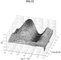
- FIGS. 7A to 7D are chronological change data of permittivity (change amounts ⁇ of real parts of complex permittivity of blood within normal values (A and B) and of blood having abnormal values (C and D).
- Erythrocyte sedimentation rates of blood specimens of FIGS. 7A to 7D are illustrated below in Table 4.
- the blood specimens within normal values and the blood specimens having abnormal values have significantly different features in a frequency band of 1 to 30 kHz.
- ⁇ is a low value on a long-time side in a frequency band of 1 to 30 kHz in association with blood coagulation.
- ⁇ is a high value on a long-time side in a frequency band of 1 to 30 kHz. The use of this method was confirmed to enable evaluation of abnormality of the erythrocyte sedimentation rate.
- Table 4 Specimen Erythrocyte Sedimentation Rate (mm/60m in) A 2 B 6 C 84 D 56
Abstract
Description
- The present technology relates to a blood state evaluation device, a blood state evaluation system, a blood state evaluation method, and a program. More specifically, the present technology relates to a technology for evaluating a state of blood from a chronological change in an electrical characteristic.
- Anti-platelet aggregation agents or anti-coagulant agents are prophylactically administered to patients or healthy persons who have thrombosis risks. Examples of the patients having thrombus formation risks include patients with diabetes, arteriosclerosis, cancer, heart disease, and respiratory disease; perioperative patients; and patients taking immunosuppressants. Also, examples of the healthy persons having thrombus risks include pregnant women and elderly people. As the anti-platelet aggregation agents, acetylsalicylic acid and the like are used; and as the anti-coagulant agents, warfarin, heparin, activated blood coagulation factor Xa inhibitors, direct thrombin inhibitors, and the like are used.
- The prophylactic administration of anti-platelet aggregation agents and anti-coagulant agents against thrombosis has the side effect that an excessively high administered dose increases a bleeding risk. In order to obtain a sufficient prophylactic effect while inhibiting this side effect, an administration management becomes important in which blood coagulability of an administered subject is timely evaluated, and the drug and dose to be administered are appropriately selected and determined.
- A method for a blood coagulability test for managing drug administration includes the prothrombin time-international normalized ratio (PT-INR), the activated partial thromboplastin time (APTT), and the like. Also, a method for a platelet aggregation test includes adding a substance that induces aggregation of platelet to platelet rich plasma (PRP) obtained by centrifuging blood, and measuring a change in transmitted light levels or absorbance associated with the aggregation to determine good or poor in aggregation capacity.
- Meanwhile, as a method for a venous thromboembolism (VTE) test, mainly, ultrasonography is used for deep vein thrombosis (DVT), and computed tomography (CT) is used for pulmonary embolism (PE), for example. In the related art, a report has been presented on a study to predict venous thromboembolism (VTE) that occurs as a side effect of artificial knee joint replacement by measuring a biomarker (Non-Patent Literature 1).
- Non-Patent Literature 1: H. Watanabe, and ten others, "Predictive blood coagulation markers for early diagnosis of venous thromboembolism after total knee joint replacement", Thrombosis Research, 2011, Vol. 128, pp. 137-143.
- However, known blood coagulability tests such as PT-INR and APTT substantially evaluate only the bleeding risk associated with reduction in blood coagulability caused by excess administration of anti-coagulant agents, and cannot evaluate the thrombus risk associated with enhancement in blood coagulability. Also, the existing platelet aggregation test using PRP may require a centrifugation process. This may cause platelet to be activated during this process, thereby inhibiting accurate test results from being obtained. Furthermore, the operation is complicated.
- On the other hand, image diagnosis by ultrasonography or computed tomography is highly precise and accurate, enabling definite diagnosis. However, such tests need time and effort, and such image diagnosis cannot evaluate the "risk" of whether a thrombus will occur in the future, before a thrombus occurs, because such image diagnosis fundamentally detects a thrombus that has already occurred. Furthermore, a method using a biomarker does not have sufficient sensitivity and specificity. For example, a study using a D-meter has a sensitivity of 75% and a specificity of 63%, and a study using an e-XDP has a sensitivity of 75% and a specificity of 59%.
- Accordingly, the present disclosure mainly aims to provide a blood state evaluation device, blood state evaluation system, blood state evaluation method, and program that enable evaluation of the risk of thrombosis easily and precisely.
- As techniques that can evaluate the degree of enhancement of blood coagulation easily and accurately, the present inventors have proposed techniques for performing dielectric measurement of the process of blood coagulation (
JP 2010-181400A JP 2012-194087A - As a blood specimen, a blood specimen obtained by collecting blood from a vein by using citric acid, for example, as an anti-coagulant agent is used. Immediately before the start of measurement, an aqueous solution of calcium chloride is added so as to release the anti-coagulant function of citric acid, causing the reaction of blood coagulation to progress. Then, by analyzing complex permittivity spectrum obtained by the measurement according to a predetermined algorithm, enhancement or reduction of blood coagulability such as a blood coagulation time can be evaluated.
- Accordingly, the present inventors have intensely considered and tested a method for evaluating the risk of venous thromboembolism (VTE) by using data of an electrical characteristic of blood obtained through measurement using an electrical characteristic measurement device such as the above-described dielectric coagulometer, and have made the present invention.
- That is, a blood state evaluation device according to the present disclosure includes at least an evaluation unit configured to evaluate a state of blood on the basis of chronological change data of an electrical characteristic of the blood in two or more frequencies or frequency bands.
- The evaluation unit may extract at least one feature point from the chronological change data of the electrical characteristic.
- In this case, the at least one feature point may be a change amount δE (= E(fx,ty)/E(fx,ta)) in an electrical characteristic value E in a given frequency fx from a reference time ta to a given time ty, for example.
- The evaluation unit may also digitalize the chronological change data of the electrical characteristic.
- In this case, the evaluation unit may evaluate the state of the blood by comparing a determination value calculated from the chronological change data of the electrical characteristic with a predetermined threshold.
- The evaluation unit may evaluate the state of the blood by an increase and/or a decrease in an electrical characteristic value E in a given frequency fx on the basis of the chronological change data of the electrical characteristic.
- Meanwhile, the electrical characteristic may be at least one kind of values selected from impedance, conductance, admittance, capacitance, permittivity, conductivity, phase angle, and a quantity obtained by conversing such a value into a quantity of electricity.
- In addition, the evaluation unit may evaluate a coagulation state of the blood. Further, a measurement unit configured to chronologically measure the electrical characteristic of blood being an evaluation target, in a particular frequency or frequency band may be provided.
- A blood state evaluation system according to the present disclosure includes: an electrical characteristic measurement device including a measurement unit that chronologically measures an electrical characteristic of blood being an evaluation target, in a particular frequency or frequency band; and a blood state evaluation device including an evaluation unit that evaluates a state of the blood on the basis of chronological change data in two or more frequencies or frequency bands from among electrical characteristics measured by the electrical characteristic measurement device.
- The blood state evaluation system may further include a server including an information storage unit that stores a result of measurement in the permittivity measurement device and/or a result of evaluation in the blood state evaluation device, in which the server may be connected to the permittivity measurement device and/or the blood state evaluation device through a network.
- A blood state evaluation method according to the present disclosure includes: an electrical characteristic measurement step of chronologically measuring an electrical characteristic of blood being an evaluation target in a particular frequency or frequency band; and a blood state evaluation step of evaluating a state of the blood on the basis of chronological change data in two or more frequencies or frequency bands from among electrical characteristics measured in the electrical characteristic measurement step.
- A program according to the present disclosure is a program for causing a computer to execute an evaluation function of evaluating a state of blood on the basis of chronological change data of an electrical characteristic of the blood in two or more frequencies or frequency bands.
- According to the present disclosure, since the blood state is evaluated on the basis of chronological change data of an electrical characteristic, it becomes possible to evaluate the risk of thrombosis easily and precisely.
-
- [
FIG. 1] FIG. 1 is a block diagram illustrating a configuration example of a blood state evaluation device according to a first embodiment of the present disclosure. - [
FIG. 2A] FIG. 2A is chronological change data of permittivity (change amount δε of a real part of complex permittivity) of blood of thrombosis. - [
FIG. 2B] FIG. 2B is chronological change data of permittivity (change amount δε of a real part of complex permittivity) of blood of a healthy person. - [
FIG. 2C] FIG. 2C is chronological change data of permittivity (change amount δε of a real part of complex permittivity) of blood of elevated blood sedimentation. - [
FIG. 3A] FIG. 3A is chronological change data of an electrical conductivity (change amount δκ) of blood having an erythrocyte sedimentation rate in a normal range. - [
FIG. 3B] FIG. 3B is chronological change data of an electrical conductivity (change amount δκ) of blood having an erythrocyte sedimentation rate in a normal range. - [
FIG. 3C] FIG. 3C is chronological change data of an electrical conductivity (change amount δκ) of blood having an erythrocyte sedimentation rate in an abnormal range. - [
FIG. 3D] FIG. 3D is chronological change data of an electrical conductivity (change amount δκ) of blood having an erythrocyte sedimentation rate in an abnormal range. - [
FIG. 4] FIG. 4 is a diagram illustrating a schematic configuration of a blood state evaluation system according to a second embodiment of the present disclosure. - [
FIG. 5] FIG. 5 is a diagram illustrating a configuration of a sample container used in Examples. - [
FIG. 6] FIG. 6 is chronological change data of permittivity (change amounts δε in real parts of complex permittivity) of blood of a patient with diabetes, measured in Example 4. - [
FIG. 7A] FIG. 7A is chronological change data of permittivity (change amount δε of a real part of complex permittivity of a blood specimen within a normal value measured in Example 5. - [
FIG. 7B] FIG. 7B is chronological change data of permittivity (change amount δε of a real part of complex permittivity of a blood specimen within a normal value measured in Example 5. - [
FIG. 7C] FIG. 7C is chronological change data of permittivity (change amount δε of a real part of complex permittivity of a blood specimen having an abnormal value measured in Example 5. - [
FIG. 7D] FIG. 7D is chronological change data of permittivity (change amount δε of a real part of complex permittivity of a blood specimen having an abnormal value measured in Example 5. - Embodiments for implementing the present disclosure will be described in detail below with reference to the appended drawings. Note that the present disclosure is not limited to each embodiment described below. The description will be made in the following order.
- First, a blood state evaluation device according to a first embodiment of the present disclosure will be described. As a risk factor of venous thromboembolism (VTE), "stasis of blood flow", "vascular endothelial injury", and "enhancement of blood coagulability" are known and are called "Virchow's triad".
- Examples of general risk factors of thrombosis include aging, obesity, protracted bed rest, maintenance of the same posture, and dehydration. Examples of risk factors relating to disease include congenital hypercoagulability, malignant tumor, inflammatory bowel disease, and nephrotic syndrome. Furthermore, there are various risk factors such as those relating to medicine including steroid and hormone drug, and those relating to medical treatment including surgery and catheterization. Among these risk factors, in particular, medical treatment such as artificial knee joint replacement is known to have a high risk of thrombosis.
- Meanwhile, even with the above-described risk factor(s), it differs significantly among individuals whether venous thromboembolism (VTE) really occurs. From such reasons, it is desirable to achieve a method that enables prediction of the thrombosis risk of each person by a simple method such as a blood test.
- Accordingly, on the basis of chronological change data of an electrical characteristic of blood in two or more frequencies or two or more frequency bands, the blood state evaluation device according to the present embodiment evaluates the state of blood, and predicts the thrombosis risk from the result.
FIG. 1 is a block diagram illustrating a configuration example of the blood state evaluation device according to the present embodiment. As illustrated inFIG. 1 , a bloodstate evaluation device 1 according to the present embodiment includes ameasurement unit 2, anevaluation unit 3, astorage unit 4, adisplay unit 5, and the like. - The
measurement unit 2 chronologically measures an electrical characteristic of blood, which is an evaluation target, in a particular frequency or frequency band. The electrical characteristic measured by themeasurement unit 2 includes, for example, impedance, conductance, admittance, capacitance, permittivity, conductivity, phase angle, and a quantity obtained by converting such a value into a quantity of electricity. The bloodstate evaluation device 1 according to the present embodiment can conduct evaluation by using one of these electrical characteristics, but may also use two or more electrical characteristics. - A configuration of the
measurement unit 2 is not particularly limited, and may be appropriately determined depending on the electrical characteristic to be measured. For example, when an alternating voltage is applied between a pair of electrodes provided in a sample container to measure the impedance and permittivity of blood, an impedance analyzer and a network analyzer can also be used as themeasurement unit 2. - Note that the
measurement unit 2 may conduct measurement only in a frequency or frequency band used by the later-describedevaluation unit 3, but may also measure the electrical characteristic in a wide band by changing frequencies so as to extract the frequency or frequency band used for evaluation from the obtained spectrum. - The
evaluation unit 3 evaluates the state of blood on the basis of chronological change data in two or more frequencies or two or more frequency bands from the electrical characteristic measured by the above-describedmeasurement unit 2. Examples of the states of blood as the evaluation target include blood coagulation state, aggregation state, solidification state, and blood clot shrinkage state. - Evaluation by the
evaluation unit 3 may be conducted by employing a method for extracting feature points from chronological change data of the electrical characteristic, a method for digitizing chronological change data of the electrical characteristic, a method for comparing a determination value calculated from chronological change data of the electrical characteristic, with a predetermined threshold, and the like. Alternatively, theevaluation unit 3 may evaluate the state of blood by increase and/or decrease in an electrical characteristic value E in a given frequency fx on the basis of chronological change data of the electrical characteristic, for example. Note that the evaluation method in theevaluation unit 3 is not limited to the above-described methods and various methods may be employed. - The
storage unit 4 stores chronological change data of the electrical characteristic of blood, which has been measured by themeasurement unit 2, the result of evaluation in theevaluation unit 3, and the like. Thestorage unit 4 is configured from a hard disk, for example. - The
display unit 5 displays chronological change data of the electrical characteristic of blood, which has been measured by themeasurement unit 2, the result of evaluation in theevaluation unit 3, and the like. Thedisplay unit 5 may have any configuration by which these can be viewed. - Next, the operation of the above-described blood
state evaluation device 1, that is, a method for evaluating the state of blood and predicting the thrombosis risk by using the bloodstate evaluation device 1 will be described. - In the blood
state evaluation device 1 according to the present embodiment, first, in themeasurement unit 2, the electrical characteristic of blood being an evaluation target is measured chronologically in a particular frequency or frequency band. In this event, conditions for measuring the electrical characteristic are not limited to particular conditions, and can be set as appropriate depending on the kind of electrical characteristic as long as the blood being the evaluation target is not altered. - The measurement may be conducted in a frequency or frequency band that is used in an evaluation step, or the electrical characteristic may be measured in a wide band including all the frequencies and frequency bands that are used. In this case, from the obtained spectrum, a frequency or frequency band used for evaluation is extracted in the
evaluation unit 3. - Next, using the electrical characteristic of blood obtained through the electrical characteristic measurement step, the state of blood being the target is evaluated in the
evaluation unit 3. For example, in a case where the electrical characteristic is permittivity, chronological change data as illustrated inFIGS. 2A to 2C is obtained. Here, the chronological change data illustrated inFIGS. 2A to 2C shows change amounts δε (= ε/εt=0) in real parts of complex permittivity of blood of thrombosis (FIG. 2A ), blood of a healthy person (FIG. 2B ), and blood of elevated blood sedimentation (FIG. 2C ). The data shows values obtained by measuring impedance in a frequency region of 100 Hz to 40 MHz under a condition of temperature being 37 °C for 60 minutes with 1-minute measurement intervals. - As illustrated in
FIG. 2A , as for the blood of thrombosis, permittivity around 10 MHz explicitly increases due to blood coagulation. Meanwhile, as illustrated inFIG. 2B , as for the blood of the healthy person, in addition to this increase, permittivity increases due to haemagglutination around 1 MHz and decreases thereafter. In addition, as illustrated inFIG. 2C , as for the blood of elevated blood sedimentation, permittivity increases indicating blood sedimentation around 2.5 kHz. By using these features, it becomes possible to predict the thrombus risk. - In the above manner, the features of obtained chronological change data of the electrical characteristic differ depending on the state of blood. The use of the features makes it possible to predict the thrombosis risk. Specifically, it is possible to evaluate the state of the blood by using increase and/or decrease in the electrical characteristic value E in the given frequency fx on the basis of chronological change data of the electrical characteristic.
- For example, in a case where the electrical characteristic is a change in permittivity associated with blood coagulation, as the overall trend, as for blood positive for thrombosis, in a high frequency band around 10 MHz (3 to 30 MHz), increase in permittivity associated with blood coagulation becomes obvious. In addition, as for the blood positive for thrombosis, in some cases, increase in permittivity may be seen due to erythrocyte rouleaux formation in about 12 minutes from the addition of an aqueous solution of calcium for blood coagulation, in a middle frequency band around 1 MHz (higher than or equal to 100 kHz and lower than 3 MHz).
- However, the blood positive to thrombosis has a smaller increase in permittivity associated with blood coagulation in a middle frequency band around 1 MHz than in a high frequency band around 10 MHz. Immediately after this small increase, permittivity decreases significantly, and after that, permittivity tends to be a substantially constant low value. In addition, the blood positive for thrombosis tends to have a significant decrease in permittivity associated with blood coagulation in a low frequency band of higher than or equal to 1 kHz and lower than 100 kHz.
- On the other hand, the blood negative for thrombosis has the following two distinctive patterns. A first pattern is a case where the increase in permittivity can be seen in a low frequency band, unlikely in positive blood. A second pattern is a case where the increase in permittivity associated with blood coagulation and the following decrease are observed separately from the increase in permittivity due to erythrocyte rouleaux formation that is seen in about 12 minutes from the addition of an aqueous solution of calcium, as described above, in a middle frequency band. The second pattern that is distinctive in the middle frequency band may be a distinctive change as in the blood of the healthy person.
- The blood
state evaluation device 1 according to the present embodiment evaluates the thrombosis risk using this feature. Specifically, in a case where permittivity obviously increases in a high frequency band (3 to 30 MHz) and hardly changes or significantly decreases in a middle frequency band (higher than or equal to 100 kHz and lower than 3 MHz) and a low frequency band (higher than or equal to 1 kHz and lower than 100 kHz), the thrombosis risk is evaluated to be high. On the other hand, in a case where permittivity explicitly increases in the middle frequency band (higher than or equal to 100 kHz and lower than 3 MHz), or in a case where permittivity has a high value in the low frequency band (higher than or equal to 1 kHz and lower than 100 kHz) even when blood coagulation occurs, the thrombosis risk is evaluated to be low. - The evaluation of the blood state can alternatively be conducted by a method for extracting at least one feature point from chronological change data of the electrical characteristic. The feature point(s) in this case can be set as a change amount δE (= E(fx,ty)/E(fx,ta)) in the electrical characteristic value E from a reference time ta to a given time ty in the given frequency fx.
- Alternatively, the evaluation may be conducted by digitalizing chronological change data of the electrical characteristic. For example, in a case where the electrical characteristic is a change in permittivity associated with blood coagulation, the evaluation may be conducted by using a determination value p1 calculated from the following
formula 1. This determination value p1 is calculated from a change amount δε (f1,t1) in permittivity in a first frequency f1 at a first time t1, a change amount δε (f2,t2) in permittivity in a second frequency f2 at a second time t2, and a change δε (f3,t3) in permittivity in a third frequency f3 at a third time t3. Note that δε (f,t)} = ε (f,t)/ε (f,t = 0) is a ratio between the measured permittivity and time 0 (before the initiation of a blood coagulation reaction). This applies to the following determination formula.
- The evaluation can alternatively conducted using a determination value p2 calculated from the following
formula 2. Note that a in the followingformula 2 is a given constant. The followingformula 2 is an formula obtained empirically. The present inventors have found that the determination value p2 calculated from theformula 2 is valid for evaluation of pulmonary embolism (PE), which is more serious than venous thromboembolism (VTE).
- Furthermore, although the determination value is calculated by using three-point frequency data of the
formulas formula 3.
- By comparing the determination values p1, p2, and p3 calculated from the
formulas 1 to 3 with a predetermined threshold, it is possible to evaluate the blood state easily. - The above-described blood state evaluation step can be conducted by creating and mounting, in a personal computer for example, a computer program for achieving the functions of an information processing apparatus. Such a computer program may also be stored in a recording medium such as a magnetic disk, an optical disc, a magneto-optical disk, or a flash memory, or may be distributed through a network.
- Note that the electrical characteristic data measured by the
measurement unit 2 may also be stored in thestorage unit 4 and may be displayed on thedisplay unit 5 as necessary, in addition to being evaluated by being sent to theevaluation unit 3,. The evaluation result obtained in theevaluation unit 3 may be stored in thestorage unit 4 and may be displayed on thedisplay unit 5 as necessary. - The above-described electrical characteristic measurement step and blood state evaluation step are not necessarily performed consecutively. The evaluation may be conducted by storing data obtained by the
measurement unit 2 in thestorage unit 4 and by reading out the data by theevaluation unit 3 from thestorage unit 4, as appropriate. - In addition, although the blood
state evaluation device 1 illustrated inFIG. 1 includes themeasurement unit 2, thestorage unit 4, and thedisplay unit 5, the bloodstate evaluation device 1 includes at least theevaluation unit 3. Furthermore, in the present embodiment, the case where the electrical characteristic is permittivity is described as an example, but the present disclosure is not limited to a method using permittivity. Similarly, the evaluation is possible by using other electrical characteristics such as impedance, admittance, and capacitance. -
FIGS. 3A to 3D are each chronological change data of an electrical conductivity δκ of blood measured in a range of 100 Hz to 40 MHz. Referring to a frequency band of 5 kHz to 1 MHz, blood having an erythrocyte sedimentation rate in a normal range illustrated inFIGS. 3A and3B has a low electrical conductivity δκ on a long-time side in association with blood coagulation. On the other hand, blood having an erythrocyte sedimentation rate in an abnormal range illustrated inFIGS. 3C and3D has a high electrical conductivity δκ on a long-time side in a frequency band of 5 kHz to 1 MHz. By use of this characteristic, even when the electrical conductivity δκ is used, it is possible to evaluate the thrombus risk highly precisely. - As specifically described above, in the blood state evaluation device according to the present embodiment, the blood state is evaluated on the basis of chronological change data of the electrical characteristic. Accordingly, a simple blood test enables evaluation of the thrombosis risk at a high sensitivity and a high specificity. Thus, the load on the medical site can be reduced.
- Next, a blood state evaluation system according to a second embodiment of the present disclosure will be described.
FIG. 4 is a diagram illustrating a schematic configuration of the blood state evaluation system according to the present embodiment. In the blood state evaluation device according to the first embodiment described above, the electrical characteristic is measured and the blood state is evaluated within the device. However, the blood state can be evaluated in an information processing device connected to an electrical characteristic measurement device. - That is, as illustrated in
FIG. 4 , the blood state evaluation system according to the present embodiment includes an electricalcharacteristic measurement device 11 and aninformation processing device 12. The blood state evaluation system according to the present embodiment may be connected to aserver 13, adisplay device 14, and the like, as necessary. - The electrical
characteristic measurement device 11 includes a measurement unit that applies a voltage between a pair of electrodes provided in a sample container to be filled with blood being the measurement target, and to chronologically measure an electrical characteristic of the blood in a particular frequency or frequency band. A configuration of the electricalcharacteristic measurement device 11 is not particularly limited, and may be appropriately determined in accordance with the electrical characteristic to be measured. For example, when an alternating voltage is applied between a pair of electrodes to measure the impedance and permittivity of blood, an impedance analyzer and a network analyzer can also be used. - The
information processing device 12 is connected to the electricalcharacteristic measurement device 11, and includes an evaluation unit that evaluates the state of blood on the basis of chronological change data in two or more frequencies or frequency bands from among the electrical characteristics measured by the electricalcharacteristic measurement device 11. Note that a specific configuration and operation of the evaluation unit are the same as in the above-described first embodiment. - The
server 13 is connected to theinformation processing device 12 and thedisplay device 14 through anetwork 15, and includes an information storage unit, for example. Further, theserver 13 manages various kinds of data uploaded from theinformation processing device 12, and outputs data to thedisplay device 14 and theinformation processing device 12 in response to a request. - The
display device 14 displays chronological change data of the electrical characteristic of blood, which has been measured by the electricalcharacteristic measurement device 11, the result of evaluation in the evaluation unit of theinformation processing device 12, and the like. Thedisplay device 14 may be provided with an information input unit so that a user can select and input data to be displayed. In this case, information inputted by the user is transmitted to theserver 13 and theinformation processing device 12 through thenetwork 15. - Also in the blood state evaluation system according to the present embodiment, since the state of blood is evaluated on the basis of chronological change data of the electrical characteristic, a simple blood test enables evaluation of the thrombosis risk highly precisely.
- Additionally, the present technology may also be configured as below.
- (1) A blood state evaluation device including at least:
- an evaluation unit configured to evaluate a state of blood on the basis of chronological change data of an electrical characteristic of the blood in two or more frequencies or frequency bands.
- (2) The blood state evaluation device according to (1),
wherein the evaluation unit extracts at least one feature point from the chronological change data of the electrical characteristic. - (3) The blood state evaluation device according to (2),
wherein the at least one feature point is a change amount δE (= E(fx,ty)/E(fx,ta)) in an electrical characteristic value E in a given frequency fx from a reference time ta to a given time ty. - (4) The blood state evaluation device according to (1),
wherein the evaluation unit digitalizes the chronological change data of the electrical characteristic. - (5) The blood state evaluation device according to (4),
wherein the evaluation unit evaluates the state of the blood by comparing a determination value calculated from the chronological change data of the electrical characteristic with a predetermined threshold. - (6) The blood state evaluation device according to (1),
wherein the evaluation unit evaluates the state of the blood by an increase and/or a decrease in an electrical characteristic value E in a given frequency fx on the basis of the chronological change data of the electrical characteristic. - (7) The blood state evaluation device according to any one of (1) to (6),
wherein the electrical characteristic is at least one kind of values selected from impedance, conductance, admittance, capacitance, permittivity, conductivity, phase angle, and a quantity obtained by conversing such a value into a quantity of electricity. - (8) The blood state evaluation device according to any one of (1) to (7),
wherein the evaluation unit evaluates a coagulation state of the blood. - (9) The blood state evaluation device according to any one of (1) to (8), further including:
- a measurement unit configured to chronologically measure the electrical characteristic of blood being an evaluation target, in a particular frequency or frequency band.
- (10) A blood state evaluation system including:
- an electrical characteristic measurement device including a measurement unit that chronologically measures an electrical characteristic of blood being an evaluation target, in a particular frequency or frequency band; and
- a blood state evaluation device including an evaluation unit that evaluates a state of the blood on the basis of chronological change data in two or more frequencies or frequency bands from among electrical characteristics measured by the electrical characteristic measurement device.
- (11) The blood state evaluation system according to (10) further including:
- a server including an information storage unit that stores a result of measurement in the permittivity measurement device and/or a result of evaluation in the blood state evaluation device,
- wherein the server is connected to the permittivity measurement device and/or the blood state evaluation device through a network.
- (12) A blood state evaluation method including:
- an electrical characteristic measurement step of chronologically measuring an electrical characteristic of blood being an evaluation target in a particular frequency or frequency band; and
- a blood state evaluation step of evaluating a state of the blood on the basis of chronological change data in two or more frequencies or frequency bands from among electrical characteristics measured in the electrical characteristic measurement step.
- (13) A program for causing a computer to execute:
- an evaluation function of evaluating a state of blood on the basis of chronological change data of an electrical characteristic of the blood in two or more frequencies or frequency bands.
- Hereinafter, effects of the present disclosure will be specifically described. In the present example, the thrombosis risk was evaluated using the blood state evaluation device according to the above-described first embodiment by the following method.
- Using a vacuum blood collection tube in which sodium citrate was treated as an anti-coagulant agent, blood was collected in the following morning of surgery (before medication starts after surgery). The temperature of the specimen blood was kept at 37°C in advance, and 0.25 M of an aqueous solution of calcium chloride was added to the specimen blood at a concentration of 85 µL per 1 mL of blood immediately before the start of measurement, to initiate a blood coagulation reaction.
- Then, dielectric measurement (impedance measurement) was performed for a blood coagulation process of the specimen blood using a dielectric coagulometry prototype machine available from Sony Corporation. The measurement was performed under the conditions of a measurement temperature of 37°C, a measurement frequency range of 100 Hz to 40 MHz, a measurement interval of one minute, and a measurement time of 60 minutes. A
sample container 20 illustrated inFIG. 5 was used for the measurement. In order to reduce the influence of blood sedimentation, the measurement was performed by rotating thesample container 20 once a minute by 180°. - On the fourth or fifth day from the surgery, it was checked whether venous thromboembolism (VTE) occurred. Specifically, deep vein thrombosis (DVT) was checked by ultrasonography, and pulmonary embolism (PE) was checked by computed tomography (CT).
- As a result of image diagnosis by ultrasonography and computed tomography (CT), 21 examples were positive for venous thromboembolism (VTE) and 9 examples were negative for venous thromboembolism (VTE) out of 30 examples investigated.
- The dielectric blood coagulation time was calculated from a change in permittivity in 10 MHz. The dielectric blood coagulation time of blood positive for venous thromboembolism (VTE) was 23±5 minutes, and the dielectric blood coagulation time of blood negative for venous thromboembolism (VTE) was 20±4 minutes. Each of these values was shorter than the value of blood of a healthy person, which was 40±6 minutes, and accordingly was confirmed to be in a state of enhanced blood coagulability having a high thrombosis risk. However, the prediction only by the dielectric blood coagulation time as to whether venous thromboembolism (VTE) occurs in reality at a high sensitivity and a high specificity was found to be difficult.
- Next, the determination value p1 was calculated using the above-described
formula 1. In this event, f1 = 2.5 kHz, f2 = 1 MHz, f3 = 10 MHz, t1 = t3-5 min, and t2 = 18 min. Note that t3 is the dielectric blood coagulation time. The threshold was set to 0.765, and the determination value p1 being smaller than this value was determined to be "positive". The results are shown below in Table 1.[Table 1] True State Positive Negative Test Results Positive True Positive False Positive Positive Predictive Value 19 2 90% Negative False Negative True Negative Negative Predictive Value 2 7 78% Sensitivity Specificity 90% 78% - As illustrated in Table 1, this method enables evaluation of the thrombosis risk at a satisfactory score of a sensitivity being 90% and a specificity being 78%.
- Here, the "sensitivity" and the "specificity" are indexes used in laboratory tests. Specifically, the "sensitivity" is a value defined as the "possibility at which an object that is supposed to be determined to be positive is correctly determined to be positive", and the "specificity" is the "possibility at which an object that is negative is correctly determined to be negative". In addition, it is preferable that each of these values is high, and a method by which high values are obtained for both the "sensitivity" and the "specificity" is an excellent testing method. However, when setting a threshold, a priority to the sensitivity results in a lower specificity, whereas a priority to the specificity results in a lower sensitivity. Accordingly, the threshold is typically set in a manner that both values can be within acceptable ranges.
- Next, as in the method described in Example 1, the thrombus risk of specimen blood (the number of specimens was 27) was evaluated using the determination value p2 of the above-described
formula 2. In this event, f1 = 2.5 kHz, f2 = 1 MHz, f3 = 10 MHz, a = 0.67, and t1 = t2 = t3 being the dielectric blood coagulation time. The threshold was set to 0.11, and the determination value p2 being smaller than this value was determined to be "positive". The results are shown below in Table 2.[Table 2] True State Positive Negative Test Results Positive True Positive False Positive Positive Predictive Value 7 3 70% Negative False Negative True Negative Negative Predictive Value 1 16 94% Sensitivity Specificity 88% 84% - As illustrated in Table 2, this method enables evaluation of pulmonary embolism (PE) at a satisfactory score of a sensitivity being 88% and a specificity being 84%.
- Next, as in the method described in Example 1, the thrombus risk of specimen blood (the number of specimens was 30) was evaluated using the determination value p3 of the above-described
formula 3. In this event, f1 = 2.5 kHz, f2 = 10 MHz, t1 = t2-6min, and t2 was the dielectric blood coagulation time. The threshold was set to 0.765, and the determination value p3 being smaller than this value was determined to be "positive". The results are shown below in Table 3.[Table 3] True State Positive Negative Test Results Positive True Positive False Positive Positive Predictive Value 18 3 86% Negative False Negative True Negative Negative Predictive Value 3 6 67% Sensitivity Specificity 86% 67% - As illustrated in Table 3, this method enables evaluation of the thrombosis risk at a satisfactory score of a sensitivity being 86% and a specificity being 67%.
- In Example 4, measurement was performed using venous blood of a patient with diabetes and a patient with both diabetes and a collagen disease. In this event, a vacuum blood collection tube in which sodium citrate was treated as an anti-coagulant agent was used to collect blood. The temperature of the specimen blood was kept at 37°C in advance, and 0.25 M of an aqueous solution of calcium chloride was added to the specimen blood at a concentration of 85 µL per 1 mL of blood immediately before the start of measurement, to initiate a blood coagulation reaction. The measurement was performed under the conditions of a measurement temperature of 37°C, a measurement frequency range of 40 Hz to 110 MHz, a measurement interval of two minutes, and a measurement time of 60 minutes.
-
FIG. 6 is chronological change data of permittivity (change amounts δε in real parts of complex permittivity) of blood of the patient with diabetes. Data surrounded by a thick line inFIG. 6 was of blood of the patient with a collagen disease, and has a different pattern from other data obviously. Specifically, as for the patient with a collagen disease, permittivity rapidly increases and then decreases in a frequency band from 1 to 100 kHz in 20 minutes immediately after the start of measurement. Note that such a feature is not seen in the patient with diabetes who did not have a collagen disease or a healthy person. The use of this method was confirmed to enable evaluation as to whether a collagen disease occurred, at the blood test. - In Example 5, measurement was performed using venous blood that was collected before surgery of a patient who was supposed to undergo artificial knee joint replacement. In this event, a vacuum blood collection tube in which sodium citrate was treated as an anti-coagulant agent was used to collect blood. The temperature of the specimen blood was kept at 37°C in advance, and 0.25 M of an aqueous solution of calcium chloride was added to the specimen blood at a concentration of 85 µL per 1 mL of blood immediately before the start of measurement, to initiate a blood coagulation reaction. The measurement was performed under the conditions of a measurement temperature of 37°C, a measurement frequency range of 100 Hz to 40 MHz, a measurement interval of one minute, and a measurement time of 60 minutes.
- The resulting data was compared with an erythrocyte sedimentation rate (ESR), which is one of general values for blood test. A normal value of the erythrocyte sedimentation rate is 10 mm/60 min for men or 15 mm/60 min for women. As a result, an obvious relation was found between these values and a dielectric coagulation pattern, and the dielectric measurement was confirmed to be able to evaluate the erythrocyte sedimentation rate.
-
FIGS. 7A to 7D are chronological change data of permittivity (change amounts δε of real parts of complex permittivity of blood within normal values (A and B) and of blood having abnormal values (C and D). Erythrocyte sedimentation rates of blood specimens ofFIGS. 7A to 7D are illustrated below in Table 4. As illustrated inFIGS. 7A to 7D , the blood specimens within normal values and the blood specimens having abnormal values have significantly different features in a frequency band of 1 to 30 kHz. In a case of the erythrocyte sedimentation rate being a normal value, δε is a low value on a long-time side in a frequency band of 1 to 30 kHz in association with blood coagulation. In contrast, in a case of the erythrocyte sedimentation rate being an abnormal value, δε is a high value on a long-time side in a frequency band of 1 to 30 kHz. The use of this method was confirmed to enable evaluation of abnormality of the erythrocyte sedimentation rate.[Table 4] Specimen Erythrocyte Sedimentation Rate (mm/60m in) A 2 B 6 C 84 D 56 -
- 1
- blood state evaluation device
- 2
- measurement unit
- 3
- evaluation unit
- 4
- storage unit
- 5
- display unit
- 11
- electrical characteristic measurement device
- 12
- information processing device
- 13
- server
- 14
- display device
- 15
- network
- 20
- sample container
Claims (13)
- A blood state evaluation device comprising at least:an evaluation unit configured to evaluate a state of blood on the basis of chronological change data of an electrical characteristic of the blood in two or more frequencies or frequency bands.
- The blood state evaluation device according to claim 1,
wherein the evaluation unit extracts at least one feature point from the chronological change data of the electrical characteristic. - The blood state evaluation device according to claim 2,
wherein the at least one feature point is a change amount δE (= E(fx,ty)/E(fx,ta)) in an electrical characteristic value E in a given frequency fx from a reference time ta to a given time ty. - The blood state evaluation device according to claim 1,
wherein the evaluation unit digitalizes the chronological change data of the electrical characteristic. - The blood state evaluation device according to claim 4,
wherein the evaluation unit evaluates the state of the blood by comparing a determination value calculated from the chronological change data of the electrical characteristic with a predetermined threshold. - The blood state evaluation device according to claim 1,
wherein the evaluation unit evaluates the state of the blood by an increase and/or a decrease in an electrical characteristic value E in a given frequency fx on the basis of the chronological change data of the electrical characteristic. - The blood state evaluation device according to claim 1,
wherein the electrical characteristic is at least one kind of values selected from impedance, conductance, admittance, capacitance, permittivity, conductivity, phase angle, and a quantity obtained by conversing such a value into a quantity of electricity. - The blood state evaluation device according to claim 1,
wherein the evaluation unit evaluates a coagulation state of the blood. - The blood state evaluation device according to claim 1, further comprising:a measurement unit configured to chronologically measure the electrical characteristic of blood being an evaluation target, in a particular frequency or frequency band.
- A blood state evaluation system comprising:an electrical characteristic measurement device including a measurement unit that chronologically measures an electrical characteristic of blood being an evaluation target, in a particular frequency or frequency band; anda blood state evaluation device including an evaluation unit that evaluates a state of the blood on the basis of chronological change data in two or more frequencies or frequency bands from among electrical characteristics measured by the electrical characteristic measurement device.
- I The blood state evaluation system according to claim 10 further comprising:a server including an information storage unit that stores a result of measurement in the permittivity measurement device and/or a result of evaluation in the blood state evaluation device,wherein the server is connected to the permittivity measurement device and/or the blood state evaluation device through a network.
- A blood state evaluation method comprising:an electrical characteristic measurement step of chronologically measuring an electrical characteristic of blood being an evaluation target in a particular frequency or frequency band; anda blood state evaluation step of evaluating a state of the blood on the basis of chronological change data in two or more frequencies or frequency bands from among electrical characteristics measured in the electrical characteristic measurement step.
- A program for causing a computer to execute:an evaluation function of evaluating a state of blood on the basis of chronological change data of an electrical characteristic of the blood in two or more frequencies or frequency bands.
Applications Claiming Priority (2)
| Application Number | Priority Date | Filing Date | Title |
|---|---|---|---|
| JP2013073508 | 2013-03-29 | ||
| PCT/JP2014/053704 WO2014156370A1 (en) | 2013-03-29 | 2014-02-18 | Blood state evaluation device, blood state evaluation system, blood state evaluation method, and program |
Publications (3)
| Publication Number | Publication Date |
|---|---|
| EP2980570A1 true EP2980570A1 (en) | 2016-02-03 |
| EP2980570A4 EP2980570A4 (en) | 2016-10-26 |
| EP2980570B1 EP2980570B1 (en) | 2020-07-22 |
Family
ID=51623371
Family Applications (1)
| Application Number | Title | Priority Date | Filing Date |
|---|---|---|---|
| EP14773633.4A Active EP2980570B1 (en) | 2013-03-29 | 2014-02-18 | Blood state evaluation system, blood state evaluation method, and program |
Country Status (6)
| Country | Link |
|---|---|
| US (2) | US10393761B2 (en) |
| EP (1) | EP2980570B1 (en) |
| JP (3) | JPWO2014156370A1 (en) |
| KR (1) | KR20150133716A (en) |
| CN (2) | CN105102969A (en) |
| WO (1) | WO2014156370A1 (en) |
Families Citing this family (11)
| Publication number | Priority date | Publication date | Assignee | Title |
|---|---|---|---|---|
| CN104937400B (en) | 2013-01-28 | 2018-07-03 | 索尼公司 | Impedance measurement device for biological sample and the impedance measurement system for biological sample |
| CN105008906B (en) | 2013-03-13 | 2020-01-07 | 索尼公司 | Blood condition analysis device and blood condition analysis system |
| CN105008907B (en) | 2013-03-15 | 2018-12-28 | 索尼公司 | Blood status analytical equipment, blood status analysis system, blood status analysis method |
| JPWO2014156370A1 (en) | 2013-03-29 | 2017-02-16 | ソニー株式会社 | Blood state evaluation apparatus, blood state evaluation system, blood state evaluation method and program |
| JP6512093B2 (en) * | 2013-03-29 | 2019-05-15 | ソニー株式会社 | Blood condition analysis device, blood condition analysis system, blood condition analysis method and program |
| JP6678023B2 (en) * | 2015-12-21 | 2020-04-08 | 日本電信電話株式会社 | Body fluid detection device and state estimation system using the body fluid detection device |
| JP7062954B2 (en) * | 2016-02-10 | 2022-05-09 | ソニーグループ株式会社 | Blood coagulation analyzer and blood coagulation analysis method |
| CN106166061B (en) * | 2016-08-30 | 2019-01-22 | 苏州涵轩信息科技有限公司 | A kind of blood status detection device and the method for obtaining temperature change value |
| CN106361334B (en) * | 2016-08-30 | 2019-04-12 | 苏州涵轩信息科技有限公司 | A kind of detection device and the method for obtaining impedance variations value and temperature change value |
| CN109363662B (en) * | 2018-11-08 | 2020-11-27 | 山东大学 | Portable thrombus detection device |
| KR20210060963A (en) | 2019-11-19 | 2021-05-27 | 전남대학교산학협력단 | Blood monitoring system by using OCT images and method thereof |
Family Cites Families (24)
| Publication number | Priority date | Publication date | Assignee | Title |
|---|---|---|---|---|
| US6046051A (en) | 1997-06-27 | 2000-04-04 | Hemosense, Inc. | Method and device for measuring blood coagulation or lysis by viscosity changes |
| US9183351B2 (en) * | 2000-05-30 | 2015-11-10 | Vladimir Shusterman | Mobile system with network-distributed data processing for biomedical applications |
| US20020019707A1 (en) | 2000-06-26 | 2002-02-14 | Cohen Alan M. | Glucose metering system |
| US8449824B2 (en) * | 2002-09-09 | 2013-05-28 | Yizhong Sun | Sensor instrument system including method for detecting analytes in fluids |
| JP4893526B2 (en) | 2007-08-10 | 2012-03-07 | ソニー株式会社 | Cell physical property value measuring method and physical property measuring apparatus |
| JP5098817B2 (en) | 2008-05-29 | 2012-12-12 | ソニー株式会社 | Physical property measuring apparatus and physical property measuring method |
| JP5691168B2 (en) | 2009-01-08 | 2015-04-01 | ソニー株式会社 | Blood coagulation system analyzer, blood coagulation system analysis method and program |
| RU2011154222A (en) * | 2009-06-08 | 2013-07-20 | Эс.И.Эй. МЕДИКАЛ СИСТЕМЗ, ИНК. | SYSTEMS AND METHODS FOR IDENTIFICATION OF COMPOUNDS IN MEDICAL LIQUIDS USING FULL CONDUCTIVITY SPECTROSCOPY |
| WO2011049601A2 (en) * | 2009-10-21 | 2011-04-28 | Micropoint Bioscience Inc. | Piezoelectric coagulation sensors |
| US20110119080A1 (en) * | 2009-11-19 | 2011-05-19 | Abbott Diabetes Care Inc. | Method and system for analyte data transmission and report generation |
| JP5905212B2 (en) | 2010-06-09 | 2016-04-20 | エイペックス バイオテクノロジー コーポレイションApex Biotechnology Corp. | Apparatus and method for measuring prothrombin time or both prothrombin time and hematocrit by analyzing changes in reactance in specimen |
| JP5549484B2 (en) * | 2010-09-01 | 2014-07-16 | ソニー株式会社 | Sample cartridge and apparatus for measuring electrical properties of liquid samples |
| WO2012058237A1 (en) * | 2010-10-26 | 2012-05-03 | Abbott Diabetes Care Inc. | Analyte measurement devices and systems, and components and methods related thereto |
| PT2676136T (en) * | 2011-02-15 | 2021-03-25 | Hemosonics Llc | Devices, systems and methods for evaluation of hemostasis |
| JP5768422B2 (en) * | 2011-03-17 | 2015-08-26 | ソニー株式会社 | Blood coagulation system analysis method and blood coagulation system analyzer |
| JP5726662B2 (en) * | 2011-07-22 | 2015-06-03 | 学校法人上智学院 | Blood impedance measuring device, artificial dialysis device, and method for operating blood impedance measuring device |
| WO2014112227A1 (en) | 2013-01-18 | 2014-07-24 | ソニー株式会社 | Electrical characteristic measurement device |
| CN104937400B (en) | 2013-01-28 | 2018-07-03 | 索尼公司 | Impedance measurement device for biological sample and the impedance measurement system for biological sample |
| WO2014122873A1 (en) | 2013-02-08 | 2014-08-14 | ソニー株式会社 | Microparticle analyzing device and microparticle analyzing system |
| CN105008906B (en) | 2013-03-13 | 2020-01-07 | 索尼公司 | Blood condition analysis device and blood condition analysis system |
| CN105008907B (en) | 2013-03-15 | 2018-12-28 | 索尼公司 | Blood status analytical equipment, blood status analysis system, blood status analysis method |
| EP2980558B1 (en) | 2013-03-26 | 2019-11-06 | Sony Corporation | Measurement device and measurement method |
| JP6512093B2 (en) | 2013-03-29 | 2019-05-15 | ソニー株式会社 | Blood condition analysis device, blood condition analysis system, blood condition analysis method and program |
| JPWO2014156370A1 (en) | 2013-03-29 | 2017-02-16 | ソニー株式会社 | Blood state evaluation apparatus, blood state evaluation system, blood state evaluation method and program |
-
2014
- 2014-02-18 JP JP2015508169A patent/JPWO2014156370A1/en active Pending
- 2014-02-18 CN CN201480017576.7A patent/CN105102969A/en active Pending
- 2014-02-18 EP EP14773633.4A patent/EP2980570B1/en active Active
- 2014-02-18 WO PCT/JP2014/053704 patent/WO2014156370A1/en active Application Filing
- 2014-02-18 KR KR1020157025315A patent/KR20150133716A/en not_active Application Discontinuation
- 2014-02-18 US US14/778,277 patent/US10393761B2/en active Active
- 2014-02-18 CN CN202010134637.5A patent/CN111272820A/en active Pending
-
2018
- 2018-06-27 JP JP2018121826A patent/JP2018179999A/en active Pending
-
2019
- 2019-08-16 US US16/543,222 patent/US20200174027A1/en not_active Abandoned
- 2019-12-03 JP JP2019218885A patent/JP6883228B2/en active Active
Also Published As
| Publication number | Publication date |
|---|---|
| US20200174027A1 (en) | 2020-06-04 |
| US10393761B2 (en) | 2019-08-27 |
| EP2980570A4 (en) | 2016-10-26 |
| JP6883228B2 (en) | 2021-06-09 |
| JPWO2014156370A1 (en) | 2017-02-16 |
| JP2020042042A (en) | 2020-03-19 |
| WO2014156370A1 (en) | 2014-10-02 |
| CN105102969A (en) | 2015-11-25 |
| US20160282366A1 (en) | 2016-09-29 |
| KR20150133716A (en) | 2015-11-30 |
| EP2980570B1 (en) | 2020-07-22 |
| CN111272820A (en) | 2020-06-12 |
| JP2018179999A (en) | 2018-11-15 |
Similar Documents
| Publication | Publication Date | Title |
|---|---|---|
| EP2980570B1 (en) | Blood state evaluation system, blood state evaluation method, and program | |
| US20190212320A1 (en) | Blood coagulation system analyzer, blood coagulation system analysis method and program | |
| JP5768422B2 (en) | Blood coagulation system analysis method and blood coagulation system analyzer | |
| EP2980571A1 (en) | Blood state analysis device, blood state analysis system, blood state analysis method, and program | |
| JP6962338B2 (en) | Blood coagulation system analyzer, blood coagulation system analysis system, blood coagulation system analysis method, and blood coagulation system analysis program, and bleeding amount prediction device, bleeding amount prediction system, bleeding amount prediction method, and bleeding amount prediction program | |
| JPWO2015159623A1 (en) | Blood state analysis device, blood state analysis system, blood state analysis method and program | |
| RU2666945C2 (en) | Method of estimation of aggregation activity of thrombocytes | |
| JP6604392B2 (en) | Blood coagulation system analyzer, blood coagulation system analysis method and program thereof | |
| RU2282855C2 (en) | Method for evaluating functional state of hemostasis system | |
| Ab Aziz et al. | Comparison of international normalized ratio (INR) between point of care device Coaguchek® XS versus standard laboratory instrument among patients receiving warfarin therapy in a northeast state of Peninsular Malaysia | |
| JP6750443B2 (en) | Platelet aggregation analysis method, platelet aggregation analysis device, platelet aggregation analysis program, and platelet aggregation analysis system | |
| JP6288182B2 (en) | Blood coagulation system analyzer, blood coagulation system analysis method and program thereof | |
| US20190162690A1 (en) | Blood coagulation system analysis system, blood coagulation system analysis method, and blood coagulation system analysis program | |
| Iberahim et al. | Coagulation Status Using Clot Wave Analysis in Patients With Prolonged Immobilization | |
| GÖKUÇ et al. | The prognostic impact of dynamic change in neutrophil to lymphocyte ratio in patients with acute pulmonary embolism | |
| Nørregaard et al. | Exercise transiently increases the density of incipient blood clots in antiplatelet-treated lacunar stroke patients | |
| JP2020190570A (en) | Platelet agglutination ability analysis method, platelet agglutination ability analyzer, program for platelet agglutination ability analysis and platelet agglutination ability analysis system |
Legal Events
| Date | Code | Title | Description |
|---|---|---|---|
| PUAI | Public reference made under article 153(3) epc to a published international application that has entered the european phase |
Free format text: ORIGINAL CODE: 0009012 |
|
| 17P | Request for examination filed |
Effective date: 20150922 |
|
| AK | Designated contracting states |
Kind code of ref document: A1 Designated state(s): AL AT BE BG CH CY CZ DE DK EE ES FI FR GB GR HR HU IE IS IT LI LT LU LV MC MK MT NL NO PL PT RO RS SE SI SK SM TR |
|
| AX | Request for extension of the european patent |
Extension state: BA ME |
|
| DAX | Request for extension of the european patent (deleted) | ||
| A4 | Supplementary search report drawn up and despatched |
Effective date: 20160928 |
|
| RIC1 | Information provided on ipc code assigned before grant |
Ipc: G01N 33/86 20060101ALI20160922BHEP Ipc: G01N 33/49 20060101ALI20160922BHEP Ipc: G01N 27/22 20060101AFI20160922BHEP Ipc: G01N 27/02 20060101ALI20160922BHEP Ipc: G01N 33/483 20060101ALI20160922BHEP |
|
| STAA | Information on the status of an ep patent application or granted ep patent |
Free format text: STATUS: EXAMINATION IS IN PROGRESS |
|
| 17Q | First examination report despatched |
Effective date: 20171208 |
|
| GRAP | Despatch of communication of intention to grant a patent |
Free format text: ORIGINAL CODE: EPIDOSNIGR1 |
|
| STAA | Information on the status of an ep patent application or granted ep patent |
Free format text: STATUS: GRANT OF PATENT IS INTENDED |
|
| INTG | Intention to grant announced |
Effective date: 20200214 |
|
| RIN1 | Information on inventor provided before grant (corrected) |
Inventor name: KATSUMOTO, YOICHI Inventor name: BRUN, MARCAURELE Inventor name: HAYASHI, YOSHIHITO |
|
| GRAS | Grant fee paid |
Free format text: ORIGINAL CODE: EPIDOSNIGR3 |
|
| GRAA | (expected) grant |
Free format text: ORIGINAL CODE: 0009210 |
|
| STAA | Information on the status of an ep patent application or granted ep patent |
Free format text: STATUS: THE PATENT HAS BEEN GRANTED |
|
| AK | Designated contracting states |
Kind code of ref document: B1 Designated state(s): AL AT BE BG CH CY CZ DE DK EE ES FI FR GB GR HR HU IE IS IT LI LT LU LV MC MK MT NL NO PL PT RO RS SE SI SK SM TR |
|
| REG | Reference to a national code |
Ref country code: GB Ref legal event code: FG4D |
|
| REG | Reference to a national code |
Ref country code: CH Ref legal event code: EP |
|
| REG | Reference to a national code |
Ref country code: DE Ref legal event code: R096 Ref document number: 602014068032 Country of ref document: DE |
|
| REG | Reference to a national code |
Ref country code: AT Ref legal event code: REF Ref document number: 1293883 Country of ref document: AT Kind code of ref document: T Effective date: 20200815 |
|
| REG | Reference to a national code |
Ref country code: IE Ref legal event code: FG4D |
|
| REG | Reference to a national code |
Ref country code: LT Ref legal event code: MG4D |
|
| REG | Reference to a national code |
Ref country code: AT Ref legal event code: MK05 Ref document number: 1293883 Country of ref document: AT Kind code of ref document: T Effective date: 20200722 |
|
| PG25 | Lapsed in a contracting state [announced via postgrant information from national office to epo] |
Ref country code: GR Free format text: LAPSE BECAUSE OF FAILURE TO SUBMIT A TRANSLATION OF THE DESCRIPTION OR TO PAY THE FEE WITHIN THE PRESCRIBED TIME-LIMIT Effective date: 20201023 Ref country code: NO Free format text: LAPSE BECAUSE OF FAILURE TO SUBMIT A TRANSLATION OF THE DESCRIPTION OR TO PAY THE FEE WITHIN THE PRESCRIBED TIME-LIMIT Effective date: 20201022 Ref country code: FI Free format text: LAPSE BECAUSE OF FAILURE TO SUBMIT A TRANSLATION OF THE DESCRIPTION OR TO PAY THE FEE WITHIN THE PRESCRIBED TIME-LIMIT Effective date: 20200722 Ref country code: LT Free format text: LAPSE BECAUSE OF FAILURE TO SUBMIT A TRANSLATION OF THE DESCRIPTION OR TO PAY THE FEE WITHIN THE PRESCRIBED TIME-LIMIT Effective date: 20200722 Ref country code: ES Free format text: LAPSE BECAUSE OF FAILURE TO SUBMIT A TRANSLATION OF THE DESCRIPTION OR TO PAY THE FEE WITHIN THE PRESCRIBED TIME-LIMIT Effective date: 20200722 Ref country code: SE Free format text: LAPSE BECAUSE OF FAILURE TO SUBMIT A TRANSLATION OF THE DESCRIPTION OR TO PAY THE FEE WITHIN THE PRESCRIBED TIME-LIMIT Effective date: 20200722 Ref country code: BG Free format text: LAPSE BECAUSE OF FAILURE TO SUBMIT A TRANSLATION OF THE DESCRIPTION OR TO PAY THE FEE WITHIN THE PRESCRIBED TIME-LIMIT Effective date: 20201022 Ref country code: HR Free format text: LAPSE BECAUSE OF FAILURE TO SUBMIT A TRANSLATION OF THE DESCRIPTION OR TO PAY THE FEE WITHIN THE PRESCRIBED TIME-LIMIT Effective date: 20200722 Ref country code: AT Free format text: LAPSE BECAUSE OF FAILURE TO SUBMIT A TRANSLATION OF THE DESCRIPTION OR TO PAY THE FEE WITHIN THE PRESCRIBED TIME-LIMIT Effective date: 20200722 Ref country code: PT Free format text: LAPSE BECAUSE OF FAILURE TO SUBMIT A TRANSLATION OF THE DESCRIPTION OR TO PAY THE FEE WITHIN THE PRESCRIBED TIME-LIMIT Effective date: 20201123 |
|
| PG25 | Lapsed in a contracting state [announced via postgrant information from national office to epo] |
Ref country code: IS Free format text: LAPSE BECAUSE OF FAILURE TO SUBMIT A TRANSLATION OF THE DESCRIPTION OR TO PAY THE FEE WITHIN THE PRESCRIBED TIME-LIMIT Effective date: 20201122 Ref country code: PL Free format text: LAPSE BECAUSE OF FAILURE TO SUBMIT A TRANSLATION OF THE DESCRIPTION OR TO PAY THE FEE WITHIN THE PRESCRIBED TIME-LIMIT Effective date: 20200722 Ref country code: RS Free format text: LAPSE BECAUSE OF FAILURE TO SUBMIT A TRANSLATION OF THE DESCRIPTION OR TO PAY THE FEE WITHIN THE PRESCRIBED TIME-LIMIT Effective date: 20200722 Ref country code: LV Free format text: LAPSE BECAUSE OF FAILURE TO SUBMIT A TRANSLATION OF THE DESCRIPTION OR TO PAY THE FEE WITHIN THE PRESCRIBED TIME-LIMIT Effective date: 20200722 |
|
| PG25 | Lapsed in a contracting state [announced via postgrant information from national office to epo] |
Ref country code: NL Free format text: LAPSE BECAUSE OF FAILURE TO SUBMIT A TRANSLATION OF THE DESCRIPTION OR TO PAY THE FEE WITHIN THE PRESCRIBED TIME-LIMIT Effective date: 20200722 |
|
| REG | Reference to a national code |
Ref country code: DE Ref legal event code: R097 Ref document number: 602014068032 Country of ref document: DE |
|
| PG25 | Lapsed in a contracting state [announced via postgrant information from national office to epo] |
Ref country code: RO Free format text: LAPSE BECAUSE OF FAILURE TO SUBMIT A TRANSLATION OF THE DESCRIPTION OR TO PAY THE FEE WITHIN THE PRESCRIBED TIME-LIMIT Effective date: 20200722 Ref country code: SM Free format text: LAPSE BECAUSE OF FAILURE TO SUBMIT A TRANSLATION OF THE DESCRIPTION OR TO PAY THE FEE WITHIN THE PRESCRIBED TIME-LIMIT Effective date: 20200722 Ref country code: IT Free format text: LAPSE BECAUSE OF FAILURE TO SUBMIT A TRANSLATION OF THE DESCRIPTION OR TO PAY THE FEE WITHIN THE PRESCRIBED TIME-LIMIT Effective date: 20200722 Ref country code: CZ Free format text: LAPSE BECAUSE OF FAILURE TO SUBMIT A TRANSLATION OF THE DESCRIPTION OR TO PAY THE FEE WITHIN THE PRESCRIBED TIME-LIMIT Effective date: 20200722 Ref country code: EE Free format text: LAPSE BECAUSE OF FAILURE TO SUBMIT A TRANSLATION OF THE DESCRIPTION OR TO PAY THE FEE WITHIN THE PRESCRIBED TIME-LIMIT Effective date: 20200722 Ref country code: DK Free format text: LAPSE BECAUSE OF FAILURE TO SUBMIT A TRANSLATION OF THE DESCRIPTION OR TO PAY THE FEE WITHIN THE PRESCRIBED TIME-LIMIT Effective date: 20200722 |
|
| PLBE | No opposition filed within time limit |
Free format text: ORIGINAL CODE: 0009261 |
|
| STAA | Information on the status of an ep patent application or granted ep patent |
Free format text: STATUS: NO OPPOSITION FILED WITHIN TIME LIMIT |
|
| PG25 | Lapsed in a contracting state [announced via postgrant information from national office to epo] |
Ref country code: AL Free format text: LAPSE BECAUSE OF FAILURE TO SUBMIT A TRANSLATION OF THE DESCRIPTION OR TO PAY THE FEE WITHIN THE PRESCRIBED TIME-LIMIT Effective date: 20200722 |
|
| 26N | No opposition filed |
Effective date: 20210423 |
|
| PG25 | Lapsed in a contracting state [announced via postgrant information from national office to epo] |
Ref country code: SK Free format text: LAPSE BECAUSE OF FAILURE TO SUBMIT A TRANSLATION OF THE DESCRIPTION OR TO PAY THE FEE WITHIN THE PRESCRIBED TIME-LIMIT Effective date: 20200722 |
|
| PG25 | Lapsed in a contracting state [announced via postgrant information from national office to epo] |
Ref country code: SI Free format text: LAPSE BECAUSE OF FAILURE TO SUBMIT A TRANSLATION OF THE DESCRIPTION OR TO PAY THE FEE WITHIN THE PRESCRIBED TIME-LIMIT Effective date: 20200722 |
|
| PG25 | Lapsed in a contracting state [announced via postgrant information from national office to epo] |
Ref country code: MC Free format text: LAPSE BECAUSE OF FAILURE TO SUBMIT A TRANSLATION OF THE DESCRIPTION OR TO PAY THE FEE WITHIN THE PRESCRIBED TIME-LIMIT Effective date: 20200722 |
|
| REG | Reference to a national code |
Ref country code: NL Ref legal event code: MP Effective date: 20200722 |
|
| GBPC | Gb: european patent ceased through non-payment of renewal fee |
Effective date: 20210218 |
|
| REG | Reference to a national code |
Ref country code: BE Ref legal event code: MM Effective date: 20210228 |
|
| PG25 | Lapsed in a contracting state [announced via postgrant information from national office to epo] |
Ref country code: CH Free format text: LAPSE BECAUSE OF NON-PAYMENT OF DUE FEES Effective date: 20210228 Ref country code: LU Free format text: LAPSE BECAUSE OF NON-PAYMENT OF DUE FEES Effective date: 20210218 Ref country code: LI Free format text: LAPSE BECAUSE OF NON-PAYMENT OF DUE FEES Effective date: 20210228 |
|
| PG25 | Lapsed in a contracting state [announced via postgrant information from national office to epo] |
Ref country code: GB Free format text: LAPSE BECAUSE OF NON-PAYMENT OF DUE FEES Effective date: 20210218 Ref country code: FR Free format text: LAPSE BECAUSE OF NON-PAYMENT OF DUE FEES Effective date: 20210228 Ref country code: IE Free format text: LAPSE BECAUSE OF NON-PAYMENT OF DUE FEES Effective date: 20210218 |
|
| PGFP | Annual fee paid to national office [announced via postgrant information from national office to epo] |
Ref country code: DE Payment date: 20220119 Year of fee payment: 9 |
|
| PG25 | Lapsed in a contracting state [announced via postgrant information from national office to epo] |
Ref country code: BE Free format text: LAPSE BECAUSE OF NON-PAYMENT OF DUE FEES Effective date: 20210228 |
|
| PG25 | Lapsed in a contracting state [announced via postgrant information from national office to epo] |
Ref country code: HU Free format text: LAPSE BECAUSE OF FAILURE TO SUBMIT A TRANSLATION OF THE DESCRIPTION OR TO PAY THE FEE WITHIN THE PRESCRIBED TIME-LIMIT; INVALID AB INITIO Effective date: 20140218 |
|
| PG25 | Lapsed in a contracting state [announced via postgrant information from national office to epo] |
Ref country code: CY Free format text: LAPSE BECAUSE OF FAILURE TO SUBMIT A TRANSLATION OF THE DESCRIPTION OR TO PAY THE FEE WITHIN THE PRESCRIBED TIME-LIMIT Effective date: 20200722 |
|
| REG | Reference to a national code |
Ref country code: DE Ref legal event code: R119 Ref document number: 602014068032 Country of ref document: DE |
|
| PG25 | Lapsed in a contracting state [announced via postgrant information from national office to epo] |
Ref country code: DE Free format text: LAPSE BECAUSE OF NON-PAYMENT OF DUE FEES Effective date: 20230901 |