EP2961316B1 - Automatisches verfahren zur vorabbestimmung der position der haut - Google Patents
Automatisches verfahren zur vorabbestimmung der position der haut Download PDFInfo
- Publication number
- EP2961316B1 EP2961316B1 EP14713205.4A EP14713205A EP2961316B1 EP 2961316 B1 EP2961316 B1 EP 2961316B1 EP 14713205 A EP14713205 A EP 14713205A EP 2961316 B1 EP2961316 B1 EP 2961316B1
- Authority
- EP
- European Patent Office
- Prior art keywords
- skin
- subject
- profile
- process according
- marker
- Prior art date
- Legal status (The legal status is an assumption and is not a legal conclusion. Google has not performed a legal analysis and makes no representation as to the accuracy of the status listed.)
- Not-in-force
Links
Images


Classifications
-
- A—HUMAN NECESSITIES
- A61—MEDICAL OR VETERINARY SCIENCE; HYGIENE
- A61B—DIAGNOSIS; SURGERY; IDENTIFICATION
- A61B5/00—Measuring for diagnostic purposes; Identification of persons
- A61B5/05—Detecting, measuring or recording for diagnosis by means of electric currents or magnetic fields; Measuring using microwaves or radio waves
- A61B5/055—Detecting, measuring or recording for diagnosis by means of electric currents or magnetic fields; Measuring using microwaves or radio waves involving electronic [EMR] or nuclear [NMR] magnetic resonance, e.g. magnetic resonance imaging
-
- A—HUMAN NECESSITIES
- A61—MEDICAL OR VETERINARY SCIENCE; HYGIENE
- A61B—DIAGNOSIS; SURGERY; IDENTIFICATION
- A61B5/00—Measuring for diagnostic purposes; Identification of persons
- A61B5/103—Measuring devices for testing the shape, pattern, colour, size or movement of the body or parts thereof, for diagnostic purposes
- A61B5/11—Measuring movement of the entire body or parts thereof, e.g. head or hand tremor or mobility of a limb
- A61B5/1126—Measuring movement of the entire body or parts thereof, e.g. head or hand tremor or mobility of a limb using a particular sensing technique
- A61B5/1127—Measuring movement of the entire body or parts thereof, e.g. head or hand tremor or mobility of a limb using a particular sensing technique using markers
-
- A—HUMAN NECESSITIES
- A61—MEDICAL OR VETERINARY SCIENCE; HYGIENE
- A61B—DIAGNOSIS; SURGERY; IDENTIFICATION
- A61B5/00—Measuring for diagnostic purposes; Identification of persons
- A61B5/103—Measuring devices for testing the shape, pattern, colour, size or movement of the body or parts thereof, for diagnostic purposes
- A61B5/11—Measuring movement of the entire body or parts thereof, e.g. head or hand tremor or mobility of a limb
- A61B5/1126—Measuring movement of the entire body or parts thereof, e.g. head or hand tremor or mobility of a limb using a particular sensing technique
- A61B5/1128—Measuring movement of the entire body or parts thereof, e.g. head or hand tremor or mobility of a limb using a particular sensing technique using image analysis
-
- A—HUMAN NECESSITIES
- A61—MEDICAL OR VETERINARY SCIENCE; HYGIENE
- A61B—DIAGNOSIS; SURGERY; IDENTIFICATION
- A61B5/00—Measuring for diagnostic purposes; Identification of persons
- A61B5/103—Measuring devices for testing the shape, pattern, colour, size or movement of the body or parts thereof, for diagnostic purposes
- A61B5/11—Measuring movement of the entire body or parts thereof, e.g. head or hand tremor or mobility of a limb
- A61B5/113—Measuring movement of the entire body or parts thereof, e.g. head or hand tremor or mobility of a limb occurring during breathing
- A61B5/1135—Measuring movement of the entire body or parts thereof, e.g. head or hand tremor or mobility of a limb occurring during breathing by monitoring thoracic expansion
-
- A—HUMAN NECESSITIES
- A61—MEDICAL OR VETERINARY SCIENCE; HYGIENE
- A61B—DIAGNOSIS; SURGERY; IDENTIFICATION
- A61B6/00—Apparatus or devices for radiation diagnosis; Apparatus or devices for radiation diagnosis combined with radiation therapy equipment
- A61B6/02—Arrangements for diagnosis sequentially in different planes; Stereoscopic radiation diagnosis
- A61B6/022—Stereoscopic imaging
-
- G—PHYSICS
- G06—COMPUTING OR CALCULATING; COUNTING
- G06T—IMAGE DATA PROCESSING OR GENERATION, IN GENERAL
- G06T7/00—Image analysis
- G06T7/10—Segmentation; Edge detection
- G06T7/11—Region-based segmentation
-
- G—PHYSICS
- G06—COMPUTING OR CALCULATING; COUNTING
- G06T—IMAGE DATA PROCESSING OR GENERATION, IN GENERAL
- G06T7/00—Image analysis
- G06T7/10—Segmentation; Edge detection
- G06T7/12—Edge-based segmentation
-
- G—PHYSICS
- G06—COMPUTING OR CALCULATING; COUNTING
- G06T—IMAGE DATA PROCESSING OR GENERATION, IN GENERAL
- G06T7/00—Image analysis
- G06T7/10—Segmentation; Edge detection
- G06T7/136—Segmentation; Edge detection involving thresholding
-
- A—HUMAN NECESSITIES
- A61—MEDICAL OR VETERINARY SCIENCE; HYGIENE
- A61B—DIAGNOSIS; SURGERY; IDENTIFICATION
- A61B90/00—Instruments, implements or accessories specially adapted for surgery or diagnosis and not covered by any of the groups A61B1/00 - A61B50/00, e.g. for luxation treatment or for protecting wound edges
- A61B90/39—Markers, e.g. radio-opaque or breast lesions markers
- A61B2090/3937—Visible markers
-
- A—HUMAN NECESSITIES
- A61—MEDICAL OR VETERINARY SCIENCE; HYGIENE
- A61B—DIAGNOSIS; SURGERY; IDENTIFICATION
- A61B90/00—Instruments, implements or accessories specially adapted for surgery or diagnosis and not covered by any of the groups A61B1/00 - A61B50/00, e.g. for luxation treatment or for protecting wound edges
- A61B90/39—Markers, e.g. radio-opaque or breast lesions markers
- A61B2090/3954—Markers, e.g. radio-opaque or breast lesions markers magnetic, e.g. NMR or MRI
- A61B2090/3958—Markers, e.g. radio-opaque or breast lesions markers magnetic, e.g. NMR or MRI emitting a signal
-
- A—HUMAN NECESSITIES
- A61—MEDICAL OR VETERINARY SCIENCE; HYGIENE
- A61B—DIAGNOSIS; SURGERY; IDENTIFICATION
- A61B2576/00—Medical imaging apparatus involving image processing or analysis
-
- A—HUMAN NECESSITIES
- A61—MEDICAL OR VETERINARY SCIENCE; HYGIENE
- A61B—DIAGNOSIS; SURGERY; IDENTIFICATION
- A61B5/00—Measuring for diagnostic purposes; Identification of persons
- A61B5/0059—Measuring for diagnostic purposes; Identification of persons using light, e.g. diagnosis by transillumination, diascopy, fluorescence
- A61B5/0077—Devices for viewing the surface of the body, e.g. camera, magnifying lens
-
- A—HUMAN NECESSITIES
- A61—MEDICAL OR VETERINARY SCIENCE; HYGIENE
- A61B—DIAGNOSIS; SURGERY; IDENTIFICATION
- A61B5/00—Measuring for diagnostic purposes; Identification of persons
- A61B5/72—Signal processing specially adapted for physiological signals or for diagnostic purposes
- A61B5/7271—Specific aspects of physiological measurement analysis
- A61B5/7275—Determining trends in physiological measurement data; Predicting development of a medical condition based on physiological measurements, e.g. determining a risk factor
-
- A—HUMAN NECESSITIES
- A61—MEDICAL OR VETERINARY SCIENCE; HYGIENE
- A61B—DIAGNOSIS; SURGERY; IDENTIFICATION
- A61B6/00—Apparatus or devices for radiation diagnosis; Apparatus or devices for radiation diagnosis combined with radiation therapy equipment
- A61B6/52—Devices using data or image processing specially adapted for radiation diagnosis
-
- G—PHYSICS
- G06—COMPUTING OR CALCULATING; COUNTING
- G06T—IMAGE DATA PROCESSING OR GENERATION, IN GENERAL
- G06T2207/00—Indexing scheme for image analysis or image enhancement
- G06T2207/30—Subject of image; Context of image processing
- G06T2207/30004—Biomedical image processing
- G06T2207/30088—Skin; Dermal
-
- G—PHYSICS
- G06—COMPUTING OR CALCULATING; COUNTING
- G06T—IMAGE DATA PROCESSING OR GENERATION, IN GENERAL
- G06T2207/00—Indexing scheme for image analysis or image enhancement
- G06T2207/30—Subject of image; Context of image processing
- G06T2207/30204—Marker
-
- G—PHYSICS
- G16—INFORMATION AND COMMUNICATION TECHNOLOGY [ICT] SPECIALLY ADAPTED FOR SPECIFIC APPLICATION FIELDS
- G16H—HEALTHCARE INFORMATICS, i.e. INFORMATION AND COMMUNICATION TECHNOLOGY [ICT] SPECIALLY ADAPTED FOR THE HANDLING OR PROCESSING OF MEDICAL OR HEALTHCARE DATA
- G16H30/00—ICT specially adapted for the handling or processing of medical images
- G16H30/40—ICT specially adapted for the handling or processing of medical images for processing medical images, e.g. editing
Definitions
- the present invention relates to the field of imaging techniques and exploratory representations of subjects, more particularly human subjects, in particular in real-time or quasi-real-time, and relates to an automatic method for predictive determination of the position of the skin of a subject, and deformations / displacements of the latter due to breathing.
- respiratory movements are a parameter that must be taken into account.
- the planned target volume (PTV) which is not changed during treatment, is intentionally overestimated, to account for target tumor shifts that are related to unknown movements due to respiration. If this makes it possible to ensure complete irradiation of the tumor, this at the same time increases the irradiation of healthy tissues, which is not desirable for the health of the patient.
- the radiologist asks the patient to block his breathing in a respiratory position similar to that of the CT image, IMR or ultrasound used to identify the target. Therefore, the duration of the operation is much longer than if the position of the tumor was perfectly known.
- Respiratory motion has long been regarded as a perfectly cyclical movement, and as a result, it has often been characterized by a single variable representing for example the volume of air inspired by the patient, or a skin marker positioned on the thorax. This approach is well known to those skilled in the field in question.
- Some of these methods of breathing-induced organ motion simulation take into account the non-cyclic nature of respiratory motion: real-time knowledge of the patient's skin position is then required to predict organ movement.
- the position of the skin can be used to drive a digital model that predicts the position of localized structures of interest in the abdominal cavity, the thoracic cavity or the abdominal wall, as for example in the aforementioned article, in wherein the complete anterior surface of the abdomen and thorax is used to drive the model, either through the use of structured light or via a plurality of skin markers.
- this surface information is difficult to obtain: during breathing, the skin moves almost everywhere and in different directions, only part of the surface that is in contact with the table remains motionless. In addition, in many applications, it is necessary to follow the position of the skin with an error not to exceed the order of a millimeter.
- the main object of the invention is to propose a solution for modeling the movements of the skin of a human subject that provides a reliable and accurate three-dimensional simulation of at least one area of interest, which evolves in real time, and requires only a minimum of measurements and a limited processing power and implements a deformable numerical model that is simple to obtain and use.
- the subject of the invention is an automatic method for predictive determination of the position and displacements of the skin of a human subject. or animal in a localized area of interest in the thorax and / or abdomen having the features of claim 1.
- the basic principle of the invention is based on the exploitation of the surprising finding made by the inventors that, in the case of a human subject breathing normally, freely or in an assisted manner, a reliable and accurate three-dimensional modeling of the skin of the subject (at the level of the thorax and abdomen), allowing a real-time prediction of skin movements, could be achieved by continuously reporting only information about the movement and position of a patient. limited number of selected points of the skin, and by exploiting a modeling of the movement of the skin on a limited number of consecutive axial planes, created from data acquired previously.
- the identification and the real-time monitoring of the position of a single point of the skin makes it possible, by exploiting a deformable model created from several axial profiles of the skin acquired at different respiratory positions, but at a different same geographical position, and in a given axial plane and including this point, to predict the position and the conformation of this same profile, themselves modified during the time by breathing movements over time, depending on the subject's breathing.
- the invention relates to an automatic method of predictive determination, for example in the form of a numerical simulation, of the position and displacements of the skin 2 of a human or animal subject 1 in an area of interest located at the level of the thorax and / or abdomen.
- the subject 1 is lying on his back and breathing freely or in an assisted manner.
- the method consists, as a preliminary step, in acquiring or determining several positions and configurations of the skin 2 in at least two positions of the respiratory cycle, so as to be able to observe a respiratory displacement whose amplitude is substantially similar to the amplitude corresponding to respiration.
- These acquisitions are made using a conventional 3D CT-scan imager. In addition, these acquisitions must cover the area of interest that we wish to simulate later.
- the invention then provides to construct an independent digital model for each axial section, corresponding, by way of simple example, to a linear interpolation between the position of the curve in expiration and the position of the curve in inspiration.
- a possible effective way to follow a point for each modeled axial section of the patient's skin may be to choose to position all the axial points to follow on the same plane, which may be the median sagittal plane. In this case, it suffices to follow the displacement of the profile of the skin at the median sagittal plane to know the position of all the points that must be followed in each axial plane, since these points constitute the profile of the skin at median sagittal plane level.
- the subject 1 is lying on his back and breathing freely or in an assisted manner.
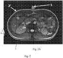
- the method consists, as a preliminary step, in acquiring or determining several positions and configurations of the profile 3 of the skin 2 in predetermined consecutive axial plane Pa, substantially perpendicular to the cranio-caudal axis X and distributed along this axis to determined locations, this at successive given instants, in at least two positions of the respiratory cycle, so as to observe a respiratory displacement whose amplitude is substantially similar to the amplitude corresponding to normal breathing.
- the determination of the position of the skin at the level of the axial profiles can be performed simultaneously for several profiles at a time, if the acquisition device allows it.
- This method also consists, during a prior phase, for each axial plane Pa considered, to build at least one deformable digital model of the skin (profile) to from information relating to the different skin profiles 3 acquired or determined previously.
- the actual current position of a specific point or area 4 of the skin 2 of the subject 1 at the level of each of the aforementioned axial planes Pa (preferably identified by means of a visible marker in the imaging method used), the position of which is significantly modified by the respiratory movement.
- a simulation 3 'of the skin profile in each axial plane Pa is provided, in real time, as a function of the actual position taken up and by exploiting the corresponding deformable numerical model, but also optionally of a three-dimensional representation 2 evolution of the skin 2 at the level of the area of interest, by interpolation between the different axial planes Pa.
- the specific points or specific areas of the skin 2 are arranged in a parallel plane close to or coincident with the median sagittal plane PSM of the subject 1, preferably being spaced apart by a distance of the order of one centimeter.
- the specific points or specific areas 4, whose real position is followed in time are distributed along the cranio-caudal axis, aligned or no, their number being advantageously at least equal to about 10 and their spacing of at most 5 cm in the direction of the aforementioned axis.
- the method preferably consists, for each axial plane Pa concerned, of generating substantially in real time, a curve 3 'representing the profile 3 of the skin 2 in the axial plane Pa considered, by applying to a base curve Cb of axial profile of the skin, a deformation, for example by displacement of at least some points spaced from the base curve, a function of the measured position of a point 4 of the skin 2 of the subject 1 located in the axial plane Pa considered.
- the basic curve used preferably corresponds to the axial profile of the skin measured in the state of maximal expiration of the subject 2.
- each deformable digital model of skin profile associated respectively with one of the predetermined axial axes Pa, is obtained by pairing points in a substantially radial direction, in the axial plane Pa considered, between at least two conformations or configurations substantially different from the curve of the skin profile, corresponding to at least two different respective states of the respiratory movement of the subject.
- each deformable digital model of skin profile, associated with one of the predetermined axial planes Pa is obtained by generating a reference curve Cr situated at the mid-distance between the two curves of skin profiles corresponding to the extreme states d inspiration and expiration, to determine the points of intersection, with the two aforementioned curves, a line normal to the reference curve Cr and passing through a point of this curve, this for a plurality of spaced points along of the latter, and to gather in an orderly manner these pairs of intersection points to form a deformation field corresponding to the displacement of the skin 2 in the axial plane Pa considered between the two extreme states mentioned above, depending on the respiratory state of the subject 1.
- the aforementioned points of the curve Cr are sufficiently spaced apart so that the normal lines do not intersect before intersecting the two axial profiles of the skin.
- the three-dimensional global simulation model of the skin is composed of a plurality of two-dimensional, skin-independent elemental axial models established in consecutive PA axial planes along the cranio-caudal axis.
- each set of information resulting from a 2D acquisition allows the construction of a model local axial of the patient's skin.
- the generation of axial models of the skin separated from each other by about 1 cm makes it possible to interpolate a faithful and accurate three-dimensional model of the patient's skin. at the level of the area of interest.
- the positions of the specific points 4 of the skin 2 are recorded in real time by means of an optical sensor device, in particular a laser device, with prior or unfolding of optical markers on the skin 2 of the skin. patient at the locations of specific points 4, the latter being advantageously selected so as to obtain a significant change in their spatial position during the respiratory movement of the subject.
- an optical sensor device in particular a laser device
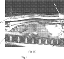
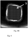
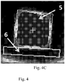
- points 4 of the skin 2 whose movements are taken into account in the context of the process as input parameters, the positions of these points 4, located in the median sagittal plane PSM of the subject 1 and in the predetermined axial planes Pa are determined by means of at least one marker 5, preferably a continuous elongate marker, resting directly or indirectly on the skin 2 of the patient 1, marrying the local surface of the skin 2 and arranged in the medial sagittal plane PSM.
- the markers 5 used consist of markers of the electromagnetic type, such as for example miniature coils, resting directly or indirectly on the skin 2, where appropriate with a known spacing relative to the latter.
- the specific points 4 of the skin 2 are each identified by means of a marker 5 which is particularly visible in the imaging method used, these specific points 4 being advantageously localized in such a way that their spatial position is significantly changed during the subject's breathing movement.
- the marker 5 used consists of a flexible and elongated body made of a material that is particularly visible in the imaging system used, extending at least along the length of the zone of interest in the direction of the cranio-caudal axis X, having a section of defined shape and minimum dimension and located at a determined distance from the skin 2 of the subject 1, resting on a strip 6 of a flexible material and substantially not visible in the system of imaging used.
- the method may advantageously consist of determining and taking up, in the sagittal plane, the curve Cm corresponding to the median axis of the elongate marker, for example formed by the succession of the different centers of gravity in section of said marker 5, and / or in the various axial planes Pa concerned, the local centers of gravity of said marker 5, and to determine the positions of the specific points 4 of the skin 2 of the subject 1 whose movements are taken into account, by exploiting the information relating, on the one hand, to this curve Cm or to these local centers of gravity, on the other hand, to the cross-sectional area and dimensions of the marker 5 and, finally, to the thickness of the interleaved material strip 6.
- the elongated marker is constituted by bar formed of gelled water, with a square or rectangular section and of sufficient length to cover at least the area of the subject whose movements induced by the respiration of said subject 1 (area of interest) is to be followed.
- the segmentation of the skin in sagittal images obtained by magnetic resonance is advantageously achieved by exploiting the very good visibility of the marker 5 in the MRI images and its contrast with respect to the textile strip 6 through which it rests on the skin of the subject (the thicknesses of said marker and said band being known data).
- the curves illustrating the positions of the marker 5 must be classified into two categories relating to the inspiration phase and the other to the expiration phase.
- the hysteresis effect is maximal at the end of the inspiration phase and at the beginning of the exhalation phase, and the discrimination of the curves is trivial with the exception of the state of extreme inspiration (see Fig. 6 ).
- the curves representing the profile 3 of the skin 2 in predetermined axial planes Pa spaced and at different times of the phases of inspiration and expiration can be obtained in different ways.
- the evolutionary three-dimensional representation 2 'of the skin 2 at the level of the zone of interest is exploited, in relation with at least one three-dimensional image of the abdomen to perform a real-time predictive simulation of the positions of the organs and / or viscera of the abdominal cavity during normal free breathing phases of the subject 1, for example by implementing the teaching of the publication cited in the introductory part of the present description.
- a practical implementation of the invention has made it possible to achieve an accuracy of 1 mm and a refresh of the simulation at a frequency of 80 Hz.
- the inventors have been able to estimate that the simulation produced by the invention has a precision at least equivalent to that of a reconstruction of the skin directly from images acquired by the imaging system.
- the invention also relates to an imaging system, in particular of the MRI type, integrating the hardware and software technical means authorizing the implementation of the method according to the invention, these means obviously resulting from the description of the method above.
- This system may, for example, comprise a magnetic resonance imaging device of the MAGNETOM Aera 1.5 Tesla type of the SIEMENS company.
Landscapes
- Health & Medical Sciences (AREA)
- Life Sciences & Earth Sciences (AREA)
- Engineering & Computer Science (AREA)
- Physics & Mathematics (AREA)
- Medical Informatics (AREA)
- General Health & Medical Sciences (AREA)
- Surgery (AREA)
- Veterinary Medicine (AREA)
- Public Health (AREA)
- Animal Behavior & Ethology (AREA)
- Biophysics (AREA)
- Pathology (AREA)
- Biomedical Technology (AREA)
- Heart & Thoracic Surgery (AREA)
- Molecular Biology (AREA)
- Nuclear Medicine, Radiotherapy & Molecular Imaging (AREA)
- Computer Vision & Pattern Recognition (AREA)
- Radiology & Medical Imaging (AREA)
- Dentistry (AREA)
- Physiology (AREA)
- Oral & Maxillofacial Surgery (AREA)
- General Physics & Mathematics (AREA)
- Theoretical Computer Science (AREA)
- High Energy & Nuclear Physics (AREA)
- Optics & Photonics (AREA)
- Measurement Of The Respiration, Hearing Ability, Form, And Blood Characteristics Of Living Organisms (AREA)
- Magnetic Resonance Imaging Apparatus (AREA)
Claims (18)
- Automatisches Verfahren zur prädiktiven Bestimmung, ausgeführt durch ein Computerprogramm, in Form einer digitalen Simulation der Position und der Bewegungen der Haut eines menschlichen oder tierischen Individuums in einem interessierenden Bereich der Brust und/oder des Bauches, wobei das genannte Individuum vorzugsweise auf dem Rücken liegt und frei oder assistiert atmet, wobei das genannte Verfahren darin besteht, vorab mehrere Positionen und Konfigurationen des Hautprofils in festgelegten, axial aufeinanderfolgenden Ebenen zu erfassen oder zu bestimmen, die zur craniocaudalen Achse im Wesentlichen rechtwinklig sind und über diese Achse an bestimmten Stellen verteilt sind, dies zu gegebenen, aufeinanderfolgenden Zeitpunkten in verschiedenen Atmungspositionen, vorteilhafterweise während Inspirations- und Exspirationsphasen des Individuums,
wobei dieses Verfahren ebenfalls darin besteht,- in einer Verarbeitungsphase vorab mindestens ein unabhängiges verformbares, digitales Modell des Hautprofils in jeder axialen Ebene (Pa) zu konstruieren, dies aus Informationen über die verschiedenen Hautprofile (3), die vorab erfasst oder bestimmt wurden,- anschließend wiederholt die tatsächliche Position eines bestimmten Punktes oder punktförmigen Bereiches (4) der Haut (2) des Individuums (1) in Höhe jeder der genannten axialen Ebenen (Pa) zu erfassen, dessen Position während der Inspirations- und Exspirationsphasen erheblich geändert wird, und- im Wesentlichen in Echtzeit einerseits eine Simulation (3') des Hautprofils in jeder axialen Ebene (Pa) in Abhängigkeit von der für jeden bestimmten Punkt oder punktförmigen Bereich (4) erfassten tatsächlichen Position und unter Verwendung des verformbaren digitalen Modells des Hautprofils in der entsprechenden Ebene (Pa) auszugeben und andererseits eine evolutive, dreidimensionale Darstellung (2') der Haut (2) im interessierenden Bereich auszugeben, dies auf der Grundlage eines globalen dreidimensionalen Simulationsmodells der Haut, bestehend aus den mehreren zweidimensionalen, axialen, verformbaren und unabhängigen Elementarmodellen, die in den axialen Ebenen (Pa) erstellt wurden, und durch Interpolation zwischen den verschiedenen genannten axialen Ebenen (Pa), Verfahren, dadurch gekennzeichnet, dass jedes verformbare digitale Hautprofilmodell, das jeweils einer der vorab festgelegten axialen Ebenen (Pa) entspricht, durch Punktepaarung in einer im Wesentlichen radialen Richtung in der betrachteten axialen Ebene (Pa) zwischen mindestens zwei deutlich unterschiedlichen Formen oder Konfigurationen der Kurve des Hautprofils, die mindestens zwei unterschiedlichen, entsprechenden Zuständen der Atmungsbewegungen des Individuums entsprechen, erhalten wird. - Verfahren nach Patentanspruch 1, dadurch gekennzeichnet, dass die bestimmten Punkte oder punktförmigen Bereiche (4) der Haut (2), deren tatsächliche Position in der Zeit verfolgt wird, in einer parallelen Ebene liegen, die der Sagittal-Medianebene (PSM) des Individuums (1) nahe oder mit ihr identisch ist, während sie vorzugsweise voneinander um einen Abstand in der Größenordnung von Zentimetern beabstandet sind.
- Verfahren nach Patentanspruch 1, dadurch gekennzeichnet, dass die bestimmten Punkte oder punktförmigen Bereiche (4), deren tatsächliche Position in der Zeit verfolgt wird, über die craniocaudale Achse verteilt sind, ausgerichtet oder nicht, wobei ihre Anzahl vorteilhafterweise mindestens gleich ungefähr 10 ist und ihr Abstand höchstens 5 cm in der Richtung der genannten Achse beträgt.
- Verfahren nach irgendeinem der Patentansprüche 1 bis 3, dadurch gekennzeichnet, dass es darin besteht, für jede betroffene axiale Ebene (Pa) im Wesentlichen in Echtzeit eine Kurve (3') zu erzeugen, die das Profil (3) der Haut (2) in der betrachteten axialen Ebene (Pa) darstellt, indem auf eine Basishautprofilkurve (Cb), die vorteilhafterweise in einem Zustand extremer Inspiration oder Exspiration des Individuums ermittelt wurde, eine Verformung ausgeübt wird, beispielsweise durch Versetzen mindestens bestimmter, voneinander beabstandeter Punkte aus der Basiskurve in Abhängigkeit von der gemessenen Position eines Punktes (4) der Haut (2) des Individuums (1), der sich in der betrachteten axialen Ebene (Pa) befindet.
- Verfahren nach irgendeinem der Patentansprüche 1 bis 4, dadurch gekennzeichnet, dass jedes digitale, verformbare Hautprofilmodell, das einer der vorab festgelegten axialen Ebenen (Pa) zugehört, durch Bestimmung einer Bezugskurve (Cr) erhalten wird, die sich in der Mitte zwischen den beiden Hautprofilkurven befindet, die den extremen Inspirations- und Exspirationszuständen entsprechen, die Schnittpunkte einer zur Bezugskurve (Cr) normalen und durch einen Punkt dieser Kurve verlaufende Gerade mit den beiden genannten Kurven für mehrere auf dieser liegende, voneinander beabstandete Punkte zu bestimmen und in geordneter Weise diese Schnittpunktpaare zu vereinen, um ein Verformungsfeld zu bilden, das der Verformung der Haut (2) in der betrachteten axialen Ebene (Pa) zwischen den beiden genannten Extremzuständen in Abhängigkeit vom Atmungszustand des Individuums (1) entspricht.
- Verfahren nach irgendeinem der Patentansprüche 1 bis 5, dadurch gekennzeichnet, dass es darin besteht, zwei digitale, verformbare Hautprofilmodelle für jede axiale Ebene (Pa) zu konstruieren, nämlich ein Modell für die Inspirationsphasen und ein Modell für die Exspirationsphasen, und die laufende Atmungsphase während der Bestimmung der simulierten Hautprofilkurven (3') in den verschiedenen axialen Ebenen (Pa) mit Hilfe eines spezifischen Sensors oder durch Auswertung der Verschiebungsrichtung der Punkte (4) festzustellen.
- Verfahren nach irgendeinem der Patentansprüche 1 bis 6, dadurch gekennzeichnet, dass die Positionen der spezifischen Punkte (4) der Haut (2) in Echtzeit mit Hilfe einer optischen Sensorvorrichtung, insbesondere mit einem Laser, unter vorheriger Anordnung optischer Markierungen auf der Haut des Patienten an den Stellen der spezifischen Punkte (4) oder nicht, erfasst werden, wobei diese letztgenannten vorteilhafterweise derart gewählt werden, dass eine deutliche Änderung ihrer räumlichen Position während der Atembewegung des Individuums erhalten wird.
- Verfahren nach irgendeinem der Patentansprüche 1 bis 6, dadurch gekennzeichnet, dass die Positionen der Punkte (4) der Haut (2), die in der Sagittal-Medianebene (PSM) des Individuums (1) und in den vorab festgelegten axialen Ebenen (Pa) liegen, mit Hilfe mindestens einer Markierung (5) bestimmt werden.
- Verfahren nach Patentanspruch 8, dadurch gekennzeichnet, dass die spezifischen Punkte (4) der Haut (2) jeweils mit Hilfe einer im angewandten bildgebenden Verfahren gut sichtbaren Markierung (5) bestimmt werden, wobei diese spezifischen Punkte (4) vorteilhafterweise derart angeordnet sind, dass ihre räumliche Position sich während der Atembewegung des Individuums deutlich ändert.
- Verfahren nach Patentanspruch 8 oder 9, dadurch gekennzeichnet, dass die verwendeten Markierungen (5) Markierungen vom elektromagnetischen Typ sind, wie etwa Miniaturspulen, die unmittelbar oder mittelbar auf der Haut (2) ruhen, gegebenenfalls in einem bekannten Abstand von dieser.
- Verfahren nach Patentanspruch 8 oder 9, dadurch gekennzeichnet, dass die einzige verwendete Markierung (5) in einer durchgehenden, langgestreckten Markierung besteht, die unmittelbar oder mittelbar auf der Haut (2) des Patienten (1) ruht, indem sie sich der lokalen Oberfläche der Haut (2) anschmiegt und in der Sagittal-Medianebene (PSM) angeordnet ist.
- Verfahren nach Patentanspruch 11, dadurch gekennzeichnet, dass die verwendete Markierung (5) in einem weichen und langgestreckten Körper besteht, der aus einem Werkstoff gefertigt ist, der im verwendeten bildgebenden Verfahren besonders gut sichtbar ist, sich mindestens über die Länge des interessierenden Bereiches in Richtung der craniocaudalen Achse (X) erstreckt und einen festgelegten Querschnitt mit festgelegter kleinster Abmessung aufweist und in einem festgelegten Abstand von der Haut (2) des Individuums (1) angeordnet ist, wobei er auf einem Streifen (6) aus weichem und im verwendeten bildgebenden Verfahren im Wesentlichen nicht sichtbaren Werkstoff besteht.
- Verfahren nach irgendeinem der Patentansprüche 11 und 12, dadurch gekennzeichnet, dass es darin besteht, in der Sagittalebene die Kurve (Cm) zu bestimmen und zu erfassen, die der Mittelachse der langgestreckten Markierung (5) entspricht, die beispielsweise durch die Reihe der verschiedenen Schwerpunkte des Querschnitts der genannten Markierung (5) und/oder die lokalen Schwerpunkte der Markierung (5) in den verschiedenen betroffenen axialen Ebenen (Pa) gebildet wird, und die Stellen der spezifischen Punkte (4) der Haut (2) des Individuums (1) zu bestimmen, deren Bewegungen berücksichtigt werden, indem die Informationen über einerseits diese Kurve (Cm) oder diese lokalen Schwerpunkte und andererseits über den Querschnitt und die Querschnittsabmessungen der Markierung (5), und schließlich über die Dicke des eingefügten Werkstoffstreifens (6) ausgewertet werden.
- Verfahren nach irgendeinem der Patentansprüche 1 bis 13, dadurch gekennzeichnet, dass die Kurven, die das Profil (3) der Haut (2) in den vorab festgelegten, voneinander beabstandeten axialen Ebenen (Pa) und zu verschiedenen Zeitpunkten der Inspirations- und Exspirationsphasen darstellen, durch Abtastung dreidimensionaler Darstellungen der Oberfläche der Haut (2), die im interessierenden Bereich, die durch Kennzeichnung und Verfolgen der Haut mit Hilfe auf der Haut befestigter Markierungen oder durch strukturiertes Licht, das auf die Haut projiziert wurde, vorab erfasst wurden, nach axialen Ebenen gewonnen werden.
- Verfahren nach irgendeinem der Patentansprüche 1 bis 14, dadurch gekennzeichnet, dass die Kurven, die das Profil (3) der Haut (2) in vorab festgelegten, voneinander beabstandeten axialen Ebenen (Pa) und zu verschiedenen Zeitpunkten der Inspirations- und Exspirationsphasen darstellen, durch eine Technik der Segmentierung dreidimensionaler Darstellungen gewonnen werden, die durch ein bildgebendes Verfahren auf der Grundlage der Magnetspinresonanz oder der Computertomographie erhalten wurden.
- Verfahren nach irgendeinem der Patentansprüche 1 bis 14, dadurch gekennzeichnet, dass die Kurven, die das Profil (3) der Haut (2) in den vorab festgelegten, voneinander beabstandeten axialen Ebenen (Pa) und zu verschiedenen Zeitpunkten der Inspirations- und Exspirationsphasen darstellen, durch Segmentierung von Bildern des Typs 2D + t gewonnen werden, die in den genannten Achsenebenen (Pa) erfolgt, und durch ein bildgebendes Verfahren auf der Grundlage der Magnetspinresonanz oder der Computertomographie erhalten wurden.
- Verfahren nach irgendeinem der Patentansprüche 1 bis 15, dadurch gekennzeichnet, dass die evolutive dreidimensionale Darstellung der Haut (2) im interessierenden Bereich in Verbindung mit mindestens einem dreidimensionalen Bild des Bauches ausgewertet wird, um eine vorhersagende Simulation in Echtzeit der Stellungen der Organe und/oder Eingeweide der Bauchhöhle während der Phasen normaler, freier Atmung des Individuums (1) zu erstellen.
- Verfahren nach irgendeinem der Patentansprüche 11 bis 13, dadurch gekennzeichnet, dass die langgestreckte Markierung (5), wenn das bildgebende Verfahren, dem das Individuum unterworfen wird, auf Magnetspinresonanz beruht, aus einem Stab aus geliertem Wasser mit quadratischem oder rechteckigem Querschnitt und einer Länge besteht, die ausreicht, um mindestens den Bereich des Individuums (1) abzudecken, dessen durch die Atmung des Individuums bewirkte Bewegungen verfolgt werden sollen.
Applications Claiming Priority (2)
| Application Number | Priority Date | Filing Date | Title |
|---|---|---|---|
| FR1351855A FR3002732A1 (fr) | 2013-03-01 | 2013-03-01 | Procede automatique de determination predictive de la position de la peau |
| PCT/FR2014/050454 WO2014132008A1 (fr) | 2013-03-01 | 2014-03-03 | Procede automatique de determination predictive de la position de la peau |
Publications (2)
| Publication Number | Publication Date |
|---|---|
| EP2961316A1 EP2961316A1 (de) | 2016-01-06 |
| EP2961316B1 true EP2961316B1 (de) | 2018-11-28 |
Family
ID=48289385
Family Applications (1)
| Application Number | Title | Priority Date | Filing Date |
|---|---|---|---|
| EP14713205.4A Not-in-force EP2961316B1 (de) | 2013-03-01 | 2014-03-03 | Automatisches verfahren zur vorabbestimmung der position der haut |
Country Status (4)
| Country | Link |
|---|---|
| US (1) | US9717441B2 (de) |
| EP (1) | EP2961316B1 (de) |
| FR (1) | FR3002732A1 (de) |
| WO (1) | WO2014132008A1 (de) |
Families Citing this family (6)
| Publication number | Priority date | Publication date | Assignee | Title |
|---|---|---|---|---|
| DE102006004197A1 (de) * | 2006-01-26 | 2007-08-09 | Klett, Rolf, Dr.Dr. | Verfahren und Vorrichtung zur Aufzeichnung von Körperbewegungen |
| CN107468256A (zh) | 2012-11-27 | 2017-12-15 | 纽玛凯尔有限公司 | 呼吸数据的分析 |
| KR101636876B1 (ko) * | 2014-11-03 | 2016-07-06 | 삼성전자주식회사 | 의료 영상 처리 장치 및 방법 |
| CN113893033B (zh) * | 2021-07-01 | 2023-05-12 | 中国科学院苏州生物医学工程技术研究所 | 一种肺部经皮穿刺导航方法及系统 |
| CN113674393B (zh) * | 2021-07-12 | 2023-09-26 | 中国科学院深圳先进技术研究院 | 呼吸运动模型的构建方法和无标记呼吸运动预测方法 |
| CN119326511B (zh) * | 2024-12-23 | 2025-03-04 | 北京大学第三医院(北京大学第三临床医学院) | 呼吸模型的建模方法及射波刀的同步呼吸追踪方法 |
Family Cites Families (6)
| Publication number | Priority date | Publication date | Assignee | Title |
|---|---|---|---|---|
| US8788020B2 (en) * | 1998-10-23 | 2014-07-22 | Varian Medical Systems, Inc. | Method and system for radiation application |
| WO2006051911A1 (ja) * | 2004-11-12 | 2006-05-18 | Kabushiki Kaisha Toshiba | 磁気共鳴イメージング装置、画像データ補正装置および画像データ補正方法 |
| US8747382B2 (en) * | 2005-04-13 | 2014-06-10 | University Of Maryland, Baltimore | Techniques for compensating movement of a treatment target in a patient |
| US7720196B2 (en) * | 2008-01-07 | 2010-05-18 | Accuray Incorporated | Target tracking using surface scanner and four-dimensional diagnostic imaging data |
| BRPI1006280A2 (pt) * | 2009-04-03 | 2019-04-02 | Koninl Philips Electronics Nv | método para a segmentação de um órgão, sistema para a segmentação de um órgão e meio de armazenamento que pode ser lido por computador |
| EP2664360B1 (de) * | 2010-02-24 | 2015-09-09 | Accuray Incorporated | Bildgeführtes Gantry-Strahlentherapiesystem und zugehörige Trackingverfahren |
-
2013
- 2013-03-01 FR FR1351855A patent/FR3002732A1/fr active Pending
-
2014
- 2014-03-03 WO PCT/FR2014/050454 patent/WO2014132008A1/fr not_active Ceased
- 2014-03-03 EP EP14713205.4A patent/EP2961316B1/de not_active Not-in-force
- 2014-03-03 US US14/770,471 patent/US9717441B2/en not_active Expired - Fee Related
Non-Patent Citations (1)
| Title |
|---|
| None * |
Also Published As
| Publication number | Publication date |
|---|---|
| US9717441B2 (en) | 2017-08-01 |
| EP2961316A1 (de) | 2016-01-06 |
| WO2014132008A1 (fr) | 2014-09-04 |
| US20160000355A1 (en) | 2016-01-07 |
| FR3002732A1 (fr) | 2014-09-05 |
Similar Documents
| Publication | Publication Date | Title |
|---|---|---|
| EP2961316B1 (de) | Automatisches verfahren zur vorabbestimmung der position der haut | |
| US10278584B2 (en) | Method and system for three-dimensional imaging | |
| EP2615969B1 (de) | System und verfahren zur berechnung von aktivierungskarten | |
| Liberadzki et al. | Structured-light-based system for shape measurement of the human body in motion | |
| JP2014004359A5 (de) | ||
| US9396576B2 (en) | Method and apparatus for estimating the three-dimensional shape of an object | |
| JP6273291B2 (ja) | 画像処理装置および方法 | |
| CN110494889A (zh) | 造影注射成像 | |
| WO2022219631A1 (en) | Systems and methods for reconstruction of 3d images from ultrasound and camera images | |
| FR2963976A1 (fr) | Procede de traitement d'images pour la determination de zones suspectes dans une matrice tissulaire, et son utilisation pour la navigation 3d a travers la matrice tissulaire | |
| EP3164078A1 (de) | Verfahren und vorrichtung zur funktionellen bildgebung des gehirns | |
| Paoli et al. | Sensor architectures and technologies for upper limb 3D surface reconstruction: a review | |
| CN101681507B (zh) | 基于模型的spect心脏取向估计 | |
| JP2007136184A5 (de) | ||
| US9092666B2 (en) | Method and apparatus for estimating organ deformation model and medical image system | |
| EP3991136B1 (de) | Verfahren zur analyse des gangs einer person | |
| US20180130212A1 (en) | Edge detection on images with correlated noise | |
| US20140185898A1 (en) | Image generation method and apparatus | |
| US20140233794A1 (en) | Method, apparatus and medical imaging system for tracking motion of organ | |
| GB2605459A (en) | Method of non-invasive medical tomographic imaging with uncertainty estimation | |
| CN105374012B (zh) | 用于消除由性能差异的探测器单元所致的条状伪影的方法 | |
| JP2011149952A (ja) | モデル入力装置およびモデル生成システム | |
| JP4137822B2 (ja) | 画像再構成方法及び装置、画像再構成プログラム | |
| Jeon et al. | Image reconstruction in EIT with unreliable electrode data using random sample consensus method | |
| Noble et al. | Arm-volume measurement for early lymphedema detection using Kinect sensor in post-surgical oncology patients |
Legal Events
| Date | Code | Title | Description |
|---|---|---|---|
| PUAI | Public reference made under article 153(3) epc to a published international application that has entered the european phase |
Free format text: ORIGINAL CODE: 0009012 |
|
| 17P | Request for examination filed |
Effective date: 20150910 |
|
| AK | Designated contracting states |
Kind code of ref document: A1 Designated state(s): AL AT BE BG CH CY CZ DE DK EE ES FI FR GB GR HR HU IE IS IT LI LT LU LV MC MK MT NL NO PL PT RO RS SE SI SK SM TR |
|
| AX | Request for extension of the european patent |
Extension state: BA ME |
|
| RIN1 | Information on inventor provided before grant (corrected) |
Inventor name: SOLER, LUC Inventor name: HOSTETTLER, ALEXANDRE Inventor name: NICOLAU, STEPHANE Inventor name: MARESCAUX, JACQUES |
|
| DAX | Request for extension of the european patent (deleted) | ||
| GRAP | Despatch of communication of intention to grant a patent |
Free format text: ORIGINAL CODE: EPIDOSNIGR1 |
|
| STAA | Information on the status of an ep patent application or granted ep patent |
Free format text: STATUS: GRANT OF PATENT IS INTENDED |
|
| INTG | Intention to grant announced |
Effective date: 20180619 |
|
| RAP1 | Party data changed (applicant data changed or rights of an application transferred) |
Owner name: INSTITUT DE RECHERCHE SUR LES CANCERS DE L'APPAREI |
|
| GRAS | Grant fee paid |
Free format text: ORIGINAL CODE: EPIDOSNIGR3 |
|
| GRAA | (expected) grant |
Free format text: ORIGINAL CODE: 0009210 |
|
| STAA | Information on the status of an ep patent application or granted ep patent |
Free format text: STATUS: THE PATENT HAS BEEN GRANTED |
|
| AK | Designated contracting states |
Kind code of ref document: B1 Designated state(s): AL AT BE BG CH CY CZ DE DK EE ES FI FR GB GR HR HU IE IS IT LI LT LU LV MC MK MT NL NO PL PT RO RS SE SI SK SM TR |
|
| REG | Reference to a national code |
Ref country code: CH Ref legal event code: EP |
|
| REG | Reference to a national code |
Ref country code: AT Ref legal event code: REF Ref document number: 1069269 Country of ref document: AT Kind code of ref document: T Effective date: 20181215 |
|
| REG | Reference to a national code |
Ref country code: DE Ref legal event code: R096 Ref document number: 602014036885 Country of ref document: DE |
|
| REG | Reference to a national code |
Ref country code: IE Ref legal event code: FG4D Free format text: LANGUAGE OF EP DOCUMENT: FRENCH |
|
| REG | Reference to a national code |
Ref country code: NL Ref legal event code: MP Effective date: 20181128 |
|
| REG | Reference to a national code |
Ref country code: LT Ref legal event code: MG4D |
|
| REG | Reference to a national code |
Ref country code: AT Ref legal event code: MK05 Ref document number: 1069269 Country of ref document: AT Kind code of ref document: T Effective date: 20181128 |
|
| PG25 | Lapsed in a contracting state [announced via postgrant information from national office to epo] |
Ref country code: ES Free format text: LAPSE BECAUSE OF FAILURE TO SUBMIT A TRANSLATION OF THE DESCRIPTION OR TO PAY THE FEE WITHIN THE PRESCRIBED TIME-LIMIT Effective date: 20181128 Ref country code: LV Free format text: LAPSE BECAUSE OF FAILURE TO SUBMIT A TRANSLATION OF THE DESCRIPTION OR TO PAY THE FEE WITHIN THE PRESCRIBED TIME-LIMIT Effective date: 20181128 Ref country code: HR Free format text: LAPSE BECAUSE OF FAILURE TO SUBMIT A TRANSLATION OF THE DESCRIPTION OR TO PAY THE FEE WITHIN THE PRESCRIBED TIME-LIMIT Effective date: 20181128 Ref country code: LT Free format text: LAPSE BECAUSE OF FAILURE TO SUBMIT A TRANSLATION OF THE DESCRIPTION OR TO PAY THE FEE WITHIN THE PRESCRIBED TIME-LIMIT Effective date: 20181128 Ref country code: BG Free format text: LAPSE BECAUSE OF FAILURE TO SUBMIT A TRANSLATION OF THE DESCRIPTION OR TO PAY THE FEE WITHIN THE PRESCRIBED TIME-LIMIT Effective date: 20190228 Ref country code: FI Free format text: LAPSE BECAUSE OF FAILURE TO SUBMIT A TRANSLATION OF THE DESCRIPTION OR TO PAY THE FEE WITHIN THE PRESCRIBED TIME-LIMIT Effective date: 20181128 Ref country code: NO Free format text: LAPSE BECAUSE OF FAILURE TO SUBMIT A TRANSLATION OF THE DESCRIPTION OR TO PAY THE FEE WITHIN THE PRESCRIBED TIME-LIMIT Effective date: 20190228 Ref country code: IS Free format text: LAPSE BECAUSE OF FAILURE TO SUBMIT A TRANSLATION OF THE DESCRIPTION OR TO PAY THE FEE WITHIN THE PRESCRIBED TIME-LIMIT Effective date: 20190328 Ref country code: AT Free format text: LAPSE BECAUSE OF FAILURE TO SUBMIT A TRANSLATION OF THE DESCRIPTION OR TO PAY THE FEE WITHIN THE PRESCRIBED TIME-LIMIT Effective date: 20181128 |
|
| PG25 | Lapsed in a contracting state [announced via postgrant information from national office to epo] |
Ref country code: SE Free format text: LAPSE BECAUSE OF FAILURE TO SUBMIT A TRANSLATION OF THE DESCRIPTION OR TO PAY THE FEE WITHIN THE PRESCRIBED TIME-LIMIT Effective date: 20181128 Ref country code: AL Free format text: LAPSE BECAUSE OF FAILURE TO SUBMIT A TRANSLATION OF THE DESCRIPTION OR TO PAY THE FEE WITHIN THE PRESCRIBED TIME-LIMIT Effective date: 20181128 Ref country code: RS Free format text: LAPSE BECAUSE OF FAILURE TO SUBMIT A TRANSLATION OF THE DESCRIPTION OR TO PAY THE FEE WITHIN THE PRESCRIBED TIME-LIMIT Effective date: 20181128 Ref country code: GR Free format text: LAPSE BECAUSE OF FAILURE TO SUBMIT A TRANSLATION OF THE DESCRIPTION OR TO PAY THE FEE WITHIN THE PRESCRIBED TIME-LIMIT Effective date: 20190301 Ref country code: PT Free format text: LAPSE BECAUSE OF FAILURE TO SUBMIT A TRANSLATION OF THE DESCRIPTION OR TO PAY THE FEE WITHIN THE PRESCRIBED TIME-LIMIT Effective date: 20190328 |
|
| PG25 | Lapsed in a contracting state [announced via postgrant information from national office to epo] |
Ref country code: NL Free format text: LAPSE BECAUSE OF FAILURE TO SUBMIT A TRANSLATION OF THE DESCRIPTION OR TO PAY THE FEE WITHIN THE PRESCRIBED TIME-LIMIT Effective date: 20181128 |
|
| PG25 | Lapsed in a contracting state [announced via postgrant information from national office to epo] |
Ref country code: DK Free format text: LAPSE BECAUSE OF FAILURE TO SUBMIT A TRANSLATION OF THE DESCRIPTION OR TO PAY THE FEE WITHIN THE PRESCRIBED TIME-LIMIT Effective date: 20181128 Ref country code: IT Free format text: LAPSE BECAUSE OF FAILURE TO SUBMIT A TRANSLATION OF THE DESCRIPTION OR TO PAY THE FEE WITHIN THE PRESCRIBED TIME-LIMIT Effective date: 20181128 Ref country code: CZ Free format text: LAPSE BECAUSE OF FAILURE TO SUBMIT A TRANSLATION OF THE DESCRIPTION OR TO PAY THE FEE WITHIN THE PRESCRIBED TIME-LIMIT Effective date: 20181128 Ref country code: PL Free format text: LAPSE BECAUSE OF FAILURE TO SUBMIT A TRANSLATION OF THE DESCRIPTION OR TO PAY THE FEE WITHIN THE PRESCRIBED TIME-LIMIT Effective date: 20181128 |
|
| REG | Reference to a national code |
Ref country code: DE Ref legal event code: R097 Ref document number: 602014036885 Country of ref document: DE |
|
| PG25 | Lapsed in a contracting state [announced via postgrant information from national office to epo] |
Ref country code: SK Free format text: LAPSE BECAUSE OF FAILURE TO SUBMIT A TRANSLATION OF THE DESCRIPTION OR TO PAY THE FEE WITHIN THE PRESCRIBED TIME-LIMIT Effective date: 20181128 Ref country code: RO Free format text: LAPSE BECAUSE OF FAILURE TO SUBMIT A TRANSLATION OF THE DESCRIPTION OR TO PAY THE FEE WITHIN THE PRESCRIBED TIME-LIMIT Effective date: 20181128 Ref country code: EE Free format text: LAPSE BECAUSE OF FAILURE TO SUBMIT A TRANSLATION OF THE DESCRIPTION OR TO PAY THE FEE WITHIN THE PRESCRIBED TIME-LIMIT Effective date: 20181128 Ref country code: SM Free format text: LAPSE BECAUSE OF FAILURE TO SUBMIT A TRANSLATION OF THE DESCRIPTION OR TO PAY THE FEE WITHIN THE PRESCRIBED TIME-LIMIT Effective date: 20181128 |
|
| REG | Reference to a national code |
Ref country code: DE Ref legal event code: R119 Ref document number: 602014036885 Country of ref document: DE |
|
| PLBE | No opposition filed within time limit |
Free format text: ORIGINAL CODE: 0009261 |
|
| STAA | Information on the status of an ep patent application or granted ep patent |
Free format text: STATUS: NO OPPOSITION FILED WITHIN TIME LIMIT |
|
| PG25 | Lapsed in a contracting state [announced via postgrant information from national office to epo] |
Ref country code: SI Free format text: LAPSE BECAUSE OF FAILURE TO SUBMIT A TRANSLATION OF THE DESCRIPTION OR TO PAY THE FEE WITHIN THE PRESCRIBED TIME-LIMIT Effective date: 20181128 Ref country code: MC Free format text: LAPSE BECAUSE OF FAILURE TO SUBMIT A TRANSLATION OF THE DESCRIPTION OR TO PAY THE FEE WITHIN THE PRESCRIBED TIME-LIMIT Effective date: 20181128 |
|
| REG | Reference to a national code |
Ref country code: CH Ref legal event code: PL |
|
| 26N | No opposition filed |
Effective date: 20190829 |
|
| GBPC | Gb: european patent ceased through non-payment of renewal fee |
Effective date: 20190303 |
|
| PG25 | Lapsed in a contracting state [announced via postgrant information from national office to epo] |
Ref country code: LU Free format text: LAPSE BECAUSE OF NON-PAYMENT OF DUE FEES Effective date: 20190303 |
|
| REG | Reference to a national code |
Ref country code: BE Ref legal event code: MM Effective date: 20190331 |
|
| PG25 | Lapsed in a contracting state [announced via postgrant information from national office to epo] |
Ref country code: LI Free format text: LAPSE BECAUSE OF NON-PAYMENT OF DUE FEES Effective date: 20190331 Ref country code: GB Free format text: LAPSE BECAUSE OF NON-PAYMENT OF DUE FEES Effective date: 20190303 Ref country code: CH Free format text: LAPSE BECAUSE OF NON-PAYMENT OF DUE FEES Effective date: 20190331 Ref country code: DE Free format text: LAPSE BECAUSE OF NON-PAYMENT OF DUE FEES Effective date: 20191001 Ref country code: IE Free format text: LAPSE BECAUSE OF NON-PAYMENT OF DUE FEES Effective date: 20190303 |
|
| PG25 | Lapsed in a contracting state [announced via postgrant information from national office to epo] |
Ref country code: BE Free format text: LAPSE BECAUSE OF NON-PAYMENT OF DUE FEES Effective date: 20190331 Ref country code: FR Free format text: LAPSE BECAUSE OF NON-PAYMENT OF DUE FEES Effective date: 20190331 |
|
| PG25 | Lapsed in a contracting state [announced via postgrant information from national office to epo] |
Ref country code: TR Free format text: LAPSE BECAUSE OF FAILURE TO SUBMIT A TRANSLATION OF THE DESCRIPTION OR TO PAY THE FEE WITHIN THE PRESCRIBED TIME-LIMIT Effective date: 20181128 |
|
| PG25 | Lapsed in a contracting state [announced via postgrant information from national office to epo] |
Ref country code: MT Free format text: LAPSE BECAUSE OF FAILURE TO SUBMIT A TRANSLATION OF THE DESCRIPTION OR TO PAY THE FEE WITHIN THE PRESCRIBED TIME-LIMIT Effective date: 20181128 |
|
| PG25 | Lapsed in a contracting state [announced via postgrant information from national office to epo] |
Ref country code: CY Free format text: LAPSE BECAUSE OF FAILURE TO SUBMIT A TRANSLATION OF THE DESCRIPTION OR TO PAY THE FEE WITHIN THE PRESCRIBED TIME-LIMIT Effective date: 20181128 |
|
| PG25 | Lapsed in a contracting state [announced via postgrant information from national office to epo] |
Ref country code: HU Free format text: LAPSE BECAUSE OF FAILURE TO SUBMIT A TRANSLATION OF THE DESCRIPTION OR TO PAY THE FEE WITHIN THE PRESCRIBED TIME-LIMIT; INVALID AB INITIO Effective date: 20140303 |
|
| PG25 | Lapsed in a contracting state [announced via postgrant information from national office to epo] |
Ref country code: MK Free format text: LAPSE BECAUSE OF FAILURE TO SUBMIT A TRANSLATION OF THE DESCRIPTION OR TO PAY THE FEE WITHIN THE PRESCRIBED TIME-LIMIT Effective date: 20181128 |