EP2959015B1 - Characterization of molecules in nanofluidics - Google Patents
Characterization of molecules in nanofluidics Download PDFInfo
- Publication number
- EP2959015B1 EP2959015B1 EP14753475.4A EP14753475A EP2959015B1 EP 2959015 B1 EP2959015 B1 EP 2959015B1 EP 14753475 A EP14753475 A EP 14753475A EP 2959015 B1 EP2959015 B1 EP 2959015B1
- Authority
- EP
- European Patent Office
- Prior art keywords
- sample
- molecules
- label
- fragments
- labeled
- Prior art date
- Legal status (The legal status is an assumption and is not a legal conclusion. Google has not performed a legal analysis and makes no representation as to the accuracy of the status listed.)
- Active
Links
- 238000012512 characterization method Methods 0.000 title 1
- 108020004414 DNA Proteins 0.000 claims description 120
- 238000000034 method Methods 0.000 claims description 104
- 239000012634 fragment Substances 0.000 claims description 95
- 238000002372 labelling Methods 0.000 claims description 62
- 102000053602 DNA Human genes 0.000 claims description 43
- 102000039446 nucleic acids Human genes 0.000 claims description 38
- 108020004707 nucleic acids Proteins 0.000 claims description 38
- 150000007523 nucleic acids Chemical class 0.000 claims description 38
- 210000004369 blood Anatomy 0.000 claims description 26
- 239000008280 blood Substances 0.000 claims description 26
- 230000003287 optical effect Effects 0.000 claims description 25
- 230000008774 maternal effect Effects 0.000 claims description 23
- 102000040430 polynucleotide Human genes 0.000 claims description 19
- 108091033319 polynucleotide Proteins 0.000 claims description 19
- 239000002157 polynucleotide Substances 0.000 claims description 19
- 230000001605 fetal effect Effects 0.000 claims description 18
- 210000004027 cell Anatomy 0.000 claims description 14
- 230000005291 magnetic effect Effects 0.000 claims description 14
- 239000007850 fluorescent dye Substances 0.000 claims description 11
- 230000002285 radioactive effect Effects 0.000 claims description 10
- 230000002159 abnormal effect Effects 0.000 claims description 8
- 238000009826 distribution Methods 0.000 claims description 8
- 210000003765 sex chromosome Anatomy 0.000 claims description 7
- 101710147059 Nicking endonuclease Proteins 0.000 claims description 6
- 238000012163 sequencing technique Methods 0.000 claims description 2
- 239000000523 sample Substances 0.000 description 239
- 125000003729 nucleotide group Chemical group 0.000 description 49
- 239000002773 nucleotide Substances 0.000 description 48
- 210000000349 chromosome Anatomy 0.000 description 31
- 239000002090 nanochannel Substances 0.000 description 29
- 230000005856 abnormality Effects 0.000 description 23
- 230000002068 genetic effect Effects 0.000 description 19
- 239000002245 particle Substances 0.000 description 19
- 102000016397 Methyltransferase Human genes 0.000 description 16
- 108060004795 Methyltransferase Proteins 0.000 description 16
- 238000001514 detection method Methods 0.000 description 14
- 206010028980 Neoplasm Diseases 0.000 description 13
- 208000036878 aneuploidy Diseases 0.000 description 13
- 231100001075 aneuploidy Toxicity 0.000 description 12
- 239000013074 reference sample Substances 0.000 description 12
- 210000001519 tissue Anatomy 0.000 description 12
- 230000000875 corresponding effect Effects 0.000 description 11
- 108090000623 proteins and genes Proteins 0.000 description 11
- 208000037280 Trisomy Diseases 0.000 description 10
- 108091032973 (ribonucleotides)n+m Proteins 0.000 description 9
- 239000011324 bead Substances 0.000 description 9
- 239000000126 substance Substances 0.000 description 9
- YBJHBAHKTGYVGT-ZKWXMUAHSA-N (+)-Biotin Chemical compound N1C(=O)N[C@@H]2[C@H](CCCCC(=O)O)SC[C@@H]21 YBJHBAHKTGYVGT-ZKWXMUAHSA-N 0.000 description 8
- 239000012530 fluid Substances 0.000 description 8
- 210000003917 human chromosome Anatomy 0.000 description 8
- 210000004881 tumor cell Anatomy 0.000 description 8
- 201000011510 cancer Diseases 0.000 description 7
- 238000002866 fluorescence resonance energy transfer Methods 0.000 description 7
- 238000007689 inspection Methods 0.000 description 7
- 230000002441 reversible effect Effects 0.000 description 7
- 230000005945 translocation Effects 0.000 description 7
- 201000010374 Down Syndrome Diseases 0.000 description 6
- 230000003321 amplification Effects 0.000 description 6
- 238000004891 communication Methods 0.000 description 6
- 230000000052 comparative effect Effects 0.000 description 6
- 238000003199 nucleic acid amplification method Methods 0.000 description 6
- 108090000765 processed proteins & peptides Proteins 0.000 description 6
- 235000018102 proteins Nutrition 0.000 description 6
- 102000004169 proteins and genes Human genes 0.000 description 6
- 210000002845 virion Anatomy 0.000 description 6
- 241000894006 Bacteria Species 0.000 description 5
- 102000004190 Enzymes Human genes 0.000 description 5
- 108090000790 Enzymes Proteins 0.000 description 5
- 206010044688 Trisomy 21 Diseases 0.000 description 5
- 230000000694 effects Effects 0.000 description 5
- 208000030454 monosomy Diseases 0.000 description 5
- 229920000642 polymer Polymers 0.000 description 5
- 239000002096 quantum dot Substances 0.000 description 5
- 108091008146 restriction endonucleases Proteins 0.000 description 5
- 238000012360 testing method Methods 0.000 description 5
- 229930024421 Adenine Natural products 0.000 description 4
- GFFGJBXGBJISGV-UHFFFAOYSA-N Adenine Chemical compound NC1=NC=NC2=C1N=CN2 GFFGJBXGBJISGV-UHFFFAOYSA-N 0.000 description 4
- 108091034117 Oligonucleotide Proteins 0.000 description 4
- 229960000643 adenine Drugs 0.000 description 4
- 239000011616 biotin Substances 0.000 description 4
- 229960002685 biotin Drugs 0.000 description 4
- 235000020958 biotin Nutrition 0.000 description 4
- 210000001124 body fluid Anatomy 0.000 description 4
- 239000010839 body fluid Substances 0.000 description 4
- 230000000295 complement effect Effects 0.000 description 4
- OPTASPLRGRRNAP-UHFFFAOYSA-N cytosine Chemical compound NC=1C=CNC(=O)N=1 OPTASPLRGRRNAP-UHFFFAOYSA-N 0.000 description 4
- 238000010586 diagram Methods 0.000 description 4
- -1 short DNA fragments Chemical class 0.000 description 4
- 239000000758 substrate Substances 0.000 description 4
- 108090001008 Avidin Proteins 0.000 description 3
- 208000005443 Circulating Neoplastic Cells Diseases 0.000 description 3
- 102000052510 DNA-Binding Proteins Human genes 0.000 description 3
- 230000004568 DNA-binding Effects 0.000 description 3
- 101710096438 DNA-binding protein Proteins 0.000 description 3
- 201000006360 Edwards syndrome Diseases 0.000 description 3
- 201000009928 Patau syndrome Diseases 0.000 description 3
- 108091093037 Peptide nucleic acid Proteins 0.000 description 3
- 239000004952 Polyamide Substances 0.000 description 3
- 208000020584 Polyploidy Diseases 0.000 description 3
- 208000035199 Tetraploidy Diseases 0.000 description 3
- 108091023040 Transcription factor Proteins 0.000 description 3
- 102000040945 Transcription factor Human genes 0.000 description 3
- 208000026487 Triploidy Diseases 0.000 description 3
- 206010044686 Trisomy 13 Diseases 0.000 description 3
- 208000006284 Trisomy 13 Syndrome Diseases 0.000 description 3
- 206010071762 Trisomy 14 Diseases 0.000 description 3
- 206010062757 Trisomy 15 Diseases 0.000 description 3
- 208000007159 Trisomy 18 Syndrome Diseases 0.000 description 3
- 101710185494 Zinc finger protein Proteins 0.000 description 3
- 102100023597 Zinc finger protein 816 Human genes 0.000 description 3
- 238000001574 biopsy Methods 0.000 description 3
- 230000002759 chromosomal effect Effects 0.000 description 3
- 208000037516 chromosome inversion disease Diseases 0.000 description 3
- 238000012217 deletion Methods 0.000 description 3
- 230000037430 deletion Effects 0.000 description 3
- 239000000412 dendrimer Substances 0.000 description 3
- 229920000736 dendritic polymer Polymers 0.000 description 3
- 230000005684 electric field Effects 0.000 description 3
- 230000005284 excitation Effects 0.000 description 3
- 150000004676 glycans Chemical class 0.000 description 3
- 239000010931 gold Substances 0.000 description 3
- 125000002887 hydroxy group Chemical group [H]O* 0.000 description 3
- 238000005259 measurement Methods 0.000 description 3
- 230000005499 meniscus Effects 0.000 description 3
- 239000002184 metal Substances 0.000 description 3
- 229910052751 metal Inorganic materials 0.000 description 3
- 229910044991 metal oxide Inorganic materials 0.000 description 3
- 150000004706 metal oxides Chemical class 0.000 description 3
- 244000005700 microbiome Species 0.000 description 3
- 239000002105 nanoparticle Substances 0.000 description 3
- 239000002070 nanowire Substances 0.000 description 3
- 150000004713 phosphodiesters Chemical class 0.000 description 3
- 229920002647 polyamide Polymers 0.000 description 3
- 229920001282 polysaccharide Polymers 0.000 description 3
- 239000005017 polysaccharide Substances 0.000 description 3
- 238000012545 processing Methods 0.000 description 3
- 108010068698 spleen exonuclease Proteins 0.000 description 3
- 208000027223 tetraploidy syndrome Diseases 0.000 description 3
- 206010053884 trisomy 18 Diseases 0.000 description 3
- 206010044689 trisomy 22 Diseases 0.000 description 3
- 208000031404 Chromosome Aberrations Diseases 0.000 description 2
- 101000969085 Geobacillus stearothermophilus Type II methyltransferase M.BseCI Proteins 0.000 description 2
- 206010036790 Productive cough Diseases 0.000 description 2
- 108010090804 Streptavidin Proteins 0.000 description 2
- HCHKCACWOHOZIP-UHFFFAOYSA-N Zinc Chemical compound [Zn] HCHKCACWOHOZIP-UHFFFAOYSA-N 0.000 description 2
- 239000012190 activator Substances 0.000 description 2
- 150000001450 anions Chemical class 0.000 description 2
- 239000000427 antigen Substances 0.000 description 2
- 102000036639 antigens Human genes 0.000 description 2
- 108091007433 antigens Proteins 0.000 description 2
- 238000013459 approach Methods 0.000 description 2
- 230000001580 bacterial effect Effects 0.000 description 2
- 230000008901 benefit Effects 0.000 description 2
- 230000027455 binding Effects 0.000 description 2
- 239000007853 buffer solution Substances 0.000 description 2
- 239000013068 control sample Substances 0.000 description 2
- 230000002596 correlated effect Effects 0.000 description 2
- 229940104302 cytosine Drugs 0.000 description 2
- 230000005670 electromagnetic radiation Effects 0.000 description 2
- 210000003754 fetus Anatomy 0.000 description 2
- PCHJSUWPFVWCPO-UHFFFAOYSA-N gold Chemical compound [Au] PCHJSUWPFVWCPO-UHFFFAOYSA-N 0.000 description 2
- 229910052737 gold Inorganic materials 0.000 description 2
- 238000009396 hybridization Methods 0.000 description 2
- 238000012986 modification Methods 0.000 description 2
- 230000004048 modification Effects 0.000 description 2
- 108010087904 neutravidin Proteins 0.000 description 2
- 230000005298 paramagnetic effect Effects 0.000 description 2
- 210000002381 plasma Anatomy 0.000 description 2
- 230000008439 repair process Effects 0.000 description 2
- 125000006853 reporter group Chemical group 0.000 description 2
- 210000003296 saliva Anatomy 0.000 description 2
- 210000002966 serum Anatomy 0.000 description 2
- 150000003384 small molecules Chemical class 0.000 description 2
- 241000894007 species Species 0.000 description 2
- 210000003802 sputum Anatomy 0.000 description 2
- 208000024794 sputum Diseases 0.000 description 2
- 210000001138 tear Anatomy 0.000 description 2
- 238000013518 transcription Methods 0.000 description 2
- 230000035897 transcription Effects 0.000 description 2
- 230000002103 transcriptional effect Effects 0.000 description 2
- 238000012546 transfer Methods 0.000 description 2
- 210000002700 urine Anatomy 0.000 description 2
- 229910052725 zinc Inorganic materials 0.000 description 2
- 239000011701 zinc Substances 0.000 description 2
- 206010000234 Abortion spontaneous Diseases 0.000 description 1
- 108010077544 Chromatin Proteins 0.000 description 1
- 206010008805 Chromosomal abnormalities Diseases 0.000 description 1
- 238000007400 DNA extraction Methods 0.000 description 1
- 102000007260 Deoxyribonuclease I Human genes 0.000 description 1
- 108010008532 Deoxyribonuclease I Proteins 0.000 description 1
- 238000004435 EPR spectroscopy Methods 0.000 description 1
- 102000004150 Flap endonucleases Human genes 0.000 description 1
- 108090000652 Flap endonucleases Proteins 0.000 description 1
- 108091092584 GDNA Proteins 0.000 description 1
- 102000006947 Histones Human genes 0.000 description 1
- 108010033040 Histones Proteins 0.000 description 1
- 102000008394 Immunoglobulin Fragments Human genes 0.000 description 1
- 108010021625 Immunoglobulin Fragments Proteins 0.000 description 1
- 208000026350 Inborn Genetic disease Diseases 0.000 description 1
- 102000003960 Ligases Human genes 0.000 description 1
- 108090000364 Ligases Proteins 0.000 description 1
- 241001465754 Metazoa Species 0.000 description 1
- 108020005196 Mitochondrial DNA Proteins 0.000 description 1
- 238000005481 NMR spectroscopy Methods 0.000 description 1
- 239000004698 Polyethylene Substances 0.000 description 1
- 239000004793 Polystyrene Substances 0.000 description 1
- 102000055027 Protein Methyltransferases Human genes 0.000 description 1
- 108700040121 Protein Methyltransferases Proteins 0.000 description 1
- 208000037492 Sex Chromosome Aberrations Diseases 0.000 description 1
- 108020004682 Single-Stranded DNA Proteins 0.000 description 1
- 108010073062 Transcription Activator-Like Effectors Proteins 0.000 description 1
- 125000003275 alpha amino acid group Chemical group 0.000 description 1
- 210000004381 amniotic fluid Anatomy 0.000 description 1
- 238000004458 analytical method Methods 0.000 description 1
- 230000003322 aneuploid effect Effects 0.000 description 1
- 238000000137 annealing Methods 0.000 description 1
- 238000003556 assay Methods 0.000 description 1
- 102000023732 binding proteins Human genes 0.000 description 1
- 108091008324 binding proteins Proteins 0.000 description 1
- 239000012472 biological sample Substances 0.000 description 1
- 230000029918 bioluminescence Effects 0.000 description 1
- 238000005415 bioluminescence Methods 0.000 description 1
- 229920001400 block copolymer Polymers 0.000 description 1
- 229920005605 branched copolymer Polymers 0.000 description 1
- 239000000872 buffer Substances 0.000 description 1
- 150000001720 carbohydrates Chemical class 0.000 description 1
- 150000001768 cations Chemical class 0.000 description 1
- 210000002230 centromere Anatomy 0.000 description 1
- 210000001175 cerebrospinal fluid Anatomy 0.000 description 1
- 230000008859 change Effects 0.000 description 1
- 238000006243 chemical reaction Methods 0.000 description 1
- 239000003795 chemical substances by application Substances 0.000 description 1
- 210000003483 chromatin Anatomy 0.000 description 1
- 239000003593 chromogenic compound Substances 0.000 description 1
- 239000000084 colloidal system Substances 0.000 description 1
- 239000002299 complementary DNA Substances 0.000 description 1
- 230000021615 conjugation Effects 0.000 description 1
- 229920001577 copolymer Polymers 0.000 description 1
- 239000013058 crude material Substances 0.000 description 1
- 230000001419 dependent effect Effects 0.000 description 1
- 201000010099 disease Diseases 0.000 description 1
- 208000037265 diseases, disorders, signs and symptoms Diseases 0.000 description 1
- 230000007613 environmental effect Effects 0.000 description 1
- 238000013213 extrapolation Methods 0.000 description 1
- 238000001215 fluorescent labelling Methods 0.000 description 1
- 238000005194 fractionation Methods 0.000 description 1
- 230000014509 gene expression Effects 0.000 description 1
- 208000016361 genetic disease Diseases 0.000 description 1
- 230000003862 health status Effects 0.000 description 1
- 229920001519 homopolymer Polymers 0.000 description 1
- 229910052739 hydrogen Inorganic materials 0.000 description 1
- 239000001257 hydrogen Substances 0.000 description 1
- 238000003384 imaging method Methods 0.000 description 1
- 238000000126 in silico method Methods 0.000 description 1
- 238000010348 incorporation Methods 0.000 description 1
- 150000002500 ions Chemical class 0.000 description 1
- 150000002605 large molecules Chemical class 0.000 description 1
- 239000003446 ligand Substances 0.000 description 1
- 150000002632 lipids Chemical class 0.000 description 1
- 229920002521 macromolecule Polymers 0.000 description 1
- 239000003550 marker Substances 0.000 description 1
- 238000002844 melting Methods 0.000 description 1
- 230000008018 melting Effects 0.000 description 1
- 108020004999 messenger RNA Proteins 0.000 description 1
- 229910021645 metal ion Inorganic materials 0.000 description 1
- 239000011325 microbead Substances 0.000 description 1
- 208000015994 miscarriage Diseases 0.000 description 1
- 239000002159 nanocrystal Substances 0.000 description 1
- 229940046166 oligodeoxynucleotide Drugs 0.000 description 1
- 239000003960 organic solvent Substances 0.000 description 1
- 230000002688 persistence Effects 0.000 description 1
- 229920000573 polyethylene Polymers 0.000 description 1
- 229920001184 polypeptide Polymers 0.000 description 1
- 229920002223 polystyrene Polymers 0.000 description 1
- 238000009598 prenatal testing Methods 0.000 description 1
- 238000002360 preparation method Methods 0.000 description 1
- 102000004196 processed proteins & peptides Human genes 0.000 description 1
- 235000004252 protein component Nutrition 0.000 description 1
- 230000005855 radiation Effects 0.000 description 1
- 229920005604 random copolymer Polymers 0.000 description 1
- 229920002477 rna polymer Polymers 0.000 description 1
- 238000005070 sampling Methods 0.000 description 1
- 210000000582 semen Anatomy 0.000 description 1
- 239000004065 semiconductor Substances 0.000 description 1
- 239000004054 semiconductor nanocrystal Substances 0.000 description 1
- 230000035945 sensitivity Effects 0.000 description 1
- LIVNPJMFVYWSIS-UHFFFAOYSA-N silicon monoxide Chemical group [Si-]#[O+] LIVNPJMFVYWSIS-UHFFFAOYSA-N 0.000 description 1
- 230000009870 specific binding Effects 0.000 description 1
- 208000000995 spontaneous abortion Diseases 0.000 description 1
- 210000004243 sweat Anatomy 0.000 description 1
- 102000055501 telomere Human genes 0.000 description 1
- 108091035539 telomere Proteins 0.000 description 1
- 210000003411 telomere Anatomy 0.000 description 1
- 238000009827 uniform distribution Methods 0.000 description 1
Images


Classifications
-
- C—CHEMISTRY; METALLURGY
- C12—BIOCHEMISTRY; BEER; SPIRITS; WINE; VINEGAR; MICROBIOLOGY; ENZYMOLOGY; MUTATION OR GENETIC ENGINEERING
- C12Q—MEASURING OR TESTING PROCESSES INVOLVING ENZYMES, NUCLEIC ACIDS OR MICROORGANISMS; COMPOSITIONS OR TEST PAPERS THEREFOR; PROCESSES OF PREPARING SUCH COMPOSITIONS; CONDITION-RESPONSIVE CONTROL IN MICROBIOLOGICAL OR ENZYMOLOGICAL PROCESSES
- C12Q1/00—Measuring or testing processes involving enzymes, nucleic acids or microorganisms; Compositions therefor; Processes of preparing such compositions
- C12Q1/68—Measuring or testing processes involving enzymes, nucleic acids or microorganisms; Compositions therefor; Processes of preparing such compositions involving nucleic acids
- C12Q1/6876—Nucleic acid products used in the analysis of nucleic acids, e.g. primers or probes
- C12Q1/6883—Nucleic acid products used in the analysis of nucleic acids, e.g. primers or probes for diseases caused by alterations of genetic material
-
- G—PHYSICS
- G16—INFORMATION AND COMMUNICATION TECHNOLOGY [ICT] SPECIALLY ADAPTED FOR SPECIFIC APPLICATION FIELDS
- G16B—BIOINFORMATICS, i.e. INFORMATION AND COMMUNICATION TECHNOLOGY [ICT] SPECIALLY ADAPTED FOR GENETIC OR PROTEIN-RELATED DATA PROCESSING IN COMPUTATIONAL MOLECULAR BIOLOGY
- G16B30/00—ICT specially adapted for sequence analysis involving nucleotides or amino acids
- G16B30/10—Sequence alignment; Homology search
-
- B—PERFORMING OPERATIONS; TRANSPORTING
- B01—PHYSICAL OR CHEMICAL PROCESSES OR APPARATUS IN GENERAL
- B01L—CHEMICAL OR PHYSICAL LABORATORY APPARATUS FOR GENERAL USE
- B01L3/00—Containers or dishes for laboratory use, e.g. laboratory glassware; Droppers
- B01L3/50—Containers for the purpose of retaining a material to be analysed, e.g. test tubes
- B01L3/502—Containers for the purpose of retaining a material to be analysed, e.g. test tubes with fluid transport, e.g. in multi-compartment structures
- B01L3/5027—Containers for the purpose of retaining a material to be analysed, e.g. test tubes with fluid transport, e.g. in multi-compartment structures by integrated microfluidic structures, i.e. dimensions of channels and chambers are such that surface tension forces are important, e.g. lab-on-a-chip
-
- C—CHEMISTRY; METALLURGY
- C12—BIOCHEMISTRY; BEER; SPIRITS; WINE; VINEGAR; MICROBIOLOGY; ENZYMOLOGY; MUTATION OR GENETIC ENGINEERING
- C12Q—MEASURING OR TESTING PROCESSES INVOLVING ENZYMES, NUCLEIC ACIDS OR MICROORGANISMS; COMPOSITIONS OR TEST PAPERS THEREFOR; PROCESSES OF PREPARING SUCH COMPOSITIONS; CONDITION-RESPONSIVE CONTROL IN MICROBIOLOGICAL OR ENZYMOLOGICAL PROCESSES
- C12Q1/00—Measuring or testing processes involving enzymes, nucleic acids or microorganisms; Compositions therefor; Processes of preparing such compositions
- C12Q1/68—Measuring or testing processes involving enzymes, nucleic acids or microorganisms; Compositions therefor; Processes of preparing such compositions involving nucleic acids
- C12Q1/6809—Methods for determination or identification of nucleic acids involving differential detection
-
- G—PHYSICS
- G01—MEASURING; TESTING
- G01N—INVESTIGATING OR ANALYSING MATERIALS BY DETERMINING THEIR CHEMICAL OR PHYSICAL PROPERTIES
- G01N33/00—Investigating or analysing materials by specific methods not covered by groups G01N1/00 - G01N31/00
- G01N33/48—Biological material, e.g. blood, urine; Haemocytometers
- G01N33/50—Chemical analysis of biological material, e.g. blood, urine; Testing involving biospecific ligand binding methods; Immunological testing
- G01N33/5005—Chemical analysis of biological material, e.g. blood, urine; Testing involving biospecific ligand binding methods; Immunological testing involving human or animal cells
- G01N33/5091—Chemical analysis of biological material, e.g. blood, urine; Testing involving biospecific ligand binding methods; Immunological testing involving human or animal cells for testing the pathological state of an organism
-
- G—PHYSICS
- G16—INFORMATION AND COMMUNICATION TECHNOLOGY [ICT] SPECIALLY ADAPTED FOR SPECIFIC APPLICATION FIELDS
- G16B—BIOINFORMATICS, i.e. INFORMATION AND COMMUNICATION TECHNOLOGY [ICT] SPECIALLY ADAPTED FOR GENETIC OR PROTEIN-RELATED DATA PROCESSING IN COMPUTATIONAL MOLECULAR BIOLOGY
- G16B30/00—ICT specially adapted for sequence analysis involving nucleotides or amino acids
-
- B—PERFORMING OPERATIONS; TRANSPORTING
- B01—PHYSICAL OR CHEMICAL PROCESSES OR APPARATUS IN GENERAL
- B01L—CHEMICAL OR PHYSICAL LABORATORY APPARATUS FOR GENERAL USE
- B01L2200/00—Solutions for specific problems relating to chemical or physical laboratory apparatus
- B01L2200/10—Integrating sample preparation and analysis in single entity, e.g. lab-on-a-chip concept
-
- B—PERFORMING OPERATIONS; TRANSPORTING
- B01—PHYSICAL OR CHEMICAL PROCESSES OR APPARATUS IN GENERAL
- B01L—CHEMICAL OR PHYSICAL LABORATORY APPARATUS FOR GENERAL USE
- B01L2300/00—Additional constructional details
- B01L2300/06—Auxiliary integrated devices, integrated components
- B01L2300/0627—Sensor or part of a sensor is integrated
-
- C—CHEMISTRY; METALLURGY
- C12—BIOCHEMISTRY; BEER; SPIRITS; WINE; VINEGAR; MICROBIOLOGY; ENZYMOLOGY; MUTATION OR GENETIC ENGINEERING
- C12Q—MEASURING OR TESTING PROCESSES INVOLVING ENZYMES, NUCLEIC ACIDS OR MICROORGANISMS; COMPOSITIONS OR TEST PAPERS THEREFOR; PROCESSES OF PREPARING SUCH COMPOSITIONS; CONDITION-RESPONSIVE CONTROL IN MICROBIOLOGICAL OR ENZYMOLOGICAL PROCESSES
- C12Q2600/00—Oligonucleotides characterized by their use
- C12Q2600/166—Oligonucleotides used as internal standards, controls or normalisation probes
Definitions
- a method of characterizing a sample can comprise labeling a plurality of sample molecules with at least a first label, wherein the sample molecules comprise polynucleotide sequences of a first genomic fragment or fragments of interest, and wherein first genomic fragment or fragments of interest correspond to a possibly abnormal genomic region of the sample.
- the method can comprise providing a plurality of labeled reference molecules, wherein the reference molecules comprise polynucleotide sequences of a reference genomic fragment or fragments, and wherein the reference genomic fragment or fragments are known not to correspond to the possibly abnormal genomic region.
- genomic fragments that overlap with or are encompassed by an abnormal chromosomal region, including, but not limited to a duplication, deletion, inversion, translocation, and or aneuploid chromosome or fragment thereof.
- a genomic fragment or fragment can correspond to an abnormal genomic region that is either present (e.g. a duplication) or absent (e.g. a deletion, for example if the genomic fragment would be encompassed by or overlap with the deleted region).
- the method can comprise translocating the plurality of labeled sample molecules and the plurality of labeled reference molecules though a fluidic channel.
- the method can comprise detecting signals from the labeled sample molecules so as to ascertain at least a first pattern or plurality of patterns characteristic of the first genomic fragment or fragments of interest; and a second pattern or plurality of patterns characteristic of the reference genomic fragment or fragments.
- the method can comprise correlating signals ascertaining the first pattern or plurality of patterns to signals ascertaining the second pattern or plurality of patterns.
- labeling comprises labeling the sample molecules with a first label, and wherein the reference molecules comprise a second label, in which the first label is configured to produce the first pattern or plurality of patterns, and in which the second label is configured to produce the second pattern or plurality of patterns, and in which the first label and the second label are different from each other.
- labeling comprises labeling with a first label, in which the first pattern or plurality of patterns and the second pattern or plurality of patterns each comprise the first label, and in which the first pattern or plurality of patterns and second pattern or plurality of patterns are different from each other.
- the method further comprised labeling the reference molecules so as to produce the labeled reference molecules, wherein the labeled reference molecules comprise the second pattern or plurality of patterns.
- the labeled reference molecules and sample molecules are from the sample.
- the labeled reference molecules are from a different tissue of the same organism as the sample.
- the labeled reference molecules and sample molecules are from different organisms.
- the signal from the labeled reference molecules comprises an electronically or optically stored value or set of values.
- the method further comprises labeling a second plurality of sample molecules from a second sample with at least the first label, wherein the second plurality of sample molecules comprise polynucleotide sequences of the first genomic fragment or fragments of interest, wherein the second plurality of sample molecules is known to not correspond to the chromosomal abnormality, and translocating the second plurality of sample molecules through the fluidic channel, and detecting signals from the labeled sample molecules so as to ascertain at least the first pattern or plurality of patterns characteristic of the first genomic fragment or fragments of interest; and the second pattern or plurality of patterns characteristic of the reference genomic fragment or fragments.
- the method further comprises aligning the positions of the patterns to positions of patterns in a reference genome.
- the sample molecules are from a sample comprising the possible genomic abnormality
- the reference genomic fragment or fragments comprise the first chromosome or fragment thereof, in which the reference genomic fragments are from a second sample known to not comprise the genomic abnormality.
- the first genomic fragment or fragments of interest comprise a sex chromosome or a least one fragment thereof
- the reference genomic fragment or fragments comprise an autosome or at least one fragment thereof.
- the first genomic fragment or fragments of interest comprise a first autosome or at least one fragment thereof, selected from the group consisting of: human chromosome 21, human chromosome 13, human chromosome 14, human chromosome 15, human chromosome 16, human chromosome 18, and human chromosome 22, and fragments thereof
- the reference genomic fragment or fragments comprise a second autosome or at least one fragment thereof, wherein the second autosome or fragment thereof is different than the first autosome or fragment thereof.
- correlating signals comprises using the ratio (K) between the signal arising from a plurality of labeled sample molecules or portions thereof (S1, S2 ...
- the first label comprises at least one of a fluorescent label, a radioactive label, a magnetic label, or a non-optical label.
- the second label comprises at least one of a fluorescent label, a radioactive label, a magnetic label, or a non-optical label.
- labeling comprises nicking one strand of a double-stranded DNA at a first sequence motif with a nicking endonuclease; and labeling the DNA.
- the method further comprises repairing at least some of the nicks on the DNA.
- the label comprises a transcriptional terminator.
- labeling with the first label comprises tagging at least one sequence motif of the sample molecules with a DNA binding entity selected from the group consisting of: a non-cutting restriction enzyme, a zinc finger protein, an antibody, a transcription factor, a transcription activator like domain, a DNA binding protein, a polyamide, a triple helix forming oligonucleotide, and a peptide nucleic acid, and a methyltransferase.
- labeling with the first label comprises tagging at least one sequence motif of the sample molecules with a methyltransferase.
- the method further comprises labeling the sample molecule with a non-sequence-specific label.
- the non-sequence specific label can be different from the first label and the second label.
- the possible abnormal genomic region comprises at least one of a translocation, addition, amplification, transversion, inversion, aneuploidy, polyploidy, monosomy, trisomy, trisomy 21, trisomy 13, trisomy 14, trisomy 15, trisomy 16, trisomy 18, trisomy 22, triploidy tetraploidy, or sex chromosome aneuploidy.
- the genetic abnormality comprises at least one of a trisomy or monosomy.
- a method of characterizing a sample can comprise labeling a plurality of sequence-specific locations on a polynucleotide sequence of a sample molecule.
- the method can comprise linearizing at least a portion of the sample molecule in a fluidic channel.
- the method can comprise quantifying a signal from the labels on the sample molecule.
- the method can comprise comparing a quantity of the signal from the sample molecule to a quantity of signal from a reference molecule.
- the method can comprise determining a presence or absence of a genetic abnormality in the sample DNA when the quantity of the signal from the sample molecule differs from the quantity of the signal arising from the reference molecule.
- the sample molecule and the reference molecule are from the same organism.
- the sample molecule and the reference molecule are from different tissues of the same organism. In some embodiments, the sample molecule and the reference molecule are from different organisms. In some embodiments, the signal from the quantity of signal from the reference molecule comprises an electronically or optically stored value or set of values. In some embodiments, the sample molecule comprises a DNA. In some embodiments, the genetic abnormality comprises at least one of a translocation, addition, amplification, transversion, inversion, aneuploidy, polyploidy, monosomy, trisomy, trisomy 21, trisomy 13, trisomy 14, trisomy 15, trisomy 16, trisomy 18, trisomy 22, triploidy tetraploidy, or sex chromosome aneuploidy.
- the genetic abnormality comprises at least one of a trisomy or monosomy.
- labeling comprises labeling the polynucleotide with at least one of a fluorescent label, a radioactive label, a magnetic label, or a non-optical label.
- labeling comprises: nicking one strand of a double-stranded DNA at a first sequence motif with a nicking endonuclease; andlabeling the DNA.
- labeling further comprises repairing at least some of the nicks on the first DNA. In some embodiments, the nicks are not repaired.
- the label comprises a transcriptional terminator.
- labeling comprises tagging at least one sequence motif of the sample molecules with a DNA binding entity selected from the group consisting of: a non-cutting restriction enzyme, a zinc finger protein, an antibody, a transcription factor, a transcription activator like domain, a DNA binding protein, a polyamide, a triple helix forming oligonucleotide, and a peptide nucleic acid, and a methyltransferase.
- labeling with the first label comprises tagging at least one sequence motif of the sample molecules with a methyltransferase.
- a method of characterizing a sample can comprise labeling sample nucleic acid molecules.
- the method can comprise translocating the labeled sample nucleic acid molecules through a fluidic nanochannel, wherein the fluidic nanochannel is configured to elongate at least a portion of the sample nucleic acid molecules, and wherein the fluidic nanochannel has a length of at least 10 nm and a cross-sectional diameter of less than 1000 nm.
- the method can comprise detecting signals arising from the sample nucleic acid molecules in the fluidic channels.
- the method can comprise determining the positions of the labels on the sample nucleic acid molecules.
- the method can comprise aligning the positions of the labels on the sample nucleic acid molecules to the position of labels in a reference genome, wherien the reference genome is obtained from a second sample from the same organism as the sample molecules.
- the fluidic nanochannel of any of the methods herein comprises a channel having a length of at least 10nm and a cross-section diameter of less than 5000nm. In some embodiments, the fluidic channel comprises a nanochannel. In some embodiments, the fluidic channel is disposed parallel to a surface of a substrate. In some embodiments.
- the translocating comprises subjecting the labeled sample to a motivating force selected from the group consisting of a fluid flow, a radioactive field, an electroosmotic force, an electrophoretic force, an electrokinetic force, a temperature gradient, a surface property gradient, a capillary flow, a pressure gradient, a magnetic field, an electric field, a receding meniscus, a surface tension, a thermal gradient, a pulling force, a pushing force, and a combination thereof.
- a motivating force selected from the group consisting of a fluid flow, a radioactive field, an electroosmotic force, an electrophoretic force, an electrokinetic force, a temperature gradient, a surface property gradient, a capillary flow, a pressure gradient, a magnetic field, an electric field, a receding meniscus, a surface tension, a thermal gradient, a pulling force, a pushing force, and a combination thereof.
- the sample of any of the methods herein is selected from the group consisting of a bacteria, a virion, a DNA molecule, an RNA molecule, a nucleic acid polymer, a protein, a peptide, and a polysaccharide.
- the sample of any of the methods herein is derived from maternal blood, and wherein the reference molecule is derived from a maternal sample other than blood.
- the sample of any of the methods herein comprises a nucleotide, and wherein the at least two labels are located at either end of a zone of interest in the nucleotide.
- the sample of any of the methods herein comprises circulating fetal cells, circulating tumor cells, or body fluids or tissues.
- any of the methods herein comprises optical inspection comprising determining the physical count, the intensity, the wavelength, or the size of the labels. In some embodiments, any of the methods herein comprise optical inspection comprising determining the length of at least one labeled region in the sample. In some embodiments, any of the methods herein, further comprise determining the signals arising from a pool comprising the sample or portions of the sample.
- K1 S1/C
- K2 S2/C
- Kn Sn/C
- a difference between K1 and Kn is used to identify the presence of a fetal sample.
- a difference between K1 and Kn is used to identify the presence of DNA from a tumor or other cancer source.
- a difference between K1 and Kn is used to determine the presence of a genetic abnormality in the sample.
- the genetic abnormality is aneuploidy.
- the genetic abnormality is a translocation, addition, amplification, transversion, or inversion.
- any of the methods herein comprises a reference derived from a known diploid or haploid chromosome. In some embodiments, any of the methods herein comprises correlating signals from the sample with the population distribution from a metagenomic or microbiome study. In some embodiments, any of the methods herein comprises generating a histogram distribution to reflect coverage depth for the sample.
- a system for characterizing a sample can comprise one or more regions for labeling sample molecules with at least two labels.
- the system can comprise a fluidic channel for translocating the labeled sample molecules, in which the fluidic channel is configured to elongate at least a portion of the sample molecule, and in which the fluidic channel has a length of at least 10 nm and a cross-sectional diameter of less than 5000 nm.
- the system can comprise a device for detecting signals arising from the labeled samples in the fluidic channels.
- a system for characterizing a sample can comprise one or more regions for labeling sample nucleic acid molecules.
- the system can comprise a fluidic nanochannel for translocating the labeled sample nucleic acid molecules, in which the fluidic nanochannel is configured to elongate at least a portion of the sample nucleic acid molecules, and in which the fluidic nanochannel has a length of at least 10 nm and a cross-sectional diameter of less than 1000 nm.
- the system can comprise a device for detecting signals arising from the sample nucleic acid molecules in the fluidic channels.
- a system for characterizing a sample can comprise a region for labeling a plurality of sequence-specific locations on a sample DNA.
- the system can comprise a region for linearizing at least a portion of the sample DNA.
- the system can comprise a device for quantifying the signal arising from the labels on the sample DNA.
- a system for characterizing a sample can comprise a means for labeling sample molecules with at least two labels.
- the system can comprise a means for linearizing the labeled sample molecules.
- the system can comprise a means for detecting signals arising from the labeled samples in the fluidic channels.
- a system for characterizing a sample can comprise a means for labeling sample nucleic acid molecules.
- the system can comprise a means for linearizing the labeled sample nucleic acid molecules.
- the system can comprise a means for detecting signals arising from the sample nucleic acid molecules in the fluidic channels.
- a system for characterizing a sample can comprise a means for labeling a plurality of sequence-specific locations on a sample DNA.
- the system can comprise a means for linearizing at least a portion of the sample DNA.
- the system can comprise a means for quantifying the signal arising from the labels on the sample DNA.
- any of the systems as described herein can characterize a sample selected from the group consisting of a bacteria, a virion, a DNA molecule, an RNA molecule, a nucleic acid polymer, a protein, a peptide, and a polysaccharide.
- any of the systems as described herein can characterize a sample derived from maternal blood, and wherein the reference molecule is derived from a maternal sample other than blood.
- any of the systems as described herein can characterize a sample comprising a nucleotide, and wherein the at least two labels are located at either end of a zone of interest in the nucleotide.
- any of the systems as described herein can characterize a sample comprising circulating fetal cells, circulating tumor cells, or body fluids or tissues.
- any of the systems as described herein can comprise a label selected from the group consisting of a fluorescent label, a radioactive label, a magnetic label, or a combination thereof.
- any of the systems as described herein can be configured for optical inspection, wherein optical inspection comprises determining the physical count, the intensity, the wavelength, or the size of the labels. In some embodiments, the optical inspection comprises determining the length of at least one labeled region in the sample.
- any of the systems as described herein can be configured for correlating the signals, in which correlating the signals comprises determining the signals arising from a pool of samples or a pool of portions of a sample
- K1 ratio between K1 and Kn is used to identify the presence of a fetal sample.
- a difference between K1 and Kn is used to identify the presence of DNA from a tumor or other cancer source. In some embodiments, a difference between K1 and Kn is used to determine the presence of a genetic abnormality in the sample. In some embodiments, the genetic abnormality is aneuploidy. In some embodiments, the genetic abnormality is a translocation, addition, amplification, transversion, or inversion.
- any of the systems as described herein can comprise a reference derived from a known diploid or haploid chromosome.
- any of the systems as described herein can correlated the signals from the sample with the population distribution from a metagenomic or microbiome study.
- the fluidic channel any of the systems as described herein comprises a nanochannel. In some embodiments, the fluidic channel of any of the systems as described herein is disposed parallel to a surface of a substrate.
- the translocating comprises subjecting the labeled sample to a motivating force selected from the group consisting of a fluid flow, a radioactive field, an electroosmotic force, an electrophoretic force, an electrokinetic force, a temperature gradient, a surface property gradient, a capillary flow, a pressure gradient, a magnetic field, an electric field, a receding meniscus, a surface tension, a thermal gradient, a pulling force, a pushing force, and a combination thereof.
- any of the systems as described herein is configured to generate a histogram distribution to reflect coverage depth for the sample.
- kit for performing any of the methods as described herein is provided.
- kit for using any of the systems as described herein is provided.
- the fetus sheds small DNA fragments into the maternal bloodstream. Tumors have also been found to release DNA into the bloodstream.
- methods for analyzing polynucleotide fragments such as DNA fragments in blood to detect the presence of circulating polynucleotide or cells from a fetus or tumor.
- methods for analyzing fetal DNA in maternal blood to detect genetic abnormalities.
- the methods described herein entail the use of a nanofluidic-based single molecule detecting platform to identify genetic abnormalities.
- Methods and apparatuses in accordance with some embodiments herein have the advantage of analyzing small or large molecules, such as small or large DNA molecules.
- a molecule or region of interest is labeled with at least one pattern
- a reference molecule or region of interest is labeled with at least one pattern.
- the molecules can be linearized in a microfluidic channel, and coverage depth for the molecule or region of interest can be compared to coverage depth for the reference molecule so as to determine copy number of the molecule of interest.
- a non-invasive method of prenatal testing is provided.
- the method is for testing blood.
- the method only tests a blood sample, and does not test a sample from other tissues.
- DNA fragments are tracked back to a tumor or other source of cancer.
- the methods are used to track DNA fragments to their source in order to identify or characterize a genetic abnormality.
- circulating DNA from a maternal blood sample is analyzed to identify and quantify fetal DNA relative to the maternal genome. In some embodiments, this information is used to determine prenatal genomic health status (such as trisomy 21) without invasive tests.
- prenatal genomic health status such as trisomy 21
- suitable oligos for use in an assay for detecting aneuploidy are provided in the HSA21 oligoarray described in Yahya-Graison et al., Classification of Human Chromosome 21 Gene-Expression Variations in Down Syndrome: Impact on Disease Phenotypes, Am J Hum Genet 2007, 81(3): 475-491 ,
- a sample of interest is compared to a reference sample.
- the sample of interest is derived from a maternal blood sample.
- the reference sample is a maternal sample from a source other than blood.
- the maternal reference sample includes polynucleotides such as DNA isolated from a diploid tissue other than blood.
- the maternal reference sample comprises a buccal sample, a saliva sample, a urine sample, a sputum sample, or a tear sample.
- trisomy 21 is detected in a maternal blood sample compared to a maternal buccal sample.
- the sample of interest is enriched for fetal nucleic acids prior to performing the methods described herein.
- fetal cells are enriched using a fetal cell specific marker that can be pulled down by an antibody.
- the sample of interest undergoes size fractionation.
- any method of enrichment known to one of skill in the art can be used.
- the sample of interest is derived from a tumor cell or suspected tumor cell, or a tissue in fluid communication with a tumor cell (for example, blood).
- the reference sample is sample from a healthy cell.
- the reference sample is from a healthy cell of the same organism as the tumor cell or suspect tumor cell.
- the reference sample is selected from a tissue that has little to no likelihood of comprising a tumor cell or nucleic acid from the tumor cell.
- the sample of interest may include nucleic acids from a variety of sources.
- the sample of interest comprises a bacteria or virion derived from an environmental sample, animal or plant tissue, blood, or other body fluid.
- DNA fragments are used to detect chromosomal abnormalities or cancer genomes.
- the methods described herein can be used to prepare and analyze DNA from circulating fetal or tumor cells.
- cells are lysed to release DNA of interest prior to analysis.
- an entire genome is assayed or analyzed. In some embodiments, only a portion of a genome is assayed or analyzed. In some embodiments, an entire chromosome is assayed or analyzed. In some embodiments, only a portion of a chromosome is assayed or analyzed. In some embodiments, an entire gene is analyzed. In some embodiments, only a portion of a gene is assayed or analyzed.
- the signals described herein can include any suitable signal, including optical signals, fluorescent signals, non-optical signals, radiative signals, electrical signals, magnetic signals, chemical signals, or any combination thereof.
- signals are generated by an electron spin resonance molecule, a fluorescent molecule, a chemiluminescent molecule, a radioisotope, an enzyme substrate, a biotin molecule, an avidin molecule, an electrical charged transferring molecule, a semiconductor nanocrystal, a semiconductor nanoparticle, a colloid gold nanocrystal, a ligand, a microbead, a magnetic bead, a paramagnetic particle, a quantum dot, a chromogenic substrate, an affinity molecule, a protein, a peptide, a nucleic acid, a carbohydrate, an antigen, a nanowire, a hapten, an antibody, an antibody fragment, a lipid, or a combination thereof.
- signals are generated by using one or more excitation sources to induce fluorescence, chemoluminescence, phosphorescence, bioluminescence, or any combination thereof.
- excitation sources include lasers, visible light sources, sources of infrared light, sources of ultraviolet light, or any combination thereof.
- the detection of nucleotides or associated signals is quantitative.
- the length of a nucleotide is quantified.
- the size of a molecule is quantified.
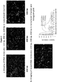
- the strength of a signal correlates with the length of a molecule. For example, as shown in Figure 3a , longer DNA molecules can generate stronger signals than shorter DNA molecules. In some embodiments, the strength of a signal correlates to the amount of DNA in a sample or fluidic channel.
- samples are analyzed for copy number variation, for example, as described in U.S. Patent Publication No. 20130034546 ,
- the quantity of particular molecules, such as DNA fragments derived from different chromosomes can be quantitatively measured in the methods provided herein.
- the amount of genomic DNA derived from a diploid autosomal chromosome is observed to be twice as much as that derived from a haploid sex chromosome.
- the quantity of such fragments reflects the copy number of a source chromosomes. In some embodiments, two or three color labels are used.
- chromosome derived fragments are detected, and a relative ratio is used to identify aneuploidy.
- the control genomic sequence includes separate portions whose total length per genome is known, wherein the sequence of interest comprises separate portions whose length per normal gene is known, and wherein a significant difference between K1 and K2 indicates a genetic abnormality in the genome.
- the nucleotide sequence of interest can relate to a trisomy-linked chromosome, wherein the control genomic sequence is from a chromosome other than the trisomy-linked chromosome, and wherein a K1/K2 ratio of approximately 2:3 or 3:2 indicates a trisomic genotype.
- the nucleotide sequence of interest comprises a deletion of a portion of a genome.
- the nucleotide sequence of interest comprises a repeating sequence.
- a copy number of repeating sequence can be determined according to some embodiments herein.
- the first sample comprises maternal blood (which, without being limited by any one theory, may include fetal nucleic acids), and the second sample comprises maternal tissue other than blood (preferably a tissue with little to no likelihood of comprising fetal nucleic acids).
- digital counting detection is performed.
- digital counting detection is performed on particles (such as beads), bacteria, or virion particles.
- particles such as beads
- bacteria such as Bacillus
- virion particles such as Bacillus, Bacillus, or Bacillus, or Bacillus, or Bacillus, or Bacillus, or Bacillus, or Bacillus, or Bacillus, or Bacillus, or Bacillus, or Bacillus, or Bacillus, or plasmic particles.
- the methods described herein can apply to a variety of targets that can be uniquely labeled.
- digital karyotyping is performed.
- digital karyotyping is performed for a chromosome with potential aneuploidy of interest.
- the methods described herein can be used to detect any chromosomal variation of interest, including translocation, addition, amplification, transversion, inversion, aneuploidy, polyploidy, monosomy, trisomy, trisomy 21, trisomy 13, trisomy 14, trisomy 15, trisomy 16, trisomy 18, trisomy 22, triploidy tetraploidy, and sex chromosome abnormalities, including but not limited to XO, XXY, XYY, and XXX.
- the methods are sensitive enough to detect "short" fragments that are on the order of tens to hundreds of nucleotides in length.
- the sample molecules as described herein comprise polynucleotide "short" fragments.
- the polynucleotiode fragments are about 10, 15, 20, 25, 30, 35, 40, 45, 50, 55, 60, 65, 70, 75, 80, 85, 90, 95, or 100 nucleotides in length.
- the polynucleotide fragments are about 100, 125, 150, 175, 200, 225, 250, 275, 300, 325, 350, 375, 400, 425, 450, 475, 500, 525, 550, 575, 600, 625, 650, 675, 700, 725, 750, 775, 800, 825, 850, 875, 900, 925, 950, 975, or 1000 nucleotides in length.
- the molecules of interest are fragments of less than about 1000, 950, 900, 850, 800, 750, 700, 650, 600, 550, 500, 450, 400, 350, 300, 250, 200, 150, 100, or 50 nucleotides in length.
- the fragments are double-stranded.
- the fragments comprise DNA.
- the fragments comprise RNA.
- the fragments comprise DNA hybridized to RNA.
- the sensitivity is about as high as detecting a single fluorophore associated with a target fragment.
- the nucleotides of interest are fragments of at least about 500 nucleotides in length, for example about 500, 600, 700, 800, 900, 1000, 1100, 1200, 1300, 1400, 1500, 1600, 1700, 1800, 1900, or 2000 nucleotides in length, including ranges between any two of the listed values, for example about 500 to about 2000 nucleotides in length, about 500 to about 1500, about 500 to about 1000, about 500 to about 900, about 500 to about 700, about 700 to about 2000, about 700 to about 1500, about 700 to about 1000, about 700 to about 900, about 1000 to about 2000, about 1000 to about 1500, or about 1500 to about 2000.
- Molecules suitable for use in the methods and systems described herein include polymers, double-stranded DNA, single-stranded DNA, RNA, DNA-RNA hybrids, polypeptides, biological molecules, proteins, and the like. Suitable polymers include homopolymers, copolymers, block copolymers, random copolymers, branched copolymers, dendrimers, or any combination thereof.
- the methods described herein are sensitive enough to detect a fetal molecule that constitutes less than about .025%, 0.5%, 0.75%, 1%, 1.25%, 1.5%, 1.75%, 2%, 2.25%, 2.5%, 2.75%, 3%, 3.25%, 3.5%, 3.75%, 4%, 4.25%, 4.5%, 4.75%, 5%, 6%, 7%, 8%, 9%, 10%, 11%, 12%, 13%, 14%, 15%, 20%, or 25% of the total number of molecules in a maternal blood sample.
- labeling is directed to a sequence motif or chemical moiety. Labeling can be carried out using any technique known to one of skill in the art, including chemical or biochemical conjugation. In some embodiments, the labels described herein are bound to a unique sequence motif. In some embodiments, the labels described herein are bound to a chemical moiety. In some of these embodiments, the chemical moiety is related to a specific chromosome.
- each label is independently selected from the group consisting of a fluorophore, a quantum dot, a dendrimer, a nanowire, a bead, a hapten, a streptavidin, an avidin, a neutravidin, a biotin, and a reactive group.
- the first and second labels are independently selected from the group consisting of a fluorophore or a quantum dot.
- at least one of the labels comprises a non-optical label.
- the labeling is carried out with a polymerase.
- the labeling is carried out with a polymerase in the presence of dNTPs comprising the label.
- the polymerase has a 5' to 3' exonuclease activity.
- the polymerase leaves a flap region, and wherein the flap region is removed to restore a ligatable nick prior to the repairing with a ligase.
- the flap region is removed using the 5' to 3' exonuclease activity of a polymerase under conditions wherein at least one nucleotide is present in limited concentration.
- the flap region is removed using the 5' to 3' exonuclease activity of a polymerase under conditions wherein at least one nucleotide is omitted from the reaction. In some embodiments herein, the flap region is removed with a flap endonuclease. In some embodiments herein, the labeling is carried out with a polymerase in the presence of at least one species of dNTP. In some embodiments herein, the at least one species of dNTP is a single species of dNTP. In some embodiments herein, a method as described herein further comprises modulating activity of the polymerase by adjusting the temperature, dNTP concentration, cofactor concentration, buffer concentration, or any combination thereof, during labeling.
- nicking the first motif or the second motif comprising nicking with Nt.BspQI.
- the a non-sequence-specific label for example a polynucleotide backbone label is applied in addition to a sequence-specific label or labels as described herein.
- At least one label as described herein comprises a non-optical label.
- a variety of non-optical labels can be used in conjunction with embodiments herein.
- a non-optical label comprises an electronic label.
- Exemplary electronic labels include, but are not limited to molecule with a strong electric charge, for example ions such as a metal ions, charged amino acid side chain, or other cations or anions.
- An electronic label can be detected, for example, by conductivity (or resistivity) when the label is disposed in a detector.
- a nanochannel comprises an electrode configured to determine the presence or absence of an electronic label by determining the conductivity or resistivity of a substance disposed in the channel.
- the non-optical label comprises a metal, metal oxide (for example metal oxide), or silicon oxide moiety.
- the non-optical label comprises a moiety (for example a nanoparticle) comprising a metal, metal oxide, or other oxide.
- the presence of a particular metal or oxide moiety can be detected, for example by nuclear magnetic resonance.
- the label is configured to release a moiety, for example a proton or an anion, upon a certain condition (e.g. change of pH) and the presence or absence of released moiety is detected.
- two or more labels are different from each other.
- a first motif can be labeled with a first label so as to generate a first unique pattern
- a second motif that is different from the first motif can be labeled with a second label different from the first label so as to generate a second unique pattern.
- two or more labels are the same.
- a first motif can be labeled with a label
- a second motif that is different from the first motif can also be labeled with the same label so as to generate a unique pattern.
- a plurality of probes corresponding to a first chromosome or region of interest are labeled with a first label
- a second plurality of probes corresponding to a second chromosome or region of interest are labeled with a second label that is different than the first label.
- labeled sample molecules comprising sequences from the first chromosome or region of interest can be differentiated from sample molecules comprising sequences from the second chromosome or region of interest based on whether they are labeled with the first label or second label.
- Nucleotides with reversible terminators can form a first phosphodiester linkage, but prior to reversal of termination, cannot form (or have limited capacity to form) a second phosphodiester linkage.
- a nucleotide with a reversible terminator can be incorporated into a polynucleotide (for example at a nick site), but the nucleotide cannot form downstream phosphodiester linkages until the terminator is reversed.
- Reversal can be performed using techniques known to one skilled in the art.
- the terminator can be attached to the nucleotide via cleavable linker, which can be cleaved, for example, via electromagnetic radiation.
- nick repair is performed using labeled nucleotides comprising a 3' reversible terminator, a single labeled nucleotide can be incorporated into the nick, but the terminator can prevent additional labeled nucleotides from being incorporated into the nick. Accordingly, nick labeling can be limited to one labeled nucleotide per nick. Limiting nick labeling to one label moiety per nick can minimize potential bias from multiple labels being incorporated into the same nick. For example, if approaches are taken to limit labeling to one label moiety per nick, two nicks that are very close together can be resolved based on a relatively strong signal from the label (i.e.
- the label on the nucleotide comprising a reversible terminator can be as described herein.
- the nucleotide comprising a reversible terminator comprises a quantum dot.
- the nucleotide comprising a reversible terminator comprises a fluorophore.
- the nucleotide comprising a reversible terminator comprises a non-optical label.
- a plurality of labels label a single sample molecule.
- at least one of the labels comprises a sequence specific label.
- at least one of the labels comprises a non-sequence specific label.
- at least one label comprises a sequence specific label, and at least one label comprises a non-sequence specific label.
- at least one label does not cut one or both strands of DNA.
- At least one label is selected from the group consisting of a non-cutting restriction enzyme, a methyltransferase, a zinc finger protein, an antibody, a transcription factor, a DNA binding protein, a hairpin polyamide, a triplex-forming oligodeoxynucleotide, a peptide nucleic acid, or a combination thereof.
- a non-cutting restriction enzyme e.g., a methyltransferase
- a zinc finger protein e.g., an antibody to a transcription factor
- a DNA binding protein e.g., a hairpin polyamide
- a triplex-forming oligodeoxynucleotide e.
- labeling is detected using a sensitive camera. In some embodiments, for example if non-optical labeling is provided, labeling is detected electronically.
- any detection method can be used that is suitable for the corresponding label.
- the methods described herein can include binding to a fluorescent label, a radioactive label, a magnetic label, or any combination thereof in one or more regions of the molecules described herein. Binding may be accomplished where the label is specifically complementary to a molecule or to at least a portion of a molecule or other region of interest.
- nicking enzymes create sequence-specific nicks that are subsequently labeled, for example using a labeled nucleotide or nucleotide analog.
- the nucleotide or analog is fluorescently labeled.
- DNA is linearized by confinement in a nanochannel, resulting in uniform linearization and allowing precise and accurate measurement of the distance between nick-labels on DNA molecules comprising a signature pattern.
- a second nicking enzyme is used.
- the second nicking enzyme is used with a second label color.
- Exemplary nickases that can be used in accordance with embodiments herein include, but are not limited to Nb.BbvCI; Nb.BsmI; Nb.BsrDI; Nb.BtsI; Nt.AlwI; Nt.BbvCI; Nt.BspQI; Nt.BstNBI; Nt.CviPII and combinations thereof.
- Examples of nicking agents and protocols are also provided in U.S. Patent Application Publication No. 2011/0171634 and U.S. Patent Application Publication No. 2012/0237936 ,
- a polynucleotide for example an RNA or DNA
- the probe can be complementary to a strand of the RNA or DNA or a portion thereof.
- the probe is complementary to a particular sequence motif.
- a plurality of probes is provided so as to be complementary to a plurality of specific sequence motifs, for example at least 2, 3, 4, 5, 6, 7, 8, 9, 10, 20, 30, 40, 50, 60, 70, 80, 90, 100, 200, 300, 400, 500, 600, 700, 800, 900, 1000, 5,000, or 10,000 probes, including ranges between any two of the listed values.
- the probe has a random sequence. In some embodiments, a probe with a plurality of random sequences is provided. In some embodiments, a probe includes one or more of an organic fluorophore, quantum dot, dendrimer, nanowires, bead, Au beads, paramagnetic beads, magnetic bead, a radiolabel, polystyrene bead, polyethylene bead, peptide, protein, haptens, antibodies, antigens, streptavidin, avidin, neutravidin, biotin, nucleotide, oligonucleotide, sequence specific binding factors such as engineered restriction enzymes, methlytransferases, zinc finger binding proteins, and the like.
- an organic fluorophore quantum dot, dendrimer, nanowires, bead, Au beads, paramagnetic beads, magnetic bead, a radiolabel, polystyrene bead, polyethylene bead, peptide, protein, haptens, antibodies,
- the probe includes a fluorophore-quencher pair.
- One configuration of the probe can include a fluorophore attached to the first end of the probe, and an appropriate quencher tethered to the second end of the probe.
- the quencher can prevent the fluorophore from fluorescing, while when the probe is hybridized to a target sequence, the probe is linearized, thus distancing the quencher from the fluorophore and permitting the fluorophore to fluoresce when excited by an appropriate wavelength of electromagnetic radiation.
- a first probe includes a first fluorophore of a FRET pair
- a second probe includes a second fluorophore of a FRET pair.
- hybridization of the first probe and the second probe to a single flap, or to a pair of flaps within a FRET radius of each other can permit energy transfer by FRET.
- a first probe includes a first fluorophore of a FRET pair, and a label on a nucleotide incorporated to fill a corresponding gap can include second fluorophore of a FRET pair.
- hybridization of the first probe to a flap, and the labeled nucleotide into the corresponding gap can permit energy transfer by FRET.
- a double-stranded DNA can be labeled by first melting hydrogen bonds between double stands of certain genomic regions to open a so-called D-loop, by increasing temperature or manipulation with organic solvent, and then hybridizing to at least one specific probes with equal or higher affinity to single stranded regions before annealing back to relative stable form.
- double-stranded DNA can be labeled by a probe as described herein without nicking or cutting either strand.
- a plurality of D-loops can be opened on a single strand. As such, a plurality of probes can be annealed to a particular double-stranded DNA.
- labeling comprises transferring a label to the polynucleotide via a methyltransferase.
- the methyltransferase specifically methylates a sequence motif.
- labeling can comprise transferring a label to a sequence motif by the methyltransferase.
- Exemplary suitable DNA methyltransferases include, but are not limited to, M.BseCI (methylates adenine at N6 within the 5'-ATCGAT-3' sequence), M.Taql (methylates adenine at N6 within the 5'-TCGA-3' sequence) and M.Hhal (methylates the first cytosine at C5 within the 5'-GCGC-3' sequence).
- two or more methyltransferases provide two or more labels, which can be the same or different.
- the channel comprises a microchannel. In some embodiments, the channel comprises a nanochannel. Suitable fluidic nanochannel segments have a characteristic cross-sectional dimension of less than about 1000nm, less than about 500 nm, or less than about 200 nm, or less than about 100 nm, or even less than about 50 nm, about 10 nm, about 5 nm, about 2 nm, or even less than about than about 0.5 nm.
- a fluidic nanochannel segment suitably has a characteristic cross-sectional dimension of less than about twice the radius of gyration of the molecule.
- the nanochannel has a characteristic cross-sectional dimension of at least about the persistence length of the molecule.
- Fluidic nanochannel segments suitable for the present method have a length of at least about 100 nm, of at least about 500 nm, of at least about 1000 nm, of at least about 2 microns, of at least about 5 microns, of at least about 10 microns, of at least about 1 mm, or even of at least about 10 mm. Fluidic nanochannel segments are, in some embodiments, present at a density of at least 1 fluidic nanochannel segment per cubic centimeter.
- fluidic channels examples can be found in U.S. Patent Publication No. 2008/0242556 , which is incorporated herein by reference in its entirety.
- a virion particles or a bacterial cell is assayed.
- a bacterial cell is assayed using a microchannel.
- the channel allows a cell with a diameter in the range of microns to tens of microns to flow through.
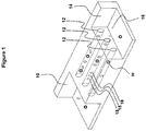
- Figure 1 is a schematic diagram illustrating a fluidic channel arrangement according to some embodiments herein.
- the arrangement can include a sample input chamber 10.
- the arrangement can include an array of fluidic channels 12, for example fluidic nanchannels.
- the arrangement can include a sample output chamber 14.
- the output chamber can comprise buffer solution 16.
- the array of nanofluidic channels 12 can be in fluid communication with the input chamber 10.
- the array of nanofluidic channels 12 can be in fluid communication with the output chamber 14.
- Sample molecules or particles of interest 18 can be disposed in the array of nanofluidic channels 10.
- Control or comparative molecules or particles of interest 18 can be disposed in the array of nanofluidic channels 10.
- the array of nanofluidic channels 12 connect the input chamber 10 to the output chamber 14.
- sample molecules or particles of interest 18 and control or comparative molecules or particles of interest 20 are loaded into the sample input chamber, and travel in buffer solution 16 through the array of nanofluidic channels. In some embodiments, the sample molecules or particles of interest 18 and control or comparative molecules or particles of interest 20 are deposited from the array of nanofluidic channels 12 into the sample output chamber 14.
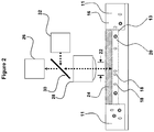
- Figure 2 is a schematic diagram illustrating an arrangement for detection of sample molecules or particles of interest according to some embodiments herein.
- the arrangement comprises a first sample inlet or outlet 11, a second sample inlet or outlet 11, and at least one fluidic channel 13 positioned therebetween and in fluid communication with each of the first and second inlet or outlet 11. It is contemplated herein that if a sample is loaded into the first inlet or outlet 11, the first inlet or outlet 11 functions as an inlet and the second inlet or outlet 11 can function as an outlet. It is contemplated herein that if a sample is loaded into the second inlet or outlet 11, the second inlet or outlet 11 functions as an inlet and the first inlet or outlet 11 can function as an outlet.
- the sample comprises molecules or particles of interest 18, control or comparative particles of interest 20, or a combination of the two.
- the molecules or particles of interest 18, control or comparative particles of interest 20 travel through the fluidic channel 13.
- the fluidic channel 13 comprises a nanochannel.
- the fluidic channel 13 comprises a microchannel.
- the fluidic channel 13 comprises a detection region 22.
- the system comprises a cover 24 disposed over the detection region 24.
- the cover 24 comprises a transparent cap.
- a detector 26 is positioned over the detection region 22 and the cover 24 (if present).
- the detector 26 comprises a photon detection/imager.
- a lens 28 is positioned in optical communication with the detection region 22 and detector 26.
- the lens 28 is positioned between detection region 22 and detector 26.
- a dichroic mirror 30 is positioned in an optical communication with the detection region 22, lens 28, detector 26, and an excitation source 32, so that a fluorescent label, if present, can be excited, and fluorescence from the fluorescent label, if present, can be detected.
- the comparison of samples to a reference sample is provided in the form of a histogram.
- physical counts of molecules with a particular labeling pattern that matches to a reference or de novo genomic assembly in silico are tabulated in a histogram distribution to reflect coverage depth.
- a higher or lower than average coverage depth in specific region or entire chromosome reflects the deviation from normal ploidy such as in the case of aneuploidy in genetic disorder or structural variations in cancer
- Some embodiments described herein can include the following: A method of characterizing a sample, comprising: labeling a region of sample molecules with at least two labels; translocating the labeled sample molecules through a fluidic channel, wherein the fluidic channel is configured to elongate at least a portion of the sample molecule, and wherein the fluidic channel has a length of at least 10 nm and a cross-sectional diameter of less than 5000 nm; detecting signals arising from the labeled samples in the fluidic channels; and correlating the signals arising from the labeled samples to signals arising from the corresponding region of a reference molecule.
- the method can further comprise: labeling a region of the reference molecule corresponding to the region of the sample molecules; translocating the labeled reference sample molecule through a fluidic channel, wherein the fluidic channel is configured to elongate at least a portion of the sample molecule, and wherein the fluidic channel has a length of at least 10 nm and a cross-sectional diameter of less than 5000 nm; and detecting signals arising from the labeled reference sample in the fluidic channels, wherein the signals arising from a known corresponding region of a reference molecule are the signals arising from the labeled reference sample.
- a method of characterizing a sample can comprise: labeling sample nucleic acid molecules; translocating the labeled sample nucleic acid molecules through a fluidic nanochannel, wherein the fluidic nanochannel is configured to elongate at least a portion of the sample nucleic acid molecules, and wherein the fluidic nanochannel has a length of at least 10 nm and a cross-sectional diameter of less than 1000 nm; detecting signals arising from the sample nucleic acid molecules in the fluidic channels; determining the positions of the labels on the sample nucleic acid molecules; and aligning the positions of the labels on the sample nucleic acid molecules to the position of labels in a reference genome.
- a method of characterizing a sample can comprise: processing double-stranded DNA samples so as to give rise to a flap of the first strand of the double-stranded DNA samples being displaced from the double-stranded DNA samples, wherein the flap has a length in the range of from about 1 to about 1000 bases, and wherein the flap gives rise to a gap in the first strand of the double-stranded DNA samples corresponding to the flap; incorporating one or more bases into the double-stranded DNA so as to eliminate at least a portion of the gap; labeling at least a portion of the processed double-stranded DNA with one or more tags; and quantifying the signal arising from the labels on the double-stranded DNA; comparing the quantity of the signal arising from the double-stranded DNA to the quantity of the signal arising from a reference DNA; and determining the presence of a genetic abnormality in the double-stranded DNA when the quantity of the signal arising from the double-stranded DNA differs from the
- a method of characterizing a sample can comprise labeling a plurality of sequence-specific locations on a sample DNA; linearizing at least a portion of the sample DNA; quantifying the signal arising from the labels on the sample DNA; comparing the quantity of the signal arising from the sample DNA to the quantity of the signal arising from a reference DNA; and determining the presence of a genetic abnormality in the sample DNA when the quantity of the signal arising from the sample DNA differs from the quantity of the signal arising from the reference DNA.
- a system for characterizing a sample can comprise: one or more regions for labeling sample molecules with at least two labels; a fluidic channel for translocating the labeled sample molecules, wherein the fluidic channel is configured to elongate at least a portion of the sample molecule, and wherein the fluidic channel has a length of at least 10 nm and a cross-sectional diameter of less than 5000 nm; and a device for detecting signals arising from the labeled samples in the fluidic channels.
- a system for characterizing a sample can comprise: one or more regions for labeling sample nucleic acid molecules; a fluidic nanochannel for translocating the labeled sample nucleic acid molecules, wherein the fluidic nanochannel is configured to elongate at least a portion of the sample nucleic acid molecules, and wherein the fluidic nanochannel has a length of at least 10 nm and a cross-sectional diameter of less than 1000 nm; and a device for detecting signals arising from the sample nucleic acid molecules in the fluidic channels.
- a system for characterizing a sample can comprise: one or more regions for processing double-stranded DNA samples so as to give rise to a flap of the first strand of the double-stranded DNA samples being displaced from the double-stranded DNA samples, wherein the flap has a length in the range of from about 1 to about 1000 bases, and wherein the flap gives rise to a gap in the first strand of the double-stranded DNA samples corresponding to the flap; one or more regions for incorporating one or more bases into the double-stranded DNA so as to eliminate at least a portion of the gap; one or more regions for labeling at least a portion of the processed double-stranded DNA with one or more tags; and a device for quantifying the signal arising from the labels on the double-stranded DNA.
- a system for characterizing a sample can comprise: a region for labeling a plurality of sequence-specific locations on a sample DNA; a region for linearizing at least a portion of the sample DNA; and a device for quantifying the signal arising from the labels on the sample DNA.
- a system for characterizing a sample can comprise: means for labeling sample molecules with at least two labels; means for linearizing the labeled sample molecules; and means for detecting signals arising from the labeled samples in the fluidic channels.
- a system for characterizing a sample can comprise: means for labeling sample nucleic acid molecules; means for linearizing the labeled sample nucleic acid molecules; and means for detecting signals arising from the sample nucleic acid molecules in the fluidic channels.
- a system for characterizing a sample can comprise: means for processing double-stranded DNA samples so as to give rise to a flap of the first strand of the double-stranded DNA samples being displaced from the double-stranded DNA samples, wherein the flap has a length in the range of from about 1 to about 1000 bases, and wherein the flap gives rise to a gap in the first strand of the double-stranded DNA samples corresponding to the flap; means for incorporating one or more bases into the double-stranded DNA so as to eliminate at least a portion of the gap; means for labeling at least a portion of the processed double-stranded DNA with one or more tags; andmeans for quantifying the signal arising from the labels on the double-stranded DNA.
- a system for characterizing a sample comprising: system for characterizing a sample, comprising: means for labeling a plurality of sequence-specific locations on a sample DNA; means for linearizing at least a portion of the sample DNA; and means for quantifying the signal arising from the labels on the sample DNA.
- a method or system as described herein wherein the sample is selected from the group consisting of a bacteria, a virion, a DNA molecule, an RNA molecule, a nucleic acid polymer, a protein, a peptide, and a polysaccharide.
- a method or system as described herein is provided, wherein the sample is derived from maternal blood, and wherein the reference molecule is derived from a maternal sample other than blood.
- a method or system as described herein wherein the sample comprises a nucleotide, and wherein the at least two labels are located at either end of a zone of interest in the nucleotide.
- a method or system as described herein wherein the label is selected from the group consisting of a fluorescent label, a radioactive label, a magnetic label, or a combination thereof.
- a method or system as described herein wherein the optical inspection comprises determining the physical count, the intensity, the wavelength, or the size of the labels.
- a method or system as described herein wherein the optical inspection comprises determining the length of at least one labeled region in the sample.
- correlating the signals comprises determining the signals arising from a pool of samples or a pool of portions of a sample.
- K1 S1/C
- a difference between K1 and Kn is used to identify the presence of a fetal sample.
- a difference between K1 and Kn is used to identify the presence of DNA from a tumor or other cancer source.
- a difference between K1 and Kn is used to determine the presence of a genetic abnormality in the sample.
- the genetic abnormality is aneuploidy. In some embodiments, the genetic abnormality is a translocation, addition, amplification, transversion, or inversion. In some embodiments, the reference is derived from a known diploid or haploid chromosome. In some embodiments, the signals from the sample are correlated with the population distribution from a metagenomic or microbiome study.
- a method or system as described herein in which the fluidic channel is a nanochannel.
- the fluidic channel is disposed parallel to a surface of a substrate.
- a method or system as described herein is provided, further comprising generating a histogram distribution to reflect coverage depth for the sample.
- a method or system as described herein wherein the sample comprises circulating fetal cells, circulating tumor cells, or body fluids or tissues.
- a method or system as described herein comprising subjecting the labeled sample to a motivating force selected from the group consisting of a fluid flow, a radioactive field, an electroosmotic force, an electrophoretic force, an electrokinetic force, a temperature gradient, a surface property gradient, a capillary flow, a pressure gradient, a magnetic field, an electric field, a receding meniscus, a surface tension, a thermal gradient, a pulling force, a pushing force, and a combination thereof.
- a motivating force selected from the group consisting of a fluid flow, a radioactive field, an electroosmotic force, an electrophoretic force, an electrokinetic force, a temperature gradient, a surface property gradient, a capillary flow, a pressure gradient, a magnetic field, an electric field, a receding meniscus, a surface tension, a thermal gradient, a pulling force, a pushing force, and a combination thereof.
- a kit for performing a method as described herein is provided.
- a kit for using the system of any one of the preceding claims is provided.
- channel means a region defined by borders. Such borders may be physical, electrical, chemical, magnetic, and the like.
- nanochannel is used to clarify that certain channels are considered nanoscale in certain dimensions.
- DNA refers to DNA of any length (e.g., 0.1Kb to 1 megabase).
- the DNA can be a highly pure preparation, crude, or semi crude material.
- the DNA can come from any biological source or can be synthetic.
- nucleotide refers to a molecule containing deoxyribonucleic acids (e.g., DNA, mtDNA, gDNA, or cDNA), ribonucleic acid (e.g., RNA or mRNA), or any other variant of nucleic acids known in the art.
- labeled nucleotide refers to a nucleotide comprising any modification that is detectable. This includes but is not limited to nucleotides with reporter groups attached to the base. Reporter groups include but are not limited to fluorescent dyes, haptens, biotin molecules or gold nanoparticles.
- nucleotide refers to a nucleotide that is not modified, or has a slight modification that does not interfere with its incorporation into DNA.
- t refers to nucleotides in DNA.
- nick refers to a phosphodiester bond break occurring on one DNA strand or the other, having a 3'hydroxyl end.
- nicking endonuclease refers to any enzyme, naturally occurring or engineered, that is capable of breaking a phosphodiester bond on a single DNA strand leaving a 3'-hydroxylat a defined sequence.
- nicking endonucleases can be naturally occurring, engineered by modifying restriction enzymes to eliminate one DNA strand cutting activity, or produced by fusing a nicking subunit to a DNA binding domain, for example, zinc fingers and transcription activator like effectors DNA recognition domains.
- labeling sites refers to any DNA site with an exposed 3' hydroxyl group onto which the polymerase can add nucleotides in a template dependent manner. Labeling sites can be generated by nicking endonucleases, hybridized probes, or any chemical or physical means of breaking a phosphodiester bond on any one DNA strand. Means of breaking a phosphodiester bond can occur to DNA outside its biological source or prior to DNA extraction, for example as a result of a biological sample exposure to chemicals, and external forces such as radiation. If 3' ends are not extendable, repair can be performed to restore the hydroxyl group, for example by using New England Biolabs' PreCR kit.
- a “sample” can include, for example, blood, serum, plasma, sputum, lavage fluid, cerebrospinal fluid, urine, semen, sweat, tears, saliva, and the like.
- blood serum
- plasma sputum
- lavage fluid cerebrospinal fluid
- urine semen
- sweat tears
- saliva saliva
- the terms "blood,” “plasma” and “serum” expressly encompass fractions or processed portions thereof.
- the “sample” expressly encompasses a processed fraction or portion derived from the biopsy, swab, smear, etc.
- chromosome refers to the heredity-bearing gene carrier of a living cell which is derived from chromatin and which comprises DNA and protein components (especially histones).
- translocating can be used interchangeably with linearizing when used in the context passing a DNA molecule through a nanochannel.
- Genomic fragments from a human male sample were generated by PCR, labeled, and run through a nanochannel. Detected fragments were then aligned to a single gene reference optical map for each chromosome. The molecules were sorted based on the alignment start site.
- the average coverage depth observed for a diploid autosomal chromosome was 5X, and was evenly distributed across the chromosome. If the sampling of molecules had been even, the alignment start sites would have been randomly distributed across the chromosome, resulting in a linear plot.
Description
- This application claims the benefit of
U.S. Provisional Application No. 61/767,219, filed February 20, 2013 - According to some embodiments herein, a method of characterizing a sample is provided. The method can comprise labeling a plurality of sample molecules with at least a first label, wherein the sample molecules comprise polynucleotide sequences of a first genomic fragment or fragments of interest, and wherein first genomic fragment or fragments of interest correspond to a possibly abnormal genomic region of the sample. The method can comprise providing a plurality of labeled reference molecules, wherein the reference molecules comprise polynucleotide sequences of a reference genomic fragment or fragments, and wherein the reference genomic fragment or fragments are known not to correspond to the possibly abnormal genomic region. As used herein "correspond to a possibly abnormal genomic region" and variations of this root term includes genomic fragments that overlap with or are encompassed by an abnormal chromosomal region, including, but not limited to a duplication, deletion, inversion, translocation, and or aneuploid chromosome or fragment thereof. As such, a genomic fragment or fragment can correspond to an abnormal genomic region that is either present (e.g. a duplication) or absent (e.g. a deletion, for example if the genomic fragment would be encompassed by or overlap with the deleted region). The method can comprise translocating the plurality of labeled sample molecules and the plurality of labeled reference molecules though a fluidic channel. The method can comprise detecting signals from the labeled sample molecules so as to ascertain at least a first pattern or plurality of patterns characteristic of the first genomic fragment or fragments of interest; and a second pattern or plurality of patterns characteristic of the reference genomic fragment or fragments. The method can comprise correlating signals ascertaining the first pattern or plurality of patterns to signals ascertaining the second pattern or plurality of patterns. In some embodiments, labeling comprises labeling the sample molecules with a first label, and wherein the reference molecules comprise a second label, in which the first label is configured to produce the first pattern or plurality of patterns, and in which the second label is configured to produce the second pattern or plurality of patterns, and in which the first label and the second label are different from each other. In some embodiments, labeling comprises labeling with a first label, in which the first pattern or plurality of patterns and the second pattern or plurality of patterns each comprise the first label, and in which the first pattern or plurality of patterns and second pattern or plurality of patterns are different from each other. In some embodiments, the method further comprised labeling the reference molecules so as to produce the labeled reference molecules, wherein the labeled reference molecules comprise the second pattern or plurality of patterns. In some embodiments, the labeled reference molecules and sample molecules are from the sample. In some embodiments, the labeled reference molecules are from a different tissue of the same organism as the sample. In some embodiments, the labeled reference molecules and sample molecules are from different organisms. In some embodiments, the signal from the labeled reference molecules comprises an electronically or optically stored value or set of values. In some embodiments, the method further comprises labeling a second plurality of sample molecules from a second sample with at least the first label, wherein the second plurality of sample molecules comprise polynucleotide sequences of the first genomic fragment or fragments of interest, wherein the second plurality of sample molecules is known to not correspond to the chromosomal abnormality, and translocating the second plurality of sample molecules through the fluidic channel, and detecting signals from the labeled sample molecules so as to ascertain at least the first pattern or plurality of patterns characteristic of the first genomic fragment or fragments of interest; and the second pattern or plurality of patterns characteristic of the reference genomic fragment or fragments. In some embodiments, the method further comprises aligning the positions of the patterns to positions of patterns in a reference genome. In some embodiments, the sample molecules are from a sample comprising the possible genomic abnormality, and the reference genomic fragment or fragments comprise the first chromosome or fragment thereof, in which the reference genomic fragments are from a second sample known to not comprise the genomic abnormality. In some embodiments, the first genomic fragment or fragments of interest comprise a sex chromosome or a least one fragment thereof, and the reference genomic fragment or fragments comprise an autosome or at least one fragment thereof. In some embodiments, the first genomic fragment or fragments of interest comprise a first autosome or at least one fragment thereof, selected from the group consisting of:
human chromosome 21,human chromosome 13,human chromosome 14,human chromosome 15,human chromosome 16,human chromosome 18, andhuman chromosome 22, and fragments thereof, and the reference genomic fragment or fragments comprise a second autosome or at least one fragment thereof, wherein the second autosome or fragment thereof is different than the first autosome or fragment thereof. In some embodiments, correlating signals comprises using the ratio (K) between the signal arising from a plurality of labeled sample molecules or portions thereof (S1, S2 ... Sn) and the signal arising from the reference (C):K1=S1/C, K2=S2/C ... Kn=Sn/C. In some embodiments, the first label comprises at least one of a fluorescent label, a radioactive label, a magnetic label, or a non-optical label. In some embodiments, the second label comprises at least one of a fluorescent label, a radioactive label, a magnetic label, or a non-optical label. In some embodiments, labeling comprises nicking one strand of a double-stranded DNA at a first sequence motif with a nicking endonuclease; and labeling the DNA. In some embodiments, the method further comprises repairing at least some of the nicks on the DNA. In some embodiments, the nicks are not repaired. In some embodiments, the label comprises a transcriptional terminator. In some embodiments, labeling with the first label comprises tagging at least one sequence motif of the sample molecules with a DNA binding entity selected from the group consisting of: a non-cutting restriction enzyme, a zinc finger protein, an antibody, a transcription factor, a transcription activator like domain, a DNA binding protein, a polyamide, a triple helix forming oligonucleotide, and a peptide nucleic acid, and a methyltransferase. In some embodiments, labeling with the first label comprises tagging at least one sequence motif of the sample molecules with a methyltransferase. In some embodiments, the method further comprises labeling the sample molecule with a non-sequence-specific label. The non-sequence specific label can be different from the first label and the second label. In some embodiments, the possible abnormal genomic region comprises at least one of a translocation, addition, amplification, transversion, inversion, aneuploidy, polyploidy, monosomy, trisomy,trisomy 21,trisomy 13,trisomy 14,trisomy 15,trisomy 16,trisomy 18,trisomy 22, triploidy tetraploidy, or sex chromosome aneuploidy. In some embodiments, the genetic abnormality comprises at least one of a trisomy or monosomy. - According to some embodiments herein, a method of characterizing a sample is provided. The method can comprise labeling a plurality of sequence-specific locations on a polynucleotide sequence of a sample molecule. The method can comprise linearizing at least a portion of the sample molecule in a fluidic channel. The method can comprise quantifying a signal from the labels on the sample molecule. The method can comprise comparing a quantity of the signal from the sample molecule to a quantity of signal from a reference molecule. The method can comprise determining a presence or absence of a genetic abnormality in the sample DNA when the quantity of the signal from the sample molecule differs from the quantity of the signal arising from the reference molecule. In some embodiments, the sample molecule and the reference molecule are from the same organism. In some embodiments, the sample molecule and the reference molecule are from different tissues of the same organism. In some embodiments, the sample molecule and the reference molecule are from different organisms. In some embodiments, the signal from the quantity of signal from the reference molecule comprises an electronically or optically stored value or set of values. In some embodiments, the sample molecule comprises a DNA. In some embodiments, the genetic abnormality comprises at least one of a translocation, addition, amplification, transversion, inversion, aneuploidy, polyploidy, monosomy, trisomy,
trisomy 21,trisomy 13,trisomy 14,trisomy 15,trisomy 16,trisomy 18,trisomy 22, triploidy tetraploidy, or sex chromosome aneuploidy. In some embodiments, the genetic abnormality comprises at least one of a trisomy or monosomy. In some embodiments, labeling comprises labeling the polynucleotide with at least one of a fluorescent label, a radioactive label, a magnetic label, or a non-optical label. In some embodiments, labeling comprises: nicking one strand of a double-stranded DNA at a first sequence motif with a nicking endonuclease; andlabeling the DNA. In some embodiments, labeling, further comprises repairing at least some of the nicks on the first DNA. In some embodiments, the nicks are not repaired. In some embodiments, the label comprises a transcriptional terminator. In some embodiments, labeling comprises tagging at least one sequence motif of the sample molecules with a DNA binding entity selected from the group consisting of: a non-cutting restriction enzyme, a zinc finger protein, an antibody, a transcription factor, a transcription activator like domain, a DNA binding protein, a polyamide, a triple helix forming oligonucleotide, and a peptide nucleic acid, and a methyltransferase. In some embodiments, labeling with the first label comprises tagging at least one sequence motif of the sample molecules with a methyltransferase. - According to some embodiments herein, a method of characterizing a sample is provided. The method can comprise labeling sample nucleic acid molecules. The method can comprise translocating the labeled sample nucleic acid molecules through a fluidic nanochannel, wherein the fluidic nanochannel is configured to elongate at least a portion of the sample nucleic acid molecules, and wherein the fluidic nanochannel has a length of at least 10 nm and a cross-sectional diameter of less than 1000 nm. The method can comprise detecting signals arising from the sample nucleic acid molecules in the fluidic channels. The method can comprise determining the positions of the labels on the sample nucleic acid molecules. The method can comprise aligning the positions of the labels on the sample nucleic acid molecules to the position of labels in a reference genome, wherien the reference genome is obtained from a second sample from the same organism as the sample molecules.
- In some embodiments, the fluidic nanochannel of any of the methods herein comprises a channel having a length of at least 10nm and a cross-section diameter of less than 5000nm. In some embodiments, the fluidic channel comprises a nanochannel. In some embodiments, the fluidic channel is disposed parallel to a surface of a substrate. In some embodiments. In some embodiments, the translocating comprises subjecting the labeled sample to a motivating force selected from the group consisting of a fluid flow, a radioactive field, an electroosmotic force, an electrophoretic force, an electrokinetic force, a temperature gradient, a surface property gradient, a capillary flow, a pressure gradient, a magnetic field, an electric field, a receding meniscus, a surface tension, a thermal gradient, a pulling force, a pushing force, and a combination thereof.
- In some embodiments, the sample of any of the methods herein is selected from the group consisting of a bacteria, a virion, a DNA molecule, an RNA molecule, a nucleic acid polymer, a protein, a peptide, and a polysaccharide. In some embodiments, the sample of any of the methods herein is derived from maternal blood, and wherein the reference molecule is derived from a maternal sample other than blood. In some embodiments, the sample of any of the methods herein comprises a nucleotide, and wherein the at least two labels are located at either end of a zone of interest in the nucleotide. In some embodiments, the sample of any of the methods herein comprises circulating fetal cells, circulating tumor cells, or body fluids or tissues.
- In some embodiments, any of the methods herein comprises optical inspection comprising determining the physical count, the intensity, the wavelength, or the size of the labels. In some embodiments, any of the methods herein comprise optical inspection comprising determining the length of at least one labeled region in the sample. In some embodiments, any of the methods herein, further comprise determining the signals arising from a pool comprising the sample or portions of the sample.
- In some embodiments, any of the methods herein comprises using the ratio (K) between the signal arising from a plurality of samples or sample portions (S1, S2 ... Sn) and the signal arising from the reference (C): K1=S1/C, K2=S2/C ... Kn=Sn/C In some embodiments, a difference between K1 and Kn is used to identify the presence of a fetal sample. In some embodiments, a difference between K1 and Kn is used to identify the presence of DNA from a tumor or other cancer source. In some embodiments, a difference between K1 and Kn is used to determine the presence of a genetic abnormality in the sample. In some embodiments, the genetic abnormality is aneuploidy. In some embodiments, the genetic abnormality is a translocation, addition, amplification, transversion, or inversion.
- In some embodiments, any of the methods herein comprises a reference derived from a known diploid or haploid chromosome. In some embodiments, any of the methods herein comprises correlating signals from the sample with the population distribution from a metagenomic or microbiome study. In some embodiments, any of the methods herein comprises generating a histogram distribution to reflect coverage depth for the sample.
- In some embodiments, a system for characterizing a sample is provided. The system can comprise one or more regions for labeling sample molecules with at least two labels. The system can comprise a fluidic channel for translocating the labeled sample molecules, in which the fluidic channel is configured to elongate at least a portion of the sample molecule, and in which the fluidic channel has a length of at least 10 nm and a cross-sectional diameter of less than 5000 nm. The system can comprise a device for detecting signals arising from the labeled samples in the fluidic channels.
- In some embodiments, a system for characterizing a sample is provided. The system can comprise one or more regions for labeling sample nucleic acid molecules.. The system can comprise a fluidic nanochannel for translocating the labeled sample nucleic acid molecules, in which the fluidic nanochannel is configured to elongate at least a portion of the sample nucleic acid molecules, and in which the fluidic nanochannel has a length of at least 10 nm and a cross-sectional diameter of less than 1000 nm.. The system can comprise a device for detecting signals arising from the sample nucleic acid molecules in the fluidic channels.
- In some embodiments a system for characterizing a sample is provided. The system can comprise a region for labeling a plurality of sequence-specific locations on a sample DNA. The system can comprise a region for linearizing at least a portion of the sample DNA. The system can comprise a device for quantifying the signal arising from the labels on the sample DNA.
- In some embodiments, a system for characterizing a sample is provided. The system can comprise a means for labeling sample molecules with at least two labels. The system can comprise a means for linearizing the labeled sample molecules. The system can comprise a means for detecting signals arising from the labeled samples in the fluidic channels.
- In some embodiments, a system for characterizing a sample is provided. The system can comprise a means for labeling sample nucleic acid molecules. The system can comprise a means for linearizing the labeled sample nucleic acid molecules. The system can comprise a means for detecting signals arising from the sample nucleic acid molecules in the fluidic channels.
- In some embodiments, a system for characterizing a sample is provided. The system can comprise a means for labeling a plurality of sequence-specific locations on a sample DNA. The system can comprise a means for linearizing at least a portion of the sample DNA. The system can comprise a means for quantifying the signal arising from the labels on the sample DNA.
- In some embodiments, any of the systems as described herein can characterize a sample selected from the group consisting of a bacteria, a virion, a DNA molecule, an RNA molecule, a nucleic acid polymer, a protein, a peptide, and a polysaccharide. In some embodiments, any of the systems as described herein can characterize a sample derived from maternal blood, and wherein the reference molecule is derived from a maternal sample other than blood. In some embodiments, any of the systems as described herein can characterize a sample comprising a nucleotide, and wherein the at least two labels are located at either end of a zone of interest in the nucleotide. In some embodiments, any of the systems as described herein can characterize a sample comprising circulating fetal cells, circulating tumor cells, or body fluids or tissues.
- In some embodiments, any of the systems as described herein can comprise a label selected from the group consisting of a fluorescent label, a radioactive label, a magnetic label, or a combination thereof. In some embodiments, any of the systems as described herein can be configured for optical inspection, wherein optical inspection comprises determining the physical count, the intensity, the wavelength, or the size of the labels. In some embodiments, the optical inspection comprises determining the length of at least one labeled region in the sample. In some embodiments, any of the systems as described herein can be configured for correlating the signals, in which correlating the signals comprises determining the signals arising from a pool of samples or a pool of portions of a sample, some embodiments, any of the systems as described herein can be configured for correlating the signals, in which correlating the signals comprises using the ratio (K) between the signal arising from a plurality of samples or sample portions (S1, S2 ... Sn) and the signal arising from the reference (C): K1=S1/C, K2=S2/C ... Kn=Sn/C. In some embodiments, a difference between K1 and Kn is used to identify the presence of a fetal sample. In some embodiments, a difference between K1 and Kn is used to identify the presence of DNA from a tumor or other cancer source. In some embodiments, a difference between K1 and Kn is used to determine the presence of a genetic abnormality in the sample. In some embodiments, the genetic abnormality is aneuploidy. In some embodiments, the genetic abnormality is a translocation, addition, amplification, transversion, or inversion.
- In some embodiments, any of the systems as described herein can comprise a reference derived from a known diploid or haploid chromosome.
- In some embodiments, any of the systems as described herein can correlated the signals from the sample with the population distribution from a metagenomic or microbiome study.
- In some embodiments, the fluidic channel any of the systems as described herein comprises a nanochannel. In some embodiments, the fluidic channel of any of the systems as described herein is disposed parallel to a surface of a substrate. In some emodiments, the translocating comprises subjecting the labeled sample to a motivating force selected from the group consisting of a fluid flow, a radioactive field, an electroosmotic force, an electrophoretic force, an electrokinetic force, a temperature gradient, a surface property gradient, a capillary flow, a pressure gradient, a magnetic field, an electric field, a receding meniscus, a surface tension, a thermal gradient, a pulling force, a pushing force, and a combination thereof.
- In some embodiments, any of the systems as described herein is configured to generate a histogram distribution to reflect coverage depth for the sample.
- In some embodiments a kit for performing any of the methods as described herein is provided.
- In some embodiments a kit for using any of the systems as described herein is provided.
-
-
Figure 1 is a schematic diagram illustrating sample molecules or particles (ovals) and reference or comparative molecules or particles (spheres) flowing through nanofluidic channels, in accordance with some embodiments herein. -
Figure 2 is a schematic diagram illustrating an embodiment of an imaging setup to detect signals emitted from labeled molecules or particles to tabulate the amount, intensity, and configuration of the sample and reference molecule or particles. -
Figure 3a is a series of images illustrating small double stranded DNA fragments with known sizes (233 bp, 498 bp, and 834 bp) that were generated by PCR, fluorescently stained, flowed, and imaged in individual nanofluidic channels.Figure 3b shows the same double stranded DNA fragments that were mixed together, flowed, and imaged in the same nanofluidic channel. The fluorescent signals were plotted in a histogram. -
Figure 4 is a series of graphs illustrating Gaussian curves depicting the photons emitted from individually labeled DNA molecules with known sizes (233 bp, 498 bp, and 834 bp). Total counts and intensity were linearly proportional to mass and/or molecule size. Unknown molecule sizes and quantities can be extrapolated by this method within a linear dynamic range. -
Figure 5 is a series of scatter plots illustrating the extrapolation of unknown molecule sizes and quantities within a linear dynamic range using the information fromFigure 4 . -
Figure 6 is a histogram illustrating genomic DNA fragments plotted against a reference genome (human genome version 19). The y-axis shows coverage depth for specific chromosomal regions. A uniform distribution throughout the genome was observed, except for regions without sequence information (such as the centromeres and telomeres). -
Figure 7a is a graph illustrating diploid genomic fragments from a human male sample aligned tochromosome 1. The y-axis provides the quantity of coverage. The x-axis provides the nucleotide position. The average coverage depth was 5X.Figure 7b is a graph showing a haploid sex chromosome X from the same male sample shown with an average coverage depth of 2X-2.5X (roughly half of the depth of diploid autosomes), demonstrating the quantitative measurement using the methods and platform according to some embodiments herein. - The fetus sheds small DNA fragments into the maternal bloodstream. Tumors have also been found to release DNA into the bloodstream. According to some embodiments herein are methods for analyzing polynucleotide fragments such as DNA fragments in blood to detect the presence of circulating polynucleotide or cells from a fetus or tumor. Also according to some embodiments herein are methods for analyzing fetal DNA in maternal blood to detect genetic abnormalities. In some preferred embodiments, the methods described herein entail the use of a nanofluidic-based single molecule detecting platform to identify genetic abnormalities. Methods and apparatuses in accordance with some embodiments herein have the advantage of analyzing small or large molecules, such as small or large DNA molecules. In some embodiments, a molecule or region of interest is labeled with at least one pattern, and a reference molecule or region of interest is labeled with at least one pattern. The molecules can be linearized in a microfluidic channel, and coverage depth for the molecule or region of interest can be compared to coverage depth for the reference molecule so as to determine copy number of the molecule of interest.
- It is estimated that about 3-15% of short DNAs in maternal blood are fetal derived. Described herein are methods of easily detecting and quantitating small molecules, including short DNA fragments, using methods that incorporate fluidics. In some preferred embodiments, the methods comprise quantitating short DNA fragments without sequencing or assembly.
- Current prenatal tests involving needle puncture to draw amniotic fluid can lead to miscarriage and other complications. Further, many current cancer detection methods also involve invasive procedures, such as biopsies. According to some embodiments herein, a non-invasive method of prenatal testing is provided. In some embodiments, the method is for testing blood. In some embodiments, the method only tests a blood sample, and does not test a sample from other tissues.
- Also described herein are methods of detecting and tracking larger molecules, including longer DNA fragments, to their source using methods that incorporate fluidics. For example, in some embodiments, DNA fragments are tracked back to a tumor or other source of cancer. In some preferred embodiments, the methods are used to track DNA fragments to their source in order to identify or characterize a genetic abnormality.
- In a preferred embodiment, circulating DNA from a maternal blood sample is analyzed to identify and quantify fetal DNA relative to the maternal genome. In some embodiments, this information is used to determine prenatal genomic health status (such as trisomy 21) without invasive tests. Examples of suitable oligos for use in an assay for detecting aneuploidy are provided in the HSA21 oligoarray described in Yahya-Graison et al., Classification of ,
- In some embodiments, a sample of interest is compared to a reference sample. In some embodiments, the sample of interest is derived from a maternal blood sample. In some of these embodiments, the reference sample is a maternal sample from a source other than blood. In some embodiments, the maternal reference sample includes polynucleotides such as DNA isolated from a diploid tissue other than blood. In some embodiments, the maternal reference sample comprises a buccal sample, a saliva sample, a urine sample, a sputum sample, or a tear sample. For example, in some embodiments,
trisomy 21 is detected in a maternal blood sample compared to a maternal buccal sample. - In some embodiments, the sample of interest is enriched for fetal nucleic acids prior to performing the methods described herein. For example, in some embodiments, fetal cells are enriched using a fetal cell specific marker that can be pulled down by an antibody. In some embodiments, the sample of interest undergoes size fractionation. However, any method of enrichment known to one of skill in the art can be used.
- In some embodiments, the sample of interest is derived from a tumor cell or suspected tumor cell, or a tissue in fluid communication with a tumor cell (for example, blood). In some embodiments, the reference sample is sample from a healthy cell. In some embodiments, the reference sample is from a healthy cell of the same organism as the tumor cell or suspect tumor cell. In some embodiments, the reference sample is selected from a tissue that has little to no likelihood of comprising a tumor cell or nucleic acid from the tumor cell.
- As one of skill in the art will recognize, the sample of interest may include nucleic acids from a variety of sources. In some embodiments, the sample of interest comprises a bacteria or virion derived from an environmental sample, animal or plant tissue, blood, or other body fluid. In some embodiments, DNA fragments are used to detect chromosomal abnormalities or cancer genomes.
- As one of skill in the art will recognize, the methods described herein can be used to prepare and analyze DNA from circulating fetal or tumor cells. For example, in some embodiments, cells are lysed to release DNA of interest prior to analysis.
- In some embodiments, an entire genome is assayed or analyzed. In some embodiments, only a portion of a genome is assayed or analyzed. In some embodiments, an entire chromosome is assayed or analyzed. In some embodiments, only a portion of a chromosome is assayed or analyzed. In some embodiments, an entire gene is analyzed. In some embodiments, only a portion of a gene is assayed or analyzed.
- The signals described herein can include any suitable signal, including optical signals, fluorescent signals, non-optical signals, radiative signals, electrical signals, magnetic signals, chemical signals, or any combination thereof. In some embodiments, signals are generated by an electron spin resonance molecule, a fluorescent molecule, a chemiluminescent molecule, a radioisotope, an enzyme substrate, a biotin molecule, an avidin molecule, an electrical charged transferring molecule, a semiconductor nanocrystal, a semiconductor nanoparticle, a colloid gold nanocrystal, a ligand, a microbead, a magnetic bead, a paramagnetic particle, a quantum dot, a chromogenic substrate, an affinity molecule, a protein, a peptide, a nucleic acid, a carbohydrate, an antigen, a nanowire, a hapten, an antibody, an antibody fragment, a lipid, or a combination thereof.
- In some embodiments, signals are generated by using one or more excitation sources to induce fluorescence, chemoluminescence, phosphorescence, bioluminescence, or any combination thereof. Suitable excitation sources include lasers, visible light sources, sources of infrared light, sources of ultraviolet light, or any combination thereof.
- In some embodiments, the detection of nucleotides or associated signals (for example, fluorophores) is quantitative. In some embodiments, the length of a nucleotide is quantified. In some embodiments, the size of a molecule is quantified. In some embodiments, the strength of a signal correlates with the length of a molecule. For example, as shown in
Figure 3a , longer DNA molecules can generate stronger signals than shorter DNA molecules. In some embodiments, the strength of a signal correlates to the amount of DNA in a sample or fluidic channel. - In some embodiments, samples are analyzed for copy number variation, for example, as described in
U.S. Patent Publication No. 20130034546 , - The quantity of particular molecules, such as DNA fragments derived from different chromosomes, can be quantitatively measured in the methods provided herein. In some embodiments, the amount of genomic DNA derived from a diploid autosomal chromosome is observed to be twice as much as that derived from a haploid sex chromosome. In some embodiments, the quantity of such fragments reflects the copy number of a source chromosomes. In some embodiments, two or three color labels are used.
- In some embodiments, chromosome derived fragments are detected, and a relative ratio is used to identify aneuploidy. In some embodiments, the copy number of a nucleotide is calculated using the ratios K1=S1/C and K2=S2/C, wherein K1 is the ratio of the signal for a first sample to a control sample, and K2 is the ratio of the signal for a second sample to the control sample. It is contemplated that the copy number from the reference sample is an integer, and that the difference between K1 and K2 can indicate an abnormality in one of the samples of interest. In some embodiments, the abnormality is detected by comparing the ratio for a particular sample to the average ratio from a plurality of samples. The methods further contemplate that the control genomic sequence includes separate portions whose total length per genome is known, wherein the sequence of interest comprises separate portions whose length per normal gene is known, and wherein a significant difference between K1 and K2 indicates a genetic abnormality in the genome. In some embodiments, the nucleotide sequence of interest can relate to a trisomy-linked chromosome, wherein the control genomic sequence is from a chromosome other than the trisomy-linked chromosome, and wherein a K1/K2 ratio of approximately 2:3 or 3:2 indicates a trisomic genotype. In some embodiments, the nucleotide sequence of interest comprises a deletion of a portion of a genome. In some embodiments, the nucleotide sequence of interest comprises a repeating sequence. As such, a copy number of repeating sequence can be determined according to some embodiments herein. In some embodiments, the first sample comprises maternal blood (which, without being limited by any one theory, may include fetal nucleic acids), and the second sample comprises maternal tissue other than blood (preferably a tissue with little to no likelihood of comprising fetal nucleic acids).
- In some embodiments, digital counting detection is performed. In some embodiments, digital counting detection is performed on particles (such as beads), bacteria, or virion particles. As one of skill in the art will recognize, the methods described herein can apply to a variety of targets that can be uniquely labeled. In some embodiments, digital karyotyping is performed. For example, in some embodiments, digital karyotyping is performed for a chromosome with potential aneuploidy of interest. The methods described herein can be used to detect any chromosomal variation of interest, including translocation, addition, amplification, transversion, inversion, aneuploidy, polyploidy, monosomy, trisomy,
trisomy 21,trisomy 13,trisomy 14,trisomy 15,trisomy 16,trisomy 18,trisomy 22, triploidy tetraploidy, and sex chromosome abnormalities, including but not limited to XO, XXY, XYY, and XXX. - In some embodiments, methods are provided herein in which the methods are sensitive enough to detect "short" fragments that are on the order of tens to hundreds of nucleotides in length. In some embodiments, the sample molecules as described herein comprise polynucleotide "short" fragments. For example, in some embodiments, the polynucleotiode fragments are about 10, 15, 20, 25, 30, 35, 40, 45, 50, 55, 60, 65, 70, 75, 80, 85, 90, 95, or 100 nucleotides in length. In some embodiments, the polynucleotide fragments are about 100, 125, 150, 175, 200, 225, 250, 275, 300, 325, 350, 375, 400, 425, 450, 475, 500, 525, 550, 575, 600, 625, 650, 675, 700, 725, 750, 775, 800, 825, 850, 875, 900, 925, 950, 975, or 1000 nucleotides in length. In some embodiments, the molecules of interest are fragments of less than about 1000, 950, 900, 850, 800, 750, 700, 650, 600, 550, 500, 450, 400, 350, 300, 250, 200, 150, 100, or 50 nucleotides in length. In some embodiments, the fragments are double-stranded. In some embodiments, the fragments comprise DNA. In some embodiments, the fragments comprise RNA. In some embodiments, the fragments comprise DNA hybridized to RNA. In some embodiments, the sensitivity is about as high as detecting a single fluorophore associated with a target fragment.
- In some embodiments, the nucleotides of interest are fragments of at least about 500 nucleotides in length, for example about 500, 600, 700, 800, 900, 1000, 1100, 1200, 1300, 1400, 1500, 1600, 1700, 1800, 1900, or 2000 nucleotides in length, including ranges between any two of the listed values, for example about 500 to about 2000 nucleotides in length, about 500 to about 1500, about 500 to about 1000, about 500 to about 900, about 500 to about 700, about 700 to about 2000, about 700 to about 1500, about 700 to about 1000, about 700 to about 900, about 1000 to about 2000, about 1000 to about 1500, or about 1500 to about 2000.
- Molecules suitable for use in the methods and systems described herein include polymers, double-stranded DNA, single-stranded DNA, RNA, DNA-RNA hybrids, polypeptides, biological molecules, proteins, and the like. Suitable polymers include homopolymers, copolymers, block copolymers, random copolymers, branched copolymers, dendrimers, or any combination thereof.
- In some embodiments, the methods described herein are sensitive enough to detect a fetal molecule that constitutes less than about .025%, 0.5%, 0.75%, 1%, 1.25%, 1.5%, 1.75%, 2%, 2.25%, 2.5%, 2.75%, 3%, 3.25%, 3.5%, 3.75%, 4%, 4.25%, 4.5%, 4.75%, 5%, 6%, 7%, 8%, 9%, 10%, 11%, 12%, 13%, 14%, 15%, 20%, or 25% of the total number of molecules in a maternal blood sample.
- In some embodiments, labeling is directed to a sequence motif or chemical moiety. Labeling can be carried out using any technique known to one of skill in the art, including chemical or biochemical conjugation. In some embodiments, the labels described herein are bound to a unique sequence motif. In some embodiments, the labels described herein are bound to a chemical moiety. In some of these embodiments, the chemical moiety is related to a specific chromosome.
- In some embodiments herein, each label is independently selected from the group consisting of a fluorophore, a quantum dot, a dendrimer, a nanowire, a bead, a hapten, a streptavidin, an avidin, a neutravidin, a biotin, and a reactive group. In some embodiments herein, the first and second labels are independently selected from the group consisting of a fluorophore or a quantum dot. In some embodiments herein, at least one of the labels comprises a non-optical label. In some embodiments herein, the labeling is carried out with a polymerase. In some embodiments herein, the labeling is carried out with a polymerase in the presence of dNTPs comprising the label. In some embodiments herein, the polymerase has a 5' to 3' exonuclease activity. In some embodiments herein, the polymerase leaves a flap region, and wherein the flap region is removed to restore a ligatable nick prior to the repairing with a ligase. In some embodiments herein, the flap region is removed using the 5' to 3' exonuclease activity of a polymerase under conditions wherein at least one nucleotide is present in limited concentration. In some embodiments herein, the flap region is removed using the 5' to 3' exonuclease activity of a polymerase under conditions wherein at least one nucleotide is omitted from the reaction. In some embodiments herein, the flap region is removed with a flap endonuclease. In some embodiments herein, the labeling is carried out with a polymerase in the presence of at least one species of dNTP. In some embodiments herein, the at least one species of dNTP is a single species of dNTP. In some embodiments herein, a method as described herein further comprises modulating activity of the polymerase by adjusting the temperature, dNTP concentration, cofactor concentration, buffer concentration, or any combination thereof, during labeling. In some embodiments herein, nicking the first motif or the second motif comprising nicking with Nt.BspQI. In some embodiments herein, the a non-sequence-specific label, for example a polynucleotide backbone label is applied in addition to a sequence-specific label or labels as described herein.
- In some embodiments, at least one label as described herein comprises a non-optical label. A variety of non-optical labels can be used in conjunction with embodiments herein. In some embodiments a non-optical label comprises an electronic label. Exemplary electronic labels include, but are not limited to molecule with a strong electric charge, for example ions such as a metal ions, charged amino acid side chain, or other cations or anions. An electronic label can be detected, for example, by conductivity (or resistivity) when the label is disposed in a detector. In some embodiments, a nanochannel comprises an electrode configured to determine the presence or absence of an electronic label by determining the conductivity or resistivity of a substance disposed in the channel. In some embodiments, the non-optical label comprises a metal, metal oxide (for example metal oxide), or silicon oxide moiety. In some embodiments, the non-optical label comprises a moiety (for example a nanoparticle) comprising a metal, metal oxide, or other oxide. The presence of a particular metal or oxide moiety can be detected, for example by nuclear magnetic resonance. In some embodiments, the label is configured to release a moiety, for example a proton or an anion, upon a certain condition (e.g. change of pH) and the presence or absence of released moiety is detected.
- In some embodiments, two or more labels are different from each other. For example, a first motif can be labeled with a first label so as to generate a first unique pattern, and a second motif that is different from the first motif can be labeled with a second label different from the first label so as to generate a second unique pattern. In some embodiments, two or more labels are the same. For example, a first motif can be labeled with a label, and a second motif that is different from the first motif can also be labeled with the same label so as to generate a unique pattern. In some embodiments, a plurality of probes corresponding to a first chromosome or region of interest are labeled with a first label, and a second plurality of probes corresponding to a second chromosome or region of interest (for example a reference chromosome or region) are labeled with a second label that is different than the first label. As such, labeled sample molecules comprising sequences from the first chromosome or region of interest can be differentiated from sample molecules comprising sequences from the second chromosome or region of interest based on whether they are labeled with the first label or second label.
- Nucleotides with reversible terminators can form a first phosphodiester linkage, but prior to reversal of termination, cannot form (or have limited capacity to form) a second phosphodiester linkage. Thus, a nucleotide with a reversible terminator can be incorporated into a polynucleotide (for example at a nick site), but the nucleotide cannot form downstream phosphodiester linkages until the terminator is reversed. Reversal can be performed using techniques known to one skilled in the art. For example, the terminator can be attached to the nucleotide via cleavable linker, which can be cleaved, for example, via electromagnetic radiation. If nick repair is performed using labeled nucleotides comprising a 3' reversible terminator, a single labeled nucleotide can be incorporated into the nick, but the terminator can prevent additional labeled nucleotides from being incorporated into the nick. Accordingly, nick labeling can be limited to one labeled nucleotide per nick. Limiting nick labeling to one label moiety per nick can minimize potential bias from multiple labels being incorporated into the same nick. For example, if approaches are taken to limit labeling to one label moiety per nick, two nicks that are very close together can be resolved based on a relatively strong signal from the label (i.e. the possibility that two labels simply got incorporated into the same nick can be ruled-out). For example, if quantitative estimates of the number of nicks is desired, a one-label-per-nick approach can facilitate direct correlation between strength of label signal and the number of nicks. The label on the nucleotide comprising a reversible terminator can be as described herein. In some embodiments, the nucleotide comprising a reversible terminator comprises a quantum dot. In some embodiments, the nucleotide comprising a reversible terminator comprises a fluorophore. In some embodiments, the nucleotide comprising a reversible terminator comprises a non-optical label.
- In some embodiments, a plurality of labels label a single sample molecule. In some embodiments, at least one of the labels comprises a sequence specific label. In some embodiments, at least one of the labels comprises a non-sequence specific label. In some embodiments, at least one label comprises a sequence specific label, and at least one label comprises a non-sequence specific label. In some embodiments, at least one label does not cut one or both strands of DNA. For example, in some embodiments, at least one label is selected from the group consisting of a non-cutting restriction enzyme, a methyltransferase, a zinc finger protein, an antibody, a transcription factor, a DNA binding protein, a hairpin polyamide, a triplex-forming oligodeoxynucleotide, a peptide nucleic acid, or a combination thereof. In some embodiments, neither the sequence specific nor the non-sequence specific label cuts DNA.
- In some embodiments, for example if fluorescent labeling is provided, labeling is detected using a sensitive camera. In some embodiments, for example if non-optical labeling is provided, labeling is detected electronically. However, any detection method can be used that is suitable for the corresponding label. The methods described herein can include binding to a fluorescent label, a radioactive label, a magnetic label, or any combination thereof in one or more regions of the molecules described herein. Binding may be accomplished where the label is specifically complementary to a molecule or to at least a portion of a molecule or other region of interest.
- In some embodiments, nicking enzymes create sequence-specific nicks that are subsequently labeled, for example using a labeled nucleotide or nucleotide analog. In some embodiment, the nucleotide or analog is fluorescently labeled. In some embodiments, DNA is linearized by confinement in a nanochannel, resulting in uniform linearization and allowing precise and accurate measurement of the distance between nick-labels on DNA molecules comprising a signature pattern. In some embodiments, a second nicking enzyme is used. In some embodiments, the second nicking enzyme is used with a second label color. Exemplary nickases that can be used in accordance with embodiments herein include, but are not limited to Nb.BbvCI; Nb.BsmI; Nb.BsrDI; Nb.BtsI; Nt.AlwI; Nt.BbvCI; Nt.BspQI; Nt.BstNBI; Nt.CviPII and combinations thereof. Examples of nicking agents and protocols are also provided in
U.S. Patent Application Publication No. 2011/0171634 andU.S. Patent Application Publication No. 2012/0237936 , - In some embodiments, a polynucleotide, for example an RNA or DNA, is labeled by hybridizing a probe to a single strand of the polynucleotide. The probe can be complementary to a strand of the RNA or DNA or a portion thereof. In some embodiments, the probe is complementary to a particular sequence motif. In some embodiments, a plurality of probes is provided so as to be complementary to a plurality of specific sequence motifs, for example at least 2, 3, 4, 5, 6, 7, 8, 9, 10, 20, 30, 40, 50, 60, 70, 80, 90, 100, 200, 300, 400, 500, 600, 700, 800, 900, 1000, 5,000, or 10,000 probes, including ranges between any two of the listed values. In some embodiments, the probe has a random sequence. In some embodiments, a probe with a plurality of random sequences is provided. In some embodiments, a probe includes one or more of an organic fluorophore, quantum dot, dendrimer, nanowires, bead, Au beads, paramagnetic beads, magnetic bead, a radiolabel, polystyrene bead, polyethylene bead, peptide, protein, haptens, antibodies, antigens, streptavidin, avidin, neutravidin, biotin, nucleotide, oligonucleotide, sequence specific binding factors such as engineered restriction enzymes, methlytransferases, zinc finger binding proteins, and the like. In some embodiments, the probe includes a fluorophore-quencher pair. One configuration of the probe can include a fluorophore attached to the first end of the probe, and an appropriate quencher tethered to the second end of the probe. As such, when the probe is unhybridized, the quencher can prevent the fluorophore from fluorescing, while when the probe is hybridized to a target sequence, the probe is linearized, thus distancing the quencher from the fluorophore and permitting the fluorophore to fluoresce when excited by an appropriate wavelength of electromagnetic radiation. In some embodiments, a first probe includes a first fluorophore of a FRET pair, and a second probe includes a second fluorophore of a FRET pair. As such, hybridization of the first probe and the second probe to a single flap, or to a pair of flaps within a FRET radius of each other can permit energy transfer by FRET. In some embodiments, a first probe includes a first fluorophore of a FRET pair, and a label on a nucleotide incorporated to fill a corresponding gap can include second fluorophore of a FRET pair. As such, hybridization of the first probe to a flap, and the labeled nucleotide into the corresponding gap can permit energy transfer by FRET.
- In some embodiments, a double-stranded DNA can be labeled by first melting hydrogen bonds between double stands of certain genomic regions to open a so-called D-loop, by increasing temperature or manipulation with organic solvent, and then hybridizing to at least one specific probes with equal or higher affinity to single stranded regions before annealing back to relative stable form. As such, in some embodiments, double-stranded DNA can be labeled by a probe as described herein without nicking or cutting either strand. In some embodiments, a plurality of D-loops can be opened on a single strand. As such, a plurality of probes can be annealed to a particular double-stranded DNA.
- In some embodiments, labeling comprises transferring a label to the polynucleotide via a methyltransferase. In some embodiments, the methyltransferase specifically methylates a sequence motif. As such, labeling can comprise transferring a label to a sequence motif by the methyltransferase. Exemplary suitable DNA methyltransferases (MTase) include, but are not limited to, M.BseCI (methylates adenine at N6 within the 5'-ATCGAT-3' sequence), M.Taql (methylates adenine at N6 within the 5'-TCGA-3' sequence) and M.Hhal (methylates the first cytosine at C5 within the 5'-GCGC-3' sequence). In some embodiments, two or more methyltransferases provide two or more labels, which can be the same or different.
- In some embodiments, labeling comprises transferring a label to the polynucleotide via a methyltransferase. In some embodiments, the methyltransferase specifically methylates a sequence motif. As such, labeling can comprise transferring a label to a sequence motif by the methyltransferase. Exemplary suitable DNA methyltransferases (MTase) include, but are not limited to, M.BseCI (methylates adenine at N6 within the 5'-ATCGAT-3' sequence), M.Taql (methylates adenine at N6 within the 5'-TCGA-3' sequence) and M.Hhal (methylates the first cytosine at C5 within the 5'-GCGC-3' sequence). In some embodiments, two or more methyltransferases provide two or more labels, which can be the same or different.
- In some embodiments, the channel comprises a microchannel. In some embodiments, the channel comprises a nanochannel. Suitable fluidic nanochannel segments have a characteristic cross-sectional dimension of less than about 1000nm, less than about 500 nm, or less than about 200 nm, or less than about 100 nm, or even less than about 50 nm, about 10 nm, about 5 nm, about 2 nm, or even less than about than about 0.5 nm. A fluidic nanochannel segment suitably has a characteristic cross-sectional dimension of less than about twice the radius of gyration of the molecule. In some embodiments, the nanochannel has a characteristic cross-sectional dimension of at least about the persistence length of the molecule. Fluidic nanochannel segments suitable for the present method have a length of at least about 100 nm, of at least about 500 nm, of at least about 1000 nm, of at least about 2 microns, of at least about 5 microns, of at least about 10 microns, of at least about 1 mm, or even of at least about 10 mm. Fluidic nanochannel segments are, in some embodiments, present at a density of at least 1 fluidic nanochannel segment per cubic centimeter.
- Examples of fluidic channels can be found in
U.S. Patent Publication No. 2008/0242556 , which is incorporated herein by reference in its entirety. In some embodiments, a virion particles or a bacterial cell is assayed. For example, in some embodiments, a bacterial cell is assayed using a microchannel. In some embodiments, the channel allows a cell with a diameter in the range of microns to tens of microns to flow through. -
Figure 1 is a schematic diagram illustrating a fluidic channel arrangement according to some embodiments herein. The arrangement can include asample input chamber 10. The arrangement can include an array offluidic channels 12, for example fluidic nanchannels. The arrangement can include asample output chamber 14. The output chamber can comprisebuffer solution 16. The array ofnanofluidic channels 12 can be in fluid communication with theinput chamber 10. The array ofnanofluidic channels 12 can be in fluid communication with theoutput chamber 14. Sample molecules or particles ofinterest 18 can be disposed in the array ofnanofluidic channels 10. Control or comparative molecules or particles ofinterest 18 can be disposed in the array ofnanofluidic channels 10. In some embodiments, the array ofnanofluidic channels 12 connect theinput chamber 10 to theoutput chamber 14. In some embodiments, sample molecules or particles ofinterest 18 and control or comparative molecules or particles ofinterest 20 are loaded into the sample input chamber, and travel inbuffer solution 16 through the array of nanofluidic channels. In some embodiments, the sample molecules or particles ofinterest 18 and control or comparative molecules or particles ofinterest 20 are deposited from the array ofnanofluidic channels 12 into thesample output chamber 14. -
Figure 2 is a schematic diagram illustrating an arrangement for detection of sample molecules or particles of interest according to some embodiments herein. In some embodiments, the arrangement comprises a first sample inlet oroutlet 11, a second sample inlet oroutlet 11, and at least onefluidic channel 13 positioned therebetween and in fluid communication with each of the first and second inlet oroutlet 11. It is contemplated herein that if a sample is loaded into the first inlet oroutlet 11, the first inlet oroutlet 11 functions as an inlet and the second inlet oroutlet 11 can function as an outlet. It is contemplated herein that if a sample is loaded into the second inlet oroutlet 11, the second inlet oroutlet 11 functions as an inlet and the first inlet oroutlet 11 can function as an outlet. In some embodiments, the sample comprises molecules or particles ofinterest 18, control or comparative particles ofinterest 20, or a combination of the two. In some embodiments, the molecules or particles ofinterest 18, control or comparative particles ofinterest 20 travel through thefluidic channel 13. In some embodiments, thefluidic channel 13 comprises a nanochannel. In some embodiments, thefluidic channel 13 comprises a microchannel. In some embodiments, thefluidic channel 13 comprises adetection region 22. In some embodiments, the system comprises acover 24 disposed over thedetection region 24. In some embodiments, thecover 24 comprises a transparent cap. In some embodiments, adetector 26 is positioned over thedetection region 22 and the cover 24 (if present). In some embodiments, for example, if optical detection is used, thedetector 26 comprises a photon detection/imager. In some embodiments, alens 28 is positioned in optical communication with thedetection region 22 anddetector 26. In some embodiments, thelens 28 is positioned betweendetection region 22 anddetector 26. In some embodiments, adichroic mirror 30 is positioned in an optical communication with thedetection region 22,lens 28,detector 26, and anexcitation source 32, so that a fluorescent label, if present, can be excited, and fluorescence from the fluorescent label, if present, can be detected. - In some embodiments, the comparison of samples to a reference sample is provided in the form of a histogram. In some embodiments, physical counts of molecules with a particular labeling pattern that matches to a reference or de novo genomic assembly in silico are tabulated in a histogram distribution to reflect coverage depth. A higher or lower than average coverage depth in specific region or entire chromosome reflects the deviation from normal ploidy such as in the case of aneuploidy in genetic disorder or structural variations in cancer
- Some embodiments described herein can include the following: A method of characterizing a sample, comprising: labeling a region of sample molecules with at least two labels; translocating the labeled sample molecules through a fluidic channel, wherein the fluidic channel is configured to elongate at least a portion of the sample molecule, and wherein the fluidic channel has a length of at least 10 nm and a cross-sectional diameter of less than 5000 nm; detecting signals arising from the labeled samples in the fluidic channels; and correlating the signals arising from the labeled samples to signals arising from the corresponding region of a reference molecule. The method can further comprise: labeling a region of the reference molecule corresponding to the region of the sample molecules; translocating the labeled reference sample molecule through a fluidic channel, wherein the fluidic channel is configured to elongate at least a portion of the sample molecule, and wherein the fluidic channel has a length of at least 10 nm and a cross-sectional diameter of less than 5000 nm; and detecting signals arising from the labeled reference sample in the fluidic channels, wherein the signals arising from a known corresponding region of a reference molecule are the signals arising from the labeled reference sample.
- In some embodiments, a method of characterizing a sample is provided. The method can comprise: labeling sample nucleic acid molecules; translocating the labeled sample nucleic acid molecules through a fluidic nanochannel, wherein the fluidic nanochannel is configured to elongate at least a portion of the sample nucleic acid molecules, and wherein the fluidic nanochannel has a length of at least 10 nm and a cross-sectional diameter of less than 1000 nm; detecting signals arising from the sample nucleic acid molecules in the fluidic channels; determining the positions of the labels on the sample nucleic acid molecules; and aligning the positions of the labels on the sample nucleic acid molecules to the position of labels in a reference genome.
- In some embodiments, a method of characterizing a sample is provided. The method can comprise: processing double-stranded DNA samples so as to give rise to a flap of the first strand of the double-stranded DNA samples being displaced from the double-stranded DNA samples, wherein the flap has a length in the range of from about 1 to about 1000 bases, and wherein the flap gives rise to a gap in the first strand of the double-stranded DNA samples corresponding to the flap; incorporating one or more bases into the double-stranded DNA so as to eliminate at least a portion of the gap; labeling at least a portion of the processed double-stranded DNA with one or more tags; and quantifying the signal arising from the labels on the double-stranded DNA; comparing the quantity of the signal arising from the double-stranded DNA to the quantity of the signal arising from a reference DNA; and determining the presence of a genetic abnormality in the double-stranded DNA when the quantity of the signal arising from the double-stranded DNA differs from the quantity of the signal arising from the reference DNA.
- In some embodiments, a method of characterizing a sample is provided. The method can comprise labeling a plurality of sequence-specific locations on a sample DNA; linearizing at least a portion of the sample DNA; quantifying the signal arising from the labels on the sample DNA; comparing the quantity of the signal arising from the sample DNA to the quantity of the signal arising from a reference DNA; and determining the presence of a genetic abnormality in the sample DNA when the quantity of the signal arising from the sample DNA differs from the quantity of the signal arising from the reference DNA.
- In some embodiments, a system for characterizing a sample is provided. The system can comprise: one or more regions for labeling sample molecules with at least two labels; a fluidic channel for translocating the labeled sample molecules, wherein the fluidic channel is configured to elongate at least a portion of the sample molecule, and wherein the fluidic channel has a length of at least 10 nm and a cross-sectional diameter of less than 5000 nm; and a device for detecting signals arising from the labeled samples in the fluidic channels.
- In some embodiments, a system for characterizing a sample is provided. The system can comprise: one or more regions for labeling sample nucleic acid molecules; a fluidic nanochannel for translocating the labeled sample nucleic acid molecules, wherein the fluidic nanochannel is configured to elongate at least a portion of the sample nucleic acid molecules, and wherein the fluidic nanochannel has a length of at least 10 nm and a cross-sectional diameter of less than 1000 nm; and a device for detecting signals arising from the sample nucleic acid molecules in the fluidic channels.
- In some embodiments, a system for characterizing a sample is provided. The system can comprise: one or more regions for processing double-stranded DNA samples so as to give rise to a flap of the first strand of the double-stranded DNA samples being displaced from the double-stranded DNA samples, wherein the flap has a length in the range of from about 1 to about 1000 bases, and wherein the flap gives rise to a gap in the first strand of the double-stranded DNA samples corresponding to the flap; one or more regions for incorporating one or more bases into the double-stranded DNA so as to eliminate at least a portion of the gap; one or more regions for labeling at least a portion of the processed double-stranded DNA with one or more tags; and a device for quantifying the signal arising from the labels on the double-stranded DNA.
- In some embodiments, a system for characterizing a sample is provided. The system can comprise: a region for labeling a plurality of sequence-specific locations on a sample DNA; a region for linearizing at least a portion of the sample DNA; and a device for quantifying the signal arising from the labels on the sample DNA.
- In some embodiments, a system for characterizing a sample is provided. The system can comprise: means for labeling sample molecules with at least two labels; means for linearizing the labeled sample molecules; and means for detecting signals arising from the labeled samples in the fluidic channels.
- In some embodiments, a system for characterizing a sample is provided. The system can comprise: means for labeling sample nucleic acid molecules; means for linearizing the labeled sample nucleic acid molecules; and means for detecting signals arising from the sample nucleic acid molecules in the fluidic channels.
- In some embodiments, a system for characterizing a sample is provided. The system can comprise: means for processing double-stranded DNA samples so as to give rise to a flap of the first strand of the double-stranded DNA samples being displaced from the double-stranded DNA samples, wherein the flap has a length in the range of from about 1 to about 1000 bases, and wherein the flap gives rise to a gap in the first strand of the double-stranded DNA samples corresponding to the flap; means for incorporating one or more bases into the double-stranded DNA so as to eliminate at least a portion of the gap; means for labeling at least a portion of the processed double-stranded DNA with one or more tags; andmeans for quantifying the signal arising from the labels on the double-stranded DNA.
- In some embodiments, a system for characterizing a sample is provided. The system can comprise: system for characterizing a sample, comprising: means for labeling a plurality of sequence-specific locations on a sample DNA; means for linearizing at least a portion of the sample DNA; and means for quantifying the signal arising from the labels on the sample DNA.
- According to some embodiments, a method or system as described herein is provided, wherein the sample is selected from the group consisting of a bacteria, a virion, a DNA molecule, an RNA molecule, a nucleic acid polymer, a protein, a peptide, and a polysaccharide.
- According to some embodiments, a method or system as described herein is provided,, wherein the sample is derived from maternal blood, and wherein the reference molecule is derived from a maternal sample other than blood.
- According to some embodiments, a method or system as described herein is provided, wherein the sample comprises a nucleotide, and wherein the at least two labels are located at either end of a zone of interest in the nucleotide.
- According to some embodiments, a method or system as described herein is provided, wherein the label is selected from the group consisting of a fluorescent label, a radioactive label, a magnetic label, or a combination thereof.
- According to some embodiments, a method or system as described herein is provided, wherein the optical inspection comprises determining the physical count, the intensity, the wavelength, or the size of the labels.
- According to some embodiments, a method or system as described herein is provided, wherein the optical inspection comprises determining the length of at least one labeled region in the sample.
- According to some embodiments, a method or system as described herein is provided, wherein correlating the signals comprises determining the signals arising from a pool of samples or a pool of portions of a sample.
- According to some embodiments, a method or system as described herein is provided, wherein correlating the signals comprises using the ratio (K) between the signal arising from a plurality of samples or sample portions (S1, S2 ... Sn) and the signal arising from the reference (C): K1=S1/C, K2=S2/C ... Kn=Sn/C. In some embodiments, a difference between K1 and Kn is used to identify the presence of a fetal sample. In some embodiments, a difference between K1 and Kn is used to identify the presence of DNA from a tumor or other cancer source. In some embodiments, a difference between K1 and Kn is used to determine the presence of a genetic abnormality in the sample. In some embodiments, the genetic abnormality is aneuploidy. In some embodiments, the genetic abnormality is a translocation, addition, amplification, transversion, or inversion. In some embodiments, the reference is derived from a known diploid or haploid chromosome. In some embodiments, the signals from the sample are correlated with the population distribution from a metagenomic or microbiome study.
- According to some embodiments, a method or system as described herein is provided, in which the fluidic channel is a nanochannel. In some embodiments, the fluidic channel is disposed parallel to a surface of a substrate. In some embodiments,
- According to some embodiments, a method or system as described herein is provided, further comprising generating a histogram distribution to reflect coverage depth for the sample.
- According to some embodiments, a method or system as described herein is provided, wherein the sample comprises circulating fetal cells, circulating tumor cells, or body fluids or tissues.
- According to some embodiments, a method or system as described herein is provided, wherein the translocating comprises subjecting the labeled sample to a motivating force selected from the group consisting of a fluid flow, a radioactive field, an electroosmotic force, an electrophoretic force, an electrokinetic force, a temperature gradient, a surface property gradient, a capillary flow, a pressure gradient, a magnetic field, an electric field, a receding meniscus, a surface tension, a thermal gradient, a pulling force, a pushing force, and a combination thereof.
- According to some embodiments, a kit for performing a method as described herein is provided.
- According to some embodiments, a kit for using the system of any one of the preceding claims is provided.
- In the description provided herein, reference is made to the accompanying drawings, which form a part hereof. The illustrative embodiments described in the detailed description, drawings, and claims are not meant to be limiting. Other embodiments may be utilized, and other changes may be made, without departing from the spirit or scope of the subject matter presented here. It will be readily understood that the aspects of the present disclosure, as generally described herein, and illustrated in the Figures, can be arranged, substituted, combined, and designed in a wide variety of different configurations, all of which are explicitly contemplated and make part of this disclosure.
- Unless defined otherwise, technical and scientific terms used herein have the same meaning as commonly understood by one of ordinary skill in the art.
- As used herein, the term "channel" means a region defined by borders. Such borders may be physical, electrical, chemical, magnetic, and the like. The term "nanochannel" is used to clarify that certain channels are considered nanoscale in certain dimensions.
- As used herein, the term "DNA" refers to DNA of any length (e.g., 0.1Kb to 1 megabase). The DNA can be a highly pure preparation, crude, or semi crude material. The DNA can come from any biological source or can be synthetic.
- As used herein, the term "nucleotide" refers to a molecule containing deoxyribonucleic acids (e.g., DNA, mtDNA, gDNA, or cDNA), ribonucleic acid (e.g., RNA or mRNA), or any other variant of nucleic acids known in the art. The term "labeled nucleotide" refers to a nucleotide comprising any modification that is detectable. This includes but is not limited to nucleotides with reporter groups attached to the base. Reporter groups include but are not limited to fluorescent dyes, haptens, biotin molecules or gold nanoparticles. The term "native nucleotide" refers to a nucleotide that is not modified, or has a slight modification that does not interfere with its incorporation into DNA. The terms "t", "c", "a", "g" and "u" refer to nucleotides in DNA.
- The term "nick" refers to a phosphodiester bond break occurring on one DNA strand or the other, having a 3'hydroxyl end.
- As used herein, the term "nicking endonuclease" refers to any enzyme, naturally occurring or engineered, that is capable of breaking a phosphodiester bond on a single DNA strand leaving a 3'-hydroxylat a defined sequence. Nicking endonucleases can be naturally occurring, engineered by modifying restriction enzymes to eliminate one DNA strand cutting activity, or produced by fusing a nicking subunit to a DNA binding domain, for example, zinc fingers and transcription activator like effectors DNA recognition domains.
- As used herein, the term "labeling sites" refers to any DNA site with an exposed 3' hydroxyl group onto which the polymerase can add nucleotides in a template dependent manner. Labeling sites can be generated by nicking endonucleases, hybridized probes, or any chemical or physical means of breaking a phosphodiester bond on any one DNA strand. Means of breaking a phosphodiester bond can occur to DNA outside its biological source or prior to DNA extraction, for example as a result of a biological sample exposure to chemicals, and external forces such as radiation. If 3' ends are not extendable, repair can be performed to restore the hydroxyl group, for example by using New England Biolabs' PreCR kit.
- As used herein a "sample" can include, for example, blood, serum, plasma, sputum, lavage fluid, cerebrospinal fluid, urine, semen, sweat, tears, saliva, and the like. As used herein, the terms "blood," "plasma" and "serum" expressly encompass fractions or processed portions thereof. Similarly, where a sample is taken from a biopsy, swab, smear, etc., the "sample" expressly encompasses a processed fraction or portion derived from the biopsy, swab, smear, etc.
- As used herein, the term "chromosome" refers to the heredity-bearing gene carrier of a living cell which is derived from chromatin and which comprises DNA and protein components (especially histones).
- As one of skill in the art will recognize, "translocating" can be used interchangeably with linearizing when used in the context passing a DNA molecule through a nanochannel.
- The methods, apparatuses, systems, and kits described herein can incorporate the methods, apparatuses, systems, and kits described in any of the following references:
U.S. Patent Application Publication No. 2009/0305273 ; PCT Publication No.WO/2008/079169 U.S. Patent Application Publication No. 2008/0242556 ; PCT Publication No.WO/2008/121828 U.S. Patent Application Publication No. 2011/0171634 ;PCT Publication No. WO/2010/002883 ;U.S. Patent Application Publication No. 2011/0296903 ;PCT Publication No. WO/2009/149362 ;U.S. Patent Application Publication No. 2011/0306504 ;PCT Publication No. WO/2010/059731 ;U.S. Patent Application Publication No. 2012/0097835 ;PCT Publication No. WO/2010/135323 ; PCT - Genomic fragments from a human male sample were generated by PCR, labeled, and run through a nanochannel. Detected fragments were then aligned to a single gene reference optical map for each chromosome. The molecules were sorted based on the alignment start site.
- As shown in
Figure 7A , the average coverage depth observed for a diploid autosomal chromosome (chromosome 1) was 5X, and was evenly distributed across the chromosome. If the sampling of molecules had been even, the alignment start sites would have been randomly distributed across the chromosome, resulting in a linear plot. - As shown in
Figure 7B , the average coverage depth observed for a haploid sex chromosome (chromosome X) from the same male sample was 2X-2.5X (roughly half the depth of diploid autosomes), and was also evenly distributed across the chromosome. This example demonstrates the quantitative measurements that can be achieved using the methods and platform described herein.
Claims (10)
- A method of characterizing a sample comprising polynucleotide sequences, the method comprising:labeling a plurality of sample nucleic acid molecules, each of 500 base pairs or less, with at least a first label, wherein the sample molecules comprise polynucleotide sequences of a genomic fragment or fragments of interest, wherein the sample is derived from maternal blood, and wherein the reference molecules are derived from a maternal sample other than blood, and wherein the genomic fragment or fragments of interest correspond to a possibly abnormal genomic region;providing a plurality of labeled reference nucleic acid molecules, each of 500 base pairs or less, wherein the reference molecules comprise polynucleotide sequences of a reference genomic fragment or fragments, and wherein the reference genomic fragment or fragments are known to not correspond to the possibly abnormal genomic region;linearizing the plurality of labeled sample molecules or portions thereof in a fluidic channel;linearizing the plurality of labeled reference molecules in a fluidic channel;detecting signals from the labeled sample molecules and labeled reference molecules so as to ascertain at least:a first pattern or plurality of patterns characteristic of the genomic fragment or fragments of interest; anda second pattern or plurality of patterns characteristic of the reference genomic fragment or fragments; andcomparing coverage depth for the genomic fragment or fragments of interest to coverage depth for the reference genomic fragment or fragments to determine copy number of the genomic fragment or fragments of interest, wherein the method does not comprise sequencing or assembly.
- The method of claim 1, wherein detecting signals from the labeled sample molecules comprises detecting a fetal molecule that constitutes less than about 1% of the total number of molecules in the maternal blood sample.
- The method of any one of claims 1-2, wherein the sample comprises circulating fetal cells.
- The method of claim 1, further comprising generating a histogram distribution to reflect coverage depth for the sample.
- The method of any one of claims 1-4, further comprising labeling reference molecules so as to produce the labeled reference molecules, wherein the labeled reference molecules comprise the second pattern or plurality of patterns.
- The method of any one of claims 1-5, wherein the signal from the labeled reference molecules comprises an electronically or optically stored value or set of values.
- The method of any one of claims 1-6,wherein the genomic fragment or fragments of interest comprise a sex chromosome or a least one fragment thereof, andwherein the reference genomic fragment or fragments comprise an autosome or at least one fragment thereof.
- The method of any one of claims 1-7, wherein the first label comprises at least one of a fluorescent label, a radioactive label, a magnetic label, or a non-optical label.
- The method of any one of claims 1-8, wherein labeling comprises:nicking one strand of a double-stranded DNA at a first sequence motif with a nicking endonuclease; andlabeling the DNA.
- The method of claim 9, further comprising repairing at least some of the nicks on the DNA.
Applications Claiming Priority (2)
| Application Number | Priority Date | Filing Date | Title |
|---|---|---|---|
| US201361767219P | 2013-02-20 | 2013-02-20 | |
| PCT/US2014/017226 WO2014130589A1 (en) | 2013-02-20 | 2014-02-19 | Characterization of molecules in nanofluidics |
Publications (3)
| Publication Number | Publication Date |
|---|---|
| EP2959015A1 EP2959015A1 (en) | 2015-12-30 |
| EP2959015A4 EP2959015A4 (en) | 2016-08-17 |
| EP2959015B1 true EP2959015B1 (en) | 2020-11-04 |
Family
ID=51391777
Family Applications (1)
| Application Number | Title | Priority Date | Filing Date |
|---|---|---|---|
| EP14753475.4A Active EP2959015B1 (en) | 2013-02-20 | 2014-02-19 | Characterization of molecules in nanofluidics |
Country Status (8)
| Country | Link |
|---|---|
| US (4) | US9809855B2 (en) |
| EP (1) | EP2959015B1 (en) |
| JP (2) | JP6542676B2 (en) |
| CN (1) | CN105229168B (en) |
| AU (1) | AU2014219001A1 (en) |
| CA (1) | CA2901460A1 (en) |
| HK (1) | HK1219511A1 (en) |
| WO (1) | WO2014130589A1 (en) |
Families Citing this family (6)
| Publication number | Priority date | Publication date | Assignee | Title |
|---|---|---|---|---|
| US9809855B2 (en) | 2013-02-20 | 2017-11-07 | Bionano Genomics, Inc. | Characterization of molecules in nanofluidics |
| US10844424B2 (en) | 2013-02-20 | 2020-11-24 | Bionano Genomics, Inc. | Reduction of bias in genomic coverage measurements |
| CN106164295B (en) | 2014-02-25 | 2020-08-11 | 生物纳米基因公司 | Reducing bias in genome coverage measurements |
| WO2018148903A1 (en) * | 2017-02-16 | 2018-08-23 | 上海亿康医学检验所有限公司 | Auxiliary diagnosis method for urinary system tumours |
| DE102019200929A1 (en) * | 2019-01-25 | 2020-07-30 | Robert Bosch Gmbh | Method and device for optically detecting a length of a macromolecule |
| JP2023554621A (en) * | 2020-12-15 | 2023-12-28 | クアンタム-エスアイ インコーポレイテッド | Ultra-sensitive biosensor method |
Family Cites Families (44)
| Publication number | Priority date | Publication date | Assignee | Title |
|---|---|---|---|---|
| US20030104428A1 (en) | 2001-06-21 | 2003-06-05 | President And Fellows Of Harvard College | Method for characterization of nucleic acid molecules |
| EP1417474B1 (en) | 2001-07-25 | 2021-12-29 | The Trustees Of Princeton University | Nanochannel arrays and their preparation and use for high throughput macromolecular analysis |
| CA2490197A1 (en) | 2002-06-20 | 2003-12-31 | Vladimir I. Slepnev | Apparatus for polynucleotide detection and quantitation |
| US8346482B2 (en) | 2003-08-22 | 2013-01-01 | Fernandez Dennis S | Integrated biosensor and simulation system for diagnosis and therapy |
| US20100216153A1 (en) * | 2004-02-27 | 2010-08-26 | Helicos Biosciences Corporation | Methods for detecting fetal nucleic acids and diagnosing fetal abnormalities |
| US20060063184A1 (en) | 2004-09-09 | 2006-03-23 | Felix Carolyn A | Compositions and methods for the detection of DNA topoisomerase II complexes with DNA |
| WO2007106838A2 (en) * | 2006-03-13 | 2007-09-20 | Ikonisys, Inc. | A non-invasive method for diagnosing fetal cells and cancer cells |
| EP2049262A2 (en) | 2006-07-19 | 2009-04-22 | Bionanomatrix, Inc. | Nanonozzle device arrays: their preparation and use for macromolecular analysis |
| US8722327B2 (en) * | 2007-03-28 | 2014-05-13 | Bionano Genomics, Inc. | Methods of macromolecular analysis using nanochannel arrays |
| EP2527471B1 (en) * | 2007-07-23 | 2020-03-04 | The Chinese University of Hong Kong | Diagnosing cancer using genomic sequencing |
| WO2009036313A1 (en) | 2007-09-14 | 2009-03-19 | Corventis, Inc. | Adherent device with multiple physiological sensors |
| EP2211142A4 (en) | 2007-11-12 | 2017-01-25 | Murata Manufacturing Co. Ltd. | Operating position detector and apparatus to which the same is applied |
| KR20170094003A (en) | 2008-06-06 | 2017-08-16 | 바이오나노 제노믹스, 인크. | Integrated nanofluidic analysis devices, fabrication methods and analysis techniques |
| CN102292451A (en) * | 2008-06-30 | 2011-12-21 | 生物纳米芯股份有限公司 | Methods and devices for single-molecule whole genome analysis |
| EP2370594B1 (en) | 2008-11-18 | 2014-01-08 | BioNano Genomics, Inc. | Polynucleotide mapping and sequencing |
| US20100137154A1 (en) * | 2008-12-01 | 2010-06-03 | Ach Robert A | Genome analysis using a methyltransferase |
| US20120097835A1 (en) | 2009-05-19 | 2012-04-26 | BioNano Geneomics, Inc. | Devices and methods for dynamic determination of sample position and orientation and dynamic repositioning |
| WO2011038327A1 (en) | 2009-09-28 | 2011-03-31 | Bionanomatrix, Inc. | Nanochannel arrays and near-field illumination devices for polymer analysis and related methods |
| SG189778A1 (en) * | 2009-10-20 | 2013-05-31 | Agency Science Tech & Res | Microfluidic system for trapping and detection of a biological entity in a sample |
| KR20120084313A (en) | 2009-10-21 | 2012-07-27 | 바이오나노 제노믹스, 인크. | Methods and related devices for single molecule whole genome analysis |
| US10388403B2 (en) | 2010-01-19 | 2019-08-20 | Verinata Health, Inc. | Analyzing copy number variation in the detection of cancer |
| WO2011116088A2 (en) * | 2010-03-16 | 2011-09-22 | Nanostring Technologies, Inc. | Compositions and methods for the detection of genomic features |
| IT1399366B1 (en) | 2010-04-08 | 2013-04-16 | Weightpack S P A | VARIABLE CAM, PARTICULARLY FOR MOLDING MACHINES. |
| US8725422B2 (en) | 2010-10-13 | 2014-05-13 | Complete Genomics, Inc. | Methods for estimating genome-wide copy number variations |
| SG189366A1 (en) | 2010-10-20 | 2013-05-31 | Bionano Genomics Inc | Systems and methods for assessing biomolecule characteristics |
| JP6153874B2 (en) | 2011-02-09 | 2017-06-28 | ナテラ, インコーポレイテッド | Method for non-invasive prenatal ploidy calls |
| WO2014014498A1 (en) | 2012-07-20 | 2014-01-23 | Verinata Health, Inc. | Detecting and classifying copy number variation in a fetal genome |
| JP5659319B2 (en) * | 2011-06-29 | 2015-01-28 | ビージーアイ ヘルス サービス カンパニー リミテッド | Non-invasive detection of genetic abnormalities in the fetus |
| WO2013036860A1 (en) | 2011-09-08 | 2013-03-14 | Bionano Genomics, Inc. | Physical map construction of whole genome and pooled clone mapping in nanochannel array |
| US9367663B2 (en) | 2011-10-06 | 2016-06-14 | Sequenom, Inc. | Methods and processes for non-invasive assessment of genetic variations |
| US10424394B2 (en) | 2011-10-06 | 2019-09-24 | Sequenom, Inc. | Methods and processes for non-invasive assessment of genetic variations |
| CA2850781C (en) | 2011-10-06 | 2020-09-01 | Sequenom, Inc. | Methods and processes for non-invasive assessment of genetic variations |
| WO2013052907A2 (en) | 2011-10-06 | 2013-04-11 | Sequenom, Inc. | Methods and processes for non-invasive assessment of genetic variations |
| US9984198B2 (en) | 2011-10-06 | 2018-05-29 | Sequenom, Inc. | Reducing sequence read count error in assessment of complex genetic variations |
| ES2624686T3 (en) | 2011-10-11 | 2017-07-17 | Sequenom, Inc. | Methods and processes for the non-invasive evaluation of genetic variations |
| US8688388B2 (en) | 2011-10-11 | 2014-04-01 | Sequenom, Inc. | Methods and processes for non-invasive assessment of genetic variations |
| EP4148739A1 (en) | 2012-01-20 | 2023-03-15 | Sequenom, Inc. | Diagnostic processes that factor experimental conditions |
| WO2013166517A1 (en) | 2012-05-04 | 2013-11-07 | Complete Genomics, Inc. | Methods for determining absolute genome-wide copy number variations of complex tumors |
| DK2852680T3 (en) | 2012-05-21 | 2020-03-16 | Sequenom Inc | Methods and processes for non-invasive evaluation of genetic variations |
| US10497461B2 (en) | 2012-06-22 | 2019-12-03 | Sequenom, Inc. | Methods and processes for non-invasive assessment of genetic variations |
| US9347127B2 (en) | 2012-07-16 | 2016-05-24 | Veeco Instruments, Inc. | Film deposition assisted by angular selective etch on a surface |
| US20130309666A1 (en) | 2013-01-25 | 2013-11-21 | Sequenom, Inc. | Methods and processes for non-invasive assessment of genetic variations |
| US20150368706A1 (en) | 2013-02-05 | 2015-12-24 | Bionano Genomics, Inc. | Methods for single-molecule analysis |
| US9809855B2 (en) | 2013-02-20 | 2017-11-07 | Bionano Genomics, Inc. | Characterization of molecules in nanofluidics |
-
2014
- 2014-02-19 US US14/768,422 patent/US9809855B2/en active Active
- 2014-02-19 CN CN201480009730.6A patent/CN105229168B/en active Active
- 2014-02-19 WO PCT/US2014/017226 patent/WO2014130589A1/en active Application Filing
- 2014-02-19 JP JP2015558929A patent/JP6542676B2/en active Active
- 2014-02-19 AU AU2014219001A patent/AU2014219001A1/en not_active Abandoned
- 2014-02-19 EP EP14753475.4A patent/EP2959015B1/en active Active
- 2014-02-19 CA CA2901460A patent/CA2901460A1/en active Pending
-
2016
- 2016-06-27 HK HK16107465.3A patent/HK1219511A1/en unknown
-
2017
- 2017-10-27 US US15/795,847 patent/US10669586B2/en active Active
-
2019
- 2019-06-13 JP JP2019110164A patent/JP6928032B2/en active Active
-
2020
- 2020-05-29 US US16/887,679 patent/US11359244B2/en active Active
-
2022
- 2022-05-11 US US17/741,708 patent/US20220340973A1/en active Pending
Non-Patent Citations (2)
| Title |
|---|
| ALEX S NORD ET AL: "Accurate and exact CNV identification from targeted high-throughput sequence data", BMC GENOMICS, BIOMED CENTRAL, vol. 12, no. 1, 12 April 2011 (2011-04-12), pages 184, XP021097730, ISSN: 1471-2164, DOI: 10.1186/1471-2164-12-184 * |
| XIE CHAO ET AL: "CNV-seq, a new method to detect copy number variation using high-throughput sequencing", BMC BIOINFORMATICS, BIOMED CENTRAL, LONDON, GB, vol. 10, no. 1, 6 March 2009 (2009-03-06), pages 80, XP021047346, ISSN: 1471-2105, DOI: 10.1186/1471-2105-10-80 * |
Also Published As
| Publication number | Publication date |
|---|---|
| US10669586B2 (en) | 2020-06-02 |
| JP2016510590A (en) | 2016-04-11 |
| JP6542676B2 (en) | 2019-07-10 |
| JP2019205434A (en) | 2019-12-05 |
| EP2959015A4 (en) | 2016-08-17 |
| WO2014130589A1 (en) | 2014-08-28 |
| US9809855B2 (en) | 2017-11-07 |
| CA2901460A1 (en) | 2014-08-28 |
| AU2014219001A1 (en) | 2015-10-08 |
| HK1219511A1 (en) | 2017-04-07 |
| CN105229168A (en) | 2016-01-06 |
| US20220340973A1 (en) | 2022-10-27 |
| US20200362413A1 (en) | 2020-11-19 |
| EP2959015A1 (en) | 2015-12-30 |
| US20180223361A1 (en) | 2018-08-09 |
| US11359244B2 (en) | 2022-06-14 |
| CN105229168B (en) | 2020-07-17 |
| US20160046992A1 (en) | 2016-02-18 |
| JP6928032B2 (en) | 2021-09-01 |
Similar Documents
| Publication | Publication Date | Title |
|---|---|---|
| US11359244B2 (en) | Characterization of molecules in nanofluidics | |
| EP2462245B1 (en) | Device and methods for epigenetic analysis | |
| AU2016202242B2 (en) | Methods and devices for single-molecule whole genome analysis | |
| Gilboa et al. | Single-molecule analysis of nucleic acid biomarkers–A review | |
| US11773429B2 (en) | Reduction of bias in genomic coverage measurements | |
| JP2005527220A (en) | Method and apparatus using single polymer analysis | |
| ES2574137T3 (en) | Strategies and methods for detecting nucleic acids by biosensors | |
| JP2005537030A5 (en) | ||
| US20150322515A1 (en) | Methods and compositions for detecting target snp | |
| CN104039978A (en) | Hydrolysis probes | |
| JP2005525787A (en) | Detection method of gene haplotype by interaction with probe | |
| Chatterjee et al. | Ultraspecific analyte detection by direct kinetic fingerprinting of single molecules | |
| WO2012085554A1 (en) | Detection of quantitative genetic differences | |
| US10844424B2 (en) | Reduction of bias in genomic coverage measurements |
Legal Events
| Date | Code | Title | Description |
|---|---|---|---|
| PUAI | Public reference made under article 153(3) epc to a published international application that has entered the european phase |
Free format text: ORIGINAL CODE: 0009012 |
|
| 17P | Request for examination filed |
Effective date: 20150918 |
|
| AK | Designated contracting states |
Kind code of ref document: A1 Designated state(s): AL AT BE BG CH CY CZ DE DK EE ES FI FR GB GR HR HU IE IS IT LI LT LU LV MC MK MT NL NO PL PT RO RS SE SI SK SM TR |
|
| AX | Request for extension of the european patent |
Extension state: BA ME |
|
| DAX | Request for extension of the european patent (deleted) | ||
| RIN1 | Information on inventor provided before grant (corrected) |
Inventor name: HASTIE, ALEX, R. Inventor name: LAM, ERNEST, TSZ-TSUN Inventor name: CAO, HAN |
|
| A4 | Supplementary search report drawn up and despatched |
Effective date: 20160714 |
|
| RIC1 | Information provided on ipc code assigned before grant |
Ipc: C12M 3/00 20060101ALI20160708BHEP Ipc: C12Q 1/68 20060101AFI20160708BHEP Ipc: C12M 1/34 20060101ALI20160708BHEP |
|
| STAA | Information on the status of an ep patent application or granted ep patent |
Free format text: STATUS: EXAMINATION IS IN PROGRESS |
|
| 17Q | First examination report despatched |
Effective date: 20180426 |
|
| REG | Reference to a national code |
Ref country code: DE Ref legal event code: R079 Ref document number: 602014072000 Country of ref document: DE Free format text: PREVIOUS MAIN CLASS: C12Q0001680000 Ipc: C12Q0001680900 |
|
| GRAJ | Information related to disapproval of communication of intention to grant by the applicant or resumption of examination proceedings by the epo deleted |
Free format text: ORIGINAL CODE: EPIDOSDIGR1 |
|
| GRAP | Despatch of communication of intention to grant a patent |
Free format text: ORIGINAL CODE: EPIDOSNIGR1 |
|
| GRAP | Despatch of communication of intention to grant a patent |
Free format text: ORIGINAL CODE: EPIDOSNIGR1 |
|
| STAA | Information on the status of an ep patent application or granted ep patent |
Free format text: STATUS: GRANT OF PATENT IS INTENDED |
|
| RIC1 | Information provided on ipc code assigned before grant |
Ipc: C12Q 1/6809 20180101AFI20200429BHEP Ipc: G16B 30/00 20190101ALI20200429BHEP Ipc: G01N 33/50 20060101ALI20200429BHEP |
|
| INTG | Intention to grant announced |
Effective date: 20200518 |
|
| GRAS | Grant fee paid |
Free format text: ORIGINAL CODE: EPIDOSNIGR3 |
|
| GRAA | (expected) grant |
Free format text: ORIGINAL CODE: 0009210 |
|
| STAA | Information on the status of an ep patent application or granted ep patent |
Free format text: STATUS: THE PATENT HAS BEEN GRANTED |
|
| AK | Designated contracting states |
Kind code of ref document: B1 Designated state(s): AL AT BE BG CH CY CZ DE DK EE ES FI FR GB GR HR HU IE IS IT LI LT LU LV MC MK MT NL NO PL PT RO RS SE SI SK SM TR |
|
| REG | Reference to a national code |
Ref country code: GB Ref legal event code: FG4D |
|
| REG | Reference to a national code |
Ref country code: CH Ref legal event code: EP |
|
| REG | Reference to a national code |
Ref country code: AT Ref legal event code: REF Ref document number: 1330897 Country of ref document: AT Kind code of ref document: T Effective date: 20201115 |
|
| REG | Reference to a national code |
Ref country code: DE Ref legal event code: R096 Ref document number: 602014072000 Country of ref document: DE |
|
| REG | Reference to a national code |
Ref country code: IE Ref legal event code: FG4D |
|
| REG | Reference to a national code |
Ref country code: NL Ref legal event code: MP Effective date: 20201104 |
|
| REG | Reference to a national code |
Ref country code: AT Ref legal event code: MK05 Ref document number: 1330897 Country of ref document: AT Kind code of ref document: T Effective date: 20201104 |
|
| PG25 | Lapsed in a contracting state [announced via postgrant information from national office to epo] |
Ref country code: NO Free format text: LAPSE BECAUSE OF FAILURE TO SUBMIT A TRANSLATION OF THE DESCRIPTION OR TO PAY THE FEE WITHIN THE PRESCRIBED TIME-LIMIT Effective date: 20210204 Ref country code: PT Free format text: LAPSE BECAUSE OF FAILURE TO SUBMIT A TRANSLATION OF THE DESCRIPTION OR TO PAY THE FEE WITHIN THE PRESCRIBED TIME-LIMIT Effective date: 20210304 Ref country code: RS Free format text: LAPSE BECAUSE OF FAILURE TO SUBMIT A TRANSLATION OF THE DESCRIPTION OR TO PAY THE FEE WITHIN THE PRESCRIBED TIME-LIMIT Effective date: 20201104 Ref country code: FI Free format text: LAPSE BECAUSE OF FAILURE TO SUBMIT A TRANSLATION OF THE DESCRIPTION OR TO PAY THE FEE WITHIN THE PRESCRIBED TIME-LIMIT Effective date: 20201104 Ref country code: GR Free format text: LAPSE BECAUSE OF FAILURE TO SUBMIT A TRANSLATION OF THE DESCRIPTION OR TO PAY THE FEE WITHIN THE PRESCRIBED TIME-LIMIT Effective date: 20210205 |
|
| PG25 | Lapsed in a contracting state [announced via postgrant information from national office to epo] |
Ref country code: IS Free format text: LAPSE BECAUSE OF FAILURE TO SUBMIT A TRANSLATION OF THE DESCRIPTION OR TO PAY THE FEE WITHIN THE PRESCRIBED TIME-LIMIT Effective date: 20210304 Ref country code: SE Free format text: LAPSE BECAUSE OF FAILURE TO SUBMIT A TRANSLATION OF THE DESCRIPTION OR TO PAY THE FEE WITHIN THE PRESCRIBED TIME-LIMIT Effective date: 20201104 Ref country code: PL Free format text: LAPSE BECAUSE OF FAILURE TO SUBMIT A TRANSLATION OF THE DESCRIPTION OR TO PAY THE FEE WITHIN THE PRESCRIBED TIME-LIMIT Effective date: 20201104 Ref country code: LV Free format text: LAPSE BECAUSE OF FAILURE TO SUBMIT A TRANSLATION OF THE DESCRIPTION OR TO PAY THE FEE WITHIN THE PRESCRIBED TIME-LIMIT Effective date: 20201104 Ref country code: ES Free format text: LAPSE BECAUSE OF FAILURE TO SUBMIT A TRANSLATION OF THE DESCRIPTION OR TO PAY THE FEE WITHIN THE PRESCRIBED TIME-LIMIT Effective date: 20201104 Ref country code: AT Free format text: LAPSE BECAUSE OF FAILURE TO SUBMIT A TRANSLATION OF THE DESCRIPTION OR TO PAY THE FEE WITHIN THE PRESCRIBED TIME-LIMIT Effective date: 20201104 Ref country code: BG Free format text: LAPSE BECAUSE OF FAILURE TO SUBMIT A TRANSLATION OF THE DESCRIPTION OR TO PAY THE FEE WITHIN THE PRESCRIBED TIME-LIMIT Effective date: 20210204 |
|
| REG | Reference to a national code |
Ref country code: LT Ref legal event code: MG9D |
|
| PG25 | Lapsed in a contracting state [announced via postgrant information from national office to epo] |
Ref country code: HR Free format text: LAPSE BECAUSE OF FAILURE TO SUBMIT A TRANSLATION OF THE DESCRIPTION OR TO PAY THE FEE WITHIN THE PRESCRIBED TIME-LIMIT Effective date: 20201104 |
|
| PG25 | Lapsed in a contracting state [announced via postgrant information from national office to epo] |
Ref country code: LT Free format text: LAPSE BECAUSE OF FAILURE TO SUBMIT A TRANSLATION OF THE DESCRIPTION OR TO PAY THE FEE WITHIN THE PRESCRIBED TIME-LIMIT Effective date: 20201104 Ref country code: SK Free format text: LAPSE BECAUSE OF FAILURE TO SUBMIT A TRANSLATION OF THE DESCRIPTION OR TO PAY THE FEE WITHIN THE PRESCRIBED TIME-LIMIT Effective date: 20201104 Ref country code: RO Free format text: LAPSE BECAUSE OF FAILURE TO SUBMIT A TRANSLATION OF THE DESCRIPTION OR TO PAY THE FEE WITHIN THE PRESCRIBED TIME-LIMIT Effective date: 20201104 Ref country code: SM Free format text: LAPSE BECAUSE OF FAILURE TO SUBMIT A TRANSLATION OF THE DESCRIPTION OR TO PAY THE FEE WITHIN THE PRESCRIBED TIME-LIMIT Effective date: 20201104 Ref country code: EE Free format text: LAPSE BECAUSE OF FAILURE TO SUBMIT A TRANSLATION OF THE DESCRIPTION OR TO PAY THE FEE WITHIN THE PRESCRIBED TIME-LIMIT Effective date: 20201104 Ref country code: CZ Free format text: LAPSE BECAUSE OF FAILURE TO SUBMIT A TRANSLATION OF THE DESCRIPTION OR TO PAY THE FEE WITHIN THE PRESCRIBED TIME-LIMIT Effective date: 20201104 |
|
| REG | Reference to a national code |
Ref country code: DE Ref legal event code: R097 Ref document number: 602014072000 Country of ref document: DE |
|
| PG25 | Lapsed in a contracting state [announced via postgrant information from national office to epo] |
Ref country code: DK Free format text: LAPSE BECAUSE OF FAILURE TO SUBMIT A TRANSLATION OF THE DESCRIPTION OR TO PAY THE FEE WITHIN THE PRESCRIBED TIME-LIMIT Effective date: 20201104 |
|
| PLBE | No opposition filed within time limit |
Free format text: ORIGINAL CODE: 0009261 |
|
| STAA | Information on the status of an ep patent application or granted ep patent |
Free format text: STATUS: NO OPPOSITION FILED WITHIN TIME LIMIT |
|
| PG25 | Lapsed in a contracting state [announced via postgrant information from national office to epo] |
Ref country code: MC Free format text: LAPSE BECAUSE OF FAILURE TO SUBMIT A TRANSLATION OF THE DESCRIPTION OR TO PAY THE FEE WITHIN THE PRESCRIBED TIME-LIMIT Effective date: 20201104 |
|
| 26N | No opposition filed |
Effective date: 20210805 |
|
| REG | Reference to a national code |
Ref country code: BE Ref legal event code: MM Effective date: 20210228 |
|
| PG25 | Lapsed in a contracting state [announced via postgrant information from national office to epo] |
Ref country code: IT Free format text: LAPSE BECAUSE OF FAILURE TO SUBMIT A TRANSLATION OF THE DESCRIPTION OR TO PAY THE FEE WITHIN THE PRESCRIBED TIME-LIMIT Effective date: 20201104 Ref country code: LU Free format text: LAPSE BECAUSE OF NON-PAYMENT OF DUE FEES Effective date: 20210219 Ref country code: LI Free format text: LAPSE BECAUSE OF NON-PAYMENT OF DUE FEES Effective date: 20210228 Ref country code: NL Free format text: LAPSE BECAUSE OF FAILURE TO SUBMIT A TRANSLATION OF THE DESCRIPTION OR TO PAY THE FEE WITHIN THE PRESCRIBED TIME-LIMIT Effective date: 20201104 Ref country code: CH Free format text: LAPSE BECAUSE OF NON-PAYMENT OF DUE FEES Effective date: 20210228 Ref country code: AL Free format text: LAPSE BECAUSE OF FAILURE TO SUBMIT A TRANSLATION OF THE DESCRIPTION OR TO PAY THE FEE WITHIN THE PRESCRIBED TIME-LIMIT Effective date: 20201104 |
|
| PG25 | Lapsed in a contracting state [announced via postgrant information from national office to epo] |
Ref country code: SI Free format text: LAPSE BECAUSE OF FAILURE TO SUBMIT A TRANSLATION OF THE DESCRIPTION OR TO PAY THE FEE WITHIN THE PRESCRIBED TIME-LIMIT Effective date: 20201104 |
|
| PG25 | Lapsed in a contracting state [announced via postgrant information from national office to epo] |
Ref country code: IE Free format text: LAPSE BECAUSE OF NON-PAYMENT OF DUE FEES Effective date: 20210219 |
|
| PG25 | Lapsed in a contracting state [announced via postgrant information from national office to epo] |
Ref country code: IS Free format text: LAPSE BECAUSE OF FAILURE TO SUBMIT A TRANSLATION OF THE DESCRIPTION OR TO PAY THE FEE WITHIN THE PRESCRIBED TIME-LIMIT Effective date: 20210304 |
|
| PG25 | Lapsed in a contracting state [announced via postgrant information from national office to epo] |
Ref country code: BE Free format text: LAPSE BECAUSE OF NON-PAYMENT OF DUE FEES Effective date: 20210228 |
|
| PGFP | Annual fee paid to national office [announced via postgrant information from national office to epo] |
Ref country code: GB Payment date: 20221230 Year of fee payment: 10 |
|
| PG25 | Lapsed in a contracting state [announced via postgrant information from national office to epo] |
Ref country code: HU Free format text: LAPSE BECAUSE OF FAILURE TO SUBMIT A TRANSLATION OF THE DESCRIPTION OR TO PAY THE FEE WITHIN THE PRESCRIBED TIME-LIMIT; INVALID AB INITIO Effective date: 20140219 |
|
| PGFP | Annual fee paid to national office [announced via postgrant information from national office to epo] |
Ref country code: DE Payment date: 20221229 Year of fee payment: 10 |
|
| PG25 | Lapsed in a contracting state [announced via postgrant information from national office to epo] |
Ref country code: CY Free format text: LAPSE BECAUSE OF FAILURE TO SUBMIT A TRANSLATION OF THE DESCRIPTION OR TO PAY THE FEE WITHIN THE PRESCRIBED TIME-LIMIT Effective date: 20201104 |
|
| P01 | Opt-out of the competence of the unified patent court (upc) registered |
Effective date: 20230530 |
|
| PGFP | Annual fee paid to national office [announced via postgrant information from national office to epo] |
Ref country code: FR Payment date: 20231212 Year of fee payment: 11 |