EP2954047B1 - Preparing tooth-like structure using stem cell - Google Patents
Preparing tooth-like structure using stem cell Download PDFInfo
- Publication number
- EP2954047B1 EP2954047B1 EP13874559.1A EP13874559A EP2954047B1 EP 2954047 B1 EP2954047 B1 EP 2954047B1 EP 13874559 A EP13874559 A EP 13874559A EP 2954047 B1 EP2954047 B1 EP 2954047B1
- Authority
- EP
- European Patent Office
- Prior art keywords
- cell
- stem cell
- epithelial
- tooth
- animal
- Prior art date
- Legal status (The legal status is an assumption and is not a legal conclusion. Google has not performed a legal analysis and makes no representation as to the accuracy of the status listed.)
- Active
Links
Images


Classifications
-
- A—HUMAN NECESSITIES
- A61—MEDICAL OR VETERINARY SCIENCE; HYGIENE
- A61K—PREPARATIONS FOR MEDICAL, DENTAL OR TOILETRY PURPOSES
- A61K35/00—Medicinal preparations containing materials or reaction products thereof with undetermined constitution
- A61K35/12—Materials from mammals; Compositions comprising non-specified tissues or cells; Compositions comprising non-embryonic stem cells; Genetically modified cells
- A61K35/48—Reproductive organs
- A61K35/54—Ovaries; Ova; Ovules; Embryos; Foetal cells; Germ cells
- A61K35/545—Embryonic stem cells; Pluripotent stem cells; Induced pluripotent stem cells; Uncharacterised stem cells
-
- A—HUMAN NECESSITIES
- A61—MEDICAL OR VETERINARY SCIENCE; HYGIENE
- A61P—SPECIFIC THERAPEUTIC ACTIVITY OF CHEMICAL COMPOUNDS OR MEDICINAL PREPARATIONS
- A61P1/00—Drugs for disorders of the alimentary tract or the digestive system
- A61P1/02—Stomatological preparations, e.g. drugs for caries, aphtae, periodontitis
-
- C—CHEMISTRY; METALLURGY
- C07—ORGANIC CHEMISTRY
- C07K—PEPTIDES
- C07K14/00—Peptides having more than 20 amino acids; Gastrins; Somatostatins; Melanotropins; Derivatives thereof
- C07K14/435—Peptides having more than 20 amino acids; Gastrins; Somatostatins; Melanotropins; Derivatives thereof from animals; from humans
- C07K14/475—Growth factors; Growth regulators
- C07K14/51—Bone morphogenetic factor; Osteogenins; Osteogenic factor; Bone-inducing factor
-
- C—CHEMISTRY; METALLURGY
- C12—BIOCHEMISTRY; BEER; SPIRITS; WINE; VINEGAR; MICROBIOLOGY; ENZYMOLOGY; MUTATION OR GENETIC ENGINEERING
- C12N—MICROORGANISMS OR ENZYMES; COMPOSITIONS THEREOF; PROPAGATING, PRESERVING, OR MAINTAINING MICROORGANISMS; MUTATION OR GENETIC ENGINEERING; CULTURE MEDIA
- C12N5/00—Undifferentiated human, animal or plant cells, e.g. cell lines; Tissues; Cultivation or maintenance thereof; Culture media therefor
- C12N5/06—Animal cells or tissues; Human cells or tissues
- C12N5/0602—Vertebrate cells
- C12N5/0652—Cells of skeletal and connective tissues; Mesenchyme
- C12N5/0654—Osteocytes, Osteoblasts, Odontocytes; Bones, Teeth
-
- C—CHEMISTRY; METALLURGY
- C12—BIOCHEMISTRY; BEER; SPIRITS; WINE; VINEGAR; MICROBIOLOGY; ENZYMOLOGY; MUTATION OR GENETIC ENGINEERING
- C12N—MICROORGANISMS OR ENZYMES; COMPOSITIONS THEREOF; PROPAGATING, PRESERVING, OR MAINTAINING MICROORGANISMS; MUTATION OR GENETIC ENGINEERING; CULTURE MEDIA
- C12N2500/00—Specific components of cell culture medium
- C12N2500/30—Organic components
- C12N2500/32—Amino acids
- C12N2500/33—Amino acids other than alpha-amino carboxylic acids, e.g. beta-amino acids, taurine
-
- C—CHEMISTRY; METALLURGY
- C12—BIOCHEMISTRY; BEER; SPIRITS; WINE; VINEGAR; MICROBIOLOGY; ENZYMOLOGY; MUTATION OR GENETIC ENGINEERING
- C12N—MICROORGANISMS OR ENZYMES; COMPOSITIONS THEREOF; PROPAGATING, PRESERVING, OR MAINTAINING MICROORGANISMS; MUTATION OR GENETIC ENGINEERING; CULTURE MEDIA
- C12N2500/00—Specific components of cell culture medium
- C12N2500/30—Organic components
- C12N2500/38—Vitamins
-
- C—CHEMISTRY; METALLURGY
- C12—BIOCHEMISTRY; BEER; SPIRITS; WINE; VINEGAR; MICROBIOLOGY; ENZYMOLOGY; MUTATION OR GENETIC ENGINEERING
- C12N—MICROORGANISMS OR ENZYMES; COMPOSITIONS THEREOF; PROPAGATING, PRESERVING, OR MAINTAINING MICROORGANISMS; MUTATION OR GENETIC ENGINEERING; CULTURE MEDIA
- C12N2501/00—Active agents used in cell culture processes, e.g. differentation
- C12N2501/06—Anti-neoplasic drugs, anti-retroviral drugs, e.g. azacytidine, cyclophosphamide
-
- C—CHEMISTRY; METALLURGY
- C12—BIOCHEMISTRY; BEER; SPIRITS; WINE; VINEGAR; MICROBIOLOGY; ENZYMOLOGY; MUTATION OR GENETIC ENGINEERING
- C12N—MICROORGANISMS OR ENZYMES; COMPOSITIONS THEREOF; PROPAGATING, PRESERVING, OR MAINTAINING MICROORGANISMS; MUTATION OR GENETIC ENGINEERING; CULTURE MEDIA
- C12N2501/00—Active agents used in cell culture processes, e.g. differentation
- C12N2501/10—Growth factors
- C12N2501/155—Bone morphogenic proteins [BMP]; Osteogenins; Osteogenic factor; Bone inducing factor
-
- C—CHEMISTRY; METALLURGY
- C12—BIOCHEMISTRY; BEER; SPIRITS; WINE; VINEGAR; MICROBIOLOGY; ENZYMOLOGY; MUTATION OR GENETIC ENGINEERING
- C12N—MICROORGANISMS OR ENZYMES; COMPOSITIONS THEREOF; PROPAGATING, PRESERVING, OR MAINTAINING MICROORGANISMS; MUTATION OR GENETIC ENGINEERING; CULTURE MEDIA
- C12N2501/00—Active agents used in cell culture processes, e.g. differentation
- C12N2501/30—Hormones
- C12N2501/38—Hormones with nuclear receptors
- C12N2501/385—Hormones with nuclear receptors of the family of the retinoic acid recptor, e.g. RAR, RXR; Peroxisome proliferator-activated receptor [PPAR]
-
- C—CHEMISTRY; METALLURGY
- C12—BIOCHEMISTRY; BEER; SPIRITS; WINE; VINEGAR; MICROBIOLOGY; ENZYMOLOGY; MUTATION OR GENETIC ENGINEERING
- C12N—MICROORGANISMS OR ENZYMES; COMPOSITIONS THEREOF; PROPAGATING, PRESERVING, OR MAINTAINING MICROORGANISMS; MUTATION OR GENETIC ENGINEERING; CULTURE MEDIA
- C12N2506/00—Differentiation of animal cells from one lineage to another; Differentiation of pluripotent cells
- C12N2506/02—Differentiation of animal cells from one lineage to another; Differentiation of pluripotent cells from embryonic cells
-
- C—CHEMISTRY; METALLURGY
- C12—BIOCHEMISTRY; BEER; SPIRITS; WINE; VINEGAR; MICROBIOLOGY; ENZYMOLOGY; MUTATION OR GENETIC ENGINEERING
- C12N—MICROORGANISMS OR ENZYMES; COMPOSITIONS THEREOF; PROPAGATING, PRESERVING, OR MAINTAINING MICROORGANISMS; MUTATION OR GENETIC ENGINEERING; CULTURE MEDIA
- C12N2506/00—Differentiation of animal cells from one lineage to another; Differentiation of pluripotent cells
- C12N2506/45—Differentiation of animal cells from one lineage to another; Differentiation of pluripotent cells from artificially induced pluripotent stem cells
Definitions
- the present disclosure relates to the field of biotechnology, in particular, the present disclosure relates to the usage of a stem cell in preparation of a tooth-like structure, a culture medium, a method for preparing an epithelial-like cell, a kit for preparing an ameloblast, method for preparing an ameloblast.
- iPSCs induced pluripotent stem cells
- diseases, injury and aging such as Pakinson disease, beta-thalassemia, hepatic disease, and spinal cord injury.
- the present disclosure is directed to provide the usage of a stem cell in preparation of a tooth-like structure, a culture medium, a method for preparing an epithelial-like cell, a kit for preparing an ameloblast, method for preparing an ameloblast.
- the invention is defined in the claims.
- the usage of a stem cell in preparation of a tooth-like structure is provided, wherein the tooth-like is produced in a mammalian animal by means of xenograft transplantation, and wherein the stem cell is induced to form an epithelial-like cell and the epithelial-like cell is recombined with a dental mesenchyme of another animal and cultured to form a reconstituted explant prior to the xenograft transplantation, wherein the stem cell is an embryo stem cell or an induced pluripotent stem cell, and wherein the mammalian animal is selected from a group consisting of mouse and rat.
- the induced pluripotent stem cell may be produced from a human cell, and the human cell may be at least one selected from a group consisting of urine cell, skin fibroblast, and periodontal ligament stem cell.
- the tooth-like structure contains ameloblast.
- teeth-like structure may be identified with histology section and stained with human specific antibodies human leukocyte antigen-I (HLA-I, 1:50, abcam, Cat. NO. ab70328) and human nucleus antigen (hNA, 1:500, Millipore, Cat. NO. 1969098) indicating the structure and derivatives of human cells in tooth-like structure.
- HLA-I human leukocyte antigen-I
- hNA human nucleus antigen
- the immunohistochemistry staining with Ameloblastin (Amel, 1:100, Santa, Cat. NO. sc-50534) may be used to describe the appearance of enamel-secreting ameloblasts in the tooth-like structures, which are produced by the epithelial sheets derived from hESCs or iPSCs.
- a culture medium may be provided.
- the culture medium may comprise: a basal medium, which is DMEM/F12 medium; a N2 supplement; a retinoic acid; and a BMP-4.
- the culture medium may comprise a N2 supplement of about 1 wt.%, a retinoic acid of about 1 ⁇ M, and a BMP-4 of about 25 ng/ml.
- the inventors surprisingly find that the above cited culture medium may be effectively used to induce the differentiation of stem cell into epithelial-like cell. And the epithelial-like cell may be further transplanted into the body of an animal, to form a tooth-like structure.
- a method for preparing an epithelial-like cell may be provided.
- the method may comprise culturing a stem cell to induce the stem cell differentiating into the epithelial-like cell using the above described culture medium, wherein the stem cell is an embryo stem cell or an induced pluripotent stem cell.
- the inventors surprisingly find that method uses the above cited culture medium, and then the method may be effectively used to induce the differentiation of stem cell into epithelial-like cell.
- the epithelial-like cell may be further transplanted into the body of an animal, to form a tooth-like structure.
- the induced pluripotent stem cell is produced from a human cell.
- the human cell is at least one selected from a group consisting of urine cell, skin fibroblast, and periodontal ligament stem cell.
- the person skilled in the art may obtain "Human induced pluripotent stem cells (hiPSCs)" from human urine cells by two different systems: 1) pMX-based retroviral system (Oct4, Sox2, Klf4, and c-Myc, Addgene); 2) Electroporation with oriP/EBNA episomal vectors (Oct4, Sox2 SV40LT, Klf4 and miR302/367).
- hiPSCs Human induced pluripotent stem cells
- pMX-based retroviral system Oct4, Sox2, Klf4, and c-Myc, Addgene
- hiPSCs are very similar to hESCs in cell morphology, growth properties, surface marker expression, teratoma formation, and multi-lineage in vitro differentiation.
- Human urine cells described herein preferably are such cells which are collected from only the mid-stream of urine with 150-200 ml in the appropriate sterilized containers (up to 500 ml).
- the human urine cell may be obtained through the following procedures. Urine samples were centrifuged at 400 g for 10 minutes at room temperature and then resuspended in PBS containing ampothericin B and penicillin/streptomycin.
- the cell pellet from urine sample is cultured in primary medium, containing DMEM/Ham's F12 1:1 (Hyclone), 10 wt.% of fetal bovine serum (FBS; PAA), SingleQuot Kit CC-4127 REGM (Lonza), ampothericin B and penicillin/streptomycin.
- the medium is kept in a small volume during the first 2 days. In the following days, the medium was changed to REBM (Renal Epithelial Basal 2 Medium, Lonza) medium containing SingleQuot Kit CC-4127 REGM (Lonza). Visible cells/colonies appeared routinely after 3-6 days, typically 3-5 per sample on average. The first full media change was made after the first cells/colonies were seen. Cells were then split onto a bigger surface aided by 0.25% trypsin containing 1 mM EDTA when the culture grew confluent. RPTECs were obtained from a biopsy and maintained in urine cell medium.
- skin fibroblasts preferably are such cells which are collected from a small piece of human skin tissue.
- the skin fibroblast may be obtained through the following procedures. The skin tissue were minced into small tissue cubes and then plated in DMEM supplemented with 10 wt.% fetal bovine serum (FBS; Hyclone), 100 units/mL penicillin streptomycin (Hyclone). The skin keratinocytes appear first surround the small pieces of skin tissues. The skin fibroblasts were then produced thereafter. The cells at passages P3 - P5 may be used.
- periodontal ligament stem cells preferably are such cells which are collected from human periodontal ligament (PDL) tissues separated from the mid-third of the root surface.
- periodontal ligament stem cells may be obtained through the following procedures. PDL tissues were minced into small tissue cubes (approximately 1 mm 3 ) and digested with a solution of 3 mg/mL collagenase (type I) with 4 mg/mL dispase (both from Sigma - Aldrich, St. Louis, MO, USA) in ⁇ -minimum essential medium ( ⁇ -MEM, Hyclone, Road Logan, UT, USA) for 15 min at 37 °C with vigorous shaking.
- ⁇ -MEM ⁇ -minimum essential medium
- tissue explants were then plated and cultured in ⁇ -MEM supplemented with 10 wt.% fetal bovine serum (FBS; Hyclone), 0.292 mg/mL glutamine (Hyclone), 100 units/mL penicillin streptomycin (Hyclone), and 100 ⁇ m/L ascorbic acid (Sigma - Aldrich).
- FBS fetal bovine serum
- Hyclone 0.292 mg/mL glutamine
- Hyclone 100 units/mL penicillin streptomycin
- ascorbic acid Sigma - Aldrich
- a kit for preparing an ameloblast may be provided, and the kit may comprises: a first culture medium, which is the culture medium described above; a second culture medium, which is DMEM medium supplemented with FBS, non-essential amino acid, glutamine, penicillin and streptomycin, wherein the first culture medium and the second medium is provided in a different container respectively.
- the kit may further comprising a third culture medium, which is at least one selected from mTeSR1TM medium, E8 medium, and KSR conditioned medium.
- the second culture medium comprises FBS of about 10 wt.%, glutamine of about 2 mM, non-essential amino acid of about 0.1 mM, penicillin of about 100 U/ml, and streptomycin of 100 U/ml.
- the epithelial-like cell may form an epithelial sheet.
- the differentiating cells can be harvested as the intact epithelial sheets at Day 7 (D7) and D14 during the differentiation. Small pieces of epithelial sheets may be obtained at D21 during the differentiation.
- the differentiating epithelial cells from both hESCs and iPSCs may be identified with the expression of pluripotent cell marker Oct4, epithelial markers K18, p63, K19, CD29, and K14, and observed under electronic microscopes (EM).
- the intact epithelial sheets derived from stem cells may be further induced to form tooth-like structures by recombination with dental mesenchyme.
- a method for preparing an ameloblast comprises preparing an epithelial-like cell by a method comprising culturing a stem cell using a first culture medium, which is DMEM/F12 medium supplemented with a N2 supplement of about 1 wt.%, a retinoic acid of about 1 ⁇ M, and a BMP-4 of about 25 ng/ml, to induce the stem cell differentiating into the epithelial-like cell; recombining the epithelial-like cell with a dental mesenchyme of a first animal to obtain a recombinant specimen; culturing the resulting recombinant specimen to obtain a reconstituted explant; and transplanting the reconstituted explant into a second animal for at a predetermined time to form the ameloblast, wherein the stem cell is an embryo stem cell or an induced pluripotent stem cell and wherein the stem cell is an embryo stem cell or an induced pluripotent stem cell and wherein the stem cell
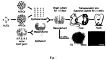
- Fig. 1 shows a schematic representation of procedures for tooth generation from hESCs and urine cell derived hiPSCs in one example of present disclosure.
- a chimeric culture system is developed for tooth regeneration from hESCs and human urine cell derived iPSCs.
- hESCs or hiPSCs can be induced to produce epithelial sheets capable of replacing the E14.5 mouse dental epithelium in a reconstitution process.
- the reconstituted explants can then be cultured in vitro for 1-2 days and transplanted beneath mouse subrenal capsule for 3 weeks ( ⁇ 2 weeks) for tooth regeneration ( Fig. 1 ).
- the stem cell is cultured for about 5-10 days, preferably about 7 days, using the culture medium described above.
- the second animal is preferably of immuno-deficiency, which may be caused by administrating a dose of Cyclosporine A to the animal.
- the first animal is a fetal mouse
- the second animal is a nude mouse.
- the dental mesenchyme is mechanically separated from a tooth germ, and the tooth germ is digested using a protease prior to the separation.
- the protease is a Dispase of about 0.75 mg/ml.
- the resulting recombinant specimen is cultured using a DMEM medium supplemented with FBS, non-essential amino acid, glutamine, penicillin and streptomycin.
- the recombinant tissue is transplanted into a space under a kidney capsule of the nude mouse.
- the predetermined time is about 2 to 4 weeks. According to an embodiment of the present disclosure, the predetermined time is about 3 weeks.
- the stem cell is cultured using at least one selected from mTeSR1TM medium, E8 medium, and KSR conditioned medium prior to preparing the epithelial-like cell, wherein the medium is replaced with the culture medium described above when the cultured stem cell reach a confluence of about 60-80%, to obtain the epithelial-like cell.
- the above described method may be effectively used to produce an ameloblast.
- the ameloblast may be contained in a tooth-like structure. With the histology examination, followed by decalcification, the inventors find that the tooth-like structure contains dental pulp, dentin, enamel space, and enamel organ.
- the inventors find that the tooth-like structure have the similar compositions with adult human teeth in both enamel and dentin. And the hardness of enamel in the regenerative tooth-like structures formed from embryo stem cell may reaches one-eighth of that in adult human teeth, and the hardness of enamel in the regenerative tooth-like structures formed from iPSC may reach one-fourth to one-third of that in adult human teeth.
- the tooth-like structures obtained in the examples of present disclosure were confirmed by the physical properties such as elastic modulus and harness by Nano-indentation and constituents by Raman spectroscopy.
- iPSCs induced pluripotent stem cell
- many cell types such as keratinobytes, neural stem cells, adipose stem cells, meningiocytes, periosteal membrane, and extraembryonic tissues.
- the kidney contains an extensive network of tubules, whose total surface is bigger than the skin.
- As part of normal physiology approximately 2000 to 7000 cells from this tubular system and downstream parts of the urinary tract (ureters, bladder, and urethra) detach and are excreted in urine daily.
- urine cells not only are not damaged but are fully functional and can be used for in vitro studies. Besides, they can be collected anywhere without medical assistance and are easily expanded.
- the person skilled in the art may generate human iPSCs from renal tubular cells present in urine (also referred as Human urine cells (hU)) by any known method.
- urine also referred as Human urine cells (hU)
- the person skilled in the art may generate iPSC using retroviral vectors of reprogramming factors Oct4, Sox2, Klf4 and c-Myc. More importantly, the urine-resulting iPSCs display an excellent ability to differentiate, suggesting that urine may be a preferred source for generating iPSCs.
- the person skilled in the art may also derive integration-free human urine induced pluripotent stem cells (ifhU-iPSCs) from human urine cells (hU) by oriP/EBNA episomal vectors carrying a combination of reprogramming factors Oct4, Sox2, SV40LT, Klf4 and miR302/367 through electroporation ( Yu, J. et al. Human induced pluripotent stem cells free of vector and transgene sequences. Science 2009:324, 797-801 ; Liao, B. et al. MicroRNA cluster 302-367 enhances somatic cell reprogramming by accelerating a mesenchymal-to-epithelial transition. The Journal of biological chemistry 2011:286, 17359-17364 ).
- the present invention describes use of a stem cell in the generation of the tooth-like structures from human induced pluripotent stem cells and human embryonic stem cells (hESCs).
- Human urine cells were cultured in primary medium contains DMEM/Ham's F12 1:1 (Hyclone), 10 wt.% of fetal bovine serum (FBS; PAA), SingleQuot Kit CC-4127 REGM (Lonza), ampothericin B and penicillin/streptomycin for 2 days and then changed to REBM (Renal Epithelial Basal 2 Medium, Lonza) medium containing SingleQuot Kit CC-4127 REGM (Lonza) (referred to as urine cell medium) during the following days.
- REBM Renal Epithelial Basal 2 Medium, Lonza
- Reference hESCs was purchased from NATIONAL STEM CELL BANK IN USA.
- iPSCs generated through retroviral vectors or oriP/EBNA episomal vectors (integration-free) were obtained from South Stem Cell Bank in China. Both reference hESCs and iPSCs were cultured on the Matrigel-coated plates in mTesR1 medium.
- Fig. 1 shows a schematic representation of procedures for tooth generation from reference hESCs and urine cell derived hiPSCs in one example of present disclosure.
- a chimeric culture system is developed for tooth regeneration from reference hESCs and human urine cell derived iPSCs.
- reference hESCs or hiPSCs can be induced to produce epithelial sheets capable of replacing the E14.5 mouse dental epithelium in a reconstitution process.
- the reconstituted explants can then be cultured in vitro for 1-2 days and transplanted beneath mouse subrenal capsule for 3 weeks ( ⁇ 2 weeks) for tooth regeneration ( Fig. 1 ).
- Example 1 Production of epithelial sheets from reference hESCs and integration-free human urine cell derived iPSCs (ifhU-iPSCs)
- the differentiating cells at D7, 14, 21 were harvested and investigated for the expression of pluripotent and keratinocyte progenitor's markers by qPCR and Western blot.
- reference H1-ESCs and ifhU-iPSCs behaved similarly as shown with markers examined at RNA level ( Oct4, K18, p63, K19, CD29, and K14), showing upregulated expressions in epithelial markers and down-regulation of pluripotent marker Oct4 ( Fig. 2b ). Similar trends were also observed for protein expression (Oct4, K18 and p63, Fig. 2c ). Moreover, the expression of p63 and K14 were verified by immunofluorescence.
- Fig. 2 shows the properties of the reference hESC/iPSC derived epithelial lineages in one example of present disclosure.
- Example 2 Generation of tooth-like structure from epithelial sheets derived from reference hESCs and ifhU-iPSCs
- the recombinants showed an intact tooth-like structure with dental pulp, dentin, enamel space, and enamel organ ( Fig. 3a , middle columns).
- the enamel organs have elongated ameloblasts with a ruffled border-like structure and papillary layer ( Fig. 3a , middle columns).
- Ameloblastin Amel located in the layer of ameloblasts and its papillary layer ( Fig. 3a ).
- HLA-I human leukocyte antigen-I
- hNA human nucleus antigen
- BSP bone sialoprotein
- Fig.3 shows tooth-like structures formed from reference H1 ESC line and hiPSC lines in 3 weeks in one example of present disclosure.
- H1-ESC human embryo stem cell
- iPSC iPSC derived from urine cells
- vUC1-iPSC-C1 represents a iPSC clone line obtained using a retroviral system, and the other 7 lines represent iPSC clone line obtained by means of Electroporation.
- both human embryo stem cell and iPSC can form a tooth-like structure containing ameloblast, and the efficiency of reference human embryo stem cell (H1-ESC) may reach 30%, which is higher than that of iPSC.
- All of the eight lines of iPSC above listed are derived from urine cell, which may have different efficiencies each other. Then we may draw a conclusion that both human embryo stem cell and iPSC can be used to prepare ameloblast with a relative higher efficiency.
Landscapes
- Health & Medical Sciences (AREA)
- Life Sciences & Earth Sciences (AREA)
- Chemical & Material Sciences (AREA)
- Engineering & Computer Science (AREA)
- Organic Chemistry (AREA)
- Biomedical Technology (AREA)
- Zoology (AREA)
- General Health & Medical Sciences (AREA)
- Genetics & Genomics (AREA)
- Cell Biology (AREA)
- Biotechnology (AREA)
- Bioinformatics & Cheminformatics (AREA)
- Wood Science & Technology (AREA)
- Medicinal Chemistry (AREA)
- Biochemistry (AREA)
- Orthopedic Medicine & Surgery (AREA)
- Developmental Biology & Embryology (AREA)
- Reproductive Health (AREA)
- Pharmacology & Pharmacy (AREA)
- Veterinary Medicine (AREA)
- General Engineering & Computer Science (AREA)
- Microbiology (AREA)
- Animal Behavior & Ethology (AREA)
- Public Health (AREA)
- Rheumatology (AREA)
- Proteomics, Peptides & Aminoacids (AREA)
- Gastroenterology & Hepatology (AREA)
- Molecular Biology (AREA)
- Toxicology (AREA)
- Biophysics (AREA)
- Gynecology & Obstetrics (AREA)
- Immunology (AREA)
- Virology (AREA)
- Epidemiology (AREA)
- Nuclear Medicine, Radiotherapy & Molecular Imaging (AREA)
- General Chemical & Material Sciences (AREA)
- Chemical Kinetics & Catalysis (AREA)
- Micro-Organisms Or Cultivation Processes Thereof (AREA)
Description
- The present disclosure relates to the field of biotechnology, in particular, the present disclosure relates to the usage of a stem cell in preparation of a tooth-like structure, a culture medium, a method for preparing an epithelial-like cell, a kit for preparing an ameloblast, method for preparing an ameloblast.
- The differentiated products of iPSCs (induced pluripotent stem cells) have been used successfully in animal models of diseases, injury and aging, such as Pakinson disease, beta-thalassemia, hepatic disease, and spinal cord injury.
- However, no solid organs or tissues such as tooth have been generated with human iPSCs.
- In view thereof, the present disclosure is directed to provide the usage of a stem cell in preparation of a tooth-like structure, a culture medium, a method for preparing an epithelial-like cell, a kit for preparing an ameloblast, method for preparing an ameloblast. The invention is defined in the claims.
- In a first aspect of the present invention, according to an embodiment of the present disclosure, the usage of a stem cell in preparation of a tooth-like structure is provided, wherein the tooth-like is produced in a mammalian animal by means of xenograft transplantation, and wherein the stem cell is induced to form an epithelial-like cell and the epithelial-like cell is recombined with a dental mesenchyme of another animal and cultured to form a reconstituted explant prior to the xenograft transplantation, wherein the stem cell is an embryo stem cell or an induced pluripotent stem cell, and wherein the mammalian animal is selected from a group consisting of mouse and rat. And in one embodiment, the induced pluripotent stem cell may be produced from a human cell, and the human cell may be at least one selected from a group consisting of urine cell, skin fibroblast, and periodontal ligament stem cell. And in one embodiment, the tooth-like structure contains ameloblast.
- The term of "tooth-like structure" may be identified with histology section and stained with human specific antibodies human leukocyte antigen-I (HLA-I, 1:50, abcam, Cat. NO. ab70328) and human nucleus antigen (hNA, 1:500, Millipore, Cat. NO. 1969098) indicating the structure and derivatives of human cells in tooth-like structure. The immunohistochemistry staining with Ameloblastin (Amel, 1:100, Santa, Cat. NO. sc-50534) may be used to describe the appearance of enamel-secreting ameloblasts in the tooth-like structures, which are produced by the epithelial sheets derived from hESCs or iPSCs.
- In another aspect of the present disclosure, a culture medium may be provided. According to an embodiment of the present disclosure, the culture medium may comprise: a basal medium, which is DMEM/F12 medium; a N2 supplement; a retinoic acid; and a BMP-4. And in one embodiment, the culture medium may comprise a N2 supplement of about 1 wt.%, a retinoic acid of about 1 µM, and a BMP-4 of about 25 ng/ml. The inventors surprisingly find that the above cited culture medium may be effectively used to induce the differentiation of stem cell into epithelial-like cell. And the epithelial-like cell may be further transplanted into the body of an animal, to form a tooth-like structure.
- In another aspect of the present disclosure, a method for preparing an epithelial-like cell may be provided. According to an embodiment of the present disclosure, the method may comprise culturing a stem cell to induce the stem cell differentiating into the epithelial-like cell using the above described culture medium, wherein the stem cell is an embryo stem cell or an induced pluripotent stem cell. The inventors surprisingly find that method uses the above cited culture medium, and then the method may be effectively used to induce the differentiation of stem cell into epithelial-like cell. And the epithelial-like cell may be further transplanted into the body of an animal, to form a tooth-like structure. According to an embodiment of the present disclosure, the induced pluripotent stem cell is produced from a human cell. According to an embodiment of the present disclosure, the human cell is at least one selected from a group consisting of urine cell, skin fibroblast, and periodontal ligament stem cell. The person skilled in the art may obtain "Human induced pluripotent stem cells (hiPSCs)" from human urine cells by two different systems: 1) pMX-based retroviral system (Oct4, Sox2, Klf4, and c-Myc, Addgene); 2) Electroporation with oriP/EBNA episomal vectors (Oct4, Sox2 SV40LT, Klf4 and miR302/367). The person skilled in the art may obtain "Human induced pluripotent stem cells (hiPSCs)" from human skin fibroblasts and periodontal ligament by pMX-based retroviral system (Oct4, Sox2, Klf4, and c-Myc, Addgene) (Esteban, M.A. et al., Vitamin C enhances the generation of mouse and human induced pluripotent stem cells. Cell Stem Cell. 2010: 6,71-9; Cai, J. et al. Generation of human induced pluripotent stem cells from umbilical cord matrix and amniotic membrane mesenchymal cells. J Biol Chem. 2010: 285,11227-34). Under human embryo stem cells (hESCs) culture conditions, hiPSCs are very similar to hESCs in cell morphology, growth properties, surface marker expression, teratoma formation, and multi-lineage in vitro differentiation.
- In one embodiment, "Human urine cells" described herein preferably are such cells which are collected from only the mid-stream of urine with 150-200 ml in the appropriate sterilized containers (up to 500 ml). In one embodiment, the human urine cell may be obtained through the following procedures. Urine samples were centrifuged at 400 g for 10 minutes at room temperature and then resuspended in PBS containing ampothericin B and penicillin/streptomycin. After washing with PBS, the cell pellet from urine sample is cultured in primary medium, containing DMEM/Ham's F12 1:1 (Hyclone), 10 wt.% of fetal bovine serum (FBS; PAA), SingleQuot Kit CC-4127 REGM (Lonza), ampothericin B and penicillin/streptomycin. The medium is kept in a small volume during the first 2 days. In the following days, the medium was changed to REBM (Renal Epithelial Basal 2 Medium, Lonza) medium containing SingleQuot Kit CC-4127 REGM (Lonza). Visible cells/colonies appeared routinely after 3-6 days, typically 3-5 per sample on average. The first full media change was made after the first cells/colonies were seen. Cells were then split onto a bigger surface aided by 0.25% trypsin containing 1 mM EDTA when the culture grew confluent. RPTECs were obtained from a biopsy and maintained in urine cell medium.
- In one embodiment, "skin fibroblasts" described herein preferably are such cells which are collected from a small piece of human skin tissue. In one embodiment, the skin fibroblast may be obtained through the following procedures. The skin tissue were minced into small tissue cubes and then plated in DMEM supplemented with 10 wt.% fetal bovine serum (FBS; Hyclone), 100 units/mL penicillin streptomycin (Hyclone). The skin keratinocytes appear first surround the small pieces of skin tissues. The skin fibroblasts were then produced thereafter. The cells at passages P3 - P5 may be used.
- In one embodiment, "periodontal ligament stem cells" described herein preferably are such cells which are collected from human periodontal ligament (PDL) tissues separated from the mid-third of the root surface. In one embodiment, periodontal ligament stem cells may be obtained through the following procedures. PDL tissues were minced into small tissue cubes (approximately 1 mm3) and digested with a solution of 3 mg/mL collagenase (type I) with 4 mg/mL dispase (both from Sigma - Aldrich, St. Louis, MO, USA) in α-minimum essential medium (α-MEM, Hyclone, Road Logan, UT, USA) for 15 min at 37 °C with vigorous shaking. The tissue explants were then plated and cultured in α-MEM supplemented with 10 wt.% fetal bovine serum (FBS; Hyclone), 0.292 mg/mL glutamine (Hyclone), 100 units/mL penicillin streptomycin (Hyclone), and 100 µm/L ascorbic acid (Sigma - Aldrich). Single cell-derived colony cultures were obtained using the limiting dilution technique and passage 0 (P0) cells were cultured. To avoid changes in cell behaviors that are associated with prolonged culture, the cells at passages P3 - P5 may be used.
- In another aspect of the present disclosure, a kit for preparing an ameloblast may be provided, and the kit may comprises: a first culture medium, which is the culture medium described above; a second culture medium, which is DMEM medium supplemented with FBS, non-essential amino acid, glutamine, penicillin and streptomycin, wherein the first culture medium and the second medium is provided in a different container respectively. The inventors surprisingly find that kit may be used to prepare ameloblast. According to an embodiment of the present disclosure, the kit may further comprising a third culture medium, which is at least one selected from mTeSR1™ medium, E8 medium, and KSR conditioned medium. According to an embodiment of the present disclosure, the second culture medium comprises FBS of about 10 wt.%, glutamine of about 2 mM, non-essential amino acid of about 0.1 mM, penicillin of about 100 U/ml, and streptomycin of 100 U/ml.
- In one embodiment, the epithelial-like cell may form an epithelial sheet. In one embodiment, the differentiating cells can be harvested as the intact epithelial sheets at Day 7 (D7) and D14 during the differentiation. Small pieces of epithelial sheets may be obtained at D21 during the differentiation. In one embodiment, the differentiating epithelial cells from both hESCs and iPSCs may be identified with the expression of pluripotent cell marker Oct4, epithelial markers K18, p63, K19, CD29, and K14, and observed under electronic microscopes (EM).
- As described in the present disclosure, the intact epithelial sheets derived from stem cells may be further induced to form tooth-like structures by recombination with dental mesenchyme.
- In a second aspect of the present invention, a method for preparing an ameloblast is provided, according to an embodiment of the present disclosure, the method comprises preparing an epithelial-like cell by a method comprising culturing a stem cell using a first culture medium, which is DMEM/F12 medium supplemented with a N2 supplement of about 1 wt.%, a retinoic acid of about 1 µM, and a BMP-4 of about 25 ng/ml, to induce the stem cell differentiating into the epithelial-like cell; recombining the epithelial-like cell with a dental mesenchyme of a first animal to obtain a recombinant specimen; culturing the resulting recombinant specimen to obtain a reconstituted explant; and transplanting the reconstituted explant into a second animal for at a predetermined time to form the ameloblast, wherein the stem cell is an embryo stem cell or an induced pluripotent stem cell and wherein the first animal and the second animal are independently selected from a group consisting of mouse and rat respectively.
-
Fig. 1 shows a schematic representation of procedures for tooth generation from hESCs and urine cell derived hiPSCs in one example of present disclosure. A chimeric culture system is developed for tooth regeneration from hESCs and human urine cell derived iPSCs. Briefly, referring toFig. 1 , hESCs or hiPSCs can be induced to produce epithelial sheets capable of replacing the E14.5 mouse dental epithelium in a reconstitution process. The reconstituted explants can then be cultured in vitro for 1-2 days and transplanted beneath mouse subrenal capsule for 3 weeks (≥ 2 weeks) for tooth regeneration (Fig. 1 ). - According to an embodiment of the present disclosure, the stem cell is cultured for about 5-10 days, preferably about 7 days, using the culture medium described above. According to an embodiment of present disclosure, the second animal is preferably of immuno-deficiency, which may be caused by administrating a dose of Cyclosporine A to the animal. According to an embodiment of the present disclosure, the first animal is a fetal mouse, and the second animal is a nude mouse. According to an embodiment of the present disclosure, the dental mesenchyme is mechanically separated from a tooth germ, and the tooth germ is digested using a protease prior to the separation. According to an embodiment of the present disclosure, the protease is a Dispase of about 0.75 mg/ml. According to an embodiment of the present disclosure, the resulting recombinant specimen is cultured using a DMEM medium supplemented with FBS, non-essential amino acid, glutamine, penicillin and streptomycin. According to an embodiment of the present disclosure, the recombinant tissue is transplanted into a space under a kidney capsule of the nude mouse. According to an embodiment of the present disclosure, the predetermined time is about 2 to 4 weeks. According to an embodiment of the present disclosure, the predetermined time is about 3 weeks. According to an embodiment of the present disclosure, the stem cell is cultured using at least one selected from mTeSR1™ medium, E8 medium, and KSR conditioned medium prior to preparing the epithelial-like cell, wherein the medium is replaced with the culture medium described above when the cultured stem cell reach a confluence of about 60-80%, to obtain the epithelial-like cell. It is surprisingly found by the inventors that the above described method may be effectively used to produce an ameloblast. And the ameloblast may be contained in a tooth-like structure. With the histology examination, followed by decalcification, the inventors find that the tooth-like structure contains dental pulp, dentin, enamel space, and enamel organ. And with Raman spectroscopy and Nano-indentation method, the inventors find that the tooth-like structure have the similar compositions with adult human teeth in both enamel and dentin. And the hardness of enamel in the regenerative tooth-like structures formed from embryo stem cell may reaches one-eighth of that in adult human teeth, and the hardness of enamel in the regenerative tooth-like structures formed from iPSC may reach one-fourth to one-third of that in adult human teeth. The tooth-like structures obtained in the examples of present disclosure were confirmed by the physical properties such as elastic modulus and harness by Nano-indentation and constituents by Raman spectroscopy.
- It was known that iPSCs (induced pluripotent stem cell) could be efficiently reprogrammed from many cell types, such as keratinobytes, neural stem cells, adipose stem cells, meningiocytes, periosteal membrane, and extraembryonic tissues. And the inventors find interestingly that the kidney contains an extensive network of tubules, whose total surface is bigger than the skin. As part of normal physiology approximately 2000 to 7000 cells from this tubular system and downstream parts of the urinary tract (ureters, bladder, and urethra) detach and are excreted in urine daily. These cells hereafter termed urine cells not only are not damaged but are fully functional and can be used for in vitro studies. Besides, they can be collected anywhere without medical assistance and are easily expanded.
- The person skilled in the art may generate human iPSCs from renal tubular cells present in urine (also referred as Human urine cells (hU)) by any known method. For example the person skilled in the art may generate iPSC using retroviral vectors of reprogramming factors Oct4, Sox2, Klf4 and c-Myc. More importantly, the urine-resulting iPSCs display an excellent ability to differentiate, suggesting that urine may be a preferred source for generating iPSCs. And the person skilled in the art may also derive integration-free human urine induced pluripotent stem cells (ifhU-iPSCs) from human urine cells (hU) by oriP/EBNA episomal vectors carrying a combination of reprogramming factors Oct4, Sox2, SV40LT, Klf4 and miR302/367 through electroporation (Yu, J. et al. Human induced pluripotent stem cells free of vector and transgene sequences. Science 2009:324, 797-801; Liao, B. et al. MicroRNA cluster 302-367 enhances somatic cell reprogramming by accelerating a mesenchymal-to-epithelial transition. The Journal of biological chemistry 2011:286, 17359-17364). Thus, the present invention describes use of a stem cell in the generation of the tooth-like structures from human induced pluripotent stem cells and human embryonic stem cells (hESCs).
- Additional aspects and advantages of the embodiments of present disclosure will be given in part in the following descriptions, become apparent in part from the following descriptions, or be learned from the practice of the embodiments of the present disclosure.
- The aforementioned features and advantages of the invention as well as additional features and advantages thereof will be more clearly understood hereinafter as a result of a detailed description of preferred embodiments when taken in conjunction with the drawings, in which:
-
Fig. 1 shows a schematic representation of procedures for tooth generation from hESCs and urine cell derived hiPSCs in one example of present disclosure. -
Fig. 2 shows the properties of the hESC/iPSC derived epithelial lineages in one example of present disclosure. -
Fig. 3 shows tooth-like structures formed from H1 ESC line and hiPSC lines in 3 weeks in one example of present disclosure. - The present disclosure will be described in detail with reference to the following examples. The standard laboratory practices of the inventors are illustrated in the following examples which are used to exemplify the mode of the present invention. The scope of the present invention shall not be construed as being limited to these examples. According to what is disclosed herein and the common level of those skilled in the art, it shall be appreciated that the following examples are only for illustration and may be subjected to various changes, modifications, and alterations without departing from the scope of the present invention. Unless otherwise stated, all the technologies concerned are conventional technologies in embryology, stem cell biology, molecular biology, tissue physiology immunology, histology, engineering, or other fields as well known by those skilled in the art.
- Human urine cells were cultured in primary medium contains DMEM/Ham's F12 1:1 (Hyclone), 10 wt.% of fetal bovine serum (FBS; PAA), SingleQuot Kit CC-4127 REGM (Lonza), ampothericin B and penicillin/streptomycin for 2 days and then changed to REBM (Renal Epithelial Basal 2 Medium, Lonza) medium containing SingleQuot Kit CC-4127 REGM (Lonza) (referred to as urine cell medium) during the following days. Reference hESCs was purchased from NATIONAL STEM CELL BANK IN USA. iPSCs generated through retroviral vectors or oriP/EBNA episomal vectors (integration-free) were obtained from South Stem Cell Bank in China. Both reference hESCs and iPSCs were cultured on the Matrigel-coated plates in mTesR1 medium.
-
Fig. 1 shows a schematic representation of procedures for tooth generation from reference hESCs and urine cell derived hiPSCs in one example of present disclosure. A chimeric culture system is developed for tooth regeneration from reference hESCs and human urine cell derived iPSCs. Briefly, referring toFig. 1 , reference hESCs or hiPSCs can be induced to produce epithelial sheets capable of replacing the E14.5 mouse dental epithelium in a reconstitution process. The reconstituted explants can then be cultured in vitro for 1-2 days and transplanted beneath mouse subrenal capsule for 3 weeks (≥ 2 weeks) for tooth regeneration (Fig. 1 ). - We first devised a way to obtain dental epithelia from reference hESCs or ifhU-iPSCs and decided on a stage-specific approach based on RA and BMP4 in N2 medium comprising a N2 supplement of about 1 wt.%, a retinoic acid of about 1 µM, and a BMP-4 of about 25 ng/ml. To this end, we obtained epithelial cells with keratinocyte-like morphology at D7 (
Fig. 2a ). We then allowed the cells to grow as epithelial sheets without passage, and they became denser at D14, and detached slightly with some cell death at D21 (Fig. 2a ). The differentiating cells at D7, 14, 21 were harvested and investigated for the expression of pluripotent and keratinocyte progenitor's markers by qPCR and Western blot. During epithelial differentiation, reference H1-ESCs and ifhU-iPSCs behaved similarly as shown with markers examined at RNA level (Oct4, K18, p63, K19, CD29, and K14), showing upregulated expressions in epithelial markers and down-regulation of pluripotent marker Oct4 (Fig. 2b ). Similar trends were also observed for protein expression (Oct4, K18 and p63,Fig. 2c ). Moreover, the expression of p63 and K14 were verified by immunofluorescence. p63 was detected earlier at D7 and continuously expressed at D21, while K14 expression was detected later at D21 (Fig. 2d ). As a result, we obtained homogenous layers of epithelial cells as sheets from both reference hESCs and ifhU-iPSCs at D7 (Fig. 2d ). These sheets harvested at D7 were tenacious and flexible, showing the flat and smooth surface at the apical side as observed by scanning electronic microscopy (SEM) (Fig. 2e ). The sheets became rugged with prominent nuclei at D14 (Fig. 2e ). Under transmission electron microscopy (TEM), the desmosomes could be observed clearly between the epithelial cells at both D7 and D14 (Fig. 2e ). Together, these results suggest that the epithelial sheets generated at D7 from reference hESC or ifhU-iPSCs have desired properties for being induced for tooth regeneration. - In details,
Fig. 2 shows the properties of the reference hESC/iPSC derived epithelial lineages in one example of present disclosure. - In the
Fig. 2 , - (a) shows the epithelial differentiation process of reference hESCs or hiPSCs through supplement of RA and BMP4 in N2 medium for 7 days, then changing into DSFM for further differentiation. Scale bar corresponds to 200 µm.
- (b) shows qPCR of representative experiment showing the down regulation of ESC-specific transcription factor (Oct4) and up regulation of keratinized epithelial markers (K18, p63, K19, CD29, K14).
- (c) shows Western blotting for Oct4, p63, and K18 of lysates from H1 or ifhU1-iPSCs derived epithelial cells; GAPDH is the loading control.
- (d) shows Phase contrast captures and immunofluorescence staining [IF: p63 (red), K14 (green), DAPI (blue)] of epithelial sheets derived from H1 and ifhU1-iPSCs at D7 and D21. Scale bars correspond to 2000 and 200 µm, respectively.
- (e) shows Scanning electron microscope (SEM) and transmission electron microscopy (TEM) images of reference H1-ESCs and ifhU1-iPSCs derived epithelial sheets at D7 and D14. White arrowheads indicate desmosomes between epithelial cells of the sheet. Scale bars in SEM and TEM correspond to 20 and 0.5 µm respectively.
- We harvested D7 epithelial sheets and recombined them with the mouse dental mesenchyme in a mTeSR1™ medium, E8 medium, or KSR conditioned medium (wherein the medium comprises FBS of about 10 wt.%, glutamine of about 2 mM, non-essential amino acid of about 0.1 mM, penicillin of about 100 U/ml, and streptomycin of 100 U/ml), before transplantation into mouse subrenal capsule (
Fig. 1 ). After 3 weeks, we observed tooth-like structures in the kidney (Fig. 3a ). We isolated individual tooth-like structures by removing them from the subrenal capsule and the surrounding bone (Fig. 3a , left columns). The recombinants showed an intact tooth-like structure with dental pulp, dentin, enamel space, and enamel organ (Fig. 3a , middle columns). The enamel organs have elongated ameloblasts with a ruffled border-like structure and papillary layer (Fig. 3a , middle columns). We also observed the expression of Ameloblastin (Amel) located in the layer of ameloblasts and its papillary layer (Fig. 3a ). We confirmed the human origin of the epithelial component in cross sections of recombinant tooth prior to isolation by immunostainings with human specific antibodies against human leukocyte antigen-I (HLA-I) and human nucleus antigen (hNA) (Fig. 3b ). Both antibodies stained negatively in the dental pulp, cartilage, surrounding bone-like structures, which were developed from mouse dental mesenchyme (Fig. 3b ). As expected, both human specific antibodies stained positively for the ameloblasts (Fig. 3b1 ), papillary layer besides (Fig. 3b2 ), and squamous epithelial cells in the cyst (Fig. 3b3 ). Furthermore, positive HLA-I staining was localized in the cytoplasm, while hNA was complementarily localized in the nucleus (Fig. 3b4, b5 ). As control, without recombination with reference hESCs or ifhU-iPSCs derived epithelial sheets, mouse dental mesenchymes transplanted under identical conditions formed bone-like structure instead (n=10/10), as confirmed by positive staining of bone sialoprotein (BSP) in the whole bone-like structure embedded with osteocytes (Fig. 3c ). - We analyzed the hardness and elastic modulus of human adult teeth (human group), 3-week mouse teeth developed from tooth germs under kidney capsule (Mouse WT group), and the regenerative teeth from reference H1-ESCs and ifhU-iPSCs groups by Nano-indentation (
Fig. 3d ). The harness and elastic modulus of dentin and enamel in five groups showed similar properties (Fig. 3d ). In the case of enamel, the hardness and elastic modulus of reference H1-ESCs and two ifhU-iPSCs groups were lower than those of human and Mouse WT groups (Fig. 3d , right graphs). Interestingly, the hardness of ifhU5-iPSCs group was around 4 times higher than that of reference H1-ESCs group (Fig. 3d , right graphs). Impressively, the hardness of ifhU5-iPSCs group reached 1/3 by comparison with that of human group (Fig. 3d , right graphs). We further analyzed the physical properties of the teeth by Raman spectroscopy. We showed that they all have similar spectra with comparable intensity (Fig. 3e ). In the 5 groups examined, the enamel of the teeth are highly mineralized and the spectra showed almost the same spectrum as hydroxyapatite [Ca5(PO4)3OH], the primary mineral in teeth] (Fig. 3e ). These tooth-like structures also showed similar signs of protein with Raman peaks in dentin, including the hydroxyapatite pick compared to human teeth (Fig. 3e ). These results indicated that the regenerative teeth from both reference H1-ESCs and two ifhU-iPSCs groups have similar constituents with human and mouse teeth. We analyzed a total of 8 hiPSC lines from three urine donors (U1, 5, 7) for tooth regeneration and had a success rate of up to 30% (Fig. 3f ). - In details,
Fig.3 shows tooth-like structures formed from reference H1 ESC line and hiPSC lines in 3 weeks in one example of present disclosure. - In the
Fig.3 , - (a) Left two columns shows Phase contrast captures of tooth-like structures after and before isolated from the surrounding bone from reference H1-ESC and ifhU1-iPSC groups (tooth is outlined by a dotted line); right three columns: Hematoxylin-Eosin staining (HE) and immunohistology staining (IHC: Ameloblastin, Amel) of relative tooth sections (reference H1-ESC: sagittal section; ifhU1-iPSC: cross section), showing the tooth-like structures containing dental pulp (dp), dentin (d), enamel space (es), and a layer of ameloblasts (Am) in both groups. Elongated ameloblasts were showed at the high magnification in the blue box. Positive Amel expression was specifically localized in these ameloblasts with the papillary layer (black arrows). The papillary layer was further pointed out by black arrowheads at the high magnification in the green box. Scale bar: the upper and bottom figures in the same column share the same scale bar as 500, 500, 400, 100, 100 µm from left to right orderly.
- (b) shows Immunohistology and immunofluorescence staining in cross sections of the ifhU1-iPSCs derived tooth: top, HLA-I expression was specifically localized in the cytoplasm of human iPSC derived cells and tissues, including ameloblast layer (black arrowheads in b1 and b2) and epithelium-derived cyst (black arrows in b3); bottom, complementary expression of hNA (green) in the nucleus of the same regions in ameloblasts (white arrowheads) and cyst epithelial cells (white arrow). * indicated cyst. DAPI is shown in blue. Higher magnifications of hNA and DAPI in ameloblasts (white arrowheads) were shown in b4 and b5 respectively. Scale bars correspond to 200 µm.
- (c) shows Image of a piece of bone detected from E14.5 mouse dental mesenchyme being transplanted under kidney capsule for 3 weeks with positive BSP expression. Scale bars: 1000, 100, 100 µm orderly.
- (d) shows Nano-indentation analyses: Left: representative images of fractured enamel and dentin surfaces in adult human tooth group (human), group of 3-week mouse tooth from E14.5 tooth germs (Mouse-WT), regenerative tooth groups from reference H1-ESCs and ifhU-iPSCs; right: hardness and elastic modulus of enamel and dentin in above groups (each group: n=3).
- (e) shows Roman spectroscopy analyses of enamel and dentin from human, mouse, reference hESCs, and ifhU-iPSCs showed great similarity for all groups on Raman peaks, including 961 cm-1 (hydroxyapatite) in both enamel and dentin, and 1669 cm-1(C=O stretching vibration), 2885 cm-1 and 2941 cm-1(C-H stretching vibration) in dentin.
- (f) shows efficiencies of tooth-like structures for reference H1 ESC line and 8 iPSC lines.
- In this example, reference human embryo stem cell (H1-ESC) and eight different lines of iPSC derived from urine cells are used to prepare ameloblast respectively in a way similar to the above examples. And the respective efficiency of obtaining ameloblast are compared among these stem cells, the results are shown in the following table I.
Table I iPSC clone lines Number of newly formed Teeth / Number of recombinant specimen Efficiency in term of percentage reference H1- ESC 15/50 30% vUC1-iPSC-C1 6/42 14.3% UC1-iPSC- C1 12/50 24% UC5-iPSC- C1 13/56 23.2% UC5-iPSC- C2 15/59 25.4% UC5-iPSC-C3 6/40 15% UC7-iPSC- C3 12/52 23.1% UC7-iPSC- C6 7/60 11.7% UC7-iPSC-C9 17/59 28.8% - In the above table, vUC1-iPSC-C1 represents a iPSC clone line obtained using a retroviral system, and the other 7 lines represent iPSC clone line obtained by means of Electroporation.
- As shown by the table I, both human embryo stem cell and iPSC can form a tooth-like structure containing ameloblast, and the efficiency of reference human embryo stem cell (H1-ESC) may reach 30%, which is higher than that of iPSC. All of the eight lines of iPSC above listed are derived from urine cell, which may have different efficiencies each other. Then we may draw a conclusion that both human embryo stem cell and iPSC can be used to prepare ameloblast with a relative higher efficiency.
- Although explanatory embodiments have been shown and described, it would be appreciated by those skilled in the art that changes, alternatives, and modifications all falling into the scope of the claims and their equivalents can be made.
Claims (9)
- Use of a stem cell in the preparation of a tooth-like structure, wherein the tooth-like structure is produced in a mammalian animal by means of xenograft transplantation, and wherein the stem cell is induced to form an epithelial-like cell and the epithelial-like cell is recombined with a dental mesenchyme of another animal and cultured to form a reconstituted explant prior to the xenograft transplantation, wherein the stem cell is an embryo stem cell or an induced pluripotent stem cell, and wherein the animal is selected from the group consisting of mouse and rat.
- The use of claim 1, wherein the induced pluripotent stem cell is produced from a human cell, and the human cell is at least one selected from a group consisting of urine cell, skin fibroblast, and periodontal ligament stem cell.
- The use of claim 1, wherein the tooth-like structure contains ameloblast.
- A method for preparing an ameloblast, comprising:preparing an epithelial-like cell by a method comprising culturing a stem cell using a first culture medium, which is DMEM/F12 medium supplemented with a N2 supplement of about 1 wt.%, a retinoic acid of about 1 µM, and a BMP-4 of about 25 ng/ml , to induce the stem cell differentiating into the epithelial-like cell;recombining the epithelial-like cell with a dental mesenchyme of a first animal to obtain a recombinant specimen;culturing the resulting recombinant specimen to obtain a reconstituted explant; andtransplanting the reconstituted explant into a second animal for a predetermined time to form the ameloblast,wherein the stem cell is an embryo stem cell or an induced pluripotent stem cell, and wherein the animal is selected from the group consisting of mouse and rat.
- The method of claim 4, wherein the stem cell is cultured for about 5-10 days, preferably about 7 days, the first animal is a fetal mouse, and the second animal is a nude mouse, and the predetermined time is about 2 to 4 weeks, preferably about 3 weeks.
- The method of claim 5, wherein the dental mesenchyme is mechanically separated from a tooth germ, and the tooth germ is digested using a protease prior to the separation, and the protease is a Dispase of about 0.75 mg/ml.
- The method of claim 4, wherein the resulting recombinant specimen is cultured using a DMEM medium supplemented with FBS, non-essential amino acid, glutamine, penicillin and streptomycin.
- The method of claim 5, wherein the recombinant tissue is transplanted into a space under a kidney capsule of the nude mouse.
- The method of claim 4, wherein the stem cell is cultured using at least one selected from mTeSR1™ medium, E8 medium, and KSR conditioned medium prior to preparing the epithelial-like cell,
wherein the medium is replaced with the first culture medium when the cultured stem cells reach a confluence of about 60-80%, to obtain the epithelial-like cell.
Applications Claiming Priority (1)
| Application Number | Priority Date | Filing Date | Title |
|---|---|---|---|
| PCT/CN2013/071408 WO2014121449A1 (en) | 2013-02-05 | 2013-02-05 | Preparing tooth-like structure using stem cell |
Publications (3)
| Publication Number | Publication Date |
|---|---|
| EP2954047A1 EP2954047A1 (en) | 2015-12-16 |
| EP2954047A4 EP2954047A4 (en) | 2016-08-03 |
| EP2954047B1 true EP2954047B1 (en) | 2019-08-28 |
Family
ID=51299163
Family Applications (1)
| Application Number | Title | Priority Date | Filing Date |
|---|---|---|---|
| EP13874559.1A Active EP2954047B1 (en) | 2013-02-05 | 2013-02-05 | Preparing tooth-like structure using stem cell |
Country Status (5)
| Country | Link |
|---|---|
| US (1) | US9782443B2 (en) |
| EP (1) | EP2954047B1 (en) |
| JP (1) | JP2016507231A (en) |
| ES (1) | ES2763864T3 (en) |
| WO (1) | WO2014121449A1 (en) |
Families Citing this family (5)
| Publication number | Priority date | Publication date | Assignee | Title |
|---|---|---|---|---|
| US3153254A (en) * | 1960-07-19 | 1964-10-20 | Trico Products Corp | Windshield wiper |
| CN105420180A (en) * | 2015-11-18 | 2016-03-23 | 广州赛莱拉干细胞科技股份有限公司 | Complete culture medium and method for in-vitro amplification of periodontal ligament stem cells |
| KR101957033B1 (en) * | 2017-08-24 | 2019-03-11 | 서울대학교산학협력단 | Method for differentiation of dental epithelium from human embryonic stem cells and use thereof |
| KR102025554B1 (en) * | 2018-04-05 | 2019-09-26 | 단국대학교 천안캠퍼스 산학협력단 | Composition for differentiation of dental epithelial cells to ameloblast |
| CN120795059A (en) | 2018-08-27 | 2025-10-17 | 瑞泽恩制药公司 | Use of raman spectroscopy in downstream purification |
Family Cites Families (13)
| Publication number | Priority date | Publication date | Assignee | Title |
|---|---|---|---|---|
| GB0003930D0 (en) * | 2000-02-18 | 2000-04-12 | King S College London | Cell |
| US20040265971A1 (en) | 2001-11-19 | 2004-12-30 | Hidetaka Sato | Drug mobilizing pluripotent stem cells from tissue into peripheral blood |
| EP1568386A1 (en) * | 2002-11-15 | 2005-08-31 | Kyushu Tlo Company, Limited | Method of organ regeneration |
| GB0304030D0 (en) * | 2003-02-21 | 2003-03-26 | King S College London | Teeth |
| BRPI0402659A (en) * | 2004-06-23 | 2006-01-31 | Univ Fed Sao Paulo Unifesp | Stem cell use, tissue engineering method, dental tissue use and biological tooth replacement |
| CN1587393A (en) * | 2004-07-15 | 2005-03-02 | 福建师范大学 | Technology for tooth regeneration using keratin stem cell and dental pulp stem cell |
| US20100093080A1 (en) * | 2005-07-29 | 2010-04-15 | Matsumoto Dental University | Tooth Regeneration Method |
| US8129187B2 (en) * | 2005-12-13 | 2012-03-06 | Kyoto University | Somatic cell reprogramming by retroviral vectors encoding Oct3/4. Klf4, c-Myc and Sox2 |
| WO2008090826A1 (en) * | 2007-01-22 | 2008-07-31 | Organ Technologies Inc. | Method for production of mesenchymal cell, method for production of tooth, and mesenchymal cell for formation of tooth |
| JPWO2010134619A1 (en) * | 2009-05-18 | 2012-11-12 | 国立大学法人東北大学 | Induction differentiation of epithelial progenitor cells / stem cells and corneal epithelial cells from induced pluripotent stem cells |
| CN102234627B (en) * | 2010-04-30 | 2015-06-03 | 中国科学院广州生物医药与健康研究院 | Culture medium additive and application thereof |
| WO2012089669A1 (en) * | 2010-12-31 | 2012-07-05 | Universität Für Bodenkultur Wien | Method of generating induced pluripotent stem cells and differentiated cells |
| WO2012168167A1 (en) * | 2011-06-09 | 2012-12-13 | F. Hoffmann-La Roche Ag | Method for differentiation of pluripotent stem cells into vascular bed cells |
-
2013
- 2013-02-05 JP JP2015555528A patent/JP2016507231A/en active Pending
- 2013-02-05 ES ES13874559T patent/ES2763864T3/en active Active
- 2013-02-05 WO PCT/CN2013/071408 patent/WO2014121449A1/en not_active Ceased
- 2013-02-05 EP EP13874559.1A patent/EP2954047B1/en active Active
- 2013-02-05 US US14/765,949 patent/US9782443B2/en not_active Expired - Fee Related
Non-Patent Citations (1)
| Title |
|---|
| None * |
Also Published As
| Publication number | Publication date |
|---|---|
| ES2763864T3 (en) | 2020-06-01 |
| WO2014121449A1 (en) | 2014-08-14 |
| JP2016507231A (en) | 2016-03-10 |
| US20160000836A1 (en) | 2016-01-07 |
| US9782443B2 (en) | 2017-10-10 |
| EP2954047A4 (en) | 2016-08-03 |
| EP2954047A1 (en) | 2015-12-16 |
Similar Documents
| Publication | Publication Date | Title |
|---|---|---|
| EP2374871B1 (en) | Pluripotent stem cells, method for preparation thereof and uses thereof | |
| Marynka-Kalmani et al. | The lamina propria of adult human oral mucosa harbors a novel stem cell population | |
| EP2205724B1 (en) | Method for isolating and culturing adult stem cells derived from human amniotic epithelium | |
| EP1948786B1 (en) | Multipotent stem cells derived from human adipose tissue and cellular therapeutic agents comprising the same | |
| KR100871984B1 (en) | Multipotent Stem Cell Derived from Placenta Tissue and Cellular Therapeutic Agents Comprising the Same | |
| JP6711757B2 (en) | Method to differentiate into hepatocytes using pluripotent stem cells derived from mesenchymal stem cells | |
| US9782443B2 (en) | Preparing tooth-like structure using stem cell | |
| JP6711759B2 (en) | Method for differentiating into neural cells using pluripotent stem cells derived from mesenchymal stem cells | |
| US20140271616A1 (en) | Compositions And Methods For Mesenchymal And/Or Chondrogenic Differentiation Of Stem Cells | |
| Shah et al. | Types and classification of stem cells | |
| Liu et al. | Application of iPS cells in dental bioengineering and beyond | |
| CN104342402A (en) | Culture method of marrow dedifferentiated mesenchymal stem cell | |
| Mohammed et al. | Osteogenic differentiation potential of human bone marrow and amniotic fluid-derived mesenchymal stem cells in vitro & in vivo | |
| JP6711756B2 (en) | Method for differentiating into adipocytes using pluripotent stem cells derived from mesenchymal stem cells | |
| KR20120006386A (en) | First placental tissue-derived stem cells and cell therapy containing the same | |
| CN115516081B (en) | Osteoblast differentiated from mesenchymal stem cells and composition for treating bone diseases comprising the same | |
| KR20160066326A (en) | Method for Differentiating Pluripotent Stem Cell induced from adipose-derived Mesenchymal Stem Cell into Neuron | |
| JP6711758B2 (en) | Method for differentiating into osteoblasts using pluripotent stem cells derived from mesenchymal stem cells | |
| KR101633019B1 (en) | Method for Preparing Induced Pluripotency Stem Cell from Mesenchymal Stem Cell and Production thereof | |
| KR101671880B1 (en) | Method for Preparing Induced Pluripotency Stem Cell from adipose-derived Mesenchymal Stem Cell and Production thereof | |
| KR101671884B1 (en) | Method for Differentiating Pluripotent Stem Cell induced from adipose-derived Mesenchymal Stem Cell into Adipocyte | |
| AU2022425291A1 (en) | Nonhuman stem cells and their use for production of cultured meat | |
| Ünal et al. | Isolation and Characterization of Mesenchymal Stem Cells Derived from Human Amniotic Membrane by Explant Technique | |
| Fard et al. | The effect of 2D culture on stemness-related transcriptome in shed microvesicles of mesenchymal stem cells | |
| KR101671883B1 (en) | Method for Differentiating Pluripotent Stem Cell induced from adipose-derived Mesenchymal Stem Cell into Hematocyte |
Legal Events
| Date | Code | Title | Description |
|---|---|---|---|
| PUAI | Public reference made under article 153(3) epc to a published international application that has entered the european phase |
Free format text: ORIGINAL CODE: 0009012 |
|
| 17P | Request for examination filed |
Effective date: 20150904 |
|
| AK | Designated contracting states |
Kind code of ref document: A1 Designated state(s): AL AT BE BG CH CY CZ DE DK EE ES FI FR GB GR HR HU IE IS IT LI LT LU LV MC MK MT NL NO PL PT RO RS SE SI SK SM TR |
|
| AX | Request for extension of the european patent |
Extension state: BA ME |
|
| DAX | Request for extension of the european patent (deleted) | ||
| A4 | Supplementary search report drawn up and despatched |
Effective date: 20160705 |
|
| RIC1 | Information provided on ipc code assigned before grant |
Ipc: C07K 14/51 20060101ALI20160630BHEP Ipc: C12N 5/077 20100101ALI20160630BHEP Ipc: C12N 5/0789 20100101ALI20160630BHEP Ipc: C12N 5/074 20100101ALI20160630BHEP Ipc: C12N 5/0775 20100101ALI20160630BHEP Ipc: A61P 1/02 20060101ALI20160630BHEP Ipc: A61K 6/00 20060101ALI20160630BHEP Ipc: C12N 5/0735 20100101AFI20160630BHEP Ipc: A61K 35/545 20150101ALI20160630BHEP Ipc: C12N 5/0797 20100101ALI20160630BHEP |
|
| STAA | Information on the status of an ep patent application or granted ep patent |
Free format text: STATUS: EXAMINATION IS IN PROGRESS |
|
| 17Q | First examination report despatched |
Effective date: 20170522 |
|
| GRAP | Despatch of communication of intention to grant a patent |
Free format text: ORIGINAL CODE: EPIDOSNIGR1 |
|
| STAA | Information on the status of an ep patent application or granted ep patent |
Free format text: STATUS: GRANT OF PATENT IS INTENDED |
|
| INTG | Intention to grant announced |
Effective date: 20190312 |
|
| GRAS | Grant fee paid |
Free format text: ORIGINAL CODE: EPIDOSNIGR3 |
|
| GRAA | (expected) grant |
Free format text: ORIGINAL CODE: 0009210 |
|
| STAA | Information on the status of an ep patent application or granted ep patent |
Free format text: STATUS: THE PATENT HAS BEEN GRANTED |
|
| AK | Designated contracting states |
Kind code of ref document: B1 Designated state(s): AL AT BE BG CH CY CZ DE DK EE ES FI FR GB GR HR HU IE IS IT LI LT LU LV MC MK MT NL NO PL PT RO RS SE SI SK SM TR |
|
| REG | Reference to a national code |
Ref country code: GB Ref legal event code: FG4D |
|
| REG | Reference to a national code |
Ref country code: CH Ref legal event code: EP |
|
| REG | Reference to a national code |
Ref country code: AT Ref legal event code: REF Ref document number: 1172428 Country of ref document: AT Kind code of ref document: T Effective date: 20190915 |
|
| REG | Reference to a national code |
Ref country code: IE Ref legal event code: FG4D |
|
| REG | Reference to a national code |
Ref country code: DE Ref legal event code: R096 Ref document number: 602013059879 Country of ref document: DE |
|
| REG | Reference to a national code |
Ref country code: NL Ref legal event code: MP Effective date: 20190828 |
|
| REG | Reference to a national code |
Ref country code: LT Ref legal event code: MG4D |
|
| PG25 | Lapsed in a contracting state [announced via postgrant information from national office to epo] |
Ref country code: NL Free format text: LAPSE BECAUSE OF FAILURE TO SUBMIT A TRANSLATION OF THE DESCRIPTION OR TO PAY THE FEE WITHIN THE PRESCRIBED TIME-LIMIT Effective date: 20190828 Ref country code: BG Free format text: LAPSE BECAUSE OF FAILURE TO SUBMIT A TRANSLATION OF THE DESCRIPTION OR TO PAY THE FEE WITHIN THE PRESCRIBED TIME-LIMIT Effective date: 20191128 Ref country code: SE Free format text: LAPSE BECAUSE OF FAILURE TO SUBMIT A TRANSLATION OF THE DESCRIPTION OR TO PAY THE FEE WITHIN THE PRESCRIBED TIME-LIMIT Effective date: 20190828 Ref country code: NO Free format text: LAPSE BECAUSE OF FAILURE TO SUBMIT A TRANSLATION OF THE DESCRIPTION OR TO PAY THE FEE WITHIN THE PRESCRIBED TIME-LIMIT Effective date: 20191128 Ref country code: HR Free format text: LAPSE BECAUSE OF FAILURE TO SUBMIT A TRANSLATION OF THE DESCRIPTION OR TO PAY THE FEE WITHIN THE PRESCRIBED TIME-LIMIT Effective date: 20190828 Ref country code: LT Free format text: LAPSE BECAUSE OF FAILURE TO SUBMIT A TRANSLATION OF THE DESCRIPTION OR TO PAY THE FEE WITHIN THE PRESCRIBED TIME-LIMIT Effective date: 20190828 Ref country code: PT Free format text: LAPSE BECAUSE OF FAILURE TO SUBMIT A TRANSLATION OF THE DESCRIPTION OR TO PAY THE FEE WITHIN THE PRESCRIBED TIME-LIMIT Effective date: 20191230 Ref country code: FI Free format text: LAPSE BECAUSE OF FAILURE TO SUBMIT A TRANSLATION OF THE DESCRIPTION OR TO PAY THE FEE WITHIN THE PRESCRIBED TIME-LIMIT Effective date: 20190828 |
|
| PG25 | Lapsed in a contracting state [announced via postgrant information from national office to epo] |
Ref country code: IS Free format text: LAPSE BECAUSE OF FAILURE TO SUBMIT A TRANSLATION OF THE DESCRIPTION OR TO PAY THE FEE WITHIN THE PRESCRIBED TIME-LIMIT Effective date: 20191228 Ref country code: RS Free format text: LAPSE BECAUSE OF FAILURE TO SUBMIT A TRANSLATION OF THE DESCRIPTION OR TO PAY THE FEE WITHIN THE PRESCRIBED TIME-LIMIT Effective date: 20190828 Ref country code: GR Free format text: LAPSE BECAUSE OF FAILURE TO SUBMIT A TRANSLATION OF THE DESCRIPTION OR TO PAY THE FEE WITHIN THE PRESCRIBED TIME-LIMIT Effective date: 20191129 Ref country code: AL Free format text: LAPSE BECAUSE OF FAILURE TO SUBMIT A TRANSLATION OF THE DESCRIPTION OR TO PAY THE FEE WITHIN THE PRESCRIBED TIME-LIMIT Effective date: 20190828 Ref country code: LV Free format text: LAPSE BECAUSE OF FAILURE TO SUBMIT A TRANSLATION OF THE DESCRIPTION OR TO PAY THE FEE WITHIN THE PRESCRIBED TIME-LIMIT Effective date: 20190828 |
|
| REG | Reference to a national code |
Ref country code: AT Ref legal event code: MK05 Ref document number: 1172428 Country of ref document: AT Kind code of ref document: T Effective date: 20190828 |
|
| PG25 | Lapsed in a contracting state [announced via postgrant information from national office to epo] |
Ref country code: TR Free format text: LAPSE BECAUSE OF FAILURE TO SUBMIT A TRANSLATION OF THE DESCRIPTION OR TO PAY THE FEE WITHIN THE PRESCRIBED TIME-LIMIT Effective date: 20190828 |
|
| PG25 | Lapsed in a contracting state [announced via postgrant information from national office to epo] |
Ref country code: EE Free format text: LAPSE BECAUSE OF FAILURE TO SUBMIT A TRANSLATION OF THE DESCRIPTION OR TO PAY THE FEE WITHIN THE PRESCRIBED TIME-LIMIT Effective date: 20190828 Ref country code: AT Free format text: LAPSE BECAUSE OF FAILURE TO SUBMIT A TRANSLATION OF THE DESCRIPTION OR TO PAY THE FEE WITHIN THE PRESCRIBED TIME-LIMIT Effective date: 20190828 Ref country code: DK Free format text: LAPSE BECAUSE OF FAILURE TO SUBMIT A TRANSLATION OF THE DESCRIPTION OR TO PAY THE FEE WITHIN THE PRESCRIBED TIME-LIMIT Effective date: 20190828 Ref country code: RO Free format text: LAPSE BECAUSE OF FAILURE TO SUBMIT A TRANSLATION OF THE DESCRIPTION OR TO PAY THE FEE WITHIN THE PRESCRIBED TIME-LIMIT Effective date: 20190828 Ref country code: PL Free format text: LAPSE BECAUSE OF FAILURE TO SUBMIT A TRANSLATION OF THE DESCRIPTION OR TO PAY THE FEE WITHIN THE PRESCRIBED TIME-LIMIT Effective date: 20190828 |
|
| PG25 | Lapsed in a contracting state [announced via postgrant information from national office to epo] |
Ref country code: IS Free format text: LAPSE BECAUSE OF FAILURE TO SUBMIT A TRANSLATION OF THE DESCRIPTION OR TO PAY THE FEE WITHIN THE PRESCRIBED TIME-LIMIT Effective date: 20200224 Ref country code: SK Free format text: LAPSE BECAUSE OF FAILURE TO SUBMIT A TRANSLATION OF THE DESCRIPTION OR TO PAY THE FEE WITHIN THE PRESCRIBED TIME-LIMIT Effective date: 20190828 Ref country code: CZ Free format text: LAPSE BECAUSE OF FAILURE TO SUBMIT A TRANSLATION OF THE DESCRIPTION OR TO PAY THE FEE WITHIN THE PRESCRIBED TIME-LIMIT Effective date: 20190828 Ref country code: SM Free format text: LAPSE BECAUSE OF FAILURE TO SUBMIT A TRANSLATION OF THE DESCRIPTION OR TO PAY THE FEE WITHIN THE PRESCRIBED TIME-LIMIT Effective date: 20190828 |
|
| REG | Reference to a national code |
Ref country code: ES Ref legal event code: FG2A Ref document number: 2763864 Country of ref document: ES Kind code of ref document: T3 Effective date: 20200601 |
|
| REG | Reference to a national code |
Ref country code: DE Ref legal event code: R097 Ref document number: 602013059879 Country of ref document: DE |
|
| PLBE | No opposition filed within time limit |
Free format text: ORIGINAL CODE: 0009261 |
|
| STAA | Information on the status of an ep patent application or granted ep patent |
Free format text: STATUS: NO OPPOSITION FILED WITHIN TIME LIMIT |
|
| PG2D | Information on lapse in contracting state deleted |
Ref country code: IS |
|
| 26N | No opposition filed |
Effective date: 20200603 |
|
| PG25 | Lapsed in a contracting state [announced via postgrant information from national office to epo] |
Ref country code: SI Free format text: LAPSE BECAUSE OF FAILURE TO SUBMIT A TRANSLATION OF THE DESCRIPTION OR TO PAY THE FEE WITHIN THE PRESCRIBED TIME-LIMIT Effective date: 20190828 |
|
| REG | Reference to a national code |
Ref country code: CH Ref legal event code: PL |
|
| REG | Reference to a national code |
Ref country code: BE Ref legal event code: MM Effective date: 20200229 |
|
| PG25 | Lapsed in a contracting state [announced via postgrant information from national office to epo] |
Ref country code: MC Free format text: LAPSE BECAUSE OF FAILURE TO SUBMIT A TRANSLATION OF THE DESCRIPTION OR TO PAY THE FEE WITHIN THE PRESCRIBED TIME-LIMIT Effective date: 20190828 Ref country code: LU Free format text: LAPSE BECAUSE OF NON-PAYMENT OF DUE FEES Effective date: 20200205 |
|
| PG25 | Lapsed in a contracting state [announced via postgrant information from national office to epo] |
Ref country code: LI Free format text: LAPSE BECAUSE OF NON-PAYMENT OF DUE FEES Effective date: 20200229 Ref country code: CH Free format text: LAPSE BECAUSE OF NON-PAYMENT OF DUE FEES Effective date: 20200229 |
|
| PG25 | Lapsed in a contracting state [announced via postgrant information from national office to epo] |
Ref country code: IE Free format text: LAPSE BECAUSE OF NON-PAYMENT OF DUE FEES Effective date: 20200205 |
|
| PG25 | Lapsed in a contracting state [announced via postgrant information from national office to epo] |
Ref country code: BE Free format text: LAPSE BECAUSE OF NON-PAYMENT OF DUE FEES Effective date: 20200229 |
|
| PG25 | Lapsed in a contracting state [announced via postgrant information from national office to epo] |
Ref country code: MT Free format text: LAPSE BECAUSE OF FAILURE TO SUBMIT A TRANSLATION OF THE DESCRIPTION OR TO PAY THE FEE WITHIN THE PRESCRIBED TIME-LIMIT Effective date: 20190828 Ref country code: CY Free format text: LAPSE BECAUSE OF FAILURE TO SUBMIT A TRANSLATION OF THE DESCRIPTION OR TO PAY THE FEE WITHIN THE PRESCRIBED TIME-LIMIT Effective date: 20190828 |
|
| PG25 | Lapsed in a contracting state [announced via postgrant information from national office to epo] |
Ref country code: MK Free format text: LAPSE BECAUSE OF FAILURE TO SUBMIT A TRANSLATION OF THE DESCRIPTION OR TO PAY THE FEE WITHIN THE PRESCRIBED TIME-LIMIT Effective date: 20190828 |
|
| PGFP | Annual fee paid to national office [announced via postgrant information from national office to epo] |
Ref country code: ES Payment date: 20240307 Year of fee payment: 12 |
|
| PGFP | Annual fee paid to national office [announced via postgrant information from national office to epo] |
Ref country code: DE Payment date: 20240206 Year of fee payment: 12 Ref country code: GB Payment date: 20240208 Year of fee payment: 12 |
|
| PGFP | Annual fee paid to national office [announced via postgrant information from national office to epo] |
Ref country code: IT Payment date: 20240212 Year of fee payment: 12 Ref country code: FR Payment date: 20240213 Year of fee payment: 12 |
|
| REG | Reference to a national code |
Ref country code: DE Ref legal event code: R119 Ref document number: 602013059879 Country of ref document: DE |
|
| GBPC | Gb: european patent ceased through non-payment of renewal fee |
Effective date: 20250205 |
|
| PG25 | Lapsed in a contracting state [announced via postgrant information from national office to epo] |
Ref country code: DE Free format text: LAPSE BECAUSE OF NON-PAYMENT OF DUE FEES Effective date: 20250902 |
|
| PG25 | Lapsed in a contracting state [announced via postgrant information from national office to epo] |
Ref country code: GB Free format text: LAPSE BECAUSE OF NON-PAYMENT OF DUE FEES Effective date: 20250205 |
|
| PG25 | Lapsed in a contracting state [announced via postgrant information from national office to epo] |
Ref country code: FR Free format text: LAPSE BECAUSE OF NON-PAYMENT OF DUE FEES Effective date: 20250228 Ref country code: IT Free format text: LAPSE BECAUSE OF NON-PAYMENT OF DUE FEES Effective date: 20250205 |
|
| REG | Reference to a national code |
Ref country code: ES Ref legal event code: FD2A Effective date: 20260327 |
|
| PG25 | Lapsed in a contracting state [announced via postgrant information from national office to epo] |
Ref country code: ES Free format text: LAPSE BECAUSE OF NON-PAYMENT OF DUE FEES Effective date: 20250206 |