EP2205724B1 - Method for isolating and culturing adult stem cells derived from human amniotic epithelium - Google Patents
Method for isolating and culturing adult stem cells derived from human amniotic epithelium Download PDFInfo
- Publication number
- EP2205724B1 EP2205724B1 EP08704712.2A EP08704712A EP2205724B1 EP 2205724 B1 EP2205724 B1 EP 2205724B1 EP 08704712 A EP08704712 A EP 08704712A EP 2205724 B1 EP2205724 B1 EP 2205724B1
- Authority
- EP
- European Patent Office
- Prior art keywords
- cells
- stem cells
- adult stem
- amniotic epithelial
- medium
- Prior art date
- Legal status (The legal status is an assumption and is not a legal conclusion. Google has not performed a legal analysis and makes no representation as to the accuracy of the status listed.)
- Not-in-force
Links
Images


Classifications
-
- C—CHEMISTRY; METALLURGY
- C12—BIOCHEMISTRY; BEER; SPIRITS; WINE; VINEGAR; MICROBIOLOGY; ENZYMOLOGY; MUTATION OR GENETIC ENGINEERING
- C12N—MICROORGANISMS OR ENZYMES; COMPOSITIONS THEREOF; PROPAGATING, PRESERVING, OR MAINTAINING MICROORGANISMS; MUTATION OR GENETIC ENGINEERING; CULTURE MEDIA
- C12N5/00—Undifferentiated human, animal or plant cells, e.g. cell lines; Tissues; Cultivation or maintenance thereof; Culture media therefor
- C12N5/06—Animal cells or tissues; Human cells or tissues
- C12N5/0602—Vertebrate cells
- C12N5/0603—Embryonic cells ; Embryoid bodies
- C12N5/0605—Cells from extra-embryonic tissues, e.g. placenta, amnion, yolk sac, Wharton's jelly
-
- A—HUMAN NECESSITIES
- A61—MEDICAL OR VETERINARY SCIENCE; HYGIENE
- A61P—SPECIFIC THERAPEUTIC ACTIVITY OF CHEMICAL COMPOUNDS OR MEDICINAL PREPARATIONS
- A61P17/00—Drugs for dermatological disorders
- A61P17/02—Drugs for dermatological disorders for treating wounds, ulcers, burns, scars, keloids, or the like
-
- A—HUMAN NECESSITIES
- A61—MEDICAL OR VETERINARY SCIENCE; HYGIENE
- A61P—SPECIFIC THERAPEUTIC ACTIVITY OF CHEMICAL COMPOUNDS OR MEDICINAL PREPARATIONS
- A61P29/00—Non-central analgesic, antipyretic or antiinflammatory agents, e.g. antirheumatic agents; Non-steroidal antiinflammatory drugs [NSAID]
-
- C—CHEMISTRY; METALLURGY
- C12—BIOCHEMISTRY; BEER; SPIRITS; WINE; VINEGAR; MICROBIOLOGY; ENZYMOLOGY; MUTATION OR GENETIC ENGINEERING
- C12N—MICROORGANISMS OR ENZYMES; COMPOSITIONS THEREOF; PROPAGATING, PRESERVING, OR MAINTAINING MICROORGANISMS; MUTATION OR GENETIC ENGINEERING; CULTURE MEDIA
- C12N2501/00—Active agents used in cell culture processes, e.g. differentation
- C12N2501/10—Growth factors
- C12N2501/11—Epidermal growth factor [EGF]
-
- C—CHEMISTRY; METALLURGY
- C12—BIOCHEMISTRY; BEER; SPIRITS; WINE; VINEGAR; MICROBIOLOGY; ENZYMOLOGY; MUTATION OR GENETIC ENGINEERING
- C12N—MICROORGANISMS OR ENZYMES; COMPOSITIONS THEREOF; PROPAGATING, PRESERVING, OR MAINTAINING MICROORGANISMS; MUTATION OR GENETIC ENGINEERING; CULTURE MEDIA
- C12N2501/00—Active agents used in cell culture processes, e.g. differentation
- C12N2501/70—Enzymes
Definitions
- the present invention relates to a method for isolating and culturing adult stem cells derived from human amniotic membrane in high yield, and more particularly to a method for obtaining a large amount of adult stem cells, the method comprising obtaining amniotic epithelial cells from human amniotic tissue in high yield by treatment with dithiothreitol (DTT) and a low concentration of trypsin and culturing the amniotic epithelial cells in a medium containing a Rho-associated kinase inhibitor.
- DTT dithiothreitol
- Stem cells refers to cells having not only self-replicating ability but also the ability to differentiate into at least two cells, and can be divided into totipotent stem cells, pluripotent stem cells, and multipotent stem cells.
- Totipotent stem cells are cells having totipotent properties capable of developing into one perfect individual, and these properties are possessed by cells up to the 8-cell stage after the fertilization of an oocyte and a sperm. When these cells are isolated and transplanted into the uterus, they can develop into one perfect individual.
- Pluripotent stem cells which are cells capable of developing into various cells and tissues derived from the ectodermal, mesodermal and endodermal layers, are derived from an inner cell mass located inside of blastocysts generated 4-5 days after fertilization. These cells are called “embryonic stem cells” and can differentiate into various other tissue cells but not form new living organisms.
- Multipotent stem cells which are stem cells capable of differentiating into only cells specific to tissues and organs containing these cells, are involved not only in the growth and development of various tissues and organs in the fetal, neonatal and adult periods but also in the maintenance of homeostasis of adult tissue and the function of inducing regeneration upon tissue damage. Tissue-specific multipotent cells are collectively called "adult stem cells”.
- the multipotent stem cells were first isolated from adult bone marrow ( Jiang et al., Nature, 418:41, 2002 ), and then also found in other various adult tissues ( Verfaillie, Trends Cell Biol., 12:502, 2002 ).
- bone marrow is the most widely known source of stem cells
- the multipotent stem cells were also found in the skin, blood vessels, muscles and brains ( Tomas et al., Nat. Cell Biol., 3:778, 2001 ; Sampaolesi et al., Science, 301:487, 2003 ; Jiang et al., Exp. Hematol., 30:896, 2002 ).
- stem cells are very rarely present in adult tissues, such as bone marrow, and such cells are difficult to culture without inducing differentiation, and thus difficult to culture in the absence of specifically screened media. Namely, it is very difficult to maintain the isolated stem cells in vitro.
- mesenchymal stem cells were also isolated from umbilical cord blood (UCB) as the source of fetal mesenchymal stem cells (MSCs), but their numbers were very small, and they show poor proliferation.
- UB umbilical cord blood
- MSCs fetal mesenchymal stem cells
- mesenchymal stem cells can be extracted in an amount 100 times larger than that of those from umbilical cord blood.
- umbilical cord blood can be used only once, and donor umbilical cord blood is additionally needed after the age of 15 years, whereas placenta stem cells can be used several times, and thus can also be used in adults.
- placenta stem cells can be used for a wider range of diseases.
- Hematopoietic stem cells of umbilical cord blood are mainly used for blood diseases, whereas placenta stem cells can be advantageously used for treating cellular injury and can be used in the future for treating many diseases, including heart failure, stroke, diabetes, osteoporosis, degenerative arthritis and the like.
- placenta tissue collected immediately after delivery is difficult to use placenta tissue collected immediately after delivery, whenever needed, and in fact, for the industrial application (placenta injection, placenta cosmetic products, etc.) of placenta tissue
- placenta tissue collected after delivery is cold stored for use.
- Placenta-derived stem cells present in an undifferentiated state can be obtained in a large amount only from the placenta collected immediately after delivery, and it is very difficult to obtain a large amount of adult stem cells from placenta tissue several hours after delivery or particularly from placenta tissue that has been cold-stored for a long period of time.
- adult stem cells derived from amniotic epithelial cells possess properties most similar to the properties of embryonic stem cells, and thus have the capability to differentiate into various cells, while they are clinically safe, because they do not cause cancer upon in vivo transplantation, unlike embryonic stem cells ( Miki et al., Stem Cell, 23:1549, 2005 ).
- the adult stem cells derived from amniotic epithelial cells are more likely to proceed to apoptosis during the culture and subculture of single cells, unlike adult stem cells derived from other placenta tissues. For this reason, it is difficult to induce proliferation of undifferentiated adult stem cells derived from amniotic epithelial cells in large numbers using conventional culture methods ( http: // www.cellapplications.com / HumanCells / HPlEpC.htm ).
- WO 2007/079183 discloses placenta stem cells and placental stem cell populations and methods of obtaining said stem cells.
- WO 2007/047468 discloses a method of suppressing an immune response comprising contacting a plurality of immune cells with a plurality of placental stem cells for a time sufficient for said placental stem cell to detectibly suppress an immune response, wherein said placental stem cells detectibly suppress T-cell proliferation in a mixed lymphocyte reaction assay.
- US 2007/0243172 discloses a method for producing placental stem cells comprising culturing finely cut amnion, chorion, decidua or placenta tissue in a medium containing collagenase and bFGF and collecting the cultured cells.
- VAN DER HEIJDEN P J ET AL "Improved procedure for the isolation of functionally active lymphoid cells from the murine intestine", JOURNAL OF IMMUNOLOGICAL METHODS, ELSEVIER SCIENCE PUBLISHERS B.V., AMSTERDAM, NL LNKD- DOI:10.1016/0022-1759(87) 90285-7, vol. 103, no. 2, 5 November 1987 (1987-11-05), pages 161-167 , XP023992484 discloses an isolation procedure for functionally active lamina limba lymphoid cells from the murine intestine. The procedure involves EDTA dithiothreitol incubation of intestinal tissue to remove epithelial and intraepithelial cells followed by collagenase digestion of the basement membrane to liberate part of the LPL.
- the present inventors have made many efforts to prepare a large amount of undifferentiated adult stem cells derived from amniotic epithelial cells for practical applications thereof and, as a result, have found that a large amount of stem cells derived from amniotic epithelial cells can be obtained from cold-stored placenta tissue by using DTT and a ROCK inhibitor to isolate and culture amniotic epithelial cells and changing the composition of culture medium, thereby completing the present invention.
- Another object of the present invention is to provide adult stem cells derived from human amniotic epithelial cells and a cell therapeutic agent for wound healing, which contains the adult stem cells as an active ingredient.
- the present invention provides a method for isolating amniotic epithelial cells, comprising the steps of: treating human amniotic tissue from the placenta harvested immediately after delivery with 5-50 mM dithiothreitol (DTT); treating the DTT-treated human amniotic tissue with 0.025-0.125% trypsin, and isolating the amniotic epithelial cells from amniotic tissue.
- DTT dithiothreitol
- the present invention also provides a method for preparing adult stem cells derived from human amniotic epithelial cells, the method comprising the steps of: culturing human amniotic epithelial cells isolated according to said method in a medium containing a Rho-associated kinase (ROCK) inhibitor; and collecting stem cells from the culture broth.
- a Rho-associated kinase (ROCK) inhibitor a Rho-associated kinase
- the present invention also provides a method for proliferating and maintaining adult stem cells derived from human amniotic epithelial cells, the method comprising subculturing human amniotic epithelial cell-derived adult stem cells prepared according to said method in a medium containing a Rho-associated kinase (ROCK) inhibitor.
- a Rho-associated kinase (ROCK) inhibitor a Rho-associated kinase
- human amniotic epithelial tissue-derived adult stem cells prepared by said method and a cell therapeutic agent for wound healing, which contains the adult stem cells as an active ingredient.
- the present invention relates to a method for isolating and preparing amniotic epithelial cells with high yield, the method comprising the steps of: treating human amniotic tissue from the placenta harvested immediately after delivery with 5-50 mM dithiothreitol (DTT); treating the DTT-treated human amniotic tissue with 0.025-0.125% trypsin, and isolating the amniotic epithelial cells from amniotic tissue.
- DTT dithiothreitol
- the present invention is characterized in that, rather than extracting various kinds of cells from amniotic tissue isolated from the placenta, only amniotic epithelial cells are extracted and isolated from the amniotic tissue.
- Amniotic epithelial cells develop from the epiblast by 8 days after fertilization and before gastrulation, and they maintain the plasticity of pregastrulation embryo cells.
- amniotic epithelial cells express the same surface markers as those on embryonic stem cells.
- the results of immunohistochemical and genetic analysis revealed that adult stem cells derived from amniotic epithelial cells have the ability to differentiate into endoderm-derived, mesoderm-derived and ectoderm-derived cells and do not cause tumor formation in vivo ( Miki et al., Stem Cell, 23:1549, 2005 ).
- amniotic epithelial cell-derived stem cells present in an undifferentiated state can be obtained in a large amount only from the placenta collected immediately after delivery, and it is very difficult to obtain a large amount of adult stem cells from placenta tissue several hours after delivery or particularly from placenta tissue that has been cold-stored for a long period of time. Due to such a problem, it is necessary to obtain a large amount of amniotic epithelial cells from the placenta harvested immediately after delivery or inhibit apoptosis of adult stem cells during cell culture.
- amniotic membrane isolated from the placenta is usually covered with blood and mucus.
- mucus and the like interfere isolation of single cells from amniotic membrane and function to reduce the effect of trypsin treatment in the prior art.
- it is required to remove impurities such as mucus before trypsin treatment, and thus create an environment in which the effect of trypsin used for the isolation of single cells can be maximized.
- the present invention relates to a method for obtaining a large amount of amniotic epithelial cells from the placenta harvested immediately after delivery by treating with dithiothreitol (DTT) before trypsin treatment and culturing the isolated cells in a large amount by inhibiting apoptosis of the isolated cells, and thus solves the above-described problem.
- DTT dithiothreitol
- DTT is a compound represented by the following structural formula 1, which is generally used for the removal of disulfide bonds from proteins, inhibition of dimmer formation in DNA, the removal of mucus, the removal of debris or connective tissue, and the like, and it is a reagent added to many biological test compositions, but has not been used in a process of extracting stem cells.
- the concentration of DTT used for the treatment is 5-50 mM, and preferably 8-15 mM. In one embodiment of the present invention, 10 mM of DTT was used. If DTT is used at a concentration of less than 5 mM, it is difficult to obtain single cells with high yield, and if DTT is used at a concentration of more than 50 mM, apoptosis may occur due to excessive protein degradation.
- human amniotic tissue is treated with DTT, it is treated with a low concentration of trypsin in order to isolate amniotic epithelial cells.
- concentration of trypsin is 0.025-0.125%, and preferably 0.05-0.10%.
- amniotic epithelial cells are obtained by treatment with a low concentration of trypsin.
- epithelial cells could not be effectively isolated with a low concentration of trypsin due to the interference caused by impurities such as mucus covering amniotic tissue, and for this reason, mesenchymal stem cells (MSCs) that can be isolated with a high concentration of trypsin or collagenase have been used generally.
- MSCs mesenchymal stem cells
- the present invention enables amniotic epithelial cells to be effectively isolated even with a low concentration of trypsin by removing mucus and the like though treatment of human amniotic tissue with DTT.
- the present invention provides a method of isolating amniotic epithelial cells in high yield by treatment with a low concentration (0.025-0.125%) of trypsin, not treatment with a high concentration (0.25-0.5%) of trypsin.
- the yield of cells can be increased by applying a physical shear stress though an additional step of vortexing at 2500-3000 rpm for 30-60 seconds immediately after trypsin treatment.
- This additional process has an advantage in that it relatively shortens the trypsin treatment time and thus reduces cell damage caused by trypsin.
- the resulting material is filtered, thus obtaining desired amniotic epithelial cells.
- the resulting material is primarily filtered through a 1-2 mm mesh, and as a result, the in vitro exposure time of cells can be reduced compared to the case in which a 100- ⁇ m strainer is used from the beginning, thus increasing the viability of cells, reducing the time taken for cell isolation, and reducing the use of the 100- ⁇ m cell strainer, which leads to cost reduction.
- a large amount of adult stem cells can be obtained.
- ROCK Rho-associated kinase
- the present invention in another aspect, relates to a method for preparing adult stem cells derived from human amniotic epithelial cells, the method comprising the steps of: culturing human amniotic epithelial cells isolated according to said method in a medium containing a Rho-associated kinase (ROCK) inhibitor; and collecting adult stem cells from the culture broth. Furthermore, the present invention relates to a method for proliferating and maintaining adult stem cells derived from human amniotic epithelial cells, the method comprising subculturing human amniotic epithelial cell-derived adult stem cells prepared according to said method in a medium containing a Rho-associated kinase (ROCK) inhibitor.
- ROCK Rho-associated kinase
- a medium which is used in the culture of amniotic epithelial cell-derived adult stem cells in the present invention may be a conventional medium such as DMEM (dulbecco modified eagle medium) or K-SFM (keratinocyte serum free medium).
- the medium may contain ascorbic acid, epidermal growth factor (EGF), insulin, antibiotics, FBS (fetal bovine serum), etc.
- the antibiotics may be conventional antibiotics known in the art, and an example thereof may be antibiotic-antimycotic (Gibco).
- a DMEM dibecco modified eagle medium
- FBS fetal bovine serum
- ascorbic acid epidermal growth factor
- nonessential amino acids and antibiotics or a K-SFM (keratinocyte serum free medium) containing FBS, ascorbic acid, hydrocortisone, NAC (N-acetyl-L-cysteine), insulin and antibiotics
- K-SFM keratinocyte serum free medium
- the proliferation of adult stem cells was shown to be somewhat higher when cultured in the DMEM medium than that when cultured in the K-SFM medium ( FIG. 7 ).
- the present inventors examined the ability of cells to proliferate through the experiment performed by replacement of the DMEM and K-SFM media and, as a result, found that, when the DMEM medium was replaced with the K-SFM, the ability of adult stem cells to proliferate was further increased compared to when only the DMEM or K-SFM medium was continuously used (not replaced) ( FIG. 8 ).
- the replacement of the DMEM medium with the K-SFM medium is preferably performed in the primary subculture of adult stem cells.
- the culture medium for amniotic epithelial cell-derived adult stem cells is most preferably a mixed medium (Gibco) consisting of a 1:1 mixture of DMEM and F-12 (nutrient mixture).
- the mixed medium may also contain ascorbic acid, epidermal growth factor (EGF), insulin, antibiotics and FBS (fetal bovine serum).
- antimycotic-antibiotics were used as antibiotics.
- isolated single amniotic epithelial cells were primarily cultured in a medium containing a ROCK (Rho-associasted kinase) inhibitor to obtain adult stem cells, and then subcultured in the presence of the ROCK inhibitor, such that the adult stem cells are maintained in an undifferentiated state.
- ROCK Ra-associasted kinase
- the ROCK (Rho-associated kinase) inhibitor which is a substance functioning to inhibit apoptosis, is known to inhibit agonist-induced Ca 2+ sensitization of neurite regeneration, myosin phosphorylation and smooth muscle contraction. More specifically, the ROCK inhibitor is known to normalize the abnormal structure of muscle cells that causes hypertension and asthma and to have the function of increasing blood flow to the optic disc and continuously reducing intraocular pressure. With respect to biological properties, the ROCK inhibitor is known to have the function of inhibiting apoptosis and maintaining cells in an undifferentiated state. Recently, studies on the use of the ROCK inhibitor to increase the viability of isolated human embryonic stem cells were conducted ( Watanabe et al., Nature Biotechnology, 25:681, 2007 ).
- a ROCK inhibitor was treated to the cells as reseeding on a fresh medium for subculture, thus establishing an environment in which the ROCK inhibitor was continuously present during the subculture of human amniotic epithelial cell-derived stem cells.
- Typical ROCK inhibitors that can be used in the present invention include Y-27632, HA-1077, Y-39983, Wf-536, etc., and among them, Y-27632 (Calbiochem or Sigma) was used in exemplary embodiments of the present invention.
- Y-27632 has the following structural formula 2:
- the concentration of the ROCK inhibitor which is used in treatment according to the present invention is preferably in the range from 10 nM to 100 ⁇ M. If the ROCK inhibitor is used at a concentration of less than 10 nM, it will be difficult to maintain an undifferentiated state adult stem cells for a long period of time, and if the ROCK inhibitor is used at a concentration of more than 100 ⁇ M, a cell morphology change may occur, and cells may enter into the differentiation process.
- amniotic epithelial cell-derived adult stem cells obtained by the method as described above.
- the morphological and immunological characteristics of amniotic epithelial cell-derived adult stem cells, obtained by the method as described above will now be described as follows.
- Single amniotic epithelial cells isolated from amniotic tissue have the typical morphology of rectangular parallelepiped- or dice-shaped, slightly rounded epithelial cells, and the size of the cells during the culture is about 5-10 ⁇ m.
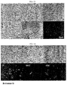
- amniotic epithelial cells increase in size depending on culture conditions, while the cytosol is increased, which causes trophoblastic change, or they are likely to undergo deformations associated with epithelio-mesenchymal transition by which epithelial cells undergo dramatic morphology changes into mesenchymal stem cell (MSC) type ( FIG. 12 ).
- MSC mesenchymal stem cell
- amniotic epithelial cell-derived adult stem cells treated with DTT and cultured in a ROCK inhibitor-containing medium continuously maintain a simple cuboidal shape (the bottommost photograph of FIG. 12 ).
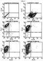
- Immunological characteristics of amniotic epithelial cell-derived adult stem cells according to the present invention were analyzed using a flow cytometer and, as a result, cells were positive for CD9, CD29, CD49f, CD73, CD90 and CD105, and negative for CD31, CD34, CD45 and CD133 ( FIGS. 9 and 10 ).
- amniotic epithelial cells have characteristics most similar to those of embryonic stem cells as already known in the art, and thus the inventive adult stem cells derived from the amniotic epithelial cells also have differentiation ability similar to that of embryonic stem cells, that is, the ability to differentiate into endoderm-, mesoderm- and ectoderm-derived cells.
- the inventive adult stem cells to differentiate into mesoderm-derived adipocytes, osteoblasts and myocytes and ectoderm-derived neurocytes, was confirmed.
- a cell therapeutic agent for treating wounds in the body and on the surface of the body, which contains adult stem cells as an active ingredient.
- wound means a bodily injury cauded by physical means which resulted in disruption of the continuity of structures and is meant to include destruction, cutting or rupture, etc. of the skin, mucosa or bone tissue.
- the cell therapeutic agent for treating wounds which contains adult stem cells as an active ingredient, can be administered directly to the diseased site in addition to parenteral administration in the form of intravenous or intramuscular injections.
- the cell therapeutic agent may be administered by various routes, including transdermal, subcutaneous, intravenous and intramuscular routes.
- Formulations for parenteral administration include sterile aqueous or non-aqueous solutions, suspensions, emulsions, etc.
- non-aqueous solution and suspension propylene glycol, polyethylene glycol, vegetable oil such as olive oil, injectable esters such as ethyloleate or the like may be used.
- the cell therapeutic agent may be administered, generally at a dose of 10 4 -10 10 cells/body, and preferably 10 6 -10 8 cells/body, once or several times a day.
- the actual dose of the active ingredients should be determined by various related factors such as administration route disease to be treated, patient's age, sex and weight, and the severity of disease.
- the above-mentioned dose ranges of the active ingredients do not limit the scope of the present invention in any way.
- the adult stem cells can be advantageously used for tissue repair and wound healing.
- the amniotic epithelial cell-derived adult stem cells possessing characteristics similar to those of embryonic stem cells have the ability to differentiate into endodermal, mesodermal and ectodermal cells, and thus can be used for wound healing and tissue repair and regeneration.
- the adult stem cells can differentiate, and proliferate and regenerate into bone, cartilage, muscle, ligament and/or nerve tissues, and thus are useful as a cell therapeutic agent.
- Example 1 Isolation of amniotic epithelial cells from amniotic tissue by treatment with DTT (dithiothreitol) and trypsin
- Amniotic membranes were collected at normal births and premature births in Korea University Guro Hospital according to the Institutional Review Board Guidelines of Korea University and used for research purposes. Amniotic tissue required in experiments was isolated from the placenta, and the isolated tissue was stored in an antibiotic-containing physiological saline or DMEM medium.
- the chemically degraded tissue was vortexed for 1 minute, and then it was primarily filtered through a 1-mm mesh and secondarily filtered through a 100- ⁇ m mesh, thus removing non-degraded tissue.
- the filtered tissue was centrifuged at 1800 rpm for 10 minutes. The single cell pellets at the bottom were well suspended, thus obtaining amniotic epithelial cells.
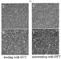
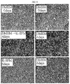
- the number of amniotic epithelial cells isolated by treatment with DTT (A) and trypsin and the number of cells after the culture of the amniotic epithelial cells (C) were significantly larger than the number of amniotic epithelial cells isolated by treatment with trypsin alone without DTT treatment (B) and the number of cells after the culture thereof (D). From this, it was found that, when amniotic tissue was treated with DTT, excessive cell disruption could be inhibited and, in addition, mucus interfering with the effect of trypsin treatment to be treated thereafter could be removed, thus further increasing the yield of amniotic epithelial cells. Furthermore, it was confirmed that, as the initial yield of amniotic epithelial cells was increased, the amount of adult stem cells which could be obtained by culturing the amniotic epithelial cells was also significantly increased.
- amniotic epithelial cells were cultured in a medium (Gibco) consisting of a 1:1 mixture of DMEM (Dulbecco modified Eagle medium) and F-12 (nutrient mixture) to which FBS, ascorbic acid, epidermal growth factor, insulin, an antibiotic and 10 ⁇ M of ROCK inhibitor Y-27632 have been added at the concentrations shown in Table 2 below.
- a medium Gibco
- DMEM Dulbecco modified Eagle medium
- F-12 nutrient mixture
- Table 2 Component Concentration Component Concentration Basal Media DMEM/F-12 1:1 mixture Insulin 5ug/ml FBS 10%(v/v) Ascorbic acid 0.2mM EGF 20ng/ml Antibiotic-antimycotic 1X
- the adult stem cells derived from the cultured amniotic epithelial cells were washed with HBSS buffer, and then incubated with Tryple-express (Gibco) or 0.25% trypsin-EDTA at 37 °C for 10 minutes. An FBS-containing medium was added thereto to inactivate trypsin, and then 5 x 10 6 stem cells derived from the amniotic epithelial cells were obtained and seeded into a fresh medium and subcultured. For each reseeding for subcultures, 10 ⁇ M of ROCK inhibitor was added to each fresh medium.
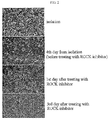
- FIG. 2 The results of the culture of the amniotic epithelial stem cell-derived adult stem cells carried out according to the above procedure are shown in FIG. 2 .
- the number of the adult stem cells was decreased, but when the ROCK inhibitor was added 4 days after the isolation of the amniotic epithelial cells, and then cultured, the number of the adult stem cells was rapidly increased.
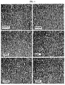
- amniotic epithelial cell-derived adult stem cells In order to examine the proliferation potential of amniotic epithelial cell-derived adult stem cells at various concentrations of ROCK inhibitor, the amniotic epithelial cells obtained in Example 1 were subcultured in DMEM/F-12 media containing FBS, ascorbic acid, epidermal growth factor, insulin and an antibiotic, to which ROCK inhibitor Y-27632 has been added at various concentrations of 100 ⁇ M, 10 ⁇ M, 1 ⁇ M, 100 nM and 10 nM. After 1 day and 4 days of the subculture, the optimum ROCK inhibitor Y-27632 concentration for the culture of the adult stem cells was examined.
- the degree of proliferation did not greatly differ from that in the control not treated with the ROCK inhibitor, and in the group treated with 100 ⁇ M of the ROCK inhibitor, the proliferation of the cells was significantly increased, but a cell morphology change or cell differentiation appeared.
- the preferred concentration of the ROCK inhibitor was in the range from 10 nM to 100 ⁇ M, and the most preferred concentration was about 10 ⁇ M.
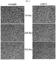
- Example 1 In order to examine the effect of ROCK inhibitor, 4x10 6 amniotic epithelial cells obtained in Example 1 were subcultured in a DMEM/F-12 medium containing FBS, ascorbic acid, epidermal growth factor, insulin and an antibiotic for 4 days. Then, the cells were seeded into each of a medium containing 10 ⁇ M of the ROCK inhibitor Y-27632 and a medium containing no ROCK inhibitor and were observed 1 day, 2 days and 3 days after seeding.
- Trypsin treatment which has been conventionally used in subculture has a problem in that the efficiency of culture is reduced due to the toxicity of trypsin, but it could be seen that the addition of the ROCK inhibitor activated the proliferation of adult stem cells by preventing apoptosis caused by trypsin treatment and inhibiting cell differentiation, while it assisted in the stable proliferation of undifferentiated adult stem cells.
- Example 3 Comparison of the abilities of stem cells to proliferate according to ROCK inhibitor treatment period
- Example 4 Increase in stem cell proliferation ability according to replacement of medium during culture of amniotic epithelial cells
- the amniotic epithelial cells obtained in Example 1 were cultured in each of DMEM medium and K-SFM medium for 3 days, 6 days and 8 days, respectively.
- the DMEM medium was supplemented with FBS, epidermal growth factor, ascorbic acid, nonessential amino acids and an antibiotic
- the K-SFM medium was supplemented with FBS, epidermal growth factor, ascorbic acid, hydrocortisone, NAC, insulin and an antibiotic.
- FIG. 7 it was observed that the proliferation ability of the amniotic epidermal cell-derived adult stem cells was higher in the DMEM medium than in the K-SFM medium.
- the adult stem cells cultured in the replaced K-SFM medium showed higher proliferation ability compared to the adult stem cells cultured in the DMEM medium alone without replacement of the medium.
- the amniotic epithelial cell-derived adult stem cells cultured in Example 2 were characterized by surface CD series antigen markers.
- CD9 epithelial cell, adhesion
- CD29 monoonuclear cell marker
- CD31 endothelial cell and stem cell marker
- CD34 hematopoietic stem cell marker
- CD45 PTPR, ASV, Leukocyte marker
- CD49f integratedin alpha 6 marker
- CD73 CD90
- CD105 TGF beta 1 marker
- CD 133 hematopoietic stem marker
- the amniotic epithelial cell-derived adult stem cells obtained in Example 2 were washed with PBS and treated with trypsin. Then, the cells were collected and centrifuged at 1500 rpm for 5 minutes. After the supernatant was discarded, the cells were added to a blocking buffer solution (5% serum (normal goat serum + normal horse serum) and incubated at 4 °C for 60 minutes, followed by centrifugation at 1500 rpm for 5 minutes. After the supernatant was discarded, the cells were suspended in PBS and dispensed into each well at a cell density of 1 x 10 5 cells which is the same number of a negative control and CD antigen markers.
- a blocking buffer solution 5% serum (normal goat serum + normal horse serum)
- the cells were stained with FITC (fluorescein isothiocyanate)-conjugated anti-human CD9 (Becton-Dickinson), CD34 (Becton-Dickinson), CD45 (Becton-Dickinson), CD105 (Becton-Dickinson) and PE (phycoerythrin)-conjugated anti-human CD29 (Becton-Dickinson), CD31 (Becton-Dickinson), CD49f (Becton-Dickinson), CD73 (Becton-Dickinson), CD90 (Becton-Dickinson) and CD133 (Miltenyi biotec). A replicate sample was used as an unstained control to ensure specificity.
- FITC fluorescein isothiocyanate
- amniotic epithelial cell-derived adult stem cells were positive for CD9, CD29, CD49f, CD73, CD90 and CD105, and were negative for CD31, CD34, CD45 and CD133.
- the amniotic epithelial cell-derived adult stem cells obtained in Example 2 were washed three times with PBS and fixed with 4% paraformaldehyde-containing PBS at room temperature for 30 minutes. After the cells were washed three times with PBS for 3 minutes each time, the cells were subjected to permeabilization with 0.1% Triton-X100-containing PBS at room temperature for 10 minutes. After the cells were washed three times with PBS for 3 minutes each time, the cells were incubated with blocking buffer (2.5% serum solution, NSG+NHS) at room temperature for 30 minutes and incubated with primary antibody-containing PBS at room temperature for 1 hour.
- blocking buffer (2.5% serum solution, NSG+NHS
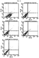
- the cells were washed three times with PBS for 5 minutes each time and incubated with a secondary antibody at room temperature for 30 minutes in dark conditions. The cells were washed three times with PBS, and then mounted. The cells thus obtained were examined for specificity of SSEA 4, Oct-4, Tra-1-60, Tra-1-81, Cytokeratin and E-cadherin.
- the adult stem cells according to the present invention were positive for SSEA 4, Oct-4, Tra-1-60 and Tra-1-81, which can be considered to be undifferentiated cell markers, that is, stem cell markers. Also, the adult stem cells were positive for Cytokeratin and E-cadherin, which can be considered to be epithelial cell markers.
- the amniotic epithelial cell-derived adult stem cells obtained in Example 2 were taken out of the incubator and observed under a Zeiss Axiovert 200 fluorescence microscope at 100x magnification. Also, the cells were photographed with an AxioCam MRm CCD mounted on the microscope. The diameter of the cells was measured using provided AxioVision ver. 4.5 program, and the size and nucleus of the cells and the size of the cytoplasm were examined from the photograph, thus determining the morphology of the amniotic epithelial cell-derived stem cells.
- the adult stem cells according to the present invention maintained the typical morphology of rectangular parallelepiped- or dice-shaped, slightly rounded epithelial cells, and the size (diameter) of the cells during the culture was about 5-10 ⁇ m.
- Example 6 Differentiation of adult stem cells derived from amniotic epithelial cells
- amniotic epithelial cell-derived adult stem cells obtained in Example 2 were cultured in adipogenesis induction medium (Nonhematopoietic AdipoDiff Medium; Miltenyi Biotec) for 3 weeks (37 °C; 5% CO 2 ; medium replacement cycle: 3-4 days) to induce the differentiation of pluripotent stem cells into adipocytes. 21 days (3 weeks) after the start of the culture, the cells were analyzed using the Oil red O staining method. As a result, it could be observed that the amniotic epithelial cell-derived adult stem cells according to the present invention differentiated into adipocytes ( FIG. 13 ).
- adipogenesis induction medium Nonhematopoietic AdipoDiff Medium; Miltenyi Biotec
- the amniotic epithelial cell-derived adult stem cells obtained in Example 2 were cultured in osteogenesis induction medium (nonhematopoietic osteoDiff medium, Miltenyi Biotec) for 2 weeks (37°C; 5% CO 2 ; medium replacement cycle: 3-4 days) to induce the differentiation of pluripotent stem cells into osteogenic cells. 14 days (2 weeks) after the start of the culture, the cells were analyzed using the Alizalin red S staining method. As a result, it was observed that the amniotic epithelial cell-derived adult stem cells differentiated into osteogenic cells ( FIG. 13 ).
- osteogenesis induction medium nonhematopoietic osteoDiff medium, Miltenyi Biotec
- amniotic epithelial cell-derived adult stem cells obtained in Example 2 were cultured in myogenic induction medium (Skeletal Muscle Cell Medium, LONZA) for 3 weeks (37 °C ; 5% CO 2 ; medium replacement cycle: 3-4 days). 21 days (3 weeks) after the start of the culture, the cells were immunostained.
- amniotic epithelial cell-derived adult stem cells according to the present invention showed positive staining for myosin that is a skeletal muscle cell-specific antigen. This suggests that the amniotic epithelial cell-derived adult stem cells according to the present invention differentiated into myocytes ( FIG. 13 ).
- amniotic epithelial cell-derived adult stem cells obtained in Example 2 were cultured in neurogenic induction medium (Neural Progenitor Media Systems, LONZA) for 2 weeks (37 °C ; 5% CO 2 ; medium replacement cycle: 3-4 days) to induce differentiation into neurocytes. 14 days (2 weeks) after the start of the culture, the cells were immunostained.
- neurogenic induction medium Neurogenic induction medium (Neural Progenitor Media Systems, LONZA) for 2 weeks (37 °C ; 5% CO 2 ; medium replacement cycle: 3-4 days) to induce differentiation into neurocytes. 14 days (2 weeks) after the start of the culture, the cells were immunostained.
- amniotic epithelial cell-derived adult stem cells according to the present invention showed positive staining for GFAP (glial fibrillary acidic protein), O1 (oligodendrocyte marker) and MAP2 (microtubule associated protein) which are antigens specific for astrocytes in the nervous system. This suggests that the amniotic epithelial cell-derived adult stem cells according to the present invention differentiated into neurocytes.
- GFAP glial fibrillary acidic protein
- O1 oligodendrocyte marker
- MAP2 microtubule associated protein
- amniotic epithelial cell-derived adult stem cells were administered to an athymic nude mouse wound model by the intradermal route, and then the wound healing ability thereof was compared with a saline control group.
- mice 4-5-week-old Bald/c-nu Slc athymic nude mice (Central Laboratory Animals Inc., Korea) were used and allocated to 2 groups as shown in Table 3 below.
- Table 3 Group Dosage Volume ( ⁇ l) No. of animals Route of administration Control 0 50 (saline) 6 intra dermis AEpSC 2 ⁇ 10 5 cells 50 (saline) 3 intra dermis
- each of 50 ⁇ l of saline and 50 ⁇ l of 2 ⁇ 10 5 cells AEpSC was prepared in a 29G insulin syringe in a clean bench 30 minutes before the start of the test.
- FIG. 15 was obtained by placing circular filter paper of the same size as that of the wound on the wound, photographing the wound together with the circular filter paper as the reference pixel area.
- FIG. 16 The results of tissue biopsy in each group after the induced wound was recovered, are shown in FIG. 16 .
- FIG. 16 in the skin of the control, there was only fibrous tissue mass, whereas, in the group administered with the amniotic epithelial stem cells, a large amount of skin appendages such as hair follicles and sweat glands were observed in the recovered skin tissue.
- amniotic epithelial stem cells according to the present invention functioned to promote the reepithelialization of skin tissue and facilitate the regeneration of skin appendages.
- the human amniotic epithelial cell-derived stem cells according to the present invention are easily extracted compared to existing therapeutic stem cells such as umbilical cord blood stem cells and bone marrow stem cells, the initial yield thereof is increased through DTT treatment, and the proliferation rate thereof is significantly increased by culturing them in a ROCK inhibitor-containing medium.
- the method of the present invention can be used to efficiently prepare adult stem cells useful as cell therapeutic agents.
Landscapes
- Health & Medical Sciences (AREA)
- Life Sciences & Earth Sciences (AREA)
- Engineering & Computer Science (AREA)
- Organic Chemistry (AREA)
- Biomedical Technology (AREA)
- Chemical & Material Sciences (AREA)
- Bioinformatics & Cheminformatics (AREA)
- Genetics & Genomics (AREA)
- Developmental Biology & Embryology (AREA)
- Gynecology & Obstetrics (AREA)
- Reproductive Health (AREA)
- Zoology (AREA)
- General Health & Medical Sciences (AREA)
- Wood Science & Technology (AREA)
- Biotechnology (AREA)
- Pharmacology & Pharmacy (AREA)
- Cell Biology (AREA)
- Pregnancy & Childbirth (AREA)
- Chemical Kinetics & Catalysis (AREA)
- General Engineering & Computer Science (AREA)
- Medicinal Chemistry (AREA)
- General Chemical & Material Sciences (AREA)
- Biochemistry (AREA)
- Veterinary Medicine (AREA)
- Animal Behavior & Ethology (AREA)
- Nuclear Medicine, Radiotherapy & Molecular Imaging (AREA)
- Microbiology (AREA)
- Public Health (AREA)
- Rheumatology (AREA)
- Pain & Pain Management (AREA)
- Dermatology (AREA)
- Medicines Containing Material From Animals Or Micro-Organisms (AREA)
- Micro-Organisms Or Cultivation Processes Thereof (AREA)
Description
- The present invention relates to a method for isolating and culturing adult stem cells derived from human amniotic membrane in high yield, and more particularly to a method for obtaining a large amount of adult stem cells, the method comprising obtaining amniotic epithelial cells from human amniotic tissue in high yield by treatment with dithiothreitol (DTT) and a low concentration of trypsin and culturing the amniotic epithelial cells in a medium containing a Rho-associated kinase inhibitor.
- Biotechnology in 21st century presents the possibility of new solutions to food, environmental and health problems, with the ultimate object of promoting human prosperity. In recent years, the technology of using stem cells has been considered as a new way to treat incurable diseases. Previously, organ transplantation, gene therapy, etc., were presented for the treatment of incurable human diseases, but their efficient use has not been achieved due to immune rejection, short supply of organs, insufficient development of vectors, and an insufficient knowledge of disease genes.
- For this reason, with increasing interests in stem cell studies, it has been recognized that totipotent stem cells having the ability to form all the organs by proliferation and differentiation can not only treat most of diseases but also fundamentally heal organ injuries. Also, many scientists have suggested clinical applicability of stem cells for the regeneration of all the organs and the treatment of incurable diseases, including Parkinson's disease, various cancers, diabetes and spinal damages.
- Stem cells refers to cells having not only self-replicating ability but also the ability to differentiate into at least two cells, and can be divided into totipotent stem cells, pluripotent stem cells, and multipotent stem cells.
- Totipotent stem cells are cells having totipotent properties capable of developing into one perfect individual, and these properties are possessed by cells up to the 8-cell stage after the fertilization of an oocyte and a sperm. When these cells are isolated and transplanted into the uterus, they can develop into one perfect individual.
- Pluripotent stem cells, which are cells capable of developing into various cells and tissues derived from the ectodermal, mesodermal and endodermal layers, are derived from an inner cell mass located inside of blastocysts generated 4-5 days after fertilization. These cells are called "embryonic stem cells" and can differentiate into various other tissue cells but not form new living organisms.
- Multipotent stem cells, which are stem cells capable of differentiating into only cells specific to tissues and organs containing these cells, are involved not only in the growth and development of various tissues and organs in the fetal, neonatal and adult periods but also in the maintenance of homeostasis of adult tissue and the function of inducing regeneration upon tissue damage. Tissue-specific multipotent cells are collectively called "adult stem cells".
- Adult stem cells are obtained by taking cells from various human organs and developing the cells into stem cells and are characterized in that they differentiate into only specific tissues. However, recently, experiments for differentiating adult stem cells into various tissues, including liver cells, succeeded and thus are receiving attention.
- The multipotent stem cells were first isolated from adult bone marrow (Jiang et al., Nature, 418:41, 2002), and then also found in other various adult tissues (Verfaillie, Trends Cell Biol., 12:502, 2002). In other words, although bone marrow is the most widely known source of stem cells, the multipotent stem cells were also found in the skin, blood vessels, muscles and brains (Tomas et al., Nat. Cell Biol., 3:778, 2001; Sampaolesi et al., Science, 301:487, 2003; Jiang et al., Exp. Hematol., 30:896, 2002). However, stem cells are very rarely present in adult tissues, such as bone marrow, and such cells are difficult to culture without inducing differentiation, and thus difficult to culture in the absence of specifically screened media. Namely, it is very difficult to maintain the isolated stem cells in vitro.
- Meanwhile, the results of studies on the isolation of mesenchymal stem cells from fetal tissue revealed that there are abundant mesenchymal stem cells in fetal tissue. However, the use of fetal tissue as a source of cell therapeutic agents has been limited due to ethical concerns. Mesenchymal stem cells were also isolated from umbilical cord blood (UCB) as the source of fetal mesenchymal stem cells (MSCs), but their numbers were very small, and they show poor proliferation.
- On the other hand, in the case of placenta stem cells which are receiving a great deal of attention recently due to excellent differentiation potential and safety, mesenchymal stem cells can be extracted in an
amount 100 times larger than that of those from umbilical cord blood. Moreover, umbilical cord blood can be used only once, and donor umbilical cord blood is additionally needed after the age of 15 years, whereas placenta stem cells can be used several times, and thus can also be used in adults. In addition, placenta stem cells can be used for a wider range of diseases. Hematopoietic stem cells of umbilical cord blood are mainly used for blood diseases, whereas placenta stem cells can be advantageously used for treating cellular injury and can be used in the future for treating many diseases, including heart failure, stroke, diabetes, osteoporosis, degenerative arthritis and the like. - However, in the case of using the placenta as the source of adult stem cells, is difficult to use placenta tissue collected immediately after delivery, whenever needed, and in fact, for the industrial application (placenta injection, placenta cosmetic products, etc.) of placenta tissue, placenta tissue collected after delivery is cold stored for use. Placenta-derived stem cells present in an undifferentiated state can be obtained in a large amount only from the placenta collected immediately after delivery, and it is very difficult to obtain a large amount of adult stem cells from placenta tissue several hours after delivery or particularly from placenta tissue that has been cold-stored for a long period of time.
- For this reason, in order to use placenta-derived adult stem cells in industrial applications there is an urgent need to develop technologies for preparing a large amount of adult stem cells from cold-stored placenta tissue or the like. Among adult stem cells, it is known that adult stem cells derived from amniotic epithelial cells possess properties most similar to the properties of embryonic stem cells, and thus have the capability to differentiate into various cells, while they are clinically safe, because they do not cause cancer upon in vivo transplantation, unlike embryonic stem cells (Miki et al., Stem Cell, 23:1549, 2005). The adult stem cells derived from amniotic epithelial cells are more likely to proceed to apoptosis during the culture and subculture of single cells, unlike adult stem cells derived from other placenta tissues. For this reason, it is difficult to induce proliferation of undifferentiated adult stem cells derived from amniotic epithelial cells in large numbers using conventional culture methods (http://www.cellapplications.com/HumanCells/HPlEpC.htm).
-
WO 2007/079183 discloses placenta stem cells and placental stem cell populations and methods of obtaining said stem cells. -
WO 2007/047468 discloses a method of suppressing an immune response comprising contacting a plurality of immune cells with a plurality of placental stem cells for a time sufficient for said placental stem cell to detectibly suppress an immune response, wherein said placental stem cells detectibly suppress T-cell proliferation in a mixed lymphocyte reaction assay. -
US 2007/0243172 discloses a method for producing placental stem cells comprising culturing finely cut amnion, chorion, decidua or placenta tissue in a medium containing collagenase and bFGF and collecting the cultured cells. - VAN DER HEIJDEN P J ET AL: "Improved procedure for the isolation of functionally active lymphoid cells from the murine intestine", JOURNAL OF IMMUNOLOGICAL METHODS, ELSEVIER SCIENCE PUBLISHERS B.V., AMSTERDAM, NL LNKD- DOI:10.1016/0022-1759(87) 90285-7, vol. 103, no. 2, 5 November 1987 (1987-11-05), pages 161-167,
XP023992484 discloses an isolation procedure for functionally active lamina propria lymphoid cells from the murine intestine. The procedure involves EDTA dithiothreitol incubation of intestinal tissue to remove epithelial and intraepithelial cells followed by collagenase digestion of the basement membrane to liberate part of the LPL. - Accordingly, the present inventors have made many efforts to prepare a large amount of undifferentiated adult stem cells derived from amniotic epithelial cells for practical applications thereof and, as a result, have found that a large amount of stem cells derived from amniotic epithelial cells can be obtained from cold-stored placenta tissue by using DTT and a ROCK inhibitor to isolate and culture amniotic epithelial cells and changing the composition of culture medium, thereby completing the present invention.
- It is an object of the present invention to provide a method for isolating amniotic epithelial cells from human amniotic tissue with high yield and a method for preparing adult stem cells derived from the isolated amniotic epithelial cells.
- Another object of the present invention is to provide adult stem cells derived from human amniotic epithelial cells and a cell therapeutic agent for wound healing, which contains the adult stem cells as an active ingredient.
- To achieve the above objects, the present invention provides a method for isolating amniotic epithelial cells, comprising the steps of: treating human amniotic tissue from the placenta harvested immediately after delivery with 5-50 mM dithiothreitol (DTT); treating the DTT-treated human amniotic tissue with 0.025-0.125% trypsin, and isolating the amniotic epithelial cells from amniotic tissue.
- The present invention also provides a method for preparing adult stem cells derived from human amniotic epithelial cells, the method comprising the steps of: culturing human amniotic epithelial cells isolated according to said method in a medium containing a Rho-associated kinase (ROCK) inhibitor; and collecting stem cells from the culture broth.
- The present invention also provides a method for proliferating and maintaining adult stem cells derived from human amniotic epithelial cells, the method comprising subculturing human amniotic epithelial cell-derived adult stem cells prepared according to said method in a medium containing a Rho-associated kinase (ROCK) inhibitor.
- Further described are human amniotic epithelial tissue-derived adult stem cells prepared by said method and a cell therapeutic agent for wound healing, which contains the adult stem cells as an active ingredient.
- Other features and embodiments of the present invention will be more apparent from the following detailed description and the appended claims.
-
-
FIG. 1 shows a comparison between the yields of amniotic epithelial cells after treatment with or without dithiothreitol (DTT) [A: amniotic epithelial cells obtained by treatment with DTT and trypsin; B: single cells obtained by treatment with trypsin alone; C: adult stem cells obtained by culturing "A"; and D: adult stem cells obtained by culturing "B"]. -
FIG. 2 is photographs showing enhanced ability of amniotic epithelial cell-derived adult stem cells cultured in the presence of a ROCK inhibitor. -
FIG. 3 is photographs showing a comparison of the enhanced ability of adult stem cells to proliferate, 1 day after treatment with the ROCK inhibitor at various concentrations. -
FIG. 4 is photographs showing a comparison of the enhanced ability of adult stem cells to proliferate, 4 days after treatment with the ROCK inhibitor at various concentrations. -
FIG. 5 shows the results obtained by comparing the ability of stem cells to proliferate when cultured with/without the ROCK inhibitor. -
FIG. 6 is photographs showing the results obtained by comparing the ability of amniotic epithelial cell-derived adult stem cells to proliferate according to the ROCK inhibitor treatment period (Control: cells cultured for 1 day without treatment with the ROCK inhibitor; A: cells treated with the ROCK inhibitor only before reseeding and cultured for 1 day, and B: cells treated with the ROCK inhibitor even after reseeding and cultured for 1 day). -
FIG. 7 shows the results obtained by comparing the ability of amniotic epithelial cell-derived adult stem cells to proliferate by culturing them in each of DMEM medium and K-SFM medium for 3 days, 6 days and 8 days, respectively. -
FIG. 8 shows a comparison of enhanced ability of amniotic epithelial cell-derived adult stem cells to proliferate by the replacement of DMEM medium and K-SFM medium. -
FIGS. 9 and10 show the results of flow cytometric analysis of amniotic epithelial cell-derived adult stem cells. -
FIG. 11 is photographs showing the responses of amniotic epithelial cell-derived adult stem cells to SSEA4, Oct-4, Cytokeratin and E-cadherin. -
FIG. 12 is optical microscope photographs showing the morphological characteristics of amniotic epithelial cell-derived adult stem cells. -
FIG. 13 is photographs showing the adipogenesis, osteogenesis and myogenesis of amniotic epithelial cell-derived adult stem cells. -
FIG. 14 is a photograph showing the neurogenesis of amniotic epithelial cell-derived adult stem cells. -
FIG. 15 is a graphic diagram showing the results obtained by observation of wound closure rates in an animal wound model. -
FIG. 16 is H&E staining photographs of recovered wound skin tissue in a group administered with amniotic epithelial cell-derived adult stem cells. - In one aspect, the present invention relates to a method for isolating and preparing amniotic epithelial cells with high yield, the method comprising the steps of: treating human amniotic tissue from the placenta harvested immediately after delivery with 5-50 mM dithiothreitol (DTT); treating the DTT-treated human amniotic tissue with 0.025-0.125% trypsin, and isolating the amniotic epithelial cells from amniotic tissue. The present invention is characterized in that, rather than extracting various kinds of cells from amniotic tissue isolated from the placenta, only amniotic epithelial cells are extracted and isolated from the amniotic tissue. Amniotic epithelial cells develop from the epiblast by 8 days after fertilization and before gastrulation, and they maintain the plasticity of pregastrulation embryo cells.
- Thus, it was found that the amniotic epithelial cells express the same surface markers as those on embryonic stem cells. The results of immunohistochemical and genetic analysis revealed that adult stem cells derived from amniotic epithelial cells have the ability to differentiate into endoderm-derived, mesoderm-derived and ectoderm-derived cells and do not cause tumor formation in vivo (Miki et al., Stem Cell, 23:1549, 2005).
- However, adult stem cells derived from amniotic epithelial cells are more likely to proceed to apoptosis during the culture and subculture of single cells, unlike adult stem cells derived from other placenta tissues, and for this reason, it is difficult to induce proliferation of undifferentiated adult stem cells derived from amniotic epithelial cells in large numbers using conventional culture methods. In other words, amniotic epithelial cell-derived stem cells present in an undifferentiated state can be obtained in a large amount only from the placenta collected immediately after delivery, and it is very difficult to obtain a large amount of adult stem cells from placenta tissue several hours after delivery or particularly from placenta tissue that has been cold-stored for a long period of time. Due to such a problem, it is necessary to obtain a large amount of amniotic epithelial cells from the placenta harvested immediately after delivery or inhibit apoptosis of adult stem cells during cell culture.
- The surface of amniotic membrane isolated from the placenta is usually covered with blood and mucus. Such mucus and the like interfere isolation of single cells from amniotic membrane and function to reduce the effect of trypsin treatment in the prior art. Thus, it is required to remove impurities such as mucus before trypsin treatment, and thus create an environment in which the effect of trypsin used for the isolation of single cells can be maximized.
- The present invention relates to a method for obtaining a large amount of amniotic epithelial cells from the placenta harvested immediately after delivery by treating with dithiothreitol (DTT) before trypsin treatment and culturing the isolated cells in a large amount by inhibiting apoptosis of the isolated cells, and thus solves the above-described problem.
- DTT is a compound represented by the following
structural formula 1, which is generally used for the removal of disulfide bonds from proteins, inhibition of dimmer formation in DNA, the removal of mucus, the removal of debris or connective tissue, and the like, and it is a reagent added to many biological test compositions, but has not been used in a process of extracting stem cells.
- In the present invention, excessive cell disruption is prevented by substituting trypsin treatment, which is a conventional procedure for isolating single cells, with DTT treatment, and the yield of single cells is increased by removing mucus that interferes with the effect of trypsin treatment to be performed later.
- Herein, the concentration of DTT used for the treatment is 5-50 mM, and preferably 8-15 mM. In one embodiment of the present invention, 10 mM of DTT was used. If DTT is used at a concentration of less than 5 mM, it is difficult to obtain single cells with high yield, and if DTT is used at a concentration of more than 50 mM, apoptosis may occur due to excessive protein degradation.
- After human amniotic tissue is treated with DTT, it is treated with a low concentration of trypsin in order to isolate amniotic epithelial cells. The concentration of trypsin is 0.025-0.125%, and preferably 0.05-0.10%.
- In order to isolate various kinds of cells from amniotic tissue, treatment with trypsin at suitable concentrations corresponding thereto is required, and particularly, amniotic epithelial cells are obtained by treatment with a low concentration of trypsin. However, in the prior art, epithelial cells could not be effectively isolated with a low concentration of trypsin due to the interference caused by impurities such as mucus covering amniotic tissue, and for this reason, mesenchymal stem cells (MSCs) that can be isolated with a high concentration of trypsin or collagenase have been used generally.
- However, the present invention enables amniotic epithelial cells to be effectively isolated even with a low concentration of trypsin by removing mucus and the like though treatment of human amniotic tissue with DTT. Specifically, the present invention provides a method of isolating amniotic epithelial cells in high yield by treatment with a low concentration (0.025-0.125%) of trypsin, not treatment with a high concentration (0.25-0.5%) of trypsin.
- Herein, the yield of cells can be increased by applying a physical shear stress though an additional step of vortexing at 2500-3000 rpm for 30-60 seconds immediately after trypsin treatment. This additional process has an advantage in that it relatively shortens the trypsin treatment time and thus reduces cell damage caused by trypsin.
- After amniotic tissue is subjected to trypsin treatment and vortexing, the resulting material is filtered, thus obtaining desired amniotic epithelial cells. Preferably, the resulting material is primarily filtered through a 1-2 mm mesh, and as a result, the in vitro exposure time of cells can be reduced compared to the case in which a 100-µm strainer is used from the beginning, thus increasing the viability of cells, reducing the time taken for cell isolation, and reducing the use of the 100-µm cell strainer, which leads to cost reduction.
- When amniotic epithelial cells isolated according to the above method are cultured in a medium containing a Rho-associated kinase (ROCK) inhibitor, a large amount of adult stem cells can be obtained.
- Therefore, the present invention, in another aspect, relates to a method for preparing adult stem cells derived from human amniotic epithelial cells, the method comprising the steps of: culturing human amniotic epithelial cells isolated according to said method in a medium containing a Rho-associated kinase (ROCK) inhibitor; and collecting adult stem cells from the culture broth. Furthermore, the present invention relates to a method for proliferating and maintaining adult stem cells derived from human amniotic epithelial cells, the method comprising subculturing human amniotic epithelial cell-derived adult stem cells prepared according to said method in a medium containing a Rho-associated kinase (ROCK) inhibitor.
- A medium which is used in the culture of amniotic epithelial cell-derived adult stem cells in the present invention may be a conventional medium such as DMEM (dulbecco modified eagle medium) or K-SFM (keratinocyte serum free medium). The medium may contain ascorbic acid, epidermal growth factor (EGF), insulin, antibiotics, FBS (fetal bovine serum), etc. The antibiotics may be conventional antibiotics known in the art, and an example thereof may be antibiotic-antimycotic (Gibco).
- As the culture medium, for example, a DMEM (dulbecco modified eagle medium) containing FBS (fetal bovine serum), ascorbic acid, epidermal growth factor, nonessential amino acids and antibiotics, or a K-SFM (keratinocyte serum free medium) containing FBS, ascorbic acid, hydrocortisone, NAC (N-acetyl-L-cysteine), insulin and antibiotics may be used to culture amniotic epithelial stem cells. Herein, the proliferation of adult stem cells was shown to be somewhat higher when cultured in the DMEM medium than that when cultured in the K-SFM medium (
FIG. 7 ). - Particularly with respect to the media, the present inventors examined the ability of cells to proliferate through the experiment performed by replacement of the DMEM and K-SFM media and, as a result, found that, when the DMEM medium was replaced with the K-SFM, the ability of adult stem cells to proliferate was further increased compared to when only the DMEM or K-SFM medium was continuously used (not replaced) (
FIG. 8 ). In this process, the replacement of the DMEM medium with the K-SFM medium is preferably performed in the primary subculture of adult stem cells. - However, the culture medium for amniotic epithelial cell-derived adult stem cells is most preferably a mixed medium (Gibco) consisting of a 1:1 mixture of DMEM and F-12 (nutrient mixture). At this time, the mixed medium may also contain ascorbic acid, epidermal growth factor (EGF), insulin, antibiotics and FBS (fetal bovine serum). In one embodiment of the present invention, antimycotic-antibiotics (Gibco) were used as antibiotics.
- In the present invention, isolated single amniotic epithelial cells were primarily cultured in a medium containing a ROCK (Rho-associasted kinase) inhibitor to obtain adult stem cells, and then subcultured in the presence of the ROCK inhibitor, such that the adult stem cells are maintained in an undifferentiated state.
- The ROCK (Rho-associated kinase) inhibitor, which is a substance functioning to inhibit apoptosis, is known to inhibit agonist-induced Ca2+ sensitization of neurite regeneration, myosin phosphorylation and smooth muscle contraction. More specifically, the ROCK inhibitor is known to normalize the abnormal structure of muscle cells that causes hypertension and asthma and to have the function of increasing blood flow to the optic disc and continuously reducing intraocular pressure. With respect to biological properties, the ROCK inhibitor is known to have the function of inhibiting apoptosis and maintaining cells in an undifferentiated state. Recently, studies on the use of the ROCK inhibitor to increase the viability of isolated human embryonic stem cells were conducted (Watanabe et al., Nature Biotechnology, 25:681, 2007).
- However, it is obvious to those skilled in the art that embryonic stem cells and adult stem cells are clearly distinguished from each other thus the sources and differentiation abilities thereof clearly differ from each other. There has been no report confirming that proliferation of adult stem cells is increased by using a ROCK inhibitor. However, in the present invention, it was confirmed that the ability of adult stem cells to proliferate was increased by a ROCK inhibitor in the isolation and culture thereof, and also the efficiency of proliferating and maintaining amniotic epithelial cell-derived stem cells was further increased by the ROCK inhibitor.
- In the above study conducted by Watanabe et al., a method of treating embryonic stem cells with a ROCK inhibitor before reseeding the cells into a fresh medium for subculture was used. Specifically, after the embryonic stem cells treated with the ROCK inhibitor were reseeded on a fresh medium, they were cultured without treatment with the ROCK inhibitor (Watanabe et al., Nature Biotechnology, 25:681, 2007).
- On the other hand, in the present invention, a ROCK inhibitor was treated to the cells as reseeding on a fresh medium for subculture, thus establishing an environment in which the ROCK inhibitor was continuously present during the subculture of human amniotic epithelial cell-derived stem cells.
- Typical ROCK inhibitors that can be used in the present invention include Y-27632, HA-1077, Y-39983, Wf-536, etc., and among them, Y-27632 (Calbiochem or Sigma) was used in exemplary embodiments of the present invention. Y-27632 has the following structural formula 2:

- The concentration of the ROCK inhibitor which is used in treatment according to the present invention is preferably in the range from 10 nM to 100 µM. If the ROCK inhibitor is used at a concentration of less than 10 nM, it will be difficult to maintain an undifferentiated state adult stem cells for a long period of time, and if the ROCK inhibitor is used at a concentration of more than 100 µM, a cell morphology change may occur, and cells may enter into the differentiation process.
- Further described are amniotic epithelial cell-derived adult stem cells, obtained by the method as described above. The morphological and immunological characteristics of amniotic epithelial cell-derived adult stem cells, obtained by the method as described above will now be described as follows.
- Single amniotic epithelial cells isolated from amniotic tissue have the typical morphology of rectangular parallelepiped- or dice-shaped, slightly rounded epithelial cells, and the size of the cells during the culture is about 5-10 µm. Generally, amniotic epithelial cells increase in size depending on culture conditions, while the cytosol is increased, which causes trophoblastic change, or they are likely to undergo deformations associated with epithelio-mesenchymal transition by which epithelial cells undergo dramatic morphology changes into mesenchymal stem cell (MSC) type (
FIG. 12 ). However, in the present invention, amniotic epithelial cell-derived adult stem cells treated with DTT and cultured in a ROCK inhibitor-containing medium continuously maintain a simple cuboidal shape (the bottommost photograph ofFIG. 12 ). - Immunological characteristics of amniotic epithelial cell-derived adult stem cells according to the present invention were analyzed using a flow cytometer and, as a result, cells were positive for CD9, CD29, CD49f, CD73, CD90 and CD105, and negative for CD31, CD34, CD45 and CD133 (
FIGS. 9 and10 ). - It was found that, in the amniotic epithelial cell-derived adult stem cells of the present invention, cytokeratin and epithelium (E)-cadherin which indicate the characteristics of epithelial cells were expressed, and
SSEA 4, Oct-4, Tra-1-60 and Tra-1-81 which are known as markers of totipotent stem cells were also expressed (FIG. 11 ). - Moreover, with respect to the differentiation ability of stem cells, amniotic epithelial cells have characteristics most similar to those of embryonic stem cells as already known in the art, and thus the inventive adult stem cells derived from the amniotic epithelial cells also have differentiation ability similar to that of embryonic stem cells, that is, the ability to differentiate into endoderm-, mesoderm- and ectoderm-derived cells. In the present Example 6, the ability of the inventive adult stem cells to differentiate into mesoderm-derived adipocytes, osteoblasts and myocytes and ectoderm-derived neurocytes, was confirmed.
- Further disclosed is a cell therapeutic agent for treating wounds in the body and on the surface of the body, which contains adult stem cells as an active ingredient.
- As used herein, the term "wound" means a bodily injury cauded by physical means which resulted in disruption of the continuity of structures and is meant to include destruction, cutting or rupture, etc. of the skin, mucosa or bone tissue.
- The cell therapeutic agent for treating wounds, which contains adult stem cells as an active ingredient, can be administered directly to the diseased site in addition to parenteral administration in the form of intravenous or intramuscular injections. The cell therapeutic agent may be administered by various routes, including transdermal, subcutaneous, intravenous and intramuscular routes.
- Formulations for parenteral administration include sterile aqueous or non-aqueous solutions, suspensions, emulsions, etc. For the non-aqueous solution and suspension, propylene glycol, polyethylene glycol, vegetable oil such as olive oil, injectable esters such as ethyloleate or the like may be used.
- For humans, the cell therapeutic agent may be administered, generally at a dose of 104-1010 cells/body, and preferably 106-108 cells/body, once or several times a day. However, it is to be understood that the actual dose of the active ingredients should be determined by various related factors such as administration route disease to be treated, patient's age, sex and weight, and the severity of disease. Thus, the above-mentioned dose ranges of the active ingredients do not limit the scope of the present invention in any way.
- The adult stem cells can be advantageously used for tissue repair and wound healing. The amniotic epithelial cell-derived adult stem cells possessing characteristics similar to those of embryonic stem cells have the ability to differentiate into endodermal, mesodermal and ectodermal cells, and thus can be used for wound healing and tissue repair and regeneration. In addition, the adult stem cells can differentiate, and proliferate and regenerate into bone, cartilage, muscle, ligament and/or nerve tissues, and thus are useful as a cell therapeutic agent.
- Hereinafter, the present invention will be described in further detail with reference to examples. It is to be understood, however, that these examples are for illustrative purposes only and are not to be construed to limit the scope of the present invention.
- Various media and reagents used in the following examples were purchased from the companies shown in Table 1 below.
Table 1 Items Brand Catalogue No. Media DMEM/F-12 Gibco 11320 EGF Sigma E 9644 Peprotech 100-15 Gibco 13247-051 FBS Gibco 16000 Insulin Sigma I 1882 Ascorbic acid Sigma A 8960 Antibiotic-antimycotic Gibco 15240 ROCK inhibitor Y-27632 Calbiochem CNB 688000 Sigma Y 0503 Trypsin solution Trypsin-EDTA Gibco 15400 L/G DMEM Gibco 11885 Buffer solution HBSS Gibco 14175 Subculture TrypLE-Express Gibco 12604 DTT DTT Gibco 15508-013 - Amniotic membranes were collected at normal births and premature births in Korea University Guro Hospital according to the Institutional Review Board Guidelines of Korea University and used for research purposes. Amniotic tissue required in experiments was isolated from the placenta, and the isolated tissue was stored in an antibiotic-containing physiological saline or DMEM medium.
- 5 g of amniotic tissue was weighed and washed with HBSS. For stable isolation of amniotic epithelial cells, the washing process was carried out using HBSS buffer more similar to the constituents of the human body rather than using general PBS. The washed amniotic tissue was transferred into a 50-ml tube, and 10 mM DTT was added thereto. After treatment with DTT for 30 minutes, the supernatant was discarded, the amniotic tissue was finely cut, and the finely cut amniotic tissue was treated with fresh 0.05% trypsin-EDTA at 37 °C for 42 minutes under stirring, thus chemically degrading the tissue. The chemically degraded tissue was vortexed for 1 minute, and then it was primarily filtered through a 1-mm mesh and secondarily filtered through a 100-µm mesh, thus removing non-degraded tissue. The filtered tissue was centrifuged at 1800 rpm for 10 minutes. The single cell pellets at the bottom were well suspended, thus obtaining amniotic epithelial cells.
- Living cells were visually counted under a microscope using a hemocytometer according to trypan blue dye method and, as a result, the number of cells counted immediately after isolation was 3.72 X 10⁷ cells (
FIG. 1 (A) ). - On the other hand, in the case in which amniotic epithelial cells were isolated from amniotic tissue by treatment with trypsin alone without the addition of DTT, the number of cells counted immediately after isolation was 9.25 X 10⁵ cells (
FIG. 1 (B) ). - As a result, as shown in
FIG. 1 , the number of amniotic epithelial cells isolated by treatment with DTT (A) and trypsin and the number of cells after the culture of the amniotic epithelial cells (C) were significantly larger than the number of amniotic epithelial cells isolated by treatment with trypsin alone without DTT treatment (B) and the number of cells after the culture thereof (D). From this, it was found that, when amniotic tissue was treated with DTT, excessive cell disruption could be inhibited and, in addition, mucus interfering with the effect of trypsin treatment to be treated thereafter could be removed, thus further increasing the yield of amniotic epithelial cells. Furthermore, it was confirmed that, as the initial yield of amniotic epithelial cells was increased, the amount of adult stem cells which could be obtained by culturing the amniotic epithelial cells was also significantly increased. - The obtained amniotic epithelial cells were cultured in a medium (Gibco) consisting of a 1:1 mixture of DMEM (Dulbecco modified Eagle medium) and F-12 (nutrient mixture) to which FBS, ascorbic acid, epidermal growth factor, insulin, an antibiotic and 10 µM of ROCK inhibitor Y-27632 have been added at the concentrations shown in Table 2 below.
Table 2 Component Concentration Component Concentration Basal Media DMEM/F-12 1:1 mixture Insulin 5ug/ ml FBS 10%(v/v) Ascorbic acid 0.2mM EGF 20ng/ml Antibiotic-antimycotic 1X - After 3 days of culture, the adult stem cells derived from the cultured amniotic epithelial cells were washed with HBSS buffer, and then incubated with Tryple-express (Gibco) or 0.25% trypsin-EDTA at 37 °C for 10 minutes. An FBS-containing medium was added thereto to inactivate trypsin, and then 5 x 10⁶ stem cells derived from the amniotic epithelial cells were obtained and seeded into a fresh medium and subcultured. For each reseeding for subcultures, 10 µM of ROCK inhibitor was added to each fresh medium.
- The results of the culture of the amniotic epithelial stem cell-derived adult stem cells carried out according to the above procedure are shown in
FIG. 2 . As can be seen inFIG. 2 , before the addition of the ROCK inhibitor, the number of the adult stem cells was decreased, but when the ROCK inhibitor was added 4 days after the isolation of the amniotic epithelial cells, and then cultured, the number of the adult stem cells was rapidly increased. - In order to examine the proliferation potential of amniotic epithelial cell-derived adult stem cells at various concentrations of ROCK inhibitor, the amniotic epithelial cells obtained in Example 1 were subcultured in DMEM/F-12 media containing FBS, ascorbic acid, epidermal growth factor, insulin and an antibiotic, to which ROCK inhibitor Y-27632 has been added at various concentrations of 100 µM, 10 µM, 1 µM, 100 nM and 10 nM. After 1 day and 4 days of the subculture, the optimum ROCK inhibitor Y-27632 concentration for the culture of the adult stem cells was examined.
- As a result, as shown in
FIG. 3 andFIG. 4 , in the group treated with 10 nM of the ROCK inhibitor, the degree of proliferation did not greatly differ from that in the control not treated with the ROCK inhibitor, and in the group treated with 100 µM of the ROCK inhibitor, the proliferation of the cells was significantly increased, but a cell morphology change or cell differentiation appeared. Thus, it was consirmed that the preferred concentration of the ROCK inhibitor was in the range from 10 nM to 100 µM, and the most preferred concentration was about 10 µM. - In order to examine the effect of ROCK inhibitor, 4x10⁶ amniotic epithelial cells obtained in Example 1 were subcultured in a DMEM/F-12 medium containing FBS, ascorbic acid, epidermal growth factor, insulin and an antibiotic for 4 days. Then, the cells were seeded into each of a medium containing 10 µM of the ROCK inhibitor Y-27632 and a medium containing no ROCK inhibitor and were observed 1 day, 2 days and 3 days after seeding.
- As a result, as can be seen in
FIG. 5 , when the obtained amniotic epithelial cells were cultured in the absence of the ROCK inhibitor, the proliferation of adult stem cells was very slow due to apoptosis. However, when the amniotic epithelial cells were cultured in the presence of the ROCK inhibitor, apoptosis of the amniotic epithelial cells was inhibited, while the proliferation of adult stem cells was significantly increased. - Trypsin treatment which has been conventionally used in subculture has a problem in that the efficiency of culture is reduced due to the toxicity of trypsin, but it could be seen that the addition of the ROCK inhibitor activated the proliferation of adult stem cells by preventing apoptosis caused by trypsin treatment and inhibiting cell differentiation, while it assisted in the stable proliferation of undifferentiated adult stem cells.
- In order to further improve the proliferation ability of the amniotic epithelial cells obtained in Example 1, the method used in the embryonic stem cell study (Watanabe et al., Nature Biotechnology, 25:681, 2007) and the method of adding ROCK inhibitor during the culture were compared. In other words, the culture in which 10 µM of ROCK inhibitor was added only before reseeding was compared with the culture in which the ROCK inhibitor was added even after reseeding.
- As a result, as can be seen in
FIG. 6 , in the case (A) of treatment with the ROCK inhibitor only before reseeding, the proliferation ability of the adult stem cells derived from the amniotic epithelial cells was clearly increased compared to the control without ROCK inhibitor treatment. Meanwhile, in the case (B) in which the ROCK inhibitor was continuously added during the reseeding and culture procedure, the proliferation ability of adult stem cells was superior compared to the case (A) in which the ROCK inhibitor was added only before reseeding. In other words, in the case (B) in which the ROCK inhibitor was continuously added even during the culture after reseeding, the proliferation ability of the amniotic epithelial cell-derived adult stem cells was most significantly increased. - The amniotic epithelial cells obtained in Example 1 were cultured in each of DMEM medium and K-SFM medium for 3 days, 6 days and 8 days, respectively.
- The DMEM medium was supplemented with FBS, epidermal growth factor, ascorbic acid, nonessential amino acids and an antibiotic, and the K-SFM medium was supplemented with FBS, epidermal growth factor, ascorbic acid, hydrocortisone, NAC, insulin and an antibiotic. As a result, as shown in
FIG. 7 , it was observed that the proliferation ability of the amniotic epidermal cell-derived adult stem cells was higher in the DMEM medium than in the K-SFM medium. - Meanwhile, diversified experiments were performed for the mass production of amniotic epithelial cell-derived adult stem cells. As a result, as can be seen in
FIG. 8 , the method of replacing the DMEM medium with the K-SFM medium in the primary subculture increased the proliferation of adult stem cells derived from amniotic epithelial cells. In addition, when the adult stem cells were cultured in the DMEM medium for 4 days and observed, the stem cells proliferated at almost similar rate. Moreover, when the DMEM medium was replaced with the K-SFM medium after 4 days of the primary subculture, and the cells were cultured in the replaced medium for 2 days, the adult stem cells cultured in the replaced K-SFM medium showed higher proliferation ability compared to the adult stem cells cultured in the DMEM medium alone without replacement of the medium. - The amniotic epithelial cell-derived adult stem cells cultured in Example 2 were characterized by surface CD series antigen markers. CD9 (epithelial cell, adhesion), CD29 (mononuclear cell marker), CD31 (endothelial cell and stem cell marker), CD34 (hematopoietic stem cell marker), CD45 (PTPR, ASV, Leukocyte marker), CD49f (integrin alpha 6 marker), CD73, CD90 (mononuclear stem cell marker), CD105 (
TGF beta 1 marker) and CD 133 (hematopoietic stem marker) were applied for FACS analysis. - The amniotic epithelial cell-derived adult stem cells obtained in Example 2 were washed with PBS and treated with trypsin. Then, the cells were collected and centrifuged at 1500 rpm for 5 minutes. After the supernatant was discarded, the cells were added to a blocking buffer solution (5% serum (normal goat serum + normal horse serum) and incubated at 4 °C for 60 minutes, followed by centrifugation at 1500 rpm for 5 minutes. After the supernatant was discarded, the cells were suspended in PBS and dispensed into each well at a cell density of 1 x 10⁵ cells which is the same number of a negative control and CD antigen markers. An R-phycoerythrin (PE)/FITC (fluorescein isothiocyanate)-conjugated mouse anti-human monoclonal antibody was added to each well, and the cells were incubated at 4 °C for 40 minutes. After incubation, the cells were centrifuged at 1500 rpm for 3 minutes. After the supernatant was removed, the cells were washed with PBS and centrifuged at 1500 rpm for 3 minutes. Then, after the supernatant was removed, the cells were washed with PBS and centrifuged at 1500 rpm for 3 minutes. After the supernatant was removed, the cells were analyzed using a flow cytometer.
- The cells were stained with FITC (fluorescein isothiocyanate)-conjugated anti-human CD9 (Becton-Dickinson), CD34 (Becton-Dickinson), CD45 (Becton-Dickinson), CD105 (Becton-Dickinson) and PE (phycoerythrin)-conjugated anti-human CD29 (Becton-Dickinson), CD31 (Becton-Dickinson), CD49f (Becton-Dickinson), CD73 (Becton-Dickinson), CD90 (Becton-Dickinson) and CD133 (Miltenyi biotec). A replicate sample was used as an unstained control to ensure specificity.
- As a result, as shown in
FIGS. 9 and10 , with respect to immunological characteristics, the amniotic epithelial cell-derived adult stem cells were positive for CD9, CD29, CD49f, CD73, CD90 and CD105, and were negative for CD31, CD34, CD45 and CD133. - The amniotic epithelial cell-derived adult stem cells obtained in Example 2 were washed three times with PBS and fixed with 4% paraformaldehyde-containing PBS at room temperature for 30 minutes. After the cells were washed three times with PBS for 3 minutes each time, the cells were subjected to permeabilization with 0.1% Triton-X100-containing PBS at room temperature for 10 minutes. After the cells were washed three times with PBS for 3 minutes each time, the cells were incubated with blocking buffer (2.5% serum solution, NSG+NHS) at room temperature for 30 minutes and incubated with primary antibody-containing PBS at room temperature for 1 hour. The cells were washed three times with PBS for 5 minutes each time and incubated with a secondary antibody at room temperature for 30 minutes in dark conditions. The cells were washed three times with PBS, and then mounted. The cells thus obtained were examined for specificity of
SSEA 4, Oct-4, Tra-1-60, Tra-1-81, Cytokeratin and E-cadherin. - As a result, as shown in
FIG. 11 , the adult stem cells according to the present invention were positive forSSEA 4, Oct-4, Tra-1-60 and Tra-1-81, which can be considered to be undifferentiated cell markers, that is, stem cell markers. Also, the adult stem cells were positive for Cytokeratin and E-cadherin, which can be considered to be epithelial cell markers. - The amniotic epithelial cell-derived adult stem cells obtained in Example 2 were taken out of the incubator and observed under a
Zeiss Axiovert 200 fluorescence microscope at 100x magnification. Also, the cells were photographed with an AxioCam MRm CCD mounted on the microscope. The diameter of the cells was measured using provided AxioVision ver. 4.5 program, and the size and nucleus of the cells and the size of the cytoplasm were examined from the photograph, thus determining the morphology of the amniotic epithelial cell-derived stem cells. - As a result, as shown in
FIG. 12 , the adult stem cells according to the present invention maintained the typical morphology of rectangular parallelepiped- or dice-shaped, slightly rounded epithelial cells, and the size (diameter) of the cells during the culture was about 5-10 µm. - The amniotic epithelial cell-derived adult stem cells obtained in Example 2 were cultured in adipogenesis induction medium (Nonhematopoietic AdipoDiff Medium; Miltenyi Biotec) for 3 weeks (37 °C; 5% CO2; medium replacement cycle: 3-4 days) to induce the differentiation of pluripotent stem cells into adipocytes. 21 days (3 weeks) after the start of the culture, the cells were analyzed using the Oil red O staining method. As a result, it could be observed that the amniotic epithelial cell-derived adult stem cells according to the present invention differentiated into adipocytes (
FIG. 13 ). - The amniotic epithelial cell-derived adult stem cells obtained in Example 2 were cultured in osteogenesis induction medium (nonhematopoietic osteoDiff medium, Miltenyi Biotec) for 2 weeks (37°C; 5% CO2; medium replacement cycle: 3-4 days) to induce the differentiation of pluripotent stem cells into osteogenic cells. 14 days (2 weeks) after the start of the culture, the cells were analyzed using the Alizalin red S staining method. As a result, it was observed that the amniotic epithelial cell-derived adult stem cells differentiated into osteogenic cells (
FIG. 13 ). - The amniotic epithelial cell-derived adult stem cells obtained in Example 2 were cultured in myogenic induction medium (Skeletal Muscle Cell Medium, LONZA) for 3 weeks (37 °C ; 5% CO2; medium replacement cycle: 3-4 days). 21 days (3 weeks) after the start of the culture, the cells were immunostained.
- As a result, the amniotic epithelial cell-derived adult stem cells according to the present invention showed positive staining for myosin that is a skeletal muscle cell-specific antigen. This suggests that the amniotic epithelial cell-derived adult stem cells according to the present invention differentiated into myocytes (
FIG. 13 ). - The amniotic epithelial cell-derived adult stem cells obtained in Example 2 were cultured in neurogenic induction medium (Neural Progenitor Media Systems, LONZA) for 2 weeks (37 °C ; 5% CO2; medium replacement cycle: 3-4 days) to induce differentiation into neurocytes. 14 days (2 weeks) after the start of the culture, the cells were immunostained.
- As a result, as shown in
FIG. 14 , the amniotic epithelial cell-derived adult stem cells according to the present invention showed positive staining for GFAP (glial fibrillary acidic protein), O1 (oligodendrocyte marker) and MAP2 (microtubule associated protein) which are antigens specific for astrocytes in the nervous system. This suggests that the amniotic epithelial cell-derived adult stem cells according to the present invention differentiated into neurocytes. - The amniotic epithelial cell-derived adult stem cells were administered to an athymic nude mouse wound model by the intradermal route, and then the wound healing ability thereof was compared with a saline control group.
- As the test animals, 4-5-week-old Bald/c-nu Slc athymic nude mice (Central Laboratory Animals Inc., Korea) were used and allocated to 2 groups as shown in Table 3 below.
Table 3 Group Dosage Volume (µℓ) No. of animals Route of administration Control 0 50 (saline) 6 intra dermis AEpSC 2×10⁵ cells 50 (saline) 3 intra dermis - After the animals were allocated to 2 groups as shown in Table 3, individual identification cards were attached to breeding cages, and each animal was separately bred in each breeding cage in order to identify each individual animal. As a test material, each of 50 µℓ of saline and 50 µℓ of 2×10⁵ cells AEpSC was prepared in a 29G insulin syringe in a
clean bench 30 minutes before the start of the test. - 1 hour before surgery, the weight of each test animal was measured, administered with an antibiotic and anesthetized. Then, the central portion of the back of each test animal was disinfected with alcohol cotton, and a full-thickness wound was induced at the central back portion using a 5 mm-diameter biopsy punch. Immediately after induction of the wound, a total of 50 µℓ of the prepared test material was injected into three intradermal sites near the wound. The locations of injection were spaced about 1 cm apart from the periphery of the wound in order to minimize the loss of the test material through the wound surface.
- To examine the wound healing effect, the condition of the animals was examined once or more everyday, and the wound portion was photographed immediately, 3 days, 6 days, 7 days, 9 days and 14 days after surgery. The analysis results are shown in
FIG. 15. FIG. 15 was obtained by placing circular filter paper of the same size as that of the wound on the wound, photographing the wound together with the circular filter paper as the reference pixel area. - As a result, as shown in
FIG. 15 , it could be observed that the wound closure rate of the group administered with the amniotic epithelial cell-derived stem cells was higher than that of the group administered with saline. - In addition, 21 days after surgery at which the induced wound was completely recovered, the test animals were autopsied and subjected to a skin biopsy. The results of tissue biopsy in each group after the induced wound was recovered, are shown in
FIG. 16 . As shown inFIG. 16 , in the skin of the control, there was only fibrous tissue mass, whereas, in the group administered with the amniotic epithelial stem cells, a large amount of skin appendages such as hair follicles and sweat glands were observed in the recovered skin tissue. - From such results, it could be found that the amniotic epithelial stem cells according to the present invention functioned to promote the reepithelialization of skin tissue and facilitate the regeneration of skin appendages.
- As described above in detail, the human amniotic epithelial cell-derived stem cells according to the present invention are easily extracted compared to existing therapeutic stem cells such as umbilical cord blood stem cells and bone marrow stem cells, the initial yield thereof is increased through DTT treatment, and the proliferation rate thereof is significantly increased by culturing them in a ROCK inhibitor-containing medium. Thus, the method of the present invention can be used to efficiently prepare adult stem cells useful as cell therapeutic agents.
Claims (11)
- A method for isolating amniotic epithelial cells, which comprises the steps of: treating human amniotic tissue from the placenta harvested immediately after delivery with 5-50 mM dithiothreitol (DTT); treating the DTT-treated human amniotic tissue with 0.025-0.125% trypsin, and isolating the amniotic epithelial cells from amniotic tissue.
- The method for isolating amniotic epithelial cells according to claim 1, which further comprises the step of vortexing the trypsin-treated human amniotic tissue.
- A method for preparing adult stem cells derived from human amniotic epithelial cells, the method comprising the steps of: culturing human amniotic epithelial cells isolated by the method of claim 1, in a medium containing a Rho-associated kinase (ROCK) inhibitor; and collecting stern cells from the culture broth.
- A method for proliferating and maintaining adult stem cells derived from human amniotic epithelial cells, the method comprising subculturing human amniotic epithelial cell-derived adult stem cells prepared by the method of claim 4, in a medium containing a Rho-associated kinase (ROCK) inhibitor.
- The method according to claim 3 or 4, wherein the concentration of the ROCK inhibitor is 10 nM to 100 µM.
- The method according to claim 3 or 4, wherein the ROCK inhibitor is a compound represented by the following structural formula:

- The method according to claim 3 or 4, wherein said medium is DMEM (dulbecco modified eagle medium) or K-SFM (keratinocyte serum free medium).
- The method according to claim 7 wherein said medium was replaced from DMEM to K-SFM in the primary subculture of adult stem cells.
- The method according to claim 3 or 4, wherein said medium is a mixed medium (Gibco) consisting of a 1:1 mixture of DMEM and F-12 (nutrient mixture).
- The method according to claim 9, wherein the medium additionally contains ascorbic acid, epidermal growth factor (EGF), insulin, antibiotics and FBS (fetal bovine serum).
- The method according to claim 4, wherein the ROCK inhibitor was added when reseeding the human amniotic epithelial cell-derived adult stem cells in the medium during the subculture.
Applications Claiming Priority (2)
| Application Number | Priority Date | Filing Date | Title |
|---|---|---|---|
| KR1020070114451A KR100795708B1 (en) | 2006-12-26 | 2007-11-09 | Method for isolating and culturing adult stem cells derived from human amniotic epithelium |
| PCT/KR2008/000172 WO2009061024A1 (en) | 2007-11-09 | 2008-01-10 | Method for isolating and culturing adult stem cells derived from human amniotic epithelium |
Publications (3)
| Publication Number | Publication Date |
|---|---|
| EP2205724A1 EP2205724A1 (en) | 2010-07-14 |
| EP2205724A4 EP2205724A4 (en) | 2011-01-05 |
| EP2205724B1 true EP2205724B1 (en) | 2016-04-27 |
Family
ID=40626115
Family Applications (1)
| Application Number | Title | Priority Date | Filing Date |
|---|---|---|---|
| EP08704712.2A Not-in-force EP2205724B1 (en) | 2007-11-09 | 2008-01-10 | Method for isolating and culturing adult stem cells derived from human amniotic epithelium |
Country Status (8)
| Country | Link |
|---|---|
| US (1) | US8455251B2 (en) |
| EP (1) | EP2205724B1 (en) |
| JP (1) | JP5319682B2 (en) |
| CN (1) | CN101765657A (en) |
| CA (1) | CA2692634C (en) |
| ES (1) | ES2581990T3 (en) |
| MX (1) | MX2010000393A (en) |
| WO (1) | WO2009061024A1 (en) |
Families Citing this family (30)
| Publication number | Priority date | Publication date | Assignee | Title |
|---|---|---|---|---|
| US8372437B2 (en) | 2006-08-17 | 2013-02-12 | Mimedx Group, Inc. | Placental tissue grafts |
| PL2247285T3 (en) * | 2007-11-30 | 2014-11-28 | Rnl Bio Co Ltd | Cellular therapeutic agent for incontinence of urine comprising stem cells originated from decidua |
| US8563307B2 (en) | 2009-02-24 | 2013-10-22 | James Wang | Treatment of immunosuppression-related disorders |
| US10130736B1 (en) | 2010-05-14 | 2018-11-20 | Musculoskeletal Transplant Foundation | Tissue-derived tissuegenic implants, and methods of fabricating and using same |
| US9352003B1 (en) | 2010-05-14 | 2016-05-31 | Musculoskeletal Transplant Foundation | Tissue-derived tissuegenic implants, and methods of fabricating and using same |
| US8883210B1 (en) | 2010-05-14 | 2014-11-11 | Musculoskeletal Transplant Foundation | Tissue-derived tissuegenic implants, and methods of fabricating and using same |
| KR101211913B1 (en) | 2010-07-16 | 2012-12-13 | 주식회사 알앤엘바이오 | Medium for Culturing Mesenchymal Stem Cells Derived from Amnion and Method for Culturing Mesenchymal Stem Cells Derived from Amnion Using thereof |
| TWI686202B (en) * | 2010-08-04 | 2020-03-01 | 幹細胞生物科技公司 | Somatic stem cells |
| US8679474B2 (en) | 2010-08-04 | 2014-03-25 | StemBios Technologies, Inc. | Somatic stem cells |
| CN102787092B (en) * | 2011-05-17 | 2018-01-09 | 李晖 | Culture medium, cell culture kit and cell culture processes |
| CN102787093B (en) * | 2011-05-17 | 2017-11-07 | 李晖 | Culture medium, cell culture kit and cell culture processes |
| WO2013005053A2 (en) | 2011-07-06 | 2013-01-10 | Cell Therapy Limited | Progenitor cells of mesodermal lineage |
| TWI614340B (en) | 2011-09-28 | 2018-02-11 | 幹細胞生物科技股份有限公司 | Somatic stem cells and method of preparing same |
| US8940294B2 (en) | 2012-03-02 | 2015-01-27 | Tissuetech, Inc. | Methods of isolating and culturing stem cells |
| AU2013250089B2 (en) | 2012-04-18 | 2016-08-18 | R Bio Co., Ltd. | Method for manufacturing stem cell having appropriate size for intravascular administration |
| CN104640979B (en) * | 2012-06-26 | 2021-05-04 | 塞拉克西斯股份有限公司 | Stem cells and pancreatic cells useful for the treatment of insulin-dependent diabetes |
| EP2929013B1 (en) | 2012-12-06 | 2020-02-05 | Stembios Technologies, Inc. | Lgr5+ somatic stem cells |
| EP2746769A1 (en) | 2012-12-21 | 2014-06-25 | Stembios Technologies, Inc. | Method for evaluating effect of action on subject based on stem celldynamics |
| CN103114072B (en) * | 2013-01-16 | 2014-04-02 | 北京博士园毛发研究中心 | Method for largely extracting active epithelial cells |
| GB201303101D0 (en) * | 2013-02-21 | 2013-04-10 | Univ Sheffield | Media for stem cells |
| WO2014159791A1 (en) * | 2013-03-14 | 2014-10-02 | Abbott Molecular Inc. | Materials and methods for assessment of colorectal adenoma |
| JP6422221B2 (en) * | 2014-03-04 | 2018-11-14 | 旭化成株式会社 | Cell mass production method comprising pluripotent stem cells |
| JP6367372B2 (en) * | 2014-07-07 | 2018-08-01 | メディポスト・カンパニー・リミテッドMedipost Co., Ltd. | The ability of small stem cells to stimulate hair growth and use of the stem cells |
| CN104195108B (en) | 2014-07-29 | 2018-02-06 | 深圳市三启生物技术有限公司 | Purposes of the kinases inhibitor in nerve cell is prepared from non-neuronal cells |
| JP2018501189A (en) * | 2014-11-19 | 2018-01-18 | ステムバイオス テクノロジーズ,インコーポレイテッド | Somatic stem cells for treating bone defects |
| CA2986702C (en) | 2015-05-21 | 2023-04-04 | David Wang | Modified demineralized cortical bone fibers |
| CN105734011B (en) * | 2016-03-09 | 2019-05-14 | 成都康景生物科技有限公司 | A kind of culture solution maintaining people's amnioic epithelium stem cell versatility |
| CN108570415A (en) * | 2017-03-10 | 2018-09-25 | 上海睿泰生物科技股份有限公司 | A kind of culture apparatus being used to prepare epithelial cell piece and its application |
| CN110463688B (en) * | 2019-07-10 | 2021-07-27 | 安徽农业大学 | Diluent of sheep semen and preservation method |
| CN115463260A (en) * | 2022-10-26 | 2022-12-13 | 上海君益禾生物医学科技有限公司 | Preparation method and application of amniotic epithelial cell collagen sponge complex |
Family Cites Families (11)
| Publication number | Priority date | Publication date | Assignee | Title |
|---|---|---|---|---|
| GB710095A (en) | 1950-03-20 | 1954-06-09 | Garrett Corp | Improvements in mechanism for controlling the speed of a fluid-actuated prime mover, e.g. an air-driven turbine |
| US20030044977A1 (en) * | 2001-08-10 | 2003-03-06 | Norio Sakuragawa | Human stem cells originated from human amniotic mesenchymal cell layer |
| US7557129B2 (en) * | 2003-02-28 | 2009-07-07 | Bayer Healthcare Llc | Cyanopyridine derivatives useful in the treatment of cancer and other disorders |
| JP2006006249A (en) * | 2004-06-28 | 2006-01-12 | Hiroshima Univ | Method for culturing amnion-derived cell and utilization of the same |
| WO2006077899A1 (en) * | 2005-01-13 | 2006-07-27 | Shigeru Yasumoto | CELLS INCLUDING Oct4-EXPRESSING CELLS, METHOD OF OBTAINING THE SAME AND METHOD OF CULTURING THE SAME |
| US20060234911A1 (en) * | 2005-03-24 | 2006-10-19 | Hoffmann F M | Method of reversing epithelial mesenchymal transition |
| NZ597304A (en) | 2005-10-13 | 2013-06-28 | Anthrogenesis Corp | Immunomodulation using placental stem cells |
| JP5550235B2 (en) | 2005-12-29 | 2014-07-16 | アントフロゲネシス コーポレーション | Placental stem cell population |
| KR20080097190A (en) * | 2005-12-29 | 2008-11-04 | 안트로제네시스 코포레이션 | Improved composition for collecting and preserving placental stem cells and methods of using the composition |
| EP1845154A1 (en) * | 2006-04-12 | 2007-10-17 | RNL Bio Co., Ltd. | Multipotent stem cells derived from placenta tissue and cellular therapeutic agents comprising the same |
| JP2008099662A (en) | 2006-09-22 | 2008-05-01 | Institute Of Physical & Chemical Research | Method for culturing stem cell |
-
2008
- 2008-01-10 US US12/668,602 patent/US8455251B2/en active Active
- 2008-01-10 MX MX2010000393A patent/MX2010000393A/en active IP Right Grant
- 2008-01-10 CA CA2692634A patent/CA2692634C/en active Active
- 2008-01-10 WO PCT/KR2008/000172 patent/WO2009061024A1/en active Application Filing
- 2008-01-10 EP EP08704712.2A patent/EP2205724B1/en not_active Not-in-force
- 2008-01-10 ES ES08704712.2T patent/ES2581990T3/en active Active
- 2008-01-10 JP JP2010523925A patent/JP5319682B2/en active Active
- 2008-01-10 CN CN200880024259A patent/CN101765657A/en active Pending
Also Published As
| Publication number | Publication date |
|---|---|
| US20100266553A1 (en) | 2010-10-21 |
| EP2205724A4 (en) | 2011-01-05 |
| WO2009061024A1 (en) | 2009-05-14 |
| MX2010000393A (en) | 2010-05-19 |
| CA2692634C (en) | 2014-05-27 |
| JP5319682B2 (en) | 2013-10-16 |
| JP2010537663A (en) | 2010-12-09 |
| US8455251B2 (en) | 2013-06-04 |
| EP2205724A1 (en) | 2010-07-14 |
| CN101765657A (en) | 2010-06-30 |
| ES2581990T3 (en) | 2016-09-08 |
| CA2692634A1 (en) | 2009-05-14 |
Similar Documents
| Publication | Publication Date | Title |
|---|---|---|
| EP2205724B1 (en) | Method for isolating and culturing adult stem cells derived from human amniotic epithelium | |
| EP1948786B1 (en) | Multipotent stem cells derived from human adipose tissue and cellular therapeutic agents comprising the same | |
| EP2374871B1 (en) | Pluripotent stem cells, method for preparation thereof and uses thereof | |
| KR100795708B1 (en) | Method for isolating and culturing adult stem cells derived from human amniotic epithelium | |
| US20070243172A1 (en) | Multipotent stem cells derived from placenta tissue and cellular therapeutic agents comprising the same | |
| KR100871984B1 (en) | Multipotent Stem Cell Derived from Placenta Tissue and Cellular Therapeutic Agents Comprising the Same | |
| US20050176139A1 (en) | Placental stem cell and methods thereof | |
| KR101669009B1 (en) | Stem cells derived from chorionic trophoblast layer without villi, and cellular therapeutic agents comprising the same | |
| JP6545690B2 (en) | Stem cells derived from trophoblast basal layer and cell therapeutic agent containing the same | |
| US9211306B2 (en) | Cellular therapeutic agent for incontinence or urine comprising stem cells originated from decidua or adipose | |
| JP5388297B2 (en) | Adipo cluster | |
| AU2018289935A1 (en) | Treatment agent for epidermolysis bullosa | |
| KR20130063483A (en) | Canine amniotic membrane-derived multipotent stem cells | |
| KR20120140197A (en) | Testicular somatic cell-derived multipotent stem cells, process for the preparation thereof, and pharmaceutical composition for treating erectile dysfunction comprising the same | |
| EP2615166B1 (en) | Equine amniotic fluid derived multipotent stem cells and a production method therefor | |
| KR20120006386A (en) | Stem cell derived from first trimester placenta and cellular therapeutic agents comprising the same | |
| Lange-Consiglio et al. | In vitro studies of horse umbilical cord matrix-derived cells: from characterization to labeling for magnetic resonance imaging | |
| US9650604B2 (en) | Equine amniotic membrane-derived mesenchymal stem cells | |
| JP6410343B2 (en) | Induction from adipose tissue-derived stem cells into epidermal keratinocytes | |
| KR20140016841A (en) | A novel stem cell source derived from fetal cartilage tissue and cellular therapeutic agent | |
| EP4245846A1 (en) | Early mesenchymal stem cells with reduced aging and preserved stem cell ability, and culturing method therefor | |
| KR20210046196A (en) | Mesenchymal stem cell originated from equine amniotic membrane and its use |
Legal Events
| Date | Code | Title | Description |
|---|---|---|---|
| PUAI | Public reference made under article 153(3) epc to a published international application that has entered the european phase |
Free format text: ORIGINAL CODE: 0009012 |
|
| 17P | Request for examination filed |
Effective date: 20100111 |
|
| AK | Designated contracting states |
Kind code of ref document: A1 Designated state(s): AT BE BG CH CY CZ DE DK EE ES FI FR GB GR HR HU IE IS IT LI LT LU LV MC MT NL NO PL PT RO SE SI SK TR |
|
| AX | Request for extension of the european patent |
Extension state: AL BA MK RS |
|
| RAP1 | Party data changed (applicant data changed or rights of an application transferred) |
Owner name: RNL BIO CO., LTD. |
|
| A4 | Supplementary search report drawn up and despatched |
Effective date: 20101203 |
|
| RIC1 | Information provided on ipc code assigned before grant |
Ipc: C12N 5/073 20100101AFI20101129BHEP |
|
| DAX | Request for extension of the european patent (deleted) | ||
| 17Q | First examination report despatched |
Effective date: 20140328 |
|
| GRAP | Despatch of communication of intention to grant a patent |
Free format text: ORIGINAL CODE: EPIDOSNIGR1 |
|
| INTG | Intention to grant announced |
Effective date: 20151211 |
|
| GRAS | Grant fee paid |
Free format text: ORIGINAL CODE: EPIDOSNIGR3 |
|
| GRAA | (expected) grant |
Free format text: ORIGINAL CODE: 0009210 |
|
| AK | Designated contracting states |
Kind code of ref document: B1 Designated state(s): AT BE BG CH CY CZ DE DK EE ES FI FR GB GR HR HU IE IS IT LI LT LU LV MC MT NL NO PL PT RO SE SI SK TR |
|
| REG | Reference to a national code |
Ref country code: GB Ref legal event code: FG4D |
|
| REG | Reference to a national code |
Ref country code: CH Ref legal event code: EP |
|
| REG | Reference to a national code |
Ref country code: AT Ref legal event code: REF Ref document number: 794849 Country of ref document: AT Kind code of ref document: T Effective date: 20160515 |
|
| REG | Reference to a national code |
Ref country code: IE Ref legal event code: FG4D |
|
| REG | Reference to a national code |
Ref country code: DE Ref legal event code: R096 Ref document number: 602008043846 Country of ref document: DE |
|
| REG | Reference to a national code |
Ref country code: SE Ref legal event code: TRGR |
|
| REG | Reference to a national code |
Ref country code: LT Ref legal event code: MG4D |
|
| REG | Reference to a national code |
Ref country code: NL Ref legal event code: MP Effective date: 20160427 |
|
| REG | Reference to a national code |
Ref country code: ES Ref legal event code: FG2A Ref document number: 2581990 Country of ref document: ES Kind code of ref document: T3 Effective date: 20160908 |
|
| REG | Reference to a national code |
Ref country code: AT Ref legal event code: MK05 Ref document number: 794849 Country of ref document: AT Kind code of ref document: T Effective date: 20160427 |
|
| PG25 | Lapsed in a contracting state [announced via postgrant information from national office to epo] |
Ref country code: NL Free format text: LAPSE BECAUSE OF FAILURE TO SUBMIT A TRANSLATION OF THE DESCRIPTION OR TO PAY THE FEE WITHIN THE PRESCRIBED TIME-LIMIT Effective date: 20160427 |
|
| PG25 | Lapsed in a contracting state [announced via postgrant information from national office to epo] |
Ref country code: NO Free format text: LAPSE BECAUSE OF FAILURE TO SUBMIT A TRANSLATION OF THE DESCRIPTION OR TO PAY THE FEE WITHIN THE PRESCRIBED TIME-LIMIT Effective date: 20160727 Ref country code: FI Free format text: LAPSE BECAUSE OF FAILURE TO SUBMIT A TRANSLATION OF THE DESCRIPTION OR TO PAY THE FEE WITHIN THE PRESCRIBED TIME-LIMIT Effective date: 20160427 Ref country code: PL Free format text: LAPSE BECAUSE OF FAILURE TO SUBMIT A TRANSLATION OF THE DESCRIPTION OR TO PAY THE FEE WITHIN THE PRESCRIBED TIME-LIMIT Effective date: 20160427 Ref country code: LT Free format text: LAPSE BECAUSE OF FAILURE TO SUBMIT A TRANSLATION OF THE DESCRIPTION OR TO PAY THE FEE WITHIN THE PRESCRIBED TIME-LIMIT Effective date: 20160427 |
|
| PG25 | Lapsed in a contracting state [announced via postgrant information from national office to epo] |
Ref country code: AT Free format text: LAPSE BECAUSE OF FAILURE TO SUBMIT A TRANSLATION OF THE DESCRIPTION OR TO PAY THE FEE WITHIN THE PRESCRIBED TIME-LIMIT Effective date: 20160427 Ref country code: LV Free format text: LAPSE BECAUSE OF FAILURE TO SUBMIT A TRANSLATION OF THE DESCRIPTION OR TO PAY THE FEE WITHIN THE PRESCRIBED TIME-LIMIT Effective date: 20160427 Ref country code: GR Free format text: LAPSE BECAUSE OF FAILURE TO SUBMIT A TRANSLATION OF THE DESCRIPTION OR TO PAY THE FEE WITHIN THE PRESCRIBED TIME-LIMIT Effective date: 20160728 Ref country code: HR Free format text: LAPSE BECAUSE OF FAILURE TO SUBMIT A TRANSLATION OF THE DESCRIPTION OR TO PAY THE FEE WITHIN THE PRESCRIBED TIME-LIMIT Effective date: 20160427 Ref country code: PT Free format text: LAPSE BECAUSE OF FAILURE TO SUBMIT A TRANSLATION OF THE DESCRIPTION OR TO PAY THE FEE WITHIN THE PRESCRIBED TIME-LIMIT Effective date: 20160829 |
|
| PG25 | Lapsed in a contracting state [announced via postgrant information from national office to epo] |
Ref country code: BE Free format text: LAPSE BECAUSE OF FAILURE TO SUBMIT A TRANSLATION OF THE DESCRIPTION OR TO PAY THE FEE WITHIN THE PRESCRIBED TIME-LIMIT Effective date: 20160427 Ref country code: IT Free format text: LAPSE BECAUSE OF FAILURE TO SUBMIT A TRANSLATION OF THE DESCRIPTION OR TO PAY THE FEE WITHIN THE PRESCRIBED TIME-LIMIT Effective date: 20160427 |
|
| REG | Reference to a national code |
Ref country code: DE Ref legal event code: R097 Ref document number: 602008043846 Country of ref document: DE |
|
| PG25 | Lapsed in a contracting state [announced via postgrant information from national office to epo] |
Ref country code: EE Free format text: LAPSE BECAUSE OF FAILURE TO SUBMIT A TRANSLATION OF THE DESCRIPTION OR TO PAY THE FEE WITHIN THE PRESCRIBED TIME-LIMIT Effective date: 20160427 Ref country code: SK Free format text: LAPSE BECAUSE OF FAILURE TO SUBMIT A TRANSLATION OF THE DESCRIPTION OR TO PAY THE FEE WITHIN THE PRESCRIBED TIME-LIMIT Effective date: 20160427 Ref country code: DK Free format text: LAPSE BECAUSE OF FAILURE TO SUBMIT A TRANSLATION OF THE DESCRIPTION OR TO PAY THE FEE WITHIN THE PRESCRIBED TIME-LIMIT Effective date: 20160427 Ref country code: RO Free format text: LAPSE BECAUSE OF FAILURE TO SUBMIT A TRANSLATION OF THE DESCRIPTION OR TO PAY THE FEE WITHIN THE PRESCRIBED TIME-LIMIT Effective date: 20160427 Ref country code: CZ Free format text: LAPSE BECAUSE OF FAILURE TO SUBMIT A TRANSLATION OF THE DESCRIPTION OR TO PAY THE FEE WITHIN THE PRESCRIBED TIME-LIMIT Effective date: 20160427 |
|
| PLBE | No opposition filed within time limit |
Free format text: ORIGINAL CODE: 0009261 |
|
| STAA | Information on the status of an ep patent application or granted ep patent |
Free format text: STATUS: NO OPPOSITION FILED WITHIN TIME LIMIT |
|
| 26N | No opposition filed |
Effective date: 20170130 |
|
| PG25 | Lapsed in a contracting state [announced via postgrant information from national office to epo] |
Ref country code: SI Free format text: LAPSE BECAUSE OF FAILURE TO SUBMIT A TRANSLATION OF THE DESCRIPTION OR TO PAY THE FEE WITHIN THE PRESCRIBED TIME-LIMIT Effective date: 20160427 |
|
| REG | Reference to a national code |
Ref country code: CH Ref legal event code: PL |
|
| GBPC | Gb: european patent ceased through non-payment of renewal fee |
Effective date: 20170110 |
|
| PG25 | Lapsed in a contracting state [announced via postgrant information from national office to epo] |
Ref country code: MC Free format text: LAPSE BECAUSE OF FAILURE TO SUBMIT A TRANSLATION OF THE DESCRIPTION OR TO PAY THE FEE WITHIN THE PRESCRIBED TIME-LIMIT Effective date: 20160427 |
|
| REG | Reference to a national code |
Ref country code: FR Ref legal event code: ST Effective date: 20170929 |
|
| PG25 | Lapsed in a contracting state [announced via postgrant information from national office to epo] |
Ref country code: FR Free format text: LAPSE BECAUSE OF NON-PAYMENT OF DUE FEES Effective date: 20170131 Ref country code: LI Free format text: LAPSE BECAUSE OF NON-PAYMENT OF DUE FEES Effective date: 20170131 Ref country code: CH Free format text: LAPSE BECAUSE OF NON-PAYMENT OF DUE FEES Effective date: 20170131 |
|
| REG | Reference to a national code |
Ref country code: IE Ref legal event code: MM4A |
|
| PG25 | Lapsed in a contracting state [announced via postgrant information from national office to epo] |
Ref country code: GB Free format text: LAPSE BECAUSE OF NON-PAYMENT OF DUE FEES Effective date: 20170110 Ref country code: LU Free format text: LAPSE BECAUSE OF NON-PAYMENT OF DUE FEES Effective date: 20170110 |
|
| PG25 | Lapsed in a contracting state [announced via postgrant information from national office to epo] |
Ref country code: IE Free format text: LAPSE BECAUSE OF NON-PAYMENT OF DUE FEES Effective date: 20170110 |
|
| PG25 | Lapsed in a contracting state [announced via postgrant information from national office to epo] |
Ref country code: ES Free format text: LAPSE BECAUSE OF NON-PAYMENT OF DUE FEES Effective date: 20170111 |
|
| PG25 | Lapsed in a contracting state [announced via postgrant information from national office to epo] |
Ref country code: MT Free format text: LAPSE BECAUSE OF NON-PAYMENT OF DUE FEES Effective date: 20170110 |
|
| REG | Reference to a national code |
Ref country code: ES Ref legal event code: FD2A Effective date: 20181116 |
|
| PGFP | Annual fee paid to national office [announced via postgrant information from national office to epo] |
Ref country code: DE Payment date: 20190131 Year of fee payment: 12 |
|
| PGFP | Annual fee paid to national office [announced via postgrant information from national office to epo] |
Ref country code: SE Payment date: 20190128 Year of fee payment: 12 |
|
| PG25 | Lapsed in a contracting state [announced via postgrant information from national office to epo] |
Ref country code: HU Free format text: LAPSE BECAUSE OF FAILURE TO SUBMIT A TRANSLATION OF THE DESCRIPTION OR TO PAY THE FEE WITHIN THE PRESCRIBED TIME-LIMIT; INVALID AB INITIO Effective date: 20080110 |
|
| PG25 | Lapsed in a contracting state [announced via postgrant information from national office to epo] |
Ref country code: BG Free format text: LAPSE BECAUSE OF FAILURE TO SUBMIT A TRANSLATION OF THE DESCRIPTION OR TO PAY THE FEE WITHIN THE PRESCRIBED TIME-LIMIT Effective date: 20160427 |
|
| PG25 | Lapsed in a contracting state [announced via postgrant information from national office to epo] |
Ref country code: CY Free format text: LAPSE BECAUSE OF NON-PAYMENT OF DUE FEES Effective date: 20160427 |
|
| PG25 | Lapsed in a contracting state [announced via postgrant information from national office to epo] |
Ref country code: TR Free format text: LAPSE BECAUSE OF FAILURE TO SUBMIT A TRANSLATION OF THE DESCRIPTION OR TO PAY THE FEE WITHIN THE PRESCRIBED TIME-LIMIT Effective date: 20160427 |
|
| PG25 | Lapsed in a contracting state [announced via postgrant information from national office to epo] |
Ref country code: IS Free format text: LAPSE BECAUSE OF FAILURE TO SUBMIT A TRANSLATION OF THE DESCRIPTION OR TO PAY THE FEE WITHIN THE PRESCRIBED TIME-LIMIT Effective date: 20160827 |
|
| REG | Reference to a national code |
Ref country code: DE Ref legal event code: R119 Ref document number: 602008043846 Country of ref document: DE |
|
| REG | Reference to a national code |
Ref country code: SE Ref legal event code: EUG |
|
| REG | Reference to a national code |
Ref country code: SE Ref legal event code: EUG |
|
| PG25 | Lapsed in a contracting state [announced via postgrant information from national office to epo] |
Ref country code: DE Free format text: LAPSE BECAUSE OF NON-PAYMENT OF DUE FEES Effective date: 20200801 Ref country code: SE Free format text: LAPSE BECAUSE OF NON-PAYMENT OF DUE FEES Effective date: 20200111 |