EP2953651B1 - Conductive biomaterial for enhancement of conduction in vitro and in vivo - Google Patents
Conductive biomaterial for enhancement of conduction in vitro and in vivo Download PDFInfo
- Publication number
- EP2953651B1 EP2953651B1 EP14748519.7A EP14748519A EP2953651B1 EP 2953651 B1 EP2953651 B1 EP 2953651B1 EP 14748519 A EP14748519 A EP 14748519A EP 2953651 B1 EP2953651 B1 EP 2953651B1
- Authority
- EP
- European Patent Office
- Prior art keywords
- daltons
- chitosan
- ppy
- biomaterial
- biocompatible
- Prior art date
- Legal status (The legal status is an assumption and is not a legal conclusion. Google has not performed a legal analysis and makes no representation as to the accuracy of the status listed.)
- Active
Links
Images








Classifications
-
- A—HUMAN NECESSITIES
- A61—MEDICAL OR VETERINARY SCIENCE; HYGIENE
- A61K—PREPARATIONS FOR MEDICAL, DENTAL OR TOILETRY PURPOSES
- A61K31/00—Medicinal preparations containing organic active ingredients
- A61K31/74—Synthetic polymeric materials
- A61K31/785—Polymers containing nitrogen
- A61K31/787—Polymers containing nitrogen containing heterocyclic rings having nitrogen as a ring hetero atom
-
- A—HUMAN NECESSITIES
- A61—MEDICAL OR VETERINARY SCIENCE; HYGIENE
- A61K—PREPARATIONS FOR MEDICAL, DENTAL OR TOILETRY PURPOSES
- A61K31/00—Medicinal preparations containing organic active ingredients
- A61K31/74—Synthetic polymeric materials
- A61K31/785—Polymers containing nitrogen
-
- A—HUMAN NECESSITIES
- A61—MEDICAL OR VETERINARY SCIENCE; HYGIENE
- A61K—PREPARATIONS FOR MEDICAL, DENTAL OR TOILETRY PURPOSES
- A61K47/00—Medicinal preparations characterised by the non-active ingredients used, e.g. carriers or inert additives; Targeting or modifying agents chemically bound to the active ingredient
- A61K47/50—Medicinal preparations characterised by the non-active ingredients used, e.g. carriers or inert additives; Targeting or modifying agents chemically bound to the active ingredient the non-active ingredient being chemically bound to the active ingredient, e.g. polymer-drug conjugates
- A61K47/51—Medicinal preparations characterised by the non-active ingredients used, e.g. carriers or inert additives; Targeting or modifying agents chemically bound to the active ingredient the non-active ingredient being chemically bound to the active ingredient, e.g. polymer-drug conjugates the non-active ingredient being a modifying agent
- A61K47/56—Medicinal preparations characterised by the non-active ingredients used, e.g. carriers or inert additives; Targeting or modifying agents chemically bound to the active ingredient the non-active ingredient being chemically bound to the active ingredient, e.g. polymer-drug conjugates the non-active ingredient being a modifying agent the modifying agent being an organic macromolecular compound, e.g. an oligomeric, polymeric or dendrimeric molecule
- A61K47/61—Medicinal preparations characterised by the non-active ingredients used, e.g. carriers or inert additives; Targeting or modifying agents chemically bound to the active ingredient the non-active ingredient being chemically bound to the active ingredient, e.g. polymer-drug conjugates the non-active ingredient being a modifying agent the modifying agent being an organic macromolecular compound, e.g. an oligomeric, polymeric or dendrimeric molecule the organic macromolecular compound being a polysaccharide or a derivative thereof
-
- A—HUMAN NECESSITIES
- A61—MEDICAL OR VETERINARY SCIENCE; HYGIENE
- A61K—PREPARATIONS FOR MEDICAL, DENTAL OR TOILETRY PURPOSES
- A61K47/00—Medicinal preparations characterised by the non-active ingredients used, e.g. carriers or inert additives; Targeting or modifying agents chemically bound to the active ingredient
- A61K47/50—Medicinal preparations characterised by the non-active ingredients used, e.g. carriers or inert additives; Targeting or modifying agents chemically bound to the active ingredient the non-active ingredient being chemically bound to the active ingredient, e.g. polymer-drug conjugates
- A61K47/51—Medicinal preparations characterised by the non-active ingredients used, e.g. carriers or inert additives; Targeting or modifying agents chemically bound to the active ingredient the non-active ingredient being chemically bound to the active ingredient, e.g. polymer-drug conjugates the non-active ingredient being a modifying agent
- A61K47/62—Medicinal preparations characterised by the non-active ingredients used, e.g. carriers or inert additives; Targeting or modifying agents chemically bound to the active ingredient the non-active ingredient being chemically bound to the active ingredient, e.g. polymer-drug conjugates the non-active ingredient being a modifying agent the modifying agent being a protein, peptide or polyamino acid
- A61K47/64—Drug-peptide, drug-protein or drug-polyamino acid conjugates, i.e. the modifying agent being a peptide, protein or polyamino acid which is covalently bonded or complexed to a therapeutically active agent
- A61K47/6435—Drug-peptide, drug-protein or drug-polyamino acid conjugates, i.e. the modifying agent being a peptide, protein or polyamino acid which is covalently bonded or complexed to a therapeutically active agent the peptide or protein in the drug conjugate being a connective tissue peptide, e.g. collagen, fibronectin or gelatin
-
- A—HUMAN NECESSITIES
- A61—MEDICAL OR VETERINARY SCIENCE; HYGIENE
- A61K—PREPARATIONS FOR MEDICAL, DENTAL OR TOILETRY PURPOSES
- A61K47/00—Medicinal preparations characterised by the non-active ingredients used, e.g. carriers or inert additives; Targeting or modifying agents chemically bound to the active ingredient
- A61K47/50—Medicinal preparations characterised by the non-active ingredients used, e.g. carriers or inert additives; Targeting or modifying agents chemically bound to the active ingredient the non-active ingredient being chemically bound to the active ingredient, e.g. polymer-drug conjugates
- A61K47/69—Medicinal preparations characterised by the non-active ingredients used, e.g. carriers or inert additives; Targeting or modifying agents chemically bound to the active ingredient the non-active ingredient being chemically bound to the active ingredient, e.g. polymer-drug conjugates the conjugate being characterised by physical or galenical forms, e.g. emulsion, particle, inclusion complex, stent or kit
- A61K47/6903—Medicinal preparations characterised by the non-active ingredients used, e.g. carriers or inert additives; Targeting or modifying agents chemically bound to the active ingredient the non-active ingredient being chemically bound to the active ingredient, e.g. polymer-drug conjugates the conjugate being characterised by physical or galenical forms, e.g. emulsion, particle, inclusion complex, stent or kit the form being semi-solid, e.g. an ointment, a gel, a hydrogel or a solidifying gel
-
- A—HUMAN NECESSITIES
- A61—MEDICAL OR VETERINARY SCIENCE; HYGIENE
- A61L—METHODS OR APPARATUS FOR STERILISING MATERIALS OR OBJECTS IN GENERAL; DISINFECTION, STERILISATION OR DEODORISATION OF AIR; CHEMICAL ASPECTS OF BANDAGES, DRESSINGS, ABSORBENT PADS OR SURGICAL ARTICLES; MATERIALS FOR BANDAGES, DRESSINGS, ABSORBENT PADS OR SURGICAL ARTICLES
- A61L27/00—Materials for grafts or prostheses or for coating grafts or prostheses
- A61L27/14—Macromolecular materials
- A61L27/26—Mixtures of macromolecular compounds
-
- A—HUMAN NECESSITIES
- A61—MEDICAL OR VETERINARY SCIENCE; HYGIENE
- A61L—METHODS OR APPARATUS FOR STERILISING MATERIALS OR OBJECTS IN GENERAL; DISINFECTION, STERILISATION OR DEODORISATION OF AIR; CHEMICAL ASPECTS OF BANDAGES, DRESSINGS, ABSORBENT PADS OR SURGICAL ARTICLES; MATERIALS FOR BANDAGES, DRESSINGS, ABSORBENT PADS OR SURGICAL ARTICLES
- A61L27/00—Materials for grafts or prostheses or for coating grafts or prostheses
- A61L27/50—Materials characterised by their function or physical properties, e.g. injectable or lubricating compositions, shape-memory materials, surface modified materials
-
- A—HUMAN NECESSITIES
- A61—MEDICAL OR VETERINARY SCIENCE; HYGIENE
- A61L—METHODS OR APPARATUS FOR STERILISING MATERIALS OR OBJECTS IN GENERAL; DISINFECTION, STERILISATION OR DEODORISATION OF AIR; CHEMICAL ASPECTS OF BANDAGES, DRESSINGS, ABSORBENT PADS OR SURGICAL ARTICLES; MATERIALS FOR BANDAGES, DRESSINGS, ABSORBENT PADS OR SURGICAL ARTICLES
- A61L31/00—Materials for other surgical articles, e.g. stents, stent-grafts, shunts, surgical drapes, guide wires, materials for adhesion prevention, occluding devices, surgical gloves, tissue fixation devices
- A61L31/04—Macromolecular materials
- A61L31/041—Mixtures of macromolecular compounds
-
- A—HUMAN NECESSITIES
- A61—MEDICAL OR VETERINARY SCIENCE; HYGIENE
- A61L—METHODS OR APPARATUS FOR STERILISING MATERIALS OR OBJECTS IN GENERAL; DISINFECTION, STERILISATION OR DEODORISATION OF AIR; CHEMICAL ASPECTS OF BANDAGES, DRESSINGS, ABSORBENT PADS OR SURGICAL ARTICLES; MATERIALS FOR BANDAGES, DRESSINGS, ABSORBENT PADS OR SURGICAL ARTICLES
- A61L31/00—Materials for other surgical articles, e.g. stents, stent-grafts, shunts, surgical drapes, guide wires, materials for adhesion prevention, occluding devices, surgical gloves, tissue fixation devices
- A61L31/14—Materials characterised by their function or physical properties, e.g. injectable or lubricating compositions, shape-memory materials, surface modified materials
-
- C—CHEMISTRY; METALLURGY
- C08—ORGANIC MACROMOLECULAR COMPOUNDS; THEIR PREPARATION OR CHEMICAL WORKING-UP; COMPOSITIONS BASED THEREON
- C08G—MACROMOLECULAR COMPOUNDS OBTAINED OTHERWISE THAN BY REACTIONS ONLY INVOLVING UNSATURATED CARBON-TO-CARBON BONDS
- C08G61/00—Macromolecular compounds obtained by reactions forming a carbon-to-carbon link in the main chain of the macromolecule
- C08G61/12—Macromolecular compounds containing atoms other than carbon in the main chain of the macromolecule
- C08G61/122—Macromolecular compounds containing atoms other than carbon in the main chain of the macromolecule derived from five- or six-membered heterocyclic compounds, other than imides
- C08G61/123—Macromolecular compounds containing atoms other than carbon in the main chain of the macromolecule derived from five- or six-membered heterocyclic compounds, other than imides derived from five-membered heterocyclic compounds
- C08G61/124—Macromolecular compounds containing atoms other than carbon in the main chain of the macromolecule derived from five- or six-membered heterocyclic compounds, other than imides derived from five-membered heterocyclic compounds with a five-membered ring containing one nitrogen atom in the ring
-
- A—HUMAN NECESSITIES
- A61—MEDICAL OR VETERINARY SCIENCE; HYGIENE
- A61K—PREPARATIONS FOR MEDICAL, DENTAL OR TOILETRY PURPOSES
- A61K47/00—Medicinal preparations characterised by the non-active ingredients used, e.g. carriers or inert additives; Targeting or modifying agents chemically bound to the active ingredient
- A61K47/30—Macromolecular organic or inorganic compounds, e.g. inorganic polyphosphates
- A61K47/36—Polysaccharides; Derivatives thereof, e.g. gums, starch, alginate, dextrin, hyaluronic acid, chitosan, inulin, agar or pectin
-
- A—HUMAN NECESSITIES
- A61—MEDICAL OR VETERINARY SCIENCE; HYGIENE
- A61K—PREPARATIONS FOR MEDICAL, DENTAL OR TOILETRY PURPOSES
- A61K9/00—Medicinal preparations characterised by special physical form
- A61K9/0012—Galenical forms characterised by the site of application
- A61K9/0019—Injectable compositions; Intramuscular, intravenous, arterial, subcutaneous administration; Compositions to be administered through the skin in an invasive manner
- A61K9/0024—Solid, semi-solid or solidifying implants, which are implanted or injected in body tissue
-
- A—HUMAN NECESSITIES
- A61—MEDICAL OR VETERINARY SCIENCE; HYGIENE
- A61L—METHODS OR APPARATUS FOR STERILISING MATERIALS OR OBJECTS IN GENERAL; DISINFECTION, STERILISATION OR DEODORISATION OF AIR; CHEMICAL ASPECTS OF BANDAGES, DRESSINGS, ABSORBENT PADS OR SURGICAL ARTICLES; MATERIALS FOR BANDAGES, DRESSINGS, ABSORBENT PADS OR SURGICAL ARTICLES
- A61L2400/00—Materials characterised by their function or physical properties
- A61L2400/06—Flowable or injectable implant compositions
-
- A—HUMAN NECESSITIES
- A61—MEDICAL OR VETERINARY SCIENCE; HYGIENE
- A61L—METHODS OR APPARATUS FOR STERILISING MATERIALS OR OBJECTS IN GENERAL; DISINFECTION, STERILISATION OR DEODORISATION OF AIR; CHEMICAL ASPECTS OF BANDAGES, DRESSINGS, ABSORBENT PADS OR SURGICAL ARTICLES; MATERIALS FOR BANDAGES, DRESSINGS, ABSORBENT PADS OR SURGICAL ARTICLES
- A61L2430/00—Materials or treatment for tissue regeneration
- A61L2430/20—Materials or treatment for tissue regeneration for reconstruction of the heart, e.g. heart valves
-
- C—CHEMISTRY; METALLURGY
- C08—ORGANIC MACROMOLECULAR COMPOUNDS; THEIR PREPARATION OR CHEMICAL WORKING-UP; COMPOSITIONS BASED THEREON
- C08G—MACROMOLECULAR COMPOUNDS OBTAINED OTHERWISE THAN BY REACTIONS ONLY INVOLVING UNSATURATED CARBON-TO-CARBON BONDS
- C08G2261/00—Macromolecular compounds obtained by reactions forming a carbon-to-carbon link in the main chain of the macromolecule
- C08G2261/10—Definition of the polymer structure
- C08G2261/12—Copolymers
- C08G2261/128—Copolymers graft
-
- C—CHEMISTRY; METALLURGY
- C08—ORGANIC MACROMOLECULAR COMPOUNDS; THEIR PREPARATION OR CHEMICAL WORKING-UP; COMPOSITIONS BASED THEREON
- C08G—MACROMOLECULAR COMPOUNDS OBTAINED OTHERWISE THAN BY REACTIONS ONLY INVOLVING UNSATURATED CARBON-TO-CARBON BONDS
- C08G2261/00—Macromolecular compounds obtained by reactions forming a carbon-to-carbon link in the main chain of the macromolecule
- C08G2261/30—Monomer units or repeat units incorporating structural elements in the main chain
- C08G2261/32—Monomer units or repeat units incorporating structural elements in the main chain incorporating heteroaromatic structural elements in the main chain
- C08G2261/322—Monomer units or repeat units incorporating structural elements in the main chain incorporating heteroaromatic structural elements in the main chain non-condensed
- C08G2261/3221—Monomer units or repeat units incorporating structural elements in the main chain incorporating heteroaromatic structural elements in the main chain non-condensed containing one or more nitrogen atoms as the only heteroatom, e.g. pyrrole, pyridine or triazole
Definitions
- the present disclosure relates to a biocompatible, electrically conductive biomaterial capable of carrying the electrical potential of a cardiac impulse.
- the present disclosure also relates to treatments using the electrically conductive biomaterial.
- MI myocardial infarction
- These treatments can unclog the occlusive blood vessels and improve the symptom of myocardial ischemia. They cannot, however, enhanced cardiac conduction or have limited improvement of cardiac function.
- a non-contractile fibrotic scar forms.
- Scar formation can be associated with widespread cardiomyocyte death and impaired electrical properties, such as slowing of electrical impulse propagation across the scar region of the myocardium.
- "passive" barriers can develop that produce unidirectional block or delay in atrioventricular (AV) electrical conduction, or even produce deadly reentrant arrhythmias.
- AV atrioventricular
- a biocompatible material comprising a conductive polymer and a biocompatible component in form of a nanofibrous scaffold.
- the conductive polymer is polypyrrole and the biocompatible component is gelatin.
- the material has a conductivity of at least 1.3x10 -5 S7cm and is suitable for treating a heart condition.
- the present disclosure relates to a biocompatible, electrically conductive material capable of treating heart conditions including myocardial infarction and other related conditions.
- the electrically conductive biomaterial can restore or improve electrical impulse propagation across the damaged tissue or scar region of the myocardium and resolve "passive" barriers to atrioventricular electrical conduction.
- the present disclosure relates to a modified biomaterial that can be adapted to deliver an electrically conductive adjuvant (e.g., conductive polymer) to the heart.
- the modified biomaterial can be used to treat heart related conditions, such as MI and arrhythmia.
- heart related conditions such as MI and arrhythmia.
- Any references in the description to methods of treatment refer to the products of the present invention for use in a method of treatment.
- the present disclosure relates to a biocompatible material comprising a conductive polymer and a biocompatible component, wherein the conductive polymer is a polypyrrole-based polymer; the biocompatible component is chitosan; and the material further comprises a crosslinking agent and the conductive polymer and the biocompatible component are chemically conjugated to form a matrix throughout to form the material which is a homogenous hydrogel.
- the present disclosure relates to a method of treating a heart condition, the method comprising introducing the biocompatible material of claim 1 to the heart.
- the heart condition can include myocardial infarction, heart failure, atrioventricular block, arrhythmia and a conduction abnormality.
- the present disclosure relates to a biocompatible, electrically conductive biomaterial capable of carrying the electrical potential of a cardiac impulse, as well as treatments using the electrically conductive biomaterial.
- the present invention relates to the treatment of MI by introducing a biocompatible, electrically conductive biomaterial to the heart.
- biocompatible refers to an article that does not cause toxic or injurious effects on biological systems.
- biomaterial refers to a polymer composition, hydrogel or article that is biocompatible.
- the biomaterial can include an article in different physical forms, such as a hydrogel, membrane, sheet, graft, or mesh. These forms include typical membranes, sheets, grafts, meshes, etc. used in surgery or tissue repair. These articles can include natural products, synthetic products, or combinations thereof.
- the biomaterial of the present disclosure can be used exclusively to form one of these articles or can be used as a component of one of these articles.
- hydrogel refers to a polymeric material, which is a network or matrix of polymer chains, capable of swelling in water or becoming swollen with water.
- a hydrogel can also be understood to be a material that retains water in an equilibrium state. The network or matrix is crosslinked.
- Conductive polymers are polymers that are inherently or intrinsically capable of electrical conductivity.
- the conductive polymers of the present invention are polypyrrole-based polymers.
- conductive polymers include polymers that exhibit a bulk specific conductance of greater than or equal to about 10 -5 Siemens per centimeter ("S/cm"), or about 10 -4 S/cm, or about 10 -3 S/cm, or about 10 -2 S/cm, or about 1 S/cm, or about 10 S/cm, or about 100 S/cm, or about 10 3 S/cm.
- the polymers can also that exhibit a range of bulk specific conductance between any of these values (e.g., between about 10 -5 S/cm and about 10 -4 S/cm, or about 10 -5 S/cm and about 1000 S/cm, or about 10 -5 S/cm and about 10 -2 S/cm, etc.)
- the conductive polymer can be linear or branched, and have a molecular weight ranging from about 44 to about 18,500 Daltons. In some embodiments, the molecular weight is greater than about 50 Daltons, or about 100 Daltons, or about 200 Daltons, or about 500 Daltons, or about 1,000 Daltons, or about 1,500 Daltons, or about 2,000 Daltons, or about 3,000 Daltons, or about 4,000 Daltons, or about 5,000 Daltons, or about 7,000 Daltons, or about 9,000 Daltons, or about 10,000 Daltons, or about 12,000 Daltons, or about 14,000 Daltons, or about 16,000 Daltons.
- the molecular weight is less than about 200 Daltons, or about 500 Daltons, or about 1,000 Daltons, or about 1,500 Daltons, or about 2,000 Daltons, or about 3,000 Daltons, or about 4,000 Daltons, or about 5,000 Daltons, or about 7,000 Daltons, or about 9,000 Daltons, or about 10,000 Daltons, or about 12,000 Daltons, or about 14,000 Daltons, or about 16,000 Daltons, or about 18,500 Daltons.
- the molecular weight can be a range between any of these values (e.g., between about 200 Daltons and about 7,000 Daltons, or between about 50 Daltons and about 10,000 Daltons, etc.).
- the biocompatible component is chitosan.
- the biocompatible component can have a molecular weight ranging from about 40 to about 200,000 Daltons. In some embodiments, the molecular weight is greater than about 100 Daltons, or about 2,000 Daltons, or about 5,000 Daltons, or about 10,000 Daltons, or about 15,000 Daltons, or about 20,000 Daltons, or about 30,000 Daltons, or about 40,000 Daltons, or about 50,000 Daltons, or about 70,000 Daltons, or about 90,000 Daltons, or about 100,000 Daltons, or about 120,000 Daltons, or about 140,000 Daltons, or about 160,000 Daltons.
- the molecular weight is less than about 2,000 Daltons, or about 5,000 Daltons, or about 10,000 Daltons, or about 15,000 Daltons, or about 20,000 Daltons, or about 30,000 Daltons, or about 40,000 Daltons, or about 50,000 Daltons, or about 70,000 Daltons, or about 90,000 Daltons, or about 100,000 Daltons, or about 120,000 Daltons, or about 140,000 Daltons, or about 160,000 Daltons, or about 180,000 Daltons, or about 200,000 Daltons.
- the molecular weight can be a range between any of these values (e.g., between about 2,000 Daltons and about 70,000 Daltons, or between about 20,000 Daltons and about 100,000 Daltons, etc.).
- the biocompatible component is chitosan having a molecular weight between about 10,000 and about 200,000 Daltons. In another embodiment, the biocompatible component is gelatin having a molecular weight between about 10,000 and about 300,000 Daltons.
- the conductive polymer and the biocompatible component are combined to form an electrically conductive biomaterial.
- the molar ratio of the conductive polymer and biocompatible component in the biomaterial can range from 1000:1 to 1:1000, respectively. In some embodiments, the molar ratio of the conductive polymer and biocompatible component can be greater than about 1:3, or about 1:2, or about 1:1, or about 2:1, or about 3:1, or about 5:1, or about 10:1, or about 25:1, or about 50:1, or about 100:1, or about 150:1, or about 200:1, or about 250:1, or about 300:1 or about 350:1 or about 400:1, or about 500:1.
- the molar ratio of the conductive polymer and biocompatible component can be less than about 1:2, or about 1:1, or about 2:1, or about 3:1, or about 5:1, or about 10:1, or about 25:1, or about 50:1, or about 100:1, or about 150:1, or about 200:1, or about 250:1, or about 300:1 or about 350:1 or about 400:1, or about 500:1, or about 1000:1.
- the molar ratio of the conductive polymer and biocompatible component can be a range between any of these values (e.g., between 1:1 to 1:350, or between 1:3 to 1:150, or between 3:1 and 300:1, etc.). In one embodiment, the ratio is 2.1:1 to 1000:1.
- the molecular weight of the biomaterial can range from about 1,000 to about 1,000,000 Daltons. In some embodiments, the molecular weight of the biomaterial is greater than about 1,000 Daltons, or about 2,000 Daltons, or about 5,000 Daltons, or about 10,000 Daltons, or about 20,000 Daltons, or about 50,000 Daltons, or about 100,000 Daltons, or about 150,000 Daltons, or about 200,000 Daltons, or about 300,000 Daltons, or about 400,000 Daltons, or about 500,000 Daltons, or about 600,000 Daltons, or about 700,000 Daltons, or about 800,000 Daltons.
- the molecular weight of the biomaterial is less than about 2,000 Daltons, or about 5,000 Daltons, or about 10,000 Daltons, or about 20,000 Daltons, or about 50,000 Daltons, or about 100,000 Daltons, or about 150,000 Daltons, or about 200,000 Daltons, or about 300,000 Daltons, or about 400,000 Daltons, or about 500,000 Daltons, or about 600,000 Daltons, or about 700,000 Daltons, or about 800,000 Daltons, or about 1,000,000 Daltons.
- the molecular weight of the biomaterial can be a range between any of these values (e.g., between about 5,000 Daltons and about 800,000 Daltons, or between about 20,000 Daltons and about 100,000 Daltons, etc.).
- the biomaterial has a molecular weight about 350,000 Daltons (i.e., comprising chitosan having a molecular weight of about 50,000 Daltons and gelatin having a molecular weight of about 300,000 Daltons.
- the conductivity of the biomaterial is primarily attributed to the presence of the conducting polymer.
- the conductivity of the biomaterial is greater than or equal to about 10 -5 S/cm.
- the conductivity of the biomaterial is greater than or equal to about 10 -4 S/cm, or about 10 -3 S/cm, or about 10 -2 S/cm, or about 1 S/cm, or about 10 S/cm.
- the conductivity of the biomaterial is less than or equal to about 10 -5 S/cm, or about 10 -4 S/cm, or about 10 -3 S/cm, or about 10 -2 S/cm, or about 1 S/cm, or about 10 S/cm.
- the conducting biomaterial, polymer and/or the hydrogel is able to carry the electrical potential of a cardiac impulse at about 100 mV.
- the repolarization voltage of tissue is about 40 mV.
- these materials are able to carry the electrical potential of a cardiac impulse about 40 mV or greater.
- the materials are able to carry the electrical potential of a cardiac impulse of about 10 to about 250 mV, or about 20 to about 200 mV, or about 50 to about 150 mV, or about 75 to about 100 mV, or any combination of these values (e.g., about 50 to about 100 mV, etc.).
- the conductive biomaterials can have resistivities (or volume resistivities) greater than about 0.06 ohm centimeter ( ⁇ cm), or about 0.1 ⁇ cm, or about 0.6 ⁇ cm, or about 1 ⁇ cm, or about 10 ⁇ cm, or about 100 ⁇ cm, or about 1,000 ⁇ cm, or about 10 4 ⁇ cm, or about 10 5 ⁇ cm, or about 2 x 10 5 ⁇ cm.
- resistivities or volume resistivities
- the conductive biomaterials can have resistivities less than about 0.1 ⁇ cm, or about 0.6 ⁇ cm, or about 1 ⁇ cm, or about 10 ⁇ cm, or about 100 ⁇ cm, or about 1,000 ⁇ cm, or about 10 4 ⁇ cm, or about 10 5 ⁇ cm, or about 2 x 10 5 ⁇ cm.
- the conductive biomaterials can have resistivities in a range between any of these values (e.g., between about 0.6 ⁇ cm and about 100 ⁇ cm, or between about 0.0625 and about 2 x 10 5 ⁇ cm, or between about 0.6 to about 2 x 10 3 ⁇ cm, etc.).
- Resistivity or volume resistivity
- Resistance (n) distance/Area (cm 2 ) x Thickness (cm). In one embodiment, the resistivity is between about 50 and about 20,000 ⁇ cm.
- the hydrogel contains a crosslinking agent used to crosslink the present hydrogels and to assist in hydrogel formation.
- the crosslinking agent can be a known crosslinking agent and contain electrophilic groups, nucleophilic groups, or both.
- the crosslinking agent can be a natural product or a synthetic product.
- multi-functional crosslinking agents which may be used include, for example, gluteraldehyde, methylene-bis-acrylamide, diethylene glycol diacrylate, ethylene glycol diacrylate, triethylene glycol-bis-methacrylate, ethylene glycol-bis-methacrylate, ethylene glycol-dimethacrylate, bisacrylamide, triethyleneglycol-bis-acrylate, 3,3'-ethylidene-bis(N-vinyl-2-pyrrolidone), trimethylolpropate trimethacrylate, glycerol trimethacrylate, polyethylene glycol dimethacrylate, and other polyacrylate and polymethacrylate esters.
- gluteraldehyde methylene-bis-acrylamide, diethylene glycol diacrylate, ethylene glycol diacrylate, triethylene glycol-bis-methacrylate, ethylene glycol-bis-methacrylate, ethylene glycol-dimethacrylate, bisacrylamide, triethyleneglycol-bis-acryl
- the crosslinking agent is genipin or tannic acid.
- the term “genipin” is meant a compound recognized as genipin as a chemical compound or an equivalent of genipin as a chemical compound by a person of ordinary skill in the art.
- the term “genipin” is intended to cover derivatives, analog, stereoisomers and mixtures thereof.
- the genipin compound can be derived from natural sources or synthetically made.
- the amount of crosslinking agent can be within the range of about 0.001 to about 10% by weight. In particular, the amount can be within the range of about 0.005 to about 5% by weight, or about 0.01 and about 3%, or any combination thereof.
- the crosslinker can be added to the material just prior to introduction (e.g., 1-10 minutes prior to introduction). In some embodiments, it takes 1-10 minutes for the biomaterial to gel. During the gelling time, the biomaterial can be introduced.
- the present disclosure relates to a biocompatible material as defined in claim 2.
- the heart condition can include myocardial infarction, heart failure, atrioventricular block, arrhythmia and a conduction abnormality.
- the electrically conductive biomaterial can promote beneficial remodeling of the border zone area following an infarct (i.e., a method of remodeling).
- the biomaterials can prevent fibrotic tissue expansion, heart chamber dialation and induce blood vessel formation (i.e., a method of preventing expansion and/or dialation, or inducing formation). All these changes can benefit heart function and prevent heart failure.
- the conductive biomaterial can also improve conduction velocities across fibrotic tissues by lowering the threshold for cardiac action potential propagation (i.e., a method of improving velocities).
- the conductive material can be introduced to fibrotic tissue of the injured region. The material will decrease resistance of the fibrotic tissue and increase current through the tissue.
- the electrically conductive biomaterial can restore or improve electrical impulse propagation across the scar region of the myocardium and resolve "passive" barriers to atrioventricular electrical conduction (i.e., a method of restoring or improving propagation).
- the conductive biomaterial e.g., hydrogel
- the conductive biomaterial can also improve the performance of implantable pacemaker devices and ultimately, may serve as an alternative to cardiac ablation for the treatment of some cardiac arrhythmias (i.e., a method of improving the performance of a pacemaker).
- the conductive biomaterial can change the fibrotic resistance, which will enhance performance of the stimulator function.
- the biocompatible biomaterial can be introduced by known method of treating biological tissue and organs with a hydrogel and similar materials.
- material can be injected into or onto the heart.
- the conductive biomaterials can also be formed in sheet, or other articles, which can be used on top of the injured tissue.
- the conductive biomaterial can be generated as graft. The excess fibrotic tissue can be removed and conductive biomaterial graft can be put and repair the defect area.
- the amount of biomaterial introduced to the tissue or organ can depend on a number of factors, such as the composition of the biomaterial, the location and the condition of the tissue or organ, the size of the tissue or organ and/or the size of the damaged or area to be treated.
- the volume of biomaterial can range from about 1 ⁇ l to about 10 mL, or about 2 ⁇ l to about 5 mL, or about 5 ⁇ l to about 3 mL, or about 10 ⁇ l to about 2 mL, or about 50 ⁇ l to about 1 mL, or about .or about 100 ⁇ l to about 500 ⁇ l, or any combination of these values (e.g., about 1 mL to about 2 mL, etc.)
- the following examples are based on the use of both mouse and rat models of early and late phase MI.
- An AV block model with in vivo and ex-vivo rat heart preparations has also been developed.
- These examples are also based on and can be used with an array of injectable biomaterial hydrogels that can act as carriers for cells and cytokines, and help to retain cells, enhance their survival, and support the timed release of cytokines and growth factors.
- injectable biomaterials are easy to manipulate, and can be chemically modified with a variety of molecules including peptide fragments, proteins and antibodies. They are also biocompatible, and their degradation products are safe for use in vivo.
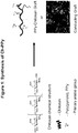
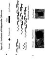
- FIG. 1 and 2A illustrate the grafting procedure that has been established.
- chitosan is dissolved in water and acetic acid.
- pyrrole monomer is added to the chitosan solution in the presence of iron (III) chloride.
- the Pyrrole polymerizes into polypyrrole and forms side chains on the chitosan backbone.
- the samples are dialyzed to filter out unbound pyrrole monomers.
- Chitosan a linear polysaccharide
- Pyrrole was crosslinked to chitosan using ferric chloride as a catalyst to produce a copolymer solution.
- Three different concentrations were produced with the highest being 284 mg Pyrrole/gram chitosan.
- Figure 2B illustrates the homogeneous nature of PPy-chitosan hydrogel.
- Chitosan-Polypyrrole (Ch-PPy or PPy-Ch) Preparation: 0.200g of Chitosan is dissolved in 10 mL of water containing 100 ⁇ L of (glacial) Acetic Acid. The solution is mixed with a stir bar on a stirring plate. 60 ⁇ L of Pyrrole is added to the Chitosan solution to provide a Ch-PPy concentration of 284 mg Pyrrole/g Chitosan (Sample A).
- Example B For a Ch-PPy concentration of 28.4 mg Pyrrole/g Chitosan (Sample B) or a Ch-PPy concentration of 2.84 mg Pyrrole/g Chitosan (Sample C), add 6 ⁇ L Py to 10 mL Ch or 0.6 ⁇ L Py to 10 mL of Ch respectively. The solution is then mixed well. 0.180 g of FeCl 3 for sample A (0.0180 g of FeCl 3 for sample B or 0.0018 g of FeCl 3 for Sample C) is dissolved in 0.5mL of dd water. The FeCl 3 solution is dropped into the Ch-PPy solution while stirring.
- the solution is then stirred well to allow the Pyrrole monomers to polymerize into Polypyrrole which form side chains on the Chitosan backbone.
- the molecular weight range of different Ch-PPy materials made is between about 50,000 and about 200,000 Daltons.
- the chitosan-polypyrrole forms a gel in the presence of a cross-linking agent, such as gluteraldehyde. Gelation was achieved by neutralizing the pH and using gluteraldehyde as the crosslinker. (See Figure 2C ).
- the PPy-chitosan prior to crosslinking (or fully crosslinking) can be molded into desired shapes. These shapes can be used as injectable material before full crosslinking occurs.
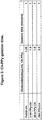
- Three different samples of PPy-Ch were tested and shown to form gels in the presence of gluteraldehyde solutions. Figure 3 shows the results of the gelation test.
- a biocompatibility study was performed to evaluate whether mammalian cells can grow on the Ch-PPy gel. If the biomaterial is biocompatible and non-toxic, it will support the growth of mammalian cells.
- Cell culture dishes were coated with CH-PPy gel.
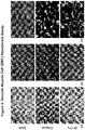
- Cultured smooth muscle cells (SMCs) were seeded onto the biomaterial-coated dishes and cultured for 1, 3, and 7 days. On each day, the cultured cells were imaged by microscopy.
- Figure 4 shows the images taken at these time points using low magnification.
- Figure 5 shows these images at a higher magnification. The area covered with cells was calculated.
- Figure 6 shows the percentage confluency of the cells grown on Ch-PPy, chitosan (an established biomaterial) and a normal cell culture dish.
- Figure 7 shows cell growth on these dishes using cell staining.
- Ch-PPy hydrogels were injected subcutaneously into rats. Injected Ch-PPy and chitosan gelled immediately and formed nodules subcutaneously. The diameters of the resultant nodules were measured immediately (day 0) and 2, 4, 8 and 21 weeks post-injection. Figure 8 shows the gelled nodules on day 0. The nodule sizes are similar between the groups at day 0. The nodules can still be observed at 12 weeks (See Figure 9 ). At 12 weeks, the chitosan degraded (dissolved) faster than Ch-PPy ( ⁇ 39% faster).
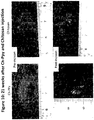
- the nodule sizes decreased significantly (See Figure 10 ).
- the Ch-PPy nodule size decreased more slowly compared with chitosan.
- the Ch-PPy has retained 70% of its original size while chitosan has only retained 39% of its original size.
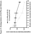
- the biodegradation is summarized in the graph shown in Figure 11 .
- the chitosan degraded significantly faster than Ch-PPy.
- the chitosan has lost 61% of its original size while Ch-PPy only lost 30% of its original size.
- the conducting hydrogels of the present disclosure are biodegradable.
- the degradation rate can be adjusted depending the composition of components.
- the conducting hydrogel biode grades between about 1 week and about 12 weeks.
- the conducting hydrogel biode grades between about 2 weeks and about 10 weeks, or between about 3 weeks and about 9 weeks, or about 4 weeks and about 8 weeks, or about 5 weeks and about 7 weeks, or any combination thereof (e.g., between about 8 and about 12 weeks).
- Ch-PPY conductivity was quantified in gel form.
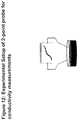
- a 2-point probe was used to measure conductivity as shown in Figure 12 .
- a voltage difference was applied between two probes placed about 7 mm apart, and into a 4 mm thick biomaterial sample, giving a current output measured in amperes. The current was plotted against the voltage range to compare of conductivity.
- the concentration of Ch-PPy is greater than about 2.84 mg pyrol/gram chitosan . In other embodiments, the concentration of Ch-PPy is greater than or equal to about 250 mg pyrol/gram chitosan. More particularly, the concentration of Ch-PPy is greater than or equal to about 300 mg pyrol/gram chitosan, or about 400 mg pyrol/gram chitosan, or about 500 mg pyrol/gram chitosan, or about 1000 mg pyrol/gram chitosan, or about 2000 mg pyrol/gram chitosan.
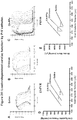
- the PPy-chitosan electrical properties were further characterized using a custom-fabricated 1 cubic cm cuvette with copper sides for electrochemical impedance spectroscopy (EIS) and cyclic voltammetry recordings.
- Figure 14A shows the custom-fabricated cuvette produced for each measurement.
- Figure 14B shows voltammograms for four groups tested (chitosan control, 0.23% PPy-chitosan (2.84 mg pyrol/gram chitosan), 2.3% PPy-chitosan (28.4 mg/g), and 23% PPy-chitosan (284 mg/g). These tests demonstrate that PPy-chitosan possesses hysteresis properties (e.g., non-linear conductive properties).
- EIS measurements are shown in Figure 15C . These measurements reveal that as the PPy composition is increased, impedance decreases, particularly at the lower frequency ranges.
- Conductivity was directly assessed from thinly-coated polystyrene dishes using a four-point probe method (See Figure 15D ). Two sets of working electrodes were used to measure the resistance of the material. Conductivity was derived from these results.
- Ch-PPY The biological conductivity of Ch-PPY was tested using cardiomyocyte contraction.
- Neonatal rat cardiomyocytes were isolated and seeded onto sterile Ch-PPY and blank control dishes. The cells were grown for 7 days to achieve confluent beating monolayers.
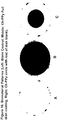
- Two different patterns of Ch-PPY were used to support cardiomyocyte cell growth: full dish coverage ( Figure 16B ) and a Ch-PPY spot ( Figure 16C ).
- spot patterns a concentric circle of Ch-PPY measuring approximately half the diameter of the dish itself was placed in the middle of the dish, and the rest of the dish left blank. The purpose of the pattern was to serve as an important control. With coated and non-coated areas are in the same dish, receiving the same cells, same media, same staining, and same incubation, a more effective comparison can be made between cells grown on Ch-PPY and those in normal culture conditions.
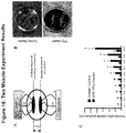
- FIG 18 shows a schematic diagram and photograph of the two muscle stimulation experiment.
- a thick layer of control gelatin or 23% PPy-gelatin was coated onto the surface of a 60-mm dish.
- Two similar sized skeletal muscles were removed from a rat hindlimb and immediately placed on posts in the dish. The muscles were trimmed so that they were the same weight.
- One muscle was stimulated directly using a programmable stimulator and tested at several voltages. The action potentials and voltage amplitudes produced by the other skeletal muscle were recorded.
- PPY-gelatin facilitated the production of larger action potentials versus the control.
- a timeline depicts the numerous experiments preformed on animals at various time points throughout the study.
- Three groups were tested with saline, chitosan only and Ch-PPy, respectively.
- Endpoints included recording of direct myocardial ECGs using an octapolar catheter; ex vivo Langendorffperfusion of rat hearts to facilitate optical mapping using the voltage sensitive dye di-4 anneps; and fixation and processing of tissues for histology at a later time.
- Figure 19B is a visual inspection of the Ch-PPy after injection in the apex and border zone, 1 week after a MI.
- Figure 19C shows the orientation for octapolar catheter recordings.
- FIG. 20 shows the results.
- time points shown are baseline (-2 weeks); 1 week post MI (-1 week); and 8 and 16 weeks post injection.
- FIG 20A there was a difference in heart rate between the three groups.
- Figure 20B there was significant reduction in QRS-interval for the PPy-Ch group vs. control at both 8 and 16 weeks post injection. This suggest enhanced left ventricle conduction.
- Optical mapping data provides evidence of changes in conduction velocity in the left ventricular free wall, particularly in the border zone area. Analysis of the direct epicardial electrophysiological recordings obtained at the final time point support these findings. Finally, histological evaluation of myocardial tissue at the end of the study was used to quantify the scar size/area, as well as retention or degradation of the injected biomaterials. The expression of connexin proteins in the border zone area was also examined, and indicated a change in the conduction velocity observed in the optical mapping experiments.
- the improvement observed with the conducting biomaterial of the present disclosure appears likely due to enhanced cardiac conduction and beneficial conductive remodeling. This is evidenced, in part, by enhancement in cardiac function (EF%, FAC% and FS%) and by shortening of the QRS-interval. This important electrophysiological parameter suggests that cardiac depolarization, particularly through the left ventricle, occurs more quickly in Ch-PPy injected hearts versus chitosan and saline-injected controls. By lowering the threshold required for electrical propagation to passively travel through the border zone and scar area in this long-term MI model, over time, global cardiac function is enhanced.
- PBS phosphate-buffered saline
- Cardiac function was evaluated using echocardiography immediately before MI, immediately before biomaterial or saline injection, and at 2, 4, and 8 weeks (acute) or 1 and 3 months (chronic) after injection. Cardiac function was also assessed at the end of the study with a pressure-volume catheter. All data are expressed as mean +/- standard deviation. Student's t -test and one- or two-way analysis of variance (ANOVA) followed by Bonferroni's post hoc test were used for parameter estimation and hypothesis testing, withp ⁇ 0.05 being considered statistically significant.
- Coronary artery ligation of the rat hearts resulted in significant LV dilatation and progressive ventricular dysfunction, as assessed by echocardiography (See Figure 22 ). All echocardiographic parameters were virtually indistinguishable among the animals at the time of biomaterial injection (Week 0; See Figures 22A-D ) due to the pre-selection of animals with similar infarct sizes.
- Four weeks following injection fractional shortening and fractional area change in the saline group had decreased more than in the chitosan and Ch-PPY groups (See Figures 22A-B ).
- the protective effect of the Ch-PPY injection was reflected in the significant preservation of fractional shortening and fractional area change compared with the saline and chitosan groups (See Figures 22A-B ).
- the left ventricular internal systolic dimension (LVIDs) and end systolic area (LVESA) in the saline group increased by week 2 and continued this trend to week 8 (See Figures 22C-D ).
- the structural stabilization effect of chitosan and Ch-PPY resulted in smaller systolic dimensions and areas.
- the Ch-PPY group demonstrated the greatest protective effect, possibly due to enhanced electrical conductivity (p ⁇ 0.05 vs. chitosan group).
- LVIDs and LVESA in the chitosan group were significantly smaller compared with saline group, and Ch-PPy group had the smallest dimension and area.
- fractional shortening FS
- fractional area change FAC
- FS fractional shortening
- FAC fractional area change
- LVIDs left ventricular internal systolic dimension
- LVESA end systolic area
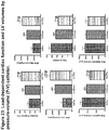
- chitosan improved ejection fraction (EF), Dp/dt max, Dp/dt min, and Tau (load-dependent indices), as well as end-systolic elastance (ESPVR) and preload recruitable stroke work (PRSW) (load-independent indices).
- Ch-PPY further improved EF, Dp/dt max, Dp/dt min, Tau, ESPVR, and PRSW (** p ⁇ 0.01 vs. saline, #p ⁇ 0.05 vs. chitosan).
- End-systolic volume was smallest in the Ch-PPY group, followed by the chitosan group. All polymer treatments reduced end-systolic volumes relative to saline (** p ⁇ 0.01 for all groups).
- LV P-V relationships were measured at 8 weeks after post-MI injection of saline, chitosan and Ch-PPy (A-D).
- load-dependent measures ejection fraction (A), Dp/dt max (B), Dp/dt min (C), and Tau_W (D) were significantly improved, and LV end systolic volume (E) was smaller (**p ⁇ 0.01) in the chitosan compared with saline group.
- Ch-PPy demonstrated the greatest (#p ⁇ 0.05 vs. chitosan) protective effects for all measures.
- Coronary artery ligation of the rat hearts resulted in significant LV dilatation and progressive ventricular dysfunction, as assessed by echocardiography.
- All echocardiographic parameters were virtually indistinguishable among the animals at the time of biomaterial injection (2 months after MI; See Figure 25A-D ) due to the pre-selection of animals with similar infarct sizes.
- the protective effect of the Ch-PPY injection was reflected in the significant preservation of fractional shortening compared with the saline and chitosan groups (See Figure 25A ).
- the left ventricular internal systolic and diastolic dimensions (LVIDs and LVIDd) in the saline and chitosan groups increased by month 1 and continued this trend to 3 months after biomaterial injection (See Figures 25B-C ).
- the effect of Ch-PPY resulted in smaller systolic and diastolic dimensions and demonstrated the greater protective effect compared with saline and chitosan 3 months after injection, possibly due to enhanced electrical conductivity ( p ⁇ 0.05 vs . chitosan and p ⁇ 0.01 vs. saline).
- serial Doppler echocardiography tests suggest that diastolic properties were improved in Ch-PPY treated rats.
- FS fractional shortening
- FS fractional shortening
- LVIDs left ventricular internal systolic dimension
- LVIDd left ventricular internal diastolic dimension
Landscapes
- Health & Medical Sciences (AREA)
- Life Sciences & Earth Sciences (AREA)
- General Health & Medical Sciences (AREA)
- Epidemiology (AREA)
- Animal Behavior & Ethology (AREA)
- Public Health (AREA)
- Veterinary Medicine (AREA)
- Chemical & Material Sciences (AREA)
- Medicinal Chemistry (AREA)
- Pharmacology & Pharmacy (AREA)
- Bioinformatics & Cheminformatics (AREA)
- Engineering & Computer Science (AREA)
- Dermatology (AREA)
- Heart & Thoracic Surgery (AREA)
- Vascular Medicine (AREA)
- Proteomics, Peptides & Aminoacids (AREA)
- Oral & Maxillofacial Surgery (AREA)
- Transplantation (AREA)
- Surgery (AREA)
- Biomedical Technology (AREA)
- Molecular Biology (AREA)
- Chemical Kinetics & Catalysis (AREA)
- Polymers & Plastics (AREA)
- Organic Chemistry (AREA)
- Materials For Medical Uses (AREA)
- Medicinal Preparation (AREA)
- Pharmaceuticals Containing Other Organic And Inorganic Compounds (AREA)
Description
- This application claims priority to U.S. provisional patent application No.
61/760,858, filed February 5, 2013 - The present disclosure relates to a biocompatible, electrically conductive biomaterial capable of carrying the electrical potential of a cardiac impulse. The present disclosure also relates to treatments using the electrically conductive biomaterial.
- Treatments of heart conditions, such as myocardial infarction (MI), mainly include medication, medical interventional therapy and surgery, such as coronary artery bypass graft surgery. These treatments can unclog the occlusive blood vessels and improve the symptom of myocardial ischemia. They cannot, however, enhanced cardiac conduction or have limited improvement of cardiac function.
- Following a myocardial infarction, a non-contractile fibrotic scar forms. Scar formation can be associated with widespread cardiomyocyte death and impaired electrical properties, such as slowing of electrical impulse propagation across the scar region of the myocardium. In addition, "passive" barriers can develop that produce unidirectional block or delay in atrioventricular (AV) electrical conduction, or even produce deadly reentrant arrhythmias. Currently, there are few direct treatments of these underlying conditions.
- Da Kai et al.: "Polypyrrole-contained electrospun conductive nanofibrous membranes for cardiac tissue engineering", Journal of Biomedical Materials Research Part A, 2011, vol. 99A, no. 3, pages 376-385 discloses, a biocompatible material comprising a conductive polymer and a biocompatible component in form of a nanofibrous scaffold. The conductive polymer is polypyrrole and the biocompatible component is gelatin. The material has a conductivity of at least 1.3x10⁻⁵ S7cm and is suitable for treating a heart condition.
- The present disclosure relates to a biocompatible, electrically conductive material capable of treating heart conditions including myocardial infarction and other related conditions. For example, the electrically conductive biomaterial can restore or improve electrical impulse propagation across the damaged tissue or scar region of the myocardium and resolve "passive" barriers to atrioventricular electrical conduction.
- The present disclosure relates to a modified biomaterial that can be adapted to deliver an electrically conductive adjuvant (e.g., conductive polymer) to the heart. The modified biomaterial can be used to treat heart related conditions, such as MI and arrhythmia. The scope of the invention is defined by the claims. Any references in the description to methods of treatment refer to the products of the present invention for use in a method of treatment.
- In one embodiment, the present disclosure relates to a biocompatible material comprising a conductive polymer and a biocompatible component, wherein the conductive polymer is a polypyrrole-based polymer; the biocompatible component is chitosan; and the material further comprises a crosslinking agent and the conductive polymer and the biocompatible component are chemically conjugated to form a matrix throughout to form the material which is a homogenous hydrogel.
- In another embodiment, the present disclosure relates to a method of treating a heart condition, the method comprising introducing the biocompatible material of
claim 1 to the heart. The heart condition can include myocardial infarction, heart failure, atrioventricular block, arrhythmia and a conduction abnormality. -
-
Figure 1 shows the synthesis of PPy-chitosan grafts. -
Figure 2 shows the synthesis of a PPy-chitosan matrix or hydrogel, with the chemical conjugation with polypyrrole side chains. -
Figure 3 shows the results of a gelation test using gluteraldehyde as the crosslinker. -
Figure 4 shows SEM images of SMCs taken using low magnification. The SMCs were cultured for 1, 3, and 7 days, and then imaged using bright field microscopy. -
Figure 5 shows select SEM images of SMCs taken using high magnification. -
Figure 6 shows the percentage confluency of the cells grown on Ch-PPy, chitosan and a normal cell culture dish. -
Figure 7 shows cell growth on Ch-PPy, chitosan and a normal cell culture dish using cell staining. -
Figure 8 shows gelled nodules of Ch-PPy and chitosan injected subcutaneously into rats onday 0. -
Figure 9 shows gelled nodules of Ch-PPy and chitosan injected subcutaneously into rats at 12 weeks. -
Figure 10 shows gelled nodules of Ch-PPy and chitosan injected subcutaneously into rats at 21 weeks. -
Figure 11 shows the biodegradation of gelled nodules of Ch-PPy and chitosan injected subcutaneously into rats. -
Figure 12 shows a 2-point probe was used to measure conductivity of conducting hydrogels. -
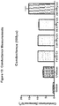
Figure 13 shows the results of conductivity measurements for conducting hydrogels. -
Figure 14A shows a custom-fabricated cuvette produced for electrical properties measurements;Figure 14B show cyclic voltammetry measurements. -
Figure 15 shows EIS measurements of conducting hydrogels. -
Figure 16 shows the biological conductivity of Ch-PPY using cardiomyocyte contraction via full dish coverage (Figure 18B ) and a Ch-PPY spot (Figure 18C ) patterns of Ch-PPy to support cardiomyocyte cell growth. -
Figure 17 shows micrographs of DAPI- and phalloidin-stained neonatal cardiomyocyte dishes. -
Figure 18 shows a schematic diagram and photograph of the two muscle stimulation experiment. -
Figure 19A shows a timeline for the numerous experiments preformed;Figure 19B shows a visual inspection of the Ch-PPy after injection in the apex and border zone, 1 week after a MI; andFigure 19C shows the orientation for octapolar catheter recordings. -
Figure 20 shows the results of surface ECGs from an in vivo study. -
Figure 21 shows schematic diagrams of the acute and chronic experimental timelines from an in vivo study testing saline, chitosan and Ch-PPy. -
Figure 22 shows coronary artery ligation of the rat hearts as assessed by echocardiography from an in vivo study testing saline, chitosan and Ch-PPy. -
Figure 23 shows load-dependent cardiac function and LV volumes by pressure-volume (P-V) catheter from an in vivo study testing saline, chitosan and Ch-PPy. -
Figure 24 shows load-independent cardiac function by P-V catheter from an in vivo study testing saline, chitosan and Ch-PPy. -
Figure 25 shows the cardiac function by echocardiography from an in vivo study testing saline, chitosan and Ch-PPy. - The present disclosure relates to a biocompatible, electrically conductive biomaterial capable of carrying the electrical potential of a cardiac impulse, as well as treatments using the electrically conductive biomaterial. In particular, the present invention relates to the treatment of MI by introducing a biocompatible, electrically conductive biomaterial to the heart.
- As used herein, the term "biocompatible" refers to an article that does not cause toxic or injurious effects on biological systems.
- As used herein, the term "biomaterial" refers to a polymer composition, hydrogel or article that is biocompatible. The biomaterial can include an article in different physical forms, such as a hydrogel, membrane, sheet, graft, or mesh. These forms include typical membranes, sheets, grafts, meshes, etc. used in surgery or tissue repair. These articles can include natural products, synthetic products, or combinations thereof. The biomaterial of the present disclosure can be used exclusively to form one of these articles or can be used as a component of one of these articles.
- As used herein, the term "hydrogel" refers to a polymeric material, which is a network or matrix of polymer chains, capable of swelling in water or becoming swollen with water. A hydrogel can also be understood to be a material that retains water in an equilibrium state. The network or matrix is crosslinked.
- In one embodiment, the present disclosure relates to a biocompatible biomaterial as defined in
claim 1. Conductive polymers are polymers that are inherently or intrinsically capable of electrical conductivity. The conductive polymers of the present invention are polypyrrole-based polymers. In one embodiment, conductive polymers include polymers that exhibit a bulk specific conductance of greater than or equal to about 10-5 Siemens per centimeter ("S/cm"), or about 10-4 S/cm, or about 10-3 S/cm, or about 10-2 S/cm, or about 1 S/cm, or about 10 S/cm, or about 100 S/cm, or about 103 S/cm. The polymers can also that exhibit a range of bulk specific conductance between any of these values (e.g., between about 10-5 S/cm and about 10-4 S/cm, or about 10-5 S/cm and about 1000 S/cm, or about 10-5 S/cm and about 10-2 S/cm, etc.) - The conductive polymer can be linear or branched, and have a molecular weight ranging from about 44 to about 18,500 Daltons. In some embodiments, the molecular weight is greater than about 50 Daltons, or about 100 Daltons, or about 200 Daltons, or about 500 Daltons, or about 1,000 Daltons, or about 1,500 Daltons, or about 2,000 Daltons, or about 3,000 Daltons, or about 4,000 Daltons, or about 5,000 Daltons, or about 7,000 Daltons, or about 9,000 Daltons, or about 10,000 Daltons, or about 12,000 Daltons, or about 14,000 Daltons, or about 16,000 Daltons. In other embodiments, the molecular weight is less than about 200 Daltons, or about 500 Daltons, or about 1,000 Daltons, or about 1,500 Daltons, or about 2,000 Daltons, or about 3,000 Daltons, or about 4,000 Daltons, or about 5,000 Daltons, or about 7,000 Daltons, or about 9,000 Daltons, or about 10,000 Daltons, or about 12,000 Daltons, or about 14,000 Daltons, or about 16,000 Daltons, or about 18,500 Daltons. In still other embodiments, the molecular weight can be a range between any of these values (e.g., between about 200 Daltons and about 7,000 Daltons, or between about 50 Daltons and about 10,000 Daltons, etc.).
- The biocompatible component is chitosan.
- The biocompatible component can have a molecular weight ranging from about 40 to about 200,000 Daltons. In some embodiments, the molecular weight is greater than about 100 Daltons, or about 2,000 Daltons, or about 5,000 Daltons, or about 10,000 Daltons, or about 15,000 Daltons, or about 20,000 Daltons, or about 30,000 Daltons, or about 40,000 Daltons, or about 50,000 Daltons, or about 70,000 Daltons, or about 90,000 Daltons, or about 100,000 Daltons, or about 120,000 Daltons, or about 140,000 Daltons, or about 160,000 Daltons. In other embodiments, the molecular weight is less than about 2,000 Daltons, or about 5,000 Daltons, or about 10,000 Daltons, or about 15,000 Daltons, or about 20,000 Daltons, or about 30,000 Daltons, or about 40,000 Daltons, or about 50,000 Daltons, or about 70,000 Daltons, or about 90,000 Daltons, or about 100,000 Daltons, or about 120,000 Daltons, or about 140,000 Daltons, or about 160,000 Daltons, or about 180,000 Daltons, or about 200,000 Daltons. In still other embodiments, the molecular weight can be a range between any of these values (e.g., between about 2,000 Daltons and about 70,000 Daltons, or between about 20,000 Daltons and about 100,000 Daltons, etc.).
- In one embodiment, the biocompatible component is chitosan having a molecular weight between about 10,000 and about 200,000 Daltons. In another embodiment, the biocompatible component is gelatin having a molecular weight between about 10,000 and about 300,000 Daltons.
- The conductive polymer and the biocompatible component are combined to form an electrically conductive biomaterial. The molar ratio of the conductive polymer and biocompatible component in the biomaterial can range from 1000:1 to 1:1000, respectively. In some embodiments, the molar ratio of the conductive polymer and biocompatible component can be greater than about 1:3, or about 1:2, or about 1:1, or about 2:1, or about 3:1, or about 5:1, or about 10:1, or about 25:1, or about 50:1, or about 100:1, or about 150:1, or about 200:1, or about 250:1, or about 300:1 or about 350:1 or about 400:1, or about 500:1. In other embodiments, the molar ratio of the conductive polymer and biocompatible component can be less than about 1:2, or about 1:1, or about 2:1, or about 3:1, or about 5:1, or about 10:1, or about 25:1, or about 50:1, or about 100:1, or about 150:1, or about 200:1, or about 250:1, or about 300:1 or about 350:1 or about 400:1, or about 500:1, or about 1000:1. In still other embodiments, the molar ratio of the conductive polymer and biocompatible component can be a range between any of these values (e.g., between 1:1 to 1:350, or between 1:3 to 1:150, or between 3:1 and 300:1, etc.). In one embodiment, the ratio is 2.1:1 to 1000:1.
- In some embodiments, the molecular weight of the biomaterial can range from about 1,000 to about 1,000,000 Daltons. In some embodiments, the molecular weight of the biomaterial is greater than about 1,000 Daltons, or about 2,000 Daltons, or about 5,000 Daltons, or about 10,000 Daltons, or about 20,000 Daltons, or about 50,000 Daltons, or about 100,000 Daltons, or about 150,000 Daltons, or about 200,000 Daltons, or about 300,000 Daltons, or about 400,000 Daltons, or about 500,000 Daltons, or about 600,000 Daltons, or about 700,000 Daltons, or about 800,000 Daltons. In other embodiments, the molecular weight of the biomaterial is less than about 2,000 Daltons, or about 5,000 Daltons, or about 10,000 Daltons, or about 20,000 Daltons, or about 50,000 Daltons, or about 100,000 Daltons, or about 150,000 Daltons, or about 200,000 Daltons, or about 300,000 Daltons, or about 400,000 Daltons, or about 500,000 Daltons, or about 600,000 Daltons, or about 700,000 Daltons, or about 800,000 Daltons, or about 1,000,000 Daltons. In still other embodiments, the molecular weight of the biomaterial can be a range between any of these values (e.g., between about 5,000 Daltons and about 800,000 Daltons, or between about 20,000 Daltons and about 100,000 Daltons, etc.).
- In one embodiment, the biomaterial has a molecular weight about 350,000 Daltons (i.e., comprising chitosan having a molecular weight of about 50,000 Daltons and gelatin having a molecular weight of about 300,000 Daltons.
- The conductivity of the biomaterial is primarily attributed to the presence of the conducting polymer. In one embodiment, the conductivity of the biomaterial is greater than or equal to about 10-5 S/cm. In some embodiments, the conductivity of the biomaterial is greater than or equal to about 10-4 S/cm, or about 10-3 S/cm, or about 10-2 S/cm, or about 1 S/cm, or about 10 S/cm. In other embodiments, the conductivity of the biomaterial is less than or equal to about 10-5 S/cm, or about 10-4 S/cm, or about 10-3 S/cm, or about 10-2 S/cm, or about 1 S/cm, or about 10 S/cm. In one embodiment, the conducting biomaterial, polymer and/or the hydrogel is able to carry the electrical potential of a cardiac impulse at about 100 mV. The repolarization voltage of tissue is about 40 mV. In particular, these materials are able to carry the electrical potential of a cardiac impulse about 40 mV or greater. In particular embodiments, the materials are able to carry the electrical potential of a cardiac impulse of about 10 to about 250 mV, or about 20 to about 200 mV, or about 50 to about 150 mV, or about 75 to about 100 mV, or any combination of these values (e.g., about 50 to about 100 mV, etc.).
- Similarly, in some embodiments the conductive biomaterials can have resistivities (or volume resistivities) greater than about 0.06 ohm centimeter (Ωcm), or about 0.1 Ωcm, or about 0.6 Ωcm, or about 1 Ωcm, or about 10 Ωcm, or about 100 Ωcm, or about 1,000 Ωcm, or about 104 Ωcm, or about 105 Ωcm, or about 2 x 10⁵ Ωcm. In other embodiments the conductive biomaterials can have resistivities less than about 0.1 Ωcm, or about 0.6 Ωcm, or about 1 Ωcm, or about 10 Ωcm, or about 100 Ωcm, or about 1,000 Ωcm, or about 104 Ωcm, or about 105 Ωcm, or about 2 x 10⁵ Ωcm. In still other embodiments, the conductive biomaterials can have resistivities in a range between any of these values (e.g., between about 0.6 Ωcm and about 100 Ωcm, or between about 0.0625 and about 2 x 10⁵ Ωcm, or between about 0.6 to about 2 x 10³ Ωcm, etc.). Resistivity (or volume resistivity) is defined as equal to: Resistance (n) = distance/Area (cm2) x Thickness (cm). In one embodiment, the resistivity is between about 50 and about 20,000 Ωcm.
- The hydrogel contains a crosslinking agent used to crosslink the present hydrogels and to assist in hydrogel formation. The crosslinking agent can be a known crosslinking agent and contain electrophilic groups, nucleophilic groups, or both. The crosslinking agent can be a natural product or a synthetic product. Examples of multi-functional crosslinking agents which may be used include, for example, gluteraldehyde, methylene-bis-acrylamide, diethylene glycol diacrylate, ethylene glycol diacrylate, triethylene glycol-bis-methacrylate, ethylene glycol-bis-methacrylate, ethylene glycol-dimethacrylate, bisacrylamide, triethyleneglycol-bis-acrylate, 3,3'-ethylidene-bis(N-vinyl-2-pyrrolidone), trimethylolpropate trimethacrylate, glycerol trimethacrylate, polyethylene glycol dimethacrylate, and other polyacrylate and polymethacrylate esters. In one embodiment, the crosslinking agent is genipin or tannic acid. As used herein, the term "genipin" is meant a compound recognized as genipin as a chemical compound or an equivalent of genipin as a chemical compound by a person of ordinary skill in the art. The term "genipin" is intended to cover derivatives, analog, stereoisomers and mixtures thereof. The genipin compound can be derived from natural sources or synthetically made.
- The amount of crosslinking agent can be within the range of about 0.001 to about 10% by weight. In particular, the amount can be within the range of about 0.005 to about 5% by weight, or about 0.01 and about 3%, or any combination thereof. The crosslinker can be added to the material just prior to introduction (e.g., 1-10 minutes prior to introduction). In some embodiments, it takes 1-10 minutes for the biomaterial to gel. During the gelling time, the biomaterial can be introduced.
- In another embodiment, the present disclosure relates to a biocompatible material as defined in
claim 2. The heart condition can include myocardial infarction, heart failure, atrioventricular block, arrhythmia and a conduction abnormality. - Without wishing to be bound, it is believed the electrically conductive biomaterial can promote beneficial remodeling of the border zone area following an infarct (i.e., a method of remodeling). The biomaterials can prevent fibrotic tissue expansion, heart chamber dialation and induce blood vessel formation (i.e., a method of preventing expansion and/or dialation, or inducing formation). All these changes can benefit heart function and prevent heart failure. The conductive biomaterial can also improve conduction velocities across fibrotic tissues by lowering the threshold for cardiac action potential propagation (i.e., a method of improving velocities). For example, the conductive material can be introduced to fibrotic tissue of the injured region. The material will decrease resistance of the fibrotic tissue and increase current through the tissue. The electrically conductive biomaterial can restore or improve electrical impulse propagation across the scar region of the myocardium and resolve "passive" barriers to atrioventricular electrical conduction (i.e., a method of restoring or improving propagation). Finally, the conductive biomaterial (e.g., hydrogel) can also improve the performance of implantable pacemaker devices and ultimately, may serve as an alternative to cardiac ablation for the treatment of some cardiac arrhythmias (i.e., a method of improving the performance of a pacemaker). The conductive biomaterial can change the fibrotic resistance, which will enhance performance of the stimulator function.
- The biocompatible biomaterial can be introduced by known method of treating biological tissue and organs with a hydrogel and similar materials. In one embodiment, material can be injected into or onto the heart. The conductive biomaterials can also be formed in sheet, or other articles, which can be used on top of the injured tissue. For example, the conductive biomaterial can be generated as graft. The excess fibrotic tissue can be removed and conductive biomaterial graft can be put and repair the defect area.
- In one embodiment, the amount of biomaterial introduced to the tissue or organ can depend on a number of factors, such as the composition of the biomaterial, the location and the condition of the tissue or organ, the size of the tissue or organ and/or the size of the damaged or area to be treated. In one embodiment, the volume of biomaterial can range from about 1 µl to about 10 mL, or about 2 µl to about 5 mL, or about 5 µl to about 3 mL, or about 10 µl to about 2 mL, or about 50 µl to about 1 mL, or about .or about 100 µl to about 500 µl, or any combination of these values (e.g., about 1 mL to about 2 mL, etc.)
- When an amount, concentration, or other value or parameter is given as either a range, preferred range, or a list of upper preferable values and lower preferable values, this is to be understood as specifically disclosing all ranges formed from any pair of any upper range limit or preferred value and any lower range limit or preferred value, regardless of whether ranges are separately disclosed. Where a range of numerical values is recited herein, unless otherwise stated, the range is intended to include the endpoints thereof, and all integers and fractions within the range. It is not intended that the scope of the invention be limited to the specific values recited when defining a range.
- The present invention is further defined in the following Examples. It should be understood that these Examples, while indicating preferred embodiments of the invention, are given by way of illustration only.
- The following examples are based on the use of both mouse and rat models of early and late phase MI. An AV block model with in vivo and ex-vivo rat heart preparations has also been developed. These examples are also based on and can be used with an array of injectable biomaterial hydrogels that can act as carriers for cells and cytokines, and help to retain cells, enhance their survival, and support the timed release of cytokines and growth factors. These injectable biomaterials are easy to manipulate, and can be chemically modified with a variety of molecules including peptide fragments, proteins and antibodies. They are also biocompatible, and their degradation products are safe for use in vivo.
- The examples presented here and in the Figures demonstrate the intrinsic biocompatible, and conductive properties of the hydrogels of the present disclosure.
- A biocompatible and conductive biomaterial was prepared by grafting pyrrole onto chitosan.
Figures 1 and2A illustrate the grafting procedure that has been established. In general, chitosan is dissolved in water and acetic acid. Thereafter, pyrrole monomer is added to the chitosan solution in the presence of iron (III) chloride. The Pyrrole polymerizes into polypyrrole and forms side chains on the chitosan backbone. At end of the reaction, the samples are dialyzed to filter out unbound pyrrole monomers. - Chitosan, a linear polysaccharide, was selected in the form of a hydrogel with suitable mechanical and cell-compatible properties for in vitro and in vivo studies. Pyrrole was crosslinked to chitosan using ferric chloride as a catalyst to produce a copolymer solution. Three different concentrations were produced with the highest being 284 mg Pyrrole/gram chitosan.
Figure 2B illustrates the homogeneous nature of PPy-chitosan hydrogel. - Chitosan-Polypyrrole (Ch-PPy or PPy-Ch) Preparation: 0.200g of Chitosan is dissolved in 10 mL of water containing 100 µL of (glacial) Acetic Acid. The solution is mixed with a stir bar on a stirring plate. 60 µL of Pyrrole is added to the Chitosan solution to provide a Ch-PPy concentration of 284 mg Pyrrole/g Chitosan (Sample A). For a Ch-PPy concentration of 28.4 mg Pyrrole/g Chitosan (Sample B) or a Ch-PPy concentration of 2.84 mg Pyrrole/g Chitosan (Sample C), add 6 µL Py to 10 mL Ch or 0.6 µL Py to 10 mL of Ch respectively. The solution is then mixed well. 0.180 g of FeCl3 for sample A (0.0180 g of FeCl3 for sample B or 0.0018 g of FeCl3 for Sample C) is dissolved in 0.5mL of dd water. The FeCl3 solution is dropped into the Ch-PPy solution while stirring. The solution is then stirred well to allow the Pyrrole monomers to polymerize into Polypyrrole which form side chains on the Chitosan backbone. The molecular weight range of different Ch-PPy materials made is between about 50,000 and about 200,000 Daltons.
- The chitosan-polypyrrole forms a gel in the presence of a cross-linking agent, such as gluteraldehyde. Gelation was achieved by neutralizing the pH and using gluteraldehyde as the crosslinker. (See
Figure 2C ). In some instances, prior to crosslinking (or fully crosslinking) the PPy-chitosan can be molded into desired shapes. These shapes can be used as injectable material before full crosslinking occurs. Three different samples of PPy-Ch were tested and shown to form gels in the presence of gluteraldehyde solutions.Figure 3 shows the results of the gelation test. - A biocompatibility study was performed to evaluate whether mammalian cells can grow on the Ch-PPy gel. If the biomaterial is biocompatible and non-toxic, it will support the growth of mammalian cells. Cell culture dishes were coated with CH-PPy gel. Cultured smooth muscle cells (SMCs) were seeded onto the biomaterial-coated dishes and cultured for 1, 3, and 7 days. On each day, the cultured cells were imaged by microscopy.
Figure 4 shows the images taken at these time points using low magnification.Figure 5 shows these images at a higher magnification. The area covered with cells was calculated.Figure 6 shows the percentage confluency of the cells grown on Ch-PPy, chitosan (an established biomaterial) and a normal cell culture dish.Figure 7 shows cell growth on these dishes using cell staining. - An in-vivo study was performed to test the in-vivo degradation of the Ch-PPy hydrogels. Ch-PPy (100µL) or chitosan alone (100µL) solutions were injected subcutaneously into rats. Injected Ch-PPy and chitosan gelled immediately and formed nodules subcutaneously. The diameters of the resultant nodules were measured immediately (day 0) and 2, 4, 8 and 21 weeks post-injection.
Figure 8 shows the gelled nodules onday 0. The nodule sizes are similar between the groups atday 0. The nodules can still be observed at 12 weeks (SeeFigure 9 ). At 12 weeks, the chitosan degraded (dissolved) faster than Ch-PPy (∼ 39% faster). At 21 weeks, the nodule sizes decreased significantly (SeeFigure 10 ). The Ch-PPy nodule size decreased more slowly compared with chitosan. The Ch-PPy has retained 70% of its original size while chitosan has only retained 39% of its original size. The biodegradation is summarized in the graph shown inFigure 11 . The chitosan degraded significantly faster than Ch-PPy. At 21 weeks after injection, the chitosan has lost 61% of its original size while Ch-PPy only lost 30% of its original size. - The conducting hydrogels of the present disclosure are biodegradable. The degradation rate can be adjusted depending the composition of components. In one embodiment, the conducting hydrogel biodegrades between about 1 week and about 12 weeks. In particular, the conducting hydrogel biodegrades between about 2 weeks and about 10 weeks, or between about 3 weeks and about 9 weeks, or about 4 weeks and about 8 weeks, or about 5 weeks and about 7 weeks, or any combination thereof (e.g., between about 8 and about 12 weeks).
- The electrophysiological function of the conductive biomaterial was investigated. Ch-PPY conductivity was quantified in gel form. A 2-point probe was used to measure conductivity as shown in
Figure 12 . A voltage difference was applied between two probes placed about 7 mm apart, and into a 4 mm thick biomaterial sample, giving a current output measured in amperes. The current was plotted against the voltage range to compare of conductivity. - Three concentrations of the Ch-PPY biomaterial were tested for their conductivity. Gelatin (an extracellular material), and chitosan were used as controls. The results are presented in
Figure 13 . Gelatin has very low conductive activity. Chitosan is about 7 times more conductive in comparison. The highest concentration of Ch-PPY material is more conductive than the lowest concentration of Ch-PPY, as well as the chitosan control. Furthermore, the lowest concentration Ch-PPY is no more conductive than chitosan, suggesting that there may be a threshold PPY concentration that makes the biomaterial conductive. - In some embodiments, the concentration of Ch-PPy is greater than about 2.84 mg pyrol/gram chitosan . In other embodiments, the concentration of Ch-PPy is greater than or equal to about 250 mg pyrol/gram chitosan. More particularly, the concentration of Ch-PPy is greater than or equal to about 300 mg pyrol/gram chitosan, or about 400 mg pyrol/gram chitosan, or about 500 mg pyrol/gram chitosan, or about 1000 mg pyrol/gram chitosan, or about 2000 mg pyrol/gram chitosan.
- The PPy-chitosan electrical properties were further characterized using a custom-fabricated 1 cubic cm cuvette with copper sides for electrochemical impedance spectroscopy (EIS) and cyclic voltammetry recordings.
Figure 14A shows the custom-fabricated cuvette produced for each measurement.Figure 14B shows voltammograms for four groups tested (chitosan control, 0.23% PPy-chitosan (2.84 mg pyrol/gram chitosan), 2.3% PPy-chitosan (28.4 mg/g), and 23% PPy-chitosan (284 mg/g). These tests demonstrate that PPy-chitosan possesses hysteresis properties (e.g., non-linear conductive properties). EIS measurements are shown inFigure 15C . These measurements reveal that as the PPy composition is increased, impedance decreases, particularly at the lower frequency ranges. Conductivity was directly assessed from thinly-coated polystyrene dishes using a four-point probe method (SeeFigure 15D ). Two sets of working electrodes were used to measure the resistance of the material. Conductivity was derived from these results. - The biological conductivity of Ch-PPY was tested using cardiomyocyte contraction. Neonatal rat cardiomyocytes were isolated and seeded onto sterile Ch-PPY and blank control dishes. The cells were grown for 7 days to achieve confluent beating monolayers. Two different patterns of Ch-PPY were used to support cardiomyocyte cell growth: full dish coverage (
Figure 16B ) and a Ch-PPY spot (Figure 16C ). For the spot patterns, a concentric circle of Ch-PPY measuring approximately half the diameter of the dish itself was placed in the middle of the dish, and the rest of the dish left blank. The purpose of the pattern was to serve as an important control. With coated and non-coated areas are in the same dish, receiving the same cells, same media, same staining, and same incubation, a more effective comparison can be made between cells grown on Ch-PPY and those in normal culture conditions. - Fully coated Ch-PPY dishes showed far different cardiomyocyte contractility and Ch-PPY conduction. Cardiac impulses were more instantaneous and synchronized across the dish, rather than following a path or orientation. It appeared as if all cells were beating at the same time, with no delay in transferring action potentials. This suggests that the Ch-PPY is functional and can electrically connect the entire monolayer. Micrographs of DAPI- and phalloidin-stained neonatal cardiomyocyte dishes are shown in
Figure 17 . - Another proof of concept experiment for the application of the conductive polymer was performed with a two muscle stimulation experiment.
Figure 18 (A and B) show a schematic diagram and photograph of the two muscle stimulation experiment. A thick layer of control gelatin or 23% PPy-gelatin was coated onto the surface of a 60-mm dish. Two similar sized skeletal muscles were removed from a rat hindlimb and immediately placed on posts in the dish. The muscles were trimmed so that they were the same weight. One muscle was stimulated directly using a programmable stimulator and tested at several voltages. The action potentials and voltage amplitudes produced by the other skeletal muscle were recorded. PPY-gelatin facilitated the production of larger action potentials versus the control. As shown inFigure 18C , the conducting hydrogels are lowering the resistivity surrounding the two skeletal muscle tissue (* p<0.01, # p<0.05, n=5 experiments/group). - A first series of in vivo studies were performed to demonstrate an improvement in heart function. All surgical procedures were approved by the Animal Care Committee of the University Health Network (Toronto, Canada). In this study, the echocardiographic function of different groups of animals (n=4 or 6 animals) at multiple time points leading up to 8 weeks was studied. PV-loop analysis is used to examine load-dependent and load-independent changes in cardiac function, and further optical mapping studies will be carried out wherever possible.
- As shown in
Figure 19A , a timeline depicts the numerous experiments preformed on animals at various time points throughout the study. Three groups were tested with saline, chitosan only and Ch-PPy, respectively. Endpoints included recording of direct myocardial ECGs using an octapolar catheter; ex vivo Langendorffperfusion of rat hearts to facilitate optical mapping using the voltage sensitive dye di-4 anneps; and fixation and processing of tissues for histology at a later time.Figure 19B is a visual inspection of the Ch-PPy after injection in the apex and border zone, 1 week after a MI.Figure 19C shows the orientation for octapolar catheter recordings. - Surface ECGs were recorded using three leads throughout the 18-week experiment.
Figure 20 shows the results. InFigure 20 , time points shown are baseline (-2 weeks); 1 week post MI (-1 week); and 8 and 16 weeks post injection. As shown inFigure 20A , there was a difference in heart rate between the three groups. As shown inFigure 20B , there was significant reduction in QRS-interval for the PPy-Ch group vs. control at both 8 and 16 weeks post injection. This suggest enhanced left ventricle conduction. As shown inFigures 20C and 20D , there was no difference between the groups in terms of repolarization kinetics as evidenced by QT-interval and QTc analysis (*p<0.05; p<0.01; n=4 or 6 animals per group/time point). - Optical mapping data provides evidence of changes in conduction velocity in the left ventricular free wall, particularly in the border zone area. Analysis of the direct epicardial electrophysiological recordings obtained at the final time point support these findings. Finally, histological evaluation of myocardial tissue at the end of the study was used to quantify the scar size/area, as well as retention or degradation of the injected biomaterials. The expression of connexin proteins in the border zone area was also examined, and indicated a change in the conduction velocity observed in the optical mapping experiments.
- The improvement observed with the conducting biomaterial of the present disclosure appears likely due to enhanced cardiac conduction and beneficial conductive remodeling. This is evidenced, in part, by enhancement in cardiac function (EF%, FAC% and FS%) and by shortening of the QRS-interval. This important electrophysiological parameter suggests that cardiac depolarization, particularly through the left ventricle, occurs more quickly in Ch-PPy injected hearts versus chitosan and saline-injected controls. By lowering the threshold required for electrical propagation to passively travel through the border zone and scar area in this long-term MI model, over time, global cardiac function is enhanced.
- Again, all surgical procedures were approved by the Animal Care Committee of the University Health Network (Toronto, Canada). Female Sprague-Dawley rats (225 - 250 g) were anesthetized, intubated and perfused with 2% isoflurane mixed with oxygen. The chest was opened by a left lateral therorectomy, and a single stitch of 7-0 prolene suture was introduced around the left anterior descending coronary artery and tightened. The chest was then closed and the animal was allowed to recover for 1 week (acute) or 2 months (chronic). These acute and chronic infarct models have previously been shown to create infarct sizes of approximately 35% of the left ventricle (LV). Schematic diagrams of the acute and chronic experimental timelines are shown in
Figure 21 . - One week (acute) or 2 months (chronic) after MI, rats were randomized into chitosan only, Ch-PPy, or phosphate-buffered saline (PBS) control groups (n = 6 - 11/group). The animals were anesthetized, intubated and the chest opened by a left lateral therorectomy. A total volume of 100uL in each group was injected into the peri-infarcted LV region of each heart, using a 500uL tuberculin syringe and a 28-gauge needle. The chest was then closed and the animal allowed to recover for 8 weeks (acute) or 3 months (chronic). After these time periods, the rats were euthanized with an isoflurane overdose (5%).
- Function was evaluated using echocardiography immediately before MI, immediately before biomaterial or saline injection, and at 2, 4, and 8 weeks (acute) or 1 and 3 months (chronic) after injection. Cardiac function was also assessed at the end of the study with a pressure-volume catheter. All data are expressed as mean +/- standard deviation. Student's t-test and one- or two-way analysis of variance (ANOVA) followed by Bonferroni's post hoc test were used for parameter estimation and hypothesis testing, withp < 0.05 being considered statistically significant.
- Coronary artery ligation of the rat hearts resulted in significant LV dilatation and progressive ventricular dysfunction, as assessed by echocardiography (See
Figure 22 ). All echocardiographic parameters were virtually indistinguishable among the animals at the time of biomaterial injection (Week 0; SeeFigures 22A-D ) due to the pre-selection of animals with similar infarct sizes. Four weeks following injection, fractional shortening and fractional area change in the saline group had decreased more than in the chitosan and Ch-PPY groups (SeeFigures 22A-B ). At 8 weeks, the protective effect of the Ch-PPY injection was reflected in the significant preservation of fractional shortening and fractional area change compared with the saline and chitosan groups (SeeFigures 22A-B ). The left ventricular internal systolic dimension (LVIDs) and end systolic area (LVESA) in the saline group increased byweek 2 and continued this trend to week 8 (SeeFigures 22C-D ). In contrast, the structural stabilization effect of chitosan and Ch-PPY resulted in smaller systolic dimensions and areas. At 8 weeks after injection, the Ch-PPY group demonstrated the greatest protective effect, possibly due to enhanced electrical conductivity (p < 0.05 vs. chitosan group). LVIDs and LVESA in the chitosan group were significantly smaller compared with saline group, and Ch-PPy group had the smallest dimension and area. - Also observed from
Figure 22 is fractional shortening (FS) and fractional area change (FAC) decreased dramatically in all groups from ligation (Week -1) to biomaterial injection (Week 0) and continued to decrease in the saline and biomaterial groups. Left ventricular internal systolic dimension (LVIDs) and end systolic area (LVESA) increased dramatically in all groups from ligation (Week -1) to polymer injection (Week 0), and this trend continued in the saline and chitosan groups. - Ventricular volumes and cardiac function were evaluated under load-dependent (
Figure 23 ) and load-independent (Figure 24 ) conditions. Compared with saline, chitosan improved ejection fraction (EF), Dp/dt max, Dp/dt min, and Tau (load-dependent indices), as well as end-systolic elastance (ESPVR) and preload recruitable stroke work (PRSW) (load-independent indices). Ch-PPY further improved EF, Dp/dt max, Dp/dt min, Tau, ESPVR, and PRSW (**p < 0.01 vs. saline, #p < 0.05 vs. chitosan). End-systolic volume was smallest in the Ch-PPY group, followed by the chitosan group. All polymer treatments reduced end-systolic volumes relative to saline (**p < 0.01 for all groups). - As shown in
Figure 23 , left ventricular (LV) P-V relationships were measured at 8 weeks after post-MI injection of saline, chitosan and Ch-PPy (A-D). Compared with the saline group, load-dependent measures ejection fraction (A), Dp/dt max (B), Dp/dt min (C), and Tau_W (D) were significantly improved, and LV end systolic volume (E) was smaller (**p<0.01) in the chitosan compared with saline group. Importantly, Ch-PPy demonstrated the greatest (#p<0.05 vs. chitosan) protective effects for all measures. - As shown in
Figure 24 (A-C), representative series of P-V loops obtained during vena cava occlusion at 8 weeks after post-MI injection of saline (A), chitosan (B), or Ch-PPy (C). InFigure 24 (D, E), load-independent measures of end systolic pressure-volume relationship (ESPVR, D) and preload recruitable stroke work (PRSW, E) were significantly improved (p<0.01) in the chitosan compared with the saline group. Importantly, Ch-PPy demonstrated the greatest (p<0.05 and 0.01 vs. chitosan respectively) protective effect. - Coronary artery ligation of the rat hearts resulted in significant LV dilatation and progressive ventricular dysfunction, as assessed by echocardiography. (See
Figure 25 ). All echocardiographic parameters were virtually indistinguishable among the animals at the time of biomaterial injection (2 months after MI; SeeFigure 25A-D ) due to the pre-selection of animals with similar infarct sizes. At 3 months after biomaterial injection (5 months after MI), the protective effect of the Ch-PPY injection was reflected in the significant preservation of fractional shortening compared with the saline and chitosan groups (SeeFigure 25A ). The left ventricular internal systolic and diastolic dimensions (LVIDs and LVIDd) in the saline and chitosan groups increased bymonth 1 and continued this trend to 3 months after biomaterial injection (SeeFigures 25B-C ). In contrast, the effect of Ch-PPY resulted in smaller systolic and diastolic dimensions and demonstrated the greater protective effect compared with saline andchitosan 3 months after injection, possibly due to enhanced electrical conductivity (p < 0.05 vs. chitosan and p < 0.01 vs. saline). Additionally, serial Doppler echocardiography tests suggest that diastolic properties were improved in Ch-PPY treated rats. During a 3-month follow-up, saline and chitosan-treated animals developed a restrictive LV filling pattern presented by increased E/A ratio (SeeFigure 25D , p < 0.01). In contrast, diastolic function improved in Ch-PPY treated hearts at 1 and 3 months after biomaterial injection (SeeFigure 25D , p < 0.01). - As shown in
Figure 25(A) , fractional shortening (FS) decreased dramatically in all groups from ligation (Month 0) to biomaterial injection (Month 2) and continued to decrease in the saline and chitosan groups. As shown inFigure 25(B) , left ventricular internal systolic dimension (LVIDs) increased dramatically in all groups from ligation (Month 0) to biomaterial injection (Month 2) and this trend continued in the saline and chitosan groups. As shown inFigure 25(C) , left ventricular internal diastolic dimension (LVIDd) increased dramatically in all groups from ligation (Month 0) to biomaterial injection (Month 2) and this trend continued in all groups. However, at 3 month after biomaterial injection, LVIDd in the Ch-PPy group was significantly smaller compared with saline group (p<0.01). As shown inFigure 25(D) , serial Doppler echocardiography tests suggest that diastolic properties were improved in Ch-PPy treated rats at 1 and 3 month after injection.
Claims (3)
- A biocompatible material comprising a conductive polymer and a biocompatible component, wherein
the conductive polymer is a polypyrrole-based polymer;
the biocompatible component is chitosan; and
the material further comprises a crosslinking agent and the conductive polymer and the biocompatible component are chemically conjugated to form a matrix throughout to form the material which is a homogenous hydrogel. - A biocompatible material for use in a method of treating a heart condition, the method comprising
introducing said biocompatible material to the heart, wherein the material includes a conductive polymer and a biocompatible component, wherein
the conductive polymer is a polypyrrole-based polymer, the biocompatible component is chitosan, and the material further comprises a crosslinking agent and the conductive polymer and the biocompatible component are chemically conjugated to form a matrix throughout to form the material which is a homogenous hydrogel. - The biocompatible material for use of claim 2 wherein the heart condition is myocardial infarction, heart failure, atrioventricular block, arrhythmia or a conduction abnormality.
Applications Claiming Priority (2)
| Application Number | Priority Date | Filing Date | Title |
|---|---|---|---|
| US201361760858P | 2013-02-05 | 2013-02-05 | |
| PCT/CA2014/000091 WO2014121378A1 (en) | 2013-02-05 | 2014-02-04 | Conductive biomaterial for enhancement of conduction in vitro and in vivo |
Publications (3)
| Publication Number | Publication Date |
|---|---|
| EP2953651A1 EP2953651A1 (en) | 2015-12-16 |
| EP2953651A4 EP2953651A4 (en) | 2017-01-25 |
| EP2953651B1 true EP2953651B1 (en) | 2020-04-01 |
Family
ID=51299116
Family Applications (1)
| Application Number | Title | Priority Date | Filing Date |
|---|---|---|---|
| EP14748519.7A Active EP2953651B1 (en) | 2013-02-05 | 2014-02-04 | Conductive biomaterial for enhancement of conduction in vitro and in vivo |
Country Status (5)
| Country | Link |
|---|---|
| US (1) | US10350237B2 (en) |
| EP (1) | EP2953651B1 (en) |
| CA (1) | CA2937019C (en) |
| ES (1) | ES2795651T3 (en) |
| WO (1) | WO2014121378A1 (en) |
Families Citing this family (18)
| Publication number | Priority date | Publication date | Assignee | Title |
|---|---|---|---|---|
| US10207252B2 (en) * | 2014-12-22 | 2019-02-19 | Nishil Mohammed | Pristine and surface functionalized cellulose nanocrystals (CNCs) incorporated hydrogel beads and uses thereof |
| CN104725657B (en) * | 2015-03-30 | 2017-11-17 | 江南大学 | A kind of method that enzyme process prepares Signa Gel |
| CN107840966B (en) * | 2016-09-21 | 2020-06-12 | 天津大学 | Pentaerythritol triacrylate-dopamine-pyrrole polymer and its application |
| CN106750579A (en) * | 2016-12-16 | 2017-05-31 | 重庆科技学院 | The preparation method of polypyrrole shitosan composite conductive thin film |
| CN106730044B (en) * | 2016-12-28 | 2020-07-07 | 创领心律管理医疗器械(上海)有限公司 | Antibacterial hydrogel bag and preparation method thereof |
| GR1009865B (en) * | 2017-04-07 | 2020-11-12 | Εθνικο Ιδρυμα Ερευνων | Electroconductive hydrogels based on poly(furan) -poly (potassium styrene sulfonate) copolymers for bioelectronic applications |
| GR20170100167A (en) * | 2017-04-07 | 2019-01-25 | Εθνικο Ιδρυμα Ερευνων | Electroconductive hydrogels based on poly(phenylene)-poly(styrenesulfonate potassium) copolymers for bioelectronic applications |
| GR20170100166A (en) * | 2017-04-07 | 2019-01-25 | Εθνικο Ιδρυμα Ερευνων | Electroconductive hydrogels based on poly(thiophene)-poly(acrylate sodium) copolymers for bioelectronic applications |
| GR1009866B (en) * | 2017-04-07 | 2020-11-12 | Εθνικο Ιδρυμα Ερευνων | Electroconductive hydrogels based on poly(pyrrole)-poly(vinylsulfonate sodium) copolymers for bioelectronic applications |
| BR112020001516A2 (en) * | 2017-07-27 | 2020-07-21 | University Health Network | conductive polymer based on benzoic acid containing biomaterial for improving tissue conduction in vitro and in vivo |
| CN108721617B (en) * | 2018-05-23 | 2021-03-05 | 上海理工大学 | Preparation method of multifunctional conjugated polymer gel-based composite material |
| CN109663150B (en) * | 2018-12-29 | 2022-01-14 | 广州贝奥吉因生物科技股份有限公司 | Myocardial repair hydrogel material and preparation method thereof |
| CA3150246A1 (en) | 2019-09-05 | 2021-03-11 | Elizabeth COSSGRIFF-HERNANDEZ | ELECTRICALLY CONDUCTIVE HYDROGELS FOR USE AS LEAD EXTENSIONS, APPARATUS FOR DELIVERING HYDROGEL INTO THE VASCULASTURE, AND METHODS OF TREATMENT OF VENTRICULAR ARRHYTHMIA WITH ELECTRICALLY CONDUCTIVE HYDROGELS INJECTED INTO THE VENOUS SYSTEM |
| CN112755200B (en) * | 2019-11-06 | 2022-12-23 | 天津大学 | Adhesive conductive-injectable combined hydrogel and its preparation method and application |
| CN111821516B (en) * | 2020-05-07 | 2021-12-24 | 广州贝奥吉因生物科技股份有限公司 | Adhesive conductive hydrogel and preparation method and application thereof |
| CN112190549B (en) * | 2020-10-13 | 2022-10-14 | 上海交通大学医学院附属新华医院 | Ultrafast charge reversible chitosan-based nanogel and preparation method and application thereof |
| CN114377210B (en) * | 2020-10-19 | 2024-02-20 | 四川大学华西医院 | A polyurethane/small intestinal submucosa/polypyrrole composite material and its preparation method and use |
| CN113831556A (en) * | 2021-11-10 | 2021-12-24 | 中新国际联合研究院 | A kind of preparation method and application of tannic acid cross-linked polypyrrole conductive polymer hydrogel |
Family Cites Families (4)
| Publication number | Priority date | Publication date | Assignee | Title |
|---|---|---|---|---|
| US8298606B2 (en) * | 2002-03-08 | 2012-10-30 | The Regents Of The University Of California | Methods and compositions for stabilizing the myocardium |
| US8303972B2 (en) * | 2005-04-19 | 2012-11-06 | Advanced Cardiovascular Systems, Inc. | Hydrogel bioscaffoldings and biomedical device coatings |
| ATE533791T1 (en) * | 2005-09-27 | 2011-12-15 | Univ Grenoble 1 | HYDROGEL FUNCTIONALIZED WITH A POLYMERIZABLE GROUP AND APPLICATIONS THEREOF AS BIOSENSORS OR BIOREACTORS |
| US9299476B2 (en) * | 2010-10-22 | 2016-03-29 | Newsouth Innovations Pty Limited | Polymeric material |
-
2014
- 2014-02-04 WO PCT/CA2014/000091 patent/WO2014121378A1/en not_active Ceased
- 2014-02-04 CA CA2937019A patent/CA2937019C/en active Active
- 2014-02-04 EP EP14748519.7A patent/EP2953651B1/en active Active
- 2014-02-04 ES ES14748519T patent/ES2795651T3/en active Active
- 2014-02-04 US US14/765,992 patent/US10350237B2/en active Active
Non-Patent Citations (1)
| Title |
|---|
| JINGHUI HUANG ET AL: "Electrical regulation of Schwann cells using conductive polypyrrole/chitosan polymers", JOURNAL OF BIOMEDICAL MATERIALS RESEARCH. PART A, vol. 9999A, 1 January 2009 (2009-01-01), HOBOKEN, NY, US, pages NA - NA, XP055293793, ISSN: 1549-3296, DOI: 10.1002/jbm.a.32511 * |
Also Published As
| Publication number | Publication date |
|---|---|
| US20150366900A1 (en) | 2015-12-24 |
| US10350237B2 (en) | 2019-07-16 |
| WO2014121378A1 (en) | 2014-08-14 |
| EP2953651A1 (en) | 2015-12-16 |
| ES2795651T3 (en) | 2020-11-24 |
| CA2937019C (en) | 2020-12-15 |
| EP2953651A4 (en) | 2017-01-25 |
| CA2937019A1 (en) | 2014-08-14 |
Similar Documents
| Publication | Publication Date | Title |
|---|---|---|
| CA2937019C (en) | Conductive biomaterial for enhancement of conduction in vitro and in vivo | |
| Guan et al. | Injectable gelatin/oxidized dextran hydrogel loaded with apocynin for skin tissue regeneration | |
| Gu et al. | Double network hydrogel for tissue engineering | |
| Kaviani et al. | Conductive GelMA/alginate/polypyrrole/graphene hydrogel as a potential scaffold for cardiac tissue engineering; physiochemical, mechanical, and biological evaluations | |
| Rami et al. | Physicochemical modulation of chitosan‐based hydrogels induces different biological responses: Interest for tissue engineering | |
| Abidian et al. | Hybrid conducting polymer-hydrogel conduits for axonal growth and neural tissue engineering | |
| Mihardja et al. | The effect of polypyrrole on arteriogenesis in an acute rat infarct model | |
| DE69918159T2 (en) | METHOD AND DEVICE FOR CONTROLLING TISSUE IMPLANT INTERACTIONS | |
| Liao et al. | Multifunctional phenylboric acid modified carboxymethyl chitosan based hydrogel crosslinked by tannic acid | |
| Petrov et al. | Novel electrically conducting 2-hydroxyethylcellulose/polyaniline nanocomposite cryogels: Synthesis and application in tissue engineering | |
| EP2082755A1 (en) | Monolithic alginate implants networked in situ | |
| Lee et al. | High-performance implantable bioelectrodes with immunocompatible topography for modulation of macrophage responses | |
| Abed et al. | Influence of polysaccharide composition on the biocompatibility of pullulan/dextran‐based hydrogels | |
| Mihardja et al. | The effect of a peptide-modified thermo-reversible methylcellulose on wound healing and LV function in a chronic myocardial infarction rodent model | |
| Malektaj et al. | Study of injectable PNIPAAm hydrogels containing niosomal angiogenetic drug delivery system for potential cardiac tissue regeneration | |
| Hachim et al. | Self‐Doped and Biodegradable Glycosaminoglycan‐PEDOT Conductive Hydrogels Facilitate Electrical Pacing of iPSC‐Derived Cardiomyocytes | |
| Yu et al. | A janus Mg2+ and H2 pump conduit for enhanced peripheral nerve regeneration | |
| Han et al. | Polypyrrole-modified gelatin-based hydrogel: A dressing for intestinal perforation treatment with enhanced wound healing and anti-adhesion properties | |
| US11052173B2 (en) | Conductive biomaterial for enhancement of conduction in vitro and in vivo | |
| Cao et al. | Verteporfin-integrated conductive zwitterionic hydrogels for scarless wound management | |
| Zhou et al. | Anti-inflammatory supramolecular hydrogel loaded chicoric acid based on graphene oxide modified hyaluronic acid and polyethylene glycol for rheumatoid arthritis treatment | |
| Li et al. | Promoting tissue repair using deferoxamine nanoparticles loaded biomimetic gelatin/HA composite hydrogel | |
| Chen et al. | Hybrid double-sided tape with asymmetrical adhesion and burst pressure tolerance for abdominal injury treatment | |
| US20210268152A1 (en) | Conductive membrane for heart conditions | |
| Reddy et al. | Curcumin and chitosan loaded nano scaffold for targeting chronic wounds through tissue engineering in regenerative medicine. |
Legal Events
| Date | Code | Title | Description |
|---|---|---|---|
| PUAI | Public reference made under article 153(3) epc to a published international application that has entered the european phase |
Free format text: ORIGINAL CODE: 0009012 |
|
| 17P | Request for examination filed |
Effective date: 20150818 |
|
| AK | Designated contracting states |
Kind code of ref document: A1 Designated state(s): AL AT BE BG CH CY CZ DE DK EE ES FI FR GB GR HR HU IE IS IT LI LT LU LV MC MK MT NL NO PL PT RO RS SE SI SK SM TR |
|
| AX | Request for extension of the european patent |
Extension state: BA ME |
|
| DAX | Request for extension of the european patent (deleted) | ||
| RIC1 | Information provided on ipc code assigned before grant |
Ipc: A61K 50/00 20060101AFI20160808BHEP Ipc: A61K 9/10 20060101ALI20160808BHEP Ipc: A61K 31/787 20060101ALI20160808BHEP Ipc: A61K 31/785 20060101ALI20160808BHEP |
|
| A4 | Supplementary search report drawn up and despatched |
Effective date: 20161223 |
|
| RIC1 | Information provided on ipc code assigned before grant |
Ipc: A61K 31/787 20060101ALI20161219BHEP Ipc: A61K 9/10 20060101ALI20161219BHEP Ipc: A61K 31/785 20060101ALI20161219BHEP Ipc: A61K 50/00 20060101AFI20161219BHEP |
|
| STAA | Information on the status of an ep patent application or granted ep patent |
Free format text: STATUS: EXAMINATION IS IN PROGRESS |
|
| 17Q | First examination report despatched |
Effective date: 20180321 |
|
| REG | Reference to a national code |
Ref country code: DE Ref legal event code: R079 Ref document number: 602014063161 Country of ref document: DE Free format text: PREVIOUS MAIN CLASS: A61K0050000000 Ipc: A61L0027260000 |
|
| GRAP | Despatch of communication of intention to grant a patent |
Free format text: ORIGINAL CODE: EPIDOSNIGR1 |
|
| STAA | Information on the status of an ep patent application or granted ep patent |
Free format text: STATUS: GRANT OF PATENT IS INTENDED |
|
| RIC1 | Information provided on ipc code assigned before grant |
Ipc: A61L 31/04 20060101ALI20190718BHEP Ipc: A61L 27/50 20060101ALI20190718BHEP Ipc: A61L 27/26 20060101AFI20190718BHEP Ipc: A61L 27/52 20060101ALI20190718BHEP Ipc: A61L 31/14 20060101ALI20190718BHEP |
|
| INTG | Intention to grant announced |
Effective date: 20190808 |
|
| GRAJ | Information related to disapproval of communication of intention to grant by the applicant or resumption of examination proceedings by the epo deleted |
Free format text: ORIGINAL CODE: EPIDOSDIGR1 |
|
| STAA | Information on the status of an ep patent application or granted ep patent |
Free format text: STATUS: EXAMINATION IS IN PROGRESS |
|
| GRAP | Despatch of communication of intention to grant a patent |
Free format text: ORIGINAL CODE: EPIDOSNIGR1 |
|
| STAA | Information on the status of an ep patent application or granted ep patent |
Free format text: STATUS: GRANT OF PATENT IS INTENDED |
|
| INTG | Intention to grant announced |
Effective date: 20191029 |
|
| GRAS | Grant fee paid |
Free format text: ORIGINAL CODE: EPIDOSNIGR3 |
|
| GRAA | (expected) grant |
Free format text: ORIGINAL CODE: 0009210 |
|
| STAA | Information on the status of an ep patent application or granted ep patent |
Free format text: STATUS: THE PATENT HAS BEEN GRANTED |
|
| AK | Designated contracting states |
Kind code of ref document: B1 Designated state(s): AL AT BE BG CH CY CZ DE DK EE ES FI FR GB GR HR HU IE IS IT LI LT LU LV MC MK MT NL NO PL PT RO RS SE SI SK SM TR |
|
| REG | Reference to a national code |
Ref country code: GB Ref legal event code: FG4D |
|
| REG | Reference to a national code |
Ref country code: AT Ref legal event code: REF Ref document number: 1250527 Country of ref document: AT Kind code of ref document: T Effective date: 20200415 Ref country code: CH Ref legal event code: EP |
|
| REG | Reference to a national code |
Ref country code: DE Ref legal event code: R096 Ref document number: 602014063161 Country of ref document: DE |
|
| REG | Reference to a national code |
Ref country code: IE Ref legal event code: FG4D |
|
| PG25 | Lapsed in a contracting state [announced via postgrant information from national office to epo] |
Ref country code: BG Free format text: LAPSE BECAUSE OF FAILURE TO SUBMIT A TRANSLATION OF THE DESCRIPTION OR TO PAY THE FEE WITHIN THE PRESCRIBED TIME-LIMIT Effective date: 20200701 |
|
| REG | Reference to a national code |
Ref country code: NL Ref legal event code: MP Effective date: 20200401 |
|
| REG | Reference to a national code |
Ref country code: LT Ref legal event code: MG4D |
|
| PG25 | Lapsed in a contracting state [announced via postgrant information from national office to epo] |
Ref country code: FI Free format text: LAPSE BECAUSE OF FAILURE TO SUBMIT A TRANSLATION OF THE DESCRIPTION OR TO PAY THE FEE WITHIN THE PRESCRIBED TIME-LIMIT Effective date: 20200401 Ref country code: SE Free format text: LAPSE BECAUSE OF FAILURE TO SUBMIT A TRANSLATION OF THE DESCRIPTION OR TO PAY THE FEE WITHIN THE PRESCRIBED TIME-LIMIT Effective date: 20200401 Ref country code: IS Free format text: LAPSE BECAUSE OF FAILURE TO SUBMIT A TRANSLATION OF THE DESCRIPTION OR TO PAY THE FEE WITHIN THE PRESCRIBED TIME-LIMIT Effective date: 20200801 Ref country code: NO Free format text: LAPSE BECAUSE OF FAILURE TO SUBMIT A TRANSLATION OF THE DESCRIPTION OR TO PAY THE FEE WITHIN THE PRESCRIBED TIME-LIMIT Effective date: 20200701 Ref country code: PT Free format text: LAPSE BECAUSE OF FAILURE TO SUBMIT A TRANSLATION OF THE DESCRIPTION OR TO PAY THE FEE WITHIN THE PRESCRIBED TIME-LIMIT Effective date: 20200817 Ref country code: NL Free format text: LAPSE BECAUSE OF FAILURE TO SUBMIT A TRANSLATION OF THE DESCRIPTION OR TO PAY THE FEE WITHIN THE PRESCRIBED TIME-LIMIT Effective date: 20200401 Ref country code: CZ Free format text: LAPSE BECAUSE OF FAILURE TO SUBMIT A TRANSLATION OF THE DESCRIPTION OR TO PAY THE FEE WITHIN THE PRESCRIBED TIME-LIMIT Effective date: 20200401 Ref country code: GR Free format text: LAPSE BECAUSE OF FAILURE TO SUBMIT A TRANSLATION OF THE DESCRIPTION OR TO PAY THE FEE WITHIN THE PRESCRIBED TIME-LIMIT Effective date: 20200702 Ref country code: LT Free format text: LAPSE BECAUSE OF FAILURE TO SUBMIT A TRANSLATION OF THE DESCRIPTION OR TO PAY THE FEE WITHIN THE PRESCRIBED TIME-LIMIT Effective date: 20200401 |
|
| REG | Reference to a national code |
Ref country code: AT Ref legal event code: MK05 Ref document number: 1250527 Country of ref document: AT Kind code of ref document: T Effective date: 20200401 |
|
| REG | Reference to a national code |
Ref country code: ES Ref legal event code: FG2A Ref document number: 2795651 Country of ref document: ES Kind code of ref document: T3 Effective date: 20201124 |
|
| PG25 | Lapsed in a contracting state [announced via postgrant information from national office to epo] |
Ref country code: HR Free format text: LAPSE BECAUSE OF FAILURE TO SUBMIT A TRANSLATION OF THE DESCRIPTION OR TO PAY THE FEE WITHIN THE PRESCRIBED TIME-LIMIT Effective date: 20200401 Ref country code: LV Free format text: LAPSE BECAUSE OF FAILURE TO SUBMIT A TRANSLATION OF THE DESCRIPTION OR TO PAY THE FEE WITHIN THE PRESCRIBED TIME-LIMIT Effective date: 20200401 Ref country code: RS Free format text: LAPSE BECAUSE OF FAILURE TO SUBMIT A TRANSLATION OF THE DESCRIPTION OR TO PAY THE FEE WITHIN THE PRESCRIBED TIME-LIMIT Effective date: 20200401 |
|
| PG25 | Lapsed in a contracting state [announced via postgrant information from national office to epo] |
Ref country code: AL Free format text: LAPSE BECAUSE OF FAILURE TO SUBMIT A TRANSLATION OF THE DESCRIPTION OR TO PAY THE FEE WITHIN THE PRESCRIBED TIME-LIMIT Effective date: 20200401 |
|
| REG | Reference to a national code |
Ref country code: DE Ref legal event code: R097 Ref document number: 602014063161 Country of ref document: DE |
|
| PG25 | Lapsed in a contracting state [announced via postgrant information from national office to epo] |
Ref country code: DK Free format text: LAPSE BECAUSE OF FAILURE TO SUBMIT A TRANSLATION OF THE DESCRIPTION OR TO PAY THE FEE WITHIN THE PRESCRIBED TIME-LIMIT Effective date: 20200401 Ref country code: AT Free format text: LAPSE BECAUSE OF FAILURE TO SUBMIT A TRANSLATION OF THE DESCRIPTION OR TO PAY THE FEE WITHIN THE PRESCRIBED TIME-LIMIT Effective date: 20200401 Ref country code: SM Free format text: LAPSE BECAUSE OF FAILURE TO SUBMIT A TRANSLATION OF THE DESCRIPTION OR TO PAY THE FEE WITHIN THE PRESCRIBED TIME-LIMIT Effective date: 20200401 Ref country code: EE Free format text: LAPSE BECAUSE OF FAILURE TO SUBMIT A TRANSLATION OF THE DESCRIPTION OR TO PAY THE FEE WITHIN THE PRESCRIBED TIME-LIMIT Effective date: 20200401 Ref country code: RO Free format text: LAPSE BECAUSE OF FAILURE TO SUBMIT A TRANSLATION OF THE DESCRIPTION OR TO PAY THE FEE WITHIN THE PRESCRIBED TIME-LIMIT Effective date: 20200401 |
|
| PLBE | No opposition filed within time limit |
Free format text: ORIGINAL CODE: 0009261 |
|
| STAA | Information on the status of an ep patent application or granted ep patent |
Free format text: STATUS: NO OPPOSITION FILED WITHIN TIME LIMIT |
|
| PG25 | Lapsed in a contracting state [announced via postgrant information from national office to epo] |
Ref country code: PL Free format text: LAPSE BECAUSE OF FAILURE TO SUBMIT A TRANSLATION OF THE DESCRIPTION OR TO PAY THE FEE WITHIN THE PRESCRIBED TIME-LIMIT Effective date: 20200401 Ref country code: SK Free format text: LAPSE BECAUSE OF FAILURE TO SUBMIT A TRANSLATION OF THE DESCRIPTION OR TO PAY THE FEE WITHIN THE PRESCRIBED TIME-LIMIT Effective date: 20200401 |
|
| 26N | No opposition filed |
Effective date: 20210112 |
|
| PG25 | Lapsed in a contracting state [announced via postgrant information from national office to epo] |
Ref country code: SI Free format text: LAPSE BECAUSE OF FAILURE TO SUBMIT A TRANSLATION OF THE DESCRIPTION OR TO PAY THE FEE WITHIN THE PRESCRIBED TIME-LIMIT Effective date: 20200401 |
|
| PG25 | Lapsed in a contracting state [announced via postgrant information from national office to epo] |
Ref country code: MC Free format text: LAPSE BECAUSE OF FAILURE TO SUBMIT A TRANSLATION OF THE DESCRIPTION OR TO PAY THE FEE WITHIN THE PRESCRIBED TIME-LIMIT Effective date: 20200401 |
|
| REG | Reference to a national code |
Ref country code: BE Ref legal event code: MM Effective date: 20210228 |
|
| PG25 | Lapsed in a contracting state [announced via postgrant information from national office to epo] |
Ref country code: LU Free format text: LAPSE BECAUSE OF NON-PAYMENT OF DUE FEES Effective date: 20210204 |
|
| PG25 | Lapsed in a contracting state [announced via postgrant information from national office to epo] |
Ref country code: IE Free format text: LAPSE BECAUSE OF NON-PAYMENT OF DUE FEES Effective date: 20210204 |
|
| PG25 | Lapsed in a contracting state [announced via postgrant information from national office to epo] |
Ref country code: BE Free format text: LAPSE BECAUSE OF NON-PAYMENT OF DUE FEES Effective date: 20210228 |
|
| PG25 | Lapsed in a contracting state [announced via postgrant information from national office to epo] |
Ref country code: HU Free format text: LAPSE BECAUSE OF FAILURE TO SUBMIT A TRANSLATION OF THE DESCRIPTION OR TO PAY THE FEE WITHIN THE PRESCRIBED TIME-LIMIT; INVALID AB INITIO Effective date: 20140204 |
|
| PG25 | Lapsed in a contracting state [announced via postgrant information from national office to epo] |
Ref country code: CY Free format text: LAPSE BECAUSE OF FAILURE TO SUBMIT A TRANSLATION OF THE DESCRIPTION OR TO PAY THE FEE WITHIN THE PRESCRIBED TIME-LIMIT Effective date: 20200401 |
|
| PG25 | Lapsed in a contracting state [announced via postgrant information from national office to epo] |
Ref country code: MK Free format text: LAPSE BECAUSE OF FAILURE TO SUBMIT A TRANSLATION OF THE DESCRIPTION OR TO PAY THE FEE WITHIN THE PRESCRIBED TIME-LIMIT Effective date: 20200401 |
|
| PG25 | Lapsed in a contracting state [announced via postgrant information from national office to epo] |
Ref country code: TR Free format text: LAPSE BECAUSE OF FAILURE TO SUBMIT A TRANSLATION OF THE DESCRIPTION OR TO PAY THE FEE WITHIN THE PRESCRIBED TIME-LIMIT Effective date: 20200401 |
|
| PG25 | Lapsed in a contracting state [announced via postgrant information from national office to epo] |
Ref country code: MT Free format text: LAPSE BECAUSE OF FAILURE TO SUBMIT A TRANSLATION OF THE DESCRIPTION OR TO PAY THE FEE WITHIN THE PRESCRIBED TIME-LIMIT Effective date: 20200401 |
|
| REG | Reference to a national code |
Ref country code: CH Ref legal event code: U11 Free format text: ST27 STATUS EVENT CODE: U-0-0-U10-U11 (AS PROVIDED BY THE NATIONAL OFFICE) Effective date: 20260301 |
|
| PGFP | Annual fee paid to national office [announced via postgrant information from national office to epo] |
Ref country code: GB Payment date: 20260212 Year of fee payment: 13 |
|
| PGFP | Annual fee paid to national office [announced via postgrant information from national office to epo] |
Ref country code: ES Payment date: 20260319 Year of fee payment: 13 |
|
| PGFP | Annual fee paid to national office [announced via postgrant information from national office to epo] |
Ref country code: DE Payment date: 20260217 Year of fee payment: 13 |
|
| PGFP | Annual fee paid to national office [announced via postgrant information from national office to epo] |
Ref country code: IT Payment date: 20260227 Year of fee payment: 13 |
|
| PGFP | Annual fee paid to national office [announced via postgrant information from national office to epo] |
Ref country code: FR Payment date: 20260223 Year of fee payment: 13 |
|
| PGFP | Annual fee paid to national office [announced via postgrant information from national office to epo] |
Ref country code: CH Payment date: 20260301 Year of fee payment: 13 |