EP2945642B1 - Factor 1 protein for use in treating or preventing diseases - Google Patents
Factor 1 protein for use in treating or preventing diseases Download PDFInfo
- Publication number
- EP2945642B1 EP2945642B1 EP14700872.6A EP14700872A EP2945642B1 EP 2945642 B1 EP2945642 B1 EP 2945642B1 EP 14700872 A EP14700872 A EP 14700872A EP 2945642 B1 EP2945642 B1 EP 2945642B1
- Authority
- EP
- European Patent Office
- Prior art keywords
- protein
- factor
- seq
- cardiomyopathy
- fragment
- Prior art date
- Legal status (The legal status is an assumption and is not a legal conclusion. Google has not performed a legal analysis and makes no representation as to the accuracy of the status listed.)
- Active
Links
- 108090000623 proteins and genes Proteins 0.000 title claims description 241
- 102000004169 proteins and genes Human genes 0.000 title claims description 231
- 208000037265 diseases, disorders, signs and symptoms Diseases 0.000 title claims description 58
- 201000010099 disease Diseases 0.000 title claims description 46
- 210000004027 cell Anatomy 0.000 claims description 115
- 125000003275 alpha amino acid group Chemical group 0.000 claims description 104
- 210000001519 tissue Anatomy 0.000 claims description 66
- 239000012634 fragment Substances 0.000 claims description 65
- 150000007523 nucleic acids Chemical class 0.000 claims description 53
- 150000001413 amino acids Chemical class 0.000 claims description 50
- 239000013598 vector Substances 0.000 claims description 40
- 102000039446 nucleic acids Human genes 0.000 claims description 39
- 108020004707 nucleic acids Proteins 0.000 claims description 39
- 239000008194 pharmaceutical composition Substances 0.000 claims description 33
- 208000031229 Cardiomyopathies Diseases 0.000 claims description 30
- 230000002861 ventricular Effects 0.000 claims description 28
- 230000010410 reperfusion Effects 0.000 claims description 23
- 208000010125 myocardial infarction Diseases 0.000 claims description 20
- 230000006907 apoptotic process Effects 0.000 claims description 19
- 239000003814 drug Substances 0.000 claims description 19
- FWMNVWWHGCHHJJ-SKKKGAJSSA-N 4-amino-1-[(2r)-6-amino-2-[[(2r)-2-[[(2r)-2-[[(2r)-2-amino-3-phenylpropanoyl]amino]-3-phenylpropanoyl]amino]-4-methylpentanoyl]amino]hexanoyl]piperidine-4-carboxylic acid Chemical compound C([C@H](C(=O)N[C@H](CC(C)C)C(=O)N[C@H](CCCCN)C(=O)N1CCC(N)(CC1)C(O)=O)NC(=O)[C@H](N)CC=1C=CC=CC=1)C1=CC=CC=C1 FWMNVWWHGCHHJJ-SKKKGAJSSA-N 0.000 claims description 16
- 206010000891 acute myocardial infarction Diseases 0.000 claims description 16
- 230000002424 anti-apoptotic effect Effects 0.000 claims description 14
- 238000011282 treatment Methods 0.000 claims description 13
- 206010061218 Inflammation Diseases 0.000 claims description 12
- 238000012217 deletion Methods 0.000 claims description 12
- 230000037430 deletion Effects 0.000 claims description 12
- 230000004054 inflammatory process Effects 0.000 claims description 12
- 230000035772 mutation Effects 0.000 claims description 12
- 208000014674 injury Diseases 0.000 claims description 11
- 208000028867 ischemia Diseases 0.000 claims description 11
- 208000002150 Arrhythmogenic Right Ventricular Dysplasia Diseases 0.000 claims description 10
- 201000006058 Arrhythmogenic right ventricular cardiomyopathy Diseases 0.000 claims description 10
- 206010056370 Congestive cardiomyopathy Diseases 0.000 claims description 10
- 201000010046 Dilated cardiomyopathy Diseases 0.000 claims description 10
- 206010038748 Restrictive cardiomyopathy Diseases 0.000 claims description 10
- 208000027418 Wounds and injury Diseases 0.000 claims description 10
- 206010020871 hypertrophic cardiomyopathy Diseases 0.000 claims description 10
- 206010063837 Reperfusion injury Diseases 0.000 claims description 9
- 210000004413 cardiac myocyte Anatomy 0.000 claims description 9
- 230000006378 damage Effects 0.000 claims description 9
- 239000000203 mixture Substances 0.000 claims description 9
- 201000002169 Mitochondrial myopathy Diseases 0.000 claims description 7
- 210000002216 heart Anatomy 0.000 claims description 7
- 208000023692 inborn mitochondrial myopathy Diseases 0.000 claims description 7
- 238000001802 infusion Methods 0.000 claims description 7
- 230000035987 intoxication Effects 0.000 claims description 7
- 231100000566 intoxication Toxicity 0.000 claims description 7
- 210000004165 myocardium Anatomy 0.000 claims description 7
- 239000000546 pharmaceutical excipient Substances 0.000 claims description 7
- 201000003728 Centronuclear myopathy Diseases 0.000 claims description 6
- 208000032978 Structural Congenital Myopathies Diseases 0.000 claims description 6
- 208000015181 infectious disease Diseases 0.000 claims description 6
- 238000002560 therapeutic procedure Methods 0.000 claims description 6
- 201000010053 Alcoholic Cardiomyopathy Diseases 0.000 claims description 5
- 206010003694 Atrophy Diseases 0.000 claims description 5
- 206010007637 Cardiomyopathy alcoholic Diseases 0.000 claims description 5
- 208000032781 Diabetic cardiomyopathy Diseases 0.000 claims description 5
- 206010052837 Endocarditis fibroplastica Diseases 0.000 claims description 5
- 206010048643 Hypereosinophilic syndrome Diseases 0.000 claims description 5
- 201000008830 Loeffler endocarditis Diseases 0.000 claims description 5
- 208000008589 Obesity Diseases 0.000 claims description 5
- 208000009609 Takotsubo Cardiomyopathy Diseases 0.000 claims description 5
- 230000037444 atrophy Effects 0.000 claims description 5
- 238000005056 compaction Methods 0.000 claims description 5
- 235000020824 obesity Nutrition 0.000 claims description 5
- 230000002269 spontaneous effect Effects 0.000 claims description 5
- 230000008733 trauma Effects 0.000 claims description 5
- 201000002481 Myositis Diseases 0.000 claims description 4
- 238000001990 intravenous administration Methods 0.000 claims description 4
- 201000006938 muscular dystrophy Diseases 0.000 claims description 4
- 229940124531 pharmaceutical excipient Drugs 0.000 claims description 4
- 206010048858 Ischaemic cardiomyopathy Diseases 0.000 claims description 3
- 230000003143 atherosclerotic effect Effects 0.000 claims description 3
- 208000029078 coronary artery disease Diseases 0.000 claims description 3
- 238000001361 intraarterial administration Methods 0.000 claims description 3
- 208000013363 skeletal muscle disease Diseases 0.000 claims description 3
- 206010002383 Angina Pectoris Diseases 0.000 claims description 2
- 200000000007 Arterial disease Diseases 0.000 claims description 2
- 208000015374 Central core disease Diseases 0.000 claims description 2
- 206010019280 Heart failures Diseases 0.000 claims description 2
- 208000010428 Muscle Weakness Diseases 0.000 claims description 2
- 208000021642 Muscular disease Diseases 0.000 claims description 2
- 206010028372 Muscular weakness Diseases 0.000 claims description 2
- 201000009623 Myopathy Diseases 0.000 claims description 2
- 206010064911 Pulmonary arterial hypertension Diseases 0.000 claims description 2
- 208000037849 arterial hypertension Diseases 0.000 claims description 2
- 201000007303 central core myopathy Diseases 0.000 claims description 2
- 230000009091 contractile dysfunction Effects 0.000 claims description 2
- 208000019622 heart disease Diseases 0.000 claims description 2
- 210000003709 heart valve Anatomy 0.000 claims description 2
- 208000015210 hypertensive heart disease Diseases 0.000 claims description 2
- 238000002347 injection Methods 0.000 claims description 2
- 239000007924 injection Substances 0.000 claims description 2
- 238000007918 intramuscular administration Methods 0.000 claims description 2
- 201000007170 intrinsic cardiomyopathy Diseases 0.000 claims description 2
- 201000000585 muscular atrophy Diseases 0.000 claims description 2
- 206010030875 ophthalmoplegia Diseases 0.000 claims description 2
- 238000001356 surgical procedure Methods 0.000 claims description 2
- 208000020446 Cardiac disease Diseases 0.000 claims 1
- 235000018102 proteins Nutrition 0.000 description 213
- 235000001014 amino acid Nutrition 0.000 description 74
- 108090000765 processed proteins & peptides Proteins 0.000 description 69
- 241000282414 Homo sapiens Species 0.000 description 60
- 229940024606 amino acid Drugs 0.000 description 53
- 102000004196 processed proteins & peptides Human genes 0.000 description 52
- 229920001184 polypeptide Polymers 0.000 description 49
- 230000014509 gene expression Effects 0.000 description 41
- 238000000034 method Methods 0.000 description 29
- 241000699670 Mus sp. Species 0.000 description 26
- 101000896290 Homo sapiens ER membrane protein complex subunit 10 Proteins 0.000 description 25
- 241000699666 Mus <mouse, genus> Species 0.000 description 24
- 230000006870 function Effects 0.000 description 24
- 230000027455 binding Effects 0.000 description 23
- 230000035755 proliferation Effects 0.000 description 23
- 206010061216 Infarction Diseases 0.000 description 20
- 230000033115 angiogenesis Effects 0.000 description 20
- 239000003112 inhibitor Substances 0.000 description 20
- 230000001023 pro-angiogenic effect Effects 0.000 description 20
- -1 DPP4 inhibitors Substances 0.000 description 19
- 230000035876 healing Effects 0.000 description 19
- 230000007574 infarction Effects 0.000 description 18
- 239000011159 matrix material Substances 0.000 description 17
- 230000033228 biological regulation Effects 0.000 description 15
- 230000002708 enhancing effect Effects 0.000 description 15
- 230000002401 inhibitory effect Effects 0.000 description 15
- 239000002773 nucleotide Substances 0.000 description 14
- 125000003729 nucleotide group Chemical group 0.000 description 14
- 241000894007 species Species 0.000 description 14
- 238000013519 translation Methods 0.000 description 14
- 102000055863 human EMC10 Human genes 0.000 description 13
- 239000013603 viral vector Substances 0.000 description 13
- 102100021659 ER membrane protein complex subunit 10 Human genes 0.000 description 12
- 239000000427 antigen Substances 0.000 description 12
- 108091007433 antigens Proteins 0.000 description 12
- 102000036639 antigens Human genes 0.000 description 12
- 208000035475 disorder Diseases 0.000 description 12
- 150000003839 salts Chemical group 0.000 description 12
- 102100031789 Myeloid-derived growth factor Human genes 0.000 description 11
- 230000006872 improvement Effects 0.000 description 11
- 230000001105 regulatory effect Effects 0.000 description 11
- 238000013518 transcription Methods 0.000 description 11
- 230000035897 transcription Effects 0.000 description 11
- 108060003951 Immunoglobulin Proteins 0.000 description 10
- 108091028043 Nucleic acid sequence Proteins 0.000 description 10
- 239000004480 active ingredient Substances 0.000 description 10
- 210000004351 coronary vessel Anatomy 0.000 description 10
- 210000002889 endothelial cell Anatomy 0.000 description 10
- 102000018358 immunoglobulin Human genes 0.000 description 10
- 239000003446 ligand Substances 0.000 description 10
- 108020004999 messenger RNA Proteins 0.000 description 10
- 241000251468 Actinopterygii Species 0.000 description 9
- 101001128431 Homo sapiens Myeloid-derived growth factor Proteins 0.000 description 9
- 108010076504 Protein Sorting Signals Proteins 0.000 description 9
- 241000251539 Vertebrata <Metazoa> Species 0.000 description 9
- 238000011161 development Methods 0.000 description 9
- 230000018109 developmental process Effects 0.000 description 9
- 230000008569 process Effects 0.000 description 9
- 102000005962 receptors Human genes 0.000 description 9
- 108020003175 receptors Proteins 0.000 description 9
- 238000007920 subcutaneous administration Methods 0.000 description 9
- 239000000126 substance Substances 0.000 description 9
- 238000006467 substitution reaction Methods 0.000 description 9
- 101710167800 Capsid assembly scaffolding protein Proteins 0.000 description 8
- 101710130420 Probable capsid assembly scaffolding protein Proteins 0.000 description 8
- 101710204410 Scaffold protein Proteins 0.000 description 8
- 238000004458 analytical method Methods 0.000 description 8
- 230000000694 effects Effects 0.000 description 8
- 239000002609 medium Substances 0.000 description 8
- 108091033319 polynucleotide Proteins 0.000 description 8
- 102000040430 polynucleotide Human genes 0.000 description 8
- 239000002157 polynucleotide Substances 0.000 description 8
- 239000000243 solution Substances 0.000 description 8
- 238000011740 C57BL/6 mouse Methods 0.000 description 7
- WQZGKKKJIJFFOK-GASJEMHNSA-N Glucose Natural products OC[C@H]1OC(O)[C@H](O)[C@@H](O)[C@@H]1O WQZGKKKJIJFFOK-GASJEMHNSA-N 0.000 description 7
- 206010028980 Neoplasm Diseases 0.000 description 7
- 238000003556 assay Methods 0.000 description 7
- 238000002474 experimental method Methods 0.000 description 7
- 238000002169 hydrotherapy Methods 0.000 description 7
- 238000001727 in vivo Methods 0.000 description 7
- 210000003205 muscle Anatomy 0.000 description 7
- PEDCQBHIVMGVHV-UHFFFAOYSA-N Glycerine Chemical compound OCC(O)CO PEDCQBHIVMGVHV-UHFFFAOYSA-N 0.000 description 6
- 101000922348 Homo sapiens C-X-C chemokine receptor type 4 Proteins 0.000 description 6
- PIWKPBJCKXDKJR-UHFFFAOYSA-N Isoflurane Chemical compound FC(F)OC(Cl)C(F)(F)F PIWKPBJCKXDKJR-UHFFFAOYSA-N 0.000 description 6
- 241000699660 Mus musculus Species 0.000 description 6
- 108700026244 Open Reading Frames Proteins 0.000 description 6
- 108091005804 Peptidases Proteins 0.000 description 6
- 102000035195 Peptidases Human genes 0.000 description 6
- 102000007056 Recombinant Fusion Proteins Human genes 0.000 description 6
- 108010008281 Recombinant Fusion Proteins Proteins 0.000 description 6
- 230000015572 biosynthetic process Effects 0.000 description 6
- 210000004204 blood vessel Anatomy 0.000 description 6
- 210000002798 bone marrow cell Anatomy 0.000 description 6
- 210000004899 c-terminal region Anatomy 0.000 description 6
- 201000011510 cancer Diseases 0.000 description 6
- 150000001875 compounds Chemical class 0.000 description 6
- 239000003623 enhancer Substances 0.000 description 6
- 239000008103 glucose Substances 0.000 description 6
- 238000000338 in vitro Methods 0.000 description 6
- 238000010348 incorporation Methods 0.000 description 6
- 229960002725 isoflurane Drugs 0.000 description 6
- 210000001616 monocyte Anatomy 0.000 description 6
- 230000008488 polyadenylation Effects 0.000 description 6
- 230000008439 repair process Effects 0.000 description 6
- 229920002477 rna polymer Polymers 0.000 description 6
- 230000001225 therapeutic effect Effects 0.000 description 6
- 108091003079 Bovine Serum Albumin Proteins 0.000 description 5
- 102100031650 C-X-C chemokine receptor type 4 Human genes 0.000 description 5
- 241001465754 Metazoa Species 0.000 description 5
- 239000004365 Protease Substances 0.000 description 5
- FAPWRFPIFSIZLT-UHFFFAOYSA-M Sodium chloride Chemical compound [Na+].[Cl-] FAPWRFPIFSIZLT-UHFFFAOYSA-M 0.000 description 5
- 241000700605 Viruses Species 0.000 description 5
- 230000001413 cellular effect Effects 0.000 description 5
- 238000003776 cleavage reaction Methods 0.000 description 5
- 230000001120 cytoprotective effect Effects 0.000 description 5
- 239000012894 fetal calf serum Substances 0.000 description 5
- 230000005764 inhibitory process Effects 0.000 description 5
- 125000005647 linker group Chemical group 0.000 description 5
- 239000007788 liquid Substances 0.000 description 5
- PWPJGUXAGUPAHP-UHFFFAOYSA-N lufenuron Chemical compound C1=C(Cl)C(OC(F)(F)C(C(F)(F)F)F)=CC(Cl)=C1NC(=O)NC(=O)C1=C(F)C=CC=C1F PWPJGUXAGUPAHP-UHFFFAOYSA-N 0.000 description 5
- 230000001323 posttranslational effect Effects 0.000 description 5
- 230000009467 reduction Effects 0.000 description 5
- 230000007017 scission Effects 0.000 description 5
- 210000000130 stem cell Anatomy 0.000 description 5
- 230000009261 transgenic effect Effects 0.000 description 5
- 241000701161 unidentified adenovirus Species 0.000 description 5
- 208000003174 Brain Neoplasms Diseases 0.000 description 4
- CURLTUGMZLYLDI-UHFFFAOYSA-N Carbon dioxide Chemical compound O=C=O CURLTUGMZLYLDI-UHFFFAOYSA-N 0.000 description 4
- 108020004705 Codon Proteins 0.000 description 4
- 108020004414 DNA Proteins 0.000 description 4
- 102000053602 DNA Human genes 0.000 description 4
- 206010061818 Disease progression Diseases 0.000 description 4
- 108090001090 Lectins Proteins 0.000 description 4
- 102000004856 Lectins Human genes 0.000 description 4
- 241000700159 Rattus Species 0.000 description 4
- 210000001744 T-lymphocyte Anatomy 0.000 description 4
- 206010052428 Wound Diseases 0.000 description 4
- 230000008859 change Effects 0.000 description 4
- 238000007385 chemical modification Methods 0.000 description 4
- 230000002950 deficient Effects 0.000 description 4
- 230000005750 disease progression Effects 0.000 description 4
- 230000002068 genetic effect Effects 0.000 description 4
- 210000003917 human chromosome Anatomy 0.000 description 4
- 229940072221 immunoglobulins Drugs 0.000 description 4
- 101150066555 lacZ gene Proteins 0.000 description 4
- 230000000670 limiting effect Effects 0.000 description 4
- 230000001404 mediated effect Effects 0.000 description 4
- 238000000386 microscopy Methods 0.000 description 4
- 230000001338 necrotic effect Effects 0.000 description 4
- 210000000056 organ Anatomy 0.000 description 4
- 230000004962 physiological condition Effects 0.000 description 4
- 238000002360 preparation method Methods 0.000 description 4
- 239000000047 product Substances 0.000 description 4
- 235000019419 proteases Nutrition 0.000 description 4
- 230000008929 regeneration Effects 0.000 description 4
- 238000011069 regeneration method Methods 0.000 description 4
- 230000010076 replication Effects 0.000 description 4
- 231100000241 scar Toxicity 0.000 description 4
- 230000028327 secretion Effects 0.000 description 4
- 238000010254 subcutaneous injection Methods 0.000 description 4
- 230000004083 survival effect Effects 0.000 description 4
- VZCYOOQTPOCHFL-UHFFFAOYSA-N trans-butenedioic acid Natural products OC(=O)C=CC(O)=O VZCYOOQTPOCHFL-UHFFFAOYSA-N 0.000 description 4
- 230000001052 transient effect Effects 0.000 description 4
- 238000010967 transthoracic echocardiography Methods 0.000 description 4
- 230000003612 virological effect Effects 0.000 description 4
- NFGXHKASABOEEW-UHFFFAOYSA-N 1-methylethyl 11-methoxy-3,7,11-trimethyl-2,4-dodecadienoate Chemical compound COC(C)(C)CCCC(C)CC=CC(C)=CC(=O)OC(C)C NFGXHKASABOEEW-UHFFFAOYSA-N 0.000 description 3
- WOVKYSAHUYNSMH-RRKCRQDMSA-N 5-bromodeoxyuridine Chemical compound C1[C@H](O)[C@@H](CO)O[C@H]1N1C(=O)NC(=O)C(Br)=C1 WOVKYSAHUYNSMH-RRKCRQDMSA-N 0.000 description 3
- QTBSBXVTEAMEQO-UHFFFAOYSA-N Acetic acid Chemical compound CC(O)=O QTBSBXVTEAMEQO-UHFFFAOYSA-N 0.000 description 3
- 108010047041 Complementarity Determining Regions Proteins 0.000 description 3
- 108020004635 Complementary DNA Proteins 0.000 description 3
- 102000004127 Cytokines Human genes 0.000 description 3
- 108090000695 Cytokines Proteins 0.000 description 3
- 102100033215 DNA nucleotidylexotransferase Human genes 0.000 description 3
- AHCYMLUZIRLXAA-SHYZEUOFSA-N Deoxyuridine 5'-triphosphate Chemical compound O1[C@H](COP(O)(=O)OP(O)(=O)OP(O)(O)=O)[C@@H](O)C[C@@H]1N1C(=O)NC(=O)C=C1 AHCYMLUZIRLXAA-SHYZEUOFSA-N 0.000 description 3
- LFQSCWFLJHTTHZ-UHFFFAOYSA-N Ethanol Chemical compound CCO LFQSCWFLJHTTHZ-UHFFFAOYSA-N 0.000 description 3
- 108091060211 Expressed sequence tag Proteins 0.000 description 3
- VZCYOOQTPOCHFL-OWOJBTEDSA-N Fumaric acid Chemical compound OC(=O)\C=C\C(O)=O VZCYOOQTPOCHFL-OWOJBTEDSA-N 0.000 description 3
- VEXZGXHMUGYJMC-UHFFFAOYSA-N Hydrochloric acid Chemical compound Cl VEXZGXHMUGYJMC-UHFFFAOYSA-N 0.000 description 3
- 108010054477 Immunoglobulin Fab Fragments Proteins 0.000 description 3
- 102000001706 Immunoglobulin Fab Fragments Human genes 0.000 description 3
- 241000124008 Mammalia Species 0.000 description 3
- 241001529936 Murinae Species 0.000 description 3
- 206010029113 Neovascularisation Diseases 0.000 description 3
- 241000283973 Oryctolagus cuniculus Species 0.000 description 3
- 102000000395 SH3 domains Human genes 0.000 description 3
- 108050008861 SH3 domains Proteins 0.000 description 3
- 108010003723 Single-Domain Antibodies Proteins 0.000 description 3
- 108091081024 Start codon Proteins 0.000 description 3
- 239000002253 acid Substances 0.000 description 3
- 230000000890 antigenic effect Effects 0.000 description 3
- WPYMKLBDIGXBTP-UHFFFAOYSA-N benzoic acid Chemical compound OC(=O)C1=CC=CC=C1 WPYMKLBDIGXBTP-UHFFFAOYSA-N 0.000 description 3
- WQZGKKKJIJFFOK-VFUOTHLCSA-N beta-D-glucose Chemical compound OC[C@H]1O[C@@H](O)[C@H](O)[C@@H](O)[C@@H]1O WQZGKKKJIJFFOK-VFUOTHLCSA-N 0.000 description 3
- 230000004071 biological effect Effects 0.000 description 3
- 210000004369 blood Anatomy 0.000 description 3
- 239000008280 blood Substances 0.000 description 3
- 239000000969 carrier Substances 0.000 description 3
- 230000030833 cell death Effects 0.000 description 3
- 230000032823 cell division Effects 0.000 description 3
- KRKNYBCHXYNGOX-UHFFFAOYSA-N citric acid Chemical compound OC(=O)CC(O)(C(O)=O)CC(O)=O KRKNYBCHXYNGOX-UHFFFAOYSA-N 0.000 description 3
- 210000004602 germ cell Anatomy 0.000 description 3
- 230000013595 glycosylation Effects 0.000 description 3
- 238000006206 glycosylation reaction Methods 0.000 description 3
- 230000012010 growth Effects 0.000 description 3
- 102000056374 human MYDGF Human genes 0.000 description 3
- 230000028993 immune response Effects 0.000 description 3
- 238000011065 in-situ storage Methods 0.000 description 3
- 230000002757 inflammatory effect Effects 0.000 description 3
- 238000007912 intraperitoneal administration Methods 0.000 description 3
- 238000002372 labelling Methods 0.000 description 3
- 238000004519 manufacturing process Methods 0.000 description 3
- 238000013508 migration Methods 0.000 description 3
- 230000005012 migration Effects 0.000 description 3
- 230000003278 mimic effect Effects 0.000 description 3
- 239000000178 monomer Substances 0.000 description 3
- 239000000816 peptidomimetic Substances 0.000 description 3
- 238000012545 processing Methods 0.000 description 3
- 230000002829 reductive effect Effects 0.000 description 3
- 230000022532 regulation of transcription, DNA-dependent Effects 0.000 description 3
- 230000004044 response Effects 0.000 description 3
- 239000011780 sodium chloride Substances 0.000 description 3
- 208000024891 symptom Diseases 0.000 description 3
- XLYOFNOQVPJJNP-UHFFFAOYSA-N water Substances O XLYOFNOQVPJJNP-UHFFFAOYSA-N 0.000 description 3
- VRYALKFFQXWPIH-PBXRRBTRSA-N (3r,4s,5r)-3,4,5,6-tetrahydroxyhexanal Chemical compound OC[C@@H](O)[C@@H](O)[C@H](O)CC=O VRYALKFFQXWPIH-PBXRRBTRSA-N 0.000 description 2
- VBICKXHEKHSIBG-UHFFFAOYSA-N 1-monostearoylglycerol Chemical compound CCCCCCCCCCCCCCCCCC(=O)OCC(O)CO VBICKXHEKHSIBG-UHFFFAOYSA-N 0.000 description 2
- CIWBSHSKHKDKBQ-JLAZNSOCSA-N Ascorbic acid Chemical compound OC[C@H](O)[C@H]1OC(=O)C(O)=C1O CIWBSHSKHKDKBQ-JLAZNSOCSA-N 0.000 description 2
- WOVKYSAHUYNSMH-UHFFFAOYSA-N BROMODEOXYURIDINE Natural products C1C(O)C(CO)OC1N1C(=O)NC(=O)C(Br)=C1 WOVKYSAHUYNSMH-UHFFFAOYSA-N 0.000 description 2
- 102000017420 CD3 protein, epsilon/gamma/delta subunit Human genes 0.000 description 2
- 108050005493 CD3 protein, epsilon/gamma/delta subunit Proteins 0.000 description 2
- 201000009030 Carcinoma Diseases 0.000 description 2
- 206010009944 Colon cancer Diseases 0.000 description 2
- 241000252212 Danio rerio Species 0.000 description 2
- FEWJPZIEWOKRBE-JCYAYHJZSA-N Dextrotartaric acid Chemical compound OC(=O)[C@H](O)[C@@H](O)C(O)=O FEWJPZIEWOKRBE-JCYAYHJZSA-N 0.000 description 2
- 206010013801 Duchenne Muscular Dystrophy Diseases 0.000 description 2
- 108010059378 Endopeptidases Proteins 0.000 description 2
- 102000005593 Endopeptidases Human genes 0.000 description 2
- LYCAIKOWRPUZTN-UHFFFAOYSA-N Ethylene glycol Chemical compound OCCO LYCAIKOWRPUZTN-UHFFFAOYSA-N 0.000 description 2
- 206010018338 Glioma Diseases 0.000 description 2
- 241000175212 Herpesvirales Species 0.000 description 2
- 241000700131 Heterocephalus glaber Species 0.000 description 2
- 241000282412 Homo Species 0.000 description 2
- 101000893549 Homo sapiens Growth/differentiation factor 15 Proteins 0.000 description 2
- 108010091135 Immunoglobulin Fc Fragments Proteins 0.000 description 2
- 102000018071 Immunoglobulin Fc Fragments Human genes 0.000 description 2
- 108700005091 Immunoglobulin Genes Proteins 0.000 description 2
- QNAYBMKLOCPYGJ-REOHCLBHSA-N L-alanine Chemical compound C[C@H](N)C(O)=O QNAYBMKLOCPYGJ-REOHCLBHSA-N 0.000 description 2
- 125000002061 L-isoleucyl group Chemical group [H]N([H])[C@]([H])(C(=O)[*])[C@](C([H])([H])[H])([H])C(C([H])([H])[H])([H])[H] 0.000 description 2
- 102000019298 Lipocalin Human genes 0.000 description 2
- 108050006654 Lipocalin Proteins 0.000 description 2
- 241000283093 Loxodonta africana Species 0.000 description 2
- 241000282560 Macaca mulatta Species 0.000 description 2
- 108700018351 Major Histocompatibility Complex Proteins 0.000 description 2
- 201000005505 Measles Diseases 0.000 description 2
- AFVFQIVMOAPDHO-UHFFFAOYSA-N Methanesulfonic acid Chemical compound CS(O)(=O)=O AFVFQIVMOAPDHO-UHFFFAOYSA-N 0.000 description 2
- 229910002651 NO3 Inorganic materials 0.000 description 2
- NHNBFGGVMKEFGY-UHFFFAOYSA-N Nitrate Chemical compound [O-][N+]([O-])=O NHNBFGGVMKEFGY-UHFFFAOYSA-N 0.000 description 2
- 229910019142 PO4 Inorganic materials 0.000 description 2
- NBIIXXVUZAFLBC-UHFFFAOYSA-N Phosphoric acid Chemical compound OP(O)(O)=O NBIIXXVUZAFLBC-UHFFFAOYSA-N 0.000 description 2
- 108091036407 Polyadenylation Proteins 0.000 description 2
- 101100221606 Saccharomyces cerevisiae (strain ATCC 204508 / S288c) COS7 gene Proteins 0.000 description 2
- QAOWNCQODCNURD-UHFFFAOYSA-L Sulfate Chemical compound [O-]S([O-])(=O)=O QAOWNCQODCNURD-UHFFFAOYSA-L 0.000 description 2
- QAOWNCQODCNURD-UHFFFAOYSA-N Sulfuric acid Chemical compound OS(O)(=O)=O QAOWNCQODCNURD-UHFFFAOYSA-N 0.000 description 2
- 102000009524 Vascular Endothelial Growth Factor A Human genes 0.000 description 2
- 108010073929 Vascular Endothelial Growth Factor A Proteins 0.000 description 2
- 102000005789 Vascular Endothelial Growth Factors Human genes 0.000 description 2
- 108010019530 Vascular Endothelial Growth Factors Proteins 0.000 description 2
- 241000269457 Xenopus tropicalis Species 0.000 description 2
- 230000002378 acidificating effect Effects 0.000 description 2
- 239000000556 agonist Substances 0.000 description 2
- 235000004279 alanine Nutrition 0.000 description 2
- 238000012867 alanine scanning Methods 0.000 description 2
- PMMURAAUARKVCB-UHFFFAOYSA-N alpha-D-ara-dHexp Natural products OCC1OC(O)CC(O)C1O PMMURAAUARKVCB-UHFFFAOYSA-N 0.000 description 2
- 230000001772 anti-angiogenic effect Effects 0.000 description 2
- 238000013459 approach Methods 0.000 description 2
- 239000007864 aqueous solution Substances 0.000 description 2
- 125000003118 aryl group Chemical group 0.000 description 2
- 210000001185 bone marrow Anatomy 0.000 description 2
- 229950004398 broxuridine Drugs 0.000 description 2
- MIOPJNTWMNEORI-UHFFFAOYSA-N camphorsulfonic acid Chemical compound C1CC2(CS(O)(=O)=O)C(=O)CC1C2(C)C MIOPJNTWMNEORI-UHFFFAOYSA-N 0.000 description 2
- 238000004113 cell culture Methods 0.000 description 2
- 239000013553 cell monolayer Substances 0.000 description 2
- 230000004663 cell proliferation Effects 0.000 description 2
- 238000012512 characterization method Methods 0.000 description 2
- 125000003636 chemical group Chemical group 0.000 description 2
- 239000003795 chemical substances by application Substances 0.000 description 2
- HVYWMOMLDIMFJA-DPAQBDIFSA-N cholesterol Chemical compound C1C=C2C[C@@H](O)CC[C@]2(C)[C@@H]2[C@@H]1[C@@H]1CC[C@H]([C@H](C)CCCC(C)C)[C@@]1(C)CC2 HVYWMOMLDIMFJA-DPAQBDIFSA-N 0.000 description 2
- 230000001143 conditioned effect Effects 0.000 description 2
- 238000007796 conventional method Methods 0.000 description 2
- 238000000432 density-gradient centrifugation Methods 0.000 description 2
- 206010012601 diabetes mellitus Diseases 0.000 description 2
- 238000000375 direct analysis in real time Methods 0.000 description 2
- MOTZDAYCYVMXPC-UHFFFAOYSA-N dodecyl hydrogen sulfate Chemical compound CCCCCCCCCCCCOS(O)(=O)=O MOTZDAYCYVMXPC-UHFFFAOYSA-N 0.000 description 2
- 229940043264 dodecyl sulfate Drugs 0.000 description 2
- 239000002552 dosage form Substances 0.000 description 2
- 231100000673 dose–response relationship Toxicity 0.000 description 2
- 229940079593 drug Drugs 0.000 description 2
- 239000003937 drug carrier Substances 0.000 description 2
- 230000009977 dual effect Effects 0.000 description 2
- 238000012063 dual-affinity re-targeting Methods 0.000 description 2
- CCIVGXIOQKPBKL-UHFFFAOYSA-M ethanesulfonate Chemical compound CCS([O-])(=O)=O CCIVGXIOQKPBKL-UHFFFAOYSA-M 0.000 description 2
- 210000003527 eukaryotic cell Anatomy 0.000 description 2
- 238000009472 formulation Methods 0.000 description 2
- 239000003102 growth factor Substances 0.000 description 2
- 208000014829 head and neck neoplasm Diseases 0.000 description 2
- 102000046181 human GDF15 Human genes 0.000 description 2
- 210000004408 hybridoma Anatomy 0.000 description 2
- XMBWDFGMSWQBCA-UHFFFAOYSA-N hydrogen iodide Chemical compound I XMBWDFGMSWQBCA-UHFFFAOYSA-N 0.000 description 2
- 230000002209 hydrophobic effect Effects 0.000 description 2
- 239000004615 ingredient Substances 0.000 description 2
- 230000000977 initiatory effect Effects 0.000 description 2
- 238000003780 insertion Methods 0.000 description 2
- 230000037431 insertion Effects 0.000 description 2
- 230000003993 interaction Effects 0.000 description 2
- 210000003734 kidney Anatomy 0.000 description 2
- 208000002780 macular degeneration Diseases 0.000 description 2
- VZCYOOQTPOCHFL-UPHRSURJSA-N maleic acid Chemical compound OC(=O)\C=C/C(O)=O VZCYOOQTPOCHFL-UPHRSURJSA-N 0.000 description 2
- 108010082117 matrigel Proteins 0.000 description 2
- 238000005259 measurement Methods 0.000 description 2
- 230000004048 modification Effects 0.000 description 2
- 238000012986 modification Methods 0.000 description 2
- 238000010369 molecular cloning Methods 0.000 description 2
- 239000012299 nitrogen atmosphere Substances 0.000 description 2
- 210000004940 nucleus Anatomy 0.000 description 2
- 230000036961 partial effect Effects 0.000 description 2
- NBIIXXVUZAFLBC-UHFFFAOYSA-K phosphate Chemical compound [O-]P([O-])([O-])=O NBIIXXVUZAFLBC-UHFFFAOYSA-K 0.000 description 2
- 239000010452 phosphate Substances 0.000 description 2
- 230000004481 post-translational protein modification Effects 0.000 description 2
- 230000002062 proliferating effect Effects 0.000 description 2
- 238000011321 prophylaxis Methods 0.000 description 2
- 108020001580 protein domains Proteins 0.000 description 2
- 230000006337 proteolytic cleavage Effects 0.000 description 2
- 238000000746 purification Methods 0.000 description 2
- 238000011160 research Methods 0.000 description 2
- 230000001177 retroviral effect Effects 0.000 description 2
- 230000003248 secreting effect Effects 0.000 description 2
- 210000002966 serum Anatomy 0.000 description 2
- 210000000329 smooth muscle myocyte Anatomy 0.000 description 2
- 238000012453 sprague-dawley rat model Methods 0.000 description 2
- 230000000087 stabilizing effect Effects 0.000 description 2
- 238000011301 standard therapy Methods 0.000 description 2
- 238000011272 standard treatment Methods 0.000 description 2
- 230000000638 stimulation Effects 0.000 description 2
- 208000003265 stomatitis Diseases 0.000 description 2
- 230000020382 suppression by virus of host antigen processing and presentation of peptide antigen via MHC class I Effects 0.000 description 2
- 210000001258 synovial membrane Anatomy 0.000 description 2
- 229940124597 therapeutic agent Drugs 0.000 description 2
- 238000002054 transplantation Methods 0.000 description 2
- 210000004881 tumor cell Anatomy 0.000 description 2
- 231100000588 tumorigenic Toxicity 0.000 description 2
- 230000000381 tumorigenic effect Effects 0.000 description 2
- 210000003606 umbilical vein Anatomy 0.000 description 2
- 208000005925 vesicular stomatitis Diseases 0.000 description 2
- 230000029663 wound healing Effects 0.000 description 2
- LSPHULWDVZXLIL-UHFFFAOYSA-N (+/-)-Camphoric acid Chemical compound CC1(C)C(C(O)=O)CCC1(C)C(O)=O LSPHULWDVZXLIL-UHFFFAOYSA-N 0.000 description 1
- GHOKWGTUZJEAQD-ZETCQYMHSA-N (D)-(+)-Pantothenic acid Chemical compound OCC(C)(C)[C@@H](O)C(=O)NCCC(O)=O GHOKWGTUZJEAQD-ZETCQYMHSA-N 0.000 description 1
- GOJUJUVQIVIZAV-UHFFFAOYSA-N 2-amino-4,6-dichloropyrimidine-5-carbaldehyde Chemical group NC1=NC(Cl)=C(C=O)C(Cl)=N1 GOJUJUVQIVIZAV-UHFFFAOYSA-N 0.000 description 1
- 229940080296 2-naphthalenesulfonate Drugs 0.000 description 1
- FEWJPZIEWOKRBE-UHFFFAOYSA-M 3-carboxy-2,3-dihydroxypropanoate Chemical compound OC(=O)C(O)C(O)C([O-])=O FEWJPZIEWOKRBE-UHFFFAOYSA-M 0.000 description 1
- ALKYHXVLJMQRLQ-UHFFFAOYSA-M 3-carboxynaphthalen-2-olate Chemical compound C1=CC=C2C=C(C([O-])=O)C(O)=CC2=C1 ALKYHXVLJMQRLQ-UHFFFAOYSA-M 0.000 description 1
- ZRPLANDPDWYOMZ-UHFFFAOYSA-N 3-cyclopentylpropionic acid Chemical compound OC(=O)CCC1CCCC1 ZRPLANDPDWYOMZ-UHFFFAOYSA-N 0.000 description 1
- XMIIGOLPHOKFCH-UHFFFAOYSA-M 3-phenylpropionate Chemical compound [O-]C(=O)CCC1=CC=CC=C1 XMIIGOLPHOKFCH-UHFFFAOYSA-M 0.000 description 1
- FHVDTGUDJYJELY-UHFFFAOYSA-N 6-{[2-carboxy-4,5-dihydroxy-6-(phosphanyloxy)oxan-3-yl]oxy}-4,5-dihydroxy-3-phosphanyloxane-2-carboxylic acid Chemical compound O1C(C(O)=O)C(P)C(O)C(O)C1OC1C(C(O)=O)OC(OP)C(O)C1O FHVDTGUDJYJELY-UHFFFAOYSA-N 0.000 description 1
- 239000005541 ACE inhibitor Substances 0.000 description 1
- 206010000050 Abdominal adhesions Diseases 0.000 description 1
- QTBSBXVTEAMEQO-UHFFFAOYSA-M Acetate Chemical compound CC([O-])=O QTBSBXVTEAMEQO-UHFFFAOYSA-M 0.000 description 1
- 206010069754 Acquired gene mutation Diseases 0.000 description 1
- 208000006468 Adrenal Cortex Neoplasms Diseases 0.000 description 1
- BUQICHWNXBIBOG-LMVFSUKVSA-N Ala-Thr Chemical compound C[C@@H](O)[C@@H](C(O)=O)NC(=O)[C@H](C)N BUQICHWNXBIBOG-LMVFSUKVSA-N 0.000 description 1
- GUBGYTABKSRVRQ-XLOQQCSPSA-N Alpha-Lactose Chemical compound O[C@@H]1[C@@H](O)[C@@H](O)[C@@H](CO)O[C@H]1O[C@@H]1[C@@H](CO)O[C@H](O)[C@H](O)[C@H]1O GUBGYTABKSRVRQ-XLOQQCSPSA-N 0.000 description 1
- QGZKDVFQNNGYKY-UHFFFAOYSA-O Ammonium Chemical compound [NH4+] QGZKDVFQNNGYKY-UHFFFAOYSA-O 0.000 description 1
- 208000003120 Angiofibroma Diseases 0.000 description 1
- WYBVBIHNJWOLCJ-IUCAKERBSA-N Arg-Leu Chemical compound CC(C)C[C@@H](C(O)=O)NC(=O)[C@@H](N)CCCNC(N)=N WYBVBIHNJWOLCJ-IUCAKERBSA-N 0.000 description 1
- 108091008875 B cell receptors Proteins 0.000 description 1
- 102000019260 B-Cell Antigen Receptors Human genes 0.000 description 1
- 108010012919 B-Cell Antigen Receptors Proteins 0.000 description 1
- 206010004446 Benign prostatic hyperplasia Diseases 0.000 description 1
- 239000005711 Benzoic acid Substances 0.000 description 1
- BVKZGUZCCUSVTD-UHFFFAOYSA-M Bicarbonate Chemical compound OC([O-])=O BVKZGUZCCUSVTD-UHFFFAOYSA-M 0.000 description 1
- 206010005003 Bladder cancer Diseases 0.000 description 1
- 206010005949 Bone cancer Diseases 0.000 description 1
- 208000018084 Bone neoplasm Diseases 0.000 description 1
- BTBUEUYNUDRHOZ-UHFFFAOYSA-N Borate Chemical compound [O-]B([O-])[O-] BTBUEUYNUDRHOZ-UHFFFAOYSA-N 0.000 description 1
- 241000283690 Bos taurus Species 0.000 description 1
- 206010006187 Breast cancer Diseases 0.000 description 1
- 208000026310 Breast neoplasm Diseases 0.000 description 1
- CPELXLSAUQHCOX-UHFFFAOYSA-M Bromide Chemical compound [Br-] CPELXLSAUQHCOX-UHFFFAOYSA-M 0.000 description 1
- FERIUCNNQQJTOY-UHFFFAOYSA-M Butyrate Chemical compound CCCC([O-])=O FERIUCNNQQJTOY-UHFFFAOYSA-M 0.000 description 1
- FERIUCNNQQJTOY-UHFFFAOYSA-N Butyric acid Natural products CCCC(O)=O FERIUCNNQQJTOY-UHFFFAOYSA-N 0.000 description 1
- OYPRJOBELJOOCE-UHFFFAOYSA-N Calcium Chemical compound [Ca] OYPRJOBELJOOCE-UHFFFAOYSA-N 0.000 description 1
- 241000282472 Canis lupus familiaris Species 0.000 description 1
- BVKZGUZCCUSVTD-UHFFFAOYSA-L Carbonate Chemical compound [O-]C([O-])=O BVKZGUZCCUSVTD-UHFFFAOYSA-L 0.000 description 1
- 102000003952 Caspase 3 Human genes 0.000 description 1
- 108090000397 Caspase 3 Proteins 0.000 description 1
- 208000003732 Cat-scratch disease Diseases 0.000 description 1
- 241000700199 Cavia porcellus Species 0.000 description 1
- 206010008342 Cervix carcinoma Diseases 0.000 description 1
- VEXZGXHMUGYJMC-UHFFFAOYSA-M Chloride anion Chemical compound [Cl-] VEXZGXHMUGYJMC-UHFFFAOYSA-M 0.000 description 1
- 208000032544 Cicatrix Diseases 0.000 description 1
- KRKNYBCHXYNGOX-UHFFFAOYSA-K Citrate Chemical compound [O-]C(=O)CC(O)(CC([O-])=O)C([O-])=O KRKNYBCHXYNGOX-UHFFFAOYSA-K 0.000 description 1
- HZZVJAQRINQKSD-UHFFFAOYSA-N Clavulanic acid Natural products OC(=O)C1C(=CCO)OC2CC(=O)N21 HZZVJAQRINQKSD-UHFFFAOYSA-N 0.000 description 1
- 108010035532 Collagen Proteins 0.000 description 1
- 102000008186 Collagen Human genes 0.000 description 1
- 208000001333 Colorectal Neoplasms Diseases 0.000 description 1
- AMRLSQGGERHDHJ-FXQIFTODSA-N Cys-Ala-Arg Chemical compound [H]N[C@@H](CS)C(=O)N[C@@H](C)C(=O)N[C@@H](CCCNC(N)=N)C(O)=O AMRLSQGGERHDHJ-FXQIFTODSA-N 0.000 description 1
- RGHNJXZEOKUKBD-SQOUGZDYSA-M D-gluconate Chemical compound OC[C@@H](O)[C@@H](O)[C@H](O)[C@@H](O)C([O-])=O RGHNJXZEOKUKBD-SQOUGZDYSA-M 0.000 description 1
- HMFHBZSHGGEWLO-SOOFDHNKSA-N D-ribofuranose Chemical compound OC[C@H]1OC(O)[C@H](O)[C@@H]1O HMFHBZSHGGEWLO-SOOFDHNKSA-N 0.000 description 1
- 102000004163 DNA-directed RNA polymerases Human genes 0.000 description 1
- 108090000626 DNA-directed RNA polymerases Proteins 0.000 description 1
- KCXVZYZYPLLWCC-UHFFFAOYSA-N EDTA Chemical compound OC(=O)CN(CC(O)=O)CCN(CC(O)=O)CC(O)=O KCXVZYZYPLLWCC-UHFFFAOYSA-N 0.000 description 1
- 241000196324 Embryophyta Species 0.000 description 1
- 102100034239 Emerin Human genes 0.000 description 1
- 201000009344 Emery-Dreifuss muscular dystrophy Diseases 0.000 description 1
- 206010014733 Endometrial cancer Diseases 0.000 description 1
- 206010014759 Endometrial neoplasm Diseases 0.000 description 1
- 241000283073 Equus caballus Species 0.000 description 1
- 241000701959 Escherichia virus Lambda Species 0.000 description 1
- 241000206602 Eukaryota Species 0.000 description 1
- 206010063560 Excessive granulation tissue Diseases 0.000 description 1
- 108010011459 Exenatide Proteins 0.000 description 1
- HTQBXNHDCUEHJF-XWLPCZSASA-N Exenatide Chemical compound C([C@@H](C(=O)N[C@@H]([C@@H](C)CC)C(=O)N[C@@H](CCC(O)=O)C(=O)N[C@@H](CC=1C2=CC=CC=C2NC=1)C(=O)N[C@@H](CC(C)C)C(=O)N[C@@H](CCCCN)C(=O)N[C@@H](CC(N)=O)C(=O)NCC(=O)NCC(=O)N1[C@@H](CCC1)C(=O)N[C@@H](CO)C(=O)N[C@@H](CO)C(=O)NCC(=O)N[C@@H](C)C(=O)N1[C@@H](CCC1)C(=O)N1[C@@H](CCC1)C(=O)N1[C@@H](CCC1)C(=O)N[C@@H](CO)C(N)=O)NC(=O)[C@H](CC(C)C)NC(=O)[C@H](CCCNC(N)=N)NC(=O)[C@@H](NC(=O)[C@H](C)NC(=O)[C@H](CCC(O)=O)NC(=O)[C@H](CCC(O)=O)NC(=O)[C@H](CCC(O)=O)NC(=O)[C@H](CCSC)NC(=O)[C@H](CCC(N)=O)NC(=O)[C@H](CCCCN)NC(=O)[C@H](CO)NC(=O)[C@H](CC(C)C)NC(=O)[C@H](CC(O)=O)NC(=O)[C@H](CO)NC(=O)[C@@H](NC(=O)[C@H](CC=1C=CC=CC=1)NC(=O)[C@@H](NC(=O)CNC(=O)[C@H](CCC(O)=O)NC(=O)CNC(=O)[C@@H](N)CC=1NC=NC=1)[C@@H](C)O)[C@@H](C)O)C(C)C)C1=CC=CC=C1 HTQBXNHDCUEHJF-XWLPCZSASA-N 0.000 description 1
- 208000037149 Facioscapulohumeral dystrophy Diseases 0.000 description 1
- 208000001640 Fibromyalgia Diseases 0.000 description 1
- 241000724791 Filamentous phage Species 0.000 description 1
- BDAGIHXWWSANSR-UHFFFAOYSA-M Formate Chemical compound [O-]C=O BDAGIHXWWSANSR-UHFFFAOYSA-M 0.000 description 1
- 208000022072 Gallbladder Neoplasms Diseases 0.000 description 1
- 241000287828 Gallus gallus Species 0.000 description 1
- 239000001828 Gelatine Substances 0.000 description 1
- 208000032612 Glial tumor Diseases 0.000 description 1
- 101800000224 Glucagon-like peptide 1 Proteins 0.000 description 1
- DTHNMHAUYICORS-KTKZVXAJSA-N Glucagon-like peptide 1 Chemical compound C([C@@H](C(=O)N[C@@H]([C@@H](C)CC)C(=O)N[C@@H](C)C(=O)N[C@@H](CC=1C2=CC=CC=C2NC=1)C(=O)N[C@@H](CC(C)C)C(=O)N[C@@H](C(C)C)C(=O)N[C@@H](CCCCN)C(=O)NCC(=O)N[C@@H](CCCNC(N)=N)C(N)=O)NC(=O)[C@H](CCC(O)=O)NC(=O)[C@H](CCCCN)NC(=O)[C@H](C)NC(=O)[C@H](C)NC(=O)[C@H](CCC(N)=O)NC(=O)CNC(=O)[C@H](CCC(O)=O)NC(=O)[C@H](CC(C)C)NC(=O)[C@H](CC=1C=CC(O)=CC=1)NC(=O)[C@H](CO)NC(=O)[C@H](CO)NC(=O)[C@@H](NC(=O)[C@H](CC(O)=O)NC(=O)[C@H](CO)NC(=O)[C@@H](NC(=O)[C@H](CC=1C=CC=CC=1)NC(=O)[C@@H](NC(=O)CNC(=O)[C@H](CCC(O)=O)NC(=O)[C@H](C)NC(=O)[C@@H](N)CC=1N=CNC=1)[C@@H](C)O)[C@@H](C)O)C(C)C)C1=CC=CC=C1 DTHNMHAUYICORS-KTKZVXAJSA-N 0.000 description 1
- 102400000322 Glucagon-like peptide 1 Human genes 0.000 description 1
- 108090000288 Glycoproteins Proteins 0.000 description 1
- 102000003886 Glycoproteins Human genes 0.000 description 1
- 229940121710 HMGCoA reductase inhibitor Drugs 0.000 description 1
- 208000028782 Hereditary disease Diseases 0.000 description 1
- 101000935587 Homo sapiens Flavin reductase (NADPH) Proteins 0.000 description 1
- 241000701024 Human betaherpesvirus 5 Species 0.000 description 1
- CPELXLSAUQHCOX-UHFFFAOYSA-N Hydrogen bromide Chemical compound Br CPELXLSAUQHCOX-UHFFFAOYSA-N 0.000 description 1
- UWBDLNOCIDGPQE-GUBZILKMSA-N Ile-Lys Chemical compound CC[C@H](C)[C@H](N)C(=O)N[C@H](C(O)=O)CCCCN UWBDLNOCIDGPQE-GUBZILKMSA-N 0.000 description 1
- WMDZARSFSMZOQO-DRZSPHRISA-N Ile-Phe Chemical compound CC[C@H](C)[C@H](N)C(=O)N[C@H](C(O)=O)CC1=CC=CC=C1 WMDZARSFSMZOQO-DRZSPHRISA-N 0.000 description 1
- MUFXDFWAJSPHIQ-XDTLVQLUSA-N Ile-Tyr Chemical compound CC[C@H](C)[C@H]([NH3+])C(=O)N[C@H](C([O-])=O)CC1=CC=C(O)C=C1 MUFXDFWAJSPHIQ-XDTLVQLUSA-N 0.000 description 1
- DGAQECJNVWCQMB-PUAWFVPOSA-M Ilexoside XXIX Chemical compound C[C@@H]1CC[C@@]2(CC[C@@]3(C(=CC[C@H]4[C@]3(CC[C@@H]5[C@@]4(CC[C@@H](C5(C)C)OS(=O)(=O)[O-])C)C)[C@@H]2[C@]1(C)O)C)C(=O)O[C@H]6[C@@H]([C@H]([C@@H]([C@H](O6)CO)O)O)O.[Na+] DGAQECJNVWCQMB-PUAWFVPOSA-M 0.000 description 1
- 102000006496 Immunoglobulin Heavy Chains Human genes 0.000 description 1
- 108010019476 Immunoglobulin Heavy Chains Proteins 0.000 description 1
- 208000026350 Inborn Genetic disease Diseases 0.000 description 1
- 102000013691 Interleukin-17 Human genes 0.000 description 1
- 108050003558 Interleukin-17 Proteins 0.000 description 1
- 102100040066 Interleukin-27 receptor subunit alpha Human genes 0.000 description 1
- 101710089672 Interleukin-27 receptor subunit alpha Proteins 0.000 description 1
- 102000015696 Interleukins Human genes 0.000 description 1
- 108010063738 Interleukins Proteins 0.000 description 1
- CKLJMWTZIZZHCS-REOHCLBHSA-N L-aspartic acid Chemical compound OC(=O)[C@@H](N)CC(O)=O CKLJMWTZIZZHCS-REOHCLBHSA-N 0.000 description 1
- WHUUTDBJXJRKMK-VKHMYHEASA-N L-glutamic acid Chemical compound OC(=O)[C@@H](N)CCC(O)=O WHUUTDBJXJRKMK-VKHMYHEASA-N 0.000 description 1
- AGPKZVBTJJNPAG-WHFBIAKZSA-N L-isoleucine Chemical compound CC[C@H](C)[C@H](N)C(O)=O AGPKZVBTJJNPAG-WHFBIAKZSA-N 0.000 description 1
- ROHFNLRQFUQHCH-YFKPBYRVSA-N L-leucine Chemical compound CC(C)C[C@H](N)C(O)=O ROHFNLRQFUQHCH-YFKPBYRVSA-N 0.000 description 1
- 125000001176 L-lysyl group Chemical group [H]N([H])[C@]([H])(C(=O)[*])C([H])([H])C([H])([H])C([H])([H])C(N([H])[H])([H])[H] 0.000 description 1
- FFEARJCKVFRZRR-BYPYZUCNSA-N L-methionine Chemical compound CSCC[C@H](N)C(O)=O FFEARJCKVFRZRR-BYPYZUCNSA-N 0.000 description 1
- 125000002435 L-phenylalanyl group Chemical group O=C([*])[C@](N([H])[H])([H])C([H])([H])C1=C([H])C([H])=C([H])C([H])=C1[H] 0.000 description 1
- 125000002842 L-seryl group Chemical group O=C([*])[C@](N([H])[H])([H])C([H])([H])O[H] 0.000 description 1
- 125000000769 L-threonyl group Chemical group [H]N([H])[C@]([H])(C(=O)[*])[C@](O[H])(C([H])([H])[H])[H] 0.000 description 1
- KZSNJWFQEVHDMF-BYPYZUCNSA-N L-valine Chemical compound CC(C)[C@H](N)C(O)=O KZSNJWFQEVHDMF-BYPYZUCNSA-N 0.000 description 1
- 125000003580 L-valyl group Chemical group [H]N([H])[C@]([H])(C(=O)[*])C(C([H])([H])[H])(C([H])([H])[H])[H] 0.000 description 1
- JVTAAEKCZFNVCJ-UHFFFAOYSA-M Lactate Chemical compound CC(O)C([O-])=O JVTAAEKCZFNVCJ-UHFFFAOYSA-M 0.000 description 1
- GUBGYTABKSRVRQ-QKKXKWKRSA-N Lactose Natural products OC[C@H]1O[C@@H](O[C@H]2[C@H](O)[C@@H](O)C(O)O[C@@H]2CO)[C@H](O)[C@@H](O)[C@H]1O GUBGYTABKSRVRQ-QKKXKWKRSA-N 0.000 description 1
- 102100022745 Laminin subunit alpha-2 Human genes 0.000 description 1
- 206010023825 Laryngeal cancer Diseases 0.000 description 1
- ROHFNLRQFUQHCH-UHFFFAOYSA-N Leucine Natural products CC(C)CC(N)C(O)=O ROHFNLRQFUQHCH-UHFFFAOYSA-N 0.000 description 1
- 201000009342 Limb-girdle muscular dystrophy Diseases 0.000 description 1
- 108010019598 Liraglutide Proteins 0.000 description 1
- YSDQQAXHVYUZIW-QCIJIYAXSA-N Liraglutide Chemical compound C([C@@H](C(=O)N[C@@H](CC(C)C)C(=O)N[C@@H](CCC(O)=O)C(=O)NCC(=O)N[C@@H](CCC(N)=O)C(=O)N[C@@H](C)C(=O)N[C@@H](C)C(=O)N[C@@H](CCCCNC(=O)CC[C@H](NC(=O)CCCCCCCCCCCCCCC)C(O)=O)C(=O)N[C@@H](CCC(O)=O)C(=O)N[C@@H](CC=1C=CC=CC=1)C(=O)N[C@@H]([C@@H](C)CC)C(=O)N[C@@H](C)C(=O)N[C@@H](CC=1C2=CC=CC=C2NC=1)C(=O)N[C@@H](CC(C)C)C(=O)N[C@@H](C(C)C)C(=O)N[C@@H](CCCNC(N)=N)C(=O)NCC(=O)N[C@@H](CCCNC(N)=N)C(=O)NCC(O)=O)NC(=O)[C@H](CO)NC(=O)[C@H](CO)NC(=O)[C@@H](NC(=O)[C@H](CC(O)=O)NC(=O)[C@H](CO)NC(=O)[C@@H](NC(=O)[C@H](CC=1C=CC=CC=1)NC(=O)[C@@H](NC(=O)CNC(=O)[C@H](CCC(O)=O)NC(=O)[C@H](C)NC(=O)[C@@H](N)CC=1NC=NC=1)[C@@H](C)O)[C@@H](C)O)C(C)C)C1=CC=C(O)C=C1 YSDQQAXHVYUZIW-QCIJIYAXSA-N 0.000 description 1
- 206010058467 Lung neoplasm malignant Diseases 0.000 description 1
- OFOBLEOULBTSOW-UHFFFAOYSA-L Malonate Chemical compound [O-]C(=O)CC([O-])=O OFOBLEOULBTSOW-UHFFFAOYSA-L 0.000 description 1
- 208000024556 Mendelian disease Diseases 0.000 description 1
- 241000282341 Mustela putorius furo Species 0.000 description 1
- 208000014767 Myeloproliferative disease Diseases 0.000 description 1
- 208000010358 Myositis Ossificans Diseases 0.000 description 1
- WYBVBIHNJWOLCJ-UHFFFAOYSA-N N-L-arginyl-L-leucine Natural products CC(C)CC(C(O)=O)NC(=O)C(N)CCCN=C(N)N WYBVBIHNJWOLCJ-UHFFFAOYSA-N 0.000 description 1
- GXCLVBGFBYZDAG-UHFFFAOYSA-N N-[2-(1H-indol-3-yl)ethyl]-N-methylprop-2-en-1-amine Chemical compound CN(CCC1=CNC2=C1C=CC=C2)CC=C GXCLVBGFBYZDAG-UHFFFAOYSA-N 0.000 description 1
- 206010028851 Necrosis Diseases 0.000 description 1
- PVNIIMVLHYAWGP-UHFFFAOYSA-N Niacin Chemical compound OC(=O)C1=CC=CN=C1 PVNIIMVLHYAWGP-UHFFFAOYSA-N 0.000 description 1
- 201000009110 Oculopharyngeal muscular dystrophy Diseases 0.000 description 1
- 241000276703 Oreochromis niloticus Species 0.000 description 1
- 240000007594 Oryza sativa Species 0.000 description 1
- 235000007164 Oryza sativa Nutrition 0.000 description 1
- 206010033128 Ovarian cancer Diseases 0.000 description 1
- 206010061535 Ovarian neoplasm Diseases 0.000 description 1
- MUBZPKHOEPUJKR-UHFFFAOYSA-N Oxalic acid Chemical compound OC(=O)C(O)=O MUBZPKHOEPUJKR-UHFFFAOYSA-N 0.000 description 1
- 241000282577 Pan troglodytes Species 0.000 description 1
- 206010061902 Pancreatic neoplasm Diseases 0.000 description 1
- 241000282520 Papio Species 0.000 description 1
- 208000031481 Pathologic Constriction Diseases 0.000 description 1
- 235000019483 Peanut oil Nutrition 0.000 description 1
- 108010043958 Peptoids Proteins 0.000 description 1
- 241000577979 Peromyscus spicilegus Species 0.000 description 1
- 102000056251 Prolyl Oligopeptidases Human genes 0.000 description 1
- 108700015930 Prolyl Oligopeptidases Proteins 0.000 description 1
- OFOBLEOULBTSOW-UHFFFAOYSA-N Propanedioic acid Natural products OC(=O)CC(O)=O OFOBLEOULBTSOW-UHFFFAOYSA-N 0.000 description 1
- XBDQKXXYIPTUBI-UHFFFAOYSA-M Propionate Chemical compound CCC([O-])=O XBDQKXXYIPTUBI-UHFFFAOYSA-M 0.000 description 1
- 206010060862 Prostate cancer Diseases 0.000 description 1
- 208000004403 Prostatic Hyperplasia Diseases 0.000 description 1
- 208000000236 Prostatic Neoplasms Diseases 0.000 description 1
- 201000004681 Psoriasis Diseases 0.000 description 1
- 201000001177 Pyomyositis Diseases 0.000 description 1
- 108020004412 RNA 3' Polyadenylation Signals Proteins 0.000 description 1
- 230000007022 RNA scission Effects 0.000 description 1
- 101710146873 Receptor-binding protein Proteins 0.000 description 1
- 108020004511 Recombinant DNA Proteins 0.000 description 1
- 208000015634 Rectal Neoplasms Diseases 0.000 description 1
- PYMYPHUHKUWMLA-LMVFSUKVSA-N Ribose Natural products OC[C@@H](O)[C@@H](O)[C@@H](O)C=O PYMYPHUHKUWMLA-LMVFSUKVSA-N 0.000 description 1
- 241000714474 Rous sarcoma virus Species 0.000 description 1
- 101100452624 Saccharomyces cerevisiae (strain ATCC 204508 / S288c) INM2 gene Proteins 0.000 description 1
- 206010039710 Scleroderma Diseases 0.000 description 1
- 108091058545 Secretory proteins Proteins 0.000 description 1
- 102000040739 Secretory proteins Human genes 0.000 description 1
- 206010040047 Sepsis Diseases 0.000 description 1
- VYPSYNLAJGMNEJ-UHFFFAOYSA-N Silicium dioxide Chemical compound O=[Si]=O VYPSYNLAJGMNEJ-UHFFFAOYSA-N 0.000 description 1
- 108020004459 Small interfering RNA Proteins 0.000 description 1
- 229920002472 Starch Polymers 0.000 description 1
- 208000005718 Stomach Neoplasms Diseases 0.000 description 1
- KDYFGRWQOYBRFD-UHFFFAOYSA-N Succinic acid Natural products OC(=O)CCC(O)=O KDYFGRWQOYBRFD-UHFFFAOYSA-N 0.000 description 1
- 229930006000 Sucrose Natural products 0.000 description 1
- CZMRCDWAGMRECN-UGDNZRGBSA-N Sucrose Chemical compound O[C@H]1[C@H](O)[C@@H](CO)O[C@@]1(CO)O[C@@H]1[C@H](O)[C@@H](O)[C@H](O)[C@@H](CO)O1 CZMRCDWAGMRECN-UGDNZRGBSA-N 0.000 description 1
- 108091008874 T cell receptors Proteins 0.000 description 1
- 102000016266 T-Cell Antigen Receptors Human genes 0.000 description 1
- 238000012288 TUNEL assay Methods 0.000 description 1
- 229920002253 Tannate Polymers 0.000 description 1
- FEWJPZIEWOKRBE-UHFFFAOYSA-N Tartaric acid Natural products [H+].[H+].[O-]C(=O)C(O)C(O)C([O-])=O FEWJPZIEWOKRBE-UHFFFAOYSA-N 0.000 description 1
- 208000024313 Testicular Neoplasms Diseases 0.000 description 1
- 206010057644 Testis cancer Diseases 0.000 description 1
- OHGNSVACHBZKSS-KWQFWETISA-N Trp-Ala Chemical compound C1=CC=C2C(C[C@H]([NH3+])C(=O)N[C@@H](C)C([O-])=O)=CNC2=C1 OHGNSVACHBZKSS-KWQFWETISA-N 0.000 description 1
- PITVQFJBUFDJDD-XEGUGMAKSA-N Trp-Ile Chemical compound C1=CC=C2C(C[C@H](N)C(=O)N[C@@H]([C@@H](C)CC)C(O)=O)=CNC2=C1 PITVQFJBUFDJDD-XEGUGMAKSA-N 0.000 description 1
- YVXIAOOYAKBAAI-SZMVWBNQSA-N Trp-Leu-Gln Chemical compound C1=CC=C2C(C[C@H](N)C(=O)N[C@@H](CC(C)C)C(=O)N[C@@H](CCC(N)=O)C(O)=O)=CNC2=C1 YVXIAOOYAKBAAI-SZMVWBNQSA-N 0.000 description 1
- ZHDQRPWESGUDST-JBACZVJFSA-N Trp-Phe-Gln Chemical compound C([C@H](NC(=O)[C@H](CC=1C2=CC=CC=C2NC=1)N)C(=O)N[C@@H](CCC(N)=O)C(O)=O)C1=CC=CC=C1 ZHDQRPWESGUDST-JBACZVJFSA-N 0.000 description 1
- PKZIWSHDJYIPRH-JBACZVJFSA-N Trp-Tyr-Gln Chemical compound [H]N[C@@H](CC1=CNC2=C1C=CC=C2)C(=O)N[C@@H](CC1=CC=C(O)C=C1)C(=O)N[C@@H](CCC(N)=O)C(O)=O PKZIWSHDJYIPRH-JBACZVJFSA-N 0.000 description 1
- DVLHKUWLNKDINO-PMVMPFDFSA-N Trp-Tyr-Leu Chemical compound [H]N[C@@H](CC1=CNC2=C1C=CC=C2)C(=O)N[C@@H](CC1=CC=C(O)C=C1)C(=O)N[C@@H](CC(C)C)C(O)=O DVLHKUWLNKDINO-PMVMPFDFSA-N 0.000 description 1
- LWFWZRANSFAJDR-JSGCOSHPSA-N Trp-Val Chemical compound C1=CC=C2C(C[C@H](N)C(=O)N[C@@H](C(C)C)C(O)=O)=CNC2=C1 LWFWZRANSFAJDR-JSGCOSHPSA-N 0.000 description 1
- 102000044209 Tumor Suppressor Genes Human genes 0.000 description 1
- 108700025716 Tumor Suppressor Genes Proteins 0.000 description 1
- 102000001742 Tumor Suppressor Proteins Human genes 0.000 description 1
- 108010040002 Tumor Suppressor Proteins Proteins 0.000 description 1
- 208000025865 Ulcer Diseases 0.000 description 1
- 208000007097 Urinary Bladder Neoplasms Diseases 0.000 description 1
- 208000006105 Uterine Cervical Neoplasms Diseases 0.000 description 1
- VEYJKJORLPYVLO-RYUDHWBXSA-N Val-Tyr Chemical compound CC(C)[C@H](N)C(=O)N[C@H](C(O)=O)CC1=CC=C(O)C=C1 VEYJKJORLPYVLO-RYUDHWBXSA-N 0.000 description 1
- KZSNJWFQEVHDMF-UHFFFAOYSA-N Valine Natural products CC(C)C(N)C(O)=O KZSNJWFQEVHDMF-UHFFFAOYSA-N 0.000 description 1
- 241000269368 Xenopus laevis Species 0.000 description 1
- 230000001594 aberrant effect Effects 0.000 description 1
- 235000011054 acetic acid Nutrition 0.000 description 1
- 150000007513 acids Chemical class 0.000 description 1
- 208000037919 acquired disease Diseases 0.000 description 1
- 208000033017 acquired idiopathic inflammatory myopathy Diseases 0.000 description 1
- WNLRTRBMVRJNCN-UHFFFAOYSA-L adipate(2-) Chemical compound [O-]C(=O)CCCCC([O-])=O WNLRTRBMVRJNCN-UHFFFAOYSA-L 0.000 description 1
- 210000004504 adult stem cell Anatomy 0.000 description 1
- 206010064930 age-related macular degeneration Diseases 0.000 description 1
- OGWAVGNOAMXIIM-UHFFFAOYSA-N albiglutide Chemical compound O=C(O)C(NC(=O)CNC(=O)C(NC(=O)C(NC(=O)C(NC(=O)C(NC(=O)C(NC(=O)C(NC(=O)C(NC(=O)C(NC(=O)C(NC(=O)C(NC(=O)C(NC(=O)C(NC(=O)CNC(=O)C(NC(=O)C(NC(=O)C(NC(=O)C(NC(=O)C(NC(=O)C(NC(=O)C(NC(=O)C(NC(=O)C(NC(=O)C(NC(=O)C(NC(=O)CNC(=O)C(NC(=O)CNC(=O)C(N)CC=1(N=CNC=1))CCC(=O)O)C(O)C)CC2(=CC=CC=C2))C(O)C)CO)CC(=O)O)C(C)C)CO)CO)CC3(=CC=C(O)C=C3))CC(C)C)CCC(=O)O)CCC(=O)N)C)C)CCCCN)CCC(=O)O)CC4(=CC=CC=C4))C(CC)C)C)CC=6(C5(=C(C=CC=C5)NC=6)))CC(C)C)C(C)C)CCCCN)CCCNC(=N)N OGWAVGNOAMXIIM-UHFFFAOYSA-N 0.000 description 1
- 229960004733 albiglutide Drugs 0.000 description 1
- 229940072056 alginate Drugs 0.000 description 1
- 235000010443 alginic acid Nutrition 0.000 description 1
- 229920000615 alginic acid Polymers 0.000 description 1
- 229910052783 alkali metal Inorganic materials 0.000 description 1
- 229910052784 alkaline earth metal Inorganic materials 0.000 description 1
- 150000008055 alkyl aryl sulfonates Chemical class 0.000 description 1
- 150000008052 alkyl sulfonates Chemical class 0.000 description 1
- HMFHBZSHGGEWLO-UHFFFAOYSA-N alpha-D-Furanose-Ribose Natural products OCC1OC(O)C(O)C1O HMFHBZSHGGEWLO-UHFFFAOYSA-N 0.000 description 1
- AWUCVROLDVIAJX-UHFFFAOYSA-N alpha-glycerophosphate Natural products OCC(O)COP(O)(O)=O AWUCVROLDVIAJX-UHFFFAOYSA-N 0.000 description 1
- 229910000147 aluminium phosphate Inorganic materials 0.000 description 1
- 150000001408 amides Chemical class 0.000 description 1
- 150000003862 amino acid derivatives Chemical class 0.000 description 1
- 125000000539 amino acid group Chemical group 0.000 description 1
- 230000002491 angiogenic effect Effects 0.000 description 1
- 238000002399 angioplasty Methods 0.000 description 1
- 229940044094 angiotensin-converting-enzyme inhibitor Drugs 0.000 description 1
- 210000004102 animal cell Anatomy 0.000 description 1
- 230000003042 antagnostic effect Effects 0.000 description 1
- 238000011122 anti-angiogenic therapy Methods 0.000 description 1
- 102000025171 antigen binding proteins Human genes 0.000 description 1
- 108091000831 antigen binding proteins Proteins 0.000 description 1
- 239000003705 antithrombocytic agent Substances 0.000 description 1
- 230000001640 apoptogenic effect Effects 0.000 description 1
- 206010003246 arthritis Diseases 0.000 description 1
- 229940072107 ascorbate Drugs 0.000 description 1
- 235000010323 ascorbic acid Nutrition 0.000 description 1
- 239000011668 ascorbic acid Substances 0.000 description 1
- 229940009098 aspartate Drugs 0.000 description 1
- QVGXLLKOCUKJST-UHFFFAOYSA-N atomic oxygen Chemical compound [O] QVGXLLKOCUKJST-UHFFFAOYSA-N 0.000 description 1
- OISFUZRUIGGTSD-LJTMIZJLSA-N azane;(2r,3r,4r,5s)-6-(methylamino)hexane-1,2,3,4,5-pentol Chemical compound N.CNC[C@H](O)[C@@H](O)[C@H](O)[C@H](O)CO OISFUZRUIGGTSD-LJTMIZJLSA-N 0.000 description 1
- 210000003719 b-lymphocyte Anatomy 0.000 description 1
- 230000001580 bacterial effect Effects 0.000 description 1
- 210000004666 bacterial spore Anatomy 0.000 description 1
- 210000000227 basophil cell of anterior lobe of hypophysis Anatomy 0.000 description 1
- 230000008901 benefit Effects 0.000 description 1
- 229940077388 benzenesulfonate Drugs 0.000 description 1
- SRSXLGNVWSONIS-UHFFFAOYSA-M benzenesulfonate Chemical compound [O-]S(=O)(=O)C1=CC=CC=C1 SRSXLGNVWSONIS-UHFFFAOYSA-M 0.000 description 1
- 229940050390 benzoate Drugs 0.000 description 1
- 235000010233 benzoic acid Nutrition 0.000 description 1
- 108010081355 beta 2-Microglobulin Proteins 0.000 description 1
- 102000015736 beta 2-Microglobulin Human genes 0.000 description 1
- 239000002876 beta blocker Substances 0.000 description 1
- 229940097320 beta blocking agent Drugs 0.000 description 1
- XMIIGOLPHOKFCH-UHFFFAOYSA-N beta-phenylpropanoic acid Natural products OC(=O)CCC1=CC=CC=C1 XMIIGOLPHOKFCH-UHFFFAOYSA-N 0.000 description 1
- 238000007622 bioinformatic analysis Methods 0.000 description 1
- 230000002051 biphasic effect Effects 0.000 description 1
- 239000003130 blood coagulation factor inhibitor Substances 0.000 description 1
- 230000036772 blood pressure Effects 0.000 description 1
- 210000001124 body fluid Anatomy 0.000 description 1
- 239000010839 body fluid Substances 0.000 description 1
- 238000006664 bond formation reaction Methods 0.000 description 1
- 210000000988 bone and bone Anatomy 0.000 description 1
- 108010006025 bovine growth hormone Proteins 0.000 description 1
- 210000004556 brain Anatomy 0.000 description 1
- 239000007853 buffer solution Substances 0.000 description 1
- KDYFGRWQOYBRFD-NUQCWPJISA-N butanedioic acid Chemical compound O[14C](=O)CC[14C](O)=O KDYFGRWQOYBRFD-NUQCWPJISA-N 0.000 description 1
- 229910052791 calcium Inorganic materials 0.000 description 1
- 239000011575 calcium Substances 0.000 description 1
- FATUQANACHZLRT-KMRXSBRUSA-L calcium glucoheptonate Chemical compound [Ca+2].OC[C@@H](O)[C@@H](O)[C@H](O)[C@@H](O)C(O)C([O-])=O.OC[C@@H](O)[C@@H](O)[C@H](O)[C@@H](O)C(O)C([O-])=O FATUQANACHZLRT-KMRXSBRUSA-L 0.000 description 1
- 239000002775 capsule Substances 0.000 description 1
- 229910052799 carbon Inorganic materials 0.000 description 1
- BVKZGUZCCUSVTD-UHFFFAOYSA-N carbonic acid Chemical compound OC(O)=O BVKZGUZCCUSVTD-UHFFFAOYSA-N 0.000 description 1
- 150000007942 carboxylates Chemical class 0.000 description 1
- 210000000845 cartilage Anatomy 0.000 description 1
- 238000010822 cell death assay Methods 0.000 description 1
- 201000010881 cervical cancer Diseases 0.000 description 1
- 239000003153 chemical reaction reagent Substances 0.000 description 1
- 230000002759 chromosomal effect Effects 0.000 description 1
- 210000000349 chromosome Anatomy 0.000 description 1
- 235000015165 citric acid Nutrition 0.000 description 1
- 229940090805 clavulanate Drugs 0.000 description 1
- HZZVJAQRINQKSD-PBFISZAISA-N clavulanic acid Chemical compound OC(=O)[C@H]1C(=C/CO)/O[C@@H]2CC(=O)N21 HZZVJAQRINQKSD-PBFISZAISA-N 0.000 description 1
- 108090001092 clostripain Proteins 0.000 description 1
- 230000035071 co-translational protein modification Effects 0.000 description 1
- 229920001436 collagen Polymers 0.000 description 1
- 208000029742 colonic neoplasm Diseases 0.000 description 1
- 230000000052 comparative effect Effects 0.000 description 1
- 239000002299 complementary DNA Substances 0.000 description 1
- 201000006815 congenital muscular dystrophy Diseases 0.000 description 1
- 230000030944 contact inhibition Effects 0.000 description 1
- 230000001276 controlling effect Effects 0.000 description 1
- 239000013601 cosmid vector Substances 0.000 description 1
- 230000008878 coupling Effects 0.000 description 1
- 238000010168 coupling process Methods 0.000 description 1
- 238000005859 coupling reaction Methods 0.000 description 1
- 239000012228 culture supernatant Substances 0.000 description 1
- 235000018417 cysteine Nutrition 0.000 description 1
- XUJNEKJLAYXESH-UHFFFAOYSA-N cysteine Natural products SCC(N)C(O)=O XUJNEKJLAYXESH-UHFFFAOYSA-N 0.000 description 1
- 230000003247 decreasing effect Effects 0.000 description 1
- 230000007850 degeneration Effects 0.000 description 1
- 201000001981 dermatomyositis Diseases 0.000 description 1
- 238000013461 design Methods 0.000 description 1
- 239000008121 dextrose Substances 0.000 description 1
- 238000003745 diagnosis Methods 0.000 description 1
- ACYGYJFTZSAZKR-UHFFFAOYSA-J dicalcium;2-[2-[bis(carboxylatomethyl)amino]ethyl-(carboxylatomethyl)amino]acetate Chemical compound [Ca+2].[Ca+2].[O-]C(=O)CN(CC([O-])=O)CCN(CC([O-])=O)CC([O-])=O ACYGYJFTZSAZKR-UHFFFAOYSA-J 0.000 description 1
- 230000029087 digestion Effects 0.000 description 1
- 230000001079 digestive effect Effects 0.000 description 1
- 239000003085 diluting agent Substances 0.000 description 1
- 239000000539 dimer Substances 0.000 description 1
- SPCNPOWOBZQWJK-UHFFFAOYSA-N dimethoxy-(2-propan-2-ylsulfanylethylsulfanyl)-sulfanylidene-$l^{5}-phosphane Chemical compound COP(=S)(OC)SCCSC(C)C SPCNPOWOBZQWJK-UHFFFAOYSA-N 0.000 description 1
- 229940090124 dipeptidyl peptidase 4 (dpp-4) inhibitors for blood glucose lowering Drugs 0.000 description 1
- 239000001177 diphosphate Substances 0.000 description 1
- XPPKVPWEQAFLFU-UHFFFAOYSA-J diphosphate(4-) Chemical compound [O-]P([O-])(=O)OP([O-])([O-])=O XPPKVPWEQAFLFU-UHFFFAOYSA-J 0.000 description 1
- 235000011180 diphosphates Nutrition 0.000 description 1
- 201000009338 distal myopathy Diseases 0.000 description 1
- POULHZVOKOAJMA-UHFFFAOYSA-M dodecanoate Chemical compound CCCCCCCCCCCC([O-])=O POULHZVOKOAJMA-UHFFFAOYSA-M 0.000 description 1
- 230000002222 downregulating effect Effects 0.000 description 1
- 229940009662 edetate Drugs 0.000 description 1
- 230000002526 effect on cardiovascular system Effects 0.000 description 1
- 239000012636 effector Substances 0.000 description 1
- 229950005627 embonate Drugs 0.000 description 1
- 210000001671 embryonic stem cell Anatomy 0.000 description 1
- 239000000839 emulsion Substances 0.000 description 1
- 230000002124 endocrine Effects 0.000 description 1
- 238000005516 engineering process Methods 0.000 description 1
- 210000002919 epithelial cell Anatomy 0.000 description 1
- 210000000981 epithelium Anatomy 0.000 description 1
- 229950000206 estolate Drugs 0.000 description 1
- 230000007717 exclusion Effects 0.000 description 1
- 229960001519 exenatide Drugs 0.000 description 1
- 230000001747 exhibiting effect Effects 0.000 description 1
- 239000013613 expression plasmid Substances 0.000 description 1
- 208000024519 eye neoplasm Diseases 0.000 description 1
- 208000008570 facioscapulohumeral muscular dystrophy Diseases 0.000 description 1
- 230000002349 favourable effect Effects 0.000 description 1
- 235000013312 flour Nutrition 0.000 description 1
- 238000000684 flow cytometry Methods 0.000 description 1
- 238000001943 fluorescence-activated cell sorting Methods 0.000 description 1
- 239000001530 fumaric acid Substances 0.000 description 1
- 230000002538 fungal effect Effects 0.000 description 1
- 108020001507 fusion proteins Proteins 0.000 description 1
- 102000037865 fusion proteins Human genes 0.000 description 1
- 201000010175 gallbladder cancer Diseases 0.000 description 1
- 206010017758 gastric cancer Diseases 0.000 description 1
- 229920000159 gelatin Polymers 0.000 description 1
- 235000019322 gelatine Nutrition 0.000 description 1
- 208000016361 genetic disease Diseases 0.000 description 1
- 210000004907 gland Anatomy 0.000 description 1
- 229960001731 gluceptate Drugs 0.000 description 1
- KWMLJOLKUYYJFJ-VFUOTHLCSA-N glucoheptonic acid Chemical compound OC[C@@H](O)[C@@H](O)[C@H](O)[C@@H](O)[C@@H](O)C(O)=O KWMLJOLKUYYJFJ-VFUOTHLCSA-N 0.000 description 1
- 229940050410 gluconate Drugs 0.000 description 1
- 229930195712 glutamate Natural products 0.000 description 1
- YQEMORVAKMFKLG-UHFFFAOYSA-N glycerine monostearate Natural products CCCCCCCCCCCCCCCCCC(=O)OC(CO)CO YQEMORVAKMFKLG-UHFFFAOYSA-N 0.000 description 1
- SVUQHVRAGMNPLW-UHFFFAOYSA-N glycerol monostearate Natural products CCCCCCCCCCCCCCCCC(=O)OCC(O)CO SVUQHVRAGMNPLW-UHFFFAOYSA-N 0.000 description 1
- 125000003630 glycyl group Chemical class [H]N([H])C([H])([H])C(*)=O 0.000 description 1
- 238000005469 granulation Methods 0.000 description 1
- 230000003179 granulation Effects 0.000 description 1
- 210000001126 granulation tissue Anatomy 0.000 description 1
- 239000001963 growth medium Substances 0.000 description 1
- 150000004820 halides Chemical class 0.000 description 1
- 201000010536 head and neck cancer Diseases 0.000 description 1
- 238000011702 heart failure animal model Methods 0.000 description 1
- 210000005003 heart tissue Anatomy 0.000 description 1
- 210000000777 hematopoietic system Anatomy 0.000 description 1
- 206010073071 hepatocellular carcinoma Diseases 0.000 description 1
- 231100000844 hepatocellular carcinoma Toxicity 0.000 description 1
- MNWFXJYAOYHMED-UHFFFAOYSA-N heptanoic acid Chemical compound CCCCCCC(O)=O MNWFXJYAOYHMED-UHFFFAOYSA-N 0.000 description 1
- IPCSVZSSVZVIGE-UHFFFAOYSA-M hexadecanoate Chemical compound CCCCCCCCCCCCCCCC([O-])=O IPCSVZSSVZVIGE-UHFFFAOYSA-M 0.000 description 1
- FUZZWVXGSFPDMH-UHFFFAOYSA-N hexanoic acid Chemical compound CCCCCC(O)=O FUZZWVXGSFPDMH-UHFFFAOYSA-N 0.000 description 1
- 102000053523 human CXCR4 Human genes 0.000 description 1
- XGIHQYAWBCFNPY-AZOCGYLKSA-N hydrabamine Chemical compound C([C@@H]12)CC3=CC(C(C)C)=CC=C3[C@@]2(C)CCC[C@@]1(C)CNCCNC[C@@]1(C)[C@@H]2CCC3=CC(C(C)C)=CC=C3[C@@]2(C)CCC1 XGIHQYAWBCFNPY-AZOCGYLKSA-N 0.000 description 1
- QAOWNCQODCNURD-UHFFFAOYSA-M hydrogensulfate Chemical compound OS([O-])(=O)=O QAOWNCQODCNURD-UHFFFAOYSA-M 0.000 description 1
- XLYOFNOQVPJJNP-UHFFFAOYSA-M hydroxide Chemical compound [OH-] XLYOFNOQVPJJNP-UHFFFAOYSA-M 0.000 description 1
- WGCNASOHLSPBMP-UHFFFAOYSA-N hydroxyacetaldehyde Natural products OCC=O WGCNASOHLSPBMP-UHFFFAOYSA-N 0.000 description 1
- 239000002471 hydroxymethylglutaryl coenzyme A reductase inhibitor Substances 0.000 description 1
- 230000001969 hypertrophic effect Effects 0.000 description 1
- 230000008105 immune reaction Effects 0.000 description 1
- 208000026278 immune system disease Diseases 0.000 description 1
- 230000036039 immunity Effects 0.000 description 1
- 238000003018 immunoassay Methods 0.000 description 1
- 201000008319 inclusion body myositis Diseases 0.000 description 1
- MGXWVYUBJRZYPE-YUGYIWNOSA-N incretin Chemical class C([C@@H](C(=O)N[C@@H](CO)C(=O)N[C@@H]([C@@H](C)CC)C(=O)N[C@@H](C)C(=O)N[C@@H](CCSC)C(=O)N[C@@H](CC(O)=O)C(=O)N[C@@H](CCCCN)C(=O)N[C@@H]([C@@H](C)CC)C(=O)N[C@@H](CC=1NC=NC=1)C(=O)N[C@@H](CCC(N)=O)C(=O)N[C@@H](CCC(N)=O)C(=O)N[C@@H](CC(O)=O)C(=O)N[C@@H](CC=1C=CC=CC=1)C(=O)N[C@@H](C(C)C)C(=O)N[C@@H](CC(N)=O)C(=O)N[C@@H](CC=1C2=CC=CC=C2NC=1)C(=O)N[C@@H](CC(C)C)C(=O)N[C@@H](CC(C)C)C(=O)N[C@@H](C)C(=O)N[C@@H](CCC(N)=O)C(=O)N[C@@H](CCCCN)C(=O)NCC(=O)N[C@@H](CCCCN)C(=O)N[C@@H](CCCCN)C(=O)N[C@@H](CC(N)=O)C(=O)N[C@@H](CC(O)=O)C(=O)N[C@@H](CC=1C2=CC=CC=C2NC=1)C(=O)N[C@@H](CCCCN)C(=O)N[C@@H](CC=1NC=NC=1)C(=O)N[C@@H](CC(N)=O)C(=O)N[C@@H]([C@@H](C)CC)C(=O)N[C@@H]([C@@H](C)O)C(=O)N[C@@H](CCC(N)=O)C(O)=O)NC(=O)[C@H](CC(O)=O)NC(=O)[C@H](CO)NC(=O)[C@@H](NC(=O)[C@H](CC=1C=CC=CC=1)NC(=O)[C@@H](NC(=O)CNC(=O)[C@H](CCC(O)=O)NC(=O)[C@H](C)NC(=O)[C@@H](N)CC=1C=CC(O)=CC=1)[C@@H](C)O)[C@@H](C)CC)C1=CC=C(O)C=C1 MGXWVYUBJRZYPE-YUGYIWNOSA-N 0.000 description 1
- 239000000859 incretin Substances 0.000 description 1
- 208000027866 inflammatory disease Diseases 0.000 description 1
- 210000000936 intestine Anatomy 0.000 description 1
- 230000003834 intracellular effect Effects 0.000 description 1
- 229940065638 intron a Drugs 0.000 description 1
- 208000012947 ischemia reperfusion injury Diseases 0.000 description 1
- SUMDYPCJJOFFON-UHFFFAOYSA-N isethionic acid Chemical compound OCCS(O)(=O)=O SUMDYPCJJOFFON-UHFFFAOYSA-N 0.000 description 1
- 229960000310 isoleucine Drugs 0.000 description 1
- AGPKZVBTJJNPAG-UHFFFAOYSA-N isoleucine Natural products CCC(C)C(N)C(O)=O AGPKZVBTJJNPAG-UHFFFAOYSA-N 0.000 description 1
- 108010044374 isoleucyl-tyrosine Proteins 0.000 description 1
- 229940001447 lactate Drugs 0.000 description 1
- 229940099584 lactobionate Drugs 0.000 description 1
- JYTUSYBCFIZPBE-AMTLMPIISA-N lactobionic acid Chemical compound OC(=O)[C@H](O)[C@@H](O)[C@@H]([C@H](O)CO)O[C@@H]1O[C@H](CO)[C@H](O)[C@H](O)[C@H]1O JYTUSYBCFIZPBE-AMTLMPIISA-N 0.000 description 1
- 239000008101 lactose Substances 0.000 description 1
- 206010023841 laryngeal neoplasm Diseases 0.000 description 1
- 229940070765 laurate Drugs 0.000 description 1
- 229960002701 liraglutide Drugs 0.000 description 1
- 210000004185 liver Anatomy 0.000 description 1
- 201000007270 liver cancer Diseases 0.000 description 1
- 208000014018 liver neoplasm Diseases 0.000 description 1
- 210000004072 lung Anatomy 0.000 description 1
- 201000005202 lung cancer Diseases 0.000 description 1
- 208000020816 lung neoplasm Diseases 0.000 description 1
- 210000002751 lymph Anatomy 0.000 description 1
- 210000004698 lymphocyte Anatomy 0.000 description 1
- 229920002521 macromolecule Polymers 0.000 description 1
- 159000000003 magnesium salts Chemical class 0.000 description 1
- 230000014759 maintenance of location Effects 0.000 description 1
- 229940049920 malate Drugs 0.000 description 1
- 239000011976 maleic acid Substances 0.000 description 1
- BJEPYKJPYRNKOW-UHFFFAOYSA-N malic acid Chemical compound OC(=O)C(O)CC(O)=O BJEPYKJPYRNKOW-UHFFFAOYSA-N 0.000 description 1
- 208000015486 malignant pancreatic neoplasm Diseases 0.000 description 1
- 208000026037 malignant tumor of neck Diseases 0.000 description 1
- IWYDHOAUDWTVEP-UHFFFAOYSA-M mandelate Chemical compound [O-]C(=O)C(O)C1=CC=CC=C1 IWYDHOAUDWTVEP-UHFFFAOYSA-M 0.000 description 1
- 238000013507 mapping Methods 0.000 description 1
- 239000000463 material Substances 0.000 description 1
- 201000001441 melanoma Diseases 0.000 description 1
- 210000004379 membrane Anatomy 0.000 description 1
- 239000012528 membrane Substances 0.000 description 1
- 230000004060 metabolic process Effects 0.000 description 1
- 229930182817 methionine Natural products 0.000 description 1
- JZMJDSHXVKJFKW-UHFFFAOYSA-M methyl sulfate(1-) Chemical compound COS([O-])(=O)=O JZMJDSHXVKJFKW-UHFFFAOYSA-M 0.000 description 1
- 238000002493 microarray Methods 0.000 description 1
- 238000010208 microarray analysis Methods 0.000 description 1
- 235000010446 mineral oil Nutrition 0.000 description 1
- 239000002480 mineral oil Substances 0.000 description 1
- 230000002438 mitochondrial effect Effects 0.000 description 1
- 230000000394 mitotic effect Effects 0.000 description 1
- 238000002156 mixing Methods 0.000 description 1
- 238000012544 monitoring process Methods 0.000 description 1
- 238000010172 mouse model Methods 0.000 description 1
- 210000004400 mucous membrane Anatomy 0.000 description 1
- 210000000663 muscle cell Anatomy 0.000 description 1
- 230000003387 muscular Effects 0.000 description 1
- 210000000066 myeloid cell Anatomy 0.000 description 1
- 230000003274 myotonic effect Effects 0.000 description 1
- KVBGVZZKJNLNJU-UHFFFAOYSA-M naphthalene-2-sulfonate Chemical compound C1=CC=CC2=CC(S(=O)(=O)[O-])=CC=C21 KVBGVZZKJNLNJU-UHFFFAOYSA-M 0.000 description 1
- 230000017074 necrotic cell death Effects 0.000 description 1
- 230000007935 neutral effect Effects 0.000 description 1
- 235000001968 nicotinic acid Nutrition 0.000 description 1
- 239000011664 nicotinic acid Substances 0.000 description 1
- 235000015097 nutrients Nutrition 0.000 description 1
- QIQXTHQIDYTFRH-UHFFFAOYSA-N octadecanoic acid Chemical compound CCCCCCCCCCCCCCCCCC(O)=O QIQXTHQIDYTFRH-UHFFFAOYSA-N 0.000 description 1
- 201000008106 ocular cancer Diseases 0.000 description 1
- 239000003921 oil Substances 0.000 description 1
- 235000019198 oils Nutrition 0.000 description 1
- 229940049964 oleate Drugs 0.000 description 1
- ZQPPMHVWECSIRJ-KTKRTIGZSA-N oleic acid Chemical compound CCCCCCCC\C=C/CCCCCCCC(O)=O ZQPPMHVWECSIRJ-KTKRTIGZSA-N 0.000 description 1
- 239000013110 organic ligand Substances 0.000 description 1
- 230000008520 organization Effects 0.000 description 1
- 229910052760 oxygen Inorganic materials 0.000 description 1
- 239000001301 oxygen Substances 0.000 description 1
- 201000002528 pancreatic cancer Diseases 0.000 description 1
- 208000008443 pancreatic carcinoma Diseases 0.000 description 1
- 229940014662 pantothenate Drugs 0.000 description 1
- 235000019161 pantothenic acid Nutrition 0.000 description 1
- 239000011713 pantothenic acid Substances 0.000 description 1
- 230000003076 paracrine Effects 0.000 description 1
- 238000007911 parenteral administration Methods 0.000 description 1
- 239000002245 particle Substances 0.000 description 1
- 230000037361 pathway Effects 0.000 description 1
- 239000000312 peanut oil Substances 0.000 description 1
- 230000006320 pegylation Effects 0.000 description 1
- JRKICGRDRMAZLK-UHFFFAOYSA-L peroxydisulfate Chemical compound [O-]S(=O)(=O)OOS([O-])(=O)=O JRKICGRDRMAZLK-UHFFFAOYSA-L 0.000 description 1
- 239000003208 petroleum Substances 0.000 description 1
- 125000002467 phosphate group Chemical group [H]OP(=O)(O[H])O[*] 0.000 description 1
- 230000026731 phosphorylation Effects 0.000 description 1
- 238000006366 phosphorylation reaction Methods 0.000 description 1
- 229940075930 picrate Drugs 0.000 description 1
- OXNIZHLAWKMVMX-UHFFFAOYSA-M picrate anion Chemical compound [O-]C1=C([N+]([O-])=O)C=C([N+]([O-])=O)C=C1[N+]([O-])=O OXNIZHLAWKMVMX-UHFFFAOYSA-M 0.000 description 1
- 229950010765 pivalate Drugs 0.000 description 1
- IUGYQRQAERSCNH-UHFFFAOYSA-N pivalic acid Chemical compound CC(C)(C)C(O)=O IUGYQRQAERSCNH-UHFFFAOYSA-N 0.000 description 1
- 239000000902 placebo Substances 0.000 description 1
- 229940068196 placebo Drugs 0.000 description 1
- 239000013612 plasmid Substances 0.000 description 1
- 239000013600 plasmid vector Substances 0.000 description 1
- 210000001778 pluripotent stem cell Anatomy 0.000 description 1
- 208000005987 polymyositis Diseases 0.000 description 1
- 239000013641 positive control Substances 0.000 description 1
- 159000000001 potassium salts Chemical class 0.000 description 1
- 230000003389 potentiating effect Effects 0.000 description 1
- 230000002265 prevention Effects 0.000 description 1
- 230000000770 proinflammatory effect Effects 0.000 description 1
- 210000001236 prokaryotic cell Anatomy 0.000 description 1
- QQONPFPTGQHPMA-UHFFFAOYSA-N propylene Natural products CC=C QQONPFPTGQHPMA-UHFFFAOYSA-N 0.000 description 1
- 125000004805 propylene group Chemical group [H]C([H])([H])C([H])([*:1])C([H])([H])[*:2] 0.000 description 1
- 235000019833 protease Nutrition 0.000 description 1
- 230000012846 protein folding Effects 0.000 description 1
- 230000004850 protein–protein interaction Effects 0.000 description 1
- 230000035485 pulse pressure Effects 0.000 description 1
- 125000001453 quaternary ammonium group Chemical group 0.000 description 1
- 108700027806 rGLP-1 Proteins 0.000 description 1
- 238000002708 random mutagenesis Methods 0.000 description 1
- 206010038038 rectal cancer Diseases 0.000 description 1
- 201000001275 rectum cancer Diseases 0.000 description 1
- 230000022983 regulation of cell cycle Effects 0.000 description 1
- 230000014493 regulation of gene expression Effects 0.000 description 1
- 230000001850 reproductive effect Effects 0.000 description 1
- 230000000241 respiratory effect Effects 0.000 description 1
- 230000008458 response to injury Effects 0.000 description 1
- 206010039073 rheumatoid arthritis Diseases 0.000 description 1
- 235000009566 rice Nutrition 0.000 description 1
- YGSDEFSMJLZEOE-UHFFFAOYSA-M salicylate Chemical compound OC1=CC=CC=C1C([O-])=O YGSDEFSMJLZEOE-UHFFFAOYSA-M 0.000 description 1
- 229960001860 salicylate Drugs 0.000 description 1
- 230000037387 scars Effects 0.000 description 1
- 238000000926 separation method Methods 0.000 description 1
- 238000002864 sequence alignment Methods 0.000 description 1
- 239000012679 serum free medium Substances 0.000 description 1
- 239000008159 sesame oil Substances 0.000 description 1
- 235000011803 sesame oil Nutrition 0.000 description 1
- 230000011664 signaling Effects 0.000 description 1
- 239000000741 silica gel Substances 0.000 description 1
- 229910002027 silica gel Inorganic materials 0.000 description 1
- 238000002741 site-directed mutagenesis Methods 0.000 description 1
- 210000002027 skeletal muscle Anatomy 0.000 description 1
- 235000020183 skimmed milk Nutrition 0.000 description 1
- 210000003491 skin Anatomy 0.000 description 1
- 201000008261 skin carcinoma Diseases 0.000 description 1
- 150000003384 small molecules Chemical class 0.000 description 1
- 210000002460 smooth muscle Anatomy 0.000 description 1
- AWUCVROLDVIAJX-GSVOUGTGSA-N sn-glycerol 3-phosphate Chemical compound OC[C@@H](O)COP(O)(O)=O AWUCVROLDVIAJX-GSVOUGTGSA-N 0.000 description 1
- 229910052708 sodium Inorganic materials 0.000 description 1
- 239000011734 sodium Substances 0.000 description 1
- RYYKJJJTJZKILX-UHFFFAOYSA-M sodium octadecanoate Chemical compound [Na+].CCCCCCCCCCCCCCCCCC([O-])=O RYYKJJJTJZKILX-UHFFFAOYSA-M 0.000 description 1
- 239000007787 solid Substances 0.000 description 1
- 230000037439 somatic mutation Effects 0.000 description 1
- 239000003549 soybean oil Substances 0.000 description 1
- 235000012424 soybean oil Nutrition 0.000 description 1
- 230000009870 specific binding Effects 0.000 description 1
- 210000000952 spleen Anatomy 0.000 description 1
- 239000003381 stabilizer Substances 0.000 description 1
- 238000010561 standard procedure Methods 0.000 description 1
- 239000008107 starch Substances 0.000 description 1
- 235000019698 starch Nutrition 0.000 description 1
- 230000036262 stenosis Effects 0.000 description 1
- 208000037804 stenosis Diseases 0.000 description 1
- 210000002784 stomach Anatomy 0.000 description 1
- 201000011549 stomach cancer Diseases 0.000 description 1
- 238000003860 storage Methods 0.000 description 1
- KDYFGRWQOYBRFD-UHFFFAOYSA-L succinate(2-) Chemical compound [O-]C(=O)CCC([O-])=O KDYFGRWQOYBRFD-UHFFFAOYSA-L 0.000 description 1
- 239000005720 sucrose Substances 0.000 description 1
- 239000006228 supernatant Substances 0.000 description 1
- 239000004094 surface-active agent Substances 0.000 description 1
- 239000000725 suspension Substances 0.000 description 1
- 230000002459 sustained effect Effects 0.000 description 1
- 208000011580 syndromic disease Diseases 0.000 description 1
- 210000002437 synoviocyte Anatomy 0.000 description 1
- 239000000454 talc Substances 0.000 description 1
- 229910052623 talc Inorganic materials 0.000 description 1
- 230000008685 targeting Effects 0.000 description 1
- 239000011975 tartaric acid Substances 0.000 description 1
- 235000002906 tartaric acid Nutrition 0.000 description 1
- 229940095064 tartrate Drugs 0.000 description 1
- 108010048573 taspoglutide Proteins 0.000 description 1
- WRGVLTAWMNZWGT-VQSPYGJZSA-N taspoglutide Chemical compound C([C@@H](C(=O)N[C@@H]([C@@H](C)CC)C(=O)N[C@@H](C)C(=O)N[C@@H](CC=1C2=CC=CC=C2NC=1)C(=O)N[C@@H](CC(C)C)C(=O)N[C@@H](C(C)C)C(=O)N[C@@H](CCCCN)C(=O)NC(C)(C)C(=O)N[C@@H](CCCNC(N)=N)C(N)=O)NC(=O)[C@H](CCC(O)=O)NC(=O)[C@H](CCCCN)NC(=O)[C@H](C)NC(=O)[C@H](C)NC(=O)[C@H](CCC(N)=O)NC(=O)CNC(=O)[C@H](CCC(O)=O)NC(=O)[C@H](CC(C)C)NC(=O)[C@H](CC=1C=CC(O)=CC=1)NC(=O)[C@H](CO)NC(=O)[C@H](CO)NC(=O)[C@@H](NC(=O)[C@H](CC(O)=O)NC(=O)[C@H](CO)NC(=O)[C@@H](NC(=O)[C@H](CC=1C=CC=CC=1)NC(=O)[C@@H](NC(=O)CNC(=O)[C@H](CCC(O)=O)NC(=O)C(C)(C)NC(=O)[C@@H](N)CC=1NC=NC=1)[C@@H](C)O)[C@@H](C)O)C(C)C)C1=CC=CC=C1 WRGVLTAWMNZWGT-VQSPYGJZSA-N 0.000 description 1
- 229950007151 taspoglutide Drugs 0.000 description 1
- 229950002757 teoclate Drugs 0.000 description 1
- 201000003120 testicular cancer Diseases 0.000 description 1
- 108700020534 tetracycline resistance-encoding transposon repressor Proteins 0.000 description 1
- 150000003573 thiols Chemical class 0.000 description 1
- JOXIMZWYDAKGHI-UHFFFAOYSA-N toluene-4-sulfonic acid Chemical compound CC1=CC=C(S(O)(=O)=O)C=C1 JOXIMZWYDAKGHI-UHFFFAOYSA-N 0.000 description 1
- 238000012546 transfer Methods 0.000 description 1
- 230000032258 transport Effects 0.000 description 1
- 239000013638 trimer Substances 0.000 description 1
- 231100000397 ulcer Toxicity 0.000 description 1
- ZDPHROOEEOARMN-UHFFFAOYSA-N undecanoic acid Chemical compound CCCCCCCCCCC(O)=O ZDPHROOEEOARMN-UHFFFAOYSA-N 0.000 description 1
- 201000005112 urinary bladder cancer Diseases 0.000 description 1
- 229940070710 valerate Drugs 0.000 description 1
- NQPDZGIKBAWPEJ-UHFFFAOYSA-N valeric acid Chemical compound CCCCC(O)=O NQPDZGIKBAWPEJ-UHFFFAOYSA-N 0.000 description 1
- 239000004474 valine Substances 0.000 description 1
- 108010009962 valyltyrosine Proteins 0.000 description 1
- 235000013311 vegetables Nutrition 0.000 description 1
- 239000003981 vehicle Substances 0.000 description 1
Images

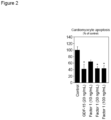

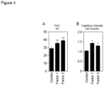

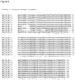


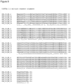

Classifications
-
- A—HUMAN NECESSITIES
- A61—MEDICAL OR VETERINARY SCIENCE; HYGIENE
- A61K—PREPARATIONS FOR MEDICAL, DENTAL OR TOILETRY PURPOSES
- A61K38/00—Medicinal preparations containing peptides
- A61K38/16—Peptides having more than 20 amino acids; Gastrins; Somatostatins; Melanotropins; Derivatives thereof
- A61K38/17—Peptides having more than 20 amino acids; Gastrins; Somatostatins; Melanotropins; Derivatives thereof from animals; from humans
- A61K38/18—Growth factors; Growth regulators
-
- A—HUMAN NECESSITIES
- A61—MEDICAL OR VETERINARY SCIENCE; HYGIENE
- A61K—PREPARATIONS FOR MEDICAL, DENTAL OR TOILETRY PURPOSES
- A61K31/00—Medicinal preparations containing organic active ingredients
- A61K31/70—Carbohydrates; Sugars; Derivatives thereof
- A61K31/7088—Compounds having three or more nucleosides or nucleotides
-
- A—HUMAN NECESSITIES
- A61—MEDICAL OR VETERINARY SCIENCE; HYGIENE
- A61K—PREPARATIONS FOR MEDICAL, DENTAL OR TOILETRY PURPOSES
- A61K38/00—Medicinal preparations containing peptides
- A61K38/16—Peptides having more than 20 amino acids; Gastrins; Somatostatins; Melanotropins; Derivatives thereof
- A61K38/17—Peptides having more than 20 amino acids; Gastrins; Somatostatins; Melanotropins; Derivatives thereof from animals; from humans
- A61K38/1703—Peptides having more than 20 amino acids; Gastrins; Somatostatins; Melanotropins; Derivatives thereof from animals; from humans from vertebrates
- A61K38/1709—Peptides having more than 20 amino acids; Gastrins; Somatostatins; Melanotropins; Derivatives thereof from animals; from humans from vertebrates from mammals
-
- A—HUMAN NECESSITIES
- A61—MEDICAL OR VETERINARY SCIENCE; HYGIENE
- A61K—PREPARATIONS FOR MEDICAL, DENTAL OR TOILETRY PURPOSES
- A61K38/00—Medicinal preparations containing peptides
- A61K38/16—Peptides having more than 20 amino acids; Gastrins; Somatostatins; Melanotropins; Derivatives thereof
- A61K38/17—Peptides having more than 20 amino acids; Gastrins; Somatostatins; Melanotropins; Derivatives thereof from animals; from humans
- A61K38/177—Receptors; Cell surface antigens; Cell surface determinants
-
- A—HUMAN NECESSITIES
- A61—MEDICAL OR VETERINARY SCIENCE; HYGIENE
- A61K—PREPARATIONS FOR MEDICAL, DENTAL OR TOILETRY PURPOSES
- A61K9/00—Medicinal preparations characterised by special physical form
- A61K9/0012—Galenical forms characterised by the site of application
- A61K9/0019—Injectable compositions; Intramuscular, intravenous, arterial, subcutaneous administration; Compositions to be administered through the skin in an invasive manner
-
- A—HUMAN NECESSITIES
- A61—MEDICAL OR VETERINARY SCIENCE; HYGIENE
- A61P—SPECIFIC THERAPEUTIC ACTIVITY OF CHEMICAL COMPOUNDS OR MEDICINAL PREPARATIONS
- A61P17/00—Drugs for dermatological disorders
- A61P17/02—Drugs for dermatological disorders for treating wounds, ulcers, burns, scars, keloids, or the like
-
- A—HUMAN NECESSITIES
- A61—MEDICAL OR VETERINARY SCIENCE; HYGIENE
- A61P—SPECIFIC THERAPEUTIC ACTIVITY OF CHEMICAL COMPOUNDS OR MEDICINAL PREPARATIONS
- A61P17/00—Drugs for dermatological disorders
- A61P17/06—Antipsoriatics
-
- A—HUMAN NECESSITIES
- A61—MEDICAL OR VETERINARY SCIENCE; HYGIENE
- A61P—SPECIFIC THERAPEUTIC ACTIVITY OF CHEMICAL COMPOUNDS OR MEDICINAL PREPARATIONS
- A61P19/00—Drugs for skeletal disorders
- A61P19/02—Drugs for skeletal disorders for joint disorders, e.g. arthritis, arthrosis
-
- A—HUMAN NECESSITIES
- A61—MEDICAL OR VETERINARY SCIENCE; HYGIENE
- A61P—SPECIFIC THERAPEUTIC ACTIVITY OF CHEMICAL COMPOUNDS OR MEDICINAL PREPARATIONS
- A61P27/00—Drugs for disorders of the senses
- A61P27/02—Ophthalmic agents
-
- A—HUMAN NECESSITIES
- A61—MEDICAL OR VETERINARY SCIENCE; HYGIENE
- A61P—SPECIFIC THERAPEUTIC ACTIVITY OF CHEMICAL COMPOUNDS OR MEDICINAL PREPARATIONS
- A61P29/00—Non-central analgesic, antipyretic or antiinflammatory agents, e.g. antirheumatic agents; Non-steroidal antiinflammatory drugs [NSAID]
-
- A—HUMAN NECESSITIES
- A61—MEDICAL OR VETERINARY SCIENCE; HYGIENE
- A61P—SPECIFIC THERAPEUTIC ACTIVITY OF CHEMICAL COMPOUNDS OR MEDICINAL PREPARATIONS
- A61P31/00—Antiinfectives, i.e. antibiotics, antiseptics, chemotherapeutics
-
- A—HUMAN NECESSITIES
- A61—MEDICAL OR VETERINARY SCIENCE; HYGIENE
- A61P—SPECIFIC THERAPEUTIC ACTIVITY OF CHEMICAL COMPOUNDS OR MEDICINAL PREPARATIONS
- A61P35/00—Antineoplastic agents
-
- A—HUMAN NECESSITIES
- A61—MEDICAL OR VETERINARY SCIENCE; HYGIENE
- A61P—SPECIFIC THERAPEUTIC ACTIVITY OF CHEMICAL COMPOUNDS OR MEDICINAL PREPARATIONS
- A61P43/00—Drugs for specific purposes, not provided for in groups A61P1/00-A61P41/00
-
- A—HUMAN NECESSITIES
- A61—MEDICAL OR VETERINARY SCIENCE; HYGIENE
- A61P—SPECIFIC THERAPEUTIC ACTIVITY OF CHEMICAL COMPOUNDS OR MEDICINAL PREPARATIONS
- A61P9/00—Drugs for disorders of the cardiovascular system
-
- A—HUMAN NECESSITIES
- A61—MEDICAL OR VETERINARY SCIENCE; HYGIENE
- A61P—SPECIFIC THERAPEUTIC ACTIVITY OF CHEMICAL COMPOUNDS OR MEDICINAL PREPARATIONS
- A61P9/00—Drugs for disorders of the cardiovascular system
- A61P9/04—Inotropic agents, i.e. stimulants of cardiac contraction; Drugs for heart failure
-
- A—HUMAN NECESSITIES
- A61—MEDICAL OR VETERINARY SCIENCE; HYGIENE
- A61P—SPECIFIC THERAPEUTIC ACTIVITY OF CHEMICAL COMPOUNDS OR MEDICINAL PREPARATIONS
- A61P9/00—Drugs for disorders of the cardiovascular system
- A61P9/10—Drugs for disorders of the cardiovascular system for treating ischaemic or atherosclerotic diseases, e.g. antianginal drugs, coronary vasodilators, drugs for myocardial infarction, retinopathy, cerebrovascula insufficiency, renal arteriosclerosis
-
- C—CHEMISTRY; METALLURGY
- C07—ORGANIC CHEMISTRY
- C07K—PEPTIDES
- C07K16/00—Immunoglobulins [IGs], e.g. monoclonal or polyclonal antibodies
- C07K16/18—Immunoglobulins [IGs], e.g. monoclonal or polyclonal antibodies against material from animals or humans
- C07K16/22—Immunoglobulins [IGs], e.g. monoclonal or polyclonal antibodies against material from animals or humans against growth factors ; against growth regulators
-
- C—CHEMISTRY; METALLURGY
- C07—ORGANIC CHEMISTRY
- C07K—PEPTIDES
- C07K16/00—Immunoglobulins [IGs], e.g. monoclonal or polyclonal antibodies
- C07K16/18—Immunoglobulins [IGs], e.g. monoclonal or polyclonal antibodies against material from animals or humans
- C07K16/28—Immunoglobulins [IGs], e.g. monoclonal or polyclonal antibodies against material from animals or humans against receptors, cell surface antigens or cell surface determinants
-
- C—CHEMISTRY; METALLURGY
- C07—ORGANIC CHEMISTRY
- C07K—PEPTIDES
- C07K2317/00—Immunoglobulins specific features
- C07K2317/30—Immunoglobulins specific features characterized by aspects of specificity or valency
- C07K2317/34—Identification of a linear epitope shorter than 20 amino acid residues or of a conformational epitope defined by amino acid residues
-
- C—CHEMISTRY; METALLURGY
- C07—ORGANIC CHEMISTRY
- C07K—PEPTIDES
- C07K2317/00—Immunoglobulins specific features
- C07K2317/70—Immunoglobulins specific features characterized by effect upon binding to a cell or to an antigen
- C07K2317/76—Antagonist effect on antigen, e.g. neutralization or inhibition of binding
-
- C—CHEMISTRY; METALLURGY
- C12—BIOCHEMISTRY; BEER; SPIRITS; WINE; VINEGAR; MICROBIOLOGY; ENZYMOLOGY; MUTATION OR GENETIC ENGINEERING
- C12N—MICROORGANISMS OR ENZYMES; COMPOSITIONS THEREOF; PROPAGATING, PRESERVING, OR MAINTAINING MICROORGANISMS; MUTATION OR GENETIC ENGINEERING; CULTURE MEDIA
- C12N2799/00—Uses of viruses
- C12N2799/02—Uses of viruses as vector
- C12N2799/021—Uses of viruses as vector for the expression of a heterologous nucleic acid
- C12N2799/022—Uses of viruses as vector for the expression of a heterologous nucleic acid where the vector is derived from an adenovirus
Definitions
- the present invention relates to proteins comprising amino acid sequences encoded by nucleic acids derived from the human chromosomal region C19Orf10 termed Factor 1 for use as a medicament.
- AMI Acute myocardial infarction
- the treatment of a patient who has an AMI comprises the opening of the occluded coronary artery by reperfusion therapy combined with the administration of thrombocyte aggregation inhibitors and coagulation inhibitors to prevent the reclosure of the vessel. Further the pulse and blood pressure can be lowered through administration of beta-blockers and ACE inhibitors. Also common is the use of statins for lowering of the cholesterol level.
- the medical approach for a direct repair of the heart muscle is currently limited to the experimental use of body's own bone marrow cells. There is a great need for drugs with a similar effect without the use of bone marrow cells.
- the tissue necrosis sustained during AMI triggers a wound healing response that leads to a replacement of the necrotic area with granulation tissue and eventually a collagen-rich scar.
- Monocytes are recruited from the bone marrow into the infarcted myocardium and play important roles during wound healing after AMI.
- the monocyte response in the myocardium is temporally biphasic. Pro-inflammatory monocytes appear earlier and promote digestion of infarcted tissue and removal of necrotic debris, whereas reparative monocytes dominate later and propagate angiogenesis and repair.
- Cell surface expression of the chemokine receptor CXCR4 identifies a reparative monocyte subset in mice and humans.
- CXCR4+ myeloid cells Pro-angiogenic and pro-healing effects of CXCR4+ myeloid cells are thought to be mediated via secreted proteins acting in a paracrine manner, but the identity of these Factors was largely unknown. Therefore, the inventors conducted a bioinformatic secretome analysis in human CXCR4+ bone marrow cells to identify novel secreted proteins controlling infarct healing and showing therapeutic potential after AMI.
- the amino acid sequence of human Factor 1 is encoded in open reading frame 10 on human chromosome 19 (C19Orf10).
- the Protein was described in 2007 in a proteom-analysis of the so called fibroblast-like synoviocytes (FLS-cells) as a new secreted Factor in the synovium.
- FLS-cells fibroblast-like synoviocytes
- a correlation between the secretion of the protein and inflammatory diseases of the joint has been supposed without any experimental or statistical evidence ( Weiler et al., Arthritis Research and Therapy 2007 , The identification and characterization of a novel protein, c19orf10, in the synovium).
- a corresponding patent application claims the protein as therapeutic agent for the treatment of joint and for the diagnosis of a tissue undergoing altered growth as well as monitoring changes in a tissue ( US 2008/0004232 A1 , Characterization of c19orf10, a novel synovial protein).
- Another scientific publication describes an enhanced expression of the protein in hepatocellar carcinoma cells ( Sunagozaka et al., International Journal of Cancer, 2010 , Identification of a secretory protein c19orf10 activated in hepatocellular carcinoma). Recombinant produced protein showed a proliferation enhancing effect on cultured hepatocellar carcinoma cells.
- C19Orf10 has also been referred to as IL-25, IL-27 and IL-27W as it was originally considered an interleukin.
- IL-25 and “IL-27” have been used inconsistently in the art and have been used to designate a variety of different proteins.
- US 2004/0185049 refers to a protein as IL-27 and discloses its use in modulating the immune response.
- This protein is structurally distinct from Factor 1 (compare Factor 1 amino acid sequence according to SEQ ID NO: 1 to the amino acid sequence of "IL-27” according to UniProt: Q8NEV9).
- EP 2 130 547 A1 refers to a protein as IL-25 and discloses its use in treating inflammation.
- This protein has also been referred to in the art as IL-17E and is structurally distinct from Factor 1 (compare the amino acid sequence of Factor 1 according to SEQ ID NO: 1 to the amino acid sequence of "IL-25" according to UniProt: Q9H293).
- the amino acid sequence of human Factor 2 is encoded in open reading frame 63 on human chromosome 19 (C19Orf63).
- the protein was described in 2009 as new secreted Factor INM02 ( Wang et al., Journal of Endocrinology 2009 , Molecular cloning of a novel secreted peptide, INM02, and regulation of its expression by glucose). The presence of the protein was shown in human serum with polyclonal antibodies. Further, a correlation between the expression of the protein in cultured MIN6 (beta-cells) as well as in isolated pancreatic rat islets and the glucose concentration in the medium was shown. An analysis of the correlation between diabetes and the expression of INM2 provided no significant results.
- a corresponding patent application claims the production of polyclonal antibodies against the protein and the use for the treatment of diabetes mellitus ( CN 200910055490 , Novel polyclonal antibody of secretive peptide INM02 and preparation method thereof).
- hHSS1 human Hematopoietic Signal peptide-containing Secreted 1
- J Neurooncol, 2011 hHSS1: a novel secreted factor and suppressor of glioma growth located at chromosome 19q13.33
- the published data shows an expression of hHSS1 in stem cells of the hematopoietic system and suggest a function as tumor-suppressor in the genesis of particular brain tumors (gliomas).
- a corresponding patent application claims the use of hHSS1 in the treatment of brain tumors ( WO2011/094446 A1 , A method for treating brain cancer using a novel tumor suppressor gene and secreted factor).
- US 2004/185049 A1 pertains to methods for modulating an immune response in an animal by administering an agent that interacts with WSX-1 receptor on CD-4+ and CD-8+ T cells.
- This patent application does not refer to a protein of SEQ ID NO: 1 or 2 as disclosed herein.
- EP 2 130 547 A1 pertains to the treatment of diseases associated with inflammatory cytokine response resulting from CD-14+ cells and shows that monocytes respond to IL-25 by down-regulating the expression of inflammatory cytokines induced by various inflammatory stimuli.
- This patent application does not refer to a protein of SEQ ID NO: 1 or 2 as disclosed herein.
- WO 02/081681 A1 does not disclose any biological data on the indications referred to and the presumed ability to enhance lymphoid cell proliferation was later shown not to be reproducible.
- KR 2012/0095063 relates to a composition comprising p53 and IL-27 for use in preventing or treating immune diseases.
- the IL-27 protein used has no similarity with present SEQ ID NO: 1.
- US 2004/214229 discloses a peptide identified after triggering of an artificial sepsis in baboons but discloses no experimental data on any medical application of said peptide.
- WO 98/11217 A1 discloses proteins having secretory signal sequences, which can be allegedly used for a vast number of applications. Said publication, however, does not correlate with the polypeptide of present SEQ ID NO: 1 since it discloses a putative signal that is an anlagoue to KDEL for said protein.
- the present invention provides in a first aspect a protein comprising the amino acid sequence according to SEQ ID NO: 1 or a fragment or a variant thereof for use as a medicament.
- Said fragment or variant has at least 90% sequence identity to SEQ ID NO: 1 over the entire length of SEQ ID NO: 1, and the fragment or variant exhibits anti-apoptotic potential and protects cells or tissue from apoptotic cell death, wherein the anti-apoptotic potential is at least 50% of that of the protein with the amino acid sequence according to SEQ ID NO: 1.
- the invention provides nucleic acids encoding the proteins according to the first aspect for the use as a medicament.
- the invention provides vectors comprising the nucleic acid of the second aspect for use as a medicament.
- the invention provides pharmaceutical compositions for use as a medicament, said composition comprising the protein for use as defined herein, the nucleic acid for use as defined herein or the vector for use as defined herein, and optionally a suitable pharmaceutical excipient.
- Nucleic acid molecules are understood as polymeric macromolecules made from nucleotide monomers. Nucleotide monomers are composed of a nucleobase, a five-carbon sugar (such as but not limited to ribose or 2'-deoxyribose), and one to three phosphate groups. Typically, a polynucleotide is formed through phosphodiester bonds between the individual nucleotide monomers.
- nucleic acid molecules include but are not limited to ribonucleic acid (RNA) and deoxyribonucleic acid (DNA).
- RNA ribonucleic acid
- DNA deoxyribonucleic acid
- ORF open reading frame
- ORF refers to a sequence of nucleotides, that can be translated into amino acids.
- such an ORF contains a start codon, a subsequent region usually having a length which is a multiple of 3 nucleotides, but does not contain a stop codon (TAG, TAA, TGA, UAG, UAA, or UGA) in the given reading frame.
- stop codon TAG, TAA, TGA, UAG, UAA, or UGA
- ORFs occur naturally or are constructed artificially, i.e. by gene-technological means.
- An ORF codes for a protein where the amino acids into which it can be translated form a peptide-linked chain.
- protein and “polypeptide” are used interchangeably herein and refer to any peptide-bond-linked chain of amino acids, regardless of length or post-translational modification.
- Proteins usable in the present invention can be further modified by chemical modification.
- This means such a chemically modified polypeptide comprises other chemical groups than the 20 naturally occurring amino acids. Examples of such other chemical groups include without limitation glycosylated amino acids and phosphorylated amino acids.
- Chemical modifications of a polypeptide may provide advantageous properties as compared to the parent polypeptide, e.g. one or more of enhanced stability, increased biological half-life, or increased water solubility.
- Chemical modifications applicable to the variants usable in the present invention include without limitation: PEGylation, glycosylation of non-glycosylated parent polypeptides, covalent coupling to therapeutic small molecules, like glucagon-like peptide 1 agonists, including exenatide, albiglutide, taspoglutide, DPP4 inhibitors, incretin and liraglutide, or the modification of the glycosylation pattern present in the parent polypeptide.
- Such chemical modifications applicable to the variants usable in the present invention may occur co- or post-translational.
- amino acid encompasses naturally occurring amino acids as well as amino acid derivatives.
- a hydrophobic non-aromatic amino acid in the context of the present invention is preferably any amino acid which has a Kyte-Doolittle hydropathy index of higher than 0.5, more preferably of higher than 1.0, even more preferably of higher than 1.5 and is not aromatic.
- a hydrophobic non-aromatic amino acid in the context of the present invention is selected from the group consisting of the amino acids alanine (Kyte Doolittle hydropathy index 1.8), methionine (Kyte Doolittle hydropathy index 1.9), isoleucine (Kyte Doolittle hydropathy index 4.5), leucine Kyte Doolittle hydropathy index 3.8), and valine (Kyte Doolittle hydropathy index 4.2), or derivatives thereof having a Kyte Doolittle hydropathy index as defined above.
- post-translational refers to events that occur after the translation of a nucleotide triplet into an amino acid and the formation of a peptide bond to the proceeding amino acid in the sequence. Such post-translational events may occur after the entire polypeptide was formed or already during the translation process on those parts of the polypeptide that have already been translated. Post-translational events typically alter or modify the chemical or structural properties of the resultant polypeptide. Examples of post-translational events include but are not limited to events such as glycosylation or phosphorylation of amino acids, or cleavage of the peptide chain, e.g. by an endopeptidase.
- co-translational refers to events that occur during the translation process of a nucleotide triplet into an amino acid chain. Those events typically alter or modify the chemical or structural properties of the resultant amino acid chain. Examples of co-translational events include but are not limited to events that may stop the translation process entirely or interrupted the peptide bond formation resulting in two discreet translation products.
- variant is used herein to refer to a polypeptide which differs in comparison to the polypeptide or fragment thereof from which it is derived by one or more changes in the amino acid sequence.
- the polypeptide from which a protein variant is derived is also known as the parent polypeptide.
- the fragment from which a protein fragment variant is derived from is known as the parent fragment.
- a variant is constructed artificially, preferably by gene-technological means.
- the parent polypeptide is a wild-type protein or wild-type protein domain.
- variants usable in the present invention may also be derived from homologs, orthologs, or paralogs of the parent polypeptide or from artificially constructed variant, provided that the variant exhibits at least one biological activity of the parent polypeptide.
- the changes in the amino acid sequence may be amino acid exchanges, insertions, deletions, N-terminal truncations, or C-terminal truncations, or any combination of these changes, which may occur at one or several sites.
- a variant usable in the present invention exhibits a total number of up to 100 (up to 1, 2, 3, 4, 5, 6, 7, 8, 9, 10, 15, 20, 25, 30, 35, 40, 45, 50, 55, 60, 65, 70, 75, 80, 85, 90, 95, or 100,) changes in the amino acid sequence (i.e. exchanges, insertions, deletions, N-terminal truncations, and/or C-terminal truncations).
- the amino acid exchanges may be conservative, and/or semi-conservative, and/or non-conservative.
- a variant usable in the present invention may differ from the protein or domain from which it is derived by up to 1, 2, 3, 4, 5, 6, 7, 8, 9, 10, 11, 12, 13, 14, 15, 16, 17, 18, 19, 20, 25, 30, 35, 40, 45, or 50, amino acid exchanges, preferably conservative amino acid changes.
- Typical substitutions are among the aliphatic amino acids, among the amino acids having aliphatic hydroxyl side chain, among the amino acids having acidic residues, among the amide derivatives, among the amino acids with basic residues, or the amino acids having aromatic residues.
- Typical semi-conservative and conservative substitutions are: Amino acid Conservative substitution Semi-conservative A G; S; T N; V; C C A; V; L M; I; F; G D E; N; Q A; S; T; K; R; H E D; Q; N A; S; T; K; R; H F W; Y; L; M; H I; V; A G A S; N; T; D; E; N; Q H Y; F; K; R L; M; A I V; L; M; A F; Y; W; G K R; H D; E; N; Q; S; T; A L M; I; V; A F; Y; W; H; C M L; I; V; A F; Y; W; C; N Q D; E; S; T; A; G; K; R P V; I L; A; M; W; Y; S; T; C; F Q N D; E; A; S; T; L; M; K; R K
- a "variant" as used herein can be characterized by a certain degree of sequence identity to the parent polypeptide or parent polynucleotide from which it is derived. More precisely, a protein variant in the context of the present invention exhibits at least 90% sequence identity to its parent polypeptide.
- the term "at least 90% sequence identity” is used throughout the specification with regard to polypeptide and polynucleotide sequence comparisons. This expression preferably refers to a sequence identity of at least 90%, at least 91%, at least 92%, at least 93%, at least 94%, at least 95%, at least 96%, at least 97%, at least 98%, or at least 99% to the respective reference polypeptide or to the respective reference polynucleotide.
- Fragments of proteins comprise deletions of amino acids, which may be N-terminal truncations, C-terminal truncations or internal deletions or any combination of these. Such variants comprising N-terminal truncations, C-terminal truncations and/or internal deletions are referred to as "fragments" in the context of the present application.
- a fragment may be naturally occurring (e.g. splice variants) or it may be constructed artificially, preferably by gene-technological means.
- a fragment (or deletion variant) has a deletion of up to 1, 2, 3, 4, 5, 6, 7, 8, 9, 10, 15, 20, 25, 30, 35, 40, 45 or 50 amino acids at its N-terminus and/or at its C-terminus and/or internally as compared to the parent polypeptide, preferably at its N-terminus, at its N- and C-terminus, or at its C-terminus.
- sequence identity is to be calculated with reference to the longer of the two sequences to be compared, if not specifically indicated otherwise.
- the similarity of nucleotide and amino acid sequences can be determined via sequence alignments.
- sequence alignments can be carried out with several art-known algorithms, preferably with the mathematical algorithm of Karlin and Altschul ( Karlin & Altschul (1993) Proc. Natl. Acad. Sci. USA 90: 5873-5877 ), with hmmalign (HMMER package, http://hmmer.wustl.edu/) or with the CLUSTAL algorithm ( Thompson, J. D., Higgins, D. G. & Gibson, T. J. (1994) Nucleic Acids Res.
- sequence matching may be calculated using e.g. BLAST, BLAT or BlastZ (or BlastX).
- BLASTN and BLASTP programs of Altschul et al. (1990) J. Mol. Biol. 215: 403-410 .
- Preferred algorithm parameters used are the default parameters as they are set on http://blast.ncbi.nlm.nih.gov/Blast.
- Expect threshold 10
- word size 3
- max matches in a query range 0
- matrix BLOSUM62
- gap costs Existence: 11 Extension: 1
- compositional adjustments conditional compositional score matrix adjustment together with the database of non-redundant protein sequences (nr) to obtain amino acid sequences homologous to the Factor 1 and Factor 2 polypeptides.
- Gapped BLAST is utilized as described in Altschul et al. (1997) Nucleic Acids Res. 25: 3389-3402 .
- Sequence matching analysis may be supplemented by established homology mapping techniques like Shuffle-LAGAN ( Brudno M., Bioinformatics 2003b, 19 Suppl 1:I54-I62 ) or Markov random fields.
- Shuffle-LAGAN Brudno M., Bioinformatics 2003b, 19 Suppl 1:I54-I62
- Markov random fields Markov random fields.
- host cell refers to a cell that harbours a nucleic acid of the invention (e.g. a plasmid or virus).
- a nucleic acid of the invention e.g. a plasmid or virus
- Such host cell may either be a prokaryotic (e.g. a bacterial cell) or a eukaryotic cell (e.g. a fungal, plant or animal cell).
- the cell can be transformed or non-transformed.
- the cell can be an isolated cell for example in a cell culture or part of a tissue, which itself can be isolated or part of a more complex organization structure such as an organ or an individual.
- Factor 1 refers to the protein indicated in NCBI reference sequence NM_019107.3 (human homologue) as well as it mammalian homologues, in particular from mouse or rat.
- the amino acid sequence of the human homologue is encoded in open reading frame 10 on human chromosome 19 (C19Orf10).
- Factor 1 protein refers to a protein, which comprises, essentially consists or consists of a core segment of human Factor 1 having an amino acid sequence according to SEQ ID NO: 1.
- Factor 1 protein has the amino acid sequence according to SEQ ID NO: 2.
- Factor 2 refers to the protein indicated in NCBI reference sequence NM_175063.4 (human homologue) as well as it mammalian homologues, in particular from mouse or rat.
- the amino acid sequence of human Factor 2 is encoded in open reading frame 63 on human chromosome 19 (C19Orf63).
- Factor 2 protein refers to a protein, which comprises, essentially consists or consists of a core segment of human Factor 2 having an amino acid sequence according to SEQ ID NO: 3.
- the Factor 2 protein may have the amino acid sequence according to SEQ ID NO: 4 and 5, respectively.
- the Factor 2 protein is the secreted form, preferably having an amino acid sequence according to SEQ ID NO: 4.
- non-transformed tissue or “non-transformed cells” refers to tissue and cells which show the physiological parameters of a comparable non cancerous or tumorigenic cell or tissue. Such parameters are, for example but not limited to, cell cycle control, cell-division rate, contact inhibition, anchor-independent growth or metabolism.
- a comparable non cancerous or tumorigenic cell or tissue can be healthy, damaged or diseased.
- the term "healing” comprises the regeneration and repair of living cells, tissue, organs and the biological system as a whole and the partial or complete resumption of normal functioning.
- tissue, organs or a biological system as a whole, it comprises the process by which the cells in the body regenerate and repair to reduce the size of a damaged or necrotic area and replace it with new living tissue.
- the replacement can happen, for example, by regeneration in which the necrotic cells are replaced by new cells that form similar tissue as was originally there; or by repair in which injured tissue is replaced with scar tissue.
- the regeneration is the process which results in partial or complete resumption of normal functioning, it is the preferred variant in the healing process.
- healing in the context of the invention comprises all processes, a person of skill in the art would combine with this term but preferably the regenerative processes should be promoted instead of the processes leading to the production of non functional tissue as for example scar tissue.
- the term healing in the context of the present invention is also preferably directed to the promotion of proliferation, migration, network formation and angiogenesis.
- enhancing proliferation refers to an increase of the cell division rate of a cell or group of cells, if compared to cells or group of cells not treated with the proteins, nucleic acids, vectors or pharmaceutical compositions of the invention. It is well known in the art how to measure cell division rate of cells, e.g. by counting mitotic cells using FACS.
- inhibiting apoptosis refers to the ability of proteins, nucleic acids, vectors or pharmaceutical compositions of the invention to prevent a cell or group of cells to enter apoptosis under conditions, in which control cells or groups of cells enter apoptosis. It is well known to the skilled person how to measure whether a cell undergoes apoptosis, e.g. by TUNEL assay.
- the invention provides a protein comprising a Factor 1 protein with the amino acid sequence according to SEQ ID NO: 1 or a fragment or a variant thereof, which has at least 90% sequence identity to SEQ ID NO: 1 over the entire length of SEQ ID NO: 1, for use as a medicament.
- the protein comprises the amino acid sequence SEQ ID NO: 1 or a fragment thereof.
- the protein has at least 91%, 92%, 93%, 94%, 95%, 96%, 97%, 98%, 99% identity to SEQ ID NO: 1.
- the protein comprises the amino acid sequence SEQ ID NO: 2.
- Preferred fragments of SEQ ID NO: 2 lack the N-terminal signal sequence MAAPSGGWNGVGASLWAALLLGAVALRPAEA (SEQ ID NO: 35).
- a person skilled in the art is able to decide without undue burden, which positions in the parental polypeptide can be mutated to which extent and which positions have to be maintained to preserve the functionality of the polypeptide.
- Such information can, for example, be gained from homologues sequences which can be identified, aligned and analyzed by bioinformatic methods well known in the art. Such analyses are exemplarily described in example 7 and the results are shown in Figures 6 and 7 .
- a Factor 1 protein comprises, essentially consists or consists of the amino acid sequence SEQ ID NO: 2.
- Such mutations may be present in the full length protein according to SEQ ID NO: 2 or in the protein lacking the N-terminal signal sequence according to SEQ ID NO: 1.
- N-terminal deletion variants in addition to the N-terminal signal may lack one or more amino acids from amino acid position 32 to 55 (based on SEQ ID NO: 2), i.e. from the N-terminally conserved region.
- the N-terminus of a deleted Factor 1 protein may be at position 32, 33, 34, 35, 36, 37, 38, 39, 40, 41, 42, 43, 44, 45, 46, 47, 48, 49, 50, 51, 52, 53, 54, 55. or 56 additionally, or alternatively the deleted Factor 1 protein may lack one or more of amino acid positions 146 to 173 (based on SEQ ID NO: 2, i.e. from the C-terminally conserved region.
- the C-terminus of a deleted Factor 1 protein may be at position 145, 146, 147, 148, 149, 150, 151, 152, 153, 154, 155, 156, 157, 158, 159, 160, 161, 162, 163, 164, 165, 166, 167, 168, 169, 170, 171, or 172.
- the protein of the first aspect of the present invention may further comprise additional amino acid sequences, e.g. for stabilizing or purifying the resulting protein.
- additional amino acid sequences e.g. for stabilizing or purifying the resulting protein.
- amino acids are His 6 -tags (SEQ ID NO: 36), myc-tags, or FLAG-tags.
- protease cleavage sites within the protein of the first aspect of the present invention can be preferred to mutate protease cleavage sites within the protein of the first aspect of the present invention to stabilize the protein (see Segers et al. Circulation 2007, 2011 ).
- the skilled person knows how to determine potential proteolytic cleavage sites within a protein.
- protein sequences can be submitted to websites providing such analysis as, e.g. http://web.expasy.org/peptide_cutter/ or http://pmap.burnham.org/proteases.
- These sites may be altered to remove the recognition/cleavage sequence of the respectively identified protease to increase the serum half-life of the protein.
- Factor 1 and Factor 2 have shown proliferation enhancing activity, in particular in enhancing angiogenesis.
- the protein according to the first aspect can be used for enhancing the proliferation of non-transformed tissue or non-transformed cells, preferably angiogenesis. Accordingly, it is preferred that Factor 1 is used in treating diseases, which can benefit from enhanced angiogenesis. Several examples of such diseases are further exemplified below.
- Enhancing of proliferation involves every grade of enhancement of proliferation of a cell or a tissue in comparison to a control cell or control tissue without administration of a protein of the invention.
- the proliferation of a cell can, for example, be measured through the incorporation of bromdeoxyuridine as described in example 2.
- the enhanced proliferation of a tissue can, for example, be determined by measuring the increase of weight or size of the respective tissue as well as with histological methods. Such methods are well known in the art as many of them are standard methods for clinical applications.
- the protein according to the first aspect is for use in treating the indications as listed in claim 2.
- the protein according to the first aspect can also be used for inhibiting apoptosis of non-transformed tissue or non-transformed cells.
- the protein of the present invention exhibits anti-apoptotic potential and protects the cells or tissue from apoptotic cell death.
- Protecting or “cytoprotective effect” in this context means that the extent of apoptotic cell death is reduced in the cells treated with the Factor 1 protein according to the present invention compared to a control by at least 20%, preferably by at least 30%, more preferably by at least 40%, and even more preferably by at least 50%, and most preferably by at least 60%.
- TUNEL in situ TdT-mediated dUTP nick end-labeling
- Other indicators for apoptosis are, for example, a fragmented genome which can be examined, e.g., by DNA laddering ( Liu et al., 2005, Circulation 111:90-96 ), cytochrom-c release, or caspase 3 activity ( Most et al., 2003, J. Biol. Chem. 278:48404-48412 ).
- the anti-apoptotic effect of a peptide may be assessed in vivo in an experimental heart failure animal model.
- mice with postischemic contractile dysfunction may be treated with the protein and cardiac tissue of treated and control mice may be assessed for the extent of apoptotic cardiomyocytes.
- the peptide may be administered preferably parenterally, such as intraperitoneally, intravenously, or subcutaneously.
- the fragments and variants of the Factor 1 protein included in this invention exhibit anti-apoptotic potential and protect the cells or tissue from apoptotic cell death that is at least 50%, preferably 60%, preferably 70%, preferably 80%, preferably 90% and more preferably at least 100% of that of a protein with the amino acid sequence according to SEQ ID NO: 1.
- a protein comprising, consisting essentially of or consisting of a Factor 2 protein, with the amino acid sequence according to SEQ ID NO: 3 or a fragment or a variant thereof, which has at least 80% sequence identity to SEQ ID NO: 3 for use in enhancing proliferation and/or healing of non-transformed tissue or non-transformed cells.
- a Factor 2 protein may comprise the amino acid sequence SEQ ID NO: 3, a fragment or a variant thereof, which has at least 80% identity to SEQ ID NO: 3.
- a respective protein may comprise the amino acid sequence SEQ ID NO: 3 or a fragment thereof.
- the protein has at least 85%, 90%, 91%, 92%, 93%, 94%, 95%, 96%, 97%, 98%, 99% sequence identity to SEQ ID NO: 3.
- This protein may further comprise additional amino acid sequences, e.g. for stabilizing or purifying the resulting protein.
- protease cleavage sites within the protein of the first aspect of the present invention to stabilize the protein.
- Suitable proteolytic cleavage sites can be identified as described above.
- a protein comprising, essentially consisting of or consisting of the amino acid sequence SEQ ID NO: 4, a fragment or a variant thereof, which has at least 80% identity to SEQ ID NO: 4.
- Preferred fragments lack the N-terminal signal sequence MAAASAGATRLLLLLLMAVAA PSRARG'(SEQ ID NO: 37).
- Mutations are preferably introduced only in those regions of the protein, which are not fully conserved between species, preferably mammals, i.e. one or more of those amino acid positions are mutated that are not marked with "*". According to a specific example, only amino acids are altered, which are neither fully conserved (indicated with "*") or conserved to a lesser extent (indicated with either ":” or ".”). Particularly preferred is a Factor 2 protein comprising the amino acid sequence SEQ ID NO: 4 or a fragment thereof. Preferably, the protein has at least 85%, 90%, 91%, 92%, 93%, 94%, 95%, 96%, 97%, 98%, 99% identity to SEQ ID NO: 4.
- Such mutations may be present in the full length protein according to SEQ ID NO: 4 or in the protein lacking the N-terminal signal sequence.
- N-terminal deletion variants in addition to the N-terminal signal may lack one or more amino acids from amino acid position 27 to 73 (based on SEQ ID NO: 4), i.e. from the N-terminally conserved region.
- the N-terminus of a deleted Factor 2 protein may be at position 27, 28, 29, 30, 31, 32, 33, 34, 35, 36, 37, 38, 39, 40, 41, 42, 43, 44, 45, 46, 47, 48, 49, 50, 51, 52, 53, 54, 55. or 56 additionally, or alternatively the deleted Factor 1 protein may lack one or more from amino acid positions 190 to 254 (based on SEQ ID NO: 4, i.e. from the C-terminally conserved region.
- the C-terminus of a deleted Factor 2 protein may be at position 189, 190, 191, 192, 193, 194, 195, 196, 197, 198, 199, 200, 201, 202, 203, 204, 205, 206, 207, 208, 209, 210, 211, 212, 213, 214, 215, 216, 217, 218, 219, 220, 221, 222, 223, 224, 225, 226, 227, 228, 229, 230, 231, 232, 233, 234, 235, 236, 237, 238, 239, 240, 241, 242, 243, 244, 245, 246, 247, 248, 249, 250, 251, 252, or 253.
- a protein comprising, essentially consisting of or consisting of the amino acid sequence of SEQ ID NO: 5, a fragment or a variant thereof, which has at least 80% identity to SEQ ID NO: 5.
- Preferred fragments lack the N-terminal signal sequence MAAASAGATRLLLLLLMAVAAPSRARG (SEQ ID NO: 37).
- Mutations are preferably introduced only in those regions of the protein, which are not fully conserved between species, preferably mammals, i.e. one or more of those amino acid positions are mutated that are not marked with "*". According to a specific example only amino acids are altered, which are neither fully conserved (indicated with "*") or conserved to a lesser extent (indicated with either ":” or ".”). Particularly preferred is a Factor 2 protein comprising the amino acid sequence SEQ ID NO: 5 or a fragment thereof. Preferably, the protein has at least 85%, 90%, 91%, 92%, 93%, 94%, 95%, 96%, 97%, 98%, 99% identity to SEQ ID NO: 5.
- Such mutations may be present in the full length protein according to SEQ ID NO: 5 or in the protein according to SEQ ID NO: 5 lacking the N-terminal signal sequence.
- N-terminal deletion variants in addition to the N-terminal signal may lack one or more amino acids from amino acid position 27 to 73 (based on SEQ ID NO: 4), i.e. from the N-terminally conserved region.
- the N-terminus of a deleted Factor 2 protein may be at position 27, 28, 29, 30, 31, 32, 33, 34, 35, 36, 37, 38, 39, 40, 41, 42, 43, 44, 45, 46, 47, 48, 49, 50, 51, 52, 53, 54, 55. or 56 additionally, or alternatively the deleted Factor 1 protein may lack one or more from amino acid positions 190 to 262 (based on SEQ ID NO: 4, i.e. from the C-terminally conserved region.
- the C-terminus of a deleted Factor 2 protein may be at position 189, 190, 191, 192, 193, 194, 195, 196, 197, 198, 199, 200, 201, 202, 203, 204, 205, 206, 207, 208, 209, 210, 211, 212, 213, 214, 215, 216, 217, 218, 219, 220, 221, 222, 223, 224, 225, 226, 227, 228, 229, 230, 231, 232, 233, 234, 235, 236, 237, 238, 239, 240, 241, 242, 243, 244, 245, 246, 247, 248, 249, 250, 251, 252, 253, 254, 255, 256, 257, 258, 259, 260, or 261.
- the fragments and variants of the protein disclosed herein above exhibit anti-apoptotic potential and protect the cells or tissue from apoptotic cell death that is at least 50%, preferably 60%, preferably 70%, preferably 80%, preferably 90% and more preferably at least 100% of that of a protein with the amino acid sequence according to SEQ ID NO: 3, SEQ ID NO: 4 or SEQ ID NO: 5, most preferably SEQ ID NO: 3.
- the protein can be used for enhancing the proliferation of non-transformed tissue or non-transformed cells. Enhancing of proliferation involves every grade of enhancement in comparison to a control cell or tissue without administration of a protein.
- the experimental methods for the measurement of proliferation are described above. A respective proliferation measurement for such a protein can be assessed by incorporation of bromdeoxyuridine what is also described in example 2.
- the protein may be used for the healing of non-transformed tissue or non-transformed cells.
- a protein may exhibit both of the above functions, i.e., enhancing proliferation and healing of non-transformed tissue or non-transformed cells.
- the protein of aspect one can be administered in vivo, ex vivo or in vitro, preferably in vivo.
- a protein of the first aspect of the invention is administered ex vivo or in vitro is the enhancement of proliferation and/or healing and/or inhibiting apoptosis for the purpose of tissue engineering wherein a tissue is produced for the transplantation into an individual.
- the cells which are used for the tissue engineering can be derived from the same individual but also from another individual of the same species or of another species. The process of removing said cells or tissue and the transplantation of the new tissue into an individual is not comprised by the invention.
- the non-transformed cells can be stem cells.
- stem cells can be embryonic stem cells or adult stem cells as well as progenitor cells.
- the cells can be omnipotent stem cells as well as pluripotent stem cells.
- the non-transformed cells or the non-transformed tissue can be diseased. Alternatively, the non-transformed cells or the non-transformed tissue are damaged. According to one example, the non-transformed cells or the non-transformed tissue are damaged and diseased.
- the non-transformed cells or the non-transformed tissue can be muscle cells or muscle tissue.
- Muscle comprises all types of muscle known by a person of skill in the art. Such muscles are, for example, skeletal, smooth or cardiac muscle. In a more particular preferred embodiment, the muscle is a cardiac muscle.
- the non-transformed cells or the non-transformed tissue are epithelial cells or epithelial tissue, or the non-transformed cells or the non-transformed tissue are nervous cells or nervous tissue.
- the non-transformed cells or the non-transformed tissue may belong to the circulatory system of an individual.
- the non-transformed cells or the non-transformed tissue may belong to or are derived from a defined system of the body of an individual selected from the group comprising the digestive, endocrine, excretory, immune, integumentary, muscular, nervous, reproductive, respiratory or skeletal system.
- the cells may belong to two, three, four, five, six, seven, eight, nine, ten or to all of the listed systems of an individual.
- the non-transformed cells or the non-transformed tissue may alternatively belong to or are derived from a defined part or organ of the body of an individual selected from the group comprising: skin, bone, heart, cartilage, vessel, oesophagus, stomach, intestine, gland, liver, kidney, lung, brain, and spleen.
- the non-transformed cells or the non-transformed tissue belong to or may be derived from the heart.
- the damage may be caused through a genetic/inherited disease or an acquired disease resulting for example from ischemia, reperfusion injury, inflammation, infection, trauma, mechanical overload, intoxication or surgery.
- the damage is caused through ischemia or through a reperfusion injury.
- the damage is preferably caused through a disease which is associated with atrophy, hypoplasy, inflammation, injury, or wounding. It is particularly preferred that the disease is associated with injury. It is also particularly preferred that the disease is associated with wounding.
- the disease is a skeletal muscle disorder selected from the group consisting of muscular dystrophy, muscle weakness, muscular atrophy, myositis, central core disease, nemaline (rod) myopathy, centronuclear myopathy, myotubular myopathy, centronuclear myotubular myopathy, ophthalmoplegia of the eye, and mitochondrial myopathy.
- a skeletal muscle disorder selected from the group consisting of muscular dystrophy, muscle weakness, muscular atrophy, myositis, central core disease, nemaline (rod) myopathy, centronuclear myopathy, myotubular myopathy, centronuclear myotubular myopathy, ophthalmoplegia of the eye, and mitochondrial myopathy.
- the muscular dystrophy may be selected from the group consisting of Becker's muscular dystrophy, congenital muscular dystrophy, Duchenne muscular dystrophy, distal muscular dystrophy, Emery-Dreifuss muscular dystrophy, facioscapulohumeral muscular dystrophy, Limb-girdle muscular dystrophy, myotonic muscular dystrophy, and oculopharyngeal muscular dystrophy.
- the myositis may be selected from the group consisting of myositis ossificans, fibromyositis, idiopathic inflammatory myopathies (such as dermatomyositis, polymyositis, and inclusion body myositis), and pyomyositis.
- the disease is primary or acquired cardiomyopathy.
- the primary cardiomyopathy is selected from inherited cardiomyopathy and cardiomyopathy caused by spontaneous mutations.
- Cardiomyopathies are for example but not limited to Hypertrophic cardiomyopathy (HCM or HOCM), Arrhythmogenic right ventricular cardiomyopathy (ARVC), Isolated ventricular non-compaction Mitochondrial myopathy, Dilated cardiomyopathy (DCM), Restrictive cardiomyopathy (RCM), Takotsubo cardiomyopathy, Loeffler endocarditis, diabetic cardiomyopathy, Alcoholic cardiomyopathy, Obesity-associated cardiomyopathy.
- the acquired cardiomypathy is preferably selected from ischemic cardiomyopathy caused by atherosclerotic or other coronary artery diseases, cardiomyopathy caused by infection or intoxication of the myocardium, hypertensive heart disease caused by pulmonary arterial hypertension and/or arterial hypertension and diseases of the heart valves, wherein the ischemic cardiomyopathy caused by atherosclerotic or other coronary artery diseases is particularly preferred.
- Non-transformed cells or non-transformed tissue which is damaged through ischemia or reperfusion injury preferably belongs to the heart. Accordingly, it is preferred that the resulting disease to be treated is selected from the group consisting of myocardial infarction, angina pectoris and heart failure, wherein the myocardial infarction is particularly preferred.
- myocardial infarction as used in the context of the invention comprises the acute myocardial infarction (AMI).
- the medical use of the protein according to the first aspect of the invention comprises the application to an individual after myocardial infarction and the healing comprises the improvement of left ventricular systolic function and may be associated with an increase in capillary density in the infarct border zone. Additionally, the protein for use according to the first aspect of the invention may reduce the mortality after a myocardial infarction.
- the methods which can be used to determine parameters like the improvement of left ventricular systolic function, increase in capillary density in the infarct border zone and the reduction of mortality after a myocardial infarction are well known in the art and exemplarily described in examples 4-6.
- the Factor 1 proteins, fragments, or variants described above are for example for use in treating or ameliorating atrophy; hypoplasy; inflammation, injury; wounding, ischemia; reperfusion injury; inflammation; infection; trauma; mechanical overload; intoxication; primary or acquired cardiomyopathy, preferably inherited cardiomyopathy and cardiomyopathy caused by spontaneous mutations.
- Cardiomyopathies are for example but not limited to Hypertrophic cardiomyopathy (HCM or HOCM), Arrhythmogenic right ventricular cardiomyopathy (ARVC), Isolated ventricular non-compaction Mitochondrial myopathy, Dilated cardiomyopathy (DCM), Restrictive cardiomyopathy (RCM), Takotsubo cardiomyopathy, Loeffler endocarditis, diabetic cardiomyopathy, Alcoholic cardiomyopathy, or Obesity-associated cardiomyopathy; myocardial infarction or in improving left ventricular systolic function.
- HCM Hypertrophic cardiomyopathy
- ARVC Arrhythmogenic right ventricular cardiomyopathy
- Isolated ventricular non-compaction Mitochondrial myopathy Dilated cardiomyopathy (DCM), Restrictive cardiomyopathy (RCM), Takotsubo cardiomyopathy, Loeffler endocarditis, diabetic cardiomyopathy, Alcoholic cardiomyopathy, or Obesity-associated cardiomyopathy
- Factor 1 and Factor 2 proteins, fragments, or variants described above for use in methods of treating the respectively indicated conditions and diseases.
- the invention provides nucleic acids encoding the proteins according to the first aspect for use as a medicament.
- enhancing proliferation and/or healing and/or inhibiting apoptosis of non-transformed tissue or non-transformed cells have the meaning and preferred meanings defined above.
- Nucleic acid sequences can be optimized in an effort to enhance expression in a host cell. Parameters to be considered include C:G content, preferred codons, and the avoidance of inhibitory secondary structure. These Factors can be combined in different ways in an attempt to obtain nucleic acid sequences having enhanced expression in a particular host (cf. e.g. Donnelly et al., International Publication Number WO 97/47358 ). The ability of a particular sequence to have enhanced expression in a particular host involves some empirical experimentation. Such experimentation involves measuring expression of a prospective nucleic acid sequence and, if needed, altering the sequence.
- the nucleic acid may further comprise a transcriptional control element or expression control sequences positioned to control expression of the protein.
- a transcriptional control element or expression control sequences positioned to control expression of the protein.
- Such a nucleic acid together with control elements is often termed as an expression system.
- expression system refers to a system designed to produce one or more gene products of interest. Typically, such system is designed “artificially”, i.e. by gene-technological means usable to produce the gene product of interest in vivo, in vitro or ex vivo.
- expression system further encompasses the expression of the gene product of interest comprising the transcription of the polynucleotides, mRNA splicing, translation into a polypeptide, co- and post-translational modification of a polypeptide or protein as well as the targeting of the protein to one or more compartments inside of the cell, the secretion from the cell and the uptake of the protein in the same or another cell.
- This general description refers to expression systems for the use in eukaryotic cells, tissues or organisms. Expression systems for prokaryotic systems may differ, wherein it is well known in the art, how an expression system for prokaryotic cells is constructed.
- Regulatory elements present in a gene expression cassette generally include: (a) a promoter transcriptionally coupled to a nucleotide sequence encoding the polypeptide, (b) a 5' ribosome binding site functionally coupled to the nucleotide sequence, (c) a terminator joined to the 3' end of the nucleotide sequence, and (d) a 3' polyadenylation signal functionally coupled to the nucleotide sequence.
- Additional regulatory elements useful for enhancing or regulating gene expression or polypeptide processing may also be present. Promoters are genetic elements that are recognized by an RNA polymerase and mediate transcription of downstream regions. Preferred promoters are strong promoters that provide for increased levels of transcription.
- promoters examples include the immediate early human cytomegalovirus promoter (CMV), and CMV with intron A ( Chapman et al, Nucl. Acids Res. 19:3979-3986, 1991 ). Additional examples of promoters include naturally occurring promoters such as the EF1 alpha promoter, the murine CMV promoter, Rous sarcoma virus promoter, and SV40 early/late promoters and the [beta]-actin promoter; and artificial promoters such as a synthetic muscle specific promoter and a chimeric muscle-specific/CMV promoter ( Li et al., Nat. Biotechnol. 17:241-245, 1999 , Hagstrom et al., Blood 95:2536-2542, 2000 ).
- CMV immediate early human cytomegalovirus promoter
- CMV with intron A Chapman et al, Nucl. Acids Res. 19:3979-3986, 1991 .
- Additional examples of promoters include naturally occurring
- the ribosome binding site is located at or near the initiation codon. Examples of preferred ribosome binding sites include CCACCAUGG, CCGCCAUGG, and ACCAUGG, where AUG is the initiation codon ( Kozak, Cell 44:283-292, 1986 ).
- the polyadenylation signal is responsible for cleaving the transcribed RNA and the addition of a poly (A) tail to the RNA.
- the polyadenylation signal in higher eukaryotes contains an AAUAAA sequence about 11-30 nucleotides from the polyadenylation addition site. The AAUAAA sequence is involved in signalling RNA cleavage ( Lewin, Genes IV, Oxford University Press, NY, 1990 ).
- the poly (A) tail is important for the processing, export from the nucleus, translation and stability of the mRNA.
- Polyadenylation signals that can be used as part of a gene expression cassette include the minimal rabbit [beta] -globin polyadenylation signal and the bovine growth hormone polyadenylation (BGH) ( Xu et al., Gene 272:149-156, 2001 , Post et al., U.S. Patent U. S. 5,122,458 ).
- BGH bovine growth hormone polyadenylation
- Examples of additional regulatory elements useful for enhancing or regulating gene expression or polypeptide processing that may be present include an enhancer, a leader sequence and an operator.
- An enhancer region increases transcription. Examples of enhancer regions include the CMV enhancer and the SV40 enhancer ( Hitt et al., Methods in Molecular Genetics 7: 13-30, 1995 , Xu, et al., Gene 272: 149-156, 2001 ).
- An enhancer region can be associated with a promoter.
- the expression of the protein according to the first aspect of the invention may be regulated.
- Such regulation can be accomplished in many steps of the gene expression. Possible regulation steps are, for example but not limited to, initiation of transcription, promoter clearance, elongation of transcription, splicing, export from the nucleus, mRNA stability, initiation of translation, translational efficiency, elongation of translation and protein folding.
- Other regulation steps which influence the concentration of a Factor 1 or Factor 2 polypeptide inside a cell affect the half-life of the protein.
- Such a regulation step is, for example, the regulated degeneration of proteins.
- the proteins of the invention comprise secreted proteins, the protein can be directed to a secretory pathway of the host cell.
- Outside of the cell can refer to, for example but not limited to, a culture medium, a tissue, intracellular matrix or space or a body fluid such as blood or lymph.
- the control of the regulatory steps mentioned above can be, for example, cell-type or tissue-type independent or cell-type or tissue-type specific.
- the control of the regulatory steps can be cell-type or tissue-type specific.
- Such a cell-type or tissue-type specific regulation is preferably accomplished through the regulation steps referring to the transcription of a nucleic acid.
- This transcriptional regulation can be accomplished through the use of cell-type or tissue-type specific promoter sequences.
- the result of this cell-type or tissue-type specific regulation can have different grades of specificity. This means, that the expression of a respective polypeptide is enhanced in the respective cell or tissue in comparison to other cell- or tissue-type or that the expression is limited to the respective cell- or tissue-type.
- Cell- or tissue-type specific promoter sequences are well known in the art and available for a broad range of cell- or tissue-types.
- the expression may not be cell-type or tissue-type specific but depends from physiological conditions. Such conditions are for example an inflammation or a wound. Such a physiological condition-specific expression can also be accomplished through regulation at all above mentioned regulation steps.
- the preferred way of regulation for a physiological condition-specific expression is the transcriptional regulation.
- a wound or inflammation specific promoter can be used.
- Respective promoters are, for example, natural occurring sequences, which can be, for example, derived from genes, which are specifically expressed during an immune reaction and/or the regeneration of wounded tissue.
- artificial promoter sequences which are, for example constructed through combination of two or more naturally occurring sequences.
- the regulation may be cell-type or tissue-type specific and physiological condition-specific.
- the expression is a heart specific expression.
- Particularly preferred is a heart specific and wound specific expression.
- Another possibility for a regulation of expression of the protein according to the first aspect of the invention is the conditional regulation of the gene expression.
- an operator sequence can be used.
- the Tet operator sequence can be used to repress gene expression.
- the conditional regulation of gene expression by means of the Tet operator together with a Tet repressor is well known in the art and many respective systems have been established for a broad range of prokaryotic and eukaryotic organisms. A person of skill in the art knows how to choose a suitable system and adapt it to the special needs of the respective application.
- a nucleic acid according to the invention preferably comprises the application to an individual after myocardial infarction and the healing preferably comprises the improvement of left ventricular systolic function and may be associated with an increase in capillary density in the infarct border zone. Additionally, it may reduce the mortality after a myocardial infarction.
- the methods which can be used to determine parameters like the improvement of left ventricular systolic function, increase in capillary density in the infarct border zone and the reduction of mortality after a myocardial infarction are well known in the art and exemplarily described in examples 4-6.
- Nucleic acids encoding the Factor 1 and Factor 2 proteins, fragments, or variants described above are preferably used in treating or ameliorating atrophy; hypoplasy; inflammation, injury; wounding, ischemia; reperfusion injury; inflammation; infection; trauma; mechanical overload; intoxication; primary or acquired cardiomyopathy, preferably inherited cardiomyopathy and cardiomyopathy caused by spontaneous mutations.
- Cardiomyopathies are for example but not limited to Hypertrophic cardiomyopathy (HCM or HOCM), Arrhythmogenic right ventricular cardiomyopathy (ARVC), Isolated ventricular non-compaction Mitochondrial myopathy, Dilated cardiomyopathy (DCM), Restrictive cardiomyopathy (RCM), Takotsubo cardiomyopathy, Loeffler endocarditis, diabetic cardiomyopathy, Alcoholic cardiomyopathy, or Obesity-associated cardiomyopathy; myocardial infarction or in improving left ventricular systolic function.
- HCM Hypertrophic cardiomyopathy
- ARVC Arrhythmogenic right ventricular cardiomyopathy
- Isolated ventricular non-compaction Mitochondrial myopathy Dilated cardiomyopathy (DCM), Restrictive cardiomyopathy (RCM), Takotsubo cardiomyopathy, Loeffler endocarditis, diabetic cardiomyopathy, Alcoholic cardiomyopathy, or Obesity-associated cardiomyopathy
- the invention provides vectors comprising the nucleic acid or the expression system of the second aspect for use as a medicament.
- enhancing proliferation and/or healing and/or inhibiting apoptosis of non-transformed tissue or non-transformed cells have the meaning and preferred meanings defined above.
- vector refers to a protein or a polynucleotide or a mixture thereof which is capable of being introduced or of introducing the proteins and/or nucleic acid comprised therein into a cell.
- genes of interest encoded by the introduced polynucleotide are expressed within the host cell upon introduction of the vector or the vectors.
- suitable vectors include but are not limited to plasmid vectors, cosmid vectors, phage vectors such as lambda phage, filamentous phage vectors, viral vectors, viral like particles, and bacterial spores.
- the vector is a viral vector.
- Suitable viral vectors include but are not limited to adenoviral vectors, adeno-associated viral (AAV) vectors, alphaviral vectors, herpes viral vectors, measles viral vectors, pox viral vectors, vesicular stomatitis viral vectors, retroviral vector and lentiviral vectors.
- AAV adeno-associated viral
- the vector is an adenoviral or an adeno-associated viral (AAV) vector.
- Nucleic acid encoding one or more proteins of the first aspect of the invention can be introduced into a host cell, a tissue or an individual using vectors suitable for therapeutic administration. Suitable vectors can preferably deliver nucleic acids into a target cell without causing an unacceptable side effect.
- the use of a vector according to the invention may comprise the application to an individual after myocardial infarction and the healing comprises the improvement of left ventricular systolic function and may be associated with an increase in capillary density in the infarct border zone. Additionally, it may reduce the mortality after a myocardial infarction.
- the methods which can be used to determine parameters like the improvement of left ventricular systolic function, increase in capillary density in the infarct border zone and the reduction of mortality after a myocardial infarction are well known in the art and exemplarily described in examples 4-6.
- Vectors comprising nucleic acids encoding the Factor 1 and Factor 2 proteins, fragments, or variants described above are preferably used in treating or ameliorating atrophy; hypoplasy; inflammation, injury; wounding, ischemia; reperfusion injury; inflammation; infection; trauma; mechanical overload; intoxication; primary or acquired cardiomyopathy, preferably inherited cardiomyopathy and cardiomyopathy caused by spontaneous mutations.
- Cardiomyopathies are for example but not limited to Hypertrophic cardiomyopathy (HCM or HOCM), Arrhythmogenic right ventricular cardiomyopathy (ARVC), Isolated ventricular non-compaction Mitochondrial myopathy, Dilated cardiomyopathy (DCM), Restrictive cardiomyopathy (RCM), Takotsubo cardiomyopathy, Loeffler endocarditis, diabetic cardiomyopathy, Alcoholic cardiomyopathy, or Obesity-associated cardiomyopathy; myocardial infarction or in improving left ventricular systolic function.
- HCM Hypertrophic cardiomyopathy
- ARVC Arrhythmogenic right ventricular cardiomyopathy
- Isolated ventricular non-compaction Mitochondrial myopathy Dilated cardiomyopathy (DCM), Restrictive cardiomyopathy (RCM), Takotsubo cardiomyopathy, Loeffler endocarditis, diabetic cardiomyopathy, Alcoholic cardiomyopathy, or Obesity-associated cardiomyopathy
- the invention provides pharmaceutical compositions for use as a medicament, said composition comprising the protein for use as defined herein and/or the nucleic acid for use as defined herein and/or the vector for use as defined herein, and optionally a carrier.
- enhancing proliferation and/or healing and/or inhibiting apoptosis of non-transformed tissue or non-transformed cells have the meaning and preferred meanings defined above.
- carrier refers to a pharmacologically inactive substance such as but not limited to a diluent, excipient, surfactants, stabilizers, physiological buffer solutions or vehicles with which the therapeutically active ingredient is administered.
- Such pharmaceutical carriers can be liquid or solid.
- Liquid carriers include but are not limited to sterile liquids, such as saline solutions in water and oils, including but not limited to those of petroleum, animal, vegetable or synthetic origin, such as peanut oil, soybean oil, mineral oil, sesame oil and the like. Saline solutions and aqueous dextrose and glycerol solutions can also be employed as liquid carriers, particularly for injectable solutions.
- a saline solution is a preferred carrier when the pharmaceutical composition is administered intravenously.
- Suitable pharmaceutical carriers are described in "Remington's Pharmaceutical Sciences” by E. W. Martin.
- the carrier is preferably a suitable pharmaceutical excipient.
- Suitable pharmaceutical "excipients” comprise starch, glucose, lactose, sucrose, gelatine, malt, rice, flour, chalk, silica gel, sodium stearate, glycerol monostearate, talc, sodium chloride, dried skim milk, glycerol, propylene, glycol, water, ethanol and the like.
- Such suitable pharamaceutical excipients are preferably pharmaceutically acceptable.
- “Pharmaceutically acceptable” means approved by a regulatory agency of the Federal or a state government or listed in the U.S. Pharmacopeia or other generally recognized pharmacopeia for use in animals, and more particularly in humans.
- composition is intended to include the formulation of the active compound with encapsulating material as a carrier providing a capsule in which the active component with or without other carriers, is surrounded by a carrier, which is thus in association with the active compound.
- active ingredient refers to the substance in a pharmaceutical composition or formulation that is biologically active, i.e. that provides pharmaceutical value.
- the active ingredient is a protein of the first aspect and/or a nucleic acid of the second aspect and/or a vector of the third aspect.
- a pharmaceutical composition may comprise one or more active ingredients which may act in conjunction with or independently of each other.
- the active ingredient can be formulated as neutral or salt forms.
- the salt form is preferably a pharmaceutically acceptable salt.
- pharmaceutically acceptable salt refers to, for example but not limited to, a salt of the polypeptides of the present invention.
- Suitable pharmaceutically acceptable salts include acid addition salts which may, for example, be formed by mixing a solution of the polypeptide of the present invention with a solution of a pharmaceutically acceptable acid such as hydrochloric acid, sulfuric acid, fumaric acid, maleic acid, succinic acid, acetic acid, benzoic acid, citric acid, tartaric acid, carbonic acid or phosphoric acid.
- suitable pharmaceutically acceptable salts thereof may include alkali metal salts (e.g., sodium or potassium salts); alkaline earth metal salts (e.g., calcium or magnesium salts); and salts formed with suitable organic ligands (e.g., ammonium, quaternary ammonium and amine cations formed using counteranions such as halide, hydroxide, carboxylate, sulfate, phosphate, nitrate, alkyl sulfonate and aryl sulfonate).
- alkali metal salts e.g., sodium or potassium salts
- alkaline earth metal salts e.g., calcium or magnesium salts
- suitable organic ligands e.g., ammonium, quaternary ammonium and amine cations formed using counteranions such as halide, hydroxide, carboxylate, sulfate, phosphate, nitrate, alkyl sulfonate and aryl sul
- compositions include, but are not limited to, acetate, adipate, alginate, ascorbate, aspartate, benzenesulfonate, benzoate, bicarbonate, bisulfate, bitartrate, borate, bromide, butyrate, calcium edetate, camphorate, camphorsulfonate, camsylate, carbonate, chloride, citrate, clavulanate, cyclopentanepropionate, digluconate, dihydrochloride, dodecylsulfate, edetate, edisylate, estolate, esylate, ethanesulfonate, formate, fumarate, gluceptate, glucoheptonate, gluconate, glutamate, glycerophosphate, glycolylarsanilate, hemisulfate, heptanoate, hexanoate, hexylresorcinate
- the active ingredient is administered to a cell, a tissue or an individual in an effective amount.
- An "effective amount” is an amount of an active ingredient sufficient to achieve the intended purpose.
- the active ingredient may be a therapeutic agent.
- the effective amount of a given active ingredient will vary with parameters such as the nature of the ingredient, the route of administration, the size and species of the individual to receive the active ingredient, and the purpose of the administration. The effective amount in each individual case may be determined empirically by a skilled artisan according to established methods in the art.
- administering includes in vivo administration to an individual as well as administration directly to cells or tissue in vitro or ex vivo.
- compositions can be customized for the treatment of a disease or disorder.
- "treat”, “treating” or “treatment” of a disease or disorder means accomplishing one or more of the following: (a) reducing the severity of the disorder; (b) limiting or preventing development of symptoms characteristic of the disorder(s) being treated; (c) inhibiting worsening of symptoms characteristic of the disorder(s) being treated; (d) limiting or preventing recurrence of the disorder(s) in patients that have previously had the disorder(s); (e) limiting or preventing recurrence of symptoms in patients that were previously symptomatic for the disorder(s); (f) reduction of mortality after occurrence of a disease or a disorder; (g) healing; and (h) prophylaxis of a disease.
- “prevent”, “preventing”, “prevention”, or “prophylaxis” of a disease or disorder means preventing that such disease or disorder occurs in patient.
- the healing comprises the treatment of an individual after myocardial infarction and the healing comprises the improvement of left ventricular systolic function and may be associated with an increase in capillary density in the infarct border zone. Additionally, it may reduce the mortality after a myocardial infarction.
- the methods which can be used to determine parameters like the improvement of left ventricular systolic function, increase in capillary density in the infarct border zone and the reduction of mortality after a myocardial infarction are well known in the art and exemplarily described in examples 4-6.
- the pharmaceutical composition contemplated by the present invention may be formulated in various ways well known to one of skill in the art.
- the pharmaceutical composition of the present invention may be in liquid form such as in the form of solutions, emulsions, or suspensions.
- the pharmaceutical composition of the present invention is formulated for parenteral administration, preferably for intravenous, intraarterial, intramuscular, subcutaneous, transdermal, intrapulmonary, intraperitoneal intracoronary, intracardiac administration, or administration via mucous membranes, preferably for intravenous, subcutaneous, or intraperitoneal administration.
- a preparation for oral or anal administration is also possible.
- the pharmaceutical composition of the present invention is in the form of a sterile aqueous solution which may contain other substances, for example, enough salts or glucose to make the solution isotonic with blood.
- the aqueous solutions should be suitably buffered (preferably to a pH of from 3 to 9, more preferably to a pH of from 5 to 7), if necessary.
- the pharmaceutical composition is preferably in unit dosage form. In such form the pharmaceutical composition is subdivided into unit doses containing appropriate quantities of the active component.
- the unit dosage form can be a packaged preparation, the package containing discrete quantities of pharmaceutical composition such as vials or ampoules.
- the administration of the pharmaceutical composition is preferably administered through the intravenous, intraarterial, intramusculuar, subcutaneous, transdermal, intrapulmonary, intraperitoneal, intracoronary or intracardiac route wherein other routes of administration known in the art are also comprised.
- the use of the pharmaceutical composition can replace the standard treatment for the respective disease or condition or can be administered additionally to the standard treatment.
- the pharmaceutical composition can be administered before, simultaneously or after a standard therapy.
- the standard therapy can be a reperfusion therapy and the pharmaceutical composition can be administered before, simultaneously or after the reperfusion therapy.
- the pharmaceutical composition is administered once or more than once. This comprises 2, 3, 4, 5, 6, 7, 8, 9, 10, 11, 12, 13, 14, 15, 16, 17, 18, 19, 20, 25, 30, 35, 40, 45 or 50 times.
- the time span for the administration of the pharmaceutical composition is not limited. Preferably, the administration does not exceed 1, 2, 3, 4, 5, 6, 7 or 8 weeks.
- a single dose of the pharmaceutical composition can independently from the overall amount of administered doses or the respective time span of administration be administered as one or more bolus injection(s) and/or infusion(s).
- the first through fourth aspect of the present invention is based on the observation of the present inventors that Factor 1 (and Factor 2) is a potent pro-angiogenic molecule in vitro and in vivo. Accordingly, the proteins, nucleic acids and vectors described above are also contemplated for ex vivo use, which may be therapeutic, e.g. for ex vivo stimulation of the cells indicated above or for use in cell culture applications.
- Anti-angiogenic strategies are used to treat cancerous conditions and other disorders, such as age-related macular degeneration, where angiogenesis contributes to disease progression (see, e.g. Ferrara N and Kerbel RS (2005) Nature: 438:967-974 or Potente M, et al. (2011) Cell. 146(6):873-887 ). Accordingly, disclosed are anti-angiogenic properties of inhibitors of Factor 1 and Factor 2. In these cases the definitions provided above in the definition section equally apply. Additionally, specific definitions provided in the description of the first to fourth aspect as well as preferred embodiments, e.g. of the term "vector" and of preferred vectors also apply to the following cases unless the context of its use clearly indicates otherwise.
- an inhibitor of Factor 1 and/or 2 protein for medical use, preferably in treating or preventing a disease in which angiogenesis contributes to disease development or progression.
- the term "inhibitor” is used broadly to refer to compounds that interfere with the pro-angiogenic activity of Factor 1 or Factor 2.
- An inhibitor may act on the transcription and/or translation of mRNA encoding Factor 1 or Factor 2 thereby preventing its cellular production and, thus secretion into the circulation and/or at the site of disease development or progression.
- An inhibitor may also act by specifically binding to Factor 1 or Factor 2 protein or to cellular proteins to which Factor 1 or Factor 2 proteins specifically bind, preferably their cellular receptors.
- Such binding may prevent or disrupt the natural interaction of Factor 1 or Factor 2 with other cellular proteins, preferably their respective cellular receptors.
- the skilled person is well aware of how to interfere with the binding between a receptor and its agonist and can use this knowledge to design suitable inhibitors of Factor 1 and Factor 2.
- an inhibitor can be derived from Factor 1 or Factor 2 protein itself by deleting or mutating those parts of Factor 1 and Factor 2 protein, respectively that exert the pro-angiogeneic function of Factor 1 and Factor 2.
- Such inactive mutated or deleted Factor 1 or Factor 2 will compete with wild-type Factor 1 and Factor 2 for its natural binding partners.
- a compound that interferes with the pro-angiogenic activity of Factor 1 or Factor 2 reduces the activity by at least 20%, preferably by at least 30%, more preferably by at least 40%.
- inhibitors that specifically bind to Factor 1 or Factor 2 or that comprise, essentially consist or consist of mutants or fragments of Factor 1 and Factor 2, respectively it is preferred that they exert this level of inhibition of Factor 1 protein or Factor 2 protein at an equimolar concentration.
- a preferred assay, which can be used to determine inhibition of pro-angiogenic activity is described in Example 10 herein. To determine whether a given inhibitor has this activity at an equimolar amount, the molar amounts of Factor 1 and 2, respectively and of the respective inhibitor have to be determined.
- Factor 1 according to SEQ ID NO: 1 has a MW of 15.84 kD and Factor 2 according to SEQ ID NO: 3 has a MW of 21.57 kD and an IgG has a molecular weight of approx. 150 kD.
- 100 ng Factor 1 and 947 ng Factor 1 specific IgG are approx. equimolar and 100 ng Factor 2 and 695 ng Factor 2-specific IgG are approx. equimolar.
- the Factor 1 and Factor 2 specific antibodies provided herein are inhibitors of Factor 1 protein or Factor 2 protein in that sense.
- the level of inhibition is preferably measured on the basis of the protein produced in a cell naturally producing Factor 1 or Factor 2.
- the skilled person is well aware of a large number of methods to measure the amounts of mRNA encoding Factor 1 or Factor 2 as well as of Factor 1 or Factor 2 proteins, which can be used when assessing the ability of a compound to interfere with transcription and/or translation of Factor 1 or Factor 2 encoding mRNA.
- the Factor 1 protein comprises, essentially consists or consists of the amino acid sequence according to SEQ ID NO: 1 and Factor 2 comprises, essentially consists or consists of the amino acid sequence according to SEQ ID NO: 3 or a variant thereof, which has at least 80% sequence identity to SEQ ID NO: 1 or 3.
- the inhibitor disclosed herein is a protein comprising, essentially consisting or consisting of an inhibitory fragment or inhibitory mutant of the amino acid sequence according to SEQ ID NO: 1 or 3 or of a variant thereof, which has at least 80% sequence identity to the amino acid sequence according to SEQ ID NO: 1 or 3.
- proteins that exert their function by protein-protein interaction via a receptor comprise domains that are necessary for binding to the receptor, and domains that elicit the receptor to transmit a signal into the cell.
- the skilled person is well aware how to generate inhibitory fragments or mutants of such receptor binding proteins.
- a series of N-and/or C-terminally truncated Factor 1 or Factor 2 proteins can be generated and tested for their pro-angiogenic activity in an assay as outlined in Example 2. Those fragments which no longer show pro-angiogenic activity are then tested in an assay as outlined in Example 10 for their ability to inhibit the pro-angiogenic activity of Factor 1 or 2 protein.
- Mutants of Factor 1 and 2 can be generated as known in the art, e.g. by alanine scanning mutagenesis.
- each mutant comprises 1, 2, 3 or more amino acids, which have been mutated to alanine (so-called cassettes) and wherein the mutants differ in the positions of the cassettes within Factor 1 or 2.
- Factor 1 or 2 mutants that have lost their pro-angiogenic activity can again be identified as outlined in Example 2.
- Inhibitory mutants can subsequently be identified by using an assay as outlined in Example 10.
- the inhibitor may be a ligand, specifically binding to the amino acid sequence according to SEQ ID NO: 1 or 3 or a variant thereof, which has at least 80% sequence identity to the amino acid sequence according to SEQ ID NO: 1 or 3 or to the receptor that naturally interacts with Factor 1 or 3 or a variant thereof, which has at least 80% sequence identity to the amino acid sequence according to SEQ ID NO: 1 or 3.
- ligand as used herein may refer to a chemical moiety that specifically binds to the specified antigen.
- Preferred ligands are amino acid based ligands, like immunoglobulins, preferably antibodies or antigen-binding fragments thereof and antibody-like proteins.
- ligands may be peptidomimetics.
- immunoglobulin is used herein to refer to immunity conferring glycoproteins of the immunoglobulin superfamily.
- Surface immunoglobulins are attached to the membrane of effector cells by their transmembrane region and encompass molecules such as but not limited to B-cell receptors, T-cell receptors, class I and II major histocompatibility complex (MHC) proteins, beta-2 microglobulin ( ⁇ 2M), CD3, CD4 and CD8.
- MHC major histocompatibility complex
- ⁇ 2M beta-2 microglobulin
- CD3, CD4 and CD8 CD3, CD4 and CD8.
- antibody refers to secreted immunoglobulins which lack the transmembrane region and can thus, be released into the bloodstream and body cavities. Antibodies are grouped into different isotypes based on the heavy chain they possess.
- Ig heavy chains There are five types of human Ig heavy chains denoted by the Greek letters: ⁇ , ⁇ , ⁇ , ⁇ , and ⁇ .
- the type of heavy chain present defines the class of antibody, i.e. these chains are found in IgA, IgD, IgE, IgG, and IgM antibodies, respectively, each performing different roles, and directing the appropriate immune response against different types of antigens. Distinct heavy chains differ in size and composition; ⁇ and ⁇ comprise approximately 450 amino acids, while ⁇ and ⁇ have approximately 550 amino acids ( Janeway et al. (2001) Immunobiology, Garland Science ).
- Antibodies comprise four polypeptide chains, namely two heavy (H) chains and two light (L) chains inter-connected by disulfide bonds.
- Each heavy chain is comprised of a heavy chain variable region (abbreviated herein as HCVR or VH) and a heavy chain constant region.
- the heavy chain constant region is comprised of three domains, CH1, CH2 and CH3.
- Each light chain is comprised of a light chain variable region (abbreviated herein as LCVR or VL) and a light chain constant region.
- the light chain constant region is comprised of one domain, CL.
- the VH and VL regions can be further subdivided into regions of hypervariability, termed complementarity determining regions (CDR), interspersed with regions that are more conserved, termed framework regions (FR).
- CDR complementarity determining regions
- Each VH and VL is composed of three CDRs and four FRs, arranged from amino-terminus to carboxy-terminus in the following order: FR1, CDR1, FR2, CDR2, FR3, CDR3, FR4.
- CDRs for heavy and light chains can be determined as known in the art. For example, the following set of rules may be used to find the CDRs within an antibody light and heavy chain sequence, respectively:
- human antibody is intended to include antibodies having variable and constant regions derived from human germline immunoglobulin sequences.
- the human mAbs may include amino acid residues not encoded by human germline immunoglobulin sequences (e.g., mutations introduced by random or site-specific mutagenesis in vitro or by somatic mutation in vivo), for example in the CDRs.
- human antibody is not intended to include “humanized antibodies” in which the CDR sequences derived from the germline of another mammalian species (e.g., mouse), have been grafted onto human FR sequences.
- Human antibodies also include antibodies isolated from human immunoglobulin libraries or from animals transgenic for one or more human immunoglobulin and that do not express endogenous immunoglobulins.
- the term "monoclonal antibody” as used herein refers to a preparation of antibody molecules of single molecular composition.
- a monoclonal antibody displays a single binding specificity and affinity for a particular epitope.
- the monoclonal antibodies can be produced by a hybridoma which includes a B cell obtained from a non-human animal, e.g. mouse, fused to an immortalized cell.
- recombinant antibody includes all antibodies that are prepared, expressed, created or isolated by recombinant means, such as (a) antibodies isolated from an animal (e.g., a mouse) that is transgenic or transchromosomal with respect to the immunoglobulin genes or a hybridoma prepared therefrom, (b) antibodies isolated from a host cell transformed to express the antibody, e.g. from a transfectoma, (c) antibodies isolated from a recombinant, combinatorial antibody library, and (d) antibodies prepared, expressed, created or isolated by any other means that involve splicing of immunoglobulin gene sequences to other DNA sequences.
- a “heterologous antibody” is defined in relation to a transgenic organism producing such an antibody. This term refers to an antibody having an amino acid sequence or an encoding nucleic acid sequence corresponding to that found in an organism not consisting of the transgenic organism, and being generally derived from a species other than the transgenic organism.
- a “heterohybrid antibody” refers to an antibody having light and heavy chains originating of different organism. For example, an antibody having a human heavy chain associated with a murine light chain is a heterohybrid antibody.
- antigen-binding fragments refers to fragments of an antibody which retain the function of specifically binding an antigen or antigenic protein but which lack some or all other structural features of an antibody or artificial constructs that comprise parts of antibodies.
- Preferred examples of antigen-binding fragments include but are not limited to the following Fab fragments, Fc fragment, Fab' fragment, F(ab') 2 , single domain antibodies (sdAb), Nanobodies, single chain Fv, Divalent single-chain variable fragments (di-scFvs), tandem scFvs, diabodies, single-chain diabodies (scDB), triabodies, Bi-specific T-cell engagers (BiTEs), or dual affinity retargeting molecules (DART molecules).
- Fab fragments (also referred to as “Fab portion” or “Fab region”) each with a single antigen binding site, and a residual “Fc fragment” (also referred to as “Fc portion” or “Fc region”) whose name reflects its ability to crystallize readily.
- Fab' fragment which refers to a Fab fragment additionally comprising the hinge region of an Ig molecule whilst “F(ab') 2 fragments” are understood to comprise two Fab' fragments being either chemically linked or connected via a disulfide bond. Whilst sdAb (Desmyter et al.
- scFv single chain Fv
- scFvA-scFvB scFvA-scFvB
- scFvs with linkers that are too short for the two variable regions to fold together, forcing scFvs to dimerize.
- linkers with a length of 5 residues are used to generate these dimers. This type is known as "diabodies”.
- Still shorter linkers (one or two amino acids) between a V H and V L domain lead to the formation of monospecific trimers, so-called “triabodies” or “tribodies”.
- Bispecific diabodies are formed by expressing two chains with the arrangement V H A-V L B and V H B-V L A or V L A-V H B and V L B-V H A, respectively.
- Single-chain diabodies comprise a V H A-V L B and a V H B-V L A fragment which are linked by a linker peptide (P) of 12-20 amino acids, preferably 14 amino acids, (V H A-V L B-P-V H B-V L A).
- "Bi-specific T-cell engagers (BiTEs)” are fusion proteins consisting of two scFvs of different antibodies wherein one of the scFvs binds to T cells via the CD3 receptor, and the other to a tumor cell via a tumor specific molecule (Kufer et al. 2004)).
- Dual affinity retargeting molecules (“DART” molecules) are diabodies additionally stabilized through a C-terminal disulfide bridge.
- antibody-like protein refers to a protein having similar properties as an antibody in that it binds to an antigen or antigenic protein without necessarily having the structural features of an antibody.
- Antibody-like proteins may occur naturally or may be designed artificially, e.g. biotechnologically.
- naturally occurring antibody-like proteins include but are not limited to antigen-binding proteins such as e.g. the family of lipocalins, which represent a family of diverse proteins which normally serve for the storage or transport of physiologically important compounds. They share a conserved barrel of eight antiparallel ⁇ -strands as their central folding motif and comprise at one end of this barrel structure six hypervariable loops which are connected to each pair of ⁇ -strands. These loops form the entrance to the binding pocket.
- scaffold-based proteins which are generated by fusing peptides with known affinity towards a certain target or by inserting said peptides into a scaffold protein to combine the binding properties of the peptide with the desired favorable characteristics of the scaffold carrier.
- scaffold-based proteins The skilled person is aware of a large number of such scaffold-based proteins.
- the term "scaffold protein" as used herein refers to a protein which possesses structural rigidity, i.e.
- scaffold proteins can be used as an alternative to antibodies for molecular recognition (see, e.g. Skerra A.: (2007) Curr. Opin. Biotechnol. 2007, 18:295-304 or Skerra A. (2000) J. Mol. Recognit. 2000, 13:167-187 ).
- Fyn SH3 domain which comprises two domains that may be mutated to transfer novel binding specificity to the SH3 domain.
- Methods to select Fyn SH3 domains that specifically bind to a given antigen are disclosed in, e.g. WO 2000/072742 or WO 2008/022759 .
- peptidomimetics is used to refer to any molecule whose essential elements (pharmacophore) mimic a natural peptide or protein in 3D space and which retain the ability to interact with the biological target and produce the same biological effect.
- Peptidomimetics include small protein-like chain designed to mimic a peptide which may typically be obtained either by modifying an existing peptide, or by designing similar systems that mimic peptides, such as e.g. peptoids and ⁇ -peptides.
- the altered chemical structure is designed to adjust the molecular properties advantageously in that e.g. the stability or biological activity is increased or decreased. According modifications involve changes to the peptide that will not occur naturally including but not limited to altered backbones and the incorporation of non-natural amino acids.
- specific binding or “specifically binding” to an antigen, e.g. Factor 1 or Factor 2 refers to the ability of a ligand to bind to an antigenic determinant of an antigen with high affinity.
- high affinity means that the K d for the interaction is below 1 ⁇ 10 -5 M, preferably below 1 ⁇ 10 -6 M, more preferably below 1 ⁇ 10 -7 , even more preferably below 1 ⁇ 10 -8 M and most preferably below 1 ⁇ 10 -9 M.
- Preferred antibodies for use are monoclonal antibodies, preferably human or humanized antibodies.
- the inhibitor can be a nucleic acid inhibiting or preventing transcription and/or translation of a mRNA encoding a protein comprising the amino acid sequence according to SEQ ID NO: 1 or 3 or a variant thereof, which has at least 80% sequence identity to the amino acid sequence according to SEQ ID NO: 1 or 3.
- the skilled person is well aware how to determine the sequence of such nucleic acids based on the genomic sequence encoding the protein comprising the amino acid sequence according to SEQ ID NO: 1 or 3.
- An example of such inhibitory nucleic acids are siRNAs specific to Factor 1 or Factor 2 encoding mRNA.
- the inhibitor is a protein that can be encoded by a nucleic acid it is envisioned that the inhibitor is administered by providing a nucleic acid encoding the inhibitor. Accordingly, also disclosed but not forming part of the present invention is a nucleic acid encoding the inhibitor as disclosed herein above, for use in treating or preventing a disease in which angiogenesis contributes to disease development or progression.
- the nucleic acid may further comprise any of the elements described in the context of the second aspect of the present invention.
- vector comprising the nucleic acid as disclosed herein above for use in treating or preventing a disease in which angiogenesis contributes to disease development or progression.
- vector in this context has the same meaning as described above.
- the vector can be a viral vector.
- Suitable viral vectors include but are not limited to adenoviral vectors, adeno-associated viral (AAV) vectors, alphaviral vectors, herpes viral vectors, measles viral vectors, pox viral vectors, vesicular stomatitis viral vectors, retroviral vector and lentiviral vectors.
- a pharmaceutical composition comprising the inhibitor as disclosed herein above, the respective nucleic acid or the respective vector and optionally a suitable pharmaceutical excipient, for use in treating or preventing a disease in which angiogenesis contributes to disease development or progression.
- This pharmaceutical composition may also comprise any of the ingredients taught above.
- a disease in which angiogenesis contributes to disease development or progression refers to diseases in which proliferation of cells that form blood vessels occurs at the onset, during development and/or during progression of the disease.
- the cells that form blood vessels and which may proliferate include endothelial cells lining the interior of the blood vessel and smooth muscle cells forming the blood vessel wall. Neither endothelial cells nor smooth muscle cells proliferate in a healthy vessel. These cells proliferate, e.g. in response to injury or chemical cues like, e.g. VEGF.
- Angiogenesis is also referred to as neovascularization and characterizes the process of forming new blood vessels from pre-existing vessels.
- angiogenesis occurs during disease progression and supports the growing tumor mass with oxygen and nutrients. In those diseases angiogenesis is not the cause of the disease but facilitates disease progression.
- Neovascularization of malignant tumors also contributes to disease progression by providing tumor cells with an escape route from the tumor mass thereby aiding metastasization.
- the disease in which angiogenesis contributes to disease development or progression is a proliferative disease.
- Preferred proliferative diseases are selected from the group consisting of benign tumors, malignant tumors, rheumatoid arthritis, psoriasis, ocular angiogenesis diseases, Osier-Webber Syndrome, plaque neovascularization, graft and post-angioplasty stenosis, telaniectasia, hemophiliac joints, angiofibroma, wound granulation, intestinal adhesions, atheriosclerosis, scleroderma, hypertrophic scars, cat scratch disease, and ulcers, in particular macular degeneration, adrenocortical cancer, bladder cancer, bone cancer, brain cancer, breast cancer, cervical cancer, colon cancer, colorectal cancer, endometrial cancer, esophogeal cancer, eye cancer, gallbladder cancer, gastric cancer, head and neck cancer, laryngeal cancer, liver cancer, lung cancer, mela
- the present inventors have identified that antibodies directed against certain epitopes of Factor 1 and Factor 2 interfere with the pro-angiogenic function of Factor 1 and Factor 2, respectively, i.e. are antagonistic antibodies. Accordingly, also disclosed but not forming part of the present invention are ligands of Factor 1 and Factor 2, respectively, that inhibit the pro-angiogenic activity of Factor 1 and Factor 2, respectively. Particularly preferred are antibodies or fragments thereof, that inhibit the pro-angiogenic activity of Factor 1 and Factor 2, respectively. The present inventors have successfully provided examples of such inhibitory antibodies by raising the antibodies that specifically bind to surface exposed domains of Factor 1 and 2. Ligands that are capable of specifically binding to these fragments of Factor 1 and 2, respectively, are also disclosed. In the context thereof, the definitions provided in the general definition section and the specific definition in the present context equally apply.
- a ligand preferably an antibody or fragment thereof, or an antibody-like protein specifically binding to an epitope of human Factor 1 protein comprised in or consisting of amino acids 61 to 76 of SEQ ID NO: 1 or a region from another Factor 1 protein corresponding to this epitope.
- a ligand preferably an antibody or fragment thereof, or an antibody-like protein specifically binding to an epitope of human Factor 2 protein comprised in or consisting of amino acids 181 to 195 of SEQ ID NO: 3 or to a region from another Factor 2 protein corresponding to this epitope.
- region from another Factor 1 or 2 protein corresponding to this epitope may refer to an amino acid sequence from another Factor 1 or 2 protein which aligns with the indicated amino acid sequences of Factor 1 or 2 when using standard alignment tools, like e.g. ClustalW, and standard parameters as outlined above.
- Figures 6 to 11 show several of such alignments of Factor 1 and 2 proteins. The skilled person can easily identify the segment of SEQ ID NO: 2 that has the amino acid sequence CTIWRPQGKSYLYFTQ (SEQ ID NO: 38) and determine the corresponding amino acids of another Factor 1 protein.
- ESTs expressed sequence tags
- a series of bioinformatic tools was used to identify putative secreted Factors among those ESTs that were characterised by an N-terminal signal peptide, absence of mitochondrial or nuclear signal peptides, absence of an endoplasmic retention sequence and absence of transmembrane domains.
- 283 putative secreted Factors were identified; 117 of those were found in NCBI Blast to have a mouse homologue.
- the cDNAs of the human homologues were cloned into expression plasmids which were then individually transfected into human embryonic kidney (HEK) cells.
- the transfected HEK cells were cultured in serum-free medium to obtain conditioned culture supernatants after 30 hours.
- the conditioned HEK cell supernatants were individually tested for pro-angiogenic effects in miniaturized angiogenesis assays and cytoprotective effects in cardiomyocyte cell death assays. This screen resulted in the identification of 2 secreted proteins; ⁇ Factor 1' showed pro-angiogenic effects and cytoprotective effects in the above assays; 'Factor 2' showed pro-angiogenic effects in the above assays.
- the human Factor 1 was identified in the screen and used in Example 2 and Figure 1 :
- mice homologue was used in Examples 3-6 and Figures 2-5 : Mus musculus DNA segment, Chr 17, Wayne State University 104, expressed (D17Wsu104e)
- the nucleic acid sequence encoding mouse Factor 1 is available under NCBI Reference Sequence: NM_080837.2 (SEQ ID NO: 7).
- the amino acid sequence of mouse Factor 1 is depicted in Fig. 6 (SEQ ID NO: 13).
- the human Factor 2 in its secreted form was identified in the screen and used in Example 2 and Figure 1 : Homo sapiens chromosome 19 open reading frame 63 (C19orf63), transcript variant HSS1.
- the nucleic acid sequence encoding human Factor 2 is available under NCBI Reference Sequence: NM_175063.4 (SEQ ID NO: 8).
- the amino acid sequence of the secreted form of human Factor 2 is depicted in Fig. 8 (SEQ ID NO: 4).
- the human Factor 2 in its transmembrane forms: Homo sapiens chromosome 19 open reading frame 63 (C19orf63), transcript variant HSM1.
- the nucleic acid sequence encoding the transmembrane form of human Factor 2 is available under NCBI Reference Sequence: NM_206538.2 (SEQ ID NO: 9).
- the amino acid sequence of the transmembrane form of human Factor 2 is depicted in Fig. 10 (SEQ ID NO: 5).
- mice homologue of the secreted form was used in Examples 4-6 and Figures 3-5 : Mus musculus hematopoietic signal peptide-containing secreted 1 (2310044H10Rik) mRNA, complete cds, alternatively spliced.
- the nucleic acid sequence encoding the transmembrane form of mouse Factor 2 is available under GenBank: AY761096.1 (SEQ ID NO: 10)
- the amino acid sequence of the secreted form of mouse Factor 2 is depicted in Fig. 8 (SEQ ID NO: 24).
- the mouse homologue of the transmembrane form Mus musculus RIKEN cDNA 2310044H10 gene (2310044H10Rik).
- the nucleic acid sequence encoding the transmembrane form of mouse Factor 2 is available under NCBI Reference Sequence: NM_197991.2 (SEQ ID NO: 11).
- the amino acid sequence of the transmembrane form of mouse Factor 2 is depicted in Fig. 10 (SEQ ID NO: 29).
- HCAEC Human coronary artery endothelial cells
- HAVEC human umbilical vein endothelial cells
- FCS fetal calf serum
- R&D Systems human recombinant VEGF-A
- SEQ ID NO: 4 recombinant human Factor 1
- SEQ ID NO: 4 recombinant human Factor 1
- SEQ ID NO: 4 recombinant human Factor 1
- SEQ ID NO: 4 Factor 2
- Factor 1 (mouse homologue encoded by the nucleic acid sequence shown in SEQ ID NO: 7) was produced in COS7 cells as a His-tagged recombinant protein. As shown in Figure 2 , recombinant Factor 1 promoted dose-dependent anti-apoptotic effects in cultured cardiomyocytes.
- adenoviruses encoding the murine homologues of Factor 1 or Factor 2 (the nucleic acid sequences shown in SEQ ID NOs: 7 and 10) were generated and tested in a mouse model of AMI.
- Adenoviral expression of both Factors resulted in an improvement in left ventricular systolic function 28 days after the infarct. This was associated with an increase in capillary density in the infarct border zone ( Figure 3 ).
- Mouse Factor 1 or Factor 2 cDNAs were cloned into replication-deficient adenoviruses using the AdEasy XL Vector System (Stratagene). A replication-deficient adenovirus encoding -galactosidase (lacZ) was used as control. Viruses were purified with the Adeno-X Virus Purification Kit (BD Biosciences). 10 to 12-week-old male C57BL/6 mice were anesthetized and ventilated with isoflurane (1-2%) and subjected to permanent left anterior descending coronary artery (LAD) ligation.
- LAD left anterior descending coronary artery
- Viruses (5 ⁇ 10 9 p.f.u.) were injected into the left ventricular (LV) cavity immediately after LAD ligation.
- Factor 1 and Factor 2 when applied as recombinant proteins in the setting of reperfused AMI (mimicking the clinical situation in AMI patients receiving reperfusion therapy), 10 to 12-week-old male C57BL/6 mice underwent coronary ligation for 1 hour (ischemia) followed by reperfusion for 28 days. Mice were treated s.c. with both Factors during the first 7 days after reperfusion.
- Factor 1 and Factor 2 (mouse homologues; amino acid sequences according to SEQ ID NOs: 13 and 24) were produced in HEK293 cells as His-tagged recombinant proteins.
- Treatment with recombinant Factor 1 or recombinant Factor 2 resulted in a significant improvement in left ventricular systolic function 28 days after the infarct. This was associated with a significant increase in capillary density in the infarct border zone ( Figure 4 ).
- mice 10 to 12-week-old male C57BL/6 mice were anesthetized and ventilated with isoflurane (1-2%) and subjected to transient left anterior descending coronary artery ligation for 1 hour followed by reperfusion for 28 days.
- Mice received a single s.c. injection of recombinant Factor 1 or Factor 2 (10 ⁇ g each) at the time of reperfusion (PBS was injected in control mice). This was followed by a 7 day continuous s.c. infusion of recombinant Factor 1 or Factor 2 using Alzet minipumps (10 ⁇ g/day). Control mice were infused with PBS.
- mice underwent coronary ligation for 1 hour followed by reperfusion for 28 days. Mice were treated s.c. with both Factors during the first 7 days after reperfusion.
- Factor 1 and Factor 2 (mouse homologues; SEQ ID NOs: 7 and 10) were produced in HEK293 cells as His-tagged recombinant proteins. Treatment with recombinant Factor 1 or recombinant Factor 2 resulted in a significant improvement in survival during the first 28 days after the infarct ( Figure 5 ).
- mice 10 to 12-week-old male C57BL/6 mice were anesthetized and ventilated with isoflurane (1-2%) and subjected to transient left anterior descending coronary artery ligation for 1 hour followed by reperfusion for 28 days.
- Mice received a single s.c. injection of recombinant Factor 1 or Factor 2 (10 ⁇ g each) at the time of reperfusion (PBS was injected in control mice). This was followed by a 7 day continuous s.c. infusion of recombinant Factor 1 or
- compositional adjustments conditional compositional score matrix adjustment together with the database of non-redundant protein sequences (nr).
- compositional adjustments conditional compositional score matrix adjustment together with the database of non-redundant protein sequences (nr).
- compositional adjustments conditional compositional score matrix adjustment together with the database of non-redundant protein sequences (nr).
- NP_996261.1 Homo sapiens 527 100% 100% 5 XP _001173798.1 Pan troglodytes 520 100% 99% 28 AAH20179.1 Mus musculus 421 93% 87% 29 EHB05689.1 Heterocephalus glaber 426 93% 92% 30 XP_003406885.1 Loxodonta africana 380 93% 92% 31 NP_988902.1 Xenopus (Silurana) tropicalis 310 89% 64% 32 NP_001157390.1 Danio rerio 223 77% 54% 33
- HCAECs Human coronary artery endothelial cells
- EGM-2 medium Lid-2 medium
- FCS Biochrom
- Cells from passages 3-6 were used. Prior to stimulation with various agents, cells were cultured overnight in MCDB131 (Life Technologies) containing 2% FCS.
- HCAECs were then seeded in 96 well plates (5 ⁇ 10 3 cells per well), and stimulated with recombinant human Factor 1, recombinant human Factor 2, or VEGF (positive control) in the presence or absence of different concentrations of rabbit anti-Factor 1 antibody, rabbit anti-Factor 2 antibody, or control IgG for 16 h.
- the antibodies were generated by Eurogentec and were raised against polypeptides contained in human Factor 1 (CTIWRPQGKSYLYFTQ, SEQ ID NO: 38, i.e. amino acids 61 to 76 of SEQ ID NO: 1) or Factor 2 (CEQAQKAKNPQEQKSF; SEQ ID NO: 39, i.e. amino acids 181 to 195 of SEQ ID NO: 3 plus an N-terminal Cys).
- CTIWRPQGKSYLYFTQ human Factor 1
- CEQ ID NO: 38 i.e. amino acids 61 to 76 of SEQ ID NO: 1
- CEQAQKAKNPQEQKSF CEQ ID NO: 39, i.e. amino acids 181 to 195 of SEQ ID NO: 3 plus an N-terminal Cys
- Cell proliferation was measured with a colorimetric BrdU incorporation immunoassay (Roche).
- Data are presented in Figure 12 (panel A Factor 1; panel B Factor 2; data are mean ⁇ S
Landscapes
- Health & Medical Sciences (AREA)
- Life Sciences & Earth Sciences (AREA)
- Chemical & Material Sciences (AREA)
- General Health & Medical Sciences (AREA)
- Medicinal Chemistry (AREA)
- Public Health (AREA)
- Pharmacology & Pharmacy (AREA)
- Veterinary Medicine (AREA)
- Animal Behavior & Ethology (AREA)
- Engineering & Computer Science (AREA)
- Bioinformatics & Cheminformatics (AREA)
- Organic Chemistry (AREA)
- Chemical Kinetics & Catalysis (AREA)
- General Chemical & Material Sciences (AREA)
- Nuclear Medicine, Radiotherapy & Molecular Imaging (AREA)
- Immunology (AREA)
- Epidemiology (AREA)
- Proteomics, Peptides & Aminoacids (AREA)
- Gastroenterology & Hepatology (AREA)
- Zoology (AREA)
- Molecular Biology (AREA)
- Dermatology (AREA)
- Cardiology (AREA)
- Heart & Thoracic Surgery (AREA)
- Biochemistry (AREA)
- Genetics & Genomics (AREA)
- Biophysics (AREA)
- Cell Biology (AREA)
- Marine Sciences & Fisheries (AREA)
- Rheumatology (AREA)
- Vascular Medicine (AREA)
- Oncology (AREA)
- Physical Education & Sports Medicine (AREA)
- Pain & Pain Management (AREA)
- Urology & Nephrology (AREA)
- Orthopedic Medicine & Surgery (AREA)
- Ophthalmology & Optometry (AREA)
- Hospice & Palliative Care (AREA)
- Communicable Diseases (AREA)
- Medicines That Contain Protein Lipid Enzymes And Other Medicines (AREA)
Description
- The present invention relates to proteins comprising amino acid sequences encoded by nucleic acids derived from the human chromosomal region C19Orf10 termed
Factor 1 for use as a medicament. - Acute myocardial infarction (AMI) is a leading cause of morbidity and mortality worldwide. In Germany alone the incidence is about 280.000 cases per year. The treatment of a patient who has an AMI comprises the opening of the occluded coronary artery by reperfusion therapy combined with the administration of thrombocyte aggregation inhibitors and coagulation inhibitors to prevent the reclosure of the vessel. Further the pulse and blood pressure can be lowered through administration of beta-blockers and ACE inhibitors. Also common is the use of statins for lowering of the cholesterol level. The medical approach for a direct repair of the heart muscle is currently limited to the experimental use of body's own bone marrow cells. There is a great need for drugs with a similar effect without the use of bone marrow cells.
- The tissue necrosis sustained during AMI triggers a wound healing response that leads to a replacement of the necrotic area with granulation tissue and eventually a collagen-rich scar. Monocytes are recruited from the bone marrow into the infarcted myocardium and play important roles during wound healing after AMI. The monocyte response in the myocardium is temporally biphasic. Pro-inflammatory monocytes appear earlier and promote digestion of infarcted tissue and removal of necrotic debris, whereas reparative monocytes dominate later and propagate angiogenesis and repair. Cell surface expression of the chemokine receptor CXCR4 identifies a reparative monocyte subset in mice and humans. Pro-angiogenic and pro-healing effects of CXCR4+ myeloid cells are thought to be mediated via secreted proteins acting in a paracrine manner, but the identity of these Factors was largely unknown. Therefore, the inventors conducted a bioinformatic secretome analysis in human CXCR4+ bone marrow cells to identify novel secreted proteins controlling infarct healing and showing therapeutic potential after AMI.
- These studies identified two different polypeptides exhibiting pro-angiogenic and/or cytoprotective effects which were named
Factor 1 andFactor 2 proteins by the Inventors. - Both Factors were described in several scientific publications referring to a biological context which does not comprise the pro-angiogenic and/or cytoprotective effects of these Factors in non-transformed cells or non-transformed tissues. None of the studies discloses evidence for or even a hint at a function of these Factors or a significant correlation of one of the Factors with a disease or condition associated exclusively with non-transformed cells or non-transformed tissue.
- The amino acid sequence of
human Factor 1 is encoded inopen reading frame 10 on human chromosome 19 (C19Orf10). The Protein was described in 2007 in a proteom-analysis of the so called fibroblast-like synoviocytes (FLS-cells) as a new secreted Factor in the synovium. A correlation between the secretion of the protein and inflammatory diseases of the joint has been supposed without any experimental or statistical evidence (Weiler et al., Arthritis Research and Therapy 2007, The identification and characterization of a novel protein, c19orf10, in the synovium). A corresponding patent application claims the protein as therapeutic agent for the treatment of joint and for the diagnosis of a tissue undergoing altered growth as well as monitoring changes in a tissue (US 2008/0004232 A1 , Characterization of c19orf10, a novel synovial protein). Another scientific publication describes an enhanced expression of the protein in hepatocellar carcinoma cells (Sunagozaka et al., International Journal of Cancer, 2010, Identification of a secretory protein c19orf10 activated in hepatocellular carcinoma). Recombinant produced protein showed a proliferation enhancing effect on cultured hepatocellar carcinoma cells. It is noted that C19Orf10 has also been referred to as IL-25, IL-27 and IL-27W as it was originally considered an interleukin. However, the terms "IL-25" and "IL-27" have been used inconsistently in the art and have been used to designate a variety of different proteins. For example,US 2004/0185049 refers to a protein as IL-27 and discloses its use in modulating the immune response. This protein is structurally distinct from Factor 1 (compareFactor 1 amino acid sequence according to SEQ ID NO: 1 to the amino acid sequence of "IL-27" according to UniProt: Q8NEV9). Similarly,EP 2 130 547 A1Factor 1 according to SEQ ID NO: 1 to the amino acid sequence of "IL-25" according to UniProt: Q9H293). - The amino acid sequence of
human Factor 2 is encoded in open reading frame 63 on human chromosome 19 (C19Orf63). The protein was described in 2009 as new secreted Factor INM02 (Wang et al., Journal of Endocrinology 2009, Molecular cloning of a novel secreted peptide, INM02, and regulation of its expression by glucose). The presence of the protein was shown in human serum with polyclonal antibodies. Further, a correlation between the expression of the protein in cultured MIN6 (beta-cells) as well as in isolated pancreatic rat islets and the glucose concentration in the medium was shown. An analysis of the correlation between diabetes and the expression of INM2 provided no significant results. A corresponding patent application claims the production of polyclonal antibodies against the protein and the use for the treatment of diabetes mellitus (CN 200910055490 - Another scientific publication described the protein as a new secreted Factor hHSS1 (human Hematopoietic Signal peptide-containing Secreted 1) (Junes-Gill et al., J Neurooncol, 2011, hHSS1: a novel secreted factor and suppressor of glioma growth located at chromosome 19q13.33). The published data shows an expression of hHSS1 in stem cells of the hematopoietic system and suggest a function as tumor-suppressor in the genesis of particular brain tumors (gliomas). A corresponding patent application claims the use of hHSS1 in the treatment of brain tumors (
WO2011/094446 A1 , A method for treating brain cancer using a novel tumor suppressor gene and secreted factor). -
US 2004/185049 A1 pertains to methods for modulating an immune response in an animal by administering an agent that interacts with WSX-1 receptor on CD-4+ and CD-8+ T cells. This patent application does not refer to a protein of SEQ ID NO: 1 or 2 as disclosed herein. -
EP 2 130 547 A1 -
WO 02/081681 A1 -
KR 2012/0095063 -
US 2004/214229 discloses a peptide identified after triggering of an artificial sepsis in baboons but discloses no experimental data on any medical application of said peptide. -
WO 98/11217 A1 - The invention is set out in the appended set of claims. In particular, the present invention provides in a first aspect a protein comprising the amino acid sequence according to SEQ ID NO: 1 or a fragment or a variant thereof for use as a medicament. Said fragment or variant has at least 90% sequence identity to SEQ ID NO: 1 over the entire length of SEQ ID NO: 1, and the fragment or variant exhibits anti-apoptotic potential and protects cells or tissue from apoptotic cell death, wherein the anti-apoptotic potential is at least 50% of that of the protein with the amino acid sequence according to SEQ ID NO: 1.
- In a second aspect, the invention provides nucleic acids encoding the proteins according to the first aspect for the use as a medicament.
- In a third aspect, the invention provides vectors comprising the nucleic acid of the second aspect for use as a medicament.
- In a fourth aspect, the invention provides pharmaceutical compositions for use as a medicament, said composition comprising the protein for use as defined herein, the nucleic acid for use as defined herein or the vector for use as defined herein, and optionally a suitable pharmaceutical excipient.
- Further aspects and embodiments are disclosed in the appended set of claims.
-
-
Figure 1 : Human coronary artery endothelial cells (HCAEC) and human umbilical vein endothelial cells (HUVEC) were cultured for 24 hours in minimal medium in the absence (control) or presence of 10% FCS, human recombinant VEGF-A (R&D Systems), or different concentrations of recombinant human Factor 1 (amino acid sequence according to SEQ ID NO: 2) or Factor 2 (amino acid sequence according to SEQ ID NO: 4), as indicated. (A) HCAEC proliferation was measured by bromodeoxyuridine incorporation. (B) HCAEC migration was assessed after wounding a confluent endothelial cell monolayer with a pipette tip. (C) HUVEC network formation was assessed in cells cultured on growth Factor-reduced Matrigel. N = 3-5 independent experiments per condition; *P < 0.05, **P < 0.01, ***P < 0.001 vs. control. -
Figure 2 : Ventricular cardiomyocytes were isolated from 1 to 3-day-old Sprague-Dawley rats by Percoll density gradient centrifugation. Cardiomyocytes were exposed to simulated ischemia for 180 min (glucose-free medium containing 2-deoxyglucose in 5% CO2 / 95% N2 atmosphere) followed by simulated reperfusion for 60 min (back to glucose-containing medium in 5% CO2 / 95% room air) in the absence (control) or presence of recombinant human GDF-15 or different concentrations of recombinant mouse Factor 1 (amino acid sequence according to SEQ ID NO: 13), as indicated. Cell death was assessed by in situ TdT-mediated dUTP nick end-labeling (TUNEL). N = 3 independent experiments per condition; *P < 0.05 vs. control. -
Figure 3 :Mouse Factor 1 orFactor 2 cDNAs (nucleic acids according to SEQ ID NO: 7 and 10) were cloned into replication-deficient adenoviruses. A replication-deficient adenovirus encoding -galactosidase (lacZ) was used as control. 10 to 12-week-old male C57BL/6 mice were anesthetized and ventilated with isoflurane and subjected to permanent left anterior descending coronary artery (LAD) ligation. Viruses were injected into the left ventricular (LV) cavity immediately after LAD ligation. (A) Left ventricular systolic function (fractional area change, FAC) was assessed bytransthoracic echocardiography 28 days after LAD ligation. (B) Isolectin-positive capillary density in the infarct border zone was quantified byfluorescent microscopy 28 days after LAD ligation. *P < 0.05, **P < 0.01 vs. Ad. lacZ control. -
Figure 4 : 10 to 12-week-old male C57BL/6 mice were anesthetized and ventilated with isoflurane and subjected to transient left anterior descending coronary artery ligation for 1 hour followed by reperfusion for 28 days. Mice received a single s.c. injection of recombinant mouse Factor 1 (amino acid sequences according to SEQ ID NO: 13) or Factor 2 (amino acid sequence according to SEQ ID NO: 24) at the time of reperfusion. This was followed by a 7 day continuous s.c. infusion ofrecombinant Factor 1 orFactor 2. Control mice were infused with PBS. (A) Left ventricular systolic function (fractional area change, FAC) was assessed bytransthoracic echocardiography 28 days after reperfusion. (B) Isolectin-positive capillary density in the infarct border zone was quantified byfluorescent microscopy 28 days after reperfusion. *P < 0.05, **P < 0.01 vs. PBS control. -
Figure 5 : 10 to 12-week-old male C57BL/6 mice were anesthetized and ventilated with isoflurane (1-2%) and subjected to transient left anterior descending coronary artery ligation for 1 hour followed by reperfusion for 28 days. Mice received a single s.c. injection ofrecombinant mouse Factor 1 or Factor 2 (SEQ ID NO: 13 and 24, respectively) at the time of reperfusion (PBS was injected in control mice). This was followed by a 7 day continuous s.c. infusion ofrecombinant Factor 1 orFactor 2. Control mice were infused with PBS. Mice were inspected daily for 28 days to assess post-infarct survival. -
Figure 6 : Sequences homologues to the protein encoded by human C19Orf10 were searched with the BLASTP algorithm. From the identified sequences, examples from different vertebrate species mainly mammalian but also one example from amphibian, bird and fish were chosen. The selected amino acid sequences were aligned using the CLUSTALW2 algorithm. Identities among all aligned species are marked by "*", most highly conserved amino acid positions, i.e. which only show conservative substitutions are marked by ":", highly conserved amino acid positions are marked by ".". -
Figure 7 : Sequences homologues to the protein encoded by human C19Orf10 were searched for with the BLASTP algorithm. From the identified sequences, examples from different vertebrate species mainly mammalian but also one example from amphibian, bird and fish were chosen. The selected mammalian amino acid sequences were aligned using the CLUSTALW2 algorithm. Identities among all aligned species are marked by "*", most highly conserved amino acid positions, i.e. which only show conservative substitutions are marked by ":", highly conserved amino acid positions are marked by ".". -
Figure 8 : Sequences homologues to the protein encoded by human C19Orf63splice variant HSS 1 were searched for with the BLASTP algorithm. From the identified sequences, examples from different vertebrate species mainly mammalian but also one sequence from amphibian and fish were chosen. The selected amino acid sequences were aligned using the CLUSTALW2 algorithm. Identities among all aligned species are marked by "*", most highly conserved amino acid positions, i.e. which only show conservative substitutions are marked by ":", highly conserved amino acid positions are marked by ".". -
Figure 9 : Sequences homologues to the protein encoded by human C19Orf63splice variant HSS 1 were searched for with the BLASTP algorithm. From the identified sequences, examples from different vertebrate species mainly mammalian but also one sequence from amphibian and fish species were chosen. The selected mammalian amino acid sequences were aligned using the CLUSTALW2 algorithm. Identities among all aligned species are marked by "*", most highly conserved amino acid positions, i.e. which only show conservative substitutions are marked by ":", highly conserved amino acid positions are marked by ".". -
Figure 10 : Sequences homologues to the protein encoded by human C19Orf63 splice variant HSM1 were searched for with the BLASTP algorithm. From the identified sequences, examples from different vertebrate species mainly mammalian but also one sequence from amphibian and fish were chosen. The selected amino acid sequences were aligned using the CLUSTALW2 algorithm. Identities among all aligned species are marked by "*", most highly conserved amino acid positions, i.e. which only show conservative substitutions are marked by ":", highly conserved amino acid positions are marked by ".". -
Figure 11 : Sequences homologues to the protein encoded by human C19Orf63 splice variant HSM1 were searched for with the BLASTP algorithm. From the identified sequences, examples from different vertebrate species mainly mammalian but also one sequence from amphibian and fish were chosen. The selected mammalian amino acid sequences were aligned using the CLUSTALW2 algorithm. Identities among all aligned species are marked by "*", most highly conserved amino acid positions, i.e. which only show conservative substitutions are marked by ":", highly conserved amino acid positions are marked by ".". -
Figure 12 : Shows the effect ofFactor Factor 1 without antibody; Panel B: ##P<0.01 vs. unstimulated control (column on the far left) *P<0.05, **P<0.01 vs.Factor 2 without antibody. - Before the present invention is described in detail below, it is to be understood that this invention is not limited to the particular methodology, protocols and reagents described herein as these may vary. It is also to be understood that the terminology used herein is for the purpose of describing particular embodiments only, and is not intended to limit the scope of the present invention which will be limited only by the appended claims. Unless defined otherwise, all technical and scientific terms used herein have the same meanings as commonly understood by one of ordinary skill in the art.
- Preferably, the terms used herein are defined as described in "A multilingual glossary of biotechnological terms: (IUPAC Recommendations)", H.G.W. Leuenberger, B. Nagel, and H. Kölbl, Eds., Helvetica Chimica Acta, CH-4010 Basel, Switzerland, (1995).
- To practice the present invention, unless otherwise indicated, conventional methods of chemistry, biochemistry, cell biology, and recombinant DNA techniques are employed which are explained in the literature in the field (cf., e.g., Molecular Cloning: A Laboratory Manual, 2nd Edition, J. Sambrook et al. eds., Cold Spring Harbor Laboratory Press, Cold Spring Harbor 1989). Furthermore, conventional methods of clinical cardiology are employed which are also explained in the literature in the field (cf., e.g., Braumvald's Heart Disease. A Textbook of Cardiovascular Medicine, 9th Edition, P. Libby et al. eds., Saunders Elsevier Philadelphia, 2011).
- Throughout this specification and the claims which follow, unless the context requires otherwise, the word "comprise", and variations such as "comprises" and "comprising", will be understood to imply the inclusion of a stated integer or step or group of integers or steps but not the exclusion of any other integer or step or group of integers or steps. As used in this specification and the appended claims, the singular forms "a", "an", and "the" include plural referents, unless the content clearly dictates otherwise.
- Nucleic acid molecules are understood as polymeric macromolecules made from nucleotide monomers. Nucleotide monomers are composed of a nucleobase, a five-carbon sugar (such as but not limited to ribose or 2'-deoxyribose), and one to three phosphate groups. Typically, a polynucleotide is formed through phosphodiester bonds between the individual nucleotide monomers. In the context of the present invention referred to nucleic acid molecules include but are not limited to ribonucleic acid (RNA) and deoxyribonucleic acid (DNA). The terms "polynucleotide" and "nucleic acid" are used interchangeably herein.
- The term "open reading frame" (ORF) refers to a sequence of nucleotides, that can be translated into amino acids. Typically, such an ORF contains a start codon, a subsequent region usually having a length which is a multiple of 3 nucleotides, but does not contain a stop codon (TAG, TAA, TGA, UAG, UAA, or UGA) in the given reading frame. Typically, ORFs occur naturally or are constructed artificially, i.e. by gene-technological means. An ORF codes for a protein where the amino acids into which it can be translated form a peptide-linked chain.
- The terms "protein" and "polypeptide" are used interchangeably herein and refer to any peptide-bond-linked chain of amino acids, regardless of length or post-translational modification. Proteins usable in the present invention (including protein derivatives, protein variants, protein fragments, protein segments, protein epitops and protein domains) can be further modified by chemical modification. This means such a chemically modified polypeptide comprises other chemical groups than the 20 naturally occurring amino acids. Examples of such other chemical groups include without limitation glycosylated amino acids and phosphorylated amino acids. Chemical modifications of a polypeptide may provide advantageous properties as compared to the parent polypeptide, e.g. one or more of enhanced stability, increased biological half-life, or increased water solubility. Chemical modifications applicable to the variants usable in the present invention include without limitation: PEGylation, glycosylation of non-glycosylated parent polypeptides, covalent coupling to therapeutic small molecules, like glucagon-
like peptide 1 agonists, including exenatide, albiglutide, taspoglutide, DPP4 inhibitors, incretin and liraglutide, or the modification of the glycosylation pattern present in the parent polypeptide. Such chemical modifications applicable to the variants usable in the present invention may occur co- or post-translational. - The term "amino acid" encompasses naturally occurring amino acids as well as amino acid derivatives. A hydrophobic non-aromatic amino acid in the context of the present invention, is preferably any amino acid which has a Kyte-Doolittle hydropathy index of higher than 0.5, more preferably of higher than 1.0, even more preferably of higher than 1.5 and is not aromatic. Preferably, a hydrophobic non-aromatic amino acid in the context of the present invention, is selected from the group consisting of the amino acids alanine (Kyte Doolittle hydropathy index 1.8), methionine (Kyte Doolittle hydropathy index 1.9), isoleucine (Kyte Doolittle hydropathy index 4.5), leucine Kyte Doolittle hydropathy index 3.8), and valine (Kyte Doolittle hydropathy index 4.2), or derivatives thereof having a Kyte Doolittle hydropathy index as defined above.
- The term "post-translational" used herein refers to events that occur after the translation of a nucleotide triplet into an amino acid and the formation of a peptide bond to the proceeding amino acid in the sequence. Such post-translational events may occur after the entire polypeptide was formed or already during the translation process on those parts of the polypeptide that have already been translated. Post-translational events typically alter or modify the chemical or structural properties of the resultant polypeptide. Examples of post-translational events include but are not limited to events such as glycosylation or phosphorylation of amino acids, or cleavage of the peptide chain, e.g. by an endopeptidase.
- The term "co-translational" used herein refers to events that occur during the translation process of a nucleotide triplet into an amino acid chain. Those events typically alter or modify the chemical or structural properties of the resultant amino acid chain. Examples of co-translational events include but are not limited to events that may stop the translation process entirely or interrupted the peptide bond formation resulting in two discreet translation products.
- The term "variant" is used herein to refer to a polypeptide which differs in comparison to the polypeptide or fragment thereof from which it is derived by one or more changes in the amino acid sequence. The polypeptide from which a protein variant is derived is also known as the parent polypeptide. Likewise, the fragment from which a protein fragment variant is derived from is known as the parent fragment. Typically, a variant is constructed artificially, preferably by gene-technological means. Typically, the parent polypeptide is a wild-type protein or wild-type protein domain. Further, the variants usable in the present invention may also be derived from homologs, orthologs, or paralogs of the parent polypeptide or from artificially constructed variant, provided that the variant exhibits at least one biological activity of the parent polypeptide. The changes in the amino acid sequence may be amino acid exchanges, insertions, deletions, N-terminal truncations, or C-terminal truncations, or any combination of these changes, which may occur at one or several sites. A variant usable in the present invention exhibits a total number of up to 100 (up to 1, 2, 3, 4, 5, 6, 7, 8, 9, 10, 15, 20, 25, 30, 35, 40, 45, 50, 55, 60, 65, 70, 75, 80, 85, 90, 95, or 100,) changes in the amino acid sequence (i.e. exchanges, insertions, deletions, N-terminal truncations, and/or C-terminal truncations). The amino acid exchanges may be conservative, and/or semi-conservative, and/or non-conservative. A variant usable in the present invention may differ from the protein or domain from which it is derived by up to 1, 2, 3, 4, 5, 6, 7, 8, 9, 10, 11, 12, 13, 14, 15, 16, 17, 18, 19, 20, 25, 30, 35, 40, 45, or 50, amino acid exchanges, preferably conservative amino acid changes.
- Typical substitutions are among the aliphatic amino acids, among the amino acids having aliphatic hydroxyl side chain, among the amino acids having acidic residues, among the amide derivatives, among the amino acids with basic residues, or the amino acids having aromatic residues. Typical semi-conservative and conservative substitutions are:
Amino acid Conservative substitution Semi-conservative A G; S; T N; V; C C A; V; L M; I; F; G D E; N; Q A; S; T; K; R; H E D; Q; N A; S; T; K; R; H F W; Y; L; M; H I; V; A G A S; N; T; D; E; N; Q H Y; F; K; R L; M; A I V; L; M; A F; Y; W; G K R; H D; E; N; Q; S; T; A L M; I; V; A F; Y; W; H; C M L; I; V; A F; Y; W; C; N Q D; E; S; T; A; G; K; R P V; I L; A; M; W; Y; S; T; C; F Q N D; E; A; S; T; L; M; K; R R K; H N; Q; S; T; D; E; A S A; T; G; N D; E; R; K T A; S; G; N; V D; E; R; K; I V A; L; I M; T; C; N W F; Y; H L; M; I; V; C Y F; W; H L; M; I; V; C - Changing from A, F, H, I, L, M, P, V, W or Y to C is semi-conservative if the new cysteine remains as a free thiol. Furthermore, the skilled person will appreciate that glycines at sterically demanding positions should not be substituted and that P should not be introduced into parts of the protein which have an alpha-helical or a beta-sheet structure.
- Alternatively or additionally, a "variant" as used herein, can be characterized by a certain degree of sequence identity to the parent polypeptide or parent polynucleotide from which it is derived. More precisely, a protein variant in the context of the present invention exhibits at least 90% sequence identity to its parent polypeptide. The term "at least 90% sequence identity" is used throughout the specification with regard to polypeptide and polynucleotide sequence comparisons. This expression preferably refers to a sequence identity of at least 90%, at least 91%, at least 92%, at least 93%, at least 94%, at least 95%, at least 96%, at least 97%, at least 98%, or at least 99% to the respective reference polypeptide or to the respective reference polynucleotide.
- Fragments of proteins comprise deletions of amino acids, which may be N-terminal truncations, C-terminal truncations or internal deletions or any combination of these. Such variants comprising N-terminal truncations, C-terminal truncations and/or internal deletions are referred to as "fragments" in the context of the present application. A fragment may be naturally occurring (e.g. splice variants) or it may be constructed artificially, preferably by gene-technological means. Preferably, a fragment (or deletion variant) has a deletion of up to 1, 2, 3, 4, 5, 6, 7, 8, 9, 10, 15, 20, 25, 30, 35, 40, 45 or 50 amino acids at its N-terminus and/or at its C-terminus and/or internally as compared to the parent polypeptide, preferably at its N-terminus, at its N- and C-terminus, or at its C-terminus.
- In case where two sequences are compared and the reference sequence is not specified in comparison to which the sequence identity percentage is to be calculated, the sequence identity is to be calculated with reference to the longer of the two sequences to be compared, if not specifically indicated otherwise.
- The similarity of nucleotide and amino acid sequences, i.e. the percentage of sequence identity, can be determined via sequence alignments. Such alignments can be carried out with several art-known algorithms, preferably with the mathematical algorithm of Karlin and Altschul (Karlin & Altschul (1993) Proc. Natl. Acad. Sci. USA 90: 5873-5877), with hmmalign (HMMER package, http://hmmer.wustl.edu/) or with the CLUSTAL algorithm (Thompson, J. D., Higgins, D. G. & Gibson, T. J. (1994) Nucleic Acids Res. 22, 4673-80) or the CLUSTALW2 algorithm (Larkin MA, Blackshields G, Brown NP, Chenna R, McGettigan PA, McWilliam H, Valentin F, Wallace IM, Wilm A, Lopez R, Thompson JD, Gibson TJ, Higgins DG. (2007). Clustal W and Clustal X version 2.0. Bioinformatics, 23, 2947-2948.) which are available e.g. on http://npsa-pbil.ibcp.fr/cgi-bin/npsa_automat.pl?page=/NPSA/npsa_clustalw.html or on http://www.ebi.ac.uk/Tools/clustalw2/index.html. Preferably, the CLUSTALW2 algorithm on http://www.ebi.ac.uk/Tools/clustalw2/index.html is used wherein the parameters used are the default parameters as they are set on http://www.ebi.ac.uk/Tools/clustalw2/index.html: Alignment type = Slow, protein weight matrix = Gonnet, gap open = 10, gap extension = 0,1 for slow pairwise alignment options and protein weight matrix = Gonnet, gap open = 10, gap extension = 0,20, gap distances = 5, No end gaps = no, Output options: format = Aln w/numbers, Order = aligned.
- The grade of sequence identity (sequence matching) may be calculated using e.g. BLAST, BLAT or BlastZ (or BlastX). A similar algorithm is incorporated into the BLASTN and BLASTP programs of Altschul et al. (1990) J. Mol. Biol. 215: 403-410. BLAST protein searches are performed with the BLASTP program available e.g. on http://blast.ncbi.nlm.nih.gov/Blast.cgi?PROGRAM=blastp&BLAST_PROGRAMS=blastp&P AGE_TYPE=BlastSearch&SHOW_DEFAULTS=on&LINK_LOC=blasthome. Preferred algorithm parameters used are the default parameters as they are set on http://blast.ncbi.nlm.nih.gov/Blast. cgi ?PROGRAM=blastp&BLAS T _PROGRAMS=blastp&P AGE_TYPE=BlastSearch&SHOW_DEFAULTS=on&LINK_LOC=blasthome: Expect threshold = 10, word size = 3, max matches in a query range = 0, matrix = BLOSUM62, gap costs = Existence: 11 Extension: 1, compositional adjustments = conditional compositional score matrix adjustment together with the database of non-redundant protein sequences (nr) to obtain amino acid sequences homologous to the
Factor 1 andFactor 2 polypeptides. - To obtain gapped alignments for comparative purposes, Gapped BLAST is utilized as described in Altschul et al. (1997) Nucleic Acids Res. 25: 3389-3402. When utilizing BLAST and Gapped BLAST programs, the default parameters of the respective programs are used. Sequence matching analysis may be supplemented by established homology mapping techniques like Shuffle-LAGAN (Brudno M., ) or Markov random fields. When percentages of sequence identity are referred to in the present application, these percentages are calculated in relation to the full length of the longer sequence, if not specifically indicated otherwise.
- The term "host cell" as used herein refers to a cell that harbours a nucleic acid of the invention (e.g. a plasmid or virus). Such host cell may either be a prokaryotic (e.g. a bacterial cell) or a eukaryotic cell (e.g. a fungal, plant or animal cell). The cell can be transformed or non-transformed. The cell can be an isolated cell for example in a cell culture or part of a tissue, which itself can be isolated or part of a more complex organization structure such as an organ or an individual.
- The terms "
Factor 1", "Factor 1 protein" or "Factor 1 polypeptide" are used interchangeably and refer to the protein indicated in NCBI reference sequence NM_019107.3 (human homologue) as well as it mammalian homologues, in particular from mouse or rat. The amino acid sequence of the human homologue is encoded inopen reading frame 10 on human chromosome 19 (C19Orf10). Preferably,Factor 1 protein refers to a protein, which comprises, essentially consists or consists of a core segment ofhuman Factor 1 having an amino acid sequence according to SEQ ID NO: 1. In a morepreferred embodiment Factor 1 protein has the amino acid sequence according to SEQ ID NO: 2. - The terms "
Factor 2", "Factor 2 protein" or "Factor 2 polypeptide" are used interchangeably and refer to the protein indicated in NCBI reference sequence NM_175063.4 (human homologue) as well as it mammalian homologues, in particular from mouse or rat. The amino acid sequence ofhuman Factor 2 is encoded in open reading frame 63 on human chromosome 19 (C19Orf63). Preferably,Factor 2 protein refers to a protein, which comprises, essentially consists or consists of a core segment ofhuman Factor 2 having an amino acid sequence according to SEQ ID NO: 3. TheFactor 2 protein may have the amino acid sequence according to SEQ ID NO: 4 and 5, respectively. Particularly, theFactor 2 protein is the secreted form, preferably having an amino acid sequence according to SEQ ID NO: 4. - The terms "non-transformed tissue" or "non-transformed cells" refers to tissue and cells which show the physiological parameters of a comparable non cancerous or tumorigenic cell or tissue. Such parameters are, for example but not limited to, cell cycle control, cell-division rate, contact inhibition, anchor-independent growth or metabolism. A comparable non cancerous or tumorigenic cell or tissue can be healthy, damaged or diseased.
- The term "healing" comprises the regeneration and repair of living cells, tissue, organs and the biological system as a whole and the partial or complete resumption of normal functioning. In the case of tissue, organs or a biological system as a whole, it comprises the process by which the cells in the body regenerate and repair to reduce the size of a damaged or necrotic area and replace it with new living tissue. The replacement can happen, for example, by regeneration in which the necrotic cells are replaced by new cells that form similar tissue as was originally there; or by repair in which injured tissue is replaced with scar tissue. As the regeneration is the process which results in partial or complete resumption of normal functioning, it is the preferred variant in the healing process. Therefore, the term healing in the context of the invention comprises all processes, a person of skill in the art would combine with this term but preferably the regenerative processes should be promoted instead of the processes leading to the production of non functional tissue as for example scar tissue. The term healing in the context of the present invention is also preferably directed to the promotion of proliferation, migration, network formation and angiogenesis.
- The term "enhancing proliferation" refers to an increase of the cell division rate of a cell or group of cells, if compared to cells or group of cells not treated with the proteins, nucleic acids, vectors or pharmaceutical compositions of the invention. It is well known in the art how to measure cell division rate of cells, e.g. by counting mitotic cells using FACS.
- The term "inhibiting apoptosis" refers to the ability of proteins, nucleic acids, vectors or pharmaceutical compositions of the invention to prevent a cell or group of cells to enter apoptosis under conditions, in which control cells or groups of cells enter apoptosis. It is well known to the skilled person how to measure whether a cell undergoes apoptosis, e.g. by TUNEL assay.
- The description of the embodiments comprises further definitions and explanations of terms used throughout the application. These descriptions and definitions are valid for the whole application unless it is otherwise stated.
- In the following, the elements of the present invention will be described. These elements are listed with specific embodiments, however, it should be understood that they may be combined in any manner and in any number to create additional embodiments. The variously described examples and preferred embodiments should not be construed to limit the present invention to only the explicitly described embodiments. This description should be understood to support and encompass embodiments which combine the explicitly described embodiments with any number of the disclosed and/or preferred elements. Furthermore, any permutations and combinations of all described elements in this application should be considered disclosed by the description of the present application unless the context indicates otherwise.
- In a first aspect, the invention provides a protein comprising a
Factor 1 protein with the amino acid sequence according to SEQ ID NO: 1 or a fragment or a variant thereof, which has at least 90% sequence identity to SEQ ID NO: 1 over the entire length of SEQ ID NO: 1, for use as a medicament. In a particularly preferred embodiment of the invention, the protein comprises the amino acid sequence SEQ ID NO: 1 or a fragment thereof. Preferably, the protein has at least 91%, 92%, 93%, 94%, 95%, 96%, 97%, 98%, 99% identity to SEQ ID NO: 1. - In a preferred embodiment of this aspect of the invention, the protein comprises the amino acid sequence SEQ ID NO: 2. Preferred fragments of SEQ ID NO: 2 lack the N-terminal signal sequence MAAPSGGWNGVGASLWAALLLGAVALRPAEA (SEQ ID NO: 35). A person skilled in the art is able to decide without undue burden, which positions in the parental polypeptide can be mutated to which extent and which positions have to be maintained to preserve the functionality of the polypeptide. Such information can, for example, be gained from homologues sequences which can be identified, aligned and analyzed by bioinformatic methods well known in the art. Such analyses are exemplarily described in example 7 and the results are shown in
Figures 6 and7 . Mutations are preferably introduced in those regions of the protein, which are not fully conserved between species, preferably mammals, i.e. one or more of those amino acid positions are mutated that are not marked with "*". According to one specific example, only amino acids are altered, which are neither fully conserved (indicated with "*") or conserved to a lesser extent (indicated with either ":" or "."). In a particularly preferred embodiment of the invention, aFactor 1 protein comprises, essentially consists or consists of the amino acid sequence SEQ ID NO: 2. - Such mutations may be present in the full length protein according to SEQ ID NO: 2 or in the protein lacking the N-terminal signal sequence according to SEQ ID NO: 1.
- N-terminal deletion variants in addition to the N-terminal signal may lack one or more amino acids from
amino acid position 32 to 55 (based on SEQ ID NO: 2), i.e. from the N-terminally conserved region. Accordingly, the N-terminus of a deletedFactor 1 protein may be atposition Factor 1 protein may lack one or more of amino acid positions 146 to 173 (based on SEQ ID NO: 2, i.e. from the C-terminally conserved region. Accordingly, the C-terminus of a deletedFactor 1 protein may be atposition - The protein of the first aspect of the present invention may further comprise additional amino acid sequences, e.g. for stabilizing or purifying the resulting protein. Examples of such amino acids are His6-tags (SEQ ID NO: 36), myc-tags, or FLAG-tags.
- It can be preferred to mutate protease cleavage sites within the protein of the first aspect of the present invention to stabilize the protein (see Segers et al. Circulation 2007, 2011). The skilled person knows how to determine potential proteolytic cleavage sites within a protein. For example, protein sequences can be submitted to websites providing such analysis as, e.g. http://web.expasy.org/peptide_cutter/ or http://pmap.burnham.org/proteases. If the protein sequence according to SEQ ID NO: 2 is submitted to http://web.expasy.org/peptide_cutter/ the following cleavage sites with lower frequency (less then 10) are determined:
Table 1 Protease Frequency Position (with reference to SEQ ID NO: 2) Arg-C proteinase 7 27 43 96 113 130 151 170 Asp- N endopeptidase 4 40 58 85 132 Clostripain 7 27 43 96 113 130 151 170 LysN 9 59 99 108 124 136 144 155 160 166 Proline- endopeptidase 4 28 44 97 152 - These sites may be altered to remove the recognition/cleavage sequence of the respectively identified protease to increase the serum half-life of the protein.
-
Factor 1 andFactor 2 have shown proliferation enhancing activity, in particular in enhancing angiogenesis. The protein according to the first aspect can be used for enhancing the proliferation of non-transformed tissue or non-transformed cells, preferably angiogenesis. Accordingly, it is preferred thatFactor 1 is used in treating diseases, which can benefit from enhanced angiogenesis. Several examples of such diseases are further exemplified below. - Enhancing of proliferation involves every grade of enhancement of proliferation of a cell or a tissue in comparison to a control cell or control tissue without administration of a protein of the invention. The proliferation of a cell can, for example, be measured through the incorporation of bromdeoxyuridine as described in example 2. The enhanced proliferation of a tissue can, for example, be determined by measuring the increase of weight or size of the respective tissue as well as with histological methods. Such methods are well known in the art as many of them are standard methods for clinical applications.
- In another preferred embodiment, the protein according to the first aspect is for use in treating the indications as listed in
claim 2. - The protein according to the first aspect can also be used for inhibiting apoptosis of non-transformed tissue or non-transformed cells. Thus, the protein of the present invention exhibits anti-apoptotic potential and protects the cells or tissue from apoptotic cell death. "Protecting" or "cytoprotective effect" in this context means that the extent of apoptotic cell death is reduced in the cells treated with the
Factor 1 protein according to the present invention compared to a control by at least 20%, preferably by at least 30%, more preferably by at least 40%, and even more preferably by at least 50%, and most preferably by at least 60%. The skilled person is able to assess cell death, for example by in situ TdT-mediated dUTP nick end-labeling (TUNEL) as described in example 3. Other indicators for apoptosis are, for example, a fragmented genome which can be examined, e.g., by DNA laddering (Liu et al., 2005, Circulation 111:90-96), cytochrom-c release, orcaspase 3 activity (Most et al., 2003, J. Biol. Chem. 278:48404-48412). The anti-apoptotic effect of a peptide may be assessed in vivo in an experimental heart failure animal model. For example, mice with postischemic contractile dysfunction may be treated with the protein and cardiac tissue of treated and control mice may be assessed for the extent of apoptotic cardiomyocytes. The peptide may be administered preferably parenterally, such as intraperitoneally, intravenously, or subcutaneously. The fragments and variants of theFactor 1 protein included in this invention exhibit anti-apoptotic potential and protect the cells or tissue from apoptotic cell death that is at least 50%, preferably 60%, preferably 70%, preferably 80%, preferably 90% and more preferably at least 100% of that of a protein with the amino acid sequence according to SEQ ID NO: 1. - Also disclosed but not forming part of the invention is a protein comprising, consisting essentially of or consisting of a
Factor 2 protein, with the amino acid sequence according to SEQ ID NO: 3 or a fragment or a variant thereof, which has at least 80% sequence identity to SEQ ID NO: 3 for use in enhancing proliferation and/or healing of non-transformed tissue or non-transformed cells. AFactor 2 protein may comprise the amino acid sequence SEQ ID NO: 3, a fragment or a variant thereof, which has at least 80% identity to SEQ ID NO: 3. A person skilled in the art is able to decide without undue burden, which positions in the polypeptide can be mutated to what extent and which positions have to be maintained to preserve the functionality of the polypeptide. Such information can, for example, be gained from homologues sequences which can be identified, aligned and analyzed by bioinformatic methods well known in the art. A respective protein may comprise the amino acid sequence SEQ ID NO: 3 or a fragment thereof. Preferably, the protein has at least 85%, 90%, 91%, 92%, 93%, 94%, 95%, 96%, 97%, 98%, 99% sequence identity to SEQ ID NO: 3. - This protein may further comprise additional amino acid sequences, e.g. for stabilizing or purifying the resulting protein.
- It is preferred to mutate protease cleavage sites within the protein of the first aspect of the present invention to stabilize the protein. Suitable proteolytic cleavage sites can be identified as described above.
- Also disclosed but not forming part of the present invention is a protein comprising, essentially consisting of or consisting of the amino acid sequence SEQ ID NO: 4, a fragment or a variant thereof, which has at least 80% identity to SEQ ID NO: 4. Preferred fragments lack the N-terminal signal sequence MAAASAGATRLLLLLLMAVAA PSRARG'(SEQ ID NO: 37). A person skilled in the art is able to decide without undue burden, which positions in the polypeptide can be mutated to what extent and which positions have to be maintained to preserve the functionality of the polypeptide. Such information can, for example, be gained from homologues sequences which can be identified, aligned and analyzed by bioinformatic methods well known in the art. Such analyses are exemplarily described in example 8 and the results are shown in
Figures 8 and9 . Mutations are preferably introduced only in those regions of the protein, which are not fully conserved between species, preferably mammals, i.e. one or more of those amino acid positions are mutated that are not marked with "*". According to a specific example, only amino acids are altered, which are neither fully conserved (indicated with "*") or conserved to a lesser extent (indicated with either ":" or "."). Particularly preferred is aFactor 2 protein comprising the amino acid sequence SEQ ID NO: 4 or a fragment thereof. Preferably, the protein has at least 85%, 90%, 91%, 92%, 93%, 94%, 95%, 96%, 97%, 98%, 99% identity to SEQ ID NO: 4. - Such mutations may be present in the full length protein according to SEQ ID NO: 4 or in the protein lacking the N-terminal signal sequence.
- N-terminal deletion variants in addition to the N-terminal signal may lack one or more amino acids from amino acid position 27 to 73 (based on SEQ ID NO: 4), i.e. from the N-terminally conserved region. Accordingly, the N-terminus of a deleted
Factor 2 protein may be atposition Factor 1 protein may lack one or more from amino acid positions 190 to 254 (based on SEQ ID NO: 4, i.e. from the C-terminally conserved region. Accordingly, the C-terminus of a deletedFactor 2 protein may be atposition - Also disclosed but not forming part of the present invention is a protein comprising, essentially consisting of or consisting of the amino acid sequence of SEQ ID NO: 5, a fragment or a variant thereof, which has at least 80% identity to SEQ ID NO: 5. Preferred fragments lack the N-terminal signal sequence MAAASAGATRLLLLLLMAVAAPSRARG (SEQ ID NO: 37). A person skilled in the art is able to decide without undue burden, which positions in the polypeptide can be mutated to what extent and which positions have to be maintained to preserve the functionality of the polypeptide. Such information can, for example, be gained from homologues sequences which can be identified, aligned and analyzed by bioinformatic methods well known in the art. Such analyses are exemplarily described in example 9 and the results are shown in
Figures 10 and11 . Mutations are preferably introduced only in those regions of the protein, which are not fully conserved between species, preferably mammals, i.e. one or more of those amino acid positions are mutated that are not marked with "*". According to a specific example only amino acids are altered, which are neither fully conserved (indicated with "*") or conserved to a lesser extent (indicated with either ":" or "."). Particularly preferred is aFactor 2 protein comprising the amino acid sequence SEQ ID NO: 5 or a fragment thereof. Preferably, the protein has at least 85%, 90%, 91%, 92%, 93%, 94%, 95%, 96%, 97%, 98%, 99% identity to SEQ ID NO: 5. - Such mutations may be present in the full length protein according to SEQ ID NO: 5 or in the protein according to SEQ ID NO: 5 lacking the N-terminal signal sequence.
- N-terminal deletion variants in addition to the N-terminal signal may lack one or more amino acids from amino acid position 27 to 73 (based on SEQ ID NO: 4), i.e. from the N-terminally conserved region. Accordingly, the N-terminus of a deleted
Factor 2 protein may be atposition Factor 1 protein may lack one or more from amino acid positions 190 to 262 (based on SEQ ID NO: 4, i.e. from the C-terminally conserved region. Accordingly, the C-terminus of a deletedFactor 2 protein may be atposition - The fragments and variants of the protein disclosed herein above exhibit anti-apoptotic potential and protect the cells or tissue from apoptotic cell death that is at least 50%, preferably 60%, preferably 70%, preferably 80%, preferably 90% and more preferably at least 100% of that of a protein with the amino acid sequence according to SEQ ID NO: 3, SEQ ID NO: 4 or SEQ ID NO: 5, most preferably SEQ ID NO: 3.
- The protein can be used for enhancing the proliferation of non-transformed tissue or non-transformed cells. Enhancing of proliferation involves every grade of enhancement in comparison to a control cell or tissue without administration of a protein. The experimental methods for the measurement of proliferation are described above. A respective proliferation measurement for such a protein can be assessed by incorporation of bromdeoxyuridine what is also described in example 2.
- The protein may be used for the healing of non-transformed tissue or non-transformed cells. A protein may exhibit both of the above functions, i.e., enhancing proliferation and healing of non-transformed tissue or non-transformed cells.
- The protein of aspect one can be administered in vivo, ex vivo or in vitro, preferably in vivo. An example, where a protein of the first aspect of the invention is administered ex vivo or in vitro is the enhancement of proliferation and/or healing and/or inhibiting apoptosis for the purpose of tissue engineering wherein a tissue is produced for the transplantation into an individual. The cells which are used for the tissue engineering can be derived from the same individual but also from another individual of the same species or of another species. The process of removing said cells or tissue and the transplantation of the new tissue into an individual is not comprised by the invention.
- The non-transformed cells can be stem cells. Such stem cells can be embryonic stem cells or adult stem cells as well as progenitor cells. The cells can be omnipotent stem cells as well as pluripotent stem cells.
- The non-transformed cells or the non-transformed tissue can be diseased. Alternatively, the non-transformed cells or the non-transformed tissue are damaged. According to one example, the non-transformed cells or the non-transformed tissue are damaged and diseased.
- The non-transformed cells or the non-transformed tissue can be muscle cells or muscle tissue. Muscle comprises all types of muscle known by a person of skill in the art. Such muscles are, for example, skeletal, smooth or cardiac muscle. In a more particular preferred embodiment, the muscle is a cardiac muscle. Alternatively, the non-transformed cells or the non-transformed tissue are epithelial cells or epithelial tissue, or the non-transformed cells or the non-transformed tissue are nervous cells or nervous tissue.
- The non-transformed cells or the non-transformed tissue may belong to the circulatory system of an individual.
- The non-transformed cells or the non-transformed tissue may belong to or are derived from a defined system of the body of an individual selected from the group comprising the digestive, endocrine, excretory, immune, integumentary, muscular, nervous, reproductive, respiratory or skeletal system. The cells may belong to two, three, four, five, six, seven, eight, nine, ten or to all of the listed systems of an individual.
- The non-transformed cells or the non-transformed tissue may alternatively belong to or are derived from a defined part or organ of the body of an individual selected from the group comprising: skin, bone, heart, cartilage, vessel, oesophagus, stomach, intestine, gland, liver, kidney, lung, brain, and spleen. Specifically, the non-transformed cells or the non-transformed tissue belong to or may be derived from the heart.
- In case of a damaged non-transformed cell or of non-transformed tissue, the damage may be caused through a genetic/inherited disease or an acquired disease resulting for example from ischemia, reperfusion injury, inflammation, infection, trauma, mechanical overload, intoxication or surgery. Preferably, the damage is caused through ischemia or through a reperfusion injury.
- In case of a diseased or a damaged cell or tissue, the damage is preferably caused through a disease which is associated with atrophy, hypoplasy, inflammation, injury, or wounding. It is particularly preferred that the disease is associated with injury. It is also particularly preferred that the disease is associated with wounding.
- In a preferred embodiment of the present invention, the disease is a skeletal muscle disorder selected from the group consisting of muscular dystrophy, muscle weakness, muscular atrophy, myositis, central core disease, nemaline (rod) myopathy, centronuclear myopathy, myotubular myopathy, centronuclear myotubular myopathy, ophthalmoplegia of the eye, and mitochondrial myopathy. The muscular dystrophy may be selected from the group consisting of Becker's muscular dystrophy, congenital muscular dystrophy, Duchenne muscular dystrophy, distal muscular dystrophy, Emery-Dreifuss muscular dystrophy, facioscapulohumeral muscular dystrophy, Limb-girdle muscular dystrophy, myotonic muscular dystrophy, and oculopharyngeal muscular dystrophy. The myositis may be selected from the group consisting of myositis ossificans, fibromyositis, idiopathic inflammatory myopathies (such as dermatomyositis, polymyositis, and inclusion body myositis), and pyomyositis.
- In another preferred embodiment of the invention, the disease is primary or acquired cardiomyopathy. The primary cardiomyopathy is selected from inherited cardiomyopathy and cardiomyopathy caused by spontaneous mutations. Cardiomyopathies are for example but not limited to Hypertrophic cardiomyopathy (HCM or HOCM), Arrhythmogenic right ventricular cardiomyopathy (ARVC), Isolated ventricular non-compaction Mitochondrial myopathy, Dilated cardiomyopathy (DCM), Restrictive cardiomyopathy (RCM), Takotsubo cardiomyopathy, Loeffler endocarditis, diabetic cardiomyopathy, Alcoholic cardiomyopathy, Obesity-associated cardiomyopathy.
- In the context of the invention, the acquired cardiomypathy is preferably selected from ischemic cardiomyopathy caused by atherosclerotic or other coronary artery diseases, cardiomyopathy caused by infection or intoxication of the myocardium, hypertensive heart disease caused by pulmonary arterial hypertension and/or arterial hypertension and diseases of the heart valves, wherein the ischemic cardiomyopathy caused by atherosclerotic or other coronary artery diseases is particularly preferred.
- Non-transformed cells or non-transformed tissue which is damaged through ischemia or reperfusion injury preferably belongs to the heart. Accordingly, it is preferred that the resulting disease to be treated is selected from the group consisting of myocardial infarction, angina pectoris and heart failure, wherein the myocardial infarction is particularly preferred. The term "myocardial infarction" as used in the context of the invention comprises the acute myocardial infarction (AMI).
- The medical use of the protein according to the first aspect of the invention comprises the application to an individual after myocardial infarction and the healing comprises the improvement of left ventricular systolic function and may be associated with an increase in capillary density in the infarct border zone. Additionally, the protein for use according to the first aspect of the invention may reduce the mortality after a myocardial infarction. The methods which can be used to determine parameters like the improvement of left ventricular systolic function, increase in capillary density in the infarct border zone and the reduction of mortality after a myocardial infarction are well known in the art and exemplarily described in examples 4-6.
- The
Factor 1 proteins, fragments, or variants described above are for example for use in treating or ameliorating atrophy; hypoplasy; inflammation, injury; wounding, ischemia; reperfusion injury; inflammation; infection; trauma; mechanical overload; intoxication; primary or acquired cardiomyopathy, preferably inherited cardiomyopathy and cardiomyopathy caused by spontaneous mutations. Cardiomyopathies are for example but not limited to Hypertrophic cardiomyopathy (HCM or HOCM), Arrhythmogenic right ventricular cardiomyopathy (ARVC), Isolated ventricular non-compaction Mitochondrial myopathy, Dilated cardiomyopathy (DCM), Restrictive cardiomyopathy (RCM), Takotsubo cardiomyopathy, Loeffler endocarditis, diabetic cardiomyopathy, Alcoholic cardiomyopathy, or Obesity-associated cardiomyopathy; myocardial infarction or in improving left ventricular systolic function. - Also disclosed but not forming part of the invention are
Factor 1 andFactor 2 proteins, fragments, or variants described above for use in methods of treating the respectively indicated conditions and diseases. - In a second aspect the invention provides nucleic acids encoding the proteins according to the first aspect for use as a medicament.
- The terms "enhancing proliferation and/or healing and/or inhibiting apoptosis of non-transformed tissue or non-transformed cells" have the meaning and preferred meanings defined above.
- Nucleic acid sequences can be optimized in an effort to enhance expression in a host cell. Parameters to be considered include C:G content, preferred codons, and the avoidance of inhibitory secondary structure. These Factors can be combined in different ways in an attempt to obtain nucleic acid sequences having enhanced expression in a particular host (cf. e.g.
Donnelly et al., International Publication Number WO 97/47358 - The nucleic acid may further comprise a transcriptional control element or expression control sequences positioned to control expression of the protein. Such a nucleic acid together with control elements is often termed as an expression system. The term "expression system" as used herein refers to a system designed to produce one or more gene products of interest. Typically, such system is designed "artificially", i.e. by gene-technological means usable to produce the gene product of interest in vivo, in vitro or ex vivo. The term "expression system" further encompasses the expression of the gene product of interest comprising the transcription of the polynucleotides, mRNA splicing, translation into a polypeptide, co- and post-translational modification of a polypeptide or protein as well as the targeting of the protein to one or more compartments inside of the cell, the secretion from the cell and the uptake of the protein in the same or another cell. This general description refers to expression systems for the use in eukaryotic cells, tissues or organisms. Expression systems for prokaryotic systems may differ, wherein it is well known in the art, how an expression system for prokaryotic cells is constructed.
- Regulatory elements present in a gene expression cassette generally include: (a) a promoter transcriptionally coupled to a nucleotide sequence encoding the polypeptide, (b) a 5' ribosome binding site functionally coupled to the nucleotide sequence, (c) a terminator joined to the 3' end of the nucleotide sequence, and (d) a 3' polyadenylation signal functionally coupled to the nucleotide sequence. Additional regulatory elements useful for enhancing or regulating gene expression or polypeptide processing may also be present. Promoters are genetic elements that are recognized by an RNA polymerase and mediate transcription of downstream regions. Preferred promoters are strong promoters that provide for increased levels of transcription. Examples of strong promoters are the immediate early human cytomegalovirus promoter (CMV), and CMV with intron A (Chapman et al, Nucl. Acids Res. 19:3979-3986, 1991). Additional examples of promoters include naturally occurring promoters such as the EF1 alpha promoter, the murine CMV promoter, Rous sarcoma virus promoter, and SV40 early/late promoters and the [beta]-actin promoter; and artificial promoters such as a synthetic muscle specific promoter and a chimeric muscle-specific/CMV promoter (Li et al., Nat. Biotechnol. 17:241-245, 1999 , Hagstrom et al., Blood 95:2536-2542, 2000).
- The ribosome binding site is located at or near the initiation codon. Examples of preferred ribosome binding sites include CCACCAUGG, CCGCCAUGG, and ACCAUGG, where AUG is the initiation codon (Kozak, Cell 44:283-292, 1986). The polyadenylation signal is responsible for cleaving the transcribed RNA and the addition of a poly (A) tail to the RNA. The polyadenylation signal in higher eukaryotes contains an AAUAAA sequence about 11-30 nucleotides from the polyadenylation addition site. The AAUAAA sequence is involved in signalling RNA cleavage (Lewin, Genes IV, Oxford University Press, NY, 1990). The poly (A) tail is important for the processing, export from the nucleus, translation and stability of the mRNA.
- Polyadenylation signals that can be used as part of a gene expression cassette include the minimal rabbit [beta] -globin polyadenylation signal and the bovine growth hormone polyadenylation (BGH) (Xu et al., Gene 272:149-156, 2001 ,
Post et al., U.S. Patent U. S. 5,122,458 ). - Examples of additional regulatory elements useful for enhancing or regulating gene expression or polypeptide processing that may be present include an enhancer, a leader sequence and an operator. An enhancer region increases transcription. Examples of enhancer regions include the CMV enhancer and the SV40 enhancer (Hitt et al., Methods in Molecular Genetics 7: 13-30, 1995 , Xu, et al., Gene 272: 149-156, 2001). An enhancer region can be associated with a promoter.
- The expression of the protein according to the first aspect of the invention may be regulated. Such regulation can be accomplished in many steps of the gene expression. Possible regulation steps are, for example but not limited to, initiation of transcription, promoter clearance, elongation of transcription, splicing, export from the nucleus, mRNA stability, initiation of translation, translational efficiency, elongation of translation and protein folding. Other regulation steps, which influence the concentration of a
Factor 1 orFactor 2 polypeptide inside a cell affect the half-life of the protein. Such a regulation step is, for example, the regulated degeneration of proteins. As the proteins of the invention comprise secreted proteins, the protein can be directed to a secretory pathway of the host cell. The efficiency of secretion regulates together with the regulatory steps referring to the expression and protein stability the concentration of the respective protein outside of the cell. Outside of the cell can refer to, for example but not limited to, a culture medium, a tissue, intracellular matrix or space or a body fluid such as blood or lymph. - The control of the regulatory steps mentioned above can be, for example, cell-type or tissue-type independent or cell-type or tissue-type specific. The control of the regulatory steps can be cell-type or tissue-type specific. Such a cell-type or tissue-type specific regulation is preferably accomplished through the regulation steps referring to the transcription of a nucleic acid. This transcriptional regulation can be accomplished through the use of cell-type or tissue-type specific promoter sequences. The result of this cell-type or tissue-type specific regulation can have different grades of specificity. This means, that the expression of a respective polypeptide is enhanced in the respective cell or tissue in comparison to other cell- or tissue-type or that the expression is limited to the respective cell- or tissue-type. Cell- or tissue-type specific promoter sequences are well known in the art and available for a broad range of cell- or tissue-types.
- The expression may not be cell-type or tissue-type specific but depends from physiological conditions. Such conditions are for example an inflammation or a wound. Such a physiological condition-specific expression can also be accomplished through regulation at all above mentioned regulation steps. The preferred way of regulation for a physiological condition-specific expression is the transcriptional regulation. For this purpose a wound or inflammation specific promoter can be used. Respective promoters are, for example, natural occurring sequences, which can be, for example, derived from genes, which are specifically expressed during an immune reaction and/or the regeneration of wounded tissue. Another possibility is the use of artificial promoter sequences, which are, for example constructed through combination of two or more naturally occurring sequences.
- The regulation may be cell-type or tissue-type specific and physiological condition-specific. Preferably, the expression is a heart specific expression. Particularly preferred is a heart specific and wound specific expression.
- Another possibility for a regulation of expression of the protein according to the first aspect of the invention is the conditional regulation of the gene expression. To accomplish conditional regulation, an operator sequence can be used. For example, the Tet operator sequence can be used to repress gene expression. The conditional regulation of gene expression by means of the Tet operator together with a Tet repressor is well known in the art and many respective systems have been established for a broad range of prokaryotic and eukaryotic organisms. A person of skill in the art knows how to choose a suitable system and adapt it to the special needs of the respective application.
- The use of a nucleic acid according to the invention preferably comprises the application to an individual after myocardial infarction and the healing preferably comprises the improvement of left ventricular systolic function and may be associated with an increase in capillary density in the infarct border zone. Additionally, it may reduce the mortality after a myocardial infarction. The methods which can be used to determine parameters like the improvement of left ventricular systolic function, increase in capillary density in the infarct border zone and the reduction of mortality after a myocardial infarction are well known in the art and exemplarily described in examples 4-6.
- Nucleic acids encoding the
Factor 1 andFactor 2 proteins, fragments, or variants described above are preferably used in treating or ameliorating atrophy; hypoplasy; inflammation, injury; wounding, ischemia; reperfusion injury; inflammation; infection; trauma; mechanical overload; intoxication; primary or acquired cardiomyopathy, preferably inherited cardiomyopathy and cardiomyopathy caused by spontaneous mutations. Cardiomyopathies are for example but not limited to Hypertrophic cardiomyopathy (HCM or HOCM), Arrhythmogenic right ventricular cardiomyopathy (ARVC), Isolated ventricular non-compaction Mitochondrial myopathy, Dilated cardiomyopathy (DCM), Restrictive cardiomyopathy (RCM), Takotsubo cardiomyopathy, Loeffler endocarditis, diabetic cardiomyopathy, Alcoholic cardiomyopathy, or Obesity-associated cardiomyopathy; myocardial infarction or in improving left ventricular systolic function. - In a third aspect, the invention provides vectors comprising the nucleic acid or the expression system of the second aspect for use as a medicament.
- The terms "enhancing proliferation and/or healing and/or inhibiting apoptosis of non-transformed tissue or non-transformed cells" have the meaning and preferred meanings defined above.
- As used herein, the term "vector" refers to a protein or a polynucleotide or a mixture thereof which is capable of being introduced or of introducing the proteins and/or nucleic acid comprised therein into a cell. In the context of the present invention it is preferred that the genes of interest encoded by the introduced polynucleotide are expressed within the host cell upon introduction of the vector or the vectors. Examples of suitable vectors include but are not limited to plasmid vectors, cosmid vectors, phage vectors such as lambda phage, filamentous phage vectors, viral vectors, viral like particles, and bacterial spores.
- Preferably, the vector is a viral vector. Suitable viral vectors include but are not limited to adenoviral vectors, adeno-associated viral (AAV) vectors, alphaviral vectors, herpes viral vectors, measles viral vectors, pox viral vectors, vesicular stomatitis viral vectors, retroviral vector and lentiviral vectors.
- In a particularly preferred embodiment of the invention, the vector is an adenoviral or an adeno-associated viral (AAV) vector.
- Nucleic acid encoding one or more proteins of the first aspect of the invention can be introduced into a host cell, a tissue or an individual using vectors suitable for therapeutic administration. Suitable vectors can preferably deliver nucleic acids into a target cell without causing an unacceptable side effect.
- The use of a vector according to the invention may comprise the application to an individual after myocardial infarction and the healing comprises the improvement of left ventricular systolic function and may be associated with an increase in capillary density in the infarct border zone. Additionally, it may reduce the mortality after a myocardial infarction. The methods which can be used to determine parameters like the improvement of left ventricular systolic function, increase in capillary density in the infarct border zone and the reduction of mortality after a myocardial infarction are well known in the art and exemplarily described in examples 4-6.
- Vectors comprising nucleic acids encoding the
Factor 1 andFactor 2 proteins, fragments, or variants described above are preferably used in treating or ameliorating atrophy; hypoplasy; inflammation, injury; wounding, ischemia; reperfusion injury; inflammation; infection; trauma; mechanical overload; intoxication; primary or acquired cardiomyopathy, preferably inherited cardiomyopathy and cardiomyopathy caused by spontaneous mutations. Cardiomyopathies are for example but not limited to Hypertrophic cardiomyopathy (HCM or HOCM), Arrhythmogenic right ventricular cardiomyopathy (ARVC), Isolated ventricular non-compaction Mitochondrial myopathy, Dilated cardiomyopathy (DCM), Restrictive cardiomyopathy (RCM), Takotsubo cardiomyopathy, Loeffler endocarditis, diabetic cardiomyopathy, Alcoholic cardiomyopathy, or Obesity-associated cardiomyopathy; myocardial infarction or in improving left ventricular systolic function. - In a fourth aspect, the invention provides pharmaceutical compositions for use as a medicament, said composition comprising the protein for use as defined herein and/or the nucleic acid for use as defined herein and/or the vector for use as defined herein, and optionally a carrier.
- The terms "enhancing proliferation and/or healing and/or inhibiting apoptosis of non-transformed tissue or non-transformed cells" have the meaning and preferred meanings defined above.
- The term "carrier", as used herein, refers to a pharmacologically inactive substance such as but not limited to a diluent, excipient, surfactants, stabilizers, physiological buffer solutions or vehicles with which the therapeutically active ingredient is administered. Such pharmaceutical carriers can be liquid or solid. Liquid carriers include but are not limited to sterile liquids, such as saline solutions in water and oils, including but not limited to those of petroleum, animal, vegetable or synthetic origin, such as peanut oil, soybean oil, mineral oil, sesame oil and the like. Saline solutions and aqueous dextrose and glycerol solutions can also be employed as liquid carriers, particularly for injectable solutions. A saline solution is a preferred carrier when the pharmaceutical composition is administered intravenously. Examples of suitable pharmaceutical carriers are described in "Remington's Pharmaceutical Sciences" by E. W. Martin. The carrier is preferably a suitable pharmaceutical excipient. Suitable pharmaceutical "excipients" comprise starch, glucose, lactose, sucrose, gelatine, malt, rice, flour, chalk, silica gel, sodium stearate, glycerol monostearate, talc, sodium chloride, dried skim milk, glycerol, propylene, glycol, water, ethanol and the like. Such suitable pharamaceutical excipients are preferably pharmaceutically acceptable.
- "Pharmaceutically acceptable" means approved by a regulatory agency of the Federal or a state government or listed in the U.S. Pharmacopeia or other generally recognized pharmacopeia for use in animals, and more particularly in humans.
- The term "composition" is intended to include the formulation of the active compound with encapsulating material as a carrier providing a capsule in which the active component with or without other carriers, is surrounded by a carrier, which is thus in association with the active compound.
- The term "active ingredient" refers to the substance in a pharmaceutical composition or formulation that is biologically active, i.e. that provides pharmaceutical value. In the context of the invention, the active ingredient is a protein of the first aspect and/or a nucleic acid of the second aspect and/or a vector of the third aspect. A pharmaceutical composition may comprise one or more active ingredients which may act in conjunction with or independently of each other. The active ingredient can be formulated as neutral or salt forms. The salt form is preferably a pharmaceutically acceptable salt.
- The term "pharmaceutically acceptable salt" refers to, for example but not limited to, a salt of the polypeptides of the present invention. Suitable pharmaceutically acceptable salts include acid addition salts which may, for example, be formed by mixing a solution of the polypeptide of the present invention with a solution of a pharmaceutically acceptable acid such as hydrochloric acid, sulfuric acid, fumaric acid, maleic acid, succinic acid, acetic acid, benzoic acid, citric acid, tartaric acid, carbonic acid or phosphoric acid. Furthermore, where the peptide carries an acidic moiety, suitable pharmaceutically acceptable salts thereof may include alkali metal salts (e.g., sodium or potassium salts); alkaline earth metal salts (e.g., calcium or magnesium salts); and salts formed with suitable organic ligands (e.g., ammonium, quaternary ammonium and amine cations formed using counteranions such as halide, hydroxide, carboxylate, sulfate, phosphate, nitrate, alkyl sulfonate and aryl sulfonate). Illustrative examples of pharmaceutically acceptable salts include, but are not limited to, acetate, adipate, alginate, ascorbate, aspartate, benzenesulfonate, benzoate, bicarbonate, bisulfate, bitartrate, borate, bromide, butyrate, calcium edetate, camphorate, camphorsulfonate, camsylate, carbonate, chloride, citrate, clavulanate, cyclopentanepropionate, digluconate, dihydrochloride, dodecylsulfate, edetate, edisylate, estolate, esylate, ethanesulfonate, formate, fumarate, gluceptate, glucoheptonate, gluconate, glutamate, glycerophosphate, glycolylarsanilate, hemisulfate, heptanoate, hexanoate, hexylresorcinate, hydrabamine, hydrobromide, hydrochloride, hydroiodide, 2-hydroxy-ethanesulfonate, hydroxynaphthoate, iodide, isothionate, lactate, lactobionate, laurate, lauryl sulfate, malate, maleate, malonate, mandelate, mesylate, methanesulfonate, methyl sulfate, mucate, 2-naphthalenesulfonate, napsylate, nicotinate, nitrate, N-methylglucamine ammonium salt, oleate, oxalate, pamoate (embonate), palmitate, pantothenate, pectinate, persulfate, 3-phenylpropionate, phosphate/diphosphate, picrate, pivalate, polygalacturonate, propionate, salicylate, stearate, sulfate, subacetate, succinate, tannate, tartrate, teoclate, tosylate, triethiodide, undecanoate, valerate, and the like (cf. e.g. S. M. Berge et al., "Pharmaceutical Salts", J. Pharm. Sci., 66, pp. 1-19 (1977)).
- The active ingredient is administered to a cell, a tissue or an individual in an effective amount. An "effective amount" is an amount of an active ingredient sufficient to achieve the intended purpose. The active ingredient may be a therapeutic agent. The effective amount of a given active ingredient will vary with parameters such as the nature of the ingredient, the route of administration, the size and species of the individual to receive the active ingredient, and the purpose of the administration. The effective amount in each individual case may be determined empirically by a skilled artisan according to established methods in the art. As used in the context of the invention, "administering" includes in vivo administration to an individual as well as administration directly to cells or tissue in vitro or ex vivo.
- The pharmaceutical compositions can be customized for the treatment of a disease or disorder. As used herein, "treat", "treating" or "treatment" of a disease or disorder means accomplishing one or more of the following: (a) reducing the severity of the disorder; (b) limiting or preventing development of symptoms characteristic of the disorder(s) being treated; (c) inhibiting worsening of symptoms characteristic of the disorder(s) being treated; (d) limiting or preventing recurrence of the disorder(s) in patients that have previously had the disorder(s); (e) limiting or preventing recurrence of symptoms in patients that were previously symptomatic for the disorder(s); (f) reduction of mortality after occurrence of a disease or a disorder; (g) healing; and (h) prophylaxis of a disease. As used herein, "prevent", "preventing", "prevention", or "prophylaxis" of a disease or disorder means preventing that such disease or disorder occurs in patient.
- Also disclosed but not forming part of the invention is a treatment with a pharmaceutical composition according to the invention, the healing comprises the treatment of an individual after myocardial infarction and the healing comprises the improvement of left ventricular systolic function and may be associated with an increase in capillary density in the infarct border zone. Additionally, it may reduce the mortality after a myocardial infarction. The methods which can be used to determine parameters like the improvement of left ventricular systolic function, increase in capillary density in the infarct border zone and the reduction of mortality after a myocardial infarction are well known in the art and exemplarily described in examples 4-6.
- The pharmaceutical composition contemplated by the present invention may be formulated in various ways well known to one of skill in the art. For example, the pharmaceutical composition of the present invention may be in liquid form such as in the form of solutions, emulsions, or suspensions. Preferably, the pharmaceutical composition of the present invention is formulated for parenteral administration, preferably for intravenous, intraarterial, intramuscular, subcutaneous, transdermal, intrapulmonary, intraperitoneal intracoronary, intracardiac administration, or administration via mucous membranes, preferably for intravenous, subcutaneous, or intraperitoneal administration. A preparation for oral or anal administration is also possible. Preferably, the pharmaceutical composition of the present invention is in the form of a sterile aqueous solution which may contain other substances, for example, enough salts or glucose to make the solution isotonic with blood. The aqueous solutions should be suitably buffered (preferably to a pH of from 3 to 9, more preferably to a pH of from 5 to 7), if necessary. The pharmaceutical composition is preferably in unit dosage form. In such form the pharmaceutical composition is subdivided into unit doses containing appropriate quantities of the active component. The unit dosage form can be a packaged preparation, the package containing discrete quantities of pharmaceutical composition such as vials or ampoules.
- The administration of the pharmaceutical composition is preferably administered through the intravenous, intraarterial, intramusculuar, subcutaneous, transdermal, intrapulmonary, intraperitoneal, intracoronary or intracardiac route wherein other routes of administration known in the art are also comprised.
- In case the pharmaceutical composition is used as a treatment for an individual, the use of the pharmaceutical composition can replace the standard treatment for the respective disease or condition or can be administered additionally to the standard treatment. In the case of an additional use of the pharmaceutical composition, the pharmaceutical composition can be administered before, simultaneously or after a standard therapy. The standard therapy can be a reperfusion therapy and the pharmaceutical composition can be administered before, simultaneously or after the reperfusion therapy.
- It is further preferred that the pharmaceutical composition is administered once or more than once. This comprises 2, 3, 4, 5, 6, 7, 8, 9, 10, 11, 12, 13, 14, 15, 16, 17, 18, 19, 20, 25, 30, 35, 40, 45 or 50 times. The time span for the administration of the pharmaceutical composition is not limited. Preferably, the administration does not exceed 1, 2, 3, 4, 5, 6, 7 or 8 weeks.
- A single dose of the pharmaceutical composition, can independently from the overall amount of administered doses or the respective time span of administration be administered as one or more bolus injection(s) and/or infusion(s).
- The first through fourth aspect of the present invention is based on the observation of the present inventors that Factor 1 (and Factor 2) is a potent pro-angiogenic molecule in vitro and in vivo. Accordingly, the proteins, nucleic acids and vectors described above are also contemplated for ex vivo use, which may be therapeutic, e.g. for ex vivo stimulation of the cells indicated above or for use in cell culture applications.
- However, the observation of the pro-angiogenic activity of
Factor Factor 1 andFactor 2, respectively are targets for anti-angiogenic therapy. The inventors were successful in proving that inhibition ofFactor 1 or inhibition ofFactor 2 can be used to inhibit angiogenesis. - Anti-angiogenic strategies are used to treat cancerous conditions and other disorders, such as age-related macular degeneration, where angiogenesis contributes to disease progression (see, e.g. Ferrara N and Kerbel RS (2005) Nature: 438:967-974 or Potente M, et al. (2011) Cell. 146(6):873-887). Accordingly, disclosed are anti-angiogenic properties of inhibitors of
Factor 1 andFactor 2. In these cases the definitions provided above in the definition section equally apply. Additionally, specific definitions provided in the description of the first to fourth aspect as well as preferred embodiments, e.g. of the term "vector" and of preferred vectors also apply to the following cases unless the context of its use clearly indicates otherwise. - Also disclosed but not forming part of the present invention is an inhibitor of
Factor 1 and/or 2 protein for medical use, preferably in treating or preventing a disease in which angiogenesis contributes to disease development or progression. The term "inhibitor" is used broadly to refer to compounds that interfere with the pro-angiogenic activity ofFactor 1 orFactor 2. An inhibitor may act on the transcription and/or translation ofmRNA encoding Factor 1 orFactor 2 thereby preventing its cellular production and, thus secretion into the circulation and/or at the site of disease development or progression. An inhibitor may also act by specifically binding toFactor 1 orFactor 2 protein or to cellular proteins to whichFactor 1 orFactor 2 proteins specifically bind, preferably their cellular receptors. Such binding may prevent or disrupt the natural interaction ofFactor 1 orFactor 2 with other cellular proteins, preferably their respective cellular receptors. The skilled person is well aware of how to interfere with the binding between a receptor and its agonist and can use this knowledge to design suitable inhibitors ofFactor 1 andFactor 2. Furthermore an inhibitor can be derived fromFactor 1 orFactor 2 protein itself by deleting or mutating those parts ofFactor 1 andFactor 2 protein, respectively that exert the pro-angiogeneic function ofFactor 1 andFactor 2. Such inactive mutated or deletedFactor 1 orFactor 2 will compete with wild-type Factor 1 andFactor 2 for its natural binding partners. A compound that interferes with the pro-angiogenic activity ofFactor 1 orFactor 2 reduces the activity by at least 20%, preferably by at least 30%, more preferably by at least 40%. In the context of inhibitors that specifically bind toFactor 1 orFactor 2 or that comprise, essentially consist or consist of mutants or fragments ofFactor 1 andFactor 2, respectively, it is preferred that they exert this level of inhibition ofFactor 1 protein orFactor 2 protein at an equimolar concentration. A preferred assay, which can be used to determine inhibition of pro-angiogenic activity is described in Example 10 herein. To determine whether a given inhibitor has this activity at an equimolar amount, the molar amounts ofFactor Factor 1 according to SEQ ID NO: 1 has a MW of 15.84 kD andFactor 2 according to SEQ ID NO: 3 has a MW of 21.57 kD and an IgG has a molecular weight of approx. 150 kD. Thus, 100 ngFactor 1 and 947 ngFactor 1 specific IgG are approx. equimolar and 100 ngFactor 2 and 695 ng Factor 2-specific IgG are approx. equimolar. It is apparent fromFig. 12 Panels A and B, respectively, that theFactor 1 andFactor 2 specific antibodies provided herein are inhibitors ofFactor 1 protein orFactor 2 protein in that sense. If the context of inhibitors that interfere with transcription and/or translation ofmRNA encoding Factor 1 orFactor 2 the level of inhibition is preferably measured on the basis of the protein produced in a cell naturally producingFactor 1 orFactor 2. The skilled person is well aware of a large number of methods to measure the amounts ofmRNA encoding Factor 1 orFactor 2 as well as ofFactor 1 orFactor 2 proteins, which can be used when assessing the ability of a compound to interfere with transcription and/or translation ofFactor 1 orFactor 2 encoding mRNA. Preferably, theFactor 1 protein comprises, essentially consists or consists of the amino acid sequence according to SEQ ID NO: 1 andFactor 2 comprises, essentially consists or consists of the amino acid sequence according to SEQ ID NO: 3 or a variant thereof, which has at least 80% sequence identity to SEQ ID NO: 1 or 3. - It preferred that the inhibitor disclosed herein is a protein comprising, essentially consisting or consisting of an inhibitory fragment or inhibitory mutant of the amino acid sequence according to SEQ ID NO: 1 or 3 or of a variant thereof, which has at least 80% sequence identity to the amino acid sequence according to SEQ ID NO: 1 or 3. It is well known in the art that proteins that exert their function by protein-protein interaction via a receptor comprise domains that are necessary for binding to the receptor, and domains that elicit the receptor to transmit a signal into the cell. Thus, the skilled person is well aware how to generate inhibitory fragments or mutants of such receptor binding proteins. For example, a series of N-and/or C-terminally
truncated Factor 1 orFactor 2 proteins can be generated and tested for their pro-angiogenic activity in an assay as outlined in Example 2. Those fragments which no longer show pro-angiogenic activity are then tested in an assay as outlined in Example 10 for their ability to inhibit the pro-angiogenic activity ofFactor Factor Factor Factor - The inhibitor may be a ligand, specifically binding to the amino acid sequence according to SEQ ID NO: 1 or 3 or a variant thereof, which has at least 80% sequence identity to the amino acid sequence according to SEQ ID NO: 1 or 3 or to the receptor that naturally interacts with
Factor - The term "immunoglobulin (Ig)" is used herein to refer to immunity conferring glycoproteins of the immunoglobulin superfamily. "Surface immunoglobulins" are attached to the membrane of effector cells by their transmembrane region and encompass molecules such as but not limited to B-cell receptors, T-cell receptors, class I and II major histocompatibility complex (MHC) proteins, beta-2 microglobulin (β2M), CD3, CD4 and CD8. Typically, the term "antibody" as used herein refers to secreted immunoglobulins which lack the transmembrane region and can thus, be released into the bloodstream and body cavities. Antibodies are grouped into different isotypes based on the heavy chain they possess. There are five types of human Ig heavy chains denoted by the Greek letters: α, δ, ε, γ, and µ. The type of heavy chain present defines the class of antibody, i.e. these chains are found in IgA, IgD, IgE, IgG, and IgM antibodies, respectively, each performing different roles, and directing the appropriate immune response against different types of antigens. Distinct heavy chains differ in size and composition; α and γ comprise approximately 450 amino acids, while µ and ε have approximately 550 amino acids (Janeway et al. (2001) Immunobiology, Garland Science). Antibodies comprise four polypeptide chains, namely two heavy (H) chains and two light (L) chains inter-connected by disulfide bonds. Each heavy chain is comprised of a heavy chain variable region (abbreviated herein as HCVR or VH) and a heavy chain constant region. The heavy chain constant region is comprised of three domains, CH1, CH2 and CH3. Each light chain is comprised of a light chain variable region (abbreviated herein as LCVR or VL) and a light chain constant region. The light chain constant region is comprised of one domain, CL. The VH and VL regions can be further subdivided into regions of hypervariability, termed complementarity determining regions (CDR), interspersed with regions that are more conserved, termed framework regions (FR). Each VH and VL is composed of three CDRs and four FRs, arranged from amino-terminus to carboxy-terminus in the following order: FR1, CDR1, FR2, CDR2, FR3, CDR3, FR4. CDRs for heavy and light chains can be determined as known in the art. For example, the following set of rules may be used to find the CDRs within an antibody light and heavy chain sequence, respectively:
- Light chain CDR-1: Start: approx.
residue 24, residue before the CDR-1 always a Cys, residue after the CDR1 always a Trp. Typically Trp-Tyr-Gln, but also, Trp-Leu-Gln, Trp-Phe-Gln, Trp-Tyr-Leu; length: 10 to 17 residues - Light chain CDR-2: Start: always 16 residues after the end of L1, residues before generally Ile-Tyr, but also, Val-Tyr, Ile-Lys, Ile-Phe, Length always 7 residues;
- Light chain CDR-3: Start: always 33 residues after end of CDR-2; residue before always Cys, residues after always Phe-Gly-XXX-Gly, length: 7 to 11 residues
- Heavy chain CDR-1: Start: approx.
residue 26 always 4 after a Cys (based on the Chothia AbM definition, Kabat definition starts 5 residues later); residues before always Cys-XXX-XXX-XXX; residues after always a Trp. Typically Trp-Val, but also, Trp-Ile, Trp-Ala, length: 10 to 12 residues [AbM definition, Chothia definition excludes the last 4 residues); - Heavy chain CDR-2: Start: always 15 residues after the end of Kabat / AbM definition, of heavy chain CDR-1; residues before: typically Leu-Glu-Trp-Ile-Gly, but a number of variations, residues after Lys/Arg-Leu/Ile/Val/Phe/Thr/Ala-Thr/Ser/Ile/Ala,
length Kabat definition 16 to 19 residues (definition according to AbM; Chothia definition ends 7 residues earlier) - Heavy chain CDR-3: Start: always 33 residues after end of heavy chain CDR-2 (always 2 amino acid residues after a Cys); residues before always Cys-XXX-XXX (typically Cys-Ala-Arg); residues after always Trp-Gly-XXX-Gly; length: 3 to 25 residues. This set of rules is known to the skilled person and can also be found on http://www.bioinf.org.uk/abs/#cdrid.
- The term "human antibody", as used herein, is intended to include antibodies having variable and constant regions derived from human germline immunoglobulin sequences. The human mAbs may include amino acid residues not encoded by human germline immunoglobulin sequences (e.g., mutations introduced by random or site-specific mutagenesis in vitro or by somatic mutation in vivo), for example in the CDRs. However, the term "human antibody", as used herein, is not intended to include "humanized antibodies" in which the CDR sequences derived from the germline of another mammalian species (e.g., mouse), have been grafted onto human FR sequences. Human antibodies also include antibodies isolated from human immunoglobulin libraries or from animals transgenic for one or more human immunoglobulin and that do not express endogenous immunoglobulins.
- The term "monoclonal antibody" as used herein refers to a preparation of antibody molecules of single molecular composition. A monoclonal antibody displays a single binding specificity and affinity for a particular epitope. The monoclonal antibodies can be produced by a hybridoma which includes a B cell obtained from a non-human animal, e.g. mouse, fused to an immortalized cell. The term "recombinant antibody", as used herein, includes all antibodies that are prepared, expressed, created or isolated by recombinant means, such as (a) antibodies isolated from an animal (e.g., a mouse) that is transgenic or transchromosomal with respect to the immunoglobulin genes or a hybridoma prepared therefrom, (b) antibodies isolated from a host cell transformed to express the antibody, e.g. from a transfectoma, (c) antibodies isolated from a recombinant, combinatorial antibody library, and (d) antibodies prepared, expressed, created or isolated by any other means that involve splicing of immunoglobulin gene sequences to other DNA sequences. As used herein, a "heterologous antibody" is defined in relation to a transgenic organism producing such an antibody. This term refers to an antibody having an amino acid sequence or an encoding nucleic acid sequence corresponding to that found in an organism not consisting of the transgenic organism, and being generally derived from a species other than the transgenic organism. As used herein, a "heterohybrid antibody" refers to an antibody having light and heavy chains originating of different organism. For example, an antibody having a human heavy chain associated with a murine light chain is a heterohybrid antibody.
- The term "antigen-binding fragments" refers to fragments of an antibody which retain the function of specifically binding an antigen or antigenic protein but which lack some or all other structural features of an antibody or artificial constructs that comprise parts of antibodies. Preferred examples of antigen-binding fragments include but are not limited to the following Fab fragments, Fc fragment, Fab' fragment, F(ab')2, single domain antibodies (sdAb), Nanobodies, single chain Fv, Divalent single-chain variable fragments (di-scFvs), tandem scFvs, diabodies, single-chain diabodies (scDB), triabodies, Bi-specific T-cell engagers (BiTEs), or dual affinity retargeting molecules (DART molecules).
- "Fab fragments" (also referred to as "Fab portion" or "Fab region") each with a single antigen binding site, and a residual "Fc fragment" (also referred to as "Fc portion" or "Fc region") whose name reflects its ability to crystallize readily. "Fab' fragment", which refers to a Fab fragment additionally comprising the hinge region of an Ig molecule whilst "F(ab')2 fragments" are understood to comprise two Fab' fragments being either chemically linked or connected via a disulfide bond. Whilst sdAb (Desmyter et al. 1996) and "Nanobodies" only comprise a single VH domain, "single chain Fv (scFv)" fragments comprise the heavy chain variable domain joined via a short linker peptide to the light chain variable domain (Huston et al. 1988). di-scFvs can be engineered by linking two scFvs (scFvA-scFvB). This can be done by producing a single peptide chain with two VH and two VL regions, yielding "tandem scFvs" (VHA-VLA-VHB-VLB). Another possibility is the creation of scFvs with linkers that are too short for the two variable regions to fold together, forcing scFvs to dimerize. Usually linkers with a length of 5 residues are used to generate these dimers. This type is known as "diabodies". Still shorter linkers (one or two amino acids) between a VH and VL domain lead to the formation of monospecific trimers, so-called "triabodies" or "tribodies". Bispecific diabodies are formed by expressing two chains with the arrangement VHA-VLB and VHB-VLA or VLA-VHB and VLB-VHA, respectively. Single-chain diabodies (scDb) comprise a VHA-VLB and a VHB-VLA fragment which are linked by a linker peptide (P) of 12-20 amino acids, preferably 14 amino acids, (VHA-VLB-P-VHB-VLA). "Bi-specific T-cell engagers (BiTEs)" are fusion proteins consisting of two scFvs of different antibodies wherein one of the scFvs binds to T cells via the CD3 receptor, and the other to a tumor cell via a tumor specific molecule (Kufer et al. 2004)). Dual affinity retargeting molecules ("DART" molecules) are diabodies additionally stabilized through a C-terminal disulfide bridge.
- The term "antibody-like protein" refers to a protein having similar properties as an antibody in that it binds to an antigen or antigenic protein without necessarily having the structural features of an antibody. Antibody-like proteins may occur naturally or may be designed artificially, e.g. biotechnologically. Examples of naturally occurring antibody-like proteins include but are not limited to antigen-binding proteins such as e.g. the family of lipocalins, which represent a family of diverse proteins which normally serve for the storage or transport of physiologically important compounds. They share a conserved barrel of eight antiparallel β-strands as their central folding motif and comprise at one end of this barrel structure six hypervariable loops which are connected to each pair of β-strands. These loops form the entrance to the binding pocket. The structural diversity among the members of the lipocalin family reflects the differing shapes and chemical properties of their binding partner. Thus, although being composed of a single polypeptide chain and being much smaller than immunoglobulins, they exhibit a vast potential to bind antigens of differing specificities. Examples of artificially designed antibody-like protein include scaffold-based proteins which are generated by fusing peptides with known affinity towards a certain target or by inserting said peptides into a scaffold protein to combine the binding properties of the peptide with the desired favorable characteristics of the scaffold carrier. The skilled person is aware of a large number of such scaffold-based proteins. The term "scaffold protein" as used herein refers to a protein which possesses structural rigidity, i.e. folds into a stable tertiary structure. The amino acids of a scaffold protein are likely to occupy a defined three-dimensional position within the scaffold protein. Thus, if one or more of the amino acids of a scaffold protein are replaced by a polypeptide of a suitable length the polypeptide will occupy similar positions as those replaced. This allows positioning a given polypeptide at a defined three-dimensional location and/or orientation within the scaffold protein. Accordingly, scaffold proteins can be used as an alternative to antibodies for molecular recognition (see, e.g. Skerra A.: (2007) Curr. Opin. Biotechnol. 2007, 18:295-304 or Skerra A. (2000) J. Mol. Recognit. 2000, 13:167-187). An example of such a scaffold protein is the Fyn SH3 domain, which comprises two domains that may be mutated to transfer novel binding specificity to the SH3 domain. Methods to select Fyn SH3 domains that specifically bind to a given antigen are disclosed in, e.g.
WO 2000/072742 orWO 2008/022759 . - In the context of the present disclosure the term "peptidomimetics" is used to refer to any molecule whose essential elements (pharmacophore) mimic a natural peptide or protein in 3D space and which retain the ability to interact with the biological target and produce the same biological effect. Peptidomimetics include small protein-like chain designed to mimic a peptide which may typically be obtained either by modifying an existing peptide, or by designing similar systems that mimic peptides, such as e.g. peptoids and β-peptides. Irrespective of the approach, the altered chemical structure is designed to adjust the molecular properties advantageously in that e.g. the stability or biological activity is increased or decreased. According modifications involve changes to the peptide that will not occur naturally including but not limited to altered backbones and the incorporation of non-natural amino acids.
- The terms "specific binding" or "specifically binding" to an antigen,
e.g. Factor 1 orFactor 2, refers to the ability of a ligand to bind to an antigenic determinant of an antigen with high affinity. In that context "high affinity" means that the Kd for the interaction is below 1 × 10⁻⁵ M, preferably below 1 × 10⁻⁶ M, more preferably below 1 × 10⁻⁷, even more preferably below 1 × 10⁻⁸ M and most preferably below 1 × 10⁻⁹ M. - Preferred antibodies for use are monoclonal antibodies, preferably human or humanized antibodies.
- The inhibitor can be a nucleic acid inhibiting or preventing transcription and/or translation of a mRNA encoding a protein comprising the amino acid sequence according to SEQ ID NO: 1 or 3 or a variant thereof, which has at least 80% sequence identity to the amino acid sequence according to SEQ ID NO: 1 or 3. The skilled person is well aware how to determine the sequence of such nucleic acids based on the genomic sequence encoding the protein comprising the amino acid sequence according to SEQ ID NO: 1 or 3. An example of such inhibitory nucleic acids are siRNAs specific to
Factor 1 orFactor 2 encoding mRNA. - In those cases in which the inhibitor is a protein that can be encoded by a nucleic acid it is envisioned that the inhibitor is administered by providing a nucleic acid encoding the inhibitor. Accordingly, also disclosed but not forming part of the present invention is a nucleic acid encoding the inhibitor as disclosed herein above, for use in treating or preventing a disease in which angiogenesis contributes to disease development or progression. The nucleic acid may further comprise any of the elements described in the context of the second aspect of the present invention.
- Accordingly, also disclosed but not forming part of the present invention is a vector comprising the nucleic acid as disclosed herein above for use in treating or preventing a disease in which angiogenesis contributes to disease development or progression. The term "vector" in this context has the same meaning as described above.
- The vector can be a viral vector. Suitable viral vectors include but are not limited to adenoviral vectors, adeno-associated viral (AAV) vectors, alphaviral vectors, herpes viral vectors, measles viral vectors, pox viral vectors, vesicular stomatitis viral vectors, retroviral vector and lentiviral vectors. Furthermore also disclosed but not forming part of the present invention is a pharmaceutical composition comprising the inhibitor as disclosed herein above, the respective nucleic acid or the respective vector and optionally a suitable pharmaceutical excipient, for use in treating or preventing a disease in which angiogenesis contributes to disease development or progression. This pharmaceutical composition may also comprise any of the ingredients taught above.
- The term "a disease in which angiogenesis contributes to disease development or progression" as used in the above context refers to diseases in which proliferation of cells that form blood vessels occurs at the onset, during development and/or during progression of the disease. The cells that form blood vessels and which may proliferate include endothelial cells lining the interior of the blood vessel and smooth muscle cells forming the blood vessel wall. Neither endothelial cells nor smooth muscle cells proliferate in a healthy vessel. These cells proliferate, e.g. in response to injury or chemical cues like, e.g. VEGF. Angiogenesis is also referred to as neovascularization and characterizes the process of forming new blood vessels from pre-existing vessels. In some diseases like ocular angiogenesis diseases, the aberrant formation of blood vessels is the cause of the disease and in some diseases like benign or malignant tumors. Angiogenesis occurs during disease progression and supports the growing tumor mass with oxygen and nutrients. In those diseases angiogenesis is not the cause of the disease but facilitates disease progression. Neovascularization of malignant tumors also contributes to disease progression by providing tumor cells with an escape route from the tumor mass thereby aiding metastasization.
- Preferably the disease in which angiogenesis contributes to disease development or progression is a proliferative disease. Preferred proliferative diseases are selected from the group consisting of benign tumors, malignant tumors, rheumatoid arthritis, psoriasis, ocular angiogenesis diseases, Osier-Webber Syndrome, plaque neovascularization, graft and post-angioplasty stenosis, telaniectasia, hemophiliac joints, angiofibroma, wound granulation, intestinal adhesions, atheriosclerosis, scleroderma, hypertrophic scars, cat scratch disease, and ulcers, in particular macular degeneration, adrenocortical cancer, bladder cancer, bone cancer, brain cancer, breast cancer, cervical cancer, colon cancer, colorectal cancer, endometrial cancer, esophogeal cancer, eye cancer, gallbladder cancer, gastric cancer, head and neck cancer, laryngeal cancer, liver cancer, lung cancer, melanoma, myeloproliferative disorders, neck cancer, nonmelanoma skin cancer, ovarian cancer, prostate cancer, benign prostatic hyperplasia, pancreatic cancer, rectal cancer, and testicular cancer. Preferred diseases to be treated are benign tumors, malignant tumors, and ocular angiogenesis diseases.
- The present inventors have identified that antibodies directed against certain epitopes of
Factor 1 andFactor 2 interfere with the pro-angiogenic function ofFactor 1 andFactor 2, respectively, i.e. are antagonistic antibodies. Accordingly, also disclosed but not forming part of the present invention are ligands ofFactor 1 andFactor 2, respectively, that inhibit the pro-angiogenic activity ofFactor 1 andFactor 2, respectively. Particularly preferred are antibodies or fragments thereof, that inhibit the pro-angiogenic activity ofFactor 1 andFactor 2, respectively. The present inventors have successfully provided examples of such inhibitory antibodies by raising the antibodies that specifically bind to surface exposed domains ofFactor Factor - Accordingly, also disclosed but not forming part of the invention is a ligand, preferably an antibody or fragment thereof, or an antibody-like protein specifically binding to an epitope of
human Factor 1 protein comprised in or consisting of amino acids 61 to 76 of SEQ ID NO: 1 or a region from anotherFactor 1 protein corresponding to this epitope. - Also disclosed but not forming part of the present invention is a ligand, preferably an antibody or fragment thereof, or an antibody-like protein specifically binding to an epitope of
human Factor 2 protein comprised in or consisting of amino acids 181 to 195 of SEQ ID NO: 3 or to a region from anotherFactor 2 protein corresponding to this epitope. - The term "region from another
Factor Factor Factor Figures 6 to 11 show several of such alignments ofFactor Factor 1 protein. - The Examples are designed in order to further illustrate the present invention and serve a better understanding. They are not to be construed as limiting the scope of the invention in any way.
- In a multi-centre, placebo-controlled clinical trial the effects of an intracoronary infusion of autologous bone marrow cells in patients with AMI were tested by one of the inventors (BOOST-2, Controlled Trials Identification No. ISRCTN17457407). Within the trial, bone marrow aspirates were obtained from AMI patients for research purposes. CXCR4+ bone marrow cells were isolated by magnetic cell separation (MiniMACS ™, Miltenyi Biotec). After two subsequent purification steps, a CXCR4+ enriched cell population was obtained (>95% purity as confirmed by flow cytometry). Next RNA was isolated from these cells and the RNA was used in a microarray analysis (Affymetrix GeneChip HG_U133 Plus 2.0). In a subsequent bioinformatic analysis, the 4000 expressed sequence tags (ESTs) that were most strongly expressed by CXCR4+ bone marrow cells in the microarray were examined. A series of bioinformatic tools was used to identify putative secreted Factors among those ESTs that were characterised by an N-terminal signal peptide, absence of mitochondrial or nuclear signal peptides, absence of an endoplasmic retention sequence and absence of transmembrane domains. In total, 283 putative secreted Factors were identified; 117 of those were found in NCBI Blast to have a mouse homologue. The cDNAs of the human homologues were cloned into expression plasmids which were then individually transfected into human embryonic kidney (HEK) cells. The transfected HEK cells were cultured in serum-free medium to obtain conditioned culture supernatants after 30 hours. The conditioned HEK cell supernatants were individually tested for pro-angiogenic effects in miniaturized angiogenesis assays and cytoprotective effects in cardiomyocyte cell death assays. This screen resulted in the identification of 2 secreted proteins; `Factor 1' showed pro-angiogenic effects and cytoprotective effects in the above assays; 'Factor 2' showed pro-angiogenic effects in the above assays.
- The sequences identified in the screen and the respective mouse or human homologues are:
- The
human Factor 1 was identified in the screen and used in Example 2 andFigure 1 : - Homo sapiens
chromosome 19 open reading frame 10 (C19Orf10) - The nucleic acid sequence encoding
human Factor 1 is available under NCBI Reference Sequence: NM_019107.3 (SEQ ID NO: 6). The amino acid ofhuman Factor 1 is depicted inFig. 6 (SEQ ID NO: 2). - The mouse homologue was used in Examples 3-6 and
Figures 2-5 :
Mus musculus DNA segment,Chr 17,Wayne State University 104, expressed (D17Wsu104e) The nucleic acid sequenceencoding mouse Factor 1 is available under NCBI Reference Sequence: NM_080837.2 (SEQ ID NO: 7). The amino acid sequence ofmouse Factor 1 is depicted inFig. 6 (SEQ ID NO: 13). - The
human Factor 2 in its secreted form was identified in the screen and used in Example 2 andFigure 1 :
Homo sapienschromosome 19 open reading frame 63 (C19orf63), transcript variant HSS1. The nucleic acid sequence encodinghuman Factor 2 is available under NCBI Reference Sequence: NM_175063.4 (SEQ ID NO: 8). The amino acid sequence of the secreted form ofhuman Factor 2 is depicted inFig. 8 (SEQ ID NO: 4). - The
human Factor 2 in its transmembrane forms:
Homo sapienschromosome 19 open reading frame 63 (C19orf63), transcript variant HSM1. The nucleic acid sequence encoding the transmembrane form ofhuman Factor 2 is available under NCBI Reference Sequence: NM_206538.2 (SEQ ID NO: 9). The amino acid sequence of the transmembrane form ofhuman Factor 2 is depicted inFig. 10 (SEQ ID NO: 5). - The amino acid sequence of a variant of the transmembrane form of
human Factor 2 is available in GenBank: AY358710.1 (SEQ ID NO: 34). - The mouse homologue of the secreted form was used in Examples 4-6 and
Figures 3-5 :
Mus musculus hematopoietic signal peptide-containing secreted 1 (2310044H10Rik) mRNA, complete cds, alternatively spliced. The nucleic acid sequence encoding the transmembrane form ofmouse Factor 2 is available under GenBank: AY761096.1 (SEQ ID NO: 10) The amino acid sequence of the secreted form ofmouse Factor 2 is depicted inFig. 8 (SEQ ID NO: 24). - The mouse homologue of the transmembrane form:
Mus musculus RIKEN cDNA 2310044H10 gene (2310044H10Rik). The nucleic acid sequence encoding the transmembrane form ofmouse Factor 2 is available under NCBI Reference Sequence: NM_197991.2 (SEQ ID NO: 11). The amino acid sequence of the transmembrane form ofmouse Factor 2 is depicted inFig. 10 (SEQ ID NO: 29). - To confirm the pro-angiogenic activities observed in the screen, both Factors (human homologues encoded by the nucleic acid sequences shown in SEQ ID NOs: 6 and 8) were produced in COS7 cells as His-tagged recombinant proteins. As shown in
Figure 1 ,recombinant Factor 1 andrecombinant Factor 2 promoted dose-dependent pro-angiogenic effects in cultured human endothelial cells. - Human coronary artery endothelial cells (HCAEC) and human umbilical vein endothelial cells (HUVEC) were purchased from Provitro (Berlin, Germany). Cells were cultured for 24 hours in minimal medium in the absence (control) or presence of 10% fetal calf serum (FCS), human recombinant VEGF-A (R&D Systems), or different concentrations of recombinant human Factor 1 (SEQ ID NO: 2) or Factor 2 (SEQ ID NO: 4), as indicated. (A) HCAEC proliferation was measured by bromodeoxyuridine incorporation. (B) HCAEC migration was assessed after wounding a confluent endothelial cell monolayer with a pipette tip. (C) HUVEC network formation was assessed in cells cultured on growth Factor-reduced Matrigel. N = 3-5 independent experiments per condition; *P < 0.05, **P < 0.01, ***P < 0.001 vs. control (see
Figure 1 ). - To confirm the cardiomyocyte-protective effects of
Factor 1 observed in the screen, neonatal rat ventricular cardiomyocytes were subjected to simulated ischemia-reperfusion injury in the presence or absence ofrecombinant Factor 1. Factor 1 (mouse homologue encoded by the nucleic acid sequence shown in SEQ ID NO: 7) was produced in COS7 cells as a His-tagged recombinant protein. As shown inFigure 2 ,recombinant Factor 1 promoted dose-dependent anti-apoptotic effects in cultured cardiomyocytes. - Ventricular cardiomyocytes were isolated from 1 to 3-day-old Sprague-Dawley rats by Percoll density gradient centrifugation. Cardiomyocytes were exposed to simulated ischemia for 180 min (glucose-free medium containing 2-deoxyglucose in 5% CO2 / 95% N2 atmosphere) followed by simulated reperfusion for 60 min (back to glucose-containing medium in 5% CO2 / 95% room air) in the absence (control) or presence of recombinant human GDF-15 (R&D Systems, known anti-apoptotic cytokine) or different concentrations of
recombinant mouse Factor 1, as indicated. Cell death was assessed by in situ TdT-mediated dUTP nick end-labeling (TUNEL). N = 3 independent experiments per condition; *P < 0.05 vs. control. - To explore the therapeutic potential of
Factor 1 andFactor 2 in the setting of AMI, adenoviruses encoding the murine homologues ofFactor 1 or Factor 2 (the nucleic acid sequences shown in SEQ ID NOs: 7 and 10) were generated and tested in a mouse model of AMI. Adenoviral expression of both Factors resulted in an improvement in left ventricularsystolic function 28 days after the infarct. This was associated with an increase in capillary density in the infarct border zone (Figure 3 ). -
Mouse Factor 1 orFactor 2 cDNAs were cloned into replication-deficient adenoviruses using the AdEasy XL Vector System (Stratagene). A replication-deficient adenovirus encoding -galactosidase (lacZ) was used as control. Viruses were purified with the Adeno-X Virus Purification Kit (BD Biosciences). 10 to 12-week-old male C57BL/6 mice were anesthetized and ventilated with isoflurane (1-2%) and subjected to permanent left anterior descending coronary artery (LAD) ligation. Viruses (5 × 10⁹ p.f.u.) were injected into the left ventricular (LV) cavity immediately after LAD ligation. (A) Left ventricular systolic function (fractional area change, FAC) was assessed by transthoracic echocardiography (Visualsonics) 28 days after LAD ligation (N = 10-12 mice per group). (B) Isolectin-positive capillary density in the infarct border zone was quantified byfluorescent microscopy 28 days after LAD ligation (N = 3 mice per group). *P < 0.05, **P < 0.01 vs. Ad. lacZ control. - To investigate the therapeutic potential of
Factor 1 andFactor 2 when applied as recombinant proteins in the setting of reperfused AMI (mimicking the clinical situation in AMI patients receiving reperfusion therapy), 10 to 12-week-old male C57BL/6 mice underwent coronary ligation for 1 hour (ischemia) followed by reperfusion for 28 days. Mice were treated s.c. with both Factors during the first 7 days after reperfusion.Factor 1 and Factor 2 (mouse homologues; amino acid sequences according to SEQ ID NOs: 13 and 24) were produced in HEK293 cells as His-tagged recombinant proteins. Treatment withrecombinant Factor 1 orrecombinant Factor 2 resulted in a significant improvement in left ventricularsystolic function 28 days after the infarct. This was associated with a significant increase in capillary density in the infarct border zone (Figure 4 ). - 10 to 12-week-old male C57BL/6 mice were anesthetized and ventilated with isoflurane (1-2%) and subjected to transient left anterior descending coronary artery ligation for 1 hour followed by reperfusion for 28 days. Mice received a single s.c. injection of
recombinant Factor 1 or Factor 2 (10 µg each) at the time of reperfusion (PBS was injected in control mice). This was followed by a 7 day continuous s.c. infusion ofrecombinant Factor 1 orFactor 2 using Alzet minipumps (10 µg/day). Control mice were infused with PBS. (A) Left ventricular systolic function (fractional area change, FAC) was assessed bytransthoracic echocardiography 28 days after reperfusion (N = 10-13 mice per group). (B) Isolectin-positive capillary density in the infarct border zone was quantified byfluorescent microscopy 28 days after reperfusion (N = 6 mice per group). *P < 0.05, **P < 0.01 vs. PBS control. - To investigate if
Factor 1 andFactor 2, when applied as recombinant proteins, can enhance survival after reperfused AMI, mice underwent coronary ligation for 1 hour followed by reperfusion for 28 days. Mice were treated s.c. with both Factors during the first 7 days after reperfusion.Factor 1 and Factor 2 (mouse homologues; SEQ ID NOs: 7 and 10) were produced in HEK293 cells as His-tagged recombinant proteins. Treatment withrecombinant Factor 1 orrecombinant Factor 2 resulted in a significant improvement in survival during the first 28 days after the infarct (Figure 5 ). - 10 to 12-week-old male C57BL/6 mice were anesthetized and ventilated with isoflurane (1-2%) and subjected to transient left anterior descending coronary artery ligation for 1 hour followed by reperfusion for 28 days. Mice received a single s.c. injection of
recombinant Factor 1 or Factor 2 (10 µg each) at the time of reperfusion (PBS was injected in control mice). This was followed by a 7 day continuous s.c. infusion ofrecombinant Factor 1 or -
Factor 2 using Alzet minipumps (10 µg/day). Control mice were infused with PBS. Mice were inspected daily for 28 days to assess post-infarct survival. N = 29 PBS-treated mice; N = 25 Factor 1-treated mice; N = 15 Factor 2-treated mice. - Homologues sequences to the protein encoded by human C19Orf10 were identified with the BLASTP algorithm on http://blast.ncbi.nlm.nih.gov/Blast.cgi?PROGRAM= blastp&BLAST_PROGRAMS=blastp&PAGE_TYPE=BlastSearch&SHOW_DEFAULTS=o n&LINK_LOC=blasthome. SEQ ID NO: 2 served as matrix.
- The parameters used are the default parameters: 11 Extension: 1, compositional adjustments = conditional compositional score matrix adjustment together with the database of non-redundant protein sequences (nr).
- From the identified sequences, examples from different vertebrate mainly mammalian but also one sequence from amphibian, bird and fish were chosen. The respective sequences are listed in Table 1.
- The selected amino acid sequences were aligned using the CLUSTALW2 algorithm on http://www.ebi.ac.uk/Tools/clustalw2/index.html. The respective parameters used are the default parameters: Alignment type = Slow, protein weight matrix = Gonnet, gap open = 10, gap extension = 0,20, gap distances = 5, No end gaps = no, Output options: format = Aln w/numbers, Order = aligned. The obtained multiple alignments are shown in
Figure 6 for all sequences and inFigure 7 for the mammalian sequences.Table 1: Homologous amino acid sequences of human Factor 1 identified with the BLASTP algorithm. The Homo sapiens sequence served as matrix.Accession Origin Total Score Query coverage Max ident SEQ ID NO NP_061980.1 Homo sapiens 359 100% 100% 2 EHH29500.1 Macaca mulatta 357 100% 99% 12 NP_543027.1 Mus musculus 280 100% 84% 13 NP_001001164.1 Bos taurus 305 100% 89% 14 XP_003421710.1 Loxodonta africana 296 100% 87% 15 EHB15128.1 Heterocephalus glaber 294 100% 87% 16 AES10565.1 Mustela putorius furo 289 100% 91% 17 NP_001006342.1 Gallus gallus 209 100% 60% 18 XP_003440079.1 Oreochromis niloticus 185 100% 53% 19 NP_001093679.1 Xenopus (Silurana) tropicalis 182 91% 55% 20 - Homologues sequences to the protein encoded by human C19Orf63 splice variant HSS1 were searched with the BLASTP algorithm on http://blast.ncbi.nlm.nih.gov/Blast.cgi?PROGRAM=blastp&BLAST_PROGRAMS=blastp&P AGE_TYPE=BlastSearch&SHOW_DEFAULTS=on&LINK_LOC=blasthome. SEQ ID NO: 4 served as matrix.
- The parameters used are the default parameters: 11 Extension: 1, compositional adjustments = conditional compositional score matrix adjustment together with the database of non-redundant protein sequences (nr).
- From the identified sequences, examples from different vertebrate species mainly mammalian but also one sequence from amphibian and fish were chosen. The respective sequences are listed in table 2.
- The selected amino acid sequence were aligned using the CLUSTALW2 algorithm on http://www.ebi.ac.uk/Tools/clustalw2/index.html. The respective parameters used are the default parameters: Alignment type = Slow, protein weight matrix = Gonnet, gap open = 10, gap extension = 0,20, gap distances = 5, No end gaps = no, Output options: format = Aln w/numbers, Order = aligned. The obtained multiple alignments are shown in
Figure 8 for all sequences and inFigure 9 for the mammalian sequences.Table 2: Homologous amino acid sequences of the secreted form of human Factor 2 identified with the BLASTP algorithm. The Homo sapiens sequence served as matrix.Accession Origin Total Score Query coverage Max ident SEQ ID NO: NP_778233.4 Homo sapiens 511 100% 100% 4 AFE66256.1 Macaca mulatta 503 100% 98% 21 XP_003465531.1 Cavia porcellus 434 100% 91% 22 XP_863321.1 Canis lupus familiaris 427 100% 91% 23 AAV30544.1 Mus musculus 401 93% 86% 24 XP_001494158.1 Equus caballus 429 100% 95% 25 AAI41719.1 Xenopus laevis 275 89% 61% 26 NP_001157390.1 Danio rerio 233 80% 55% 27 - Homologues sequences to the protein encoded by human C19Orf63 splice variant HSM1 were searched with the BLASTP algorithm on http://blast.ncbi.nlm.nih.gov/Blast.cgi?PROGRAM=blastp&BLAST_PROGRAMS=blastp&P AGE_TYPE=BlastSearch&SHOW_DEFAULTS=on&LINK_LOC=blasthome. SEQ ID NO: 5 served as matrix.
- The parameters used are the default parameters: 11 Extension: 1, compositional adjustments = conditional compositional score matrix adjustment together with the database of non-redundant protein sequences (nr).
- From the identified sequences, examples from different vertebrate species mainly mammalian but also one sequence from amphibian and fish were chosen. The respective sequences are listed in table 3.
- The selected amino acid sequence were aligned using the CLUSTALW2 algorithm on http://www.ebi.ac.uk/Tools/clustalw2/index.html. The respective parameters used are the default parameters: Alignment type = Slow, protein weight matrix = Gonnet, gap open = 10, gap extension = 0,20, gap distances = 5, No end gaps = no, Output options: format = Aln w/numbers, Order = aligned. The obtained multiple alignments are shown in
Figure 10 for all sequences and inFigure 11 for the mammalian sequences.Table 3: Homologous amino acid sequences of the transmembrane form of human Factor 2 identified with the BLASTP algorithm. The Homo sapiens sequence served as matrix.Accession Origin Total Score Query coverage Max ident SEQ ID NO: NP_996261.1 Homo sapiens 527 100% 100% 5 XP _001173798.1 Pan troglodytes 520 100% 99% 28 AAH20179.1 Mus musculus 421 93% 87% 29 EHB05689.1 Heterocephalus glaber 426 93% 92% 30 XP_003406885.1 Loxodonta africana 380 93% 92% 31 NP_988902.1 Xenopus (Silurana) tropicalis 310 89% 64% 32 NP_001157390.1 Danio rerio 223 77% 54% 33 - Human coronary artery endothelial cells (HCAECs, from Provitro) were grown in T75 flasks in EGM-2 medium (Lonza) supplemented with 10% FCS (Biochrom). Cells from passages 3-6 were used. Prior to stimulation with various agents, cells were cultured overnight in MCDB131 (Life Technologies) containing 2% FCS. HCAECs were then seeded in 96 well plates (5 × 10³ cells per well), and stimulated with recombinant
human Factor 1, recombinanthuman Factor 2, or VEGF (positive control) in the presence or absence of different concentrations of rabbit anti-Factor 1 antibody, rabbit anti-Factor 2 antibody, or control IgG for 16 h. The antibodies were generated by Eurogentec and were raised against polypeptides contained in human Factor 1 (CTIWRPQGKSYLYFTQ, SEQ ID NO: 38, i.e. amino acids 61 to 76 of SEQ ID NO: 1) or Factor 2 (CEQAQKAKNPQEQKSF; SEQ ID NO: 39, i.e. amino acids 181 to 195 of SEQ ID NO: 3 plus an N-terminal Cys). Cell proliferation was measured with a colorimetric BrdU incorporation immunoassay (Roche). Data are presented inFigure 12 (panel A Factor 1;panel B Factor 2; data are mean ± SEM from 3-6 experiments).
Claims (14)
- A protein comprising the amino acid sequence according to SEQ ID NO: 1, or a fragment or a variant thereof, for use as a medicament,
wherein the fragment or variant (i) has at least 90% sequence identity to SEQ ID NO: 1, over the entire length of SEQ ID NO: 1, and wherein the fragment or variant (ii) exhibits anti-apoptotic potential and protects cells or tissue from apoptotic cell death, wherein the anti-apoptotic potential is at least 50% of that of the protein with the amino acid sequence according to SEQ ID NO: 1. - The protein or the fragment or variant for use according to claim 1, wherein the medicament is for use in(i) treating a disease selected form the group consisting of atrophy, hypoplasy, injury, wounding, ischemia, preferably ischemia of the heart, reperfusion injury, preferably reperfusion injury of the heart, trauma, mechanical overload, intoxication, surgery, primary or acquired cardiomyopathy, postischemic contractile dysfunction, myocardial infarction, preferably acute myocardial infarction, angina pectoris, heart failure, inflammation of the heart, and skeletal muscle disorder, wherein the skeletal muscle disorder is selected from the group consisting of muscular dystrophy, muscle weakness, muscular atrophy, myositis, central core disease, nemaline (rod) myopathy, centronuclear myopathy, myotubular myopathy, centronuclear myotubular myopathy, ophthalmoplegia of the eye, and mitochondrial myopathy;(ii) improving left ventricular systolic function after myocardial infarction; or(iii) protecting cardiomyocytes from apoptosis in a patient.
- The protein for use according to claim 1 or 2, wherein the protein comprises SEQ ID NO: 2 or a fragment of SEQ ID NO: 2 having a deletion of up to 1, 2, 3, 4, 5, 6, 7, 8, 9, 10, 15, 20, 25, 30 amino acids, preferably wherein the deletion is at the N-terminus.
- The protein for use according to claim 1 or 2, wherein the protein comprises SEQ ID NO: 1.
- The protein for use according to claim 1 or 2, wherein the protein consists of SEQ ID NO: 1.
- The protein or the fragment or variant for use of any one of claims 1 to 5, wherein the medicament is for use in treating primary cardiomyopathy, preferably inherited cardiomyopathy or cardiomyopathy caused by spontaneous mutations.
- The protein or the fragment or variant for use of any one of claim 1 to 5 or a fragment or a variant thereof, for use in the treatment of acquired cardiomyopathy, preferably ischemic cardiomyopathy caused by atherosclerotic or other coronary artery diseases, cardiomyopathy caused by infection or intoxication of the myocardium, hypertensive heart disease caused by pulmonary arterial hypertension and/or arterial hypertension and diseases of the heart valves.
- The protein or the fragment or variant for use according to any one of claims 1 to 7, wherein the cardiomyopathy is selected from the group consisting of hypertrophic cardiomyopathy (HCM or HOCM), arrhythmogenic right ventricular cardiomyopathy (ARVC), isolated ventricular non-compaction mitochondrial myopathy, dilated cardiomyopathy (DCM), restrictive cardiomyopathy (RCM), Takotsubo cardiomyopathy, Loeffler endocarditis, diabetic cardiomyopathy, alcoholic cardiomyopathy, or obesity-associated cardiomyopathy.
- A nucleic acid encoding a protein comprising the amino acid sequence according to SEQ ID NO: 1, or a fragment or a variant thereof, for use as a medicament,
wherein the fragment or variant (i) has at least 90% sequence identity to SEQ ID NO: 1 over the entire length of SEQ ID NO: 1, and wherein the fragment or variant (ii) exhibits anti-apoptotic potential and protects cells or tissue from apoptotic cell death, wherein the anti-apoptotic potential is at least 50% of that of the protein with the amino acid sequence according to SEQ ID NO: 1. - A vector comprising a nucleic acid encoding a protein comprising the amino acid sequence according to SEQ ID NO: 1, or a fragment or a variant thereof, for use as a medicament,
wherein the fragment or variant (i) has at least 90% sequence identity to SEQ ID NO: 1 over the entire length of SEQ ID NO: 1, and wherein the fragment or variant (ii) exhibits anti-apoptotic potential and protects cells or tissue from apoptotic cell death, wherein the anti-apoptotic potential is at least 50% of that of the protein with the amino acid sequence according to SEQ ID NO: 1. - A pharmaceutical composition for use as a medicament, said composition comprising:(i) the protein or the fragment or variant as defined in any one of claims 1 to 8,(ii) the nucleic acid as defined in claim 9, or(iii) the vector as defined in claim 10, andoptionally a suitable pharmaceutical excipient.
- The pharmaceutical composition for use according to claim 11, wherein said pharmaceutical composition is formulated for administering through the oral, intravenous, intramucosal, intraarterial, intramuscular or intracoronary route.
- The pharmaceutical composition for use according to any one of claims 11 to 12, formulated for administering before, simultaneously or after a reperfusion therapy.
- The pharmaceutical composition for use according to any one of claims 12 to 13, wherein the administration is through one or more bolus injection(s) and/or infusion(s).
Priority Applications (5)
| Application Number | Priority Date | Filing Date | Title |
|---|---|---|---|
| EP20178045.9A EP3747457A3 (en) | 2013-01-17 | 2014-01-16 | Factor 1 protein, factor 2 protein and inhibitors thereof for use in treating or preventing diseases |
| RS20240548A RS65553B1 (en) | 2013-01-17 | 2014-01-16 | Factor 1 protein for use in treating or preventing diseases |
| SI201432073T SI2945642T1 (en) | 2013-01-17 | 2014-01-16 | Factor 1 protein for use in treating or preventing diseases |
| HRP20240623TT HRP20240623T1 (en) | 2013-01-17 | 2014-01-16 | Factor 1 protein for use in treating or preventing diseases |
| EP14700872.6A EP2945642B1 (en) | 2013-01-17 | 2014-01-16 | Factor 1 protein for use in treating or preventing diseases |
Applications Claiming Priority (3)
| Application Number | Priority Date | Filing Date | Title |
|---|---|---|---|
| EP13151593 | 2013-01-17 | ||
| PCT/EP2014/050788 WO2014111458A2 (en) | 2013-01-17 | 2014-01-16 | Factor 1 protein, factor 2 protein and inhibitors thereof for use in treating or preventing diseases |
| EP14700872.6A EP2945642B1 (en) | 2013-01-17 | 2014-01-16 | Factor 1 protein for use in treating or preventing diseases |
Related Child Applications (2)
| Application Number | Title | Priority Date | Filing Date |
|---|---|---|---|
| EP20178045.9A Division EP3747457A3 (en) | 2013-01-17 | 2014-01-16 | Factor 1 protein, factor 2 protein and inhibitors thereof for use in treating or preventing diseases |
| EP20178045.9A Division-Into EP3747457A3 (en) | 2013-01-17 | 2014-01-16 | Factor 1 protein, factor 2 protein and inhibitors thereof for use in treating or preventing diseases |
Publications (2)
| Publication Number | Publication Date |
|---|---|
| EP2945642A2 EP2945642A2 (en) | 2015-11-25 |
| EP2945642B1 true EP2945642B1 (en) | 2024-02-21 |
Family
ID=47559322
Family Applications (2)
| Application Number | Title | Priority Date | Filing Date |
|---|---|---|---|
| EP14700872.6A Active EP2945642B1 (en) | 2013-01-17 | 2014-01-16 | Factor 1 protein for use in treating or preventing diseases |
| EP20178045.9A Pending EP3747457A3 (en) | 2013-01-17 | 2014-01-16 | Factor 1 protein, factor 2 protein and inhibitors thereof for use in treating or preventing diseases |
Family Applications After (1)
| Application Number | Title | Priority Date | Filing Date |
|---|---|---|---|
| EP20178045.9A Pending EP3747457A3 (en) | 2013-01-17 | 2014-01-16 | Factor 1 protein, factor 2 protein and inhibitors thereof for use in treating or preventing diseases |
Country Status (15)
| Country | Link |
|---|---|
| US (1) | US10369198B2 (en) |
| EP (2) | EP2945642B1 (en) |
| JP (3) | JP6809789B2 (en) |
| CN (5) | CN104968361B (en) |
| CA (1) | CA2898128A1 (en) |
| DK (1) | DK2945642T3 (en) |
| FI (1) | FI2945642T3 (en) |
| HK (1) | HK1215540A1 (en) |
| HR (1) | HRP20240623T1 (en) |
| LT (1) | LT2945642T (en) |
| PL (1) | PL2945642T3 (en) |
| PT (1) | PT2945642T (en) |
| RS (1) | RS65553B1 (en) |
| SI (1) | SI2945642T1 (en) |
| WO (1) | WO2014111458A2 (en) |
Families Citing this family (12)
| Publication number | Priority date | Publication date | Assignee | Title |
|---|---|---|---|---|
| PL2945642T3 (en) * | 2013-01-17 | 2024-07-29 | Medizinische Hochschule Hannover | Factor 1 protein for use in treating or preventing diseases |
| EP3071215B1 (en) | 2013-11-21 | 2020-01-08 | The Brigham and Women's Hospital, Inc. | Compositions and methods for treating pulmonary hypertension |
| AU2015296037B2 (en) * | 2014-08-01 | 2021-04-29 | The Brigham And Women's Hospital, Inc. | Methods and compositions relating to treatment of pulmonary arterial hypertension |
| US10435757B1 (en) | 2016-06-15 | 2019-10-08 | University Of South Florida | Methods of measuring C19MC miRNA in a post-natal tissue and uses thereof |
| CN108802372B (en) * | 2018-06-19 | 2021-05-11 | 上海伦泽生物科技有限公司 | Kit for detecting endoplasmic reticulum protein complex subunit 10 in human serum |
| CN115698048A (en) | 2020-01-21 | 2023-02-03 | 勃林格殷格翰国际有限公司 | Myeloid-derived growth factor for treating or preventing fibrosis, hypertrophy or heart failure |
| CN114149499B (en) * | 2020-09-07 | 2023-12-05 | 复旦大学附属华山医院 | Monoclonal antibodies against human EMC10 and their use in the treatment and/or prevention of obesity |
| CN114149500B (en) * | 2020-09-07 | 2023-12-01 | 复旦大学附属华山医院 | Application of monoclonal antibody against human EMC10 in preparation of products for treating and/or preventing fatty liver |
| CN114149498B (en) * | 2020-09-07 | 2023-12-05 | 复旦大学附属华山医院 | Application of monoclonal antibody against human EMC10 in preventing and treating type 2 diabetes |
| AU2021405790A1 (en) | 2020-12-23 | 2023-07-06 | Boehringer Ingelheim International Gmbh | Viral capsid proteins with specificity to heart tissue cells |
| WO2023233034A1 (en) | 2022-06-03 | 2023-12-07 | Boehringer Ingelheim International Gmbh | Recombinant expression of myeloid-derived growth factor |
| WO2024052563A1 (en) | 2022-09-08 | 2024-03-14 | Boehringer Ingelheim International Gmbh | Myeloid-derived growth factor for use in treating cardiogenic shock |
Family Cites Families (22)
| Publication number | Priority date | Publication date | Assignee | Title |
|---|---|---|---|---|
| DE3584341D1 (en) | 1984-08-24 | 1991-11-14 | Upjohn Co | RECOMBINANT DNA COMPOUNDS AND EXPRESSION OF POLYPEPTIDES LIKE TPA. |
| AU717542B2 (en) | 1996-06-11 | 2000-03-30 | Merck & Co., Inc. | Synthetic hepatitis C genes |
| JP2001506484A (en) * | 1996-09-13 | 2001-05-22 | 財団法人相模中央化学研究所 | Human protein having secretory signal sequence and DNA encoding the same |
| CA2265923A1 (en) * | 1996-09-13 | 1998-03-19 | Sagami Chemical Research Center | Human proteins having secretory signal sequences and dnas encoding these proteins |
| IL131021A0 (en) * | 1997-01-29 | 2001-01-28 | Cornell Res Foundation Inc | Multiple site delivery of adenoviral vector for the induction of angiogenesis |
| EP1181358B1 (en) | 1999-05-26 | 2010-11-17 | Next Biomed Technologies NBT Oy | Methods and materials for generating sh3 domains with tailored binding properties |
| ATE393825T1 (en) * | 1999-06-02 | 2008-05-15 | Genentech Inc | STIMULATION OR INHIBITION OF ANGIOGENESIS AND CARDIAC VASCULARIZATION |
| CA2381985A1 (en) | 1999-08-16 | 2001-02-22 | Universita'degli Studi Di Siena | Vegf-d and angiogenic use thereof |
| AU2001271973A1 (en) * | 2000-07-20 | 2002-02-05 | Kevin P. Baker | Compositions and methods for the diagnosis and treatment of disorders involving angiogenesis |
| WO2002081681A1 (en) * | 2001-03-30 | 2002-10-17 | Chugai Seiyaku Kabushiki Kaisha | Novel use of cytokine sf20 |
| DE10119804A1 (en) * | 2001-04-23 | 2002-10-24 | Brahms Ag | New peptides produced in primates during inflammation and sepsis, useful for diagnosis and treatment of these diseases, and their related antibodies |
| WO2004060867A2 (en) * | 2003-01-02 | 2004-07-22 | Timothy Kirk Gallaher | Secreted protein factor and cell membrane-bound splice variant |
| WO2004069173A2 (en) * | 2003-01-31 | 2004-08-19 | The Trustees Of The University Of Pennsylvania | Methods for modulating an inflammatory response |
| WO2005074836A1 (en) | 2004-01-29 | 2005-08-18 | Brown University | Methods for progenitor cell recruitment and isolation |
| CN101160055A (en) * | 2005-02-16 | 2008-04-09 | 莱蒂恩公司 | Lentiviral vectors and their use |
| US20080004232A1 (en) * | 2006-05-09 | 2008-01-03 | John Wilkins | Characterization of c19orf10, a Novel Synovial Protein |
| EP1892248A1 (en) | 2006-08-21 | 2008-02-27 | Eidgenössische Technische Hochschule Zürich | Specific and high affinity binding proteins comprising modified SH3 domains of FYN kinase |
| EP2130547A1 (en) * | 2008-06-06 | 2009-12-09 | Giuliani International Limited | IL-25 for use in the treatment of inflammatory diseases |
| PE20121647A1 (en) * | 2009-08-29 | 2012-12-31 | Abbvie Inc | THERAPEUTIC BINDING PROTEINS TO DLL4 |
| WO2011094446A1 (en) * | 2010-01-27 | 2011-08-04 | Neumedicines, Inc | A method for treating brain cancer using a novel tumor suppressor gene and secreted factor |
| KR20120095063A (en) * | 2011-02-18 | 2012-08-28 | 가톨릭대학교 산학협력단 | Composition for prevention or treatment of immune disease comprising p53 induced by il-27 |
| PL2945642T3 (en) * | 2013-01-17 | 2024-07-29 | Medizinische Hochschule Hannover | Factor 1 protein for use in treating or preventing diseases |
-
2014
- 2014-01-16 PL PL14700872.6T patent/PL2945642T3/en unknown
- 2014-01-16 EP EP14700872.6A patent/EP2945642B1/en active Active
- 2014-01-16 CA CA2898128A patent/CA2898128A1/en active Pending
- 2014-01-16 RS RS20240548A patent/RS65553B1/en unknown
- 2014-01-16 US US14/759,852 patent/US10369198B2/en active Active
- 2014-01-16 EP EP20178045.9A patent/EP3747457A3/en active Pending
- 2014-01-16 HR HRP20240623TT patent/HRP20240623T1/en unknown
- 2014-01-16 SI SI201432073T patent/SI2945642T1/en unknown
- 2014-01-16 LT LTEPPCT/EP2014/050788T patent/LT2945642T/en unknown
- 2014-01-16 FI FIEP14700872.6T patent/FI2945642T3/en active
- 2014-01-16 DK DK14700872.6T patent/DK2945642T3/en active
- 2014-01-16 CN CN201480005079.5A patent/CN104968361B/en active Active
- 2014-01-16 CN CN201911137853.9A patent/CN111100197A/en active Pending
- 2014-01-16 CN CN201911137855.8A patent/CN110903378A/en active Pending
- 2014-01-16 WO PCT/EP2014/050788 patent/WO2014111458A2/en active Application Filing
- 2014-01-16 CN CN201911138039.9A patent/CN110922467A/en active Pending
- 2014-01-16 PT PT147008726T patent/PT2945642T/en unknown
- 2014-01-16 JP JP2015553072A patent/JP6809789B2/en active Active
- 2014-01-16 CN CN201911124174.8A patent/CN110894226A/en active Pending
-
2016
- 2016-03-24 HK HK16103520.5A patent/HK1215540A1/en unknown
-
2020
- 2020-08-11 JP JP2020135954A patent/JP2020203894A/en active Pending
-
2022
- 2022-11-08 JP JP2022178566A patent/JP2023025005A/en active Pending
Also Published As
| Publication number | Publication date |
|---|---|
| EP2945642A2 (en) | 2015-11-25 |
| CN110922467A (en) | 2020-03-27 |
| CN110894226A (en) | 2020-03-20 |
| CN111100197A (en) | 2020-05-05 |
| EP3747457A3 (en) | 2021-03-03 |
| US20150352186A1 (en) | 2015-12-10 |
| JP2016506911A (en) | 2016-03-07 |
| DK2945642T3 (en) | 2024-05-27 |
| FI2945642T3 (en) | 2024-05-10 |
| CN110903378A (en) | 2020-03-24 |
| CN104968361B (en) | 2019-12-17 |
| EP3747457A2 (en) | 2020-12-09 |
| WO2014111458A3 (en) | 2014-09-12 |
| HRP20240623T1 (en) | 2024-08-02 |
| HK1215540A1 (en) | 2016-09-02 |
| JP2020203894A (en) | 2020-12-24 |
| SI2945642T1 (en) | 2024-07-31 |
| JP6809789B2 (en) | 2021-01-06 |
| US10369198B2 (en) | 2019-08-06 |
| LT2945642T (en) | 2024-06-25 |
| RS65553B1 (en) | 2024-06-28 |
| WO2014111458A2 (en) | 2014-07-24 |
| CN104968361A (en) | 2015-10-07 |
| PL2945642T3 (en) | 2024-07-29 |
| JP2023025005A (en) | 2023-02-21 |
| CN110922468A (en) | 2020-03-27 |
| CA2898128A1 (en) | 2014-07-24 |
| PT2945642T (en) | 2024-05-27 |
Similar Documents
| Publication | Publication Date | Title |
|---|---|---|
| EP2945642B1 (en) | Factor 1 protein for use in treating or preventing diseases | |
| US20220106392A1 (en) | Albumin binding domain fusion proteins | |
| JP7305543B2 (en) | Tissue-specific Wnt signal enhancing molecules and uses thereof | |
| CN106687478B (en) | Novel anti-human Tie-2 antibodies | |
| MXPA05002219A (en) | Compositions and methods for treating cardiovascular disease. | |
| KR20130004568A (en) | Inhibition of axl signaling in anti-metastatic therapy | |
| EP3060235B1 (en) | Endoglin peptides to treat fibrotic diseases | |
| JP2015505843A (en) | Soluble IGF receptor Fc fusion protein and use thereof | |
| US20130202594A1 (en) | ALK1 Antagonists and Their Uses in Treating Renal Cell Carcinoma | |
| CN115551530A (en) | Modified TFF2 polypeptides | |
| TW202142254A (en) | Myeloid-derived growth factor for use in treating or preventing fibrosis, hypertrophy or heart failure | |
| US11390666B2 (en) | Vascular endothelial growth factor/anti-fibronectin antibody fusion proteins | |
| AU2016332062A1 (en) | Treatment of bile acid disorders | |
| CN110922468B (en) | Factor 1 proteins, factor 2 proteins and inhibitors thereof for the treatment or prophylaxis of diseases | |
| JP2020060580A (en) | Anti il4-il13 bispecific antibody | |
| EP4240388A1 (en) | Novel bifunctional multispecific antagonists capable of inhibiting multiple ligands of tgf-beta family and uses thereof | |
| CN114929255A (en) | DKK3b peptidomimetics and methods of use | |
| CN116801895A (en) | Novel bifunctional multispecific antagonists capable of inhibiting more than one ligand of the TGF-beta family and uses thereof |
Legal Events
| Date | Code | Title | Description |
|---|---|---|---|
| REG | Reference to a national code |
Ref country code: HR Ref legal event code: TUEP Ref document number: P20240623T Country of ref document: HR |
|
| PUAI | Public reference made under article 153(3) epc to a published international application that has entered the european phase |
Free format text: ORIGINAL CODE: 0009012 |
|
| 17P | Request for examination filed |
Effective date: 20150810 |
|
| AK | Designated contracting states |
Kind code of ref document: A2 Designated state(s): AL AT BE BG CH CY CZ DE DK EE ES FI FR GB GR HR HU IE IS IT LI LT LU LV MC MK MT NL NO PL PT RO RS SE SI SK SM TR |
|
| AX | Request for extension of the european patent |
Extension state: BA ME |
|
| DAX | Request for extension of the european patent (deleted) | ||
| REG | Reference to a national code |
Ref country code: HK Ref legal event code: DE Ref document number: 1215540 Country of ref document: HK |
|
| STAA | Information on the status of an ep patent application or granted ep patent |
Free format text: STATUS: EXAMINATION IS IN PROGRESS |
|
| 17Q | First examination report despatched |
Effective date: 20181122 |
|
| STAA | Information on the status of an ep patent application or granted ep patent |
Free format text: STATUS: EXAMINATION IS IN PROGRESS |
|
| STAA | Information on the status of an ep patent application or granted ep patent |
Free format text: STATUS: EXAMINATION IS IN PROGRESS |
|
| GRAP | Despatch of communication of intention to grant a patent |
Free format text: ORIGINAL CODE: EPIDOSNIGR1 |
|
| STAA | Information on the status of an ep patent application or granted ep patent |
Free format text: STATUS: GRANT OF PATENT IS INTENDED |
|
| RIC1 | Information provided on ipc code assigned before grant |
Ipc: A61P 17/02 20060101ALI20220627BHEP Ipc: A61P 43/00 20060101ALI20220627BHEP Ipc: A61P 35/00 20060101ALI20220627BHEP Ipc: A61P 31/00 20060101ALI20220627BHEP Ipc: A61P 29/00 20060101ALI20220627BHEP Ipc: A61P 27/02 20060101ALI20220627BHEP Ipc: A61P 19/02 20060101ALI20220627BHEP Ipc: A61P 17/06 20060101ALI20220627BHEP Ipc: A61P 9/10 20060101ALI20220627BHEP Ipc: A61P 9/04 20060101ALI20220627BHEP Ipc: A61P 9/00 20060101ALI20220627BHEP Ipc: A61K 9/00 20060101ALI20220627BHEP Ipc: A61K 31/7088 20060101ALI20220627BHEP Ipc: A61K 38/18 20060101ALI20220627BHEP Ipc: C12N 15/86 20060101ALI20220627BHEP Ipc: A61K 38/17 20060101AFI20220627BHEP |
|
| INTG | Intention to grant announced |
Effective date: 20220712 |
|
| GRAJ | Information related to disapproval of communication of intention to grant by the applicant or resumption of examination proceedings by the epo deleted |
Free format text: ORIGINAL CODE: EPIDOSDIGR1 |
|
| STAA | Information on the status of an ep patent application or granted ep patent |
Free format text: STATUS: EXAMINATION IS IN PROGRESS |
|
| INTC | Intention to grant announced (deleted) | ||
| GRAP | Despatch of communication of intention to grant a patent |
Free format text: ORIGINAL CODE: EPIDOSNIGR1 |
|
| STAA | Information on the status of an ep patent application or granted ep patent |
Free format text: STATUS: GRANT OF PATENT IS INTENDED |
|
| INTG | Intention to grant announced |
Effective date: 20230509 |
|
| P01 | Opt-out of the competence of the unified patent court (upc) registered |
Effective date: 20230516 |
|
| GRAS | Grant fee paid |
Free format text: ORIGINAL CODE: EPIDOSNIGR3 |
|
| GRAA | (expected) grant |
Free format text: ORIGINAL CODE: 0009210 |
|
| STAA | Information on the status of an ep patent application or granted ep patent |
Free format text: STATUS: THE PATENT HAS BEEN GRANTED |
|
| AK | Designated contracting states |
Kind code of ref document: B1 Designated state(s): AL AT BE BG CH CY CZ DE DK EE ES FI FR GB GR HR HU IE IS IT LI LT LU LV MC MK MT NL NO PL PT RO RS SE SI SK SM TR |
|
| REG | Reference to a national code |
Ref country code: GB Ref legal event code: FG4D |
|
| REG | Reference to a national code |
Ref country code: CH Ref legal event code: EP |
|
| REG | Reference to a national code |
Ref country code: DE Ref legal event code: R096 Ref document number: 602014089522 Country of ref document: DE |
|
| REG | Reference to a national code |
Ref country code: IE Ref legal event code: FG4D |
|
| REG | Reference to a national code |
Ref country code: PT Ref legal event code: SC4A Ref document number: 2945642 Country of ref document: PT Date of ref document: 20240527 Kind code of ref document: T Free format text: AVAILABILITY OF NATIONAL TRANSLATION Effective date: 20240520 Ref country code: DK Ref legal event code: T3 Effective date: 20240522 |
|
| REG | Reference to a national code |
Ref country code: SE Ref legal event code: TRGR |
|
| REG | Reference to a national code |
Ref country code: NL Ref legal event code: FP Ref country code: GR Ref legal event code: EP Ref document number: 20240401151 Country of ref document: GR Effective date: 20240611 |
|
| REG | Reference to a national code |
Ref country code: HR Ref legal event code: T1PR Ref document number: P20240623 Country of ref document: HR |
|
| REG | Reference to a national code |
Ref country code: AT Ref legal event code: UEP Ref document number: 1658513 Country of ref document: AT Kind code of ref document: T Effective date: 20240221 Ref country code: EE Ref legal event code: FG4A Ref document number: E024308 Country of ref document: EE Effective date: 20240521 |