EP2940147A1 - Filtration methods and devices - Google Patents
Filtration methods and devices Download PDFInfo
- Publication number
- EP2940147A1 EP2940147A1 EP15166818.3A EP15166818A EP2940147A1 EP 2940147 A1 EP2940147 A1 EP 2940147A1 EP 15166818 A EP15166818 A EP 15166818A EP 2940147 A1 EP2940147 A1 EP 2940147A1
- Authority
- EP
- European Patent Office
- Prior art keywords
- membrane
- membranes
- filter
- filter membrane
- cfu
- Prior art date
- Legal status (The legal status is an assumption and is not a legal conclusion. Google has not performed a legal analysis and makes no representation as to the accuracy of the status listed.)
- Granted
Links
- 238000001914 filtration Methods 0.000 title abstract description 51
- 239000012528 membrane Substances 0.000 claims abstract description 340
- 239000002250 absorbent Substances 0.000 claims description 39
- 230000002745 absorbent Effects 0.000 claims description 39
- XLYOFNOQVPJJNP-UHFFFAOYSA-N water Substances O XLYOFNOQVPJJNP-UHFFFAOYSA-N 0.000 abstract description 104
- 238000000034 method Methods 0.000 abstract description 98
- 239000007788 liquid Substances 0.000 abstract description 57
- 239000011148 porous material Substances 0.000 abstract description 50
- 230000000717 retained effect Effects 0.000 abstract description 46
- 230000004907 flux Effects 0.000 abstract description 29
- 239000000523 sample Substances 0.000 description 100
- 238000011084 recovery Methods 0.000 description 48
- 241000588724 Escherichia coli Species 0.000 description 47
- 239000000243 solution Substances 0.000 description 43
- 241000894006 Bacteria Species 0.000 description 39
- 239000002202 Polyethylene glycol Substances 0.000 description 35
- 229920001223 polyethylene glycol Polymers 0.000 description 35
- 239000004743 Polypropylene Substances 0.000 description 31
- 229920001155 polypropylene Polymers 0.000 description 31
- 229920000515 polycarbonate Polymers 0.000 description 29
- 239000004417 polycarbonate Substances 0.000 description 29
- 238000001514 detection method Methods 0.000 description 26
- -1 etc.) Proteins 0.000 description 26
- 229920002239 polyacrylonitrile Polymers 0.000 description 26
- 239000001974 tryptic soy broth Substances 0.000 description 25
- 108010050327 trypticase-soy broth Proteins 0.000 description 25
- 239000000017 hydrogel Substances 0.000 description 23
- LFQSCWFLJHTTHZ-UHFFFAOYSA-N Ethanol Chemical compound CCO LFQSCWFLJHTTHZ-UHFFFAOYSA-N 0.000 description 20
- 239000003085 diluting agent Substances 0.000 description 19
- 238000002145 thermally induced phase separation Methods 0.000 description 19
- 239000002480 mineral oil Substances 0.000 description 16
- 235000010446 mineral oil Nutrition 0.000 description 16
- 239000002121 nanofiber Substances 0.000 description 16
- 229920000573 polyethylene Polymers 0.000 description 16
- 239000004698 Polyethylene Substances 0.000 description 15
- 230000001332 colony forming effect Effects 0.000 description 15
- 229920001213 Polysorbate 20 Polymers 0.000 description 13
- 238000005266 casting Methods 0.000 description 13
- 239000000256 polyoxyethylene sorbitan monolaurate Substances 0.000 description 13
- 235000010486 polyoxyethylene sorbitan monolaurate Nutrition 0.000 description 13
- 239000004677 Nylon Substances 0.000 description 12
- 239000002667 nucleating agent Substances 0.000 description 12
- 229920001778 nylon Polymers 0.000 description 12
- 235000011067 sorbitan monolaureate Nutrition 0.000 description 12
- KFZMGEQAYNKOFK-UHFFFAOYSA-N Isopropanol Chemical compound CC(C)O KFZMGEQAYNKOFK-UHFFFAOYSA-N 0.000 description 11
- 238000012360 testing method Methods 0.000 description 11
- 108020004414 DNA Proteins 0.000 description 10
- 238000010828 elution Methods 0.000 description 10
- 238000001125 extrusion Methods 0.000 description 10
- QSHDDOUJBYECFT-UHFFFAOYSA-N mercury Chemical compound [Hg] QSHDDOUJBYECFT-UHFFFAOYSA-N 0.000 description 10
- 229910052753 mercury Inorganic materials 0.000 description 10
- 229920000642 polymer Polymers 0.000 description 10
- 230000008569 process Effects 0.000 description 10
- 229920000219 Ethylene vinyl alcohol Polymers 0.000 description 9
- 238000004458 analytical method Methods 0.000 description 9
- 239000000463 material Substances 0.000 description 9
- 239000008188 pellet Substances 0.000 description 9
- 229920006395 saturated elastomer Polymers 0.000 description 9
- 239000008223 sterile water Substances 0.000 description 9
- 239000004094 surface-active agent Substances 0.000 description 9
- 238000011282 treatment Methods 0.000 description 9
- YWEWWNPYDDHZDI-JJKKTNRVSA-N (1r)-1-[(4r,4ar,8as)-2,6-bis(3,4-dimethylphenyl)-4,4a,8,8a-tetrahydro-[1,3]dioxino[5,4-d][1,3]dioxin-4-yl]ethane-1,2-diol Chemical compound C1=C(C)C(C)=CC=C1C1O[C@H]2[C@@H]([C@H](O)CO)OC(C=3C=C(C)C(C)=CC=3)O[C@H]2CO1 YWEWWNPYDDHZDI-JJKKTNRVSA-N 0.000 description 8
- 101710185022 Proteasome-activating nucleotidase 2 Proteins 0.000 description 8
- 230000001580 bacterial effect Effects 0.000 description 8
- 230000003247 decreasing effect Effects 0.000 description 8
- 239000012530 fluid Substances 0.000 description 8
- 238000011065 in-situ storage Methods 0.000 description 8
- 238000002360 preparation method Methods 0.000 description 8
- 229920005989 resin Polymers 0.000 description 8
- 239000011347 resin Substances 0.000 description 8
- 239000000126 substance Substances 0.000 description 8
- LWZFANDGMFTDAV-BURFUSLBSA-N [(2r)-2-[(2r,3r,4s)-3,4-dihydroxyoxolan-2-yl]-2-hydroxyethyl] dodecanoate Chemical compound CCCCCCCCCCCC(=O)OC[C@@H](O)[C@H]1OC[C@H](O)[C@H]1O LWZFANDGMFTDAV-BURFUSLBSA-N 0.000 description 7
- 238000010521 absorption reaction Methods 0.000 description 7
- 238000004891 communication Methods 0.000 description 7
- 230000005484 gravity Effects 0.000 description 7
- 239000000203 mixture Substances 0.000 description 7
- 229920002451 polyvinyl alcohol Polymers 0.000 description 7
- 239000002904 solvent Substances 0.000 description 7
- 239000002033 PVDF binder Substances 0.000 description 6
- 239000004793 Polystyrene Substances 0.000 description 6
- 239000004372 Polyvinyl alcohol Substances 0.000 description 6
- 101710185016 Proteasome-activating nucleotidase 1 Proteins 0.000 description 6
- 238000011156 evaluation Methods 0.000 description 6
- 239000006260 foam Substances 0.000 description 6
- 235000013305 food Nutrition 0.000 description 6
- 229920002223 polystyrene Polymers 0.000 description 6
- 229920002981 polyvinylidene fluoride Polymers 0.000 description 6
- 238000001878 scanning electron micrograph Methods 0.000 description 6
- IJGRMHOSHXDMSA-UHFFFAOYSA-N Atomic nitrogen Chemical compound N#N IJGRMHOSHXDMSA-UHFFFAOYSA-N 0.000 description 5
- 241000588919 Citrobacter freundii Species 0.000 description 5
- 241000194032 Enterococcus faecalis Species 0.000 description 5
- 239000012491 analyte Substances 0.000 description 5
- 238000003556 assay Methods 0.000 description 5
- 229920002678 cellulose Polymers 0.000 description 5
- 239000012468 concentrated sample Substances 0.000 description 5
- 230000000694 effects Effects 0.000 description 5
- 230000007613 environmental effect Effects 0.000 description 5
- 239000000835 fiber Substances 0.000 description 5
- 229920006393 polyether sulfone Polymers 0.000 description 5
- 108090000623 proteins and genes Proteins 0.000 description 5
- 230000002829 reductive effect Effects 0.000 description 5
- LLQPHQFNMLZJMP-UHFFFAOYSA-N Fentrazamide Chemical compound N1=NN(C=2C(=CC=CC=2)Cl)C(=O)N1C(=O)N(CC)C1CCCCC1 LLQPHQFNMLZJMP-UHFFFAOYSA-N 0.000 description 4
- 241000588915 Klebsiella aerogenes Species 0.000 description 4
- FAPWRFPIFSIZLT-UHFFFAOYSA-M Sodium chloride Chemical compound [Na+].[Cl-] FAPWRFPIFSIZLT-UHFFFAOYSA-M 0.000 description 4
- 239000011248 coating agent Substances 0.000 description 4
- 238000000576 coating method Methods 0.000 description 4
- 238000010790 dilution Methods 0.000 description 4
- 239000012895 dilution Substances 0.000 description 4
- 229940032049 enterococcus faecalis Drugs 0.000 description 4
- 239000001963 growth medium Substances 0.000 description 4
- 108020004707 nucleic acids Proteins 0.000 description 4
- 102000039446 nucleic acids Human genes 0.000 description 4
- 150000007523 nucleic acids Chemical class 0.000 description 4
- 239000002773 nucleotide Substances 0.000 description 4
- 125000003729 nucleotide group Chemical group 0.000 description 4
- 239000002953 phosphate buffered saline Substances 0.000 description 4
- 229920002492 poly(sulfone) Polymers 0.000 description 4
- 239000000047 product Substances 0.000 description 4
- 102000004169 proteins and genes Human genes 0.000 description 4
- 238000012421 spiking Methods 0.000 description 4
- 239000011550 stock solution Substances 0.000 description 4
- 238000003828 vacuum filtration Methods 0.000 description 4
- 238000003260 vortexing Methods 0.000 description 4
- 101100437498 Escherichia coli (strain K12) uidA gene Proteins 0.000 description 3
- VGGSQFUCUMXWEO-UHFFFAOYSA-N Ethene Chemical compound C=C VGGSQFUCUMXWEO-UHFFFAOYSA-N 0.000 description 3
- 239000005977 Ethylene Substances 0.000 description 3
- RVGRUAULSDPKGF-UHFFFAOYSA-N Poloxamer Chemical compound C1CO1.CC1CO1 RVGRUAULSDPKGF-UHFFFAOYSA-N 0.000 description 3
- 239000004695 Polyether sulfone Substances 0.000 description 3
- DNIAPMSPPWPWGF-UHFFFAOYSA-N Propylene glycol Chemical compound CC(O)CO DNIAPMSPPWPWGF-UHFFFAOYSA-N 0.000 description 3
- 239000011324 bead Substances 0.000 description 3
- 239000006161 blood agar Substances 0.000 description 3
- 235000012206 bottled water Nutrition 0.000 description 3
- 239000000872 buffer Substances 0.000 description 3
- 239000003153 chemical reaction reagent Substances 0.000 description 3
- 229920001577 copolymer Polymers 0.000 description 3
- 239000008367 deionised water Substances 0.000 description 3
- 229910021641 deionized water Inorganic materials 0.000 description 3
- 238000013461 design Methods 0.000 description 3
- 229940092559 enterobacter aerogenes Drugs 0.000 description 3
- 230000004927 fusion Effects 0.000 description 3
- 238000005259 measurement Methods 0.000 description 3
- 239000000178 monomer Substances 0.000 description 3
- 239000008363 phosphate buffer Substances 0.000 description 3
- 229920003023 plastic Polymers 0.000 description 3
- 239000004033 plastic Substances 0.000 description 3
- 229920002401 polyacrylamide Polymers 0.000 description 3
- 229920000098 polyolefin Polymers 0.000 description 3
- 238000012545 processing Methods 0.000 description 3
- 239000006150 trypticase soy agar Substances 0.000 description 3
- 108091032973 (ribonucleotides)n+m Proteins 0.000 description 2
- 241000580513 Citrobacter braakii Species 0.000 description 2
- 229920001780 ECTFE Polymers 0.000 description 2
- 241000588697 Enterobacter cloacae Species 0.000 description 2
- 102000004190 Enzymes Human genes 0.000 description 2
- 108090000790 Enzymes Proteins 0.000 description 2
- 241000186805 Listeria innocua Species 0.000 description 2
- 239000012901 Milli-Q water Substances 0.000 description 2
- MHABMANUFPZXEB-UHFFFAOYSA-N O-demethyl-aloesaponarin I Natural products O=C1C2=CC=CC(O)=C2C(=O)C2=C1C=C(O)C(C(O)=O)=C2C MHABMANUFPZXEB-UHFFFAOYSA-N 0.000 description 2
- 238000002944 PCR assay Methods 0.000 description 2
- KDLHZDBZIXYQEI-UHFFFAOYSA-N Palladium Chemical compound [Pd] KDLHZDBZIXYQEI-UHFFFAOYSA-N 0.000 description 2
- 239000004952 Polyamide Substances 0.000 description 2
- 239000004721 Polyphenylene oxide Substances 0.000 description 2
- XBDQKXXYIPTUBI-UHFFFAOYSA-N Propionic acid Chemical compound CCC(O)=O XBDQKXXYIPTUBI-UHFFFAOYSA-N 0.000 description 2
- 241000607142 Salmonella Species 0.000 description 2
- 241001138501 Salmonella enterica Species 0.000 description 2
- 229920002472 Starch Polymers 0.000 description 2
- 239000013504 Triton X-100 Substances 0.000 description 2
- 229920004890 Triton X-100 Polymers 0.000 description 2
- 239000013566 allergen Substances 0.000 description 2
- XAGFODPZIPBFFR-UHFFFAOYSA-N aluminium Chemical compound [Al] XAGFODPZIPBFFR-UHFFFAOYSA-N 0.000 description 2
- 229910052782 aluminium Inorganic materials 0.000 description 2
- 150000001413 amino acids Chemical class 0.000 description 2
- 230000003321 amplification Effects 0.000 description 2
- 239000007864 aqueous solution Substances 0.000 description 2
- 230000008901 benefit Effects 0.000 description 2
- 230000029918 bioluminescence Effects 0.000 description 2
- 238000005415 bioluminescence Methods 0.000 description 2
- 238000003490 calendering Methods 0.000 description 2
- 229940041514 candida albicans extract Drugs 0.000 description 2
- 239000003651 drinking water Substances 0.000 description 2
- 238000001523 electrospinning Methods 0.000 description 2
- 238000001952 enzyme assay Methods 0.000 description 2
- 238000000605 extraction Methods 0.000 description 2
- 239000007789 gas Substances 0.000 description 2
- 150000004676 glycans Chemical class 0.000 description 2
- 239000002609 medium Substances 0.000 description 2
- 238000005374 membrane filtration Methods 0.000 description 2
- 230000000813 microbial effect Effects 0.000 description 2
- 238000001000 micrograph Methods 0.000 description 2
- 239000012982 microporous membrane Substances 0.000 description 2
- 229910052757 nitrogen Inorganic materials 0.000 description 2
- 238000003199 nucleic acid amplification method Methods 0.000 description 2
- 239000002245 particle Substances 0.000 description 2
- 238000005191 phase separation Methods 0.000 description 2
- 125000000951 phenoxy group Chemical group [H]C1=C([H])C([H])=C(O*)C([H])=C1[H] 0.000 description 2
- 238000000053 physical method Methods 0.000 description 2
- 229920002037 poly(vinyl butyral) polymer Polymers 0.000 description 2
- 229920002647 polyamide Polymers 0.000 description 2
- 229920000728 polyester Polymers 0.000 description 2
- 229920001451 polypropylene glycol Polymers 0.000 description 2
- 229920001282 polysaccharide Polymers 0.000 description 2
- 239000005017 polysaccharide Substances 0.000 description 2
- 229920002689 polyvinyl acetate Polymers 0.000 description 2
- 239000011118 polyvinyl acetate Substances 0.000 description 2
- 102000004196 processed proteins & peptides Human genes 0.000 description 2
- 108090000765 processed proteins & peptides Proteins 0.000 description 2
- 238000012340 reverse transcriptase PCR Methods 0.000 description 2
- 230000002441 reversible effect Effects 0.000 description 2
- 239000011780 sodium chloride Substances 0.000 description 2
- 239000007787 solid Substances 0.000 description 2
- 239000011877 solvent mixture Substances 0.000 description 2
- 239000008107 starch Substances 0.000 description 2
- 235000019698 starch Nutrition 0.000 description 2
- 239000003053 toxin Substances 0.000 description 2
- 231100000765 toxin Toxicity 0.000 description 2
- 239000012138 yeast extract Substances 0.000 description 2
- 229930195730 Aflatoxin Natural products 0.000 description 1
- XWIYFDMXXLINPU-UHFFFAOYSA-N Aflatoxin G Chemical compound O=C1OCCC2=C1C(=O)OC1=C2C(OC)=CC2=C1C1C=COC1O2 XWIYFDMXXLINPU-UHFFFAOYSA-N 0.000 description 1
- 229920001817 Agar Polymers 0.000 description 1
- 229920000936 Agarose Polymers 0.000 description 1
- 235000017060 Arachis glabrata Nutrition 0.000 description 1
- 244000105624 Arachis hypogaea Species 0.000 description 1
- 235000010777 Arachis hypogaea Nutrition 0.000 description 1
- 235000018262 Arachis monticola Nutrition 0.000 description 1
- 241000193830 Bacillus <bacterium> Species 0.000 description 1
- 108091003079 Bovine Serum Albumin Proteins 0.000 description 1
- 241000588923 Citrobacter Species 0.000 description 1
- 108091026890 Coding region Proteins 0.000 description 1
- 102000008186 Collagen Human genes 0.000 description 1
- 108010035532 Collagen Proteins 0.000 description 1
- 229920000742 Cotton Polymers 0.000 description 1
- 241000195493 Cryptophyta Species 0.000 description 1
- FBPFZTCFMRRESA-FSIIMWSLSA-N D-Glucitol Natural products OC[C@H](O)[C@H](O)[C@@H](O)[C@H](O)CO FBPFZTCFMRRESA-FSIIMWSLSA-N 0.000 description 1
- 238000007400 DNA extraction Methods 0.000 description 1
- 108020005199 Dehydrogenases Proteins 0.000 description 1
- 241000588914 Enterobacter Species 0.000 description 1
- 241000194033 Enterococcus Species 0.000 description 1
- 241000588722 Escherichia Species 0.000 description 1
- IAYPIBMASNFSPL-UHFFFAOYSA-N Ethylene oxide Chemical compound C1CO1 IAYPIBMASNFSPL-UHFFFAOYSA-N 0.000 description 1
- YPZRHBJKEMOYQH-UYBVJOGSSA-N FADH2 Chemical compound C1=NC2=C(N)N=CN=C2N1[C@@H]([C@H](O)[C@@H]1O)O[C@@H]1COP(O)(=O)OP(O)(=O)OC[C@@H](O)[C@@H](O)[C@@H](O)CN1C(NC(=O)NC2=O)=C2NC2=C1C=C(C)C(C)=C2 YPZRHBJKEMOYQH-UYBVJOGSSA-N 0.000 description 1
- 108010010803 Gelatin Proteins 0.000 description 1
- 241000588748 Klebsiella Species 0.000 description 1
- 241000186781 Listeria Species 0.000 description 1
- 229920001410 Microfiber Polymers 0.000 description 1
- 229920001730 Moisture cure polyurethane Polymers 0.000 description 1
- 102000035195 Peptidases Human genes 0.000 description 1
- 108091005804 Peptidases Proteins 0.000 description 1
- 102000008153 Peptide Elongation Factor Tu Human genes 0.000 description 1
- 108010049977 Peptide Elongation Factor Tu Proteins 0.000 description 1
- 108020005120 Plant DNA Proteins 0.000 description 1
- 229920003171 Poly (ethylene oxide) Polymers 0.000 description 1
- GOOHAUXETOMSMM-UHFFFAOYSA-N Propylene oxide Chemical compound CC1CO1 GOOHAUXETOMSMM-UHFFFAOYSA-N 0.000 description 1
- 239000004365 Protease Substances 0.000 description 1
- 241000589516 Pseudomonas Species 0.000 description 1
- 240000004808 Saccharomyces cerevisiae Species 0.000 description 1
- 241000607361 Salmonella enterica subsp. enterica Species 0.000 description 1
- 241000239226 Scorpiones Species 0.000 description 1
- 241000607768 Shigella Species 0.000 description 1
- 229920002125 Sokalan® Polymers 0.000 description 1
- 238000003848 UV Light-Curing Methods 0.000 description 1
- 239000011358 absorbing material Substances 0.000 description 1
- 239000000853 adhesive Substances 0.000 description 1
- 230000001070 adhesive effect Effects 0.000 description 1
- 239000005409 aflatoxin Substances 0.000 description 1
- 239000008272 agar Substances 0.000 description 1
- 239000000427 antigen Substances 0.000 description 1
- 102000036639 antigens Human genes 0.000 description 1
- 108091007433 antigens Proteins 0.000 description 1
- 102000005936 beta-Galactosidase Human genes 0.000 description 1
- 108010005774 beta-Galactosidase Proteins 0.000 description 1
- 230000015572 biosynthetic process Effects 0.000 description 1
- 238000009835 boiling Methods 0.000 description 1
- 229940098773 bovine serum albumin Drugs 0.000 description 1
- 150000001720 carbohydrates Chemical class 0.000 description 1
- 125000003178 carboxy group Chemical group [H]OC(*)=O 0.000 description 1
- 239000001768 carboxy methyl cellulose Substances 0.000 description 1
- 235000010948 carboxy methyl cellulose Nutrition 0.000 description 1
- 150000001735 carboxylic acids Chemical class 0.000 description 1
- 239000008112 carboxymethyl-cellulose Substances 0.000 description 1
- 229940105329 carboxymethylcellulose Drugs 0.000 description 1
- 239000001913 cellulose Substances 0.000 description 1
- 229920002301 cellulose acetate Polymers 0.000 description 1
- 238000012512 characterization method Methods 0.000 description 1
- 229920001436 collagen Polymers 0.000 description 1
- 239000012141 concentrate Substances 0.000 description 1
- 229920003020 cross-linked polyethylene Polymers 0.000 description 1
- 239000004703 cross-linked polyethylene Substances 0.000 description 1
- 238000004925 denaturation Methods 0.000 description 1
- 230000036425 denaturation Effects 0.000 description 1
- 230000014670 detection of bacterium Effects 0.000 description 1
- 238000011161 development Methods 0.000 description 1
- 125000004386 diacrylate group Chemical group 0.000 description 1
- 230000000741 diarrhetic effect Effects 0.000 description 1
- 229910001873 dinitrogen Inorganic materials 0.000 description 1
- LOKCTEFSRHRXRJ-UHFFFAOYSA-I dipotassium trisodium dihydrogen phosphate hydrogen phosphate dichloride Chemical compound P(=O)(O)(O)[O-].[K+].P(=O)(O)([O-])[O-].[Na+].[Na+].[Cl-].[K+].[Cl-].[Na+] LOKCTEFSRHRXRJ-UHFFFAOYSA-I 0.000 description 1
- 238000011038 discontinuous diafiltration by volume reduction Methods 0.000 description 1
- 238000005516 engineering process Methods 0.000 description 1
- 231100000655 enterotoxin Toxicity 0.000 description 1
- 238000005530 etching Methods 0.000 description 1
- 238000002474 experimental method Methods 0.000 description 1
- 235000014106 fortified food Nutrition 0.000 description 1
- 229920000159 gelatin Polymers 0.000 description 1
- 239000008273 gelatin Substances 0.000 description 1
- 235000019322 gelatine Nutrition 0.000 description 1
- 235000011852 gelatine desserts Nutrition 0.000 description 1
- 239000011521 glass Substances 0.000 description 1
- PCHJSUWPFVWCPO-UHFFFAOYSA-N gold Chemical compound [Au] PCHJSUWPFVWCPO-UHFFFAOYSA-N 0.000 description 1
- 229910052737 gold Inorganic materials 0.000 description 1
- 239000010931 gold Substances 0.000 description 1
- 239000005556 hormone Substances 0.000 description 1
- 229940088597 hormone Drugs 0.000 description 1
- 229920001477 hydrophilic polymer Polymers 0.000 description 1
- 238000003384 imaging method Methods 0.000 description 1
- 238000003018 immunoassay Methods 0.000 description 1
- 238000011534 incubation Methods 0.000 description 1
- 230000001678 irradiating effect Effects 0.000 description 1
- 238000011901 isothermal amplification Methods 0.000 description 1
- 239000010410 layer Substances 0.000 description 1
- 230000000670 limiting effect Effects 0.000 description 1
- 150000002632 lipids Chemical class 0.000 description 1
- 235000021056 liquid food Nutrition 0.000 description 1
- 230000014759 maintenance of location Effects 0.000 description 1
- 238000004519 manufacturing process Methods 0.000 description 1
- 239000003550 marker Substances 0.000 description 1
- 108020004999 messenger RNA Proteins 0.000 description 1
- 239000002207 metabolite Substances 0.000 description 1
- 239000003658 microfiber Substances 0.000 description 1
- 238000002156 mixing Methods 0.000 description 1
- 238000012986 modification Methods 0.000 description 1
- 230000004048 modification Effects 0.000 description 1
- 239000002991 molded plastic Substances 0.000 description 1
- 238000012544 monitoring process Methods 0.000 description 1
- BOPGDPNILDQYTO-NNYOXOHSSA-N nicotinamide-adenine dinucleotide Chemical compound C1=CCC(C(=O)N)=CN1[C@H]1[C@H](O)[C@H](O)[C@@H](COP(O)(=O)OP(O)(=O)OC[C@@H]2[C@H]([C@@H](O)[C@@H](O2)N2C3=NC=NC(N)=C3N=C2)O)O1 BOPGDPNILDQYTO-NNYOXOHSSA-N 0.000 description 1
- 229930027945 nicotinamide-adenine dinucleotide Natural products 0.000 description 1
- 239000012299 nitrogen atmosphere Substances 0.000 description 1
- 230000009871 nonspecific binding Effects 0.000 description 1
- 229910052763 palladium Inorganic materials 0.000 description 1
- 244000045947 parasite Species 0.000 description 1
- 235000020232 peanut Nutrition 0.000 description 1
- 150000003904 phospholipids Chemical class 0.000 description 1
- 239000013612 plasmid Substances 0.000 description 1
- 238000007747 plating Methods 0.000 description 1
- 239000002798 polar solvent Substances 0.000 description 1
- 229920000058 polyacrylate Polymers 0.000 description 1
- 239000004584 polyacrylic acid Substances 0.000 description 1
- 229920005597 polymer membrane Polymers 0.000 description 1
- 108091033319 polynucleotide Proteins 0.000 description 1
- 102000040430 polynucleotide Human genes 0.000 description 1
- 239000002157 polynucleotide Substances 0.000 description 1
- 235000010482 polyoxyethylene sorbitan monooleate Nutrition 0.000 description 1
- 229920001184 polypeptide Polymers 0.000 description 1
- 229920005606 polypropylene copolymer Polymers 0.000 description 1
- 229920000053 polysorbate 80 Polymers 0.000 description 1
- 229920002635 polyurethane Polymers 0.000 description 1
- 239000004814 polyurethane Substances 0.000 description 1
- 229920001289 polyvinyl ether Polymers 0.000 description 1
- 238000000746 purification Methods 0.000 description 1
- 238000012372 quality testing Methods 0.000 description 1
- 238000011002 quantification Methods 0.000 description 1
- 239000011541 reaction mixture Substances 0.000 description 1
- 238000003753 real-time PCR Methods 0.000 description 1
- 230000009467 reduction Effects 0.000 description 1
- 238000004626 scanning electron microscopy Methods 0.000 description 1
- 238000007789 sealing Methods 0.000 description 1
- 238000012163 sequencing technique Methods 0.000 description 1
- 239000010865 sewage Substances 0.000 description 1
- 239000002356 single layer Substances 0.000 description 1
- 159000000000 sodium salts Chemical class 0.000 description 1
- 238000000935 solvent evaporation Methods 0.000 description 1
- 238000000527 sonication Methods 0.000 description 1
- 239000000600 sorbitol Substances 0.000 description 1
- 241000894007 species Species 0.000 description 1
- 230000009870 specific binding Effects 0.000 description 1
- 238000004611 spectroscopical analysis Methods 0.000 description 1
- 238000005507 spraying Methods 0.000 description 1
- 238000003860 storage Methods 0.000 description 1
- 150000005846 sugar alcohols Polymers 0.000 description 1
- 239000006228 supernatant Substances 0.000 description 1
- 239000000725 suspension Substances 0.000 description 1
- 238000005382 thermal cycling Methods 0.000 description 1
- 230000007704 transition Effects 0.000 description 1
- 238000013519 translation Methods 0.000 description 1
- 230000014616 translation Effects 0.000 description 1
- 101150101900 uidA gene Proteins 0.000 description 1
- NLVXSWCKKBEXTG-UHFFFAOYSA-N vinylsulfonic acid Chemical compound OS(=O)(=O)C=C NLVXSWCKKBEXTG-UHFFFAOYSA-N 0.000 description 1
- 239000003643 water by type Substances 0.000 description 1
- 239000002349 well water Substances 0.000 description 1
- 235000020681 well water Nutrition 0.000 description 1
Images


Classifications
-
- C—CHEMISTRY; METALLURGY
- C12—BIOCHEMISTRY; BEER; SPIRITS; WINE; VINEGAR; MICROBIOLOGY; ENZYMOLOGY; MUTATION OR GENETIC ENGINEERING
- C12Q—MEASURING OR TESTING PROCESSES INVOLVING ENZYMES, NUCLEIC ACIDS OR MICROORGANISMS; COMPOSITIONS OR TEST PAPERS THEREFOR; PROCESSES OF PREPARING SUCH COMPOSITIONS; CONDITION-RESPONSIVE CONTROL IN MICROBIOLOGICAL OR ENZYMOLOGICAL PROCESSES
- C12Q1/00—Measuring or testing processes involving enzymes, nucleic acids or microorganisms; Compositions therefor; Processes of preparing such compositions
- C12Q1/02—Measuring or testing processes involving enzymes, nucleic acids or microorganisms; Compositions therefor; Processes of preparing such compositions involving viable microorganisms
- C12Q1/24—Methods of sampling, or inoculating or spreading a sample; Methods of physically isolating an intact microorganisms
-
- B—PERFORMING OPERATIONS; TRANSPORTING
- B01—PHYSICAL OR CHEMICAL PROCESSES OR APPARATUS IN GENERAL
- B01D—SEPARATION
- B01D63/00—Apparatus in general for separation processes using semi-permeable membranes
-
- B—PERFORMING OPERATIONS; TRANSPORTING
- B01—PHYSICAL OR CHEMICAL PROCESSES OR APPARATUS IN GENERAL
- B01D—SEPARATION
- B01D69/00—Semi-permeable membranes for separation processes or apparatus characterised by their form, structure or properties; Manufacturing processes specially adapted therefor
- B01D69/02—Semi-permeable membranes for separation processes or apparatus characterised by their form, structure or properties; Manufacturing processes specially adapted therefor characterised by their properties
-
- B—PERFORMING OPERATIONS; TRANSPORTING
- B01—PHYSICAL OR CHEMICAL PROCESSES OR APPARATUS IN GENERAL
- B01D—SEPARATION
- B01D69/00—Semi-permeable membranes for separation processes or apparatus characterised by their form, structure or properties; Manufacturing processes specially adapted therefor
- B01D69/10—Supported membranes; Membrane supports
- B01D69/107—Organic support material
- B01D69/1071—Woven, non-woven or net mesh
-
- B—PERFORMING OPERATIONS; TRANSPORTING
- B01—PHYSICAL OR CHEMICAL PROCESSES OR APPARATUS IN GENERAL
- B01J—CHEMICAL OR PHYSICAL PROCESSES, e.g. CATALYSIS OR COLLOID CHEMISTRY; THEIR RELEVANT APPARATUS
- B01J20/00—Solid sorbent compositions or filter aid compositions; Sorbents for chromatography; Processes for preparing, regenerating or reactivating thereof
- B01J20/22—Solid sorbent compositions or filter aid compositions; Sorbents for chromatography; Processes for preparing, regenerating or reactivating thereof comprising organic material
- B01J20/26—Synthetic macromolecular compounds
- B01J20/265—Synthetic macromolecular compounds modified or post-treated polymers
- B01J20/267—Cross-linked polymers
-
- B—PERFORMING OPERATIONS; TRANSPORTING
- B01—PHYSICAL OR CHEMICAL PROCESSES OR APPARATUS IN GENERAL
- B01J—CHEMICAL OR PHYSICAL PROCESSES, e.g. CATALYSIS OR COLLOID CHEMISTRY; THEIR RELEVANT APPARATUS
- B01J20/00—Solid sorbent compositions or filter aid compositions; Sorbents for chromatography; Processes for preparing, regenerating or reactivating thereof
- B01J20/28—Solid sorbent compositions or filter aid compositions; Sorbents for chromatography; Processes for preparing, regenerating or reactivating thereof characterised by their form or physical properties
- B01J20/28014—Solid sorbent compositions or filter aid compositions; Sorbents for chromatography; Processes for preparing, regenerating or reactivating thereof characterised by their form or physical properties characterised by their form
- B01J20/28047—Gels
-
- B—PERFORMING OPERATIONS; TRANSPORTING
- B01—PHYSICAL OR CHEMICAL PROCESSES OR APPARATUS IN GENERAL
- B01J—CHEMICAL OR PHYSICAL PROCESSES, e.g. CATALYSIS OR COLLOID CHEMISTRY; THEIR RELEVANT APPARATUS
- B01J20/00—Solid sorbent compositions or filter aid compositions; Sorbents for chromatography; Processes for preparing, regenerating or reactivating thereof
- B01J20/28—Solid sorbent compositions or filter aid compositions; Sorbents for chromatography; Processes for preparing, regenerating or reactivating thereof characterised by their form or physical properties
- B01J20/28014—Solid sorbent compositions or filter aid compositions; Sorbents for chromatography; Processes for preparing, regenerating or reactivating thereof characterised by their form or physical properties characterised by their form
- B01J20/2805—Sorbents inside a permeable or porous casing, e.g. inside a container, bag or membrane
-
- C—CHEMISTRY; METALLURGY
- C12—BIOCHEMISTRY; BEER; SPIRITS; WINE; VINEGAR; MICROBIOLOGY; ENZYMOLOGY; MUTATION OR GENETIC ENGINEERING
- C12Q—MEASURING OR TESTING PROCESSES INVOLVING ENZYMES, NUCLEIC ACIDS OR MICROORGANISMS; COMPOSITIONS OR TEST PAPERS THEREFOR; PROCESSES OF PREPARING SUCH COMPOSITIONS; CONDITION-RESPONSIVE CONTROL IN MICROBIOLOGICAL OR ENZYMOLOGICAL PROCESSES
- C12Q1/00—Measuring or testing processes involving enzymes, nucleic acids or microorganisms; Compositions therefor; Processes of preparing such compositions
- C12Q1/02—Measuring or testing processes involving enzymes, nucleic acids or microorganisms; Compositions therefor; Processes of preparing such compositions involving viable microorganisms
- C12Q1/04—Determining presence or kind of microorganism; Use of selective media for testing antibiotics or bacteriocides; Compositions containing a chemical indicator therefor
-
- C—CHEMISTRY; METALLURGY
- C12—BIOCHEMISTRY; BEER; SPIRITS; WINE; VINEGAR; MICROBIOLOGY; ENZYMOLOGY; MUTATION OR GENETIC ENGINEERING
- C12Q—MEASURING OR TESTING PROCESSES INVOLVING ENZYMES, NUCLEIC ACIDS OR MICROORGANISMS; COMPOSITIONS OR TEST PAPERS THEREFOR; PROCESSES OF PREPARING SUCH COMPOSITIONS; CONDITION-RESPONSIVE CONTROL IN MICROBIOLOGICAL OR ENZYMOLOGICAL PROCESSES
- C12Q1/00—Measuring or testing processes involving enzymes, nucleic acids or microorganisms; Compositions therefor; Processes of preparing such compositions
- C12Q1/68—Measuring or testing processes involving enzymes, nucleic acids or microorganisms; Compositions therefor; Processes of preparing such compositions involving nucleic acids
- C12Q1/6876—Nucleic acid products used in the analysis of nucleic acids, e.g. primers or probes
- C12Q1/6888—Nucleic acid products used in the analysis of nucleic acids, e.g. primers or probes for detection or identification of organisms
- C12Q1/689—Nucleic acid products used in the analysis of nucleic acids, e.g. primers or probes for detection or identification of organisms for bacteria
-
- G—PHYSICS
- G01—MEASURING; TESTING
- G01N—INVESTIGATING OR ANALYSING MATERIALS BY DETERMINING THEIR CHEMICAL OR PHYSICAL PROPERTIES
- G01N33/00—Investigating or analysing materials by specific methods not covered by groups G01N1/00 - G01N31/00
- G01N33/48—Biological material, e.g. blood, urine; Haemocytometers
- G01N33/50—Chemical analysis of biological material, e.g. blood, urine; Testing involving biospecific ligand binding methods; Immunological testing
- G01N33/68—Chemical analysis of biological material, e.g. blood, urine; Testing involving biospecific ligand binding methods; Immunological testing involving proteins, peptides or amino acids
-
- B—PERFORMING OPERATIONS; TRANSPORTING
- B01—PHYSICAL OR CHEMICAL PROCESSES OR APPARATUS IN GENERAL
- B01D—SEPARATION
- B01D2323/00—Details relating to membrane preparation
- B01D2323/39—Electrospinning
-
- B—PERFORMING OPERATIONS; TRANSPORTING
- B01—PHYSICAL OR CHEMICAL PROCESSES OR APPARATUS IN GENERAL
- B01D—SEPARATION
- B01D2325/00—Details relating to properties of membranes
- B01D2325/12—Adsorbents being present on the surface of the membranes or in the pores
Definitions
- Membrane filtration is a standard step in many methods of analyzing a liquid sample for the presence of biological organisms. Such analyses are commonly performed in the interest of, for example, food safety, water quality, and/or environmental monitoring and/or study.
- Many membranes having an average pore size of 0.45 ⁇ m or less e.g., cellulose acetate, nylon, etc. membranes
- These membranes despite having an average pore size of 0.45 ⁇ m or less, typically possess a significant number of pores at the membrane surface that are larger than the biological organisms and, therefore, have torturous pore structure into which biological organisms may become trapped.
- the present invention provides a method of filtering a liquid sample.
- the method includes passing a sample comprising at least one biological organism through a filter membrane at a passive water volume flux of at least 10 L/m 2 .h.psi, wherein the filter membrane comprises a Bubble Point pore size of no more than 1.0 ⁇ m, thereby retaining at least one biological organism on the surface of the membrane; and detecting the at least one biological organism retained on the surface of the filter membrane.
- the biological organism may be detected in situ on the filter membrane, while in other embodiments, the biological organism may be removed from the filter membrane before being detected.
- the method includes eluting retained biological organisms from the filter membrane.
- the method can further include quantifying at least one of the biological organisms.
- the method can include detecting and/or quantifying the biological organism no more than 24 hours after the sample is passed through the filter membrane.
- the liquid sample can include, for example, food samples, environmental samples, or water samples.
- the filter membrane may be provided in functional communication with an absorbent member.
- the method can include reducing the volume of the liquid sample by at least 50%.
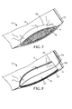
- the present invention provides a filter device.
- the filter device includes a pocket comprising a pocket surface that defines a pocket volume; an absorbent member disposed on at least a portion of the pocket surface; and a filter membrane disposed on at least a portion of the absorbent member in fluid communication with the pocket volume.
- liquid samples may be analyzed for the presence of one or more biological organisms.
- the methods can involve passing a liquid sample through a membrane having a Bubble Point pore size of no more than 1.0 ⁇ m while still providing a relatively high water volume flux.
- the method can further provide that a relatively high percentage of the biological organisms of the sample are retained on the surface of the membrane rather than being imbedded in pores of the membrane.
- the method further provides that a relatively high percentage of the biological organisms retained on the surface of the membrane may be easily recovered from the membrane surface.
- the method can further provide that the volume of the liquid sample is significantly reduced.
- Active refers to filtration methods in which a mechanized force (e.g., a vacuum) drives the movement of liquid sample through a filter membrane.
- a mechanized force e.g., a vacuum
- Bio analyte refers to a molecule, or a derivative thereof, that occurs in or is formed by an organism.
- a biological analyte can include, but is not limited to, at least one of an amino acid, a nucleic acid, a polypeptide, a protein, a polynucleotide, a lipid, a phospholipid, a saccharide, a polysaccharide, or any combination of two or more thereof.
- Exemplary biological analytes can include, but are not limited to, a metabolite (e.g., staphylococcal enterotoxin), an allergen (e.g., a peanut allergen), a hormone, a toxin (e.g., Bacillus diarrheal toxin, aflatoxin, etc.), RNA (e.g., mRNA, total RNA, tRNA, etc.), DNA (e.g., plasmid DNA, plant DNA, etc.), a tagged protein, an antibody, an antigen, or any combination of two or more thereof.
- a metabolite e.g., staphylococcal enterotoxin
- an allergen e.g., a peanut allergen
- a hormone e.g., Bacillus diarrheal toxin, aflatoxin, etc.
- RNA e.g., mRNA, total RNA, tRNA, etc.
- DNA e.g., plasmid DNA,
- “Elute” and variations thereof refer to removing biological organisms from a filter membrane using low stringency physical methods such as, for example, gravity, manual shaking, or vortexing.
- Entrapped refers to biological organisms captured by a filter membrane that are not easily eluted from the filter membrane because, for example, the biological organisms are captured in spaces within the membrane.
- Passive refers to filtration in which no mechanized force (e.g., a vacuum) drives the movement of liquid sample through a filter membrane. Passive filtration methods include filtration using, for example, gravity and/or absorption of fluid by an absorbent to drive the movement of fluid through a filter membrane.
- Recovered refers to biological organisms that are eluted from a filter membrane in condition for detection and/or further analysis.
- Retained and variations thereof refer to biological organisms that are disposed on the filter membrane surface after filtration and are easily eluted from the filter membrane.
- Water volume flux refers to a volume of fluid passing through a unit area of membrane per unit time per unit of pressure. Unless otherwise indicated, water volume flux is expressed herein as liters of liquid sample passing through one square meter of membrane per hour per pound per square inch of pressure (L/m 2 .h.psi).
- liquid samples may be filtered using a filter membrane so that each of two competing parameters are satisfied.
- the methods involved retaining biological organisms on the surface of the filter membrane so that the retained biological organisms may be easily eluted from the membrane for further analysis.
- existing methods designed to capture high percentages of biological organisms do so by using membranes having very small average pore size. This necessarily limits the water flux volume and, consequently, the rate at which a given volume of liquid sample may be processed.
- the methods described herein further provide a greater water flux volume than presently observed in filtration methods.
- the method can involve filtering large volumes (e.g., multiple liters) of liquid sample such as may be desired for, for example, environmental testing, water quality testing, water treatment testing, and/or testing of repaired and/or restored water utility pipes.
- large volumes e.g., multiple liters
- water quality testing e.g., water quality testing
- water treatment testing e.g., water treatment testing
- the method can involve filtering large volumes (e.g., multiple liters) of liquid sample such as may be desired for, for example, environmental testing, water quality testing, water treatment testing, and/or testing of repaired and/or restored water utility pipes.
- present methods for testing the water quality in repaired and/or restored water pipes it can take two to three days to confirm that the water quality is sufficient to restore water service.
- the method can involve filtering enriched food samples, food processing water samples, and/or potable water samples.
- the methods described herein can decrease the time required for such testing in at least two ways. First, the methods permit large volumes of liquid sample to be filtered and analyzed. Second, the methods result in a significant percentage of the captured biological organisms being retained on the surface of the filter membrane so that they are more readily recovered by elution.
- the methods can involve relatively rapid filtration of smaller volumes of liquid samples (e.g., less than one liter).
- the combination of relatively high water flux volume and retaining biological organisms on the filter membrane for easy recovery promotes more rapid and/or simpler filtration of liquid samples.
- biological organisms may be recovered using simple gravity to dislodge retained biological organisms from the filter membrane.
- a liquid sample may be significantly concentrated-i.e., some quantity less than the entire liquid volume may be passed through the filter membrane.
- the methods include passing a sample comprising at least one biological organism through a filter membrane having a Bubble Point pore size of no more than 1.0 ⁇ m, thereby retaining at least one biological organism on the surface of the membrane.
- the methods include passing the sample through the filter membrane at a water volume flux of at least 10 L/m 2 .h.psi for passive filtration, or a water volume flux of at least 100 L/m 2 .h.psi for active filtration.
- the method further includes detecting the at least one biological organism retained on the surface of the filter membrane.
- the liquid sample may be obtained from any suitable source, and may include a water sample.
- exemplary water samples include environmental samples (e.g., lakes, rivers, streams, oceans, ponds, etc.), water utility/water treatment samples (e.g., water supply pipes, water treatment facilities, water treatment discharge, sewage, etc.), potable water samples (e.g., bottled water, well water) or food samples (e.g., liquid foods, food samples processed by, for example, homogenizing, etc.). Samples may be filtered as collected or may be processed to some degree prior to filtration and further analysis.
- the biological organism may be any prokaryotic or eukaryotic organism for which detection and/or quantitation in a liquid sample may be desired.
- the biological organism may include, for example, a parasite, or a microbe such as, for example, a unicellular eukaryotic organism (e.g., a yeast), an algae, or a bacterium.
- Exemplary microbes include, for example, coliform bacteria.
- Exemplary bacterial species include, for example, Escherichia spp. (e.g., E. coli), Enterobacter spp. (e.g., E. aerogenes ) Enterococcus spp., (e.g., E.
- Citrobacter spp. e.g., C. freundii
- Klebsiella spp. Shigella spp.
- Salmonella spp. e.g., S. enteric
- Listeria spp Pseudomonas spp., etc.
- the filter membrane may possess a Bubble Point pore size of no more than 1.0 ⁇ m, although the methods may be performed using a filter membrane having a Bubble Point pore size of greater than 1.0 ⁇ m.
- Exemplary filter membranes can have a Bubble Point pore size of, for example, no more than 0.95 ⁇ m, no more than 0.9 ⁇ m, no more than 0.85 ⁇ m, no more than 0.8 ⁇ m, no more than 0.75 ⁇ m, no more than 0.7 ⁇ m, no more than 0.6 ⁇ m, or no more than 0.5 ⁇ m.
- Suitable Bubble Point pore sizes may be determined, at least in part, by, for example, the size of biological organism that is desired to be detected, the volume of sample to be filtered, and the depth of the filter membrane's pores. In the context of multi-zone membranes, The Bubble Point pore size is measured for the zone positioned to retain biological organisms.
- Exemplary filter membranes can be made by, for example, TIPS (thermally induced phase separation) process, SIPS (solvent induced phase separation) process, VIPS (vapor induced phase separation) process, stretching process, track-etching, or electrospinning (e.g., PAN fiber membranes).
- Suitable membrane materials include, for example, polyolefins (e.g., polyethylene and/or polypropylene), ethylene-chlorotrifluoroethylene copolymer, polyacrylonitrile, polycarbonate, polyester, polyamide, polysulfone, polyethersulfone, polyvinylidene fluoride (PVDF), cellulose ester, and/or combinations thereof.
- Nanofiber filter membranes can have the fiber diameter less than 5 ⁇ m such as, for example, less than 1 ⁇ m.
- Nanofiber membranes may be prepared from, for example, polyacrylonitrile, polyvinylidene fluoride, a cellulose ester, polyvinyl acetate, polyvinyl alcohol, polyvinyl butyral, and/or combinations thereof.
- TIPS polyolefin membranes can be prepared so that they possess a single, homogeneous zone of membrane structure, each zone having a different pore microstructure.
- a TIPS membrane may be prepared as a multi-zone membrane that includes two or more zones, each zone having a different pore microstructure.
- a multi-zone TIPS membrane may contain distinct zones or, alternatively, may possess a transition zone between two otherwise distinct zones.
- Exemplary filter membranes include membranes and methods for making exemplary filter membranes are described in, for example, in U.S. Patent No. 4,539,256 , U.S. Patent No. 4,726,989 , U.S. Patent No. 4,867,881 , U.S. Patent No. 5,120,594 , U.S. Patent No. 5,260,360 , International Patent Publication No. WO2010/078234 , International Patent Publication No. WO2010/071764 , U.S. Provisional Patent Application Serial No. 61/351,441 , entitled, "Coated Porous Materials," filed June 4, 2010, and U.S. Provisional Patent Application Serial No. 61/351,447 , entitled, "Process for Making Coated Porous Materials,” filed June 4, 2010.
- active filtration can provide a water flux volume of at least 100 L/m 2 .h.psi, although the methods may be performed at a water flux volume less than 100 L/m 2 .h.psi.
- Exemplary water flux volume values using active filtration include, for example, at least 250 L/m 2 .h.psi, at least 500 L/m 2 .h.psi, at least 750 L/m 2 .h.psi, at least 1000 L/m 2 .h.psi, at least 1250 L/m 2 .h.psi, at least 1500 L/m 2 .h.psi, at least 1750 L/m 2 .h.psi, at least 2000 L/m 2 .h.psi, at least 2500 L/m 2 .h.psi, or at least 3000 L/m 2 .h.psi.
- the maximum water flux rate may be determined, at least in part, by the maximum capacity of the mechanized force used to
- passive filtration can provide a water flux volume of at least 10 L/m 2 .h.psi, although the methods may be performed at a water flux volume less than 10 L/m 2 .h.psi.
- Exemplary water flux volume values using active filtration include, for example, at least 10 L/m 2 .h.psi, at least 20 L/m 2 .h.psi, at least 25 L/m 2 .h.psi, at least 32 L/m 2 .h.psi, at least 50 L/m 2 .h.psi, at least 60 L/m 2 .h.psi, at least 75 L/m 2 .h.psi, at least 88 L/m 2 .h.psi, at least 95 L/m 2 .h.psi, or at least 100 L/m 2 .h.psi.
- the maximum passive water flux rate may be determined, at least in part, by the Bubble Point pore size of the filter membrane and/or the flux gradient generated by, for example, an absorbent material positioned in fluid communication with the filter membrane so that it is capable of drawing at least a portion of the fluid sample through the filter membrane.
- the method results in at least 30% of the biological organisms in the sample are retained by the filter membrane, although the methods may be practiced so that fewer than 30% of the biological organisms in the sample being retained by the filter membrane.
- some embodiments can include, for example, a filter membrane in functional communication with an absorbent member within a larger device.
- an absorbent member 16 can generate a water flux gradient sufficient to draw liquid across the filter membrane and into the absorbent member 16 .
- the filter membrane can cover at least a portion of the absorbent member 16 so that flow of liquid into the absorbent member 16 requires that the liquid flow through the filter membrane. In this manner, biological organisms in the liquid sample may be retained by the filter membrane.
- the device 10 further comprises a container 12 with an optional closure 13 .
- the container 12 may comprise a flexible, deformable container 12 such as a ZIPLOK brand storage bag, for example, having a sealable closure 13 comprised of interlocking components ( 13a and 13b ).
- the absorbent member 16 may be disposed on a surface of a pocket 18 formed in a device 10 .
- the pocket 18 may, therefore, function as a receptacle for the liquid sample 24 .
- the absorbent member 16 can optionally include a nonwoven backing 20 to assist in forming a seal 26 , thereby fastening the absorbent member 16 to a portion of the container 12 .
- the seal 26 may be formed via an adhesive or by heat-bonding, for example.
- the filter membrane 14 may be disposed on the open-pocket surface (i.e., facing the interior volume of the pocket 18 ) of the absorbent member 16 . In use, illustrated in FIGS.
- the device 10 may be oriented so that the filter membrane 14 is in a generally vertical orientation. In some cases, therefore, biological organisms retained by the filter membrane 14 may elute from the filter membrane 14 due to gravity. Thus, as liquid is absorbed by the absorbent member 16 , biological organisms in the liquid sample 24 are concentrated. When the liquid sample 24 reaches a volume corresponding to a desired level of concentration of the biological organisms, a portion of the concentrated liquid sample 24 ( FIG. 10A ) may be removed from the device 10 by using, for example, a syringe 22 . The removed portion of the concentrated liquid sample may then be subjected to further analysis.
- a portion of the concentrated liquid sample 24 may be removed from the device 10 by using, for example, a sample port 30 to obtain the concentrated liquid sample 24.
- the sample port 30 may include an elastically-deformable split septum 40 (e.g., a split-cap TPE plug style cap available from Micronic North America, LLC; McMurray, PA), comprising a slit 42 through which a pipette tip (not shown) can be inserted to recover all or a portion of the concentrated liquid sample 24 .
- the sample port may comprise a valve that can be opened to release a portion (or all) of the concentrated liquid sample by gravity flow, for example.
- FIG. 13 shows another embodiment of a device 10 according to the present disclosure.
- material used for the filter membrane 14 is configured to form a pocket 18 (e.g., by sealing together all but a portion of the edges of two sheets of filter membrane material) disposed inside a container 12 (e.g., a ZIPLOK bag).
- the pocket 18 has in interior volume defined by the filter membrane 14 .
- the pocket 18 may further comprise an exterior nonwoven backing 20 to provide structural support for the filter membrane 14 .
- An absorbent member 16 is placed in a portion of container 12 outside of the pocket 18 formed by the filter membrane 14 .
- the absorbent member 16 is proximate the pocket 18 .
- the absorbent member 16 contacts at least a portion of the filter membrane 14 external to the pocket 18 . Even more preferably, the absorbent member 16 is disposed on at least a portion of the filter membrane 14 external to the pocket 18 . In some embodiments, the absorbent member 16 is disposed between the filter membrane and the nonwoven backing 20 .
- the device 10 further can comprise a support member 28 (e.g., a molded plastic rod with a base) that can be used to provide structural support for a relatively flexible container 12 before, during, and after the addition of a liquid sample. In use, the device 10 can be held (e.g., either manually or via a support member) while a liquid sample (not shown) is placed into the pocket 18 . After a portion of the liquid sample passes through the filter membrane 14 , the concentrated liquid sample (not shown) can be removed from the pocket 18 using a pipette, for example.
- a support member 28 e.g., a molded plastic rod with a base
- An alternative embodiment (not shown) of the device illustrated in FIG. 13 comprises a device in which the absorbent member is disposed in the interior volume of the pocket and the sample is deposited into the container external to the pocket. In use, the sample is deposited into the container rather than into the pocket. After at least a portion of the liquid sample has passed through the filter membrane, all or a portion of the remainder of the liquid sample in the container can be withdrawn from the container for detection of a biological organism using any of the detection methods disclosed herein.
- the absorbent member 16 may be constructed of any suitable fluid-absorbing material.
- the absorbent member 24 can include a hydrogel.
- hydrogel refers to a polymeric material that is hydrophilic and that is either swollen or capable of being swollen with a polar solvent. Suitable hydrogels include crosslinked hydrogels, swollen hydrogels, and dried or partially-dried hydrogels.
- Suitable hydrogels include polymers comprising ethylenically unsaturated carboxyl-containing monomers and co-monomers selected from carboxylic acids, vinyl sulfonic acid, cellulosic monomer, polyvinyl alcohol, as described in U.S. Patent Application Publication No. US2004/0157971 ; polymers comprising starch, cellulose, polyvinyl alcohol, polyethylene oxide, polypropylene glycol, and copolymers thereof, as described in U.S. Patent Application Publication No. US 2006/0062854 ; the hydrogels, and polymeric beads made therefrom, described in International Patent Publication No.
- WO 2007/146722 polymers comprising polyurethane prepolymer with at least one alcohol selected from polyethylene glycol, polypropylene glycol, and propylene glycol, as described in U.S. Patent No. 6,861,067 ; and polymers comprising a hydrophilic polymer selected from polysaccharide, polyvinylpyrolidone, polyvinyl alcohol, polyvinyl ether, polyurethane, polyacrylate, polyacrylamide, collagen and gelatin, as described in U.S. Patent No. 6,669,981 , the disclosures of which are all herein incorporated by reference in their entirety.
- Other suitable hydrogels include agar, agarose, and polyacrylamide hydrogels.
- the hydrogel can include crosslinked polyacrylic acid sodium salt/copolymer, polyacrylamide copolymer, ethylene maleic anhydride copolymer, cross-linked carboxy-methyl-cellulose, polyvinyl alcohol copolymers, cross-linked polyethylene oxide, starch grafted copolymer of polyacrylonitrile, or any combination thereof.
- the hydrogels can include a shaped hydrogel.
- Shaped hydrogels include hydrogels shaped into, for example, beads, sheets, ribbons, and fibers.
- the hydrogels can further include supported hydrogels.
- Supported hydrogels include hydrogels disposed on and/or in beads, nonwoven sheets, fibers (e.g., blown microfibers), and the like.
- the volume of the concentrated sample may be less than 50% of the volume of the sample added to the device, although the method may be practiced while reducing the volume of the sample to a lesser extent.
- Exemplary degrees of volume reduction can include, for example, the concentrated sample having a volume that is less than 40%, less than 35%, less than 30%, less than 25%, less than 20%, less than 15%, less than 10%, less than 9%, less than 8%, less than 7%, less than 6%, less than 5%, less than 4%, less than 3%, less than 2%, less than 1%, less than 0.9%, less than 0.8%, less than 0.7%, less than 0.6%, less than 0.5%, less than 0.4%, less than 0.3%, less than 0.2%, less than 0.1%, less than 0.09%, less than 0.08%, less than 0.07%, less than 0.06%, less than 0.05%, less than 0.04%, less than 0.03%, less than 0.02%, or less than 0.01% of the volume of the sample added to the device.
- the methods further include detecting at least one biological organism retained by the filter membrane.
- a biological organism retained by the filter membrane includes biological organisms that are in contact with the filter membrane as well as biological organisms subsequently recovered from the filter membrane.
- Biological organisms may be recovered from the filter membrane by any suitable method.
- One feature of biological organisms retained on the filter membrane by practicing the methods described herein is that the retained biological organisms may be removed from the filter membrane using relatively low stringency physical methods such as, for example, gravity, manual shaking, and/or vortexing.
- biological organisms concentrated in the reduced volume of the liquid sample may be considered recovered from the filter membrane, even if the biological organisms did not reside in or on the filter membrane for any discernable length of time.
- retained biological organisms may be detected in situ while still in contact with the filter membrane.
- the retained biological organisms may be removed from the filter membrane and the biological organisms so recovered may be detected.
- the biological organisms may be detected using any suitable method including those routine to those of ordinary skill in the art of microbial detection. Suitable in situ detection methods include, for example, detecting biological organism-specific binding of an antibody composition (e.g., monoclonal or polyclonal antibodies). Other detection methods can include, for example, detecting the presence of a biological analyte produced by the biological organism.
- Exemplary detection methods include, but are not limited to, detecting amplified (by, for example, PCR) biological organism-specific nucleotides sequences, nucleotide sequencing, enzyme assays (e.g., detection of dehydrogenases, glucoronidases, ⁇ -galactosidases, proteases, etc.), bioluminescence assays (e.g., detection of ATP/ADP/AMP), detection of proteins/peptides, spectrometry, and/or fluorescence (e.g., detection of NAD/NADH, FAD/FADH, autofluorescence), and the like.
- Suitable detection methods for biological organisms recovered from the filter membrane include methods applicable for in situ detection of biological organisms, and further includes detecting growth in culture.
- the method can further include quantifying biological organisms retained by the filter membrane.
- a biological organism retained by the filter membrane includes biological organisms that are in contact with the filter membrane as well as biological organisms subsequently recovered from the filter membrane.
- retained biological organisms may be quantified in situ while still in contact with the filter membrane.
- the retained biological organisms may be removed from the filter membrane and the biological organisms so recovered may be quantified.
- the biological organisms may be quantified using any suitable method including those routine to those of ordinary skill in the art of microbial detection such as, for example, colony forming unit (cfu) detection, most probable number (MPN) analysis, ATP bioluminescence, enzyme assays, PCR, reverse transcriptase PCR (RT-PCR), quantitative PCR, and the like.
- the method includes eluting at least 50% of the retained biological organisms from the filter membrane, although the method may be performed after eluting less than 50% of the retained biological organisms from the filter membrane.
- the retained biological organisms are eluted by repositioning the filter membrane so that the force of gravity causes the retained biological organisms to dislodge and thereby elute from the filter membrane.
- retained biological organisms may be eluted from the filter membrane by manually shaking the filter membrane to dislodge the retained biological organisms from the filter membrane.
- retained biological organisms may be eluted by vortexing the filter membrane to dislodge the retained biological organisms from the filter membrane.
- biological organisms may be eluted from the filter membrane by foam elution as described in Example 12, below.
- Certain existing methods provide recovery of up to about 30% of biological organisms (e.g., bacteria) and, therefore, fail to provide the same degree of recovery as observed using the methods described herein.
- biological organisms e.g., bacteria
- certain existing methods may fail to provide satisfactory recovery of biological organisms because the microporous filter membranes possess a significant amount of large pores at the surface of the membrane even when the filter membranes have a pore rating smaller than the size of bacteria.
- the large pores are believed to entrap the biological organisms rather than retain the biological organisms while the sample volume is being reduced.
- nonspecific binding of, for example, bacteria to the surface of the filter membrane creates a challenge. This is due, at least in part, because one way to reduce the volume of a liquid sample more quickly is to provide a greater surface area of filter membrane through which liquid may be absorbed. Increasing the surface are, however, also increases the surface area to which biological organisms may bind nonspecifically. Many alternatives were investigated. However, none of the membranes was able to provide both a high retention rate of biological organisms (and, therefore, high recovery rate) and a good water flux volume (e.g., sufficient to concentrate from 225 ml to 2-3 ml in, for example, less than one hour.
- the method can provide a reduction in sample volume of about 98.6% to about 99.2% while permitting recovery of at least 70% of biological organisms in the original sample.
- the steps may be conducted in any feasible order. And, as appropriate, any combination of two or more steps may be conducted simultaneously.
- a multi-zone microporous polypropylene membrane (designated herein as R1901-11) was prepared as described in International Patent Publication No. WO2010/078234 using both a 40 mm twin screw extruder and a 25 mm twin screw extruder. Melt streams from the two extruders were cast into a single sheet through a multi-manifold die.
- Melt stream 1 Polypropylene (PP) resin pellets (F008F from Sunoco Chemicals, Philadelphia, PA) and a nucleating agent (MILLAD 3988, Milliken Chemical, Spartanburg, SC) were introduced into a 40 mm twin screw extruder which was maintained at a screw speed of 250 rpm.
- the mineral oil diluent (Mineral Oil SUPERLA White 31, Chevron Corp., San Ramon, CA) was fed separately from the reservoir into the extruder.
- the weight ratio of PP/diluent/nucleating agent was 29.25%/70.7%/0.05%.
- the total extrusion rate was about 30 lb/hr (13.6 kg/hr) and the extruder's eight zones were set to provide a decreasing temperature profile from 271°C to 177°C.
- PP resin pellets and MILLAD 3988 were introduced into a 25 mm twin screw extruder which was maintained at a screw speed of 125 rpm.
- the mineral oil diluent was fed separately from the reservoir into the extruder.
- the weight ratio of PP/diluent/nucleating agent was 29.14%/70.7%/0.16%.
- the total extrusion rate was about 6 lb/hr (2.72 kg/hr) and the extruder's eight zones were set to provide a decreasing temperature profile from 271°C to 177°C.
- the multi-zone film was cast from the multi-manifold die maintained at 177°C onto a patterned casting wheel.
- the temperature of casting wheel was maintained at 60°C and the casting speed was 3.35 m/min (11 ft/min).
- the resulting film was washed in-line in a solvent to remove mineral oil in the film and then air dried.
- the washed film was sequentially oriented in the length and cross direction 1.8 x 2.80 at 99°C and 154°C, respectively.
- a multi-zone microporous polypropylene membrane (designated herein as R1901-8B) was prepared as described in International Patent Publication No. WO2010/078234 using both a 40 mm twin screw extruder and a 25 mm twin screw extruder. Melt streams from the two extruders were cast into a single sheet through a multi-manifold die.
- Melt stream 1 Polypropylene (PP) resin pellets (F008F from Sunoco Chemicals, Philadelphia, PA) and a nucleating agent (MILLAD 3988, Milliken Chemical, Spartanburg, SC) were introduced into a 40 mm twin screw extruder which was maintained at a screw speed of 250 rpm.
- the mineral oil diluent (Mineral Oil SUPERLA White 31, Chevron Corp., San Ramon, CA) was fed separately from the reservoir into the extruder.
- the weight ratio of PP/diluent/nucleating agent was 29.254%/70.7%/0.045%.
- the total extrusion rate was about 27 lb/hr (12.2 kg/hr) and the extruder's eight zones were set to provide a decreasing temperature profile from 271°C to 177°C.
- PP resin pellets and MILLAD 3988 were introduced into a 25 mm twin screw extruder which was maintained at a screw speed of 125 rpm.
- the mineral oil diluent was fed separately from the reservoir into the extruder.
- the weight ratio of PP/diluent/nucleating agent was 28.146%/70.7%/0.154%.
- the total extrusion rate was about 9 lb/hr (4.08 kg/hr) and the extruder's eight zones were set to provide a decreasing temperature profile from 271°C to 177°C.
- the multi-zone film was cast from the multi-manifold die maintained at 177°C onto a patterned casting wheel.
- the temperature of casting wheel was maintained at 60°C and the casting speed was 3.52 m/min (11.54 ft/min).
- the resulting film was washed in-line in a solvent to remove the mineral oil diluent and then air dried.
- the washed film was sequentially oriented in the length and cross direction 1.6 x 2.85 at 99°C and 154°C, respectively.
- a multi-zone microporous polypropylene membrane (designated herein as R1933-7) was prepared as described in International Patent Publication No. WO2010/078234 using both a 40 mm twin screw extruder and a 25mm twin screw extruder. Two melt streams from extruders were cast into a single sheet through a multi-manifold die.
- Melt stream 1 Polypropylene (PP) resin pellets (F008F from Sunoco Chemicals, Philadelphia, PA) and a nucleating agent (MILLAD 3988, Milliken Chemical, Spartanburg, SC) were introduced into a 40 mm twin screw extruder which was maintained at a screw speed of 175 rpm.
- the mineral oil diluent Kerpanol 350 Mineral Oil, Brenntag Great Lakes LCC, St. Paul, MN was fed separately from a reservoir into the extruder.
- the weight ratio of PP/diluent/nucleating agent was 34.247%/65.7%/0.053%.
- the total extrusion rate was about 32 lb/hr (14.5 kg/hr) and the extruder's eight zones were set to provide a decreasing temperature profile from 271°C to 177°C.
- PP resin pellets and MILLAD 3988 were introduced into a 25 mm twin screw extruder which was maintained at a screw speed of 150 rpm.
- the mineral oil diluent was fed separately from the reservoir into the extruder.
- the weight ratio of PP/diluent/nucleating agent was 29.14%/70.7%/0.16%.
- the total extrusion rate was about 6 lb/hr (2.72 kg/hr) and the extruder's eight zones were set to provide a decreasing temperature profile from 254°C to 177°C.
- the multi-zone film was cast from the multi-manifold die maintained at 177°C onto a patterned casting wheel.
- the temperature of casting wheel was maintained at 71°C and the casting speed was 5.79 m/min (19.00 ft/min).
- the resulting film was washed in-line in a solvent to remove mineral oil diluent and then air dried.
- the washed film was sequentially oriented in the length and cross direction 1.5 x 2.70 at 99°C and 160°C, respectively.
- a multi-zone microporous polypropylene membrane (designated herein as R1933-18) was prepared as described in International Patent Publication No. WO2010/078234 using both a 40 mm twin screw extruder and a 25 mm twin screw extruder. Two melt streams from extruders were cast into a single sheet through a multi-manifold die.
- PP resin pellets F008F from Sunoco Chemicals, Philadelphia, PA
- a nucleating agent MILLAD 3988, Milliken Chemical, Spartanburg, SC
- the mineral oil diluent Kaydol 350 Mineral Oil, Brenntag Great Lakes LCC, St. Paul, MN
- the weight ratio of PP/diluent/nucleating agent was 34.247%/65.7%/0.053%.
- the total extrusion rate was about 32 lb/hr (14.5 kg/hr) and the extruder's eight zones were set to provide a decreasing temperature profile from 271°C to 177°C.
- PP resin pellets and MILLAD 3988 were introduced into a 25 mm twin screw extruder which was maintained at a screw speed of 150 rpm.
- the mineral oil diluent was fed separately from the reservoir into the extruder.
- the weight ratio of PP/diluent/nucleating agent was 28.98%/70.7%/0.32%.
- the total extrusion rate was about 6 lb/hr (2.72 kg/hr) and the extruder's eight zones were set to provide a decreasing temperature profile from 260°C to 194°C.
- the multi-zone film was cast from the multi-manifold die maintained at 177°C onto a patterned casting wheel.
- the temperature of casting wheel was maintained at 52°C and the casting speed was 5.84 m/min (19.15 ft/min).
- the resulting film was washed in-line in a solvent to remove the mineral oil diluent and then air dried.
- the washed film was sequentially oriented in the length and cross direction 1.7 x 2.75 at 99°C and 160°C, respectively.
- a 4-wt% SPAN20 (Uniqema, New Castle, DE) solution was prepared by dissolving the surfactant in 2-propanol (Alfa Aesar, Ward Hill, MA).
- a TIPS microporous membrane was saturated with the above surfactant solution in a polyethylene (PE) bag.
- the membrane saturated instantly and excessive surface solution was removed by rubbing the PE bag.
- the membrane was removed from the bag and exposed to air to completely dry the membrane.
- the dried membranes were stored in a PE bag at room temperature.
- TIPS membranes were coated with polyethylene glycol (PEG) as described in U.S. Provisional Patent Application Serial No. 61/351,447 , entitled, "Process for Making Coated Porous Materials,” filed June 4, 2010.
- PEG polyethylene glycol
- EVAL stock solution was made by dissolving an ethylene-vinyl alcohol copolymer (EVAL) with 44 mol% ethylene content (EVAL44, Sigma-Aldrich Co., St Louis, MO, USA) in an ethanol (AAPER Alcohol and Chemical Co. Shelbyville, KY)/water solvent mixture (70 vol% ethanol) in a water bath at temperature 70-80°C.
- a TIPS microporous membrane was saturated with the coating solution above in a heavy weight PE bag. Effort was made to remove the excessive surface solution by paper towel wiping after the saturated membrane was removed from the PE bag. The membrane was allowed to dry by solvent evaporation at room temperature for 10-12 hours. Then, the dry membrane was saturated with a 20-wt% NaCl aqueous solution. After that, the membrane went through a nitrogen inert Fusion UV system with H-bulb on a conveying belt. The speed of the belt was 20 feet per minute (fpm). The membrane was sent through the UV system again in the same speed with the opposite membrane side facing the light source. The cured membrane sample was washed in excessive deionized water and dried at 90°C for 1 to 2 hours until completely dry. The dried membranes were stored in a PE bag at room temperature.
- PAN membranes were made as disclosed in Korean Patent Application No. KR20040040692 .
- a 10.5-wt% of polyacrylonitrile (Mw. 150,000) was prepared in N , N- dimethlyacetamide (DMAC).
- DMAC N , N- dimethlyacetamide
- a syringe pump Using a syringe pump, a constant flow of PAN polymer solution (50 ⁇ l/min/hole) was supplied into a syringe connected to a high voltage source.
- An electric force of 90-100 Kv was introduced to form an electrostatic force to cause the polymer solution ejection into air and formation of PAN nanofibers.
- the collected PAN nanofibers had bulkiness similar to cotton and not like that of a film and/or a membrane.
- a post-treatment was carried out at 140°C and 10-20 kgf/cm 3 pressure.
- the PAN nanofibers were stored as a roll in a PE bag at room temperature.
- a 47 mm disk of a membrane was cut using a die punch and the membrane disk was mounted in a Gelman magnetic holder (Gelman Sciences, Inc., Ann Arbor, MI).
- the active membrane diameter in the holder was 34 mm.
- One hundred ml of water was added to the holder and a vacuum pressure of about 23.5 inches of mercury was applied using a vacuum pump (GAST Manufacturing, Inc., Benton Harbor, MI) to draw water through the membrane.
- the time for the water to pass through the membrane was recorded with a stopwatch.
- the water flow rate (flux) was calculated using the time, vacuum pressure, and area of the membrane and expressed in L/(m 2 .h.psi).
- the Bubble Point pore size of a membrane was measured according to ASTM-F316-03.
- the membrane was pre-wetted with isopropanol or FC-43 (3M Co., St Paul, MN), or liquid GALWICK (PMI, Porous Materials, Inc., Ithaca, NY) and mounted on a testing holder. Pressurized nitrogen gas was gradually applied to one side of the membrane until the gas flow detected at the other side reached 100%. The pressure at 100% gas flow through the membrane was recorded and used to calculated Bubble Point pore size.
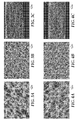
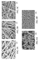
- TIPS membrane processing conditions are summarized in Table 1, below.
- the filter from each of the samples was mounted on an aluminum stub.
- TIPS membranes two sections from each of the samples were removed and mounted on an aluminum stub to view both the "Tight" and "Open” surfaces. Cross sections of each of the TIPS membranes were also prepared by tearing under liquid nitrogen. These were mounted on an additional stub. All specimens were sputter coated with gold/palladium and were examined using a JEOL 7001F Field Emission Scanning Electron Microscope. Digital photomicrographs were the product of secondary electron imaging (SEI), a technique used to image surface morphology of a sample. All micrographs were taken at a viewing angle normal to the surface of the stub or sectioned face (nominally).
- SEI secondary electron imaging
- the "Tight” and “Open” surfaces are indicated in the image for each cross section.
- a length marker is also shown in the lower portion of each micrograph of FIGS. 1-6 .
- Table 3 The various bacteria used in the examples (Table 3) were obtained from ATCC (Manassas, VA). Table 3. Bacteria used in examples Bacteria ATCC No. Enterococcus faecalis 700802 Escherichia coli 51813 Salmonella enterica subsp. enterica 51812 Citrobacter braakii 10625 Citrobacter freundii 14135 Enterobacter aerogenes 29007 Enterobacter cloacae 10699
- E. coli was grown over night in Tryptic Soy Broth (TSB) at 37°C.
- TTB Tryptic Soy Broth
- the culture was diluted to obtain approximately 100 cfu/ml and 1 ml of the solution was added to 1000 ml of sterile water to obtain approximately 100 cfu.
- a 47 mm membrane was cut from sheets or discs and placed on sterile glass filter holder assembly with funnel, fritted base (Millipore, Billerica, MA).
- the filter holder was connected to a 4L vacuum filtering flask.
- the solution was filtered through the various membranes at vacuum pressure of about 20 inches of mercury using an AIR CADET Vacuum/Pressure Station (model No. 420-3901, Barnant Company, Barrington, IL).
- the membranes were removed aseptically and placed on blood agar or tryptic soy agar plates (Hardy Diagnostics, Santa Maria, CA) and incubated overnight at 37°C. The colonies growing on membranes were counted to determine colony forming units (cfu). The results obtained are shown below in Table 4. All the membranes tested showed greater than 73% recovery with the smaller pore size membranes ( ⁇ 0.5 ⁇ m) showing greater than 90% recovery. Table 4. Recovery of E.
- E. coli was grown over night in TSB at 37°C.
- the culture was diluted to obtain approximately 100 cfu/ml and 1 ml of the solution was added to 1000 ml of sterile water to obtain approximately 100 cfu.
- a 47 mm membrane was cut from sheets or discs and placed on sterile vacuum filtration apparatus. The solution was filtered through the various membranes at vacuum pressure of about 20 inches of mercury using an AIR CADET Vacuum/Pressure Station. The membranes were removed aseptically and added to a sterile polystyrene 50-ml centrifuge tube (BD Biosciences, San Jose, CA) with 5 ml of 0.2% Tween-20 (Sigma-Aldrich Co., St.
- E. coli was grown over night in TSB at 37°C.
- the culture was diluted to obtain approximately 100 cfu/ml and 1 ml of the solution was added to 1000 ml of sterile water to obtain approximately 100 cfu.
- a 47 mm membrane was cut from sheets or discs and placed on a vacuum filtration apparatus. The solution was filtered through the various membranes at vacuum pressure of about 20 inches of mercury using an AIR CADET Vacuum/Pressure Station.
- the membranes were removed aseptically and added to a sterile polystyrene 50-ml centrifuge tube (BD Biosciences, San Jose, CA) with 5 ml of various extractants and vortexed (Fixed Speed Vortex Mixer, VWR, West Chester, PA) at room temperature for 1 to 2 minutes.
- the solution was plated on 3M PETRIFILM E. coli /Coliform Count Plates according to manufacturer's instruction and incubated overnight at 37°C.
- the plates were read using 3M PETRIFILM Plate Reader and colony forming units (cfu) were determined. The results obtained are shown below in Table 6.
- E. coli was grown over night in TSB at 37°C.
- the culture was diluted to obtain approximately 100 cfu/ml and 1 ml of the solution was added to 1000 ml of sterile water to obtain approximately 100 cfu.
- a 47 mm membrane was cut from sheets or discs and placed on a vacuum filtration apparatus. The solution was filtered through the various membranes at vacuum pressure of about 20 inches of mercury using an AIR CADET Vacuum/Pressure Station.
- the membranes were removed aseptically and added to a sterile polystyrene 50-ml centrifuge tube (BD Biosciences, San Jose, CA) with 5 ml of various concentrations of Tween-20 and vortexed (Fixed Speed Vortex Mixer, VWR, West Chester, PA) at room temperature for 1 to 2 minutes.
- the solution was plated on 3M PETRIFILM E. coli/Coliform Count Plates according to manufacturer's instruction and incubated overnight at 37°C. The plates were read using 3M PETRIFILM Plate Reader and colony forming units (cfu) were determined. The results obtained are shown below in Table 7. Use of 0.1% or 0.2% Tween-20 gave the best recoveries from all of the membranes tested. Table 7.
- E. coli was grown over night in TSB at 37°C.
- the culture was diluted to obtain approximately 100 cfu/ml and 1 ml of the solution was added to 1000 ml of sterile water to obtain approximately 100 cfu.
- a 47 mm membrane was cut from sheets or discs and placed on a vacuum filtration apparatus. The solution was filtered through the various membranes at vacuum pressure of about 20 inches of mercury using an AIR CADET Vacuum/Pressure Station. The membranes were removed aseptically and added to a sterile polystyrene 50-ml centrifuge tube (BD Biosciences, San Jose, CA) with 5 ml of 0.2% Tween-20.
- the tubes were sonicated for 5 minutes using an ultrasonicator (Branson 2200, Branson Ultrasonics, Dansbury, CT), vortexed for 1 to 2 minutes (Fixed Speed Vortex Mixer, VWR, West Chester, PA) or shaken in an orbital shaker (Newbrunswik Scientific shaker, Model Innova 4000) for 10 minutes at room temperature.
- the solutions were plated on 3M PETRIFILM E. coli /Coliform Count Plates according to manufacturer's instruction and incubated overnight at 37°C. The plates were read using 3M PETRIFILM Plate Reader and colony forming units (cfu) were determined. The results obtained are shown below in Table 8. Table 8.
- E. coli was grown over night in TSB at 37°C.
- the culture was diluted to obtain approximately 100 cfu/ml and 1 ml of the solution was added to 50 ml of sterile water to obtain approximately 100 cfu.
- a 25 mm membrane was cut from sheets or discs and placed in a 25 mm Swinnex filter holder (Millipore Corp., Billerica, MA).
- the filter holder was attached to a vacuum manifold (Waters Corporation, Milford, MA) and a 50 ml syringe was attached to the other end of filter holder.
- the spiked water sample was filtered through the various membranes at vacuum pressure of about 20 inches of mercury using an AIR CADET Vacuum/Pressure Station.
- the filter holder with the membrane was attached to HSC 40 bench-top concentrator (InnovaPrep, Drexel, MO).
- the system generates foam of the extractant solution and the bacteria were eluted by passing the foam (1 ml of 0.05% Tween-20) through the membrane.
- E. coli, Salmonella enteric subsp. enteric, and Enterococcus faecalis were grown over night in TSB at 37°C.
- the culture was diluted to obtain approximately 10 cfu/ml and 1 ml of the solution was added to 1000 ml of sterile water to obtain approximately 10 cfu.
- the solution was filtered through the various membranes at vacuum pressure of about 20 inches of mercury using an AIR CADET Vacuum/Pressure Station.
- the membranes were removed aseptically and added to a sterile polystyrene 50-ml centrifuge tube (BD Biosciences, San Jose, CA) with 10 ml of Terrific Broth (TB) or tryptic soy broth (TSB) and agitated at 300 rpm in a Newbrunswik Scientific shaker, Model Innova 4000 for 2 hours at 37°C.
- Control tubes were set up by spiking about 10 cfu (100 ⁇ l of 10 2 cfu/ml) into 10 ml TB and were grown similarly. At the end of two hours, growth media from the tubes were plated on 3M PETRIFILM E. coli/Coliform Count Plates (for E.
- E. coli, Enterobncter nerogenes, Enterobncter cloacae, Citrobacter freundii, and Citrobncter braakii were grown over night in TSB at 37°C.
- the culture was diluted to obtain approximately 100 cfu/ml and 0.4 ml of the solution was added to 1000 ml of sterile water to obtain approximately 40 cfu.
- the solution was filtered through the various membranes at vacuum pressure of about 20 inches of mercury using an AIR CADET Vacuum/Pressure Station.
- the membranes were removed aseptically and added to a sterile polystyrene 50-ml centrifuge tube (BD Biosciences, San Jose, CA) with 10 ml of Terrific Broth (TB, Sigma-Aldrich Co., St. Louis, MO) or tryptic soy broth (TSB, BD Biosciences, San Jose, CA) and agitated at 300 rpm in a Newbrunswik Scientific shaker, Model Innova 4000 for 2.5 or 3 hours at 37°C. Control tubes were set up by spiking about 40 cfu (400 ⁇ l of 100 cfu/ml) into 10 ml TB and were grown similarly.
- E. coli genes uidA (coding for b-glucoronidase) and tufA (coding for protein chain elongation factor EF-Tu) were selected as target genes.
- PCR primers and probes were designed based on alignment of all the sequences available in GenBank. The primers designed were:
- the primers and probes were synthesized by IDT DNA Technologies, Coralville, IA. Designed primers were used at 250 to 500 nM and probe at 125 to 250 nM with 10 ⁇ l 2x TaqMan Fast Universal Master Mix (Applied Biosystems, Foster City, CA) and 5 ⁇ l of DNA template.
- commercially available reagents for detection of E. coli from Primer Design Ltd, Southampton, UK (Quantification of E. coli standard kit) and BioGx, Birmingham, Alabama ( E. coli species Scorpions) were used according to manufacturer's instructions.
- E. coli cells were diluted serially in Butterfield phosphate buffer and DNA template was prepared by mixing 100 ⁇ l of PREPMAN Ultra sample prep reagent (Applied Biosystems) with 25 ⁇ l of bacterial dilutions and boiling for 10 minutes. The boiled suspension was cooled, spun at 14,000 RPM for 2 minutes and supernatant was transferred to a clean tube. 5 ⁇ l of DNA sample was added to 96-well PCR plate containing 20 ⁇ l of reaction mix (primers, probes, and enzyme mix). Thermal cycling was carried out using ABI 7500 sequence detection system with the following conditions: 2 minutes at 95°C for denaturation followed by 40 cycles of: 20 seconds at 95°C and 1 minute at 60°C.
- E. coli was grown over night in TSB at 37°C.
- the culture was diluted to obtain approximately 10 cfu/ml and 1 ml of the solution was added to 1000 ml of sterile water to obtain approximately 10 cfu.
- This solution was filtered through various membranes at vacuum pressure of about 20 inches of mercury using an AIR CADET Vacuum/Pressure Station.
- the membranes were removed aseptically and added to a 50-ml tube with 10 ml of TB (Sigma-Aldrich Co., St. Louis, MO) and agitated at 300 rpm in a Newbrunswik Scientific shaker, Model Innova 4000 for two hours at 37°C.
- Control tubes were set up by spiking about 10 cfu (100 ⁇ l of 10 2 cfu/ml) into 10 ml TB and were grown similarly. All the samples were set up in duplicates. At the end of two hours, growth media from one set of tubes were plated on 3M PETRIFILM E. coli /Coliform Count Plates and incubated overnight at 37°C. The plates were read using 3M PETRIFILM Plate Reader and colony forming units (cfu) were determined.
- the growth media containing cells were spun at 5000 rpm for 20 minutes to pellet cells.
- DNA was extracted using Qiagen Mini DNA extraction kit according to manufacturer's instructions and DNA was eluted in 10 ⁇ l. 5 ⁇ l of extracted DNA was added to 20 ⁇ l PCR assay mix and PCR was carried out as described above with primer and probes for uidA gene (Primer Design kit). The entire process from filtration followed by growth and detection by PCT took about 4 hours.
- the fold-increase varied from 5-fold to 18-fold.
- the modified TIPS membranes showed 9- to 18-fold increase and were positive by PCR.
- the commercial membranes showed only 5- to 6-fold increase and did not show any amplification of target DNA. Table 19. Rapid Detection of E.
- a 4 wt % (weight %) surfactant solution was prepared by dissolving sorbitol monolaurate (SPAN 20 available from Croda, New Castle DE) in 2-propanol (Alfa Aesar, Ward Hill, MA).
- R1930-10, R1901-11, and R1901-8B membranes (Table 1) were separately placed in polyethylene (PE) bags with sufficient surfactant solution to saturate them. The membranes saturated immediately. Excess surfactant solution was removed by rubbing the bags to squeeze the solution out of the bag. The membranes were removed from the bags and air dried at room temperature. The properties for the treated and untreated membranes were characterized for the properties shown in Table 2.
- the Tight Zone Thickness refers to the approximate thickness of the layer having the smaller pore size. The dried membranes were stored in a plastic bag until used.
- the membranes were constructed as a bag concentration device similar to the device shown in Figures 11 and 12 . Bags including each membrane were constructed in the same manner.
- An 8 inch by 8 inch ZIPLOC polyethylene bag (S.C.Johnson & Son, Inc., Racine, WI) was cut along the sealed edges and separated into two pieces.
- a single layer of dry membrane was cut into a pentagonal shape having three square five-inch sides and two equilateral sides of about 2.7 inches.
- the membrane was stacked atop a polypropylene nonwoven sheet (TYPAR, Reemay Inc, Washington, SC) having the same dimensions with the open side of the multi-zone membrane facing the nonwoven sheet.
- FIG. 11 shows an exploded view of the bag concentration device 10 with the following components: ZIPLOK bag sheets (container components 12a and 12b with interlocking closure components 13a and 13b ), polypropylene nonwoven sheet (nonwoven backing 20 ), filter membrane 14 , and superabsorbent particles (absorbent member 16 ).
- Each bag was evaluated by placing 3.8 g of superabsorbent hydro gel particles (polyacrylate-polyalcohol) into each bag outside of the pouch.
- a broth was prepared by adding 1.125 ml of an ethylene oxide/propylene oxide surfactant PLURONIC L64 (PL64, available from BASF, Mount Olive, NJ) and 0.45 g of bovine serum albumin (BSA, Sigma-Aldrich Co., St. Louis, MO) were added to 225 ml of sterilized tryptic soy broth (TSB) obtained as Quick-Enrich TSB (3M Co., St. Paul, MN) to final concentrations of 0.5% PL64 and 0.2% BSA.
- PLURONIC L64 ethylene oxide/propylene oxide surfactant
- BSA bovine serum albumin
- the broth was then inoculated with 225 ⁇ l of Butterfield phosphate buffer containing approximately 10 5 cfu (colony forming units)/ml of Listeria innocua (ATCC 33090).
- the bacterial broth was then poured into the pouch of the concentration bag (the tight side of the membrane) and propped upright on a bench at room temperature until about three ml of the solution remained in the pouch (20-30 minutes) and the absorption time was recorded.
- the remaining liquid was removed with a pipette and transferred to a 15 ml graduated centrifuge tube to measure the volume.
- Each concentrated sample was then diluted 10-fold with Butterfield's buffer and 100 ⁇ l of the dilution was plated on a Modified Oxford Medium plate (MOX plate obtained from Hardy Diagnostics, Santa Maria, CA) and incubated at 37°C for 24 hours.
- MOX plate obtained from Hardy Diagnostics, Santa Maria, CA
- Control represents the initial concentration. Control samples were prepared in the same manner as the evaluation assays but not concentrated. Separate controls were tested with each set of membranes, e.g., Control 1 was tested at the same time as the SPAN 20 treated membranes and Control 2 was tested at the same time as the PEG treated membranes.
- a 5 wt % stock solution of ethylene vinyl alcohol copolymer was prepared by dissolving an ethylene-vinyl alcohol copolymer (EVAL44) having 44 mole % ethylene content (44 % ethylene content poly(vinylalcohol co-ethylene polymer) obtained under the product number 414107 from Sigma-Aldrich Co., St. Louis, MO) in an ethanol (AAPER Alcohol and Chemical Co. Shelbyville, KY)/water solvent mixture (70 vol% ethanol) in a water bath at temperature 70-80°C.
- EVAL44 ethylene-vinyl alcohol copolymer having 44 mole % ethylene content (44 % ethylene content poly(vinylalcohol co-ethylene polymer) obtained under the product number 414107 from Sigma-Aldrich Co., St. Louis, MO) in an ethanol (AAPER Alcohol and Chemical Co. Shelbyville, KY)/water solvent mixture (70 vol% ethanol) in a water bath at temperature 70-80°C.
- a polyethylene glycol (PEG) coating solution was made containing 1 wt % EVAL44, 2 wt % polyethylene glycol (600) diacrylate (obtained under product number SR610 from Sartomer, Warrington, PA), 1 wt % reactive photoinitiator VAZPIA (2-[4-(2-hydroxy-2-methylpropanoyl)phenoxy]ethyl-2-methyl-2-N-propenoylamino propanoate, as disclosed in U.S. Patent No. 5,506,279 ) in ethanol/water mixture solvent (70 vol% ethanol).
- PEG polyethylene glycol
- Microporous membranes R1933-7 and R1933-18 were saturated with the PEG coating solution in a heavy weight PE bag, then removed from the bag. Excess solution was removed by wiping the surface of the saturated membrane with a paper towel. The membrane was air-dried at room temperature for 10-12 hours. The dry membrane was then saturated with a 20 wt% NaCl aqueous solution and excess solution was removed. The membrane was then placed on a conveyor belt of a UV curing system (Fusion UV system with H-bulb from Fusion UV Systems, Inc., Gaithersburg, MD) and cured in an inert nitrogen atmosphere chamber. The belt speed was 20 feet per minute (fpm).
- a UV curing system Fusion UV system with H-bulb from Fusion UV Systems, Inc., Gaithersburg, MD
- the membrane was turned over and run through the UV system a second time at the same speed with the opposite membrane side facing the light source.
- the cured membrane was washed in excess deionized water and dried at 90°C for 1 to 2 hours until completely dry.
- the dried membranes, with a permanent treatment, were stored in a PE bag at room temperature.
- Bag concentration devices were prepared as described in Example 17.
- PAN polyacrylonitrile
- PAN-1, PAN-2, PAN-3, PAN-4, and PAN-5 were made as disclosed in Korean Patent Application No. KR20040040692 .
- a 10.5-wt% solution of polyacrylonitrile (MW. 150,000) polymer in N,N -dimethlyacefamide (DMAC) was prepared by dispersing the polymer in the liquid.
- a constant flow of PAN polymer solution 50ul/min/hole was pumped to a syringe connected to a high voltage source.
- An electric force of 90 -100 Kv was introduced to the syringe which caused ejection of the polymer solution into the air to form electrospun PAN nanofibers.
- the fibers were collected on a web to form a bulky batt. To reduce the bulkiness and increase the structural integrity of this electrospun PAN nanofibers, post-treatment was carried out by calendering at 140°C and at pressures between about 10 to 20 kgf/cm 3 . No other subsequent treatments were used.
- the membranes were stored in PE bags at room temperature. The membranes had pore sizes of 0.2 ⁇ m, 0.53 ⁇ m, and 0.73 ⁇ m for the PAN-4, PAN-2, and PAN-5 membranes, respectively. Bag concentration devices were prepared as described in Example 17.
- Nylon Membrane Liquid Filter product numbers 080ZN (0.8 ⁇ m) and 0606SN (0.6 ⁇ m) were obtained from 3M Purification Inc., Meriden, CT.
- Bag concentration devices were prepared as described in Example 17.
- polycarbonate membrane filters 0.8 ⁇ m and 0.6 ⁇ m polycarbonate membrane filters were obtained from GE Osmonics (Hopkins, MN). Bag concentration devices were prepared as described in Example 17.
- polyether/polysulfone (PES) membranes were obtained from GE Osmosics (Hopkins, MN). Bag concentration devices were prepared as described in Example 17.
- Bags containing a 0.6 ⁇ m nylon membrane (Example 20) and bags containing a 0.8 ⁇ m polycarbonate membrane (Example 21) were evaluated according to the procedure described in Example 17 except that 4.0 grams of hydrogel was used in each bag. Bags containing the 0.6 ⁇ m nylon membrane also evaluated using a TSB broth prepared as described above, except that the surfactant used was 0.01% fluorosurfactant (3M NOVEC FC-4430, 3M Co., St. Paul, MN) instead of PLURONIC L64. Test results are shown in Table 22. Table 22.
- Bags containing a 0.8 ⁇ m nylon membrane (Example 20) and bags containing a 0.8 ⁇ m PES membrane (Example 22) were evaluated according to the procedure described in Example 17 except as follows.
- the amount of hydrogel used in each bag was about 4.0 grams.
- the insides of the bags were sterilized by spraying the inside of the bag with isopropyl alcohol, propping the bag open, and irradiating the bag with ultraviolet light for 45 minutes.
- the tryptic soy broth contained 0.6% Yeast Extract prepared by dissolving 30 grams of TSB with 3 grams of Yeast Extract in 1 liter of deionized water.
- a large sample volume collection system was built using a four-gallon plastic carboy (P/N 073004, US Plastic Corporation, Lima OH).
- a 47 mm filter holder (Cat # EW-06623-22, Cole-Parmer Instrument Co., Vernon Hills, IL) was attached to a 1 ⁇ 2" float valve (Hudson Valve Company, Bakersfield, CA) using appropriate pipe fittings (Menards, Stillwater, MN).
- the float valve was fitted into the opening of the carboy so that the float will rise to stop water flow when the required amount was reached.
- To the other end of the filter holder appropriate pipe fittings were attached so that the filter holder can be attached to water source.
- a 47 mm membrane filter was placed in the filter holder and the device was attached to water source (tap).
- the water source was turned on and water was allowed to filter through the membrane.
- the container was full (10 liters)
- the float valve shut of the water flow.
- the tap was turned off, the filter holder was detached, and the membrane filter was removed and processed for further analysis.
- the membrane was placed on blood agar (Hardy Diagnostics, Santa Maria, CA) or tryptic soy agar plate (Hardy Diagnostics) and the plate was incubated at 37°C for 16 to 24 hours. The colony forming units were counted to determine levels of bacteria in 10 liters of water.
- a large sample volume collection system was built by attaching a flow meter (Cat. # WU-05610-01, Cole-Parmer Instrument Co., Vernon Hills, IL) to a 47 mm filter holder (Cat # EW-06623-22, Cole-Parmer) using appropriate pipe fittings (Menards, Stillwater, MN). To the other end of the filter holder appropriate pipe fittings were attached so that the filter holder can be attached to water source.
- a 47 mm membrane filter was placed in the filter holder and the device was attached to water source (tap). The water source was turned on and water was allowed to filter through the membrane. The reading on the flow meter was used to determine the amount of water flowing through the filter and when the water flow reached the required amount (10 liters) the water flow was shut off manually.
- the filter holder was detached and the membrane filter was removed and processed for further analysis.
- the membrane was placed on blood agar (Hardy Diagnostics, Santa Maria, CA) or tryptic soy agar plate (Hardy Diagnostics) and the plate was incubated at 37°C for 16 to 24 hours. The colony forming units were counted to determine levels of bacteria in 10 liters of water.
- 1 to 10 liters of water sample is processed using preferred membrane filters.
- the retained bacteria can be eluted or grown further for detection by assays such as PCR, isothermal amplification, immunoassays, etc.
- assays such as PCR, isothermal amplification, immunoassays, etc.
- the rapid detection will enable to determine presence or absence of bacteria in a short time (e.g., less than eight hours) allowing for faster to return service of restored pipes.
Landscapes
- Chemical & Material Sciences (AREA)
- Life Sciences & Earth Sciences (AREA)
- Organic Chemistry (AREA)
- Health & Medical Sciences (AREA)
- Engineering & Computer Science (AREA)
- Proteomics, Peptides & Aminoacids (AREA)
- Analytical Chemistry (AREA)
- Wood Science & Technology (AREA)
- Zoology (AREA)
- Immunology (AREA)
- Molecular Biology (AREA)
- Chemical Kinetics & Catalysis (AREA)
- General Health & Medical Sciences (AREA)
- Physics & Mathematics (AREA)
- Biochemistry (AREA)
- Biotechnology (AREA)
- Microbiology (AREA)
- General Engineering & Computer Science (AREA)
- Genetics & Genomics (AREA)
- Bioinformatics & Cheminformatics (AREA)
- Biophysics (AREA)
- Pathology (AREA)
- Biomedical Technology (AREA)
- Hematology (AREA)
- Urology & Nephrology (AREA)
- Toxicology (AREA)
- Cell Biology (AREA)
- Food Science & Technology (AREA)
- Medicinal Chemistry (AREA)
- General Physics & Mathematics (AREA)
- Dispersion Chemistry (AREA)
- Separation Using Semi-Permeable Membranes (AREA)
Abstract
Description
- This application claims the benefit of
U.S. Provisional Patent Application No. 61/352,205, filed June 7, 2010 - This application has associated with it a sequence listing with the file name Sequence_Listing_66415WO003, created May 25, 2011 and contains 1,753 bytes, which is incorporated herein by reference.
- Membrane filtration is a standard step in many methods of analyzing a liquid sample for the presence of biological organisms. Such analyses are commonly performed in the interest of, for example, food safety, water quality, and/or environmental monitoring and/or study. Many membranes having an average pore size of 0.45 µm or less (e.g., cellulose acetate, nylon, etc. membranes) may be able to trap bacteria and allow growth of the trapped bacteria when placed on a suitable medium. It can be difficult, however, to recover bacteria from such membranes. These membranes, despite having an average pore size of 0.45 µm or less, typically possess a significant number of pores at the membrane surface that are larger than the biological organisms and, therefore, have torturous pore structure into which biological organisms may become trapped.
- In one aspect, the present invention provides a method of filtering a liquid sample. Generally, the method includes passing a sample comprising at least one biological organism through a filter membrane at a passive water volume flux of at least 10 L/m2.h.psi, wherein the filter membrane comprises a Bubble Point pore size of no more than 1.0 µm, thereby retaining at least one biological organism on the surface of the membrane; and detecting the at least one biological organism retained on the surface of the filter membrane.
- In certain embodiments, the biological organism may be detected in situ on the filter membrane, while in other embodiments, the biological organism may be removed from the filter membrane before being detected. Thus, in some embodiments, the method includes eluting retained biological organisms from the filter membrane.
- In some embodiments, the method can further include quantifying at least one of the biological organisms.
- In some embodiments, the method can include detecting and/or quantifying the biological organism no more than 24 hours after the sample is passed through the filter membrane.
- In some embodiments, the liquid sample can include, for example, food samples, environmental samples, or water samples.
- In some embodiments, the filter membrane may be provided in functional communication with an absorbent member.
- In some embodiments, the method can include reducing the volume of the liquid sample by at least 50%.
- In another aspect, the present invention provides a filter device. Generally, the filter device includes a pocket comprising a pocket surface that defines a pocket volume; an absorbent member disposed on at least a portion of the pocket surface; and a filter membrane disposed on at least a portion of the absorbent member in fluid communication with the pocket volume.
- The above summary of the present invention is not intended to describe each disclosed embodiment or every implementation of the present invention. The description that follows more particularly exemplifies illustrative embodiments. In several places throughout the application, guidance is provided through lists of examples, which examples can be used in various combinations. In each instance, the recited list serves only as a representative group and should not be interpreted as an exclusive list.
-
-
FIG. 1 . SEM image of R1933-18 (nascent, 0.23 µm) membrane. 1A Open side (5000x), 1B Tight side (5000x), 1C Cross-section (500x). -
FIG. 2 . SEM image of R1933-7 (nascent, 0.34 µm) membrane. 2A Open side (5000x), 2B Tight side (5000x), 2C Cross-section (500x). -
FIG. 3 . SEM image of R1933-8B (nascent, 0.51 µm) membrane. 3A Open side (5000x), 3B Tight side (5000x), 3C Cross-section (500x). -
FIG. 4 . SEM image of R1901-11 (nascent, 0.74 µm) membrane. 4A Open side (5000x), 4B Tight side (5000x), 4C Cross-section (500x). -
FIG. 5 . SEM image of PAN membranes (10,000x). 5A PAN-1 (0.613 µm), 5B PAN-2 (0.531 µm), 5C PAN-3 (0.367 µm). -
FIG. 6 . SEM images (10,000x) of MF-Millipore Type HAWP (0.4 µm) (6A) and Isopore polycarbonate filter (0.4 µm) (6B). Both membrane images are of sides receiving sample. -
FIG. 7 . A perspective schematic view of one embodiment of a filtration device. -
FIG. 8 . A schematic side view of one embodiment of a filtration device. -
FIG. 9 . A schematic side view of one embodiment of a filtration device. -
FIG. 10A . A schematic side view of one embodiment of a filtration device. -
FIG. 10B . A schematic side view of one embodiment of a filtration device with a sample-retrieval port. -
FIG. 11 . An exploded side view of one embodiment of a filtration device according to the present disclosure. -
FIG. 12 . A plan view of the assembled filtration device ofFIG. 11 . -
FIG. 13 . A cross-sectional schematic side view of an embodiment of a filtration device with a support member, according to the present disclosure. - We describe herein methods in which liquid samples may be analyzed for the presence of one or more biological organisms. Depending upon the specific context in which the methods are practiced, the methods can involve passing a liquid sample through a membrane having a Bubble Point pore size of no more than 1.0 µm while still providing a relatively high water volume flux.
- Optionally, the method can further provide that a relatively high percentage of the biological organisms of the sample are retained on the surface of the membrane rather than being imbedded in pores of the membrane. Thus, in some embodiments, the method further provides that a relatively high percentage of the biological organisms retained on the surface of the membrane may be easily recovered from the membrane surface. In other cases, the method can further provide that the volume of the liquid sample is significantly reduced.
- The following terms shall have the indicated meanings.
- "Active" refers to filtration methods in which a mechanized force (e.g., a vacuum) drives the movement of liquid sample through a filter membrane.
- "Biological analyte" refers to a molecule, or a derivative thereof, that occurs in or is formed by an organism. For example, a biological analyte can include, but is not limited to, at least one of an amino acid, a nucleic acid, a polypeptide, a protein, a polynucleotide, a lipid, a phospholipid, a saccharide, a polysaccharide, or any combination of two or more thereof. Exemplary biological analytes can include, but are not limited to, a metabolite (e.g., staphylococcal enterotoxin), an allergen (e.g., a peanut allergen), a hormone, a toxin (e.g., Bacillus diarrheal toxin, aflatoxin, etc.), RNA (e.g., mRNA, total RNA, tRNA, etc.), DNA (e.g., plasmid DNA, plant DNA, etc.), a tagged protein, an antibody, an antigen, or any combination of two or more thereof.
- "Bubble Point pore size" refers to a computed average pore size of a membrane. Bubble Point pore size is based on the fact that liquid is held in the pores of a filter by surface tension and capillary forces. The minimum pressure required to overcome surface tension and force liquid out of the pores is a measure of the pore diameter. The formula for computing Bubble Point pore size is:

where: - P = bubble-point pressure;
- σ = surface tension of the liquid (72 dynes/cm for water);
- θ = liquid-solid contact angle (which for water is generally assumed to be zero); and
- D = diameter of the pore.
- "Elute" and variations thereof refer to removing biological organisms from a filter membrane using low stringency physical methods such as, for example, gravity, manual shaking, or vortexing.
- "Entrapped" refers to biological organisms captured by a filter membrane that are not easily eluted from the filter membrane because, for example, the biological organisms are captured in spaces within the membrane.
- "Passive" refers to filtration in which no mechanized force (e.g., a vacuum) drives the movement of liquid sample through a filter membrane. Passive filtration methods include filtration using, for example, gravity and/or absorption of fluid by an absorbent to drive the movement of fluid through a filter membrane.
- "Recovered" refers to biological organisms that are eluted from a filter membrane in condition for detection and/or further analysis.
- "Retained" and variations thereof refer to biological organisms that are disposed on the filter membrane surface after filtration and are easily eluted from the filter membrane.
- "Water volume flux" refers to a volume of fluid passing through a unit area of membrane per unit time per unit of pressure. Unless otherwise indicated, water volume flux is expressed herein as liters of liquid sample passing through one square meter of membrane per hour per pound per square inch of pressure (L/m2.h.psi).
- The term "and/or" means one or all of the listed elements or a combination of any two or more of the listed elements.
- The terms "comprises" and variations thereof do not have a limiting meaning where these terms appear in the description and claims.
- Unless otherwise specified, "a," "an," "the," and "at least one" are used interchangeably and mean one or more than one.
- Also herein, the recitations of numerical ranges by endpoints include all numbers subsumed within that range (e.g., 1 to 5 includes 1, 1.5, 2, 2.75, 3, 3.80, 4, 5, etc.).
- Many commercially available membranes are used for filtering and removing biological organisms from liquid samples. The art of membrane filtration is well known. Existing commercially available membranes, however, typically have a torturous path and possess significant amount of large pores at the surface. This feature makes it difficult to recover filtered biological organisms because the captured biological organisms may become lodged in the pores of the filter membrane and are, therefore, difficult to remove from the filter intact so that the captured biological organisms may be analyzed.
- Here, we describe methods in which liquid samples may be filtered using a filter membrane so that each of two competing parameters are satisfied. First, the methods involved retaining biological organisms on the surface of the filter membrane so that the retained biological organisms may be easily eluted from the membrane for further analysis. However, existing methods designed to capture high percentages of biological organisms do so by using membranes having very small average pore size. This necessarily limits the water flux volume and, consequently, the rate at which a given volume of liquid sample may be processed. Thus, the methods described herein further provide a greater water flux volume than presently observed in filtration methods.
- In some cases, the method can involve filtering large volumes (e.g., multiple liters) of liquid sample such as may be desired for, for example, environmental testing, water quality testing, water treatment testing, and/or testing of repaired and/or restored water utility pipes. For example, using present methods for testing the water quality in repaired and/or restored water pipes, it can take two to three days to confirm that the water quality is sufficient to restore water service. Using the methods described herein, however, it may be possible to confirm water quality rapidly enough that water service can be restored within 24 hours.
- In other cases, the method can involve filtering enriched food samples, food processing water samples, and/or potable water samples.
- The methods described herein can decrease the time required for such testing in at least two ways. First, the methods permit large volumes of liquid sample to be filtered and analyzed. Second, the methods result in a significant percentage of the captured biological organisms being retained on the surface of the filter membrane so that they are more readily recovered by elution.
- In other applications, the methods can involve relatively rapid filtration of smaller volumes of liquid samples (e.g., less than one liter). In these applications, too, the combination of relatively high water flux volume and retaining biological organisms on the filter membrane for easy recovery promotes more rapid and/or simpler filtration of liquid samples. In some of these applications, biological organisms may be recovered using simple gravity to dislodge retained biological organisms from the filter membrane. In other applications, a liquid sample may be significantly concentrated-i.e., some quantity less than the entire liquid volume may be passed through the filter membrane. Because a significant percentage of the biological organisms are retained on the filter membrane rather than being entrapped within the filter membrane, a greater percentage of the biological organisms in the original sample may be recovered in the remaining liquid volume, thereby concentrating the biological organisms for further identification and/or other analyses.
- Generally, the methods include passing a sample comprising at least one biological organism through a filter membrane having a Bubble Point pore size of no more than 1.0 µm, thereby retaining at least one biological organism on the surface of the membrane. The methods include passing the sample through the filter membrane at a water volume flux of at least 10 L/m2.h.psi for passive filtration, or a water volume flux of at least 100 L/m2.h.psi for active filtration. The method further includes detecting the at least one biological organism retained on the surface of the filter membrane.
- The liquid sample may be obtained from any suitable source, and may include a water sample. Exemplary water samples include environmental samples (e.g., lakes, rivers, streams, oceans, ponds, etc.), water utility/water treatment samples (e.g., water supply pipes, water treatment facilities, water treatment discharge, sewage, etc.), potable water samples (e.g., bottled water, well water) or food samples (e.g., liquid foods, food samples processed by, for example, homogenizing, etc.). Samples may be filtered as collected or may be processed to some degree prior to filtration and further analysis.
- The biological organism may be any prokaryotic or eukaryotic organism for which detection and/or quantitation in a liquid sample may be desired. Accordingly, the biological organism may include, for example, a parasite, or a microbe such as, for example, a unicellular eukaryotic organism (e.g., a yeast), an algae, or a bacterium. Exemplary microbes include, for example, coliform bacteria. Exemplary bacterial species include, for example, Escherichia spp. (e.g., E. coli), Enterobacter spp. (e.g., E. aerogenes) Enterococcus spp., (e.g., E. faecalis), Citrobacter spp., (e.g., C. freundii), Klebsiella spp., Shigella spp., Salmonella spp. (e.g., S. enteric), Listeria spp, Pseudomonas spp., etc.
- The filter membrane may possess a Bubble Point pore size of no more than 1.0 µm, although the methods may be performed using a filter membrane having a Bubble Point pore size of greater than 1.0 µm. Exemplary filter membranes can have a Bubble Point pore size of, for example, no more than 0.95 µm, no more than 0.9 µm, no more than 0.85 µm, no more than 0.8 µm, no more than 0.75 µm, no more than 0.7 µm, no more than 0.6 µm, or no more than 0.5 µm. Suitable Bubble Point pore sizes may be determined, at least in part, by, for example, the size of biological organism that is desired to be detected, the volume of sample to be filtered, and the depth of the filter membrane's pores. In the context of multi-zone membranes, The Bubble Point pore size is measured for the zone positioned to retain biological organisms.
- Exemplary filter membranes can be made by, for example, TIPS (thermally induced phase separation) process, SIPS (solvent induced phase separation) process, VIPS (vapor induced phase separation) process, stretching process, track-etching, or electrospinning (e.g., PAN fiber membranes). Suitable membrane materials include, for example, polyolefins (e.g., polyethylene and/or polypropylene), ethylene-chlorotrifluoroethylene copolymer, polyacrylonitrile, polycarbonate, polyester, polyamide, polysulfone, polyethersulfone, polyvinylidene fluoride (PVDF), cellulose ester, and/or combinations thereof.
- Suitable membranes may be characterized as porous membranes or as nanofiber membranes. Nanofiber filter membranes can have the fiber diameter less than 5 µm such as, for example, less than 1 µm. Nanofiber membranes may be prepared from, for example, polyacrylonitrile, polyvinylidene fluoride, a cellulose ester, polyvinyl acetate, polyvinyl alcohol, polyvinyl butyral, and/or combinations thereof.
- Certain TIPS polyolefin membranes can be prepared so that they possess a single, homogeneous zone of membrane structure, each zone having a different pore microstructure. In other cases, a TIPS membrane may be prepared as a multi-zone membrane that includes two or more zones, each zone having a different pore microstructure. A multi-zone TIPS membrane may contain distinct zones or, alternatively, may possess a transition zone between two otherwise distinct zones.
- Exemplary filter membranes include membranes and methods for making exemplary filter membranes are described in, for example, in
U.S. Patent No. 4,539,256 ,U.S. Patent No. 4,726,989 ,U.S. Patent No. 4,867,881 ,U.S. Patent No. 5,120,594 ,U.S. Patent No. 5,260,360 , International Patent Publication No.WO2010/078234 , International Patent Publication No.WO2010/071764 ,U.S. Provisional Patent Application Serial No. 61/351,441 U.S. Provisional Patent Application Serial No. 61/351,447 - In some cases, active filtration can provide a water flux volume of at least 100 L/m2.h.psi, although the methods may be performed at a water flux volume less than 100 L/m2.h.psi. Exemplary water flux volume values using active filtration include, for example, at least 250 L/m2.h.psi, at least 500 L/m2.h.psi, at least 750 L/m2.h.psi, at least 1000 L/m2.h.psi, at least 1250 L/m2.h.psi, at least 1500 L/m2.h.psi, at least 1750 L/m2.h.psi, at least 2000 L/m2.h.psi, at least 2500 L/m2.h.psi, or at least 3000 L/m2.h.psi. The maximum water flux rate may be determined, at least in part, by the maximum capacity of the mechanized force used to drive movement of the liquid sample through the filter membrane, the strength and/or durability of the filter membrane, and the Bubble Point pore size of the filter membrane.
- In some cases, passive filtration can provide a water flux volume of at least 10 L/m2.h.psi, although the methods may be performed at a water flux volume less than 10 L/m2.h.psi. Exemplary water flux volume values using active filtration include, for example, at least 10 L/m2.h.psi, at least 20 L/m2.h.psi, at least 25 L/m2.h.psi, at least 32 L/m2.h.psi, at least 50 L/m2.h.psi, at least 60 L/m2.h.psi, at least 75 L/m2.h.psi, at least 88 L/m2.h.psi, at least 95 L/m2.h.psi, or at least 100 L/m2.h.psi. The maximum passive water flux rate may be determined, at least in part, by the Bubble Point pore size of the filter membrane and/or the flux gradient generated by, for example, an absorbent material positioned in fluid communication with the filter membrane so that it is capable of drawing at least a portion of the fluid sample through the filter membrane.
- In some embodiments, the method results in at least 30% of the biological organisms in the sample are retained by the filter membrane, although the methods may be practiced so that fewer than 30% of the biological organisms in the sample being retained by the filter membrane. In exemplary methods, at least 40%, at least 45%, at least 50%, at least 55%, at least 60%, at least 65%, at least 70%, at least 75%, at least 76%, at least 77%, at least 78%, at least 79%, at least 80%, at least 81%, at least 82%, at least 83%, at least 84%, at least 85%, at least 86%, at least 87%, at least 88%, at least 89%, at least 90%, at least 91%, at least 92%, at least 93%, at least 94%, at least 95%, at least 96%, at least 97%, at least 98%, at least 99%, at least 97.5%, at least 98.5%, at least 99%, at least 99.1%, at least 99.2%, at least 99.3%, at least 99.4%, at least 99.5%, at least 99.6%, at least 99.7%, at least 99.8%, or at least 99.9% of the biological organisms in the sample are retained by the filter membrane.
- As noted above, some embodiments can include, for example, a filter membrane in functional communication with an absorbent member within a larger device. Embodiments of
such devices 10 are shown inFIGS. 7-13 . In such embodiments, anabsorbent member 16 can generate a water flux gradient sufficient to draw liquid across the filter membrane and into theabsorbent member 16. In some embodiments (e.g., as shown inFIG. 7 ), the filter membrane can cover at least a portion of theabsorbent member 16 so that flow of liquid into theabsorbent member 16 requires that the liquid flow through the filter membrane. In this manner, biological organisms in the liquid sample may be retained by the filter membrane. Thedevice 10 further comprises acontainer 12 with anoptional closure 13. Thecontainer 12 may comprise a flexible,deformable container 12 such as a ZIPLOK brand storage bag, for example, having asealable closure 13 comprised of interlocking components (13a and 13b). - In one particular embodiment, shown in
FIG. 8 , theabsorbent member 16 may be disposed on a surface of apocket 18 formed in adevice 10. Thepocket 18 may, therefore, function as a receptacle for theliquid sample 24. Theabsorbent member 16 can optionally include anonwoven backing 20 to assist in forming aseal 26, thereby fastening theabsorbent member 16 to a portion of thecontainer 12. Theseal 26 may be formed via an adhesive or by heat-bonding, for example. Thefilter membrane 14 may be disposed on the open-pocket surface (i.e., facing the interior volume of the pocket 18) of theabsorbent member 16. In use, illustrated inFIGS. 9 and 10A-B , thedevice 10 may be oriented so that thefilter membrane 14 is in a generally vertical orientation. In some cases, therefore, biological organisms retained by thefilter membrane 14 may elute from thefilter membrane 14 due to gravity. Thus, as liquid is absorbed by theabsorbent member 16, biological organisms in theliquid sample 24 are concentrated. When theliquid sample 24 reaches a volume corresponding to a desired level of concentration of the biological organisms, a portion of the concentrated liquid sample 24 (FIG. 10A ) may be removed from thedevice 10 by using, for example, asyringe 22. The removed portion of the concentrated liquid sample may then be subjected to further analysis. - In some embodiments, a portion of the concentrated liquid sample 24 (
FIG. 10B ) may be removed from thedevice 10 by using, for example, asample port 30 to obtain the concentratedliquid sample 24. Thesample port 30 may include an elastically-deformable split septum 40 (e.g., a split-cap TPE plug style cap available from Micronic North America, LLC; McMurray, PA), comprising aslit 42 through which a pipette tip (not shown) can be inserted to recover all or a portion of the concentratedliquid sample 24. In some embodiments (not shown), the sample port may comprise a valve that can be opened to release a portion (or all) of the concentrated liquid sample by gravity flow, for example. -
FIG. 13 shows another embodiment of adevice 10 according to the present disclosure. In this embodiment, material used for thefilter membrane 14 is configured to form a pocket 18 (e.g., by sealing together all but a portion of the edges of two sheets of filter membrane material) disposed inside a container 12 (e.g., a ZIPLOK bag). Thepocket 18 has in interior volume defined by thefilter membrane 14. Optionally, thepocket 18 may further comprise an exteriornonwoven backing 20 to provide structural support for thefilter membrane 14. Anabsorbent member 16 is placed in a portion ofcontainer 12 outside of thepocket 18 formed by thefilter membrane 14. Preferably, theabsorbent member 16 is proximate thepocket 18. More preferably, theabsorbent member 16 contacts at least a portion of thefilter membrane 14 external to thepocket 18. Even more preferably, theabsorbent member 16 is disposed on at least a portion of thefilter membrane 14 external to thepocket 18. In some embodiments, theabsorbent member 16 is disposed between the filter membrane and thenonwoven backing 20. Optionally, thedevice 10 further can comprise a support member 28 (e.g., a molded plastic rod with a base) that can be used to provide structural support for a relativelyflexible container 12 before, during, and after the addition of a liquid sample. In use, thedevice 10 can be held (e.g., either manually or via a support member) while a liquid sample (not shown) is placed into thepocket 18. After a portion of the liquid sample passes through thefilter membrane 14, the concentrated liquid sample (not shown) can be removed from thepocket 18 using a pipette, for example. - An alternative embodiment (not shown) of the device illustrated in
FIG. 13 comprises a device in which the absorbent member is disposed in the interior volume of the pocket and the sample is deposited into the container external to the pocket. In use, the sample is deposited into the container rather than into the pocket. After at least a portion of the liquid sample has passed through the filter membrane, all or a portion of the remainder of the liquid sample in the container can be withdrawn from the container for detection of a biological organism using any of the detection methods disclosed herein. - The
absorbent member 16 may be constructed of any suitable fluid-absorbing material. In some cases, theabsorbent member 24 can include a hydrogel. As used herein, the term "hydrogel" refers to a polymeric material that is hydrophilic and that is either swollen or capable of being swollen with a polar solvent. Suitable hydrogels include crosslinked hydrogels, swollen hydrogels, and dried or partially-dried hydrogels. - Suitable hydrogels include polymers comprising ethylenically unsaturated carboxyl-containing monomers and co-monomers selected from carboxylic acids, vinyl sulfonic acid, cellulosic monomer, polyvinyl alcohol, as described in U.S. Patent Application Publication No.
US2004/0157971 ; polymers comprising starch, cellulose, polyvinyl alcohol, polyethylene oxide, polypropylene glycol, and copolymers thereof, as described in U.S. Patent Application Publication No.US 2006/0062854 ; the hydrogels, and polymeric beads made therefrom, described in International Patent Publication No.WO 2007/146722 ; polymers comprising polyurethane prepolymer with at least one alcohol selected from polyethylene glycol, polypropylene glycol, and propylene glycol, as described inU.S. Patent No. 6,861,067 ; and polymers comprising a hydrophilic polymer selected from polysaccharide, polyvinylpyrolidone, polyvinyl alcohol, polyvinyl ether, polyurethane, polyacrylate, polyacrylamide, collagen and gelatin, as described inU.S. Patent No. 6,669,981 , the disclosures of which are all herein incorporated by reference in their entirety. Other suitable hydrogels include agar, agarose, and polyacrylamide hydrogels. In certain embodiments, the hydrogel can include crosslinked polyacrylic acid sodium salt/copolymer, polyacrylamide copolymer, ethylene maleic anhydride copolymer, cross-linked carboxy-methyl-cellulose, polyvinyl alcohol copolymers, cross-linked polyethylene oxide, starch grafted copolymer of polyacrylonitrile, or any combination thereof. - The hydrogels can include a shaped hydrogel. Shaped hydrogels include hydrogels shaped into, for example, beads, sheets, ribbons, and fibers.
- The hydrogels can further include supported hydrogels. Supported hydrogels include hydrogels disposed on and/or in beads, nonwoven sheets, fibers (e.g., blown microfibers), and the like.
- In some embodiments, the volume of the concentrated sample may be less than 50% of the volume of the sample added to the device, although the method may be practiced while reducing the volume of the sample to a lesser extent. Exemplary degrees of volume reduction can include, for example, the concentrated sample having a volume that is less than 40%, less than 35%, less than 30%, less than 25%, less than 20%, less than 15%, less than 10%, less than 9%, less than 8%, less than 7%, less than 6%, less than 5%, less than 4%, less than 3%, less than 2%, less than 1%, less than 0.9%, less than 0.8%, less than 0.7%, less than 0.6%, less than 0.5%, less than 0.4%, less than 0.3%, less than 0.2%, less than 0.1%, less than 0.09%, less than 0.08%, less than 0.07%, less than 0.06%, less than 0.05%, less than 0.04%, less than 0.03%, less than 0.02%, or less than 0.01% of the volume of the sample added to the device. In one exemplary embodiment, an initial sample of 225 ml may be reduced to a volume of approximately 2 ml, so that the concentrated sample possesses a volume of approximately 0.09% of the volume of the liquid sample added to the device.
- The methods further include detecting at least one biological organism retained by the filter membrane. In this context, a biological organism retained by the filter membrane includes biological organisms that are in contact with the filter membrane as well as biological organisms subsequently recovered from the filter membrane. Biological organisms may be recovered from the filter membrane by any suitable method. One feature of biological organisms retained on the filter membrane by practicing the methods described herein is that the retained biological organisms may be removed from the filter membrane using relatively low stringency physical methods such as, for example, gravity, manual shaking, and/or vortexing. So, for example, in embodiments just described in which the volume of liquid sample is reduced, biological organisms concentrated in the reduced volume of the liquid sample may be considered recovered from the filter membrane, even if the biological organisms did not reside in or on the filter membrane for any discernable length of time.
- Thus, in some embodiments, retained biological organisms may be detected in situ while still in contact with the filter membrane. In other embodiments, however, the retained biological organisms may be removed from the filter membrane and the biological organisms so recovered may be detected. Whether detected in situ or following recovery from the filter membrane, the biological organisms may be detected using any suitable method including those routine to those of ordinary skill in the art of microbial detection. Suitable in situ detection methods include, for example, detecting biological organism-specific binding of an antibody composition (e.g., monoclonal or polyclonal antibodies). Other detection methods can include, for example, detecting the presence of a biological analyte produced by the biological organism. Exemplary detection methods include, but are not limited to, detecting amplified (by, for example, PCR) biological organism-specific nucleotides sequences, nucleotide sequencing, enzyme assays (e.g., detection of dehydrogenases, glucoronidases, β-galactosidases, proteases, etc.), bioluminescence assays (e.g., detection of ATP/ADP/AMP), detection of proteins/peptides, spectrometry, and/or fluorescence (e.g., detection of NAD/NADH, FAD/FADH, autofluorescence), and the like. Suitable detection methods for biological organisms recovered from the filter membrane include methods applicable for in situ detection of biological organisms, and further includes detecting growth in culture.
- In some cases, the method can further include quantifying biological organisms retained by the filter membrane. In this context, too, a biological organism retained by the filter membrane includes biological organisms that are in contact with the filter membrane as well as biological organisms subsequently recovered from the filter membrane.
- Thus, in some embodiments, retained biological organisms may be quantified in situ while still in contact with the filter membrane. In other embodiments, however, the retained biological organisms may be removed from the filter membrane and the biological organisms so recovered may be quantified. Whether quantified in situ or following recovery from the filter membrane, the biological organisms may be quantified using any suitable method including those routine to those of ordinary skill in the art of microbial detection such as, for example, colony forming unit (cfu) detection, most probable number (MPN) analysis, ATP bioluminescence, enzyme assays, PCR, reverse transcriptase PCR (RT-PCR), quantitative PCR, and the like.
- In embodiments in which the biological organisms are detected and/or quantified following recovery from the filter membrane, the method includes eluting at least 50% of the retained biological organisms from the filter membrane, although the method may be performed after eluting less than 50% of the retained biological organisms from the filter membrane. In exemplary methods, at least 50%, at least 55%, at least 60%, at least 65%, at least 70%, at least 75%, at least 76%, at least 77%, at least 78%, at least 79%, at least 80%, at least 81%, at least 82%, at least 83%, at least 84%, at least 85%, at least 86%, at least 87%, at least 88%, at least 89%, at least 90%, at least 91%, at least 92%, at least 93%, at least 94%, at least 95%, at least 96%, at least 97%, at least 98%, at least 99%, at least 97.5%, at least 98.5%, at least 99%, at least 99.1%, at least 99.2%, at least 99.3%, at least 99.4%, at least 99.5%, at least 99.6%, at least 99.7%, at least 99.8%, or at least 99.9% of the retained biological organisms are eluted from the filter membrane.
- In some cases, the retained biological organisms are eluted by repositioning the filter membrane so that the force of gravity causes the retained biological organisms to dislodge and thereby elute from the filter membrane. In other cases, retained biological organisms may be eluted from the filter membrane by manually shaking the filter membrane to dislodge the retained biological organisms from the filter membrane. In other cases, retained biological organisms may be eluted by vortexing the filter membrane to dislodge the retained biological organisms from the filter membrane. In other cases, biological organisms may be eluted from the filter membrane by foam elution as described in Example 12, below.
- Certain existing methods provide recovery of up to about 30% of biological organisms (e.g., bacteria) and, therefore, fail to provide the same degree of recovery as observed using the methods described herein.
- Without wishing to be bound by any particular theory, certain existing methods may fail to provide satisfactory recovery of biological organisms because the microporous filter membranes possess a significant amount of large pores at the surface of the membrane even when the filter membranes have a pore rating smaller than the size of bacteria. The large pores are believed to entrap the biological organisms rather than retain the biological organisms while the sample volume is being reduced.
- In addition, nonspecific binding of, for example, bacteria to the surface of the filter membrane creates a challenge. This is due, at least in part, because one way to reduce the volume of a liquid sample more quickly is to provide a greater surface area of filter membrane through which liquid may be absorbed. Increasing the surface are, however, also increases the surface area to which biological organisms may bind nonspecifically. Many alternatives were investigated. However, none of the membranes was able to provide both a high retention rate of biological organisms (and, therefore, high recovery rate) and a good water flux volume (e.g., sufficient to concentrate from 225 ml to 2-3 ml in, for example, less than one hour.
- In certain embodiments, the method can provide a reduction in sample volume of about 98.6% to about 99.2% while permitting recovery of at least 70% of biological organisms in the original sample.
- For any method disclosed herein that includes discrete steps, the steps may be conducted in any feasible order. And, as appropriate, any combination of two or more steps may be conducted simultaneously.
- The present invention is illustrated by the following examples. It is to be understood that the particular examples, materials, amounts, and procedures are to be interpreted broadly in accordance with the scope and spirit of the invention as set forth herein.
- A multi-zone microporous polypropylene membrane (designated herein as R1901-11) was prepared as described in International Patent Publication No.
WO2010/078234 using both a 40 mm twin screw extruder and a 25 mm twin screw extruder. Melt streams from the two extruders were cast into a single sheet through a multi-manifold die. - Melt stream 1. Polypropylene (PP) resin pellets (F008F from Sunoco Chemicals, Philadelphia, PA) and a nucleating agent (MILLAD 3988, Milliken Chemical, Spartanburg, SC) were introduced into a 40 mm twin screw extruder which was maintained at a screw speed of 250 rpm. The mineral oil diluent (Mineral Oil SUPERLA White 31, Chevron Corp., San Ramon, CA) was fed separately from the reservoir into the extruder. The weight ratio of PP/diluent/nucleating agent was 29.25%/70.7%/0.05%. The total extrusion rate was about 30 lb/hr (13.6 kg/hr) and the extruder's eight zones were set to provide a decreasing temperature profile from 271°C to 177°C.
- Melt stream 2. PP resin pellets and MILLAD 3988 were introduced into a 25 mm twin screw extruder which was maintained at a screw speed of 125 rpm. The mineral oil diluent was fed separately from the reservoir into the extruder. The weight ratio of PP/diluent/nucleating agent was 29.14%/70.7%/0.16%. The total extrusion rate was about 6 lb/hr (2.72 kg/hr) and the extruder's eight zones were set to provide a decreasing temperature profile from 271°C to 177°C.
- The multi-zone film was cast from the multi-manifold die maintained at 177°C onto a patterned casting wheel. The temperature of casting wheel was maintained at 60°C and the casting speed was 3.35 m/min (11 ft/min). The resulting film was washed in-line in a solvent to remove mineral oil in the film and then air dried. The washed film was sequentially oriented in the length and cross direction 1.8 x 2.80 at 99°C and 154°C, respectively.
- A multi-zone microporous polypropylene membrane (designated herein as R1901-8B) was prepared as described in International Patent Publication No.
WO2010/078234 using both a 40 mm twin screw extruder and a 25 mm twin screw extruder. Melt streams from the two extruders were cast into a single sheet through a multi-manifold die. - Melt stream 1. Polypropylene (PP) resin pellets (F008F from Sunoco Chemicals, Philadelphia, PA) and a nucleating agent (MILLAD 3988, Milliken Chemical, Spartanburg, SC) were introduced into a 40 mm twin screw extruder which was maintained at a screw speed of 250 rpm. The mineral oil diluent (Mineral Oil SUPERLA White 31, Chevron Corp., San Ramon, CA) was fed separately from the reservoir into the extruder. The weight ratio of PP/diluent/nucleating agent was 29.254%/70.7%/0.045%. The total extrusion rate was about 27 lb/hr (12.2 kg/hr) and the extruder's eight zones were set to provide a decreasing temperature profile from 271°C to 177°C.
- Melt stream 2. PP resin pellets and MILLAD 3988 were introduced into a 25 mm twin screw extruder which was maintained at a screw speed of 125 rpm. The mineral oil diluent was fed separately from the reservoir into the extruder. The weight ratio of PP/diluent/nucleating agent was 28.146%/70.7%/0.154%. The total extrusion rate was about 9 lb/hr (4.08 kg/hr) and the extruder's eight zones were set to provide a decreasing temperature profile from 271°C to 177°C.
- The multi-zone film was cast from the multi-manifold die maintained at 177°C onto a patterned casting wheel. The temperature of casting wheel was maintained at 60°C and the casting speed was 3.52 m/min (11.54 ft/min). The resulting film was washed in-line in a solvent to remove the mineral oil diluent and then air dried. The washed film was sequentially oriented in the length and cross direction 1.6 x 2.85 at 99°C and 154°C, respectively.
- A multi-zone microporous polypropylene membrane (designated herein as R1933-7) was prepared as described in International Patent Publication No.
WO2010/078234 using both a 40 mm twin screw extruder and a 25mm twin screw extruder. Two melt streams from extruders were cast into a single sheet through a multi-manifold die. - Melt stream 1. Polypropylene (PP) resin pellets (F008F from Sunoco Chemicals, Philadelphia, PA) and a nucleating agent (MILLAD 3988, Milliken Chemical, Spartanburg, SC) were introduced into a 40 mm twin screw extruder which was maintained at a screw speed of 175 rpm. The mineral oil diluent (Kaydol 350 Mineral Oil, Brenntag Great Lakes LCC, St. Paul, MN) was fed separately from a reservoir into the extruder. The weight ratio of PP/diluent/nucleating agent was 34.247%/65.7%/0.053%. The total extrusion rate was about 32 lb/hr (14.5 kg/hr) and the extruder's eight zones were set to provide a decreasing temperature profile from 271°C to 177°C.
- Melt stream 2. PP resin pellets and MILLAD 3988 were introduced into a 25 mm twin screw extruder which was maintained at a screw speed of 150 rpm. The mineral oil diluent was fed separately from the reservoir into the extruder. The weight ratio of PP/diluent/nucleating agent was 29.14%/70.7%/0.16%. The total extrusion rate was about 6 lb/hr (2.72 kg/hr) and the extruder's eight zones were set to provide a decreasing temperature profile from 254°C to 177°C.
- The multi-zone film was cast from the multi-manifold die maintained at 177°C onto a patterned casting wheel. The temperature of casting wheel was maintained at 71°C and the casting speed was 5.79 m/min (19.00 ft/min). The resulting film was washed in-line in a solvent to remove mineral oil diluent and then air dried. The washed film was sequentially oriented in the length and cross direction 1.5 x 2.70 at 99°C and 160°C, respectively.
- A multi-zone microporous polypropylene membrane (designated herein as R1933-18) was prepared as described in International Patent Publication No.
WO2010/078234 using both a 40 mm twin screw extruder and a 25 mm twin screw extruder. Two melt streams from extruders were cast into a single sheet through a multi-manifold die. - Melt stream 1. Polypropylene (PP) resin pellets (F008F from Sunoco Chemicals, Philadelphia, PA) and a nucleating agent (MILLAD 3988, Milliken Chemical, Spartanburg, SC) were introduced into the hopper using a solids feeder and the materials were fed into of a 40 mm twin screw extruder which was maintained at a screw speed of 175 rpm. The mineral oil diluent (Kaydol 350 Mineral Oil, Brenntag Great Lakes LCC, St. Paul, MN) was fed separately from a reservoir into the extruder. The weight ratio of PP/diluent/nucleating agent was 34.247%/65.7%/0.053%. The total extrusion rate was about 32 lb/hr (14.5 kg/hr) and the extruder's eight zones were set to provide a decreasing temperature profile from 271°C to 177°C.
- Melt stream 2. PP resin pellets and MILLAD 3988 were introduced into a 25 mm twin screw extruder which was maintained at a screw speed of 150 rpm. The mineral oil diluent was fed separately from the reservoir into the extruder. The weight ratio of PP/diluent/nucleating agent was 28.98%/70.7%/0.32%. The total extrusion rate was about 6 lb/hr (2.72 kg/hr) and the extruder's eight zones were set to provide a decreasing temperature profile from 260°C to 194°C.
- The multi-zone film was cast from the multi-manifold die maintained at 177°C onto a patterned casting wheel. The temperature of casting wheel was maintained at 52°C and the casting speed was 5.84 m/min (19.15 ft/min). The resulting film was washed in-line in a solvent to remove the mineral oil diluent and then air dried. The washed film was sequentially oriented in the length and cross direction 1.7 x 2.75 at 99°C and 160°C, respectively.
- A 4-wt% SPAN20 (Uniqema, New Castle, DE) solution was prepared by dissolving the surfactant in 2-propanol (Alfa Aesar, Ward Hill, MA).
- A TIPS microporous membrane was saturated with the above surfactant solution in a polyethylene (PE) bag. The membrane saturated instantly and excessive surface solution was removed by rubbing the PE bag. The membrane was removed from the bag and exposed to air to completely dry the membrane. The dried membranes were stored in a PE bag at room temperature.
- The TIPS membranes were coated with polyethylene glycol (PEG) as described in
U.S. Provisional Patent Application Serial No. 61/351,447 - A 5-wt% EVAL stock solution was made by dissolving an ethylene-vinyl alcohol copolymer (EVAL) with 44 mol% ethylene content (EVAL44, Sigma-Aldrich Co., St Louis, MO, USA) in an ethanol (AAPER Alcohol and Chemical Co. Shelbyville, KY)/water solvent mixture (70 vol% ethanol) in a water bath at temperature 70-80°C.
- From the above stock solution, a solution was made containing 1-wt% EVAL44, 2-wt% SR@610 (Sartomer, Warrington, PA), 1wt% reactive photoinitiator VAZPIA ( 2-[4-(2-hydroxy-2-methylpropanoyl)phenoxy]ethyl-2-methyl-2-N-propenoylamino propanoate, as disclosed in
U.S. Patent No. 5,506,279 ) in ethanol/water mixture solvent (70 vol% ethanol) - A TIPS microporous membrane was saturated with the coating solution above in a heavy weight PE bag. Effort was made to remove the excessive surface solution by paper towel wiping after the saturated membrane was removed from the PE bag. The membrane was allowed to dry by solvent evaporation at room temperature for 10-12 hours. Then, the dry membrane was saturated with a 20-wt% NaCl aqueous solution. After that, the membrane went through a nitrogen inert Fusion UV system with H-bulb on a conveying belt. The speed of the belt was 20 feet per minute (fpm). The membrane was sent through the UV system again in the same speed with the opposite membrane side facing the light source. The cured membrane sample was washed in excessive deionized water and dried at 90°C for 1 to 2 hours until completely dry. The dried membranes were stored in a PE bag at room temperature.
- Various PAN membranes were made as disclosed in Korean Patent Application No.
KR20040040692 - A 47 mm disk of a membrane was cut using a die punch and the membrane disk was mounted in a Gelman magnetic holder (Gelman Sciences, Inc., Ann Arbor, MI). The active membrane diameter in the holder was 34 mm. One hundred ml of water was added to the holder and a vacuum pressure of about 23.5 inches of mercury was applied using a vacuum pump (GAST Manufacturing, Inc., Benton Harbor, MI) to draw water through the membrane. The time for the water to pass through the membrane was recorded with a stopwatch. The water flow rate (flux) was calculated using the time, vacuum pressure, and area of the membrane and expressed in L/(m2.h.psi).
- The Bubble Point pore size of a membrane was measured according to ASTM-F316-03. The membrane was pre-wetted with isopropanol or FC-43 (3M Co., St Paul, MN), or liquid GALWICK (PMI, Porous Materials, Inc., Ithaca, NY) and mounted on a testing holder. Pressurized nitrogen gas was gradually applied to one side of the membrane until the gas flow detected at the other side reached 100%. The pressure at 100% gas flow through the membrane was recorded and used to calculated Bubble Point pore size.
- TIPS membrane processing conditions are summarized in Table 1, below.
- The water flux rate and Bubble Point pore size for various membranes are shown below in Table 2, below.
Table 1.TIPS Membrane Processing Conditions R1930-10 R1901-11 R1901-8B R1933-7 R1933-18 MS 1 Screw Speed 150 rpm 250 rpm 250 rpm 175 rpm 175 rpm MS 1 PP/ DIL/ NA ratio (weight %) 29.23/ 70.70/ 0.072 29.25/ 70.7 / 0.05 29.25/ 70.7/ 0.045 34.25/ 65.7/ 0.053 34.25/ 65.7/ 0.053 MS 1 Extrusion Rate 21 lbs/hr (9.53 kg/h) 30 lb/hr ( 13.6 kg/hr) 27 lb/hr (12.2 kg/hr) 32 lb/hr (14. 5 kg/hr) 32 lb/hr (14.5 kg/hr) MS 1 Temp Profile 271°C to 204°C 271°Cto 177°C 271°C to 177°C 271°Cto 177°C 271°C to 177°C MS 2 Screw Speed 150 rpm 125 rpm 125 rpm 150 rpm 150 rpm MS 2 PP/ DIL/ NA ratio (weight %) 29.15/ 70.70/ 0.15 29.14/ 70.7/ 0.16 29.15/ 70.7 / 0.15 29.14/ 70.7 / 0.16 28.98/ 70.7 / 0.32 MS 2 Extrusion Rate 9 lbs/hr (4.08 kg/h) 6 lb/hr (2.72 kg/hr) 9 lb/hr (4.08 kg/hr) 6 lb/hr (2.72 kg/hr) 6 lb/hr (2.72 kg/hr) MS 2 Temp Profile 271°C to 204°C 271°Cto 177°C 271°C to 177°C 254°C to 177°C 260°C to 194°C Die Temperature 199°C (390° F) 177°C (350°F) 177°C (350°F) 177°C (350°F) 177°C (350°F) Wheel temperature 60°C 60°C 60°C 71°C 52°C Casting wheel speed 13.0 ft/min (4.0 m/min) 3.35 m/min (11 ft/min) 3.52 m/min (11.54 ft/min) 5.79 m/min (19.00 ft/min) 5.84m/min (19.15 ft/min) Orientation - LxW 1.70 x 3.35 1.8 x 2.80 1.6 x 2.85 1.5 x 2.70 1.7x2.75 Orientation Temp - L/W 99°C/154°C 99°C/154°C 99°C/54°C 99°C/160°C Table 2 - Membrane Properties TIPS Membrane Membrane - treatment Water flux (L/m2.h.psi) Bubble Point pore size (µm) Porosity Tight zone thickness (µm) Total thickness (µm) R1930-10 - untreated 937 0.34 84% 16.0 53.3 R1930-10 - SPAN20 917 - 16.0 53.3 R1901-11 - untreated 2723 0.74 85% 8 104 R1901-11 - SPAN20 2,739 0.74 - 8 104 R1901-11 - PEG 2427 0.62 - 8 104 R1902-8B - untreated 1832 0.51 84% 23 109 R1902-8B - SPAN20 1,945 0.51 84% 23 109 R1902-8B - PEG 2091 0.49 - 23 109 R1933-7 - untreated 1263 0.34 77% 12 74 R1933-7 - SPAN20 680 0.34 - 12 74 R1933-7 - PEG 1401 0.34 - 12 74 R1933-18 - untreated 577 0.23 - 6 56 R1922-18 - SPAN20 357 - - 6 56 Nanofiber filters PAN-1 3995 0.613 62% - 11.1 PAN-2 2430 0.531 62% - 16.9 PAN-3 3436 0.367 70% - 15.5 - For PAN membranes, the filter from each of the samples was mounted on an aluminum stub. For TIPS membranes, two sections from each of the samples were removed and mounted on an aluminum stub to view both the "Tight" and "Open" surfaces. Cross sections of each of the TIPS membranes were also prepared by tearing under liquid nitrogen. These were mounted on an additional stub. All specimens were sputter coated with gold/palladium and were examined using a JEOL 7001F Field Emission Scanning Electron Microscope. Digital photomicrographs were the product of secondary electron imaging (SEI), a technique used to image surface morphology of a sample. All micrographs were taken at a viewing angle normal to the surface of the stub or sectioned face (nominally). Images were captured at various magnifications and the magnification is indicated on the images shown. The "Tight" and "Open" surfaces (also referred to as sides or zones) are indicated in the image for each cross section. A length marker is also shown in the lower portion of each micrograph of
FIGS. 1-6 . - The various bacteria used in the examples (Table 3) were obtained from ATCC (Manassas, VA).
Table 3. Bacteria used in examples Bacteria ATCC No. Enterococcus faecalis 700802 Escherichia coli 51813 Salmonella enterica subsp. enterica 51812 Citrobacter braakii 10625 Citrobacter freundii 14135 Enterobacter aerogenes 29007 Enterobacter cloacae 10699 - Pure cultures of the bacterial strains were inoculated into Tryptic Soy Broth (TSB, BD, Franklin Lakes, NJ) and were grown overnight at 37°C. The cultures were diluted serially in Butterfield phosphate buffer (Whatman, Piscataway, NJ) to obtain desired amount of colony forming units (cfu) per ml for spiking into water samples. The bacteria were quantified by plating appropriate dilutions on 3M PETRIFILM E. coli/Coliform Count Plates (3M Co., St. Paul, MN) according to manufacturer's instruction and incubated overnight at 37°C. The plates were read using 3M PETRIFILM Plate Reader (3M Co.) and colony forming units (cfu) were determined.
- E. coli was grown over night in Tryptic Soy Broth (TSB) at 37°C. The culture was diluted to obtain approximately 100 cfu/ml and 1 ml of the solution was added to 1000 ml of sterile water to obtain approximately 100 cfu. A 47 mm membrane was cut from sheets or discs and placed on sterile glass filter holder assembly with funnel, fritted base (Millipore, Billerica, MA). The filter holder was connected to a 4L vacuum filtering flask. The solution was filtered through the various membranes at vacuum pressure of about 20 inches of mercury using an AIR CADET Vacuum/Pressure Station (model No. 420-3901, Barnant Company, Barrington, IL). The membranes were removed aseptically and placed on blood agar or tryptic soy agar plates (Hardy Diagnostics, Santa Maria, CA) and incubated overnight at 37°C. The colonies growing on membranes were counted to determine colony forming units (cfu). The results obtained are shown below in Table 4. All the membranes tested showed greater than 73% recovery with the smaller pore size membranes (<0.5 µm) showing greater than 90% recovery.
Table 4. Recovery of E. coli from spiked water sample by filtration and direct growth Total cfu recovered %Recovery Input 105 cfu R1933-18/SPAN (0.23 µm) 98 93.33 R1933-7/SPAN (0.34 µm) 95 90.48 R1901-8B/SPAN (0.49 µm) 92 87.62 R1901-11/SPAN (0.74 µm) 77 73.33 PAN-1 (0.613 µm) 80 76.19 PAN-2 (0.531 µm) 88 83.81 PAN-3 (0.367 µm) 100 95.24 Isopore polycarbonate filter (0.40 µm) 97 92.38 MF-Millipore Type HAWP (0.45 µm) 98 93.33 - E. coli was grown over night in TSB at 37°C. The culture was diluted to obtain approximately 100 cfu/ml and 1 ml of the solution was added to 1000 ml of sterile water to obtain approximately 100 cfu. A 47 mm membrane was cut from sheets or discs and placed on sterile vacuum filtration apparatus. The solution was filtered through the various membranes at vacuum pressure of about 20 inches of mercury using an AIR CADET Vacuum/Pressure Station. The membranes were removed aseptically and added to a sterile polystyrene 50-ml centrifuge tube (BD Biosciences, San Jose, CA) with 5 ml of 0.2% Tween-20 (Sigma-Aldrich Co., St. Louis, MO) and vortexed (Fixed Speed Vortex Mixer, VWR, West Chester, PA) at room temperature for 1 to 2 minutes. The solution was plated on 3M PETRIFILM E. coli/Coliform Count Plates (3M Co., St. Paul, MN) according to manufacturer's instruction and incubated overnight at 37°C. The plates were read using 3M PETRIFILM Plate Reader (3M Co.) and colony forming units (cfu) were determined. The results obtained are shown below in Table 5. The results are representative of a typical experiment. From 1000 ml water samples spiked with about 100 cfu of E. coli, recoveries ranged from 28.6% to 72.9%. For the treated TIPS membranes and one PAN membrane (PAN-3) the recovery varied from 45% to 72.9%. The TIPS membrane R1933-18/SPAN (0.2 µm) had a low flux rate as it took 25 minutes to filter 1 liter of water. R1933-7/SPAN (0.34 µm) had a good flux rate. Both membranes showed good recovery of filtered bacteria. For the two commercial membranes the recovery ranged from 28.6 to 35.7%.
Table 5. Recovery of E. coli by filtration and elution from various membranes Total cfu recovered %Recovery Time to filter 1 liter 20 mm Hg vacuum (min:sec)Input 140 cfu R1933-18/SPAN (0.2 µm) 102 72.86 25:19 R1933-7/Oriqinal (0.34 µm) 54 38.57 5:28 R1933-7/PEG (0.34 µm) 87 61.90 6:01 R1933-7/SPAN (0.34 µm) 98 70.00 6:08 R1901-8B/PEG (0.51 µm) 68 48.81 2:50 R1901-8B/SPAN (0.49 µm) 78 55.71 2:56 R1901-11/PEG (0.62 µm) 63 45.24 2:11 R1901-11/SPAN (0.74 µm) 72 51.43 2:40 PAN-1 (0.613 µm) 45 32.14 1:08 PAN-2 (0.531 µm) 52 37.14 1:32 PAN-3 (0.367 µm) 100 71.43 1:20 Isopore Polycarbonate filter (0.40 µm) 50 35.71 2:41 MF-Millipore Type HAWP (0.45 µm) 40 28.57 2:35 - E. coli was grown over night in TSB at 37°C. The culture was diluted to obtain approximately 100 cfu/ml and 1 ml of the solution was added to 1000 ml of sterile water to obtain approximately 100 cfu. A 47 mm membrane was cut from sheets or discs and placed on a vacuum filtration apparatus. The solution was filtered through the various membranes at vacuum pressure of about 20 inches of mercury using an AIR CADET Vacuum/Pressure Station. The membranes were removed aseptically and added to a sterile polystyrene 50-ml centrifuge tube (BD Biosciences, San Jose, CA) with 5 ml of various extractants and vortexed (Fixed Speed Vortex Mixer, VWR, West Chester, PA) at room temperature for 1 to 2 minutes. The solution was plated on 3M PETRIFILM E. coli/Coliform Count Plates according to manufacturer's instruction and incubated overnight at 37°C. The plates were read using 3M PETRIFILM Plate Reader and colony forming units (cfu) were determined. The results obtained are shown below in Table 6. Use of 0.2% Tween-20 and phosphate buffered saline (PBS, Invitrogen, Carlsbad, CA) showed better recovery than Triton-X-100 (Sigma-Aldrich Co., St. Louis, MO) or water. With Tween-20, the recovery ranged from 35% to 77%, while with PBS it was 33% to 79%. With sterile MILLI-Q water (Millipore Corp., Billerica, MA) and Trition-X-100 the recoveries were only 11% to 46%.
Table 6. Recovery of E. coli by filtration and elution using various extractants from membranes Membranes 0.2% Tween-20 0.1 % Triton-X-100 PBS Milli-Q Water R1933-7/SPAN (0.34 µm) 76.7% 37.1% 73.8% 26.2% R1901-8B/SPAN (0.49 µm) 65.2% 31.4% 69.0% 35.7% PAN-3 (0.367 µm) 63.5% 46.2% 78.6% 35.7% Isopore Polycarbonate (0.40 µm) 34.7% 10.7% 33.3% 19.0% - E. coli was grown over night in TSB at 37°C. The culture was diluted to obtain approximately 100 cfu/ml and 1 ml of the solution was added to 1000 ml of sterile water to obtain approximately 100 cfu. A 47 mm membrane was cut from sheets or discs and placed on a vacuum filtration apparatus. The solution was filtered through the various membranes at vacuum pressure of about 20 inches of mercury using an AIR CADET Vacuum/Pressure Station. The membranes were removed aseptically and added to a sterile polystyrene 50-ml centrifuge tube (BD Biosciences, San Jose, CA) with 5 ml of various concentrations of Tween-20 and vortexed (Fixed Speed Vortex Mixer, VWR, West Chester, PA) at room temperature for 1 to 2 minutes. The solution was plated on 3M PETRIFILM E. coli/Coliform Count Plates according to manufacturer's instruction and incubated overnight at 37°C. The plates were read using 3M PETRIFILM Plate Reader and colony forming units (cfu) were determined. The results obtained are shown below in Table 7. Use of 0.1% or 0.2% Tween-20 gave the best recoveries from all of the membranes tested.
Table 7. Effect of Tween-20 concentration on percent Recovery of E. coli by filtration and elution from membranes Tween-20 concentration Membranes 0.01% 0.05% 0.10% 0.20% 0.50% R1933-7/PEG (0.34 µm) 57.5 45.7 51.6 70.8 20.6 R1933-7/SPAN (0.34 µm) 64.9 57.5 59.0 78.2 73.7 R1901-8B/PEG (0.51 µm) 38.3 47.2 63.4 67.8 23.6 PAN-3 (0.367 µm) 38.5 44.9 57.7 64.1 32.1 Isopore Polycarbonate filter (0.40 µm) 12.8 19.2 32.1 38.5 19.2 MF-Millipore Type HAWP (0.45 µm) 19.2 32.1 38.5 32.1 25.6 - E. coli was grown over night in TSB at 37°C. The culture was diluted to obtain approximately 100 cfu/ml and 1 ml of the solution was added to 1000 ml of sterile water to obtain approximately 100 cfu. A 47 mm membrane was cut from sheets or discs and placed on a vacuum filtration apparatus. The solution was filtered through the various membranes at vacuum pressure of about 20 inches of mercury using an AIR CADET Vacuum/Pressure Station. The membranes were removed aseptically and added to a sterile polystyrene 50-ml centrifuge tube (BD Biosciences, San Jose, CA) with 5 ml of 0.2% Tween-20. The tubes were sonicated for 5 minutes using an ultrasonicator (Branson 2200, Branson Ultrasonics, Dansbury, CT), vortexed for 1 to 2 minutes (Fixed Speed Vortex Mixer, VWR, West Chester, PA) or shaken in an orbital shaker (Newbrunswik Scientific shaker, Model Innova 4000) for 10 minutes at room temperature. The solutions were plated on 3M PETRIFILM E. coli/Coliform Count Plates according to manufacturer's instruction and incubated overnight at 37°C. The plates were read using 3M PETRIFILM Plate Reader and colony forming units (cfu) were determined. The results obtained are shown below in Table 8.
Table 8. Recovery of E. coli from membranes after filtration by various methods of extraction Sonication Vortexing Shaking Total cfu recovered % recovery Total cfu recovered % recovery Total cfu recovered % recovery Input cells 96 cfu R1933-7/PEG (0.34 µm) 28 29.5 63 66 12 12.5 R1901-8B/PEG (0.51 µm) 30 31.3 75 78.1 15 15.6 PAN-3 (0.367 µm) 32 33.0 47 48.6 32 33.3 Isopore polycarbonate filter (0.40 µm) 22 22.6 42 43.4 15 15.6 MF-Millipore Type HAWP (0.45 µm) 28 29.5 38 39.9 10 10.4 - E. coli was grown over night in TSB at 37°C. The culture was diluted to obtain approximately 100 cfu/ml and 1 ml of the solution was added to 50 ml of sterile water to obtain approximately 100 cfu. A 25 mm membrane was cut from sheets or discs and placed in a 25 mm Swinnex filter holder (Millipore Corp., Billerica, MA). The filter holder was attached to a vacuum manifold (Waters Corporation, Milford, MA) and a 50 ml syringe was attached to the other end of filter holder. The spiked water sample was filtered through the various membranes at vacuum pressure of about 20 inches of mercury using an AIR CADET Vacuum/Pressure Station. The filter holder with the membrane was attached to
HSC 40 bench-top concentrator (InnovaPrep, Drexel, MO). The system generates foam of the extractant solution and the bacteria were eluted by passing the foam (1 ml of 0.05% Tween-20) through the membrane. - The extracted solutions were plated on 3M PETRIFILM E. coli/Coliform Count Plates according to manufacturer's instruction and incubated overnight at 37°C. The plates were read using 3M PETRIFILM Plate Reader and colony forming units (cfu) were determined. The results obtained are shown below in Table 9. The foam elution method offers an advantage for eluting biological organisms in small volumes and enables easy extraction of nucleic acids without further concentration of eluted material.
Table 9. Recovery of E. coli from membranes after filtration by foam elution Total cfu in 1 ml % Recovery Input 86 cfu R1933-7/SPAN (0.34 µm) 46 53.5 R1933-7/PEG (0.34 µm) 49 57.0 PAN-3 (0.367 µm) 47 54.7 Isopore polycarbonate filter (0.40 µm) 35 40.7 MF-Millipore Type HAWP (0.45 µm) 25 29.1 - E. coli, Salmonella enteric subsp. enteric, and Enterococcus faecalis were grown over night in TSB at 37°C. The culture was diluted to obtain approximately 10 cfu/ml and 1 ml of the solution was added to 1000 ml of sterile water to obtain approximately 10 cfu. The solution was filtered through the various membranes at vacuum pressure of about 20 inches of mercury using an AIR CADET Vacuum/Pressure Station. The membranes were removed aseptically and added to a sterile polystyrene 50-ml centrifuge tube (BD Biosciences, San Jose, CA) with 10 ml of Terrific Broth (TB) or tryptic soy broth (TSB) and agitated at 300 rpm in a Newbrunswik Scientific shaker, Model Innova 4000 for 2 hours at 37°C. Control tubes were set up by spiking about 10 cfu (100 µl of 102 cfu/ml) into 10 ml TB and were grown similarly. At the end of two hours, growth media from the tubes were plated on 3M PETRIFILM E. coli/Coliform Count Plates (for E. coli) and Aerobic Count Plates (for S. enterica and Enterococcus faecalis) and incubated overnight at 37°C. The plates were read using 3M PETRIFILM Plate Reader and colony forming units (cfu) were determined. The input number of cells was used to calculate the fold-increase.
Table 10. Increase in cell number of E. coli after filtration and growth in TB for two hours Total cfu in 5 ml Fold-increase Total cfu in 5 ml Fold-increase Input to 1 liter water 6 cfu 24 cfu Control (no filtration) 65 10.83 250 10.42 R1901-11/SPAN (0.74 µm) 68 11.33 195 8.13 PAN-3 (0.367 µm) 67 11.17 225 9.38 Isopore polycarbonate filter (0.40 µm) 43 7.17 105 4.38 Table 11. Increase in cell number of E. coli after filtration and growth in TB for two hours Total cfu in 10 ml Fold-increase Input to I liter water 11 cfu Control (no filtration) 150 13.64 PAN-1 (0.613 µm) 70 6.36 PAN-2 (0.531 µm) 100 9.09 PAN-3 (0.367 µm) 160 14.55 R1901-8B/SPAN (0.49 µm) 140 12.73 R1901-11/SPAN (0.74 µm) 110 10.00 Isopore polycarbonate filter (0.40 µm) 60 5.45 MF-Millipore Type HAWP (0.45 µm) 40 3.64 Table 12. Increase in E. coli cell numbers after filtration and growth in TB for two hours Total cfu in 10 ml Fold-Increase Input 11 cfu Control 150 13.6 R1933-18/SPAN (0.2 µm) 120 10.9 R1933-7/PEG (0.34 µm) 130 11.8 R1933-7/SPAN (0.34 µm) 120 10.9 R1901-8B/PEG (0.51 µm) 110 10.0 R1901-8B/SPAN (0.49 µm) 140 12.7 R1901-11/PEG (0.62 µm) 90 8.2 R1901-11/SPAN (0.74 µm) 100 9.1 PAN-3 (0.367 µm) 107 9.7 Isopore Polycarbonate filter (0.40 µm) 70 6.4 MF-Millipore Type HAWP (0.45 µm) 60 5.5 Table 13. Increase in E. coli cell numbers after filtration and growth in TSB for two hours Total cfu in 10 ml Fold-increase Total cfu in 10 ml Fold-increase Input 11 cfu 44 cfu Control 150 13.6 540 12.3 R1901-8B/SPAN (0.49 µm) 145 13.2 350 8.0 PAN-3 (0.367 µm) 130 11.8 310 7.0 Isopore Polycarbonate filter (0.40 µm) 85 7.7 230 5.2 MF-Millipore Type HAWP (0.45 µm) 70 6.4 250 5.7 Table 14. Increase in S. enterica cell numbers after filtration and growth in TSB for two hours Total cfu in 10 ml Fold-increase Total cfu in 10 ml Fold-increase Input 9 cfu 36 cfu Control 40 4.4 150 4.2 R1901-8B/SPAN (0.49 µm) 35 3.9 130 3.6 PAN-3 (0.367 µm) 30 3.3 150 4.2 Isopore Polycarbonate filter (0.40 µm) 22 2.4 90 2.5 MF-Millipore Type HAWP (0.45 µm) 25 2.8 110 3.1 Table 15. Increase in E. coli and Enterococcusfcieccilis cell numbers after filtration and growth in TSB for two hours E. coli Enterococcus faecalis Total cfu in 10 ml Fold-increase Total cfu in 10 ml Fold- increase Input 13 cfu 30 cfu Control 130 10.0 220 7.4 R1933-7/PEG (0.34 µm) 110 8.5 160 5.2 R1933-7/SPAN (0.34 µm) 120 9.2 180 6.0 Isopore Polycarbonate filter (0.40 µm) 70 5.4 130 4.2 MF-Millipore Type HAWP (0.45 µm) 50 3.8 60 2.0 - E. coli, Enterobncter nerogenes, Enterobncter cloacae, Citrobacter freundii, and Citrobncter braakii were grown over night in TSB at 37°C. The culture was diluted to obtain approximately 100 cfu/ml and 0.4 ml of the solution was added to 1000 ml of sterile water to obtain approximately 40 cfu. The solution was filtered through the various membranes at vacuum pressure of about 20 inches of mercury using an AIR CADET Vacuum/Pressure Station. The membranes were removed aseptically and added to a sterile polystyrene 50-ml centrifuge tube (BD Biosciences, San Jose, CA) with 10 ml of Terrific Broth (TB, Sigma-Aldrich Co., St. Louis, MO) or tryptic soy broth (TSB, BD Biosciences, San Jose, CA) and agitated at 300 rpm in a Newbrunswik Scientific shaker, Model Innova 4000 for 2.5 or 3 hours at 37°C. Control tubes were set up by spiking about 40 cfu (400 µl of 100 cfu/ml) into 10 ml TB and were grown similarly. At the end of incubation period, growth media from the tubes were plated on 3M PETRIFILM E. coli/Coliform Count Plates and incubated overnight at 37°C. The plates were read using 3M PETRIFILM Plate Reader and colony forming units (cfu) were determined. The input number of cells was used to calculate the fold-increase.
Table 16. Increase in cell number of coliform bacteria after filtration and growth in TB for two and half hours E. coli Total cfu in 10 ml Fold-Increase Input 92 cfu Control 2500 27.2 R1933-7/PEG (0.34 µm) 1900 20.7 Isopore Polycarbonate filter (0.40 µm) 820 8.9 MF-Millipore Type HAWP (0.45 µm) 530 5.8 Enterobacter aerogenes Total cfu in 10 ml Fold-Increase Input 80 cfu Control 2450 30.6 R1933-7/PEG (0.34 µm) 1500 18.8 Isopore Polycarbonate filter (0.40 µm) 900 11.3 MF-Millipore Type HAWP (0.45 µm) 450 5.6 Enterobacter cloacae Total cfu in 10 ml Fold-Increase Input 52 cfu Control 1100 21.2 R1933-7/PEG (0.34 µm) 550 10.6 Isopore Polycarbonate filter (0.40 µm) 310 6.0 MF-Millipore Type HAWP (0.45 µm) 170 3.3 Citrobacter braakii Total cfu in 10 ml Fold-Increase Input 10 cfu Control 150 15 R1933-7/PEG (0.34 µm) 100 10 Isopore Polycarbonate filter (0.40 µm) 60 6 MF-Millipore Type HAWP (0.45 µm) 40 4 Citrobacter freundii Total cfu in 10 ml Fold-Increase Input 80 cfu Control 900 11.3 R1933-7/PEG (0.34 µm) 650 8.1 Isopore Polycarbonate filter (0.40 µm) 400 5.0 MF-Millipore Type HAWP (0.45 µm) 250 3.1 Table 17. Increase in cell number of coliform bacteria after filtration and growth in TSB for three hours E. coli Total cfu in 10 ml Fold-Increase Input 88 cfu Control 5100 58.0 R1933-7/PEG (0.34 µm) 6000 68.2 Isopore Polycarbonate filter (0.40 µm) 3500 39.8 MF-Millipore Type HAWP (0.45 µm) 2800 31.8 Enterobacter aerogenes Total cfu in 10 ml Fold-Increase Input 76 cfu Control 4900 64.5 R1933-7/PEG (0.34 µm) 4000 52.6 Isopore Polycarbonate filter (0.40 µm) 2850 37.5 MF-Millipore Type HAWP (0.45 µm) 2200 28.9 Citrobacter freundii Total cfu in 10 ml Fold-Increase Input 68 cfu Control 2050 30.1 R1933-7/PEG (0.34 µm) 1400 20.6 Isopore Polycarbonate filter (0.40 µm) 700 10.3 MF-Millipore Type HAWP (0.45 µm) 875 12.9 - Two E. coli genes uidA (coding for b-glucoronidase) and tufA (coding for protein chain elongation factor EF-Tu) were selected as target genes. PCR primers and probes were designed based on alignment of all the sequences available in GenBank. The primers designed were:
- uidA: Forward primer 5'-TCTACTTTACTGGCTTTGGTCG-3' (SEQ ID NO:1)
Reverse primer 5'-CGTAAGGGTAATGCGAGGTAC-3' (SEQ ID NO:2)
Probe 5'-6-FAM-AGGATTCGATAACGTGCTGATGGTGC-3'-Iowablack FQ (SEQ ID NO:3) - tufA: Forward primer: 5'-TCACCATCAACACTTCTCACG-3' (SEQ ID NO:4)
Reverse primer: 5'-CAGCAACTACCAGGATCGC-3' (SEQ ID NO:5)
Probe: 5'-6-FAM- TGAATACGACACCCCGACCCG-3'-Iowablack FQ (SEQ ID NO:6) - The primers and probes were synthesized by IDT DNA Technologies, Coralville, IA. Designed primers were used at 250 to 500 nM and probe at 125 to 250 nM with 10 µl 2x TaqMan Fast Universal Master Mix (Applied Biosystems, Foster City, CA) and 5 µl of DNA template. In addition, commercially available reagents for detection of E. coli from Primer Design Ltd, Southampton, UK (Quantification of E. coli standard kit) and BioGx, Birmingham, Alabama (E. coli species Scorpions) were used according to manufacturer's instructions.
- E. coli cells were diluted serially in Butterfield phosphate buffer and DNA template was prepared by mixing 100 µl of PREPMAN Ultra sample prep reagent (Applied Biosystems) with 25 µl of bacterial dilutions and boiling for 10 minutes. The boiled suspension was cooled, spun at 14,000 RPM for 2 minutes and supernatant was transferred to a clean tube. 5 µl of DNA sample was added to 96-well PCR plate containing 20 µl of reaction mix (primers, probes, and enzyme mix). Thermal cycling was carried out using ABI 7500 sequence detection system with the following conditions: 2 minutes at 95°C for denaturation followed by 40 cycles of: 20 seconds at 95°C and 1 minute at 60°C. As shown below the limit of detection with the PCR was about 100 cfu.
Table 18. PCR detection of E. coli Approximate Concentration of bacteria in PCR tube In-House reagents Primer Design Kit BioGX kit Ct Ct Ct uidA tufA uidA NTC 40 40 40 40 1 cfu 40 40 40 40 10 cfu 38.34 39.37 38.8 39.1 100 cfu 34.94 35.42 33.4 33.7 1000 cfu 30.96 30.33 29.8 29.5 10000 cfu 27.04 25.83 24.3 23.4 100000 cfu 22.25 22.86 20.3 20.7 - E. coli was grown over night in TSB at 37°C. The culture was diluted to obtain approximately 10 cfu/ml and 1 ml of the solution was added to 1000 ml of sterile water to obtain approximately 10 cfu. This solution was filtered through various membranes at vacuum pressure of about 20 inches of mercury using an AIR CADET Vacuum/Pressure Station. The membranes were removed aseptically and added to a 50-ml tube with 10 ml of TB (Sigma-Aldrich Co., St. Louis, MO) and agitated at 300 rpm in a Newbrunswik Scientific shaker, Model Innova 4000 for two hours at 37°C. Control tubes were set up by spiking about 10 cfu (100 µl of 102 cfu/ml) into 10 ml TB and were grown similarly. All the samples were set up in duplicates. At the end of two hours, growth media from one set of tubes were plated on 3M PETRIFILM E. coli/Coliform Count Plates and incubated overnight at 37°C. The plates were read using 3M PETRIFILM Plate Reader and colony forming units (cfu) were determined.
- From the other set of tubes, the growth media containing cells were spun at 5000 rpm for 20 minutes to pellet cells. DNA was extracted using Qiagen Mini DNA extraction kit according to manufacturer's instructions and DNA was eluted in 10 µl. 5 µl of extracted DNA was added to 20 µl PCR assay mix and PCR was carried out as described above with primer and probes for uidA gene (Primer Design kit). The entire process from filtration followed by growth and detection by PCT took about 4 hours.
- As shown below, the fold-increase varied from 5-fold to 18-fold. From 1000 ml water samples spiked with 10 cfu of E. coli, the modified TIPS membranes showed 9- to 18-fold increase and were positive by PCR. The commercial membranes showed only 5- to 6-fold increase and did not show any amplification of target DNA.
Table 19. Rapid Detection of E. coli by PCR Total cfu in 10 ml Fold-increase PCR Assay (Ct) Amplification Input to 1000 ml water 10 cfu NTC 40 No Control (no filtration) 156 15.6 32.1 Yes R1933-7/PEG (0.34 mm) 150 15.0 32.7 Yes R1933-7/SPAN (0.34 mm) 175 17.5 31.9 Yes R1901-8B/PEG (0.51 mm) 98 9.8 34.2 Yes R1901-8B/SPAN (0.49 mm) 122 12.2 33.1 Yes R1901-11/PEG (0.62 mm) 90 9.0 34.9 Yes R1901-11/SPAN (0.74 mm) 102 10.2 34 Yes PAN-3 (0.367 mm) 115 11.5 33 Yes Isopore Polycarbonate (0.40 mm) 60 6.0 38.8 No MF-Millipore Type HAWP (0.45 mm) 50 5.0 39 No - A 4 wt % (weight %) surfactant solution was prepared by dissolving sorbitol monolaurate (
SPAN 20 available from Croda, New Castle DE) in 2-propanol (Alfa Aesar, Ward Hill, MA). R1930-10, R1901-11, and R1901-8B membranes (Table 1) were separately placed in polyethylene (PE) bags with sufficient surfactant solution to saturate them. The membranes saturated immediately. Excess surfactant solution was removed by rubbing the bags to squeeze the solution out of the bag. The membranes were removed from the bags and air dried at room temperature. The properties for the treated and untreated membranes were characterized for the properties shown in Table 2. The Tight Zone Thickness refers to the approximate thickness of the layer having the smaller pore size. The dried membranes were stored in a plastic bag until used. - The membranes were constructed as a bag concentration device similar to the device shown in
Figures 11 and 12 . Bags including each membrane were constructed in the same manner. An 8 inch by 8 inch ZIPLOC polyethylene bag (S.C.Johnson & Son, Inc., Racine, WI) was cut along the sealed edges and separated into two pieces. A single layer of dry membrane was cut into a pentagonal shape having three square five-inch sides and two equilateral sides of about 2.7 inches. The membrane was stacked atop a polypropylene nonwoven sheet (TYPAR, Reemay Inc, Charleston, SC) having the same dimensions with the open side of the multi-zone membrane facing the nonwoven sheet. The stack was then placed on the inner surface of one piece of the ZIPLOC bag with the square shape facing the ZIPLOC seal and the triangle forming a v-shape near the bottom of the bag, and the tight side of the membrane facing the inner surface of the bag.FIG. 11 shows an exploded view of thebag concentration device 10 with the following components: ZIPLOK bag sheets (container components closure components filter membrane 14, and superabsorbent particles (absorbent member 16). Four edges of the membrane and nonwoven pentagon were heat sealed to the ZIPLOC bag to form a pouch with the five-inch side of the pentagon facing the ZIPLOC seal at the top of the bag left open, as shown inFIG. 12 . The two sides of the ZIPLOC bag were then heat sealed (seals 26) so the nonwoven faced the opposing wall of the bag to form a concentration bag similar to the one shown inFIG. 8 . - Each bag was evaluated by placing 3.8 g of superabsorbent hydro gel particles (polyacrylate-polyalcohol) into each bag outside of the pouch. For each test, a broth was prepared by adding 1.125 ml of an ethylene oxide/propylene oxide surfactant PLURONIC L64 (PL64, available from BASF, Mount Olive, NJ) and 0.45 g of bovine serum albumin (BSA, Sigma-Aldrich Co., St. Louis, MO) were added to 225 ml of sterilized tryptic soy broth (TSB) obtained as Quick-Enrich TSB (3M Co., St. Paul, MN) to final concentrations of 0.5% PL64 and 0.2% BSA. The broth was then inoculated with 225 µl of Butterfield phosphate buffer containing approximately 105 cfu (colony forming units)/ml of Listeria innocua (ATCC 33090). The bacterial broth was then poured into the pouch of the concentration bag (the tight side of the membrane) and propped upright on a bench at room temperature until about three ml of the solution remained in the pouch (20-30 minutes) and the absorption time was recorded. The remaining liquid was removed with a pipette and transferred to a 15 ml graduated centrifuge tube to measure the volume. Each concentrated sample was then diluted 10-fold with Butterfield's buffer and 100 µl of the dilution was plated on a Modified Oxford Medium plate (MOX plate obtained from Hardy Diagnostics, Santa Maria, CA) and incubated at 37°C for 24 hours.
- The control represents the initial concentration. Control samples were prepared in the same manner as the evaluation assays but not concentrated. Separate controls were tested with each set of membranes, e.g., Control 1 was tested at the same time as the
SPAN 20 treated membranes and Control 2 was tested at the same time as the PEG treated membranes. - Each evaluation was replicated a second time and the Bacteria Concentration represents two different separate counts after concentrating. The concentration factor is the final concentration divided by the initial concentration of bacteria. Test results for the
SPAN 20 treated membranes are shown in Table 20.Table 20. Bacterial Recovery Rates and Absorption Times for TIPS Membranes Membrane Bacteria concentration (cfu/ml) Volume recovered (ml) Recovered Bacteria numbers Recovery rate Concentration factor Absorption time (min) R1930-10 6850 3 2.06x10⁴ 58.9% 44 36 SPAN 205700 3.8 2.17x10⁴ 62.1% 37 36 R1901-11 9550 3 2.87x10⁴ 82.2% 62 22 SPAN 207550 2.8 2.11x10⁴ 60.6% 49 22 R1901-8B 7850 3 2.36x10⁴ 67.5% 51 20 SPAN 207900 3 2.37x10⁴ 68.0% 51 20 Control 1 155 225 3.49x10⁴ - - - R1901-8B 4250 23.4 1.45x10⁴ 75.6% 50 25 PEG 4050 3.2 1.30x10⁴ 67.8% 48 25 Control 2 69 225 1.55x10⁴ - - - - A 5 wt % stock solution of ethylene vinyl alcohol copolymer was prepared by dissolving an ethylene-vinyl alcohol copolymer (EVAL44) having 44 mole % ethylene content (44 % ethylene content poly(vinylalcohol co-ethylene polymer) obtained under the product number 414107 from Sigma-Aldrich Co., St. Louis, MO) in an ethanol (AAPER Alcohol and Chemical Co. Shelbyville, KY)/water solvent mixture (70 vol% ethanol) in a water bath at temperature 70-80°C.
- From the above stock solution, a polyethylene glycol (PEG) coating solution was made containing 1 wt % EVAL44, 2 wt % polyethylene glycol (600) diacrylate (obtained under product number SR610 from Sartomer, Warrington, PA), 1 wt % reactive photoinitiator VAZPIA (2-[4-(2-hydroxy-2-methylpropanoyl)phenoxy]ethyl-2-methyl-2-N-propenoylamino propanoate, as disclosed in
U.S. Patent No. 5,506,279 ) in ethanol/water mixture solvent (70 vol% ethanol). - Microporous membranes R1933-7 and R1933-18 (Table 1) were saturated with the PEG coating solution in a heavy weight PE bag, then removed from the bag. Excess solution was removed by wiping the surface of the saturated membrane with a paper towel. The membrane was air-dried at room temperature for 10-12 hours. The dry membrane was then saturated with a 20 wt% NaCl aqueous solution and excess solution was removed. The membrane was then placed on a conveyor belt of a UV curing system (Fusion UV system with H-bulb from Fusion UV Systems, Inc., Gaithersburg, MD) and cured in an inert nitrogen atmosphere chamber. The belt speed was 20 feet per minute (fpm). The membrane was turned over and run through the UV system a second time at the same speed with the opposite membrane side facing the light source. The cured membrane was washed in excess deionized water and dried at 90°C for 1 to 2 hours until completely dry. The dried membranes, with a permanent treatment, were stored in a PE bag at room temperature.
- Bag concentration devices were prepared as described in Example 17.
- Various polyacrylonitrile (PAN) polymer membranes (PAN-1, PAN-2, PAN-3, PAN-4, and PAN-5) were made as disclosed in Korean Patent Application No.
KR20040040692 - The bags were evaluated according to the same procedure as described in Example 17 except that 3.9 grams of hydrogel was used in each bag. Test results are shown in Table 21.
Table 21. Bacterial Recovery Rate for Polyacrylonitrile Membranes Membrane (pore size) Bacteria concentration (cfu/ml) Volume recovered (ml) Recovered Bacteria numbers Recovery rate Concentration factor Absorption time (min) PAN-4 700 2.5 1.75x10³ 16.2% 15 40 (0.2 µm) 500 3.5 1.75103 16.2% 10 40 PAN-2 1850 3.7 6.85103 63.4% 36 <10 (0.5 µm) 2250 3.4 6.85x10³ 50.0% 47 <10 PAN-5 850 2.6 2..21x103 20.5% 18 <10 (0.7 µm) 1350 4.5 6.05x10³ 56.3% 28 <10 Control 48 225 1.08x10³ - - - - Nylon Membrane Liquid Filter product numbers 080ZN (0.8 µm) and 0606SN (0.6 µm) were obtained from 3M Purification Inc., Meriden, CT.
- Bag concentration devices were prepared as described in Example 17.
- 0.8 µm and 0.6 µm polycarbonate membrane filters were obtained from GE Osmonics (Hopkins, MN). Bag concentration devices were prepared as described in Example 17.
- 0.8 µm and 0.6 µm polyether/polysulfone (PES) membranes were obtained from GE Osmosics (Hopkins, MN). Bag concentration devices were prepared as described in Example 17.
- Bags containing a 0.6 µm nylon membrane (Example 20) and bags containing a 0.8 µm polycarbonate membrane (Example 21) were evaluated according to the procedure described in Example 17 except that 4.0 grams of hydrogel was used in each bag. Bags containing the 0.6 µm nylon membrane also evaluated using a TSB broth prepared as described above, except that the surfactant used was 0.01% fluorosurfactant (3M NOVEC FC-4430, 3M Co., St. Paul, MN) instead of PLURONIC L64. Test results are shown in Table 22.
Table 22. Bacteria Recovery From Nylon and Polycarbonate Membranes Membrane (pore size) Bacteria concentration (cfu/ml) Volume recovered (ml) Recovered Bacteria numbers Recovery rate Concentration factor Absorption time (min) Nylon 1600 2.3 3.60x10³ 17.8% 18 70 (0.6 µm) 1600 1.4 2.24x10³ 11.1% 18 70 Nylon 3200 1.6 5.12x10³ 25.3% 36 70 (0.6 µm) 0.01%FC4430 Polycarbonate 1500 2.7 4.05x10³ 20.0% 17 90 (0.8 µm) 2500 1.3 3.25x10³ 16.1% 28 90 Control 90 225 2.03x10⁴ - Bags containing a 0.8µm nylon membrane (Example 20) and bags containing a 0.8µm PES membrane (Example 22) were evaluated according to the procedure described in Example 17 except as follows. The amount of hydrogel used in each bag was about 4.0 grams. The insides of the bags were sterilized by spraying the inside of the bag with isopropyl alcohol, propping the bag open, and irradiating the bag with ultraviolet light for 45 minutes. The tryptic soy broth contained 0.6% Yeast Extract prepared by dissolving 30 grams of TSB with 3 grams of Yeast Extract in 1 liter of deionized water. One of two surfactants, as indicated in Table 23 was added to the broth - 0.2% (w/v) PLURONIC L64 and 0.2% Tween 80 (v/v) (Sigma-Aldrich Co., St. Louis, MO). The resulting broth (225 ml) was inoculated with 225 µl of Butterfield's Buffer containing about 1x10⁷ cfu/ml of Listeria innocua (ATCC 33090) and mixed well. The broth was then poured into the pouch of the sample concentration bag and propped upright for 50-90 minutes. The concentrated sample was collected, measured, and diluted in Butterfield's buffer 10-fold sequentially in 3 ml. Then 1 ml of the 3rd dilutions of each sample was plated on AC PETRIFILM Plates (3M, Co. St. Paul, MN). The plates were incubated at 37°C for 24 hours and the colonies were counted. Table 23 shows the bacterial recovery results.
Table 23. Bacterial Recovery Rate for Polyether Sulfone and Nylon Membranes Membrane (Surfactant) Bacteria concentration (cfu/ml) Volume recovered (ml) Recovered Bacteria numbers Recovery rate Concentration factor Absorption time (min) Nylon 0.8µm 32000 6.5 2.08x10⁵ 26.0% 9 60 (0.2% Tween 20) Nylon 0.8 µm 396500 7.4 2.92x10⁵ 36.6% 11 60 (0.2% PL64) PES 29500 12.6 3.72x10⁵ 46.5% 8 100 (0.2% Tween 20) PES 34500 3.0 1.04x10⁵ 18.22% 10 100 (0.2% PL64) Control 3550 225 7.99x10⁴ - A large sample volume collection system was built using a four-gallon plastic carboy (P/N 073004, US Plastic Corporation, Lima OH). A 47 mm filter holder (Cat # EW-06623-22, Cole-Parmer Instrument Co., Vernon Hills, IL) was attached to a ½" float valve (Hudson Valve Company, Bakersfield, CA) using appropriate pipe fittings (Menards, Stillwater, MN). The float valve was fitted into the opening of the carboy so that the float will rise to stop water flow when the required amount was reached. To the other end of the filter holder appropriate pipe fittings were attached so that the filter holder can be attached to water source. A 47 mm membrane filter was placed in the filter holder and the device was attached to water source (tap). The water source was turned on and water was allowed to filter through the membrane. When the container was full (10 liters), the float valve shut of the water flow. The tap was turned off, the filter holder was detached, and the membrane filter was removed and processed for further analysis. The membrane was placed on blood agar (Hardy Diagnostics, Santa Maria, CA) or tryptic soy agar plate (Hardy Diagnostics) and the plate was incubated at 37°C for 16 to 24 hours. The colony forming units were counted to determine levels of bacteria in 10 liters of water.
- A large sample volume collection system was built by attaching a flow meter (Cat. # WU-05610-01, Cole-Parmer Instrument Co., Vernon Hills, IL) to a 47 mm filter holder (Cat # EW-06623-22, Cole-Parmer) using appropriate pipe fittings (Menards, Stillwater, MN). To the other end of the filter holder appropriate pipe fittings were attached so that the filter holder can be attached to water source. A 47 mm membrane filter was placed in the filter holder and the device was attached to water source (tap). The water source was turned on and water was allowed to filter through the membrane. The reading on the flow meter was used to determine the amount of water flowing through the filter and when the water flow reached the required amount (10 liters) the water flow was shut off manually. The filter holder was detached and the membrane filter was removed and processed for further analysis. The membrane was placed on blood agar (Hardy Diagnostics, Santa Maria, CA) or tryptic soy agar plate (Hardy Diagnostics) and the plate was incubated at 37°C for 16 to 24 hours. The colony forming units were counted to determine levels of bacteria in 10 liters of water.
- 1 to 10 liters of water sample is processed using preferred membrane filters. The retained bacteria can be eluted or grown further for detection by assays such as PCR, isothermal amplification, immunoassays, etc. The rapid detection will enable to determine presence or absence of bacteria in a short time (e.g., less than eight hours) allowing for faster to return service of restored pipes.
- The complete disclosure of all patents, patent applications, and publications, and electronically available material (including, for instance, nucleotide sequence submissions in, e.g., GenBank and RefSeq, and amino acid sequence submissions in, e.g., SwissProt, PIR, PRF, PDB, and translations from annotated coding regions in GenBank and RefSeq) cited herein are incorporated by reference in their entirety. In the event that any inconsistency exists between the disclosure of the present application and the disclosure(s) of any document incorporated herein by reference, the disclosure of the present application shall govern. The foregoing detailed description and examples have been given for clarity of understanding only. No unnecessary limitations are to be understood therefrom. The invention is not limited to the exact details shown and described, for variations obvious to one skilled in the art will be included within the invention defined by the claims.
- Unless otherwise indicated, all numbers expressing quantities of components, molecular weights, and so forth used in the specification and claims are to be understood as being modified in all instances by the term "about." Accordingly, unless otherwise indicated to the contrary, the numerical parameters set forth in the specification and claims are approximations that may vary depending upon the desired properties sought to be obtained by the present invention. At the very least, and not as an attempt to limit the doctrine of equivalents to the scope of the claims, each numerical parameter should at least be construed in light of the number of reported significant digits and by applying ordinary rounding techniques.
- Notwithstanding that the numerical ranges and parameters setting forth the broad scope of the invention are approximations, the numerical values set forth in the specific examples are reported as precisely as possible. All numerical values, however, inherently contain a range necessarily resulting from the standard deviation found in their respective testing measurements.
- All headings are for the convenience of the reader and should not be used to limit the meaning of the text that follows the heading, unless so specified.
Further embodiments: - 1. A method comprising:
- passing a sample comprising at least one biological organism through a filter membrane at a passive water volume flux of at least 10 L/m2.h.psi, wherein the filter membrane comprises a Bubble Point pore size of no more than 1.0 µm, thereby retaining at least one biological organism on the surface of the membrane; and
- detecting the at least one biological organism retained on the surface of the filter membrane.
- 2. The method of embodiment 1 wherein the passive water volume flux is at least 32 L/m2.h.psi.
- 3. The method of embodiment 1 wherein the passive water volume flux is at least 88 L/m2.h.psi.
- 4. The method of any preceding embodiment wherein the Bubble Point pore size is no more than 0.8 µm.
- 5. The method of any preceding embodiment wherein the Bubble Point pore size is no more than 0.2 µm.
- 6. The method of any preceding embodiment wherein at least 30% of the biological organisms in the sample are retained on the surface of the filter membrane.
- 7. The method of any preceding embodiment wherein at least 70% of the biological organisms in the sample are retained on the surface of the filter membrane.
- 8. The method of any preceding embodiment wherein at least 90% of the biological organisms in the sample are retained on the surface of the filter membrane.
- 9. The method of any preceding embodiment wherein at least 99% of the biological organisms in the sample are retained on the surface of the filter membrane.
- 10. The method of any preceding embodiment wherein at least 99.5% of the biological organisms in the sample are retained on the surface of the filter membrane.
- 11. The method of any preceding embodiment wherein the at least one biological organism is detected in situ on the surface of the filter membrane.
- 12. The method of any one of embodiments 1-10 wherein detecting the at least one biological organism retained on the surface of the filter membrane comprises eluting the retained biological organism from the surface of the membrane prior to performing a detection assay.
- 13. The method of
embodiment 12 wherein at least 50% of the retained biological organisms are eluted from the surface of the filter membrane. - 14. The method of
embodiment 13 wherein at least 90% of the retained biological organisms are eluted from the surface of the filter membrane. - 15. The method of any one of the preceding embodiments wherein detecting the at least one biological organism comprises at least one of the following:
- contacting the biological organism with an antibody composition that specifically binds to the biological organism;
- detecting an enzyme activity of the biological organism;
- detecting a biological analyte of the biological organism;
- detecting a biological analyte produced by the biological organism, followed by detecting a portion of a nucleic acid from the biological organism;
- amplifying at least a portion of a nucleic acid molecule from the biological organism; and
- detecting the nucleotide sequence of the biological organism.
- 16. The method of any preceding embodiment wherein the filter membrane comprises a polyolefin porous membrane, an ethylene-chlorotrifluoroethylene copolymer porous membrane, a polyacrylonitrile porous membrane, a polycarbonate porous membrane, a polyester porous membrane, a cellulose ester porous membrane, a polyamide porous membrane, a polyethersulfone porous membrane, a polysulfone porous membrane, a polyvinylidene fluoride (PVDF) porous membrane, a polyacrylonitrile nanofiber membrane, a PVDF nanofiber membrane, a cellulose ester nanofiber membrane, a polyvinyl acetate or alcohol nanofiber membrane, or a polyvinyl butyral nanofiber membrane.
- 17. The method of any preceding embodiment wherein the filter membrane comprises a TIPS membrane or a nanofiber membrane.
- 18. The method of any preceding embodiment wherein the at least one biological organism is detected no more than 24 hours after the sample is passed through the filter membrane.
- 19. The method of
embodiment 18 wherein the at least one biological organism is detected no more than 12 hours after the sample is passed through the filter membrane. - 20. The method of embodiment 19 wherein the at least one biological organism is detected no more than three hours after the sample is passed through the filter membrane.
- 21. The method of any preceding embodiment wherein the sample comprises food.
- 22. The method of any one of embodiments 1-20 wherein the sample comprises an environmental sample.
- 23. The method of any one of embodiments 1-20 wherein the sample comprises a water sample.
- 24. The method of any preceding embodiment wherein the filter membrane is in functional communication with an absorbent member.
- 25. The method of
embodiment 24 wherein the absorbent member is enveloped by the filter membrane. - 26. The method of any one of embodiments 1-10 and 12-25 further comprising performing an assay to quantify the detected biological organisms recovered from the surface of the filter membrane
- 27. The method of any preceding embodiment further comprising:
- providing a device that comprises:
- a pocket comprising a pocket surface that defines a pocket volume;
- an absorbent member disposed on at least a portion of the pocket surface; and
- a filter membrane disposed on at least a portion of the absorbent member in fluid communication with the pocket volume;
- wherein passing a sample comprising at least one biological organism through a filter membrane comprises:
- adding the sample to the pocket volume;
- permitting the absorbent member to absorb at least 50% of the sample volume.
- providing a device that comprises:
- 28. A filter device comprising:
- a pocket comprising a pocket surface that defines a pocket volume;
- an absorbent member disposed on at least a portion of the pocket surface; and
- a filter membrane disposed on at least a portion of the absorbent member in fluid communication with the pocket volume.
- 29. A filter device, comprising:
- a container;
- a filter membrane forming a pocket having an exterior surface, an interior surface, and an interior volume defined by the interior surface;
- an absorbent member;
- wherein both the filter membrane and the absorbent member are disposed in the container;
- wherein the absorbent member is disposed in the container outside of the interior volume.
- 30. The filter device of embodiment 29, wherein the container comprises a flexible, deformable container.
- 31. The filter device of embodiment 29 or
embodiment 30, wherein the pocket further comprises a nonwoven backing disposed adjacent the exterior surface of the filter membrane. - 32. The filter device of any one of embodiments 29 through 31, wherein the absorbent member is disposed on at least a portion of the exterior surface.
- 33. The filter device of any one of embodiments 29 through 32, further comprising a support member disposed in the container.
- 34. The device of any one of
embodiments 28 through 33, wherein the pocket further comprises a sample port.
Claims (6)
- A filter device, comprising:a container;a filter membrane forming a pocket having an exterior surface, an interior surface, andan interior volume defined by the interior surface;an absorbent member;wherein both the filter membrane and the absorbent member are disposed in the container;wherein the absorbent member is disposed in the container outside of the interior volume.
- The filter device of claim 1, wherein the container comprises a flexible, deformable container.
- The filter device of claim 1 or claim 2, wherein the pocket further comprises a nonwoven backing disposed adjacent the exterior surface of the filter membrane.
- The filter device of any one of claims 1 through 3, wherein the absorbent member is disposed on at least a portion of the exterior surface.
- The filter device of any one of claims 1 through 4, further comprising a support member disposed in the container.
- The device of any one of claims 1 through 5, wherein the pocket further comprises a sample port.
Applications Claiming Priority (2)
| Application Number | Priority Date | Filing Date | Title |
|---|---|---|---|
| US35220510P | 2010-06-07 | 2010-06-07 | |
| EP11726018.2A EP2576812A1 (en) | 2010-06-07 | 2011-06-06 | Filtration methods and devices |
Related Parent Applications (1)
| Application Number | Title | Priority Date | Filing Date |
|---|---|---|---|
| EP11726018.2A Division EP2576812A1 (en) | 2010-06-07 | 2011-06-06 | Filtration methods and devices |
Publications (2)
| Publication Number | Publication Date |
|---|---|
| EP2940147A1 true EP2940147A1 (en) | 2015-11-04 |
| EP2940147B1 EP2940147B1 (en) | 2018-03-21 |
Family
ID=44454832
Family Applications (2)
| Application Number | Title | Priority Date | Filing Date |
|---|---|---|---|
| EP15166818.3A Active EP2940147B1 (en) | 2010-06-07 | 2011-06-06 | Filtration methods and devices |
| EP11726018.2A Withdrawn EP2576812A1 (en) | 2010-06-07 | 2011-06-06 | Filtration methods and devices |
Family Applications After (1)
| Application Number | Title | Priority Date | Filing Date |
|---|---|---|---|
| EP11726018.2A Withdrawn EP2576812A1 (en) | 2010-06-07 | 2011-06-06 | Filtration methods and devices |
Country Status (6)
| Country | Link |
|---|---|
| US (2) | US9410210B2 (en) |
| EP (2) | EP2940147B1 (en) |
| KR (1) | KR101792307B1 (en) |
| CN (2) | CN102985555B (en) |
| BR (1) | BR112012031130B1 (en) |
| WO (1) | WO2011156258A1 (en) |
Families Citing this family (22)
| Publication number | Priority date | Publication date | Assignee | Title |
|---|---|---|---|---|
| BRPI0918357A2 (en) | 2008-12-19 | 2016-07-26 | 3M Innovative Properties Co | system and method for sample processing |
| CN102301220B (en) | 2008-12-19 | 2014-06-25 | 3M创新有限公司 | System And Method For Concentrating Samples |
| CN103608658B (en) | 2011-06-30 | 2017-06-09 | 3M创新有限公司 | Using the system and method for the analyte of interest in microstructured surface detection sample |
| CN103620373B (en) | 2011-06-30 | 2017-07-07 | 3M创新有限公司 | Using the method for analyte of interest in filter and microstructured surface detection sample |
| WO2015095145A1 (en) | 2013-12-20 | 2015-06-25 | 3M Innovative Properties Company | Systems and methods for sample concentration and detection using a separation liquid |
| WO2015095142A2 (en) | 2013-12-20 | 2015-06-25 | 3M Innovative Properties Company | Systems and methods for sample concentration and detection |
| JP2016220668A (en) * | 2015-06-02 | 2016-12-28 | パナソニックIpマネジメント株式会社 | Method for determining whether or not test sample contains phytopathogenic oomycete |
| JP2016220667A (en) * | 2015-06-02 | 2016-12-28 | パナソニックIpマネジメント株式会社 | Method for determining whether or not test sample contains phytopathogenic oomycete |
| WO2017019345A1 (en) | 2015-07-24 | 2017-02-02 | 3M Innovative Properties Company | Thin-film culture device for enumerating microorganisms |
| JP6167310B2 (en) * | 2015-08-03 | 2017-07-26 | パナソニックIpマネジメント株式会社 | Method for determining whether a test sample contains phytopathogenic oomycete |
| WO2017116695A1 (en) | 2015-12-29 | 2017-07-06 | 3M Innovative Properties Company | Assembly and method for field filtration of water samples |
| WO2017116694A1 (en) | 2015-12-29 | 2017-07-06 | 3M Innovative Properties Company | Method and system for detecting microorganisms in large-volume samples |
| AU2016414842B2 (en) | 2016-07-15 | 2023-05-11 | Panasonic Intellectual Property Management Co., Ltd. | Method for determining whether or not test sample contains phytopathogenic fungus |
| CN110062881A (en) | 2016-12-09 | 2019-07-26 | 3M创新有限公司 | System and method for quickly detecting analytes of interest analytes |
| WO2018138935A1 (en) | 2017-01-25 | 2018-08-02 | パナソニックIpマネジメント株式会社 | Method for determining whether or not test sample contains phytopathogenic fungus |
| CN106914045B (en) * | 2017-03-06 | 2019-03-26 | 深圳市易瑞生物技术股份有限公司 | A kind of filter device and its application before food safety detection solid sample in processing |
| EP3778498A4 (en) * | 2018-03-28 | 2021-11-03 | Sekisui Chemical Co., Ltd. | Sheet laminate, method for manufacturing gas supply body, gas supply body, supply body unit, and wastewater treatment device |
| EP3788130A1 (en) | 2018-05-03 | 2021-03-10 | 3M Innovative Properties Company | Selective thin-film culture device for enumerating microorganisms |
| WO2019229600A1 (en) | 2018-05-29 | 2019-12-05 | 3M Innovative Properties Company | Device for enumerating microorganisms |
| US11351505B2 (en) * | 2018-11-20 | 2022-06-07 | King Fahd University Of Petroleum And Minerals | Fiber-knotted porous membrane bag for the removal of environmental pollutants from water |
| WO2022115187A1 (en) * | 2020-11-24 | 2022-06-02 | California Institute Of Technology | Membrane-based, in-gel loop-mediated isothermal amplification (lamp) system and method for detecting microbes |
| US11319520B1 (en) * | 2021-09-21 | 2022-05-03 | Upside Foods, Inc. | Filter cake-based systems and methods for the cultivation of cells and cell biomass |
Citations (18)
| Publication number | Priority date | Publication date | Assignee | Title |
|---|---|---|---|---|
| US3342328A (en) * | 1966-04-14 | 1967-09-19 | Harvey F Swenson | Dialyzer membrane storage assembly |
| US4539256A (en) | 1982-09-09 | 1985-09-03 | Minnesota Mining And Manufacturing Co. | Microporous sheet material, method of making and articles made therewith |
| US4726989A (en) | 1986-12-11 | 1988-02-23 | Minnesota Mining And Manufacturing | Microporous materials incorporating a nucleating agent and methods for making same |
| US4867881A (en) | 1987-09-14 | 1989-09-19 | Minnesota Minning And Manufacturing Company | Orientied microporous film |
| US5120594A (en) | 1989-11-20 | 1992-06-09 | Minnesota Mining And Manufacturing Company | Microporous polyolefin shaped articles with patterned surface areas of different porosity |
| US5260360A (en) | 1991-10-18 | 1993-11-09 | Minnesota Mining And Manufacturing Company | Oil, water and sweat repellent microporous membrane materials |
| US5506279A (en) | 1993-10-13 | 1996-04-09 | Minnesota Mining And Manufacturing Company | Acrylamido functional disubstituted acetyl aryl ketone photoinitiators |
| US5770086A (en) * | 1996-01-25 | 1998-06-23 | Eureka| Science Corp. | Methods and apparatus using hydrogels |
| US6517709B1 (en) * | 2000-01-14 | 2003-02-11 | Troy Cardwell | Catch basin erosion containment filter assembly |
| US6669981B2 (en) | 2000-11-29 | 2003-12-30 | Bristol-Myers Squibb Company | Light stabilized antimicrobial materials |
| KR20040040692A (en) | 2002-11-07 | 2004-05-13 | 주식회사 나노테크닉스 | A manufacturing device and the method of preparing for the nanofibers via electro-blown spinning process |
| US20040157971A1 (en) | 2001-06-29 | 2004-08-12 | Kim Young-Sam | Superabsorbent carboxyl-containing polymers with odor control properties and method for preparation |
| US6861067B2 (en) | 1998-09-17 | 2005-03-01 | Sherwood Services Ag | Hydrogel wound dressing and the method of making and using the same |
| US20050067343A1 (en) * | 2003-09-26 | 2005-03-31 | Zulauf Gary B. | Fluid containment apparatus, and method of using same |
| US20060062854A1 (en) | 2004-09-22 | 2006-03-23 | Noble Fiber Technologies, Llc | Hydrogel having anti-microbial properties |
| WO2007146722A1 (en) | 2006-06-08 | 2007-12-21 | 3M Innovative Properties Company | Polymeric beads and methods of making polymeric beads |
| WO2010071764A1 (en) | 2008-12-19 | 2010-06-24 | 3M Innovative Properties Company | Microporous material from ethylene-chlorotrifluoroethylene copolymer and method for making same |
| WO2010078234A1 (en) | 2008-12-31 | 2010-07-08 | 3M Innovative Properties Company | Porous membranes with multiple zones having different pore sizes |
Family Cites Families (28)
| Publication number | Priority date | Publication date | Assignee | Title |
|---|---|---|---|---|
| JPS56158144A (en) | 1980-05-12 | 1981-12-05 | Agency Of Ind Science & Technol | Removing method for water and low-molecular weight material |
| JPS58128683U (en) * | 1982-02-23 | 1983-08-31 | 長田 理 | Simple water purifier cartridge |
| US4920105A (en) | 1987-07-09 | 1990-04-24 | Rensselaer Polytechnic Insitute | Membrane pouch |
| US6106483A (en) | 1989-01-10 | 2000-08-22 | Lamina, Inc. | Apparatus for obtaining a cytology monolayer |
| US5443727A (en) * | 1990-10-30 | 1995-08-22 | Minnesota Mining And Manufacturing Company | Articles having a polymeric shell and method for preparing same |
| US5242595A (en) * | 1991-04-25 | 1993-09-07 | U.S. Filter/Illinois Water Treatment, Inc. | Bacteria removal by ceramic microfiltration |
| JP3228812B2 (en) | 1993-02-10 | 2001-11-12 | 日本マイクロリス株式会社 | How to measure viable cell count |
| US5338766A (en) * | 1993-03-26 | 1994-08-16 | The Procter & Gamble Company | Superabsorbent polymer foam |
| US5993954A (en) | 1997-04-29 | 1999-11-30 | 3M Innovative Properties Company | Temperature-sensitive microporous film |
| US6096213A (en) | 1998-08-14 | 2000-08-01 | 3M Innovative Properties Company | Puncture-resistant polyolefin membranes |
| US6171689B1 (en) | 1999-01-15 | 2001-01-09 | 3M Innovative Properties Company | Flame retardant microporous materials |
| US6461724B1 (en) | 1999-08-30 | 2002-10-08 | 3M Innovative Properties Company | Microporous material resistant to capillary collapse |
| US7229665B2 (en) | 2001-05-22 | 2007-06-12 | Millipore Corporation | Process of forming multilayered structures |
| JP2002224665A (en) * | 2001-01-31 | 2002-08-13 | Mitsubishi Rayon Co Ltd | Cartridge for purifying water and apparatus for treating water |
| JP5090593B2 (en) * | 2001-05-23 | 2012-12-05 | 三菱レイヨン株式会社 | Water purification cartridge |
| EP1413350B1 (en) | 2001-08-01 | 2011-12-14 | Asahi Kasei Medical Co., Ltd. | Multilayer microporous film |
| CN1142105C (en) * | 2001-12-17 | 2004-03-17 | 武汉大学 | High purity water antifoulant and its special container |
| US20040063169A1 (en) * | 2002-02-05 | 2004-04-01 | Jeffrey Kane | Filtration assembly |
| WO2005058453A1 (en) | 2003-12-15 | 2005-06-30 | Preentec Ag | Method for the concentration and purification of biological compounds |
| WO2005093045A2 (en) * | 2004-03-15 | 2005-10-06 | Purdue Research Foundation | Cell concentration and pathogen recovery |
| CN2715135Y (en) * | 2004-07-16 | 2005-08-03 | 中国辐射防护研究院 | Passive HTO sampler |
| EP1833597B1 (en) | 2004-12-22 | 2011-03-23 | Entegris, Inc. | Multilayer porous membrane and process of manufacture |
| CN100347096C (en) * | 2006-01-12 | 2007-11-07 | 上海交通大学 | Acousto-optic sterilization treatment device for drinking water |
| CN200986352Y (en) * | 2006-12-15 | 2007-12-05 | 天津双昊车用空调有限公司 | liquid storing drier |
| US7993523B2 (en) * | 2007-03-06 | 2011-08-09 | E. I. Du Pont De Nemours And Company | Liquid filtration media |
| WO2011153085A2 (en) | 2010-06-01 | 2011-12-08 | 3M Innovative Properties Company | Process for making coated porous materials |
| EP2576029A4 (en) | 2010-06-01 | 2015-10-28 | 3M Innovative Properties Co | Coated porous materials |
| CA2801894A1 (en) | 2010-06-07 | 2011-12-15 | 3M Innovative Properties Company | Filtration methods and devices |
-
2011
- 2011-06-06 CN CN201180028343.3A patent/CN102985555B/en active Active
- 2011-06-06 US US13/701,935 patent/US9410210B2/en active Active
- 2011-06-06 EP EP15166818.3A patent/EP2940147B1/en active Active
- 2011-06-06 WO PCT/US2011/039242 patent/WO2011156258A1/en active Application Filing
- 2011-06-06 KR KR1020137000210A patent/KR101792307B1/en active IP Right Grant
- 2011-06-06 CN CN201410080734.5A patent/CN103816808B/en active Active
- 2011-06-06 BR BR112012031130-1A patent/BR112012031130B1/en not_active IP Right Cessation
- 2011-06-06 EP EP11726018.2A patent/EP2576812A1/en not_active Withdrawn
-
2016
- 2016-06-17 US US15/185,792 patent/US10131930B2/en active Active
Patent Citations (18)
| Publication number | Priority date | Publication date | Assignee | Title |
|---|---|---|---|---|
| US3342328A (en) * | 1966-04-14 | 1967-09-19 | Harvey F Swenson | Dialyzer membrane storage assembly |
| US4539256A (en) | 1982-09-09 | 1985-09-03 | Minnesota Mining And Manufacturing Co. | Microporous sheet material, method of making and articles made therewith |
| US4726989A (en) | 1986-12-11 | 1988-02-23 | Minnesota Mining And Manufacturing | Microporous materials incorporating a nucleating agent and methods for making same |
| US4867881A (en) | 1987-09-14 | 1989-09-19 | Minnesota Minning And Manufacturing Company | Orientied microporous film |
| US5120594A (en) | 1989-11-20 | 1992-06-09 | Minnesota Mining And Manufacturing Company | Microporous polyolefin shaped articles with patterned surface areas of different porosity |
| US5260360A (en) | 1991-10-18 | 1993-11-09 | Minnesota Mining And Manufacturing Company | Oil, water and sweat repellent microporous membrane materials |
| US5506279A (en) | 1993-10-13 | 1996-04-09 | Minnesota Mining And Manufacturing Company | Acrylamido functional disubstituted acetyl aryl ketone photoinitiators |
| US5770086A (en) * | 1996-01-25 | 1998-06-23 | Eureka| Science Corp. | Methods and apparatus using hydrogels |
| US6861067B2 (en) | 1998-09-17 | 2005-03-01 | Sherwood Services Ag | Hydrogel wound dressing and the method of making and using the same |
| US6517709B1 (en) * | 2000-01-14 | 2003-02-11 | Troy Cardwell | Catch basin erosion containment filter assembly |
| US6669981B2 (en) | 2000-11-29 | 2003-12-30 | Bristol-Myers Squibb Company | Light stabilized antimicrobial materials |
| US20040157971A1 (en) | 2001-06-29 | 2004-08-12 | Kim Young-Sam | Superabsorbent carboxyl-containing polymers with odor control properties and method for preparation |
| KR20040040692A (en) | 2002-11-07 | 2004-05-13 | 주식회사 나노테크닉스 | A manufacturing device and the method of preparing for the nanofibers via electro-blown spinning process |
| US20050067343A1 (en) * | 2003-09-26 | 2005-03-31 | Zulauf Gary B. | Fluid containment apparatus, and method of using same |
| US20060062854A1 (en) | 2004-09-22 | 2006-03-23 | Noble Fiber Technologies, Llc | Hydrogel having anti-microbial properties |
| WO2007146722A1 (en) | 2006-06-08 | 2007-12-21 | 3M Innovative Properties Company | Polymeric beads and methods of making polymeric beads |
| WO2010071764A1 (en) | 2008-12-19 | 2010-06-24 | 3M Innovative Properties Company | Microporous material from ethylene-chlorotrifluoroethylene copolymer and method for making same |
| WO2010078234A1 (en) | 2008-12-31 | 2010-07-08 | 3M Innovative Properties Company | Porous membranes with multiple zones having different pore sizes |
Also Published As
| Publication number | Publication date |
|---|---|
| EP2576812A1 (en) | 2013-04-10 |
| WO2011156258A1 (en) | 2011-12-15 |
| CN103816808A (en) | 2014-05-28 |
| US10131930B2 (en) | 2018-11-20 |
| CN103816808B (en) | 2017-01-11 |
| BR112012031130B1 (en) | 2019-06-11 |
| CN102985555B (en) | 2016-05-25 |
| EP2940147B1 (en) | 2018-03-21 |
| CN102985555A (en) | 2013-03-20 |
| KR101792307B1 (en) | 2017-10-31 |
| KR20130110144A (en) | 2013-10-08 |
| US9410210B2 (en) | 2016-08-09 |
| US20160362718A1 (en) | 2016-12-15 |
| BR112012031130A2 (en) | 2016-10-04 |
| US20130344488A1 (en) | 2013-12-26 |
Similar Documents
| Publication | Publication Date | Title |
|---|---|---|
| EP2940147B1 (en) | Filtration methods and devices | |
| AU2011265159B2 (en) | Filtration methods and devices | |
| EP2649193B1 (en) | Microorganism concentration process and device | |
| CN107267369B (en) | Microorganism concentration method and apparatus | |
| Dobrowsky et al. | Efficiency of microfiltration systems for the removal of bacterial and viral contaminants from surface and rainwater | |
| CN109890976A (en) | The method for recycling microbial cell | |
| US20120282623A1 (en) | Rapid pathogen detection techniques and apparatus | |
| EP3347485B1 (en) | Methods for isolating microbial cells from a blood sample | |
| Simon et al. | Biological activity in expanded clay (EC) and granulated activated carbon (GAC) seawater filters | |
| Xu et al. | Pathogens inactivation in nutrient recovery from urine: A review | |
| US20170081656A1 (en) | Method and reagent for extracting nucleic acid | |
| JP2015167536A (en) | Method of producing purified virus liquid, virus detection method, and virus purification member | |
| WO2017116695A1 (en) | Assembly and method for field filtration of water samples | |
| US20220291095A1 (en) | Device containing glass beads functionalized with polyethyleneimine, and use thereof for capturing microorganisms | |
| Jiménez Rodríguez | Development of a nanostructured membrane-based passive sampler as a tool for the monitoring of viral pathogens through wastewater-based epidemiology | |
| CN117568230A (en) | Microbacterium zeae and application thereof in degradation of 1-chloro-2-nitrobenzene | |
| WO2017013731A1 (en) | Method for producing purified virus solution, method for detecting virus, and virus purification member | |
| Wang et al. | Modification of Polyvinylidene Fluoride Membrane With Ciprofloxacin To Improve the Bacteriostatic Performance | |
| Li et al. | The Microplastics Release by Polymeric Membranes and its Relevant Microbiotic Implications in Drinking Water |
Legal Events
| Date | Code | Title | Description |
|---|---|---|---|
| PUAI | Public reference made under article 153(3) epc to a published international application that has entered the european phase |
Free format text: ORIGINAL CODE: 0009012 |
|
| 17P | Request for examination filed |
Effective date: 20150507 |
|
| AC | Divisional application: reference to earlier application |
Ref document number: 2576812 Country of ref document: EP Kind code of ref document: P |
|
| AK | Designated contracting states |
Kind code of ref document: A1 Designated state(s): AL AT BE BG CH CY CZ DE DK EE ES FI FR GB GR HR HU IE IS IT LI LT LU LV MC MK MT NL NO PL PT RO RS SE SI SK SM TR |
|
| AX | Request for extension of the european patent |
Extension state: BA ME |
|
| R17P | Request for examination filed (corrected) |
Effective date: 20150507 |
|
| STAA | Information on the status of an ep patent application or granted ep patent |
Free format text: STATUS: EXAMINATION IS IN PROGRESS |
|
| 17Q | First examination report despatched |
Effective date: 20170222 |
|
| GRAP | Despatch of communication of intention to grant a patent |
Free format text: ORIGINAL CODE: EPIDOSNIGR1 |
|
| STAA | Information on the status of an ep patent application or granted ep patent |
Free format text: STATUS: GRANT OF PATENT IS INTENDED |
|
| INTG | Intention to grant announced |
Effective date: 20170926 |
|
| GRAS | Grant fee paid |
Free format text: ORIGINAL CODE: EPIDOSNIGR3 |
|
| GRAA | (expected) grant |
Free format text: ORIGINAL CODE: 0009210 |
|
| STAA | Information on the status of an ep patent application or granted ep patent |
Free format text: STATUS: THE PATENT HAS BEEN GRANTED |
|
| AC | Divisional application: reference to earlier application |
Ref document number: 2576812 Country of ref document: EP Kind code of ref document: P |
|
| AK | Designated contracting states |
Kind code of ref document: B1 Designated state(s): AL AT BE BG CH CY CZ DE DK EE ES FI FR GB GR HR HU IE IS IT LI LT LU LV MC MK MT NL NO PL PT RO RS SE SI SK SM TR |
|
| REG | Reference to a national code |
Ref country code: GB Ref legal event code: FG4D |
|
| REG | Reference to a national code |
Ref country code: CH Ref legal event code: EP |
|
| REG | Reference to a national code |
Ref country code: AT Ref legal event code: REF Ref document number: 981148 Country of ref document: AT Kind code of ref document: T Effective date: 20180415 |
|
| REG | Reference to a national code |
Ref country code: IE Ref legal event code: FG4D |
|
| REG | Reference to a national code |
Ref country code: DE Ref legal event code: R096 Ref document number: 602011046850 Country of ref document: DE |
|
| REG | Reference to a national code |
Ref country code: FR Ref legal event code: PLFP Year of fee payment: 8 |
|
| REG | Reference to a national code |
Ref country code: NL Ref legal event code: MP Effective date: 20180321 |
|
| PG25 | Lapsed in a contracting state [announced via postgrant information from national office to epo] |
Ref country code: FI Free format text: LAPSE BECAUSE OF FAILURE TO SUBMIT A TRANSLATION OF THE DESCRIPTION OR TO PAY THE FEE WITHIN THE PRESCRIBED TIME-LIMIT Effective date: 20180321 Ref country code: NO Free format text: LAPSE BECAUSE OF FAILURE TO SUBMIT A TRANSLATION OF THE DESCRIPTION OR TO PAY THE FEE WITHIN THE PRESCRIBED TIME-LIMIT Effective date: 20180621 Ref country code: HR Free format text: LAPSE BECAUSE OF FAILURE TO SUBMIT A TRANSLATION OF THE DESCRIPTION OR TO PAY THE FEE WITHIN THE PRESCRIBED TIME-LIMIT Effective date: 20180321 Ref country code: LT Free format text: LAPSE BECAUSE OF FAILURE TO SUBMIT A TRANSLATION OF THE DESCRIPTION OR TO PAY THE FEE WITHIN THE PRESCRIBED TIME-LIMIT Effective date: 20180321 Ref country code: CY Free format text: LAPSE BECAUSE OF FAILURE TO SUBMIT A TRANSLATION OF THE DESCRIPTION OR TO PAY THE FEE WITHIN THE PRESCRIBED TIME-LIMIT Effective date: 20180321 |
|
| REG | Reference to a national code |
Ref country code: LT Ref legal event code: MG4D |
|
| REG | Reference to a national code |
Ref country code: AT Ref legal event code: MK05 Ref document number: 981148 Country of ref document: AT Kind code of ref document: T Effective date: 20180321 |
|
| PG25 | Lapsed in a contracting state [announced via postgrant information from national office to epo] |
Ref country code: GR Free format text: LAPSE BECAUSE OF FAILURE TO SUBMIT A TRANSLATION OF THE DESCRIPTION OR TO PAY THE FEE WITHIN THE PRESCRIBED TIME-LIMIT Effective date: 20180622 Ref country code: LV Free format text: LAPSE BECAUSE OF FAILURE TO SUBMIT A TRANSLATION OF THE DESCRIPTION OR TO PAY THE FEE WITHIN THE PRESCRIBED TIME-LIMIT Effective date: 20180321 Ref country code: SE Free format text: LAPSE BECAUSE OF FAILURE TO SUBMIT A TRANSLATION OF THE DESCRIPTION OR TO PAY THE FEE WITHIN THE PRESCRIBED TIME-LIMIT Effective date: 20180321 Ref country code: BG Free format text: LAPSE BECAUSE OF FAILURE TO SUBMIT A TRANSLATION OF THE DESCRIPTION OR TO PAY THE FEE WITHIN THE PRESCRIBED TIME-LIMIT Effective date: 20180621 Ref country code: RS Free format text: LAPSE BECAUSE OF FAILURE TO SUBMIT A TRANSLATION OF THE DESCRIPTION OR TO PAY THE FEE WITHIN THE PRESCRIBED TIME-LIMIT Effective date: 20180321 |
|
| PG25 | Lapsed in a contracting state [announced via postgrant information from national office to epo] |
Ref country code: NL Free format text: LAPSE BECAUSE OF FAILURE TO SUBMIT A TRANSLATION OF THE DESCRIPTION OR TO PAY THE FEE WITHIN THE PRESCRIBED TIME-LIMIT Effective date: 20180321 Ref country code: PL Free format text: LAPSE BECAUSE OF FAILURE TO SUBMIT A TRANSLATION OF THE DESCRIPTION OR TO PAY THE FEE WITHIN THE PRESCRIBED TIME-LIMIT Effective date: 20180321 Ref country code: EE Free format text: LAPSE BECAUSE OF FAILURE TO SUBMIT A TRANSLATION OF THE DESCRIPTION OR TO PAY THE FEE WITHIN THE PRESCRIBED TIME-LIMIT Effective date: 20180321 Ref country code: AL Free format text: LAPSE BECAUSE OF FAILURE TO SUBMIT A TRANSLATION OF THE DESCRIPTION OR TO PAY THE FEE WITHIN THE PRESCRIBED TIME-LIMIT Effective date: 20180321 Ref country code: IT Free format text: LAPSE BECAUSE OF FAILURE TO SUBMIT A TRANSLATION OF THE DESCRIPTION OR TO PAY THE FEE WITHIN THE PRESCRIBED TIME-LIMIT Effective date: 20180321 Ref country code: ES Free format text: LAPSE BECAUSE OF FAILURE TO SUBMIT A TRANSLATION OF THE DESCRIPTION OR TO PAY THE FEE WITHIN THE PRESCRIBED TIME-LIMIT Effective date: 20180321 Ref country code: RO Free format text: LAPSE BECAUSE OF FAILURE TO SUBMIT A TRANSLATION OF THE DESCRIPTION OR TO PAY THE FEE WITHIN THE PRESCRIBED TIME-LIMIT Effective date: 20180321 |
|
| PG25 | Lapsed in a contracting state [announced via postgrant information from national office to epo] |
Ref country code: CZ Free format text: LAPSE BECAUSE OF FAILURE TO SUBMIT A TRANSLATION OF THE DESCRIPTION OR TO PAY THE FEE WITHIN THE PRESCRIBED TIME-LIMIT Effective date: 20180321 Ref country code: SM Free format text: LAPSE BECAUSE OF FAILURE TO SUBMIT A TRANSLATION OF THE DESCRIPTION OR TO PAY THE FEE WITHIN THE PRESCRIBED TIME-LIMIT Effective date: 20180321 Ref country code: AT Free format text: LAPSE BECAUSE OF FAILURE TO SUBMIT A TRANSLATION OF THE DESCRIPTION OR TO PAY THE FEE WITHIN THE PRESCRIBED TIME-LIMIT Effective date: 20180321 Ref country code: SK Free format text: LAPSE BECAUSE OF FAILURE TO SUBMIT A TRANSLATION OF THE DESCRIPTION OR TO PAY THE FEE WITHIN THE PRESCRIBED TIME-LIMIT Effective date: 20180321 |
|
| PG25 | Lapsed in a contracting state [announced via postgrant information from national office to epo] |
Ref country code: PT Free format text: LAPSE BECAUSE OF FAILURE TO SUBMIT A TRANSLATION OF THE DESCRIPTION OR TO PAY THE FEE WITHIN THE PRESCRIBED TIME-LIMIT Effective date: 20180723 |
|
| REG | Reference to a national code |
Ref country code: DE Ref legal event code: R119 Ref document number: 602011046850 Country of ref document: DE |
|
| REG | Reference to a national code |
Ref country code: DE Ref legal event code: R097 Ref document number: 602011046850 Country of ref document: DE |
|
| PLBE | No opposition filed within time limit |
Free format text: ORIGINAL CODE: 0009261 |
|
| STAA | Information on the status of an ep patent application or granted ep patent |
Free format text: STATUS: NO OPPOSITION FILED WITHIN TIME LIMIT |
|
| PG25 | Lapsed in a contracting state [announced via postgrant information from national office to epo] |
Ref country code: DK Free format text: LAPSE BECAUSE OF FAILURE TO SUBMIT A TRANSLATION OF THE DESCRIPTION OR TO PAY THE FEE WITHIN THE PRESCRIBED TIME-LIMIT Effective date: 20180321 |
|
| REG | Reference to a national code |
Ref country code: CH Ref legal event code: PL |
|
| 26N | No opposition filed |
Effective date: 20190102 |
|
| GBPC | Gb: european patent ceased through non-payment of renewal fee |
Effective date: 20180621 |
|
| REG | Reference to a national code |
Ref country code: BE Ref legal event code: MM Effective date: 20180630 |
|
| REG | Reference to a national code |
Ref country code: IE Ref legal event code: MM4A |
|
| PG25 | Lapsed in a contracting state [announced via postgrant information from national office to epo] |
Ref country code: MC Free format text: LAPSE BECAUSE OF FAILURE TO SUBMIT A TRANSLATION OF THE DESCRIPTION OR TO PAY THE FEE WITHIN THE PRESCRIBED TIME-LIMIT Effective date: 20180321 Ref country code: LU Free format text: LAPSE BECAUSE OF NON-PAYMENT OF DUE FEES Effective date: 20180606 |
|
| PG25 | Lapsed in a contracting state [announced via postgrant information from national office to epo] |
Ref country code: IE Free format text: LAPSE BECAUSE OF NON-PAYMENT OF DUE FEES Effective date: 20180606 Ref country code: LI Free format text: LAPSE BECAUSE OF NON-PAYMENT OF DUE FEES Effective date: 20180630 Ref country code: CH Free format text: LAPSE BECAUSE OF NON-PAYMENT OF DUE FEES Effective date: 20180630 Ref country code: GB Free format text: LAPSE BECAUSE OF NON-PAYMENT OF DUE FEES Effective date: 20180621 Ref country code: DE Free format text: LAPSE BECAUSE OF NON-PAYMENT OF DUE FEES Effective date: 20190101 |
|
| PG25 | Lapsed in a contracting state [announced via postgrant information from national office to epo] |
Ref country code: BE Free format text: LAPSE BECAUSE OF NON-PAYMENT OF DUE FEES Effective date: 20180630 Ref country code: SI Free format text: LAPSE BECAUSE OF FAILURE TO SUBMIT A TRANSLATION OF THE DESCRIPTION OR TO PAY THE FEE WITHIN THE PRESCRIBED TIME-LIMIT Effective date: 20180321 |
|
| PG25 | Lapsed in a contracting state [announced via postgrant information from national office to epo] |
Ref country code: MT Free format text: LAPSE BECAUSE OF NON-PAYMENT OF DUE FEES Effective date: 20180606 |
|
| PG25 | Lapsed in a contracting state [announced via postgrant information from national office to epo] |
Ref country code: TR Free format text: LAPSE BECAUSE OF FAILURE TO SUBMIT A TRANSLATION OF THE DESCRIPTION OR TO PAY THE FEE WITHIN THE PRESCRIBED TIME-LIMIT Effective date: 20180321 |
|
| PG25 | Lapsed in a contracting state [announced via postgrant information from national office to epo] |
Ref country code: MK Free format text: LAPSE BECAUSE OF NON-PAYMENT OF DUE FEES Effective date: 20180321 Ref country code: HU Free format text: LAPSE BECAUSE OF FAILURE TO SUBMIT A TRANSLATION OF THE DESCRIPTION OR TO PAY THE FEE WITHIN THE PRESCRIBED TIME-LIMIT; INVALID AB INITIO Effective date: 20110606 |
|
| PG25 | Lapsed in a contracting state [announced via postgrant information from national office to epo] |
Ref country code: IS Free format text: LAPSE BECAUSE OF FAILURE TO SUBMIT A TRANSLATION OF THE DESCRIPTION OR TO PAY THE FEE WITHIN THE PRESCRIBED TIME-LIMIT Effective date: 20180721 |
|
| PGFP | Annual fee paid to national office [announced via postgrant information from national office to epo] |
Ref country code: FR Payment date: 20240621 Year of fee payment: 14 |