EP2931751B1 - Therapeutic cd47 antibodies - Google Patents
Therapeutic cd47 antibodies Download PDFInfo
- Publication number
- EP2931751B1 EP2931751B1 EP13863325.0A EP13863325A EP2931751B1 EP 2931751 B1 EP2931751 B1 EP 2931751B1 EP 13863325 A EP13863325 A EP 13863325A EP 2931751 B1 EP2931751 B1 EP 2931751B1
- Authority
- EP
- European Patent Office
- Prior art keywords
- seq
- cancer
- antigen
- binding fragment
- monoclonal antibody
- Prior art date
- Legal status (The legal status is an assumption and is not a legal conclusion. Google has not performed a legal analysis and makes no representation as to the accuracy of the status listed.)
- Active
Links
Images


Classifications
-
- C—CHEMISTRY; METALLURGY
- C07—ORGANIC CHEMISTRY
- C07K—PEPTIDES
- C07K16/00—Immunoglobulins [IGs], e.g. monoclonal or polyclonal antibodies
- C07K16/18—Immunoglobulins [IGs], e.g. monoclonal or polyclonal antibodies against material from animals or humans
- C07K16/28—Immunoglobulins [IGs], e.g. monoclonal or polyclonal antibodies against material from animals or humans against receptors, cell surface antigens or cell surface determinants
- C07K16/2803—Immunoglobulins [IGs], e.g. monoclonal or polyclonal antibodies against material from animals or humans against receptors, cell surface antigens or cell surface determinants against the immunoglobulin superfamily
-
- A—HUMAN NECESSITIES
- A61—MEDICAL OR VETERINARY SCIENCE; HYGIENE
- A61P—SPECIFIC THERAPEUTIC ACTIVITY OF CHEMICAL COMPOUNDS OR MEDICINAL PREPARATIONS
- A61P1/00—Drugs for disorders of the alimentary tract or the digestive system
- A61P1/04—Drugs for disorders of the alimentary tract or the digestive system for ulcers, gastritis or reflux esophagitis, e.g. antacids, inhibitors of acid secretion, mucosal protectants
-
- A—HUMAN NECESSITIES
- A61—MEDICAL OR VETERINARY SCIENCE; HYGIENE
- A61P—SPECIFIC THERAPEUTIC ACTIVITY OF CHEMICAL COMPOUNDS OR MEDICINAL PREPARATIONS
- A61P17/00—Drugs for dermatological disorders
- A61P17/06—Antipsoriatics
-
- A—HUMAN NECESSITIES
- A61—MEDICAL OR VETERINARY SCIENCE; HYGIENE
- A61P—SPECIFIC THERAPEUTIC ACTIVITY OF CHEMICAL COMPOUNDS OR MEDICINAL PREPARATIONS
- A61P19/00—Drugs for skeletal disorders
-
- A—HUMAN NECESSITIES
- A61—MEDICAL OR VETERINARY SCIENCE; HYGIENE
- A61P—SPECIFIC THERAPEUTIC ACTIVITY OF CHEMICAL COMPOUNDS OR MEDICINAL PREPARATIONS
- A61P19/00—Drugs for skeletal disorders
- A61P19/02—Drugs for skeletal disorders for joint disorders, e.g. arthritis, arthrosis
-
- A—HUMAN NECESSITIES
- A61—MEDICAL OR VETERINARY SCIENCE; HYGIENE
- A61P—SPECIFIC THERAPEUTIC ACTIVITY OF CHEMICAL COMPOUNDS OR MEDICINAL PREPARATIONS
- A61P25/00—Drugs for disorders of the nervous system
-
- A—HUMAN NECESSITIES
- A61—MEDICAL OR VETERINARY SCIENCE; HYGIENE
- A61P—SPECIFIC THERAPEUTIC ACTIVITY OF CHEMICAL COMPOUNDS OR MEDICINAL PREPARATIONS
- A61P29/00—Non-central analgesic, antipyretic or antiinflammatory agents, e.g. antirheumatic agents; Non-steroidal antiinflammatory drugs [NSAID]
-
- A—HUMAN NECESSITIES
- A61—MEDICAL OR VETERINARY SCIENCE; HYGIENE
- A61P—SPECIFIC THERAPEUTIC ACTIVITY OF CHEMICAL COMPOUNDS OR MEDICINAL PREPARATIONS
- A61P35/00—Antineoplastic agents
-
- A—HUMAN NECESSITIES
- A61—MEDICAL OR VETERINARY SCIENCE; HYGIENE
- A61P—SPECIFIC THERAPEUTIC ACTIVITY OF CHEMICAL COMPOUNDS OR MEDICINAL PREPARATIONS
- A61P35/00—Antineoplastic agents
- A61P35/02—Antineoplastic agents specific for leukemia
-
- A—HUMAN NECESSITIES
- A61—MEDICAL OR VETERINARY SCIENCE; HYGIENE
- A61P—SPECIFIC THERAPEUTIC ACTIVITY OF CHEMICAL COMPOUNDS OR MEDICINAL PREPARATIONS
- A61P37/00—Drugs for immunological or allergic disorders
- A61P37/02—Immunomodulators
-
- A—HUMAN NECESSITIES
- A61—MEDICAL OR VETERINARY SCIENCE; HYGIENE
- A61P—SPECIFIC THERAPEUTIC ACTIVITY OF CHEMICAL COMPOUNDS OR MEDICINAL PREPARATIONS
- A61P37/00—Drugs for immunological or allergic disorders
- A61P37/02—Immunomodulators
- A61P37/06—Immunosuppressants, e.g. drugs for graft rejection
-
- A—HUMAN NECESSITIES
- A61—MEDICAL OR VETERINARY SCIENCE; HYGIENE
- A61P—SPECIFIC THERAPEUTIC ACTIVITY OF CHEMICAL COMPOUNDS OR MEDICINAL PREPARATIONS
- A61P43/00—Drugs for specific purposes, not provided for in groups A61P1/00-A61P41/00
-
- A—HUMAN NECESSITIES
- A61—MEDICAL OR VETERINARY SCIENCE; HYGIENE
- A61P—SPECIFIC THERAPEUTIC ACTIVITY OF CHEMICAL COMPOUNDS OR MEDICINAL PREPARATIONS
- A61P5/00—Drugs for disorders of the endocrine system
- A61P5/14—Drugs for disorders of the endocrine system of the thyroid hormones, e.g. T3, T4
-
- A—HUMAN NECESSITIES
- A61—MEDICAL OR VETERINARY SCIENCE; HYGIENE
- A61P—SPECIFIC THERAPEUTIC ACTIVITY OF CHEMICAL COMPOUNDS OR MEDICINAL PREPARATIONS
- A61P9/00—Drugs for disorders of the cardiovascular system
- A61P9/10—Drugs for disorders of the cardiovascular system for treating ischaemic or atherosclerotic diseases, e.g. antianginal drugs, coronary vasodilators, drugs for myocardial infarction, retinopathy, cerebrovascula insufficiency, renal arteriosclerosis
-
- C—CHEMISTRY; METALLURGY
- C07—ORGANIC CHEMISTRY
- C07K—PEPTIDES
- C07K16/00—Immunoglobulins [IGs], e.g. monoclonal or polyclonal antibodies
- C07K16/18—Immunoglobulins [IGs], e.g. monoclonal or polyclonal antibodies against material from animals or humans
- C07K16/28—Immunoglobulins [IGs], e.g. monoclonal or polyclonal antibodies against material from animals or humans against receptors, cell surface antigens or cell surface determinants
- C07K16/2896—Immunoglobulins [IGs], e.g. monoclonal or polyclonal antibodies against material from animals or humans against receptors, cell surface antigens or cell surface determinants against molecules with a "CD"-designation, not provided for elsewhere
-
- A—HUMAN NECESSITIES
- A61—MEDICAL OR VETERINARY SCIENCE; HYGIENE
- A61K—PREPARATIONS FOR MEDICAL, DENTAL OR TOILETRY PURPOSES
- A61K39/00—Medicinal preparations containing antigens or antibodies
- A61K2039/505—Medicinal preparations containing antigens or antibodies comprising antibodies
-
- C—CHEMISTRY; METALLURGY
- C07—ORGANIC CHEMISTRY
- C07K—PEPTIDES
- C07K2317/00—Immunoglobulins specific features
- C07K2317/20—Immunoglobulins specific features characterized by taxonomic origin
- C07K2317/24—Immunoglobulins specific features characterized by taxonomic origin containing regions, domains or residues from different species, e.g. chimeric, humanized or veneered
-
- C—CHEMISTRY; METALLURGY
- C07—ORGANIC CHEMISTRY
- C07K—PEPTIDES
- C07K2317/00—Immunoglobulins specific features
- C07K2317/30—Immunoglobulins specific features characterized by aspects of specificity or valency
- C07K2317/33—Crossreactivity, e.g. for species or epitope, or lack of said crossreactivity
-
- C—CHEMISTRY; METALLURGY
- C07—ORGANIC CHEMISTRY
- C07K—PEPTIDES
- C07K2317/00—Immunoglobulins specific features
- C07K2317/70—Immunoglobulins specific features characterized by effect upon binding to a cell or to an antigen
- C07K2317/71—Decreased effector function due to an Fc-modification
-
- C—CHEMISTRY; METALLURGY
- C07—ORGANIC CHEMISTRY
- C07K—PEPTIDES
- C07K2317/00—Immunoglobulins specific features
- C07K2317/70—Immunoglobulins specific features characterized by effect upon binding to a cell or to an antigen
- C07K2317/73—Inducing cell death, e.g. apoptosis, necrosis or inhibition of cell proliferation
-
- C—CHEMISTRY; METALLURGY
- C07—ORGANIC CHEMISTRY
- C07K—PEPTIDES
- C07K2317/00—Immunoglobulins specific features
- C07K2317/70—Immunoglobulins specific features characterized by effect upon binding to a cell or to an antigen
- C07K2317/75—Agonist effect on antigen
-
- C—CHEMISTRY; METALLURGY
- C07—ORGANIC CHEMISTRY
- C07K—PEPTIDES
- C07K2317/00—Immunoglobulins specific features
- C07K2317/70—Immunoglobulins specific features characterized by effect upon binding to a cell or to an antigen
- C07K2317/76—Antagonist effect on antigen, e.g. neutralization or inhibition of binding
-
- C—CHEMISTRY; METALLURGY
- C07—ORGANIC CHEMISTRY
- C07K—PEPTIDES
- C07K2317/00—Immunoglobulins specific features
- C07K2317/90—Immunoglobulins specific features characterized by (pharmaco)kinetic aspects or by stability of the immunoglobulin
- C07K2317/92—Affinity (KD), association rate (Ka), dissociation rate (Kd) or EC50 value
Definitions
- the present invention relates to antibodies that bind CD47, including human and other CD47, and their use in treating conditions and disorders, such as ischemia-reperfusion injury (IRI) and cancers, mediated by this receptor.
- IRI ischemia-reperfusion injury
- CD47 is a cell surface receptor comprised of an extracellular IgV set domain, a 5 membrane spanning transmembrane domain, and a cytoplasmic tail that is alternatively spliced.
- Two ligands bind CD47: thrombospondin-1 (TSP1), and signal inhibitory receptor protein alpha (SIRPalpha).
- TSP1 binding to CD47 activates the heterotrimeric G protein Gi, which leads to suppression of intracellular cyclic AMP (cAMP) levels.
- cAMP cyclic AMP
- the TSP1-CD47 pathway opposes the beneficial effects of the nitric oxide pathway in all vascular cells.
- the nitric oxide (NO) pathway consists of any of three enzymes (nitric oxide synthases, NOS I, NOS II and NOS III) that generate bioactive gas NO using arginine as a substrate. NO can act within the cell in which it is produced, or in neighboring cells, to activate the enzyme soluble guanylyl cyclase that produces the messenger molecule cyclic GMP (cGMP).
- cGMP messenger molecule cyclic GMP
- the proper functioning of the NO-cGMP pathway is essential for protecting the cardiovascular system against stresses including, but not limited to, those resulting from wounding, inflammation, hypertension, metabolic syndrome, ischemia, and ischemia-reperfusion injury (IRI).
- SIRPalpha is expressed on hematopoietic cells, including macrophages and dendritic cells. When it engages CD47 on a potential phagocytic target cell, phagocytosis is slowed or prevented.
- the CD47-SIRPalpha interaction effectively sends a "don't eat me” signal to the phagocyte.
- blocking the SIRPalpha-CD47 interaction with a monoclonal antibody in this therapeutic context can provide an effective anti-cancer therapy by promoting, i.e., increasing, the uptake and clearance of cancer cells by the host's immune system. This mechanism is effective in both leukemias and many types of solid tumors.
- U.S. Patent 8,236,313 contemplates antibodies that could be useful in the field of ischemia and blood flow to reverse and/or prevent tissue ischemia and related and associated tissue and cell damage, including antibodies that block CD47. No antibodies are actually disclosed.
- U.S. Patent 8,101,719 discloses humanized antibodies that bind to CD47 for use in treating hematological disorders.
- Objects of the invention include humanized anti-CD47 antibodies and small antibody fragments exhibiting reduced antigenicity while retaining their CD47 binding activity and apoptosis-inducing activity.
- Such antibodies and small fragments are contemplated for use in treating hematological disorders such as various types of leukemias, malignant lymphoma, aplastic anemia, myeodysplastic syndromes, and polycythemia vera. No other properties of these antibodies are disclosed.
- PCT International Publication WO 2011/143624 discloses chimeric and humanized anti-CD47 monoclonal antibodies for use as reagents for the diagnosis and immunotherapy of diseases associated with CD47 in humans, particularly in cancer therapy, for example to increase phagocytosis of cancer cells expressing CD47.
- Preferred antibodies are non-activating, i.e., block ligand binding, but do not signal.
- Disclosed humanized B6H12 and 5F9 antibodies bound soluble human CD47; B6H12 also bound human CD47 on the surface of human CD47-transfected YB2/0 cells.
- Humanized B6H12 and 5F9 antibodies enabled phagocytosis of CFSE-labeled HL-60 cells by mouse bone marrow- or peripheral blood-derived macrophages in vitro, respectively.
- Humanized B6H12 utilized human VH-3-7 and VK3-11 frameworks.
- U.S. 2013/0142786 discloses non-activating anti-CD47 antibodies that increase the phagocytosis of CD47 expressing cells.
- PCT International Publication WO 2013/119714 discloses anti-CD47 antibodies that do not cause a significant level of hemagglutination of human red blood cells.
- WO 99/40940 discloses ligands of CD47 and agents binding these ligands.
- WO 99/40940 also discloses the use of monoclonal antibodies specific to CD47, thrombospondin, Tp47, SIRP ⁇ , and fragments thereof, particularly for the treatment or the prophylaxis of various inflammatory, autoimmune and allergic diseases, as well as in treatment of graft rejection and/or chronic lymphocytic leukemia.
- PCT International Publication WO 2008/060785 discloses the role played by thrombospondin-1 (TSP1) in blocking the effects of nitric oxide (NO) in the vascular system.
- TSP1 thrombospondin-1
- NO nitric oxide
- CD47 antibodies are described as exemplary agents.
- Subramanian et al. (Blood, 15 March 2006, Vol. 107 (6); pages 2548-2556 ) describes the species and cell-type specific interactions between CD47 and human SIRP ⁇ .
- This document describes the binding activity of two CD47 antibodies: CIKm1 from ICN (Costa Mesa, CA); and B6H12 from ATCC (HB-9711), against CD47 expressing red blood cells from different species. Neither antibody was able to bind to rat CD47. B6H12 was also unable to bind to CD47 on pig red blood cells.
- Roberts et al. (Matrix Biology, 10 December 2011, Vol. 31 (3); pages 162-169 ) describes the role of thrombospondin-1 in inducing signaling by binding to CD47 and regulating cardiovascular dynamics, hemostasis, immunity and mitochondrial homeostasis.There exists a need for antibodies to human CD47 that selectively block the binding of TSP1 to CD47 to promote the beneficial effects of nitric oxide-cGMP signaling and cAMP signaling in the cardiovascular system in settings in which IRI plays a role in pathogenesis.
- Antibody compounds of the present invention meet these needs. They bind to epitopes in the extracellular IgV domain of CD47, inhibiting TSP1 and SIRPalpha binding to CD47 and receptor activation, while inducing little or no agonist activity. Certain other antibodies of the present invention also provide a tumor-toxic or cell death induction effect that is specific to activated or transformed cancer cells in addition to promoting (increasing) tumor cell phagocytic clearance. In view of these properties, antibody compounds of the present invention should be therapeutically useful in treating many forms of IRI and both blood cancers and solid tumors.
- the present antibody compounds possess a number of other desirable properties, including broad reactivity with CD47 of a wide variety of mammalian species, including that of human, mouse, rat, pig, and dog, making these antibodies useful in both human and veterinary medicine.
- This feature is further advantageous in that it facilitates preclinical studies including, but not limited to, safety and efficacy studies, in a variety of mammalian species, and therefore the development of such antibodies as human and veterinary therapeutics.
- the present invention provides:
- Antibody compounds of the present invention bind to epitopes in the extracellular IgV domain of CD47, inhibiting TSP1 and SIRPalpha binding to CD47 and receptor activation, while inducing little or no agonist activity.
- Certain antibodies of the present invention provide a tumor-toxic, cell death-inducing effect that is specific to activated or transformed cells, in addition to increasing tumor cell phagocytic clearance, i.e., dual activity.
- antibody compounds of the present invention should be therapeutically useful in treating many forms of IRI and both blood cancers and solid tumors.
- the present antibody compounds also possess a number of other desirable properties, including broad reactivity with CD47 of a wide variety of mammalian species, including that of human, mouse, rat, pig, and/or dog, i.e., any individual one of these mammalian species, or various combinations thereof, making these antibodies useful in both human and veterinary medicine.
- This broad reactivity is further advantageous in that it facilitates preclinical studies including, but not limited to, safety and efficacy studies, in a variety of mammalian species, and therefore the development of such antibodies as human and veterinary therapeutics.
- a full-length antibody as it exists naturally is an immunoglobulin molecule comprising two heavy (H) chains and two light (L) chains interconnected by disulfide bonds.
- the amino terminal portion of each chain includes a variable region of about 100-110 or more amino acids primarily responsible for antigen recognition via the complementarity determining regions (CDRs) contained therein.
- the carboxy-terminal portion of each chain defines a constant region primarily responsible for effector function.
- Each light chain variable region (LCVR) and heavy chain variable region (HCVR) is composed of 3 CDRs and 4 FRs, arranged from amino-terminus to carboxy-terminus in the following order: FR1, CDR1, FR2, CDR2, FR3, CDR3, FR4.
- the 3 CDRs of the light chain are referred to as "LCDR1, LCDR2, and LCDR3” and the 3 CDRs of the heavy chain are referred to as "HCDR1, HCDR2, and HCDR3.”
- the CDRs contain most of the residues which form specific interactions with the antigen.
- CDR amino acid residues within the LCVR and HCVR regions are in accordance with the well-known Kabat numbering convention. While the light chain CDRs and heavy chain CDRs disclosed herein are numbered 1, 2, and 3, respectively, it is not necessary that they be employed in the corresponding antibody compound light and heavy chain variable regions in that numerical order, i.e., they can be present in any numerical order in a light or heavy chain variable region, respectively.
- Light chains are classified as kappa or lambda, and are characterized by a particular constant region as known in the art.
- Heavy chains are classified as gamma, mu, alpha, delta, or epsilon, and define the isotype of an antibody as IgG, IgM, IgA, IgD, or IgE, respectively.
- IgG antibodies can be further divided into subclasses, e.g., IgG1, IgG2, IgG3, IgG4. Each heavy chain type is characterized by a particular constant region with a sequence well known in the art.
- the monoclonal antibodies and other antibody compounds useful in the methods and compositions described herein can be any of these isotypes. Furthermore, any of these isotypes can comprise amino acid modifications as follows.
- the antibody constant region is of human IgG1 isotype.
- the human IgG1 constant region is modified at amino acid Asn297 (Kabat Numbering) to prevent to glycosylation of the antibody.
- this modification can be Asn297 ⁇ Ala (N297A) or Asn297 ⁇ Gln(N297Q) ( Sazinsky et al. (2008) PNAS 105(51):20167-20172 ).
- the constant region of the antibody is modified at amino acid Leu234 (Kabat Numbering) to alter Fc receptor interactions.
- this modification can be Leu234 ⁇ Ala (L234A).
- the constant region of the antibody is modified at amino acid Leu235 (Kabat Numbering) to alter Fc receptor interactions.
- this modification can be Leu235 ⁇ Glu (L235E) or Leu235 ⁇ Ala (L235A).
- the constant region of the antibody is altered at both amino acid 234 and 235.
- these modifications can be Leu234 ⁇ Ala and Leu235 ⁇ Ala (L234A/L235A) (EU index of Kabat et al. (1991) Sequences of Proteins of Immunological Interest ).
- the constant region of the antibody is of human IgG2 isotype.
- the human IgG2 constant region is modified at amino acid Asn297 (Kabat Numbering) to prevent to glycosylation of the antibody.
- this modification can be Asn297 ⁇ Ala (N297A) or Asn297 ⁇ Gln(N297Q).
- the constant region of the antibody is of human IgG3 isotype.
- the human IgG3 constant region is modified at amino acid Asn297 (Kabat Numbering) to prevent to glycosylation of the antibody.
- this modification can be Asn297 ⁇ Ala (N297A) or Asn297 ⁇ Gln(N297Q).
- the human IgG3 constant region is modified at amino acid 435 to extend the half-life.
- this modification can be Arg435 ⁇ His (R435H) (EU index of Kabat et al. (1991) Sequences of Proteins of Immunological Interest ).
- the constant region of the antibody is of human IgG4 isotype.
- the human IgG4 constant region is modified within the hinge region to prevent or reduce strand exchange.
- this modification can be Ser228 ⁇ Pro (S228P) ( Angal et al. (1993) Molecular Immunology 30(1): 105-108 ).
- the human IgG4 constant region is modified at amino acid 235 to alter Fc receptor interactions.
- this can be Leu235 ⁇ Glu (L235E).
- the human IgG4 constant region is modified within the hinge and at amino acid 235.
- this can be Ser228 ⁇ Pro and Leu235 ⁇ Glu (S228P/L235E).
- the human IgG4 constant region is modified at amino acid Asn297 (Kabat Numbering) to prevent to glycosylation of the antibody.
- Asn297 Kabat Numbering
- this can be Asn297 ⁇ Ala (N297A).
- EU index of Kabat et al. (1991) Sequences of Proteins of Immunological Interest ).
- the human IgG constant region is modified to enhance FcRn binding.
- Fc mutations that enhance binding to FcRn are Met252 ⁇ Tyr, Ser254 ⁇ Thr, Thr256 ⁇ Glu (M252Y, S254T, and T256E, respectively) ( Kabat numbering, Dall'Acqua et al. (2006) J. Biol. Chem. 281(33) 23514-23524 ), or Met428 ⁇ Leu and Asn434 ⁇ Ser (M428L, N434S) ( Zalevsky et al. (2010) Nature Biotech. 28(2):157-159 ). (EU index of Kabat et al. (1991) Sequences of Proteins of Immunological Interest ).
- the human IgG constant region is modified to alter antibody-dependent cellular cytotoxicity (ADCC) and/or complement-dependent cytotoxicity (CDC), e.g. , the amino acid modifications described in Natsume et al. (2008) Cancer Res. 68(10):3863-72 ; Idusogie et al. (2001) J. Immunol. 166(4):2571-5 ; Moore et al. (2010) mAbs 2(2):181-189 ; Lazar et al. (2006) PNAS 103(11):4005-4010 ; Shields et al. (2001) J. Biol. Chem. 276(9):6591-6604 ; Stavenhagen et al. (2007) Cancer Res.
- ADCC antibody-dependent cellular cytotoxicity
- CDC complement-dependent cytotoxicity
- the human IgG constant region is modified to induce heterodimerization.
- having an amino acid modification within the CH3 domain at Thr366, which when replaced with a more bulky amino acid, such as Trp (T366W) is able to preferentially pair with a second CH3 domain having amino acid modifications to less bulky amino acids at positions Thr366, Leu368, and Tyr407, e.g., Ser, Ala, and Val, respectively (T366S/L368A/Y407V).
- Heterodimerization via CH3 modifications can be further stabilized by the introduction of a disulfide bond, for example by changing Ser354 to Cys (S354C) and Tyr349 to Cys (Y349C) on opposite CH3 domains (reviewed in Carter (2001) Journal of Immunological Methods 248:7-15 ).
- mAb monoclonal antibody
- mAbs of the present invention preferably exist in a homogeneous or substantially homogeneous population, and can be chimeric or humanized. Complete mAbs contain two heavy chains and two light chains.
- Antigen binding fragments of such monoclonal antibodies may be desirable for certain applications due to their small size and consequent superior tissue distribution, and include, for example, Fab fragments, Fab' fragments, F(ab') 2 fragments, Fd fragments, single chain Fv fragments (ScFv), and one-armed antibodies comprising a light chain and a heavy chain.
- Preferred antigen binding fragments are those that bind to the antigen recognized by the intact antibody. Fc fragments can also be obtained.
- Monoclonal antibodies and antigen-binding fragments thereof of the present invention can be produced, for example, by recombinant technologies, phage display technologies, synthetic technologies, e.g., CDR-grafting, or combinations of such technologies, or other technologies known in the art, including proteolytic digestion of intact antibodies.
- Antibody compounds refers to mAbs and Fabs, and competing antibodies, disclosed herein that specifically bind CD47 of various species, including human, rat, mouse, pig, and dog CD47, and that exhibit the properties disclosed herein.
- mAb as used herein with respect to antibodies encompassed by the present invention includes Fabs and competing antibodies.
- Additional antibody compounds exhibiting similar functional properties according to the present invention can be generated by conventional methods. For example, mice can be immunized with human CD47 or fragments thereof, the resulting antibodies can be recovered and purified, and determination of whether they possess binding and functional properties similar to or the same as the antibody compounds disclosed herein can be assessed by the methods disclosed in Examples 3 and 4, below. Antigen-binding fragments can also be prepared by conventional methods.
- humanized antibodies refers to monoclonal antibodies and antigen binding fragments thereof, including the antibody compounds disclosed herein, that have binding and functional properties according to the invention similar to those disclosed herein, and that have framework regions that are substantially human or fully human surrounding CDRs derived from a non-human antibody.
- Framework region or “framework sequence” refers to any one of framework regions 1 to 4.
- Humanized antibodies and antigen binding fragments encompassed by the present invention include molecules wherein any one or more of framework regions 1 to 4 is substantially or fully human, i.e., wherein any of the possible combinations of individual substantially or fully human framework regions 1 to 4, is present.
- this includes molecules in which framework region 1 and framework region 2, framework region 1 and framework region 3, framework region 1, 2, and 3, etc., are substantially or fully human.
- Substantially human frameworks are those that have at least 80% sequence identity to a known human germline framework sequence.
- the substantially human frameworks have at least 85%, at least 90%, at least 95%, at least 96%, at least 97%, at least 98%, or at least 99% sequence identity, to a framework sequence disclosed herein, or to a known human germline framework sequence.
- CDRs encompassed by the present invention include not only those specifically disclosed herein, but also CDR sequences having sequence identities of at least 80%, at least 85%, at least 90%, at least 95%, at least 96%, at least 97%, at least 98%, or at least 99% sequence identity to a CDR sequence disclosed herein.
- CDRs encompassed by the present invention include not only those specifically disclosed herein, but also CDR sequences having 1, 2, 3, 4, or 5 amino acid changes at corresponding positions compared to CDR sequences disclosed herein.
- Such sequence identical, or amino acid modified, CDRs preferably bind to the antigen recognized by the intact antibody.
- sequence identity means the percentage of identical nucleotide or amino acid residues at corresponding positions in two or more sequences when the sequences are aligned to maximize sequence matching, i.e., taking into account gaps and insertions. Identity can be readily calculated by known methods, including but not limited to those described in: Computational Molecular Biology, Lesk, A. M., ed., Oxford University Press, New York, 1988 ; Biocomputing: Informatics and Genome Projects, Smith, D. W., ed., Academic Press, New York, 1993 ; Computer Analysis of Sequence Data, Part I, Griffin, A. M., and Griffin, H.
- Optimal alignment of sequences for comparison can be conducted, for example, by the local homology algorithm of Smith & Waterman, by the homology alignment algorithms, by the search for similarity method or, by computerized implementations of these algorithms (GAP, BESTFIT, PASTA, and TFASTA in the GCG Wisconsin Package, available from Accelrys, Inc., San Diego, California, United States of America), or by visual inspection. See generally, ( Altschul, S. F. et al., J. Mol. Biol. 215: 403-410 (1990 ) and Altschul et al. Nucl. Acids Res. 25: 3389-3402 (1997 )).
- BLAST algorithm One example of an algorithm that is suitable for determining percent sequence identity and sequence similarity is the BLAST algorithm, which is described in (Altschul, S., et al., NCBI NLM NIH Bethesda, Md. 20894; & Altschul, S., et al., J. Mol. Biol. 215: 403-410 (1990 ).
- Software for performing BLAST analyses is publicly available through the National Center for Biotechnology Information.
- This algorithm involves first identifying high scoring sequence pairs (HSPs) by identifying short words of length W in the query sequence, which either match or satisfy some positive-valued threshold score T when aligned with a word of the same length in a database sequence. T is referred to as the neighborhood word score threshold.
- HSPs high scoring sequence pairs
- initial neighborhood word hits act as seeds for initiating searches to find longer HSPs containing them.
- the word hits are then extended in both directions along each sequence for as far as the cumulative alignment score can be increased. Cumulative scores are calculated using, for nucleotide sequences, the parameters M (reward score for a pair of matching residues; always; 0) and N (penalty score for mismatching residues; always; 0). For amino acid sequences, a scoring matrix is used to calculate the cumulative score. Extension of the word hits in each direction are halted when: the cumulative alignment score falls off by the quantity X from its maximum achieved value, the cumulative score goes to zero or below due to the accumulation of one or more negative-scoring residue alignments, or the end of either sequence is reached.
- the BLAST algorithm parameters W, T, and X determine the sensitivity and speed of the alignment.
- the BLASTP program uses as defaults a wordlength (W) of 3, an expectation (E) of 10, and the BLOSUM62 scoring matrix.
- the BLAST algorithm In addition to calculating percent sequence identity, the BLAST algorithm also performs a statistical analysis of the similarity between two sequences.
- One measure of similarity provided by the BLAST algorithm is the smallest sum probability (P(N)), which provides an indication of the probability by which a match between two nucleotide or amino acid sequences would occur by chance.
- P(N) the smallest sum probability
- a test nucleic acid sequence is considered similar to a reference sequence if the smallest sum probability in a comparison of the test nucleic acid sequence to the reference nucleic acid sequence is in one embodiment less than about 0.1, in another embodiment less than about 0.01, and in still another embodiment less than about 0.001.
- Fully human frameworks are those that are identical to a known human germline framework sequence.
- Human framework germline sequences can be obtained from ImMunoGeneTics (IMGT) via their website or from The Immunoglobulin FactsBook by Marie-Paule Lefranc and Gerard Lefranc, Academic Press, 2001, ISBN 012441351 .
- germline light chain frameworks can be selected from the group consisting of: A11, A17, A18, A19, A20, A27, A30, LI, L1I, L12, L2, L5, L15, L6, L8, 012, 02, and 08
- germline heavy chain framework regions can be selected from the group consisting of: VH2-5, VH2-26, VH2-70, VH3-20, VH3-72, VHI-46, VH3-9, VH3-66, VH3-74, VH4-31, VHI-18, VHI-69, VI-13-7, VH3-11, VH3-15, VH3-21, VH3-23, VH3-30, VH3-48, VH4-39, VH4-59, and VH5-5I.
- Humanized antibodies in addition to those disclosed herein exhibiting similar functional properties according to the present invention can be generated using several different methods.
- the parent antibody compound CDRs are grafted into a human framework that has a high sequence identity with the parent antibody compound framework.
- the sequence identity of the new framework will generally be at least 80%, at least 85%, at least 90%, at least 95%, at least 96%, at least 97%, at least 98%, or at least 99% identical to the sequence of the corresponding framework in the parent antibody compound.
- frameworks having fewer than 100 amino acid residues one, two, or three amino acid residues can be changed. This grafting may result in a reduction in binding affinity compared to that of the parent antibody.
- the framework can be back-mutated to the parent framework at certain positions based on specific criteria disclosed by Queen et al. (1991) Proc. Natl. Acad. Sci. USA 88:2869 .
- Additional references describing methods useful in humanizing mouse antibodies include U.S. Pat. Nos. 4,816,397 ; 5,225,539 ; and 5,693,761 ; computer programs ABMOD and ENCAD as described in Levitt (1983) J. Mol. Biol. 168:595-620 ; and the method of Winter and co-workers ( Jones et al. (1986) Nature 321:522-525 ; Riechmann et al. (1988) Nature 332:323-327 ; and Verhoeyen et al. (1988) Science 239:1534-1536 .
- the framework amino acid of the human germ-line sequence that is being used (the "acceptor framework") is replaced by a framework amino acid from a framework of the parent antibody compound (the “donor framework"): (a) the amino acid in the human framework region of the acceptor framework is unusual for human frameworks at that position, whereas the corresponding amino acid in the donor immunoglobulin is typical for human frameworks at that position; (b) the position of the amino acid is immediately adjacent to one of the CDRs; or (c) any side chain atom of a framework amino acid is within about 5-6 angstroms (center-to-center) of any atom of a CDR amino acid in a three dimensional immunoglobulin model.
- Another approach to generating human engineered antibodies exhibiting similar functional properties to the antibody compounds disclosed herein involves randomly mutating amino acids within the grafted CDRs without changing the framework, and screening the resultant molecules for binding affinity and other functional properties that are as good as or better than those of the parent antibody compounds.
- Single mutations can also be introduced at each amino acid position within each CDR, followed by assessing the effects of such mutations on binding affinity and other functional properties.
- Single mutations producing improved properties can be combined to assess their effects in combination with one another.
- Example 1 The method described in Example 1 below can also be employed.
- amino acid substitution within the frameworks is restricted to one, two, or three positions within any one or more of the 4 light chain and/or heavy chain framework regions disclosed herein.
- amino acid substitution within the CDRs is restricted to one, two, or three positions within any one or more of the 3 light chain and/or heavy chain CDRs. Combinations of the various changes within these framework regions and CDRs described above are also possible.
- telomere binding agent such as an antibody
- a specific binding agent is said specifically to recognize a target molecular species when it can bind specifically to that target.
- Binding affinity is a term that refers to the strength of binding of one molecule to another at a site on the molecule. If a particular molecule will bind to or specifically associate with another particular molecule, these two molecules are said to exhibit binding affinity for each other. Binding affinity is related to the association constant and dissociation constant for a pair of molecules, but it is not critical to the methods herein that these constants be measured or determined.
- affinities as used herein to describe interactions between molecules of the described methods are generally apparent affinities (unless otherwise specified) observed in empirical studies, which can be used to compare the relative strength with which one molecule (e.g., an antibody or other specific binding partner) will bind two other molecules (e.g., two versions or variants of a peptide).
- one molecule e.g., an antibody or other specific binding partner
- two other molecules e.g., two versions or variants of a peptide.
- epitope refers to a specific arrangement of amino acids located on a peptide or protein to which an antibody or antibody fragment binds.
- Epitopes often consist of a chemically active surface grouping of molecules such as amino acids or sugar side chains, and have specific three dimensional structural characteristics as well as specific charge characteristics.
- Epitopes can be linear, i.e., involving binding to a single sequence of amino acids, or conformational, i.e., involving binding to two or more sequences of amino acids in various regions of the antigen that may not necessarily be contiguous.
- Monoclonal antibodies or antigen-binding fragments thereof that "compete” with the molecules disclosed herein are those that bind human CD47 at site(s) that are identical to, or overlapping with, the site(s) at which the present molecules bind. Competing monoclonal antibodies or antigen-binding fragments thereof can be identified, for example, via an antibody competition assay. For example, a sample of purified or partially purified human CD47 extracellular domain can be bound to a solid support. Then, an antibody compound, or antigen binding fragment thereof, of the present invention and a monoclonal antibody or antigen-binding fragment thereof suspected of being able to compete with such invention antibody compound are added. One of the two molecules is labeled.
- the labeled compound and the unlabeled compound bind to separate and discrete sites on CD47, the labeled compound will bind to the same level whether or not the suspected competing compound is present. However, if the sites of interaction are identical or overlapping, the unlabeled compound will compete, and the amount of labeled compound bound to the antigen will be lowered. If the unlabeled compound is present in excess, very little, if any, labeled compound will bind.
- competing monoclonal antibodies or antigen-binding fragments thereof are those that decrease the binding of the present antibody compounds to CD47 by about 50%, about 60%, about 70%, about 80%, about 85%, about 90%, about 95%, or about 99%.
- Such assays can be made quantitative by using purified antibodies. A standard curve is established by titrating one antibody against itself, i.e., the same antibody is used for both the label and the competitor. The capacity of an unlabeled competing monoclonal antibody or antigen-binding fragment thereof to inhibit the binding of the labeled molecule to the plate is titrated. The results are plotted, and the concentrations necessary to achieve the desired degree of binding inhibition are compared.
- Competing antibodies described herein may possess about ⁇ 30%, about ⁇ 20%, about ⁇ 10%, about ⁇ 5%, or identical biological activity as that of the antibody compounds disclosed herein as determined by the methods disclosed in Examples 3-7.
- treating means slowing, interrupting, arresting, controlling, stopping, reducing, or reversing the progression or severity of a sign, symptom, disorder, condition, or disease, but does not necessarily involve a total elimination of all disease-related signs, symptoms, conditions, or disorders.
- treating and the like refer to a therapeutic intervention that ameliorates a sign, symptom, etc., of a disease or pathological condition after it has begun to develop.
- Acute events and chronic conditions can be treated.
- an antibody or antigen binding fragment thereof is administered at the onset of a symptom, disorder, condition, disease, or procedure, and is discontinued when the acute event ends, or in the case of organ transplantation to the organ, at the time of organ harvest and/or to the transplant recipient at the time of organ transplantation.
- a chronic symptom, disorder, condition, or disease is treated over a more protracted time frame.
- an antibody compound of the present invention refers to the amount or dose of an antibody compound of the present invention which, upon single or multiple dose administration to a patient or organ, provides the desired treatment or prevention.
- Therapeutically effective amounts of the present antibody compounds can comprise an amount in the range of from about 0.1 mg/kg to about 150 mg/kg, more preferably from about 0.1 mg/kg to about 100 mg/kg, and even more preferably from about 0.1 mg/kg to about 50 mg/kg per single dose administered to a harvested organ or to a patient.
- a therapeutically effective amount for any individual patient can be determined by the health care provider by monitoring the effect of the antibody compounds on a biomarker, such as serum biomarkers of injury of the treated organ, including but not limited to liver, kidney, lung, intestine, pancreas and heart, changes in pulmonary artery pressures, cell surface CD47 expression in tumor or non-tumor tissues, tumor regression, circulating tumor cells or tumor stem cells, etc.
- a biomarker such as serum biomarkers of injury of the treated organ, including but not limited to liver, kidney, lung, intestine, pancreas and heart, changes in pulmonary artery pressures, cell surface CD47 expression in tumor or non-tumor tissues, tumor regression, circulating tumor cells or tumor stem cells, etc.
- Analysis of the data obtained by these methods permits modification of the treatment regimen during therapy so that optimal amounts of antibody compounds of the present invention, whether employed alone or in combination with one another, or in combination with another therapeutic agent, or both, are administered, and so that the duration of treatment can be determined as well.
- the antibody compounds of the present invention can be used as medicaments in human and veterinary medicine, administered by a variety of routes.
- Veterinary applications include the treatment of companion/pet animals, such as cats and dogs; working animals, such as guide or service dogs, and horses; sport animals, such as horses and dogs; zoo animals, such as primates, cats such as lions and tigers, bears, etc.; and other valuable animals kept in captivity.
- compositions are for parenteral administration, by, for example, intravenous, intramuscular, subcutaneous, etc., administration by infusion, injection, implantation, etc., as is well known in the art.
- Such pharmaceutical compositions can be prepared by methods well known in the art. See, e.g., Remington: The Science and Practice of Pharmacy, 21st Edition (2005), Lippincott Williams & Wilkins, Philadelphia, PA , and comprise one or more antibody compounds disclosed herein, and a pharmaceutically or veterinarily acceptable, e.g., physiologically acceptable, carrier, diluent, or excipient.
- the monoclonal antibodies or antigen binding fragments thereof, of the present invention that bind to CD47 can be used alone, or in any appropriate combinations with one another, to achieve the greatest treatment efficacy.
- the methods of the present invention can further comprise administering to a patient in need thereof an effective amount of a nitric oxide donor, precursor, or both; a nitric oxide generating topical agent; an agent that activates soluble guanylyl cyclase; an agent that inhibits cyclic nucleotide phosphodiesterases; or any combination of any of the foregoing.
- the nitric oxide donor or precursor can be selected from NO gas, isosorbide dinitrate, nitrite, nitroprusside, nitroglycerin, 3-Morpholinosydnonimine (SIN-1), S-nitroso-N-acetylpenicillamine (SNAP), Diethylenetriamine/NO (DETA/NO), S-nitrosothiols, Bidil®, and arginine.
- NO gas isosorbide dinitrate, nitrite, nitroprusside, nitroglycerin, 3-Morpholinosydnonimine (SIN-1), S-nitroso-N-acetylpenicillamine (SNAP), Diethylenetriamine/NO (DETA/NO), S-nitrosothiols, Bidil®, and arginine.
- the agent that activates soluble guanylyl cyclase can be a non-NO (nitric oxide)-based chemical activator of soluble guanylyl cyclase that increases cGMP levels in vascular cells.
- Such agents bind soluble guanylyl cyclase in a region other than the NO binding motif, and activate the enzyme regardless of local NO or reactive oxygen stress (ROS).
- ROS reactive oxygen stress
- Non-limiting examples of chemical activators of soluble guanylyl cyclase include organic nitrates ( Artz et al. (2002) J. Biol. Chem. 277:18253-18256 ); protoporphyrin IX ( Ignarro et al. (1982) Proc. Natl. Acad. Sci.
- the agent that inhibits cyclic nucleotide phosphodiesterases can be selected from sildenafil, tadalafil, vardenafil, udenafil, and avanafil.
- the methods of the present invention can further comprise treating the patient via surgery, radiation, and/or administering to a patient in need thereof an effective amount of a chemical small molecule or biologic drug including, but not limited to, a peptide, polypeptide, protein, nucleic acid therapeutic, etc., conventionally used, or currently being developed, to treat tumorous conditions.
- a chemical small molecule or biologic drug including, but not limited to, a peptide, polypeptide, protein, nucleic acid therapeutic, etc., conventionally used, or currently being developed, to treat tumorous conditions.
- a chemical small molecule or biologic drug including, but not limited to, a peptide, polypeptide, protein, nucleic acid therapeutic, etc., conventionally used, or currently being developed, to treat tumorous conditions.
- a chemical small molecule or biologic drug including, but not limited to, a peptide, polypeptide, protein, nucleic acid therapeutic, etc., conventionally used, or currently being developed, to treat tumorous conditions.
- combination therapies are often employed in cancer treatment as single-agent therapies or procedures may not be sufficient to treat or cure the disease or condition.
- Conventional cancer treatments often involve surgery, radiation treatment, the administration of a combination of cytotoxic drugs to achieve additive or synergistic effects, and combinations of any or all of these approaches.
- chemotherapeutic and biologic therapy combinations employ drugs that work via different mechanisms of action, increasing cancer cell control or killing, reducing the likelihood of drug resistance during therapy, and minimizing possible overlapping toxicities by permitting the use of reduced doses of individual drugs.
- Classes of conventional anti-tumor/anti-neoplastic agents useful in the combination therapies encompassed by the present invention are disclosed, for example, in Goodman & Gilman's The Pharmacological Basis of Therapeutics, Twelfth Edition (2010) L.L. Brunton, B.A. Chabner, and B. C. Knollmann Eds., Section VIII, "Chemotherapy of Neoplastic Diseases", Chapters 60-63, pp. 1665-1770, McGraw-Hill, NY , and include, for example, alkylating agents; antimetabolites; natural products; a variety of miscellaneous agents; hormones and antagonists; and monoclonal antibodies.
- tumor refers to all neoplastic cell growth and proliferation, whether malignant or benign, and all pre-cancerous and cancerous cells and tissues.
- cancer refers to all neoplastic cell growth and proliferation, whether malignant or benign, and all pre-cancerous and cancerous cells and tissues.
- cancer refers to all neoplastic cell growth and proliferation, whether malignant or benign, and all pre-cancerous and cancerous cells and tissues.
- cancer and “cancerous” refer to or describe the physiological condition in mammals that is typically characterized by aberrant cell growth/proliferation.
- cancers include, but are not limited to, carcinomas, lymphomas, blastomas, sarcomas, and leukemias.
- susceptible cancer refers to a cancer, cells of which express CD47 and that are responsive to treatment with an antibody or antigen binding fragment thereof, or competing antibody or antigen binding fragment thereof, of the present invention.
- exemplary susceptible cancers include, but are not limited to, leukemias, including acute lymphocytic (lymphoblastic) leukemia, acute myeloid leukemia, myelogenous leukemia, chronic lymphocytic leukemia, multiple myeloma, chronic myeloid leukemia, and plasma cell leukemia; lymphomas, including Hodgkin lymphoma and Non-Hodgkin lymphoma, including B cell lymphoma, diffuse large B cell lymphoma, follicular lymphoma, mantle cell lymphoma, marginal zone B cell lymphoma, T cell lymphoma, and Waldenstrom macroglobulinemia; ovarian cancer; breast cancer; endometrial cancer; colon cancer; rectal cancer; bladder cancer; lung cancer
- directly toxic refers to the ability of certain of the humanized antibodies or antigen binding fragments thereof disclosed herein to kill transformed/cancer cells via a cell autonomous mechanism without participation of complement or other cells, including but not limited to, T cells, neutrophils, natural killer cells, macrophages, or dendritic cells.
- the amount of cytotoxicity/cell death induced by the present humanized or chimeric mAbs can be compared to that induced by mAb 1F7, and is expected to be comparable or greater at equivalent concentrations ( Manna and Frazier (2003) J. Immunol. 170:3544-3553 ; Manna and Frazier (2004) Cancer Res. 64:1026-1036 ; Riss et al. (2013) Cell Viability Assays, NCI/NIH guidance manual, available at http://www.ncbi.nlm.nih.gov/books/NBK144065 ).
- “Phagocytosis” of cancer cells refers to the engulfment and digestion of such cells by macrophages, and the eventual digestion or degradation of these cancer cells and their release extracellularly, or intracellularly to undergo further processing.
- Phagocytosis of tumor cells by macrophages isolated from either mouse or human blood is measured in vitro essentially as described by Willingham et al. (2012) Proc Natl Acad Sci USA 109(17):6662-7 and Tseng et al. (2013) Proc Natl Acad Sci USA 110(27):11103-8 .
- phagocytosis assay 10 3 - 10 5 macrophages (effector cells) per well are plated into tissue culture plates (either treated to promote adherence of the macrophages for analysis by confocal microscopy or untreated to permit their ready suspension for flow cytometry analysis) and allowed to adhere and then incubated in serum free medium prior to assay.
- Cancer cell lines which can be either of hematological or solid tumor origin, are labeled with 2.5 ⁇ M carboxyfluorescein succinimidyl ester (CFSE) according to the manufacturer's protocol (Sigma-Aldrich) and added at a 1:1 to 1:4 effector to target cell ratio.
- CFSE carboxyfluorescein succinimidyl ester
- Various concentrations of anti-CD47 or control antibodies are added and incubated for 2 h at 37°. Macrophages are repeatedly washed and subsequently imaged using microscopy and the number of cancer cells that are phagocytosed by the macrophages are counted. The phagocytic index is calculated as the number of phagocytosed CFSE-labeled cancer cells per 100 macrophages.
- macrophages can also be labeled with a fluorescently tagged antibody specific for the macrophage and the number of phagocytosed cells can be assessed using two-color flow cytometry.
- the anti-CD47 humanized and chimeric mAbs disclosed herein will increase the phagocytic index from a low level of phagocytosis (0 -20 target cells per 100 macrophages) to a much higher level (50 -200+ target cells per 100 macrophages), dependent upon both the concentration and affinity of the antibody used, as well as the ability of the antibody to block the interaction of target cell CD47 with macrophage SIPRalpha.
- Preferred antibodies of the present invention have a phagocytic index of at least 40, more preferably of at least 50, target cells per 100 macrophages.
- Ischemia refers to a vascular phenomenon in which a decrease in the blood supply to a bodily organ, tissue, or part is caused, for instance, by constriction or obstruction of one or more blood vessels. Ischemia sometimes results from vasoconstriction or thrombosis or embolism. Ischemia can lead to direct ischemic injury, tissue damage due to cell death caused by reduced oxygen supply. Ischemia can occur acutely, as during surgery, or from trauma to tissue incurred in accidents, injuries and war settings, or following harvest of organs intended for subsequent transplantation, for example. It can also occur sub-acutely, as found in atherosclerotic peripheral vascular disease, where progressive narrowing of blood vessels leads to inadequate blood flow to tissues and organs.
- ischemia When a tissue is subjected to ischemia, a sequence of chemical events is initiated that may ultimately lead to cellular dysfunction and necrosis. If ischemia is ended by the restoration of blood flow, a second series of injurious events ensue, producing additional injury.
- the resultant injury involves two components--the direct injury occurring during the ischemic interval, and the indirect or reperfusion injury that follows.
- Ischemic stroke can be caused by several different kinds of diseases. The most common problem is narrowing of the arteries in the neck or head. This is most often caused by atherosclerosis, or gradual cholesterol deposition. If the arteries become too narrow, blood cells may collect in them and form blood clots (thrombi). These blood clots can block the artery where they are formed (thrombosis), or can dislodge and become trapped in arteries closer to the brain (embolism). Cerebral stroke can occur when atherosclerotic plaque separates away partially from the vessel wall and occludes the flow of blood through the blood vessel.
- Reperfusion refers to restoration of blood flow to tissue that is ischemic, due to decrease in blood flow. Reperfusion is a procedure for treating infarction or other ischemia, by enabling viable ischemic tissue to recover, thus limiting further necrosis. However, reperfusion can itself further damage the ischemic tissue, causing reperfusion injury.
- ischemic/reperfusion injury involves tissue injury that occurs after blood flow is restored. Current understanding is that much of this injury is caused by chemical products, free radicals, and active biological agents released by the ischemic tissues.
- Netric oxide donor, precursor, or nitric oxide generating topical agent refers to a compound or agent that either delivers NO, or that can be converted to NO through enzymatic or non-enzymatic processes. Examples include, but are not limited to, NO gas, isosorbide dinitrite, nitrite, nitroprusside, nitroglycerin, 3-Morpholinosydnonimine (SIN-1), S-nitroso-N-acetyl-penicillamine (SNAP), Diethylenetriamine/NO (DETA/NO), S-nitrosothiols, Bidil®, and arginine.
- NO gas isosorbide dinitrite, nitrite, nitroprusside, nitroglycerin, 3-Morpholinosydnonimine (SIN-1), S-nitroso-N-acetyl-penicillamine (SNAP), Diethylenetriamine/NO (DETA/NO), S-nitrosothiols, Bid
- Soluble guanylyl cyclase is the receptor for nitric oxide in vascular smooth muscle.
- nitric oxide is endogenously generated by endothelial nitric oxide synthase from L-arginine, and activates soluble guanylyl cyclase in adjacent vascular smooth muscle cells to increase cGMP levels, inducing vascular relaxation.
- Nitric oxide binds to the normally reduced heme moiety of soluble guanylyl cyclase, and increases the formation of cGMP from GTP, leading to a decrease in intracellular calcium, vasodilation, and antiinflammatory effects.
- Oxidation of the heme iron on sGC decreases responsiveness of the enzyme to nitric oxide, and promotes vasoconstriction.
- the nitric oxide-sGC-cGMP pathway therefore plays an important role in cardiovascular diseases.
- Nitrogen-containing compounds such as sodium azide, sodium nitrite, hydroxylamine, nitroglycerin, and sodium nitroprusside have been shown to stimulate sGC, causing an increase in cGMP, and vascular relaxation.
- activators of sGC activate the oxidized or heme-deficient sGC enzyme that is not responsive to nitric oxide, i.e., they stimulate sGC independent of redox state.
- stimulators of of sGC can enhance the sensitivity of reduced sGC to nitric oxide
- activators of sGC can increase sGC enzyme activity even when the enzyme is oxidized and is therefore less, or unresponsive, to nitric oxide.
- sGC activators are non-nitric oxide based.
- An agent that activates soluble guanylyl cyclase refers, for example, to organic nitrates ( Artz et al. (2002) J. Biol. Chem. 277:18253-18256 ); protoporphyrin IX ( Ignarro et al. (1982) Proc. Natl. Acad. Sci. USA 79:2870-2873 ); YC-1 ( Ko et al. (1994) Blood 84:4226-4233 ); BAY 41-2272 and BAY 41-8543 ( Stasch et al. (2001 Nature 410 (6825): 212-5 ), CMF-1571, and A-350619 (reviewed in Evgenov et al. (2006) Nat.
- an agent that inhibits cyclic nucleotide phosphodiesterases examples include sildenafil, tadalafil, vardenafil, udenafil, and avanafil.
- the term “about” as used herein is a flexible word with a meaning similar to “approximately” or “nearly”. The term “about” indicates that exactitude is not claimed, but rather a contemplated variation. Thus, as used herein, the term “about” means within 1 or 2 standard deviations from the specifically recited value, or ⁇ a range of up to 20%, up to 15%, up to 10%, up to 5%, or up to 4%, 3%, 2%, or 1% compared to the specifically recited value.
- IRI ischemia-reperfusion injury
- IRI contributes to poor outcomes in many surgical procedures where IRI occurs due to the necessity to stop blood flow for a period of time, in many forms/causes of trauma in which blood flow is interrupted and later restored by therapeutic intervention and in procedures required for organ transplantation, cardio/pulmonary bypass procedures, reattachment of severed body parts, reconstructive and cosmetic surgeries and other situations involving stopping and restarting blood flow.
- Ischemia itself causes many physiological changes that, by themselves would eventually lead to cell and tissue necrosis and death.
- Reperfusion poses its own set of damaging events including generation of reactive oxygen species, thrombosis, inflammation and cytokine mediated damage.
- TSP1-CD47 The pathways that are limited by the TSP1-CD47 system are precisely those that would be of most benefit in combating the damage of IRI. Thus, blocking the TSP1-CD47 pathway, as with the antibodies disclosed herein, will provide more robust functioning of these endogenous protective pathways.
- humanized anti-CD47 antibodies, antigen binding fragments thereof of the present invention can be used in the methods disclosed in U.S. Patent 8,236,313 .
- CD47 has been identified as a novel therapeutic target in hematologic cancers ( Majeti et al. (2009) Cell 138(2):286-99 , as well as in solid tumors such as colon, prostate, breast, and brain cancers ( Willingham et al. (2012) Proc Natl Acad Sci USA 109(17):6662-7 . Many human cancers up-regulate cell surface expression of CD47 and those expressing the highest levels of CD47 are the most aggressive and the most lethal for patients.
- Increased CD47 expression is thought to protect cancer cells from phagocytic clearance by sending a "don't eat me” signal to macrophages via SIRPalpha, an inhibitory receptor that prevents phagocytosis of CD47-bearing cells ( Jaiswal et al. (2009) Cell 138(2):271-851 ; Chao et al. (2010) Science Translational Medicine 2(63):63ra94 ).
- SIRPalpha an inhibitory receptor that prevents phagocytosis of CD47-bearing cells
- CD47mAbs Anti-CD47 mAbs that block the CD47/SIRPalpha interaction enhance phagocytosis of cancer cells in vitro and contribute to control of tumor burden in published human to mouse xenograft tumor models.
- CD47 mAbs can attack transformed cells that have not yet been exploited in the war on cancer.
- mAb 1F7 acts via a non-apoptotic mechanism that involves a direct CD47-dependent attack on mitochondria, discharging their membrane potential and destroying the ATP-generating capacity of the cell leading to rapid cell death. It is noteworthy that mAb 1F7 does not kill resting leukocytes, which also express CD47, but only those cells that are "activated” by transformation. Thus, normal circulating cells, all of which express CD47, are spared while cancer cells are selectively killed by the tumor-toxic CD47mAb ( Manna and Frazier (2003) A. J. Immunol. 170:3544-53 ).
- This mechanism can be thought of as a proactive, selective and direct attack on tumor cells in contrast to the passive mechanism of promoting (increasing) phagocytosis by simply blocking CD47/SIRPalpha binding.
- mAb 1F7 also blocks binding of SIRPalpha to CD47 and thus it can act via two mechanisms: (1) direct tumor cytotoxicity, inducing cell death and (2) promoting (increasing) phagocytosis of the dead and dying tumor cells.
- a single mAb that can accomplish both functions may be superior to one that only blocks CD47/SIRPalpha binding.
- blocking mAbs Antibodies that block CD47 and prevent its binding to SIRPalpha have shown efficacy in human tumor in mouse (xenograft) tumor models. Such blocking CD47mAbs exhibiting this property promote (increase) the phagocytosis of cancer cells by macrophages, which can reduce tumor burden ( Majeti et al. (2009) Cell 138(2):286-99 ) and may ultimately lead to generation of an adaptive immune response to the tumor ( Tseng et al. (2013) Proc Natl Acad Sci U S A. 110(27):11103-8 ). These blocking mAbs have no direct cytotoxic action against the cancer cells, unlike mAbs of the present invention as exemplified by several of the clones described in Example 4 herein, and therefore encompassed by the present invention.
- An additional advantage of such a dual action mAb is that the induced cell death will result in the appearance on the surface of the dying/dead cell of additional molecules (e.g., phosphatidylserine or calreticulin) that can be recognized by prophagocytic receptors on macrophages, thus further promoting phagocytic clearance of the cancer cell beyond that which could be achieved by simply blocking the CD47-SIRPalpha interaction.
- additional molecules e.g., phosphatidylserine or calreticulin
- CD47mAb antibody compounds encompassed by the present invention that have both blocking and cytotoxic functions will provide increased therapeutic benefits compared to antibodies that exhibit only a single function.
- a CD47 mAb or antigen binding fragment thereof disclosed herein can be used to treat a number of diseases and conditions in which IRI is a contributing feature, and to treat various autoimmune and inflammatory diseases.
- diseases and conditions include: organ transplantation in which a mAb or antigen binding fragment thereof of the present invention is administered to the donor prior to organ harvest, to the harvested donor organ, to the organ preservation solution, to the recipient patient, or to any combination thereof; skin grafting; surgical resections or tissue reconstruction in which such mAb or fragment is administered either locally by injection to the affected tissue or parenterally to the patient; reattachment of body parts; treatment of traumatic injury; pulmonary hypertension; sickle cell disease (crisis); myocardial infarction; stroke; surgically-induced ischemia; acute kidney disease/kidney failure; any other condition in which IRI occurs and contributes to the pathogenesis of disease; and autoimmune/inflammatory diseases, including arthritis, multiple sclerosis, psoriasis, Crohn's disease, inflammatory
- CD47 mAbs and antigen binding fragments thereof of the present invention can also be used to increase tissue perfusion in a subject in need of such treatment.
- Such subjects can be identified by diagnostic procedures indicating a need for increased tissue perfusion.
- the need for increased tissue perfusion may arise because the subject has had, is having, or will have, a surgery selected from integument surgery, soft tissue surgery, composite tissue surgery, skin graft surgery, resection of a solid organ, organ transplant surgery, or reattachment or an appendage or other body part.
- mAbs and antigen binding fragments thereof effective as cancer therapeutics can be administered to patients, preferably parenterally, with susceptible hematologic cancers and solid tumors including, but not limited to, leukemias, including acute lymphocytic (lymphoblastic) leukemia, acute myeloid leukemia, myelogenous leukemia, chronic lymphocytic leukemia, multiple myeloma, chronic myeloid leukemia, and plasma cell leukemia; lymphomas, including Hodgkin lymphoma and Non-Hodgkin lymphoma, including B cell lymphoma, diffuse large B cell lymphoma, follicular lymphoma, mantle cell lymphoma, marginal zone B cell lymphoma, T cell lymphoma, and Waldenstrom macroglobulinemia; ovarian cancer; breast cancer; endometrial cancer; colon cancer; rectal cancer; bladder cancer; lung cancer; bronchial cancer; bone cancer; prostate cancer; pancreatic cancer; gastric
- the mAb may be advantageous to administer the mAb directly to the cancer by injection into the tumor. Since CD47 expression is up-regulated on many cancers, it may also be desirable to use one or more of the disclosed mAbs as imaging and diagnostic agents when labeled with radioactive or other tracers known to those skilled in the art of in vivo imaging of cancers/tumors.
- Antibody Interchangeability Use of All mAb clones 1-24 as Cancer Therapeutics and/or in IRI Indications
- the different antibodies disclosed herein have been classified as either cytotoxic or non-cytotoxic, and are useful for either cancer indications or ischemia-reperfusion indications as the the ligands of CD47 that are responsible for its role in cancer (SIRPalpha) and IRI (thrombospondin-1) are prevented from binding to CD47 by antibodies of both classes.
- antibodies of either class can be efficacious in a particular therapeutic context, and may thus be used interchangeably, in place of one another, or in combination with one another, as appropriate, to achieve the desired therapeutic effect.
- therapeutic methods encompassed herein include the use of the antibodies disclosed herein alone, and/or in combinations with one another, and/or with antigen-binding fragments thereof, and/or with competing antibodies exhibiting appropriate biological/therapeutic activity, as well, i.e., all possible combinations of these antibody compounds.
- the present therapeutic methods also encompass the use of these antibodies, antigen-binding fragments thereof, competing antibodies, etc., and combinations thereof further in combination with: (1) any one or more of the nitric oxide donor, precursor, or nitric oxide generating topical agents, and/or agents that activate soluble guanylyl cyclase, and/or agents that inhibit cyclic nucleotide phosphodiesterases disclosed herein, or (2) any one or more anti-tumor therapeutic treatments selected from surgery, radiation, anti-tumor or anti-neoplastic agents, and combinations of any of these, or (3) equivalents of any of the foregoing of (1) or (2) as would be apparent to one of ordinary skill in the art, in appropriate combination(s) to achieve the desired therapeutic treatment effect for the particular indication.
- the humanized antibodies disclosed herein comprise frameworks derived from the human genome.
- the collection covers the diversity found in the human germ line sequences, yielding functionally expressed antibodies in vivo.
- the complementarity determining regions (CDRs) in the light and heavy chain variable regions of the target chimeric, non-human antibody VxP037-01LC/ VxP037-01HC (SEQ ID NO:7/SEQ ID NO:57) are determined following commonly accepted rules disclosed, for example, in " Protein Sequence and Structure Analysis of Antibody Variable Domains", In: Antibody Engineering Lab Manual, Eds. S. Duebel and R. Kontermann, Springer-Verlag, Heidelberg (2001 )).
- the CDR fragments are synthesized and combined with pools of frameworks to generate full length variable domains.
- the humanized variable domains are then combined with a secretion signal and human kappa and human IgG1 constant domains, and cloned into a mammalian expression system (e.g., OptiCHO System, Lifetechnologies, Carlsbad, CA) to generate a library of humanized IgG1 variants.
- a mammalian expression system e.g., OptiCHO System, Lifetechnologies, Carlsbad, CA
- An aliquot of the library is sequenced to ensure high diversity and integrity of the reading frames of the individual clones.
- the cell culture supernatant containing the humanized IgG variants is then screened for binding to the target antigen.
- the concentration of each variant is determined in order to calculate specific activity for each clone.
- the specific activity of each clone is compared to the specific activity of chimeric clone VxP037-01LC/ VxP037-01HC (SEQ ID NO:7/SEQ ID NO:57) expressed on the same plate, and normalized. Top hits from each plate are re-arrayed and re-screened for confirmation. The final candidates are selected by specific activity, functional activity, expression level, and sequence diversity, as well as other criteria, as described below.
- amino acid sequences of the light chain and heavy chain variable regions, the complete light and heavy chains, and the respective encoding nucleotide sequences of the foregoing, of the present human engineered antibodies are listed below in the section entitled "Amino Acid and Nucleic Acid Sequences.”
- the light chain and heavy chain CDR amino acid sequences are shown in Tables 1 and 2, respectively.
- Table 1 Light Chain CDRs CDR1 CDR2 CDR3 RSSQSLVHSNGNTYLH (SEQ ID NO:1) KVSYRFS (SEQ ID NO:2) SQNTHVPRT (SEQ ID NO:3) Table 2.
- Cross species reactivity of humanized antibodies of the present invention is determined using freshly isolated red blood cells (RBCs), which display CD47 on their surface, from human, mouse, rat, pig, and dog according to the methods disclosed in Kamel et al. (2010) Blood. Transfus. 8(4):260-266 .
- RBCs red blood cells
- Supernatants containing secreted antibodies are collected from CHO cells transiently transfected with plasmids encoding antibody clones and used as collected, or antibodies are further purified from the supernatants using standard methods.
- Transfected CHO cells are grown in F-12 medium containing 10% heat inactivated fetal bovine serum (BioWest; S01520).
- Antibody concentration in the supernatants is determined utilizing a quantitative ELISA.
- ELISA plates are coated with a donkey anti-human FC antibody (Sigma; Catalog #12136) at 10 ⁇ g/ml overnight at 4°C (Promega; Catalog # W4031).
- Plates are washed with PBS, and then blocked with casein blocking solution (ThermoScientific; Catalog # 37532) for 60 minutes at room temperature. Plates are again washed with PBS, tissue culture supernatants are added, and the plates are incubated for 60 minutes at room temperature. Plates are then washed three times with PBS and incubated with peroxidase-conjugated goat anti-human IgG (Jackson Immunoresearch Labs; Catalog #109-035-003) for 60 minutes at room temperature. Plates are washed three times with PBS, and the peroxidase substrate 3,3',5,5'-tetramethylbenzidine is added (Sigma; Catalog #T4444). Reactions are terminated by the addition of HCl to 0.7N, and absorbance at 450nM is determined using a Tecan model Infinite M200 plate reader.
- casein blocking solution ThermoScientific; Catalog # 37532
- RBCs are incubated for 60 minutes on ice with tissue culture supernatants containing the secreted humanized antibodies at a concentration of 10 ng/ml in a solution of phosphate buffered saline, pH 7.2, 2.5 mM EDTA (PBS+E), or with various concentrations of purified antibodies. Cells are then washed with cold PBS+E, and incubated for an additional hour on ice with FITC labeled donkey anti-human antibody (Jackson Immuno Research Labs, West Grove, PA; Catalogue # 709-096-149) in PBS +E.
- FITC labeled donkey anti-human antibody Jackson Immuno Research Labs, West Grove, PA; Catalogue # 709-096-149
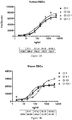
- Figure 1 shows cross species binding curves to human, mouse, rat, and porcine RBCs (panels A, B, C, and D, respectively, generated using various concentrations of purified antibodies from clones Cl 1, Cl 1.1, Cl 13, and Cl 13.1.
- Clones Cl 1 and Cl 13 are as described above in Table 3.
- Clones Cl 1.1 and Cl 13.1 are Fc mutants of clones Cl 1 and Cl 13, respectively, modified to reduce effector function. Each has an Asn297 ⁇ Gln(N297Q) mutation in the Fc domain ( Sazinsky et al. (2008) PNAS 105(51):20167-20172 ). All of these clones exhibit concentration-dependent binding to all of the species of RBCs tested.
- the purpose of this experiment is to identify antibody clones of the present invention that do, and do not, exhibit cytotoxic activity.
- the therapeutic mAb should ideally lack cytotoxic activity.
- antibodies useful in the treatment of cancer should ideally exhibit toxicity against transformed/cancer cells. This additional property of selective toxicity to cancer cells is expected to have advantages compared to mAbs that only prevent SIRPalpha binding to CD47.
- antibodies disclosed herein have been classified as either cytotoxic or non-cytotoxic, they are useful for either cancer indications or ischemia-reperfusion indications as the ligands of CD47 that are responsible for its role in cancer (SIRPalpha) and IRI (thrombospondin-1) are prevented from binding to CD47 by antibodies of both classes.
- SIRPalpha ischemia-reperfusion indications
- IRI thrombospondin-1
- antibodies of either class can be efficacious in a particular therapeutic context, and may thus be used interchangeably, in place of one another, or in combination with one another, as appropriate, to achieve the desired therapeutic effect.
- Jurkat JE6.1 cells (ATCC, Manassas, VA; Catalog # TIB-152) are grown in Iscove's modified Dulbeccco's medium containing 5% (v/v) heat inactivated fetal bovine serum (BioWest; Catalogue # S01520), 100 units/mL penicillin, 100 ⁇ g mL streptomycin (Sigma; Catalogue # P4222) at densities less than 1 x 10 6 cells/mL.
- Iscove's modified Dulbeccco's medium containing 5% (v/v) heat inactivated fetal bovine serum (BioWest; Catalogue # S01520), 100 units/mL penicillin, 100 ⁇ g mL streptomycin (Sigma; Catalogue # P4222) at densities less than 1 x 10 6 cells/mL.
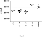
- cells are plated in 96 well tissue culture plates at a density of 2x10 4 cells/ml in Iscoves modified Dulbecco's medium containing 5% (v/v) heat inactivated fetal bovine serum (BioWest; Catalog # S01520), 100 units/mL penicillin, 100 ⁇ g/mL streptomycin (Sigma; #P4222) along with humanized antibodies as disclosed herein at a final concentration of 10 ng/ml, prepared as described above in Example 3, in Table 3, or at a concentration of 5 ⁇ g/ml using purified antibodies in Figure 2 .
- Cells are incubated for 72 hours at 37°C in an atmosphere of 5% (v/v) CO 2 .
- the following clones are considered to be non-toxic: 1, 4, 7, 9, 11, 12, 18, 20, 21, 22, 23, and 24.
- the results using purified clones 13, 14, and 24 shown in Figure 2 also indicate that clones 13 and 14 are cytotoxic with similar activity to 1F7, while clone 24 does not reduce cell viability.
- cGMP cGMP Fluorescent Assay Kit
- Molecular Devices Sunnyvale, CA
- Jurkat JE6.1 cells ATCC, Manassas, VA; Catalog # TIB-152 are used as these cells retain the NO-cGMP signaling pathway when grown in culture and exhibit a robust and reproducible inhibitory response to TSP1 ligation of CD47.
- Cells are grown in Iscove's modified Dulbeccco's medium containing 5% (v/v) heat inactivated fetal bovine serum (BioWest; Catalogue # S01520), 100 units/mL penicillin, 100 ⁇ g mL streptomycin (Sigma; Catalogue # P4222) at densities less than 1 x 10 6 cells/mL.
- cells are plated in 96 well tissue culture plates at a density of 1x10 5 cells/ml in Iscoves modified Dulbecco's medium containing 5% (v/v) heat inactivated fetal bovine serum (BioWest; Catalog # S01520), 100 units/mL penicillin, 100 ⁇ g/mL streptomycin (Sigma; #P4222) for 24 hours and then transferred to serum free medium overnight.
- humanized antibodies as disclosed herein purified from transient transfections in CHO cells as described above in Example 3, as well as the control chimeric antibody, are then added at a final concentration of 20 ng/ml, followed 15 minutes later by 0 or 1 ⁇ g/ml human TSP1 (Athens Research and Technology, Athens, GA, Catalogue # 16-20-201319). After an additional 15 minutes, the NO donor, diethylamine NONOate (Cayman Chemical, Ann Arbor, MI, Catalog # 82100), is added to half the wells at a final concentration of 1 ⁇ M. Five minutes later, the cells are lysed with buffer supplied in the cGMP kit, and aliquots of each well are assayed for cGMP content.
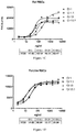
- Humanized clones 1, 9, 11, 13, and 24 of the present invention also significantly reverse TSP1 inhibition, demonstrating that they have the ability to increase NO signaling ( Figures 3 and 4 ), suggesting their utility in protecting the cardiovascular system against stresses including, but not limited to, those resulting from wounding, inflammation, hypertension, metabolic syndrome, ischemia, and ischemia-reperfusion injury (IRI).
- IRI ischemia-reperfusion injury
- the purpose of this experiment is to demonstrate that a humanized antibody clone disclosed herein, i.e., Clone 1, that is shown to regulate nitric oxide signaling in vitro in Example 5, is effective in reducing IRI and kidney damage in vivo in a rat kidney transplant model.
- IRI significantly contributes to delayed graft function and inflammation leading to graft loss, and is exacerbated by the thrombospondin-1/CD47 system through inhibition of nitric oxide signaling.
- a syngeneic rat renal transplantation model of IRI with bilaterally nephrectomized recipients is used to evaluate the effect of the anti-CD47 monoclonal antibody Clone 1 on graft function following transplantation as described in Schumacher et al. (2003) Microsurg. 23:389-394 and Karatzas et al. (2007) Microsug. 27:668-672 .
- CD47mAb Clone 1 perfusion of donor kidneys results in improved kidney function compared to controls as measured by a reduction in serum creatinine.
- the purpose of this experiment is to demonstrate that a humanized antibody clone disclosed herein, i.e., Clone 13, that was shown to exhibit cytotoxic activity and reduce cell viability in vitro in Example 4, reduces tumor burden in vivo in a mouse leukemia model.
- the anti-tumor activity of the anti-CD47mAb Clone13 (Cl 13; clone number as described above in Example 3) is determined in a syngeneic murine model of Acute Promyelocytic Leukemia (APL) as described in Ramirez et al. (2009) Blood 113:6206-6214 .
- APL Acute Promyelocytic Leukemia
- Murine APL cells (B6APL1) are injected intravenously into C57BL/6 mice that are randomized into three groups (5-10 mice per group): Group 1: no APL; Group 2: APL with no treatment; Group 3: APL with anti-CD47mAb Cl 13treatment.
- Antibody treatment is initiated on the day of tumor inoculation (day 0), and given in single doses of 10 ⁇ g/dose (0.4mg/kg) in phosphate buffered saline, pH 7.2, by intraperitoneal injection on days 0, 3, and 6.
- Tumor burden is evaluated at day 25 following tumor cell inoculation. Blood samples from each mouse are analyzed for white blood cell count using an automated hemocytometer, and circulating APL cells (representing the tumor burden) are quantified by flow cytometry (CD34 + /CD117 + cells).
- mice treated with Cl 13 have reduced tumor burden compared to untreated mice at 25 days after tumor inoculation, thus demonstrating anti-tumor activity of this humanized clone.
- >VxP037-01-LC-Pro represents the full length light chain variable domain + constant domain amino acid sequence.
- the underlined amino acid sequence framework 4 + the constant domain.
- All the humanized light chain sequences contain the same constant domain as VxP037-01-LC-Pro. However, this is not shown in the remaining humanized light chain amino acid sequences. >VxP037-01-LC-Pro
- the underlined nucleic acid sequence encodes the underlined protein sequence in >VxP037-01-LC-Pro, above. >VxP037-01-LC-DNA
- >VxP037-01-HC-Pro represents the full length heavy chain variable domain + constant domain amino acid sequence.
- the underlined amino acid sequence framework 4 + the constant domain.
- All the humanized heavy chain sequences contain the same constant domain as >VxP037-01-HC-Pro. However, this is not shown in the remaining humanized heavy chain amino acid sequences. >VxP037-01-HC-Pro
- the underlined nucleic acid sequence encodes the underlined protein sequence in >VxP037-01-HC-Pro, above. >VxP037-01-HC-DNA
Description
- The present invention relates to antibodies that bind CD47, including human and other CD47, and their use in treating conditions and disorders, such as ischemia-reperfusion injury (IRI) and cancers, mediated by this receptor.
- CD47 is a cell surface receptor comprised of an extracellular IgV set domain, a 5 membrane spanning transmembrane domain, and a cytoplasmic tail that is alternatively spliced. Two ligands bind CD47: thrombospondin-1 (TSP1), and signal inhibitory receptor protein alpha (SIRPalpha). TSP1 binding to CD47 activates the heterotrimeric G protein Gi, which leads to suppression of intracellular cyclic AMP (cAMP) levels. In addition, the TSP1-CD47 pathway opposes the beneficial effects of the nitric oxide pathway in all vascular cells. The nitric oxide (NO) pathway consists of any of three enzymes (nitric oxide synthases, NOS I, NOS II and NOS III) that generate bioactive gas NO using arginine as a substrate. NO can act within the cell in which it is produced, or in neighboring cells, to activate the enzyme soluble guanylyl cyclase that produces the messenger molecule cyclic GMP (cGMP). The proper functioning of the NO-cGMP pathway is essential for protecting the cardiovascular system against stresses including, but not limited to, those resulting from wounding, inflammation, hypertension, metabolic syndrome, ischemia, and ischemia-reperfusion injury (IRI). In the context of these cellular stresses, the inhibition of the NO-cGMP pathway by the TSP1-CD47 system exacerbates the effects of stress. This is a particular problem in the cardiovascular system where both cGMP and cAMP play important protective roles. There are many cases in which ischemia and reperfusion injury cause or contribute to disease, trauma, and poor outcomes of surgical procedures.
- SIRPalpha is expressed on hematopoietic cells, including macrophages and dendritic cells. When it engages CD47 on a potential phagocytic target cell, phagocytosis is slowed or prevented. The CD47-SIRPalpha interaction effectively sends a "don't eat me" signal to the phagocyte. Thus, blocking the SIRPalpha-CD47 interaction with a monoclonal antibody in this therapeutic context can provide an effective anti-cancer therapy by promoting, i.e., increasing, the uptake and clearance of cancer cells by the host's immune system. This mechanism is effective in both leukemias and many types of solid tumors.
-
U.S. Patent 8,236,313 contemplates antibodies that could be useful in the field of ischemia and blood flow to reverse and/or prevent tissue ischemia and related and associated tissue and cell damage, including antibodies that block CD47. No antibodies are actually disclosed. -
U.S. Patent 8,101,719 discloses humanized antibodies that bind to CD47 for use in treating hematological disorders. Objects of the invention include humanized anti-CD47 antibodies and small antibody fragments exhibiting reduced antigenicity while retaining their CD47 binding activity and apoptosis-inducing activity. Such antibodies and small fragments are contemplated for use in treating hematological disorders such as various types of leukemias, malignant lymphoma, aplastic anemia, myeodysplastic syndromes, and polycythemia vera. No other properties of these antibodies are disclosed. -
PCT International Publication WO 2011/143624 discloses chimeric and humanized anti-CD47 monoclonal antibodies for use as reagents for the diagnosis and immunotherapy of diseases associated with CD47 in humans, particularly in cancer therapy, for example to increase phagocytosis of cancer cells expressing CD47. Preferred antibodies are non-activating, i.e., block ligand binding, but do not signal. Disclosed humanized B6H12 and 5F9 antibodies bound soluble human CD47; B6H12 also bound human CD47 on the surface of human CD47-transfected YB2/0 cells. Humanized B6H12 and 5F9 antibodies enabled phagocytosis of CFSE-labeled HL-60 cells by mouse bone marrow- or peripheral blood-derived macrophages in vitro, respectively. Humanized B6H12 utilized human VH-3-7 and VK3-11 frameworks. -
U.S. 2013/0142786 discloses non-activating anti-CD47 antibodies that increase the phagocytosis of CD47 expressing cells. -
PCT International Publication WO 2013/119714 discloses anti-CD47 antibodies that do not cause a significant level of hemagglutination of human red blood cells. -
PCT International Publication WO 99/40940 WO 99/40940 -
PCT International Publication WO 2008/060785 discloses the role played by thrombospondin-1 (TSP1) in blocking the effects of nitric oxide (NO) in the vascular system. This document reports that the effects of TSP1 are mediated by CD47. Further described are methods for preventing tissue ischemia by blocking the TSP1-CD47 interaction. CD47 antibodies are described as exemplary agents. - Subramanian et al. (Blood, 15 March 2006, Vol. 107 (6); pages 2548-2556) describes the species and cell-type specific interactions between CD47 and human SIRPα. This document describes the binding activity of two CD47 antibodies: CIKm1 from ICN (Costa Mesa, CA); and B6H12 from ATCC (HB-9711), against CD47 expressing red blood cells from different species. Neither antibody was able to bind to rat CD47. B6H12 was also unable to bind to CD47 on pig red blood cells.
- Isenberg et al. (Surgery, 1 November 2008, Vol. 144 (5); pages 752-761) describes the treatment of induced liver ischemia-reperfusion in mice with CD47 antibodies.
- Roberts et al. (Matrix Biology, 10 December 2011, Vol. 31 (3); pages 162-169) describes the role of thrombospondin-1 in inducing signaling by binding to CD47 and regulating cardiovascular dynamics, hemostasis, immunity and mitochondrial homeostasis.There exists a need for antibodies to human CD47 that selectively block the binding of TSP1 to CD47 to promote the beneficial effects of nitric oxide-cGMP signaling and cAMP signaling in the cardiovascular system in settings in which IRI plays a role in pathogenesis. These situations/diseases include organ transplantation, acute kidney injury, cardiopulmonary bypass surgery, pulmonary hypertension, sickle cell disease, myocardial infarction, stroke, surgical resections and reconstructive surgery, reattachment of digits/body parts, skin grafting, and trauma. There is also a need for antibodies that block the binding of SIRPalpha to CD47, thus providing novel anti-cancer therapies. Such antibodies that also have the ability to selectively kill or induce cell death of transformed or cancer cells are also expected to provide additional therapeutic benefit.
- Antibody compounds of the present invention meet these needs. They bind to epitopes in the extracellular IgV domain of CD47, inhibiting TSP1 and SIRPalpha binding to CD47 and receptor activation, while inducing little or no agonist activity. Certain other antibodies of the present invention also provide a tumor-toxic or cell death induction effect that is specific to activated or transformed cancer cells in addition to promoting (increasing) tumor cell phagocytic clearance. In view of these properties, antibody compounds of the present invention should be therapeutically useful in treating many forms of IRI and both blood cancers and solid tumors.
- In addition, the present antibody compounds possess a number of other desirable properties, including broad reactivity with CD47 of a wide variety of mammalian species, including that of human, mouse, rat, pig, and dog, making these antibodies useful in both human and veterinary medicine. This feature is further advantageous in that it facilitates preclinical studies including, but not limited to, safety and efficacy studies, in a variety of mammalian species, and therefore the development of such antibodies as human and veterinary therapeutics.
- Accordingly, the present invention provides:
- [1] A monoclonal antibody, or antigen-binding fragment thereof, that specifically binds human, rat, mouse, pig, and dog CD47, wherein the monoclonal antibody or antigen binding fragment thereof comprises three light chain complementarity determining regions (LCDRs 1-3) and three heavy chain complementarity determining regions (HCDRs 1-3), wherein:
-
LCDR 1 comprises the amino acid sequence RSSQSLVHSNGNTYLH (SEQ ID NO:1); - LCDR 2 comprises the amino acid sequence KVSYRFS (SEQ ID NO:2); and
- LCDR 3 comprises the amino acid sequence SQNTHVPRT (SEQ ID NO:3);
-
HCDR 1 comprises the amino acid sequence GYTFTNYYVF (SEQ ID NO:4); - HCDR 2 comprises the amino acid sequence DINPVNGDTNFNEKFKN (SEQ ID NO:5); and
- HCDR 3 comprises the amino acid sequence GGYTMDY (SEQ ID NO:6).
-
- [2] The monoclonal antibody or antigen-binding fragment thereof of [1], which is chimeric or humanized.
- [3] The monoclonal antibody, or antigen-binding fragment thereof, of either one of [1]or[2], which comprises a light chain variable region (LCVR) and a heavy chain variable region (HCVR), wherein said LCVR and said HCVR comprise, respectively, amino acid sequences selected from the group consisting of:
- SEQ ID NO:7 and SEQ ID NO:57;
- SEQ ID NO:8 and SEQ ID NO:58;
- SEQ ID NO:9 and SEQ ID NO:59;
- SEQ ID NO:10 and SEQ ID NO:60;
- SEQ ID NO:11 and SEQ ID NO:61;
- SEQ ID NO:12 and SEQ ID NO:62;
- SEQ ID NO:13 and SEQ ID NO:63;
- SEQ ID NO:14 and SEQ ID NO:64;
- SEQ ID NO:15 and SEQ ID NO:65;
- SEQ ID NO:16 and SEQ ID NO:66;
- SEQ ID NO:17 and SEQ ID NO:67;
- SEQ ID NO:18 and SEQ ID NO:68;
- SEQ ID NO:19 and SEQ ID NO:69;
- SEQ ID NO:20 and SEQ ID NO:70;
- SEQ ID NO:21 and SEQ ID NO:71;
- SEQ ID NO:22 and SEQ ID NO:72;
- SEQ ID NO:23 and SEQ ID NO:73;
- SEQ ID NO:24 and SEQ ID NO:74;
- SEQ ID NO:25 and SEQ ID NO:75;
- SEQ ID NO:26 and SEQ ID NO:76;
- SEQ ID NO:27 and SEQ ID NO:77;
- SEQ ID NO:28 and SEQ ID NO:78;
- SEQ ID NO:29 and SEQ ID NO:79;
- SEQ ID NO:30 and SEQ ID NO:80; and
- SEQ ID NO:31 and SEQ ID NO:81.
- [4] A monoclonal antibody, or antigen-binding fragment thereof, which comprises a light chain variable region (LCVR) and a heavy chain variable region (HCVR), wherein said LCVR and said HCVR comprise, respectively, amino acid sequences selected from the group consisting of:
- SEQ ID NO:7 and SEQ ID NO:57;
- SEQ ID NO:8 and SEQ ID NO:58;
- SEQ ID NO:11 and SEQ ID NO:61;
- SEQ ID NO:14 and SEQ ID NO:64;
- SEQ ID NO:16 and SEQ ID NO:66;
- SEQ ID NO:18 and SEQ ID NO:68;
- SEQ ID NO:19 and SEQ ID NO:69;
- SEQ ID NO:25 and SEQ ID NO:75;
- SEQ ID NO:27 and SEQ ID NO:77;
- SEQ ID NO:28 and SEQ ID NO:78;
- SEQ ID NO:29 and SEQ ID NO:79;
- SEQ ID NO:30 and SEQ ID NO:80; and
- SEQ ID NO:31 and SEQ ID NO:81.
- [5] A monoclonal antibody, or antigen-binding fragment thereof, which comprises a light chain variable region (LCVR) and a heavy chain variable region (HCVR), wherein said LCVR and said HCVR comprise, respectively, amino acid sequences selected from the group consisting of:
- SEQ ID NO:9 and SEQ ID NO:59;
- SEQ ID NO:10 and SEQ ID NO:60;
- SEQ ID NO:12 and SEQ ID NO:62;
- SEQ ID NO:13 and SEQ ID NO:63;
- SEQ ID NO:15 and SEQ ID NO:65;
- SEQ ID NO:17 and SEQ ID NO:67;
- SEQ ID NO:20 and SEQ ID NO:70;
- SEQ ID NO:21 and SEQ ID NO:71;
- SEQ ID NO:22 and SEQ ID NO:72;
- SEQ ID NO:23 and SEQ ID NO:73;
- SEQ ID NO:24 and SEQ ID NO:74; and
- SEQ ID NO:26 and SEQ ID NO:76.
- [6] A pharmaceutical composition, comprising said monoclonal antibody, or antigen-binding fragment thereof, of any one of [1]-[5], and a pharmaceutically or physiologically acceptable carrier, diluent, or excipient.
- [7] A monoclonal antibody, or antigen-binding fragment thereof, of any one of claims [1]-[5] for use in human therapy.
- [8] The monoclonal antibody, or antigen-binding fragment thereof, of any one of [1]-[5] for use in treating ischemia-reperfusion injury, or an autoimmune or inflammatory disease, in a human.
- [9] The monoclonal antibody, or antigen-binding fragment thereof, of [8], which comprises a light chain variable region (LCVR) and a heavy chain variable region (HCVR), wherein said LCVR and said HCVR comprise, respectively, amino acid sequences selected from the group consisting of:
- SEQ ID NO:7 and SEQ ID NO:57;
- SEQ ID NO:8 and SEQ ID NO:58;
- SEQ ID NO:11 and SEQ ID NO:61;
- SEQ ID NO:14 and SEQ ID NO:64;
- SEQ ID NO:16 and SEQ ID NO:66;
- SEQ ID NO:18 and SEQ ID NO:68;
- SEQ ID NO:19 and SEQ ID NO:69;
- SEQ ID NO:25 and SEQ ID NO:75;
- SEQ ID NO:27 and SEQ ID NO:77;
- SEQ ID NO:28 and SEQ ID NO:78;
- SEQ ID NO:29 and SEQ ID NO:79;
- SEQ ID NO:30 and SEQ ID NO:80; and
- SEQ ID NO:31 and SEQ ID NO:81.
- [10] The monoclonal antibody, or antigen-binding fragment thereof, of [8] or [9], wherein said ischemia-reperfusion injury occurs in organ transplantation, acute kidney injury, cardiopulmonary bypass surgery, pulmonary hypertension, sickle cell disease, myocardial infarction, stroke, surgical resections and reconstructive surgery, reattachment of an appendage or other body part, skin grafting, or trauma.
- [11] The monoclonal antibody, or antigen-binding fragment thereof, of [8] or [9], wherein said autoimmune or inflammatory disease is selected from the group consisting of arthritis, multiple sclerosis, psoriasis, Crohn's disease, inflammatory bowel disease, lupus, Grave's disease and Hashimoto's thyroiditis, and ankylosing spondylitis.
- [12] The monoclonal antibody, or antigen-binding fragment thereof, of any one of [1]-[5] for use in treating a susceptible cancer.
- [13] The monoclonal antibody, or antigen-binding fragment thereof, of [12], which comprises a light chain variable region (LCVR) and a heavy chain variable region (HCVR), wherein said LCVR and said HCVR comprise, respectively, amino acid sequences selected from the group consisting of:
- SEQ ID NO:9 and SEQ ID NO:59;
- SEQ ID NO:10 and SEQ ID NO:60;
- SEQ ID NO:12 and SEQ ID NO:62;
- SEQ ID NO:13 and SEQ ID NO:63;
- SEQ ID NO:15 and SEQ ID NO:65;
- SEQ ID NO:17 and SEQ ID NO:67;
- SEQ ID NO:20 and SEQ ID NO:70;
- SEQ ID NO:21 and SEQ ID NO:71;
- SEQ ID NO:22 and SEQ ID NO:72;
- SEQ ID NO:23 and SEQ ID NO:73;
- SEQ ID NO:24 and SEQ ID NO:74; and
- SEQ ID NO:26 and SEQ ID NO:76.
- [14] The monoclonal antibody, or antigen binding fragment thereof, of [12] or [13], which promotes phagocytosis and/or killing of cells of said susceptible cancer.
- [15] The monoclonal antibody, or antigen binding fragment thereof, of any one of [12]-[14], wherein said susceptible cancer is selected from the group consisting of a leukemia, a lymphoma, ovarian cancer, breast cancer, endometrial cancer, colon cancer, rectal cancer, gastric cancer, bladder cancer, lung cancer, bronchial cancer, bone cancer, prostate cancer, pancreatic cancer, liver and bile duct cancer, esophageal cancer, renal cancer, thyroid cancer, head and neck cancer, testicular cancer, glioblastoma, astrocytoma, melanoma, myelodysplastic syndrome, and a sarcoma.
- [16] The monoclonal antibody, or antigen binding fragment thereof, of [15], wherein:
- said leukemia is selected from the group consisting of acute lymphocytic (lymphoblastic) leukemia, acute myeloid leukemia, myelogenous leukemia, chronic lymphocytic leukemia, multiple myeloma, plasma cell leukemia, and chronic myeloid leukemia;
- said lymphoma is selected from the group consisting of Hodgkin lymphoma and Non-Hodgkin lymphoma including B cell lymphoma, diffuse large B cell lymphoma, follicular lymphoma, mantle cell lymphoma, marginal zone B cell lymphoma, T cell lymphoma, and Waldenstrom macroglobulinemia; and
- said sarcoma is selected from the group consisting of osteosarcoma, Ewing sarcoma, leiomyosarcoma, synovial sarcoma, alveolar soft part sarcoma, angiosarcoma, liposarcoma, fibrosarcoma, rhabdomyosarcoma, and chrondrosarcoma.
- [17] The monoclonal antibody, or antigen binding fragment thereof, for use of [8], further comprising administering to said patient an anti-tumor therapeutic treatment selected from the group consisting of surgery, radiation, an anti-tumor or anti-neoplastic agent, and combinations of any of the foregoing.
- Described herein but not within the scope of the claimed invention is:
- [18] Use of said monoclonal antibody, or antigen-binding fragment thereof, of any one of [1]-[5] to treat ischemia-reperfusion injury, or an autoimmune or inflammatory disease, in a human or companion/pet animal, working animal, sport animal, zoo animal, or other valuable animal kept in captivity.
- [19] The use of [18], wherein said monoclonal antibody, or antigen binding fragment thereof, comprises a light chain variable region (LCVR) and a heavy chain variable region (HCVR), wherein said LCVR and said HCVR comprise, respectively, amino acid sequences selected from the group consisting of:
- SEQ ID NO:7 and SEQ ID NO:57;
- SEQ ID NO:8 and SEQ ID NO:58;
- SEQ ID NO:11 and SEQ ID NO:61;
- SEQ ID NO:14 and SEQ ID NO:64;
- SEQ ID NO:16 and SEQ ID NO:66;
- SEQ ID NO:18 and SEQ ID NO:68;
- SEQ ID NO:19 and SEQ ID NO:69;
- SEQ ID NO:25 and SEQ ID NO:75;
- SEQ ID NO:27 and SEQ ID NO:77;
- SEQ ID NO:28 and SEQ ID NO:78;
- SEQ ID NO:29 and SEQ ID NO:79;
- SEQ ID NO:30 and SEQ ID NO:80; and
- SEQ ID NO:31 and SEQ ID NO:81.
- [20] Use of said monoclonal antibody, or antigen-binding fragment thereof, of any one of [1]-[5] to treat a susceptible cancer.
- [21] The use of [20], wherein said monoclonal antibody, or antigen binding fragment thereof, comprises a light chain variable region (LCVR) and a heavy chain variable region (HCVR), wherein said LCVR and said HCVR comprise, respectively, amino acid sequences selected from the group consisting of:
- SEQ ID NO:9 and SEQ ID NO:59;
- SEQ ID NO:10 and SEQ ID NO:60;
- SEQ ID NO:12 and SEQ ID NO:62;
- SEQ ID NO:13 and SEQ ID NO:63;
- SEQ ID NO:15 and SEQ ID NO:65;
- SEQ ID NO:17 and SEQ ID NO:67;
- SEQ ID NO:20 and SEQ ID NO:70;
- SEQ ID NO:21 and SEQ ID NO:71;
- SEQ ID NO:22 and SEQ ID NO:72;
- SEQ ID NO:23 and SEQ ID NO:73;
- SEQ ID NO:24 and SEQ ID NO:74; and
- SEQ ID NO:26 and SEQ ID NO:76.
- [22] Use of said monoclonal antibody, or antigen-binding fragment thereof, of any one of [1]-[5] for the manufacture of a medicament to treat ischemia-reperfusion injury, or an autoimmune or inflammatory disease, in a human or companion/pet animal, working animal, sport animal, zoo animal, or other valuable animal kept in captivity.
- [23] The use of [22], wherein said monoclonal antibody, or antigen binding fragment thereof, comprises a light chain variable region (LCVR) and a heavy chain variable region (HCVR), wherein said LCVR and said HCVR comprise, respectively, amino acid sequences selected from the group consisting of:
- SEQ ID NO:7 and SEQ ID NO:57;
- SEQ ID NO:8 and SEQ ID NO:58;
- SEQ ID NO:11 and SEQ ID NO:61;
- SEQ ID NO:14 and SEQ ID NO:64;
- SEQ ID NO:16 and SEQ ID NO:66;
- SEQ ID NO:18 and SEQ ID NO:68;
- SEQ ID NO:19 and SEQ ID NO:69;
- SEQ ID NO:25 and SEQ ID NO:75;
- SEQ ID NO:27 and SEQ ID NO:77;
- SEQ ID NO:28 and SEQ ID NO:78;
- SEQ ID NO:29 and SEQ ID NO:79;
- SEQ ID NO:30 and SEQ ID NO:80; and
- SEQ ID NO:31 and SEQ ID NO:81.
- [24] The use of [22] or [23], wherein said ischemia-reperfusion injury occurs in organ transplantation, acute kidney injury, cardiopulmonary bypass surgery, pulmonary hypertension, sickle cell disease, myocardial infarction, stroke, surgical resections and reconstructive surgery, reattachment of an appendage or other body part, skin grafting, and trauma.
- [25] The use of [22] or [23], wherein said autoimmune or inflammatory disease is selected from the group consisting of arthritis, multiple sclerosis, psoriasis, Crohn's disease, inflammatory bowel disease, lupus, Grave's disease and Hashimoto's thyroiditis, and ankylosing spondylitis.
- [26] Use of said monoclonal antibody, or antigen-binding fragment thereof, of any one of [1]-[5] for the manufacture of a medicament to treat a susceptible cancer.
- [27] The use of [26], wherein said monoclonal antibody, or antigen-binding fragment thereof, comprises a light chain variable region (LCVR) and a heavy chain variable region (HCVR), wherein said LCVR and said HCVR comprise, respectively, amino acid sequences selected from the group consisting of:
- SEQ ID NO:9 and SEQ ID NO:59;
- SEQ ID NO:10 and SEQ ID NO:60;
- SEQ ID NO:12 and SEQ ID NO:62;
- SEQ ID NO:13 and SEQ ID NO:63;
- SEQ ID NO:15 and SEQ ID NO:65;
- SEQ ID NO:17 and SEQ ID NO:67;
- SEQ ID NO:20 and SEQ ID NO:70;
- SEQ ID NO:21 and SEQ ID NO:71;
- SEQ ID NO:22 and SEQ ID NO:72;
- SEQ ID NO:23 and SEQ ID NO:73;
- SEQ ID NO:24 and SEQ ID NO:74; and
- SEQ ID NO:26 and SEQ ID NO:76.
- [28] A method of treating ischemia or ischemia-reperfusion injury in a patient in need thereof, comprising administering to said patient an effective amount of said monoclonal antibody, or antigen-binding fragment thereof, of any one of [1]-[5], or a monoclonal antibody, or antigen-binding fragment thereof, that competes with said monoclonal antibody or antigen-binding fragment thereof of any one of [1]-[5] for binding to CD47.
- [29] The method of [28], wherein said patient is about to be subjected to, or is experiencing, ischemia or ischemia-reperfusion injury.
- [30] The method of [28] or [29], wherein said patient is a human.
- [31] The method of [28] or [29], wherein said patient is a companion/pet animal, working animal, sport animal, zoo animal, or other valuable animal kept in captivity.
- [32] The method of any one of [28]-[29], wherein said ischemia occurs because said patient will undergo, or is undergoing, a surgery selected from the group consisting of integument surgery, soft tissue surgery, composite tissue surgery, cosmetic surgery, surgical resections, reconstructive surgery, skin graft surgery, and limb reattachment surgery.
- [33] The method of [32], wherein said skin graft is an autograft.
- [34] The method of any one of [28]-[31], wherein said ischemia occurs because said patient will undergo, or is undergoing, organ transplant surgery.
- [35] The method of any one of [28]-[31], wherein said ischemia-reperfusion injury occurs in organ transplantation, acute kidney injury, cardiopulmonary bypass surgery, pulmonary hypertension, sickle cell disease, myocardial infarction, stroke, surgical resection, reconstructive surgery, reattachment of an appendage or other body part, or skin grafting.
- [36] The method of any one of [28]-[35], wherein said monoclonal antibody, antigen-binding fragment thereof, or competing monoclonal antibody or antigen binding fragment thereof, is administered before, during, or after said subject undergoes ischemia or surgery, or a combination of any of these time periods.
- [37] The method of any one of [28]-[36], further comprising administering to said patient an effective amount of a nitric oxide donor, precursor, or both; a nitric oxide generating topical agent; an agent that activates soluble guanylyl cyclase; an agent that inhibits cyclic nucleotide phosphodiesterases; or any combination of any of the foregoing.
- [38] The method of [37], wherein:
- said nitric oxide donor or precursor is selected from the group consisting of NO gas, isosorbide dinitrate, nitrite, nitroprusside, nitroglycerin, 3-Morpholinosydnonimine (SIN-1), S-nitroso-N-acetylpenicillamine (SNAP), Diethylenetriamine/NO (DETA/NO), S-nitrosothiols, Bidil®, and arginine; and
- said agent that inhibits cyclic nucleotide phosphodiesterases is selected from the group consisting of sildenafil, tadalafil, vardenafil udenafil, and avanafil.
- [39] A method of increasing tissue perfusion in a subject in need thereof, comprising administering to said subject an effective amount of a monoclonal antibody, or antigen-binding fragment thereof, of any one of [1]-[5], or a monoclonal antibody, or antigen-binding fragment thereof, that competes with said monoclonal antibody or antigen-binding fragment thereof of any one of [1]-[5] for binding to CD47.
- [40] The method of [39], wherein said subject has, or is at risk of developing, at least one disease or condition selected from the group consisting of ischemia-reperfusion injury, myocardial infarction, myocardial ischemia, stroke, cerebral ischemia, sickle cell anemia, and pulmonary hypertension.
- [41] The method of [39], wherein said subject has, or is at risk of developing, at least one disease or condition selected from the group consisting of hypertension, atherosclerosis, vasculopathy, ischemia secondary to diabetes, and peripheral vascular disease.
- [42] The method of [39], wherein the need for increased tissue perfusion arises because said subject has had, is having, or will have, a surgery selected from the group consisting of integument surgery, soft tissue surgery, composite tissue surgery, skin graft surgery, resection of a solid organ, and reattachment or an appendage or other body part.
- [43] The method of [42], wherein said skin graft is an autograft.
- [44] The method of [39], wherein the need for increased tissue perfusion arises because said subject has had, is having, or will have, organ transplant surgery.
- [45] The method of any one of [39]-[44], further comprising administering to said subject an effective amount of a nitric oxide donor, precursor, or both; a nitric oxide generating topical agent; an agent that activates soluble guanylyl cyclase; an agent that inhibits cyclic nucleotide phosphodiesterases; or any combination of any of the foregoing.
- [46] The method of [45], wherein:
- said nitric oxide donor or precursor is selected from the group consisting of NO gas, isosorbide dinitrate, nitrite, nitroprusside, nitroglycerin, 3-Morpholinosydnonimine (SIN-1), S-nitroso-N-acetylpenicillamine (SNAP), Diethylenetriamine/NO (DETA/NO), S-nitrosothiols, Bidil®, and arginine; and
- said agent that inhibits cyclic nucleotide phosphodiesterases is selected from the group consisting of sildenafil, tadalafil, vardenafil udenafil, and avanafil.
- [47] A method of transplanting a donor organ from an organ donor to an organ recipient, comprising any single step, any combination of steps, or all steps selected from the group consisting of steps i) - iii):
- i) administering to said organ donor prior to, during, both prior to and during, after, or any combination thereof, donation of said donor organ an effective amount of said monoclonal antibody or antigen-binding fragment thereof of any one of [1]-[5], and/or a monoclonal antibody, or antigen-binding fragment thereof, that competes with said monoclonal antibody or antigen-binding fragment thereof of any one of [1]-[5] for binding to CD47;
- ii) contacting said donor organ prior to, during, both prior to and during, after, or any combination thereof, transplantation to said organ recipient, and an effective amount of said monoclonal antibody or antigen-binding fragment thereof of any one of [1]-[5], and/or a monoclonal antibody, or antigen-binding fragment thereof, that competes with said monoclonal antibody or antigen-binding fragment thereof of any one of [1]-[5] for binding to CD47; and
- iii) administering to said organ recipient prior to, during, both prior to and during, after, or any combination thereof, transplantation of said donor organ to said organ recipient, an effective amount of said monoclonal antibody or antigen-binding fragment thereof of any one of [1]-[5], and/or a monoclonal antibody, or antigen-binding fragment thereof, that competes with said monoclonal antibody or antigen-binding fragment thereof of any one of [1]-[5] for binding to CD47.
- [48] The method of claim 47, wherein said monoclonal antibody or antigen-binding fragment thereof of any one of [1]-[5], or monoclonal antibody, or antigen-binding fragment thereof, that competes with said monoclonal antibody or antigen-binding fragment thereof of any one of [1]-[5] for binding to CD47, reduces ischemia reperfusion injury in said donor organ.
- [49] The method of [47] or [48], further comprising administering to said organ donor, said donor organ, said organ recipient, or any combination thereof, an effective amount of a nitric oxide donor, precursor, or both; a nitric oxide generating topical agent; an agent that activates soluble guanylyl cyclase; or an agent that inhibits cyclic nucleotide phosphodiesterases; or any combination of any of the foregoing..
- [50] The method of [49], wherein:
- said nitric oxide donor or precursor is selected from the group consisting of NO gas, isosorbide dinitrate, nitrite, nitroprusside, nitroglycerin, 3-Morpholinosydnonimine (SIN-1), S-nitroso-N-acetylpenicillamine (SNAP), Diethylenetriamine/NO (DETA/NO), S-nitrosothiols, Bidil®, and arginine; and
- said agent that inhibits cyclic nucleotide phosphodiesterases is selected from the group consisting of sildenafil, tadalafil, vardenafil, udenafil, and avanafil.
- [51] A method of treating an autoimmune or inflammatory disease in a patient in need thereof, comprising administering to said patient an effective amount of said monoclonal antibody, or antigen-binding fragment thereof, of any one of [1]-[5], or a monoclonal antibody, or antigen-binding fragment thereof, that competes with said monoclonal antibody or antigen-binding fragment thereof of any one of [1]-[5] for binding to CD47.
- [52] The method of [51], wherein said autoimmune or inflammatory disease is selected from the group consisting of arthritis, multiple sclerosis, psoriasis, Crohn's disease, inflammatory bowel disease, lupus, Grave's disease and Hashimoto's thyroiditis, and ankylosing spondylitis.
- [53] The method of [51] or [52], wherein said patient is a human.
- [54] The method of [51] or [52], wherein said patient is a companion/pet animal, working animal, sport animal, zoo animal, or other valuable animal kept in captivity.
- [55] The method of any one of [51]-[54], further comprising administering to said patient an effective amount of a nitric oxide donor, precursor, or both; a nitric oxide generating topical agent; an agent that activates soluble guanylyl cyclase; an agent that inhibits cyclic nucleotide phosphodiesterases; or any combination of any of the foregoing.
- [56] The method of [55], wherein:
- said nitric oxide donor or precursor is selected from the group consisting of NO gas, isosorbide dinitrate, nitrite, nitroprusside, nitroglycerin, 3-Morpholinosydnonimine (SIN-1), S-nitroso-N-acetylpenicillamine (SNAP), Diethylenetriamine/NO (DETA/NO), S-nitrosothiols, Bidil®, and arginine; and
- said agent that inhibits cyclic nucleotide phosphodiesterases is selected from the group consisting of sildenafil, tadalafil, vardenafil, udenafil, and avanafil.
- [57] A method of treating a susceptible cancer in a human or companion/pet animal, working animal, sport animal, zoo animal, or other valuable animal kept in captivity in need thereof, comprising administering thereto an effective amount of a monoclonal antibody or antigen binding fragment thereof of any one of [1]-[5], or a monoclonal antibody, or antigen-binding fragment thereof, that competes with said monoclonal antibody or antigen-binding fragment thereof of any one of [1]-[5] for binding to CD47, and which exhibits cytotoxic activity.
- [58] The method of [57], wherein said susceptible cancer is selected from the group consisting of a leukemia, a lymphoma, ovarian cancer, breast cancer, endometrial cancer, colon cancer, rectal cancer, gastric cancer, bladder cancer, lung cancer, bronchial cancer, bone cancer, prostate cancer, pancreatic cancer, liver and bile duct cancer, esophageal cancer, renal cancer, thyroid cancer, head and neck cancer, testicular cancer, glioblastoma, astrocytoma, melanoma, myelodysplastic syndrome, and a sarcoma.
- [59] The method of [58], wherein:
- said leukemia is selected from the group consisting of acute lymphocytic (lymphoblastic) leukemia, acute myeloid leukemia, myelogenous leukemia, chronic lymphocytic leukemia, multiple myeloma, plasma cell leukemia, and chronic myeloid leukemia;
- said lymphoma is selected from the group consisting of Hodgkin lymphoma and Non-Hodgkin lymphoma including B cell lymphoma, diffuse large B cell lymphoma, follicular lymphoma, mantle cell lymphoma, marginal zone B cell lymphoma, T cell lymphoma, and Waldenstrom macroglobulinemia; and
- said sarcoma is selected from the group consisting of osteosarcoma, Ewing sarcoma, leiomyosarcoma, synovial sarcoma, alveolar soft part sarcoma, angiosarcoma, liposarcoma, fibrosarcoma, rhabdomyosarcoma, and chrondrosarcoma.
- [60] The method of any one of [57]-[59], wherein said monoclonal antibody or antigen-binding fragment thereof of any one of [1]-[5], or said monoclonal antibody, or antigen-binding fragment thereof, that competes with said monoclonal antibody or antigen-binding fragment thereof of any one of [1]-[5] for binding to CD47 and which exhibits cytotoxic activity, increases phagocytosis of cells of said susceptible cancer.
- [61] The method of [60], wherein said monoclonal antibody or antigen-binding fragment thereof of any one of [1]-[5], or said monoclonal antibody, or antigen-binding fragment thereof, that competes with said monoclonal antibody or antigen-binding fragment thereof of any one of [1]-[5] for binding to CD47 which exhibits cytotoxic activity and increases phagocytosis of cells of said susceptible cancer inhibits CD47 binding to SIRPalpha.
- [62] The method of any one of [57] to [61], wherein said monoclonal antibody or antigen-binding fragment thereof of any one of [1]-[5], or said monoclonal antibody, or antigen-binding fragment thereof, that competes with said monoclonal antibody or antigen-binding fragment thereof of any one of [1]-[5] for binding to CD47 and which exhibits cytotoxic activity, is directly toxic to cells of said susceptible cancer.
- [63] A humanized monoclonal antibody, or antigen-binding fragment thereof, that specifically binds human CD47.
- [64] A humanized or chimeric monoclonal antibody, or antigen-binding fragment thereof, that specifically binds human, rat, mouse, pig, or dog CD47, blocks binding of SIRPalpha to CD47, and which exhibits the dual activities of:
- i) inducing death of cancer cells, and
- ii) increasing phagocytosis of said cancer cells.
- [65] A monoclonal antibody, or antigen-binding fragment thereof, that competes with said humanized or chimeric monoclonal antibody or antigen-binding fragment thereof of 64 for binding to human, rat, mouse, pig, or dog CD47, and which exhibits said dual activities.
- [66] A pharmaceutical composition, comprising said humanized or chimeric monoclonal antibody, or antigen-binding fragment thereof, of [64], or said competing monoclonal antibody, or antigen-binding fragment thereof, of [65], and a pharmaceutically or physiologically acceptable carrier, diluent, or excipient.
- [67] A humanized or chimeric monoclonal antibody, or antigen-binding fragment thereof, that specifically binds human, rat, mouse, pig, or dog CD47, blocks binding of SIRPalpha to CD47, and which exhibits the dual activities of:
- i) inducing death of cancer cells, and
- ii) increasing phagocytosis of said cancer cells, or
for use in human therapy or therapy of companion/pet animals, working animals, sport animals, zoo animals, or therapy of other valuable animals kept in captivity. - [68] A humanized or chimeric monoclonal antibody, or antigen-binding fragment thereof, that specifically binds human, rat, mouse, pig, or dog CD47, blocks binding of SIRPalpha to CD47, and which exhibits the dual activities of:
- i) inducing death of cancer cells, and
- ii) increasing phagocytosis of said cancer cells, or
for use in treating ischemia-reperfusion injury, or an autoimmune or inflammatory disease, in a human or companion/pet animal, working animal, sport animal, zoo animal, or other valuable animal kept in captivity. - [69] The humanized or chimeric monoclonal antibody or antigen-binding fragment thereof, or competing monoclonal antibody or antigen-binding fragment thereof, of [68], wherein said ischemia-reperfusion injury occurs in organ transplantation, acute kidney injury, cardiopulmonary bypass surgery, pulmonary hypertension, sickle cell disease, myocardial infarction, stroke, surgical resections and reconstructive surgery, reattachment of an appendage or other body part, skin grafting, or trauma.
- [70] The humanized or chimeric monoclonal antibody or antigen-binding fragment thereof, or competing monoclonal antibody or antigen-binding fragment thereof, of [68], wherein said autoimmune or inflammatory disease is selected from the group consisting of arthritis, multiple sclerosis, psoriasis, Crohn's disease, inflammatory bowel disease, lupus, Grave's disease and Hashimoto's thyroiditis, and ankylosing spondylitis.
- [71] A humanized or chimeric monoclonal antibody, or antigen-binding fragment thereof, that specifically binds human, rat, mouse, pig, or dog CD47, blocks binding of SIRPalpha to CD47, and which exhibits the dual activities of:
- i) inducing death of cancer cells, and
- ii) increasing phagocytosis of said cancer cells, or
for use in treating a susceptible cancer. - [72] The humanized or chimeric monoclonal antibody or antigen-binding fragment thereof, or competing monoclonal antibody or antigen-binding fragment thereof, of [71], wherein said susceptible cancer is selected from the group consisting of a leukemia, a lymphoma, ovarian cancer, breast cancer, endometrial cancer, colon cancer, rectal cancer, gastric cancer, bladder cancer, lung cancer, bronchial cancer, bone cancer, prostate cancer, pancreatic cancer, liver and bile duct cancer, esophageal cancer, renal cancer, thyroid cancer, head and neck cancer, testicular cancer, glioblastoma, astrocytoma, melanoma, myelodysplastic syndrome, and a sarcoma.
- [73] The humanized or chimeric monoclonal antibody or antigen-binding fragment thereof, or competing monoclonal antibody or antigen-binding fragment thereof, of [72], wherein:
- said leukemia is selected from the group consisting of acute lymphocytic (lymphoblastic) leukemia, acute myeloid leukemia, myelogenous leukemia, chronic lymphocytic leukemia, multiple myeloma, plasma cell leukemia, and chronic myeloid leukemia;
- said lymphoma is selected from the group consisting of Hodgkin lymphoma and Non-Hodgkin lymphoma including B cell lymphoma, diffuse large B cell lymphoma, follicular lymphoma, mantle cell lymphoma, marginal zone B cell lymphoma, T cell lymphoma, and Waldenstrom macroglobulinemia; and
- said sarcoma is selected from the group consisting of osteosarcoma, Ewing sarcoma, leiomyosarcoma, synovial sarcoma, alveolar soft part sarcoma, angiosarcoma, liposarcoma, fibrosarcoma, rhabdomyosarcoma, and chrondrosarcoma.
- [74] Use of a humanized or chimeric monoclonal antibody, or antigen-binding fragment thereof, that specifically binds human, rat, mouse, pig, or dog CD47, blocks binding of SIRPalpha to CD47, and which exhibits the dual activities of:
- i) inducing death of cancer cells, and
- ii) increasing phagocytosis of said cancer cells, or
to treat ischemia-reperfusion injury, or an autoimmune or inflammatory disease, in a human or companion/pet animal, working animal, sport animal, zoo animal, or other valuable animal kept in captivity. - [75] Use of a humanized or chimeric monoclonal antibody, or antigen-binding fragment thereof, that specifically binds human, rat, mouse, pig, or dog CD47, blocks binding of SIRPalpha to CD47, and which exhibits the dual activities of:
- i) inducing death of cancer cells, and
- ii) increasing phagocytosis of said cancer cells, or
to treat a susceptible cancer. - [76] The use of [75], wherein said susceptible cancer is selected from the group consisting of a leukemia, a lymphoma, ovarian cancer, breast cancer, endometrial cancer, colon cancer, rectal cancer, gastric cancer, bladder cancer, lung cancer, bronchial cancer, bone cancer, prostate cancer, pancreatic cancer, liver and bile duct cancer, esophageal cancer, renal cancer, thyroid cancer, head and neck cancer, testicular cancer, glioblastoma, astrocytoma, melanoma, myelodysplastic syndrome, and a sarcoma.
- [77] The use of [76], wherein:
- said leukemia is selected from the group consisting of acute lymphocytic (lymphoblastic) leukemia, acute myeloid leukemia, myelogenous leukemia, chronic lymphocytic leukemia, multiple myeloma, plasma cell leukemia, and chronic myeloid leukemia;
- said lymphoma is selected from the group consisting of Hodgkin lymphoma and Non-Hodgkin lymphoma including B cell lymphoma, diffuse large B cell lymphoma, follicular lymphoma, mantle cell lymphoma, marginal zone B cell lymphoma, T cell lymphoma, and Waldenstrom macroglobulinemia; and
- said sarcoma is selected from the group consisting of osteosarcoma, Ewing sarcoma, leiomyosarcoma, synovial sarcoma, alveolar soft part sarcoma, angiosarcoma, liposarcoma, fibrosarcoma, rhabdomyosarcoma, and chrondrosarcoma.
- [78] Use of a humanized or chimeric monoclonal antibody, or antigen-binding fragment thereof, that specifically binds human, rat, mouse, pig, or dog CD47, blocks binding of SIRPalpha to CD47, and which exhibits the dual activities of:
- i) inducing death of cancer cells, and
- ii) increasing phagocytosis of said cancer cells, or
for the manufacture of a medicament to treat ischemia-reperfusion injury, or an autoimmune or inflammatory disease, in a human or companion/pet animal, working animal, sport animal, zoo animal, or other valuable animal kept in captivity. - [79] The use of [78], wherein said ischemia-reperfusion injury occurs in organ transplantation, acute kidney injury, cardiopulmonary bypass surgery, pulmonary hypertension, sickle cell disease, myocardial infarction, stroke, surgical resections and reconstructive surgery, reattachment of an appendage or other body part, skin grafting, and trauma.
- [80] The use of [78], wherein said autoimmune or inflammatory disease is selected from the group consisting of arthritis, multiple sclerosis, psoriasis, Crohn's disease, inflammatory bowel disease, lupus, Grave's disease and Hashimoto's thyroiditis, and ankylosing spondylitis.
- [81] Use of a humanized or chimeric monoclonal antibody, or antigen-binding fragment thereof, that specifically binds human, rat, mouse, pig, or dog CD47, blocks binding of SIRPalpha to CD47, and which exhibits the dual activities of:
- i) inducing death of cancer cells, and
- ii) increasing phagocytosis of said cancer cells, or
for the manufacture of a medicament to treat a susceptible cancer. - [82] The use of [81], wherein said susceptible cancer is selected from the group consisting of a leukemia, a lymphoma, ovarian cancer, breast cancer, endometrial cancer, colon cancer, rectal cancer, gastric cancer, bladder cancer, lung cancer, bronchial cancer, bone cancer, prostate cancer, pancreatic cancer, liver and bile duct cancer, esophageal cancer, renal cancer, thyroid cancer, head and neck cancer, testicular cancer, glioblastoma, astrocytoma, melanoma, myelodysplastic syndrome, and a sarcoma.
- [83] The use of [82], wherein:
- said leukemia is selected from the group consisting of acute lymphocytic (lymphoblastic) leukemia, acute myeloid leukemia, myelogenous leukemia, chronic lymphocytic leukemia, multiple myeloma, plasma cell leukemia, and chronic myeloid leukemia;
- said lymphoma is selected from the group consisting of Hodgkin lymphoma and Non-Hodgkin lymphoma including B cell lymphoma, diffuse large B cell lymphoma, follicular lymphoma, mantle cell lymphoma, marginal zone B cell lymphoma, T cell lymphoma, and Waldenstrom macroglobulinemia; and
- said sarcoma is selected from the group consisting of osteosarcoma, Ewing sarcoma, leiomyosarcoma, synovial sarcoma, alveolar soft part sarcoma, angiosarcoma, liposarcoma, fibrosarcoma, rhabdomyosarcoma, and chrondrosarcoma.
- [84] A method of treating ischemia or ischemia-reperfusion injury in a patient in need thereof, comprising administering to said patient an effective amount of a humanized or chimeric monoclonal antibody, or antigen-binding fragment thereof, that specifically binds human, rat, mouse, pig, or dog CD47, blocks binding of SIRPalpha to CD47, and which exhibits the dual activities of:
- i) inducing death of cancer cells, and
- ii) increasing phagocytosis of said cancer cells, or
- [85] The method of [84], wherein said patient is about to be subjected to, or is experiencing, ischemia or ischemia-reperfusion injury.
- [86] The method of [84] or [85], wherein said patient is a human.
- [87] The method of [84] or [85], wherein said patient is a companion/pet animal, working animal, sport animal, zoo animal, or other valuable animal kept in captivity.
- [88] The method of any one of [84]-[87], wherein said ischemia occurs because said patient will undergo, or is undergoing, a surgery selected from the group consisting of integument surgery, soft tissue surgery, composite tissue surgery, cosmetic surgery, surgical resections, reconstructive surgery, skin graft surgery, and limb reattachment surgery.
- [89] The method of [88], wherein said skin graft is an autograft.
- [90] The method of any one of [84]-[87], wherein said ischemia occurs because said patient will undergo, or is undergoing, organ transplant surgery.
- [91] The method of any one of [84]-[87], wherein said ischemia-reperfusion injury occurs in organ transplantation, acute kidney injury, cardiopulmonary bypass surgery, pulmonary hypertension, sickle cell disease, myocardial infarction, stroke, surgical resection, reconstructive surgery, reattachment of an appendage or other body part, or skin grafting.
- [92] The method of any one of [84]-[91], wherein said monoclonal antibody, antigen-binding fragment thereof, or competing monoclonal antibody or antigen binding fragment thereof, is administered before, during, or after said subject undergoes ischemia or surgery, or a combination of any of these time periods.
- [93] The method of any one of [84]-[92], further comprising administering to said patient an effective amount of a nitric oxide donor, precursor, or both; a nitric oxide generating topical agent; an agent that activates soluble guanylyl cyclase; an agent that inhibits cyclic nucleotide phosphodiesterases; or any combination of any of the foregoing.
- [94] The method of [93], wherein:
- said nitric oxide donor or precursor is selected from the group consisting of NO gas, isosorbide dinitrate, nitrite, nitroprusside, nitroglycerin, 3-Morpholinosydnonimine (SIN-1), S-nitroso-N-acetylpenicillamine (SNAP), Diethylenetriamine/NO (DETA/NO), S-nitrosothiols, Bidil®, and arginine; and
- said agent that inhibits cyclic nucleotide phosphodiesterases is selected from the group consisting of sildenafil, tadalafil, vardenafil, udenafil, and avanafil.
- [95] A method of increasing tissue perfusion in a subject in need thereof, comprising administering to said subject an effective amount of a humanized or chimeric monoclonal antibody, or antigen-binding fragment thereof, that specifically binds human, rat, mouse, pig, or dog CD47, blocks binding of SIRPalpha to CD47, and which exhibits the dual activities of:
- i) inducing death of cancer cells, and
- ii) increasing phagocytosis of said cancer cells, or
- [96] The method of [95], wherein said subject has, or is at risk of developing, at least one disease or condition selected from the group consisting of ischemia-reperfusion injury, myocardial infarction, myocardial ischemia, stroke, cerebral ischemia, sickle cell anemia, and pulmonary hypertension.
- [97] The method of [95], wherein said subject has, or is at risk of developing, at least one disease or condition selected from the group consisting of hypertension, atherosclerosis, vasculopathy, ischemia secondary to diabetes, and peripheral vascular disease.
- [98] The method of [97], wherein the need for increased tissue perfusion arises because said subject has had, is having, or will have, a surgery selected from the group consisting of integument surgery, soft tissue surgery, composite tissue surgery, skin graft surgery, resection of a solid organ, and reattachment or an appendage or other body part.
- [99] The method of [98], wherein said skin graft is an autograft.
- [100] The method of [95], wherein the need for increased tissue perfusion arises because said subject has had, is having, or will have, organ transplant surgery.
- [101] The method of any one of [95]-[100], further comprising administering to said subject an effective amount of a nitric oxide donor, precursor, or both; a nitric oxide generating topical agent; an agent that activates soluble guanylyl cyclase; an agent that inhibits cyclic nucleotide phosphodiesterases; or any combination of any of the foregoing.
- [102] The method of [101], wherein:
- said nitric oxide donor or precursor is selected from the group consisting of NO gas, isosorbide dinitrate, nitrite, nitroprusside, nitroglycerin, 3-Morpholinosydnonimine (SIN-1), S-nitroso-N-acetylpenicillamine (SNAP), Diethylenetriamine/NO (DETA/NO), S-nitrosothiols, Bidil®, and arginine; and
- said agent that inhibits cyclic nucleotide phosphodiesterases is selected from the group consisting of sildenafil, tadalafil, vardenafil, udenafil, and avanafil.
- [103] A method of transplanting a donor organ from an organ donor to an organ recipient, comprising any single step, any combination of steps, or all steps selected from the group consisting of steps i) - iii):
- i) administering to said organ donor prior to, during, both prior to and during, after, or any combination thereof, donation of said donor organ an effective amount of a humanized or chimeric monoclonal antibody, or antigen-binding fragment thereof, that specifically binds human, rat, mouse, pig, or dog CD47, blocks binding of SIRPalpha to CD47, and which exhibits the dual activities of:
- a) inducing death of cancer cells, and
- b) increasing phagocytosis of said cancer cells, or
- ii) contacting said donor organ prior to, during, both prior to and during, after, or any combination thereof, transplantation to said organ recipient, and an effective amount of said humanized or chimeric monoclonal antibody, antigen-binding fragment thereof, or competing monoclonal antibody or antigen binding fragment thereof; and
- iii) administering to said organ recipient prior to, during, both prior to and during, after, or any combination thereof, transplantation of said donor organ to said organ recipient, an effective amount of said humanized or chimeric monoclonal antibody, antigen-binding fragment thereof, or competing monoclonal antibody or antigen binding fragment thereof.
- i) administering to said organ donor prior to, during, both prior to and during, after, or any combination thereof, donation of said donor organ an effective amount of a humanized or chimeric monoclonal antibody, or antigen-binding fragment thereof, that specifically binds human, rat, mouse, pig, or dog CD47, blocks binding of SIRPalpha to CD47, and which exhibits the dual activities of:
- [104] The method of [103], wherein said humanized or chimeric monoclonal antibody, antigen-binding fragment thereof, or competing monoclonal antibody or antigen binding fragment thereof, reduces ischemia reperfusion injury in said donor organ.
- [105] The method of [103] or [104], further comprising administering to said organ donor, said donor organ, said organ recipient, or any combination thereof, an effective amount of a nitric oxide donor, precursor, or both; a nitric oxide generating topical agent; an agent that activates soluble guanylyl cyclase; an agent that inhibits cyclic nucleotide phosphodiesterases; or any combination of any of the foregoing.
- [106] The method of [105], wherein:
- said nitric oxide donor or precursor is selected from the group consisting of NO gas, isosorbide dinitrate, nitrite, nitroprusside, nitroglycerin, 3-Morpholinosydnonimine (SIN-1), S-nitroso-N-acetylpenicillamine (SNAP), Diethylenetriamine/NO (DETA/NO), S-nitrosothiols, Bidil®, and arginine; and
- said agent that inhibits cyclic nucleotide phosphodiesterases is selected from the group consisting of sildenafil, tadalafil, vardenafil, udenafil, and avanafil.
- [107] A method of treating an autoimmune or inflammatory disease in a patient in need thereof, comprising administering to said patient an effective amount of a humanized or chimeric monoclonal antibody, or antigen-binding fragment thereof, that specifically binds human, rat, mouse, pig, or dog CD47, blocks binding of SIRPalpha to CD47, and which exhibits the dual activities of:
- i) inducing death of cancer cells, and
- ii) increasing phagocytosis of said cancer cells, or
- [108] The method of [107], wherein said autoimmune or inflammatory disease is selected from the group consisting of arthritis, multiple sclerosis, psoriasis, Crohn's disease, inflammatory bowel disease, lupus, Grave's disease and Hashimoto's thyroiditis, and ankylosing spondylitis.
- [109] The method of [107] or [108], wherein said patient is a human.
- [110] The method of [107] or [108], wherein said patient is a companion/pet animal, working animal, sport animal, zoo animal, or other valuable animal kept in captivity.
- [111] The method of any one of [107]-[110], further comprising administering to said patient an effective amount of a nitric oxide donor, precursor, or both; a nitric oxide generating topical agent; an agent that activates soluble guanylyl cyclase; an agent that inhibits cyclic nucleotide phosphodiesterases; or any combination of any of the foregoing.
- [112] The method of [111], wherein:
- said nitric oxide donor or precursor is selected from the group consisting of NO gas, isosorbide dinitrate, nitrite, nitroprusside, nitroglycerin, 3-Morpholinosydnonimine (SIN-1), S-nitroso-N-acetylpenicillamine (SNAP), Diethylenetriamine/NO (DETA/NO), S-nitrosothiols, Bidil®, and arginine; and
- said agent that inhibits cyclic nucleotide phosphodiesterases is selected from the group consisting of sildenafil, tadalafil, vardenafil, udenafil, and avanafil.
- [113] A method of treating a susceptible cancer in a human or companion/pet animal, working animal, sport animal, zoo animal, or other valuable animal kept in captivity in need thereof, comprising administering thereto an effective amount of a humanized or chimeric monoclonal antibody, or antigen-binding fragment thereof, that specifically binds human, rat, mouse, pig, or dog CD47, blocks binding of SIRPalpha to CD47, and which exhibits the dual activities of:
- i) inducing death of cancer cells, and
- ii) increasing phagocytosis of said cancer cells, or
- [114] The method of [113], wherein said susceptible cancer is selected from the group consisting of a leukemia, a lymphoma, ovarian cancer, breast cancer, endometrial cancer, colon cancer, rectal cancer, gastric cancer, bladder cancer, lung cancer, bronchial cancer, bone cancer, prostate cancer, pancreatic cancer, liver and bile duct cancer, esophageal cancer, renal cancer, thyroid cancer, head and neck cancer, testicular cancer, glioblastoma, astrocytoma, melanoma, myelodysplastic syndrome, and a sarcoma.
- [115] The method of [114], wherein:
- said leukemia is selected from the group consisting of acute lymphocytic (lymphoblastic) leukemia, acute myeloid leukemia, myelogenous leukemia, chronic lymphocytic leukemia, multiple myeloma, plasma cell leukemia, and chronic myeloid leukemia;
- said lymphoma is selected from the group consisting of Hodgkin lymphoma and Non-Hodgkin lymphoma including B cell lymphoma, diffuse large B cell lymphoma, follicular lymphoma, mantle cell lymphoma, marginal zone B cell lymphoma, T cell lymphoma, and Waldenstrom macroglobulinemia; and
- said sarcoma is selected from the group consisting of osteosarcoma, Ewing sarcoma, leiomyosarcoma, synovial sarcoma, alveolar soft part sarcoma, angiosarcoma, liposarcoma, fibrosarcoma, rhabdomyosarcoma, and chrondrosarcoma.
- [116] The use or method of [17], wherein said an anti-tumor or anti-neoplastic agent is a small chemical molecule or a biologic therapeutic.
- [117] The use or method of [116], wherein said small chemical molecule or biologic therapeutic is selected from the group consisting of an alkylating agent; an antimetabolite; a natural product; a miscellaneous agent used in cancer therapy; a hormone; an antagonist; a monoclonal antibody or antigen-binding fragment thereof; a cytokine; an antisense oligonucleotide; and an siRNA.
- [118] A method of enhancing the therapeutic effect of a soluble guanylyl cyclase activator, comprising administering to a patient in need thereof:
- i) an effective amount of a soluble guanylyl cyclase activator, and
- ii) a monoclonal antibody or antigen-binding fragment thereof, or competing antibody or antigen-binding fragment thereof, of any one of claims [1][5] or [64]-[65] in an amount effective to enhance said therapeutic effect of said soluble guanylyl cyclase activator.
- [119] The method of [118], wherein said therapeutic effect comprises treatment of ischemia-reperfusion injury, or an autoimmune or inflammatory disease, in a human or companion/pet animal, working animal, sport animal, zoo animal, or other valuable animal kept in captivity.
- [120] The method of [119], wherein said ischemia-reperfusion injury occurs in organ transplantation, acute kidney injury, cardiopulmonary bypass surgery, pulmonary hypertension, sickle cell disease, myocardial infarction, stroke, surgical resections and reconstructive surgery, reattachment of an appendage or other body part, skin grafting, or trauma.
- [121] The method of [119], wherein said autoimmune or inflammatory disease is selected from the group consisting of arthritis, multiple sclerosis, psoriasis, Crohn's disease, inflammatory bowel disease, lupus, Grave's disease and Hashimoto's thyroiditis, and ankylosing spondylitis.
- [122] Use of said monoclonal antibody or antigen-binding fragment thereof, or competing antibody or antigen-binding fragment thereof, of any one of [1]-[5] or [64]-[65] for the manufacture of a medicament to enhance the therapeutic effect of a soluble guanylyl cyclase activator.
- [123] The use of [122], wherein said therapeutic effect comprises treatment of ischemia-reperfusion injury, or an autoimmune or inflammatory disease, in a human or companion/pet animal, working animal, sport animal, zoo animal, or other valuable animal kept in captivity.
- [124] The use of [123], wherein said ischemia-reperfusion injury occurs in organ transplantation, acute kidney injury, cardiopulmonary bypass surgery, pulmonary hypertension, sickle cell disease, myocardial infarction, stroke, surgical resections and reconstructive surgery, reattachment of an appendage or other body part, skin grafting, and trauma.
- [125] The use of [123], wherein said autoimmune or inflammatory disease is selected from the group consisting of arthritis, multiple sclerosis, psoriasis, Crohn's disease, inflammatory bowel disease, lupus, Grave's disease and Hashimoto's thyroiditis, and ankylosing spondylitis.
- [126] A method of increasing the level of cGMP in vascular cells, comprising administering to said cells:
- i) an effective amount of a soluble guanylyl cyclase activator, and
- ii) a monoclonal antibody or antigen-binding fragment thereof, or competing antibody or antigen-binding fragment thereof, of any one of [1]-[5] or [64]-[65] in an amount effective to increase the level of cGMP in said vascular cells.
- [127] The monoclonal antibody or antigen-binding fragment thereof, or competing antibody or antigen-binding fragment thereof, of any one of [1]-[5] or [64]-[65], which is an IgG isotype selected from the group consisting of IgG1 isotype, IgG2 isotype, IgG3 isotype, and IgG4 isotype.
- [128] A pharmaceutical composition, comprising said monoclonal antibody or antigen-binding fragment thereof, or competing antibody or antigen-binding fragment thereof, of [127], and a pharmaceutically or physiologically acceptable carrier, diluent, or excipient.
- [129] The monoclonal antibody or antigen-binding fragment thereof, or competing antibody or antigen-binding fragment thereof, of [127] for use in human therapy or therapy of companion/pet animals, working animals, sport animals, zoo animals, or therapy of other valuable animals kept in captivity.
- [130] The monoclonal antibody or antigen-binding fragment thereof, or competing antibody or antigen-binding fragment thereof, of [127] for use in treating ischemia-reperfusion injury, or an autoimmune or inflammatory disease, in a human or companion/pet animal, working animal, sport animal, zoo animal, or other valuable animal kept in captivity.
- [131] The monoclonal antibody or antigen-binding fragment thereof, or competing antibody or antigen-binding fragment thereof, of [127] for use in treating a susceptible cancer.
- [132] Use of said monoclonal antibody or antigen-binding fragment thereof, or competing antibody or antigen-binding fragment thereof, of [127] to treat ischemia-reperfusion injury, or an autoimmune or inflammatory disease, in a human or companion/pet animal, working animal, sport animal, zoo animal, or other valuable animal kept in captivity.
- [133] Use of said monoclonal antibody or antigen-binding fragment thereof, or competing antibody or antigen-binding fragment thereof, of [127] to treat a susceptible cancer.
- [134] Use of said monoclonal antibody or antigen-binding fragment thereof, or competing antibody or antigen-binding fragment thereof, of [127] for the manufacture of a medicament to treat ischemia-reperfusion injury, or an autoimmune or inflammatory disease, in a human or companion/pet animal, working animal, sport animal, zoo animal, or other valuable animal kept in captivity.
- [135] Use of said monoclonal antibody or antigen-binding fragment thereof, or competing antibody or antigen-binding fragment thereof, of [127] for the manufacture of a medicament to treat a susceptible cancer.
Expressly disclosed herein is the use of the monoclonal antibodies or antigen-binding fragments thereof of [127]-[135] in any of the methods, uses, compositions, or any other embodiments disclosed herein. - [136] The monoclonal antibody or antigen-binding fragment thereof, or competing antibody or antigen-binding fragment thereof, of any one of [1]-[5] or [64]-[65], wherein:
- i) when said monoclonal antibody or antigen-binding fragment thereof, or competing antibody or antigen-binding fragment thereof, is human IgG1 isotype, the human IgG1 constant region is modified at amino acid Asn297 to prevent to glycosylation; and/or at amino acid Leu 234 and/or Leu235 to alter Fc receptor interactions; and/or to enhance FcRn binding; and/or to alter antibody-dependent cellular cytotoxicity and/or complement-dependent cytotoxicity; and/or to induce heterodimerization, optionally further by introduction of a disulfide bond;
- ii) when said monoclonal antibody or antigen-binding fragment thereof, or competing antibody or antigen-binding fragment thereof, is human IgG2 isotype, the human IgG2 constant region is modified at amino acid Asn297 to prevent to glycosylation; and/or to enhance FcRn binding; and/or to alter antibody-dependent cellular cytotoxicity and/or complement-dependent cytotoxicity; and/or to induce heterodimerization, optionally further by introduction of a disulfide bond;
- iii) when said monoclonal antibody or antigen-binding fragment thereof, or competing antibody or antigen-binding fragment thereof, is human IgG3 isotype, the human IgG3 constant region is modified at amino acid Asn297 to prevent to glycosylation; and/or at amino acid 435 to extend half-life; and/or to enhance FcRn binding; and/or to alter antibody-dependent cellular cytotoxicity and/or complement-dependent cytotoxicity; and/or to induce heterodimerization, optionally further by introduction of a disulfide bond;
- iv) when said monoclonal antibody or antigen-binding fragment thereof, or competing antibody or antigen-binding fragment thereof, is human IgG4 isotype, the human IgG4 constant region is modified within the hinge region to prevent or reduce strand exchange; and/or at amino acid 235 to alter Fc receptor interactions; and/or at amino acid Asn297 to prevent glycosylation; and/or to enhance FcRn binding; and/or to alter antibody-dependent cellular cytotoxicity; and/or complement-dependent cytotoxicity; and/or to induce heterodimerization, optionally further by introduction of a disulfide bond.
- [137] The monoclonal antibody or antigen-binding fragment thereof, or competing antibody or antigen-binding fragement thereof, of [136], wherein:
- i) when said monoclonal antibody or antigen-binding fragment thereof, or competing antibody or antigen-binding fragment thereof, is human IgG1 isotype, the human IgG1 constant region is modified at amino acid Asn297 to prevent to glycosylation by modification of Asn297→Ala (N297A) or Asn297→Gln(N297Q); and/or at amino acid Leu 234 by modification of Leu234→Ala (L234A) and/or Leu235 by modification of Leu235→Glu (L235E) or Leu235→Ala (L235A) or at both amino acid 234 and 235 by modification of Leu234→Ala and Leu235→Ala to alter Fc receptor interactions; and/or to enhance FcRn binding by modification of Met252→Tyr, Ser254→Thr, Thr256→Glu, Met428→Leu, or Asn434→Ser; and/or to alter antibody-dependent cellular cytotoxicity and/or complement-dependent cytotoxicity; and/or to induce heterodimerization by modification of Thr366→Trp, and optionally further by introduction of a disulfide bond by modification of Ser354→Cys and Tyr349→Cys on opposite CH3 domains;
- ii) when said monoclonal antibody or antigen-binding fragment thereof, or competing antibody or antigen-binding fragment thereof, is human IgG2 isotype, the human IgG2 constant region is modified at amino acid Asn297 to prevent to glycosylation by modification of Asn297→Ala or Asn297→Gln; and/or to enhance FcRn binding by modification of Met252→Tyr, Ser254→Thr, Thr256→Glu, Met428→Leu, or Asn434→Ser; and/or to alter antibody-dependent cellular cytotoxicity and/or complement-dependent cytotoxicity; and/or to induce heterodimerization by modification of Thr366→Trp, and optionally further by introduction of a disulfide bond by modification of Ser354→Cys and Tyr349→Cys on opposite CH3 domains;
- iii) when said monoclonal antibody or antigen-binding fragment thereof, or competing antibody or antigen-binding fragment thereof, is human IgG3 isotype, the human IgG3 constant region is modified at amino acid Asn297 to prevent to glycosylation by modification of Asn297→Ala or Asn297→Gln; and/or at amino acid 435 to extend half-life by modification of Arg435→His; and/or to enhance FcRn binding by modification of Met252→Tyr, Ser254→Thr, Thr256→Glu, Met428→Leu, or Asn434→Ser; and/or to alter antibody-dependent cellular cytotoxicity and/or complement-dependent cytotoxicity; and/or to induce heterodimerization by modification of Thr366→Trp, and optionally further by introduction of a disulfide bond by modification of Ser354→Cys and Tyr349→Cys on opposite CH3 domains;
- iv) when said monoclonal antibody or antigen-binding fragment thereof, or competing antibody or antigen-binding fragment thereof, is human IgG4 isotype, the human IgG4 constant region is modified within the hinge region to prevent or reduce strand exchange by modification of Ser228→Pro; and/or at amino acid 235 to alter Fc receptor interactions by modification of Leu235→Glu, or by modification within the hinge and at amino acid 235 by modifying Ser228→Pro and Leu235→Glu; and/or at amino acid Asn297 to prevent glycosylation by modification of Asn297→Ala; and/or and/or to enhance FcRn binding by modification of Met252→Tyr, Ser254→Thr, Thr256→Glu, Met428→Leu, or Asn434→Ser; and/or to alter antibody-dependent cellular cytotoxicity and/or complement-dependent cytotoxicity; and/or to induce heterodimerization by modification of Thr366→Trp, and optionally further by introduction of a disulfide bond by modification of Ser354→Cys and Tyr349→Cys on opposite CH3 domains.
- Expressly disclosed herein is the use of the monoclonal antibodies or antigen binding fragments thereof of [136]-[137] in any of the methods, uses, compositions, or any other embodiments disclosed herein.
- The above and other aspects, features, and advantages of the present invention will be better understood from the following detailed descriptions taken in conjunction with the accompanying drawing(s), all of which are given by way of illustration only, and are not limitative of the present invention, in which:
-
Figure 1 , panels A, B, C, and D, show cross species binding curves to human, mouse, rat, and porcine RBCs, respectively, generated using various concentrations of purified antibodies fromclones Cl 1, Cl 1.1,Cl 13, and Cl 13.1 as described in Example 3.Clones Cl 1 andCl 13 are as described in Table 3. Clones Cl 1.1 and Cl 13.1 are Fc mutants ofclones Cl 1 andCl 13, respectively, modified to reduce effector function. Each has an Asn297→Gln(N297Q) mutation in the Fc domain (Sazinsky et al. (2008) PNAS 105(51):20167-20172). All of these clones exhibit concentration-dependent binding to all of the species of RBCs tested.
RBCs are incubated for 60 minutes on ice with various concentrations of purified antibodies fromclones Cl 1, Cl 1.1,Cl 13, and Cl 13.1. Cells are then washed with cold PBS containing EDTA, incubated for an additional hour on ice with FITC labeled donkey anti-human antibody, washed, and antibody binding is analyzed using a BD FACS Aria Cell Sorter (Becton Dickinson) or a C6 Accuri Flow Cytometer (Becton Dickinson). -
Figure 2 shows the cytotoxic activity of certain humanized clones assessed by reducing the cell viability of human Jurkat cancer cells. Jurkat JE6.1 cells are plated in 96 well tissue culture plates at a density of 2x10⁴ cells/ml in Iscoves modified Dulbecco's medium containing 5% (v/v) heat inactivated fetal bovine serum, and incubated with 5 µg/ml of purified humanized antibodies (Clones Clones clone 24 does not reduce cell viability. -
Figure 3 shows reversal of TSP1 inhibition of NO-stimulated cGMP production by humanized antibodies of the present invention. As described in Example 5, Jurkat JE6.1 cells are incubated overnight in serum-free medium and then incubated with humanized antibodies of the present invention or the control chimeric mAb, and with or without TSP1, followed by treatment with or without a NO donor. Cells are lysed 5 minutes later and cGMP is measured. None of the present humanized antibody clones tested, or the control chimeric mAb, has an effect on basal cGMP levels. The control chimeric antibody reverses the TSP1 inhibition, as dohumanized clones Cl 1; Cl 9; Cl 11;Cl 24, respectively). PBS: phosphate buffered saline; TSP or TSP1: thrombospondin-1; DEA: diethylamine NONOate; chim: chimeric antibody >VxP037-01LC (SEQ ID NO:7))/>VxP037-01HC (SEQ ID NO:57). -
Figure 4 shows reversal of TSP1 Inhibition of NO-stimulated cGMP production by humanized antibodies of the present invention. As described in Example 5, Jurkat JE6.1 cells are incubated overnight in serum-free medium and then incubated with purified humanizedClone clones -
Figure 5 shows that treatment of a donor kidney with Clone 1 (Cl 1) at the time of organ harvest is effective in reducing IRI and kidney damage in vivo in a rat kidney transplantation model as assessed by measuring serum creatinine. A syngeneic rat renal transplantation model of IRI with bilaterally nephrectomized recipients is used to evaluate the effect of the anti-CD47monoclonal antibody Clone 1 on graft function following transplantation. Male Lewis rats weighing 275-300 g are used as both donor and recipient animals. Donor kidneys are flushed with 50 µg of purifiedClone 1 or vehicle (phosphate buffered saline, pH 7.2), stored at 4°C in University of Wisconsin preservation (WU) solution for 6 hours, and then transplanted. Two days following transplantation, kidney function is assessed by measuring circulating serum creatinine. Treatment of donor kidneys withClone 1 results in improved kidney function compared to controls as measured by a reduction in serum creatinine. -
Figure 6 shows that purified, humanized antibody Clone 13 (Cl 13) reduces tumor burden in vivo in a syngeneic mouse Acute Promyelocytic Leukemia (APL) model. Murine APL cells (B6APL1) are injected intravenously into C57BL/6 mice randomized into three groups (5-10 mice per group): Group 1: no APL; Group 2: APL with no treatment; Group 3: APL treated withanti-CD47mAb Cl 13. Antibody treatment is initiated on the day of tumor inoculation (day 0), and given in single doses of 10 µg/dose (0.4mg/kg) by intraperitoneal injection ondays 0, 3, and 6. Circulating APL cells (representing the tumor burden) are evaluated at day 25 following tumor inoculation by flow cytometry (CD34+/CD117+ cells). Mice treated withCl 13 have reduced tumor burden compared to untreated mice at 25 days after tumor inoculation, demonstrating anti-tumor activity of this humanized clone. - The following detailed description of the invention is provided to aid those skilled in the art in practicing the various embodiments of the present invention described herein, including all the methods, uses, compositions, etc., described herein.
- Antibody compounds of the present invention bind to epitopes in the extracellular IgV domain of CD47, inhibiting TSP1 and SIRPalpha binding to CD47 and receptor activation, while inducing little or no agonist activity. Certain antibodies of the present invention provide a tumor-toxic, cell death-inducing effect that is specific to activated or transformed cells, in addition to increasing tumor cell phagocytic clearance, i.e., dual activity. In view of these properties, antibody compounds of the present invention should be therapeutically useful in treating many forms of IRI and both blood cancers and solid tumors.
- The present antibody compounds also possess a number of other desirable properties, including broad reactivity with CD47 of a wide variety of mammalian species, including that of human, mouse, rat, pig, and/or dog, i.e., any individual one of these mammalian species, or various combinations thereof, making these antibodies useful in both human and veterinary medicine. This broad reactivity is further advantageous in that it facilitates preclinical studies including, but not limited to, safety and efficacy studies, in a variety of mammalian species, and therefore the development of such antibodies as human and veterinary therapeutics.
- A full-length antibody as it exists naturally is an immunoglobulin molecule comprising two heavy (H) chains and two light (L) chains interconnected by disulfide bonds. The amino terminal portion of each chain includes a variable region of about 100-110 or more amino acids primarily responsible for antigen recognition via the complementarity determining regions (CDRs) contained therein. The carboxy-terminal portion of each chain defines a constant region primarily responsible for effector function.
- The CDRs are interspersed with regions that are more conserved, termed framework regions ("FR"). Each light chain variable region (LCVR) and heavy chain variable region (HCVR) is composed of 3 CDRs and 4 FRs, arranged from amino-terminus to carboxy-terminus in the following order: FR1, CDR1, FR2, CDR2, FR3, CDR3, FR4. The 3 CDRs of the light chain are referred to as "LCDR1, LCDR2, and LCDR3" and the 3 CDRs of the heavy chain are referred to as "HCDR1, HCDR2, and HCDR3." The CDRs contain most of the residues which form specific interactions with the antigen. The numbering and positioning of CDR amino acid residues within the LCVR and HCVR regions are in accordance with the well-known Kabat numbering convention. While the light chain CDRs and heavy chain CDRs disclosed herein are numbered 1, 2, and 3, respectively, it is not necessary that they be employed in the corresponding antibody compound light and heavy chain variable regions in that numerical order, i.e., they can be present in any numerical order in a light or heavy chain variable region, respectively.
- Light chains are classified as kappa or lambda, and are characterized by a particular constant region as known in the art. Heavy chains are classified as gamma, mu, alpha, delta, or epsilon, and define the isotype of an antibody as IgG, IgM, IgA, IgD, or IgE, respectively. IgG antibodies can be further divided into subclasses, e.g., IgG1, IgG2, IgG3, IgG4. Each heavy chain type is characterized by a particular constant region with a sequence well known in the art. The monoclonal antibodies and other antibody compounds useful in the methods and compositions described herein can be any of these isotypes. Furthermore, any of these isotypes can comprise amino acid modifications as follows.
- In some embodiments, the antibody constant region is of human IgG1 isotype.
- In some embodiments, the human IgG1 constant region is modified at amino acid Asn297 (Kabat Numbering) to prevent to glycosylation of the antibody. For example, this modification can be Asn297→Ala (N297A) or Asn297→Gln(N297Q) (Sazinsky et al. (2008) PNAS 105(51):20167-20172).
- In some embodiments, the constant region of the antibody is modified at amino acid Leu234 (Kabat Numbering) to alter Fc receptor interactions. For example, this modification can be Leu234→Ala (L234A).
- In some embodiments, the constant region of the antibody is modified at amino acid Leu235 (Kabat Numbering) to alter Fc receptor interactions. For example, this modification can be Leu235→Glu (L235E) or Leu235→Ala (L235A).
- In some embodiments, the constant region of the antibody is altered at both amino acid 234 and 235. For example, these modifications can be Leu234→Ala and Leu235→Ala (L234A/L235A) (EU index of Kabat et al. (1991) Sequences of Proteins of Immunological Interest ).
- In some embodiments, the constant region of the antibody is of human IgG2 isotype.
- In some embodiments, the human IgG2 constant region is modified at amino acid Asn297 (Kabat Numbering) to prevent to glycosylation of the antibody. For example, this modification can be Asn297→Ala (N297A) or Asn297→Gln(N297Q).
- In some embodiments, the constant region of the antibody is of human IgG3 isotype.
- In some embodiments, the human IgG3 constant region is modified at amino acid Asn297 (Kabat Numbering) to prevent to glycosylation of the antibody. For example, this modification can be Asn297→Ala (N297A) or Asn297→Gln(N297Q).
- In some embodiments, the human IgG3 constant region is modified at amino acid 435 to extend the half-life. For example, this modification can be Arg435→His (R435H) (EU index of Kabat et al. (1991) Sequences of Proteins of Immunological Interest).
- In some embodiments, the constant region of the antibody is of human IgG4 isotype.
- In some embodiments, the human IgG4 constant region is modified within the hinge region to prevent or reduce strand exchange. For example, this modification can be Ser228→Pro (S228P) (Angal et al. (1993) Molecular Immunology 30(1): 105-108).
- In other embodiments, the human IgG4 constant region is modified at amino acid 235 to alter Fc receptor interactions. For example, this can be Leu235→Glu (L235E).
- In some embodiments, the human IgG4 constant region is modified within the hinge and at amino acid 235. For example, this can be Ser228→Pro and Leu235→Glu (S228P/L235E).
- In some embodiments, the human IgG4 constant region is modified at amino acid Asn297 (Kabat Numbering) to prevent to glycosylation of the antibody. For example, this can be Asn297→Ala (N297A). (EU index of Kabat et al. (1991) Sequences of Proteins of Immunological Interest).
- In some embodiments, the human IgG constant region is modified to enhance FcRn binding. Examples of Fc mutations that enhance binding to FcRn are Met252→Tyr, Ser254→Thr, Thr256→Glu (M252Y, S254T, and T256E, respectively) (Kabat numbering, Dall'Acqua et al. (2006) J. Biol. Chem. 281(33) 23514-23524), or Met428→Leu and Asn434→Ser (M428L, N434S) (Zalevsky et al. (2010) Nature Biotech. 28(2):157-159). (EU index of Kabat et al. (1991) Sequences of Proteins of Immunological Interest).
- In some embodiments, the human IgG constant region is modified to alter antibody-dependent cellular cytotoxicity (ADCC) and/or complement-dependent cytotoxicity (CDC), e.g. , the amino acid modifications described in Natsume et al. (2008) Cancer Res. 68(10):3863-72; Idusogie et al. (2001) J. Immunol. 166(4):2571-5; Moore et al. (2010) mAbs 2(2):181-189; Lazar et al. (2006) PNAS 103(11):4005-4010; Shields et al. (2001) J. Biol. Chem. 276(9):6591-6604; Stavenhagen et al. (2007) Cancer Res. 67(18):8882-8890; Stavenhagen et al. (2008) Advan. Enzyme Regul. 48:152-164; Alegre et al. (1992) J. Immunol. 148:3461-3468; reviewed in Kaneko and Niwa (2011) Biodrugs 25(1):1-11.
- In some embodiments, the human IgG constant region is modified to induce heterodimerization. For example, having an amino acid modification within the CH3 domain at Thr366, which when replaced with a more bulky amino acid, such as Trp (T366W), is able to preferentially pair with a second CH3 domain having amino acid modifications to less bulky amino acids at positions Thr366, Leu368, and Tyr407, e.g., Ser, Ala, and Val, respectively (T366S/L368A/Y407V). Heterodimerization via CH3 modifications can be further stabilized by the introduction of a disulfide bond, for example by changing Ser354 to Cys (S354C) and Tyr349 to Cys (Y349C) on opposite CH3 domains (reviewed in Carter (2001) Journal of Immunological Methods 248:7-15).
- As used herein, the term "monoclonal antibody" (mAb) as applied to the present antibody compounds refers to an antibody that is derived from a single copy or clone including, for example, any eukaryotic, prokaryotic, or phage clone, and not the method by which it is produced. mAbs of the present invention preferably exist in a homogeneous or substantially homogeneous population, and can be chimeric or humanized. Complete mAbs contain two heavy chains and two light chains.
- "Antigen binding fragments" of such monoclonal antibodies may be desirable for certain applications due to their small size and consequent superior tissue distribution, and include, for example, Fab fragments, Fab' fragments, F(ab')2 fragments, Fd fragments, single chain Fv fragments (ScFv), and one-armed antibodies comprising a light chain and a heavy chain. Preferred antigen binding fragments are those that bind to the antigen recognized by the intact antibody. Fc fragments can also be obtained. Monoclonal antibodies and antigen-binding fragments thereof of the present invention can be produced, for example, by recombinant technologies, phage display technologies, synthetic technologies, e.g., CDR-grafting, or combinations of such technologies, or other technologies known in the art, including proteolytic digestion of intact antibodies.
- "Antibody compounds" refers to mAbs and Fabs, and competing antibodies, disclosed herein that specifically bind CD47 of various species, including human, rat, mouse, pig, and dog CD47, and that exhibit the properties disclosed herein. Thus, the term "mAb" as used herein with respect to antibodies encompassed by the present invention includes Fabs and competing antibodies. Additional antibody compounds exhibiting similar functional properties according to the present invention can be generated by conventional methods. For example, mice can be immunized with human CD47 or fragments thereof, the resulting antibodies can be recovered and purified, and determination of whether they possess binding and functional properties similar to or the same as the antibody compounds disclosed herein can be assessed by the methods disclosed in Examples 3 and 4, below. Antigen-binding fragments can also be prepared by conventional methods. Methods for producing and purifying antibodies and antigen-binding fragments are well known in the art and can be found, for example, in Harlow and Lane (1988) Antibodies, A Laboratory Manual, Cold Spring Harbor Laboratory Press, Cold Spring Harbor, N.Y., chapters 5-8 and 15, ISBN 0-87969-314-2.
- The phrase "humanized antibodies" refers to monoclonal antibodies and antigen binding fragments thereof, including the antibody compounds disclosed herein, that have binding and functional properties according to the invention similar to those disclosed herein, and that have framework regions that are substantially human or fully human surrounding CDRs derived from a non-human antibody. "Framework region" or "framework sequence" refers to any one of
framework regions 1 to 4. Humanized antibodies and antigen binding fragments encompassed by the present invention include molecules wherein any one or more offramework regions 1 to 4 is substantially or fully human, i.e., wherein any of the possible combinations of individual substantially or fullyhuman framework regions 1 to 4, is present. For example, this includes molecules in whichframework region 1 and framework region 2,framework region 1 and framework region 3,framework region 1, 2, and 3, etc., are substantially or fully human. Substantially human frameworks are those that have at least 80% sequence identity to a known human germline framework sequence. Preferably, the substantially human frameworks have at least 85%, at least 90%, at least 95%, at least 96%, at least 97%, at least 98%, or at least 99% sequence identity, to a framework sequence disclosed herein, or to a known human germline framework sequence. - CDRs encompassed by the present invention include not only those specifically disclosed herein, but also CDR sequences having sequence identities of at least 80%, at least 85%, at least 90%, at least 95%, at least 96%, at least 97%, at least 98%, or at least 99% sequence identity to a CDR sequence disclosed herein. Alternatively, CDRs encompassed by the present invention include not only those specifically disclosed herein, but also CDR sequences having 1, 2, 3, 4, or 5 amino acid changes at corresponding positions compared to CDR sequences disclosed herein. Such sequence identical, or amino acid modified, CDRs preferably bind to the antigen recognized by the intact antibody.
- As used herein, the phrase "sequence identity" means the percentage of identical nucleotide or amino acid residues at corresponding positions in two or more sequences when the sequences are aligned to maximize sequence matching, i.e., taking into account gaps and insertions. Identity can be readily calculated by known methods, including but not limited to those described in: Computational Molecular Biology, Lesk, A. M., ed., Oxford University Press, New York, 1988; Biocomputing: Informatics and Genome Projects, Smith, D. W., ed., Academic Press, New York, 1993; Computer Analysis of Sequence Data, Part I, Griffin, A. M., and Griffin, H. G., eds., Humana Press, New Jersey, 1994; Sequence Analysis in Molecular Biology, von Heinje, G., Academic Press, 1987; and Sequence Analysis Primer, Gribskov, M. and Devereux, J., eds., M Stockton Press, New York, 1991; and Carillo, H., and Lipman, D., SIAM J. Applied Math., 48: 1073 (1988). Methods to determine identity are designed to give the largest match between the sequences tested. Moreover, methods to determine identity are codified in publicly available computer programs.
- Optimal alignment of sequences for comparison can be conducted, for example, by the local homology algorithm of Smith & Waterman, by the homology alignment algorithms, by the search for similarity method or, by computerized implementations of these algorithms (GAP, BESTFIT, PASTA, and TFASTA in the GCG Wisconsin Package, available from Accelrys, Inc., San Diego, California, United States of America), or by visual inspection. See generally, (Altschul, S. F. et al., J. Mol. Biol. 215: 403-410 (1990) and Altschul et al. Nucl. Acids Res. 25: 3389-3402 (1997)).
- One example of an algorithm that is suitable for determining percent sequence identity and sequence similarity is the BLAST algorithm, which is described in (Altschul, S., et al., NCBI NLM NIH Bethesda, Md. 20894; & Altschul, S., et al., J. Mol. Biol. 215: 403-410 (1990). Software for performing BLAST analyses is publicly available through the National Center for Biotechnology Information. This algorithm involves first identifying high scoring sequence pairs (HSPs) by identifying short words of length W in the query sequence, which either match or satisfy some positive-valued threshold score T when aligned with a word of the same length in a database sequence. T is referred to as the neighborhood word score threshold.
- These initial neighborhood word hits act as seeds for initiating searches to find longer HSPs containing them. The word hits are then extended in both directions along each sequence for as far as the cumulative alignment score can be increased. Cumulative scores are calculated using, for nucleotide sequences, the parameters M (reward score for a pair of matching residues; always; 0) and N (penalty score for mismatching residues; always; 0). For amino acid sequences, a scoring matrix is used to calculate the cumulative score. Extension of the word hits in each direction are halted when: the cumulative alignment score falls off by the quantity X from its maximum achieved value, the cumulative score goes to zero or below due to the accumulation of one or more negative-scoring residue alignments, or the end of either sequence is reached. The BLAST algorithm parameters W, T, and X determine the sensitivity and speed of the alignment. The BLASTN program (for nucleotide sequences) uses as defaults a wordlength (W) of 11, an expectation (E) of 10, a cutoff of 100, M = 5, N = -4, and a comparison of both strands. For amino acid sequences, the BLASTP program uses as defaults a wordlength (W) of 3, an expectation (E) of 10, and the BLOSUM62 scoring matrix.
- In addition to calculating percent sequence identity, the BLAST algorithm also performs a statistical analysis of the similarity between two sequences. One measure of similarity provided by the BLAST algorithm is the smallest sum probability (P(N)), which provides an indication of the probability by which a match between two nucleotide or amino acid sequences would occur by chance. For example, a test nucleic acid sequence is considered similar to a reference sequence if the smallest sum probability in a comparison of the test nucleic acid sequence to the reference nucleic acid sequence is in one embodiment less than about 0.1, in another embodiment less than about 0.01, and in still another embodiment less than about 0.001.
- Fully human frameworks are those that are identical to a known human germline framework sequence. Human framework germline sequences can be obtained from ImMunoGeneTics (IMGT) via their website or from The Immunoglobulin FactsBook by Marie-Paule Lefranc and Gerard Lefranc, Academic Press, 2001, ISBN 012441351. For example, germline light chain frameworks can be selected from the group consisting of: A11, A17, A18, A19, A20, A27, A30, LI, L1I, L12, L2, L5, L15, L6, L8, 012, 02, and 08, and germline heavy chain framework regions can be selected from the group consisting of: VH2-5, VH2-26, VH2-70, VH3-20, VH3-72, VHI-46, VH3-9, VH3-66, VH3-74, VH4-31, VHI-18, VHI-69, VI-13-7, VH3-11, VH3-15, VH3-21, VH3-23, VH3-30, VH3-48, VH4-39, VH4-59, and VH5-5I.
- Humanized antibodies in addition to those disclosed herein exhibiting similar functional properties according to the present invention can be generated using several different methods. In one approach, the parent antibody compound CDRs are grafted into a human framework that has a high sequence identity with the parent antibody compound framework. The sequence identity of the new framework will generally be at least 80%, at least 85%, at least 90%, at least 95%, at least 96%, at least 97%, at least 98%, or at least 99% identical to the sequence of the corresponding framework in the parent antibody compound. In the case of frameworks having fewer than 100 amino acid residues, one, two, or three amino acid residues can be changed. This grafting may result in a reduction in binding affinity compared to that of the parent antibody. If this is the case, the framework can be back-mutated to the parent framework at certain positions based on specific criteria disclosed by Queen et al. (1991) Proc. Natl. Acad. Sci. USA 88:2869. Additional references describing methods useful in humanizing mouse antibodies include
U.S. Pat. Nos. 4,816,397 ;5,225,539 ; and5,693,761 ; computer programs ABMOD and ENCAD as described in Levitt (1983) J. Mol. Biol. 168:595-620; and the method of Winter and co-workers (Jones et al. (1986) Nature 321:522-525; Riechmann et al. (1988) Nature 332:323-327; and Verhoeyen et al. (1988) Science 239:1534-1536. - The identification of residues to consider for back-mutation can be carried out as follows.
- When an amino acid falls under the following category, the framework amino acid of the human germ-line sequence that is being used (the "acceptor framework") is replaced by a framework amino acid from a framework of the parent antibody compound (the "donor framework"): (a) the amino acid in the human framework region of the acceptor framework is unusual for human frameworks at that position, whereas the corresponding amino acid in the donor immunoglobulin is typical for human frameworks at that position; (b) the position of the amino acid is immediately adjacent to one of the CDRs; or (c) any side chain atom of a framework amino acid is within about 5-6 angstroms (center-to-center) of any atom of a CDR amino acid in a three dimensional immunoglobulin model.
- When each of the amino acids in the human framework region of the acceptor framework and a corresponding amino acid in the donor framework is generally unusual for human frameworks at that position, such amino acid can be replaced by an amino acid typical for human frameworks at that position. This back-mutation criterion enables one to recover the activity of the parent antibody compound.
- Another approach to generating human engineered antibodies exhibiting similar functional properties to the antibody compounds disclosed herein involves randomly mutating amino acids within the grafted CDRs without changing the framework, and screening the resultant molecules for binding affinity and other functional properties that are as good as or better than those of the parent antibody compounds. Single mutations can also be introduced at each amino acid position within each CDR, followed by assessing the effects of such mutations on binding affinity and other functional properties. Single mutations producing improved properties can be combined to assess their effects in combination with one another.
- Further, a combination of both of the foregoing approaches is possible. After CDR grafting, one can back-mutate specific framework regions in addition to introducing amino acid changes in the CDRs. This methodology is described in Wu et al. (1999) J. Mol. Biol. 294:151-162.
- The method described in Example 1 below can also be employed.
- Applying the teachings of the present invention, a person skilled in the art can use common techniques, e.g., site-directed mutagenesis, to substitute amino acids within the presently disclosed CDR and framework sequences and thereby generate further variable region amino acid sequences derived from the present sequences. Up to all naturally occurring amino acids can be introduced at a specific substitution site, including conservative amino acid substitutions as are well known to those of ordinary skill in the art. The methods disclosed herein can then be used to screen these additional variable region amino acid sequences to identify sequences having the indicated in vitro and/or in vivo functions. In this way, further sequences suitable for preparing human engineered antibodies and antigen-binding portions thereof in accordance with the present invention can be identified. Preferably, amino acid substitution within the frameworks is restricted to one, two, or three positions within any one or more of the 4 light chain and/or heavy chain framework regions disclosed herein. Preferably, amino acid substitution within the CDRs is restricted to one, two, or three positions within any one or more of the 3 light chain and/or heavy chain CDRs. Combinations of the various changes within these framework regions and CDRs described above are also possible.
- That the functional properties of the antibody compounds generated by introducing the amino acid modifications discussed above conform to, and are comparable to, those exhibited by the specific molecules disclosed herein can be confirmed by the methods disclosed in the Examples below.
- The terms "specifically binds", "bind specifically", "specific binding", and the like as applied to the present antibody compounds refer to the ability of a specific binding agent (such as an antibody) to bind to a target molecular species in preference to binding to other molecular species with which the specific binding agent and target molecular species are admixed. A specific binding agent is said specifically to recognize a target molecular species when it can bind specifically to that target.
- "Binding affinity" is a term that refers to the strength of binding of one molecule to another at a site on the molecule. If a particular molecule will bind to or specifically associate with another particular molecule, these two molecules are said to exhibit binding affinity for each other. Binding affinity is related to the association constant and dissociation constant for a pair of molecules, but it is not critical to the methods herein that these constants be measured or determined. Rather, affinities as used herein to describe interactions between molecules of the described methods are generally apparent affinities (unless otherwise specified) observed in empirical studies, which can be used to compare the relative strength with which one molecule (e.g., an antibody or other specific binding partner) will bind two other molecules (e.g., two versions or variants of a peptide). The concepts of binding affinity, association constant, and dissociation constant are well known.
- The term "epitope" refers to a specific arrangement of amino acids located on a peptide or protein to which an antibody or antibody fragment binds. Epitopes often consist of a chemically active surface grouping of molecules such as amino acids or sugar side chains, and have specific three dimensional structural characteristics as well as specific charge characteristics. Epitopes can be linear, i.e., involving binding to a single sequence of amino acids, or conformational, i.e., involving binding to two or more sequences of amino acids in various regions of the antigen that may not necessarily be contiguous.
- Monoclonal antibodies or antigen-binding fragments thereof that "compete" with the molecules disclosed herein are those that bind human CD47 at site(s) that are identical to, or overlapping with, the site(s) at which the present molecules bind. Competing monoclonal antibodies or antigen-binding fragments thereof can be identified, for example, via an antibody competition assay. For example, a sample of purified or partially purified human CD47 extracellular domain can be bound to a solid support. Then, an antibody compound, or antigen binding fragment thereof, of the present invention and a monoclonal antibody or antigen-binding fragment thereof suspected of being able to compete with such invention antibody compound are added. One of the two molecules is labeled. If the labeled compound and the unlabeled compound bind to separate and discrete sites on CD47, the labeled compound will bind to the same level whether or not the suspected competing compound is present. However, if the sites of interaction are identical or overlapping, the unlabeled compound will compete, and the amount of labeled compound bound to the antigen will be lowered. If the unlabeled compound is present in excess, very little, if any, labeled compound will bind. For purposes of the present invention, competing monoclonal antibodies or antigen-binding fragments thereof are those that decrease the binding of the present antibody compounds to CD47 by about 50%, about 60%, about 70%, about 80%, about 85%, about 90%, about 95%, or about 99%. Details of procedures for carrying out such competition assays are well known in the art and can be found, for example, in Harlow and Lane (1988) Antibodies, A Laboratory Manual, Cold Spring Harbor Laboratory Press, Cold Spring Harbor, N.Y., pages 567-569, ISBN 0-87969-314-2. Such assays can be made quantitative by using purified antibodies. A standard curve is established by titrating one antibody against itself, i.e., the same antibody is used for both the label and the competitor. The capacity of an unlabeled competing monoclonal antibody or antigen-binding fragment thereof to inhibit the binding of the labeled molecule to the plate is titrated. The results are plotted, and the concentrations necessary to achieve the desired degree of binding inhibition are compared.
- Competing antibodies described herein may possess about ±30%, about ±20%, about ±10%, about ±5%, or identical biological activity as that of the antibody compounds disclosed herein as determined by the methods disclosed in Examples 3-7.
- The term "treating" (or "treat" or "treatment") means slowing, interrupting, arresting, controlling, stopping, reducing, or reversing the progression or severity of a sign, symptom, disorder, condition, or disease, but does not necessarily involve a total elimination of all disease-related signs, symptoms, conditions, or disorders. The term "treating" and the like refer to a therapeutic intervention that ameliorates a sign, symptom, etc., of a disease or pathological condition after it has begun to develop.
- Acute events and chronic conditions can be treated. In an acute event, an antibody or antigen binding fragment thereof is administered at the onset of a symptom, disorder, condition, disease, or procedure, and is discontinued when the acute event ends, or in the case of organ transplantation to the organ, at the time of organ harvest and/or to the transplant recipient at the time of organ transplantation. In contrast, a chronic symptom, disorder, condition, or disease is treated over a more protracted time frame.
- The term "effective amount" refers to the amount or dose of an antibody compound of the present invention which, upon single or multiple dose administration to a patient or organ, provides the desired treatment or prevention. Therapeutically effective amounts of the present antibody compounds can comprise an amount in the range of from about 0.1 mg/kg to about 150 mg/kg, more preferably from about 0.1 mg/kg to about 100 mg/kg, and even more preferably from about 0.1 mg/kg to about 50 mg/kg per single dose administered to a harvested organ or to a patient. A therapeutically effective amount for any individual patient can be determined by the health care provider by monitoring the effect of the antibody compounds on a biomarker, such as serum biomarkers of injury of the treated organ, including but not limited to liver, kidney, lung, intestine, pancreas and heart, changes in pulmonary artery pressures, cell surface CD47 expression in tumor or non-tumor tissues, tumor regression, circulating tumor cells or tumor stem cells, etc. Analysis of the data obtained by these methods permits modification of the treatment regimen during therapy so that optimal amounts of antibody compounds of the present invention, whether employed alone or in combination with one another, or in combination with another therapeutic agent, or both, are administered, and so that the duration of treatment can be determined as well. In this way, the dosing/treatment regimen can be modified over the course of therapy so that the lowest amounts of antibody compounds used alone or in combination that exhibit satisfactory efficacy are administered, and so that administration of such compounds is continued only so long as is necessary to successfully treat the patient.
- The antibody compounds of the present invention can be used as medicaments in human and veterinary medicine, administered by a variety of routes. Veterinary applications include the treatment of companion/pet animals, such as cats and dogs; working animals, such as guide or service dogs, and horses; sport animals, such as horses and dogs; zoo animals, such as primates, cats such as lions and tigers, bears, etc.; and other valuable animals kept in captivity.
- Most preferably, such compositions are for parenteral administration, by, for example, intravenous, intramuscular, subcutaneous, etc., administration by infusion, injection, implantation, etc., as is well known in the art. Such pharmaceutical compositions can be prepared by methods well known in the art. See, e.g., Remington: The Science and Practice of Pharmacy, 21st Edition (2005), Lippincott Williams & Wilkins, Philadelphia, PA, and comprise one or more antibody compounds disclosed herein, and a pharmaceutically or veterinarily acceptable, e.g., physiologically acceptable, carrier, diluent, or excipient.
- It should be noted that in all of the therapeutic methods disclosed herein, the monoclonal antibodies or antigen binding fragments thereof, of the present invention that bind to CD47, can be used alone, or in any appropriate combinations with one another, to achieve the greatest treatment efficacy.
- In addition to administering the combinations of antibody compounds as described immediately above, the methods of the present invention, for example those related to treatment of IRI-related indications, can further comprise administering to a patient in need thereof an effective amount of a nitric oxide donor, precursor, or both; a nitric oxide generating topical agent; an agent that activates soluble guanylyl cyclase; an agent that inhibits cyclic nucleotide phosphodiesterases; or any combination of any of the foregoing.
- In these methods, the nitric oxide donor or precursor can be selected from NO gas, isosorbide dinitrate, nitrite, nitroprusside, nitroglycerin, 3-Morpholinosydnonimine (SIN-1), S-nitroso-N-acetylpenicillamine (SNAP), Diethylenetriamine/NO (DETA/NO), S-nitrosothiols, Bidil®, and arginine.
- The agent that activates soluble guanylyl cyclase can be a non-NO (nitric oxide)-based chemical activator of soluble guanylyl cyclase that increases cGMP levels in vascular cells. Such agents bind soluble guanylyl cyclase in a region other than the NO binding motif, and activate the enzyme regardless of local NO or reactive oxygen stress (ROS). Non-limiting examples of chemical activators of soluble guanylyl cyclase include organic nitrates (Artz et al. (2002) J. Biol. Chem. 277:18253-18256); protoporphyrin IX (Ignarro et al. (1982) Proc. Natl. Acad. Sci. USA 79:2870-2873); YC-1 (Ko et al. (1994) Blood 84:4226-4233); BAY 41-2272 and BAY 41-8543 (Stasch et al. (2001 Nature 410 (6825): 212-5), CMF-1571, and A-350619 (reviewed in Evgenov et al. (2006) Nat. Rev. Drug. Discov. 5:755-768); BAY 58-2667 (Cinaciguat; Frey et al. (2008) Journal of Clinical Pharmacology 48 (12): 1400-10); BAY 63-2521 (Riociguat; Mittendorf et al. (2009) Chemmedchem 4 (5): 853-65). Additional soluble guanylyl cyclase activators are disclosed in Stasch et al. (2011) Circulation 123:2263-2273; Derbyshire and Marletta (2012) Ann. Rev. Biochem. 81:533-559, and Nossaman et al. (2012) Critical Care Research and Practice, Volume 2012, Article ID 290805, pages 1-12.
- The agent that inhibits cyclic nucleotide phosphodiesterases can be selected from sildenafil, tadalafil, vardenafil, udenafil, and avanafil.
- In addition to the foregoing, the methods of the present invention, for example those related to treatment of cancer indications, can further comprise treating the patient via surgery, radiation, and/or administering to a patient in need thereof an effective amount of a chemical small molecule or biologic drug including, but not limited to, a peptide, polypeptide, protein, nucleic acid therapeutic, etc., conventionally used, or currently being developed, to treat tumorous conditions. This includes antibodies other than those disclosed herein, cytokines, antisense oligonucleotides, siRNAs, etc.
- As is well known to those of ordinary skill in the art, combination therapies are often employed in cancer treatment as single-agent therapies or procedures may not be sufficient to treat or cure the disease or condition. Conventional cancer treatments often involve surgery, radiation treatment, the administration of a combination of cytotoxic drugs to achieve additive or synergistic effects, and combinations of any or all of these approaches. Especially useful chemotherapeutic and biologic therapy combinations employ drugs that work via different mechanisms of action, increasing cancer cell control or killing, reducing the likelihood of drug resistance during therapy, and minimizing possible overlapping toxicities by permitting the use of reduced doses of individual drugs.
- Classes of conventional anti-tumor/anti-neoplastic agents useful in the combination therapies encompassed by the present invention are disclosed, for example, in Goodman & Gilman's The Pharmacological Basis of Therapeutics, Twelfth Edition (2010) L.L. Brunton, B.A. Chabner, and B. C. Knollmann Eds., Section VIII, "Chemotherapy of Neoplastic Diseases", Chapters 60-63, pp. 1665-1770, McGraw-Hill, NY, and include, for example, alkylating agents; antimetabolites; natural products; a variety of miscellaneous agents; hormones and antagonists; and monoclonal antibodies.
- The term "tumor" refers to all neoplastic cell growth and proliferation, whether malignant or benign, and all pre-cancerous and cancerous cells and tissues. The terms "cancer", "cancerous", and "tumor" are not mutually exclusive as used herein.
- The terms "cancer" and "cancerous" refer to or describe the physiological condition in mammals that is typically characterized by aberrant cell growth/proliferation. Examples of cancers include, but are not limited to, carcinomas, lymphomas, blastomas, sarcomas, and leukemias.
- The term "susceptible cancer" as used herein refers to a cancer, cells of which express CD47 and that are responsive to treatment with an antibody or antigen binding fragment thereof, or competing antibody or antigen binding fragment thereof, of the present invention. Exemplary susceptible cancers include, but are not limited to, leukemias, including acute lymphocytic (lymphoblastic) leukemia, acute myeloid leukemia, myelogenous leukemia, chronic lymphocytic leukemia, multiple myeloma, chronic myeloid leukemia, and plasma cell leukemia; lymphomas, including Hodgkin lymphoma and Non-Hodgkin lymphoma, including B cell lymphoma, diffuse large B cell lymphoma, follicular lymphoma, mantle cell lymphoma, marginal zone B cell lymphoma, T cell lymphoma, and Waldenstrom macroglobulinemia; ovarian cancer; breast cancer; endometrial cancer; colon cancer; rectal cancer; bladder cancer; lung cancer; bronchial cancer; bone cancer; prostate cancer; pancreatic cancer; gastric cancer; liver and bile duct cancer; esophageal cancer; renal cancer; thyroid cancer; head and neck cancer; testicular cancer; glioblastoma; astrocytoma; melanoma; myelodysplastic syndrome; and sarcomas including, but not limited to, osteosarcoma, Ewing sarcoma, leiomyosarcoma, synovial sarcoma, alveolar soft part sarcoma, angiosarcoma, liposarcoma, fibrosarcoma, rhabdomyosarcoma, and chrondrosarcoma.
- The term "directly toxic" refers to the ability of certain of the humanized antibodies or antigen binding fragments thereof disclosed herein to kill transformed/cancer cells via a cell autonomous mechanism without participation of complement or other cells, including but not limited to, T cells, neutrophils, natural killer cells, macrophages, or dendritic cells.
- The terms "directly toxic", "direct cytotoxicity", and the like, and "induces cell death", "inducing cell death", and the like, are also used interchangeably herein to mean that addition of an antibody compound of the present invention to cultured cancer cells causes these cells to display quantifiable characteristics associated with cell death including any one, or more, of the following:
- 1. Binding of Annexin V (in the presence of calcium ion) to the cells as detected by flow cytometry or confocal fluorescence microscopy;
- 2. Uptake of the fluorescent compound propidium iodide (as assayed by flow cytometry);
- 3. Uptake of the dye trypan blue (scored with light microscopy);
- 4. Loss of mitochondrial membrane potential as assayed by one of several available potentiometric fluorescent dyes such as DiIodo-C6 or JC1;
- 5. Conversion of a tetrazolium dye derivative (such as resazurin, Formazan-based assays (MTT, WST1);
- 6. Kinetic measurement of the growth rate of the cell culture assessed by counting live cells (exclusion of trypan blue); and/or
- 7. Rate of incorporation of labeled nucleotide precursors into DNA (using a nucleotide analog with an epitope tag or radioactive nucleotide for quantification).
- The amount of cytotoxicity/cell death induced by the present humanized or chimeric mAbs can be compared to that induced by mAb 1F7, and is expected to be comparable or greater at equivalent concentrations (Manna and Frazier (2003) J. Immunol. 170:3544-3553; Manna and Frazier (2004) Cancer Res. 64:1026-1036; Riss et al. (2013) Cell Viability Assays, NCI/NIH guidance manual, available at http://www.ncbi.nlm.nih.gov/books/NBK144065).
- The foregoing is a link to a NCI/NIH manual that describes numerous types of cell viability assays that can be used to assess antibody cytotoxicity/induction of cell death: Assay Guidance Manual (Internet), "Cell Viability Assays", Terry L Riss, PhD, Richard A Moravec, BS, Andrew L Niles, MS, Helene A Benink, PhD, Tracy J Worzella, MS, and Lisa Minor, PhD. Contributor Information, published May 1, 2013.
- "Phagocytosis" of cancer cells refers to the engulfment and digestion of such cells by macrophages, and the eventual digestion or degradation of these cancer cells and their release extracellularly, or intracellularly to undergo further processing. Anti-CD47 monoclonal antibodies that block SIRPalpha binding to CD47, the "don't eat me" signal which is highly expressed on cancer cells as compared with normal cells, induce macrophage phagocytosis of cancer cells. SIRPalpha binding to CD47 on cancer cells would otherwise allow these cells to escape macrophage phagocytosis.
- Phagocytosis of tumor cells by macrophages isolated from either mouse or human blood is measured in vitro essentially as described by Willingham et al. (2012) Proc Natl Acad Sci USA 109(17):6662-7 and Tseng et al. (2013) Proc Natl Acad Sci USA 110(27):11103-8.
- For the in vitro phagocytosis assay, 103 - 105 macrophages (effector cells) per well are plated into tissue culture plates (either treated to promote adherence of the macrophages for analysis by confocal microscopy or untreated to permit their ready suspension for flow cytometry analysis) and allowed to adhere and then incubated in serum free medium prior to assay. Cancer cell lines (target cells), which can be either of hematological or solid tumor origin, are labeled with 2.5 µM carboxyfluorescein succinimidyl ester (CFSE) according to the manufacturer's protocol (Sigma-Aldrich) and added at a 1:1 to 1:4 effector to target cell ratio. Various concentrations of anti-CD47 or control antibodies (0.1 - 10 µg/mL) are added and incubated for 2 h at 37°. Macrophages are repeatedly washed and subsequently imaged using microscopy and the number of cancer cells that are phagocytosed by the macrophages are counted. The phagocytic index is calculated as the number of phagocytosed CFSE-labeled cancer cells per 100 macrophages. Alternatively, macrophages can also be labeled with a fluorescently tagged antibody specific for the macrophage and the number of phagocytosed cells can be assessed using two-color flow cytometry.
- It is expected that the anti-CD47 humanized and chimeric mAbs disclosed herein will increase the phagocytic index from a low level of phagocytosis (0 -20 target cells per 100 macrophages) to a much higher level (50 -200+ target cells per 100 macrophages), dependent upon both the concentration and affinity of the antibody used, as well as the ability of the antibody to block the interaction of target cell CD47 with macrophage SIPRalpha. Preferred antibodies of the present invention have a phagocytic index of at least 40, more preferably of at least 50, target cells per 100 macrophages.
- The terms "promote", "promoting", and the like are used herein synonymously with "increase", "increasing", etc.
- "Ischemia" refers to a vascular phenomenon in which a decrease in the blood supply to a bodily organ, tissue, or part is caused, for instance, by constriction or obstruction of one or more blood vessels. Ischemia sometimes results from vasoconstriction or thrombosis or embolism. Ischemia can lead to direct ischemic injury, tissue damage due to cell death caused by reduced oxygen supply. Ischemia can occur acutely, as during surgery, or from trauma to tissue incurred in accidents, injuries and war settings, or following harvest of organs intended for subsequent transplantation, for example. It can also occur sub-acutely, as found in atherosclerotic peripheral vascular disease, where progressive narrowing of blood vessels leads to inadequate blood flow to tissues and organs.
- When a tissue is subjected to ischemia, a sequence of chemical events is initiated that may ultimately lead to cellular dysfunction and necrosis. If ischemia is ended by the restoration of blood flow, a second series of injurious events ensue, producing additional injury. Thus, whenever there is a transient decrease or interruption of blood flow in a subject, the resultant injury involves two components--the direct injury occurring during the ischemic interval, and the indirect or reperfusion injury that follows.
- "Ischemic stroke" can be caused by several different kinds of diseases. The most common problem is narrowing of the arteries in the neck or head. This is most often caused by atherosclerosis, or gradual cholesterol deposition. If the arteries become too narrow, blood cells may collect in them and form blood clots (thrombi). These blood clots can block the artery where they are formed (thrombosis), or can dislodge and become trapped in arteries closer to the brain (embolism). Cerebral stroke can occur when atherosclerotic plaque separates away partially from the vessel wall and occludes the flow of blood through the blood vessel.
- "Reperfusion" refers to restoration of blood flow to tissue that is ischemic, due to decrease in blood flow. Reperfusion is a procedure for treating infarction or other ischemia, by enabling viable ischemic tissue to recover, thus limiting further necrosis. However, reperfusion can itself further damage the ischemic tissue, causing reperfusion injury.
- In addition to the immediate injury that occurs during deprivation of blood flow, "ischemic/reperfusion injury" involves tissue injury that occurs after blood flow is restored. Current understanding is that much of this injury is caused by chemical products, free radicals, and active biological agents released by the ischemic tissues.
- "Nitric oxide donor, precursor, or nitric oxide generating topical agent" refers to a compound or agent that either delivers NO, or that can be converted to NO through enzymatic or non-enzymatic processes. Examples include, but are not limited to, NO gas, isosorbide dinitrite, nitrite, nitroprusside, nitroglycerin, 3-Morpholinosydnonimine (SIN-1), S-nitroso-N-acetyl-penicillamine (SNAP), Diethylenetriamine/NO (DETA/NO), S-nitrosothiols, Bidil®, and arginine.
- "Soluble guanylyl cyclase (sGC)" is the receptor for nitric oxide in vascular smooth muscle. In the cardiovascular system, nitric oxide is endogenously generated by endothelial nitric oxide synthase from L-arginine, and activates soluble guanylyl cyclase in adjacent vascular smooth muscle cells to increase cGMP levels, inducing vascular relaxation. Nitric oxide binds to the normally reduced heme moiety of soluble guanylyl cyclase, and increases the formation of cGMP from GTP, leading to a decrease in intracellular calcium, vasodilation, and antiinflammatory effects. Oxidation of the heme iron on sGC decreases responsiveness of the enzyme to nitric oxide, and promotes vasoconstriction. The nitric oxide-sGC-cGMP pathway therefore plays an important role in cardiovascular diseases. Nitrogen-containing compounds such as sodium azide, sodium nitrite, hydroxylamine, nitroglycerin, and sodium nitroprusside have been shown to stimulate sGC, causing an increase in cGMP, and vascular relaxation. In contrast to stimulators of sGC, which bind to reduced sGC, activators of sGC activate the oxidized or heme-deficient sGC enzyme that is not responsive to nitric oxide, i.e., they stimulate sGC independent of redox state. While stimulators of of sGC can enhance the sensitivity of reduced sGC to nitric oxide, activators of sGC can increase sGC enzyme activity even when the enzyme is oxidized and is therefore less, or unresponsive, to nitric oxide. Thus, sGC activators are non-nitric oxide based. Note the reviews of Nossaman et al. (2012) Critical Care Research and Practice, Volume 2012, article 290805, and Derbyshire and Marletta (2012) Ann. Rev. Biochem. 81:533-559.
- "An agent that activates soluble guanylyl cyclase" refers, for example, to organic nitrates (Artz et al. (2002) J. Biol. Chem. 277:18253-18256); protoporphyrin IX (Ignarro et al. (1982) Proc. Natl. Acad. Sci. USA 79:2870-2873); YC-1 (Ko et al. (1994) Blood 84:4226-4233); BAY 41-2272 and BAY 41-8543 (Stasch et al. (2001 Nature 410 (6825): 212-5), CMF-1571, and A-350619 (reviewed in Evgenov et al. (2006) Nat. Rev. Drug. Discov. 5:755-768); BAY 58-2667 (Cinaciguat; Frey et al. (2008) Journal of Clinical Pharmacology 48 (12): 1400-10); BAY 63-2521 (Riociguat; Mittendorf et al. (2009) Chemmedchem 4 (5): 853-65). Additional soluble guanylyl cyclase activators are disclosed in Stasch et al. (2011) Circulation 123:2263-2273; Derbyshire and Marletta (2012) Ann. Rev. Biochem. 81:533-559, and Nossaman et al. (2012) Critical Care Research and Practice, Volume 2012, Article ID 290805, pages 1-12.
- Examples of "an agent that inhibits cyclic nucleotide phosphodiesterases" include sildenafil, tadalafil, vardenafil, udenafil, and avanafil.
- The singular terms "a", "an", and "the" include plural referents unless context clearly indicates otherwise. Similarly, the word "or" is intended to include "and" unless the context clearly indicates otherwise. Hence, comprising A or B means including A, or B, or A and B.
- The term "about" as used herein is a flexible word with a meaning similar to "approximately" or "nearly". The term "about" indicates that exactitude is not claimed, but rather a contemplated variation. Thus, as used herein, the term "about" means within 1 or 2 standard deviations from the specifically recited value, or ± a range of up to 20%, up to 15%, up to 10%, up to 5%, or up to 4%, 3%, 2%, or 1% compared to the specifically recited value.
- The term "comprising" as used in a claim herein is open-ended, and means that the claim must have all the features specifically recited therein, but that there is no bar on additional features that are not recited being present as well. The term "comprising" leaves the claim open for the inclusion of unspecified ingredients even in major amounts. The term "consisting essentially of' in a claim means that the invention necessarily includes the listed ingredients, and is open to unlisted ingredients that do not materially affect the basic and novel properties of the invention. A "consisting essentially of' claim occupies a middle ground between closed claims that are written in a closed "consisting of' format and fully open claims that are drafted in a "comprising' format". These terms can be used interchangeably herein if, and when, this may become necessary.
- Furthermore, the use of the term "including", as well as other related forms, such as "includes" and "included", is not limiting.
- Following periods of tissue ischemia, the initiation of blood flow causes damage referred to as "ischemia-reperfusion injury" or IRI. IRI contributes to poor outcomes in many surgical procedures where IRI occurs due to the necessity to stop blood flow for a period of time, in many forms/causes of trauma in which blood flow is interrupted and later restored by therapeutic intervention and in procedures required for organ transplantation, cardio/pulmonary bypass procedures, reattachment of severed body parts, reconstructive and cosmetic surgeries and other situations involving stopping and restarting blood flow. Ischemia itself causes many physiological changes that, by themselves would eventually lead to cell and tissue necrosis and death. Reperfusion poses its own set of damaging events including generation of reactive oxygen species, thrombosis, inflammation and cytokine mediated damage. The pathways that are limited by the TSP1-CD47 system are precisely those that would be of most benefit in combating the damage of IRI. Thus, blocking the TSP1-CD47 pathway, as with the antibodies disclosed herein, will provide more robust functioning of these endogenous protective pathways.
- The humanized anti-CD47 antibodies, antigen binding fragments thereof of the present invention can be used in the methods disclosed in
U.S. Patent 8,236,313 . - CD47 has been identified as a novel therapeutic target in hematologic cancers (Majeti et al. (2009) Cell 138(2):286-99, as well as in solid tumors such as colon, prostate, breast, and brain cancers (Willingham et al. (2012) Proc Natl Acad Sci USA 109(17):6662-7. Many human cancers up-regulate cell surface expression of CD47 and those expressing the highest levels of CD47 are the most aggressive and the most lethal for patients. Increased CD47 expression is thought to protect cancer cells from phagocytic clearance by sending a "don't eat me" signal to macrophages via SIRPalpha, an inhibitory receptor that prevents phagocytosis of CD47-bearing cells (Jaiswal et al. (2009) Cell 138(2):271-851; Chao et al. (2010) Science Translational Medicine 2(63):63ra94). Thus, the increase of CD47 expression by many cancers provides them with a cloak of "selfness" that slows their phagocytic clearance by macrophages and dendritic cells. Anti-CD47 mAbs (CD47mAbs) that block the CD47/SIRPalpha interaction enhance phagocytosis of cancer cells in vitro and contribute to control of tumor burden in published human to mouse xenograft tumor models. However, there are mechanisms by which CD47 mAbs can attack transformed cells that have not yet been exploited in the war on cancer.
- Frazier et al. have shown that a particular anti-human CD47mAb (clone 1F7) has a direct, tumor-toxic effect on human T cell leukemias (Manna and Frazier (2003) A. J. Immunol. 170:3544-53) and several breast cancers (Manna and Frazier (2004) A. Cancer Research 64(3): 1026-36). Other groups have reported such findings in additional types of leukemia (Uno et al. (2007) Oncol. Rep. 17(5):1189-94; Mateo et al. (1999) Nat. Med. 5:1277-84). MAb 1F7 kills CD47 bearing tumor cells without the action of complement or cell mediated killing by NK cells, T cells or macrophages. Instead, mAb 1F7 acts via a non-apoptotic mechanism that involves a direct CD47-dependent attack on mitochondria, discharging their membrane potential and destroying the ATP-generating capacity of the cell leading to rapid cell death. It is noteworthy that mAb 1F7 does not kill resting leukocytes, which also express CD47, but only those cells that are "activated" by transformation. Thus, normal circulating cells, all of which express CD47, are spared while cancer cells are selectively killed by the tumor-toxic CD47mAb (Manna and Frazier (2003) A. J. Immunol. 170:3544-53). This mechanism can be thought of as a proactive, selective and direct attack on tumor cells in contrast to the passive mechanism of promoting (increasing) phagocytosis by simply blocking CD47/SIRPalpha binding. Importantly, mAb 1F7 also blocks binding of SIRPalpha to CD47 and thus it can act via two mechanisms: (1) direct tumor cytotoxicity, inducing cell death and (2) promoting (increasing) phagocytosis of the dead and dying tumor cells. A single mAb that can accomplish both functions may be superior to one that only blocks CD47/SIRPalpha binding. In fact, it has been shown that combining a blocking CD47mAb to promote phagocytosis with the cytotoxic anti-CD20 mAb, Rituximab, is more effective than either mAb alone at eradicating human non-Hodgkins lymphoma in a xenograft mouse model (Chao et al. (2010) Cell 142(5):699-713). However, Rituximab kills by lysing cancer cells, leading to a harsh side effect profile (Hansel et al. (2010) Nat Rev Drug Discov. 9(4):325-38). In contrast, the tumor-toxic mAb 1F7 does not cause rapid cell lysis, but rather causes display of phosphatidylserine on the cell surface, thus promoting (increasing) phagocytic clearance by this mechanism as well.
- Antibodies that block CD47 and prevent its binding to SIRPalpha ("blocking mAbs") have shown efficacy in human tumor in mouse (xenograft) tumor models. Such blocking CD47mAbs exhibiting this property promote (increase) the phagocytosis of cancer cells by macrophages, which can reduce tumor burden (Majeti et al. (2009) Cell 138(2):286-99) and may ultimately lead to generation of an adaptive immune response to the tumor (Tseng et al. (2013) Proc Natl Acad Sci U S A. 110(27):11103-8). These blocking mAbs have no direct cytotoxic action against the cancer cells, unlike mAbs of the present invention as exemplified by several of the clones described in Example 4 herein, and therefore encompassed by the present invention.
- Interestingly and importantly, it has been shown in a xenograft model of non-Hodgkins lymphoma that the efficacy of blocking CD47mAbs can be enhanced by simultaneous treatment with another mAb (in that case, Rituxan®/Rituximab) that binds to CD20 on the cancer cell and that has cytotoxic activity toward that cancer cell (Chao et al. (2010) Cell 142(5):699-713). That study demonstrates the potential for an increased therapeutic benefit resulting from combining direct cytotoxicity with CD47/SIRPalpha blockade. Thus, it is reasonable to expect that a single antiCD47mAb antibody compound that combines both properties, i.e., blocking SIRPalpha binding and also inducing/promoting death of cancer cells, a "dual action antibody", will be a more efficacious therapeutic entity than a CD47mAb with either single property alone.
- An additional advantage of such a dual action mAb is that the induced cell death will result in the appearance on the surface of the dying/dead cell of additional molecules (e.g., phosphatidylserine or calreticulin) that can be recognized by prophagocytic receptors on macrophages, thus further promoting phagocytic clearance of the cancer cell beyond that which could be achieved by simply blocking the CD47-SIRPalpha interaction.
- Therefore, it is fully expected that such dual action CD47mAb antibody compounds encompassed by the present invention that have both blocking and cytotoxic functions will provide increased therapeutic benefits compared to antibodies that exhibit only a single function.
- Administration of a CD47 mAb or antigen binding fragment thereof disclosed herein can be used to treat a number of diseases and conditions in which IRI is a contributing feature, and to treat various autoimmune and inflammatory diseases. These include: organ transplantation in which a mAb or antigen binding fragment thereof of the present invention is administered to the donor prior to organ harvest, to the harvested donor organ, to the organ preservation solution, to the recipient patient, or to any combination thereof; skin grafting; surgical resections or tissue reconstruction in which such mAb or fragment is administered either locally by injection to the affected tissue or parenterally to the patient; reattachment of body parts; treatment of traumatic injury; pulmonary hypertension; sickle cell disease (crisis); myocardial infarction; stroke; surgically-induced ischemia; acute kidney disease/kidney failure; any other condition in which IRI occurs and contributes to the pathogenesis of disease; and autoimmune/inflammatory diseases, including arthritis, multiple sclerosis, psoriasis, Crohn's disease, inflammatory bowel disease, lupus, Grave's disease and Hashimoto's thyroiditis, and ankylosing spondylitis.
- CD47 mAbs and antigen binding fragments thereof of the present invention can also be used to increase tissue perfusion in a subject in need of such treatment. Such subjects can be identified by diagnostic procedures indicating a need for increased tissue perfusion. In addition, the need for increased tissue perfusion may arise because the subject has had, is having, or will have, a surgery selected from integument surgery, soft tissue surgery, composite tissue surgery, skin graft surgery, resection of a solid organ, organ transplant surgery, or reattachment or an appendage or other body part.
- Presently disclosed mAbs and antigen binding fragments thereof effective as cancer therapeutics can be administered to patients, preferably parenterally, with susceptible hematologic cancers and solid tumors including, but not limited to, leukemias, including acute lymphocytic (lymphoblastic) leukemia, acute myeloid leukemia, myelogenous leukemia, chronic lymphocytic leukemia, multiple myeloma, chronic myeloid leukemia, and plasma cell leukemia; lymphomas, including Hodgkin lymphoma and Non-Hodgkin lymphoma, including B cell lymphoma, diffuse large B cell lymphoma, follicular lymphoma, mantle cell lymphoma, marginal zone B cell lymphoma, T cell lymphoma, and Waldenstrom macroglobulinemia; ovarian cancer; breast cancer; endometrial cancer; colon cancer; rectal cancer; bladder cancer; lung cancer; bronchial cancer; bone cancer; prostate cancer; pancreatic cancer; gastric cancer; liver and bile duct cancer; esophageal cancer; renal cancer; thyroid cancer; head and neck cancer; testicular cancer; glioblastoma; astrocytoma; melanoma; myelodysplastic syndrome; and sarcomas including, but not limited to, osteosarcoma, Ewing sarcoma, leiomyosarcoma, synovial sarcoma, alveolar soft part sarcoma, angiosarcoma, liposarcoma, fibrosarcoma, rhabdomyosarcoma, and chrondrosarcoma.
- In certain cases, it may be advantageous to administer the mAb directly to the cancer by injection into the tumor. Since CD47 expression is up-regulated on many cancers, it may also be desirable to use one or more of the disclosed mAbs as imaging and diagnostic agents when labeled with radioactive or other tracers known to those skilled in the art of in vivo imaging of cancers/tumors.
- The different antibodies disclosed herein have been classified as either cytotoxic or non-cytotoxic, and are useful for either cancer indications or ischemia-reperfusion indications as the the ligands of CD47 that are responsible for its role in cancer (SIRPalpha) and IRI (thrombospondin-1) are prevented from binding to CD47 by antibodies of both classes.
- Therefore, depending on the predominance of the particular pathogenic mechanism in a particular disease, condition, therapeutic application, or cancer, antibodies of either class can be efficacious in a particular therapeutic context, and may thus be used interchangeably, in place of one another, or in combination with one another, as appropriate, to achieve the desired therapeutic effect.
- Note the further discussions, and data evidencing antibody interchangeability, in Examples 4 and 5 below, respectively.
- It should be noted that the therapeutic methods encompassed herein include the use of the antibodies disclosed herein alone, and/or in combinations with one another, and/or with antigen-binding fragments thereof, and/or with competing antibodies exhibiting appropriate biological/therapeutic activity, as well, i.e., all possible combinations of these antibody compounds.
- In addition, the present therapeutic methods also encompass the use of these antibodies, antigen-binding fragments thereof, competing antibodies, etc., and combinations thereof further in combination with: (1) any one or more of the nitric oxide donor, precursor, or nitric oxide generating topical agents, and/or agents that activate soluble guanylyl cyclase, and/or agents that inhibit cyclic nucleotide phosphodiesterases disclosed herein, or (2) any one or more anti-tumor therapeutic treatments selected from surgery, radiation, anti-tumor or anti-neoplastic agents, and combinations of any of these, or (3) equivalents of any of the foregoing of (1) or (2) as would be apparent to one of ordinary skill in the art, in appropriate combination(s) to achieve the desired therapeutic treatment effect for the particular indication.
- The following examples illustrate various aspects of the present invention, but should not be considered as limiting the invention only to these particularly disclosed embodiments. The materials and methods employed in the examples below are for illustrative purposes, and are not intended to limit the practice of the present invention thereto. Any materials and methods similar or equivalent to those described herein as would be apparent to one of ordinary skill in the art can be used in the practice or testing of the present invention.
- The humanized antibodies disclosed herein comprise frameworks derived from the human genome. The collection covers the diversity found in the human germ line sequences, yielding functionally expressed antibodies in vivo. The complementarity determining regions (CDRs) in the light and heavy chain variable regions of the target chimeric, non-human antibody VxP037-01LC/ VxP037-01HC (SEQ ID NO:7/SEQ ID NO:57) are determined following commonly accepted rules disclosed, for example, in "Protein Sequence and Structure Analysis of Antibody Variable Domains", In: Antibody Engineering Lab Manual, Eds. S. Duebel and R. Kontermann, Springer-Verlag, Heidelberg (2001)). The CDR fragments are synthesized and combined with pools of frameworks to generate full length variable domains. The humanized variable domains are then combined with a secretion signal and human kappa and human IgG1 constant domains, and cloned into a mammalian expression system (e.g., OptiCHO System, Lifetechnologies, Carlsbad, CA) to generate a library of humanized IgG1 variants. An aliquot of the library is sequenced to ensure high diversity and integrity of the reading frames of the individual clones. Aliquots of the humanized variant library are then re-arrayed as single clones into 96 well plates, mini-prepped (e.g., 96 well Miniprep Kit, Qiagen Hilden, Germany), and transfected into CHO cells (Lipofectamine transfection protocol as recommended by Lifetechnologies, Carlsbad, CA). Transfected CHO cells are grown in DMEM medium with 10% FBS (both from Lifetechnologies, Carlsbad, CA) at 37°C under 5% CO2. The humanized variants are expressed as full length IgG1 molecules, and secreted into the medium.
- The cell culture supernatant containing the humanized IgG variants is then screened for binding to the target antigen. In parallel, the concentration of each variant is determined in order to calculate specific activity for each clone. The specific activity of each clone is compared to the specific activity of chimeric clone VxP037-01LC/ VxP037-01HC (SEQ ID NO:7/SEQ ID NO:57) expressed on the same plate, and normalized. Top hits from each plate are re-arrayed and re-screened for confirmation. The final candidates are selected by specific activity, functional activity, expression level, and sequence diversity, as well as other criteria, as described below.
- The amino acid sequences of the light chain and heavy chain variable regions, the complete light and heavy chains, and the respective encoding nucleotide sequences of the foregoing, of the present human engineered antibodies are listed below in the section entitled "Amino Acid and Nucleic Acid Sequences."
- The light chain and heavy chain CDR amino acid sequences are shown in Tables 1 and 2, respectively.
Table 1. Light Chain CDRs CDR1 CDR2 CDR3 RSSQSLVHSNGNTYLH (SEQ ID NO:1) KVSYRFS (SEQ ID NO:2) SQNTHVPRT (SEQ ID NO:3) Table 2. Heavy Chain CDRs CDR1 CDR2 CDR3 GYTFTNYYVF (SEQ ID NO:4) DINPVNGDTNFNEKFKN (SEQ ID NO:5) GGYTMDY (SEQ ID NO:6) - Cross species reactivity of humanized antibodies of the present invention is determined using freshly isolated red blood cells (RBCs), which display CD47 on their surface, from human, mouse, rat, pig, and dog according to the methods disclosed in Kamel et al. (2010) Blood. Transfus. 8(4):260-266.
- Supernatants containing secreted antibodies are collected from CHO cells transiently transfected with plasmids encoding antibody clones and used as collected, or antibodies are further purified from the supernatants using standard methods. Transfected CHO cells are grown in F-12 medium containing 10% heat inactivated fetal bovine serum (BioWest; S01520). Antibody concentration in the supernatants is determined utilizing a quantitative ELISA. ELISA plates are coated with a donkey anti-human FC antibody (Sigma; Catalog #12136) at 10 µg/ml overnight at 4°C (Promega; Catalog # W4031). Plates are washed with PBS, and then blocked with casein blocking solution (ThermoScientific; Catalog # 37532) for 60 minutes at room temperature. Plates are again washed with PBS, tissue culture supernatants are added, and the plates are incubated for 60 minutes at room temperature. Plates are then washed three times with PBS and incubated with peroxidase-conjugated goat anti-human IgG (Jackson Immunoresearch Labs; Catalog #109-035-003) for 60 minutes at room temperature. Plates are washed three times with PBS, and the peroxidase substrate 3,3',5,5'-tetramethylbenzidine is added (Sigma; Catalog #T4444). Reactions are terminated by the addition of HCl to 0.7N, and absorbance at 450nM is determined using a Tecan model Infinite M200 plate reader.
- RBCs are incubated for 60 minutes on ice with tissue culture supernatants containing the secreted humanized antibodies at a concentration of 10 ng/ml in a solution of phosphate buffered saline, pH 7.2, 2.5 mM EDTA (PBS+E), or with various concentrations of purified antibodies. Cells are then washed with cold PBS+E, and incubated for an additional hour on ice with FITC labeled donkey anti-human antibody (Jackson Immuno Research Labs, West Grove, PA; Catalogue # 709-096-149) in PBS +E. Cells are then washed with PBS+E, and antibody binding is analyzed using a BD FACSAria Cell Sorter (Becton Dickinson) or a C6 Accuri Flow Cytometer (Becton Dickinson). Antibody binding is quantitated by comparison of mean fluorescence values relative to that of chimeric antibody >VxP037-01LC (SEQ ID NO:7))/>VxP037-01HC (SEQ ID NO:57). The mean fluorescence value for each antibody is divided by the mean fluorescence value for the chimeric antibody.
- The results obtained from the supernatants are shown in Table 3, where "Chimera" represents chimeric antibody >VxP037-01LC (SEQ ID NO:7))/>VxP037-01HC (SEQ ID NO:57),
Clone 1 represents >pVxK7b-037-hum01-LC (SEQ ID NO:8)/>pVxK7b-037-hum01-HC (SEQ ID NO:58), Clone 2 represents >pVxK7b-037-hum02-LC (SEQ ID NO:9)/ >pVxK7b-037-hum02-HC (SEQ ID NO:59), and so on similarly for remaining clones 3-24.Table 3 Binding of Humanized Antibodies to CD47 on the Surface of Red Blood Cells of Different Mammalian Species Clone No. Human Mouse Rat Pig Dog Chimera 1.0 1.0 1.0 1.0 1.0 1 1.1 1.7 2.7 1.3 1.0 2 1.0 1.2 2.6 1.2 1.0 3 0.7 0.9 1.7 0.9 0.9 4 0.6 0.6 1.0 0.6 0.6 5 1.0 1.0 2.2 1.2 1.1 6 0.9 1.2 2.1 1.1 1.1 7 0.5 0.4 0.8 0.9 0.8 8 0.7 0.7 1.2 0.8 0.8 9 1.2 1.4 3.7 1.6 1.0 10 1.1 1.2 2.9 1.5 1.1 11 0.8 0.7 1.2 1.2 0.8 12 0.8 0.6 1.3 1.4 0.9 13 1.2 1.3 3.1 1.4 1.0 14 1.1 1.5 3.2 1.4 1.3 15 1.0 1.3 2.4 1.2 1.1 16 0.9 1.0 2.1 1.1 1.1 17 0.8 0.9 2.1 1.3 1.3 18 1.0 1.3 2.2 1.2 1.5 19 0.7 1.0 2.6 1.3 1.2 20 1.3 1.5 1.9 1.7 1.1 21 1.2 1.2 2.8 1.4 1.1 22 1.1 1.2 2.8 1.4 1.0 23 1.2 1.4 3.3 1.7 1.1 24 0.8 0.7 1.2 1.1 1.0 -
Figure 1 shows cross species binding curves to human, mouse, rat, and porcine RBCs (panels A, B, C, and D, respectively, generated using various concentrations of purified antibodies fromclones Cl 1, Cl 1.1,Cl 13, and Cl 13.1.Clones Cl 1 andCl 13 are as described above in Table 3. Clones Cl 1.1 and Cl 13.1 are Fc mutants ofclones Cl 1 andCl 13, respectively, modified to reduce effector function. Each has an Asn297→Gln(N297Q) mutation in the Fc domain (Sazinsky et al. (2008) PNAS 105(51):20167-20172). All of these clones exhibit concentration-dependent binding to all of the species of RBCs tested. - These data demonstrate that all of the humanized CD47 mAb clones disclosed herein bind well to CD47 of a variety of different mammalian species, confirming the useful cross-species reactivity of these antibodies.
- The purpose of this experiment is to identify antibody clones of the present invention that do, and do not, exhibit cytotoxic activity. For use in cardiovascular indications, including transplantation and other applications related to IRI, the therapeutic mAb should ideally lack cytotoxic activity. In contrast, antibodies useful in the treatment of cancer should ideally exhibit toxicity against transformed/cancer cells. This additional property of selective toxicity to cancer cells is expected to have advantages compared to mAbs that only prevent SIRPalpha binding to CD47.
- However, as noted above in the section entitled "Antibody Interchangeability", while the antibodies disclosed herein have been classified as either cytotoxic or non-cytotoxic, they are useful for either cancer indications or ischemia-reperfusion indications as the ligands of CD47 that are responsible for its role in cancer (SIRPalpha) and IRI (thrombospondin-1) are prevented from binding to CD47 by antibodies of both classes. Note, by way of example, Example 5 below, employing both non-cytotoxic and cytotoxic antibodies.
- Therefore, depending on the predominance of the particular pathogenic mechanism in a particular disease, condition, therapeutic application, or cancer, antibodies of either class can be efficacious in a particular therapeutic context, and may thus be used interchangeably, in place of one another, or in combination with one another, as appropriate, to achieve the desired therapeutic effect.
- The method employed is described in Vistica et al. (1991) Cancer Res. 51:2515 - 2520.
- Jurkat JE6.1 cells (ATCC, Manassas, VA; Catalog # TIB-152) are grown in Iscove's modified Dulbeccco's medium containing 5% (v/v) heat inactivated fetal bovine serum (BioWest; Catalogue # S01520), 100 units/mL penicillin, 100 µg mL streptomycin (Sigma; Catalogue # P4222) at densities less than 1 x 10⁶ cells/mL. For the cell viability assay, cells are plated in 96 well tissue culture plates at a density of 2x10⁴ cells/ml in Iscoves modified Dulbecco's medium containing 5% (v/v) heat inactivated fetal bovine serum (BioWest; Catalog # S01520), 100 units/mL penicillin, 100 µg/mL streptomycin (Sigma; #P4222) along with humanized antibodies as disclosed herein at a final concentration of 10 ng/ml, prepared as described above in Example 3, in Table 3, or at a concentration of 5 µg/ml using purified antibodies in
Figure 2 . Cells are incubated for 72 hours at 37°C in an atmosphere of 5% (v/v) CO2. Cell density is then quantitated using WST1 reagent (Roche Applied Science, Indianapolis, IN; Catalog #05015944001) according to the manufacturer's instructions. The effect of the antibodies on cell growth is quantitated by comparison to growth of cells containing no added antibody (PBS; average percent killing = 0). - The results using the supernatants are shown in Table 4. The values in the table represent the mean of 3 separate experiments. "Chimera" and clone numbers are as described above in Example 3. 1F7 is the anti-human CD47mAb, described above, that has a direct, tumor-toxic effect on human T cell leukemias (Manna and Frazier (2003) A. J. Immunol. 170:3544-53) and several breast cancers (Manna and Frazier (2004) A. Cancer Research 64(3): 1026-36).
- The results using the purified
clones Figure 2 .Table 4 Cytotoxicity of Humanized CD47mAbs on Transformed Human T Cells, Jurkat JE6.1 Clone No. Average % Killing % of 1F7 Cytotoxic Chimera 4.3 19 1 -3.3 -14 2 9.8 43 Yes 3 8.6 38 Yes 4 6.8 30 5 11.8 52 Yes 6 14 61 Yes 7 1.8 8 8 10.6 46 Yes 9 1 4 10 7.4 32 Yes 11 -7.2 -32 12 -6.9 -30 13 17.8 78 Yes 14 16.5 72 Yes 15 8.1 36 Yes 16 8.7 38 Yes 17 12.4 54 Yes 18 5.4 23 19 9.6 42 Yes 20 3.1 14 21 4.5 20 22 -0.7 -3 23 4.8 21 24 -13.1 -57 1F7 22.9 100 Yes - These data demonstrate that the majority of the present humanized antibody clones are not significantly cytotoxic toward Jurkat T cells. However, certain of the clones have significant cytotoxicity, similar to previously identified mouse anti-human CD47mAb 1F7 (Manna and Frazier, J. Immunol. (2003) 170(7):3544-53.
- The following clones, indicated in Table 2 with a "Yes", are considered to be cytotoxic: 2, 3, 5, 6, 8, 10, 13, 14, 15, 16, 17, and 19.
- The following clones are considered to be non-toxic: 1, 4, 7, 9, 11, 12, 18, 20, 21, 22, 23, and 24. The results using purified
clones Figure 2 also indicate thatclones clone 24 does not reduce cell viability. - The purpose of this experiment is to demonstrate that non-cytotoxic (
numbers 1, 9, 11, and 24) and cytotoxic (number 13) humanized antibody clones of the present invention exhibit the ability to reverse TSP1-mediated inhibition of NO-stimulated cGMP synthesis as, for example, described previously using mouse monoclonal antibodies to CD47 as disclosed by Isenberg et al. (2006) J. Biol. Chem. 281:26069-80. This is an example of antibody interchangeability in the present invention, discussed above in the Detailed Description and in Example 4. - The method employed to measure cGMP is as described by the manufacturer (CatchPoint Cyclic-GMP Fluorescent Assay Kit, Molecular Devices, Sunnyvale, CA). Jurkat JE6.1 cells (ATCC, Manassas, VA; Catalog # TIB-152) are used as these cells retain the NO-cGMP signaling pathway when grown in culture and exhibit a robust and reproducible inhibitory response to TSP1 ligation of CD47. Cells are grown in Iscove's modified Dulbeccco's medium containing 5% (v/v) heat inactivated fetal bovine serum (BioWest; Catalogue # S01520), 100 units/mL penicillin, 100 µg mL streptomycin (Sigma; Catalogue # P4222) at densities less than 1 x 10⁶ cells/mL. For the cGMP assay, cells are plated in 96 well tissue culture plates at a density of 1x10⁵ cells/ml in Iscoves modified Dulbecco's medium containing 5% (v/v) heat inactivated fetal bovine serum (BioWest; Catalog # S01520), 100 units/mL penicillin, 100 µg/mL streptomycin (Sigma; #P4222) for 24 hours and then transferred to serum free medium overnight.
- The humanized antibodies as disclosed herein, purified from transient transfections in CHO cells as described above in Example 3, as well as the control chimeric antibody, are then added at a final concentration of 20 ng/ml, followed 15 minutes later by 0 or 1 µg/ml human TSP1 (Athens Research and Technology, Athens, GA, Catalogue # 16-20-201319). After an additional 15 minutes, the NO donor, diethylamine NONOate (Cayman Chemical, Ann Arbor, MI, Catalog # 82100), is added to half the wells at a final concentration of 1 µM. Five minutes later, the cells are lysed with buffer supplied in the cGMP kit, and aliquots of each well are assayed for cGMP content.
- As shown in
Figures 3 and4 , none of the present humanized antibody clones tested, or the chimeric control mAb, has an effect on basal cGMP levels. As expected, the chimeric antibody (>VxP037-01LC (SEQ ID NO:7))/>VxP037-01HC (SEQ ID NO:57) reverses the TSP1 inhibition. -
Humanized clones Figures 3 and4 ), suggesting their utility in protecting the cardiovascular system against stresses including, but not limited to, those resulting from wounding, inflammation, hypertension, metabolic syndrome, ischemia, and ischemia-reperfusion injury (IRI). - The purpose of this experiment is to demonstrate that a humanized antibody clone disclosed herein, i.e.,
Clone 1, that is shown to regulate nitric oxide signaling in vitro in Example 5, is effective in reducing IRI and kidney damage in vivo in a rat kidney transplant model. IRI significantly contributes to delayed graft function and inflammation leading to graft loss, and is exacerbated by the thrombospondin-1/CD47 system through inhibition of nitric oxide signaling. - A syngeneic rat renal transplantation model of IRI with bilaterally nephrectomized recipients is used to evaluate the effect of the anti-CD47
monoclonal antibody Clone 1 on graft function following transplantation as described in Schumacher et al. (2003) Microsurg. 23:389-394 and Karatzas et al. (2007) Microsug. 27:668-672. - Male Lewis rats weighing 275-300 g are obtained from Charles River Laboratories (Wilmington, MA). Donor kidneys are flushed with 50 µg of purified
Clone 1 or vehicle (phosphate buffered saline, pH 7.2), and stored at 4°C in University of Wisconsin preservation solution (UW) for 6 hours prior to transplantation. Two days following transplantation, kidney function is assessed by measuring serum creatinine by standard methodology. - As shown in
Figure 5 ,CD47mAb Clone 1 perfusion of donor kidneys results in improved kidney function compared to controls as measured by a reduction in serum creatinine. - The purpose of this experiment is to demonstrate that a humanized antibody clone disclosed herein, i.e.,
Clone 13, that was shown to exhibit cytotoxic activity and reduce cell viability in vitro in Example 4, reduces tumor burden in vivo in a mouse leukemia model. - The anti-tumor activity of the anti-CD47mAb Clone13 (
Cl 13; clone number as described above in Example 3) is determined in a syngeneic murine model of Acute Promyelocytic Leukemia (APL) as described in Ramirez et al. (2009) Blood 113:6206-6214. - Murine APL cells (B6APL1) are injected intravenously into C57BL/6 mice that are randomized into three groups (5-10 mice per group): Group 1: no APL; Group 2: APL with no treatment; Group 3: APL with anti-CD47mAb Cl 13treatment. Antibody treatment is initiated on the day of tumor inoculation (day 0), and given in single doses of 10 µg/dose (0.4mg/kg) in phosphate buffered saline, pH 7.2, by intraperitoneal injection on
days 0, 3, and 6. - Tumor burden is evaluated at day 25 following tumor cell inoculation. Blood samples from each mouse are analyzed for white blood cell count using an automated hemocytometer, and circulating APL cells (representing the tumor burden) are quantified by flow cytometry (CD34+/CD117+ cells).
- As shown in
Figure 6 , mice treated withCl 13 have reduced tumor burden compared to untreated mice at 25 days after tumor inoculation, thus demonstrating anti-tumor activity of this humanized clone. -
- >VxP037-01LC: Underlined amino acid sequences represent CDRs

-
>pVxK7b-037-hum01-LC

-
>pVxK7b-037-hum02-LC

-
>pVxK7b-037-hum03-LC

-
>pVxK7b-037-hum04-LC

-
>pVxK7b-037-hum05-LC

-
>pVxK7b-037-hum06-LC

-
>pVxK7b-037-hum07-LC

-
>pVxK7b-037-hum08-LC

-
>pVxK7b-037-hum09-LC

-
>pVxK7b-037-hum10-LC

-
>pVxK7b-037-hum11-LC

-
>pVxK7b-037-hum12-LC

-
>pVxK7b-037-hum13-LC

-
>pVxK7b-037-hum14-LC

-
>pVxK7b-037-hum15-LC

-
>pVxK7b-037-hum16-LC

-
>pVxK7b-037-hum17-LC

-
>pVxK7b-037-hum18-LC

-
>pVxK7b-037-hum19-LC

-
>pVxK7b-037-hum20-LC

-
>pVxK7b-037-hum21-LC

-
>pVxK7b-037-hum22-LC

-
>pVxK7b-037-hum23-LC

-
>pVxK7b-037-hum24-LC

-
-
>VxP037-01LC

-
>pVxK7b-037-hum01-LC

-
>pVxK7b-037-hum02-LC

-
>pVxK7b-037-hum03-LC

-
>pVxK7b-037-hum04-LC


-
>pVxK7b-037-hum05-LC

-
>pVxK7b-037-hum06-LC

-
>pVxK7b-037-hum07-LC

-
>pVxK7b-037-hum08-LC

-
>pVxK7b-037-hum09-LC

-
>pVxK7b-037-hum10-LC

-
>pVxK7b-037-hum11-LC


-
>pVxK7b-037-hum12-LC

-
>pVxK7b-037-hum13-LC

-
>pVxK7b-037-hum14-LC

-
>pVxK7b-037-hum15-LC

-
>pVxK7b-037-hum16-LC

-
>pVxK7b-037-hum17-LC

-
>pVxK7b-037-hum18-LC


-
>pVxK7b-037-hum19-LC

-
>pVxK7b-037-hum20-LC

-
>pVxK7b-037-hum21-LC

-
>pVxK7b-037-hum22-LC

-
>pVxK7b-037-hum23-LC

-
>pVxK7b-037-hum24-LC

-
-
>VxP037-01HC

-
>pVxK7b-037-hum01-HC

-
>pVxK7b-037-hum02-HC

-
>pVxK7b-037-hum03-HC

-
>pVxK7b-037-hum04-HC

-
>pVxK7b-037-hum05-HC

-
>pVxK7b-037-hum06-HC

-
>pVxK7b-037-hum07-HC

-
>pVxK7b-037-hum08-HC

-
>pVxK7b-037-hum09-HC

-
>pVxK7b-037-hum10-HC

-
>pVxK7b-037-hum11-HC

-
>pVxK7b-037-hum12-HC

-
>pVxK7b-037-hum13-HC

-
>pVxK7b-037-hum14-HC

-
>pVxK7b-037-hum15-HC

-
>pVxK7b-037-hum16-HC

-
>pVxK7b-037-hum17-HC

-
>pVxK7b-037-hum18-HC

-
>pVxK7b-037-hum19-HC

-
>pVxK7b-037-hum20-HC

-
>pVxK7b-037-hum21-HC

-
>pVxK7b-037-hum22-HC

-
>pVxK7b-037-hum23-HC

-
>pVxK7b-037-hum24-HC

-
-
>VxP037-01HC

-
>pVxK7b-037-hum01-HC


-
>pVxK7b-037-hum02-HC

-
>pVxK7b-037-hum03-HC

-
>pVxK7b-037-hum04-HC

-
>pVxK7b-037-hum05-HC

-
>pVxK7b-037-hum06-HC

-
>pVxK7b-037-hum07-HC

-
>pVxK7b-037-hum08-HC


-
>pVxK7b-037-hum09-HC

-
>pVxK7b-037-hum10-HC

-
>pVxK7b-037-hum11-HC

-
>pVxK7b-037-hum12-HC

-
>pVxK7b-037-hum13-HC

-
>pVxK7b-037-hum14-HC

-
>pVxK7b-037-hum15-HC


-
>pVxK7b-037-hum16-HC

-
>pVxK7b-037-hum17-HC

-
>pVxK7b-037-hum18-HC

-
>pVxK7b-037-hum19-HC

-
>pVxK7b-037-hum20-HC

-
>pVxK7b-037-hum21-HC

-
>pVxK7b-037-hum22-HC


-
>pVxK7b-037-hum23-HC

-
>pVxK7b-037-hum24-HC

- >VxP037-01-LC-Pro represents the full length light chain variable domain + constant domain amino acid sequence. The underlined amino acid sequence = framework 4 + the constant domain. All the humanized light chain sequences contain the same constant domain as VxP037-01-LC-Pro. However, this is not shown in the remaining humanized light chain amino acid sequences.
>VxP037-01-LC-Pro
- The underlined nucleic acid sequence encodes the underlined protein sequence in >VxP037-01-LC-Pro, above.
>VxP037-01-LC-DNA
- >VxP037-01-HC-Pro represents the full length heavy chain variable domain + constant domain amino acid sequence. The underlined amino acid sequence = framework 4 + the constant domain. All the humanized heavy chain sequences contain the same constant domain as >VxP037-01-HC-Pro. However, this is not shown in the remaining humanized heavy chain amino acid sequences.
>VxP037-01-HC-Pro

- The underlined nucleic acid sequence encodes the underlined protein sequence in >VxP037-01-HC-Pro, above.
>VxP037-01-HC-DNA
-
- <110> Frazier, William
Manning, Pamela
Frey, Gerhard
Chang, Hwai Wen - <120> Therapeutic CD47 Antibodies
- <130> VLX0001-401-PC
- <150>
US 61/736301
<151> 2012-12-12 - <160> 110
- <170> PatentIn version 3.5
- <210> 1
<211> 16
<212> PRT
<213> Artificial Sequence - <220>
<223> light chain complementarity determining region - <400> 1

- <210> 2
<211> 7
<212> PRT
<213> Artificial Sequence - <220>
<223> light chain complementarity determining region - <400> 2

- <210> 3
<211> 9
<212> PRT
<213> Artificial Sequence - <220>
<223> light chain complementarity determining region - <400> 3

- <210> 4
<211> 10
<212> PRT
<213> Artificial Sequence - <220>
<223> heavy chain complementarity determining region - <400> 4

- <210> 5
<211> 17
<212> PRT
<213> Artificial Sequence - <220>
<223> heavy chain complementarity determining region - <400> 5

- <210> 6
<211> 7
<212> PRT
<213> Artificial Sequence - <220>
<223> heavy chain complementarity determining region - <400> 6

- <210> 7
<211> 106
<212> PRT
<213> Artificial Sequence - <220>
<223> light chain variable region - <400> 7


- <210> 8
<211> 106
<212> PRT
<213> Artificial Sequence - <220>
<223> light chain variable region - <400> 8

- <210> 9
<211> 106
<212> PRT
<213> Artificial Sequence - <220>
<223> light chain variable region - <400> 9

- <210> 10
<211> 106
<212> PRT
<213> Artificial Sequence - <220>
<223> light chain variable region - <400> 10


- <210> 11
<211> 106
<212> PRT
<213> Artificial Sequence - <220>
<223> light chain variable region - <400> 11

- <210> 12
<211> 106
<212> PRT
<213> Artificial Sequence - <220>
<223> light chain variable region - <400> 12


- <210> 13
<211> 106
<212> PRT
<213> Artificial Sequence - <220>
<223> light chain variable region - <400> 13

- <210> 14
<211> 106
<212> PRT
<213> Artificial Sequence - <220>
<223> light chain variable region - <400> 14

- <210> 15
<211> 106
<212> PRT
<213> Artificial Sequence - <220>
<223> light chain variable region - <400> 15


- <210> 16
<211> 106
<212> PRT
<213> Artificial Sequence - <220>
<223> light chain variable region - <400> 16

- <210> 17
<211> 106
<212> PRT
<213> Artificial Sequence - <220>
<223> light chain variable region - <400> 17


- <210> 18
<211> 106
<212> PRT
<213> Artificial Sequence - <220>
<223> light chain variable region - <400> 18

- <210> 19
<211> 106
<212> PRT
<213> Artificial Sequence - <220>
<223> light chain variable region - <400> 19

- <210> 20
<211> 106
<212> PRT
<213> Artificial Sequence - <220>
<223> light chain variable region - <400> 20


- <210> 21
<211> 106
<212> PRT
<213> Artificial Sequence - <220>
<223> light chain variable region - <400> 21

- <210> 22
<211> 106
<212> PRT
<213> Artificial Sequence - <220>
<223> light chain variable region - <400> 22


- <210> 23
<211> 106
<212> PRT
<213> Artificial Sequence - <220>
<223> light chain variable region - <400> 23

- <210> 24
<211> 106
<212> PRT
<213> Artificial Sequence - <220>
<223> light chain variable region - <400> 24

- <210> 25
<211> 106
<212> PRT
<213> Artificial Sequence - <220>
<223> light chain variable region - <400> 25


- <210> 26
<211> 106
<212> PRT
<213> Artificial Sequence - <220>
<223> light chain variable region - <400> 26

- <210> 27
<211> 106
<212> PRT
<213> Artificial Sequence - <220>
<223> light chain variable region - <400> 27


- <210> 28
<211> 106
<212> PRT
<213> Artificial Sequence - <220>
<223> light chain variable region - <400> 28

- <210> 29
<211> 106
<212> PRT
<213> Artificial Sequence - <220>
<223> light chain variable region - <400> 29

- <210> 30
<211> 106
<212> PRT
<213> Artificial Sequence - <220>
<223> light chain variable region - <400> 30


- <210> 31
<211> 106
<212> PRT
<213> Artificial Sequence - <220>
<223> light chain variable region - <400> 31

- <210> 32
<211> 319
<212> DNA
<213> Artificial Sequence - <220>
<223> light chain variable region - <400> 32


- <210> 33
<211> 318
<212> DNA
<213> Artificial Sequence - <220>
<223> light chain variable region - <400> 33

- <210> 34
<211> 318
<212> DNA
<213> Artificial Sequence - <220>
<223> light chain variable region - <400> 34

- <210> 35
<211> 318
<212> DNA
<213> Artificial Sequence - <220>
<223> light chain variable region - <400> 35


- <210> 36
<211> 318
<212> DNA
<213> Artificial Sequence - <220>
<223> light chain variable region - <400> 36

- <210> 37
<211> 318
<212> DNA
<213> Artificial Sequence - <220>
<223> light chain variable region - <400> 37

- <210> 38
<211> 318
<212> DNA
<213> Artificial Sequence - <220>
<223> light chain variable region - <400> 38

- <210> 39
<211> 318
<212> DNA
<213> Artificial Sequence - <220>
<223> light chain variable region - <400> 39

- <210> 40
<211> 318
<212> DNA
<213> Artificial Sequence - <220>
<223> light chain variable region - <400> 40

- <210> 41
<211> 318
<212> DNA
<213> Artificial Sequence - <220>
<223> light chain variable region - <400> 41

- <210> 42
<211> 318
<212> DNA
<213> Artificial Sequence - <220>
<223> light chain variable region - <400> 42

- <210> 43
<211> 318
<212> DNA
<213> Artificial Sequence - <220>
<223> light chain variable region - <400> 43

- <210> 44
<211> 318
<212> DNA
<213> Artificial Sequence - <220>
<223> light chain variable region - <400> 44

- <210> 45
<211> 318
<212> DNA
<213> Artificial Sequence - <220>
<223> light chain variable region - <400> 45

- <210> 46
<211> 318
<212> DNA
<213> Artificial Sequence - <220>
<223> light chain variable region - <400> 46

- <210> 47
<211> 318
<212> DNA
<213> Artificial Sequence - <220>
<223> light chain variable region - <400> 47

- <210> 48
<211> 318
<212> DNA
<213> Artificial Sequence - <220>
<223> light chain variable region - <400> 48

- <210> 49
<211> 318
<212> DNA
<213> Artificial Sequence - <220>
<223> light chain variable region - <400> 49


- <210> 50
<211> 318
<212> DNA
<213> Artificial Sequence - <220>
<223> light chain variable region - <400> 50

- <210> 51
<211> 318
<212> DNA
<213> Artificial Sequence - <220>
<223> light chain variable region - <400> 51

- <210> 52
<211> 318
<212> DNA
<213> Artificial Sequence - <220>
<223> light chain variable region - <400> 52


- <210> 53
<211> 318
<212> DNA
<213> Artificial Sequence - <220>
<223> light chain variable region - <400> 53

- <210> 54
<211> 318
<212> DNA
<213> Artificial Sequence - <220>
<223> light chain variable region - <400> 54

- <210> 55
<211> 318
<212> DNA
<213> Artificial Sequence - <220>
<223> light chain variable region - <400> 55


- <210> 56
<211> 318
<212> DNA
<213> Artificial Sequence - <220>
<223> light chain variable region - <400> 56

- <210> 57
<211> 109
<212> PRT
<213> Artificial Sequence - <220>
<223> heavy chain variable region - <400> 57


- <210> 58
<211> 109
<212> PRT
<213> Artificial Sequence - <220>
<223> heavy chain variable region - <400> 58

- <210> 59
<211> 109
<212> PRT
<213> Artificial Sequence - <220>
<223> heavy chain variable region - <400> 59


- <210> 60
<211> 109
<212> PRT
<213> Artificial Sequence - <220>
<223> heavy chain variable region - <400> 60

- <210> 61
<211> 109
<212> PRT
<213> Artificial Sequence - <220>
<223> heavy chain variable region - <400> 61

- <210> 62
<211> 109
<212> PRT
<213> Artificial Sequence - <220>
<223> heavy chain variable region - <400> 62


- <210> 63
<211> 109
<212> PRT
<213> Artificial Sequence - <220>
<223> heavy chain variable region - <400> 63

- <210> 64
<211> 109
<212> PRT
<213> Artificial Sequence - <220>
<223> heavy chain variable region - <400> 64


- <210> 65
<211> 109
<212> PRT
<213> Artificial Sequence - <220>
<223> heavy chain variable region - <400> 65

- <210> 66
<211> 109
<212> PRT
<213> Artificial Sequence - <220>
<223> heavy chain variable region - <400> 66

- <210> 67
<211> 109
<212> PRT
<213> Artificial Sequence - <220>
<223> heavy chain variable region - <400> 67


- <210> 68
<211> 109
<212> PRT
<213> Artificial Sequence - <220>
<223> heavy chain variable region - <400> 68

- <210> 69
<211> 109
<212> PRT
<213> Artificial Sequence - <220>
<223> heavy chain variable region - <400> 69


- <210> 70
<211> 109
<212> PRT
<213> Artificial Sequence - <220>
<223> heavy chain variable region - <400> 70

- <210> 71
<211> 109
<212> PRT
<213> Artificial Sequence - <220>
<223> heavy chain variable region - <400> 71

- <210> 72
<211> 109
<212> PRT
<213> Artificial Sequence - <220>
<223> heavy chain variable region - <400> 72


- <210> 73
<211> 109
<212> PRT
<213> Artificial Sequence - <220>
<223> heavy chain variable region - <400> 73

- <210> 74
<211> 109
<212> PRT
<213> Artificial Sequence - <220>
<223> heavy chain variable region - <400> 74


- <210> 75
<211> 109
<212> PRT
<213> Artificial Sequence - <220>
<223> heavy chain variable region - <400> 75

- <210> 76
<211> 109
<212> PRT
<213> Artificial Sequence - <220>
<223> heavy chain variable region - <400> 76

- <210> 77
<211> 109
<212> PRT
<213> Artificial Sequence - <220>
<223> heavy chain variable region - <400> 77


- <210> 78
<211> 109
<212> PRT
<213> Artificial Sequence - <220>
<223> heavy chain variable region - <400> 78

- <210> 79
<211> 109
<212> PRT
<213> Artificial Sequence - <220>
<223> heavy chain variable region - <400> 79


- <210> 80
<211> 109
<212> PRT
<213> Artificial Sequence - <220>
<223> heavy chain variable region - <400> 80

- <210> 81
<211> 109
<212> PRT
<213> Artificial Sequence - <220>
<223> heavy chain variable region - <400> 81

- <210> 82
<211> 327
<212> DNA
<213> Artificial Sequence - <220>
<223> heavy chain variable region - <400> 82

- <210> 83
<211> 327
<212> DNA
<213> Artificial Sequence - <220>
<223> heavy chain variable region - <400> 83

- <210> 84
<211> 327
<212> DNA
<213> Artificial Sequence - <220>
<223> heavy chain variable region - <400> 84

- <210> 85
<211> 327
<212> DNA
<213> Artificial Sequence - <220>
<223> heavy chain variable region - <400> 85

- <210> 86
<211> 327
<212> DNA
<213> Artificial Sequence - <220>
<223> heavy chain variable region - <400> 86

- <210> 87
<211> 327
<212> DNA
<213> Artificial Sequence - <220>
<223> heavy chain variable region - <400> 87

- <210> 88
<211> 327
<212> DNA
<213> Artificial Sequence - <220>
<223> heavy chain variable region - <400> 88

- <210> 89
<211> 327
<212> DNA
<213> Artificial Sequence - <220>
<223> heavy chain variable region - <400> 89

- <210> 90
<211> 327
<212> DNA
<213> Artificial Sequence - <220>
<223> heavy chain variable region - <400> 90

- <210> 91
<211> 327
<212> DNA
<213> Artificial Sequence - <220>
<223> heavy chain variable region - <400> 91


- <210> 92
<211> 327
<212> DNA
<213> Artificial Sequence - <220>
<223> heavy chain variable region - <400> 92

- <210> 93
<211> 327
<212> DNA
<213> Artificial Sequence - <220>
<223> heavy chain variable region - <400> 93

- <210> 94
<211> 327
<212> DNA
<213> Artificial Sequence - <220>
<223> heavy chain variable region - <400> 94


- <210> 95
<211> 327
<212> DNA
<213> Artificial Sequence - <220>
<223> heavy chain variable region - <400> 95

- <210> 96
<211> 327
<212> DNA
<213> Artificial Sequence - <220>
<223> heavy chain variable region - <400> 96

- <210> 97
<211> 327
<212> DNA
<213> Artificial Sequence - <220>
<223> heavy chain variable region - <400> 97


- <210> 98
<211> 327
<212> DNA
<213> Artificial Sequence - <220>
<223> heavy chain variable region - <400> 98

- <210> 99
<211> 327
<212> DNA
<213> Artificial Sequence - <220>
<223> heavy chain variable region - <400> 99

- <210> 100
<211> 327
<212> DNA
<213> Artificial Sequence - <220>
<223> heavy chain variable region - <400> 100

- <210> 101
<211> 327
<212> DNA
<213> Artificial Sequence - <220>
<223> heavy chain variable region - <400> 101

- <210> 102
<211> 327
<212> DNA
<213> Artificial Sequence - <220>
<223> heavy chain variable region - <400> 102

- <210> 103
<211> 327
<212> DNA
<213> Artificial Sequence - <220>
<223> heavy chain variable region - <400> 103

- <210> 104
<211> 327
<212> DNA
<213> Artificial Sequence - <220>
<223> heavy chain variable region - <400> 104

- <210> 105
<211> 327
<212> DNA
<213> Artificial Sequence - <220>
<223> heavy chain variable region - <400> 105

- <210> 106
<211> 327
<212> DNA
<213> Artificial Sequence - <220>
<223> heavy chain variable region - <400> 106

- <210> 107
<211> 219
<212> PRT
<213> Artificial Sequence - <220>
<223> complete light chain - <400> 107


- <210> 108
<211> 660
<212> DNA
<213> Artificial Sequence - <220>
<223> complete light chain - <400> 108

- <210> 109
<211> 446
<212> PRT
<213> Artificial Sequence - <220>
<223> complete heavy chain - <400> 109


- <210> 110
<211> 1341
<212> DNA
<213> Artificial Sequence - <220>
<223> complete heavy chain - <400> 110


Claims (13)
- A monoclonal antibody, or antigen-binding fragment thereof, that specifically binds human, rat, mouse, pig, and dog CD47, wherein the monoclonal antibody or antigen binding fragment thereof comprises three light chain complementarity determining regions (LCDRs 1-3) and three heavy chain complementarity determining regions (HCDRs 1-3), wherein:LCDR 1 comprises the amino acid sequence RSSQSLVHSNGNTYLH (SEQ ID NO:1);LCDR 2 comprises the amino acid sequence KVSYRFS (SEQ ID NO:2); andLCDR 3 comprises the amino acid sequence SQNTHVPRT (SEQ ID NO:3);HCDR 1 comprises the amino acid sequence GYTFTNYYVF (SEQ ID NO:4);HCDR 2 comprises the amino acid sequence DINPVNGDTNFNEKFKN (SEQ ID NO:5); andHCDR 3 comprises the amino acid sequence GGYTMDY(SEQ ID NO:6).
- The monoclonal antibody or antigen-binding fragment thereof of claim 1, which is chimeric or humanized.
- The monoclonal antibody, or antigen-binding fragment thereof, of claim 1, which comprises a light chain variable region (LCVR) and a heavy chain variable region (HCVR), wherein said LCVR and said HCVR comprise, respectively, amino acid sequences selected from the group consisting of:SEQ ID NO:7 and SEQ ID NO:57;SEQ ID NO:8 and SEQ ID NO:58;SEQ ID NO:9 and SEQ ID NO:59;SEQ ID NO:10 and SEQ ID NO:60;SEQ ID NO:11 and SEQ ID NO:61;SEQ ID NO:12 and SEQ ID NO:62;SEQ ID NO:13 and SEQ ID NO:63;SEQ ID NO:14 and SEQ ID NO:64;SEQ ID NO:15 and SEQ ID NO:65;SEQ ID NO:16 and SEQ ID NO:66;SEQ ID NO:17 and SEQ ID NO:67;SEQ ID NO:18 and SEQ ID NO:68;SEQ ID NO:19 and SEQ ID NO:69;SEQ ID NO:20 and SEQ ID NO:70;SEQ ID NO:21 and SEQ ID NO:71;SEQ ID NO:22 and SEQ ID NO:72;SEQ ID NO:23 and SEQ ID NO:73;SEQ ID NO:24 and SEQ ID NO:74;SEQ ID NO:25 and SEQ ID NO:75;SEQ ID NO:26 and SEQ ID NO:76;SEQ ID NO:27 and SEQ ID NO:77;SEQ ID NO:28 and SEQ ID NO:78;SEQ ID NO:29 and SEQ ID NO:79;SEQ ID NO:30 and SEQ ID NO:80; andSEQ ID NO:31 and SEQ ID NO:81,wherein each one of LCVR SEQ ID NOs:7-31 further comprises a constant domain having the amino acid sequence shown in SEQ ID NO: 117 and,
wherein each one of HCVR SEQ ID NOs:57-81 comprises a constant domain selected from among SEQ ID NO:118, SEQ ID NO:119, SEQ ID NO:120, and SEQ ID NO:121. - A pharmaceutical composition, comprising said monoclonal antibody, or antigen-binding fragment thereof, of claim 1, and a pharmaceutically or physiologically acceptable carrier, diluent, or excipient.
- The monoclonal antibody, or antigen-binding fragment thereof, of claim 1, for use in human therapy.
- The monoclonal antibody, or antigen-binding fragment thereof, of claim 1, for use in treating ischemia-reperfusion injury, or an autoimmune or inflammatory disease, in a human subject.
- The monoclonal antibody, or antigen-binding fragment thereof, for use according to claim 6, wherein said ischemia-reperfusion injury occurs in organ transplantation, acute kidney injury, cardiopulmonary bypass surgery, pulmonary hypertension, sickle cell disease, myocardial infarction, stroke, surgical resections and reconstructive surgery, reattachment of an appendage or other body part, skin grafting, or trauma.
- The monoclonal antibody, or antigen-binding fragment thereof, for use according to claim 6, wherein said autoimmune or inflammatory disease is selected from the group consisting of arthritis, multiple sclerosis, psoriasis, Crohn's disease, inflammatory bowel disease, lupus, Grave's disease and Hashimoto's thyroiditis, and ankylosing spondylitis.
- The monoclonal antibody, or antigen-binding fragment thereof, of claim 1 for use in treating or reducing a susceptible cancer.
- The monoclonal antibody, or antigen binding fragment thereof, for use according to claim 9 which promotes phagocytosis of said susceptible cancer.
- The monoclonal antibody, or antigen binding fragment thereof, for use according to claim 10, wherein said susceptible cancer is selected from the group consisting of a leukemia, a lymphoma, ovarian cancer, breast cancer, endometrial cancer, colon cancer, rectal cancer, gastric cancer, bladder cancer, lung cancer, bronchial cancer, bone cancer, prostate cancer, pancreatic cancer, liver and bile duct cancer, esophageal cancer, renal cancer, thyroid cancer, head and neck cancer, testicular cancer, glioblastoma, astrocytoma, melanoma, myelodysplastic syndrome, and a sarcoma.
- The monoclonal antibody, or antigen binding fragment thereof, for use according to claim 11, wherein:said leukemia is selected from the group consisting of acute lymphocytic (lymphoblastic) leukemia, acute myeloid leukemia, myelogenous leukemia, chronic lymphocytic leukemia, multiple myeloma, plasma cell leukemia, and chronic myeloid leukemia;said lymphoma is selected from the group consisting of Hodgkin lymphoma and Non-Hodgkin lymphoma including B cell lymphoma, diffuse large B cell lymphoma, follicular lymphoma, mantle cell lymphoma, marginal zone B cell lymphoma, T cell lymphoma, and WaldenstrOm's macroglobulinemia; andsaid sarcoma is selected from the group consisting of osteosarcoma, Ewing sarcoma, leiomyosarcoma, synovial sarcoma, alveolar soft part sarcoma, angiosarcoma, liposarcoma, fibrosarcoma, rhabdomyosarcoma, and chrondrosarcoma.
- The monoclonal antibody or antigen-binding fragment thereof, for use according to claim 5 further comprising administering to said patient an anti-tumor therapeutic treatment selected from the group consisting of surgery, radiation, an anti-tumor or anti-neoplastic agent, and combinations of any of the foregoing.
Applications Claiming Priority (3)
| Application Number | Priority Date | Filing Date | Title |
|---|---|---|---|
| US201261736301P | 2012-12-12 | 2012-12-12 | |
| US201361833691P | 2013-06-11 | 2013-06-11 | |
| PCT/US2013/074766 WO2014093678A2 (en) | 2012-12-12 | 2013-12-12 | Therapeutic cd47 antibodies |
Publications (3)
| Publication Number | Publication Date |
|---|---|
| EP2931751A2 EP2931751A2 (en) | 2015-10-21 |
| EP2931751A4 EP2931751A4 (en) | 2016-07-13 |
| EP2931751B1 true EP2931751B1 (en) | 2020-02-05 |
Family
ID=50935081
Family Applications (1)
| Application Number | Title | Priority Date | Filing Date |
|---|---|---|---|
| EP13863325.0A Active EP2931751B1 (en) | 2012-12-12 | 2013-12-12 | Therapeutic cd47 antibodies |
Country Status (16)
| Country | Link |
|---|---|
| US (4) | US20140363442A1 (en) |
| EP (1) | EP2931751B1 (en) |
| JP (1) | JP6572131B2 (en) |
| KR (1) | KR20150093770A (en) |
| CN (1) | CN105102479B (en) |
| AU (1) | AU2013359167B2 (en) |
| BR (1) | BR112015013431A2 (en) |
| CA (1) | CA2894439A1 (en) |
| ES (1) | ES2786083T3 (en) |
| HK (1) | HK1216647A1 (en) |
| IL (1) | IL239339A0 (en) |
| MX (1) | MX2015007446A (en) |
| PH (1) | PH12015501318A1 (en) |
| RU (1) | RU2015122228A (en) |
| SG (1) | SG11201504469XA (en) |
| WO (2) | WO2014093678A2 (en) |
Families Citing this family (99)
| Publication number | Priority date | Publication date | Assignee | Title |
|---|---|---|---|---|
| CA3054220C (en) | 2008-01-15 | 2022-10-11 | The Board Of Trustees Of The Leland Stanford Junior University | Methods for manilpulating phagocytosis mediated by cd47 |
| US9221908B2 (en) | 2012-12-12 | 2015-12-29 | Vasculox, Inc. | Therapeutic CD47 antibodies |
| AU2013359167B2 (en) | 2012-12-12 | 2018-08-23 | Arch Oncology, Inc. | Therapeutic CD47 antibodies |
| AU2013362789B8 (en) | 2012-12-17 | 2018-08-02 | Pfizer Inc. | Treatment of CD47+ disease cells with SIRP Alpha-Fc fusions |
| EP3936145A1 (en) * | 2014-07-31 | 2022-01-12 | The University Of Western Australia | A method for the identification of immunotherapy-drug combinations using a network approach |
| US11053314B2 (en) | 2014-10-10 | 2021-07-06 | The United States Of America, As Represented By The Secretary, Department Of Health And Human Services | Methods to eliminate cancer stem cells by targeting CD47 |
| JP6857603B2 (en) * | 2014-11-18 | 2021-04-14 | ヤンセン ファーマシューティカ エヌ.ベー. | Anti-CD147 antibody, method and use |
| PT3850946T (en) | 2014-12-05 | 2024-01-02 | Regeneron Pharma | Non-human animals having a humanized cluster of differentiation 47 gene |
| US20170151281A1 (en) | 2015-02-19 | 2017-06-01 | Batu Biologics, Inc. | Chimeric antigen receptor dendritic cell (car-dc) for treatment of cancer |
| EP3265125A4 (en) * | 2015-03-04 | 2018-11-07 | Sorrento Therapeutics, Inc. | Antibody therapeutics that bind cd47 |
| CN104804093A (en) * | 2015-05-27 | 2015-07-29 | 江苏春申堂药业有限公司 | Single-domain antibody for CD47 |
| PT3341015T (en) * | 2015-08-26 | 2021-10-12 | Univ Leland Stanford Junior | Enhanced depletion of targeted cells with cd47 blockade and an immune costimulatory agonist |
| CA2998548A1 (en) * | 2015-09-10 | 2017-03-16 | Affigen, Inc. | Sequencing-directed selection of tumor theranostics |
| EP3349787A4 (en) | 2015-09-18 | 2019-03-27 | Arch Oncology, Inc. | Therapeutic cd47 antibodies |
| GB2558131B (en) | 2015-09-21 | 2021-05-19 | Surface Oncology Inc | Anti-CD47 antibodies and methods of use |
| MY189819A (en) | 2015-11-27 | 2022-03-10 | Cartherics Pty Ltd | Genetically modified cells and uses thereof |
| WO2017096017A1 (en) | 2015-12-02 | 2017-06-08 | Stsciences, Inc. | Antibodies specific to glycosylated btla (b- and t- lymphocyte attenuator) |
| CA3177526A1 (en) | 2016-01-11 | 2017-07-20 | Forty Seven, Inc. | Humanized, mouse or chimeric anti-cd47 monoclonal antibodies |
| KR20180102628A (en) * | 2016-01-21 | 2018-09-17 | 더 보드 어브 트러스티스 어브 더 리랜드 스탠포드 주니어 유니버시티 | Treatment of Cancer by Combination of Immunomodulators |
| EP3442593A4 (en) * | 2016-04-15 | 2020-01-01 | Trillium Therapeutics Inc. | Macrophage stimulation in cd47 blockade therapy |
| CN106084052B (en) * | 2016-06-17 | 2019-12-27 | 长春金赛药业股份有限公司 | anti-CD 47 monoclonal antibody and application thereof |
| CN106117354B (en) * | 2016-06-24 | 2020-01-14 | 安徽未名细胞治疗有限公司 | Whole-molecule IgG antibody of fully human anti-CD 47 and application thereof |
| JP7241677B2 (en) | 2016-07-19 | 2023-03-17 | テバ・ファーマシューティカルズ・オーストラリア・ピーティワイ・リミテッド | Anti-CD47 combination therapy |
| WO2018075960A1 (en) * | 2016-10-21 | 2018-04-26 | Tioma Therapeutics, Inc. | Therapeutic cd47 antibodies |
| EP3534964A4 (en) | 2016-11-03 | 2020-07-15 | Trillium Therapeutics Inc. | Enhancement of cd47 blockade therapy by proteasome inhibitors |
| TW201819413A (en) * | 2016-11-28 | 2018-06-01 | 大陸商江蘇恆瑞醫藥股份有限公司 | CD47 antibody, antigen-binding fragments and pharmaceutical use thereof |
| EP3345924A1 (en) * | 2017-01-10 | 2018-07-11 | Universität Duisburg-Essen | Use of cd47 antibodies |
| GB201700621D0 (en) | 2017-01-13 | 2017-03-01 | Guest Ryan Dominic | Method,device and kit for the aseptic isolation,enrichment and stabilsation of cells from mammalian solid tissue |
| KR102607101B1 (en) | 2017-01-26 | 2023-11-29 | 제트엘아이피 홀딩 리미티드 | CD47 antigen binding unit and its uses |
| SG11201908678XA (en) * | 2017-03-27 | 2019-10-30 | Celgene Corp | Methods and compositions for reduction of immunogenicity |
| CN110958888A (en) * | 2017-03-28 | 2020-04-03 | 延龄草治疗公司 | CD47 blocking therapy |
| CA3069523A1 (en) | 2017-07-11 | 2019-01-17 | Actym Therapeutics, Inc. | Engineered immunostimulatory bacterial strains and uses thereof |
| JP7262440B2 (en) * | 2017-08-02 | 2023-04-21 | フェインズ セラピューティクス,インコーポレーテッド | Anti-CD47 antibody and uses thereof |
| BR112020003306A2 (en) | 2017-08-18 | 2020-08-25 | Ultrahuman Four Limited | bonding agents |
| CN109422811A (en) * | 2017-08-29 | 2019-03-05 | 信达生物制药(苏州)有限公司 | Anti-cd 47 antibody and application thereof |
| EA039662B1 (en) | 2017-10-03 | 2022-02-24 | Закрытое Акционерное Общество "Биокад" | Antibodies specific to cd47 and pd-l1 |
| EP3697817B1 (en) | 2017-10-18 | 2022-10-05 | Forty Seven, Inc. | Anti-cd47 agent-based ovarian cancer therapy |
| WO2019086573A1 (en) | 2017-11-01 | 2019-05-09 | Hummingbird Bioscience Holdings Pte. Ltd. | Cd47 antigen-binding molecules |
| WO2019090355A1 (en) * | 2017-11-06 | 2019-05-09 | Children's National Medical Center | Cells expressing antibodies and methods of treatment using the same |
| EP3706775A4 (en) | 2017-11-06 | 2021-09-01 | Trillium Therapeutics Inc. | Cd47 blockade with radiation therapy |
| EP3717515A2 (en) | 2017-12-01 | 2020-10-07 | Seattle Genetics, Inc. | Cd47 (masked) antibodies and uses thereof for treating cancer |
| CN110144009B (en) * | 2018-02-14 | 2020-01-21 | 上海洛启生物医药技术有限公司 | CD47 single domain antibodies and uses thereof |
| CN110305212A (en) * | 2018-03-27 | 2019-10-08 | 信达生物制药(苏州)有限公司 | Anti-cd 47 antibody and application thereof |
| WO2019200462A1 (en) * | 2018-04-16 | 2019-10-24 | Adaerata, Limited Partnership | Methods of preventing or treating non-hematopoietic slamf7 positive and slamf7 negative cancers |
| CN108484770B (en) * | 2018-05-16 | 2020-11-13 | 武汉云克隆科技股份有限公司 | Recombinant rat anti-mouse CD4 monoclonal antibody, preparation method and application |
| CN110538321B (en) * | 2018-05-29 | 2023-03-10 | 江苏恒瑞医药股份有限公司 | CD47 antibody pharmaceutical composition and application thereof |
| CN112566662A (en) | 2018-06-15 | 2021-03-26 | 阿库鲁斯生物科学公司 | Blocking antibodies against CD47 and methods of use thereof |
| CN110615841B (en) * | 2018-06-20 | 2022-01-04 | 瑞阳(苏州)生物科技有限公司 | Anti-human CD47 monoclonal antibody and application thereof |
| KR20210030973A (en) | 2018-07-11 | 2021-03-18 | 액팀 테라퓨틱스, 인코퍼레이티드 | Engineered immunostimulatory bacterial strains and uses thereof |
| CN112805029A (en) * | 2018-08-13 | 2021-05-14 | 安驰肿瘤公司 | Therapeutic CD47 antibodies |
| WO2020047161A2 (en) | 2018-08-28 | 2020-03-05 | Actym Therapeutics, Inc. | Engineered immunostimulatory bacterial strains and uses thereof |
| CN110872350B (en) * | 2018-08-31 | 2023-04-07 | 南京圣和药业股份有限公司 | anti-CD 47 antibodies and uses thereof |
| WO2020097193A1 (en) | 2018-11-06 | 2020-05-14 | The Regents Of The University Of California | Chimeric antigen receptors for phagocytosis |
| CN113166257B (en) * | 2018-12-03 | 2023-05-30 | 上海开拓者生物医药有限公司 | CD47 antibody and preparation method and application thereof |
| CN116239698A (en) | 2019-03-06 | 2023-06-09 | 江苏恒瑞医药股份有限公司 | Bifunctional fusion protein and medical application thereof |
| US11026973B2 (en) | 2019-04-30 | 2021-06-08 | Myeloid Therapeutics, Inc. | Engineered phagocytic receptor compositions and methods of use thereof |
| US20200400662A1 (en) | 2019-06-07 | 2020-12-24 | ALX Oncology Inc. | Methods and reagents for reducing the interference of drugs that bind cd47 in serological assays |
| JP7317148B2 (en) | 2019-06-19 | 2023-07-28 | レプ バイオファーマ カンパニー リミテッド | ANTI-CD47 ANTIBODY AND USES THEREOF |
| WO2020253785A1 (en) * | 2019-06-19 | 2020-12-24 | Lepu Biopharma Co., Ltd. | Anti-cd47 antibodies and uses thereof |
| CN114761041A (en) | 2019-07-16 | 2022-07-15 | 吉利德科学公司 | HIV vaccines and methods of making and using the same |
| WO2021043220A1 (en) * | 2019-09-03 | 2021-03-11 | 中山康方生物医药有限公司 | Anti-cd47 monoclonal antibody and use thereof |
| CN114981409A (en) | 2019-09-03 | 2022-08-30 | 美洛德生物医药公司 | Methods and compositions for genomic integration |
| JP7371243B2 (en) | 2019-10-18 | 2023-10-30 | フォーティ セブン, インコーポレイテッド | Combination therapy to treat myelodysplastic syndromes and acute myeloid leukemia |
| KR20220103959A (en) * | 2019-10-25 | 2022-07-25 | 우시 바이올로직스 아일랜드 리미티드 | Novel anti-CD47 antibodies and uses thereof |
| MX2022005123A (en) | 2019-10-31 | 2022-05-30 | Forty Seven Inc | Anti-cd47 and anti-cd20 based treatment of blood cancer. |
| US10980836B1 (en) | 2019-12-11 | 2021-04-20 | Myeloid Therapeutics, Inc. | Therapeutic cell compositions and methods of manufacturing and use thereof |
| GB201918230D0 (en) | 2019-12-11 | 2020-01-22 | Prec Therapeutics Ltd | Antibodies and their uses |
| IL294098A (en) | 2019-12-20 | 2022-08-01 | Instil Bio Uk Ltd | Devices and methods for isolating tumor infiltrating lymphocytes and uses thereof |
| BR112022012625A2 (en) | 2019-12-24 | 2022-09-06 | Carna Biosciences Inc | DIACYLGLYCEROL KINASE MODULATING COMPOUNDS |
| IL295023A (en) | 2020-02-14 | 2022-09-01 | Jounce Therapeutics Inc | Antibodies and fusion proteins that bind to ccr8 and uses thereof |
| WO2021190441A1 (en) * | 2020-03-23 | 2021-09-30 | 倍而达药业(苏州)有限公司 | Cd47/humanized cd47 antibody or antigen binding fragment or immunologically active fragment thereof and use thereof |
| AU2021250200A1 (en) * | 2020-04-02 | 2022-12-01 | Chia Tai Tianqing Pharmaceutical Group Co., Ltd. | Antigen-binding polypeptide binding to CD47, and use thereof |
| CN111635459B (en) * | 2020-06-27 | 2021-01-15 | 广东赛尔生物科技有限公司 | anti-CD 47 antibodies and their use in treating cancer |
| CA3200514A1 (en) | 2020-11-03 | 2022-05-12 | Rdiscovery, LLC | Therapies for treatment of cancer and phagocytosis-deficiency related diseases |
| US20230399405A1 (en) * | 2020-11-04 | 2023-12-14 | The Trustees Of Dartmouth College | Vista agonist for treatment/prevention of ischemic and/or reperfusion injury |
| EP4240367A2 (en) | 2020-11-04 | 2023-09-13 | Myeloid Therapeutics, Inc. | Engineered chimeric fusion protein compositions and methods of use thereof |
| EP4256336A1 (en) | 2020-12-06 | 2023-10-11 | ALX Oncology Inc. | Multimers for reducing the interference of drugs that bind cd47 in serological assays |
| WO2022130016A1 (en) | 2020-12-18 | 2022-06-23 | Instil Bio (Uk) Limited | Tumor infiltrating lymphocytes and anti-cd47 therapeutics |
| KR20220118961A (en) * | 2021-02-19 | 2022-08-26 | (주)샤페론 | Single domain antibodies against CD47 and use thereof |
| EP4304633A1 (en) | 2021-03-12 | 2024-01-17 | Mendus B.V. | Methods of vaccination and use of cd47 blockade |
| CN112979764B (en) * | 2021-03-26 | 2022-08-02 | 复旦大学附属中山医院 | Polypeptide specifically binding to human CD47 molecule and application thereof |
| TW202302145A (en) | 2021-04-14 | 2023-01-16 | 美商基利科學股份有限公司 | Co-inhibition of cd47/sirpα binding and nedd8-activating enzyme e1 regulatory subunit for the treatment of cancer |
| KR20240025616A (en) | 2021-06-23 | 2024-02-27 | 길리애드 사이언시즈, 인코포레이티드 | Diacylglycerol Kinase Modulating Compounds |
| AU2022297367A1 (en) | 2021-06-23 | 2023-12-07 | Gilead Sciences, Inc. | Diacylglyercol kinase modulating compounds |
| US20230060354A1 (en) | 2021-06-23 | 2023-03-02 | Gilead Sciences, Inc. | Diacylglyercol kinase modulating compounds |
| AU2022298639A1 (en) | 2021-06-23 | 2023-12-07 | Gilead Sciences, Inc. | Diacylglyercol kinase modulating compounds |
| CA3234909A1 (en) | 2021-10-28 | 2023-05-04 | Gilead Sciences, Inc. | Pyridizin-3(2h)-one derivatives |
| WO2023077030A1 (en) | 2021-10-29 | 2023-05-04 | Gilead Sciences, Inc. | Cd73 compounds |
| US20230242508A1 (en) | 2021-12-22 | 2023-08-03 | Gilead Sciences, Inc. | Ikaros zinc finger family degraders and uses thereof |
| US20240124412A1 (en) | 2021-12-22 | 2024-04-18 | Gilead Sciences, Inc. | Ikaros zinc finger family degraders and uses thereof |
| TW202340168A (en) | 2022-01-28 | 2023-10-16 | 美商基利科學股份有限公司 | Parp7 inhibitors |
| WO2023159220A1 (en) * | 2022-02-18 | 2023-08-24 | Kenjockety Biotechnology, Inc. | Anti-cd47 antibodies |
| TW202346277A (en) | 2022-03-17 | 2023-12-01 | 美商基利科學股份有限公司 | Ikaros zinc finger family degraders and uses thereof |
| WO2023183817A1 (en) | 2022-03-24 | 2023-09-28 | Gilead Sciences, Inc. | Combination therapy for treating trop-2 expressing cancers |
| TW202345901A (en) | 2022-04-05 | 2023-12-01 | 美商基利科學股份有限公司 | Combination therapy for treating colorectal cancer |
| TW202400138A (en) | 2022-04-21 | 2024-01-01 | 美商基利科學股份有限公司 | Kras g12d modulating compounds |
| WO2024006929A1 (en) | 2022-07-01 | 2024-01-04 | Gilead Sciences, Inc. | Cd73 compounds |
| WO2024015741A1 (en) | 2022-07-12 | 2024-01-18 | Gilead Sciences, Inc. | Hiv immunogenic polypeptides and vaccines and uses thereof |
| US20240091351A1 (en) | 2022-09-21 | 2024-03-21 | Gilead Sciences, Inc. | FOCAL IONIZING RADIATION AND CD47/SIRPa DISRUPTION ANTICANCER COMBINATION THERAPY |
Family Cites Families (46)
| Publication number | Priority date | Publication date | Assignee | Title |
|---|---|---|---|---|
| ATE143052T1 (en) | 1986-07-07 | 1996-10-15 | Centocor Inc | CHIMERIC MUINE-HUMAN IMMUNOGLOBULIN SPECIFIC FOR TUMOR-ASSOCIATED 17-1A ANTIGEN |
| HUP0003513A3 (en) | 1997-09-11 | 2004-05-28 | Chugai Pharmaceutical Co Ltd | Monoclonal antibody inducing apoptosis |
| US7531643B2 (en) | 1997-09-11 | 2009-05-12 | Chugai Seiyaku Kabushiki Kaisha | Monoclonal antibody inducing apoptosis |
| CA2226962A1 (en) * | 1998-02-16 | 1999-08-16 | Marie Sarfati | Use of binding agents to cd47 and its ligands in the treatment or the prophylaxis of various inflammatory, autoimmune and allergic diseases and in the treatment of graft rejection |
| DE19813759C1 (en) | 1998-03-27 | 1999-07-15 | Gsf Forschungszentrum Umwelt | Inducing a natural killer cell immune response ex vivo |
| US7696325B2 (en) | 1999-03-10 | 2010-04-13 | Chugai Seiyaku Kabushiki Kaisha | Polypeptide inducing apoptosis |
| EP1167388A4 (en) | 1999-03-10 | 2002-05-29 | Chugai Pharmaceutical Co Ltd | Single-stranded fv inducing apoptosis |
| EP1109900A1 (en) * | 1999-06-21 | 2001-06-27 | Inkine Pharmaceutical Company Inc. | Angiocidin: a cys-ser-val-thr-cys-gly specific tumor cell adhesion receptor |
| US20010041670A1 (en) * | 1999-12-06 | 2001-11-15 | Ronit Simantov | Thrombospondin-binding region of histidine-rich glycoprotein and method of use |
| WO2003050295A2 (en) | 2001-12-12 | 2003-06-19 | Conforma Therapeutics Corporation | Assays and implements for determining and modulating hsp90 binding activity |
| US20040213792A1 (en) | 2003-04-24 | 2004-10-28 | Clemmons David R. | Method for inhibiting cellular activation by insulin-like growth factor-1 |
| WO2005044857A1 (en) | 2003-11-11 | 2005-05-19 | Chugai Seiyaku Kabushiki Kaisha | Humanized anti-cd47 antibody |
| PE20060817A1 (en) | 2004-09-10 | 2006-10-10 | Wyeth Corp | HUMANIZED AND CONJUGATED ANTI-5T4 ANTIBODIES ANTI-5T4 / CALICHEAMICIN ANTIBODIES |
| JP2007008895A (en) | 2005-07-04 | 2007-01-18 | Chugai Pharmaceut Co Ltd | Combined use of anti-cd47 antibody and integrin ligand |
| US7514229B2 (en) | 2005-09-29 | 2009-04-07 | The Board Of Trustees Of The Leland Stanford Junior University | Methods for diagnosing and evaluating treatment of blood disorders |
| US8044178B2 (en) | 2006-03-10 | 2011-10-25 | Wyeth Llc | Anti-5T4 antibodies and uses thereof |
| WO2008043072A2 (en) | 2006-10-05 | 2008-04-10 | Biogen Idec Inc. | Cd80 antagonists for treating neoplastic disorders |
| EP2695896B1 (en) | 2006-10-06 | 2018-08-22 | The Government of the United States of America as represented by the Secretary of the Department of Health and Human Services | Prevention of tissue ischemia, related methods and compositions |
| AU2014201010B2 (en) | 2008-01-15 | 2016-03-03 | The Board Of Trustees Of The Leland Stanford Junior University | Methods for manipulating phagocytosis mediated by CD47 |
| ES2796085T3 (en) | 2008-01-15 | 2020-11-25 | Univ Leland Stanford Junior | Stem cell markers of acute myeloid leukemia |
| CA3054220C (en) | 2008-01-15 | 2022-10-11 | The Board Of Trustees Of The Leland Stanford Junior University | Methods for manilpulating phagocytosis mediated by cd47 |
| US8747847B2 (en) | 2008-02-11 | 2014-06-10 | Curetech Ltd. | Monoclonal antibodies for tumor treatment |
| KR101604515B1 (en) | 2008-03-14 | 2016-03-17 | 알러간, 인코포레이티드 | Immuno-Based Botulinum Toxin Serotype A Activity Assays |
| EP2111869A1 (en) | 2008-04-23 | 2009-10-28 | Stichting Sanquin Bloedvoorziening | Compositions and methods to enhance the immune system |
| EP2340034B2 (en) | 2008-08-07 | 2019-02-13 | The United States Of America, As Represented By The Secretary, Department of Health and Human Services | Radioprotectants targeting thrombospondin-1 and cd47 |
| WO2011034969A1 (en) | 2009-09-15 | 2011-03-24 | The Board Of Trustees Of The Leland Stanford Junior University | Synergistic anti-cd47 therapy for hematologic cancers |
| WO2011083140A1 (en) | 2010-01-08 | 2011-07-14 | Ablynx Nv | Immunoglobulin single variable domain directed against human cxcr4 |
| JP2013518053A (en) | 2010-01-21 | 2013-05-20 | イミュノジェン, インコーポレイテッド | Compositions and methods for the treatment of ovarian cancer |
| WO2011143624A2 (en) * | 2010-05-14 | 2011-11-17 | The Board Of Trustees Of The Leland Stanford Junior University | Humanized and chimeric monoclonal antibodies to cd47 |
| US9243057B2 (en) | 2010-08-31 | 2016-01-26 | The Regents Of The University Of California | Antibodies for botulinum neurotoxins |
| WO2012088309A1 (en) | 2010-12-21 | 2012-06-28 | The Board Of Trustees Of The Leland Stanford Junior University | Therapeutic and diagnostic methods for manipulating phagocytosis through calreticulin and low density lipoprotein-related receptor |
| US20140140989A1 (en) | 2012-02-06 | 2014-05-22 | Inhibrx Llc | Non-Platelet Depleting and Non-Red Blood Cell Depleting CD47 Antibodies and Methods of Use Thereof |
| CA2863834A1 (en) | 2012-02-06 | 2013-08-15 | Inhibrx Llc | Cd47 antibodies and methods of use thereof |
| US20140294765A1 (en) | 2012-06-21 | 2014-10-02 | Compugen Ltd. | Lsr antibodies, and uses thereof for treatment of cancer |
| EP3725807A1 (en) | 2012-12-03 | 2020-10-21 | NovImmune SA | Anti-cd47 antibodies and methods of use thereof |
| US9221908B2 (en) | 2012-12-12 | 2015-12-29 | Vasculox, Inc. | Therapeutic CD47 antibodies |
| AU2013359167B2 (en) | 2012-12-12 | 2018-08-23 | Arch Oncology, Inc. | Therapeutic CD47 antibodies |
| CA2900468A1 (en) | 2013-02-06 | 2014-08-14 | Inhibrx Llc | Non-platelet depleting and non-red blood cell depleting cd47 antibodies and methods of use thereof |
| ES2728066T3 (en) | 2013-03-15 | 2019-10-22 | Univ Leland Stanford Junior | Methods for obtaining therapeutically effective doses of anti-CD47 agents |
| CN103665165B (en) | 2013-08-28 | 2016-02-24 | 江苏匡亚生物医药科技有限公司 | Bi-specific antibody of a kind of targeted human CD47-SIRP signal α path and its production and use |
| TW201524998A (en) | 2013-12-31 | 2015-07-01 | Dev Center Biotechnology | Anti-VEGF antibodies and use thereof |
| EP3349787A4 (en) | 2015-09-18 | 2019-03-27 | Arch Oncology, Inc. | Therapeutic cd47 antibodies |
| US10946042B2 (en) | 2015-12-01 | 2021-03-16 | The Trustees Of The University Of Pennsylvania | Compositions and methods for selective phagocytosis of human cancer cells |
| WO2018075960A1 (en) | 2016-10-21 | 2018-04-26 | Tioma Therapeutics, Inc. | Therapeutic cd47 antibodies |
| US20190309066A1 (en) | 2017-03-22 | 2019-10-10 | Arch Oncology, Inc. | Combination therapy for the treatment of solid and hematological cancers |
| CA3057139A1 (en) | 2017-03-22 | 2018-09-27 | Arch Oncology, Inc. | Combination therapy for the treatment of solid and hematological cancers |
-
2013
- 2013-12-12 AU AU2013359167A patent/AU2013359167B2/en active Active
- 2013-12-12 EP EP13863325.0A patent/EP2931751B1/en active Active
- 2013-12-12 ES ES13863325T patent/ES2786083T3/en active Active
- 2013-12-12 KR KR1020157018026A patent/KR20150093770A/en not_active Application Discontinuation
- 2013-12-12 BR BR112015013431A patent/BR112015013431A2/en not_active Application Discontinuation
- 2013-12-12 RU RU2015122228A patent/RU2015122228A/en not_active Application Discontinuation
- 2013-12-12 WO PCT/US2013/074766 patent/WO2014093678A2/en active Application Filing
- 2013-12-12 CA CA2894439A patent/CA2894439A1/en not_active Abandoned
- 2013-12-12 SG SG11201504469XA patent/SG11201504469XA/en unknown
- 2013-12-12 CN CN201380065387.2A patent/CN105102479B/en active Active
- 2013-12-12 MX MX2015007446A patent/MX2015007446A/en unknown
- 2013-12-12 JP JP2015547563A patent/JP6572131B2/en active Active
-
2014
- 2014-06-11 US US14/302,348 patent/US20140363442A1/en not_active Abandoned
-
2015
- 2015-06-09 PH PH12015501318A patent/PH12015501318A1/en unknown
- 2015-06-10 IL IL239339A patent/IL239339A0/en unknown
- 2015-06-11 US US14/737,053 patent/US20150274826A1/en not_active Abandoned
- 2015-06-11 WO PCT/US2015/035345 patent/WO2015191861A1/en active Application Filing
-
2016
- 2016-04-14 HK HK16104306.3A patent/HK1216647A1/en unknown
-
2017
- 2017-10-03 US US15/723,523 patent/US10669336B2/en active Active
- 2017-10-03 US US15/723,534 patent/US10676524B2/en active Active
Non-Patent Citations (1)
| Title |
|---|
| SUBRAMANIAN S ET AL: "Species- and cell type-specific interactions between CD47 and human SIRP[alpha]", BLOOD, AMERICAN SOCIETY OF HEMATOLOGY, US, vol. 107, no. 6, 15 March 2006 (2006-03-15), pages 2548 - 2556, XP002608759, ISSN: 0006-4971, [retrieved on 20051115], DOI: 10.1182/BLOOD-2005-04-1463 * |
Also Published As
| Publication number | Publication date |
|---|---|
| US10676524B2 (en) | 2020-06-09 |
| US10669336B2 (en) | 2020-06-02 |
| HK1216647A1 (en) | 2016-11-25 |
| WO2015191861A1 (en) | 2015-12-17 |
| US20150274826A1 (en) | 2015-10-01 |
| IL239339A0 (en) | 2015-07-30 |
| AU2013359167A1 (en) | 2015-06-11 |
| NZ708445A (en) | 2020-10-30 |
| KR20150093770A (en) | 2015-08-18 |
| MX2015007446A (en) | 2015-12-07 |
| SG11201504469XA (en) | 2015-07-30 |
| ES2786083T3 (en) | 2020-10-08 |
| EP2931751A2 (en) | 2015-10-21 |
| RU2015122228A (en) | 2017-01-19 |
| PH12015501318A1 (en) | 2015-08-24 |
| EP2931751A4 (en) | 2016-07-13 |
| WO2014093678A2 (en) | 2014-06-19 |
| AU2013359167B2 (en) | 2018-08-23 |
| JP2016501892A (en) | 2016-01-21 |
| US20140363442A1 (en) | 2014-12-11 |
| CA2894439A1 (en) | 2014-06-19 |
| WO2014093678A3 (en) | 2014-11-27 |
| JP6572131B2 (en) | 2019-09-04 |
| US20180057592A1 (en) | 2018-03-01 |
| US20180051081A1 (en) | 2018-02-22 |
| CN105102479A (en) | 2015-11-25 |
| BR112015013431A2 (en) | 2017-11-14 |
| CN105102479B (en) | 2019-06-28 |
Similar Documents
| Publication | Publication Date | Title |
|---|---|---|
| EP2931751B1 (en) | Therapeutic cd47 antibodies | |
| US11292834B2 (en) | Therapeutic CD47 antibodies | |
| JP6885606B2 (en) | Therapeutic CD47 antibody | |
| US20210324075A1 (en) | Therapeutic cd47 antibodies | |
| EP3798230B1 (en) | Therapeutic antibodies | |
| EP3950716A1 (en) | Anti-claudin 18.2 antibody and application thereof | |
| KR102166083B1 (en) | Antibodies to bradykinin b1 receptor ligands | |
| AU2015229591A1 (en) | Anti-EGFRvlll antibodies and uses thereof | |
| TW201247705A (en) | Anti-CD38 antibodies | |
| JP2020511481A (en) | Combination therapy for the treatment of solid and hematological cancers | |
| EP3992211A1 (en) | Anti-cea antibody and application thereof | |
| CN114901364A (en) | Combination therapy for treating solid and hematologic cancers | |
| JP2021532140A (en) | Long-term administration of bispecific antibody constructs that bind to CD33 and CD3 | |
| NZ708445B2 (en) | Therapeutic cd47 antibodies | |
| US20150152193A1 (en) | Axl antibodies |
Legal Events
| Date | Code | Title | Description |
|---|---|---|---|
| PUAI | Public reference made under article 153(3) epc to a published international application that has entered the european phase |
Free format text: ORIGINAL CODE: 0009012 |
|
| 17P | Request for examination filed |
Effective date: 20150604 |
|
| AK | Designated contracting states |
Kind code of ref document: A2 Designated state(s): AL AT BE BG CH CY CZ DE DK EE ES FI FR GB GR HR HU IE IS IT LI LT LU LV MC MK MT NL NO PL PT RO RS SE SI SK SM TR |
|
| AX | Request for extension of the european patent |
Extension state: BA ME |
|
| DAX | Request for extension of the european patent (deleted) | ||
| A4 | Supplementary search report drawn up and despatched |
Effective date: 20160609 |
|
| RIC1 | Information provided on ipc code assigned before grant |
Ipc: A61K 39/00 20060101ALI20160603BHEP Ipc: A61K 39/395 20060101ALI20160603BHEP Ipc: C07K 16/46 20060101ALI20160603BHEP Ipc: A61P 35/02 20060101ALI20160603BHEP Ipc: A61P 35/00 20060101ALI20160603BHEP Ipc: C07K 16/28 20060101AFI20160603BHEP |
|
| REG | Reference to a national code |
Ref country code: HK Ref legal event code: DE Ref document number: 1216647 Country of ref document: HK |
|
| STAA | Information on the status of an ep patent application or granted ep patent |
Free format text: STATUS: EXAMINATION IS IN PROGRESS |
|
| 17Q | First examination report despatched |
Effective date: 20180920 |
|
| GRAP | Despatch of communication of intention to grant a patent |
Free format text: ORIGINAL CODE: EPIDOSNIGR1 |
|
| STAA | Information on the status of an ep patent application or granted ep patent |
Free format text: STATUS: GRANT OF PATENT IS INTENDED |
|
| INTG | Intention to grant announced |
Effective date: 20190628 |
|
| GRAS | Grant fee paid |
Free format text: ORIGINAL CODE: EPIDOSNIGR3 |
|
| RAP1 | Party data changed (applicant data changed or rights of an application transferred) |
Owner name: ARCH ONCOLOGY, INC. |
|
| GRAA | (expected) grant |
Free format text: ORIGINAL CODE: 0009210 |
|
| STAA | Information on the status of an ep patent application or granted ep patent |
Free format text: STATUS: THE PATENT HAS BEEN GRANTED |
|
| AK | Designated contracting states |
Kind code of ref document: B1 Designated state(s): AL AT BE BG CH CY CZ DE DK EE ES FI FR GB GR HR HU IE IS IT LI LT LU LV MC MK MT NL NO PL PT RO RS SE SI SK SM TR |
|
| REG | Reference to a national code |
Ref country code: GB Ref legal event code: FG4D |
|
| REG | Reference to a national code |
Ref country code: AT Ref legal event code: REF Ref document number: 1229827 Country of ref document: AT Kind code of ref document: T Effective date: 20200215 |
|
| REG | Reference to a national code |
Ref country code: DE Ref legal event code: R096 Ref document number: 602013065554 Country of ref document: DE |
|
| REG | Reference to a national code |
Ref country code: IE Ref legal event code: FG4D |
|
| REG | Reference to a national code |
Ref country code: CH Ref legal event code: EP |
|
| RAP2 | Party data changed (patent owner data changed or rights of a patent transferred) |
Owner name: ARCH ONCOLOGY, INC. |
|
| REG | Reference to a national code |
Ref country code: NL Ref legal event code: MP Effective date: 20200205 |
|
| PG25 | Lapsed in a contracting state [announced via postgrant information from national office to epo] |
Ref country code: RS Free format text: LAPSE BECAUSE OF FAILURE TO SUBMIT A TRANSLATION OF THE DESCRIPTION OR TO PAY THE FEE WITHIN THE PRESCRIBED TIME-LIMIT Effective date: 20200205 Ref country code: NO Free format text: LAPSE BECAUSE OF FAILURE TO SUBMIT A TRANSLATION OF THE DESCRIPTION OR TO PAY THE FEE WITHIN THE PRESCRIBED TIME-LIMIT Effective date: 20200505 Ref country code: FI Free format text: LAPSE BECAUSE OF FAILURE TO SUBMIT A TRANSLATION OF THE DESCRIPTION OR TO PAY THE FEE WITHIN THE PRESCRIBED TIME-LIMIT Effective date: 20200205 Ref country code: PT Free format text: LAPSE BECAUSE OF FAILURE TO SUBMIT A TRANSLATION OF THE DESCRIPTION OR TO PAY THE FEE WITHIN THE PRESCRIBED TIME-LIMIT Effective date: 20200628 |
|
| REG | Reference to a national code |
Ref country code: LT Ref legal event code: MG4D |
|
| PG25 | Lapsed in a contracting state [announced via postgrant information from national office to epo] |
Ref country code: HR Free format text: LAPSE BECAUSE OF FAILURE TO SUBMIT A TRANSLATION OF THE DESCRIPTION OR TO PAY THE FEE WITHIN THE PRESCRIBED TIME-LIMIT Effective date: 20200205 Ref country code: LV Free format text: LAPSE BECAUSE OF FAILURE TO SUBMIT A TRANSLATION OF THE DESCRIPTION OR TO PAY THE FEE WITHIN THE PRESCRIBED TIME-LIMIT Effective date: 20200205 Ref country code: SE Free format text: LAPSE BECAUSE OF FAILURE TO SUBMIT A TRANSLATION OF THE DESCRIPTION OR TO PAY THE FEE WITHIN THE PRESCRIBED TIME-LIMIT Effective date: 20200205 Ref country code: GR Free format text: LAPSE BECAUSE OF FAILURE TO SUBMIT A TRANSLATION OF THE DESCRIPTION OR TO PAY THE FEE WITHIN THE PRESCRIBED TIME-LIMIT Effective date: 20200506 Ref country code: BG Free format text: LAPSE BECAUSE OF FAILURE TO SUBMIT A TRANSLATION OF THE DESCRIPTION OR TO PAY THE FEE WITHIN THE PRESCRIBED TIME-LIMIT Effective date: 20200505 Ref country code: IS Free format text: LAPSE BECAUSE OF FAILURE TO SUBMIT A TRANSLATION OF THE DESCRIPTION OR TO PAY THE FEE WITHIN THE PRESCRIBED TIME-LIMIT Effective date: 20200605 |
|
| PG25 | Lapsed in a contracting state [announced via postgrant information from national office to epo] |
Ref country code: NL Free format text: LAPSE BECAUSE OF FAILURE TO SUBMIT A TRANSLATION OF THE DESCRIPTION OR TO PAY THE FEE WITHIN THE PRESCRIBED TIME-LIMIT Effective date: 20200205 |
|
| REG | Reference to a national code |
Ref country code: ES Ref legal event code: FG2A Ref document number: 2786083 Country of ref document: ES Kind code of ref document: T3 Effective date: 20201008 |
|
| PG25 | Lapsed in a contracting state [announced via postgrant information from national office to epo] |
Ref country code: EE Free format text: LAPSE BECAUSE OF FAILURE TO SUBMIT A TRANSLATION OF THE DESCRIPTION OR TO PAY THE FEE WITHIN THE PRESCRIBED TIME-LIMIT Effective date: 20200205 Ref country code: SM Free format text: LAPSE BECAUSE OF FAILURE TO SUBMIT A TRANSLATION OF THE DESCRIPTION OR TO PAY THE FEE WITHIN THE PRESCRIBED TIME-LIMIT Effective date: 20200205 Ref country code: CZ Free format text: LAPSE BECAUSE OF FAILURE TO SUBMIT A TRANSLATION OF THE DESCRIPTION OR TO PAY THE FEE WITHIN THE PRESCRIBED TIME-LIMIT Effective date: 20200205 Ref country code: SK Free format text: LAPSE BECAUSE OF FAILURE TO SUBMIT A TRANSLATION OF THE DESCRIPTION OR TO PAY THE FEE WITHIN THE PRESCRIBED TIME-LIMIT Effective date: 20200205 Ref country code: RO Free format text: LAPSE BECAUSE OF FAILURE TO SUBMIT A TRANSLATION OF THE DESCRIPTION OR TO PAY THE FEE WITHIN THE PRESCRIBED TIME-LIMIT Effective date: 20200205 Ref country code: LT Free format text: LAPSE BECAUSE OF FAILURE TO SUBMIT A TRANSLATION OF THE DESCRIPTION OR TO PAY THE FEE WITHIN THE PRESCRIBED TIME-LIMIT Effective date: 20200205 Ref country code: DK Free format text: LAPSE BECAUSE OF FAILURE TO SUBMIT A TRANSLATION OF THE DESCRIPTION OR TO PAY THE FEE WITHIN THE PRESCRIBED TIME-LIMIT Effective date: 20200205 |
|
| REG | Reference to a national code |
Ref country code: DE Ref legal event code: R097 Ref document number: 602013065554 Country of ref document: DE |
|
| REG | Reference to a national code |
Ref country code: AT Ref legal event code: MK05 Ref document number: 1229827 Country of ref document: AT Kind code of ref document: T Effective date: 20200205 |
|
| PLBE | No opposition filed within time limit |
Free format text: ORIGINAL CODE: 0009261 |
|
| STAA | Information on the status of an ep patent application or granted ep patent |
Free format text: STATUS: NO OPPOSITION FILED WITHIN TIME LIMIT |
|
| 26N | No opposition filed |
Effective date: 20201106 |
|
| PG25 | Lapsed in a contracting state [announced via postgrant information from national office to epo] |
Ref country code: AT Free format text: LAPSE BECAUSE OF FAILURE TO SUBMIT A TRANSLATION OF THE DESCRIPTION OR TO PAY THE FEE WITHIN THE PRESCRIBED TIME-LIMIT Effective date: 20200205 |
|
| PG25 | Lapsed in a contracting state [announced via postgrant information from national office to epo] |
Ref country code: SI Free format text: LAPSE BECAUSE OF FAILURE TO SUBMIT A TRANSLATION OF THE DESCRIPTION OR TO PAY THE FEE WITHIN THE PRESCRIBED TIME-LIMIT Effective date: 20200205 Ref country code: PL Free format text: LAPSE BECAUSE OF FAILURE TO SUBMIT A TRANSLATION OF THE DESCRIPTION OR TO PAY THE FEE WITHIN THE PRESCRIBED TIME-LIMIT Effective date: 20200205 |
|
| REG | Reference to a national code |
Ref country code: CH Ref legal event code: PL |
|
| PG25 | Lapsed in a contracting state [announced via postgrant information from national office to epo] |
Ref country code: MC Free format text: LAPSE BECAUSE OF FAILURE TO SUBMIT A TRANSLATION OF THE DESCRIPTION OR TO PAY THE FEE WITHIN THE PRESCRIBED TIME-LIMIT Effective date: 20200205 |
|
| REG | Reference to a national code |
Ref country code: BE Ref legal event code: MM Effective date: 20201231 |
|
| PG25 | Lapsed in a contracting state [announced via postgrant information from national office to epo] |
Ref country code: IE Free format text: LAPSE BECAUSE OF NON-PAYMENT OF DUE FEES Effective date: 20201212 Ref country code: LU Free format text: LAPSE BECAUSE OF NON-PAYMENT OF DUE FEES Effective date: 20201212 |
|
| PG25 | Lapsed in a contracting state [announced via postgrant information from national office to epo] |
Ref country code: CH Free format text: LAPSE BECAUSE OF NON-PAYMENT OF DUE FEES Effective date: 20201231 Ref country code: LI Free format text: LAPSE BECAUSE OF NON-PAYMENT OF DUE FEES Effective date: 20201231 |
|
| PG25 | Lapsed in a contracting state [announced via postgrant information from national office to epo] |
Ref country code: TR Free format text: LAPSE BECAUSE OF FAILURE TO SUBMIT A TRANSLATION OF THE DESCRIPTION OR TO PAY THE FEE WITHIN THE PRESCRIBED TIME-LIMIT Effective date: 20200205 Ref country code: MT Free format text: LAPSE BECAUSE OF FAILURE TO SUBMIT A TRANSLATION OF THE DESCRIPTION OR TO PAY THE FEE WITHIN THE PRESCRIBED TIME-LIMIT Effective date: 20200205 Ref country code: CY Free format text: LAPSE BECAUSE OF FAILURE TO SUBMIT A TRANSLATION OF THE DESCRIPTION OR TO PAY THE FEE WITHIN THE PRESCRIBED TIME-LIMIT Effective date: 20200205 |
|
| PG25 | Lapsed in a contracting state [announced via postgrant information from national office to epo] |
Ref country code: MK Free format text: LAPSE BECAUSE OF FAILURE TO SUBMIT A TRANSLATION OF THE DESCRIPTION OR TO PAY THE FEE WITHIN THE PRESCRIBED TIME-LIMIT Effective date: 20200205 Ref country code: AL Free format text: LAPSE BECAUSE OF FAILURE TO SUBMIT A TRANSLATION OF THE DESCRIPTION OR TO PAY THE FEE WITHIN THE PRESCRIBED TIME-LIMIT Effective date: 20200205 |
|
| REG | Reference to a national code |
Ref country code: HK Ref legal event code: WD Ref document number: 1216647 Country of ref document: HK |
|
| PG25 | Lapsed in a contracting state [announced via postgrant information from national office to epo] |
Ref country code: BE Free format text: LAPSE BECAUSE OF NON-PAYMENT OF DUE FEES Effective date: 20201231 |
|
| PGFP | Annual fee paid to national office [announced via postgrant information from national office to epo] |
Ref country code: GB Payment date: 20221223 Year of fee payment: 10 Ref country code: FR Payment date: 20221222 Year of fee payment: 10 Ref country code: DE Payment date: 20220620 Year of fee payment: 10 |
|
| PGFP | Annual fee paid to national office [announced via postgrant information from national office to epo] |
Ref country code: ES Payment date: 20230227 Year of fee payment: 10 |
|
| PGFP | Annual fee paid to national office [announced via postgrant information from national office to epo] |
Ref country code: IT Payment date: 20221228 Year of fee payment: 10 |
|
| P01 | Opt-out of the competence of the unified patent court (upc) registered |
Effective date: 20230503 |