EP2930525B1 - Dynamische mr-bildgebung mit variablem kontrast - Google Patents
Dynamische mr-bildgebung mit variablem kontrast Download PDFInfo
- Publication number
- EP2930525B1 EP2930525B1 EP15160572.2A EP15160572A EP2930525B1 EP 2930525 B1 EP2930525 B1 EP 2930525B1 EP 15160572 A EP15160572 A EP 15160572A EP 2930525 B1 EP2930525 B1 EP 2930525B1
- Authority
- EP
- European Patent Office
- Prior art keywords
- movement
- cycle
- images
- examination object
- image
- Prior art date
- Legal status (The legal status is an assumption and is not a legal conclusion. Google has not performed a legal analysis and makes no representation as to the accuracy of the status listed.)
- Active
Links
Images
Classifications
-
- A—HUMAN NECESSITIES
- A61—MEDICAL OR VETERINARY SCIENCE; HYGIENE
- A61B—DIAGNOSIS; SURGERY; IDENTIFICATION
- A61B5/00—Measuring for diagnostic purposes; Identification of persons
- A61B5/72—Signal processing specially adapted for physiological signals or for diagnostic purposes
- A61B5/7203—Signal processing specially adapted for physiological signals or for diagnostic purposes for noise prevention, reduction or removal
- A61B5/7207—Signal processing specially adapted for physiological signals or for diagnostic purposes for noise prevention, reduction or removal of noise induced by motion artifacts
- A61B5/7214—Signal processing specially adapted for physiological signals or for diagnostic purposes for noise prevention, reduction or removal of noise induced by motion artifacts using signal cancellation, e.g. based on input of two identical physiological sensors spaced apart, or based on two signals derived from the same sensor, for different optical wavelengths
-
- A—HUMAN NECESSITIES
- A61—MEDICAL OR VETERINARY SCIENCE; HYGIENE
- A61B—DIAGNOSIS; SURGERY; IDENTIFICATION
- A61B5/00—Measuring for diagnostic purposes; Identification of persons
- A61B5/0033—Features or image-related aspects of imaging apparatus, e.g. for MRI, optical tomography or impedance tomography apparatus; Arrangements of imaging apparatus in a room
- A61B5/004—Features or image-related aspects of imaging apparatus, e.g. for MRI, optical tomography or impedance tomography apparatus; Arrangements of imaging apparatus in a room adapted for image acquisition of a particular organ or body part
- A61B5/0044—Features or image-related aspects of imaging apparatus, e.g. for MRI, optical tomography or impedance tomography apparatus; Arrangements of imaging apparatus in a room adapted for image acquisition of a particular organ or body part for the heart
-
- A—HUMAN NECESSITIES
- A61—MEDICAL OR VETERINARY SCIENCE; HYGIENE
- A61B—DIAGNOSIS; SURGERY; IDENTIFICATION
- A61B5/00—Measuring for diagnostic purposes; Identification of persons
- A61B5/05—Detecting, measuring or recording for diagnosis by means of electric currents or magnetic fields; Measuring using microwaves or radio waves
- A61B5/055—Detecting, measuring or recording for diagnosis by means of electric currents or magnetic fields; Measuring using microwaves or radio waves involving electronic [EMR] or nuclear [NMR] magnetic resonance, e.g. magnetic resonance imaging
-
- A—HUMAN NECESSITIES
- A61—MEDICAL OR VETERINARY SCIENCE; HYGIENE
- A61B—DIAGNOSIS; SURGERY; IDENTIFICATION
- A61B5/00—Measuring for diagnostic purposes; Identification of persons
- A61B5/74—Details of notification to user or communication with user or patient; User input means
- A61B5/742—Details of notification to user or communication with user or patient; User input means using visual displays
-
- G—PHYSICS
- G01—MEASURING; TESTING
- G01R—MEASURING ELECTRIC VARIABLES; MEASURING MAGNETIC VARIABLES
- G01R33/00—Arrangements or instruments for measuring magnetic variables
- G01R33/20—Arrangements or instruments for measuring magnetic variables involving magnetic resonance
- G01R33/44—Arrangements or instruments for measuring magnetic variables involving magnetic resonance using nuclear magnetic resonance [NMR]
- G01R33/48—NMR imaging systems
- G01R33/50—NMR imaging systems based on the determination of relaxation times, e.g. T1 measurement by IR sequences; T2 measurement by multiple-echo sequences
-
- G—PHYSICS
- G01—MEASURING; TESTING
- G01R—MEASURING ELECTRIC VARIABLES; MEASURING MAGNETIC VARIABLES
- G01R33/00—Arrangements or instruments for measuring magnetic variables
- G01R33/20—Arrangements or instruments for measuring magnetic variables involving magnetic resonance
- G01R33/44—Arrangements or instruments for measuring magnetic variables involving magnetic resonance using nuclear magnetic resonance [NMR]
- G01R33/48—NMR imaging systems
- G01R33/54—Signal processing systems, e.g. using pulse sequences ; Generation or control of pulse sequences; Operator console
- G01R33/56—Image enhancement or correction, e.g. subtraction or averaging techniques, e.g. improvement of signal-to-noise ratio and resolution
- G01R33/563—Image enhancement or correction, e.g. subtraction or averaging techniques, e.g. improvement of signal-to-noise ratio and resolution of moving material, e.g. flow contrast angiography
- G01R33/56308—Characterization of motion or flow; Dynamic imaging
- G01R33/56325—Cine imaging
-
- G—PHYSICS
- G01—MEASURING; TESTING
- G01R—MEASURING ELECTRIC VARIABLES; MEASURING MAGNETIC VARIABLES
- G01R33/00—Arrangements or instruments for measuring magnetic variables
- G01R33/20—Arrangements or instruments for measuring magnetic variables involving magnetic resonance
- G01R33/44—Arrangements or instruments for measuring magnetic variables involving magnetic resonance using nuclear magnetic resonance [NMR]
- G01R33/48—NMR imaging systems
- G01R33/54—Signal processing systems, e.g. using pulse sequences ; Generation or control of pulse sequences; Operator console
- G01R33/56—Image enhancement or correction, e.g. subtraction or averaging techniques, e.g. improvement of signal-to-noise ratio and resolution
- G01R33/565—Correction of image distortions, e.g. due to magnetic field inhomogeneities
- G01R33/56509—Correction of image distortions, e.g. due to magnetic field inhomogeneities due to motion, displacement or flow, e.g. gradient moment nulling
-
- A—HUMAN NECESSITIES
- A61—MEDICAL OR VETERINARY SCIENCE; HYGIENE
- A61B—DIAGNOSIS; SURGERY; IDENTIFICATION
- A61B2576/00—Medical imaging apparatus involving image processing or analysis
Definitions
- the present invention relates to a method for calculating MR images of an examination object that performs a cyclic movement and an MR system for this purpose.
- a first possibility for the imaging of moving objects is the so-called single-shot technique, in which the raw data space is read out completely after irradiation of an RF pulse and in which the recording of the data is fast enough to freeze the movement.
- the further recording option is the segmented recording technique, in which the data recording for an image is split over several cycles of motion and MR data is recorded only in comparable motion phases. Cardiac imaging requires consideration of respiration and cardiac motion. The movement through breathing can be minimized by breath hold techniques or frozen by navigator gating.
- the first option limits the measurement duration, and the second option limits the efficiency and increases the complexity.
- CINE data acquisition for the measurement of heart muscle movement, in which many MR images are recorded per cardiac cycle, with a good constant contrast between heart muscle and blood is needed. This means using a sequence with good T2 / T1 contrast.
- image acquisition is a static tissue characterization in which tissue properties are determined by measuring an image per heartbeat. This usually involves a preparation block such as a saturation pulse or inversion pulse or a T2 preparation followed by an optional waiting time and the subsequent image acquisition. The preparation block generates the contrast required for characterization.
- contrast agents wherein the tissue characterization can be performed with or without contrast agent use.
- T1 contrasts before and after contrast administration are clinically important.
- An important tissue characterization in the heart is scar enhancement by so-called delayed enhancement, in which five to ten minutes after contrast administration, a T1-weighted image is taken so that the healthy myocardium no longer gives a signal, but the scar provides a bright signal.
- a method for calculating MR images of an examination subject, which performs a cyclical movement, wherein MR signals for recording MR images of the examination subject are detected over at least two cycles of cyclic motion and wherein a plurality of MR images are acquired in each of the at least two cycles.
- the magnetization of the examination subject influencing the MR images approaches an equilibrium state over the at least two cycles, wherein in a second cycle of the at least two cycles the magnetization is closer to the equilibrium state than in a first cycle of the at least two cycles.
- motion information for various movement phases of the cyclic movement of the examination subject is determined by using the plurality of MR images from the second cycle such that motion information of the examination subject is determined for each of the different motion phases.
- a movement correction of the examination subject in the MR images of the first cycle for the different movement phases of the cyclic movement is performed using the motion information determined in the second cycle, such that a motion-corrected MR image is obtained for the different MR images of the first cycle is calculated for the different movement phases of the cyclic movement.
- the motion information can be reliably determined because the contrast change in this series of MR images of the second cycle is now low.
- the movement information which may be deformation information, for example
- This motion information is then transferred to the MR images in the first cycle, in which the magnetization is further away from the equilibrium state.
- the application of the motion information from the second cycle is not limited to a single first previous cycle.
- the motion information from the second cycle may also be applied to MR images from multiple cycles prior to this second cycle.
- the motion information of the second cycle may be applied to the MR images of at least one previous cycle.
- incomplete cycles can be used, meaning that not all MR images of a cycle must be used.
- the starting point of the cycles or the starting point within the cycles for the movement determination can be freely selectable.
- each MR image is preferably assigned a contrast value with which the examination object is displayed in the associated MR image.
- a contrast change between temporally adjacent MR images of the first cycle greater than temporally adjacent MR images of the second cycle. Since the magnetization is closer to the equilibrium state during the acquisition of the MR images in the second cycle than in the first cycle, the contrast differences between the individual MR images are greater in the first cycle than in the second cycle. If it is assumed that each MR image recorded in the first cycle has a different contrast value and each recorded MR image of the first cycle can be assigned to a movement phase, then at least one MR output image results for each of the different contrast values of the first cycle at the associated contrast value was recorded as an MR image.
- motion corrected MR images for the other missing motion phases are now calculated using the MR image taken at the associated contrast value taken in one of the motion phases and using the motion information for the different motion phases. These then have the same contrast value as the associated output image.
- the MR images recorded in the first cycle each have a different contrast value.
- the movement-corrected MR images for the respective other movement phases can then be calculated with the aid of the motion information obtained in the second cycle and the recorded MR image in the first cycle, the output image.
- this one contrast value there is a series of MR images which show the movement of the examination object with the same contrast. If this is repeated for the other contrast values of the images of the first cycle, one obtains an image sequence for the different contrast values so that so-called moving or CINE images can be created arbitrarily for different contrast values from the image sequence.
- MR images are available for all the contrast values of the first cycle and for all motion phases of the cyclic motion that were either recorded or calculated. Thus, after recording for any contrast values, MR images of the cyclic movement can be viewed.
- the magnetization of the examination object is prepared by the irradiation of a preparation pulse before the detection of the MR signals, the magnetization then approaching the equilibrium state over the at least two cycles.
- a preparation can be carried out, for example, by an inversion pulse by inverting the magnetization by 180 °. If the image acquisition then takes place with fast gradient echo sequences, for example a bSSFP sequence (balanced steadystate free precession), then the contrast in the individual MR images differs greatly at the beginning, the first cycle immediately after irradiation, while in the second cycle it only differs slightly changes from MR image to MR image.
- fast gradient echo sequences for example a bSSFP sequence (balanced steadystate free precession)
- a saturation pulse can also be irradiated by saturating the magnetization before the acquisition of the MR signals and finally returning to the equilibrium state. However, it is not the irradiation of an inversion or saturation pulse necessary.
- such a course of the magnetization can be achieved if, for example, a gradient echo sequence is used again and again with a repetition time TR which is less than the T1 time. Again, the magnetization runs after a while in a state of equilibrium.
- the first cycle is the temporally first cycle in the cyclic motion in which the MR signals of the examination object are acquired
- the second cycle is preferably the last cycle in which the MR signals of the examination subject are recorded.
- the magnetization is very close to the equilibrium state, while the magnetization in the first temporal cycle changes the most.
- the MR signals may be acquired over several cardiac cycles, for example, a number between 3 and 6, preferably 4 or 5. Since a cardiac cycle lasts about one second, the acquisition of the MR images is within three possible until about six seconds. During this time, even non-healthy investigators can hold their breath. Depending on the application, more than 6 cycles can be used. Only one cycle number ⁇ 2 is necessary. For recording many MR images within one cardiac cycle, it is possible to record the MR images with the aid of acceleration methods, for example compressed-sensing technology. As is well known, this technique further shortens the echo time by taking advantage of certain conditions of the acquired MR data to reduce the number of MR raw data points actually acquired.
- acceleration methods for example compressed-sensing technology
- T1 and T2 relaxation maps of the examination object from the MR images calculated and recorded for the various contrast values. If, for example, an inversion pulse was used, the intensity profile in the individual MR images can be used to deduce the T1 or T2 time. This can then be calculated and displayed pixel by pixel for the different movement phases.
- the invention also relates to an MR system for calculating the MR images with a recording unit for recording the MR signals and generating the MR images over the at least two cycles of the cyclical movement, as explained above, and with a computing unit, such as Explained in detail above, the motion information for the various phases of the cyclic motion using the MR images from the second cycle calculated and applied to the MR images in the first cycle to for the MR images in the first cycle, which have different contrast values, to compute MR images for the different motion phases of the motion.
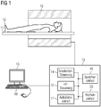
- Fig. 1 schematically shows a magnetic resonance system with which according to the invention MR images of an examination object, such as an organ that performs a cyclic movement, can be created at different contrasts.
- the magnetic resonance system has a magnet 10 for generating a polarization field B o , wherein an examination subject 12 arranged on a couch 11 is moved into the center of the magnet in order to record locally encoded magnetic resonance signals from an examination subject.
- the magnetization generated by the polarization field B 0 can be deflected out of the equilibrium position and the resulting magnetization can be detected in magnetic resonance signals by means of reception coils (not shown).
- reception coils not shown.
- the general mode of operation for generating magnetic resonance signals with different imaging sequences is known to those skilled in the art known, so that a detailed explanation is omitted.
- the magnetic resonance apparatus further has a central control unit 13, which is used to control the MR device.
- the central control unit 13 has a gradient control 14 for controlling and switching the magnetic field gradients.
- An HF controller 15 is provided for controlling and irradiating the RF pulses for deflecting the magnetization.
- a memory unit 16 for example, the necessary for the recording of the MR images imaging sequences can be stored and other programs that are necessary for the operation of the MR system.
- a recording unit 17 controls the image acquisition and thus controls, depending on the selected imaging sequence, in which sequence magnetic field gradients and RF pulses are radiated.
- the acquisition unit 17 thus also controls the gradient control 14 and the HF control 15.
- the MR images calculated in a computing unit 20 can be displayed on a display 18 and an operator can operate the MR system via an input unit 19.
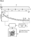
- Fig. 2 schematically a part of the imaging sequence and the post-processing steps are shown, with which images of the heart, for example, so-called delayed enhancement examinations, can be performed after contrast administration.
- image acquisition takes place during a period of time which is schematically represented by the bar 29.
- the image capture may be a bSSFP sequence that uses a compressed-sensing technology with kt regularization.
- 2D MR data are recorded after the inversion pulse 21, which is radiated directly after the R wave of the ECG.
- the image acquisition takes place over four cardiac cycles: a cardiac cycle 23, a cardiac cycle 24, a cardiac cycle 25 and a cardiac cycle 26.
- the temporal The resolution of the MR images recorded during the various cardiac cycles can range from 30 to 40 ms, resulting in multiple MR images per cardiac cycle.
- Fig. 2 Furthermore, the contrast is schematically shown, which have the individual MR images in each cardiac cycles.
- the contrast between the individual MR images 23a-23g of the first cardiac cycle 23 changes very sharply due to the inversion pulse just irradiated.
- the magnetization approaches its equilibrium state over the recording time, so that in cycle 26 the difference in the magnetization between the individual MR images is only slight.
- the recording takes place over at least two cycles, wherein in a first cycle, the cycle 23, the magnetization change from MR image to MR image is greater than in a second cycle, in the illustrated case the cycle 26.
- the MR images 26a-26g of the cycle 26 are now used to calculate motion information of the moving heart, for example deformation information. Since the individual MR images 26a-26g have a low contrast difference, the heart movement can be well determined with these images, since there are no tissue-related contrast differences between the individual images. How a registration of the individual MR images at different cardiac phases to each other is possible and how it can be calculated from individual deformation images showing the deformation of the heart in the individual cardiac phases, the skilled person is known and will not be explained here.
- the deformation information described above may be based on the natural motion of the heart. If there is still a residual movement due to the not completely stopped breath, i. a movement due to the movement of the environment, this can also be corrected. If there is still a slight movement between the two cycles 23 and 26, for example due to low respiratory activity, this can be compensated before the determination of the movement information by registering the MR images of the different cycles with each other. In this case, for example, the last image from the first cycle, image 23g, can be registered on the last image of the second cycle, image 26g. The resulting second motion information can then be applied to the entire MR images of the second cycle. In general, MR images of the same motion phase from the two cycles can be compared with one another in order to calculate second motion information therefrom.
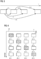
- FIG. 3 For this purpose, the different movement phases of a cardiac cycle are shown schematically, wherein in the case illustrated four movement phases are shown. Of course, it is possible to divide the cyclic movement into more or less different phases of movement.
- the individual MR images 26a-26g are assigned to the individual movement phases, or each image represents a movement phase, wherein there is at least one MR image for each movement phase. at the in Fig. 2 For example, eight images were taken per cycle. However, this number may vary and was used for drawing purposes only.
- the change of movement resulting from the first relative to the second movement phase can be determined.
- this is shown schematically by the arrow 1-2.
- the motion or deformation change from the first relative to the third phase can be determined, shown as 1-3 in the picture, and the change of motion from the first to the fourth phase is represented by 1-4.
- the motion change from the second phase relative to the first phase and from the second relative to the third or fourth phase is calculated, so that the change of motion from each of the motion phases to each of the other motion phases is calculated.
- n (n-1) motion or deformation information results.
- This motion or deformation information may include translational and / or rotational components.
- hatched MR images are MR images acquired by the MR system in the respective cycle, in the illustrated case 41-1, 42-2, 43-3 and 44-4.
- these four MR images may include any of the MR images 23a-23g of FIG Fig. 2 his.
- the captured MR image 41-1 has a first contrast, for example, since it was taken directly after irradiation of the inversion pulse.
- the MR images 41-2, 41-3 and 41-4 can now be calculated. Referring to the example of Fig.
- the deformation information 1-2, 1-3 and 1-4 would be used to starting from the recorded MR image 41-1 to calculate the images 41-2 to 41-4.
- a sequence of MR images that can be used, for example, for a CINE representation of the moving heart in the first contrast.
- the MR images 42 Starting from the recorded MR image and the movement information, here the movement information 2-1, 2-3 and 2-4, the MR images 42-1, 42-3 and 42-4 are calculated, so that for another other contrast a sequence of MR images was calculated.
- the MR images 43-1 to 43-4 and 44-1 to 44-3 can be calculated.
- Fig. 4 can be seen schematically, one obtains a sequence of MR images at different movement phases for different contrast values. These sequences of MR images may be reviewed by a physician to obtain information on the mobility of the myocardium at the various contrast levels.
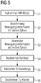
- Fig. 2 it is possible from the magnetization development, as in Fig. 2 is shown to calculate T1 and T2 values, for example, by a 3-parameter fit calculated voxel-based based on the images produced.
- step 51 motion information can then be determined from the MR images of the second of the at least two cycles, in the example of FIG Fig. 4 the fourth cycle was the last during MR image acquisition.
- step 53 this motion information is applied to the MR images of the first cycle to, as in Fig. 4 shown the MR images at the different contrast values for the different motion phases of the cyclic To calculate movement.
- a contrast may be determined at which the user desires the representation. This means that only in retrospect and not before taking the MR images in step 51 any contrast was selected.
- the image sequence desired with the selected contrast can then be displayed, for example for a CINE representation.
- the motion information of the last cycle has been applied only to the MR images of the first cycle.
- the motion information of the last cycle is also applied to the MR images of the second cycle, cycle 24, which in turn has different contrast values than in the first cycle 23.
- the contrast differences in cycle 24 are no longer as great as in cycle 23, however the use of the motion information is not limited to the MR images of the first cycle 23; an application to the first cycles 23, 24 is also conceivable.
- the invention described above enables the creation of MR images with sufficient spatial and temporal resolution at different contrasts.
Landscapes
- Health & Medical Sciences (AREA)
- Physics & Mathematics (AREA)
- Life Sciences & Earth Sciences (AREA)
- Engineering & Computer Science (AREA)
- Nuclear Medicine, Radiotherapy & Molecular Imaging (AREA)
- General Health & Medical Sciences (AREA)
- Radiology & Medical Imaging (AREA)
- High Energy & Nuclear Physics (AREA)
- Signal Processing (AREA)
- Condensed Matter Physics & Semiconductors (AREA)
- General Physics & Mathematics (AREA)
- Biomedical Technology (AREA)
- Public Health (AREA)
- Pathology (AREA)
- Veterinary Medicine (AREA)
- Heart & Thoracic Surgery (AREA)
- Medical Informatics (AREA)
- Molecular Biology (AREA)
- Surgery (AREA)
- Animal Behavior & Ethology (AREA)
- Biophysics (AREA)
- Vascular Medicine (AREA)
- Artificial Intelligence (AREA)
- Computer Vision & Pattern Recognition (AREA)
- Physiology (AREA)
- Psychiatry (AREA)
- Cardiology (AREA)
- Magnetic Resonance Imaging Apparatus (AREA)
Applications Claiming Priority (1)
| Application Number | Priority Date | Filing Date | Title |
|---|---|---|---|
| DE102014206724.3A DE102014206724B4 (de) | 2014-04-08 | 2014-04-08 | Dynamische Bildgebung mit variablem Kontrast |
Publications (2)
| Publication Number | Publication Date |
|---|---|
| EP2930525A1 EP2930525A1 (de) | 2015-10-14 |
| EP2930525B1 true EP2930525B1 (de) | 2019-12-04 |
Family
ID=52823458
Family Applications (1)
| Application Number | Title | Priority Date | Filing Date |
|---|---|---|---|
| EP15160572.2A Active EP2930525B1 (de) | 2014-04-08 | 2015-03-24 | Dynamische mr-bildgebung mit variablem kontrast |
Country Status (6)
Families Citing this family (4)
| Publication number | Priority date | Publication date | Assignee | Title |
|---|---|---|---|---|
| CN104156975B (zh) * | 2013-05-13 | 2018-04-24 | 东芝医疗系统株式会社 | 医学图像分析装置和方法以及医学成像设备 |
| DE102014225282B4 (de) * | 2014-12-09 | 2016-07-21 | Siemens Healthcare Gmbh | Deformationsberechnung bei zyklischer Bewegung eines Untersuchungsobjekts |
| DE102015224162B4 (de) * | 2015-12-03 | 2017-11-30 | Siemens Healthcare Gmbh | Verfahren zur Ermittlung einer eine Bewegung in einem zumindest teilweise bewegten Untersuchungsbereich beschreibenden Bewegungsinformation und Magnetresonanzeinrichtung |
| EP3518760B1 (en) | 2016-11-21 | 2024-09-25 | Siemens Healthineers AG | Method for recording diagnostic measurement data of a head via a magnetic resonance device |
Family Cites Families (19)
| Publication number | Priority date | Publication date | Assignee | Title |
|---|---|---|---|---|
| WO2003096047A1 (en) | 2002-05-13 | 2003-11-20 | Koninklijke Philips Electronics N.V. | Prior-information-enhanced dynamic magnetic resonance imaging |
| US6922580B2 (en) | 2002-06-04 | 2005-07-26 | Koninklijke Philips Electronics N.V. | Blood flow gated MRI |
| JP2005278919A (ja) * | 2004-03-30 | 2005-10-13 | Hitachi Medical Corp | 磁気共鳴イメージング装置 |
| US7809426B2 (en) | 2004-04-29 | 2010-10-05 | The Cleveland Clinic Foundation | Acquiring contrast-enhanced, T1 weighted, cine magnetic resonance images |
| DE102005000714A1 (de) | 2005-01-03 | 2006-07-20 | Siemens Ag | Verfahren zur Bildgebung eines periodisch bewegten Objektbereichs eines Objekts |
| JP4639136B2 (ja) | 2005-10-19 | 2011-02-23 | ジーイー・メディカル・システムズ・グローバル・テクノロジー・カンパニー・エルエルシー | 磁気共鳴イメージング装置 |
| US20080150532A1 (en) * | 2006-12-21 | 2008-06-26 | General Electric Company | Method and apparatus for measuring t1 relaxation |
| EP1956383B1 (en) * | 2007-02-06 | 2010-10-20 | Kabushiki Kaisha Toshiba | MRI involving a cine prescan for motion analysis |
| JP2008212634A (ja) * | 2007-02-06 | 2008-09-18 | Toshiba Corp | 磁気共鳴イメージング装置及びその画像解析方法並びに画像解析プログラム |
| DE102007018089B4 (de) | 2007-04-02 | 2010-10-14 | Siemens Ag | Herz-Bildgebung mittels MRI mit adaptiver Inversionszeit |
| WO2011058047A1 (en) * | 2009-11-10 | 2011-05-19 | Deutsches Herzzentrum Berlin | Look-locker ir-ssfp for cardiac mr imaging with simultaneous generation of cardiac tl maps, cine images and ir -prepared images |
| DE102010003895B4 (de) | 2010-04-13 | 2019-07-04 | Siemens Healthcare Gmbh | Verfahren zur Erzeugung von angiographischen Magnetresonanzbildern |
| US8848990B2 (en) * | 2010-09-28 | 2014-09-30 | Siemens Aktiengesellschaft | Automatic registration of image series with varying contrast based on synthetic images derived from intensity behavior model |
| US20120133747A1 (en) | 2010-11-29 | 2012-05-31 | Sony Corporation | Image processing apparatus, display apparatus, image processing method and image processing program |
| JP2012115319A (ja) * | 2010-11-29 | 2012-06-21 | R Tech:Kk | 器官運動解析装置及び器官運動解析プログラム |
| JP6076677B2 (ja) * | 2011-11-25 | 2017-02-08 | 東芝メディカルシステムズ株式会社 | 磁気共鳴イメージング装置 |
| EP2827763A1 (en) * | 2012-03-21 | 2015-01-28 | Koninklijke Philips N.V. | System and method for differentiation of normal myocardium from diffuse disease using t1 mapping in non-ischemic cardiomyopathies and others |
| US9129424B2 (en) * | 2012-04-17 | 2015-09-08 | Siemens Aktiengesellschaft | Phase sensitive T1 mapping in magnetic resonance imaging |
| DE102012206585B4 (de) * | 2012-04-20 | 2013-12-12 | Siemens Aktiengesellschaft | Verfahren zur schnellen ortsaufgelösten Bestimmung eines Magnetresonanz-Relaxationsparameters in einem Untersuchungsgebiet |
-
2014
- 2014-04-08 DE DE102014206724.3A patent/DE102014206724B4/de not_active Expired - Fee Related
-
2015
- 2015-03-24 EP EP15160572.2A patent/EP2930525B1/de active Active
- 2015-04-06 JP JP2015077704A patent/JP6133926B2/ja active Active
- 2015-04-07 KR KR1020150049011A patent/KR101631026B1/ko not_active Expired - Fee Related
- 2015-04-08 US US14/681,454 patent/US10420512B2/en active Active
- 2015-04-08 CN CN201510162338.1A patent/CN104970794B/zh active Active
Non-Patent Citations (1)
| Title |
|---|
| None * |
Also Published As
| Publication number | Publication date |
|---|---|
| DE102014206724A1 (de) | 2015-10-08 |
| CN104970794B (zh) | 2018-02-16 |
| JP2015198938A (ja) | 2015-11-12 |
| US20150282764A1 (en) | 2015-10-08 |
| US10420512B2 (en) | 2019-09-24 |
| CN104970794A (zh) | 2015-10-14 |
| KR20150116795A (ko) | 2015-10-16 |
| JP6133926B2 (ja) | 2017-05-24 |
| EP2930525A1 (de) | 2015-10-14 |
| DE102014206724B4 (de) | 2015-11-12 |
| KR101631026B1 (ko) | 2016-06-15 |
Similar Documents
| Publication | Publication Date | Title |
|---|---|---|
| DE102007041826B4 (de) | Verfahren zur Optimierung von angiographischen MR-Bildern | |
| DE102010041446B4 (de) | Erstellung eines MR-Bilddatensatzes bei sehr kurzen Echozeiten TE | |
| DE102006012181B4 (de) | Verfahren und Vorrichtung zur getrennten dreidimensionalen Darstellung von Arterien und Venen in einem Körperteil | |
| DE102014218653A1 (de) | Prospektive Bewegungskorrektur | |
| DE102012206585A1 (de) | Verfahren zur schnellen ortsaufgelösten Bestimmung eines Magnetresonanz-Relaxationsparameters in einem Untersuchungsgebiet | |
| DE102011007574A1 (de) | Verfahren zur quasi-kontinuierlichen dynamischen Bewegungskorrektur bei Messungen der Magnetresonanz | |
| DE102015207591B4 (de) | Verfahren zu einer Bewegungskorrektur von Magnetresonanz-Messdaten | |
| DE3817195A1 (de) | Verfahren zum reduzieren von artefakten in einem bild | |
| DE4428503A1 (de) | Diffusionsgewichtete Bildgebung mit magnetischer Resonanz | |
| DE102008049709A1 (de) | Verfahren zur selektiven Darstellung einer Bewegung der Lunge, Computerprogramm, Bildverarbeitungseinheit und Magnetresonanzgerät | |
| EP2930525B1 (de) | Dynamische mr-bildgebung mit variablem kontrast | |
| DE102012208431A1 (de) | Korrigieren von Phasenfehlern bei multidimensionalen ortsselektiven Hochfrequenz-MR-Anregungspulsen | |
| EP3382413B1 (de) | 2d navigatortechnik in der mrt | |
| DE102015218106A1 (de) | Verfahren zu einer Bewegungskorrektur von Magnetresonanz-Messdaten | |
| DE102014209351B4 (de) | Magnetresonanz-Spektroskopieverfahren mit kurzer Echozeit, Magnetresonanzanlage und digitales Speichermedium | |
| DE102010041125B4 (de) | Räumliche Korrektur von Bilddaten einer Serie von Magnetresonanzaufnahmen | |
| DE102008050030A1 (de) | Verfahren und Vorrichtung zum Bestimmen eines Inversionszeitwerts von Gewebe mittels Magnetresonanztechnik | |
| DE102016200629A1 (de) | Verfahren zur Magnetresonanz-Bildgebung | |
| DE102016214775A1 (de) | Bestimmung einer Eigenschaft eines Organs | |
| DE102017212553B4 (de) | Synchrone MR-Bildgebung und Strahlentherapie | |
| DE102016213632A1 (de) | Verbesserte Akquisition von MR-Messdaten bei einer Untersuchung mit angehaltenem Atem | |
| DE102016202085B3 (de) | Verfahren, Magnetresonanzanlage und elektronisch lesbarer Datenträger zur Narbenquantifizierung im Myokard | |
| EP3290940B1 (de) | Iterative rekonstruktion von quantitativen mr-bildern | |
| DE102019219862B4 (de) | Kombinierte Bestimmung von T1 und eines Gewebeanteils | |
| DE102014209753B4 (de) | Bildaufnahme mit zufallsverteilter Aufnahme der Rohdaten |
Legal Events
| Date | Code | Title | Description |
|---|---|---|---|
| PUAI | Public reference made under article 153(3) epc to a published international application that has entered the european phase |
Free format text: ORIGINAL CODE: 0009012 |
|
| AK | Designated contracting states |
Kind code of ref document: A1 Designated state(s): AL AT BE BG CH CY CZ DE DK EE ES FI FR GB GR HR HU IE IS IT LI LT LU LV MC MK MT NL NO PL PT RO RS SE SI SK SM TR |
|
| AX | Request for extension of the european patent |
Extension state: BA ME |
|
| RAP1 | Party data changed (applicant data changed or rights of an application transferred) |
Owner name: SIEMENS HEALTHCARE GMBH |
|
| 17P | Request for examination filed |
Effective date: 20160406 |
|
| RBV | Designated contracting states (corrected) |
Designated state(s): AL AT BE BG CH CY CZ DE DK EE ES FI FR GB GR HR HU IE IS IT LI LT LU LV MC MK MT NL NO PL PT RO RS SE SI SK SM TR |
|
| GRAP | Despatch of communication of intention to grant a patent |
Free format text: ORIGINAL CODE: EPIDOSNIGR1 |
|
| STAA | Information on the status of an ep patent application or granted ep patent |
Free format text: STATUS: GRANT OF PATENT IS INTENDED |
|
| INTG | Intention to grant announced |
Effective date: 20190805 |
|
| GRAS | Grant fee paid |
Free format text: ORIGINAL CODE: EPIDOSNIGR3 |
|
| GRAA | (expected) grant |
Free format text: ORIGINAL CODE: 0009210 |
|
| STAA | Information on the status of an ep patent application or granted ep patent |
Free format text: STATUS: THE PATENT HAS BEEN GRANTED |
|
| AK | Designated contracting states |
Kind code of ref document: B1 Designated state(s): AL AT BE BG CH CY CZ DE DK EE ES FI FR GB GR HR HU IE IS IT LI LT LU LV MC MK MT NL NO PL PT RO RS SE SI SK SM TR |
|
| REG | Reference to a national code |
Ref country code: GB Ref legal event code: FG4D Free format text: NOT ENGLISH |
|
| REG | Reference to a national code |
Ref country code: CH Ref legal event code: EP |
|
| REG | Reference to a national code |
Ref country code: AT Ref legal event code: REF Ref document number: 1210061 Country of ref document: AT Kind code of ref document: T Effective date: 20191215 |
|
| REG | Reference to a national code |
Ref country code: DE Ref legal event code: R096 Ref document number: 502015011101 Country of ref document: DE |
|
| REG | Reference to a national code |
Ref country code: IE Ref legal event code: FG4D Free format text: LANGUAGE OF EP DOCUMENT: GERMAN |
|
| REG | Reference to a national code |
Ref country code: NL Ref legal event code: MP Effective date: 20191204 |
|
| REG | Reference to a national code |
Ref country code: LT Ref legal event code: MG4D |
|
| PG25 | Lapsed in a contracting state [announced via postgrant information from national office to epo] |
Ref country code: LT Free format text: LAPSE BECAUSE OF FAILURE TO SUBMIT A TRANSLATION OF THE DESCRIPTION OR TO PAY THE FEE WITHIN THE PRESCRIBED TIME-LIMIT Effective date: 20191204 Ref country code: GR Free format text: LAPSE BECAUSE OF FAILURE TO SUBMIT A TRANSLATION OF THE DESCRIPTION OR TO PAY THE FEE WITHIN THE PRESCRIBED TIME-LIMIT Effective date: 20200305 Ref country code: NO Free format text: LAPSE BECAUSE OF FAILURE TO SUBMIT A TRANSLATION OF THE DESCRIPTION OR TO PAY THE FEE WITHIN THE PRESCRIBED TIME-LIMIT Effective date: 20200304 Ref country code: BG Free format text: LAPSE BECAUSE OF FAILURE TO SUBMIT A TRANSLATION OF THE DESCRIPTION OR TO PAY THE FEE WITHIN THE PRESCRIBED TIME-LIMIT Effective date: 20200304 Ref country code: FI Free format text: LAPSE BECAUSE OF FAILURE TO SUBMIT A TRANSLATION OF THE DESCRIPTION OR TO PAY THE FEE WITHIN THE PRESCRIBED TIME-LIMIT Effective date: 20191204 Ref country code: SE Free format text: LAPSE BECAUSE OF FAILURE TO SUBMIT A TRANSLATION OF THE DESCRIPTION OR TO PAY THE FEE WITHIN THE PRESCRIBED TIME-LIMIT Effective date: 20191204 Ref country code: LV Free format text: LAPSE BECAUSE OF FAILURE TO SUBMIT A TRANSLATION OF THE DESCRIPTION OR TO PAY THE FEE WITHIN THE PRESCRIBED TIME-LIMIT Effective date: 20191204 |
|
| PG25 | Lapsed in a contracting state [announced via postgrant information from national office to epo] |
Ref country code: HR Free format text: LAPSE BECAUSE OF FAILURE TO SUBMIT A TRANSLATION OF THE DESCRIPTION OR TO PAY THE FEE WITHIN THE PRESCRIBED TIME-LIMIT Effective date: 20191204 Ref country code: RS Free format text: LAPSE BECAUSE OF FAILURE TO SUBMIT A TRANSLATION OF THE DESCRIPTION OR TO PAY THE FEE WITHIN THE PRESCRIBED TIME-LIMIT Effective date: 20191204 |
|
| PG25 | Lapsed in a contracting state [announced via postgrant information from national office to epo] |
Ref country code: AL Free format text: LAPSE BECAUSE OF FAILURE TO SUBMIT A TRANSLATION OF THE DESCRIPTION OR TO PAY THE FEE WITHIN THE PRESCRIBED TIME-LIMIT Effective date: 20191204 |
|
| PG25 | Lapsed in a contracting state [announced via postgrant information from national office to epo] |
Ref country code: NL Free format text: LAPSE BECAUSE OF FAILURE TO SUBMIT A TRANSLATION OF THE DESCRIPTION OR TO PAY THE FEE WITHIN THE PRESCRIBED TIME-LIMIT Effective date: 20191204 Ref country code: RO Free format text: LAPSE BECAUSE OF FAILURE TO SUBMIT A TRANSLATION OF THE DESCRIPTION OR TO PAY THE FEE WITHIN THE PRESCRIBED TIME-LIMIT Effective date: 20191204 Ref country code: EE Free format text: LAPSE BECAUSE OF FAILURE TO SUBMIT A TRANSLATION OF THE DESCRIPTION OR TO PAY THE FEE WITHIN THE PRESCRIBED TIME-LIMIT Effective date: 20191204 Ref country code: ES Free format text: LAPSE BECAUSE OF FAILURE TO SUBMIT A TRANSLATION OF THE DESCRIPTION OR TO PAY THE FEE WITHIN THE PRESCRIBED TIME-LIMIT Effective date: 20191204 Ref country code: PT Free format text: LAPSE BECAUSE OF FAILURE TO SUBMIT A TRANSLATION OF THE DESCRIPTION OR TO PAY THE FEE WITHIN THE PRESCRIBED TIME-LIMIT Effective date: 20200429 Ref country code: CZ Free format text: LAPSE BECAUSE OF FAILURE TO SUBMIT A TRANSLATION OF THE DESCRIPTION OR TO PAY THE FEE WITHIN THE PRESCRIBED TIME-LIMIT Effective date: 20191204 |
|
| PG25 | Lapsed in a contracting state [announced via postgrant information from national office to epo] |
Ref country code: SK Free format text: LAPSE BECAUSE OF FAILURE TO SUBMIT A TRANSLATION OF THE DESCRIPTION OR TO PAY THE FEE WITHIN THE PRESCRIBED TIME-LIMIT Effective date: 20191204 Ref country code: SM Free format text: LAPSE BECAUSE OF FAILURE TO SUBMIT A TRANSLATION OF THE DESCRIPTION OR TO PAY THE FEE WITHIN THE PRESCRIBED TIME-LIMIT Effective date: 20191204 Ref country code: IS Free format text: LAPSE BECAUSE OF FAILURE TO SUBMIT A TRANSLATION OF THE DESCRIPTION OR TO PAY THE FEE WITHIN THE PRESCRIBED TIME-LIMIT Effective date: 20200404 |
|
| REG | Reference to a national code |
Ref country code: DE Ref legal event code: R097 Ref document number: 502015011101 Country of ref document: DE |
|
| PLBE | No opposition filed within time limit |
Free format text: ORIGINAL CODE: 0009261 |
|
| STAA | Information on the status of an ep patent application or granted ep patent |
Free format text: STATUS: NO OPPOSITION FILED WITHIN TIME LIMIT |
|
| PG25 | Lapsed in a contracting state [announced via postgrant information from national office to epo] |
Ref country code: DK Free format text: LAPSE BECAUSE OF FAILURE TO SUBMIT A TRANSLATION OF THE DESCRIPTION OR TO PAY THE FEE WITHIN THE PRESCRIBED TIME-LIMIT Effective date: 20191204 Ref country code: MC Free format text: LAPSE BECAUSE OF FAILURE TO SUBMIT A TRANSLATION OF THE DESCRIPTION OR TO PAY THE FEE WITHIN THE PRESCRIBED TIME-LIMIT Effective date: 20191204 |
|
| REG | Reference to a national code |
Ref country code: CH Ref legal event code: PL |
|
| 26N | No opposition filed |
Effective date: 20200907 |
|
| PG25 | Lapsed in a contracting state [announced via postgrant information from national office to epo] |
Ref country code: PL Free format text: LAPSE BECAUSE OF FAILURE TO SUBMIT A TRANSLATION OF THE DESCRIPTION OR TO PAY THE FEE WITHIN THE PRESCRIBED TIME-LIMIT Effective date: 20191204 Ref country code: SI Free format text: LAPSE BECAUSE OF FAILURE TO SUBMIT A TRANSLATION OF THE DESCRIPTION OR TO PAY THE FEE WITHIN THE PRESCRIBED TIME-LIMIT Effective date: 20191204 |
|
| REG | Reference to a national code |
Ref country code: BE Ref legal event code: MM Effective date: 20200331 |
|
| PG25 | Lapsed in a contracting state [announced via postgrant information from national office to epo] |
Ref country code: LU Free format text: LAPSE BECAUSE OF NON-PAYMENT OF DUE FEES Effective date: 20200324 |
|
| PG25 | Lapsed in a contracting state [announced via postgrant information from national office to epo] |
Ref country code: IE Free format text: LAPSE BECAUSE OF NON-PAYMENT OF DUE FEES Effective date: 20200324 Ref country code: IT Free format text: LAPSE BECAUSE OF FAILURE TO SUBMIT A TRANSLATION OF THE DESCRIPTION OR TO PAY THE FEE WITHIN THE PRESCRIBED TIME-LIMIT Effective date: 20191204 Ref country code: LI Free format text: LAPSE BECAUSE OF NON-PAYMENT OF DUE FEES Effective date: 20200331 Ref country code: CH Free format text: LAPSE BECAUSE OF NON-PAYMENT OF DUE FEES Effective date: 20200331 |
|
| PG25 | Lapsed in a contracting state [announced via postgrant information from national office to epo] |
Ref country code: BE Free format text: LAPSE BECAUSE OF NON-PAYMENT OF DUE FEES Effective date: 20200331 |
|
| REG | Reference to a national code |
Ref country code: AT Ref legal event code: MM01 Ref document number: 1210061 Country of ref document: AT Kind code of ref document: T Effective date: 20200324 |
|
| PG25 | Lapsed in a contracting state [announced via postgrant information from national office to epo] |
Ref country code: AT Free format text: LAPSE BECAUSE OF NON-PAYMENT OF DUE FEES Effective date: 20200324 |
|
| PG25 | Lapsed in a contracting state [announced via postgrant information from national office to epo] |
Ref country code: TR Free format text: LAPSE BECAUSE OF FAILURE TO SUBMIT A TRANSLATION OF THE DESCRIPTION OR TO PAY THE FEE WITHIN THE PRESCRIBED TIME-LIMIT Effective date: 20191204 Ref country code: MT Free format text: LAPSE BECAUSE OF FAILURE TO SUBMIT A TRANSLATION OF THE DESCRIPTION OR TO PAY THE FEE WITHIN THE PRESCRIBED TIME-LIMIT Effective date: 20191204 Ref country code: CY Free format text: LAPSE BECAUSE OF FAILURE TO SUBMIT A TRANSLATION OF THE DESCRIPTION OR TO PAY THE FEE WITHIN THE PRESCRIBED TIME-LIMIT Effective date: 20191204 |
|
| PG25 | Lapsed in a contracting state [announced via postgrant information from national office to epo] |
Ref country code: MK Free format text: LAPSE BECAUSE OF FAILURE TO SUBMIT A TRANSLATION OF THE DESCRIPTION OR TO PAY THE FEE WITHIN THE PRESCRIBED TIME-LIMIT Effective date: 20191204 |
|
| REG | Reference to a national code |
Ref country code: DE Ref legal event code: R081 Ref document number: 502015011101 Country of ref document: DE Owner name: SIEMENS HEALTHINEERS AG, DE Free format text: FORMER OWNER: SIEMENS HEALTHCARE GMBH, MUENCHEN, DE |
|
| PGFP | Annual fee paid to national office [announced via postgrant information from national office to epo] |
Ref country code: FR Payment date: 20250314 Year of fee payment: 11 |
|
| PGFP | Annual fee paid to national office [announced via postgrant information from national office to epo] |
Ref country code: DE Payment date: 20250520 Year of fee payment: 11 |
|
| PGFP | Annual fee paid to national office [announced via postgrant information from national office to epo] |
Ref country code: GB Payment date: 20250403 Year of fee payment: 11 |