EP2928482B1 - Verbesserung der zielfindungskapazität von stammzellen - Google Patents
Verbesserung der zielfindungskapazität von stammzellen Download PDFInfo
- Publication number
- EP2928482B1 EP2928482B1 EP13801557.3A EP13801557A EP2928482B1 EP 2928482 B1 EP2928482 B1 EP 2928482B1 EP 13801557 A EP13801557 A EP 13801557A EP 2928482 B1 EP2928482 B1 EP 2928482B1
- Authority
- EP
- European Patent Office
- Prior art keywords
- mscs
- pei
- cells
- stem cells
- capacity
- Prior art date
- Legal status (The legal status is an assumption and is not a legal conclusion. Google has not performed a legal analysis and makes no representation as to the accuracy of the status listed.)
- Not-in-force
Links
Images



Classifications
-
- A—HUMAN NECESSITIES
- A61—MEDICAL OR VETERINARY SCIENCE; HYGIENE
- A61K—PREPARATIONS FOR MEDICAL, DENTAL OR TOILETRY PURPOSES
- A61K35/00—Medicinal preparations containing materials or reaction products thereof with undetermined constitution
- A61K35/12—Materials from mammals; Compositions comprising non-specified tissues or cells; Compositions comprising non-embryonic stem cells; Genetically modified cells
- A61K35/28—Bone marrow; Haematopoietic stem cells; Mesenchymal stem cells of any origin, e.g. adipose-derived stem cells
-
- A—HUMAN NECESSITIES
- A61—MEDICAL OR VETERINARY SCIENCE; HYGIENE
- A61K—PREPARATIONS FOR MEDICAL, DENTAL OR TOILETRY PURPOSES
- A61K47/00—Medicinal preparations characterised by the non-active ingredients used, e.g. carriers or inert additives; Targeting or modifying agents chemically bound to the active ingredient
- A61K47/50—Medicinal preparations characterised by the non-active ingredients used, e.g. carriers or inert additives; Targeting or modifying agents chemically bound to the active ingredient the non-active ingredient being chemically bound to the active ingredient, e.g. polymer-drug conjugates
- A61K47/51—Medicinal preparations characterised by the non-active ingredients used, e.g. carriers or inert additives; Targeting or modifying agents chemically bound to the active ingredient the non-active ingredient being chemically bound to the active ingredient, e.g. polymer-drug conjugates the non-active ingredient being a modifying agent
- A61K47/56—Medicinal preparations characterised by the non-active ingredients used, e.g. carriers or inert additives; Targeting or modifying agents chemically bound to the active ingredient the non-active ingredient being chemically bound to the active ingredient, e.g. polymer-drug conjugates the non-active ingredient being a modifying agent the modifying agent being an organic macromolecular compound, e.g. an oligomeric, polymeric or dendrimeric molecule
- A61K47/59—Medicinal preparations characterised by the non-active ingredients used, e.g. carriers or inert additives; Targeting or modifying agents chemically bound to the active ingredient the non-active ingredient being chemically bound to the active ingredient, e.g. polymer-drug conjugates the non-active ingredient being a modifying agent the modifying agent being an organic macromolecular compound, e.g. an oligomeric, polymeric or dendrimeric molecule obtained otherwise than by reactions only involving carbon-to-carbon unsaturated bonds, e.g. polyureas or polyurethanes
-
- A—HUMAN NECESSITIES
- A61—MEDICAL OR VETERINARY SCIENCE; HYGIENE
- A61K—PREPARATIONS FOR MEDICAL, DENTAL OR TOILETRY PURPOSES
- A61K49/00—Preparations for testing in vivo
- A61K49/06—Nuclear magnetic resonance [NMR] contrast preparations; Magnetic resonance imaging [MRI] contrast preparations
- A61K49/18—Nuclear magnetic resonance [NMR] contrast preparations; Magnetic resonance imaging [MRI] contrast preparations characterised by a special physical form, e.g. emulsions, microcapsules, liposomes
- A61K49/1896—Nuclear magnetic resonance [NMR] contrast preparations; Magnetic resonance imaging [MRI] contrast preparations characterised by a special physical form, e.g. emulsions, microcapsules, liposomes not provided for elsewhere, e.g. cells, viruses, ghosts, red blood cells, virus capsides
-
- C—CHEMISTRY; METALLURGY
- C12—BIOCHEMISTRY; BEER; SPIRITS; WINE; VINEGAR; MICROBIOLOGY; ENZYMOLOGY; MUTATION OR GENETIC ENGINEERING
- C12N—MICROORGANISMS OR ENZYMES; COMPOSITIONS THEREOF; PROPAGATING, PRESERVING, OR MAINTAINING MICROORGANISMS; MUTATION OR GENETIC ENGINEERING; CULTURE MEDIA
- C12N5/00—Undifferentiated human, animal or plant cells, e.g. cell lines; Tissues; Cultivation or maintenance thereof; Culture media therefor
- C12N5/06—Animal cells or tissues; Human cells or tissues
- C12N5/0602—Vertebrate cells
- C12N5/0652—Cells of skeletal and connective tissues; Mesenchyme
- C12N5/0662—Stem cells
- C12N5/0663—Bone marrow mesenchymal stem cells (BM-MSC)
-
- A—HUMAN NECESSITIES
- A01—AGRICULTURE; FORESTRY; ANIMAL HUSBANDRY; HUNTING; TRAPPING; FISHING
- A01K—ANIMAL HUSBANDRY; AVICULTURE; APICULTURE; PISCICULTURE; FISHING; REARING OR BREEDING ANIMALS, NOT OTHERWISE PROVIDED FOR; NEW BREEDS OF ANIMALS
- A01K2267/00—Animals characterised by purpose
- A01K2267/02—Animal zootechnically ameliorated
- A01K2267/025—Animal producing cells or organs for transplantation
-
- A—HUMAN NECESSITIES
- A01—AGRICULTURE; FORESTRY; ANIMAL HUSBANDRY; HUNTING; TRAPPING; FISHING
- A01K—ANIMAL HUSBANDRY; AVICULTURE; APICULTURE; PISCICULTURE; FISHING; REARING OR BREEDING ANIMALS, NOT OTHERWISE PROVIDED FOR; NEW BREEDS OF ANIMALS
- A01K2267/00—Animals characterised by purpose
- A01K2267/03—Animal model, e.g. for test or diseases
- A01K2267/035—Animal model for multifactorial diseases
- A01K2267/0356—Animal model for processes and diseases of the central nervous system, e.g. stress, learning, schizophrenia, pain, epilepsy
-
- A—HUMAN NECESSITIES
- A01—AGRICULTURE; FORESTRY; ANIMAL HUSBANDRY; HUNTING; TRAPPING; FISHING
- A01K—ANIMAL HUSBANDRY; AVICULTURE; APICULTURE; PISCICULTURE; FISHING; REARING OR BREEDING ANIMALS, NOT OTHERWISE PROVIDED FOR; NEW BREEDS OF ANIMALS
- A01K67/00—Rearing or breeding animals, not otherwise provided for; New or modified breeds of animals
- A01K67/027—New or modified breeds of vertebrates
- A01K67/0271—Chimeric vertebrates, e.g. comprising exogenous cells
-
- C—CHEMISTRY; METALLURGY
- C12—BIOCHEMISTRY; BEER; SPIRITS; WINE; VINEGAR; MICROBIOLOGY; ENZYMOLOGY; MUTATION OR GENETIC ENGINEERING
- C12N—MICROORGANISMS OR ENZYMES; COMPOSITIONS THEREOF; PROPAGATING, PRESERVING, OR MAINTAINING MICROORGANISMS; MUTATION OR GENETIC ENGINEERING; CULTURE MEDIA
- C12N2500/00—Specific components of cell culture medium
- C12N2500/05—Inorganic components
- C12N2500/10—Metals; Metal chelators
- C12N2500/20—Transition metals
- C12N2500/24—Iron; Fe chelators; Transferrin
-
- C—CHEMISTRY; METALLURGY
- C12—BIOCHEMISTRY; BEER; SPIRITS; WINE; VINEGAR; MICROBIOLOGY; ENZYMOLOGY; MUTATION OR GENETIC ENGINEERING
- C12N—MICROORGANISMS OR ENZYMES; COMPOSITIONS THEREOF; PROPAGATING, PRESERVING, OR MAINTAINING MICROORGANISMS; MUTATION OR GENETIC ENGINEERING; CULTURE MEDIA
- C12N2500/00—Specific components of cell culture medium
- C12N2500/50—Soluble polymers, e.g. polyethyleneglycol [PEG]
Definitions
- the present invention relates to a method of improving the targeting capacity of mesenchymal stem cells, a preparation having correspondingly improved mesenchymal stem cells, and the use of a linear cationic polymer to improve the targeting capacity of mesenchymal stem cells.
- Stem cells in particular mesenchymal stem cells (MSCs), are promising candidates for a so-called cellular therapy for the treatment of various diseases because of their regenerative and immunomodulatory potential. Examples are graft-versus-host disease and osteogenesis imperfecta. Cellular therapy requires reliable and sufficient delivery of cells to the desired anatomical structure as well as survival of the cellular graft.
- MSCs mesenchymal stem cells
- Systemic or intravenous MSCs target the site of injury.
- the majority of transplanted MSCs accumulate through a first-pass effect in undesired localizations, especially in parenchymal organs such as the liver, spleen, and lung.
- the mechanisms underlying the accumulation of MSCs in non-injured parenchymal organs following intravascular administration are not yet fully understood.
- the massive presence of transplanted MSCs in the lung is believed to be the result of a nonspecific capture process of the relatively large MSCs in the small pulmonary vessels.
- more specific mechanisms also appear to mediate the adhesion of MSCs to the pulmonary epithelium.
- MSCs beneficial effects of MSCs are mediated by paracrine factors released by the MSCs.
- transplantation dose for the MSCs to be prevalent at the site of injury remains unclear.
- a prerequisite for finding the adequate transplantation dose is the direct delivery of the MSCs to the target region.
- the undirected accumulation of transplanted MSCs in various organs and tissues can increase the risk of side effects and reduce the therapeutic effects of MSCs at the site of injury. This is of particular importance since it has been shown that MSCs migrate to neoplastic tissues after systemic administration and contribute to tumor growth and promote the growth of metastases.
- MSCs The interaction of MSCs with the endothelium of the host is a key factor in targeting the MSCs after their delivery into the vasculature.
- Human MSCs express a variety of chemokine receptors belonging to the CC and CXC families, as well as adhesion molecules such as integrins, endoglin (CD105), VCAM-1 (CD106), and H-CAM (CD44).
- the adhesion of MSCs to the endothelium can be observed under inflammatory and noninflammatory conditions, with the presence of proinflammatory cytokines, such as TNF- ⁇ , or chemokines and their ligands, such as SDF-1a, promoting the adhesion and transendothelial migration of MSCs.
- proinflammatory cytokines such as TNF- ⁇
- chemokines and their ligands such as SDF-1a
- BMSCs bone marrow mesenchymal stem cells
- PEI2k-SPI0 polythyleneimine superparamagnetic iron oxide
- SDF-1 Stromal Derived Factor-1
- integrins In determining the target of human stem cells and endothelial progenitor cells, integrins also play a major role; see. Kavanagh et al. (2011), Hematopoietic Stem Cell Homing to Injured Tissues, Stem Cell Reviews and Reports, Humana Press, Inc., New York, US, Vol. 7, No. 3, pp. 672-682 , Emmanouil et al. (2005), Role of Beta2-Integrins for Homing and Neovascularization Capacity of Endothelial Progenitor Cells, The Journal of Experimental Medicine, Rockefeller University Press, US, Vol. 201, No. 1, pp.
- MSCs mesenchymal stem cells
- the inventors have surprisingly found that simple treatment of stem cells with a linear cationic polymer alters the MSCs such that the first-pass effect is significantly reduced and the proportion of cells reaching the target area is significantly increased.
- targeting capacity is meant a ability of a stem cell to migrate along a concentration gradient to the location of the highest concentration of one or more signaling molecules.
- An example of such signal molecules is the group of chemokines.
- a particularly relevant form of targeting capacity according to the present invention is the ability of a stem cell to migrate to the site of cellular injury or tissue damage upon administration to an organism.
- the targeting capacity of the stem cells is improved without the mediation by target-recognizing bioligands, such as antibodies.
- Cationic agents such as PEI are used to transfect cells; see. DE 695 35 540 T2 , PEI is also being tested for in vivo gene therapy.
- were Modifications proposed by PEI to reduce or circumvent the first-pass effect; see. Merkel et al. (2009). Shufer et al. (2010), Functional Investigations on Human Mesenchymal Stem Cell's Exposed to Magnetic Fields and Labeled With Clinically Approved Iron Nanoparticles, BMC Cell Biology, Biomed Central, London, GB, Vol. 11, No. 1 describe the use of PEI to label cells with magnetic nanoparticles. PEI enables the uptake of these nanoparticles into the cells, which can then be detected in the body by MRI. An influence on the targeting of the cells or an influence on the adhesion is not known in the prior art. Also, no method is known which uses a cationic agent such as PEI to improve the targeting capacity.
- MSCs from any mammals are suitable.
- MSCs are already used in the context of cell therapy in the prior art and are distinguished by their regenerative and immunomodulatory potential.
- the inventive method significantly increases the targeting capacity of these cell populations. By reducing the first-pass effect, significantly lower amounts of cells can be administered, which can significantly reduce side effects.
- linear cationic polymer has the advantage that, according to findings of the inventors, particularly good results are achieved.
- suitable linear cationic polymers are polyethyleneimine (PEI) or cationic click polymers.
- the linear cationic polymer is polyethyleneimine (PEI).
- PEI polyethyleneimine
- the MSCs are human MSCs.
- Human MSCs (hMSCs) treated according to the invention are particularly well suited for clinical applications in humans. This has been confirmed by decades of clinical applications. The introduction of animal substances in humans is thus avoided. Furthermore, while retaining the human system, optimal transferability of in vitro knowledge of human stem cells into patient use is ensured.
- the stem cells are additionally treated or incubated with iron oxide particles in a third step.
- the additional treatment with iron oxide nanoparticles leads to a further improvement of the targeting capacity of the stem cells.
- the stem cells can be treated simultaneously with the cationic linear polymer and the iron oxide nanoparticles.
- the stem cells may also be treated first with the cationic linear polymer and then with the iron oxide nanoparticles.
- the MSCs may also be incubated first with the iron oxide nanoparticles and then with the cationic linear polymer.
- the iron oxide nanoparticles are ferucarbotrane.
- Iron oxide nanoparticles or ferucarbotran is commercially available, for example in the form of the product "Resovist®” from Schering AG, Berlin, Germany.
- the improvement of the target finding capacity is effected by inhibition of the cell adhesion receptors of the MSCs.
- This measure has the advantage that, according to findings of the inventors, molecular structures on the surface of the MSCs are modified in such a way that the first-pass effect is inhibited, thereby increasing the targeting capacity.
- the improvement of the targeting capacity takes place by stimulation of the chemokine receptors of the MSCs.
- stimulation comprises both an increase in the expression and the activity or affinity of the chemokine receptors of the MSCs.
- a preparation comprising stem cells, preferably mesenchymal stem cells (MSCs) and / or hematopoietic stem cells (HSCs), obtained by the method of the invention.
- the preparation is preferably a pharmaceutical preparation, more preferably one for the treatment of cellular damage and / or tissue damage.
- the cellular damage or tissue damage can be inflammations, degenerative diseases, tumorous diseases and neuronal lesions.
- the preparation or pharmaceutical preparation is suitable for the treatment of strokes, heart attacks, cardiac insufficiency, bone / joint diseases including rheumatic diseases, Alzheimer's disease, Parkinson's disease, autoimmune diseases including multiple sclerosis.
- Another object of the present invention is the use of a linear cationic polymer, more preferably a polyethyleneimine (PEI), to improve cell fate capacity of mesenchymal stem cells (MSCs).
- PEI polyethyleneimine
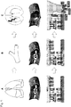
- FIG.1 Figure 4 shows a schematic representation of the effects of PEI treatment of MSCs on their homing receptor function and targeting capacity.
- the function of the receptors which mediate the adhesion to endothelium in the lung (A) or in non-inflammatory tissue / blood vessels (B), is blocked by PEI. This allows the MSCs to pass these areas unhindered across the bloodstream.
- the chemokines are amplified. Since PEI does not affect the function of the chemokine receptors, the MSCs can migrate unhindered there. In sum, more PEI-treated MSCs than untreated MSCs can migrate into the target area, as they are retained to a lesser extent in the lung or non-inflammatory tissue / blood vessels
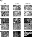
- Fig. 2 shows the adipogenic red oil O staining of lipid vacuoles [A], undifferentiated control [B], osteogenic alizarin red staining [C], undifferentiated control [D] and chondrogenic (E) differentiation capacity of the untreated MSCs, PEI MSCs and PEI NP MSCs.
- Treatment with PEI with / without nanoparticles (NP) shows no effect on the differentiation capacity of MSCs.
- Scale bar 100 ⁇ m
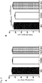
- Fig. 3 shows the viability of MSCs unaffected by treatment with PEI with / without NP (A). Treatment with PEI reduced the diameter of MSCs slightly but significantly (B).
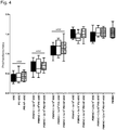
- Fig. 4 shows the potency of MSCs for suppressing the proliferation of T cells ( in vitro immunosuppression). Treatment with PEI with / without NP has no effect on the immunosuppressive properties of MSCs.
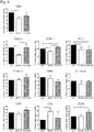
- Fig. 5 shows the expression / accessibility of homing receptors and chemokine receptors of MSCs.
- Treatment with PEI reduces the expression / accessibility of CD44, endoglin, VCAM-1 and VLA-1, whereas the chemokine receptors CCR9 and CXCR4 remain unaffected and CCR4 is even more strongly expressed.
- Treatment with PEI + NP enhances the expression of VCAM-1 and CXCR-4.
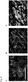
- Fig. 6 shows the expression of CXCR-4 on MSCs. Compared to untreated MSCs (A) and PEI-MSCs (B), treatment with PEI + NP leads to increased expression of CXCR-4 on the MSCs (C).
- Fig. 7 shows the expression of CXCR-4 on PEI-NP MSCs relative to the PEI-NP complexes.
- the increased expression of CXCR-4 is observed especially in the vicinity of the extracellular PEI-NP complexes.
- the PEI-NP complexes are identifiable as gray agglomerates in phase-contrast transmitted light microscopy (A).
- Enhanced CXCR-4 expression in the area of PEI-NP complexes B, C).
- the PEI-NP complexes are highlighted in grayscale in the overlay representation of A and B by editing with "Adobe Photoshop CS5.1".
- the CXCR-4 isotype control shows no nonspecific fluorescence in the region of the PEI-NP complexes (D).
- Fig. 8 shows studies on the regulation of CD44 or CXCR4 expression.
- the treatment with PEI leads to an increased expression of CD44 mRNA (A) and CD44 total protein (B) as well as to a reduced expression of CXCR-4 total protein (C).
- Treatment with PEI + NP has no effect on CD44 and CXCR-4 expression, neither at mRNA nor protein level (AC).
- Fig. 9 shows the adhesion behavior of MSCs to TNF- ⁇ activated or non-activated endothelial cells in the dynamic flow chamber model in vitro.
- PEI-MSC show decreased adhesion to TNF- ⁇ activated endothelial cells
- PEI-NP MSCs show enhanced adhesion to TNF- ⁇ activated endothelial cells (A).
- Treated and untreated MSCs show reduced adhesion to unactivated endothelial cells compared to untreated MSCs on TNF- ⁇ -activated endothelial cells.
- the adhesion behavior of PEI-MSCs and PEI-NP-MSCs to non-activated endothelial cells is stronger than the adhesion behavior of untreated MSCs to non-activated endothelial cells (B). Increased adhesion to TNF- ⁇ -activated endothelial cells following treatment with PEI + NP is abolished in non-activated endothelial cells (C). The reduced adhesion after treatment with PEI is independent of the degree of activation of the endothelial cells (D).
- the adhesion of untreated MSCs and PEI-NP MSCs to TNF- ⁇ -activated endothelial cells can be reduced by blocking anti-CD44 and anti-CXCR-4 antibodies (E, F, H, I), whereas the decreased adhesion of PEI MSCs can not be further reduced with blocking anti-CD44 and anti-CXCR-4 antibodies (G, J).
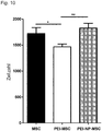
- Fig. 10 shows the immunohistological quantification of GFP + MSCs in the lung 24h after iv transplantation in vivo. Treatment with PEI results in decreased detection of MSCs in the lung.
- Fig. 11 shows the immunohistological quantification of GFP + MSCs in the brain 24h after iv transplantation in vivo.
- MSCs and PEI-MSCs were detectable compared to the non-lesioned, contralateral hippocampus, with more PEI-MSCs being detectable in the area of the lesion (A).
- A the area of the lesion
- PEI-MSCs being detectable in the area of the lesion
- E whole brain
- Bone marrow was taken under sterile conditions during orthopedic surgery randomly selected donors who had given their informed consent after written informed consent. From each donor, 5-10 ml total BM was collected in a sterile syringe.
- MSCs mononuclear cells from BM were isolated by density gradient centrifugation on Lymphoflot (Biotest, Dreieich, Germany), washed twice with PBS (Lonza, Walkersville, USA), counted and at a density of 1x10 5 cells / cm 2 in medium in cell culture flasks ( Nunc, Roskilde, Denmark) containing ⁇ -MEM (Lonza), 1% penicillin-streptomicin (Lonza) and 10% FBS (Lonza, Switzerland). The resulting passaged cells were cultured at 37 ° C in a humid atmosphere with 5% CO 2 .
- the non-adherent cells were removed by washing after 24 hours and the medium was changed twice a week. Upon reaching subconfluence, the MSCs were detached using trypsin-EDTA (Lonza), counted with a CASY® cell counter (Roche, Basel, Switzerland) and placed in fresh tissue culture flasks for the next passage (P1) at a density of 1000 cells plated out per cm 2 .
- GPF + hBM MSCs were kindly provided by M. Dominici, Modena, Italy.
- a bicistronic retroviral vector derived from a murine stem cell virus (pMIGR1) encoding GFP was used to stably genetically modify MSC.
- Retroviral production was by FLYRD18-packaging cell lines. MSCs were transduced by virus-containing medium of FLYRD18-GFP (both ⁇ 1x10 6 TU / ml).
- the MSCs were plated at a density of 5000 cells / cm 2 in tissue culture flasks with standard culture medium containing low glucose DMEM (Gibco) supplemented with 10% FCS (Hyclone, Thermo Scientific). The culture medium was changed twice a week. Upon reaching subconfluence, the cells were either subcultured, used immediately for experiments, or cryopreserved as a strain for later experiments.
- MSCs can be functionally characterized by their differentiation capacity in vitro .
- the inventors have evaluated the differentiation potential in the adipogenic, osteogenic and chondriogenic lines as described above. Briefly, for adipogenic and osteogenic differentiation, MSCs were plated in P1 at a density of 1000 cells per cm 2 and cultured under standard culturing conditions until subconfluence was reached.
- adipogenic differentiation was induced using the hMSC Adipogenetic Bullet Kit (PT-3004, Lonza), or osteogenic differentiation was induced using "osteogenic medium” composed of SCM with 10 -8 M dexamethasone, 0, 2 mM ascorbic acid and 10 mM ⁇ -glycerol phosphate (Sigma Aldrich).
- osteogenic medium composed of SCM with 10 -8 M dexamethasone, 0, 2 mM ascorbic acid and 10 mM ⁇ -glycerol phosphate (Sigma Aldrich).
- a flow cytometric analysis of surface marker expression on hMSCs at P1 was performed with a FACScan (BD Biosciences) using the BD CellQuest Pro software and (secondary) PE-labeled antibodies (anti-CD10, CD14, CD15, -CD19, -CD29, -CD31, -CD34, -CD43, -CD44, -CD45, -CD56, -CD59, -CD71, - CD73, -CD90, -CD105, -CD106, -CD117, -CD130 , -CD140a, -CD140b, -CD146, -CD166, -GD2, -HLA-Class I and -HLA-Class II (BD Biosciences); -CD93 (R & D Systems); - CD133 and -CD271 (Miltenyi Biotec); - CD243 (Millipore); CD173 (Ab
- PE-conjugated or unlabeled IgG1, IgG2a and IgM antibodies were used as isotype-matched controls.
- the secondary antibody was a goat polyclonal PE-conjugated goat anti-mouse Ig (BD Bioscience).
- the cells were cultured in the incubator (37 ° C, 5% humid CO 2 ). After treatment and before harvesting and processing for the in vitro or transplantation assays In vivo , the cells were washed with PBS and cultured for 24 hours in 15 ml of culture medium.
- Flow cytometric analysis for the evaluation of the targeting receptors on the cell surface of the MSCs was performed with a FACSort flow cytometer (BD Biosciences) using the BD CellQuest Pro software. 24 hours after treatment, the cells were detached with Accutase TM (PAA Laboratories), washed and resuspended in FACS buffer (PBS + 0.1% FBS). Each sample contained a 2x10 5 cell suspension in 100 ⁇ l FACS buffer. 10 ⁇ l or 20 ⁇ l (according to the manufacturer's instructions) of the FITC- or PE-conjugated antibody was added. After an incubation period of 30 minutes at room temperature in the dark and two washing steps, the sample could be analyzed.
- antigens which have been found to be expressed on untreated MSCs, were used to further analyze the MSCs treated with polyethyleneimine alone or in combination with nanoparticles: anti-human ⁇ -1 integrin (CD49a / VLA-1) , ⁇ -4 integrin (CD49d / VLA-4), ⁇ -1 integrin (CD29 / VLA-4), -H-CAM (CD44), -P-selectin / LECAM-3 (CD62P), -doglin (CD105) , -VCAM (CD106), -CCR4 (CD194), -CXCR4 (CD184), and -CCR9 (CDw199).
- anti-human ⁇ -1 integrin CD49a / VLA-1
- ⁇ -4 integrin CD49d / VLA-4
- ⁇ -1 integrin CD29 / VLA-4
- -H-CAM CD44
- -P-selectin / LECAM-3 CD62P
- Table 1 Antibodies for flow cytometric analysis of the targeting receptors specificity fluorescence Subtype / isotype immunoglobulins source CD29 FITC IgG 1 Serotec CD44 FITC IgG 1 R & D CD49a PE IgG 1 BD CD49d PE IgG 1 R & D CD49e PE IgG 1 R & D CD62P FITC IgG 1 R & D CD105 FITC IgG 1 R & D CD106 FITC IgG 2a R & D CCR1 FITC IgG 2b R & D CCR4 PE IgG 2b R & D CCR5 PE IgG 2b R & D CCR6 PE IgG 2b R & D CCR7 FITC IgG 2a R & D CCR9 PE IgG 2a R & D CXCR4 FITC
- the viability and diameter of the detached cells were examined using a CASY®2 cell counter and analysis system, Model TT (Roche Diagnostics, Mannheim, Germany) according to the ECE method.
- the immunosuppressive properties of PEI-MSCs, PEI-NP-MSCs and untreated MSCs were analyzed in co-culture assays with activated allogeneic T cells.
- MSCs were mitotically inactivated by treatment with 40 ⁇ g / ml mitomycin at 37 ° C in serum-free medium for 30 minutes. Subsequently, the MSCs were washed three times in culture medium, counted and plated in 96-well plates in triplicate at a density of 2x10 4 cells per well. After 24 hours, 1x10 5 peripheral blood mononuclear cells (PBMNC), freshly isolated by density gradient centrifugation from heparinized blood from healthy donors, were added to the respective wells of the BM-MSC cultures.
- PBMNC peripheral blood mononuclear cells
- T cells were stimulated with 10 ⁇ g / ml phytohaemagglutinin-L (Sigma-Aldrich) and cultured for 72 hours under standard culture conditions. During the last six hours, 100 ⁇ M bromodeoxyuridine (BrdU) labeling solution from the Cell Proliferation ELISA, BrdU (Roche) colorimetric kit was added to the (co) cultures and cell proliferation was determined by ELISA using the anti-BrdU antibody from the kit determined according to the manufacturer's instructions. The photometric light absorption was measured in a plate reader and the absorbance values were averaged and normalized to control with only PBMNC of the respective blood donor.
- RhdU bromodeoxyuridine
- the MSCs were cultured and treated with PEI or PEI-NP in chamber slides (Becton Dickinson). The cell samples were each incubated for 30 minutes at 37 ° C with the primary antibody against CXCR4 (R & D Systems), followed by incubation with the secondary fluorochrome-labeled antibody (Alex Fluor 488, Invitrogen). The samples were embedded in mounting medium containing 4 ', 6-diamidino-2-phenylindole (DAPI) for nuclear staining and examined with a fluorescence microscope (Zeiss, Oberkochen, Germany) and the AxioVision Rel. 4.8 software (Zeiss).
- DAPI 6-diamidino-2-phenylindole
- RNA from PEI-MSCs, PEI-NP-MSCs and untreated MSCs was isolated according to the manufacturer's instructions using the RNeasy Mini Kit (Qiagen, Hilden, Germany). The remaining genomic DNA was digested using the RNase-Free DNase Set (Qiagen). The RNA concentration was determined using a NanoDrop photometer (Thermo Scientific). The RNA was stored at -80 ° C for up to three months. For analysis by quantitative PCR, 500 ⁇ g RNA of each sample was reverse transcribed using the Transcriptor First Strand cDNA Synthesis Kit (Roche) according to the manufacturer's instructions. The resulting cDNA was stored at -20 ° C for up to 6 months.
- Western blotting was done using protein lysates from treated and untreated MSCs. Protein concentrations were determined by the method of Lowry. For each lane, 100 ⁇ g of sodium dodecyl sulfate-polyacrylamide gel electrophoresis protein was run on a 12.5% gel and transferred to PVDF membranes by tank blotting.
- the membranes were resuspended in 0.66% (v / w) I block or 5% w / v non-greasy dry milk (Tropix, Applied Biosystems, Rothstadt, Germany) in PBST or TBST (0.05% Tween 20 in PBS or TBS , pH 7.4) for 1.5 hours and then diluted at 4 ° C overnight with antibodies to CXCR4 (1: 500, AbD Serotec, Oxford, UK), and CD44 antibodies (1: 1000, Cell Signaling Technology, Danvers MA, USA) and GAPDH (1: 1000, Labeling Control, Chemicon International, Nuremberg, Germany).
- membranes were incubated for 2 hours at room temperature with an alkaline phosphatase-conjugated goat or mouse anti-rabbit antibody as a secondary antibody (Santa Cruz, Biotechnology, California, USA) diluted 1: 10,000 in I- Block, incubated and then exposed to CDP-Star (Tropix) as Chemolumineszenzsubstrat for one hour in the dark room.
- the signal intensities were recorded using a CCD camera system.
- hAoECs human aortic endothelial cells
- HAM nutrient mixture F-12
- DMEM Dulbecco's Mod Eagle Medium
- the medium was supplemented with 30 ⁇ g / ml endothelial cell growth supplement (BD, Heidelberg), 50 ⁇ g / ml heparin (Sigma), 100 ⁇ g / ml streptomycin and 100 IU / ml penicillin (Biochrom, Berlin, Germany) and 20% FBS (PAA) , The medium was changed every three to four days.
- HAoECs were plated on Ibidi microcarrier ( ⁇ -Slide I (0.4) Luer, Collagen IV, IBIDI, Munich) at a density of 7.2 ⁇ 10 5 cells / cm 2 and incubated overnight, resulting in a confluent, adherent layer led.
- the HAoECs were activated with 2.5 ng / ml TNFa (R & C, Wiesbaden, Germany) for 16 hours at 37 ° C.
- Treated or untreated MSCs (P4 or P5) were washed, detached with trypsin, counted and adjusted to a concentration of 3x10 5 cells / ml.
- the microcarriers were incorporated into a flow-through adhesion assay.
- a syringe pump PLD 2000, Harvard Apparatus, Natick, MA
- the MSCs were perfused through the microcarriers for 10 minutes.
- the inventors carried out blocking experiments with suitable antibodies.
- the MSCs were treated with 10 ⁇ g / ml anti-CD44 antibodies (BD, Heidelberg, Germany) or anti-CXCR4 antibodies (R & D Systems, Wiesbaden, Germany) for 30 minutes.
- Chinese Hamster Ovary (CHO) cells were used as negative controls.
- Sprague-Dawley rats (Charles River, Wilmington, MA) (weight at the beginning of the experiments: 220-300 g) were kept under constant climatic conditions in a livestock room with a 12:12 hour light-dark cycle. All experiments were carried out in the light phase. The rats had access to 15 g of standard rat diet per day and water ad libitum. Upon arrival from the provider, all rats had at least 10 days to acclimatise and recover from the transport stress.
- mice were anesthetized with either pentobarbital (64 mg / kg) or isoflurane (1-2%) in air with perioperative fentanyl anesthesia (3-5 ⁇ g / kg s.c.).
- Post-surgical analgesic treatment was by carprofen injections (50 mg / kg s.c. daily).
- the head of each animal was fixed in a stereotactic frame (Stoelting, Dublin, Ireland), the skin on the skull was treated with iodine solution for disinfection and a short incision was made through the skin. Small holes were drilled into the distinct stereotactic location for the hippocampus without damaging the dura mater.
- Microinjection (60 mmol / ⁇ l) of quinolinate was made by means of a microsyringe of Hamilton (cannula diameter 0.45 mm) for one minute in the left hippocampus (coordinates relative to the bregma: AP -3.8 mm, L +2.4 mm, V -3.2 mm).
- the cannula remained in position for a further two minutes and was then withdrawn relatively slowly.
- the skull holes were carefully closed with bone wax and the skin was closed using 2-4 stitches. In all rats the wounds healed without complications within 2-3 days after the procedure.
- 5x10 6 treated (PEI or PEI-NP) or untreated GFP + MSCs were resuspended in 800 ⁇ l of sterile phosphate buffered saline and administered 7 days after surgery by intravenous injection into the lateral tail vein under short ketamine anesthesia (100-125 mg / kg ip).
- the number of animals per group was: 5 (untreated MSCs, immunohistochemistry), 5 (PEI-MSCs, immunohistochemistry); 6 (PEI-NP-MSCs; immunohistochemistry); 12 (no cells, 6 with lesion, 6 without lesion, multiplex).
- Tissue preparation was done 24 hours after cell administration after beheading the respective animal using a guillotine after ketamine anesthesia.
- the left and right lungs and both halves of the brain were removed and frozen for immunohistochemical and multiplex analyzes as described below.
- Sagittal frozen sections (20 ⁇ m thick) were prepared from the left and right brain halves of the rats.
- the sections were stained with 4 ', 6-diamidino-2-phenylindole (DAPI) (Vector Laboratories Burlingame, CA).
- DAPI 6-diamidino-2-phenylindole
- Sagittal sections were prepared at regular intervals for the quantification of the GFP + MSCs, starting with an interhemispheric fissure, and by fluorescence microscopy using an Olympus BX51 microscope (Olympus, Hamburg, Germany) and the "AnalySIS" software (Soft Imaging System GmbH, Germany ) analyzed.
- the numbers of GFP + MSCs were obtained from the entire area of the hippocampus.
- Elongated cryosections (25 ⁇ m thickness) of the entire left lung were made at regular intervals and either embedded with DAPI for GFP + MSC quantification or processed for immunocytochemistry, as described below.
- Five fields per lung section quantified GFP + MSCs (30 sections per organ).
- Brains from control rats (no cell transplantation lesioned with quinolinic acid were removed, immediately frozen and stored at -80 ° C until analysis, brains were split along the interhemispheric fissure, the left and right hemispheres were weighed and analyzed separately.
- Ice cold lysis buffer 300mM NaCl, 50mM Tris, 2mM MgCl 2 , 0.05% Nonidet P-40) containing one tablet "Complete Protease Inhibitor" (Roche Diagnostics) was added in a ratio of 1/3 (w / v) The tissue was placed on ice by 15 strokes in a glass-glass homogenizer, which rotated at 1200 rpm.
- the homogenates were clarified by centrifugation for 10 minutes at 1500xg, and the supernatant was aliquoted and frozen at -80 ° C.
- the protein content was quantified using the BIO-Rad DC Protein Assay (Munich, Germany).
- rat cytokines monocyte chemotactic protein [MCP] -1, interferon [IFN] - ⁇ , tumor necrosis factor [TNF] - ⁇ , RANTES (regulated and normal T-cell expressed and secreted), interleukin [IL] -1 ⁇ , IL-1 ⁇ , IL-6, IFN- ⁇ -induced protein [IP] -10 [CXCL 10], MCP-3 and macrophage-inflammatory protein [MIP] -1a
- MCP monocyte chemotactic protein
- IFN tumor necrosis factor
- RANTES regulated and normal T-cell expressed and secreted
- IL interleukin
- IL-6 interleukin -1 ⁇
- MCP-3 and macrophage-inflammatory protein [MIP] -1a were performed using a multiplex bead immunoassay system (Procarta Cytokine Assay Kit; Affymetrix, Inc., Santa Clara, Calif
- Fig. 1 is the principle of targeting of MSCs presented.
- the function of the receptors which mediates the adhesion to endothelium in the lung (A) or in non-inflammatory tissue / blood vessels (B), is blocked by PEI. This allows the MSCs to pass these areas unhindered across the bloodstream.
- the chemokines are amplified. Since PEI does not affect the function of the chemokine receptors, the MSCs can migrate unhindered there. In sum, more PEI-treated MSCs than untreated MSCs can migrate into the target area, as they are retained to a lesser extent in the lung or non-inflammatory tissue / blood vessels.
- the MSCs showed positive expression of CD10, CD29, CD44, CD59, CD73, CD90, CD105, CD140b, CD146, CD166, and GD2, but showed no expression of CD14, CD34, CD45, and CD133 (data not shown).
- the MSCs differentiated into cells of the adipogenic, osteogenic and chondrogenic line in vitro. Furthermore, treatment with polyethyleneimine alone or in combination with nanoparticles did not influence the differentiation potential of MSCs in vitro ( Fig. 2A , C, E).
- the viability of MSCs was not affected by treatment with polyethylenimine alone or in combination with nanoparticles.
- the viable cell percentage was 90.5% ( ⁇ 0.9% SA) for untreated MSCs, 88.2% ( ⁇ 1% SA) for PEI MSCs, and 88.0% ( ⁇ 1.9% SA). for PEI-NP MSCs ( Fig. 3A ).
- the mean diameter of the PEI MSCs was marginal but significantly lower (20.2 ⁇ m [ ⁇ 0.6 ⁇ m SA]) than the diameter of the untreated MSCs (21.4 ⁇ m [ ⁇ 0.7 ⁇ m SA]) or PEI NP-MSCs (22.0 ⁇ m [ ⁇ 0.5 ⁇ m SA]) ( Fig. 3B ).
- the immunomodulatory capacity of the MSCs was determined by the ability of the MSCs to suppress the proliferation of activated T cells in vitro .
- the proliferation of 1x10 5 T cells activated by PHA could be significantly suppressed by the addition of 0.1x10 5 MSCs or 0.2x10 5 MSCs, whereas the addition of 0.05x10 5 MSCs or 0.025x10 5 MSCs had no effect showed the proliferation of T cells.
- No differences between the immunomodulatory capacity of untreated MSCs, PEI-MSCs or PEI-NP-MSCs could be observed ( Fig. 4 ).
- Untreated MSCs showed no expression of CCR1 (CD191), CCR5 (CD195), CCR6 (CD196), CCR7 (CD197), CXCR5 (CD185) and CXCR6 (CDw186 / BONZO) in vitro (data not shown).
- Treatment of MSCs with PEI alone resulted in decreased detection of H-CAM, endoglin, VCAM-1 and VLA-1 by flow cytometry.
- evidence of CCR4 increased after treatment with PEI alone.
- treatment of MSCs with PEI in combination with nanoparticles resulted in increased detection of VCAM-1 and CXCR4 compared to untreated MSCs and MSCs treated with PEI alone.
- VLA-1 decreased after treatment with PEI in combination with nanoparticles.
- Treatment of MSCs with PEI alone or in combination with nanoparticles did not alter the detection of P-selectin, VLA-4 (CD49d, ⁇ -1 integrin) and CCR9 compared to untreated MSCs ( Fig. 5 ).
- CXCR4 The overall expression of CXCR4 was not affected by the treatment of MSCs with polyethylenimine alone and an even distribution of CXCR4 molecules on the surface of the untreated MSCs and the MSCs treated with PEI alone could be observed ( Fig. 6A, B ). In contrast, the CXCR4 signal was greatly increased on MSCs treated with PEI in combination with nanoparticles. In particular, the CXCR4 molecules showed an aggregated distribution pattern on the cell surface of PEI-NP MSCs ( Fig. 6C ). PEI form complexes with nanoparticles on the surface of MSCs before incorporation into the cells by endocytosis.
- the inventors were able to show that the increased CXCR4 expression is closely related to the localization of the PEI-NP complexes on the surface of the MSCs ( Fig. 7A-C ). Control experiments on the isotype were able to rule out unspecific autofluorescence of the nanoparticles ( Fig. 7D ).
- MSCs with PEI alone resulted in increased expression of CD44 mRNA compared to untreated MSCs or MSCs treated with PEI in combination with nanoparticles ( Fig. 8A ).
- Treatment of the MSCs with PEI alone or in combination with nanoparticles had no significant effect on the expression of the mRNA of the CXCR4 transcript, variant 1 (FIG. Fig. 8B ).
- human MSCs did not express mRNA of the CXCR4 transcript, variant 2 (data not shown).
- Treatment of MSCs with PEI alone or in combination with nanoparticles resulted in increased expression of total CD44 protein compared to untreated MSCs.
- Treatment of MSCs with PEI alone resulted in decreased expression of the entire protein of CXCR4 compared to untreated MSCs or MSCs treated with PEI in combination with nanoparticles ( Fig. 8C ).
- the targeting pattern of untreated MSCs, PEI-NP-MSCs, and PEI-MSCs was evaluated after intravenous administration by MSCs in a rat model of unilateral hippocampal brain injury.
- GFP-positive MSCs were identified and quantified in the lungs of the animals.
- significantly fewer MSCs treated with PEI alone could be detected in the lungs compared to untreated MSCs and PEI-NP MSCs.
- the targeting rate of the PEI-NP MSCs in the lungs showed an increasing trend, but no statistically significant difference could be observed compared to untreated MSCs ( Fig. 10 ).
- MCP-1a After unilateral administration of quinolinic acid into the hippocampus region, significantly more MCP-1a, MIP-1a and IP-10 could be detected in the lesioned hemisphere compared to the non-lesioned hemisphere (data not shown). No differences in the levels of MCP-3, IL-1a, RANTES and TNF- ⁇ were observed (data not shown) and no relevant levels of IL-1b, IL-6 and IFN- ⁇ were detectable (data not shown).
- the inventors provide an easy-to-implement method for improving stem cell targeting capacity.
Landscapes
- Health & Medical Sciences (AREA)
- Life Sciences & Earth Sciences (AREA)
- Engineering & Computer Science (AREA)
- Biomedical Technology (AREA)
- General Health & Medical Sciences (AREA)
- Developmental Biology & Embryology (AREA)
- Chemical & Material Sciences (AREA)
- Epidemiology (AREA)
- Public Health (AREA)
- Bioinformatics & Cheminformatics (AREA)
- Zoology (AREA)
- Immunology (AREA)
- Veterinary Medicine (AREA)
- Biotechnology (AREA)
- Hematology (AREA)
- Animal Behavior & Ethology (AREA)
- Cell Biology (AREA)
- Pharmacology & Pharmacy (AREA)
- Genetics & Genomics (AREA)
- Organic Chemistry (AREA)
- Wood Science & Technology (AREA)
- Virology (AREA)
- Medicinal Chemistry (AREA)
- Microbiology (AREA)
- Rheumatology (AREA)
- Radiology & Medical Imaging (AREA)
- Biochemistry (AREA)
- General Engineering & Computer Science (AREA)
- Nuclear Medicine, Radiotherapy & Molecular Imaging (AREA)
- Medicines Containing Material From Animals Or Micro-Organisms (AREA)
- Micro-Organisms Or Cultivation Processes Thereof (AREA)
Description
- Die vorliegende Erfindung betrifft ein Verfahren zur Verbesserung der Zielfindungskapazität von mesenchymalen Stammzellen, eine Präparation, die entsprechend verbesserte mesenchymalen Stammzellen aufweist, sowie die Verwendung eines linearen kationischen Polymers zur Verbesserung der Zielfindungskapazität von mesenchymalen Stammzellen.
- Stammzellen, insbesondere mesenchymale Stammzellen (MSCs), sind aufgrund ihres regenerativen und immunmodulatorischen Potentials vielversprechende Kandidaten für eine sog. zelluläre Therapie zur Behandlung verschiedener Krankheiten. Beispiele sind die Transplantat-Wirt-Krankheit und Osteogenesis imperfecta. Die zelluläre Therapie erfordert das zuverlässige und ausreichende Zuführen von Zellen zu der gewünschten anatomischen Struktur sowie das Überleben des zellulären Transplantates.
- Systemisch, bzw. intravenös zugeführte MSCs sind auf die Stelle der Schädigung ausgerichtet. Allerdings akkumuliert der Großteil der transplantierten MSCs durch einen First-Pass-Effekt in ungewünschten Lokalisierungen, insbesondere in parenchymalen Organen wie der Leber, Milz und Lunge. Die Mechanismen, die der Akkumulierung von MSCs in nicht-verletzten parenchymalen Organen nach der intravaskulären Verabreichung unterliegen, sind noch nicht vollständig verstanden. Es wird angenommen, dass die massive Präsenz von transplantierten MSCs in der Lunge das Ergebnis eines unspezifischen Einfangprozesses der relativ großen MSCs in den kleinen pulmonalen Gefäßen ist. Es scheinen jedoch auch spezifischere Mechanismen die Adhäsion von MSCs an das pulmonale Epithel zu vermitteln.
- Die günstigen Wirkungen der MSCs werden durch parakrine Faktoren vermittelt, die von den MSCs freigesetzt werden. Allerdings ist die genaue Transplantationsdosis für die MSCs, die an der Stelle der Schädigung vorzuherrschen hat, nach wie vor unklar. Eine Voraussetzung für das Auffinden der adäquaten Transplantationsdosis ist die direkte Zufuhr der MSCs zu der Zielregion. Ferner kann die ungerichtete Akkumulierung der transplantierten MSCs in verschiedenen Organen und Geweben das Risiko von Nebenwirkungen erhöhen und die therapeutischen Wirkungen der MSCs an der Stelle der Schädigung reduzieren. Dies ist von besonderer Bedeutung, da gezeigt wurde, dass MSCs nach systemischer Verabreichung zu neoplastischen Geweben migrieren und zum Tumorwachstum beitragen sowie das Wachstum von Metastasen fördern.
- Die Interaktion von MSCs mit dem Endothel des Wirtes ist ein entscheidender Faktor für die Zielfindung der MSCs nach deren Applikation in das Gefäßsystem. Die komplexen Prozesse, die zu der Adhäsion der Zellen an das Endothel und der transendothelialen Migration beitragen, welche in der Zielfindung an der Stelle der Verletzung resultiert, erfordern eine empfindliche Balance von Chemokinrezeptoren und Adhäsionsmolekülen. Humane MSCs exprimieren eine Vielzahl von Chemokinrezeptoren, die zu den CC- und CXC-Familien gehören, sowie Adhäsionsmolekülen, wie Integrine, Endoglin (CD105), VCAM-1 (CD106) und H-CAM (CD44). Die Adhäsion von MSCs an das Endothel kann unter entzündlichen und nicht entzündlichen Bedingungen beobachtet werden, wobei die Gegenwart von proentzündlichen Zytokinen, wie TNF-α, oder Chemokinen und deren Liganden wie SDF-1a, die Adhäsion und transendotheliale Migration von MSCs fördern.
- Im Stand der Technik wurden verschiedene Strategien entwickelt, um die Zielfindungskapazität von MSCs zu optimieren. Cheng et al. (2008), Targeted migration of mesenchymal stem cells modified with CXCR4 gene to infarcted myocardium improves cardiac performance, Mol. Ther. 16 (3), Seiten 571-579, schlagen hierzu die Erhöhung der Expression von CXCR4 vor. Kumar und Ponnazhagan (2007), Bone homing of mesenchymal stem cells by ectopic alpha 4 Integrin expression, The FASEB Journal 21 (14), Seiten 3917-3927, schlagen die Erhöhung der Expression von VLA-4 (CD49d) vor. Sackstein et al. (2008), Ex vivo glycan engineering of CD44 programs human multipotent mesenchymal stromal cell trafficking to bone, Nat. Med. 14(2), Seiten 181 bis 187, schlagen die Manipulation der Expression von HCELL vor. Sarkar et al. (2011), Engineered cell homing, Blood 118 (25), e184-191, beschreiben die Manipulation von PSGL-1.
- Die im Stand der Technik beschriebenen Technologien erfordern aufwendige genetische Modifizierungen oder komplexe Konstruktionen in vitro. Ferner haben sich die vorgeschlagenen Lösungen aufgrund der ubiquitären Expression von Integrinen und Selektinen nicht bewährt.
- Hui-Zhu et al. (2012) beschreiben die Markierung von mesenchymalen Stammzellen aus dem Knochenmark (BMSCs) durch Polythylenimin-superparamagnetisches Eisenoxid (PEI2k-SPI0) für das MR-Imaging.
- In der
DE 10 2006 037 702 wird die Inkubation von Stammzellen mit einem Magnetkomposit, das kationische Polymerverbindungen und darin eingekapselte Bioliganden aufweisen kann, beschrieben. - Barbash et al. (2003), Systemic Delivery of Bone Marrow-Derived Mesenchymal Stem Cells to the Infarcted Myocardium: Feasibility, Cell Migration, and Body Distribution", Circulation, Bd. 108, Nr. 7, Seiten 863-868, und Noort et al. (2002), Mesenchymal stem cells promote engraftment of human umbilical cord blood-derived CD34+ cells in NOD/SCID mice, Experimental Hematology, Elsevier Inc., US, Bd. 30, Nr. 8, 1, Seiten 870-878, beschreiben, dass bei systemischer Gabe von Stammzellen nur ein geringer Anteil der Zellen den gewünschten Wirkort erreicht. Viele Zellen bleiben in Lunge, Leber und Niere.
- Eine Verringerung des Verbleibens vom MSC in der Lunge wurde durch Gefäßerweiterung erreicht; vgl. Barbash et al. (2003; a.a.O.) und Gao et al. (2001), The Dynamic In Vivo Distribution of Bone Marrow-Derived Mesenchymal Stem Cells After Infusion, Cells Tissues Organs, Karger, Basel, CH, Bd. 169, Seiten 12-20.
- Es ist bekannt, dass die Rekrutierung von Stammzellen in verwundetem Gewebe oder Organen über die Ausschüttung von Chemokinen, Cytokinen und Wachstumsfaktoren gesteuert wird. Einer dieser Faktoren ist Stromal Derived Factor- 1 (SDF-1) welcher auf CXCR4+ Zellen wirkt; vgl. Schoenhard et al. (2009), Stem Cell Therapy: Pieces of the Puzzle, Journal of Cardiovascular Translational Research, Bd. 3, Nr. 1, Seiten 49-60.
- Jones et al. (2011), Upregulating CXCR4 in Human Fetal Mesenchymal Stem Cells Enhances Engraftment and Bone Mechanics in a Mouse Model of Osteogenesis Imperfecta, Stem Cells Translational Medicine, Bd. 1, Nr. 1, Seiten 70-78, Cheng et al. (2008), Targeted Migration of Mesenchymal Stem Cells Modified With CXCR4 Gene to Infarcted Myocardium Improves Cardiac Performance, Molecular Therapy, Bd. 16, Nr. 3, Seiten 571-579, und Ghadge et al. (2011), SDF-1 Alpha as a Therapeutic Stem Cell Homing Factor in Myocardial Infarction, Pharmacology and Therapeutics, Elsevier, GB, Bd. 129, Nr. 1, Seiten 97-108, beschreiben eine erhöhte Rekrutierung von MSC in Knochen bzw Myokard durch die Stimulierung der CXCR4 Expression von MSC welche deren Migration erhöht.
- Rekrutierung in Knochengewebe wurde auch durch erhöhte Expression von alpha4-integrin und CD44 in MSC erreicht; vgl. Kumar et al. (2007), Bone Homing of Mesenchymal Stem Cells by Ectopic 4 Integrin Expression, und Sackstein et al. (2008), Ex Vivo Glycan Engineering of CD44 Programs Human Multipotent Mesenchymal Stromal Cell Trafficking to Bone, Nature Medicine, Nature Publishing Group, New York, US, Bd. 14, Nr. 2, Seiten 181-187.
- Bei der Zielfindung von humanen Stammzellen und endothelialen Vorläuferzellen spielen auch Integrine eine große Rolle; vgl. Kavanagh et al. (2011), Hematopoietic Stem Cell Homing to Injured Tissues, Stem Cell Reviews and Reports, Humana Press, Inc., New York, US, Bd. 7, Nr. 3, Seiten 672-682. Emmanouil et al. (2005), Role of Beta2-Integrins for Homing and Neovascularization Capacity of Endothelial Progenitor Cells, The Journal of Experimental Medicine, Rockefeller University Press, US, Bd. 201, Nr. 1, Seiten 63-72. Erhöhte Expression wurde durch die Einführung des gewünschten Gens oder eine Inkubation mit Faktoren wie SDF-1 oder mit aktivierenden Antikörpern erreicht. Es wurde auch versucht die Adhäsion von Zellen durch kovalente Modifizierung zu beeinflussen; vgl. Sarkar et al. (2008), Chemical Engineering of Mesenchymal Stem Cells to Induce a Cell Rolling Response, Bioconjugate Chemistry, Bd. 19, Nr. 11, Seiten 2105-2109.
- Vor diesem Hintergrund ist es eine Aufgabe der vorliegenden Erfindung, ein Verfahren bereitzustellen, mit dem die Zielfindungskapazität von Stammzellen verbessert werden kann und die Nachteile aus dem Stand der Technik vermieden werden. Insbesondere soll ein solches Verfahren bereitgestellt werden, das keine komplexen genetischen Modifizierungen oder Technologien erfordert.
- Diese Aufgabe wird durch ein Verfahren zur Verbesserung der Zielfindungskapazität von mesenchymalen Stammzellen (MSCs) gelöst, das folgende Schritte aufweist: (1) Bereitstellung von MSCs, und (2) Inkubation der MSCs mit einem linearen kationischen Agens.
- Die Erfinder haben überraschenderweise festgestellt, dass eine einfache Behandlung von Stammzellen mit einem linearen kationischen Polymers die MSCs derart verändert, dass der First-Pass-Effekt signifikant vermindert und der Anteil der Zellen, die das Zielgebiet erreichen, deutlich erhöht wird.
- Unter "Zielfindungskapazität" wird die Fähigkeit einer Stammzelle verstanden, entlang eines Konzentrationsgradienten zum Ort der höchsten Konzentration eines oder mehrerer Signalmoleküle zu migrieren. Ein Beispiel solcher Signalmoleküle bildet die Gruppe der Chemokine. Eine erfindungsgemäß besonders relevante Form der Zielfindungskapazität ist die Fähigkeit einer Stammzelle, nach einer Verabreichung in einen Organismus an den Ort einer zellulären Schädigung oder eines Gewebeschadens zu migrieren.
- Die der Erfindung zugrundeliegende Aufgabe wird hiermit vollkommen gelöst.
- Anders als dies bspw. in der
DE 10 2006 037 702 beschrieben ist, wird die Zielfindungskapazität der Stammzellen ohne die Vermittlung durch Zielstrukturerkennende Bioliganden, wie Antikörpern, verbessert. - Kationische Agentien, wie z.B. PEI werden zur Transfektion von Zellen verwendet; vgl.
DE 695 35 540 T2 . PEI wird auch getestet für die in vivo Gentherapie. Allerdings wurden Modifikationen von PEI vorgeschlagen, um den First-Pass Effekt zu verringern oder zu umgehen; vgl. Merkel et al. (2009). Schäfer et al. (2010), Functional Investigations on Human Mesenchymal Stem Cells Exposed to Magnetic Fields and Labeled With Clinically Approved Iron Nanoparticles, BMC Cell Biology, Biomed Central, London, GB, Bd. 11, Nr. 1, beschreiben die Verwendung von PEI, um Zellen mit magnetischen Nanopartikeln zu markieren. PEI ermöglicht die Aufnahme dieser Nanopartikel in die Zellen, welche dann im Körper mittels MRI nachgewiesen werden können. Ein Einfluss auf die Zielfindung der Zellen oder eine Beeinflussung der Adhäsion ist im Stand der Technik nicht bekannt. Es ist auch kein Verfahren bekannt, welches ein kationisches Agens, wie PEI, zur Verbesserung der Zielfindungskapazität verwendet. - Geeignet sind grundsätzlich MSCs aus beliebigen Säugetieren. MSCs werden im Stand der Technik bereits im Rahmen der Zelltherapie eingesetzt und weisen sich durch ihr regeneratives und immunmodulatorisches Potential aus. Durch das erfindungsgemäße Verfahren wird die Zielfindungskapazität dieser Zellpopulationen deutlich erhöht. Durch die Verringerung des First-Pass-Effekts können deutlich geringere Mengen von Zellen verabreicht werden, wodurch Nebenwirkungen signifikant vermindert werden können.
- Die Verwendung eines linearen kationischen Polymers hat den Vorteil, dass nach Erkenntnissen der Erfinder besonders gute Ergebnisse erzielt werden. Beispiele für geeignete lineare kationische Polymere sind Polyethylenimin (PEI) oder kationische Click-Polymere.
- Dabei ist es erfindungsgemäß bevorzugt, wenn das lineare kationische Polymer Polyethylenimin (PEI) ist.
- Die Erfinder haben festgestellt, dass die Behandlung von Stammzellen mit Polyethylenimin (PEI) besonders gute Ergebnisse liefert. PEI ist kommerziell erhältlich und steht deshalb in großen Mengen. Ein geeignetes PEI-Produkt ist bspw. jetPEI™ der Firma Polypuls-transfection S.A., Illkirch, Frankreich.
- Nach einer Weiterentwicklung des erfindungsgemäßen Verfahrens sind die MSCs humane MSCs.
- Humane MSCs (hMSCs), die erfindungsgemäß behandelt wurden, eignen sich besonders gut für klinische Anwendungen im Menschen. Dies haben Jahrzehnte lange klinische Anwendungen bestätigt. Die Einbringung tierischer Substanzen in den Menschen wird so vermieden. Weiterhin ist bei Beibehaltung des humanen Systems die optimale Übertragbarkeit von In-vitro-Erkenntnissen über humane Stammzellen in die Patientenanwendung gewährleistet.
- Nach einer bevorzugten Weiterentwicklung des erfindungsgemäßen Verfahrens werden in einem dritten Schritt die Stammzellen zusätzlich mit Eisenoxidpartikel behandelt bzw. inkubiert.
- Nach Erkenntnissen der Erfinder führt die zusätzliche Behandlung mit Eisenoxidnanopartikeln zu einer weiteren Verbesserung der Zielfindungskapazität der Stammzellen. Die Erfinder haben festgestellt, dass durch diese Maßnahme die Eignung der Stammzellen für eine lokale Applikation, beispielsweise eine Injektion in bzw. um den Ort von zellulären Schäden oder Gewebeschäden, erhöht wird. Erfindungsgemäß können die Stammzellen gleichzeitig mit dem kationischen linearen Polymer und den Eisenoxidnanopartikeln behandelt werden. Die Stammzellen können jedoch auch zuerst mit dem kationischen linearen Polymer und anschließend mit den Eisenoxidnanopartikeln behandelt werden. Die MSCs können außerdem zunächst mit den Eisenoxidnanopartikeln inkubiert werden und anschließend mit dem kationischen linearen Polymers.
- Nach einer bevorzugten Variante handelt es sich bei den EisenoxidNanopartikel um Ferucarbotran.
- Diese Maßnahme hat den Vorteil, dass nach Erkenntnissen der Erfinder besonders gute Ergebnisse erzielt werden. Eisenoxidnanopartikel bzw. Ferucarbotran ist kommerziell erhältlich, beispielsweise in Form des Produktes "Resovist®" der Firma Schering AG, Berlin, Deutschland.
- Dabei ist es erfindungsgemäß weiter bevorzugt, wenn die Verbesserung der Zielfindungskapazität durch Inhibition der Zelladhäsionsrezeptoren der MSCs erfolgt.
- Diese Maßnahme hat den Vorteil, dass nach Erkenntnissen der Erfinder molekulare Strukturen auf der Oberfläche der MSCs derart modifiziert werden, sodass der First-Pass-Effekt inhibiert und dadurch die Zielfindungskapazität erhöht wird.
- Nach einer bevorzugten Weiterentwicklung des erfindungsgemäßen Verfahrens, erfolgt die Verbesserung der Zielfindungskapazität durch Stimulation der Chemokinrezeptoren der MSCs.
- Diese Maßnahme hat den Vorteil, dass Strukturen auf der Oberfläche der Stammzellen derart verändert werden, dass die Migration in Richtung der höchsten Konzentration des Signalmoleküls verbessert wird. "Stimulation" umfasst erfindungsgemäß sowohl eine Erhöhung der Expression als auch der Aktivität bzw. Affinität der Chemokinrezeptoren der MSCs.
- Offenbart wird außerdem eine Präparation, die Stammzellen, vorzugsweise mesenchymale Stammzellen (MSCs) und/oder hämatopoetische Stammzellen (HSCs) aufweist, die nach dem erfindungsgemäßen Verfahren erhalten bzw. behandelt wurden. Bei der Präparation handelt es sich vorzugsweise um eine pharmazeutische Präparation, weiter vorzugsweise um eine solche zur Behandlung von zellulären Schäden und/oder Gewebeschäden. Bei den zellulären Schäden bzw. Gewebeschäden kann es sich um Entzündungen, degenerative Erkrankungen, tumoröse Erkrankungen und neuronale Läsionen handeln.
- Wie die Erfinder feststellen konnten, eignet sich die Präparation bzw. pharmazeutische Präparation zur Behandlung von Schlaganfällen, Herzinfarkten, Herzinsuffizienz, Knochen-/Gelenkserkrankungen einschließlich rheumatischer Erkrankungen, Alzheimersche Demenz, Parkinsonsche Krankheit, Autoimmunerkrankungen einschließlich Multipler Sklerose.
- Ein weiterer Gegenstand der vorliegenden Erfindung betrifft die Verwendung eines linearen kationischen Polymers, weiter bevorzugt eines Polyethylenimins (PEIs), zur Verbesserung der Zellfindungskapazität von mesenchymalen Stammzellen (MSCs).
- Die Merkmale, Eigenschaften und Vorteile des erfindungsgemäßen Verfahrens gelten gleichermaßen für die erfindungsgemäße Verwendung.
- Es versteht sich, dass die vorstehend genannten und die nachstehend noch zu erläuternden Merkmale nicht nur in der jeweils angegebenen Kombination, sondern auch in anderen Kombinationen oder in Alleinstellung verwendbar sind, ohne den Rahmen der vorliegenden Erfindung zu verlassen.
- Die Erfindung wird anhand von Ausführungsbeispielen näher erläutert, aus denen sich weitere Vorteile, Eigenschaften und Merkmale der Erfindung ergeben. Die Ausführungsbeispiele dienen lediglich zur Illustration und schränken die Reichweite der Erfindung nicht ein. Dabei wird Bezug genommen auf die beigefügten Figuren, in denen Folgendes dargestellt ist:
-
Fig.1 zeigt eine schematische Darstellung der Effekte der PEI-Behandlung von MSCs auf deren Homing-Rezeptorfunktion und Zielfindungskapazität. Die Funktion der Rezeptoren, welche die Adhäsion an Endothel in der Lunge (A) bzw. in nicht entzündlich verändertem Gewebe/Blutgefäßen (B) vermittelt, wird durch PEI blockiert. Hierdurch können die MSCs diese Bereiche ungehindert über den Blutstrom passieren. Im Zielgebiet (C) sind die Chemokine verstärkt exprimiert. Da PEI die Funktion der Chemokinrezeptoren nicht beeinträchtigt, können die MSCs dort ungehindert einwandern. In der Summe können mehr PEI-behandelte MSCs als unbehandelte MSCs in das Zielgebiet einwandern, da diese in geringerem Ausmaß in der Lunge bzw. in nicht entzündlich verändertem Gewebe/Blutgefäßen zurückgehalten werden -
Fig. 2 zeigt die adipogene Red-Oil-O-Färbung von Lipidvakuolen [A], undifferenzierte Kontrolle [B], osteogene Alizarin-Rot-Färbung [C], undifferenzierte Kontrolle [D] und chondrogene (E) Differenzierungskapazität der unbehandelten MSCs, PEI-MSCs und PEI-NP-MSCs. Die Behandlung mit PEI mit/ohne Nanopartikel (NP) zeigt keinen Effekt auf die Differenzierungskapazität der MSCs. Pfeile: Komplexe von PEI+NP. Maßstabsbalken: 100 µm -
Fig. 3 zeigt die Viabilität der MSCs, welche durch die Behandlung mit PEI mit/ohne NP nicht beeinträchtigt ist (A). Die Behandlung mit PEI vermindert den Durchmesser der MSCs geringfügig aber signifikant (B). -
Fig. 4 zeigt die Potenz der MSCs zur Suppression der Proliferation von T-Zellen (In-vitro-Immunsuppression). Die Behandlung mit PEI mit/ohne NP zeigt keinen Effekt auf die immunsuppressiven Eigenschaften der MSCs. -
Fig. 5 zeigt die Expression/Zugänglichkeit von Homingrezeptoren und Chemokinrezeptoren von MSCs. Die Behandlung mit PEI vermindert die Expression/Zugänglichkeit von CD44, Endoglin, VCAM-1 und VLA-1, wohingegen die Chemokinrezeptoren CCR9 und CXCR4 unbeeinflusst bleiben und CCR4 sogar verstärkt exprimiert ist. Die Behandlung mit PEI+NP verstärkt die Expression von VCAM-1 und CXCR-4. -
Fig. 6 zeigt die Expression von CXCR-4 auf MSCs. Im Vergleich zu unbehandelten MSCs (A) und PEI-MSCs (B) führt die Behandlung mit PEI+NP zu einer verstärkten Expression von CXCR-4 auf den MSCs (C). CXCR-4: grün, Zellkerne: blau (DAPI); Maßstabsbalken: 50 µm. -
Fig. 7 zeigt die Expression von CXCR-4 auf PEI-NP-MSCs in Bezug zu den PEI-NP-Komplexen. Die verstärkte CXCR-4-Expression ist v.a. in der Nähe zu den extrazellulären PEI-NP-Komplexen zu beobachten. Die PEI-NP-Komplexe sind in der Phasenkontrast-Durchlichtmikroskopie als graue Agglomerate identifizierbar (A). Verstärkte CXCR-4-Expression im Bereich der PEI-NP-Komplexe (B, C). Die PEI-NP-Komplexe sind in der Überlagerungsdarstellung von A und B mittels Bearbeitung mittels "Adobe Photoshop CS5.1" in Graustufen hervorgehoben. Die CXCR-4-lsotyp-Kontrolle zeigt keine unspezifische Fluoreszenz im Bereich der PEI-NP-Komplexe (D). CXCR-4: grün, Zellkerne: blau (DAPI); Maßstabsbalken: 50 µm. -
Fig. 8 zeigt Untersuchungen zur Regulation der CD44- bzw. CXCR4-Expression. Die Behandlung mit PEI führt zu einer verstärkten Expression von CD44 mRNA (A) und CD44 Gesamtprotein (B) sowie zu einer verminderten Expression von CXCR-4 Gesamtprotein (C). Die Behandlung mit PEI+NP zeigt keinen Effekt auf die CD44- und CXCR-4-Expression, weder auf mRNA- noch Protein-Ebene (A-C). -
Fig. 9 zeigt das Adhäsionsverhalten der MSCs an TNF-α aktivierte bzw. nicht-aktivierte Endothelzellen im dynamischen Flusskammermodell in vitro. PEI-MSC zeigen eine verminderte Adhäsion an TNF-α-aktivierte Endothelzellen, wohingegen PEI-NP-MSCs eine verstärkte Adhäsion an TNF-α-aktivierte Endothelzellen zeigen (A). Behandelte und unbehandelte MSCs zeigen ein vermindertes Adhäsionsverhalten an nicht-aktivierte Endothelzellen in Vergleich zu unbehandelten MSCs an TNF-α-aktivierte Endothelzellen. Das Adhäsionsverhalten von PEI-MSCs und PEI-NP-MSCs an nicht-aktivierte Endothelzellen ist stärker als das Adhäsionsverhalten von unbehandelten MSCs an nicht-aktivierte Endothelzellen (B). Die verstärkte Adhäsion an TNF-α-aktivierte Endothelzellen nach Behandlung mit PEI+NP ist bei nicht-aktivierten Endothelzellen aufgehoben (C). Die verminderte Adhäsion nach Behandlung mit PEI ist unabhängig vom Aktivierungsgrad der Endothelzellen (D). Die Adhäsion von unbehandelten MSCs und PEI-NP-MSCs an TNF-α-aktivierte Endothelzellen kann mittels blockierenden anti-CD44 und anti-CXCR-4-Antikörpern vermindert werden (E, F, H, I), wohingegen die verminderte Adhäsion von PEI-MSCs nicht weiter mit blockierenden anti-CD44 und anti-CXCR-4-Antikörpern vermindert werden kann (G, J). -
Fig. 10 zeigt die immunhistologische Quantifizierung von GFP+ MSCs in der Lunge 24h nach i.v. Transplantation in vivo. Die Behandlung mit PEI führt zu einer verminderten Detektion von MSCs in der Lunge. -
Fig. 11 zeigt die immunhistologische Quantifizierung von GFP+ MSCs im Gehirn 24h nach i.v. Transplantation in vivo. In der hippocampalen Hirnläsion waren mehr unbehandelte MSCs und PEI-MSCs im Vergleich zum nicht-lädierten, kontralateralen Hippocampus detektierbar, wobei im Bereich der Läsion mehr PEI-MSCs nachweisbar waren (A). In den anderen Hirnregionen (laterale Ventrikel [B], Hirnrinde [C], Striatum [D]) sowie im Gesamthirn (E) zeigte sich kein Unterschied zwischen den Hemisphären bzw. den Zellgruppen. - Das Protokoll wurde von den lokalen zuständigen Behörden genehmigt. Knochenmark (BM) wurde unter sterilen Bedingungen während orthopädischen Operationen randomisiert gewählten Spendern entnommen, die nach entsprechender Aufklärung ihre schriftliche Zustimmung erteilt hatten. Von jedem Spender wurden 5-10 ml Gesamt-BM in einer sterilen Spritze gesammelt.
- Um MSCs aus Gesamt-BM zu isolieren, wurden im Stand der Technik bekannte Technologien verwendet. Zusammengefasst, mononukleare Zellen aus BM wurden mittels Dichtegradientenzentrifugation auf Lymphoflot (Biotest, Dreieich, Deutschland) isoliert, zweimal mit PBS (Lonza, Walkersville, USA) gewaschen, gezählt und mit einer Dichte von 1x10⁵-Zellen/cm2 in Medium in Zellkulturflaschen (Nunc, Roskilde, Dänemark) gegeben, das α-MEM (Lonza), 1% Penicillin-Streptomicin (Lonza) und 10% FBS (Lonza, Schweiz) enthielt. Die resultierenden passagierten Zellen wurden bei 37°C in feuchter Atmosphäre mit 5% CO2 kultiviert. Die nicht-adhärenten Zellen wurden durch Waschvorgänge nach 24 Stunden entfernt und das Medium wurde zweimal die Woche gewechselt. Nach Erreichen der Subkonfluenz, wurden die MSCs unter Verwendung von Trypsin-EDTA (Lonza) abgelöst, mit einem CASY®-Zellzählgerät (Roche, Basel, Schweiz) gezählt und in frische Gewebekulturflaschen für die nächste Passage (P1) mit einer Dichte von 1000 Zellen pro cm2 ausplattiert.
- GPF+-hBM-MSCs wurden freundlicherweise von M. Dominici, Modena, Italien, bereitgestellt. Es wurde ein bicistronischer retroviraler Vektor, der sich von einem murinen Stammzellvirus ableitet (pMIGR1), welcher für GFP codiert, verwendet, um MSC stabil genetisch zu modifizieren. Die retrovirale Produktion erfolgte durch FLYRD18-verpackende Zelllinien. MSCs wurden durch virusenthaltendes Medium von FLYRD18-GFP (beides ≈ 1x10⁶ TU/ml) transduziert. Die anschließende Kultivierung und Expansion von GFP+ hBM-MSCs erfolgte unter den folgenden Bedingungen: Die MSCs wurden mit einer Dichte von 5000 Zellen/cm2 in Gewebekulturflaschen mit Standardkulturmedium ausplattiert, das DMEM mit niedriger Glukose (Gibco) enthielt, welches mit 10% FCS (Hyclone, Thermo Scientific) versetzt war. Das Kulturmedium wurde zweimal die Woche gewechselt. Nach Erreichen der Subkonfluenz, wurden die Zellen entweder subkultiviert, unmittelbar für Experimente verwendet oder als Stamm für spätere Experimente kryokonserviert.
- MSCs können funktionell durch ihre Differenzierungskapazität in vitro charakterisiert werden. Die Erfinder haben das Differenzierungspotential in die adipogen, osteogenen und chondriogenen Linien evaluiert, wie zuvor beschrieben. In Kürze, für die adipogene und osteogene Differenzierung wurden MSCs in P1 mit einer Dichte von 1000 Zellen pro cm2 ausplattiert und unter Standardkultivierungsbedingungen bis zum Erreichen der Subkonfluenz kultiviert. Anschließend wurde entweder die adipogene Differenzierung unter Verwendung des hMSC Adipogenetic BulletKit (PT-3004, Lonza) induziert, oder es wurde die osteogene Differenzierung unter Verwendung von "osteogenischem Medium" induziert, das zusammengesetzt war aus SCM mit 10-8 M Dexamethason, 0,2 mM Ascorbinsäure und 10 mM β-Glycerolphosphat (Sigma Aldrich). Nach drei Wochen unter Differenzierungsbedingungen wurden die Lipidvakuolen in den adipogenischen Kulturen mit Ölrot O und die Calciumablagerungen der osteogenischen Kulturen mit Alizarin Rot S gefärbt. Für die chondrogenische Differenzierung wurden 2,5x10⁵ hMSCs in P1 in Mikromassenpelletkulturen kultiviert. Die Differenzierung wurde unter Verwendung des hMSC chondrogenischen Differenzierungs-BulletKit (Lonza) induziert, versetzt mit TGF-Beta 3 (Lonza) als Wachstumsfaktor. Nach vierwöchiger Differenzierung wurden Gefrierschnitte der fixierten Pellets angefertigt und mit Safranin O oder Alcian blau gefärbt.
- Um das charakteristische Oberflächenepitopmuster der hMSCs zu bestimmen, wurde eine durchflusszytometrische Analyse der Oberflächenmarkerexpression an hMSCs bei P1 mit einem FACScan (BD Biosciences) unter Verwendung der BD CellQuest Pro Software und (sekundären) PE-markierten Antikörpern (Anti-CD10, -CD14, -CD15, -CD19, -CD29, -CD31, -CD34, -CD 43, -CD44, -CD45, -CD56, -CD59, -CD71, - CD73, -CD90, -CD105, -CD106, -CD117, -CD130, -CD140a, -CD140b, -CD146, -CD166, -GD2, -HLA-Klasse I und -HLA-Klasse II (BD Biosciences); -CD93 (R&D Systems); - CD133 und -CD271 (Miltenyi Biotec); -CD243 (Millipore); -CD173 (AbD Serotec) und - W8B2 (BioLegend)) durchgeführt. PE-konjugierte oder nicht-markierte lgG1, -lgG2a und - lgM-Antikörper (BD Bioscience) wurden als Isotyp-abgestimmte Kontrollen verwendet. Der Sekundärantikörper war ein polyklonaler PE-konjugierter Anti-Maus lg der Ziege (BD Bioscience).
- Die Behandlung der MSCs erfolgte in der Passage 2 bei einer Konfluenz von 90% für 4 Stunden in T-75-Flaschen. Am nächsten Tag wurden die MSCs für 4 Stunden mit dem kationischen linearen Polyethylenimin jetPEI™ (Polyplus-transfection, Illkirch, Frankreich) bei einer Endkonzentration von 10 µl/ml (PEI-MSC), oder mit einer Formulierung aus 10 µl/ml jetPEI™ plus 60 µg/ml Ferucarbotran (Resovist®, Bayer Schering AG, Berlin, Deutschland), bestehend aus superparamagnetischem EisenoxidNanopartikeln (mittlerer Partikeldurchmesser 4-6 nm), beschichtet mit Carboxydextran (mittlerer hydrodynamischer Durchmesser 60 nm) (PEI-NP-MSC). Während der Behandlung wurden die Zellen in dem Inkubator kultiviert (37°C, 5% feuchtes CO2). Nach der Behandlung und vor der Ernte und Prozessierung für die Assays in vitro oder die Transplantation in vivo wurden die Zellen mit PBS gewaschen und für 24 Stunden in 15 ml Kultivierungsmedium kultiviert.
- Die durchflusszytometrische Analyse für die Evaluierung der Zielfindungsrezeptoren auf der Zelloberfläche der MSCs wurde mit einem FACSort Durchflusszytometer (BD Biosciences) unter Verwendung der BD CellQuest Pro Software durchgeführt. 24h nach der Behandlung wurden die Zellen mit Accutase™ (PAA Laboratories) abgelöst, gewaschen und in FACS-Puffer (PBS + 0,1% FBS) resuspendiert. Jede Probe enthielt eine Zellsuspension mit 2x10⁵ Zellen in 100 µl FACS-Puffer. 10 µl oder 20 µl (gemäß den Anweisungen des Herstellers) des FITC- oder PE-konjugierten Antikörpers wurden hinzugegeben. Nach einer Inkubationsdauer von 30 Minuten bei Raumtemperatur im Dunkeln und zwei Waschschritten konnte die Probe analysiert werden. Sämtliche Antikörper stammten von BD Biosciences oder R&D Systems. Die folgenden Antikörper wurden verwendet, um die Expression der Zelladhäsionsrezeptoren und ChemokinRezeptoren auf den unbehandelten MSCs zu untersuchen: Anti-human-α-1integrin (CD49a/VLA-1), -α-4integrin (CD49d/VLA-4), -β-1integrin (CD29), -H-CAM (CD44), -P-Selectin/LECAM-3 (CD62P), -endoglin (CD105), -VCAM (CD106), -CCR1 (CD191), - CCR4 (CD194), -CCR5 (CD195), -CCR6 (CD196), -CCR7 (CD197), -CXCR4 (CD184), -CXCR5 (CD185), -CXCR6 (CDw186/BONZO), und -CCR9 (CDw199). Die folgenden Antigene, die, wie sich herausgestellt hat, auf unbehandelten MSCs exprimiert werden, wurden verwendet, um die mit Polyethylenimin allein oder in Kombination mit Nanopartikeln behandelten MSCs weiter zu analysieren: Anti-human-α-1integrin (CD49a/VLA-1), -α-4integrin (CD49d/VLA-4), -β-1integrin (CD29/VLA-4), -H-CAM (CD44), -P-Selectin/LECAM-3 (CD62P), -endoglin (CD105), -VCAM (CD106), -CCR4 (CD194), - CXCR4 (CD184), und -CCR9 (CDw 199). Für Details der Fluoreszenzkonjugation und dem Subtyp/Isotyp der Immunglobuline, siehe nachstehende Tabelle 1.
Tabelle 1: Antikörper für die durchflusszytometrische Analyse der Zielfindungsrezeptoren Spezifität Fluoreszenz Subtyp/Isotyp Immunglobuline Bezugsquelle CD29 FITC IgG1 Serotec CD44 FITC IgG1 R&D CD49a PE IgG1 BD CD49d PE IgG1 R&D CD49e PE IgG1 R&D CD62P FITC IgG1 R&D CD105 FITC IgG1 R&D CD106 FITC IgG2a R&D CCR1 FITC IgG2b R&D CCR4 PE IgG2b R&D CCR5 PE IgG2b R&D CCR6 PE IgG2b R&D CCR7 FITC IgG2a R&D CCR9 PE IgG2a R&D CXCR4 FITC IgG2a R&D CXCR5 PE IgG2b R&D CXDR6 PE IgG2b R&D - Für die Analyse wurde das geometrische Mittel der Fluoreszenzintensität verwendet. Um die Antigenexpression auf behandelten und unbehandelten MSCs zu vergleichen, wurde die Antigenexpression auf unbehandelten MSC auf 100% gesetzt. Sämtliche Daten wurden als relative Werte gemäß den jeweils unbehandelten MSCs der gleichen MSC-Population analysiert.
- Die Lebensfähigkeit und der Durchmesser der abgelösten Zellen wurde mit einem CASY®2-Zellzählgerät und -analysesystem, Model TT (Roche Diagnostics, Mannheim, Deutschland) gemäß der ECE-Methode untersucht.
- Die immunsuppresiven Eigenschaften von PEI-MSCs, PEI-NP-MSCs und unbehandelten MSCs wurden in Co-Kultivierungsassays mit aktivierten allogenen T-Zellen analysiert. MSCs wurden durch 30-minütige Behandlung mit 40 µg/ml Mitomycin bei 37°C in serumfreiem Medium mitotisch inaktiviert. Anschließend wurden die MSCs dreimal in Kultivierungsmedium gewaschen, gezählt und in Platten mit 96 Vertiefungen im Dreifachansatz bei einer Dichte von 2x10⁴ Zellen pro Vertiefung ausplattiert. Nach 24 Stunden wurden 1x10⁵ mononukleare Zellen aus dem peripheren Blut (PBMNC), frisch isoliert mittels Dichtegradientenzentrifugation aus heparinisiertem Blut von gesunden Spendern, zu den jeweiligen Vertiefungen der BM-MSC-Kulturen hinzugegeben. Die T-Zellen wurden mit 10 µg/ml) Phytohaemagglutinin-L (Sigma-Aldrich) stimuliert und für 72 Stunden unter Standardkultivierungsbedingungen kultiviert. Während der letzten sechs Stunden wurden 100 µM Bromodeoxyuridin (BrdU) Markierungslösung aus dem kolorimetrischen Kit Cell Proliferation ELISA, BrdU (Roche) zu den (Co-)Kulturen hinzugegeben und die Zellproliferation wurde mittels ELISA unter Verwendung des Anti-BrdU-Antikörpers aus dem Kit gemäß den Anweisungen des Herstellers bestimmt. Die photometrische Lichtabsorption wurde in einem Plattenlesegerät gemessen und die Absorptionswerte wurden gemittelt und gegenüber der Kontrolle mit nur PBMNC des jeweiligen Blutspenders normalisiert.
- Für die Immunfluoreszenzfärbung von CXCR4 in vitro wurden die MSCs kultiviert und mit PEI oder PEI-NP in Kammerobjektträgern (Becton Dickinson) behandelt. Die Zellproben wurden jeweils für 30 Minuten bei 37°C mit dem Primärantikörper gegen CXCR4 (R&D Systems) inkubiert, gefolgt von einer Inkubation mit dem sekundären fluorochrom-markierten Antikörper (Alex Fluor 488, Invitrogen). Die Proben wurden in Eindeckmedium enthaltend 4',6-diamidino-2-phenylindol (DAPI) zur Kernfärbung eingebettet und mit einem Fluoreszenzmikroskop (Zeiss, Oberkochen, Deutschland) und der AxioVision Rel. 4.8 Software (Zeiss) untersucht.
- RNA aus PEI-MSCs, PEI-NP-MSCs und aus unbehandelten MSCs wurde gemäß den Anweisungen des Herstellers unter Verwendung des RNeasy Mini Kit (Qiagen, Hilden, Deutschland) isoliert. Die verbleibende genomische DNA wurde unter Verwendung des RNase-Free DNase Set (Qiagen) verdaut. Die RNA-Konzentration wurde unter Verwendung eines NanoDrop Photometers (Thermo Scientific) bestimmt. Die RNA wurde bei -80°C für bis zu drei Monaten gelagert. Zur Analyse durch quantitative PCR wurden von jeder Probe 500 µg RNA unter Verwendung des Transcriptor First Strand cDNA Synthese Kits (Roche) gemäß den Anweisungen des Hersteller reverstranskribiert. Die resultierende cDNA wurde bei -20°C für bis zu 6 Monate gelagert. Für die Analyse der Expression der CXCR4 mRNA, CD44 mRNA und GAPDH mRNA als Haushaltsgen wurde eine PCR-Analyse der cDNA unter Verwendung von "ready-to-use"-Amplifizierungsprimergemische fürdie RT-PCR (Search-LC, Heidelberg, Deutschland) in Kombination mit LightCycler FastStart DNA Master SYBR Green I (Roche) und dem LightCycler Instrument (Roche) gemäß den Anweisungen des Herstellers durchgeführt. Die Ergebnisse der PCR wurden durch Normalisierung der Expression von jedem Zielgen in Bezug auf die Expression des Haushaltsgens GAPDH in jeder Probe analysiert.
- Das Western Blotting erfolgte unter Verwendung von Proteinlysaten aus behandelten und unbehandelten MSCs. Die Proteinkonzentrationen wurden nach dem Verfahren von Lowry bestimmt. Für jede Spur wurden 100 µg Protein einer Natriumdodecylsulfat-Polyacrylamid-Gelelektrophorese in einem 12,5%-igen Gel durchgeführt und durch Tankblotting auf PVDF-Membranen transferiert. Die Membranen wurden in 0,66% (v/w) I-Block oder 5% w/v nicht fettiger Trockenmilch (Tropix, Applied Biosystems, Weiterstadt, Deutschland) in PBST oder TBST (0,05% Tween 20 in PBS oder TBS, pH 7,4) für 1,5 Stunden blockiert und anschließend bei 4°C über Nacht mit Antikörpern gegen CXCR4 (1:500, AbD Serotec, Oxford, Vereinigtes Königreich), und CD44-Antikörpern, verdünnt (1:1000, Cell Signaling Technology, Danvers MA, USA) und GAPDH (1:1000, Markierungskontrolle, Chemicon International; Nürnberg, Deutschland) inkubiert. Für die Visualisierung der Antikörperbindung wurden die Membranen für 2 Stunden bei Raumtemperatur mit einem alkalische-Phosphatase-konjugiertem Anti-Kaninchen Antikörper aus der Ziege oder Maus als sekundärem Antikörper (Santa Cruz, Biotechnology, Kalifornien, USA), verdünnt 1:10.000 in I-Block, inkubiert und anschließend an CDP-Star (Tropix) als Chemolumineszenzsubstrat für eine Stunde im Dunkelraum exponiert. Die Signalintensitäten wurden unter Verwendung eines CCD-Kamerasystems aufgezeichnet.
- In allen Experimenten wurden als adhärente Schicht humane aortale endotheliale Zellen (hAoECs) (Promocell, Heidelberg, Deutschland, Katalog-Nr. C-12272) verwendet. Die HAoECs wurden bei 37°C in einer 5%-igen CO2-Atmosphäre in Endothelzellmedium enthaltend Nutrientmischung F-12 (HAM) (Gibco Invitrogen) und Dulbecco's Mod Eagle Medium (DMEM) (Gibco Invitrogen) in einem Verhältnis von 4:1 kultiviert. Das Medium wurde mit 30 µg/ml Endothelzellwachstumszusatz (BD, Heidelberg), 50 µg/ml Heparin (Sigma), 100 µg/ml Streptomycin und 100 IE/ml Penicillin (Biochrom, Berlin, Deutschland) und 20% FBS (PAA) versetzt. Das Medium wurde alle drei bis vier Tage gewechselt.
- HAoECs wurden auf Ibidi-Mikroträger (µ-Slide I (0,4) Luer, Collagen IV, IBIDI, München) mit einer Dichte von 7,2x10⁵ Zellen/cm2 ausplattiert und über Nacht inkubiert, was zu einer konfluenten, adhärenten Schicht führte. Die HAoECs wurden mit 2,5 ng/ml TNFa (R&C, Wiesbaden, Deutschland) für 16 Stunden bei 37°C aktiviert.
- Behandelte oder unbehandelte MSCs (P4 oder P5) wurden gewaschen, mit Trypsin abgelöst, gezählt und auf eine Konzentration von 3x10⁵ Zellen/ml eingestellt. Die Mikroträger wurden in einen Durchfluss-basierten Adhäsionsassay inkorporiert. Zusammengefasst, eine Spritzenpumpe (PHD 2000, Harvard Apparatus, Natick, MA) bewegte die Zellsuspension in Medium durch die Mikroträger mit einer kontrollierten Rate von 0,1 ml/min, wobei die Visualisierung der Zellen in dem Träger durch Phasenkontrastmikroskopie (Axiovert, Zeiss, Oberkochen, Deutschland) möglich war. Die MSCs wurden für 10 Minuten durch die Mikroträger perfundiert. Für die Analyse der MSC-Adhäsion unter Scherstress-Bedingungen wurden Videosequenzen (10 Sekunden) mit einer Digitalkamera (CF15 MC, Kappa, Gleichen, Deutschland) aufgezeichnet. Eine Zellbewegung von weniger einem Zelldurchmesser innerhalb von 10 Sekunden wurde als adhärent definiert. Um Geschwindigkeiten zu messen, wurden Aufnahmen dieser Videosequenzen bei 25 Frames/Sekunden digitalisiert, und die Entfernung der Bewegung der MSCs während des 10-Sekunden-lntervalls wurde in Pixel umgerechnet. Sämtliche Messungen erfolgten unter Verwendung einer selbsterstellten Software zur Bilderkennung (CellTracker, C. Zanke, Universitätsklinikum, Tübingen).
- Um den Einfluss der Oberflächenantigene auf die Adhäsion zu evaluieren, führten die Erfinder Blockierungsexperimente mit geeigneten Antikörpern durch. Die MSCs wurden mit 10 µg/ml Anti-CD44-Antikörpern (BD, Heidelberg, Deutschland) oder Anti-CXCR4-Antikörpern (R&D Systems, Wiesbaden, Deutschland) für 30 Minuten behandelt. Ovarzellen aus dem chinesischen Hamster (CHO) wurden als Negativkontrollen verwendet.
- Sprague-Dawley-Ratten (Charles River, Wilmington, MA) (Gewicht zu Beginn der Experimente: 220 - 300 g) wurden unter konstanten klimatischen Bedingungen in einem Tierhalteraum mit 12:12 Stunden Hell-Dunkel-Zyklus gehalten. Sämtliche Experimente wurden in der Lichtphase durchgeführt. Die Ratten hatten Zugang zu 15 g Standardrattenfutter pro Tag und Wasser nach Belieben. Nach der Ankunft vom Anbieter hatten sämtliche Ratten zumindest 10 Tage Zeit, sich vom Transportstress zu akklimatisieren und zu erholen.
- Für die unilaterale Läsion des Hippocampus wurden die Tiere entweder mit Pentobarbital (64 mg/kg) oder Isofluran (1-2%) in Luft mit perioperativer Fentanylanästhesie (3-5 µg/kg s.c.) anästhetisiert. Die post-chirurgische Analgesiebehandlung erfolgte durch Injektionen von Carprofen (täglich 50 mg/kg s.c.). Der Kopf eines jeden Tieres wurde in einem stereotaktischen Rahmen (Stoelting, Dublin, Irland) fixiert, die Haut auf dem Schädel wurde für die Desinfektion mit lodinlösung behandelt und es erfolgte ein kurzer Einschnitt durch die Haut. Es wurden kleine Löcher in die distinkte stereotaktische Lokalisierung für den Hippocampus gebohrt, ohne die Dura Mater zu beschädigen. Die Mikroinjektion (60 mmol/µl) von Quinolinat (Sigma-Aldrich) erfolgte mittels einer Mikrospritze von Hamilton (Kanülendurchmesser 0,45 mm) über eine Minute in den linken Hippocampus (Koordinaten relativ zu dem Bregma: AP -3,8 mm, L +2,4 mm, V -3,2 mm). Die Kanüle verblieb für weitere zwei Minuten in Position und wurde anschließend relativ langsam herausgezogen. Die Schädellöcher wurden vorsichtig mittels Knochenwachs verschlossen und die Haut wurde unter Verwendung von 2-4 Stichen verschlossen. Bei allen Ratten heilten die Wunden ohne Komplikationen innerhalb von 2-3 Tagen nach dem Eingriff.
- 5x10⁶ behandelte (PEI oder PEI-NP) oder unbehandelte GFP+MSCs wurden in 800 µl steriler phosphatgepufferter Saline resuspendiert und 7 Tage nach dem chirurgischen Eingriff durch intravenöse Injektion in die laterale Schwanzvene unter kurzer Ketamin-Anästhesie verabreicht (100-125 mg/kg i.p.). Die Anzahl der Tiere pro Gruppe betrug: 5 (unbehandelte MSCs; Immunhistochemie), 5 (PEI-MSCs; Immunhistochemie); 6 (PEI-NP-MSCs; Immunhistochemie); 12 (keine Zellen; 6 mit Läsion, 6 ohne Läsion; Multiplex).
- Die Präparation des Gewebes erfolgte 24 Stunden nach der Zellverabreichung nach Enthauptung des jeweiligen Tiers unter Verwendung einer Guillotine nach Ketamin-Anästhesie. Die linke und rechte Lunge und beide Gehirnhälften wurden entnommen und für die immunhistochemischen und Multiplexanalysen, wie unten beschrieben, eingefroren.
- Von den linken und rechten Gehirnhälften der Ratten wurden sagittale Gefrierschnitte (20 µm Dicke) hergestellt. Zur Detektion von GFP+MSCs wurden die Schnitte mit 4',6-Diamidino-2-phenylindol (DAPI) (Vector Laboratories Burlingame, CA) gefärbt. Für die Quantifizierung der GFP+MSCs wurden in regelmäßigen Intervallen Sagittalschnitte angefertigt, beginnend mit einer interhemisphärischen Fissur, und durch Fluoreszenzmikroskopie unter Verwendung eines Olympus BX51 Mikroskops (Olympus, Hamburg, Deutschland) und der "AnalySIS"-Software (Soft Imaging System GmbH, Deutschland) analysiert. Die Zahlen der GFP+MSCs wurden vom gesamten Bereich des Hippocampus erhalten.
- Längliche Cryoschnitte (25 µm Dicke) der gesamten linken Lunge wurden in regelmäßigen Intervallen angefertigt und entweder mit DAPI für die GFP+MSC Quantifizierung eingebettet oder für die Immunzytochemie verarbeitet, wie unten beschrieben. Von 5 Feldern pro Lungenschnitt erfolgte die Quantifizierung von GFP+MSCs (30 Schnitte pro Organ).
- Gehirne von den Kontrollratten (keine Zelltransplantation, die mit Quinolinsäure lädiert wurden, wurden entfernt, unmittelbar eingefroren und bei -80°C bis zur Analyse gelagert. Die Gehirne wurden entlang der interhemisphärischen Fissur geteilt; die linke und rechte Hemisphäre wurden gewogen und separat analysiert. Eiskalter Lysepuffer (300 mM NaCl, 50 mM Tris, 2 mM MgCl2, 0,05% Nonidet P-40), enthaltend eine Tablette "Complete Protease Inhibitor" (Roche Diagnostics) wurde in einem Verhältnis von 1/3 (Gewicht/Volumen) hinzugegeben. Das Gewebe wurde auf Eis durch 15 Stöße in einem Glas-Glas-Homogenisator getrennt, welcher mit 1200 rpm rotierte. Anschließend wurden die Homogenate durch Zentrifugation für 10 Minuten bei 1500 x g geklärt, und der Überstand wurde in Aliquote geteilt und bei -80°C eingefroren. Die Quantifizierung des Proteingehalts erfolgte unter Verwendung des BIO-Rad DC Protein Assays (München, Deutschland). Die Bestimmung der Rattenzytokine (Monozyten-chemotaktisches Protein [MCP]-1, Interferon [IFN]-γ, Tumornekrosefaktor [TNF]-α, RANTES (reguliert und normal T-Zell-exprimiert und sekretiert), Interleukin [IL]-1α, IL-1β, IL-6, IFN-γ-induziertes Protein [IP]-10 [CXCL 10], MCP-3 und makrophageninflammatorisches Protein [MIP]-1a), erfolgte unter Verwendung eines Multiplex-Bead-Immunoassay-Systems (Procarta Zytokine Assay Kit; Affymetrix, Inc., Santa Clara, CA) gemäß den Anweisungen des Herstellers. Die Ergebnisse werden als pg/ml (oder pg/mg Protein) dargestellt.
- Die statistische Analyse erfolgte unter Verwendung der ANOVA-Analyse der Varianz oder des zweiseitigen t-Tests. Unterschiede wurden als signifikant gewertet bei p<0,05.
- In der
Fig. 1 ist das Prinzip der Zielfindung von MSCs dargestellt. Die Funktion der Rezeptoren, welche die Adhäsion an Endothel in der Lunge (A) bzw. in nichtentzündlich verändertem Gewebe/Blutgefäßen (B) vermittelt, wird durch PEI blockiert. Hierdurch können die MSCs diese Bereiche ungehindert über den Blutstrom passieren. Im Zielgebiet (C) sind die Chemokine verstärkt exprimiert. Da PEI die Funktion der Chemokinrezeptoren nicht beeinträchtigt, können die MSCs dort ungehindert einwandern. In der Summe können mehr PEI-behandelte MSCs als unbehandelte MSCs in das Zielgebiet einwandern, da diese in geringerem Ausmaß in der Lunge bzw. in nicht entzündlich verändertem Gewebe/Blutgefäßen zurückgehalten werden. - Analysiert durch Durchflusszytometrie, zeigten die MSCs eine positive Expression von CD10, CD29, CD44, CD59, CD73, CD90, CD105, CD140b, CD146, CD166 und GD2, zeigten jedoch keine Expression von CD14, CD34, CD45 und CD133 (Daten nicht gezeigt). Nach der Behandlung mit den jeweiligen Differenzierungsmedien, differenzierten die MSCs in Zellen der adipogenen, osteogenen und chondrogenen Linie in vitro. Ferner beeinflusste die Behandlung mit Polyethylenimin allein oder in Kombination mit Nanopartikeln nicht das Differenzierungspotential der MSCs in vitro (
Fig. 2A , C, E). - Die Lebensfähigkeit der MSCs wurde durch die Behandlung mit Polyethylenimin allein oder in Kombination mit Nanopartikeln nicht beeinflusst. Der prozentuale Anteil der lebensfähigen Zellen betrug 90,5% (±0,9% SA) für unbehandelte MSCs, 88,2% (±1% SA) für PEI-MSCs und 88,0% (±1,9% SA) für PEI-NP-MSCs (
Fig. 3A ). - Der mittlere Durchmesser der PEI-MSCs war geringfügig, jedoch signifikant geringer (20,2 µm [±0,6 µm SA]) als der Durchmesser der unbehandelten MSCs (21,4 µm [±0,7 µm SA]) oder PEI-NP-MSCs (22,0 µm [±0,5 µm SA]) (
Fig. 3B ). - Die immunmodulatorische Kapazität der MSCs wurde über die Fähigkeit der MSCs, die Proliferation von aktivierten T-Zellen in vitro zu unterdrücken, bestimmt. Die Proliferation von 1x10⁵ T-Zellen, aktiviert durch PHA, konnte signifikant durch die Zugabe von 0,1x10⁵ MSCs oder 0,2x10⁵ MSCs unterdrückt werden, wohingegen die Zugabe von 0,05x10⁵ MSCs oder 0,025x105 MSCs keine Auswirkung auf die Proliferation von T-Zellen zeigte. Es konnten keine Unterschiede zwischen der immunmodulatorischen Kapazität von unbehandelten MSCs, PEI-MSCs oder PEI-NP-MSCs beobachtet werden (
Fig. 4 ). - Unbehandelte MSCs zeigten keine Expression von CCR1 (CD191), CCR5 (CD195), CCR6 (CD196), CCR7 (CD197), CXCR5 (CD185) und CXCR6 (CDw186/BONZO) in vitro (Daten nicht gezeigt). Die Behandlung der MSCs mit PEI allein resultierte in einem verminderten Nachweis von H-CAM, Endoglin, VCAM-1 und VLA-1 über die Durchflusszytometrie. Im Vergleich mit unbehandelten MSCs erhöhte sich der Nachweis von CCR4 nach einer Behandlung mit PEI allein. Im Gegensatz hierzu, resultierte die Behandlung von MSCs mit PEI in Kombination mit Nanopartikeln in einer erhöhten Detektion von VCAM-1 und CXCR4 im Vergleich zu unbehandelten MSCs und MSCs, die mit PEI allein behandelt wurden. Im Vergleich zu unbehandelten MSCs nahm der Nachweis von VLA-1 nach Behandlung mit PEI in Kombination mit Nanopartikeln ab. Die Behandlung der MSCs mit PEI allein oder in Kombination mit Nanopartikeln führte zu keiner Veränderung des Nachweises von P-Selectin, VLA-4 (CD49d, β-1-Integrin) und CCR9 im Vergleich zu unbehandelten MSCs (
Fig. 5 ). - Die Gesamtexpression von CXCR4 wurde durch die Behandlung der MSCs mit Polyethylenimin allein nicht beeinflusst und es konnte eine gleichmäßige Verteilung der CXCR4-Moleküle auf der Oberfläche der unbehandelten MSCs und der MSCs, die mit PEI allein behandelt wurden, beobachtet werden (
Fig. 6A, B ). Hierzu im Gegensatz, war das CXCR4-Signal stark erhöht auf MSCs, die mit PEI in Kombination mit Nanopartikeln behandelt wurden. Insbesondere zeigten die CXCR4-Moleküle ein aggregiertes Verteilungsmuster auf der Zelloberfläche von PEI-NP-MSCs (Fig. 6C ). PEI bilden Komplexe mit Nanopartikeln auf der Oberfläche von MSCs, bevor eine Inkorporation in die Zellen durch Endozytose erfolgt. Die Erfinder konnten zeigen, dass die erhöhte CXCR4-Expression in engem Zusammenhang mit der Lokalisierung der PEI-NP-Komplexen auf der Oberfläche der MSCs steht (Fig. 7A-C ). Kontrollexperimente zum Isotyp konnten eine unspezifische Autofluoreszenz der Nanopartikel ausschließen (Fig. 7D ). - Die Behandlung von MSCs mit PEI allein resultierte in einer erhöhten Expression der CD44 mRNA im Vergleich zu unbehandelten MSCs oder MSCs, die mit PEI in Kombination mit Nanopartikeln behandelt wurden (
Fig. 8A ). Eine Behandlung der MSCs mit PEI allein oder in Kombination mit Nanopartikeln hatte keine signifikante Wirkung auf die Expression der mRNA des CXCR4-Transkriptes, Variante 1 (Fig. 8B ). Bemerkenswerterweise exprimierten humane MSCs keine mRNA des CXCR4-Transkriptes, Variante 2 (Daten nicht gezeigt). - Die Behandlung der MSCs mit PEI allein oder in Kombination mit Nanopartikeln resultierte in einer erhöhten Expression des Gesamtproteins von CD44 im Vergleich zu unbehandelten MSCs. Die Behandlung der MSCs mit PEI allein resultierte in einer verminderten Expression des gesamten Proteins von CXCR4 im Vergleich zu unbehandelten MSCs oder MSCs, die mit PEI in Kombination mit Nanopartikeln behandelt wurden (
Fig. 8C ). - Unter dynamischen Scherstress -Bedingungen verminderte die Behandlung der MSCs mit PEI allein die Adhäsionskapazität der MSCs an Endothelzellen, die mit TNFa aktiviert waren, während die Behandlung der MSCs mit Nanopartikeln plus PEI die Adhäsionskapazität der MSCs erhöhte (
Fig. 9A ). Bei nicht-aktivierten Endothelzellen, war die Adhäsionskapazität von unbehandelten MSCs und PEI-NP-MSCs im Vergleich zur Adhäsionskapazität an aktivierten Endothelzellen signifikant reduziert und kaum irgendeine MSC konnte an CHO-Zellen detektiert werden, die an TNFa exponiert waren (Fig. 9B, C ), was darauf hinweist, dass die Adhäsion von unbehandelten MSCs und PEI-NP-MSCs an Endothelzellen von der Interaktion von intakten Adhäsionsmolekülen abhängt. Interessanterweise war die Adhäsionskapazität von MSCs, die mit PEI allein behandelt wurden, an aktivierte Endothelzellen nicht unterschiedlich im Vergleich zu nicht-aktivierten Endothelzellen (Fig. 9D ). Die Adhäsionskapazität von unbehandelten MSCs und PEI-NP-MSCs konnte signifikant durch die Zugabe von blockierenden Antikörpern gegen CD44 bzw. CXCR4 (Fig. 9E , F, H, I) reduziert werden, wohingegen die Adhäsionskapazität von MSCs, die mit PEI allein behandelt wurden, durch die Zugabe von blockierenden Antikörpern gegen CD44 oder CXCR4 nicht reduziert werden (Fig. 9G, J ). - Das Zielfindungsmuster von unbehandelten MSCs, PEI-NP-MSCs und PEI-MSCs wurde nach intravenöser Verabreichung vom MSCs in einem Rattenmodell zur unilateralen Hippocampus-Gehirnschädigung evaluiert. Um die periphere Zielfindungsrate in einem nicht-geschädigten parenchymalen Organ zu analysieren, wurden GFP-positive MSCs in den Lungen der Tiere identifiziert und quantifiziert. Korrelierend zu der reduzierten Adhäsionsrate in vitro konnten signifikant weniger MSCs, die mit PEI allein behandelt wurden, in den Lungen im Vergleich zu unbehandelten MSCs und PEI-NP-MSCs detektiert werden. Die Zielfindungsrate der PEI-NP-MSCs in den Lungen zeigte einen zunehmenden Trend, wobei jedoch kein statistisch signifikanter Unterschied im Vergleich zu unbehandelten MSCs beobachtet werden konnte (
Fig. 10 ). - Nach der unilateralen Verabreichung von Quinolinsäure in den Hippocampus-Bereich konnten signifikant mehr MCP-1a, MIP-1a und IP-10 in der lädierten Hemisphäre im Vergleich zur nicht-lädierten Hemisphäre detektiert werden (Daten nicht gezeigt). Es wurden keine Unterschiede in den Konzentrationen von MCP-3, IL-1a, RANTES und TNF-α beobachtet (Daten nicht gezeigt), und es waren keine relevanten Mengen von IL-1b, IL-6 und IFN-γ nachweisbar (Daten nicht gezeigt).
- Im Gehirn konnten signifikant mehr MSCs und PEI-MSCs im lädierten Hippocampus im Vergleich zum nicht-lädierten (kontralateralen) Hippocampus detektiert werden. Die Anzahl der PEI-MSCs, die als Ziel den lädierten Hippocampus finden konnten, war hochsignifikant erhöht im Vergleich zur Anzahl der unbehandelten MSCs oder PEI-NP-MSCs in dem lädierten Hippocampus, was auf eine zielgerichtete Migration der PEI-MSCs an die Stelle der Schädigung hindeutet (
Fig. 11A ). Interessanterweise konnte kein Unterschied in der Anzahl der PEI-NP-MSCs im lädierten Hippocampus im Vergleich zum nicht-lädierten Hippocampus beobachtet werden. Es konnten keine Unterschiede im Zielfindungsmuster der unbehandelten MSCs, PEI-MSCs und PEI-NP-MSCs in den nicht-lädierten Bereichen des Gehirns (Lateralventrikel, Kortex, Striatum) sowie dem Gehirn insgesamt (Fig. 11B-E ) detektiert werden. - Die Erfinder stellen ein einfach durchzuführendes Verfahren zur Verbesserung der Zielfindungskapazität von Stammzellen bereit.
Claims (9)
- In vitro-Verfahren zur Verbesserung der Zielfindungskapazität von mesenchymalen Stammzellen (MSCs), das folgende Schritte aufweist:1. Bereitstellung von MSCs, und2. Inkubation der MSCs mit einem linearen kationischen Polymer.
- Verfahren nach Anspruch 1, dadurch gekennzeichnet, dass das lineare kationische Polymer Polyethylenimin (PEI) ist.
- Verfahren nach einem der vorherigen Ansprüche, dadurch gekennzeichnet, dass die MSCs humane MSCs sind.
- Verfahren nach einem der vorherigen Ansprüche, gekennzeichnet, durch folgenden weiteren Schritt:
3. Inkubation der MSCs mit Eisenoxidnanopartikeln. - Verfahren nach Anspruch 4, dadurch gekennzeichnet, dass die Eisenoxidnanopartikel Ferucarbotran sind.
- Verfahren nach einem der vorherigen Ansprüche, dadurch gekennzeichnet, dass die Verbesserung der Zielfindungskapazität durch Inhibition der Zelladhäsionsrezeptoren der MSCs erfolgt.
- Verfahren nach einem der vorherigen Ansprüche, dadurch gekennzeichnet, dass die Verbesserung der Zielfindungskapazität durch Stimulation der Chemokinrezeptoren der MSCs erfolgt.
- In vitro-Verwendung eines linearen kationischen Polymers zur Verbesserung der Zielfindungskapazität von mesenchymalen Stammzellen (MSCs).
- Verwendung nach Anspruch 8, dadurch gekennzeichnet, dass das lineare kationische Polymer Polyethylenimin (PEI) ist.
Applications Claiming Priority (2)
| Application Number | Priority Date | Filing Date | Title |
|---|---|---|---|
| DE102012111891.4A DE102012111891A1 (de) | 2012-12-06 | 2012-12-06 | Verbesserung der Zielfindungskapazität von Stammzellen |
| PCT/EP2013/075505 WO2014086839A1 (de) | 2012-12-06 | 2013-12-04 | Verbesserung der zielfindungskapazität von stammzellen |
Publications (2)
| Publication Number | Publication Date |
|---|---|
| EP2928482A1 EP2928482A1 (de) | 2015-10-14 |
| EP2928482B1 true EP2928482B1 (de) | 2018-09-19 |
Family
ID=49724580
Family Applications (1)
| Application Number | Title | Priority Date | Filing Date |
|---|---|---|---|
| EP13801557.3A Not-in-force EP2928482B1 (de) | 2012-12-06 | 2013-12-04 | Verbesserung der zielfindungskapazität von stammzellen |
Country Status (3)
| Country | Link |
|---|---|
| EP (1) | EP2928482B1 (de) |
| DE (1) | DE102012111891A1 (de) |
| WO (1) | WO2014086839A1 (de) |
Families Citing this family (1)
| Publication number | Priority date | Publication date | Assignee | Title |
|---|---|---|---|---|
| CN112426440B (zh) * | 2020-10-21 | 2023-05-02 | 浙江大学 | 一种干细胞制剂及其制备方法和应用 |
Citations (1)
| Publication number | Priority date | Publication date | Assignee | Title |
|---|---|---|---|---|
| DE102006037702A1 (de) * | 2006-08-11 | 2008-02-14 | Müller-Schulte, Detlef, Dr. | Multifunktionelle Magnetkomposite für die Stammzelltherapie und/oder -Diagnostik |
Family Cites Families (1)
| Publication number | Priority date | Publication date | Assignee | Title |
|---|---|---|---|---|
| FR2722506B1 (fr) * | 1994-07-13 | 1996-08-14 | Rhone Poulenc Rorer Sa | Composition contenant des acides nucleiques, preparation et utilisations |
-
2012
- 2012-12-06 DE DE102012111891.4A patent/DE102012111891A1/de not_active Withdrawn
-
2013
- 2013-12-04 EP EP13801557.3A patent/EP2928482B1/de not_active Not-in-force
- 2013-12-04 WO PCT/EP2013/075505 patent/WO2014086839A1/de not_active Ceased
Patent Citations (1)
| Publication number | Priority date | Publication date | Assignee | Title |
|---|---|---|---|---|
| DE102006037702A1 (de) * | 2006-08-11 | 2008-02-14 | Müller-Schulte, Detlef, Dr. | Multifunktionelle Magnetkomposite für die Stammzelltherapie und/oder -Diagnostik |
Also Published As
| Publication number | Publication date |
|---|---|
| DE102012111891A1 (de) | 2014-06-12 |
| WO2014086839A1 (de) | 2014-06-12 |
| EP2928482A1 (de) | 2015-10-14 |
Similar Documents
| Publication | Publication Date | Title |
|---|---|---|
| JP5866601B2 (ja) | 医薬組成物の製造方法 | |
| Zhou et al. | Comparison of mesenchymal stromal cells from human bone marrow and adipose tissue for the treatment of spinal cord injury | |
| Honmou et al. | Intravenous administration of auto serum-expanded autologous mesenchymal stem cells in stroke | |
| Ra et al. | Safety of intravenous infusion of human adipose tissue-derived mesenchymal stem cells in animals and humans | |
| Belmar-Lopez et al. | Tissue-derived mesenchymal stromal cells used as vehicles for anti-tumor therapy exert different in vivo effects on migration capacity and tumor growth | |
| US7968088B2 (en) | Internally administered therapeutic agents for cranial nerve diseases comprising mesenchymal cells as an active ingredient | |
| Duan et al. | The long-term fate of mesenchymal stem cells labeled with magnetic resonance imaging-visible polymersomes in cerebral ischemia | |
| Hu et al. | Exosomes derived from human amniotic fluid mesenchymal stem cells alleviate cardiac fibrosis via enhancing angiogenesis in vivo and in vitro | |
| EP2129774B1 (de) | Zellen zur therapie des herzens | |
| US12226437B2 (en) | Method for enhancing expression of insulin like growth factor 1 receptor in umbilical cord mesenchymal stem cell expressing IGF1R and method for obtaining multipotent umbilical cord mesenchymal stem cell | |
| Schäfer et al. | Modulating endothelial adhesion and migration impacts stem cell therapies efficacy | |
| US20160030483A1 (en) | Materials and methods for treating allergic and inflammatory conditions | |
| EP3321356B1 (de) | Mesenchymale stammzelle, kulturverfahren dafür, isolierungsverfahren dafür und verwendungen davon | |
| JP2018530992A6 (ja) | 間葉系幹細胞、そのクローン原性増殖方法、分離方法及び用途 | |
| Bansal et al. | A Short Study Report on Bone Marrow Aspirate Concentrate Cell Therapy in Ten South Asian Indian Patients with Autism. | |
| WO2014086839A1 (de) | Verbesserung der zielfindungskapazität von stammzellen | |
| Kuroda et al. | Autologous bone marrow stromal cell transplantation for central nervous system disorders–recent progress and perspective for clinical application | |
| US8216838B2 (en) | Method for efficient production of monocyte-derived multipotent cell (MOMC) | |
| DE102009041885B4 (de) | Separation von Mesenchymalen Stammzellen | |
| Luo et al. | Adipose-Derived Stem Cells Transfected to Express Brain-Derived Neurotrophic Factor Reduce Hippocampal Amyloid Plaque Load and Improve Dendritic Morphology in the APP/PS1dE9 Mouse Model of Alzheimer’s Disease | |
| EP4729608A1 (de) | Aus nabelschnurblut gewonnene endothelvorläuferzellen und zusammensetzung zur prävention oder behandlung ischämischer erkrankungen damit | |
| US11110129B2 (en) | Method for treating multiple sclerosis | |
| DE102010023837A1 (de) | Isolierung von mesenchymalen Stammzellen | |
| TW200813227A (en) | Use of adipose-derived stem cells for treatment of leukodystrophies | |
| WO2012060517A1 (ko) | Cd45가 발현되는 중간엽 줄기세포를 유효성분으로 포함하는 신경질환 예방 및 치료용 의약 조성 |
Legal Events
| Date | Code | Title | Description |
|---|---|---|---|
| PUAI | Public reference made under article 153(3) epc to a published international application that has entered the european phase |
Free format text: ORIGINAL CODE: 0009012 |
|
| 17P | Request for examination filed |
Effective date: 20150701 |
|
| AK | Designated contracting states |
Kind code of ref document: A1 Designated state(s): AL AT BE BG CH CY CZ DE DK EE ES FI FR GB GR HR HU IE IS IT LI LT LU LV MC MK MT NL NO PL PT RO RS SE SI SK SM TR |
|
| AX | Request for extension of the european patent |
Extension state: BA ME |
|
| DAX | Request for extension of the european patent (deleted) | ||
| 17Q | First examination report despatched |
Effective date: 20161013 |
|
| GRAP | Despatch of communication of intention to grant a patent |
Free format text: ORIGINAL CODE: EPIDOSNIGR1 |
|
| INTG | Intention to grant announced |
Effective date: 20180307 |
|
| GRAS | Grant fee paid |
Free format text: ORIGINAL CODE: EPIDOSNIGR3 |
|
| GRAA | (expected) grant |
Free format text: ORIGINAL CODE: 0009210 |
|
| AK | Designated contracting states |
Kind code of ref document: B1 Designated state(s): AL AT BE BG CH CY CZ DE DK EE ES FI FR GB GR HR HU IE IS IT LI LT LU LV MC MK MT NL NO PL PT RO RS SE SI SK SM TR |
|
| REG | Reference to a national code |
Ref country code: GB Ref legal event code: FG4D Free format text: NOT ENGLISH |
|
| REG | Reference to a national code |
Ref country code: CH Ref legal event code: EP |
|
| REG | Reference to a national code |
Ref country code: AT Ref legal event code: REF Ref document number: 1042465 Country of ref document: AT Kind code of ref document: T Effective date: 20181015 |
|
| REG | Reference to a national code |
Ref country code: IE Ref legal event code: FG4D Free format text: LANGUAGE OF EP DOCUMENT: GERMAN |
|
| REG | Reference to a national code |
Ref country code: DE Ref legal event code: R096 Ref document number: 502013011137 Country of ref document: DE |
|
| REG | Reference to a national code |
Ref country code: CH Ref legal event code: NV Representative=s name: ISLER AND PEDRAZZINI AG, CH |
|
| REG | Reference to a national code |
Ref country code: NL Ref legal event code: FP |
|
| PG25 | Lapsed in a contracting state [announced via postgrant information from national office to epo] |
Ref country code: FI Free format text: LAPSE BECAUSE OF FAILURE TO SUBMIT A TRANSLATION OF THE DESCRIPTION OR TO PAY THE FEE WITHIN THE PRESCRIBED TIME-LIMIT Effective date: 20180919 Ref country code: GR Free format text: LAPSE BECAUSE OF FAILURE TO SUBMIT A TRANSLATION OF THE DESCRIPTION OR TO PAY THE FEE WITHIN THE PRESCRIBED TIME-LIMIT Effective date: 20181220 Ref country code: NO Free format text: LAPSE BECAUSE OF FAILURE TO SUBMIT A TRANSLATION OF THE DESCRIPTION OR TO PAY THE FEE WITHIN THE PRESCRIBED TIME-LIMIT Effective date: 20181219 Ref country code: BG Free format text: LAPSE BECAUSE OF FAILURE TO SUBMIT A TRANSLATION OF THE DESCRIPTION OR TO PAY THE FEE WITHIN THE PRESCRIBED TIME-LIMIT Effective date: 20181219 Ref country code: SE Free format text: LAPSE BECAUSE OF FAILURE TO SUBMIT A TRANSLATION OF THE DESCRIPTION OR TO PAY THE FEE WITHIN THE PRESCRIBED TIME-LIMIT Effective date: 20180919 Ref country code: LT Free format text: LAPSE BECAUSE OF FAILURE TO SUBMIT A TRANSLATION OF THE DESCRIPTION OR TO PAY THE FEE WITHIN THE PRESCRIBED TIME-LIMIT Effective date: 20180919 Ref country code: RS Free format text: LAPSE BECAUSE OF FAILURE TO SUBMIT A TRANSLATION OF THE DESCRIPTION OR TO PAY THE FEE WITHIN THE PRESCRIBED TIME-LIMIT Effective date: 20180919 |
|
| REG | Reference to a national code |
Ref country code: LT Ref legal event code: MG4D |
|
| PG25 | Lapsed in a contracting state [announced via postgrant information from national office to epo] |
Ref country code: AL Free format text: LAPSE BECAUSE OF FAILURE TO SUBMIT A TRANSLATION OF THE DESCRIPTION OR TO PAY THE FEE WITHIN THE PRESCRIBED TIME-LIMIT Effective date: 20180919 Ref country code: HR Free format text: LAPSE BECAUSE OF FAILURE TO SUBMIT A TRANSLATION OF THE DESCRIPTION OR TO PAY THE FEE WITHIN THE PRESCRIBED TIME-LIMIT Effective date: 20180919 Ref country code: LV Free format text: LAPSE BECAUSE OF FAILURE TO SUBMIT A TRANSLATION OF THE DESCRIPTION OR TO PAY THE FEE WITHIN THE PRESCRIBED TIME-LIMIT Effective date: 20180919 |
|
| PG25 | Lapsed in a contracting state [announced via postgrant information from national office to epo] |
Ref country code: IS Free format text: LAPSE BECAUSE OF FAILURE TO SUBMIT A TRANSLATION OF THE DESCRIPTION OR TO PAY THE FEE WITHIN THE PRESCRIBED TIME-LIMIT Effective date: 20190119 Ref country code: ES Free format text: LAPSE BECAUSE OF FAILURE TO SUBMIT A TRANSLATION OF THE DESCRIPTION OR TO PAY THE FEE WITHIN THE PRESCRIBED TIME-LIMIT Effective date: 20180919 Ref country code: PL Free format text: LAPSE BECAUSE OF FAILURE TO SUBMIT A TRANSLATION OF THE DESCRIPTION OR TO PAY THE FEE WITHIN THE PRESCRIBED TIME-LIMIT Effective date: 20180919 Ref country code: RO Free format text: LAPSE BECAUSE OF FAILURE TO SUBMIT A TRANSLATION OF THE DESCRIPTION OR TO PAY THE FEE WITHIN THE PRESCRIBED TIME-LIMIT Effective date: 20180919 Ref country code: IT Free format text: LAPSE BECAUSE OF FAILURE TO SUBMIT A TRANSLATION OF THE DESCRIPTION OR TO PAY THE FEE WITHIN THE PRESCRIBED TIME-LIMIT Effective date: 20180919 Ref country code: EE Free format text: LAPSE BECAUSE OF FAILURE TO SUBMIT A TRANSLATION OF THE DESCRIPTION OR TO PAY THE FEE WITHIN THE PRESCRIBED TIME-LIMIT Effective date: 20180919 Ref country code: CZ Free format text: LAPSE BECAUSE OF FAILURE TO SUBMIT A TRANSLATION OF THE DESCRIPTION OR TO PAY THE FEE WITHIN THE PRESCRIBED TIME-LIMIT Effective date: 20180919 |
|
| PG25 | Lapsed in a contracting state [announced via postgrant information from national office to epo] |
Ref country code: SM Free format text: LAPSE BECAUSE OF FAILURE TO SUBMIT A TRANSLATION OF THE DESCRIPTION OR TO PAY THE FEE WITHIN THE PRESCRIBED TIME-LIMIT Effective date: 20180919 Ref country code: SK Free format text: LAPSE BECAUSE OF FAILURE TO SUBMIT A TRANSLATION OF THE DESCRIPTION OR TO PAY THE FEE WITHIN THE PRESCRIBED TIME-LIMIT Effective date: 20180919 Ref country code: PT Free format text: LAPSE BECAUSE OF FAILURE TO SUBMIT A TRANSLATION OF THE DESCRIPTION OR TO PAY THE FEE WITHIN THE PRESCRIBED TIME-LIMIT Effective date: 20190119 |
|
| REG | Reference to a national code |
Ref country code: DE Ref legal event code: R097 Ref document number: 502013011137 Country of ref document: DE |
|
| PLBE | No opposition filed within time limit |
Free format text: ORIGINAL CODE: 0009261 |
|
| STAA | Information on the status of an ep patent application or granted ep patent |
Free format text: STATUS: NO OPPOSITION FILED WITHIN TIME LIMIT |
|
| PG25 | Lapsed in a contracting state [announced via postgrant information from national office to epo] |
Ref country code: DK Free format text: LAPSE BECAUSE OF FAILURE TO SUBMIT A TRANSLATION OF THE DESCRIPTION OR TO PAY THE FEE WITHIN THE PRESCRIBED TIME-LIMIT Effective date: 20180919 |
|
| 26N | No opposition filed |
Effective date: 20190620 |
|
| PG25 | Lapsed in a contracting state [announced via postgrant information from national office to epo] |
Ref country code: MC Free format text: LAPSE BECAUSE OF FAILURE TO SUBMIT A TRANSLATION OF THE DESCRIPTION OR TO PAY THE FEE WITHIN THE PRESCRIBED TIME-LIMIT Effective date: 20180919 Ref country code: LU Free format text: LAPSE BECAUSE OF NON-PAYMENT OF DUE FEES Effective date: 20181204 |
|
| REG | Reference to a national code |
Ref country code: IE Ref legal event code: MM4A |
|
| REG | Reference to a national code |
Ref country code: BE Ref legal event code: MM Effective date: 20181231 |
|
| PG25 | Lapsed in a contracting state [announced via postgrant information from national office to epo] |
Ref country code: SI Free format text: LAPSE BECAUSE OF FAILURE TO SUBMIT A TRANSLATION OF THE DESCRIPTION OR TO PAY THE FEE WITHIN THE PRESCRIBED TIME-LIMIT Effective date: 20180919 Ref country code: IE Free format text: LAPSE BECAUSE OF NON-PAYMENT OF DUE FEES Effective date: 20181204 |
|
| PG25 | Lapsed in a contracting state [announced via postgrant information from national office to epo] |
Ref country code: BE Free format text: LAPSE BECAUSE OF NON-PAYMENT OF DUE FEES Effective date: 20181231 |
|
| PG25 | Lapsed in a contracting state [announced via postgrant information from national office to epo] |
Ref country code: MT Free format text: LAPSE BECAUSE OF FAILURE TO SUBMIT A TRANSLATION OF THE DESCRIPTION OR TO PAY THE FEE WITHIN THE PRESCRIBED TIME-LIMIT Effective date: 20180919 |
|
| PG25 | Lapsed in a contracting state [announced via postgrant information from national office to epo] |
Ref country code: TR Free format text: LAPSE BECAUSE OF FAILURE TO SUBMIT A TRANSLATION OF THE DESCRIPTION OR TO PAY THE FEE WITHIN THE PRESCRIBED TIME-LIMIT Effective date: 20180919 |
|
| PG25 | Lapsed in a contracting state [announced via postgrant information from national office to epo] |
Ref country code: CY Free format text: LAPSE BECAUSE OF FAILURE TO SUBMIT A TRANSLATION OF THE DESCRIPTION OR TO PAY THE FEE WITHIN THE PRESCRIBED TIME-LIMIT Effective date: 20180919 Ref country code: HU Free format text: LAPSE BECAUSE OF FAILURE TO SUBMIT A TRANSLATION OF THE DESCRIPTION OR TO PAY THE FEE WITHIN THE PRESCRIBED TIME-LIMIT; INVALID AB INITIO Effective date: 20131204 Ref country code: MK Free format text: LAPSE BECAUSE OF NON-PAYMENT OF DUE FEES Effective date: 20180919 |
|
| PGFP | Annual fee paid to national office [announced via postgrant information from national office to epo] |
Ref country code: DE Payment date: 20210423 Year of fee payment: 8 Ref country code: FR Payment date: 20210429 Year of fee payment: 8 |
|
| PGFP | Annual fee paid to national office [announced via postgrant information from national office to epo] |
Ref country code: CH Payment date: 20210519 Year of fee payment: 8 Ref country code: GB Payment date: 20210429 Year of fee payment: 8 Ref country code: AT Payment date: 20210430 Year of fee payment: 8 |
|
| PGFP | Annual fee paid to national office [announced via postgrant information from national office to epo] |
Ref country code: NL Payment date: 20210426 Year of fee payment: 8 |
|
| REG | Reference to a national code |
Ref country code: DE Ref legal event code: R119 Ref document number: 502013011137 Country of ref document: DE |
|
| REG | Reference to a national code |
Ref country code: CH Ref legal event code: PL |
|
| REG | Reference to a national code |
Ref country code: NL Ref legal event code: MM Effective date: 20220101 |
|
| REG | Reference to a national code |
Ref country code: AT Ref legal event code: MM01 Ref document number: 1042465 Country of ref document: AT Kind code of ref document: T Effective date: 20211204 |
|
| GBPC | Gb: european patent ceased through non-payment of renewal fee |
Effective date: 20211204 |
|
| PG25 | Lapsed in a contracting state [announced via postgrant information from national office to epo] |
Ref country code: NL Free format text: LAPSE BECAUSE OF NON-PAYMENT OF DUE FEES Effective date: 20220101 |
|
| PG25 | Lapsed in a contracting state [announced via postgrant information from national office to epo] |
Ref country code: GB Free format text: LAPSE BECAUSE OF NON-PAYMENT OF DUE FEES Effective date: 20211204 Ref country code: DE Free format text: LAPSE BECAUSE OF NON-PAYMENT OF DUE FEES Effective date: 20220701 Ref country code: AT Free format text: LAPSE BECAUSE OF NON-PAYMENT OF DUE FEES Effective date: 20211204 |
|
| PG25 | Lapsed in a contracting state [announced via postgrant information from national office to epo] |
Ref country code: FR Free format text: LAPSE BECAUSE OF NON-PAYMENT OF DUE FEES Effective date: 20211231 |
|
| PG25 | Lapsed in a contracting state [announced via postgrant information from national office to epo] |
Ref country code: LI Free format text: LAPSE BECAUSE OF NON-PAYMENT OF DUE FEES Effective date: 20211231 Ref country code: CH Free format text: LAPSE BECAUSE OF NON-PAYMENT OF DUE FEES Effective date: 20211231 |