EP2916876B1 - Methods and compositions for wound healing - Google Patents
Methods and compositions for wound healing Download PDFInfo
- Publication number
- EP2916876B1 EP2916876B1 EP13853763.4A EP13853763A EP2916876B1 EP 2916876 B1 EP2916876 B1 EP 2916876B1 EP 13853763 A EP13853763 A EP 13853763A EP 2916876 B1 EP2916876 B1 EP 2916876B1
- Authority
- EP
- European Patent Office
- Prior art keywords
- wound
- silver
- pems
- wounds
- agent
- Prior art date
- Legal status (The legal status is an assumption and is not a legal conclusion. Google has not performed a legal analysis and makes no representation as to the accuracy of the status listed.)
- Active
Links
- 238000000034 method Methods 0.000 title claims description 87
- 239000000203 mixture Substances 0.000 title description 41
- 230000029663 wound healing Effects 0.000 title description 33
- 229920000642 polymer Polymers 0.000 claims description 324
- -1 poly(allylamine hydrochloride) Polymers 0.000 claims description 232
- 239000004332 silver Substances 0.000 claims description 193
- 229910052709 silver Inorganic materials 0.000 claims description 189
- BQCADISMDOOEFD-UHFFFAOYSA-N Silver Chemical compound [Ag] BQCADISMDOOEFD-UHFFFAOYSA-N 0.000 claims description 170
- 239000011159 matrix material Substances 0.000 claims description 104
- 229920000867 polyelectrolyte Polymers 0.000 claims description 92
- 239000013543 active substance Substances 0.000 claims description 86
- 229920002125 Sokalan® Polymers 0.000 claims description 62
- 239000004372 Polyvinyl alcohol Substances 0.000 claims description 57
- 229920002451 polyvinyl alcohol Polymers 0.000 claims description 57
- 229940068984 polyvinyl alcohol Drugs 0.000 claims description 57
- 230000008569 process Effects 0.000 claims description 54
- 239000003795 chemical substances by application Substances 0.000 claims description 53
- 229920002518 Polyallylamine hydrochloride Polymers 0.000 claims description 52
- 239000004599 antimicrobial Substances 0.000 claims description 52
- 229910021645 metal ion Inorganic materials 0.000 claims description 43
- 239000000758 substrate Substances 0.000 claims description 37
- 239000002105 nanoparticle Substances 0.000 claims description 36
- 108090000765 processed proteins & peptides Proteins 0.000 claims description 36
- 229920000435 poly(dimethylsiloxane) Polymers 0.000 claims description 34
- 230000003214 anti-biofilm Effects 0.000 claims description 32
- 239000004205 dimethyl polysiloxane Substances 0.000 claims description 32
- 239000003102 growth factor Substances 0.000 claims description 31
- 102000004196 processed proteins & peptides Human genes 0.000 claims description 27
- 150000001875 compounds Chemical class 0.000 claims description 24
- 229920001184 polypeptide Polymers 0.000 claims description 21
- 229960003260 chlorhexidine Drugs 0.000 claims description 19
- 150000003384 small molecules Chemical class 0.000 claims description 19
- QTBSBXVTEAMEQO-UHFFFAOYSA-N Acetic acid Chemical compound CC(O)=O QTBSBXVTEAMEQO-UHFFFAOYSA-N 0.000 claims description 18
- GHXZTYHSJHQHIJ-UHFFFAOYSA-N Chlorhexidine Chemical compound C=1C=C(Cl)C=CC=1NC(N)=NC(N)=NCCCCCCN=C(N)N=C(N)NC1=CC=C(Cl)C=C1 GHXZTYHSJHQHIJ-UHFFFAOYSA-N 0.000 claims description 18
- 230000000975 bioactive effect Effects 0.000 claims description 16
- 229940079593 drug Drugs 0.000 claims description 15
- 239000003814 drug Substances 0.000 claims description 15
- HTTJABKRGRZYRN-UHFFFAOYSA-N Heparin Chemical compound OC1C(NC(=O)C)C(O)OC(COS(O)(=O)=O)C1OC1C(OS(O)(=O)=O)C(O)C(OC2C(C(OS(O)(=O)=O)C(OC3C(C(O)C(O)C(O3)C(O)=O)OS(O)(=O)=O)C(CO)O2)NS(O)(=O)=O)C(C(O)=O)O1 HTTJABKRGRZYRN-UHFFFAOYSA-N 0.000 claims description 14
- 102000004190 Enzymes Human genes 0.000 claims description 13
- 108090000790 Enzymes Proteins 0.000 claims description 13
- 238000004519 manufacturing process Methods 0.000 claims description 13
- VAZJLPXFVQHDFB-UHFFFAOYSA-N 1-(diaminomethylidene)-2-hexylguanidine Polymers CCCCCCN=C(N)N=C(N)N VAZJLPXFVQHDFB-UHFFFAOYSA-N 0.000 claims description 12
- 229920002873 Polyethylenimine Polymers 0.000 claims description 12
- 229920002413 Polyhexanide Polymers 0.000 claims description 12
- KIUKXJAPPMFGSW-DNGZLQJQSA-N (2S,3S,4S,5R,6R)-6-[(2S,3R,4R,5S,6R)-3-Acetamido-2-[(2S,3S,4R,5R,6R)-6-[(2R,3R,4R,5S,6R)-3-acetamido-2,5-dihydroxy-6-(hydroxymethyl)oxan-4-yl]oxy-2-carboxy-4,5-dihydroxyoxan-3-yl]oxy-5-hydroxy-6-(hydroxymethyl)oxan-4-yl]oxy-3,4,5-trihydroxyoxane-2-carboxylic acid Chemical compound CC(=O)N[C@H]1[C@H](O)O[C@H](CO)[C@@H](O)[C@@H]1O[C@H]1[C@H](O)[C@@H](O)[C@H](O[C@H]2[C@@H]([C@@H](O[C@H]3[C@@H]([C@@H](O)[C@H](O)[C@H](O3)C(O)=O)O)[C@H](O)[C@@H](CO)O2)NC(C)=O)[C@@H](C(O)=O)O1 KIUKXJAPPMFGSW-DNGZLQJQSA-N 0.000 claims description 11
- CPKVUHPKYQGHMW-UHFFFAOYSA-N 1-ethenylpyrrolidin-2-one;molecular iodine Chemical compound II.C=CN1CCCC1=O CPKVUHPKYQGHMW-UHFFFAOYSA-N 0.000 claims description 11
- 229920000669 heparin Polymers 0.000 claims description 11
- 229920002674 hyaluronan Polymers 0.000 claims description 11
- 229960003160 hyaluronic acid Drugs 0.000 claims description 11
- PNDPGZBMCMUPRI-UHFFFAOYSA-N iodine Chemical compound II PNDPGZBMCMUPRI-UHFFFAOYSA-N 0.000 claims description 11
- NLXLAEXVIDQMFP-UHFFFAOYSA-N Ammonia chloride Chemical compound [NH4+].[Cl-] NLXLAEXVIDQMFP-UHFFFAOYSA-N 0.000 claims description 10
- MHAJPDPJQMAIIY-UHFFFAOYSA-N Hydrogen peroxide Chemical compound OO MHAJPDPJQMAIIY-UHFFFAOYSA-N 0.000 claims description 10
- 229920000153 Povidone-iodine Polymers 0.000 claims description 10
- 239000003242 anti bacterial agent Substances 0.000 claims description 10
- 229940121363 anti-inflammatory agent Drugs 0.000 claims description 10
- 239000002260 anti-inflammatory agent Substances 0.000 claims description 10
- 239000003146 anticoagulant agent Substances 0.000 claims description 10
- 229940127219 anticoagulant drug Drugs 0.000 claims description 10
- CKHJYUSOUQDYEN-UHFFFAOYSA-N gallium(3+) Chemical compound [Ga+3] CKHJYUSOUQDYEN-UHFFFAOYSA-N 0.000 claims description 10
- 150000004676 glycans Chemical class 0.000 claims description 10
- 229920001282 polysaccharide Polymers 0.000 claims description 10
- 239000005017 polysaccharide Substances 0.000 claims description 10
- 229960001621 povidone-iodine Drugs 0.000 claims description 10
- FHVDTGUDJYJELY-UHFFFAOYSA-N 6-{[2-carboxy-4,5-dihydroxy-6-(phosphanyloxy)oxan-3-yl]oxy}-4,5-dihydroxy-3-phosphanyloxane-2-carboxylic acid Chemical compound O1C(C(O)=O)C(P)C(O)C(O)C1OC1C(C(O)=O)OC(OP)C(O)C1O FHVDTGUDJYJELY-UHFFFAOYSA-N 0.000 claims description 9
- 229940072056 alginate Drugs 0.000 claims description 9
- 229920000615 alginic acid Polymers 0.000 claims description 9
- 235000010443 alginic acid Nutrition 0.000 claims description 9
- 229960002897 heparin Drugs 0.000 claims description 9
- 229920002643 polyglutamic acid Polymers 0.000 claims description 9
- 229920001661 Chitosan Polymers 0.000 claims description 8
- VEXZGXHMUGYJMC-UHFFFAOYSA-M Chloride anion Chemical compound [Cl-] VEXZGXHMUGYJMC-UHFFFAOYSA-M 0.000 claims description 8
- 229910000807 Ga alloy Inorganic materials 0.000 claims description 8
- 229940088710 antibiotic agent Drugs 0.000 claims description 8
- 239000011521 glass Substances 0.000 claims description 8
- 229920002134 Carboxymethyl cellulose Polymers 0.000 claims description 7
- 239000001768 carboxy methyl cellulose Substances 0.000 claims description 7
- 235000010948 carboxy methyl cellulose Nutrition 0.000 claims description 7
- 239000008112 carboxymethyl-cellulose Substances 0.000 claims description 7
- 229960000633 dextran sulfate Drugs 0.000 claims description 7
- 239000013528 metallic particle Substances 0.000 claims description 7
- 229920001467 poly(styrenesulfonates) Polymers 0.000 claims description 7
- SQDAZGGFXASXDW-UHFFFAOYSA-N 5-bromo-2-(trifluoromethoxy)pyridine Chemical compound FC(F)(F)OC1=CC=C(Br)C=N1 SQDAZGGFXASXDW-UHFFFAOYSA-N 0.000 claims description 6
- 229920001287 Chondroitin sulfate Polymers 0.000 claims description 6
- 230000000202 analgesic effect Effects 0.000 claims description 6
- 229940030225 antihemorrhagics Drugs 0.000 claims description 6
- 229940059329 chondroitin sulfate Drugs 0.000 claims description 6
- 239000002874 hemostatic agent Substances 0.000 claims description 6
- NOWKCMXCCJGMRR-UHFFFAOYSA-N Aziridine Chemical compound C1CN1 NOWKCMXCCJGMRR-UHFFFAOYSA-N 0.000 claims description 5
- 229920002971 Heparan sulfate Polymers 0.000 claims description 5
- 229920002201 Oxidized cellulose Polymers 0.000 claims description 5
- 229920000805 Polyaspartic acid Polymers 0.000 claims description 5
- 108010020346 Polyglutamic Acid Proteins 0.000 claims description 5
- 235000019270 ammonium chloride Nutrition 0.000 claims description 5
- 229960005376 cadexomer iodine Drugs 0.000 claims description 5
- 229910052757 nitrogen Inorganic materials 0.000 claims description 5
- 229940107304 oxidized cellulose Drugs 0.000 claims description 5
- 108010064470 polyaspartate Proteins 0.000 claims description 5
- 239000000052 vinegar Substances 0.000 claims description 5
- 235000021419 vinegar Nutrition 0.000 claims description 5
- 229910001316 Ag alloy Inorganic materials 0.000 claims description 4
- 229920002845 Poly(methacrylic acid) Polymers 0.000 claims description 4
- 239000004472 Lysine Substances 0.000 claims description 3
- 239000004809 Teflon Substances 0.000 claims description 3
- 229920006362 Teflon® Polymers 0.000 claims description 3
- 229920003023 plastic Polymers 0.000 claims description 3
- 239000004033 plastic Substances 0.000 claims description 3
- 229910052751 metal Inorganic materials 0.000 claims description 2
- 239000002184 metal Substances 0.000 claims description 2
- 229940009188 silver Drugs 0.000 claims 1
- 208000027418 Wounds and injury Diseases 0.000 description 434
- 206010052428 Wound Diseases 0.000 description 433
- 239000010410 layer Substances 0.000 description 160
- HEFNNWSXXWATRW-UHFFFAOYSA-N Ibuprofen Chemical compound CC(C)CC1=CC=C(C(C)C(O)=O)C=C1 HEFNNWSXXWATRW-UHFFFAOYSA-N 0.000 description 65
- 235000019422 polyvinyl alcohol Nutrition 0.000 description 53
- CMWTZPSULFXXJA-VIFPVBQESA-N naproxen Chemical compound C1=C([C@H](C)C(O)=O)C=CC2=CC(OC)=CC=C21 CMWTZPSULFXXJA-VIFPVBQESA-N 0.000 description 44
- 239000000463 material Substances 0.000 description 41
- FOIXSVOLVBLSDH-UHFFFAOYSA-N Silver ion Chemical compound [Ag+] FOIXSVOLVBLSDH-UHFFFAOYSA-N 0.000 description 40
- 239000010408 film Substances 0.000 description 37
- 108090000623 proteins and genes Proteins 0.000 description 34
- 239000000243 solution Substances 0.000 description 33
- 108091032973 (ribonucleotides)n+m Proteins 0.000 description 30
- 230000000845 anti-microbial effect Effects 0.000 description 30
- 238000011068 loading method Methods 0.000 description 30
- 235000018102 proteins Nutrition 0.000 description 29
- 102000004169 proteins and genes Human genes 0.000 description 29
- 210000001519 tissue Anatomy 0.000 description 29
- 239000002120 nanofilm Substances 0.000 description 28
- 239000012867 bioactive agent Substances 0.000 description 27
- CGIGDMFJXJATDK-UHFFFAOYSA-N indomethacin Chemical compound CC1=C(CC(O)=O)C2=CC(OC)=CC=C2N1C(=O)C1=CC=C(Cl)C=C1 CGIGDMFJXJATDK-UHFFFAOYSA-N 0.000 description 26
- 241000699670 Mus sp. Species 0.000 description 25
- 210000004027 cell Anatomy 0.000 description 25
- 239000004971 Cross linker Substances 0.000 description 24
- 241000282414 Homo sapiens Species 0.000 description 22
- 230000004048 modification Effects 0.000 description 22
- 238000012986 modification Methods 0.000 description 22
- XLYOFNOQVPJJNP-UHFFFAOYSA-N water Substances O XLYOFNOQVPJJNP-UHFFFAOYSA-N 0.000 description 22
- 230000015572 biosynthetic process Effects 0.000 description 21
- SQGYOTSLMSWVJD-UHFFFAOYSA-N silver(1+) nitrate Chemical compound [Ag+].[O-]N(=O)=O SQGYOTSLMSWVJD-UHFFFAOYSA-N 0.000 description 21
- 238000005516 engineering process Methods 0.000 description 18
- 241000283690 Bos taurus Species 0.000 description 16
- 239000007787 solid Substances 0.000 description 16
- 102000040650 (ribonucleotides)n+m Human genes 0.000 description 15
- JZUFKLXOESDKRF-UHFFFAOYSA-N Chlorothiazide Chemical compound C1=C(Cl)C(S(=O)(=O)N)=CC2=C1NCNS2(=O)=O JZUFKLXOESDKRF-UHFFFAOYSA-N 0.000 description 15
- 230000001684 chronic effect Effects 0.000 description 15
- 229920001464 poly(sodium 4-styrenesulfonate) Polymers 0.000 description 15
- 102000000541 Defensins Human genes 0.000 description 14
- 108010002069 Defensins Proteins 0.000 description 14
- 125000002091 cationic group Chemical group 0.000 description 14
- 230000000813 microbial effect Effects 0.000 description 14
- 210000003491 skin Anatomy 0.000 description 14
- 108010035532 Collagen Proteins 0.000 description 13
- 102000008186 Collagen Human genes 0.000 description 13
- 230000000844 anti-bacterial effect Effects 0.000 description 13
- 239000007864 aqueous solution Substances 0.000 description 13
- 239000003397 biobrane Substances 0.000 description 13
- 229920001436 collagen Polymers 0.000 description 13
- 238000000151 deposition Methods 0.000 description 13
- 210000002950 fibroblast Anatomy 0.000 description 13
- 125000000524 functional group Chemical group 0.000 description 13
- 230000035876 healing Effects 0.000 description 13
- 238000001000 micrograph Methods 0.000 description 13
- 239000004005 microsphere Substances 0.000 description 13
- 238000011282 treatment Methods 0.000 description 13
- 239000000853 adhesive Substances 0.000 description 12
- 230000001070 adhesive effect Effects 0.000 description 12
- 210000004207 dermis Anatomy 0.000 description 12
- HUPFGZXOMWLGNK-UHFFFAOYSA-N diflunisal Chemical compound C1=C(O)C(C(=O)O)=CC(C=2C(=CC(F)=CC=2)F)=C1 HUPFGZXOMWLGNK-UHFFFAOYSA-N 0.000 description 12
- 229940088598 enzyme Drugs 0.000 description 12
- DKYWVDODHFEZIM-UHFFFAOYSA-N ketoprofen Chemical compound OC(=O)C(C)C1=CC=CC(C(=O)C=2C=CC=CC=2)=C1 DKYWVDODHFEZIM-UHFFFAOYSA-N 0.000 description 12
- 239000002245 particle Substances 0.000 description 12
- 229920000729 poly(L-lysine) polymer Polymers 0.000 description 12
- 239000000126 substance Substances 0.000 description 12
- 238000012546 transfer Methods 0.000 description 12
- 108020004414 DNA Proteins 0.000 description 11
- 239000011324 bead Substances 0.000 description 11
- 230000003013 cytotoxicity Effects 0.000 description 11
- 231100000135 cytotoxicity Toxicity 0.000 description 11
- 230000008021 deposition Effects 0.000 description 11
- 208000014674 injury Diseases 0.000 description 11
- 230000003993 interaction Effects 0.000 description 11
- 210000004962 mammalian cell Anatomy 0.000 description 11
- 239000002773 nucleotide Substances 0.000 description 11
- 125000003729 nucleotide group Chemical group 0.000 description 11
- UEJSSZHHYBHCEL-UHFFFAOYSA-N silver(1+) sulfadiazinate Chemical compound [Ag+].C1=CC(N)=CC=C1S(=O)(=O)[N-]C1=NC=CC=N1 UEJSSZHHYBHCEL-UHFFFAOYSA-N 0.000 description 11
- MLKXDPUZXIRXEP-MFOYZWKCSA-N sulindac Chemical compound CC1=C(CC(O)=O)C2=CC(F)=CC=C2\C1=C/C1=CC=C(S(C)=O)C=C1 MLKXDPUZXIRXEP-MFOYZWKCSA-N 0.000 description 11
- 238000013459 approach Methods 0.000 description 10
- 238000006243 chemical reaction Methods 0.000 description 10
- 229960003600 silver sulfadiazine Drugs 0.000 description 10
- 241000894007 species Species 0.000 description 10
- 238000001356 surgical procedure Methods 0.000 description 10
- 230000000699 topical effect Effects 0.000 description 10
- 230000008733 trauma Effects 0.000 description 10
- 235000012431 wafers Nutrition 0.000 description 10
- XUIMIQQOPSSXEZ-UHFFFAOYSA-N Silicon Chemical compound [Si] XUIMIQQOPSSXEZ-UHFFFAOYSA-N 0.000 description 9
- 206010012601 diabetes mellitus Diseases 0.000 description 9
- KPHWPUGNDIVLNH-UHFFFAOYSA-M diclofenac sodium Chemical compound [Na+].[O-]C(=O)CC1=CC=CC=C1NC1=C(Cl)C=CC=C1Cl KPHWPUGNDIVLNH-UHFFFAOYSA-M 0.000 description 9
- 230000000694 effects Effects 0.000 description 9
- 210000002744 extracellular matrix Anatomy 0.000 description 9
- HYYBABOKPJLUIN-UHFFFAOYSA-N mefenamic acid Chemical compound CC1=CC=CC(NC=2C(=CC=CC=2)C(O)=O)=C1C HYYBABOKPJLUIN-UHFFFAOYSA-N 0.000 description 9
- 230000002980 postoperative effect Effects 0.000 description 9
- 230000009467 reduction Effects 0.000 description 9
- 239000010703 silicon Substances 0.000 description 9
- 229910052710 silicon Inorganic materials 0.000 description 9
- 229910001961 silver nitrate Inorganic materials 0.000 description 9
- 241000894006 Bacteria Species 0.000 description 8
- 102100037362 Fibronectin Human genes 0.000 description 8
- 108010067306 Fibronectins Proteins 0.000 description 8
- BAPJBEWLBFYGME-UHFFFAOYSA-N Methyl acrylate Chemical compound COC(=O)C=C BAPJBEWLBFYGME-UHFFFAOYSA-N 0.000 description 8
- 241000283973 Oryctolagus cuniculus Species 0.000 description 8
- 230000001580 bacterial effect Effects 0.000 description 8
- 229940124447 delivery agent Drugs 0.000 description 8
- 230000012010 growth Effects 0.000 description 8
- 238000011534 incubation Methods 0.000 description 8
- 210000002510 keratinocyte Anatomy 0.000 description 8
- 230000007246 mechanism Effects 0.000 description 8
- XQYZDYMELSJDRZ-UHFFFAOYSA-N papaverine Chemical compound C1=C(OC)C(OC)=CC=C1CC1=NC=CC2=CC(OC)=C(OC)C=C12 XQYZDYMELSJDRZ-UHFFFAOYSA-N 0.000 description 8
- 230000002829 reductive effect Effects 0.000 description 8
- 230000004044 response Effects 0.000 description 8
- 238000004528 spin coating Methods 0.000 description 8
- 231100000331 toxic Toxicity 0.000 description 8
- 230000002588 toxic effect Effects 0.000 description 8
- 230000001988 toxicity Effects 0.000 description 8
- 231100000419 toxicity Toxicity 0.000 description 8
- SNIOPGDIGTZGOP-UHFFFAOYSA-N Nitroglycerin Chemical compound [O-][N+](=O)OCC(O[N+]([O-])=O)CO[N+]([O-])=O SNIOPGDIGTZGOP-UHFFFAOYSA-N 0.000 description 7
- 102000035195 Peptidases Human genes 0.000 description 7
- 108091005804 Peptidases Proteins 0.000 description 7
- 150000001413 amino acids Chemical group 0.000 description 7
- 238000010586 diagram Methods 0.000 description 7
- 238000004090 dissolution Methods 0.000 description 7
- 238000002474 experimental method Methods 0.000 description 7
- 230000003328 fibroblastic effect Effects 0.000 description 7
- 238000000338 in vitro Methods 0.000 description 7
- 230000005764 inhibitory process Effects 0.000 description 7
- MOYKHGMNXAOIAT-JGWLITMVSA-N isosorbide dinitrate Chemical compound [O-][N+](=O)O[C@H]1CO[C@@H]2[C@H](O[N+](=O)[O-])CO[C@@H]21 MOYKHGMNXAOIAT-JGWLITMVSA-N 0.000 description 7
- 239000011859 microparticle Substances 0.000 description 7
- 150000003839 salts Chemical class 0.000 description 7
- 238000001179 sorption measurement Methods 0.000 description 7
- 239000000725 suspension Substances 0.000 description 7
- 239000010409 thin film Substances 0.000 description 7
- RZVAJINKPMORJF-UHFFFAOYSA-N Acetaminophen Chemical compound CC(=O)NC1=CC=C(O)C=C1 RZVAJINKPMORJF-UHFFFAOYSA-N 0.000 description 6
- CSCPPACGZOOCGX-UHFFFAOYSA-N Acetone Chemical compound CC(C)=O CSCPPACGZOOCGX-UHFFFAOYSA-N 0.000 description 6
- 102000010834 Extracellular Matrix Proteins Human genes 0.000 description 6
- 108010037362 Extracellular Matrix Proteins Proteins 0.000 description 6
- 108010026389 Gramicidin Proteins 0.000 description 6
- 102000007547 Laminin Human genes 0.000 description 6
- 108010085895 Laminin Proteins 0.000 description 6
- NBIIXXVUZAFLBC-UHFFFAOYSA-N Phosphoric acid Chemical compound OP(O)(O)=O NBIIXXVUZAFLBC-UHFFFAOYSA-N 0.000 description 6
- 208000004210 Pressure Ulcer Diseases 0.000 description 6
- 108091030071 RNAI Proteins 0.000 description 6
- 108020004459 Small interfering RNA Proteins 0.000 description 6
- 102100035140 Vitronectin Human genes 0.000 description 6
- 108010031318 Vitronectin Proteins 0.000 description 6
- 229940024606 amino acid Drugs 0.000 description 6
- 235000001014 amino acid Nutrition 0.000 description 6
- 229920001448 anionic polyelectrolyte Polymers 0.000 description 6
- 125000003178 carboxy group Chemical group [H]OC(*)=O 0.000 description 6
- 125000002843 carboxylic acid group Chemical group 0.000 description 6
- 230000000295 complement effect Effects 0.000 description 6
- 230000001276 controlling effect Effects 0.000 description 6
- 239000003792 electrolyte Substances 0.000 description 6
- 238000009472 formulation Methods 0.000 description 6
- 239000012634 fragment Substances 0.000 description 6
- 230000009368 gene silencing by RNA Effects 0.000 description 6
- 239000000017 hydrogel Substances 0.000 description 6
- 230000000670 limiting effect Effects 0.000 description 6
- 229920001308 poly(aminoacid) Polymers 0.000 description 6
- 239000007921 spray Substances 0.000 description 6
- 238000005507 spraying Methods 0.000 description 6
- 229920001059 synthetic polymer Polymers 0.000 description 6
- SGTNSNPWRIOYBX-UHFFFAOYSA-N 2-(3,4-dimethoxyphenyl)-5-{[2-(3,4-dimethoxyphenyl)ethyl](methyl)amino}-2-(propan-2-yl)pentanenitrile Chemical compound C1=C(OC)C(OC)=CC=C1CCN(C)CCCC(C#N)(C(C)C)C1=CC=C(OC)C(OC)=C1 SGTNSNPWRIOYBX-UHFFFAOYSA-N 0.000 description 5
- 102000044503 Antimicrobial Peptides Human genes 0.000 description 5
- 108700042778 Antimicrobial Peptides Proteins 0.000 description 5
- 102100038326 Beta-defensin 4A Human genes 0.000 description 5
- 102100026735 Coagulation factor VIII Human genes 0.000 description 5
- 206010011985 Decubitus ulcer Diseases 0.000 description 5
- 101000911390 Homo sapiens Coagulation factor VIII Proteins 0.000 description 5
- 206010061218 Inflammation Diseases 0.000 description 5
- 102000011782 Keratins Human genes 0.000 description 5
- 108010076876 Keratins Proteins 0.000 description 5
- 241001529936 Murinae Species 0.000 description 5
- 229930193140 Neomycin Natural products 0.000 description 5
- 108091028043 Nucleic acid sequence Proteins 0.000 description 5
- 229920003171 Poly (ethylene oxide) Polymers 0.000 description 5
- 239000004365 Protease Substances 0.000 description 5
- 241000700157 Rattus norvegicus Species 0.000 description 5
- 235000011054 acetic acid Nutrition 0.000 description 5
- 125000003277 amino group Chemical group 0.000 description 5
- 239000002246 antineoplastic agent Substances 0.000 description 5
- 230000001851 biosynthetic effect Effects 0.000 description 5
- 239000000872 buffer Substances 0.000 description 5
- 239000000084 colloidal system Substances 0.000 description 5
- 239000002131 composite material Substances 0.000 description 5
- 238000003618 dip coating Methods 0.000 description 5
- 239000002532 enzyme inhibitor Substances 0.000 description 5
- 238000002073 fluorescence micrograph Methods 0.000 description 5
- 229960002003 hydrochlorothiazide Drugs 0.000 description 5
- 238000001727 in vivo Methods 0.000 description 5
- 208000015181 infectious disease Diseases 0.000 description 5
- 230000004054 inflammatory process Effects 0.000 description 5
- 229960004927 neomycin Drugs 0.000 description 5
- 238000002047 photoemission electron microscopy Methods 0.000 description 5
- QYSPLQLAKJAUJT-UHFFFAOYSA-N piroxicam Chemical compound OC=1C2=CC=CC=C2S(=O)(=O)N(C)C=1C(=O)NC1=CC=CC=N1 QYSPLQLAKJAUJT-UHFFFAOYSA-N 0.000 description 5
- 229920001483 poly(ethyl methacrylate) polymer Polymers 0.000 description 5
- 239000002243 precursor Substances 0.000 description 5
- 125000002924 primary amino group Chemical group [H]N([H])* 0.000 description 5
- 210000000130 stem cell Anatomy 0.000 description 5
- 239000013589 supplement Substances 0.000 description 5
- 238000013268 sustained release Methods 0.000 description 5
- 239000012730 sustained-release form Substances 0.000 description 5
- 238000003786 synthesis reaction Methods 0.000 description 5
- 230000009885 systemic effect Effects 0.000 description 5
- VZCYOOQTPOCHFL-UHFFFAOYSA-N trans-butenedioic acid Natural products OC(=O)C=CC(O)=O VZCYOOQTPOCHFL-UHFFFAOYSA-N 0.000 description 5
- PYJJCSYBSYXGQQ-UHFFFAOYSA-N trichloro(octadecyl)silane Chemical compound CCCCCCCCCCCCCCCCCC[Si](Cl)(Cl)Cl PYJJCSYBSYXGQQ-UHFFFAOYSA-N 0.000 description 5
- 229940088594 vitamin Drugs 0.000 description 5
- 229930003231 vitamin Natural products 0.000 description 5
- 235000013343 vitamin Nutrition 0.000 description 5
- 239000011782 vitamin Substances 0.000 description 5
- 229920003169 water-soluble polymer Polymers 0.000 description 5
- 230000037314 wound repair Effects 0.000 description 5
- YBJHBAHKTGYVGT-ZKWXMUAHSA-N (+)-Biotin Chemical compound N1C(=O)N[C@@H]2[C@H](CCCCC(=O)O)SC[C@@H]21 YBJHBAHKTGYVGT-ZKWXMUAHSA-N 0.000 description 4
- SMZOUWXMTYCWNB-UHFFFAOYSA-N 2-(2-methoxy-5-methylphenyl)ethanamine Chemical compound COC1=CC=C(C)C=C1CCN SMZOUWXMTYCWNB-UHFFFAOYSA-N 0.000 description 4
- JKNCOURZONDCGV-UHFFFAOYSA-N 2-(dimethylamino)ethyl 2-methylprop-2-enoate Chemical compound CN(C)CCOC(=O)C(C)=C JKNCOURZONDCGV-UHFFFAOYSA-N 0.000 description 4
- NIXOWILDQLNWCW-UHFFFAOYSA-N 2-Propenoic acid Natural products OC(=O)C=C NIXOWILDQLNWCW-UHFFFAOYSA-N 0.000 description 4
- UGIJCMNGQCUTPI-UHFFFAOYSA-N 2-aminoethyl prop-2-enoate Chemical compound NCCOC(=O)C=C UGIJCMNGQCUTPI-UHFFFAOYSA-N 0.000 description 4
- CIWBSHSKHKDKBQ-JLAZNSOCSA-N Ascorbic acid Chemical compound OC[C@H](O)[C@H]1OC(=O)C(O)=C1O CIWBSHSKHKDKBQ-JLAZNSOCSA-N 0.000 description 4
- BSYNRYMUTXBXSQ-UHFFFAOYSA-N Aspirin Chemical compound CC(=O)OC1=CC=CC=C1C(O)=O BSYNRYMUTXBXSQ-UHFFFAOYSA-N 0.000 description 4
- 208000034309 Bacterial disease carrier Diseases 0.000 description 4
- 102100037437 Beta-defensin 1 Human genes 0.000 description 4
- 102000015081 Blood Coagulation Factors Human genes 0.000 description 4
- 108010039209 Blood Coagulation Factors Proteins 0.000 description 4
- 101800003223 Cecropin-A Proteins 0.000 description 4
- SJIXRGNQPBQWMK-UHFFFAOYSA-N DEAEMA Natural products CCN(CC)CCOC(=O)C(C)=C SJIXRGNQPBQWMK-UHFFFAOYSA-N 0.000 description 4
- 229920002307 Dextran Polymers 0.000 description 4
- ULGZDMOVFRHVEP-RWJQBGPGSA-N Erythromycin Chemical compound O([C@@H]1[C@@H](C)C(=O)O[C@@H]([C@@]([C@H](O)[C@@H](C)C(=O)[C@H](C)C[C@@](C)(O)[C@H](O[C@H]2[C@@H]([C@H](C[C@@H](C)O2)N(C)C)O)[C@H]1C)(C)O)CC)[C@H]1C[C@@](C)(OC)[C@@H](O)[C@H](C)O1 ULGZDMOVFRHVEP-RWJQBGPGSA-N 0.000 description 4
- 108090000386 Fibroblast Growth Factor 1 Proteins 0.000 description 4
- VZCYOOQTPOCHFL-OWOJBTEDSA-N Fumaric acid Chemical compound OC(=O)\C=C\C(O)=O VZCYOOQTPOCHFL-OWOJBTEDSA-N 0.000 description 4
- GYHNNYVSQQEPJS-UHFFFAOYSA-N Gallium Chemical group [Ga] GYHNNYVSQQEPJS-UHFFFAOYSA-N 0.000 description 4
- DHMQDGOQFOQNFH-UHFFFAOYSA-N Glycine Chemical compound NCC(O)=O DHMQDGOQFOQNFH-UHFFFAOYSA-N 0.000 description 4
- RPTUSVTUFVMDQK-UHFFFAOYSA-N Hidralazin Chemical compound C1=CC=C2C(NN)=NN=CC2=C1 RPTUSVTUFVMDQK-UHFFFAOYSA-N 0.000 description 4
- WOBHKFSMXKNTIM-UHFFFAOYSA-N Hydroxyethyl methacrylate Chemical compound CC(=C)C(=O)OCCO WOBHKFSMXKNTIM-UHFFFAOYSA-N 0.000 description 4
- 102000000589 Interleukin-1 Human genes 0.000 description 4
- 108010002352 Interleukin-1 Proteins 0.000 description 4
- 108090001005 Interleukin-6 Proteins 0.000 description 4
- 102000004889 Interleukin-6 Human genes 0.000 description 4
- XEEYBQQBJWHFJM-UHFFFAOYSA-N Iron Chemical compound [Fe] XEEYBQQBJWHFJM-UHFFFAOYSA-N 0.000 description 4
- 241000282560 Macaca mulatta Species 0.000 description 4
- CERQOIWHTDAKMF-UHFFFAOYSA-N Methacrylic acid Chemical compound CC(=C)C(O)=O CERQOIWHTDAKMF-UHFFFAOYSA-N 0.000 description 4
- BAVYZALUXZFZLV-UHFFFAOYSA-N Methylamine Chemical compound NC BAVYZALUXZFZLV-UHFFFAOYSA-N 0.000 description 4
- UWWDHYUMIORJTA-HSQYWUDLSA-N Moexipril Chemical compound C([C@@H](C(=O)OCC)N[C@@H](C)C(=O)N1[C@@H](CC2=CC(OC)=C(OC)C=C2C1)C(O)=O)CC1=CC=CC=C1 UWWDHYUMIORJTA-HSQYWUDLSA-N 0.000 description 4
- 241000699666 Mus <mouse, genus> Species 0.000 description 4
- 241000699660 Mus musculus Species 0.000 description 4
- QPFYXYFORQJZEC-FOCLMDBBSA-N Phenazopyridine Chemical compound NC1=NC(N)=CC=C1\N=N\C1=CC=CC=C1 QPFYXYFORQJZEC-FOCLMDBBSA-N 0.000 description 4
- 241000282898 Sus scrofa Species 0.000 description 4
- 208000025865 Ulcer Diseases 0.000 description 4
- 241000700605 Viruses Species 0.000 description 4
- MZVQCMJNVPIDEA-UHFFFAOYSA-N [CH2]CN(CC)CC Chemical group [CH2]CN(CC)CC MZVQCMJNVPIDEA-UHFFFAOYSA-N 0.000 description 4
- 229940035676 analgesics Drugs 0.000 description 4
- 125000000129 anionic group Chemical group 0.000 description 4
- 239000000730 antalgic agent Substances 0.000 description 4
- 230000006399 behavior Effects 0.000 description 4
- 230000003115 biocidal effect Effects 0.000 description 4
- 239000012620 biological material Substances 0.000 description 4
- 229920001400 block copolymer Polymers 0.000 description 4
- 239000003114 blood coagulation factor Substances 0.000 description 4
- 210000001124 body fluid Anatomy 0.000 description 4
- 210000000988 bone and bone Anatomy 0.000 description 4
- 239000007975 buffered saline Substances 0.000 description 4
- 239000006172 buffering agent Substances 0.000 description 4
- 229920006317 cationic polymer Polymers 0.000 description 4
- HCQPHKMLKXOJSR-IRCPFGJUSA-N cecropin-a Chemical compound C([C@@H](C(=O)N[C@@H](CCCCN)C(=O)N[C@@H](CCCCN)C(=O)N[C@H](C(=O)N[C@@H](CCC(O)=O)C(=O)N[C@@H](CCCCN)C(=O)N[C@H](C(=O)NCC(=O)N[C@@H](CCC(N)=O)C(=O)N[C@@H](CC(N)=O)C(=O)N[C@H](C(=O)N[C@@H](CCCNC(N)=N)C(=O)N[C@@H](CC(O)=O)C(=O)NCC(=O)N[C@@H]([C@@H](C)CC)C(=O)N[C@@H]([C@@H](C)CC)C(=O)N[C@@H](CCCCN)C(=O)N[C@@H](C)C(=O)NCC(=O)N1[C@@H](CCC1)C(=O)N[C@@H](C)C(=O)N[C@@H](C(C)C)C(=O)N[C@@H](C)C(=O)N[C@@H](C(C)C)C(=O)N[C@@H](C(C)C)C(=O)NCC(=O)N[C@@H](CCC(N)=O)C(=O)N[C@@H](C)C(=O)N[C@@H]([C@@H](C)O)C(=O)N[C@@H](CCC(N)=O)C(=O)N[C@@H]([C@@H](C)CC)C(=O)N[C@@H](C)C(=O)N[C@@H](CCCCN)C(N)=O)[C@@H](C)CC)C(C)C)[C@@H](C)CC)NC(=O)[C@H](CC(C)C)NC(=O)[C@H](CCCCN)NC(=O)[C@H](CC=1C2=CC=CC=C2NC=1)NC(=O)[C@@H](N)CCCCN)C1=CC=CC=C1 HCQPHKMLKXOJSR-IRCPFGJUSA-N 0.000 description 4
- RZEKVGVHFLEQIL-UHFFFAOYSA-N celecoxib Chemical compound C1=CC(C)=CC=C1C1=CC(C(F)(F)F)=NN1C1=CC=C(S(N)(=O)=O)C=C1 RZEKVGVHFLEQIL-UHFFFAOYSA-N 0.000 description 4
- 239000003153 chemical reaction reagent Substances 0.000 description 4
- 239000004927 clay Substances 0.000 description 4
- 239000000470 constituent Substances 0.000 description 4
- 229920001577 copolymer Polymers 0.000 description 4
- 239000006071 cream Substances 0.000 description 4
- 230000003247 decreasing effect Effects 0.000 description 4
- 239000000412 dendrimer Substances 0.000 description 4
- 229920000736 dendritic polymer Polymers 0.000 description 4
- UREBDLICKHMUKA-CXSFZGCWSA-N dexamethasone Chemical compound C1CC2=CC(=O)C=C[C@]2(C)[C@]2(F)[C@@H]1[C@@H]1C[C@@H](C)[C@@](C(=O)CO)(O)[C@@]1(C)C[C@@H]2O UREBDLICKHMUKA-CXSFZGCWSA-N 0.000 description 4
- 229960002086 dextran Drugs 0.000 description 4
- 239000002934 diuretic Substances 0.000 description 4
- 229940030606 diuretics Drugs 0.000 description 4
- 210000000981 epithelium Anatomy 0.000 description 4
- 210000000416 exudates and transudate Anatomy 0.000 description 4
- LZPBLUATTGKZBH-UHFFFAOYSA-L fenoprofen calcium Chemical compound O.O.[Ca+2].[O-]C(=O)C(C)C1=CC=CC(OC=2C=CC=CC=2)=C1.[O-]C(=O)C(C)C1=CC=CC(OC=2C=CC=CC=2)=C1 LZPBLUATTGKZBH-UHFFFAOYSA-L 0.000 description 4
- 239000000835 fiber Substances 0.000 description 4
- 239000006260 foam Substances 0.000 description 4
- 230000006870 function Effects 0.000 description 4
- 239000000416 hydrocolloid Substances 0.000 description 4
- JYGXADMDTFJGBT-VWUMJDOOSA-N hydrocortisone Chemical compound O=C1CC[C@]2(C)[C@H]3[C@@H](O)C[C@](C)([C@@](CC4)(O)C(=O)CO)[C@@H]4[C@@H]3CCC2=C1 JYGXADMDTFJGBT-VWUMJDOOSA-N 0.000 description 4
- 230000002209 hydrophobic effect Effects 0.000 description 4
- 239000007943 implant Substances 0.000 description 4
- 230000001976 improved effect Effects 0.000 description 4
- 230000001965 increasing effect Effects 0.000 description 4
- 229910052500 inorganic mineral Inorganic materials 0.000 description 4
- 238000005342 ion exchange Methods 0.000 description 4
- 239000011707 mineral Substances 0.000 description 4
- 239000003960 organic solvent Substances 0.000 description 4
- 229920001277 pectin Polymers 0.000 description 4
- 239000001814 pectin Substances 0.000 description 4
- 235000010987 pectin Nutrition 0.000 description 4
- 229960001181 phenazopyridine Drugs 0.000 description 4
- 229920001606 poly(lactic acid-co-glycolic acid) Polymers 0.000 description 4
- 229920000768 polyamine Polymers 0.000 description 4
- 229920000447 polyanionic polymer Polymers 0.000 description 4
- 229920002851 polycationic polymer Polymers 0.000 description 4
- 229920001451 polypropylene glycol Polymers 0.000 description 4
- 150000003141 primary amines Chemical class 0.000 description 4
- 230000035755 proliferation Effects 0.000 description 4
- 150000003346 selenoethers Chemical class 0.000 description 4
- 239000013545 self-assembled monolayer Substances 0.000 description 4
- 239000002904 solvent Substances 0.000 description 4
- 238000010186 staining Methods 0.000 description 4
- 230000008093 supporting effect Effects 0.000 description 4
- 239000004094 surface-active agent Substances 0.000 description 4
- PAJMKGZZBBTTOY-ZFORQUDYSA-N treprostinil Chemical compound C1=CC=C(OCC(O)=O)C2=C1C[C@@H]1[C@@H](CC[C@@H](O)CCCCC)[C@H](O)C[C@@H]1C2 PAJMKGZZBBTTOY-ZFORQUDYSA-N 0.000 description 4
- 230000001228 trophic effect Effects 0.000 description 4
- 239000005526 vasoconstrictor agent Substances 0.000 description 4
- 229940124549 vasodilator Drugs 0.000 description 4
- 239000003071 vasodilator agent Substances 0.000 description 4
- SFLSHLFXELFNJZ-QMMMGPOBSA-N (-)-norepinephrine Chemical compound NC[C@H](O)C1=CC=C(O)C(O)=C1 SFLSHLFXELFNJZ-QMMMGPOBSA-N 0.000 description 3
- PJJGZPJJTHBVMX-UHFFFAOYSA-N 5,7-Dihydroxyisoflavone Chemical compound C=1C(O)=CC(O)=C(C2=O)C=1OC=C2C1=CC=CC=C1 PJJGZPJJTHBVMX-UHFFFAOYSA-N 0.000 description 3
- 229910017745 AgNP Inorganic materials 0.000 description 3
- QGZKDVFQNNGYKY-UHFFFAOYSA-N Ammonia Chemical class N QGZKDVFQNNGYKY-UHFFFAOYSA-N 0.000 description 3
- XPCFTKFZXHTYIP-PMACEKPBSA-N Benazepril Chemical compound C([C@@H](C(=O)OCC)N[C@@H]1C(N(CC(O)=O)C2=CC=CC=C2CC1)=O)CC1=CC=CC=C1 XPCFTKFZXHTYIP-PMACEKPBSA-N 0.000 description 3
- 101710125314 Beta-defensin 1 Proteins 0.000 description 3
- 102100026887 Beta-defensin 103 Human genes 0.000 description 3
- 101710125298 Beta-defensin 2 Proteins 0.000 description 3
- 101710125296 Beta-defensin 3 Proteins 0.000 description 3
- 101710176951 Beta-defensin 4A Proteins 0.000 description 3
- OYPRJOBELJOOCE-UHFFFAOYSA-N Calcium Chemical compound [Ca] OYPRJOBELJOOCE-UHFFFAOYSA-N 0.000 description 3
- 102100022641 Coagulation factor IX Human genes 0.000 description 3
- 206010056340 Diabetic ulcer Diseases 0.000 description 3
- 206010063560 Excessive granulation tissue Diseases 0.000 description 3
- 108010076282 Factor IX Proteins 0.000 description 3
- 108010014173 Factor X Proteins 0.000 description 3
- 108010071289 Factor XIII Proteins 0.000 description 3
- 102100031706 Fibroblast growth factor 1 Human genes 0.000 description 3
- 108090000723 Insulin-Like Growth Factor I Proteins 0.000 description 3
- 108010007859 Lisinopril Proteins 0.000 description 3
- 206010062575 Muscle contracture Diseases 0.000 description 3
- 108010025020 Nerve Growth Factor Proteins 0.000 description 3
- 102000007072 Nerve Growth Factors Human genes 0.000 description 3
- 102100029494 Neutrophil defensin 1 Human genes 0.000 description 3
- 206010034133 Pathogen resistance Diseases 0.000 description 3
- 241000269431 Phyllomedusa sauvagii Species 0.000 description 3
- 239000002202 Polyethylene glycol Substances 0.000 description 3
- ZLMJMSJWJFRBEC-UHFFFAOYSA-N Potassium Chemical compound [K] ZLMJMSJWJFRBEC-UHFFFAOYSA-N 0.000 description 3
- OFOBLEOULBTSOW-UHFFFAOYSA-N Propanedioic acid Natural products OC(=O)CC(O)=O OFOBLEOULBTSOW-UHFFFAOYSA-N 0.000 description 3
- DNIAPMSPPWPWGF-UHFFFAOYSA-N Propylene glycol Chemical compound CC(O)CO DNIAPMSPPWPWGF-UHFFFAOYSA-N 0.000 description 3
- HEMHJVSKTPXQMS-UHFFFAOYSA-M Sodium hydroxide Chemical compound [OH-].[Na+] HEMHJVSKTPXQMS-UHFFFAOYSA-M 0.000 description 3
- NHUHCSRWZMLRLA-UHFFFAOYSA-N Sulfisoxazole Chemical compound CC1=NOC(NS(=O)(=O)C=2C=CC(N)=CC=2)=C1C NHUHCSRWZMLRLA-UHFFFAOYSA-N 0.000 description 3
- 208000002847 Surgical Wound Diseases 0.000 description 3
- 108010070741 Tachypleus tridentatus tachyplesin peptide Proteins 0.000 description 3
- 239000004098 Tetracycline Substances 0.000 description 3
- FNYLWPVRPXGIIP-UHFFFAOYSA-N Triamterene Chemical compound NC1=NC2=NC(N)=NC(N)=C2N=C1C1=CC=CC=C1 FNYLWPVRPXGIIP-UHFFFAOYSA-N 0.000 description 3
- 208000000558 Varicose Ulcer Diseases 0.000 description 3
- 241000269368 Xenopus laevis Species 0.000 description 3
- 238000005299 abrasion Methods 0.000 description 3
- 238000002835 absorbance Methods 0.000 description 3
- 230000002745 absorbent Effects 0.000 description 3
- 239000002250 absorbent Substances 0.000 description 3
- 239000000443 aerosol Substances 0.000 description 3
- 125000002355 alkine group Chemical group 0.000 description 3
- SHGAZHPCJJPHSC-YCNIQYBTSA-N all-trans-retinoic acid Chemical compound OC(=O)\C=C(/C)\C=C\C=C(/C)\C=C\C1=C(C)CCCC1(C)C SHGAZHPCJJPHSC-YCNIQYBTSA-N 0.000 description 3
- 229910000147 aluminium phosphate Inorganic materials 0.000 description 3
- 239000002870 angiogenesis inducing agent Substances 0.000 description 3
- 229960005475 antiinfective agent Drugs 0.000 description 3
- IVRMZWNICZWHMI-UHFFFAOYSA-N azide group Chemical group [N-]=[N+]=[N-] IVRMZWNICZWHMI-UHFFFAOYSA-N 0.000 description 3
- 239000003899 bactericide agent Substances 0.000 description 3
- 239000002585 base Substances 0.000 description 3
- 229910052791 calcium Inorganic materials 0.000 description 3
- 239000011575 calcium Substances 0.000 description 3
- 150000001732 carboxylic acid derivatives Chemical class 0.000 description 3
- 229960000590 celecoxib Drugs 0.000 description 3
- 230000001413 cellular effect Effects 0.000 description 3
- KDLRVYVGXIQJDK-AWPVFWJPSA-N clindamycin Chemical compound CN1C[C@H](CCC)C[C@H]1C(=O)N[C@H]([C@H](C)Cl)[C@@H]1[C@H](O)[C@H](O)[C@@H](O)[C@@H](SC)O1 KDLRVYVGXIQJDK-AWPVFWJPSA-N 0.000 description 3
- 238000000576 coating method Methods 0.000 description 3
- 208000006111 contracture Diseases 0.000 description 3
- 231100000433 cytotoxic Toxicity 0.000 description 3
- 230000001472 cytotoxic effect Effects 0.000 description 3
- 230000006378 damage Effects 0.000 description 3
- 229960000616 diflunisal Drugs 0.000 description 3
- HSUGRBWQSSZJOP-RTWAWAEBSA-N diltiazem Chemical compound C1=CC(OC)=CC=C1[C@H]1[C@@H](OC(C)=O)C(=O)N(CCN(C)C)C2=CC=CC=C2S1 HSUGRBWQSSZJOP-RTWAWAEBSA-N 0.000 description 3
- 201000010099 disease Diseases 0.000 description 3
- 208000037265 diseases, disorders, signs and symptoms Diseases 0.000 description 3
- 239000000975 dye Substances 0.000 description 3
- GBXSMTUPTTWBMN-XIRDDKMYSA-N enalapril Chemical compound C([C@@H](C(=O)OCC)N[C@@H](C)C(=O)N1[C@@H](CCC1)C(O)=O)CC1=CC=CC=C1 GBXSMTUPTTWBMN-XIRDDKMYSA-N 0.000 description 3
- 210000002919 epithelial cell Anatomy 0.000 description 3
- 150000002148 esters Chemical class 0.000 description 3
- 230000002349 favourable effect Effects 0.000 description 3
- 239000012530 fluid Substances 0.000 description 3
- SYTBZMRGLBWNTM-UHFFFAOYSA-N flurbiprofen Chemical compound FC1=CC(C(C(O)=O)C)=CC=C1C1=CC=CC=C1 SYTBZMRGLBWNTM-UHFFFAOYSA-N 0.000 description 3
- 229910052733 gallium Inorganic materials 0.000 description 3
- 210000001126 granulation tissue Anatomy 0.000 description 3
- 210000003494 hepatocyte Anatomy 0.000 description 3
- 239000001257 hydrogen Substances 0.000 description 3
- 229910052739 hydrogen Inorganic materials 0.000 description 3
- 230000001771 impaired effect Effects 0.000 description 3
- 229920000831 ionic polymer Polymers 0.000 description 3
- CSSYQJWUGATIHM-IKGCZBKSSA-N l-phenylalanyl-l-lysyl-l-cysteinyl-l-arginyl-l-arginyl-l-tryptophyl-l-glutaminyl-l-tryptophyl-l-arginyl-l-methionyl-l-lysyl-l-lysyl-l-leucylglycyl-l-alanyl-l-prolyl-l-seryl-l-isoleucyl-l-threonyl-l-cysteinyl-l-valyl-l-arginyl-l-arginyl-l-alanyl-l-phenylal Chemical compound C([C@H](N)C(=O)N[C@@H](CCCCN)C(=O)N[C@@H](CS)C(=O)N[C@@H](CCCNC(N)=N)C(=O)N[C@@H](CCCNC(N)=N)C(=O)N[C@@H](CC=1C2=CC=CC=C2NC=1)C(=O)N[C@@H](CCC(N)=O)C(=O)N[C@@H](CC=1C2=CC=CC=C2NC=1)C(=O)N[C@@H](CCCNC(N)=N)C(=O)N[C@@H](CCSC)C(=O)N[C@@H](CCCCN)C(=O)N[C@@H](CCCCN)C(=O)N[C@@H](CC(C)C)C(=O)NCC(=O)N[C@@H](C)C(=O)N1CCC[C@H]1C(=O)N[C@@H](CO)C(=O)N[C@@H]([C@@H](C)CC)C(=O)N[C@@H]([C@@H](C)O)C(=O)N[C@@H](CS)C(=O)N[C@@H](C(C)C)C(=O)N[C@@H](CCCNC(N)=N)C(=O)N[C@@H](CCCNC(N)=N)C(=O)N[C@@H](C)C(=O)N[C@@H](CC=1C=CC=CC=1)C(O)=O)C1=CC=CC=C1 CSSYQJWUGATIHM-IKGCZBKSSA-N 0.000 description 3
- 229960001627 lamivudine Drugs 0.000 description 3
- JTEGQNOMFQHVDC-NKWVEPMBSA-N lamivudine Chemical compound O=C1N=C(N)C=CN1[C@H]1O[C@@H](CO)SC1 JTEGQNOMFQHVDC-NKWVEPMBSA-N 0.000 description 3
- 150000002632 lipids Chemical class 0.000 description 3
- 239000007788 liquid Substances 0.000 description 3
- RLAWWYSOJDYHDC-BZSNNMDCSA-N lisinopril Chemical compound C([C@H](N[C@@H](CCCCN)C(=O)N1[C@@H](CCC1)C(O)=O)C(O)=O)CC1=CC=CC=C1 RLAWWYSOJDYHDC-BZSNNMDCSA-N 0.000 description 3
- MGIUUAHJVPPFEV-ABXDCCGRSA-N magainin ii Chemical compound C([C@H](NC(=O)[C@H](CCCCN)NC(=O)CNC(=O)[C@@H](NC(=O)CN)[C@@H](C)CC)C(=O)N[C@@H](CC(C)C)C(=O)N[C@@H](CC=1NC=NC=1)C(=O)N[C@@H](CO)C(=O)N[C@@H](C)C(=O)N[C@@H](CCCCN)C(=O)N[C@@H](CCCCN)C(=O)N[C@@H](CC=1C=CC=CC=1)C(=O)NCC(=O)N[C@@H](CCCCN)C(=O)N[C@@H](C)C(=O)N[C@@H](CC=1C=CC=CC=1)C(=O)N[C@@H](C(C)C)C(=O)NCC(=O)N[C@@H](CCC(O)=O)C(=O)N[C@@H]([C@@H](C)CC)C(=O)N[C@@H](CCSC)C(=O)N[C@@H](CC(N)=O)C(=O)N[C@@H](CO)C(O)=O)C1=CC=CC=C1 MGIUUAHJVPPFEV-ABXDCCGRSA-N 0.000 description 3
- VZCYOOQTPOCHFL-UPHRSURJSA-N maleic acid Chemical compound OC(=O)\C=C/C(O)=O VZCYOOQTPOCHFL-UPHRSURJSA-N 0.000 description 3
- 239000011976 maleic acid Substances 0.000 description 3
- 238000007726 management method Methods 0.000 description 3
- AQCHWTWZEMGIFD-UHFFFAOYSA-N metolazone Chemical compound CC1NC2=CC(Cl)=C(S(N)(=O)=O)C=C2C(=O)N1C1=CC=CC=C1C AQCHWTWZEMGIFD-UHFFFAOYSA-N 0.000 description 3
- MLIWTQXUIKBGEG-YQNYITMWSA-N misgurin Chemical compound NCCCC[C@@H](C(O)=O)NC(=O)[C@H](CCCNC(N)=N)NC(=O)[C@H](CCCNC(N)=N)NC(=O)[C@H](CCCNC(N)=N)NC(=O)[C@H](C)NC(=O)[C@H](C)NC(=O)[C@H](C)NC(=O)CNC(=O)[C@H](CCCCN)NC(=O)[C@H](CCCCN)NC(=O)[C@H](CO)NC(=O)[C@@H](NC(=O)[C@H](CCCCN)NC(=O)[C@H](CO)NC(=O)[C@@H](NC(=O)[C@H](CCC(O)=O)NC(=O)[C@H](CCC(O)=O)NC(=O)[C@@H](NC(=O)[C@H](CCCNC(N)=N)NC(=O)[C@H](CCC(N)=O)NC(=O)[C@@H](N)CCCNC(N)=N)C(C)C)CC(C)C)CC1=CC=CC=C1 MLIWTQXUIKBGEG-YQNYITMWSA-N 0.000 description 3
- 239000000178 monomer Substances 0.000 description 3
- 239000002103 nanocoating Substances 0.000 description 3
- CDBRNDSHEYLDJV-FVGYRXGTSA-M naproxen sodium Chemical compound [Na+].C1=C([C@H](C)C([O-])=O)C=CC2=CC(OC)=CC=C21 CDBRNDSHEYLDJV-FVGYRXGTSA-M 0.000 description 3
- 229940063525 neomycin / polymyxin b Drugs 0.000 description 3
- HYIMSNHJOBLJNT-UHFFFAOYSA-N nifedipine Chemical compound COC(=O)C1=C(C)NC(C)=C(C(=O)OC)C1C1=CC=CC=C1[N+]([O-])=O HYIMSNHJOBLJNT-UHFFFAOYSA-N 0.000 description 3
- NXFQHRVNIOXGAQ-YCRREMRBSA-N nitrofurantoin Chemical compound O1C([N+](=O)[O-])=CC=C1\C=N\N1C(=O)NC(=O)C1 NXFQHRVNIOXGAQ-YCRREMRBSA-N 0.000 description 3
- 230000036961 partial effect Effects 0.000 description 3
- 230000001575 pathological effect Effects 0.000 description 3
- 230000006320 pegylation Effects 0.000 description 3
- 230000002688 persistence Effects 0.000 description 3
- 229920001223 polyethylene glycol Polymers 0.000 description 3
- 229920002689 polyvinyl acetate Polymers 0.000 description 3
- 239000011118 polyvinyl acetate Substances 0.000 description 3
- 239000011591 potassium Substances 0.000 description 3
- 229960003975 potassium Drugs 0.000 description 3
- 229910052700 potassium Inorganic materials 0.000 description 3
- NLKNQRATVPKPDG-UHFFFAOYSA-M potassium iodide Chemical compound [K+].[I-] NLKNQRATVPKPDG-UHFFFAOYSA-M 0.000 description 3
- 230000001105 regulatory effect Effects 0.000 description 3
- 230000000717 retained effect Effects 0.000 description 3
- RZJQGNCSTQAWON-UHFFFAOYSA-N rofecoxib Chemical compound C1=CC(S(=O)(=O)C)=CC=C1C1=C(C=2C=CC=CC=2)C(=O)OC1 RZJQGNCSTQAWON-UHFFFAOYSA-N 0.000 description 3
- 239000000523 sample Substances 0.000 description 3
- 229920006268 silicone film Polymers 0.000 description 3
- 239000002356 single layer Substances 0.000 description 3
- 229960000654 sulfafurazole Drugs 0.000 description 3
- 235000019364 tetracycline Nutrition 0.000 description 3
- 150000003522 tetracyclines Chemical class 0.000 description 3
- 238000012876 topography Methods 0.000 description 3
- 229960001727 tretinoin Drugs 0.000 description 3
- 231100000397 ulcer Toxicity 0.000 description 3
- 230000035899 viability Effects 0.000 description 3
- BFNXYSZBURSNHS-UVJOBNTFSA-N (2s)-1-[(2s)-6-amino-2-[[(1s)-1-carboxy-3-phenylpropyl]amino]hexanoyl]pyrrolidine-2-carboxylic acid;6-chloro-1,1-dioxo-3,4-dihydro-2h-1$l^{6},2,4-benzothiadiazine-7-sulfonamide Chemical compound C1=C(Cl)C(S(=O)(=O)N)=CC2=C1NCNS2(=O)=O.C([C@H](N[C@@H](CCCCN)C(=O)N1[C@@H](CCC1)C(O)=O)C(O)=O)CC1=CC=CC=C1 BFNXYSZBURSNHS-UVJOBNTFSA-N 0.000 description 2
- ZEUUPKVZFKBXPW-TWDWGCDDSA-N (2s,3r,4s,5s,6r)-4-amino-2-[(1s,2s,3r,4s,6r)-4,6-diamino-3-[(2r,3r,5s,6r)-3-amino-6-(aminomethyl)-5-hydroxyoxan-2-yl]oxy-2-hydroxycyclohexyl]oxy-6-(hydroxymethyl)oxane-3,5-diol;sulfuric acid Chemical compound OS(O)(=O)=O.N[C@@H]1C[C@H](O)[C@@H](CN)O[C@@H]1O[C@H]1[C@H](O)[C@@H](O[C@@H]2[C@@H]([C@@H](N)[C@H](O)[C@@H](CO)O2)O)[C@H](N)C[C@@H]1N ZEUUPKVZFKBXPW-TWDWGCDDSA-N 0.000 description 2
- BIDNLKIUORFRQP-XYGFDPSESA-N (2s,4s)-4-cyclohexyl-1-[2-[[(1s)-2-methyl-1-propanoyloxypropoxy]-(4-phenylbutyl)phosphoryl]acetyl]pyrrolidine-2-carboxylic acid Chemical compound C([P@@](=O)(O[C@H](OC(=O)CC)C(C)C)CC(=O)N1[C@@H](C[C@H](C1)C1CCCCC1)C(O)=O)CCCC1=CC=CC=C1 BIDNLKIUORFRQP-XYGFDPSESA-N 0.000 description 2
- NSOITZIGPJTCMQ-KCXFZFQESA-N (4r,7s,10s,13s,16s,19s,22r)-22-[[(2s,3s)-2-[[(2s)-2-[[(2s)-2-[[(2s)-1-[(2s)-2-[[(2s,3s)-2-[[(2s)-6-amino-2-[[(2s,3s)-2-[[(2s)-2-[[2-[[2-[[(2s)-2-[[(2s)-2-amino-3-phenylpropanoyl]amino]-4-methylpentanoyl]amino]acetyl]amino]acetyl]amino]-4-methylpentanoyl]a Chemical compound C([C@H](N)C(=O)N[C@@H](CC(C)C)C(=O)NCC(=O)NCC(=O)N[C@@H](CC(C)C)C(=O)N[C@@H]([C@@H](C)CC)C(=O)N[C@@H](CCCCN)C(=O)N[C@@H]([C@@H](C)CC)C(=O)N[C@@H](C(C)C)C(=O)N1[C@@H](CCC1)C(=O)N[C@@H](C)C(=O)N[C@@H](CCSC)C(=O)N[C@@H]([C@@H](C)CC)C(=O)N[C@@H]1C(N[C@@H](C)C(=O)N[C@H](C(=O)N[C@H](C(=O)N[C@@H](CCCCN)C(=O)N[C@@H](CCCCN)C(=O)N[C@@H](CSSC1)C(O)=O)[C@@H](C)O)C(C)C)=O)C1=CC=CC=C1 NSOITZIGPJTCMQ-KCXFZFQESA-N 0.000 description 2
- GHOKWGTUZJEAQD-ZETCQYMHSA-N (D)-(+)-Pantothenic acid Chemical compound OCC(C)(C)[C@@H](O)C(=O)NCCC(O)=O GHOKWGTUZJEAQD-ZETCQYMHSA-N 0.000 description 2
- UCTWMZQNUQWSLP-VIFPVBQESA-N (R)-adrenaline Chemical compound CNC[C@H](O)C1=CC=C(O)C(O)=C1 UCTWMZQNUQWSLP-VIFPVBQESA-N 0.000 description 2
- XUBOMFCQGDBHNK-JTQLQIEISA-N (S)-gatifloxacin Chemical compound FC1=CC(C(C(C(O)=O)=CN2C3CC3)=O)=C2C(OC)=C1N1CCN[C@@H](C)C1 XUBOMFCQGDBHNK-JTQLQIEISA-N 0.000 description 2
- NCCJWSXETVVUHK-ZYSAIPPVSA-N (z)-7-[(2r)-2-amino-2-carboxyethyl]sulfanyl-2-[[(1s)-2,2-dimethylcyclopropanecarbonyl]amino]hept-2-enoic acid;(5r,6s)-3-[2-(aminomethylideneamino)ethylsulfanyl]-6-[(1r)-1-hydroxyethyl]-7-oxo-1-azabicyclo[3.2.0]hept-2-ene-2-carboxylic acid Chemical compound C1C(SCC\N=C/N)=C(C(O)=O)N2C(=O)[C@H]([C@H](O)C)[C@H]21.CC1(C)C[C@@H]1C(=O)N\C(=C/CCCCSC[C@H](N)C(O)=O)C(O)=O NCCJWSXETVVUHK-ZYSAIPPVSA-N 0.000 description 2
- GVJHHUAWPYXKBD-UHFFFAOYSA-N (±)-α-Tocopherol Chemical compound OC1=C(C)C(C)=C2OC(CCCC(C)CCCC(C)CCCC(C)C)(C)CCC2=C1C GVJHHUAWPYXKBD-UHFFFAOYSA-N 0.000 description 2
- UOTMYNBWXDUBNX-UHFFFAOYSA-N 1-[(3,4-dimethoxyphenyl)methyl]-6,7-dimethoxyisoquinolin-2-ium;chloride Chemical compound Cl.C1=C(OC)C(OC)=CC=C1CC1=NC=CC2=CC(OC)=C(OC)C=C12 UOTMYNBWXDUBNX-UHFFFAOYSA-N 0.000 description 2
- YBYIRNPNPLQARY-UHFFFAOYSA-N 1H-indene Chemical compound C1=CC=C2CC=CC2=C1 YBYIRNPNPLQARY-UHFFFAOYSA-N 0.000 description 2
- PSMXGKUBAXWYNH-UHFFFAOYSA-N 2-(ethylamino)prop-2-enoic acid Chemical compound CCNC(=C)C(O)=O PSMXGKUBAXWYNH-UHFFFAOYSA-N 0.000 description 2
- BEWCNXNIQCLWHP-UHFFFAOYSA-N 2-(tert-butylamino)ethyl 2-methylprop-2-enoate Chemical compound CC(=C)C(=O)OCCNC(C)(C)C BEWCNXNIQCLWHP-UHFFFAOYSA-N 0.000 description 2
- HZAXFHJVJLSVMW-UHFFFAOYSA-N 2-Aminoethan-1-ol Chemical compound NCCO HZAXFHJVJLSVMW-UHFFFAOYSA-N 0.000 description 2
- QMQCYMRBZCWBNI-UHFFFAOYSA-N 2-aminopropyl 2-methylprop-2-enoate Chemical compound CC(N)COC(=O)C(C)=C QMQCYMRBZCWBNI-UHFFFAOYSA-N 0.000 description 2
- OVENINIFSWEPGA-UHFFFAOYSA-N 2-aminopropyl prop-2-enoate Chemical compound CC(N)COC(=O)C=C OVENINIFSWEPGA-UHFFFAOYSA-N 0.000 description 2
- MNZNJOQNLFEAKG-UHFFFAOYSA-N 2-morpholin-4-ylethyl 2-methylprop-2-enoate Chemical compound CC(=C)C(=O)OCCN1CCOCC1 MNZNJOQNLFEAKG-UHFFFAOYSA-N 0.000 description 2
- GMTJKMKNOIUGPG-UHFFFAOYSA-N 2-piperidin-1-ylethyl 2-methylprop-2-enoate Chemical compound CC(=C)C(=O)OCCN1CCCCC1 GMTJKMKNOIUGPG-UHFFFAOYSA-N 0.000 description 2
- WZRJTRPJURQBRM-UHFFFAOYSA-N 4-amino-n-(5-methyl-1,2-oxazol-3-yl)benzenesulfonamide;5-[(3,4,5-trimethoxyphenyl)methyl]pyrimidine-2,4-diamine Chemical compound O1C(C)=CC(NS(=O)(=O)C=2C=CC(N)=CC=2)=N1.COC1=C(OC)C(OC)=CC(CC=2C(=NC(N)=NC=2)N)=C1 WZRJTRPJURQBRM-UHFFFAOYSA-N 0.000 description 2
- IOJFHZXQSLNAQJ-UHFFFAOYSA-N 4-azido-2,3,5,6-tetrafluorobenzoic acid Chemical compound OC(=O)C1=C(F)C(F)=C(N=[N+]=[N-])C(F)=C1F IOJFHZXQSLNAQJ-UHFFFAOYSA-N 0.000 description 2
- SFIUYASDNWEYDB-HHQFNNIRSA-N 6-chloro-1,1-dioxo-3,4-dihydro-2h-1$l^{6},2,4-benzothiadiazine-7-sulfonamide;(2s)-1-[(2s)-2-methyl-3-sulfanylpropanoyl]pyrrolidine-2-carboxylic acid Chemical compound SC[C@@H](C)C(=O)N1CCC[C@H]1C(O)=O.C1=C(Cl)C(S(=O)(=O)N)=CC2=C1NCNS2(=O)=O SFIUYASDNWEYDB-HHQFNNIRSA-N 0.000 description 2
- STQGQHZAVUOBTE-UHFFFAOYSA-N 7-Cyan-hept-2t-en-4,6-diinsaeure Natural products C1=2C(O)=C3C(=O)C=4C(OC)=CC=CC=4C(=O)C3=C(O)C=2CC(O)(C(C)=O)CC1OC1CC(N)C(O)C(C)O1 STQGQHZAVUOBTE-UHFFFAOYSA-N 0.000 description 2
- 102100026802 72 kDa type IV collagenase Human genes 0.000 description 2
- GJCOSYZMQJWQCA-UHFFFAOYSA-N 9H-xanthene Chemical compound C1=CC=C2CC3=CC=CC=C3OC2=C1 GJCOSYZMQJWQCA-UHFFFAOYSA-N 0.000 description 2
- HRPVXLWXLXDGHG-UHFFFAOYSA-N Acrylamide Chemical compound NC(=O)C=C HRPVXLWXLXDGHG-UHFFFAOYSA-N 0.000 description 2
- VVJKKWFAADXIJK-UHFFFAOYSA-N Allylamine Chemical compound NCC=C VVJKKWFAADXIJK-UHFFFAOYSA-N 0.000 description 2
- PAYRUJLWNCNPSJ-UHFFFAOYSA-N Aniline Chemical compound NC1=CC=CC=C1 PAYRUJLWNCNPSJ-UHFFFAOYSA-N 0.000 description 2
- CJLHTKGWEUGORV-UHFFFAOYSA-N Artemin Chemical compound C1CC2(C)C(O)CCC(=C)C2(O)C2C1C(C)C(=O)O2 CJLHTKGWEUGORV-UHFFFAOYSA-N 0.000 description 2
- 108010028006 B-Cell Activating Factor Proteins 0.000 description 2
- 108010001478 Bacitracin Proteins 0.000 description 2
- 239000004342 Benzoyl peroxide Substances 0.000 description 2
- OMPJBNCRMGITSC-UHFFFAOYSA-N Benzoylperoxide Chemical compound C=1C=CC=CC=1C(=O)OOC(=O)C1=CC=CC=C1 OMPJBNCRMGITSC-UHFFFAOYSA-N 0.000 description 2
- 241000255789 Bombyx mori Species 0.000 description 2
- 102000007350 Bone Morphogenetic Proteins Human genes 0.000 description 2
- 108010007726 Bone Morphogenetic Proteins Proteins 0.000 description 2
- 101800004538 Bradykinin Proteins 0.000 description 2
- 102400000967 Bradykinin Human genes 0.000 description 2
- 102000004219 Brain-derived neurotrophic factor Human genes 0.000 description 2
- 108090000715 Brain-derived neurotrophic factor Proteins 0.000 description 2
- 241001415440 Bufo gargarizans Species 0.000 description 2
- 241000283705 Capra hircus Species 0.000 description 2
- BVKZGUZCCUSVTD-UHFFFAOYSA-L Carbonate Chemical compound [O-]C([O-])=O BVKZGUZCCUSVTD-UHFFFAOYSA-L 0.000 description 2
- 108010084457 Cathepsins Proteins 0.000 description 2
- 102000005600 Cathepsins Human genes 0.000 description 2
- 241000700199 Cavia porcellus Species 0.000 description 2
- 241000255579 Ceratitis capitata Species 0.000 description 2
- 108010005939 Ciliary Neurotrophic Factor Proteins 0.000 description 2
- 102100031614 Ciliary neurotrophic factor Human genes 0.000 description 2
- 102100029117 Coagulation factor X Human genes 0.000 description 2
- RYGMFSIKBFXOCR-UHFFFAOYSA-N Copper Chemical compound [Cu] RYGMFSIKBFXOCR-UHFFFAOYSA-N 0.000 description 2
- 102000004127 Cytokines Human genes 0.000 description 2
- 108090000695 Cytokines Proteins 0.000 description 2
- 108010092160 Dactinomycin Proteins 0.000 description 2
- MQJKPEGWNLWLTK-UHFFFAOYSA-N Dapsone Chemical compound C1=CC(N)=CC=C1S(=O)(=O)C1=CC=C(N)C=C1 MQJKPEGWNLWLTK-UHFFFAOYSA-N 0.000 description 2
- 101710085792 Defensin-like protein 1 Proteins 0.000 description 2
- 229920000045 Dermatan sulfate Polymers 0.000 description 2
- BXZVVICBKDXVGW-NKWVEPMBSA-N Didanosine Chemical compound O1[C@H](CO)CC[C@@H]1N1C(NC=NC2=O)=C2N=C1 BXZVVICBKDXVGW-NKWVEPMBSA-N 0.000 description 2
- IAZDPXIOMUYVGZ-UHFFFAOYSA-N Dimethylsulphoxide Chemical compound CS(C)=O IAZDPXIOMUYVGZ-UHFFFAOYSA-N 0.000 description 2
- AOJJSUZBOXZQNB-TZSSRYMLSA-N Doxorubicin Chemical compound O([C@H]1C[C@@](O)(CC=2C(O)=C3C(=O)C=4C=CC=C(C=4C(=O)C3=C(O)C=21)OC)C(=O)CO)[C@H]1C[C@H](N)[C@H](O)[C@H](C)O1 AOJJSUZBOXZQNB-TZSSRYMLSA-N 0.000 description 2
- 108010061435 Enalapril Proteins 0.000 description 2
- 241000283073 Equus caballus Species 0.000 description 2
- 229920000896 Ethulose Polymers 0.000 description 2
- 108010091443 Exopeptidases Proteins 0.000 description 2
- 102000018389 Exopeptidases Human genes 0.000 description 2
- 108010023321 Factor VII Proteins 0.000 description 2
- 108010074864 Factor XI Proteins 0.000 description 2
- 108010080865 Factor XII Proteins 0.000 description 2
- 102000000429 Factor XII Human genes 0.000 description 2
- 102100028071 Fibroblast growth factor 7 Human genes 0.000 description 2
- 108090000385 Fibroblast growth factor 7 Proteins 0.000 description 2
- 229920001917 Ficoll Polymers 0.000 description 2
- CEAZRRDELHUEMR-URQXQFDESA-N Gentamicin Chemical compound O1[C@H](C(C)NC)CC[C@@H](N)[C@H]1O[C@H]1[C@H](O)[C@@H](O[C@@H]2[C@@H]([C@@H](NC)[C@@](C)(O)CO2)O)[C@H](N)C[C@@H]1N CEAZRRDELHUEMR-URQXQFDESA-N 0.000 description 2
- 229930182566 Gentamicin Natural products 0.000 description 2
- 102000004038 Glia Maturation Factor Human genes 0.000 description 2
- 108090000495 Glia Maturation Factor Proteins 0.000 description 2
- 102000034615 Glial cell line-derived neurotrophic factor Human genes 0.000 description 2
- 108091010837 Glial cell line-derived neurotrophic factor Proteins 0.000 description 2
- 102000006395 Globulins Human genes 0.000 description 2
- 108010044091 Globulins Proteins 0.000 description 2
- 239000004471 Glycine Substances 0.000 description 2
- 229920002683 Glycosaminoglycan Polymers 0.000 description 2
- QXZGBUJJYSLZLT-UHFFFAOYSA-N H-Arg-Pro-Pro-Gly-Phe-Ser-Pro-Phe-Arg-OH Natural products NC(N)=NCCCC(N)C(=O)N1CCCC1C(=O)N1C(C(=O)NCC(=O)NC(CC=2C=CC=CC=2)C(=O)NC(CO)C(=O)N2C(CCC2)C(=O)NC(CC=2C=CC=CC=2)C(=O)NC(CCCN=C(N)N)C(O)=O)CCC1 QXZGBUJJYSLZLT-UHFFFAOYSA-N 0.000 description 2
- 244000020551 Helianthus annuus Species 0.000 description 2
- 235000003222 Helianthus annuus Nutrition 0.000 description 2
- 102100021866 Hepatocyte growth factor Human genes 0.000 description 2
- 108010019494 Histatins Proteins 0.000 description 2
- 102000006492 Histatins Human genes 0.000 description 2
- 101000627872 Homo sapiens 72 kDa type IV collagenase Proteins 0.000 description 2
- 229920000663 Hydroxyethyl cellulose Polymers 0.000 description 2
- 239000004354 Hydroxyethyl cellulose Substances 0.000 description 2
- DGAQECJNVWCQMB-PUAWFVPOSA-M Ilexoside XXIX Chemical compound C[C@@H]1CC[C@@]2(CC[C@@]3(C(=CC[C@H]4[C@]3(CC[C@@H]5[C@@]4(CC[C@@H](C5(C)C)OS(=O)(=O)[O-])C)C)[C@@H]2[C@]1(C)O)C)C(=O)O[C@H]6[C@@H]([C@H]([C@@H]([C@H](O6)CO)O)O)O.[Na+] DGAQECJNVWCQMB-PUAWFVPOSA-M 0.000 description 2
- 108700022013 Insecta cecropin B Proteins 0.000 description 2
- 102000004218 Insulin-Like Growth Factor I Human genes 0.000 description 2
- 102000048143 Insulin-Like Growth Factor II Human genes 0.000 description 2
- 108090001117 Insulin-Like Growth Factor II Proteins 0.000 description 2
- 108010050904 Interferons Proteins 0.000 description 2
- 102000014150 Interferons Human genes 0.000 description 2
- 102100030694 Interleukin-11 Human genes 0.000 description 2
- 108010002350 Interleukin-2 Proteins 0.000 description 2
- 102000000588 Interleukin-2 Human genes 0.000 description 2
- 101800004361 Lactoferricin-B Proteins 0.000 description 2
- JAQUASYNZVUNQP-USXIJHARSA-N Levorphanol Chemical compound C1C2=CC=C(O)C=C2[C@]23CCN(C)[C@H]1[C@@H]2CCCC3 JAQUASYNZVUNQP-USXIJHARSA-N 0.000 description 2
- 108060003100 Magainin Proteins 0.000 description 2
- FYYHWMGAXLPEAU-UHFFFAOYSA-N Magnesium Chemical compound [Mg] FYYHWMGAXLPEAU-UHFFFAOYSA-N 0.000 description 2
- 238000000585 Mann–Whitney U test Methods 0.000 description 2
- 108010000684 Matrix Metalloproteinases Proteins 0.000 description 2
- 102000002274 Matrix Metalloproteinases Human genes 0.000 description 2
- 239000000637 Melanocyte-Stimulating Hormone Substances 0.000 description 2
- 108010007013 Melanocyte-Stimulating Hormones Proteins 0.000 description 2
- 108010036176 Melitten Proteins 0.000 description 2
- ZRVUJXDFFKFLMG-UHFFFAOYSA-N Meloxicam Chemical compound OC=1C2=CC=CC=C2S(=O)(=O)N(C)C=1C(=O)NC1=NC=C(C)S1 ZRVUJXDFFKFLMG-UHFFFAOYSA-N 0.000 description 2
- 241000699673 Mesocricetus auratus Species 0.000 description 2
- 241001465754 Metazoa Species 0.000 description 2
- FLOSMHQXBMRNHR-QPJJXVBHSA-N Methazolamide Chemical compound CC(=O)\N=C1\SC(S(N)(=O)=O)=NN1C FLOSMHQXBMRNHR-QPJJXVBHSA-N 0.000 description 2
- CESYKOGBSMNBPD-UHFFFAOYSA-N Methyclothiazide Chemical compound ClC1=C(S(N)(=O)=O)C=C2S(=O)(=O)N(C)C(CCl)NC2=C1 CESYKOGBSMNBPD-UHFFFAOYSA-N 0.000 description 2
- BYBLEWFAAKGYCD-UHFFFAOYSA-N Miconazole Chemical compound ClC1=CC(Cl)=CC=C1COC(C=1C(=CC(Cl)=CC=1)Cl)CN1C=NC=C1 BYBLEWFAAKGYCD-UHFFFAOYSA-N 0.000 description 2
- ZFMITUMMTDLWHR-UHFFFAOYSA-N Minoxidil Chemical compound NC1=[N+]([O-])C(N)=CC(N2CCCCC2)=N1 ZFMITUMMTDLWHR-UHFFFAOYSA-N 0.000 description 2
- NWIBSHFKIJFRCO-WUDYKRTCSA-N Mytomycin Chemical compound C1N2C(C(C(C)=C(N)C3=O)=O)=C3[C@@H](COC(N)=O)[C@@]2(OC)[C@@H]2[C@H]1N2 NWIBSHFKIJFRCO-WUDYKRTCSA-N 0.000 description 2
- SEQKRHFRPICQDD-UHFFFAOYSA-N N-tris(hydroxymethyl)methylglycine Chemical compound OCC(CO)(CO)[NH2+]CC([O-])=O SEQKRHFRPICQDD-UHFFFAOYSA-N 0.000 description 2
- 229910002651 NO3 Inorganic materials 0.000 description 2
- BLXXJMDCKKHMKV-UHFFFAOYSA-N Nabumetone Chemical compound C1=C(CCC(C)=O)C=CC2=CC(OC)=CC=C21 BLXXJMDCKKHMKV-UHFFFAOYSA-N 0.000 description 2
- 206010050502 Neuropathic ulcer Diseases 0.000 description 2
- 108090000189 Neuropeptides Proteins 0.000 description 2
- 101800000838 Neutrophil cationic peptide 1 Proteins 0.000 description 2
- 101710117081 Neutrophil defensin 1 Proteins 0.000 description 2
- 102400001060 Neutrophil defensin 2 Human genes 0.000 description 2
- ZBBHBTPTTSWHBA-UHFFFAOYSA-N Nicardipine Chemical compound COC(=O)C1=C(C)NC(C)=C(C(=O)OCCN(C)CC=2C=CC=CC=2)C1C1=CC=CC([N+]([O-])=O)=C1 ZBBHBTPTTSWHBA-UHFFFAOYSA-N 0.000 description 2
- 108010053775 Nisin Proteins 0.000 description 2
- NVNLLIYOARQCIX-MSHCCFNRSA-N Nisin Chemical compound N1C(=O)[C@@H](CC(C)C)NC(=O)C(=C)NC(=O)[C@@H]([C@H](C)CC)NC(=O)[C@@H](NC(=O)C(=C/C)/NC(=O)[C@H](N)[C@H](C)CC)CSC[C@@H]1C(=O)N[C@@H]1C(=O)N2CCC[C@@H]2C(=O)NCC(=O)N[C@@H](C(=O)N[C@H](CCCCN)C(=O)N[C@@H]2C(NCC(=O)N[C@H](C)C(=O)N[C@H](CC(C)C)C(=O)N[C@H](CCSC)C(=O)NCC(=O)N[C@H](CS[C@@H]2C)C(=O)N[C@H](CC(N)=O)C(=O)N[C@H](CCSC)C(=O)N[C@H](CCCCN)C(=O)N[C@@H]2C(N[C@H](C)C(=O)N[C@@H]3C(=O)N[C@@H](C(N[C@H](CC=4NC=NC=4)C(=O)N[C@H](CS[C@@H]3C)C(=O)N[C@H](CO)C(=O)N[C@H]([C@H](C)CC)C(=O)N[C@H](CC=3NC=NC=3)C(=O)N[C@H](C(C)C)C(=O)NC(=C)C(=O)N[C@H](CCCCN)C(O)=O)=O)CS[C@@H]2C)=O)=O)CS[C@@H]1C NVNLLIYOARQCIX-MSHCCFNRSA-N 0.000 description 2
- NHNBFGGVMKEFGY-UHFFFAOYSA-N Nitrate Chemical compound [O-][N+]([O-])=O NHNBFGGVMKEFGY-UHFFFAOYSA-N 0.000 description 2
- GRYLNZFGIOXLOG-UHFFFAOYSA-N Nitric acid Chemical compound O[N+]([O-])=O GRYLNZFGIOXLOG-UHFFFAOYSA-N 0.000 description 2
- 239000004677 Nylon Substances 0.000 description 2
- 101000830383 Oryctolagus cuniculus Corticostatin-3 Proteins 0.000 description 2
- 241000269325 Pardachirus pavoninus Species 0.000 description 2
- JGSARLDLIJGVTE-MBNYWOFBSA-N Penicillin G Natural products N([C@H]1[C@H]2SC([C@@H](N2C1=O)C(O)=O)(C)C)C(=O)CC1=CC=CC=C1 JGSARLDLIJGVTE-MBNYWOFBSA-N 0.000 description 2
- 108090000284 Pepsin A Proteins 0.000 description 2
- 102000057297 Pepsin A Human genes 0.000 description 2
- GLUUGHFHXGJENI-UHFFFAOYSA-N Piperazine Chemical compound C1CNCCN1 GLUUGHFHXGJENI-UHFFFAOYSA-N 0.000 description 2
- NQRYJNQNLNOLGT-UHFFFAOYSA-N Piperidine Chemical compound C1CCNCC1 NQRYJNQNLNOLGT-UHFFFAOYSA-N 0.000 description 2
- 229920002732 Polyanhydride Polymers 0.000 description 2
- 108010040201 Polymyxins Proteins 0.000 description 2
- 239000004793 Polystyrene Substances 0.000 description 2
- CYLWJCABXYDINA-UHFFFAOYSA-N Polythiazide Polymers ClC1=C(S(N)(=O)=O)C=C2S(=O)(=O)N(C)C(CSCC(F)(F)F)NC2=C1 CYLWJCABXYDINA-UHFFFAOYSA-N 0.000 description 2
- 102220552576 Proliferation-associated protein 2G4_K10E_mutation Human genes 0.000 description 2
- 108010094028 Prothrombin Proteins 0.000 description 2
- JUJWROOIHBZHMG-UHFFFAOYSA-N Pyridine Chemical compound C1=CC=NC=C1 JUJWROOIHBZHMG-UHFFFAOYSA-N 0.000 description 2
- KAESVJOAVNADME-UHFFFAOYSA-N Pyrrole Chemical compound C=1C=CNC=1 KAESVJOAVNADME-UHFFFAOYSA-N 0.000 description 2
- AUNGANRZJHBGPY-SCRDCRAPSA-N Riboflavin Chemical compound OC[C@@H](O)[C@@H](O)[C@@H](O)CN1C=2C=C(C)C(C)=CC=2N=C2C1=NC(=O)NC2=O AUNGANRZJHBGPY-SCRDCRAPSA-N 0.000 description 2
- 235000013290 Sagittaria latifolia Nutrition 0.000 description 2
- 101800001697 Saposin-B Proteins 0.000 description 2
- 102400000830 Saposin-B Human genes 0.000 description 2
- BUGBHKTXTAQXES-UHFFFAOYSA-N Selenium Chemical compound [Se] BUGBHKTXTAQXES-UHFFFAOYSA-N 0.000 description 2
- VYPSYNLAJGMNEJ-UHFFFAOYSA-N Silicium dioxide Chemical compound O=[Si]=O VYPSYNLAJGMNEJ-UHFFFAOYSA-N 0.000 description 2
- 241000191963 Staphylococcus epidermidis Species 0.000 description 2
- 241000239221 Tachypleus gigas Species 0.000 description 2
- NKANXQFJJICGDU-QPLCGJKRSA-N Tamoxifen Chemical compound C=1C=CC=CC=1C(/CC)=C(C=1C=CC(OCCN(C)C)=CC=1)/C1=CC=CC=C1 NKANXQFJJICGDU-QPLCGJKRSA-N 0.000 description 2
- 108010000499 Thromboplastin Proteins 0.000 description 2
- 102000002262 Thromboplastin Human genes 0.000 description 2
- VXFJYXUZANRPDJ-WTNASJBWSA-N Trandopril Chemical compound C([C@@H](C(=O)OCC)N[C@@H](C)C(=O)N1[C@@H](C[C@H]2CCCC[C@@H]21)C(O)=O)CC1=CC=CC=C1 VXFJYXUZANRPDJ-WTNASJBWSA-N 0.000 description 2
- 108060008682 Tumor Necrosis Factor Proteins 0.000 description 2
- 102100036922 Tumor necrosis factor ligand superfamily member 13B Human genes 0.000 description 2
- 108010059993 Vancomycin Proteins 0.000 description 2
- 108010000134 Vascular Cell Adhesion Molecule-1 Proteins 0.000 description 2
- 102100023543 Vascular cell adhesion protein 1 Human genes 0.000 description 2
- 206010048038 Wound infection Diseases 0.000 description 2
- 101800002865 Xenopsin Proteins 0.000 description 2
- 101500009721 Xenopus laevis Magainin-2 Proteins 0.000 description 2
- HCHKCACWOHOZIP-UHFFFAOYSA-N Zinc Chemical compound [Zn] HCHKCACWOHOZIP-UHFFFAOYSA-N 0.000 description 2
- 238000009825 accumulation Methods 0.000 description 2
- BZKPWHYZMXOIDC-UHFFFAOYSA-N acetazolamide Chemical compound CC(=O)NC1=NN=C(S(N)(=O)=O)S1 BZKPWHYZMXOIDC-UHFFFAOYSA-N 0.000 description 2
- 239000002253 acid Substances 0.000 description 2
- RJURFGZVJUQBHK-UHFFFAOYSA-N actinomycin D Natural products CC1OC(=O)C(C(C)C)N(C)C(=O)CN(C)C(=O)C2CCCN2C(=O)C(C(C)C)NC(=O)C1NC(=O)C1=C(N)C(=O)C(C)=C2OC(C(C)=CC=C3C(=O)NC4C(=O)NC(C(N5CCCC5C(=O)N(C)CC(=O)N(C)C(C(C)C)C(=O)OC4C)=O)C(C)C)=C3N=C21 RJURFGZVJUQBHK-UHFFFAOYSA-N 0.000 description 2
- 239000002390 adhesive tape Substances 0.000 description 2
- UCTWMZQNUQWSLP-UHFFFAOYSA-N adrenaline Chemical compound CNCC(O)C1=CC=C(O)C(O)=C1 UCTWMZQNUQWSLP-UHFFFAOYSA-N 0.000 description 2
- 150000001345 alkine derivatives Chemical class 0.000 description 2
- AVJBPWGFOQAPRH-FWMKGIEWSA-N alpha-L-IdopA-(1->3)-beta-D-GalpNAc4S Chemical compound CC(=O)N[C@H]1[C@H](O)O[C@H](CO)[C@H](OS(O)(=O)=O)[C@@H]1O[C@H]1[C@H](O)[C@@H](O)[C@H](O)[C@H](C(O)=O)O1 AVJBPWGFOQAPRH-FWMKGIEWSA-N 0.000 description 2
- 230000004075 alteration Effects 0.000 description 2
- XSDQTOBWRPYKKA-UHFFFAOYSA-N amiloride Chemical compound NC(=N)NC(=O)C1=NC(Cl)=C(N)N=C1N XSDQTOBWRPYKKA-UHFFFAOYSA-N 0.000 description 2
- BZRLYGTWEDFWCR-UHFFFAOYSA-N amino 2-methylpent-2-enoate Chemical compound CCC=C(C)C(=O)ON BZRLYGTWEDFWCR-UHFFFAOYSA-N 0.000 description 2
- 229960003022 amoxicillin Drugs 0.000 description 2
- LSQZJLSUYDQPKJ-NJBDSQKTSA-N amoxicillin Chemical compound C1([C@@H](N)C(=O)N[C@H]2[C@H]3SC([C@@H](N3C2=O)C(O)=O)(C)C)=CC=C(O)C=C1 LSQZJLSUYDQPKJ-NJBDSQKTSA-N 0.000 description 2
- AVKUERGKIZMTKX-NJBDSQKTSA-N ampicillin Chemical compound C1([C@@H](N)C(=O)N[C@H]2[C@H]3SC([C@@H](N3C2=O)C(O)=O)(C)C)=CC=CC=C1 AVKUERGKIZMTKX-NJBDSQKTSA-N 0.000 description 2
- 229920006318 anionic polymer Polymers 0.000 description 2
- 230000000603 anti-haemophilic effect Effects 0.000 description 2
- 229940121375 antifungal agent Drugs 0.000 description 2
- 239000003443 antiviral agent Substances 0.000 description 2
- 229940121357 antivirals Drugs 0.000 description 2
- 150000001540 azides Chemical class 0.000 description 2
- 229960003071 bacitracin Drugs 0.000 description 2
- 229930184125 bacitracin Natural products 0.000 description 2
- CLKOFPXJLQSYAH-ABRJDSQDSA-N bacitracin A Chemical compound C1SC([C@@H](N)[C@@H](C)CC)=N[C@@H]1C(=O)N[C@@H](CC(C)C)C(=O)N[C@H](CCC(O)=O)C(=O)N[C@@H]([C@@H](C)CC)C(=O)N[C@@H]1C(=O)N[C@H](CCCN)C(=O)N[C@@H]([C@@H](C)CC)C(=O)N[C@H](CC=2C=CC=CC=2)C(=O)N[C@@H](CC=2N=CNC=2)C(=O)N[C@H](CC(O)=O)C(=O)N[C@@H](CC(N)=O)C(=O)NCCCC1 CLKOFPXJLQSYAH-ABRJDSQDSA-N 0.000 description 2
- 210000004082 barrier epithelial cell Anatomy 0.000 description 2
- 229960004530 benazepril Drugs 0.000 description 2
- HDWIHXWEUNVBIY-UHFFFAOYSA-N bendroflumethiazidum Chemical compound C1=C(C(F)(F)F)C(S(=O)(=O)N)=CC(S(N2)(=O)=O)=C1NC2CC1=CC=CC=C1 HDWIHXWEUNVBIY-UHFFFAOYSA-N 0.000 description 2
- 230000008901 benefit Effects 0.000 description 2
- BLFLLBZGZJTVJG-UHFFFAOYSA-N benzocaine Chemical compound CCOC(=O)C1=CC=C(N)C=C1 BLFLLBZGZJTVJG-UHFFFAOYSA-N 0.000 description 2
- WPYMKLBDIGXBTP-UHFFFAOYSA-N benzoic acid Chemical compound OC(=O)C1=CC=CC=C1 WPYMKLBDIGXBTP-UHFFFAOYSA-N 0.000 description 2
- 235000019400 benzoyl peroxide Nutrition 0.000 description 2
- 229960003328 benzoyl peroxide Drugs 0.000 description 2
- NDTSRXAMMQDVSW-UHFFFAOYSA-N benzthiazide Chemical compound C1=C(Cl)C(S(=O)(=O)N)=CC(S(N2)(=O)=O)=C1N=C2CSCC1=CC=CC=C1 NDTSRXAMMQDVSW-UHFFFAOYSA-N 0.000 description 2
- UIEATEWHFDRYRU-UHFFFAOYSA-N bepridil Chemical compound C1CCCN1C(COCC(C)C)CN(C=1C=CC=CC=1)CC1=CC=CC=C1 UIEATEWHFDRYRU-UHFFFAOYSA-N 0.000 description 2
- 239000011616 biotin Substances 0.000 description 2
- 229960002685 biotin Drugs 0.000 description 2
- 235000020958 biotin Nutrition 0.000 description 2
- 210000004204 blood vessel Anatomy 0.000 description 2
- 229940112869 bone morphogenetic protein Drugs 0.000 description 2
- GJPICJJJRGTNOD-UHFFFAOYSA-N bosentan Chemical compound COC1=CC=CC=C1OC(C(=NC(=N1)C=2N=CC=CN=2)OCCO)=C1NS(=O)(=O)C1=CC=C(C(C)(C)C)C=C1 GJPICJJJRGTNOD-UHFFFAOYSA-N 0.000 description 2
- QXZGBUJJYSLZLT-FDISYFBBSA-N bradykinin Chemical compound NC(=N)NCCC[C@H](N)C(=O)N1CCC[C@H]1C(=O)N1[C@H](C(=O)NCC(=O)N[C@@H](CC=2C=CC=CC=2)C(=O)N[C@@H](CO)C(=O)N2[C@@H](CCC2)C(=O)N[C@@H](CC=2C=CC=CC=2)C(=O)N[C@@H](CCCNC(N)=N)C(O)=O)CCC1 QXZGBUJJYSLZLT-FDISYFBBSA-N 0.000 description 2
- 229940077737 brain-derived neurotrophic factor Drugs 0.000 description 2
- 108010040767 buforin I Proteins 0.000 description 2
- 108010025307 buforin II Proteins 0.000 description 2
- MAEIEVLCKWDQJH-UHFFFAOYSA-N bumetanide Chemical compound CCCCNC1=CC(C(O)=O)=CC(S(N)(=O)=O)=C1OC1=CC=CC=C1 MAEIEVLCKWDQJH-UHFFFAOYSA-N 0.000 description 2
- RYYVLZVUVIJVGH-UHFFFAOYSA-N caffeine Chemical compound CN1C(=O)N(C)C(=O)C2=C1N=CN2C RYYVLZVUVIJVGH-UHFFFAOYSA-N 0.000 description 2
- 235000010410 calcium alginate Nutrition 0.000 description 2
- YKPUWZUDDOIDPM-SOFGYWHQSA-N capsaicin Chemical compound COC1=CC(CNC(=O)CCCC\C=C\C(C)C)=CC=C1O YKPUWZUDDOIDPM-SOFGYWHQSA-N 0.000 description 2
- FAKRSMQSSFJEIM-RQJHMYQMSA-N captopril Chemical compound SC[C@@H](C)C(=O)N1CCC[C@H]1C(O)=O FAKRSMQSSFJEIM-RQJHMYQMSA-N 0.000 description 2
- 150000007942 carboxylates Chemical class 0.000 description 2
- 229940105329 carboxymethylcellulose Drugs 0.000 description 2
- 235000010418 carrageenan Nutrition 0.000 description 2
- 229920001525 carrageenan Polymers 0.000 description 2
- 239000000679 carrageenan Substances 0.000 description 2
- 229940113118 carrageenan Drugs 0.000 description 2
- 230000010261 cell growth Effects 0.000 description 2
- 230000012292 cell migration Effects 0.000 description 2
- 230000036755 cellular response Effects 0.000 description 2
- 239000003638 chemical reducing agent Substances 0.000 description 2
- 229960002152 chlorhexidine acetate Drugs 0.000 description 2
- WDRFFJWBUDTUCA-UHFFFAOYSA-N chlorhexidine acetate Chemical compound CC(O)=O.CC(O)=O.C=1C=C(Cl)C=CC=1NC(N)=NC(N)=NCCCCCCN=C(N)N=C(N)NC1=CC=C(Cl)C=C1 WDRFFJWBUDTUCA-UHFFFAOYSA-N 0.000 description 2
- 229960001884 chlorhexidine diacetate Drugs 0.000 description 2
- RRGUKTPIGVIEKM-UHFFFAOYSA-N cilostazol Chemical compound C=1C=C2NC(=O)CCC2=CC=1OCCCCC1=NN=NN1C1CCCCC1 RRGUKTPIGVIEKM-UHFFFAOYSA-N 0.000 description 2
- MYSWGUAQZAJSOK-UHFFFAOYSA-N ciprofloxacin Chemical compound C12=CC(N3CCNCC3)=C(F)C=C2C(=O)C(C(=O)O)=CN1C1CC1 MYSWGUAQZAJSOK-UHFFFAOYSA-N 0.000 description 2
- 229960002626 clarithromycin Drugs 0.000 description 2
- AGOYDEPGAOXOCK-KCBOHYOISA-N clarithromycin Chemical compound O([C@@H]1[C@@H](C)C(=O)O[C@@H]([C@@]([C@H](O)[C@@H](C)C(=O)[C@H](C)C[C@](C)([C@H](O[C@H]2[C@@H]([C@H](C[C@@H](C)O2)N(C)C)O)[C@H]1C)OC)(C)O)CC)[C@H]1C[C@@](C)(OC)[C@@H](O)[C@H](C)O1 AGOYDEPGAOXOCK-KCBOHYOISA-N 0.000 description 2
- 238000003776 cleavage reaction Methods 0.000 description 2
- 229960002227 clindamycin Drugs 0.000 description 2
- 229960004022 clotrimazole Drugs 0.000 description 2
- VNFPBHJOKIVQEB-UHFFFAOYSA-N clotrimazole Chemical compound ClC1=CC=CC=C1C(N1C=NC=C1)(C=1C=CC=CC=1)C1=CC=CC=C1 VNFPBHJOKIVQEB-UHFFFAOYSA-N 0.000 description 2
- 229940047766 co-trimoxazole Drugs 0.000 description 2
- 239000011248 coating agent Substances 0.000 description 2
- OROGSEYTTFOCAN-DNJOTXNNSA-N codeine Chemical compound C([C@H]1[C@H](N(CC[C@@]112)C)C3)=C[C@H](O)[C@@H]1OC1=C2C3=CC=C1OC OROGSEYTTFOCAN-DNJOTXNNSA-N 0.000 description 2
- 235000015246 common arrowhead Nutrition 0.000 description 2
- 229910052802 copper Inorganic materials 0.000 description 2
- 239000010949 copper Substances 0.000 description 2
- 210000004087 cornea Anatomy 0.000 description 2
- 230000002596 correlated effect Effects 0.000 description 2
- 239000003431 cross linking reagent Substances 0.000 description 2
- 108010055370 cryptdin Proteins 0.000 description 2
- 238000011461 current therapy Methods 0.000 description 2
- 229960000860 dapsone Drugs 0.000 description 2
- STQGQHZAVUOBTE-VGBVRHCVSA-N daunorubicin Chemical compound O([C@H]1C[C@@](O)(CC=2C(O)=C3C(=O)C=4C=CC=C(C=4C(=O)C3=C(O)C=21)OC)C(C)=O)[C@H]1C[C@H](N)[C@H](O)[C@H](C)O1 STQGQHZAVUOBTE-VGBVRHCVSA-N 0.000 description 2
- 230000001419 dependent effect Effects 0.000 description 2
- 238000005137 deposition process Methods 0.000 description 2
- 108090000454 dermaseptin Proteins 0.000 description 2
- YFHLIDBAPTWLGU-CTKMSOPVSA-N dermaseptin Chemical compound C([C@@H](C(=O)N[C@@H](C)C(=O)NCC(=O)N[C@@H](CCCCN)C(=O)N[C@@H](C)C(=O)N[C@@H](C)C(=O)N[C@@H](CC(C)C)C(=O)NCC(=O)N[C@@H](C)C(=O)N[C@@H](C)C(=O)N[C@@H](C)C(=O)N[C@@H](CC(O)=O)C(=O)N[C@H](C(=O)N[C@@H]([C@@H](C)CC)C(=O)N[C@@H](CO)C(=O)N[C@@H](CCC(N)=O)C(=O)NCC(=O)N[C@@H]([C@@H](C)O)C(=O)N[C@@H](CCC(N)=O)C(O)=O)[C@@H](C)O)NC(=O)[C@H](CC(C)C)NC(=O)[C@H](C)NC(=O)[C@H](CCSC)NC(=O)[C@@H](NC(=O)CNC(=O)[C@H](CC(C)C)NC(=O)[C@H](CCCCN)NC(=O)[C@H](CCCCN)NC(=O)[C@H](CC(C)C)NC(=O)[C@H](CCSC)NC(=O)[C@@H](NC(=O)[C@H](CCCCN)NC(=O)[C@H](CC=1C2=CC=CC=C2NC=1)NC(=O)[C@H](CC(C)C)NC(=O)[C@H](C)N)[C@@H](C)O)[C@@H](C)O)C1=CN=CN1 YFHLIDBAPTWLGU-CTKMSOPVSA-N 0.000 description 2
- 229940049701 dermaseptin Drugs 0.000 description 2
- 229940051593 dermatan sulfate Drugs 0.000 description 2
- 238000011161 development Methods 0.000 description 2
- 230000018109 developmental process Effects 0.000 description 2
- DCOPUUMXTXDBNB-UHFFFAOYSA-N diclofenac Chemical compound OC(=O)CC1=CC=CC=C1NC1=C(Cl)C=CC=C1Cl DCOPUUMXTXDBNB-UHFFFAOYSA-N 0.000 description 2
- GJQPMPFPNINLKP-UHFFFAOYSA-N diclofenamide Chemical compound NS(=O)(=O)C1=CC(Cl)=C(Cl)C(S(N)(=O)=O)=C1 GJQPMPFPNINLKP-UHFFFAOYSA-N 0.000 description 2
- 229960002656 didanosine Drugs 0.000 description 2
- 238000002845 discoloration Methods 0.000 description 2
- 239000006185 dispersion Substances 0.000 description 2
- 238000009826 distribution Methods 0.000 description 2
- NOPFSRXAKWQILS-UHFFFAOYSA-N docosan-1-ol Chemical compound CCCCCCCCCCCCCCCCCCCCCCO NOPFSRXAKWQILS-UHFFFAOYSA-N 0.000 description 2
- 238000012377 drug delivery Methods 0.000 description 2
- 230000005684 electric field Effects 0.000 description 2
- 230000009881 electrostatic interaction Effects 0.000 description 2
- 238000000921 elemental analysis Methods 0.000 description 2
- 108010078911 embryonal carcinoma-derived growth factor Proteins 0.000 description 2
- 229960000873 enalapril Drugs 0.000 description 2
- 210000002889 endothelial cell Anatomy 0.000 description 2
- 230000004890 epithelial barrier function Effects 0.000 description 2
- 229960003276 erythromycin Drugs 0.000 description 2
- AVOLMBLBETYQHX-UHFFFAOYSA-N etacrynic acid Chemical compound CCC(=C)C(=O)C1=CC=C(OCC(O)=O)C(Cl)=C1Cl AVOLMBLBETYQHX-UHFFFAOYSA-N 0.000 description 2
- NNYBQONXHNTVIJ-UHFFFAOYSA-N etodolac Chemical compound C1COC(CC)(CC(O)=O)C2=C1C(C=CC=C1CC)=C1N2 NNYBQONXHNTVIJ-UHFFFAOYSA-N 0.000 description 2
- 210000003414 extremity Anatomy 0.000 description 2
- 108700004203 eye-derived growth factor Proteins 0.000 description 2
- 239000004744 fabric Substances 0.000 description 2
- IVLVTNPOHDFFCJ-UHFFFAOYSA-N fentanyl citrate Chemical compound OC(=O)CC(O)(C(O)=O)CC(O)=O.C=1C=CC=CC=1N(C(=O)CC)C(CC1)CCN1CCC1=CC=CC=C1 IVLVTNPOHDFFCJ-UHFFFAOYSA-N 0.000 description 2
- 229960004884 fluconazole Drugs 0.000 description 2
- RFHAOTPXVQNOHP-UHFFFAOYSA-N fluconazole Chemical compound C1=NC=NN1CC(C=1C(=CC(F)=CC=1)F)(O)CN1C=NC=N1 RFHAOTPXVQNOHP-UHFFFAOYSA-N 0.000 description 2
- 239000001530 fumaric acid Substances 0.000 description 2
- 238000007306 functionalization reaction Methods 0.000 description 2
- 230000002538 fungal effect Effects 0.000 description 2
- ZZUFCTLCJUWOSV-UHFFFAOYSA-N furosemide Chemical compound C1=C(Cl)C(S(=O)(=O)N)=CC(C(O)=O)=C1NCC1=CC=CO1 ZZUFCTLCJUWOSV-UHFFFAOYSA-N 0.000 description 2
- 229960003923 gatifloxacin Drugs 0.000 description 2
- 230000002068 genetic effect Effects 0.000 description 2
- 229920000578 graft copolymer Polymers 0.000 description 2
- 229960004905 gramicidin Drugs 0.000 description 2
- IUAYMJGZBVDSGL-XNNAEKOYSA-N gramicidin S Chemical compound C([C@@H]1C(=O)N2CCC[C@H]2C(=O)N[C@H](C(=O)N[C@@H](CCCN)C(=O)N[C@H](C(N[C@H](CC=2C=CC=CC=2)C(=O)N2CCC[C@H]2C(=O)N[C@H](C(=O)N[C@@H](CCCN)C(=O)N[C@@H](CC(C)C)C(=O)N1)C(C)C)=O)CC(C)C)C(C)C)C1=CC=CC=C1 IUAYMJGZBVDSGL-XNNAEKOYSA-N 0.000 description 2
- 229950009774 gramicidin s Drugs 0.000 description 2
- ZWCXYZRRTRDGQE-SORVKSEFSA-N gramicidina Chemical compound C1=CC=C2C(C[C@H](NC(=O)[C@@H](CC(C)C)NC(=O)[C@H](CC=3C4=CC=CC=C4NC=3)NC(=O)[C@@H](CC(C)C)NC(=O)[C@H](CC=3C4=CC=CC=C4NC=3)NC(=O)[C@@H](CC(C)C)NC(=O)[C@H](CC=3C4=CC=CC=C4NC=3)NC(=O)[C@H](C(C)C)NC(=O)[C@H](C(C)C)NC(=O)[C@@H](C(C)C)NC(=O)[C@H](C)NC(=O)[C@H](NC(=O)[C@H](C)NC(=O)CNC(=O)[C@@H](NC=O)C(C)C)CC(C)C)C(=O)NCCO)=CNC2=C1 ZWCXYZRRTRDGQE-SORVKSEFSA-N 0.000 description 2
- 239000001046 green dye Substances 0.000 description 2
- 230000036541 health Effects 0.000 description 2
- 125000000623 heterocyclic group Chemical group 0.000 description 2
- OROGSEYTTFOCAN-UHFFFAOYSA-N hydrocodone Natural products C1C(N(CCC234)C)C2C=CC(O)C3OC2=C4C1=CC=C2OC OROGSEYTTFOCAN-UHFFFAOYSA-N 0.000 description 2
- 229960000890 hydrocortisone Drugs 0.000 description 2
- DMDGGSIALPNSEE-UHFFFAOYSA-N hydroflumethiazide Chemical compound C1=C(C(F)(F)F)C(S(=O)(=O)N)=CC2=C1NCNS2(=O)=O DMDGGSIALPNSEE-UHFFFAOYSA-N 0.000 description 2
- 235000019447 hydroxyethyl cellulose Nutrition 0.000 description 2
- 238000003384 imaging method Methods 0.000 description 2
- 238000007654 immersion Methods 0.000 description 2
- 230000006872 improvement Effects 0.000 description 2
- 238000011065 in-situ storage Methods 0.000 description 2
- PQNFLJBBNBOBRQ-UHFFFAOYSA-N indane Chemical compound C1=CC=C2CCCC2=C1 PQNFLJBBNBOBRQ-UHFFFAOYSA-N 0.000 description 2
- USSYUMHVHQSYNA-SLDJZXPVSA-N indolicidin Chemical compound CC[C@H](C)[C@H](N)C(=O)N[C@@H](CC(C)C)C(=O)N1CCC[C@H]1C(=O)N[C@H](C(=O)N[C@@H](CCCCN)C(=O)N[C@@H](CC=1C2=CC=CC=C2NC=1)C(=O)N1[C@@H](CCC1)C(=O)N[C@@H](CC=1C2=CC=CC=C2NC=1)C(=O)N[C@@H](CC=1C2=CC=CC=C2NC=1)C(=O)N1[C@@H](CCC1)C(=O)N[C@@H](CC=1C2=CC=CC=C2NC=1)C(=O)N[C@@H](CCCNC(N)=N)C(=O)N[C@@H](CCCNC(N)=N)C(N)=O)CC1=CNC2=CC=CC=C12 USSYUMHVHQSYNA-SLDJZXPVSA-N 0.000 description 2
- 230000006698 induction Effects 0.000 description 2
- 239000003112 inhibitor Substances 0.000 description 2
- NOESYZHRGYRDHS-UHFFFAOYSA-N insulin Chemical compound N1C(=O)C(NC(=O)C(CCC(N)=O)NC(=O)C(CCC(O)=O)NC(=O)C(C(C)C)NC(=O)C(NC(=O)CN)C(C)CC)CSSCC(C(NC(CO)C(=O)NC(CC(C)C)C(=O)NC(CC=2C=CC(O)=CC=2)C(=O)NC(CCC(N)=O)C(=O)NC(CC(C)C)C(=O)NC(CCC(O)=O)C(=O)NC(CC(N)=O)C(=O)NC(CC=2C=CC(O)=CC=2)C(=O)NC(CSSCC(NC(=O)C(C(C)C)NC(=O)C(CC(C)C)NC(=O)C(CC=2C=CC(O)=CC=2)NC(=O)C(CC(C)C)NC(=O)C(C)NC(=O)C(CCC(O)=O)NC(=O)C(C(C)C)NC(=O)C(CC(C)C)NC(=O)C(CC=2NC=NC=2)NC(=O)C(CO)NC(=O)CNC2=O)C(=O)NCC(=O)NC(CCC(O)=O)C(=O)NC(CCCNC(N)=N)C(=O)NCC(=O)NC(CC=3C=CC=CC=3)C(=O)NC(CC=3C=CC=CC=3)C(=O)NC(CC=3C=CC(O)=CC=3)C(=O)NC(C(C)O)C(=O)N3C(CCC3)C(=O)NC(CCCCN)C(=O)NC(C)C(O)=O)C(=O)NC(CC(N)=O)C(O)=O)=O)NC(=O)C(C(C)CC)NC(=O)C(CO)NC(=O)C(C(C)O)NC(=O)C1CSSCC2NC(=O)C(CC(C)C)NC(=O)C(NC(=O)C(CCC(N)=O)NC(=O)C(CC(N)=O)NC(=O)C(NC(=O)C(N)CC=1C=CC=CC=1)C(C)C)CC1=CN=CN1 NOESYZHRGYRDHS-UHFFFAOYSA-N 0.000 description 2
- 102000006495 integrins Human genes 0.000 description 2
- 108010044426 integrins Proteins 0.000 description 2
- 229910052742 iron Inorganic materials 0.000 description 2
- 230000007794 irritation Effects 0.000 description 2
- YWXYYJSYQOXTPL-SLPGGIOYSA-N isosorbide mononitrate Chemical compound [O-][N+](=O)O[C@@H]1CO[C@@H]2[C@@H](O)CO[C@@H]21 YWXYYJSYQOXTPL-SLPGGIOYSA-N 0.000 description 2
- 208000011379 keloid formation Diseases 0.000 description 2
- 230000002147 killing effect Effects 0.000 description 2
- 239000004816 latex Substances 0.000 description 2
- 229920000126 latex Polymers 0.000 description 2
- 229960000681 leflunomide Drugs 0.000 description 2
- VHOGYURTWQBHIL-UHFFFAOYSA-N leflunomide Chemical compound O1N=CC(C(=O)NC=2C=CC(=CC=2)C(F)(F)F)=C1C VHOGYURTWQBHIL-UHFFFAOYSA-N 0.000 description 2
- 229920005610 lignin Polymers 0.000 description 2
- 229920002521 macromolecule Polymers 0.000 description 2
- 108091005128 magainin I Proteins 0.000 description 2
- OFIZOVDANLLTQD-ZVNXOKPXSA-N magainin i Chemical compound C([C@H](NC(=O)[C@H](CCCCN)NC(=O)CNC(=O)[C@@H](NC(=O)CN)[C@@H](C)CC)C(=O)N[C@@H](CC(C)C)C(=O)N[C@@H](CC=1NC=NC=1)C(=O)N[C@@H](CO)C(=O)N[C@@H](C)C(=O)NCC(=O)N[C@@H](CCCCN)C(=O)N[C@@H](CC=1C=CC=CC=1)C(=O)NCC(=O)N[C@@H](CCCCN)C(=O)N[C@@H](C)C(=O)N[C@@H](CC=1C=CC=CC=1)C(=O)N[C@@H](C(C)C)C(=O)NCC(=O)N[C@@H](CCC(O)=O)C(=O)N[C@@H]([C@@H](C)CC)C(=O)N[C@@H](CCSC)C(=O)N[C@@H](CCCCN)C(=O)N[C@@H](CO)C(O)=O)C1=CC=CC=C1 OFIZOVDANLLTQD-ZVNXOKPXSA-N 0.000 description 2
- 239000011777 magnesium Substances 0.000 description 2
- 229910052749 magnesium Inorganic materials 0.000 description 2
- 108010079402 mammary-derived growth factor 1 Proteins 0.000 description 2
- VDXZNPDIRNWWCW-JFTDCZMZSA-N melittin Chemical compound NCC(=O)N[C@@H]([C@@H](C)CC)C(=O)NCC(=O)N[C@@H](C)C(=O)N[C@@H](C(C)C)C(=O)N[C@@H](CC(C)C)C(=O)N[C@@H](CCCCN)C(=O)N[C@@H](C(C)C)C(=O)N[C@@H](CC(C)C)C(=O)N[C@@H]([C@@H](C)O)C(=O)N[C@@H]([C@@H](C)O)C(=O)NCC(=O)N[C@@H](CC(C)C)C(=O)N1CCC[C@H]1C(=O)N[C@@H](C)C(=O)N[C@@H](CC(C)C)C(=O)N[C@@H]([C@@H](C)CC)C(=O)N[C@@H](CO)C(=O)N[C@H](C(=O)N[C@@H]([C@@H](C)CC)C(=O)N[C@@H](CCCCN)C(=O)N[C@@H](CCCNC(N)=N)C(=O)N[C@@H](CCCCN)C(=O)N[C@@H](CCCNC(N)=N)C(=O)N[C@@H](CCC(N)=O)C(=O)N[C@@H](CCC(N)=O)C(N)=O)CC1=CNC2=CC=CC=C12 VDXZNPDIRNWWCW-JFTDCZMZSA-N 0.000 description 2
- 108020004999 messenger RNA Proteins 0.000 description 2
- BDAGIHXWWSANSR-UHFFFAOYSA-N methanoic acid Natural products OC=O BDAGIHXWWSANSR-UHFFFAOYSA-N 0.000 description 2
- 229960003739 methyclothiazide Drugs 0.000 description 2
- 125000002496 methyl group Chemical group [H]C([H])([H])* 0.000 description 2
- 229960000282 metronidazole Drugs 0.000 description 2
- VAOCPAMSLUNLGC-UHFFFAOYSA-N metronidazole Chemical compound CC1=NC=C([N+]([O-])=O)N1CCO VAOCPAMSLUNLGC-UHFFFAOYSA-N 0.000 description 2
- 229960002509 miconazole Drugs 0.000 description 2
- 229960003632 minoxidil Drugs 0.000 description 2
- 108010083449 misgurin Proteins 0.000 description 2
- 229960005170 moexipril Drugs 0.000 description 2
- 238000004264 monolayer culture Methods 0.000 description 2
- BQJCRHHNABKAKU-KBQPJGBKSA-N morphine Chemical compound O([C@H]1[C@H](C=C[C@H]23)O)C4=C5[C@@]12CCN(C)[C@@H]3CC5=CC=C4O BQJCRHHNABKAKU-KBQPJGBKSA-N 0.000 description 2
- 210000004400 mucous membrane Anatomy 0.000 description 2
- GUAQVFRUPZBRJQ-UHFFFAOYSA-N n-(3-aminopropyl)-2-methylprop-2-enamide Chemical compound CC(=C)C(=O)NCCCN GUAQVFRUPZBRJQ-UHFFFAOYSA-N 0.000 description 2
- 230000007935 neutral effect Effects 0.000 description 2
- GRZXCHIIZXMEPJ-HTLKCAKFSA-N neutrophil peptide-2 Chemical compound C([C@H]1C(=O)N[C@H]2CSSC[C@H]3C(=O)N[C@H](C(N[C@@H](C)C(=O)NCC(=O)N[C@@H](CCC(O)=O)C(=O)N[C@@H](CCCNC(N)=N)C(=O)N[C@@H](CCCNC(N)=N)C(=O)N[C@@H](CC=4C=CC(O)=CC=4)C(=O)NCC(=O)N[C@H](C(=O)N[C@@H](CSSC[C@H](NC(=O)[C@H](CC=4C=CC(O)=CC=4)NC(=O)[C@@H](N)CSSC[C@H](NC2=O)C(O)=O)C(=O)N[C@@H](CCCNC(N)=N)C(=O)N[C@@H]([C@@H](C)CC)C(=O)N2[C@@H](CCC2)C(=O)N[C@@H](C)C(=O)N3)C(=O)N[C@H](C(=O)N[C@@H](CC=2C=CC(O)=CC=2)C(=O)N[C@@H](CCC(N)=O)C(=O)NCC(=O)N[C@@H](CCCNC(N)=N)C(=O)N[C@@H](CC(C)C)C(=O)N[C@@H](CC=2C3=CC=CC=C3NC=2)C(=O)N[C@@H](C)C(=O)N1)[C@@H](C)CC)[C@@H](C)O)=O)[C@@H](C)CC)C1=CC=CC=C1 GRZXCHIIZXMEPJ-HTLKCAKFSA-N 0.000 description 2
- NQDJXKOVJZTUJA-UHFFFAOYSA-N nevirapine Chemical compound C12=NC=CC=C2C(=O)NC=2C(C)=CC=NC=2N1C1CC1 NQDJXKOVJZTUJA-UHFFFAOYSA-N 0.000 description 2
- 239000004309 nisin Substances 0.000 description 2
- 235000010297 nisin Nutrition 0.000 description 2
- 229910017604 nitric acid Inorganic materials 0.000 description 2
- 229960000564 nitrofurantoin Drugs 0.000 description 2
- 229940021182 non-steroidal anti-inflammatory drug Drugs 0.000 description 2
- 229960002748 norepinephrine Drugs 0.000 description 2
- SFLSHLFXELFNJZ-UHFFFAOYSA-N norepinephrine Natural products NCC(O)C1=CC=C(O)C(O)=C1 SFLSHLFXELFNJZ-UHFFFAOYSA-N 0.000 description 2
- 229920001778 nylon Polymers 0.000 description 2
- 239000002674 ointment Substances 0.000 description 2
- 210000000056 organ Anatomy 0.000 description 2
- 108010036550 osteoclast activating factor Proteins 0.000 description 2
- OFPXSFXSNFPTHF-UHFFFAOYSA-N oxaprozin Chemical compound O1C(CCC(=O)O)=NC(C=2C=CC=CC=2)=C1C1=CC=CC=C1 OFPXSFXSNFPTHF-UHFFFAOYSA-N 0.000 description 2
- WYWIFABBXFUGLM-UHFFFAOYSA-N oxymetazoline Chemical compound CC1=CC(C(C)(C)C)=C(O)C(C)=C1CC1=NCCN1 WYWIFABBXFUGLM-UHFFFAOYSA-N 0.000 description 2
- LSQZJLSUYDQPKJ-UHFFFAOYSA-N p-Hydroxyampicillin Natural products O=C1N2C(C(O)=O)C(C)(C)SC2C1NC(=O)C(N)C1=CC=C(O)C=C1 LSQZJLSUYDQPKJ-UHFFFAOYSA-N 0.000 description 2
- 206010033675 panniculitis Diseases 0.000 description 2
- 244000052769 pathogen Species 0.000 description 2
- 230000036285 pathological change Effects 0.000 description 2
- 229960000292 pectin Drugs 0.000 description 2
- 230000035515 penetration Effects 0.000 description 2
- 229940056360 penicillin g Drugs 0.000 description 2
- 239000000137 peptide hydrolase inhibitor Substances 0.000 description 2
- SONNWYBIRXJNDC-VIFPVBQESA-N phenylephrine Chemical compound CNC[C@H](O)C1=CC=CC(O)=C1 SONNWYBIRXJNDC-VIFPVBQESA-N 0.000 description 2
- ZJAOAACCNHFJAH-UHFFFAOYSA-N phosphonoformic acid Chemical compound OC(=O)P(O)(O)=O ZJAOAACCNHFJAH-UHFFFAOYSA-N 0.000 description 2
- 229960005264 piperacillin sodium Drugs 0.000 description 2
- WCMIIGXFCMNQDS-IDYPWDAWSA-M piperacillin sodium Chemical compound [Na+].O=C1C(=O)N(CC)CCN1C(=O)N[C@H](C=1C=CC=CC=1)C(=O)N[C@@H]1C(=O)N2[C@@H](C([O-])=O)C(C)(C)S[C@@H]21 WCMIIGXFCMNQDS-IDYPWDAWSA-M 0.000 description 2
- 239000013612 plasmid Substances 0.000 description 2
- 229920000740 poly(D-lysine) polymer Polymers 0.000 description 2
- 229920000744 poly(arginines) Polymers 0.000 description 2
- 229920001495 poly(sodium acrylate) polymer Polymers 0.000 description 2
- 229920002401 polyacrylamide Polymers 0.000 description 2
- 239000004584 polyacrylic acid Substances 0.000 description 2
- 229920001296 polysiloxane Polymers 0.000 description 2
- 229960002796 polystyrene sulfonate Drugs 0.000 description 2
- 239000011970 polystyrene sulfonate Substances 0.000 description 2
- ABVRVIZBZKUTMK-JSYANWSFSA-M potassium clavulanate Chemical compound [K+].[O-]C(=O)[C@H]1C(=C/CO)/O[C@@H]2CC(=O)N21 ABVRVIZBZKUTMK-JSYANWSFSA-M 0.000 description 2
- OXCMYAYHXIHQOA-UHFFFAOYSA-N potassium;[2-butyl-5-chloro-3-[[4-[2-(1,2,4-triaza-3-azanidacyclopenta-1,4-dien-5-yl)phenyl]phenyl]methyl]imidazol-4-yl]methanol Chemical compound [K+].CCCCC1=NC(Cl)=C(CO)N1CC1=CC=C(C=2C(=CC=CC=2)C2=N[N-]N=N2)C=C1 OXCMYAYHXIHQOA-UHFFFAOYSA-N 0.000 description 2
- 239000000047 product Substances 0.000 description 2
- 230000002035 prolonged effect Effects 0.000 description 2
- 230000001737 promoting effect Effects 0.000 description 2
- 230000000069 prophylactic effect Effects 0.000 description 2
- MCSINKKTEDDPNK-UHFFFAOYSA-N propyl propionate Chemical compound CCCOC(=O)CC MCSINKKTEDDPNK-UHFFFAOYSA-N 0.000 description 2
- WGYKZJWCGVVSQN-UHFFFAOYSA-N propylamine Chemical compound CCCN WGYKZJWCGVVSQN-UHFFFAOYSA-N 0.000 description 2
- KAQKFAOMNZTLHT-OZUDYXHBSA-N prostaglandin I2 Chemical compound O1\C(=C/CCCC(O)=O)C[C@@H]2[C@@H](/C=C/[C@@H](O)CCCCC)[C@H](O)C[C@@H]21 KAQKFAOMNZTLHT-OZUDYXHBSA-N 0.000 description 2
- 210000002307 prostate Anatomy 0.000 description 2
- 235000019833 protease Nutrition 0.000 description 2
- 235000019419 proteases Nutrition 0.000 description 2
- LXNHXLLTXMVWPM-UHFFFAOYSA-N pyridoxine Chemical compound CC1=NC=C(CO)C(CO)=C1O LXNHXLLTXMVWPM-UHFFFAOYSA-N 0.000 description 2
- JSDRRTOADPPCHY-HSQYWUDLSA-N quinapril Chemical compound C([C@@H](C(=O)OCC)N[C@@H](C)C(=O)N1[C@@H](CC2=CC=CC=C2C1)C(O)=O)CC1=CC=CC=C1 JSDRRTOADPPCHY-HSQYWUDLSA-N 0.000 description 2
- 229960001455 quinapril Drugs 0.000 description 2
- AGMMTXLNIQSRCG-UHFFFAOYSA-N quinethazone Chemical compound NS(=O)(=O)C1=C(Cl)C=C2NC(CC)NC(=O)C2=C1 AGMMTXLNIQSRCG-UHFFFAOYSA-N 0.000 description 2
- 108010047804 ranalexin Proteins 0.000 description 2
- 229940118867 remodulin Drugs 0.000 description 2
- 229960000371 rofecoxib Drugs 0.000 description 2
- 230000007017 scission Effects 0.000 description 2
- 150000003335 secondary amines Chemical class 0.000 description 2
- 239000011669 selenium Substances 0.000 description 2
- 229910052711 selenium Inorganic materials 0.000 description 2
- QZAYGJVTTNCVMB-UHFFFAOYSA-N serotonin Chemical compound C1=C(O)C=C2C(CCN)=CNC2=C1 QZAYGJVTTNCVMB-UHFFFAOYSA-N 0.000 description 2
- 210000002966 serum Anatomy 0.000 description 2
- 239000000741 silica gel Substances 0.000 description 2
- 229910002027 silica gel Inorganic materials 0.000 description 2
- WTJDAUWOECZENF-OZWITMHCSA-N smap-29 Chemical compound NC(=O)[C@H](C)NC(=O)[C@H]([C@@H](C)CC)NC(=O)[C@H](CCCNC(N)=N)NC(=O)[C@H]([C@@H](C)CC)NC(=O)[C@H]([C@@H](C)CC)NC(=O)[C@H](CCCNC(N)=N)NC(=O)[C@H](CC(C)C)NC(=O)[C@H](C(C)C)NC(=O)[C@H]([C@@H](C)O)NC(=O)[C@@H]1CCCN1C(=O)CNC(=O)[C@@H](NC(=O)[C@H](CCCCN)NC(=O)[C@H](CCCCN)NC(=O)[C@@H](NC(=O)CNC(=O)[C@H](CC=1NC=NC=1)NC(=O)[C@H](C)NC(=O)[C@@H](NC(=O)[C@H](CCCCN)NC(=O)[C@H](CCCNC(N)=N)NC(=O)CNC(=O)[C@H](CC(C)C)NC(=O)[C@H](CCCNC(N)=N)NC(=O)[C@H](CCCNC(N)=N)NC(=O)[C@H](CC(C)C)NC(=O)CNC(=O)[C@@H](N)CCCNC(N)=N)[C@@H](C)CC)C(C)C)CC1=CC=C(O)C=C1 WTJDAUWOECZENF-OZWITMHCSA-N 0.000 description 2
- 229910052708 sodium Inorganic materials 0.000 description 2
- OGPIIGMUPMPMNT-UHFFFAOYSA-M sodium meclofenamate (anhydrous) Chemical compound [Na+].CC1=CC=C(Cl)C(NC=2C(=CC=CC=2)C([O-])=O)=C1Cl OGPIIGMUPMPMNT-UHFFFAOYSA-M 0.000 description 2
- NNMHYFLPFNGQFZ-UHFFFAOYSA-M sodium polyacrylate Chemical compound [Na+].[O-]C(=O)C=C NNMHYFLPFNGQFZ-UHFFFAOYSA-M 0.000 description 2
- 239000002195 soluble material Substances 0.000 description 2
- 238000001228 spectrum Methods 0.000 description 2
- LXMSZDCAJNLERA-ZHYRCANASA-N spironolactone Chemical compound C([C@@H]1[C@]2(C)CC[C@@H]3[C@@]4(C)CCC(=O)C=C4C[C@H]([C@@H]13)SC(=O)C)C[C@@]21CCC(=O)O1 LXMSZDCAJNLERA-ZHYRCANASA-N 0.000 description 2
- 230000002269 spontaneous effect Effects 0.000 description 2
- 150000003431 steroids Chemical class 0.000 description 2
- 230000004936 stimulating effect Effects 0.000 description 2
- UCSJYZPVAKXKNQ-HZYVHMACSA-N streptomycin Chemical compound CN[C@H]1[C@H](O)[C@@H](O)[C@H](CO)O[C@H]1O[C@@H]1[C@](C=O)(O)[C@H](C)O[C@H]1O[C@@H]1[C@@H](NC(N)=N)[C@H](O)[C@@H](NC(N)=N)[C@H](O)[C@H]1O UCSJYZPVAKXKNQ-HZYVHMACSA-N 0.000 description 2
- 210000004304 subcutaneous tissue Anatomy 0.000 description 2
- 229960002673 sulfacetamide Drugs 0.000 description 2
- SKIVFJLNDNKQPD-UHFFFAOYSA-N sulfacetamide Chemical compound CC(=O)NS(=O)(=O)C1=CC=C(N)C=C1 SKIVFJLNDNKQPD-UHFFFAOYSA-N 0.000 description 2
- IHCDKJZZFOUARO-UHFFFAOYSA-M sulfacetamide sodium Chemical compound O.[Na+].CC(=O)[N-]S(=O)(=O)C1=CC=C(N)C=C1 IHCDKJZZFOUARO-UHFFFAOYSA-M 0.000 description 2
- 229960005404 sulfamethoxazole Drugs 0.000 description 2
- 229960000894 sulindac Drugs 0.000 description 2
- JLKIGFTWXXRPMT-UHFFFAOYSA-N sulphamethoxazole Chemical compound O1C(C)=CC(NS(=O)(=O)C=2C=CC(N)=CC=2)=N1 JLKIGFTWXXRPMT-UHFFFAOYSA-N 0.000 description 2
- ZJQFYZCNRTZAIM-PMXBASNASA-N tachyplesin Chemical compound C([C@H]1C(=O)N[C@@H](CCCNC(N)=N)C(=O)NCC(=O)N[C@H](C(N[C@H]2CSSC[C@H](NC(=O)[C@H](C(C)C)NC(=O)[C@H](CCCNC(N)=N)NC(=O)[C@H](CC=3C=CC=CC=3)NC(=O)[C@@H](NC(=O)[C@H](CC=3C4=CC=CC=C4NC=3)NC(=O)[C@@H](N)CCCCN)CSSC[C@H](NC(=O)[C@H](CCCNC(N)=N)NC(=O)[C@H](CCCNC(N)=N)NC(=O)[C@H](CC=3C=CC(O)=CC=3)NC2=O)C(=O)N[C@@H](CCCNC(N)=N)C(N)=O)C(=O)N1)=O)[C@@H](C)CC)C1=CC=C(O)C=C1 ZJQFYZCNRTZAIM-PMXBASNASA-N 0.000 description 2
- 229920001864 tannin Polymers 0.000 description 2
- 239000001648 tannin Substances 0.000 description 2
- 235000018553 tannin Nutrition 0.000 description 2
- 150000003512 tertiary amines Chemical class 0.000 description 2
- 238000012360 testing method Methods 0.000 description 2
- 229960002180 tetracycline Drugs 0.000 description 2
- 229930101283 tetracycline Natural products 0.000 description 2
- 230000001225 therapeutic effect Effects 0.000 description 2
- JZRWCGZRTZMZEH-UHFFFAOYSA-N thiamine Chemical compound CC1=C(CCO)SC=[N+]1CC1=CN=C(C)N=C1N JZRWCGZRTZMZEH-UHFFFAOYSA-N 0.000 description 2
- 229960004075 ticarcillin disodium Drugs 0.000 description 2
- ZBBCUBMBMZNEME-QBGWIPKPSA-L ticarcillin disodium Chemical compound [Na+].[Na+].C=1([C@@H](C([O-])=O)C(=O)N[C@H]2[C@H]3SC([C@@H](N3C2=O)C([O-])=O)(C)C)C=CSC=1 ZBBCUBMBMZNEME-QBGWIPKPSA-L 0.000 description 2
- 229960004477 tobramycin sulfate Drugs 0.000 description 2
- UPSPUYADGBWSHF-UHFFFAOYSA-N tolmetin Chemical compound C1=CC(C)=CC=C1C(=O)C1=CC=C(CC(O)=O)N1C UPSPUYADGBWSHF-UHFFFAOYSA-N 0.000 description 2
- 231100000167 toxic agent Toxicity 0.000 description 2
- 239000003440 toxic substance Substances 0.000 description 2
- 230000007704 transition Effects 0.000 description 2
- 229960005032 treprostinil Drugs 0.000 description 2
- LMJSLTNSBFUCMU-UHFFFAOYSA-N trichlormethiazide Chemical compound C1=C(Cl)C(S(=O)(=O)N)=CC2=C1NC(C(Cl)Cl)NS2(=O)=O LMJSLTNSBFUCMU-UHFFFAOYSA-N 0.000 description 2
- LNPDTQAFDNKSHK-UHFFFAOYSA-N valdecoxib Chemical compound CC=1ON=C(C=2C=CC=CC=2)C=1C1=CC=C(S(N)(=O)=O)C=C1 LNPDTQAFDNKSHK-UHFFFAOYSA-N 0.000 description 2
- 210000005167 vascular cell Anatomy 0.000 description 2
- 235000019155 vitamin A Nutrition 0.000 description 2
- 239000011719 vitamin A Substances 0.000 description 2
- 235000019154 vitamin C Nutrition 0.000 description 2
- 239000011718 vitamin C Substances 0.000 description 2
- 235000019166 vitamin D Nutrition 0.000 description 2
- 239000011710 vitamin D Substances 0.000 description 2
- 230000010388 wound contraction Effects 0.000 description 2
- 229920001285 xanthan gum Polymers 0.000 description 2
- HBOMLICNUCNMMY-XLPZGREQSA-N zidovudine Chemical compound O=C1NC(=O)C(C)=CN1[C@@H]1O[C@H](CO)[C@@H](N=[N+]=[N-])C1 HBOMLICNUCNMMY-XLPZGREQSA-N 0.000 description 2
- 229960002555 zidovudine Drugs 0.000 description 2
- 229910052725 zinc Inorganic materials 0.000 description 2
- 239000011701 zinc Substances 0.000 description 2
- UHVMMEOXYDMDKI-JKYCWFKZSA-L zinc;1-(5-cyanopyridin-2-yl)-3-[(1s,2s)-2-(6-fluoro-2-hydroxy-3-propanoylphenyl)cyclopropyl]urea;diacetate Chemical compound [Zn+2].CC([O-])=O.CC([O-])=O.CCC(=O)C1=CC=C(F)C([C@H]2[C@H](C2)NC(=O)NC=2N=CC(=CC=2)C#N)=C1O UHVMMEOXYDMDKI-JKYCWFKZSA-L 0.000 description 2
- BMKDZUISNHGIBY-ZETCQYMHSA-N (+)-dexrazoxane Chemical compound C([C@H](C)N1CC(=O)NC(=O)C1)N1CC(=O)NC(=O)C1 BMKDZUISNHGIBY-ZETCQYMHSA-N 0.000 description 1
- WZUVPPKBWHMQCE-XJKSGUPXSA-N (+)-haematoxylin Chemical compound C12=CC(O)=C(O)C=C2C[C@]2(O)[C@H]1C1=CC=C(O)C(O)=C1OC2 WZUVPPKBWHMQCE-XJKSGUPXSA-N 0.000 description 1
- HMJIYCCIJYRONP-UHFFFAOYSA-N (+-)-Isradipine Chemical compound COC(=O)C1=C(C)NC(C)=C(C(=O)OC(C)C)C1C1=CC=CC2=NON=C12 HMJIYCCIJYRONP-UHFFFAOYSA-N 0.000 description 1
- XUDGDVPXDYGCTG-UHFFFAOYSA-N (2,5-dioxopyrrolidin-1-yl) 2-[2-(2,5-dioxopyrrolidin-1-yl)oxycarbonyloxyethylsulfonyl]ethyl carbonate Chemical compound O=C1CCC(=O)N1OC(=O)OCCS(=O)(=O)CCOC(=O)ON1C(=O)CCC1=O XUDGDVPXDYGCTG-UHFFFAOYSA-N 0.000 description 1
- XSWBNALIBMCQED-UHFFFAOYSA-N (2,5-dioxopyrrolidin-1-yl) 2-phenyl-2-(pyridin-2-yldisulfanyl)propanoate Chemical compound O=C1CCC(=O)N1OC(=O)C(C=1C=CC=CC=1)(C)SSC1=CC=CC=N1 XSWBNALIBMCQED-UHFFFAOYSA-N 0.000 description 1
- LLXVXPPXELIDGQ-UHFFFAOYSA-N (2,5-dioxopyrrolidin-1-yl) 3-(2,5-dioxopyrrol-1-yl)benzoate Chemical compound C=1C=CC(N2C(C=CC2=O)=O)=CC=1C(=O)ON1C(=O)CCC1=O LLXVXPPXELIDGQ-UHFFFAOYSA-N 0.000 description 1
- JWDFQMWEFLOOED-UHFFFAOYSA-N (2,5-dioxopyrrolidin-1-yl) 3-(pyridin-2-yldisulfanyl)propanoate Chemical compound O=C1CCC(=O)N1OC(=O)CCSSC1=CC=CC=N1 JWDFQMWEFLOOED-UHFFFAOYSA-N 0.000 description 1
- FXYPGCIGRDZWNR-UHFFFAOYSA-N (2,5-dioxopyrrolidin-1-yl) 3-[[3-(2,5-dioxopyrrolidin-1-yl)oxy-3-oxopropyl]disulfanyl]propanoate Chemical compound O=C1CCC(=O)N1OC(=O)CCSSCCC(=O)ON1C(=O)CCC1=O FXYPGCIGRDZWNR-UHFFFAOYSA-N 0.000 description 1
- BQWBEDSJTMWJAE-UHFFFAOYSA-N (2,5-dioxopyrrolidin-1-yl) 4-[(2-iodoacetyl)amino]benzoate Chemical compound C1=CC(NC(=O)CI)=CC=C1C(=O)ON1C(=O)CCC1=O BQWBEDSJTMWJAE-UHFFFAOYSA-N 0.000 description 1
- PMJWDPGOWBRILU-UHFFFAOYSA-N (2,5-dioxopyrrolidin-1-yl) 4-[4-(2,5-dioxopyrrol-1-yl)phenyl]butanoate Chemical compound O=C1CCC(=O)N1OC(=O)CCCC(C=C1)=CC=C1N1C(=O)C=CC1=O PMJWDPGOWBRILU-UHFFFAOYSA-N 0.000 description 1
- RBAFCMJBDZWZIV-UHFFFAOYSA-N (2,5-dioxopyrrolidin-1-yl) 4-azido-2-hydroxybenzoate Chemical compound OC1=CC(N=[N+]=[N-])=CC=C1C(=O)ON1C(=O)CCC1=O RBAFCMJBDZWZIV-UHFFFAOYSA-N 0.000 description 1
- LWAVGNJLLQSNNN-UHFFFAOYSA-N (2,5-dioxopyrrolidin-1-yl) 4-azidobenzoate Chemical compound C1=CC(N=[N+]=[N-])=CC=C1C(=O)ON1C(=O)CCC1=O LWAVGNJLLQSNNN-UHFFFAOYSA-N 0.000 description 1
- FUOJEDZPVVDXHI-UHFFFAOYSA-N (2,5-dioxopyrrolidin-1-yl) 5-azido-2-nitrobenzoate Chemical compound [O-][N+](=O)C1=CC=C(N=[N+]=[N-])C=C1C(=O)ON1C(=O)CCC1=O FUOJEDZPVVDXHI-UHFFFAOYSA-N 0.000 description 1
- GRNALJOZUYFKSS-UHFFFAOYSA-N (2,5-dioxopyrrolidin-1-yl) 6-[(2-iodoacetyl)amino]hexanoate Chemical compound ICC(=O)NCCCCCC(=O)ON1C(=O)CCC1=O GRNALJOZUYFKSS-UHFFFAOYSA-N 0.000 description 1
- QYEAAMBIUQLHFQ-UHFFFAOYSA-N (2,5-dioxopyrrolidin-1-yl) 6-[3-(pyridin-2-yldisulfanyl)propanoylamino]hexanoate Chemical compound O=C1CCC(=O)N1OC(=O)CCCCCNC(=O)CCSSC1=CC=CC=N1 QYEAAMBIUQLHFQ-UHFFFAOYSA-N 0.000 description 1
- XMAYWYJOQHXEEK-OZXSUGGESA-N (2R,4S)-ketoconazole Chemical compound C1CN(C(=O)C)CCN1C(C=C1)=CC=C1OC[C@@H]1O[C@@](CN2C=NC=C2)(C=2C(=CC(Cl)=CC=2)Cl)OC1 XMAYWYJOQHXEEK-OZXSUGGESA-N 0.000 description 1
- BLSQLHNBWJLIBQ-OZXSUGGESA-N (2R,4S)-terconazole Chemical compound C1CN(C(C)C)CCN1C(C=C1)=CC=C1OC[C@@H]1O[C@@](CN2N=CN=C2)(C=2C(=CC(Cl)=CC=2)Cl)OC1 BLSQLHNBWJLIBQ-OZXSUGGESA-N 0.000 description 1
- UKVZSPHYQJNTOU-GQJPYGCMSA-N (2S)-6-amino-2-[[(2S)-2-[[(2S)-2-[[(2S)-2-[[(2S)-2-[[(2S)-2-[[(2S)-2-[[(2S)-2-[[2-[[(2S)-2-[[(2S)-1-[(2S)-2-[[(2S)-5-amino-2-[[(2S)-2-[[2-[[(2S)-2-[[(2S)-2-[[(2S)-2-[[(2S)-2-[[(2S)-2-[[(2S,3R)-2-amino-3-hydroxybutanoyl]amino]-5-carbamimidamidopentanoyl]amino]-3-hydroxypropanoyl]amino]-3-hydroxypropanoyl]amino]-5-carbamimidamidopentanoyl]amino]propanoyl]amino]acetyl]amino]-4-methylpentanoyl]amino]-5-oxopentanoyl]amino]-3-phenylpropanoyl]pyrrolidine-2-carbonyl]amino]-3-methylbutanoyl]amino]acetyl]amino]-5-carbamimidamidopentanoyl]amino]-3-methylbutanoyl]amino]-3-(1H-imidazol-5-yl)propanoyl]amino]-5-carbamimidamidopentanoyl]amino]-4-methylpentanoyl]amino]-4-methylpentanoyl]amino]-5-carbamimidamidopentanoyl]amino]hexanoic acid Chemical compound C([C@H](NC(=O)[C@H](CCC(N)=O)NC(=O)[C@@H](NC(=O)CNC(=O)[C@H](C)NC(=O)[C@H](CCCNC(N)=N)NC(=O)[C@H](CO)NC(=O)[C@H](CO)NC(=O)[C@H](CCCNC(N)=N)NC(=O)[C@@H](N)[C@@H](C)O)CC(C)C)C(=O)N1[C@@H](CCC1)C(=O)N[C@@H](C(C)C)C(=O)NCC(=O)N[C@@H](CCCNC(N)=N)C(=O)N[C@@H](C(C)C)C(=O)N[C@@H](CC=1NC=NC=1)C(=O)N[C@@H](CCCNC(N)=N)C(=O)N[C@@H](CC(C)C)C(=O)N[C@@H](CC(C)C)C(=O)N[C@@H](CCCNC(N)=N)C(=O)N[C@@H](CCCCN)C(O)=O)C1=CC=CC=C1 UKVZSPHYQJNTOU-GQJPYGCMSA-N 0.000 description 1
- RDEIXVOBVLKYNT-VQBXQJRRSA-N (2r,3r,4r,5r)-2-[(1s,2s,3r,4s,6r)-4,6-diamino-3-[(2r,3r,6s)-3-amino-6-(1-aminoethyl)oxan-2-yl]oxy-2-hydroxycyclohexyl]oxy-5-methyl-4-(methylamino)oxane-3,5-diol;(2r,3r,4r,5r)-2-[(1s,2s,3r,4s,6r)-4,6-diamino-3-[(2r,3r,6s)-3-amino-6-(aminomethyl)oxan-2-yl]o Chemical compound OS(O)(=O)=O.O1C[C@@](O)(C)[C@H](NC)[C@@H](O)[C@H]1O[C@@H]1[C@@H](O)[C@H](O[C@@H]2[C@@H](CC[C@@H](CN)O2)N)[C@@H](N)C[C@H]1N.O1C[C@@](O)(C)[C@H](NC)[C@@H](O)[C@H]1O[C@@H]1[C@@H](O)[C@H](O[C@@H]2[C@@H](CC[C@H](O2)C(C)N)N)[C@@H](N)C[C@H]1N.O1[C@H](C(C)NC)CC[C@@H](N)[C@H]1O[C@H]1[C@H](O)[C@@H](O[C@@H]2[C@@H]([C@@H](NC)[C@@](C)(O)CO2)O)[C@H](N)C[C@@H]1N RDEIXVOBVLKYNT-VQBXQJRRSA-N 0.000 description 1
- CIDUJQMULVCIBT-MQDUPKMGSA-N (2r,3r,4r,5r)-2-[(1s,2s,3r,4s,6r)-4-amino-3-[[(2s,3r)-3-amino-6-(aminomethyl)-3,4-dihydro-2h-pyran-2-yl]oxy]-6-(ethylamino)-2-hydroxycyclohexyl]oxy-5-methyl-4-(methylamino)oxane-3,5-diol Chemical compound O([C@@H]1[C@@H](N)C[C@H]([C@@H]([C@H]1O)O[C@@H]1[C@@H]([C@@H](NC)[C@@](C)(O)CO1)O)NCC)[C@H]1OC(CN)=CC[C@H]1N CIDUJQMULVCIBT-MQDUPKMGSA-N 0.000 description 1
- RDJGLLICXDHJDY-NSHDSACASA-N (2s)-2-(3-phenoxyphenyl)propanoic acid Chemical compound OC(=O)[C@@H](C)C1=CC=CC(OC=2C=CC=CC=2)=C1 RDJGLLICXDHJDY-NSHDSACASA-N 0.000 description 1
- OHCNRADJYUSTIV-FPNHNIPFSA-N (2s)-n-[(2s)-1-[[(2s)-1-[[(2s)-1-[[2-[[(2s)-1-[[(2s)-1-[[(2s)-1-amino-4-methylsulfanyl-1-oxobutan-2-yl]amino]-1-oxo-3-phenylpropan-2-yl]amino]-3-(1h-imidazol-5-yl)-1-oxopropan-2-yl]amino]-2-oxoethyl]amino]-3-methyl-1-oxobutan-2-yl]amino]-1-oxopropan-2-yl] Chemical compound C([C@@H](C(=O)N[C@@H](CCSC)C(N)=O)NC(=O)[C@H](CC=1NC=NC=1)NC(=O)CNC(=O)[C@@H](NC(=O)[C@H](C)NC(=O)[C@H](CC=1C2=CC=CC=C2NC=1)NC(=O)[C@H](CCC(N)=O)NC(=O)[C@H]1NC(=O)CC1)C(C)C)C1=CC=CC=C1 OHCNRADJYUSTIV-FPNHNIPFSA-N 0.000 description 1
- MZOFCQQQCNRIBI-VMXHOPILSA-N (3s)-4-[[(2s)-1-[[(2s)-1-[[(1s)-1-carboxy-2-hydroxyethyl]amino]-4-methyl-1-oxopentan-2-yl]amino]-5-(diaminomethylideneamino)-1-oxopentan-2-yl]amino]-3-[[2-[[(2s)-2,6-diaminohexanoyl]amino]acetyl]amino]-4-oxobutanoic acid Chemical compound OC[C@@H](C(O)=O)NC(=O)[C@H](CC(C)C)NC(=O)[C@H](CCCN=C(N)N)NC(=O)[C@H](CC(O)=O)NC(=O)CNC(=O)[C@@H](N)CCCCN MZOFCQQQCNRIBI-VMXHOPILSA-N 0.000 description 1
- GERXSZLDSOPHJV-UHFFFAOYSA-N (4-nitrophenyl) 2-iodoacetate Chemical compound [O-][N+](=O)C1=CC=C(OC(=O)CI)C=C1 GERXSZLDSOPHJV-UHFFFAOYSA-N 0.000 description 1
- IPTISJQFTXXZRC-KMFZOGSNSA-M (4R,4aR,7S,7aR,12bS)-9-methoxy-3-methyl-2,4,4a,7,7a,13-hexahydro-1H-4,12-methanobenzofuro[3,2-e]isoquinolin-7-ol dimethyl-(6-oxo-8-phenyloctyl)-propylazanium 5-ethyl-5-(3-methylbutyl)-1,3-diazinane-2,4,6-trione N-(4-hydroxyphenyl)acetamide phosphoric acid 1,3,7-trimethylpurine-2,6-dione bromide Chemical compound [Br-].OP(O)(O)=O.CC(=O)Nc1ccc(O)cc1.Cn1cnc2n(C)c(=O)n(C)c(=O)c12.CCC1(CCC(C)C)C(=O)NC(=O)NC1=O.CCC[N+](C)(C)CCCCCC(=O)CCc1ccccc1.COc1ccc2C[C@@H]3[C@@H]4C=C[C@H](O)[C@@H]5Oc1c2[C@]45CCN3C IPTISJQFTXXZRC-KMFZOGSNSA-M 0.000 description 1
- SGKRLCUYIXIAHR-AKNGSSGZSA-N (4s,4ar,5s,5ar,6r,12ar)-4-(dimethylamino)-1,5,10,11,12a-pentahydroxy-6-methyl-3,12-dioxo-4a,5,5a,6-tetrahydro-4h-tetracene-2-carboxamide Chemical compound C1=CC=C2[C@H](C)[C@@H]([C@H](O)[C@@H]3[C@](C(O)=C(C(N)=O)C(=O)[C@H]3N(C)C)(O)C3=O)C3=C(O)C2=C1O SGKRLCUYIXIAHR-AKNGSSGZSA-N 0.000 description 1
- UHHHTIKWXBRCLT-VDBOFHIQSA-N (4s,4ar,5s,5ar,6r,12ar)-4-(dimethylamino)-1,5,10,11,12a-pentahydroxy-6-methyl-3,12-dioxo-4a,5,5a,6-tetrahydro-4h-tetracene-2-carboxamide;ethanol;hydrate;dihydrochloride Chemical compound O.Cl.Cl.CCO.C1=CC=C2[C@H](C)[C@@H]([C@H](O)[C@@H]3[C@](C(O)=C(C(N)=O)C(=O)[C@H]3N(C)C)(O)C3=O)C3=C(O)C2=C1O.C1=CC=C2[C@H](C)[C@@H]([C@H](O)[C@@H]3[C@](C(O)=C(C(N)=O)C(=O)[C@H]3N(C)C)(O)C3=O)C3=C(O)C2=C1O UHHHTIKWXBRCLT-VDBOFHIQSA-N 0.000 description 1
- FFTVPQUHLQBXQZ-KVUCHLLUSA-N (4s,4as,5ar,12ar)-4,7-bis(dimethylamino)-1,10,11,12a-tetrahydroxy-3,12-dioxo-4a,5,5a,6-tetrahydro-4h-tetracene-2-carboxamide Chemical compound C1C2=C(N(C)C)C=CC(O)=C2C(O)=C2[C@@H]1C[C@H]1[C@H](N(C)C)C(=O)C(C(N)=O)=C(O)[C@@]1(O)C2=O FFTVPQUHLQBXQZ-KVUCHLLUSA-N 0.000 description 1
- GUXHBMASAHGULD-SEYHBJAFSA-N (4s,4as,5as,6s,12ar)-7-chloro-4-(dimethylamino)-1,6,10,11,12a-pentahydroxy-3,12-dioxo-4a,5,5a,6-tetrahydro-4h-tetracene-2-carboxamide Chemical compound C1([C@H]2O)=C(Cl)C=CC(O)=C1C(O)=C1[C@@H]2C[C@H]2[C@H](N(C)C)C(=O)C(C(N)=O)=C(O)[C@@]2(O)C1=O GUXHBMASAHGULD-SEYHBJAFSA-N 0.000 description 1
- WDLWHQDACQUCJR-ZAMMOSSLSA-N (6r,7r)-7-[[(2r)-2-azaniumyl-2-(4-hydroxyphenyl)acetyl]amino]-8-oxo-3-[(e)-prop-1-enyl]-5-thia-1-azabicyclo[4.2.0]oct-2-ene-2-carboxylate Chemical compound C1([C@@H](N)C(=O)N[C@H]2[C@@H]3N(C2=O)C(=C(CS3)/C=C/C)C(O)=O)=CC=C(O)C=C1 WDLWHQDACQUCJR-ZAMMOSSLSA-N 0.000 description 1
- MMRINLZOZVAPDZ-LSGRDSQZSA-N (6r,7r)-7-[[(2z)-2-(2-amino-1,3-thiazol-4-yl)-2-methoxyiminoacetyl]amino]-3-[(1-methylpyrrolidin-1-ium-1-yl)methyl]-8-oxo-5-thia-1-azabicyclo[4.2.0]oct-2-ene-2-carboxylic acid;chloride Chemical compound Cl.S([C@@H]1[C@@H](C(N1C=1C([O-])=O)=O)NC(=O)\C(=N/OC)C=2N=C(N)SC=2)CC=1C[N+]1(C)CCCC1 MMRINLZOZVAPDZ-LSGRDSQZSA-N 0.000 description 1
- GPYKKBAAPVOCIW-HSASPSRMSA-N (6r,7s)-7-[[(2r)-2-amino-2-phenylacetyl]amino]-3-chloro-8-oxo-1-azabicyclo[4.2.0]oct-2-ene-2-carboxylic acid;hydrate Chemical compound O.C1([C@H](C(=O)N[C@@H]2C(N3C(=C(Cl)CC[C@@H]32)C(O)=O)=O)N)=CC=CC=C1 GPYKKBAAPVOCIW-HSASPSRMSA-N 0.000 description 1
- PVPBBTJXIKFICP-UHFFFAOYSA-N (7-aminophenothiazin-3-ylidene)azanium;chloride Chemical compound [Cl-].C1=CC(=[NH2+])C=C2SC3=CC(N)=CC=C3N=C21 PVPBBTJXIKFICP-UHFFFAOYSA-N 0.000 description 1
- FPVKHBSQESCIEP-UHFFFAOYSA-N (8S)-3-(2-deoxy-beta-D-erythro-pentofuranosyl)-3,6,7,8-tetrahydroimidazo[4,5-d][1,3]diazepin-8-ol Natural products C1C(O)C(CO)OC1N1C(NC=NCC2O)=C2N=C1 FPVKHBSQESCIEP-UHFFFAOYSA-N 0.000 description 1
- MINDHVHHQZYEEK-UHFFFAOYSA-N (E)-(2S,3R,4R,5S)-5-[(2S,3S,4S,5S)-2,3-epoxy-5-hydroxy-4-methylhexyl]tetrahydro-3,4-dihydroxy-(beta)-methyl-2H-pyran-2-crotonic acid ester with 9-hydroxynonanoic acid Natural products CC(O)C(C)C1OC1CC1C(O)C(O)C(CC(C)=CC(=O)OCCCCCCCCC(O)=O)OC1 MINDHVHHQZYEEK-UHFFFAOYSA-N 0.000 description 1
- FDKXTQMXEQVLRF-ZHACJKMWSA-N (E)-dacarbazine Chemical compound CN(C)\N=N\c1[nH]cnc1C(N)=O FDKXTQMXEQVLRF-ZHACJKMWSA-N 0.000 description 1
- 229930182837 (R)-adrenaline Natural products 0.000 description 1
- LKJPYSCBVHEWIU-KRWDZBQOSA-N (R)-bicalutamide Chemical compound C([C@@](O)(C)C(=O)NC=1C=C(C(C#N)=CC=1)C(F)(F)F)S(=O)(=O)C1=CC=C(F)C=C1 LKJPYSCBVHEWIU-KRWDZBQOSA-N 0.000 description 1
- TVYLLZQTGLZFBW-ZBFHGGJFSA-N (R,R)-tramadol Chemical compound COC1=CC=CC([C@]2(O)[C@H](CCCC2)CN(C)C)=C1 TVYLLZQTGLZFBW-ZBFHGGJFSA-N 0.000 description 1
- UHKAJLSKXBADFT-UHFFFAOYSA-N 1,3-indandione Chemical compound C1=CC=C2C(=O)CC(=O)C2=C1 UHKAJLSKXBADFT-UHFFFAOYSA-N 0.000 description 1
- HJTAZXHBEBIQQX-UHFFFAOYSA-N 1,5-bis(chloromethyl)naphthalene Chemical compound C1=CC=C2C(CCl)=CC=CC2=C1CCl HJTAZXHBEBIQQX-UHFFFAOYSA-N 0.000 description 1
- UBCHPRBFMUDMNC-UHFFFAOYSA-N 1-(1-adamantyl)ethanamine Chemical compound C1C(C2)CC3CC2CC1(C(N)C)C3 UBCHPRBFMUDMNC-UHFFFAOYSA-N 0.000 description 1
- QQZPGRDQMBPRFY-UHFFFAOYSA-N 1-(2,3-dihydro-1,4-benzodioxin-6-yl)-3-(3-phenylpyrrolidin-1-ium-1-yl)propan-1-one;chloride Chemical compound Cl.C=1C=C2OCCOC2=CC=1C(=O)CCN(C1)CCC1C1=CC=CC=C1 QQZPGRDQMBPRFY-UHFFFAOYSA-N 0.000 description 1
- AFNXATANNDIXLG-SFHVURJKSA-N 1-[(2r)-2-[(4-chlorophenyl)methylsulfanyl]-2-(2,4-dichlorophenyl)ethyl]imidazole Chemical compound C1=CC(Cl)=CC=C1CS[C@H](C=1C(=CC(Cl)=CC=1)Cl)CN1C=NC=C1 AFNXATANNDIXLG-SFHVURJKSA-N 0.000 description 1
- PZBPKYOVPCNPJY-UHFFFAOYSA-N 1-[2-(allyloxy)-2-(2,4-dichlorophenyl)ethyl]imidazole Chemical compound ClC1=CC(Cl)=CC=C1C(OCC=C)CN1C=NC=C1 PZBPKYOVPCNPJY-UHFFFAOYSA-N 0.000 description 1
- NOYCWFCBEPFQSX-UHFFFAOYSA-N 1-[2-[2-(2,5-dioxo-3-sulfopyrrolidin-1-yl)oxycarbonyloxyethylsulfonyl]ethoxycarbonyloxy]-2,5-dioxopyrrolidine-3-sulfonic acid Chemical compound O=C1C(S(=O)(=O)O)CC(=O)N1OC(=O)OCCS(=O)(=O)CCOC(=O)ON1C(=O)C(S(O)(=O)=O)CC1=O NOYCWFCBEPFQSX-UHFFFAOYSA-N 0.000 description 1
- DIYPCWKHSODVAP-UHFFFAOYSA-N 1-[3-(2,5-dioxopyrrol-1-yl)benzoyl]oxy-2,5-dioxopyrrolidine-3-sulfonic acid Chemical compound O=C1C(S(=O)(=O)O)CC(=O)N1OC(=O)C1=CC=CC(N2C(C=CC2=O)=O)=C1 DIYPCWKHSODVAP-UHFFFAOYSA-N 0.000 description 1
- LCZVQHWMSQLWSC-UHFFFAOYSA-N 1-[4-(2,5-dioxopyrrol-1-yl)butanoyloxy]-2,5-dioxopyrrolidine-3-sulfonic acid Chemical compound O=C1C(S(=O)(=O)O)CC(=O)N1OC(=O)CCCN1C(=O)C=CC1=O LCZVQHWMSQLWSC-UHFFFAOYSA-N 0.000 description 1
- FPKVOQKZMBDBKP-UHFFFAOYSA-N 1-[4-[(2,5-dioxopyrrol-1-yl)methyl]cyclohexanecarbonyl]oxy-2,5-dioxopyrrolidine-3-sulfonic acid Chemical compound O=C1C(S(=O)(=O)O)CC(=O)N1OC(=O)C1CCC(CN2C(C=CC2=O)=O)CC1 FPKVOQKZMBDBKP-UHFFFAOYSA-N 0.000 description 1
- VHYRLCJMMJQUBY-UHFFFAOYSA-N 1-[4-[4-(2,5-dioxopyrrol-1-yl)phenyl]butanoyloxy]-2,5-dioxopyrrolidine-3-sulfonic acid Chemical compound O=C1C(S(=O)(=O)O)CC(=O)N1OC(=O)CCCC1=CC=C(N2C(C=CC2=O)=O)C=C1 VHYRLCJMMJQUBY-UHFFFAOYSA-N 0.000 description 1
- IXPNQXFRVYWDDI-UHFFFAOYSA-N 1-methyl-2,4-dioxo-1,3-diazinane-5-carboximidamide Chemical compound CN1CC(C(N)=N)C(=O)NC1=O IXPNQXFRVYWDDI-UHFFFAOYSA-N 0.000 description 1
- VSNHCAURESNICA-NJFSPNSNSA-N 1-oxidanylurea Chemical compound N[14C](=O)NO VSNHCAURESNICA-NJFSPNSNSA-N 0.000 description 1
- LEZWWPYKPKIXLL-UHFFFAOYSA-N 1-{2-(4-chlorobenzyloxy)-2-(2,4-dichlorophenyl)ethyl}imidazole Chemical compound C1=CC(Cl)=CC=C1COC(C=1C(=CC(Cl)=CC=1)Cl)CN1C=NC=C1 LEZWWPYKPKIXLL-UHFFFAOYSA-N 0.000 description 1
- QXHHHPZILQDDPS-UHFFFAOYSA-N 1-{2-[(2-chloro-3-thienyl)methoxy]-2-(2,4-dichlorophenyl)ethyl}imidazole Chemical compound S1C=CC(COC(CN2C=NC=C2)C=2C(=CC(Cl)=CC=2)Cl)=C1Cl QXHHHPZILQDDPS-UHFFFAOYSA-N 0.000 description 1
- JLGKQTAYUIMGRK-UHFFFAOYSA-N 1-{2-[(7-chloro-1-benzothiophen-3-yl)methoxy]-2-(2,4-dichlorophenyl)ethyl}imidazole Chemical compound ClC1=CC(Cl)=CC=C1C(OCC=1C2=CC=CC(Cl)=C2SC=1)CN1C=NC=C1 JLGKQTAYUIMGRK-UHFFFAOYSA-N 0.000 description 1
- FRPZMMHWLSIFAZ-UHFFFAOYSA-N 10-undecenoic acid Chemical compound OC(=O)CCCCCCCCC=C FRPZMMHWLSIFAZ-UHFFFAOYSA-N 0.000 description 1
- FUFLCEKSBBHCMO-UHFFFAOYSA-N 11-dehydrocorticosterone Natural products O=C1CCC2(C)C3C(=O)CC(C)(C(CC4)C(=O)CO)C4C3CCC2=C1 FUFLCEKSBBHCMO-UHFFFAOYSA-N 0.000 description 1
- FPIPGXGPPPQFEQ-UHFFFAOYSA-N 13-cis retinol Natural products OCC=C(C)C=CC=C(C)C=CC1=C(C)CCCC1(C)C FPIPGXGPPPQFEQ-UHFFFAOYSA-N 0.000 description 1
- QZTKDVCDBIDYMD-UHFFFAOYSA-N 2,2'-[(2-amino-2-oxoethyl)imino]diacetic acid Chemical compound NC(=O)CN(CC(O)=O)CC(O)=O QZTKDVCDBIDYMD-UHFFFAOYSA-N 0.000 description 1
- IHPYMWDTONKSCO-UHFFFAOYSA-N 2,2'-piperazine-1,4-diylbisethanesulfonic acid Chemical compound OS(=O)(=O)CCN1CCN(CCS(O)(=O)=O)CC1 IHPYMWDTONKSCO-UHFFFAOYSA-N 0.000 description 1
- ASNTZYQMIUCEBV-UHFFFAOYSA-N 2,5-dioxo-1-[6-[3-(pyridin-2-yldisulfanyl)propanoylamino]hexanoyloxy]pyrrolidine-3-sulfonic acid Chemical compound O=C1C(S(=O)(=O)O)CC(=O)N1OC(=O)CCCCCNC(=O)CCSSC1=CC=CC=N1 ASNTZYQMIUCEBV-UHFFFAOYSA-N 0.000 description 1
- IIRFMIKBXCMIPE-UHFFFAOYSA-N 2,5-dioxo-1-[6-[[2-[1-(pyridin-2-yldisulfanyl)ethyl]benzoyl]amino]hexanoyloxy]pyrrolidine-3-sulfonic acid Chemical compound C=1C=CC=C(C(=O)NCCCCCC(=O)ON2C(C(CC2=O)S(O)(=O)=O)=O)C=1C(C)SSC1=CC=CC=N1 IIRFMIKBXCMIPE-UHFFFAOYSA-N 0.000 description 1
- GVJXGCIPWAVXJP-UHFFFAOYSA-N 2,5-dioxo-1-oxoniopyrrolidine-3-sulfonate Chemical compound ON1C(=O)CC(S(O)(=O)=O)C1=O GVJXGCIPWAVXJP-UHFFFAOYSA-N 0.000 description 1
- SYEKJCKNTHYWOJ-UHFFFAOYSA-N 2-(2,5-dioxopyrrolidin-1-yl)-2-sulfobutanedioic acid;ethane-1,2-diol Chemical compound OCCO.OC(=O)CC(S(O)(=O)=O)(C(O)=O)N1C(=O)CCC1=O.OC(=O)CC(S(O)(=O)=O)(C(O)=O)N1C(=O)CCC1=O SYEKJCKNTHYWOJ-UHFFFAOYSA-N 0.000 description 1
- UEJJHQNACJXSKW-UHFFFAOYSA-N 2-(2,6-dioxopiperidin-3-yl)-1H-isoindole-1,3(2H)-dione Chemical compound O=C1C2=CC=CC=C2C(=O)N1C1CCC(=O)NC1=O UEJJHQNACJXSKW-UHFFFAOYSA-N 0.000 description 1
- NDIURPSCHWTXDC-UHFFFAOYSA-N 2-(4,5-dimethoxy-2-nitrophenyl)acetohydrazide Chemical compound COC1=CC(CC(=O)NN)=C([N+]([O-])=O)C=C1OC NDIURPSCHWTXDC-UHFFFAOYSA-N 0.000 description 1
- JVJUWEFOGFCHKR-UHFFFAOYSA-N 2-(diethylamino)ethyl 1-(3,4-dimethylphenyl)cyclopentane-1-carboxylate;hydrochloride Chemical class Cl.C=1C=C(C)C(C)=CC=1C1(C(=O)OCCN(CC)CC)CCCC1 JVJUWEFOGFCHKR-UHFFFAOYSA-N 0.000 description 1
- VHVPQPYKVGDNFY-DFMJLFEVSA-N 2-[(2r)-butan-2-yl]-4-[4-[4-[4-[[(2r,4s)-2-(2,4-dichlorophenyl)-2-(1,2,4-triazol-1-ylmethyl)-1,3-dioxolan-4-yl]methoxy]phenyl]piperazin-1-yl]phenyl]-1,2,4-triazol-3-one Chemical compound O=C1N([C@H](C)CC)N=CN1C1=CC=C(N2CCN(CC2)C=2C=CC(OC[C@@H]3O[C@](CN4N=CN=C4)(OC3)C=3C(=CC(Cl)=CC=3)Cl)=CC=2)C=C1 VHVPQPYKVGDNFY-DFMJLFEVSA-N 0.000 description 1
- QXLQZLBNPTZMRK-UHFFFAOYSA-N 2-[(dimethylamino)methyl]-1-(2,4-dimethylphenyl)prop-2-en-1-one Chemical compound CN(C)CC(=C)C(=O)C1=CC=C(C)C=C1C QXLQZLBNPTZMRK-UHFFFAOYSA-N 0.000 description 1
- WZPBZJONDBGPKJ-VEHQQRBSSA-L 2-[(z)-[1-(2-amino-1,3-thiazol-4-yl)-2-[[(2s,3s)-2-methyl-4-oxo-1-sulfonatoazetidin-3-yl]amino]-2-oxoethylidene]amino]oxy-2-methylpropanoate Chemical compound O=C1N(S([O-])(=O)=O)[C@@H](C)[C@@H]1NC(=O)C(=N/OC(C)(C)C([O-])=O)\C1=CSC(N)=N1 WZPBZJONDBGPKJ-VEHQQRBSSA-L 0.000 description 1
- JKMHFZQWWAIEOD-UHFFFAOYSA-N 2-[4-(2-hydroxyethyl)piperazin-1-yl]ethanesulfonic acid Chemical compound OCC[NH+]1CCN(CCS([O-])(=O)=O)CC1 JKMHFZQWWAIEOD-UHFFFAOYSA-N 0.000 description 1
- RTQWWZBSTRGEAV-PKHIMPSTSA-N 2-[[(2s)-2-[bis(carboxymethyl)amino]-3-[4-(methylcarbamoylamino)phenyl]propyl]-[2-[bis(carboxymethyl)amino]propyl]amino]acetic acid Chemical compound CNC(=O)NC1=CC=C(C[C@@H](CN(CC(C)N(CC(O)=O)CC(O)=O)CC(O)=O)N(CC(O)=O)CC(O)=O)C=C1 RTQWWZBSTRGEAV-PKHIMPSTSA-N 0.000 description 1
- AJTVSSFTXWNIRG-UHFFFAOYSA-N 2-[bis(2-hydroxyethyl)amino]ethanesulfonic acid Chemical compound OCC[NH+](CCO)CCS([O-])(=O)=O AJTVSSFTXWNIRG-UHFFFAOYSA-N 0.000 description 1
- FMYBFLOWKQRBST-UHFFFAOYSA-N 2-[bis(carboxymethyl)amino]acetic acid;nickel Chemical compound [Ni].OC(=O)CN(CC(O)=O)CC(O)=O FMYBFLOWKQRBST-UHFFFAOYSA-N 0.000 description 1
- JIVPVXMEBJLZRO-CQSZACIVSA-N 2-chloro-5-[(1r)-1-hydroxy-3-oxo-2h-isoindol-1-yl]benzenesulfonamide Chemical compound C1=C(Cl)C(S(=O)(=O)N)=CC([C@@]2(O)C3=CC=CC=C3C(=O)N2)=C1 JIVPVXMEBJLZRO-CQSZACIVSA-N 0.000 description 1
- DVLFYONBTKHTER-UHFFFAOYSA-N 3-(N-morpholino)propanesulfonic acid Chemical compound OS(=O)(=O)CCCN1CCOCC1 DVLFYONBTKHTER-UHFFFAOYSA-N 0.000 description 1
- INEWUCPYEUEQTN-UHFFFAOYSA-N 3-(cyclohexylamino)-2-hydroxy-1-propanesulfonic acid Chemical compound OS(=O)(=O)CC(O)CNC1CCCCC1 INEWUCPYEUEQTN-UHFFFAOYSA-N 0.000 description 1
- NUFBIAUZAMHTSP-UHFFFAOYSA-N 3-(n-morpholino)-2-hydroxypropanesulfonic acid Chemical compound OS(=O)(=O)CC(O)CN1CCOCC1 NUFBIAUZAMHTSP-UHFFFAOYSA-N 0.000 description 1
- UZFPOOOQHWICKY-UHFFFAOYSA-N 3-[13-[1-[1-[8,12-bis(2-carboxyethyl)-17-(1-hydroxyethyl)-3,7,13,18-tetramethyl-21,24-dihydroporphyrin-2-yl]ethoxy]ethyl]-18-(2-carboxyethyl)-8-(1-hydroxyethyl)-3,7,12,17-tetramethyl-22,23-dihydroporphyrin-2-yl]propanoic acid Chemical compound N1C(C=C2C(=C(CCC(O)=O)C(C=C3C(=C(C)C(C=C4N5)=N3)CCC(O)=O)=N2)C)=C(C)C(C(C)O)=C1C=C5C(C)=C4C(C)OC(C)C1=C(N2)C=C(N3)C(C)=C(C(O)C)C3=CC(C(C)=C3CCC(O)=O)=NC3=CC(C(CCC(O)=O)=C3C)=NC3=CC2=C1C UZFPOOOQHWICKY-UHFFFAOYSA-N 0.000 description 1
- RZQXOGQSPBYUKH-UHFFFAOYSA-N 3-[[1,3-dihydroxy-2-(hydroxymethyl)propan-2-yl]azaniumyl]-2-hydroxypropane-1-sulfonate Chemical compound OCC(CO)(CO)NCC(O)CS(O)(=O)=O RZQXOGQSPBYUKH-UHFFFAOYSA-N 0.000 description 1
- QQHITEBEBQNARV-UHFFFAOYSA-N 3-[[2-carboxy-2-(2,5-dioxopyrrolidin-1-yl)-2-sulfoethyl]disulfanyl]-2-(2,5-dioxopyrrolidin-1-yl)-2-sulfopropanoic acid Chemical compound O=C1CCC(=O)N1C(S(O)(=O)=O)(C(=O)O)CSSCC(S(O)(=O)=O)(C(O)=O)N1C(=O)CCC1=O QQHITEBEBQNARV-UHFFFAOYSA-N 0.000 description 1
- XCBLFURAFHFFJF-UHFFFAOYSA-N 3-[bis(2-hydroxyethyl)azaniumyl]-2-hydroxypropane-1-sulfonate Chemical compound OCCN(CCO)CC(O)CS(O)(=O)=O XCBLFURAFHFFJF-UHFFFAOYSA-N 0.000 description 1
- 101710102225 4 kDa defensin Proteins 0.000 description 1
- UPMLOUAZCHDJJD-UHFFFAOYSA-N 4,4'-Diphenylmethane Diisocyanate Chemical compound C1=CC(N=C=O)=CC=C1CC1=CC=C(N=C=O)C=C1 UPMLOUAZCHDJJD-UHFFFAOYSA-N 0.000 description 1
- NBUHTTJGQKIBMR-UHFFFAOYSA-N 4,6-dimethylpyrimidin-5-amine Chemical compound CC1=NC=NC(C)=C1N NBUHTTJGQKIBMR-UHFFFAOYSA-N 0.000 description 1
- DYUTXEVRMPFGTH-UHFFFAOYSA-N 4-(2,5-dimethylphenyl)-5-methyl-1,3-thiazol-2-amine Chemical compound S1C(N)=NC(C=2C(=CC=C(C)C=2)C)=C1C DYUTXEVRMPFGTH-UHFFFAOYSA-N 0.000 description 1
- BPTLVHGRADPJAY-UHFFFAOYSA-N 4-(3-benzyl-2,3-dihydro-1h-inden-5-yl)butan-1-amine Chemical compound C12=CC(CCCCN)=CC=C2CCC1CC1=CC=CC=C1 BPTLVHGRADPJAY-UHFFFAOYSA-N 0.000 description 1
- OSWFIVFLDKOXQC-UHFFFAOYSA-N 4-(3-methoxyphenyl)aniline Chemical compound COC1=CC=CC(C=2C=CC(N)=CC=2)=C1 OSWFIVFLDKOXQC-UHFFFAOYSA-N 0.000 description 1
- CFXFIKUJAHECFS-UHFFFAOYSA-N 4-(6-benzyl-2,3-dihydro-1h-inden-1-yl)butan-1-amine Chemical compound C1=C2C(CCCCN)CCC2=CC=C1CC1=CC=CC=C1 CFXFIKUJAHECFS-UHFFFAOYSA-N 0.000 description 1
- HZLHRDBTVSZCBS-UVJJDBRNSA-N 4-[(e)-(4-aminophenyl)-(4-imino-3-methylcyclohexa-2,5-dien-1-ylidene)methyl]-2-methylaniline;hydrochloride Chemical compound Cl.C1=CC(=N)C(C)=C\C1=C(C=1C=C(C)C(N)=CC=1)/C1=CC=C(N)C=C1 HZLHRDBTVSZCBS-UVJJDBRNSA-N 0.000 description 1
- BMUKKTUHUDJSNZ-UHFFFAOYSA-N 4-[1-hydroxy-2-(1-phenoxypropan-2-ylamino)propyl]phenol Chemical compound C=1C=C(O)C=CC=1C(O)C(C)NC(C)COC1=CC=CC=C1 BMUKKTUHUDJSNZ-UHFFFAOYSA-N 0.000 description 1
- PTGXAUBQBSGPKF-UHFFFAOYSA-N 4-[1-hydroxy-2-(4-phenylbutan-2-ylamino)propyl]phenol Chemical compound C=1C=C(O)C=CC=1C(O)C(C)NC(C)CCC1=CC=CC=C1 PTGXAUBQBSGPKF-UHFFFAOYSA-N 0.000 description 1
- LHCOVOKZWQYODM-CPEOKENHSA-N 4-amino-1-[(2r,5s)-2-(hydroxymethyl)-1,3-oxathiolan-5-yl]pyrimidin-2-one;1-[(2r,4s,5s)-4-azido-5-(hydroxymethyl)oxolan-2-yl]-5-methylpyrimidine-2,4-dione Chemical compound O=C1N=C(N)C=CN1[C@H]1O[C@@H](CO)SC1.O=C1NC(=O)C(C)=CN1[C@@H]1O[C@H](CO)[C@@H](N=[N+]=[N-])C1 LHCOVOKZWQYODM-CPEOKENHSA-N 0.000 description 1
- LUDXWSVNRXAANN-YZPBMOCRSA-N 4-amino-n-(3,4-dimethyl-1,2-oxazol-5-yl)benzenesulfonamide;(3r,4s,5s,6r,7r,9r,11r,12r,13s,14r)-6-[(2s,3r,4s,6r)-4-(dimethylamino)-3-hydroxy-6-methyloxan-2-yl]oxy-14-ethyl-7,12,13-trihydroxy-4-[(2r,4r,5s,6s)-5-hydroxy-4-methoxy-4,6-dimethyloxan-2-yl]oxy-3, Chemical compound CC1=NOC(NS(=O)(=O)C=2C=CC(N)=CC=2)=C1C.O([C@@H]1[C@@H](C)C(=O)O[C@@H]([C@@]([C@H](O)[C@@H](C)C(=O)[C@H](C)C[C@@](C)(O)[C@H](O[C@H]2[C@@H]([C@H](C[C@@H](C)O2)N(C)C)O)[C@H]1C)(C)O)CC)[C@H]1C[C@@](C)(OC)[C@@H](O)[C@H](C)O1 LUDXWSVNRXAANN-YZPBMOCRSA-N 0.000 description 1
- IFQUPKAISSPFTE-UHFFFAOYSA-N 4-benzoylbenzoic acid Chemical compound C1=CC(C(=O)O)=CC=C1C(=O)C1=CC=CC=C1 IFQUPKAISSPFTE-UHFFFAOYSA-N 0.000 description 1
- YWMSSKBMOFPBDM-UHFFFAOYSA-N 4-carbamoylbenzenesulfonyl chloride Chemical compound NC(=O)C1=CC=C(S(Cl)(=O)=O)C=C1 YWMSSKBMOFPBDM-UHFFFAOYSA-N 0.000 description 1
- AVPYQKSLYISFPO-UHFFFAOYSA-N 4-chlorobenzaldehyde Chemical compound ClC1=CC=C(C=O)C=C1 AVPYQKSLYISFPO-UHFFFAOYSA-N 0.000 description 1
- AEUAEICGCMSYCQ-UHFFFAOYSA-N 4-n-(7-chloroquinolin-1-ium-4-yl)-1-n,1-n-diethylpentane-1,4-diamine;dihydrogen phosphate Chemical compound OP(O)(O)=O.ClC1=CC=C2C(NC(C)CCCN(CC)CC)=CC=NC2=C1 AEUAEICGCMSYCQ-UHFFFAOYSA-N 0.000 description 1
- QLHLYJHNOCILIT-UHFFFAOYSA-N 4-o-(2,5-dioxopyrrolidin-1-yl) 1-o-[2-[4-(2,5-dioxopyrrolidin-1-yl)oxy-4-oxobutanoyl]oxyethyl] butanedioate Chemical compound O=C1CCC(=O)N1OC(=O)CCC(=O)OCCOC(=O)CCC(=O)ON1C(=O)CCC1=O QLHLYJHNOCILIT-UHFFFAOYSA-N 0.000 description 1
- HSUGRBWQSSZJOP-UHFFFAOYSA-N 5-[2-(dimethylamino)ethyl]-2-(4-methoxyphenyl)-4-oxo-2,3,4,5-tetrahydro-1,5-benzothiazepin-3-yl acetate Chemical compound C1=CC(OC)=CC=C1C1C(OC(C)=O)C(=O)N(CCN(C)C)C2=CC=CC=C2S1 HSUGRBWQSSZJOP-UHFFFAOYSA-N 0.000 description 1
- IDPUKCWIGUEADI-UHFFFAOYSA-N 5-[bis(2-chloroethyl)amino]uracil Chemical compound ClCCN(CCCl)C1=CNC(=O)NC1=O IDPUKCWIGUEADI-UHFFFAOYSA-N 0.000 description 1
- BSYNRYMUTXBXSQ-FOQJRBATSA-N 59096-14-9 Chemical compound CC(=O)OC1=CC=CC=C1[14C](O)=O BSYNRYMUTXBXSQ-FOQJRBATSA-N 0.000 description 1
- USSIQXCVUWKGNF-UHFFFAOYSA-N 6-(dimethylamino)-4,4-diphenylheptan-3-one Chemical compound C=1C=CC=CC=1C(CC(C)N(C)C)(C(=O)CC)C1=CC=CC=C1 USSIQXCVUWKGNF-UHFFFAOYSA-N 0.000 description 1
- WYWHKKSPHMUBEB-UHFFFAOYSA-N 6-Mercaptoguanine Natural products N1C(N)=NC(=S)C2=C1N=CN2 WYWHKKSPHMUBEB-UHFFFAOYSA-N 0.000 description 1
- QRDAGKVHMGNVHB-UHFFFAOYSA-N 6-chloro-1,1-dioxo-3,4-dihydro-2h-1$l^{6},2,4-benzothiadiazine-7-sulfonamide;3,5-diamino-6-chloro-n-(diaminomethylidene)pyrazine-2-carboxamide;hydrochloride Chemical compound Cl.NC(N)=NC(=O)C1=NC(Cl)=C(N)N=C1N.C1=C(Cl)C(S(=O)(=O)N)=CC2=C1NCNS2(=O)=O QRDAGKVHMGNVHB-UHFFFAOYSA-N 0.000 description 1
- XZRKEDHJXXLVSK-UHFFFAOYSA-N 6-chloro-1,1-dioxo-3,4-dihydro-2h-1$l^{6},2,4-benzothiadiazine-7-sulfonamide;6-phenylpteridine-2,4,7-triamine Chemical compound C1=C(Cl)C(S(=O)(=O)N)=CC2=C1NCNS2(=O)=O.NC1=NC2=NC(N)=NC(N)=C2N=C1C1=CC=CC=C1 XZRKEDHJXXLVSK-UHFFFAOYSA-N 0.000 description 1
- RFNRSUBWOXSCLB-FXFKJASFSA-N 6-chloro-1,1-dioxo-3,4-dihydro-2h-1$l^{6},2,4-benzothiadiazine-7-sulfonamide;s-[(7r,8r,9s,10r,13s,14s,17r)-10,13-dimethyl-3,5'-dioxospiro[2,6,7,8,9,11,12,14,15,16-decahydro-1h-cyclopenta[a]phenanthrene-17,2'-oxolane]-7-yl] ethanethioate Chemical compound C1=C(Cl)C(S(=O)(=O)N)=CC2=C1NCNS2(=O)=O.C([C@@H]1[C@]2(C)CC[C@@H]3[C@@]4(C)CCC(=O)C=C4C[C@H]([C@@H]13)SC(=O)C)C[C@@]21CCC(=O)O1 RFNRSUBWOXSCLB-FXFKJASFSA-N 0.000 description 1
- VVIAGPKUTFNRDU-UHFFFAOYSA-N 6S-folinic acid Natural products C1NC=2NC(N)=NC(=O)C=2N(C=O)C1CNC1=CC=C(C(=O)NC(CCC(O)=O)C(O)=O)C=C1 VVIAGPKUTFNRDU-UHFFFAOYSA-N 0.000 description 1
- BUCORZSTKDOEKQ-UHFFFAOYSA-N 7-chloro-4-hydroxy-N-methyl-5-phenyl-3H-1,4-benzodiazepin-2-imine Chemical compound C=12C=C(Cl)C=CC2=NC(=NC)CN(O)C=1C1=CC=CC=C1 BUCORZSTKDOEKQ-UHFFFAOYSA-N 0.000 description 1
- ZCYVEMRRCGMTRW-UHFFFAOYSA-N 7553-56-2 Chemical compound [I] ZCYVEMRRCGMTRW-UHFFFAOYSA-N 0.000 description 1
- SHGAZHPCJJPHSC-ZVCIMWCZSA-N 9-cis-retinoic acid Chemical compound OC(=O)/C=C(\C)/C=C/C=C(/C)\C=C\C1=C(C)CCCC1(C)C SHGAZHPCJJPHSC-ZVCIMWCZSA-N 0.000 description 1
- GSDSWSVVBLHKDQ-UHFFFAOYSA-N 9-fluoro-3-methyl-10-(4-methylpiperazin-1-yl)-7-oxo-2,3-dihydro-7H-[1,4]oxazino[2,3,4-ij]quinoline-6-carboxylic acid Chemical compound FC1=CC(C(C(C(O)=O)=C2)=O)=C3N2C(C)COC3=C1N1CCN(C)CC1 GSDSWSVVBLHKDQ-UHFFFAOYSA-N 0.000 description 1
- PEXYCIDPTYEWFU-UHFFFAOYSA-N 9-phenylnonan-1-amine Chemical compound NCCCCCCCCCC1=CC=CC=C1 PEXYCIDPTYEWFU-UHFFFAOYSA-N 0.000 description 1
- 229930008281 A03AD01 - Papaverine Natural products 0.000 description 1
- 239000007991 ACES buffer Substances 0.000 description 1
- 101150079978 AGRN gene Proteins 0.000 description 1
- QTBSBXVTEAMEQO-UHFFFAOYSA-M Acetate Chemical compound CC([O-])=O QTBSBXVTEAMEQO-UHFFFAOYSA-M 0.000 description 1
- 108010063503 Actinin Proteins 0.000 description 1
- 102000010825 Actinin Human genes 0.000 description 1
- 102000007469 Actins Human genes 0.000 description 1
- 108010085238 Actins Proteins 0.000 description 1
- 241000256118 Aedes aegypti Species 0.000 description 1
- 102100040026 Agrin Human genes 0.000 description 1
- 108700019743 Agrin Proteins 0.000 description 1
- 108010009551 Alamethicin Proteins 0.000 description 1
- JBMKAUGHUNFTOL-UHFFFAOYSA-N Aldoclor Chemical compound C1=C(Cl)C(S(=O)(=O)N)=CC2=C1NC=NS2(=O)=O JBMKAUGHUNFTOL-UHFFFAOYSA-N 0.000 description 1
- 239000012099 Alexa Fluor family Substances 0.000 description 1
- 101800002011 Amphipathic peptide Proteins 0.000 description 1
- 102100038778 Amphiregulin Human genes 0.000 description 1
- 108010033760 Amphiregulin Proteins 0.000 description 1
- 241001219494 Androctonus australis hector Species 0.000 description 1
- 102100022987 Angiogenin Human genes 0.000 description 1
- 108010064733 Angiotensins Proteins 0.000 description 1
- 102000015427 Angiotensins Human genes 0.000 description 1
- WZPBZJONDBGPKJ-UHFFFAOYSA-N Antibiotic SQ 26917 Natural products O=C1N(S(O)(=O)=O)C(C)C1NC(=O)C(=NOC(C)(C)C(O)=O)C1=CSC(N)=N1 WZPBZJONDBGPKJ-UHFFFAOYSA-N 0.000 description 1
- 101710160250 Antimicrobial peptide PGQ Proteins 0.000 description 1
- 241000256844 Apis mellifera Species 0.000 description 1
- 108010001781 Apligraf Proteins 0.000 description 1
- 108700016232 Arg(2)-Sar(4)- dermorphin (1-4) Proteins 0.000 description 1
- BFYIZQONLCFLEV-DAELLWKTSA-N Aromasine Chemical compound O=C1C=C[C@]2(C)[C@H]3CC[C@](C)(C(CC4)=O)[C@@H]4[C@@H]3CC(=C)C2=C1 BFYIZQONLCFLEV-DAELLWKTSA-N 0.000 description 1
- 102100026376 Artemin Human genes 0.000 description 1
- 101710205806 Artemin Proteins 0.000 description 1
- 108010024976 Asparaginase Proteins 0.000 description 1
- 102000015790 Asparaginase Human genes 0.000 description 1
- 108010017640 Aspartic Acid Proteases Proteins 0.000 description 1
- 102000004580 Aspartic Acid Proteases Human genes 0.000 description 1
- AXRYRYVKAWYZBR-UHFFFAOYSA-N Atazanavir Natural products C=1C=C(C=2N=CC=CC=2)C=CC=1CN(NC(=O)C(NC(=O)OC)C(C)(C)C)CC(O)C(NC(=O)C(NC(=O)OC)C(C)(C)C)CC1=CC=CC=C1 AXRYRYVKAWYZBR-UHFFFAOYSA-N 0.000 description 1
- 108010019625 Atazanavir Sulfate Proteins 0.000 description 1
- 101800001288 Atrial natriuretic factor Proteins 0.000 description 1
- 101800001890 Atrial natriuretic peptide Proteins 0.000 description 1
- 102400001282 Atrial natriuretic peptide Human genes 0.000 description 1
- 108090001008 Avidin Proteins 0.000 description 1
- 239000007992 BES buffer Substances 0.000 description 1
- 108010073005 BMAP-28 Proteins 0.000 description 1
- KPYSYYIEGFHWSV-UHFFFAOYSA-N Baclofen Chemical compound OC(=O)CC(CN)C1=CC=C(Cl)C=C1 KPYSYYIEGFHWSV-UHFFFAOYSA-N 0.000 description 1
- 239000005711 Benzoic acid Substances 0.000 description 1
- 101710177030 Beta-defensin 10 Proteins 0.000 description 1
- 102100037841 Beta-defensin 105 Human genes 0.000 description 1
- 102100037913 Beta-defensin 106 Human genes 0.000 description 1
- 102100037909 Beta-defensin 107 Human genes 0.000 description 1
- 102100039829 Beta-defensin 110 Human genes 0.000 description 1
- 102100039807 Beta-defensin 113 Human genes 0.000 description 1
- 101710177029 Beta-defensin 13 Proteins 0.000 description 1
- 101710125300 Beta-defensin 4 Proteins 0.000 description 1
- 101710125297 Beta-defensin 5 Proteins 0.000 description 1
- 101710125302 Beta-defensin 6 Proteins 0.000 description 1
- 101710125299 Beta-defensin 7 Proteins 0.000 description 1
- 101710125304 Beta-defensin 9 Proteins 0.000 description 1
- 108010006654 Bleomycin Proteins 0.000 description 1
- 102000013585 Bombesin Human genes 0.000 description 1
- 108010051479 Bombesin Proteins 0.000 description 1
- 206010051779 Bone marrow toxicity Diseases 0.000 description 1
- BTBUEUYNUDRHOZ-UHFFFAOYSA-N Borate Chemical compound [O-]B([O-])[O-] BTBUEUYNUDRHOZ-UHFFFAOYSA-N 0.000 description 1
- ZOXJGFHDIHLPTG-UHFFFAOYSA-N Boron Chemical compound [B] ZOXJGFHDIHLPTG-UHFFFAOYSA-N 0.000 description 1
- 206010006187 Breast cancer Diseases 0.000 description 1
- 108010004032 Bromelains Proteins 0.000 description 1
- 206010006802 Burns second degree Diseases 0.000 description 1
- COVZYZSDYWQREU-UHFFFAOYSA-N Busulfan Chemical compound CS(=O)(=O)OCCCCOS(C)(=O)=O COVZYZSDYWQREU-UHFFFAOYSA-N 0.000 description 1
- AFWTZXXDGQBIKW-UHFFFAOYSA-N C14 surfactin Natural products CCCCCCCCCCCC1CC(=O)NC(CCC(O)=O)C(=O)NC(CC(C)C)C(=O)NC(CC(C)C)C(=O)NC(C(C)C)C(=O)NC(CC(O)=O)C(=O)NC(CC(C)C)C(=O)NC(CC(C)C)C(=O)O1 AFWTZXXDGQBIKW-UHFFFAOYSA-N 0.000 description 1
- VFGBXFZXJAWPOE-PEOIOWGVSA-N CCCCCC[C@H]1OC(=O)CNC(=O)[C@H]([C@H](C)O)NC(=O)[C@H](CO)NC(=O)[C@H]([C@H](C)CC)NC(=O)[C@H](CC(C)C)N(C)C(=O)[C@@H]1C Chemical compound CCCCCC[C@H]1OC(=O)CNC(=O)[C@H]([C@H](C)O)NC(=O)[C@H](CO)NC(=O)[C@H]([C@H](C)CC)NC(=O)[C@H](CC(C)C)N(C)C(=O)[C@@H]1C VFGBXFZXJAWPOE-PEOIOWGVSA-N 0.000 description 1
- 239000008000 CHES buffer Substances 0.000 description 1
- QAGYKUNXZHXKMR-UHFFFAOYSA-N CPD000469186 Natural products CC1=C(O)C=CC=C1C(=O)NC(C(O)CN1C(CC2CCCCC2C1)C(=O)NC(C)(C)C)CSC1=CC=CC=C1 QAGYKUNXZHXKMR-UHFFFAOYSA-N 0.000 description 1
- 108060001064 Calcitonin Proteins 0.000 description 1
- 102100038518 Calcitonin Human genes 0.000 description 1
- 241000282472 Canis lupus familiaris Species 0.000 description 1
- GAGWJHPBXLXJQN-UORFTKCHSA-N Capecitabine Chemical compound C1=C(F)C(NC(=O)OCCCCC)=NC(=O)N1[C@H]1[C@H](O)[C@H](O)[C@@H](C)O1 GAGWJHPBXLXJQN-UORFTKCHSA-N 0.000 description 1
- GAGWJHPBXLXJQN-UHFFFAOYSA-N Capecitabine Natural products C1=C(F)C(NC(=O)OCCCCC)=NC(=O)N1C1C(O)C(O)C(C)O1 GAGWJHPBXLXJQN-UHFFFAOYSA-N 0.000 description 1
- 101000859017 Capra hircus Cathelicidin-2 Proteins 0.000 description 1
- 102100028892 Cardiotrophin-1 Human genes 0.000 description 1
- DLGOEMSEDOSKAD-UHFFFAOYSA-N Carmustine Chemical compound ClCCNC(=O)N(N=O)CCCl DLGOEMSEDOSKAD-UHFFFAOYSA-N 0.000 description 1
- 108010020326 Caspofungin Proteins 0.000 description 1
- VWDXGKUTGQJJHJ-UHFFFAOYSA-N Catenarin Natural products C1=C(O)C=C2C(=O)C3=C(O)C(C)=CC(O)=C3C(=O)C2=C1O VWDXGKUTGQJJHJ-UHFFFAOYSA-N 0.000 description 1
- 101710140438 Cathelicidin antimicrobial peptide Proteins 0.000 description 1
- 102100038608 Cathelicidin antimicrobial peptide Human genes 0.000 description 1
- 101710115643 Cathelicidin-1 Proteins 0.000 description 1
- 101710115644 Cathelicidin-2 Proteins 0.000 description 1
- 101710115621 Cathelicidin-3 Proteins 0.000 description 1
- 108050004290 Cecropin Proteins 0.000 description 1
- 101800000749 Cecropin-C Proteins 0.000 description 1
- NCFTXMQPRQZFMZ-WERGMSTESA-M Cefoperazone sodium Chemical compound [Na+].O=C1C(=O)N(CC)CCN1C(=O)N[C@H](C=1C=CC(O)=CC=1)C(=O)N[C@@H]1C(=O)N2C(C([O-])=O)=C(CSC=3N(N=NN=3)C)CS[C@@H]21 NCFTXMQPRQZFMZ-WERGMSTESA-M 0.000 description 1
- GNWUOVJNSFPWDD-XMZRARIVSA-M Cefoxitin sodium Chemical compound [Na+].N([C@]1(OC)C(N2C(=C(COC(N)=O)CS[C@@H]21)C([O-])=O)=O)C(=O)CC1=CC=CS1 GNWUOVJNSFPWDD-XMZRARIVSA-M 0.000 description 1
- 229920000298 Cellophane Polymers 0.000 description 1
- 229930186147 Cephalosporin Natural products 0.000 description 1
- 101710133860 Ceratotoxin-A Proteins 0.000 description 1
- 101710134012 Ceratotoxin-B Proteins 0.000 description 1
- 108010010737 Ceruletide Proteins 0.000 description 1
- GHOKWGTUZJEAQD-UHFFFAOYSA-N Chick antidermatitis factor Natural products OCC(C)(C)C(O)C(=O)NCCC(O)=O GHOKWGTUZJEAQD-UHFFFAOYSA-N 0.000 description 1
- JWBOIMRXGHLCPP-UHFFFAOYSA-N Chloditan Chemical compound C=1C=CC=C(Cl)C=1C(C(Cl)Cl)C1=CC=C(Cl)C=C1 JWBOIMRXGHLCPP-UHFFFAOYSA-N 0.000 description 1
- 229920002567 Chondroitin Polymers 0.000 description 1
- VYZAMTAEIAYCRO-UHFFFAOYSA-N Chromium Chemical compound [Cr] VYZAMTAEIAYCRO-UHFFFAOYSA-N 0.000 description 1
- 208000017667 Chronic Disease Diseases 0.000 description 1
- 108090000746 Chymosin Proteins 0.000 description 1
- 108090000317 Chymotrypsin Proteins 0.000 description 1
- VWFCHDSQECPREK-LURJTMIESA-N Cidofovir Chemical compound NC=1C=CN(C[C@@H](CO)OCP(O)(O)=O)C(=O)N=1 VWFCHDSQECPREK-LURJTMIESA-N 0.000 description 1
- PTOAARAWEBMLNO-KVQBGUIXSA-N Cladribine Chemical compound C1=NC=2C(N)=NC(Cl)=NC=2N1[C@H]1C[C@H](O)[C@@H](CO)O1 PTOAARAWEBMLNO-KVQBGUIXSA-N 0.000 description 1
- QCDFBFJGMNKBDO-UHFFFAOYSA-N Clioquinol Chemical compound C1=CN=C2C(O)=C(I)C=C(Cl)C2=C1 QCDFBFJGMNKBDO-UHFFFAOYSA-N 0.000 description 1
- 240000005605 Clitoria ternatea Species 0.000 description 1
- 235000008358 Clitoria ternatea Nutrition 0.000 description 1
- GJSURZIOUXUGAL-UHFFFAOYSA-N Clonidine Chemical compound ClC1=CC=CC(Cl)=C1NC1=NCCN1 GJSURZIOUXUGAL-UHFFFAOYSA-N 0.000 description 1
- 102100023804 Coagulation factor VII Human genes 0.000 description 1
- 102100030563 Coagulation factor XI Human genes 0.000 description 1
- 101710168855 Coleoptericin Proteins 0.000 description 1
- 108010078777 Colistin Proteins 0.000 description 1
- 108060005980 Collagenase Proteins 0.000 description 1
- 102000029816 Collagenase Human genes 0.000 description 1
- 102100027995 Collagenase 3 Human genes 0.000 description 1
- MFYSYFVPBJMHGN-ZPOLXVRWSA-N Cortisone Chemical compound O=C1CC[C@]2(C)[C@H]3C(=O)C[C@](C)([C@@](CC4)(O)C(=O)CO)[C@@H]4[C@@H]3CCC2=C1 MFYSYFVPBJMHGN-ZPOLXVRWSA-N 0.000 description 1
- MFYSYFVPBJMHGN-UHFFFAOYSA-N Cortisone Natural products O=C1CCC2(C)C3C(=O)CC(C)(C(CC4)(O)C(=O)CO)C4C3CCC2=C1 MFYSYFVPBJMHGN-UHFFFAOYSA-N 0.000 description 1
- CMSMOCZEIVJLDB-UHFFFAOYSA-N Cyclophosphamide Chemical compound ClCCN(CCCl)P1(=O)NCCCO1 CMSMOCZEIVJLDB-UHFFFAOYSA-N 0.000 description 1
- UHDGCWIWMRVCDJ-CCXZUQQUSA-N Cytarabine Chemical compound O=C1N=C(N)C=CN1[C@H]1[C@@H](O)[C@H](O)[C@@H](CO)O1 UHDGCWIWMRVCDJ-CCXZUQQUSA-N 0.000 description 1
- DYDCUQKUCUHJBH-UWTATZPHSA-N D-Cycloserine Chemical compound N[C@@H]1CONC1=O DYDCUQKUCUHJBH-UWTATZPHSA-N 0.000 description 1
- DYDCUQKUCUHJBH-UHFFFAOYSA-N D-Cycloserine Natural products NC1CONC1=O DYDCUQKUCUHJBH-UHFFFAOYSA-N 0.000 description 1
- AUNGANRZJHBGPY-UHFFFAOYSA-N D-Lyxoflavin Natural products OCC(O)C(O)C(O)CN1C=2C=C(C)C(C)=CC=2N=C2C1=NC(=O)NC2=O AUNGANRZJHBGPY-UHFFFAOYSA-N 0.000 description 1
- 150000008574 D-amino acids Chemical class 0.000 description 1
- ZZZCUOFIHGPKAK-UHFFFAOYSA-N D-erythro-ascorbic acid Natural products OCC1OC(=O)C(O)=C1O ZZZCUOFIHGPKAK-UHFFFAOYSA-N 0.000 description 1
- 102000053602 DNA Human genes 0.000 description 1
- 108010013198 Daptomycin Proteins 0.000 description 1
- WEAHRLBPCANXCN-UHFFFAOYSA-N Daunomycin Natural products CCC1(O)CC(OC2CC(N)C(O)C(C)O2)c3cc4C(=O)c5c(OC)cccc5C(=O)c4c(O)c3C1 WEAHRLBPCANXCN-UHFFFAOYSA-N 0.000 description 1
- 101710178508 Defensin 3 Proteins 0.000 description 1
- 101710177983 Defensin SD2 Proteins 0.000 description 1
- 101710178505 Defensin-1 Proteins 0.000 description 1
- 206010061619 Deformity Diseases 0.000 description 1
- FMTDIUIBLCQGJB-UHFFFAOYSA-N Demethylchlortetracyclin Natural products C1C2C(O)C3=C(Cl)C=CC(O)=C3C(=O)C2=C(O)C2(O)C1C(N(C)C)C(O)=C(C(N)=O)C2=O FMTDIUIBLCQGJB-UHFFFAOYSA-N 0.000 description 1
- 101710156169 Dermaseptin-2 Proteins 0.000 description 1
- 101710156168 Dermaseptin-3 Proteins 0.000 description 1
- 208000037408 Device failure Diseases 0.000 description 1
- BQTXJHAJMDGOFI-NJLPOHDGSA-N Dexamethasone 21-(4-Pyridinecarboxylate) Chemical compound O=C([C@]1(O)[C@@]2(C)C[C@H](O)[C@]3(F)[C@@]4(C)C=CC(=O)C=C4CC[C@H]3[C@@H]2C[C@H]1C)COC(=O)C1=CC=NC=C1 BQTXJHAJMDGOFI-NJLPOHDGSA-N 0.000 description 1
- IIUZTXTZRGLYTI-UHFFFAOYSA-N Dihydrogriseofulvin Natural products COC1CC(=O)CC(C)C11C(=O)C(C(OC)=CC(OC)=C2Cl)=C2O1 IIUZTXTZRGLYTI-UHFFFAOYSA-N 0.000 description 1
- 241000255601 Drosophila melanogaster Species 0.000 description 1
- 102000018386 EGF Family of Proteins Human genes 0.000 description 1
- 108010066486 EGF Family of Proteins Proteins 0.000 description 1
- XPOQHMRABVBWPR-UHFFFAOYSA-N Efavirenz Natural products O1C(=O)NC2=CC=C(Cl)C=C2C1(C(F)(F)F)C#CC1CC1 XPOQHMRABVBWPR-UHFFFAOYSA-N 0.000 description 1
- 206010014418 Electrolyte imbalance Diseases 0.000 description 1
- 102100040897 Embryonic growth/differentiation factor 1 Human genes 0.000 description 1
- 241000196324 Embryophyta Species 0.000 description 1
- 239000010282 Emodin Substances 0.000 description 1
- RBLJKYCRSCQLRP-UHFFFAOYSA-N Emodin-dianthron Natural products O=C1C2=CC(C)=CC(O)=C2C(=O)C2=C1CC(=O)C=C2O RBLJKYCRSCQLRP-UHFFFAOYSA-N 0.000 description 1
- XQSPYNMVSIKCOC-NTSWFWBYSA-N Emtricitabine Chemical compound C1=C(F)C(N)=NC(=O)N1[C@H]1O[C@@H](CO)SC1 XQSPYNMVSIKCOC-NTSWFWBYSA-N 0.000 description 1
- 108010066671 Enalaprilat Proteins 0.000 description 1
- 102000005593 Endopeptidases Human genes 0.000 description 1
- 108010059378 Endopeptidases Proteins 0.000 description 1
- 108050009340 Endothelin Proteins 0.000 description 1
- 102000002045 Endothelin Human genes 0.000 description 1
- 108010032976 Enfuvirtide Proteins 0.000 description 1
- YAVZHCFFUATPRK-YZPBMOCRSA-N Erythromycin stearate Chemical compound CCCCCCCCCCCCCCCCCC(O)=O.O([C@@H]1[C@@H](C)C(=O)O[C@@H]([C@@]([C@H](O)[C@@H](C)C(=O)[C@H](C)C[C@@](C)(O)[C@H](O[C@H]2[C@@H]([C@H](C[C@@H](C)O2)N(C)C)O)[C@H]1C)(C)O)CC)[C@H]1C[C@@](C)(OC)[C@@H](O)[C@H](C)O1 YAVZHCFFUATPRK-YZPBMOCRSA-N 0.000 description 1
- 102000003951 Erythropoietin Human genes 0.000 description 1
- 108090000394 Erythropoietin Proteins 0.000 description 1
- 208000035874 Excoriation Diseases 0.000 description 1
- 108010014172 Factor V Proteins 0.000 description 1
- 108010054218 Factor VIII Proteins 0.000 description 1
- 102000001690 Factor VIII Human genes 0.000 description 1
- 241000282324 Felis Species 0.000 description 1
- 241000282326 Felis catus Species 0.000 description 1
- 108010073385 Fibrin Proteins 0.000 description 1
- 102000009123 Fibrin Human genes 0.000 description 1
- BWGVNKXGVNDBDI-UHFFFAOYSA-N Fibrin monomer Chemical compound CNC(=O)CNC(=O)CN BWGVNKXGVNDBDI-UHFFFAOYSA-N 0.000 description 1
- 108010049003 Fibrinogen Proteins 0.000 description 1
- 102000008946 Fibrinogen Human genes 0.000 description 1
- 102000018233 Fibroblast Growth Factor Human genes 0.000 description 1
- 108050007372 Fibroblast Growth Factor Proteins 0.000 description 1
- 102000003971 Fibroblast Growth Factor 1 Human genes 0.000 description 1
- 102000003974 Fibroblast growth factor 2 Human genes 0.000 description 1
- 108090000379 Fibroblast growth factor 2 Proteins 0.000 description 1
- 102100028043 Fibroblast growth factor 3 Human genes 0.000 description 1
- 108090000378 Fibroblast growth factor 3 Proteins 0.000 description 1
- 102100028072 Fibroblast growth factor 4 Human genes 0.000 description 1
- 108090000381 Fibroblast growth factor 4 Proteins 0.000 description 1
- 102100028073 Fibroblast growth factor 5 Human genes 0.000 description 1
- 108090000380 Fibroblast growth factor 5 Proteins 0.000 description 1
- 102100028075 Fibroblast growth factor 6 Human genes 0.000 description 1
- 108090000382 Fibroblast growth factor 6 Proteins 0.000 description 1
- 108090000368 Fibroblast growth factor 8 Proteins 0.000 description 1
- 108010029961 Filgrastim Proteins 0.000 description 1
- 208000036119 Frailty Diseases 0.000 description 1
- VWUXBMIQPBEWFH-WCCTWKNTSA-N Fulvestrant Chemical compound OC1=CC=C2[C@H]3CC[C@](C)([C@H](CC4)O)[C@@H]4[C@@H]3[C@H](CCCCCCCCCS(=O)CCCC(F)(F)C(F)(F)F)CC2=C1 VWUXBMIQPBEWFH-WCCTWKNTSA-N 0.000 description 1
- 101710174049 Gallinacin-1 alpha Proteins 0.000 description 1
- 241000287828 Gallus gallus Species 0.000 description 1
- 108010026132 Gelatinases Proteins 0.000 description 1
- 102000013382 Gelatinases Human genes 0.000 description 1
- VFGBXFZXJAWPOE-UHFFFAOYSA-N Globomycin Natural products CCCCCCC1OC(=O)CNC(=O)C(C(C)O)NC(=O)C(CO)NC(=O)C(C(C)CC)NC(=O)C(CC(C)C)N(C)C(=O)C1C VFGBXFZXJAWPOE-UHFFFAOYSA-N 0.000 description 1
- 102000005720 Glutathione transferase Human genes 0.000 description 1
- 108010070675 Glutathione transferase Proteins 0.000 description 1
- 244000060234 Gmelina philippensis Species 0.000 description 1
- 229920000544 Gore-Tex Polymers 0.000 description 1
- 108010069236 Goserelin Proteins 0.000 description 1
- 102100039619 Granulocyte colony-stimulating factor Human genes 0.000 description 1
- 108010017213 Granulocyte-Macrophage Colony-Stimulating Factor Proteins 0.000 description 1
- 102000004457 Granulocyte-Macrophage Colony-Stimulating Factor Human genes 0.000 description 1
- 102100039620 Granulocyte-macrophage colony-stimulating factor Human genes 0.000 description 1
- 101710168479 Granulysin Proteins 0.000 description 1
- 102100021186 Granulysin Human genes 0.000 description 1
- UXWOXTQWVMFRSE-UHFFFAOYSA-N Griseoviridin Natural products O=C1OC(C)CC=C(C(NCC=CC=CC(O)CC(O)C2)=O)SCC1NC(=O)C1=COC2=N1 UXWOXTQWVMFRSE-UHFFFAOYSA-N 0.000 description 1
- 108010090296 Growth Differentiation Factor 1 Proteins 0.000 description 1
- 239000007995 HEPES buffer Substances 0.000 description 1
- WZUVPPKBWHMQCE-UHFFFAOYSA-N Haematoxylin Natural products C12=CC(O)=C(O)C=C2CC2(O)C1C1=CC=C(O)C(O)=C1OC2 WZUVPPKBWHMQCE-UHFFFAOYSA-N 0.000 description 1
- CTETYYAZBPJBHE-UHFFFAOYSA-N Haloprogin Chemical compound ClC1=CC(Cl)=C(OCC#CI)C=C1Cl CTETYYAZBPJBHE-UHFFFAOYSA-N 0.000 description 1
- YOOXNSPYGCZLAX-UHFFFAOYSA-N Helminthosporin Natural products C1=CC(O)=C2C(=O)C3=CC(C)=CC(O)=C3C(=O)C2=C1O YOOXNSPYGCZLAX-UHFFFAOYSA-N 0.000 description 1
- 108090000100 Hepatocyte Growth Factor Proteins 0.000 description 1
- 241000157283 Heuchera sanguinea Species 0.000 description 1
- 101800002189 Hevein Proteins 0.000 description 1
- 102100035108 High affinity nerve growth factor receptor Human genes 0.000 description 1
- 102100030483 Histatin-1 Human genes 0.000 description 1
- 101710098641 Histatin-1 Proteins 0.000 description 1
- 101000958205 Hogna carolinensis M-lycotoxin-Hc1a Proteins 0.000 description 1
- 101000958201 Hogna carolinensis M-lycotoxin-Hc2a Proteins 0.000 description 1
- 241000282412 Homo Species 0.000 description 1
- 101000952040 Homo sapiens Beta-defensin 1 Proteins 0.000 description 1
- 101000884714 Homo sapiens Beta-defensin 4A Proteins 0.000 description 1
- 101000577887 Homo sapiens Collagenase 3 Proteins 0.000 description 1
- 101001041589 Homo sapiens Defensin-5 Proteins 0.000 description 1
- 101000599951 Homo sapiens Insulin-like growth factor I Proteins 0.000 description 1
- 101001002634 Homo sapiens Interleukin-1 alpha Proteins 0.000 description 1
- 101001013150 Homo sapiens Interstitial collagenase Proteins 0.000 description 1
- 101000577881 Homo sapiens Macrophage metalloelastase Proteins 0.000 description 1
- 101000969688 Homo sapiens Macrophage-expressed gene 1 protein Proteins 0.000 description 1
- 101000990912 Homo sapiens Matrilysin Proteins 0.000 description 1
- 101000990902 Homo sapiens Matrix metalloproteinase-9 Proteins 0.000 description 1
- 101000990908 Homo sapiens Neutrophil collagenase Proteins 0.000 description 1
- 101000918983 Homo sapiens Neutrophil defensin 1 Proteins 0.000 description 1
- 101000830386 Homo sapiens Neutrophil defensin 3 Proteins 0.000 description 1
- 101000929203 Homo sapiens Neutrophil defensin 4 Proteins 0.000 description 1
- 101000990915 Homo sapiens Stromelysin-1 Proteins 0.000 description 1
- 101000577874 Homo sapiens Stromelysin-2 Proteins 0.000 description 1
- 101000577877 Homo sapiens Stromelysin-3 Proteins 0.000 description 1
- 238000006736 Huisgen cycloaddition reaction Methods 0.000 description 1
- 108010000521 Human Growth Hormone Proteins 0.000 description 1
- 102000002265 Human Growth Hormone Human genes 0.000 description 1
- 239000000854 Human Growth Hormone Substances 0.000 description 1
- 241000725303 Human immunodeficiency virus Species 0.000 description 1
- VEXZGXHMUGYJMC-UHFFFAOYSA-N Hydrochloric acid Chemical compound Cl VEXZGXHMUGYJMC-UHFFFAOYSA-N 0.000 description 1
- UFHFLCQGNIYNRP-UHFFFAOYSA-N Hydrogen Chemical compound [H][H] UFHFLCQGNIYNRP-UHFFFAOYSA-N 0.000 description 1
- 206010020772 Hypertension Diseases 0.000 description 1
- 206010020880 Hypertrophy Diseases 0.000 description 1
- XQFRJNBWHJMXHO-RRKCRQDMSA-N IDUR Chemical compound C1[C@H](O)[C@@H](CO)O[C@H]1N1C(=O)NC(=O)C(I)=C1 XQFRJNBWHJMXHO-RRKCRQDMSA-N 0.000 description 1
- XDXDZDZNSLXDNA-TZNDIEGXSA-N Idarubicin Chemical compound C1[C@H](N)[C@H](O)[C@H](C)O[C@H]1O[C@@H]1C2=C(O)C(C(=O)C3=CC=CC=C3C3=O)=C3C(O)=C2C[C@@](O)(C(C)=O)C1 XDXDZDZNSLXDNA-TZNDIEGXSA-N 0.000 description 1
- XDXDZDZNSLXDNA-UHFFFAOYSA-N Idarubicin Natural products C1C(N)C(O)C(C)OC1OC1C2=C(O)C(C(=O)C3=CC=CC=C3C3=O)=C3C(O)=C2CC(O)(C(C)=O)C1 XDXDZDZNSLXDNA-UHFFFAOYSA-N 0.000 description 1
- 239000005795 Imazalil Substances 0.000 description 1
- 108090001061 Insulin Proteins 0.000 description 1
- 102000004877 Insulin Human genes 0.000 description 1
- 102100037852 Insulin-like growth factor I Human genes 0.000 description 1
- 102100040018 Interferon alpha-2 Human genes 0.000 description 1
- 108010079944 Interferon-alpha2b Proteins 0.000 description 1
- 102100020881 Interleukin-1 alpha Human genes 0.000 description 1
- 108090000174 Interleukin-10 Proteins 0.000 description 1
- 108090000177 Interleukin-11 Proteins 0.000 description 1
- 102000003812 Interleukin-15 Human genes 0.000 description 1
- 108090000172 Interleukin-15 Proteins 0.000 description 1
- 108010002386 Interleukin-3 Proteins 0.000 description 1
- 108090000978 Interleukin-4 Proteins 0.000 description 1
- 108010002616 Interleukin-5 Proteins 0.000 description 1
- 108010002586 Interleukin-7 Proteins 0.000 description 1
- 108090001007 Interleukin-8 Proteins 0.000 description 1
- 108010002335 Interleukin-9 Proteins 0.000 description 1
- JUZNIMUFDBIJCM-ANEDZVCMSA-N Invanz Chemical compound O=C([C@H]1NC[C@H](C1)SC=1[C@H](C)[C@@H]2[C@H](C(N2C=1C(O)=O)=O)[C@H](O)C)NC1=CC=CC(C(O)=O)=C1 JUZNIMUFDBIJCM-ANEDZVCMSA-N 0.000 description 1
- LPHGQDQBBGAPDZ-UHFFFAOYSA-N Isocaffeine Natural products CN1C(=O)N(C)C(=O)C2=C1N(C)C=N2 LPHGQDQBBGAPDZ-UHFFFAOYSA-N 0.000 description 1
- QVPSGVSNYPRFAS-UHFFFAOYSA-N Isoxsuprine hydrochloride Chemical compound Cl.C=1C=C(O)C=CC=1C(O)C(C)NC(C)COC1=CC=CC=C1 QVPSGVSNYPRFAS-UHFFFAOYSA-N 0.000 description 1
- UETNIIAIRMUTSM-UHFFFAOYSA-N Jacareubin Natural products CC1(C)OC2=CC3Oc4c(O)c(O)ccc4C(=O)C3C(=C2C=C1)O UETNIIAIRMUTSM-UHFFFAOYSA-N 0.000 description 1
- OFFWOVJBSQMVPI-RMLGOCCBSA-N Kaletra Chemical compound N1([C@@H](C(C)C)C(=O)N[C@H](C[C@H](O)[C@H](CC=2C=CC=CC=2)NC(=O)COC=2C(=CC=CC=2C)C)CC=2C=CC=CC=2)CCCNC1=O.N([C@@H](C(C)C)C(=O)N[C@H](C[C@H](O)[C@H](CC=1C=CC=CC=1)NC(=O)OCC=1SC=NC=1)CC=1C=CC=CC=1)C(=O)N(C)CC1=CSC(C(C)C)=N1 OFFWOVJBSQMVPI-RMLGOCCBSA-N 0.000 description 1
- 108060005987 Kallikrein Proteins 0.000 description 1
- 102000001399 Kallikrein Human genes 0.000 description 1
- PWKSKIMOESPYIA-BYPYZUCNSA-N L-N-acetyl-Cysteine Chemical compound CC(=O)N[C@@H](CS)C(O)=O PWKSKIMOESPYIA-BYPYZUCNSA-N 0.000 description 1
- 150000008575 L-amino acids Chemical class 0.000 description 1
- FBOZXECLQNJBKD-ZDUSSCGKSA-N L-methotrexate Chemical compound C=1N=C2N=C(N)N=C(N)C2=NC=1CN(C)C1=CC=C(C(=O)N[C@@H](CCC(O)=O)C(O)=O)C=C1 FBOZXECLQNJBKD-ZDUSSCGKSA-N 0.000 description 1
- 108010063045 Lactoferrin Proteins 0.000 description 1
- 102000010445 Lactoferrin Human genes 0.000 description 1
- 238000001074 Langmuir--Blodgett assembly Methods 0.000 description 1
- 241000713666 Lentivirus Species 0.000 description 1
- 108010092277 Leptin Proteins 0.000 description 1
- 102000016267 Leptin Human genes 0.000 description 1
- 102100032352 Leukemia inhibitory factor Human genes 0.000 description 1
- 108090000581 Leukemia inhibitory factor Proteins 0.000 description 1
- HLFSDGLLUJUHTE-SNVBAGLBSA-N Levamisole Chemical compound C1([C@H]2CN3CCSC3=N2)=CC=CC=C1 HLFSDGLLUJUHTE-SNVBAGLBSA-N 0.000 description 1
- GSDSWSVVBLHKDQ-JTQLQIEISA-N Levofloxacin Chemical compound C([C@@H](N1C2=C(C(C(C(O)=O)=C1)=O)C=C1F)C)OC2=C1N1CCN(C)CC1 GSDSWSVVBLHKDQ-JTQLQIEISA-N 0.000 description 1
- NNJVILVZKWQKPM-UHFFFAOYSA-N Lidocaine Chemical compound CCN(CC)CC(=O)NC1=C(C)C=CC=C1C NNJVILVZKWQKPM-UHFFFAOYSA-N 0.000 description 1
- OJMMVQQUTAEWLP-UHFFFAOYSA-N Lincomycin Natural products CN1CC(CCC)CC1C(=O)NC(C(C)O)C1C(O)C(O)C(O)C(SC)O1 OJMMVQQUTAEWLP-UHFFFAOYSA-N 0.000 description 1
- GQYIWUVLTXOXAJ-UHFFFAOYSA-N Lomustine Chemical compound ClCCN(N=O)C(=O)NC1CCCCC1 GQYIWUVLTXOXAJ-UHFFFAOYSA-N 0.000 description 1
- 239000007993 MOPS buffer Substances 0.000 description 1
- 241000282567 Macaca fascicularis Species 0.000 description 1
- 108010046938 Macrophage Colony-Stimulating Factor Proteins 0.000 description 1
- 102100028123 Macrophage colony-stimulating factor 1 Human genes 0.000 description 1
- 102100027998 Macrophage metalloelastase Human genes 0.000 description 1
- 102100021285 Macrophage-expressed gene 1 protein Human genes 0.000 description 1
- TYMRLRRVMHJFTF-UHFFFAOYSA-N Mafenide Chemical compound NCC1=CC=C(S(N)(=O)=O)C=C1 TYMRLRRVMHJFTF-UHFFFAOYSA-N 0.000 description 1
- 208000002720 Malnutrition Diseases 0.000 description 1
- 241000124008 Mammalia Species 0.000 description 1
- 206010026749 Mania Diseases 0.000 description 1
- 102100030417 Matrilysin Human genes 0.000 description 1
- 102000000380 Matrix Metalloproteinase 1 Human genes 0.000 description 1
- 102100030412 Matrix metalloproteinase-9 Human genes 0.000 description 1
- SBDNJUWAMKYJOX-UHFFFAOYSA-N Meclofenamic Acid Chemical compound CC1=CC=C(Cl)C(NC=2C(=CC=CC=2)C(O)=O)=C1Cl SBDNJUWAMKYJOX-UHFFFAOYSA-N 0.000 description 1
- 101710151321 Melanostatin Proteins 0.000 description 1
- XADCESSVHJOZHK-UHFFFAOYSA-N Meperidine Chemical compound C=1C=CC=CC=1C1(C(=O)OCC)CCN(C)CC1 XADCESSVHJOZHK-UHFFFAOYSA-N 0.000 description 1
- XOGTZOOQQBDUSI-UHFFFAOYSA-M Mesna Chemical compound [Na+].[O-]S(=O)(=O)CCS XOGTZOOQQBDUSI-UHFFFAOYSA-M 0.000 description 1
- 102000005741 Metalloproteases Human genes 0.000 description 1
- 108010006035 Metalloproteases Proteins 0.000 description 1
- QXKHYNVANLEOEG-UHFFFAOYSA-N Methoxsalen Chemical compound C1=CC(=O)OC2=C1C=C1C=COC1=C2OC QXKHYNVANLEOEG-UHFFFAOYSA-N 0.000 description 1
- 108010092801 Midkine Proteins 0.000 description 1
- 102100030335 Midkine Human genes 0.000 description 1
- 206010061291 Mineral deficiency Diseases 0.000 description 1
- 241001275944 Misgurnus anguillicaudatus Species 0.000 description 1
- 241000237536 Mytilus edulis Species 0.000 description 1
- 241001147138 Mytilus galloprovincialis Species 0.000 description 1
- 101001055320 Myxine glutinosa Insulin-like growth factor Proteins 0.000 description 1
- FSVCELGFZIQNCK-UHFFFAOYSA-N N,N-bis(2-hydroxyethyl)glycine Chemical compound OCCN(CCO)CC(O)=O FSVCELGFZIQNCK-UHFFFAOYSA-N 0.000 description 1
- DBXNUXBLKRLWFA-UHFFFAOYSA-N N-(2-acetamido)-2-aminoethanesulfonic acid Chemical compound NC(=O)CNCCS(O)(=O)=O DBXNUXBLKRLWFA-UHFFFAOYSA-N 0.000 description 1
- NQTADLQHYWFPDB-UHFFFAOYSA-N N-Hydroxysuccinimide Chemical compound ON1C(=O)CCC1=O NQTADLQHYWFPDB-UHFFFAOYSA-N 0.000 description 1
- WHNWPMSKXPGLAX-UHFFFAOYSA-N N-Vinyl-2-pyrrolidone Chemical compound C=CN1CCCC1=O WHNWPMSKXPGLAX-UHFFFAOYSA-N 0.000 description 1
- MKWKNSIESPFAQN-UHFFFAOYSA-N N-cyclohexyl-2-aminoethanesulfonic acid Chemical compound OS(=O)(=O)CCNC1CCCCC1 MKWKNSIESPFAQN-UHFFFAOYSA-N 0.000 description 1
- ZDZOTLJHXYCWBA-VCVYQWHSSA-N N-debenzoyl-N-(tert-butoxycarbonyl)-10-deacetyltaxol Chemical compound O([C@H]1[C@H]2[C@@](C([C@H](O)C3=C(C)[C@@H](OC(=O)[C@H](O)[C@@H](NC(=O)OC(C)(C)C)C=4C=CC=CC=4)C[C@]1(O)C3(C)C)=O)(C)[C@@H](O)C[C@H]1OC[C@]12OC(=O)C)C(=O)C1=CC=CC=C1 ZDZOTLJHXYCWBA-VCVYQWHSSA-N 0.000 description 1
- 101800000597 N-terminal peptide Proteins 0.000 description 1
- 102400000108 N-terminal peptide Human genes 0.000 description 1
- JOCBASBOOFNAJA-UHFFFAOYSA-N N-tris(hydroxymethyl)methyl-2-aminoethanesulfonic acid Chemical compound OCC(CO)(CO)NCCS(O)(=O)=O JOCBASBOOFNAJA-UHFFFAOYSA-N 0.000 description 1
- CMWTZPSULFXXJA-UHFFFAOYSA-N Naproxen Natural products C1=C(C(C)C(O)=O)C=CC2=CC(OC)=CC=C21 CMWTZPSULFXXJA-UHFFFAOYSA-N 0.000 description 1
- RGPDEAGGEXEMMM-UHFFFAOYSA-N Nefopam Chemical compound C12=CC=CC=C2CN(C)CCOC1C1=CC=CC=C1 RGPDEAGGEXEMMM-UHFFFAOYSA-N 0.000 description 1
- DDUHZTYCFQRHIY-UHFFFAOYSA-N Negwer: 6874 Natural products COC1=CC(=O)CC(C)C11C(=O)C(C(OC)=CC(OC)=C2Cl)=C2O1 DDUHZTYCFQRHIY-UHFFFAOYSA-N 0.000 description 1
- 206010028980 Neoplasm Diseases 0.000 description 1
- 108010063605 Netrins Proteins 0.000 description 1
- 102000010803 Netrins Human genes 0.000 description 1
- 102000014413 Neuregulin Human genes 0.000 description 1
- 108050003475 Neuregulin Proteins 0.000 description 1
- 102400000058 Neuregulin-1 Human genes 0.000 description 1
- 108090000556 Neuregulin-1 Proteins 0.000 description 1
- 102400000064 Neuropeptide Y Human genes 0.000 description 1
- 102000004230 Neurotrophin 3 Human genes 0.000 description 1
- 108090000742 Neurotrophin 3 Proteins 0.000 description 1
- 102000003683 Neurotrophin-4 Human genes 0.000 description 1
- 108090000099 Neurotrophin-4 Proteins 0.000 description 1
- 102100021584 Neurturin Human genes 0.000 description 1
- 108010015406 Neurturin Proteins 0.000 description 1
- 102100030411 Neutrophil collagenase Human genes 0.000 description 1
- 101710117153 Neutrophil defensin 2 Proteins 0.000 description 1
- 101800000287 Neutrophil defensin 2 Proteins 0.000 description 1
- 102100024761 Neutrophil defensin 3 Human genes 0.000 description 1
- 102100036348 Neutrophil defensin 4 Human genes 0.000 description 1
- PVNIIMVLHYAWGP-UHFFFAOYSA-N Niacin Chemical compound OC(=O)C1=CC=CN=C1 PVNIIMVLHYAWGP-UHFFFAOYSA-N 0.000 description 1
- IOVCWXUNBOPUCH-UHFFFAOYSA-M Nitrite anion Chemical compound [O-]N=O IOVCWXUNBOPUCH-UHFFFAOYSA-M 0.000 description 1
- CLJHABUMMDMAFA-UHFFFAOYSA-N Nylidrin hydrochloride Chemical compound [Cl-].C=1C=C(O)C=CC=1C(O)C(C)[NH2+]C(C)CCC1=CC=CC=C1 CLJHABUMMDMAFA-UHFFFAOYSA-N 0.000 description 1
- RRJHESVQVSRQEX-SUYBPPKGSA-N O-formylcefamandole Chemical compound CN1N=NN=C1SCC1=C(C(O)=O)N2C(=O)[C@@H](NC(=O)[C@H](OC=O)C=3C=CC=CC=3)[C@H]2SC1 RRJHESVQVSRQEX-SUYBPPKGSA-N 0.000 description 1
- 239000008896 Opium Substances 0.000 description 1
- BRUQQQPBMZOVGD-XFKAJCMBSA-N Oxycodone Chemical compound O=C([C@@H]1O2)CC[C@@]3(O)[C@H]4CC5=CC=C(OC)C2=C5[C@@]13CCN4C BRUQQQPBMZOVGD-XFKAJCMBSA-N 0.000 description 1
- UQCNKQCJZOAFTQ-ISWURRPUSA-N Oxymorphone Chemical compound O([C@H]1C(CC[C@]23O)=O)C4=C5[C@@]12CCN(C)[C@@H]3CC5=CC=C4O UQCNKQCJZOAFTQ-ISWURRPUSA-N 0.000 description 1
- 101800004819 PGLa Proteins 0.000 description 1
- 239000007990 PIPES buffer Substances 0.000 description 1
- 108010007525 PR 26 Proteins 0.000 description 1
- 108010007321 PR 39 Proteins 0.000 description 1
- JTJJGVCUEGCBHL-IWDHFESKSA-N PR-39 Chemical compound C([C@H](NC(=O)CNC(=O)[C@@H]1CCCN1C(=O)[C@@H]1CCCN1C(=O)[C@@H](NC(=O)[C@H](CCCNC(N)=N)NC(=O)[C@H]1N(CCC1)C(=O)[C@H]1N(CCC1)C(=O)[C@H](CC(C)C)NC(=O)[C@H](CCCNC(N)=N)NC(=O)[C@H]1N(CCC1)C(=O)[C@H]1N(CCC1)C(=O)[C@H](CC=1C=CC=CC=1)NC(=O)[C@H](CC=1C=CC=CC=1)NC(=O)[C@H]1N(CCC1)C(=O)[C@H]1N(CCC1)C(=O)[C@H]1N(CCC1)C(=O)[C@H](CCCNC(N)=N)NC(=O)[C@H]1N(CCC1)C(=O)[C@H](CCCNC(N)=N)NC(=O)[C@H]1N(CCC1)C(=O)[C@H](CC(C)C)NC(=O)[C@H](CC=1C=CC(O)=CC=1)NC(=O)[C@H]1N(CCC1)C(=O)[C@H]1N(CCC1)C(=O)[C@H](CCCNC(N)=N)NC(=O)[C@H]1N(CCC1)C(=O)[C@H](CCCNC(N)=N)NC(=O)[C@H](CCCNC(N)=N)NC(=O)[C@@H](N)CCCNC(N)=N)[C@@H](C)CC)C(=O)N1[C@@H](CCC1)C(=O)N1[C@@H](CCC1)C(=O)N[C@@H](CCCNC(N)=N)C(=O)N[C@@H](CC=1C=CC=CC=1)C(=O)N1[C@@H](CCC1)C(=O)N1[C@@H](CCC1)C(=O)N[C@@H](CCCNC(N)=N)C(=O)N[C@@H](CC=1C=CC=CC=1)C(=O)N1[C@@H](CCC1)C(O)=O)C1=CC=CC=C1 JTJJGVCUEGCBHL-IWDHFESKSA-N 0.000 description 1
- 239000002033 PVDF binder Substances 0.000 description 1
- 229930012538 Paclitaxel Natural products 0.000 description 1
- 241000282577 Pan troglodytes Species 0.000 description 1
- 108010067372 Pancreatic elastase Proteins 0.000 description 1
- 102000016387 Pancreatic elastase Human genes 0.000 description 1
- 108090000526 Papain Proteins 0.000 description 1
- 101710098305 Pardaxin-1 Proteins 0.000 description 1
- 101710098311 Pardaxin-2 Proteins 0.000 description 1
- UOZODPSAJZTQNH-UHFFFAOYSA-N Paromomycin II Natural products NC1C(O)C(O)C(CN)OC1OC1C(O)C(OC2C(C(N)CC(N)C2O)OC2C(C(O)C(O)C(CO)O2)N)OC1CO UOZODPSAJZTQNH-UHFFFAOYSA-N 0.000 description 1
- 241001494479 Pecora Species 0.000 description 1
- JNTOCHDNEULJHD-UHFFFAOYSA-N Penciclovir Chemical compound N1C(N)=NC(=O)C2=C1N(CCC(CO)CO)C=N2 JNTOCHDNEULJHD-UHFFFAOYSA-N 0.000 description 1
- 229930182555 Penicillin Natural products 0.000 description 1
- 239000004105 Penicillin G potassium Substances 0.000 description 1
- 239000004107 Penicillin G sodium Substances 0.000 description 1
- ZPHBZEQOLSRPAK-UHFFFAOYSA-N Phosphoramidon Natural products C=1NC2=CC=CC=C2C=1CC(C(O)=O)NC(=O)C(CC(C)C)NP(O)(=O)OC1OC(C)C(O)C(O)C1O ZPHBZEQOLSRPAK-UHFFFAOYSA-N 0.000 description 1
- OAICVXFJPJFONN-UHFFFAOYSA-N Phosphorus Chemical compound [P] OAICVXFJPJFONN-UHFFFAOYSA-N 0.000 description 1
- 208000010067 Pituitary ACTH Hypersecretion Diseases 0.000 description 1
- 208000020627 Pituitary-dependent Cushing syndrome Diseases 0.000 description 1
- 102100037596 Platelet-derived growth factor subunit A Human genes 0.000 description 1
- 102100039277 Pleiotrophin Human genes 0.000 description 1
- 108010093965 Polymyxin B Proteins 0.000 description 1
- 229920005830 Polyurethane Foam Polymers 0.000 description 1
- 102220552572 Proliferation-associated protein 2G4_K11E_mutation Human genes 0.000 description 1
- 101800001753 Protease Proteins 0.000 description 1
- 102100027378 Prothrombin Human genes 0.000 description 1
- 102100037925 Prothymosin alpha Human genes 0.000 description 1
- 241001502122 Pyrrhocoris apterus Species 0.000 description 1
- 241000270934 Rana catesbeiana Species 0.000 description 1
- 101001041642 Rattus norvegicus Neutrophil antibiotic peptide NP-1 Proteins 0.000 description 1
- 108090000873 Receptor Protein-Tyrosine Kinases Proteins 0.000 description 1
- 108090000783 Renin Proteins 0.000 description 1
- 102100028255 Renin Human genes 0.000 description 1
- 102100037486 Reverse transcriptase/ribonuclease H Human genes 0.000 description 1
- NTGIIKCGBNGQAR-UHFFFAOYSA-N Rheoemodin Natural products C1=C(O)C=C2C(=O)C3=CC(O)=CC(O)=C3C(=O)C2=C1O NTGIIKCGBNGQAR-UHFFFAOYSA-N 0.000 description 1
- IWUCXVSUMQZMFG-AFCXAGJDSA-N Ribavirin Chemical compound N1=C(C(=O)N)N=CN1[C@H]1[C@H](O)[C@H](O)[C@@H](CO)O1 IWUCXVSUMQZMFG-AFCXAGJDSA-N 0.000 description 1
- NCDNCNXCDXHOMX-UHFFFAOYSA-N Ritonavir Natural products C=1C=CC=CC=1CC(NC(=O)OCC=1SC=NC=1)C(O)CC(CC=1C=CC=CC=1)NC(=O)C(C(C)C)NC(=O)N(C)CC1=CSC(C(C)C)=N1 NCDNCNXCDXHOMX-UHFFFAOYSA-N 0.000 description 1
- 241000283984 Rodentia Species 0.000 description 1
- 240000004808 Saccharomyces cerevisiae Species 0.000 description 1
- 101100290000 Saccharomyces cerevisiae (strain ATCC 204508 / S288c) MAC1 gene Proteins 0.000 description 1
- 101710159789 Sapecin Proteins 0.000 description 1
- IRHXGOXEBNJUSN-YOXDLBRISA-N Saquinavir mesylate Chemical compound CS(O)(=O)=O.C([C@@H]([C@H](O)CN1C[C@H]2CCCC[C@H]2C[C@H]1C(=O)NC(C)(C)C)NC(=O)[C@H](CC(N)=O)NC(=O)C=1N=C2C=CC=CC2=CC=1)C1=CC=CC=C1 IRHXGOXEBNJUSN-YOXDLBRISA-N 0.000 description 1
- 241000257193 Sarcophaga peregrina Species 0.000 description 1
- 108050003978 Semaphorin Proteins 0.000 description 1
- 102000014105 Semaphorin Human genes 0.000 description 1
- 238000012300 Sequence Analysis Methods 0.000 description 1
- 241000907663 Siproeta stelenes Species 0.000 description 1
- 102000013275 Somatomedins Human genes 0.000 description 1
- XNKLLVCARDGLGL-JGVFFNPUSA-N Stavudine Chemical compound O=C1NC(=O)C(C)=CN1[C@H]1C=C[C@@H](CO)O1 XNKLLVCARDGLGL-JGVFFNPUSA-N 0.000 description 1
- 108010090804 Streptavidin Proteins 0.000 description 1
- 244000057717 Streptococcus lactis Species 0.000 description 1
- 235000014897 Streptococcus lactis Nutrition 0.000 description 1
- 102100030416 Stromelysin-1 Human genes 0.000 description 1
- 102100028848 Stromelysin-2 Human genes 0.000 description 1
- 102100028847 Stromelysin-3 Human genes 0.000 description 1
- OJCZPLDERGDQRJ-UHFFFAOYSA-N Sufentanil citrate Chemical compound OC(=O)CC(O)(C(O)=O)CC(O)=O.C1CN(CCC=2SC=CC=2)CCC1(COC)N(C(=O)CC)C1=CC=CC=C1 OJCZPLDERGDQRJ-UHFFFAOYSA-N 0.000 description 1
- OUUQCZGPVNCOIJ-UHFFFAOYSA-M Superoxide Chemical compound [O-][O] OUUQCZGPVNCOIJ-UHFFFAOYSA-M 0.000 description 1
- UZMAPBJVXOGOFT-UHFFFAOYSA-N Syringetin Natural products COC1=C(O)C(OC)=CC(C2=C(C(=O)C3=C(O)C=C(O)C=C3O2)O)=C1 UZMAPBJVXOGOFT-UHFFFAOYSA-N 0.000 description 1
- 239000007994 TES buffer Substances 0.000 description 1
- 108700012920 TNF Proteins 0.000 description 1
- QJJXYPPXXYFBGM-LFZNUXCKSA-N Tacrolimus Chemical compound C1C[C@@H](O)[C@H](OC)C[C@@H]1\C=C(/C)[C@@H]1[C@H](C)[C@@H](O)CC(=O)[C@H](CC=C)/C=C(C)/C[C@H](C)C[C@H](OC)[C@H]([C@H](C[C@H]2C)OC)O[C@@]2(O)C(=O)C(=O)N2CCCC[C@H]2C(=O)O1 QJJXYPPXXYFBGM-LFZNUXCKSA-N 0.000 description 1
- NAVMQTYZDKMPEU-UHFFFAOYSA-N Targretin Chemical compound CC1=CC(C(CCC2(C)C)(C)C)=C2C=C1C(=C)C1=CC=C(C(O)=O)C=C1 NAVMQTYZDKMPEU-UHFFFAOYSA-N 0.000 description 1
- BPEGJWRSRHCHSN-UHFFFAOYSA-N Temozolomide Chemical compound O=C1N(C)N=NC2=C(C(N)=O)N=CN21 BPEGJWRSRHCHSN-UHFFFAOYSA-N 0.000 description 1
- 108010045759 Teprotide Proteins 0.000 description 1
- 108010076830 Thionins Proteins 0.000 description 1
- 108010036928 Thiorphan Proteins 0.000 description 1
- FOCVUCIESVLUNU-UHFFFAOYSA-N Thiotepa Chemical compound C1CN1P(N1CC1)(=S)N1CC1 FOCVUCIESVLUNU-UHFFFAOYSA-N 0.000 description 1
- 108010078233 Thymalfasin Proteins 0.000 description 1
- 102400000800 Thymosin alpha-1 Human genes 0.000 description 1
- NGBFQHCMQULJNZ-UHFFFAOYSA-N Torsemide Chemical compound CC(C)NC(=O)NS(=O)(=O)C1=CN=CC=C1NC1=CC=CC(C)=C1 NGBFQHCMQULJNZ-UHFFFAOYSA-N 0.000 description 1
- 102000004887 Transforming Growth Factor beta Human genes 0.000 description 1
- 108090001012 Transforming Growth Factor beta Proteins 0.000 description 1
- 108010009583 Transforming Growth Factors Proteins 0.000 description 1
- 102000009618 Transforming Growth Factors Human genes 0.000 description 1
- 102400001320 Transforming growth factor alpha Human genes 0.000 description 1
- 101800004564 Transforming growth factor alpha Proteins 0.000 description 1
- 239000007997 Tricine buffer Substances 0.000 description 1
- GSEJCLTVZPLZKY-UHFFFAOYSA-N Triethanolamine Chemical compound OCCN(CCO)CCO GSEJCLTVZPLZKY-UHFFFAOYSA-N 0.000 description 1
- 239000007983 Tris buffer Substances 0.000 description 1
- 241000445365 Trypoxylus dichotomus Species 0.000 description 1
- 108090000631 Trypsin Proteins 0.000 description 1
- 102000004142 Trypsin Human genes 0.000 description 1
- 102000000852 Tumor Necrosis Factor-alpha Human genes 0.000 description 1
- 208000034953 Twin anemia-polycythemia sequence Diseases 0.000 description 1
- HDOVUKNUBWVHOX-QMMMGPOBSA-N Valacyclovir Chemical compound N1C(N)=NC(=O)C2=C1N(COCCOC(=O)[C@@H](N)C(C)C)C=N2 HDOVUKNUBWVHOX-QMMMGPOBSA-N 0.000 description 1
- WPVFJKSGQUFQAP-GKAPJAKFSA-N Valcyte Chemical compound N1C(N)=NC(=O)C2=C1N(COC(CO)COC(=O)[C@@H](N)C(C)C)C=N2 WPVFJKSGQUFQAP-GKAPJAKFSA-N 0.000 description 1
- 108010067973 Valinomycin Proteins 0.000 description 1
- 108010078660 Vaseretic Proteins 0.000 description 1
- 108010003205 Vasoactive Intestinal Peptide Proteins 0.000 description 1
- 102400000015 Vasoactive intestinal peptide Human genes 0.000 description 1
- OIRDTQYFTABQOQ-UHTZMRCNSA-N Vidarabine Chemical compound C1=NC=2C(N)=NC=NC=2N1[C@@H]1O[C@H](CO)[C@@H](O)[C@@H]1O OIRDTQYFTABQOQ-UHTZMRCNSA-N 0.000 description 1
- JXLYSJRDGCGARV-WWYNWVTFSA-N Vinblastine Natural products O=C(O[C@H]1[C@](O)(C(=O)OC)[C@@H]2N(C)c3c(cc(c(OC)c3)[C@]3(C(=O)OC)c4[nH]c5c(c4CCN4C[C@](O)(CC)C[C@H](C3)C4)cccc5)[C@@]32[C@H]2[C@@]1(CC)C=CCN2CC3)C JXLYSJRDGCGARV-WWYNWVTFSA-N 0.000 description 1
- 108700022715 Viral Proteases Proteins 0.000 description 1
- FPIPGXGPPPQFEQ-BOOMUCAASA-N Vitamin A Natural products OC/C=C(/C)\C=C\C=C(\C)/C=C/C1=C(C)CCCC1(C)C FPIPGXGPPPQFEQ-BOOMUCAASA-N 0.000 description 1
- 229930003268 Vitamin C Natural products 0.000 description 1
- 229930003316 Vitamin D Natural products 0.000 description 1
- QYSXJUFSXHHAJI-XFEUOLMDSA-N Vitamin D3 Natural products C1(/[C@@H]2CC[C@@H]([C@]2(CCC1)C)[C@H](C)CCCC(C)C)=C/C=C1\C[C@@H](O)CCC1=C QYSXJUFSXHHAJI-XFEUOLMDSA-N 0.000 description 1
- 229930003427 Vitamin E Natural products 0.000 description 1
- 229930003448 Vitamin K Natural products 0.000 description 1
- PCWZKQSKUXXDDJ-UHFFFAOYSA-N Xanthotoxin Natural products COCc1c2OC(=O)C=Cc2cc3ccoc13 PCWZKQSKUXXDDJ-UHFFFAOYSA-N 0.000 description 1
- 101800003220 Xenopsin precursor fragment Proteins 0.000 description 1
- 101100463788 Xenopus laevis pgq gene Proteins 0.000 description 1
- 108700015333 Xenopus promagainin Proteins 0.000 description 1
- WREGKURFCTUGRC-POYBYMJQSA-N Zalcitabine Chemical compound O=C1N=C(N)C=CN1[C@@H]1O[C@H](CO)CC1 WREGKURFCTUGRC-POYBYMJQSA-N 0.000 description 1
- 241000255737 Zophobas atratus Species 0.000 description 1
- IHHXIUAEPKVVII-ZSCHJXSPSA-N [(1s)-5-amino-1-carboxypentyl]azanium;2-[4-(2-methylpropyl)phenyl]propanoate Chemical compound OC(=O)[C@@H](N)CCCC[NH3+].CC(C)CC1=CC=C(C(C)C([O-])=O)C=C1 IHHXIUAEPKVVII-ZSCHJXSPSA-N 0.000 description 1
- UFUVLHLTWXBHGZ-MGZQPHGTSA-N [(2r,3r,4s,5r,6r)-6-[(1s,2s)-2-chloro-1-[[(2s,4r)-1-methyl-4-propylpyrrolidine-2-carbonyl]amino]propyl]-4,5-dihydroxy-2-methylsulfanyloxan-3-yl] dihydrogen phosphate Chemical compound CN1C[C@H](CCC)C[C@H]1C(=O)N[C@H]([C@H](C)Cl)[C@@H]1[C@H](O)[C@H](O)[C@@H](OP(O)(O)=O)[C@@H](SC)O1 UFUVLHLTWXBHGZ-MGZQPHGTSA-N 0.000 description 1
- IUSFTUWHKCSCDY-QTKZZPNDSA-N [(2s,3s)-5-[2-(dimethylamino)ethyl]-2-(4-methoxyphenyl)-4-oxo-2,3-dihydro-1,5-benzothiazepin-3-yl] acetate;(2s)-2-hydroxybutanedioic acid Chemical compound OC(=O)[C@@H](O)CC(O)=O.C1=CC(OC)=CC=C1[C@H]1[C@@H](OC(C)=O)C(=O)N(CCN(C)C)C2=CC=CC=C2S1 IUSFTUWHKCSCDY-QTKZZPNDSA-N 0.000 description 1
- KGTSLTYUUFWZNW-PPJQWWMSSA-N [(7S,9E,11S,12R,13S,14R,15R,16R,17S,18S,19E,21Z)-2,15,17,27,29-pentahydroxy-11-methoxy-3,7,12,14,16,18,22-heptamethyl-26-[(E)-(4-methylpiperazin-1-yl)iminomethyl]-6,23-dioxo-8,30-dioxa-24-azatetracyclo[23.3.1.14,7.05,28]triaconta-1(29),2,4,9,19,21,25,27-octaen-13-yl] acetate pyridine-4-carbohydrazide Chemical compound NNC(=O)c1ccncc1.CO[C@H]1\C=C\O[C@@]2(C)Oc3c(C2=O)c2c(O)c(\C=N\N4CCN(C)CC4)c(NC(=O)\C(C)=C/C=C/[C@H](C)[C@H](O)[C@@H](C)[C@@H](O)[C@@H](C)[C@H](OC(C)=O)[C@@H]1C)c(O)c2c(O)c3C KGTSLTYUUFWZNW-PPJQWWMSSA-N 0.000 description 1
- XNNFKMQVLWINJZ-UHFFFAOYSA-N [1,2]selenazolo[5,4-b]pyridin-3-one Chemical class C1=CC=C2C(=O)N[se]C2=N1 XNNFKMQVLWINJZ-UHFFFAOYSA-N 0.000 description 1
- RRDRHWJDBOGQHN-JWCTVYNTSA-N [2-[(2s,5r,8s,11s,14r,17s,22s)-17-[(1r)-1-hydroxyethyl]-22-[[(2s)-2-[[(2s,3r)-3-hydroxy-2-[[(2s)-2-[6-methyloctanoyl(sulfomethyl)amino]-4-(sulfomethylamino)butanoyl]amino]butyl]amino]-4-(sulfomethylamino)butanoyl]amino]-5,8-bis(2-methylpropyl)-3,6,9,12,15 Chemical compound CCC(C)CCCCC(=O)N(CS(O)(=O)=O)[C@@H](CCNCS(O)(=O)=O)C(=O)N[C@H]([C@@H](C)O)CN[C@@H](CCNCS(O)(=O)=O)C(=O)N[C@H]1CCNC(=O)[C@H]([C@@H](C)O)NC(=O)[C@@H](CCNCS(O)(=O)=O)NC(=O)[C@H](CCNCS(O)(=O)=O)NC(=O)[C@H](CC(C)C)NC(=O)[C@@H](CC(C)C)NC(=O)[C@H](CCNCS(O)(=O)=O)NC1=O RRDRHWJDBOGQHN-JWCTVYNTSA-N 0.000 description 1
- 229940114030 abacavir / lamivudine / zidovudine Drugs 0.000 description 1
- 229960000531 abacavir sulfate Drugs 0.000 description 1
- WMHSRBZIJNQHKT-FFKFEZPRSA-N abacavir sulfate Chemical compound OS(O)(=O)=O.C=12N=CN([C@H]3C=C[C@@H](CO)C3)C2=NC(N)=NC=1NC1CC1.C=12N=CN([C@H]3C=C[C@@H](CO)C3)C2=NC(N)=NC=1NC1CC1 WMHSRBZIJNQHKT-FFKFEZPRSA-N 0.000 description 1
- 229940077422 accupril Drugs 0.000 description 1
- 229940092576 accuretic Drugs 0.000 description 1
- 229940062352 aceon Drugs 0.000 description 1
- 229960000571 acetazolamide Drugs 0.000 description 1
- OIPILFWXSMYKGL-UHFFFAOYSA-N acetylcholine Chemical compound CC(=O)OCC[N+](C)(C)C OIPILFWXSMYKGL-UHFFFAOYSA-N 0.000 description 1
- 229960004373 acetylcholine Drugs 0.000 description 1
- 229960001138 acetylsalicylic acid Drugs 0.000 description 1
- 229960004150 aciclovir Drugs 0.000 description 1
- MKUXAQIIEYXACX-UHFFFAOYSA-N aciclovir Chemical compound N1C(N)=NC(=O)C2=C1N(COCCO)C=N2 MKUXAQIIEYXACX-UHFFFAOYSA-N 0.000 description 1
- 230000002378 acidificating effect Effects 0.000 description 1
- 229930183665 actinomycin Natural products 0.000 description 1
- RJURFGZVJUQBHK-IIXSONLDSA-N actinomycin D Chemical compound C[C@H]1OC(=O)[C@H](C(C)C)N(C)C(=O)CN(C)C(=O)[C@@H]2CCCN2C(=O)[C@@H](C(C)C)NC(=O)[C@H]1NC(=O)C1=C(N)C(=O)C(C)=C2OC(C(C)=CC=C3C(=O)N[C@@H]4C(=O)N[C@@H](C(N5CCC[C@H]5C(=O)N(C)CC(=O)N(C)[C@@H](C(C)C)C(=O)O[C@@H]4C)=O)C(C)C)=C3N=C21 RJURFGZVJUQBHK-IIXSONLDSA-N 0.000 description 1
- 150000001263 acyl chlorides Chemical class 0.000 description 1
- 150000001266 acyl halides Chemical class 0.000 description 1
- 229940092980 adalat Drugs 0.000 description 1
- 239000000654 additive Substances 0.000 description 1
- 229960001997 adefovir Drugs 0.000 description 1
- WOZSCQDILHKSGG-UHFFFAOYSA-N adefovir depivoxil Chemical compound N1=CN=C2N(CCOCP(=O)(OCOC(=O)C(C)(C)C)OCOC(=O)C(C)(C)C)C=NC2=C1N WOZSCQDILHKSGG-UHFFFAOYSA-N 0.000 description 1
- 210000004504 adult stem cell Anatomy 0.000 description 1
- 229940013181 advil Drugs 0.000 description 1
- 229940060228 afrin Drugs 0.000 description 1
- LGHSQOCGTJHDIL-UTXLBGCNSA-N alamethicin Chemical compound N([C@@H](C)C(=O)NC(C)(C)C(=O)N[C@@H](C)C(=O)N[C@@H](CCC(N)=O)C(=O)NC(C)(C)C(=O)N[C@H](C(=O)NC(C)(C)C(=O)NCC(=O)N[C@@H](CC(C)C)C(=O)NC(C)(C)C(=O)N1[C@@H](CCC1)C(=O)N[C@@H](C(C)C)C(=O)NC(C)(C)C(=O)NC(C)(C)C(=O)N[C@@H](CCC(O)=O)C(=O)N[C@@H](CCC(N)=O)C(=O)N[C@H](CO)CC=1C=CC=CC=1)C(C)C)C(=O)C(C)(C)NC(=O)[C@@H]1CCCN1C(=O)C(C)(C)NC(C)=O LGHSQOCGTJHDIL-UTXLBGCNSA-N 0.000 description 1
- 229960000919 alatrofloxacin Drugs 0.000 description 1
- UUZPPAMZDFLUHD-VUJLHGSVSA-N alatrofloxacin Chemical compound C([C@@H]1[C@H]([C@@H]1C1)NC(=O)[C@H](C)NC(=O)[C@@H](N)C)N1C(C(=CC=1C(=O)C(C(O)=O)=C2)F)=NC=1N2C1=CC=C(F)C=C1F UUZPPAMZDFLUHD-VUJLHGSVSA-N 0.000 description 1
- ARHWPKZXBHOEEE-UHFFFAOYSA-N alclofenac Chemical compound OC(=O)CC1=CC=C(OCC=C)C(Cl)=C1 ARHWPKZXBHOEEE-UHFFFAOYSA-N 0.000 description 1
- 229940007499 aldactazide Drugs 0.000 description 1
- 229940092229 aldactone Drugs 0.000 description 1
- 229960005310 aldesleukin Drugs 0.000 description 1
- 108700025316 aldesleukin Proteins 0.000 description 1
- 229960000548 alemtuzumab Drugs 0.000 description 1
- 229940060515 aleve Drugs 0.000 description 1
- 229960005380 alfentanil hydrochloride Drugs 0.000 description 1
- 229960001445 alitretinoin Drugs 0.000 description 1
- 239000003513 alkali Substances 0.000 description 1
- FPIPGXGPPPQFEQ-OVSJKPMPSA-N all-trans-retinol Chemical compound OC\C=C(/C)\C=C\C=C(/C)\C=C\C1=C(C)CCCC1(C)C FPIPGXGPPPQFEQ-OVSJKPMPSA-N 0.000 description 1
- OFCNXPDARWKPPY-UHFFFAOYSA-N allopurinol Chemical compound OC1=NC=NC2=C1C=NN2 OFCNXPDARWKPPY-UHFFFAOYSA-N 0.000 description 1
- 229960003459 allopurinol Drugs 0.000 description 1
- 102000018568 alpha-Defensin Human genes 0.000 description 1
- 108050007802 alpha-defensin Proteins 0.000 description 1
- 229960000473 altretamine Drugs 0.000 description 1
- 229960003805 amantadine Drugs 0.000 description 1
- DKNWSYNQZKUICI-UHFFFAOYSA-N amantadine Chemical compound C1C(C2)CC3CC2CC1(N)C3 DKNWSYNQZKUICI-UHFFFAOYSA-N 0.000 description 1
- JKOQGQFVAUAYPM-UHFFFAOYSA-N amifostine Chemical compound NCCCNCCSP(O)(O)=O JKOQGQFVAUAYPM-UHFFFAOYSA-N 0.000 description 1
- 229960001097 amifostine Drugs 0.000 description 1
- 229960001656 amikacin sulfate Drugs 0.000 description 1
- 229960002576 amiloride Drugs 0.000 description 1
- HTIQEAQVCYTUBX-UHFFFAOYSA-N amlodipine Chemical compound CCOC(=O)C1=C(COCCN)NC(C)=C(C(=O)OC)C1C1=CC=CC=C1Cl HTIQEAQVCYTUBX-UHFFFAOYSA-N 0.000 description 1
- 229910021529 ammonia Inorganic materials 0.000 description 1
- 210000001691 amnion Anatomy 0.000 description 1
- 229940124323 amoebicide Drugs 0.000 description 1
- 229940038195 amoxicillin / clavulanate Drugs 0.000 description 1
- 229960000723 ampicillin Drugs 0.000 description 1
- 229940042762 ampicillin / probenecid Drugs 0.000 description 1
- 229960003311 ampicillin trihydrate Drugs 0.000 description 1
- 229960001830 amprenavir Drugs 0.000 description 1
- YMARZQAQMVYCKC-OEMFJLHTSA-N amprenavir Chemical compound C([C@@H]([C@H](O)CN(CC(C)C)S(=O)(=O)C=1C=CC(N)=CC=1)NC(=O)O[C@@H]1COCC1)C1=CC=CC=C1 YMARZQAQMVYCKC-OEMFJLHTSA-N 0.000 description 1
- 238000002266 amputation Methods 0.000 description 1
- 229940059275 anacin Drugs 0.000 description 1
- 229960001694 anagrelide Drugs 0.000 description 1
- OTBXOEAOVRKTNQ-UHFFFAOYSA-N anagrelide Chemical compound N1=C2NC(=O)CN2CC2=C(Cl)C(Cl)=CC=C21 OTBXOEAOVRKTNQ-UHFFFAOYSA-N 0.000 description 1
- 229940072359 anaprox Drugs 0.000 description 1
- 229960002932 anastrozole Drugs 0.000 description 1
- YBBLVLTVTVSKRW-UHFFFAOYSA-N anastrozole Chemical compound N#CC(C)(C)C1=CC(C(C)(C#N)C)=CC(CN2N=CN=C2)=C1 YBBLVLTVTVSKRW-UHFFFAOYSA-N 0.000 description 1
- 230000033115 angiogenesis Effects 0.000 description 1
- 230000002491 angiogenic effect Effects 0.000 description 1
- 108010072788 angiogenin Proteins 0.000 description 1
- 238000002399 angioplasty Methods 0.000 description 1
- 150000008064 anhydrides Chemical class 0.000 description 1
- 229960002512 anileridine Drugs 0.000 description 1
- LKYQLAWMNBFNJT-UHFFFAOYSA-N anileridine Chemical compound C1CC(C(=O)OCC)(C=2C=CC=CC=2)CCN1CCC1=CC=C(N)C=C1 LKYQLAWMNBFNJT-UHFFFAOYSA-N 0.000 description 1
- 150000001450 anions Chemical class 0.000 description 1
- XRCFXMGQEVUZFC-UHFFFAOYSA-N anisindione Chemical compound C1=CC(OC)=CC=C1C1C(=O)C2=CC=CC=C2C1=O XRCFXMGQEVUZFC-UHFFFAOYSA-N 0.000 description 1
- 229960002138 anisindione Drugs 0.000 description 1
- 229940089918 ansaid Drugs 0.000 description 1
- 230000000507 anthelmentic effect Effects 0.000 description 1
- 229940124339 anthelmintic agent Drugs 0.000 description 1
- 239000000921 anthelmintic agent Substances 0.000 description 1
- 229940124599 anti-inflammatory drug Drugs 0.000 description 1
- 230000002421 anti-septic effect Effects 0.000 description 1
- 239000000059 antiamebic agent Substances 0.000 description 1
- 229940019748 antifibrinolytic proteinase inhibitors Drugs 0.000 description 1
- 229940064004 antiseptic throat preparations Drugs 0.000 description 1
- 239000004019 antithrombin Substances 0.000 description 1
- 210000001765 aortic valve Anatomy 0.000 description 1
- 239000003125 aqueous solvent Substances 0.000 description 1
- 229940090880 ardeparin Drugs 0.000 description 1
- 101150010487 are gene Proteins 0.000 description 1
- GOLCXWYRSKYTSP-UHFFFAOYSA-N arsenic trioxide Inorganic materials O1[As]2O[As]1O2 GOLCXWYRSKYTSP-UHFFFAOYSA-N 0.000 description 1
- 229960002594 arsenic trioxide Drugs 0.000 description 1
- 235000010323 ascorbic acid Nutrition 0.000 description 1
- 229960005070 ascorbic acid Drugs 0.000 description 1
- 239000011668 ascorbic acid Substances 0.000 description 1
- 229940021792 ascriptin Drugs 0.000 description 1
- 229960003272 asparaginase Drugs 0.000 description 1
- DCXYFEDJOCDNAF-UHFFFAOYSA-M asparaginate Chemical compound [O-]C(=O)C(N)CC(N)=O DCXYFEDJOCDNAF-UHFFFAOYSA-M 0.000 description 1
- 238000003556 assay Methods 0.000 description 1
- 206010003549 asthenia Diseases 0.000 description 1
- 229960003277 atazanavir Drugs 0.000 description 1
- AXRYRYVKAWYZBR-GASGPIRDSA-N atazanavir Chemical compound C([C@H](NC(=O)[C@@H](NC(=O)OC)C(C)(C)C)[C@@H](O)CN(CC=1C=CC(=CC=1)C=1N=CC=CC=1)NC(=O)[C@@H](NC(=O)OC)C(C)(C)C)C1=CC=CC=C1 AXRYRYVKAWYZBR-GASGPIRDSA-N 0.000 description 1
- 229960003159 atovaquone Drugs 0.000 description 1
- KUCQYCKVKVOKAY-CTYIDZIISA-N atovaquone Chemical compound C1([C@H]2CC[C@@H](CC2)C2=C(C(C3=CC=CC=C3C2=O)=O)O)=CC=C(Cl)C=C1 KUCQYCKVKVOKAY-CTYIDZIISA-N 0.000 description 1
- 230000003190 augmentative effect Effects 0.000 description 1
- VSRXQHXAPYXROS-UHFFFAOYSA-N azanide;cyclobutane-1,1-dicarboxylic acid;platinum(2+) Chemical compound [NH2-].[NH2-].[Pt+2].OC(=O)C1(C(O)=O)CCC1 VSRXQHXAPYXROS-UHFFFAOYSA-N 0.000 description 1
- 229960004099 azithromycin Drugs 0.000 description 1
- MQTOSJVFKKJCRP-BICOPXKESA-N azithromycin Chemical compound O([C@@H]1[C@@H](C)C(=O)O[C@@H]([C@@]([C@H](O)[C@@H](C)N(C)C[C@H](C)C[C@@](C)(O)[C@H](O[C@H]2[C@@H]([C@H](C[C@@H](C)O2)N(C)C)O)[C@H]1C)(C)O)CC)[C@H]1C[C@@](C)(OC)[C@@H](O)[C@H](C)O1 MQTOSJVFKKJCRP-BICOPXKESA-N 0.000 description 1
- 229960003644 aztreonam Drugs 0.000 description 1
- 229940091616 bacitracin / polymyxin b Drugs 0.000 description 1
- 108010016341 bactenecin Proteins 0.000 description 1
- RHISNKCGUDDGEG-UHFFFAOYSA-N bactenecin Chemical compound CCC(C)C1NC(=O)C(C(C)C)NC(=O)C(C(C)C)NC(=O)C(C(C)CC)NC(=O)C(CCCN=C(N)N)NC(=O)C(NC(=O)C(CC(C)C)NC(=O)C(N)CCCN=C(N)N)CSSCC(C(=O)NC(CCCN=C(N)N)C(O)=O)NC(=O)C(C(C)C)NC(=O)C(CCCN=C(N)N)NC1=O RHISNKCGUDDGEG-UHFFFAOYSA-N 0.000 description 1
- 238000001266 bandaging Methods 0.000 description 1
- 230000004888 barrier function Effects 0.000 description 1
- 229960003515 bendroflumethiazide Drugs 0.000 description 1
- 230000009286 beneficial effect Effects 0.000 description 1
- ZYGHJZDHTFUPRJ-UHFFFAOYSA-N benzo-alpha-pyrone Natural products C1=CC=C2OC(=O)C=CC2=C1 ZYGHJZDHTFUPRJ-UHFFFAOYSA-N 0.000 description 1
- 229960005274 benzocaine Drugs 0.000 description 1
- 235000010233 benzoic acid Nutrition 0.000 description 1
- 229960001541 benzthiazide Drugs 0.000 description 1
- IYNDLOXRXUOGIU-LQDWTQKMSA-M benzylpenicillin potassium Chemical compound [K+].N([C@H]1[C@H]2SC([C@@H](N2C1=O)C([O-])=O)(C)C)C(=O)CC1=CC=CC=C1 IYNDLOXRXUOGIU-LQDWTQKMSA-M 0.000 description 1
- 229960003665 bepridil Drugs 0.000 description 1
- 229960002537 betamethasone Drugs 0.000 description 1
- UREBDLICKHMUKA-DVTGEIKXSA-N betamethasone Chemical compound C1CC2=CC(=O)C=C[C@]2(C)[C@]2(F)[C@@H]1[C@@H]1C[C@H](C)[C@@](C(=O)CO)(O)[C@@]1(C)C[C@@H]2O UREBDLICKHMUKA-DVTGEIKXSA-N 0.000 description 1
- 229960002938 bexarotene Drugs 0.000 description 1
- 229940110331 bextra Drugs 0.000 description 1
- 229960000997 bicalutamide Drugs 0.000 description 1
- 239000007998 bicine buffer Substances 0.000 description 1
- 239000003139 biocide Substances 0.000 description 1
- 229920000249 biocompatible polymer Polymers 0.000 description 1
- 238000001574 biopsy Methods 0.000 description 1
- NXVYSVARUKNFNF-NXEZZACHSA-N bis(2,5-dioxopyrrolidin-1-yl) (2r,3r)-2,3-dihydroxybutanedioate Chemical compound O=C([C@H](O)[C@@H](O)C(=O)ON1C(CCC1=O)=O)ON1C(=O)CCC1=O NXVYSVARUKNFNF-NXEZZACHSA-N 0.000 description 1
- PFYXSUNOLOJMDX-UHFFFAOYSA-N bis(2,5-dioxopyrrolidin-1-yl) carbonate Chemical compound O=C1CCC(=O)N1OC(=O)ON1C(=O)CCC1=O PFYXSUNOLOJMDX-UHFFFAOYSA-N 0.000 description 1
- LNQHREYHFRFJAU-UHFFFAOYSA-N bis(2,5-dioxopyrrolidin-1-yl) pentanedioate Chemical compound O=C1CCC(=O)N1OC(=O)CCCC(=O)ON1C(=O)CCC1=O LNQHREYHFRFJAU-UHFFFAOYSA-N 0.000 description 1
- OWMVSZAMULFTJU-UHFFFAOYSA-N bis-tris Chemical compound OCCN(CCO)C(CO)(CO)CO OWMVSZAMULFTJU-UHFFFAOYSA-N 0.000 description 1
- 229960000782 bismuth subsalicylate Drugs 0.000 description 1
- ZREIPSZUJIFJNP-UHFFFAOYSA-K bismuth subsalicylate Chemical compound C1=CC=C2O[Bi](O)OC(=O)C2=C1 ZREIPSZUJIFJNP-UHFFFAOYSA-K 0.000 description 1
- 229940077187 bismuth tribromophenate Drugs 0.000 description 1
- VYLDEYYOISNGST-UHFFFAOYSA-N bissulfosuccinimidyl suberate Chemical compound O=C1C(S(=O)(=O)O)CC(=O)N1OC(=O)CCCCCCC(=O)ON1C(=O)C(S(O)(=O)=O)CC1=O VYLDEYYOISNGST-UHFFFAOYSA-N 0.000 description 1
- 229960001561 bleomycin Drugs 0.000 description 1
- OYVAGSVQBOHSSS-UAPAGMARSA-O bleomycin A2 Chemical compound N([C@H](C(=O)N[C@H](C)[C@@H](O)[C@H](C)C(=O)N[C@@H]([C@H](O)C)C(=O)NCCC=1SC=C(N=1)C=1SC=C(N=1)C(=O)NCCC[S+](C)C)[C@@H](O[C@H]1[C@H]([C@@H](O)[C@H](O)[C@H](CO)O1)O[C@@H]1[C@H]([C@@H](OC(N)=O)[C@H](O)[C@@H](CO)O1)O)C=1N=CNC=1)C(=O)C1=NC([C@H](CC(N)=O)NC[C@H](N)C(N)=O)=NC(N)=C1C OYVAGSVQBOHSSS-UAPAGMARSA-O 0.000 description 1
- 239000006161 blood agar Substances 0.000 description 1
- WMFBKFIMNMRQAY-MVPIGCMHSA-N bmap-28 Chemical compound CC[C@H](C)[C@@H](C(N)=O)NC(=O)[C@H](CCCNC(N)=N)NC(=O)[C@H]([C@@H](C)CC)NC(=O)[C@H]([C@@H](C)CC)NC(=O)[C@@H]1CCCN1C(=O)[C@H](C(C)C)NC(=O)[C@H]([C@@H](C)CC)NC(=O)[C@H]([C@@H](C)CC)NC(=O)[C@H]1N(C(=O)CNC(=O)[C@H](CC=2C=CC(O)=CC=2)NC(=O)[C@H](CCCCN)NC(=O)[C@H](CCCCN)NC(=O)[C@H](CC=2C3=CC=CC=C3NC=2)NC(=O)[C@H](C)NC(=O)[C@H](CCCNC(N)=N)NC(=O)[C@H](CC(C)C)NC(=O)[C@@H](NC(=O)[C@H](CCCCN)NC(=O)[C@H](CCCNC(N)=N)NC(=O)CNC(=O)[C@H](CC(C)C)NC(=O)[C@H](CO)NC(=O)[C@H](CCCNC(N)=N)NC(=O)[C@H](CC(C)C)NC(=O)CNC(=O)CN)[C@@H](C)CC)CCC1 WMFBKFIMNMRQAY-MVPIGCMHSA-N 0.000 description 1
- DNDCVAGJPBKION-DOPDSADYSA-N bombesin Chemical compound C([C@@H](C(=O)N[C@@H](CC(C)C)C(=O)N[C@@H](CCSC)C(N)=O)NC(=O)CNC(=O)[C@@H](NC(=O)[C@H](C)NC(=O)[C@H](CC=1NC2=CC=CC=C2C=1)NC(=O)[C@H](CCC(N)=O)NC(=O)[C@H](CC(N)=O)NC(=O)CNC(=O)[C@H](CC(C)C)NC(=O)[C@H](CCCNC(N)=N)NC(=O)[C@H](CCC(N)=O)NC(=O)[C@H]1NC(=O)CC1)C(C)C)C1=CN=CN1 DNDCVAGJPBKION-DOPDSADYSA-N 0.000 description 1
- 231100000366 bone marrow toxicity Toxicity 0.000 description 1
- 229910052796 boron Inorganic materials 0.000 description 1
- 229960003065 bosentan Drugs 0.000 description 1
- 210000004556 brain Anatomy 0.000 description 1
- 235000019835 bromelain Nutrition 0.000 description 1
- 229940057344 bufferin Drugs 0.000 description 1
- 239000008364 bulk solution Substances 0.000 description 1
- 229960004064 bumetanide Drugs 0.000 description 1
- 229940088498 bumex Drugs 0.000 description 1
- 229960003455 buphenine Drugs 0.000 description 1
- 229960001736 buprenorphine Drugs 0.000 description 1
- RMRJXGBAOAMLHD-IHFGGWKQSA-N buprenorphine Chemical compound C([C@]12[C@H]3OC=4C(O)=CC=C(C2=4)C[C@@H]2[C@]11CC[C@]3([C@H](C1)[C@](C)(O)C(C)(C)C)OC)CN2CC1CC1 RMRJXGBAOAMLHD-IHFGGWKQSA-N 0.000 description 1
- 229960002092 busulfan Drugs 0.000 description 1
- UZVHFVZFNXBMQJ-UHFFFAOYSA-N butalbital Chemical compound CC(C)CC1(CC=C)C(=O)NC(=O)NC1=O UZVHFVZFNXBMQJ-UHFFFAOYSA-N 0.000 description 1
- 229960002546 butalbital Drugs 0.000 description 1
- ABJKWBDEJIDSJZ-UHFFFAOYSA-N butenafine Chemical compound C=1C=CC2=CC=CC=C2C=1CN(C)CC1=CC=C(C(C)(C)C)C=C1 ABJKWBDEJIDSJZ-UHFFFAOYSA-N 0.000 description 1
- 229960002962 butenafine Drugs 0.000 description 1
- 229960005074 butoconazole Drugs 0.000 description 1
- SWLMUYACZKCSHZ-UHFFFAOYSA-N butoconazole Chemical compound C1=CC(Cl)=CC=C1CCC(SC=1C(=CC=CC=1Cl)Cl)CN1C=NC=C1 SWLMUYACZKCSHZ-UHFFFAOYSA-N 0.000 description 1
- IFKLAQQSCNILHL-QHAWAJNXSA-N butorphanol Chemical compound N1([C@@H]2CC3=CC=C(C=C3[C@@]3([C@]2(CCCC3)O)CC1)O)CC1CCC1 IFKLAQQSCNILHL-QHAWAJNXSA-N 0.000 description 1
- 229960001113 butorphanol Drugs 0.000 description 1
- PPKJUHVNTMYXOD-PZGPJMECSA-N c49ws9n75l Chemical compound O=C([C@@H]1N(C2=O)CC[C@H]1S(=O)(=O)CCN(CC)CC)O[C@H](C(C)C)[C@H](C)\C=C\C(=O)NC\C=C\C(\C)=C\[C@@H](O)CC(=O)CC1=NC2=CO1.N([C@@H]1C(=O)N[C@@H](C(N2CCC[C@H]2C(=O)N(C)[C@@H](CC=2C=CC(=CC=2)N(C)C)C(=O)N2C[C@@H](CS[C@H]3C4CCN(CC4)C3)C(=O)C[C@H]2C(=O)N[C@H](C(=O)O[C@@H]1C)C=1C=CC=CC=1)=O)CC)C(=O)C1=NC=CC=C1O PPKJUHVNTMYXOD-PZGPJMECSA-N 0.000 description 1
- 229930190815 caerulein Natural products 0.000 description 1
- 229960001948 caffeine Drugs 0.000 description 1
- VJEONQKOZGKCAK-UHFFFAOYSA-N caffeine Natural products CN1C(=O)N(C)C(=O)C2=C1C=CN2C VJEONQKOZGKCAK-UHFFFAOYSA-N 0.000 description 1
- 229940088033 calan Drugs 0.000 description 1
- 239000000648 calcium alginate Substances 0.000 description 1
- 229960002681 calcium alginate Drugs 0.000 description 1
- OKHHGHGGPDJQHR-YMOPUZKJSA-L calcium;(2s,3s,4s,5s,6r)-6-[(2r,3s,4r,5s,6r)-2-carboxy-6-[(2r,3s,4r,5s,6r)-2-carboxylato-4,5,6-trihydroxyoxan-3-yl]oxy-4,5-dihydroxyoxan-3-yl]oxy-3,4,5-trihydroxyoxane-2-carboxylate Chemical compound [Ca+2].O[C@@H]1[C@H](O)[C@H](O)O[C@@H](C([O-])=O)[C@H]1O[C@H]1[C@@H](O)[C@@H](O)[C@H](O[C@H]2[C@H]([C@@H](O)[C@H](O)[C@H](O2)C([O-])=O)O)[C@H](C(O)=O)O1 OKHHGHGGPDJQHR-YMOPUZKJSA-L 0.000 description 1
- IVFYLRMMHVYGJH-PVPPCFLZSA-N calusterone Chemical compound C1C[C@]2(C)[C@](O)(C)CC[C@H]2[C@@H]2[C@@H](C)CC3=CC(=O)CC[C@]3(C)[C@H]21 IVFYLRMMHVYGJH-PVPPCFLZSA-N 0.000 description 1
- 229950009823 calusterone Drugs 0.000 description 1
- 229960004117 capecitabine Drugs 0.000 description 1
- KHAVLLBUVKBTBG-UHFFFAOYSA-N caproleic acid Natural products OC(=O)CCCCCCCC=C KHAVLLBUVKBTBG-UHFFFAOYSA-N 0.000 description 1
- 229960002504 capsaicin Drugs 0.000 description 1
- 235000017663 capsaicin Nutrition 0.000 description 1
- 229960000830 captopril Drugs 0.000 description 1
- 229960004484 carbachol Drugs 0.000 description 1
- AIXAANGOTKPUOY-UHFFFAOYSA-N carbachol Chemical compound [Cl-].C[N+](C)(C)CCOC(N)=O AIXAANGOTKPUOY-UHFFFAOYSA-N 0.000 description 1
- 150000001720 carbohydrates Chemical class 0.000 description 1
- 229960004562 carboplatin Drugs 0.000 description 1
- 229940097611 cardene Drugs 0.000 description 1
- 108010041776 cardiotrophin 1 Proteins 0.000 description 1
- 229940088029 cardizem Drugs 0.000 description 1
- 229960005243 carmustine Drugs 0.000 description 1
- 235000021466 carotenoid Nutrition 0.000 description 1
- 150000001747 carotenoids Chemical class 0.000 description 1
- NSQLIUXCMFBZME-MPVJKSABSA-N carperitide Chemical compound C([C@H]1C(=O)NCC(=O)NCC(=O)N[C@@H](CCCNC(N)=N)C(=O)N[C@@H](CCSC)C(=O)N[C@@H](CC(O)=O)C(=O)N[C@@H](CCCNC(N)=N)C(=O)N[C@H](C(NCC(=O)N[C@@H](C)C(=O)N[C@@H](CCC(N)=O)C(=O)N[C@@H](CO)C(=O)NCC(=O)N[C@@H](CC(C)C)C(=O)NCC(=O)N[C@@H](CSSC[C@@H](C(=O)N1)NC(=O)[C@H](CO)NC(=O)[C@H](CO)NC(=O)[C@H](CCCNC(N)=N)NC(=O)[C@H](CCCNC(N)=N)NC(=O)[C@H](CC(C)C)NC(=O)[C@@H](N)CO)C(=O)N[C@@H](CC(N)=O)C(=O)N[C@@H](CO)C(=O)N[C@@H](CC=1C=CC=CC=1)C(=O)N[C@@H](CCCNC(N)=N)C(=O)N[C@@H](CC=1C=CC(O)=CC=1)C(O)=O)=O)[C@@H](C)CC)C1=CC=CC=C1 NSQLIUXCMFBZME-MPVJKSABSA-N 0.000 description 1
- JYIKNQVWKBUSNH-WVDDFWQHSA-N caspofungin Chemical compound C1([C@H](O)[C@@H](O)[C@H]2C(=O)N[C@H](C(=O)N3CC[C@H](O)[C@H]3C(=O)N[C@H](NCCN)[C@H](O)C[C@@H](C(N[C@H](C(=O)N3C[C@H](O)C[C@H]3C(=O)N2)[C@@H](C)O)=O)NC(=O)CCCCCCCC[C@@H](C)C[C@@H](C)CC)[C@H](O)CCN)=CC=C(O)C=C1 JYIKNQVWKBUSNH-WVDDFWQHSA-N 0.000 description 1
- 229960003034 caspofungin Drugs 0.000 description 1
- 238000005266 casting Methods 0.000 description 1
- 230000015556 catabolic process Effects 0.000 description 1
- 229940047475 cataflam Drugs 0.000 description 1
- 108060001132 cathelicidin Proteins 0.000 description 1
- 102000014509 cathelicidin Human genes 0.000 description 1
- POIUWJQBRNEFGX-XAMSXPGMSA-N cathelicidin Chemical compound C([C@@H](C(=O)N[C@@H](CCCNC(N)=N)C(=O)N[C@@H](CCCCN)C(=O)N[C@@H](CO)C(=O)N[C@@H](CCCCN)C(=O)N[C@@H](CCC(O)=O)C(=O)N[C@@H](CCCCN)C(=O)N[C@@H]([C@@H](C)CC)C(=O)NCC(=O)N[C@@H](CCCCN)C(=O)N[C@@H](CCC(O)=O)C(=O)N[C@@H](CC=1C=CC=CC=1)C(=O)N[C@@H](CCCCN)C(=O)N[C@@H](CCCNC(N)=N)C(=O)N[C@@H]([C@@H](C)CC)C(=O)N[C@@H](C(C)C)C(=O)N[C@@H](CCC(N)=O)C(=O)N[C@@H](CCCNC(N)=N)C(=O)N[C@@H]([C@@H](C)CC)C(=O)N[C@@H](CCCCN)C(=O)N[C@@H](CC(O)=O)C(=O)N[C@@H](CC=1C=CC=CC=1)C(=O)N[C@@H](CC(C)C)C(=O)N[C@@H](CCCNC(N)=N)C(=O)N[C@@H](CC(N)=O)C(=O)N[C@@H](CC(C)C)C(=O)N[C@@H](C(C)C)C(=O)N1[C@@H](CCC1)C(=O)N[C@@H](CCCNC(N)=N)C(=O)N[C@@H]([C@@H](C)O)C(=O)N[C@@H](CCC(O)=O)C(=O)N[C@@H](CO)C(O)=O)NC(=O)[C@H](CC=1C=CC=CC=1)NC(=O)[C@H](CC(O)=O)NC(=O)CNC(=O)[C@H](CC(C)C)NC(=O)[C@@H](N)CC(C)C)C1=CC=CC=C1 POIUWJQBRNEFGX-XAMSXPGMSA-N 0.000 description 1
- 150000001768 cations Chemical class 0.000 description 1
- 108010046237 cecropin P1-LI Proteins 0.000 description 1
- PRIVBYDFWSFUFP-RJLJEYQFSA-N cecropin p1 Chemical compound O=C([C@H](CCC(N)=O)NC(=O)[C@H]([C@@H](C)CC)NC(=O)[C@H](C)NC(=O)[C@H]([C@@H](C)CC)NC(=O)[C@H](C)NC(=O)[C@@H](NC(=O)CNC(=O)[C@H](CCC(O)=O)NC(=O)[C@H](CO)NC(=O)[C@@H](NC(=O)[C@H](CCCNC(N)=N)NC(=O)[C@H](CCCCN)NC(=O)[C@H](CCCCN)NC(=O)[C@H](C)NC(=O)[C@H](CO)NC(=O)[C@H](CC(N)=O)NC(=O)[C@H](CCC(O)=O)NC(=O)[C@H](CC(C)C)NC(=O)[C@H](CCCCN)NC(=O)[C@H](CCCCN)NC(=O)[C@H](C)NC(=O)[C@@H](NC(=O)[C@H](CCCCN)NC(=O)[C@H](CO)NC(=O)[C@H](CC(C)C)NC(=O)[C@H](CC=1C2=CC=CC=C2NC=1)NC(=O)[C@@H](N)CO)[C@@H](C)O)[C@@H](C)CC)[C@@H](C)CC)NCC(=O)NCC(=O)N1CCC[C@H]1C(=O)N[C@@H](CCCNC(N)=N)C(O)=O PRIVBYDFWSFUFP-RJLJEYQFSA-N 0.000 description 1
- QYIYFLOTGYLRGG-GPCCPHFNSA-N cefaclor Chemical compound C1([C@H](C(=O)N[C@@H]2C(N3C(=C(Cl)CS[C@@H]32)C(O)=O)=O)N)=CC=CC=C1 QYIYFLOTGYLRGG-GPCCPHFNSA-N 0.000 description 1
- 229960005361 cefaclor Drugs 0.000 description 1
- 229960004841 cefadroxil Drugs 0.000 description 1
- NBFNMSULHIODTC-CYJZLJNKSA-N cefadroxil monohydrate Chemical compound O.C1([C@@H](N)C(=O)N[C@H]2[C@@H]3N(C2=O)C(=C(CS3)C)C(O)=O)=CC=C(O)C=C1 NBFNMSULHIODTC-CYJZLJNKSA-N 0.000 description 1
- 229960002440 cefamandole nafate Drugs 0.000 description 1
- 229960003408 cefazolin sodium Drugs 0.000 description 1
- FLKYBGKDCCEQQM-WYUVZMMLSA-M cefazolin sodium Chemical compound [Na+].S1C(C)=NN=C1SCC1=C(C([O-])=O)N2C(=O)[C@@H](NC(=O)CN3N=NN=C3)[C@H]2SC1 FLKYBGKDCCEQQM-WYUVZMMLSA-M 0.000 description 1
- 229960003719 cefdinir Drugs 0.000 description 1
- RTXOFQZKPXMALH-GHXIOONMSA-N cefdinir Chemical compound S1C(N)=NC(C(=N\O)\C(=O)N[C@@H]2C(N3C(=C(C=C)CS[C@@H]32)C(O)=O)=O)=C1 RTXOFQZKPXMALH-GHXIOONMSA-N 0.000 description 1
- 229960002100 cefepime Drugs 0.000 description 1
- 229960002129 cefixime Drugs 0.000 description 1
- OKBVVJOGVLARMR-QSWIMTSFSA-N cefixime Chemical compound S1C(N)=NC(C(=N\OCC(O)=O)\C(=O)N[C@@H]2C(N3C(=C(C=C)CS[C@@H]32)C(O)=O)=O)=C1 OKBVVJOGVLARMR-QSWIMTSFSA-N 0.000 description 1
- 229960002417 cefoperazone sodium Drugs 0.000 description 1
- 229960002727 cefotaxime sodium Drugs 0.000 description 1
- AZZMGZXNTDTSME-JUZDKLSSSA-M cefotaxime sodium Chemical compound [Na+].N([C@@H]1C(N2C(=C(COC(C)=O)CS[C@@H]21)C([O-])=O)=O)C(=O)\C(=N/OC)C1=CSC(N)=N1 AZZMGZXNTDTSME-JUZDKLSSSA-M 0.000 description 1
- ZQQALMSFFARWPK-ZTQQJVKJSA-L cefotetan disodium Chemical compound [Na+].[Na+].N([C@]1(OC)C(N2C(=C(CSC=3N(N=NN=3)C)CS[C@@H]21)C([O-])=O)=O)C(=O)C1SC(=C(C(N)=O)C([O-])=O)S1 ZQQALMSFFARWPK-ZTQQJVKJSA-L 0.000 description 1
- 229960004445 cefotetan disodium Drugs 0.000 description 1
- 229960003016 cefoxitin sodium Drugs 0.000 description 1
- 229960005090 cefpodoxime Drugs 0.000 description 1
- WYUSVOMTXWRGEK-HBWVYFAYSA-N cefpodoxime Chemical compound N([C@H]1[C@@H]2N(C1=O)C(=C(CS2)COC)C(O)=O)C(=O)C(=N/OC)\C1=CSC(N)=N1 WYUSVOMTXWRGEK-HBWVYFAYSA-N 0.000 description 1
- 229960002580 cefprozil Drugs 0.000 description 1
- 229960002588 cefradine Drugs 0.000 description 1
- 229960000484 ceftazidime Drugs 0.000 description 1
- NMVPEQXCMGEDNH-TZVUEUGBSA-N ceftazidime pentahydrate Chemical compound O.O.O.O.O.S([C@@H]1[C@@H](C(N1C=1C([O-])=O)=O)NC(=O)\C(=N/OC(C)(C)C(O)=O)C=2N=C(N)SC=2)CC=1C[N+]1=CC=CC=C1 NMVPEQXCMGEDNH-TZVUEUGBSA-N 0.000 description 1
- 229960004086 ceftibuten Drugs 0.000 description 1
- UNJFKXSSGBWRBZ-BJCIPQKHSA-N ceftibuten Chemical compound S1C(N)=NC(C(=C\CC(O)=O)\C(=O)N[C@@H]2C(N3C(=CCS[C@@H]32)C(O)=O)=O)=C1 UNJFKXSSGBWRBZ-BJCIPQKHSA-N 0.000 description 1
- ADLFUPFRVXCDMO-LIGXYSTNSA-M ceftizoxime sodium Chemical compound [Na+].N([C@@H]1C(N2C(=CCS[C@@H]21)C([O-])=O)=O)C(=O)\C(=N/OC)C1=CSC(N)=N1 ADLFUPFRVXCDMO-LIGXYSTNSA-M 0.000 description 1
- 229960000636 ceftizoxime sodium Drugs 0.000 description 1
- 229960000479 ceftriaxone sodium Drugs 0.000 description 1
- 229960001668 cefuroxime Drugs 0.000 description 1
- JFPVXVDWJQMJEE-IZRZKJBUSA-N cefuroxime Chemical compound N([C@@H]1C(N2C(=C(COC(N)=O)CS[C@@H]21)C(O)=O)=O)C(=O)\C(=N/OC)C1=CC=CO1 JFPVXVDWJQMJEE-IZRZKJBUSA-N 0.000 description 1
- 229940047495 celebrex Drugs 0.000 description 1
- 229920002678 cellulose Polymers 0.000 description 1
- 239000001913 cellulose Substances 0.000 description 1
- 229940106164 cephalexin Drugs 0.000 description 1
- ZAIPMKNFIOOWCQ-UEKVPHQBSA-N cephalexin Chemical compound C1([C@@H](N)C(=O)N[C@H]2[C@@H]3N(C2=O)C(=C(CS3)C)C(O)=O)=CC=CC=C1 ZAIPMKNFIOOWCQ-UEKVPHQBSA-N 0.000 description 1
- 229940124587 cephalosporin Drugs 0.000 description 1
- 150000001780 cephalosporins Chemical class 0.000 description 1
- RDLPVSKMFDYCOR-UEKVPHQBSA-N cephradine Chemical compound C1([C@@H](N)C(=O)N[C@H]2[C@@H]3N(C2=O)C(=C(CS3)C)C(O)=O)=CCC=CC1 RDLPVSKMFDYCOR-UEKVPHQBSA-N 0.000 description 1
- LZXMDTIXCKHFGQ-NBKZZIEESA-N ceratotoxin b Chemical compound C([C@H](NC(=O)[C@H](C)NC(=O)[C@H](CO)NC(=O)CNC(=O)[C@@H](NC(=O)[C@@H](N)CO)[C@@H](C)CC)C(=O)N[C@@H](CCCCN)C(=O)N[C@@H](CCCCN)C(=O)N[C@@H](C)C(=O)N[C@@H](CC(C)C)C(=O)N1[C@@H](CCC1)C(=O)N[C@@H](C(C)C)C(=O)N[C@@H](C)C(=O)N[C@@H](CCCCN)C(=O)N[C@@H](CCCCN)C(=O)N[C@@H]([C@@H](C)CC)C(=O)NCC(=O)N[C@@H](CCCCN)C(=O)N[C@@H](C)C(=O)N[C@@H](C)C(=O)N[C@@H](CC(C)C)C(=O)N1[C@@H](CCC1)C(=O)N[C@@H]([C@@H](C)CC)C(=O)N[C@@H](C)C(=O)N[C@@H](CCCCN)C(=O)N[C@@H](C)C(=O)N[C@@H](C)C(=O)N[C@@H](CC(C)C)C(=O)N1[C@@H](CCC1)C(O)=O)C1=CC=CC=C1 LZXMDTIXCKHFGQ-NBKZZIEESA-N 0.000 description 1
- YRALAIOMGQZKOW-HYAOXDFASA-N ceruletide Chemical compound C([C@@H](C(=O)N[C@H](C(=O)NCC(=O)N[C@@H](CC=1C2=CC=CC=C2NC=1)C(=O)N[C@@H](CCSC)C(=O)N[C@@H](CC(O)=O)C(=O)N[C@@H](CC=1C=CC=CC=1)C(N)=O)[C@@H](C)O)NC(=O)[C@H](CC(O)=O)NC(=O)[C@H](CCC(N)=O)NC(=O)[C@H]1NC(=O)CC1)C1=CC=C(OS(O)(=O)=O)C=C1 YRALAIOMGQZKOW-HYAOXDFASA-N 0.000 description 1
- 229960001706 ceruletide Drugs 0.000 description 1
- UKVZSPHYQJNTOU-IVBHRGSNSA-N chembl1240717 Chemical compound C([C@H](NC(=O)[C@H](CCC(N)=O)NC(=O)[C@@H](NC(=O)CNC(=O)[C@H](C)NC(=O)[C@H](CCCNC(N)=N)NC(=O)[C@H](CO)NC(=O)[C@H](CO)NC(=O)[C@H](CCCNC(N)=N)NC(=O)[C@@H](N)[C@H](C)O)CC(C)C)C(=O)N1[C@@H](CCC1)C(=O)N[C@@H](C(C)C)C(=O)NCC(=O)N[C@@H](CCCNC(N)=N)C(=O)N[C@@H](C(C)C)C(=O)N[C@@H](CC=1NC=NC=1)C(=O)N[C@@H](CCCNC(N)=N)C(=O)N[C@@H](CC(C)C)C(=O)N[C@@H](CC(C)C)C(=O)N[C@@H](CCCNC(N)=N)C(=O)N[C@@H](CCCCN)C(O)=O)C1=CC=CC=C1 UKVZSPHYQJNTOU-IVBHRGSNSA-N 0.000 description 1
- GLCFDMVVSSMZRL-BRKZGERCSA-N chembl499388 Chemical compound C([C@H]1C(=O)N[C@H]2CSSC[C@H]3C(=O)N[C@H](C(N[C@@H](C)C(=O)NCC(=O)N[C@@H](CCC(O)=O)C(=O)N[C@@H](CCCNC(N)=N)C(=O)N[C@@H](CCCNC(N)=N)C(=O)N[C@@H](CC=4C=CC(O)=CC=4)C(=O)NCC(=O)N[C@H](C(=O)N[C@@H](CSSC[C@H](NC(=O)[C@H](CC=4C=CC(O)=CC=4)NC(=O)[C@H](CSSC[C@H](NC2=O)C(O)=O)NC(=O)[C@@H](N)CC(O)=O)C(=O)N[C@@H](CCCNC(N)=N)C(=O)N[C@@H]([C@@H](C)CC)C(=O)N2[C@@H](CCC2)C(=O)N[C@@H](C)C(=O)N3)C(=O)N[C@H](C(=O)N[C@@H](CC=2C=CC(O)=CC=2)C(=O)N[C@@H](CCC(N)=O)C(=O)NCC(=O)N[C@@H](CCCNC(N)=N)C(=O)N[C@@H](CC(C)C)C(=O)N[C@@H](CC=2C3=CC=CC=C3NC=2)C(=O)N[C@@H](C)C(=O)N1)[C@@H](C)CC)[C@@H](C)O)=O)[C@@H](C)CC)C1=CC=CC=C1 GLCFDMVVSSMZRL-BRKZGERCSA-N 0.000 description 1
- KAFGYXORACVKTE-UEDJBKKJSA-N chembl503567 Chemical compound C([C@H]1C(=O)N[C@H]2CSSC[C@H](NC(=O)[C@H](CC=3C=CC=CC=3)NC(=O)[C@H](CCCNC(N)=N)NC(=O)[C@H](CCCNC(N)=N)NC(=O)[C@H](CCCNC(N)=N)NC2=O)C(=O)N[C@H](C(=O)N[C@@H](CSSC[C@@H](C(N1)=O)NC(=O)[C@@H](NC(=O)[C@H](CCCNC(N)=N)NC(=O)CNC(=O)CNC(=O)[C@@H](N)CCCNC(N)=N)CC(C)C)C(=O)N[C@@H](C(C)C)C(=O)NCC(=O)N[C@@H](CCCNC(N)=N)C(O)=O)C(C)C)C1=CC=C(O)C=C1 KAFGYXORACVKTE-UEDJBKKJSA-N 0.000 description 1
- NDAYQJDHGXTBJL-MWWSRJDJSA-N chembl557217 Chemical compound C1=CC=C2C(C[C@H](NC(=O)[C@@H](CC(C)C)NC(=O)[C@H](CC=3C4=CC=CC=C4NC=3)NC(=O)[C@@H](CC(C)C)NC(=O)[C@H](CC=3C4=CC=CC=C4NC=3)NC(=O)[C@@H](CC(C)C)NC(=O)[C@H](CC=3C4=CC=CC=C4NC=3)NC(=O)[C@@H](C(C)C)NC(=O)[C@H](C(C)C)NC(=O)[C@@H](C(C)C)NC(=O)[C@H](C)NC(=O)[C@H](NC(=O)CNC(=O)[C@@H](NC=O)C(C)C)CC(C)C)C(=O)NCCO)=CNC2=C1 NDAYQJDHGXTBJL-MWWSRJDJSA-N 0.000 description 1
- 229960004630 chlorambucil Drugs 0.000 description 1
- JCKYGMPEJWAADB-UHFFFAOYSA-N chlorambucil Chemical compound OC(=O)CCCC1=CC=C(N(CCCl)CCCl)C=C1 JCKYGMPEJWAADB-UHFFFAOYSA-N 0.000 description 1
- 229960005091 chloramphenicol Drugs 0.000 description 1
- WIIZWVCIJKGZOK-RKDXNWHRSA-N chloramphenicol Chemical compound ClC(Cl)C(=O)N[C@H](CO)[C@H](O)C1=CC=C([N+]([O-])=O)C=C1 WIIZWVCIJKGZOK-RKDXNWHRSA-N 0.000 description 1
- FOCAUTSVDIKZOP-UHFFFAOYSA-N chloroacetic acid Chemical compound OC(=O)CCl FOCAUTSVDIKZOP-UHFFFAOYSA-N 0.000 description 1
- 229940106681 chloroacetic acid Drugs 0.000 description 1
- 229960002328 chloroquine phosphate Drugs 0.000 description 1
- 229960002155 chlorothiazide Drugs 0.000 description 1
- 229960001523 chlortalidone Drugs 0.000 description 1
- JIVPVXMEBJLZRO-UHFFFAOYSA-N chlorthalidone Chemical compound C1=C(Cl)C(S(=O)(=O)N)=CC(C2(O)C3=CC=CC=C3C(=O)N2)=C1 JIVPVXMEBJLZRO-UHFFFAOYSA-N 0.000 description 1
- DLGJWSVWTWEWBJ-HGGSSLSASA-N chondroitin Chemical compound CC(O)=N[C@@H]1[C@H](O)O[C@H](CO)[C@H](O)[C@@H]1OC1[C@H](O)[C@H](O)C=C(C(O)=O)O1 DLGJWSVWTWEWBJ-HGGSSLSASA-N 0.000 description 1
- DHUDWKXJLYWMMO-GCLXVQITSA-N chp1 peptide Chemical compound C([C@@H](C(=O)N[C@@H](CC(C)C)C(=O)N[C@@H]([C@@H](C)CC)C(=O)N[C@@H](CCCCN)C(=O)N[C@@H](C(C)C)C(=O)NCC(=O)N[C@@H](CO)C(=O)N[C@@H](CS)C(=O)N[C@@H](CC=1C=CC=CC=1)C(=O)NCC(=O)N[C@@H](CC=1C=CC=CC=1)C(=O)N[C@@H](CCCNC(N)=N)C(=O)N[C@@H](CO)C(=O)N[C@@H](CS)C(=O)N[C@@H](CS)C(=O)N[C@@H](CCCCN)C(=O)N[C@@H](CC=1C2=CC=CC=C2NC=1)C(=O)N1[C@@H](CCC1)C(=O)N[C@@H](CC=1C2=CC=CC=C2NC=1)C(=O)N[C@@H](CC(N)=O)C(=O)N[C@@H](C)C(O)=O)NC(=O)[C@H](CO)NC(=O)[C@H]1N(CCC1)C(=O)[C@H](CS)NC(=O)CNC(=O)CNC(=O)[C@H](CC=1C=CC=CC=1)NC(=O)[C@H](CC1N=CN=C1)NC(=O)[C@H](CS)NC(=O)[C@H](CO)NC(=O)CNC(=O)CNC(=O)[C@H](CCCCN)NC(=O)[C@H](CS)NC(=O)[C@H](CC=1C=CC=CC=1)NC(=O)[C@@H](N)CC(C)C)C1C=NC=N1 DHUDWKXJLYWMMO-GCLXVQITSA-N 0.000 description 1
- 229910052804 chromium Inorganic materials 0.000 description 1
- 239000011651 chromium Substances 0.000 description 1
- 201000006994 chronic ulcer of skin Diseases 0.000 description 1
- 229940080701 chymosin Drugs 0.000 description 1
- 229960003749 ciclopirox Drugs 0.000 description 1
- SCKYRAXSEDYPSA-UHFFFAOYSA-N ciclopirox Chemical compound ON1C(=O)C=C(C)C=C1C1CCCCC1 SCKYRAXSEDYPSA-UHFFFAOYSA-N 0.000 description 1
- 229960000724 cidofovir Drugs 0.000 description 1
- 229960003716 cilastatin sodium Drugs 0.000 description 1
- 229950007664 cilofungin Drugs 0.000 description 1
- ZKZKCEAHVFVZDJ-MTUMARHDSA-N cilofungin Chemical compound C1=CC(OCCCCCCCC)=CC=C1C(=O)N[C@@H]1C(=O)N[C@@H]([C@@H](C)O)C(=O)N2C[C@H](O)C[C@H]2C(=O)N[C@@H]([C@H](O)[C@@H](O)C=2C=CC(O)=CC=2)C(=O)N[C@@H]([C@@H](C)O)C(=O)N2C[C@H](C)[C@H](O)[C@H]2C(=O)N[C@H](O)[C@H](O)C1 ZKZKCEAHVFVZDJ-MTUMARHDSA-N 0.000 description 1
- 108010090182 cilofungin Proteins 0.000 description 1
- 229960004588 cilostazol Drugs 0.000 description 1
- VDUWPHTZYNWKRN-UHFFFAOYSA-N cinoxacin Chemical compound C1=C2N(CC)N=C(C(O)=O)C(=O)C2=CC2=C1OCO2 VDUWPHTZYNWKRN-UHFFFAOYSA-N 0.000 description 1
- 229960004621 cinoxacin Drugs 0.000 description 1
- 229960003405 ciprofloxacin Drugs 0.000 description 1
- 229960004316 cisplatin Drugs 0.000 description 1
- DQLATGHUWYMOKM-UHFFFAOYSA-L cisplatin Chemical compound N[Pt](N)(Cl)Cl DQLATGHUWYMOKM-UHFFFAOYSA-L 0.000 description 1
- 229960002436 cladribine Drugs 0.000 description 1
- 229940038649 clavulanate potassium Drugs 0.000 description 1
- 229960001200 clindamycin hydrochloride Drugs 0.000 description 1
- 229960002291 clindamycin phosphate Drugs 0.000 description 1
- 229960005228 clioquinol Drugs 0.000 description 1
- 229940093546 clioquinol / hydrocortisone Drugs 0.000 description 1
- 229960002896 clonidine Drugs 0.000 description 1
- 229920001688 coating polymer Polymers 0.000 description 1
- AGVAZMGAQJOSFJ-WZHZPDAFSA-M cobalt(2+);[(2r,3s,4r,5s)-5-(5,6-dimethylbenzimidazol-1-yl)-4-hydroxy-2-(hydroxymethyl)oxolan-3-yl] [(2r)-1-[3-[(1r,2r,3r,4z,7s,9z,12s,13s,14z,17s,18s,19r)-2,13,18-tris(2-amino-2-oxoethyl)-7,12,17-tris(3-amino-3-oxopropyl)-3,5,8,8,13,15,18,19-octamethyl-2 Chemical compound [Co+2].N#[C-].[N-]([C@@H]1[C@H](CC(N)=O)[C@@]2(C)CCC(=O)NC[C@@H](C)OP(O)(=O)O[C@H]3[C@H]([C@H](O[C@@H]3CO)N3C4=CC(C)=C(C)C=C4N=C3)O)\C2=C(C)/C([C@H](C\2(C)C)CCC(N)=O)=N/C/2=C\C([C@H]([C@@]/2(CC(N)=O)C)CCC(N)=O)=N\C\2=C(C)/C2=N[C@]1(C)[C@@](C)(CC(N)=O)[C@@H]2CCC(N)=O AGVAZMGAQJOSFJ-WZHZPDAFSA-M 0.000 description 1
- ASARMUCNOOHMLO-WLORSUFZSA-L cobalt(2+);[(2r,3s,4r,5s)-5-(5,6-dimethylbenzimidazol-1-yl)-4-hydroxy-2-(hydroxymethyl)oxolan-3-yl] [(2s)-1-[3-[(1r,2r,3r,4z,7s,9z,12s,13s,14z,17s,18s,19r)-2,13,18-tris(2-amino-2-oxoethyl)-7,12,17-tris(3-amino-3-oxopropyl)-3,5,8,8,13,15,18,19-octamethyl-2 Chemical compound [Co+2].[N-]([C@@H]1[C@H](CC(N)=O)[C@@]2(C)CCC(=O)NC[C@H](C)OP([O-])(=O)O[C@H]3[C@H]([C@H](O[C@@H]3CO)N3C4=CC(C)=C(C)C=C4N=C3)O)\C2=C(C)/C([C@H](C\2(C)C)CCC(N)=O)=N/C/2=C\C([C@H]([C@@]/2(CC(N)=O)C)CCC(N)=O)=N\C\2=C(C)/C2=N[C@]1(C)[C@@](C)(CC(N)=O)[C@@H]2CCC(N)=O ASARMUCNOOHMLO-WLORSUFZSA-L 0.000 description 1
- 229960004126 codeine Drugs 0.000 description 1
- 229940108538 colistimethate Drugs 0.000 description 1
- 229960003346 colistin Drugs 0.000 description 1
- 108700028201 colistinmethanesulfonic acid Proteins 0.000 description 1
- 230000037319 collagen production Effects 0.000 description 1
- FCFNRCROJUBPLU-UHFFFAOYSA-N compound M126 Natural products CC(C)C1NC(=O)C(C)OC(=O)C(C(C)C)NC(=O)C(C(C)C)OC(=O)C(C(C)C)NC(=O)C(C)OC(=O)C(C(C)C)NC(=O)C(C(C)C)OC(=O)C(C(C)C)NC(=O)C(C)OC(=O)C(C(C)C)NC(=O)C(C(C)C)OC1=O FCFNRCROJUBPLU-UHFFFAOYSA-N 0.000 description 1
- 210000002808 connective tissue Anatomy 0.000 description 1
- 230000008602 contraction Effects 0.000 description 1
- 238000013270 controlled release Methods 0.000 description 1
- 230000000875 corresponding effect Effects 0.000 description 1
- 239000003246 corticosteroid Substances 0.000 description 1
- 229960001334 corticosteroids Drugs 0.000 description 1
- 229960004544 cortisone Drugs 0.000 description 1
- 238000002316 cosmetic surgery Methods 0.000 description 1
- 235000001671 coumarin Nutrition 0.000 description 1
- 150000004775 coumarins Chemical class 0.000 description 1
- 229920006037 cross link polymer Polymers 0.000 description 1
- 238000004132 cross linking Methods 0.000 description 1
- 108010017361 cryptdin 2 Proteins 0.000 description 1
- 239000013078 crystal Substances 0.000 description 1
- ZXJXZNDDNMQXFV-UHFFFAOYSA-M crystal violet Chemical compound [Cl-].C1=CC(N(C)C)=CC=C1[C+](C=1C=CC(=CC=1)N(C)C)C1=CC=C(N(C)C)C=C1 ZXJXZNDDNMQXFV-UHFFFAOYSA-M 0.000 description 1
- WZHCOOQXZCIUNC-UHFFFAOYSA-N cyclandelate Chemical compound C1C(C)(C)CC(C)CC1OC(=O)C(O)C1=CC=CC=C1 WZHCOOQXZCIUNC-UHFFFAOYSA-N 0.000 description 1
- 229960000729 cyclandelate Drugs 0.000 description 1
- NZNMSOFKMUBTKW-UHFFFAOYSA-N cyclohexanecarboxylic acid Chemical compound OC(=O)C1CCCCC1 NZNMSOFKMUBTKW-UHFFFAOYSA-N 0.000 description 1
- 229960004397 cyclophosphamide Drugs 0.000 description 1
- 229960003077 cycloserine Drugs 0.000 description 1
- XUJNEKJLAYXESH-UHFFFAOYSA-N cysteine Natural products SCC(N)C(O)=O XUJNEKJLAYXESH-UHFFFAOYSA-N 0.000 description 1
- 235000018417 cysteine Nutrition 0.000 description 1
- 229960000684 cytarabine Drugs 0.000 description 1
- 230000001086 cytosolic effect Effects 0.000 description 1
- 229940127089 cytotoxic agent Drugs 0.000 description 1
- 229960003901 dacarbazine Drugs 0.000 description 1
- 229960000640 dactinomycin Drugs 0.000 description 1
- 229960004969 dalteparin Drugs 0.000 description 1
- 229960005484 daptomycin Drugs 0.000 description 1
- DOAKLVKFURWEDJ-QCMAZARJSA-N daptomycin Chemical compound C([C@H]1C(=O)O[C@H](C)[C@@H](C(NCC(=O)N[C@@H](CCCN)C(=O)N[C@@H](CC(O)=O)C(=O)N[C@H](C)C(=O)N[C@@H](CC(O)=O)C(=O)NCC(=O)N[C@H](CO)C(=O)N[C@H](C(=O)N1)[C@H](C)CC(O)=O)=O)NC(=O)[C@H](CC(O)=O)NC(=O)[C@@H](CC(N)=O)NC(=O)[C@H](CC=1C2=CC=CC=C2NC=1)NC(=O)CCCCCCCCC)C(=O)C1=CC=CC=C1N DOAKLVKFURWEDJ-QCMAZARJSA-N 0.000 description 1
- 229940099387 daranide Drugs 0.000 description 1
- 229960000975 daunorubicin Drugs 0.000 description 1
- 229940070230 daypro Drugs 0.000 description 1
- 230000034994 death Effects 0.000 description 1
- 230000007812 deficiency Effects 0.000 description 1
- 230000006735 deficit Effects 0.000 description 1
- 238000006731 degradation reaction Methods 0.000 description 1
- 230000003413 degradative effect Effects 0.000 description 1
- 229960000475 delavirdine mesylate Drugs 0.000 description 1
- MEPNHSOMXMALDZ-UHFFFAOYSA-N delavirdine mesylate Chemical compound CS(O)(=O)=O.CC(C)NC1=CC=CN=C1N1CCN(C(=O)C=2NC3=CC=C(NS(C)(=O)=O)C=C3C=2)CC1 MEPNHSOMXMALDZ-UHFFFAOYSA-N 0.000 description 1
- WHBIGIKBNXZKFE-UHFFFAOYSA-N delavirdine mesylate Natural products CC(C)NC1=CC=CN=C1N1CCN(C(=O)C=2NC3=CC=C(NS(C)(=O)=O)C=C3C=2)CC1 WHBIGIKBNXZKFE-UHFFFAOYSA-N 0.000 description 1
- 230000003111 delayed effect Effects 0.000 description 1
- 238000012217 deletion Methods 0.000 description 1
- 230000037430 deletion Effects 0.000 description 1
- 229960002398 demeclocycline Drugs 0.000 description 1
- 238000001514 detection method Methods 0.000 description 1
- 239000003599 detergent Substances 0.000 description 1
- 230000001627 detrimental effect Effects 0.000 description 1
- 229960003957 dexamethasone Drugs 0.000 description 1
- 229960000605 dexrazoxane Drugs 0.000 description 1
- XLMALTXPSGQGBX-GCJKJVERSA-N dextropropoxyphene Chemical compound C([C@](OC(=O)CC)([C@H](C)CN(C)C)C=1C=CC=CC=1)C1=CC=CC=C1 XLMALTXPSGQGBX-GCJKJVERSA-N 0.000 description 1
- 229960004193 dextropropoxyphene Drugs 0.000 description 1
- 229960003461 dezocine Drugs 0.000 description 1
- VTMVHDZWSFQSQP-VBNZEHGJSA-N dezocine Chemical compound C1CCCC[C@H]2CC3=CC=C(O)C=C3[C@]1(C)[C@H]2N VTMVHDZWSFQSQP-VBNZEHGJSA-N 0.000 description 1
- 238000003745 diagnosis Methods 0.000 description 1
- 238000000502 dialysis Methods 0.000 description 1
- 229940099238 diamox Drugs 0.000 description 1
- 229960001259 diclofenac Drugs 0.000 description 1
- 229960005081 diclofenamide Drugs 0.000 description 1
- 229960001585 dicloxacillin Drugs 0.000 description 1
- YFAGHNZHGGCZAX-JKIFEVAISA-N dicloxacillin Chemical compound N([C@@H]1C(N2[C@H](C(C)(C)S[C@@H]21)C(O)=O)=O)C(=O)C1=C(C)ON=C1C1=C(Cl)C=CC=C1Cl YFAGHNZHGGCZAX-JKIFEVAISA-N 0.000 description 1
- 238000009792 diffusion process Methods 0.000 description 1
- KCFYHBSOLOXZIF-UHFFFAOYSA-N dihydrochrysin Natural products COC1=C(O)C(OC)=CC(C2OC3=CC(O)=CC(O)=C3C(=O)C2)=C1 KCFYHBSOLOXZIF-UHFFFAOYSA-N 0.000 description 1
- XYYVYLMBEZUESM-UHFFFAOYSA-N dihydrocodeine Natural products C1C(N(CCC234)C)C2C=CC(=O)C3OC2=C4C1=CC=C2OC XYYVYLMBEZUESM-UHFFFAOYSA-N 0.000 description 1
- KZNICNPSHKQLFF-UHFFFAOYSA-N dihydromaleimide Natural products O=C1CCC(=O)N1 KZNICNPSHKQLFF-UHFFFAOYSA-N 0.000 description 1
- RBNPZEHAODHBPZ-UHFFFAOYSA-M dihydroxyaluminium Chemical compound O.O.NCC(=O)O[Al] RBNPZEHAODHBPZ-UHFFFAOYSA-M 0.000 description 1
- 229960004166 diltiazem Drugs 0.000 description 1
- HDRXZJPWHTXQRI-BHDTVMLSSA-N diltiazem hydrochloride Chemical compound [Cl-].C1=CC(OC)=CC=C1[C@H]1[C@@H](OC(C)=O)C(=O)N(CC[NH+](C)C)C2=CC=CC=C2S1 HDRXZJPWHTXQRI-BHDTVMLSSA-N 0.000 description 1
- 238000010790 dilution Methods 0.000 description 1
- 239000012895 dilution Substances 0.000 description 1
- 230000003292 diminished effect Effects 0.000 description 1
- JXVXIMINGDDNPF-UHFFFAOYSA-N diphenylmethanone;pyrrole-2,5-dione Chemical compound O=C1NC(=O)C=C1.C=1C=CC=CC=1C(=O)C1=CC=CC=C1 JXVXIMINGDDNPF-UHFFFAOYSA-N 0.000 description 1
- IZEKFCXSFNUWAM-UHFFFAOYSA-N dipyridamole Chemical compound C=12N=C(N(CCO)CCO)N=C(N3CCCCC3)C2=NC(N(CCO)CCO)=NC=1N1CCCCC1 IZEKFCXSFNUWAM-UHFFFAOYSA-N 0.000 description 1
- 229960002768 dipyridamole Drugs 0.000 description 1
- 229940042399 direct acting antivirals protease inhibitors Drugs 0.000 description 1
- WLOHNSSYAXHWNR-NXPDYKKBSA-N dirithromycin Chemical compound O([C@@H]1[C@@H](C)C(=O)O[C@@H]([C@@]([C@H]2O[C@H](COCCOC)N[C@H]([C@@H]2C)[C@H](C)C[C@@](C)(O)[C@H](O[C@H]2[C@@H]([C@H](C[C@@H](C)O2)N(C)C)O)[C@H]1C)(C)O)CC)[C@H]1C[C@@](C)(OC)[C@@H](O)[C@H](C)O1 WLOHNSSYAXHWNR-NXPDYKKBSA-N 0.000 description 1
- 229960004100 dirithromycin Drugs 0.000 description 1
- NWOYIVRVSJDTLK-YSDBFZIDSA-L disodium;(2s,5r,6r)-6-[[(2r)-2-amino-2-phenylacetyl]amino]-3,3-dimethyl-7-oxo-4-thia-1-azabicyclo[3.2.0]heptane-2-carboxylate;(1r,4s)-3,3-dimethyl-2,2,6-trioxo-2$l^{6}-thiabicyclo[3.2.0]heptane-4-carboxylate Chemical compound [Na+].[Na+].O=S1(=O)C(C)(C)[C@H](C([O-])=O)C2C(=O)C[C@H]21.C1([C@@H](N)C(=O)N[C@H]2[C@H]3SC([C@@H](N3C2=O)C([O-])=O)(C)C)=CC=CC=C1 NWOYIVRVSJDTLK-YSDBFZIDSA-L 0.000 description 1
- FDRNWTJTHBSPMW-GNXCPKRQSA-L disodium;(6r,7r)-7-[[(2e)-2-(2-amino-1,3-thiazol-4-yl)-2-methoxyiminoacetyl]amino]-3-[(2-methyl-6-oxido-5-oxo-1,2,4-triazin-3-yl)sulfanylmethyl]-8-oxo-5-thia-1-azabicyclo[4.2.0]oct-2-ene-2-carboxylate Chemical compound [Na+].[Na+].S([C@@H]1[C@@H](C(N1C=1C([O-])=O)=O)NC(=O)/C(=N/OC)C=2N=C(N)SC=2)CC=1CSC1=NC(=O)C([O-])=NN1C FDRNWTJTHBSPMW-GNXCPKRQSA-L 0.000 description 1
- 230000009429 distress Effects 0.000 description 1
- ZWIBGKZDAWNIFC-UHFFFAOYSA-N disuccinimidyl suberate Chemical compound O=C1CCC(=O)N1OC(=O)CCCCCCC(=O)ON1C(=O)CCC1=O ZWIBGKZDAWNIFC-UHFFFAOYSA-N 0.000 description 1
- 229940074654 diuril Drugs 0.000 description 1
- 229960003668 docetaxel Drugs 0.000 description 1
- 229960000735 docosanol Drugs 0.000 description 1
- 229940072701 dolobid Drugs 0.000 description 1
- 231100000673 dose–response relationship Toxicity 0.000 description 1
- 229960004679 doxorubicin Drugs 0.000 description 1
- 229960003722 doxycycline Drugs 0.000 description 1
- 229960001172 doxycycline hyclate Drugs 0.000 description 1
- 238000001035 drying Methods 0.000 description 1
- 229940018309 dyazide Drugs 0.000 description 1
- 229940089048 dyrenium Drugs 0.000 description 1
- 229960003913 econazole Drugs 0.000 description 1
- 229940073063 ecotrin Drugs 0.000 description 1
- 229940098751 edecrin Drugs 0.000 description 1
- 229960003804 efavirenz Drugs 0.000 description 1
- XPOQHMRABVBWPR-ZDUSSCGKSA-N efavirenz Chemical compound C([C@]1(C2=CC(Cl)=CC=C2NC(=O)O1)C(F)(F)F)#CC1CC1 XPOQHMRABVBWPR-ZDUSSCGKSA-N 0.000 description 1
- 230000002500 effect on skin Effects 0.000 description 1
- 239000013536 elastomeric material Substances 0.000 description 1
- 210000001671 embryonic stem cell Anatomy 0.000 description 1
- RHMXXJGYXNZAPX-UHFFFAOYSA-N emodin Chemical compound C1=C(O)C=C2C(=O)C3=CC(C)=CC(O)=C3C(=O)C2=C1O RHMXXJGYXNZAPX-UHFFFAOYSA-N 0.000 description 1
- VASFLQKDXBAWEL-UHFFFAOYSA-N emodin Natural products OC1=C(OC2=C(C=CC(=C2C1=O)O)O)C1=CC=C(C=C1)O VASFLQKDXBAWEL-UHFFFAOYSA-N 0.000 description 1
- 230000002996 emotional effect Effects 0.000 description 1
- 229960000366 emtricitabine Drugs 0.000 description 1
- 239000003995 emulsifying agent Substances 0.000 description 1
- OYFJQPXVCSSHAI-QFPUQLAESA-N enalapril maleate Chemical compound OC(=O)\C=C/C(O)=O.C([C@@H](C(=O)OCC)N[C@@H](C)C(=O)N1[C@@H](CCC1)C(O)=O)CC1=CC=CC=C1 OYFJQPXVCSSHAI-QFPUQLAESA-N 0.000 description 1
- 238000005538 encapsulation Methods 0.000 description 1
- 230000002124 endocrine Effects 0.000 description 1
- 229940066758 endopeptidases Drugs 0.000 description 1
- ZUBDGKVDJUIMQQ-UBFCDGJISA-N endothelin-1 Chemical compound C([C@@H](C(=O)N[C@@H](CC(C)C)C(=O)N[C@@H](CC(O)=O)C(=O)N[C@@H]([C@@H](C)CC)C(=O)N[C@@H]([C@@H](C)CC)C(=O)N[C@@H](CC=1C2=CC=CC=C2NC=1)C(O)=O)NC(=O)[C@H]1NC(=O)[C@H](CC=2C=CC=CC=2)NC(=O)[C@@H](CC=2C=CC(O)=CC=2)NC(=O)[C@H](C(C)C)NC(=O)[C@H]2CSSC[C@@H](C(N[C@H](CO)C(=O)N[C@@H](CO)C(=O)N[C@H](CC(C)C)C(=O)N[C@@H](CCSC)C(=O)N[C@H](CC(O)=O)C(=O)N[C@@H](CCCCN)C(=O)N[C@@H](CCC(O)=O)C(=O)N2)=O)NC(=O)[C@@H](CO)NC(=O)[C@H](N)CSSC1)C1=CNC=N1 ZUBDGKVDJUIMQQ-UBFCDGJISA-N 0.000 description 1
- 210000003989 endothelium vascular Anatomy 0.000 description 1
- PEASPLKKXBYDKL-FXEVSJAOSA-N enfuvirtide Chemical compound C([C@@H](C(=O)N[C@@H](CO)C(=O)N[C@@H](CC(C)C)C(=O)N[C@@H]([C@@H](C)CC)C(=O)N[C@@H](CCC(O)=O)C(=O)N[C@@H](CCC(O)=O)C(=O)N[C@@H](CO)C(=O)N[C@@H](CCC(N)=O)C(=O)N[C@@H](CC(N)=O)C(=O)N[C@@H](CCC(N)=O)C(=O)N[C@@H](CCC(N)=O)C(=O)N[C@@H](CCC(O)=O)C(=O)N[C@@H](CCCCN)C(=O)N[C@@H](CC(N)=O)C(=O)N[C@@H](CCC(O)=O)C(=O)N[C@@H](CCC(N)=O)C(=O)N[C@@H](CCC(O)=O)C(=O)N[C@@H](CC(C)C)C(=O)N[C@@H](CC(C)C)C(=O)N[C@@H](CCC(O)=O)C(=O)N[C@@H](CC(C)C)C(=O)N[C@@H](CC(O)=O)C(=O)N[C@@H](CCCCN)C(=O)N[C@@H](CC=1C2=CC=CC=C2NC=1)C(=O)N[C@@H](C)C(=O)N[C@@H](CO)C(=O)N[C@@H](CC(C)C)C(=O)N[C@@H](CC=1C2=CC=CC=C2NC=1)C(=O)N[C@@H](CC(N)=O)C(=O)N[C@@H](CC=1C2=CC=CC=C2NC=1)C(=O)N[C@@H](CC=1C=CC=CC=1)C(N)=O)NC(=O)[C@@H](NC(=O)[C@H](CC(C)C)NC(=O)[C@H](CO)NC(=O)[C@@H](NC(=O)[C@H](CC=1C=CC(O)=CC=1)NC(C)=O)[C@@H](C)O)[C@@H](C)CC)C1=CN=CN1 PEASPLKKXBYDKL-FXEVSJAOSA-N 0.000 description 1
- 229960002062 enfuvirtide Drugs 0.000 description 1
- 230000002708 enhancing effect Effects 0.000 description 1
- 229960002125 enilconazole Drugs 0.000 description 1
- 229960002549 enoxacin Drugs 0.000 description 1
- IDYZIJYBMGIQMJ-UHFFFAOYSA-N enoxacin Chemical compound N1=C2N(CC)C=C(C(O)=O)C(=O)C2=CC(F)=C1N1CCNCC1 IDYZIJYBMGIQMJ-UHFFFAOYSA-N 0.000 description 1
- 229960000610 enoxaparin Drugs 0.000 description 1
- YQGOJNYOYNNSMM-UHFFFAOYSA-N eosin Chemical compound [Na+].OC(=O)C1=CC=CC=C1C1=C2C=C(Br)C(=O)C(Br)=C2OC2=C(Br)C(O)=C(Br)C=C21 YQGOJNYOYNNSMM-UHFFFAOYSA-N 0.000 description 1
- 210000002615 epidermis Anatomy 0.000 description 1
- 229960005139 epinephrine Drugs 0.000 description 1
- 229960001123 epoprostenol Drugs 0.000 description 1
- 229960002770 ertapenem Drugs 0.000 description 1
- 229940110893 erythromycin / sulfisoxazole Drugs 0.000 description 1
- 229960003203 erythromycin estolate Drugs 0.000 description 1
- AWMFUEJKWXESNL-JZBHMOKNSA-N erythromycin estolate Chemical compound CCCCCCCCCCCCOS(O)(=O)=O.O([C@@H]1[C@@H](C)C(=O)O[C@@H]([C@@]([C@H](O)[C@@H](C)C(=O)[C@H](C)C[C@@](C)(O)[C@H](O[C@H]2[C@@H]([C@H](C[C@@H](C)O2)N(C)C)OC(=O)CC)[C@H]1C)(C)O)CC)[C@H]1C[C@@](C)(OC)[C@@H](O)[C@H](C)O1 AWMFUEJKWXESNL-JZBHMOKNSA-N 0.000 description 1
- 229960000741 erythromycin ethylsuccinate Drugs 0.000 description 1
- NSYZCCDSJNWWJL-YXOIYICCSA-N erythromycin ethylsuccinate Chemical compound O1[C@H](C)C[C@H](N(C)C)[C@@H](OC(=O)CCC(=O)OCC)[C@@H]1O[C@H]1[C@@](O)(C)C[C@@H](C)C(=O)[C@H](C)[C@@H](O)[C@](C)(O)[C@@H](CC)OC(=O)[C@H](C)[C@@H](O[C@@H]2O[C@@H](C)[C@H](O)[C@](C)(OC)C2)[C@@H]1C NSYZCCDSJNWWJL-YXOIYICCSA-N 0.000 description 1
- 229960004142 erythromycin stearate Drugs 0.000 description 1
- 229940105423 erythropoietin Drugs 0.000 description 1
- 229960001842 estramustine Drugs 0.000 description 1
- FRPJXPJMRWBBIH-RBRWEJTLSA-N estramustine Chemical compound ClCCN(CCCl)C(=O)OC1=CC=C2[C@H]3CC[C@](C)([C@H](CC4)O)[C@@H]4[C@@H]3CCC2=C1 FRPJXPJMRWBBIH-RBRWEJTLSA-N 0.000 description 1
- 229960003199 etacrynic acid Drugs 0.000 description 1
- 125000002534 ethynyl group Chemical group [H]C#C* 0.000 description 1
- 229960005293 etodolac Drugs 0.000 description 1
- 229960005420 etoposide Drugs 0.000 description 1
- VJJPUSNTGOMMGY-MRVIYFEKSA-N etoposide Chemical compound COC1=C(O)C(OC)=CC([C@@H]2C3=CC=4OCOC=4C=C3[C@@H](O[C@H]3[C@@H]([C@@H](O)[C@@H]4O[C@H](C)OC[C@H]4O3)O)[C@@H]3[C@@H]2C(OC3)=O)=C1 VJJPUSNTGOMMGY-MRVIYFEKSA-N 0.000 description 1
- 229960000752 etoposide phosphate Drugs 0.000 description 1
- LIQODXNTTZAGID-OCBXBXKTSA-N etoposide phosphate Chemical compound COC1=C(OP(O)(O)=O)C(OC)=CC([C@@H]2C3=CC=4OCOC=4C=C3[C@@H](O[C@H]3[C@@H]([C@@H](O)[C@@H]4O[C@H](C)OC[C@H]4O3)O)[C@@H]3[C@@H]2C(OC3)=O)=C1 LIQODXNTTZAGID-OCBXBXKTSA-N 0.000 description 1
- 229940085392 excedrin Drugs 0.000 description 1
- 229960000255 exemestane Drugs 0.000 description 1
- 230000001747 exhibiting effect Effects 0.000 description 1
- 229960004222 factor ix Drugs 0.000 description 1
- 229940012413 factor vii Drugs 0.000 description 1
- 229960000301 factor viii Drugs 0.000 description 1
- 229940012444 factor xiii Drugs 0.000 description 1
- 229960004396 famciclovir Drugs 0.000 description 1
- GGXKWVWZWMLJEH-UHFFFAOYSA-N famcyclovir Chemical compound N1=C(N)N=C2N(CCC(COC(=O)C)COC(C)=O)C=NC2=C1 GGXKWVWZWMLJEH-UHFFFAOYSA-N 0.000 description 1
- 229940065410 feldene Drugs 0.000 description 1
- RZTAMFZIAATZDJ-UHFFFAOYSA-N felodipine Chemical compound CCOC(=O)C1=C(C)NC(C)=C(C(=O)OC)C1C1=CC=CC(Cl)=C1Cl RZTAMFZIAATZDJ-UHFFFAOYSA-N 0.000 description 1
- 239000010248 fenbid Substances 0.000 description 1
- 229960001419 fenoprofen Drugs 0.000 description 1
- 229960002428 fentanyl Drugs 0.000 description 1
- 229960004207 fentanyl citrate Drugs 0.000 description 1
- 229950003499 fibrin Drugs 0.000 description 1
- 229940012952 fibrinogen Drugs 0.000 description 1
- 229960004177 filgrastim Drugs 0.000 description 1
- 238000007667 floating Methods 0.000 description 1
- 229940001440 flolan Drugs 0.000 description 1
- 229960000961 floxuridine Drugs 0.000 description 1
- ODKNJVUHOIMIIZ-RRKCRQDMSA-N floxuridine Chemical compound C1[C@H](O)[C@@H](CO)O[C@H]1N1C(=O)NC(=O)C(F)=C1 ODKNJVUHOIMIIZ-RRKCRQDMSA-N 0.000 description 1
- XRECTZIEBJDKEO-UHFFFAOYSA-N flucytosine Chemical compound NC1=NC(=O)NC=C1F XRECTZIEBJDKEO-UHFFFAOYSA-N 0.000 description 1
- 229960004413 flucytosine Drugs 0.000 description 1
- 229960000390 fludarabine Drugs 0.000 description 1
- GIUYCYHIANZCFB-FJFJXFQQSA-N fludarabine phosphate Chemical compound C1=NC=2C(N)=NC(F)=NC=2N1[C@@H]1O[C@H](COP(O)(O)=O)[C@@H](O)[C@@H]1O GIUYCYHIANZCFB-FJFJXFQQSA-N 0.000 description 1
- MHMNJMPURVTYEJ-UHFFFAOYSA-N fluorescein-5-isothiocyanate Chemical compound O1C(=O)C2=CC(N=C=S)=CC=C2C21C1=CC=C(O)C=C1OC1=CC(O)=CC=C21 MHMNJMPURVTYEJ-UHFFFAOYSA-N 0.000 description 1
- 229920001109 fluorescent polymer Polymers 0.000 description 1
- 229960002390 flurbiprofen Drugs 0.000 description 1
- 229960002074 flutamide Drugs 0.000 description 1
- MKXKFYHWDHIYRV-UHFFFAOYSA-N flutamide Chemical compound CC(C)C(=O)NC1=CC=C([N+]([O-])=O)C(C(F)(F)F)=C1 MKXKFYHWDHIYRV-UHFFFAOYSA-N 0.000 description 1
- VVIAGPKUTFNRDU-ABLWVSNPSA-N folinic acid Chemical compound C1NC=2NC(N)=NC(=O)C=2N(C=O)C1CNC1=CC=C(C(=O)N[C@@H](CCC(O)=O)C(O)=O)C=C1 VVIAGPKUTFNRDU-ABLWVSNPSA-N 0.000 description 1
- 235000008191 folinic acid Nutrition 0.000 description 1
- 239000011672 folinic acid Substances 0.000 description 1
- 229960001447 fomivirsen Drugs 0.000 description 1
- XCWFZHPEARLXJI-UHFFFAOYSA-N fomivirsen Chemical compound C1C(N2C3=C(C(NC(N)=N3)=O)N=C2)OC(CO)C1OP(O)(=S)OCC1OC(N(C)C(=O)\N=C(\N)C=C)CC1OP(O)(=S)OCC1OC(N2C3=C(C(NC(N)=N3)=O)N=C2)CC1OP(O)(=S)OCC1OC(N2C(NC(=O)C(C)=C2)=O)CC1OP(O)(=S)OCC1OC(N2C(NC(=O)C(C)=C2)=O)CC1OP(O)(=S)OCC1OC(N2C(NC(=O)C(C)=C2)=O)CC1OP(O)(=S)OCC1OC(N2C3=C(C(NC(N)=N3)=O)N=C2)CC1OP(O)(=S)OCC1OC(N2C(N=C(N)C=C2)=O)CC1OP(O)(=S)OCC(C(C1)OP(S)(=O)OCC2C(CC(O2)N2C(N=C(N)C=C2)=O)OP(O)(=S)OCC2C(CC(O2)N2C(NC(=O)C(C)=C2)=O)OP(O)(=S)OCC2C(CC(O2)N2C(NC(=O)C(C)=C2)=O)OP(O)(=S)OCC2C(CC(O2)N2C(N=C(N)C=C2)=O)OP(O)(=S)OCC2C(CC(O2)N2C(NC(=O)C(C)=C2)=O)OP(O)(=S)OCC2C(CC(O2)N2C(NC(=O)C(C)=C2)=O)OP(O)(=S)OCC2C(CC(O2)N2C(N=C(N)C=C2)=O)OP(O)(=S)OCC2C(CC(O2)N2C(NC(=O)C(C)=C2)=O)OP(O)(=S)OCC2C(CC(O2)N2C(NC(=O)C(C)=C2)=O)OP(O)(=S)OCC2C(CC(O2)N2C3=C(C(NC(N)=N3)=O)N=C2)OP(O)(=S)OCC2C(CC(O2)N2C(N=C(N)C=C2)=O)OP(O)(=S)OCC2C(CC(O2)N2C3=C(C(NC(N)=N3)=O)N=C2)O)OC1N1C=C(C)C(=O)NC1=O XCWFZHPEARLXJI-UHFFFAOYSA-N 0.000 description 1
- KANJSNBRCNMZMV-ABRZTLGGSA-N fondaparinux Chemical compound O[C@@H]1[C@@H](NS(O)(=O)=O)[C@@H](OC)O[C@H](COS(O)(=O)=O)[C@H]1O[C@H]1[C@H](OS(O)(=O)=O)[C@@H](O)[C@H](O[C@@H]2[C@@H]([C@@H](OS(O)(=O)=O)[C@H](O[C@H]3[C@@H]([C@@H](O)[C@H](O[C@@H]4[C@@H]([C@@H](O)[C@H](O)[C@@H](COS(O)(=O)=O)O4)NS(O)(=O)=O)[C@H](O3)C(O)=O)O)[C@@H](COS(O)(=O)=O)O2)NS(O)(=O)=O)[C@H](C(O)=O)O1 KANJSNBRCNMZMV-ABRZTLGGSA-N 0.000 description 1
- 229960001318 fondaparinux Drugs 0.000 description 1
- 235000019253 formic acid Nutrition 0.000 description 1
- 229960005102 foscarnet Drugs 0.000 description 1
- 229960000308 fosfomycin Drugs 0.000 description 1
- YMDXZJFXQJVXBF-STHAYSLISA-N fosfomycin Chemical compound C[C@@H]1O[C@@H]1P(O)(O)=O YMDXZJFXQJVXBF-STHAYSLISA-N 0.000 description 1
- 229960002490 fosinopril Drugs 0.000 description 1
- 229960003704 framycetin Drugs 0.000 description 1
- PGBHMTALBVVCIT-VCIWKGPPSA-N framycetin Chemical compound N[C@@H]1[C@@H](O)[C@H](O)[C@H](CN)O[C@@H]1O[C@H]1[C@@H](O)[C@H](O[C@H]2[C@@H]([C@@H](N)C[C@@H](N)[C@@H]2O)O[C@@H]2[C@@H]([C@@H](O)[C@H](O)[C@@H](CN)O2)N)O[C@@H]1CO PGBHMTALBVVCIT-VCIWKGPPSA-N 0.000 description 1
- 229960002258 fulvestrant Drugs 0.000 description 1
- CHPZKNULDCNCBW-UHFFFAOYSA-N gallium nitrate Inorganic materials [Ga+3].[O-][N+]([O-])=O.[O-][N+]([O-])=O.[O-][N+]([O-])=O CHPZKNULDCNCBW-UHFFFAOYSA-N 0.000 description 1
- WIGCFUFOHFEKBI-UHFFFAOYSA-N gamma-tocopherol Natural products CC(C)CCCC(C)CCCC(C)CCCC1CCC2C(C)C(O)C(C)C(C)C2O1 WIGCFUFOHFEKBI-UHFFFAOYSA-N 0.000 description 1
- 229960002963 ganciclovir Drugs 0.000 description 1
- IRSCQMHQWWYFCW-UHFFFAOYSA-N ganciclovir Chemical compound O=C1NC(N)=NC2=C1N=CN2COC(CO)CO IRSCQMHQWWYFCW-UHFFFAOYSA-N 0.000 description 1
- 239000000499 gel Substances 0.000 description 1
- 229960005277 gemcitabine Drugs 0.000 description 1
- SDUQYLNIPVEERB-QPPQHZFASA-N gemcitabine Chemical compound O=C1N=C(N)C=CN1[C@H]1C(F)(F)[C@H](O)[C@@H](CO)O1 SDUQYLNIPVEERB-QPPQHZFASA-N 0.000 description 1
- 229960003170 gemifloxacin Drugs 0.000 description 1
- ZRCVYEYHRGVLOC-HYARGMPZSA-N gemifloxacin Chemical compound C1C(CN)C(=N/OC)/CN1C(C(=C1)F)=NC2=C1C(=O)C(C(O)=O)=CN2C1CC1 ZRCVYEYHRGVLOC-HYARGMPZSA-N 0.000 description 1
- 229960003297 gemtuzumab ozogamicin Drugs 0.000 description 1
- 229940084866 genpril Drugs 0.000 description 1
- 229960002518 gentamicin Drugs 0.000 description 1
- 229960001235 gentian violet Drugs 0.000 description 1
- 230000002518 glial effect Effects 0.000 description 1
- 108010081886 globomycin Proteins 0.000 description 1
- 230000014101 glucose homeostasis Effects 0.000 description 1
- 230000004153 glucose metabolism Effects 0.000 description 1
- RWSXRVCMGQZWBV-WDSKDSINSA-N glutathione Chemical compound OC(=O)[C@@H](N)CCC(=O)N[C@@H](CS)C(=O)NCC(O)=O RWSXRVCMGQZWBV-WDSKDSINSA-N 0.000 description 1
- 229960003690 goserelin acetate Drugs 0.000 description 1
- 210000000527 greater trochanter Anatomy 0.000 description 1
- 229960002867 griseofulvin Drugs 0.000 description 1
- DDUHZTYCFQRHIY-RBHXEPJQSA-N griseofulvin Chemical compound COC1=CC(=O)C[C@@H](C)[C@@]11C(=O)C(C(OC)=CC(OC)=C2Cl)=C2O1 DDUHZTYCFQRHIY-RBHXEPJQSA-N 0.000 description 1
- 239000001963 growth medium Substances 0.000 description 1
- 230000010005 growth-factor like effect Effects 0.000 description 1
- 210000004919 hair shaft Anatomy 0.000 description 1
- 229960001906 haloprogin Drugs 0.000 description 1
- 230000009931 harmful effect Effects 0.000 description 1
- 230000023560 heart growth Effects 0.000 description 1
- 210000003958 hematopoietic stem cell Anatomy 0.000 description 1
- 108010056734 hepatopoietin B Proteins 0.000 description 1
- WKMZPSWMEXVKKG-XITFREQTSA-N hevein Chemical compound OC(=O)CC[C@H](N)C(=O)N[C@@H](CCC(N)=O)C(=O)N[C@@H](CS)C(=O)NCC(=O)N[C@@H](CCCNC(N)=N)C(=O)N[C@@H](CCC(N)=O)C(=O)N[C@@H](C)C(=O)NCC(=O)NCC(=O)N[C@@H](CCCCN)C(=O)N[C@@H](CC(C)C)C(=O)N[C@@H](CS)C(=O)N1CCC[C@H]1C(=O)N[C@@H](CC(N)=O)C(=O)N[C@@H](CC(N)=O)C(=O)N[C@@H](CC(C)C)C(=O)N[C@@H](CS)C(=O)N[C@@H](CS)C(=O)N[C@@H](CO)C(=O)N[C@@H](CCC(N)=O)C(=O)N[C@H](C(=O)NCC(=O)N[C@@H](CC=1C2=CC=CC=C2NC=1)C(=O)N[C@@H](CS)C(=O)NCC(=O)OC(=O)CC[C@H](NC(=O)[C@H](CC(O)=O)NC(=O)[C@@H](NC(=O)[C@@H](N)CO)[C@@H](C)O)C(=O)N[C@@H](CC=1C=CC(O)=CC=1)C(=O)N[C@@H](CS)C(=O)N[C@@H](CO)C(=O)N1[C@@H](CCC1)C(=O)N[C@@H](CC(O)=O)C(=O)N[C@@H](CC1N=CN=C1)C(=O)N[C@@H](CC(N)=O)C(=O)N[C@@H](CS)C(=O)N[C@@H](CCC(N)=O)C(=O)N[C@@H](CO)C(=O)N[C@@H](CC(N)=O)C(=O)N[C@@H](CS)C(=O)N[C@@H](CCCCN)C(=O)N[C@@H](CC(O)=O)C(O)=O)CC1=CNC2=CC=CC=C12 WKMZPSWMEXVKKG-XITFREQTSA-N 0.000 description 1
- 235000010299 hexamethylene tetramine Nutrition 0.000 description 1
- VKYKSIONXSXAKP-UHFFFAOYSA-N hexamethylenetetramine Chemical compound C1N(C2)CN3CN1CN2C3 VKYKSIONXSXAKP-UHFFFAOYSA-N 0.000 description 1
- UXNFIJPHRQEWRQ-UHFFFAOYSA-N hexamethylenetetramine mandelate salt Chemical compound C1N(C2)CN3CN1CN2C3.OC(=O)C(O)C1=CC=CC=C1 UXNFIJPHRQEWRQ-UHFFFAOYSA-N 0.000 description 1
- UUVWYPNAQBNQJQ-UHFFFAOYSA-N hexamethylmelamine Chemical compound CN(C)C1=NC(N(C)C)=NC(N(C)C)=N1 UUVWYPNAQBNQJQ-UHFFFAOYSA-N 0.000 description 1
- FUZZWVXGSFPDMH-UHFFFAOYSA-N hexanoic acid Chemical compound CCCCCC(O)=O FUZZWVXGSFPDMH-UHFFFAOYSA-N 0.000 description 1
- 125000003104 hexanoyl group Chemical group O=C([*])C([H])([H])C([H])([H])C([H])([H])C([H])([H])C([H])([H])[H] 0.000 description 1
- CUOPXNHMMIAXEF-AKRYILKSSA-N histatin 1 Chemical compound C([C@@H](C(=O)N[C@@H](CC(C)C)C(=O)N[C@@H](CC=1C=CC(O)=CC=1)C(=O)N[C@@H](CC(O)=O)C(=O)N[C@@H](CC(N)=O)C(O)=O)NC(=O)[C@H](CC(N)=O)NC(=O)[C@H](CO)NC(=O)CNC(=O)[C@H](CC=1C=CC(O)=CC=1)NC(=O)[C@H](CC(O)=O)NC(=O)CNC(=O)[C@H](CC=1C=CC(O)=CC=1)NC(=O)[C@H](CC=1C=CC=CC=1)NC(=O)[C@H]1N(CCC1)C(=O)[C@H](CC=1C=CC=CC=1)NC(=O)[C@H](CCC(O)=O)NC(=O)[C@H](CCCNC(N)=N)NC(=O)[C@H](CC=1NC=NC=1)NC(=O)[C@H](CO)NC(=O)[C@H](CC=1NC=NC=1)NC(=O)[C@H](CC=1NC=NC=1)NC(=O)[C@H](CCCCN)NC(=O)[C@H](CCC(O)=O)NC(=O)[C@H](CC=1NC=NC=1)NC(=O)[C@H](CC=1C=CC=CC=1)NC(=O)[C@H](CCCCN)NC(=O)[C@H](CCCNC(N)=N)NC(=O)[C@H](CCCNC(N)=N)NC(=O)[C@H](CC=1C=CC(O)=CC=1)NC(=O)CNC(=O)[C@H](CC=1NC=NC=1)NC(=O)[C@H](CC=1NC=NC=1)NC(=O)[C@H](CCCNC(N)=N)NC(=O)[C@H](CCCCN)NC(=O)[C@H](CCC(O)=O)NC(=O)[C@H](CC=1NC=NC=1)NC(=O)[C@H](COP(O)(O)=O)NC(=O)[C@@H](N)CC(O)=O)C1=CC=C(O)C=C1 CUOPXNHMMIAXEF-AKRYILKSSA-N 0.000 description 1
- 239000013029 homogenous suspension Substances 0.000 description 1
- 229920001519 homopolymer Polymers 0.000 description 1
- 229960002474 hydralazine Drugs 0.000 description 1
- 150000002430 hydrocarbons Chemical group 0.000 description 1
- LLPOLZWFYMWNKH-CMKMFDCUSA-N hydrocodone Chemical compound C([C@H]1[C@H](N(CC[C@@]112)C)C3)CC(=O)[C@@H]1OC1=C2C3=CC=C1OC LLPOLZWFYMWNKH-CMKMFDCUSA-N 0.000 description 1
- 229960000240 hydrocodone Drugs 0.000 description 1
- 229960003313 hydroflumethiazide Drugs 0.000 description 1
- WVLOADHCBXTIJK-YNHQPCIGSA-N hydromorphone Chemical compound O([C@H]1C(CC[C@H]23)=O)C4=C5[C@@]12CCN(C)[C@@H]3CC5=CC=C4O WVLOADHCBXTIJK-YNHQPCIGSA-N 0.000 description 1
- 229960001410 hydromorphone Drugs 0.000 description 1
- 229960002738 hydromorphone hydrochloride Drugs 0.000 description 1
- 125000002887 hydroxy group Chemical group [H]O* 0.000 description 1
- XXSMGPRMXLTPCZ-UHFFFAOYSA-N hydroxychloroquine Chemical compound ClC1=CC=C2C(NC(C)CCCN(CCO)CC)=CC=NC2=C1 XXSMGPRMXLTPCZ-UHFFFAOYSA-N 0.000 description 1
- 229960004171 hydroxychloroquine Drugs 0.000 description 1
- 229960001001 ibritumomab tiuxetan Drugs 0.000 description 1
- 229960001680 ibuprofen Drugs 0.000 description 1
- 229940021876 ibuprohm Drugs 0.000 description 1
- 229940075667 ibutab Drugs 0.000 description 1
- 229960000908 idarubicin Drugs 0.000 description 1
- 229960004716 idoxuridine Drugs 0.000 description 1
- HOMGKSMUEGBAAB-UHFFFAOYSA-N ifosfamide Chemical compound ClCCNP1(=O)OCCCN1CCCl HOMGKSMUEGBAAB-UHFFFAOYSA-N 0.000 description 1
- 229960001101 ifosfamide Drugs 0.000 description 1
- 229960003685 imatinib mesylate Drugs 0.000 description 1
- YLMAHDNUQAMNNX-UHFFFAOYSA-N imatinib methanesulfonate Chemical compound CS(O)(=O)=O.C1CN(C)CCN1CC1=CC=C(C(=O)NC=2C=C(NC=3N=C(C=CN=3)C=3C=NC=CC=3)C(C)=CC=2)C=C1 YLMAHDNUQAMNNX-UHFFFAOYSA-N 0.000 description 1
- 229940093221 imdur Drugs 0.000 description 1
- 230000002779 inactivation Effects 0.000 description 1
- 238000010348 incorporation Methods 0.000 description 1
- NDDAHWYSQHTHNT-UHFFFAOYSA-N indapamide Chemical compound CC1CC2=CC=CC=C2N1NC(=O)C1=CC=C(Cl)C(S(N)(=O)=O)=C1 NDDAHWYSQHTHNT-UHFFFAOYSA-N 0.000 description 1
- CBVCZFGXHXORBI-PXQQMZJSSA-N indinavir Chemical compound C([C@H](N(CC1)C[C@@H](O)C[C@@H](CC=2C=CC=CC=2)C(=O)N[C@H]2C3=CC=CC=C3C[C@H]2O)C(=O)NC(C)(C)C)N1CC1=CC=CN=C1 CBVCZFGXHXORBI-PXQQMZJSSA-N 0.000 description 1
- 229960001936 indinavir Drugs 0.000 description 1
- 229940089536 indocin Drugs 0.000 description 1
- 229960000905 indomethacin Drugs 0.000 description 1
- 230000001939 inductive effect Effects 0.000 description 1
- 238000009616 inductively coupled plasma Methods 0.000 description 1
- 230000002401 inhibitory effect Effects 0.000 description 1
- 230000000977 initiatory effect Effects 0.000 description 1
- 239000010954 inorganic particle Substances 0.000 description 1
- 238000003780 insertion Methods 0.000 description 1
- 230000037431 insertion Effects 0.000 description 1
- 229940125396 insulin Drugs 0.000 description 1
- 230000010354 integration Effects 0.000 description 1
- 230000002452 interceptive effect Effects 0.000 description 1
- 229940079322 interferon Drugs 0.000 description 1
- 229940047124 interferons Drugs 0.000 description 1
- 230000003834 intracellular effect Effects 0.000 description 1
- VBUWHHLIZKOSMS-RIWXPGAOSA-N invicorp Chemical compound C([C@@H](C(=O)N[C@@H](CC(C)C)C(=O)N[C@@H](CC(N)=O)C(=O)N[C@@H](CO)C(=O)N[C@@H]([C@@H](C)CC)C(=O)N[C@@H](CC(C)C)C(=O)N[C@@H](CC(N)=O)C(O)=O)NC(=O)[C@H](CCCCN)NC(=O)[C@H](CCCCN)NC(=O)[C@@H](NC(=O)[C@H](C)NC(=O)[C@H](CCSC)NC(=O)[C@H](CCC(N)=O)NC(=O)[C@H](CCCCN)NC(=O)[C@H](CCCNC(N)=N)NC(=O)[C@H](CC(C)C)NC(=O)[C@H](CCCNC(N)=N)NC(=O)[C@@H](NC(=O)[C@H](CC=1C=CC(O)=CC=1)NC(=O)[C@H](CC(N)=O)NC(=O)[C@H](CC(O)=O)NC(=O)[C@@H](NC(=O)[C@H](CC=1C=CC=CC=1)NC(=O)[C@@H](NC(=O)[C@H](C)NC(=O)[C@H](CC(O)=O)NC(=O)[C@H](CO)NC(=O)[C@@H](N)CC=1NC=NC=1)C(C)C)[C@@H](C)O)[C@@H](C)O)C(C)C)C1=CC=C(O)C=C1 VBUWHHLIZKOSMS-RIWXPGAOSA-N 0.000 description 1
- 239000011630 iodine Substances 0.000 description 1
- 229910052740 iodine Inorganic materials 0.000 description 1
- 150000002497 iodine compounds Chemical class 0.000 description 1
- 229960004768 irinotecan Drugs 0.000 description 1
- UWKQSNNFCGGAFS-XIFFEERXSA-N irinotecan Chemical compound C1=C2C(CC)=C3CN(C(C4=C([C@@](C(=O)OC4)(O)CC)C=4)=O)C=4C3=NC2=CC=C1OC(=O)N(CC1)CCC1N1CCCCC1 UWKQSNNFCGGAFS-XIFFEERXSA-N 0.000 description 1
- 230000002427 irreversible effect Effects 0.000 description 1
- 229940088024 isoptin Drugs 0.000 description 1
- 229940093268 isordil Drugs 0.000 description 1
- 229960000201 isosorbide dinitrate Drugs 0.000 description 1
- 229960004819 isoxsuprine Drugs 0.000 description 1
- 229960004130 itraconazole Drugs 0.000 description 1
- 229960000318 kanamycin Drugs 0.000 description 1
- 229930027917 kanamycin Natural products 0.000 description 1
- SBUJHOSQTJFQJX-NOAMYHISSA-N kanamycin Chemical compound O[C@@H]1[C@@H](O)[C@H](O)[C@@H](CN)O[C@@H]1O[C@H]1[C@H](O)[C@@H](O[C@@H]2[C@@H]([C@@H](N)[C@H](O)[C@@H](CO)O2)O)[C@H](N)C[C@@H]1N SBUJHOSQTJFQJX-NOAMYHISSA-N 0.000 description 1
- 229930182823 kanamycin A Natural products 0.000 description 1
- 229960004125 ketoconazole Drugs 0.000 description 1
- 229940012215 ketofen Drugs 0.000 description 1
- 229960000991 ketoprofen Drugs 0.000 description 1
- 229960004752 ketorolac Drugs 0.000 description 1
- OZWKMVRBQXNZKK-UHFFFAOYSA-N ketorolac Chemical compound OC(=O)C1CCN2C1=CC=C2C(=O)C1=CC=CC=C1 OZWKMVRBQXNZKK-UHFFFAOYSA-N 0.000 description 1
- BWHLPLXXIDYSNW-UHFFFAOYSA-N ketorolac tromethamine Chemical compound OCC(N)(CO)CO.OC(=O)C1CCN2C1=CC=C2C(=O)C1=CC=CC=C1 BWHLPLXXIDYSNW-UHFFFAOYSA-N 0.000 description 1
- 229940078795 lactoferrin Drugs 0.000 description 1
- 235000021242 lactoferrin Nutrition 0.000 description 1
- 229940033984 lamivudine / zidovudine Drugs 0.000 description 1
- MJIHNNLFOKEZEW-UHFFFAOYSA-N lansoprazole Chemical compound CC1=C(OCC(F)(F)F)C=CN=C1CS(=O)C1=NC2=CC=CC=C2N1 MJIHNNLFOKEZEW-UHFFFAOYSA-N 0.000 description 1
- 229960003174 lansoprazole Drugs 0.000 description 1
- 238000000707 layer-by-layer assembly Methods 0.000 description 1
- OTQCKZUSUGYWBD-BRHMIFOHSA-N lepirudin Chemical compound C([C@@H](C(=O)N[C@H](C(=O)N[C@@H](CC(O)=O)C(=O)N[C@@H](CS)C(=O)N[C@H](C(=O)N[C@@H](CCC(O)=O)C(=O)N[C@@H](CO)C(=O)NCC(=O)N[C@@H](CCC(N)=O)C(=O)N[C@@H](CC(N)=O)C(=O)N[C@@H](CC(C)C)C(=O)N[C@@H](CS)C(=O)N[C@@H](CC(C)C)C(=O)N[C@@H](CS)C(=O)N[C@@H](CCC(O)=O)C(=O)NCC(=O)N[C@@H](CO)C(=O)N[C@@H](CC(N)=O)C(=O)N[C@H](C(=O)N[C@@H](CS)C(=O)NCC(=O)N[C@@H](CCC(N)=O)C(=O)NCC(=O)N[C@@H](CC(N)=O)C(=O)N[C@@H](CCCCN)C(=O)N[C@@H](CS)C(=O)N[C@@H]([C@@H](C)CC)C(=O)N[C@@H](CC(C)C)C(=O)NCC(=O)N[C@@H](CO)C(=O)N[C@@H](CC(O)=O)C(=O)NCC(=O)N[C@@H](CCC(O)=O)C(=O)N[C@@H](CCCCN)C(=O)N[C@@H](CC(N)=O)C(=O)N[C@@H](CCC(N)=O)C(=O)N[C@@H](CS)C(=O)N[C@@H](C(C)C)C(=O)N[C@@H]([C@@H](C)O)C(=O)NCC(=O)N[C@@H](CCC(O)=O)C(=O)NCC(=O)N[C@@H]([C@@H](C)O)C(=O)N1[C@@H](CCC1)C(=O)N[C@@H](CCCCN)C(=O)N1[C@@H](CCC1)C(=O)N[C@@H](CCC(N)=O)C(=O)N[C@@H](CO)C(=O)N[C@@H](CC=1NC=NC=1)C(=O)N[C@@H](CC(N)=O)C(=O)N[C@@H](CC(O)=O)C(=O)NCC(=O)N[C@@H](CC(O)=O)C(=O)N[C@@H](CC=1C=CC=CC=1)C(=O)N[C@@H](CCC(O)=O)C(=O)N[C@@H](CCC(O)=O)C(=O)N[C@@H]([C@@H](C)CC)C(=O)N1[C@@H](CCC1)C(=O)N[C@@H](CCC(O)=O)C(=O)N[C@@H](CCC(O)=O)C(=O)N[C@@H](CC=1C=CC(O)=CC=1)C(=O)N[C@@H](CC(C)C)C(=O)N[C@@H](CCC(N)=O)C(O)=O)C(C)C)[C@@H](C)O)[C@@H](C)O)NC(=O)[C@@H](NC(=O)[C@@H](N)CC(C)C)[C@@H](C)O)C1=CC=C(O)C=C1 OTQCKZUSUGYWBD-BRHMIFOHSA-N 0.000 description 1
- 229960004408 lepirudin Drugs 0.000 description 1
- 229940039781 leptin Drugs 0.000 description 1
- NRYBAZVQPHGZNS-ZSOCWYAHSA-N leptin Chemical compound O=C([C@H](CO)NC(=O)[C@H](CC(C)C)NC(=O)[C@H](CC(O)=O)NC(=O)[C@H](CC(C)C)NC(=O)[C@H](CCC(N)=O)NC(=O)[C@H](CC=1C2=CC=CC=C2NC=1)NC(=O)[C@H](CC(C)C)NC(=O)[C@@H](NC(=O)[C@H](CC(O)=O)NC(=O)[C@H](CCC(N)=O)NC(=O)[C@H](CC(C)C)NC(=O)[C@H](CO)NC(=O)CNC(=O)[C@H](CCC(N)=O)NC(=O)[C@@H](N)CC(C)C)CCSC)N1CCC[C@H]1C(=O)NCC(=O)N[C@@H](CS)C(O)=O NRYBAZVQPHGZNS-ZSOCWYAHSA-N 0.000 description 1
- 229960003881 letrozole Drugs 0.000 description 1
- HPJKCIUCZWXJDR-UHFFFAOYSA-N letrozole Chemical compound C1=CC(C#N)=CC=C1C(N1N=CN=C1)C1=CC=C(C#N)C=C1 HPJKCIUCZWXJDR-UHFFFAOYSA-N 0.000 description 1
- 229960001691 leucovorin Drugs 0.000 description 1
- XBMIVRRWGCYBTQ-AVRDEDQJSA-N levacetylmethadol Chemical group C=1C=CC=CC=1C(C[C@H](C)N(C)C)([C@@H](OC(C)=O)CC)C1=CC=CC=C1 XBMIVRRWGCYBTQ-AVRDEDQJSA-N 0.000 description 1
- 229960001614 levamisole Drugs 0.000 description 1
- 229960003376 levofloxacin Drugs 0.000 description 1
- 229940087121 levomethadyl Drugs 0.000 description 1
- 229940028395 levophed Drugs 0.000 description 1
- 229960003406 levorphanol Drugs 0.000 description 1
- 229960004194 lidocaine Drugs 0.000 description 1
- 239000003446 ligand Substances 0.000 description 1
- 229960005287 lincomycin Drugs 0.000 description 1
- OJMMVQQUTAEWLP-KIDUDLJLSA-N lincomycin Chemical compound CN1C[C@H](CCC)C[C@H]1C(=O)N[C@H]([C@@H](C)O)[C@@H]1[C@H](O)[C@H](O)[C@@H](O)[C@@H](SC)O1 OJMMVQQUTAEWLP-KIDUDLJLSA-N 0.000 description 1
- 229960003907 linezolid Drugs 0.000 description 1
- TYZROVQLWOKYKF-ZDUSSCGKSA-N linezolid Chemical compound O=C1O[C@@H](CNC(=O)C)CN1C(C=C1F)=CC=C1N1CCOCC1 TYZROVQLWOKYKF-ZDUSSCGKSA-N 0.000 description 1
- 108010031583 lingual antimicrobial peptide Proteins 0.000 description 1
- 125000005647 linker group Chemical group 0.000 description 1
- 108010053156 lipid transfer protein Proteins 0.000 description 1
- 239000002502 liposome Substances 0.000 description 1
- 229960002394 lisinopril Drugs 0.000 description 1
- 108010033243 lisinopril drug combination hydrochlorothiazide Proteins 0.000 description 1
- 108010061043 litorin Proteins 0.000 description 1
- OHCNRADJYUSTIV-UHFFFAOYSA-N litorin Natural products C=1N=CNC=1CC(NC(=O)CNC(=O)C(NC(=O)C(C)NC(=O)C(CC=1C2=CC=CC=C2NC=1)NC(=O)C(CCC(N)=O)NC(=O)C1NC(=O)CC1)C(C)C)C(=O)NC(C(=O)NC(CCSC)C(N)=O)CC1=CC=CC=C1 OHCNRADJYUSTIV-UHFFFAOYSA-N 0.000 description 1
- 210000004185 liver Anatomy 0.000 description 1
- 229940063718 lodine Drugs 0.000 description 1
- 229960002422 lomefloxacin Drugs 0.000 description 1
- ZEKZLJVOYLTDKK-UHFFFAOYSA-N lomefloxacin Chemical compound FC1=C2N(CC)C=C(C(O)=O)C(=O)C2=CC(F)=C1N1CCNC(C)C1 ZEKZLJVOYLTDKK-UHFFFAOYSA-N 0.000 description 1
- 229960002247 lomustine Drugs 0.000 description 1
- 229940113983 lopinavir / ritonavir Drugs 0.000 description 1
- 229960001977 loracarbef Drugs 0.000 description 1
- 229940080268 lotensin Drugs 0.000 description 1
- 229940080266 lotensin hct Drugs 0.000 description 1
- STDPEXNTSMXASR-FVIUBGISSA-N lycotoxin i Chemical compound C([C@H](NC(=O)[C@H](CCCCN)NC(=O)[C@H](CC(C)C)NC(=O)[C@H](C)NC(=O)[C@H]([C@@H](C)O)NC(=O)[C@H](CC(C)C)NC(=O)[C@H](CC=1C2=CC=CC=C2NC=1)NC(=O)[C@@H](N)[C@@H](C)CC)C(=O)N[C@@H](CC(C)C)C(=O)NCC(=O)N[C@@H](CCCCN)C(=O)N[C@@H](CC=1NC=NC=1)C(=O)N[C@@H](C)C(=O)N[C@@H](C)C(=O)N[C@@H](CCCCN)C(=O)N[C@@H](CC=1NC=NC=1)C(=O)N[C@@H](CC(C)C)C(=O)N[C@@H](C)C(=O)N[C@@H](CCCCN)C(=O)N[C@@H](CCC(N)=O)C(=O)N[C@@H](CCC(N)=O)C(=O)N[C@@H](CC(C)C)C(=O)N[C@@H](CO)C(=O)N[C@@H](CCCCN)C(=O)N[C@@H](CC(C)C)C(O)=O)C1=CC=CC=C1 STDPEXNTSMXASR-FVIUBGISSA-N 0.000 description 1
- 210000004698 lymphocyte Anatomy 0.000 description 1
- 230000002132 lysosomal effect Effects 0.000 description 1
- MRSJBSHLMOBYSH-UHFFFAOYSA-N m-Nisoldipine Chemical compound COC(=O)C1=C(C)NC(C)=C(C(=O)OCC(C)C)C1C1=CC=CC([N+]([O-])=O)=C1 MRSJBSHLMOBYSH-UHFFFAOYSA-N 0.000 description 1
- 229960003640 mafenide Drugs 0.000 description 1
- 108010070320 magainin A Proteins 0.000 description 1
- 108010070314 magainin G Proteins 0.000 description 1
- MGYZVMMHKLKEHR-QTRCTKFUSA-N magainin g Chemical compound C([C@H](NC(=O)[C@H](CCCCN)NC(=O)CNC(=O)[C@@H](NC(=O)CCN)[C@@H](C)CC)C(=O)N[C@@H](CC(C)C)C(=O)N[C@@H](CC=1NC=NC=1)C(=O)N[C@@H](CO)C(=O)N[C@@H](C)C(=O)N[C@@H](CCCCN)C(=O)N[C@@H](CCCCN)C(=O)N[C@@H](CC=1C=CC=CC=1)C(=O)N[C@@H](C)C(=O)N[C@@H](CCCCN)C(=O)N[C@@H](C)C(=O)N[C@@H](CC=1C=CC=CC=1)C(=O)N[C@@H](C(C)C)C(=O)N[C@@H](C)C(=O)N[C@@H](CCC(O)=O)C(=O)N[C@@H]([C@@H](C)CC)C(=O)N[C@@H](CCSC)C(=O)N[C@@H](CC(N)=O)C(=O)N[C@@H](CO)C(N)=O)C1=CC=CC=C1 MGYZVMMHKLKEHR-QTRCTKFUSA-N 0.000 description 1
- PIMDIROPKFPSDI-BXRJKVPUSA-N magainin-a Chemical compound C([C@H](NC(=O)[C@H](CCCCN)NC(=O)CNC(=O)[C@@H](NC(=O)CCN)[C@@H](C)CC)C(=O)N[C@@H](CC(C)C)C(=O)N[C@@H](CC=1NC=NC=1)C(=O)N[C@@H](C)C(=O)N[C@@H](C)C(=O)N[C@@H](CCCCN)C(=O)N[C@@H](CCCCN)C(=O)N[C@@H](CC=1C=CC=CC=1)C(=O)N[C@@H](C)C(=O)N[C@@H](CCCCN)C(=O)N[C@@H](C)C(=O)N[C@@H](CC=1C=CC=CC=1)C(=O)N[C@@H](C(C)C)C(=O)N[C@@H](C)C(=O)N[C@@H](CCC(O)=O)C(=O)N[C@@H]([C@@H](C)CC)C(=O)N[C@@H](CCSC)C(=O)N[C@@H](CC(N)=O)C(=O)N[C@@H](CO)C(N)=O)C1=CC=CC=C1 PIMDIROPKFPSDI-BXRJKVPUSA-N 0.000 description 1
- 239000002122 magnetic nanoparticle Substances 0.000 description 1
- 239000006249 magnetic particle Substances 0.000 description 1
- 229940107698 malachite green Drugs 0.000 description 1
- FDZZZRQASAIRJF-UHFFFAOYSA-M malachite green Chemical compound [Cl-].C1=CC(N(C)C)=CC=C1C(C=1C=CC=CC=1)=C1C=CC(=[N+](C)C)C=C1 FDZZZRQASAIRJF-UHFFFAOYSA-M 0.000 description 1
- 230000001071 malnutrition Effects 0.000 description 1
- 235000000824 malnutrition Nutrition 0.000 description 1
- WPBNNNQJVZRUHP-UHFFFAOYSA-L manganese(2+);methyl n-[[2-(methoxycarbonylcarbamothioylamino)phenyl]carbamothioyl]carbamate;n-[2-(sulfidocarbothioylamino)ethyl]carbamodithioate Chemical compound [Mn+2].[S-]C(=S)NCCNC([S-])=S.COC(=O)NC(=S)NC1=CC=CC=C1NC(=S)NC(=O)OC WPBNNNQJVZRUHP-UHFFFAOYSA-L 0.000 description 1
- 229940103179 mavik Drugs 0.000 description 1
- 229940072630 maxzide Drugs 0.000 description 1
- 229960004961 mechlorethamine Drugs 0.000 description 1
- HAWPXGHAZFHHAD-UHFFFAOYSA-N mechlorethamine Chemical class ClCCN(C)CCCl HAWPXGHAZFHHAD-UHFFFAOYSA-N 0.000 description 1
- 229940013798 meclofenamate Drugs 0.000 description 1
- 229920002529 medical grade silicone Polymers 0.000 description 1
- 229960003464 mefenamic acid Drugs 0.000 description 1
- RQZAXGRLVPAYTJ-GQFGMJRRSA-N megestrol acetate Chemical compound C1=C(C)C2=CC(=O)CC[C@]2(C)[C@@H]2[C@@H]1[C@@H]1CC[C@@](C(C)=O)(OC(=O)C)[C@@]1(C)CC2 RQZAXGRLVPAYTJ-GQFGMJRRSA-N 0.000 description 1
- 229960004296 megestrol acetate Drugs 0.000 description 1
- 210000002752 melanocyte Anatomy 0.000 description 1
- 201000001441 melanoma Diseases 0.000 description 1
- 229960001929 meloxicam Drugs 0.000 description 1
- SGDBTWWWUNNDEQ-LBPRGKRZSA-N melphalan Chemical compound OC(=O)[C@@H](N)CC1=CC=C(N(CCCl)CCCl)C=C1 SGDBTWWWUNNDEQ-LBPRGKRZSA-N 0.000 description 1
- 229960001924 melphalan Drugs 0.000 description 1
- 210000004379 membrane Anatomy 0.000 description 1
- 239000012528 membrane Substances 0.000 description 1
- 230000004630 mental health Effects 0.000 description 1
- 229960000901 mepacrine Drugs 0.000 description 1
- 229940051129 meperidine hydrochloride Drugs 0.000 description 1
- GLVAUDGFNGKCSF-UHFFFAOYSA-N mercaptopurine Chemical compound S=C1NC=NC2=C1NC=N2 GLVAUDGFNGKCSF-UHFFFAOYSA-N 0.000 description 1
- 229960001428 mercaptopurine Drugs 0.000 description 1
- 229960002260 meropenem Drugs 0.000 description 1
- DMJNNHOOLUXYBV-PQTSNVLCSA-N meropenem Chemical compound C=1([C@H](C)[C@@H]2[C@H](C(N2C=1C(O)=O)=O)[C@H](O)C)S[C@@H]1CN[C@H](C(=O)N(C)C)C1 DMJNNHOOLUXYBV-PQTSNVLCSA-N 0.000 description 1
- 229960004635 mesna Drugs 0.000 description 1
- 208000030159 metabolic disease Diseases 0.000 description 1
- 239000003475 metalloproteinase inhibitor Substances 0.000 description 1
- 229960001797 methadone Drugs 0.000 description 1
- 229960004083 methazolamide Drugs 0.000 description 1
- 229960004011 methenamine Drugs 0.000 description 1
- 229960002786 methenamine mandelate Drugs 0.000 description 1
- 229960000485 methotrexate Drugs 0.000 description 1
- 229960004469 methoxsalen Drugs 0.000 description 1
- CWWARWOPSKGELM-SARDKLJWSA-N methyl (2s)-2-[[(2s)-2-[[2-[[(2s)-2-[[(2s)-2-[[(2s)-5-amino-2-[[(2s)-5-amino-2-[[(2s)-1-[(2s)-6-amino-2-[[(2s)-1-[(2s)-2-amino-5-(diaminomethylideneamino)pentanoyl]pyrrolidine-2-carbonyl]amino]hexanoyl]pyrrolidine-2-carbonyl]amino]-5-oxopentanoyl]amino]-5 Chemical compound C([C@@H](C(=O)NCC(=O)N[C@@H](CC(C)C)C(=O)N[C@@H](CCSC)C(=O)OC)NC(=O)[C@H](CC=1C=CC=CC=1)NC(=O)[C@H](CCC(N)=O)NC(=O)[C@H](CCC(N)=O)NC(=O)[C@H]1N(CCC1)C(=O)[C@H](CCCCN)NC(=O)[C@H]1N(CCC1)C(=O)[C@@H](N)CCCN=C(N)N)C1=CC=CC=C1 CWWARWOPSKGELM-SARDKLJWSA-N 0.000 description 1
- 229960002817 metolazone Drugs 0.000 description 1
- 108091070501 miRNA Proteins 0.000 description 1
- 229940101576 microzide Drugs 0.000 description 1
- 229940042468 midamor Drugs 0.000 description 1
- 229940064639 minipress Drugs 0.000 description 1
- 229940093246 minitran Drugs 0.000 description 1
- 229960004023 minocycline Drugs 0.000 description 1
- CFCUWKMKBJTWLW-BKHRDMLASA-N mithramycin Chemical compound O([C@@H]1C[C@@H](O[C@H](C)[C@H]1O)OC=1C=C2C=C3C[C@H]([C@@H](C(=O)C3=C(O)C2=C(O)C=1C)O[C@@H]1O[C@H](C)[C@@H](O)[C@H](O[C@@H]2O[C@H](C)[C@H](O)[C@H](O[C@@H]3O[C@H](C)[C@@H](O)[C@@](C)(O)C3)C2)C1)[C@H](OC)C(=O)[C@@H](O)[C@@H](C)O)[C@H]1C[C@@H](O)[C@H](O)[C@@H](C)O1 CFCUWKMKBJTWLW-BKHRDMLASA-N 0.000 description 1
- 230000000116 mitigating effect Effects 0.000 description 1
- 229960004857 mitomycin Drugs 0.000 description 1
- 229960000350 mitotane Drugs 0.000 description 1
- KKZJGLLVHKMTCM-UHFFFAOYSA-N mitoxantrone Chemical compound O=C1C2=C(O)C=CC(O)=C2C(=O)C2=C1C(NCCNCCO)=CC=C2NCCNCCO KKZJGLLVHKMTCM-UHFFFAOYSA-N 0.000 description 1
- 229960001156 mitoxantrone Drugs 0.000 description 1
- 229940112801 mobic Drugs 0.000 description 1
- 239000003607 modifier Substances 0.000 description 1
- 229940118178 monopril Drugs 0.000 description 1
- 229960005181 morphine Drugs 0.000 description 1
- 230000000877 morphologic effect Effects 0.000 description 1
- 229940072709 motrin Drugs 0.000 description 1
- 229960003702 moxifloxacin Drugs 0.000 description 1
- FABPRXSRWADJSP-MEDUHNTESA-N moxifloxacin Chemical compound COC1=C(N2C[C@H]3NCCC[C@H]3C2)C(F)=CC(C(C(C(O)=O)=C2)=O)=C1N2C1CC1 FABPRXSRWADJSP-MEDUHNTESA-N 0.000 description 1
- 229960003128 mupirocin Drugs 0.000 description 1
- 229930187697 mupirocin Natural products 0.000 description 1
- DDHVILIIHBIMQU-YJGQQKNPSA-L mupirocin calcium hydrate Chemical compound O.O.[Ca+2].C[C@H](O)[C@H](C)[C@@H]1O[C@H]1C[C@@H]1[C@@H](O)[C@@H](O)[C@H](C\C(C)=C\C(=O)OCCCCCCCCC([O-])=O)OC1.C[C@H](O)[C@H](C)[C@@H]1O[C@H]1C[C@@H]1[C@@H](O)[C@@H](O)[C@H](C\C(C)=C\C(=O)OCCCCCCCCC([O-])=O)OC1 DDHVILIIHBIMQU-YJGQQKNPSA-L 0.000 description 1
- 230000035772 mutation Effects 0.000 description 1
- RTGDFNSFWBGLEC-SYZQJQIISA-N mycophenolate mofetil Chemical compound COC1=C(C)C=2COC(=O)C=2C(O)=C1C\C=C(/C)CCC(=O)OCCN1CCOCC1 RTGDFNSFWBGLEC-SYZQJQIISA-N 0.000 description 1
- 229960004866 mycophenolate mofetil Drugs 0.000 description 1
- 229940087525 mykrox Drugs 0.000 description 1
- 210000000651 myofibroblast Anatomy 0.000 description 1
- GNOLWGAJQVLBSM-UHFFFAOYSA-N n,n,5,7-tetramethyl-1,2,3,4-tetrahydronaphthalen-1-amine Chemical compound C1=C(C)C=C2C(N(C)C)CCCC2=C1C GNOLWGAJQVLBSM-UHFFFAOYSA-N 0.000 description 1
- RQUGVTLRYOAFLV-UHFFFAOYSA-N n-(4-aminobutyl)-4-azido-2-hydroxybenzamide Chemical compound NCCCCNC(=O)C1=CC=C(N=[N+]=[N-])C=C1O RQUGVTLRYOAFLV-UHFFFAOYSA-N 0.000 description 1
- APVPOHHVBBYQAV-UHFFFAOYSA-N n-(4-aminophenyl)sulfonyloctadecanamide Chemical compound CCCCCCCCCCCCCCCCCC(=O)NS(=O)(=O)C1=CC=C(N)C=C1 APVPOHHVBBYQAV-UHFFFAOYSA-N 0.000 description 1
- JORAUNFTUVJTNG-BSTBCYLQSA-N n-[(2s)-4-amino-1-[[(2s,3r)-1-[[(2s)-4-amino-1-oxo-1-[[(3s,6s,9s,12s,15r,18s,21s)-6,9,18-tris(2-aminoethyl)-3-[(1r)-1-hydroxyethyl]-12,15-bis(2-methylpropyl)-2,5,8,11,14,17,20-heptaoxo-1,4,7,10,13,16,19-heptazacyclotricos-21-yl]amino]butan-2-yl]amino]-3-h Chemical compound CC(C)CCCCC(=O)N[C@@H](CCN)C(=O)N[C@H]([C@@H](C)O)CN[C@@H](CCN)C(=O)N[C@H]1CCNC(=O)[C@H]([C@@H](C)O)NC(=O)[C@H](CCN)NC(=O)[C@H](CCN)NC(=O)[C@H](CC(C)C)NC(=O)[C@@H](CC(C)C)NC(=O)[C@H](CCN)NC1=O.CCC(C)CCCCC(=O)N[C@@H](CCN)C(=O)N[C@H]([C@@H](C)O)CN[C@@H](CCN)C(=O)N[C@H]1CCNC(=O)[C@H]([C@@H](C)O)NC(=O)[C@H](CCN)NC(=O)[C@H](CCN)NC(=O)[C@H](CC(C)C)NC(=O)[C@@H](CC(C)C)NC(=O)[C@H](CCN)NC1=O JORAUNFTUVJTNG-BSTBCYLQSA-N 0.000 description 1
- BLCLNMBMMGCOAS-UHFFFAOYSA-N n-[1-[[1-[[1-[[1-[[1-[[1-[[1-[2-[(carbamoylamino)carbamoyl]pyrrolidin-1-yl]-5-(diaminomethylideneamino)-1-oxopentan-2-yl]amino]-4-methyl-1-oxopentan-2-yl]amino]-3-[(2-methylpropan-2-yl)oxy]-1-oxopropan-2-yl]amino]-3-(4-hydroxyphenyl)-1-oxopropan-2-yl]amin Chemical compound C1CCC(C(=O)NNC(N)=O)N1C(=O)C(CCCN=C(N)N)NC(=O)C(CC(C)C)NC(=O)C(COC(C)(C)C)NC(=O)C(NC(=O)C(CO)NC(=O)C(CC=1C2=CC=CC=C2NC=1)NC(=O)C(CC=1NC=NC=1)NC(=O)C1NC(=O)CC1)CC1=CC=C(O)C=C1 BLCLNMBMMGCOAS-UHFFFAOYSA-N 0.000 description 1
- 229960004270 nabumetone Drugs 0.000 description 1
- 229960000899 nadroparin Drugs 0.000 description 1
- 229960001775 nafcillin sodium Drugs 0.000 description 1
- OCXSDHJRMYFTMA-KMFBOIRUSA-M nafcillin sodium monohydrate Chemical compound O.[Na+].C1=CC=CC2=C(C(=O)N[C@@H]3C(N4[C@H](C(C)(C)S[C@@H]43)C([O-])=O)=O)C(OCC)=CC=C21 OCXSDHJRMYFTMA-KMFBOIRUSA-M 0.000 description 1
- 229960004313 naftifine Drugs 0.000 description 1
- OZGNYLLQHRPOBR-DHZHZOJOSA-N naftifine Chemical compound C=1C=CC2=CC=CC=C2C=1CN(C)C\C=C\C1=CC=CC=C1 OZGNYLLQHRPOBR-DHZHZOJOSA-N 0.000 description 1
- 229960000805 nalbuphine Drugs 0.000 description 1
- NETZHAKZCGBWSS-CEDHKZHLSA-N nalbuphine Chemical compound C([C@]12[C@H]3OC=4C(O)=CC=C(C2=4)C[C@@H]2[C@]1(O)CC[C@@H]3O)CN2CC1CCC1 NETZHAKZCGBWSS-CEDHKZHLSA-N 0.000 description 1
- 229940089466 nalfon Drugs 0.000 description 1
- MHWLWQUZZRMNGJ-UHFFFAOYSA-N nalidixic acid Chemical compound C1=C(C)N=C2N(CC)C=C(C(O)=O)C(=O)C2=C1 MHWLWQUZZRMNGJ-UHFFFAOYSA-N 0.000 description 1
- 229960000210 nalidixic acid Drugs 0.000 description 1
- UBWXUGDQUBIEIZ-QNTYDACNSA-N nandrolone phenpropionate Chemical compound O([C@H]1CC[C@H]2[C@H]3[C@@H]([C@H]4CCC(=O)C=C4CC3)CC[C@@]21C)C(=O)CCC1=CC=CC=C1 UBWXUGDQUBIEIZ-QNTYDACNSA-N 0.000 description 1
- 229960001133 nandrolone phenpropionate Drugs 0.000 description 1
- 239000002102 nanobead Substances 0.000 description 1
- 239000002114 nanocomposite Substances 0.000 description 1
- 239000002121 nanofiber Substances 0.000 description 1
- 239000002073 nanorod Substances 0.000 description 1
- 239000002072 nanorope Substances 0.000 description 1
- 229940100605 naprelan Drugs 0.000 description 1
- 229940090008 naprosyn Drugs 0.000 description 1
- 229960002009 naproxen Drugs 0.000 description 1
- CMWTZPSULFXXJA-VIFPVBQESA-M naproxen(1-) Chemical compound C1=C([C@H](C)C([O-])=O)C=CC2=CC(OC)=CC=C21 CMWTZPSULFXXJA-VIFPVBQESA-M 0.000 description 1
- 229960003255 natamycin Drugs 0.000 description 1
- 239000004311 natamycin Substances 0.000 description 1
- 235000010298 natamycin Nutrition 0.000 description 1
- NCXMLFZGDNKEPB-FFPOYIOWSA-N natamycin Chemical compound O[C@H]1[C@@H](N)[C@H](O)[C@@H](C)O[C@H]1O[C@H]1/C=C/C=C/C=C/C=C/C[C@@H](C)OC(=O)/C=C/[C@H]2O[C@@H]2C[C@H](O)C[C@](O)(C[C@H](O)[C@H]2C(O)=O)O[C@H]2C1 NCXMLFZGDNKEPB-FFPOYIOWSA-N 0.000 description 1
- 229920005615 natural polymer Polymers 0.000 description 1
- QAGYKUNXZHXKMR-HKWSIXNMSA-N nelfinavir Chemical compound CC1=C(O)C=CC=C1C(=O)N[C@H]([C@H](O)CN1[C@@H](C[C@@H]2CCCC[C@@H]2C1)C(=O)NC(C)(C)C)CSC1=CC=CC=C1 QAGYKUNXZHXKMR-HKWSIXNMSA-N 0.000 description 1
- 229960000884 nelfinavir Drugs 0.000 description 1
- NQHXCOAXSHGTIA-SKXNDZRYSA-N nelfinavir mesylate Chemical compound CS(O)(=O)=O.CC1=C(O)C=CC=C1C(=O)N[C@H]([C@H](O)CN1[C@@H](C[C@@H]2CCCC[C@@H]2C1)C(=O)NC(C)(C)C)CSC1=CC=CC=C1 NQHXCOAXSHGTIA-SKXNDZRYSA-N 0.000 description 1
- 229960005230 nelfinavir mesylate Drugs 0.000 description 1
- 229940105623 neo-synephrine Drugs 0.000 description 1
- 229940101054 neptazane Drugs 0.000 description 1
- 229960000808 netilmicin Drugs 0.000 description 1
- 210000002569 neuron Anatomy 0.000 description 1
- 201000001119 neuropathy Diseases 0.000 description 1
- 230000007823 neuropathy Effects 0.000 description 1
- 239000002858 neurotransmitter agent Substances 0.000 description 1
- 230000000508 neurotrophic effect Effects 0.000 description 1
- 229940032018 neurotrophin 3 Drugs 0.000 description 1
- 229940097998 neurotrophin 4 Drugs 0.000 description 1
- 108010087904 neutravidin Proteins 0.000 description 1
- 229960000689 nevirapine Drugs 0.000 description 1
- 229960001783 nicardipine Drugs 0.000 description 1
- 229960003512 nicotinic acid Drugs 0.000 description 1
- 235000001968 nicotinic acid Nutrition 0.000 description 1
- 239000011664 nicotinic acid Substances 0.000 description 1
- 229960001597 nifedipine Drugs 0.000 description 1
- XWXYUMMDTVBTOU-UHFFFAOYSA-N nilutamide Chemical compound O=C1C(C)(C)NC(=O)N1C1=CC=C([N+]([O-])=O)C(C(F)(F)F)=C1 XWXYUMMDTVBTOU-UHFFFAOYSA-N 0.000 description 1
- 229960002653 nilutamide Drugs 0.000 description 1
- UIAGMCDKSXEBJQ-UHFFFAOYSA-N nimodipine Chemical compound COCCOC(=O)C1=C(C)NC(C)=C(C(=O)OC(C)C)C1C1=CC=CC([N+]([O-])=O)=C1 UIAGMCDKSXEBJQ-UHFFFAOYSA-N 0.000 description 1
- 150000002823 nitrates Chemical class 0.000 description 1
- 229940072991 nitro-bid Drugs 0.000 description 1
- 229940072981 nitro-dur Drugs 0.000 description 1
- 229940072992 nitrodisc Drugs 0.000 description 1
- IAIWVQXQOWNYOU-FPYGCLRLSA-N nitrofural Chemical compound NC(=O)N\N=C\C1=CC=C([N+]([O-])=O)O1 IAIWVQXQOWNYOU-FPYGCLRLSA-N 0.000 description 1
- 229960001907 nitrofurazone Drugs 0.000 description 1
- 229940073020 nitrol Drugs 0.000 description 1
- 231100001083 no cytotoxicity Toxicity 0.000 description 1
- 231100000065 noncytotoxic Toxicity 0.000 description 1
- 231100000252 nontoxic Toxicity 0.000 description 1
- 230000003000 nontoxic effect Effects 0.000 description 1
- 229960001180 norfloxacin Drugs 0.000 description 1
- OGJPXUAPXNRGGI-UHFFFAOYSA-N norfloxacin Chemical compound C1=C2N(CC)C=C(C(O)=O)C(=O)C2=CC(F)=C1N1CCNCC1 OGJPXUAPXNRGGI-UHFFFAOYSA-N 0.000 description 1
- SPBQQULUYYSECI-JPPUHRGBSA-N np-3b Chemical compound C([C@@H](C(=O)N[C@@H](CCCNC(N)=N)C(=O)N[C@@H](CCC(O)=O)C(=O)N[C@@H](CCCNC(N)=N)C(=O)N[C@@H](CCCNC(N)=N)C(=O)N[C@@H]([C@@H](C)CC)C(=O)NCC(=O)N[C@@H](CC(O)=O)C(=O)N[C@@H](CS)C(=O)N[C@@H](CCCCN)C(=O)N[C@@H]([C@@H](C)CC)C(=O)N[C@@H](CCCNC(N)=N)C(=O)NCC(=O)N[C@@H](C(C)C)C(=O)N[C@@H](CCCNC(N)=N)C(=O)N[C@@H](CC=1C=CC=CC=1)C(=O)N1[C@@H](CCC1)C(=O)N[C@@H](CC=1C=CC=CC=1)C(=O)N[C@@H](CS)C(=O)N[C@@H](CS)C(=O)N1[C@@H](CCC1)C(=O)N[C@@H](CCCNC(N)=N)C(O)=O)NC(=O)[C@H](CO)NC(=O)[C@H](CS)NC(=O)[C@H](CC(C)C)NC(=O)[C@H](CC(C)C)NC(=O)[C@H](CCC(N)=O)NC(=O)[C@H](CCCCN)NC(=O)[C@H](CCCNC(N)=N)NC(=O)[C@H](CS)NC(=O)[C@@H](NC(=O)[C@H](CS)NC(=O)[C@H](CCCNC(N)=N)NC(=O)CN)C(C)C)C1=CC=C(O)C=C1 SPBQQULUYYSECI-JPPUHRGBSA-N 0.000 description 1
- 150000007523 nucleic acids Chemical group 0.000 description 1
- URPYMXQQVHTUDU-OFGSCBOVSA-N nucleopeptide y Chemical compound C([C@@H](C(=O)N[C@@H]([C@@H](C)CC)C(=O)N[C@@H](CC(N)=O)C(=O)N[C@@H](CC(C)C)C(=O)N[C@@H]([C@@H](C)CC)C(=O)N[C@@H]([C@@H](C)O)C(=O)N[C@@H](CCCNC(N)=N)C(=O)N[C@@H](CCC(N)=O)C(=O)N[C@@H](CCCNC(N)=N)C(=O)N[C@@H](CC=1C=CC(O)=CC=1)C(N)=O)NC(=O)[C@H](CC=1NC=NC=1)NC(=O)[C@H](CCCNC(N)=N)NC(=O)[C@H](CC(C)C)NC(=O)[C@H](C)NC(=O)[C@H](CO)NC(=O)[C@H](CC=1C=CC(O)=CC=1)NC(=O)[C@H](CC=1C=CC(O)=CC=1)NC(=O)[C@H](CCCNC(N)=N)NC(=O)[C@H](C)NC(=O)[C@H](CC(C)C)NC(=O)[C@H](CC(O)=O)NC(=O)[C@H](CCC(O)=O)NC(=O)[C@H](C)NC(=O)[C@H]1N(CCC1)C(=O)[C@H](C)NC(=O)[C@H](CC(O)=O)NC(=O)[C@H](CCC(O)=O)NC(=O)CNC(=O)[C@H]1N(CCC1)C(=O)[C@H](CC(N)=O)NC(=O)[C@H](CC(O)=O)NC(=O)[C@H]1N(CCC1)C(=O)[C@H](CCCCN)NC(=O)[C@H](CO)NC(=O)[C@H]1N(CCC1)C(=O)[C@@H](N)CC=1C=CC(O)=CC=1)C1=CC=C(O)C=C1 URPYMXQQVHTUDU-OFGSCBOVSA-N 0.000 description 1
- 229940072711 nuprin Drugs 0.000 description 1
- 230000000474 nursing effect Effects 0.000 description 1
- 208000015380 nutritional deficiency disease Diseases 0.000 description 1
- 229960000988 nystatin Drugs 0.000 description 1
- 229940115973 nystatin / triamcinolone Drugs 0.000 description 1
- VQOXZBDYSJBXMA-NQTDYLQESA-N nystatin A1 Chemical compound O[C@H]1[C@@H](N)[C@H](O)[C@@H](C)O[C@H]1O[C@H]1/C=C/C=C/C=C/C=C/CC/C=C/C=C/[C@H](C)[C@@H](O)[C@@H](C)[C@H](C)OC(=O)C[C@H](O)C[C@H](O)C[C@H](O)CC[C@@H](O)[C@H](O)C[C@](O)(C[C@H](O)[C@H]2C(O)=O)O[C@H]2C1 VQOXZBDYSJBXMA-NQTDYLQESA-N 0.000 description 1
- 229960001699 ofloxacin Drugs 0.000 description 1
- 210000002747 omentum Anatomy 0.000 description 1
- 229960001027 opium Drugs 0.000 description 1
- 229960001840 oprelvekin Drugs 0.000 description 1
- 108010046821 oprelvekin Proteins 0.000 description 1
- 230000000399 orthopedic effect Effects 0.000 description 1
- 229960003752 oseltamivir Drugs 0.000 description 1
- NENPYTRHICXVCS-YNEHKIRRSA-N oseltamivir acid Chemical compound CCC(CC)O[C@@H]1C=C(C(O)=O)C[C@H](N)[C@H]1NC(C)=O NENPYTRHICXVCS-YNEHKIRRSA-N 0.000 description 1
- 230000000426 osmoregulatory effect Effects 0.000 description 1
- 230000003204 osmotic effect Effects 0.000 description 1
- 229960003994 oxacillin sodium Drugs 0.000 description 1
- 229960001756 oxaliplatin Drugs 0.000 description 1
- DWAFYCQODLXJNR-BNTLRKBRSA-L oxaliplatin Chemical compound O1C(=O)C(=O)O[Pt]11N[C@@H]2CCCC[C@H]2N1 DWAFYCQODLXJNR-BNTLRKBRSA-L 0.000 description 1
- 229960002739 oxaprozin Drugs 0.000 description 1
- 229960003483 oxiconazole Drugs 0.000 description 1
- QRJJEGAJXVEBNE-MOHJPFBDSA-N oxiconazole Chemical compound ClC1=CC(Cl)=CC=C1CO\N=C(C=1C(=CC(Cl)=CC=1)Cl)\CN1C=NC=C1 QRJJEGAJXVEBNE-MOHJPFBDSA-N 0.000 description 1
- 239000007800 oxidant agent Substances 0.000 description 1
- 230000001590 oxidative effect Effects 0.000 description 1
- 229960002085 oxycodone Drugs 0.000 description 1
- 229960005162 oxymetazoline hydrochloride Drugs 0.000 description 1
- BEEDODBODQVSIM-UHFFFAOYSA-N oxymetazoline hydrochloride Chemical compound Cl.CC1=CC(C(C)(C)C)=C(O)C(C)=C1CC1=NCCN1 BEEDODBODQVSIM-UHFFFAOYSA-N 0.000 description 1
- 229960005118 oxymorphone Drugs 0.000 description 1
- 229960001592 paclitaxel Drugs 0.000 description 1
- 230000036407 pain Effects 0.000 description 1
- 229940046231 pamidronate Drugs 0.000 description 1
- WRUUGTRCQOWXEG-UHFFFAOYSA-N pamidronate Chemical compound NCCC(O)(P(O)(O)=O)P(O)(O)=O WRUUGTRCQOWXEG-UHFFFAOYSA-N 0.000 description 1
- 229940055726 pantothenic acid Drugs 0.000 description 1
- 235000019161 pantothenic acid Nutrition 0.000 description 1
- 239000011713 pantothenic acid Substances 0.000 description 1
- 229960001789 papaverine Drugs 0.000 description 1
- 229960005489 paracetamol Drugs 0.000 description 1
- 108010049120 parasin I Proteins 0.000 description 1
- NFEQUGKCQWAGLY-UAAVROCESA-N parasin i Chemical compound OC[C@@H](C(O)=O)NC(=O)[C@H](CO)NC(=O)[C@H](CCCNC(N)=N)NC(=O)[C@H]([C@@H](C)O)NC(=O)[C@H](CCCCN)NC(=O)[C@H](C)NC(=O)[C@H](CCCCN)NC(=O)[C@H](C)NC(=O)[C@H](CCCNC(N)=N)NC(=O)[C@H](C(C)C)NC(=O)[C@H](CCCCN)NC(=O)CNC(=O)CNC(=O)[C@H](CCC(N)=O)NC(=O)[C@H](CCCCN)NC(=O)CNC(=O)[C@H](CCCNC(N)=N)NC(=O)CNC(=O)[C@@H](N)CCCCN NFEQUGKCQWAGLY-UAAVROCESA-N 0.000 description 1
- 230000003071 parasitic effect Effects 0.000 description 1
- 229960001914 paromomycin Drugs 0.000 description 1
- UOZODPSAJZTQNH-LSWIJEOBSA-N paromomycin Chemical compound N[C@@H]1[C@@H](O)[C@H](O)[C@H](CN)O[C@@H]1O[C@H]1[C@@H](O)[C@H](O[C@H]2[C@@H]([C@@H](N)C[C@@H](N)[C@@H]2O)O[C@@H]2[C@@H]([C@@H](O)[C@H](O)[C@@H](CO)O2)N)O[C@@H]1CO UOZODPSAJZTQNH-LSWIJEOBSA-N 0.000 description 1
- 231100000915 pathological change Toxicity 0.000 description 1
- 230000007170 pathology Effects 0.000 description 1
- HQQSBEDKMRHYME-UHFFFAOYSA-N pefloxacin mesylate Chemical compound [H+].CS([O-])(=O)=O.C1=C2N(CC)C=C(C(O)=O)C(=O)C2=CC(F)=C1N1CCN(C)CC1 HQQSBEDKMRHYME-UHFFFAOYSA-N 0.000 description 1
- 229960001218 pegademase Drugs 0.000 description 1
- 108010027841 pegademase bovine Proteins 0.000 description 1
- 229960001744 pegaspargase Drugs 0.000 description 1
- 108010001564 pegaspargase Proteins 0.000 description 1
- 229960001373 pegfilgrastim Drugs 0.000 description 1
- 108010044644 pegfilgrastim Proteins 0.000 description 1
- NRNCYVBFPDDJNE-UHFFFAOYSA-N pemoline Chemical compound O1C(N)=NC(=O)C1C1=CC=CC=C1 NRNCYVBFPDDJNE-UHFFFAOYSA-N 0.000 description 1
- 229960001179 penciclovir Drugs 0.000 description 1
- 235000019368 penicillin G potassium Nutrition 0.000 description 1
- 235000019369 penicillin G sodium Nutrition 0.000 description 1
- 229940090663 penicillin v potassium Drugs 0.000 description 1
- 150000002960 penicillins Chemical class 0.000 description 1
- 229960004448 pentamidine Drugs 0.000 description 1
- XDRYMKDFEDOLFX-UHFFFAOYSA-N pentamidine Chemical compound C1=CC(C(=N)N)=CC=C1OCCCCCOC1=CC=C(C(N)=N)C=C1 XDRYMKDFEDOLFX-UHFFFAOYSA-N 0.000 description 1
- YBVNFKZSMZGRAD-UHFFFAOYSA-N pentamidine isethionate Chemical compound OCCS(O)(=O)=O.OCCS(O)(=O)=O.C1=CC(C(=N)N)=CC=C1OCCCCCOC1=CC=C(C(N)=N)C=C1 YBVNFKZSMZGRAD-UHFFFAOYSA-N 0.000 description 1
- 229960001624 pentamidine isethionate Drugs 0.000 description 1
- VOKSWYLNZZRQPF-GDIGMMSISA-N pentazocine Chemical compound C1C2=CC=C(O)C=C2[C@@]2(C)[C@@H](C)[C@@H]1N(CC=C(C)C)CC2 VOKSWYLNZZRQPF-GDIGMMSISA-N 0.000 description 1
- 229960005301 pentazocine Drugs 0.000 description 1
- 229960002340 pentostatin Drugs 0.000 description 1
- FPVKHBSQESCIEP-JQCXWYLXSA-N pentostatin Chemical compound C1[C@H](O)[C@@H](CO)O[C@H]1N1C(N=CNC[C@H]2O)=C2N=C1 FPVKHBSQESCIEP-JQCXWYLXSA-N 0.000 description 1
- 229940111202 pepsin Drugs 0.000 description 1
- 229960002582 perindopril Drugs 0.000 description 1
- IPVQLZZIHOAWMC-QXKUPLGCSA-N perindopril Chemical compound C1CCC[C@H]2C[C@@H](C(O)=O)N(C(=O)[C@H](C)N[C@@H](CCC)C(=O)OCC)[C@H]21 IPVQLZZIHOAWMC-QXKUPLGCSA-N 0.000 description 1
- IYNMDWMQHSMDDE-MHXJNQAMSA-N perindopril erbumine Chemical compound CC(C)(C)N.C1CCC[C@@H]2N(C(=O)[C@H](C)N[C@@H](CCC)C(=O)OCC)[C@H](C(O)=O)C[C@@H]21 IYNMDWMQHSMDDE-MHXJNQAMSA-N 0.000 description 1
- 208000033808 peripheral neuropathy Diseases 0.000 description 1
- 229960000482 pethidine Drugs 0.000 description 1
- 239000013520 petroleum-based product Substances 0.000 description 1
- 239000002831 pharmacologic agent Substances 0.000 description 1
- 230000000144 pharmacologic effect Effects 0.000 description 1
- DDBREPKUVSBGFI-UHFFFAOYSA-N phenobarbital Chemical compound C=1C=CC=CC=1C1(CC)C(=O)NC(=O)NC1=O DDBREPKUVSBGFI-UHFFFAOYSA-N 0.000 description 1
- 229960002695 phenobarbital Drugs 0.000 description 1
- HCTVWSOKIJULET-LQDWTQKMSA-M phenoxymethylpenicillin potassium Chemical compound [K+].N([C@H]1[C@H]2SC([C@@H](N2C1=O)C([O-])=O)(C)C)C(=O)COC1=CC=CC=C1 HCTVWSOKIJULET-LQDWTQKMSA-M 0.000 description 1
- 229960001802 phenylephrine Drugs 0.000 description 1
- OCYSGIYOVXAGKQ-FVGYRXGTSA-N phenylephrine hydrochloride Chemical compound [H+].[Cl-].CNC[C@H](O)C1=CC=CC(O)=C1 OCYSGIYOVXAGKQ-FVGYRXGTSA-N 0.000 description 1
- 229960003733 phenylephrine hydrochloride Drugs 0.000 description 1
- NMHMNPHRMNGLLB-UHFFFAOYSA-N phloretic acid Chemical compound OC(=O)CCC1=CC=C(O)C=C1 NMHMNPHRMNGLLB-UHFFFAOYSA-N 0.000 description 1
- 108010072906 phosphoramidon Proteins 0.000 description 1
- BWSDNRQVTFZQQD-AYVHNPTNSA-N phosphoramidon Chemical compound O([P@@](O)(=O)N[C@H](CC(C)C)C(=O)N[C@H](CC=1[C]2C=CC=CC2=NC=1)C(O)=O)[C@H]1O[C@@H](C)[C@H](O)[C@@H](O)[C@@H]1O BWSDNRQVTFZQQD-AYVHNPTNSA-N 0.000 description 1
- 229910052698 phosphorus Inorganic materials 0.000 description 1
- 239000011574 phosphorus Substances 0.000 description 1
- SHUZOJHMOBOZST-UHFFFAOYSA-N phylloquinone Natural products CC(C)CCCCC(C)CCC(C)CCCC(=CCC1=C(C)C(=O)c2ccccc2C1=O)C SHUZOJHMOBOZST-UHFFFAOYSA-N 0.000 description 1
- PKUBGLYEOAJPEG-UHFFFAOYSA-N physcion Natural products C1=C(C)C=C2C(=O)C3=CC(C)=CC(O)=C3C(=O)C2=C1O PKUBGLYEOAJPEG-UHFFFAOYSA-N 0.000 description 1
- 230000001766 physiological effect Effects 0.000 description 1
- 229960000952 pipobroman Drugs 0.000 description 1
- NJBFOOCLYDNZJN-UHFFFAOYSA-N pipobroman Chemical compound BrCCC(=O)N1CCN(C(=O)CCBr)CC1 NJBFOOCLYDNZJN-UHFFFAOYSA-N 0.000 description 1
- 229960002702 piroxicam Drugs 0.000 description 1
- 239000004014 plasticizer Substances 0.000 description 1
- 108010017843 platelet-derived growth factor A Proteins 0.000 description 1
- 108010000685 platelet-derived growth factor AB Proteins 0.000 description 1
- 229940095638 pletal Drugs 0.000 description 1
- 229960003171 plicamycin Drugs 0.000 description 1
- 229920002704 polyhistidine Polymers 0.000 description 1
- 229920000024 polymyxin B Polymers 0.000 description 1
- XDJYMJULXQKGMM-UHFFFAOYSA-N polymyxin E1 Natural products CCC(C)CCCCC(=O)NC(CCN)C(=O)NC(C(C)O)C(=O)NC(CCN)C(=O)NC1CCNC(=O)C(C(C)O)NC(=O)C(CCN)NC(=O)C(CCN)NC(=O)C(CC(C)C)NC(=O)C(CC(C)C)NC(=O)C(CCN)NC1=O XDJYMJULXQKGMM-UHFFFAOYSA-N 0.000 description 1
- KNIWPHSUTGNZST-UHFFFAOYSA-N polymyxin E2 Natural products CC(C)CCCCC(=O)NC(CCN)C(=O)NC(C(C)O)C(=O)NC(CCN)C(=O)NC1CCNC(=O)C(C(C)O)NC(=O)C(CCN)NC(=O)C(CCN)NC(=O)C(CC(C)C)NC(=O)C(CC(C)C)NC(=O)C(CCN)NC1=O KNIWPHSUTGNZST-UHFFFAOYSA-N 0.000 description 1
- 229960005266 polymyxin b Drugs 0.000 description 1
- 239000003910 polypeptide antibiotic agent Substances 0.000 description 1
- 229960005483 polythiazide Drugs 0.000 description 1
- 229920000046 polythiazide Polymers 0.000 description 1
- 229920002635 polyurethane Polymers 0.000 description 1
- 239000004814 polyurethane Substances 0.000 description 1
- 229920006264 polyurethane film Polymers 0.000 description 1
- 239000011496 polyurethane foam Substances 0.000 description 1
- 229920002981 polyvinylidene fluoride Polymers 0.000 description 1
- 229940072710 ponstel Drugs 0.000 description 1
- 229960004293 porfimer sodium Drugs 0.000 description 1
- 230000008092 positive effect Effects 0.000 description 1
- 229960004839 potassium iodide Drugs 0.000 description 1
- IENZQIKPVFGBNW-UHFFFAOYSA-N prazosin Chemical compound N=1C(N)=C2C=C(OC)C(OC)=CC2=NC=1N(CC1)CCN1C(=O)C1=CC=CO1 IENZQIKPVFGBNW-UHFFFAOYSA-N 0.000 description 1
- 229960001289 prazosin Drugs 0.000 description 1
- WFXFYZULCQKPIP-UHFFFAOYSA-N prazosin hydrochloride Chemical compound [H+].[Cl-].N=1C(N)=C2C=C(OC)C(OC)=CC2=NC=1N(CC1)CCN1C(=O)C1=CC=CO1 WFXFYZULCQKPIP-UHFFFAOYSA-N 0.000 description 1
- 229960005205 prednisolone Drugs 0.000 description 1
- OIGNJSKKLXVSLS-VWUMJDOOSA-N prednisolone Chemical compound O=C1C=C[C@]2(C)[C@H]3[C@@H](O)C[C@](C)([C@@](CC4)(O)C(=O)CO)[C@@H]4[C@@H]3CCC2=C1 OIGNJSKKLXVSLS-VWUMJDOOSA-N 0.000 description 1
- 229960004618 prednisone Drugs 0.000 description 1
- XOFYZVNMUHMLCC-ZPOLXVRWSA-N prednisone Chemical compound O=C1C=C[C@]2(C)[C@H]3C(=O)C[C@](C)([C@@](CC4)(O)C(=O)CO)[C@@H]4[C@@H]3CCC2=C1 XOFYZVNMUHMLCC-ZPOLXVRWSA-N 0.000 description 1
- 230000002265 prevention Effects 0.000 description 1
- 229940088953 prinivil Drugs 0.000 description 1
- 229940117265 prinzide Drugs 0.000 description 1
- CPTBDICYNRMXFX-UHFFFAOYSA-N procarbazine Chemical compound CNNCC1=CC=C(C(=O)NC(C)C)C=C1 CPTBDICYNRMXFX-UHFFFAOYSA-N 0.000 description 1
- 229960000624 procarbazine Drugs 0.000 description 1
- 229940089949 procardia Drugs 0.000 description 1
- 238000012545 processing Methods 0.000 description 1
- 230000000770 proinflammatory effect Effects 0.000 description 1
- 230000002062 proliferating effect Effects 0.000 description 1
- 230000009696 proliferative response Effects 0.000 description 1
- 230000001681 protective effect Effects 0.000 description 1
- WVMOUHNEDGUTQU-GPOLKXGASA-N protegrin 2 Chemical compound C([C@H]1C(=O)N[C@H]2CSSC[C@H](NC(=O)[C@H](CC=3C=CC=CC=3)NC(=O)[C@H](CCCNC(N)=N)NC(=O)[C@H](CCCNC(N)=N)NC(=O)[C@H](CCCNC(N)=N)NC2=O)C(=O)N[C@H](C(N[C@@H](CSSC[C@@H](C(=O)N1)NC(=O)[C@H](CC(C)C)NC(=O)[C@H](CCCNC(N)=N)NC(=O)CNC(=O)CNC(=O)[C@@H](N)CCCNC(N)=N)C(=O)N[C@@H](C(C)C)C(N)=O)=O)[C@@H](C)CC)C1=CC=C(O)C=C1 WVMOUHNEDGUTQU-GPOLKXGASA-N 0.000 description 1
- 108010032966 protegrin-1 Proteins 0.000 description 1
- 108010032979 protegrin-2 Proteins 0.000 description 1
- 229940039716 prothrombin Drugs 0.000 description 1
- 108010014750 prothymosin alpha Proteins 0.000 description 1
- UMJSCPRVCHMLSP-UHFFFAOYSA-N pyridine Natural products COC1=CC=CN=C1 UMJSCPRVCHMLSP-UHFFFAOYSA-N 0.000 description 1
- 235000008160 pyridoxine Nutrition 0.000 description 1
- 239000011677 pyridoxine Substances 0.000 description 1
- WKSAUQYGYAYLPV-UHFFFAOYSA-N pyrimethamine Chemical compound CCC1=NC(N)=NC(N)=C1C1=CC=C(Cl)C=C1 WKSAUQYGYAYLPV-UHFFFAOYSA-N 0.000 description 1
- 229960000611 pyrimethamine Drugs 0.000 description 1
- GPKJTRJOBQGKQK-UHFFFAOYSA-N quinacrine Chemical compound C1=C(OC)C=C2C(NC(C)CCCN(CC)CC)=C(C=CC(Cl)=C3)C3=NC2=C1 GPKJTRJOBQGKQK-UHFFFAOYSA-N 0.000 description 1
- IBBLRJGOOANPTQ-JKVLGAQCSA-N quinapril hydrochloride Chemical compound Cl.C([C@@H](C(=O)OCC)N[C@@H](C)C(=O)N1[C@@H](CC2=CC=CC=C2C1)C(O)=O)CC1=CC=CC=C1 IBBLRJGOOANPTQ-JKVLGAQCSA-N 0.000 description 1
- 229960000577 quinethazone Drugs 0.000 description 1
- 229940052337 quinupristin/dalfopristin Drugs 0.000 description 1
- HDACQVRGBOVJII-JBDAPHQKSA-N ramipril Chemical compound C([C@@H](C(=O)OCC)N[C@@H](C)C(=O)N1[C@@H](C[C@@H]2CCC[C@@H]21)C(O)=O)CC1=CC=CC=C1 HDACQVRGBOVJII-JBDAPHQKSA-N 0.000 description 1
- 108010055749 ranatensin Proteins 0.000 description 1
- 229960000424 rasburicase Drugs 0.000 description 1
- 108010084837 rasburicase Proteins 0.000 description 1
- 108010093351 rat neutrophil peptide 3 Proteins 0.000 description 1
- 230000008707 rearrangement Effects 0.000 description 1
- 238000011084 recovery Methods 0.000 description 1
- 230000000306 recurrent effect Effects 0.000 description 1
- 230000008929 regeneration Effects 0.000 description 1
- 238000011069 regeneration method Methods 0.000 description 1
- 229940087462 relafen Drugs 0.000 description 1
- 230000013878 renal filtration Effects 0.000 description 1
- 238000009877 rendering Methods 0.000 description 1
- 229940099254 renese Drugs 0.000 description 1
- 238000012552 review Methods 0.000 description 1
- 108700017106 rhesus-theta-defensin-1 Proteins 0.000 description 1
- 229960000329 ribavirin Drugs 0.000 description 1
- HZCAHMRRMINHDJ-DBRKOABJSA-N ribavirin Natural products O[C@@H]1[C@H](O)[C@@H](CO)O[C@H]1N1N=CN=C1 HZCAHMRRMINHDJ-DBRKOABJSA-N 0.000 description 1
- 235000019192 riboflavin Nutrition 0.000 description 1
- 239000002151 riboflavin Substances 0.000 description 1
- 229960002477 riboflavin Drugs 0.000 description 1
- 229960000888 rimantadine Drugs 0.000 description 1
- 229960000311 ritonavir Drugs 0.000 description 1
- NCDNCNXCDXHOMX-XGKFQTDJSA-N ritonavir Chemical compound N([C@@H](C(C)C)C(=O)N[C@H](C[C@H](O)[C@H](CC=1C=CC=CC=1)NC(=O)OCC=1SC=NC=1)CC=1C=CC=CC=1)C(=O)N(C)CC1=CSC(C(C)C)=N1 NCDNCNXCDXHOMX-XGKFQTDJSA-N 0.000 description 1
- 229960004641 rituximab Drugs 0.000 description 1
- BEOLWJVNPROZQJ-AQSKNYQYSA-N rtd-1 Chemical compound C([C@@H]1NC(=O)CNC(=O)[C@H](CCCNC(N)=N)NC(=O)[C@H]([C@@H](C)O)NC(=O)[C@@H]2CSSC[C@H](NC1=O)C(=O)N[C@@H](CCCNC(N)=N)C(=O)N[C@H]1CSSC[C@H](NC(=O)[C@H](CCCNC(N)=N)NC(=O)[C@@H]3CSSC[C@@H](C(N[C@@H](CCCNC(N)=N)C(=O)N[C@@H](CCCNC(N)=N)C(=O)NCC(=O)N[C@H](C(=O)N3)C(C)C)=O)NC(=O)[C@H](CC(C)C)NC1=O)C(=O)N[C@H](C(N2)=O)[C@@H](C)CC)C1=CC=CC=C1 BEOLWJVNPROZQJ-AQSKNYQYSA-N 0.000 description 1
- OOHSAXUMXBYUKE-JFFWMJMXSA-N sapecin Chemical compound C([C@H](NC(=O)[C@H](CC(N)=O)NC(=O)[C@@H](NC(=O)CNC(=O)[C@@H](NC(=O)CNC(=O)[C@H](CO)NC(=O)[C@H](CC(C)C)NC(=O)[C@H](CC(C)C)NC(=O)[C@H](CC(O)=O)NC(=O)[C@H](CS)NC(=O)[C@@H](NC(=O)[C@H](C)N)[C@@H](C)O)[C@@H](C)O)[C@@H](C)CC)C(=O)N[C@@H](CO)C(=O)N[C@@H](C)C(=O)N[C@@H](CS)C(=O)N[C@@H](C)C(=O)N[C@@H](C)C(=O)N[C@@H](CC=1NC=NC=1)C(=O)N[C@@H](CS)C(=O)N[C@@H](CC(C)C)C(=O)N[C@@H](CC(C)C)C(=O)N[C@@H](CCCNC(N)=N)C(=O)NCC(=O)N[C@@H](CC(N)=O)C(=O)N[C@@H](CCCNC(N)=N)C(=O)NCC(=O)NCC(=O)N[C@@H](CC=1C=CC(O)=CC=1)C(=O)N[C@@H](CS)C(=O)N[C@@H](CC(N)=O)C(=O)NCC(=O)N[C@@H](CCCCN)C(=O)N[C@@H](C)C(=O)N[C@@H](C(C)C)C(=O)N[C@@H](CS)C(=O)N[C@@H](C(C)C)C(=O)N[C@@H](CS)C(=O)N[C@@H](CCCNC(N)=N)C(=O)N[C@@H](CC(N)=O)C(O)=O)C1=CN=CN1 OOHSAXUMXBYUKE-JFFWMJMXSA-N 0.000 description 1
- 229960001852 saquinavir Drugs 0.000 description 1
- QWAXKHKRTORLEM-UGJKXSETSA-N saquinavir Chemical compound C([C@@H]([C@H](O)CN1C[C@H]2CCCC[C@H]2C[C@H]1C(=O)NC(C)(C)C)NC(=O)[C@H](CC(N)=O)NC(=O)C=1N=C2C=CC=CC2=CC=1)C1=CC=CC=C1 QWAXKHKRTORLEM-UGJKXSETSA-N 0.000 description 1
- 229960003542 saquinavir mesylate Drugs 0.000 description 1
- 229960002530 sargramostim Drugs 0.000 description 1
- 108010038379 sargramostim Proteins 0.000 description 1
- 231100000241 scar Toxicity 0.000 description 1
- 230000036573 scar formation Effects 0.000 description 1
- 210000003786 sclera Anatomy 0.000 description 1
- 229910000338 selenium disulfide Inorganic materials 0.000 description 1
- JNMWHTHYDQTDQZ-UHFFFAOYSA-N selenium sulfide Chemical compound S=[Se]=S JNMWHTHYDQTDQZ-UHFFFAOYSA-N 0.000 description 1
- 229960005265 selenium sulfide Drugs 0.000 description 1
- 239000002094 self assembled monolayer Substances 0.000 description 1
- 238000013207 serial dilution Methods 0.000 description 1
- 229940076279 serotonin Drugs 0.000 description 1
- 229960005429 sertaconazole Drugs 0.000 description 1
- 230000011664 signaling Effects 0.000 description 1
- 229940099261 silvadene Drugs 0.000 description 1
- 229940100890 silver compound Drugs 0.000 description 1
- 150000003379 silver compounds Chemical class 0.000 description 1
- PHWXUGHIIBDVKD-UHFFFAOYSA-N sitaxentan Chemical compound CC1=NOC(NS(=O)(=O)C2=C(SC=C2)C(=O)CC=2C(=CC=3OCOC=3C=2)C)=C1Cl PHWXUGHIIBDVKD-UHFFFAOYSA-N 0.000 description 1
- 229960002578 sitaxentan Drugs 0.000 description 1
- 210000002460 smooth muscle Anatomy 0.000 description 1
- 229940083542 sodium Drugs 0.000 description 1
- 239000011734 sodium Substances 0.000 description 1
- 235000010413 sodium alginate Nutrition 0.000 description 1
- 239000000661 sodium alginate Substances 0.000 description 1
- 229940005550 sodium alginate Drugs 0.000 description 1
- 239000012279 sodium borohydride Substances 0.000 description 1
- 229910000033 sodium borohydride Inorganic materials 0.000 description 1
- YWIVKILSMZOHHF-QJZPQSOGSA-N sodium;(2s,3s,4s,5r,6r)-6-[(2s,3r,4r,5s,6r)-3-acetamido-2-[(2s,3s,4r,5r,6r)-6-[(2r,3r,4r,5s,6r)-3-acetamido-2,5-dihydroxy-6-(hydroxymethyl)oxan-4-yl]oxy-2-carboxy-4,5-dihydroxyoxan-3-yl]oxy-5-hydroxy-6-(hydroxymethyl)oxan-4-yl]oxy-3,4,5-trihydroxyoxane-2- Chemical compound [Na+].CC(=O)N[C@H]1[C@H](O)O[C@H](CO)[C@@H](O)[C@@H]1O[C@H]1[C@H](O)[C@@H](O)[C@H](O[C@H]2[C@@H]([C@@H](O[C@H]3[C@@H]([C@@H](O)[C@H](O)[C@H](O3)C(O)=O)O)[C@H](O)[C@@H](CO)O2)NC(C)=O)[C@@H](C(O)=O)O1 YWIVKILSMZOHHF-QJZPQSOGSA-N 0.000 description 1
- VDUVBBMAXXHEQP-ZTRPPZFVSA-M sodium;(2s,6r)-3,3-dimethyl-6-[(5-methyl-3-phenyl-1,2-oxazole-4-carbonyl)amino]-7-oxo-4-thia-1-azabicyclo[3.2.0]heptane-2-carboxylate Chemical compound [Na+].N([C@@H]1C(N2[C@H](C(C)(C)SC21)C([O-])=O)=O)C(=O)C1=C(C)ON=C1C1=CC=CC=C1 VDUVBBMAXXHEQP-ZTRPPZFVSA-M 0.000 description 1
- HHSGWIABCIVPJT-UHFFFAOYSA-M sodium;1-[4-[(2-iodoacetyl)amino]benzoyl]oxy-2,5-dioxopyrrolidine-3-sulfonate Chemical compound [Na+].O=C1C(S(=O)(=O)[O-])CC(=O)N1OC(=O)C1=CC=C(NC(=O)CI)C=C1 HHSGWIABCIVPJT-UHFFFAOYSA-M 0.000 description 1
- QQILXENAYPUNEA-UHFFFAOYSA-N sodium;2-[1-methyl-5-(4-methylbenzoyl)pyrrol-2-yl]acetic acid;dihydrate Chemical compound O.O.[Na+].C1=CC(C)=CC=C1C(=O)C1=CC=C(CC(O)=O)N1C QQILXENAYPUNEA-UHFFFAOYSA-N 0.000 description 1
- GGCZERPQGJTIQP-UHFFFAOYSA-N sodium;9,10-dioxoanthracene-2-sulfonic acid Chemical compound [Na+].C1=CC=C2C(=O)C3=CC(S(=O)(=O)O)=CC=C3C(=O)C2=C1 GGCZERPQGJTIQP-UHFFFAOYSA-N 0.000 description 1
- 108700031632 somatrem Proteins 0.000 description 1
- 229960003259 somatrem Drugs 0.000 description 1
- 229960004532 somatropin Drugs 0.000 description 1
- 238000000527 sonication Methods 0.000 description 1
- 229940093252 sorbitrate Drugs 0.000 description 1
- 229960004954 sparfloxacin Drugs 0.000 description 1
- DZZWHBIBMUVIIW-DTORHVGOSA-N sparfloxacin Chemical compound C1[C@@H](C)N[C@@H](C)CN1C1=C(F)C(N)=C2C(=O)C(C(O)=O)=CN(C3CC3)C2=C1F DZZWHBIBMUVIIW-DTORHVGOSA-N 0.000 description 1
- 229960002256 spironolactone Drugs 0.000 description 1
- 230000007480 spreading Effects 0.000 description 1
- 238000003892 spreading Methods 0.000 description 1
- 230000006641 stabilisation Effects 0.000 description 1
- 238000011105 stabilization Methods 0.000 description 1
- 230000000087 stabilizing effect Effects 0.000 description 1
- 229960001203 stavudine Drugs 0.000 description 1
- 230000003637 steroidlike Effects 0.000 description 1
- 229960005322 streptomycin Drugs 0.000 description 1
- 229960001052 streptozocin Drugs 0.000 description 1
- ZSJLQEPLLKMAKR-GKHCUFPYSA-N streptozocin Chemical compound O=NN(C)C(=O)N[C@H]1[C@@H](O)O[C@H](CO)[C@@H](O)[C@@H]1O ZSJLQEPLLKMAKR-GKHCUFPYSA-N 0.000 description 1
- 230000035882 stress Effects 0.000 description 1
- 210000002536 stromal cell Anatomy 0.000 description 1
- 229960002317 succinimide Drugs 0.000 description 1
- JJAHTWIKCUJRDK-UHFFFAOYSA-N succinimidyl 4-(N-maleimidomethyl)cyclohexane-1-carboxylate Chemical compound C1CC(CN2C(C=CC2=O)=O)CCC1C(=O)ON1C(=O)CCC1=O JJAHTWIKCUJRDK-UHFFFAOYSA-N 0.000 description 1
- 229960001204 sufentanil citrate Drugs 0.000 description 1
- 229960002607 sulconazole Drugs 0.000 description 1
- SEEPANYCNGTZFQ-UHFFFAOYSA-N sulfadiazine Chemical compound C1=CC(N)=CC=C1S(=O)(=O)NC1=NC=CC=N1 SEEPANYCNGTZFQ-UHFFFAOYSA-N 0.000 description 1
- 229960004306 sulfadiazine Drugs 0.000 description 1
- FDDDEECHVMSUSB-UHFFFAOYSA-N sulfanilamide Chemical compound NC1=CC=C(S(N)(=O)=O)C=C1 FDDDEECHVMSUSB-UHFFFAOYSA-N 0.000 description 1
- 229960001940 sulfasalazine Drugs 0.000 description 1
- NCEXYHBECQHGNR-QZQOTICOSA-N sulfasalazine Chemical compound C1=C(O)C(C(=O)O)=CC(\N=N\C=2C=CC(=CC=2)S(=O)(=O)NC=2N=CC=CC=2)=C1 NCEXYHBECQHGNR-QZQOTICOSA-N 0.000 description 1
- NCEXYHBECQHGNR-UHFFFAOYSA-N sulfasalazine Natural products C1=C(O)C(C(=O)O)=CC(N=NC=2C=CC(=CC=2)S(=O)(=O)NC=2N=CC=CC=2)=C1 NCEXYHBECQHGNR-UHFFFAOYSA-N 0.000 description 1
- YRALAIOMGQZKOW-UHFFFAOYSA-N sulfated caerulein Natural products C=1C=CC=CC=1CC(C(N)=O)NC(=O)C(CC(O)=O)NC(=O)C(CCSC)NC(=O)C(CC=1C2=CC=CC=C2NC=1)NC(=O)CNC(=O)C(C(C)O)NC(=O)C(NC(=O)C(CC(O)=O)NC(=O)C(CCC(N)=O)NC(=O)C1NC(=O)CC1)CC1=CC=C(OS(O)(=O)=O)C=C1 YRALAIOMGQZKOW-UHFFFAOYSA-N 0.000 description 1
- 229940124530 sulfonamide Drugs 0.000 description 1
- NJGWOFRZMQRKHT-UHFFFAOYSA-N surfactin Natural products CC(C)CCCCCCCCCC1CC(=O)NC(CCC(O)=O)C(=O)NC(CC(C)C)C(=O)NC(CC(C)C)C(=O)NC(C(C)C)C(=O)NC(CC(O)=O)C(=O)NC(CC(C)C)C(=O)NC(CC(C)C)C(=O)O1 NJGWOFRZMQRKHT-UHFFFAOYSA-N 0.000 description 1
- NJGWOFRZMQRKHT-WGVNQGGSSA-N surfactin C Chemical compound CC(C)CCCCCCCCC[C@@H]1CC(=O)N[C@@H](CCC(O)=O)C(=O)N[C@@H](CC(C)C)C(=O)N[C@H](CC(C)C)C(=O)N[C@@H](C(C)C)C(=O)N[C@@H](CC(O)=O)C(=O)N[C@H](CC(C)C)C(=O)N[C@@H](CC(C)C)C(=O)O1 NJGWOFRZMQRKHT-WGVNQGGSSA-N 0.000 description 1
- 210000000106 sweat gland Anatomy 0.000 description 1
- 230000009897 systematic effect Effects 0.000 description 1
- VMAGWLOHJBPKIA-DAESJHERSA-N tachyplesin ii Chemical compound C([C@H]1C(=O)N[C@@H](CCCNC(N)=N)C(=O)NCC(=O)N[C@H](C(N[C@H]2CSSC[C@H](NC(=O)[C@H](C(C)C)NC(=O)[C@H](CCCNC(N)=N)NC(=O)[C@H](CC=3C=CC=CC=3)NC(=O)[C@@H](NC(=O)[C@H](CC=3C4=CC=CC=C4NC=3)NC(=O)[C@@H](N)CCCNC(N)=N)CSSC[C@H](NC(=O)[C@H](CCCCN)NC(=O)[C@H](CCCNC(N)=N)NC(=O)[C@H](CC=3C=CC(O)=CC=3)NC2=O)C(=O)N[C@@H](CCCNC(N)=N)C(C)=O)C(=O)N1)=O)[C@@H](C)CC)C1=CC=C(O)C=C1 VMAGWLOHJBPKIA-DAESJHERSA-N 0.000 description 1
- 229960001967 tacrolimus Drugs 0.000 description 1
- QJJXYPPXXYFBGM-SHYZHZOCSA-N tacrolimus Natural products CO[C@H]1C[C@H](CC[C@@H]1O)C=C(C)[C@H]2OC(=O)[C@H]3CCCCN3C(=O)C(=O)[C@@]4(O)O[C@@H]([C@H](C[C@H]4C)OC)[C@@H](C[C@H](C)CC(=C[C@@H](CC=C)C(=O)C[C@H](O)[C@H]2C)C)OC QJJXYPPXXYFBGM-SHYZHZOCSA-N 0.000 description 1
- 229960001603 tamoxifen Drugs 0.000 description 1
- RCINICONZNJXQF-MZXODVADSA-N taxol Chemical compound O([C@@H]1[C@@]2(C[C@@H](C(C)=C(C2(C)C)[C@H](C([C@]2(C)[C@@H](O)C[C@H]3OC[C@]3([C@H]21)OC(C)=O)=O)OC(=O)C)OC(=O)[C@H](O)[C@@H](NC(=O)C=1C=CC=CC=1)C=1C=CC=CC=1)O)C(=O)C1=CC=CC=C1 RCINICONZNJXQF-MZXODVADSA-N 0.000 description 1
- 229960000373 tazobactam sodium Drugs 0.000 description 1
- 229960004964 temozolomide Drugs 0.000 description 1
- 210000002435 tendon Anatomy 0.000 description 1
- 229960001278 teniposide Drugs 0.000 description 1
- NRUKOCRGYNPUPR-QBPJDGROSA-N teniposide Chemical compound COC1=C(O)C(OC)=CC([C@@H]2C3=CC=4OCOC=4C=C3[C@@H](O[C@H]3[C@@H]([C@@H](O)[C@@H]4O[C@@H](OC[C@H]4O3)C=3SC=CC=3)O)[C@@H]3[C@@H]2C(OC3)=O)=C1 NRUKOCRGYNPUPR-QBPJDGROSA-N 0.000 description 1
- 229960004556 tenofovir Drugs 0.000 description 1
- VCMJCVGFSROFHV-WZGZYPNHSA-N tenofovir disoproxil fumarate Chemical compound OC(=O)\C=C\C(O)=O.N1=CN=C2N(C[C@@H](C)OCP(=O)(OCOC(=O)OC(C)C)OCOC(=O)OC(C)C)C=NC2=C1N VCMJCVGFSROFHV-WZGZYPNHSA-N 0.000 description 1
- DAEAKVVPRJTPEQ-CSCXCSGISA-N teprotide Chemical compound N([C@@H](CC=1[C]2C=CC=CC2=NC=1)C(=O)N1CCC[C@H]1C(=O)N[C@@H](CCCN=C(N)N)C(=O)N1CCC[C@H]1C(=O)N[C@@H](CCC(N)=O)C(=O)N[C@@H]([C@@H](C)CC)C(=O)N1[C@@H](CCC1)C(=O)N1[C@@H](CCC1)C(O)=O)C(=O)[C@@H]1CCC(=O)N1 DAEAKVVPRJTPEQ-CSCXCSGISA-N 0.000 description 1
- 229950010186 teprotide Drugs 0.000 description 1
- DOMXUEMWDBAQBQ-WEVVVXLNSA-N terbinafine Chemical compound C1=CC=C2C(CN(C\C=C\C#CC(C)(C)C)C)=CC=CC2=C1 DOMXUEMWDBAQBQ-WEVVVXLNSA-N 0.000 description 1
- 229960002722 terbinafine Drugs 0.000 description 1
- 229960000580 terconazole Drugs 0.000 description 1
- 229960005353 testolactone Drugs 0.000 description 1
- BPEWUONYVDABNZ-DZBHQSCQSA-N testolactone Chemical compound O=C1C=C[C@]2(C)[C@H]3CC[C@](C)(OC(=O)CC4)[C@@H]4[C@@H]3CCC2=C1 BPEWUONYVDABNZ-DZBHQSCQSA-N 0.000 description 1
- GKCBAIGFKIBETG-UHFFFAOYSA-N tetracaine Chemical compound CCCCNC1=CC=C(C(=O)OCCN(C)C)C=C1 GKCBAIGFKIBETG-UHFFFAOYSA-N 0.000 description 1
- 229960002372 tetracaine Drugs 0.000 description 1
- 229940040944 tetracyclines Drugs 0.000 description 1
- ZRKFYGHZFMAOKI-QMGMOQQFSA-N tgfbeta Chemical compound C([C@H](NC(=O)[C@H](C(C)C)NC(=O)CNC(=O)[C@H](CCC(O)=O)NC(=O)[C@H](CCCNC(N)=N)NC(=O)[C@H](CC(N)=O)NC(=O)[C@H](CC(C)C)NC(=O)[C@H]([C@@H](C)O)NC(=O)[C@H](CCC(O)=O)NC(=O)[C@H]([C@@H](C)O)NC(=O)[C@H](CC(C)C)NC(=O)CNC(=O)[C@H](C)NC(=O)[C@H](CO)NC(=O)[C@H](CCC(N)=O)NC(=O)[C@@H](NC(=O)[C@H](C)NC(=O)[C@H](C)NC(=O)[C@@H](NC(=O)[C@H](CC(C)C)NC(=O)[C@@H](N)CCSC)C(C)C)[C@@H](C)CC)C(=O)N[C@@H]([C@@H](C)O)C(=O)N[C@@H](C(C)C)C(=O)N[C@@H](CC=1C=CC=CC=1)C(=O)N[C@@H](C)C(=O)N1[C@@H](CCC1)C(=O)N[C@@H]([C@@H](C)O)C(=O)N[C@@H](CC(N)=O)C(=O)N[C@@H](CCC(O)=O)C(=O)N[C@@H](C)C(=O)N[C@@H](CC=1C=CC=CC=1)C(=O)N[C@@H](CCCNC(N)=N)C(=O)N[C@@H](C)C(=O)N[C@@H](CC(C)C)C(=O)N1[C@@H](CCC1)C(=O)N1[C@@H](CCC1)C(=O)N[C@@H](CCCNC(N)=N)C(=O)N[C@@H](CCC(O)=O)C(=O)N[C@@H](CCCNC(N)=N)C(=O)N[C@@H](CO)C(=O)N[C@@H](CCCNC(N)=N)C(=O)N[C@@H](CC(C)C)C(=O)N[C@@H](CC(C)C)C(O)=O)C1=CC=C(O)C=C1 ZRKFYGHZFMAOKI-QMGMOQQFSA-N 0.000 description 1
- 238000002560 therapeutic procedure Methods 0.000 description 1
- 239000004308 thiabendazole Substances 0.000 description 1
- 229960004546 thiabendazole Drugs 0.000 description 1
- 235000010296 thiabendazole Nutrition 0.000 description 1
- WJCNZQLZVWNLKY-UHFFFAOYSA-N thiabendazole Chemical compound S1C=NC(C=2NC3=CC=CC=C3N=2)=C1 WJCNZQLZVWNLKY-UHFFFAOYSA-N 0.000 description 1
- 235000019157 thiamine Nutrition 0.000 description 1
- 239000011721 thiamine Substances 0.000 description 1
- 150000007970 thio esters Chemical class 0.000 description 1
- 125000003396 thiol group Chemical group [H]S* 0.000 description 1
- LJJKNPQAGWVLDQ-SNVBAGLBSA-N thiorphan Chemical compound OC(=O)CNC(=O)[C@@H](CS)CC1=CC=CC=C1 LJJKNPQAGWVLDQ-SNVBAGLBSA-N 0.000 description 1
- 229960001196 thiotepa Drugs 0.000 description 1
- NZVYCXVTEHPMHE-ZSUJOUNUSA-N thymalfasin Chemical compound CC(=O)N[C@@H](CO)C(=O)N[C@@H](CC(O)=O)C(=O)N[C@@H](C)C(=O)N[C@@H](C)C(=O)N[C@@H](C(C)C)C(=O)N[C@@H](CC(O)=O)C(=O)N[C@@H]([C@@H](C)O)C(=O)N[C@@H](CO)C(=O)N[C@@H](CO)C(=O)N[C@@H](CCC(O)=O)C(=O)N[C@@H]([C@@H](C)CC)C(=O)N[C@@H]([C@@H](C)O)C(=O)N[C@@H]([C@@H](C)O)C(=O)N[C@@H](CCCCN)C(=O)N[C@@H](CC(O)=O)C(=O)N[C@@H](CC(C)C)C(=O)N[C@@H](CCCCN)C(=O)N[C@@H](CCC(O)=O)C(=O)N[C@@H](CCCCN)C(=O)N[C@@H](CCCCN)C(=O)N[C@@H](CCC(O)=O)C(=O)N[C@@H](C(C)C)C(=O)N[C@@H](C(C)C)C(=O)N[C@@H](CCC(O)=O)C(=O)N[C@@H](CCC(O)=O)C(=O)N[C@@H](C)C(=O)N[C@@H](CCC(O)=O)C(=O)N[C@@H](CC(N)=O)C(O)=O NZVYCXVTEHPMHE-ZSUJOUNUSA-N 0.000 description 1
- 229960004231 thymalfasin Drugs 0.000 description 1
- 229940035248 tiazac Drugs 0.000 description 1
- 229960005062 tinzaparin Drugs 0.000 description 1
- 229960004214 tioconazole Drugs 0.000 description 1
- 229960003087 tioguanine Drugs 0.000 description 1
- MNRILEROXIRVNJ-UHFFFAOYSA-N tioguanine Chemical compound N1C(N)=NC(=S)C2=NC=N[C]21 MNRILEROXIRVNJ-UHFFFAOYSA-N 0.000 description 1
- 230000000451 tissue damage Effects 0.000 description 1
- 231100000827 tissue damage Toxicity 0.000 description 1
- 230000008364 tissue synthesis Effects 0.000 description 1
- 229960000707 tobramycin Drugs 0.000 description 1
- NLVFBUXFDBBNBW-PBSUHMDJSA-N tobramycin Chemical compound N[C@@H]1C[C@H](O)[C@@H](CN)O[C@@H]1O[C@H]1[C@H](O)[C@@H](O[C@@H]2[C@@H]([C@@H](N)[C@H](O)[C@@H](CO)O2)O)[C@H](N)C[C@@H]1N NLVFBUXFDBBNBW-PBSUHMDJSA-N 0.000 description 1
- 229960001017 tolmetin Drugs 0.000 description 1
- 229960004880 tolnaftate Drugs 0.000 description 1
- FUSNMLFNXJSCDI-UHFFFAOYSA-N tolnaftate Chemical compound C=1C=C2C=CC=CC2=CC=1OC(=S)N(C)C1=CC=CC(C)=C1 FUSNMLFNXJSCDI-UHFFFAOYSA-N 0.000 description 1
- 229960000303 topotecan Drugs 0.000 description 1
- UCFGDBYHRUNTLO-QHCPKHFHSA-N topotecan Chemical compound C1=C(O)C(CN(C)C)=C2C=C(CN3C4=CC5=C(C3=O)COC(=O)[C@]5(O)CC)C4=NC2=C1 UCFGDBYHRUNTLO-QHCPKHFHSA-N 0.000 description 1
- 229940019127 toradol Drugs 0.000 description 1
- 229960005026 toremifene Drugs 0.000 description 1
- XFCLJVABOIYOMF-QPLCGJKRSA-N toremifene Chemical compound C1=CC(OCCN(C)C)=CC=C1C(\C=1C=CC=CC=1)=C(\CCCl)C1=CC=CC=C1 XFCLJVABOIYOMF-QPLCGJKRSA-N 0.000 description 1
- 229960005267 tositumomab Drugs 0.000 description 1
- 229940118436 tracleer Drugs 0.000 description 1
- 229960004380 tramadol Drugs 0.000 description 1
- TVYLLZQTGLZFBW-GOEBONIOSA-N tramadol Natural products COC1=CC=CC([C@@]2(O)[C@@H](CCCC2)CN(C)C)=C1 TVYLLZQTGLZFBW-GOEBONIOSA-N 0.000 description 1
- 229960002051 trandolapril Drugs 0.000 description 1
- LLPOLZWFYMWNKH-UHFFFAOYSA-N trans-dihydrocodeinone Natural products C1C(N(CCC234)C)C2CCC(=O)C3OC2=C4C1=CC=C2OC LLPOLZWFYMWNKH-UHFFFAOYSA-N 0.000 description 1
- 238000013518 transcription Methods 0.000 description 1
- 230000035897 transcription Effects 0.000 description 1
- 230000001131 transforming effect Effects 0.000 description 1
- 230000001052 transient effect Effects 0.000 description 1
- 229960000575 trastuzumab Drugs 0.000 description 1
- 238000011277 treatment modality Methods 0.000 description 1
- 238000011269 treatment regimen Methods 0.000 description 1
- 229960001288 triamterene Drugs 0.000 description 1
- 229960004813 trichlormethiazide Drugs 0.000 description 1
- 229960003962 trifluridine Drugs 0.000 description 1
- VSQQQLOSPVPRAZ-RRKCRQDMSA-N trifluridine Chemical compound C1[C@H](O)[C@@H](CO)O[C@H]1N1C(=O)NC(=O)C(C(F)(F)F)=C1 VSQQQLOSPVPRAZ-RRKCRQDMSA-N 0.000 description 1
- IEDVJHCEMCRBQM-UHFFFAOYSA-N trimethoprim Chemical compound COC1=C(OC)C(OC)=CC(CC=2C(=NC(N)=NC=2)N)=C1 IEDVJHCEMCRBQM-UHFFFAOYSA-N 0.000 description 1
- 229960001082 trimethoprim Drugs 0.000 description 1
- LENZDBCJOHFCAS-UHFFFAOYSA-N tris Chemical compound OCC(N)(CO)CO LENZDBCJOHFCAS-UHFFFAOYSA-N 0.000 description 1
- SAOHCOFTVLEOCB-UHFFFAOYSA-K tris(2,4,6-tribromophenoxy)bismuthane Chemical compound [Bi+3].[O-]C1=C(Br)C=C(Br)C=C1Br.[O-]C1=C(Br)C=C(Br)C=C1Br.[O-]C1=C(Br)C=C(Br)C=C1Br SAOHCOFTVLEOCB-UHFFFAOYSA-K 0.000 description 1
- 229960005041 troleandomycin Drugs 0.000 description 1
- LQCLVBQBTUVCEQ-QTFUVMRISA-N troleandomycin Chemical compound O1[C@@H](C)[C@H](OC(C)=O)[C@@H](OC)C[C@@H]1O[C@@H]1[C@@H](C)C(=O)O[C@H](C)[C@H](C)[C@H](OC(C)=O)[C@@H](C)C(=O)[C@@]2(OC2)C[C@H](C)[C@H](O[C@H]2[C@@H]([C@H](C[C@@H](C)O2)N(C)C)OC(C)=O)[C@H]1C LQCLVBQBTUVCEQ-QTFUVMRISA-N 0.000 description 1
- 229960000497 trovafloxacin Drugs 0.000 description 1
- WVPSKSLAZQPAKQ-CDMJZVDBSA-N trovafloxacin Chemical compound C([C@H]1[C@@H]([C@H]1C1)N)N1C(C(=CC=1C(=O)C(C(O)=O)=C2)F)=NC=1N2C1=CC=C(F)C=C1F WVPSKSLAZQPAKQ-CDMJZVDBSA-N 0.000 description 1
- 239000012588 trypsin Substances 0.000 description 1
- 239000001974 tryptic soy broth Substances 0.000 description 1
- 108010050327 trypticase-soy broth Proteins 0.000 description 1
- 230000005747 tumor angiogenesis Effects 0.000 description 1
- 102000003390 tumor necrosis factor Human genes 0.000 description 1
- 230000001875 tumorinhibitory effect Effects 0.000 description 1
- 230000036269 ulceration Effects 0.000 description 1
- 229960002703 undecylenic acid Drugs 0.000 description 1
- 241000701161 unidentified adenovirus Species 0.000 description 1
- 241001515965 unidentified phage Species 0.000 description 1
- 241001430294 unidentified retrovirus Species 0.000 description 1
- 229940054506 uniretic Drugs 0.000 description 1
- 229940054495 univasc Drugs 0.000 description 1
- 229960001055 uracil mustard Drugs 0.000 description 1
- 229940093257 valacyclovir Drugs 0.000 description 1
- 229960002004 valdecoxib Drugs 0.000 description 1
- 229960002149 valganciclovir Drugs 0.000 description 1
- FCFNRCROJUBPLU-DNDCDFAISA-N valinomycin Chemical compound CC(C)[C@@H]1NC(=O)[C@H](C)OC(=O)[C@@H](C(C)C)NC(=O)[C@@H](C(C)C)OC(=O)[C@H](C(C)C)NC(=O)[C@H](C)OC(=O)[C@@H](C(C)C)NC(=O)[C@@H](C(C)C)OC(=O)[C@H](C(C)C)NC(=O)[C@H](C)OC(=O)[C@@H](C(C)C)NC(=O)[C@@H](C(C)C)OC1=O FCFNRCROJUBPLU-DNDCDFAISA-N 0.000 description 1
- 229960000653 valrubicin Drugs 0.000 description 1
- ZOCKGBMQLCSHFP-KQRAQHLDSA-N valrubicin Chemical compound O([C@H]1C[C@](CC2=C(O)C=3C(=O)C4=CC=CC(OC)=C4C(=O)C=3C(O)=C21)(O)C(=O)COC(=O)CCCC)[C@H]1C[C@H](NC(=O)C(F)(F)F)[C@H](O)[C@H](C)O1 ZOCKGBMQLCSHFP-KQRAQHLDSA-N 0.000 description 1
- ZWCXYZRRTRDGQE-LUPIJMBPSA-N valyl gramicidin a Chemical compound C1=CC=C2C(C[C@H](NC(=O)[C@@H](CC(C)C)NC(=O)[C@H](CC=3C4=CC=CC=C4NC=3)NC(=O)[C@@H](CC(C)C)NC(=O)[C@H](CC=3C4=CC=CC=C4NC=3)NC(=O)[C@@H](CC(C)C)NC(=O)[C@H](CC=3C4=CC=CC=C4NC=3)NC(=O)[C@@H](C(C)C)NC(=O)[C@H](C(C)C)NC(=O)[C@@H](C(C)C)NC(=O)[C@H](C)NC(=O)[C@H](NC(=O)[C@H](C)NC(=O)CNC(=O)[C@@H](NC=O)C(C)C)CC(C)C)C(=O)NCCO)=CNC2=C1 ZWCXYZRRTRDGQE-LUPIJMBPSA-N 0.000 description 1
- 229960001572 vancomycin hydrochloride Drugs 0.000 description 1
- LCTORFDMHNKUSG-XTTLPDOESA-N vancomycin monohydrochloride Chemical compound Cl.O([C@@H]1[C@@H](O)[C@H](O)[C@@H](CO)O[C@H]1OC1=C2C=C3C=C1OC1=CC=C(C=C1Cl)[C@@H](O)[C@H](C(N[C@@H](CC(N)=O)C(=O)N[C@H]3C(=O)N[C@H]1C(=O)N[C@H](C(N[C@@H](C3=CC(O)=CC(O)=C3C=3C(O)=CC=C1C=3)C(O)=O)=O)[C@H](O)C1=CC=C(C(=C1)Cl)O2)=O)NC(=O)[C@@H](CC(C)C)NC)[C@H]1C[C@](C)(N)[C@H](O)[C@H](C)O1 LCTORFDMHNKUSG-XTTLPDOESA-N 0.000 description 1
- 230000002792 vascular Effects 0.000 description 1
- 208000019553 vascular disease Diseases 0.000 description 1
- 210000003556 vascular endothelial cell Anatomy 0.000 description 1
- 229940032178 vaseretic Drugs 0.000 description 1
- 229940099270 vasotec Drugs 0.000 description 1
- 229960001722 verapamil Drugs 0.000 description 1
- 229940055010 verelan Drugs 0.000 description 1
- 229960003636 vidarabine Drugs 0.000 description 1
- 229960003048 vinblastine Drugs 0.000 description 1
- JXLYSJRDGCGARV-XQKSVPLYSA-N vincaleukoblastine Chemical compound C([C@@H](C[C@]1(C(=O)OC)C=2C(=CC3=C([C@]45[C@H]([C@@]([C@H](OC(C)=O)[C@]6(CC)C=CCN([C@H]56)CC4)(O)C(=O)OC)N3C)C=2)OC)C[C@@](C2)(O)CC)N2CCC2=C1NC1=CC=CC=C21 JXLYSJRDGCGARV-XQKSVPLYSA-N 0.000 description 1
- 229960004528 vincristine Drugs 0.000 description 1
- OGWKCGZFUXNPDA-XQKSVPLYSA-N vincristine Chemical compound C([N@]1C[C@@H](C[C@]2(C(=O)OC)C=3C(=CC4=C([C@]56[C@H]([C@@]([C@H](OC(C)=O)[C@]7(CC)C=CCN([C@H]67)CC5)(O)C(=O)OC)N4C=O)C=3)OC)C[C@@](C1)(O)CC)CC1=C2NC2=CC=CC=C12 OGWKCGZFUXNPDA-XQKSVPLYSA-N 0.000 description 1
- OGWKCGZFUXNPDA-UHFFFAOYSA-N vincristine Natural products C1C(CC)(O)CC(CC2(C(=O)OC)C=3C(=CC4=C(C56C(C(C(OC(C)=O)C7(CC)C=CCN(C67)CC5)(O)C(=O)OC)N4C=O)C=3)OC)CN1CCC1=C2NC2=CC=CC=C12 OGWKCGZFUXNPDA-UHFFFAOYSA-N 0.000 description 1
- 229960002066 vinorelbine Drugs 0.000 description 1
- GBABOYUKABKIAF-GHYRFKGUSA-N vinorelbine Chemical compound C1N(CC=2C3=CC=CC=C3NC=22)CC(CC)=C[C@H]1C[C@]2(C(=O)OC)C1=CC([C@]23[C@H]([C@]([C@H](OC(C)=O)[C@]4(CC)C=CCN([C@H]34)CC2)(O)C(=O)OC)N2C)=C2C=C1OC GBABOYUKABKIAF-GHYRFKGUSA-N 0.000 description 1
- 229940087652 vioxx Drugs 0.000 description 1
- 230000003612 virological effect Effects 0.000 description 1
- 150000003710 vitamin D derivatives Chemical class 0.000 description 1
- 235000019165 vitamin E Nutrition 0.000 description 1
- 229940046009 vitamin E Drugs 0.000 description 1
- 239000011709 vitamin E Substances 0.000 description 1
- 235000019168 vitamin K Nutrition 0.000 description 1
- 239000011712 vitamin K Substances 0.000 description 1
- 150000003721 vitamin K derivatives Chemical class 0.000 description 1
- 229940045997 vitamin a Drugs 0.000 description 1
- 229940011671 vitamin b6 Drugs 0.000 description 1
- 229940046008 vitamin d Drugs 0.000 description 1
- 150000003722 vitamin derivatives Chemical class 0.000 description 1
- 229940046010 vitamin k Drugs 0.000 description 1
- 229940063674 voltaren Drugs 0.000 description 1
- 229960004740 voriconazole Drugs 0.000 description 1
- BCEHBSKCWLPMDN-MGPLVRAMSA-N voriconazole Chemical compound C1([C@H](C)[C@](O)(CN2N=CN=C2)C=2C(=CC(F)=CC=2)F)=NC=NC=C1F BCEHBSKCWLPMDN-MGPLVRAMSA-N 0.000 description 1
- 238000003260 vortexing Methods 0.000 description 1
- 229960005080 warfarin Drugs 0.000 description 1
- PJVWKTKQMONHTI-UHFFFAOYSA-N warfarin Chemical compound OC=1C2=CC=CC=C2OC(=O)C=1C(CC(=O)C)C1=CC=CC=C1 PJVWKTKQMONHTI-UHFFFAOYSA-N 0.000 description 1
- 239000000080 wetting agent Substances 0.000 description 1
- 229960000523 zalcitabine Drugs 0.000 description 1
- 229960001028 zanamivir Drugs 0.000 description 1
- ARAIBEBZBOPLMB-UFGQHTETSA-N zanamivir Chemical compound CC(=O)N[C@@H]1[C@@H](N=C(N)N)C=C(C(O)=O)O[C@H]1[C@H](O)[C@H](O)CO ARAIBEBZBOPLMB-UFGQHTETSA-N 0.000 description 1
- 229940087514 zaroxolyn Drugs 0.000 description 1
- 229940063159 zestoretic Drugs 0.000 description 1
- 229940072252 zestril Drugs 0.000 description 1
- NWONKYPBYAMBJT-UHFFFAOYSA-L zinc sulfate Chemical compound [Zn+2].[O-]S([O-])(=O)=O NWONKYPBYAMBJT-UHFFFAOYSA-L 0.000 description 1
- 229960001763 zinc sulfate Drugs 0.000 description 1
- 229910000368 zinc sulfate Inorganic materials 0.000 description 1
- 229910000166 zirconium phosphate Inorganic materials 0.000 description 1
- LEHFSLREWWMLPU-UHFFFAOYSA-B zirconium(4+);tetraphosphate Chemical compound [Zr+4].[Zr+4].[Zr+4].[O-]P([O-])([O-])=O.[O-]P([O-])([O-])=O.[O-]P([O-])([O-])=O.[O-]P([O-])([O-])=O LEHFSLREWWMLPU-UHFFFAOYSA-B 0.000 description 1
- XRASPMIURGNCCH-UHFFFAOYSA-N zoledronic acid Chemical compound OP(=O)(O)C(P(O)(O)=O)(O)CN1C=CN=C1 XRASPMIURGNCCH-UHFFFAOYSA-N 0.000 description 1
- 229960004276 zoledronic acid Drugs 0.000 description 1
- KRJOFJHOZZPBKI-KSWODRSDSA-N α-defensin-1 Chemical compound C([C@H]1C(=O)N[C@H]2CSSC[C@H]3C(=O)N[C@H](C(N[C@@H](C)C(=O)NCC(=O)N[C@@H](CCC(O)=O)C(=O)N[C@@H](CCCNC(N)=N)C(=O)N[C@@H](CCCNC(N)=N)C(=O)N[C@@H](CC=4C=CC(O)=CC=4)C(=O)NCC(=O)N[C@H](C(=O)N[C@@H](CSSC[C@H](NC(=O)[C@H](CC=4C=CC(O)=CC=4)NC(=O)[C@H](CSSC[C@H](NC2=O)C(O)=O)NC(=O)[C@H](C)N)C(=O)N[C@@H](CCCNC(N)=N)C(=O)N[C@@H]([C@@H](C)CC)C(=O)N2[C@@H](CCC2)C(=O)N[C@@H](C)C(=O)N3)C(=O)N[C@H](C(=O)N[C@@H](CC=2C=CC(O)=CC=2)C(=O)N[C@@H](CCC(N)=O)C(=O)NCC(=O)N[C@@H](CCCNC(N)=N)C(=O)N[C@@H](CC(C)C)C(=O)N[C@@H](CC=2C3=CC=CC=C3NC=2)C(=O)N[C@@H](C)C(=O)N1)[C@@H](C)CC)[C@@H](C)O)=O)[C@@H](C)CC)C1=CC=CC=C1 KRJOFJHOZZPBKI-KSWODRSDSA-N 0.000 description 1
Images


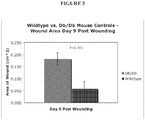
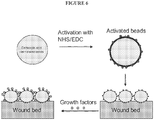


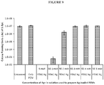


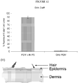

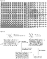
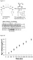
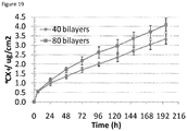
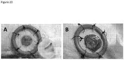

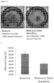


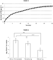
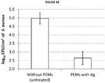


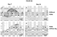


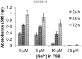


Classifications
-
- A—HUMAN NECESSITIES
- A61—MEDICAL OR VETERINARY SCIENCE; HYGIENE
- A61L—METHODS OR APPARATUS FOR STERILISING MATERIALS OR OBJECTS IN GENERAL; DISINFECTION, STERILISATION OR DEODORISATION OF AIR; CHEMICAL ASPECTS OF BANDAGES, DRESSINGS, ABSORBENT PADS OR SURGICAL ARTICLES; MATERIALS FOR BANDAGES, DRESSINGS, ABSORBENT PADS OR SURGICAL ARTICLES
- A61L15/00—Chemical aspects of, or use of materials for, bandages, dressings or absorbent pads
- A61L15/16—Bandages, dressings or absorbent pads for physiological fluids such as urine or blood, e.g. sanitary towels, tampons
- A61L15/42—Use of materials characterised by their function or physical properties
-
- A—HUMAN NECESSITIES
- A61—MEDICAL OR VETERINARY SCIENCE; HYGIENE
- A61F—FILTERS IMPLANTABLE INTO BLOOD VESSELS; PROSTHESES; DEVICES PROVIDING PATENCY TO, OR PREVENTING COLLAPSING OF, TUBULAR STRUCTURES OF THE BODY, e.g. STENTS; ORTHOPAEDIC, NURSING OR CONTRACEPTIVE DEVICES; FOMENTATION; TREATMENT OR PROTECTION OF EYES OR EARS; BANDAGES, DRESSINGS OR ABSORBENT PADS; FIRST-AID KITS
- A61F13/00—Bandages or dressings; Absorbent pads
- A61F13/02—Adhesive plasters or dressings
-
- A—HUMAN NECESSITIES
- A61—MEDICAL OR VETERINARY SCIENCE; HYGIENE
- A61L—METHODS OR APPARATUS FOR STERILISING MATERIALS OR OBJECTS IN GENERAL; DISINFECTION, STERILISATION OR DEODORISATION OF AIR; CHEMICAL ASPECTS OF BANDAGES, DRESSINGS, ABSORBENT PADS OR SURGICAL ARTICLES; MATERIALS FOR BANDAGES, DRESSINGS, ABSORBENT PADS OR SURGICAL ARTICLES
- A61L15/00—Chemical aspects of, or use of materials for, bandages, dressings or absorbent pads
- A61L15/16—Bandages, dressings or absorbent pads for physiological fluids such as urine or blood, e.g. sanitary towels, tampons
- A61L15/22—Bandages, dressings or absorbent pads for physiological fluids such as urine or blood, e.g. sanitary towels, tampons containing macromolecular materials
- A61L15/24—Macromolecular compounds obtained by reactions only involving carbon-to-carbon unsaturated bonds; Derivatives thereof
-
- A—HUMAN NECESSITIES
- A61—MEDICAL OR VETERINARY SCIENCE; HYGIENE
- A61L—METHODS OR APPARATUS FOR STERILISING MATERIALS OR OBJECTS IN GENERAL; DISINFECTION, STERILISATION OR DEODORISATION OF AIR; CHEMICAL ASPECTS OF BANDAGES, DRESSINGS, ABSORBENT PADS OR SURGICAL ARTICLES; MATERIALS FOR BANDAGES, DRESSINGS, ABSORBENT PADS OR SURGICAL ARTICLES
- A61L15/00—Chemical aspects of, or use of materials for, bandages, dressings or absorbent pads
- A61L15/16—Bandages, dressings or absorbent pads for physiological fluids such as urine or blood, e.g. sanitary towels, tampons
- A61L15/22—Bandages, dressings or absorbent pads for physiological fluids such as urine or blood, e.g. sanitary towels, tampons containing macromolecular materials
- A61L15/26—Macromolecular compounds obtained otherwise than by reactions only involving carbon-to-carbon unsaturated bonds; Derivatives thereof
-
- A—HUMAN NECESSITIES
- A61—MEDICAL OR VETERINARY SCIENCE; HYGIENE
- A61L—METHODS OR APPARATUS FOR STERILISING MATERIALS OR OBJECTS IN GENERAL; DISINFECTION, STERILISATION OR DEODORISATION OF AIR; CHEMICAL ASPECTS OF BANDAGES, DRESSINGS, ABSORBENT PADS OR SURGICAL ARTICLES; MATERIALS FOR BANDAGES, DRESSINGS, ABSORBENT PADS OR SURGICAL ARTICLES
- A61L15/00—Chemical aspects of, or use of materials for, bandages, dressings or absorbent pads
- A61L15/16—Bandages, dressings or absorbent pads for physiological fluids such as urine or blood, e.g. sanitary towels, tampons
- A61L15/42—Use of materials characterised by their function or physical properties
- A61L15/44—Medicaments
-
- A—HUMAN NECESSITIES
- A61—MEDICAL OR VETERINARY SCIENCE; HYGIENE
- A61L—METHODS OR APPARATUS FOR STERILISING MATERIALS OR OBJECTS IN GENERAL; DISINFECTION, STERILISATION OR DEODORISATION OF AIR; CHEMICAL ASPECTS OF BANDAGES, DRESSINGS, ABSORBENT PADS OR SURGICAL ARTICLES; MATERIALS FOR BANDAGES, DRESSINGS, ABSORBENT PADS OR SURGICAL ARTICLES
- A61L15/00—Chemical aspects of, or use of materials for, bandages, dressings or absorbent pads
- A61L15/16—Bandages, dressings or absorbent pads for physiological fluids such as urine or blood, e.g. sanitary towels, tampons
- A61L15/42—Use of materials characterised by their function or physical properties
- A61L15/46—Deodorants or malodour counteractants, e.g. to inhibit the formation of ammonia or bacteria
-
- A—HUMAN NECESSITIES
- A61—MEDICAL OR VETERINARY SCIENCE; HYGIENE
- A61P—SPECIFIC THERAPEUTIC ACTIVITY OF CHEMICAL COMPOUNDS OR MEDICINAL PREPARATIONS
- A61P17/00—Drugs for dermatological disorders
- A61P17/02—Drugs for dermatological disorders for treating wounds, ulcers, burns, scars, keloids, or the like
-
- A—HUMAN NECESSITIES
- A61—MEDICAL OR VETERINARY SCIENCE; HYGIENE
- A61L—METHODS OR APPARATUS FOR STERILISING MATERIALS OR OBJECTS IN GENERAL; DISINFECTION, STERILISATION OR DEODORISATION OF AIR; CHEMICAL ASPECTS OF BANDAGES, DRESSINGS, ABSORBENT PADS OR SURGICAL ARTICLES; MATERIALS FOR BANDAGES, DRESSINGS, ABSORBENT PADS OR SURGICAL ARTICLES
- A61L2300/00—Biologically active materials used in bandages, wound dressings, absorbent pads or medical devices
- A61L2300/10—Biologically active materials used in bandages, wound dressings, absorbent pads or medical devices containing or releasing inorganic materials
- A61L2300/102—Metals or metal compounds, e.g. salts such as bicarbonates, carbonates, oxides, zeolites, silicates
- A61L2300/104—Silver, e.g. silver sulfadiazine
-
- A—HUMAN NECESSITIES
- A61—MEDICAL OR VETERINARY SCIENCE; HYGIENE
- A61L—METHODS OR APPARATUS FOR STERILISING MATERIALS OR OBJECTS IN GENERAL; DISINFECTION, STERILISATION OR DEODORISATION OF AIR; CHEMICAL ASPECTS OF BANDAGES, DRESSINGS, ABSORBENT PADS OR SURGICAL ARTICLES; MATERIALS FOR BANDAGES, DRESSINGS, ABSORBENT PADS OR SURGICAL ARTICLES
- A61L2300/00—Biologically active materials used in bandages, wound dressings, absorbent pads or medical devices
- A61L2300/20—Biologically active materials used in bandages, wound dressings, absorbent pads or medical devices containing or releasing organic materials
- A61L2300/204—Biologically active materials used in bandages, wound dressings, absorbent pads or medical devices containing or releasing organic materials with nitrogen-containing functional groups, e.g. aminoxides, nitriles, guanidines
- A61L2300/206—Biguanides, e.g. chlorohexidine
-
- A—HUMAN NECESSITIES
- A61—MEDICAL OR VETERINARY SCIENCE; HYGIENE
- A61L—METHODS OR APPARATUS FOR STERILISING MATERIALS OR OBJECTS IN GENERAL; DISINFECTION, STERILISATION OR DEODORISATION OF AIR; CHEMICAL ASPECTS OF BANDAGES, DRESSINGS, ABSORBENT PADS OR SURGICAL ARTICLES; MATERIALS FOR BANDAGES, DRESSINGS, ABSORBENT PADS OR SURGICAL ARTICLES
- A61L2300/00—Biologically active materials used in bandages, wound dressings, absorbent pads or medical devices
- A61L2300/40—Biologically active materials used in bandages, wound dressings, absorbent pads or medical devices characterised by a specific therapeutic activity or mode of action
- A61L2300/404—Biocides, antimicrobial agents, antiseptic agents
-
- A—HUMAN NECESSITIES
- A61—MEDICAL OR VETERINARY SCIENCE; HYGIENE
- A61L—METHODS OR APPARATUS FOR STERILISING MATERIALS OR OBJECTS IN GENERAL; DISINFECTION, STERILISATION OR DEODORISATION OF AIR; CHEMICAL ASPECTS OF BANDAGES, DRESSINGS, ABSORBENT PADS OR SURGICAL ARTICLES; MATERIALS FOR BANDAGES, DRESSINGS, ABSORBENT PADS OR SURGICAL ARTICLES
- A61L2400/00—Materials characterised by their function or physical properties
- A61L2400/12—Nanosized materials, e.g. nanofibres, nanoparticles, nanowires, nanotubes; Nanostructured surfaces
Definitions
- the present invention relates to a process for manufacturing a wound active nanoscale polymer matrix microsheet.
- Open cutaneous wounds represent one major category of wounds and include burn wounds, wounds resulting from chemical (especially alkali) burns, wounds from physical trauma, neuropathic ulcers, pressure sores, venous stasis ulcers, and diabetic ulcers.
- Open cutaneous wounds routinely heal by a process which comprises six major components: i) inflammation, ii) fibroblast proliferation, iii) blood vessel proliferation, iv) connective tissue synthesis, v) epithelialization, and vi) wound contraction. Wound healing is impaired when these components, either individually or as a whole, do not function properly.
- wound healing including but not limited to malnutrition, systemic debility due to a variety of causes, wound infection, local lack of progenitor cells, local and/or systemic pharmacological agents (e.g., numerous chemotherapeutic agents, actinomycin and steroids), repeated local trauma, diabetes and other endocrine/metabolic diseases (e.g., Cushing's disease), and advanced age ( Hunt and Goodson, 1988, Current Surgical Diagnosis & Treatment, Appleton & Lange, pp. 86-98 ). Additionally, wounds that are extensive in size, regardless of the initiating cause, present special challenges due to the large surface area that must be re-epithelialized to re-establish surface integrity.
- Diabetes mellitus is a chronic disorder of glucose metabolism and homeostasis that damages many organs. It is the eighth leading cause of death in the United States ( Harris et al., 1987, Diabetes 36:523 ).
- vascular disease, neuropathy, infections, and recurrent trauma predispose the extremities, especially the foot, to pathologic changes. These pathological changes can ultimately lead to chronic ulceration, which may necessitate amputation.
- Chronic wounds and wounds with pathological or dysregulated healing represent a major health burden and drain on health care resources. Chronic wounds have major impacts on the physical and mental health, productivity, morbidity, mortality and cost of care for affected individuals.
- Normal wound healing is an enormously complex process involving the coordinated interplay between fibroblasts, vascular cells, extracellular matrix and epithelial cells to result in a seamless progression through an inflammatory reaction, wound repair, contracture and coverage by an epithelial barrier.
- the wound healing processes can become asynchronous (i.e., loss of connectivity with triggering mechanisms associated with prior cellular events) and are unable to progress to closure, resulting in a chronic ulcer.
- Wounds that do not readily heal can cause the subject considerable physical, emotional, and social distress as well as great financial expense ( Richey et al., 1989, Annals of Plastic Surgery 23:159 ). Indeed, wounds that fail to heal properly and become infected may require excision of the affected tissue.
- a number of treatment modalities have been developed as scientists' basic understanding of wounds and wound healing mechanisms has progressed.
- films e.g., polyurethane films
- hydrocolloids hydrophilic colloidal particles bound to polyurethane foam
- hydrogels cross-linked polymers containing about at least 60% water
- foams hydrophilic or hydrophobic
- calcium alginates nonwoven composites of fibers from calcium alginate
- cellophane cellulose with a plasticizer
- silver is considered particularly favorable because the likelihood of developing bacterial resistance to silver is believed to be very low; therefore, it can be employed as a bactericidal agent continuously.
- currently available methods of applying silver as a bactericidal agent for wound treatment are inadequate.
- 0.5% silver nitrate solution is a standard and popular agent for topical burn wound therapy, providing a beneficial effect in decreasing wound surface inflammation.
- such formulations have a high concentration of silver, there is no residual activity, necessitating frequent applications (e.g., up to 12 times a day) which poses a severe logistical burden in clinical settings.
- Silver ions released through use of 0.5% silver nitrate solution become rapidly inactive through formation of chemical complexes by chloride within 2 hours. Frequent dressings also result in large excesses of silver being delivered to the wound, causing wound-discoloration and toxic effects ( Dunn et al., 2004, Burns 30(supplement 1):S1 ). Additionally, nitrate is toxic to wounds and to mammalian cells. The reduction of nitrate to nitrite further causes oxidant-induced damage to cells, which is cited as the most likely reason for the impaired re-epithelialization with use of silver nitrate solution in partial thickness burns or donor sites.
- Silver compounds such as silver sulfadiazine in cream formulations have also been used for wound treatment.
- cream formulations e.g., Flammazine®, silvadene®
- such formulations also have limited residual activity and have to be applied twice a day.
- Bacterial resistance does develop to these formulations, and, impaired re-epithelialization has also been observed.
- Bone marrow toxicity has been observed with silver sulfadiazine, primarily due to its propylene glycol component.
- silver itself is incorporated into the dressing instead of being applied as a separate formulation. Controlled and prolonged release of silver to the wound allows dressings to be changed less frequently. However, dressings have to be impregnated with large amount of silver, which results in cytotoxicity to mammalian cells. Silver released from a commercially available wound-dressing (ActicoatTM) containing nanocrystalline silver ( Dunn et al., 2004, Burns 30(supplement 1):S1 ) is toxic to in vitro monolayer cell cultures of keratinocytes and fibroblasts ( Poon et al., 2004, Burns 30:140 ; Trop et al., 2006, J. Trauma 60:648 ).
- US 2011/0189287 A1 discloses methods and compositions for wound healing utilizing cross-linker covalent modification molecules to attach and deliver wound active agents to a wound.
- the present invention relates to a process for manufacturing a wound active nanoscale polymer matrix micro sheet as claimed in the attached claims.
- the wound active agents disclosed for use in the process herein are highly bactericidal but that support growth and viability of mammalian cells (e.g., keratinocytes, neurons, vascular endothelial cells and fibroblasts cells).
- the wound active agent is silver, including but not limited to silver nanoparticles.
- the silver loading of some embodiments of the present invention is between 0.35 and 0.4 ⁇ g/cm 2 .
- Polyelectrolyte multilayer micro sheets produced according to the present invention comprising silver (e.g., silver nanoparticles) find use in the treatment of wounds and the prevention of infection, including but not limited to as coatings on devices that come partially, directly, indirectly, or completely in contact with the body of a subject (e.g., a human patient).
- silver e.g., silver nanoparticles
- the at least one wound active agent includes, but is not limited to, trophic factors (including polypetide growth factors, neuropeptides, neurotrophins, , extracellular matrices and their individual native constituents (exemplified by but not limited to laminin, fibronectin, vitronectin, collagens, also select amino acid sequences found in these proteins known to promote cell behaviors favorable to wound healing e.g., integrin binding sequences exemplified by but not limited to RGD, EILDV,VCAM-1 and their recombined or synthetic analogs, enzymes, enzyme inhibitors, polypeptides, antimicrobial peptides ( exemplified by but not limited to defensins, magaignins, cathelocidins, bactenicin) anti-infective agents including silver containing compounds (e.g., ionic silver, elemental silver, silver nanoparticles, and formulations thereof), buffering agents, vitamins and minerals, compounds that promote generation/stabilization of
- kits include small interfering RNAs (siRNAs-also referred to as micro RNAs) that are capable of promoting cellular behaviors conducive to the wound healing process.
- the kits include compounds that promote/stabilize a favorable pH, osmotic environment, surface energy, surface charge, surface functionalities that enhance galvano and magneto positive effects on wound healing, or balance of MMP/other peptidase/protease activity.
- the present invention provides processes for manufacture of a nanoscale polymer matrix microsheet comprising: a) forming a nanoscale polymer layer about 0.5 nm to 1000 nm thick on a substrate; b) introducing a bioactive agent into the nanoscale polymer layer to provide a wound active nanoscale polymer layer, wherein the bioactive agent is a wound active agent; c) forming a sacrificial polymer layer from 0,1 ⁇ m thick to 100 ⁇ m thick on the wound active nanoscale polymer layer, wherein the sacrificial polymer layer is dissolvable.
- the nanoscale polymer matrix is a polymer multilayer. In some embodiments, the nanoscale polymer matrix is formed by alternating layers of at least one positively charged electrolyte and at least one negatively charged polyelectrolyte. In some embodiments, the at least one positively charged polyelectrolyte is selected form the group consisting of poly(allylamine hydrochloride) (PAH), polyl-lysine (PLL), poly(ethylene imine) (PEI), poly(histidine), poly(N,N-dimethyl aminoacrylate), poly(N,N,N-trimethylaminoacrylate chloride), poly(methyacrylamidopropyltrimethyl ammonium chloride), and natural or synthetic polysaccharides such as chitosan.
- PAH poly(allylamine hydrochloride)
- PLL polyl-lysine
- PEI poly(ethylene imine)
- PHI poly(histidine)
- the at least one negatively charged polyelectrolyte is selected from the group consisting of poly(acrylic acid) (PAA), poly(styrenesulfonate) (PSS), alginate, hyaluronic acid, heparin, heparan sulfate, chondroitin sulfate, dextran sulfate, poly(methacrylic acid), oxidized cellulose, carboxymethyl cellulose, polyaspartic acid, and polyglutamic acid.
- PAA poly(acrylic acid)
- PSS poly(styrenesulfonate)
- alginate hyaluronic acid
- heparin heparan sulfate
- chondroitin sulfate chondroitin sulfate
- dextran sulfate poly(methacrylic acid)
- oxidized cellulose carboxymethyl cellulose
- polyaspartic acid polyglutamic acid
- the polymer multilayer is formed by applying the at least one positively charged electrolyte and at least one negatively charged polyelectrolyte by a method selected from the group consisting of spraying polymer solutions on the substrate, dip coating the substrate in polymer solutions, or spin coating polymer solutions on the substrate.
- the at least one positively charged electrolyte and the at least one negatively charged polyelectrolyte are synthetic polyelectrolytes.
- the wound active agent is incorporated into the nanoscale polymer layer so that the wound active agent is interspersed within the three dimensional structure of the nanoscale polymer layer. In some embodiments, the wound active agent is incorporated into the nanoscale polymer multilayer so that the wound active agent is interspersed within the layers the polymer multilayer. In some embodiments, the wound active agent is selected from the group consisting of an antimicrobial agent, an antibiofilm agent, a growth factor, a hemostatic agent, a wound active peptide, a wound active polypeptide, an analgesic, an anticoagulant, anti-inflammatory agent, and a drug molecule or a drug compound.
- the antimicrobial agent is selected from the groups consisting of charged small molecule antimicrobial agents, antimicrobial polypeptides, metallic particles, and metal ion antimicrobial agents.
- the metal ion antimicrobial agent is a metal ion, metal ion salt, or metal ion nanoparticle.
- the metal ion nanoparticle is a silver nanoparticle.
- the charged small molecule antimicrobial agent is selected from the group consisting of chlorhexidine, antibiotics, polyhexamethylene biguanide (PHMB), iodine, cadexomer iodine, povidone iodine (PVI), hydrogen peroxide, and vinegar (acetic acid).
- the metal ion antibiofilm agent is a metal ion, a metal ion salt, or a metal ion nanoparticle. In some embodiments, the metal ion antibiofilm agent is a gallium salt, gallium nanoparticle, gallium alloy, or an alloy of gallium and silver.
- the bioactive agent is introduced into the nanoscale polymer layer during the formation of the nanoscale polymer layer. In some embodiments, the bioactive agent is introduced into the nanoscale polymer layer after formation of the nanoscale polymer layer.
- introducing a bioactive agent into the nanoscale polymer layer to provide a bioactive nanoscale polymer multilayer comprises introducing silver ions into the nanoscale polymer multilayer and reducing the silver ions in situ to provide silver nanoparticles.
- introducing a bioactive agent into the nanoscale polymer layer to provide a bioactive nanoscale polymer multilayer comprises introducing a charged small molecule antimicrobial agent in between polyelectrolyte layers having a different charge.
- the process comprises 1 to 20 repititions of the introducing step, e.g., to control and/or increase the amound of bioactive agent introduced into the nanoscale polymer layer.
- the process comprises controlling the amount of the bioactive agent in the nanoscale polymer matrix microsheet by controlling the number of nanoscale polymer layers, by controlling the pH of forming the nanoscale polymer layer, and/or by controlling the number of introducing cycles.
- the sacrificial polymer layer is from about 0.1 ⁇ m thick to about 100 ⁇ m thick.
- the sacrificial polymer layer comprises a water soluble polymer.
- the water soluble sacrificial polymer layer is made of and/or comprises polyvinyl alcohol (PVA).
- PVA polyvinyl alcohol
- the soluble polymer has a molecular weight of greater than 22 kDa.
- the water soluble polymer is removable by renal filtration.
- the water soluble sacrificial polymer layer is made of polyacrylic acid (PAA).
- PAA polyacrylic acid
- the sacrificial polymer layer comprising a water soluble polymer dissolves when exposed to moisture on a surface so that the bioactive nanoscale polymer multilayer is deposited on the surface.
- the processes further comprise introducing a bioactive agent into the sacrificial polymer layer.
- the processes further comprise introducing microparticles or nanoparticles into the sacrificial polymer layer.
- the microparticles or nanoparticles in the sacrificial polymer layer are loaded with bioactive agents.
- the processes further comprise introducing magnetic microparticles or nanoparticles into the sacrificial polymer layer.
- the bioactive agent is selected from the group consisting of an antimicrobial agent, a growth factor, a hemostatic agent, a bioactive peptide, a bioactive polypeptide, an analgesic, an anticoagulant, anti-inflammatory agent, and a drug molecule or a drug compound.
- the antimicrobial agent is selected from the goups consisting of charged small molecule antimicrobial agents, antimicrobial polypeptides, metallic particles, and metal ion antimicrobial agents.
- the metal ion antimicrobial reagent is a metal ion, metal ion salt, or metal ion nanoparticle.
- the metal ion nanoparticle is a silver nanoparticle.
- the small molecule antimicrobial agent is selected from the group consisting of chlorhexidine, antibiotics, polyhexamethylene biguanide (PHMB), iodine, cadexomer iodine, povidone iodine (PVI), hydrogen peroxide, vinegar (acetic acid).
- the substrate is selected from the group consisting of a polydimethylsiloxane (PDMS) substrate, a glass substrate, a plastic substrate, a metal substrate, and a Teflon substrate.
- PDMS polydimethylsiloxane
- the substrate is functionalized to provide a low-energy surface.
- the substrate is functionalized with octadecyltricrhlorosilane.
- the processes further comprise the step of removing the nanoscale polymer layer matrix microsheet comprising the wound active nanoscale polymer layer and associated sacrificial polymer layer from the substrate. In some embodiments, greater than 90%, greater than 95%, greater than 97%, greater than 98%, or greater than 99% of the nanoscale polymer matrix microsheet comprising the wound active nanoscale polymer layer and associated sacrificial polymer layer is removed from said substrate. In some embodiments, the processes further comprise incorporating nanoscale or microscale particles or beads into the nanoscale polymer layer. In some embodiments, the processes further comprise incorporating magnetic nanoparticles or microparticles into the nanoscale polymer layer.
- the nanoscale polymer layer comprises a polymer selected from the group consisting of polyelectrolytes, lipids, proteins, collagen, hyaluornic acid, chitosan, keratin, fibronectin, vitronectin, laminin, polysaccharides, polyanhydrides, poly (lactic-co-glycolic acid) (PLGA), poly(1-lactic acid) (PLLA), and combinations thereof or fragments thereof.
- the sacrificial polymer layer is applied to the nanoscale polymer layer by spin coating, dip coating, or spray coating.
- the nanoscale polymer matrix microsheet has a Young modulus of from about 0.1 GPa to about 10 GPa after removal from the substrate.
- forming the nanoscale polymer layer comprises depositing a positively charged electrolyte and a negatively charged polyelectrolyte to form a bilayer. In some embodiments, the processes comprise 2 to 50 depositing steps. In some embodiments, forming the nanoscale polymer layer comprises a step of providing solution conditions comprising a pH from 1.5 to 2.5, a pH from 6.5 to 8.5, and/or a pH from 4.5 to 6.5. In some embodiments, the processes further comprise drying the nanoscale polymer matrix microsheet. In some embodiments, the process further comprises indroducing a second or more bioactive agent(s) into the nanoscale polymer layer.
- a microsheet comprising: a wound active nanoscale polymer layer about 0.5 nm to 1000 nm thick, the nanoscale wound active polymer layer having a wound active agent incorporated therein; and a sacrificial polymer layer adjacent to the wound active nanoscale polymer layer, the sacrificial polymer layer being from 0,1 ⁇ m thick to 100 ⁇ m thick on said wound active nanoscale polymer layer and being dissolvable when exposed to a moist surface.
- the nanoscale polymer layer is a polymer multilayer.
- the nanoscale polymer multilayer is formed by alternating layers of at least one positively charged electrolyte and at least one negatively charged polyelectrolyte.
- the at least one positively charged polyelectrolyte is selected form the group consisting of poly(allylamine hydrochloride) (PAH), polyl-lysine (PLL), poly(ethylene imine) (PEI), poly(histidine), poly(N,N-dimethyl aminoacrylate), poly(N,N,N-trimethylaminoacrylate chloride), poly(methyacrylamidopropyltrimethyl ammonium chloride), and natural or synthetic polysaccharides such as chitosan.
- PAH poly(allylamine hydrochloride)
- PLL polyl-lysine
- PEI poly(ethylene imine)
- PHI poly(histidine)
- poly(N,N,N-trimethylaminoacrylate chloride) poly(methyacrylamidopropyltrimethyl ammonium chloride)
- natural or synthetic polysaccharides such as chitosan.
- the at least one negatively charged polyelectrolyte is selected from the group consisting of poly(acrylic acid) (PAA), poly(styrenesulfonate) (PSS), alginate, hyaluronic acid, heparin, heparan sulfate, chondroitin sulfate, dextran sulfate, poly(meth)acrylic acid, oxidized cellulose, carboxymethyl cellulose, polyaspartic acid, and polyglutamic acid.
- PAA poly(acrylic acid)
- PSS poly(styrenesulfonate)
- alginate hyaluronic acid
- heparin heparan sulfate
- chondroitin sulfate chondroitin sulfate
- dextran sulfate poly(meth)acrylic acid
- the at least one positively charged electrolyte and the at least one negatively charged polyelectrolyte are synthetic polyelectrolytes.
- the wound active agent is incorporated into the nanoscale polymer layer so that the wound active agent is interspersed within the three dimensional structure of the nanoscale polymer layer. In some embodiments, the wound active agent is incorporated into the nanoscale polymer multilayer so that the wound active agent is interspersed within the layers the nanoscale polymer multilayer.
- the wound active agent is selected from the group consisting of an antimicrobial agent, a growth factor, a hemostatic agent, a bioactive peptide, a bioactive polypeptide, an analgesic, an anticoagulant, anti-inflammatory agent, and a drug molecule or a drug compound.
- the antimicrobial agent is selected from the goups consisting of small molecule antimicrobial agents, charged small molecule antimicrobial agents, antimicrobial polypeptides, metallic particles, and metal ion antimicrobial agents.
- the metal ion antimicrobial reagent is a metal ion, metal ion salt, or metal ion nanoparticle.
- the metal ion nanoparticle is a silver nanoparticle.
- the charged small molecule antimicrobial agents or small molecule antimicrobial agent is selected from the group consisting of chlorhexidine, antibiotics, polyhexamethylene biguanide (PHMB), iodine, cadexomer iodine, povidone iodine (PVI), hydrogen peroxide, vinegar (acetic acid).
- the sacrificial polymer layer is from about 0.1 ⁇ m thick to about 100 ⁇ m thick. In some embodiments, the sacrificial polymer layer is from about 0.1 ⁇ m thick to about 50 ⁇ m thick.
- the sacrificial polymer layer comprises from about 1 ⁇ m thick to about 20 ⁇ m thick. In some embodiments, the sacrificial polymer layer is from about 1 ⁇ m thick to about 10 ⁇ m thick. In some embodiments, the sacrificial polymer layer comprises a water soluble polymer. In some embodiments, the sacrificial polymer layer comprises a wound active agent. In some embodiments, the wound active agent is selected from the group consisting of an antimicrobial agent, a growth factor, a hemostatic agent, a bioactive peptide, a bioactive polypeptide, an analgesic, an anticoagulant, anti-inflammatory agent, and a drug molecule, or a drug compound.
- the antimicrobial agent is selected from the groups consisting of small molecule antimicrobial agents, antimicrobial polypeptides, metallic particles, and metal ion antimicrobial agents.
- the metal ion antimicrobial reagent is a metal ion, metal ion salt, or metal ion nanoparticle.
- the metal ion nanoparticle is a silver nanoparticle.
- the small molecule antimicrobial agent is selected from the group consisting of chlorhexidine, antibiotics, polyhexamethylene biguanide (PHMB), iodine, cadexomer iodine, povidone iodine (PVI), hydrogen peroxide, and vinegar (acetic acid).
- the microsheets further comprise nanoscale or microscale particles incorporated into the nanoscale polymer layer.
- the nanoscale polymer layer comprises a polymer selected from the group consisting of polyelectrolytes, lipids, proteins, collagen, hyaluornic acid, chitosan, keratin, fibronectin, vitronectin, laminin, polysaccharides, polyanhydrides, poly (lactic-co-glycolic acid) (PLGA), poly(1-lactic acid) (PLLA), and combinations thereof or fragments thereof.
- PLGA poly (lactic-co-glycolic acid)
- PLLA poly(1-lactic acid)
- the microsheet comprises the wound active agent at a concentration of approximately 0.01 to 100 ⁇ g/cm 2 .
- the bioactive agent is provided in an amount so that the wound active agent is released at a rate of about 0.01 and 100 ⁇ g/cm 2 per day.
- the microsheet has an area of from about 0.2 to 600 cm 2 .
- the sacrificial polymer layer has a uniform thickness with a variation of less than 500, 400, 300, 200, 100, 50, 20 or 10% of the average thickness when measured in cross section.
- the microsheet has a Young modulus of from about 0.1 GPa to about 10 GPa, e.g., after removal from a substrate.
- the sacrificial polymer layer dissolves when exposed to moisture to leave the nanoscale polymer layer on the surface.
- the nanoscale polymer layer forms a barrier to exogenous pathogens when applied to a moist surface.
- the microsheet further comprises a second or more wound active agent(s).
- the microsheet is translucent and/or transparent.
- the microsheet produced with the process of the invention can be used in a medical device comprising the microsheet produced as described above, wherein the medical device can be selected from the group consisting of a a wound dressing, an implantable medical device, a medical device that contacts skin, a catheter, a stent, a membrane, a contact lens, and a surgical mesh, wherein further the wound dressing can be a biologic wound dressing, an abiologic wound dressing, an absorbent material, a foam, a transparent thin film, a hydrogel, hydrofibers, hydrocolloids, alginate fibers, silica gel, sodium polyacrylate, potassium polyacrylamides, and combinations thereof.
- the medical device can be selected from the group consisting of a a wound dressing, an implantable medical device, a medical device that contacts skin, a catheter, a stent, a membrane, a contact lens, and a surgical mesh
- the wound dressing can be a biologic wound dressing, an abiologic wound dressing, an absorbent material, a foam, a
- wound refers broadly to injuries to the skin and subcutaneous tissue initiated in different ways (e.g., pressure sores from extended bed rest and wounds induced by trauma) and with varying characteristics.
- the methods and compositions described herein are useful for treatment of all types of wounds, including wounds to internal and external tissues.
- Wounds may be classified into one of four grades depending on the depth of the wound: i) Grade I: wounds limited to the epithelium; ii) Grade II: wounds extending into the dermis; iii) Grade III: wounds extending into the subcutaneous tissue; and iv) Grade IV (or full-thickness wounds): wounds wherein bones are exposed (e.g., a bony pressure point such as the greater trochanter or the sacrum).
- Grade I wounds limited to the epithelium
- Grade II wounds extending into the dermis
- Grade III wounds extending into the subcutaneous tissue
- Grade IV or full-thickness wounds
- partial thickness wound refers to wounds that encompass Grades I-III; examples of partial thickness wounds include burn wounds, pressure sores, venous stasis ulcers, and diabetic ulcers.
- deep wound is meant to include both Grade III and Grade IV wounds. The present invention contemplates treating all wound types, including deep wounds and chronic wounds.
- chronic wound refers to a wound that has not healed within 30 days.
- promote wound healing refers to either the induction of the formation of granulation tissue of wound contraction and/or the induction of epithelialization (i.e., the generation of new cells in the epithelium). Wound healing is conveniently measured by decreasing wound area.
- wound active agent refers to known or potential chemical compounds that induce a desired pharmacological, physiological effect useful in the treatment and healing of a wound, wherein the effect may be prophylactic or therapeutic.
- the terms also encompass pharmaceutically acceptable, pharmacologically active derivatives of those active agents specifically mentioned herein, including, but not limited to, trophic factors, extracellular matrices, enzymes, enzyme inhibitors, defensins, polypeptides, anti-infective agents (including but not limited to ionic silver, elemental silver, and silver nanoparticles), buffering agents, vitamins and minerals, analgesics, anticoagulants, coagulation factors, anti-inflammatory agents, vasoconstrictors, vasodilators, diuretics, and anti-cancer agents.
- polymer multilayer refers to the composition formed by sequential and repeated application of polymer(s) to form a multilayered structure.
- polyelectrolyte multilayers are polymer multilayers are formed by the alternating addition of anionic and cationic polyelectrolytes to a wound or support.
- polymer multilayer also refers to the composition formed by sequential and repeated application of polymer(s) to a wound or to a solid support.
- polymer layer can refer to a single layer composed of polymer molecules, such as anionic or cationic polyelectrolyte molecules, existing either as one layer within multiple layers, or as a single layer of only one type of polyelectrolyte molecules on a wound or support. While the delivery of the polymers to the wound bed or support is sequential in preferred embodiments, the use of the term “polymer multilayer” is not limiting in terms of the resulting structure of the coating. It is well understood by those skilled in the art that inter-diffusion of polymers such as polyelectrolytes can take place leading to structures that may be well-mixed in terms of the distribution of anionic and cationic polyelectrolytes.
- polyelectrolyte includes polymer species as well as nanoparticulate species, and that it is not limiting in scope other than to indicate that the species possesses multiple charged or partially charged groups. It is also well understood by those skilled in the art that multilayer structures can be formed through a variety of interactions, including electrostatic interactions and others such as hydrogen bonding. Thus, the use of the term "polyelectrolyte" is not limiting in terms of the interactions leading to the formation of the wound bed constructs.
- crosslinked refers to a composition containing intermolecular crosslinks and optionally intramolecular crosslinks as well, arising from the formation of covalent bonds.
- Covalent bonding between two crosslinkable components may be direct, in which case an atom in one component is directly bound to an atom in the other component, or it may be indirect, through a linking group.
- a crosslinked structure may, in addition to covalent bonds, also include intermolecular and/or intramolecular noncovalent bonds such as hydrogen bonds and electrostatic (ionic) bonds.
- Covalent modification agent refers to any molecule that covalently links molecules to each other.
- Covalent modification agents include homobifunctional and heterobifunctional cross-linkers as well as photoactivatable cross linkers.
- the term "homobifunctional cross-linker” refers to a molecule used to covalently link identical or similar molecules to each other. Homobifunctional cross-linkers have two identical reactive groups; thus, a homobifunctional cross-linker can only link molecules of the same type to each other. Conversely, a “heterobifunctional cross-linker” refers to a molecule used to covalently link dissimilar molecules to each other, because it has two or more different reactive groups that can interact with various molecules of different types. Hetero- and homo-multifunctional crosslinkers refers to multivalent crosslinkers with both hetero- and homo- crosslinking functionalities. Activated dendrimers are an example of multifunctional crosslinkers.
- subject refers to any animal (e.g., a mammal), including, but not limited to, humans, non-human primates, rodents, dogs, cats, and the like, which is to be the recipient of a particular treatment.
- subject and “patient” are used interchangeably herein.
- surfactant refers to an amphiphilic material that modifies the surface and interface properties of liquids or solids. Surfactants can reduce the surface tension between two liquids. Detergents, wetting agents, emulsifying agents, dispersion agents, and foam inhibitors are all surfactants.
- block copolymer refers to a polymer consisting of at least two monomers.
- adjacent blocks are constitutionally different, i.e. adjacent blocks comprise constitutional units derived from different species of monomer or from the same species of monomer but with a different composition or sequence distribution of constitutional units.
- a block copolymer can be thought of as two homopolymers joined together at the ends.
- solvent refers to a liquid that can dissolve a substance.
- organic solvent refers to a solvent derived from a petroleum-based product.
- polyelectrolyte refers to a water-soluble macromolecular polymer substance containing many repeating ionic constituent units, including cations and anions.
- primary amine refers to a derivative of ammonia in which a hydrogen has been replaced by a hydrocarbon unit.
- Primary amines have the general formula RNH 2 and examples include, but are not limited to, aniline, methylamine, and 1-propylamine.
- DNA delivery agent refers to any molecule that can bring DNA into contact with an identified target.
- a DNA delivery agent causes uptake of DNA into a cell or cells, in vitro or in vivo.
- DNA delivery agents can be viruses including, but not limited to, adenoviruses and retroviruses.
- DNA delivery agents can also be non-viral agents including, but not limited to, plasmids, lipids, liposomes, polymers and peptides.
- exposable refers to anything that is capable of being exposed.
- An exposable surface or molecule is one that is made available to interaction with other surfaces or molecules.
- a covalent modification agent is exposable to a wound active agent; thus, the two agents can interact with each other and form covalent bonds.
- the term "functionalized” refers to a modification of an existing molecular segment to generate or introduce a new reactive functional group (e.g., a maleimido or succinimidyl group) that is capable of undergoing reaction with another functional group (e.g., a sulfhydryl group) to form a covalent bond.
- a new reactive functional group e.g., a maleimido or succinimidyl group
- another functional group e.g., a sulfhydryl group
- a component containing carboxylic acid (-COOH) groups can be functionalized by reaction with N-hydroxy-succinimide or N-hydroxysulfosuccinimide using known procedures, to form a new reactive functional group in the form of an activated carboxylate (which is a reactive electrophilic group), i.e., an N-hydroxysuccinimide ester or an N-hydroxysulfosuccinimide ester, respectively.
- carboxylic acid groups can be functionalized by reaction with an acyl halide, e.g., an acyl chloride, again using known procedures, to provide a new reactive functional group in the form of an anhydride.
- aqueous solution includes solutions, suspensions, dispersions, colloids, and the like containing water.
- click chemistry refers to the use of chemical building blocks with built-in high-energy content to drive a spontaneous and irreversible linkage reaction with appropriate complementary sites in other blocks. These chemical reactions (e.g., including, but not limited to, those between azide and acetylene groups that combine readily with each other) are specific and result in covalent linkage between the two molecules.
- non-chemical ligation refers to a chemoselective reaction of two unprotected peptide segments. The reaction results in an initial thioester-linked species, then spontaneous rearrangement of this transient intermediate occurs, yielding a full-length product with a native peptide bond at the ligation site.
- specific protein binding refers to an interaction between two or more proteins that have high affinity and specificity for each other. Proteins must bind to specific other proteins in vivo in order to function. The proteins are required to bind to only one or a few other proteins of the few thousand proteins typically present in vivo; these interactions are employed in vitro in the present invention to attach wound active agents to the wound.
- specific protein binding interactions include, but are not limited to, those between biotin and avidin, neutravidin, or streptavidin; glutathione-S-transferase and glutathione; and nickel-nitrilotriacetic acid and polyhistidine.
- device refers to an object that contacts the body or bodily fluid of a subject for therapeutic or prophylactic purposes. Some devices may partially or indirectly contact the body or bodily fluid of a subject (e.g., catheter, dialysis tubing, diagnostic sensors, drug delivery devices), while other devices are completely imbedded in or encompassed by the body of a subject (e.g., stent, pacemaker, internally implanted defibrillator, angioplasty balloon, orthopedic device, spinal cage, implantable drug pump, artificial disc, ear disc).
- stent e.g., pacemaker, internally implanted defibrillator, angioplasty balloon, orthopedic device, spinal cage, implantable drug pump, artificial disc, ear disc.
- selective toxicity refers to the property of differentially toxic effects on mammalian versus microbial cells.
- a selectively toxic agent may effectively kill bacterial cells while permitting growth and viability of mammalian cells.
- toxic refers to any detrimental or harmful effects on a subject, a cell, or a tissue as compared to the same cell or tissue prior to the administration of the toxicant.
- nanoparticle and “nanoscale particles” are used interchangeably and refer to a nanoscale particle with a size that is measured in nanometers, for example, a nanoscopic particle that has at least one dimension of less than about 1000, 500, or 100 nm.
- nanoparticles include nanobeads, nanofibers, nanohoms, nano-onions, nanorods, and nanoropes.
- microparticle and “microscale particles” are used interchangeably and refers to a microscale particle with a size that is measured in micrometers, for example, a microscale particle that has at least one dimension of less than about 10 micrometers, 5 micrometers, or 2 micrometers.
- wound dressing refers to materials placed proximal to a wound that have absorbent, adhesive, protective, osmoregulatory, pH-regulatory, or pressure-inducing properties. Wound dressings may be in direct or indirect contact with a wound. Wound dressings are not limited by size or shape. Indeed, many wound dressing materials may be cut or configured to conform to the dimensions of a wound.
- wound dressing materials include but are not limited to gauze, adhesive tape, bandages, and commercially available wound dressings including but not limited to adhesive bandages and pads from the Band-Aid ® line of wound dressings, adhesive bandages and pads from the Nexcare ® line of wound dressings, adhesive bandages and non-adhesive pads from the Kendall Curity Tefla ® line of wound dressings, adhesive bandages and pads from the Tegaderm ® line of wound dressings, adhesive bandages and pads from the Steri-Strip ® line of wound dressings, the COMFEEL ® line of wound dressings, adhesive bandages and pads, the Duoderm ® line of wound dressings, adhesive bandages and pads, the TEGADERMTM line of wound dressings, adhesive bandages and pads, the OPSITE ® line of wound dressings, adhesive bandages and pads, and biologic wound dressings.
- adhesive bandages and pads from the Band-Aid ® line of wound dressings, adhesive bandages and pads from the Nexcare ® line of wound dressings, adhesive
- a "biologic wound dressing” is a type of wound dressing that comprises, e.g., is coated with or incorporates, cells and/or one or more biomolecules or fragments of biomolecules that can be placed in contact with the wound surface.
- the biomolecules may be provided in the form of an artificial tissue matrix. Examples of such biomolecules include, but are not limited, to collagen, hyaluronic acid, glycosaminoglycans, laminin, vitronectin, fibronectin, keratin, antimicrobial polypeptides and combinations thereof.
- suitable biologic wound dressings include, but are not limited to, BIOBRANETM, IntegraTM, Apligraf ® , Dermagraft ® , Oasis ® , Transcyte ® , Cryoskin ® and My skin ® .
- the term "antimicrobial silver composition” refers to a composition that comprises silver as an active antimicrobial agent.
- antimicrobial silver compositions include, but are not limited to silver nanoparticles, elemental silver, zero valent silver, multivalent silver ions carried by zirconium phosphate (ZP-Ag)(See, e.g., Wound Repair and Regeneration, 16: 800-804 ), and silver containing compounds such as silver sulfadiazine and related compounds.
- ZP-Ag zirconium phosphate
- releasable antimicrobial silver composition refers to a antimicrobial silver composition that can be released from a material, for example, a polymer multilayer solid support, so that antimicrobial activity can be observed. The release of the antimicrobial silver composition can be defined as an amount of the composition released from a defined area or volume of the material.
- the microenvironment of the pathologic/chronic wound bed is dysregulated with alterations in extracellular matrix constituents, degradative enzymes, growth factor and other cytoactive factor activity.
- the present invention provides a process for manufacturing a wound active nanoscale polymer matrix microsheet as claimed in the attached claims.
- stimulating a "desirable" healing response is best achieved by a differential modulation of the cellular responses within the wound.
- Modulation of the fibroblastic response in this way has the potential to provide superior clinical outcomes and reduce the need for subsequent reconstructive procedures targeted at recovery of limb or other critical bodily functions.
- the feasibility of such an approach has been demonstrated previously, such as in the report by Muehlberger and colleagues on the effects tretinoin on incisional wounds ( Muehlberger et al., 2005, J. Am. Acad. Derm. 52:583 ). In that study, application of tretinoin resulted in an increased fibroblastic proliferation but the production of collagen was diminished.
- SAMs Self-assembled monolayers
- Langmuir-Blodgett techniques have been commonly employed to produce new interfaces ( Mrksich, 1997, Curr. Opin. Colloid Interface Sci. 2:83 ; Lösche, 1997, Curr. Opin. Solid State Mater. Sci. 2:546 ).
- the layer-by-layer deposition process is not limited in applicability to polyelectrolytes, but can be applied to viruses, nanoparticles, non-ionic polymers, proteins and other forms of microscopic and nanoscopic matter.
- a recent review provides information on a wide range of species and interfacial structures that can be formed by the layer-by-layer deposition procedure.
- the scope of the invention described herein is not limited to polyelectrolytes but applies to all species that have been demonstrated to be incorporated into interfacial structures by the layer-by-layer deposition process.
- the wound active agent is silver or a form of silver.
- Silver is a widely used nonspecific biocidal agent that acts against a very broad spectrum of bacterial species ( Yin et al., 1999, J. Burn Care Rehabil. 20:195 ), yeast species, fungal species ( Wright et al., 1999, Am. J Infect. Control 27:344 ), and viruses ( Hussain et al., 1991, Biochem. Biophys. Res. Comm. 189:1444 ), including several antibiotic resistant strains ( Wright et al., 1998, Am. J. Infect. Control 26:572 ).
- topical silver agents undergo fast inactivation in the wounds because of the highly reactive nature of silver ions.
- Silver based topical solutions like 0.5 % silver nitrate, or ointments like 1 % silver sulfadiazine cream that are currently used in clinics for wound healing, release silver at concentrations up to 3200 ppm but most of this is rapidly inactivated though the formation of chemical complexes in the wound ( Dunn et al., Burns, 2004, 30(supplement 1):S1 ), necessitating frequent applications, up to 12 times a day for silver nitrate solutions and at least twice a day with silver sulfadiazine creams.
- Silver released from a leading commercial wound-dressing ActicoatTM, that contains nanocrystalline silver was shown to be toxic to in vitro monolayer cell cultures of keratinocytes and fibroblasts ( Poon et al., 2004, Burns 30:140 ; Trop et al., 2006, J. Trauma 60:648 ).
- the challenges with current topical silver antimicrobials lie in their low silver release levels, the lack of penetration, the rapid consumption of silver ions, and the presence of silver nitrate or cream bases that are pro-inflammatory, negatively affecting wound healing. Issues like wound staining, electrolyte imbalance, and patient discomfort also exist.
- nanometer-thick polymeric films were assembled that were impregnated with silver nanoparticles to levels resulting in efficient killing of bacteria but no cytotoxic effects on the adherence and growth of mouse fibroblasts cells on the films.
- the polymeric films are impregnated with an antimicrobial silver composition by incubating the film in an antimicrobial silver composition as described in Example 13.
- the antimicrobial silver composition is directly incorporated into the polymer multilayer during synthesis of the multilayer. See, e.g., Langmuir. 2005 Dec 6;21(25): 11915-21 .
- the films produced with the process according to the invention can be applied directly on the exposed wound-bed, allowing them to release the silver ions locally into the wound. This reduces the amount of bactericidal silver needed to impregnate into the film by several orders compared to topical silver delivery agents, significantly reducing the silver-based toxicity.
- Some molecularly-thin composite film embodiments of the present invention can be tuned to contain as low as 0.4 ⁇ g/cm 2 of soluble silver and release a total of less than 1 ppm silver ions in buffers. Such films display 99.9999 % bacteria-killing efficiency without any measurable cytotoxicity to mammalian cells. Furthermore, films containing these levels of silver allow adherence and growth of mammalian cells (e.g., NIH-3T3 mouse fibroblasts).
- mammalian cells e.g., NIH-3T3 mouse fibroblasts.
- the releasable antimicrobial silver composition is loaded at an amount of from 1 or 10 to about 50, 100, 200, 300, 400, 500 or 1000 ⁇ g/cm 2 of the polymer multilayer and/or releasable in an amount of about 0.01 and 100 ⁇ g/cm 2 of the polymer multilayer per day. In some embodiments, the releasable antimicrobial silver composition is releasable in an amount of about 0.05 and 100 ⁇ g/cm 2 of the polymer multilayer. In some embodiments, the releasable antimicrobial silver composition is releasable in an amount of about 0.05 and 20 ⁇ g/cm 2 of the polymer multilayer.
- the releasable antimicrobial silver composition is releasable in an amount of about 0.05 and 5 ⁇ g/cm 2 of the polymer multilayer. In some embodiments, the releasable antimicrobial silver composition is releasable in an amount of about 0.05 and 2 ⁇ g/cm 2 of the polymer multilayer. In some embodiments, the releasable antimicrobial silver composition is releasable in an amount of about 0.05 and 1 ⁇ g/cm 2 of the polymer multilayer. In some embodiments, the releasable antimicrobial silver composition is releasable in an amount of about 0.1 to 0.5 ⁇ g/cm 2 of the polymer multilayer.
- ActicoatTM releases a total of over 120 ppm silver on dissolution in water for 24 hr ( Taylor et al., 2005, Biomaterials 26:7221 ), and has been shown to be cytotoxic to keratinocytes and fibroblasts ( Poon et al., 2005, Burns 30:140 ; Trop et al., 2006, J. Trauma 60:648 ).
- the silver-releasing moleculary-thin composite films as manufactured by the process of the present invention have advantage over: 1) other topical delivery agents of silver currently used in clinics; 2) wound-dressings that contain large amounts of silver in them; and 3) other bactericidal composite thin-films containing large amounts of silver, which are cytotoxic to mammalian cells and do not allow mammalian cells to adhere and grow over them.
- Using silver-nanoparticle molecularly-thin films for localized delivery of bactericidal silver in wounds can overcome silver-related toxicity encountered with topical silver-delivery agents for wound healing ( Poon et al., 2004, Burns 30:140 ; Baldi et al., 1988, Toxicol. Lett. 41:261 ; Atiyeh et al., 2007, Burns 33:139 ; Coombs et al., 1992, Burns 18:179 ).
- these polyelectrolyte multilayers are uniform, highly interpenetrated ultrathin nanocomposite films, typically far less than 1 ⁇ m thick ( Decher, 1997, Science 277:1232 ; Hammond, 1999, Curr. Opin. Colloid Interface Sci. 4:430 ).
- Their porous and supramolecular architecture allows incorporating a variety of molecules including DNA, enzymes, viruses, dendrimers, colloids, inorganic particles, and dyes ( Decher, 1997, Science 277:1232 ).
- they can be used for simultaneous localized delivery of other bioactive agents in the wound-matrix (e.g., cell recruiting growth factors, DNA plasmids encoding such growth factors).
- PEMs can conformally coat substrates of any type, size or shape, including natural and synthetic polymers ( Hammond, 1999, Curr. Opin. Colloid Interface Sci. 4:430 ), they can be constructed on any wound-bed or implantable medical device.
- Figure 1 provides a schematic diagram 100 of a wound bed 110 on which a polyelectrolyte multilayer 130 has been deposited.
- the diagram 100 depicts that the wound bed 110 comprises a heterogeneous surface depicted by shapes 120 which represent different chemical moieties.
- the polyelectrolyte multilayer 130 provides a homogenous surface onto which functional groups can 140 can be attached to form a homogenous functionalized surface 150.
- the functional groups are uniform, however, in some preferred embodiments, different functional groups are utilized.
- a wide variety of active agents can then be attached to the surface via the functional groups 140.
- the wound bed is covalently modified with covalent modification agents.
- the covalent modification agents include, but are not limited to, homobifunctional and heterobifunctional cross-linkers as well as photoactivatable cross linkers.
- Figure 2 provides a schematic diagram 200 of a wound bed 210 comprising a heterogeneous surface depicted by shapes 220 which represent different chemical moieties.
- the wound bed is covalently modified by reacting covalent modification agents 230 with the different chemical moieties to provide a relatively homogenous functionalized surface 250.
- covalent modification agents 230 present functional groups 240.
- the functional groups are uniform, however, in some preferred embodiments, different functional groups are utilized.
- a wide variety of active agents can then be attached to the surface via the functional groups 240. These embodiments are discussed in more detail below.
- a wound active nanoscale polymer matrix microsheet can be manufactured as claimed in claim 1, wherein the wound active nanoscale polymer layer is a polymer multilayer.
- the multilayer structures comprise layers of polymers that form polyelectrolytes, while in other embodiments, the multilayers comprise polymers that do not have a charge (i.e., non-ionic polymers) or a combination of charged and uncharged polymer layers.
- polyelectrolyte films built-up by the alternated adsorption of cationic and anionic polyelectrolyte layers constitute a novel and promising technique to modify wound surfaces in a controlled way ( Decher et al., 1992, Thin Solid Films 210/211:831 ; Decher, 1997, Science 277:1232 ).
- One of the most important properties of such multilayers is that they exhibit an excess of alternatively positive and negative charges ( Caruso et al., 1999, J Am Chem Soc 121:6039 ; Ladam et al., 2000, Langmuir 16:1249 ). Not only can this constitute the motor of their buildup ( Joanny, 1999, Eur.
- the polymer multilayers are nanoscale in dimension. Accordingly, in some embodiments, the polymer multilayers are from about 1 nm to 1000 nm thick, from about 1 nm to 500 nm thick, from about 1 nm to 100 nm thick, from about 1 nm to about 25 nm thick, from about 1 nm to about 10 nm thick, or less than about 500 nm, 100 nm, 25 nm or 10 nm thick.
- the nanoscale dimension of the polymer multilayers allows for the loading of a lower total amount of an active agent while still allowing delivery of an effective amount (i.e., an amount of active agent that accelerates wound healing as compared to controls) of the active agent as compared to matrix structures with greater thickness. It is contemplated that the lower total loading levels result in reduced toxicity in the wound environment, especially when antimicrobial compounds are incorporated into the polymer multilayer.
- the compliance of the polymer multilayers is adjusted to facilitate cell migration in the wound.
- the polymer multilayers exhibit a compliance, measured in kilopascals (kPa) of from about 3 to about 500 kPa, about 7 to about 250 kPa, about 10 to about 250 kPA or from about 10 to about 200 kPa.
- PLL cationic polyelectrolyte poly(L-lysine)
- Polyelectrolyte layers are formed by alternating applications of anionic polyelectrolytes and cationic polyelectrolytes to surfaces to form a polyelectrolyte layer.
- the layers can be used to deliver a wound active agent to a wound.
- at least four layers, and, more preferably, at least six layers are used to form the polyelectrolyte multilayer.
- more than six layers are used, e.g., 7, 8, 9, 10, 15, 20, 25, 30, 35, 40, 45, 50 or more layers.
- a polyelectrolyte multilayer comprises 10.5 layers (e.g., comprising 10 bilayers of the two components and an additional layer of one of the two components).
- the cationic polyelectrolyte used is PLL and the anionic polyelectrolyte used is poly(L-glutamic acid) (PGA).
- PGA poly(L-glutamic acid)
- PES poly(ethylene imine)
- PAH poly(allylamine hydrochloride)
- PSS poly(sodium 4-styrenesulfonate)
- PAC poly(acrylic acid)
- PMA-P poly(maleic acid-co-propylene)
- PAA poly(acrylic acid)
- PVS poly(vinyl sulfate)
- PVS poly(vinyl sulfate)
- the polymer is a dendrimer, grafted polymer, or star architecture polymer.
- the multilayer responds to or is organized in the presence of an electric field, for example an electric field formed by placing electrodes on either side of a wound.
- the polymer multilayer 130 can be comprised of polystyrene sulfonate, an amphiphillic polyelectrolyte with an affinity for hydrophobic regions of the wound bed, and an amphoteric polymer.
- the polymer multilayer is preferably functionalized with one or more crosslinking agents presenting an alkyne so that a uniform surface is presented (e.g., 140 in Figure 1 where x represents an alkyne group). From this point, widely available click chemistries can be used to add desired wound active agents to the modified surface of the wound.
- the wound modifying agent is an azide conjugate or otherwise comprises an azide group and is reacted with the alkyne groups displayed on the wound bed in a Huisgen Cycloaddition.
- Suitable methods for preparing polyelectrolyte multilayers include those described, for example, in Cho and Char, Langmuir 20:4011-4016, 2004 ; Okamura et al., Adv. Mater. 21, 4388-92 (2009 ), Cho et al., Adv. Mat. 13(14):1076-1078 (2001 ); and U.S. pat. Publ. 2010/0062258 .
- Suitable methods include layer by layer deposition, formation on SAMs, and spin coating assisted assembly.
- Cationic polymers useful in the present invention can be any biocompatible water-soluble polycationic polymer, for example, any polymer having protonated heterocycles attached as pendant groups.
- water soluble means that the entire polymer must be soluble in aqueous solutions, such as buffered saline or buffered saline with small amounts of added organic solvents as co-solvents, at a temperature between 20 and 37°C.
- the material will not be sufficiently soluble (defined herein as soluble to the extent of at least one gram per liter) in aqueous solutions per se but can be brought into solution by grafting the polycationic polymer with water-soluble polynonionic materials such as polyethylene glycol.
- Representative cationic polymers include natural and unnatural polyamino acids having net positive charge at neutral pH, positively charged polysaccharides, and positively charged synthetic polymers.
- suitable polycationic materials include polyamines having amine groups on either the polymer backbone or the polymer side chains, such as poly-L-lysine (PLL) and other positively charged polyamino acids of natural or synthetic amino acids or mixtures of amino acids, including, but not limited to, poly(D-lysine), poly(ornithine), poly(arginine), and poly(histidine), and nonpeptide polyamines such as poly(aminostyrene), poly(aminoacrylate), poly (N-methyl aminoacrylate), poly (N-ethylaminoacrylate), poly(N,N-dimethyl aminoacrylate), poly(N,N-diethylaminoacrylate), poly(aminomethacrylate), poly(N-methyl amino-methacrylate), poly(N-ethyl amino methacrylate), poly(N,
- the polymers must include at least five charges, and the molecular weight of the polycationic material must be sufficient to yield the desired degree of binding to a tissue or other surface, having a molecular weight of at least 1000 g/mole.
- Polyanionic materials useful in the present invention can be any biocompatible water-soluble polyanionic polymer, for example, any polymer having carboxylic acid groups attached as pendant groups. Suitable materials include alginate, carrageenan, furcellaran, pectin, xanthan, hyaluronic acid, heparin, heparan sulfate, chondroitin s ulfate, dermatan sulfate, dextran sulfate, poly(meth)acrylic acid, oxidized cellulose, carboxymethyl cellulose and crosmarmelose, synthetic polymers and copolymers containing pendant carboxyl groups, such as those containing maleic acid or fumaric acid in the backbone.
- Suitable materials include alginate, carrageenan, furcellaran, pectin, xanthan, hyaluronic acid, heparin, heparan sulfate, chondroitin s ulfate, dermatan sulfate
- Polyaminoacids of predominantly negative charge are also suitable.
- these materials include polyaspartic acid, polyglutamic acid, and copolymers thereof with other natural and unnatural amino acids.
- Polyphenolic materials such as tannins and lignins can be used if they are sufficiently biocompatible.
- Preferred materials include alginate, pectin, carboxymethyl cellulose, heparin and hyaluronic acid.
- the anionic polymer is poly(acrylic acid) (PAA).
- the multilayer structures are formed from uncharged polymers or from a combination of charged and uncharged polymers.
- uncharged polymers include, but are not limited to, dextran, dextran sulfate, diethylaminoethyl (DEAE)-dextran, hydroxyethyl cellulose, ethyl(hydroxyethyl) cellulose, acrylamide, polyethylene oxide, polypropylene oxide, polyethylene oxide - polypropylene oxide copolymers, PAAN a , Ficoll, polyvinylpyrolidine, and polyacrylic acid.
- DEAE diethylaminoethyl
- the multilayer structures are formed from one or more amphoteric polymers, alone in combination with the other polymers described herein.
- the amphoteric polymers comprise one or more of acrylic acid (AA), DMAEMA (dimethylaminoethyl methacrylate), APA (2-aminopropyl acrylate), MorphEMA (morpholinoethyl methacrylate), DEAEMA (diethylaminoethyl methacrylate), t-ButylAEMA (t-butylaminoethyl methacrylate), PipEMA (piperidinoethyl methacrylate), AEMA (aminoethyl methacrylate), HEMA (2-hydroxyethyl methacrylate), MA (methyl acrylate), MAA (methacrylic acid) APMA (2-aminopropyl methacrylate), AEA (aminoethyl acrylate).
- the amphoteric polymer comprises (a) carboxylic acid, (b) primary amine, and (c) secondary and/or tertiary amine.
- the amphoteric polymers have an isoelectric point of 4 to 8, preferably 5 to 7 and have a number average molecular weight in the range of 10,000 to 150,000.
- a polymer multilayer is produced on a supporting substrate (e.g., PDMS), removed from the supporting substrate, then laid on a wound.
- a polymer multilayer is produced on a supporting substrate (e.g., PDMS), overlaid with a dissolvable sacrificial support layer, e.g., comprising a biocompatible polymer (e.g., polyvinyl alcohol (PVA)).
- PVA polyvinyl alcohol
- the sacrificial support layer facilitates the removal of the polymer multilayer from the supporting substrate, facilitates manual handling of the polymer multilayer, and/or facilitates application of the polymer multilayer on a wound. Then, after placement, the support layer dissolves and thus promotes the adherence of the polymer multilayer to the micro-topography and nano-topography of the wound.
- Wound active agents can be delivered to a wound bed or incorporated into a wound bed using the systems described above that have been manufactured with the process of the invention .
- the wound active agent is bound covalently or non-covalently with the polyelectrolyte layer,.
- the present invention is not limited to a particular mechanism by which the wound active agent binds to the polyelectrolyte layer.
- the polyelectrolyte layer may function as a drug delivery scaffold to deliver one or more wound active agents to the wound.
- Wound active agents that may be desirable to deliver include, but are not limited to, trophic factors, extracellular matrices (ECMs), ECM fragments or synthetic constructs, enzymes, enzyme inhibitors, defensins, polypeptides, anti-infective agents (including antimicrobials, antivirals and antifungals), buffering agents, vitamins and minerals, analgesics, anticoagulants, coagulation factors, anti-inflammatory agents, vasoconstrictors, vasodilators, diuretics, and anti-cancer agents.
- active agents include chlorhexidine, iodine based antimicrobials such as PVP-iodine; selenium based antimicrobials such as 7-azabenzisoselenazol-3(2H)-ones, selenium disulfide, and selenides; and silver based antimicrobials (e.g., silver sulfadiazine, ionic silver, elemental silver, silver nanoparticles)).
- iodine based antimicrobials such as PVP-iodine
- selenium based antimicrobials such as 7-azabenzisoselenazol-3(2H)-ones, selenium disulfide, and selenides
- silver based antimicrobials e.g., silver sulfadiazine, ionic silver, elemental silver, silver nanoparticles
- selenides with the use of standard and variations of typical protein and carbohydrate attachment chemistries, carboxyl and amino containing selenides may be
- wound active agents can be incorporated into the polyelectrolyte layer, wherein the wound active agents are released from the polyelectrolyte layer into the wound.
- wound active agents can be trophic factors, including, but not limited to, agrin, amphiregulin, artemin, cardiotrophin-1, epidermal growth factors including EGF; fibroblast growth factors (e.g., FGF-1, FGF-2, FGF-3, FGF-4, FGF-5, FGF-6, and FGF-7); LIF, CSF-1, CSF-2, CSF-3, erythropoietin, endothelial cell growth factors including ECGF; FGF- related and ECGF-related growth factors (e.g., endothelial cell stimulating angiogenesis factor, tumor angiogenesis factor, retina-derived growth factor (RDGF), vascular endothelium growth factor (VEGF), brain-derived growth factors (BDGF-A and B), astroglial growth factors (AGF 1 and 2), omentum-derived growth factor, fibroblast-stimulating factor (FSF), and embryonal carcinoma-derived growth factor (ECDGF)); neurotrophic growth factors (e.g.
- the wound active agents are integrin binding sequences exemplified by, but not limited to RGD, EILDV, VCAM-1 and their recombined or synthetic analogs, enzymes, enzyme inhibitors, and polypeptides.
- the enzymes include exopeptidases and endopeptidases (also known as proteases and proteinases), including but not limited to the serine proteinases chymotrypsin, trypsin, elastase, and kallikrein, bacterial enzymes, the cysteine proteases papain, actinin, bromelain, cathepsins, cytosolic calpains, parasitic proteases, aspartic proteinases, the pepsin family of proteases pepsin and chymosin, lysosomal cathepsins D, renin, fungal proteases, the viral proteases, AIDS virus retropepsin, and the metalloproteinases (MMPs), collagenases, Maggott enzyme, MMP1, MMP2, MMP8, MMP13, gelatinases, MMP2, MMP9, MMP3, MMP7, MMP10, MMP11, and MMP12.
- MMPs metalloproteinases
- the enzyme inhibitors include captopril, thiorphan, phosphoramidon, teprotide, protease and proteinase inhibitors, metalloproteinase inhibitors and exopeptidase inhibitors.
- defensins including, but not limited to, alpha-defensins HNP 1, 2, 3 and 4, and beta-defensins HBD-1 and HBD-2 are used, or polypeptides such as fibronectin, serotonin, PAF, PDEGF, TNFa, IL1, IL6, IGF, IGF-1, IGF-2, IL-1, PDGF, FGF, KGF, VEGF, bradykinin, prothymosin-alpha, and thymosin-alpha1, or antimicrobials, including, but not limited to, magainin (e.g., magainin I, magainin II, xenopsin, xenopsin precursor fragment, caerulein precursor fragment), magainin I and II analogs (e.g., PGLa, magainin A, magainin G, pexiganin, Z-12, pexigainin acetate, D35, MSI-78A, MG0 (K10), magainin I
- antimicrobial peptides are synthesized from L-amino acids, while in other embodiments, the peptides are synthesized from, or comprise, D-amino acids. Additional antimicrobial polypeptides of use in the present invention are listed in Figure 7 .
- the antimicrobials include loracarbef, cephalexin, cefadroxil, cefixime, ceftibuten, cefprozil, cefpodoxime, cephradine, cefuroxime, cefaclor, neomycin/polymyxin/bacitracin, dicloxacillin, nitrofurantoin, nitrofurantoin macrocrystal, nitrofurantoin/nitrofuran mac, dirithromycin, gemifloxacin, ampicillin, gatifloxacin, penicillin V potassium, ciprofloxacin, enoxacin, amoxicillin, amoxicillin/clavulanate potassium, clarithromycin, levofloxacin, moxifloxacin, azithromycin, sparfloxacin, cefdinir, ofloxacin, trovafloxacin, lomefloxacin, methenamine, erythromycin, norfloxacin, clindamycin/
- antivirals include amantadine, acyclovir, foscarnet, indinavir, ribavirin, enfuvirtide, emtricitabine, lamivudine, abacavir sulfate, fomivirsen, valacyclovir, tenofovir, cidofovir, atazanavir, amprenavir, delavirdine mesylate, famciclovir, adefovir, didanosine, efavirenz, trifluridine, inidinavir, lamivudine, vidarabine, lopinavir/ritonavir, ganciclovir, zanamivir, abacavir/lamivudine/zidovudine, lamivudine/zidovudine, nelfinavir, nelfinavir mesylate, nevirapine, ritonavir
- antifungals includeamphotericin B, nystatin, nystatin/triamcinolone, itraconazole, ketoconazole, miconazole, sulconazole, clotrimazole, clotrimazole/betamethasone, enilconazole, econazole, oxiconazole, tioconazole, terconazole, butoconazole, thiabendazole, flucytosine, butenafine, ciclopirox, haloprogin, naftifine, tolnaftate, natamycin, undecylenic acid, mafenide, dapsone, clioquinol, clioquinol/hydrocortisone, potassium iodide, silver sulfadiazine, gentian violet, carbol-fuchsin, cilofungin, sertaconazole, voriconazole, fluconazole, ter
- the buffering agents include Maleic acid, Phosphoric acid, Glycine, Chloroacetic acid, Formic acid, Benzoic acid, Acetic acid, Pyridine, Piperazine, MES, Bis-tris, Carbonate, ACES, ADA MOPSO, PIPES, Phosphoric acid, BES, MOPS, TES, HEPES, DIPSO, TAPSO, Triethanolamine, HEPSO, Tris, Tricine, Bicine, TAPS, Borate, Ammonia, CHES, Ethanolamine, CAPSO, Glycine, Carbonate, CAPS, Methylamine, Piperidine, and Phosphoric acid.
- the vitamins and minerals include Vitamin A, Carotenoids, Vitamin D, Vitamin E, Vitamin K, Vitamin C/ascorbic acid, B1/thiamin, B2/riboflavin, B3/niacin, B5/pantothenic acid, B6/pyridoxine, B12/cobalamin, Biotin, Calcium, Magnesium, Phosphorus, Sodium, Chloride, Potassium, Boron, Chromium, Copper, Iodine, Iron, Manganese, Selenium, and Zinc.
- the analgesics include acetaminophen, anileridine, acetylsalicylic acid, buprenorphine, butorphanol, fentanyl, fentanyl citrate, codeine, rofecoxib, hydrocodone, hydromorphone, hydromorphone hydrochloride, levorphanol, alfentanil hydrochloride, meperidine, meperidine hydrochloride, methadone, morphine, nalbuphine, opium, levomethadyl, hyaluronate sodium, sufentanil citrate, capsaicin, tramadol, leflunomide, oxycodone, oxymorphone, celecoxib, pentazocine, propoxyphene, benzocaine, lidocaine, dezocine, clonidine, butalbital, phenobarbital, tetracaine, phenazopyridine, sulfameth
- the anticoagulants include coumarins, 1,3-indandione, anisindione, fondaparinux, heparin, lepirudin, antithrombin, warfarin, enoxaparin, dipyridamole, dalteparin, ardeparin, nadroparin, and tinzaparin.
- the coagulation factors includeFactor I (fibrinogen), Factor II (prothrombin), Factor III (thromboplastin, tissue factor), Factor IV (calcium), Factor V (labile factor), Factor VII (stable factor), Factor VIII (antihemophilic globulin, antihemophilic globulin, antihemophilic factor A), Factor IX (plasma thromboplastin component, Christmas factor, antihemophilic factor B), Factor X (Stuart factor, Prower factor, Stuart-Prower factor), Factor XI (plasma thromboplastin antecedent, antihemophilic factor C), Factor XII (Hageman factor, surface factor, contact factor), and Factor XIII (fibrin stabilizing factor, fibrin stabilizing enzyme, fibri-nase).
- Factor I fibrinogen
- Factor II prothrombin
- Factor III thromboplastin, tissue factor
- Factor IV calcium
- Factor V labile factor
- the anti-inflammatory agents include non steroidal anti-inflammatory drugs (NSAIDs) including diclofenac (also known as Voltaren, Abitren, Allvoran, Almiral, Alonpin, Anfenax, Artrites, Betaren, Blesin, Bolabomin, Cataflam, Clofec, Clofen, Cordralan, Curinflam, Diclomax, Diclosian, Dicsnal, Difenac, Ecofenac, Hizemin, Inflamac, Inflanac, Klotaren, Lidonin, Monoflam, Naboal, Oritaren, Remethan, Savismin, Silino, Staren, Tsudohmin, Voltarol, Voren, Voveran, and Vurdon), diflunisal (also known as Dolobid, Adomal, Diflonid, Diflunil, Dolisal, Dolobis, Dolocid, Donobid, Dopanone, Dorbid, Dugodol,
- vasoconstrictors include epinephrine (adrenaline, Susphrine), phenylephrine hydrochloride (Neo-Synephrine), oxymetazoline hydrochloride (Afrin), norepinephrine (Levophed), and caffeine.
- vasodilators include bosentan (Tracleer), epoprostenol (Flolan), treprostinil (Remodulin), sitaxsentan, nifedipine (Adalat, Procardia), nicardipine (Cardene), verapamil (Calan, Covera-HS, Isoptin, Verelan), diltiazem (Dilacor XR, Diltia XT, Tiamate, Tiazac, Cardizem), isradipine (DynaCirc), nimodipine (Nimotop), amlodipine (Norvasc), felodipine (Plendil), nisoldipine (Sular), bepridil (Vascor), hydralazine (Apresoline), minoxidil (Loniten), isosorbide dinitrate (Dilatrate-SR, Iso-Bid, Isonate, Isorbid,
- diuretics include acetazolamide (Diamox), dichlorphenamide (Daranide), methazolamide (Neptazane), bendroflumethiazide (Naturetin), benzthiazide (Exna), chlorothiazide (Diuril), chlorthalidone (Hygroton), hydrochlorothiazide (Esidrix, HydroDiuril, Microzide), hydroflumethiazide (Diucardin), indapamide (Lozol), methyclothiazide (Enduron), metolazone (Zaroxolyn, Mykrox), polythiazide (Renese), quinethazone (Hydromox), trichlormethiazide (Naqua), bumetanide (Bumex), ethacrynic acid (Edecrin), furosemide (Lasix), torsemide (Demadex), amiloride (M
- anti-cancer agents include aldesleukin, alemtuzumab, alitretinoin, allopurinol, altretamine, amifostine, anagrelide, anastrozole, arsenic trioxide, asparaginase, bexarotene, bicalutamide, bleomycin, busulfan, calusterone, capecitabine, carboplatin, carmustine, celecoxib, chlorambucil, cisplatin, cladribine, cyclophosphamide, cytarabine, dacarbazine, dactinomycin, darbepoetin alpha, daunorubicin, daunomycin, dexrazoxane, docetaxel, doxorubicin, epoetin alpha, estramustine, etoposide, etoposide phosphate, exemestane, filgrastim, floxur
- the wound active agent is an siRNA.
- the RNAi constructs of the present invention are gene(s) that express RNAs that base pair to form a dsRNA RNA region.
- the RNAs may be a part of the same molecule or different molecules.
- the RNAi construct comprises a promoter operably linked to a nucleic acid sequence encoding two complementary sequences separated by a loop sequence.
- the complementary regions correspond to a target RNA sequence separated by a loop sequence.
- the RNAi construct is expressed, the complementary regions of the resulting RNA molecule pair with one another to form a double stranded RNA region.
- the present invention is not limited to loop sequences of any particular length.
- the loop sequences range from about 4 to about 20 nucleotides in length. In more preferred embodiments, the loop sequences are from about 6 to about 12 nucleotides in length. In other preferred embodiments, the dsRNA regions are from about 19 to about 23 in length.
- the dsRNA is formed from RNA transcribed from a vector as two separate stands.
- the two strands of DNA used to form the dsRNA may belong to the same or two different duplexes in which they each form with a DNA strand of at least partially complementary sequence.
- the DNA sequence to be transcribed is flanked by two promoters, one controlling the transcription of one of the strands, and the other that of the complementary strand. These two promoters may be identical or different.
- a DNA duplex provided at each end with a promoter sequence can directly generate RNAs of defined length, and which can join in pairs to form a dsRNA. See, e.g., U.S. Pat. No. 5,795,715 .
- RNA duplex formation may be initiated either inside or outside the cell.
- the resulting siRNA can comprise two blunt ends, one blunt end and one end with an overhang, or two ends with overhangs.
- the end or ends with overhangs comprise an overhang of either one or two nucleotides.
- a siRNA of 23 nucleotides in length comprises two 19mers with a two nucleotide overhang at each end.
- a siRNA of 21 nucleotides in length comprises two 19mers with a single nucleotide overhang at each end.
- a siRNA of 22 nucleotides in length comprises two 22mers with no overhangs at either end.
- Inhibition is sequence-specific in that nucleotide sequences corresponding to the duplex region of the RNA are targeted for genetic inhibition. RNA molecules containing a nucleotide sequence identical to a portion of the target gene are preferred for inhibition. RNA sequences with insertions, deletions, and single point mutations relative to the target sequence have also been found to be effective for inhibition.
- sequence identity may optimized by sequence comparison and alignment algorithms known in the art (see Gribskov and Devereux, Sequence Analysis Primer, Stockton Press, 1991 , and references cited therein) and calculating the percent difference between the nucleotide sequences by, for example, the Smith-Waterman algorithm as implemented in the BESTFIT software program using default parameters (e.g., University of Wisconsin Genetic Computing Group).
- the duplex region of the RNA may be defined functionally as a nucleotide sequence that is capable of hybridizing with a portion of the target gene transcript.
- the dsRNA can range from about 21 base pairs (bp) of the gene to the full length of the gene or more.
- the dsRNA used in the methods of the present invention is about 1000 bp in length.
- the dsRNA is about 500 bp in length.
- the dsRNA is about 22 bp in length.
- the sequences that mediate RNAi are from about 21 to about 23 nucleotides.
- the isolated iRNAs of the present invention mediate degradation of the target RNA.
- the double stranded RNA of the present invention need only be sufficiently similar to natural RNA that it has the ability to mediate RNAi for the target RNA.
- the present invention relates to RNA molecules of varying lengths that direct cleavage of specific mRNA to which their sequence corresponds. It is not necessary that there be perfect correspondence of the sequences, but the correspondence must be sufficient to enable the RNA to direct RNAi cleavage of the target mRNA.
- the RNA molecules of the present invention comprise a 3' hydroxyl group.
- microsheets comprising a nanoscale polymer matrix that can be applied to a wound, a biologic tissue, a cornea, a lens, a bone, a tendon, a surgical mesh, a wound dressing, a biomedical device, a device used for healthcare, or other surface.
- the nanoscale polymer matrix is functionalized.
- the nanoscale polymer matrix is not functionalized.
- the nanoscale polymer matrix comprises one or more polymers, preferably biocompatible, or is formed from one or more proteins, or is a combination of polymers and proteins.
- the nanoscale polymer layer is formed from synthetic polymers such as synthetic polyelectrolytes. In other embodiments, the nanoscale polymer layer is formed from naturally occurring polymers such as polysaccharides.
- the nanoscale polymer matrix is functionalized to allow for covalent interaction and/or binding to the tissue surface or the wound bed, or to allow application of - bioactive agents to the nanoscale polymer matrix.
- a - bioactive agent being a wound active agent, for example an antimicrobial agent such as silver, polyhexamethylene biguanide (PHMB), - chlorhexidine, or iodine compound, or an antibiotic, is incorporated into the nanoscale polymer matrix.
- the - bioactive agent is preferably impregnated, incorporated or interspersed throughout the three dimensional structure of the nanoscale polymer matrix.
- the nanoscale polymer matrix is polyelectrolyte multilayer (PEM)
- the - bioactive agent is preferably incorporated between or within the layers of the polymer multilayer.
- a polymer matrix (e.g., a nanoscale polymer matrix and/or a a sacrificial polymer layer) comprising an antibiofilm agent is manufactured.
- the technology is not limited in the antibiofilm agent that is used in embodiments of the device and associated method, kit, and method of treatment embodiments.
- the antibiofilm agent is a small molecule antibiofilm agent, a charged small molecule antibiofilm agent, an antibiofilm polypeptide, an antibiofilm enzyme (e.g., Dispersin B), a metallic particle, or a metal ion antibiofilm agent (e.g., a metal ion, metal ion salt, or metal ion nanoparticle).
- the metal ion antibiofilm agent is a gallium ion, a gallium ion salt, a gallium ion nanoparticle, an alloy of gallium, or an alloy of gallium and silver.
- the nanoscale polymer matrices are nanoscale to microscale in dimension. Accordingly, in some embodiments, the nanoscale polymer matrices are from about 1 nm to 1000 nm thick, from about 1 nm to 500 nm thick, from about 1 nm to 100 nm thick, from about 1 nm to about 25 nm thick, from about 1 nm to about 10 nm thick, or less than about 500 nm, 100 nm, 25 nm or 10 nm thick.
- the nanoscale dimension of the matrices allows for the loading of a lower total amount of an active agent while still allowing delivery of an effective amount (i.e., an amount of active agent that accelerates wound healing as compared to controls) of the active agent as compared to matrix structures with greater thickness. It is contemplated that the lower total loading levels result in reduced toxicity in the wound environment, especially when antimicrobial compounds are incorporated into the polymer multilayer.
- the compliance of the nanoscale polymer matrices is adjusted to facilitate cell migration in the wound.
- the matrices have a compliance, measured in kilopascals (kPa) of from about 100 Pa to about 500 kPa, about 7 to about 250 kPa, about 10 to about 250 kPA or from about 10 to about 200 kPa.
- the matrices, e.g., polymer multilayers have a compliance of from about 3 kPa to 5, 4, 3, 2, 1, 0.5, 0.1 and 0.05 GPa (gigapascals).
- the nanoscale polymer matrix is supported on a sacrificial polymer layer, formed from a degradable or dissolvable support material such as a dissolvable polymer.
- a sacrificial polymer layer is formed on the nanoscale polymer matrix, for example, by spin coating, spraying or casting the sacrificial polymer layer material on the nanoscale polymer matrix.
- the sacrificial polymer layer material is dissolvable in aqueous environments or environments where moisture is present, such as moist surfaces like wound beds, internal body surfaces, epithelial surfaces and the like.
- the sacrificial polymer layer material is dissolvable in aqueous solutions after application of the nanoscale polymer matrix on the surface.
- the present process provides a microsheet article comprising two layers, a first nanoscale polymer matrix layer adjacent to a sacrificial polymer layer. The sacrificial polymer layer dissolves when applied to a moist surface so that the nanoscale polymer matrix is left in contact with the moist surface.
- the nanoscale polymer matrix can be transferred to a wound or tissue using a sacrificial polymer layer that lies on top of nanoscale polymer matrix after transfer to the wound or tissue, or the nanoscale polymer matrix is transferred to a wound or tissue using a sacrificial polymer layer that lies underneath the nanoscale polymer matrix after transfer to the wound or tissue.
- a nanoscale polymer matrix made with PEMs according to a process of the invention and a dissolvable sacrificial cast can be transferred to a wound or tissue surface such that sacrificial cast dissolves completely in wound and PEMs are in direct contact with the wound tissue and a primary/secondary wound can be placed over the wound.
- the nanoscale polymer matrix containing one or more bioactive agents can be transferred to a wound or tissue using a sacrificial polymer layer that lies on top of nanoscale polymer matrix after transfer to the wound or tissue.
- a wound dressing can be placed on top of the sacrificial polymer layer before or after the sacrificial polymer layer is dissolved.
- the nanoscale polymer matrix is a polyelectrolyte multilayer (PEM).
- PEM polyelectrolyte multilayer
- the sacrificial polymer layer of a microsheet contains bioactive agents, antimicrobial agents, antibiofilm agents, microparticles, nanoparticles, magnetic particles.
- microparticles or nanoparticles in the sacrificial polymer layer contain bioactive agents or antimicrobial agents.
- the sacrificial layer comprises an antibiofilm agent.
- the technology is not limited in the antibiofilm agent that is used in embodiments of the device and associated method, kit, and method of treatment embodiments.
- the antibiofilm agent is a small molecule antibiofilm agent, a charged small molecule antibiofilm agent, an antibiofilm polypeptide, an antibiofilm enzyme (e.g., Dispersin B), a metallic particle, or a metal ion antibiofilm agent (e.g., a metal ion, metal ion salt, or metal ion nanoparticle).
- the metal ion antibiofilm agent is a gallium ion, a gallium ion salt, a gallium ion nanoparticle, an alloy of gallium, or an alloy of gallium and silver.
- the sacrificial polymer layer of a microsheet is water soluble.
- the sacrificial polymer layer is made of non-toxic polymer, and in some embodiments the sacrificial polymer layer is poly vinyl alcohol (PVA).
- the sacrificial polymer layer is made of polyacrylic acid (PAA).
- the matrix is a polymer multilayer.
- the multilayer structures comprise layers of polyelectrolytes (i.e., forming a polyelectrolyte multilayer), while in other embodiments, the multilayers comprise polymers that do not have a charge (i.e., non-ionic polymers) or a combination of charged and uncharged polymer layers.
- polyelectrolyte films built-up by the alternated adsorption of cationic and anionic polyelectrolyte layers constitute a novel and promising technique to modify wound surfaces in a controlled way ( Decher et al., 1992, Thin Solid Films 210/211:831 ; Decher, 1997, Science 277:1232 ).
- One of the most important properties of such multilayers is that they exhibit an excess of alternatively positive and negative charges ( Caruso et al., 1999, J Am Chem Soc 121:6039 ; Ladam et al., 2000, Langmuir 16:1249 ). Not only can this constitute the motor of their buildup ( Joanny, 1999, Eur.
- Polyelectrolyte layers are formed by alternating applications of anionic polyelectrolytes and cationic polyelectrolytes to surfaces to form a polyelectrolyte multilayer.
- one or more wound active agents such as those described above, are incorporated into the multilayer.
- at least four layers, and, more preferably, at least six layers are used to form the polyelectrolyte multilayer.
- Cationic polyelectrolytes useful in the present invention can be any biocompatible water-soluble polycationic polymer, for example, any polymer having protonated heterocycles attached as pendant groups.
- water soluble means that the entire polymer must be soluble in aqueous solutions, such as buffered saline or buffered saline with small amounts of added organic solvents as co-solvents, at a temperature between 20 and 37° Centigrade.
- the material will not be sufficiently soluble (defined herein as soluble to the extent of at least one gram per liter) in aqueous solutions per se but can be brought into solution by grafting the polycationic polymer with water-soluble polynonionic materials such as polyethylene glycol.
- Representative cationic polyelectrolytes include natural and unnatural polyamino acids having net positive charge at neutral pH, positively charged polysaccharides, and positively charged synthetic polymers.
- suitable polycationic materials include polyamines having amine groups on either the polymer backbone or the polymer side chains, such as poly-L-lysine (PLL) and other positively charged polyamino acids of natural or synthetic amino acids or mixtures of amino acids, including, but not limited to, poly(D-lysine), poly(ornithine), poly(arginine), and poly(histidine), and nonpeptide polyamines such as poly(aminostyrene), poly(aminoacrylate), poly (N-methyl aminoacrylate), poly (N-ethylaminoacrylate), poly(N,N-dimethyl aminoacrylate), poly(N,N-diethylaminoacrylate), poly(aminomethacrylate), poly(N-methyl amino-methacrylate), poly(N-ethyl amino methacrylate),
- the polymers must include at least five charges, and the molecular weight of the polycationic material must be sufficient to yield the desired degree of binding to a tissue or other surface, having a molecular weight of at least 1000 g/mole.
- Polyanionic materials useful in the present invention can be any biocompatible water-soluble polyanionic polymer, for example, any polymer having carboxylic acid groups attached as pendant groups. Suitable materials include alginate, carrageenan, furcellaran, pectin, xanthan, hyaluronic acid, heparin, heparan sulfate, chondroitin sulfate, polyacrylic acid (PAA), dermatan sulfate, dextran sulfate, poly(meth)acrylic acid, oxidized cellulose, carboxymethyl cellulose and crosmarmelose, synthetic polymers and copolymers containing pendant carboxyl groups, such as those containing maleic acid or fumaric acid in the backbone.
- PAA polyacrylic acid
- PAA polyacrylic acid
- dermatan sulfate dextran sulfate
- poly(meth)acrylic acid oxidized cellulose, carboxymethyl cellulose and crosmarmelose
- Polyaminoacids of predominantly negative charge are also suitable.
- these materials include polyaspartic acid, polyglutamic acid, and copolymers thereof with other natural and unnatural amino acids.
- Polyphenolic materials such as tannins and lignins can be used if they are sufficiently biocompatible.
- Preferred materials include alginate, pectin, carboxymethyl cellulose, heparin and hyaluronic acid.
- the cationic polyelectrolyte used is PLL and the anionic polyelectrolyte used is poly(L-glutamic acid) (PGA).
- PGA poly(L-glutamic acid)
- PES poly(ethylene imine)
- PAH poly(allylamine hydrochloride)
- PSS poly(sodium 4-styrenesulfonate)
- PAC poly(acrylic acid)
- PMA-P poly(maleic acid-co-propylene)
- PVS poly(vinyl sulfate)
- the polymer is a dendrimer, grafted polymer, or star architecture polymer.
- the multilayer structures are formed from uncharged polymers or from a combination of charged and uncharged polymers.
- uncharged polymers include, but are not limited to, dextran, dextran sulfate, diethylaminoethyl (DEAE)-dextran, hydroxyethyl cellulose, ethyl(hydroxyethyl) cellulose, acrylamide, polyethylene oxide, polypropylene oxide, polyethylene oxide - polypropylene oxide copolymers, PAAN a , Ficoll, polyvinylpyrolidine, and polyacrylic acid.
- DEAE diethylaminoethyl
- the multilayer structures are formed from one or more amphoteric polymers, alone in combination with the other polymers described herein.
- the amphoteric polymers comprise one or more of acrylic acid (AA), DMAEMA (dimethylaminoethyl methacrylate), APA (2-aminopropyl acrylate), MorphEMA (morpholinoethyl methacrylate), DEAEMA (diethylaminoethyl methacrylate), t-ButylAEMA (t-butylaminoethyl methacrylate), PipEMA (piperidinoethyl methacrylate), AEMA (aminoethyl methacrylate), HEMA (2-hydroxyethyl methacrylate), MA (methyl acrylate), MAA (methacrylic acid) APMA (2-aminopropyl methacrylate), AEA (aminoethyl acrylate).
- the amphoteric polymer comprises (a) carboxylic acid, (b) primary amine, and (c) secondary and/or tertiary amine.
- the amphoteric polymers have an isoelectric point of 4 to 8, preferably 5 to 7 and have a number average molecular weight in the range of 10,000 to 150,000.
- Polymer layers may be formed by a variety of methods.
- the polymer layers are formed on solid supports as described in detail below.
- the polymer or polymer multilayer is formed by sequential application of polymers using either a pump (including syringes, ink jet printers, and electrojets) or spray, such as an aerosol spray, or by dip coating.
- particle bombardment is utilized.
- the use of a brush including an air brush is contemplated.
- the nanoscale polymer matrix is formed on a solid support, a sacrificial polymer layer is formed on the nanoscale polymer matrix, and the resulting microsheet is peeled from the solid support.
- the matrix comprises one or more proteins.
- the proteins form a hydrogel.
- the matrix comprises one or more extracellular matrix proteins.
- the matrix comprises at least one of collagen, laminin, vitronectin, fibronectin, keratin, and combinations thereof.
- the protein matrix may preferably be formed by a variety of methods.
- the protein matrix is formed on solid supports as described in detail below.
- it is contemplated that the protein matrix is formed by application of proteins or solutions or gels of proteins using either a pump (including syringes, ink jet printers, and electrojets) or spray, such as an aerosol spray.
- a brush including an air brush is contemplated.
- a sponge is utilized.
- a solid support or stamp such as an elastomeric material, for example, PDMS (polydimethylsiloxane), silicone, hydrogel or latex, is used to support the protein matrix and mechanically transfer the protein matrix into or onto the wound bed.
- the matrix is further modified to include cells, including, but not limited to, stem cells, keratinocytes, 3-D skin constructs, corneal epithelial cells, conjunctival cells, corneal limbal stem cell, human embryonic stem cells, pluripotential stem cells, adult induced stem cells, hematopoietic stem cells, hepatocytes, pancreatic cells and the like.
- stem cells including, but not limited to, stem cells, keratinocytes, 3-D skin constructs, corneal epithelial cells, conjunctival cells, corneal limbal stem cell, human embryonic stem cells, pluripotential stem cells, adult induced stem cells, hematopoietic stem cells, hepatocytes, pancreatic cells and the like.
- compositions and methods described herein are used to functionalize harvested and processed (frozen, freeze dried, fixed and stored dry or wet, fresh tissue for direct transplant) animal and human tissue (pig/human aortic valve, harvested human amniotic membrane, human cadaver skin, harvested sclera and cornea, harvested cadaver bone, harvested blood vessels and the like.
- animal and human tissue pig/human aortic valve, harvested human amniotic membrane, human cadaver skin, harvested sclera and cornea, harvested cadaver bone, harvested blood vessels and the like.
- the compositions and methods described herein are used to functionalize skin constructs, including, but not limited to, autograft skin (i.e., skin is harvested from a patient, functionalized as described herein, and returned to the patient), organotypically cultured human skin equivalents, and other keratinocyte products such as DermagraftTM.
- Microsheets comprising a nanoscale polymer matrix and a sacrificial polymer layer
- the present invention provides a process for manufacturing microsheets comprising at least two layers, the first layer being a nanoscale polymer matrix and the second layer being a sacrificial polymer layer.
- Microsheets and methods for making the microsheets are provided in Figures 13 , 16 and 17 .
- a nanoscale polymer matrix is formed on a solid support such as PDMS.
- the nanoscale polymer matrix may comprise any of the polymers described in detail above. Additional support materials are described in more detail below.
- a wound active agent is incorporated into the nanoscale polymer matrix during formation of the matrix. It can also be impregnated into the matrix following formation of the matrix to provide a bioactive nanoscale polymer matrix.
- the wound active agent is preferably incorporated or interspersed within the three dimensional structure of the matrix.
- Figure 16 shows a depiction wherein silver ions and nanoparticles are interspersed within the three dimensional structure of a PAH/PAA nanoscale polyelectrolyte multilayer (PEM) matrix.
- the nanoscale polymer matrix is impregnated with silver nitrate solution to impregnate silver ions; the matrix is then incubated in a solution of a reducing agent to form silver nanoparticles within the matrix.
- Figure 17 shows a depiction wherein chlorhexidine, a positively charged antimicrobial agent, is interspersed between negatively charged polyelectrolyte (PAA) layers in a PAH/PAA nanoscale polyelectrolyte multilayer (PEM) matrix.
- PAA polyelectrolyte
- PEM nanoscale polyelectrolyte multilayer
- a sacrificial polymer layer is then cast on the nanoscale polymer matrix.
- the sacrificial polymer layer preferably comprises a degradable or dissolvable material as described above.
- the sacrificial polymer layer comprises polyvinyl alcohol (PVA).
- the sacrificial polymer layer is made of polyacrylic acid (PAA).
- PAA polyacrylic acid
- the sacrificial polymer layer is preferably cast by dip coating, spin coating or spraying. It is contemplated that dip coating, spin coating or spraying provides a sacrificial polymer layer of uniform thickness.
- the microsheet is from about 0.2 cm 2 to about 600 cm 2 .
- the sacrificial polymer layer has a thickness variation across the entire microsheet of less than 500, 400, 300, 200, 100, 50, 20 or 10% of the average thickness when measured in cross section.
- the microsheet may be peeled from the solid support to provide a free standing microsheet which can be applied to a surface such as a device or wound tissue as depicted in Figures 13 and 14 , respectively.
- the microsheets of the present invention have excellent mechanical strength even though they are microscopically thin and can be handled by hand or by machines.
- the microsheets have a Young modulus of from about 0.1 GPa to about 10 GPa.
- the exposed nanoscale polymer matrix is brought into direct contact with the surface of the device or a wound surface.
- the sacrificial polymer layer dissolves leaving the nanoscale polymer matrix displayed on or adhered to the surface.
- the matrices described above are functionalized.
- the matrices are functionalized with one or more covalent modification agents.
- the crosslinkers comprise either an azide group or an alkyne group so that suitable click chemistries can be utilized.
- the at least one covalent modification agent is a homobifunctional cross-linker. In other embodiments, the at least one covalent modification agent is a heterobifunctional cross-linker.
- the homobifunctional cross-linker is an N-hydroxysuccinimidyl ester (e.g., including, but not limited to, disuccinimidyl ester, dithiobis(succinimidylpropionate), 3,3'-dithiobis(sulfosuccinimidylpropionate), disuccinimidyl suberate, bis(sulfosuccinimidyl)suberate, disuccinimidyl tartarate, disulfosuccinimidyl tartarate, bis[2-(succinimidyloxycarbonyloxy) ethyl]sulfone, bis[2-(sulfosuccinimidooxycarbonyloxy) ethyl]sulfone, ethylene glycolbis(succinimidyl-succinate), ethylene glycolbis(sulfosuccinimidylsuccinate), disuccinimidyl
- the homobifunctional cross-linker is at a concentration between 1 nanomolar and 10 millimolar. In some preferred embodiments, the homobifunctional cross-linker is at a concentration between 10 micromolar and 1 millimolar.
- the at least one covalent modification agent is a heterobifunctional cross-linker (e.g., including, but not limited to, N-succinimidyl 3-(2-pyridyldithio)propionate, succinimidyl 6-(3-[2-pyridyldithio]-propionamido)hexanoate, sulfosuccinimidyl 6-(3'-[2-pyridyldithio]-propionamido)hexanoate, succinimidyloxycarbonyl- ⁇ -methyl- ⁇ -(2-pyridyldithio)toluene, sulfosuccinimidyl-6-[ ⁇ -methyl- ⁇ -
- the heterobifunctional cross-linker is modified with functional groups, rendering it soluble in aqueous solvents for delivery as an aqueous solution.
- the aqueous solution contains additives (e.g., including, but not limited to, surfactants and block copolymers).
- additives e.g., including, but not limited to, surfactants and block copolymers.
- a multiplicity of hetero-bifunctional cross-linkers can be attached to a molecule, polymer or particle to serve as the cross-linking agent.
- the heterobifunctional cross-linker is dissolved in an organic solvent (e.g., including, but not limited to, dimethyl sulfoxide).
- the covalent modifier is a photoactivatable crosslinker.
- Suitable photoactivatable crosslinkers include, but are not limited to, aryl azide N-((2-pyridyldithio)ethyl)-4-azidosalicylamide, 4-azido-2,3,5,6-tetrafluorobenzoic acid, succinimidyl ester, 4-azido-2,3,5,6-tetrafluorobenzoic acid, STP ester, benzophenone maleimide, succinimidyl ester of 4-benzoylbenzoic acid, N-5-azido-2-Nitrobenzoyloxysuccinimide, N-Hydroxysulfo succinimidyl-4- azidobenzo ate, N-hydroxysuccinimidyl-4-azidosalicylic acid, and (4-[p-azidosalicylamido]butylamine).
- the covalent modification agent may be applied to the matrix by any suitable method.
- the covalent modification agent is applied using either a pump (including syringes, ink jet printers, and electrojets) or spray, such as an aerosol spray.
- a pump including syringes, ink jet printers, and electrojets
- spray such as an aerosol spray.
- a brush including an air brush is contemplated.
- a sponge is utilized.
- the functionalized matrix displays one or more reactive moieties, for example, azide moieties, succinimidyl moieties, alkyne moieties or any of the other reactive groups of the compounds described above.
- the matrix is modified by PEGylation with polyethylene glycol. It is contemplated that PEGylation can be used to control interaction of the matrix with the wound bed and to prevent nonspecific adsorption as desired. PEGylation can also be used to control delivery of the wound active agent from the matrix.
- the matrices comprise one or more wound active agents as described in detail above.
- the wound active agent or agents are noncovalently incorporated into the matrix.
- silver containing antimicrobials are incorporated into the functionalized matrix, for example, a polyelectrolyte multilayer as described above.
- the wound active agent or agents are covalently immobilized on the matrix, for example, via a covalent modification agent.
- the one or more wound active agents are applied to form a gradient with respect to the wound modifying agent.
- the gradients present a higher contraction of wound active agent at one or more first desired locations in the wound following application of the wound modifying agent to the wound and a lower concentration of wound active agent at one or second location in the wound following application of the matrix to the wound.
- the concentrations of the wound active agents are layered in a wound bed in a gradient such that higher concentrations of a particular composition is greater proximal to the wound bed than distal to the wound bed in a vertical fashion. The converse, where concentrations of compositions is greater distal to the wound bed than proximal, is also contemplated.
- compositions in a wound bed wherein a horizontal gradient is deposited Concentration of compositions in a wound bed wherein a horizontal gradient is deposited is also contemplated. Topographical gradients are also contemplated, wherein compositions are deposited such that the concentrations of compositions in a wound bed or on a biocompatible particle follow the topography of the substrate, for example, a higher concentration of compositions is deposited in the valleys of undulations of an exemplary substrate compared to the peaks of the undulations.
- the gradient comprises a higher concentration of the wound active agent in the center of the matrix which transitions to a lower concentration of the wound active agent away from the center of the matrix. Accordingly, when the matrix is applied to a wound, the gradient results in a higher concentration of wound active agent in the center of the wound and a lower concentration of wound active agent as one moves to the periphery of the wound. In some embodiments, the gradient comprises a lower concentration of the wound active agent in the center of the matrix which transitions to a higher concentration of the wound active agent away from the center of the matrix. Accordingly, the gradient results in a lower concentration of wound active agent in the center of the wound and a higher concentration of wound active agent as one moves to the periphery of the wound.
- the gradients of high or low concentration can be any shape, such as circular, square, rectangular, oval, oblong, etc. so that the matrix and gradient can conform to a variety of wound shapes.
- the gradient may be centered on a longitudinal axis that extends along the length of the wound and can be centered on the wound.
- the gradient can be circular or oval-shaped for application to open type wounds, burns, sores and ulcers that are roughly circular or oval.
- the gradients comprise a series of features arranged in a pattern.
- the gradients can form a series of stripes or high and low concentrations of one or more wound active agents along a longitudinal axis of the matrix.
- the gradients can form a checkerboard pattern, array, concentric circles, overlapping circles or oval, etc.
- the matrices are formed on a support material.
- Suitable support materials include, but not limited to, solid supports formed from polymeric materials, elastomeric materials such PDMS, nylon, Teflon ® , Gortex ® , silk, polyurethane, and silicon, preferably medical grade silicon, PVDF, polyethylene oxide, and water soluble materials and polymers such as polyvinyl alcohol.
- the polymer(s) used to form the polymer multilayer are different than the polymer(s) forming the support.
- an additional layer of material such as a water soluble material or polymer, is disposed between the polymer multilayer and the support.
- the solid support is a wound dressing or biologic wound dressing that is compatible with functionalization by addition of a matrix material.
- wound dressings that can be modified by addition of a matrix as described below include, but are not limited to, BiobraneTM, gauze, adhesive tape, bandages such as Band-Aids ® , and other commercially available wound dressings including but not limited to COMPEEL ® , DUODERMTM, TAGADERMTM, and OPSITE ® .
- the support is a silicon wafer, surfaces, such as glass or silicon wafers, that are coated with hydrophobic self assembled monolayer, fabrics, such as nylon fabrics, absorbent material, foam, a transparent thin film, a hydrogel, hydro fibers, hydrocolloids, alginate fibers, silica gel, sodium polyacrylate, potassium polyacrylamides and combinations thereof.
- the supports comprise (e.g., is coated with or displays) a biological molecule selected from the group consisting of collagen, hyaluronic acid, glycosaminoglycans, keratin, fibronectin, vitronectin, laminin, and combinations or fragments thereof.
- the polymer multilayers are deposited onto a support by adsorption from solution, spraying, or spin coating.
- the matrices are preferably polymer multilayers, for example, polyelectrolyte multilayers.
- the matrices preferably comprise one more wound active agents as described above and/or or nanoscale or microscale beads as described above.
- the wound active agent is one or more of silver nanoparticles, elemental silver, and silver containing compounds such as silver sulfadiazine and related compounds, preferably included in the concentration ranges described above.
- the microsheets as manufactured by the process of the present invention can be applied to all types of wounds. Furthermore, a wound modifying agent with one or more wound active agents can be applied to skin, mucous membranes, body cavities, and to internal surfaces of bones, tissues, etc. that have been damaged. A wound modifying agent with one or more wound active agents can be used on wounds such as cuts, abrasions, ulcers, surgical incision sites, burns, and to treat other types of tissue damage.
- the microsheets as manufactured by the process of the present invention described above enhance wound healing.
- embodiments of the technology provide for the modification of wound-beds using microfilms comprising PEMs loaded with silver-nanoparticles and a dissolvable layer of PVA.
- the conformal contact of the PEMs to the wound-bed provides for the localized delivery of silver to wounds (see, e.g., Figure 49 ).
- Conventional silver dressings rest on top of a wound and have to release large concentrations of silver ions to diffuse through the wound exudate and kill microbes on the wound bed (see, e.g., Figure 49A ). Most of the silver ions released are inactivated in the wound exudate.
- microfilm silver dressings conform around the micro-contours of wound, providing intimate contact of active silver ions with wound bed, requiring up to 100 ⁇ less silver than conventional dressings to kill microbes on wound-bed (see, e.g., Figure 49B ).
- microfilm dressings reduce loss of silver ions in wound exudate, reducing silver cytotoxicity and tissue staining.
- the PEMs/PVA microfilms find use, e.g., as a proactive application over clean surgical wounds to prevent microbial infections.
- conventional silver dressings are not recommended for use until a 'critical colonization' is established ( M. H. Hermans, Am. J. Nurs. 2006, 106, 69 ) .
- the PEM/PVA microfilm constructs used herein for wound microbial management possess a number of improved and/or novel attributes that are useful in clinical contexts.
- Embodiments of the microfilm constructs are mechanically strong and translucent, allowing clinicians to handle the films and to see through them, thus providing for easy placement on a wound-bed.
- conventional silver-dressings are opaque and are typically removed to examine wounds. Accordingly, embodiments of the PEM/PVA microfilms described herein reduce patient pain, nursing time, and costs associated with dressing changes.
- conventional silver dressings are designed to release large concentrations of silver to replenish silver ions lost to proteins in wound fluid ( M. Trop, M. Novak, S. Rodl, B. Hellbom, W. Kroell, W.
- the approach is generalizable, thus providing embodiments applicable to a heterogeneous variety of wounds and, in some embodiments, to simultaneous release of multiple bioactive agents in wound-beds to support different phases of wound healing.
- the technology contemplates the modification of wound-beds with PEMs comprising multiple bioactive agents.
- some conventional technologies use sacrificial polymer layers for the transfer of polymer nanofilms to wounds ( R. Vendamme, S.-Y. Onoue, A. Nakao, T. Kunitake, Nat. Mater. 2006, 5, 494 ; W. Cheng, M. J. Campolongo, S. J. Tan, D. Luo, Nano Today 2009, 4, 482 ).
- some conventional technologies use PEMs comprised of chitosan and sodium alginate, and the transfer the PEMs to wounds.
- an antibiotic is included to manage microbial burden.
- conventional technologies use a processes of fabrication comprising (i) formation of a PEM on a substrate and coating of the PEM with a film of PVA; (ii) mechanical removal of the PEM/PVA, inversion of the construct, and replacement attachment of the PEM/PVA constructs onto a substrate with the PEM surface exposed, (iii) deposition of antibiotic (tetracycline, TC) from acetone onto the surface of the PEM and encapsulation of the deposited layer of acetone by a layer of hydrophobic polyvinylacetate. Release of the TC from the construct occurred over 6 hours in a manner that was dependent on the thickness of the polyvinylacetate layer (layers of polyvinylacetate thicker than 200 nm were reported to delaminate during experiments).
- antibiotic tetracycline
- embodiments of the techonology provided herein provide a fabrication procedure to prepare PEMs capable of delivering bioactive agents that is novel and provides significant improvement over conventional technologies ( T. Fujie, A. Saito, M. Kinoshita, H. Miyazaki, S. Ohtsubo, D. Saitoh, S. Takeoka, Biomaterials 2010, 31, 6269 ; A. Saito, H. Miyazaki, T. Fujie, S. Ohtsubo, M. Kinoshita, D. Saitoh, S. Takeoka, Acta Biomater. 2012, 8, 2932 ).
- the embodiments of the procedure occur on a single surface, and do not require mechanical removal and replacement of the construct during fabrication.
- embodiments of the technology provide for the controlled release of bioactive agents over 30 days, a period of time that encompasses the time typically involved in wound healing.
- advantages are provided in the use of broad spectrum antimicrobial agents (e.g., silver) for routine management of microbial burden rather than antibiotics such as TC due to the increasing concerns of antibiotic-resistant bacteria to specific antibiotics.
- the silver-loaded PEMs provided herein are more effective in reducing bacterial colonization both in vitro (bacterial reduction of 5.8 and 2.7 log 10 CFU for silver and TC, respectively) and in vivo (bacterial reduction of 2.4 and 1 log 10 CFU for silver and TC, respectively) compared to conventional PEMs.
- biobrane exemplifies a primary dressing with high infection rates
- the data collected indicate that wound treatment with silver nanofilms is broadly useful for management of microbial burden in combination with a range of moist wound dressings such as silicon films, collagen scaffolds, hydrogel sheets, hydrofibers, and hydrocolloids.
- the technology contemplates the use of silver nanofilms in combination with these primary wound dressings to reduce microbial burden in contaminated wounds to provide, e.g., a reduction of microbial colonization in contaminated wounds to expedite wound closure as compared to unmodified wounds.
- Polyelectrolyte multilayers electrostatically assembled alternately from such weak polyacids as poly(allylamine hydrochloride) (PAH) and poly(acrylic acid) (PAA) were tuned to impregnate post-assembly with silver nanoparticles to levels where they kill bacteria effectively yet allow adhesion of mammalian cells on the films without measurable cytotoxicity.
- Silver ions (Ag + ) were incorporated in the films by incubating them with bulk solution of a silver salt (e.g., silver nitrate).
- Silver loading in the PEMs could be varied from -24.2 ⁇ g/cm 2 to 0.4 ⁇ g/cm 2 .
- Cytotoxicity to NIH-3T3 cells seeded on these films was correlated with the concentration of soluble silver in the PEMs, with augmented toxicity at higher silver concentrations, as shown in Figure 8 .
- PEMs prepared with relatively more ionized PAA entrapped lower amounts of silver and demonstrated reduced cytotoxicity. Further, their strongly ionically crosslinked architecture allowed better attachment and spreading of fibroblasts than PEMs prepared using weakly ionized PAA, as shown in Figure 10 .
- PEMs Bacteria killing efficiency of PEMs correlated to the amount of silver entrapped in the films ( Figure 9 ), with PEMs containing as little as -0.4 ⁇ g/cm 2 of silver providing up to 99.9999% kill in a water-borne assay. Furthermore, PEMs with this silver-loading did not exhibit measurable cytotoxicity to NIH-3T3 cells.
- Reference Example 14 Transfer to a soft surface with microscale beads.
- Example 15 Synthesis and use of nanoscale polymer matrices with an accompanying sacrificial polymer layer (microsheet) .
- polymer matrices are assembled as polymer multilayer nanofilms over polydimethylsiloxane (PDMS) sheets; post-assembly, a sacrificial polymer layer of water-soluble polymer polyvinylalcohol (PVA) is casted (by spin coating) over the nanofilms; microsheets are peeled-off the PDMS sheets; sacrificial cast of microsheets dissolves quickly in drops of water when placed over a device surface; polymer nanofilm is immobilized on the device surface.
- Polymer nanofilms synthesized in this manner and loaded with silver nanoparticles or chlorhexidine were tested in-vitro and in an in-vivo wound healing model.
- PEMs Polyelectrolyte multilayers of the oppositely charged polyelectrolytes poly(allylaminehydrochloride) (PAH) (MW 70 kDa; Sigma Aldrich St. Louis, MO) and poly(acrylic acid) (PAA) (MW 60 kDa; Polysciences, Warrington, PA) were assembled on elastomeric poly(dimethylsiloxane) (PDMS) (Dow Chemical, Midland, MI) sheets, or on plasma-cleaned silicon wafers, by 'layer-by-layer' deposition as described in detail elsewhere.
- PAH poly(allylaminehydrochloride)
- PAA poly(acrylic acid)
- PDMS elastomeric poly(dimethylsiloxane)
- PEMs with the desired number of multilayers were assembled on PDMS sheets using a StratoSequence Robot (nanoStrata Inc, Tallahassee, FL), by sequential incubation in solutions (0.01 M by repeat unit) of PAA (between pH 2.5 and pH 7.5) and PAH (pH 7.5) for 10 minutes each.
- PAA between pH 2.5 and pH 7.5
- PAH pH 7.5
- PAH pH 7.5
- PAH pH 7.5
- Fluorescently labeled PEMs were assembly using fluorescein-5-isothiocynate (FITC) (Ext/Em-492/518)-labeled PAH.
- the PEMs were nanometer-thick (10-200 nm). Their porous and supramolecular architecture allows them to be impregnated with a range of bioactive agents post-assembly or during assembly.
- micropartilces or nanoparticles were impregnated during fabrication.
- PAH(PAA/PAH) 10 multilayers deposited on PDMS sheets and terminating in a positively charged PAH layer were incubated with a 1 wt% suspension of microspheres for 2 h, and then washed 3 times with water.
- PAH(PAA/PAH) 10 PS-microspheres
- PAH/PSS PS-microspheres
- PAH/PSS PS-microspheres
- PAH/PSS PAH/PSS 2
- Ag-NP PSS(PAH/PSS) 2
- Ag-NP PAA/PAH
- PAA (MW 60 kDa) was obtained from Polysciences (Warrington, PA).
- PAH (MW 70 kDa), Chlorhexidine diacetate (CX) (MW 625 Da) and all other reagents were obtained from Sigma Aldrich (St. Louis, MO).
- PEMs with desired number of multilayers were assembled on Poly(dimethylsiloxane) (PDMS) sheets, or on plasma-cleaned silicon wafers, using a StratoSequence Robot (nanoStrata Inc, Tallahassee, FL), by sequential incubation in solutions of PAA and PAH (0.01 M by repeat unit) and/or chlorhexidine diacetate (0.1mM) for 10 min each.
- Polyelectrolytes solutions were adjusted to the desired pH using either 1 M HCL or 1 M NaOH; eg: PAH (adjusted to pH 7.5), PAA (adjusted to pH 5.5) and CX (pH of -8.0).
- PAH adjusted to pH 7.5
- PAA adjusted to pH 5.5
- CX pH of -8.0
- the formation of the PEMs onto PDMS was initiated by the adsorption of PAH. Stamps were rinsed with DI water 3 times for 1 min each after immersion in each polyelectrolyte solution. After assembly, the PEMs were dried in vacuum at 60°C for 1 hour.
- PEMs with CX were assembled in different architectures, including (PAH/PAA) 5 (CX/PAA) n architecture with successive depositions of CX and PAA, and a 'tetralayer' structure of (PAH/PAA/CX/PAA) n ) with incorporation of cationic chlorhexidine molecules between alternating PAH and PAA bilayers.
- PAH labeled with fluorescein-5-isothiocyanate (FITC) (Ext/Em- 488/510) (cat# F1906, Invitrogen, Carlsbad, CA) was used to prepare fluorescent films.
- Crimson fluorescent (Ext/Em- 625/645) FluoSpheres ® carboxylate-modified polystyrene (PS) microspheres, 2 ⁇ m in diameter (cat# F8816, Invitrogen), or non-fluorescent carboxyl latex microspheres, 2 ⁇ m in diameter (cat# C37274, Invitrogen) were used to prepare PEMs containing microspheres.
- microspheres were washed three times with DI water in Eppendorf tubes by centrifuging for 5 min at 9000g, and re-suspended in fresh DI water by sonication and vortexing before use.
- PAH/PAA multilayers ending in cationic PAH layer were incubated with a suspension of negatively charged microspheres for 2 h, and then washed 3x with water. Additional layers of polyelectrolytes were assembled subsequently over the microspheres.
- Concentration of chlorhexidine in aqueous solutions was quantified by measuring their UV absorbance at 255 nm wavelength on a UV-Vis spectrophotometer (Beckman Coulter, Fullerton, CA). Fresh standard calibration curves of CX concentrations in solution were prepared for each experiment, with 0.1 ⁇ g/mL CX used as minimum standard concentration.
- PVA polymer- polyvinyl alcohol
- the strain of Staphylococcus aureus subsp. aureus ATCC 25923 was obtained from The American Type Culture Collection (Manassas, VA). Bacteria were grown in Tryptic Soy Broth with Yeast Extract (BD, Franklin Lakes, NJ) overnight at 37°C with shaking (200 rpm) until a cell density of approximately 4 ⁇ 10 9 CFU/mL was reached, as determined from optical density (600 nm) measured on a UV-vis spectrometer (Beckman Coulter, Fullerton, CA). The bacterial suspensions were centrifuged at 2700 rpm for 10 min and the pellet washed and resuspended in PBS.
- Manassas, VA The American Type Culture Collection (Manassas, VA). Bacteria were grown in Tryptic Soy Broth with Yeast Extract (BD, Franklin Lakes, NJ) overnight at 37°C with shaking (200 rpm) until a cell density of approximately 4 ⁇ 10 9 CFU/mL was
- test substrates were placed in the wells of 96-well plates and incubated with 100 ⁇ L HBSS (Hank's Buffered Salt Solution) (pH 7.4) containing 10 7 CFU of S. aureus. Plates were incubated with shaking (200 rpm) at 37°C for 24 h. After incubation, the fluid in each well was collected, the wells rinsed twice with 200 ⁇ L ice cold PBS, and the fluid and washes from each well pooled and brought to 1 mL in PBS. Serial dilutions of the solutions in PBS were spread onto Trypticase Soy Blood Agar plates (#221261, BD, Franklin Lakes, NJ) and incubated at 37°C. Viable bacterial colonies were counted on agar plates after 24 h incubation. All assays were carried out on at least three different days, with at 3-6 replicates of each test sample in each experiment.
- HBSS Hormic Acid Solution
- mice All experimental protocols were approved by the Institutional Animal Care and Use Committee (IACUC) of the University of Wisconsin-Madison. Phenotypically normal male mice (heterozygous for Lepr db , Jackson Laboratories, Inc.) between the ages of 8-12 weeks were used. Mice were housed in groups during a one week acclimation period prior to the study and housed individually thereafter. Throughout the study period, mice were maintained in a temperature-controlled facility with a standard light/dark cycle. All mice were provided with environmental enrichment and food and water ad libitum. Mice were randomly assigned to either control or experimental groups on the day of surgery.
- IACUC Institutional Animal Care and Use Committee
- mice were anaesthetized with inhaled isoflurane, administered using an induction chamber.
- the mice were injected subcutaneously with buprenorphine (0.1 mg/kg) for pain control and the cranial thoracodorsal region was shaved and aseptically prepared for surgery.
- Wounds were splinted to prevent wound contracture and promote wound closure by epithelialiation.
- Silicone O-rings (McMaster-Carr®, inner diameter 11 mm, outer diameter 15 mm) were applied to the skin 4 mm caudal to the base of the ears on each side of the dorsal midline and secured with tissue glue (Tissumend II) and six 5-0 interrupted nylon sutures.
- the splints and sutures were monitored daily. Any loss of contact between the splint and skin was repaired with glue. Broken or missing sutures were replaced under anesthesia as described above, and a dose of buprenorphine was administered subcutaneously (0.1 mg/kg) for pain control. Wounds were removed from analysis if two or more sutures were broken or missing within a 24-hour period. Wounds were digitally traced in the photographs and the area calculated using NIH ImageJ software. Wound closure was calculated at each time point as the percentage of the original wound area.
- a bacterial suspension of 10 6 CFU of S. aureus in 10 ⁇ L PBS was applied topically to the wound and allowed to absorb for 15 minutes before application of a wound dressing (e.g., Biobrane).
- a wound dressing e.g., Biobrane
- all mice were euthanized and the wounds with the attached Biobrane were excised aseptically with a 6 mm biopsy punch. Wounds and Biobrane were homogenized for 15 minutes in 1 mL PBS. Homogenates were serially diluted in PBS and plated on blood agar. Bacterial quantification was performed for each wound and reported as CFU per cm 2 of the wound surface area.
- Figure 15 provides images of microsheets composed of a sacrificial water-soluble cast of PVA and a nanofilm made with PEMs containing silver-nanoparticles.
- Panel A provides an image of a microsheet (1" ⁇ 1") placed on a glass dish.
- Panel B provides an image of a polymer nanofilm matrix (1" ⁇ 1") floating on a water surface after the dissolution of sacrificial cast of the microsheet in solution.
- FIG. 18 demonstrates sustained release of silver-ions in PBS from polymer nanofilms assembled as PEMs of PAH and PAA containing 11 ⁇ 2.1 ug/cm 2 of silver-nanoparticles.
- Figure 19 demonstrates sustained release of chlorhexidine (CX) in PBS from polymer nanofilms fabricated as PEMs containing chlorhexidine acetate molecules.
- FIG. 20 provides images of application of a microsheet as a silver-microfilm wound dressing on excisional wounds in mice.
- A A 6-mm diameter full-thickness splinted wound on the flank of a mice.
- B Coverage of wound by a silver impregnated microfilm wound dressing. The sacrificial cast dissolved in moist wound to immobilize silver-nanofilm polymer matrix on the wound surface.
- Figure 21 provides data showing reduction in the bacterial colonization of excisional wounds in mice on day 3 post-surgery by a microsheet used as silver-microfilm wound dressing containing silver-nanoparticles. All wounds were inoculated with S. aureus on day 0 and covered with IntegraTM bilayer wound dressing, or covered with silver-microfilm wound dressing (microsheet) along with IntegraTM dressing. The bioactive plymer nanofilms expedited wound closure in vivo.
- FIG 22 provides data demonstrating that silver-microfilm wound dressing (microsheet) prevents infection and expedites closure of contaminated wounds in mice. Wounds were inoculated with S. aureus and covered with a biosynthetic dressing (BiobraneTM) in one group (see Figure 22A ) or covered with a silver-microfilm wound dressing (microsheet) before placement of BiobraneTM on the wounds (see Figure 22B).
- Figure 22A and Figure 22B show representative images of wounds in group A and group B on day 9 post surgery.
- Figure 24 shows immobilization of polymer nanofilms by a microsheet on the surface of moist collagen matrix of IntegraTM bilayer wound dressing.
- A Micrograph of the surface of collagen matrix of IntegraTM wound dressing.
- Figure 25 shows immobilization of a polymer nanofilm containing microspheres by a microsheet on the moist collagen matrix of Integra wound dressing.
- A Micrograph of a microsheet with a polymer nanofilm containing 2 ⁇ m size fluorescent microsphers, as assembled on an elastomeric sheet.
- FIG. 26 shows reduction in bacterial colonization in solutions incubated on the surface of IntegraTM Bilayer wound dressing modified by silver-nano films (AgNP) using microsheets with water-soluble sacrificial casts of PVA. Solutions over test dressings were repeatedly inoculated with10 7 CFU/ml of S. aureus every 24 h. Similarly, Figure 26 shows reduction in the bacterial colonization of excisional wounds in mice on day 3 post-surgery by IntegraTM Bilayer wound dressing in which collagen matrix surface was modified with silver-microsheets containing silver-nanoparticles (AgNP). All wounds were inoculated with S. aureus on day 0 and covered with either IntegraTM wound dressing, or IntegraTM dressing surface modified with silver-microsheets.
- AgNP silver-nano films
- PEMs of PAA and PAH were assembled on PDMS sheets, glass substrates, and silicon wafers, by spray coating polyelectrolytes using a multiarm spraying robot.
- An automated SPALASTM (Spray Assisted Layer-by-layer Assembly) coating system was employed.
- Polyelectrolyte solutions, rinse water and air were delivered through four different spray nozzles onto the substrate. Substrates of up to 6" ⁇ 10" were coated with PEMs using this method.
- PEMs fabrication on a PDMS sheet was initiated by first spraying PAH. After that, negatively charged polyelectrolyte solution (PAA) and positively charged polyelectrolyte solution (PAH) was sprayed in alternative cycles with intermittent water rinsing and drying cycles.
- PEMs with (PAH/PAA) 20 bilayers were fabricated in an example. PEMs were subsequently impregnated with silver-ions;that were subsequently reduced to silver-nanoparticles in-situ.
- Silver loading in PEMs was characterized using inductively coupled plasma emission spectroscopy (ICPES) to range from 1 ⁇ g/cm 2 to 21 ⁇ g/cm 2 .
- ICPES inductively coupled plasma emission spectroscopy
- Example 17 Mechanical strength of microsheets.
- Figure 27 provides a stress strain curve showing mechanical strength of a silver-microsheet at 25°C in a Dynamic Strain Sweep test at 0.1 Hz frequency, as measured using a Film and Fiber tool on a Dynamic Mechanical Analyzer (DMA).
- Elastic Young storage modulus (E') was ⁇ 10 9 Pa and elastic loss modulus (E") was ⁇ 10 8 Pa.
- Microsheet was made of a PVA cast over nanofilms (PEMs) of (PAH/PAA) 20 impregnated with silver-nanoparticles. Rectangular microsheet strips of width 3 mm were used for this tensile testing. Microsheet was clamped on the tension clamp such as to provide 10 mm of specimen length.
- Example 18 Polymeric multilayer nanofilms with silver nanoparticles reduce microbial burden in contaminated wounds
- nanofilms were impregnated with silver ions that were subsequently reduced in situ to silver nanoparticles.
- a dissolvable cast of poly(vinylalcohol) PVA was spin-coated over the PEM to create a PEM/PVA composite that is peeled from the underlying support to create an easily handled microfilm.
- PAH 65 kDa
- sodium borohydride sodium borohydride
- PVA 13-23 kDa, 98% hydrolyzed
- Sigma Aldrich Sigma Aldrich
- silver nitrate Alfa Aesar
- PAA 50 kDa
- FITC fluorescein-5-isothiocyanate
- Polished silicon wafers were purchased from Silicon Sense (Nashua, NH). Terminally gamma irradiated human cadaver skin allograft, GammaGraft®, was obtained from Promethean LifeSciences Inc, Pittsburgh, PA. All work with GammaGraft was performed under sterile conditions in an air-flow hood.
- PEMs Poly(dimethylsiloxane) (PDMS) sheets were fabricated by curing Sylgard 184 (10:1 base to catalyst; Dow Chemical, Midland, MI) on OTS-functionalized silicon wafers or glass slides at 60°C for 24 hours. After 24 hours, PDMS was released from the silicon wafers/glass slides and PEMs were assembled on the side of the PDMS that previously was in contact with the OTS-coated silicon wafer/glass slide. The OTS-coated silicon wafer was used to ensure formation of a smooth surface on each PDMS sheet. Aqueous solutions of polyelectrolytes were adjusted to the desired pH using either 1 M HCl or 1 M NaOH for layer-by-layer assembly.
- the PEMs were assembled on PDMS sheet using a StratoSequence IV, a robotic dipping machine, from nanoStrata Inc, Tallahassee, FL.
- the PDMS sheets were first immersed into a PAH solution (0.01 M) for 10 min followed by three 1-minute rinses with deionized water (Millipore, 18.2 M ⁇ ).
- the substrates were immersed in PAA solution (0.01 M) for 10 minutes followed by the rinsing steps described above.
- the adsorption and rinsing steps were repeated until the desired number of multilayers was deposited, as described elsewhere ( A. Agarwal, K. M. Guthrie, C. J. Czuprynski, M. J. Schurr, J. F. McAnulty, C. J. Murphy, N. L. Abbott, Adv. Funct. Mater. 2011, 21, 1863 ).
- Post fabrication the PEMs were dried in vacuum at 60°C for 1 hour and then stored at ambient conditions.
- Ag + ions diffuse into the PEMs and exchange with the carboxylic protons of the PAA.
- the carboxylate-bound Ag + ions within the PEMs were subsequently reduced to form Ag(0) nanoparticles by incubating PEMs in aqueous NaBH 4 (2 mM) solution for 1 minute and again rinsing with water ( M. Logar, B. Jancar, D. Suvorov, R. Kostanj ⁇ ek, Nanotechnology 2007, 18, 325601 ).
- the acidic solutions conditions regenerate the carboxylic acid groups within the PEMs. This allows additional Ag + to be loaded into the PEMs. Repeated incubation in Ag + solutions followed by reducing agent solutions can be used to increase the loading of silver in the PEMs.
- the loading of silver incorporated into the PEMs was determined by extracting the silver from the PEMs (which were punched into circles of 6 mm diameter using a biopsy punch) into 5 mL of 2% nitric acid by incubation for 24 hours.
- the concentration of Ag + extracted from the PEMs was measured by elemental analysis using an inductively coupled plasma (ICP) emission spectrometer (Perkin Elmer Optima 3000 DV) at the wavelength of 328.068 nm ( Z. Shi, K. G. Neoh, S. P. Zhong, L. Y. L. Yung, E. T. Kang, W. Wang, J. Biomed. Mater. Res. A. 2006, 76, 826 ).
- the detection limit of the instrument was specified to be -0.1 ppb. Details regarding the analytical method can be found elsewhere ( A. Agarwal, T. L. Weis, M. J. Schurr, N. G. Faith, C. J. Czuprynski, J. F. McAnulty, C. J. Murphy, N. L. Abbott, Biomaterials 2010, 31, 680 ).
- S. aureus ATCC 6538
- P. aeruginosa ATCC 27853
- ATCC Manassas, VA
- Bacteria were grown in Tryptic Soy Broth Yeast Extract (TSBYE) (BD, Franklin Lakes, NJ) overnight at 37°C with shaking at 200 rpm until a cell density of approximately 4 ⁇ 10 9 CFU/mL was reached.
- TBSYE Tryptic Soy Broth Yeast Extract
- the cell density was calculated from a standard curve for optical density (600 nm) of bacterial suspensions prepared using a UV-Vis spectrometer (Beckman Coulter, Fullerton, CA).
- HBSS Hank's Balance Salt Solutions
- test samples 1) GammaGraft without PEMs, 2) PEMs without silver on GammaGraft, and 3) PEMs with silver on GammaGraft, were placed at the bottom of the wells of 96-well plates and were incubated with 100 ⁇ L PBS buffer (pH 7.4) containing 10 7 CFU of bacteria. Plates were incubated with shaking (150 rpm) at 37°C for 24 to 48 hours.
- each well was collected in Eppendorf tubes pre-filled with 900 ⁇ L of ice cold PBS to make a 1 mL suspension of bacteria. To ensure collection of most of the bacteria present in the well, each well was rinsed twice with 200 ⁇ L diluted bacterial solution collected previously. The viable bacterial cells in the washings were determined by a surface spread-plate method ( B. Herigstad, M. Hamilton, J. Heersink, J. Microbiol. Methods 2001, 44, 121 ). Dilutions of the samples were prepared in PBS and 100 ⁇ l of each diluted sample was spread onto Trypticase Soy Blood Agar plates (#221261, BD, Franklin Lakes, NJ).
- CFU colony forming units
- mice full-thickness wound model All experimental protocols were approved by the Institutional Animal Care and Use Committee (IACUC) of the School of Veterinary Medicine, University of Wisconsin-Madison. Phenotypically normal male mice (heterozygous for Lepr db , Jackson Laboratories, Inc.) or BALB/c mice, 8-12 weeks old, were used in this study. In order to prevent other mice from disturbing the wounds, mice were housed individually in ventilated cages. All mice were acclimatized for a minimum of one week before the surgery. Throughout the study period, mice were maintained in a temperature-controlled facility with a standard light/dark cycle.
- IACUC Institutional Animal Care and Use Committee
- mice All mice were provided with environmental enrichment and food and water ad libitum. Mice were randomly assigned to either control or experimental groups on the day of surgery. For wounding, mice were anaesthetized with inhaled isoflurane, administered using an induction chamber. After buprenorphine (0.001 mg) delivery to the mice (used for pain control), hair on the left and right thorax immediately behind the shoulder was removed using electric clippers. The area was swabbed with 4% chlorhexidine gluconate surgical scrub alternating with 0.9% sterile saline (3 times each).
- silicone O-rings (McMaster-Carr, inner diameter 11 mm, outer diameter 15 mm) were applied to the skin 4-mm caudal to the base of the ears on each side of the dorsal midline and secured with tissue glue (Tissumend II) and six 5-0 interrupted nylon sutures. Two 6 mm diameter full thickness excisional wounds were created on the back of each mouse using a biopsy punch. Wounds were centered in the skin circumscribed by the O-ring. O-ring splint application was used to decrease contracture of the wound margins over the course of the study ( R. D. Galiano, J. Michaels, M. Dobryansky, J. P. Levine, G. C. Gurtner, Wound Repair. Regen.
- mice were euthanized by intra-peritoneal injection of Beuthanasia®-D (Schering-Plough) solution (0.5 mL/mouse) after induction of anesthesia.
- Beuthanasia®-D Schering-Plough
- Wounds were removed from analysis if two or more sutures were broken or missing within a 24-hour period. Wounds were digitally traced in the photographs and the area calculated using NIH ImageJ software. Wound closure was calculated at each time point as the percentage of the original wound area.
- each wound was inoculated with 10 ⁇ L of a bacterial suspension containing ⁇ 10 6 CFU of S. aureus. After the bacterial suspension was incubated on the wound for 15 min, the PEM/PVA microfilms (8 mm diameter) were applied to the surgical site and covered with an 8-mm disc of Biobrane® (UDL Labs). PEM/PVA microfilms (with and without silver) and Biobrane were sterilized by UV light for 30 minutes before application to the wounds. Sterile non-adherent padding (Telfa) was placed on top of the Biobrane, and the entire dressing construct was covered with Tegaderm (3M).
- Tegaderm 3M
- mice were recovered from anesthesia on a warming pad. After three days, the mice were euthanized.
- the wounds (including the Biobrane cover) were harvested with a 6-mm biopsy punch, placed into an Eppendorf tube with 1 mL sterile PBS, and homogenized with a bullet blender for 15 minutes.
- the viable bacterial cells in the homogenate were determined by a surface-spread plate method as described in the in vitro assay.
- mice On day 7 post-operative, 2, 5 and 5 mice from the group without microfilms, from the group with microfilms without silver, and from the group with microfilms with silver, respectively, were euthanized. On day 14, after operation, 2, 3 and 3 mice from each group, respectively, were euthanized.
- the entire wound with adjacent normal skin was excised and fixed in 10% buffered formalin.
- the specimen included the epidermis, dermis and the subcutaneous tissue.
- Excised wound sites fixed in formalin were processed and embedded in paraffin, and wound sections were stained with hematoxylin and eosin (H&E).
- the H&E sections were photographed using a mounted digital camera (Olympus DP72, Melville, NY) and images were analyzed using an image analysis software (CellSence Dimension 1.4, Olympus, Melville, NY) for length of re-epithelialization, epithelial gap, amount of fibrovascular proliferation in the dermis, and inflammatory response.
- PEMs are identified using the notation (Poly1 pH1 /Poly2 pH2 ) n , where Poly1 and Poly2 are the polymers used and pH1 and pH2 are the respective solution conditions at which those polymers were adsorbed; n is the number of bilayers, where one bilayer comprises or consists of one adsorbed layer of Poly1 and one adsorbed layer of Poly2.
- n is the number of bilayers, where one bilayer comprises or consists of one adsorbed layer of Poly1 and one adsorbed layer of Poly2.
- the layers of such "bilayers" are substantially intermixed in PEMs. Growth of (PAH 7.5 /PAA 2.5 and 5 . 5 ) 10.5 multilayers (vacuum dried after fabrication) on piranha-cleaned silicon wafers were confirmed by performing measurements of ellipsometric thicknesses ( Figure 29 ).
- PVA is a non-toxic biodegradable polymer widely used in fabrication of biomaterials including hydrogels ( C. R. Nuttelman, S. M. Henry, K. S. Anseth, Biomaterials 2002, 23, 3617 ; R. H. Schmedlen, K. S. Masters, J. L. West, Biomaterials 2002, 23, 4325 ).
- the final PEM/PVA microfilm was measured to have an elastic Young's modulus (E') of 1 GPa ( Figure 27 ).
- E' elastic Young's modulus
- the microfilms were sufficiently robust that they could be easily handled, e.g., by hand or tweezers, and placed on wound surfaces or skin grafts.
- glass substrates are functionalized with octadecyltrichlorosilane (OTS) before deposition of the PEMs.
- OTS octadecyltrichlorosilane
- PEMs were impregnated with silver by the exchange of silver ions with protons of the carboxylic groups of the PAA in the PEMs.
- the silver ions in the PEMs were subsequently reduced in situ to form silver nanoparticles (see Materials & Methods in Section B for details).
- the PEMs made from (PAH/PAA 2.5 ) 5.5 provided intermediate loadings of silver (1.3 ⁇ 0.01 ⁇ g cm -2 ). These PEMs with 3 different loadings of silver were chosen for further in vitro antibacterial testing. It is noted that the 0.04 ⁇ ⁇ 0.01 ⁇ g cm -2 loading was chosen as a control because the PEMs were subjected to the same processing conditions as the other two loadings yet contain minimal active agent.
- Figure 33 shows that the loadings of silver within the PEMs were measured to be invariant during three months of storage.
- PEM/PVA microfilms were held by tweezers (with PEMs side facing the skin dermis) and contacted with the dermis by first placing an edge of the microfilm into contact with the skin dermis and then releasing the microfilm from the tweezers to allow the remainder of the microfilm to adhere to the skin dermis.
- the PVA cast of each microfilm was observed to dissolve gradually over 10 minutes on the moist dermis and the PEMs were observed to adhere to the dermis.
- the efficiency of transfer of the PEMs from the PDMS support onto the skin dermis was evaluated using fluorescent microscopy.
- PEMs with 40.5 multilayers of FITC-PAH were assembled. Fluorescent imaging was performed immediately after the PEMs were transferred onto the skin dermis. Quantification of the transfer efficiency was performed using Fiji software by comparing the projected fluorescent area of the PEMs on the PDMS sheets ( Figure 34a ) with that of the PEMs after transfer onto the skin dermis ( Figure 34b ), averaged over 13 samples. Repeat experiments confirmed that 99.7 ⁇ 0.5% of the fluorescent area that was projected by the PEMs on the PDMS sheets was transferred onto the skin dermis. The experiments were performed on several different days using different batches of microfilms.
- the rate of dissolution of the PVA cast within a composite microfilm was studied in excess PBS using a Malachite Green spectroscopic dye (0.01 wt% dye) mixed into the PVA solution used for casting.
- the 8 mm diameter composite microfilms were incubated in a multiwell plate in 3 mL PBS, and the cumulative concentration of Malachite Green that was released into the PBS was determined at different time points by measuring the absorbance of the solution at 595 nm wavelength, as shown in Figure 35 . From these measurements, we concluded that -98% of the PVA cast dissolves within 10 minutes of contact with the PBS.
- the persistence of the PEMs that were immobilized on the skin-dermis following dissolution of PVA was evaluated using fluorescent microscopy.
- the fluorescent PEMs were observed to remain adhered to the skin-dermis after repeated pipetting of 1 ml PBS (20 ⁇ L each) onto the PEMs ( Figure 34c ).
- the adherence of the PEMs on the skin dermis was tested against an external vertically applied pressure ( ⁇ 8 kPa) and lateral abrasion generated by the motion of Telfa dressing (a commonly used non-adherent dressing) at ⁇ 1 mm/s ( Figure 36 ). In all treatments, >95% of the fluorescent area of the PEMs remained intact on the skin dermis, indicating good adherence of the PEMs to the skin dermis.
- the strong adherence of the nanofilms on soft tissue is attributed to strong physical interactions, such as through Van der Walls interactions between the PEM and tissue surface ( T. Fujie, N. Matsutani, M. Kinoshita, Y. Okamura, A.
- the release profile of silver ions in PBS from modified skin dermis is presented in Figure 34e .
- the amount of silver released in 24 hours was 0.04 ⁇ ⁇ 0.01, 0.20 ⁇ 0.01, and 0.44 ⁇ 0.04 ⁇ g cm ⁇ 2 from the dermis modified with the three PEM constructs described above, respectively.
- the dermis released none, 0.07 ⁇ 0.01, or 0.18 ⁇ 0.03 ⁇ g cm -2 /day of silver, respectively, for the next 2 days.
- the total amount of silver released, at saturation, over 6 days was 0.04 ⁇ ⁇ 0.01, 0.41 ⁇ 0.01, and 0.80 ⁇ 0.03 ⁇ g cm -2 , respectively.
- Antibacterial activity of skin dermis modified with composite PEM / PVA microfilms were performed using skin dermis (GammaGraft) modified with PEM/PVA microfilms to test for antimicrobial activity against suspensions of either Gram-positive bacteria ( Staphylococcus aureus ) or Gram-negative bacteria ( Pseudomonas aeruginosa ) . Briefly, biopsy punches (6 mm diameter) of dermis placed at the bottom of the wells of a 96-well plate were incubated with 100 ⁇ L HBSS containing ⁇ 10 7 CFU of bacteria on a shaker plate (150 rpm) in a humidified incubator at 37°C. Negative controls in each experiment were no skin dermis, unmodified skin dermis, and skin dermis modified with PEMs without silver. Viable bacterial counts remaining in suspensions were determined by plating, and are reported as colony forming units (CFU).
- CFU colony forming units
- the wounds were 'splinted' to minimize wound closure by contraction and, thus, allow wound closure primarily by epithelialization as in human dermal healing ( R. D. Galiano, J. Michaels, M. Dobryansky, J. P. Levine, G. C. Gurtner, Wound Repair. Regen. 2004, 12, 485 ).
- Initial in vivo studies determined the transfer and persistence of PEMs on the wound-bed.
- Freshly prepared surgical wounds (6 mm diameter) were allowed to exude excess fluid for 5 minutes and then were covered with the PEM/PVA microfilms, with the PEMs facing the wound-bed, as shown in Figure 40a .
- the edge of the composite microfilm was held firmly in tweezers and one edge was brought into contact with the wound-bed. Release of the microfilm from the tweezers resulted in adherence of the remaining film to the wound-bed.
- the PVA cast of each microfilm was observed to start dissolving immediately after contact with the moist wound environment.
- the PEMs were found to adhere conformally to the wound-bed within 10 minutes of dissolution of the PVA cast of the composite microfilms, as shown in Figure 40b .
- Biobrane® is a transparent biosynthetic dressing widely used in hospitals for the treatment of burn wounds ( I. S. Whitaker, S. Prowse, T. S. Potokar, Ann. Plast. Surg. 2008, 60, 333 ; E. M. Lang, C. a. Eiberg, M. Brandis, G. B. Stark, Ann. Plast. Surg. 2005, 55, 485 ). It is composed of a silicone membrane with nylon fibers coated with collagen peptides. Biobrane® seals the wounds and promotes tissue growth. A key advantage of Biobrane is that it integrates into wounds and thus circumvents painful dressing changes ( R. L. Klein, B. F. Rothmann, R.
- Standard of care is to treat wounds with topical solutions of antimicrobial agents before use of Biobrane, or to cover Biobrane with silver dressings that deliver silver to the wound-bed by diffusion across Biobrane. Accordingly, experiments were conducted to assess if wound-beds modified with silver/PEMs prevented and/or reduced bacterial colonization in wounds under Biobrane.
- Three groups of n 8 mice each (e.g., a total of 16 wounds/group) were employed: unmodified wounds, wounds modified with PEMs without silver, and wounds modified with PEMs containing silver nanoparticles.
- a typical silver release profile for these PEMs is presented in Figure 42 As shown, the PEMs provided an initial burst release of 3.1 ⁇ 0.1 ⁇ g cm -2 of silver in the first 24 hours, followed by a release of ⁇ 1.5 ⁇ g cm -2 silver /day for 4 days, and a release rate of -0.4 ⁇ g cm -2 /day thereafter. The cumulative release of silver over the 30 days period was measured to be 17.2 ⁇ 0.6 ⁇ g cm -2 .
- FIG 45a Images of the representative wounds treated with silver/PEMs are shown in Figure 45a, Figure 45b, and Figure 45c for post-operative day 0, 7 and 14. Similar representative images for the control groups are shown in Figure 46 . These images show, qualitatively, that wounds modified with silver/PEMs exhibited similar rates of wound healing as the controls. The epithelial coverage of the wounds was assessed from digital images of wounds, and is presented in Figure 45d as the percentage of original wound size on post-operative days 3, 7, 10 and 14. The results document that the rate of wound closure in all treatment groups was not significantly different on any particular day of observation (p > 0.05, ANOVA Tukey's test).
- FIG 47a and Figure 47b Representative histopathological sections of the wounds modified with silver/PEMs are presented in Figure 47a and Figure 47b for day 7 and 14 post-surgery, respectively. Similar representative histopathological images for control wounds are presented in Figure 48 . Images at day 7 post-surgery clearly show a migrating re-epithelialization cell layer at the wound edge. In addition, wounds modified with silver/PEMs exhibited re-epithelization similar to that in control wounds. Complete closure by re-epithelialization was observed in images on day 14. The quality of granulation tissue and degree of inflammation in all tissue sections was found not to be significantly different between the three test groups, indicating normal progress of wound healing.
- biofilms typically need to be surgically debrided in conventional treatment regimes to allow normal wound healing to progress ( Wolcott et al. Regular debridement is the main tool for maintaining a healthy wound bed in most chronic wounds. Journal of Wound Care. 2009;18:54-6 ).
- the process leading to infection and establishment of biofilms begins at the moment of wounding and needs to be prevented at the onset of wound treatment ( Kennedy et al. Burns, biofilm and a new appraisal of burn wound sepsis. Burns. 2010;36:49-56 ). Accordingly, there is an urgent need for improved dressings and formulations that prevent biofilm formation in acute infections and sensitize bacteria in biofilms in chronic wounds to antimicrobials.
- the transition metal gallium (Ga) inhibits bacterial growth, prevents biofilm formation, and disperses established biofilms ( Kaneko et al. The transition metal gallium disrupts Pseudomonas aeruginosa iron metabolism and has antimicrobial and antibiofilm activity. The Journal of Clinical Investigation. 2007;117:877-88 ; Zhu et al. Pre-treatment with EDTA-gallium prevents the formation of biofilms on surfaces. Experimental and therapeutic medicine. 2013;5:1001-4 ; Valappil et al. Effect of novel antibacterial gallium-carboxymethyl cellulose on Pseudomonas aeruginosa. Dalton Transactions. 2013;42:1778-86 ; Halwani et al.
- Co-encapsulation of gallium with gentamicin in liposomes enhances antimicrobial activity of gentamicin against Pseudomonas aeruginosa. Journal of Antimicrobial Chemotherapy. 2008;62:1291-7 ).
- Ga disrupts iron (Fe) metabolism of bacteria that is essential for the functioning of key enzymes required for bacterial growth ( Kaneko et al. The transition metal gallium disrupts Pseudomonas aeruginosa iron metabolism and has antimicrobial and antibiofilm activity. The Journal of Clinical Investigation. 2007;117:877-88 ).
- Free Fe levels are already extremely low at wound sites due to multiple host defenses that sequester Fe to deter microbial infections.
- Ga taken up by bacteria further decreases Fe uptake of bacteria by repressing Fe-responsive transcriptor regulators ( Kaneko et al. The transition metal gallium disrupts Pseudomonas aeruginosa iron metabolism and has antimicrobial and antibiofilm activity. The Journal of Clinical Investigation. 2007;117:877-88 ).
- Ga3+ has an ionic radius nearly identical to that of Fe 3+ , and many biologic systems are unable to distinguish Ga 3+ from Fe 3+ ( Chitambar et al. Targeting iron-dependent DNA synthesis with gallium and transferrin-gallium. Pathobiology : journal of immunopathology, molecular and cellular biology. 1991;59:3-10 ).
- Ga 3+ cannot be reduced in physiological conditions.
- sequential oxidation and reduction steps critical for many Fe-mediated biological functions are inhibited by Ga 3+ , resulting in bacterial cell death ( Chitambar et al. Targeting iron-dependent DNA synthesis with gallium and transferrin-gallium. Pathobiology : Journal of Immunopathology, Molecular and Cellular Biology. 1991;59:3-10 ).
- studies have shown that Ga is particularly taken up by the nutrient (and, thus, Fe) starved cells in the center of biofilms to which conventional antimicrobials and antibiotics are least effective, making Ga particularly advantageous in biofilm dispersal ( Kaneko et al.
- transition metal gallium disrupts Pseudomonas aeruginosa iron metabolism and has antimicrobial and antibiofilm activity.
- Biofilm growth arising from suspensions of 10 6 CFU/ml P. aeruginosa was quantified after 24, 48, and 72 hours by staining biofilms with crystal violet, following methods described previously ( Brandenburg et al. Tryptophan Inhibits Biofilm Formation by Pseudomonas aeruginosa. Antimicrobial Agents and Chemotherapy. 2013;57:1921-5 ). It was determined that 25 ⁇ M Ga 3+ in solution was sufficient to completely inhibit biofilm formation for 72 hours ( Figure 51 ).
- Ga 3+ is not bactericidal at this concentration, but can be employed to sensitize bacteria in biofilms to other bactericidal agents such as silver.
- TSB disperses established biofilms ( Kaneko et al. The transition metal gallium disrupts Pseudomonas aeruginosa iron metabolism and has antimicrobial and antibiofilm activity. The Journal of Clinical Investigation. 2007;117:877-88 ), and that concentrations of up to 1 mM Ga 3+ are not cytotoxic to mammalian cells ( Chandler et al. Cytotoxicity of Gallium and Indium Ions Compared with Mercuric Ion. Journal of Dental Research. 1994;73:1554-9 ).
- Example 20 Microfilm dressing with gallium in the sacrificial polymeric cast
- microfilm dressings that provide 1) a burst release of an antibiofilm agent (e.g., transition metal gallium) in the wounds to inhibit biofilm formation, followed by 2) a sustained prolonged release of a broad spectrum antimicrobial (e.g., silver) that kill bacteria and prevent microbial colonization.
- an antibiofilm agent e.g., transition metal gallium
- a broad spectrum antimicrobial e.g., silver
- microfilm dressings e.g., 100-200 ⁇ m thick
- the top layer comprises a water-soluble polymeric cast and the bottom layer comprises polymer multilayer nanofilms (e.g., 50-200 nm thick) impregnated with bioactive and/or antimicrobial agents.
- the microfilm dressings comprise silver nanoparticles in the polymeric nanofilms and the antibiofilm transition metal gallium in the soluble polymeric cast (see, e.g., Figure 52 ).
- an antibiofilm agent e.g., gallium
- bactericidal agent e.g., silver
- PEMs of (PAH/PAA) 20 were assembled on PDMS sheets by spray coating, as described herein.
- Post-fabrication PEMs were impregnated with silver nanoparticles by multiple cycles of silver ion exchange and in situ silver ion reduction, as described herein.
- a film of polyvinyl alcohol (PVA, MW 22 kDa) was applied over the PEMs by spin coating (at 1500 rpm for 10 seconds) a 5% w/v PVA (MW 22 kDa) solution containing gallium nitrate.
- Ga 3+ loading in the PVA cast over the PEMs was controlled at between 1 to 10 ⁇ g/cm 2 by adjusting the gallium nitrate concentration in the 5% PVA casting solution in water.
- Ga 3+ loading in the PVA casts was determined by dissolution in 2% nitric acid and subsequent elemental analysis by inductively coupled plasma emission spectroscopy (ICPES) ( Valappil et al. Effect of novel antibacterial gallium-carboxymethyl cellulose on Pseudomonas aeruginosa. Dalton Transactions. 2013;42:1778-86 ).
- ICPES inductively coupled plasma emission spectroscopy
- Ga 3+ loading in PEMs was determined by extraction in 2% nitric acid and elemental analysis by inductively coupled plasma emission spectroscopy (ICPES) ( Valappil et al. Effect of novel antibacterial gallium-carboxymethyl cellulose on Pseudomonas aeruginosa. Dalton Transactions. 2013;42:1778-86 ).
- ICPES inductively coupled plasma emission spectroscopy
- PEMs releasing 1 ⁇ g/cm 2 of Ga 3+ completely inhibited formation of P. aeruginosa biofilms similarly to the effect of 25 ⁇ M Ga(NO 3 ) 3 in solution (Example 19; Figure 51 ).
Landscapes
- Health & Medical Sciences (AREA)
- Chemical & Material Sciences (AREA)
- Life Sciences & Earth Sciences (AREA)
- Engineering & Computer Science (AREA)
- Animal Behavior & Ethology (AREA)
- General Health & Medical Sciences (AREA)
- Public Health (AREA)
- Veterinary Medicine (AREA)
- Materials Engineering (AREA)
- Epidemiology (AREA)
- Hematology (AREA)
- Chemical Kinetics & Catalysis (AREA)
- Biomedical Technology (AREA)
- Vascular Medicine (AREA)
- Heart & Thoracic Surgery (AREA)
- Dermatology (AREA)
- Nuclear Medicine, Radiotherapy & Molecular Imaging (AREA)
- Organic Chemistry (AREA)
- Pharmacology & Pharmacy (AREA)
- Medicinal Chemistry (AREA)
- General Chemical & Material Sciences (AREA)
- Bioinformatics & Cheminformatics (AREA)
- Materials For Medical Uses (AREA)
- Medicinal Preparation (AREA)
- Pharmaceuticals Containing Other Organic And Inorganic Compounds (AREA)
- Peptides Or Proteins (AREA)
Description
- The present invention relates to a process for manufacturing a wound active nanoscale polymer matrix microsheet.
- The primary goal in the treatment of wounds is to achieve wound closure. Open cutaneous wounds represent one major category of wounds and include burn wounds, wounds resulting from chemical (especially alkali) burns, wounds from physical trauma, neuropathic ulcers, pressure sores, venous stasis ulcers, and diabetic ulcers. Open cutaneous wounds routinely heal by a process which comprises six major components: i) inflammation, ii) fibroblast proliferation, iii) blood vessel proliferation, iv) connective tissue synthesis, v) epithelialization, and vi) wound contraction. Wound healing is impaired when these components, either individually or as a whole, do not function properly. Numerous factors can affect wound healing, including but not limited to malnutrition, systemic debility due to a variety of causes, wound infection, local lack of progenitor cells, local and/or systemic pharmacological agents (e.g., numerous chemotherapeutic agents, actinomycin and steroids), repeated local trauma, diabetes and other endocrine/metabolic diseases (e.g., Cushing's disease), and advanced age (Hunt and Goodson, 1988, Current Surgical Diagnosis & Treatment, Appleton & Lange, pp. 86-98). Additionally, wounds that are extensive in size, regardless of the initiating cause, present special challenges due to the large surface area that must be re-epithelialized to re-establish surface integrity.
- Delayed wound healing causes substantial morbidity in subjects with diabetes. Diabetes mellitus is a chronic disorder of glucose metabolism and homeostasis that damages many organs. It is the eighth leading cause of death in the United States (Harris et al., 1987, Diabetes 36:523). In persons with diabetes, vascular disease, neuropathy, infections, and recurrent trauma predispose the extremities, especially the foot, to pathologic changes. These pathological changes can ultimately lead to chronic ulceration, which may necessitate amputation. Chronic wounds and wounds with pathological or dysregulated healing represent a major health burden and drain on health care resources. Chronic wounds have major impacts on the physical and mental health, productivity, morbidity, mortality and cost of care for affected individuals. The most common types of chronic wounds are caused by systemic diseases such as diabetes, vascular problems such as venous hypertension and by immobility-induced pressure sores; accounting for 70% of all chronic wounds. Statistics on the prevalence of chronic wounds varies, however studies report that 0.2% to 1% of the population suffer from venous ulcers, 0.5% from pressure ulcers, and 5% to 10% of people with diabetes experience neuropathic ulcers. The economic impact of chronic wounds for these conditions alone in the United States has been estimated to be well over $15 billion, annually. With the population growing older, cases of diabetes mellitus will increase as will the magnitude of the problem associated with chronic wounds in these patients.
- Normal wound healing is an enormously complex process involving the coordinated interplay between fibroblasts, vascular cells, extracellular matrix and epithelial cells to result in a seamless progression through an inflammatory reaction, wound repair, contracture and coverage by an epithelial barrier. However, in many patients, due to either the local wound environment or systemic disease or other factors, the wound healing processes can become asynchronous (i.e., loss of connectivity with triggering mechanisms associated with prior cellular events) and are unable to progress to closure, resulting in a chronic ulcer.
- Wounds that do not readily heal can cause the subject considerable physical, emotional, and social distress as well as great financial expense (Richey et al., 1989, Annals of Plastic Surgery 23:159). Indeed, wounds that fail to heal properly and become infected may require excision of the affected tissue. A number of treatment modalities have been developed as scientists' basic understanding of wounds and wound healing mechanisms has progressed.
- The most commonly used conventional modality to assist in wound healing involves the use of wound dressings. In the 1960s, a major breakthrough in wound care occurred when it was discovered that wound healing with moist, occlusive dressings was, generally speaking, more effective than the use of dry, non-occlusive dressings (Winter, 1962, Nature 193:293). Today, numerous types of dressings are routinely used, including films (e.g., polyurethane films), hydrocolloids (hydrophilic colloidal particles bound to polyurethane foam), hydrogels (cross-linked polymers containing about at least 60% water), foams (hydrophilic or hydrophobic), calcium alginates (nonwoven composites of fibers from calcium alginate), and cellophane (cellulose with a plasticizer) (Kannon and Garrett, 1995, Dermatol. Surg. 21:583; Davies, 1983, Burns 10:94). Unfortunately, certain types of wounds (e.g., diabetic ulcers, pressure sores) and the wounds of certain subjects (e.g., recipients of exogenous corticosteroids) do not heal in a timely manner (or at all) with the use of such dressings.
- Several pharmaceutical modalities have also been utilized in an attempt to improve wound healing. For example, some practitioners have utilized treatment regimens involving zinc sulfate. However, the efficacy of these regimens has been primarily attributed to their reversal of the effects of sub-normal serum zinc levels (e.g., decreased host resistance and altered intracellular bactericidal activity) (Riley, 1981, Am. Fam. Physician 24:107). While other vitamin and mineral deficiencies have also been associated with decreased wound healing (e.g., deficiencies of vitamins A, C and D; and calcium, magnesium, copper, and iron), there is no strong evidence that increasing the serum levels of these substances above their normal levels actually enhances wound healing. Thus, except in very limited circumstances, the promotion of wound healing with these agents has met with little success.
- Current clinical approaches used to promote healing in dysregulated wounds include protection of the wound bed from mechanical trauma (e.g. splinting, bandaging), meticulous control of surface microbial burden (antibiotics, antimicrobial peptides, bacteriophages, antiseptics and other antimicrobial compounds that broadly inhibit wound pathogens (e.g., silver sulfadiazine) combined with topical application of soluble cytoactive factors (e.g. growth factors exemplified by but not limited to epidermal growth factor-EGF, exogenous extracellular matrix constituents such as fibronectin), surgical excision of the wound margin or entire bed and surgical placement of tissue flaps and/or autografts, allografts and xenografts. All of these approaches fall short of promoting optimal healing conditions in many of the most challenging wounds. It is likely that a major contributing factor to the failure of these traditional approaches is the fact that they do not alter the intrinsic chemistry/structure of the wound bed itself that has been shown in many cases to contribute significantly to their persistence. Additionally, the historical use of a single factor or set of factors to treat all wounds often falls short due to the great heterogeneity found in wound beds themselves and the complex environment of the wound itself containing a community of signaling molecules that frequently modulate the activity of individual molecules.
- Of broad-spectrum bactericidal agents, silver is considered particularly favorable because the likelihood of developing bacterial resistance to silver is believed to be very low; therefore, it can be employed as a bactericidal agent continuously. However, currently available methods of applying silver as a bactericidal agent for wound treatment are inadequate. For example, 0.5% silver nitrate solution is a standard and popular agent for topical burn wound therapy, providing a beneficial effect in decreasing wound surface inflammation. However, while such formulations have a high concentration of silver, there is no residual activity, necessitating frequent applications (e.g., up to 12 times a day) which poses a severe logistical burden in clinical settings. Silver ions released through use of 0.5% silver nitrate solution become rapidly inactive through formation of chemical complexes by chloride within 2 hours. Frequent dressings also result in large excesses of silver being delivered to the wound, causing wound-discoloration and toxic effects (Dunn et al., 2004, Burns 30(supplement 1):S1). Additionally, nitrate is toxic to wounds and to mammalian cells. The reduction of nitrate to nitrite further causes oxidant-induced damage to cells, which is cited as the most likely reason for the impaired re-epithelialization with use of silver nitrate solution in partial thickness burns or donor sites.
- Silver compounds such as silver sulfadiazine in cream formulations (e.g., Flammazine®, silvadene®) have also been used for wound treatment. However, such formulations also have limited residual activity and have to be applied twice a day. Bacterial resistance does develop to these formulations, and, impaired re-epithelialization has also been observed. Bone marrow toxicity has been observed with silver sulfadiazine, primarily due to its propylene glycol component.
- Additionally, in some methods, silver itself is incorporated into the dressing instead of being applied as a separate formulation. Controlled and prolonged release of silver to the wound allows dressings to be changed less frequently. However, dressings have to be impregnated with large amount of silver, which results in cytotoxicity to mammalian cells. Silver released from a commercially available wound-dressing (Acticoat™) containing nanocrystalline silver (Dunn et al., 2004, Burns 30(supplement 1):S1) is toxic to in vitro monolayer cell cultures of keratinocytes and fibroblasts (Poon et al., 2004, Burns 30:140; Trop et al., 2006, J. Trauma 60:648).
- The complex nature of pathologic wounds, and the lack of significant clinical progress based on current therapies, indicates the urgent need for new and unconventional approaches. What is needed are safe, effective, and interactive means for enhancing the healing of chronic and severe wounds. The methods should be adaptable without regard to the type of wound, or the nature of the patient population, to which the subject belongs.
-
US 2011/0189287 A1 discloses methods and compositions for wound healing utilizing cross-linker covalent modification molecules to attach and deliver wound active agents to a wound. - The present invention relates to a process for manufacturing a wound active nanoscale polymer matrix micro sheet as claimed in the attached claims. The wound active agents disclosed for use in the process herein are highly bactericidal but that support growth and viability of mammalian cells (e.g., keratinocytes, neurons, vascular endothelial cells and fibroblasts cells). In preferred embodiments, the wound active agent is silver, including but not limited to silver nanoparticles. In particularly preferred embodiments, the silver loading of some embodiments of the present invention is between 0.35 and 0.4 µg/cm2. Polyelectrolyte multilayer micro sheets produced according to the present invention comprising silver (e.g., silver nanoparticles) find use in the treatment of wounds and the prevention of infection, including but not limited to as coatings on devices that come partially, directly, indirectly, or completely in contact with the body of a subject (e.g., a human patient).
- In some embodiments, the at least one wound active agent includes, but is not limited to, trophic factors (including polypetide growth factors, neuropeptides, neurotrophins, , extracellular matrices and their individual native constituents (exemplified by but not limited to laminin, fibronectin, vitronectin, collagens, also select amino acid sequences found in these proteins known to promote cell behaviors favorable to wound healing e.g., integrin binding sequences exemplified by but not limited to RGD, EILDV,VCAM-1 and their recombined or synthetic analogs, enzymes, enzyme inhibitors, polypeptides, antimicrobial peptides ( exemplified by but not limited to defensins, magaignins, cathelocidins, bactenicin) anti-infective agents including silver containing compounds (e.g., ionic silver, elemental silver, silver nanoparticles, and formulations thereof), buffering agents, vitamins and minerals, compounds that promote generation/stabilization of nitic oxide, energy sources for cells, analgesics, anticoagulants, coagulation factors, anti-inflammatory agents, vasoconstrictors, vasodilators, diuretics, and anti-cancer agents. In other embodiments the kits include small interfering RNAs (siRNAs-also referred to as micro RNAs) that are capable of promoting cellular behaviors conducive to the wound healing process. In other embodiments, the kits include compounds that promote/stabilize a favorable pH, osmotic environment, surface energy, surface charge, surface functionalities that enhance galvano and magneto positive effects on wound healing, or balance of MMP/other peptidase/protease activity.
- The present invention provides processes for manufacture of a nanoscale polymer matrix microsheet comprising: a) forming a nanoscale polymer layer about 0.5 nm to 1000 nm thick on a substrate; b) introducing a bioactive agent into the nanoscale polymer layer to provide a wound active nanoscale polymer layer, wherein the bioactive agent is a wound active agent; c) forming a sacrificial polymer layer from 0,1µm thick to 100 µm thick on the wound active nanoscale polymer layer, wherein the sacrificial polymer layer is dissolvable.
- In some embodiments, the nanoscale polymer matrix is a polymer multilayer. In some embodiments, the nanoscale polymer matrix is formed by alternating layers of at least one positively charged electrolyte and at least one negatively charged polyelectrolyte. In some embodiments, the at least one positively charged polyelectrolyte is selected form the group consisting of poly(allylamine hydrochloride) (PAH), polyl-lysine (PLL), poly(ethylene imine) (PEI), poly(histidine), poly(N,N-dimethyl aminoacrylate), poly(N,N,N-trimethylaminoacrylate chloride), poly(methyacrylamidopropyltrimethyl ammonium chloride), and natural or synthetic polysaccharides such as chitosan. In some embodiments, the at least one negatively charged polyelectrolyte is selected from the group consisting of poly(acrylic acid) (PAA), poly(styrenesulfonate) (PSS), alginate, hyaluronic acid, heparin, heparan sulfate, chondroitin sulfate, dextran sulfate, poly(methacrylic acid), oxidized cellulose, carboxymethyl cellulose, polyaspartic acid, and polyglutamic acid. In some embodiments, the polymer multilayer is formed by applying the at least one positively charged electrolyte and at least one negatively charged polyelectrolyte by a method selected from the group consisting of spraying polymer solutions on the substrate, dip coating the substrate in polymer solutions, or spin coating polymer solutions on the substrate. In some embodiments, the at least one positively charged electrolyte and the at least one negatively charged polyelectrolyte are synthetic polyelectrolytes.
- In some embodiments, the wound active agent is incorporated into the nanoscale polymer layer so that the wound active agent is interspersed within the three dimensional structure of the nanoscale polymer layer. In some embodiments, the wound active agent is incorporated into the nanoscale polymer multilayer so that the wound active agent is interspersed within the layers the polymer multilayer. In some embodiments, the wound active agent is selected from the group consisting of an antimicrobial agent, an antibiofilm agent, a growth factor, a hemostatic agent, a wound active peptide, a wound active polypeptide, an analgesic, an anticoagulant, anti-inflammatory agent, and a drug molecule or a drug compound. In some embodiments, the antimicrobial agent is selected from the groups consisting of charged small molecule antimicrobial agents, antimicrobial polypeptides, metallic particles, and metal ion antimicrobial agents. In some embodiments, the metal ion antimicrobial agent is a metal ion, metal ion salt, or metal ion nanoparticle. In some embodiments, the metal ion nanoparticle is a silver nanoparticle. In some embodiments, the charged small molecule antimicrobial agent is selected from the group consisting of chlorhexidine, antibiotics, polyhexamethylene biguanide (PHMB), iodine, cadexomer iodine, povidone iodine (PVI), hydrogen peroxide, and vinegar (acetic acid). In some embodiments, the metal ion antibiofilm agent is a metal ion, a metal ion salt, or a metal ion nanoparticle. In some embodiments, the metal ion antibiofilm agent is a gallium salt, gallium nanoparticle, gallium alloy, or an alloy of gallium and silver. In some embodiments, the bioactive agent is introduced into the nanoscale polymer layer during the formation of the nanoscale polymer layer. In some embodiments, the bioactive agent is introduced into the nanoscale polymer layer after formation of the nanoscale polymer layer. In some embodiments, introducing a bioactive agent into the nanoscale polymer layer to provide a bioactive nanoscale polymer multilayer comprises introducing silver ions into the nanoscale polymer multilayer and reducing the silver ions in situ to provide silver nanoparticles. In some embodiments, introducing a bioactive agent into the nanoscale polymer layer to provide a bioactive nanoscale polymer multilayer comprises introducing a charged small molecule antimicrobial agent in between polyelectrolyte layers having a different charge. In some embodiments, the process comprises 1 to 20 repititions of the introducing step, e.g., to control and/or increase the amound of bioactive agent introduced into the nanoscale polymer layer. In some embodiments, the process comprises controlling the amount of the bioactive agent in the nanoscale polymer matrix microsheet by controlling the number of nanoscale polymer layers, by controlling the pH of forming the nanoscale polymer layer, and/or by controlling the number of introducing cycles.
- The sacrificial polymer layer is from about 0.1 µm thick to about 100 µm thick. The sacrificial polymer layer comprises a water soluble polymer. In some embodiments, the water soluble sacrificial polymer layer is made of and/or comprises polyvinyl alcohol (PVA). In some embodiments, the soluble polymer has a molecular weight of greater than 22 kDa. In some embodiments, the water soluble polymer is removable by renal filtration.
- In some embodiments, the water soluble sacrificial polymer layer is made of polyacrylic acid (PAA). In some embodiments, the sacrificial polymer layer comprising a water soluble polymer dissolves when exposed to moisture on a surface so that the bioactive nanoscale polymer multilayer is deposited on the surface. In some embodiments, the processes further comprise introducing a bioactive agent into the sacrificial polymer layer. In some embodiments, the processes further comprise introducing microparticles or nanoparticles into the sacrificial polymer layer. In some embodiments, the microparticles or nanoparticles in the sacrificial polymer layer are loaded with bioactive agents. In some embodiments, the processes further comprise introducing magnetic microparticles or nanoparticles into the sacrificial polymer layer. In some embodiments, the bioactive agent is selected from the group consisting of an antimicrobial agent, a growth factor, a hemostatic agent, a bioactive peptide, a bioactive polypeptide, an analgesic, an anticoagulant, anti-inflammatory agent, and a drug molecule or a drug compound. In some embodiments, the antimicrobial agent is selected from the goups consisting of charged small molecule antimicrobial agents, antimicrobial polypeptides, metallic particles, and metal ion antimicrobial agents. In some embodiments, the metal ion antimicrobial reagent is a metal ion, metal ion salt, or metal ion nanoparticle. In some embodiments, the metal ion nanoparticle is a silver nanoparticle. In some embodiments, the small molecule antimicrobial agent is selected from the group consisting of chlorhexidine, antibiotics, polyhexamethylene biguanide (PHMB), iodine, cadexomer iodine, povidone iodine (PVI), hydrogen peroxide, vinegar (acetic acid). In some embodiments, the substrate is selected from the group consisting of a polydimethylsiloxane (PDMS) substrate, a glass substrate, a plastic substrate, a metal substrate, and a Teflon substrate. In some embodiments, the substrate is functionalized to provide a low-energy surface. In some embodiments, the substrate is functionalized with octadecyltricrhlorosilane.
- In some embodiments, the processes further comprise the step of removing the nanoscale polymer layer matrix microsheet comprising the wound active nanoscale polymer layer and associated sacrificial polymer layer from the substrate. In some embodiments, greater than 90%, greater than 95%, greater than 97%, greater than 98%, or greater than 99% of the nanoscale polymer matrix microsheet comprising the wound active nanoscale polymer layer and associated sacrificial polymer layer is removed from said substrate. In some embodiments, the processes further comprise incorporating nanoscale or microscale particles or beads into the nanoscale polymer layer. In some embodiments, the processes further comprise incorporating magnetic nanoparticles or microparticles into the nanoscale polymer layer.
- In some embodiments, the nanoscale polymer layer comprises a polymer selected from the group consisting of polyelectrolytes, lipids, proteins, collagen, hyaluornic acid, chitosan, keratin, fibronectin, vitronectin, laminin, polysaccharides, polyanhydrides, poly (lactic-co-glycolic acid) (PLGA), poly(1-lactic acid) (PLLA), and combinations thereof or fragments thereof. In some embodiments, the sacrificial polymer layer is applied to the nanoscale polymer layer by spin coating, dip coating, or spray coating. In some embodiments, the nanoscale polymer matrix microsheet has a Young modulus of from about 0.1 GPa to about 10 GPa after removal from the substrate. In some embodiments, forming the nanoscale polymer layer comprises depositing a positively charged electrolyte and a negatively charged polyelectrolyte to form a bilayer. In some embodiments, the processes comprise 2 to 50 depositing steps. In some embodiments, forming the nanoscale polymer layer comprises a step of providing solution conditions comprising a pH from 1.5 to 2.5, a pH from 6.5 to 8.5, and/or a pH from 4.5 to 6.5. In some embodiments, the processes further comprise drying the nanoscale polymer matrix microsheet. In some embodiments, the process further comprises indroducing a second or more bioactive agent(s) into the nanoscale polymer layer.
- With the process of the invention, a microsheet is manufactured comprising: a wound active nanoscale polymer layer about 0.5 nm to 1000 nm thick, the nanoscale wound active polymer layer having a wound active agent incorporated therein; and a sacrificial polymer layer adjacent to the wound active nanoscale polymer layer, the sacrificial polymer layer being from 0,1 µm thick to 100 µm thick on said wound active nanoscale polymer layer and being dissolvable when exposed to a moist surface. In some embodiments, the nanoscale polymer layer is a polymer multilayer. In some embodiments, the nanoscale polymer multilayer is formed by alternating layers of at least one positively charged electrolyte and at least one negatively charged polyelectrolyte. In some embodiments, the at least one positively charged polyelectrolyte is selected form the group consisting of poly(allylamine hydrochloride) (PAH), polyl-lysine (PLL), poly(ethylene imine) (PEI), poly(histidine), poly(N,N-dimethyl aminoacrylate), poly(N,N,N-trimethylaminoacrylate chloride), poly(methyacrylamidopropyltrimethyl ammonium chloride), and natural or synthetic polysaccharides such as chitosan. In some embodiments, the at least one negatively charged polyelectrolyte is selected from the group consisting of poly(acrylic acid) (PAA), poly(styrenesulfonate) (PSS), alginate, hyaluronic acid, heparin, heparan sulfate, chondroitin sulfate, dextran sulfate, poly(meth)acrylic acid, oxidized cellulose, carboxymethyl cellulose, polyaspartic acid, and polyglutamic acid. In some embodiments, the at least one positively charged electrolyte and the at least one negatively charged polyelectrolyte are synthetic polyelectrolytes. In some embodiments, the wound active agent is incorporated into the nanoscale polymer layer so that the wound active agent is interspersed within the three dimensional structure of the nanoscale polymer layer. In some embodiments, the wound active agent is incorporated into the nanoscale polymer multilayer so that the wound active agent is interspersed within the layers the nanoscale polymer multilayer.
- In some embodiments, the wound active agent is selected from the group consisting of an antimicrobial agent, a growth factor, a hemostatic agent, a bioactive peptide, a bioactive polypeptide, an analgesic, an anticoagulant, anti-inflammatory agent, and a drug molecule or a drug compound. In some embodiments, the antimicrobial agent is selected from the goups consisting of small molecule antimicrobial agents, charged small molecule antimicrobial agents, antimicrobial polypeptides, metallic particles, and metal ion antimicrobial agents. In some embodiments, the metal ion antimicrobial reagent is a metal ion, metal ion salt, or metal ion nanoparticle. In some embodiments, the metal ion nanoparticle is a silver nanoparticle. In some embodiments, the charged small molecule antimicrobial agents or small molecule antimicrobial agent is selected from the group consisting of chlorhexidine, antibiotics, polyhexamethylene biguanide (PHMB), iodine, cadexomer iodine, povidone iodine (PVI), hydrogen peroxide, vinegar (acetic acid). The sacrificial polymer layer is from about 0.1 µm thick to about 100 µm thick. In some embodiments, the sacrificial polymer layer is from about 0.1 µm thick to about 50 µm thick. In some embodiments, wherein the sacrificial polymer layer is from about 1 µm thick to about 20 µm thick. In some embodiments, the sacrificial polymer layer is from about 1 µm thick to about 10 µm thick. In some embodiments, the sacrificial polymer layer comprises a water soluble polymer. In some embodiments, the sacrificial polymer layer comprises a wound active agent. In some embodiments, the wound active agent is selected from the group consisting of an antimicrobial agent, a growth factor, a hemostatic agent, a bioactive peptide, a bioactive polypeptide, an analgesic, an anticoagulant, anti-inflammatory agent, and a drug molecule, or a drug compound. In some embodiments, the antimicrobial agent is selected from the groups consisting of small molecule antimicrobial agents, antimicrobial polypeptides, metallic particles, and metal ion antimicrobial agents. In some embodiments, the metal ion antimicrobial reagent is a metal ion, metal ion salt, or metal ion nanoparticle. In some embodiments, the metal ion nanoparticle is a silver nanoparticle. In some embodiments, the small molecule antimicrobial agent is selected from the group consisting of chlorhexidine, antibiotics, polyhexamethylene biguanide (PHMB), iodine, cadexomer iodine, povidone iodine (PVI), hydrogen peroxide, and vinegar (acetic acid).
- In some embodiments, the microsheets further comprise nanoscale or microscale particles incorporated into the nanoscale polymer layer.
- In some embodiments, the nanoscale polymer layer comprises a polymer selected from the group consisting of polyelectrolytes, lipids, proteins, collagen, hyaluornic acid, chitosan, keratin, fibronectin, vitronectin, laminin, polysaccharides, polyanhydrides, poly (lactic-co-glycolic acid) (PLGA), poly(1-lactic acid) (PLLA), and combinations thereof or fragments thereof.
- In some embodiments, the microsheet comprises the wound active agent at a concentration of approximately 0.01 to 100 µg/cm2. In some embodiments, the bioactive agent is provided in an amount so that the wound active agent is released at a rate of about 0.01 and 100 µg/cm2 per day. In some embodiments, the microsheet has an area of from about 0.2 to 600 cm2.
- In some embodiments, the sacrificial polymer layer has a uniform thickness with a variation of less than 500, 400, 300, 200, 100, 50, 20 or 10% of the average thickness when measured in cross section. In some embodiments, the microsheet has a Young modulus of from about 0.1 GPa to about 10 GPa, e.g., after removal from a substrate.
- In some embodiments, when the article is applied to a surface, the sacrificial polymer layer dissolves when exposed to moisture to leave the nanoscale polymer layer on the surface. In some embodiments, the nanoscale polymer layer forms a barrier to exogenous pathogens when applied to a moist surface. In some embodiments, the microsheet further comprises a second or more wound active agent(s). In some embodiments, the microsheet is translucent and/or transparent.
- The microsheet produced with the process of the invention can be used in a medical device comprising the microsheet produced as described above, wherein the medical device can be selected from the group consisting of a a wound dressing, an implantable medical device, a medical device that contacts skin, a catheter, a stent, a membrane, a contact lens, and a surgical mesh, wherein further the wound dressing can be a biologic wound dressing, an abiologic wound dressing, an absorbent material, a foam, a transparent thin film, a hydrogel, hydrofibers, hydrocolloids, alginate fibers, silica gel, sodium polyacrylate, potassium polyacrylamides, and combinations thereof.
-
-
Figure 1 provides a schematic of a wound bed modified with a polyelectrolyte multilayer. -
Figure 2 provides a schematic of a wound bed modified with covalent modifying agents. -
Figure 3 exemplifies the covalent immobilization of proteins to model amine-terminated treated glass surfaces. -
Figure 4 shows ex vivo results for multilayer deposition of polyelectrolytes polystyrene sulfonate (PSS) and FITC-labelled poly (allylamine hydrochloride) (FITC-PAH). -
Figure 5 demonstrates the difference in healing of full thickness cutaneous wounds in diabetic (db/db) mice compared to control (wild type) mice. -
Figure 6 is exemplary of the use of beads for healing a wound bed. In this example, carboxylic acid-terminated beads are activated outside the wound bed by using NHS and EDC. The activated beads are introduced into the wound bed and are immobilized in the wound bed via reaction of the activated surfaces of the beads with amine groups present in the wound bed. Growth factors are introduced into the wound bed and immobilized via reaction with the exposed surfaces of the activated beads. -
Figure 7 provides a list of antimicrobial polypeptides, identified by name and AMSDb database ID number. -
Figure 8 shows the viability of NIH-3T3 mouse fibroblasts after 24 hr incubation in growth media on silver-loaded polyelectrolyte multilayers (PEMs) (Example 13). -
Figure 9 shows viable bacteria detected on silver-loadedPEMs 8 hr after incubation with 108 cfu/ml of S. epidermidis in buffer. PEMs with varying silver loading were prepared by incubating them with serial decimal dilutions of bulk Ag+ solution. PEMs incubated with 5 mM Ag+ (silver nitrate) solution as indicated in this figure contained ~0.38 µg/cm2 (where no cytotoxicity towards NIH-3T3 cells was observed inFigure 8 ), while silver loading in rest of the PEMs was below the detection limit of the elemental analysis spectrometer. -
Figure 10 shows NIH 3T3 cells after 24 h incubation on glass surfaces coated with PEMs (10.5 bilayers of PAH7.5/PAAx) without silver (a,c,e) or with silver nanoparticles at concentrations (b) 0.62, (d) 0.48, or (f) 0.39 µg/cm2. PEMs were prepared with PAA solution of pH 5.5 (a, b), or 6.5 (c, d) or 7.5 (e, f). -
Figure 11 provides a schematic depiction of one embodiment of polymer multilayers with microscale beads. -
Figure 12 provides a graph depicting transfer of a polymer multilayer containing microscale beads to a soft surface as compared to transfer of a polymer multilayer without microscale beads. -
Figure 13 provides an illustration of the fabrication of microsheets comprised of a polymer nanofilm and a sacrificial cast. -
Figure 14 provides an illustration of application of microsheets as a microfilm wound dressing. Sacrificial cast of the microfilm dressing dissolves quickly in moist wounds and immobilizes polymer nanofilm on wound surface. -
Figure 15 provides images of microsheets composed of a sacrificial water-soluble cast of PVA and a nanofilm made with PEMs containing silver-nanoparticles. (a) Microsheet (1"x1") placed on a glass dish. (b) Polymer nanofilm (1"x1") floating on a water surface after the dissolution of sacrificial cast of the microsheet in solution. -
Figure 16 provides diagrams of showing assembly of polymer nanofilms deposited as PEMs of PAH and PAA on a substrate. PEMs are impregnated with silver ions (Ag+) and silver nanoparticles (AgNP). -
Figure 17 provides a diagram of assembly of polymer nanofilms deposited on a substrate as PEMs of PAA and PAH with chlorhexidine acetate. -
Figure 18 provides a graph showing sustained release of silver-ions in PBS from polymer nanofilms assembled as PEMs of PAH and PAA containing 11±2.1 ug/cm2 of silver-nanoparticles. -
Figure 19 provides a graph showing sustained release of chlorhexidine (CX) in PBS from polymer nanofilms fabricated as PEMs containing chlorhexidine acetate molecules. -
Figure 20A and B provides images showing application of a silver-microfilm wound dressing on excisional wounds in mice. (A) A 6 mm diameter full-thickness splinted wound on the flank of a mice. (b) Coverage of wound by a silver impregnated microfilm wound dressing. The sacrificial cast dissolved in moist wound to immobilize silver-nanofilm on the wound surface. -
Figure 21 provides a graph showing reduction in the bacterial colonization of excisional wounds in mice onday 3 post-surgery by silver-microfilm wound dressing containing silver-nanoparticles. All wounds were inoculated with S. aureus onday 0 and covered with Integra wound dressing, or covered with silver-microfilm wound dressing along with Integra dressing. -
Figure 22 shows that silver-microfilm wound dressing prevents infection and expedites closure of contaminated wounds in mice. Two surgical wounds (6 mm diameter) created on the flanks of each balb/c mouse were inoculated with 2×10⁵ CFU of S. aureus and (A) covered with a biosynthetic dressing- Biobrane in one group or (B) covered with a silver-microfilm wound dressing before placement of Biobrane on the wounds. Figure shows representative images of wounds in group A and group B onday 9 post surgery.Figure 22C shows area (average ± SEM) of wounds (as percentage of original wound size) onday 9 post surgery in mice wounds that were treated either with only Biobrane dressing, or with Silver-microfilm wound dressing and Biobrane. (n=10 mice/group; p<0.05).Figure 22D shows microbial burden (average ± SEM) of S. aureus in wound biopsies harvested onday 9 post surgery from the two groups of mice. Microbial colonization of wounds was significantly less in the group treated with silver microfilm dressing (n=10 mice/group; p<0.05; Mann-Whitney U test).Figure 22E shows percentage wound closure (average ± SEM) (as percentage of original wound size) onday 9 post surgery. Wounds closure was significantly higher in the group treated with silver microfilm dressing (n=10 mice/group; p<0.05; Mann-Whitney U test). -
Figure 23 is a diagram of application of microsheets for surface modification of a medical device, such as surgical mesh, contact lens, or wound dressing. Sacrificial cast of the microsheet dissolves quickly on the moist surface and immobilizes polymer nanofilm on it. -
Figure 24 provides images showing immobilization of polymer nanofilms by a microsheet on the surface of moist collagen matrix of Integra wound dressing. (A) Micrograph of the surface of collagen matrix of Integra wound dressing. (B) Micrograph of fluorescent polymer nanofilm immobilized on the collagen matrix of Integra wound dressing after the dissolution of the sacrificial cast of a microsheet. Scale bar= 200 um. -
Figure 25 provides images showing immobilization of a polymer nanofilm containing microspheres by a microsheet on the moist collagen matrix of Integra wound dressing. (A) Micrograph of a microsheet with a polymer nanofilm containing 2 um size fluorescent microsphers, as assembled on an elastomeric sheet. (B) Micrograph of polymer nanofilm (with fluorescent microspheres) immobilized on the moist collagen matrix of Integra dressing after dissolution of the sacrificial cast of the microsheet. Scale bar= 20 um. -
Figure 26 provides a graph showing reduction in bacterial colonization in solutions incubated on the surface of Integra wound dressing modified by silver-nanofilms (AgNP) using microsheets with water-soluble sacrificial casts of PVA. Solutions over test dressings were repeatedly inoculated with107 CFU/ml of S. aureus every 24 h. -
Figure 27 provides a stress strain curve showing mechanical strength of a silver-micro sheet. -
Figure 28 shows a schematic diagram according to an embodiment of the technology described herein.Figure 28a shows polyelectrolytes employed for layer-by-layer assembly of nanometer-thick PEMs.Figure 28b is a schematic illustration of the procedure used to fabricate PEM/PVA microfilms and apply them to wound-beds: (1) PEMs are assembled on PDMS sheets, (2) post-fabrication, PEMs are impregnated with silver ions that are subsequently reduced to silver-nanoparticles, (3) PVA solution is spin-coated over silver-loaded PEMs, (4) PEM/PVA microfilm is peeled from the PDMS sheet and (5) placed onto a moist wound-bed. 6) Dissolution of the PVA layer in the moist wound leads to (7) immobilization of the PEMs on the wound-bed. -
Figure 29 is a plot that shows the ellipsometric thickness of PEMs of (FITC-PAH/PAA) with increasing number of bilayers, n, assembled on silicon wafers. Data are presented as mean ± SEM with n = 12. As shown in the figure, PEMs assembled using PAA solution at pH = 2.5 are thicker than those fabricated using PAA solution at pH = 5.5. At a lower pH of PAA, there are less ionized carboxyl groups available for electrostatic binding with the charged polycations. This weaker electrostatic interaction between polymer segments, therefore, requires more polyanion deposition to compensate charge on polycations, and results in thicker multilayers. Polymer multilayers with PAA pH 2.5 show a linear growth trend (average thickness of ~1200 Å for 10.5 bilayers), while the one with PAA pH 5.5 shows an exponential growth trend (average thickness of ~800 Å for 10.5 bilayers). -
Figure 30 is a series of fluorescence microscopy images showing that microfilms of PEMs/PVA are uniformly peeled from PDMS sheets on which they are fabricated and retain their silver loadings.Figure 30a, Figure 30b, and Figure 30b show representative fluorescent micrographs of a 6 mm diameter PEM/PVA microfilm with PEMs of (FITC-PAH/PAA5.5)10.5.Figure 30a shows PEM on a PDMS sheet before peeling;Figure 30b shows PDMS after peeling; andFigure 30c shows PEM placed on a glass slide for imaging. -
Figure 31 is a series of fluorescence micrographs showing that PEMs of AF-PAH/PAA5.5)10.5 (assembled using Alexa Fluor 560 labeled PAH) cannot be peeled as PEM/PVA microfilm from non-OTS functionalized silicon wafer (Figure 31a, Figure 31b, and Figure 31c ), but can be peeled uniformly from OTS functionalized silicon wafers (Figure 31d, Figure 31e, and Figure 31f). Figure 31a and Figure 31d show AF-PEMs fabricated on Si wafer substrates not functionalized and functionalized with OTS, respectively;Figure 31b and Figure 31e show PEM left on Si wafer not functionalized and functionalized with OTS, respectively, after peeling off as a PEM/PVA microfilm;Figure 31c and Figure 31f shows a PEM/PVA microfilm peeled off the Si wafer substrates not functionalized and functionalized with OTS, respectively, and placed on a glass slide for imaging. -
Figure 32 is a plot showing silver loading of PEMs/PVA microfilms before and after peeling the PEMs from the PDMS sheets, illustrated using PEMs with different silver loadings. Data are presented as mean ± SEM with n ≥ 4. -
Figure 33 isa plot showing silver loading in PEMs stored for 3 months at ambient temperature is not significantly different from the freshly assembled PEMs. Data presented as mean ± SEM (n ≥ 3). -
Figure 34 is a series of fluorescence microscopy images and a plot showing hcaracterization of PEMs immobilized from PEM/PVA microfilms onto human cadaver skin dermis (GammaGraft).Figure 34a shows a microfilm with PEMs of (FITC-PAH/PAA2.5)40.5 on a PDMS sheet;Figure34b shows a PEM/PVA microfilm placed onto skin dermis with the PEMs facing the surface of the tissue;Figure 34c shows PEMs immobilized on skin dermis after repeated rinsing with 1 mL PBS; andFigure 34d shows PEMs retained on human skin dermis after 3 days of continuous incubation in excess PBS on shaker plates. The plot inFigure 34e shows the sustained release of silver ions in PBS from the skin dermis modified with PEMs impregnated with a range of silver loadings. Data are presented as mean ± SEM with n ≥ 4. -
Figure 35 is a plot showing the PVA cast in the PEM/PVA microfilm dissolves completely in aqueous solutions within 10 minutes. The plot presents absorbance intensity of a Malachite Green-dye incorporated in PVA cast and released in 3 mL PBS buffer along with the dissolution of the PVA cast at different time points. Absorbance intensity of Malachite Green dye in PBS was measured on a UV-vis spectrophotometer at 595 nm wavelength. Data presented as mean ± SEM (n ≥ 5). -
Figure 36 is a series of fluorescence micrographs showing the persistence of FITC-PEMs, e.g., (FITC-PAH/PAA2.5)40.5, immobilized on skin dermis against mechanical pressure and abrasion.Figure 36a shows FITC-PEMs immobilized on skin dermis;Figure 36 shows intact FITC-PEMs on skin dermis after vertically applied mechanical pressure of ~8 kPa;Figure 36c shows FITC-PEMs retained on skin dermis after lateral abrasion with Telfa dressings (1 mm/s) following mechanical pressure treatment described inFigure 36b . -
Figure 37 is a series of fluorescence micrographs showing formation of cracks in the PEMs of (PAH/PAA2.5) with 40.5 bilayers.Figure 37a is a fluorescent micrograph of a PEM of (FITC-PAH/PAA2.5) having 5.5 bilayers fabricated on an elastomeric PDMS sheet.Figure 37b is a fluorescent micrograph of a PEM of (FITC-PAH/PAA2.5) having 10.5 bilayers fabricated on an elastomeric PDMS sheet.Figure 37c is a fluorescent micrograph of a PEM of (FITC-PAH/PAA2.5) having 40.5 bilayers fabricated on an elastomeric PDMS sheet. -
Figure 38 is a plot showing antibacterial activity in suspensions of S. aureus incubated for 24 h or 48 h over skin-dermis modified with PEM/PVA microfilms with a range of silver loadings. Modified skin-dermis was incubated in 96-well plates with 107 CFU of S. aureus in 100 mL HBSS buffer on shaker plates (150 rpm) at 37°C. Data are presented as mean ± SEM with n ≥ 4. -
Figure 39 is a plot showing antibacterial activity in suspensions of Ps. aeruginosa incubated for 24 hours over skin-dermis modified with silver/PEMs (with silver loadings of 2.9 ± 0.1 µg cm-2) using PEM/PVA microfilm. Modified skin dermis was placed in 96-well plates and incubated with 107 CFU of Ps. aeruginosa in 100 mL PBS buffer for 24 hours with shaking (150 rpm) at 37°C. After incubation, bacteria were rinsed off and collected from the test wells and their serial dilutions were plated on blood agar plates. Data presented as mean ± SEM with n ≥ 4. -
Figure 40 is a series of images showing modification of full-thickness splinted wound in mice with a silver/PEM using a microfilm. The photograph ofFigure 40a shows splinted wounds in mice with PEM/PVA microfilm held by tweezers;Figure 40b shows PEMs adhered on mice wounds.Figure 40c, Figure 40d, and Figure 40e shows the persistence of fluorescence-tagged PEMs immobilized on the surface of excisional full-thickness.Figure 40c is a fluorescent micrographshowing a PEM/PVA microfilm comprising a PEM of (FITC-PAH/PAA2.5)40.5 on PDMS sheet before transfer;Figure 40d is a fluorescent micrographshowing the PEM/PVA microfilm comprising a PEM of (FITC-PAH/PAA2.5)40.5 on wound-bed harvested from mice after 1 h; andFigure 40e is a fluorescent micrographshowing the PEM/PVA microfilm comprising a PEM of (FITC-PAH/PAA2.5)40.5 on wound-bed harvested from mice after 3 days post-surgery. -
Figure 41 is a plot showing silver loading in PEMs of (PAH/PAA2.5)10.5 tailored up to 16.8 ± 0.5 µg cm-2 by repeating three cycles of silver ion exchange and their in situ reduction into silver nanoparticles. Data are presented as mean ± SEM (n ≥ 4). -
Figure 42 is a plot showing sustained release of silver ions into PBS from PEMs of (PAH/PAA2.5)10.5 with silver loadings of 16.8 ± 0.5 µg cm-2. Silver loading in the PEMs was achieved by repeating three cycles of silver ion exchange and reduction. This construct of PEMs was used to treat contaminated wounds in mice. Data are presented as mean ± SEM with n = 3. -
Figure 43 is a plot showing inhibition of microbial colonization in contaminated murine wounds treated with PEM/PVA microfilms (with silver loading of 16.8 ± 0.5 µg cm-2).Excisional 6 mm diameter splinted wounds topically inoculated with 3.0 × 10⁶ CFU/cm2 of S. aureus were treated with the PEM/PVA microfilm and covered with a biosynthetic wound dressing Biobrane®. The plot shows bacterial counts recovered from wounds harvested and homogenized (along with Biobrane®) after 3 days post-surgery (∗, P> 0.05, ∗∗, P< 0.001, ∗∗∗, P< 0.03). Data represent the mean ± SEM with n = 8 mice (=16 wounds) for each group. -
Figure 44 is a plot showing inhibition of microbial colonization in contaminated murine wounds treated with PEM/PVA microfilms (with silver loading of 17.63 ± 3.13 µg cm-2).Excisional 6 mm diameter splinted wounds topically inoculated with 2.71 × 10⁶ CFU/cm2 of S. aureus were treated with the PEM/PVA microfilm and covered with a biosynthetic wound dressing Biobrane®. The plot shows bacterial counts recovered from wounds harvested and homogenized (along with Biobrane®) after 3 days post-surgery. Data represent the mean ± SEM with n=10 mice (=20 wounds) for each group (P < 0.001). -
Figure 45 is a series of images and a plot showing PEM/PVA microfilms promote normal wound healing in excisional splinted wounds in mice.Figure 45a, Figure 45b, and Figure 45c show gross images of wounds onpost-operative days Figure 45d shows the percentage of original wound size onpost-operative days days day 7, some wounds were harvested for histopathology. The sample sizes for the three groups ondays -
Figure 46 is a series of images showing PEM/PVA microfilms promote normal wound healing in excisional splinted wounds in mice. The micrographs ofFigure 46a, Figure 46b, and Figure 46c , respectively, show gross images of wounds without PEMs and the micrographs ofFigure 46d, Figure 46e, and Figure 46f show gross images of wounds modified with PEMs containing no silver.Figure 46a and Figure 46d are images frompost-operative day 0;Figure 46b and Figure 46e are images from post-operative 7; andFigure 46c and Figure 46f are images from post-operative 14. Each line on the scale represents 1 mm. -
Figure 47 is a series of images and a plot showing PEM/PVA microfilm (with 16.8 ± 0.5 µg cm-2 of silver) promotes epithelialization similar to the untreated wound in excisional splinted wounds in mice, as determined by histopathology.Figure 47a and Figure 47b show representative H&E-stained sections of woundspost-operative days Figure 47c shows the percentage of original wound size onpost-operative days day 7 for the groups without PEMs, with PEM/PVA without Ag, and with PEM/PVA with Ag, were n = 4, 9, and 7, respectively. Forday 14, n = 3 for each group. -
Figure 48 shows PEM/PVA microfilm promotes epithelialization similar to the untreated wound in excisional splinted wounds in mice, as determined by histopathology.Figure 48a and Figure 48b are micrographs showing H&E-stained sections from wounds without PEMs post-operative atdays Figure 48c and Figure 48d are micrographs showing H&E-stained sections from wounds immobilized with PEMs containing no silver post-operative atdays -
Figure 49 is a drawing comparing the coverage of a wound by conventional dressings and embodiments of the microfilm silver dressings provided herein.Figure 49A depicts a wound covered with a conventional dressing andFigure 49B depicts a wound covered with a microfilm dressing as discussed herein. -
Figure 50 shows micrographs of haematoxylin and eosin (H&E) stained wounds onday 12 post-surgery that were created on the flanks of balb/c mice and inoculated with 2 × 10⁵ CFU S. aureus.Figure 50A shows a wound that was covered with a biosynthetic Biobrane dressing.Figure 50B shows a wound covered with a silver-microfilm wound dressing before placement of Biobrane on the wounds. InFigure 50 , the scale = 0.5 mm. -
Figure 51 is a plot showing inhibition of P. aeruginosa biofilms at the bottom of 48-well plates in TSB media with Ga(NO3)3. The data are presented as the absorbance of crystal violet stained biofilms (n = 4, mean ± stdev). -
Figure 52 is a drawing showing a microfilm dressing comprising a water soluble polymeric cast (with antibiofilm Ga) that adheres to a polymer multilayer nanofilm (PEMs) containing silver nanoparticles on the wound-bed. -
Figure 53 is a plot showing the sustained release of Ga3+ in PBS from PEMs of (PAA/PAH)10 containing 16.1 ± 0.8 µg/cm2 of gallium. -
Figure 54 is a drawing showing the sequential transfer of nanocoatings of PEMs containing gallium (Ga) or silver nanoparticles (AgNP) by stamping onto a silicone film dressing.Figure 54A shows the sequential depostion of nanocoatings onto silicone film andFigure 54B shows a removal of a stamp to leave the nanocoating on the silicone film. - To facilitate an understanding of the invention set forth in the disclosure that follows, a number of terms are defined below.
- The term "wound" refers broadly to injuries to the skin and subcutaneous tissue initiated in different ways (e.g., pressure sores from extended bed rest and wounds induced by trauma) and with varying characteristics. The methods and compositions described herein are useful for treatment of all types of wounds, including wounds to internal and external tissues. Wounds may be classified into one of four grades depending on the depth of the wound: i) Grade I: wounds limited to the epithelium; ii) Grade II: wounds extending into the dermis; iii) Grade III: wounds extending into the subcutaneous tissue; and iv) Grade IV (or full-thickness wounds): wounds wherein bones are exposed (e.g., a bony pressure point such as the greater trochanter or the sacrum).
- The term "partial thickness wound" refers to wounds that encompass Grades I-III; examples of partial thickness wounds include burn wounds, pressure sores, venous stasis ulcers, and diabetic ulcers. The term "deep wound" is meant to include both Grade III and Grade IV wounds. The present invention contemplates treating all wound types, including deep wounds and chronic wounds.
- The term "chronic wound" refers to a wound that has not healed within 30 days.
- The phrases "promote wound healing," "enhance wound healing," and the like refer to either the induction of the formation of granulation tissue of wound contraction and/or the induction of epithelialization (i.e., the generation of new cells in the epithelium). Wound healing is conveniently measured by decreasing wound area.
- The term "wound active agent" refers to known or potential chemical compounds that induce a desired pharmacological, physiological effect useful in the treatment and healing of a wound, wherein the effect may be prophylactic or therapeutic. The terms also encompass pharmaceutically acceptable, pharmacologically active derivatives of those active agents specifically mentioned herein, including, but not limited to, trophic factors, extracellular matrices, enzymes, enzyme inhibitors, defensins, polypeptides, anti-infective agents (including but not limited to ionic silver, elemental silver, and silver nanoparticles), buffering agents, vitamins and minerals, analgesics, anticoagulants, coagulation factors, anti-inflammatory agents, vasoconstrictors, vasodilators, diuretics, and anti-cancer agents.
- The term "polymer multilayer" refers to the composition formed by sequential and repeated application of polymer(s) to form a multilayered structure. For example, polyelectrolyte multilayers are polymer multilayers are formed by the alternating addition of anionic and cationic polyelectrolytes to a wound or support. The term "polymer multilayer" also refers to the composition formed by sequential and repeated application of polymer(s) to a wound or to a solid support. In addition, the term "polymer layer" can refer to a single layer composed of polymer molecules, such as anionic or cationic polyelectrolyte molecules, existing either as one layer within multiple layers, or as a single layer of only one type of polyelectrolyte molecules on a wound or support. While the delivery of the polymers to the wound bed or support is sequential in preferred embodiments, the use of the term "polymer multilayer" is not limiting in terms of the resulting structure of the coating. It is well understood by those skilled in the art that inter-diffusion of polymers such as polyelectrolytes can take place leading to structures that may be well-mixed in terms of the distribution of anionic and cationic polyelectrolytes. It is also understood that the term polyelectrolyte includes polymer species as well as nanoparticulate species, and that it is not limiting in scope other than to indicate that the species possesses multiple charged or partially charged groups. It is also well understood by those skilled in the art that multilayer structures can be formed through a variety of interactions, including electrostatic interactions and others such as hydrogen bonding. Thus, the use of the term "polyelectrolyte" is not limiting in terms of the interactions leading to the formation of the wound bed constructs.
- The term "crosslinked" herein refers to a composition containing intermolecular crosslinks and optionally intramolecular crosslinks as well, arising from the formation of covalent bonds. Covalent bonding between two crosslinkable components may be direct, in which case an atom in one component is directly bound to an atom in the other component, or it may be indirect, through a linking group. A crosslinked structure may, in addition to covalent bonds, also include intermolecular and/or intramolecular noncovalent bonds such as hydrogen bonds and electrostatic (ionic) bonds.
- The term "covalent modification agent" refers to any molecule that covalently links molecules to each other. Covalent modification agents include homobifunctional and heterobifunctional cross-linkers as well as photoactivatable cross linkers.
- The term "homobifunctional cross-linker" refers to a molecule used to covalently link identical or similar molecules to each other. Homobifunctional cross-linkers have two identical reactive groups; thus, a homobifunctional cross-linker can only link molecules of the same type to each other. Conversely, a "heterobifunctional cross-linker" refers to a molecule used to covalently link dissimilar molecules to each other, because it has two or more different reactive groups that can interact with various molecules of different types. Hetero- and homo-multifunctional crosslinkers refers to multivalent crosslinkers with both hetero- and homo- crosslinking functionalities. Activated dendrimers are an example of multifunctional crosslinkers.
- The term "subject" refers to any animal (e.g., a mammal), including, but not limited to, humans, non-human primates, rodents, dogs, cats, and the like, which is to be the recipient of a particular treatment. Typically, the terms "subject" and "patient" are used interchangeably herein.
- The term "surfactant" refers to an amphiphilic material that modifies the surface and interface properties of liquids or solids. Surfactants can reduce the surface tension between two liquids. Detergents, wetting agents, emulsifying agents, dispersion agents, and foam inhibitors are all surfactants.
- The term "block copolymer" refers to a polymer consisting of at least two monomers. In a block copolymer, adjacent blocks are constitutionally different, i.e. adjacent blocks comprise constitutional units derived from different species of monomer or from the same species of monomer but with a different composition or sequence distribution of constitutional units. A block copolymer can be thought of as two homopolymers joined together at the ends.
- The term "solvent" refers to a liquid that can dissolve a substance. The term "organic solvent" refers to a solvent derived from a petroleum-based product.
- The term "polyelectrolyte" refers to a water-soluble macromolecular polymer substance containing many repeating ionic constituent units, including cations and anions.
- The term "primary amine" refers to a derivative of ammonia in which a hydrogen has been replaced by a hydrocarbon unit. Primary amines have the general formula RNH2 and examples include, but are not limited to, aniline, methylamine, and 1-propylamine.
- The term "DNA delivery agent" refers to any molecule that can bring DNA into contact with an identified target. In some instances, a DNA delivery agent causes uptake of DNA into a cell or cells, in vitro or in vivo. DNA delivery agents can be viruses including, but not limited to, adenoviruses and retroviruses. DNA delivery agents can also be non-viral agents including, but not limited to, plasmids, lipids, liposomes, polymers and peptides.
- The term "exposable" refers to anything that is capable of being exposed. An exposable surface or molecule is one that is made available to interaction with other surfaces or molecules. For example, in the context of the present invention, a covalent modification agent is exposable to a wound active agent; thus, the two agents can interact with each other and form covalent bonds.
- The term "functionalized" refers to a modification of an existing molecular segment to generate or introduce a new reactive functional group (e.g., a maleimido or succinimidyl group) that is capable of undergoing reaction with another functional group (e.g., a sulfhydryl group) to form a covalent bond. For example, a component containing carboxylic acid (-COOH) groups can be functionalized by reaction with N-hydroxy-succinimide or N-hydroxysulfosuccinimide using known procedures, to form a new reactive functional group in the form of an activated carboxylate (which is a reactive electrophilic group), i.e., an N-hydroxysuccinimide ester or an N-hydroxysulfosuccinimide ester, respectively. In another example, carboxylic acid groups can be functionalized by reaction with an acyl halide, e.g., an acyl chloride, again using known procedures, to provide a new reactive functional group in the form of an anhydride.
- As used herein, the term "aqueous solution" includes solutions, suspensions, dispersions, colloids, and the like containing water.
- As used herein, the term "click chemistry" refers to the use of chemical building blocks with built-in high-energy content to drive a spontaneous and irreversible linkage reaction with appropriate complementary sites in other blocks. These chemical reactions (e.g., including, but not limited to, those between azide and acetylene groups that combine readily with each other) are specific and result in covalent linkage between the two molecules.
- The term "native chemical ligation" refers to a chemoselective reaction of two unprotected peptide segments. The reaction results in an initial thioester-linked species, then spontaneous rearrangement of this transient intermediate occurs, yielding a full-length product with a native peptide bond at the ligation site.
- The term "specific protein binding" refers to an interaction between two or more proteins that have high affinity and specificity for each other. Proteins must bind to specific other proteins in vivo in order to function. The proteins are required to bind to only one or a few other proteins of the few thousand proteins typically present in vivo; these interactions are employed in vitro in the present invention to attach wound active agents to the wound. In the context of the present invention, specific protein binding interactions include, but are not limited to, those between biotin and avidin, neutravidin, or streptavidin; glutathione-S-transferase and glutathione; and nickel-nitrilotriacetic acid and polyhistidine.
- The term "device" refers to an object that contacts the body or bodily fluid of a subject for therapeutic or prophylactic purposes. Some devices may partially or indirectly contact the body or bodily fluid of a subject (e.g., catheter, dialysis tubing, diagnostic sensors, drug delivery devices), while other devices are completely imbedded in or encompassed by the body of a subject (e.g., stent, pacemaker, internally implanted defibrillator, angioplasty balloon, orthopedic device, spinal cage, implantable drug pump, artificial disc, ear disc).
- The term "selective toxicity" refers to the property of differentially toxic effects on mammalian versus microbial cells. For example, a selectively toxic agent may effectively kill bacterial cells while permitting growth and viability of mammalian cells.
- The term "toxic" refers to any detrimental or harmful effects on a subject, a cell, or a tissue as compared to the same cell or tissue prior to the administration of the toxicant.
- As used herein, the terms "nanoparticle" and "nanoscale particles" are used interchangeably and refer to a nanoscale particle with a size that is measured in nanometers, for example, a nanoscopic particle that has at least one dimension of less than about 1000, 500, or 100 nm. Examples of nanoparticles include nanobeads, nanofibers, nanohoms, nano-onions, nanorods, and nanoropes.
- As used herein, the term "microparticle" and "microscale particles" are used interchangeably and refers to a microscale particle with a size that is measured in micrometers, for example, a microscale particle that has at least one dimension of less than about 10 micrometers, 5 micrometers, or 2 micrometers.
- The term "wound dressing" refers to materials placed proximal to a wound that have absorbent, adhesive, protective, osmoregulatory, pH-regulatory, or pressure-inducing properties. Wound dressings may be in direct or indirect contact with a wound. Wound dressings are not limited by size or shape. Indeed, many wound dressing materials may be cut or configured to conform to the dimensions of a wound. Examples of wound dressing materials include but are not limited to gauze, adhesive tape, bandages, and commercially available wound dressings including but not limited to adhesive bandages and pads from the Band-Aid® line of wound dressings, adhesive bandages and pads from the Nexcare® line of wound dressings, adhesive bandages and non-adhesive pads from the Kendall Curity Tefla® line of wound dressings, adhesive bandages and pads from the Tegaderm® line of wound dressings, adhesive bandages and pads from the Steri-Strip® line of wound dressings, the COMFEEL® line of wound dressings, adhesive bandages and pads, the Duoderm® line of wound dressings, adhesive bandages and pads, the TEGADERM™ line of wound dressings, adhesive bandages and pads, the OPSITE® line of wound dressings, adhesive bandages and pads, and biologic wound dressings. A "biologic wound dressing" is a type of wound dressing that comprises, e.g., is coated with or incorporates, cells and/or one or more biomolecules or fragments of biomolecules that can be placed in contact with the wound surface. The biomolecules may be provided in the form of an artificial tissue matrix. Examples of such biomolecules include, but are not limited, to collagen, hyaluronic acid, glycosaminoglycans, laminin, vitronectin, fibronectin, keratin, antimicrobial polypeptides and combinations thereof. Examples of suitable biologic wound dressings include, but are not limited to, BIOBRANE™, Integra™, Apligraf®, Dermagraft®, Oasis®, Transcyte®, Cryoskin® and My skin®.
- As used herein, the term "antimicrobial silver composition" refers to a composition that comprises silver as an active antimicrobial agent. Examples of "antimicrobial silver compositions" include, but are not limited to silver nanoparticles, elemental silver, zero valent silver, multivalent silver ions carried by zirconium phosphate (ZP-Ag)(See, e.g., Wound Repair and Regeneration, 16: 800-804), and silver containing compounds such as silver sulfadiazine and related compounds. The term "releasable antimicrobial silver composition" refers to a antimicrobial silver composition that can be released from a material, for example, a polymer multilayer solid support, so that antimicrobial activity can be observed. The release of the antimicrobial silver composition can be defined as an amount of the composition released from a defined area or volume of the material.
- The complex nature of wounds, and the lack of significant clinical progress based on current therapies, indicates the urgent need for new and unconventional approaches. The microenvironment of the pathologic/chronic wound bed is dysregulated with alterations in extracellular matrix constituents, degradative enzymes, growth factor and other cytoactive factor activity. The present invention provides a process for manufacturing a wound active nanoscale polymer matrix microsheet as claimed in the attached claims.
- In normal wound healing, the coordinated interplay between fibroblasts, vascular cells, extracellular matrix components and epithelial cells results in a seamless progression through an inflammatory reaction, wound repair, contracture and coverage by an epithelial barrier. However, in many subjects with dysregulated wound microenvironment, systemic disease or other confounding circumstances, the wound healing processes become asynchronous resulting in an indolent ulcer (Pierce, 2001, Am. J. Pathol. 159:399). In other subjects, a loss or lack of regulatory responses to appropriately modulate cellular behaviors during healing causes an exuberant proliferative response that in itself is a problem for the subject. This is particularly true for patients prone to keloid formation or in burn patients where excessive fibroblastic proliferation and collagen production result in disfiguring and disabling scar formation.
- It is clear that across the spectrum of non-healing wounds that there are a variety of inciting mechanisms and wound microenvironments. These wounds exhibit pathology at many junctures in the progression to closure. Deficits in angiogenesis, fibroblastic responses and re-epithelialization all play a role in chronic wounds. Thus, a single factor treatment approach to chronic wounds is not likely to be fruitful across the disparate array of wounds presented in the clinical milieu. In such a heterogeneous environment, a more promising strategy involves the identification of compounds that are able to modulate specific aspects of the cellular response and behavior in the wound environment, providing the potential for custom crafting of a healing response tailored to the individual wound and patient.
- Due to the heterogeneous spectrum of wound environments, it is contemplated that stimulating a "desirable" healing response is best achieved by a differential modulation of the cellular responses within the wound.
- In contrast, in some burns, such as deep second degree burns where dermal and hair shaft epithelial elements persist to replace lost tissues, a rich angiogenic and epithelial response is needed, but it is desirable to mitigate the fibroblastic reaction to reduce scar hypertrophy, contracture and disfigurement. A similar mitigation is desirable in healing subjects prone to keloid formation where the proliferative fibroblastic response in these wounds must be suppressed. It is also advantageous in wounds near joints or orifices to be able to promote rapid healing and coverage with epithelium but modulate the fibroblastic response so that improved suppleness of the tissue is retained. Modulation of the fibroblastic response in this way has the potential to provide superior clinical outcomes and reduce the need for subsequent reconstructive procedures targeted at recovery of limb or other critical bodily functions. The feasibility of such an approach has been demonstrated previously, such as in the report by Muehlberger and colleagues on the effects tretinoin on incisional wounds (Muehlberger et al., 2005, J. Am. Acad. Derm. 52:583). In that study, application of tretinoin resulted in an increased fibroblastic proliferation but the production of collagen was diminished.
- The modification of surfaces has become an important challenge in the last decade for applications in implant materials, prostheses, and artificial organs, allowing broad medical applications for implant and tissue engineering (Langer and Vacanti, 1993, Science 260:920); Peppas and Langer, 1994, Science 263:1715; Angelova and Hunkeler, 1999, Trends Biotechnol. 17:409). For the improved integration efficiency of implants, several approaches, involving the alteration of physicochemical, morphological, and biochemical properties of device surfaces, have been investigated in an effort to obtain a suitable bone-implant interface. Self-assembled monolayers (SAMs) or Langmuir-Blodgett techniques have been commonly employed to produce new interfaces (Mrksich, 1997, Curr. Opin. Colloid Interface Sci. 2:83; Lösche, 1997, Curr. Opin. Solid State Mater. Sci. 2:546).
- More recently, a new versatile method of self assembled architectures based on the alternate deposition of polyanions and polycations has been developed for the buildup of multilayered polyelectrolyte films (Decher, 1997, Science 277:1232). Besides varying film thickness, roughness, and porosity, it is also possible to incorporate in the film architecture functionalized macromolecules (Caruso et al., 1997, Langmuir 13:3427; Cassier et al.,1998, Supramol. Sci. 5:309). It has also been demonstrated that the layer-by-layer deposition process is not limited in applicability to polyelectrolytes, but can be applied to viruses, nanoparticles, non-ionic polymers, proteins and other forms of microscopic and nanoscopic matter. A recent review provides information on a wide range of species and interfacial structures that can be formed by the layer-by-layer deposition procedure. The scope of the invention described herein is not limited to polyelectrolytes but applies to all species that have been demonstrated to be incorporated into interfacial structures by the layer-by-layer deposition process.
- In some embodiments of the present invention, the wound active agent is silver or a form of silver. Silver is a widely used nonspecific biocidal agent that acts against a very broad spectrum of bacterial species (Yin et al., 1999, J. Burn Care Rehabil. 20:195), yeast species, fungal species (Wright et al., 1999, Am. J Infect. Control 27:344), and viruses (Hussain et al., 1991, Biochem. Biophys. Res. Comm. 189:1444), including several antibiotic resistant strains (Wright et al., 1998, Am. J. Infect. Control 26:572). However, topical silver agents undergo fast inactivation in the wounds because of the highly reactive nature of silver ions. Frequent wound dressings result in large excess of silver being delivered to the wound, resulting in toxic effects from overdose. In vitro studies have shown cytotoxic effects of silver ions on fibroblasts (Poon et al., 2004, Burns 30:140; Hildago et al., Skin Pharmacol. Appl. Skin Physiol. 11:140), keratinocytes (Poon et al., 2004, Burns 30:140), hepatocytes (Baldi et al., 1988, Toxicol. Lett. 41:261), and lymphocytes (Hussain et al., 1991, Biochem. Biophys. Res. Comm. 189:1444).
- The highly reactive nature of silver ions complicates their delivery to wound bed. The released silver ions become rapidly inactive as they readily bind to proteins and chloride within complex wound fluid (Mooney et al., 2006, Plastic and Reconstr. Surg. 117:666), resulting in unwanted adsorption of silver ions in epidermis cells and sweat glands (Klasen, 2000, Burns 26:117). Silver based topical solutions like 0.5% silver nitrate, or ointments like 1% silver sulfadiazine cream, that are currently used in clinics for wound healing, release silver at concentrations up to 3200 ppm but most of this is rapidly inactivated though the formation of chemical complexes in the wound (Dunn et al., Burns, 2004, 30(supplement 1):S1), necessitating frequent applications, up to 12 times a day for silver nitrate solutions and at least twice a day with silver sulfadiazine creams. Frequent dressings result in large excess of silver being delivered to the wound, resulting in toxic effects and discoloration from overdose (Atiyeh et al., 2007, Burns 33:139; Trop et al., 2006, J. Trauma 60:648). Wound-dressings with silver incorporated into the dressing itself have been recently introduced. They provide prolonged release of silver to the wound, allowing dressings to be changed less frequently. However, these reservoir-dressings have to be impregnated with large amount of silver, which results in cytotoxicity. Silver released from a leading commercial wound-dressing Acticoat™, that contains nanocrystalline silver (Dunn et al., Burns, 2004, 30(supplement 1):S1), was shown to be toxic to in vitro monolayer cell cultures of keratinocytes and fibroblasts (Poon et al., 2004, Burns 30:140; Trop et al., 2006, J. Trauma 60:648). Thus, the challenges with current topical silver antimicrobials lie in their low silver release levels, the lack of penetration, the rapid consumption of silver ions, and the presence of silver nitrate or cream bases that are pro-inflammatory, negatively affecting wound healing. Issues like wound staining, electrolyte imbalance, and patient discomfort also exist.
- In experiments conducted during the course of developing certain embodiments of the present invention (Example 13), nanometer-thick polymeric films were assembled that were impregnated with silver nanoparticles to levels resulting in efficient killing of bacteria but no cytotoxic effects on the adherence and growth of mouse fibroblasts cells on the films. In some embodiments, the polymeric films are impregnated with an antimicrobial silver composition by incubating the film in an antimicrobial silver composition as described in Example 13. In other embodiments, the antimicrobial silver composition is directly incorporated into the polymer multilayer during synthesis of the multilayer. See, e.g., Langmuir. 2005 .
- The films produced with the process according to the invention can be applied directly on the exposed wound-bed, allowing them to release the silver ions locally into the wound. This reduces the amount of bactericidal silver needed to impregnate into the film by several orders compared to topical silver delivery agents, significantly reducing the silver-based toxicity. While the present invention is not limited to any particular mechanism, and an understanding of the mechanism is not necessary to practice the present invention, it is contemplated that while silver-impregnated dressings and ointments have to deliver high levels of silver into the wound bed to overcome rapid consumption of silver ions in wound-fluid that limits the penetration of released silver, the localized release of silver right into the wound-matrix by some polymeric thin film embodiments presented herein circumvents the use of large loadings of silver.
- Some molecularly-thin composite film embodiments of the present invention can be tuned to contain as low as 0.4 µg/cm2 of soluble silver and release a total of less than 1 ppm silver ions in buffers. Such films display 99.9999% bacteria-killing efficiency without any measurable cytotoxicity to mammalian cells. Furthermore, films containing these levels of silver allow adherence and growth of mammalian cells (e.g., NIH-3T3 mouse fibroblasts). In some embodiments, the releasable antimicrobial silver composition is loaded at an amount of from 1 or 10 to about 50, 100, 200, 300, 400, 500 or 1000 µg/cm2 of the polymer multilayer and/or releasable in an amount of about 0.01 and 100 µg/cm2 of the polymer multilayer per day. In some embodiments, the releasable antimicrobial silver composition is releasable in an amount of about 0.05 and 100 µg/cm2 of the polymer multilayer. In some embodiments, the releasable antimicrobial silver composition is releasable in an amount of about 0.05 and 20 µg/cm2 of the polymer multilayer. In some embodiments, the releasable antimicrobial silver composition is releasable in an amount of about 0.05 and 5 µg/cm2 of the polymer multilayer. In some embodiments, the releasable antimicrobial silver composition is releasable in an amount of about 0.05 and 2 µg/cm2 of the polymer multilayer. In some embodiments, the releasable antimicrobial silver composition is releasable in an amount of about 0.05 and 1 µg/cm2 of the polymer multilayer. In some embodiments, the releasable antimicrobial silver composition is releasable in an amount of about 0.1 to 0.5 µg/cm2 of the polymer multilayer.
- Experiments conducted during the course of developing some embodiments of the present invention allowed systematic variation of the concentration of silver in the films and demonstrated their cytotoxicity and anti-bacterial efficiency to be dependent on the silver-loading. In comparison, the amount of soluble silver in the commercial wound dressing Acticoat™ is ~100 µg/cm2 (Dunn et al., 2004, Burns 30(supplement 1):S1). Acticoat™ releases a total of over 120 ppm silver on dissolution in water for 24 hr (Taylor et al., 2005, Biomaterials 26:7221), and has been shown to be cytotoxic to keratinocytes and fibroblasts (Poon et al., 2005, Burns 30:140; Trop et al., 2006, J. Trauma 60:648).
- Several reports exist on composite films of polyelectrolytes that incorporate silver and display anti-bacterial activity by releasing silver in the solution (Lee et al., 2005, Langmuir 21:9651; Li et al., 2006, Langmuir 22:9820; Grunlan et al., 2005, Biomacromolecules 6:1149; Yu et al., 2007, Bioconj. Chem. 18:1521; Shi et al., 2006, J. Biomed. Mat. Res. Part A 76A:826). However, all these studies have focused on controlling the volume fraction and concentration of silver in the films. Only films containing high loadings of silver (-5.5 µg/cm2 or more) have been shown to demonstrate anti-bacterial activity (Wang et al., 2002, Langmuir 18:3370; Logar et al., 2007, Nanotechnol. 18:325601). Such polyelectrolyte films, when loaded with those levels of silver, display strong cytotoxicity and kill 100% NIH-3T3 cells seeded on them (e.g., reference Example 13).
- In experiments conducted during the course of developing the process of the present invention (e.g., reference Example 13), loading levels of silver in PEMs were identified and achieved that lead to silver-impregnated antibacterial molecularly-thin films permitting adhesion of mammalian cells without exhibiting cytotoxicity. The films find use for the localized delivery of bactericidal silver to the wounds, reducing risks of toxicity from silver overdose observed with other topical delivery agents. Implantable medical devices can be coated with these silver-impregnated thin films to kill bacteria right during their initial attachment to these surfaces and prevent bacterial colonization, a leading cause of implant failure.
- Thus, the silver-releasing moleculary-thin composite films as manufactured by the process of the present invention have advantage over: 1) other topical delivery agents of silver currently used in clinics; 2) wound-dressings that contain large amounts of silver in them; and 3) other bactericidal composite thin-films containing large amounts of silver, which are cytotoxic to mammalian cells and do not allow mammalian cells to adhere and grow over them.
- Using silver-nanoparticle molecularly-thin films for localized delivery of bactericidal silver in wounds can overcome silver-related toxicity encountered with topical silver-delivery agents for wound healing (Poon et al., 2004, Burns 30:140; Baldi et al., 1988, Toxicol. Lett. 41:261; Atiyeh et al., 2007, Burns 33:139; Coombs et al., 1992, Burns 18:179). Furthermore, these polyelectrolyte multilayers (PEMs) are uniform, highly interpenetrated ultrathin nanocomposite films, typically far less than 1 µm thick (Decher, 1997, Science 277:1232; Hammond, 1999, Curr. Opin. Colloid Interface Sci. 4:430). Their porous and supramolecular architecture allows incorporating a variety of molecules including DNA, enzymes, viruses, dendrimers, colloids, inorganic particles, and dyes (Decher, 1997, Science 277:1232). Thus, along with delivering bactericidal silver, they can be used for simultaneous localized delivery of other bioactive agents in the wound-matrix (e.g., cell recruiting growth factors, DNA plasmids encoding such growth factors). Since PEMs can conformally coat substrates of any type, size or shape, including natural and synthetic polymers (Hammond, 1999, Curr. Opin. Colloid Interface Sci. 4:430), they can be constructed on any wound-bed or implantable medical device.
-
Figure 1 provides a schematic diagram 100 of awound bed 110 on which apolyelectrolyte multilayer 130 has been deposited. The diagram 100 depicts that thewound bed 110 comprises a heterogeneous surface depicted byshapes 120 which represent different chemical moieties. Thepolyelectrolyte multilayer 130 provides a homogenous surface onto which functional groups can 140 can be attached to form a homogenous functionalized surface 150. In the embodiment depicted, the functional groups are uniform, however, in some preferred embodiments, different functional groups are utilized. A wide variety of active agents can then be attached to the surface via thefunctional groups 140. In other embodiments, the wound bed is covalently modified with covalent modification agents. The covalent modification agents include, but are not limited to, homobifunctional and heterobifunctional cross-linkers as well as photoactivatable cross linkers.Figure 2 provides a schematic diagram 200 of awound bed 210 comprising a heterogeneous surface depicted byshapes 220 which represent different chemical moieties. The wound bed is covalently modified by reactingcovalent modification agents 230 with the different chemical moieties to provide a relatively homogenous functionalized surface 250. In preferred embodiments,covalent modification agents 230 presentfunctional groups 240. In the embodiment depicted, the functional groups are uniform, however, in some preferred embodiments, different functional groups are utilized. A wide variety of active agents can then be attached to the surface via thefunctional groups 240. These embodiments are discussed in more detail below. - With the process of the present invention, a wound active nanoscale polymer matrix microsheet can be manufactured as claimed in
claim 1, wherein the wound active nanoscale polymer layer is a polymer multilayer. In some embodiments, the multilayer structures comprise layers of polymers that form polyelectrolytes, while in other embodiments, the multilayers comprise polymers that do not have a charge (i.e., non-ionic polymers) or a combination of charged and uncharged polymer layers. In some embodiments, it is contemplated that polyelectrolyte films built-up by the alternated adsorption of cationic and anionic polyelectrolyte layers constitute a novel and promising technique to modify wound surfaces in a controlled way (Decher et al., 1992, Thin ; Decher, 1997, Science 277:1232). One of the most important properties of such multilayers is that they exhibit an excess of alternatively positive and negative charges (Caruso et al., 1999, J Am Chem Soc 121:6039; Ladam et al., 2000, Langmuir 16:1249). Not only can this constitute the motor of their buildup (Joanny, 1999, Eur. Phys. J. Biol. 9:117), but it allows, by simple contact, to adsorb a great variety of compounds such as dyes, particles(Cassagneau et al., 1998, J. Am. Chem. Soc. 120:7848; Caruso et al., 1999, Langmuir 15:8276; Lvov et al., 1997, Langmuir 13:6195), clay microplates (Ariga et al., 1999, Appl. Clay Sci. 15:137) and proteins (Keller et al., 1994, J. Am. Chem. Soc. 116:8817; Lvov et al., 1995, J. Am. Chem. Soc. 117:6117; Caruso et al., 1997, Langmuir 13:3427). - In some embodiments, the polymer multilayers, such as polyelectrolyte multilayers, are nanoscale in dimension. Accordingly, in some embodiments, the polymer multilayers are from about 1 nm to 1000 nm thick, from about 1 nm to 500 nm thick, from about 1 nm to 100 nm thick, from about 1 nm to about 25 nm thick, from about 1 nm to about 10 nm thick, or less than about 500 nm, 100 nm, 25 nm or 10 nm thick. It is contemplated that the nanoscale dimension of the polymer multilayers (i.e., the nanoscale thickness) allows for the loading of a lower total amount of an active agent while still allowing delivery of an effective amount (i.e., an amount of active agent that accelerates wound healing as compared to controls) of the active agent as compared to matrix structures with greater thickness. It is contemplated that the lower total loading levels result in reduced toxicity in the wound environment, especially when antimicrobial compounds are incorporated into the polymer multilayer.
- In some embodiments, the compliance of the polymer multilayers is adjusted to facilitate cell migration in the wound. In some embodiments, the polymer multilayers exhibit a compliance, measured in kilopascals (kPa) of from about 3 to about 500 kPa, about 7 to about 250 kPa, about 10 to about 250 kPA or from about 10 to about 200 kPa.
- The cationic polyelectrolyte poly(L-lysine) (PLL) interacts with anionic sites on cell surfaces and in the extracellular matrix (Elbert and Hubbell, 1998, J. Biomed. Mater. Res. 42:55).
- Polyelectrolyte layers are formed by alternating applications of anionic polyelectrolytes and cationic polyelectrolytes to surfaces to form a polyelectrolyte layer. The layers can be used to deliver a wound active agent to a wound. Preferably, at least four layers, and, more preferably, at least six layers are used to form the polyelectrolyte multilayer. In some embodiments, more than six layers are used, e.g., 7, 8, 9, 10, 15, 20, 25, 30, 35, 40, 45, 50 or more layers. In some preferred embodiments, a polyelectrolyte multilayer comprises 10.5 layers (e.g., comprising 10 bilayers of the two components and an additional layer of one of the two components).
- In some embodiments, the cationic polyelectrolyte used is PLL and the anionic polyelectrolyte used is poly(L-glutamic acid) (PGA). Indeed, the use of a variety of polyelectrolytes is contemplated, including, but not limited to, poly(ethylene imine) (PEI), poly(allylamine hydrochloride) (PAH), poly(sodium 4-styrenesulfonate) (PSS), poly(acrylic acid) (PAC), poly(maleic acid-co-propylene) (PMA-P), poly(acrylic acid) (PAA), and poly(vinyl sulfate) (PVS). It is also possible to use naturally occurring polyelectrolytes, including hyaluronic acid and chondroitin sulfate.
- In still further embodiments, the polymer is a dendrimer, grafted polymer, or star architecture polymer. In other embodiments, the multilayer responds to or is organized in the presence of an electric field, for example an electric field formed by placing electrodes on either side of a wound.
- Referring to
Figure 1 , thepolymer multilayer 130 can be comprised of polystyrene sulfonate, an amphiphillic polyelectrolyte with an affinity for hydrophobic regions of the wound bed, and an amphoteric polymer. The polymer multilayer is preferably functionalized with one or more crosslinking agents presenting an alkyne so that a uniform surface is presented (e.g., 140 inFigure 1 where x represents an alkyne group). From this point, widely available click chemistries can be used to add desired wound active agents to the modified surface of the wound. In some embodiments, the wound modifying agent is an azide conjugate or otherwise comprises an azide group and is reacted with the alkyne groups displayed on the wound bed in a Huisgen Cycloaddition. - Other suitable methods for preparing polyelectrolyte multilayers include those described, for example, in Cho and Char, Langmuir 20:4011-4016, 2004; Okamura et al., Adv. Mater. 21, 4388-92 (2009), Cho et al., Adv. Mat. 13(14):1076-1078 (2001); and
U.S. pat. Publ. 2010/0062258 . Suitable methods include layer by layer deposition, formation on SAMs, and spin coating assisted assembly. - Cationic polymers useful in the present invention can be any biocompatible water-soluble polycationic polymer, for example, any polymer having protonated heterocycles attached as pendant groups. As used herein, "water soluble" means that the entire polymer must be soluble in aqueous solutions, such as buffered saline or buffered saline with small amounts of added organic solvents as co-solvents, at a temperature between 20 and 37°C. In some embodiments, the material will not be sufficiently soluble (defined herein as soluble to the extent of at least one gram per liter) in aqueous solutions per se but can be brought into solution by grafting the polycationic polymer with water-soluble polynonionic materials such as polyethylene glycol.
- Representative cationic polymers include natural and unnatural polyamino acids having net positive charge at neutral pH, positively charged polysaccharides, and positively charged synthetic polymers. Examples of suitable polycationic materials include polyamines having amine groups on either the polymer backbone or the polymer side chains, such as poly-L-lysine (PLL) and other positively charged polyamino acids of natural or synthetic amino acids or mixtures of amino acids, including, but not limited to, poly(D-lysine), poly(ornithine), poly(arginine), and poly(histidine), and nonpeptide polyamines such as poly(aminostyrene), poly(aminoacrylate), poly (N-methyl aminoacrylate), poly (N-ethylaminoacrylate), poly(N,N-dimethyl aminoacrylate), poly(N,N-diethylaminoacrylate), poly(aminomethacrylate), poly(N-methyl amino-methacrylate), poly(N-ethyl amino methacrylate), poly(N,N-dimethyl amino methacrylate), poly(N,N-diethyl amino methacrylate), poly(ethyleneimine), polymers of quaternary amines, such as poly(N,N,N-trimethylaminoacrylate chloride), poly(methyacrylamidopropyltrimethyl ammonium chloride), and natural or synthetic polysaccharides such as chitosan. In some embodiments, PLL is a preferred material. In some preferred embodiments, the cationic polymer is poly(allylamine hydrochoride) (PAH).
- In general, the polymers must include at least five charges, and the molecular weight of the polycationic material must be sufficient to yield the desired degree of binding to a tissue or other surface, having a molecular weight of at least 1000 g/mole.
- Polyanionic materials useful in the present invention can be any biocompatible water-soluble polyanionic polymer, for example, any polymer having carboxylic acid groups attached as pendant groups. Suitable materials include alginate, carrageenan, furcellaran, pectin, xanthan, hyaluronic acid, heparin, heparan sulfate, chondroitin s ulfate, dermatan sulfate, dextran sulfate, poly(meth)acrylic acid, oxidized cellulose, carboxymethyl cellulose and crosmarmelose, synthetic polymers and copolymers containing pendant carboxyl groups, such as those containing maleic acid or fumaric acid in the backbone. Polyaminoacids of predominantly negative charge are also suitable. Examples of these materials include polyaspartic acid, polyglutamic acid, and copolymers thereof with other natural and unnatural amino acids. Polyphenolic materials such as tannins and lignins can be used if they are sufficiently biocompatible. Preferred materials include alginate, pectin, carboxymethyl cellulose, heparin and hyaluronic acid. In some preferred embodiments, the anionic polymer is poly(acrylic acid) (PAA).
- In some embodiments, the multilayer structures are formed from uncharged polymers or from a combination of charged and uncharged polymers. Examples of uncharged polymers include, but are not limited to, dextran, dextran sulfate, diethylaminoethyl (DEAE)-dextran, hydroxyethyl cellulose, ethyl(hydroxyethyl) cellulose, acrylamide, polyethylene oxide, polypropylene oxide, polyethylene oxide - polypropylene oxide copolymers, PAANa, Ficoll, polyvinylpyrolidine, and polyacrylic acid.
- In some embodiments, the multilayer structures are formed from one or more amphoteric polymers, alone in combination with the other polymers described herein. In some embodiments, the amphoteric polymers comprise one or more of acrylic acid (AA), DMAEMA (dimethylaminoethyl methacrylate), APA (2-aminopropyl acrylate), MorphEMA (morpholinoethyl methacrylate), DEAEMA (diethylaminoethyl methacrylate), t-ButylAEMA (t-butylaminoethyl methacrylate), PipEMA (piperidinoethyl methacrylate), AEMA (aminoethyl methacrylate), HEMA (2-hydroxyethyl methacrylate), MA (methyl acrylate), MAA (methacrylic acid) APMA (2-aminopropyl methacrylate), AEA (aminoethyl acrylate). In some embodiments, the amphoteric polymer comprises (a) carboxylic acid, (b) primary amine, and (c) secondary and/or tertiary amine. The amphoteric polymers have an isoelectric point of 4 to 8, preferably 5 to 7 and have a number average molecular weight in the range of 10,000 to 150,000.
- In some embodiments, a polymer multilayer is produced on a supporting substrate (e.g., PDMS), removed from the supporting substrate, then laid on a wound. In some embodiments, a polymer multilayer is produced on a supporting substrate (e.g., PDMS), overlaid with a dissolvable sacrificial support layer, e.g., comprising a biocompatible polymer (e.g., polyvinyl alcohol (PVA)). The sacrificial support layer facilitates the removal of the polymer multilayer from the supporting substrate, facilitates manual handling of the polymer multilayer, and/or facilitates application of the polymer multilayer on a wound. Then, after placement, the support layer dissolves and thus promotes the adherence of the polymer multilayer to the micro-topography and nano-topography of the wound.
- Wound active agents can be delivered to a wound bed or incorporated into a wound bed using the systems described above that have been manufactured with the process of the invention . In some embodiments, the wound active agent is bound covalently or non-covalently with the polyelectrolyte layer,. The present invention is not limited to a particular mechanism by which the wound active agent binds to the polyelectrolyte layer.
- The polyelectrolyte layer may function as a drug delivery scaffold to deliver one or more wound active agents to the wound. Wound active agents that may be desirable to deliver include, but are not limited to, trophic factors, extracellular matrices (ECMs), ECM fragments or synthetic constructs, enzymes, enzyme inhibitors, defensins, polypeptides, anti-infective agents (including antimicrobials, antivirals and antifungals), buffering agents, vitamins and minerals, analgesics, anticoagulants, coagulation factors, anti-inflammatory agents, vasoconstrictors, vasodilators, diuretics, and anti-cancer agents. In addition, would active agents include chlorhexidine, iodine based antimicrobials such as PVP-iodine; selenium based antimicrobials such as 7-azabenzisoselenazol-3(2H)-ones, selenium disulfide, and selenides; and silver based antimicrobials (e.g., silver sulfadiazine, ionic silver, elemental silver, silver nanoparticles)). With respect to selenides, with the use of standard and variations of typical protein and carbohydrate attachment chemistries, carboxyl and amino containing selenides may be routinely attached to many polymers, peptides, antibodies, steroids and drugs. Polymers and other molecules with attached selenides generate superoxide in a dose dependent manner in biological solutions, in cells or attached to insoluble matrixes such as silicones.
- A wide variety of wound active agents can be incorporated into the polyelectrolyte layer, wherein the wound active agents are released from the polyelectrolyte layer into the wound.
- A wide variety of wound active agents can be trophic factors, including, but not limited to, agrin, amphiregulin, artemin, cardiotrophin-1, epidermal growth factors including EGF; fibroblast growth factors (e.g., FGF-1, FGF-2, FGF-3, FGF-4, FGF-5, FGF-6, and FGF-7); LIF, CSF-1, CSF-2, CSF-3, erythropoietin, endothelial cell growth factors including ECGF; FGF- related and ECGF-related growth factors (e.g., endothelial cell stimulating angiogenesis factor, tumor angiogenesis factor, retina-derived growth factor (RDGF), vascular endothelium growth factor (VEGF), brain-derived growth factors (BDGF-A and B), astroglial growth factors (AGF 1 and 2), omentum-derived growth factor, fibroblast-stimulating factor (FSF), and embryonal carcinoma-derived growth factor (ECDGF)); neurotrophic growth factors (e.g, nerve growth factors (NGFs), neurturin, brain-derived neurotrophic factor (BDNF), neurotrophin-3, neurotrophin-4, and ciliary neurotrophic factor (CNTF)); glial growth factors (e.g., GGF-I, GGF-II, GGF-III, glia maturation factor (GMF), and glial-derived neurotrophic factor (GDNF)); liver growth factors (e.g., hepatopoietin A, hepatopoietin B, and hepatocyte growth factors including HGF); prostate growth factors including prostate-derived growth factors (PGFs); mammary growth factors including mammary-derived growth factor 1 (MDGF-1) and mammary tumor-derived factor (MTGF); heart growth factors including nonmyocyte-derived growth factor (NMDGF); melanocyte growth factors including melanocyte-stimulating hormone (MSH) and melanoma growth-stimulating activity (MGSA); angiogenic factors (e.g., angiogenin, angiotropin, platelet-derived ECGF, VEGF, and pleiotrophin); transforming growth factors including TGF-α and TGF-β; TGF-like growth factors (e.g., TGF-beta1, TGF-beta2, TGF-beta3, GDF-1, CDGF, tumor-derived TGF-like factors, ND-TGF, and human epithelial transforming factor); regulatory peptides with growth factor-like properties (e.g., bombesin and bombesin-like peptides ranatensin and litorin, angiotensin, endothelin, atrial natriuretic factor, vasoactive intestinal peptide, and bradykinin); platelet-derived growth factors including PDGF-A, PDGF-B, and PDGF-AB; neuropeptides (e.g., substance P, calcitonin gene-regulated peptide (CGRP), and neuropeptide Y); neurotransmitters and their analogs including norepinephrine, acetylcholine and carbachol; hedgehog, heregulin/neuregulin, IL-1, osteoclast-activating factor (OAF), lymphocyte-activating factor (LAF), hepatocyte-stimulating factor (HSF), B-cell-activating factor (BAF), tumor inhibitory factor 2 (TIF-2), keratinocyte-derived T-cell growth factor (KD-TCGF), IL-2, IL-3, IL-4, IL-5, IL-6, IL-7, IL-8, IL-9, IL-10, IL-11, stromal cell-derived cytokine (SCDC), IL-12, IL-13, IL-14, IL-15, insulin, insulin-like growth factors including IGF-1, IGF-2, and IGF-BP; interferons including INF-alpha, INF-beta, and INF-gamma; leptin, midkine, tumor necrosis factors (TNF-alpha and beta), netrins, saposins, semaphorins, somatrem, somatropin, stem cell factor, VVGF, bone morphogenetic proteins (BMPs), adhesion molecules, other cytokines, heparin-binding growth factors, and tyrosine kinase receptor ligands. In some embodiments, the wound active agent is a peptide such as AcEEED, which is the N terminal peptide for alpha smooth muscle actin and has been shown to inhibit contractile properties of myofibroblasts.
- In some embodiments, the wound active agents are integrin binding sequences exemplified by, but not limited to RGD, EILDV, VCAM-1 and their recombined or synthetic analogs, enzymes, enzyme inhibitors, and polypeptides.
- In some embodiments, the enzymes include exopeptidases and endopeptidases (also known as proteases and proteinases), including but not limited to the serine proteinases chymotrypsin, trypsin, elastase, and kallikrein, bacterial enzymes, the cysteine proteases papain, actinin, bromelain, cathepsins, cytosolic calpains, parasitic proteases, aspartic proteinases, the pepsin family of proteases pepsin and chymosin, lysosomal cathepsins D, renin, fungal proteases, the viral proteases, AIDS virus retropepsin, and the metalloproteinases (MMPs), collagenases, Maggott enzyme, MMP1, MMP2, MMP8, MMP13, gelatinases, MMP2, MMP9, MMP3, MMP7, MMP10, MMP11, and MMP12.
- In some embodiments, the enzyme inhibitors include captopril, thiorphan, phosphoramidon, teprotide, protease and proteinase inhibitors, metalloproteinase inhibitors and exopeptidase inhibitors.
- In some embodiments, defensins, including, but not limited to, alpha-defensins HNP 1, 2, 3 and 4, and beta-defensins HBD-1 and HBD-2 are used, or polypeptides such as fibronectin, serotonin, PAF, PDEGF, TNFa, IL1, IL6, IGF, IGF-1, IGF-2, IL-1, PDGF, FGF, KGF, VEGF, bradykinin, prothymosin-alpha, and thymosin-alpha1, or antimicrobials, including, but not limited to, magainin (e.g., magainin I, magainin II, xenopsin, xenopsin precursor fragment, caerulein precursor fragment), magainin I and II analogs (e.g., PGLa, magainin A, magainin G, pexiganin, Z-12, pexigainin acetate, D35, MSI-78A, MG0 (K10E, K11E, F12W-magainin 2), MG2+ (K10E, F12W-magainin-2), MG4+ (F12W-magainin 2), MG6+ (f12W, E19Q-magainin 2 amide), MSI-238, reversed magainin II analogs (e.g., 53D, 87-ISM, and A87-ISM), Ala-magainin II amide, magainin II amide), cecropin P1, cecropin A, cecropin B, indolicidin, nisin, ranalexin, lactoferricin B, poly-L-lysine, cecropin A (1-8)-magainin II (1-12), cecropin A (1-8)-melittin (1-12), CA(1-13)-MA(1-13), CA(1-13)-ME(1-13), gramicidin, gramicidin A, gramicidin D, gramicidin S, alamethicin, protegrin, histatin, dermaseptin, lentivirus amphipathic peptide or analog, parasin I, lycotoxin I or II, globomycin, gramicidin S, surfactin, ralinomycin, valinomycin, polymyxin B, PM2 ((+/-) 1-(4-aminobutyl)-6-benzylindane), PM2c ((+/-) -6-benzyl-1-(3-carboxypropyl)indane), PM3 ((+/-) 1-benzyl-6-(4-aminobutyl)indane), tachyplesin, buforin I or II, misgurin, melittin, PR-39, PR-26, 9-phenylnonylamine, (KLAKKLA)n, (KLAKLAK)n, where n = 1, 2, or 3, (KALKALK)3, KLGKKLG)n, and KAAKKAA)n, wherein N = 1, 2, or 3, paradaxin, Bac 5, Bac 7, ceratoxin, mdelin 1 and 5, bombin-like peptides, PGQ, cathelicidin, HD-5, Oabac5alpha, ChBac5, SMAP-29, Bac7.5, lactoferrin, granulysin, thionin, hevein and knottin-like peptides, MPG1, 1bAMP, snakin, lipid transfer proteins, and plant defensins. Exemplary sequences for the above compounds are provided in Table 1. In some embodiments, the antimicrobial peptides are synthesized from L-amino acids, while in other embodiments, the peptides are synthesized from, or comprise, D-amino acids. Additional antimicrobial polypeptides of use in the present invention are listed in
Figure 7 .TABLE 1 Antimicrobial Peptides SEQ ID NO: Name Organism Sequence 1 lingual antimicrobial peptide precursor (Magainin) Bos taurus 
2 antimicrobial peptide PGQ Xenopus laevis gvlsnvigylkklgtgalnavlkq 3 Xenopsin Xenopus laevis 
4 magainin precursor Xenopus laevis 
5 tachyplesin I Tachypleus gigas kwcfrvcyrgicyrrcr 6 tachyplesin II Tachypleus gigas rwcfrvcyrgicyrkcr 7 buforin I Bufo bufo gagarizans 
8 buforin II Bufo bufo gagarizans trssraglqfpvgrvhrllrk 9 cecropin A Bombyx mori 
10 cecropin B Bombyx mori 
11 cecropin C Drosophila melanogaster 
12 cecropin PI Sus scrofa swlsktakklensakkrisegiaiaiqggpr 13 indolicidin Bos taurus ilpwkwpwwpwrr 14 nisin Lactococcus lactis itsislctpgcktgalmgcnmktatchcsihvsk 15 ran alexin Rana catesbeiana flgglikivpamicavtkkc 16 lactoferricin B Bos taurus fkcrrwqwrmkklgapsitcvrraf 17 protegrin -1 Sus scrofa rggrlcycrrrfcvcvgrx 18 protegrin-2 Sus scrofa ggrlcycrrrfcicvg 19 histatin precursor Homo sapiens 
20 histatin 1 Macaca fascicularis dsheerhhgrhghhkygrkthekhhshrgyrsnylydn 21 dermaseptin Phyllomedusa sauvagei alwktmlkklgtmalhagkaalgaaadtisqtq 22 dermaseptin 2 Phyllomedusa sauvagei alwftmlkklgtmalhagkaalgaaantisqgtq 23 dermaseptin 3 Phyllomedusa sauvagei alwknmlkgigklagkaalgavkklvgaes 24 misgurin Misgurnus anguillicaudatu s rqrveelskfskkgaaarrrk 25 melittin Apis mellifera gigavlkvlttglpaliswisrkkrqq 26 pardaxin-1 Pardachirus pavoninus gffalipkiissplfktllsavgsalsssgeqe 27 pardaxin-2 Pardachirus pavoninus gffalipkiisspifktllsavgsalsssggqe 28 bactenecin 5 precursor Bos taurus 29 bactenecin precursor Bos taurus 30 ceratotoxin A Ceratitis capitata sigsalkkalpvakkigkialpiakaalp 31 ceratotoxin B Ceratitis capitata sigsafkkalpvakkigkaalpiakaalp 32 cathelicidin antimicrobial peptide Homo sapiens 
33 myeloid cathelicidin 3 Equus caballus 
34 myeloid antimicrobial peptide BMAP-28 Bos taurus 
35 myeloid cathelicidin 1 Equus caballus 
36 SMAP 29 Ovis aries 
37 BNP-1 Bos taurus rlcrivvirvcr 38 HNP-1 Homo sapiens acycripaciagerrygtciyqgrlwafcc 39 HNP-2 Homo sapiens cycripaciagerrygtciyqgrlwafcc 40 HNP-3 Homo sapiens dcycripaciagerrygtciyqgrlwafcc 41 HNP-4 Homo sapiens vcscrlvfcrrtelrvgncliggvsftycctrv 42 NP-1 Oryctolagus cuniculus vvcacrralclprerragfcrirgrihplccrr 43 NP-2 Oryctolagus cuniculus vvcacrralclplerragfcrirgrihplccrr 44 NP-3A Oryctolagus cuniculus gicacrrrfcpnserfsgycrvngaryvrccsrr 45 NP-3B Oryctolagus cuniculus grcvcrkqllcs yrerrigdckirgvrfpfccpr 46 NP-4 Oryctolagus cuniculus vsctcrrfscgfgerasgsctvnggvrhtlccrr 47 NP-5 Oryctolagus cuniculus vfctcrgflcgsgerasgsctingvrhtlccrr 48 RatNP-1 Rattus norvegicus vtcycrrtrcgfrerlsgacgyrgriyrlccr 49 Rat-NP-3 Rattus norvegicus cscrysscrfgerllsgacrlngriyrlcc 50 Rat-NP-4 Rattus norvegicus actcrigacvsgerltgacglngriyrlccr 51 GPNP Guinea pig rrcicttrtcrfpyrrlgtcifqnrvytfcc 52 beta defensin-3 Homo sapiens 53 theta defensin-1 Macaca mulatta rcictrgfcrclcrrgvc 54 defensin CUA1 Helianthus annuus 
55 defensin SD2 Helianthus annuus 
56 neutrophil defensin 2 Macaca mulatta acycripaclagerrygtcfymgrvwafcc 57 4 KDA defensin Androctonus australis hector gfgcpfnqgachrhcrsirrrggycaglfkqtctcyr 58 defensin Mytilus galloprovinciali s gfgcpnnyqchrhcksipgrcggycggxhrlrctcyrc 59 defensin AMP1 Heuchera sanguinea 
60 defensin AMP1 Clitoria ternatea 
61 cysteine-rich cryptdin-1 homolog Mus musculus 
62 beta-defensin-9 Bos taurus qgvrnfvtcrinrgfcvpircpghrrqigtclgpqikccr 63 beta-defensin-7 Bos taurus qgvrnfvtcrinrgfcvpircpghrrqigtclgprikccr 64 beta-defensin-6 Bos taurus qgvrnhvtcriyggfcvpircpgrtrqigtcfgrpvkccrrw 65 beta-defensin-5 Bos taurus qvvrnpqscrwnmgvcipiscpgnmrqigtcfgprvpccr 66 beta-defensin-4 Bos taurus qrvrnpqscrwnmgvcipflcrvgmrqigtcfgprvpccrr 67 beta-defensin-3 Bos taurus qgvrnhvtcrinrgfcvpircpgrtrqigtcfgprikccrsw 68 beta-defensin-10 Bos taurus qgvrsylscwgnrgicllnrcpgrmrqigtclaprvkccr 69 beta-defensin-13 Bos taurus sgisgplscgrnggvcipircpvpmrqigtcfgrpvkccrsw 70 beta-defensin-1 Bos taurus dfaschtnggiclpnrcpghmiqigicfrprvkccrsw 71 coleoptericin Zophobas atratus 
72 beta defensin-3 Homo sapiens 
73 defensin C Aedes aegypti atcdllsgfgvgdsacaahciargnrggycnskkvcvcrn 74 defensin B Mytilus edulis gfgcpndypchrhcksipgryggycggxhrlrctc 75 sapecin C Sarcophaga peregrina atcdllsgigvqhsacalhcvfrgnrggyctgkgicvcrn 76 macrophage antibiotic peptide MCP-1 Oryctolagus cuniculus 
77 cryptdin-2 Mus musculus 
78 cryptdin-5 Mus musculus 
79 cryptdin 12 Mus musculus lrdlvcycrargckgrermngtcrkghllymlccr 80 defensin Pyrrhocoris apterus atcdilsfqsqwvtpnhagcalhcvikgykggqckitvchcrr 81 defensin R-5 Rattus norvegicus vtcycrstrcgfrerlsgacgyrgriyrlccr 82 defensin R-2 Rattus norvegicus vtcscrtsscrfgerlsgacrlngriyrlcc 83 defensin NP-6 Oryctolagus cuniculus gicacrrrfclnfeqfsgycrvngaryvrccsrr 84 beta-defensin-2 Pan troglodytes 
85 beta-defensin-2 Homo sapiens 
86 beta-defensin-1 Homo sapiens 
87 beta-defensin-1 Capra hircus 
88 beta defensin-2 Capra hircus 
89 defensin-3 Macaca mulatta 
90 defensin -1 Macaca mulatta 
91 neutrophil defensin 1 Mesocricetus auratus vtcfcrrrgcasrerhigycrfgntiyrlccrr 92 neutrophil defensin 1 Mesocricetus auratus cfckrpvcdsgetqigycrlgntfyrlccrq 93 Gallinacin 1-alpha Gallus gallus grksdcfrkngfcaflkcpyltlisgkcsrfhlcckriw 94 defensin Allomyrina dichotoma vtcdllsfeakgfaanhslcaahclaigrrggscergvcicrr 95 neutrophil cationic peptide 1 Cavia porcellus rrcicttrtcrfpyrrlgtcifqnrvytfcc - In some embodiments, the antimicrobials include loracarbef, cephalexin, cefadroxil, cefixime, ceftibuten, cefprozil, cefpodoxime, cephradine, cefuroxime, cefaclor, neomycin/polymyxin/bacitracin, dicloxacillin, nitrofurantoin, nitrofurantoin macrocrystal, nitrofurantoin/nitrofuran mac, dirithromycin, gemifloxacin, ampicillin, gatifloxacin, penicillin V potassium, ciprofloxacin, enoxacin, amoxicillin, amoxicillin/clavulanate potassium, clarithromycin, levofloxacin, moxifloxacin, azithromycin, sparfloxacin, cefdinir, ofloxacin, trovafloxacin, lomefloxacin, methenamine, erythromycin, norfloxacin, clindamycin/benzoyl peroxide, quinupristin/dalfopristin, doxycycline, amikacin sulfate, vancomycin, kanamycin, netilmicin, streptomycin, tobramycin sulfate, gentamicin sulfate, tetracyclines, framycetin, minocycline, nalidixic acid, demeclocycline, trimethoprim, miconazole, colistimethate, piperacillin sodium/tazobactam sodium, paromomycin, colistin/neomycin/hydrocortisone, amebicides, sulfisoxazole, pentamidine, sulfadiazine, clindamycin phosphate, metronidazole, oxacillin sodium, nafcillin sodium, vancomycin hydrochloride, clindamycin, cefotaxime sodium, co-trimoxazole, ticarcillin disodium, piperacillin sodium, ticarcillin disodium/clavulanate potassium, neomycin, daptomycin, cefazolin sodium, cefoxitin sodium, ceftizoxime sodium, penicillin G potassium and sodium, ceftriaxone sodium, ceftazidime, imipenem/cilastatin sodium, aztreonam, cinoxacin, erythromycin/sulfisoxazole, cefotetan disodium, ampicillin sodium/sulbactam sodium, cefoperazone sodium, cefamandole nafate, gentamicin, sulfisoxazole/phenazopyridine, tobramycin, lincomycin, neomycin/polymyxin B/gramicidin, clindamycin hydrochloride, lansoprazole/clarithromycin/amoxicillin, alatrofloxacin, linezolid, bismuth subsalicylate/metronidazole/tetracycline, erythromycin/benzoyl peroxide, mupirocin, fosfomycin, pentamidine isethionate, imipenem/cilastatin, troleandomycin, gatifloxacin, chloramphenicol, cycloserine, neomycin/polymyxin B/hydrocortisone, ertapenem, meropenem, cephalosporins, fluconazole, cefepime, sulfamethoxazole, sulfamethoxazole/trimethoprim, neomycin/polymyxin B, penicillins, rifampin/isoniazid, erythromycin estolate, erythromycin ethylsuccinate, erythromycin stearate, ampicillin trihydrate, ampicillin/probenecid, sulfasalazine, sulfanilamide, sodium sulfacetamide, dapsone, doxycycline hyclate, trimenthoprim/sulfa, methenamine mandelate, plasmodicides, pyrimethamine, hydroxychloroquine, chloroquine phosphate, trichomonocides, anthelmintics, atovaquone, bacitracin, bacitracin/polymyxin b, gentamycin, neomycin/polymyxin/dexameth, neomycin sulf/dexameth, sulfacetamide/prednisolone, sulfacetamide/phenylephrine, tobramycin sulfate/dexameth, bismuth tribromophenate, silver ion compounds, silver nanoparticles, zerovalent silver, multivalent silver, elemental silver, and silver containing compounds such as silver sulfadiazine and related compounds.
- In some embodiments, antivirals include amantadine, acyclovir, foscarnet, indinavir, ribavirin, enfuvirtide, emtricitabine, lamivudine, abacavir sulfate, fomivirsen, valacyclovir, tenofovir, cidofovir, atazanavir, amprenavir, delavirdine mesylate, famciclovir, adefovir, didanosine, efavirenz, trifluridine, inidinavir, lamivudine, vidarabine, lopinavir/ritonavir, ganciclovir, zanamivir, abacavir/lamivudine/zidovudine, lamivudine/zidovudine, nelfinavir, nelfinavir mesylate, nevirapine, ritonavir, saquinavir, saquinavir mesylate, rimantadine, stavudine, docosanol, zalcitabine, idoxuridine, zidovudine, zidovudine/didanosine, valganciclovir, penciclovir, lamivudine, and oseltamivir.
- In some embodiments, antifungals includeamphotericin B, nystatin, nystatin/triamcinolone, itraconazole, ketoconazole, miconazole, sulconazole, clotrimazole, clotrimazole/betamethasone, enilconazole, econazole, oxiconazole, tioconazole, terconazole, butoconazole, thiabendazole, flucytosine, butenafine, ciclopirox, haloprogin, naftifine, tolnaftate, natamycin, undecylenic acid, mafenide, dapsone, clioquinol, clioquinol/hydrocortisone, potassium iodide, silver sulfadiazine, gentian violet, carbol-fuchsin, cilofungin, sertaconazole, voriconazole, fluconazole, terbinafine, caspofungin, other topical azole drugs, and griseofulvin.
- In some embodiments, the buffering agents include Maleic acid, Phosphoric acid, Glycine, Chloroacetic acid, Formic acid, Benzoic acid, Acetic acid, Pyridine, Piperazine, MES, Bis-tris, Carbonate, ACES, ADA MOPSO, PIPES, Phosphoric acid, BES, MOPS, TES, HEPES, DIPSO, TAPSO, Triethanolamine, HEPSO, Tris, Tricine, Bicine, TAPS, Borate, Ammonia, CHES, Ethanolamine, CAPSO, Glycine, Carbonate, CAPS, Methylamine, Piperidine, and Phosphoric acid.
- In some embodiments, the vitamins and minerals, include Vitamin A, Carotenoids, Vitamin D, Vitamin E, Vitamin K, Vitamin C/ascorbic acid, B1/thiamin, B2/riboflavin, B3/niacin, B5/pantothenic acid, B6/pyridoxine, B12/cobalamin, Biotin, Calcium, Magnesium, Phosphorus, Sodium, Chloride, Potassium, Boron, Chromium, Copper, Iodine, Iron, Manganese, Selenium, and Zinc.
- In some embodiments, the the analgesics include acetaminophen, anileridine, acetylsalicylic acid, buprenorphine, butorphanol, fentanyl, fentanyl citrate, codeine, rofecoxib, hydrocodone, hydromorphone, hydromorphone hydrochloride, levorphanol, alfentanil hydrochloride, meperidine, meperidine hydrochloride, methadone, morphine, nalbuphine, opium, levomethadyl, hyaluronate sodium, sufentanil citrate, capsaicin, tramadol, leflunomide, oxycodone, oxymorphone, celecoxib, pentazocine, propoxyphene, benzocaine, lidocaine, dezocine, clonidine, butalbital, phenobarbital, tetracaine, phenazopyridine, sulfamethoxazole/phenazopyridine, and sulfisoxazole/phenazopyridine.
- In some embodiments, the anticoagulants include coumarins, 1,3-indandione, anisindione, fondaparinux, heparin, lepirudin, antithrombin, warfarin, enoxaparin, dipyridamole, dalteparin, ardeparin, nadroparin, and tinzaparin.
- In some embodiments, the coagulation factors includeFactor I (fibrinogen), Factor II (prothrombin), Factor III (thromboplastin, tissue factor), Factor IV (calcium), Factor V (labile factor), Factor VII (stable factor), Factor VIII (antihemophilic globulin, antihemophilic globulin, antihemophilic factor A), Factor IX (plasma thromboplastin component, Christmas factor, antihemophilic factor B), Factor X (Stuart factor, Prower factor, Stuart-Prower factor), Factor XI (plasma thromboplastin antecedent, antihemophilic factor C), Factor XII (Hageman factor, surface factor, contact factor), and Factor XIII (fibrin stabilizing factor, fibrin stabilizing enzyme, fibri-nase).
- In some embodiments, the anti-inflammatory agents include non steroidal anti-inflammatory drugs (NSAIDs) including diclofenac (also known as Voltaren, Abitren, Allvoran, Almiral, Alonpin, Anfenax, Artrites, Betaren, Blesin, Bolabomin, Cataflam, Clofec, Clofen, Cordralan, Curinflam, Diclomax, Diclosian, Dicsnal, Difenac, Ecofenac, Hizemin, Inflamac, Inflanac, Klotaren, Lidonin, Monoflam, Naboal, Oritaren, Remethan, Savismin, Silino, Staren, Tsudohmin, Voltarol, Voren, Voveran, and Vurdon), diflunisal (also known as Dolobid, Adomal, Diflonid, Diflunil, Dolisal, Dolobis, Dolocid, Donobid, Dopanone, Dorbid, Dugodol, Flovacil, Fluniget, Fluodonil, Flustar, Ilacen, Noaldol, Reuflos, and Unisal), etodolac (also known as Lodine), fenoprofen (also known as Nalfon, Fenoprex, Fenopron, Fepron, Nalgesic, and Progesic), flurbiprofen (also known as Ansaid and Ocuflur), ibuprofen (also known as Rufen, Motrin, Aches-N-Pain, Advil, Nuprin, Dolgesic, Genpril, Haltran, Ibifon, Ibren, Ibumed, Ibuprin, Ibupro-600, Ibuprohm, Ibu-Tab, Ibutex, Ifen, Medipren, Midol 200, Motrin-IB, Cramp End, Profen, Ro-Profen, Trendar, Alaxan, Brofen, Alfam, Brufen, Algofen, Brufort, Amersol, Bruzon, Andran, Buburone, Anflagen, Butacortelone, Apsifen, Deflem, Artofen, Dolgit, Artril, Dolocyl, Bloom, Donjust, Bluton, Easifon, Ebufac, Emflam, Emodin, Fenbid, Fenspan, Focus, Ibosure, Ibufen, Ibufug, Ibugen, Ibumetin, Ibupirac, Imbun, Inabrin, Inflam, Irfen, Librofen, Limidon, Lopane, Mynosedin, Napacetin, Nobafon, Nobgen, Novogent, Novoprofen, Nurofen, Optifen, Paduden, Paxofen, Perofen, Proartinal, Prontalgin, Q-Profen, Relcofen, Remofen, Roidenin, Seclodin, Tarein, and Zofen), indomethacin (also known as Indameth, Indocin, Amuno, Antalgin, Areumatin, Argilex, Artherexin, Arthrexin, Artrinovo, Bavilon, Bonidon, Boutycin, Chrono-Indocid, Cidalgon, Confortid, Confortind, Domecid, Durametacin, Elemetacin, Idicin, Imbrilon, Inacid, Indacin, Indecin, Indocap, Indocen, Indocid, Indoflex, Indolag, Indolar, Indomed, Indomee, Indometacinum, Indometicina, Indometin, Indovis, Indox, Indozu, Indrenin, Indylon, Inflazon, Inpan, Lauzit, Liometace, Metacen, Metindon, Metocid, Mezolin, Mobilan, Novomethacin, Peralgon, Reflox, Rheumacid, Rheumacin, Salinac, Servindomet, Toshisan, and Vonum), ketoprofen (also known as Orudis, Alrheumat, Alrheumun, Alrhumat, Aneol, Arcental, Dexal, Epatec, Fastum, Keduril, Kefenid, Keprofen, Ketofen, Ketonal, Ketosolan, Kevadon, Mero, Naxal, Oruvail, Profenid, Salient, Tofen, and Treosin), ketorolac (also known as Toradol),
meclofenamate (also known as Meclofen, Meclomen, and Movens), mefenamic acid (also known as Ponstel, Alpain, Aprostal, Benostan, Bonabol, Coslan, Dysman, Dyspen, Ecopan, Lysalgo, Manic, Mefac, Mefic, Mefix, Parkemed, Pondex, Ponsfen, Ponstan, Ponstyl, Pontal, Ralgec, and Youfenam), nabumetone (also known as Relafen), naproxen (also known as Naprosyn, Anaprox, Aleve, Apranax, Apronax, Arthrisil, Artrixen, Artroxen, Bonyl, Congex, Danaprox, Diocodal, Dysmenalgit, Femex, Flanax, Flexipen, Floginax, Gibixen, Headlon, Laraflex, Laser, Leniartil, Nafasol, Naixan, Nalyxan, Napoton, Napren, Naprelan, Naprium, Naprius, Naprontag, Naprux, Napxen, Narma, Naxen, Naxid, Novonaprox, Nycopren, Patxen, Prexan, Prodexin, Rahsen, Roxen, Saritilron, Sinartrin, Sinton, Sutony, Synflex, Tohexen, Veradol, Vinsen, and Xenar), oxaprozin (also known as Daypro), piroxicam (also known as Feldene, Algidol, Antiflog, Arpyrox, Atidem, Bestocam, Butacinon, Desinflam, Dixonal, Doblexan, Dolonex, Feline, Felrox, Fuldin, Indene, Infeld, Inflamene, Lampoflex, Larapam, Medoptil, Novopirocam, Osteral, Pilox, Piraldene, Piram, Pirax, Piricam, Pirocam, Pirocaps, Piroxan, Piroxedol, Piroxim, Piton, Posidene, Pyroxy, Reucam, Rexicam, Riacen, Rosic, Sinalgico, Sotilen, Stopen, and Zunden), sulindac (also known as Clinoril, Aflodac, Algocetil, Antribid, Arthridex, Arthrocine, Biflace, Citireuma, Clisundac, Imbaral, Lindak, Lyndak, Mobilin, Reumofil, Sudac, Sulene, Sulic, Sulindal, Suloril, and Sulreuma), tolmetin (also known as Tolectin, Donison, Midocil, Reutol, and Safitex), celecoxib (also known as Celebrex), meloxicam (also known as Mobic), rofecoxib (also known as Vioxx), valdecoxib (also known as Bextra), aspirin (also known as Anacin, Ascriptin, Bayer, Bufferin, Ecotrin, and Excedrin) and steroidal anti-inflammatory drugs including cortisone, prednisone and dexamethasone. - In some embodiments, vasoconstrictors include epinephrine (adrenaline, Susphrine), phenylephrine hydrochloride (Neo-Synephrine), oxymetazoline hydrochloride (Afrin), norepinephrine (Levophed), and caffeine.
- In some embodiments, vasodilators include bosentan (Tracleer), epoprostenol (Flolan), treprostinil (Remodulin), sitaxsentan, nifedipine (Adalat, Procardia), nicardipine (Cardene), verapamil (Calan, Covera-HS, Isoptin, Verelan), diltiazem (Dilacor XR, Diltia XT, Tiamate, Tiazac, Cardizem), isradipine (DynaCirc), nimodipine (Nimotop), amlodipine (Norvasc), felodipine (Plendil), nisoldipine (Sular), bepridil (Vascor), hydralazine (Apresoline), minoxidil (Loniten), isosorbide dinitrate (Dilatrate-SR, Iso-Bid, Isonate, Isorbid, Isordil, Isotrate, Sorbitrate), isorbide mononitrate (IMDUR), prazosin (Minipress), cilostazol (Pletal), treprostinil (Remodulin), cyclandelate, isoxsuprine (Vasodilan), nylidrin (Arlidin), nitrates (Deponit, Minitran, Nitro-Bid, Nitrodisc, Nitro-Dur, Nitrol, Transderm-Nitro), benazepril (Lotensin), benazepril and hydrochlorothiazide (Lotensin HCT), captopril (Capoten), captopril and hydrochlorothiazide (Capozide), enalapril (Vasotec), enalapril and hydrochlorothiazide (Vaseretic), fosinopril (Monopril), lisinopril (Prinivil, Zestril), lisinopril and hydrochlorothiazide (Prinzide, Zestoretic), moexipril (Univasc), moexipril and hydrochlorothiazide (Uniretic), perindopril (Aceon), quinapril (Accupril), quinapril and hydrochlorothiazide (Accuretic), ramipril (Altace), trandolapril (Mavik), papaverine (Cerespan, Genabid, Pavabid, Pavabid HP, Pavacels, Pavacot, Pavagen, Pavarine, Pavased, Pavatine, Pavatym, Paverolan).
- In some embodiments, diuretics include acetazolamide (Diamox), dichlorphenamide (Daranide), methazolamide (Neptazane), bendroflumethiazide (Naturetin), benzthiazide (Exna), chlorothiazide (Diuril), chlorthalidone (Hygroton), hydrochlorothiazide (Esidrix, HydroDiuril, Microzide), hydroflumethiazide (Diucardin), indapamide (Lozol), methyclothiazide (Enduron), metolazone (Zaroxolyn, Mykrox), polythiazide (Renese), quinethazone (Hydromox), trichlormethiazide (Naqua), bumetanide (Bumex), ethacrynic acid (Edecrin), furosemide (Lasix), torsemide (Demadex), amiloride (Midamor), amiloride and hydrochlorothiazide (Moduretic), spironolactone (Aldactone), spironolactone and hydrochlorothiazide (Aldactazide), triamterene (Dyrenium), triamterene and hydrochlorothiazide (Dyazide, Maxzide).
- In some embodiments, anti-cancer agents include aldesleukin, alemtuzumab, alitretinoin, allopurinol, altretamine, amifostine, anagrelide, anastrozole, arsenic trioxide, asparaginase, bexarotene, bicalutamide, bleomycin, busulfan, calusterone, capecitabine, carboplatin, carmustine, celecoxib, chlorambucil, cisplatin, cladribine, cyclophosphamide, cytarabine, dacarbazine, dactinomycin, darbepoetin alpha, daunorubicin, daunomycin, dexrazoxane, docetaxel, doxorubicin, epoetin alpha, estramustine, etoposide, etoposide phosphate, exemestane, filgrastim, floxuridine, fludarabine, flutamide, fulvestrant, gemcitabine, gemtuzumab ozogamicin, goserelin acetate, hydroxyurea, ibritumomab tiuxetan, idarubicin, ifosfamide, imatinib mesylate, interferon alpha-2a, interferon alpha-2b, irinotecan, leflunomide, letrozole, leucovorin, levamisole, lomustine, meclorethamine (nitrogen mustard), megestrol acetate, melphalan, mercaptopurine, mesna, methotrexate, methoxsalen, mitomycin C, mitotane, mitoxantrone, mycophenolate mofetil, nandrolone phenpropionate, nilutamide, nofetumomab, oprelvekin, oxaliplatin, paclitaxel, pamidronate, pegademase, pegaspargase, pegfilgrastim, pentostatin, pipobroman, plicamycin, porfimer sodium, procarbazine, quinacrine, rasburicase rituximab, sargramostim, streptozocin, tacrolimus, tamoxifen, temozolomide, teniposide, testolactone, thioguanine, thiotepa, topotecan, toremifene, tositumomab, trastuzumab, tretinoin, uracil mustard, valrubicin, vinblastine, vincristine, vinorelbine, and zoledronate.
- In other embodiments, the wound active agent is an siRNA. The RNAi constructs of the present invention are gene(s) that express RNAs that base pair to form a dsRNA RNA region. The RNAs may be a part of the same molecule or different molecules. In preferred embodiments, the RNAi construct comprises a promoter operably linked to a nucleic acid sequence encoding two complementary sequences separated by a loop sequence. The complementary regions correspond to a target RNA sequence separated by a loop sequence. When the RNAi construct is expressed, the complementary regions of the resulting RNA molecule pair with one another to form a double stranded RNA region. The present invention is not limited to loop sequences of any particular length. In some preferred embodiments, the loop sequences range from about 4 to about 20 nucleotides in length. In more preferred embodiments, the loop sequences are from about 6 to about 12 nucleotides in length. In other preferred embodiments, the dsRNA regions are from about 19 to about 23 in length.
- In other embodiments, the dsRNA is formed from RNA transcribed from a vector as two separate stands. In other embodiments, the two strands of DNA used to form the dsRNA may belong to the same or two different duplexes in which they each form with a DNA strand of at least partially complementary sequence. When the dsRNA is thus-produced, the DNA sequence to be transcribed is flanked by two promoters, one controlling the transcription of one of the strands, and the other that of the complementary strand. These two promoters may be identical or different. In some embodiments, a DNA duplex provided at each end with a promoter sequence can directly generate RNAs of defined length, and which can join in pairs to form a dsRNA. See, e.g.,
U.S. Pat. No. 5,795,715 . RNA duplex formation may be initiated either inside or outside the cell. - It will be recognized that after processing the resulting siRNA can comprise two blunt ends, one blunt end and one end with an overhang, or two ends with overhangs. In some embodiments, the end or ends with overhangs comprise an overhang of either one or two nucleotides. As a non-limiting example, a siRNA of 23 nucleotides in length comprises two 19mers with a two nucleotide overhang at each end. As another non-limiting example, a siRNA of 21 nucleotides in length comprises two 19mers with a single nucleotide overhang at each end. As still another non-limiting example, a siRNA of 22 nucleotides in length comprises two 22mers with no overhangs at either end.
- Inhibition is sequence-specific in that nucleotide sequences corresponding to the duplex region of the RNA are targeted for genetic inhibition. RNA molecules containing a nucleotide sequence identical to a portion of the target gene are preferred for inhibition. RNA sequences with insertions, deletions, and single point mutations relative to the target sequence have also been found to be effective for inhibition. Thus, sequence identity may optimized by sequence comparison and alignment algorithms known in the art (see Gribskov and Devereux, Sequence Analysis Primer, Stockton Press, 1991, and references cited therein) and calculating the percent difference between the nucleotide sequences by, for example, the Smith-Waterman algorithm as implemented in the BESTFIT software program using default parameters (e.g., University of Wisconsin Genetic Computing Group). Greater than 90% sequence identity, or even 100% sequence identity, between the inhibitory RNA and the portion of the target gene is preferred. Alternatively, the duplex region of the RNA may be defined functionally as a nucleotide sequence that is capable of hybridizing with a portion of the target gene transcript.
- There is no upper limit on the length of the dsRNA that can be used. For example, the dsRNA can range from about 21 base pairs (bp) of the gene to the full length of the gene or more. In one embodiment, the dsRNA used in the methods of the present invention is about 1000 bp in length. In another embodiment, the dsRNA is about 500 bp in length. In yet another embodiment, the dsRNA is about 22 bp in length. In some preferred embodiments, the sequences that mediate RNAi are from about 21 to about 23 nucleotides. The isolated iRNAs of the present invention mediate degradation of the target RNA.
- The double stranded RNA of the present invention need only be sufficiently similar to natural RNA that it has the ability to mediate RNAi for the target RNA. In one embodiment, the present invention relates to RNA molecules of varying lengths that direct cleavage of specific mRNA to which their sequence corresponds. It is not necessary that there be perfect correspondence of the sequences, but the correspondence must be sufficient to enable the RNA to direct RNAi cleavage of the target mRNA. In a particular embodiment, the RNA molecules of the present invention comprise a 3' hydroxyl group.
- In some embodiments, with the process of the present invention as claimed in the attached claims, microsheets are provided comprising a nanoscale polymer matrix that can be applied to a wound, a biologic tissue, a cornea, a lens, a bone, a tendon, a surgical mesh, a wound dressing, a biomedical device, a device used for healthcare, or other surface. In some embodiments, the nanoscale polymer matrix is functionalized. In some embodiments, the nanoscale polymer matrix is not functionalized. In some embodiments, the nanoscale polymer matrix comprises one or more polymers, preferably biocompatible, or is formed from one or more proteins, or is a combination of polymers and proteins. In some embodiments, the nanoscale polymer layer is formed from synthetic polymers such as synthetic polyelectrolytes. In other embodiments, the nanoscale polymer layer is formed from naturally occurring polymers such as polysaccharides. In some embodiments, the nanoscale polymer matrix is functionalized to allow for covalent interaction and/or binding to the tissue surface or the wound bed, or to allow application of - bioactive agents to the nanoscale polymer matrix. In some embodiments, a - bioactive agent, being a wound active agent, for example an antimicrobial agent such as silver, polyhexamethylene biguanide (PHMB), - chlorhexidine, or iodine compound, or an antibiotic, is incorporated into the nanoscale polymer matrix. The - bioactive agent is preferably impregnated, incorporated or interspersed throughout the three dimensional structure of the nanoscale polymer matrix. For example, if the nanoscale polymer matrix is polyelectrolyte multilayer (PEM), the - bioactive agent is preferably incorporated between or within the layers of the polymer multilayer.
- In some embodiments, a polymer matrix (e.g., a nanoscale polymer matrix and/or a a sacrificial polymer layer) comprising an antibiofilm agent is manufactured. The technology is not limited in the antibiofilm agent that is used in embodiments of the device and associated method, kit, and method of treatment embodiments. For example, in some embodiments the antibiofilm agent is a small molecule antibiofilm agent, a charged small molecule antibiofilm agent, an antibiofilm polypeptide, an antibiofilm enzyme (e.g., Dispersin B), a metallic particle, or a metal ion antibiofilm agent (e.g., a metal ion, metal ion salt, or metal ion nanoparticle). Further, in some embodiments, the metal ion antibiofilm agent is a gallium ion, a gallium ion salt, a gallium ion nanoparticle, an alloy of gallium, or an alloy of gallium and silver.
- In some embodiments, the nanoscale polymer matrices, such as polymer multilayers, are nanoscale to microscale in dimension. Accordingly, in some embodiments, the nanoscale polymer matrices are from about 1 nm to 1000 nm thick, from about 1 nm to 500 nm thick, from about 1 nm to 100 nm thick, from about 1 nm to about 25 nm thick, from about 1 nm to about 10 nm thick, or less than about 500 nm, 100 nm, 25 nm or 10 nm thick. It is contemplated that the nanoscale dimension of the matrices (i.e., the nanoscale thickness) allows for the loading of a lower total amount of an active agent while still allowing delivery of an effective amount (i.e., an amount of active agent that accelerates wound healing as compared to controls) of the active agent as compared to matrix structures with greater thickness. It is contemplated that the lower total loading levels result in reduced toxicity in the wound environment, especially when antimicrobial compounds are incorporated into the polymer multilayer.
- In some embodiments, the compliance of the nanoscale polymer matrices, such as polymer multilayers, is adjusted to facilitate cell migration in the wound. In some embodiments, the matrices have a compliance, measured in kilopascals (kPa) of from about 100 Pa to about 500 kPa, about 7 to about 250 kPa, about 10 to about 250 kPA or from about 10 to about 200 kPa. In some embodiments, the matrices, e.g., polymer multilayers, have a compliance of from about 3 kPa to 5, 4, 3, 2, 1, 0.5, 0.1 and 0.05 GPa (gigapascals).
- According to the process of the invention, the nanoscale polymer matrix is supported on a sacrificial polymer layer, formed from a degradable or dissolvable support material such as a dissolvable polymer. According to the process of the invention the nanoscale polymer matrix is first formed on a solid support and then a sacrificial polymer layer is formed on the nanoscale polymer matrix, for example, by spin coating, spraying or casting the sacrificial polymer layer material on the nanoscale polymer matrix. The sacrificial polymer layer material is dissolvable in aqueous environments or environments where moisture is present, such as moist surfaces like wound beds, internal body surfaces, epithelial surfaces and the like. In preferred embodiments, the sacrificial polymer layer material is dissolvable in aqueous solutions after application of the nanoscale polymer matrix on the surface. Thus, the present process providesa microsheet article comprising two layers, a first nanoscale polymer matrix layer adjacent to a sacrificial polymer layer. The sacrificial polymer layer dissolves when applied to a moist surface so that the nanoscale polymer matrix is left in contact with the moist surface. The nanoscale polymer matrix can be transferred to a wound or tissue using a sacrificial polymer layer that lies on top of nanoscale polymer matrix after transfer to the wound or tissue, or the nanoscale polymer matrix is transferred to a wound or tissue using a sacrificial polymer layer that lies underneath the nanoscale polymer matrix after transfer to the wound or tissue. A nanoscale polymer matrix made with PEMs according to a process of the invention and a dissolvable sacrificial cast can be transferred to a wound or tissue surface such that sacrificial cast dissolves completely in wound and PEMs are in direct contact with the wound tissue and a primary/secondary wound can be placed over the wound.
- The nanoscale polymer matrix containing one or more bioactive agents can be transferred to a wound or tissue using a sacrificial polymer layer that lies on top of nanoscale polymer matrix after transfer to the wound or tissue. A wound dressing can be placed on top of the sacrificial polymer layer before or after the sacrificial polymer layer is dissolved.
- In some embodiments, the nanoscale polymer matrix is a polyelectrolyte multilayer (PEM).
- In some embodiments, the sacrificial polymer layer of a microsheet contains bioactive agents, antimicrobial agents, antibiofilm agents, microparticles, nanoparticles, magnetic particles. In some embodiments, microparticles or nanoparticles in the sacrificial polymer layer contain bioactive agents or antimicrobial agents.
- In some embodiments, the sacrificial layer comprises an antibiofilm agent. The technology is not limited in the antibiofilm agent that is used in embodiments of the device and associated method, kit, and method of treatment embodiments. For example, in some embodiments the antibiofilm agent is a small molecule antibiofilm agent, a charged small molecule antibiofilm agent, an antibiofilm polypeptide, an antibiofilm enzyme (e.g., Dispersin B), a metallic particle, or a metal ion antibiofilm agent (e.g., a metal ion, metal ion salt, or metal ion nanoparticle). Further, in some embodiments, the metal ion antibiofilm agent is a gallium ion, a gallium ion salt, a gallium ion nanoparticle, an alloy of gallium, or an alloy of gallium and silver.
- The sacrificial polymer layer of a microsheet is water soluble. In some embodiments, the sacrificial polymer layer is made of non-toxic polymer, and in some embodiments the sacrificial polymer layer is poly vinyl alcohol (PVA). In some embodiments the sacrificial polymer layer is made of polyacrylic acid (PAA).
- In some embodiments of the process of the invention, the matrix is a polymer multilayer. In some embodiments, the multilayer structures comprise layers of polyelectrolytes (i.e., forming a polyelectrolyte multilayer), while in other embodiments, the multilayers comprise polymers that do not have a charge (i.e., non-ionic polymers) or a combination of charged and uncharged polymer layers. In some embodiments, it is contemplated that polyelectrolyte films built-up by the alternated adsorption of cationic and anionic polyelectrolyte layers constitute a novel and promising technique to modify wound surfaces in a controlled way (Decher et al., 1992, Thin ; Decher, 1997, Science 277:1232). One of the most important properties of such multilayers is that they exhibit an excess of alternatively positive and negative charges (Caruso et al., 1999, J Am Chem Soc 121:6039; Ladam et al., 2000, Langmuir 16:1249). Not only can this constitute the motor of their buildup (Joanny, 1999, Eur. Phys. J. Biol. 9:117), but it allows, by simple contact, to adsorb a great variety of compounds such as dyes, particles(Cassagneau et al., 1998, J. Am. Chem. Soc. 120:7848; Caruso et al., 1999, Langmuir 15:8276; Lvov et al., 1997, Langmuir 13:6195), clay microplates (Ariga et al., 1999, Appl. Clay Sci. 15:137) and proteins (Keller et al., 1994, J. Am. Chem. Soc. 116:8817; Lvov et al., 1995, J. Am. Chem. Soc. 117:6117; Caruso et al., 1997, Langmuir 13:3427).
- Polyelectrolyte layers are formed by alternating applications of anionic polyelectrolytes and cationic polyelectrolytes to surfaces to form a polyelectrolyte multilayer. In some embodiments, one or more wound active agents, such as those described above, are incorporated into the multilayer. Preferably, at least four layers, and, more preferably, at least six layers are used to form the polyelectrolyte multilayer.
- Cationic polyelectrolytes useful in the present invention can be any biocompatible water-soluble polycationic polymer, for example, any polymer having protonated heterocycles attached as pendant groups. As used herein, "water soluble" means that the entire polymer must be soluble in aqueous solutions, such as buffered saline or buffered saline with small amounts of added organic solvents as co-solvents, at a temperature between 20 and 37° Centigrade. In some embodiments, the material will not be sufficiently soluble (defined herein as soluble to the extent of at least one gram per liter) in aqueous solutions per se but can be brought into solution by grafting the polycationic polymer with water-soluble polynonionic materials such as polyethylene glycol.
- Representative cationic polyelectrolytes include natural and unnatural polyamino acids having net positive charge at neutral pH, positively charged polysaccharides, and positively charged synthetic polymers. Examples of suitable polycationic materials include polyamines having amine groups on either the polymer backbone or the polymer side chains, such as poly-L-lysine (PLL) and other positively charged polyamino acids of natural or synthetic amino acids or mixtures of amino acids, including, but not limited to, poly(D-lysine), poly(ornithine), poly(arginine), and poly(histidine), and nonpeptide polyamines such as poly(aminostyrene), poly(aminoacrylate), poly (N-methyl aminoacrylate), poly (N-ethylaminoacrylate), poly(N,N-dimethyl aminoacrylate), poly(N,N-diethylaminoacrylate), poly(aminomethacrylate), poly(N-methyl amino-methacrylate), poly(N-ethyl amino methacrylate), poly(N,N-dimethyl amino methacrylate), poly(N,N-diethyl amino methacrylate), poly(ethyleneimine), polymers of quaternary amines, such as poly(N,N,N-trimethylaminoacrylate chloride), poly(methyacrylamidopropyltrimethyl ammonium chloride), and natural or synthetic polysaccharides such as chitosan.
- In general, the polymers must include at least five charges, and the molecular weight of the polycationic material must be sufficient to yield the desired degree of binding to a tissue or other surface, having a molecular weight of at least 1000 g/mole.
- Polyanionic materials useful in the present invention can be any biocompatible water-soluble polyanionic polymer, for example, any polymer having carboxylic acid groups attached as pendant groups. Suitable materials include alginate, carrageenan, furcellaran, pectin, xanthan, hyaluronic acid, heparin, heparan sulfate, chondroitin sulfate, polyacrylic acid (PAA), dermatan sulfate, dextran sulfate, poly(meth)acrylic acid, oxidized cellulose, carboxymethyl cellulose and crosmarmelose, synthetic polymers and copolymers containing pendant carboxyl groups, such as those containing maleic acid or fumaric acid in the backbone. Polyaminoacids of predominantly negative charge are also suitable. Examples of these materials include polyaspartic acid, polyglutamic acid, and copolymers thereof with other natural and unnatural amino acids. Polyphenolic materials such as tannins and lignins can be used if they are sufficiently biocompatible. Preferred materials include alginate, pectin, carboxymethyl cellulose, heparin and hyaluronic acid.
- In some embodiments, the cationic polyelectrolyte used is PLL and the anionic polyelectrolyte used is poly(L-glutamic acid) (PGA). Indeed, the use of a variety of polyelectrolytes is contemplated, including, but not limited to, poly(ethylene imine) (PEI), poly(allylamine hydrochloride) (PAH), poly(sodium 4-styrenesulfonate) (PSS), poly(acrylic acid) (PAC), poly(maleic acid-co-propylene) (PMA-P), and poly(vinyl sulfate) (PVS). It is also possible to use naturally occurring polyelectrolytes, including hyaluronic acid and chondroitin sulfate. In still further embodiments, the polymer is a dendrimer, grafted polymer, or star architecture polymer.
- In some embodiments, the multilayer structures are formed from uncharged polymers or from a combination of charged and uncharged polymers. Examples of uncharged polymers include, but are not limited to, dextran, dextran sulfate, diethylaminoethyl (DEAE)-dextran, hydroxyethyl cellulose, ethyl(hydroxyethyl) cellulose, acrylamide, polyethylene oxide, polypropylene oxide, polyethylene oxide - polypropylene oxide copolymers, PAANa, Ficoll, polyvinylpyrolidine, and polyacrylic acid.
- In some embodiments, the multilayer structures are formed from one or more amphoteric polymers, alone in combination with the other polymers described herein. In some embodiments, the amphoteric polymers comprise one or more of acrylic acid (AA), DMAEMA (dimethylaminoethyl methacrylate), APA (2-aminopropyl acrylate), MorphEMA (morpholinoethyl methacrylate), DEAEMA (diethylaminoethyl methacrylate), t-ButylAEMA (t-butylaminoethyl methacrylate), PipEMA (piperidinoethyl methacrylate), AEMA (aminoethyl methacrylate), HEMA (2-hydroxyethyl methacrylate), MA (methyl acrylate), MAA (methacrylic acid) APMA (2-aminopropyl methacrylate), AEA (aminoethyl acrylate). In some embodiments, the amphoteric polymer comprises (a) carboxylic acid, (b) primary amine, and (c) secondary and/or tertiary amine. The amphoteric polymers have an isoelectric point of 4 to 8, preferably 5 to 7 and have a number average molecular weight in the range of 10,000 to 150,000.
- Polymer layers may be formed by a variety of methods.The polymer layers are formed on solid supports as described in detail below. In some embodiments, it is contemplated that the polymer or polymer multilayer is formed by sequential application of polymers using either a pump (including syringes, ink jet printers, and electrojets) or spray, such as an aerosol spray, or by dip coating. In other embodiments, particle bombardment is utilized. In other embodiments, the use of a brush including an air brush is contemplated. In an embodiment of the process of the invention , the nanoscale polymer matrix is formed on a solid support, a sacrificial polymer layer is formed on the nanoscale polymer matrix, and the resulting microsheet is peeled from the solid support.
- In some embodiments, the matrix comprises one or more proteins. In preferred embodiments, the proteins form a hydrogel. In some preferred embodiments, the matrix comprises one or more extracellular matrix proteins. In some embodiments, the matrix comprises at least one of collagen, laminin, vitronectin, fibronectin, keratin, and combinations thereof. As described above, the protein matrix may preferably be formed by a variety of methods. In some embodiments, the protein matrix is formed on solid supports as described in detail below. In some embodiments, it is contemplated that the protein matrix is formed by application of proteins or solutions or gels of proteins using either a pump (including syringes, ink jet printers, and electrojets) or spray, such as an aerosol spray. In other embodiments, the use of a brush including an air brush is contemplated. In other embodiments, a sponge is utilized. In other embodiments a solid support or stamp such as an elastomeric material, for example, PDMS (polydimethylsiloxane), silicone, hydrogel or latex, is used to support the protein matrix and mechanically transfer the protein matrix into or onto the wound bed.
- In some embodiments, the matrix is further modified to include cells, including, but not limited to, stem cells, keratinocytes, 3-D skin constructs, corneal epithelial cells, conjunctival cells, corneal limbal stem cell, human embryonic stem cells, pluripotential stem cells, adult induced stem cells, hematopoietic stem cells, hepatocytes, pancreatic cells and the like. In some embodiments, the compositions and methods described herein are used to functionalize harvested and processed (frozen, freeze dried, fixed and stored dry or wet, fresh tissue for direct transplant) animal and human tissue (pig/human aortic valve, harvested human amniotic membrane, human cadaver skin, harvested sclera and cornea, harvested cadaver bone, harvested blood vessels and the like. In some embodiments, the compositions and methods described herein are used to functionalize skin constructs, including, but not limited to, autograft skin (i.e., skin is harvested from a patient, functionalized as described herein, and returned to the patient), organotypically cultured human skin equivalents, and other keratinocyte products such as Dermagraft™.
- As detailed above, the present invention provides a process for manufacturing microsheets comprising at least two layers, the first layer being a nanoscale polymer matrix and the second layer being a sacrificial polymer layer. Microsheets and methods for making the microsheets are provided in
Figures 13 ,16 and17 . Referring toFigure 13 , a nanoscale polymer matrix is formed on a solid support such as PDMS. The nanoscale polymer matrix may comprise any of the polymers described in detail above. Additional support materials are described in more detail below. In some embodiments, a wound active agent is incorporated into the nanoscale polymer matrix during formation of the matrix. It can also be impregnated into the matrix following formation of the matrix to provide a bioactive nanoscale polymer matrix. As described above, the wound active agent is preferably incorporated or interspersed within the three dimensional structure of the matrix.Figure 16 shows a depiction wherein silver ions and nanoparticles are interspersed within the three dimensional structure of a PAH/PAA nanoscale polyelectrolyte multilayer (PEM) matrix. In preferred embodiments, the nanoscale polymer matrix is impregnated with silver nitrate solution to impregnate silver ions; the matrix is then incubated in a solution of a reducing agent to form silver nanoparticles within the matrix.Figure 17 shows a depiction wherein chlorhexidine, a positively charged antimicrobial agent, is interspersed between negatively charged polyelectrolyte (PAA) layers in a PAH/PAA nanoscale polyelectrolyte multilayer (PEM) matrix. - Referring again to
Figure 13 , in some embodiments, a sacrificial polymer layer is then cast on the nanoscale polymer matrix. The sacrificial polymer layer preferably comprises a degradable or dissolvable material as described above. In some preferred embodiments, the sacrificial polymer layer comprises polyvinyl alcohol (PVA). In some embodiments the sacrificial polymer layer is made of polyacrylic acid (PAA). In some embodiments, the sacrificial polymer layer is preferably cast by dip coating, spin coating or spraying. It is contemplated that dip coating, spin coating or spraying provides a sacrificial polymer layer of uniform thickness. In some preferred embodiments, the microsheet is from about 0.2 cm2 to about 600 cm2. In some embodiments, the sacrificial polymer layer has a thickness variation across the entire microsheet of less than 500, 400, 300, 200, 100, 50, 20 or 10% of the average thickness when measured in cross section. Still referring toFigure 13 , the microsheet may be peeled from the solid support to provide a free standing microsheet which can be applied to a surface such as a device or wound tissue as depicted inFigures 13 and 14 , respectively. The microsheets of the present invention have excellent mechanical strength even though they are microscopically thin and can be handled by hand or by machines. In some embodiments, the microsheets have a Young modulus of from about 0.1 GPa to about 10 GPa. As can be seen fromFigures 13 and 14 , the exposed nanoscale polymer matrix is brought into direct contact with the surface of the device or a wound surface. In the presence of moisture, the sacrificial polymer layer dissolves leaving the nanoscale polymer matrix displayed on or adhered to the surface. - In some embodiments, the matrices described above are functionalized. In preferred embodiments, the matrices are functionalized with one or more covalent modification agents. In some preferred embodiments, the crosslinkers comprise either an azide group or an alkyne group so that suitable click chemistries can be utilized. In some embodiments, the at least one covalent modification agent is a homobifunctional cross-linker. In other embodiments, the at least one covalent modification agent is a heterobifunctional cross-linker. For example, in some embodiments, the homobifunctional cross-linker is an N-hydroxysuccinimidyl ester (e.g., including, but not limited to, disuccinimidyl ester, dithiobis(succinimidylpropionate), 3,3'-dithiobis(sulfosuccinimidylpropionate), disuccinimidyl suberate, bis(sulfosuccinimidyl)suberate, disuccinimidyl tartarate, disulfosuccinimidyl tartarate, bis[2-(succinimidyloxycarbonyloxy) ethyl]sulfone, bis[2-(sulfosuccinimidooxycarbonyloxy) ethyl]sulfone, ethylene glycolbis(succinimidyl-succinate), ethylene glycolbis(sulfosuccinimidylsuccinate), disuccinimidyl glutarate, and N,N'-disuccinimidylcarbonate). In some embodiments, the homobifunctional cross-linker is at a concentration between 1 nanomolar and 10 millimolar. In some preferred embodiments, the homobifunctional cross-linker is at a concentration between 10 micromolar and 1 millimolar. In other embodiments, the at least one covalent modification agent is a heterobifunctional cross-linker (e.g., including, but not limited to, N-succinimidyl 3-(2-pyridyldithio)propionate, succinimidyl 6-(3-[2-pyridyldithio]-propionamido)hexanoate, sulfosuccinimidyl 6-(3'-[2-pyridyldithio]-propionamido)hexanoate, succinimidyloxycarbonyl-α-methyl-α-(2-pyridyldithio)toluene, sulfosuccinimidyl-6-[α-methyl-α-(2-pyridyldithio) toluamido]hexanoate, succinimidyl 4-(N-maleimidomethyl)cyclohexane- 1-carboxylate, sulfosuccinimidyl 4-(N-maleimidomethyl)cyclohexane- 1-carboxylate, m-maleimidobenzoyl-N-hydroxysuccinimide ester, m-maleimidobenzoyl-N-hydroxy-sulfosuccinimide ester, N-succinimidyl(4-iodoacetyl)aminobenzoate, sulfo-succinimidyl(4-iodoacetyl)aminobenzoate, succinimidyl-4-(p-maleimidophenyl)butyrate, sulfosuccinimidyl-4-(p-maleimidophenyl)butyrate, N-(y-maleimidobutyryloxy)succinimide ester, N-(γ-maleimidobutyryloxy)sulfosuccinimide ester, succinimidyl 6-((iodoacetyl)amino)hexanoate, succinimidyl 6-(6-(((4-iodoacetyl)amino)hexanoyl)amino)hexanoate, succinimidyl 4-(((iodoacetyl)amino)methyl) cyclohexane-1-carboxylate, succinimidyl 6-((((4-iodoacetyl)amino)methyl)cyclohexane-1-carbonyl)amino)-hexanoate, and p-nitrophenyl iodoacetate). In some embodiments, the heterobifunctional cross-linker is modified with functional groups, rendering it soluble in aqueous solvents for delivery as an aqueous solution. Furthermore, in some embodiments, the aqueous solution contains additives (e.g., including, but not limited to, surfactants and block copolymers). In other embodiments, a multiplicity of hetero-bifunctional cross-linkers can be attached to a molecule, polymer or particle to serve as the cross-linking agent. In other embodiments, the heterobifunctional cross-linker is dissolved in an organic solvent (e.g., including, but not limited to, dimethyl sulfoxide).
- In some embodiments, the covalent modifier is a photoactivatable crosslinker. Suitable photoactivatable crosslinkers include, but are not limited to, aryl azide N-((2-pyridyldithio)ethyl)-4-azidosalicylamide, 4-azido-2,3,5,6-tetrafluorobenzoic acid, succinimidyl ester, 4-azido-2,3,5,6-tetrafluorobenzoic acid, STP ester, benzophenone maleimide, succinimidyl ester of 4-benzoylbenzoic acid, N-5-azido-2-Nitrobenzoyloxysuccinimide, N-Hydroxysulfo succinimidyl-4- azidobenzo ate, N-hydroxysuccinimidyl-4-azidosalicylic acid, and (4-[p-azidosalicylamido]butylamine).
- The covalent modification agent may be applied to the matrix by any suitable method. . In some embodiments, it is contemplated that the covalent modification agent is applied using either a pump (including syringes, ink jet printers, and electrojets) or spray, such as an aerosol spray. In other embodiments, the use of a brush including an air brush is contemplated. In other embodiments, a sponge is utilized.
- Following application of the covalent modification agent to the matrix, a functionalized matrix is formed. In some embodiments, the functionalized matrix displays one or more reactive moieties, for example, azide moieties, succinimidyl moieties, alkyne moieties or any of the other reactive groups of the compounds described above.
- In some embodiments, the matrix is modified by PEGylation with polyethylene glycol. It is contemplated that PEGylation can be used to control interaction of the matrix with the wound bed and to prevent nonspecific adsorption as desired. PEGylation can also be used to control delivery of the wound active agent from the matrix.
- The matrices comprise one or more wound active agents as described in detail above. In some embodiments, the wound active agent or agents are noncovalently incorporated into the matrix. In some preferred embodiments, silver containing antimicrobials are incorporated into the functionalized matrix, for example, a polyelectrolyte multilayer as described above. In some embodiments, the wound active agent or agents are covalently immobilized on the matrix, for example, via a covalent modification agent.
- In some embodiments, the one or more wound active agents are applied to form a gradient with respect to the wound modifying agent. In general, the gradients present a higher contraction of wound active agent at one or more first desired locations in the wound following application of the wound modifying agent to the wound and a lower concentration of wound active agent at one or second location in the wound following application of the matrix to the wound. For example, the concentrations of the wound active agents are layered in a wound bed in a gradient such that higher concentrations of a particular composition is greater proximal to the wound bed than distal to the wound bed in a vertical fashion. The converse, where concentrations of compositions is greater distal to the wound bed than proximal, is also contemplated. Concentration of compositions in a wound bed wherein a horizontal gradient is deposited is also contemplated. Topographical gradients are also contemplated, wherein compositions are deposited such that the concentrations of compositions in a wound bed or on a biocompatible particle follow the topography of the substrate, for example, a higher concentration of compositions is deposited in the valleys of undulations of an exemplary substrate compared to the peaks of the undulations.
- In some embodiments, the gradient comprises a higher concentration of the wound active agent in the center of the matrix which transitions to a lower concentration of the wound active agent away from the center of the matrix. Accordingly, when the matrix is applied to a wound, the gradient results in a higher concentration of wound active agent in the center of the wound and a lower concentration of wound active agent as one moves to the periphery of the wound. In some embodiments, the gradient comprises a lower concentration of the wound active agent in the center of the matrix which transitions to a higher concentration of the wound active agent away from the center of the matrix. Accordingly, the gradient results in a lower concentration of wound active agent in the center of the wound and a higher concentration of wound active agent as one moves to the periphery of the wound. If two or more wound active agents are utilized, they can be presented as similar gradients or the gradients can be varied so that the concentrations of the two or more wound active agents vary across the wound. The gradients of high or low concentration can be any shape, such as circular, square, rectangular, oval, oblong, etc. so that the matrix and gradient can conform to a variety of wound shapes. For example, for long, incision type wound, the gradient may be centered on a longitudinal axis that extends along the length of the wound and can be centered on the wound. As another example, the gradient can be circular or oval-shaped for application to open type wounds, burns, sores and ulcers that are roughly circular or oval. In other embodiments, the gradients comprise a series of features arranged in a pattern. For example, the gradients can form a series of stripes or high and low concentrations of one or more wound active agents along a longitudinal axis of the matrix. Alternatively, the gradients can form a checkerboard pattern, array, concentric circles, overlapping circles or oval, etc.
- The matrices are formed on a support material. Suitable support materials include, but not limited to, solid supports formed from polymeric materials, elastomeric materials such PDMS, nylon, Teflon®, Gortex®, silk, polyurethane, and silicon, preferably medical grade silicon, PVDF, polyethylene oxide, and water soluble materials and polymers such as polyvinyl alcohol. In some embodiments, the polymer(s) used to form the polymer multilayer are different than the polymer(s) forming the support. In some embodiments, an additional layer of material, such as a water soluble material or polymer, is disposed between the polymer multilayer and the support. In some embodiments, the solid support is a wound dressing or biologic wound dressing that is compatible with functionalization by addition of a matrix material. Examples of commercially available wound dressings that can be modified by addition of a matrix as described below include, but are not limited to, Biobrane™, gauze, adhesive tape, bandages such as Band-Aids®, and other commercially available wound dressings including but not limited to COMPEEL®, DUODERM™, TAGADERM™, and OPSITE®. In some embodiments, the support is a silicon wafer, surfaces, such as glass or silicon wafers, that are coated with hydrophobic self assembled monolayer, fabrics, such as nylon fabrics, absorbent material, foam, a transparent thin film, a hydrogel, hydro fibers, hydrocolloids, alginate fibers, silica gel, sodium polyacrylate, potassium polyacrylamides and combinations thereof. In some embodiments, the supports comprise (e.g., is coated with or displays) a biological molecule selected from the group consisting of collagen, hyaluronic acid, glycosaminoglycans, keratin, fibronectin, vitronectin, laminin, and combinations or fragments thereof.
- There are several different methods that can be used to deposit polymer multilayers on a support. In some embodiments, the polymer multilayers are deposited onto a support by adsorption from solution, spraying, or spin coating. As described above, the matrices are preferably polymer multilayers, for example, polyelectrolyte multilayers. In some embodiments, the matrices preferably comprise one more wound active agents as described above and/or or nanoscale or microscale beads as described above. In some preferred embodiments, the wound active agent is one or more of silver nanoparticles, elemental silver, and silver containing compounds such as silver sulfadiazine and related compounds, preferably included in the concentration ranges described above.
- The microsheets as manufactured by the process of the present invention can be applied to all types of wounds. Furthermore, a wound modifying agent with one or more wound active agents can be applied to skin, mucous membranes, body cavities, and to internal surfaces of bones, tissues, etc. that have been damaged. A wound modifying agent with one or more wound active agents can be used on wounds such as cuts, abrasions, ulcers, surgical incision sites, burns, and to treat other types of tissue damage. The microsheets as manufactured by the process of the present invention described above enhance wound healing.
- As discussed herein and supported by the experimental examples, embodiments of the technology provide for the modification of wound-beds using microfilms comprising PEMs loaded with silver-nanoparticles and a dissolvable layer of PVA. Following dissolution of the PVA, the conformal contact of the PEMs to the wound-bed provides for the localized delivery of silver to wounds (see, e.g.,
Figure 49 ). Conventional silver dressings rest on top of a wound and have to release large concentrations of silver ions to diffuse through the wound exudate and kill microbes on the wound bed (see, e.g.,Figure 49A ). Most of the silver ions released are inactivated in the wound exudate. In contrast, microfilm silver dressings conform around the micro-contours of wound, providing intimate contact of active silver ions with wound bed, requiring up to 100× less silver than conventional dressings to kill microbes on wound-bed (see, e.g.,Figure 49B ). As such, microfilm dressings reduce loss of silver ions in wound exudate, reducing silver cytotoxicity and tissue staining. - This improved technology reduced the loss of silver in the wound exudate and thus minimized the need for high silver loadings within the nanofilms (A. Agarwal, T. L. Weis, M. J. Schurr, N. G. Faith, C. J. Czuprynski, J. F. McAnulty, C. J. Murphy, N. L. Abbott, Biomaterials 2010, 31, 680). Experiments conducted during the development of embodiments of the technology indicate that PEMs releasing up to 100 times less silver than conventional silver dressings (e.g., Acticoat® dressings with nanocrystalline silver release -100 µg cm-2 per day) (P. L. Taylor, a L. Ussher, R. E. Burrell, Biomaterials 2005, 26, 7221) effectively reduced microbial colonization in contaminated murine wounds. Whereas conventional silver dressings (including Acticoat®) significantly inhibit re-epithelialization of excisional splinted wounds in mice, (A. Burd, C. H. Kwok, S. C. Hung, H. S. Chan, H. Gu, W. K. Lam, L. Huang, Wound Repair. Regen. 2007, 15, 94) embodiments of the technology described herein comprising silver did not inhibit wound re-epithelialization in a similar murine wound-model and did not cause excessive inflammation. Accordingly, the PEMs/PVA microfilms find use, e.g., as a proactive application over clean surgical wounds to prevent microbial infections. In contrast, conventional silver dressings are not recommended for use until a 'critical colonization' is established (M. H. Hermans, Am. J. Nurs. 2006, 106, 69).
- The PEM/PVA microfilm constructs used herein for wound microbial management possess a number of improved and/or novel attributes that are useful in clinical contexts. (1) Embodiments of the microfilm constructs are mechanically strong and translucent, allowing clinicians to handle the films and to see through them, thus providing for easy placement on a wound-bed. In contrast, conventional silver-dressings are opaque and are typically removed to examine wounds. Accordingly, embodiments of the PEM/PVA microfilms described herein reduce patient pain, nursing time, and costs associated with dressing changes. (2) As described earlier, conventional silver dressings are designed to release large concentrations of silver to replenish silver ions lost to proteins in wound fluid (M. Trop, M. Novak, S. Rodl, B. Hellbom, W. Kroell, W. Goessler, ; E. K. Mooney, C. Lippitt, J. Friedman, Plast. Reconstr. Surg. 2006, 117, 666). This results in excessive accumulation of silver aggregates in the epithelium, causing tissue staining and irritation (B. S. Atiyeh, M. Costagliola, S. N. Hayek, S. A. Dibo, Burns 2007, 33, 139), and impairing re-epithelialization (A. Burd, C. H. Kwok, S. C. Hung, H. S. Chan, H. Gu, W. K. Lam, L. Huang, Wound Repair. Regen. 2007, 15, 94). In contrast, the low rates of release of silver from embodiments of the polymer nanofilms described herein significantly reduce silver accumulation in the wound, reducing the potential for tissue staining and irritation. (3) As demonstrated previously, PEMs can be impregnated with an array of bioactive or antimicrobial agents (T. Boudou, T. Crouzier, K. Ren, G. Blin, C. Picart, Adv. Mater. 2010, 22, 441; J. a. Lichter, K. J. Van Vliet, M. F. Rubner, Macromolecules 2009, 42, 8573). The embodiments of the technology provided herein are practical for use in a clinical environment with no loss of efficacy of the nanofilm component of the construct. Accordingly, the approach is generalizable, thus providing embodiments applicable to a heterogeneous variety of wounds and, in some embodiments, to simultaneous release of multiple bioactive agents in wound-beds to support different phases of wound healing. The technology contemplates the modification of wound-beds with PEMs comprising multiple bioactive agents.
- The technology provides significant improvements over conventional technologies in several aspects. For example, some conventional technologies use sacrificial polymer layers for the transfer of polymer nanofilms to wounds (R. Vendamme, S.-Y. Onoue, A. Nakao, T. Kunitake, Nat. Mater. 2006, 5, 494; W. Cheng, M. J. Campolongo, S. J. Tan, D. Luo, ). In particular, some conventional technologies use PEMs comprised of chitosan and sodium alginate, and the transfer the PEMs to wounds. In some conventional approaches, an antibiotic is included to manage microbial burden. In particular, conventional technologies use a processes of fabrication comprising (i) formation of a PEM on a substrate and coating of the PEM with a film of PVA; (ii) mechanical removal of the PEM/PVA, inversion of the construct, and replacement attachment of the PEM/PVA constructs onto a substrate with the PEM surface exposed, (iii) deposition of antibiotic (tetracycline, TC) from acetone onto the surface of the PEM and encapsulation of the deposited layer of acetone by a layer of hydrophobic polyvinylacetate. Release of the TC from the construct occurred over 6 hours in a manner that was dependent on the thickness of the polyvinylacetate layer (layers of polyvinylacetate thicker than 200 nm were reported to delaminate during experiments).
- In contrast, embodiments of the techonology provided herein provide a fabrication procedure to prepare PEMs capable of delivering bioactive agents that is novel and provides significant improvement over conventional technologies (T. Fujie, A. Saito, M. Kinoshita, H. Miyazaki, S. Ohtsubo, D. Saitoh, S. Takeoka, Biomaterials 2010, 31, 6269; A. Saito, H. Miyazaki, T. Fujie, S. Ohtsubo, M. Kinoshita, D. Saitoh, S. Takeoka, Acta Biomater. 2012, 8, 2932). Specifically, the embodiments of the procedure occur on a single surface, and do not require mechanical removal and replacement of the construct during fabrication. In addition, embodiments of the technology provide for the controlled release of bioactive agents over 30 days, a period of time that encompasses the time typically involved in wound healing. In some embodiments, advantages are provided in the use of broad spectrum antimicrobial agents (e.g., silver) for routine management of microbial burden rather than antibiotics such as TC due to the increasing concerns of antibiotic-resistant bacteria to specific antibiotics. In some embodiments, the silver-loaded PEMs provided herein are more effective in reducing bacterial colonization both in vitro (bacterial reduction of 5.8 and 2.7 log10 CFU for silver and TC, respectively) and in vivo (bacterial reduction of 2.4 and 1 log10 CFU for silver and TC, respectively) compared to conventional PEMs.
- Experiments conducted during the development of embodiments of the technology described herein indicated the efficacy of antimicrobial silver nanofilms in contaminated wounds under Biobrane® dressing. In the experiments, Biobrane was used as an example of a biosynthetic dressing with a high incidence of wound infection rates in patients (K. Nichols, Z. Moaveni, A. Alkadhi, W. Mcewan, ANZ J. Surg. 2009, 79, A7). Because Biobrane exemplifies a primary dressing with high infection rates, the data collected indicate that wound treatment with silver nanofilms is broadly useful for management of microbial burden in combination with a range of moist wound dressings such as silicon films, collagen scaffolds, hydrogel sheets, hydrofibers, and hydrocolloids. The technology contemplates the use of silver nanofilms in combination with these primary wound dressings to reduce microbial burden in contaminated wounds to provide, e.g., a reduction of microbial colonization in contaminated wounds to expedite wound closure as compared to unmodified wounds.
- The examples below serve to further illustrate the invention.
- Polyelectrolyte multilayers electrostatically assembled alternately from such weak polyacids as poly(allylamine hydrochloride) (PAH) and poly(acrylic acid) (PAA) were tuned to impregnate post-assembly with silver nanoparticles to levels where they kill bacteria effectively yet allow adhesion of mammalian cells on the films without measurable cytotoxicity. Silver ions (Ag+) were incorporated in the films by incubating them with bulk solution of a silver salt (e.g., silver nitrate). While the present invention is not limited to any particular mechanism, and an understanding of the mechanism is not necessary to practice the present invention, it is contemplated that Ag+ ions bound to the free carboxylic acid groups of PAA in the films by ion exchange with the acidic protons. Post binding, Ag+ in the films were reduced to zerovalent Ag nanoparticles by using an aqueous solution of a reducing agent. By changing the pH of the polyelectrolyte assembly solutions, the number of nonionized carboxylic acid groups (and hence the amount of silver incorporated) in the PEMs post-assembly was effectively controlled. These silver-impregnated nanometer-thick films were then investigated for their interactions with NIH-3T3 cells, a highly adhesive murine fibroblast cell line, and Staphylococcus epidermidis, a gram-positive bacterium that occurs frequently on skin and in mucous membranes.
- Silver loading in the PEMs could be varied from -24.2 µg/cm2 to 0.4 µg/cm2. Cytotoxicity to NIH-3T3 cells seeded on these films was correlated with the concentration of soluble silver in the PEMs, with augmented toxicity at higher silver concentrations, as shown in
Figure 8 . PEMs prepared with relatively more ionized PAA entrapped lower amounts of silver and demonstrated reduced cytotoxicity. Further, their strongly ionically crosslinked architecture allowed better attachment and spreading of fibroblasts than PEMs prepared using weakly ionized PAA, as shown inFigure 10 . Bacteria killing efficiency of PEMs correlated to the amount of silver entrapped in the films (Figure 9 ), with PEMs containing as little as -0.4 µg/cm2 of silver providing up to 99.9999% kill in a water-borne assay. Furthermore, PEMs with this silver-loading did not exhibit measurable cytotoxicity to NIH-3T3 cells. - Introducing rigid microspheres in the polymer multilayers facilitates their transfer onto the dermis layer of a commercially available human skin graft- GammaGraft®. A PAH(PAA/PAH)10 multilayer, with fluorescent PAH and a PAH(PAA/PAH)5(PS µ-spheres)(PAH/PAA)5PAH multilayer, with fluorescent PS microspheres were stamped onto skin grafts. The portion of the multilayers that adhered to the grafts were accessed by fluorescence microscopy and imaging. The results (
Figure 12 ) showed that the polymer multilayers with microscale beads showed much better transfer to the skin graft than did the polymer multilayer without microscale beads. - As illustrated in
Figure 13 , polymer matrices are assembled as polymer multilayer nanofilms over polydimethylsiloxane (PDMS) sheets; post-assembly, a sacrificial polymer layer of water-soluble polymer polyvinylalcohol (PVA) is casted (by spin coating) over the nanofilms; microsheets are peeled-off the PDMS sheets; sacrificial cast of microsheets dissolves quickly in drops of water when placed over a device surface; polymer nanofilm is immobilized on the device surface. Polymer nanofilms synthesized in this manner and loaded with silver nanoparticles or chlorhexidine were tested in-vitro and in an in-vivo wound healing model. - Polyelectrolyte multilayers (PEMs) of the oppositely charged polyelectrolytes poly(allylaminehydrochloride) (PAH) (
MW 70 kDa; Sigma Aldrich St. Louis, MO) and poly(acrylic acid) (PAA) (MW 60 kDa; Polysciences, Warrington, PA) were assembled on elastomeric poly(dimethylsiloxane) (PDMS) (Dow Chemical, Midland, MI) sheets, or on plasma-cleaned silicon wafers, by 'layer-by-layer' deposition as described in detail elsewhere. Briefly, PEMs with the desired number of multilayers were assembled on PDMS sheets using a StratoSequence Robot (nanoStrata Inc, Tallahassee, FL), by sequential incubation in solutions (0.01 M by repeat unit) of PAA (between pH 2.5 and pH 7.5) and PAH (pH 7.5) for 10 minutes each. The formation of the PEMs onto PDMS was initiated by the adsorption of PAH. PDMS sheets were rinsed withDI water 3 times for 1 minute each after immersion in each polyelectrolyte solution. After assembly, the PEMs were dried in vacuum at 60°C for 1 hour. Number of bilayers (n) fabricated are denoted as (PAH/PAA) n . Fluorescently labeled PEMs were assembly using fluorescein-5-isothiocynate (FITC) (Ext/Em-492/518)-labeled PAH. The PEMs were nanometer-thick (10-200 nm). Their porous and supramolecular architecture allows them to be impregnated with a range of bioactive agents post-assembly or during assembly. - Synthesis of the silver nanoparticles within the PEMs was initiated by incubation of the pre-assembled PEMs on PDMS in an aqueous solution of AgNO3 (10mM) for 1 hour, followed by rinsing in water. Silver ions (Ag+) diffuse into the PEMs and exchange with the carboxylic protons of the PAA. The carboxylate-bound silver ions (Ag+) within the PEMs were then subsequently reduced to form Ag(0) nanoparticles by incubating PEMs in NaBH4 (2 mM) aqueous solution (pH 7.0) for 10 min and again rinsing with water. Besides forming the AgNPs, this reducing condition regenerates the carboxylic acid groups within the PEMs. Therefore, additional Ag+ could be loaded into the PEMs. This process of silvr-ion exchange and reduction in PEMs was repeated to obtain PEMs with desired silver loading. Silver loading in PEMs was determined by extracting the silver ions from PEMs into 2% nitric acid for 24 h. The concentration of Ag+ extracted from the PEMs was measured by elemental analysis using an inductively coupled plasma (ICP) emission spectrometer (Perkin Elmer Optima 3000 DV) at the wavelength of 328.068 nm.
- In some PEMs, micropartilces or nanoparticles were impregnated during fabrication. Negatively charged, crimson fluorescent (Ext/Em- 625/645 nm) carboxylate-modified polystyrene (PS) microspheres, 2 µm in diameter (cat# F8816, Invitrogen, Carlsbad, CA), were incorporated into the PEMs as described elsewhere. Briefly, PAH(PAA/PAH)10 multilayers deposited on PDMS sheets and terminating in a positively charged PAH layer were incubated with a 1 wt% suspension of microspheres for 2 h, and then washed 3 times with water. Subsequently, another set of (PAH/PSS)2 multilayers was deposited over the microspheres (PSS= poly(styrenesulfonate),
MW 70 kDa, Sigma). This was followed by the deposition of a layer of positively charged 20 nm diameter poly(vinylpyrrolidone) (PVP)-coated silver nanoparticles (NanoAmor Inc, Houston, TX)22-24 Briefly, a 0.2 wt% suspension of nanoparticles in aqueous solution, adjusted to pH 2.0 using dilute nitric acid, was sonicated and vortexed for 30 min several times for homogenous suspension. The suspension was then incubated over the PEMs ending with negatively charged PSS for 2 min, and then rinsed off withwater 3 times. To increase the loading of silver in the PEMs, another set of PSS(PAH/PSS)2 multilayer was deposited over the first layer of silver nanoparticles, followed by deposition of the second layer of silver nanoparticles. Finally, the PEMs were capped with a set of (PAA/PAA)2 multilayer. - The final structure of the PEMs was denoted as PAH(PAA/PAH)10(PS-microspheres) (PAH/PSS)2(Ag-NP)PSS(PAH/PSS)2(Ag-NP)(PAA/PAH)2.
- PAA (
MW 60 kDa) was obtained from Polysciences (Warrington, PA). PAH (MW 70 kDa), Chlorhexidine diacetate (CX) (MW 625 Da) and all other reagents were obtained from Sigma Aldrich (St. Louis, MO). PEMs with desired number of multilayers were assembled on Poly(dimethylsiloxane) (PDMS) sheets, or on plasma-cleaned silicon wafers, using a StratoSequence Robot (nanoStrata Inc, Tallahassee, FL), by sequential incubation in solutions of PAA and PAH (0.01 M by repeat unit) and/or chlorhexidine diacetate (0.1mM) for 10 min each. Polyelectrolytes solutions were adjusted to the desired pH using either 1 M HCL or 1 M NaOH; eg: PAH (adjusted to pH 7.5), PAA (adjusted to pH 5.5) and CX (pH of -8.0). The formation of the PEMs onto PDMS was initiated by the adsorption of PAH. Stamps were rinsed withDI water 3 times for 1 min each after immersion in each polyelectrolyte solution. After assembly, the PEMs were dried in vacuum at 60°C for 1 hour. - PEMs with CX were assembled in different architectures, including (PAH/PAA) 5 (CX/PAA) n architecture with successive depositions of CX and PAA, and a 'tetralayer' structure of (PAH/PAA/CX/PAA) n ) with incorporation of cationic chlorhexidine molecules between alternating PAH and PAA bilayers.
- PAH labeled with fluorescein-5-isothiocyanate (FITC) (Ext/Em- 488/510) (cat# F1906, Invitrogen, Carlsbad, CA) was used to prepare fluorescent films. Crimson fluorescent (Ext/Em- 625/645) FluoSpheres® carboxylate-modified polystyrene (PS) microspheres, 2 µm in diameter (cat# F8816, Invitrogen), or non-fluorescent carboxyl latex microspheres, 2 µm in diameter (cat# C37274, Invitrogen) were used to prepare PEMs containing microspheres. The microspheres were washed three times with DI water in Eppendorf tubes by centrifuging for 5 min at 9000g, and re-suspended in fresh DI water by sonication and vortexing before use. To deposit a monolayer of microspheres on the PEMs, PAH/PAA multilayers ending in cationic PAH layer were incubated with a suspension of negatively charged microspheres for 2 h, and then washed 3x with water. Additional layers of polyelectrolytes were assembled subsequently over the microspheres.
- To determine the release profile of chlorhexidine in solution from PEMs, substrates containing PEMs were carved out into 2 cm diameter pieces and incubated with 2 mL PBS buffer (pH 7.4) in a 12-well (non-tissue culture treated) polystyrene plate. At defined time points, the entire solution was collected from the wells of the plate and replaced with fresh buffer. The samples were stored at 4°C until characterized. The raw data in µg/mL of the chlorhexidine concentration in the eluted solution was multiplied the total elution volume and normalized by the surface area of the substrate to be reported as drug released per unit surface area (µg/cm2). Concentration of chlorhexidine in aqueous solutions was quantified by measuring their UV absorbance at 255 nm wavelength on a UV-Vis spectrophotometer (Beckman Coulter, Fullerton, CA). Fresh standard calibration curves of CX concentrations in solution were prepared for each experiment, with 0.1 µg/mL CX used as minimum standard concentration.
- To fabricate a microsheet with PEMs, a water-soluble sacrificial film of non-toxic polymer- polyvinyl alcohol (PVA) (Mw= 22 kDa) was deposited over the PEMs. Briefly, a PVA solution (2.5 wt% - 25 wt%) was spin coated (2000 rpm; acceleration=27) for 10s. PEMs/PVA microsheet was oven dried for 5-10 min at 70°C. After baking, the PVA/PEMs microsheet, with PEMs and PVA film, was peeled off the supporting PDMS sheets using tweezers. The freestanding microsheet were mechanically robust and could be handled and placed over a secondary device or tissue surface. The water-soluble cast quickly dissolved in aqueous solution, or in moisture on a device surface, or in the moist wound tissue, leaving the polymer nanofilms floating in a solution or adhered to the device/tissue surface.
- The strain of Staphylococcus aureus subsp. aureus ATCC 25923 was obtained from The American Type Culture Collection (Manassas, VA). Bacteria were grown in Tryptic Soy Broth with Yeast Extract (BD, Franklin Lakes, NJ) overnight at 37°C with shaking (200 rpm) until a cell density of approximately 4×10⁹ CFU/mL was reached, as determined from optical density (600 nm) measured on a UV-vis spectrometer (Beckman Coulter, Fullerton, CA). The bacterial suspensions were centrifuged at 2700 rpm for 10 min and the pellet washed and resuspended in PBS. In antibacterial assays, test substrates were placed in the wells of 96-well plates and incubated with 100 µL HBSS (Hank's Buffered Salt Solution) (pH 7.4) containing 107 CFU of S. aureus. Plates were incubated with shaking (200 rpm) at 37°C for 24 h. After incubation, the fluid in each well was collected, the wells rinsed twice with 200 µL ice cold PBS, and the fluid and washes from each well pooled and brought to 1 mL in PBS. Serial dilutions of the solutions in PBS were spread onto Trypticase Soy Blood Agar plates (#221261, BD, Franklin Lakes, NJ) and incubated at 37°C. Viable bacterial colonies were counted on agar plates after 24 h incubation. All assays were carried out on at least three different days, with at 3-6 replicates of each test sample in each experiment.
- All experimental protocols were approved by the Institutional Animal Care and Use Committee (IACUC) of the University of Wisconsin-Madison. Phenotypically normal male mice (heterozygous for Leprdb, Jackson Laboratories, Inc.) between the ages of 8-12 weeks were used. Mice were housed in groups during a one week acclimation period prior to the study and housed individually thereafter. Throughout the study period, mice were maintained in a temperature-controlled facility with a standard light/dark cycle. All mice were provided with environmental enrichment and food and water ad libitum. Mice were randomly assigned to either control or experimental groups on the day of surgery. For wounding, mice were anaesthetized with inhaled isoflurane, administered using an induction chamber. The mice were injected subcutaneously with buprenorphine (0.1 mg/kg) for pain control and the cranial thoracodorsal region was shaved and aseptically prepared for surgery. Wounds were splinted to prevent wound contracture and promote wound closure by epithelialiation. Silicone O-rings (McMaster-Carr®,
inner diameter 11 mm,outer diameter 15 mm) were applied to theskin 4 mm caudal to the base of the ears on each side of the dorsal midline and secured with tissue glue (Tissumend II) and six 5-0 interrupted nylon sutures. Wounds were centered in the skin circumscribed by the O-ring. A 6-mm biopsy punch was used to create two symmetrical wounds. Each wound was then covered with an 8-mm disc of test dressing (eg. Biobrane™) that had been sterilized by UV light for 30 minutes. Sterile non-adherent padding was placed on top of the Biobrane, and the entire construct was covered with Tegaderm™, secured with tissue glue. Mice were recovered from anesthesia on a warming pad. Wounds were photographed using a digital camera and body weights of mice were registered onpost-operative day 1, and every 2-3 days thereafter until the end of the study. Upon completion of the study, mice were killed by intra-peritoneal injection of Beuthanasia®-D (Schering-Plough) solution (0.5 ml/mouse) after induction of anesthesia. - For the duration of the studies, the splints and sutures were monitored daily. Any loss of contact between the splint and skin was repaired with glue. Broken or missing sutures were replaced under anesthesia as described above, and a dose of buprenorphine was administered subcutaneously (0.1 mg/kg) for pain control. Wounds were removed from analysis if two or more sutures were broken or missing within a 24-hour period. Wounds were digitally traced in the photographs and the area calculated using NIH ImageJ software. Wound closure was calculated at each time point as the percentage of the original wound area.
- For microbial-burden studies, a bacterial suspension of 106 CFU of S. aureus in 10 µL PBS was applied topically to the wound and allowed to absorb for 15 minutes before application of a wound dressing (e.g., Biobrane). At the end of the study, all mice were euthanized and the wounds with the attached Biobrane were excised aseptically with a 6 mm biopsy punch. Wounds and Biobrane were homogenized for 15 minutes in 1 mL PBS. Homogenates were serially diluted in PBS and plated on blood agar. Bacterial quantification was performed for each wound and reported as CFU per cm2 of the wound surface area. If a wound yielded no bacterial colonies, a value of 1.0 CFU per cm2 was assigned to it for purposes of statistical analysis. Bacterial counts for the two wounds on each mouse were averaged and the latter used for statistical analysis in each experimental group. Statistical analysis was performed using SigmaPlot® software (Systat Software, Inc.). All normally distributed data were analyzed by Student's t-test. All non-normally distributed data were analyzed by Mann-Whitney U test. Significance was set at p < 0.05.
- The synthesis of polymer nanofilms as microsheets with a sacrificial supporting layer was efficient and produced nanosheets with excellent bioactivity.
Figure 15 provides images of microsheets composed of a sacrificial water-soluble cast of PVA and a nanofilm made with PEMs containing silver-nanoparticles. Panel A provides an image of a microsheet (1" × 1") placed on a glass dish. Panel B provides an image of a polymer nanofilm matrix (1" × 1") floating on a water surface after the dissolution of sacrificial cast of the microsheet in solution. - Sustained release of bioactive agents from the polymer nanofilms was assayed in vitro.
Figure 18 demonstrates sustained release of silver-ions in PBS from polymer nanofilms assembled as PEMs of PAH and PAA containing 11 ± 2.1 ug/cm2 of silver-nanoparticles.Figure 19 demonstrates sustained release of chlorhexidine (CX) in PBS from polymer nanofilms fabricated as PEMs containing chlorhexidine acetate molecules. - The use of the bioactive polymer nanofilms was assessed in vivo in a mouse wound model.
Figure 20 provides images of application of a microsheet as a silver-microfilm wound dressing on excisional wounds in mice. (A) A 6-mm diameter full-thickness splinted wound on the flank of a mice. (B) Coverage of wound by a silver impregnated microfilm wound dressing. The sacrificial cast dissolved in moist wound to immobilize silver-nanofilm polymer matrix on the wound surface.Figure 21 provides data showing reduction in the bacterial colonization of excisional wounds in mice onday 3 post-surgery by a microsheet used as silver-microfilm wound dressing containing silver-nanoparticles. All wounds were inoculated with S. aureus onday 0 and covered with Integra™ bilayer wound dressing, or covered with silver-microfilm wound dressing (microsheet) along with Integra™ dressing. The bioactive plymer nanofilms expedited wound closure in vivo. -
Figure 22 provides data demonstrating that silver-microfilm wound dressing (microsheet) prevents infection and expedites closure of contaminated wounds in mice. Wounds were inoculated with S. aureus and covered with a biosynthetic dressing (Biobrane™) in one group (seeFigure 22A ) or covered with a silver-microfilm wound dressing (microsheet) before placement of Biobrane™ on the wounds (seeFigure 22B). Figure 22A and Figure 22B show representative images of wounds in group A and group B onday 9 post surgery. InFigure 22C , the plot shows the area (average ± SEM) of wounds (as percentage of original wound size) onday 9 post surgery in mice wounds that were treated either with only Biobrane dressing or with silver-microfilm wound dressing and Biobrane (n = 10 mice/group; p < 0.05). InFigure 22D , the plot shows the microbial burden (average ± SEM) of S. aureus in wound biopsies harvested onday 9 post surgery from the two groups of mice. Microbial colonization of wounds was significantly less in the group treated with silver microfilm dressing (n=10 mice/group; p<0.05; Mann-Whitney U test). InFigure 22E , the plot shows the percentage wound closure (average ± SEM) (as percentage of original wound size) onday 9 post surgery. In sum, these data indicated that wound closure was significantly better (e.g., faster) and infection significantly lower in the group treated with silver microfilm dressing (n=10 mice/group; p<0.05; Mann-Whitney U test). - The use of the microsheets to modify medical surfaces on medical devices was also assessed, as illustrated in
Figure 23. Figure 24 shows immobilization of polymer nanofilms by a microsheet on the surface of moist collagen matrix of Integra™ bilayer wound dressing. (A) Micrograph of the surface of collagen matrix of Integra™ wound dressing. (B) Micrograph of fluorescent polymer nanofilm immobilized on the collagen matrix of Integra™ wound dressing after the dissolution of the sacrificial cast of a microsheet. Scale bar= 200 um.Figure 25 shows immobilization of a polymer nanofilm containing microspheres by a microsheet on the moist collagen matrix of Integra wound dressing. (A) Micrograph of a microsheet with a polymer nanofilm containing 2 µm size fluorescent microsphers, as assembled on an elastomeric sheet. (B) Micrograph of polymer nanofilm (with fluorescent microspheres) immobilized on the moist collagen matrix of Integra dressing after dissolution of the sacrificial cast of the microsheet. Scale bar= 20 µm. - Modification of medical devices such as wound dressings with the bioactive nanoscale polymer layers provides for a reduction in bacterial colonization following use of the modified device.
Figure 26 shows reduction in bacterial colonization in solutions incubated on the surface of Integra™ Bilayer wound dressing modified by silver-nano films (AgNP) using microsheets with water-soluble sacrificial casts of PVA. Solutions over test dressings were repeatedly inoculated with107 CFU/ml of S. aureus every 24 h. Similarly,Figure 26 shows reduction in the bacterial colonization of excisional wounds in mice onday 3 post-surgery by Integra™ Bilayer wound dressing in which collagen matrix surface was modified with silver-microsheets containing silver-nanoparticles (AgNP). All wounds were inoculated with S. aureus onday 0 and covered with either Integra™ wound dressing, or Integra™ dressing surface modified with silver-microsheets. - In some examples, PEMs of PAA and PAH were assembled on PDMS sheets, glass substrates, and silicon wafers, by spray coating polyelectrolytes using a multiarm spraying robot. An automated SPALAS™ (Spray Assisted Layer-by-layer Assembly) coating system was employed.
- Polyelectrolyte solutions, rinse water and air were delivered through four different spray nozzles onto the substrate. Substrates of up to 6" × 10" were coated with PEMs using this method. PEMs fabrication on a PDMS sheet was initiated by first spraying PAH. After that, negatively charged polyelectrolyte solution (PAA) and positively charged polyelectrolyte solution (PAH) was sprayed in alternative cycles with intermittent water rinsing and drying cycles. PEMs with (PAH/PAA)20 bilayers were fabricated in an example. PEMs were subsequently impregnated with silver-ions;that were subsequently reduced to silver-nanoparticles in-situ. Silver loading in PEMs was characterized using inductively coupled plasma emission spectroscopy (ICPES) to range from 1 µg/cm2 to 21 µg/cm2. The thickness of (PAH/PAA)20 nanofilms, fabricated by spray coating on silicon wafers and characterized using ellipsometry, was in the range of 100-200 nm.
-
Figure 27 provides a stress strain curve showing mechanical strength of a silver-microsheet at 25°C in a Dynamic Strain Sweep test at 0.1 Hz frequency, as measured using a Film and Fiber tool on a Dynamic Mechanical Analyzer (DMA). Elastic Young storage modulus (E') was ~109 Pa and elastic loss modulus (E") was ~108 Pa. Microsheet was made of a PVA cast over nanofilms (PEMs) of (PAH/PAA)20 impregnated with silver-nanoparticles. Rectangular microsheet strips ofwidth 3 mm were used for this tensile testing. Microsheet was clamped on the tension clamp such as to provide 10 mm of specimen length. - During developments of particular embodiments of the technology provided herein, data were collected indicating that polymeric multilayer nanofilms comprising antimicrobial agents, when immobilized on wound-beds through use of a free-standing composite microfilm delivery construct, provide sustained and intimate contact of antimicrobials with the wound-bed. This approach provided therapeutic activity at significantly lower antimicrobial loadings than in conventional dressings (thus lowering potential for tissue toxicity and treatment costs). Specifically, nanometer-thick polyelectrolyte multilayers (PEMs) were assembled by sequential deposition of poly(allylamine hydrochloride) and poly(acrylic acid) by dip coating on polydimethylsiloxane (PDMS) sheets. Post-fabrication, nanofilms were impregnated with silver ions that were subsequently reduced in situ to silver nanoparticles. In some embodiments, a dissolvable cast of poly(vinylalcohol) PVA was spin-coated over the PEM to create a PEM/PVA composite that is peeled from the underlying support to create an easily handled microfilm.
- When placed on a moist wound, the PVA cast of the composite microfilm dissolved, leaving the PEMs immobilized on the wound-bed (
Figure 28 ). In vitro, PEMs (releasing 1-2 µg cm―2 day―1 of silver ions for up to 30 days) immobilized by the microfilms on human cadaver skin-dermis killed 5 log10 CFUs of pathogenic bacteria S. aureus in 24 hours. In BALB/c mice, full-thickness splinted wounds topically inoculated with 105 CFU of S. aureus and covered with a collagen dressing (Biobrane®), presented up to 3 log10 CFUs less bacterial burden when treated with silver/microfilms for 3 days, as compared to unmodified wounds. In uncontaminated wounds, silver/microfilms allowed normal and complete wound closure by re-epithelialization. These results indicate that modification of wound-beds with PEMs that contain silver nanoparticles and are delivered using a dissolvable microfilm construct does not impair wound healing but effectively reduces microbial colonization under primary dressings using low loadings of antimicrobial agent. Details of these experiments are provided below: - Materials: PAH (Mw = 65 kDa), sodium borohydride, and PVA (Mw = 13-23 kDa, 98% hydrolyzed) were obtained from Sigma Aldrich (St. Louis, MO), silver nitrate from Alfa Aesar (Ward Hill, MA), and PAA (Mw = 50 kDa) from Polysciences (Warrington, PA). For the fluorescent study, PAH was labeled with fluorescein-5-isothiocyanate (FITC) (excitation and emission wavelengths 492 nm and 518 nm, respectively) (Sigma Aldrich) using procedures described elsewhere (J. K. Gupta, E. Tjipto, A. N. Zelikin, F. Caruso, N. L. Abbott, ). Polished silicon wafers were purchased from Silicon Sense (Nashua, NH). Terminally gamma irradiated human cadaver skin allograft, GammaGraft®, was obtained from Promethean LifeSciences Inc, Pittsburgh, PA. All work with GammaGraft was performed under sterile conditions in an air-flow hood.
- Cleaning of silicon wafer or glass slides: Glass microscope slides and silicon wafers were cleaned sequentially in acidic piranha solution (70:30 (% v/v) H2SO4: H2O2) and alkaline piranha solution (70:30 (% v/v) KOH: H2O2) for 1 hour at 80°C, according to published procedures (J. J. Skaife, N. L. Abbott, Chem. Mater. 1999, 11, 612). When required, cleaned glass slides and silicon wafers were coated with octadecyltrichlorosilane (OTS) (using procedures described elsewhere; see, e.g., J. M. Brake, N. L. Abbott, ) to generate a low-energy surface.
- Preparation of PEMs: Poly(dimethylsiloxane) (PDMS) sheets were fabricated by curing Sylgard 184 (10:1 base to catalyst; Dow Chemical, Midland, MI) on OTS-functionalized silicon wafers or glass slides at 60°C for 24 hours. After 24 hours, PDMS was released from the silicon wafers/glass slides and PEMs were assembled on the side of the PDMS that previously was in contact with the OTS-coated silicon wafer/glass slide. The OTS-coated silicon wafer was used to ensure formation of a smooth surface on each PDMS sheet. Aqueous solutions of polyelectrolytes were adjusted to the desired pH using either 1 M HCl or 1 M NaOH for layer-by-layer assembly. The PEMs were assembled on PDMS sheet using a StratoSequence IV, a robotic dipping machine, from nanoStrata Inc, Tallahassee, FL. The PDMS sheets were first immersed into a PAH solution (0.01 M) for 10 min followed by three 1-minute rinses with deionized water (Millipore, 18.2 MΩ). Next, the substrates were immersed in PAA solution (0.01 M) for 10 minutes followed by the rinsing steps described above. The adsorption and rinsing steps were repeated until the desired number of multilayers was deposited, as described elsewhere (A. Agarwal, K. M. Guthrie, C. J. Czuprynski, M. J. Schurr, J. F. McAnulty, C. J. Murphy, N. L. Abbott, Adv. Funct. Mater. 2011, 21, 1863). Post fabrication, the PEMs were dried in vacuum at 60°C for 1 hour and then stored at ambient conditions.
- Characterization of PEMs: An Olympus IX70 inverted microscope equipped with Chroma Technology Corp. (Rockingham, VT) fluorescence filter cubes was used to image the fluorescence from fluorescent PAH. Images were captured and analyzed using the Metavue version 7.1.2.0 software package (Molecular Devices, Toronto, Canada). A Gaertner LSE ellipsometer (λ = 632.8 nm, ψ = 70°) was used to measure the thickness of PEMs prepared on silicon wafers. The effective substrate parameters (ns= 3.85, ks = -0.02) were obtained by averaging twelve measurements on the silicon wafers. The refractive index of the PEMs was assumed to be 1.55 (N. a. Lockwood, K. D. Cadwell, F. Caruso, N. L. Abbott, Adv. Mater. 2006, 18, 850).
- Loading of silver nanoparticles in PEMs and quantification of silver release from PEMs: Synthesis of silver nanoparticles (AgNPs) within the PEMs was initiated by incubation of the pre-assembled PEMs on PDMS in an aqueous solution of AgNO3 (10 mM) for 1 hour, followed by rinsing in water. As described in past reports (T. C. Wang, M. F. Rubner, R. E. Cohen, ; S. Joly, R. Kane, L. Radzilowski, T. Wang, A. Wu, R. E. Cohen, E. L. Thomas, M. F. Rubner, ), Ag+ ions diffuse into the PEMs and exchange with the carboxylic protons of the PAA. The carboxylate-bound Ag+ ions within the PEMs were subsequently reduced to form Ag(0) nanoparticles by incubating PEMs in aqueous NaBH4 (2 mM) solution for 1 minute and again rinsing with water (M. Logar, B. Jancar, D. Suvorov, R. Kostanjšek, ). In addition to forming the AgNPs, the acidic solutions conditions regenerate the carboxylic acid groups within the PEMs. This allows additional Ag+ to be loaded into the PEMs. Repeated incubation in Ag+ solutions followed by reducing agent solutions can be used to increase the loading of silver in the PEMs.
- The loading of silver incorporated into the PEMs was determined by extracting the silver from the PEMs (which were punched into circles of 6 mm diameter using a biopsy punch) into 5 mL of 2% nitric acid by incubation for 24 hours. The concentration of Ag+ extracted from the PEMs was measured by elemental analysis using an inductively coupled plasma (ICP) emission spectrometer (Perkin Elmer Optima 3000 DV) at the wavelength of 328.068 nm (Z. Shi, K. G. Neoh, S. P. Zhong, L. Y. L. Yung, E. T. Kang, W. Wang, J. Biomed. Mater. Res. A. 2006, 76, 826). The detection limit of the instrument was specified to be -0.1 ppb. Details regarding the analytical method can be found elsewhere (A. Agarwal, T. L. Weis, M. J. Schurr, N. G. Faith, C. J. Czuprynski, J. F. McAnulty, C. J. Murphy, N. L. Abbott, Biomaterials 2010, 31, 680).
- Similarly, the release of silver from the PEMs into aqueous solution was quantified by ICP. Sample substrates were individually placed into 15 mL centrifuge tubes, immersed in 3 mL of PBS buffer and incubated without agitation. Substrates were removed daily and placed into new vials containing 3 mL of fresh PBS to simulate sink conditions, with at least 3 samples prepared per experimental group. For ICP analysis, 1 mL of solution from each vial was combined with 2 mL of 3% nitric acid.
- In vitro antibacterial activity: S. aureus (ATCC 6538) and P. aeruginosa (ATCC 27853) were obtained from ATCC (Manassas, VA). Bacteria were grown in Tryptic Soy Broth Yeast Extract (TSBYE) (BD, Franklin Lakes, NJ) overnight at 37°C with shaking at 200 rpm until a cell density of approximately 4 × 10⁹ CFU/mL was reached. The cell density was calculated from a standard curve for optical density (600 nm) of bacterial suspensions prepared using a UV-Vis spectrometer (Beckman Coulter, Fullerton, CA). The bacterial suspensions were centrifuged at 4000 rpm for 10 minutes and the pellet washed and resuspended in Hank's Balance Salt Solutions (HBSS) buffer. For antibacterial assays, the test samples: 1) GammaGraft without PEMs, 2) PEMs without silver on GammaGraft, and 3) PEMs with silver on GammaGraft, were placed at the bottom of the wells of 96-well plates and were incubated with 100 µL PBS buffer (pH 7.4) containing 107 CFU of bacteria. Plates were incubated with shaking (150 rpm) at 37°C for 24 to 48 hours. After the incubation period, the solution in each well was collected in Eppendorf tubes pre-filled with 900 µL of ice cold PBS to make a 1 mL suspension of bacteria. To ensure collection of most of the bacteria present in the well, each well was rinsed twice with 200 µL diluted bacterial solution collected previously. The viable bacterial cells in the washings were determined by a surface spread-plate method (B. Herigstad, M. Hamilton, J. Heersink, J. Microbiol. Methods 2001, 44, 121). Dilutions of the samples were prepared in PBS and 100 µl of each diluted sample was spread onto Trypticase Soy Blood Agar plates (#221261, BD, Franklin Lakes, NJ). After incubation in a 37°C incubator for 24 hours, bacterial colonies were counted and used to calculate the mean colony forming units (CFU) per ml. All assays were carried out on at least three different days, with at least three replicates of each test sample performed on each day.
- In vivo antibacterial and wound healing study in mice full-thickness wound model:
All experimental protocols were approved by the Institutional Animal Care and Use Committee (IACUC) of the School of Veterinary Medicine, University of Wisconsin-Madison. Phenotypically normal male mice (heterozygous for Leprdb, Jackson Laboratories, Inc.) or BALB/c mice, 8-12 weeks old, were used in this study. In order to prevent other mice from disturbing the wounds, mice were housed individually in ventilated cages. All mice were acclimatized for a minimum of one week before the surgery. Throughout the study period, mice were maintained in a temperature-controlled facility with a standard light/dark cycle. All mice were provided with environmental enrichment and food and water ad libitum. Mice were randomly assigned to either control or experimental groups on the day of surgery. For wounding, mice were anaesthetized with inhaled isoflurane, administered using an induction chamber. After buprenorphine (0.001 mg) delivery to the mice (used for pain control), hair on the left and right thorax immediately behind the shoulder was removed using electric clippers. The area was swabbed with 4% chlorhexidine gluconate surgical scrub alternating with 0.9% sterile saline (3 times each). Subsequently, silicone O-rings (McMaster-Carr,inner diameter 11 mm,outer diameter 15 mm) were applied to the skin 4-mm caudal to the base of the ears on each side of the dorsal midline and secured with tissue glue (Tissumend II) and six 5-0 interrupted nylon sutures. Two 6 mm diameter full thickness excisional wounds were created on the back of each mouse using a biopsy punch. Wounds were centered in the skin circumscribed by the O-ring. O-ring splint application was used to decrease contracture of the wound margins over the course of the study (R. D. Galiano, J. Michaels, M. Dobryansky, J. P. Levine, G. C. Gurtner, Wound Repair. Regen. 2004, 12, 485). Following wounding, 8 mm diameter PEM/PVA composite microfilms (held in tweezers) were placed onto the wound-beds. Wounds were covered with secondary dressings, as described in the text. Mice were recovered from anesthesia on a warming pad. Wounds were photographed using a digital camera and body weights of mice were registered onpost-operative day 1, and every 2-3 days thereafter until the end of the study. Upon completion of the study, mice were euthanized by intra-peritoneal injection of Beuthanasia®-D (Schering-Plough) solution (0.5 mL/mouse) after induction of anesthesia. - For the wound healing study, the splints and sutures were monitored daily for the duration of the study. Similar to our previous animal study (K. M. Guthrie, A. Agarwal, D. S. Tackes, K. W. Johnson, N. L. Abbott, C. J. Murphy, C. J. Czuprynski, P. R. Kierski, M. J. Schurr, J. F. McAnulty, Ann. Surg. 2012, 256, 371), any loss of contact between the splint and skin was repaired with glue. Broken or missing sutures were replaced under anesthesia as described above, and a dose of buprenorphine was administered subcutaneously for pain control. Wounds were removed from analysis if two or more sutures were broken or missing within a 24-hour period. Wounds were digitally traced in the photographs and the area calculated using NIH ImageJ software. Wound closure was calculated at each time point as the percentage of the original wound area.
- For evaluation of antibacterial efficacy, each wound was inoculated with 10 µL of a bacterial suspension containing ~106 CFU of S. aureus. After the bacterial suspension was incubated on the wound for 15 min, the PEM/PVA microfilms (8 mm diameter) were applied to the surgical site and covered with an 8-mm disc of Biobrane® (UDL Labs). PEM/PVA microfilms (with and without silver) and Biobrane were sterilized by UV light for 30 minutes before application to the wounds. Sterile non-adherent padding (Telfa) was placed on top of the Biobrane, and the entire dressing construct was covered with Tegaderm (3M). These covers were used to protect the wound from rubbing and to create a moist environment to encourage bacterial growth. Mice were recovered from anesthesia on a warming pad. After three days, the mice were euthanized. The wounds (including the Biobrane cover) were harvested with a 6-mm biopsy punch, placed into an Eppendorf tube with 1 mL sterile PBS, and homogenized with a bullet blender for 15 minutes. The viable bacterial cells in the homogenate were determined by a surface-spread plate method as described in the in vitro assay.
- Histological examinations: On
day 7 post-operative, 2, 5 and 5 mice from the group without microfilms, from the group with microfilms without silver, and from the group with microfilms with silver, respectively, were euthanized. Onday 14, after operation, 2, 3 and 3 mice from each group, respectively, were euthanized. For the histopathological study, the entire wound with adjacent normal skin was excised and fixed in 10% buffered formalin. The specimen included the epidermis, dermis and the subcutaneous tissue. Excised wound sites fixed in formalin were processed and embedded in paraffin, and wound sections were stained with hematoxylin and eosin (H&E). Under light microscopy, the H&E sections were photographed using a mounted digital camera (Olympus DP72, Melville, NY) and images were analyzed using an image analysis software (CellSence Dimension 1.4, Olympus, Melville, NY) for length of re-epithelialization, epithelial gap, amount of fibrovascular proliferation in the dermis, and inflammatory response. - Statistical analysis: Data are presented as means with standard error of the mean (SEM) as error bars, calculated for three or more data points. Significant difference between two groups were evaluated by Student's t-test and between more than two groups by one-way ANOVA analysis of variance, followed by Tukey's test. The level of significance was set at p < 0.05.
- Fabrication of free standing PEM/PVA microfilms: During the development of embodiments of the technology described herein, layer-by-layer deposition was used to fabricate PEMs consisting of PAH (pH 7.5) and PAA (pH 2.5 and 5.5), starting with deposition of a layer of PAH on a poly(dimethylsiloxane) (PDMS) sheet (see Materials & Methods in Section B for details). Below, PEMs are identified using the notation (Poly1pH1/Poly2pH2)n, where Poly1 and Poly2 are the polymers used and pH1 and pH2 are the respective solution conditions at which those polymers were adsorbed; n is the number of bilayers, where one bilayer comprises or consists of one adsorbed layer of Poly1 and one adsorbed layer of Poly2. Here, it is understood that in some embodiments the layers of such "bilayers" are substantially intermixed in PEMs. Growth of (PAH7.5/PAA2.5 and 5.5)10.5 multilayers (vacuum dried after fabrication) on piranha-cleaned silicon wafers were confirmed by performing measurements of ellipsometric thicknesses (
Figure 29 ). - PVA is a non-toxic biodegradable polymer widely used in fabrication of biomaterials including hydrogels (C. R. Nuttelman, S. M. Henry, K. S. Anseth, ; R. H. Schmedlen, K. S. Masters, J. L. West, ). PVA with MW = 22 kDa was employed in these studies so that any polymer absorbed systemically would be removed through renal filtration (Y. Jiang, A. Schädlich, E. Amado, C. Weis, E. Odermatt, K. Mäder, J. Kressler, J. Biomed. Mater. Res. B. Appl. Biomater. 2010, 93, 275). To assemble the microfilms, 25-50 µL of 2.5 wt.% PVA solution was pipetted over PEMs supported on PDMS sheets (6-8 mm diameter), wetting all their surface area. The PDMS disk was subsequently spun at 1500 rpm (ac = 27) for 10 seconds to facilitate deposition of a uniformly thick microfilm of PVA over the PEM. Next, the microfilm construct was dried in an oven for 5 minutes at 70°C. After drying, the composite PEM/PVA construct was peeled from the PDMS surface, e.g., using tweezers. The thickness of the PVA microfilm was measured to be 20-40 µm using an electric digital micrometer (Marathon Watch Co., Richmond Hill, Ontario, Canada). The final PEM/PVA microfilm was measured to have an elastic Young's modulus (E') of 1 GPa (
Figure 27 ). The microfilms were sufficiently robust that they could be easily handled, e.g., by hand or tweezers, and placed on wound surfaces or skin grafts. - The effectiveness of the removal of PEMs from the PDMS sheets was quantified using (PAH/PAA5.5)10.5 nanofilms that were assembled with fluorescein-5-isothiocynate (FITC) (Ext/Em-492/518)-labeled PAH. Montage fluorescent micrographs were acquired of a PEM/PVA microfilm before and after peeling from the PDMS substrate (
Figure 30a, Figure 30b, and Figure 30c ). The fluorescent micrographs (imaged using 4× objective) were stitched together using MosaicJ, a plugin available in Fiji software, to create a composite image of the polymer film over a large area (~6 mm diameter). The results demonstrate that 99.3 ± 1.1% of the PEM was peeled from the PDMS sheet (quantified using the fluorescent area measured from micrographs using Fiji software, averaged over 23 samples). - These results were highly reproducible and were obtained using multiple batches of PEMs and PVA. Also, PEMs fabricated on bare glass and coated with PVA could not be peeled from the bare glass substrates. As such, in some embodiments glass substrates are functionalized with octadecyltrichlorosilane (OTS) before deposition of the PEMs. Using the OTS-functionalized glass, it was possible to peel the PEM/PVA microfilm from the rigid substrate (
Figure 31 ). While not being bound by theory (and while an understanding of the mechanism is not required to practice the technology provided herein), this result is consistent with OTS treatment of the glass leading to a low energy surface that only weakly adheres to the PEMs (A. D. Stroock, R. S. Kane, M. Weck, S. J. Metallo, G. M. Whitesides, Langmuir 2003, 19, 2466; T. Fujie, N. Matsutani, M. Kinoshita, Y. Okamura, A. Saito, S. Takeoka, Adv. Funct. Mater. 2009, 19, 2560). - After fabrication, PEMs were impregnated with silver by the exchange of silver ions with protons of the carboxylic groups of the PAA in the PEMs. The silver ions in the PEMs were subsequently reduced in situ to form silver nanoparticles (see Materials & Methods in Section B for details). The silver loading in each PEM was tailored by controlling: (i) the number of bilayers in the PEM, (ii) the pH of the assembly solution of the weak polyelectrolyte PAA (pH = 5.5 or pH = 2.5), and (iii) the number of silver ion exchange and reduction cycles. For in vitro studies, PEMs with a single cycle of silver ion exchange and reduction were tested and characterized for short-term (1 to 4 days) silver release profiles and antibacterial efficacy. Constructs identified from the in vitro studies were impregnated with higher silver loadings by using multiple silver ion exchange and reduction cycles to provide long-term (> 10 days) release of antibacterial silver in murine wounds. As shown in
Figure 32 , the loading of silver in PEMs of (PAH/PAA)10.5 could be varied systematically from 0.04 ± <0.01 to 2.9 ± 0.1 µg cm-2 by changing the pH of the PAA assembly solution from pH 5.5 to pH 2.5. The PEMs made from (PAH/PAA2.5)5.5 provided intermediate loadings of silver (1.3 ± 0.01 µg cm-2). These PEMs with 3 different loadings of silver were chosen for further in vitro antibacterial testing. It is noted that the 0.04 ± <0.01 µg cm-2 loading was chosen as a control because the PEMs were subjected to the same processing conditions as the other two loadings yet contain minimal active agent.Figure 33 shows that the loadings of silver within the PEMs were measured to be invariant during three months of storage. - Experiments were also conducted to quantify the amount of silver that remained on the PDMS sheets after peeling of the PEM/PVA composite microfilms from the PDMS (second column of
Figure 32 ). For all silver loadings used in this study, only trace amounts of silver were determined to remain on the PDMS (<0.01, 0.05 ± <0.01 and 0.07 ± <0.01 µg cm―2, respectively, for the three loadings described above). The loadings of silver in the free standing PEM/PVA composite microfilms were determined to be 0.04 ± <0.01, 1.2 ± 0.04, and 2.7 ± 0.2 µg cm―2, respectively (third column ofFigure 32 ). These results indicate that most (93 ± 3 %, 95 ± 4 %, and 94 ± 8 %, respectively) of the silver present within the PEMs on the PDMS support was retained in the free-standing PEM/PVA microfilms. - Transfer of PEMs onto Skin Grafts: During the development of embodiments of the technology described herein, experiments were performed to simulate the mechanical characteristics and surface topography of a partial thickness wound, experiments were conducted to evaluate the transfer of PEMs onto the dermal side of terminally irradiated human cadaver skin grafts (GammaGraft®, Promethean Lifesciences). To this end, PEM/PVA microfilms with a diameter of 6 mm were peeled from PDMS sheets and were placed onto the moist skin dermis (6 or 8 mm diameter) with the PEMs facing the surface of the tissue. The skin dermis was maintained moist before the placement of the free standing PEM/PVA microfilm using PBS, mimicking moist wounds. PEM/PVA microfilms were held by tweezers (with PEMs side facing the skin dermis) and contacted with the dermis by first placing an edge of the microfilm into contact with the skin dermis and then releasing the microfilm from the tweezers to allow the remainder of the microfilm to adhere to the skin dermis. The PVA cast of each microfilm was observed to dissolve gradually over 10 minutes on the moist dermis and the PEMs were observed to adhere to the dermis.
- The efficiency of transfer of the PEMs from the PDMS support onto the skin dermis was evaluated using fluorescent microscopy. To visualize fluorescent PEMs over the background auto-fluorescence of the skin graft, PEMs with 40.5 multilayers of FITC-PAH were assembled. Fluorescent imaging was performed immediately after the PEMs were transferred onto the skin dermis. Quantification of the transfer efficiency was performed using Fiji software by comparing the projected fluorescent area of the PEMs on the PDMS sheets (
Figure 34a ) with that of the PEMs after transfer onto the skin dermis (Figure 34b ), averaged over 13 samples. Repeat experiments confirmed that 99.7 ± 0.5% of the fluorescent area that was projected by the PEMs on the PDMS sheets was transferred onto the skin dermis. The experiments were performed on several different days using different batches of microfilms. - The rate of dissolution of the PVA cast within a composite microfilm was studied in excess PBS using a Malachite Green spectroscopic dye (0.01 wt% dye) mixed into the PVA solution used for casting. The 8 mm diameter composite microfilms were incubated in a multiwell plate in 3 mL PBS, and the cumulative concentration of Malachite Green that was released into the PBS was determined at different time points by measuring the absorbance of the solution at 595 nm wavelength, as shown in
Figure 35 . From these measurements, we concluded that -98% of the PVA cast dissolves within 10 minutes of contact with the PBS. - The persistence of the PEMs that were immobilized on the skin-dermis following dissolution of PVA was evaluated using fluorescent microscopy. The fluorescent PEMs were observed to remain adhered to the skin-dermis after repeated pipetting of 1 ml PBS (20 µL each) onto the PEMs (
Figure 34c ). The modified skin-dermis was, subsequently, incubated in multiwell plates for 1 to 3 days in excess PBS solution on shaker plates. Fluorescent micrographs (n = 21) showed that 98.4 ± 1.4% of the PEMs remained adhered to skin dermis (Figure 34d ). Additionally, the adherence of the PEMs on the skin dermis was tested against an external vertically applied pressure (~8 kPa) and lateral abrasion generated by the motion of Telfa dressing (a commonly used non-adherent dressing) at ~1 mm/s (Figure 36 ). In all treatments, >95% of the fluorescent area of the PEMs remained intact on the skin dermis, indicating good adherence of the PEMs to the skin dermis. Without being bound by theory, the strong adherence of the nanofilms on soft tissue is attributed to strong physical interactions, such as through Van der Walls interactions between the PEM and tissue surface (T. Fujie, N. Matsutani, M. Kinoshita, Y. Okamura, A. Saito, S. Takeoka, Adv. Funct. Mater. 2009, 19, 2560). However, an understanding of the mechanism is not required to practice the technology. Some cracks were observed in the PEMs with 40.5 bilayers of (PAH/PAA2.5) (seeFigure 37 ). However, such cracks were not observed in PEMs with only 10.5 bilayers of (PAH/PAA2.5). The presence of the cracks in the thick PEMs, however, did not prevent their efficient transfer onto the skin dermis by the composite microfilms. - The release profile of silver ions in PBS from modified skin dermis is presented in
Figure 34e . The amount of silver released in 24 hours was 0.04 ± <0.01, 0.20 ± 0.01, and 0.44 ± 0.04 µg cm―2 from the dermis modified with the three PEM constructs described above, respectively. Subsequently, the dermis released none, 0.07 ± 0.01, or 0.18 ± 0.03 µg cm-2/day of silver, respectively, for the next 2 days. The total amount of silver released, at saturation, over 6 days was 0.04 ± <0.01, 0.41 ± 0.01, and 0.80 ± 0.03 µg cm-2, respectively. These results indicate that, (i) the rate of release of silver ions from the modified dermis depends on the loadings of silver within the PEMs, and (ii) the total amount of silver released from the dermis was lower than the total loading of silver in the PEMs determined before their transfer on to the dermis. - Antibacterial activity of skin dermis modified with composite PEM/PVA microfilms:
During the development of embodiments of the technology provided herein, experiments were performed using skin dermis (GammaGraft) modified with PEM/PVA microfilms to test for antimicrobial activity against suspensions of either Gram-positive bacteria (Staphylococcus aureus) or Gram-negative bacteria (Pseudomonas aeruginosa). Briefly, biopsy punches (6 mm diameter) of dermis placed at the bottom of the wells of a 96-well plate were incubated with 100 µL HBSS containing ~107 CFU of bacteria on a shaker plate (150 rpm) in a humidified incubator at 37°C. Negative controls in each experiment were no skin dermis, unmodified skin dermis, and skin dermis modified with PEMs without silver. Viable bacterial counts remaining in suspensions were determined by plating, and are reported as colony forming units (CFU). - Bacterial counts in suspension after 24 hours and 48 hours incubation are summarized in
Figure 38 . Dermis modified by PEMs with no silver or PEMs with 0.04 ± <0.01 µg cm―2 of silver resulted in bacterial counts in suspensions that were similar to unmodified skin dermis (as shown in the first two columns ofFigure 38 ). The PEMs containing 1.3 ± 0.01 µg cm―2 silver caused 3 log10 and 5 log10 CFU decrease in the suspensions within 24 hours and 48 hours, respectively. Finally, the PEMs containing 2.9 ± 0.1 µg cm-2 silver caused more than 5 log10 CFU decrease within 24 hours of incubation. PEMs with this latter silver loading also mediated a 5 log10 CFU decrease in bacterial counts in suspensions of P. aeruginosa (Figure 39 ). Experiments indicated that the dermis modified with PEMs containing 2.9 ± 0.1 µg cm―2 silver was found to release only 0.44 ± 0.04 µg cm―2 of silver in PBS within 24 hours (seeFigure 34 ). This amount of silver (-0.4 µg cm―2) is sufficient to have a potent antibacterial effect while allowing normal growth and proliferation of NIH 3T3 mouse fibroblast cells (A. Agarwal, T. L. Weis, M. J. Schurr, N. G. Faith, C. J. Czuprynski, J. F. McAnulty, C. J. Murphy, N. L. Abbott, Biomaterials 2010, 31, 680). - In summary, the in vitro experiments described above demonstrate that PEM/PVA microfilms that adhere to the surface of human cadaver skin dermis release sufficient silver in 24 hours and 48 hours to cause a >5 log10 CFU decrease in bacterial counts in suspensions of S. aureus and P. aeruginosa. These constructs of (PAH/PAA2.5)10.5 were subsequently prepared with a range of silver loadings (including higher loadings achieved by repeated silver ion exchange and reduction cycles, see above) to provide sustained release of antibacterial silver from contaminated murine wound beds described below.
- Modification of excisional wounds in mice using PEM/PVA microfilms: During the development of embodiments of the technology provided herein, experiments were conducted to test modification of excisional wounds in mice using PEM/PVA microfilms. All in vivo experiments were performed in an excisional full-thickness wound model in mice, as described in the Materials & Method Section B. Briefly, wounds (diameter 6-8 mm) were surgically created on the left and right flanks of mice and were splinted, as described previously (R. D. Galiano, J. Michaels, M. Dobryansky, J. P. Levine, G. C. Gurtner, Wound Repair. Regen. 2004, 12, 485) and shown in
Figure 40a . The wounds were 'splinted' to minimize wound closure by contraction and, thus, allow wound closure primarily by epithelialization as in human dermal healing (R. D. Galiano, J. Michaels, M. Dobryansky, J. P. Levine, G. C. Gurtner, Wound Repair. Regen. 2004, 12, 485). Initial in vivo studies determined the transfer and persistence of PEMs on the wound-bed. Freshly prepared surgical wounds (6 mm diameter) were allowed to exude excess fluid for 5 minutes and then were covered with the PEM/PVA microfilms, with the PEMs facing the wound-bed, as shown inFigure 40a . Briefly, the edge of the composite microfilm was held firmly in tweezers and one edge was brought into contact with the wound-bed. Release of the microfilm from the tweezers resulted in adherence of the remaining film to the wound-bed. The PVA cast of each microfilm was observed to start dissolving immediately after contact with the moist wound environment. The PEMs were found to adhere conformally to the wound-bed within 10 minutes of dissolution of the PVA cast of the composite microfilms, as shown inFigure 40b . - Transfer of the PEMs onto the wound-bed using the PEM/PVA microfilm was quantified using fluorescent microscopy and PEMs assembled using FITC-labeled PAH (as in studies with skin dermis described above). Fluorescent micrographs of FITC-PEMs in
Figure 40c, Figure 40d, and Figure 40e depict the transfer and persistence of PEMs on murine wounds for up to 3 days post-surgery. To quantify the amount of PEM adhered initially to the wound-bed, wounds were harvested within 30 minutes of treatment and imaged to measure the fluorescent area (n = 4). These measurements revealed that the area of the wound covered by the PEM was 107 ± 5 % of the area of the PEM/PVA microfilm prior to transfer onto the wound (shown inFigure 40c and Figure 40d ). This result indicates that minor stretching of the PEMs may occur on the elastic tissue surface during harvesting and sample preparation. The PEMs, however, remained firmly attached on the wound-bed. - In one set of mice, splinted wounds modified with fluorescent PEMs were allowed to heal for 3 days and then were harvested for fluorescent imaging. After deposition of these PEMs, the wounds were covered with a plastic cover slip such that the cover slip did not touch the wound-bed but prevented external disturbance to the wound-bed (such as scratching by mice). In addition, an aluminum foil was glued over the cover slips to limit exposure to light. The entire construct was then covered with Tegaderm®. As shown in
Figure 40e , approximately 90 ± 2 % (n = 4) of the wound-bed was covered by fluorescent PEMs after 3 days post-immobilization. Approximately 10% of the fluorescent PEM was found to be lost or detached from the tissue surface. Without being bound by theory, it is hypothesized that the PEMs are removed from the tissue surface and dispersed as microscopic fragments as the tissue regenerates. However, an understanding if the mechanism is not required to practice the technology. - Reduction in microbial colonization in contaminated wounds modified with silverlPEMs using microfilms: During the development of embodiments of the technology provided herein, experiments were conducted to test the reduction in microbial colonization in contaminated wounds modified with silver/PEMs using microfilms. Splinted wounds in mice were topically inoculated with ~106 CFU S. aureus in 10 µl of PBS. Following bacterial inoculation, the wounds were treated with the PEM/PVA microfilms (8 mm diameter) and then covered with the biosynthetic wound dressing Biobrane® (8-mm diameter). Sterile nonadherent padding (Telfa) was placed on top of the Biobrane, and the entire construct was secured with Tegaderm (3M). It is noted here that Biobrane® is a transparent biosynthetic dressing widely used in hospitals for the treatment of burn wounds (I. S. Whitaker, S. Prowse, T. S. Potokar, Ann. Plast. Surg. 2008, 60, 333; E. M. Lang, C. a. Eiberg, M. Brandis, G. B. Stark, Ann. Plast. Surg. 2005, 55, 485). It is composed of a silicone membrane with nylon fibers coated with collagen peptides. Biobrane® seals the wounds and promotes tissue growth. A key advantage of Biobrane is that it integrates into wounds and thus circumvents painful dressing changes (R. L. Klein, B. F. Rothmann, R. Marshall, J. Pediatr. Surg. 1984, 19, 846; M. Tavis, J. Thornto, R. Bartlett, J. Roth, E. Woodroof, ). However, it also seals into the wound peri-wound and/or endogenous bacteria, resulting in bacterial colonization and failure of therapy in up to 20% of the applications of the dressing (K. Nichols, Z. Moaveni, A. Alkadhi, W. Mcewan, ANZ J. Surg. 2009, 79, A7). Standard of care is to treat wounds with topical solutions of antimicrobial agents before use of Biobrane, or to cover Biobrane with silver dressings that deliver silver to the wound-bed by diffusion across Biobrane. Accordingly, experiments were conducted to assess if wound-beds modified with silver/PEMs prevented and/or reduced bacterial colonization in wounds under Biobrane. Three groups of n = 8 mice each (e.g., a total of 16 wounds/group) were employed: unmodified wounds, wounds modified with PEMs without silver, and wounds modified with PEMs containing silver nanoparticles.
- Extrapolating from the in vitro studies, PEMs of (PAH/PAA2.5)10.5 were used in mouse experiments. To ensure prolonged release of antimicrobial silver in the murine wounds over 14 days (the average time it takes for complete closure of 6 mm diameter wounds), the silver loading in the (PAH/PAA2.5)10.5 was increased to 16.8 ± 0.5 µg cm-2 by using three cycles of silver ion exchange and reduction, as shown in
Figure 41 . The silver loading cycles ended with a final silver ion exchange, such that the PEMs provided an initial burst release of silver ions (bound to carboxylic groups of PAA) that was followed by sustained release of silver ions via dissolution of silver nanoparticles entrapped in the PEMs. A typical silver release profile for these PEMs (from PDMS sheets into PBS) is presented inFigure 42 As shown, the PEMs provided an initial burst release of 3.1 ± 0.1 µg cm-2 of silver in the first 24 hours, followed by a release of ~1.5 µg cm-2 silver /day for 4 days, and a release rate of -0.4 µg cm-2/day thereafter. The cumulative release of silver over the 30 days period was measured to be 17.2 ± 0.6 µg cm-2. - Several clinical studies have documented that it takes 48 hours to 72 hours for Biobrane to integrate into wound-beds (R. L. Klein, B. F. Rothmann, R. Marshall, J. Pediatr. Surg. 1984, 19, 846; M. Tavis, J. Thornto, R. Bartlett, J. Roth, E. Woodroof, ). If wounds are infected during this period, they form exudate pockets under Biobrane where the dressing does not integrate into the wound-bed (J. E. Greenwood, J. Clausen, S. Kavanagh, ). Therefore, experiments were conducted and data were collected that demonstrate that modification of contaminated wounds with PEM/PVA microfilms reduce colonization of S. aureus under Biobrane within the first 3 days post-surgery and ensure normal wound closure thereafter. To that end, all mice were euthanized on
day 3 post surgery and wounds were harvested, along with the Biobrane, and homogenized for bacterial quantification. Wounds were also imaged onday 0 andday 3 before euthanizing the mice.Figure 43 shows that an average of 1.8 × 10⁶ CFU/cm2 of S. aureus was recovered from unmodified wounds onday 3 post surgery. Similarly, 3.1× 10⁵ CFU/cm2 of S. aureus was recovered from the wounds that were modified with PEMs containing no silver. The bacterial burden of wounds in those two groups was not significantly different (p > 0.05). In contrast, wounds that were modified by silver/PEMs using the PEM/PVA composite microfilms had an average of only 6.1× 10³ CFU/cm2 of S. aureus, e.g., an average of 2.4 log10 reduction in bacterial burden of the wounds under Biobrane as compared to unmodified wounds. A repeat experiment with a different batch of silver/PEMs and a larger size of n = 10 mice/group provided evidence of a similar reduction in the microbial burden of the wounds under Biobrane (Figure 44 ). Overall, these results indicate that modification of wound-beds with silver/PEMs is effective in reducing bacterial colonization of contaminated wounds under Biobrane. - Promotion of normal healing in mice wounds modified with silverlPEMs: During the development of embodiments of the technology, experiments were performed to demonstrate that modification of wound-beds with silver/PEMs containing 16.8 ± 0.5 µg cm-2 silver (e.g., a loading that reduces bacterial colonization of contaminated wounds, see above) does not impair normal healing in clean surgical wounds. In particular, experiments were conducted in which wound healing was monitored until complete wound closure by re-epithelialization (observed within 14 days in all test groups). For these experiments, mice were divided into three groups: a control group with unmodified wounds (n = 4 mice), another control group with wounds modified by PEMs without silver (n = 8 mice), and a test group with wounds modified with silver/PEMs (n = 8 mice). Wounds were covered with a plastic cover slip placed over the splints and the entire construct was covered with Tegaderm®. Mice were monitored daily and wounds were photographed on
day day 7 to harvest the wounds for histopathological analysis. - Images of the representative wounds treated with silver/PEMs are shown in
Figure 45a, Figure 45b, and Figure 45c forpost-operative day Figure 46 . These images show, qualitatively, that wounds modified with silver/PEMs exhibited similar rates of wound healing as the controls. The epithelial coverage of the wounds was assessed from digital images of wounds, and is presented inFigure 45d as the percentage of original wound size onpost-operative days - Representative histopathological sections of the wounds modified with silver/PEMs are presented in
Figure 47a and Figure 47b forday Figure 48 . Images atday 7 post-surgery clearly show a migrating re-epithelialization cell layer at the wound edge. In addition, wounds modified with silver/PEMs exhibited re-epithelization similar to that in control wounds. Complete closure by re-epithelialization was observed in images onday 14. The quality of granulation tissue and degree of inflammation in all tissue sections was found not to be significantly different between the three test groups, indicating normal progress of wound healing. The epithelial gap in the wounds, measured from histopathological images onday Figure 47c as the percentage of initial wound size. These data indicate that the rate of re-epithelialization was not significantly different among the three treatment groups (p > 0.05, Student's t-test). Overall, these results indicate that modification of the wound-bed using silver-loaded PEM/PVA microfilms (containing 16.8 ± 0.5 µg cm-2 silver) does not cause excessive tissue inflammation and does not impair normal wound healing by re-epithelialization. - Furthermore, in additional experiments, surgical wounds (6 mm diameter) were created on the flanks of balb/c mice and inoculated with 2×10⁵ CFU S. aureus. Wound were covered with a Biobrane biosynthetic dressing in one group of mice (
Figure 50A ), and covered with a silver-microfilm wound dressing before placement of Biobrane on the wounds in another group (Figure 50B ). Micrographs of haematoxylin and eosin (H&E) stained wounds onday 12 post-surgery) showed that a wound treated with Biobrane alone had an epithelialization gap of approximately 2.1 mm (see, e.g.,Figure 50A , between the arrows). In contrast, H&E stain micrographs from mice treated with a silver-microfilm wound dressing showed that wounds treated with silver microfilm dressing and Biobrane had complete epithelialization (see, e.g.,Figure 50B , scale=0.5 mm). Table 2 summarizes histopathology observations (by a board certified pathologist) of the two groups of wounds onday 12 post-surgery. Wounds treated with a silver microfilm dressing had significantly smaller epithelial gap, more collagen, and less inflammation (p<0.05, n=10; Mann Whitney U test).Table 2. Histopathology of wounds on Day 12.†Significantly different values (n=10, p<0.05) Biobrane Biobrane/silver microfilm Epithelial gap (mm) 0.55 ±0.79 0.03 ±0.09† Re-epithelialization (mm) 3.04 ±1.08 3.55 ±1.10 Collagen (% area) 11.98 ±6.55 19.30 ± 5.38† Inflammation score (0-4) 2.15 ± 0.99 1.65 ±0.75† Granulation score (0-4) 2.65 ± 0.67 2.70 ±0.66 - Conventional antimicrobials and antibiotics are ineffective against microorganisms in biofilms. In particular, bacterial colonization of wounds can lead to chronic infections where bacteria live in biofilms, which are surface associated bacterial communities encased in an extracellular polymeric matrix (Rhoads et al. Biofilms in wounds: management strategies. Journal of Wound Care. 2008;17:502-8). Physiological changes inherent to biofilm growth make bacteria in biofilms approximately 1000 times more resistant to killing by antibiotics and/or antimicrobials than planktonic (free-living) bacteria (Stewart et al. Antibiotic resistance of bacteria in biofilms. The Lancet. 2001;358:135-8). Biofilms can cause failure of treatment, loss of limbs (e.g. diabetic foot ulcers), or death, and biofilms typically need to be surgically debrided in conventional treatment regimes to allow normal wound healing to progress (Wolcott et al. Regular debridement is the main tool for maintaining a healthy wound bed in most chronic wounds. Journal of Wound Care. 2009;18:54-6). The process leading to infection and establishment of biofilms begins at the moment of wounding and needs to be prevented at the onset of wound treatment (Kennedy et al. Burns, biofilm and a new appraisal of burn wound sepsis. Burns. 2010;36:49-56). Accordingly, there is an urgent need for improved dressings and formulations that prevent biofilm formation in acute infections and sensitize bacteria in biofilms in chronic wounds to antimicrobials.
- The transition metal gallium (Ga) inhibits bacterial growth, prevents biofilm formation, and disperses established biofilms (Kaneko et al. The transition metal gallium disrupts Pseudomonas aeruginosa iron metabolism and has antimicrobial and antibiofilm activity. The Journal of Clinical Investigation. 2007;117:877-88; Zhu et al. Pre-treatment with EDTA-gallium prevents the formation of biofilms on surfaces. Experimental and therapeutic medicine. 2013;5:1001-4; Valappil et al. Effect of novel antibacterial gallium-carboxymethyl cellulose on Pseudomonas aeruginosa. Dalton Transactions. 2013;42:1778-86; Halwani et al. Co-encapsulation of gallium with gentamicin in liposomes enhances antimicrobial activity of gentamicin against Pseudomonas aeruginosa. Journal of Antimicrobial Chemotherapy. 2008;62:1291-7). Ga disrupts iron (Fe) metabolism of bacteria that is essential for the functioning of key enzymes required for bacterial growth (Kaneko et al. The transition metal gallium disrupts Pseudomonas aeruginosa iron metabolism and has antimicrobial and antibiofilm activity. The Journal of Clinical Investigation. 2007;117:877-88). Free Fe levels are already extremely low at wound sites due to multiple host defenses that sequester Fe to deter microbial infections. Ga taken up by bacteria further decreases Fe uptake of bacteria by repressing Fe-responsive transcriptor regulators (Kaneko et al. The transition metal gallium disrupts Pseudomonas aeruginosa iron metabolism and has antimicrobial and antibiofilm activity. The Journal of Clinical Investigation. 2007;117:877-88). Ga3+ has an ionic radius nearly identical to that of Fe3+, and many biologic systems are unable to distinguish Ga3+ from Fe3+ (Chitambar et al. Targeting iron-dependent DNA synthesis with gallium and transferrin-gallium. Pathobiology : journal of immunopathology, molecular and cellular biology. 1991;59:3-10). Substitution of Fe with Ga disrupts Fe-dependent processes because unlike Fe3+, Ga3+ cannot be reduced in physiological conditions. As such, sequential oxidation and reduction steps critical for many Fe-mediated biological functions are inhibited by Ga3+, resulting in bacterial cell death (Chitambar et al. Targeting iron-dependent DNA synthesis with gallium and transferrin-gallium. Pathobiology : Journal of Immunopathology, Molecular and Cellular Biology. 1991;59:3-10). Moreover, studies have shown that Ga is particularly taken up by the nutrient (and, thus, Fe) starved cells in the center of biofilms to which conventional antimicrobials and antibiotics are least effective, making Ga particularly advantageous in biofilm dispersal (Kaneko et al. The transition metal gallium disrupts Pseudomonas aeruginosa iron metabolism and has antimicrobial and antibiofilm activity. The Journal of Clinical Investigation. 2007;117:877-88). Because of the excellent antibiofilm properties of Ga, together with the fact that it has been FDA approved for intravenous injections (to treat hypercalcemia of malignancy) (Bernstein. Mechanisms of Therapeutic Activity for Gallium. Pharmacological Reviews. 1998;50:665-82), experiments were conducted to test Ga as a component of microfilm wound dressings or polymeric nanofilms to sensitize microbes in biofilms to bactericidal silver.
- During the development of embodiments of the technology provided herein, experiments were conducted to determine the minimum concentration of Ga (e.g., as Ga(NO3)3) to inhibit biofilm formation. In particular, Ga was added to cultures of Pseudomonas aeruginosa in biofilm growth media (500 µL of 1% tryptic soy broth (TSB)) to test the inhibition of biofilm formation at the bottom of 48-well tissue culture plates.
- Biofilm growth arising from suspensions of 106 CFU/ml P. aeruginosa (ATCC 27853) was quantified after 24, 48, and 72 hours by staining biofilms with crystal violet, following methods described previously (Brandenburg et al. Tryptophan Inhibits Biofilm Formation by Pseudomonas aeruginosa. Antimicrobial Agents and Chemotherapy. 2013;57:1921-5). It was determined that 25 µM Ga3+ in solution was sufficient to completely inhibit biofilm formation for 72 hours (
Figure 51 ). - There was no decrease in the free (planktonic) bacterial counts in solution, indicating that Ga3+ is not bactericidal at this concentration, but can be employed to sensitize bacteria in biofilms to other bactericidal agents such as silver. For example, other studies have shown that 100 µM Ga3+ in TSB disperses established biofilms (Kaneko et al. The transition metal gallium disrupts Pseudomonas aeruginosa iron metabolism and has antimicrobial and antibiofilm activity. The Journal of Clinical Investigation. 2007;117:877-88), and that concentrations of up to 1 mM Ga3+ are not cytotoxic to mammalian cells (Chandler et al. Cytotoxicity of Gallium and Indium Ions Compared with Mercuric Ion. Journal of Dental Research. 1994;73:1554-9).
- During the development of embodiments of the technology provided herein, experiments were conducted to develop and evaluate microfilm dressings that provide 1) a burst release of an antibiofilm agent (e.g., transition metal gallium) in the wounds to inhibit biofilm formation, followed by 2) a sustained prolonged release of a broad spectrum antimicrobial (e.g., silver) that kill bacteria and prevent microbial colonization.
- Data were collected from tests of microfilm dressings (e.g., 100-200 µm thick) comprising two distinct polymeric layers as described herein. The top layer comprises a water-soluble polymeric cast and the bottom layer comprises polymer multilayer nanofilms (e.g., 50-200 nm thick) impregnated with bioactive and/or antimicrobial agents. In this example, the microfilm dressings comprise silver nanoparticles in the polymeric nanofilms and the antibiofilm transition metal gallium in the soluble polymeric cast (see, e.g.,
Figure 52 ). - Without being bound by theory (indeed, an understanding of the theory is not required to practice the technology), it was contemplated that an initial burst release of gallium in wounds with the dissolution of the polymeric cast of the microfilm dressing would disperse biofilms and render microbes incapable of forming biofilms. Subsequently, polymeric nanofilms containing silver nanoparticles, immobilized on the wound bed, would provide sustained long-term release of antimicrobial silver ions, preventing microbial colonization and reducing need for painful dressing changes. Moreover, it was contemplated that sensitization of microbes in biofilms by the initial burst release of an antibiofilm agent (e.g., gallium) would require significantly low concentrations of bactericidal agent (e.g., silver) to prevent or clear wound infections.
- Accordingly, PEMs of (PAH/PAA)20 were assembled on PDMS sheets by spray coating, as described herein. Post-fabrication, PEMs were impregnated with silver nanoparticles by multiple cycles of silver ion exchange and in situ silver ion reduction, as described herein. Subsequently, a film of polyvinyl alcohol (PVA,
MW 22 kDa) was applied over the PEMs by spin coating (at 1500 rpm for 10 seconds) a 5% w/v PVA (MW 22 kDa) solution containing gallium nitrate. - Based on the the data collected above in Example 19 (see
Figure 51 ), Ga3+ loading in the PVA cast over the PEMs was controlled at between 1 to 10 µg/cm2 by adjusting the gallium nitrate concentration in the 5% PVA casting solution in water. Ga3+ loading in the PVA casts was determined by dissolution in 2% nitric acid and subsequent elemental analysis by inductively coupled plasma emission spectroscopy (ICPES) (Valappil et al. Effect of novel antibacterial gallium-carboxymethyl cellulose on Pseudomonas aeruginosa. Dalton Transactions. 2013;42:1778-86). PVA casts dissolved in 0.5 mL water in the wells of a 48-well plate release approximately 28 to 280 µM Ga3+/cm2 in solution. - During the development of embodiments of the technology provided herein, experiments were conducted to further test PEMs comprising Ga. PMMs of (PAH/PAA)10 were assembled on PDMS sheets by spray coating, thetn were impregnated post-fabrication with Ga3+ by incubation in 10 mM Ga(NO3)3 solution for 1 hour. Ga3+ ions were exchanged with protons on the carboxylic groups of PAA in the nanofilms. PEMs were subsequently rinsed in water and baked at 215°C under N2 for 2 hours to allow thermal crosslinking via heat induced amide bond formation between PAA and PAH (Harris et al. Synthesis of Passivating, Nylon-Like Coatings through Cross-Linking of Ultrathin Polyelectrolyte Films. Journal of the American Chemical Society. 1999;121:1978-9). Data were collected indicating that crosslinking of PEMs allowed sustained long-term release of Ga3+ in PBS (
Figure 53 ). - Ga3+ loading in PEMs was determined by extraction in 2% nitric acid and elemental analysis by inductively coupled plasma emission spectroscopy (ICPES) (Valappil et al. Effect of novel antibacterial gallium-carboxymethyl cellulose on Pseudomonas aeruginosa. Dalton Transactions. 2013;42:1778-86). In preliminary studies, PEMs releasing 1 µg/cm2 of Ga3+ completely inhibited formation of P. aeruginosa biofilms similarly to the effect of 25 µM Ga(NO3)3 in solution (Example 19;
Figure 51 ). These data indicate that the technology described herein provides for Ga3+ incorporation and release from PEMs such that the PEMs release antibiofilm concentrations in solutions. -
- <110> Imbed Biosciences, Inc.
Agarwal, Ankit
Abbott, Nicholas L. - <120> Methods and Compositions for Wound Healing
- <130> IMBED-33575/WO-1/ORD
- <150>
US 61/723,111
<151> 2012-11-06 - <160> 100
- <170> PatentIn version 3.5
- <210> 1
<211> 64
<212> PRT
<213> Bos taurus - <400> 1

- <210> 2
<211> 24
<212> PRT
<213> Xenopus laevis - <400> 2
Ala Leu Asn Ala Val Leu Lys Gln

20 - <210> 3
<211> 81
<212> PRT
<213> Xenopus laevis - <400> 3


- <210> 4
<211> 303
<212> PRT
<213> Xenopus laevis - <400> 4


- <210> 5
<211> 17
<212> PRT
<213> Tachypleus gigas - <400> 5

- <210> 6
<211> 17
<212> PRT
<213> Tachypleus gigas - <400> 6

- <210> 7
<211> 129
<212> PRT
<213> Bufo bufo gagarizans - <400> 7

- <210> 8
<211> 21
<212> PRT
<213> Bufo bufo gagarizans - <400> 8


- <210> 9
<211> 63
<212> PRT
<213> Bombyx mori - <400> 9

- <210> 10
<211> 63
<212> PRT
<213> Bombyx mori - <400> 10

- <210> 11
<211> 63
<212> PRT
<213> Drosophila melanogaster - <400> 11


- <210> 12
<211> 31
<212> PRT
<213> Sus scrofa - <400> 12

- <210> 13
<211> 13
<212> PRT
<213> Bos taurus - <400> 13

- <210> 14
<211> 34
<212> PRT
<213> Lactococcus lactis - <400> 14
Ser Lys

- <210> 15
<211> 20
<212> PRT
<213> Rana catesbeiana - <400> 15

- <210> 16
<211> 25
<212> PRT
<213> Bos taurus - <400> 16

- <210> 17
<211> 19
<212> PRT
<213> Sus scrofa - <220>
<221> misc_feature
<222> (19)..(19)
<223> Xaa can be any naturally occurring amino acid - <400> 17
Gly Arg Xaa

- <210> 18
<211> 16
<212> PRT
<213> Sus scrofa - <400> 18

- <210> 19
<211> 51
<212> PRT
<213> Homo sapiens - <400> 19


- <210> 20
<211> 38
<212> PRT
<213> Macaca fascicularis - <400> 20

- <210> 21
<211> 33
<212> PRT
<213> Phyllomedusa sauvagei - <400> 21
Gln

- <210> 22
<211> 34
<212> PRT
<213> Phyllomedusa sauvagei - <400> 22
 Thr Gln
- <210> 23
<211> 30
<212> PRT
<213> Phyllomedusa sauvagei - <400> 23

- <210> 24
<211> 21
<212> PRT
<213> Misgurnus anguillicaudatus - <400> 24

- <210> 25
<211> 27
<212> PRT
<213> Apis mellifera - <400> 25

- <210> 26
<211> 33
<212> PRT
<213> Pardachirus pavoninus - <400> 26
 Glu
Glu
- <210> 27
<211> 33
<212> PRT
<213> Pardachirus pavoninus - <400> 27
Glu

- <210> 28
<211> 176
<212> PRT
<213> Bos taurus - <400> 28


- <210> 29
<211> 155
<212> PRT
<213> Bos taurus - <400> 29

- <210> 30
<211> 29
<212> PRT
<213> Ceratitis capitata - <400> 30

- <210> 31
<211> 29
<212> PRT
<213> Ceratitis capitata - <400> 31

- <210> 32
<211> 170
<212> PRT
<213> Homo sapiens - <400> 32


- <210> 33
<211> 170
<212> PRT
<213> Equus caballus - <400> 33


- <210> 34
<211> 159
<212> PRT
<213> Bos taurus - <400> 34

- <210> 35
<211> 156
<212> PRT
<213> Equus caballus - <400> 35


- <210> 36
<211> 160
<212> PRT
<213> Ovis aries - <400> 36


- <210> 37
<211> 12
<212> PRT
<213> Bos taurus - <400> 37

- <210> 38
<211> 30
<212> PRT
<213> Homo sapiens - <400> 38

- <210> 39
<211> 29
<212> PRT
<213> Homo sapiens - <400> 39

- <210> 40
<211> 30
<212> PRT
<213> Homo sapiens - <400> 40

- <210> 41
<211> 33
<212> PRT
<213> Homo sapiens - <400> 41
Val

- <210> 42
<211> 33
<212> PRT
<213> Oryctolagus cuniculus - <400> 42
Arg

- <210> 43
<211> 33
<212> PRT
<213> Oryctolagus cuniculus - <400> 43
 Arg
Arg
- <210> 44
<211> 34
<212> PRT
<213> Oryctolagus cuniculus - <400> 44
Arg Arg

- <210> 45
<211> 34
<212> PRT
<213> Oryctolagus cuniculus - <400> 45
Pro Arg

- <210> 46
<211> 34
<212> PRT
<213> Oryctolagus cuniculus - <400> 46

- <210> 47
<211> 33
<212> PRT
<213> Oryctolagus cuniculus - <400> 47
Arg

- <210> 48
<211> 32
<212> PRT
<213> Rattus norvegicus - <400> 48

- <210> 49
<211> 30
<212> PRT
<213> Rattus norvegicus - <400> 49

- <210> 50
<211> 31
<212> PRT
<213> Rattus norvegicus - <400> 50


- <210> 51
<211> 31
<212> PRT
<213> Guinea pig - <400> 51

- <210> 52
<211> 67
<212> PRT
<213> Homo sapiens - <400> 52

- <210> 53
<211> 18
<212> PRT
<213> Macaca mulatta - <400> 53

- <210> 54
<211> 78
<212> PRT
<213> Helianthus annuus - <400> 54

- <210> 55
<211> 78
<212> PRT
<213> Helianthus annuus - <400> 55

- <210> 56
<211> 30
<212> PRT
<213> Macaca mulatta - <400> 56


- <210> 57
<211> 37
<212> PRT
<213> Androctonus australis hector - <400> 57

- <210> 58
<211> 38
<212> PRT
<213> Mytilus galloprovincialis - <220>
<221> misc_feature
<222> (28)..(28)
<223> Xaa can be any naturally occurring amino acid - <400> 58

- <210> 59
<211> 54
<212> PRT
<213> Heuchera sanguinea - <400> 59


- <210> 60
<211> 49
<212> PRT
<213> Clitoria ternatea - <400> 60
Cys

- <210> 61
<211> 91
<212> PRT
<213> Mus musculus - <400> 61

- <210> 62
<211> 40
<212> PRT
<213> Bos taurus - <400> 62

- <210> 63
<211> 40
<212> PRT
<213> Bos taurus - <400> 63

- <210> 64
<211> 42
<212> PRT
<213> Bos taurus - <400> 64

- <210> 65
<211> 40
<212> PRT
<213> Bos taurus - <400> 65

- <210> 66
<211> 41
<212> PRT
<213> Bos taurus - <400> 66

- <210> 67
<211> 42
<212> PRT
<213> Bos taurus - <400> 67

- <210> 68
<211> 40
<212> PRT
<213> Bos taurus - <400> 68


- <210> 69
<211> 42
<212> PRT
<213> Bos taurus - <400> 69

- <210> 70
<211> 38
<212> PRT
<213> Bos taurus - <400> 70

- <210> 71
<211> 74
<212> PRT
<213> Zophobas atratus - <400> 71


- <210> 72
<211> 67
<212> PRT
<213> Homo sapiens - <400> 72

- <210> 73
<211> 40
<212> PRT
<213> Aedes aegypti - <400> 73

- <210> 74
<211> 35
<212> PRT
<213> Mytilus edulis - <220>
<221> misc_feature
<222> (28)..(28)
<223> Xaa can be any naturally occurring amino acid - <400> 74

- <210> 75
<211> 40
<212> PRT
<213> Sarcophaga peregrina - <400> 75

- <210> 76
<211> 95
<212> PRT
<213> Oryctolagus cuniculus - <400> 76


- <210> 77
<211> 92
<212> PRT
<213> Mus musculus - <400> 77

- <210> 78
<211> 93
<212> PRT
<213> Mus musculus - <400> 78

- <210> 79
<211> 35
<212> PRT
<213> Mus musculus - <400> 79

- <210> 80
<211> 43
<212> PRT
<213> Pyrrhocoris apterus - <400> 80

- <210> 81
<211> 32
<212> PRT
<213> Rattus norvegicus - <400> 81

- <210> 82
<211> 31
<212> PRT
<213> Rattus norvegicus - <400> 82


- <210> 83
<211> 34
<212> PRT
<213> Oryctolagus cuniculus - <400> 83
Arg Arg

- <210> 84
<211> 64
<212> PRT
<213> Pan troglodytes - <400> 84

- <210> 85
<211> 64
<212> PRT
<213> Homo sapiens - <400> 85


- <210> 86
<211> 68
<212> PRT
<213> Homo sapiens - <400> 86

- <210> 87
<211> 64
<212> PRT
<213> Capra hircus - <400> 87

- <210> 88
<211> 64
<212> PRT
<213> Capra hircus - <400> 88

- <210> 89
<211> 96
<212> PRT
<213> Macaca mulatta - <400> 89

- <210> 90
<211> 96
<212> PRT
<213> Macaca mulatta - <400> 90


- <210> 91
<211> 33
<212> PRT
<213> Mesocricetus auratus - <400> 91
Arg

- <210> 92
<211> 31
<212> PRT
<213> Mesocricetus auratus - <400> 92

- <210> 93
<211> 39
<212> PRT
<213> Gallus gallus - <400> 93


- <210> 94
<211> 43
<212> PRT
<213> Allomyrina dichotoma - <400> 94

- <210> 95
<211> 31
<212> PRT
<213> Cavia porcellus - <400> 95

- <210> 96
<211> 7
<212> PRT
<213> Artificial Sequence - <220>
<223> Synthetic - <400> 96

- <210> 97
<211> 7
<212> PRT
<213> Artificial Sequence - <220>
<223> Synthetic - <400> 97

- <210> 98
<211> 21
<212> PRT
<213> Artificial Sequence - <220>
<223> Synthetic - <400> 98

- <210> 99
<211> 7
<212> PRT
<213> Artificial Sequence - <220>
<223> Synthetic - <400> 99

- <210> 100
<211> 7
<212> PRT
<213> Artificial Sequence - <220>
<223> Synthetic - <400> 100

Claims (14)
- A process for manufacturing a wound active nanoscale polymer matrix microsheet, the method comprising the following steps:a) forming a nanoscale polymer layer about 0.5 nm to 1000 nm thick on a substrate;b) introducing a wound active agent into said nanoscale polymer layer to provide the wound active nanoscale polymer layer; andc) forming the sacrificial polymer layer from 0,1 µm thick to 100 µm thick on said wound active nanoscale polymer layer, wherein the sacrificial polymer layer is dissolvable.
- The process of claim 1, further comprising step
d) removing the nanoscale polymer layer matrix microsheet comprising the bioactive nanoscale polymer layer and associated sacrificial polymer layer from the substrate. - The process of Claim 1, wherein said wound active nanoscale polymer layer is a polymer multilayer.
- The process of Claim 3, wherein said polymer multilayer is formed by alternating layers of at least one positively charged polyelectrolyte and at least one negatively charged polyelectrolyte, wherein the at least one positively charged polyelectrolyte is preferably selected form the group consisting of poly(allylamine hydrochloride) (PAH), polyl-lysine (PLL), poly(ethylene imine) (PEI), poly(histidine), poly(N,N-dimethyl aminoacrylate), poly(N,N,N-trimethylaminoacrylate chloride), poly(methyacrylamidopropyltrimethyl ammonium chloride), and natural or synthetic polysaccharides such as chitosan, and wherein the at least one negatively charged polyelectrolyte is preferably selected from the group consisting of poly(acrylic acid) (PAA), poly(styrenesulfonate) (PSS), alginate, hyaluronic acid, heparin, heparan sulfate, chondroitin sulfate, dextran sulfate, poly(meth)acrylic acid, oxidized cellulose, carboxymethyl cellulose, polyaspartic acid, and polyglutamic acid.
- The process of Claim 1, wherein said wound active agent is incorporated into said nanoscale polymer multilayer so that said wound active agent is interspersed within the layers of said nanoscale polymer multilayer.
- The process of Claim 1, wherein said wound active agent is selected from the group consisting of an antimicrobial agent, an antibiofilm agent, a growth factor, a hemostatic agent, a bioactive peptide, a bioactive polypeptide, an analgesic, an anticoagulant, anti-inflammatory agent, and a drug molecule or a drug compound.
- The process of Claim 6, wherein said antimicrobial agent is a metal ion antimicrobial agent, which metal ion antimicrobial agent is preferably selected from the group consisting of a silver nanoparticle, or a small molecule antimicrobial agent, which small molecule antimicrobial agent is preferably selected from the group consisting of silver, chlorhexidine, antibiotics, polyhexamethylene biguanide (PHMB), iodine, cadexomer iodine, povidone iodine (PVI), hydrogen peroxide, and vinegar (acetic acid).
- The process of Claim 7, wherein said antibiofilm agent is selected from the group consisting of small molecule antibiofilm agents, charged small molecule antibiofilm agents, antibiofilm polypeptides, antibiofilm enzymes, metallic particles, and metal ion antibiofilm agents, which metal ion antibiofilm agent is preferably a is a gallium ion, gallium ion salt, gallium ion nanoparticle, gallium alloy, or an alloy of gallium and silver.
- The process of Claim 1, wherein said sacrificial polymer layer comprises a wound active agent selected from the group consisting of an antimicrobial agent, an antibiofilm agent, a growth factor, a hemostatic agent, a bioactive peptide, a bioactive polypeptide, an analgesic, an anticoagulant, anti-inflammatory agent, and a drug molecule or a drug compound.
- The process of Claim 1, wherein said wound active agent is provided in an amount so that said wound active agent in said nanoscale polymer layer is released at a rate of 0.01 to 100 µg/cm2 per day.
- The process of Claim 1, wherein said sacrificial polymer layer is formed from a polymer selected from the group consisting of polyvinyl alcohol (PVA), polyacrylic acid (PAA).
- The process of claim 1, wherein said sacrificial polymer layer is formed from polyvinyl alcohol (PVA).
- The process of claim 3, wherein said polymer multilayer is formed by alternating layers of at least one positively charged polyelectrolyte and at least one negatively charged polyelectrolyte, wherein the at least one positively charged polyelectrolyte is poly(allylamine hydrochloride) (PAH), and wherein the at least one negatively charged polyelectrolyte is poly(acrylic acid) (PAA).
- The process of claim 1, wherein the substrate is selected from the group consisting of a polydimethylsiloxane (PDMS) substrate, a glass substrate, a plastic substrate, a metal substrate, and a Teflon substrate.
Priority Applications (1)
| Application Number | Priority Date | Filing Date | Title |
|---|---|---|---|
| EP21203499.5A EP3995158A1 (en) | 2012-11-06 | 2013-11-05 | Methods and compositions for wound healing |
Applications Claiming Priority (2)
| Application Number | Priority Date | Filing Date | Title |
|---|---|---|---|
| US201261723111P | 2012-11-06 | 2012-11-06 | |
| PCT/US2013/068463 WO2014074503A1 (en) | 2012-11-06 | 2013-11-05 | Methods and compositions for wound healing |
Related Child Applications (1)
| Application Number | Title | Priority Date | Filing Date |
|---|---|---|---|
| EP21203499.5A Division EP3995158A1 (en) | 2012-11-06 | 2013-11-05 | Methods and compositions for wound healing |
Publications (3)
| Publication Number | Publication Date |
|---|---|
| EP2916876A1 EP2916876A1 (en) | 2015-09-16 |
| EP2916876A4 EP2916876A4 (en) | 2016-09-07 |
| EP2916876B1 true EP2916876B1 (en) | 2021-10-20 |
Family
ID=50685106
Family Applications (2)
| Application Number | Title | Priority Date | Filing Date |
|---|---|---|---|
| EP13853763.4A Active EP2916876B1 (en) | 2012-11-06 | 2013-11-05 | Methods and compositions for wound healing |
| EP21203499.5A Pending EP3995158A1 (en) | 2012-11-06 | 2013-11-05 | Methods and compositions for wound healing |
Family Applications After (1)
| Application Number | Title | Priority Date | Filing Date |
|---|---|---|---|
| EP21203499.5A Pending EP3995158A1 (en) | 2012-11-06 | 2013-11-05 | Methods and compositions for wound healing |
Country Status (5)
| Country | Link |
|---|---|
| US (1) | US20150283287A1 (en) |
| EP (2) | EP2916876B1 (en) |
| JP (3) | JP6334550B2 (en) |
| ES (1) | ES2904352T3 (en) |
| WO (1) | WO2014074503A1 (en) |
Families Citing this family (59)
| Publication number | Priority date | Publication date | Assignee | Title |
|---|---|---|---|---|
| DE112008001301T5 (en) | 2007-05-14 | 2010-04-29 | Reserach Foundation Of State University Of New York | Induction of a physiological dispersion response in bacterial cells in a biofilm |
| WO2009097508A2 (en) | 2008-01-30 | 2009-08-06 | Mcanulty, Jonathan | Methods and compositions for wound healing |
| US11389663B2 (en) | 2011-04-01 | 2022-07-19 | Bioregentech, Inc. | Laser assisted wound healing protocol and system |
| US11730760B2 (en) | 2011-04-01 | 2023-08-22 | The Bioregentech Institute, Inc. | Laser assisted wound healing protocol and system |
| CN104069537B (en) * | 2014-07-17 | 2016-04-06 | 厦门大学 | Sodium alginate-sodium carboxymethyl cellulose-Chitosan in Wound Dressing and preparation method thereof |
| CN104225668B (en) * | 2014-08-26 | 2016-07-06 | 南通大学 | Effect anti-bacterial hydrogel dressing and preparation method thereof stage by stage |
| WO2016054174A1 (en) * | 2014-10-02 | 2016-04-07 | S. Jackson, Inc. | Method of in vivo monitoring of the condition of an internal surgical repair |
| US9415419B2 (en) * | 2014-10-21 | 2016-08-16 | Eastman Kodak Company | Making imprinted multi-layer biocidal particle structure |
| CN104474570B (en) * | 2014-11-25 | 2016-08-17 | 广州创赛生物医用材料有限公司 | A kind of medical Povidone Iodine Dressings and preparation method thereof |
| CN104548188B (en) * | 2015-01-27 | 2017-04-19 | 王福亮 | Hyaluronic acid-nano silver-based dressing and preparation method thereof |
| US10632534B2 (en) * | 2015-02-26 | 2020-04-28 | Purdue Research Foundation | Processes for producing and treating thin-films composed of nanomaterials |
| CN104784744A (en) * | 2015-05-09 | 2015-07-22 | 南京师范大学 | Compound method for preparing wound dressing by utilizing human hair keratin |
| CN108136065A (en) * | 2015-06-29 | 2018-06-08 | 3M创新有限公司 | Antimicrobial articles and its application method |
| US10570194B2 (en) | 2015-08-06 | 2020-02-25 | Grifols Worldwide Operations Limited | Method for treating infectious diseases using a composition comprising plasma-derived immunoglobulin M (IgM) |
| CN105251036A (en) * | 2015-09-30 | 2016-01-20 | 苏州蔻美新材料有限公司 | Medical hemostatic material and preparation method thereof |
| WO2017060489A1 (en) * | 2015-10-09 | 2017-04-13 | INSERM (Institut National de la Santé et de la Recherche Médicale) | Methods for inhibiting or reducing bacterial biofilms |
| WO2017132770A1 (en) * | 2016-02-04 | 2017-08-10 | O.R. Science Inc. | Wound dressings and methods |
| CN105582576B (en) * | 2016-02-23 | 2018-08-17 | 武汉大复生物科技有限公司 | Endothelial progenitor cells excretion body is improved to discharge and promote the biomaterial of bone defect healing, Preparation method and use |
| CN105624833B (en) * | 2016-03-03 | 2017-11-14 | 青岛大学 | The preparation method of magnetic alginate fibre |
| KR102637746B1 (en) * | 2016-07-29 | 2024-02-16 | 임베드 바이오사이언시스 아이엔씨. | Methods and compositions for wound healing |
| US20210275440A1 (en) * | 2016-09-29 | 2021-09-09 | The University Of Memphis Research Foundation | Microbead compositions and methods for delivering an agent |
| EP3528627A4 (en) * | 2016-10-20 | 2020-05-27 | Bard Access Systems, Inc. | Antimicrobial alginate-based microparticles and related materials and methods |
| CN106540238B (en) * | 2016-11-03 | 2021-02-26 | 东南大学 | Self-assembled antibacterial lipopeptide nano silver particle and preparation method thereof |
| US11654293B2 (en) | 2016-11-10 | 2023-05-23 | The Bioregentech Institute, Inc. | Laser assisted wound healing protocol and system |
| WO2018089954A1 (en) | 2016-11-10 | 2018-05-17 | BioRegentech | Laser assisted wound healing protocol and system |
| PL3548043T3 (en) * | 2016-12-05 | 2022-12-19 | Thioredoxin Systems Ab | Antibiotic compositions comprising silver ions and ebselen |
| WO2018218096A1 (en) * | 2017-05-26 | 2018-11-29 | Musc Foundation For Research Development | Multi-layered system for wound healing along with methods of its formation and use |
| US11058804B2 (en) | 2017-06-13 | 2021-07-13 | Ethicon Llc | Surgical fastener device for the prevention of ECM degradation |
| US20180353174A1 (en) * | 2017-06-13 | 2018-12-13 | Ethicon Llc | Surgical Stapler with Controlled Healing |
| US10939911B2 (en) | 2017-06-13 | 2021-03-09 | Ethicon Llc | Surgical stapler with end effector coating |
| GB201711183D0 (en) | 2017-07-12 | 2017-08-23 | Smith & Nephew | Antimicrobial or wound care materials, devices and uses |
| GB201711181D0 (en) | 2017-07-12 | 2017-08-23 | Smith & Nephew | Polymer foam material, device and use |
| WO2019245497A2 (en) * | 2018-02-12 | 2019-12-26 | Demir Caner | A compound for chronic wound treatment |
| US11541105B2 (en) | 2018-06-01 | 2023-01-03 | The Research Foundation For The State University Of New York | Compositions and methods for disrupting biofilm formation and maintenance |
| US11793832B2 (en) * | 2018-08-16 | 2023-10-24 | Nicholas L. Abbott | Methods and compositions for wound healing |
| CN108969791B (en) * | 2018-08-23 | 2021-05-14 | 大连大学 | Composite wound dressing loaded with nano-silver and preparation method thereof |
| US11564848B2 (en) * | 2018-09-27 | 2023-01-31 | Exosseus Llc | Method of producing a custom-fit orthopedic immobilization device |
| CN109395068A (en) * | 2018-11-26 | 2019-03-01 | 浙江舒是生物科技有限公司 | A kind of refractory wound Special sterilizing liquid |
| CN113677303B (en) * | 2019-01-28 | 2023-09-26 | 核心科学创新有限公司 | Wound dressing compositions and methods |
| RU2706425C1 (en) * | 2019-02-18 | 2019-11-19 | Андрей Александрович Волков | Wound coating |
| CN109772053B (en) * | 2019-03-12 | 2021-05-14 | 苏州西部世界机器人有限公司 | Touch simulation silica gel skin |
| RU2720831C1 (en) * | 2019-05-31 | 2020-05-13 | Государственное бюджетное учреждение здравоохранения "Научно-исследовательский институт - краевая клиническая больница N 1 имени профессора С.В. Очаповского" Министерства здравоохранения Краснодарского края (ГБУЗ "НИИ-ККБ N 1 им. проф. Очаповского" Минздрава Краснодарского края) | Method of vacuum dressing application |
| US10688224B1 (en) | 2019-07-03 | 2020-06-23 | King Abdulaziz University | Prosthetic implantable antibacterial surgical mesh |
| US20210046163A1 (en) * | 2019-08-12 | 2021-02-18 | Bioregentech, Inc. | Therapeutic systems, devices, and compositions with wound healing and tissue regenerative properties, uses thereof, and corresponding methods |
| US20210170072A1 (en) * | 2019-12-05 | 2021-06-10 | Cornell University | Atmosphere-breathing refillable biphasic device for cell replacement therapy |
| CN111228302A (en) * | 2020-02-27 | 2020-06-05 | 青岛科技大学 | Antibacterial hydrogel and preparation method and application thereof |
| CN111658826B (en) * | 2020-06-15 | 2022-04-01 | 深圳兰度生物材料有限公司 | Antibacterial collagen membrane and preparation method thereof |
| WO2021257508A1 (en) * | 2020-06-16 | 2021-12-23 | Advanced Dressing, LLC | Controlled oxygen dressing |
| CZ2020409A3 (en) * | 2020-07-13 | 2022-01-26 | Univerzita Tomáše Bati ve Zlíně | Reactive two-component hydrogel for multifunctional wound dressing and preparing it |
| CN114732936B (en) * | 2021-01-08 | 2023-05-05 | 烟台宝肤灵生物科技有限公司 | High-breathability degradable drug-loaded skin wound dressing |
| CN112871578B (en) * | 2021-01-19 | 2022-07-22 | 北京亚宝生物药业有限公司 | Preparation device and preparation method of patch |
| CN113181416B (en) * | 2021-04-26 | 2022-04-05 | 苏州和塑美科技有限公司 | Self-adhesive nanofiber antibacterial dressing easy to transfer and preparation method thereof |
| WO2023004124A2 (en) * | 2021-07-23 | 2023-01-26 | Visus Therapeutics, Inc. | Histatin combinations and methods for treating or inhibiting cell loss |
| CN113999410B (en) * | 2021-09-13 | 2024-01-19 | 西北大学 | Preparation method of double-crosslinked antibacterial hydrogel for repairing burn and scald wound |
| KR102527411B1 (en) * | 2022-02-08 | 2023-04-28 | 영남이공대학교 산학협력단 | Composition for wet band, the wet band, and method for manufacturing the same |
| WO2023161818A1 (en) * | 2022-02-23 | 2023-08-31 | Ccrest Laboratories Inc. | Metal(loid)-based compositions and uses thereof against bacterial biofilms |
| KR102442848B1 (en) * | 2022-05-13 | 2022-09-15 | 주식회사 티앤엘 | Hemostatic Nonwoven |
| CN114984301B (en) * | 2022-05-30 | 2023-03-17 | 浙江大学 | Antibacterial dressing for promoting wound surface to heal without scars and preparation method thereof |
| CN115518195A (en) * | 2022-09-15 | 2022-12-27 | 广东省科学院生物与医学工程研究所 | Long-acting antibacterial composite microsphere and preparation method and application thereof |
Family Cites Families (25)
| Publication number | Priority date | Publication date | Assignee | Title |
|---|---|---|---|---|
| US5268178A (en) | 1989-09-25 | 1993-12-07 | The Board Of Regents, The University Of Texas System | Biodegradable antibiotic implants and methods of their use in treating and preventing infections |
| KR100196773B1 (en) * | 1990-01-12 | 1999-06-15 | 워얼렐 에스. 브로우;도날드 에스. 브로아즈 | Method of enhancing wound healing and tissue repair |
| US5686116A (en) * | 1990-01-12 | 1997-11-11 | New York Society For The Relief Of The Ruptured And Crippled, Maintaining The Hospital For Special Surgery | Methods of enhancing repair, healing and augmentation of bone implants |
| FR2685346B1 (en) | 1991-12-18 | 1994-02-11 | Cis Bio International | PROCESS FOR THE PREPARATION OF DOUBLE-STRANDED RNA, AND ITS APPLICATIONS. |
| US5512131A (en) | 1993-10-04 | 1996-04-30 | President And Fellows Of Harvard College | Formation of microstamped patterns on surfaces and derivative articles |
| US6015880A (en) | 1994-03-16 | 2000-01-18 | California Institute Of Technology | Method and substrate for performing multiple sequential reactions on a matrix |
| US5807522A (en) | 1994-06-17 | 1998-09-15 | The Board Of Trustees Of The Leland Stanford Junior University | Methods for fabricating microarrays of biological samples |
| EP0812434B1 (en) | 1995-03-01 | 2013-09-18 | President and Fellows of Harvard College | Microcontact printing on surfaces and derivative articles |
| US5834121A (en) | 1996-01-16 | 1998-11-10 | Solid Phase Sciences Corp. | Composite magnetic beads |
| US5731152A (en) | 1996-05-13 | 1998-03-24 | Motorola, Inc. | Methods and systems for biological reagent placement |
| AU751956B2 (en) | 1997-03-20 | 2002-09-05 | University Of Washington | Solvent for biopolymer synthesis, solvent microdroplets and methods of use |
| US5858770A (en) | 1997-09-30 | 1999-01-12 | Brandeis University | Cell culture plate with oxygen and carbon dioxide-permeable waterproof sealing membrane |
| US6368079B2 (en) | 1998-12-23 | 2002-04-09 | Battelle Pulmonary Therapeutics, Inc. | Piezoelectric micropump |
| US6869858B2 (en) | 1999-01-25 | 2005-03-22 | Taiwan Semiconductor Manufacturing Company, Ltd. | Shallow trench isolation planarized by wet etchback and chemical mechanical polishing |
| WO2000068303A1 (en) | 1999-05-10 | 2000-11-16 | Basf Aktiengesellschaft | Biodegradable polyester foamed material particles |
| GB0114399D0 (en) * | 2001-06-13 | 2001-08-08 | Univ Liverpool | Substrates, biomaterials and methods |
| US6935727B2 (en) | 2001-12-18 | 2005-08-30 | Agilent Technologies, Inc. | Pulse jet print head assembly having multiple reservoirs and methods for use in the manufacture of biopolymeric arrays |
| JP4854299B2 (en) * | 2003-03-10 | 2012-01-18 | 川澄化学工業株式会社 | Anti-adhesive material |
| US20050152955A1 (en) * | 2003-12-16 | 2005-07-14 | Akhave Jay R. | Electrostatically self-assembled antimicrobial coating for medical applications |
| US20050249791A1 (en) * | 2004-05-07 | 2005-11-10 | 3M Innovative Properties Company | Antimicrobial articles |
| KR100706251B1 (en) * | 2005-07-14 | 2007-04-12 | 삼성전자주식회사 | Apparatus for manufacturing semiconductor and method for circulaing coolant |
| WO2009097508A2 (en) * | 2008-01-30 | 2009-08-06 | Mcanulty, Jonathan | Methods and compositions for wound healing |
| EP2389071B1 (en) * | 2009-01-23 | 2017-08-30 | Kane Biotech Inc. | Biofilm-removing antimicrobial compositions and uses thereof |
| WO2010093333A1 (en) * | 2009-02-11 | 2010-08-19 | Nanyang Technological University | Multi-layered surgical prosthesis |
| WO2011100425A2 (en) * | 2010-02-10 | 2011-08-18 | Wound Engineering Llc | Methods and compositions for wound healing |
-
2013
- 2013-11-05 EP EP13853763.4A patent/EP2916876B1/en active Active
- 2013-11-05 EP EP21203499.5A patent/EP3995158A1/en active Pending
- 2013-11-05 US US14/440,997 patent/US20150283287A1/en active Pending
- 2013-11-05 JP JP2015541849A patent/JP6334550B2/en active Active
- 2013-11-05 WO PCT/US2013/068463 patent/WO2014074503A1/en active Application Filing
- 2013-11-05 ES ES13853763T patent/ES2904352T3/en active Active
-
2018
- 2018-04-26 JP JP2018085665A patent/JP6929817B2/en active Active
-
2020
- 2020-05-21 JP JP2020089038A patent/JP2020143125A/en active Pending
Non-Patent Citations (1)
| Title |
|---|
| None * |
Also Published As
| Publication number | Publication date |
|---|---|
| JP2020143125A (en) | 2020-09-10 |
| ES2904352T3 (en) | 2022-04-04 |
| EP3995158A1 (en) | 2022-05-11 |
| JP6334550B2 (en) | 2018-05-30 |
| EP2916876A4 (en) | 2016-09-07 |
| JP6929817B2 (en) | 2021-09-01 |
| WO2014074503A1 (en) | 2014-05-15 |
| JP2018127490A (en) | 2018-08-16 |
| JP2015536725A (en) | 2015-12-24 |
| EP2916876A1 (en) | 2015-09-16 |
| US20150283287A1 (en) | 2015-10-08 |
Similar Documents
| Publication | Publication Date | Title |
|---|---|---|
| EP2916876B1 (en) | Methods and compositions for wound healing | |
| US11554194B2 (en) | Methods and compositions for wound healing | |
| US20210186891A1 (en) | Methods and compositions for wound healing | |
| US9174839B2 (en) | Methods and compositions for wound healing | |
| Priya et al. | Bilayer cryogel wound dressing and skin regeneration grafts for the treatment of acute skin wounds | |
| Hu et al. | Preparation of biocompatible wound dressings with long-term antimicrobial activity through covalent bonding of antibiotic agents to natural polymers | |
| Wu et al. | Self-supply of H2O2 and O2 by a composite nanogenerator for chemodynamic therapy/hypoxia improvement and rapid therapy of biofilm-infected wounds | |
| Ajmal et al. | PLGA/Gelatin-based electrospun nanofiber scaffold encapsulating antibacterial and antioxidant molecules for accelerated tissue regeneration | |
| Vallet-Regí et al. | Drug delivery and bone infection | |
| US11793832B2 (en) | Methods and compositions for wound healing | |
| US20240139237A1 (en) | Methods and compositions for wound healing | |
| Amer et al. | Silver nanoparticles preparation and their effect on full-thickness skin wound healing in rabbit model. | |
| Ahmed et al. | Particles-based medicated wound dressings: a comprehensive review | |
| Laçin et al. | Tissue Engineering and Wound Healing Biomaterials |
Legal Events
| Date | Code | Title | Description |
|---|---|---|---|
| PUAI | Public reference made under article 153(3) epc to a published international application that has entered the european phase |
Free format text: ORIGINAL CODE: 0009012 |
|
| 17P | Request for examination filed |
Effective date: 20150601 |
|
| AK | Designated contracting states |
Kind code of ref document: A1 Designated state(s): AL AT BE BG CH CY CZ DE DK EE ES FI FR GB GR HR HU IE IS IT LI LT LU LV MC MK MT NL NO PL PT RO RS SE SI SK SM TR |
|
| AX | Request for extension of the european patent |
Extension state: BA ME |
|
| DAX | Request for extension of the european patent (deleted) | ||
| REG | Reference to a national code |
Ref country code: DE Ref legal event code: R079 Ref document number: 602013079750 Country of ref document: DE Free format text: PREVIOUS MAIN CLASS: A61L0015120000 Ipc: A61L0015220000 |
|
| RA4 | Supplementary search report drawn up and despatched (corrected) |
Effective date: 20160804 |
|
| RIC1 | Information provided on ipc code assigned before grant |
Ipc: A61L 15/42 20060101ALI20160729BHEP Ipc: A61F 13/02 20060101ALI20160729BHEP Ipc: A61L 15/44 20060101ALI20160729BHEP Ipc: A61L 15/46 20060101ALI20160729BHEP Ipc: A61L 15/22 20060101AFI20160729BHEP |
|
| STAA | Information on the status of an ep patent application or granted ep patent |
Free format text: STATUS: EXAMINATION IS IN PROGRESS |
|
| 17Q | First examination report despatched |
Effective date: 20180712 |
|
| STAA | Information on the status of an ep patent application or granted ep patent |
Free format text: STATUS: EXAMINATION IS IN PROGRESS |
|
| GRAP | Despatch of communication of intention to grant a patent |
Free format text: ORIGINAL CODE: EPIDOSNIGR1 |
|
| STAA | Information on the status of an ep patent application or granted ep patent |
Free format text: STATUS: GRANT OF PATENT IS INTENDED |
|
| INTG | Intention to grant announced |
Effective date: 20210503 |
|
| GRAS | Grant fee paid |
Free format text: ORIGINAL CODE: EPIDOSNIGR3 |
|
| GRAA | (expected) grant |
Free format text: ORIGINAL CODE: 0009210 |
|
| STAA | Information on the status of an ep patent application or granted ep patent |
Free format text: STATUS: THE PATENT HAS BEEN GRANTED |
|
| AK | Designated contracting states |
Kind code of ref document: B1 Designated state(s): AL AT BE BG CH CY CZ DE DK EE ES FI FR GB GR HR HU IE IS IT LI LT LU LV MC MK MT NL NO PL PT RO RS SE SI SK SM TR |
|
| REG | Reference to a national code |
Ref country code: GB Ref legal event code: FG4D |
|
| REG | Reference to a national code |
Ref country code: CH Ref legal event code: EP |
|
| REG | Reference to a national code |
Ref country code: DE Ref legal event code: R096 Ref document number: 602013079750 Country of ref document: DE |
|
| REG | Reference to a national code |
Ref country code: IE Ref legal event code: FG4D |
|
| REG | Reference to a national code |
Ref country code: AT Ref legal event code: REF Ref document number: 1439384 Country of ref document: AT Kind code of ref document: T Effective date: 20211115 |
|
| REG | Reference to a national code |
Ref country code: SE Ref legal event code: TRGR |
|
| REG | Reference to a national code |
Ref country code: LT Ref legal event code: MG9D |
|
| REG | Reference to a national code |
Ref country code: NL Ref legal event code: MP Effective date: 20211020 |
|
| REG | Reference to a national code |
Ref country code: AT Ref legal event code: MK05 Ref document number: 1439384 Country of ref document: AT Kind code of ref document: T Effective date: 20211020 |
|
| REG | Reference to a national code |
Ref country code: ES Ref legal event code: FG2A Ref document number: 2904352 Country of ref document: ES Kind code of ref document: T3 Effective date: 20220404 |
|
| PG25 | Lapsed in a contracting state [announced via postgrant information from national office to epo] |
Ref country code: RS Free format text: LAPSE BECAUSE OF FAILURE TO SUBMIT A TRANSLATION OF THE DESCRIPTION OR TO PAY THE FEE WITHIN THE PRESCRIBED TIME-LIMIT Effective date: 20211020 Ref country code: LT Free format text: LAPSE BECAUSE OF FAILURE TO SUBMIT A TRANSLATION OF THE DESCRIPTION OR TO PAY THE FEE WITHIN THE PRESCRIBED TIME-LIMIT Effective date: 20211020 Ref country code: FI Free format text: LAPSE BECAUSE OF FAILURE TO SUBMIT A TRANSLATION OF THE DESCRIPTION OR TO PAY THE FEE WITHIN THE PRESCRIBED TIME-LIMIT Effective date: 20211020 Ref country code: BG Free format text: LAPSE BECAUSE OF FAILURE TO SUBMIT A TRANSLATION OF THE DESCRIPTION OR TO PAY THE FEE WITHIN THE PRESCRIBED TIME-LIMIT Effective date: 20220120 Ref country code: AT Free format text: LAPSE BECAUSE OF FAILURE TO SUBMIT A TRANSLATION OF THE DESCRIPTION OR TO PAY THE FEE WITHIN THE PRESCRIBED TIME-LIMIT Effective date: 20211020 |
|
| PG25 | Lapsed in a contracting state [announced via postgrant information from national office to epo] |
Ref country code: IS Free format text: LAPSE BECAUSE OF FAILURE TO SUBMIT A TRANSLATION OF THE DESCRIPTION OR TO PAY THE FEE WITHIN THE PRESCRIBED TIME-LIMIT Effective date: 20220220 Ref country code: PT Free format text: LAPSE BECAUSE OF FAILURE TO SUBMIT A TRANSLATION OF THE DESCRIPTION OR TO PAY THE FEE WITHIN THE PRESCRIBED TIME-LIMIT Effective date: 20220221 Ref country code: PL Free format text: LAPSE BECAUSE OF FAILURE TO SUBMIT A TRANSLATION OF THE DESCRIPTION OR TO PAY THE FEE WITHIN THE PRESCRIBED TIME-LIMIT Effective date: 20211020 Ref country code: NO Free format text: LAPSE BECAUSE OF FAILURE TO SUBMIT A TRANSLATION OF THE DESCRIPTION OR TO PAY THE FEE WITHIN THE PRESCRIBED TIME-LIMIT Effective date: 20220120 Ref country code: NL Free format text: LAPSE BECAUSE OF FAILURE TO SUBMIT A TRANSLATION OF THE DESCRIPTION OR TO PAY THE FEE WITHIN THE PRESCRIBED TIME-LIMIT Effective date: 20211020 Ref country code: LV Free format text: LAPSE BECAUSE OF FAILURE TO SUBMIT A TRANSLATION OF THE DESCRIPTION OR TO PAY THE FEE WITHIN THE PRESCRIBED TIME-LIMIT Effective date: 20211020 Ref country code: HR Free format text: LAPSE BECAUSE OF FAILURE TO SUBMIT A TRANSLATION OF THE DESCRIPTION OR TO PAY THE FEE WITHIN THE PRESCRIBED TIME-LIMIT Effective date: 20211020 Ref country code: GR Free format text: LAPSE BECAUSE OF FAILURE TO SUBMIT A TRANSLATION OF THE DESCRIPTION OR TO PAY THE FEE WITHIN THE PRESCRIBED TIME-LIMIT Effective date: 20220121 |
|
| REG | Reference to a national code |
Ref country code: DE Ref legal event code: R097 Ref document number: 602013079750 Country of ref document: DE |
|
| PG25 | Lapsed in a contracting state [announced via postgrant information from national office to epo] |
Ref country code: SM Free format text: LAPSE BECAUSE OF FAILURE TO SUBMIT A TRANSLATION OF THE DESCRIPTION OR TO PAY THE FEE WITHIN THE PRESCRIBED TIME-LIMIT Effective date: 20211020 Ref country code: SK Free format text: LAPSE BECAUSE OF FAILURE TO SUBMIT A TRANSLATION OF THE DESCRIPTION OR TO PAY THE FEE WITHIN THE PRESCRIBED TIME-LIMIT Effective date: 20211020 Ref country code: RO Free format text: LAPSE BECAUSE OF FAILURE TO SUBMIT A TRANSLATION OF THE DESCRIPTION OR TO PAY THE FEE WITHIN THE PRESCRIBED TIME-LIMIT Effective date: 20211020 Ref country code: MC Free format text: LAPSE BECAUSE OF FAILURE TO SUBMIT A TRANSLATION OF THE DESCRIPTION OR TO PAY THE FEE WITHIN THE PRESCRIBED TIME-LIMIT Effective date: 20211020 Ref country code: LU Free format text: LAPSE BECAUSE OF NON-PAYMENT OF DUE FEES Effective date: 20211105 Ref country code: EE Free format text: LAPSE BECAUSE OF FAILURE TO SUBMIT A TRANSLATION OF THE DESCRIPTION OR TO PAY THE FEE WITHIN THE PRESCRIBED TIME-LIMIT Effective date: 20211020 Ref country code: DK Free format text: LAPSE BECAUSE OF FAILURE TO SUBMIT A TRANSLATION OF THE DESCRIPTION OR TO PAY THE FEE WITHIN THE PRESCRIBED TIME-LIMIT Effective date: 20211020 Ref country code: CZ Free format text: LAPSE BECAUSE OF FAILURE TO SUBMIT A TRANSLATION OF THE DESCRIPTION OR TO PAY THE FEE WITHIN THE PRESCRIBED TIME-LIMIT Effective date: 20211020 Ref country code: BE Free format text: LAPSE BECAUSE OF NON-PAYMENT OF DUE FEES Effective date: 20211130 |
|
| REG | Reference to a national code |
Ref country code: BE Ref legal event code: MM Effective date: 20211130 |
|
| PLBE | No opposition filed within time limit |
Free format text: ORIGINAL CODE: 0009261 |
|
| STAA | Information on the status of an ep patent application or granted ep patent |
Free format text: STATUS: NO OPPOSITION FILED WITHIN TIME LIMIT |
|
| 26N | No opposition filed |
Effective date: 20220721 |
|
| PG25 | Lapsed in a contracting state [announced via postgrant information from national office to epo] |
Ref country code: IE Free format text: LAPSE BECAUSE OF NON-PAYMENT OF DUE FEES Effective date: 20211105 Ref country code: AL Free format text: LAPSE BECAUSE OF FAILURE TO SUBMIT A TRANSLATION OF THE DESCRIPTION OR TO PAY THE FEE WITHIN THE PRESCRIBED TIME-LIMIT Effective date: 20211020 |
|
| PG25 | Lapsed in a contracting state [announced via postgrant information from national office to epo] |
Ref country code: SI Free format text: LAPSE BECAUSE OF FAILURE TO SUBMIT A TRANSLATION OF THE DESCRIPTION OR TO PAY THE FEE WITHIN THE PRESCRIBED TIME-LIMIT Effective date: 20211020 |
|
| PG25 | Lapsed in a contracting state [announced via postgrant information from national office to epo] |
Ref country code: IT Free format text: LAPSE BECAUSE OF FAILURE TO SUBMIT A TRANSLATION OF THE DESCRIPTION OR TO PAY THE FEE WITHIN THE PRESCRIBED TIME-LIMIT Effective date: 20211020 Ref country code: HU Free format text: LAPSE BECAUSE OF FAILURE TO SUBMIT A TRANSLATION OF THE DESCRIPTION OR TO PAY THE FEE WITHIN THE PRESCRIBED TIME-LIMIT; INVALID AB INITIO Effective date: 20131105 |
|
| PG25 | Lapsed in a contracting state [announced via postgrant information from national office to epo] |
Ref country code: CY Free format text: LAPSE BECAUSE OF FAILURE TO SUBMIT A TRANSLATION OF THE DESCRIPTION OR TO PAY THE FEE WITHIN THE PRESCRIBED TIME-LIMIT Effective date: 20211020 |
|
| PGFP | Annual fee paid to national office [announced via postgrant information from national office to epo] |
Ref country code: GB Payment date: 20231127 Year of fee payment: 11 |
|
| PGFP | Annual fee paid to national office [announced via postgrant information from national office to epo] |
Ref country code: ES Payment date: 20231201 Year of fee payment: 11 |
|
| PGFP | Annual fee paid to national office [announced via postgrant information from national office to epo] |
Ref country code: SE Payment date: 20231127 Year of fee payment: 11 Ref country code: FR Payment date: 20231127 Year of fee payment: 11 Ref country code: DE Payment date: 20231129 Year of fee payment: 11 Ref country code: CH Payment date: 20231201 Year of fee payment: 11 |