EP2914633B1 - Antibody/drug conjugates and methods of use - Google Patents
Antibody/drug conjugates and methods of use Download PDFInfo
- Publication number
- EP2914633B1 EP2914633B1 EP13850731.4A EP13850731A EP2914633B1 EP 2914633 B1 EP2914633 B1 EP 2914633B1 EP 13850731 A EP13850731 A EP 13850731A EP 2914633 B1 EP2914633 B1 EP 2914633B1
- Authority
- EP
- European Patent Office
- Prior art keywords
- phor18
- antibody
- chain
- conjugate
- artificial sequence
- Prior art date
- Legal status (The legal status is an assumption and is not a legal conclusion. Google has not performed a legal analysis and makes no representation as to the accuracy of the status listed.)
- Active
Links
- 238000000034 method Methods 0.000 title claims description 103
- 239000003814 drug Substances 0.000 title description 14
- 229940079593 drug Drugs 0.000 title description 9
- 230000002101 lytic effect Effects 0.000 claims description 172
- 102100022005 B-lymphocyte antigen CD20 Human genes 0.000 claims description 118
- 101000897405 Homo sapiens B-lymphocyte antigen CD20 Proteins 0.000 claims description 118
- 108090000765 processed proteins & peptides Proteins 0.000 claims description 103
- 102000004196 processed proteins & peptides Human genes 0.000 claims description 42
- 230000003463 hyperproliferative effect Effects 0.000 claims description 34
- 108010021625 Immunoglobulin Fragments Proteins 0.000 claims description 29
- 102000008394 Immunoglobulin Fragments Human genes 0.000 claims description 29
- 150000001413 amino acids Chemical class 0.000 claims description 27
- 125000002496 methyl group Chemical group [H]C([H])([H])* 0.000 claims description 26
- 229920001184 polypeptide Polymers 0.000 claims description 25
- 229940127121 immunoconjugate Drugs 0.000 claims description 23
- 125000002924 primary amino group Chemical group [H]N([H])* 0.000 claims description 22
- 125000003178 carboxy group Chemical group [H]OC(*)=O 0.000 claims description 20
- 150000008574 D-amino acids Chemical class 0.000 claims description 16
- 239000008194 pharmaceutical composition Substances 0.000 claims description 11
- 125000006850 spacer group Chemical group 0.000 claims description 11
- 108091023037 Aptamer Proteins 0.000 claims description 4
- BWGNESOTFCXPMA-UHFFFAOYSA-N Dihydrogen disulfide Chemical compound SS BWGNESOTFCXPMA-UHFFFAOYSA-N 0.000 claims description 2
- 125000002091 cationic group Chemical group 0.000 claims description 2
- 230000007935 neutral effect Effects 0.000 claims description 2
- 210000004027 cell Anatomy 0.000 description 367
- 206010028980 Neoplasm Diseases 0.000 description 325
- 239000000562 conjugate Substances 0.000 description 260
- 201000011510 cancer Diseases 0.000 description 154
- 229940049595 antibody-drug conjugate Drugs 0.000 description 137
- 206010061289 metastatic neoplasm Diseases 0.000 description 82
- 208000037265 diseases, disorders, signs and symptoms Diseases 0.000 description 81
- 102100032252 Antizyme inhibitor 2 Human genes 0.000 description 71
- 108091022873 acetoacetate decarboxylase Proteins 0.000 description 71
- 102000005962 receptors Human genes 0.000 description 70
- 108020003175 receptors Proteins 0.000 description 70
- 231100000331 toxic Toxicity 0.000 description 69
- 230000002588 toxic effect Effects 0.000 description 69
- 239000000611 antibody drug conjugate Substances 0.000 description 66
- 101150029707 ERBB2 gene Proteins 0.000 description 62
- 230000009826 neoplastic cell growth Effects 0.000 description 60
- 239000012634 fragment Substances 0.000 description 57
- 108090000623 proteins and genes Proteins 0.000 description 57
- 208000035475 disorder Diseases 0.000 description 56
- 230000036210 malignancy Effects 0.000 description 55
- 238000011282 treatment Methods 0.000 description 53
- 125000003275 alpha amino acid group Chemical group 0.000 description 51
- 230000000694 effects Effects 0.000 description 50
- 230000014509 gene expression Effects 0.000 description 50
- 230000001603 reducing effect Effects 0.000 description 50
- 150000007523 nucleic acids Chemical class 0.000 description 49
- 230000035755 proliferation Effects 0.000 description 49
- 230000001394 metastastic effect Effects 0.000 description 47
- 239000000203 mixture Substances 0.000 description 47
- 230000002401 inhibitory effect Effects 0.000 description 46
- 230000000683 nonmetastatic effect Effects 0.000 description 45
- 241000282414 Homo sapiens Species 0.000 description 43
- 238000000338 in vitro Methods 0.000 description 43
- 102000039446 nucleic acids Human genes 0.000 description 39
- 108020004707 nucleic acids Proteins 0.000 description 39
- 102000004169 proteins and genes Human genes 0.000 description 39
- 235000018102 proteins Nutrition 0.000 description 37
- 206010027476 Metastases Diseases 0.000 description 30
- 230000021615 conjugation Effects 0.000 description 30
- 125000005647 linker group Chemical group 0.000 description 30
- 102100030086 Receptor tyrosine-protein kinase erbB-2 Human genes 0.000 description 29
- 230000004663 cell proliferation Effects 0.000 description 29
- 239000012528 membrane Substances 0.000 description 29
- 108020004414 DNA Proteins 0.000 description 28
- 102000053602 DNA Human genes 0.000 description 28
- 235000001014 amino acid Nutrition 0.000 description 27
- 231100000135 cytotoxicity Toxicity 0.000 description 27
- 230000003013 cytotoxicity Effects 0.000 description 27
- 230000003211 malignant effect Effects 0.000 description 26
- 229940024606 amino acid Drugs 0.000 description 25
- 201000010099 disease Diseases 0.000 description 25
- 208000032839 leukemia Diseases 0.000 description 25
- 230000009401 metastasis Effects 0.000 description 25
- 208000024891 symptom Diseases 0.000 description 25
- 238000002560 therapeutic procedure Methods 0.000 description 25
- 230000001594 aberrant effect Effects 0.000 description 24
- 230000027455 binding Effects 0.000 description 24
- 230000001613 neoplastic effect Effects 0.000 description 24
- 101001012157 Homo sapiens Receptor tyrosine-protein kinase erbB-2 Proteins 0.000 description 23
- 241000588724 Escherichia coli Species 0.000 description 22
- 230000001965 increasing effect Effects 0.000 description 21
- 239000013612 plasmid Substances 0.000 description 21
- 230000008685 targeting Effects 0.000 description 21
- 239000013598 vector Substances 0.000 description 21
- FAPWRFPIFSIZLT-UHFFFAOYSA-M Sodium chloride Chemical compound [Na+].[Cl-] FAPWRFPIFSIZLT-UHFFFAOYSA-M 0.000 description 20
- -1 CD31 Proteins 0.000 description 19
- 230000004913 activation Effects 0.000 description 19
- 230000000670 limiting effect Effects 0.000 description 19
- 108010076504 Protein Sorting Signals Proteins 0.000 description 18
- 230000030833 cell death Effects 0.000 description 17
- 239000002609 medium Substances 0.000 description 17
- 102000047934 Caspase-3/7 Human genes 0.000 description 16
- 108700037887 Caspase-3/7 Proteins 0.000 description 16
- 241000699670 Mus sp. Species 0.000 description 16
- 125000000539 amino acid group Chemical group 0.000 description 16
- 230000003833 cell viability Effects 0.000 description 16
- 210000004978 chinese hamster ovary cell Anatomy 0.000 description 16
- 230000006872 improvement Effects 0.000 description 16
- 230000036515 potency Effects 0.000 description 16
- 239000011780 sodium chloride Substances 0.000 description 16
- 210000001519 tissue Anatomy 0.000 description 16
- 208000015914 Non-Hodgkin lymphomas Diseases 0.000 description 15
- 239000000427 antigen Substances 0.000 description 15
- 230000008901 benefit Effects 0.000 description 15
- 210000004899 c-terminal region Anatomy 0.000 description 15
- 239000013604 expression vector Substances 0.000 description 15
- 229960004641 rituximab Drugs 0.000 description 15
- 230000001225 therapeutic effect Effects 0.000 description 15
- 102000011727 Caspases Human genes 0.000 description 14
- 108010076667 Caspases Proteins 0.000 description 14
- 240000004808 Saccharomyces cerevisiae Species 0.000 description 14
- 108091007433 antigens Proteins 0.000 description 14
- 102000036639 antigens Human genes 0.000 description 14
- 238000003149 assay kit Methods 0.000 description 14
- 230000015572 biosynthetic process Effects 0.000 description 14
- 210000000481 breast Anatomy 0.000 description 14
- 239000003102 growth factor Substances 0.000 description 14
- 238000001727 in vivo Methods 0.000 description 14
- 230000006907 apoptotic process Effects 0.000 description 13
- 230000002062 proliferating effect Effects 0.000 description 13
- 239000000126 substance Substances 0.000 description 13
- 210000003719 b-lymphocyte Anatomy 0.000 description 12
- 210000000170 cell membrane Anatomy 0.000 description 12
- 230000003389 potentiating effect Effects 0.000 description 12
- 230000004083 survival effect Effects 0.000 description 12
- 208000011691 Burkitt lymphomas Diseases 0.000 description 11
- 230000001093 anti-cancer Effects 0.000 description 11
- 230000000259 anti-tumor effect Effects 0.000 description 11
- 238000004519 manufacturing process Methods 0.000 description 11
- 241001465754 Metazoa Species 0.000 description 10
- 206010033128 Ovarian cancer Diseases 0.000 description 10
- 238000004458 analytical method Methods 0.000 description 10
- 239000003795 chemical substances by application Substances 0.000 description 10
- 230000012010 growth Effects 0.000 description 10
- 210000004072 lung Anatomy 0.000 description 10
- 238000002415 sodium dodecyl sulfate polyacrylamide gel electrophoresis Methods 0.000 description 10
- 239000000243 solution Substances 0.000 description 10
- 201000009030 Carcinoma Diseases 0.000 description 9
- 108091028043 Nucleic acid sequence Proteins 0.000 description 9
- 206010061535 Ovarian neoplasm Diseases 0.000 description 9
- 230000000118 anti-neoplastic effect Effects 0.000 description 9
- 229940034982 antineoplastic agent Drugs 0.000 description 9
- 230000022534 cell killing Effects 0.000 description 9
- 230000001939 inductive effect Effects 0.000 description 9
- 210000000056 organ Anatomy 0.000 description 9
- 229960002087 pertuzumab Drugs 0.000 description 9
- 230000028327 secretion Effects 0.000 description 9
- 238000011285 therapeutic regimen Methods 0.000 description 9
- 230000001988 toxicity Effects 0.000 description 9
- 231100000419 toxicity Toxicity 0.000 description 9
- 208000026310 Breast neoplasm Diseases 0.000 description 8
- 101100314454 Caenorhabditis elegans tra-1 gene Proteins 0.000 description 8
- QOSSAOTZNIDXMA-UHFFFAOYSA-N Dicylcohexylcarbodiimide Chemical compound C1CCCCC1N=C=NC1CCCCC1 QOSSAOTZNIDXMA-UHFFFAOYSA-N 0.000 description 8
- PEDCQBHIVMGVHV-UHFFFAOYSA-N Glycerine Chemical compound OCC(O)CO PEDCQBHIVMGVHV-UHFFFAOYSA-N 0.000 description 8
- 102100035721 Syndecan-1 Human genes 0.000 description 8
- 230000002411 adverse Effects 0.000 description 8
- 239000000872 buffer Substances 0.000 description 8
- 210000001072 colon Anatomy 0.000 description 8
- 230000003247 decreasing effect Effects 0.000 description 8
- 239000013613 expression plasmid Substances 0.000 description 8
- 239000012737 fresh medium Substances 0.000 description 8
- 229940088597 hormone Drugs 0.000 description 8
- 238000003119 immunoblot Methods 0.000 description 8
- 238000009169 immunotherapy Methods 0.000 description 8
- 230000004048 modification Effects 0.000 description 8
- 238000012986 modification Methods 0.000 description 8
- 238000000746 purification Methods 0.000 description 8
- 238000001959 radiotherapy Methods 0.000 description 8
- 230000002829 reductive effect Effects 0.000 description 8
- 238000013207 serial dilution Methods 0.000 description 8
- 210000002784 stomach Anatomy 0.000 description 8
- 238000003860 storage Methods 0.000 description 8
- 238000001262 western blot Methods 0.000 description 8
- 206010006187 Breast cancer Diseases 0.000 description 7
- 108010021290 LHRH Receptors Proteins 0.000 description 7
- 102000008238 LHRH Receptors Human genes 0.000 description 7
- 108010029485 Protein Isoforms Proteins 0.000 description 7
- 102000001708 Protein Isoforms Human genes 0.000 description 7
- 230000001028 anti-proliverative effect Effects 0.000 description 7
- 230000002500 effect on skin Effects 0.000 description 7
- HKSZLNNOFSGOKW-UHFFFAOYSA-N ent-staurosporine Natural products C12=C3N4C5=CC=CC=C5C3=C3CNC(=O)C3=C2C2=CC=CC=C2N1C1CC(NC)C(OC)C4(C)O1 HKSZLNNOFSGOKW-UHFFFAOYSA-N 0.000 description 7
- 230000006870 function Effects 0.000 description 7
- 230000004927 fusion Effects 0.000 description 7
- 230000002496 gastric effect Effects 0.000 description 7
- 230000002518 glial effect Effects 0.000 description 7
- 230000002489 hematologic effect Effects 0.000 description 7
- 239000005556 hormone Substances 0.000 description 7
- 238000002347 injection Methods 0.000 description 7
- 239000007924 injection Substances 0.000 description 7
- 239000000463 material Substances 0.000 description 7
- 210000002752 melanocyte Anatomy 0.000 description 7
- 230000002611 ovarian Effects 0.000 description 7
- 238000007619 statistical method Methods 0.000 description 7
- HKSZLNNOFSGOKW-FYTWVXJKSA-N staurosporine Chemical compound C12=C3N4C5=CC=CC=C5C3=C3CNC(=O)C3=C2C2=CC=CC=C2N1[C@H]1C[C@@H](NC)[C@@H](OC)[C@]4(C)O1 HKSZLNNOFSGOKW-FYTWVXJKSA-N 0.000 description 7
- CGPUWJWCVCFERF-UHFFFAOYSA-N staurosporine Natural products C12=C3N4C5=CC=CC=C5C3=C3CNC(=O)C3=C2C2=CC=CC=C2N1C1CC(NC)C(OC)C4(OC)O1 CGPUWJWCVCFERF-UHFFFAOYSA-N 0.000 description 7
- 238000006467 substitution reaction Methods 0.000 description 7
- 238000003786 synthesis reaction Methods 0.000 description 7
- 238000013518 transcription Methods 0.000 description 7
- 230000035897 transcription Effects 0.000 description 7
- 238000001890 transfection Methods 0.000 description 7
- NMUSYJAQQFHJEW-UHFFFAOYSA-N 5-Azacytidine Natural products O=C1N=C(N)N=CN1C1C(O)C(O)C(CO)O1 NMUSYJAQQFHJEW-UHFFFAOYSA-N 0.000 description 6
- NMUSYJAQQFHJEW-KVTDHHQDSA-N 5-azacytidine Chemical compound O=C1N=C(N)N=CN1[C@H]1[C@H](O)[C@H](O)[C@@H](CO)O1 NMUSYJAQQFHJEW-KVTDHHQDSA-N 0.000 description 6
- 101150013553 CD40 gene Proteins 0.000 description 6
- 102100032937 CD40 ligand Human genes 0.000 description 6
- 102100025221 CD70 antigen Human genes 0.000 description 6
- IAZDPXIOMUYVGZ-UHFFFAOYSA-N Dimethylsulphoxide Chemical compound CS(C)=O IAZDPXIOMUYVGZ-UHFFFAOYSA-N 0.000 description 6
- 101000998120 Homo sapiens Interleukin-3 receptor subunit alpha Proteins 0.000 description 6
- 102100033493 Interleukin-3 receptor subunit alpha Human genes 0.000 description 6
- 241000235058 Komagataella pastoris Species 0.000 description 6
- 102100027347 Neural cell adhesion molecule 1 Human genes 0.000 description 6
- DNIAPMSPPWPWGF-UHFFFAOYSA-N Propylene glycol Chemical compound CC(O)CO DNIAPMSPPWPWGF-UHFFFAOYSA-N 0.000 description 6
- 101710100968 Receptor tyrosine-protein kinase erbB-2 Proteins 0.000 description 6
- 238000000692 Student's t-test Methods 0.000 description 6
- 102100040245 Tumor necrosis factor receptor superfamily member 5 Human genes 0.000 description 6
- 230000002001 anti-metastasis Effects 0.000 description 6
- 238000003556 assay Methods 0.000 description 6
- 229960002756 azacitidine Drugs 0.000 description 6
- 230000001580 bacterial effect Effects 0.000 description 6
- 150000001720 carbohydrates Chemical class 0.000 description 6
- 239000006143 cell culture medium Substances 0.000 description 6
- 238000012054 celltiter-glo Methods 0.000 description 6
- 210000003679 cervix uteri Anatomy 0.000 description 6
- 238000006243 chemical reaction Methods 0.000 description 6
- 238000002512 chemotherapy Methods 0.000 description 6
- 230000009089 cytolysis Effects 0.000 description 6
- 231100000433 cytotoxic Toxicity 0.000 description 6
- 230000001472 cytotoxic effect Effects 0.000 description 6
- LOKCTEFSRHRXRJ-UHFFFAOYSA-I dipotassium trisodium dihydrogen phosphate hydrogen phosphate dichloride Chemical compound P(=O)(O)(O)[O-].[K+].P(=O)(O)([O-])[O-].[Na+].[Na+].[Cl-].[K+].[Cl-].[Na+] LOKCTEFSRHRXRJ-UHFFFAOYSA-I 0.000 description 6
- 238000009472 formulation Methods 0.000 description 6
- 239000001963 growth medium Substances 0.000 description 6
- 238000011534 incubation Methods 0.000 description 6
- 125000003729 nucleotide group Chemical group 0.000 description 6
- 229960002450 ofatumumab Drugs 0.000 description 6
- 230000036961 partial effect Effects 0.000 description 6
- 239000002953 phosphate buffered saline Substances 0.000 description 6
- 238000002271 resection Methods 0.000 description 6
- 238000012360 testing method Methods 0.000 description 6
- 210000004291 uterus Anatomy 0.000 description 6
- 238000002255 vaccination Methods 0.000 description 6
- 102100038080 B-cell receptor CD22 Human genes 0.000 description 5
- 102100024222 B-lymphocyte antigen CD19 Human genes 0.000 description 5
- 101000884305 Homo sapiens B-cell receptor CD22 Proteins 0.000 description 5
- 101000980825 Homo sapiens B-lymphocyte antigen CD19 Proteins 0.000 description 5
- 101000868215 Homo sapiens CD40 ligand Proteins 0.000 description 5
- 101000934356 Homo sapiens CD70 antigen Proteins 0.000 description 5
- 101000874179 Homo sapiens Syndecan-1 Proteins 0.000 description 5
- 206010020843 Hyperthermia Diseases 0.000 description 5
- 206010025323 Lymphomas Diseases 0.000 description 5
- 206010028851 Necrosis Diseases 0.000 description 5
- 102100024616 Platelet endothelial cell adhesion molecule Human genes 0.000 description 5
- 239000007983 Tris buffer Substances 0.000 description 5
- 102100036857 Tumor necrosis factor receptor superfamily member 8 Human genes 0.000 description 5
- 241000700605 Viruses Species 0.000 description 5
- 239000003153 chemical reaction reagent Substances 0.000 description 5
- 238000010367 cloning Methods 0.000 description 5
- 230000006378 damage Effects 0.000 description 5
- 239000000122 growth hormone Substances 0.000 description 5
- 201000009277 hairy cell leukemia Diseases 0.000 description 5
- 230000036031 hyperthermia Effects 0.000 description 5
- 230000003993 interaction Effects 0.000 description 5
- 239000003446 ligand Substances 0.000 description 5
- 201000001441 melanoma Diseases 0.000 description 5
- 230000017074 necrotic cell death Effects 0.000 description 5
- 229920002113 octoxynol Polymers 0.000 description 5
- 238000005457 optimization Methods 0.000 description 5
- 239000000863 peptide conjugate Substances 0.000 description 5
- 239000000546 pharmaceutical excipient Substances 0.000 description 5
- 102000040430 polynucleotide Human genes 0.000 description 5
- 108091033319 polynucleotide Proteins 0.000 description 5
- 239000002157 polynucleotide Substances 0.000 description 5
- 238000002360 preparation method Methods 0.000 description 5
- 230000009467 reduction Effects 0.000 description 5
- 239000007787 solid Substances 0.000 description 5
- LENZDBCJOHFCAS-UHFFFAOYSA-N tris Chemical compound OCC(N)(CO)CO LENZDBCJOHFCAS-UHFFFAOYSA-N 0.000 description 5
- 210000004881 tumor cell Anatomy 0.000 description 5
- MTCFGRXMJLQNBG-REOHCLBHSA-N (2S)-2-Amino-3-hydroxypropansäure Chemical compound OC[C@H](N)C(O)=O MTCFGRXMJLQNBG-REOHCLBHSA-N 0.000 description 4
- CIWBSHSKHKDKBQ-JLAZNSOCSA-N Ascorbic acid Chemical compound OC[C@H](O)[C@H]1OC(=O)C(O)=C1O CIWBSHSKHKDKBQ-JLAZNSOCSA-N 0.000 description 4
- 241000894006 Bacteria Species 0.000 description 4
- 102100024217 CAMPATH-1 antigen Human genes 0.000 description 4
- 102100027207 CD27 antigen Human genes 0.000 description 4
- 108010065524 CD52 Antigen Proteins 0.000 description 4
- WQZGKKKJIJFFOK-QTVWNMPRSA-N D-mannopyranose Chemical compound OC[C@H]1OC(O)[C@@H](O)[C@@H](O)[C@@H]1O WQZGKKKJIJFFOK-QTVWNMPRSA-N 0.000 description 4
- RTZKZFJDLAIYFH-UHFFFAOYSA-N Diethyl ether Chemical compound CCOCC RTZKZFJDLAIYFH-UHFFFAOYSA-N 0.000 description 4
- AOJJSUZBOXZQNB-TZSSRYMLSA-N Doxorubicin Chemical compound O([C@H]1C[C@@](O)(CC=2C(O)=C3C(=O)C=4C=CC=C(C=4C(=O)C3=C(O)C=21)OC)C(=O)CO)[C@H]1C[C@H](N)[C@H](O)[C@H](C)O1 AOJJSUZBOXZQNB-TZSSRYMLSA-N 0.000 description 4
- 241000196324 Embryophyta Species 0.000 description 4
- DHMQDGOQFOQNFH-UHFFFAOYSA-N Glycine Chemical compound NCC(O)=O DHMQDGOQFOQNFH-UHFFFAOYSA-N 0.000 description 4
- 108010051696 Growth Hormone Proteins 0.000 description 4
- 102100031573 Hematopoietic progenitor cell antigen CD34 Human genes 0.000 description 4
- 101000914511 Homo sapiens CD27 antigen Proteins 0.000 description 4
- 101000777663 Homo sapiens Hematopoietic progenitor cell antigen CD34 Proteins 0.000 description 4
- 101000878605 Homo sapiens Low affinity immunoglobulin epsilon Fc receptor Proteins 0.000 description 4
- 101000934338 Homo sapiens Myeloid cell surface antigen CD33 Proteins 0.000 description 4
- 101000581981 Homo sapiens Neural cell adhesion molecule 1 Proteins 0.000 description 4
- 101000914514 Homo sapiens T-cell-specific surface glycoprotein CD28 Proteins 0.000 description 4
- 101000851376 Homo sapiens Tumor necrosis factor receptor superfamily member 8 Proteins 0.000 description 4
- 108060003951 Immunoglobulin Proteins 0.000 description 4
- 108010002350 Interleukin-2 Proteins 0.000 description 4
- 102000000588 Interleukin-2 Human genes 0.000 description 4
- 102100038007 Low affinity immunoglobulin epsilon Fc receptor Human genes 0.000 description 4
- 241000699666 Mus <mouse, genus> Species 0.000 description 4
- 102100025243 Myeloid cell surface antigen CD33 Human genes 0.000 description 4
- NWIBSHFKIJFRCO-WUDYKRTCSA-N Mytomycin Chemical compound C1N2C(C(C(C)=C(N)C3=O)=O)=C3[C@@H](COC(N)=O)[C@@]2(OC)[C@@H]2[C@H]1N2 NWIBSHFKIJFRCO-WUDYKRTCSA-N 0.000 description 4
- 229930012538 Paclitaxel Natural products 0.000 description 4
- 206010035226 Plasma cell myeloma Diseases 0.000 description 4
- 229920001213 Polysorbate 20 Polymers 0.000 description 4
- 206010039491 Sarcoma Diseases 0.000 description 4
- 102100038803 Somatotropin Human genes 0.000 description 4
- 102100027213 T-cell-specific surface glycoprotein CD28 Human genes 0.000 description 4
- 210000001744 T-lymphocyte Anatomy 0.000 description 4
- 108060008682 Tumor Necrosis Factor Proteins 0.000 description 4
- 102000000852 Tumor Necrosis Factor-alpha Human genes 0.000 description 4
- RJURFGZVJUQBHK-UHFFFAOYSA-N actinomycin D Natural products CC1OC(=O)C(C(C)C)N(C)C(=O)CN(C)C(=O)C2CCCN2C(=O)C(C(C)C)NC(=O)C1NC(=O)C1=C(N)C(=O)C(C)=C2OC(C(C)=CC=C3C(=O)NC4C(=O)NC(C(N5CCCC5C(=O)N(C)CC(=O)N(C)C(C(C)C)C(=O)OC4C)=O)C(C)C)=C3N=C21 RJURFGZVJUQBHK-UHFFFAOYSA-N 0.000 description 4
- 238000007792 addition Methods 0.000 description 4
- 239000002269 analeptic agent Substances 0.000 description 4
- 230000037396 body weight Effects 0.000 description 4
- 235000014633 carbohydrates Nutrition 0.000 description 4
- 229910052799 carbon Inorganic materials 0.000 description 4
- 150000001875 compounds Chemical class 0.000 description 4
- 230000007423 decrease Effects 0.000 description 4
- 238000010494 dissociation reaction Methods 0.000 description 4
- 230000005593 dissociations Effects 0.000 description 4
- 210000005168 endometrial cell Anatomy 0.000 description 4
- 210000004696 endometrium Anatomy 0.000 description 4
- 238000011156 evaluation Methods 0.000 description 4
- 206010020718 hyperplasia Diseases 0.000 description 4
- 102000018358 immunoglobulin Human genes 0.000 description 4
- 230000002147 killing effect Effects 0.000 description 4
- 150000002632 lipids Chemical class 0.000 description 4
- 210000004962 mammalian cell Anatomy 0.000 description 4
- 239000003550 marker Substances 0.000 description 4
- GLVAUDGFNGKCSF-UHFFFAOYSA-N mercaptopurine Chemical compound S=C1NC=NC2=C1NC=N2 GLVAUDGFNGKCSF-UHFFFAOYSA-N 0.000 description 4
- 230000037230 mobility Effects 0.000 description 4
- 239000013642 negative control Substances 0.000 description 4
- 210000005170 neoplastic cell Anatomy 0.000 description 4
- 210000001672 ovary Anatomy 0.000 description 4
- 210000003101 oviduct Anatomy 0.000 description 4
- 229960001592 paclitaxel Drugs 0.000 description 4
- 239000000256 polyoxyethylene sorbitan monolaurate Substances 0.000 description 4
- 235000010486 polyoxyethylene sorbitan monolaurate Nutrition 0.000 description 4
- 229940068977 polysorbate 20 Drugs 0.000 description 4
- 230000004044 response Effects 0.000 description 4
- 229920002477 rna polymer Polymers 0.000 description 4
- 239000000523 sample Substances 0.000 description 4
- 230000004936 stimulating effect Effects 0.000 description 4
- UCSJYZPVAKXKNQ-HZYVHMACSA-N streptomycin Chemical compound CN[C@H]1[C@H](O)[C@@H](O)[C@H](CO)O[C@H]1O[C@@H]1[C@](C=O)(O)[C@H](C)O[C@H]1O[C@@H]1[C@@H](NC(N)=N)[C@H](O)[C@@H](NC(N)=N)[C@H](O)[C@H]1O UCSJYZPVAKXKNQ-HZYVHMACSA-N 0.000 description 4
- 239000000725 suspension Substances 0.000 description 4
- RCINICONZNJXQF-MZXODVADSA-N taxol Chemical compound O([C@@H]1[C@@]2(C[C@@H](C(C)=C(C2(C)C)[C@H](C([C@]2(C)[C@@H](O)C[C@H]3OC[C@]3([C@H]21)OC(C)=O)=O)OC(=O)C)OC(=O)[C@H](O)[C@@H](NC(=O)C=1C=CC=CC=1)C=1C=CC=CC=1)O)C(=O)C1=CC=CC=C1 RCINICONZNJXQF-MZXODVADSA-N 0.000 description 4
- 229960005267 tositumomab Drugs 0.000 description 4
- 210000001215 vagina Anatomy 0.000 description 4
- XLYOFNOQVPJJNP-UHFFFAOYSA-N water Substances O XLYOFNOQVPJJNP-UHFFFAOYSA-N 0.000 description 4
- 208000024893 Acute lymphoblastic leukemia Diseases 0.000 description 3
- 208000031261 Acute myeloid leukaemia Diseases 0.000 description 3
- WVDDGKGOMKODPV-UHFFFAOYSA-N Benzyl alcohol Chemical compound OCC1=CC=CC=C1 WVDDGKGOMKODPV-UHFFFAOYSA-N 0.000 description 3
- 241000701822 Bovine papillomavirus Species 0.000 description 3
- 102000019034 Chemokines Human genes 0.000 description 3
- 108010012236 Chemokines Proteins 0.000 description 3
- 108020004705 Codon Proteins 0.000 description 3
- 108700010070 Codon Usage Proteins 0.000 description 3
- UHDGCWIWMRVCDJ-CCXZUQQUSA-N Cytarabine Chemical compound O=C1N=C(N)C=CN1[C@H]1[C@@H](O)[C@H](O)[C@@H](CO)O1 UHDGCWIWMRVCDJ-CCXZUQQUSA-N 0.000 description 3
- 102000004127 Cytokines Human genes 0.000 description 3
- 108090000695 Cytokines Proteins 0.000 description 3
- 101710112752 Cytotoxin Proteins 0.000 description 3
- 102100029727 Enteropeptidase Human genes 0.000 description 3
- 108010013369 Enteropeptidase Proteins 0.000 description 3
- LFQSCWFLJHTTHZ-UHFFFAOYSA-N Ethanol Chemical compound CCO LFQSCWFLJHTTHZ-UHFFFAOYSA-N 0.000 description 3
- 241000238631 Hexapoda Species 0.000 description 3
- 102000006496 Immunoglobulin Heavy Chains Human genes 0.000 description 3
- 108010019476 Immunoglobulin Heavy Chains Proteins 0.000 description 3
- 102000013463 Immunoglobulin Light Chains Human genes 0.000 description 3
- 108010065825 Immunoglobulin Light Chains Proteins 0.000 description 3
- 102000005755 Intercellular Signaling Peptides and Proteins Human genes 0.000 description 3
- 108010070716 Intercellular Signaling Peptides and Proteins Proteins 0.000 description 3
- 150000008575 L-amino acids Chemical group 0.000 description 3
- FBOZXECLQNJBKD-ZDUSSCGKSA-N L-methotrexate Chemical compound C=1N=C2N=C(N)N=C(N)C2=NC=1CN(C)C1=CC=C(C(=O)N[C@@H](CCC(O)=O)C(O)=O)C=C1 FBOZXECLQNJBKD-ZDUSSCGKSA-N 0.000 description 3
- 208000033776 Myeloid Acute Leukemia Diseases 0.000 description 3
- 206010028813 Nausea Diseases 0.000 description 3
- 208000006664 Precursor Cell Lymphoblastic Leukemia-Lymphoma Diseases 0.000 description 3
- 108020004511 Recombinant DNA Proteins 0.000 description 3
- MTCFGRXMJLQNBG-UHFFFAOYSA-N Serine Natural products OCC(N)C(O)=O MTCFGRXMJLQNBG-UHFFFAOYSA-N 0.000 description 3
- 102100021669 Stromal cell-derived factor 1 Human genes 0.000 description 3
- 101710088580 Stromal cell-derived factor 1 Proteins 0.000 description 3
- KZSNJWFQEVHDMF-UHFFFAOYSA-N Valine Chemical compound CC(C)C(N)C(O)=O KZSNJWFQEVHDMF-UHFFFAOYSA-N 0.000 description 3
- 238000010521 absorption reaction Methods 0.000 description 3
- 238000001042 affinity chromatography Methods 0.000 description 3
- 238000001261 affinity purification Methods 0.000 description 3
- 230000003466 anti-cipated effect Effects 0.000 description 3
- 230000000340 anti-metabolite Effects 0.000 description 3
- 230000000692 anti-sense effect Effects 0.000 description 3
- 229940100197 antimetabolite Drugs 0.000 description 3
- 239000002256 antimetabolite Substances 0.000 description 3
- 230000036528 appetite Effects 0.000 description 3
- 235000019789 appetite Nutrition 0.000 description 3
- 210000004556 brain Anatomy 0.000 description 3
- 239000000969 carrier Substances 0.000 description 3
- 230000010261 cell growth Effects 0.000 description 3
- 238000012512 characterization method Methods 0.000 description 3
- 230000000973 chemotherapeutic effect Effects 0.000 description 3
- 238000003776 cleavage reaction Methods 0.000 description 3
- 230000000295 complement effect Effects 0.000 description 3
- 238000010276 construction Methods 0.000 description 3
- 230000000139 costimulatory effect Effects 0.000 description 3
- 125000004122 cyclic group Chemical group 0.000 description 3
- 235000018417 cysteine Nutrition 0.000 description 3
- XUJNEKJLAYXESH-UHFFFAOYSA-N cysteine Natural products SCC(N)C(O)=O XUJNEKJLAYXESH-UHFFFAOYSA-N 0.000 description 3
- 230000001086 cytosolic effect Effects 0.000 description 3
- 231100000599 cytotoxic agent Toxicity 0.000 description 3
- 239000002619 cytotoxin Substances 0.000 description 3
- 210000004443 dendritic cell Anatomy 0.000 description 3
- 238000013461 design Methods 0.000 description 3
- 238000001514 detection method Methods 0.000 description 3
- 238000011161 development Methods 0.000 description 3
- 230000018109 developmental process Effects 0.000 description 3
- 230000004069 differentiation Effects 0.000 description 3
- 239000003085 diluting agent Substances 0.000 description 3
- 239000000539 dimer Substances 0.000 description 3
- 239000002552 dosage form Substances 0.000 description 3
- 239000003937 drug carrier Substances 0.000 description 3
- 230000002708 enhancing effect Effects 0.000 description 3
- 210000003238 esophagus Anatomy 0.000 description 3
- 230000035558 fertility Effects 0.000 description 3
- 239000000499 gel Substances 0.000 description 3
- 235000011187 glycerol Nutrition 0.000 description 3
- 230000003394 haemopoietic effect Effects 0.000 description 3
- 229940022353 herceptin Drugs 0.000 description 3
- 230000002209 hydrophobic effect Effects 0.000 description 3
- 230000001976 improved effect Effects 0.000 description 3
- 210000003000 inclusion body Anatomy 0.000 description 3
- 210000003734 kidney Anatomy 0.000 description 3
- 210000004185 liver Anatomy 0.000 description 3
- 210000002751 lymph Anatomy 0.000 description 3
- 210000002540 macrophage Anatomy 0.000 description 3
- 230000010534 mechanism of action Effects 0.000 description 3
- 229960000485 methotrexate Drugs 0.000 description 3
- 230000035772 mutation Effects 0.000 description 3
- 201000000050 myeloid neoplasm Diseases 0.000 description 3
- 230000008693 nausea Effects 0.000 description 3
- 210000003739 neck Anatomy 0.000 description 3
- 239000002773 nucleotide Substances 0.000 description 3
- 239000005022 packaging material Substances 0.000 description 3
- 210000000496 pancreas Anatomy 0.000 description 3
- 230000001737 promoting effect Effects 0.000 description 3
- 210000002307 prostate Anatomy 0.000 description 3
- 230000003307 reticuloendothelial effect Effects 0.000 description 3
- 230000007017 scission Effects 0.000 description 3
- 239000002904 solvent Substances 0.000 description 3
- 210000000130 stem cell Anatomy 0.000 description 3
- 210000001550 testis Anatomy 0.000 description 3
- 210000001685 thyroid gland Anatomy 0.000 description 3
- MZOFCQQQCNRIBI-VMXHOPILSA-N (3s)-4-[[(2s)-1-[[(2s)-1-[[(1s)-1-carboxy-2-hydroxyethyl]amino]-4-methyl-1-oxopentan-2-yl]amino]-5-(diaminomethylideneamino)-1-oxopentan-2-yl]amino]-3-[[2-[[(2s)-2,6-diaminohexanoyl]amino]acetyl]amino]-4-oxobutanoic acid Chemical compound OC[C@@H](C(O)=O)NC(=O)[C@H](CC(C)C)NC(=O)[C@H](CCCN=C(N)N)NC(=O)[C@H](CC(O)=O)NC(=O)CNC(=O)[C@@H](N)CCCCN MZOFCQQQCNRIBI-VMXHOPILSA-N 0.000 description 2
- FDKXTQMXEQVLRF-ZHACJKMWSA-N (E)-dacarbazine Chemical compound CN(C)\N=N\c1[nH]cnc1C(N)=O FDKXTQMXEQVLRF-ZHACJKMWSA-N 0.000 description 2
- VSNHCAURESNICA-NJFSPNSNSA-N 1-oxidanylurea Chemical compound N[14C](=O)NO VSNHCAURESNICA-NJFSPNSNSA-N 0.000 description 2
- 150000003923 2,5-pyrrolediones Chemical class 0.000 description 2
- RTQWWZBSTRGEAV-PKHIMPSTSA-N 2-[[(2s)-2-[bis(carboxymethyl)amino]-3-[4-(methylcarbamoylamino)phenyl]propyl]-[2-[bis(carboxymethyl)amino]propyl]amino]acetic acid Chemical compound CNC(=O)NC1=CC=C(C[C@@H](CN(CC(C)N(CC(O)=O)CC(O)=O)CC(O)=O)N(CC(O)=O)CC(O)=O)C=C1 RTQWWZBSTRGEAV-PKHIMPSTSA-N 0.000 description 2
- XAUDJQYHKZQPEU-KVQBGUIXSA-N 5-aza-2'-deoxycytidine Chemical compound O=C1N=C(N)N=CN1[C@@H]1O[C@H](CO)[C@@H](O)C1 XAUDJQYHKZQPEU-KVQBGUIXSA-N 0.000 description 2
- WYWHKKSPHMUBEB-UHFFFAOYSA-N 6-Mercaptoguanine Natural products N1C(N)=NC(=S)C2=C1N=CN2 WYWHKKSPHMUBEB-UHFFFAOYSA-N 0.000 description 2
- CZVCGJBESNRLEQ-UHFFFAOYSA-N 7h-purine;pyrimidine Chemical class C1=CN=CN=C1.C1=NC=C2NC=NC2=N1 CZVCGJBESNRLEQ-UHFFFAOYSA-N 0.000 description 2
- 208000014697 Acute lymphocytic leukaemia Diseases 0.000 description 2
- 208000016683 Adult T-cell leukemia/lymphoma Diseases 0.000 description 2
- 206010003571 Astrocytoma Diseases 0.000 description 2
- 108091008875 B cell receptors Proteins 0.000 description 2
- 208000010839 B-cell chronic lymphocytic leukemia Diseases 0.000 description 2
- 108010006654 Bleomycin Proteins 0.000 description 2
- COVZYZSDYWQREU-UHFFFAOYSA-N Busulfan Chemical compound CS(=O)(=O)OCCCCOS(C)(=O)=O COVZYZSDYWQREU-UHFFFAOYSA-N 0.000 description 2
- 102100036841 C-C motif chemokine 1 Human genes 0.000 description 2
- 102100023705 C-C motif chemokine 14 Human genes 0.000 description 2
- FVLVBPDQNARYJU-XAHDHGMMSA-N C[C@H]1CCC(CC1)NC(=O)N(CCCl)N=O Chemical compound C[C@H]1CCC(CC1)NC(=O)N(CCCl)N=O FVLVBPDQNARYJU-XAHDHGMMSA-N 0.000 description 2
- 101100504918 Caenorhabditis elegans glo-3 gene Proteins 0.000 description 2
- 241000283707 Capra Species 0.000 description 2
- 108010022366 Carcinoembryonic Antigen Proteins 0.000 description 2
- 102100025475 Carcinoembryonic antigen-related cell adhesion molecule 5 Human genes 0.000 description 2
- DLGOEMSEDOSKAD-UHFFFAOYSA-N Carmustine Chemical compound ClCCNC(=O)N(N=O)CCCl DLGOEMSEDOSKAD-UHFFFAOYSA-N 0.000 description 2
- JWBOIMRXGHLCPP-UHFFFAOYSA-N Chloditan Chemical compound C=1C=CC=C(Cl)C=1C(C(Cl)Cl)C1=CC=C(Cl)C=C1 JWBOIMRXGHLCPP-UHFFFAOYSA-N 0.000 description 2
- 208000005243 Chondrosarcoma Diseases 0.000 description 2
- 108091026890 Coding region Proteins 0.000 description 2
- 206010009944 Colon cancer Diseases 0.000 description 2
- CMSMOCZEIVJLDB-UHFFFAOYSA-N Cyclophosphamide Chemical compound ClCCN(CCCl)P1(=O)NCCCO1 CMSMOCZEIVJLDB-UHFFFAOYSA-N 0.000 description 2
- 229930105110 Cyclosporin A Natural products 0.000 description 2
- PMATZTZNYRCHOR-CGLBZJNRSA-N Cyclosporin A Chemical compound CC[C@@H]1NC(=O)[C@H]([C@H](O)[C@H](C)C\C=C\C)N(C)C(=O)[C@H](C(C)C)N(C)C(=O)[C@H](CC(C)C)N(C)C(=O)[C@H](CC(C)C)N(C)C(=O)[C@@H](C)NC(=O)[C@H](C)NC(=O)[C@H](CC(C)C)N(C)C(=O)[C@H](C(C)C)NC(=O)[C@H](CC(C)C)N(C)C(=O)CN(C)C1=O PMATZTZNYRCHOR-CGLBZJNRSA-N 0.000 description 2
- 108010036949 Cyclosporine Proteins 0.000 description 2
- 108010092160 Dactinomycin Proteins 0.000 description 2
- 102000001301 EGF receptor Human genes 0.000 description 2
- 108060006698 EGF receptor Proteins 0.000 description 2
- YQYJSBFKSSDGFO-UHFFFAOYSA-N Epihygromycin Natural products OC1C(O)C(C(=O)C)OC1OC(C(=C1)O)=CC=C1C=C(C)C(=O)NC1C(O)C(O)C2OCOC2C1O YQYJSBFKSSDGFO-UHFFFAOYSA-N 0.000 description 2
- 201000008808 Fibrosarcoma Diseases 0.000 description 2
- GHASVSINZRGABV-UHFFFAOYSA-N Fluorouracil Chemical compound FC1=CNC(=O)NC1=O GHASVSINZRGABV-UHFFFAOYSA-N 0.000 description 2
- WSFSSNUMVMOOMR-UHFFFAOYSA-N Formaldehyde Chemical compound O=C WSFSSNUMVMOOMR-UHFFFAOYSA-N 0.000 description 2
- 208000032612 Glial tumor Diseases 0.000 description 2
- 206010018338 Glioma Diseases 0.000 description 2
- 102100041003 Glutamate carboxypeptidase 2 Human genes 0.000 description 2
- SXRSQZLOMIGNAQ-UHFFFAOYSA-N Glutaraldehyde Chemical compound O=CCCCC=O SXRSQZLOMIGNAQ-UHFFFAOYSA-N 0.000 description 2
- 239000004471 Glycine Substances 0.000 description 2
- 108010017213 Granulocyte-Macrophage Colony-Stimulating Factor Proteins 0.000 description 2
- 102100039620 Granulocyte-macrophage colony-stimulating factor Human genes 0.000 description 2
- 208000017604 Hodgkin disease Diseases 0.000 description 2
- 208000010747 Hodgkins lymphoma Diseases 0.000 description 2
- 101000978381 Homo sapiens C-C motif chemokine 14 Proteins 0.000 description 2
- 101000892862 Homo sapiens Glutamate carboxypeptidase 2 Proteins 0.000 description 2
- 101000947178 Homo sapiens Platelet basic protein Proteins 0.000 description 2
- 206010062767 Hypophysitis Diseases 0.000 description 2
- SIKJAQJRHWYJAI-UHFFFAOYSA-N Indole Chemical compound C1=CC=C2NC=CC2=C1 SIKJAQJRHWYJAI-UHFFFAOYSA-N 0.000 description 2
- 102100037850 Interferon gamma Human genes 0.000 description 2
- 108010074328 Interferon-gamma Proteins 0.000 description 2
- QNAYBMKLOCPYGJ-REOHCLBHSA-N L-alanine Chemical compound C[C@H](N)C(O)=O QNAYBMKLOCPYGJ-REOHCLBHSA-N 0.000 description 2
- RHGKLRLOHDJJDR-BYPYZUCNSA-N L-citrulline Chemical group NC(=O)NCCC[C@H]([NH3+])C([O-])=O RHGKLRLOHDJJDR-BYPYZUCNSA-N 0.000 description 2
- COLNVLDHVKWLRT-QMMMGPOBSA-N L-phenylalanine Chemical compound OC(=O)[C@@H](N)CC1=CC=CC=C1 COLNVLDHVKWLRT-QMMMGPOBSA-N 0.000 description 2
- KZSNJWFQEVHDMF-BYPYZUCNSA-N L-valine Chemical compound CC(C)[C@H](N)C(O)=O KZSNJWFQEVHDMF-BYPYZUCNSA-N 0.000 description 2
- 239000005517 L01XE01 - Imatinib Substances 0.000 description 2
- 239000002147 L01XE04 - Sunitinib Substances 0.000 description 2
- 239000002136 L01XE07 - Lapatinib Substances 0.000 description 2
- 239000005536 L01XE08 - Nilotinib Substances 0.000 description 2
- 208000018142 Leiomyosarcoma Diseases 0.000 description 2
- 206010024264 Lethargy Diseases 0.000 description 2
- GQYIWUVLTXOXAJ-UHFFFAOYSA-N Lomustine Chemical compound ClCCN(N=O)C(=O)NC1CCCCC1 GQYIWUVLTXOXAJ-UHFFFAOYSA-N 0.000 description 2
- 102100035304 Lymphotactin Human genes 0.000 description 2
- 239000004472 Lysine Substances 0.000 description 2
- KDXKERNSBIXSRK-UHFFFAOYSA-N Lysine Natural products NCCCCC(N)C(O)=O KDXKERNSBIXSRK-UHFFFAOYSA-N 0.000 description 2
- 231100000002 MTT assay Toxicity 0.000 description 2
- 238000000134 MTT assay Methods 0.000 description 2
- TWRXJAOTZQYOKJ-UHFFFAOYSA-L Magnesium chloride Chemical compound [Mg+2].[Cl-].[Cl-] TWRXJAOTZQYOKJ-UHFFFAOYSA-L 0.000 description 2
- 102100028389 Melanoma antigen recognized by T-cells 1 Human genes 0.000 description 2
- VFKZTMPDYBFSTM-KVTDHHQDSA-N Mitobronitol Chemical compound BrC[C@@H](O)[C@@H](O)[C@H](O)[C@H](O)CBr VFKZTMPDYBFSTM-KVTDHHQDSA-N 0.000 description 2
- NQTADLQHYWFPDB-UHFFFAOYSA-N N-Hydroxysuccinimide Chemical class ON1C(=O)CCC1=O NQTADLQHYWFPDB-UHFFFAOYSA-N 0.000 description 2
- RHGKLRLOHDJJDR-UHFFFAOYSA-N Ndelta-carbamoyl-DL-ornithine Natural products OC(=O)C(N)CCCNC(N)=O RHGKLRLOHDJJDR-UHFFFAOYSA-N 0.000 description 2
- 108050003738 Neural cell adhesion molecule 1 Proteins 0.000 description 2
- 206010029260 Neuroblastoma Diseases 0.000 description 2
- 108700020796 Oncogene Proteins 0.000 description 2
- 241000283973 Oryctolagus cuniculus Species 0.000 description 2
- 229930182555 Penicillin Natural products 0.000 description 2
- JGSARLDLIJGVTE-MBNYWOFBSA-N Penicillin G Chemical compound N([C@H]1[C@H]2SC([C@@H](N2C1=O)C(O)=O)(C)C)C(=O)CC1=CC=CC=C1 JGSARLDLIJGVTE-MBNYWOFBSA-N 0.000 description 2
- ISWSIDIOOBJBQZ-UHFFFAOYSA-N Phenol Chemical compound OC1=CC=CC=C1 ISWSIDIOOBJBQZ-UHFFFAOYSA-N 0.000 description 2
- 102100036154 Platelet basic protein Human genes 0.000 description 2
- 208000033766 Prolymphocytic Leukemia Diseases 0.000 description 2
- 206010060862 Prostate cancer Diseases 0.000 description 2
- 108010072866 Prostate-Specific Antigen Proteins 0.000 description 2
- 102100038358 Prostate-specific antigen Human genes 0.000 description 2
- 201000000582 Retinoblastoma Diseases 0.000 description 2
- BQCADISMDOOEFD-UHFFFAOYSA-N Silver Chemical compound [Ag] BQCADISMDOOEFD-UHFFFAOYSA-N 0.000 description 2
- 241000700584 Simplexvirus Species 0.000 description 2
- 108010003723 Single-Domain Antibodies Proteins 0.000 description 2
- ZSJLQEPLLKMAKR-UHFFFAOYSA-N Streptozotocin Natural products O=NN(C)C(=O)NC1C(O)OC(CO)C(O)C1O ZSJLQEPLLKMAKR-UHFFFAOYSA-N 0.000 description 2
- 108050006774 Syndecan Proteins 0.000 description 2
- 208000031673 T-Cell Cutaneous Lymphoma Diseases 0.000 description 2
- 229940123237 Taxane Drugs 0.000 description 2
- 241000723873 Tobacco mosaic virus Species 0.000 description 2
- 102100033579 Trophoblast glycoprotein Human genes 0.000 description 2
- JXLYSJRDGCGARV-WWYNWVTFSA-N Vinblastine Natural products O=C(O[C@H]1[C@](O)(C(=O)OC)[C@@H]2N(C)c3c(cc(c(OC)c3)[C@]3(C(=O)OC)c4[nH]c5c(c4CCN4C[C@](O)(CC)C[C@H](C3)C4)cccc5)[C@@]32[C@H]2[C@@]1(CC)C=CCN2CC3)C JXLYSJRDGCGARV-WWYNWVTFSA-N 0.000 description 2
- 208000016025 Waldenstroem macroglobulinemia Diseases 0.000 description 2
- 238000002679 ablation Methods 0.000 description 2
- RJURFGZVJUQBHK-IIXSONLDSA-N actinomycin D Chemical compound C[C@H]1OC(=O)[C@H](C(C)C)N(C)C(=O)CN(C)C(=O)[C@@H]2CCCN2C(=O)[C@@H](C(C)C)NC(=O)[C@H]1NC(=O)C1=C(N)C(=O)C(C)=C2OC(C(C)=CC=C3C(=O)N[C@@H]4C(=O)N[C@@H](C(N5CCC[C@H]5C(=O)N(C)CC(=O)N(C)[C@@H](C(C)C)C(=O)O[C@@H]4C)=O)C(C)C)=C3N=C21 RJURFGZVJUQBHK-IIXSONLDSA-N 0.000 description 2
- 230000001154 acute effect Effects 0.000 description 2
- 208000009956 adenocarcinoma Diseases 0.000 description 2
- WNLRTRBMVRJNCN-UHFFFAOYSA-N adipic acid Chemical compound OC(=O)CCCCC(O)=O WNLRTRBMVRJNCN-UHFFFAOYSA-N 0.000 description 2
- 210000004100 adrenal gland Anatomy 0.000 description 2
- 201000006966 adult T-cell leukemia Diseases 0.000 description 2
- 235000004279 alanine Nutrition 0.000 description 2
- 229930013930 alkaloid Natural products 0.000 description 2
- 150000001336 alkenes Chemical class 0.000 description 2
- 229940100198 alkylating agent Drugs 0.000 description 2
- 239000002168 alkylating agent Substances 0.000 description 2
- 102000013529 alpha-Fetoproteins Human genes 0.000 description 2
- 108010026331 alpha-Fetoproteins Proteins 0.000 description 2
- 150000001412 amines Chemical class 0.000 description 2
- 125000006295 amino methylene group Chemical group [H]N(*)C([H])([H])* 0.000 description 2
- 239000003708 ampul Substances 0.000 description 2
- 210000004102 animal cell Anatomy 0.000 description 2
- 238000010171 animal model Methods 0.000 description 2
- 239000003242 anti bacterial agent Substances 0.000 description 2
- 230000009876 antimalignant effect Effects 0.000 description 2
- 210000001106 artificial yeast chromosome Anatomy 0.000 description 2
- 235000010323 ascorbic acid Nutrition 0.000 description 2
- 229960005070 ascorbic acid Drugs 0.000 description 2
- 239000011668 ascorbic acid Substances 0.000 description 2
- 235000003704 aspartic acid Nutrition 0.000 description 2
- 229960002170 azathioprine Drugs 0.000 description 2
- LMEKQMALGUDUQG-UHFFFAOYSA-N azathioprine Chemical compound CN1C=NC([N+]([O-])=O)=C1SC1=NC=NC2=C1NC=N2 LMEKQMALGUDUQG-UHFFFAOYSA-N 0.000 description 2
- SQVRNKJHWKZAKO-UHFFFAOYSA-N beta-N-Acetyl-D-neuraminic acid Natural products CC(=O)NC1C(O)CC(O)(C(O)=O)OC1C(O)C(O)CO SQVRNKJHWKZAKO-UHFFFAOYSA-N 0.000 description 2
- 230000001588 bifunctional effect Effects 0.000 description 2
- 229960000074 biopharmaceutical Drugs 0.000 description 2
- 229960001561 bleomycin Drugs 0.000 description 2
- OYVAGSVQBOHSSS-UAPAGMARSA-O bleomycin A2 Chemical compound N([C@H](C(=O)N[C@H](C)[C@@H](O)[C@H](C)C(=O)N[C@@H]([C@H](O)C)C(=O)NCCC=1SC=C(N=1)C=1SC=C(N=1)C(=O)NCCC[S+](C)C)[C@@H](O[C@H]1[C@H]([C@@H](O)[C@H](O)[C@H](CO)O1)O[C@@H]1[C@H]([C@@H](OC(N)=O)[C@H](O)[C@@H](CO)O1)O)C=1N=CNC=1)C(=O)C1=NC([C@H](CC(N)=O)NC[C@H](N)C(N)=O)=NC(N)=C1C OYVAGSVQBOHSSS-UAPAGMARSA-O 0.000 description 2
- 210000004369 blood Anatomy 0.000 description 2
- 239000008280 blood Substances 0.000 description 2
- 210000000988 bone and bone Anatomy 0.000 description 2
- 210000001185 bone marrow Anatomy 0.000 description 2
- 229960002092 busulfan Drugs 0.000 description 2
- 229960005243 carmustine Drugs 0.000 description 2
- 230000020411 cell activation Effects 0.000 description 2
- 230000001413 cellular effect Effects 0.000 description 2
- JCKYGMPEJWAADB-UHFFFAOYSA-N chlorambucil Chemical compound OC(=O)CCCC1=CC=C(N(CCCl)CCCl)C=C1 JCKYGMPEJWAADB-UHFFFAOYSA-N 0.000 description 2
- 229960004630 chlorambucil Drugs 0.000 description 2
- OSASVXMJTNOKOY-UHFFFAOYSA-N chlorobutanol Chemical compound CC(C)(O)C(Cl)(Cl)Cl OSASVXMJTNOKOY-UHFFFAOYSA-N 0.000 description 2
- 229960001265 ciclosporin Drugs 0.000 description 2
- 229960004316 cisplatin Drugs 0.000 description 2
- DQLATGHUWYMOKM-UHFFFAOYSA-L cisplatin Chemical compound N[Pt](N)(Cl)Cl DQLATGHUWYMOKM-UHFFFAOYSA-L 0.000 description 2
- 229960002173 citrulline Drugs 0.000 description 2
- 235000013477 citrulline Nutrition 0.000 description 2
- 238000000576 coating method Methods 0.000 description 2
- 239000002299 complementary DNA Substances 0.000 description 2
- 201000007241 cutaneous T cell lymphoma Diseases 0.000 description 2
- 229960004397 cyclophosphamide Drugs 0.000 description 2
- 229960003901 dacarbazine Drugs 0.000 description 2
- 229960000640 dactinomycin Drugs 0.000 description 2
- 230000034994 death Effects 0.000 description 2
- 238000012217 deletion Methods 0.000 description 2
- 230000037430 deletion Effects 0.000 description 2
- CFCUWKMKBJTWLW-UHFFFAOYSA-N deoliosyl-3C-alpha-L-digitoxosyl-MTM Natural products CC=1C(O)=C2C(O)=C3C(=O)C(OC4OC(C)C(O)C(OC5OC(C)C(O)C(OC6OC(C)C(O)C(C)(O)C6)C5)C4)C(C(OC)C(=O)C(O)C(C)O)CC3=CC2=CC=1OC(OC(C)C1O)CC1OC1CC(O)C(O)C(C)O1 CFCUWKMKBJTWLW-UHFFFAOYSA-N 0.000 description 2
- 230000001419 dependent effect Effects 0.000 description 2
- 238000010790 dilution Methods 0.000 description 2
- 239000012895 dilution Substances 0.000 description 2
- 239000006185 dispersion Substances 0.000 description 2
- 239000002612 dispersion medium Substances 0.000 description 2
- VHJLVAABSRFDPM-QWWZWVQMSA-N dithiothreitol Chemical group SC[C@@H](O)[C@H](O)CS VHJLVAABSRFDPM-QWWZWVQMSA-N 0.000 description 2
- 229960004679 doxorubicin Drugs 0.000 description 2
- 210000001198 duodenum Anatomy 0.000 description 2
- VLCYCQAOQCDTCN-UHFFFAOYSA-N eflornithine Chemical compound NCCCC(N)(C(F)F)C(O)=O VLCYCQAOQCDTCN-UHFFFAOYSA-N 0.000 description 2
- 239000012149 elution buffer Substances 0.000 description 2
- 239000000839 emulsion Substances 0.000 description 2
- 230000002357 endometrial effect Effects 0.000 description 2
- 230000007613 environmental effect Effects 0.000 description 2
- 210000003979 eosinophil Anatomy 0.000 description 2
- 210000003527 eukaryotic cell Anatomy 0.000 description 2
- 229960002949 fluorouracil Drugs 0.000 description 2
- 230000002538 fungal effect Effects 0.000 description 2
- XGALLCVXEZPNRQ-UHFFFAOYSA-N gefitinib Chemical compound C=12C=C(OCCCN3CCOCC3)C(OC)=CC2=NC=NC=1NC1=CC=C(F)C(Cl)=C1 XGALLCVXEZPNRQ-UHFFFAOYSA-N 0.000 description 2
- 230000002068 genetic effect Effects 0.000 description 2
- 238000010353 genetic engineering Methods 0.000 description 2
- 208000005017 glioblastoma Diseases 0.000 description 2
- 150000004676 glycans Chemical class 0.000 description 2
- 210000003128 head Anatomy 0.000 description 2
- 125000000487 histidyl group Chemical group [H]N([H])C(C(=O)O*)C([H])([H])C1=C([H])N([H])C([H])=N1 0.000 description 2
- 239000003668 hormone analog Substances 0.000 description 2
- 125000001165 hydrophobic group Chemical group 0.000 description 2
- 229960001001 ibritumomab tiuxetan Drugs 0.000 description 2
- 210000003405 ileum Anatomy 0.000 description 2
- KTUFNOKKBVMGRW-UHFFFAOYSA-N imatinib Chemical compound C1CN(C)CCN1CC1=CC=C(C(=O)NC=2C=C(NC=3N=C(C=CN=3)C=3C=NC=CC=3)C(C)=CC=2)C=C1 KTUFNOKKBVMGRW-UHFFFAOYSA-N 0.000 description 2
- 230000005764 inhibitory process Effects 0.000 description 2
- 238000003780 insertion Methods 0.000 description 2
- 230000037431 insertion Effects 0.000 description 2
- 230000010354 integration Effects 0.000 description 2
- 210000001630 jejunum Anatomy 0.000 description 2
- BCFGMOOMADDAQU-UHFFFAOYSA-N lapatinib Chemical compound O1C(CNCCS(=O)(=O)C)=CC=C1C1=CC=C(N=CN=C2NC=3C=C(Cl)C(OCC=4C=C(F)C=CC=4)=CC=3)C2=C1 BCFGMOOMADDAQU-UHFFFAOYSA-N 0.000 description 2
- 206010024627 liposarcoma Diseases 0.000 description 2
- 239000007788 liquid Substances 0.000 description 2
- 229960002247 lomustine Drugs 0.000 description 2
- 210000004698 lymphocyte Anatomy 0.000 description 2
- 208000025036 lymphosarcoma Diseases 0.000 description 2
- HAWPXGHAZFHHAD-UHFFFAOYSA-N mechlorethamine Chemical compound ClCCN(C)CCCl HAWPXGHAZFHHAD-UHFFFAOYSA-N 0.000 description 2
- 229960004961 mechlorethamine Drugs 0.000 description 2
- SGDBTWWWUNNDEQ-LBPRGKRZSA-N melphalan Chemical compound OC(=O)[C@@H](N)CC1=CC=C(N(CCCl)CCCl)C=C1 SGDBTWWWUNNDEQ-LBPRGKRZSA-N 0.000 description 2
- 229960001924 melphalan Drugs 0.000 description 2
- 206010027191 meningioma Diseases 0.000 description 2
- 229960001428 mercaptopurine Drugs 0.000 description 2
- 229910052751 metal Inorganic materials 0.000 description 2
- 239000002184 metal Substances 0.000 description 2
- 244000005700 microbiome Species 0.000 description 2
- CFCUWKMKBJTWLW-BKHRDMLASA-N mithramycin Chemical compound O([C@@H]1C[C@@H](O[C@H](C)[C@H]1O)OC=1C=C2C=C3C[C@H]([C@@H](C(=O)C3=C(O)C2=C(O)C=1C)O[C@@H]1O[C@H](C)[C@@H](O)[C@H](O[C@@H]2O[C@H](C)[C@H](O)[C@H](O[C@@H]3O[C@H](C)[C@@H](O)[C@@](C)(O)C3)C2)C1)[C@H](OC)C(=O)[C@@H](O)[C@@H](C)O)[C@H]1C[C@@H](O)[C@H](O)[C@@H](C)O1 CFCUWKMKBJTWLW-BKHRDMLASA-N 0.000 description 2
- 229960005485 mitobronitol Drugs 0.000 description 2
- 229960004857 mitomycin Drugs 0.000 description 2
- 229960000350 mitotane Drugs 0.000 description 2
- 238000002156 mixing Methods 0.000 description 2
- 210000001616 monocyte Anatomy 0.000 description 2
- 210000000214 mouth Anatomy 0.000 description 2
- 210000003205 muscle Anatomy 0.000 description 2
- 201000005962 mycosis fungoides Diseases 0.000 description 2
- VMGAPWLDMVPYIA-HIDZBRGKSA-N n'-amino-n-iminomethanimidamide Chemical compound N\N=C\N=N VMGAPWLDMVPYIA-HIDZBRGKSA-N 0.000 description 2
- DIOQZVSQGTUSAI-UHFFFAOYSA-N n-butylhexane Natural products CCCCCCCCCC DIOQZVSQGTUSAI-UHFFFAOYSA-N 0.000 description 2
- 210000001989 nasopharynx Anatomy 0.000 description 2
- 210000000822 natural killer cell Anatomy 0.000 description 2
- 230000001537 neural effect Effects 0.000 description 2
- HHZIURLSWUIHRB-UHFFFAOYSA-N nilotinib Chemical compound C1=NC(C)=CN1C1=CC(NC(=O)C=2C=C(NC=3N=C(C=CN=3)C=3C=NC=CC=3)C(C)=CC=2)=CC(C(F)(F)F)=C1 HHZIURLSWUIHRB-UHFFFAOYSA-N 0.000 description 2
- GHLZUHZBBNDWHW-UHFFFAOYSA-N nonanamide Chemical compound CCCCCCCCC(N)=O GHLZUHZBBNDWHW-UHFFFAOYSA-N 0.000 description 2
- 239000002777 nucleoside Substances 0.000 description 2
- 150000003833 nucleoside derivatives Chemical class 0.000 description 2
- JRZJOMJEPLMPRA-UHFFFAOYSA-N olefin Natural products CCCCCCCC=C JRZJOMJEPLMPRA-UHFFFAOYSA-N 0.000 description 2
- 229920001542 oligosaccharide Polymers 0.000 description 2
- 150000002482 oligosaccharides Chemical class 0.000 description 2
- 230000003287 optical effect Effects 0.000 description 2
- 201000008968 osteosarcoma Diseases 0.000 description 2
- 229960001972 panitumumab Drugs 0.000 description 2
- 244000045947 parasite Species 0.000 description 2
- 229940049954 penicillin Drugs 0.000 description 2
- 210000003899 penis Anatomy 0.000 description 2
- 210000003800 pharynx Anatomy 0.000 description 2
- COLNVLDHVKWLRT-UHFFFAOYSA-N phenylalanine Natural products OC(=O)C(N)CC1=CC=CC=C1 COLNVLDHVKWLRT-UHFFFAOYSA-N 0.000 description 2
- 210000003635 pituitary gland Anatomy 0.000 description 2
- 239000000419 plant extract Substances 0.000 description 2
- 210000004180 plasmocyte Anatomy 0.000 description 2
- 229960003171 plicamycin Drugs 0.000 description 2
- 229920001223 polyethylene glycol Polymers 0.000 description 2
- 229920000642 polymer Polymers 0.000 description 2
- 229920001282 polysaccharide Polymers 0.000 description 2
- 239000005017 polysaccharide Substances 0.000 description 2
- 239000013641 positive control Substances 0.000 description 2
- 239000000843 powder Substances 0.000 description 2
- OIGNJSKKLXVSLS-VWUMJDOOSA-N prednisolone Chemical compound O=C1C=C[C@]2(C)[C@H]3[C@@H](O)C[C@](C)([C@@](CC4)(O)C(=O)CO)[C@@H]4[C@@H]3CCC2=C1 OIGNJSKKLXVSLS-VWUMJDOOSA-N 0.000 description 2
- 229960005205 prednisolone Drugs 0.000 description 2
- 208000025638 primary cutaneous T-cell non-Hodgkin lymphoma Diseases 0.000 description 2
- CPTBDICYNRMXFX-UHFFFAOYSA-N procarbazine Chemical compound CNNCC1=CC=C(C(=O)NC(C)C)C=C1 CPTBDICYNRMXFX-UHFFFAOYSA-N 0.000 description 2
- 229960000624 procarbazine Drugs 0.000 description 2
- 238000012545 processing Methods 0.000 description 2
- 230000000644 propagated effect Effects 0.000 description 2
- 239000012562 protein A resin Substances 0.000 description 2
- 230000010490 psychological well-being Effects 0.000 description 2
- RXWNCPJZOCPEPQ-NVWDDTSBSA-N puromycin Chemical compound C1=CC(OC)=CC=C1C[C@H](N)C(=O)N[C@H]1[C@@H](O)[C@H](N2C3=NC=NC(=C3N=C2)N(C)C)O[C@@H]1CO RXWNCPJZOCPEPQ-NVWDDTSBSA-N 0.000 description 2
- 230000005855 radiation Effects 0.000 description 2
- 238000003259 recombinant expression Methods 0.000 description 2
- 210000000664 rectum Anatomy 0.000 description 2
- BOLDJAUMGUJJKM-LSDHHAIUSA-N renifolin D Natural products CC(=C)[C@@H]1Cc2c(O)c(O)ccc2[C@H]1CC(=O)c3ccc(O)cc3O BOLDJAUMGUJJKM-LSDHHAIUSA-N 0.000 description 2
- 238000011160 research Methods 0.000 description 2
- 239000011347 resin Substances 0.000 description 2
- 229920005989 resin Polymers 0.000 description 2
- 230000001177 retroviral effect Effects 0.000 description 2
- 201000009410 rhabdomyosarcoma Diseases 0.000 description 2
- 229960003440 semustine Drugs 0.000 description 2
- 210000002966 serum Anatomy 0.000 description 2
- SQVRNKJHWKZAKO-OQPLDHBCSA-N sialic acid Chemical compound CC(=O)N[C@@H]1[C@@H](O)C[C@@](O)(C(O)=O)OC1[C@H](O)[C@H](O)CO SQVRNKJHWKZAKO-OQPLDHBCSA-N 0.000 description 2
- 229910052709 silver Inorganic materials 0.000 description 2
- 239000004332 silver Substances 0.000 description 2
- 210000003491 skin Anatomy 0.000 description 2
- 210000000813 small intestine Anatomy 0.000 description 2
- 239000007790 solid phase Substances 0.000 description 2
- 238000002798 spectrophotometry method Methods 0.000 description 2
- 230000006641 stabilisation Effects 0.000 description 2
- 238000011105 stabilization Methods 0.000 description 2
- 230000000087 stabilizing effect Effects 0.000 description 2
- 229960005322 streptomycin Drugs 0.000 description 2
- 229960001052 streptozocin Drugs 0.000 description 2
- ZSJLQEPLLKMAKR-GKHCUFPYSA-N streptozocin Chemical compound O=NN(C)C(=O)N[C@H]1[C@@H](O)O[C@H](CO)[C@@H](O)[C@@H]1O ZSJLQEPLLKMAKR-GKHCUFPYSA-N 0.000 description 2
- KDYFGRWQOYBRFD-UHFFFAOYSA-N succinic acid Chemical compound OC(=O)CCC(O)=O KDYFGRWQOYBRFD-UHFFFAOYSA-N 0.000 description 2
- 238000002198 surface plasmon resonance spectroscopy Methods 0.000 description 2
- DKPFODGZWDEEBT-QFIAKTPHSA-N taxane Chemical class C([C@]1(C)CCC[C@@H](C)[C@H]1C1)C[C@H]2[C@H](C)CC[C@@H]1C2(C)C DKPFODGZWDEEBT-QFIAKTPHSA-N 0.000 description 2
- 238000010257 thawing Methods 0.000 description 2
- 150000003568 thioethers Chemical class 0.000 description 2
- 229960003087 tioguanine Drugs 0.000 description 2
- MNRILEROXIRVNJ-UHFFFAOYSA-N tioguanine Chemical compound N1C(N)=NC(=S)C2=NC=N[C]21 MNRILEROXIRVNJ-UHFFFAOYSA-N 0.000 description 2
- 239000012096 transfection reagent Substances 0.000 description 2
- 230000001052 transient effect Effects 0.000 description 2
- 230000014616 translation Effects 0.000 description 2
- 239000013638 trimer Substances 0.000 description 2
- 241000701161 unidentified adenovirus Species 0.000 description 2
- 210000003932 urinary bladder Anatomy 0.000 description 2
- 239000004474 valine Substances 0.000 description 2
- 230000035899 viability Effects 0.000 description 2
- 229960003048 vinblastine Drugs 0.000 description 2
- JXLYSJRDGCGARV-XQKSVPLYSA-N vincaleukoblastine Chemical compound C([C@@H](C[C@]1(C(=O)OC)C=2C(=CC3=C([C@]45[C@H]([C@@]([C@H](OC(C)=O)[C@]6(CC)C=CCN([C@H]56)CC4)(O)C(=O)OC)N3C)C=2)OC)C[C@@](C2)(O)CC)N2CCC2=C1NC1=CC=CC=C21 JXLYSJRDGCGARV-XQKSVPLYSA-N 0.000 description 2
- OGWKCGZFUXNPDA-XQKSVPLYSA-N vincristine Chemical compound C([N@]1C[C@@H](C[C@]2(C(=O)OC)C=3C(=CC4=C([C@]56[C@H]([C@@]([C@H](OC(C)=O)[C@]7(CC)C=CCN([C@H]67)CC5)(O)C(=O)OC)N4C=O)C=3)OC)C[C@@](C1)(O)CC)CC1=C2NC2=CC=CC=C12 OGWKCGZFUXNPDA-XQKSVPLYSA-N 0.000 description 2
- 229960004528 vincristine Drugs 0.000 description 2
- OGWKCGZFUXNPDA-UHFFFAOYSA-N vincristine Natural products C1C(CC)(O)CC(CC2(C(=O)OC)C=3C(=CC4=C(C56C(C(C(OC(C)=O)C7(CC)C=CCN(C67)CC5)(O)C(=O)OC)N4C=O)C=3)OC)CN1CCC1=C2NC2=CC=CC=C12 OGWKCGZFUXNPDA-UHFFFAOYSA-N 0.000 description 2
- 230000003612 virological effect Effects 0.000 description 2
- OZFAFGSSMRRTDW-UHFFFAOYSA-N (2,4-dichlorophenyl) benzenesulfonate Chemical class ClC1=CC(Cl)=CC=C1OS(=O)(=O)C1=CC=CC=C1 OZFAFGSSMRRTDW-UHFFFAOYSA-N 0.000 description 1
- AOUOVFRSCMDPFA-QSDJMHMYSA-N (2s)-2-[[(2s)-2-[[(2s)-2-[[(2s)-2-amino-3-carboxypropanoyl]amino]-4-carboxybutanoyl]amino]-3-methylbutanoyl]amino]butanedioic acid Chemical compound OC(=O)C[C@@H](C(O)=O)NC(=O)[C@H](C(C)C)NC(=O)[C@H](CCC(O)=O)NC(=O)[C@@H](N)CC(O)=O AOUOVFRSCMDPFA-QSDJMHMYSA-N 0.000 description 1
- QZNNVYOVQUKYSC-JEDNCBNOSA-N (2s)-2-amino-3-(1h-imidazol-5-yl)propanoic acid;hydron;chloride Chemical compound Cl.OC(=O)[C@@H](N)CC1=CN=CN1 QZNNVYOVQUKYSC-JEDNCBNOSA-N 0.000 description 1
- TZCPCKNHXULUIY-RGULYWFUSA-N 1,2-distearoyl-sn-glycero-3-phosphoserine Chemical compound CCCCCCCCCCCCCCCCCC(=O)OC[C@H](COP(O)(=O)OC[C@H](N)C(O)=O)OC(=O)CCCCCCCCCCCCCCCCC TZCPCKNHXULUIY-RGULYWFUSA-N 0.000 description 1
- BDNKZNFMNDZQMI-UHFFFAOYSA-N 1,3-diisopropylcarbodiimide Chemical compound CC(C)N=C=NC(C)C BDNKZNFMNDZQMI-UHFFFAOYSA-N 0.000 description 1
- RTBFRGCFXZNCOE-UHFFFAOYSA-N 1-methylsulfonylpiperidin-4-one Chemical compound CS(=O)(=O)N1CCC(=O)CC1 RTBFRGCFXZNCOE-UHFFFAOYSA-N 0.000 description 1
- IIZPXYDJLKNOIY-JXPKJXOSSA-N 1-palmitoyl-2-arachidonoyl-sn-glycero-3-phosphocholine Chemical compound CCCCCCCCCCCCCCCC(=O)OC[C@H](COP([O-])(=O)OCC[N+](C)(C)C)OC(=O)CCC\C=C/C\C=C/C\C=C/C\C=C/CCCCC IIZPXYDJLKNOIY-JXPKJXOSSA-N 0.000 description 1
- AXAVXPMQTGXXJZ-UHFFFAOYSA-N 2-aminoacetic acid;2-amino-2-(hydroxymethyl)propane-1,3-diol Chemical compound NCC(O)=O.OCC(N)(CO)CO AXAVXPMQTGXXJZ-UHFFFAOYSA-N 0.000 description 1
- QFVHZQCOUORWEI-UHFFFAOYSA-N 4-[(4-anilino-5-sulfonaphthalen-1-yl)diazenyl]-5-hydroxynaphthalene-2,7-disulfonic acid Chemical compound C=12C(O)=CC(S(O)(=O)=O)=CC2=CC(S(O)(=O)=O)=CC=1N=NC(C1=CC=CC(=C11)S(O)(=O)=O)=CC=C1NC1=CC=CC=C1 QFVHZQCOUORWEI-UHFFFAOYSA-N 0.000 description 1
- LJIRBXZDQGQUOO-KVTDHHQDSA-N 6-amino-3-[(2r,3r,4s,5r)-3,4-dihydroxy-5-(hydroxymethyl)oxolan-2-yl]-1,4-dihydro-1,3,5-triazin-2-one Chemical compound C1NC(N)=NC(=O)N1[C@H]1[C@H](O)[C@H](O)[C@@H](CO)O1 LJIRBXZDQGQUOO-KVTDHHQDSA-N 0.000 description 1
- 206010000830 Acute leukaemia Diseases 0.000 description 1
- 102100029457 Adenine phosphoribosyltransferase Human genes 0.000 description 1
- 108010024223 Adenine phosphoribosyltransferase Proteins 0.000 description 1
- 208000003200 Adenoma Diseases 0.000 description 1
- 206010001233 Adenoma benign Diseases 0.000 description 1
- HJCMDXDYPOUFDY-WHFBIAKZSA-N Ala-Gln Chemical compound C[C@H](N)C(=O)N[C@H](C(O)=O)CCC(N)=O HJCMDXDYPOUFDY-WHFBIAKZSA-N 0.000 description 1
- 208000031873 Animal Disease Models Diseases 0.000 description 1
- 239000004475 Arginine Substances 0.000 description 1
- 102100024630 Asc-type amino acid transporter 1 Human genes 0.000 description 1
- DCXYFEDJOCDNAF-UHFFFAOYSA-N Asparagine Natural products OC(=O)C(N)CC(N)=O DCXYFEDJOCDNAF-UHFFFAOYSA-N 0.000 description 1
- 208000023275 Autoimmune disease Diseases 0.000 description 1
- 208000003950 B-cell lymphoma Diseases 0.000 description 1
- MLDQJTXFUGDVEO-UHFFFAOYSA-N BAY-43-9006 Chemical compound C1=NC(C(=O)NC)=CC(OC=2C=CC(NC(=O)NC=3C=C(C(Cl)=CC=3)C(F)(F)F)=CC=2)=C1 MLDQJTXFUGDVEO-UHFFFAOYSA-N 0.000 description 1
- 208000032791 BCR-ABL1 positive chronic myelogenous leukemia Diseases 0.000 description 1
- 108060000903 Beta-catenin Proteins 0.000 description 1
- 102000015735 Beta-catenin Human genes 0.000 description 1
- 102100026189 Beta-galactosidase Human genes 0.000 description 1
- 241000283690 Bos taurus Species 0.000 description 1
- 101710155835 C-C motif chemokine 1 Proteins 0.000 description 1
- 102100023702 C-C motif chemokine 13 Human genes 0.000 description 1
- 101710112613 C-C motif chemokine 13 Proteins 0.000 description 1
- 102100023703 C-C motif chemokine 15 Human genes 0.000 description 1
- 102100023698 C-C motif chemokine 17 Human genes 0.000 description 1
- 102100021943 C-C motif chemokine 2 Human genes 0.000 description 1
- 101710155857 C-C motif chemokine 2 Proteins 0.000 description 1
- 102100036848 C-C motif chemokine 20 Human genes 0.000 description 1
- 102100036849 C-C motif chemokine 24 Human genes 0.000 description 1
- 102100032367 C-C motif chemokine 5 Human genes 0.000 description 1
- 102100032366 C-C motif chemokine 7 Human genes 0.000 description 1
- 101710155834 C-C motif chemokine 7 Proteins 0.000 description 1
- 102100034871 C-C motif chemokine 8 Human genes 0.000 description 1
- 101710155833 C-C motif chemokine 8 Proteins 0.000 description 1
- 102100039398 C-X-C motif chemokine 2 Human genes 0.000 description 1
- 102100036150 C-X-C motif chemokine 5 Human genes 0.000 description 1
- 102100036153 C-X-C motif chemokine 6 Human genes 0.000 description 1
- 101710085504 C-X-C motif chemokine 6 Proteins 0.000 description 1
- 108700012434 CCL3 Proteins 0.000 description 1
- 108010046080 CD27 Ligand Proteins 0.000 description 1
- 108010029697 CD40 Ligand Proteins 0.000 description 1
- OYPRJOBELJOOCE-UHFFFAOYSA-N Calcium Chemical compound [Ca] OYPRJOBELJOOCE-UHFFFAOYSA-N 0.000 description 1
- 108090000312 Calcium Channels Proteins 0.000 description 1
- 102000003922 Calcium Channels Human genes 0.000 description 1
- UXVMQQNJUSDDNG-UHFFFAOYSA-L Calcium chloride Chemical compound [Cl-].[Cl-].[Ca+2] UXVMQQNJUSDDNG-UHFFFAOYSA-L 0.000 description 1
- 102100025570 Cancer/testis antigen 1 Human genes 0.000 description 1
- 241000282472 Canis lupus familiaris Species 0.000 description 1
- OKTJSMMVPCPJKN-UHFFFAOYSA-N Carbon Chemical group [C] OKTJSMMVPCPJKN-UHFFFAOYSA-N 0.000 description 1
- 102000004031 Carboxy-Lyases Human genes 0.000 description 1
- 108090000489 Carboxy-Lyases Proteins 0.000 description 1
- 208000005623 Carcinogenesis Diseases 0.000 description 1
- 201000000274 Carcinosarcoma Diseases 0.000 description 1
- 241000701489 Cauliflower mosaic virus Species 0.000 description 1
- 241000700199 Cavia porcellus Species 0.000 description 1
- 108010067225 Cell Adhesion Molecules Proteins 0.000 description 1
- 102000016289 Cell Adhesion Molecules Human genes 0.000 description 1
- 238000003734 CellTiter-Glo Luminescent Cell Viability Assay Methods 0.000 description 1
- 108010083647 Chemokine CCL24 Proteins 0.000 description 1
- 102000000013 Chemokine CCL3 Human genes 0.000 description 1
- 108010055165 Chemokine CCL4 Proteins 0.000 description 1
- 102000001326 Chemokine CCL4 Human genes 0.000 description 1
- 108010055166 Chemokine CCL5 Proteins 0.000 description 1
- 102000016950 Chemokine CXCL1 Human genes 0.000 description 1
- 108010014419 Chemokine CXCL1 Proteins 0.000 description 1
- 102000009410 Chemokine receptor Human genes 0.000 description 1
- 108050000299 Chemokine receptor Proteins 0.000 description 1
- 102000008186 Collagen Human genes 0.000 description 1
- 108010035532 Collagen Proteins 0.000 description 1
- 208000001333 Colorectal Neoplasms Diseases 0.000 description 1
- 108010047041 Complementarity Determining Regions Proteins 0.000 description 1
- 108091035707 Consensus sequence Proteins 0.000 description 1
- 241000699802 Cricetulus griseus Species 0.000 description 1
- SHZGCJCMOBCMKK-UHFFFAOYSA-N D-mannomethylose Natural products CC1OC(O)C(O)C(O)C1O SHZGCJCMOBCMKK-UHFFFAOYSA-N 0.000 description 1
- 101150074155 DHFR gene Proteins 0.000 description 1
- 208000000471 Dysplastic Nevus Syndrome Diseases 0.000 description 1
- 206010062805 Dysplastic naevus Diseases 0.000 description 1
- KCXVZYZYPLLWCC-UHFFFAOYSA-N EDTA Chemical compound OC(=O)CN(CC(O)=O)CCN(CC(O)=O)CC(O)=O KCXVZYZYPLLWCC-UHFFFAOYSA-N 0.000 description 1
- 238000012286 ELISA Assay Methods 0.000 description 1
- UPEZCKBFRMILAV-JNEQICEOSA-N Ecdysone Natural products O=C1[C@H]2[C@@](C)([C@@H]3C([C@@]4(O)[C@@](C)([C@H]([C@H]([C@@H](O)CCC(O)(C)C)C)CC4)CC3)=C1)C[C@H](O)[C@H](O)C2 UPEZCKBFRMILAV-JNEQICEOSA-N 0.000 description 1
- 206010014759 Endometrial neoplasm Diseases 0.000 description 1
- 102100023688 Eotaxin Human genes 0.000 description 1
- 101710139422 Eotaxin Proteins 0.000 description 1
- 241000283086 Equidae Species 0.000 description 1
- 241000701959 Escherichia virus Lambda Species 0.000 description 1
- VGGSQFUCUMXWEO-UHFFFAOYSA-N Ethene Chemical compound C=C VGGSQFUCUMXWEO-UHFFFAOYSA-N 0.000 description 1
- 239000005977 Ethylene Substances 0.000 description 1
- 108010060374 FSH Receptors Proteins 0.000 description 1
- 102000008175 FSH Receptors Human genes 0.000 description 1
- 108010074860 Factor Xa Proteins 0.000 description 1
- 241000282326 Felis catus Species 0.000 description 1
- 102100037362 Fibronectin Human genes 0.000 description 1
- 108010067306 Fibronectins Proteins 0.000 description 1
- PNNNRSAQSRJVSB-SLPGGIOYSA-N Fucose Natural products C[C@H](O)[C@@H](O)[C@H](O)[C@H](O)C=O PNNNRSAQSRJVSB-SLPGGIOYSA-N 0.000 description 1
- 101710115997 Gamma-tubulin complex component 2 Proteins 0.000 description 1
- 108010010803 Gelatin Proteins 0.000 description 1
- 206010064571 Gene mutation Diseases 0.000 description 1
- 208000034826 Genetic Predisposition to Disease Diseases 0.000 description 1
- WQZGKKKJIJFFOK-GASJEMHNSA-N Glucose Natural products OC[C@H]1OC(O)[C@H](O)[C@@H](O)[C@@H]1O WQZGKKKJIJFFOK-GASJEMHNSA-N 0.000 description 1
- ZWZWYGMENQVNFU-UHFFFAOYSA-N Glycerophosphorylserin Natural products OC(=O)C(N)COP(O)(=O)OCC(O)CO ZWZWYGMENQVNFU-UHFFFAOYSA-N 0.000 description 1
- 102000003886 Glycoproteins Human genes 0.000 description 1
- 108090000288 Glycoproteins Proteins 0.000 description 1
- 239000000579 Gonadotropin-Releasing Hormone Substances 0.000 description 1
- 102000009465 Growth Factor Receptors Human genes 0.000 description 1
- 108010009202 Growth Factor Receptors Proteins 0.000 description 1
- 102100020948 Growth hormone receptor Human genes 0.000 description 1
- 201000007292 Her2-receptor negative breast cancer Diseases 0.000 description 1
- 208000021519 Hodgkin lymphoma Diseases 0.000 description 1
- 241001272567 Hominoidea Species 0.000 description 1
- 241000282412 Homo Species 0.000 description 1
- 101000728693 Homo sapiens 28S ribosomal protein S11, mitochondrial Proteins 0.000 description 1
- 101000713104 Homo sapiens C-C motif chemokine 1 Proteins 0.000 description 1
- 101000978376 Homo sapiens C-C motif chemokine 15 Proteins 0.000 description 1
- 101000978362 Homo sapiens C-C motif chemokine 17 Proteins 0.000 description 1
- 101000713099 Homo sapiens C-C motif chemokine 20 Proteins 0.000 description 1
- 101000889128 Homo sapiens C-X-C motif chemokine 2 Proteins 0.000 description 1
- 101000947186 Homo sapiens C-X-C motif chemokine 5 Proteins 0.000 description 1
- 101000856237 Homo sapiens Cancer/testis antigen 1 Proteins 0.000 description 1
- 101000893764 Homo sapiens FUN14 domain-containing protein 2 Proteins 0.000 description 1
- 101000804764 Homo sapiens Lymphotactin Proteins 0.000 description 1
- 101001014223 Homo sapiens MAPK/MAK/MRK overlapping kinase Proteins 0.000 description 1
- 101000578784 Homo sapiens Melanoma antigen recognized by T-cells 1 Proteins 0.000 description 1
- 101000973997 Homo sapiens Nucleosome assembly protein 1-like 4 Proteins 0.000 description 1
- 101000996747 Homo sapiens Putative gonadotropin-releasing hormone II receptor Proteins 0.000 description 1
- 101000914484 Homo sapiens T-lymphocyte activation antigen CD80 Proteins 0.000 description 1
- 101000964427 Homo sapiens Zinc finger and BTB domain-containing protein 14 Proteins 0.000 description 1
- 241000282596 Hylobatidae Species 0.000 description 1
- 108010091358 Hypoxanthine Phosphoribosyltransferase Proteins 0.000 description 1
- 108090000723 Insulin-Like Growth Factor I Proteins 0.000 description 1
- 102000004218 Insulin-Like Growth Factor I Human genes 0.000 description 1
- 102000013462 Interleukin-12 Human genes 0.000 description 1
- 108010065805 Interleukin-12 Proteins 0.000 description 1
- 102000003816 Interleukin-13 Human genes 0.000 description 1
- 108090000176 Interleukin-13 Proteins 0.000 description 1
- 108010002386 Interleukin-3 Proteins 0.000 description 1
- 108090001005 Interleukin-6 Proteins 0.000 description 1
- 108010002586 Interleukin-7 Proteins 0.000 description 1
- 108090001007 Interleukin-8 Proteins 0.000 description 1
- 102000004890 Interleukin-8 Human genes 0.000 description 1
- DCXYFEDJOCDNAF-REOHCLBHSA-N L-asparagine Chemical compound OC(=O)[C@@H](N)CC(N)=O DCXYFEDJOCDNAF-REOHCLBHSA-N 0.000 description 1
- CKLJMWTZIZZHCS-REOHCLBHSA-N L-aspartic acid Chemical compound OC(=O)[C@@H](N)CC(O)=O CKLJMWTZIZZHCS-REOHCLBHSA-N 0.000 description 1
- SHZGCJCMOBCMKK-DHVFOXMCSA-N L-fucopyranose Chemical compound C[C@@H]1OC(O)[C@@H](O)[C@H](O)[C@@H]1O SHZGCJCMOBCMKK-DHVFOXMCSA-N 0.000 description 1
- ZDXPYRJPNDTMRX-VKHMYHEASA-N L-glutamine Chemical compound OC(=O)[C@@H](N)CCC(N)=O ZDXPYRJPNDTMRX-VKHMYHEASA-N 0.000 description 1
- AGPKZVBTJJNPAG-WHFBIAKZSA-N L-isoleucine Chemical compound CC[C@H](C)[C@H](N)C(O)=O AGPKZVBTJJNPAG-WHFBIAKZSA-N 0.000 description 1
- ROHFNLRQFUQHCH-YFKPBYRVSA-N L-leucine Chemical compound CC(C)C[C@H](N)C(O)=O ROHFNLRQFUQHCH-YFKPBYRVSA-N 0.000 description 1
- FFEARJCKVFRZRR-BYPYZUCNSA-N L-methionine Chemical compound CSCC[C@H](N)C(O)=O FFEARJCKVFRZRR-BYPYZUCNSA-N 0.000 description 1
- LRQKBLKVPFOOQJ-YFKPBYRVSA-N L-norleucine Chemical compound CCCC[C@H]([NH3+])C([O-])=O LRQKBLKVPFOOQJ-YFKPBYRVSA-N 0.000 description 1
- QIVBCDIJIAJPQS-VIFPVBQESA-N L-tryptophane Chemical compound C1=CC=C2C(C[C@H](N)C(O)=O)=CNC2=C1 QIVBCDIJIAJPQS-VIFPVBQESA-N 0.000 description 1
- 239000005411 L01XE02 - Gefitinib Substances 0.000 description 1
- 239000005511 L01XE05 - Sorafenib Substances 0.000 description 1
- 208000006404 Large Granular Lymphocytic Leukemia Diseases 0.000 description 1
- 208000032004 Large-Cell Anaplastic Lymphoma Diseases 0.000 description 1
- 108090001090 Lectins Proteins 0.000 description 1
- 102000004856 Lectins Human genes 0.000 description 1
- ROHFNLRQFUQHCH-UHFFFAOYSA-N Leucine Natural products CC(C)CC(N)C(O)=O ROHFNLRQFUQHCH-UHFFFAOYSA-N 0.000 description 1
- 108060001084 Luciferase Proteins 0.000 description 1
- 239000005089 Luciferase Substances 0.000 description 1
- 206010058467 Lung neoplasm malignant Diseases 0.000 description 1
- 208000031422 Lymphocytic Chronic B-Cell Leukemia Diseases 0.000 description 1
- 102000004083 Lymphotoxin-alpha Human genes 0.000 description 1
- 108090000542 Lymphotoxin-alpha Proteins 0.000 description 1
- 102100031520 MAPK/MAK/MRK overlapping kinase Human genes 0.000 description 1
- 108010010995 MART-1 Antigen Proteins 0.000 description 1
- 241000282553 Macaca Species 0.000 description 1
- 241000124008 Mammalia Species 0.000 description 1
- 208000035490 Megakaryoblastic Acute Leukemia Diseases 0.000 description 1
- 102100022430 Melanocyte protein PMEL Human genes 0.000 description 1
- 102000012750 Membrane Glycoproteins Human genes 0.000 description 1
- 108010090054 Membrane Glycoproteins Proteins 0.000 description 1
- 206010027406 Mesothelioma Diseases 0.000 description 1
- 102000003792 Metallothionein Human genes 0.000 description 1
- 108090000157 Metallothionein Proteins 0.000 description 1
- 101100261636 Methanothermobacter marburgensis (strain ATCC BAA-927 / DSM 2133 / JCM 14651 / NBRC 100331 / OCM 82 / Marburg) trpB2 gene Proteins 0.000 description 1
- 241000713333 Mouse mammary tumor virus Species 0.000 description 1
- 208000034578 Multiple myelomas Diseases 0.000 description 1
- 101100441533 Mus musculus Cxcl9 gene Proteins 0.000 description 1
- 238000011887 Necropsy Methods 0.000 description 1
- 229930193140 Neomycin Natural products 0.000 description 1
- 108010010424 Nuclear Factor 90 Proteins Proteins 0.000 description 1
- 102000015863 Nuclear Factor 90 Proteins Human genes 0.000 description 1
- 108091034117 Oligonucleotide Proteins 0.000 description 1
- 229940122060 Ornithine decarboxylase inhibitor Drugs 0.000 description 1
- 108010077077 Osteonectin Proteins 0.000 description 1
- 102000009890 Osteonectin Human genes 0.000 description 1
- 208000007571 Ovarian Epithelial Carcinoma Diseases 0.000 description 1
- 229910019142 PO4 Inorganic materials 0.000 description 1
- 108060006580 PRAME Proteins 0.000 description 1
- 102000036673 PRAME Human genes 0.000 description 1
- 108050007154 PWWP domain-containing DNA repair factor 3A Proteins 0.000 description 1
- 102100034640 PWWP domain-containing DNA repair factor 3A Human genes 0.000 description 1
- 241000282579 Pan Species 0.000 description 1
- 241001494479 Pecora Species 0.000 description 1
- 102000035195 Peptidases Human genes 0.000 description 1
- 108091005804 Peptidases Proteins 0.000 description 1
- 208000027190 Peripheral T-cell lymphomas Diseases 0.000 description 1
- 101100124346 Photorhabdus laumondii subsp. laumondii (strain DSM 15139 / CIP 105565 / TT01) hisCD gene Proteins 0.000 description 1
- 241000235648 Pichia Species 0.000 description 1
- 108010069381 Platelet Endothelial Cell Adhesion Molecule-1 Proteins 0.000 description 1
- 208000037062 Polyps Diseases 0.000 description 1
- 241000282405 Pongo abelii Species 0.000 description 1
- 241000288906 Primates Species 0.000 description 1
- XBDQKXXYIPTUBI-UHFFFAOYSA-M Propionate Chemical compound CCC([O-])=O XBDQKXXYIPTUBI-UHFFFAOYSA-M 0.000 description 1
- 208000000236 Prostatic Neoplasms Diseases 0.000 description 1
- 239000004365 Protease Substances 0.000 description 1
- 102100033845 Putative gonadotropin-releasing hormone II receptor Human genes 0.000 description 1
- 241000700159 Rattus Species 0.000 description 1
- 108070000025 Releasing hormones receptors Proteins 0.000 description 1
- 102000016983 Releasing hormones receptors Human genes 0.000 description 1
- 208000007660 Residual Neoplasm Diseases 0.000 description 1
- 108010047827 Sialic Acid Binding Immunoglobulin-like Lectins Proteins 0.000 description 1
- 102000007073 Sialic Acid Binding Immunoglobulin-like Lectins Human genes 0.000 description 1
- 101150081875 Slc7a10 gene Proteins 0.000 description 1
- 206010041067 Small cell lung cancer Diseases 0.000 description 1
- DWAQJAXMDSEUJJ-UHFFFAOYSA-M Sodium bisulfite Chemical compound [Na+].OS([O-])=O DWAQJAXMDSEUJJ-UHFFFAOYSA-M 0.000 description 1
- 108010068542 Somatotropin Receptors Proteins 0.000 description 1
- 108091081024 Start codon Proteins 0.000 description 1
- 241000282887 Suidae Species 0.000 description 1
- 101800001271 Surface protein Proteins 0.000 description 1
- 108090000058 Syndecan-1 Proteins 0.000 description 1
- 108091008874 T cell receptors Proteins 0.000 description 1
- 102000016266 T-Cell Antigen Receptors Human genes 0.000 description 1
- 208000031672 T-Cell Peripheral Lymphoma Diseases 0.000 description 1
- 201000008717 T-cell large granular lymphocyte leukemia Diseases 0.000 description 1
- 206010042971 T-cell lymphoma Diseases 0.000 description 1
- 102100027222 T-lymphocyte activation antigen CD80 Human genes 0.000 description 1
- 239000004098 Tetracycline Substances 0.000 description 1
- FZWLAAWBMGSTSO-UHFFFAOYSA-N Thiazole Chemical compound C1=CSC=N1 FZWLAAWBMGSTSO-UHFFFAOYSA-N 0.000 description 1
- 102100036407 Thioredoxin Human genes 0.000 description 1
- AYFVYJQAPQTCCC-UHFFFAOYSA-N Threonine Natural products CC(O)C(N)C(O)=O AYFVYJQAPQTCCC-UHFFFAOYSA-N 0.000 description 1
- 239000004473 Threonine Substances 0.000 description 1
- 102000002938 Thrombospondin Human genes 0.000 description 1
- 108060008245 Thrombospondin Proteins 0.000 description 1
- 108020004440 Thymidine kinase Proteins 0.000 description 1
- QIVBCDIJIAJPQS-UHFFFAOYSA-N Tryptophan Natural products C1=CC=C2C(CC(N)C(O)=O)=CNC2=C1 QIVBCDIJIAJPQS-UHFFFAOYSA-N 0.000 description 1
- 108060008683 Tumor Necrosis Factor Receptor Proteins 0.000 description 1
- 108700025716 Tumor Suppressor Genes Proteins 0.000 description 1
- 102000044209 Tumor Suppressor Genes Human genes 0.000 description 1
- 101710165436 Tumor necrosis factor receptor superfamily member 8 Proteins 0.000 description 1
- 108091005906 Type I transmembrane proteins Proteins 0.000 description 1
- 108091005956 Type II transmembrane proteins Proteins 0.000 description 1
- 108091005966 Type III transmembrane proteins Proteins 0.000 description 1
- 108060008724 Tyrosinase Proteins 0.000 description 1
- 102000003425 Tyrosinase Human genes 0.000 description 1
- 101710173441 Ubiquilin-1 Proteins 0.000 description 1
- 102100039934 Ubiquilin-1 Human genes 0.000 description 1
- 241000700618 Vaccinia virus Species 0.000 description 1
- IXKSXJFAGXLQOQ-XISFHERQSA-N WHWLQLKPGQPMY Chemical compound C([C@@H](C(=O)N[C@@H](CC=1C2=CC=CC=C2NC=1)C(=O)N[C@@H](CC(C)C)C(=O)N[C@@H](CCC(N)=O)C(=O)N[C@@H](CC(C)C)C(=O)N1CCC[C@H]1C(=O)NCC(=O)N[C@@H](CCC(N)=O)C(=O)N[C@@H](CC(O)=O)C(=O)N1CCC[C@H]1C(=O)N[C@@H](CCSC)C(=O)N[C@@H](CC=1C=CC(O)=CC=1)C(O)=O)NC(=O)[C@@H](N)CC=1C2=CC=CC=C2NC=1)C1=CNC=N1 IXKSXJFAGXLQOQ-XISFHERQSA-N 0.000 description 1
- 238000001793 Wilcoxon signed-rank test Methods 0.000 description 1
- 208000008383 Wilms tumor Diseases 0.000 description 1
- 108010048626 Y-Box-Binding Protein 1 Proteins 0.000 description 1
- 102100022224 Y-box-binding protein 1 Human genes 0.000 description 1
- 102100040315 Zinc finger and BTB domain-containing protein 14 Human genes 0.000 description 1
- KBGAYAKRZNYFFG-BOHATCBPSA-N aceneuramic acid Chemical compound OC(=O)C(=O)C[C@H](O)[C@@H](NC(=O)C)[C@@H](O)[C@H](O)[C@H](O)CO KBGAYAKRZNYFFG-BOHATCBPSA-N 0.000 description 1
- 150000001242 acetic acid derivatives Chemical class 0.000 description 1
- 230000003213 activating effect Effects 0.000 description 1
- 239000004480 active ingredient Substances 0.000 description 1
- 208000021841 acute erythroid leukemia Diseases 0.000 description 1
- 208000013593 acute megakaryoblastic leukemia Diseases 0.000 description 1
- 208000020700 acute megakaryocytic leukemia Diseases 0.000 description 1
- 239000000654 additive Substances 0.000 description 1
- 239000001361 adipic acid Substances 0.000 description 1
- 235000011037 adipic acid Nutrition 0.000 description 1
- 230000001919 adrenal effect Effects 0.000 description 1
- 229960000548 alemtuzumab Drugs 0.000 description 1
- 150000003797 alkaloid derivatives Chemical class 0.000 description 1
- WQZGKKKJIJFFOK-PHYPRBDBSA-N alpha-D-galactose Chemical compound OC[C@H]1O[C@H](O)[C@H](O)[C@@H](O)[C@H]1O WQZGKKKJIJFFOK-PHYPRBDBSA-N 0.000 description 1
- UPEZCKBFRMILAV-UHFFFAOYSA-N alpha-Ecdysone Natural products C1C(O)C(O)CC2(C)C(CCC3(C(C(C(O)CCC(C)(C)O)C)CCC33O)C)C3=CC(=O)C21 UPEZCKBFRMILAV-UHFFFAOYSA-N 0.000 description 1
- 150000003862 amino acid derivatives Chemical class 0.000 description 1
- 229940126575 aminoglycoside Drugs 0.000 description 1
- 230000003698 anagen phase Effects 0.000 description 1
- JFCQEDHGNNZCLN-UHFFFAOYSA-N anhydrous glutaric acid Natural products OC(=O)CCCC(O)=O JFCQEDHGNNZCLN-UHFFFAOYSA-N 0.000 description 1
- 238000011558 animal model by disease Methods 0.000 description 1
- 230000000844 anti-bacterial effect Effects 0.000 description 1
- 229940125644 antibody drug Drugs 0.000 description 1
- 229940121375 antifungal agent Drugs 0.000 description 1
- 239000003429 antifungal agent Substances 0.000 description 1
- 210000000612 antigen-presenting cell Anatomy 0.000 description 1
- 239000002246 antineoplastic agent Substances 0.000 description 1
- 229940045719 antineoplastic alkylating agent nitrosoureas Drugs 0.000 description 1
- 239000003963 antioxidant agent Substances 0.000 description 1
- 235000006708 antioxidants Nutrition 0.000 description 1
- 238000003782 apoptosis assay Methods 0.000 description 1
- 230000005775 apoptotic pathway Effects 0.000 description 1
- 238000013459 approach Methods 0.000 description 1
- 239000007864 aqueous solution Substances 0.000 description 1
- ODKSFYDXXFIFQN-UHFFFAOYSA-N arginine Natural products OC(=O)C(N)CCCNC(N)=N ODKSFYDXXFIFQN-UHFFFAOYSA-N 0.000 description 1
- 150000001483 arginine derivatives Chemical class 0.000 description 1
- 235000009582 asparagine Nutrition 0.000 description 1
- 229960001230 asparagine Drugs 0.000 description 1
- 150000001510 aspartic acids Chemical class 0.000 description 1
- 239000012298 atmosphere Substances 0.000 description 1
- 229940120638 avastin Drugs 0.000 description 1
- VSRXQHXAPYXROS-UHFFFAOYSA-N azanide;cyclobutane-1,1-dicarboxylic acid;platinum(2+) Chemical compound [NH2-].[NH2-].[Pt+2].OC(=O)C1(C(O)=O)CCC1 VSRXQHXAPYXROS-UHFFFAOYSA-N 0.000 description 1
- WZPBZJONDBGPKJ-VEHQQRBSSA-N aztreonam Chemical compound O=C1N(S([O-])(=O)=O)[C@@H](C)[C@@H]1NC(=O)C(=N/OC(C)(C)C(O)=O)\C1=CSC([NH3+])=N1 WZPBZJONDBGPKJ-VEHQQRBSSA-N 0.000 description 1
- 230000003385 bacteriostatic effect Effects 0.000 description 1
- 230000009286 beneficial effect Effects 0.000 description 1
- 235000019445 benzyl alcohol Nutrition 0.000 description 1
- WQZGKKKJIJFFOK-VFUOTHLCSA-N beta-D-glucose Chemical compound OC[C@H]1O[C@@H](O)[C@H](O)[C@@H](O)[C@@H]1O WQZGKKKJIJFFOK-VFUOTHLCSA-N 0.000 description 1
- 108010005774 beta-Galactosidase Proteins 0.000 description 1
- OQFSQFPPLPISGP-UHFFFAOYSA-N beta-carboxyaspartic acid Natural products OC(=O)C(N)C(C(O)=O)C(O)=O OQFSQFPPLPISGP-UHFFFAOYSA-N 0.000 description 1
- 229960000397 bevacizumab Drugs 0.000 description 1
- 210000003445 biliary tract Anatomy 0.000 description 1
- 230000004071 biological effect Effects 0.000 description 1
- 230000008033 biological extinction Effects 0.000 description 1
- 230000033228 biological regulation Effects 0.000 description 1
- 210000001772 blood platelet Anatomy 0.000 description 1
- GXJABQQUPOEUTA-RDJZCZTQSA-N bortezomib Chemical compound C([C@@H](C(=O)N[C@@H](CC(C)C)B(O)O)NC(=O)C=1N=CC=NC=1)C1=CC=CC=C1 GXJABQQUPOEUTA-RDJZCZTQSA-N 0.000 description 1
- 229910052791 calcium Inorganic materials 0.000 description 1
- 239000011575 calcium Substances 0.000 description 1
- 239000001110 calcium chloride Substances 0.000 description 1
- 229910001628 calcium chloride Inorganic materials 0.000 description 1
- 229940112129 campath Drugs 0.000 description 1
- 230000036952 cancer formation Effects 0.000 description 1
- 230000000711 cancerogenic effect Effects 0.000 description 1
- 239000002775 capsule Substances 0.000 description 1
- 150000001721 carbon Chemical group 0.000 description 1
- 125000004432 carbon atom Chemical group C* 0.000 description 1
- 229960004562 carboplatin Drugs 0.000 description 1
- 150000001735 carboxylic acids Chemical class 0.000 description 1
- 231100000357 carcinogen Toxicity 0.000 description 1
- 231100000504 carcinogenesis Toxicity 0.000 description 1
- 239000003183 carcinogenic agent Substances 0.000 description 1
- 239000011111 cardboard Substances 0.000 description 1
- 230000006369 cell cycle progression Effects 0.000 description 1
- 230000024245 cell differentiation Effects 0.000 description 1
- 230000032823 cell division Effects 0.000 description 1
- 230000006037 cell lysis Effects 0.000 description 1
- 239000002771 cell marker Substances 0.000 description 1
- 239000006285 cell suspension Substances 0.000 description 1
- 238000002659 cell therapy Methods 0.000 description 1
- 229960005395 cetuximab Drugs 0.000 description 1
- 239000002738 chelating agent Substances 0.000 description 1
- 239000013043 chemical agent Substances 0.000 description 1
- 238000007385 chemical modification Methods 0.000 description 1
- 229940044683 chemotherapy drug Drugs 0.000 description 1
- 229960004926 chlorobutanol Drugs 0.000 description 1
- 210000000349 chromosome Anatomy 0.000 description 1
- 230000001684 chronic effect Effects 0.000 description 1
- 208000032852 chronic lymphocytic leukemia Diseases 0.000 description 1
- 150000001860 citric acid derivatives Chemical class 0.000 description 1
- 208000029664 classic familial adenomatous polyposis Diseases 0.000 description 1
- 239000011248 coating agent Substances 0.000 description 1
- 229920001436 collagen Polymers 0.000 description 1
- 208000029742 colonic neoplasm Diseases 0.000 description 1
- 238000004891 communication Methods 0.000 description 1
- 238000004590 computer program Methods 0.000 description 1
- 239000013078 crystal Substances 0.000 description 1
- 229960000684 cytarabine Drugs 0.000 description 1
- 108010057085 cytokine receptors Proteins 0.000 description 1
- 102000003675 cytokine receptors Human genes 0.000 description 1
- 229960002806 daclizumab Drugs 0.000 description 1
- 229960002482 dalotuzumab Drugs 0.000 description 1
- 229960003603 decitabine Drugs 0.000 description 1
- 230000001934 delay Effects 0.000 description 1
- 239000003599 detergent Substances 0.000 description 1
- 230000006866 deterioration Effects 0.000 description 1
- 239000008121 dextrose Substances 0.000 description 1
- 206010012601 diabetes mellitus Diseases 0.000 description 1
- 150000001991 dicarboxylic acids Chemical class 0.000 description 1
- 235000005911 diet Nutrition 0.000 description 1
- 230000037213 diet Effects 0.000 description 1
- 235000014113 dietary fatty acids Nutrition 0.000 description 1
- 208000018554 digestive system carcinoma Diseases 0.000 description 1
- UGMCXQCYOVCMTB-UHFFFAOYSA-K dihydroxy(stearato)aluminium Chemical compound CCCCCCCCCCCCCCCCCC(=O)O[Al](O)O UGMCXQCYOVCMTB-UHFFFAOYSA-K 0.000 description 1
- 239000003534 dna topoisomerase inhibitor Substances 0.000 description 1
- 238000012377 drug delivery Methods 0.000 description 1
- UPEZCKBFRMILAV-JMZLNJERSA-N ecdysone Chemical compound C1[C@@H](O)[C@@H](O)C[C@]2(C)[C@@H](CC[C@@]3([C@@H]([C@@H]([C@H](O)CCC(C)(C)O)C)CC[C@]33O)C)C3=CC(=O)[C@@H]21 UPEZCKBFRMILAV-JMZLNJERSA-N 0.000 description 1
- 230000001614 effect on membrane Effects 0.000 description 1
- 239000012636 effector Substances 0.000 description 1
- 230000002124 endocrine Effects 0.000 description 1
- 210000000750 endocrine system Anatomy 0.000 description 1
- 208000023965 endometrium neoplasm Diseases 0.000 description 1
- 210000002889 endothelial cell Anatomy 0.000 description 1
- 210000003989 endothelium vascular Anatomy 0.000 description 1
- 239000003623 enhancer Substances 0.000 description 1
- 229940082789 erbitux Drugs 0.000 description 1
- 150000002148 esters Chemical class 0.000 description 1
- AEOCXXJPGCBFJA-UHFFFAOYSA-N ethionamide Chemical compound CCC1=CC(C(N)=S)=CC=N1 AEOCXXJPGCBFJA-UHFFFAOYSA-N 0.000 description 1
- VJJPUSNTGOMMGY-MRVIYFEKSA-N etoposide Chemical compound COC1=C(O)C(OC)=CC([C@@H]2C3=CC=4OCOC=4C=C3[C@@H](O[C@H]3[C@@H]([C@@H](O)[C@@H]4O[C@H](C)OC[C@H]4O3)O)[C@@H]3[C@@H]2C(OC3)=O)=C1 VJJPUSNTGOMMGY-MRVIYFEKSA-N 0.000 description 1
- 229960005420 etoposide Drugs 0.000 description 1
- 230000007717 exclusion Effects 0.000 description 1
- 238000010195 expression analysis Methods 0.000 description 1
- 229950009929 farletuzumab Drugs 0.000 description 1
- 229930195729 fatty acid Natural products 0.000 description 1
- 239000000194 fatty acid Substances 0.000 description 1
- 150000004665 fatty acids Chemical class 0.000 description 1
- NMUSYJAQQFHJEW-ARQDHWQXSA-N fazarabine Chemical compound O=C1N=C(N)N=CN1[C@H]1[C@@H](O)[C@H](O)[C@@H](CO)O1 NMUSYJAQQFHJEW-ARQDHWQXSA-N 0.000 description 1
- 210000004700 fetal blood Anatomy 0.000 description 1
- 239000000835 fiber Substances 0.000 description 1
- 210000002950 fibroblast Anatomy 0.000 description 1
- 229950008085 figitumumab Drugs 0.000 description 1
- 239000011888 foil Substances 0.000 description 1
- 229940014144 folate Drugs 0.000 description 1
- 108020005243 folate receptor Proteins 0.000 description 1
- 102000006815 folate receptor Human genes 0.000 description 1
- OVBPIULPVIDEAO-LBPRGKRZSA-N folic acid Chemical compound C=1N=C2NC(N)=NC(=O)C2=NC=1CNC1=CC=C(C(=O)N[C@@H](CCC(O)=O)C(O)=O)C=C1 OVBPIULPVIDEAO-LBPRGKRZSA-N 0.000 description 1
- 235000019152 folic acid Nutrition 0.000 description 1
- 239000011724 folic acid Substances 0.000 description 1
- 125000003929 folic acid group Chemical group 0.000 description 1
- 230000037406 food intake Effects 0.000 description 1
- 229930182830 galactose Natural products 0.000 description 1
- 229950001109 galiximab Drugs 0.000 description 1
- 229960002584 gefitinib Drugs 0.000 description 1
- 238000001502 gel electrophoresis Methods 0.000 description 1
- 239000008273 gelatin Substances 0.000 description 1
- 229920000159 gelatin Polymers 0.000 description 1
- 235000019322 gelatine Nutrition 0.000 description 1
- 235000011852 gelatine desserts Nutrition 0.000 description 1
- 229960005277 gemcitabine Drugs 0.000 description 1
- SDUQYLNIPVEERB-QPPQHZFASA-N gemcitabine Chemical compound O=C1N=C(N)C=CN1[C@H]1C(F)(F)[C@H](O)[C@@H](CO)O1 SDUQYLNIPVEERB-QPPQHZFASA-N 0.000 description 1
- 238000012224 gene deletion Methods 0.000 description 1
- 210000004907 gland Anatomy 0.000 description 1
- 230000000762 glandular Effects 0.000 description 1
- 239000011521 glass Substances 0.000 description 1
- 229940080856 gleevec Drugs 0.000 description 1
- ZDXPYRJPNDTMRX-UHFFFAOYSA-N glutamine Natural products OC(=O)C(N)CCC(N)=O ZDXPYRJPNDTMRX-UHFFFAOYSA-N 0.000 description 1
- 150000002333 glycines Chemical class 0.000 description 1
- 230000013595 glycosylation Effects 0.000 description 1
- 238000006206 glycosylation reaction Methods 0.000 description 1
- 101150106093 gpt gene Proteins 0.000 description 1
- 239000008187 granular material Substances 0.000 description 1
- 210000003958 hematopoietic stem cell Anatomy 0.000 description 1
- 210000000777 hematopoietic system Anatomy 0.000 description 1
- 230000002949 hemolytic effect Effects 0.000 description 1
- 101150113423 hisD gene Proteins 0.000 description 1
- HNDVDQJCIGZPNO-UHFFFAOYSA-N histidine Natural products OC(=O)C(N)CC1=CN=CN1 HNDVDQJCIGZPNO-UHFFFAOYSA-N 0.000 description 1
- 238000002744 homologous recombination Methods 0.000 description 1
- 230000006801 homologous recombination Effects 0.000 description 1
- 108091008039 hormone receptors Proteins 0.000 description 1
- 229960002411 imatinib Drugs 0.000 description 1
- 210000002865 immune cell Anatomy 0.000 description 1
- 238000003364 immunohistochemistry Methods 0.000 description 1
- 239000007943 implant Substances 0.000 description 1
- PZOUSPYUWWUPPK-UHFFFAOYSA-N indole Natural products CC1=CC=CC2=C1C=CN2 PZOUSPYUWWUPPK-UHFFFAOYSA-N 0.000 description 1
- RKJUIXBNRJVNHR-UHFFFAOYSA-N indolenine Natural products C1=CC=C2CC=NC2=C1 RKJUIXBNRJVNHR-UHFFFAOYSA-N 0.000 description 1
- 230000008595 infiltration Effects 0.000 description 1
- 238000001764 infiltration Methods 0.000 description 1
- 230000004941 influx Effects 0.000 description 1
- 108091008042 inhibitory receptors Proteins 0.000 description 1
- 230000000977 initiatory effect Effects 0.000 description 1
- 239000007972 injectable composition Substances 0.000 description 1
- 230000003834 intracellular effect Effects 0.000 description 1
- 238000001990 intravenous administration Methods 0.000 description 1
- 229960005386 ipilimumab Drugs 0.000 description 1
- 229940084651 iressa Drugs 0.000 description 1
- 229960004768 irinotecan Drugs 0.000 description 1
- UWKQSNNFCGGAFS-XIFFEERXSA-N irinotecan Chemical compound C1=C2C(CC)=C3CN(C(C4=C([C@@](C(=O)OC4)(O)CC)C=4)=O)C=4C3=NC2=CC=C1OC(=O)N(CC1)CCC1N1CCCCC1 UWKQSNNFCGGAFS-XIFFEERXSA-N 0.000 description 1
- 238000002955 isolation Methods 0.000 description 1
- 229960000310 isoleucine Drugs 0.000 description 1
- AGPKZVBTJJNPAG-UHFFFAOYSA-N isoleucine Natural products CCC(C)C(N)C(O)=O AGPKZVBTJJNPAG-UHFFFAOYSA-N 0.000 description 1
- 229940043355 kinase inhibitor Drugs 0.000 description 1
- 229960004891 lapatinib Drugs 0.000 description 1
- 239000000787 lecithin Substances 0.000 description 1
- 235000010445 lecithin Nutrition 0.000 description 1
- 229940067606 lecithin Drugs 0.000 description 1
- 239000002523 lectin Substances 0.000 description 1
- 244000144972 livestock Species 0.000 description 1
- 229940076783 lucentis Drugs 0.000 description 1
- 201000005202 lung cancer Diseases 0.000 description 1
- 208000020816 lung neoplasm Diseases 0.000 description 1
- 230000001926 lymphatic effect Effects 0.000 description 1
- 208000003747 lymphoid leukemia Diseases 0.000 description 1
- 108010019677 lymphotactin Proteins 0.000 description 1
- 150000002668 lysine derivatives Chemical class 0.000 description 1
- 229910001629 magnesium chloride Inorganic materials 0.000 description 1
- 238000013507 mapping Methods 0.000 description 1
- 108010082117 matrigel Proteins 0.000 description 1
- 239000011159 matrix material Substances 0.000 description 1
- 210000003519 mature b lymphocyte Anatomy 0.000 description 1
- AEUKDPKXTPNBNY-XEYRWQBLSA-N mcp 2 Chemical compound C([C@@H](C(=O)N[C@@H](CS)C(=O)N[C@@H](CCCNC(N)=N)C(=O)N[C@@H]([C@@H](C)CC)C(=O)N[C@@H](CCCNC(N)=N)C(=O)NCC(=O)N[C@@H](CCCNC(N)=N)C(=O)N[C@@H]([C@@H](C)CC)C(=O)N[C@@H](CC=1NC=NC=1)C(=O)N1[C@@H](CCC1)C(=O)N[C@@H](CC(C)C)C(=O)N[C@@H](CS)C(=O)N[C@@H](CS)C(=O)N[C@@H](CCCNC(N)=N)C(=O)N[C@@H](CCCNC(N)=N)C(O)=O)NC(=O)CNC(=O)[C@H](C)NC(=O)[C@H](CCCNC(N)=N)NC(=O)[C@H](CCCNC(N)=N)NC(=O)[C@H](CCC(O)=O)NC(=O)[C@H](CC(C)C)NC(=O)[C@H]1N(CCC1)C(=O)[C@H](CC(C)C)NC(=O)[C@H](CS)NC(=O)[C@H](CC(C)C)NC(=O)[C@H](C)NC(=O)[C@H](CCCNC(N)=N)NC(=O)[C@H](CCCNC(N)=N)NC(=O)[C@H](CS)NC(=O)[C@H](C)NC(=O)[C@H](CS)NC(=O)[C@@H](NC(=O)[C@@H](N)C(C)C)C(C)C)C1=CC=CC=C1 AEUKDPKXTPNBNY-XEYRWQBLSA-N 0.000 description 1
- 238000005259 measurement Methods 0.000 description 1
- 230000001404 mediated effect Effects 0.000 description 1
- 238000002483 medication Methods 0.000 description 1
- 102000006240 membrane receptors Human genes 0.000 description 1
- 108020004084 membrane receptors Proteins 0.000 description 1
- 210000001806 memory b lymphocyte Anatomy 0.000 description 1
- 108020004999 messenger RNA Proteins 0.000 description 1
- 150000002739 metals Chemical class 0.000 description 1
- 208000037819 metastatic cancer Diseases 0.000 description 1
- 208000011575 metastatic malignant neoplasm Diseases 0.000 description 1
- 229930182817 methionine Natural products 0.000 description 1
- 235000010270 methyl p-hydroxybenzoate Nutrition 0.000 description 1
- HPNSFSBZBAHARI-UHFFFAOYSA-N micophenolic acid Natural products OC1=C(CC=C(C)CCC(O)=O)C(OC)=C(C)C2=C1C(=O)OC2 HPNSFSBZBAHARI-UHFFFAOYSA-N 0.000 description 1
- 239000011325 microbead Substances 0.000 description 1
- 101150115039 mig gene Proteins 0.000 description 1
- ZAHQPTJLOCWVPG-UHFFFAOYSA-N mitoxantrone dihydrochloride Chemical compound Cl.Cl.O=C1C2=C(O)C=CC(O)=C2C(=O)C2=C1C(NCCNCCO)=CC=C2NCCNCCO ZAHQPTJLOCWVPG-UHFFFAOYSA-N 0.000 description 1
- 108091005601 modified peptides Proteins 0.000 description 1
- 239000003607 modifier Substances 0.000 description 1
- 210000000865 mononuclear phagocyte system Anatomy 0.000 description 1
- 229960000951 mycophenolic acid Drugs 0.000 description 1
- HPNSFSBZBAHARI-RUDMXATFSA-N mycophenolic acid Chemical compound OC1=C(C\C=C(/C)CCC(O)=O)C(OC)=C(C)C2=C1C(=O)OC2 HPNSFSBZBAHARI-RUDMXATFSA-N 0.000 description 1
- 210000000066 myeloid cell Anatomy 0.000 description 1
- LBWFXVZLPYTWQI-IPOVEDGCSA-N n-[2-(diethylamino)ethyl]-5-[(z)-(5-fluoro-2-oxo-1h-indol-3-ylidene)methyl]-2,4-dimethyl-1h-pyrrole-3-carboxamide;(2s)-2-hydroxybutanedioic acid Chemical compound OC(=O)[C@@H](O)CC(O)=O.CCN(CC)CCNC(=O)C1=C(C)NC(\C=C/2C3=CC(F)=CC=C3NC\2=O)=C1C LBWFXVZLPYTWQI-IPOVEDGCSA-N 0.000 description 1
- 229960004927 neomycin Drugs 0.000 description 1
- 239000002581 neurotoxin Substances 0.000 description 1
- 231100000618 neurotoxin Toxicity 0.000 description 1
- 208000004649 neutrophil actin dysfunction Diseases 0.000 description 1
- 229960001346 nilotinib Drugs 0.000 description 1
- OSTGTTZJOCZWJG-UHFFFAOYSA-N nitrosourea Chemical compound NC(=O)N=NO OSTGTTZJOCZWJG-UHFFFAOYSA-N 0.000 description 1
- 208000002154 non-small cell lung carcinoma Diseases 0.000 description 1
- 239000000346 nonvolatile oil Substances 0.000 description 1
- 210000001331 nose Anatomy 0.000 description 1
- 238000001668 nucleic acid synthesis Methods 0.000 description 1
- 229950005751 ocrelizumab Drugs 0.000 description 1
- 239000002818 ornithine decarboxylase inhibitor Substances 0.000 description 1
- 238000004806 packaging method and process Methods 0.000 description 1
- 238000012856 packing Methods 0.000 description 1
- 239000000123 paper Substances 0.000 description 1
- 239000011087 paperboard Substances 0.000 description 1
- 230000037361 pathway Effects 0.000 description 1
- WBXPDJSOTKVWSJ-ZDUSSCGKSA-N pemetrexed Chemical compound C=1NC=2NC(N)=NC(=O)C=2C=1CCC1=CC=C(C(=O)N[C@@H](CCC(O)=O)C(O)=O)C=C1 WBXPDJSOTKVWSJ-ZDUSSCGKSA-N 0.000 description 1
- 229960005079 pemetrexed Drugs 0.000 description 1
- 230000035515 penetration Effects 0.000 description 1
- 238000010647 peptide synthesis reaction Methods 0.000 description 1
- 239000000816 peptidomimetic Substances 0.000 description 1
- LUYQYZLEHLTPBH-UHFFFAOYSA-N perfluorobutanesulfonyl fluoride Chemical compound FC(F)(F)C(F)(F)C(F)(F)C(F)(F)S(F)(=O)=O LUYQYZLEHLTPBH-UHFFFAOYSA-N 0.000 description 1
- 210000001322 periplasm Anatomy 0.000 description 1
- 230000003285 pharmacodynamic effect Effects 0.000 description 1
- 229960003742 phenol Drugs 0.000 description 1
- 235000021317 phosphate Nutrition 0.000 description 1
- 125000002467 phosphate group Chemical group [H]OP(=O)(O[H])O[*] 0.000 description 1
- 150000003013 phosphoric acid derivatives Chemical class 0.000 description 1
- 239000003757 phosphotransferase inhibitor Substances 0.000 description 1
- 239000002504 physiological saline solution Substances 0.000 description 1
- 239000004033 plastic Substances 0.000 description 1
- 229920003023 plastic Polymers 0.000 description 1
- 239000008389 polyethoxylated castor oil Substances 0.000 description 1
- 108010094020 polyglycine Proteins 0.000 description 1
- 229920002704 polyhistidine Polymers 0.000 description 1
- 229920005862 polyol Polymers 0.000 description 1
- 150000003077 polyols Chemical class 0.000 description 1
- 239000000244 polyoxyethylene sorbitan monooleate Substances 0.000 description 1
- 235000010482 polyoxyethylene sorbitan monooleate Nutrition 0.000 description 1
- 229920000053 polysorbate 80 Polymers 0.000 description 1
- 229940068968 polysorbate 80 Drugs 0.000 description 1
- 239000002243 precursor Substances 0.000 description 1
- 239000003755 preservative agent Substances 0.000 description 1
- 230000008569 process Effects 0.000 description 1
- 230000005522 programmed cell death Effects 0.000 description 1
- 210000001236 prokaryotic cell Anatomy 0.000 description 1
- 230000002035 prolonged effect Effects 0.000 description 1
- 230000000069 prophylactic effect Effects 0.000 description 1
- 238000011321 prophylaxis Methods 0.000 description 1
- 230000022558 protein metabolic process Effects 0.000 description 1
- 238000001243 protein synthesis Methods 0.000 description 1
- 230000017854 proteolysis Effects 0.000 description 1
- 229950010131 puromycin Drugs 0.000 description 1
- 238000011002 quantification Methods 0.000 description 1
- 229960002633 ramucirumab Drugs 0.000 description 1
- 229960003876 ranibizumab Drugs 0.000 description 1
- 238000010188 recombinant method Methods 0.000 description 1
- 230000001105 regulatory effect Effects 0.000 description 1
- 230000010076 replication Effects 0.000 description 1
- 210000002345 respiratory system Anatomy 0.000 description 1
- 230000000284 resting effect Effects 0.000 description 1
- 230000002441 reversible effect Effects 0.000 description 1
- 238000010845 search algorithm Methods 0.000 description 1
- 230000003248 secreting effect Effects 0.000 description 1
- 230000035945 sensitivity Effects 0.000 description 1
- 238000012163 sequencing technique Methods 0.000 description 1
- 230000035939 shock Effects 0.000 description 1
- 230000019491 signal transduction Effects 0.000 description 1
- 230000011664 signaling Effects 0.000 description 1
- 238000002741 site-directed mutagenesis Methods 0.000 description 1
- 238000001542 size-exclusion chromatography Methods 0.000 description 1
- 210000004927 skin cell Anatomy 0.000 description 1
- 208000000587 small cell lung carcinoma Diseases 0.000 description 1
- 150000003384 small molecules Chemical class 0.000 description 1
- 230000000391 smoking effect Effects 0.000 description 1
- 235000010267 sodium hydrogen sulphite Nutrition 0.000 description 1
- 239000007909 solid dosage form Substances 0.000 description 1
- 229960003787 sorafenib Drugs 0.000 description 1
- 230000000638 stimulation Effects 0.000 description 1
- 238000007920 subcutaneous administration Methods 0.000 description 1
- 239000000758 substrate Substances 0.000 description 1
- 239000001384 succinic acid Substances 0.000 description 1
- 230000036561 sun exposure Effects 0.000 description 1
- WINHZLLDWRZWRT-ATVHPVEESA-N sunitinib Chemical compound CCN(CC)CCNC(=O)C1=C(C)NC(\C=C/2C3=CC(F)=CC=C3NC\2=O)=C1C WINHZLLDWRZWRT-ATVHPVEESA-N 0.000 description 1
- 229960001796 sunitinib Drugs 0.000 description 1
- 239000004094 surface-active agent Substances 0.000 description 1
- 229940034785 sutent Drugs 0.000 description 1
- 239000006188 syrup Substances 0.000 description 1
- 235000020357 syrup Nutrition 0.000 description 1
- 230000009885 systemic effect Effects 0.000 description 1
- 239000003826 tablet Substances 0.000 description 1
- 229940069905 tasigna Drugs 0.000 description 1
- NRUKOCRGYNPUPR-QBPJDGROSA-N teniposide Chemical compound COC1=C(O)C(OC)=CC([C@@H]2C3=CC=4OCOC=4C=C3[C@@H](O[C@H]3[C@@H]([C@@H](O)[C@@H]4O[C@@H](OC[C@H]4O3)C=3SC=CC=3)O)[C@@H]3[C@@H]2C(OC3)=O)=C1 NRUKOCRGYNPUPR-QBPJDGROSA-N 0.000 description 1
- 229960001278 teniposide Drugs 0.000 description 1
- 230000002381 testicular Effects 0.000 description 1
- 229930101283 tetracycline Natural products 0.000 description 1
- 229960002180 tetracycline Drugs 0.000 description 1
- 235000019364 tetracycline Nutrition 0.000 description 1
- 150000003522 tetracyclines Chemical class 0.000 description 1
- 150000003536 tetrazoles Chemical class 0.000 description 1
- 229940124597 therapeutic agent Drugs 0.000 description 1
- RTKIYNMVFMVABJ-UHFFFAOYSA-L thimerosal Chemical compound [Na+].CC[Hg]SC1=CC=CC=C1C([O-])=O RTKIYNMVFMVABJ-UHFFFAOYSA-L 0.000 description 1
- 229940033663 thimerosal Drugs 0.000 description 1
- 108060008226 thioredoxin Proteins 0.000 description 1
- 229940094937 thioredoxin Drugs 0.000 description 1
- 230000000699 topical effect Effects 0.000 description 1
- 229940044693 topoisomerase inhibitor Drugs 0.000 description 1
- 229960000303 topotecan Drugs 0.000 description 1
- UCFGDBYHRUNTLO-QHCPKHFHSA-N topotecan Chemical compound C1=C(O)C(CN(C)C)=C2C=C(CN3C4=CC5=C(C3=O)COC(=O)[C@]5(O)CC)C4=NC2=C1 UCFGDBYHRUNTLO-QHCPKHFHSA-N 0.000 description 1
- 238000013519 translation Methods 0.000 description 1
- 102000035160 transmembrane proteins Human genes 0.000 description 1
- 108091005703 transmembrane proteins Proteins 0.000 description 1
- 229960000575 trastuzumab Drugs 0.000 description 1
- 101150081616 trpB gene Proteins 0.000 description 1
- 101150111232 trpB-1 gene Proteins 0.000 description 1
- 102000003298 tumor necrosis factor receptor Human genes 0.000 description 1
- 208000029729 tumor suppressor gene on chromosome 11 Diseases 0.000 description 1
- 229940094060 tykerb Drugs 0.000 description 1
- 238000000870 ultraviolet spectroscopy Methods 0.000 description 1
- 241000701447 unidentified baculovirus Species 0.000 description 1
- 241001515965 unidentified phage Species 0.000 description 1
- 241001430294 unidentified retrovirus Species 0.000 description 1
- 238000011144 upstream manufacturing Methods 0.000 description 1
- 210000002229 urogenital system Anatomy 0.000 description 1
- 210000003462 vein Anatomy 0.000 description 1
- 229940099039 velcade Drugs 0.000 description 1
- 238000012447 xenograft mouse model Methods 0.000 description 1
- 229950008250 zalutumumab Drugs 0.000 description 1
Images
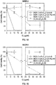
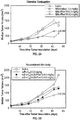

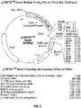

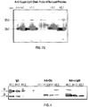


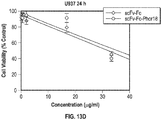

Classifications
-
- A—HUMAN NECESSITIES
- A61—MEDICAL OR VETERINARY SCIENCE; HYGIENE
- A61K—PREPARATIONS FOR MEDICAL, DENTAL OR TOILETRY PURPOSES
- A61K47/00—Medicinal preparations characterised by the non-active ingredients used, e.g. carriers or inert additives; Targeting or modifying agents chemically bound to the active ingredient
- A61K47/50—Medicinal preparations characterised by the non-active ingredients used, e.g. carriers or inert additives; Targeting or modifying agents chemically bound to the active ingredient the non-active ingredient being chemically bound to the active ingredient, e.g. polymer-drug conjugates
- A61K47/51—Medicinal preparations characterised by the non-active ingredients used, e.g. carriers or inert additives; Targeting or modifying agents chemically bound to the active ingredient the non-active ingredient being chemically bound to the active ingredient, e.g. polymer-drug conjugates the non-active ingredient being a modifying agent
- A61K47/68—Medicinal preparations characterised by the non-active ingredients used, e.g. carriers or inert additives; Targeting or modifying agents chemically bound to the active ingredient the non-active ingredient being chemically bound to the active ingredient, e.g. polymer-drug conjugates the non-active ingredient being a modifying agent the modifying agent being an antibody, an immunoglobulin or a fragment thereof, e.g. an Fc-fragment
- A61K47/6801—Drug-antibody or immunoglobulin conjugates defined by the pharmacologically or therapeutically active agent
- A61K47/6803—Drugs conjugated to an antibody or immunoglobulin, e.g. cisplatin-antibody conjugates
- A61K47/6811—Drugs conjugated to an antibody or immunoglobulin, e.g. cisplatin-antibody conjugates the drug being a protein or peptide, e.g. transferrin or bleomycin
- A61K47/6817—Toxins
-
- C—CHEMISTRY; METALLURGY
- C07—ORGANIC CHEMISTRY
- C07K—PEPTIDES
- C07K7/00—Peptides having 5 to 20 amino acids in a fully defined sequence; Derivatives thereof
- C07K7/04—Linear peptides containing only normal peptide links
- C07K7/08—Linear peptides containing only normal peptide links having 12 to 20 amino acids
-
- A—HUMAN NECESSITIES
- A61—MEDICAL OR VETERINARY SCIENCE; HYGIENE
- A61K—PREPARATIONS FOR MEDICAL, DENTAL OR TOILETRY PURPOSES
- A61K47/00—Medicinal preparations characterised by the non-active ingredients used, e.g. carriers or inert additives; Targeting or modifying agents chemically bound to the active ingredient
- A61K47/50—Medicinal preparations characterised by the non-active ingredients used, e.g. carriers or inert additives; Targeting or modifying agents chemically bound to the active ingredient the non-active ingredient being chemically bound to the active ingredient, e.g. polymer-drug conjugates
- A61K47/51—Medicinal preparations characterised by the non-active ingredients used, e.g. carriers or inert additives; Targeting or modifying agents chemically bound to the active ingredient the non-active ingredient being chemically bound to the active ingredient, e.g. polymer-drug conjugates the non-active ingredient being a modifying agent
- A61K47/68—Medicinal preparations characterised by the non-active ingredients used, e.g. carriers or inert additives; Targeting or modifying agents chemically bound to the active ingredient the non-active ingredient being chemically bound to the active ingredient, e.g. polymer-drug conjugates the non-active ingredient being a modifying agent the modifying agent being an antibody, an immunoglobulin or a fragment thereof, e.g. an Fc-fragment
- A61K47/6801—Drug-antibody or immunoglobulin conjugates defined by the pharmacologically or therapeutically active agent
- A61K47/6803—Drugs conjugated to an antibody or immunoglobulin, e.g. cisplatin-antibody conjugates
- A61K47/6811—Drugs conjugated to an antibody or immunoglobulin, e.g. cisplatin-antibody conjugates the drug being a protein or peptide, e.g. transferrin or bleomycin
-
- A—HUMAN NECESSITIES
- A61—MEDICAL OR VETERINARY SCIENCE; HYGIENE
- A61K—PREPARATIONS FOR MEDICAL, DENTAL OR TOILETRY PURPOSES
- A61K47/00—Medicinal preparations characterised by the non-active ingredients used, e.g. carriers or inert additives; Targeting or modifying agents chemically bound to the active ingredient
- A61K47/50—Medicinal preparations characterised by the non-active ingredients used, e.g. carriers or inert additives; Targeting or modifying agents chemically bound to the active ingredient the non-active ingredient being chemically bound to the active ingredient, e.g. polymer-drug conjugates
- A61K47/51—Medicinal preparations characterised by the non-active ingredients used, e.g. carriers or inert additives; Targeting or modifying agents chemically bound to the active ingredient the non-active ingredient being chemically bound to the active ingredient, e.g. polymer-drug conjugates the non-active ingredient being a modifying agent
- A61K47/68—Medicinal preparations characterised by the non-active ingredients used, e.g. carriers or inert additives; Targeting or modifying agents chemically bound to the active ingredient the non-active ingredient being chemically bound to the active ingredient, e.g. polymer-drug conjugates the non-active ingredient being a modifying agent the modifying agent being an antibody, an immunoglobulin or a fragment thereof, e.g. an Fc-fragment
- A61K47/6835—Medicinal preparations characterised by the non-active ingredients used, e.g. carriers or inert additives; Targeting or modifying agents chemically bound to the active ingredient the non-active ingredient being chemically bound to the active ingredient, e.g. polymer-drug conjugates the non-active ingredient being a modifying agent the modifying agent being an antibody, an immunoglobulin or a fragment thereof, e.g. an Fc-fragment the modifying agent being an antibody or an immunoglobulin bearing at least one antigen-binding site
- A61K47/6849—Medicinal preparations characterised by the non-active ingredients used, e.g. carriers or inert additives; Targeting or modifying agents chemically bound to the active ingredient the non-active ingredient being chemically bound to the active ingredient, e.g. polymer-drug conjugates the non-active ingredient being a modifying agent the modifying agent being an antibody, an immunoglobulin or a fragment thereof, e.g. an Fc-fragment the modifying agent being an antibody or an immunoglobulin bearing at least one antigen-binding site the antibody targeting a receptor, a cell surface antigen or a cell surface determinant
-
- A—HUMAN NECESSITIES
- A61—MEDICAL OR VETERINARY SCIENCE; HYGIENE
- A61P—SPECIFIC THERAPEUTIC ACTIVITY OF CHEMICAL COMPOUNDS OR MEDICINAL PREPARATIONS
- A61P31/00—Antiinfectives, i.e. antibiotics, antiseptics, chemotherapeutics
- A61P31/04—Antibacterial agents
-
- A—HUMAN NECESSITIES
- A61—MEDICAL OR VETERINARY SCIENCE; HYGIENE
- A61P—SPECIFIC THERAPEUTIC ACTIVITY OF CHEMICAL COMPOUNDS OR MEDICINAL PREPARATIONS
- A61P31/00—Antiinfectives, i.e. antibiotics, antiseptics, chemotherapeutics
- A61P31/10—Antimycotics
-
- A—HUMAN NECESSITIES
- A61—MEDICAL OR VETERINARY SCIENCE; HYGIENE
- A61P—SPECIFIC THERAPEUTIC ACTIVITY OF CHEMICAL COMPOUNDS OR MEDICINAL PREPARATIONS
- A61P31/00—Antiinfectives, i.e. antibiotics, antiseptics, chemotherapeutics
- A61P31/12—Antivirals
-
- A—HUMAN NECESSITIES
- A61—MEDICAL OR VETERINARY SCIENCE; HYGIENE
- A61P—SPECIFIC THERAPEUTIC ACTIVITY OF CHEMICAL COMPOUNDS OR MEDICINAL PREPARATIONS
- A61P33/00—Antiparasitic agents
-
- A—HUMAN NECESSITIES
- A61—MEDICAL OR VETERINARY SCIENCE; HYGIENE
- A61P—SPECIFIC THERAPEUTIC ACTIVITY OF CHEMICAL COMPOUNDS OR MEDICINAL PREPARATIONS
- A61P35/00—Antineoplastic agents
-
- A—HUMAN NECESSITIES
- A61—MEDICAL OR VETERINARY SCIENCE; HYGIENE
- A61P—SPECIFIC THERAPEUTIC ACTIVITY OF CHEMICAL COMPOUNDS OR MEDICINAL PREPARATIONS
- A61P43/00—Drugs for specific purposes, not provided for in groups A61P1/00-A61P41/00
-
- C—CHEMISTRY; METALLURGY
- C07—ORGANIC CHEMISTRY
- C07K—PEPTIDES
- C07K16/00—Immunoglobulins [IGs], e.g. monoclonal or polyclonal antibodies
- C07K16/18—Immunoglobulins [IGs], e.g. monoclonal or polyclonal antibodies against material from animals or humans
- C07K16/28—Immunoglobulins [IGs], e.g. monoclonal or polyclonal antibodies against material from animals or humans against receptors, cell surface antigens or cell surface determinants
- C07K16/2887—Immunoglobulins [IGs], e.g. monoclonal or polyclonal antibodies against material from animals or humans against receptors, cell surface antigens or cell surface determinants against CD20
-
- C—CHEMISTRY; METALLURGY
- C07—ORGANIC CHEMISTRY
- C07K—PEPTIDES
- C07K16/00—Immunoglobulins [IGs], e.g. monoclonal or polyclonal antibodies
- C07K16/18—Immunoglobulins [IGs], e.g. monoclonal or polyclonal antibodies against material from animals or humans
- C07K16/28—Immunoglobulins [IGs], e.g. monoclonal or polyclonal antibodies against material from animals or humans against receptors, cell surface antigens or cell surface determinants
- C07K16/30—Immunoglobulins [IGs], e.g. monoclonal or polyclonal antibodies against material from animals or humans against receptors, cell surface antigens or cell surface determinants from tumour cells
- C07K16/3015—Breast
-
- C—CHEMISTRY; METALLURGY
- C07—ORGANIC CHEMISTRY
- C07K—PEPTIDES
- C07K16/00—Immunoglobulins [IGs], e.g. monoclonal or polyclonal antibodies
- C07K16/18—Immunoglobulins [IGs], e.g. monoclonal or polyclonal antibodies against material from animals or humans
- C07K16/28—Immunoglobulins [IGs], e.g. monoclonal or polyclonal antibodies against material from animals or humans against receptors, cell surface antigens or cell surface determinants
- C07K16/30—Immunoglobulins [IGs], e.g. monoclonal or polyclonal antibodies against material from animals or humans against receptors, cell surface antigens or cell surface determinants from tumour cells
- C07K16/3069—Reproductive system, e.g. ovaria, uterus, testes, prostate
-
- C—CHEMISTRY; METALLURGY
- C07—ORGANIC CHEMISTRY
- C07K—PEPTIDES
- C07K16/00—Immunoglobulins [IGs], e.g. monoclonal or polyclonal antibodies
- C07K16/18—Immunoglobulins [IGs], e.g. monoclonal or polyclonal antibodies against material from animals or humans
- C07K16/32—Immunoglobulins [IGs], e.g. monoclonal or polyclonal antibodies against material from animals or humans against translation products of oncogenes
-
- C—CHEMISTRY; METALLURGY
- C07—ORGANIC CHEMISTRY
- C07K—PEPTIDES
- C07K19/00—Hybrid peptides, i.e. peptides covalently bound to nucleic acids, or non-covalently bound protein-protein complexes
-
- A—HUMAN NECESSITIES
- A61—MEDICAL OR VETERINARY SCIENCE; HYGIENE
- A61K—PREPARATIONS FOR MEDICAL, DENTAL OR TOILETRY PURPOSES
- A61K38/00—Medicinal preparations containing peptides
-
- C—CHEMISTRY; METALLURGY
- C07—ORGANIC CHEMISTRY
- C07K—PEPTIDES
- C07K2317/00—Immunoglobulins specific features
- C07K2317/50—Immunoglobulins specific features characterized by immunoglobulin fragments
- C07K2317/52—Constant or Fc region; Isotype
- C07K2317/524—CH2 domain
-
- C—CHEMISTRY; METALLURGY
- C07—ORGANIC CHEMISTRY
- C07K—PEPTIDES
- C07K2317/00—Immunoglobulins specific features
- C07K2317/50—Immunoglobulins specific features characterized by immunoglobulin fragments
- C07K2317/52—Constant or Fc region; Isotype
- C07K2317/526—CH3 domain
-
- C—CHEMISTRY; METALLURGY
- C07—ORGANIC CHEMISTRY
- C07K—PEPTIDES
- C07K2317/00—Immunoglobulins specific features
- C07K2317/60—Immunoglobulins specific features characterized by non-natural combinations of immunoglobulin fragments
- C07K2317/62—Immunoglobulins specific features characterized by non-natural combinations of immunoglobulin fragments comprising only variable region components
- C07K2317/622—Single chain antibody (scFv)
-
- C—CHEMISTRY; METALLURGY
- C07—ORGANIC CHEMISTRY
- C07K—PEPTIDES
- C07K2319/00—Fusion polypeptide
- C07K2319/70—Fusion polypeptide containing domain for protein-protein interaction
- C07K2319/74—Fusion polypeptide containing domain for protein-protein interaction containing a fusion for binding to a cell surface receptor
-
- C—CHEMISTRY; METALLURGY
- C07—ORGANIC CHEMISTRY
- C07K—PEPTIDES
- C07K2319/00—Fusion polypeptide
- C07K2319/95—Fusion polypeptide containing a motif/fusion for degradation (ubiquitin fusions, PEST sequence)
Definitions
- the present disclosure relates to antibodies, and Heavy (H) chains and/or Light (L) chains of antibodies, and antibody fragments fused or conjugated to drugs, such as lytic peptide conjugates, methods of using conjugates, for example, in methods of treating undesirable or aberrant cell proliferation or hyperproliferative disorders, such as non-metastatic and metastatic neoplasias, cancers, tumors and malignancies that express targets that bind to such antibodies, and Heavy (H) chains and/or Light (L) chains of antibodies.
- drugs such as lytic peptide conjugates
- WO 2011/137245 A2 discloses an anti-Her2/neu antibody conjugated to the lytic peptide Phor-18, KFAKFAK KFAKFAK KFAK.
- WO 2011/031477 A2 discloses an anti-nucleolin antibody conjugated to the lytic peptide Phor-18, KFAKFAK KFAKFAK KFAK.
- the present invention relates to an antibody conjugate, comprising an antibody that binds to a target, wherein the antibody is linked to a lytic domain, wherein said lytic domain comprises or consists of a peptide selected from KFAKFAKKFAKFAKK, KFAKFAKKFAKFAKKF, KFAKFAKKFAKFAKKFA, KFAKFAKKFAKFAK, KFAKFAKKFAKFAKF and KFAKFAKKFAKFAKFA; wherein the lytic domain is linked to a Heavy (H) chain of the antibody, or linked to a Light (L) chain of the antibody, and wherein said target is CD20.
- the invention is based, at least in part on lytic domains fused to an antibody, lytic domains fused or conjugated to Heavy (H) chains and/or Light (L) chains of antibodies, and lytic domains fused or conjugated to antibody fragments, that bind to a target.
- lytic domains fused to an antibody
- lytic domains fused or conjugated to Heavy (H) chains and/or Light (L) chains of antibodies and lytic domains fused or conjugated to antibody fragments, that bind to a target.
- Such fusions can also be referred to herein as antibody or polypeptide conjugates or fusion constructs.
- the antibody, or Heavy (H) chain and/or Light (L) chain of an antibody that binds to the target allows the lytic domain to target expressing cells for destruction, including undesirable or aberrant proliferating cells or hyperproliferating cells, such as non-metastatic and metastatic neoplasias, cancers, tumors and malignancies, that express the target to to which the antibody or Heavy (H) chain and/or Light (L) chain binds.
- a number of non-metastatic and metastatic neoplastic, cancer, tumor and malignant cells overexpress targets, such as receptors or ligands.
- targets such as receptors or ligands.
- many non-metastatic and metastatic neoplasias, cancers, tumors and malignancies express the receptor target CD20 that is used as a target of the antibody or polypeptide conjugate or fusion construct.
- Conjugates can be designed to bind to any cell or cell population that expresses a target of interest.
- An antibody, fragment thereof, or Heavy (H) chain and/or Light (L) chain of an antibody or fragment thereof, selected based upon the target to which it binds, can be linked to a lytic domain.
- the resulting antibody or polypeptide conjugate or fusion construct can in turn reduce or inhibit proliferation of cells that express or target, thereby reducing or inhibiting proliferation or growth of the target expressing cells.
- Conjugates do not require cells to divide in order to kill the target cells. Accordingly, conjugates are useful against dividing and non-dividing cells.
- the target is CD20 and the lytic domain comprises or consists of a peptide selected selected from KFAKFAKKFAKFAKK, KFAKFAKKFAKFAKKF, KFAKFAKKFAKFAKKFA, KFAKFAKKFAKFAKF and KFAKFAKKFAKFAKFA. All other targets or lytic domains mentioned in the description do not fall under the scope of the claimed invention.
- an antibody conjugate includes or consists of an antibody (or fragment thereof) that binds to a target, linked to a lytic domain that includes or consists of an L- or D-amino acid sequence that includes a peptide sequence selected from KFAKFAKKFAKFAKK.
- KFAKFAKKFAKFAKKF, KFAKFAKKFAKFAKKFA, KFAKFAKKFAKFAKFAK, KFAKFAKKFAKFAKFAKF, KFAKFAKKFAKFAKFAKFA and KFAKFAKKFAKFAKFAKFAKFAKFAKFAKFAKFAK (SEQ ID NOs.: 1-7).
- a polypeptide conjugate includes or consists of a Heavy (H) chain and/or Light (L) chain of an antibody or fragment thereof that binds to a target, linked to a lytic domain that includes or consists of an L- or D-amino acid sequence selected from for example, KFAKFAKKFAKFAKK, KFAKFAKKFAKFAKKF, KFAKFAKKFAKFAKKFAK, KFAKFAKKFAKFAKFAKF, KFAKFAKKFAKFAKFAKFA and KFAKFAKKFAKFAKFAKFAKFAKFAKFAKFAK (SEQ ID NOs.: 1-7).
- a second domain may include or consist of a lytic domain: KFAKFAKKFAKFAKK, KFAKFAKKFAKFAKKF, KFAKFAKKFAKFAKKFA, KFAKFAKKFAKFAK, KFAKFAKKFAKFAKF or KFAKFAKKFAKFAKFA (SEQ ID NOs.: 1-7).
- a polypeptide includes or consists of a: 1) lytic domain linked to the amino(NH 2 )-terminus of an antibody Heavy (H) chain; 2) a lytic domain linked to the amino(NH 2 )-terminus of an antibody Light (L) chain; 3) a lytic domain linked to a carboxy(C)-terminus of the antibody Heavy (H) chain; or 4) a lytic domain linked to a carboxy(C)-terminus of the antibody Light (L) chain.
- a polypeptide includes or consists of a: 1) lytic domain linked to an amino(NH 2 )-terminus and a lytic domain linked to a carboxy(C)-terminus of an antibody Heavy (H) chain; or 2) a lytic domain linked to the amino(NH 2 )-terminus and a lytic domain linked to a carboxy(C)-terminus of an antibody Light (L) chain.
- targets include amino acid sequences (e.g., polypeptides, peptides, proteins), polysaccharides, oligosaccharides, carbohydrates, and lipids.
- Specific non-limiting classes of targets include receptors and antigens.
- Targets include receptors that bind to antigens, receptors or ligands, including hormones, growth factors, cluster of differentiation (collectively known as CD molecules or CD markers), hormone and growth factor analogues, and fragments of hormones, hormone analogs, growth factors, growth factor analogues, and fragments of growth factors and analogues.
- receptor targets include Her2/neu (Human Epidermal growth factor Receptor 2, also known as ErbB-2), luteinizing hormone releasing hormone receptor (LHRH-R), epidermal growth factor (EGF) receptor, folate, and growth hormone (GH) receptor.
- CD domains include CD19, CD20, CD22, CD23, CD27, CD28, CD30, CD31, CD33, CD34, CD40, CD52, CD56, CD70, CD123, CD138, or CD154, and others. According to the claims the target is CD20. All other targets mentioned in the description do not fall under the scope of the claimed invention.
- Antigen targets include viral, bacterial, fungal and parasite antigens. Antigen targets also include tumor associated antigens (TAAs).
- TAAs tumor associated antigens
- An antibody includes 2 Heavy (H) chains and 2 Light (L) chains as well as antibody fragments.
- a polypeptide that includes or consists of a Heavy (H) chain and/or Light (L) chain of an antibody or fragment of a Heavy (H) chain or Light (L) chain can include a single H or L chain or a single H or L chain fragment, or a plurality (2, 3, 4 or more) of Heavy (H) chains and/or Light (L) chains, or a plurality of fragments of Heavy (H) chains and/or Light (L) chains.
- a polyeptide that includes a Heavy (H) chain and/or Light (L) chain of an antibody or fragment can but is not required to include 2 Heavy (H) chains and 2 Light (L) chains and therefore polypeptide conjugates as set forth herein can exclude native antibodies that comprise 2 Heavy (H) chains and 2 Light (L) chains.
- An antibody or fragment thereof may be an oligomeric (higher order or valent) forms, such as a trimer, tetramer, pentamer, hexamer, heptamer, and so forth, with other antibodies, fragments thereof, Heavy (H) chain, Light (L) chain, or polyeptides sequence distinct from an antibody Heavy (H) or Light (L) chain.
- Antibodies include monoclonals and fragments of monoclonal antibodies. Antibodies include mammalian, human, humanized, primatized and chimeric sequences.
- Amino acid sequences include or consist of natural (L-) or non-natural (e.g., D-) amino acids.
- an amino acid sequence has about 2 to 10, 10 to 14, 15 to 20, (i.e., 15, 16, 17, 18, 19 or 20 amino acids), 10 to 20, 20 to 30, 30 to 40, 40 to 50, 60 to 70, 70 to 80, 80 to 90, 90 to 100, 100 to 125, 125 to 150, 150 to 175, 175 to 200, 200 to 250, 250 to 300, or more amino acid residues.
- Full-length antibody Heavy (H) chains, Light (L) chains are typically 90 to 130 amino acids in length, but may be shorter, for example, comprise a variable Heavy (H) chain, or Light (L) chain sequence, comprising one, two, or three complemetarity determining regions (CDRs) with or without framework regions.
- Lytic domains are typically 10 to 14, 15 to 20, (i.e., 15, 16, 17, 18, 19 or 20 amino acids), 10 to 20, 20 to 30, 30 to 40, or 40 to 50, but may optionally be longer (50 or more) or shorter (less than 10).
- An amino acid sequence can include or consist of a linear or cyclic structure.
- Conjugates that include lytic domains can have the lytic domain at any location of the antibody (or fragment thereof) or Heavy (H) chain or Light (L) chain of an antibody.
- a lytic domain can be positioned at any amino acid position (amino acid residue) of the antibody (or fragment thereof) or Heavy (H) chain or Light (L) chain of an antibody.
- a conjugate can include multiple lytic domains. Accordingly, one or more (e.g., two, three, four, five, six, seven, eight or more) lytic domains linked to the Heavy (H) chain or Light (L) chain.) lytic domains can be included in a conjugate of the disclosure.
- Lytic domains can also be positioned at the C-terminus, the NH 2 -terminus, or both the C-terminus and the NH 2 -terminus of a polyeptide sequence.
- a conjugate has a lytic domain positioned at either (or both) the NH 2 -terminus or the C-terminus of the antibody (or fragment thereof) Heavy (H) chain or Light (L) chain, or Heavy (H) chain or Light (L) chain.
- a lytic domain is linked to the amino(NH 2 )-terminus of the Heavy (H) chain or linked to the carboxy(C)-terminus of the Heavy (H) chain; a lytic domain is linked to the amino(NH 2 )-terminus of the Light (L) chain or linked to the carboxy(C)-terminus of the Light (L) chain.
- such domains can be linked to the Heavy (H) chain or Light (L) chain, to the amino(NH 2 )-terminus of the Heavy (H) chain, to the carboxy(C)-terminus of the Heavy (H) chain, to the amino(NH 2 )-terminus of the Light (L) chain, or to the carboxy(C)-terminus of the Light (L) chain.
- At least one of a plurality of lytic domains is linked to the amino(NH 2 )-terminus of the Heavy (H) chain, and at least one is linked to the amino(NH 2 )-terminus of the Light (L) chain; at least one of the lytic domains is linked to the amino(NH 2 )-terminus of the Heavy (H) chain, at least one of the lytic domains is linked to the amino(NH 2 )-terminus of the Light (L) chain, and at least one of the lytic domains is linked to the carboxy(C)-terminus of the Heavy (H) chain or is linked to the carboxy(C)-terminus of the Light (L) chain; and at least one of a plurality of lytic domains is linked to the amino(NH 2 )-terminus of the Heavy (H) chain, at least one of the lytic domains is linked to the amino(NH 2 )-terminus of the Light (L) chain, at least one of the lytic domains is linked to the carboxy(
- conjugates include a plurality of lytic domains
- the lytic domains have an identical amino acid sequence, or have a different amino acid sequence.
- a conjugate can include two, a third, fourth, fifth, sixth, seventh lytic domain, etc., any or all of which may be identical or different from each other.
- a lytic domain is joined to a Heavy (H) chain or Light (L) chain immediately after the last amino acid at the amino(NH 2 )-terminus or the carboxy(C)-terminus of the Heavy (H) chain or the Light (L) chain, for example, by a covalent (e.g., peptide or nonpeptide) bond thereby forming a continuous amino acid sequence between the lytic domain and the Heavy (H) chain or Light (L) chain.
- antibodies, Heavy (H) chains and Light (L) chains and lytic domains can be joined by a peptide or a non-peptide linker or spacer.
- antibodies, Heavy (H) chains and Light (L) chains and lytic domains and lytic domains are joined by a peptide sequence that includes or consist of one or more A, S or G amino acid residues (e.
- Conjugates further include or consist of additional (e.g., non-lytic) domains.
- a conjugate includes a second, third, fourth, fifth, sixth, seventh domain, etc., which may be distinct from one or more (or all) lytic domains included in the conjugate.
- Conjugates include or consist of isolated and/or purified forms. Conjugates also include or consist of a formulation or a mixture. Such formulations and mixtures include compositions, such as a mixture of conjugate and a pharmaceutically acceptable carrier or excipient appropriate for use, administration to or in vivo contact with a subject, or a mixture of conjugate and an anti-cell proliferative or immune stimulating agent.
- Conjugates include or consist of a unit dosage form, such as a dosage form for use or administration to a subject.
- a conjugate may be a unit dosage to administer or in an amount effective to or treat a subject having undesirable cell proliferation or a hyperproliferative disorder.
- a conjugate may be a unit dosage to administer or in an amount effective to treat a subject having a neoplasia, tumor or cancer.
- a conjugate may be a unit dosage to administer or in an amount effective to reduce fertility of a subject.
- kits can include a conjugate and instructions for reducing or inhibiting proliferation of a cell, reducing or inhibiting proliferation of a hyperproliferating cell, reducing or inhibiting proliferation of a neoplastic, tumor or cancer cell, treating a subject having a hyperproliferative disorder, treating a subject having a neoplasia, tumor or cancer, or reducing fertility of an animal.
- nucleic acids that encode conjugates.
- a nucleic acid sequence encodes a: 1) lytic domain linked to the amino(NH 2 )-terminus of the antibody Heavy (H) chain; 2) a lytic domain linked to the amino(NH 2 )-terminus of the antibody Light (L) chain; 3) a lytic domain linked to the carboxy(C)-terminus of the antibody Heavy (H) chain; or 4) a lytic domain linked to the carboxy(C)-terminus of the antibody Light (L) chain, of an antibody or polypeptide conjugate.
- a nucleic acid sequence encodes a: 1) lytic domain linked to the amino(NH 2 )-terminus and a lytic domain linked to the carboxy(C)-terminus of the antibody Heavy (H) chain; 2) a lytic domain linked to the amino(NH 2 )-terminus and a lytic domain linked to the carboxy(C)-terminus of the antibody Light (L) chain, of the antibody or polypeptide conjugate.
- Nucleic acids can be included in a vector, such as an expression vector that when expressed in a cell encodes a conjugate.
- Host cells can be transformed with a nucleic acid (e.g., encoding all or a portion of a conjugate, e.g., a Heavy (H) chain and/or Light (L) chain sequence linked to a lytic domain) in a vector, such that the cell expresses a conjugate encoded by the nucleic acid.
- a nucleic acid e.g., encoding all or a portion of a conjugate, e.g., a Heavy (H) chain and/or Light (L) chain sequence linked to a lytic domain
- targets can be expressed in or on a cell.
- Cells that express a target to which a conjugate binds can be targeted for binding by the conjugates described herein. Accordingly, such cells can be selectively targeted by selecting a conjugate that binds to a target expressed by the cells.
- Non-limiting target expressing cells include, for example, hyperproliferative cells. Additional cells that express targets include, for example, breast, ovarian, uterine, cervical, stomach, lung, gastric, colon, bladder, glial, dermal (e.g., melanocytes), hematologic and endometrial cells.
- targets include, for example, breast, ovarian, uterine, cervical, stomach, lung, gastric, colon, bladder, glial, dermal (e.g., melanocytes), hematologic and endometrial cells.
- Conjugates are useful for, among other things, reducing or inhibiting proliferation of a cell, reducing or inhibiting cell proliferation, reducing or inhibiting proliferation of a hyperproliferating cell, reducing or inhibiting proliferation of a neoplastic, tumor, cancer or malignant cell and treating undesirable or aberrant cell proliferation, such as hyperproliferating cells or hyperproliferative disorders.
- hyperproliferative disorders include benign hyperplasia, non-metastatic and metastatic neoplasias, cancers tumors and malignancies.
- references in the description to methods of treatment refer to the compounds, pharmaceutical compositions and medicaments of the present invention for use in a method for treatment of the human (or animal) body by therapy. Described herein are also methods of reducing or inhibiting proliferation of a cell; methods of reducing or inhibiting cell proliferation; methods of reducing or inhibiting proliferation of a hyperproliferating cell; and methods of reducing or inhibiting proliferation of a neoplastic, tumor, cancer or malignant cell.
- a method may include contacting a cell with a conjugate in an amount sufficient to reduce or inhibit proliferation of the cell; contacting a cell with a conjugate in an amount sufficient to reduce or inhibit cell proliferation; contacting a cell with a conjugate in an amount sufficient to reduce or inhibit proliferation of the hyperproliferating cell; and contacting a cell with a conjugate in an amount sufficient to reduce or inhibit proliferation of the neoplastic, tumor, cancer or malignant cell.
- a method may include contacting a target expressing cell with a conjugate in an amount sufficient to reduce or inhibit proliferation of the cell; contacting a target expressing cell with the conjugate in an amount sufficient to reduce or inhibit proliferation of the hyperproliferating cell; and contacting a target expressing cell with a conjugate in an amount sufficient to reduce or inhibit proliferation of the neoplastic, tumor, cancer or malignant cell, wherein the conjugate binds to a target expressed by the cell.
- Exemplary cells to be targeted in accordance with the uses and methods as described herein include cells that express any desired target.
- Such non-limiting cells therefore include, for example, breast, ovarian, uterine, cervical, stomach, lung, gastric, colon, bladder, glial, dermal (e.g., melanocytes), hematologic and endometrial cells.
- More particular non-limiting cells express a receptor, such as Her2/neu, an antigen (e.g., a tumor associated antigen) or a cluster of differentiation domain (CD).
- a receptor such as Her2/neu, an antigen (e.g., a tumor associated antigen) or a cluster of differentiation domain (CD).
- an antigen e.g., a tumor associated antigen
- CD cluster of differentiation domain
- Methods being also described herein include, among others, administering to or contacting a subject in need of inhibiting, reducing or preventing proliferation, survival, differentiation, death, or activity of a cell, such as a hyperprolifertive cell or an undesirably proliferating cell.
- Exemplary subjects include a subject having or at risk of having undesirable or aberrant cell proliferation; a subject having or at risk of having a benign hyperplasia; or a non-metastatic or metastatic neoplasia, cancer, tumor or malignancy (e.g., a solid or liquid tumor, in any of breast, ovarian, uterine, cervical, stomach, lung, gastric, colon, bladder, glial, dermal (e.g., melanocytes), hematologic or endometrial cells).
- a subject having or at risk of having undesirable or aberrant cell proliferation e.g., a benign hyperplasia
- a non-metastatic or metastatic neoplasia, cancer, tumor or malignancy e.g., a solid or liquid tumor, in any of breast, ovarian, uterine, cervical, stomach, lung, gastric, colon, bladder, glial, dermal (e.g., melanocytes), hematologic or endometrial cells).
- a use or method may include administering to a subject an amount of the conjugate sufficient to treat the hyperproliferative disorder; and administering to a subject an amount of the conjugate sufficient to reduce or inhibit proliferation of the neoplasia, tumor, cancer or malignancy.
- Methods and uses include treating a subject having or at risk of having a metastasis. For example, an amount of a conjugate effective to reduce or inhibit spread or dissemination of a tumor, cancer or neoplasia to other sites, locations or regions within the subject.
- a method or use may reduce or inhibit metastasis of a primary tumor or cancer to one or more other sites, formation or establishment of a metastasis at one or more other sites, locations or regions thereby reducing or inhibiting tumor or cancer relapse or tumor or cancer progression.
- a method or use may reduce or inhibit growth, proliferation, mobility or invasiveness of tumor or cancer cells that potentially or do develop metastases (e.g., disseminated tumor cells); may reduce or inhibit formation or establishment of metastases arising from a primary tumor or cancer to one or more other sites, locations or regions distinct from the primary tumor or cancer; may reduce or inhibit growth or proliferation of a metastasis at one or more other sites, locations or regions distinct from the primary tumor or cancer after the metastasis has formed or has been established; or may reduce or inhibit formation or establishment of additional metastasis after the metastasis has been formed or established.
- a method or use may reduce or inhibit relapse or progression of the neoplasia, tumor, cancer or malignancy.
- methods and uses of reducing or inhibiting metastasis of a neoplasia, tumor, cancer or malignancy to other sites, or formation or establishment of metastatic neoplasia, tumor, cancer or malignancy at other sites distal from a primary neoplasia, tumor, cancer or malignancyA method may include administering to a subject an amount of the conjugate sufficient to reduce or inhibit metastasis of the neoplasia, tumor, cancer or malignancy to other sites, or formation or establishment of metastatic neoplasia, tumor, cancer or malignancy at other sites distal from the primary neoplasia, tumor, cancer or malignancy.
- Neoplasia, tumor, cancer and malignancy treatable in accordance with the disclosure therefore include metastatic, and non-metastatic or benign forms.
- Non-limiting examples include a solid cellular mass, hematopoietic cells, or a carcinoma, sarcoma (e.g.
- lymphosarcoma liposarcoma, osteosarcoma, chondrosarcoma, leiomyosarcoma, rhabdomyosarcoma or fibrosarcoma
- lymphoma leukemia, adenoma, adenocarcinoma, melanoma, glioma, glioblastoma, meningioma, neuroblastoma, retinoblastoma, astrocytoma, oligodendrocytoma, mesothelioma, reticuloendothelial, lymphatic or haematopoietic (e.g., myeloma, lymphoma or leukemia) neoplasia, tumor, cancer or malignancy.
- lymphatic or haematopoietic e.g., myeloma, lymphoma or leukemia
- Neoplasia, tumor, cancer and malignancy treatable in accordance with the disclosure can be present in or affect a lung (small cell lung or non-small cell lung cancer), thyroid, head or neck, nasopharynx, throat, nose or sinuses, brain, spine, adrenal gland, pituitary gland, breast, ovarian, uterine, cervical, gastrointestinal (mouth, esophagus, stomach, duodenum, ileum, jejunum (small intestine), colon, rectum), lung, genito-urinary tract (uterus, ovary, cervix, endometrial, bladder, testicle, penis, prostate), glial, hematologic, endometrial, lymph, blood, muscle, dermal (e.g., melanocytes) or skin cell, kidney, pancreas, liver, bone, bone marrow, Wilm's tumors, biliary tract, B-ALL (B-cell lymphoblastic leukemia), stem cell, or he
- Methods and uses may be practiced alone, for example, in subjects that are not candidates for other therpaies (e.g., surgical resection, chemotherapy, immunotherapy, radiotherapy, thermal therapry, vaccination, etc.). Methods and uses may also be practiced with other treatments or therapies (e.g., surgical resection, radiotherapy, ionizing or chemical radiation therapy, chemotherapy, immunotherapy, local or regional thermal (hyperthermia) therapy, or vaccination). Such treatments or therapies can be administered prior to, substantially contemporaneously with (separately or in a mixture), or following administration of a conjugate.
- a method or use may include administering an anti-cell proliferative, anti-neoplastic, anti-tumor, anti-cancer or immune-enhancing treatment or therapy.
- a method or use may include administering an alkylating agent, anti-metabolite, plant extract, plant alkaloid, nitrosourea, hormone, nucleoside or nucleotide analog; cyclophosphamide, azathioprine, cyclosporin A, prednisolone, melphalan, chlorambucil, mechlorethamine, busulphan, methotrexate, 6-mercaptopurine, thioguanine, 5-fluorouracil, cytosine arabinoside , 5-azacytidine (5-AZC) and 5-azacytidine related compounds, bleomycin, actinomycin D, mithramycin, mitomycin C, carmustine, lomustine, semustine, streptozotocin, hydroxyurea, cisplatin, carboplatin, oxiplatin, mitotane, procarbazine, dacarbazine, a taxane (e.g., taxo
- Cell or immunotherapies include lymphocytes, plasma cells, macrophages, dendritic cells, T-cells, NK cells or B-cells; an antibody, a cell growth factor, a cell survival factor, a cell differentiative factor, a cytokine or a chemokine.
- Additional agents that are applicable with conjugates in compostions, methods or uses described herein include targeted drugs or biologicals, such as antibodies (monoclonal) or small molecules.
- Methods described herein include providing a subject with a benefit.
- a method of treatment may result in partial or complete destruction of the neoplastic, tumor, cancer or malignant cell mass, volume, size or numbers of cells, stimulating, inducing or increasing neoplastic, tumor, cancer or malignant cell necrosis, lysis or apoptosis, reducing neoplasia, tumor, cancer or malignancy volume size, cell mass, inhibiting or preventing progression or an increase in neoplasia, tumor, cancer or malignancy volume, mass, size or cell numbers, or prolonging lifespan; may result in reducing or decreasing severity, duration or frequency of an adverse symptom or complication associated with or caused by the neoplasia, tumor, cancer or malignancy; may result in reducing or decreasing pain, discomfort, nausea, weakness or lethargy; or may result in increased energy, appetite, improved mobility or psychological well being.
- Subjects treatable in accordance with the methods described herein include mammals.
- a subject may be a human.
- a conjugate that includes a portion that binds to a target joined or fused to a second lytic domain as defined by the claims.
- a conjugate includes a first target binding domain (e.g., an antibody, or a Heavy (H) chain and/or Light (L) chain of an antibody) and a second domain that includes a lytic portion, which is/are directly or indirectly toxic to a cell, which can thereby reduce cell proliferation or survival, or stimulate, induce, increase or enhance cell death, killing or apoptosis.
- a first target binding domain e.g., an antibody, or a Heavy (H) chain and/or Light (L) chain of an antibody
- a second domain that includes a lytic portion, which is/are directly or indirectly toxic to a cell, which can thereby reduce cell proliferation or survival, or stimulate, induce, increase or enhance cell death, killing or apoptosis.
- conjugates that include or consist of an antibody, or a Heavy (H) chain and/or Light (L) chain of an antibody, that bind to a target, and a second lytic or toxic domain.
- a conjugate may include a Heavy (H) chain and/or Light (L) chain of an antibody and a lytic domain comprising or consisting of a 10-100 residue L- or D-amino acid sequence selected from KFAKFAKKFAKFAKK, KFAKFAKKFAKFAKKF, KFAKFAKKFAKFAKKFAK, KFAKFAKKFAKFAKF, KFAKFAKKFAKFAKFA and KFAKFAKKFAKFAKFAKFAKFAKFAKFAKFAK (SEQ ID NOs.: 1-7).
- conjugate or “fusion construct” and grammatical variations thereof, means that the construct contains portions or sections that are derived from, obtained or isolated from, or are based upon or modeled after two different molecular entities that are distinct from each other and do not typically exist together in nature. That is, for example, a first portion of the conjugate includes or consists of an antibody or antibody fragment, or a Heavy (H) chain and/or Light (L) chain of an antibody, and a second portion of the conjugate includes or consists of a lytic portion or domain, each of the first and second portions/domains structurally distinct.
- a conjugate can also be referred to as a "fusion construct," wherein the conjugate includes or consists of a of an antibody or antibody fragment, or a Heavy (H) chain and/or Light (L) chain of an antibody, that bind to a target, and a second lytic domain or portion.
- the conjugate includes or consists of a of an antibody or antibody fragment, or a Heavy (H) chain and/or Light (L) chain of an antibody, that bind to a target, and a second lytic domain or portion.
- First domains and or second (lytic) domains of conjugates include or consist of amino acid sequences (peptides, polypeptides, proteins, lectins), nucleic acids (DNA, RNA) and carbohydrates (saccharides, sialic acid, galactose, mannose, fucose, acetylneuraminic acid, etc.).
- amino acid sequences peptides, polypeptides, proteins, lectins
- nucleic acids DNA, RNA
- carbohydrates saccharide, sialic acid, galactose, mannose, fucose, acetylneuraminic acid, etc.
- Amino acid sequences can be linked by non-natural and non-amide chemical bonds including, for example, those formed with glutaraldehyde, N-hydroxysuccinimide esters, bifunctional maleimides, or N, N'-dicyclohexylcarbodiimide (DCC).
- Non-amide bonds include, for example, ketomethylene, aminomethylene, olefin, ether, thioether and the like (see, e.g., Spatola in Chemistry and Biochemistry of Amino Acids, Peptides and Proteins, Vol. 7, pp 267-357 (1983), "Peptide and Backbone Modifications," Marcel Decker, NY ).
- First and second (lytic) domains of a conjugate or fusion construct include L-amino acid sequences, D-amino acid sequences and amino acid sequences with mixtures of L-amino acids and D-amino acids.
- Conjugates of amino acid sequences of first and second domains can be a linear or a cyclic structure, and can be further conjugated to another distinct moiety (e.g., third, fourth, fifth, sixth, seventh, etc.
- Exemplary lengths of conjugates are from about 10 to 15, 15 to 20, 20 to 25, 25 to 50, 50 to 100, 100 to 150, 150 to 200, or 200 to 300 or more amino acid residues in length.
- a first or second domain may include or consist of an amino acid sequence of about 1 to 10, 10 to 20, 15 to 20, 20 to 30, 30 to 40, 40 to 50, 60 to 70, 70 to 80, 80 to 90, 90 to 100 or more residues.
- a lytic domain may include or consist of a 10, 11, 12, 13, 14, 15, 16, 17, 18, 19, 20, 21, 22, 23, 24, 25, 26, 27, 28, or more residue amino acid sequence, or a 10, 11, 12, 13, 15, 16, 17, 18, 19, 20, 22, 23, 24, 25, 26, 27, 28, or more residue amino acid sequence.
- Conjugate that includes or consists of a first portion antibody or antibody fragment, or a Heavy (H) chain and/or Light (L) chain of an antibody, where the first portion binds to a target (e.g., receptor), and a second portion that includes or consists of a lytic domain, the lytic domain can form an amphipathic alpha-helix.
- An amphipathic alpha-helix contains mostly hydrophilic amino acids on one side of the alpha-helix and the other side contains mostly hydrophobic amino acids. Since the alpha helix makes a complete turn for every 3.6 residues, the amino acid sequence of an amphipathic alpha helix alternates between hydrophilic and hydrophobic residues every 3 to 4 residues.
- a PNNPNNP repeat pattern or motif is predicted to form an amphipathic alpha-helix where P represents a positively charged amino acid residue and N a neutral amino acid residue.
- a PNNPNNP repeat pattern provides a cationic binding site for the lytic peptide to interact with a negatively charged cell membrane and a hydrophobic site for membrane interaction/penetration.
- Conjugates therefore include that with lytic domains having one or more uninterrupted PNNPNNP repeat patterns or motifs, or one or more interrupted PNNPNNP repeat patterns or motifs, which can form an amphipathic alpha-helix.
- a 15 or 18 residue amino acid sequence such as KFAKFAKKFAKFAKK (SEQ ID NO.:1) and KFAKFAKKFAKFAKKFAK (SEQ ID NO.:4), has uninterrupted and interrupted PNNPNNP repeat motifs.
- conjugates include antibodies and antibody fragments that bind to a target.
- An "antibody” refers to any monoclonal or polyclonal immunoglobulin molecule, such as IgM, IgG, IgA, IgE, IgD, and any subclass thereof.
- Exemplary subclasses for IgG are IgG 1 , IgG 2 , IgG 3 and IgG 4 including engineered antibody subclasses and recombinant antibodies with various glycosylation patterns.
- Conjugates also include a Heavy (H) chain or a Light (L) chain of an antibody, or a fragment of a Heavy (H) chain or a Light (L) chain of an antibody, that bind to a target.
- Such conjugates can have a plurality of Heavy (H) chains and/or Light (L) chains of an antibody, or a fragment of Heavy (H) chains and/or Light (L) chains of an antibody, that bind to a target.
- Antibody fragments, and fragments of Heavy (H) chains and/or Light (L) chains of an antibody include the hypervariable (target binding) region, or any or all of the complementarity determining regions (CDRs) or framework regions (FRs) within a Heavy (H) chain and/or Light (L) chain of an antibody, sufficient to confer target binding.
- CDRs complementarity determining regions
- FRs framework regions
- antibody fragments include Fab, Fab', F(ab') 2 , Fv, Fd, single-chain Fv (scFv), disulfide-linked Fvs (sdFv), V L , V H , Camel Ig, V-NAR, VHH, trispecific (Fab 3 ), bispecific (Fab 2 ), diabody ((V L -V H ) 2 or (V H -V L ) 2 ), triabody (trivalent), tetrabody (tetravalent), minibody ((scF v -C H 3) 2 ), bispecific single-chain Fv (Bis-scFv), IgGdeltaCH2, scFv-Fc, (scFv) 2 -Fc, affibody (e.g., ZHer2-neu:2, ZHer2-neu:4 ZHer2-neu:7 ZHer2-neu:8), aptamer, avimer or nanobody.
- Fab 3 bispecific bispecific (Fab 2
- Antibodies, Heavy (H) chains and Light (L) chains of an antibody, and fragments thereof include those produced by or expressed on cells, such as B cells, or synthesized or engineered to be produced by other cells, e.g., CHO cells.
- Such antibodies, Heavy (H) chains and Light (L) chains of an antibody, and fragments thereof include those with improved characteristics, such as increased serum stability and/or half life in vivo, PK, etc. (e.g., as described in Antibody Engineering Vol 1, Konterman R and Duebel S, eds., 2010, Springer , WO 2006/130834 and Horton et al., Cancer Res 68:8049 (2008 )).
- Non-limiting mutations in the Fc include, for example, I253A, H310A, H435 R, H435Q, G236R, L328 R, S239D, I332E.
- Non-limiting mutations in IgGl can be at residues 238, 252, 253, 254, 255, 256, 265, 272, 289, 288, 303, 305, 307, 309, 311, 312, 317, 340, 356, 360, 362, 376, 378, 380, 382, 386, 388, 400, 413, 415, 424, 433, 434, 435, 439 and/or 477 of the Fc region.
- Targets include amino acid sequences (e.g., polypeptides, peptides, proteins), polysaccharides, oligosaccharides, carbohydrates, and lipids.
- targets include receptors and antigens. Such targets can be expressed by or on a cell (e.g., on the cell membrane).
- Targets include receptors that bind to hormones, growth factors, hormone and growth factor analogues, and fragments of hormones, hormone analogs, growth factors, growth factor analogues, and fragments of growth factors and analogues.
- receptor targets include Her2/neu (Human Epidermal growth factor Receptor 2, also known as ErbB-2), luteinizing hormone releasing hormone receptor (LHRH-R), epidermal growth factor receptor (EGF-R), folate-, and growth hormone (GH) receptor.
- receptor targets include a tumor necrosis factor (TNF) family member receptor (e.g., TNF-alpha, TNF-beta (lymphtoxin, LT), TRAIL, Fas, LIGHT, or 4-1BB) or oncofetoprotein (5T4).
- TNF tumor necrosis factor
- family member receptor e.g., TNF-alpha, TNF-beta (lymphtoxin, LT), TRAIL, Fas, LIGHT, or 4-1BB
- TNF4 tumor necrosis factor 4-1BB
- TNF-alpha e.g., TNF-alpha, TNF-beta (lymphtoxin, LT), TRAIL, Fas, LIGHT, or 4-1BB
- T4 oncofetoprotein
- receptor targets include an immunoglobulin-like receptor (e.g., CD19, CD20, CD22, CD23, CD27, CD28, CD30, CD31, CD33, CD34, CD40, CD52, CD56, CD70, CD123, CD138, CD123, CD138, or CD154),or other receptors, (e.g., hormone receptor, a cytokine receptor, a growth factor receptor, or a chemokine receptor).
- immunoglobulin-like receptor e.g., CD19, CD20, CD22, CD23, CD27, CD28, CD30, CD31, CD33, CD34, CD40, CD52, CD56, CD70, CD123, CD138, CD123, CD138, or CD154
- other receptors e.g., hormone receptor, a cytokine receptor, a growth factor receptor, or a chemokine receptor.
- Antigen targets include viral, bacterial, fungal and parasite antigens. Antigen targets also include tumor associated antigens (TAAs). Particular non-limiting examples of TAA targets include carcinoembryonic antigen (CEA), alpha-fetoprotein (AFP), prostate specific antigen (PSA), prostate specific membrane antigen (PSMA), CA- 125 (epithelial ovarian cancer), soluble Interleukin-2 (IL-2) receptor, RAGE-1, tyrosinase, MAGE-1, MAGE-2, NY-ESO-1, Melan-A/MART-1, glycoprotein (gp) 75, gp100, beta-catenin, PRAME, MUM-1, ZFP161, Ubiquilin-1, HOX-B6, YB-1, Osteonectin, ILF3, IGF-1, oncofetoprotein, luteinizing hormone releasing hormone receptor (LHRH-R), growth hormone receptor, phosphatidylserine, follicle stimulating hormone receptors,
- receptors such as Her2/neu or CD20 are typically expressed by or present on (e.g., a membrane receptor) or within a cell.
- Receptors such as Her2/neu, may associate with the cell membrane surface or traverse the cell membrane.
- CD20 is typically not internalized and is expressed on the surface of B cells and B-cell malignancies. Receptors therefore include full length intact native receptors containing an extracellular, transmembrane or cytoplasmic portion, as well as truncated forms or fragments thereof (e.g., an extracellular, transmembrane or cytoplasmic portion or subsequence of a receptor, such as Her2/neu alone, or in combination).
- a soluble receptor such as Her2/neu typically lacks a transmembrane region and can optionally also lack all or a part of the native extracellular or cytoplasmic region (if present in native receptor, e.g., Her2/neu). Such truncated forms and fragments can retain at least partial binding to a conjugate.
- Exemplary antibodies, Heavy (H) chains, Light (L) chains, and fragments thereof include those that bind to epitopes present on receptors.
- Exemplary Her2/neu epitopes to which antibodies, Heavy (H) chains or Light (L) chains of an antibody, or fragments thereof bind include HER-2 (p5-13) A2, HER-2 (p8-16) A24, HER-2 (p48-56) A2, HER-2 (p63-71) A24, HER-2 (p106-114) A2, HER-2 (p369-377) A2, A3, A26, HER-2 (p435-443) A2, HER-2 (p654-662) A2, HER-2 (p665-673) A2, HER-2 (p689-697) A2, HER-2 (p754-762) A3, A11, A33, HER-2 (p773-782) A2, HER-2 (p780-788) A24, HER-2 (p785-794) A2, HER-2 (p789-797) A2,
- Exemplary antibodies that bind to Her2/neu include humanized anti-ErbB2 antibodies huMAb4D1-1, huMAb4D5-1, huMAb4D5-2, huMAb4D5-3, huMAb4D5-4, huMAb4D5-5, huMAb4D5-6, huMAb4D5-7 and huMAb4D5-8 (HERCEPTIN TM ) as described in U.S. Patent No. 5,821,337 ; humanized 520C9 ( WO 93/21319 ) and humanized 2C4 (pertuzumab) as described in U.S. Patent No.
- Non-limiting representative antibodies, Heavy (H) chains, Light (L) chains, and fragments thereof that bind to Her2/neu are set forth in Table A.
- Antibodies or fragments thereof, that bind to a target other than CD20 do not form part or the claimed invention.
- huMAb4D5-1 V L (Light chain) : V H (heavy chain): huMAb4D5-2: V L (Light chain) : V H (heavy chain): huMAb4D5-3: V L (Light chain) : V H (heavy chain): huMAb4D5-4: V L (Light chain) : V H (heavy chain): huMAb4D5-5: V L (Light chain) : V H (heavy chain): huMAb4D5-6: V L (Light chain) : chain): V H (heavy chain): huMAb4D5-7: V L (Light chain) : V H (heavy chain): huMAb4D5-8: V L (Light chain) : V H (heavy chain): huMAb4D5: V L (heavy chain): V H (heavy chain): Anti
- huMAb4D5 V L (Light chain) : V H (heavy chain): huMAb4D5-8 (for VL: Q27, D28, N30, T31, A32, Y49, F53, Y55, R66 H91, Y92, T94; for VH: W95, D98, F100, Y100, Y102): V L (Light chain): Claim 1 mutation
- V L (Light chain) V H (heavy chain): Pertuzumab variants ( US2009/0285837 A1 )
- V L (Light chain) V H (Heavy chain): Pertuzumab Sequences: humanized 2C4, 7C2, 7F3, 7D3, 3E8, 4D5, 2H11, 3H4 ( U.S. Patent No. 7,
- scaffold like structures that bind to targets (e.g., receptors) can be included in a conjugate as described herein.
- targets e.g., receptors
- Such structures include affibodies, aptamers, avimers and nanobodies.
- Conjugates that include such affibodies, aptamers, avimers and nanobodies are also included.
- Her2/neu binding affibodies include ZHer2-neu:2, ZHer2-neu:4 ZHer2-neu:7 ZHer2-neu:8 and Fab63, which affibody sequences are in Table B. These Her2/neu binding affibodies do not form part of the claimed invention. Table B (SEQ ID NOs.:40-47)
- CD20 targeting antibodies include Rituximab, Ofatumumab (Arzerra ® ), Tositumumab (GSK), Ibritumomab (IDEC) (reference Ivanov 2008), 2F2 (HuMax-CD20), 7D8, IgM2C6, IgG1 2C6, 11B8, B1, 2H7, LT20, 1F5 and AT80, as described in Teeling et al. (US 2004/0167919 ).
- V L and V H chains and whole antibodies are set forth below in Table C (Bold fonts indicate respective complimentary determining regions, CDRs, for each chain, CDR1, CDR2 and CDR3): Table C (SEQ ID NOs.:48-69)
- Non-limiting exemplary anti-luteinizing hormone releasing hormone receptor (LHRH-R) Antibody Light (V L ) and Heavy (V H ) chain sequences are set for in Table D. These antibodies do not form part of the claimed invention.
- Table D (SEQ ID NOs.:70-73)
- A Immunoglobulin light chains ( ⁇ ) of GHR-106
- B Immunoglobulin heavy chains of GHR-106
- a conjugate that includes a modification of one or more residues of an antibody, an antibody Heavy (H) chain, an antibody Light (L) chain, a fragment thereof, or a lytic domain can incorporate any number of modifications or variations, as long as such modifications or variations do not destroy target binding and/or lytic activity.
- a modified antibody, Heavy (H) chain, Light (L) chain, or fragment thereof, that binds to a target can retain at least partial target (receptor) binding, or a modified lytic domain can retain at least partial lytic activity, such as cell killing or apoptosis.
- a receptor e.g., Her2/neu or CD20
- a modified lytic domain can retain at least partial lytic activity, such as cell killing or apoptosis.
- a “conservative substitution” is a replacement of one amino acid by a biologically, chemically or structurally similar residue.
- Biologically similar means that the substitution is compatible with a biological activity, e.g., lytic activity.
- Structurally similar means that the amino acids have side chains with similar length, such as alanine, glycine and serine, or having similar size, or the structure of a first, second or additional domain is maintained, such as an amphipathic alpha helix.
- Chemical similarity means that the residues have the same charge or are both hydrophilic and hydrophobic.
- Particular examples include the substitution of one hydrophobic residue, such as isoleucine, valine, leucine or methionine for another, or the substitution of one polar residue for another, such as the substitution of arginine for lysine, glutamic for aspartic acids, or glutamine for asparagine, serine for threonine, etc.
- Routine assays can be used to determine whether a conjugate variant has activity, e.g., target binding activity or lytic activity.
- a subsequence of an antibody, Heavy (H) chain, Light (L) chain, fragments thereof, or a lytic domain has at least 1 to 5, 5 to 10, 10 to 15, 15 to 20, 20 to 25, 25 to 30, 30 to 35, 35 to 40, 40 to 50, 50 to 60, 60 to 70, 70 to 80, 80 to 90, 90 to 100, or more amino acid residues identical to the reference sequence.
- a substitution or deletion of one or more amino acids can have a sequence with 50%, 60%, 70%, 75%, 80%, 85%, 90%, 95%, 96%, 97%, 98%, or more identity to a reference sequence (e.g., antibody, Heavy (H) chain, Light (L) chain, fragment thereof, or lytic domain, such as KFAKFAKKFAKFAKK, KFAKFAKKFAKFAKKF, KFAKFAKKFAKFAKKFAK, KFAKFAKKFAKFAKF, KFAKFAKKFAKFAKFAKF, KFAKFAKKFAKFAKFAKFA or KFAKFAKKFAKFAKFAKFAKFAKFAKFAKFAK (SEQ ID NOs.: 1-7)).
- a conjugate includes an antibody, Heavy (H) chain, Light (L) chain, or fragment thereof, that binds to a target (e.g., receptor, such as Her2/neu or CD20) and a second lytic domain that includes or consists of an L- or D-amino acid sequence set forth as: KFAKFAKKFAKFAKK, KFAKFAKKFAKFAKKF, KFAKFAKKFAKFAKKFA, KFAKFAKKFAKFAKFAKF, KFAKFAKKFAKFAKFAKFA or KFAKFAKKFAKFAKFAKFAKFAKFAKFAKFAK (SEQ ID NOs.: 1-7) having one or more of the K residues substituted with an F or L residue, one or more of the F residues substituted with a K, A or L residue, or one or more of the A residues substituted with a K, F or L residue.
- a target e.g., receptor, such as Her2/neu or CD20
- a second lytic domain
- a conjugate may include an antibody, Heavy (H) chain, Light (L) chain, or fragment thereof, that binds to a target (e.g., receptor, such as Her2/neu or CD20) and a second domain consisting of an L- or D-amino acid sequence selected from KFAKFAKKFAKFAKK, KFAKFAKKFAKFAKKF, KFAKFAKKFAKFAKKFA, KFAKFAKKFAKFAKFAKF, KFAKFAKKFAKFAKFAKFA and KFAKFAKKFAKFAKFAKFAKFAKFAKFAKFAKFAK (SEQ ID NOs.: 1-7) having one or more of the K residues substituted with an F or L residue, one or more of the F residues substituted with a K, A or L residue, or one or more of the A residues substituted with a K, F or L residue (e.g., 12, 13, 14, 15, 16, 17, 18, 19, 20, 21, 22, 23, 24, 25, 26, 27 or 28 residues in length).
- a target
- identity and “homology” and grammatical variations thereof mean that two or more referenced entities are the same. Thus, where two amino acid sequences are identical, they have the same amino acid sequence. "Areas, regions or domains of identity” mean that a portion of two or more referenced entities are the same. Thus, where two amino acid sequences are identical or homologous over one or more sequence regions, they share identity in these regions.
- complementary when used in reference to a nucleic acid sequence means the referenced regions are 100% complementary, i.e., exhibit 100% base pairing with no mismatches.
- the amount of sequence identity required to retain a function or activity depends upon the protein, the region and the function or activity of that region.
- a function or activity e.g., target binding or lytic
- multiple PNNPNNP sequence repeat patterns or motifs can be present, but one or more interrupted or non-interrupted PNNPNNP sequence repeat patterns or motifs need not be present.
- BLAST e.g., BLAST 2.0
- Altschul et al., J. Mol. Biol. 215:403 (1990 ), publicly available through NCBI has exemplary search parameters as follows: Mismatch -2; gap open 5; gap extension 2.
- a BLASTP algorithm is typically used in combination with a scoring matrix, such as PAM100, PAM 250, BLOSUM 62 or BLOSUM 50.
- FASTA e.g., FASTA2 and FASTA3
- SSEARCH sequence comparison programs are also used to quantitate the extent of identity ( Pearson et al., Proc. Natl. Acad. Sci. USA 85:2444 (1988 ); Pearson, Methods MolBiol. 132:185 (2000 ); and Smith et al., J. Mol. Biol. 147:195 (1981 )).
- Programs for quantitating protein structural similarity using Delaunay-based topological mapping have also been developed ( Bostick et al., Biochem Biophys Res Commun. 304:320 (2003 )).
- Conjugate amino acid residues can be joined by a covalent or a non-covalent bond.
- covalent bonds are amide bonds, non-natural and non-amide chemical bonds, which include, for example, glutaraldehyde, N-hydroxysuccinimide esters, bifunctional maleimides, N, N'-dicyclohexylcarbodiimide (DCC) or N,N'-diisopropylcarbodiimide (DIC).
- Lytic domains may be positioned anywhere on the antibody, antibody fragment, Heavy (H) chain, Light (L) chain, or fragment of a Heavy (H) chain or a Light (L) chain of an antibody, provided that the lytic domain (or any additional domain) does not destroy binding to a target.
- Non-limiting positions include a lytic domain positioned at the amino-terminus, or at the carboxyl-terminus, of an antibody Heavy (H) chain or a Light (L) chain.
- additional domains e.g., third, fourth, fifth, sixth, seventh, etc. domains
- the additional domain(s) can also be positioned anywhere.
- First portion antibodies, Heavy (H) chains, Light (L) chains, and fragments thereof, and second (lytic) domains (and additional domains, when present), can be fused or joined to each other by a covalent or a non-covalent bond.
- First portion antibodies, Heavy (H) chains, Light (L) chains, and fragments thereof, and second (lytic) domains (and additional domains, when present) can be immediately adjacent to each other or separated by an intervening region, such as a hinge, spacer or linker positioned between the two domains.
- linkers or spacers include a non-peptide linker or spacer, such as a continuous carbon atom (C) chain (e.g., CCCCC).
- Multi-carbon chains include carboxylic acids (e.g., dicarboxylic acids) such as glutaric acid, succinic acid and adipic acid.
- carboxylic acids e.g., dicarboxylic acids
- glutaric acid succinic acid
- adipic acid e.g., a particular non-limiting example is a 6 carbon linker such as ⁇ -amino-caproic acid.
- linkers or spacers include one or more amino acid residues, such as a peptide spacer or linker positioned between the antibody, Heavy (H) chain, Light (L) chainy, or fragment thereof, and the second (lytic) domains (or additional domains, when present).
- Peptide spacer or linker sequences can be any length, but typically range from about 1-10, 10-20, 20-30, 30-40, or 40-50 amino acid residues.
- a peptide spacer or linker positioned between two (or more) domains may be from 1 to 5, 1 to 10, 1 to 20, 1 to 25 L- or D-amino acid residues, or 1 to 4, 1 to 6 or 1 to 8 L- or D-amino acid residues.
- Particular amino acid residues that are included in sequences positioned between the first and second domains include one or more of or A, S or G amino acid residues.
- Derivatives of amino acids and peptides can be positioned between the two (or more) domains.
- a specific non-limiting example of an amino acid derivative is a lysine derivative.
- Conjugates with or without a spacer or linker, or a third, fourth, fifth, sixth, seventh, etc. domain can be entirely composed of natural amino acids or synthetic, non-natural amino acids or amino acid analogues, or can include derivatized forms.
- a conjugate includes in a antibody, Heavy (H) chain, Light (L) chainy, or fragment thereof, and/or a second (lytic) domain (and additional domains, when present), one or more D-amino acids, mixtures of D-amino acids and L-amino acids, or a sequence composed entirely of D-amino acid residues.
- Conjugates can contain any combination of non-natural structural components, which are typically from three structural groups: a) residue linkage groups other than the natural amide bond ("peptide bond") linkages; b) non-natural residues in place of naturally occurring amino acid residues; or c) residues which induce secondary structural mimicry, i.e., induce or stabilize a secondary structure, e.g., an alpha helix conformation.
- Conjugates include cyclic structures such as an end-to-end amide bond between the amino and carboxy-terminus of the molecule or intra- or inter-molecular disulfide bond(s).
- Conjugates may be modified in vitro or in vivo, e.g., post-translationally modified to include, for example, sugar or carbohydrate residues, phosphate groups, fatty acids, lipids, etc.
- an addition include a third, fourth, fifth, sixth or seventh domain.
- Conjugates with two domains can therefore include one or more additional domains (third, fourth, fifth, sixth, seventh, etc.) covalently linked thereto to impart a distinct or complementary function or activity.
- additional domains include domains facilitating isolation, which include, for example, metal chelating peptides such as polyhistidine tracts and histidine-tryptophan modules that allow purification on immobilized metals; protein A binding domains that allow purification on immobilized immunoglobulin; and domain utilized in the FLAGS extension/affinity purification system (Immunex Corp, Seattle WA).
- an expression vector can include a conjugate-encoding nucleic acid sequence linked to six histidine residues followed by a thioredoxin and an enterokinase cleavage site. The histidine residues facilitate detection and purification of the conjugate while the enterokinase cleavage site provides a means for purifying the construct from the remainder of the protein (see e.g., Kroll, DNA Cell. Biol. 12:441 (1993 )).
- Conjugate activity can be affected by various factors and therefore conjugates can be designed or optimized by taking into consideration one or more of these factors. Such factors include, for example, length, which can affect toxicity to cells. Cell killing activity of alpha helix forming lytic peptide domains can also depend on the stability of the helix. Linkers and spacers can affect membrane interaction of a lytic domain and the helical structure of a lytic domain. The charge of lytic peptide domains, which is determined in part by the particular amino acid residues present in the domain, also affects cell killing potency.
- antibody Heavy (H) chain, Light (L) chain, and second (lytic) domains (and additional domains, when present), relative to lytic domain (particular amino acid residue, or N- or C-terminus) also can affect cell killing activity of conjugates.
- Conjugate in vivo half-life can be increased by constructing lytic peptide domains with one or more non-naturally occurring amino acids or derivatives.
- conjugates with D-amino acids e.g., up to 30%, 40%, 50%, 60%, or more of all residues are D-enantiomers
- D-amino acids e.g., up to 30%, 40%, 50%, 60%, or more of all residues are D-enantiomers
- constructing lytic peptide domains with one or more non-naturally occurring amino acids or derivatives can reduce hemolytic activity.
- Such conjugates with D-enantiomers also have a greater tendency to be monomeric in solution- they do not significantly aggregate.
- Peptides and peptidomimetics can be produced and isolated using methods known in the art. Peptides can be synthesized, whole or in part, using chemical methods known in the art (see, e.g., Caruthers (1980). Nucleic Acids Res. Symp. Ser. 215 ; Horn (1980); and Banga, A.K., Therapeutic Peptides and Proteins, Formulation. Processing and Delivery Systems (1995) Technomic Publishing Co., Lancaster, PA ). Peptide synthesis can be performed using various solid-phase techniques (see, e.g., Roberge Science 269:202 (1995 ); Merrifield, Methods Enzymol.
- nucleic acids encoding the conjugates of the disclosure (and portiosn of antibodies, Heavy (H) chains, Light (L) chains, and fragments thereof, and second (lytic) domains, and additional domains, when present), and vectors that include nucleic acid encoding conjugates.
- Nucleic acid which can also be referred to herein as a gene, polynucleotide, nucleotide sequence, primer, oligonucleotide or probe refers to natural or modified purine- and pyrimidine-containing polymers of any length, either polyribonucleotides or polydeoxyribonucleotides or mixed polyribo-polydeoxyribo nucleotides and ⁇ -anomeric forms thereof.
- the two or more purine- and pyrimidine-containing polymers are typically linked by a phosphoester bond or analog thereof.
- the terms can be used interchangeably to refer to all forms of nucleic acid, including deoxyribonucleic acid (DNA) and ribonucleic acid (RNA).
- the nucleic acids can be single strand, double, or triplex, linear or circular. Nucleic acids include genomic DNA, cDNA, and antisense. RNA nucleic acid can be spliced or unspliced mRNA, rRNA, tRNA or antisense. Nucleic acids include naturally occurring, synthetic, as well as nucleotide analogues and derivatives.
- nucleic acids include sequences degenerate with respect to sequences encoding conjugates as described herein.
- degenerate nucleic acid sequences encoding conjugates are also described herein.
- Nucleic acid can be produced using any of a variety of known standard cloning and chemical synthesis methods, and can be altered intentionally by site-directed mutagenesis or other recombinant techniques known to one skilled in the art. Purity of polynucleotides can be determined through sequencing, gel electrophoresis, UV spectrometry.
- Nucleic acids may be inserted into a nucleic acid construct in which expression of the nucleic acid is influenced or regulated by an "expression control element," referred to herein as an "expression cassette.”
- expression control element refers to one or more nucleic acid sequence elements that regulate or influence expression of a nucleic acid sequence to which it is operatively linked.
- An expression control element can include, as appropriate, promoters, enhancers, transcription terminators, gene silencers, a start codon (e.g., ATG) in front of a protein-encoding gene, etc.
- An expression control element operatively linked to a nucleic acid sequence controls transcription and, as appropriate, translation of the nucleic acid sequence.
- the term "operatively linked” refers to a juxtaposition wherein the referenced components are in a relationship permitting them to function in their intended manner.
- expression control elements are juxtaposed at the 5' or the 3' ends of the genes but can also be intronic.
- Expression control elements include elements that activate transcription constitutively, that are inducible (i.e., require an external signal for activation), or derepressible (i.e., require a signal to turn transcription off; when the signal is no longer present, transcription is activated or "derepressed”). Also included in the expression cassettes as described herein are control elements sufficient to render gene expression controllable for specific cell-types or tissues (i.e., tissue-specific control elements). Typically, such elements are located upstream or downstream (i.e., 5' and 3') of the coding sequence. Promoters are generally positioned 5' of the coding sequence. Promoters, produced by recombinant DNA or synthetic techniques, can be used to provide for transcription of the polynucleotides as described herein. A "promoter” is meant a minimal sequence element sufficient to direct transcription.
- Nucleic acids may be inserted into a plasmid for propagation into a host cell and for subsequent genetic manipulation if desired.
- a plasmid is a nucleic acid that can be stably propagated in a host cell; plasmids may optionally contain expression control elements in order to drive expression of the nucleic acid.
- a vector is used herein synonymously with a plasmid and may also include an expression control element for expression in a host cell. Plasmids and vectors generally contain at least an origin of replication for propagation in a cell and a promoter. Plasmids and vectors are therefore useful for genetic manipulation of conjugate encoding nucleic acids, producing conjugates or antisense nucleic acid, and expressing conjugates in host cells and organisms, for example.
- Bacterial system promoters include T7 and inducible promoters such as pL of bacteriophage ⁇ , plac, ptrp, ptac (ptrp-lac hybrid promoter) and tetracycline responsive promoters.
- Insect cell system promoters include constitutive or inducible promoters (e.g., ecdysone).
- Mammalian cell constitutive promoters include SV40, RSV, bovine papilloma virus (BPV) and other virus promoters, or inducible promoters derived from the genome of mammalian cells (e.g., metallothionein IIA promoter; heat shock promoter) or from mammalian viruses (e.g., the adenovirus late promoter; the inducible mouse mammary tumor virus long terminal repeat).
- a retroviral genome can be genetically modified for introducing and directing expression of a conjugate in appropriate host cells.
- Expression systems further include vectors designed for in vivo use.
- vectors designed for in vivo use include adenoviral vectors ( U.S. Patent Nos. 5,700,470 and 5,731,172 ), adeno-associated vectors ( U.S. Patent No. 5,604,090 ), herpes simplex virus vectors ( U.S. Patent No. 5,501,979 ), retroviral vectors ( U.S. Patent Nos. 5,624,820 , 5,693,508 and 5,674,703 ), BPV vectors ( U.S. Patent No. 5,719,054 ) and CMV vectors ( U.S. Patent No. 5,561,063 ).
- Yeast vectors include constitutive and inducible promoters (see, e.g., Ausubel et al., In: Current Protocols in Molecular Biology, Vol. 2, Ch. 13, ed., Greene Publish. Assoc. & Wiley Interscience, 1988 ; Grant et al. Methods in Enzymology, 153:516 (1987), eds. Wu & Grossman ; Bitter Methods in Enzymology, 152:673 (1987), eds. Berger & Kimmel, Acad. Press, N.Y .; and, Strathern et al., The Molecular Biology of the Yeast Saccharomyces (1982) eds. Cold Spring Harbor Press, Vols.
- a constitutive yeast promoter such as ADH or LEU2 or an inducible promoter such as GAL may be used ( R. Rothstein In: DNA Cloning, A Practical Approach, Vol. 11, Ch. 3, ed. D.M. Glover, IRL Press, Wash., D.C., 1986 ).
- Vectors that facilitate integration of foreign nucleic acid sequences into a yeast chromosome, via homologous recombination for example, are known in the art.
- Yeast artificial chromosomes YAC are typically used when the inserted polynucleotides are too large for more conventional vectors (e.g., greater than about 12 Kb).
- Expression vectors also can contain a selectable marker conferring resistance to a selective pressure or identifiable marker (e.g., beta-galactosidase), thereby allowing cells having the vector to be selected for, grown and expanded.
- a selectable marker can be on a second vector that is cotransfected into a host cell with a first vector containing a nucleic acid encoding a conjugate.
- Selection systems include but are not limited to herpes simplex virus thymidine kinase gene ( Wigler et al., Cell 11:223 (1977 )), hypoxanthine-guanine phosphoribosyltransferase gene ( Szybalska et al., Proc. Natl. Acad. Sci. USA 48:2026 (1962 )), and adenine phosphoribosyltransferase ( Lowy et al., Cell 22:817 (1980 )) genes which can be employed in tk-, hgprt- or aprt- cells, respectively.
- antimetabolite resistance can be used as the basis of selection for dhfr, which confers resistance to methotrexate ( O'Hare et al., Proc. Natl. Acad. Sci. USA 78:1527 (1981 )); the gpt gene, which confers resistance to mycophenolic acid ( Mulligan et al., Proc. Natl. Acad. Sci. USA 78:2072 (1981 )); neomycin gene, which confers resistance to aminoglycoside G-418 ( Colberre-Garapin et al., J. Mol. Biol.
- hygromycin gene which confers resistance to hygromycin ( Santerre et al., Gene 30:147 (1984 )).
- Additional selectable genes include trpB, which allows cells to utilize indole in place of tryptophan; hisD, which allows cells to utilize histinol in place of histidine ( Hartman et al., Proc. Natl. Acad. Sci.
- ODC omithine decarboxylase
- Host cells that express conjugates, and host cells transformed with nucleic acids encoding conjugates are also provided.
- a host cell may be a prokaryotic cell.
- a host cell may be a eukaryotic cell.
- the eukaryotic cell is a yeast or mammalian (e.g., human, primate, etc.) cell.
- a "host cell” is a cell into which a nucleic acid is introduced that can be propagated, transcribed, or encoded conjugate expressed. The term also includes any progeny or subclones of the host cell. Host cells include cells that express conjugates.
- Host cells include but are not limited to microorganisms such as bacteria and yeast; and plant, insect and mammalian cells.
- bacteria transformed with recombinant bacteriophage nucleic acid, plasmid nucleic acid or cosmid nucleic acid expression vectors yeast transformed with recombinant yeast expression vectors
- plant cell systems infected with recombinant virus expression vectors e.g., cauliflower mosaic virus, CaMV; tobacco mosaic virus, TMV
- recombinant plasmid expression vectors e.g., Ti plasmid
- insect cell systems infected with recombinant virus expression vectors e.g., baculovirus
- animal cell systems infected with recombinant virus expression vectors e.g., retroviruses, adenovirus, vaccinia virus, or transformed animal cell systems engineered for transient or stable propagation or expression.
- Antibody and polypeptide conjugates, nucleic acids encoding conjugates, and vectors and host cells expressing conjugates or transformed with nucleic acids encoding conjugates include isolated and purified forms.
- isolated when used as a modifier of a conjugate or composition as described herein, means that the composition is made by the hand of man or is separated, substantially completely or at least in part, from the naturally occurring in vivo environment. Generally, an isolated composition is substantially free of one or more materials with which it normally associates with in nature, for example, one or more protein, nucleic acid, lipid, carbohydrate, cell membrane.
- isolated does not exclude alternative physical forms of the composition, such as multimers/oligomers, variants, modifications or derivatized forms, or forms expressed in host cells produced by the hand of man.
- isolated also does not exclude forms (e.g., pharmaceutical formulations and combination compositions) in which there are combinations therein, any one of which is produced by the hand of man.
- an “isolated” composition can also be “purified” when free of some, a substantial number of, most or all of the materials with which it typically associates with in nature.
- an isolated conjugate that also is substantially pure does not include polypeptides or polynucleotides present among millions of other sequences, such as proteins of a protein library or nucleic acids in a genomic or cDNA library, for example.
- a “purified” composition can be combined with one or more other molecules.
- a mixture may include one or more conjugates and a pharmaceutically acceptable carrier or excipient.
- a mixture may include one or more conjugates and an anti-cell proliferative, anti-tumor, anti-cancer, or anti-neoplastic treatment or agent.
- a mixture may include one or more conjugates and an immune enhancing agent.
- Combinations, such as one or more conjugates in a pharmaceutically acceptable carrier or excipient, with one or more of an anti-cell proliferative, anti-tumor, anti-cancer, or anti-neoplastic treatment or agent, and an immune enhancing treatment or agent, are also provided.
- Conjugates as described herein such as antibodies, Heavy (H) chains, Light (L) chains, and fragments thereof that bind to a target (e.g., a receptor), and second (lytic) domains, can be used to target cells for lysis, cell death or apoptosis.
- a target e.g., a receptor
- Such cells can be selectively targeted.
- a cell that expresses receptor such as Her2/neu or CD20 can be targeted by a conjugate and thereby be preferentially killed compared to cells that express little if any receptor.
- a method or use may include contacting a target expressing cell with a conjugate in an amount sufficient to reduce or inhibit proliferation of the cell.
- a method may include contacting a target expressing cell with a conjugate in an amount sufficient to reduce or inhibit cell proliferation.
- a method or use may include contacting a hyperproliferative target expressing cell or hyperproliferating target expressing cells with a conjugate in an amount sufficient to reduce or inhibit proliferation.
- a method or use may include contacting a neoplastic, cancer, tumor or malignant target expressing cell with a conjugate in an amount sufficient to reduce or inhibit proliferation of the target expressing cell.
- a method or use may include contacting a dormant or non-dividing neoplastic, cancer, tumor or malignant target expressing cell with a conjugate in an amount sufficient to reduce or inhibit proliferation of the dormant or non-dividing cell.
- a method or use may include contacting the target expressing cell with a conjugate in an amount sufficient to reduce or inhibit proliferation of the cell (e.g., hyperproliferating cell), wherein the conjugate binds to a target (e.g., a receptor) expressed by the cell.
- a conjugate in an amount sufficient to reduce or inhibit proliferation of the cell (e.g., hyperproliferating cell), wherein the conjugate binds to a target (e.g., a receptor) expressed by the cell.
- a method or a use may include contacting the cell with a conjugate in an amount sufficient to reduce or inhibit proliferation of the neoplastic, tumor, cancer or malignant cell, wherein the conjugate binds to the target (e.g., a receptor) expressed by the cell.
- a conjugate in an amount sufficient to reduce or inhibit proliferation of the neoplastic, tumor, cancer or malignant cell, wherein the conjugate binds to the target (e.g., a receptor) expressed by the cell.
- contacting means direct or indirect binding or interaction between two or more entities (e.g., between a conjugate and a target and/or cell).
- Contacting as used herein includes in solution, in solid phase, in vitro, ex vivo, in a cell and in vivo. Contacting in vivo can be referred to as administering, or administration or delivery.
- Cells to target for reducing or inhibiting proliferation include cells that express a target (e.g., a receptor).
- a target e.g., a receptor
- Non-limiting exemplary cells include breast, ovarian, uterine, cervical, stomach, lung, gastric, colon, bladder, glial, dermal (e.g., melanocytes), hematologic and endometrial cells.
- Conjugates and methods and uses as described herein are also applicable to treating undesirable or aberrant cell proliferation and hyperproliferative disorders, which include cells expressing a target (e.g., a receptor).
- a target e.g., a receptor
- a method or a use may include administering to a subject (in need of treatment) an amount of a conjugate sufficient to treat the undesirable or aberrant cell proliferation or the hyperproliferative disorder.
- hyperproliferative disorder refers to any undesirable or aberrant cell survival (e.g., failure to undergo programmed cell death or apoptosis), growth or proliferation.
- Such disorders include benign hyperplasias, non-metastatic and metastatic neoplasias, cancers, tumors and malignancies.
- Undesirable or aberrant cell proliferation and hyperproliferative disorders can affect any cell, tissue, organ in a subject.
- Undesirable or aberrant cell proliferation and hyperproliferative disorders can be present in a subject, locally, regionally or systemically.
- a hyperproliferative disorder can arise from a multitude of tissues and organs, including but not limited to breast, lung (e.g., small cell or non-small cell), thyroid, head and neck, brain, nasopharynx, throat, nose or sinuses, lymphoid, adrenal gland, pituitary gland, thyroid, lymph, gastrointestinal (mouth, esophagus, stomach, duodenum, ileum, jejunum (small intestine), colon, rectum), genito-urinary tract (uterus, ovary, vagina, cervix, endometrium, fallopian tube, bladder, testicle, penis, prostate), kidney, pancreas, liver, bone, bone marrow, lymph, blood (hematologic), brain (glial), muscle, skin, dermal (e.g., melanocytes), and stem cells, which may or may not metastasize to other secondary sites, regions or locations.
- breast breast
- lung e.g., small cell or non-small
- Conjugates and methods and uses as described herein are also applicable to metastatic or non-metastatic tumor, cancer, malignancy or neoplasia of any cell, organ or tissue origin.
- Such disorders can affect virtually any cell or tissue type, e.g., carcinoma, sarcoma, melanoma, neural, and reticuloendothelial or hematopoietic neoplastic disorders (e.g., myeloma, lymphoma or leukemia).
- neoplasia and “tumor” refer to a cell or population of cells whose growth, proliferation or survival is greater than growth, proliferation or survival of a normal counterpart cell, e.g. a cell proliferative or differentiative disorder.
- a tumor is a neoplasia that has formed a distinct mass or growth.
- cancer or “malignancy” refers to a neoplasia or tumor that can invade adjacent spaces, tissues or organs.
- a “metastasis” refers to a neoplasia, tumor, cancer or malignancy that has disseminated or spread from its primary site to one or more secondary sites, locations or regions within the subject, in which the sites, locations or regions are distinct from the primary tumor or cancer. All or a portion of such cells can express a target (e.g., a receptor) can therefore be targeted with conjugates as described herein.
- a target e.g., a receptor
- Neoplastic, tumor, cancer and malignant cells include dormant or residual neoplastic, tumor, cancer and malignant cells, all or a portion of which express a target (e.g., a receptor). Such cells typically consist of remnant tumor cells that are not dividing (GO-G1 arrest). These cells can persist in a primary site or as disseminated neoplastic, tumor, cancer or malignant cells as a minimal residual disease. These dormant neoplastic, tumor, cancer or malignant cells remain asymptomatic, but can develop severe symptoms and death once these dormant cells proliferate. Methods as described herein can be used to reduce or inhibit proliferation of dormant neoplastic, tumor, cancer or malignant cells, which can in turn inhibit or reduce tumor or cancer relapse, or tumor or cancer metastasis or progression.
- references in the description to methods of treatment refer to the compounds, pharmaceutical compositions and medicaments of the present invention for use in a method for treatment of the human (or animal) body by therapy. Described herein are also methods of treating a subject having a metastatic or non-metastatic tumor, cancer, malignancy or neoplasia.
- a method may include administering to a subject (in need of treatment) an amount of a conjugate of sufficient to treat (e.g., reduce or inhibit proliferation) the metastatic or non-metastatic tumor, cancer, malignancy or neoplasia.
- the metastatic or non-metastatic tumor, cancer, malignancy or neoplasia may be in any stage, e.g., early or advanced, such as a stage I, II, III, IV or V tumor.
- the metastatic or non-metastatic tumor, cancer, malignancy or neoplasia may have been subject to a prior treatment or be stabilized (nonprogressing) or in remission.
- methods described herein can be used to reduce or inhibit metastasis of a primary tumor or cancer to other sites, or the formation or establishment of metastatic tumors or cancers at other sites distal from the primary tumor or cancer thereby inhibiting or reducing tumor or cancer relapse or tumor or cancer progression.
- methods described herein include, among other things, 1) reducing or inhibiting growth, proliferation, mobility or invasiveness of tumor or cancer cells that potentially or do develop metastases (e.g., disseminated tumor cells, DTC); 2) reducing or inhibiting formation or establishment of metastases arising from a primary tumor or cancer to one or more other sites, locations or regions distinct from the primary tumor or cancer; 3) reducing or inhibiting growth or proliferation of a metastasis at one or more other sites, locations or regions distinct from the primary tumor or cancer after a metastasis has formed or has been established; and 4) reducing or inhibiting formation or establishment of additional metastasis after the metastasis has been formed or established.
- metastases e.g., disseminated tumor cells, DTC
- DTC disseminated tumor cells
- Cells of a metastatic or non-metastatic tumor, cancer, malignancy or neoplasia may be aggregated in a "solid” cell mass or be dispersed or diffused.
- a "solid" tumor refers to cancer, neoplasia or metastasis that typically aggregates together and forms a mass.
- Specific non-limiting examples include breast, ovarian, uterine, cervical, stomach, lung, gastric, colon, bladder, glial, dermal (e.g., melanocytes) and endometrial tumors/cancers.
- Carcinomas which refer to malignancies of epithelial or endocrine tissue, include respiratory system carcinomas, gastrointestinal system carcinomas, genitourinary system carcinomas, testicular carcinomas, breast carcinomas, prostatic carcinomas, endocrine system carcinomas, and melanomas.
- Exemplary carcinomas include those forming from the uterus, cervix, lung, prostate, breast, head and neck, colon, pancreas, testes, adrenal, kidney, esophagus, stomach, liver and ovary.
- the term also includes carcinosarcomas, e.g., which include malignant tumors composed of carcinomatous and sarcomatous tissues.
- Adenocarcinoma includes a carcinoma of a glandular tissue, or in which the tumor forms a gland like structure.
- Sarcomas refer to malignant tumors of mesenchymal cell origin.
- exemplary sarcomas include for example, lymphosarcoma, liposarcoma, osteosarcoma, chondrosarcoma, leiomyosarcoma, rhabdomyosarcoma and fibrosarcoma.
- Neural neoplasias include glioma, glioblastoma, meningioma, neuroblastoma, retinoblastoma, astrocytoma and oligodendrocytoma.
- Particular examples include neoplasia of the reticuloendothelial or hematopoietic system, such as lymphomas, myelomas and leukemias.
- leukemias include acute and chronic lymphoblastic, myeloblastic and multiple myeloma.
- diseases arise from poorly differentiated acute leukemias, e.g., erythroblastic leukemia and acute megakaryoblastic leukemia.
- Lymphoid malignancies include, but are not limited to, acute lymphoblastic leukemia (ALL), which includes B-lineage ALL (B-ALL) and T-lineage ALL (T-ALL), chronic lymphocytic leukemia (CLL), prolymphocytic leukemia (PLL), hairy cell leukemia (HLL) and Waldenstroem's macroglobulinemia (WM).
- ALL acute lymphoblastic leukemia
- ALL includes B-lineage ALL (B-ALL) and T-lineage ALL (T-ALL)
- CLL chronic lymphocytic leukemia
- PLL prolymphocytic leukemia
- HLL hairy cell leukemia
- W Waldenstroem's macroglobulinemia
- Specific malignant lymphomas include, non-Hodgkin lymphoma and variants, peripheral T cell lymphomas, adult T cell leukemia/lymphoma (ATL), cutaneous T-cell lymphoma (CTCL), large granular lymphocytic leukemia (LGF), Hodgkin's disease and Reed-Sternberg disease.
- undesirable or aberrant cell proliferation or hyperproliferative disorders can occur in uterus, breast, vagina, cervix, endometrium and fallopian tube.
- Described herein are also methods and uses of treating uterus, breast, vagina, cervix, endometrium and fallopian tube hyperproliferative disorders.
- a method or use may include administering to a subject an amount of a conjugate sufficient to treat a uterus, breast, vagina, cervix, endometrium or fallopian tube hyperproliferative disorder.
- compositions, treatment, protocol, therapy or regimen having an anti-cell proliferative activity or effect can be combined with a conjugate or used in combination in a method or use as described herein.
- Conjugates and methods and uses as described herein therefore include anti-proliferative, anti-tumor, anti-cancer, anti-neoplastic and anti-metastatic treatments, protocols and therapies, which include any other composition, treatment, protocol or therapeutic regimen that inhibits, decreases, retards, slows, reduces or prevents a hyperproliferative disorder, such as tumor, cancer, malignant or neoplastic growth, progression, metastasis, proliferation or survival, or worsening in vitro or in vivo.
- a hyperproliferative disorder such as tumor, cancer, malignant or neoplastic growth, progression, metastasis, proliferation or survival, or worsening in vitro or in vivo.
- an anti-proliferative (e.g., tumor) therapy include chemotherapy, immunotherapy, radiotherapy (ionizing or chemical), local thermal (hyperthermia) therapy, surgical resection and vaccination.
- a conjugate can be administered prior to, substantially contemporaneously with or following administration or use of the anti-cell proliferative, anti-neoplastic, anti-tumor, anti-cancer, anti-metastatic or immune-enhancing treatment or therapy.
- a conjugate can be administered or used as a combination composition with the anti-cell proliferative, anti-neoplastic, anti-tumor, anti-cancer, anti-metastatic or immune-enhancing treatment or therapy, metastatic or non-metastatic tumor, cancer, malignancy or neoplasia.
- Anti-proliferative, anti-neoplastic, anti-tumor, anti-cancer and anti-metastatic compositions, therapies, protocols or treatments include those that prevent, disrupt, interrupt, inhibit or delay cell cycle progression or cell proliferation; stimulate or enhance apoptosis or cell death, inhibit nucleic acid or protein synthesis or metabolism, inhibit cell division, or decrease, reduce or inhibit cell survival, or production or utilization of a necessary cell survival factor, growth factor or signaling pathway (extracellular or intracellular).
- Non-limiting examples of chemical agent classes having anti-cell proliferative, anti-neoplastic, anti-tumor, anti-cancer and anti-metastatic activities include alkylating agents, anti-metabolites, plant extracts, plant alkaloids, nitrosoureas, hormones, nucleoside and nucleotide analogues.
- drugs having anti-cell proliferative, anti-neoplastic, anti-tumor, anti-cancer and anti-metastatic activities include cyclophosphamide, azathioprine, cyclosporin A, prednisolone, melphalan, chlorambucil, mechlorethamine, busulphan, methotrexate, 6-mercaptopurine, thioguanine, 5-fluorouracil, cytosine arabinoside, AZT, 5-azacytidine (5-AZC) and 5-azacytidine related compounds such as decitabine (5-aza-2'deoxycytidine), cytarabine, 1-beta-D-arabinofuranosyl-5-azacytosine and dihydro-5-azacytidine, bleomycin, actinomycin D, mithramycin, mitomycin C, carmustine, lomustine, semustine, streptozotocin, hydroxyurea, cis
- conjugates and methods and uses are known to the skilled artisan and can be employed.
- biologicals such as antibodies that are different from the antibodies used for conjugation, cell growth factors, cell survival factors, cell differentiative factors, cytokines and chemokines can be administered or used.
- Non-limiting examples of monoclonal antibodies include rituximab (Rituxan ® ), trastuzumab (Herceptin ® ), pertuzumab (Perjeta ® )), bevacizumab (Avastin ® ), ranibizumab (Lucentis ® ), cetuximab (Erbitux ® ), alemtuzumab (Campath ® ), panitumumab (Vectibix ® ), ibritumomab tiuxetan (Zevalin ® ), tositumomab (Bexxar ® ), ipilimumab, zalutumumab, dalotuzumab, figitumumab, ramucirumab, galiximab, farletuzumab, ocrelizumab, ofatumumab (Arzerra ® ), tositumumab,
- kinase inhibitors e.g., imatinib (Gleevec ® ), gefitinib (Iressa ® ), bortzomib (Velcade ® ), lapatinib (Tykerb ® ), sunitinib (Sutent ® ), sorafenib (Nevaxar ® ), nilotinib (Tasigna ® ) etc.
- Non-limiting examples of cell growth factors, cell survival factors, cell differentiative factors, cytokines and chemokines include IL-2, IL-1 ⁇ , IL-1 ⁇ , IL-3, IL-6, IL-7, granulocyte-macrophage-colony stimulating factor (GMCSF), IFN- ⁇ , IL-12, TNF- ⁇ , TNF ⁇ , MIP-1 ⁇ , MIP-1 ⁇ , RANTES, SDF-1, MCP-1, MCP-2, MCP-3, MCP-4, eotaxin, eotaxin-2, I-309/TCA3, ATAC, HCC-1, HCC-2, HCC-3, LARC/MIP-3 ⁇ , PARC, TARC, CK ⁇ , CK ⁇ 6, CK ⁇ 7, CK ⁇ 8, CK ⁇ 9, CK ⁇ 11, CK ⁇ 12, C10, IL-8, GRO ⁇ , GRO ⁇ , ENA-78, GCP-2, PBP/CTAPIII
- immune-enhancing treatments and therapies include cell based therapies.
- immune-enhancing treatments and therapies include administering lymphocytes, plasma cells, macrophages, dendritic cells, NK cells and B-cells.
- a method or use may include administering to a subject with or at risk of a metastatic or non-metastatic tumor, cancer, malignancy or neoplasia, an amount of a conjugate sufficient to treat the metastatic or non-metastatic tumor, cancer, malignancy or neoplasia; administering to the subject an amount of a conjugate sufficient to treat the subject; and administering to a subject that is undergoing or has undergone metastatic or non-metastatic tumor, cancer, malignancy or neoplasia therapy, an amount of a conjugate sufficient to increase effectiveness of the anti-proliferative, anti-tumor, anti-cancer, anti-neoplasia or anti-malignancy therapy.
- Methods and uses as described herein may be practiced prior to (i.e. prophylaxis), concurrently with or after evidence of the presence of undesirable or aberrant cell proliferation or a hyperproliferative disorder, disease or condition begins (e.g., one or more symptoms).
- Administering or using a conjugate prior to, concurrently with or immediately following development of a symptom of undesirable or aberrant cell proliferation or a hyperproliferative disorder may decrease the occurrence, frequency, severity, progression, or duration of one or more symptoms of the undesirable or aberrant cell proliferation or a hyperproliferative disorder, disease or condition in the subject.
- administering or using a conjugate prior to, concurrently with or immediately following development of one or more symptoms of the undesirable or aberrant cell proliferation or a hyperproliferative disorder, disease or condition may inhibit, decrease or prevent the spread or dissemination of hyperproliferating cells (e.g., metastasis) to other sites, regions, tissues or organs in a subject, or establishment of hyperproliferating cells (e.g., metastasis) at other sites, regions, tissues or organs in a subject.
- hyperproliferating cells e.g., metastasis
- Conjugates and the methods and uses as described herein, such as treatment methods and uses, can provide a detectable or measurable therapeutic benefit or improvement to a subject.
- a therapeutic benefit or improvement is any measurable or detectable, objective or subjective, transient, temporary, or longer-term benefit to the subject or improvement in the condition, disorder or disease, an adverse symptom, consequence or underlying cause, of any degree, in a tissue, organ, cell or cell population of the subject.
- Therapeutic benefits and improvements include, but are not limited to, reducing or decreasing occurrence, frequency, severity, progression, or duration of one or more symptoms or complications associated with a disorder, disease or condition, or an underlying cause or consequential effect of the disorder, disease or condition.
- Conjugates and methods and uses as described herein therefore include providing a therapeutic benefit or improvement to a subject.
- a conjugate in a sufficient or effective amount to a subject in need thereof.
- An “amount sufficient” or “amount effective” refers to an amount that is anticipated to provide, in single or multiple doses, alone or in combination, with one or more other compositions (therapeutic agents such as a chemotheraputic or immune stimulating drug), treatments, protocols, or therapeutic regimens agents, a detectable response of any duration of time (long or short term), a desired outcome in or a benefit to a subject of any measurable or detectable degree or for any duration of time (e.g., for hours, days, months, years, or cured).
- the doses or "sufficient amount” or “effective amount” for treatment typically are effective to ameliorate a disorder, disease or condition, or one, multiple or all adverse symptoms, consequences or complications of the disorder, disease or condition, to a measurable extent, although reducing or inhibiting a progression or worsening of the disorder, disease or condition or a symptom, is considered a satisfactory outcome.
- a detectable improvement means a detectable objective or subjective improvement in a subject's condition.
- a detectable improvement includes a subjective or objective reduction in the occurrence, frequency, severity, progression, or duration of a symptom caused by or associated with a disorder, disease or condition, an improvement in an underlying cause or a consequence of the disorder, disease or condition, or a reversal of the disorder, disease or condition.
- Treatments or uses can therefore result in inhibiting, reducing or preventing a disorder, disease or condition, or an associated symptom or consequence, or underlying cause; inhibiting, reducing or preventing a progression or worsening of a disorder, disease, condition, symptom or consequence, or underlying cause; or further deterioration or occurrence of one or more additional symptoms of the disorder, disease condition, or symptom.
- a successful treatment outcome leads to a "therapeutic effect,” or “benefit” or inhibiting, reducing or preventing the occurrence, frequency, severity, progression, or duration of one or more symptoms or underlying causes or consequences of a condition, disorder, disease or symptom in the subject.
- Treatment methods and uses affecting one or more underlying causes of the condition, disorder, disease or symptom are therefore considered to be beneficial.
- Stabilizing or inhibiting progression or worsening of a disorder or condition is also a successful treatment outcome.
- a therapeutic benefit or improvement therefore need not be complete ablation of any one, most or all symptoms, complications, consequences or underlying causes associated with the condition, disorder or disease.
- a satisfactory endpoint is achieved when there is a stabilization or an incremental improvement in a subject's condition, or a partial reduction in the occurrence, frequency, severity, progression, or duration, or inhibition or reversal, of one or more associated adverse symptoms or complications or consequences or underlying causes, worsening or progression (e.g., stabilizing one or more symptoms or complications of the condition, disorder or disease), of one or more of the physiological, biochemical or cellular manifestations or characteristics of the disorder or disease, over a short or long duration of time (hours, days, weeks, months, etc.).
- a method or use of treatment results in partial or complete destruction of a metastatic or non-metastatic tumor, cancer, malignant or neoplastic cell mass, volume, size or numbers of cells; results in stimulating, inducing or increasing metastatic or non-metastatic tumor, cancer, malignant or neoplastic cell necrosis, lysis or apoptosis; results in reducing metastatic or non-metastatic tumor, cancer, malignant or neoplastic volume, size, cell mass; results in inhibiting or preventing progression or an increase in metastatic or non-metastatic tumor, cancer, malignant or neoplastic volume, mass, size or cell numbers; results in inhibiting or decreasing the spread or dissemination of hyperproliferating cells (e.g., metastasis) to other (secondary) sites, regions, tissues or organs in a subject, or establishment of hyperproliferating cells (e.g., metastasis) at other (secondary) sites, regions, tissues or organs in a subject; or
- An amount sufficient or an amount effective can but need not be provided in a single administration or dose and, can but need not be, administered alone or in combination with another composition (e.g., chemotherapeutic or immune enhancing or stimulating agent), treatment, protocol or therapeutic regimen.
- another composition e.g., chemotherapeutic or immune enhancing or stimulating agent
- the amount may be proportionally increased as indicated by the need of the subject, status of the disorder, disease or condition treated or the side effects of treatment.
- an amount sufficient or an amount effective need not be sufficient or effective if given in single or multiple doses without a second composition (e.g., chemotherapeutic or immune stimulating agent), treatment, protocol or therapeutic regimen, since additional doses, amounts or duration above and beyond such doses, or additional compositions (e.g., chemotherapeutic or immune stimulating agents), treatments, protocols or therapeutic regimens may be included in order to be considered effective or sufficient in a given subject.
- Amounts considered sufficient also include amounts that result in a reduction of the use of another treatment, therapeutic regimen or protocol.
- An amount sufficient or an amount effective need not be effective in each and every subject treated, prophylactically or therapeutically, nor a majority of treated subjects in a given group or population. As is typical for treatment or therapeutic methods, some subjects will exhibit greater or less response to a given treatment, therapeutic regimen or protocol. An amount sufficient or an amount effective refers to sufficiency or effectiveness in a particular subject, not a group or the general population.
- Such amounts will depend in part upon the condition treated, such as the type or stage of undesirable or aberrant cell proliferation or hyperproliferative disorder (e.g., a metastatic or non-metastatic tumor, cancer, malignancy or neoplasia), the therapeutic effect desired, as well as the individual subject (e.g., the bioavailability within the subject, gender, age, etc.).
- a metastatic or non-metastatic tumor e.g., a metastatic or non-metastatic tumor, cancer, malignancy or neoplasia
- the therapeutic effect desired e.g., the individual subject (e.g., the bioavailability within the subject, gender, age, etc.).
- a hyperproliferative disorder e.g., a metastatic or non-metastatic tumor, cancer, malignancy or neoplasia
- a hyperproliferative disorder include a reduction in cell size, mass or volume, inhibiting an increase in cell size, mass or volume, a slowing or inhibition of worsening or progression, stimulating cell necrosis, lysis or apoptosis, reducing or inhibiting neoplastic or tumor malignancy or metastasis, reducing mortality, and prolonging lifespan of a subject.
- inhibiting or delaying an increase in cell size, mass, volume or metastasis can increase lifespan (reduce mortality) even if only for a few days, weeks or months, even though complete ablation of the metastatic or non-metastatic tumor, cancer, malignancy or neoplasia has not occurred.
- Adverse symptoms and complications associated with a hyperproliferative disorder e.g., a metastatic or non-metastatic tumor, cancer, malignancy or neoplasia
- a hyperproliferative disorder e.g., a metastatic or non-metastatic tumor, cancer, malignancy or neoplasia
- a hyperproliferative disorder e.g., a metastatic or non-metastatic tumor, cancer, malignancy or neoplasia
- a hyperproliferative disorder include, for example, pain, nausea, discomfort, lack of appetite, lethargy and weakness.
- a reduction in the occurrence, frequency, severity, progression, or duration of a symptom of undesirable or aberrant cell proliferation such as a hyperproliferative disorder (e.g., a metastatic or non-metastatic tumor, cancer, malignancy or neoplasia), such as an improvement in subjective feeling (e.g., increased energy, appetite, reduced nausea, improved mobility or psychological well being, etc.), are therefore all examples of therapeutic benefit or improvement.
- a hyperproliferative disorder e.g., a metastatic or non-metastatic tumor, cancer, malignancy or neoplasia
- an improvement in subjective feeling e.g., increased energy, appetite, reduced nausea, improved mobility or psychological well being, etc.
- a sufficient or effective amount of a conjugate is considered as having a therapeutic effect if administration results in less chemotherapeutic drug, radiation or immunotherapy being required for treatment of undesirable or aberrant cell proliferation, such as a hyperproliferative disorder (e.g., a metastatic or non-metastatic tumor, cancer, malignancy or neoplasia).
- a hyperproliferative disorder e.g., a metastatic or non-metastatic tumor, cancer, malignancy or neoplasia.
- subject refers to animals, typically mammalian animals, such as humans, non human primates (apes, gibbons, chimpanzees, orangutans, macaques), domestic animals (dogs and cats), farm animals (horses, cows, goats, sheep, pigs) and experimental animal (mouse, rat, rabbit, guinea pig).
- subjects include animal disease models, for example, animal models of undesirable or aberrant cell proliferation, such as a hyperproliferative disorder (e.g., a metastatic or non-metastatic tumor, cancer, malignancy or neoplasia) for analysis of conjugates in vivo.
- a hyperproliferative disorder e.g., a metastatic or non-metastatic tumor, cancer, malignancy or neoplasia
- Subjects appropriate for treatment include those having or at risk of having a metastatic or non-metastatic tumor, cancer, malignant or neoplastic cell, those undergoing as well as those who are undergoing or have undergone anti-proliferative (e.g., metastatic or non-metastatic tumor, cancer, malignancy or neoplasia) therapy, including subjects where the tumor is in remission.
- anti-proliferative e.g., metastatic or non-metastatic tumor, cancer, malignancy or neoplasia
- “At risk” subjects typically have risk factors associated with undesirable or aberrant cell proliferation, development of hyperplasia (e.g., a tumor).
- At risk or candidate subjects include those with cells that express a target (e.g., a receptor) to which a conjugate can bind, particularly where cells targeted for necrosis, lysis, killing or destruction express greater numbers or amounts of a target (e.g., a receptor) than non-target cells.
- a target e.g., a receptor
- Such cells can be selectively or preferentially targeted for necrosis, lysis or killing.
- At risk subjects also include those that are candidates for and those that have undergone surgical resection, chemotherapy, immunotherapy, ionizing or chemical radiotherapy, local or regional thermal (hyperthermia) therapy, or vaccination.
- the disclosure is therefore applicable to treating a subject who is at risk of a metastatic or non-metastatic tumor, cancer, malignancy or neoplasia or a complication associated with a metastatic or non-metastatic tumor, cancer, malignancy or neoplasia, for example, due to metastatic or non-metastatic tumor, cancer, malignancy or neoplasia reappearance or regrowth following a period of stability or remission.
- Risk factors include gender, lifestyle (diet, smoking), occupation (medical and clinical personnel, agricultural and livestock workers), environmental factors (carcinogen exposure), family history (autoimmune disorders, diabetes, etc.), genetic predisposition, etc.
- subjects at risk for developing melanoma include excess sun exposure (ultraviolet radiation), fair skin, high numbers of naevi (dysplastic nevus), patient phenotype, family history, or a history of a previous melanoma.
- Subjects at risk for developing cancer can therefore be identified by lifestyle, occupation, environmental factors, family history, and genetic screens for tumor associated genes, gene deletions or gene mutations.
- Subjects at risk for developing breast cancer lack Brcal, for example.
- Subjects at risk for developing colon cancer have early age or high frequency polyp formation, or deleted or mutated tumor suppressor genes, such as adenomatous polyposis coli (APC), for example.
- APC adenomatous polyposis coli
- Subjects also include those precluded from other treatments. For example, certain subjects may not be good candidates for surgical resection, chemotherapy, immunotherapy, ionizing or chemical radiotherapy, local or regional thermal (hyperthermia) therapy, or vaccination. Thus, candidate subjects for treatment as described herein include those that are not a candidate for surgical resection, chemotherapy, immunotherapy, ionizing or chemical radiotherapy, local or regional thermal (hyperthermia) therapy, or vaccination.
- Conjugates may be formulated in a unit dose or unit dosage form.
- a conjugate is in an amount anticipated to be effective to treat a subject having undesirable or aberrant cell proliferation or a hyperproliferative disorder.
- a conjugate is in an amount anticipated to be effective to treat a subject having a metastatic or non-metastatic tumor, cancer, malignancy or neoplasia.
- Exemplary unit doses range from about 1-25, 25-250, 250-500, 500-1000, 1000-2500, 2500-5000, 5000-25,000, 5000-50,000 ⁇ g; and from about 1-25, 25-250, 250-500, 500-1000, 1000-2500 or 2500-5000, 5000-25,000, 5000-50,000 or 50,000-100,000 mg.
- Conjugates and methods and uses as described herein may be contacted or provided in vitro, ex vivo or in vivo.
- Conjugates can be administered to provide the intended effect as a single or multiple dosages, for example, in an effective or sufficient amount.
- Exemplary doses range from about 1-25, 25-250, 250-500, 500-1000, 1000-2500 or 2500-5000, 5000-25,000, 5000-50,000, or 50,000-100,000 ⁇ g/kg, on consecutive days, or alternating days or intermittently.
- Single or multiple doses can be administered on consecutive days, alternating days or intermittently.
- Conjugates can be administered and methods may be practiced via systemic, regional or local administration, by any route.
- a conjugate can be administered systemically, regionally or locally, intravenously, orally (e.g., ingestion or inhalation), intramuscularly, intraperitoneally, intradermally, subcutaneously, intracavity, intracranially, transdermally (topical), parenterally, e.g. transmucosally or rectally.
- Compositions and methods and uses as described herein including pharmaceutical formulations can be administered via a (micro)encapsulated delivery system or packaged into an implant for administration.
- a pharmaceutical composition refers to "pharmaceutically acceptable” and “physiologically acceptable” carriers, diluents or excipients.
- pharmaceutically acceptable and “physiologically acceptable,” when referring to carriers, diluents or excipients includes solvents (aqueous or non-aqueous), detergents, solutions, emulsions, dispersion media, coatings, isotonic and absorption promoting or delaying agents, compatible with pharmaceutical administration and with the other components of the formulation.
- Such formulations can be contained in a tablet (coated or uncoated), capsule (hard or soft), microbead, emulsion, powder, granule, crystal, suspension, syrup or elixir.
- compositions can be formulated to be compatible with a particular route of administration or use.
- Compositions for parenteral, intradermal, or subcutaneous administration can include a sterile diluent, such as water, saline solution, fixed oils, polyethylene glycols, glycerine, propylene glycol or other synthetic solvents.
- the preparation may contain one or more preservatives to prevent microorganism growth (e.g., antibacterial agents such as benzyl alcohol or methyl parabens; antioxidants such as ascorbic acid or sodium bisulfite; chelating agents such as ethylenediaminetetraacetic acid; buffers such as acetates, citrates or phosphates and agents for the adjustment of tonicity such as sodium chloride or dextrose).
- preservatives to prevent microorganism growth e.g., antibacterial agents such as benzyl alcohol or methyl parabens; antioxidants such as ascorbic acid or sodium bisulfite; chelating agents such as ethylenediaminetetraacetic acid; buffers such as acetates, citrates or phosphates and agents for the adjustment of tonicity such as sodium chloride or dextrose).
- antibacterial agents such as benzyl alcohol or methyl parabens
- antioxidants such as ascorbic acid or sodium bisulfite
- compositions for injection include sterile aqueous solutions (where water soluble) or dispersions and sterile powders for the extemporaneous preparation of sterile injectable solutions or dispersion.
- suitable carriers include physiological saline, bacteriostatic water, Cremophor EL TM (BASF, Parsippany, NJ) or phosphate buffered saline (PBS).
- the carrier can be a solvent or dispersion medium containing, for example, water, ethanol, polyol (e.g., glycerol, propylene glycol, and polyetheylene glycol), and suitable mixtures thereof.
- Fluidity can be maintained, for example, by the use of a coating such as lecithin, or by the use of surfactants.
- Antibacterial and antifungal agents include, for example, parabens, chlorobutanol, phenol, ascorbic acid and thimerosal. Including an agent that delays absorption, for example, aluminum monostearate and gelatin can prolonged absorption of injectable compositions.
- Polysorbate 20 and polysorbate 80 can be added into the formulation mixture, for example, up to 1%.
- Other non-limiting additives include histidine HCl, ⁇ , ⁇ -treahlose dehydrate.
- kits including conjugates described herein, combination compositions and pharmaceutical formulations thereof, packaged into suitable packaging material.
- a kit optionally includes a label or packaging insert including a description of the components or instructions for use in vitro, in vivo, or ex vivo, of the components therein.
- Exemplary instructions include instructions for reducing or inhibiting proliferation of a cell, reducing or inhibiting proliferation of undesirable or aberrant cells, such as a hyperproliferating cell, reducing or inhibiting proliferation of a metastatic or non-metastatic tumor, cancer, malignant or neoplastic cell, treating a subject having a hyperproliferative disorder, treating a subject having a metastatic or non-metastatic tumor, cancer, malignancy or neoplasia, or reducing fertility of an animal.
- undesirable or aberrant cells such as a hyperproliferating cell, reducing or inhibiting proliferation of a metastatic or non-metastatic tumor, cancer, malignant or neoplastic cell, treating a subject having a hyperproliferative disorder, treating a subject having a metastatic or non-metastatic tumor, cancer, malignancy or neoplasia, or reducing fertility of an animal.
- kits can contain a collection of such components, e.g., two or more conjugates alone, or in combination with another therapeutically useful composition (e.g., an anti-proliferative or immune-enhancing drug).
- another therapeutically useful composition e.g., an anti-proliferative or immune-enhancing drug
- packaging material refers to a physical structure housing the components of the kit.
- the packaging material can maintain the components sterilely, and can be made of material commonly used for such purposes (e.g., paper, corrugated fiber, glass, plastic, foil, ampules, vials, tubes, etc.).
- Kits as described herein can include labels or inserts.
- Labels or inserts include "printed matter," e.g., paper or cardboard, or separate or affixed to a component, a kit or packing material (e.g., a box), or attached to an ampule, tube or vial containing a kit component.
- Labels or inserts can additionally include a computer readable medium, optical disk such as CD- or DVD-ROM/RAM, DVD, MP3, magnetic tape, or an electrical storage media such as RAM and ROM or hybrids of these such as magnetic/optical storage media, FLASH media or memory type cards.
- Labels or inserts can include identifying information of one or more components therein, dose amounts, clinical pharmacology of the active ingredient(s) including mechanism of action, pharmacokinetics and pharmacodynamics. Labels or inserts can include information identifying manufacturer information, lot numbers, manufacturer location and date.
- Labels or inserts can include information on a condition, disorder, disease or symptom for which a kit component may be used.
- Labels or inserts can include instructions for the clinician or for a subject for using one or more of the kit components in a method, treatment protocol or therapeutic regimen. Instructions can include dosage amounts, frequency or duration, and instructions for practicing any of the methods, treatment protocols or therapeutic regimes set forth herein. Exemplary instructions include, instructions for treating an undesirable or aberrant cell proliferation, hyperproliferating cells and disorders (e.g., metastatic or non-metastatic tumor, cancer, malignancy or neoplasia). Kits as described herein therefore can additionally include labels or instructions for practicing any of the methods and uses as described herein.
- Labels or inserts can include information on any benefit that a component may provide, such as a prophylactic or therapeutic benefit. Labels or inserts can include information on potential adverse side effects, such as warnings to the subject or clinician regarding situations where it would not be appropriate to use a particular composition. Adverse side effects could also occur when the subject has, will be or is currently taking one or more other medications that may be incompatible with the composition, or the subject has, will be or is currently undergoing another treatment protocol or therapeutic regimen which would be incompatible with the composition and, therefore, instructions could include information regarding such incompatibilities.
- Kits described herein can additionally include other components. Each component of the kit can be enclosed within an individual container and all of the various containers can be within a single package. Kits described herein can be designed for cold storage. Kits described herein can further be designed to contain host cells expressing conjugates described herein, or that contain nucleic acids encoding conjugates. The cells in the kit can be maintained under appropriate storage conditions until the cells are ready to be used. For example, a kit including one or more cells can contain appropriate cell storage medium so that the cells can be thawed and grown.
- a conjugate or "a target (e.g., a receptor),” or a “lytic domain” includes a plurality of such conjugates, targets, or lytic domains, and so forth.
- reference to a range of 90-100% includes 91-99%, 92-98%, 93-95%, 91-98%, 91-97%, 91-96%, 91-95%, 91-94%, 91-93%, and so forth.
- Reference to a range of 90-100% also includes 91%, 92%, 93%, 94%, 95%, 95%, 97%, etc., as well as 91.1%, 91.2%, 91.3%, 91.4%, 91.5%, etc., 92.1%, 92.2%, 92.3%, 92.4%, 92.5%, etc., and so forth.
- reference to a range of 1-5,000 fold includes 1, 2, 3, 4, 5, 6, 7, 8, 9, 10, 11, 12, 13, 14, 15, 16, 17, 18, 19, 20, fold, etc., as well as 1.1, 1.2, 1.3, 1.4, 1.5, fold, etc., 2.1, 2.2, 2.3, 2.4, 2.5, fold, etc., and any numerical range within such a ranges, such as 1-2, 5-10, 10-50, 50-100, 100-500, 100-1000, 500-1000, 1000-2000, 1000-5000, etc.
- reference to a range of KD 10- 5 M to about KD 10- 13 M includes any numerical value or range within or encompassing such values.
- a series of ranges are disclosed throughout this document.
- the use of a series of ranges include combinations of the upper and lower ranges to provide another range. This construction applies regardless of the breadth of the range and in all contexts throughout this patent document.
- reference to a series of ranges such as 5-10, 10-20, 20-30, 30-40, 40-50, 50-75, 75-100, 100-150, and 150-171, includes ranges such as 5-20, 5-30, 5-40, 5-50, 5-75, 5-100, 5-150, 5-171, and 10-30, 10-40, 10-50, 10-75, 10-100, 10-150, 10-171, and 20-40, 20-50, 20-75, 20-100, 20-150, 20-171, and so forth.
- Phor18- scFv-C H 3 -Phor18(2 molecules of Phor -18 joined at N- and C-terminal ends of the antibody scFv- C H 3 -GS-Phor18 (one molecule of Phor18 joined to the antibody at the C-terminus by GS linker, scFv- C H 3 -GS-(KLAKLAK) 2 KLAK (SEQ ID NO.:74) (one molecule of (KLAKLAK) 2 KLAK (SEQ ID NO.:74) linked to the antibody at the C-terminus by GS linker, scFv- C H 3 - -NRVRRS(SEQ ID NO.:75) -Phor18(one molecule of Phor18 to the antibody at the C-terminus by NRVRRS linker, and scFv- C H 3 -NRVRRS-(KLAKLAK) 2 KLAK (SEQ ID NO.:74) (
- Cytotoxicity was compared to a naked antibody (antibody without a lytic peptide) in Her-2 receptor positive cells (SKBR-3 and SKOV-3, human breast and ovarian cancer cells, respectively) and Her-2 receptor negative breast cancer cells (MDA-MB-231).
- Recombinant DNA technique was used to synthesize anti-Her2/neu antibody as a recombinant antibody in Escherichia coli.
- the scFv- C H 3 antibody ( Olafsen T. et al Protein Engineering, Design & Selection 17, 315-323, 2004 ) was conjugated via a peptide linker or without a linker as described in Table 1 to either Phor18 or an amphipathic, alpha-helical lytic peptide, (KLAKLAK) 2 KLAK (SEQ ID NO.:74) and analyzed for cytotoxicity in vitro.
- the plasmid was acquired through gene codon optimization.
- the gene was synthesized with a N-His tag sequence and the plasmid was subcloned into an E. coli bacteria expression vector pUC57. After expression optimization and evaluation the His-tag product was selected and 1 L of the bacteria expression product was purified in a one-step affinity purification.
- Table 1 Nucleotide sequence of the plasmid insertion for the production of each recombinant Her2/neu antibody and antibody conjugate. 1.
- Her2/neu scFv- C H 3 (SEQ ID NO.:76)
- scFv- C H 3 -GS -Phor18 (SEQ ID NO.:77) 3.
- Purified antibody in phosphate buffered saline is concentrated to a concentration of approximately 2 mg/mL.
- a 20 mM solution of N-succinidyl-3-(2-pyridylothio)propionate (SPDP) is freshly prepared in dimethylsulfoxide (DMSO), and added to the antibody solution in 20-fold excess.
- DMSO dimethylsulfoxide
- SPDP N-succinidyl-3-(2-pyridylothio)propionate
- DMSO dimethylsulfoxide
- SPDP dimethylsulfoxide
- the cytotoxic molecule containing cysteine is thoroughly reduced by reaction with a 10-fold excess of reductacryl reagent before mixing in 10-fold excess with the antibody-linker construct.
- the reaction is allowed to incubate at room temperature for 18 hours, then desalted to remove unreacted cytotoxin molecule.
- the solution is filter-sterilized before storage.
- the Her2/neu-antibody-Phor18 conjugates (Phor18- scFv- C H 3 -Phor18, scFv- C H 3 -GS-Phor18, scFv-C H 3 -NRVRRS (SEQ ID NO.:75) -Phor18, the Her2/neu-antibody-(KLAKLAK) 2 KLAK (SEQ ID NO.:74) (scFv- C H 3 -GS-(KLAKLAK) 2 KLAK (SEQ ID NO.:74), and scFv- C H 3 -NRVRRS (SEQ ID NO.:75) -(KLAKLAK) 2 KLAK) (SEQ ID NO.:74), or scFv- C H 3 - receptor antibody (naked) in Tris/HCL-buffer were diluted with saline and added to cells at increasing concentrations of 0, 0.0012, 0.012, 0.12, 1.2, 6.0,
- This example describes studies indicating that anti-Her2-Phor18 antibody conjugate killed Her2 expressing breast cancer cells.
- the anti-Her2-Phor18 antibody conjugates (Phor18- scFv- C H 3 -Phor18, scFv- C H 3 -GS-Phor18, scFv-C H 3 -NRVRRS (SEQ ID NO.:75)-Phor18) killed Her2/neu positive human breast cancer SKBR-3 and ovarian cancer SKOV-3 cell lines by 48 hours, whereas the Her2/neu negative human breast cancer MDA-MB-231 cell line was not killed.
- Evidence of cytotoxicity was observed microscopically at as early as 24 hours of incubation. As expected, unconjugated Phor18 showed only modest cytotoxicity.
- the HER2/neu antibody conjugated to the Phor18 was significantly more cytotoxic than antibody conjugated to the lytic peptide (KLAKLAK) 2 KLAK(SEQ ID NO.:74).
- Figure 1 Table 2
- the Her2/neu negative MDA-MB-231 cells were not killed by any of the recombinant antibody- lytic peptide conjugates indicating that the cytotoxicity of the antibodies was mediated via Her2/neu receptors.
- the "naked" (unconjugated) antibody (scFv- C H 3) was not cytotoxic in all 3 cell lines indicating that the cell-killing properties of the antibody-lytic peptide conjugates were due to the presence of lytic peptide payload and sequence of the lytic peptide.
- This example includes a description of in vitro cytotoxicity studies of recombinantly produced antibody to Her-2 receptor conjugated to lytic peptide, Phor18 (KFAKFAK KFAKFAK KFAK (SEQ ID NO.:4)) (scFv- C H 3 -GS- Phor18), and a chemically conjugated MAb-Phor18 conjugate against Her2 positive ovarian cancer cell line SKOV-3.
- This example describes an in vivo study in a mouse xenograft model of human ovarian cancer with various doses of anti-Her2-Phor18 antibody conjugates (scFv- C H 3 -GS-Phor18, MAb-Phor18), naked whole antibody (MAb) and naked recombinant antibody (scFv- C H 3) treatments.
- scFv- C H 3 -GS-Phor18, MAb-Phor18 naked whole antibody
- MAb naked recombinant antibody
- mice Female Nu/Nu mice were injected subcutaneously with a SKOV-3/Matrigel suspension (4 ⁇ 10 6 cells). Tumor weights from mice that were killed on day 42 served as baseline. In brief, treatment started on day 43 after tumor cell injection on tumors of median tumor volume of 130.3 ⁇ 10.25 mm 3 and continued on days 47, 50, 54, 57 and 60 as a single bolus injection into the lateral tail vein.
- tumor volumes were measured twice per week and body weights were determined.
- Final necropsy was conducted on day 64 after tumor cell injection where tumors were excised, weighed and fixed in formalin for histological evaluation.
- Treatments were: saline control, whole naked monoclonal anti-Her2-antibody, MAb, (3 mg/kg), recombinant naked-Her2-antibody (scFv- C H 3) (3 mg/kg), scFv- C H 3 -GS-Phor18 (0.3 and 3 mg/kg), MAb-Phor18 (0.3 and 3mg/kg).
- Tumors from mice sacrificed at treatment start underwent immunohistochemistry evaluation of Her2/neu receptors. Each group consisted of 8-9 mice.
- mice All groups of mice tolerated the injections well. No mice died as a consequence of injection.
- Figure 4 The effect of antibody conjugated Phor18 injections and naked antibodies on the primary tumors (volume and tumor weights, Figures 2A, 2B , and 3 ) and body weight is illustrated in Figure 4 .
- Figure 2A and 2B show median tumor volumes during the course of the study and mean tumor weights on day 64 for each individual treatment group for saline controls, and mice treated with MAb (naked) (3 mg/kg), scFv- C H 3 (3 mg/kg), scFv- C H 3 -GS-Phor18 (0.3 and 3 mg/kg), MAb-Phor18 (0.3 and 3mg/kg).
- This example describes in vitro cytotoxicity studies of recombinantly produced antibody to Her-2 receptor conjugated to lytic peptide, Phor18 (KFAKFAK KFAKFAK KFAK(SEQ ID NO.:4)) against ovarian cancer cells.
- scFv-C H 2- C H 3 -GS-Phor18 one molecule of Phor18 joined to the antibody at the C-terminus by GS linker, consisting of V L --G linker--V H —C H 2- C H 3 --G linker—C H 3-C H 2--V H ---G linker—V L -GS-(Phor18). Cytotoxicity was compared to a naked antibody (scFv- C H 2- C H 3; antibody without a lytic peptide) in Her-2 receptor positive cells (SKOV-3, human ovarian cancer cells).
- SKOV-3 Her-2 receptor positive cells
- Table 3 Amino Acid sequence for the production of each recombinant antibody, (A) scFv-C H 2-C H 3; and antibody conjugate (B) scFv-C H 2-C H 3-GS-Phor18.
- the Her2-antibody-Phor18 conjugates (scFv- C H 2- C H 3 -GS-Phor18, scFv-C H 3 GS-Phor18) or the naked antibodies (scFv- C H 2- C H 3, scFv-C H 3) in Tris/HCL-buffer were diluted with saline and added to cells at increasing concentrations of 0, 0.0012, 0.012, 0.12, 1.2, 6.0, 12.0, 120, 360 and 720 nM. Incubations were conducted for 48 h at 37°C. Cell viability was determined using Cell Titer Glo luminescent cell viability assay (Promega). Controls contained USP saline or 0.1 % TritonX-100 TM as reference for 0 and 100 % cell death, respectively. All data were processed and analyzed using Graph Pad Prizm 4 TM software (Graph Pad Prizm, Inc).
- This example includes a description of the construction and expression of whole anti-Her2-IgG1-Phor18 antibody conjugates with defined location lytic domain (Phor18), and specified numbers of 2, 4 and 6 Phor18 lytic domains per antibody in a mammalian expression system. These conjugates are also referred to as antibody-drug conjugates (ADC).
- ADC antibody-drug conjugates
- Phor18-V L -C L -Phor18 Light Chain (L4): (SEQ ID NO.:88) Phor18-V H Heavy Chain (H2): (SEQ ID NO.:89) C H 3-Phor18 Heavy Chain (H3): (SEQ ID NO.:90 Phor18-V H -C H 3-Phor18 Heavy Chain (H4): (SEQ ID NO.:91)
- Heavy (H) and light (L) chain transcripts were synthesized out of Genewiz pUC57 plasmids using PCR with primers containing 5' EcoR1 and 3' Xbal restriction sites for directional ligation into the multiple cloning site of the pCMVTnT mammalian expression plasmid ( Figure 5 , Promega, Madison, WI, L5620, lot 14524919). Each transcript included a Kozak consensus sequence and a secretion signal at the 5' end.
- Anti-Her2/neu antibody was produced by Lonza Inc. (Cambridge, UK).
- a Chinese Hamster Ovary (CHO) cell mammalian expression system was used to produce naked (i.e., no lytic domain) anti-Her2/neu IgG1, and ADCs having a lytic domain (Phor18) at defined locations (N- or C-terminal) and in ratios of 2, 4 and 6 lytic domains per whole antibody.
- Free-style CHO Suspension Cell Expression System (Invitrogen Life Sciences, Carlsbad, CA, cat# K9000-20) was used to grow FS-CHO cells (according to manufacturer's directions) in FreeStyle CHO expression medium supplemented with 8mM glutamax and 5 ml/L penicillin/streptomycin. Cells were thawed into growth medium without penicillin/streptomycin that was prewarmed to 37°C and equilibrated in an 8% CO 2 atmosphere and grown in 30ml in shaker flasks at 125-135 rpm. Cell density was kept at or below 1 ⁇ 10 6 cells/ml to avoid clumping.
- FS-CHO cells were transfected according to the Invitrogen protocol.
- FS CHO cells were expanded for 7 or more days after thawing. Cells were doubling every 24h. The day before transfection, clumps were removed and cells were pelleted and resuspended in P/S-free medium at 5 ⁇ 10 5 /ml. On the day of transfection, cells were adjusted to 9 ⁇ 10 5 /ml if necessary and viability was close to 99%.
- Each 500ml spinner flask (VWR, cat# PBV125) of 180ml cells was transfected with 180 ⁇ g of total plasmid DNA mixed with 180 ⁇ l of FSMax transfection reagent (Invitrogen, cat# 16447-100). Cells were swirled rapidly while adding DNA mixture slowly. Ratios of H:L chains analyzed were 3:2, 1:1, and 2:3.
- DPBS salts in a 20X solution and polysorbate 20 were added to the protein A elution buffer (Tris- glycine, 50mM-100mM) at the following final concentrations to stabilize for storage at 4°C and during freeze-thaw cycles: CaCl 2 100mg/L, MgCl 2 (•6H2O) 100mg/L, KCl 200mg/L, NaCl 8g/L, and 0.09 mg/ml PS20.
- the OD280 for each sample was determined on a spectrophotometer.
- ADC concentration [mg/ml] was calculated by using an extinction coefficient of 1.4 (based on amino acid sequence). For greater accuracy, ADC concentration was determined by anti-human IgG Elisa assay (Genway, 40-374-130037).
- the first Anti-Her2/neu antibody-based ADCs were produced under the regulation of signal sequences and expression analysis was conducted.
- ADCs produced were: H1L1 (naked), H2L2 (Phor18-V L -Phor18-V H -IgG1), H2L1 (Phor18-V H -IgG1), H1L2 (Phor18-V L -IgG1), H1L3 (C L -Phor18-IgG1) H3L1 (C H 3-Phor18-IgG1), H3L3 (C L -Phor18-C H 3-Phor18-IgG1), H3L4(Phor18-V L -C L -Phor18-C H 3-Phor18-IgG1), and H4L3 (Phor18-V H -C L -Phor18-C H 3-Phor18-IgG1). Based on spectrophotometric (
- Table 6 shows the individual ADC descriptions, abbreviations, and the number and locations of lytic domains (Phor18).
- Table 6 ADC descriptions and abbreviations Name of ADC ADC Abbreviation (H L) Phor18 location Number of Phor18 lytic sequences/Antibody IgG1 H1 L1 None ('Naked') 0 Phor18-V L -IgG1 H1 L2 N-termini light chains (V L ) 2 C L -Phor18-IgG1 H1 L3 C-termini light chains (C L ) 2 Phor18-V L -IgG1-C L -Phor18 H1L4 N-termini and C-termini light chains (V L ,C L ) 4 Phor18-V L -Phor18-V H -IgG1 H2 L2 N-termini heavy and light chains (V H ,V L ) 4 Phor18-V H -IgG1 H2 L1 N-termin
- the quality of ADCs was analyzed on immunoblots of reduced antibodies, allowing heavy and light chains to be visualized.
- the presence of Phor18 was confirmed on the light (L) chain of H1L2 Phor18-V L -IgG1 (H1L2), on the light (L) chain of H1L3 (C L -Phor18- IgG1) and on the heavy (H) chain of H3L1 (C H 3-Phor18-IgG1), which has Phor18 only on the C-terminus of the light chain ( Figure 6 ).
- H1L1 IgG1
- H3L1 C H 3-Phor18-IgG1
- H1L3 IgG1-C L -Phor18
- H2L2 Phor18-V L -Phor18-V H -IgG1
- H1L1 IgG1
- H3L1 C H 3-Phor18-IgG1
- H1L3 IgG1-C L -Phor18
- H2L2 Phor18-V L -Phor18-V H -IgG1
- Figure 7B kappa-light chain western blot analysis
- Figures 6 and 7A anti-Phor18 immuno blots
- This example includes a description of the potency and specificity of the eight ADCs.
- ADCs with anti-Her-2 receptor (IgG1) conjugated to 2, 4 or 6 Phor18 molecules at the N- or C-terminus were analyzed in vitro using a Her2/neu positive ovarian cancer cell line (SKOV-3 human ovarian cancer cells) and compared to "naked" antibody.
- Her-2 receptor negative, ER negative, PR negative human breast cancer cells (MDA-MB-231) served as control.
- the SKOV-3 (Her2/neu positive, passage number pU51) and MDA-MB-231 (Her2/neu negative, ER negative, PR negative, pu 14) cells were seeded at a density of 2,000 cells per well in opaque plates in heat inactivated full medium using cell dissociation buffer. After 2 days, cells were replenished with fresh media (75 ⁇ l) and incubated with 25 ⁇ l of a 4x serial dilution of each ADC and naked antibody prepared in cell culture media were added.
- each ADC was determined spectrophotometrically (OD 280 ) and in some cases by IgG determination using ELISA assays.
- Each ADC and naked antibody were prepared from frozen stocks to produce as highest concentration 800 nM or lower depending on the initial concentration.
- Serial dilutions were prepared in cell culture media to achieve a final concentration per well of 0, 0.001, 0.01, 0.1, 1, 10, 100 and 200 nM, corresponding to 0.00015, 0.0015, 0.015, 0.15, 1.5, 15, 30 and 60 ⁇ g/ml for concentrations determined spectrophotometrically. For studies that had IgG contents the highest possible concentration was tested followed by a 1:1 and 1:10 dilutions for each ADC.
- C-terminal conjugated ADC (C H 3-Phor18-IgG1) with one Phor18 molecule had no detectable membrane activity. Naked anti-Her2-IgG1 also did not detetcably affect membrane integrity.
- Table 7 summarizes the results of this activity study.
- ADCs showed high specificity for the target cell line SKOV-3 compared to the negative control (Her2/neu negative) MDA-MB-231.
- IC 50 values were in the low nanomolar range for N-terminus conjugated ADC Phor18-V H -IgG1 (13.02 ⁇ 2.3 nM, 2 Phor18/AB, N-terminus) and H2L1 Phor18-V L -IgG1 (6.9 ⁇ 3.4 nM, 2 Phor18/AB,N-terminus), compared to C-terminus conjugated ADC: C H 3-Phor18-IgG1 (27.4 ⁇ 5.1 nM, 2 Phor18/AB, H3L1, C-terminus) having 2 Phor18 molecules per antibody.
- N-terminal conjugated ADCs with 4 molecules Phor18 on the N-terminus had IC 50 values of 0.54 ⁇ 0.2 nM (Phor18-V L -Phor18-V H -IgG1, 4 Phor18/AB, N-terminus, H2L2) compared to the C-terminus counterpart C L -Phor18-C H 3-Phor18-IgG1 (36.3 ⁇ 10.6 nM, 4 Phor18/AB, H3L3, C-terminus, p ⁇ 0.001). Lytic domain conjugation at the N-terminus was 70 fold more potent than conjugation at the C-terminus.
- Phor18 conjugation from 2 to 4 molecules per antibody resulted in a 12 fold more potent ADC with conjugation at the N-terminus, but not at the C-terminus.
- ADCs conjugated with 6 Phor18 molecules per antibody showed about the same potency with IC50 values of 1.1 ⁇ 0.2 nM for Phor18-V L -C L -Phor18-C H 3-Phor18-IgG1 (6 Phor18, N and C terminus, H3L4), and 1.1 ⁇ 0.4 nM for Phor18-V H -C L -Phor18-C H 3-Phor18-IgG1 (6 Phor18, H4L3).
- Naked anti-Her2-IgG1 showed substantially less cell killing activity in SKOV-3 target cells after 48 hours (IC 50 values of 234.6 ⁇ 49 nM). None of the ADCs killed target negative MDA-MB-231 cells under the same conditions.
- the potency of lytic domain (Phor18) conjugated antibodies (ADCs) were dependent on the location of conjugation and the number of lytic domain (Phor18) molecules: the highest potency against target cells were observed for the N-terminal lytic domain (Phor18)-antibody conjugates with 2 and 4 lytic domain (Phor18) molecules per antibody.
- the N-terminal conjugation exhibited a 70 fold activity increase compared to the C-terminal lytic domain (Phor18)-antibody conjugation.
- N-terminal Phor18 conjugated ADCs were more potent compared to C-terminal Phor18 conjugates having both 2 and 4 Phor18 molecules per antibody.
- N-terminal conjugation showed measurable effects on reducing membrane integrity of the target cells SKOV-3.
- Phor18 conjugated ADCs were up to 433 fold more potent compared to the naked antibody.
- Table 7 In vitro activity of ADCs and naked antibody produced in CHO cells ADC and Naked antibody ADC - ID Terminus of Conjugation of Phor18 to AB In vitro activity after 48 hours IC 50 [nM] SKOV-3 Her2/neu (+) IC 50 [nM] MDA-MB-231 Her2/neu(-) IgG1 H1L1 None 234.6 ⁇ 49 Not toxic Phor18-V L -IgG1 H1L2 N-termini, L chains 6.9 ⁇ 3.4 Not toxic Phor18-V H -IgG1 H2L1 N-termini, H chains 13.02 ⁇ 2.3 Not toxic Phor18-V L -Phor18-V H -IgG1 H2L2 N-termini, H and L chains 0.54 ⁇ 0.2
- This example includes a description of optimization of ADC expression using IgG kappa signal sequences.
- An alternate secretion signal peptide was evaluated for improvement of quality and expression levels of ADCs.
- the amino acid sequence of the human IgG kappa signal peptide is MQTDTLLLWVLLLWVPGSTGA ( Felgenhauer M, et al.,. Nucleic Acids Res.. 18:4927 (1990 )).
- Phor18-V L -IgG1 H1L2
- IgG1-C L -Phor18-IgG1 H1L3
- Phor18-V L -Phor18-V H -IgG1 H2L2
- This example describes studies to characterize the binding kintetics of Her2/neu protein to Phor18-V L -IgG1 and Phor18-V L -Phor18-V H -IgG1 as compared to the binding of Her2/neu protein to Anti-Her2/neu antibody.
- This example describes studies to characterize and evaluate ADCs for cytotoxicity.
- ADC concentrations were determined from spectrophotometric measurements (OC 280 ) and in IgG quantification assays.
- Serial dilutions of selected ADCs with N-terminal Phor18 conjugation for 2 and 4 Phor18 molecules per antibody (Phor18-V L -IgG1 and Phor18-V L -Phor18-V H -IgG1) were prepared as described in Example 10.
- the in vitro activity was based on IgG content of each preparation of Phor18-V L -IgG1 and Phor18-V L -Phor18-V H -IgG1.
- a 4h time point was analyzed to determine effects of the ADCs on cell membrane integrity (Table 8).
- the ADCs with stoichiometric ratios of 2 Phor18/AB and 4 Phor18/AB desintegrated cell membranes of SKOV-3 target positive cells after 4h exposure IC50 values of 21.5 ⁇ 1 for Phor18-VL-IgG1 and 2.51 ⁇ 0.2 nM for Phor18-V L -Phor18-V H -IgG1
- the membrane activity was 10 fold higher for N-terminal 4 Phor18 conjugated antibody.
- Phor18-V L -IgG1 3.7 ⁇ 0.9 nM
- Phor18-V L -Phor18-V H -IgG1 0.2 ⁇ 0.04 nM.
- the maximal potency of Phor18-V L -I G G1 and Phor18-V L -Phor18-V H -IgG1 was measured after 48 hours for Phor18-V L -IgG1 (0.54 ⁇ 0.2 nM) and Phor18-V L -Phor18-V H -IgG1 (0.07 ⁇ 0.02 nM; p ⁇ 0.0001).
- Phor18-V L -Phor18-V H -IgG1 was the most active ADC showing a 10 fold higher activity than Phor18-V L -IgG1.
- the Her2/neu negative control cell line MDA-MB-231 was not killed with either naked antibody or ADCs (Phor18-V L -IgG1 or Phor18-V L -Phor18-V H -IgG1) (Table 9).
- the data demonstrate that higher numbers of Phor18 (four vs two) conjugated to the N-terminal domain of the antibody are most potent, compared to antibodies with a C-terminal Phor18 conjugation.
- Table 9 In vitro activities of recombinantly produced ADCs with N-terminal Phor18 conjugations of 2 and 4 peptides against Her2/neu positive ovarian cancer cell line.
- ADC and mAB In vitro activities Target Cells (SKOV-3) [IC 50 - nM] 4 hr (Membrane integrity) 24hr 48 hr Anti-Her2-IgG1 intact 225.8 ⁇ 43 295.3 ⁇ 80.6 Phor18-V L -IgG1 21.5 ⁇ 1.0 3.7 ⁇ 0.9 0.54 ⁇ 0.2 Phor18-V L -Phor18-V H - IgG1 2.51 ⁇ 0.2 0.2 ⁇ 0.04 0.07 ⁇ 0.02
- This example describes production of ADCs that bind to CD20.
- CD 20 is expressed on the surface of B-cell malignancies and represents a surface target that is not internalized.
- Antibody fragment conjugates were produced in E.coli (single chain fragments and Phor18-conjugates) and Pichia pastoris (single chain dimers and Phor18-conjugates) and whole antibody conjugates with 2, 4, and 6 Phor18 molecules were expressed in CHO cells. Chemical conjugations with anti-CD20 IgG1 antibodies AT80 and MS4A1 were conducted.
- the cytotoxin molecule containing cysteine was thoroughly reduced by reaction with a 10-fold excess of reductacryl reagent before mixing in 10-fold excess with the antibody-linker construct.
- the reaction is allowed to incubate at room temperature for 18 hours, then desalted to remove unreacted cytotoxin molecule.
- the solution is filter-sterilized before storage.
- Concentrations of final ADCs were for the AT80-Phor18 conjugate 0.76 mg/ml and for the MS4A1-Phor18 conjugate 0.34 mg/ml as determined by Bradford protein measurements.
- Typical number of Phor18 molecules per antibody using the SPDP method for conjugation is 3-5 molecules of Phor18 per antibody.
- Naked antibody anti-CD20-IgG1-Phor18 were chemically conjugated (MS4A and AT80), Mw app. 158,000 g/mol at a concentrations of 0.34 mg/ml (MS4A-Phor18) and 0.76 mg/ml (AT-80-Phor18).
- Cell lines were obtained at the American Type Cell Collection (Mannassas, VA).
- Human Non-Hodgkin's lymphoma cells Daudi (CD20 positive, passage number p2), Raji (CD20 positive p 2) and human leukemia cell line U937 (CD20 negative, p 10) were seeded at a density of 3,000 cells per well in opaque plates in heat inactivated full medium.
- MS4A1-Phor18 and AT80-Phor18 were tested for their membrane activity.
- MS4A1-Phor18 was not active after 2 or 5 hours in CD20 positive cell lines Raji and Daudi, whereas AT-80-Phor18 destroyed membrane integrity in CD20 positive Daudi cells with a IC 50 value of 106.1 ⁇ 2.9 nM.
- Daudi cells were killed by the AT-80-Phor18 conjugate within 48 h with IC 50 values of 11.9 ⁇ 0.9 nM and Raji cells with IC 50 values of 6.3 ⁇ 1.02 nM.
- the CD20 negative control cell line U937 was not killed by either naked AT-80 antibody or AT-80-Phor18 conjugates.
- the MS4A1-Phor18 conjugate showed low activity in Raji cells with IC 50 values of 267 ⁇ 13 and 227 ⁇ 11 nM after 24 and 46 hours. Daudi cells were much more sensitive to the MS4A1-Phor18 conjugate with IC 50 values of 10.6 ⁇ 2.1 and 3.0 ⁇ 1.2 nM.
- the naked MS4A1 antibody was not toxic to either Raji or Daudi cell lines.
- the CD20 negative human leukemia cells (U937) were not killed by either of the ADCs (Table 10).
- This example describes a comparison of caspase activation of CD20 targeting ADCs.
- CD20 targeting ADCs are not internalized. Initiation of cell death can be measured by determining apoptosis related pathways. Early apoptosis processes shows activation of caspases 3 and 7. Caspases are members of the cysteine aspartic acid-specific protease family and play key effector role in apoptosis in mammalian cells.
- the assay provides a luminogenic caspase-3/7 substrate, which contains the tetrapeptide sequence DEVD, in a reagent optimized for caspase activity, luciferase activity and cell lysis.
- Phor18-conjugated ADCs activate caspase 3/7 when bound to the target cells thus promoting apoptotic cell death within 5 hours.
- This example describes expression constructs and characterization of single chain anti-CD20 Phor18 (scFv-Phor18) conjugates in E . coli.
- a single chain Fv anti-CD20 fragment was designed by inserting Rituxan CDRs into the humanized variable regions of p185.
- a poly-histidine tag was added to the N-terminus of the protein for purification. The amino acid sequence is shown below with the inserted CDRs bolded.
- a poly-glycine flexible linker was inserted between the V L and V H domains and Phor18 was placed at the C-terminus after a GS linker.
- This antibody fragment was ordered from Genscript USA (Piscataway, NJ) for production in E . coli. Genscript used their pGS21a expression plasmid for production in E . coli Arctic Express cells. Genscript isolated the protein from E. coli inclusion bodies.
- the naked scFv fragment had was purified through affinity chromatography and resulted in a yield of 15 mg/L at a concentration of 0.31 mg/ml. the purity was determined as 85 %.
- the molecular weight was determined using Coomassie stained SDS PAGE analysis with 27,088 g/mol.
- the Phor18 conjugate had a similar yield of 15 mg/L and a purity of 85% based on Coomassie stained SDS-Page. The molecular weight was measured at 29,349 g/mol. Both naked AB and ADC were provided in 50 mM Tris buffer, pH 8.0.
- Anti-CD20 ScFv-Phor18 has a calculated molecular weight if 29.3kD.
- the antibody fragment was expressed with a poly-histidine tag for purification.
- This example describes expression and characterization of recombinantly produced CD20 targeting Fv-C H 3- C H 3- Fv-Phor18 conjugates in E.coli:
- a bivalent anti-CD20 minibody was designed by adding a flexible glycine-serine linker between the variable domains and between the C H 3 domains to result in a Fv-C H 3-C H 3-Fv minibody.
- a poly-histidine tag was added to the N-terminus of the protein for purification from inclusion bodies and Phor18 was placed at the C-terminus after a GS linker.
- the amino acid sequences are shown below with the inserted CDRs bolded in variable domains and the C H 3 domain are in lower case letters.
- the antibody fragment was obtained from Genscript USA (Piscataway, NJ) for production in E . coli. Genscript used their pGS21a expression plasmid for production in E. coli Arctic Express cells. Genscript isolated the protein from E. coli inclusion bodies.
- the naked anti-CD20 minibody had was purified through affinity chromatography and resulted in a yield of 3 mg/L at a concentration of 0.23 mg/ml. the purity was determined as 80 %. The molecular weight was determined using Coomassie stained SDS PAGE analysis with 78,988 g/mol. The Phor18 conjugate had a similar yield of 3 mg/L and a purity of 70% based on Coomassie stained SDS-Page. The molecular weight was measured at 93,515 g/mol. Both naked AB and ADC were provided in 50 mM Tris buffer, 150 mM NaCl, 15-20 % glycerol, pH 8-9.5.
- the naked single chain Fv and anti-CD20 minibody and scFv-Phor18 and anti CD20-Phor18 conjugates were expressed in E.coli. Distinct molecular weights that corresponded to each antibody fragment backbone were demonstrated on SDS-PAGE gels and Western blots.
- This example describes the in vitro analysis of the scFv and anti-CD20 naked minibody and Phor18 conjugates in CD20 positive and CD20 negative cell lines.
- the concentration of each naked antibody fragment and ADC was determined according to Bradford. Naked antibody scFv Mw 27,088 g/mol at a concentration of 0.3 mg/ml, minibody Mw 78,988 g/mol, 0.231 mg/ml; Phor 18 conjugated antibodies were scFv-Phor18 (Mw 29,349 g/mol, 0.9 mg/ml) and minibody-Phor18 (Mw 93,515, 0.48 mg/ml). Cell lines were obtained at the American Type Cell Collection (Mannassas,VA).
- Recombinant scFv-Phor18 conjugates were expressed in E . coli. As shown in Figure 10 , Table 11, the anti-CD20- scFv-Phor18 conjugate destroyed membrane integrity in CD20 positive Daudi cells. Human Burkitts lymphoma cells (Daudi) were killed within 48 h, whereas the CD20 negative human leukemia cells (U937) were not killed. Naked scFv antibody did not kill any of the cell lines. Hill plot analysis of the cell viability data resulted in IC 50 values for the minibody conjugate of 10.02 ⁇ 0.5 nM after 24 h and 1,5 ⁇ 0.3 nM after 48 h in Daudi cells. Naked scFv was not toxic.
- the CD20 negative cell line U937 was not killed by either naked scFv or scFv-Phor18 or minibody-Phol 8 conjugates. In vitro activities of the minibody-Phor18 conjugates were 10.5 ⁇ 0.5 and 3.9 ⁇ 1.6 nM in CD20 positive Daudi cells after 24 and 46 hours. Naked minibody was not toxic. Table 11: In vitro toxicities of scFv-Phor18 and Fv-C H 3-C H 3-Fv -Phor18, and scFv and Fv-C H 3-C H 3-Fv targeting CD 20. Daudi cells (NHL) are positive for CD 20, U937 cells (leukemia) are negative for CD20.
- scFv- Phor18 and Fv-C H 3-C H 3-Fv -Phor18 conjugates were expressed in E.coli and were more potent than naked scFv or Fv-C H 3-C H 3-Fv.
- CD20 targeted ADCs killed specifically target cells - cell death was independent on internalization.
- scFv- Phor18 or Fv-C H 3-C H 3-Fv -Phor18 targeted conjugates but not the naked scFv antibody fragment activated apoptotic pathways.
- C-terminus conjugated scFv and Fv-C H 3-C H 3-Fv conjugates showed similar activities in the nanomolar range after 24 hours.
- This example describes a possible mechanism of action of the CD20 targeted ADC.
- Relative caspase 3/7 activation was calculated from relative light units from luminometric signals. Staurosporine was set at 100 % caspase 3/7 activation and no reagents for 0 % activation controls. scFv-Phor18 at 5 ⁇ g/ml had elevated caspase 3/7 levels of 18.3 ⁇ 0.8 and 29.3 ⁇ 0.9% compared to Staurosporine ( Figure 11 ).
- Phor18-conjugated ADCs activate caspase 3/7 in target cells thus promoting apoptotic cell death. Accordingly, antibody Phor18 conjugates as single chain or minibody fragments can be produced recombinantly and are active without internalization on target cells. Caspase activation through ADCs may be a possible mechanism of action.
- This example describes the expression of CD20 targeted scFvFc Phor18 conjugates having Phor18 conjugated at the N or C terminus and having a stoichiometry of 1 and 2 Phor18 molecules per antibody fragment.
- the recombinant series was produced by Genscript, USA (Piscataway, NJ) in E.coli using periplasmatic secretion. Transcripts were synthesized and codon usage was optimized for E. Coli The Genscript pGS-21a expression vector was used. The expressed proteins were directed to the periplasm of E . Coli cells by addition of a cleavable PelB bacterial signal sequence, composed of the amino acids MKYLLPTAAAGLLLLAAQPAMA (SEQ ID NO.:101), to the N-terminus. The Rituxan CDRs were inserted into the humanized variable regions of p185 Herceptin and are shown in bold. Four glycines and a serine linked the variable regions for added flexibility. Phor18 was positioned at the N-terminus, the C-terminus, or both. The amino acid sequence of the antibody fragment are as follows:
- the ADCs were purified over protein A affinity chromatography columns and stored frozen (-20 degrees C). The concentration of each naked antibody fragment and ADC was determined spectrophotometrically (OD 280 ). Naked antibody scFv ⁇ Fc Mw 52,562 g/mol at a concentration of 1.1 mg/ml, Phor 18 conjugated antibodies were Phor18-V L -scFvFc (Mw 54,685 g/mol, 1.0 mg/ml), Phor18-C H 3 -scFvFc (Mw 54,685 g/mol, 0.8 mg/ml) formed single chains.
- cytotoxicity of recombinantly produced CD20 targeting scFv-Fc-Phor18 conjugates in a E.coli expression system with Phor18 conjugations at N- or C- terminus and at N- and C- terminus was compared to "naked" antibody (scFv-Fc) in CD20 positive cells (Daudi, Burkitts lymphoma).
- CD20 negative leukemia cells (U937) served as controls.
- E.coli expressed ADCs represented single chains and were conjugated to 1 Phor18 molecule at the N-terminus of the V L chain (Phor18-V L -scFv-Fc), at the C-terminus of the C H 3 chain (scFv-Fc-C H 3-Phor18).
- the concentration of each naked antibody fragment and ADC was determined spectrophotometrically (OD 280 ). Naked antibody scFv ⁇ Fc Mw 52,562 g/mol at a concentration of 1.1 mg/ml, Phor 18 conjugated antibodies were Phor18-V L -scFvFc (Mw 54,685 g/mol, 1.0 mg/ml), scFv-Fc-C H 3-Phor18 (Mw 54,685 g/mol, 0.8 mg/ml).
- Human Non-Hodgkin's lymphoma cells Daudi (CD20 positive, passage number p7) and human leukemia cell line U937 (CD20 negative, p 6) were seeded at a density of 2,000 cells per well in opaque plates in heat inactivated full medium using cell dissociation buffer. After 24 hours cells were replenished with fresh media (75 ⁇ l) and incubated and incubated with 25 ⁇ l of a 4x serial dilution of each ADC and naked antibody prepared in cell culture media were added at concentrations of 0.01, 0.1, 1, 10, 100, 200 and 500 nM for scFvFc, Phor18-V L -scFvFc and scFv-Fc-C H 3-Phor18.
- Naked scFv caused cell killing at 677.2 ⁇ 45.3 nM in the CD20 positive Daudi cell line.
- the CD20 negative human leukemia cells (U937) was not killed after 4 hours and showed compared to the target cell line lower sensitivity with IC 50 values of 495.4 ⁇ 35.2 nM for the naked scFvFc, 105.3 ⁇ 15.6nM Phor18-V L -scFv-Fc and 722.3 ⁇ 33.2 nM for the scFv-Fc-C H 3-Phor18 conjugates.
- Table 12 In vitro toxicities of Phor18-V L -scFv-Fc and scFv-Fc-C H 3-Phor18, and scFv-Fc targeting CD 20. Daudi cells (NHL) are positive for CD 20, U937 cells (leukemia) are negative for CD20.
- N-terminus conjugated Phor18 antibody fragments were more toxic compared to C-terminus conjugated Phor18 antibody fragments.
- This example describes expression and activity of CD20 targeting scFv-Fc-Phor18 conjugates produced in Pichia Pastoris (yeast).
- the concentration of each naked antibody fragment and ADC was determined spectrophotometrically (OD 280 ).
- Naked antibody scFv -Fc (Mw 102,078 g/mol) at a concentration of 0.26 mg/ml
- Phor 18 conjugated antibodies were Phor18-V L -scFvFc (Mw 106,616 g/mol, 0.5 mg/ml), scFvFc-C H 3-Phor18 (Mw 106,616 g/mol, 0.17 mg/ml) and Phor18-V L -scFv-Fc-C H 3-Phor18 (Mw 111,154 g/mol, 0.7 mg/ml).
- Pichia expressed ADCs represented single chain dimers and were conjugated to 2 or 4 Phor18 molecules at the N-terminus of the V L chain (Phorl8-V L -scFvFc), at the C-terminus of the C H 3 chain (scFvFc-C H 3-Phor18) and at the N-terminus of the V L chains and the C-terminus of the C H 3 chain (Phor18-V L -scFv-Fc-C H 3-Phor18).
- Human Non-Hodgkin's lymphoma cells Daudi (CD20 positive, passage number p3) and human leukemia cell line U937 (CD20 negative, p 12) were seeded at a density of 2,000 cells per well in opaque plates in heat inactivated full medium using cell dissociation buffer.
- IC 50 values were measured after 48 hours with 1.6 ⁇ 0.3 nM for Phor18-V L -scFv-Fc-C H 3-Phor18, and 3.4 ⁇ 0.05 nM for Phor18-V L - scFvFc and 106 ⁇ 10 nM for scFv-Fc-C H 3-Phor18; naked scFvFc had a IC 50 value of 339 ⁇ 26 nM.
- the CD20 negative human leukemia cells (U937) showed the same response to either conjugated and unconjugated ADC: after 4 hours no effects on membrane integrity, IC 50 values after 24 hours were comparable to naked scFv-Fc with 298 ⁇ 13.4, 246.1 ⁇ 14.8 for Phor18-V L -scFv-Fc-C H 3-Phor18, 875 ⁇ 81 for Phor18-V L - scFvFc and 274 ⁇ 154 nM for scFv-Fc-C H 3-Phor18.
- Potent scFv-Fc- Phor18 conjugates with 2 and 4 Phor18 molecules at the N- or C- terminus and N- and C-terminus were expressed in Pichia Pastoris.
- the ADCs were more potent than naked scFv-Fc.
- ScFv-Fc-Phor18 conjugates destroyed membrane integrity of the target cells when N and C-terminus was conjugated or N-terminus was conjugated.
- C-terminus conjugation was 50 ⁇ 100 fold less active compared to N-terminus and C- and N-terminus conjugated ADCs.
- CD20 targeted ADCs killed specifically target cells - the cell death was independent on internalization. Increasing numbers of Phor18 on ADC resulted in increased potency.
- CHO cell expressed CD20 targeting ADCs represented whole antibodies and were conjugated to 2, 4 and 6 Phor18 at different locations.
- the produced ADCs were characterized to confirm Phor18 presence on heavy and light chains, their molecular weights.
- Cytotoxicity was determined in vitro using the recombinantly produced CD20 targeting IgG2-Phor18 conjugates from a CHO cell expression system and compared with "naked" antibody (Rituxan) in CD20 positive cells (Daudi, Burkitts lymphoma). CD20 negative leukemia cells (U937) served as controls.
- V L and V H domains of the anti-CD20 ADC are humanized sequences from anti-p185 Herceptin with the CDRs replaced by Rituxan anti-CD20 CDRs.
- Whole human IgG2 was used for the full antibody backbone with the ⁇ isoform of the constant light (C L ) domain.
- the IgG2 isoform was chosen to minimize FcR interactions and limit binding and killing of immune cells by Rituxan ADCs.
- Gene synthesis was conducted by Genscript USA Inc., Piscataway, NJ, with codon usage optimized for CHO cells.
- the amino acid sequences of the "unconjugated" anti-CD20 antibodyAnti-CD20 antibody heavy (H) and light (L) chains are shown below.
- Rituxan CDRs in variable domains are shown in bold type.
- cleavage of the signal peptide in the expressed ADC protein leaves the following 6 amino acids on the N-terminus of each peptide because of the plasmid design: DAAQPA (SEQ ID NO.:152).
- Genscript ADC transcript DNA sequences are shown below for the full light chain and full heavy chain of the ADC with Phor18 (italics):
- Reverse primers have the EcoR1 restriction site (GAATTC) at the 5' end.
- Invitrogen free-style suspension grown CHO cells (Free-style MAX CHO expression system cat# K9000-20) were transfected using Invitrogen protocols. Briefly, FS CHO cells were expanded for 6 to 7 days after thawing to rapid growth phase, doubling every 24h. The day before transfection, clumps were removed, cells were pelleted and resuspended in P/S-free medium at 5 ⁇ 10 5 /ml. On the day of transfection, 30ml cultures of cells were adjusted to 9 ⁇ 10 5 /ml if necessary and viability should be close to 99%.
- Protein was harvested on day 3 and day 6 after transfection. Approximately 0.25ml of protein A resin (Genscript L00210, capacity >20mg IgG per ml resin) was used to isolate secreted ADCs from the FS CHO medium using the Genscript protocol provided The expressed anti-CD20 IgG2 antibody (humanized variable light and heavy domains regions to CD20 receptor) and the various antibody-Phor18 conjugates with stoichiometric ratios of Phor18: AB of 2:1, 4:1 and 6:1 were characterized using SDS PAGE, and Western blot analyses (Table 14).
- Table 14 ADC descriptions for CD20 targeting ADC Name of ADC ID Phor18 location Number of Phor18/Antibody IgG2 480 None ('Naked') 0 Phor18-V L -lgG2 481 N-termini light chains (V L ) 2 Phor18-V H -IgG2 483 N-termini heavy chains (V H ) 2 Phor18-V L -Phor18-V H -IgG2 485 N-termini heavy and light chains (V H ,V L ) 4 Phor18-V L -Phor18-V H -C L - Phor18-IgG2 487 N-termini and C-termini light chains (V L ,V H , C L ) 6 Phor18-V L -C L -Phor18-V H - Phor18-C H 3-Phor18-IgG2 489 N-termini and C-termini light chains (V L ,C L , V H
- ADCs were probed on Western blots probed with anti-Phor18. All ADCs have a single Phor18 on the heavy and light chains. Yield, purity and cytotoxicity of recombinantly produced antibodies (as a full IgG2 antibody) with Heavy (H) or Light (L) chain C-terminal- or N-terminal-Phor18 conjugation was analyzed. Two, 4 and 6 molecules of lytic domains (Phor18) conjugated to whole antibody molecule were expressed.
- the concentration of recombinantly produced antibody and antibody-Phorl 8 conjugates was determined using spectrophotometric measurements (OD 280 ) and were as follows: IgG2(Mw 150,000; 1.096 mg/ml), Phor18-V L -IgG2 (Mw 154,340 g/mol, 0.561 mg/ml), Phor18-V H -IgG2 (Mw 154,340 g/mol, 0.1 mg/ml) and Phor18-V L -Phor18V H -IgG2 (Mw 158,400 g/mol, 0.561 mg/ml) and Phor18-V L -Phor18V H - C L -Phor18-IgG2 (Mw 162,600, 0.07 mg/ml).
- This example describes in vitro activity of recombinantly produced IgG2-Phor18 conjugates.
- cytotoxicity of recombinantly produced CD20 targeting IgG2-Phor18 conjugates in a mammalian expression system was compared with "naked" antibody (IgG2) in CD20 positive cells (Daudi, Burkitts lymphoma).
- CD20 negative leukemia cells (U937) served as controls.
- CHO cell expressed ADCs represented intact antibodies and were conjugated to 2, 4 and 6 Phor18 molecules at the N-terminus of the V L chain at the N-terminus of the V H chain, at the N-terminus of the V H and V L chain and at the N-terminus at the V L , V H and C-terminus of the C L chain.
- Table 14 The sequence descriptions are summarized in Table 14.
- Human Non-Hodgkin's lymphoma cells Daudi (CD20 positive, passage number p6) and human leukemia cell line U937 (CD20 negative, p 10) were seeded at a density of 2,000 cells per well in opaque plates in heat inactivated full medium using cell dissociation buffer.
- Daudi cells were positive for CD 20, U937 cells (leukemia) are negative for CD20.
- Destruction of membrane integrity was measured after 4 hours at nanomolar concentrations of 1,108 ⁇ 198 for Phor18-V H -IgG2 (2 Phor18), 109.6 ⁇ 20.1 for Phor18-V L -Phor18V H -IgG2 (4 Phor18) and 71.6 ⁇ 29 for Phor18-V L -Phor18V H -C L -Phor18-IgG2 (6 Phor18). Naked antibody was not toxic after 24 or 48 hours to the CD20 positive Daudi cells.
- Phor18 conjugates with Phor18:antibody stoichiometries of 2, 4 and 6 were recombinantly expressed in CHO cells. These antibody drug conjugates had confirmed Phor18 molecules at heavy and light chains. They were active in CD20 positive cells in vitro with activities of membrane disintegration after 4 hours. After 48 hours single digit nanomolar activities were measured that were highest in ADC with 6 and 4 Phor18.
- Non-internalizing CD20 antibody conjugates destroyed membrane integrity of target cells (Daudi) within 4 h. Cell death was observed after 24 h with IC 50 values in the low nanomolar range. CD20 negative cells (U937) were not killed. Full IgG2 ADCs having 4 and 6 clips had increased potency to target positive cells compared to 2 Phor18 conjugates. The positioning of Phor18 on the N-terminus generated ADCs with higher potencies. Increase of Phor18 numbers per antibody showed increased potencies.
- This example includes target sequence information for representative target proteins to which antibody and polypeptide conjugates of the disclosure can be produced and naked antibodies and their Phor18 conjugates. Examples wherein the target is other than CD20 are cited as reference examples and do not form part of the claimed invention.
Description
- The present disclosure relates to antibodies, and Heavy (H) chains and/or Light (L) chains of antibodies, and antibody fragments fused or conjugated to drugs, such as lytic peptide conjugates, methods of using conjugates, for example, in methods of treating undesirable or aberrant cell proliferation or hyperproliferative disorders, such as non-metastatic and metastatic neoplasias, cancers, tumors and malignancies that express targets that bind to such antibodies, and Heavy (H) chains and/or Light (L) chains of antibodies.
- The need to develop new therapeutics for treatment of primary tumors and metastases is clearly evident when the five year survival rate of cancer patients is considered: Only 10-40 % for patients with lung, colorectal, breast and prostate cancer survive if diagnosed with distant metastatic disease.
-
WO 2011/137245 A2 discloses an anti-Her2/neu antibody conjugated to the lytic peptide Phor-18, KFAKFAK KFAKFAK KFAK.WO 2011/031477 A2 discloses an anti-nucleolin antibody conjugated to the lytic peptide Phor-18, KFAKFAK KFAKFAK KFAK. - The invention is defined by the claims. In a first aspect, the present invention relates to an antibody conjugate, comprising an antibody that binds to a target, wherein the antibody is linked to a lytic domain, wherein said lytic domain comprises or consists of a peptide selected from KFAKFAKKFAKFAKK, KFAKFAKKFAKFAKKF, KFAKFAKKFAKFAKKFA, KFAKFAKKFAKFAKKFAK, KFAKFAKKFAKFAKKFAKF and KFAKFAKKFAKFAKKFAKFA; wherein the lytic domain is linked to a Heavy (H) chain of the antibody, or linked to a Light (L) chain of the antibody, and wherein said target is CD20.
- The invention is based, at least in part on lytic domains fused to an antibody, lytic domains fused or conjugated to Heavy (H) chains and/or Light (L) chains of antibodies, and lytic domains fused or conjugated to antibody fragments, that bind to a target. Such fusions can also be referred to herein as antibody or polypeptide conjugates or fusion constructs. Contact of a cell with a lytic domain is believed to cause disruption of the cell membrane which results in cell death. The antibody, or Heavy (H) chain and/or Light (L) chain of an antibody that binds to the target allows the lytic domain to target expressing cells for destruction, including undesirable or aberrant proliferating cells or hyperproliferating cells, such as non-metastatic and metastatic neoplasias, cancers, tumors and malignancies, that express the target to to which the antibody or Heavy (H) chain and/or Light (L) chain binds. A number of non-metastatic and metastatic neoplastic, cancer, tumor and malignant cells overexpress targets, such as receptors or ligands. For example, many non-metastatic and metastatic neoplasias, cancers, tumors and malignancies, express the receptor target CD20 that is used as a target of the antibody or polypeptide conjugate or fusion construct.
- Conjugates can be designed to bind to any cell or cell population that expresses a target of interest. An antibody, fragment thereof, or Heavy (H) chain and/or Light (L) chain of an antibody or fragment thereof, selected based upon the target to which it binds, can be linked to a lytic domain. The resulting antibody or polypeptide conjugate or fusion construct can in turn reduce or inhibit proliferation of cells that express or target, thereby reducing or inhibiting proliferation or growth of the target expressing cells.
- Conjugates do not require cells to divide in order to kill the target cells. Accordingly, conjugates are useful against dividing and non-dividing cells.
- According to the claims the target is CD20 and the lytic domain comprises or consists of a peptide selected selected from KFAKFAKKFAKFAKK, KFAKFAKKFAKFAKKF, KFAKFAKKFAKFAKKFA, KFAKFAKKFAKFAKKFAK, KFAKFAKKFAKFAKKFAKF and KFAKFAKKFAKFAKKFAKFA. All other targets or lytic domains mentioned in the description do not fall under the scope of the claimed invention.
- Described are antibody and polypeptide conjugates that include a lytic domain. In one implementation, an antibody conjugate includes or consists of an antibody (or fragment thereof) that binds to a target, linked to a lytic domain that includes or consists of an L- or D-amino acid sequence that includes a peptide sequence selected from KFAKFAKKFAKFAKK. KFAKFAKKFAKFAKKF, KFAKFAKKFAKFAKKFA, KFAKFAKKFAKFAKKFAK, KFAKFAKKFAKFAKKFAKF, KFAKFAKKFAKFAKKFAKFA and KFAKFAKKFAKFAKKFAKFAKKFAKFAK (SEQ ID NOs.: 1-7). In another implementation, a polypeptide conjugate includes or consists of a Heavy (H) chain and/or Light (L) chain of an antibody or fragment thereof that binds to a target, linked to a lytic domain that includes or consists of an L- or D-amino acid sequence selected from for example, KFAKFAKKFAKFAKK, KFAKFAKKFAKFAKKF, KFAKFAKKFAKFAKKFA, KFAKFAKKFAKFAKKFAK, KFAKFAKKFAKFAKKFAKF, KFAKFAKKFAKFAKKFAKFA and KFAKFAKKFAKFAKKFAKFAKKFAKFAK (SEQ ID NOs.: 1-7).
- Also described herein are isolated and purified conjugates that include or consist of an antibody (or fragment thereof) or a Heavy (H) chain and/or Light (L) chain of an antibody or fragment thereof that binds to a target, and a second domain. A second domain may include or consist of a lytic domain: KFAKFAKKFAKFAKK, KFAKFAKKFAKFAKKF, KFAKFAKKFAKFAKKFA, KFAKFAKKFAKFAKKFAK, KFAKFAKKFAKFAKKFAKF or KFAKFAKKFAKFAKKFAKFA (SEQ ID NOs.: 1-7).
- Furthermore, described herein are polypeptides that include one or more lytic domains. In various implementations, a polypeptide includes or consists of a: 1) lytic domain linked to the amino(NH2)-terminus of an antibody Heavy (H) chain; 2) a lytic domain linked to the amino(NH2)-terminus of an antibody Light (L) chain; 3) a lytic domain linked to a carboxy(C)-terminus of the antibody Heavy (H) chain; or 4) a lytic domain linked to a carboxy(C)-terminus of the antibody Light (L) chain. In additional various implementations, a polypeptide includes or consists of a: 1) lytic domain linked to an amino(NH2)-terminus and a lytic domain linked to a carboxy(C)-terminus of an antibody Heavy (H) chain; or 2) a lytic domain linked to the amino(NH2)-terminus and a lytic domain linked to a carboxy(C)-terminus of an antibody Light (L) chain.
- Examples of targets include amino acid sequences (e.g., polypeptides, peptides, proteins), polysaccharides, oligosaccharides, carbohydrates, and lipids. Specific non-limiting classes of targets include receptors and antigens.
- Targets include receptors that bind to antigens, receptors or ligands, including hormones, growth factors, cluster of differentiation (collectively known as CD molecules or CD markers), hormone and growth factor analogues, and fragments of hormones, hormone analogs, growth factors, growth factor analogues, and fragments of growth factors and analogues. Particular examples of receptor targets include Her2/neu (Human Epidermal
growth factor Receptor 2, also known as ErbB-2), luteinizing hormone releasing hormone receptor (LHRH-R), epidermal growth factor (EGF) receptor, folate, and growth hormone (GH) receptor. Particular examples of CD domains include CD19, CD20, CD22, CD23, CD27, CD28, CD30, CD31, CD33, CD34, CD40, CD52, CD56, CD70, CD123, CD138, or CD154, and others. According to the claims the target is CD20. All other targets mentioned in the description do not fall under the scope of the claimed invention. - Antigen targets include viral, bacterial, fungal and parasite antigens. Antigen targets also include tumor associated antigens (TAAs).
- An antibody includes 2 Heavy (H) chains and 2 Light (L) chains as well as antibody fragments. A polypeptide that includes or consists of a Heavy (H) chain and/or Light (L) chain of an antibody or fragment of a Heavy (H) chain or Light (L) chain can include a single H or L chain or a single H or L chain fragment, or a plurality (2, 3, 4 or more) of Heavy (H) chains and/or Light (L) chains, or a plurality of fragments of Heavy (H) chains and/or Light (L) chains. A polyeptide that includes a Heavy (H) chain and/or Light (L) chain of an antibody or fragment can but is not required to include 2 Heavy (H) chains and 2 Light (L) chains and therefore polypeptide conjugates as set forth herein can exclude native antibodies that comprise 2 Heavy (H) chains and 2 Light (L) chains.
- An antibody or fragment thereof may be an oligomeric (higher order or valent) forms, such as a trimer, tetramer, pentamer, hexamer, heptamer, and so forth, with other antibodies, fragments thereof, Heavy (H) chain, Light (L) chain, or polyeptides sequence distinct from an antibody Heavy (H) or Light (L) chain. Antibodies include monoclonals and fragments of monoclonal antibodies. Antibodies include mammalian, human, humanized, primatized and chimeric sequences.
- Amino acid sequences (e.g., polypeptides, peptides, proteins, antibodies, Heavy (H) chains, Light (L) chains, lytic domains, etc.) include or consist of natural (L-) or non-natural (e.g., D-) amino acids. In particular aspects, an amino acid sequence has about 2 to 10, 10 to 14, 15 to 20, (i.e., 15, 16, 17, 18, 19 or 20 amino acids), 10 to 20, 20 to 30, 30 to 40, 40 to 50, 60 to 70, 70 to 80, 80 to 90, 90 to 100, 100 to 125, 125 to 150, 150 to 175, 175 to 200, 200 to 250, 250 to 300, or more amino acid residues. Full-length antibody Heavy (H) chains, Light (L) chains, are typically 90 to 130 amino acids in length, but may be shorter, for example, comprise a variable Heavy (H) chain, or Light (L) chain sequence, comprising one, two, or three complemetarity determining regions (CDRs) with or without framework regions. Lytic domains are typically 10 to 14, 15 to 20, (i.e., 15, 16, 17, 18, 19 or 20 amino acids), 10 to 20, 20 to 30, 30 to 40, or 40 to 50, but may optionally be longer (50 or more) or shorter (less than 10). An amino acid sequence can include or consist of a linear or cyclic structure.
- Conjugates that include lytic domains can have the lytic domain at any location of the antibody (or fragment thereof) or Heavy (H) chain or Light (L) chain of an antibody. Thus, a lytic domain can be positioned at any amino acid position (amino acid residue) of the antibody (or fragment thereof) or Heavy (H) chain or Light (L) chain of an antibody. In addition, a conjugate can include multiple lytic domains. Accordingly, one or more (e.g., two, three, four, five, six, seven, eight or more) lytic domains linked to the Heavy (H) chain or Light (L) chain.) lytic domains can be included in a conjugate of the disclosure.
- Lytic domains can also be positioned at the C-terminus, the NH2-terminus, or both the C-terminus and the NH2-terminus of a polyeptide sequence. In particular implementations, a conjugate has a lytic domain positioned at either (or both) the NH2-terminus or the C-terminus of the antibody (or fragment thereof) Heavy (H) chain or Light (L) chain, or Heavy (H) chain or Light (L) chain. In particular aspects, a lytic domain is linked to the amino(NH2)-terminus of the Heavy (H) chain or linked to the carboxy(C)-terminus of the Heavy (H) chain; a lytic domain is linked to the amino(NH2)-terminus of the Light (L) chain or linked to the carboxy(C)-terminus of the Light (L) chain. In a conjugate with a plurality of lytic domains, such domains can be linked to the Heavy (H) chain or Light (L) chain, to the amino(NH2)-terminus of the Heavy (H) chain, to the carboxy(C)-terminus of the Heavy (H) chain, to the amino(NH2)-terminus of the Light (L) chain, or to the carboxy(C)-terminus of the Light (L) chain. In more particular aspects, at least one of a plurality of lytic domains is linked to the amino(NH2)-terminus of the Heavy (H) chain, and at least one is linked to the amino(NH2)-terminus of the Light (L) chain; at least one of the lytic domains is linked to the amino(NH2)-terminus of the Heavy (H) chain, at least one of the lytic domains is linked to the amino(NH2)-terminus of the Light (L) chain, and at least one of the lytic domains is linked to the carboxy(C)-terminus of the Heavy (H) chain or is linked to the carboxy(C)-terminus of the Light (L) chain; and at least one of a plurality of lytic domains is linked to the amino(NH2)-terminus of the Heavy (H) chain, at least one of the lytic domains is linked to the amino(NH2)-terminus of the Light (L) chain, at least one of the lytic domains is linked to the carboxy(C)-terminus of the Heavy (H) chain and at least one of the lytic domains is linked to the carboxy(C)-terminus of the Light (L) chain.
- In implementations in which conjugates include a plurality of lytic domains, the lytic domains have an identical amino acid sequence, or have a different amino acid sequence. Accordingly a conjugate can include two, a third, fourth, fifth, sixth, seventh lytic domain, etc., any or all of which may be identical or different from each other.
- In particular implementations, a lytic domain is joined to a Heavy (H) chain or Light (L) chain immediately after the last amino acid at the amino(NH2)-terminus or the carboxy(C)-terminus of the Heavy (H) chain or the Light (L) chain, for example, by a covalent (e.g., peptide or nonpeptide) bond thereby forming a continuous amino acid sequence between the lytic domain and the Heavy (H) chain or Light (L) chain. In additional implementations, antibodies, Heavy (H) chains and Light (L) chains and lytic domains can be joined by a peptide or a non-peptide linker or spacer. In particular aspects, antibodies, Heavy (H) chains and Light (L) chains and lytic domains and lytic domains are joined by a peptide sequence having from about 1 to 25 amino acid residues, or are joined by a linear carbon chain, such as CN (where N=1-100 carbon atoms, e.g., C, CC, CCC, CCCC, CCCCC, CCCCCC, CCCCCCC, CCCCCCCC, etc.). In more particular aspects, antibodies, Heavy (H) chains and Light (L) chains and lytic domains and lytic domains are joined by a peptide sequence that includes or consist of one or more A, S or G amino acid residues (e. g., a peptide sequence including or consisting of GSGGS (SEQ ID No.:8), ASAAS (SEQ ID No.:9), GS, AF, FK, VK, FFK, FA, GSGRSA (SEQ ID NO.:10), RVRRSV (SEQ ID N0.:11), SS, Cit-V (Cit = Citrulline (H2NC(O)NH(CH2)3CH(NH2)CO2H); Val = Valine), F-Cit (F=Phenylalanine, Cit = Citrulline).
- Conjugates further include or consist of additional (e.g., non-lytic) domains. Thus, in various aspects, a conjugate includes a second, third, fourth, fifth, sixth, seventh domain, etc., which may be distinct from one or more (or all) lytic domains included in the conjugate.
- Conjugates include or consist of isolated and/or purified forms. Conjugates also include or consist of a formulation or a mixture. Such formulations and mixtures include compositions, such as a mixture of conjugate and a pharmaceutically acceptable carrier or excipient appropriate for use, administration to or in vivo contact with a subject, or a mixture of conjugate and an anti-cell proliferative or immune stimulating agent.
- Conjugates include or consist of a unit dosage form, such as a dosage form for use or administration to a subject. A conjugate may be a unit dosage to administer or in an amount effective to or treat a subject having undesirable cell proliferation or a hyperproliferative disorder. A conjugate may be a unit dosage to administer or in an amount effective to treat a subject having a neoplasia, tumor or cancer. A conjugate may be a unit dosage to administer or in an amount effective to reduce fertility of a subject.
- Conjugates can be included within kits, optionally with instructions for practicing a method or use described herein. A kit may include a conjugate and instructions for reducing or inhibiting proliferation of a cell, reducing or inhibiting proliferation of a hyperproliferating cell, reducing or inhibiting proliferation of a neoplastic, tumor or cancer cell, treating a subject having a hyperproliferative disorder, treating a subject having a neoplasia, tumor or cancer, or reducing fertility of an animal.
- There are also provided nucleic acids that encode conjugates. In various implementations, a nucleic acid sequence encodes a: 1) lytic domain linked to the amino(NH2)-terminus of the antibody Heavy (H) chain; 2) a lytic domain linked to the amino(NH2)-terminus of the antibody Light (L) chain; 3) a lytic domain linked to the carboxy(C)-terminus of the antibody Heavy (H) chain; or 4) a lytic domain linked to the carboxy(C)-terminus of the antibody Light (L) chain, of an antibody or polypeptide conjugate. In another implementation, a nucleic acid sequence encodes a: 1) lytic domain linked to the amino(NH2)-terminus and a lytic domain linked to the carboxy(C)-terminus of the antibody Heavy (H) chain; 2) a lytic domain linked to the amino(NH2)-terminus and a lytic domain linked to the carboxy(C)-terminus of the antibody Light (L) chain, of the antibody or polypeptide conjugate.
- Nucleic acids can be included in a vector, such as an expression vector that when expressed in a cell encodes a conjugate. Host cells can be transformed with a nucleic acid (e.g., encoding all or a portion of a conjugate, e.g., a Heavy (H) chain and/or Light (L) chain sequence linked to a lytic domain) in a vector, such that the cell expresses a conjugate encoded by the nucleic acid.
- As disclosed herein, targets can be expressed in or on a cell. Cells that express a target to which a conjugate binds can be targeted for binding by the conjugates described herein. Accordingly, such cells can be selectively targeted by selecting a conjugate that binds to a target expressed by the cells.
- Non-limiting target expressing cells include, for example, hyperproliferative cells. Additional cells that express targets include, for example, breast, ovarian, uterine, cervical, stomach, lung, gastric, colon, bladder, glial, dermal (e.g., melanocytes), hematologic and endometrial cells.
- Conjugates are useful for, among other things, reducing or inhibiting proliferation of a cell, reducing or inhibiting cell proliferation, reducing or inhibiting proliferation of a hyperproliferating cell, reducing or inhibiting proliferation of a neoplastic, tumor, cancer or malignant cell and treating undesirable or aberrant cell proliferation, such as hyperproliferating cells or hyperproliferative disorders. Non-limiting examples of hyperproliferative disorders include benign hyperplasia, non-metastatic and metastatic neoplasias, cancers tumors and malignancies.
- Any references in the description to methods of treatment refer to the compounds, pharmaceutical compositions and medicaments of the present invention for use in a method for treatment of the human (or animal) body by therapy. Described herein are also methods of reducing or inhibiting proliferation of a cell; methods of reducing or inhibiting cell proliferation; methods of reducing or inhibiting proliferation of a hyperproliferating cell; and methods of reducing or inhibiting proliferation of a neoplastic, tumor, cancer or malignant cell. A method may include contacting a cell with a conjugate in an amount sufficient to reduce or inhibit proliferation of the cell; contacting a cell with a conjugate in an amount sufficient to reduce or inhibit cell proliferation; contacting a cell with a conjugate in an amount sufficient to reduce or inhibit proliferation of the hyperproliferating cell; and contacting a cell with a conjugate in an amount sufficient to reduce or inhibit proliferation of the neoplastic, tumor, cancer or malignant cell.
- Also described herein are methods of selectively reducing or inhibiting proliferation of a cell that expresses a target to which a conjugate binds; selectively reducing or inhibiting proliferation of a hyperproliferating cell that express a target to which a conjugated binds; and selectively reducing or inhibiting proliferation of a neoplastic, tumor, cancer or malignant cell that expresses a target to which a conjugated binds. A method may include contacting a target expressing cell with a conjugate in an amount sufficient to reduce or inhibit proliferation of the cell; contacting a target expressing cell with the conjugate in an amount sufficient to reduce or inhibit proliferation of the hyperproliferating cell; and contacting a target expressing cell with a conjugate in an amount sufficient to reduce or inhibit proliferation of the neoplastic, tumor, cancer or malignant cell, wherein the conjugate binds to a target expressed by the cell.
- Exemplary cells to be targeted in accordance with the uses and methods as described herein include cells that express any desired target. Such non-limiting cells therefore include, for example, breast, ovarian, uterine, cervical, stomach, lung, gastric, colon, bladder, glial, dermal (e.g., melanocytes), hematologic and endometrial cells. More particular non-limiting cells express a receptor, such as Her2/neu, an antigen (e.g., a tumor associated antigen) or a cluster of differentiation domain (CD).
- Methods being also described herein include, among others, administering to or contacting a subject in need of inhibiting, reducing or preventing proliferation, survival, differentiation, death, or activity of a cell, such as a hyperprolifertive cell or an undesirably proliferating cell. Exemplary subjects include a subject having or at risk of having undesirable or aberrant cell proliferation; a subject having or at risk of having a benign hyperplasia; or a non-metastatic or metastatic neoplasia, cancer, tumor or malignancy (e.g., a solid or liquid tumor, in any of breast, ovarian, uterine, cervical, stomach, lung, gastric, colon, bladder, glial, dermal (e.g., melanocytes), hematologic or endometrial cells).
- Also described herein are uses and methods of treating a subject having a hyperproliferative disorder and uses and methods of treating a subject having a neoplasia, tumor, cancer or malignancy (metastatic, non-metastatic or benign). A use or method may include administering to a subject an amount of the conjugate sufficient to treat the hyperproliferative disorder; and administering to a subject an amount of the conjugate sufficient to reduce or inhibit proliferation of the neoplasia, tumor, cancer or malignancy.
- Methods and uses include treating a subject having or at risk of having a metastasis. For example, an amount of a conjugate effective to reduce or inhibit spread or dissemination of a tumor, cancer or neoplasia to other sites, locations or regions within the subject. A method or use may reduce or inhibit metastasis of a primary tumor or cancer to one or more other sites, formation or establishment of a metastasis at one or more other sites, locations or regions thereby reducing or inhibiting tumor or cancer relapse or tumor or cancer progression. A method or use may reduce or inhibit growth, proliferation, mobility or invasiveness of tumor or cancer cells that potentially or do develop metastases (e.g., disseminated tumor cells); may reduce or inhibit formation or establishment of metastases arising from a primary tumor or cancer to one or more other sites, locations or regions distinct from the primary tumor or cancer; may reduce or inhibit growth or proliferation of a metastasis at one or more other sites, locations or regions distinct from the primary tumor or cancer after the metastasis has formed or has been established; or may reduce or inhibit formation or establishment of additional metastasis after the metastasis has been formed or established. A method or use may reduce or inhibit relapse or progression of the neoplasia, tumor, cancer or malignancy.
- Further described herein are methods and uses of reducing or inhibiting metastasis of a neoplasia, tumor, cancer or malignancy to other sites, or formation or establishment of metastatic neoplasia, tumor, cancer or malignancy at other sites distal from a primary neoplasia, tumor, cancer or malignancyA method may include administering to a subject an amount of the conjugate sufficient to reduce or inhibit metastasis of the neoplasia, tumor, cancer or malignancy to other sites, or formation or establishment of metastatic neoplasia, tumor, cancer or malignancy at other sites distal from the primary neoplasia, tumor, cancer or malignancy.
- Neoplasia, tumor, cancer and malignancy treatable in accordance with the disclosure therefore include metastatic, and non-metastatic or benign forms. Non-limiting examples include a solid cellular mass, hematopoietic cells, or a carcinoma, sarcoma (e.g. lymphosarcoma, liposarcoma, osteosarcoma, chondrosarcoma, leiomyosarcoma, rhabdomyosarcoma or fibrosarcoma), lymphoma, leukemia, adenoma, adenocarcinoma, melanoma, glioma, glioblastoma, meningioma, neuroblastoma, retinoblastoma, astrocytoma, oligodendrocytoma, mesothelioma, reticuloendothelial, lymphatic or haematopoietic (e.g., myeloma, lymphoma or leukemia) neoplasia, tumor, cancer or malignancy.
- Neoplasia, tumor, cancer and malignancy treatable in accordance with the disclosure can be present in or affect a lung (small cell lung or non-small cell lung cancer), thyroid, head or neck, nasopharynx, throat, nose or sinuses, brain, spine, adrenal gland, pituitary gland, breast, ovarian, uterine, cervical, gastrointestinal (mouth, esophagus, stomach, duodenum, ileum, jejunum (small intestine), colon, rectum), lung, genito-urinary tract (uterus, ovary, cervix, endometrial, bladder, testicle, penis, prostate), glial, hematologic, endometrial, lymph, blood, muscle, dermal (e.g., melanocytes) or skin cell, kidney, pancreas, liver, bone, bone marrow, Wilm's tumors, biliary tract, B-ALL (B-cell lymphoblastic leukemia), stem cell, or hematologic neoplasia, tumor, cancer, or malignancy.
- Methods and uses may be practiced alone, for example, in subjects that are not candidates for other therpaies (e.g., surgical resection, chemotherapy, immunotherapy, radiotherapy, thermal therapry, vaccination, etc.). Methods and uses may also be practiced with other treatments or therapies (e.g., surgical resection, radiotherapy, ionizing or chemical radiation therapy, chemotherapy, immunotherapy, local or regional thermal (hyperthermia) therapy, or vaccination). Such treatments or therapies can be administered prior to, substantially contemporaneously with (separately or in a mixture), or following administration of a conjugate. A method or use may include administering an anti-cell proliferative, anti-neoplastic, anti-tumor, anti-cancer or immune-enhancing treatment or therapy. A method or use may include administering an alkylating agent, anti-metabolite, plant extract, plant alkaloid, nitrosourea, hormone, nucleoside or nucleotide analog; cyclophosphamide, azathioprine, cyclosporin A, prednisolone, melphalan, chlorambucil, mechlorethamine, busulphan, methotrexate, 6-mercaptopurine, thioguanine, 5-fluorouracil, cytosine arabinoside , 5-azacytidine (5-AZC) and 5-azacytidine related compounds, bleomycin, actinomycin D, mithramycin, mitomycin C, carmustine, lomustine, semustine, streptozotocin, hydroxyurea, cisplatin, carboplatin, oxiplatin, mitotane, procarbazine, dacarbazine, a taxane (e.g., taxol or paclitaxel), vinblastine, vincristine, doxorubicin or dibromomannitol, topoisomerase inhibitors, (irinotecan, topotecan, etoposide, teniposide), gemcitabine, pemetrexed etc.
- Cell or immunotherapies include lymphocytes, plasma cells, macrophages, dendritic cells, T-cells, NK cells or B-cells; an antibody, a cell growth factor, a cell survival factor, a cell differentiative factor, a cytokine or a chemokine. Additional agents that are applicable with conjugates in compostions, methods or uses described herein include targeted drugs or biologicals, such as antibodies (monoclonal) or small molecules.
- Methods described herein include providing a subject with a benefit. A method of treatment may result in partial or complete destruction of the neoplastic, tumor, cancer or malignant cell mass, volume, size or numbers of cells, stimulating, inducing or increasing neoplastic, tumor, cancer or malignant cell necrosis, lysis or apoptosis, reducing neoplasia, tumor, cancer or malignancy volume size, cell mass, inhibiting or preventing progression or an increase in neoplasia, tumor, cancer or malignancy volume, mass, size or cell numbers, or prolonging lifespan; may result in reducing or decreasing severity, duration or frequency of an adverse symptom or complication associated with or caused by the neoplasia, tumor, cancer or malignancy; may result in reducing or decreasing pain, discomfort, nausea, weakness or lethargy; or may result in increased energy, appetite, improved mobility or psychological well being.
- Subjects treatable in accordance with the methods described herein include mammals. A subject may be a human.
-
-
Figures 1A and 1B show cytotoxicity of recombinantl scFv-CH3 (naked antibody), scFv-CH3 - GS-Phor18 and scFv-CH3 -GS-(KLAKLAK)2KLAK (SEQ ID NO.:74) to Her2-neu receptor positive A) breast (SKBR-3); and B) ovarian (SKOV-3) cancer cell lines determined after 48 hours. -
Figures 2A and 2B show median tumor volumes from mice treated with naked monoclonal antibody (MAb), A) MAb-Phor18 and B) recombinant scFv-C H3 naked antibody and scFv-CH3 -Phor18 conjugates during the study period of 64 days in SKOV-3 xenografted mice in comparison with saline injected mice. -
Figure 3 shows mean tumor weights from mice treated with MAb (naked), MAb-Phor18 and recombinantly scFv-C H3 naked antibody and scFv-CH3 -Phor18 conjugates on day 64 in SKOV-3 xenografted mice in comparison with saline injected mice. -
Figure 4 shows mean body weights from mice treated with with MAb (naked), MAb-Phor18 and recombinantly scFv-C H3 naked antibody and scFv-CH3 -Phor18 conjugates during the study period of 64 days in SKOV-3 xenografted mice in comparison with saline injected mice. -
Figure 5 shows the pCMVTnT expression vector (Promega) used for ADC heavy (H) and light (L) chain expression. -
Figure 6 shows an anti-Phor18 immunoblot of ADCs produced: H1L1 (IgG1), H3L1 (CH3-Phor18-IgG1), H1L3 (CL-Phor18-IgG1), H2L2 (Phor18-VL-Phor18-VH-IgG1) and H1L2 (Phor18-VL-Phor18-IgG). -
Figures 7A and7B show a quality analysis of ADCs produced: anti Her2-antibody, H1L1 (naked), H1L3 (CL-Phor18- IgG1) and H3L1 (CH3-Phor18-IgG1) using A) and anti-Phor18 probe; and B) anti-kappa light (L) chain probe, of reduced proteins. -
Figure 8 shows anti IgG and anti-Phor18 Immunoblot of ADCs produced: H1L3 (CL-Phor18-IgG1), H1L2 (Phor18-VL-Phor18-IgG1 and H2L2 (Phor18-VL-Phor18-VH-IgG1). -
Figure 9 shows effect of whole antibody CD20 and antibody CD20-Phor18 conjugates - chemically linked - onCaspase 3/7 activities in Daudi cells. -
Figures 10A and10B show in vitro activity of recombinantly produced scFv-Phor18 compared to naked scFv in CD20 expressing Daudi cells. A) Cell membrane integrity was determined after 2 h, and B) cell viability was determined after 24 h of incubation with each naked and conjugated AB. Naked scFv did not disintegrate cell membranes or killed the target cells, whereas scFv-Phor18 conjugates showed membrane disintegration and cell killing. -
Figure 11 shows in vitrocaspase 3/7 activation measured for naked scFv and scFv-Phor18 conjugates. scFv-Phor 18 increasedcaspase 3/7 activities in a concentration dependent manner. Staurosporine served as control for caspase activation. Naked scFv did not detectably activate caspases. -
Figure 12 illustrates expression vector for anti-CD20 ADC and naked antibody expression used for transfection in a yeast system. -
Figures 13A-13D show in vitro activities of scFvFc and Phor18-VL-scFvFc-CH3-Phor18 in CD20 positive Daudi cell lines after A) 4 and B) 24 hours, and in CD20 negative U937 cells after C) 4 and D) 24 hours. -
Figure 14 illustrates expression vector for anti-CD20 ADC and naked antibody expression. - The invention is based at least in part on a conjugate that includes a portion that binds to a target joined or fused to a second lytic domain as defined by the claims. In a typical configuration, a conjugate includes a first target binding domain (e.g., an antibody, or a Heavy (H) chain and/or Light (L) chain of an antibody) and a second domain that includes a lytic portion, which is/are directly or indirectly toxic to a cell, which can thereby reduce cell proliferation or survival, or stimulate, induce, increase or enhance cell death, killing or apoptosis.
- Described are also conjugates that include or consist of an antibody, or a Heavy (H) chain and/or Light (L) chain of an antibody, that bind to a target, and a second lytic or toxic domain. A conjugate may include an antibody or fragment thereof and a lytic domain comprising or consisting of a 10-100 residue L- or D-amino acid sequence that includes a peptide sequence (selected from amino acids such as Lysine=K, Phenylalanine = F and Alanine = A), for example, KFAKFAKKFAKFAKK, KFAKFAKKFAKFAKKF, KFAKFAKKFAKFAKKFA, KFAKFAKKFAKFAKKFAK, KFAKFAKKFAKFAKKFAKF, KFAKFAKKFAKFAKKFAKFA and KFAKFAKKFAKFAKKFAKFAKKFAKFAK (SEQ ID NO.: 1-7). A conjugate may include a Heavy (H) chain and/or Light (L) chain of an antibody and a lytic domain comprising or consisting of a 10-100 residue L- or D-amino acid sequence selected from KFAKFAKKFAKFAKK, KFAKFAKKFAKFAKKF, KFAKFAKKFAKFAKKFA, KFAKFAKKFAKFAKKFAK, KFAKFAKKFAKFAKKFAKF, KFAKFAKKFAKFAKKFAKFA and KFAKFAKKFAKFAKKFAKFAKKFAKFAK (SEQ ID NOs.: 1-7).
- As used herein, the term "conjugate" or "fusion construct" and grammatical variations thereof, means that the construct contains portions or sections that are derived from, obtained or isolated from, or are based upon or modeled after two different molecular entities that are distinct from each other and do not typically exist together in nature. That is, for example, a first portion of the conjugate includes or consists of an antibody or antibody fragment, or a Heavy (H) chain and/or Light (L) chain of an antibody, and a second portion of the conjugate includes or consists of a lytic portion or domain, each of the first and second portions/domains structurally distinct. A conjugate can also be referred to as a "fusion construct," wherein the conjugate includes or consists of a of an antibody or antibody fragment, or a Heavy (H) chain and/or Light (L) chain of an antibody, that bind to a target, and a second lytic domain or portion.
- First domains and or second (lytic) domains of conjugates include or consist of amino acid sequences (peptides, polypeptides, proteins, lectins), nucleic acids (DNA, RNA) and carbohydrates (saccharides, sialic acid, galactose, mannose, fucose, acetylneuraminic acid, etc.). The terms "amino acid sequence," "protein," "polypeptide" and "peptide" are used interchangeably herein to refer to two or more amino acids, or "residues," covalently linked by an amide bond or equivalent. Amino acid sequences can be linked by non-natural and non-amide chemical bonds including, for example, those formed with glutaraldehyde, N-hydroxysuccinimide esters, bifunctional maleimides, or N, N'-dicyclohexylcarbodiimide (DCC). Non-amide bonds include, for example, ketomethylene, aminomethylene, olefin, ether, thioether and the like (see, e.g., Spatola in Chemistry and Biochemistry of Amino Acids, Peptides and Proteins, Vol. 7, pp 267-357 (1983), "Peptide and Backbone Modifications," Marcel Decker, NY).
- First and second (lytic) domains of a conjugate or fusion construct include L-amino acid sequences, D-amino acid sequences and amino acid sequences with mixtures of L-amino acids and D-amino acids. Conjugates of amino acid sequences of first and second domains can be a linear or a cyclic structure, and can be further conjugated to another distinct moiety (e.g., third, fourth, fifth, sixth, seventh, etc. domains), form intra or intermolecular disulfide bonds, and also form higher order multimers or oligomers (trimers, tetramers, pentamers, hexamers, heptamers, etc.) with other conjugates having the same or different antibody, Heavy (H) chain, Light (L) chain or lytic sequence, or with other entirely distinct molecules.
- Exemplary lengths of conjugates are from about 10 to 15, 15 to 20, 20 to 25, 25 to 50, 50 to 100, 100 to 150, 150 to 200, or 200 to 300 or more amino acid residues in length. A first or second domain may include or consist of an amino acid sequence of about 1 to 10, 10 to 20, 15 to 20, 20 to 30, 30 to 40, 40 to 50, 60 to 70, 70 to 80, 80 to 90, 90 to 100 or more residues. A lytic domain may include or consist of a 10, 11, 12, 13, 14, 15, 16, 17, 18, 19, 20, 21, 22, 23, 24, 25, 26, 27, 28, or more residue amino acid sequence, or a 10, 11, 12, 13, 15, 16, 17, 18, 19, 20, 22, 23, 24, 25, 26, 27, 28, or more residue amino acid sequence.
- Conjugate that includes or consists of a first portion antibody or antibody fragment, or a Heavy (H) chain and/or Light (L) chain of an antibody, where the first portion binds to a target (e.g., receptor), and a second portion that includes or consists of a lytic domain, the lytic domain can form an amphipathic alpha-helix. An amphipathic alpha-helix contains mostly hydrophilic amino acids on one side of the alpha-helix and the other side contains mostly hydrophobic amino acids. Since the alpha helix makes a complete turn for every 3.6 residues, the amino acid sequence of an amphipathic alpha helix alternates between hydrophilic and hydrophobic residues every 3 to 4 residues. A PNNPNNP repeat pattern or motif is predicted to form an amphipathic alpha-helix where P represents a positively charged amino acid residue and N a neutral amino acid residue. A PNNPNNP repeat pattern provides a cationic binding site for the lytic peptide to interact with a negatively charged cell membrane and a hydrophobic site for membrane interaction/penetration. Conjugates therefore include that with lytic domains having one or more uninterrupted PNNPNNP repeat patterns or motifs, or one or more interrupted PNNPNNP repeat patterns or motifs, which can form an amphipathic alpha-helix. For example, a 15 or 18 residue amino acid sequence, such as KFAKFAKKFAKFAKK (SEQ ID NO.:1) and KFAKFAKKFAKFAKKFAK (SEQ ID NO.:4), has uninterrupted and interrupted PNNPNNP repeat motifs.
- As diosclosed herein, conjugates include antibodies and antibody fragments that bind to a target. An "antibody" refers to any monoclonal or polyclonal immunoglobulin molecule, such as IgM, IgG, IgA, IgE, IgD, and any subclass thereof. Exemplary subclasses for IgG are IgG1, IgG2, IgG3 and IgG4 including engineered antibody subclasses and recombinant antibodies with various glycosylation patterns.
- Conjugates also include a Heavy (H) chain or a Light (L) chain of an antibody, or a fragment of a Heavy (H) chain or a Light (L) chain of an antibody, that bind to a target. Such conjugates can have a plurality of Heavy (H) chains and/or Light (L) chains of an antibody, or a fragment of Heavy (H) chains and/or Light (L) chains of an antibody, that bind to a target.
- Antibody fragments, and fragments of Heavy (H) chains and/or Light (L) chains of an antibody, include the hypervariable (target binding) region, or any or all of the complementarity determining regions (CDRs) or framework regions (FRs) within a Heavy (H) chain and/or Light (L) chain of an antibody, sufficient to confer target binding. Specific non-limiting examples of antibody fragments include Fab, Fab', F(ab')2, Fv, Fd, single-chain Fv (scFv), disulfide-linked Fvs (sdFv), VL, VH, Camel Ig, V-NAR, VHH, trispecific (Fab3), bispecific (Fab2), diabody ((VL-VH)2 or (VH-VL)2), triabody (trivalent), tetrabody (tetravalent), minibody ((scFv-CH3)2), bispecific single-chain Fv (Bis-scFv), IgGdeltaCH2, scFv-Fc, (scFv)2-Fc, affibody (e.g., ZHer2-neu:2, ZHer2-neu:4 ZHer2-neu:7 ZHer2-neu:8), aptamer, avimer or nanobody.
- Antibodies, Heavy (H) chains and Light (L) chains of an antibody, and fragments thereof, include those produced by or expressed on cells, such as B cells, or synthesized or engineered to be produced by other cells, e.g., CHO cells. Such antibodies, Heavy (H) chains and Light (L) chains of an antibody, and fragments thereof, include those with improved characteristics, such as increased serum stability and/or half life in vivo, PK, etc. (e.g., as described in Antibody Engineering Vol 1, Konterman R and Duebel S, eds., 2010, Springer,
WO 2006/130834 and Horton et al., Cancer Res 68:8049 (2008)). Non-limiting mutations in the Fc include, for example, I253A, H310A, H435 R, H435Q, G236R, L328 R, S239D, I332E. Non-limiting mutations in IgGlcan be at residues 238, 252, 253, 254, 255, 256, 265, 272, 289, 288, 303, 305, 307, 309, 311, 312, 317, 340, 356, 360, 362, 376, 378, 380, 382, 386, 388, 400, 413, 415, 424, 433, 434, 435, 439 and/or 477 of the Fc region. - Antibodies, Heavy (H) chains and Light (L) chains of an antibody, and fragments thereof bind to a target. Specific non-limiting examples of targets include amino acid sequences (e.g., polypeptides, peptides, proteins), polysaccharides, oligosaccharides, carbohydrates, and lipids. Specific non-limiting classes of targets include receptors and antigens. Such targets can be expressed by or on a cell (e.g., on the cell membrane).
- According to the claims the target is CD20. Any other target mentioned in this description does not fall under the scope of the claimed invention. Targets include receptors that bind to hormones, growth factors, hormone and growth factor analogues, and fragments of hormones, hormone analogs, growth factors, growth factor analogues, and fragments of growth factors and analogues. Particular non-limiting examples of receptor targets include Her2/neu (Human Epidermal
growth factor Receptor 2, also known as ErbB-2), luteinizing hormone releasing hormone receptor (LHRH-R), epidermal growth factor receptor (EGF-R), folate-, and growth hormone (GH) receptor. Further particular non-limiting examples of receptor targets include a tumor necrosis factor (TNF) family member receptor (e.g., TNF-alpha, TNF-beta (lymphtoxin, LT), TRAIL, Fas, LIGHT, or 4-1BB) or oncofetoprotein (5T4). Additional particular non-limiting examples of receptor targets include an immunoglobulin-like receptor (e.g., CD19, CD20, CD22, CD23, CD27, CD28, CD30, CD31, CD33, CD34, CD40, CD52, CD56, CD70, CD123, CD138, CD123, CD138, or CD154),or other receptors, (e.g., hormone receptor, a cytokine receptor, a growth factor receptor, or a chemokine receptor). - Antigen targets include viral, bacterial, fungal and parasite antigens. Antigen targets also include tumor associated antigens (TAAs). Particular non-limiting examples of TAA targets include carcinoembryonic antigen (CEA), alpha-fetoprotein (AFP), prostate specific antigen (PSA), prostate specific membrane antigen (PSMA), CA- 125 (epithelial ovarian cancer), soluble Interleukin-2 (IL-2) receptor, RAGE-1, tyrosinase, MAGE-1, MAGE-2, NY-ESO-1, Melan-A/MART-1, glycoprotein (gp) 75, gp100, beta-catenin, PRAME, MUM-1, ZFP161, Ubiquilin-1, HOX-B6, YB-1, Osteonectin, ILF3, IGF-1, oncofetoprotein, luteinizing hormone releasing hormone receptor (LHRH-R), growth hormone receptor, phosphatidylserine, follicle stimulating hormone receptors, VGEF receptor, folate receptor, CD19, CD20, CD22, CD23, CD27, CD28, CD30, CD31, CD33, CD34, CD40, CD52, CD56, CD70, CD123, CD138, or CD154.
- As disclosed herein, receptors, such as Her2/neu or CD20 are typically expressed by or present on (e.g., a membrane receptor) or within a cell. Receptors, such as Her2/neu, may associate with the cell membrane surface or traverse the cell membrane. CD20 is typically not internalized and is expressed on the surface of B cells and B-cell malignancies. Receptors therefore include full length intact native receptors containing an extracellular, transmembrane or cytoplasmic portion, as well as truncated forms or fragments thereof (e.g., an extracellular, transmembrane or cytoplasmic portion or subsequence of a receptor, such as Her2/neu alone, or in combination). For example, a soluble receptor such as Her2/neu typically lacks a transmembrane region and can optionally also lack all or a part of the native extracellular or cytoplasmic region (if present in native receptor, e.g., Her2/neu). Such truncated forms and fragments can retain at least partial binding to a conjugate.
- Exemplary antibodies, Heavy (H) chains, Light (L) chains, and fragments thereof include those that bind to epitopes present on receptors. Exemplary Her2/neu epitopes to which antibodies, Heavy (H) chains or Light (L) chains of an antibody, or fragments thereof bind, include HER-2 (p5-13) A2, HER-2 (p8-16) A24, HER-2 (p48-56) A2, HER-2 (p63-71) A24, HER-2 (p106-114) A2, HER-2 (p369-377) A2, A3, A26, HER-2 (p435-443) A2, HER-2 (p654-662) A2, HER-2 (p665-673) A2, HER-2 (p689-697) A2, HER-2 (p754-762) A3, A11, A33, HER-2 (p773-782) A2, HER-2 (p780-788) A24, HER-2 (p785-794) A2, HER-2 (p789-797) A2, HER-2 (p799-807) A2, HER-2 (p952-961) A2 and HER-2 (p1023-1032) A2 (the amino acid position numbers of Her2/neu epitope is refered to by a "p" followed by arabic numbers).
- Exemplary antibodies that bind to Her2/neu include humanized anti-ErbB2 antibodies huMAb4D1-1, huMAb4D5-1, huMAb4D5-2, huMAb4D5-3, huMAb4D5-4, huMAb4D5-5, huMAb4D5-6, huMAb4D5-7 and huMAb4D5-8 (HERCEPTIN™) as described in
U.S. Patent No. 5,821,337 ; humanized 520C9 (WO 93/21319 U.S. Patent No. 7,097,840 and pertuzumab variants as described inUS2009/0285837A1 . Non-limiting representative antibodies, Heavy (H) chains, Light (L) chains, and fragments thereof that bind to Her2/neu are set forth in Table A. Antibodies or fragments thereof, that bind to a target other than CD20 do not form part or the claimed invention.Table A (SEQ ID NOs.: 12-39) huMAb4D5-1: VL (Light chain): 
VH (heavy chain): 
huMAb4D5-2: VL (Light chain): 
VH (heavy chain): 
huMAb4D5-3: VL (Light chain): 
VH (heavy chain): 

huMAb4D5-4: VL (Light chain): 
VH (heavy chain): 
huMAb4D5-5: VL (Light chain): 
VH (heavy chain): 
huMAb4D5-6: VL (Light chain): chain):
VH (heavy chain): 
huMAb4D5-7: VL (Light chain): 
VH (heavy chain): 
huMAb4D5-8: VL (Light chain): 
VH (heavy chain): 
huMAb4D5: V L (Light chain): 
VH (heavy chain): 
Antibody sequence permutations( U. S. Patent No. 7,435,797 SQ 1 and 2))huMAb4D5: VL (Light chain): 
VH (heavy chain): 
huMAb4D5-8 (for VL: Q27, D28, N30, T31, A32, Y49, F53, Y55, R66 H91, Y92, T94; for VH: W95, D98, F100, Y100, Y102): VL (Light chain): Claim 1 mutation 
VH (heavy chain): 
Pertuzumab Sequences ( US 2010/0015157A1 )VL (Light chain): 
VH (heavy chain): 
Pertuzumab variants ( US2009/0285837 A1 )VL (Light chain): 
V H (Heavy chain): 

Pertuzumab Sequences: humanized 2C4, 7C2, 7F3, 7D3, 3E8, 4D5, 2H11, 3H4 ( U.S. Patent No. 7,097,840 )VL (Light chain): 
VH (heavy chain): 
- Additional anti-ErbB2 antibodies with various properties have been described in Tagliabue et al. Int. J. Cancer 47:933-937 (1991); McKenzie et al. Oncogene 4:543 (1989); Maier et al. Cancer Res. 51:5361 (1991); Bacus et al. Molecular Carcinogenesis 3:350 (1990); Stancovski et al. PNAS (USA) 88:8691 (1991); Bacus et al. Cancer Research 52:2580 (1992); Xu et al. Int. JCancer 53:401-408 (1993);
WO 94/00136 U.S. Patent No. 5,783,186 ; and Klapper et al. Oncogene 14:2099 (1997). Conjugates based on such antibodies, Heavy (H) chains and/or Light (L) chains of an antibody, and fragments thereof, are included in the disclosure. These conjugates do not form part of the claimed invention. - As disclosed herein, other scaffold like structures that bind to targets (e.g., receptors) can be included in a conjugate as described herein. Such structures include affibodies, aptamers, avimers and nanobodies. Conjugates that include such affibodies, aptamers, avimers and nanobodies are also included.
- Exemplary Her2/neu binding affibodies, include ZHer2-neu:2, ZHer2-neu:4 ZHer2-neu:7 ZHer2-neu:8 and Fab63, which affibody sequences are in Table B. These Her2/neu binding affibodies do not form part of the claimed invention.
Table B (SEQ ID NOs.:40-47) 
- CD20 targeting antibodies include Rituximab, Ofatumumab (Arzerra®), Tositumumab (GSK), Ibritumomab (IDEC) (reference Ivanov 2008), 2F2 (HuMax-CD20), 7D8, IgM2C6, IgG1 2C6, 11B8, B1, 2H7, LT20, 1F5 and AT80, as described in
Teeling et al. (US 2004/0167919 ). Exemplary anti-CD20 VL and VH chains and whole antibodies are set forth below in Table C (Bold fonts indicate respective complimentary determining regions, CDRs, for each chain, CDR1, CDR2 and CDR3):Table C (SEQ ID NOs.:48-69) VL (Light chain): 
VH (heavy chain): 
Rituxan® VL and VH chains: VL (Light chain): 
Ibritumomab (IDEC) VL (Light chain): 
VH (heavy chain): 
Tositumomab: >Mouse-Human chimeric Anti-CD20 (same V domains as ibritumomab- they differ in constant regions only) VL (Light chain): 
VH (heavy chain): 
2C6 US7850962 B2 (Teeling et al )VH (heavy chain): 
VL (Light chain): 
2F2 US2004/0167319A1 (Teeling et al )VH (heavy chain): 
VL (Light chain): 
11B8 US2004/0167319A1 (Teeling, et al. )VH (heavy chain): 
VL (Light chain): 
7D8 US2004/0167319A1 (Teeling et al )VH (heavy chain): 
VL (Light chain): 
Whole antibody drug sequences: Ibritumomab (IDEC) (reference Ivanov 2008) Mouse Anti-CD20 Heavy chain 1: 
Mouse Anti-CD20 Light chain 1: 
Rituximab Heaw chain chimeric: 
Light chain chimeric: 
Tositumomab Mouse-Human chimeric Anti-CD20 Heavy Chain 1: 
Mouse-Human chimeric Anti-CD20 Light Chain 1: 
- Non-limiting exemplary anti-luteinizing hormone releasing hormone receptor (LHRH-R) Antibody Light (VL) and Heavy (VH) chain sequences are set for in Table D. These antibodies do not form part of the claimed invention.
Table D (SEQ ID NOs.:70-73) (A) Immunoglobulin light chains (κ) of GHR-106 
(B) Immunoglobulin heavy chains of GHR-106 
- Described herein are also modifications or variations, such as amino acid modifications (e.g., conservative or non-conservative substitutions, additions or deletions) of a conjugate, in an antibody, Heavy (H) chain, Light (L) chain, or fragment thereof portion, a lytic domain, or any other domain of a conjugate. Thus, a conjugate that includes a modification of one or more residues of an antibody, an antibody Heavy (H) chain, an antibody Light (L) chain, a fragment thereof, or a lytic domain can incorporate any number of modifications or variations, as long as such modifications or variations do not destroy target binding and/or lytic activity. Thus, for example, a modified antibody, Heavy (H) chain, Light (L) chain, or fragment thereof, that binds to a target, such as a receptor (e.g., Her2/neu or CD20) can retain at least partial target (receptor) binding, or a modified lytic domain can retain at least partial lytic activity, such as cell killing or apoptosis.
- A "conservative substitution" is a replacement of one amino acid by a biologically, chemically or structurally similar residue. Biologically similar means that the substitution is compatible with a biological activity, e.g., lytic activity. Structurally similar means that the amino acids have side chains with similar length, such as alanine, glycine and serine, or having similar size, or the structure of a first, second or additional domain is maintained, such as an amphipathic alpha helix. Chemical similarity means that the residues have the same charge or are both hydrophilic and hydrophobic. Particular examples include the substitution of one hydrophobic residue, such as isoleucine, valine, leucine or methionine for another, or the substitution of one polar residue for another, such as the substitution of arginine for lysine, glutamic for aspartic acids, or glutamine for asparagine, serine for threonine, etc. Routine assays can be used to determine whether a conjugate variant has activity, e.g., target binding activity or lytic activity.
- In particular implementations, a subsequence of an antibody, Heavy (H) chain, Light (L) chain, fragments thereof, or a lytic domain has at least 1 to 5, 5 to 10, 10 to 15, 15 to 20, 20 to 25, 25 to 30, 30 to 35, 35 to 40, 40 to 50, 50 to 60, 60 to 70, 70 to 80, 80 to 90, 90 to 100, or more amino acid residues identical to the reference sequence. In additional particular implementations, a substitution or deletion of one or more amino acids (e.g., 1-3, 3-5, 5-10, 10-20, 20-30, or more) residues of an antibody, Heavy (H) chain, Light (L) chain, fragment thereof, and/or lytic domain can have a sequence with 50%, 60%, 70%, 75%, 80%, 85%, 90%, 95%, 96%, 97%, 98%, or more identity to a reference sequence (e.g., antibody, Heavy (H) chain, Light (L) chain, fragment thereof, or lytic domain, such as KFAKFAKKFAKFAKK, KFAKFAKKFAKFAKKF, KFAKFAKKFAKFAKKFA, KFAKFAKKFAKFAKKFAK, KFAKFAKKFAKFAKKFAKF, KFAKFAKKFAKFAKKFAKFA or KFAKFAKKFAKFAKKFAKFAKKFAKFAK (SEQ ID NOs.: 1-7)).
- In a particular implementation, a conjugate includes an antibody, Heavy (H) chain, Light (L) chain, or fragment thereof, that binds to a target (e.g., receptor, such as Her2/neu or CD20) and a second lytic domain that includes or consists of an L- or D-amino acid sequence set forth as: KFAKFAKKFAKFAKK, KFAKFAKKFAKFAKKF, KFAKFAKKFAKFAKKFA, KFAKFAKKFAKFAKKFAK, KFAKFAKKFAKFAKKFAKF, KFAKFAKKFAKFAKKFAKFA or KFAKFAKKFAKFAKKFAKFAKKFAKFAK (SEQ ID NOs.: 1-7) having one or more of the K residues substituted with an F or L residue, one or more of the F residues substituted with a K, A or L residue, or one or more of the A residues substituted with a K, F or L residue. A conjugate may include an antibody, Heavy (H) chain, Light (L) chain, or fragment thereof, that binds to a target (e.g., receptor, such as Her2/neu or CD20) and a second domain consisting of an L- or D-amino acid sequence selected from KFAKFAKKFAKFAKK, KFAKFAKKFAKFAKKF, KFAKFAKKFAKFAKKFA, KFAKFAKKFAKFAKKFAK, KFAKFAKKFAKFAKKFAKF, KFAKFAKKFAKFAKKFAKFA and KFAKFAKKFAKFAKKFAKFAKKFAKFAK (SEQ ID NOs.: 1-7) having one or more of the K residues substituted with an F or L residue, one or more of the F residues substituted with a K, A or L residue, or one or more of the A residues substituted with a K, F or L residue (e.g., 12, 13, 14, 15, 16, 17, 18, 19, 20, 21, 22, 23, 24, 25, 26, 27 or 28 residues in length).
- The term "identity" and "homology" and grammatical variations thereof mean that two or more referenced entities are the same. Thus, where two amino acid sequences are identical, they have the same amino acid sequence. "Areas, regions or domains of identity" mean that a portion of two or more referenced entities are the same. Thus, where two amino acid sequences are identical or homologous over one or more sequence regions, they share identity in these regions. The term "complementary," when used in reference to a nucleic acid sequence means the referenced regions are 100% complementary, i.e., exhibit 100% base pairing with no mismatches.
- Due to variation in the amount of sequence conservation between structurally and functionally related proteins, the amount of sequence identity required to retain a function or activity (e.g., target binding or lytic) depends upon the protein, the region and the function or activity of that region. For example, for a lytic peptide sequence multiple PNNPNNP sequence repeat patterns or motifs can be present, but one or more interrupted or non-interrupted PNNPNNP sequence repeat patterns or motifs need not be present.
- The extent of identity between two sequences can be ascertained using a computer program and mathematical algorithm known in the art. Such algorithms that calculate percent sequence identity (homology) generally account for sequence gaps and mismatches over the comparison region. For example, a BLAST (e.g., BLAST 2.0) search algorithm (see, e.g., Altschul et al., J. Mol. Biol. 215:403 (1990), publicly available through NCBI) has exemplary search parameters as follows: Mismatch -2; gap open 5;
gap extension 2. For polypeptide sequence comparisons, a BLASTP algorithm is typically used in combination with a scoring matrix, such as PAM100, PAM 250, BLOSUM 62 orBLOSUM 50. FASTA (e.g., FASTA2 and FASTA3) and SSEARCH sequence comparison programs are also used to quantitate the extent of identity (Pearson et al., Proc. Natl. Acad. Sci. USA 85:2444 (1988); Pearson, Methods MolBiol. 132:185 (2000); and Smith et al., J. Mol. Biol. 147:195 (1981)). Programs for quantitating protein structural similarity using Delaunay-based topological mapping have also been developed (Bostick et al., Biochem Biophys Res Commun. 304:320 (2003)). - Conjugate amino acid residues can be joined by a covalent or a non-covalent bond. Non-limiting examples of covalent bonds are amide bonds, non-natural and non-amide chemical bonds, which include, for example, glutaraldehyde, N-hydroxysuccinimide esters, bifunctional maleimides, N, N'-dicyclohexylcarbodiimide (DCC) or N,N'-diisopropylcarbodiimide (DIC). Linking groups alternative to amide bonds include, for example, ketomethylene (e.g., -C(=O)-CH2- for -C(=O)-NH-), aminomethylene (CH2-NH), ethylene, olefin (CH=CH), ether (CH2-O), thioether (CH2-S), tetrazole (CN4-), thiazole, retroamide, thioamide, or ester (see, e.g., Spatola (1983) in Chemistry and Biochemistry of Amino Acids, Peptides and Proteins, Vol. 7, pp 267-357, "Peptide and Backbone Modifications," Marcel Decker, NY).
- Lytic domains (and additional domains, when present) may be positioned anywhere on the antibody, antibody fragment, Heavy (H) chain, Light (L) chain, or fragment of a Heavy (H) chain or a Light (L) chain of an antibody, provided that the lytic domain (or any additional domain) does not destroy binding to a target. Non-limiting positions include a lytic domain positioned at the amino-terminus, or at the carboxyl-terminus, of an antibody Heavy (H) chain or a Light (L) chain. Where additional domains are present (e.g., third, fourth, fifth, sixth, seventh, etc. domains), the additional domain(s) can also be positioned anywhere.
- First portion antibodies, Heavy (H) chains, Light (L) chains, and fragments thereof, and second (lytic) domains (and additional domains, when present), can be fused or joined to each other by a covalent or a non-covalent bond. First portion antibodies, Heavy (H) chains, Light (L) chains, and fragments thereof, and second (lytic) domains (and additional domains, when present), can be immediately adjacent to each other or separated by an intervening region, such as a hinge, spacer or linker positioned between the two domains.
- Examples of linkers or spacers include a non-peptide linker or spacer, such as a continuous carbon atom (C) chain (e.g., CCCCC). Multi-carbon chains include carboxylic acids (e.g., dicarboxylic acids) such as glutaric acid, succinic acid and adipic acid. A particular non-limiting example is a 6 carbon linker such as α-amino-caproic acid.
- Additional examples of linkers or spacers include one or more amino acid residues, such as a peptide spacer or linker positioned between the antibody, Heavy (H) chain, Light (L) chainy, or fragment thereof, and the second (lytic) domains (or additional domains, when present). Peptide spacer or linker sequences can be any length, but typically range from about 1-10, 10-20, 20-30, 30-40, or 40-50 amino acid residues. A peptide spacer or linker positioned between two (or more) domains may be from 1 to 5, 1 to 10, 1 to 20, 1 to 25 L- or D-amino acid residues, or 1 to 4, 1 to 6 or 1 to 8 L- or D-amino acid residues. Particular amino acid residues that are included in sequences positioned between the first and second domains include one or more of or A, S or G amino acid residues. Specific non-limiting examples of peptides positioned between the first and second domains include a sequence within or set forth as: GSGGS(SEQ ID NO.:9), ASAAS(SEQ ID NO.:8) or multiples of the particular linker sequence (GSGGS(SEQ ID NO.:9))n or (ASAAS(SEQ ID NO.:8))n, where n=1-5, 5-10, 10-20, etc. Derivatives of amino acids and peptides can be positioned between the two (or more) domains. A specific non-limiting example of an amino acid derivative is a lysine derivative.
- Conjugates with or without a spacer or linker, or a third, fourth, fifth, sixth, seventh, etc. domain can be entirely composed of natural amino acids or synthetic, non-natural amino acids or amino acid analogues, or can include derivatized forms. In various implementations, a conjugate includes in a antibody, Heavy (H) chain, Light (L) chainy, or fragment thereof, and/or a second (lytic) domain (and additional domains, when present), one or more D-amino acids, mixtures of D-amino acids and L-amino acids, or a sequence composed entirely of D-amino acid residues.
- Conjugates can contain any combination of non-natural structural components, which are typically from three structural groups: a) residue linkage groups other than the natural amide bond ("peptide bond") linkages; b) non-natural residues in place of naturally occurring amino acid residues; or c) residues which induce secondary structural mimicry, i.e., induce or stabilize a secondary structure, e.g., an alpha helix conformation. Conjugates include cyclic structures such as an end-to-end amide bond between the amino and carboxy-terminus of the molecule or intra- or inter-molecular disulfide bond(s). Conjugates may be modified in vitro or in vivo, e.g., post-translationally modified to include, for example, sugar or carbohydrate residues, phosphate groups, fatty acids, lipids, etc.
- Specific examples of an addition include a third, fourth, fifth, sixth or seventh domain. Conjugates with two domains can therefore include one or more additional domains (third, fourth, fifth, sixth, seventh, etc.) covalently linked thereto to impart a distinct or complementary function or activity. Exemplary additional domains include domains facilitating isolation, which include, for example, metal chelating peptides such as polyhistidine tracts and histidine-tryptophan modules that allow purification on immobilized metals; protein A binding domains that allow purification on immobilized immunoglobulin; and domain utilized in the FLAGS extension/affinity purification system (Immunex Corp, Seattle WA). Optional inclusion of a cleavable sequence such as Factor Xa or enterokinase between a purification domain and the conjugate can be used to facilitate purification. For example, an expression vector can include a conjugate-encoding nucleic acid sequence linked to six histidine residues followed by a thioredoxin and an enterokinase cleavage site. The histidine residues facilitate detection and purification of the conjugate while the enterokinase cleavage site provides a means for purifying the construct from the remainder of the protein (see e.g., Kroll, DNA Cell. Biol. 12:441 (1993)).
- Conjugate activity can be affected by various factors and therefore conjugates can be designed or optimized by taking into consideration one or more of these factors. Such factors include, for example, length, which can affect toxicity to cells. Cell killing activity of alpha helix forming lytic peptide domains can also depend on the stability of the helix. Linkers and spacers can affect membrane interaction of a lytic domain and the helical structure of a lytic domain. The charge of lytic peptide domains, which is determined in part by the particular amino acid residues present in the domain, also affects cell killing potency. The positioning of antibody, Heavy (H) chain, Light (L) chain, and second (lytic) domains (and additional domains, when present), relative to lytic domain (particular amino acid residue, or N- or C-terminus) also can affect cell killing activity of conjugates.
- Conjugate in vivo half-life can be increased by constructing lytic peptide domains with one or more non-naturally occurring amino acids or derivatives. For example, conjugates with D-amino acids (e.g., up to 30%, 40%, 50%, 60%, or more of all residues are D-enantiomers) are resistant to serum proteolysis and therefore can be active for longer times thereby increasing in vivo potency. Furthermore, constructing lytic peptide domains with one or more non-naturally occurring amino acids or derivatives can reduce hemolytic activity. Such conjugates with D-enantiomers also have a greater tendency to be monomeric in solution- they do not significantly aggregate.
- Peptides and peptidomimetics can be produced and isolated using methods known in the art. Peptides can be synthesized, whole or in part, using chemical methods known in the art (see, e.g., Caruthers (1980). Nucleic Acids Res. Symp. Ser. 215; Horn (1980); and Banga, A.K., Therapeutic Peptides and Proteins, Formulation. Processing and Delivery Systems (1995) Technomic Publishing Co., Lancaster, PA). Peptide synthesis can be performed using various solid-phase techniques (see, e.g., Roberge Science 269:202 (1995); Merrifield, Methods Enzymol. 289:3(1997)) and automated synthesis may be achieved, e.g., using the ABI 431A Peptide Synthesizer (Perkin Elmer) in accordance with the manufacturer's instructions. Peptides and peptide mimetics can also be synthesized using combinatorial methodologies. Synthetic residues and polypeptides incorporating mimetics can be synthesized using a variety of procedures and methodologies known in the art (see, e.g., Organic Syntheses Collective Volumes, Gilman, et al. (Eds) John Wiley & Sons, Inc., NY). Modified peptides can be produced by chemical modification methods (see, for example, Belousov, Nucleic Acids Res. 25:3440 (1997); Frenkel, Free Radic. Biol. Med. 19:373 (1995); and Blommers, Biochemistry 33:7886 (1994).
- Described herein are also nucleic acids encoding the conjugates of the disclosure (and portiosn of antibodies, Heavy (H) chains, Light (L) chains, and fragments thereof, and second (lytic) domains, and additional domains, when present), and vectors that include nucleic acid encoding conjugates. Nucleic acid, which can also be referred to herein as a gene, polynucleotide, nucleotide sequence, primer, oligonucleotide or probe refers to natural or modified purine- and pyrimidine-containing polymers of any length, either polyribonucleotides or polydeoxyribonucleotides or mixed polyribo-polydeoxyribo nucleotides and α-anomeric forms thereof. The two or more purine- and pyrimidine-containing polymers are typically linked by a phosphoester bond or analog thereof. The terms can be used interchangeably to refer to all forms of nucleic acid, including deoxyribonucleic acid (DNA) and ribonucleic acid (RNA). The nucleic acids can be single strand, double, or triplex, linear or circular. Nucleic acids include genomic DNA, cDNA, and antisense. RNA nucleic acid can be spliced or unspliced mRNA, rRNA, tRNA or antisense. Nucleic acids include naturally occurring, synthetic, as well as nucleotide analogues and derivatives.
- As a result of the degeneracy of the genetic code, nucleic acids include sequences degenerate with respect to sequences encoding conjugates as described herein. Thus, degenerate nucleic acid sequences encoding conjugates are also described herein.
- Nucleic acid can be produced using any of a variety of known standard cloning and chemical synthesis methods, and can be altered intentionally by site-directed mutagenesis or other recombinant techniques known to one skilled in the art. Purity of polynucleotides can be determined through sequencing, gel electrophoresis, UV spectrometry.
- Nucleic acids may be inserted into a nucleic acid construct in which expression of the nucleic acid is influenced or regulated by an "expression control element," referred to herein as an "expression cassette." The term "expression control element" refers to one or more nucleic acid sequence elements that regulate or influence expression of a nucleic acid sequence to which it is operatively linked. An expression control element can include, as appropriate, promoters, enhancers, transcription terminators, gene silencers, a start codon (e.g., ATG) in front of a protein-encoding gene, etc.
- An expression control element operatively linked to a nucleic acid sequence controls transcription and, as appropriate, translation of the nucleic acid sequence. The term "operatively linked" refers to a juxtaposition wherein the referenced components are in a relationship permitting them to function in their intended manner. Typically expression control elements are juxtaposed at the 5' or the 3' ends of the genes but can also be intronic.
- Expression control elements include elements that activate transcription constitutively, that are inducible (i.e., require an external signal for activation), or derepressible (i.e., require a signal to turn transcription off; when the signal is no longer present, transcription is activated or "derepressed"). Also included in the expression cassettes as described herein are control elements sufficient to render gene expression controllable for specific cell-types or tissues (i.e., tissue-specific control elements). Typically, such elements are located upstream or downstream (i.e., 5' and 3') of the coding sequence. Promoters are generally positioned 5' of the coding sequence. Promoters, produced by recombinant DNA or synthetic techniques, can be used to provide for transcription of the polynucleotides as described herein. A "promoter" is meant a minimal sequence element sufficient to direct transcription.
- Nucleic acids may be inserted into a plasmid for propagation into a host cell and for subsequent genetic manipulation if desired. A plasmid is a nucleic acid that can be stably propagated in a host cell; plasmids may optionally contain expression control elements in order to drive expression of the nucleic acid. A vector is used herein synonymously with a plasmid and may also include an expression control element for expression in a host cell. Plasmids and vectors generally contain at least an origin of replication for propagation in a cell and a promoter. Plasmids and vectors are therefore useful for genetic manipulation of conjugate encoding nucleic acids, producing conjugates or antisense nucleic acid, and expressing conjugates in host cells and organisms, for example.
- Bacterial system promoters include T7 and inducible promoters such as pL of bacteriophage λ, plac, ptrp, ptac (ptrp-lac hybrid promoter) and tetracycline responsive promoters. Insect cell system promoters include constitutive or inducible promoters (e.g., ecdysone). Mammalian cell constitutive promoters include SV40, RSV, bovine papilloma virus (BPV) and other virus promoters, or inducible promoters derived from the genome of mammalian cells (e.g., metallothionein IIA promoter; heat shock promoter) or from mammalian viruses (e.g., the adenovirus late promoter; the inducible mouse mammary tumor virus long terminal repeat). Alternatively, a retroviral genome can be genetically modified for introducing and directing expression of a conjugate in appropriate host cells.
- Expression systems further include vectors designed for in vivo use. Particular non-limiting examples include adenoviral vectors (
U.S. Patent Nos. 5,700,470 and5,731,172 ), adeno-associated vectors (U.S. Patent No. 5,604,090 ), herpes simplex virus vectors (U.S. Patent No. 5,501,979 ), retroviral vectors (U.S. Patent Nos. 5,624,820 ,5,693,508 and5,674,703 ), BPV vectors (U.S. Patent No. 5,719,054 ) and CMV vectors (U.S. Patent No. 5,561,063 ). - Yeast vectors include constitutive and inducible promoters (see, e.g., Ausubel et al., In: Current Protocols in Molecular Biology, Vol. 2, Ch. 13, ed., Greene Publish. Assoc. & Wiley Interscience, 1988; Grant et al. Methods in Enzymology, 153:516 (1987), eds. Wu & Grossman; Bitter Methods in Enzymology, 152:673 (1987), eds. Berger & Kimmel, Acad. Press, N.Y.; and, Strathern et al., The Molecular Biology of the Yeast Saccharomyces (1982) eds. Cold Spring Harbor Press, Vols. I and II). A constitutive yeast promoter such as ADH or LEU2 or an inducible promoter such as GAL may be used (R. Rothstein In: DNA Cloning, A Practical Approach, Vol. 11, Ch. 3, ed. D.M. Glover, IRL Press, Wash., D.C., 1986). Vectors that facilitate integration of foreign nucleic acid sequences into a yeast chromosome, via homologous recombination for example, are known in the art. Yeast artificial chromosomes (YAC) are typically used when the inserted polynucleotides are too large for more conventional vectors (e.g., greater than about 12 Kb).
- Expression vectors also can contain a selectable marker conferring resistance to a selective pressure or identifiable marker (e.g., beta-galactosidase), thereby allowing cells having the vector to be selected for, grown and expanded. Alternatively, a selectable marker can be on a second vector that is cotransfected into a host cell with a first vector containing a nucleic acid encoding a conjugate.
- Selection systems include but are not limited to herpes simplex virus thymidine kinase gene (Wigler et al., Cell 11:223 (1977)), hypoxanthine-guanine phosphoribosyltransferase gene (Szybalska et al., Proc. Natl. Acad. Sci. USA 48:2026 (1962)), and adenine phosphoribosyltransferase (Lowy et al., Cell 22:817 (1980)) genes which can be employed in tk-, hgprt- or aprt- cells, respectively. Additionally, antimetabolite resistance can be used as the basis of selection for dhfr, which confers resistance to methotrexate (O'Hare et al., Proc. Natl. Acad. Sci. USA 78:1527 (1981)); the gpt gene, which confers resistance to mycophenolic acid (Mulligan et al., Proc. Natl. Acad. Sci. USA 78:2072 (1981)); neomycin gene, which confers resistance to aminoglycoside G-418 (Colberre-Garapin et al., J. Mol. Biol. 150:1(1981)); puromycin; and hygromycin gene, which confers resistance to hygromycin (Santerre et al., Gene 30:147 (1984)). Additional selectable genes include trpB, which allows cells to utilize indole in place of tryptophan; hisD, which allows cells to utilize histinol in place of histidine (Hartman et al., Proc. Natl. Acad. Sci. USA 85:8047 (1988)); and ODC (omithine decarboxylase), which confers resistance to the ornithine decarboxylase inhibitor, 2-(difluoromethyl)-DL-ornithine, DFMO (McConlogue (1987) In: Current Communications in Molecular Biology, Cold Spring Harbor Laboratory).
- Host cells that express conjugates, and host cells transformed with nucleic acids encoding conjugates (e.g., antibodies, Heavy (H) chains, Light (L) chains, and fragments thereof, and second (lytic) domains, and additional domains, when present), and vectors including a nucleic acid that encodes the conjugates are also provided. A host cell may be a prokaryotic cell. A host cell may be a eukaryotic cell. In various aspects, the eukaryotic cell is a yeast or mammalian (e.g., human, primate, etc.) cell.
- As used herein, a "host cell" is a cell into which a nucleic acid is introduced that can be propagated, transcribed, or encoded conjugate expressed. The term also includes any progeny or subclones of the host cell. Host cells include cells that express conjugates.
- Host cells include but are not limited to microorganisms such as bacteria and yeast; and plant, insect and mammalian cells. For example, bacteria transformed with recombinant bacteriophage nucleic acid, plasmid nucleic acid or cosmid nucleic acid expression vectors; yeast transformed with recombinant yeast expression vectors; plant cell systems infected with recombinant virus expression vectors (e.g., cauliflower mosaic virus, CaMV; tobacco mosaic virus, TMV) or transformed with recombinant plasmid expression vectors (e.g., Ti plasmid); insect cell systems infected with recombinant virus expression vectors (e.g., baculovirus); and animal cell systems infected with recombinant virus expression vectors (e.g., retroviruses, adenovirus, vaccinia virus), or transformed animal cell systems engineered for transient or stable propagation or expression.
- Antibody and polypeptide conjugates, nucleic acids encoding conjugates, and vectors and host cells expressing conjugates or transformed with nucleic acids encoding conjugates include isolated and purified forms. The term "isolated," when used as a modifier of a conjugate or composition as described herein, means that the composition is made by the hand of man or is separated, substantially completely or at least in part, from the naturally occurring in vivo environment. Generally, an isolated composition is substantially free of one or more materials with which it normally associates with in nature, for example, one or more protein, nucleic acid, lipid, carbohydrate, cell membrane. The term "isolated" does not exclude alternative physical forms of the composition, such as multimers/oligomers, variants, modifications or derivatized forms, or forms expressed in host cells produced by the hand of man. The term "isolated" also does not exclude forms (e.g., pharmaceutical formulations and combination compositions) in which there are combinations therein, any one of which is produced by the hand of man.
- An "isolated" composition can also be "purified" when free of some, a substantial number of, most or all of the materials with which it typically associates with in nature. Thus, an isolated conjugate that also is substantially pure does not include polypeptides or polynucleotides present among millions of other sequences, such as proteins of a protein library or nucleic acids in a genomic or cDNA library, for example. A "purified" composition can be combined with one or more other molecules.
- Also described herein are mixtures of conjugates and combination compositions. A mixture may include one or more conjugates and a pharmaceutically acceptable carrier or excipient. A mixture may include one or more conjugates and an anti-cell proliferative, anti-tumor, anti-cancer, or anti-neoplastic treatment or agent. A mixture may include one or more conjugates and an immune enhancing agent. Combinations, such as one or more conjugates in a pharmaceutically acceptable carrier or excipient, with one or more of an anti-cell proliferative, anti-tumor, anti-cancer, or anti-neoplastic treatment or agent, and an immune enhancing treatment or agent, are also provided.
- Conjugates as described herein, such as antibodies, Heavy (H) chains, Light (L) chains, and fragments thereof that bind to a target (e.g., a receptor), and second (lytic) domains, can be used to target cells for lysis, cell death or apoptosis. Such cells can be selectively targeted. For example a cell that expresses receptor such as Her2/neu or CD20 can be targeted by a conjugate and thereby be preferentially killed compared to cells that express little if any receptor.
- Described herein are also methods and uses of reducing or inhibiting proliferation of a cell that expresses a target (e.g., a receptor) and methods of reducing or inhibiting cell proliferation. A method or use may include contacting a target expressing cell with a conjugate in an amount sufficient to reduce or inhibit proliferation of the cell. A method may include contacting a target expressing cell with a conjugate in an amount sufficient to reduce or inhibit cell proliferation.
- Also provided are methods and uses of reducing or inhibiting proliferation of a hyperproliferative cell that expresses a target (e.g., a receptor), and methods and uses of reducing or inhibiting proliferation of hyperproliferating cells that express a target (e.g., a receptor). A method or use may include contacting a hyperproliferative target expressing cell or hyperproliferating target expressing cells with a conjugate in an amount sufficient to reduce or inhibit proliferation.
- Further provided are methods and uses of reducing or inhibiting proliferation of a non-metastatic or metastatic neoplastic, cancer, tumor and malignant cells that express a target (e.g., a receptor). A method or use may include contacting a neoplastic, cancer, tumor or malignant target expressing cell with a conjugate in an amount sufficient to reduce or inhibit proliferation of the target expressing cell.
- Still further provided are methods and uses of reducing or inhibiting proliferation of a dormant or non-dividing non-metastatic or metastatic neoplastic, cancer, tumor and malignant cells that express a target (e.g., a receptor). A method or use may include contacting a dormant or non-dividing neoplastic, cancer, tumor or malignant target expressing cell with a conjugate in an amount sufficient to reduce or inhibit proliferation of the dormant or non-dividing cell.
- Additionally provided are methods and uses of selectively reducing or inhibiting proliferation of a cell (e.g., a hyperproliferating cell) that expresses a target (e.g., a receptor). A method or use may include contacting the target expressing cell with a conjugate in an amount sufficient to reduce or inhibit proliferation of the cell (e.g., hyperproliferating cell), wherein the conjugate binds to a target (e.g., a receptor) expressed by the cell.
- Yet additionally provided are methods and uses of selectively reducing or inhibiting proliferation of a neoplastic, tumor, cancer or malignant cell that expresses that expresses a target (e.g., a receptor). A method or a use may include contacting the cell with a conjugate in an amount sufficient to reduce or inhibit proliferation of the neoplastic, tumor, cancer or malignant cell, wherein the conjugate binds to the target (e.g., a receptor) expressed by the cell.
- The term "contacting" means direct or indirect binding or interaction between two or more entities (e.g., between a conjugate and a target and/or cell). Contacting as used herein includes in solution, in solid phase, in vitro, ex vivo, in a cell and in vivo. Contacting in vivo can be referred to as administering, or administration or delivery.
- Cells to target for reducing or inhibiting proliferation, non-selectively or selectively, include cells that express a target (e.g., a receptor). Non-limiting exemplary cells include breast, ovarian, uterine, cervical, stomach, lung, gastric, colon, bladder, glial, dermal (e.g., melanocytes), hematologic and endometrial cells.
- Conjugates and methods and uses as described herein are also applicable to treating undesirable or aberrant cell proliferation and hyperproliferative disorders, which include cells expressing a target (e.g., a receptor). Thus, methods and uses of treating undesirable or aberrant cell proliferation and hyperproliferative disorders are also described herein. A method or a use may include administering to a subject (in need of treatment) an amount of a conjugate sufficient to treat the undesirable or aberrant cell proliferation or the hyperproliferative disorder.
- The term "hyperproliferative disorder" refers to any undesirable or aberrant cell survival (e.g., failure to undergo programmed cell death or apoptosis), growth or proliferation. Such disorders include benign hyperplasias, non-metastatic and metastatic neoplasias, cancers, tumors and malignancies. Undesirable or aberrant cell proliferation and hyperproliferative disorders can affect any cell, tissue, organ in a subject. Undesirable or aberrant cell proliferation and hyperproliferative disorders can be present in a subject, locally, regionally or systemically. A hyperproliferative disorder can arise from a multitude of tissues and organs, including but not limited to breast, lung (e.g., small cell or non-small cell), thyroid, head and neck, brain, nasopharynx, throat, nose or sinuses, lymphoid, adrenal gland, pituitary gland, thyroid, lymph, gastrointestinal (mouth, esophagus, stomach, duodenum, ileum, jejunum (small intestine), colon, rectum), genito-urinary tract (uterus, ovary, vagina, cervix, endometrium, fallopian tube, bladder, testicle, penis, prostate), kidney, pancreas, liver, bone, bone marrow, lymph, blood (hematologic), brain (glial), muscle, skin, dermal (e.g., melanocytes), and stem cells, which may or may not metastasize to other secondary sites, regions or locations.
- Conjugates and methods and uses as described herein are also applicable to metastatic or non-metastatic tumor, cancer, malignancy or neoplasia of any cell, organ or tissue origin. Such disorders can affect virtually any cell or tissue type, e.g., carcinoma, sarcoma, melanoma, neural, and reticuloendothelial or hematopoietic neoplastic disorders (e.g., myeloma, lymphoma or leukemia).
- As used herein, the terms "neoplasia" and "tumor" refer to a cell or population of cells whose growth, proliferation or survival is greater than growth, proliferation or survival of a normal counterpart cell, e.g. a cell proliferative or differentiative disorder. A tumor is a neoplasia that has formed a distinct mass or growth. A "cancer" or "malignancy" refers to a neoplasia or tumor that can invade adjacent spaces, tissues or organs. A "metastasis" refers to a neoplasia, tumor, cancer or malignancy that has disseminated or spread from its primary site to one or more secondary sites, locations or regions within the subject, in which the sites, locations or regions are distinct from the primary tumor or cancer. All or a portion of such cells can express a target (e.g., a receptor) can therefore be targeted with conjugates as described herein.
- Neoplastic, tumor, cancer and malignant cells (metastatic or non-metastatic) include dormant or residual neoplastic, tumor, cancer and malignant cells, all or a portion of which express a target (e.g., a receptor). Such cells typically consist of remnant tumor cells that are not dividing (GO-G1 arrest). These cells can persist in a primary site or as disseminated neoplastic, tumor, cancer or malignant cells as a minimal residual disease. These dormant neoplastic, tumor, cancer or malignant cells remain asymptomatic, but can develop severe symptoms and death once these dormant cells proliferate. Methods as described herein can be used to reduce or inhibit proliferation of dormant neoplastic, tumor, cancer or malignant cells, which can in turn inhibit or reduce tumor or cancer relapse, or tumor or cancer metastasis or progression.
- Any references in the description to methods of treatment refer to the compounds, pharmaceutical compositions and medicaments of the present invention for use in a method for treatment of the human (or animal) body by therapy. Described herein are also methods of treating a subject having a metastatic or non-metastatic tumor, cancer, malignancy or neoplasia. A method may include administering to a subject (in need of treatment) an amount of a conjugate of sufficient to treat (e.g., reduce or inhibit proliferation) the metastatic or non-metastatic tumor, cancer, malignancy or neoplasia.
- The metastatic or non-metastatic tumor, cancer, malignancy or neoplasia may be in any stage, e.g., early or advanced, such as a stage I, II, III, IV or V tumor. The metastatic or non-metastatic tumor, cancer, malignancy or neoplasia may have been subject to a prior treatment or be stabilized (nonprogressing) or in remission.
- In terms of metastasis, methods described herein can be used to reduce or inhibit metastasis of a primary tumor or cancer to other sites, or the formation or establishment of metastatic tumors or cancers at other sites distal from the primary tumor or cancer thereby inhibiting or reducing tumor or cancer relapse or tumor or cancer progression. Thus, methods described herein include, among other things, 1) reducing or inhibiting growth, proliferation, mobility or invasiveness of tumor or cancer cells that potentially or do develop metastases (e.g., disseminated tumor cells, DTC); 2) reducing or inhibiting formation or establishment of metastases arising from a primary tumor or cancer to one or more other sites, locations or regions distinct from the primary tumor or cancer; 3) reducing or inhibiting growth or proliferation of a metastasis at one or more other sites, locations or regions distinct from the primary tumor or cancer after a metastasis has formed or has been established; and 4) reducing or inhibiting formation or establishment of additional metastasis after the metastasis has been formed or established.
- Cells of a metastatic or non-metastatic tumor, cancer, malignancy or neoplasia (all or a portion of which express a target (e.g., a receptor)) may be aggregated in a "solid" cell mass or be dispersed or diffused. A "solid" tumor refers to cancer, neoplasia or metastasis that typically aggregates together and forms a mass. Specific non-limiting examples include breast, ovarian, uterine, cervical, stomach, lung, gastric, colon, bladder, glial, dermal (e.g., melanocytes) and endometrial tumors/cancers.
- Carcinomas, which refer to malignancies of epithelial or endocrine tissue, include respiratory system carcinomas, gastrointestinal system carcinomas, genitourinary system carcinomas, testicular carcinomas, breast carcinomas, prostatic carcinomas, endocrine system carcinomas, and melanomas. Exemplary carcinomas include those forming from the uterus, cervix, lung, prostate, breast, head and neck, colon, pancreas, testes, adrenal, kidney, esophagus, stomach, liver and ovary. The term also includes carcinosarcomas, e.g., which include malignant tumors composed of carcinomatous and sarcomatous tissues. Adenocarcinoma includes a carcinoma of a glandular tissue, or in which the tumor forms a gland like structure.
- Sarcomas refer to malignant tumors of mesenchymal cell origin. Exemplary sarcomas include for example, lymphosarcoma, liposarcoma, osteosarcoma, chondrosarcoma, leiomyosarcoma, rhabdomyosarcoma and fibrosarcoma.
- Neural neoplasias include glioma, glioblastoma, meningioma, neuroblastoma, retinoblastoma, astrocytoma and oligodendrocytoma.
- A "liquid tumor," which refers to neoplasia that is dispersed or is diffuse in nature, as they do not typically form a solid mass. Particular examples include neoplasia of the reticuloendothelial or hematopoietic system, such as lymphomas, myelomas and leukemias. Non-limiting examples of leukemias include acute and chronic lymphoblastic, myeloblastic and multiple myeloma. Typically, such diseases arise from poorly differentiated acute leukemias, e.g., erythroblastic leukemia and acute megakaryoblastic leukemia. Specific myeloid disorders include, but are not limited to, acute promyeloid leukemia (APML), acute myelogenous leukemia (AML) and chronic myelogenous leukemia (CML). Lymphoid malignancies include, but are not limited to, acute lymphoblastic leukemia (ALL), which includes B-lineage ALL (B-ALL) and T-lineage ALL (T-ALL), chronic lymphocytic leukemia (CLL), prolymphocytic leukemia (PLL), hairy cell leukemia (HLL) and Waldenstroem's macroglobulinemia (WM). Specific malignant lymphomas include, non-Hodgkin lymphoma and variants, peripheral T cell lymphomas, adult T cell leukemia/lymphoma (ATL), cutaneous T-cell lymphoma (CTCL), large granular lymphocytic leukemia (LGF), Hodgkin's disease and Reed-Sternberg disease.
- As disclosed herein, undesirable or aberrant cell proliferation or hyperproliferative disorders can occur in uterus, breast, vagina, cervix, endometrium and fallopian tube. Described herein are also methods and uses of treating uterus, breast, vagina, cervix, endometrium and fallopian tube hyperproliferative disorders. A method or use may include administering to a subject an amount of a conjugate sufficient to treat a uterus, breast, vagina, cervix, endometrium or fallopian tube hyperproliferative disorder.
- Any composition, treatment, protocol, therapy or regimen having an anti-cell proliferative activity or effect can be combined with a conjugate or used in combination in a method or use as described herein. Conjugates and methods and uses as described herein therefore include anti-proliferative, anti-tumor, anti-cancer, anti-neoplastic and anti-metastatic treatments, protocols and therapies, which include any other composition, treatment, protocol or therapeutic regimen that inhibits, decreases, retards, slows, reduces or prevents a hyperproliferative disorder, such as tumor, cancer, malignant or neoplastic growth, progression, metastasis, proliferation or survival, or worsening in vitro or in vivo. Particular non-limiting examples of an anti-proliferative (e.g., tumor) therapy include chemotherapy, immunotherapy, radiotherapy (ionizing or chemical), local thermal (hyperthermia) therapy, surgical resection and vaccination. A conjugate can be administered prior to, substantially contemporaneously with or following administration or use of the anti-cell proliferative, anti-neoplastic, anti-tumor, anti-cancer, anti-metastatic or immune-enhancing treatment or therapy. A conjugate can be administered or used as a combination composition with the anti-cell proliferative, anti-neoplastic, anti-tumor, anti-cancer, anti-metastatic or immune-enhancing treatment or therapy, metastatic or non-metastatic tumor, cancer, malignancy or neoplasia.
- Anti-proliferative, anti-neoplastic, anti-tumor, anti-cancer and anti-metastatic compositions, therapies, protocols or treatments include those that prevent, disrupt, interrupt, inhibit or delay cell cycle progression or cell proliferation; stimulate or enhance apoptosis or cell death, inhibit nucleic acid or protein synthesis or metabolism, inhibit cell division, or decrease, reduce or inhibit cell survival, or production or utilization of a necessary cell survival factor, growth factor or signaling pathway (extracellular or intracellular). Non-limiting examples of chemical agent classes having anti-cell proliferative, anti-neoplastic, anti-tumor, anti-cancer and anti-metastatic activities include alkylating agents, anti-metabolites, plant extracts, plant alkaloids, nitrosoureas, hormones, nucleoside and nucleotide analogues. Specific examples of drugs having anti-cell proliferative, anti-neoplastic, anti-tumor, anti-cancer and anti-metastatic activities include cyclophosphamide, azathioprine, cyclosporin A, prednisolone, melphalan, chlorambucil, mechlorethamine, busulphan, methotrexate, 6-mercaptopurine, thioguanine, 5-fluorouracil, cytosine arabinoside, AZT, 5-azacytidine (5-AZC) and 5-azacytidine related compounds such as decitabine (5-aza-2'deoxycytidine), cytarabine, 1-beta-D-arabinofuranosyl-5-azacytosine and dihydro-5-azacytidine, bleomycin, actinomycin D, mithramycin, mitomycin C, carmustine, lomustine, semustine, streptozotocin, hydroxyurea, cisplatin, mitotane, procarbazine, dacarbazine, a taxane (e.g., taxol or paclitaxel), vinblastine, vincristine, doxorubicin and dibromomannitol etc.
- Additional agents that are applicable with conjugates and methods and uses are known to the skilled artisan and can be employed. For example, biologicals such as antibodies that are different from the antibodies used for conjugation, cell growth factors, cell survival factors, cell differentiative factors, cytokines and chemokines can be administered or used. Non-limiting examples of monoclonal antibodies include rituximab (Rituxan®), trastuzumab (Herceptin®), pertuzumab (Perjeta®)), bevacizumab (Avastin®), ranibizumab (Lucentis®), cetuximab (Erbitux®), alemtuzumab (Campath®), panitumumab (Vectibix®), ibritumomab tiuxetan (Zevalin®), tositumomab (Bexxar®), ipilimumab, zalutumumab, dalotuzumab, figitumumab, ramucirumab, galiximab, farletuzumab, ocrelizumab, ofatumumab (Arzerra®), tositumumab, ibritumomab, 2F2 (HuMax-CD20), 7D8, IgM2C6, IgG1 2C6, 11B8, B1, 2H7, LT20, 1F5 or AT80 daclizumab (Zenapax®), anti-LHRH receptor antibodies such as clone A9E4, F1G4, AT2G7, GNRH03, GNRHR2, etc. which can be used in combination with, inter alia, a conjugate described herein.
- Other targeted drugs that are applicable for use with the conjugates are kinase inhibitors e.g., imatinib (Gleevec®), gefitinib (Iressa®), bortzomib (Velcade®), lapatinib (Tykerb®), sunitinib (Sutent®), sorafenib (Nevaxar®), nilotinib (Tasigna®) etc. Non-limiting examples of cell growth factors, cell survival factors, cell differentiative factors, cytokines and chemokines include IL-2, IL-1α, IL-1β, IL-3, IL-6, IL-7, granulocyte-macrophage-colony stimulating factor (GMCSF), IFN-γ, IL-12, TNF-α, TNFβ, MIP-1α, MIP-1β, RANTES, SDF-1, MCP-1, MCP-2, MCP-3, MCP-4, eotaxin, eotaxin-2, I-309/TCA3, ATAC, HCC-1, HCC-2, HCC-3, LARC/MIP-3α, PARC, TARC, CKβ, CKβ6, CKβ7, CKβ8, CKβ9, CKβ11, CKβ12, C10, IL-8, GROα, GROβ, ENA-78, GCP-2, PBP/CTAPIII |-TG/NAP-2, Mig, PBSF/SDF-1 and lymphotactin.
- Additional non-limiting examples include immune-enhancing treatments and therapies, which include cell based therapies. In particular, immune-enhancing treatments and therapies include administering lymphocytes, plasma cells, macrophages, dendritic cells, NK cells and B-cells.
- Methods and uses of treating a metastatic or non-metastatic tumor, cancer, malignancy or neoplasia, methods and uses of treating a subject in need of treatment due to having or at risk of having a metastatic or non-metastatic tumor, cancer, malignancy or neoplasia, and methods and uses of increasing effectiveness or improving an anti-proliferative, anti-tumor, anti-cancer, anti-neoplasia or anti-malignancy, therapy are provided. A method or use may include administering to a subject with or at risk of a metastatic or non-metastatic tumor, cancer, malignancy or neoplasia, an amount of a conjugate sufficient to treat the metastatic or non-metastatic tumor, cancer, malignancy or neoplasia; administering to the subject an amount of a conjugate sufficient to treat the subject; and administering to a subject that is undergoing or has undergone metastatic or non-metastatic tumor, cancer, malignancy or neoplasia therapy, an amount of a conjugate sufficient to increase effectiveness of the anti-proliferative, anti-tumor, anti-cancer, anti-neoplasia or anti-malignancy therapy.
- Methods and uses as described herein may be practiced prior to (i.e. prophylaxis), concurrently with or after evidence of the presence of undesirable or aberrant cell proliferation or a hyperproliferative disorder, disease or condition begins (e.g., one or more symptoms). Administering or using a conjugate prior to, concurrently with or immediately following development of a symptom of undesirable or aberrant cell proliferation or a hyperproliferative disorder may decrease the occurrence, frequency, severity, progression, or duration of one or more symptoms of the undesirable or aberrant cell proliferation or a hyperproliferative disorder, disease or condition in the subject. In addition, administering or using a conjugate prior to, concurrently with or immediately following development of one or more symptoms of the undesirable or aberrant cell proliferation or a hyperproliferative disorder, disease or condition may inhibit, decrease or prevent the spread or dissemination of hyperproliferating cells (e.g., metastasis) to other sites, regions, tissues or organs in a subject, or establishment of hyperproliferating cells (e.g., metastasis) at other sites, regions, tissues or organs in a subject.
- Conjugates and the methods and uses as described herein, such as treatment methods and uses, can provide a detectable or measurable therapeutic benefit or improvement to a subject. A therapeutic benefit or improvement is any measurable or detectable, objective or subjective, transient, temporary, or longer-term benefit to the subject or improvement in the condition, disorder or disease, an adverse symptom, consequence or underlying cause, of any degree, in a tissue, organ, cell or cell population of the subject.
- Therapeutic benefits and improvements include, but are not limited to, reducing or decreasing occurrence, frequency, severity, progression, or duration of one or more symptoms or complications associated with a disorder, disease or condition, or an underlying cause or consequential effect of the disorder, disease or condition. Conjugates and methods and uses as described herein therefore include providing a therapeutic benefit or improvement to a subject.
- In a method or use of the described herein in which a therapeutic benefit or improvement is a desired outcome, a conjugate can be administered in a sufficient or effective amount to a subject in need thereof. An "amount sufficient" or "amount effective" refers to an amount that is anticipated to provide, in single or multiple doses, alone or in combination, with one or more other compositions (therapeutic agents such as a chemotheraputic or immune stimulating drug), treatments, protocols, or therapeutic regimens agents, a detectable response of any duration of time (long or short term), a desired outcome in or a benefit to a subject of any measurable or detectable degree or for any duration of time (e.g., for hours, days, months, years, or cured). The doses or "sufficient amount" or "effective amount" for treatment (e.g., to provide a therapeutic benefit or improvement) typically are effective to ameliorate a disorder, disease or condition, or one, multiple or all adverse symptoms, consequences or complications of the disorder, disease or condition, to a measurable extent, although reducing or inhibiting a progression or worsening of the disorder, disease or condition or a symptom, is considered a satisfactory outcome.
- The term "ameliorate" means a detectable objective or subjective improvement in a subject's condition. A detectable improvement includes a subjective or objective reduction in the occurrence, frequency, severity, progression, or duration of a symptom caused by or associated with a disorder, disease or condition, an improvement in an underlying cause or a consequence of the disorder, disease or condition, or a reversal of the disorder, disease or condition.
- Treatments or uses can therefore result in inhibiting, reducing or preventing a disorder, disease or condition, or an associated symptom or consequence, or underlying cause; inhibiting, reducing or preventing a progression or worsening of a disorder, disease, condition, symptom or consequence, or underlying cause; or further deterioration or occurrence of one or more additional symptoms of the disorder, disease condition, or symptom. Thus, a successful treatment outcome leads to a "therapeutic effect," or "benefit" or inhibiting, reducing or preventing the occurrence, frequency, severity, progression, or duration of one or more symptoms or underlying causes or consequences of a condition, disorder, disease or symptom in the subject. Treatment methods and uses affecting one or more underlying causes of the condition, disorder, disease or symptom are therefore considered to be beneficial. Stabilizing or inhibiting progression or worsening of a disorder or condition is also a successful treatment outcome.
- A therapeutic benefit or improvement therefore need not be complete ablation of any one, most or all symptoms, complications, consequences or underlying causes associated with the condition, disorder or disease. Thus, a satisfactory endpoint is achieved when there is a stabilization or an incremental improvement in a subject's condition, or a partial reduction in the occurrence, frequency, severity, progression, or duration, or inhibition or reversal, of one or more associated adverse symptoms or complications or consequences or underlying causes, worsening or progression (e.g., stabilizing one or more symptoms or complications of the condition, disorder or disease), of one or more of the physiological, biochemical or cellular manifestations or characteristics of the disorder or disease, over a short or long duration of time (hours, days, weeks, months, etc.).
- In particular implementations, a method or use of treatment results in partial or complete destruction of a metastatic or non-metastatic tumor, cancer, malignant or neoplastic cell mass, volume, size or numbers of cells; results in stimulating, inducing or increasing metastatic or non-metastatic tumor, cancer, malignant or neoplastic cell necrosis, lysis or apoptosis; results in reducing metastatic or non-metastatic tumor, cancer, malignant or neoplastic volume, size, cell mass; results in inhibiting or preventing progression or an increase in metastatic or non-metastatic tumor, cancer, malignant or neoplastic volume, mass, size or cell numbers; results in inhibiting or decreasing the spread or dissemination of hyperproliferating cells (e.g., metastasis) to other (secondary) sites, regions, tissues or organs in a subject, or establishment of hyperproliferating cells (e.g., metastasis) at other (secondary) sites, regions, tissues or organs in a subject; or results in prolonging lifespan of the subject. In additional particular implementations, a method or use of treatment results in reducing or decreasing severity, duration or frequency of an adverse symptom or complication associated with or caused by the metastatic or non-metastatic tumor, cancer, malignancy or neoplasia.
- An amount sufficient or an amount effective can but need not be provided in a single administration or dose and, can but need not be, administered alone or in combination with another composition (e.g., chemotherapeutic or immune enhancing or stimulating agent), treatment, protocol or therapeutic regimen. For example, the amount may be proportionally increased as indicated by the need of the subject, status of the disorder, disease or condition treated or the side effects of treatment. In addition, an amount sufficient or an amount effective need not be sufficient or effective if given in single or multiple doses without a second composition (e.g., chemotherapeutic or immune stimulating agent), treatment, protocol or therapeutic regimen, since additional doses, amounts or duration above and beyond such doses, or additional compositions (e.g., chemotherapeutic or immune stimulating agents), treatments, protocols or therapeutic regimens may be included in order to be considered effective or sufficient in a given subject. Amounts considered sufficient also include amounts that result in a reduction of the use of another treatment, therapeutic regimen or protocol.
- An amount sufficient or an amount effective need not be effective in each and every subject treated, prophylactically or therapeutically, nor a majority of treated subjects in a given group or population. As is typical for treatment or therapeutic methods, some subjects will exhibit greater or less response to a given treatment, therapeutic regimen or protocol. An amount sufficient or an amount effective refers to sufficiency or effectiveness in a particular subject, not a group or the general population. Such amounts will depend in part upon the condition treated, such as the type or stage of undesirable or aberrant cell proliferation or hyperproliferative disorder (e.g., a metastatic or non-metastatic tumor, cancer, malignancy or neoplasia), the therapeutic effect desired, as well as the individual subject (e.g., the bioavailability within the subject, gender, age, etc.).
- Particular non-limiting examples of therapeutic benefit or improvement for undesirable or aberrant cell proliferation, such as a hyperproliferative disorder (e.g., a metastatic or non-metastatic tumor, cancer, malignancy or neoplasia) include a reduction in cell size, mass or volume, inhibiting an increase in cell size, mass or volume, a slowing or inhibition of worsening or progression, stimulating cell necrosis, lysis or apoptosis, reducing or inhibiting neoplastic or tumor malignancy or metastasis, reducing mortality, and prolonging lifespan of a subject. Thus, inhibiting or delaying an increase in cell size, mass, volume or metastasis (stabilization) can increase lifespan (reduce mortality) even if only for a few days, weeks or months, even though complete ablation of the metastatic or non-metastatic tumor, cancer, malignancy or neoplasia has not occurred. Adverse symptoms and complications associated with a hyperproliferative disorder (e.g., a metastatic or non-metastatic tumor, cancer, malignancy or neoplasia) that can be reduced or decreased include, for example, pain, nausea, discomfort, lack of appetite, lethargy and weakness. A reduction in the occurrence, frequency, severity, progression, or duration of a symptom of undesirable or aberrant cell proliferation, such as a hyperproliferative disorder (e.g., a metastatic or non-metastatic tumor, cancer, malignancy or neoplasia), such as an improvement in subjective feeling (e.g., increased energy, appetite, reduced nausea, improved mobility or psychological well being, etc.), are therefore all examples of therapeutic benefit or improvement.
- For example, a sufficient or effective amount of a conjugate is considered as having a therapeutic effect if administration results in less chemotherapeutic drug, radiation or immunotherapy being required for treatment of undesirable or aberrant cell proliferation, such as a hyperproliferative disorder (e.g., a metastatic or non-metastatic tumor, cancer, malignancy or neoplasia).
- The term "subject" refers to animals, typically mammalian animals, such as humans, non human primates (apes, gibbons, chimpanzees, orangutans, macaques), domestic animals (dogs and cats), farm animals (horses, cows, goats, sheep, pigs) and experimental animal (mouse, rat, rabbit, guinea pig). Subjects include animal disease models, for example, animal models of undesirable or aberrant cell proliferation, such as a hyperproliferative disorder (e.g., a metastatic or non-metastatic tumor, cancer, malignancy or neoplasia) for analysis of conjugates in vivo.
- Subjects appropriate for treatment include those having or at risk of having a metastatic or non-metastatic tumor, cancer, malignant or neoplastic cell, those undergoing as well as those who are undergoing or have undergone anti-proliferative (e.g., metastatic or non-metastatic tumor, cancer, malignancy or neoplasia) therapy, including subjects where the tumor is in remission. "At risk" subjects typically have risk factors associated with undesirable or aberrant cell proliferation, development of hyperplasia (e.g., a tumor).
- Particular examples of at risk or candidate subjects include those with cells that express a target (e.g., a receptor) to which a conjugate can bind, particularly where cells targeted for necrosis, lysis, killing or destruction express greater numbers or amounts of a target (e.g., a receptor) than non-target cells. Such cells can be selectively or preferentially targeted for necrosis, lysis or killing.
- At risk subjects also include those that are candidates for and those that have undergone surgical resection, chemotherapy, immunotherapy, ionizing or chemical radiotherapy, local or regional thermal (hyperthermia) therapy, or vaccination. The disclosure is therefore applicable to treating a subject who is at risk of a metastatic or non-metastatic tumor, cancer, malignancy or neoplasia or a complication associated with a metastatic or non-metastatic tumor, cancer, malignancy or neoplasia, for example, due to metastatic or non-metastatic tumor, cancer, malignancy or neoplasia reappearance or regrowth following a period of stability or remission.
- Risk factors include gender, lifestyle (diet, smoking), occupation (medical and clinical personnel, agricultural and livestock workers), environmental factors (carcinogen exposure), family history (autoimmune disorders, diabetes, etc.), genetic predisposition, etc. For example, subjects at risk for developing melanoma include excess sun exposure (ultraviolet radiation), fair skin, high numbers of naevi (dysplastic nevus), patient phenotype, family history, or a history of a previous melanoma. Subjects at risk for developing cancer can therefore be identified by lifestyle, occupation, environmental factors, family history, and genetic screens for tumor associated genes, gene deletions or gene mutations. Subjects at risk for developing breast cancer lack Brcal, for example. Subjects at risk for developing colon cancer have early age or high frequency polyp formation, or deleted or mutated tumor suppressor genes, such as adenomatous polyposis coli (APC), for example.
- Subjects also include those precluded from other treatments. For example, certain subjects may not be good candidates for surgical resection, chemotherapy, immunotherapy, ionizing or chemical radiotherapy, local or regional thermal (hyperthermia) therapy, or vaccination. Thus, candidate subjects for treatment as described herein include those that are not a candidate for surgical resection, chemotherapy, immunotherapy, ionizing or chemical radiotherapy, local or regional thermal (hyperthermia) therapy, or vaccination.
- Conjugates may be formulated in a unit dose or unit dosage form. In a particular implementation, a conjugate is in an amount anticipated to be effective to treat a subject having undesirable or aberrant cell proliferation or a hyperproliferative disorder. In an additional particular implementation, a conjugate is in an amount anticipated to be effective to treat a subject having a metastatic or non-metastatic tumor, cancer, malignancy or neoplasia. Exemplary unit doses range from about 1-25, 25-250, 250-500, 500-1000, 1000-2500, 2500-5000, 5000-25,000, 5000-50,000 µg; and from about 1-25, 25-250, 250-500, 500-1000, 1000-2500 or 2500-5000, 5000-25,000, 5000-50,000 or 50,000-100,000 mg.
- Conjugates and methods and uses as described herein may be contacted or provided in vitro, ex vivo or in vivo. Conjugates can be administered to provide the intended effect as a single or multiple dosages, for example, in an effective or sufficient amount. Exemplary doses range from about 1-25, 25-250, 250-500, 500-1000, 1000-2500 or 2500-5000, 5000-25,000, 5000-50,000, or 50,000-100,000 µg/kg, on consecutive days, or alternating days or intermittently. Single or multiple doses can be administered on consecutive days, alternating days or intermittently.
- Conjugates can be administered and methods may be practiced via systemic, regional or local administration, by any route. For example, a conjugate can be administered systemically, regionally or locally, intravenously, orally (e.g., ingestion or inhalation), intramuscularly, intraperitoneally, intradermally, subcutaneously, intracavity, intracranially, transdermally (topical), parenterally, e.g. transmucosally or rectally. Compositions and methods and uses as described herein including pharmaceutical formulations can be administered via a (micro)encapsulated delivery system or packaged into an implant for administration.
- Described are also conjugates and methods and uses in which the conjugates are included in pharmaceutical compositions. A pharmaceutical composition refers to "pharmaceutically acceptable" and "physiologically acceptable" carriers, diluents or excipients. As used herein, the term "pharmaceutically acceptable" and "physiologically acceptable," when referring to carriers, diluents or excipients includes solvents (aqueous or non-aqueous), detergents, solutions, emulsions, dispersion media, coatings, isotonic and absorption promoting or delaying agents, compatible with pharmaceutical administration and with the other components of the formulation. Such formulations can be contained in a tablet (coated or uncoated), capsule (hard or soft), microbead, emulsion, powder, granule, crystal, suspension, syrup or elixir.
- Pharmaceutical compositions can be formulated to be compatible with a particular route of administration or use. Compositions for parenteral, intradermal, or subcutaneous administration can include a sterile diluent, such as water, saline solution, fixed oils, polyethylene glycols, glycerine, propylene glycol or other synthetic solvents. The preparation may contain one or more preservatives to prevent microorganism growth (e.g., antibacterial agents such as benzyl alcohol or methyl parabens; antioxidants such as ascorbic acid or sodium bisulfite; chelating agents such as ethylenediaminetetraacetic acid; buffers such as acetates, citrates or phosphates and agents for the adjustment of tonicity such as sodium chloride or dextrose).
- Pharmaceutical compositions for injection include sterile aqueous solutions (where water soluble) or dispersions and sterile powders for the extemporaneous preparation of sterile injectable solutions or dispersion. For intravenous administration, suitable carriers include physiological saline, bacteriostatic water, Cremophor EL™ (BASF, Parsippany, NJ) or phosphate buffered saline (PBS). The carrier can be a solvent or dispersion medium containing, for example, water, ethanol, polyol (e.g., glycerol, propylene glycol, and polyetheylene glycol), and suitable mixtures thereof. Fluidity can be maintained, for example, by the use of a coating such as lecithin, or by the use of surfactants. Antibacterial and antifungal agents include, for example, parabens, chlorobutanol, phenol, ascorbic acid and thimerosal. Including an agent that delays absorption, for example, aluminum monostearate and gelatin can prolonged absorption of injectable compositions. Polysorbate 20 and
polysorbate 80 can be added into the formulation mixture, for example, up to 1%. Other non-limiting additives include histidine HCl, α,α-treahlose dehydrate. - Additional pharmaceutical formulations and delivery systems are known to the skilled artisan and are applicable in the methods described herein (see, e.g., Remington's Pharmaceutical Sciences (1990) 18th ed., Mack Publishing Co., Easton, PA; The Merck Index (1996) 12th ed., Merck Publishing Group, Whitehouse, NJ; Pharmaceutical Principles of Solid Dosage Forms, Technonic Publishing Co., Inc., Lancaster, Pa., (1993); and Poznansky, et al., Drug Delivery Systems, R. L. Juliano, ed., Oxford, N.Y. (1980), pp. 253-315).
- Also described herein are kits including conjugates described herein, combination compositions and pharmaceutical formulations thereof, packaged into suitable packaging material. A kit optionally includes a label or packaging insert including a description of the components or instructions for use in vitro, in vivo, or ex vivo, of the components therein. Exemplary instructions include instructions for reducing or inhibiting proliferation of a cell, reducing or inhibiting proliferation of undesirable or aberrant cells, such as a hyperproliferating cell, reducing or inhibiting proliferation of a metastatic or non-metastatic tumor, cancer, malignant or neoplastic cell, treating a subject having a hyperproliferative disorder, treating a subject having a metastatic or non-metastatic tumor, cancer, malignancy or neoplasia, or reducing fertility of an animal.
- A kit can contain a collection of such components, e.g., two or more conjugates alone, or in combination with another therapeutically useful composition (e.g., an anti-proliferative or immune-enhancing drug).
- The term "packaging material" refers to a physical structure housing the components of the kit. The packaging material can maintain the components sterilely, and can be made of material commonly used for such purposes (e.g., paper, corrugated fiber, glass, plastic, foil, ampules, vials, tubes, etc.).
- Kits as described herein can include labels or inserts. Labels or inserts include "printed matter," e.g., paper or cardboard, or separate or affixed to a component, a kit or packing material (e.g., a box), or attached to an ampule, tube or vial containing a kit component. Labels or inserts can additionally include a computer readable medium, optical disk such as CD- or DVD-ROM/RAM, DVD, MP3, magnetic tape, or an electrical storage media such as RAM and ROM or hybrids of these such as magnetic/optical storage media, FLASH media or memory type cards.
- Labels or inserts can include identifying information of one or more components therein, dose amounts, clinical pharmacology of the active ingredient(s) including mechanism of action, pharmacokinetics and pharmacodynamics. Labels or inserts can include information identifying manufacturer information, lot numbers, manufacturer location and date.
- Labels or inserts can include information on a condition, disorder, disease or symptom for which a kit component may be used. Labels or inserts can include instructions for the clinician or for a subject for using one or more of the kit components in a method, treatment protocol or therapeutic regimen. Instructions can include dosage amounts, frequency or duration, and instructions for practicing any of the methods, treatment protocols or therapeutic regimes set forth herein. Exemplary instructions include, instructions for treating an undesirable or aberrant cell proliferation, hyperproliferating cells and disorders (e.g., metastatic or non-metastatic tumor, cancer, malignancy or neoplasia). Kits as described herein therefore can additionally include labels or instructions for practicing any of the methods and uses as described herein.
- Labels or inserts can include information on any benefit that a component may provide, such as a prophylactic or therapeutic benefit. Labels or inserts can include information on potential adverse side effects, such as warnings to the subject or clinician regarding situations where it would not be appropriate to use a particular composition. Adverse side effects could also occur when the subject has, will be or is currently taking one or more other medications that may be incompatible with the composition, or the subject has, will be or is currently undergoing another treatment protocol or therapeutic regimen which would be incompatible with the composition and, therefore, instructions could include information regarding such incompatibilities.
- Kits described herein can additionally include other components. Each component of the kit can be enclosed within an individual container and all of the various containers can be within a single package. Kits described herein can be designed for cold storage. Kits described herein can further be designed to contain host cells expressing conjugates described herein, or that contain nucleic acids encoding conjugates. The cells in the kit can be maintained under appropriate storage conditions until the cells are ready to be used. For example, a kit including one or more cells can contain appropriate cell storage medium so that the cells can be thawed and grown.
- As used herein, the singular forms "a", "and," and "the" include plural referents unless the context clearly indicates otherwise. Thus, for example, reference to "a conjugate" or "a target (e.g., a receptor)," or a "lytic domain" includes a plurality of such conjugates, targets, or lytic domains, and so forth.
- As used herein, numerical values are often presented in a range format throughout this document. The use of a range format is merely for convenience and brevity and should not be construed as an inflexible limitation on the scope of the invention unless the context clearly indicates otherwise. Accordingly, a range expressly includes all possible subranges, all individual numerical values within that range, and all numerical values or numerical ranges including integers within such ranges and fractions of the values or the integers within ranges unless the context clearly indicates otherwise. This construction applies regardless of the breadth of the range and in all contexts throughout this patent document. Thus, for example, reference to a range of 90-100% includes 91-99%, 92-98%, 93-95%, 91-98%, 91-97%, 91-96%, 91-95%, 91-94%, 91-93%, and so forth. Reference to a range of 90-100% also includes 91%, 92%, 93%, 94%, 95%, 95%, 97%, etc., as well as 91.1%, 91.2%, 91.3%, 91.4%, 91.5%, etc., 92.1%, 92.2%, 92.3%, 92.4%, 92.5%, etc., and so forth.
- In addition, reference to a range of 1-5,000 fold includes 1, 2, 3, 4, 5, 6, 7, 8, 9, 10, 11, 12, 13, 14, 15, 16, 17, 18, 19, 20, fold, etc., as well as 1.1, 1.2, 1.3, 1.4, 1.5, fold, etc., 2.1, 2.2, 2.3, 2.4, 2.5, fold, etc., and any numerical range within such a ranges, such as 1-2, 5-10, 10-50, 50-100, 100-500, 100-1000, 500-1000, 1000-2000, 1000-5000, etc. In a further example, reference to a range of KD 10-5 M to about KD 10-13 M includes any numerical value or range within or encompassing such values.
- As also used herein a series of ranges are disclosed throughout this document. The use of a series of ranges include combinations of the upper and lower ranges to provide another range. This construction applies regardless of the breadth of the range and in all contexts throughout this patent document. Thus, for example, reference to a series of ranges such as 5-10, 10-20, 20-30, 30-40, 40-50, 50-75, 75-100, 100-150, and 150-171, includes ranges such as 5-20, 5-30, 5-40, 5-50, 5-75, 5-100, 5-150, 5-171, and 10-30, 10-40, 10-50, 10-75, 10-100, 10-150, 10-171, and 20-40, 20-50, 20-75, 20-100, 20-150, 20-171, and so forth.
- The following examples are intended to illustrate but not limit the scope of invention described in the claims.
- To determine, in in vitro studies, cytotoxicity of recombinantly produced antibody (as a antibody) scFv-
C H3 to Her-2 receptor conjugated to the lytic peptide, Phor18 (KFAKFAK KFAKFAK KFAK) or (KLAKLAK)2KLAK (SEQ ID NO.:74). Various linkers (GS and NRVRRS (SEQ ID NO.:75)) and 1 or 2 molecules of lytic peptides per antibody molecule were studied. - Peptides studied were: Phor18- scFv-CH3 -Phor18(2 molecules of Phor -18 joined at N- and C-terminal ends of the antibody, scFv- CH3 -GS-Phor18 (one molecule of Phor18 joined to the antibody at the C-terminus by GS linker, scFv- CH3 -GS-(KLAKLAK)2KLAK (SEQ ID NO.:74) (one molecule of (KLAKLAK)2KLAK (SEQ ID NO.:74) linked to the antibody at the C-terminus by GS linker, scFv- CH3 - -NRVRRS(SEQ ID NO.:75) -Phor18(one molecule of Phor18 to the antibody at the C-terminus by NRVRRS linker, and scFv- CH3 -NRVRRS-(KLAKLAK)2KLAK (SEQ ID NO.:74) (one molecule of (KLAKLAK)2KLAK (SEQ ID NO.:74) to the antibody at the C-terminus by NRVRRS linker). Cytotoxicity was compared to a naked antibody (antibody without a lytic peptide) in Her-2 receptor positive cells (SKBR-3 and SKOV-3, human breast and ovarian cancer cells, respectively) and Her-2 receptor negative breast cancer cells (MDA-MB-231).
- This example describes various materials and methods used in the studies described herein.
- Materials: Recombinant DNA technique was used to synthesize anti-Her2/neu antibody as a recombinant antibody in Escherichia coli. The scFv-
C H3 antibody (Olafsen T. et al Protein Engineering, Design & ) was conjugated via a peptide linker or without a linker as described in Table 1 to either Phor18 or an amphipathic, alpha-helical lytic peptide, (KLAKLAK)2KLAK (SEQ ID NO.:74) and analyzed for cytotoxicity in vitro. The plasmid was acquired through gene codon optimization. The gene was synthesized with a N-His tag sequence and the plasmid was subcloned into an E. coli bacteria expression vector pUC57. After expression optimization and evaluation the His-tag product was selected and 1 L of the bacteria expression product was purified in a one-step affinity purification. The sequences of the plasmid gene insertion for each construct is described in Table 1.Table 1: Nucleotide sequence of the plasmid insertion for the production of each recombinant Her2/neu antibody and antibody conjugate. 1. Her2/neu scFv- C H 3: (SEQ ID NO.:76) 
2. scFv- C H 3 -GS -Phor18: (SEQ ID NO.:77) 
3. scFv- C H 3 -GS-(KLAKLAK) 2 KLAK: (SEQ ID NO.:78) 

4. scFv- C H 3 -NRVRRS-Phor18: (SEQ ID NO.:79) 
5. scFv- C H 3 -NRVRRS-(KLAKLAK) 2 KLAK: (SEQ ID NO.:80) 

6. Phor18- scFv- C H 3 -Phor18: (SEQ ID NO.:81) 

- Purified antibody in phosphate buffered saline (PBS) is concentrated to a concentration of approximately 2 mg/mL. A 20 mM solution of N-succinidyl-3-(2-pyridylothio)propionate (SPDP) is freshly prepared in dimethylsulfoxide (DMSO), and added to the antibody solution in 20-fold excess. The mixture is incubated at room temperature for about 30 minutes to produce the antibody-linker intermediate. Excess unreacted SPDP is removed by size exclusion. The cytotoxic molecule containing cysteine is thoroughly reduced by reaction with a 10-fold excess of reductacryl reagent before mixing in 10-fold excess with the antibody-linker construct. The reaction is allowed to incubate at room temperature for 18 hours, then desalted to remove unreacted cytotoxin molecule. The solution is filter-sterilized before storage.
- In vitro: In vitro cytotoxicity studies were performed to determine the cytotoxicity of the recombinant antibody preparations (conjugated and unconjugated) and lytic peptide, Phor18, was used in control incubations. Cells were prepared in 96 well plates using 2,000 cells/well and were allowed to attach for 48 hours. Phor18 in lyophilized form was freshly dissolved in saline and added into the multi-well plates at increasing concentrations of 0, 0.00001, 0.0001, 0.001, 0.01, 0.1, 1, 10, 25, and 100 µM. The Her2/neu-antibody-Phor18 conjugates (Phor18- scFv- CH3 -Phor18, scFv- CH3 -GS-Phor18, scFv-CH3 -NRVRRS (SEQ ID NO.:75) -Phor18, the Her2/neu-antibody-(KLAKLAK)2KLAK (SEQ ID NO.:74) (scFv- CH3 -GS-(KLAKLAK)2KLAK (SEQ ID NO.:74), and scFv- CH3 -NRVRRS (SEQ ID NO.:75) -(KLAKLAK)2KLAK) (SEQ ID NO.:74), or scFv- CH3 - receptor antibody (naked) in Tris/HCL-buffer were diluted with saline and added to cells at increasing concentrations of 0, 0.0012, 0.012, 0.12, 1.2, 6.0, 12.0, 120, 360 and 720 nM. Incubations were conducted for 48 h at 37°C. Cell viability was determined using formazan conversion assays (MTT assays). Controls contained USP saline or 0.1 % TritonX-100™ as reference for 0 and 100 % cell death, respectively. All data were processed and analyzed using
Graph Pad Prizm 4™ software (Graph Pad Prizm, Inc). - This example describes studies indicating that anti-Her2-Phor18 antibody conjugate killed Her2 expressing breast cancer cells.
- As shown in Table 2, the anti-Her2-Phor18 antibody conjugates (Phor18- scFv- CH3 -Phor18, scFv- CH3 -GS-Phor18, scFv-CH3 -NRVRRS (SEQ ID NO.:75)-Phor18) killed Her2/neu positive human breast cancer SKBR-3 and ovarian cancer SKOV-3 cell lines by 48 hours, whereas the Her2/neu negative human breast cancer MDA-MB-231 cell line was not killed. Evidence of cytotoxicity was observed microscopically at as early as 24 hours of incubation. As expected, unconjugated Phor18 showed only modest cytotoxicity.
- The HER2/neu antibody conjugated to the Phor18 was significantly more cytotoxic than antibody conjugated to the lytic peptide (KLAKLAK)2KLAK(SEQ ID NO.:74). (
Figure 1 , Table 2). The Her2/neu negative MDA-MB-231 cells were not killed by any of the recombinant antibody- lytic peptide conjugates indicating that the cytotoxicity of the antibodies was mediated via Her2/neu receptors. The "naked" (unconjugated) antibody (scFv- CH3) was not cytotoxic in all 3 cell lines indicating that the cell-killing properties of the antibody-lytic peptide conjugates were due to the presence of lytic peptide payload and sequence of the lytic peptide. Again, as expected, unconjugated Phor18 showed very minimal non-specific cytotoxicity in all cell lines (Table 2).Table 2: In vitro cytotoxicity of anti-Her2-Phor18 antibody conjugates (scFv- CH3 -Phor18 and - scFv-CH3 -(KLAKLAK)2KLAK (SEQ ID NO.:74) conjugates, Her2/neu scFv- C H3 and unconjugated Phor18 in Her2/neu receptor positive SKOV-3, SKBR-3 and Her2/neu receptor negative MDA-MB-231 cancer cells. Values are IC50 expressed in nM.Recombinant Antibody Conjugate IC50 [nM] SKOV-3 IC50 [nM] SKBR-3 IC50 [nM] MDA-MB-231 Phor18-scFv-CH3-Phor18 44.33±9.2 51.56±6.1 > 1000 scFv-CH3-GS-Phor18 27±2.5 30±1.9 > 1000 scFv-CH3-GS-(KLAKLAK)2KLAK 235±6.5 246±41 > 1000 scFv-CH3-NRVRRS-Phor18 29.3±3.5 76.3±16 > 1000 scFV-CH3-NRVRRS-(KLAKLAK)2KLAK 247±40.5 338±8.7 > 1000 Her2/neu scFv- C H3> 1000 > 1000 > 1000 Phor18 18,180 11,455 9,258 - The results indicate that recombinantly produced Her2 antibody scFv- CH3 -Phor18 and Her2 antibody scFv- CH3 -(KLAKLAK)2KLAK (SEQ ID NO.:74) conjugates are active in the nanomolar range against Her2/neu receptor expressing cell lines. The unconjugated antibody or free lytic peptide (Phor18) were without effect indicating that conjugation of lytic peptides to ligands (e.g., antibodies) that bind to Her2/neu receptor to enhances cell cytotoxic potency.
- This example includes a description of in vitro cytotoxicity studies of recombinantly produced antibody to Her-2 receptor conjugated to lytic peptide, Phor18 (KFAKFAK KFAKFAK KFAK (SEQ ID NO.:4)) (scFv- CH3 -GS- Phor18), and a chemically conjugated MAb-Phor18 conjugate against Her2 positive ovarian cancer cell line SKOV-3.
- Cells were prepared in 96 well plates using 5,000 cells/well and were allowed to attach for 48 hours. MAb-Phor18, scFv- CH3 -GS- Phor18, scFv-
C H3 were diluted in saline and added at increasing concentrations of 0, 0.0012, 0.012, 0.12, 1.2, 6.0, 12, 120, 360 and 720 nM, N=8 data points per concentration. Incubations were conducted for 24 h at 37°C. Cell viability was determined using formazan conversion assays (MTT assays). Controls contained USP saline or 0.1 % TritonX-100™ as reference for 0 and 100 % cell death, respectively. - Data were processed and analyzed using
Graph Pad Prizm 4™ software (Graph Pad Prizm, Inc). Statistical analysis for significance was determined by a two-tailed Student's T-test. The whole MAb-Phor18 resulted in IC50 values of 60.53±3.8 nM and scFv- CH3 -GS- Phor18 was 59.8±3.8 nM. The "naked" (unconjugated) antibodies (MAb and scFv- CH3) were not cytotoxic. In vitro chemically linked HER2 antibody (MAb-Phor18) and recombinant Phor18 conjugate (scFv- CH3 -GS- Phor18) showed similar toxicity to SKOV-3 cells, whereas the naked recombinant antibody (scFv- CH3) was not toxic. - This example describes an in vivo study in a mouse xenograft model of human ovarian cancer with various doses of anti-Her2-Phor18 antibody conjugates (scFv- CH3 -GS-Phor18, MAb-Phor18), naked whole antibody (MAb) and naked recombinant antibody (scFv- CH3) treatments.
- Female Nu/Nu mice were injected subcutaneously with a SKOV-3/Matrigel suspension (4×10⁶ cells). Tumor weights from mice that were killed on day 42 served as baseline. In brief, treatment started on day 43 after tumor cell injection on tumors of median tumor volume of 130.3± 10.25 mm3 and continued on
days - During the entire study tumor volumes were measured twice per week and body weights were determined. Final necropsy was conducted on day 64 after tumor cell injection where tumors were excised, weighed and fixed in formalin for histological evaluation.
- Treatments were: saline control, whole naked monoclonal anti-Her2-antibody, MAb, (3 mg/kg), recombinant naked-Her2-antibody (scFv- CH3) (3 mg/kg), scFv- CH3 -GS-Phor18 (0.3 and 3 mg/kg), MAb-Phor18 (0.3 and 3mg/kg). Tumors from mice sacrificed at treatment start underwent immunohistochemistry evaluation of Her2/neu receptors. Each group consisted of 8-9 mice.
- All groups of mice tolerated the injections well. No mice died as a consequence of injection.
- The effect of antibody conjugated Phor18 injections and naked antibodies on the primary tumors (volume and tumor weights,
Figures 2A, 2B , and3 ) and body weight is illustrated inFigure 4 .Figure 2A and 2B show median tumor volumes during the course of the study and mean tumor weights on day 64 for each individual treatment group for saline controls, and mice treated with MAb (naked) (3 mg/kg), scFv- CH3 (3 mg/kg), scFv- CH3 -GS-Phor18 (0.3 and 3 mg/kg), MAb-Phor18 (0.3 and 3mg/kg). - Tumor volumes and weights decreased significantly in all animals treated with 3 mg/kg MAb-Phor18 chemically linked (p<0.04) or recombinantly produced scFv-CH3 -Phor18 conjugates (p<0.02) . Naked MAb or scFv-
C H3 were not decreasing tumor volumes or tumor weights compared to saline controls at doses of 3 mg/kg (Figure 2A, 2B , and3 ). Statistical analysis was conducted inGraphpad prizm 4 using the Wilcoxon signed rank test. Body weights were stable in all treatment groups and control animals (Figure 4 ). - This example describes in vitro cytotoxicity studies of recombinantly produced antibody to Her-2 receptor conjugated to lytic peptide, Phor18 (KFAKFAK KFAKFAK KFAK(SEQ ID NO.:4)) against ovarian cancer cells.
- scFv-CH2- CH3 -GS-Phor18 (one molecule of Phor18 joined to the antibody at the C-terminus by GS linker, consisting of VL--G linker--VH—CH2-
C H3 --G linker—CH3-C H2--VH---G linker—VL-GS-(Phor18). Cytotoxicity was compared to a naked antibody (scFv- CH2-C H3; antibody without a lytic peptide) in Her-2 receptor positive cells (SKOV-3, human ovarian cancer cells). - Materials: Recombinant DNA technique was used to synthesize anti-Her2 antibody as an scFv- CH2-
C H3 antibody in Escherichia coli. The antibody (Olafsen T. et al Protein Engineering, Design & ) was conjugated via a peptide linker to either Phor18 and analyzed for cytotoxicity in vitro. The plasmid was acquired through gene codon optimization. The gene was synthesized with a N-His tag sequence and the plasmid was subcloned into an E. coli bacteria expression vector pUC57. After expression optimization and evaluation the His-tag product was selected and 1 L of the bacteria expression product was purified in a one-step affinity purification. The amino acid sequence of for each construct is described in Table 3.Table 3: Amino Acid sequence for the production of each recombinant antibody, (A) scFv-CH2- C H3; and antibody conjugate (B) scFv-CH2-CH3-GS-Phor18.A) scFv-CH2- C H3; and antibody conjugate: (SEQ ID NO.:82)
B) scFv-CH2-CH3-GS-Phor18: (SEQ ID NO.:83) 
- In vitro cytotoxicity studies were performed to determine cytotoxicity of the recombinant antibody preparations (scFv- CH2-
C H3, scFv-C H3, and scFv- CH2- CH3 -GS-Phor18, scFv-CH3 -GS-Phor18). Her-2 receptor positive SKOV-3 cells were prepared in 96 well plates using 2,000 cells/well and were allowed to attach for 48 hours. The Her2-antibody-Phor18 conjugates (scFv- CH2- CH3 -GS-Phor18, scFv-C H3 GS-Phor18) or the naked antibodies (scFv- CH2-C H3, scFv-CH3) in Tris/HCL-buffer were diluted with saline and added to cells at increasing concentrations of 0, 0.0012, 0.012, 0.12, 1.2, 6.0, 12.0, 120, 360 and 720 nM. Incubations were conducted for 48 h at 37°C. Cell viability was determined using Cell Titer Glo luminescent cell viability assay (Promega). Controls contained USP saline or 0.1 % TritonX-100™ as reference for 0 and 100 % cell death, respectively. All data were processed and analyzed usingGraph Pad Prizm 4™ software (Graph Pad Prizm, Inc). - The Her2 antibody (scFv- CH2-
C H3, scFv-CH3) conjugated to the Phor18 resulted in IC50 values of 53.7±0.63 nM for scFv-CH3 -Phor18 and 56.7±0.92 nM for scFv-CH2-CH3 -Fv-Phor18. The "naked" (unconjugated) antibodies, scFv- CH2-C H3, scFv-C H3, were not cytotoxic. In vitro recombinantly Phor18 conjugates show similar toxicity to SKOV-3 cells. - This example includes a description of the construction and expression of whole anti-Her2-IgG1-Phor18 antibody conjugates with defined location lytic domain (Phor18), and specified numbers of 2, 4 and 6 Phor18 lytic domains per antibody in a mammalian expression system. These conjugates are also referred to as antibody-drug conjugates (ADC).
- Recombinant expression of whole IgG1 antibody-Phor18 (KFAKFAKKFAKFAKKFAK (SEQ ID NO.:4)) conjugates in a mammalian system (CHO cells) was conducted using two different secretion signal sequences: a proprietary secretion signal sequence for antibody heavy and light chains, and human IgG kappa-light chain secretion signal. The expressed anti-Her2 IgG1 antibody (humanized variable light and heavy domains regions to Her-2 receptor) and the various antibody-
Phorl 8 conjugates with stoichiometric ratios of Phor18 : AB of 2, 4 and 6 were characterized using SDS PAGE, Western blot analyses, and surface Plasmon resonance (selected ADCs). - Yield, purity and cytotoxicity of recombinantly produced antibodies (as a full IgG1 antibody) with Heavy (H) or Light (L) chain C-terminal- or N-terminal-Phor18 conjugation was analyzed. Two, 4 and 6 molecules of lytic domains (Phor18) conjugated to whole antibody molecule were expressed. The amino acid sequences of the "unconjugated anti-Her2 antibody, anti-Her2 antibody heavy (H) and light (L) chains are shown in Table 4.
Table 4: Anti-Her2/neu antibody amino acid sequence from Drugbank.ca DB00072. There was one additional amino acid in the CH1 domain of the Anti-Her2 antibody in comparison to human IgG1 (underlined). Anti-HER2/neu Light chain (naked L1): (SEQ ID NO.:84) 

Anti-HER2/neu Heavy chain 2 (naked H1): (SEQ ID NO.:85 
- The amino acid sequences of antibody-lytic peptide conjugate heavy (H) and light (L) chains are shown in Table 5. Gene synthesis was conducted at Genewiz, Inc (South Plainfield, NJ) using preferred codon usage for Chinese Hamster Ovary cells. The transcripts were ligated into the pUC57 bacterial plasmid.
Table 5: Amino acid sequences of lytic-peptide (Phorl8, KFAKFAKKFAKFAKKFAK (SEQ ID NO.:4))-antibody heavy and light chain conjugates Phor18-VL Light Chain (L2): (SEQ ID NO . :86) 
CL-Phor18 Light Chain (L3): (SEQ ID NO . :87) 

Phor18-VL-CL-Phor18 Light Chain (L4): (SEQ ID NO.:88) 
Phor18-VH Heavy Chain (H2): (SEQ ID NO.:89) 
CH3-Phor18 Heavy Chain (H3): (SEQ ID NO.:90 

Phor18-VH-CH3-Phor18 Heavy Chain (H4): (SEQ ID NO.:91) 
- Heavy (H) and light (L) chain transcripts were synthesized out of Genewiz pUC57 plasmids using PCR with primers containing 5' EcoR1 and 3' Xbal restriction sites for directional ligation into the multiple cloning site of the pCMVTnT mammalian expression plasmid (
Figure 5 , Promega, Madison, WI, L5620, lot 14524919). Each transcript included a Kozak consensus sequence and a secretion signal at the 5' end. - Anti-Her2/neu antibody was produced by Lonza Inc. (Cambridge, UK). A Chinese Hamster Ovary (CHO) cell mammalian expression system was used to produce naked (i.e., no lytic domain) anti-Her2/neu IgG1, and ADCs having a lytic domain (Phor18) at defined locations (N- or C-terminal) and in ratios of 2, 4 and 6 lytic domains per whole antibody.
- In brief, Free-style CHO Suspension Cell Expression System (Invitrogen Life Sciences, Carlsbad, CA, cat# K9000-20) was used to grow FS-CHO cells (according to manufacturer's directions) in FreeStyle CHO expression medium supplemented with 8mM glutamax and 5 ml/L penicillin/streptomycin. Cells were thawed into growth medium without penicillin/streptomycin that was prewarmed to 37°C and equilibrated in an 8% CO2 atmosphere and grown in 30ml in shaker flasks at 125-135 rpm. Cell density was kept at or below 1×10⁶ cells/ml to avoid clumping.
- FS-CHO cells were transfected according to the Invitrogen protocol. FS CHO cells were expanded for 7 or more days after thawing. Cells were doubling every 24h. The day before transfection, clumps were removed and cells were pelleted and resuspended in P/S-free medium at 5×10⁵/ml. On the day of transfection, cells were adjusted to 9×10⁵/ml if necessary and viability was close to 99%. Each 500ml spinner flask (VWR, cat# PBV125) of 180ml cells was transfected with 180µg of total plasmid DNA mixed with 180µl of FSMax transfection reagent (Invitrogen, cat# 16447-100). Cells were swirled rapidly while adding DNA mixture slowly. Ratios of H:L chains analyzed were 3:2, 1:1, and 2:3.
- ADC secreted into the cell medium, harvested on
day 4 today 6 after transfection, was purified using protein A columns. Approximately 0.25ml of protein A resin (Genscript L00210, capacity >20mg IgGper ml resin) was used to isolate secreted ADCs from the FS CHO medium using the Genscript product protocol and buffer descriptions provided. ADC was eluted once with 0.5 ml low pH elution buffer of 0.1M glycine pH 2.5 and eluate was reapplied to the column and collected a second time. pH was adjusted to 6.5 with1M Tris pH 8. - To confirm the presence of protein, SDS-PAGE (4-15% TGX gels, Bio-Rad Labs, Hercules, CA, cat# 456-1084) analyses was used. ADCs separated on gels were silver-stained (Sigma-Aldrich, St Louis, MO, Prot Sill kit). The anti-Phor18 rabbit polyclonal was from Covance (Denver, PA, cat# 338983), and for detection an anti-rabbit-HRP was used (cat# 111-035-046, Jackson ImmunoResearch, Philadelphia, PA). Detection of ADCs was conducted with an anti-human IgG from Jackson ImmunoResearch (cat# 109-035-088). The presence of the (L) light chain was confirmed on reduced ADCs with HRP-mouse anti-human kappa (Invitrogen, cat#053920).
- To formulate the ADCs, DPBS salts in a 20X solution and polysorbate 20 (PS20) were added to the protein A elution buffer (Tris- glycine, 50mM-100mM) at the following final concentrations to stabilize for storage at 4°C and during freeze-thaw cycles: CaCl2 100mg/L, MgCl2 (•6H2O) 100mg/L, KCl 200mg/L, NaCl 8g/L, and 0.09 mg/ml PS20. To determine protein concentration, for some batches, the OD280 for each sample was determined on a spectrophotometer. ADC concentration [mg/ml] was calculated by using an extinction coefficient of 1.4 (based on amino acid sequence). For greater accuracy, ADC concentration was determined by anti-human IgG Elisa assay (Genway, 40-374-130037).
- The first Anti-Her2/neu antibody-based ADCs were produced under the regulation of signal sequences and expression analysis was conducted. ADCs produced were: H1L1 (naked), H2L2 (Phor18-VL-Phor18-VH-IgG1), H2L1 (Phor18-VH-IgG1), H1L2 (Phor18-VL-IgG1), H1L3 (CL-Phor18-IgG1) H3L1 (CH3-Phor18-IgG1), H3L3 (CL-Phor18-CH3-Phor18-IgG1), H3L4(Phor18-VL-CL-Phor18-CH3-Phor18-IgG1), and H4L3 (Phor18-VH-CL-Phor18-CH3-Phor18-IgG1). Based on spectrophotometric (OD 280) analysis, average yields for ADCs were H2L2 = 0.12mg/L and H1L3 = 0.8mg/L
- Table 6 shows the individual ADC descriptions, abbreviations, and the number and locations of lytic domains (Phor18).
Table 6: ADC descriptions and abbreviations Name of ADC ADC Abbreviation (H L) Phor18 location Number of Phor18 lytic sequences/Antibody IgG1 H1 L1 None ('Naked') 0 Phor18-VL-IgG1 H1 L2 N-termini light chains (VL) 2 CL-Phor18-IgG1 H1 L3 C-termini light chains (CL) 2 Phor18-VL-IgG1-CL-Phor18 H1L4 N-termini and C-termini light chains (VL,CL) 4 Phor18-VL-Phor18-VH-IgG1 H2 L2 N-termini heavy and light chains (VH,VL) 4 Phor18-VH-IgG1 H2 L1 N-termini heavy chains (VH) 2 Phor18-VH-CL-Phor18-IgG1 H2 L3 N-termini heavy chains and C-termini light chains (VH,CL) 4 CH3-Phor18-IgG1 H3 L1 C-termini heavy chain (CH3) 2 Phor18-VL-CH3-Phor18-IgG1 H3 L2 N-termini light chains, C-termini heavy chains (VL, CH3) 4 CL-Phor18-CH3-Phor18-IgG1 H3 L3 C-termini light and heavy chains (CL, CH3) 4 Phor18-VL-CL-Phor18-CH3-Phor18-IgG1 H3 L4 N-termini light chains and C-termini both light and heavy chains (VL,CL, CH3) 6 Phor18-VH-CL-Phor18-CH3-Phor18-IgG1 H4 L3 N-termini heavy chains; C-termini both heavy and liqht chains (VH,CL, CH3) 6 Phor18-VH-CH3-Phor18-IgG1 H4 L1 N- and C-termini heavy chains (VH, CH3) 4 Phor18-VH-Phor18VL-CH3-Phor18-IgG1 H4L2 N-termini heavy and light chains, C-termini heavy chains (VH, VL, CH3) 6 - The quality of ADCs was analyzed on immunoblots of reduced antibodies, allowing heavy and light chains to be visualized. The presence of Phor18 was confirmed on the light (L) chain of H1L2 Phor18-VL-IgG1 (H1L2), on the light (L) chain of H1L3 (CL-Phor18- IgG1) and on the heavy (H) chain of H3L1 (CH3-Phor18-IgG1), which has Phor18 only on the C-terminus of the light chain (
Figure 6 ). - Presence of Phor18 on heavy and light chain was confirmed in westemblot analysis (
Figure 7A ) for the heavy chains with Phor18 conjugation. H1L1 and anti-Her2/neu antibody, the naked antibody, served as negative control and did not show a band probing for Phor18. Presence of light chains in recombinantly produced antibodies and antibody conjugates were confirmed in western blot analysis (kappa light chains) for H1L1 (IgG1), H3L1 (CH3-Phor18-IgG1), H1L3 (IgG1-CL-Phor18) and H2L2 (Phor18-VL-Phor18-VH-IgG1) had both heavy and light chains based on IgG western blot analysis, kappa-light chain western blot analysis (Figure 7B ) and anti-Phor18 immuno blots (Figures 6 and 7A ). - The data indicate that whole antibody-lytic domain (Phor18) conjugates can be produced recombinantly in a mammalian expression system having pre-determined stoichiometries of lytic domain (Phor18):AB of 2, 4 and 6, and lytic domain (Phor18) in pre-determined locations.
- This example includes a description of the potency and specificity of the eight ADCs.
- ADCs with anti-Her-2 receptor (IgG1) conjugated to 2, 4 or 6 Phor18 molecules at the N- or C-terminus were analyzed in vitro using a Her2/neu positive ovarian cancer cell line (SKOV-3 human ovarian cancer cells) and compared to "naked" antibody. Her-2 receptor negative, ER negative, PR negative human breast cancer cells (MDA-MB-231) served as control.
- In brief, the SKOV-3 (Her2/neu positive, passage number pU51) and MDA-MB-231 (Her2/neu negative, ER negative, PR negative, pu 14) cells were seeded at a density of 2,000 cells per well in opaque plates in heat inactivated full medium using cell dissociation buffer. After 2 days, cells were replenished with fresh media (75 µl) and incubated with 25 µl of a 4x serial dilution of each ADC and naked antibody prepared in cell culture media were added.
- Cells incubated for 4 hours were assayed for membrane integrity using a luminometric assay kit (Promega, Cytotox Glo G9292 lot # 317872). Cell viability was determined was determined after 24, 48 and 72 hours using a luminescent assay kit (Promega, W, Cell Titer Glo, G 7572, lot 31511202). Controls for 100 % cell viability (culture media) and 100 % cell death (0.1 % Triton X 100) were incubated under the same conditions.
- Data were processed and analyzed to obtain IC50 values using Graph Pad Prizm version 5.00 for Windows, GraphPad Software, San Diego California USA, www.graphpad.com (Graph Pad Prizm, Inc). Statistical analysis for significance was determined by a two-tailed Student's T-test. Each test was conducted using double plates of 2-3 wells each to achieve an N of 4-6 data points per time point.
- The concentration of each ADC was determined spectrophotometrically (OD280) and in some cases by IgG determination using ELISA assays. Each ADC and naked antibody were prepared from frozen stocks to produce as highest concentration 800 nM or lower depending on the initial concentration.
- Serial dilutions were prepared in cell culture media to achieve a final concentration per well of 0, 0.001, 0.01, 0.1, 1, 10, 100 and 200 nM, corresponding to 0.00015, 0.0015, 0.015, 0.15, 1.5, 15, 30 and 60 µg/ml for concentrations determined spectrophotometrically. For studies that had IgG contents the highest possible concentration was tested followed by a 1:1 and 1:10 dilutions for each ADC.
- Various time points were included to determine the activity of the recombinantly expressed ADCs. The earliest indication of activity was measured as effect on membrane integration after 4 hours of ADC exposure in Her2/neu positive SKOV-3 cells.
- N-terminal conjugated ADC with Phor18 on the variable light chain (Phor18-VL-IgG1) disintegrated the target cell membrane (IC50 = 187.9 ± 12.8 nM). C-terminal conjugated ADC (CH3-Phor18-IgG1) with one Phor18 molecule had no detectable membrane activity. Naked anti-Her2-IgG1 also did not detetcably affect membrane integrity.
- Table 7 summarizes the results of this activity study. ADCs showed high specificity for the target cell line SKOV-3 compared to the negative control (Her2/neu negative) MDA-MB-231.
- Maximal toxicity levels were determined after 48 hours. The IC50 values [nM] were in the low nanomolar range for N-terminus conjugated ADC Phor18-VH-IgG1 (13.02±2.3 nM, 2 Phor18/AB, N-terminus) and H2L1 Phor18-VL-IgG1 (6.9±3.4 nM, 2 Phor18/AB,N-terminus), compared to C-terminus conjugated ADC: CH3-Phor18-IgG1 (27.4±5.1 nM, 2 Phor18/AB, H3L1, C-terminus) having 2 Phor18 molecules per antibody.
- N-terminal conjugated ADCs with 4 molecules Phor18 on the N-terminus had IC50 values of 0.54±0.2 nM (Phor18-VL-Phor18-VH-IgG1, 4 Phor18/AB, N-terminus, H2L2) compared to the C-terminus counterpart CL-Phor18-CH3-Phor18-IgG1 (36.3±10.6 nM, 4 Phor18/AB, H3L3, C-terminus, p<0.001). Lytic domain conjugation at the N-terminus was 70 fold more potent than conjugation at the C-terminus.
- These data indicate that ADCs having N-terminal Phor18 conjugation were superior to C-terminal Phor18 conjugation for stoichiometric ratios of 2 and 4 molecules per antibody.
- Increase of Phor18 conjugation from 2 to 4 molecules per antibody resulted in a 12 fold more potent ADC with conjugation at the N-terminus, but not at the C-terminus. ADCs conjugated with 6 Phor18 molecules per antibody showed about the same potency with IC50 values of 1.1±0.2 nM for Phor18-VL-CL-Phor18-CH3-Phor18-IgG1 (6 Phor18, N and C terminus, H3L4), and 1.1±0.4 nM for Phor18-VH-CL-Phor18-CH3-Phor18-IgG1 (6 Phor18, H4L3).
- Naked anti-Her2-IgG1 showed substantially less cell killing activity in SKOV-3 target cells after 48 hours (IC50 values of 234.6±49 nM). None of the ADCs killed target negative MDA-MB-231 cells under the same conditions.
- These data show that lytic domain (Phor18)-conjugated anti-Her-2 receptor antibodies (ADCs) are highly specific to Her2/neu expressing cells. Absence of the target receptor leaves cells unharmed.
- The potency of lytic domain (Phor18) conjugated antibodies (ADCs) were dependent on the location of conjugation and the number of lytic domain (Phor18) molecules: the highest potency against target cells were observed for the N-terminal lytic domain (Phor18)-antibody conjugates with 2 and 4 lytic domain (Phor18) molecules per antibody. The N-terminal conjugation exhibited a 70 fold activity increase compared to the C-terminal lytic domain (Phor18)-antibody conjugation. N-terminal Phor18 conjugated ADCs were more potent compared to C-terminal Phor18 conjugates having both 2 and 4 Phor18 molecules per antibody. In addition, N-terminal conjugation showed measurable effects on reducing membrane integrity of the target cells SKOV-3.
- Increasing the number of conjugated lytic domain (Phor18) molecules to 6 per antibody had maximal activities in the low nanomolar range. Phor18 conjugated ADCs were up to 433 fold more potent compared to the naked antibody.
Table 7: In vitro activity of ADCs and naked antibody produced in CHO cells ADC and Naked antibody ADC - ID Terminus of Conjugation of Phor18 to AB In vitro activity after 48 hours IC50 [nM] SKOV-3 Her2/neu (+) IC50 [nM] MDA-MB-231 Her2/neu(-) IgG1 H1L1 None 234.6±49 Not toxic Phor18-VL-IgG1 H1L2 N-termini, L chains 6.9±3.4 Not toxic Phor18-VH-IgG1 H2L1 N-termini, H chains 13.02±2.3 Not toxic Phor18-VL-Phor18-VH-IgG1 H2L2 N-termini, H and L chains 0.54 ±0.2 Not toxic CH3-Phor18-IgG1 H3L1 C termini H chains 27.4±5.1 Not toxic CL-Phor18-CH3-Phor18-IgG1 H3L3 C-termini H and L chains 36.3±10.6 Not toxic Phor18-VL-CL-Phor18-CH3-Phor18-IgG1 H3L4 N-termini L chains and C chains H chains 1.1±0.2 Not toxic Phor18-VH-CL-Phor18-CH3-Phor18-IgG1 H4L3 N-termini H chains and C-termini L chains 1.1±0.4 Not toxic - This example includes a description of optimization of ADC expression using IgG kappa signal sequences. An alternate secretion signal peptide was evaluated for improvement of quality and expression levels of ADCs. To produce human IgG kappa signal sequence for expression of heavy and light chains with N-terminal Phor18, new gene synthesis for the L2 (Phor18-VL), L4 (Phor18-VL-CL-Phor18), and H2 (Phor18-VH) antibody heavy (H) and light (L) chain transcripts with the new signal peptide at the 5' end was conducted by Genewiz, Inc. The amino acid sequence of the human IgG kappa signal peptide is MQTDTLLLWVLLLWVPGSTGA (Felgenhauer M, et al.,. Nucleic Acids Res.. 18:4927 (1990)).-
- The Phor18-VL-IgG1 and Phor18-VL-Phor18-VH-IgG1 ADCs produced with the igk secretion signal had a strong signal for light chains on immunoblots of reduced proteins probed with anti-CL kappa and anti-Phor18. Because the IgG kappa signal peptide induced higher expression and quality, the igk transcripts were selected for ADC production. Production levels of ADCs expressed in CHO cells with the IgG kappa signal sequence, based on Elisa quantitation, were: H1L1 = 0.8mg/L, CL-Phor18- IgG1 = 0.8mg/L, Phor18-VL-IgG1 = 0.3mg/L, and Phor18-VL-Phor18-VH-IgG1 = 0.15mg/L. Several additional batches of N-terminal Phor18 ADCs with IgG kappa signal sequence were produced and analyzed for expression level, quality and efficacy in vitro with comparable expression levels.
- Phor18-VL-IgG1 (H1L2), IgG1-CL-Phor18-IgG1 (H1L3) and Phor18-VL-Phor18-VH-IgG1 (H2L2). These N-terminal and C-terminal conjugated Phor18 ADCs were analyzed for quality and purity by anti-Phor18 immunoblots, IgG and kappa light chain western blot analysis (
Figure 8 ). Phor18 presence was confirmed on the heavy and light chains through their molecular weights (Figure 8 ). - Immunoblot analysis of ADCs showed that the Phor18-VL-IgG1, CL-Phor18-IgG1 and Phor18-VL-Phor18-VH-IgG1 ADCs produced with the igk secretion signal had heavy and light chains present with Phor18. Expression levels were lower than 0.5mg/L for both Phor18-VL-IgG1 and Phor18-VL-Phor18-VH-IgG1.
- This example describes studies to characterize the binding kintetics of Her2/neu protein to Phor18-VL-IgG1 and Phor18-VL-Phor18-VH-IgG1 as compared to the binding of Her2/neu protein to Anti-Her2/neu antibody.
- Surface plasmon resonance studies were conducted on a BioRad ProteOn system using a GLM sensor chip coated with goat anti-human IgG. ADCs (Phor18-VL-IgG1 and Phor18-VL-Phor18-VH-IgG1) and Anti-Her2/neu antibody were diluted to concentrations of 5 µg/ml and injected over the anti-human IgG surface for capture. Anti-Her2/neu antibody was captured from 150 RU to 1000 RU. Phor18-VL-IgG1 and Phor18-VL-Phor18-VH-IgG1 capture levels were between 200 to 900 RU.
- ErbB2/Her2/neu (Sino Biological #1004-H08H) was analyzed at 50 nM as the highest concentration in a three-fold dilution series (N=4). Data were collected at 25 degrees C. Responses from the target surfaces were subtracted by the reference surface and then globally fit to a 1:1 interaction model. Binding constants were determined as shown in Table 8.
Table 8: Binding constants determined at 25 degrees C. Ka (M-1 s-1) Kd (s-1) KD (pM) Phor18-VL-IgG1 2.20 x 10⁵ 4.43 x 10⁻⁵ 201±24 Phor18-VL-Phor18-VH-IgG1 2.05 x 10⁵ 3.55 x 10⁻⁵ 175±13 Anti-Her2/neu antibody 3.90 x 10⁵ 3.28 10-5 84±13 - The results indicate that ErbB2 bound to Anti-Her2/neu antibody with an affinity of 84±13 pM and to Phor18-VL-IgG1 and Phor18-VL-Phor18-VH-IgG1 with affinities that were approximately 2.5 and 2-fold weaker at 200 ±24 and 175 ± 13 pM, respectively. The most significant difference seen was in the association rate for Anti-Her2/neu antibody being about two-fold faster than for Phor18-VL-IgG1 and Phor18-VL-Phor18-VH-IgG1. Thus, the binding kinetics of Phor18-conjugated to a whole antibody are similar to Anti-Her2/neu antibody, indicating that binding properties are barely affected by the conjugation on the variable light and heavy chains.
- This example describes studies to characterize and evaluate ADCs for cytotoxicity.
- Different expression batches were prepared. ADC concentrations were determined from spectrophotometric measurements (OC280) and in IgG quantification assays. Serial dilutions of selected ADCs with N-terminal Phor18 conjugation for 2 and 4 Phor18 molecules per antibody (Phor18-VL-IgG1 and Phor18-VL-Phor18-VH-IgG1) were prepared as described in Example 10. The in vitro activity was based on IgG content of each preparation of Phor18-VL-IgG1 and Phor18-VL-Phor18-VH-IgG1.
- A 4h time point was analyzed to determine effects of the ADCs on cell membrane integrity (Table 8). The ADCs with stoichiometric ratios of 2 Phor18/AB and 4 Phor18/AB desintegrated cell membranes of SKOV-3 target positive cells after 4h exposure (IC50 values of 21.5±1 for Phor18-VL-IgG1 and 2.51±0.2 nM for Phor18-VL-Phor18-VH-IgG1), indicating membrane disruption. The membrane activity was 10 fold higher for N-
terminal 4 Phor18 conjugated antibody. - Cell death was measured after 24 hours with IC50 values in the low nanomolar range for Phor18-VL-IgG1 (3.7±0.9 nM) and Phor18-VL-Phor18-VH-IgG1 (0.2±0.04 nM). The maximal potency of Phor18-VL-IGG1 and Phor18-VL-Phor18-VH-IgG1 was measured after 48 hours for Phor18-VL-IgG1 (0.54±0.2 nM) and Phor18-VL-Phor18-VH-IgG1 (0.07±0.02 nM; p<0.0001). In both cases the in vitro potency for 4 Phor18 antibody conjugates (Phor18-VL-Phor18-VH-IgG1) was 10-20 fold higher than the 2 Phor18 antibody conjugate (Phor18-VL-IgG1) (p<0.0001). Naked antibody killed Her2/neu positive target cells (SKOV-3) at 225.8±43 and 295.3±80.6 nM after 24 and 48 hours.
- Consistent with previous results, Phor18-VL-Phor18-VH-IgG1 was the most active ADC showing a 10 fold higher activity than Phor18-VL-IgG1. The Her2/neu negative control cell line MDA-MB-231 was not killed with either naked antibody or ADCs (Phor18-VL-IgG1 or Phor18-VL-Phor18-VH-IgG1) (Table 9). The data demonstrate that higher numbers of Phor18 (four vs two) conjugated to the N-terminal domain of the antibody are most potent, compared to antibodies with a C-terminal Phor18 conjugation.
Table 9: In vitro activities of recombinantly produced ADCs with N-terminal Phor18 conjugations of 2 and 4 peptides against Her2/neu positive ovarian cancer cell line. ADC and mAB In vitro activities Target Cells (SKOV-3) [IC50 - nM] 4 hr (Membrane integrity) 24hr 48 hr Anti-Her2-IgG1 intact 225.8±43 295.3±80.6 Phor18-VL-IgG1 21.5±1.0 3.7±0.9 0.54±0.2 Phor18-VL-Phor18-VH -IgG1 2.51±0.2 0.2±0.04 0.07±0.02 - This example describes production of ADCs that bind to CD20.
-
CD 20 is expressed on the surface of B-cell malignancies and represents a surface target that is not internalized. Antibody fragment conjugates were produced in E.coli (single chain fragments and Phor18-conjugates) and Pichia pastoris (single chain dimers and Phor18-conjugates) and whole antibody conjugates with 2, 4, and 6 Phor18 molecules were expressed in CHO cells. Chemical conjugations with anti-CD20 IgG1 antibodies AT80 and MS4A1 were conducted. - Chemical Conjugation - AT80 (mouse IgG1, Tenovus) and MS4A1(rituximab like, R&D) Purified antibodies IgG1 AT80 and IgG1 MS4A1 were obtained and used for the chemical conjugation with Phor18. The antibodies were in phosphate buffered saline (PBS) concentrated to a approximately 2 mg/mL. A 20 mM solution of SPDP was freshly prepared in DMSO, and added to the antibody solution in 20-fold excess. The mixture was incubated at room temperature for about 30 minutes to produce the antibody-linker intermediate. Excess unreacted SPDP is removed by size exclusion chromatography. The cytotoxin molecule containing cysteine was thoroughly reduced by reaction with a 10-fold excess of reductacryl reagent before mixing in 10-fold excess with the antibody-linker construct. The reaction is allowed to incubate at room temperature for 18 hours, then desalted to remove unreacted cytotoxin molecule. The solution is filter-sterilized before storage. Concentrations of final ADCs were for the AT80-Phor18 conjugate 0.76 mg/ml and for the MS4A1-Phor18 conjugate 0.34 mg/ml as determined by Bradford protein measurements. Typical number of Phor18 molecules per antibody using the SPDP method for conjugation is 3-5 molecules of Phor18 per antibody.
- Two antibodies against CD20 chemically conjugated to Phor18 were analyzed because these bind to different domains of the extracellular CD20 loops. To compare, in in vitro studies, the cytotoxicity of two chemically conjugated CD20 targeting whole antibody IgG1-Phor18 conjugates with "naked" antibody (IgG1) in CD20 positive cells (Daudi and Raji, Burkitt's lymphoma). CD20 negative leukemia cells (U937) served as controls.
- Naked antibody anti-CD20-IgG1-Phor18 were chemically conjugated (MS4A and AT80), Mw app. 158,000 g/mol at a concentrations of 0.34 mg/ml (MS4A-Phor18) and 0.76 mg/ml (AT-80-Phor18). Cell lines were obtained at the American Type Cell Collection (Mannassas, VA). Human Non-Hodgkin's lymphoma cells Daudi (CD20 positive, passage number p2), Raji (CD20 positive p 2) and human leukemia cell line U937 (CD20 negative, p 10) were seeded at a density of 3,000 cells per well in opaque plates in heat inactivated full medium. After 24 hours cells were replenished with fresh media (75 µl) and incubated with 25 µl of a 4x serial dilution of MS4A1-Phor18 and AT80-Phor18 of 0.001, 0.01, 0.1, 1, 10, 100 and 500 nM (N=6). Cells incubated for 2-5 hours were assayed for membrane integrity using a luminometric assay kit (Promega, Madison, WI, Cytotox Glo G9292 lot # 301329). Cell viability was determined was determined after 24, and 48 hours using a luminescent assay kit (Promega, Madison, WI, Cell Titer Glo, G 7572, lot 30068102).
- Chemically conjugated MS4A1-Phor18 and AT80-Phor18 were tested for their membrane activity. MS4A1-Phor18 was not active after 2 or 5 hours in CD20 positive cell lines Raji and Daudi, whereas AT-80-Phor18 destroyed membrane integrity in CD20 positive Daudi cells with a IC50 value of 106.1±2.9 nM. Daudi cells were killed by the AT-80-Phor18 conjugate within 48 h with IC50 values of 11.9±0.9 nM and Raji cells with IC50 values of 6.3±1.02 nM. The CD20 negative control cell line U937 was not killed by either naked AT-80 antibody or AT-80-Phor18 conjugates.
- The MS4A1-Phor18 conjugate showed low activity in Raji cells with IC50 values of 267±13 and 227±11 nM after 24 and 46 hours. Daudi cells were much more sensitive to the MS4A1-Phor18 conjugate with IC50 values of 10.6±2.1 and 3.0±1.2 nM. The naked MS4A1 antibody was not toxic to either Raji or Daudi cell lines.The CD20 negative human leukemia cells (U937) were not killed by either of the ADCs (Table 10).
- These data show that chemically conjugated Phor18 ADCs kill CD20 positive cells on contact, and do not appear to require internalization. Cytotoxicity is specific for CD20 target and depends on the binding domain of the ADC.
Table 10: In vitro activities of chemically conjugated anti-CD20 IgG1-Phor18 conjugates in CD20 positive Non-Hodgkin's lymphoma cell lines Raji and Daudi and the CD20 negative cell line U937 Raji Anti-CD20-Phor18 (AT80) [IC50 nM] Anti-CD20 AT80 [IC50nM] Anti-CD20-Phor18 (MS4A1) [IC50 nM] Anti-CD20 MS4A1 [IC50nM] 2 h 120.8± 0.45 Not toxic ND ND 5 h (N=6) 138.2± 28.8 Not toxic ND ND 24 h ND ND 267.1±13 Not toxic 46 h 6.3± 1.02 ND 227±11 Not toxic Daudi Anti-CD20-Phor18 (AT80) [IC50 nM] Anti-CD20 AT80 [IC50nM] Anti-CD20-Phor18 (MS4A1) [IC50 nM] Anti-CD20 MS4A1 [IC50nM] 2 h 126.5± 12.1 Not toxic Not toxic Not toxic 5 h (N=6) 106.1± 2.9 Not toxic Not toxic Not toxic 24 h ND ND 10.6±2.1 Not toxic 46 h 11.9± 0.9 115.5± 53 3.0± 1.2 Not toxic U937 (CD20 negative) Anti-CD20-Phor18 (AT80) [IC50 nM] Anti-CD20 AT80 [IC50nM] Anti-CD20-Phor18 (MS4A1) [IC50 nM] Anti-CD20 MS4A1 [IC50nM] 2-46 h Not toxic Not toxic Not toxic Not toxic ND = not determined - This example describes a comparison of caspase activation of CD20 targeting ADCs.
- CD20 targeting ADCs are not internalized. Initiation of cell death can be measured by determining apoptosis related pathways. Early apoptosis processes shows activation of
caspases 3 and 7. Caspases are members of the cysteine aspartic acid-specific protease family and play key effector role in apoptosis in mammalian cells. The assay provides a luminogenic caspase-3/7 substrate, which contains the tetrapeptide sequence DEVD, in a reagent optimized for caspase activity, luciferase activity and cell lysis. - To compare, in in vitro studies, the caspase activation of chemically conjugated CD20 targeting whole antibody IgG1-Phor18 conjugate with "naked" antibody (IgG1-AT80) in CD20 positive cells (Daudi Burkitt's lymphoma).
- Human Non-Hodgkin's lymphoma cells Daudi (CD20 positive, passage number p2), were seeded at a density of 3,000 cells per well in opaque plates in heat inactivated full medium. After 24 hours cells were fed with fresh media (75 µl) and incubated with AT80-Phor18 (0.76 mg/ml) or naked antibody AT-80 of 15 and 75 µg/ml (100 and 500 nM) (N=6). Staurosporine at 10 µM served as positive control for
caspase 3/7 activation. After 5 hours of incubatin cultures were assayed forcaspase 3/7 levels using a luminometric assay kit (Promega, Madison, WI,Caspase Glo 3/7 G811C lot # 28731802). -
Caspase 3/7 activation was calculated from relative light units from luminometric signals. Staurosporine was set at 100% caspase 3/7 activation, cell suspension alone without additions of reagents served as 0% caspase levels. AT80-Phor18elevated caspase 3/7 levels to 13±2.6 % at 15 µg/ml and reached 85.7±5 at 75 µg/ml. Unconjugated AT-80 at 15 or 75 µg/ml concentrations lacked caspase activation (-12.3 and -2.3 %). The highest dose of AT80-Phor18 resulted in a caspase activation that was comparable to Staurosporine (p=0.06) (Figure 9 ). - Phor18-conjugated ADCs activate
caspase 3/7 when bound to the target cells thus promoting apoptotic cell death within 5 hours. - This example describes expression constructs and characterization of single chain anti-CD20 Phor18 (scFv-Phor18) conjugates in E. coli.
- A single chain Fv anti-CD20 fragment was designed by inserting Rituxan CDRs into the humanized variable regions of p185. A poly-histidine tag was added to the N-terminus of the protein for purification. The amino acid sequence is shown below with the inserted CDRs bolded. A poly-glycine flexible linker was inserted between the VL and VH domains and Phor18 was placed at the C-terminus after a GS linker.
- Amino acid sequence of the scFv fragment (naked AB); CDRs in bold (SEQ ID NO.:92)

- Amino acid sequence of the scFv-Phor18 conjugate (C-terminus); CDRs in bold (SEQ ID NO.:93)


- This antibody fragment was ordered from Genscript USA (Piscataway, NJ) for production in E. coli. Genscript used their pGS21a expression plasmid for production in E. coli Arctic Express cells. Genscript isolated the protein from E. coli inclusion bodies.
- The naked scFv fragment had was purified through affinity chromatography and resulted in a yield of 15 mg/L at a concentration of 0.31 mg/ml. the purity was determined as 85 %. The molecular weight was determined using Coomassie stained SDS PAGE analysis with 27,088 g/mol. The Phor18 conjugate had a similar yield of 15 mg/L and a purity of 85% based on Coomassie stained SDS-Page. The molecular weight was measured at 29,349 g/mol. Both naked AB and ADC were provided in 50 mM Tris buffer, pH 8.0.
- Anti-CD20 ScFv-Phor18 has a calculated molecular weight if 29.3kD. The antibody fragment was expressed with a poly-histidine tag for purification. An immunoblot of the protein probed with anti-histidine and SDS-PAGE stained with coomassie blue indicated the molecular weight was in the correct range.
- This example describes expression and characterization of recombinantly produced CD20 targeting Fv-CH3- CH3- Fv-Phor18 conjugates in E.coli:
- A bivalent anti-CD20 minibody was designed by adding a flexible glycine-serine linker between the variable domains and between the
C H3 domains to result in a Fv-CH3-CH3-Fv minibody. A poly-histidine tag was added to the N-terminus of the protein for purification from inclusion bodies and Phor18 was placed at the C-terminus after a GS linker. The amino acid sequences are shown below with the inserted CDRs bolded in variable domains and theC H3 domain are in lower case letters. - Amino acid sequence of the anti-CD20 minibody Fv-CH3-CH3-Fv; CDRs in bold (SEQ ID NO.:94)


- Amino acid sequence of the anti-CD20-Phor18 minibody conjugate Fv-CH3-CH3-Fv-Phor18; CDRs in bold (SEQ ID NO.:95)

- The antibody fragment was obtained from Genscript USA (Piscataway, NJ) for production in E. coli. Genscript used their pGS21a expression plasmid for production in E. coli Arctic Express cells. Genscript isolated the protein from E. coli inclusion bodies.
- The naked anti-CD20 minibody had was purified through affinity chromatography and resulted in a yield of 3 mg/L at a concentration of 0.23 mg/ml. the purity was determined as 80 %. The molecular weight was determined using Coomassie stained SDS PAGE analysis with 78,988 g/mol. The Phor18 conjugate had a similar yield of 3 mg/L and a purity of 70% based on Coomassie stained SDS-Page. The molecular weight was measured at 93,515 g/mol. Both naked AB and ADC were provided in 50 mM Tris buffer, 150 mM NaCl, 15-20 % glycerol, pH 8-9.5.
- The naked single chain Fv and anti-CD20 minibody and scFv-Phor18 and anti CD20-Phor18 conjugates were expressed in E.coli. Distinct molecular weights that corresponded to each antibody fragment backbone were demonstrated on SDS-PAGE gels and Western blots.
- This example describes the in vitro analysis of the scFv and anti-CD20 naked minibody and Phor18 conjugates in CD20 positive and CD20 negative cell lines.
- To compare, in in vitro studies, the cytotoxicity of a recombinantly produced CD20 targeting scFv-Phor18 and anti-CD20-Phor18 minibody in a bacterial expression system with "naked" antibody (scFv and anti-CD20 minibody) in CD20 positive cells (Daudi, Burkitt's lymphoma). CD20 negative leukemia cells (U937) served as controls.
- The concentration of each naked antibody fragment and ADC was determined according to Bradford. Naked antibody scFv Mw 27,088 g/mol at a concentration of 0.3 mg/ml, minibody Mw 78,988 g/mol, 0.231 mg/ml;
Phor 18 conjugated antibodies were scFv-Phor18 (Mw 29,349 g/mol, 0.9 mg/ml) and minibody-Phor18 (Mw 93,515, 0.48 mg/ml). Cell lines were obtained at the American Type Cell Collection (Mannassas,VA). Human Non-Hodgkin's lymphoma cells Daudi (CD20 positive, passage number p8) and human leukemia cell line U937 (CD20 negative, p 16) were seeded at a density of 2,000 cells per well in opaque plates in heat inactivated full medium. After 24 hours cells were replenished with fresh media (75 µl) and incubated with 25 µl of a 4x serial dilution of scFv-Phor18 and minibody-Phor18 ADC and naked antibody fragment (scFv and minibody) prepared in cell culture media were added at concentrations of 0.0001, 0.001, 0.01, 0.1, 1, 10, 100, 200, and 500 nM (N=6). Cells incubated for 4 hours were assayed for membrane integrity using a luminometric assay kit (Promega, Madison, WI, , Cytotox Glo G9292 lot # 301329). Cell viability was determined was determined after 24, 48 and 72 hours using a luminescent assay kit (Promega, Madison, WI, Cell Titer Glo, G 7572, lot 30062102). - Controls for 100 % cell viability (culture media) and 100 % cell death (0.1 % Triton X 100) incubated under the same conditions.
- Data were processed and analyzed to obtain IC50 values using Graph Pad Prizm version 5.00 for Windows, GraphPad Software, San Diego California USA, www.graphpad.com (Graph Pad Prizm, Inc). Statistical analysis for significance was determined by a two-tailed Student's T-test. Each test was conducted using 2 plates with 2-3 wells each to achieve an N of 4-6 data points per time point.
- Recombinant scFv-Phor18 conjugates were expressed in E. coli. As shown in
Figure 10 , Table 11, the anti-CD20- scFv-Phor18 conjugate destroyed membrane integrity in CD20 positive Daudi cells. Human Burkitts lymphoma cells (Daudi) were killed within 48 h, whereas the CD20 negative human leukemia cells (U937) were not killed. Naked scFv antibody did not kill any of the cell lines. Hill plot analysis of the cell viability data resulted in IC50 values for the minibody conjugate of 10.02±0.5 nM after 24 h and 1,5±0.3 nM after 48 h in Daudi cells. Naked scFv was not toxic. The CD20 negative cell line U937 was not killed by either naked scFv or scFv-Phor18 or minibody-Phol 8 conjugates. In vitro activities of the minibody-Phor18 conjugates were 10.5±0.5 and 3.9±1.6 nM in CD20 positive Daudi cells after 24 and 46 hours. Naked minibody was not toxic.Table 11: In vitro toxicities of scFv-Phor18 and Fv-CH3-CH3-Fv -Phor18, and scFv and Fv-CH3-CH3- Fv targeting CD 20. Daudi cells (NHL) are positive forCD 20, U937 cells (leukemia) are negative for CD20.Daudi scFv [IC50 nM] scFv-Phor18 [IC50 nM] Fv-CH3-CH3-Fv [IC50 nM] Fv-CH3-CH3-Fv - Phor18 [IC50 nM] 2 h Not toxic 109.8± 0.9 ND ND 5 h (N=8) Not toxic 111.6± 15.1 ND ND 24 h (N=6) Not toxic 10.02± 0.5 Not toxic 10.5± 0.5 46 h (N=6) Not toxic 1.5± 0.3 Not toxic 3.6± 1.6 U937 2-48 h Not toxic Not toxic Not toxic Not toxic ND= Not Determined - Potent scFv- Phor18 and Fv-CH3-CH3-Fv -Phor18 conjugates were expressed in E.coli and were more potent than naked scFv or Fv-CH3-CH3-Fv. CD20 targeted ADCs killed specifically target cells - cell death was independent on internalization. scFv- Phor18 or Fv-CH3-CH3-Fv -Phor18 targeted conjugates but not the naked scFv antibody fragment activated apoptotic pathways. C-terminus conjugated scFv and Fv-CH3-CH3-Fv conjugates showed similar activities in the nanomolar range after 24 hours.
- This example describes a possible mechanism of action of the CD20 targeted ADC.
- In in vitro studies, caspase activation of recombinantly expressed CD20 targeting scFv-Phor18 conjugate with "naked" antibody (scFv) in CD20 positive cells (Daudi Burkitt's lymphoma) was compared. Human Non-Hodgkin's lymphoma cells Daudi (CD20 positive, p 5) and Raji (CD20 positive, passage number p 3), were seeded at a density of 3,000 cells per well in opaque plates in heat inactivated full medium. After 24 hours cells were fed with fresh media (75 µl) and incubated with scFv-Phor18 (0.365 mg/ml) or naked antibody scFv of 5 and 15 µg/ml (100 and 500 nM) (N=6). Staurosporine at 10 µM served as positive control for
caspase 3/7 activation. After 5 hours were assayed forcaspase 3/7 activation using a luminometric assay kit (Promega, Madison, WI,Caspase Glo 3/7 G811C lot # 28731802). -
Relative caspase 3/7 activation was calculated from relative light units from luminometric signals. Staurosporine was set at 100% caspase 3/7 activation and no reagents for 0 % activation controls. scFv-Phor18 at 5 µg/ml had elevatedcaspase 3/7 levels of 18.3±0.8 and 29.3±0.9% compared to Staurosporine (Figure 11 ). - Phor18-conjugated ADCs activate
caspase 3/7 in target cells thus promoting apoptotic cell death. Accordingly, antibody Phor18 conjugates as single chain or minibody fragments can be produced recombinantly and are active without internalization on target cells. Caspase activation through ADCs may be a possible mechanism of action. - This example describes the expression of CD20 targeted scFvFc Phor18 conjugates having Phor18 conjugated at the N or C terminus and having a stoichiometry of 1 and 2 Phor18 molecules per antibody fragment.
- The recombinant series was produced by Genscript, USA (Piscataway, NJ) in E.coli using periplasmatic secretion. Transcripts were synthesized and codon usage was optimized for E. Coli The Genscript pGS-21a expression vector was used. The expressed proteins were directed to the periplasm of E. Coli cells by addition of a cleavable PelB bacterial signal sequence, composed of the amino acids MKYLLPTAAAGLLLLAAQPAMA (SEQ ID NO.:101), to the N-terminus. The Rituxan CDRs were inserted into the humanized variable regions of p185 Herceptin and are shown in bold. Four glycines and a serine linked the variable regions for added flexibility. Phor18 was positioned at the N-terminus, the C-terminus, or both. The amino acid sequence of the antibody fragment are as follows:
- Cleaved PelB N-terminal signal sequence MKYLLPTAAAGLLLLAAQPAMA (SEQ ID NO.:96)
- Amino acid sequences of scFv-Fc (naked antibody); CDRs in bold (SEQ ID NO.:97)

- Amino acid sequences of lytic-peptide Phor18-antibody conjugate; CDRs in bold Phor18-scFv-Fc-Phor18: N- and C-terminal conjugation (SEQ ID NO.:98)


- Amino acid sequences of lytic-peptide Phor18-antibody conjugate; CDRs in bold Phor18-scFv-Fc: N-terminal conjugation (SEQ ID NO.:99)

- Amino acid sequences of lytic-peptide Phor18-antibody conjugate; CDRs in bold Phor18-scFv-Fc: C-terminal conjugation (SEQ ID NO.: 100)

- The ADCs were purified over protein A affinity chromatography columns and stored frozen (-20 degrees C). The concentration of each naked antibody fragment and ADC was determined spectrophotometrically (OD280). Naked antibody scFv―Fc Mw 52,562 g/mol at a concentration of 1.1 mg/ml,
Phor 18 conjugated antibodies were Phor18-VL-scFvFc (Mw 54,685 g/mol, 1.0 mg/ml), Phor18-CH3 -scFvFc (Mw 54,685 g/mol, 0.8 mg/ml) formed single chains. - This example describes activity of scFv-Fc-Phor18 conjugates in in vitro studies.
- The cytotoxicity of recombinantly produced CD20 targeting scFv-Fc-Phor18 conjugates in a E.coli expression system with Phor18 conjugations at N- or C- terminus and at N- and C- terminus was compared to "naked" antibody (scFv-Fc) in CD20 positive cells (Daudi, Burkitts lymphoma). CD20 negative leukemia cells (U937) served as controls. E.coli expressed ADCs represented single chains and were conjugated to 1 Phor18 molecule at the N-terminus of the VL chain (Phor18-VL-scFv-Fc), at the C-terminus of the
C H3 chain (scFv-Fc-CH3-Phor18). - The concentration of each naked antibody fragment and ADC was determined spectrophotometrically (OD280). Naked antibody scFv―Fc Mw 52,562 g/mol at a concentration of 1.1 mg/ml,
Phor 18 conjugated antibodies were Phor18-VL-scFvFc (Mw 54,685 g/mol, 1.0 mg/ml), scFv-Fc-CH3-Phor18 (Mw 54,685 g/mol, 0.8 mg/ml). Human Non-Hodgkin's lymphoma cells Daudi (CD20 positive, passage number p7) and human leukemia cell line U937 (CD20 negative, p 6) were seeded at a density of 2,000 cells per well in opaque plates in heat inactivated full medium using cell dissociation buffer. After 24 hours cells were replenished with fresh media (75 µl) and incubated and incubated with 25 µl of a 4x serial dilution of each ADC and naked antibody prepared in cell culture media were added at concentrations of 0.01, 0.1, 1, 10, 100, 200 and 500 nM for scFvFc, Phor18-VL-scFvFc and scFv-Fc-CH3-Phor18. - Cells incubated for 4 hours were assayed for membrane integrity using a luminometric assay kit (Promega, Madison, WI, Cytotox Glo G9292 lot # 26229601). Cell viability was determined was determined after 24hours using a luminescent assay kit (Promega, Madison, WI, Cell Titer Glo, G 7572, lot 30731602). Controls for 100 % cell viability (culture media) and 100 % cell death (0.1 % Triton X 100) incubated under the same conditions.
- Data were processed and analyzed to obtain IC50 values using Graph Pad Prizm version 5.00 for Windows, GraphPad Software, San Diego California USA, www.graphpad.com (Graph Pad Prizm, Inc). Statistical analysis for significance was determined by a two-tailed Student's T-test. Each test was conducted using 2 plates with 2-3 wells each to achieve an N of 4-6 data points per time point.
- Recombinant ScFv-Fc- Phor18 conjugates with 1 Phor18 on the N-terminus or the C-terminus were expressed in E.coli. As shown in Table 12, the anti-CD20-Phor18 conjugate Phor18-VL-scFvFc destroyed membrane integrity in CD20 positive Daudi cells after 4 h with IC50 values of 277±37.5 nM. A tenfold higher value was obtained for the C-terminal conjugate for scFv-Fc-CH3-Phor18 with 2558 ±259 nM. Naked scFvFc was not toxic.
- Human Burkitts lymphoma cells (Daudi) were killed within 24 h with IC50 values of 21.8±0.8 nM for Phor18-VL-scFv-Fc, and 422.6±47.5 nM for scFv-Fc-CH3-Phor18.
- Naked scFv caused cell killing at 677.2±45.3 nM in the CD20 positive Daudi cell line. The CD20 negative human leukemia cells (U937) was not killed after 4 hours and showed compared to the target cell line lower sensitivity with IC50 values of 495.4±35.2 nM for the naked scFvFc, 105.3±15.6nM Phor18-VL-scFv-Fc and 722.3±33.2 nM for the scFv-Fc-CH3-Phor18 conjugates.
Table 12: In vitro toxicities of Phor18-VL-scFv-Fc and scFv-Fc-CH3-Phor18, and scFv- Fc targeting CD 20. Daudi cells (NHL) are positive forCD 20, U937 cells (leukemia) are negative for CD20.Daudi scFvFc naked AB Phor18-VL-scFv-Fc IC50 values [nM] scFv-Fc-CH3-Phor18 IC50 values [nM] 4 h (N=8) Not toxic 277±37.5 2558± 259 24 h (N=8) 677.2 ±45.3 21.8±0.8 422.6±47.5 U937 scFvFc naked AB Phor18-VL-scFv-Fc IC50 values [nM] scFv-Fc-CH3-Phor18 IC50 values [nM] 4 h (N=8) Not toxic Not toxic Not toxic 24 h (N=8) 495.4±35.2 105.3±15.6 722.3±33.2 - N-terminus conjugated Phor18 antibody fragments were more toxic compared to C-terminus conjugated Phor18 antibody fragments.
- This example describes expression and activity of CD20 targeting scFv-Fc-Phor18 conjugates produced in Pichia Pastoris (yeast).
- Four anti-CD20 single chain Fv-Fc antibody fragments were designed by inserting the Rituxan CDRs into the humanized variable regions of p185. The inserted CDRs are bolded in the amino acid sequences shown below. A poly-G linker was used between the variable regions and human constant domains hinge,
C H2 andC H3. The order of the antibody fragment sequences shown below are as follows: naked antibody, C-terminal and N-terminal Phor18, N-terminal Phor18 only, C-terminal Phor18 only. The amino acid sequences for each construct are shown below: - Amino acid sequences of naked antibody; CDRs in bold (SEQ ID NO.: 101)


- Amino acid sequences of conjugated antibody Phor18-VL-scFv-Fc-CH3-Phor18; CDRs in bold (SEQ ID NO.: 102)

- Amino acid sequences of conjugated antibody Phor18-VL-scFv-Fc; CDRs in bold (SEQ ID NO.: 103)


- Amino acid sequences of conjugated antibody scFv-Fc-CH3-Phor18; CDRs in bold (SEQ ID NO.:104)

- Characterization of CD20 targeting scFv-Fc-Phor18 conjugates on silver stained SDS PAGE showed that Pichia pastoris expressed ADCs represented single chain dimers and were conjugated to 2 or 4 Phor18 molecules at the N-terminus of the VL chain (Phor18-VL-scFvFc), at the C-terminus of the
C H3 chain (scFvFc-CH3-Phor18) and at the N-terminus of the VL chains and the C-terminus of theC H3 chain (Phor18-VL-scFv-Fc-CH3-Phor18). - The concentration of each naked antibody fragment and ADC was determined spectrophotometrically (OD280). Naked antibody scFv -Fc (Mw 102,078 g/mol) at a concentration of 0.26 mg/ml,
Phor 18 conjugated antibodies were Phor18-VL-scFvFc (Mw 106,616 g/mol, 0.5 mg/ml), scFvFc-CH3-Phor18 (Mw 106,616 g/mol, 0.17 mg/ml) and Phor18-VL-scFv-Fc-CH3-Phor18 (Mw 111,154 g/mol, 0.7 mg/ml). - To compare, in in vitro studies, the cytotoxicity of recombinantly produced CD20 targeting scFv-Fc-Phor18 conjugates in a yeast expression system with "naked" antibody (scFv-Fc) in CD20 positive cells (Daudi, Burkitts lymphoma). CD20 negative leukemia cells (U937) served as controls. Pichia expressed ADCs represented single chain dimers and were conjugated to 2 or 4 Phor18 molecules at the N-terminus of the VL chain (Phorl8-VL-scFvFc), at the C-terminus of the
C H3 chain (scFvFc-CH3-Phor18) and at the N-terminus of the VL chains and the C-terminus of theC H3 chain (Phor18-VL-scFv-Fc-CH3-Phor18). - Human Non-Hodgkin's lymphoma cells Daudi (CD20 positive, passage number p3) and human leukemia cell line U937 (CD20 negative, p 12) were seeded at a density of 2,000 cells per well in opaque plates in heat inactivated full medium using cell dissociation buffer. After 24 hours cells were replenished with fresh media (75 µl) and incubated with 25 µl of a 4x serial dilution of each ADC and naked antibody prepared in cell culture media were added at concentrations of 0.013, 0.133, 1.33, 13.3, 133, 266, and 633 nM for Phor18-VL-scFv-Fc-CH3-Phor18 and Phor18-VL-scFvFc and 0.013-266 nM for the naked antibody scFv -Fc and scFvFc-CH3-Phor18.
- Cells incubated for 4 hours were assayed for membrane integrity using a luminometric assay kit (Promega, Madison, WI, Cytotox Glo G9292 lot # 26229601). Cell viability was determined was determined after 24, and 48 hours using a luminescent assay kit (Promega, Madison, WI, Cell Titer Glo, G7572, lot 31386501). Controls for 100 % cell viability (culture media) and 100 % cell death (0.1 % Triton X 100) incubated under the same conditions.
- Data were processed and analyzed to obtain IC50 values using Graph Pad Prizm version 5.00 for Windows, GraphPad Software, San Diego California USA, www.graphpad.com (Graph Pad Prizm, Inc). Statistical analysis for significance was determined by a two-tailed Student's T-test. Each test was conducted using 2 plates with 2-3 wells each to achieve an N of 4-6 data points per time point.
- Recombinant ScFv-Fc- Phor18 conjugates were expressed in Pichia Pastoris. As shown in
Figure 13 and Table 13, the anti-CD20-Phor18 conjugates destroyed membrane integrity in CD20 positive Daudi cells after 4 h with IC50 values of 6.2±2 nM for Phor18-VL-scFv-Fc-CH3-Phor18, and 23.5±2.5 nM for Phor18-VL- scFvFc and 282.4±15 nM for scFv-Fc-CH3-Phor18; unconjugated, naked scFvFc was not toxic. Human Burkitts lymphoma cells (Daudi) were killed within 24 h with IC50 values of 9.9±2 nM for Phor18-VL-scFv-Fc-CH3-Phor18, and 18.8±4 nM for Phor18-VL-scFvFc and 141.1±6 nM for scFv-Fc-CH3-Phor18; naked scFvFc had a IC50 value of 287±28 nM. The lowest IC50 values were measured after 48 hours with 1.6±0.3 nM for Phor18-VL-scFv-Fc-CH3-Phor18, and 3.4±0.05 nM for Phor18-VL- scFvFc and 106±10 nM for scFv-Fc-CH3-Phor18; naked scFvFc had a IC50 value of 339±26 nM. The CD20 negative human leukemia cells (U937) showed the same response to either conjugated and unconjugated ADC: after 4 hours no effects on membrane integrity, IC50 values after 24 hours were comparable to naked scFv-Fc with 298±13.4, 246.1±14.8 for Phor18-VL-scFv-Fc-CH3-Phor18, 875±81 for Phor18-VL- scFvFc and 274±154 nM for scFv-Fc-CH3-Phor18. - These data demonstrate that N-terminal conjugated ADCs are more potent than C-terminal conjugated ADCs. ADCs with 4 Phor18 molecules were more active than conjugates with 2 Phor18 molecules.
Table 13: In vitro toxicities of Phor18-VL-scFv-Fc and scFv-Fc-CH3-Phor18, Phor18-VL-scFv-Fc-CH3-Phor18 and scFv- Fc targeting CD 20. Daudi cells (NHL) are positive forCD 20, U937 cells (leukemia) are negative for CD20.scFvFc (naked) Phor18-VL-scFv-Fc-CH3-Phor18 Phor18-VL -scFv-Fc scFv-Fc-CH3-Phor18 Number of Phor18 0 4 (N- and C-terminus) 2 (N-terminus) 2 (C-terminus) Daudi IC50 values [nM] IC50 values [nM] IC50 values [nM] IC50 values [nM] 4h (Membrane integrity) Not toxic 6.2±2 23.5±2.5 282.4± 15 24 h 287±28 9.9±2 18.8±4 141.1±6 48 h 339±26 1.6±0.3 3.4±0.05 106±10 U937 IC50 values [nM] IC50 values [nM] IC50 values [nM] IC50 values [nM] 4h (Membrane integrity) Not toxic Not toxic Not toxic Not toxic 24 h 298±13.4 246.1±14.8 875±81 274±154 48 h 112.7±4 117±7 Not toxic 116±4.5 - Potent scFv-Fc- Phor18 conjugates with 2 and 4 Phor18 molecules at the N- or C- terminus and N- and C-terminus were expressed in Pichia Pastoris. The ADCs were more potent than naked scFv-Fc. ScFv-Fc-Phor18 conjugates destroyed membrane integrity of the target cells when N and C-terminus was conjugated or N-terminus was conjugated. C-terminus conjugation was 50 ― 100 fold less active compared to N-terminus and C- and N-terminus conjugated ADCs. CD20 targeted ADCs killed specifically target cells - the cell death was independent on internalization. Increasing numbers of Phor18 on ADC resulted in increased potency.
- This example describes IgG2-Phor18 conjugates produced in mammalian system:
- CHO cell expressed CD20 targeting ADCs represented whole antibodies and were conjugated to 2, 4 and 6 Phor18 at different locations. The produced ADCs were characterized to confirm Phor18 presence on heavy and light chains, their molecular weights.
- Cytotoxicity was determined in vitro using the recombinantly produced CD20 targeting IgG2-Phor18 conjugates from a CHO cell expression system and compared with "naked" antibody (Rituxan) in CD20 positive cells (Daudi, Burkitts lymphoma). CD20 negative leukemia cells (U937) served as controls.
- The VL and VH domains of the anti-CD20 ADC are humanized sequences from anti-p185 Herceptin with the CDRs replaced by Rituxan anti-CD20 CDRs. Whole human IgG2 was used for the full antibody backbone with the λ isoform of the constant light (CL) domain.
- The IgG2 isoform was chosen to minimize FcR interactions and limit binding and killing of immune cells by Rituxan ADCs. Gene synthesis was conducted by Genscript USA Inc., Piscataway, NJ, with codon usage optimized for CHO cells. The amino acid sequences of the "unconjugated" anti-CD20 antibodyAnti-CD20 antibody heavy (H) and light (L) chains are shown below. Rituxan CDRs in variable domains are shown in bold type.
- anti-CD20 antibody amino acid sequence of light chain and heavy chain with Rituxan CDRs Light chain: VL (SEQ ID NO.:105)

- CL (λ) (SEQ ID NO.:106)


- Heavy chain: VH (SEQ ID NO.:107)

- IGg2 CH1 and hinge (SEQ ID NO.:108)

-
IGg2 C H2 and CH3 (SEQ ID NO.:109)
- Recombinant expression of whole IgG2 antibody-Phor18 (KFAKFAKKFAKFAK KFAK (SEQ ID NO.:4)) conjugates in a mammalian system (CHO cells). Heavy and light chain transcripts were synthesized out of the Genscript DNA transcripts using PCR primers with Asc1 and EcoR1 restriction sites for directional cloning into the pSECTag2 mammalian expression plasmid (Invitrogen cat# V900-20, lot 842626). The pSecTag2 plasmid was chosen because it has a mammalian CMV promoter and an Igk signal sequence for secretion of the antibody. Because of the location of the multiple cloning site in the expression plasmid relative to the signal sequence, cleavage of the signal peptide in the expressed ADC protein leaves the following 6 amino acids on the N-terminus of each peptide because of the plasmid design: DAAQPA (SEQ ID NO.:152).
- Genscript ADC transcript DNA sequences are shown below for the full light chain and full heavy chain of the ADC with Phor18 (italics):
- Sequence of the gene for expression of CD20 targeting antibody-
Phorl 8 conjugates in CHO cells The full ADC light chain target sequence is: (SEQ ID NO.:110)

- The full ADC heavy chain target sequence is: (SEQ ID NO.:111)

- PCR primers used to subclone the light chains and heavy chains are shown below.
- Forward primers have the ASCI restriction site (GGCGCGCC) at the 5' end.
- Reverse primers have the EcoR1 restriction site (GAATTC) at the 5' end.
- Light chain primers (SEQ ID NOs.:112-115)
- 480L for: GGGGGCGCGCC GATATTCAGATGACTCAGAGCC (Tm=55.6)
- 485Lfor: GGGGGCGCGCC AAGTTCGCAAAGTTCGCCAA (Tm=63)
- 480Lrev: GGG GAATTC TTATCAGCTACATTCGGTTGGT(Tm=58.65)
- 487Lrev: GGG GAATTC TTATCATTTAAAGGCCTTGAATGCT (Tm = 61.37) Mar18
- Heavy chain primers (SEQ ID NOs.:116-118)
- 480Hfor: GGG GGCGCGCC GAAGTGCAGCTCGTCGAAAG (Tm=61)
- 485Hfor: GGG GGCGCGCC AAGTTCGCCAAATTTGCTAAGA(Tm=60.25)
- 480Hrev: GGG GAATTC TTATCATTTGCCGGGGGA (Tm= 62)
- The amino acid sequences for each of the naked IgG2 antibody and the Phor18-IgG2 conjugates with 2 Phor18 molecules (Phor18-VL IgG2, Phor18-VH IgG2), 4 Phor18 molecules (Phor18-VL-Phor18VH-IgG2), 6 Phor18 molecules (Phor18-VL-CL-Phor18-Phor18-VH-IgG2) and 8 Phor18 molecules Phor18-VL-CL-Phor18-VH-Phor18-CH3-Phor18-IgG2 and Phor18-VL-CL-Phor18-VH-Phor18-CH3-IgG2) are shown below.
-
- IgG2 (480) (naked) (SEQ ID NO.:119)
Light chain
- Heavy chain (SEQ ID NO.:120

- Phor18-VL IgG2 (481) (2 Phor18, N-terminus) (SEQ ID NO.:121)
Light chain

- Heavy chain (SEQ ID NO.:122)

- Phor18-VH IgG2 (483) (2 Phor18, N-terminus) (SEQ ID NO.: 123)
Light chain
- Heavy chain (SEQ ID NO.:124)


- Phor18-VL-Phor18VH-IgG2 (485) (4 Phor18, 2 N-terminus) (SEQ ID NO.:125)
Light chain
- Heavy chain (SEQ ID NO.:126

- Phor18-VL-CL-Phor18-Phor18-VH-IgG2 (487) (6 Phor18, 2 N and 1 C-terminus) (SEQ ID NO.:127)
Light chain

- Heavy chain (SEQ ID NO.: 128)

- Phor18-VL-CL-Phor18-VH-Phor18-CH3-Phor18-IgG2 (489) (8 Phor18, 2 N and 2 C-terminus) (SEQ ID NO.:129)
Light chain
- Heavy chain (SEQ ID NO.:130)


- Invitrogen free-style suspension grown CHO cells (Free-style MAX CHO expression system cat# K9000-20) were transfected using Invitrogen protocols. Briefly, FS CHO cells were expanded for 6 to 7 days after thawing to rapid growth phase, doubling every 24h. The day before transfection, clumps were removed, cells were pelleted and resuspended in P/S-free medium at 5×10⁵/ml. On the day of transfection, 30ml cultures of cells were adjusted to 9×10⁵/ml if necessary and viability should be close to 99%.
- Each 125ml spinner flask (VWR cat# PBV125) of 30ml cells was transfected with 35µg of total plasmid DNA mixed with 35µl of FSMax transfection reagent (Invitrogen cat# 16447-100). DNA should be at 1mg/ml or higher in concentration. Cells were swirled rapidly while adding DNA mixture slowly. Ratios of H:L chains tested were 3:2, 1:1, and 2:3.
- Protein was harvested on
day 3 andday 6 after transfection. Approximately 0.25ml of protein A resin (Genscript L00210, capacity >20mg IgG per ml resin) was used to isolate secreted ADCs from the FS CHO medium using the Genscript protocol provided The expressed anti-CD20 IgG2 antibody (humanized variable light and heavy domains regions to CD20 receptor) and the various antibody-Phor18 conjugates with stoichiometric ratios of Phor18: AB of 2:1, 4:1 and 6:1 were characterized using SDS PAGE, and Western blot analyses (Table 14).Table 14: ADC descriptions for CD20 targeting ADC Name of ADC ID Phor18 location Number of Phor18/Antibody IgG2 480 None ('Naked') 0 Phor18-VL-lgG2 481 N-termini light chains (VL) 2 Phor18-VH-IgG2 483 N-termini heavy chains (VH) 2 Phor18-VL-Phor18-VH-IgG2 485 N-termini heavy and light chains (VH,VL) 4 Phor18-VL-Phor18-VH -CL -Phor18-IgG2 487 N-termini and C-termini light chains (VL,VH, CL) 6 Phor18-VL-CL-Phor18-VH -Phor18-CH3-Phor18-IgG2 489 N-termini and C-termini light chains (VL,CL, VH, CH3) 8 - ADCs were probed on Western blots probed with anti-Phor18. All ADCs have a single Phor18 on the heavy and light chains. Yield, purity and cytotoxicity of recombinantly produced antibodies (as a full IgG2 antibody) with Heavy (H) or Light (L) chain C-terminal- or N-terminal-Phor18 conjugation was analyzed. Two, 4 and 6 molecules of lytic domains (Phor18) conjugated to whole antibody molecule were expressed.
- The concentration of recombinantly produced antibody and antibody-
Phorl 8 conjugates was determined using spectrophotometric measurements (OD280) and were as follows: IgG2(Mw 150,000; 1.096 mg/ml), Phor18-VL-IgG2 (Mw 154,340 g/mol, 0.561 mg/ml), Phor18-VH-IgG2 (Mw 154,340 g/mol, 0.1 mg/ml) and Phor18-VL-Phor18VH-IgG2 (Mw 158,400 g/mol, 0.561 mg/ml) and Phor18-VL-Phor18VH- CL-Phor18-IgG2 (Mw 162,600, 0.07 mg/ml). - This example describes in vitro activity of recombinantly produced IgG2-Phor18 conjugates.
- The cytotoxicity of recombinantly produced CD20 targeting IgG2-Phor18 conjugates in a mammalian expression system was compared with "naked" antibody (IgG2) in CD20 positive cells (Daudi, Burkitts lymphoma). CD20 negative leukemia cells (U937) served as controls. CHO cell expressed ADCs represented intact antibodies and were conjugated to 2, 4 and 6 Phor18 molecules at the N-terminus of the VL chain at the N-terminus of the VH chain, at the N-terminus of the VH and VL chain and at the N-terminus at the VL, VH and C-terminus of the CL chain. The sequence descriptions are summarized in Table 14.
- Human Non-Hodgkin's lymphoma cells Daudi (CD20 positive, passage number p6) and human leukemia cell line U937 (CD20 negative, p 10) were seeded at a density of 2,000 cells per well in opaque plates in heat inactivated full medium using cell dissociation buffer. After 24 hours cells were replenished with fresh media (75 µl) and incubated with 25 µl of a 4x serial dilution of each ADC and naked antibody prepared in cell culture media were added at concentrations between 0.001-200 nM for IgG2, Phor18-VL-IgG2, Phor18-VH-IgG2 and Phor18-VL-Phor18VH-IgG2, and 0.001-100 nM for Phor18-VL-Phor18VH- CL-Phor18-IgG2.
- Cells incubated for 4 hours were assayed for membrane integrity using a luminometric assay kit (Promega, Madison, WI, Cytotox Glo G9292 lot # 317872). Cell viability was determined was determined after 24, and 48 hours using a luminescent assay kit (Promega, Madison, WI, Cell Titer Glo, G 7572, lot 336262).
- Controls for 100 % cell viability (culture media) and 100 % cell death (0.1 % Triton X 100) incubated under the same conditions.
- Data were processed and analyzed to obtain IC50 values using Graph Pad Prizm version 5.00 for Windows, GraphPad Software, San Diego California USA, www.graphpad.com (Graph Pad Prizm, Inc). Statistical analysis for significance was determined by a two-tailed Student's T-test. Each test was conducted using double plates of 2-3 wells each to achieve an N of 4-6 data points per time point.
Table 15: In vitro toxicities of anti-CD20 IgG2, Phor18-VL-IgG2, Phor18-VH-IgG2, Phor18-VL-Phor18VH-IgG2 and Phor18-VL—Phor18Vn- CL-Phor18-IgG2, targeting CD 20. Daudi cells (NHL) were positive forCD 20, U937 cells (leukemia) are negative for CD20.IgG2 Phor18-VL -IgG2 IC50 [nM] Phor18-VH -IgG2 IC50 [nM] Phor18-VL -Phor18VH-IgG2 IC50 [nM] Phor18-VL—Phor18VH- CL -Phor18-IgG2 IC50 [nM] naked VL N-terminus VH N-terminus VL and VH N - terminus VL and VH N-terminus, CL C- terminus 0 2 Phor18 2 Phor18 4 Phor18 6 Phor18 Daudi IC50 [nM] IC50 [nM] IC50 [nM] IC50 [nM] IC50 [nM] 4h (Membrane integrity) Not toxic ND 1108±198 109.6±20.1 71.6±29 24 h Not toxic 824±63 75.24±37.5 40.1±15.1 18.8±5.9 48 h Not toxic >436 4.8±2 20.4±8.2 1.9±0.2 U937 IC50 [nM] IC50 [nM] IC50 [nM] IC50 [nM] IC50 [nM] 4h (Membrane integrity) Not toxic Not toxic Not toxic Not toxic Not toxic 24 h Not toxic Not toxic Not toxic Not toxic Not toxic 48 h Not toxic >1000 Not toxic 249.6±29 258±41 ND=not determined - Destruction of membrane integrity was measured after 4 hours at nanomolar concentrations of 1,108±198 for Phor18-VH-IgG2 (2 Phor18), 109.6±20.1 for Phor18-VL-Phor18VH-IgG2 (4 Phor18) and 71.6±29 for Phor18-VL-Phor18VH-CL-Phor18-IgG2 (6 Phor18). Naked antibody was not toxic after 24 or 48 hours to the CD20 positive Daudi cells.
- Killing of the target cells after 24 h expressed as IC50 values [nM] were 824±63, 75.24±37.5 , (2 Phor18), 40.1±15.1 (4 Phor18) and 18.8±5.9 (6 Phor18) nM. Maximal effects measured after 48 h (IC50 [nM] >436 (Phor18-VL-IgG2), 4.8±2 Phor18-VH-IgG2 (2 Clips) and 20.4±8.2 nM Phor18-VL— Phor18VH--IgG2 (4 Clips), and 1.9±0.2 nM Phor18-VL—Phor18VH- CL-Phor18-IgG2 (6 Phor18) respectively). The CD20 negative cell line (U937) was not killed by either naked antibodies or both 2 4, and 6 Phor18 conjugated ADCs after 4 and 24 hours. Toxicity levels after 48 hours were similar with IC50 values of 249 and 258 nM. These data demonstrate that position of Phor18 determines the potency of a CD20 ADC with increased potency for Phor18-VH-IgG2. Increase of Phor18 molecules per antibody increased the potency.
- Whole antibody Phor18 conjugates with Phor18:antibody stoichiometries of 2, 4 and 6 were recombinantly expressed in CHO cells. These antibody drug conjugates had confirmed Phor18 molecules at heavy and light chains. They were active in CD20 positive cells in vitro with activities of membrane disintegration after 4 hours. After 48 hours single digit nanomolar activities were measured that were highest in ADC with 6 and 4 Phor18.
- Non-internalizing CD20 antibody conjugates destroyed membrane integrity of target cells (Daudi) within 4 h. Cell death was observed after 24 h with IC50 values in the low nanomolar range. CD20 negative cells (U937) were not killed. Full IgG2 ADCs having 4 and 6 clips had increased potency to target positive cells compared to 2 Phor18 conjugates. The positioning of Phor18 on the N-terminus generated ADCs with higher potencies. Increase of Phor18 numbers per antibody showed increased potencies.
- Higher numbers of Phor18 (six vs four vs two) on the antibody conjugated to the N-terminal domain are more potent compared to naked antibodies.
- This example includes target sequence information for representative target proteins to which antibody and polypeptide conjugates of the disclosure can be produced and naked antibodies and their Phor18 conjugates. Examples wherein the target is other than CD20 are cited as reference examples and do not form part of the claimed invention.
- ERBB2 (HER2/NEU) Isoform 1 [UniParc]: (SEQ ID NO.: 131)


- CD19: B-lymphocyte surface antigen B4, component of the B-cell co-receptor, NCBI Reference Sequence: NP_001171569.1:
ORIGIN (SEQ ID NO.:132)
- CD20: a type III transmembrane protein found on B cells that forms a calcium channel in the cell membrane allowing for the influx of calcium required for cell activation; expressed in B-cell lymphomas, hairy cell leukemia, and B-cell chronic lymphocytic leukemia. Important for therapy of those diseases, as an antibody against CD20 exists: Rituximab. NCBI Reference Sequence NP_068769.2, MS4A1 - P11836: (SEQ ID NO.:133)

- CD22: a sugar binding transmembrane protein that specifically binds sialic acid with an immunoglobulin (Ig) domain located at its N-terminus. It is a member of the immunoglobulin superfamily and the SIGLEC family. CD22 functions as an inhibitory receptor for B cell receptor (BCR) signaling. NCBI Reference Sequence NP_001172028.1:
ORIGIN (SEQ ID NO.:134)

- CD23: a type II transmembrane protein found on mature B cells, monocytes, activated macrophages, eosinophils, platelets, and dendritic cells that enhances capture and processing of antigen complexed with IgE. NCBI Reference Sequence NP_001193948.2:
ORIGIN (SEQ ID NO.:135)
- CD27: TNF-receptor. Present on the surface of resting memory B cells. NCBI Reference Sequence NP_001233.1:
ORIGIN (SEQ ID NO.:136)
- CD28: present on all T-cells, and when matched with the appropriate ligand, labeled B7 which can be either CD80 or CD86, it has costimulatory effect on the T-cell. It is also expressed on Eosinophil granulocytes, especially after tissue infiltration. There its ligation leads to release of potent neurotoxins, IL-2 and IL-13 as well as IFN-γ. Checksum 1D9B6552A5878D0F: (SEQ ID NO.:137)

- CD30: a type I transmembrane protein present on activated T and B cells that may play a role in cell activation and/or differentiation; expressed in Hodgkin disease, some T-cell lymphomas, and anaplastic large cell lymphomas. (SEQ ID NO.:138)

- P28908-2, Tumor necrosis factor
receptor superfamily member 8, Homo sapiens (SEQ ID NO.: 139):

- CD31: PECAM-1, a cell adhesion molecule on platelets and endothelial cells (SEQ ID NO.:140).


- CD33: a marker of unknown function found on immature myeloid cells, including acute myeloid leukemia blasts and mature monocytes. P20138: (SEQ ID NO.:141)

- CD34: stem cell marker, adhesion, found on hematopoietic precursors (found in high concentrations in umbilical cord blood), capillary endothelium, and embryonic fibroblasts. Isoform CD34-F: (SEQ ID NO.:142)


- CD40: a costimulatory protein found on antigen presenting cells. CD40 combines with CD154 (CD40L) on T cells to induce antibody isotype switching in B cells. Isoform I: (SEQ ID NO.: 143)

- CD52: P31358m(SEQ ID NO.:144)

- Q9UJ81 (SEQ ID NO.:145)

- CD 56: Neural cell adhesion molecule 1. Short name=N-CAM-1, Alternative name(s). CD_antigen=CD56. Isoform 1: (SEQ ID NO.:146)


- CD70: Tumor necrosis factor ligand superfamily member 7. P32970 (CD70 HUMAN): (SEQ ID NO.:147)

- CD123: IL3RA. Isoform 1: (SEQ ID NO.:148)

- CD154: The ligand for CD40. This is a costimulatory molecule that plays many roles, best known for activating B cells but also known to induce the activation of an APC in association with T cell receptor stimulation by MHC molecules on the APC. Q3L8U2: (SEQ ID NO.:149)

- CD138: syndecan, a plasma cell-surface glycoprotein, known as syndecan-1. Syndecan functions as the alpha receptor for collagen, fibronectin and thrombospondin. P18827: (SEQ ID NO.:150)

- Oncofetoprotein - 5T4 Trophoblast glycoprotein. NCBI Reference Sequence NP_001159864.1:
ORIGIN (SEQ ID NO.:151)
-
- <110> ESPERANCE PHARMACEUTICALS, INC. LEUSCHNER, CAROLA ALILA, HECTOR
- <120> ANTIBODY/DRUG CONJUGATES AND METHODS OF USE
- <130> 026039-0424485
- <140> To Be Assigned
<141> Herewith - <150> 61/720,257
<151> 2012-10-30 - <160> 152
- <170> PatentIn version 3.5
- <210> 1
<211> 15
<212> PRT
<213> Artificial Sequence - <220>
<223> Description of Artificial Sequence: Lytic peptide - <400> 1

- <210> 2
<211> 16
<212> PRT
<213> Artificial Sequence - <220>
<223> Description of Artificial Sequence: Lytic peptide - <400> 2

- <210> 3
<211> 17
<212> PRT
<213> Artificial Sequence - <220>
<223> Description of Artificial Sequence: Lytic peptide - <400> 3

- <210> 4
<211> 18
<212> PRT
<213> Artificial Sequence - <220>
<223> Description of Artificial Sequence: Lytic peptide - <400> 4

- <210> 5
<211> 19
<212> PRT
<213> Artificial Sequence - <220>
<223> Description of Artificial Sequence: Lytic peptide - <400> 5

- <210> 6
<211> 20
<212> PRT
<213> Artificial Sequence - <220>
<223> Description of Artificial Sequence: Lytic peptide - <400> 6

- <210> 7
<211> 28
<212> PRT
<213> Artificial Sequence - <220>
<223> Description of Artificial Sequence: Lytic peptide - <400> 7

- <210> 8
<211> 5
<212> PRT
<213> Artificial Sequence - <220>
<223> Description of Artificial Sequence: Linker peptide - <400> 8

- <210> 9
<211> 5
<212> PRT
<213> Artificial Sequence - <220>
<223> Description of Artificial Sequence: Linker peptide - <400> 9

- <210> 10
<211> 6
<212> PRT
<213> Artificial Sequence - <220>
<223> Description of Artificial Sequence: Linker peptide - <400> 10

- <210> 11
<211> 6
<212> PRT
<213> Artificial Sequence - <220>
<223> Description of Artificial Sequence: Linker peptide - <400> 11

- <210> 12
<211> 107
<212> PRT
<213> Artificial Sequence - <220>
<223> Description of Artificial Sequence: Light chain mammalian humanized antibody - <400> 12

- <210> 13
<211> 120
<212> PRT
<213> Artificial Sequence - <220>
<223> Description of Artificial Sequence: Heavy chain mammalian humanized antibody - <400> 13


- <210> 14
<211> 107
<212> PRT
<213> Artificial Sequence - <220>
<223> Description of Artificial Sequence: Light chain mammalian humanized antibody - <400> 14

- <210> 15
<211> 120
<212> PRT
<213> Artificial Sequence - <220>
<223> Description of Artificial Sequence: Heavy chain mammalian humanized antibody - <400> 15

- <210> 16
<211> 107
<212> PRT
<213> Artificial Sequence - <220>
<223> Description of Artificial Sequence: Light chain mammalian humanized antibody - <400> 16


- <210> 17
<211> 120
<212> PRT
<213> Artificial Sequence - <220>
<223> Description of Artificial Sequence: Heavy chain mammalian humanized antibody - <400> 17

- <210> 18
<211> 107
<212> PRT
<213> Artificial Sequence - <220>
<223> Description of Artificial Sequence: Light chain mammalian humanized antibody - <400> 18

- <210> 19
<211> 120
<212> PRT
<213> Artificial Sequence - <220>
<223> Description of Artificial Sequence: Heavy chain mammalian humanized antibody - <400> 19


- <210> 20
<211> 107
<212> PRT
<213> Artificial Sequence - <220>
<223> Description of Artificial Sequence: Light chain mammalian humanized antibody - <400> 20

- <210> 21
<211> 120
<212> PRT
<213> Artificial Sequence - <220>
<223> Description of Artificial Sequence: Heavy chain mammalian humanized antibody - <400> 21

- <210> 22
<211> 107
<212> PRT
<213> Artificial Sequence - <220>
<223> Description of Artificial Sequence: Light chain mammalian humanized antibody - <400> 22


- <210> 23
<211> 120
<212> PRT
<213> Artificial Sequence - <220>
<223> Description of Artificial Sequence: Heavy chain mammalian humanized antibody - <400> 23

- <210> 24
<211> 107
<212> PRT
<213> Artificial Sequence - <220>
<223> Description of Artificial Sequence: Light chain mammalian humanized antibody - <400> 24

- <210> 25
<211> 120
<212> PRT
<213> Artificial Sequence - <220>
<223> Description of Artificial Sequence: Heavy chain mammalian humanized antibody - <400> 25


- <210> 26
<211> 107
<212> PRT
<213> Artificial Sequence - <220>
<223> Description of Artificial Sequence: Light chain mammalian humanized antibody - <400> 26

- <210> 27
<211> 120
<212> PRT
<213> Artificial Sequence - <220>
<223> Description of Artificial Sequence: Heavy chain mammalian humanized antibody - <400> 27

- <210> 28
<211> 107
<212> PRT
<213> Artificial Sequence - <220>
<223> Description of Artificial Sequence: Light chain mammalian humanized antibody - <400> 28


- <210> 29
<211> 120
<212> PRT
<213> Artificial Sequence - <220>
<223> Description of Artificial Sequence: Heavy chain mammalian humanized antibody - <400> 29

- <210> 30
<211> 107
<212> PRT
<213> Artificial Sequence - <220>
<223> Description of Artificial Sequence: Light chain mammalian humanized antibody - <400> 30

- <210> 31
<211> 120
<212> PRT
<213> Artificial Sequence - <220>
<223> Description of Artificial Sequence: Heavy chain mammalian humanized antibody - <400> 31


- <210> 32
<211> 108
<212> PRT
<213> Artificial Sequence - <220>
<223> Description of Artificial Sequence: Light chain mammalian humanized antibody - <400> 32

- <210> 33
<211> 121
<212> PRT
<213> Artificial Sequence - <220>
<223> Description of Artificial Sequence: Heavy chain mammalian humanized antibody - <400> 33


- <210> 34
<211> 216
<212> PRT
<213> Artificial Sequence - <220>
<223> Description of Artificial Sequence: Light chain mammalian humanized antibody - <400> 34


- <210> 35
<211> 448
<212> PRT
<213> Artificial Sequence - <220>
<223> Description of Artificial Sequence: Heavy chain mammalian humanized antibody - <400> 35


- <210> 36
<211> 217
<212> PRT
<213> Artificial Sequence - <220>
<223> Description of Artificial Sequence: Light chain mammalian humanized antibody - <400> 36


- <210> 37
<211> 448
<212> PRT
<213> Artificial Sequence - <220>
<223> Description of Artificial Sequence: Heavy chain mammalian humanized antibody - <400> 37


- <210> 38
<211> 216
<212> PRT
<213> Artificial Sequence - <220>
<223> Description of Artificial Sequence: Light chain mammalian humanized antibody - <400> 38


- <210> 39
<211> 448
<212> PRT
<213> Artificial Sequence - <220>
<223> Description of Artificial Sequence: Heavy chain mammalian humanized antibody - <400> 39


- <210> 40
<211> 58
<212> PRT
<213> Artificial Sequence - <220>
<223> Description of Artificial Sequence: Mammalian humanized antibody - <400> 40

- <210> 41
<211> 58
<212> PRT
<213> Artificial Sequence - <220>
<223> Description of Artificial Sequence: Mammalian humanized antibody
- <210> 42
<211> 58
<212> PRT
<213> Artificial Sequence - <220>
<223> Description of Artificial Sequence: Mammalian humanized antibody - <400> 42

- <210> 43
<211> 58
<212> PRT
<213> Artificial Sequence - <220>
<223> Description of Artificial Sequence: Mammalian humanized antibody - <400> 43


- <210> 44
<211> 58
<212> PRT
<213> Artificial Sequence - <220>
<223> Description of Artificial Sequence: Mammalian humanized antibody - <400> 44

- <210> 45
<211> 58
<212> PRT
<213> Artificial Sequence - <220>
<223> Description of Artificial Sequence: Mammalian humanized antibody - <400> 45

- <210> 46
<211> 58
<212> PRT
<213> Artificial Sequence - <220>
<223> Description of Artificial Sequence: Mammalian humanized antibody - <400> 46

- <210> 47
<211> 57
<212> PRT
<213> Artificial Sequence - <220>
<223> Description of Artificial Sequence: Mammalian humanized antibody - <400> 47

- <210> 48
<211> 106
<212> PRT
<213> Artificial Sequence - <220>
<223> Description of Artificial Sequence: Light chain antibody - <400> 48


- <210> 49
<211> 121
<212> PRT
<213> Artificial Sequence - <220>
<223> Description of Artificial Sequence: Heavy chain antibody - <400> 49


- <210> 50
<211> 106
<212> PRT
<213> Artificial Sequence - <220>
<223> Description of Artificial Sequence: Light chain antibody - <400> 50

- <210> 51
<211> 120
<212> PRT
<213> Artificial Sequence - <220>
<223> Description of Artificial Sequence: Heavy chain antibody - <400> 51


- <210> 52
<211> 106
<212> PRT
<213> Artificial Sequence - <220>
<223> Description of Artificial Sequence: Light chain antibody - <400> 52

- <210> 53
<211> 121
<212> PRT
<213> Artificial Sequence - <220>
<223> Description of Artificial Sequence: Heavy chain antibody - <400> 53

- <210> 54
<211> 106
<212> PRT
<213> Artificial Sequence - <220>
<223> Description of Artificial Sequence: Light chain antibody - <400> 54


- <210> 55
<211> 121
<212> PRT
<213> Artificial Sequence - <220>
<223> Description of Artificial Sequence: Heavy chain antibody - <400> 55

- <210> 56
<211> 123
<212> PRT
<213> Artificial Sequence - <220>
<223> Description of Artificial Sequence: Heavy chain antibody - <400> 56

- <210> 57
<211> 107
<212> PRT
<213> Artificial Sequence - <220>
<223> Description of Artificial Sequence: Light chain antibody - <400> 57


- <210> 58
<211> 141
<212> PRT
<213> Artificial Sequence - <220>
<223> Description of Artificial Sequence: Heavy chain antibody - <400> 58

- <210> 59
<211> 126
<212> PRT
<213> Artificial Sequence - <220>
<223> Description of Artificial Sequence: Light chain antibody - <400> 59

- <210> 60
<211> 144
<212> PRT
<213> Artificial Sequence - <220>
<223> Description of Artificial Sequence: Heavy chain antibody - <400> 60


- <210> 61
<211> 127
<212> PRT
<213> Artificial Sequence - <220>
<223> Description of Artificial Sequence: Light chain antibody - <400> 61

- <210> 62
<211> 141
<212> PRT
<213> Artificial Sequence - <220>
<223> Description of Artificial Sequence: Heavy chain antibody - <400> 62

- <210> 63
<211> 127
<212> PRT
<213> Artificial Sequence - <220>
<223> Description of Artificial Sequence: Light chain antibody - <400> 63


- <210> 64
<211> 443
<212> PRT
<213> Artificial Sequence - <220>
<223> Description of Artificial Sequence: Heavy chain antibody - <400> 64


- <210> 65
<211> 209
<212> PRT
<213> Artificial Sequence - <220>
<223> Description of Artificial Sequence: Light chain antibody - <400> 65


- <210> 66
<211> 451
<212> PRT
<213> Artificial Sequence - <220>
<223> Description of Artificial Sequence: Heavy chain antibody - <400> 66


- <210> 67
<211> 213
<212> PRT
<213> Artificial Sequence - <220>
<223> Description of Artificial Sequence: Light chain antibody - <400> 67


- <210> 68
<211> 447
<212> PRT
<213> Artificial Sequence - <220>
<223> Description of Artificial Sequence: Heavy chain antibody - <400> 68


- <210> 69
<211> 210
<212> PRT
<213> Artificial Sequence - <220>
<223> Description of Artificial Sequence: Light chain antibody - <400> 69


- <210> 70
<211> 396
<212> DNA
<213> Artificial Sequence - <220>
<223> Description of Artificial Sequence: Immunoglobulin light chain - <400> 70

- <210> 71
<211> 130
<212> PRT
<213> Artificial Sequence - <220>
<223> Description of Artificial Sequence: Immunoglobulin light chain - <400> 71


- <210> 72
<211> 354
<212> DNA
<213> Artificial Sequence - <220>
<223> Description of Artificial Sequence: Immunoglobulin heavy chain - <400> 72

- <210> 73
<211> 118
<212> PRT
<213> Artificial Sequence - <220>
<223> Description of Artificial Sequence: Immunoglobulin heavy chain - <400> 73


- <210> 74
<211> 12
<212> PRT
<213> Artificial Sequence - <220>
<223> Description of Artificial Sequence: Lytic peptide - <400> 74

- <210> 75
<211> 6
<212> PRT
<213> Artificial Sequence - <220>
<223> Description of Artificial Sequence: Linker - <400> 75

- <210> 76
<211> 2217
<212> DNA
<213> Artificial Sequence - <220>
<223> Description of Artificial Sequence: Her2/neu antibody - <400> 76


- <210> 77
<211> 2277
<212> DNA
<213> Artificial Sequence - <220>
<223> Description of Artificial Sequence: Her2/neu antibody - <400> 77


- <210> 78
<211> 2265
<212> DNA
<213> Artificial Sequence - <220>
<223> Description of Artificial Sequence: Her2/neu antibody - <400> 78


- <210> 79
<211> 2277
<212> DNA
<213> Artificial Sequence - <220>
<223> Description of Artificial Sequence: Her2/neu antibody - <400> 79


- <210> 80
<211> 2289
<212> DNA
<213> Artificial Sequence - <220>
<223> Description of Artificial Sequence: Her2/neu antibody - <400> 80


- <210> 81
<211> 2313
<212> DNA
<213> Artificial Sequence - <220>
<223> Description of Artificial Sequence: Her2/neu antibody - <400> 81


- <210> 82
<211> 948
<212> PRT
<213> Artificial Sequence - <220>
<223> Description of Artificial Sequence: Recombinant anti-Her2 antibody - <400> 82


- <210> 83
<211> 968
<212> PRT
<213> Artificial Sequence - <220>
<223> Description of Artificial Sequence: Lytic peptide conjugate - <400> 83


- <210> 84
<211> 214
<212> PRT
<213> Artificial Sequence - <220>
<223> Description of Artificial Sequence: Anti-Her2/neu light chain antibody - <400> 84


- <210> 85
<211> 451
<212> PRT
<213> Artificial Sequence - <220>
<223> Description of Artificial Sequence: Her2/neu heavy chain antibody - <400> 85


- <210> 86
<211> 234
<212> PRT
<213> Artificial Sequence - <220>
<223> Description of Artificial Sequence: Light chain antibody - <400> 86


- <210> 87
<211> 235
<212> PRT
<213> Artificial Sequence - <220>
<223> Description of Artificial Sequence: Light chain antibody - <400> 87


- <210> 88
<211> 255
<212> PRT
<213> Artificial Sequence - <220>
<223> Description of Artificial Sequence: Light chain antibody - <400> 88


- <210> 89
<211> 471
<212> PRT
<213> Artificial Sequence - <220>
<223> Description of Artificial Sequence: Heavy chain antibody - <400> 89


- <210> 90
<211> 472
<212> PRT
<213> Artificial Sequence - <220>
<223> Description of Artificial Sequence: Heavy chain antibody - <400> 90


- <210> 91
<211> 492
<212> PRT
<213> Artificial Sequence - <220>
<223> Description of Artificial Sequence: Heavy chain antibody - <400> 91


- <210> 92
<211> 249
<212> PRT
<213> Artificial Sequence - <220>
<223> Description of Artificial Sequence: scFv fragment - <400> 92


- <210> 93
<211> 269
<212> PRT
<213> Artificial Sequence - <220>
<223> Description of Artificial Sequence: scFv-Phor18 conjugate - <400> 93


- <210> 94
<211> 717
<212> PRT
<213> Artificial Sequence - <220>
<223> Description of Artificial Sequence: Anti-CD20 mimibody - <400> 94


- <210> 95
<211> 737
<212> PRT
<213> Artificial Sequence - <220>
<223> Description of Artificial Sequence: Anti-CD20-Phor18 mimibody conjugate - <400> 95


- <210> 96
<211> 22
<212> PRT
<213> Artificial Sequence - <220>
<223> Description of Artificial Sequence: Signal sequence - <400> 96

- <210> 97
<211> 454
<212> PRT
<213> Artificial Sequence - <220>
<223> Description of Artificial Sequence: Naked antibody - <400> 97


- <210> 98
<211> 494
<212> PRT
<213> Artificial Sequence - <220>
<223> Description of Artificial Sequence: Lytic peptide - <400> 98


- <210> 99
<211> 474
<212> PRT
<213> Artificial Sequence - <220>


- <210> 100
<211> 474
<212> PRT
<213> Artificial Sequence - <220>
<223> Description of Artificial Sequence: Lytic peptide - <400> 100


- <210> 101
<211> 467
<212> PRT
<213> Artificial Sequence - <220>
<223> Description of Artificial Sequence: Naked antibody - <400> 101


- <210> 102
<211> 507
<212> PRT
<213> Artificial Sequence - <220>
<223> Description of Artificial Sequence: Conjugated antibody - <400> 102


- <210> 103
<211> 487
<212> PRT
<213> Artificial Sequence - <220>
<223> Description of Artificial Sequence: Conjugated antibody - <400> 103


- <210> 104
<211> 487
<212> PRT
<213> Artificial Sequence - <220>
<223> Description of Artificial Sequence: Conjugated antibody - <400> 104


- <210> 105
<211> 108
<212> PRT
<213> Artificial Sequence - <220>
<223> Description of Artificial Sequence: Anti-CD20 light chain antibody - <400> 105


- <210> 106
<211> 106
<212> PRT
<213> Artificial Sequence - <220>
<223> Description of Artificial Sequence: Anti-CD20 light chain antibody - <400> 106

- <210> 107
<211> 122
<212> PRT
<213> Artificial Sequence - <220>
<223> Description of Artificial Sequence: Anti-CD20 heavy chain antibody - <400> 107


- <210> 108
<211> 110
<212> PRT
<213> Artificial Sequence - <220>
<223> Description of Artificial Sequence: Anti-CD20 hinge antibody - <400> 108


- <210> 109
<211> 216
<212> PRT
<213> Artificial Sequence - <220>
<223> Description of Artificial Sequence: Anti-CD20 heavy chain antibody - <400> 109


- <210> 110
<211> 765
<212> DNA
<213> Artificial Sequence - <220>
<223> Description of Artificial Sequence: ADC light chain target - <400> 110

- <210> 111
<211> 1470
<212> DNA
<213> Artificial Sequence - <220>
<223> Description of Artificial Sequence: ADC heavy chain target - <400> 111


- <210> 112
<211> 33
<212> DNA
<213> Artificial Sequence - <220>
<223> Description of Artificial Sequence: ADC light chain primer - <400> 112
gggggcgcgc cgatattcag atgactcaga gcc 33 - <210> 113
<211> 31
<212> DNA
<213> Artificial Sequence - <220>
<223> Description of Artificial Sequence: ADC light chain primer - <400> 113
gggggcgcgc caagttcgca aagttcgcca a 31 - <210> 114
<211> 31
<212> DNA
<213> Artificial Sequence - <220>
<223> Description of Artificial Sequence: ADC light chain primer - <400> 114
ggggaattct tatcagctac attcggttgg t 31 - <210> 115
<211> 34
<212> DNA
<213> Artificial Sequence - <220>
<223> Description of Artificial Sequence: ADC light chain primer - <400> 115
ggggaattct tatcatttaa aggccttgaa tgct 34 - <210> 116
<211> 31
<212> DNA
<213> Artificial Sequence - <220>
<223> Description of Artificial Sequence: ADC heavy chain primer - <400> 116
gggggcgcgc cgaagtgcag ctcgtcgaaa g 31 - <210> 117
<211> 33
<212> DNA
<213> Artificial Sequence - <220>
<223> Description of Artificial Sequence: ADC heavy chain primer - <400> 117
gggggcgcgc caagttcgcc aaatttgcta aga 33 - <210> 118
<211> 27
<212> DNA
<213> Artificial Sequence - <220>
<223> Description of Artificial Sequence: ADC heavy chain primer - <400> 118
ggggaattct tatcatttgc cggggga 27 - <210> 119
<211> 212
<212> PRT
<213> Artificial Sequence - <220>
<223> Description of Artificial Sequence: Light chain naked antibody - <400> 119

- <210> 120
<211> 447
<212> PRT
<213> Artificial Sequence - <220>
<223> Description of Artificial Sequence: Heavy chain naked antibody - <400> 120


- <210> 121
<211> 233
<212> PRT
<213> Artificial Sequence - <220>
<223> Description of Artificial Sequence: Light chain naked antibody - <400> 121


- <210> 122
<211> 447
<212> PRT
<213> Artificial Sequence - <220>
<223> Description of Artificial Sequence: Heavy chain naked antibody - <400> 122


- <210> 123
<211> 212
<212> PRT
<213> Artificial Sequence - <220>
<223> Description of Artificial Sequence: Light chain naked antibody - <400> 123


- <210> 124
<211> 468
<212> PRT
<213> Artificial Sequence - <220>
<223> Description of Artificial Sequence: Heavy chain naked antibody - <400> 124


- <210> 125
<211> 233
<212> PRT
<213> Artificial Sequence - <220>
<223> Description of Artificial Sequence: Light chain naked antibody - <400> 125


- <210> 126
<211> 468
<212> PRT
<213> Artificial Sequence - <220>
<223> Description of Artificial Sequence: Heavy chain naked antibody - <400> 126


- <210> 127
<211> 253
<212> PRT
<213> Artificial Sequence - <220>
<223> Description of Artificial Sequence: Light chain naked antibody - <400> 127


- <210> 128
<211> 468
<212> PRT
<213> Artificial Sequence - <220>
<223> Description of Artificial Sequence: Heavy chain naked antibody - <400> 128


- <210> 129
<211> 253
<212> PRT
<213> Artificial Sequence - <220>
<223> Description of Artificial Sequence: Light chain naked antibody - <400> 129


- <210> 130
<211> 488
<212> PRT
<213> Artificial Sequence - <220>
<223> Description of Artificial Sequence: Heavy chain naked antibody - <400> 130


- <210> 131
<211> 1255
<212> PRT
<213> Artificial Sequence - <220>
<223> Description of Artificial Sequence: Target sequence - <400> 131


- <210> 132
<211> 557
<212> PRT
<213> Artificial Sequence - <220>
<223> Description of Artificial Sequence: Target sequence - <400> 132


- <210> 133
<211> 297
<212> PRT
<213> Artificial Sequence - <220>
<223> Description of Artificial Sequence: Target sequence - <400> 133


- <210> 134
<211> 759
<212> PRT
<213> Artificial Sequence - <220>
<223> Description of Artificial Sequence: Target sequence - <400> 134


- <210> 135
<211> 320
<212> PRT
<213> Artificial Sequence - <220>
<223> Description of Artificial Sequence: Target sequence - <400> 135


- <210> 136
<211> 260
<212> PRT
<213> Artificial Sequence - <220>
<223> Description of Artificial Sequence: Target sequence - <400> 136


- <210> 137
<211> 220
<212> PRT
<213> Artificial Sequence - <220>
<223> Description of Artificial Sequence: Target sequence - <400> 137


- <210> 138
<211> 595
<212> PRT
<213> Artificial Sequence - <220>
<223> Description of Artificial Sequence: Target sequence - <400> 138


- <210> 139
<211> 132
<212> PRT
<213> Artificial Sequence - <220>
<223> Description of Artificial Sequence: Target sequence - <400> 139


- <210> 140
<211> 738
<212> PRT
<213> Artificial Sequence - <220>
<223> Description of Artificial Sequence: Target sequence - <400> 140


- <210> 141
<211> 364
<212> PRT
<213> Artificial Sequence - <220>
<223> Description of Artificial Sequence: Target sequence - <400> 141


- <210> 142
<211> 385
<212> PRT
<213> Artificial Sequence - <220>
<223> Description of Artificial Sequence: Target sequence - <400> 142


- <210> 143
<211> 277
<212> PRT
<213> Artificial Sequence - <220>
<223> Description of Artificial Sequence: Target sequence - <400> 143


- <210> 144
<211> 61
<212> PRT
<213> Artificial Sequence - <220>
<223> Description of Artificial Sequence: Target sequence - <400> 144

- <210> 145
<211> 18
<212> PRT
<213> Artificial Sequence - <220>
<223> Description of Artificial Sequence: Target sequence - <400> 145

- <210> 146
<211> 858
<212> PRT
<213> Artificial Sequence - <220>
<223> Description of Artificial Sequence: Target sequence - <400> 146


- <210> 147
<211> 193
<212> PRT
<213> Artificial Sequence - <220>
<223> Description of Artificial Sequence: Target sequence - <400> 147


- <210> 148
<211> 378
<212> PRT
<213> Artificial Sequence - <220>
<223> Description of Artificial Sequence: Target sequence - <400> 148


- <210> 149
<211> 240
<212> PRT
<213> Artificial Sequence - <220>
<223> Description of Artificial Sequence: Target sequence - <400> 149


- <210> 150
<211> 320
<212> PRT
<213> Artificial Sequence - <220>
<223> Description of Artificial Sequence: Target sequence - <400> 150


- <210> 151
<211> 420
<212> PRT
<213> Artificial Sequence - <220>
<223> Description of Artificial Sequence: Target sequence - <400> 151


- <210> 152
<211> 6
<212> PRT
<213> Artificial Sequence - <220>
<223> Description of Artificial Sequence: Signal peptide - <400> 152

Claims (17)
- An antibody conjugate, comprising an antibody that binds to a target, wherein the antibody is linked to a lytic domain, wherein said lytic domain comprises or consists of a peptide selected from KFAKFAKKFAKFAKK, KFAKFAKKFAKFAKKF, KFAKFAKKFAKFAKKFA, KFAKFAKKFAKFAKKFAK, KFAKFAKKFAKFAKKFAKF and KFAKFAKKFAKFAKKFAKFA;
wherein the lytic domain is linked to a Heavy (H) chain of the antibody, or linked to a Light (L) chain of the antibody, and wherein said target is CD20. - A polypeptide conjugate, comprising a Heavy (H) chain or a Light (L) chain of an antibody that binds to a target, wherein the Heavy (H) chain or the Light (L) chain is linked to a lytic domain, and wherein said lytic domain comprises or consists of a peptide selected from KFAKFAKKFAKFAKK, KFAKFAKKFAKFAKKF, KFAKFAKKFAKFAKKFA, KFAKFAKKFAKFAKKFAK, KFAKFAKKFAKFAKKFAKF and KFAKFAKKFAKFAKKFAKFA;
wherein said target is CD20. - The conjugate of claims 1 or 2, wherein the lytic domain is linked to the amino(NH2)-terminus of the Heavy (H) chain or linked to the carboxy(C)-terminus of the Heavy (H) chain.
- The conjugate of claims 1 or 2, wherein the lytic domain is linked to the amino(NH2)-terminus of the Light (L) chain or linked to the carboxy(C)-terminus of the Light (L) chain.
- The conjugate of claims 1 or 2, comprising one, two, three, four, five, six, seven or eight lytic domains linked to the Heavy (H) chain or Light (L) chain.
- The conjugate of claims 1 or 2, wherein said Heavy (H) chain or Light (L) chain and said lytic domain are joined(a) by a covalent bond; or(b) by a peptide sequence or a non-peptide linker or spacer.
- The conjugate of claims 1 or 2, wherein said lytic domain consists of a sequence from 15 to 20 L- or D-amino acids, or from 15 to 100 L- or D-amino acids.
- The conjugate of claims 1 or 2, wherein the lytic domain(a) is cationic;(b) forms an amphipathic alpha helical structure; or(c) forms an uninterrupted or an interupted PNNPNNP repeat motif, where P is a positively charged amino acid and N is a neutral amino acid.
- The conjugate of claim 1, wherein the antibody comprises an antibody fragment or subsequence that binds to the target.
- The conjugate of claim 9, wherein said antibody fragment or subsequence comprises an Fab, Fab', F(ab')2, Fv, Fd, single-chain Fv (scFv), disulfide-linked Fvs (sdFv), VL, VH, Camel Ig, V-NAR, VHH, trispecific (Fab3), bispecific (Fab2), diabody ((VL-VH)2 or (VH-VL)2), triabody (trivalent), tetrabody (tetravalent), minibody ((SCFv-CH3)2), bispecific single-chain Fv (Bis-scFv), IgGdeltaCH2, scFv-Fc, (scFv)2-Fc, affibody, aptamer, avimer or nanobody.
- The conjugate of claims 1 or 2, wherein said antibody or said Heavy (H) chain or Light (L) chain comprises a mammalian antibody.
- The conjugate of claims 1 or 2, wherein said Heavy (H) chain or Light (L) chain or antibody is selected from any Heavy (H) chain or Light (L) chain or antibody or any one or more CDR sequence set forth in any of SEQ ID NOs: 48 to 69 or 92 to 95.
- The conjugate of claims 1 or 2, wherein said conjugate comprises any Heavy (H) chain conjugate, any Light (L) chain conjugate or any whole antibody conjugate sequence set forth in any of SEQ ID NOs: 48 to 69 or 92 to 95.
- The antibody conjugate of claim 1, wherein said peptide is KFAKFAKKFAKFAKKFAK.
- The polypeptide conjugate of claim 2, wherein said peptide is KFAKFAKKFAKFAKKFAK.
- A pharmaceutical composition comprising the antibody or polypeptide conjugate of claims 1 or 2.
- A conjugate of claims 1 or 2 for use in a method of treating a subject having a hyperproliferative disorder.
Priority Applications (1)
| Application Number | Priority Date | Filing Date | Title |
|---|---|---|---|
| EP21212744.3A EP4035685A1 (en) | 2012-10-30 | 2013-10-30 | Antibody/drug conjugates and methods of use |
Applications Claiming Priority (2)
| Application Number | Priority Date | Filing Date | Title |
|---|---|---|---|
| US201261720257P | 2012-10-30 | 2012-10-30 | |
| PCT/US2013/067621 WO2014070957A1 (en) | 2012-10-30 | 2013-10-30 | Antibody/drug conjugates and methods of use |
Related Child Applications (1)
| Application Number | Title | Priority Date | Filing Date |
|---|---|---|---|
| EP21212744.3A Division EP4035685A1 (en) | 2012-10-30 | 2013-10-30 | Antibody/drug conjugates and methods of use |
Publications (3)
| Publication Number | Publication Date |
|---|---|
| EP2914633A1 EP2914633A1 (en) | 2015-09-09 |
| EP2914633A4 EP2914633A4 (en) | 2016-07-06 |
| EP2914633B1 true EP2914633B1 (en) | 2021-12-08 |
Family
ID=50622572
Family Applications (2)
| Application Number | Title | Priority Date | Filing Date |
|---|---|---|---|
| EP21212744.3A Withdrawn EP4035685A1 (en) | 2012-10-30 | 2013-10-30 | Antibody/drug conjugates and methods of use |
| EP13850731.4A Active EP2914633B1 (en) | 2012-10-30 | 2013-10-30 | Antibody/drug conjugates and methods of use |
Family Applications Before (1)
| Application Number | Title | Priority Date | Filing Date |
|---|---|---|---|
| EP21212744.3A Withdrawn EP4035685A1 (en) | 2012-10-30 | 2013-10-30 | Antibody/drug conjugates and methods of use |
Country Status (12)
| Country | Link |
|---|---|
| US (4) | US9492563B2 (en) |
| EP (2) | EP4035685A1 (en) |
| JP (1) | JP6559066B2 (en) |
| KR (2) | KR102460297B1 (en) |
| CN (1) | CN105121472A (en) |
| AU (2) | AU2013337926B2 (en) |
| BR (1) | BR112015009752A2 (en) |
| CA (1) | CA2889475A1 (en) |
| DK (1) | DK2914633T3 (en) |
| HK (1) | HK1214828A1 (en) |
| IL (1) | IL238323B2 (en) |
| WO (1) | WO2014070957A1 (en) |
Families Citing this family (47)
| Publication number | Priority date | Publication date | Assignee | Title |
|---|---|---|---|---|
| US7960139B2 (en) | 2007-03-23 | 2011-06-14 | Academia Sinica | Alkynyl sugar analogs for the labeling and visualization of glycoconjugates in cells |
| DK2318832T3 (en) | 2008-07-15 | 2014-01-20 | Academia Sinica | Glycan arrays on PTFE-like aluminum coated slides and related methods |
| US11377485B2 (en) | 2009-12-02 | 2022-07-05 | Academia Sinica | Methods for modifying human antibodies by glycan engineering |
| US10087236B2 (en) | 2009-12-02 | 2018-10-02 | Academia Sinica | Methods for modifying human antibodies by glycan engineering |
| US10338069B2 (en) | 2010-04-12 | 2019-07-02 | Academia Sinica | Glycan arrays for high throughput screening of viruses |
| CN104777301B (en) | 2010-05-10 | 2018-04-20 | 中央研究院 | The measure of the Oseltamivir neurological susceptibility of zanamivir phosphonate ester congener and influenza virus with anti-influenza activity |
| US10130714B2 (en) | 2012-04-14 | 2018-11-20 | Academia Sinica | Enhanced anti-influenza agents conjugated with anti-inflammatory activity |
| WO2014031498A1 (en) | 2012-08-18 | 2014-02-27 | Academia Sinica | Cell-permeable probes for identification and imaging of sialidases |
| US9547009B2 (en) | 2012-08-21 | 2017-01-17 | Academia Sinica | Benzocyclooctyne compounds and uses thereof |
| EP4035685A1 (en) | 2012-10-30 | 2022-08-03 | Esperance Pharmaceuticals, Inc. | Antibody/drug conjugates and methods of use |
| WO2014210397A1 (en) | 2013-06-26 | 2014-12-31 | Academia Sinica | Rm2 antigens and use thereof |
| EP3013347B1 (en) | 2013-06-27 | 2019-12-11 | Academia Sinica | Glycan conjugates and use thereof |
| JP6486368B2 (en) | 2013-09-06 | 2019-03-20 | アカデミア シニカAcademia Sinica | Activation of human iNKT cells using glycolipids containing modified glycosyl groups |
| CA2937123A1 (en) | 2014-01-16 | 2015-07-23 | Academia Sinica | Compositions and methods for treatment and detection of cancers |
| US10150818B2 (en) | 2014-01-16 | 2018-12-11 | Academia Sinica | Compositions and methods for treatment and detection of cancers |
| WO2015148915A1 (en) | 2014-03-27 | 2015-10-01 | Academia Sinica | Reactive labelling compounds and uses thereof |
| US10118969B2 (en) | 2014-05-27 | 2018-11-06 | Academia Sinica | Compositions and methods relating to universal glycoforms for enhanced antibody efficacy |
| US10005847B2 (en) | 2014-05-27 | 2018-06-26 | Academia Sinica | Anti-HER2 glycoantibodies and uses thereof |
| JP2017518989A (en) | 2014-05-27 | 2017-07-13 | アカデミア シニカAcademia Sinica | Anti-CD20 glycoengineered antibody group and use thereof |
| JP6894239B2 (en) | 2014-05-27 | 2021-06-30 | アカデミア シニカAcademia Sinica | Compositions and methods for universal glycoforms for enhanced antibody efficacy |
| US11332523B2 (en) | 2014-05-28 | 2022-05-17 | Academia Sinica | Anti-TNF-alpha glycoantibodies and uses thereof |
| WO2016040369A2 (en) | 2014-09-08 | 2016-03-17 | Academia Sinica | HUMAN iNKT CELL ACTIVATION USING GLYCOLIPIDS |
| AU2015314809A1 (en) * | 2014-09-12 | 2017-03-16 | Biogen Ma Inc. | Humanized anti-alpha v beta 5 antibodies and uses thereof |
| EP4043487A1 (en) * | 2014-12-08 | 2022-08-17 | The United States of America, as represented by The Secretary, Department of Health and Human Services | Anti-cd70 chimeric antigen receptors |
| US9975965B2 (en) | 2015-01-16 | 2018-05-22 | Academia Sinica | Compositions and methods for treatment and detection of cancers |
| US10495645B2 (en) | 2015-01-16 | 2019-12-03 | Academia Sinica | Cancer markers and methods of use thereof |
| US10342858B2 (en) | 2015-01-24 | 2019-07-09 | Academia Sinica | Glycan conjugates and methods of use thereof |
| JP6768011B2 (en) * | 2015-06-23 | 2020-10-14 | バイエル ファーマ アクチエンゲゼルシャフト | Antibody drug conjugate with anti-CD123 antibody of kinesin spindle protein (KSP) inhibitor |
| US11197934B2 (en) * | 2015-09-17 | 2021-12-14 | The Scripps Research Institute | Dual variable domain immunoconjugates and uses thereof |
| US11197919B2 (en) * | 2015-11-04 | 2021-12-14 | City Of Hope | Chimeric antigen receptors targeting HER2 |
| DK3411393T3 (en) * | 2016-02-05 | 2021-06-28 | Hope City | ADMINISTRATION OF MANIPULATED T-CELLS FOR THE TREATMENT OF CANCERS IN THE CENTRAL NERVOUS SYSTEM |
| WO2017156192A1 (en) | 2016-03-08 | 2017-09-14 | Academia Sinica | Methods for modular synthesis of n-glycans and arrays thereof |
| US11123435B2 (en) | 2016-08-03 | 2021-09-21 | H. Lee Moffitt Cancer Center And Research Institute, Inc. | TLR9 targeted therapeutics |
| CN106248971B (en) * | 2016-08-19 | 2017-10-03 | 合肥瀚科迈博生物技术有限公司 | For detecting the ELISA kit of HER2 contents, application method and purposes in human serum |
| US10538592B2 (en) | 2016-08-22 | 2020-01-21 | Cho Pharma, Inc. | Antibodies, binding fragments, and methods of use |
| CN111100204B (en) * | 2019-11-26 | 2022-03-11 | 山东立菲生物产业有限公司 | Antibody targeting CD20, preparation method and application thereof |
| WO2021195553A1 (en) * | 2020-03-26 | 2021-09-30 | Esperance Pharmaceuticals, Inc | Lytic domain fusion constructs, checkpoint inhibitors, and methods of making and using same |
| US11347500B2 (en) | 2020-06-17 | 2022-05-31 | Bank Of America Corporation | Software code converter for resolving conflicts during code development |
| US11573775B2 (en) | 2020-06-17 | 2023-02-07 | Bank Of America Corporation | Software code converter for resolving redundancy during code development |
| US11782685B2 (en) | 2020-06-17 | 2023-10-10 | Bank Of America Corporation | Software code vectorization converter |
| WO2022109611A1 (en) | 2020-11-20 | 2022-05-27 | Simcere Innovation, Inc. | Armed dual car-t compositions and methods for cancer immunotherapy |
| CN113326087B (en) * | 2021-05-19 | 2023-01-24 | 翱捷科技股份有限公司 | Chinese character library processing method and device on LVGL |
| TW202320857A (en) | 2021-07-06 | 2023-06-01 | 美商普方生物製藥美國公司 | Linkers, drug linkers and conjugates thereof and methods of using the same |
| WO2023133509A2 (en) * | 2022-01-08 | 2023-07-13 | Carogen Corporation | Multi-antigen therapeutic vaccines to treat or prevent chronic hepatitis b virus infection |
| WO2024040024A1 (en) * | 2022-08-15 | 2024-02-22 | Navrogen, Inc. | Fc-ENGINEERED MONOCLONAL ANTIBODIES REFRACTORY TO TUMOR IMMUNOSUPPRESSIVE ICAM-1/CD54 |
| CN116514998B (en) * | 2023-05-12 | 2023-09-15 | 再少年(北京)生物科技有限公司 | Chimeric antigen receptor, chimeric antigen receptor-natural killer cell and application thereof in preparation of antitumor drugs |
| CN116593716B (en) * | 2023-07-11 | 2023-10-13 | 军科正源(北京)药物研究有限责任公司 | Method for detecting anti-PEG-rhGH neutralizing antibody |
Family Cites Families (82)
| Publication number | Priority date | Publication date | Assignee | Title |
|---|---|---|---|---|
| US5273876A (en) | 1987-06-26 | 1993-12-28 | Syntro Corporation | Recombinant human cytomegalovirus containing foreign gene |
| US5861478A (en) | 1987-07-06 | 1999-01-19 | Helix Biomedix, Inc. | Lytic peptides |
| EP0383770B1 (en) | 1987-07-06 | 1995-09-13 | Louisiana State University and Agricultural and Mechanical College | Inhibition of eucaryotic pathogens and neoplasms with lytic peptides |
| CA1327311C (en) | 1987-07-06 | 1994-03-01 | Jesse M. Jaynes | Therapeutic antimicrobial polypeptides, their use and methods for preparation |
| JPH04503306A (en) | 1989-02-01 | 1992-06-18 | ザ・ジェネラル・ホスピタル・コーポレーション | Herpes simplex virus type 1 expression vector |
| US5459237A (en) | 1990-02-08 | 1995-10-17 | Magainin Pharmaceuticals Inc. | Peptide compositions and uses therefor |
| EP0514464A4 (en) | 1990-02-08 | 1993-04-28 | Magainin Sciences, Inc. | Biologically active amphiphilic peptides and method of inhibiting growth of target cells, virus or virally-infected cell |
| US5792831A (en) | 1990-02-08 | 1998-08-11 | Magainin Pharmaceuticals Inc. | Analogues of magainin peptides containing D-amino acids |
| GB9105383D0 (en) | 1991-03-14 | 1991-05-01 | Immunology Ltd | An immunotherapeutic for cervical cancer |
| WO1992022317A1 (en) | 1991-06-12 | 1992-12-23 | Magainin Pharmaceuticals, Inc. | Composition and treatment with biologically active peptides having c-terminal substitutions |
| LU91067I2 (en) * | 1991-06-14 | 2004-04-02 | Genentech Inc | Trastuzumab and its variants and immunochemical derivatives including immotoxins |
| JPH07501820A (en) | 1991-12-09 | 1995-02-23 | マゲイニン ファーマス−ティカルズ インコーポレーテッド | Compositions and treatments with bioactive peptides and chelating agents |
| WO1993021319A1 (en) | 1992-04-08 | 1993-10-28 | Cetus Oncology Corporation | HUMANIZED C-erbB-2 SPECIFIC ANTIBODIES |
| CA2137087A1 (en) | 1992-06-01 | 1993-12-09 | U. Prasad Kari | Biologically active peptides having n-terminal substitutions |
| US6348445B1 (en) | 1992-06-01 | 2002-02-19 | Magainin Pharmaceuticals, Inc. | Biologically active peptides with reduced toxicity in animals and a method for preparing same |
| CA2120745A1 (en) | 1992-06-30 | 1994-01-06 | Philip G. Kasprzyk | A combination of anti-erbb-2 monoclonal antibodies and method of using |
| AU5081193A (en) | 1992-08-31 | 1994-03-29 | Magainin Pharmaceuticals, Inc. | Treatment of gynecological malignancies with biologically active peptides |
| US5674703A (en) | 1992-12-02 | 1997-10-07 | Woo; Savio L. C. | Episomal vector systems and related methods |
| CA2150946A1 (en) | 1992-12-03 | 1994-06-09 | Leonard S. Jacob | Treatment of dermatological malignancies with biologically active peptides |
| CA2151046A1 (en) | 1992-12-07 | 1994-06-23 | Mukta Hendi | Treatment of septic shock with conjugated biologically active peptides |
| WO1994019369A1 (en) | 1993-02-26 | 1994-09-01 | Magainin Pharmaceuticals, Inc. | Treatment of cancerous growths with biologically active peptides and protease inhibitors |
| JPH09500102A (en) | 1993-04-28 | 1997-01-07 | ウォーセスター ファウンデーション フォー エクスペリメンタル バイオロジー | Cell-directed dissolution pore forming agent |
| US5561107A (en) | 1993-06-04 | 1996-10-01 | Demeter Biotechnologies, Ltd. | Method of enhancing wound healing by stimulating fibroblast and keratinocyte growth in vivo, utilizing amphipathic peptides |
| US5968904A (en) | 1993-06-04 | 1999-10-19 | Demegen, Inc. | Modified arginine containing lytic peptides and method of making the same by glyoxylation |
| US5773413A (en) | 1993-06-04 | 1998-06-30 | Demeter Biotechnologies, Ltd. | Method of combating mammalian neoplasias, and lytic peptides therefor |
| WO1994028921A1 (en) | 1993-06-04 | 1994-12-22 | Demeter Biotechnologies, Ltd. | Method of treating pulmonary disease states with non-naturally occurring amphipathic peptides |
| US5955573A (en) | 1993-06-04 | 1999-09-21 | Demegen, Inc. | Ubiquitin-lytic peptide fusion gene constructs, protein products deriving therefrom, and methods of making and using same |
| AU1086595A (en) | 1993-11-08 | 1995-05-29 | Demeter Biotechnologies, Ltd. | Methylated lysine-rich lytic peptides and method of making same by reductive alkylation |
| AU7353494A (en) | 1993-11-12 | 1995-05-29 | Case Western Reserve University | Episomal expression vector for human gene therapy |
| CA2117668C (en) | 1994-03-09 | 2005-08-09 | Izumu Saito | Recombinant adenovirus and process for producing the same |
| US5789542A (en) | 1994-04-22 | 1998-08-04 | Board Of Supervisors Of Louisiana State University And Agricultural And Mechanical College | Amphipathic peptides |
| US5604090A (en) | 1994-06-06 | 1997-02-18 | Fred Hutchinson Cancer Research Center | Method for increasing transduction of cells by adeno-associated virus vectors |
| IL114697A0 (en) | 1994-07-22 | 1995-11-27 | Demeter Biotech Ltd | Polypeptides comprising ubiquitin-lytic peptide fusion protein products deriving therefrom their preparation and use |
| US5693508A (en) | 1994-11-08 | 1997-12-02 | Chang; Lung-Ji | Retroviral expression vectors containing MoMLV/CMV-IE/HIV-TAR chimeric long terminal repeats |
| JP3770333B2 (en) | 1995-03-15 | 2006-04-26 | 大日本住友製薬株式会社 | Recombinant DNA virus and method for producing the same |
| US5783186A (en) | 1995-12-05 | 1998-07-21 | Amgen Inc. | Antibody-induced apoptosis |
| AUPN861496A0 (en) * | 1996-03-13 | 1996-04-04 | Commonwealth Scientific And Industrial Research Organisation | Lytic peptides |
| US5712379A (en) * | 1996-06-07 | 1998-01-27 | California Institute Of Technology | Method and compositions for controlling gene expression |
| JP2001501620A (en) | 1996-10-04 | 2001-02-06 | デメジェン インコーポレイテッド | Methods of treating immunodeficiency virus infection |
| US6566334B1 (en) | 1997-02-06 | 2003-05-20 | Board Of Supervisors Of Louisiana State University And Agricultural And Mechanical College | Short amphipathic peptides with activity against bacteria and intracellular pathogens |
| US6635740B1 (en) | 1997-03-27 | 2003-10-21 | Board Of Supervisors Of Louisiana State University And Agricultural And Mechanical College | Ligand/lytic peptide compositions and methods of use |
| AU6587998A (en) | 1997-03-27 | 1998-10-20 | Demeter Biotechnologies, Ltd. | Ligand/lytic peptide compositions and methods of use |
| CA2294518A1 (en) | 1997-07-15 | 1999-01-28 | Magainin Pharmaceuticals Inc. | Biologically active peptides with reduced toxicity in animals and a method for preparing same |
| US6680058B1 (en) | 1997-09-03 | 2004-01-20 | Board Of Supervisors Of Louisiana State University And Agricultural And Mechanical College | Compositions and methods for contraception in or sterilization of mammals |
| US6461813B2 (en) | 1998-09-21 | 2002-10-08 | Rigel Pharmaceuticals, Inc. | Multiparameter FACS assays to detect alterations in cell cycle regulation |
| US6656906B1 (en) | 1998-05-20 | 2003-12-02 | Trimeris, Inc. | Hybrid polypeptides with enhanced pharmacokinetic properties |
| AU2495200A (en) | 1999-03-08 | 2000-09-28 | Genentech Inc. | Compositions and methods for the treatment of tumor |
| GB0005702D0 (en) | 2000-03-09 | 2000-05-03 | Alpharma As | Method |
| CA2383510A1 (en) | 1999-09-23 | 2001-03-29 | Zentaris Ag | Method for the therapeutic management of extrauterine proliferation of endometreial tissue, chronic pelvic pain and fallopian tube obstruction |
| KR100607396B1 (en) | 2000-03-14 | 2006-08-02 | 젠타리스 게엠베하 | Novel LHRH antagonists |
| US7097840B2 (en) * | 2000-03-16 | 2006-08-29 | Genentech, Inc. | Methods of treatment using anti-ErbB antibody-maytansinoid conjugates |
| US6875744B2 (en) | 2001-03-28 | 2005-04-05 | Helix Biomedix, Inc. | Short bioactive peptides |
| EP1443966B1 (en) | 2001-11-15 | 2007-03-14 | Pantarhei Bioscience B.V. | Method of preventing or treating benign gynaecological disorders |
| ES2401428T3 (en) | 2002-04-10 | 2013-04-19 | Genentech, Inc. | Anti-HER2 antibody variants |
| EP1501935B1 (en) | 2002-04-22 | 2009-09-23 | Dow Global Technologies Inc. | Low-cost production of peptides |
| AU2003287950B2 (en) | 2002-09-27 | 2009-01-29 | Zentaris Gmbh | Administration form for pharmaceutically active peptides with sustained release and method for the production thereof |
| EA201492217A1 (en) * | 2002-10-17 | 2015-06-30 | Генмаб А/С | HUMAN MONOCLONAL ANTIBODIES AGAINST CD20 |
| US20040167919A1 (en) | 2003-02-24 | 2004-08-26 | David Sterling | Methods for selectable display of parts of a document on a computer monitor |
| CN1894277A (en) | 2003-02-24 | 2007-01-10 | 陶氏环球技术公司 | Periodic antimicrobial peptides |
| US7091185B2 (en) | 2003-02-24 | 2006-08-15 | Dow Global Technologies Inc. | Periodic antimicrobial peptides |
| GB0307777D0 (en) | 2003-04-04 | 2003-05-07 | Medical Res Council | Conjugate compounds |
| NO20031818D0 (en) | 2003-04-23 | 2003-04-23 | Uni I Tromsoe | Process of bioactive peptide preparation |
| GB0320806D0 (en) | 2003-09-05 | 2003-10-08 | Astrazeneca Ab | Therapeutic treatment |
| US8071364B2 (en) | 2003-12-24 | 2011-12-06 | Transgenrx, Inc. | Gene therapy using transposon-based vectors |
| EP1740946B1 (en) * | 2004-04-20 | 2013-11-06 | Genmab A/S | Human monoclonal antibodies against cd20 |
| BRPI0513681A (en) * | 2004-07-22 | 2008-05-13 | Genentech Inc | compositions comprising her2 antibody, method, pharmaceutical formulations, polypeptide, antibody and cancer treatment method |
| JO3000B1 (en) | 2004-10-20 | 2016-09-05 | Genentech Inc | Antibody Formulations. |
| GB0506759D0 (en) | 2005-04-02 | 2005-05-11 | Medical Res Council | Combination treatment methods |
| EP1896503B1 (en) | 2005-05-31 | 2014-10-29 | Board of Regents, The University of Texas System | IgG1 ANTIBODIES WITH MUTATED Fc PORTION FOR INCREASED BINDING TO FcRn RECEPTOR AND USES TEHEREOF |
| US20090061006A1 (en) | 2006-03-31 | 2009-03-05 | Carola Leuschner | Layered Nanoparticles for Sustained Release of Small Molecules |
| WO2008016633A2 (en) * | 2006-08-02 | 2008-02-07 | Ariad Gene Therapeutics, Inc. | Combination therapy |
| US7288622B1 (en) | 2006-09-19 | 2007-10-30 | Issar Pharmaceuticals Pvt Ltd | Composition for treatment of burns and wounds |
| US7803755B2 (en) | 2006-12-21 | 2010-09-28 | Jesse Jaynes | Molecules for the treatment and prevention of fungal diseases |
| KR101749310B1 (en) | 2008-01-24 | 2017-06-21 | 에스퍼란스 파마슈티컬스, 인코포레이티드 | Lytic domain fusion constructs and methods of making and using same |
| MY186725A (en) * | 2008-04-30 | 2021-08-13 | Immunogen Inc | Cross-linkers and their uses |
| WO2011031477A2 (en) * | 2009-08-25 | 2011-03-17 | Esperance Pharmaceuticals, Inc. | Nucleolin-binding peptides, nucleolin-binding lytic peptides, fusion constructs and methods of making and using same |
| CA2780713A1 (en) * | 2009-09-03 | 2011-03-10 | Vancouver Biotech Ltd. | Monoclonal antibodies against gonadotropin-releasing hormone receptor |
| US10093961B2 (en) | 2009-12-22 | 2018-10-09 | Esperance Pharmaceuticals | Methods for diagnosis of and predicting treatment efficacy of hormone receptor expressing tumors, cancers and neoplasias |
| WO2011137245A2 (en) * | 2010-04-30 | 2011-11-03 | Esperance Pharmaceuticals, Inc. | Lytic-peptide-her2/neu (human epidermal growth factor receptor 2) ligand conjugates and methods of use |
| WO2012050892A2 (en) | 2010-09-29 | 2012-04-19 | Esperance Pharmaceuticals, Inc. | Methods for stimulating, increasing or enhancing killing of a cell that expresses luteinizing hormone releasing hormone (lhrh) receptors |
| EP4035685A1 (en) | 2012-10-30 | 2022-08-03 | Esperance Pharmaceuticals, Inc. | Antibody/drug conjugates and methods of use |
| EP2920212A4 (en) | 2012-11-15 | 2016-07-27 | Esperance Pharmaceuticals Inc | Follicle-stimulating hormone (fsh)/lytic domain fusion constructs and methods of making and using same |
-
2013
- 2013-10-30 EP EP21212744.3A patent/EP4035685A1/en not_active Withdrawn
- 2013-10-30 BR BR112015009752A patent/BR112015009752A2/en not_active Application Discontinuation
- 2013-10-30 DK DK13850731.4T patent/DK2914633T3/en active
- 2013-10-30 JP JP2015539954A patent/JP6559066B2/en active Active
- 2013-10-30 US US14/067,819 patent/US9492563B2/en active Active
- 2013-10-30 CN CN201380069036.9A patent/CN105121472A/en active Pending
- 2013-10-30 IL IL238323A patent/IL238323B2/en unknown
- 2013-10-30 WO PCT/US2013/067621 patent/WO2014070957A1/en active Application Filing
- 2013-10-30 CA CA2889475A patent/CA2889475A1/en active Pending
- 2013-10-30 KR KR1020157014177A patent/KR102460297B1/en active IP Right Grant
- 2013-10-30 AU AU2013337926A patent/AU2013337926B2/en active Active
- 2013-10-30 KR KR1020217021848A patent/KR20210090298A/en not_active Application Discontinuation
- 2013-10-30 EP EP13850731.4A patent/EP2914633B1/en active Active
-
2016
- 2016-03-08 HK HK16102610.8A patent/HK1214828A1/en unknown
- 2016-11-14 US US15/350,924 patent/US10233214B2/en active Active
-
2018
- 2018-03-21 AU AU2018201996A patent/AU2018201996B2/en active Active
-
2019
- 2019-01-11 US US16/246,189 patent/US20190367558A1/en not_active Abandoned
-
2021
- 2021-10-15 US US17/503,127 patent/US20220048951A1/en active Pending
Non-Patent Citations (1)
| Title |
|---|
| None * |
Also Published As
| Publication number | Publication date |
|---|---|
| JP6559066B2 (en) | 2019-08-14 |
| CN105121472A (en) | 2015-12-02 |
| IL238323A0 (en) | 2015-06-30 |
| WO2014070957A1 (en) | 2014-05-08 |
| IL238323B1 (en) | 2023-07-01 |
| US20140127241A1 (en) | 2014-05-08 |
| US20190367558A1 (en) | 2019-12-05 |
| US10233214B2 (en) | 2019-03-19 |
| JP2016501839A (en) | 2016-01-21 |
| US20170121370A1 (en) | 2017-05-04 |
| AU2013337926B2 (en) | 2017-12-21 |
| DK2914633T3 (en) | 2022-03-14 |
| AU2018201996A1 (en) | 2018-04-12 |
| EP4035685A1 (en) | 2022-08-03 |
| BR112015009752A2 (en) | 2017-07-11 |
| CA2889475A1 (en) | 2014-05-08 |
| AU2013337926A1 (en) | 2015-05-07 |
| US9492563B2 (en) | 2016-11-15 |
| HK1214828A1 (en) | 2016-08-05 |
| KR20150095637A (en) | 2015-08-21 |
| US20220048951A1 (en) | 2022-02-17 |
| KR20210090298A (en) | 2021-07-19 |
| KR102460297B1 (en) | 2022-10-28 |
| AU2018201996B2 (en) | 2019-06-20 |
| EP2914633A4 (en) | 2016-07-06 |
| IL238323B2 (en) | 2023-11-01 |
| EP2914633A1 (en) | 2015-09-09 |
Similar Documents
| Publication | Publication Date | Title |
|---|---|---|
| EP2914633B1 (en) | Antibody/drug conjugates and methods of use | |
| AU2019202283B2 (en) | Trispecific binding proteins and methods of use | |
| US20200353032A1 (en) | Lytic-peptide-her2/neu (human epidermal growth factor receptor 2) ligand conjugates and methods of use | |
| EP3126388B1 (en) | Anti-egfrviii antibodies and uses thereof | |
| AU2005224942B2 (en) | Modified bouganin proteins, cytotoxins and methods and uses thereof | |
| US20110124564A1 (en) | Nucleolin-binding peptides, nucleolin- binding lytic peptides, fusion constructs and methods of making and using same | |
| JP4398644B2 (en) | ErbB interfacial peptide mimics and methods of use thereof | |
| AU2013344701A1 (en) | Follicle-stimulating hormone (FSH)/lytic domain fusion constructs and methods of making and using same | |
| WO2012050892A2 (en) | Methods for stimulating, increasing or enhancing killing of a cell that expresses luteinizing hormone releasing hormone (lhrh) receptors | |
| CN113227142B (en) | anti-PD-1 antibodies and uses thereof | |
| AU2022297538A1 (en) | Extended-release immune cell engaging proteins and methods of treatment |
Legal Events
| Date | Code | Title | Description |
|---|---|---|---|
| PUAI | Public reference made under article 153(3) epc to a published international application that has entered the european phase |
Free format text: ORIGINAL CODE: 0009012 |
|
| 17P | Request for examination filed |
Effective date: 20150530 |
|
| AK | Designated contracting states |
Kind code of ref document: A1 Designated state(s): AL AT BE BG CH CY CZ DE DK EE ES FI FR GB GR HR HU IE IS IT LI LT LU LV MC MK MT NL NO PL PT RO RS SE SI SK SM TR |
|
| AX | Request for extension of the european patent |
Extension state: BA ME |
|
| DAX | Request for extension of the european patent (deleted) | ||
| RA4 | Supplementary search report drawn up and despatched (corrected) |
Effective date: 20160607 |
|
| RIC1 | Information provided on ipc code assigned before grant |
Ipc: C07K 16/32 20060101ALI20160601BHEP Ipc: A61P 35/00 20060101ALI20160601BHEP Ipc: A61K 47/48 20060101AFI20160601BHEP Ipc: C07K 14/00 20060101ALI20160601BHEP Ipc: A61K 39/395 20060101ALI20160601BHEP Ipc: C07K 16/44 20060101ALI20160601BHEP Ipc: A61K 47/42 20060101ALI20160601BHEP Ipc: C07K 16/28 20060101ALI20160601BHEP |
|
| REG | Reference to a national code |
Ref country code: HK Ref legal event code: DE Ref document number: 1214828 Country of ref document: HK |
|
| STAA | Information on the status of an ep patent application or granted ep patent |
Free format text: STATUS: EXAMINATION IS IN PROGRESS |
|
| 17Q | First examination report despatched |
Effective date: 20180430 |
|
| STAA | Information on the status of an ep patent application or granted ep patent |
Free format text: STATUS: EXAMINATION IS IN PROGRESS |
|
| REG | Reference to a national code |
Ref country code: DE Ref legal event code: R079 Ref document number: 602013080363 Country of ref document: DE Free format text: PREVIOUS MAIN CLASS: C07K0016440000 Ipc: A61K0047640000 |
|
| GRAP | Despatch of communication of intention to grant a patent |
Free format text: ORIGINAL CODE: EPIDOSNIGR1 |
|
| STAA | Information on the status of an ep patent application or granted ep patent |
Free format text: STATUS: GRANT OF PATENT IS INTENDED |
|
| RIC1 | Information provided on ipc code assigned before grant |
Ipc: A61P 35/00 20060101ALI20210415BHEP Ipc: C07K 16/28 20060101ALI20210415BHEP Ipc: C07K 16/32 20060101ALI20210415BHEP Ipc: A61K 47/68 20170101ALI20210415BHEP Ipc: A61K 47/64 20170101AFI20210415BHEP |
|
| INTG | Intention to grant announced |
Effective date: 20210511 |
|
| GRAS | Grant fee paid |
Free format text: ORIGINAL CODE: EPIDOSNIGR3 |
|
| GRAA | (expected) grant |
Free format text: ORIGINAL CODE: 0009210 |
|
| STAA | Information on the status of an ep patent application or granted ep patent |
Free format text: STATUS: THE PATENT HAS BEEN GRANTED |
|
| AK | Designated contracting states |
Kind code of ref document: B1 Designated state(s): AL AT BE BG CH CY CZ DE DK EE ES FI FR GB GR HR HU IE IS IT LI LT LU LV MC MK MT NL NO PL PT RO RS SE SI SK SM TR |
|
| REG | Reference to a national code |
Ref country code: GB Ref legal event code: FG4D |
|
| REG | Reference to a national code |
Ref country code: AT Ref legal event code: REF Ref document number: 1453259 Country of ref document: AT Kind code of ref document: T Effective date: 20211215 Ref country code: CH Ref legal event code: EP |
|
| REG | Reference to a national code |
Ref country code: DE Ref legal event code: R096 Ref document number: 602013080363 Country of ref document: DE |
|
| REG | Reference to a national code |
Ref country code: IE Ref legal event code: FG4D |
|
| REG | Reference to a national code |
Ref country code: FI Ref legal event code: FGE |
|
| REG | Reference to a national code |
Ref country code: DK Ref legal event code: T3 Effective date: 20220309 |
|
| REG | Reference to a national code |
Ref country code: NL Ref legal event code: FP |
|
| REG | Reference to a national code |
Ref country code: SE Ref legal event code: TRGR |
|
| REG | Reference to a national code |
Ref country code: LT Ref legal event code: MG9D |
|
| PG25 | Lapsed in a contracting state [announced via postgrant information from national office to epo] |
Ref country code: RS Free format text: LAPSE BECAUSE OF FAILURE TO SUBMIT A TRANSLATION OF THE DESCRIPTION OR TO PAY THE FEE WITHIN THE PRESCRIBED TIME-LIMIT Effective date: 20211208 Ref country code: LT Free format text: LAPSE BECAUSE OF FAILURE TO SUBMIT A TRANSLATION OF THE DESCRIPTION OR TO PAY THE FEE WITHIN THE PRESCRIBED TIME-LIMIT Effective date: 20211208 Ref country code: BG Free format text: LAPSE BECAUSE OF FAILURE TO SUBMIT A TRANSLATION OF THE DESCRIPTION OR TO PAY THE FEE WITHIN THE PRESCRIBED TIME-LIMIT Effective date: 20220308 |
|
| REG | Reference to a national code |
Ref country code: CH Ref legal event code: PK Free format text: BERICHTIGUNGEN |
|
| REG | Reference to a national code |
Ref country code: AT Ref legal event code: MK05 Ref document number: 1453259 Country of ref document: AT Kind code of ref document: T Effective date: 20211208 |
|
| PG25 | Lapsed in a contracting state [announced via postgrant information from national office to epo] |
Ref country code: LV Free format text: LAPSE BECAUSE OF FAILURE TO SUBMIT A TRANSLATION OF THE DESCRIPTION OR TO PAY THE FEE WITHIN THE PRESCRIBED TIME-LIMIT Effective date: 20211208 Ref country code: HR Free format text: LAPSE BECAUSE OF FAILURE TO SUBMIT A TRANSLATION OF THE DESCRIPTION OR TO PAY THE FEE WITHIN THE PRESCRIBED TIME-LIMIT Effective date: 20211208 Ref country code: GR Free format text: LAPSE BECAUSE OF FAILURE TO SUBMIT A TRANSLATION OF THE DESCRIPTION OR TO PAY THE FEE WITHIN THE PRESCRIBED TIME-LIMIT Effective date: 20220309 Ref country code: ES Free format text: LAPSE BECAUSE OF FAILURE TO SUBMIT A TRANSLATION OF THE DESCRIPTION OR TO PAY THE FEE WITHIN THE PRESCRIBED TIME-LIMIT Effective date: 20211208 |
|
| RAP4 | Party data changed (patent owner data changed or rights of a patent transferred) |
Owner name: ESPERANCE PHARMACEUTICALS, INC. |
|
| RIN2 | Information on inventor provided after grant (corrected) |
Inventor name: ALILA, HECTOR Inventor name: LEUSCHNER, CAROLA |
|
| REG | Reference to a national code |
Ref country code: NO Ref legal event code: T2 Effective date: 20211208 |
|
| PG25 | Lapsed in a contracting state [announced via postgrant information from national office to epo] |
Ref country code: SM Free format text: LAPSE BECAUSE OF FAILURE TO SUBMIT A TRANSLATION OF THE DESCRIPTION OR TO PAY THE FEE WITHIN THE PRESCRIBED TIME-LIMIT Effective date: 20211208 Ref country code: SK Free format text: LAPSE BECAUSE OF FAILURE TO SUBMIT A TRANSLATION OF THE DESCRIPTION OR TO PAY THE FEE WITHIN THE PRESCRIBED TIME-LIMIT Effective date: 20211208 Ref country code: RO Free format text: LAPSE BECAUSE OF FAILURE TO SUBMIT A TRANSLATION OF THE DESCRIPTION OR TO PAY THE FEE WITHIN THE PRESCRIBED TIME-LIMIT Effective date: 20211208 Ref country code: PT Free format text: LAPSE BECAUSE OF FAILURE TO SUBMIT A TRANSLATION OF THE DESCRIPTION OR TO PAY THE FEE WITHIN THE PRESCRIBED TIME-LIMIT Effective date: 20220408 Ref country code: EE Free format text: LAPSE BECAUSE OF FAILURE TO SUBMIT A TRANSLATION OF THE DESCRIPTION OR TO PAY THE FEE WITHIN THE PRESCRIBED TIME-LIMIT Effective date: 20211208 Ref country code: CZ Free format text: LAPSE BECAUSE OF FAILURE TO SUBMIT A TRANSLATION OF THE DESCRIPTION OR TO PAY THE FEE WITHIN THE PRESCRIBED TIME-LIMIT Effective date: 20211208 |
|
| PG25 | Lapsed in a contracting state [announced via postgrant information from national office to epo] |
Ref country code: PL Free format text: LAPSE BECAUSE OF FAILURE TO SUBMIT A TRANSLATION OF THE DESCRIPTION OR TO PAY THE FEE WITHIN THE PRESCRIBED TIME-LIMIT Effective date: 20211208 Ref country code: AT Free format text: LAPSE BECAUSE OF FAILURE TO SUBMIT A TRANSLATION OF THE DESCRIPTION OR TO PAY THE FEE WITHIN THE PRESCRIBED TIME-LIMIT Effective date: 20211208 |
|
| REG | Reference to a national code |
Ref country code: DE Ref legal event code: R097 Ref document number: 602013080363 Country of ref document: DE |
|
| PG25 | Lapsed in a contracting state [announced via postgrant information from national office to epo] |
Ref country code: IS Free format text: LAPSE BECAUSE OF FAILURE TO SUBMIT A TRANSLATION OF THE DESCRIPTION OR TO PAY THE FEE WITHIN THE PRESCRIBED TIME-LIMIT Effective date: 20220408 |
|
| PLBE | No opposition filed within time limit |
Free format text: ORIGINAL CODE: 0009261 |
|
| STAA | Information on the status of an ep patent application or granted ep patent |
Free format text: STATUS: NO OPPOSITION FILED WITHIN TIME LIMIT |
|
| PG25 | Lapsed in a contracting state [announced via postgrant information from national office to epo] |
Ref country code: AL Free format text: LAPSE BECAUSE OF FAILURE TO SUBMIT A TRANSLATION OF THE DESCRIPTION OR TO PAY THE FEE WITHIN THE PRESCRIBED TIME-LIMIT Effective date: 20211208 |
|
| PGFP | Annual fee paid to national office [announced via postgrant information from national office to epo] |
Ref country code: SE Payment date: 20220926 Year of fee payment: 10 Ref country code: NL Payment date: 20220927 Year of fee payment: 10 Ref country code: GB Payment date: 20220922 Year of fee payment: 10 |
|
| 26N | No opposition filed |
Effective date: 20220909 |
|
| PG25 | Lapsed in a contracting state [announced via postgrant information from national office to epo] |
Ref country code: SI Free format text: LAPSE BECAUSE OF FAILURE TO SUBMIT A TRANSLATION OF THE DESCRIPTION OR TO PAY THE FEE WITHIN THE PRESCRIBED TIME-LIMIT Effective date: 20211208 |
|
| PGFP | Annual fee paid to national office [announced via postgrant information from national office to epo] |
Ref country code: MC Payment date: 20220930 Year of fee payment: 10 Ref country code: FR Payment date: 20220921 Year of fee payment: 10 Ref country code: BE Payment date: 20220927 Year of fee payment: 10 |
|
| PGFP | Annual fee paid to national office [announced via postgrant information from national office to epo] |
Ref country code: NO Payment date: 20221011 Year of fee payment: 10 Ref country code: LU Payment date: 20221019 Year of fee payment: 10 Ref country code: IE Payment date: 20221006 Year of fee payment: 10 Ref country code: FI Payment date: 20221011 Year of fee payment: 10 Ref country code: DK Payment date: 20221011 Year of fee payment: 10 Ref country code: DE Payment date: 20220921 Year of fee payment: 10 |
|
| PGFP | Annual fee paid to national office [announced via postgrant information from national office to epo] |
Ref country code: CH Payment date: 20221003 Year of fee payment: 10 |
|
| PGFP | Annual fee paid to national office [announced via postgrant information from national office to epo] |
Ref country code: MT Payment date: 20221020 Year of fee payment: 10 |
|
| PG25 | Lapsed in a contracting state [announced via postgrant information from national office to epo] |
Ref country code: IT Free format text: LAPSE BECAUSE OF FAILURE TO SUBMIT A TRANSLATION OF THE DESCRIPTION OR TO PAY THE FEE WITHIN THE PRESCRIBED TIME-LIMIT Effective date: 20211208 |
|
| PG25 | Lapsed in a contracting state [announced via postgrant information from national office to epo] |
Ref country code: HU Free format text: LAPSE BECAUSE OF FAILURE TO SUBMIT A TRANSLATION OF THE DESCRIPTION OR TO PAY THE FEE WITHIN THE PRESCRIBED TIME-LIMIT; INVALID AB INITIO Effective date: 20131030 |