EP2914277B1 - Activin-actrii antagonists and uses for treating bone and other disorders - Google Patents
Activin-actrii antagonists and uses for treating bone and other disorders Download PDFInfo
- Publication number
- EP2914277B1 EP2914277B1 EP13851864.2A EP13851864A EP2914277B1 EP 2914277 B1 EP2914277 B1 EP 2914277B1 EP 13851864 A EP13851864 A EP 13851864A EP 2914277 B1 EP2914277 B1 EP 2914277B1
- Authority
- EP
- European Patent Office
- Prior art keywords
- seq
- actriib
- actriia
- polypeptide
- polypeptides
- Prior art date
- Legal status (The legal status is an assumption and is not a legal conclusion. Google has not performed a legal analysis and makes no representation as to the accuracy of the status listed.)
- Active
Links
- 210000000988 bone and bone Anatomy 0.000 title description 89
- 239000005557 antagonist Substances 0.000 title description 14
- 238000000034 method Methods 0.000 claims description 245
- 108090000765 processed proteins & peptides Proteins 0.000 claims description 182
- 102000004196 processed proteins & peptides Human genes 0.000 claims description 174
- 229920001184 polypeptide Polymers 0.000 claims description 169
- 239000003112 inhibitor Substances 0.000 claims description 136
- 208000020832 chronic kidney disease Diseases 0.000 claims description 95
- 125000003275 alpha amino acid group Chemical group 0.000 claims description 94
- 208000005475 Vascular calcification Diseases 0.000 claims description 82
- OYPRJOBELJOOCE-UHFFFAOYSA-N Calcium Chemical compound [Ca] OYPRJOBELJOOCE-UHFFFAOYSA-N 0.000 claims description 57
- 239000011575 calcium Substances 0.000 claims description 57
- 229910052791 calcium Inorganic materials 0.000 claims description 57
- 230000001965 increasing effect Effects 0.000 claims description 36
- 230000007423 decrease Effects 0.000 claims description 17
- 230000002792 vascular Effects 0.000 claims description 16
- 208000024891 symptom Diseases 0.000 claims description 8
- 208000028208 end stage renal disease Diseases 0.000 claims description 5
- 201000000523 end stage renal failure Diseases 0.000 claims description 5
- 210000004351 coronary vessel Anatomy 0.000 claims description 4
- 238000011161 development Methods 0.000 claims description 4
- 238000000502 dialysis Methods 0.000 claims description 4
- 206010012601 diabetes mellitus Diseases 0.000 claims description 3
- 230000010412 perfusion Effects 0.000 claims description 3
- 208000035150 Hypercholesterolemia Diseases 0.000 claims description 2
- 206010020772 Hypertension Diseases 0.000 claims description 2
- 208000007177 Left Ventricular Hypertrophy Diseases 0.000 claims description 2
- 230000006907 apoptotic process Effects 0.000 claims description 2
- 208000031225 myocardial ischemia Diseases 0.000 claims description 2
- 210000004509 vascular smooth muscle cell Anatomy 0.000 claims description 2
- 108010057453 activin receptor type II-B Proteins 0.000 description 259
- 108010057429 activin receptor type II-A Proteins 0.000 description 228
- 235000001014 amino acid Nutrition 0.000 description 203
- 150000001413 amino acids Chemical class 0.000 description 157
- 229940024606 amino acid Drugs 0.000 description 155
- 108090000623 proteins and genes Proteins 0.000 description 134
- 239000000203 mixture Substances 0.000 description 115
- 102100027647 Activin receptor type-2B Human genes 0.000 description 100
- 108010059616 Activins Proteins 0.000 description 100
- 102100021886 Activin receptor type-2A Human genes 0.000 description 99
- 241000699670 Mus sp. Species 0.000 description 97
- 239000000488 activin Substances 0.000 description 92
- 102100026818 Inhibin beta E chain Human genes 0.000 description 87
- 210000004027 cell Anatomy 0.000 description 84
- 150000007523 nucleic acids Chemical group 0.000 description 82
- 108020001507 fusion proteins Proteins 0.000 description 76
- 102000037865 fusion proteins Human genes 0.000 description 76
- 102000004169 proteins and genes Human genes 0.000 description 76
- 230000027455 binding Effects 0.000 description 74
- 235000018102 proteins Nutrition 0.000 description 74
- 108020004707 nucleic acids Proteins 0.000 description 64
- 102000039446 nucleic acids Human genes 0.000 description 64
- 210000002966 serum Anatomy 0.000 description 64
- 230000014509 gene expression Effects 0.000 description 62
- 230000035772 mutation Effects 0.000 description 62
- 208000004434 Calcinosis Diseases 0.000 description 61
- 208000013725 Chronic Kidney Disease-Mineral and Bone disease Diseases 0.000 description 60
- 230000002308 calcification Effects 0.000 description 60
- 150000001875 compounds Chemical class 0.000 description 58
- 101000937269 Homo sapiens Activin receptor type-2B Proteins 0.000 description 55
- 102000045412 human ACVR2B Human genes 0.000 description 55
- 238000011282 treatment Methods 0.000 description 49
- 239000013598 vector Substances 0.000 description 49
- 210000004899 c-terminal region Anatomy 0.000 description 46
- 230000000694 effects Effects 0.000 description 46
- 230000004927 fusion Effects 0.000 description 43
- 210000003734 kidney Anatomy 0.000 description 37
- -1 Nodal Proteins 0.000 description 36
- 102000005962 receptors Human genes 0.000 description 35
- 108020003175 receptors Proteins 0.000 description 35
- 238000003556 assay Methods 0.000 description 34
- 108091028043 Nucleic acid sequence Proteins 0.000 description 33
- 230000008416 bone turnover Effects 0.000 description 32
- 239000002243 precursor Substances 0.000 description 32
- 239000012634 fragment Substances 0.000 description 31
- 102220479051 CD59 glycoprotein_L79D_mutation Human genes 0.000 description 29
- 241000282414 Homo sapiens Species 0.000 description 29
- 241001465754 Metazoa Species 0.000 description 29
- 108020004414 DNA Proteins 0.000 description 28
- 210000001519 tissue Anatomy 0.000 description 28
- 230000003247 decreasing effect Effects 0.000 description 27
- 239000003446 ligand Substances 0.000 description 26
- 238000001356 surgical procedure Methods 0.000 description 25
- 102100024802 Fibroblast growth factor 23 Human genes 0.000 description 24
- 101000970954 Homo sapiens Activin receptor type-2A Proteins 0.000 description 24
- 101001051973 Homo sapiens Fibroblast growth factor 23 Proteins 0.000 description 24
- 230000013595 glycosylation Effects 0.000 description 24
- 238000006206 glycosylation reaction Methods 0.000 description 24
- 230000004048 modification Effects 0.000 description 24
- 238000012986 modification Methods 0.000 description 24
- 238000013059 nephrectomy Methods 0.000 description 24
- 239000002773 nucleotide Substances 0.000 description 24
- 125000003729 nucleotide group Chemical group 0.000 description 24
- 238000012360 testing method Methods 0.000 description 24
- 239000003981 vehicle Substances 0.000 description 24
- 102000003978 Tissue Plasminogen Activator Human genes 0.000 description 22
- 108090000373 Tissue Plasminogen Activator Proteins 0.000 description 22
- 239000013612 plasmid Substances 0.000 description 22
- 238000012216 screening Methods 0.000 description 22
- 229960000187 tissue plasminogen activator Drugs 0.000 description 22
- 102000003982 Parathyroid hormone Human genes 0.000 description 21
- 108090000445 Parathyroid hormone Proteins 0.000 description 21
- 210000004369 blood Anatomy 0.000 description 21
- 239000008280 blood Substances 0.000 description 21
- 239000000199 parathyroid hormone Substances 0.000 description 21
- 229960001319 parathyroid hormone Drugs 0.000 description 21
- 229910052698 phosphorus Inorganic materials 0.000 description 21
- 238000006467 substitution reaction Methods 0.000 description 21
- 102000019307 Sclerostin Human genes 0.000 description 19
- 108050006698 Sclerostin Proteins 0.000 description 19
- LFQSCWFLJHTTHZ-UHFFFAOYSA-N Ethanol Chemical compound CCO LFQSCWFLJHTTHZ-UHFFFAOYSA-N 0.000 description 18
- 238000002591 computed tomography Methods 0.000 description 18
- 238000000746 purification Methods 0.000 description 18
- 239000013604 expression vector Substances 0.000 description 17
- 229910052500 inorganic mineral Inorganic materials 0.000 description 17
- 238000004519 manufacturing process Methods 0.000 description 17
- 239000011707 mineral Substances 0.000 description 17
- 210000000689 upper leg Anatomy 0.000 description 17
- 108091032973 (ribonucleotides)n+m Proteins 0.000 description 16
- 208000020084 Bone disease Diseases 0.000 description 16
- 108010014612 Follistatin Proteins 0.000 description 16
- 102000016970 Follistatin Human genes 0.000 description 16
- 239000003795 chemical substances by application Substances 0.000 description 16
- 210000003527 eukaryotic cell Anatomy 0.000 description 16
- 238000002703 mutagenesis Methods 0.000 description 16
- 231100000350 mutagenesis Toxicity 0.000 description 16
- 230000001105 regulatory effect Effects 0.000 description 16
- 238000004458 analytical method Methods 0.000 description 15
- OAICVXFJPJFONN-UHFFFAOYSA-N Phosphorus Chemical compound [P] OAICVXFJPJFONN-UHFFFAOYSA-N 0.000 description 14
- 238000005259 measurement Methods 0.000 description 14
- 239000011574 phosphorus Substances 0.000 description 14
- 241000699666 Mus <mouse, genus> Species 0.000 description 13
- 238000001514 detection method Methods 0.000 description 13
- 208000017169 kidney disease Diseases 0.000 description 13
- 239000000523 sample Substances 0.000 description 13
- 102100040898 Growth/differentiation factor 11 Human genes 0.000 description 12
- 239000000427 antigen Substances 0.000 description 12
- 108091007433 antigens Proteins 0.000 description 12
- 102000036639 antigens Human genes 0.000 description 12
- 150000001720 carbohydrates Chemical group 0.000 description 12
- DDRJAANPRJIHGJ-UHFFFAOYSA-N creatinine Chemical compound CN1CC(=O)NC1=N DDRJAANPRJIHGJ-UHFFFAOYSA-N 0.000 description 12
- 238000012217 deletion Methods 0.000 description 12
- 230000037430 deletion Effects 0.000 description 12
- 235000005911 diet Nutrition 0.000 description 12
- 230000037213 diet Effects 0.000 description 12
- 238000009547 dual-energy X-ray absorptiometry Methods 0.000 description 12
- 210000001236 prokaryotic cell Anatomy 0.000 description 12
- PEDCQBHIVMGVHV-UHFFFAOYSA-N Glycerine Chemical compound OCC(O)CO PEDCQBHIVMGVHV-UHFFFAOYSA-N 0.000 description 11
- 230000004075 alteration Effects 0.000 description 11
- 239000000126 substance Substances 0.000 description 11
- 102000002260 Alkaline Phosphatase Human genes 0.000 description 10
- 108020004774 Alkaline Phosphatase Proteins 0.000 description 10
- 241001529936 Murinae Species 0.000 description 10
- 102000004067 Osteocalcin Human genes 0.000 description 10
- 108090000573 Osteocalcin Proteins 0.000 description 10
- 229910019142 PO4 Inorganic materials 0.000 description 10
- 238000007792 addition Methods 0.000 description 10
- 208000007502 anemia Diseases 0.000 description 10
- HVYWMOMLDIMFJA-DPAQBDIFSA-N cholesterol Chemical compound C1C=C2C[C@@H](O)CC[C@]2(C)[C@@H]2[C@@H]1[C@@H]1CC[C@H]([C@H](C)CCCC(C)C)[C@@]1(C)CC2 HVYWMOMLDIMFJA-DPAQBDIFSA-N 0.000 description 10
- 230000001054 cortical effect Effects 0.000 description 10
- 210000004408 hybridoma Anatomy 0.000 description 10
- 239000003550 marker Substances 0.000 description 10
- 239000000463 material Substances 0.000 description 10
- 230000011164 ossification Effects 0.000 description 10
- 230000001177 retroviral effect Effects 0.000 description 10
- 235000000346 sugar Nutrition 0.000 description 10
- 230000001225 therapeutic effect Effects 0.000 description 10
- 102000004190 Enzymes Human genes 0.000 description 9
- 108090000790 Enzymes Proteins 0.000 description 9
- CKLJMWTZIZZHCS-REOHCLBHSA-N L-aspartic acid Chemical compound OC(=O)[C@@H](N)CC(O)=O CKLJMWTZIZZHCS-REOHCLBHSA-N 0.000 description 9
- 240000004808 Saccharomyces cerevisiae Species 0.000 description 9
- 235000004279 alanine Nutrition 0.000 description 9
- 230000015572 biosynthetic process Effects 0.000 description 9
- 238000003776 cleavage reaction Methods 0.000 description 9
- 229940088598 enzyme Drugs 0.000 description 9
- 235000019441 ethanol Nutrition 0.000 description 9
- 230000006870 function Effects 0.000 description 9
- 230000024924 glomerular filtration Effects 0.000 description 9
- 238000001727 in vivo Methods 0.000 description 9
- 230000003993 interaction Effects 0.000 description 9
- 239000002502 liposome Substances 0.000 description 9
- 230000007017 scission Effects 0.000 description 9
- 230000011664 signaling Effects 0.000 description 9
- 210000005166 vasculature Anatomy 0.000 description 9
- 239000004475 Arginine Substances 0.000 description 8
- 108010070675 Glutathione transferase Proteins 0.000 description 8
- 102100029100 Hematopoietic prostaglandin D synthase Human genes 0.000 description 8
- 241000238631 Hexapoda Species 0.000 description 8
- QNAYBMKLOCPYGJ-REOHCLBHSA-N L-alanine Chemical compound C[C@H](N)C(O)=O QNAYBMKLOCPYGJ-REOHCLBHSA-N 0.000 description 8
- FAPWRFPIFSIZLT-UHFFFAOYSA-M Sodium chloride Chemical compound [Na+].[Cl-] FAPWRFPIFSIZLT-UHFFFAOYSA-M 0.000 description 8
- ODKSFYDXXFIFQN-UHFFFAOYSA-N arginine Natural products OC(=O)C(N)CCCNC(N)=N ODKSFYDXXFIFQN-UHFFFAOYSA-N 0.000 description 8
- 230000037182 bone density Effects 0.000 description 8
- 230000008468 bone growth Effects 0.000 description 8
- 238000004587 chromatography analysis Methods 0.000 description 8
- 230000000295 complement effect Effects 0.000 description 8
- 238000000338 in vitro Methods 0.000 description 8
- 230000005764 inhibitory process Effects 0.000 description 8
- 230000001404 mediated effect Effects 0.000 description 8
- 238000010172 mouse model Methods 0.000 description 8
- 210000000963 osteoblast Anatomy 0.000 description 8
- 239000000816 peptidomimetic Substances 0.000 description 8
- 229920001223 polyethylene glycol Polymers 0.000 description 8
- 239000000047 product Substances 0.000 description 8
- 230000009467 reduction Effects 0.000 description 8
- 239000003381 stabilizer Substances 0.000 description 8
- 241000701447 unidentified baculovirus Species 0.000 description 8
- 238000011740 C57BL/6 mouse Methods 0.000 description 7
- WQZGKKKJIJFFOK-GASJEMHNSA-N Glucose Natural products OC[C@H]1OC(O)[C@H](O)[C@@H](O)[C@@H]1O WQZGKKKJIJFFOK-GASJEMHNSA-N 0.000 description 7
- 101710194452 Growth/differentiation factor 11 Proteins 0.000 description 7
- 101000893545 Homo sapiens Growth/differentiation factor 11 Proteins 0.000 description 7
- 101001051093 Homo sapiens Low-density lipoprotein receptor Proteins 0.000 description 7
- 102100024640 Low-density lipoprotein receptor Human genes 0.000 description 7
- 230000004988 N-glycosylation Effects 0.000 description 7
- 108010023082 activin A Proteins 0.000 description 7
- 108010023079 activin B Proteins 0.000 description 7
- 238000001042 affinity chromatography Methods 0.000 description 7
- 125000000539 amino acid group Chemical group 0.000 description 7
- 210000000709 aorta Anatomy 0.000 description 7
- 235000018417 cysteine Nutrition 0.000 description 7
- XUJNEKJLAYXESH-UHFFFAOYSA-N cysteine Natural products SCC(N)C(O)=O XUJNEKJLAYXESH-UHFFFAOYSA-N 0.000 description 7
- 230000002255 enzymatic effect Effects 0.000 description 7
- 230000003907 kidney function Effects 0.000 description 7
- 239000008194 pharmaceutical composition Substances 0.000 description 7
- 239000010452 phosphate Substances 0.000 description 7
- 238000012545 processing Methods 0.000 description 7
- 238000000159 protein binding assay Methods 0.000 description 7
- 230000002829 reductive effect Effects 0.000 description 7
- 238000003786 synthesis reaction Methods 0.000 description 7
- 208000010392 Bone Fractures Diseases 0.000 description 6
- 206010017076 Fracture Diseases 0.000 description 6
- 108091034117 Oligonucleotide Proteins 0.000 description 6
- 102000008108 Osteoprotegerin Human genes 0.000 description 6
- 108010035042 Osteoprotegerin Proteins 0.000 description 6
- DNIAPMSPPWPWGF-UHFFFAOYSA-N Propylene glycol Chemical compound CC(O)CO DNIAPMSPPWPWGF-UHFFFAOYSA-N 0.000 description 6
- MTCFGRXMJLQNBG-UHFFFAOYSA-N Serine Natural products OCC(N)C(O)=O MTCFGRXMJLQNBG-UHFFFAOYSA-N 0.000 description 6
- 102000007591 Tartrate-Resistant Acid Phosphatase Human genes 0.000 description 6
- 108010032050 Tartrate-Resistant Acid Phosphatase Proteins 0.000 description 6
- 239000004473 Threonine Substances 0.000 description 6
- 241000700605 Viruses Species 0.000 description 6
- 230000005856 abnormality Effects 0.000 description 6
- 230000001580 bacterial effect Effects 0.000 description 6
- 238000004113 cell culture Methods 0.000 description 6
- 230000008859 change Effects 0.000 description 6
- 230000008878 coupling Effects 0.000 description 6
- 238000010168 coupling process Methods 0.000 description 6
- 238000005859 coupling reaction Methods 0.000 description 6
- 229940109239 creatinine Drugs 0.000 description 6
- 230000006378 damage Effects 0.000 description 6
- 239000003814 drug Substances 0.000 description 6
- 239000003623 enhancer Substances 0.000 description 6
- 238000001415 gene therapy Methods 0.000 description 6
- XKUKSGPZAADMRA-UHFFFAOYSA-N glycyl-glycyl-glycine Natural products NCC(=O)NCC(=O)NCC(O)=O XKUKSGPZAADMRA-UHFFFAOYSA-N 0.000 description 6
- 210000002216 heart Anatomy 0.000 description 6
- 238000002955 isolation Methods 0.000 description 6
- 150000002632 lipids Chemical class 0.000 description 6
- 210000004962 mammalian cell Anatomy 0.000 description 6
- 239000011159 matrix material Substances 0.000 description 6
- XXUPLYBCNPLTIW-UHFFFAOYSA-N octadec-7-ynoic acid Chemical compound CCCCCCCCCCC#CCCCCCC(O)=O XXUPLYBCNPLTIW-UHFFFAOYSA-N 0.000 description 6
- 108091033319 polynucleotide Proteins 0.000 description 6
- 102000040430 polynucleotide Human genes 0.000 description 6
- 239000002157 polynucleotide Substances 0.000 description 6
- 230000017854 proteolysis Effects 0.000 description 6
- 108091006082 receptor inhibitors Proteins 0.000 description 6
- 150000003839 salts Chemical class 0.000 description 6
- 239000000243 solution Substances 0.000 description 6
- 241000894007 species Species 0.000 description 6
- 230000000087 stabilizing effect Effects 0.000 description 6
- 230000007306 turnover Effects 0.000 description 6
- 239000004382 Amylase Substances 0.000 description 5
- 102000013142 Amylases Human genes 0.000 description 5
- 108010065511 Amylases Proteins 0.000 description 5
- 241000256844 Apis mellifera Species 0.000 description 5
- DCXYFEDJOCDNAF-UHFFFAOYSA-N Asparagine Natural products OC(=O)C(N)CC(N)=O DCXYFEDJOCDNAF-UHFFFAOYSA-N 0.000 description 5
- 201000001320 Atherosclerosis Diseases 0.000 description 5
- 208000006386 Bone Resorption Diseases 0.000 description 5
- 206010065687 Bone loss Diseases 0.000 description 5
- 108010035532 Collagen Proteins 0.000 description 5
- 102000008186 Collagen Human genes 0.000 description 5
- 101100346152 Drosophila melanogaster modSP gene Proteins 0.000 description 5
- 238000002965 ELISA Methods 0.000 description 5
- 108091006020 Fc-tagged proteins Proteins 0.000 description 5
- 241000282412 Homo Species 0.000 description 5
- ODKSFYDXXFIFQN-BYPYZUCNSA-P L-argininium(2+) Chemical compound NC(=[NH2+])NCCC[C@H]([NH3+])C(O)=O ODKSFYDXXFIFQN-BYPYZUCNSA-P 0.000 description 5
- DCXYFEDJOCDNAF-REOHCLBHSA-N L-asparagine Chemical compound OC(=O)[C@@H](N)CC(N)=O DCXYFEDJOCDNAF-REOHCLBHSA-N 0.000 description 5
- WHUUTDBJXJRKMK-VKHMYHEASA-N L-glutamic acid Chemical compound OC(=O)[C@@H](N)CCC(O)=O WHUUTDBJXJRKMK-VKHMYHEASA-N 0.000 description 5
- 101150013552 LDLR gene Proteins 0.000 description 5
- ROHFNLRQFUQHCH-UHFFFAOYSA-N Leucine Natural products CC(C)CC(N)C(O)=O ROHFNLRQFUQHCH-UHFFFAOYSA-N 0.000 description 5
- 206010063000 Low turnover osteopathy Diseases 0.000 description 5
- 238000011887 Necropsy Methods 0.000 description 5
- 108010076504 Protein Sorting Signals Proteins 0.000 description 5
- 235000019418 amylase Nutrition 0.000 description 5
- 235000009582 asparagine Nutrition 0.000 description 5
- 229960001230 asparagine Drugs 0.000 description 5
- 239000011230 binding agent Substances 0.000 description 5
- 230000024279 bone resorption Effects 0.000 description 5
- 230000001413 cellular effect Effects 0.000 description 5
- 229920001436 collagen Polymers 0.000 description 5
- 108010049937 collagen type I trimeric cross-linked peptide Proteins 0.000 description 5
- 238000007796 conventional method Methods 0.000 description 5
- 238000003745 diagnosis Methods 0.000 description 5
- 208000037265 diseases, disorders, signs and symptoms Diseases 0.000 description 5
- 239000008103 glucose Substances 0.000 description 5
- 238000005534 hematocrit Methods 0.000 description 5
- 230000002163 immunogen Effects 0.000 description 5
- 230000007246 mechanism Effects 0.000 description 5
- 238000010603 microCT Methods 0.000 description 5
- 210000002997 osteoclast Anatomy 0.000 description 5
- 230000036961 partial effect Effects 0.000 description 5
- 235000021317 phosphate Nutrition 0.000 description 5
- 239000000843 powder Substances 0.000 description 5
- 230000002265 prevention Effects 0.000 description 5
- 208000002815 pulmonary hypertension Diseases 0.000 description 5
- 239000011780 sodium chloride Substances 0.000 description 5
- 150000008163 sugars Chemical class 0.000 description 5
- XLYOFNOQVPJJNP-UHFFFAOYSA-N water Substances O XLYOFNOQVPJJNP-UHFFFAOYSA-N 0.000 description 5
- UBWXUGDQUBIEIZ-UHFFFAOYSA-N (13-methyl-3-oxo-2,6,7,8,9,10,11,12,14,15,16,17-dodecahydro-1h-cyclopenta[a]phenanthren-17-yl) 3-phenylpropanoate Chemical compound CC12CCC(C3CCC(=O)C=C3CC3)C3C1CCC2OC(=O)CCC1=CC=CC=C1 UBWXUGDQUBIEIZ-UHFFFAOYSA-N 0.000 description 4
- MTCFGRXMJLQNBG-REOHCLBHSA-N (2S)-2-Amino-3-hydroxypropansäure Chemical compound OC[C@H](N)C(O)=O MTCFGRXMJLQNBG-REOHCLBHSA-N 0.000 description 4
- 101710191686 Activin receptor type-2A Proteins 0.000 description 4
- 102000005606 Activins Human genes 0.000 description 4
- 206010003211 Arteriosclerosis coronary artery Diseases 0.000 description 4
- 241000701822 Bovine papillomavirus Species 0.000 description 4
- 108091026890 Coding region Proteins 0.000 description 4
- 108020004705 Codon Proteins 0.000 description 4
- 108010013369 Enteropeptidase Proteins 0.000 description 4
- 102100029727 Enteropeptidase Human genes 0.000 description 4
- 241000588724 Escherichia coli Species 0.000 description 4
- WSFSSNUMVMOOMR-UHFFFAOYSA-N Formaldehyde Chemical compound O=C WSFSSNUMVMOOMR-UHFFFAOYSA-N 0.000 description 4
- 108010031186 Glycoside Hydrolases Proteins 0.000 description 4
- 102000005744 Glycoside Hydrolases Human genes 0.000 description 4
- 102100039939 Growth/differentiation factor 8 Human genes 0.000 description 4
- 101710154606 Hemagglutinin Proteins 0.000 description 4
- 101000886562 Homo sapiens Growth/differentiation factor 8 Proteins 0.000 description 4
- 102000008100 Human Serum Albumin Human genes 0.000 description 4
- 108091006905 Human Serum Albumin Proteins 0.000 description 4
- HNDVDQJCIGZPNO-YFKPBYRVSA-N L-histidine Chemical compound OC(=O)[C@@H](N)CC1=CN=CN1 HNDVDQJCIGZPNO-YFKPBYRVSA-N 0.000 description 4
- ROHFNLRQFUQHCH-YFKPBYRVSA-N L-leucine Chemical compound CC(C)C[C@H](N)C(O)=O ROHFNLRQFUQHCH-YFKPBYRVSA-N 0.000 description 4
- AYFVYJQAPQTCCC-GBXIJSLDSA-N L-threonine Chemical compound C[C@@H](O)[C@H](N)C(O)=O AYFVYJQAPQTCCC-GBXIJSLDSA-N 0.000 description 4
- 101710175625 Maltose/maltodextrin-binding periplasmic protein Proteins 0.000 description 4
- 108010036176 Melitten Proteins 0.000 description 4
- 101710093908 Outer capsid protein VP4 Proteins 0.000 description 4
- 101710135467 Outer capsid protein sigma-1 Proteins 0.000 description 4
- 102000035195 Peptidases Human genes 0.000 description 4
- 108091005804 Peptidases Proteins 0.000 description 4
- 239000004365 Protease Substances 0.000 description 4
- 101710176177 Protein A56 Proteins 0.000 description 4
- 241000700159 Rattus Species 0.000 description 4
- 241000714474 Rous sarcoma virus Species 0.000 description 4
- 208000005770 Secondary Hyperparathyroidism Diseases 0.000 description 4
- CDBYLPFSWZWCQE-UHFFFAOYSA-L Sodium Carbonate Chemical compound [Na+].[Na+].[O-]C([O-])=O CDBYLPFSWZWCQE-UHFFFAOYSA-L 0.000 description 4
- CZMRCDWAGMRECN-UGDNZRGBSA-N Sucrose Chemical compound O[C@H]1[C@H](O)[C@@H](CO)O[C@@]1(CO)O[C@@H]1[C@H](O)[C@@H](O)[C@H](O)[C@@H](CO)O1 CZMRCDWAGMRECN-UGDNZRGBSA-N 0.000 description 4
- 229930006000 Sucrose Natural products 0.000 description 4
- AYFVYJQAPQTCCC-UHFFFAOYSA-N Threonine Natural products CC(O)C(N)C(O)=O AYFVYJQAPQTCCC-UHFFFAOYSA-N 0.000 description 4
- JLCPHMBAVCMARE-UHFFFAOYSA-N [3-[[3-[[3-[[3-[[3-[[3-[[3-[[3-[[3-[[3-[[3-[[5-(2-amino-6-oxo-1H-purin-9-yl)-3-[[3-[[3-[[3-[[3-[[3-[[5-(2-amino-6-oxo-1H-purin-9-yl)-3-[[5-(2-amino-6-oxo-1H-purin-9-yl)-3-hydroxyoxolan-2-yl]methoxy-hydroxyphosphoryl]oxyoxolan-2-yl]methoxy-hydroxyphosphoryl]oxy-5-(5-methyl-2,4-dioxopyrimidin-1-yl)oxolan-2-yl]methoxy-hydroxyphosphoryl]oxy-5-(6-aminopurin-9-yl)oxolan-2-yl]methoxy-hydroxyphosphoryl]oxy-5-(6-aminopurin-9-yl)oxolan-2-yl]methoxy-hydroxyphosphoryl]oxy-5-(6-aminopurin-9-yl)oxolan-2-yl]methoxy-hydroxyphosphoryl]oxy-5-(6-aminopurin-9-yl)oxolan-2-yl]methoxy-hydroxyphosphoryl]oxyoxolan-2-yl]methoxy-hydroxyphosphoryl]oxy-5-(5-methyl-2,4-dioxopyrimidin-1-yl)oxolan-2-yl]methoxy-hydroxyphosphoryl]oxy-5-(4-amino-2-oxopyrimidin-1-yl)oxolan-2-yl]methoxy-hydroxyphosphoryl]oxy-5-(5-methyl-2,4-dioxopyrimidin-1-yl)oxolan-2-yl]methoxy-hydroxyphosphoryl]oxy-5-(5-methyl-2,4-dioxopyrimidin-1-yl)oxolan-2-yl]methoxy-hydroxyphosphoryl]oxy-5-(6-aminopurin-9-yl)oxolan-2-yl]methoxy-hydroxyphosphoryl]oxy-5-(6-aminopurin-9-yl)oxolan-2-yl]methoxy-hydroxyphosphoryl]oxy-5-(4-amino-2-oxopyrimidin-1-yl)oxolan-2-yl]methoxy-hydroxyphosphoryl]oxy-5-(4-amino-2-oxopyrimidin-1-yl)oxolan-2-yl]methoxy-hydroxyphosphoryl]oxy-5-(4-amino-2-oxopyrimidin-1-yl)oxolan-2-yl]methoxy-hydroxyphosphoryl]oxy-5-(6-aminopurin-9-yl)oxolan-2-yl]methoxy-hydroxyphosphoryl]oxy-5-(4-amino-2-oxopyrimidin-1-yl)oxolan-2-yl]methyl [5-(6-aminopurin-9-yl)-2-(hydroxymethyl)oxolan-3-yl] hydrogen phosphate Polymers Cc1cn(C2CC(OP(O)(=O)OCC3OC(CC3OP(O)(=O)OCC3OC(CC3O)n3cnc4c3nc(N)[nH]c4=O)n3cnc4c3nc(N)[nH]c4=O)C(COP(O)(=O)OC3CC(OC3COP(O)(=O)OC3CC(OC3COP(O)(=O)OC3CC(OC3COP(O)(=O)OC3CC(OC3COP(O)(=O)OC3CC(OC3COP(O)(=O)OC3CC(OC3COP(O)(=O)OC3CC(OC3COP(O)(=O)OC3CC(OC3COP(O)(=O)OC3CC(OC3COP(O)(=O)OC3CC(OC3COP(O)(=O)OC3CC(OC3COP(O)(=O)OC3CC(OC3COP(O)(=O)OC3CC(OC3COP(O)(=O)OC3CC(OC3COP(O)(=O)OC3CC(OC3COP(O)(=O)OC3CC(OC3COP(O)(=O)OC3CC(OC3CO)n3cnc4c(N)ncnc34)n3ccc(N)nc3=O)n3cnc4c(N)ncnc34)n3ccc(N)nc3=O)n3ccc(N)nc3=O)n3ccc(N)nc3=O)n3cnc4c(N)ncnc34)n3cnc4c(N)ncnc34)n3cc(C)c(=O)[nH]c3=O)n3cc(C)c(=O)[nH]c3=O)n3ccc(N)nc3=O)n3cc(C)c(=O)[nH]c3=O)n3cnc4c3nc(N)[nH]c4=O)n3cnc4c(N)ncnc34)n3cnc4c(N)ncnc34)n3cnc4c(N)ncnc34)n3cnc4c(N)ncnc34)O2)c(=O)[nH]c1=O JLCPHMBAVCMARE-UHFFFAOYSA-N 0.000 description 4
- PNNCWTXUWKENPE-UHFFFAOYSA-N [N].NC(N)=O Chemical compound [N].NC(N)=O PNNCWTXUWKENPE-UHFFFAOYSA-N 0.000 description 4
- 230000002378 acidificating effect Effects 0.000 description 4
- 238000001261 affinity purification Methods 0.000 description 4
- 125000003295 alanine group Chemical group N[C@@H](C)C(=O)* 0.000 description 4
- 230000000692 anti-sense effect Effects 0.000 description 4
- 238000013459 approach Methods 0.000 description 4
- 210000001367 artery Anatomy 0.000 description 4
- 235000003704 aspartic acid Nutrition 0.000 description 4
- 239000011324 bead Substances 0.000 description 4
- OQFSQFPPLPISGP-UHFFFAOYSA-N beta-carboxyaspartic acid Natural products OC(=O)C(N)C(C(O)=O)C(O)=O OQFSQFPPLPISGP-UHFFFAOYSA-N 0.000 description 4
- 230000004071 biological effect Effects 0.000 description 4
- 230000033558 biomineral tissue development Effects 0.000 description 4
- 230000018678 bone mineralization Effects 0.000 description 4
- GMRQFYUYWCNGIN-NKMMMXOESA-N calcitriol Chemical compound C1(/[C@@H]2CC[C@@H]([C@]2(CCC1)C)[C@@H](CCCC(C)(C)O)C)=C\C=C1\C[C@@H](O)C[C@H](O)C1=C GMRQFYUYWCNGIN-NKMMMXOESA-N 0.000 description 4
- 235000020964 calcitriol Nutrition 0.000 description 4
- 239000011612 calcitriol Substances 0.000 description 4
- 229960005084 calcitriol Drugs 0.000 description 4
- 239000001506 calcium phosphate Substances 0.000 description 4
- 210000004978 chinese hamster ovary cell Anatomy 0.000 description 4
- 235000012000 cholesterol Nutrition 0.000 description 4
- 208000029078 coronary artery disease Diseases 0.000 description 4
- 230000022811 deglycosylation Effects 0.000 description 4
- 208000035475 disorder Diseases 0.000 description 4
- 239000000839 emulsion Substances 0.000 description 4
- 230000002068 genetic effect Effects 0.000 description 4
- 235000011187 glycerol Nutrition 0.000 description 4
- 108010067216 glycyl-glycyl-glycine Proteins 0.000 description 4
- 239000005090 green fluorescent protein Substances 0.000 description 4
- 230000012010 growth Effects 0.000 description 4
- 239000000185 hemagglutinin Substances 0.000 description 4
- HNDVDQJCIGZPNO-UHFFFAOYSA-N histidine Natural products OC(=O)C(N)CC1=CN=CN1 HNDVDQJCIGZPNO-UHFFFAOYSA-N 0.000 description 4
- 229940088597 hormone Drugs 0.000 description 4
- 239000005556 hormone Substances 0.000 description 4
- 238000009396 hybridization Methods 0.000 description 4
- 201000005991 hyperphosphatemia Diseases 0.000 description 4
- 238000003384 imaging method Methods 0.000 description 4
- 238000005304 joining Methods 0.000 description 4
- VDXZNPDIRNWWCW-UHFFFAOYSA-N melitten Chemical compound NCC(=O)NC(C(C)CC)C(=O)NCC(=O)NC(C)C(=O)NC(C(C)C)C(=O)NC(CC(C)C)C(=O)NC(CCCCN)C(=O)NC(C(C)C)C(=O)NC(CC(C)C)C(=O)NC(C(C)O)C(=O)NC(C(C)O)C(=O)NCC(=O)NC(CC(C)C)C(=O)N1CCCC1C(=O)NC(C)C(=O)NC(CC(C)C)C(=O)NC(C(C)CC)C(=O)NC(CO)C(=O)NC(C(=O)NC(C(C)CC)C(=O)NC(CCCCN)C(=O)NC(CCCNC(N)=N)C(=O)NC(CCCCN)C(=O)NC(CCCNC(N)=N)C(=O)NC(CCC(N)=O)C(=O)NC(CCC(N)=O)C(N)=O)CC1=CNC2=CC=CC=C12 VDXZNPDIRNWWCW-UHFFFAOYSA-N 0.000 description 4
- BDAGIHXWWSANSR-UHFFFAOYSA-N methanoic acid Natural products OC=O BDAGIHXWWSANSR-UHFFFAOYSA-N 0.000 description 4
- 230000001323 posttranslational effect Effects 0.000 description 4
- 230000008439 repair process Effects 0.000 description 4
- 239000011347 resin Substances 0.000 description 4
- 229920005989 resin Polymers 0.000 description 4
- 230000004044 response Effects 0.000 description 4
- 238000007423 screening assay Methods 0.000 description 4
- 238000001542 size-exclusion chromatography Methods 0.000 description 4
- 150000003384 small molecules Chemical class 0.000 description 4
- 239000005720 sucrose Substances 0.000 description 4
- 238000002198 surface plasmon resonance spectroscopy Methods 0.000 description 4
- 239000000725 suspension Substances 0.000 description 4
- 230000009424 thromboembolic effect Effects 0.000 description 4
- 241000701161 unidentified adenovirus Species 0.000 description 4
- 238000005406 washing Methods 0.000 description 4
- 210000005253 yeast cell Anatomy 0.000 description 4
- JWUBBDSIWDLEOM-XHQRYOPUSA-N (3e)-3-[(2e)-2-[1-(6-hydroxy-6-methylheptan-2-yl)-7a-methyl-2,3,3a,5,6,7-hexahydro-1h-inden-4-ylidene]ethylidene]-4-methylidenecyclohexan-1-ol Chemical compound C1CCC2(C)C(C(CCCC(C)(C)O)C)CCC2\C1=C\C=C1/CC(O)CCC1=C JWUBBDSIWDLEOM-XHQRYOPUSA-N 0.000 description 3
- GZCWLCBFPRFLKL-UHFFFAOYSA-N 1-prop-2-ynoxypropan-2-ol Chemical compound CC(O)COCC#C GZCWLCBFPRFLKL-UHFFFAOYSA-N 0.000 description 3
- 102100036475 Alanine aminotransferase 1 Human genes 0.000 description 3
- 108010082126 Alanine transaminase Proteins 0.000 description 3
- 102000009027 Albumins Human genes 0.000 description 3
- 108010088751 Albumins Proteins 0.000 description 3
- 241000894006 Bacteria Species 0.000 description 3
- 235000021318 Calcifediol Nutrition 0.000 description 3
- 102000014914 Carrier Proteins Human genes 0.000 description 3
- 102220525561 Coiled-coil domain-containing protein 200_A24N_mutation Human genes 0.000 description 3
- 241000699802 Cricetulus griseus Species 0.000 description 3
- ZAHDXEIQWWLQQL-IHRRRGAJSA-N Deoxypyridinoline Chemical compound OC(=O)[C@@H](N)CCCC[N+]1=CC(O)=C(C[C@H](N)C([O-])=O)C(CC[C@H](N)C(O)=O)=C1 ZAHDXEIQWWLQQL-IHRRRGAJSA-N 0.000 description 3
- 102100030074 Dickkopf-related protein 1 Human genes 0.000 description 3
- 206010061818 Disease progression Diseases 0.000 description 3
- 241000196324 Embryophyta Species 0.000 description 3
- XEKOWRVHYACXOJ-UHFFFAOYSA-N Ethyl acetate Chemical compound CCOC(C)=O XEKOWRVHYACXOJ-UHFFFAOYSA-N 0.000 description 3
- 108010010803 Gelatin Proteins 0.000 description 3
- 102000006395 Globulins Human genes 0.000 description 3
- 108010044091 Globulins Proteins 0.000 description 3
- WHUUTDBJXJRKMK-UHFFFAOYSA-N Glutamic acid Natural products OC(=O)C(N)CCC(O)=O WHUUTDBJXJRKMK-UHFFFAOYSA-N 0.000 description 3
- 102000001554 Hemoglobins Human genes 0.000 description 3
- 108010054147 Hemoglobins Proteins 0.000 description 3
- 101000864646 Homo sapiens Dickkopf-related protein 1 Proteins 0.000 description 3
- KFZMGEQAYNKOFK-UHFFFAOYSA-N Isopropanol Chemical compound CC(C)O KFZMGEQAYNKOFK-UHFFFAOYSA-N 0.000 description 3
- 240000007049 Juglans regia Species 0.000 description 3
- 235000009496 Juglans regia Nutrition 0.000 description 3
- ONIBWKKTOPOVIA-BYPYZUCNSA-N L-Proline Chemical compound OC(=O)[C@@H]1CCCN1 ONIBWKKTOPOVIA-BYPYZUCNSA-N 0.000 description 3
- 108010001831 LDL receptors Proteins 0.000 description 3
- 102000000853 LDL receptors Human genes 0.000 description 3
- 235000010643 Leucaena leucocephala Nutrition 0.000 description 3
- 240000007472 Leucaena leucocephala Species 0.000 description 3
- KDXKERNSBIXSRK-UHFFFAOYSA-N Lysine Natural products NCCCCC(N)C(O)=O KDXKERNSBIXSRK-UHFFFAOYSA-N 0.000 description 3
- 230000004989 O-glycosylation Effects 0.000 description 3
- 239000002202 Polyethylene glycol Substances 0.000 description 3
- 108010050808 Procollagen Proteins 0.000 description 3
- ONIBWKKTOPOVIA-UHFFFAOYSA-N Proline Natural products OC(=O)C1CCCN1 ONIBWKKTOPOVIA-UHFFFAOYSA-N 0.000 description 3
- LCYXYLLJXMAEMT-SAXRGWBVSA-N Pyridinoline Chemical compound OC(=O)[C@@H](N)CCC1=C[N+](C[C@H](O)CC[C@H](N)C([O-])=O)=CC(O)=C1C[C@H](N)C(O)=O LCYXYLLJXMAEMT-SAXRGWBVSA-N 0.000 description 3
- 108091030071 RNAI Proteins 0.000 description 3
- 239000004098 Tetracycline Substances 0.000 description 3
- 238000008050 Total Bilirubin Reagent Methods 0.000 description 3
- 229930003316 Vitamin D Natural products 0.000 description 3
- QYSXJUFSXHHAJI-XFEUOLMDSA-N Vitamin D3 Natural products C1(/[C@@H]2CC[C@@H]([C@]2(CCC1)C)[C@H](C)CCCC(C)C)=C/C=C1\C[C@@H](O)CCC1=C QYSXJUFSXHHAJI-XFEUOLMDSA-N 0.000 description 3
- 238000002679 ablation Methods 0.000 description 3
- 239000002671 adjuvant Substances 0.000 description 3
- 239000000556 agonist Substances 0.000 description 3
- 210000000628 antibody-producing cell Anatomy 0.000 description 3
- 210000002376 aorta thoracic Anatomy 0.000 description 3
- 125000000637 arginyl group Chemical group N[C@@H](CCCNC(N)=N)C(=O)* 0.000 description 3
- WQZGKKKJIJFFOK-VFUOTHLCSA-N beta-D-glucose Chemical compound OC[C@H]1O[C@@H](O)[C@H](O)[C@@H](O)[C@@H]1O WQZGKKKJIJFFOK-VFUOTHLCSA-N 0.000 description 3
- 108010005774 beta-Galactosidase Proteins 0.000 description 3
- 108091008324 binding proteins Proteins 0.000 description 3
- 238000004820 blood count Methods 0.000 description 3
- 239000000872 buffer Substances 0.000 description 3
- 239000002775 capsule Substances 0.000 description 3
- 125000003178 carboxy group Chemical group [H]OC(*)=O 0.000 description 3
- 210000000845 cartilage Anatomy 0.000 description 3
- 238000000423 cell based assay Methods 0.000 description 3
- 238000001246 colloidal dispersion Methods 0.000 description 3
- 238000002586 coronary angiography Methods 0.000 description 3
- 239000013078 crystal Substances 0.000 description 3
- 230000007812 deficiency Effects 0.000 description 3
- 238000013461 design Methods 0.000 description 3
- 230000018109 developmental process Effects 0.000 description 3
- 230000005750 disease progression Effects 0.000 description 3
- 239000006185 dispersion Substances 0.000 description 3
- 238000002224 dissection Methods 0.000 description 3
- 239000003995 emulsifying agent Substances 0.000 description 3
- 210000003743 erythrocyte Anatomy 0.000 description 3
- 239000008273 gelatin Substances 0.000 description 3
- 229920000159 gelatin Polymers 0.000 description 3
- 235000019322 gelatine Nutrition 0.000 description 3
- 235000011852 gelatine desserts Nutrition 0.000 description 3
- 230000009368 gene silencing by RNA Effects 0.000 description 3
- 235000013922 glutamic acid Nutrition 0.000 description 3
- 239000004220 glutamic acid Substances 0.000 description 3
- 230000035876 healing Effects 0.000 description 3
- 229910052588 hydroxylapatite Inorganic materials 0.000 description 3
- 238000003364 immunohistochemistry Methods 0.000 description 3
- 230000006872 improvement Effects 0.000 description 3
- 238000011534 incubation Methods 0.000 description 3
- 230000002401 inhibitory effect Effects 0.000 description 3
- 238000002372 labelling Methods 0.000 description 3
- 210000003141 lower extremity Anatomy 0.000 description 3
- 229920002521 macromolecule Polymers 0.000 description 3
- 230000004060 metabolic process Effects 0.000 description 3
- 210000000056 organ Anatomy 0.000 description 3
- 239000002245 particle Substances 0.000 description 3
- XYJRXVWERLGGKC-UHFFFAOYSA-D pentacalcium;hydroxide;triphosphate Chemical compound [OH-].[Ca+2].[Ca+2].[Ca+2].[Ca+2].[Ca+2].[O-]P([O-])([O-])=O.[O-]P([O-])([O-])=O.[O-]P([O-])([O-])=O XYJRXVWERLGGKC-UHFFFAOYSA-D 0.000 description 3
- 125000001151 peptidyl group Chemical group 0.000 description 3
- 150000003904 phospholipids Chemical class 0.000 description 3
- 150000003013 phosphoric acid derivatives Chemical class 0.000 description 3
- 239000006187 pill Substances 0.000 description 3
- 229920002704 polyhistidine Polymers 0.000 description 3
- 102000054765 polymorphisms of proteins Human genes 0.000 description 3
- 230000004481 post-translational protein modification Effects 0.000 description 3
- 238000002360 preparation method Methods 0.000 description 3
- 125000002924 primary amino group Chemical group [H]N([H])* 0.000 description 3
- 230000008569 process Effects 0.000 description 3
- 230000000644 propagated effect Effects 0.000 description 3
- 239000001509 sodium citrate Substances 0.000 description 3
- NLJMYIDDQXHKNR-UHFFFAOYSA-K sodium citrate Chemical compound O.O.[Na+].[Na+].[Na+].[O-]C(=O)CC(O)(CC([O-])=O)C([O-])=O NLJMYIDDQXHKNR-UHFFFAOYSA-K 0.000 description 3
- 239000007790 solid phase Substances 0.000 description 3
- 238000010186 staining Methods 0.000 description 3
- 230000000638 stimulation Effects 0.000 description 3
- 239000000375 suspending agent Substances 0.000 description 3
- 239000003826 tablet Substances 0.000 description 3
- 230000008685 targeting Effects 0.000 description 3
- 229960002180 tetracycline Drugs 0.000 description 3
- 229930101283 tetracycline Natural products 0.000 description 3
- 235000019364 tetracycline Nutrition 0.000 description 3
- 150000003522 tetracyclines Chemical class 0.000 description 3
- 229940124597 therapeutic agent Drugs 0.000 description 3
- 238000002560 therapeutic procedure Methods 0.000 description 3
- 230000008467 tissue growth Effects 0.000 description 3
- 238000001890 transfection Methods 0.000 description 3
- QORWJWZARLRLPR-UHFFFAOYSA-H tricalcium bis(phosphate) Chemical compound [Ca+2].[Ca+2].[Ca+2].[O-]P([O-])([O-])=O.[O-]P([O-])([O-])=O QORWJWZARLRLPR-UHFFFAOYSA-H 0.000 description 3
- 235000019166 vitamin D Nutrition 0.000 description 3
- 239000011710 vitamin D Substances 0.000 description 3
- 150000003710 vitamin D derivatives Chemical class 0.000 description 3
- 229940046008 vitamin d Drugs 0.000 description 3
- 235000020234 walnut Nutrition 0.000 description 3
- 239000000080 wetting agent Substances 0.000 description 3
- YBJHBAHKTGYVGT-ZKWXMUAHSA-N (+)-Biotin Chemical compound N1C(=O)N[C@@H]2[C@H](CCCCC(=O)O)SC[C@@H]21 YBJHBAHKTGYVGT-ZKWXMUAHSA-N 0.000 description 2
- PUPZLCDOIYMWBV-UHFFFAOYSA-N (+/-)-1,3-Butanediol Chemical compound CC(O)CCO PUPZLCDOIYMWBV-UHFFFAOYSA-N 0.000 description 2
- JNYAEWCLZODPBN-JGWLITMVSA-N (2r,3r,4s)-2-[(1r)-1,2-dihydroxyethyl]oxolane-3,4-diol Chemical compound OC[C@@H](O)[C@H]1OC[C@H](O)[C@H]1O JNYAEWCLZODPBN-JGWLITMVSA-N 0.000 description 2
- VBICKXHEKHSIBG-UHFFFAOYSA-N 1-monostearoylglycerol Chemical compound CCCCCCCCCCCCCCCCCC(=O)OCC(O)CO VBICKXHEKHSIBG-UHFFFAOYSA-N 0.000 description 2
- AOYNUTHNTBLRMT-SLPGGIOYSA-N 2-deoxy-2-fluoro-aldehydo-D-glucose Chemical compound OC[C@@H](O)[C@@H](O)[C@H](O)[C@@H](F)C=O AOYNUTHNTBLRMT-SLPGGIOYSA-N 0.000 description 2
- CPBJMKMKNCRKQB-UHFFFAOYSA-N 3,3-bis(4-hydroxy-3-methylphenyl)-2-benzofuran-1-one Chemical compound C1=C(O)C(C)=CC(C2(C3=CC=CC=C3C(=O)O2)C=2C=C(C)C(O)=CC=2)=C1 CPBJMKMKNCRKQB-UHFFFAOYSA-N 0.000 description 2
- OSJPPGNTCRNQQC-UWTATZPHSA-N 3-phospho-D-glyceric acid Chemical compound OC(=O)[C@H](O)COP(O)(O)=O OSJPPGNTCRNQQC-UWTATZPHSA-N 0.000 description 2
- OSWFIVFLDKOXQC-UHFFFAOYSA-N 4-(3-methoxyphenyl)aniline Chemical compound COC1=CC=CC(C=2C=CC(N)=CC=2)=C1 OSWFIVFLDKOXQC-UHFFFAOYSA-N 0.000 description 2
- KDCGOANMDULRCW-UHFFFAOYSA-N 7H-purine Chemical compound N1=CNC2=NC=NC2=C1 KDCGOANMDULRCW-UHFFFAOYSA-N 0.000 description 2
- 102000013563 Acid Phosphatase Human genes 0.000 description 2
- 108010051457 Acid Phosphatase Proteins 0.000 description 2
- 108700008323 ActRIIA-mIgG2aFc fusion Proteins 0.000 description 2
- 229920001817 Agar Polymers 0.000 description 2
- GUBGYTABKSRVRQ-XLOQQCSPSA-N Alpha-Lactose Chemical compound O[C@@H]1[C@@H](O)[C@@H](O)[C@@H](CO)O[C@H]1O[C@@H]1[C@@H](CO)O[C@H](O)[C@H](O)[C@H]1O GUBGYTABKSRVRQ-XLOQQCSPSA-N 0.000 description 2
- 206010002091 Anaesthesia Diseases 0.000 description 2
- 102220638160 Arylamine N-acetyltransferase 1_R64K_mutation Human genes 0.000 description 2
- 241000416162 Astragalus gummifer Species 0.000 description 2
- 208000037260 Atherosclerotic Plaque Diseases 0.000 description 2
- 241000271566 Aves Species 0.000 description 2
- 125000001433 C-terminal amino-acid group Chemical group 0.000 description 2
- VTYYLEPIZMXCLO-UHFFFAOYSA-L Calcium carbonate Chemical compound [Ca+2].[O-]C([O-])=O VTYYLEPIZMXCLO-UHFFFAOYSA-L 0.000 description 2
- 101710132601 Capsid protein Proteins 0.000 description 2
- 102000053642 Catalytic RNA Human genes 0.000 description 2
- 108090000994 Catalytic RNA Proteins 0.000 description 2
- 102220578645 Chorion-specific transcription factor GCMb_N65A_mutation Human genes 0.000 description 2
- 101710094648 Coat protein Proteins 0.000 description 2
- 241000701022 Cytomegalovirus Species 0.000 description 2
- 206010059866 Drug resistance Diseases 0.000 description 2
- 238000012286 ELISA Assay Methods 0.000 description 2
- 241000701959 Escherichia virus Lambda Species 0.000 description 2
- 206010015548 Euthanasia Diseases 0.000 description 2
- 108010074860 Factor Xa Proteins 0.000 description 2
- 102000009109 Fc receptors Human genes 0.000 description 2
- 108010087819 Fc receptors Proteins 0.000 description 2
- 101150050927 Fcgrt gene Proteins 0.000 description 2
- 102000004042 Fibroblast Growth Factor-23 Human genes 0.000 description 2
- 108090000569 Fibroblast Growth Factor-23 Proteins 0.000 description 2
- 206010016654 Fibrosis Diseases 0.000 description 2
- 102100029379 Follistatin-related protein 3 Human genes 0.000 description 2
- 241000206672 Gelidium Species 0.000 description 2
- 108700039691 Genetic Promoter Regions Proteins 0.000 description 2
- KOSRFJWDECSPRO-WDSKDSINSA-N Glu-Glu Chemical compound OC(=O)CC[C@H](N)C(=O)N[C@@H](CCC(O)=O)C(O)=O KOSRFJWDECSPRO-WDSKDSINSA-N 0.000 description 2
- 102000028180 Glycophorins Human genes 0.000 description 2
- 108091005250 Glycophorins Proteins 0.000 description 2
- 102100021181 Golgi phosphoprotein 3 Human genes 0.000 description 2
- 108010043121 Green Fluorescent Proteins Proteins 0.000 description 2
- 102000004144 Green Fluorescent Proteins Human genes 0.000 description 2
- 101150105462 HIS6 gene Proteins 0.000 description 2
- WZUVPPKBWHMQCE-UHFFFAOYSA-N Haematoxylin Chemical compound C12=CC(O)=C(O)C=C2CC2(O)C1C1=CC=C(O)C(O)=C1OC2 WZUVPPKBWHMQCE-UHFFFAOYSA-N 0.000 description 2
- 241000713858 Harvey murine sarcoma virus Species 0.000 description 2
- 101001062529 Homo sapiens Follistatin-related protein 3 Proteins 0.000 description 2
- 101000599951 Homo sapiens Insulin-like growth factor I Proteins 0.000 description 2
- 101001059454 Homo sapiens Serine/threonine-protein kinase MARK2 Proteins 0.000 description 2
- 241000701044 Human gammaherpesvirus 4 Species 0.000 description 2
- MHAJPDPJQMAIIY-UHFFFAOYSA-N Hydrogen peroxide Chemical compound OO MHAJPDPJQMAIIY-UHFFFAOYSA-N 0.000 description 2
- PMMYEEVYMWASQN-DMTCNVIQSA-N Hydroxyproline Chemical compound O[C@H]1CN[C@H](C(O)=O)C1 PMMYEEVYMWASQN-DMTCNVIQSA-N 0.000 description 2
- 102000009490 IgG Receptors Human genes 0.000 description 2
- 108010073807 IgG Receptors Proteins 0.000 description 2
- 108060003951 Immunoglobulin Proteins 0.000 description 2
- 102000006496 Immunoglobulin Heavy Chains Human genes 0.000 description 2
- 108010019476 Immunoglobulin Heavy Chains Proteins 0.000 description 2
- 102100037852 Insulin-like growth factor I Human genes 0.000 description 2
- AGPKZVBTJJNPAG-WHFBIAKZSA-N L-isoleucine Chemical compound CC[C@H](C)[C@H](N)C(O)=O AGPKZVBTJJNPAG-WHFBIAKZSA-N 0.000 description 2
- COLNVLDHVKWLRT-QMMMGPOBSA-N L-phenylalanine Chemical compound OC(=O)[C@@H](N)CC1=CC=CC=C1 COLNVLDHVKWLRT-QMMMGPOBSA-N 0.000 description 2
- QIVBCDIJIAJPQS-VIFPVBQESA-N L-tryptophane Chemical compound C1=CC=C2C(C[C@H](N)C(O)=O)=CNC2=C1 QIVBCDIJIAJPQS-VIFPVBQESA-N 0.000 description 2
- OUYCCCASQSFEME-QMMMGPOBSA-N L-tyrosine Chemical compound OC(=O)[C@@H](N)CC1=CC=C(O)C=C1 OUYCCCASQSFEME-QMMMGPOBSA-N 0.000 description 2
- KZSNJWFQEVHDMF-BYPYZUCNSA-N L-valine Chemical compound CC(C)[C@H](N)C(O)=O KZSNJWFQEVHDMF-BYPYZUCNSA-N 0.000 description 2
- GUBGYTABKSRVRQ-QKKXKWKRSA-N Lactose Natural products OC[C@H]1O[C@@H](O[C@H]2[C@H](O)[C@@H](O)C(O)O[C@@H]2CO)[C@H](O)[C@@H](O)[C@H]1O GUBGYTABKSRVRQ-QKKXKWKRSA-N 0.000 description 2
- 239000004472 Lysine Substances 0.000 description 2
- 101710125418 Major capsid protein Proteins 0.000 description 2
- 241000713869 Moloney murine leukemia virus Species 0.000 description 2
- 101000970963 Mus musculus Activin receptor type-2A Proteins 0.000 description 2
- 102100038895 Myc proto-oncogene protein Human genes 0.000 description 2
- 101710135898 Myc proto-oncogene protein Proteins 0.000 description 2
- OVRNDRQMDRJTHS-CBQIKETKSA-N N-Acetyl-D-Galactosamine Chemical compound CC(=O)N[C@H]1[C@@H](O)O[C@H](CO)[C@H](O)[C@@H]1O OVRNDRQMDRJTHS-CBQIKETKSA-N 0.000 description 2
- OVRNDRQMDRJTHS-UHFFFAOYSA-N N-acelyl-D-glucosamine Natural products CC(=O)NC1C(O)OC(CO)C(O)C1O OVRNDRQMDRJTHS-UHFFFAOYSA-N 0.000 description 2
- MBLBDJOUHNCFQT-UHFFFAOYSA-N N-acetyl-D-galactosamine Natural products CC(=O)NC(C=O)C(O)C(O)C(O)CO MBLBDJOUHNCFQT-UHFFFAOYSA-N 0.000 description 2
- OVRNDRQMDRJTHS-FMDGEEDCSA-N N-acetyl-beta-D-glucosamine Chemical compound CC(=O)N[C@H]1[C@H](O)O[C@H](CO)[C@@H](O)[C@@H]1O OVRNDRQMDRJTHS-FMDGEEDCSA-N 0.000 description 2
- MBLBDJOUHNCFQT-LXGUWJNJSA-N N-acetylglucosamine Natural products CC(=O)N[C@@H](C=O)[C@@H](O)[C@H](O)[C@H](O)CO MBLBDJOUHNCFQT-LXGUWJNJSA-N 0.000 description 2
- 125000001429 N-terminal alpha-amino-acid group Chemical group 0.000 description 2
- LOJFGJZQOKTUBR-XAQOOIOESA-N NC(N)=NCCC[C@@H](C(O)=O)NC(=O)CNC(=O)CNC(=O)[C@H](CC(O)=O)NC(=O)[C@@H](NC(=O)[C@@H](NC(=O)[C@H](CCCCN)NC(=O)[C@@H](N)CCC(O)=O)C)CC1=CN=CN1 Chemical compound NC(N)=NCCC[C@@H](C(O)=O)NC(=O)CNC(=O)CNC(=O)[C@H](CC(O)=O)NC(=O)[C@@H](NC(=O)[C@@H](NC(=O)[C@H](CCCCN)NC(=O)[C@@H](N)CCC(O)=O)C)CC1=CN=CN1 LOJFGJZQOKTUBR-XAQOOIOESA-N 0.000 description 2
- 101100395023 Neurospora crassa (strain ATCC 24698 / 74-OR23-1A / CBS 708.71 / DSM 1257 / FGSC 987) his-7 gene Proteins 0.000 description 2
- 101710141454 Nucleoprotein Proteins 0.000 description 2
- 208000001132 Osteoporosis Diseases 0.000 description 2
- 238000012408 PCR amplification Methods 0.000 description 2
- 101150012394 PHO5 gene Proteins 0.000 description 2
- ISWSIDIOOBJBQZ-UHFFFAOYSA-N Phenol Chemical compound OC1=CC=CC=C1 ISWSIDIOOBJBQZ-UHFFFAOYSA-N 0.000 description 2
- 108091000080 Phosphotransferase Proteins 0.000 description 2
- 206010035226 Plasma cell myeloma Diseases 0.000 description 2
- 102000013566 Plasminogen Human genes 0.000 description 2
- 108010051456 Plasminogen Proteins 0.000 description 2
- 101710083689 Probable capsid protein Proteins 0.000 description 2
- 108010029485 Protein Isoforms Proteins 0.000 description 2
- 102000001708 Protein Isoforms Human genes 0.000 description 2
- 108020004511 Recombinant DNA Proteins 0.000 description 2
- 108010008281 Recombinant Fusion Proteins Proteins 0.000 description 2
- 102000007056 Recombinant Fusion Proteins Human genes 0.000 description 2
- 229920002684 Sepharose Polymers 0.000 description 2
- 102100028904 Serine/threonine-protein kinase MARK2 Human genes 0.000 description 2
- 108020004459 Small interfering RNA Proteins 0.000 description 2
- 229920002472 Starch Polymers 0.000 description 2
- 108700005078 Synthetic Genes Proteins 0.000 description 2
- 101710137500 T7 RNA polymerase Proteins 0.000 description 2
- 102000002933 Thioredoxin Human genes 0.000 description 2
- 108090000190 Thrombin Proteins 0.000 description 2
- 101710120037 Toxin CcdB Proteins 0.000 description 2
- 229920001615 Tragacanth Polymers 0.000 description 2
- 101710150448 Transcriptional regulator Myc Proteins 0.000 description 2
- QIVBCDIJIAJPQS-UHFFFAOYSA-N Tryptophan Natural products C1=CC=C2C(CC(N)C(O)=O)=CNC2=C1 QIVBCDIJIAJPQS-UHFFFAOYSA-N 0.000 description 2
- 108010015920 Type I Activin Receptors Proteins 0.000 description 2
- 102000002138 Type I Activin Receptors Human genes 0.000 description 2
- 102000000523 Type II Activin Receptors Human genes 0.000 description 2
- 108010041546 Type II Activin Receptors Proteins 0.000 description 2
- KZSNJWFQEVHDMF-UHFFFAOYSA-N Valine Natural products CC(C)C(N)C(O)=O KZSNJWFQEVHDMF-UHFFFAOYSA-N 0.000 description 2
- 241000269370 Xenopus <genus> Species 0.000 description 2
- 230000002159 abnormal effect Effects 0.000 description 2
- 238000010521 absorption reaction Methods 0.000 description 2
- 238000009825 accumulation Methods 0.000 description 2
- 230000021736 acetylation Effects 0.000 description 2
- 238000006640 acetylation reaction Methods 0.000 description 2
- 230000009471 action Effects 0.000 description 2
- 239000012190 activator Substances 0.000 description 2
- 239000004480 active ingredient Substances 0.000 description 2
- 230000010933 acylation Effects 0.000 description 2
- 238000005917 acylation reaction Methods 0.000 description 2
- 235000010419 agar Nutrition 0.000 description 2
- 238000012867 alanine scanning Methods 0.000 description 2
- 235000010443 alginic acid Nutrition 0.000 description 2
- 229920000615 alginic acid Polymers 0.000 description 2
- KOSRFJWDECSPRO-UHFFFAOYSA-N alpha-L-glutamyl-L-glutamic acid Natural products OC(=O)CCC(N)C(=O)NC(CCC(O)=O)C(O)=O KOSRFJWDECSPRO-UHFFFAOYSA-N 0.000 description 2
- 125000003368 amide group Chemical group 0.000 description 2
- 230000037005 anaesthesia Effects 0.000 description 2
- 230000000890 antigenic effect Effects 0.000 description 2
- 125000003118 aryl group Chemical group 0.000 description 2
- 229940009098 aspartate Drugs 0.000 description 2
- 230000003143 atherosclerotic effect Effects 0.000 description 2
- 239000000440 bentonite Substances 0.000 description 2
- 235000012216 bentonite Nutrition 0.000 description 2
- 229910000278 bentonite Inorganic materials 0.000 description 2
- SVPXDRXYRYOSEX-UHFFFAOYSA-N bentoquatam Chemical compound O.O=[Si]=O.O=[Al]O[Al]=O SVPXDRXYRYOSEX-UHFFFAOYSA-N 0.000 description 2
- SESFRYSPDFLNCH-UHFFFAOYSA-N benzyl benzoate Chemical compound C=1C=CC=CC=1C(=O)OCC1=CC=CC=C1 SESFRYSPDFLNCH-UHFFFAOYSA-N 0.000 description 2
- WQZGKKKJIJFFOK-FPRJBGLDSA-N beta-D-galactose Chemical compound OC[C@H]1O[C@@H](O)[C@H](O)[C@@H](O)[C@H]1O WQZGKKKJIJFFOK-FPRJBGLDSA-N 0.000 description 2
- SQVRNKJHWKZAKO-UHFFFAOYSA-N beta-N-Acetyl-D-neuraminic acid Natural products CC(=O)NC1C(O)CC(O)(C(O)=O)OC1C(O)C(O)CO SQVRNKJHWKZAKO-UHFFFAOYSA-N 0.000 description 2
- 230000001588 bifunctional effect Effects 0.000 description 2
- 230000000975 bioactive effect Effects 0.000 description 2
- 230000003115 biocidal effect Effects 0.000 description 2
- 230000008827 biological function Effects 0.000 description 2
- 230000010072 bone remodeling Effects 0.000 description 2
- 239000006227 byproduct Substances 0.000 description 2
- DEGAKNSWVGKMLS-UHFFFAOYSA-N calcein Chemical compound O1C(=O)C2=CC=CC=C2C21C1=CC(CN(CC(O)=O)CC(O)=O)=C(O)C=C1OC1=C2C=C(CN(CC(O)=O)CC(=O)O)C(O)=C1 DEGAKNSWVGKMLS-UHFFFAOYSA-N 0.000 description 2
- 159000000007 calcium salts Chemical class 0.000 description 2
- OSGAYBCDTDRGGQ-UHFFFAOYSA-L calcium sulfate Chemical compound [Ca+2].[O-]S([O-])(=O)=O OSGAYBCDTDRGGQ-UHFFFAOYSA-L 0.000 description 2
- 230000021523 carboxylation Effects 0.000 description 2
- 238000006473 carboxylation reaction Methods 0.000 description 2
- 230000000747 cardiac effect Effects 0.000 description 2
- 239000000969 carrier Substances 0.000 description 2
- 230000003197 catalytic effect Effects 0.000 description 2
- 238000005277 cation exchange chromatography Methods 0.000 description 2
- 239000006143 cell culture medium Substances 0.000 description 2
- 230000033077 cellular process Effects 0.000 description 2
- 230000005754 cellular signaling Effects 0.000 description 2
- OSASVXMJTNOKOY-UHFFFAOYSA-N chlorobutanol Chemical compound CC(C)(O)C(Cl)(Cl)Cl OSASVXMJTNOKOY-UHFFFAOYSA-N 0.000 description 2
- 238000013375 chromatographic separation Methods 0.000 description 2
- 210000000349 chromosome Anatomy 0.000 description 2
- 238000010367 cloning Methods 0.000 description 2
- 239000010941 cobalt Substances 0.000 description 2
- 229910017052 cobalt Inorganic materials 0.000 description 2
- GUTLYIVDDKVIGB-UHFFFAOYSA-N cobalt atom Chemical compound [Co] GUTLYIVDDKVIGB-UHFFFAOYSA-N 0.000 description 2
- 239000003086 colorant Substances 0.000 description 2
- 238000004440 column chromatography Methods 0.000 description 2
- 238000002742 combinatorial mutagenesis Methods 0.000 description 2
- 239000002299 complementary DNA Substances 0.000 description 2
- 230000009918 complex formation Effects 0.000 description 2
- 230000001086 cytosolic effect Effects 0.000 description 2
- 238000002405 diagnostic procedure Methods 0.000 description 2
- 230000000447 dimerizing effect Effects 0.000 description 2
- 238000010494 dissociation reaction Methods 0.000 description 2
- 230000005593 dissociations Effects 0.000 description 2
- PMMYEEVYMWASQN-UHFFFAOYSA-N dl-hydroxyproline Natural products OC1C[NH2+]C(C([O-])=O)C1 PMMYEEVYMWASQN-UHFFFAOYSA-N 0.000 description 2
- 229940079593 drug Drugs 0.000 description 2
- 239000003937 drug carrier Substances 0.000 description 2
- 238000010894 electron beam technology Methods 0.000 description 2
- 238000001962 electrophoresis Methods 0.000 description 2
- 230000035194 endochondral ossification Effects 0.000 description 2
- 238000005516 engineering process Methods 0.000 description 2
- 230000002708 enhancing effect Effects 0.000 description 2
- 238000001976 enzyme digestion Methods 0.000 description 2
- 238000002474 experimental method Methods 0.000 description 2
- 239000000284 extract Substances 0.000 description 2
- 230000004761 fibrosis Effects 0.000 description 2
- 239000000945 filler Substances 0.000 description 2
- 238000002866 fluorescence resonance energy transfer Methods 0.000 description 2
- 239000007850 fluorescent dye Substances 0.000 description 2
- 108091006047 fluorescent proteins Proteins 0.000 description 2
- 102000034287 fluorescent proteins Human genes 0.000 description 2
- 235000019253 formic acid Nutrition 0.000 description 2
- 238000009472 formulation Methods 0.000 description 2
- 125000002485 formyl group Chemical group [H]C(*)=O 0.000 description 2
- 238000001641 gel filtration chromatography Methods 0.000 description 2
- 229930195712 glutamate Natural products 0.000 description 2
- ZDXPYRJPNDTMRX-UHFFFAOYSA-N glutamine Natural products OC(=O)C(N)CCC(N)=O ZDXPYRJPNDTMRX-UHFFFAOYSA-N 0.000 description 2
- 108010055341 glutamyl-glutamic acid Proteins 0.000 description 2
- RWSXRVCMGQZWBV-WDSKDSINSA-N glutathione Chemical compound OC(=O)[C@@H](N)CCC(=O)N[C@@H](CS)C(=O)NCC(O)=O RWSXRVCMGQZWBV-WDSKDSINSA-N 0.000 description 2
- 150000004676 glycans Polymers 0.000 description 2
- 230000002414 glycolytic effect Effects 0.000 description 2
- 229930182470 glycoside Natural products 0.000 description 2
- 150000002338 glycosides Chemical class 0.000 description 2
- 239000008187 granular material Substances 0.000 description 2
- 210000004349 growth plate Anatomy 0.000 description 2
- 235000009200 high fat diet Nutrition 0.000 description 2
- 238000013537 high throughput screening Methods 0.000 description 2
- 102000057639 human GDF11 Human genes 0.000 description 2
- 125000002887 hydroxy group Chemical group [H]O* 0.000 description 2
- 229960002591 hydroxyproline Drugs 0.000 description 2
- 238000002649 immunization Methods 0.000 description 2
- 230000003053 immunization Effects 0.000 description 2
- 238000003018 immunoassay Methods 0.000 description 2
- 102000018358 immunoglobulin Human genes 0.000 description 2
- 238000005462 in vivo assay Methods 0.000 description 2
- 230000002779 inactivation Effects 0.000 description 2
- 230000006698 induction Effects 0.000 description 2
- 230000001939 inductive effect Effects 0.000 description 2
- 239000003701 inert diluent Substances 0.000 description 2
- 230000003834 intracellular effect Effects 0.000 description 2
- 230000032631 intramembranous ossification Effects 0.000 description 2
- 238000007912 intraperitoneal administration Methods 0.000 description 2
- 238000004255 ion exchange chromatography Methods 0.000 description 2
- 229960000310 isoleucine Drugs 0.000 description 2
- AGPKZVBTJJNPAG-UHFFFAOYSA-N isoleucine Natural products CCC(C)C(N)C(O)=O AGPKZVBTJJNPAG-UHFFFAOYSA-N 0.000 description 2
- 239000008101 lactose Substances 0.000 description 2
- 108020001756 ligand binding domains Proteins 0.000 description 2
- 230000029226 lipidation Effects 0.000 description 2
- 239000007788 liquid Substances 0.000 description 2
- 239000008297 liquid dosage form Substances 0.000 description 2
- HQKMJHAJHXVSDF-UHFFFAOYSA-L magnesium stearate Chemical compound [Mg+2].CCCCCCCCCCCCCCCCCC([O-])=O.CCCCCCCCCCCCCCCCCC([O-])=O HQKMJHAJHXVSDF-UHFFFAOYSA-L 0.000 description 2
- 102000043253 matrix Gla protein Human genes 0.000 description 2
- 108010057546 matrix Gla protein Proteins 0.000 description 2
- 239000002609 medium Substances 0.000 description 2
- 239000012528 membrane Substances 0.000 description 2
- 229910052751 metal Inorganic materials 0.000 description 2
- 239000002184 metal Substances 0.000 description 2
- MYWUZJCMWCOHBA-VIFPVBQESA-N methamphetamine Chemical compound CN[C@@H](C)CC1=CC=CC=C1 MYWUZJCMWCOHBA-VIFPVBQESA-N 0.000 description 2
- 239000000693 micelle Substances 0.000 description 2
- 238000010369 molecular cloning Methods 0.000 description 2
- 150000002772 monosaccharides Chemical class 0.000 description 2
- 239000004570 mortar (masonry) Substances 0.000 description 2
- 230000037257 muscle growth Effects 0.000 description 2
- 201000000050 myeloid neoplasm Diseases 0.000 description 2
- 229950006780 n-acetylglucosamine Drugs 0.000 description 2
- 239000013642 negative control Substances 0.000 description 2
- 108091008104 nucleic acid aptamers Proteins 0.000 description 2
- 229960002378 oftasceine Drugs 0.000 description 2
- 239000003921 oil Substances 0.000 description 2
- 239000004006 olive oil Substances 0.000 description 2
- 230000001582 osteoblastic effect Effects 0.000 description 2
- 230000002188 osteogenic effect Effects 0.000 description 2
- 230000001009 osteoporotic effect Effects 0.000 description 2
- 230000002093 peripheral effect Effects 0.000 description 2
- 102000013415 peroxidase activity proteins Human genes 0.000 description 2
- 108040007629 peroxidase activity proteins Proteins 0.000 description 2
- 238000002823 phage display Methods 0.000 description 2
- 239000012071 phase Substances 0.000 description 2
- COLNVLDHVKWLRT-UHFFFAOYSA-N phenylalanine Natural products OC(=O)C(N)CC1=CC=CC=C1 COLNVLDHVKWLRT-UHFFFAOYSA-N 0.000 description 2
- NBIIXXVUZAFLBC-UHFFFAOYSA-K phosphate Chemical compound [O-]P([O-])([O-])=O NBIIXXVUZAFLBC-UHFFFAOYSA-K 0.000 description 2
- WTJKGGKOPKCXLL-RRHRGVEJSA-N phosphatidylcholine Chemical compound CCCCCCCCCCCCCCCC(=O)OC[C@H](COP([O-])(=O)OCC[N+](C)(C)C)OC(=O)CCCCCCCC=CCCCCCCCC WTJKGGKOPKCXLL-RRHRGVEJSA-N 0.000 description 2
- 230000026731 phosphorylation Effects 0.000 description 2
- 238000006366 phosphorylation reaction Methods 0.000 description 2
- 102000020233 phosphotransferase Human genes 0.000 description 2
- 229920000747 poly(lactic acid) Polymers 0.000 description 2
- 239000004626 polylactic acid Substances 0.000 description 2
- 229920000642 polymer Polymers 0.000 description 2
- 150000004804 polysaccharides Polymers 0.000 description 2
- 238000002600 positron emission tomography Methods 0.000 description 2
- 230000003389 potentiating effect Effects 0.000 description 2
- 239000003755 preservative agent Substances 0.000 description 2
- 230000000750 progressive effect Effects 0.000 description 2
- 230000002035 prolonged effect Effects 0.000 description 2
- 238000011002 quantification Methods 0.000 description 2
- 150000003856 quaternary ammonium compounds Chemical class 0.000 description 2
- 238000002708 random mutagenesis Methods 0.000 description 2
- 230000008085 renal dysfunction Effects 0.000 description 2
- 210000002796 renal vein Anatomy 0.000 description 2
- 230000010076 replication Effects 0.000 description 2
- 108091008146 restriction endonucleases Proteins 0.000 description 2
- 230000000717 retained effect Effects 0.000 description 2
- 238000003757 reverse transcription PCR Methods 0.000 description 2
- 210000003705 ribosome Anatomy 0.000 description 2
- 108091092562 ribozyme Proteins 0.000 description 2
- 210000003752 saphenous vein Anatomy 0.000 description 2
- SQVRNKJHWKZAKO-OQPLDHBCSA-N sialic acid Chemical compound CC(=O)N[C@@H]1[C@@H](O)C[C@@](O)(C(O)=O)OC1[C@H](O)[C@H](O)CO SQVRNKJHWKZAKO-OQPLDHBCSA-N 0.000 description 2
- 210000003625 skull Anatomy 0.000 description 2
- 238000002415 sodium dodecyl sulfate polyacrylamide gel electrophoresis Methods 0.000 description 2
- 239000002904 solvent Substances 0.000 description 2
- 210000000952 spleen Anatomy 0.000 description 2
- 235000019698 starch Nutrition 0.000 description 2
- 150000003431 steroids Chemical class 0.000 description 2
- 238000009662 stress testing Methods 0.000 description 2
- 239000006188 syrup Substances 0.000 description 2
- 235000020357 syrup Nutrition 0.000 description 2
- 125000005931 tert-butyloxycarbonyl group Chemical group [H]C([H])([H])C(OC(*)=O)(C([H])([H])[H])C([H])([H])[H] 0.000 description 2
- UEUXEKPTXMALOB-UHFFFAOYSA-J tetrasodium;2-[2-[bis(carboxylatomethyl)amino]ethyl-(carboxylatomethyl)amino]acetate Chemical compound [Na+].[Na+].[Na+].[Na+].[O-]C(=O)CN(CC([O-])=O)CCN(CC([O-])=O)CC([O-])=O UEUXEKPTXMALOB-UHFFFAOYSA-J 0.000 description 2
- 125000003396 thiol group Chemical group [H]S* 0.000 description 2
- 108060008226 thioredoxin Proteins 0.000 description 2
- 229940094937 thioredoxin Drugs 0.000 description 2
- 125000000341 threoninyl group Chemical group [H]OC([H])(C([H])([H])[H])C([H])(N([H])[H])C(*)=O 0.000 description 2
- 229960004072 thrombin Drugs 0.000 description 2
- 210000002303 tibia Anatomy 0.000 description 2
- 235000010487 tragacanth Nutrition 0.000 description 2
- 239000000196 tragacanth Substances 0.000 description 2
- 229940116362 tragacanth Drugs 0.000 description 2
- FGMPLJWBKKVCDB-UHFFFAOYSA-N trans-L-hydroxy-proline Natural products ON1CCCC1C(O)=O FGMPLJWBKKVCDB-UHFFFAOYSA-N 0.000 description 2
- 238000013518 transcription Methods 0.000 description 2
- 230000035897 transcription Effects 0.000 description 2
- 230000002103 transcriptional effect Effects 0.000 description 2
- 238000012546 transfer Methods 0.000 description 2
- 230000009466 transformation Effects 0.000 description 2
- 230000001131 transforming effect Effects 0.000 description 2
- 230000001052 transient effect Effects 0.000 description 2
- 230000010474 transient expression Effects 0.000 description 2
- 102000035160 transmembrane proteins Human genes 0.000 description 2
- 108091005703 transmembrane proteins Proteins 0.000 description 2
- 229910000391 tricalcium phosphate Inorganic materials 0.000 description 2
- 229940078499 tricalcium phosphate Drugs 0.000 description 2
- 235000019731 tricalcium phosphate Nutrition 0.000 description 2
- ITMCEJHCFYSIIV-UHFFFAOYSA-N triflic acid Chemical compound OS(=O)(=O)C(F)(F)F ITMCEJHCFYSIIV-UHFFFAOYSA-N 0.000 description 2
- OUYCCCASQSFEME-UHFFFAOYSA-N tyrosine Natural products OC(=O)C(N)CC1=CC=C(O)C=C1 OUYCCCASQSFEME-UHFFFAOYSA-N 0.000 description 2
- 238000000108 ultra-filtration Methods 0.000 description 2
- 241000712461 unidentified influenza virus Species 0.000 description 2
- 210000002700 urine Anatomy 0.000 description 2
- 239000004474 valine Substances 0.000 description 2
- 238000011100 viral filtration Methods 0.000 description 2
- 230000003612 virological effect Effects 0.000 description 2
- 230000003442 weekly effect Effects 0.000 description 2
- KILNVBDSWZSGLL-KXQOOQHDSA-N 1,2-dihexadecanoyl-sn-glycero-3-phosphocholine Chemical compound CCCCCCCCCCCCCCCC(=O)OC[C@H](COP([O-])(=O)OCC[N+](C)(C)C)OC(=O)CCCCCCCCCCCCCCC KILNVBDSWZSGLL-KXQOOQHDSA-N 0.000 description 1
- NRJAVPSFFCBXDT-HUESYALOSA-N 1,2-distearoyl-sn-glycero-3-phosphocholine Chemical compound CCCCCCCCCCCCCCCCCC(=O)OC[C@H](COP([O-])(=O)OCC[N+](C)(C)C)OC(=O)CCCCCCCCCCCCCCCCC NRJAVPSFFCBXDT-HUESYALOSA-N 0.000 description 1
- TZCPCKNHXULUIY-RGULYWFUSA-N 1,2-distearoyl-sn-glycero-3-phosphoserine Chemical compound CCCCCCCCCCCCCCCCCC(=O)OC[C@H](COP(O)(=O)OC[C@H](N)C(O)=O)OC(=O)CCCCCCCCCCCCCCCCC TZCPCKNHXULUIY-RGULYWFUSA-N 0.000 description 1
- GMRQFYUYWCNGIN-ZVUFCXRFSA-N 1,25-dihydroxy vitamin D3 Chemical compound C1([C@@H]2CC[C@@H]([C@]2(CCC1)C)[C@@H](CCCC(C)(C)O)C)=CC=C1C[C@@H](O)C[C@H](O)C1=C GMRQFYUYWCNGIN-ZVUFCXRFSA-N 0.000 description 1
- 229940058015 1,3-butylene glycol Drugs 0.000 description 1
- IIZPXYDJLKNOIY-JXPKJXOSSA-N 1-palmitoyl-2-arachidonoyl-sn-glycero-3-phosphocholine Chemical compound CCCCCCCCCCCCCCCC(=O)OC[C@H](COP([O-])(=O)OCC[N+](C)(C)C)OC(=O)CCC\C=C/C\C=C/C\C=C/C\C=C/CCCCC IIZPXYDJLKNOIY-JXPKJXOSSA-N 0.000 description 1
- AOYNUTHNTBLRMT-MXWOLSILSA-N 2-Deoxy-2(F-18)fluoro-2-D-glucose Chemical compound OC[C@@H](O)[C@@H](O)[C@H](O)[C@@H]([18F])C=O AOYNUTHNTBLRMT-MXWOLSILSA-N 0.000 description 1
- JNODDICFTDYODH-UHFFFAOYSA-N 2-hydroxytetrahydrofuran Chemical compound OC1CCCO1 JNODDICFTDYODH-UHFFFAOYSA-N 0.000 description 1
- HVCOBJNICQPDBP-UHFFFAOYSA-N 3-[3-[3,5-dihydroxy-6-methyl-4-(3,4,5-trihydroxy-6-methyloxan-2-yl)oxyoxan-2-yl]oxydecanoyloxy]decanoic acid;hydrate Chemical compound O.OC1C(OC(CC(=O)OC(CCCCCCC)CC(O)=O)CCCCCCC)OC(C)C(O)C1OC1C(O)C(O)C(O)C(C)O1 HVCOBJNICQPDBP-UHFFFAOYSA-N 0.000 description 1
- FJKROLUGYXJWQN-UHFFFAOYSA-N 4-hydroxybenzoic acid Chemical compound OC(=O)C1=CC=C(O)C=C1 FJKROLUGYXJWQN-UHFFFAOYSA-N 0.000 description 1
- 102000018918 Activin Receptors Human genes 0.000 description 1
- 108010052946 Activin Receptors Proteins 0.000 description 1
- 102100034134 Activin receptor type-1B Human genes 0.000 description 1
- 101710173005 Activin receptor type-1C Proteins 0.000 description 1
- 102100034135 Activin receptor type-1C Human genes 0.000 description 1
- 102220556625 Acyl-coenzyme A thioesterase 11_K55A_mutation Human genes 0.000 description 1
- 108700028369 Alleles Proteins 0.000 description 1
- 239000005995 Aluminium silicate Substances 0.000 description 1
- 235000003276 Apios tuberosa Nutrition 0.000 description 1
- 244000105624 Arachis hypogaea Species 0.000 description 1
- 235000010777 Arachis hypogaea Nutrition 0.000 description 1
- 235000010744 Arachis villosulicarpa Nutrition 0.000 description 1
- 241000714235 Avian retrovirus Species 0.000 description 1
- 102100026189 Beta-galactosidase Human genes 0.000 description 1
- BVKZGUZCCUSVTD-UHFFFAOYSA-M Bicarbonate Chemical compound OC([O-])=O BVKZGUZCCUSVTD-UHFFFAOYSA-M 0.000 description 1
- 102100022544 Bone morphogenetic protein 7 Human genes 0.000 description 1
- 101000937267 Bos taurus Activin receptor type-2B Proteins 0.000 description 1
- 206010006187 Breast cancer Diseases 0.000 description 1
- 108010074051 C-Reactive Protein Proteins 0.000 description 1
- 102100032752 C-reactive protein Human genes 0.000 description 1
- 101100028791 Caenorhabditis elegans pbs-5 gene Proteins 0.000 description 1
- 241000283707 Capra Species 0.000 description 1
- 229920002134 Carboxymethyl cellulose Polymers 0.000 description 1
- 241000202252 Cerberus Species 0.000 description 1
- 102220578642 Chorion-specific transcription factor GCMb_K74M_mutation Human genes 0.000 description 1
- 208000026151 Chronic thromboembolic pulmonary hypertension Diseases 0.000 description 1
- 108010022452 Collagen Type I Proteins 0.000 description 1
- 102000012422 Collagen Type I Human genes 0.000 description 1
- 108091035707 Consensus sequence Proteins 0.000 description 1
- 241000699800 Cricetinae Species 0.000 description 1
- 239000004971 Cross linker Substances 0.000 description 1
- FBPFZTCFMRRESA-FSIIMWSLSA-N D-Glucitol Natural products OC[C@H](O)[C@H](O)[C@@H](O)[C@H](O)CO FBPFZTCFMRRESA-FSIIMWSLSA-N 0.000 description 1
- FBPFZTCFMRRESA-KVTDHHQDSA-N D-Mannitol Chemical compound OC[C@@H](O)[C@@H](O)[C@H](O)[C@H](O)CO FBPFZTCFMRRESA-KVTDHHQDSA-N 0.000 description 1
- FBPFZTCFMRRESA-JGWLITMVSA-N D-glucitol Chemical compound OC[C@H](O)[C@@H](O)[C@H](O)[C@H](O)CO FBPFZTCFMRRESA-JGWLITMVSA-N 0.000 description 1
- 235000019739 Dicalciumphosphate Nutrition 0.000 description 1
- KCXVZYZYPLLWCC-UHFFFAOYSA-N EDTA Chemical compound OC(=O)CN(CC(O)=O)CCN(CC(O)=O)CC(O)=O KCXVZYZYPLLWCC-UHFFFAOYSA-N 0.000 description 1
- 102220575187 Egl nine homolog 1_F82A_mutation Human genes 0.000 description 1
- 208000003287 Eisenmenger Complex Diseases 0.000 description 1
- 208000020686 Eisenmenger syndrome Diseases 0.000 description 1
- LVGKNOAMLMIIKO-UHFFFAOYSA-N Elaidinsaeure-aethylester Natural products CCCCCCCCC=CCCCCCCCC(=O)OCC LVGKNOAMLMIIKO-UHFFFAOYSA-N 0.000 description 1
- 108010036395 Endoglin Proteins 0.000 description 1
- 102000012085 Endoglin Human genes 0.000 description 1
- 102000010834 Extracellular Matrix Proteins Human genes 0.000 description 1
- 108010037362 Extracellular Matrix Proteins Proteins 0.000 description 1
- 239000004606 Fillers/Extenders Substances 0.000 description 1
- 102100031181 Glyceraldehyde-3-phosphate dehydrogenase Human genes 0.000 description 1
- JZNWSCPGTDBMEW-UHFFFAOYSA-N Glycerophosphorylethanolamin Natural products NCCOP(O)(=O)OCC(O)CO JZNWSCPGTDBMEW-UHFFFAOYSA-N 0.000 description 1
- ZWZWYGMENQVNFU-UHFFFAOYSA-N Glycerophosphorylserin Natural products OC(=O)C(N)COP(O)(=O)OCC(O)CO ZWZWYGMENQVNFU-UHFFFAOYSA-N 0.000 description 1
- 229930186217 Glycolipid Natural products 0.000 description 1
- 108010051696 Growth Hormone Proteins 0.000 description 1
- 208000032843 Hemorrhage Diseases 0.000 description 1
- 239000004705 High-molecular-weight polyethylene Substances 0.000 description 1
- 101000799189 Homo sapiens Activin receptor type-1B Proteins 0.000 description 1
- 101000899361 Homo sapiens Bone morphogenetic protein 7 Proteins 0.000 description 1
- 101000931668 Homo sapiens Follistatin Proteins 0.000 description 1
- 208000037147 Hypercalcaemia Diseases 0.000 description 1
- DGAQECJNVWCQMB-PUAWFVPOSA-M Ilexoside XXIX Chemical compound C[C@@H]1CC[C@@]2(CC[C@@]3(C(=CC[C@H]4[C@]3(CC[C@@H]5[C@@]4(CC[C@@H](C5(C)C)OS(=O)(=O)[O-])C)C)[C@@H]2[C@]1(C)O)C)C(=O)O[C@H]6[C@@H]([C@H]([C@@H]([C@H](O6)CO)O)O)O.[Na+] DGAQECJNVWCQMB-PUAWFVPOSA-M 0.000 description 1
- 108010054477 Immunoglobulin Fab Fragments Proteins 0.000 description 1
- 102000001706 Immunoglobulin Fab Fragments Human genes 0.000 description 1
- 102100025885 Inhibin alpha chain Human genes 0.000 description 1
- 108010004250 Inhibins Proteins 0.000 description 1
- 229920001202 Inulin Polymers 0.000 description 1
- YQEZLKZALYSWHR-UHFFFAOYSA-N Ketamine Chemical compound C=1C=CC=C(Cl)C=1C1(NC)CCCCC1=O YQEZLKZALYSWHR-UHFFFAOYSA-N 0.000 description 1
- FFEARJCKVFRZRR-BYPYZUCNSA-N L-methionine Chemical compound CSCC[C@H](N)C(O)=O FFEARJCKVFRZRR-BYPYZUCNSA-N 0.000 description 1
- 108060001084 Luciferase Proteins 0.000 description 1
- 239000005089 Luciferase Substances 0.000 description 1
- 241000124008 Mammalia Species 0.000 description 1
- 240000003183 Manihot esculenta Species 0.000 description 1
- 235000016735 Manihot esculenta subsp esculenta Nutrition 0.000 description 1
- 229930195725 Mannitol Natural products 0.000 description 1
- 206010027336 Menstruation delayed Diseases 0.000 description 1
- 102220538147 Methionine-tRNA ligase, cytoplasmic_A64R_mutation Human genes 0.000 description 1
- 229920000168 Microcrystalline cellulose Polymers 0.000 description 1
- 101150118570 Msx2 gene Proteins 0.000 description 1
- 101100060131 Mus musculus Cdk5rap2 gene Proteins 0.000 description 1
- 101001098398 Mus musculus Osteocalcin Proteins 0.000 description 1
- CTQNGGLPUBDAKN-UHFFFAOYSA-N O-Xylene Chemical compound CC1=CC=CC=C1C CTQNGGLPUBDAKN-UHFFFAOYSA-N 0.000 description 1
- 240000007817 Olea europaea Species 0.000 description 1
- 108700020796 Oncogene Proteins 0.000 description 1
- 241000283973 Oryctolagus cuniculus Species 0.000 description 1
- 102000057297 Pepsin A Human genes 0.000 description 1
- 108090000284 Pepsin A Proteins 0.000 description 1
- 102220496976 Platelet-activating factor acetylhydrolase 2, cytoplasmic_L79A_mutation Human genes 0.000 description 1
- 241000276498 Pollachius virens Species 0.000 description 1
- 229920003171 Poly (ethylene oxide) Polymers 0.000 description 1
- 229920002732 Polyanhydride Polymers 0.000 description 1
- NPYPAHLBTDXSSS-UHFFFAOYSA-N Potassium ion Chemical compound [K+] NPYPAHLBTDXSSS-UHFFFAOYSA-N 0.000 description 1
- 102220542870 Presenilins-associated rhomboid-like protein, mitochondrial_L79E_mutation Human genes 0.000 description 1
- 102220495689 Putative uncharacterized protein FLJ43944_R40A_mutation Human genes 0.000 description 1
- CZPWVGJYEJSRLH-UHFFFAOYSA-N Pyrimidine Chemical compound C1=CN=CN=C1 CZPWVGJYEJSRLH-UHFFFAOYSA-N 0.000 description 1
- 102000014128 RANK Ligand Human genes 0.000 description 1
- 108010025832 RANK Ligand Proteins 0.000 description 1
- 101100437153 Rattus norvegicus Acvr2b gene Proteins 0.000 description 1
- 206010062237 Renal impairment Diseases 0.000 description 1
- 235000004443 Ricinus communis Nutrition 0.000 description 1
- 241000283984 Rodentia Species 0.000 description 1
- 208000034189 Sclerosis Diseases 0.000 description 1
- 108010071390 Serum Albumin Proteins 0.000 description 1
- 102000007562 Serum Albumin Human genes 0.000 description 1
- 108010003723 Single-Domain Antibodies Proteins 0.000 description 1
- DBMJMQXJHONAFJ-UHFFFAOYSA-M Sodium laurylsulphate Chemical compound [Na+].CCCCCCCCCCCCOS([O-])(=O)=O DBMJMQXJHONAFJ-UHFFFAOYSA-M 0.000 description 1
- 235000002595 Solanum tuberosum Nutrition 0.000 description 1
- 244000061456 Solanum tuberosum Species 0.000 description 1
- 102100038803 Somatotropin Human genes 0.000 description 1
- SSZBUIDZHHWXNJ-UHFFFAOYSA-N Stearinsaeure-hexadecylester Natural products CCCCCCCCCCCCCCCCCC(=O)OCCCCCCCCCCCCCCCC SSZBUIDZHHWXNJ-UHFFFAOYSA-N 0.000 description 1
- 108010090804 Streptavidin Proteins 0.000 description 1
- 238000000692 Student's t-test Methods 0.000 description 1
- 102000043168 TGF-beta family Human genes 0.000 description 1
- 108091085018 TGF-beta family Proteins 0.000 description 1
- 208000035896 Twin-reversed arterial perfusion sequence Diseases 0.000 description 1
- 206010046865 Vaccinia virus infection Diseases 0.000 description 1
- 108010003533 Viral Envelope Proteins Proteins 0.000 description 1
- 240000008042 Zea mays Species 0.000 description 1
- 235000005824 Zea mays ssp. parviglumis Nutrition 0.000 description 1
- 235000002017 Zea mays subsp mays Nutrition 0.000 description 1
- ATBOMIWRCZXYSZ-XZBBILGWSA-N [1-[2,3-dihydroxypropoxy(hydroxy)phosphoryl]oxy-3-hexadecanoyloxypropan-2-yl] (9e,12e)-octadeca-9,12-dienoate Chemical compound CCCCCCCCCCCCCCCC(=O)OCC(COP(O)(=O)OCC(O)CO)OC(=O)CCCCCCC\C=C\C\C=C\CCCCC ATBOMIWRCZXYSZ-XZBBILGWSA-N 0.000 description 1
- 230000003187 abdominal effect Effects 0.000 description 1
- 239000002250 absorbent Substances 0.000 description 1
- 230000002745 absorbent Effects 0.000 description 1
- 239000003655 absorption accelerator Substances 0.000 description 1
- 210000000577 adipose tissue Anatomy 0.000 description 1
- 150000001298 alcohols Chemical class 0.000 description 1
- 150000001299 aldehydes Chemical class 0.000 description 1
- 239000000783 alginic acid Substances 0.000 description 1
- 229960001126 alginic acid Drugs 0.000 description 1
- 150000004781 alginic acids Chemical class 0.000 description 1
- RGCKGOZRHPZPFP-UHFFFAOYSA-N alizarin Chemical compound C1=CC=C2C(=O)C3=C(O)C(O)=CC=C3C(=O)C2=C1 RGCKGOZRHPZPFP-UHFFFAOYSA-N 0.000 description 1
- 150000001350 alkyl halides Chemical class 0.000 description 1
- 108010075843 alpha-2-HS-Glycoprotein Proteins 0.000 description 1
- 102000012005 alpha-2-HS-Glycoprotein Human genes 0.000 description 1
- AWUCVROLDVIAJX-UHFFFAOYSA-N alpha-glycerophosphate Natural products OCC(O)COP(O)(O)=O AWUCVROLDVIAJX-UHFFFAOYSA-N 0.000 description 1
- 150000004645 aluminates Chemical class 0.000 description 1
- 229910052782 aluminium Inorganic materials 0.000 description 1
- XAGFODPZIPBFFR-UHFFFAOYSA-N aluminium Chemical compound [Al] XAGFODPZIPBFFR-UHFFFAOYSA-N 0.000 description 1
- 235000012211 aluminium silicate Nutrition 0.000 description 1
- 150000001408 amides Chemical class 0.000 description 1
- 150000001412 amines Chemical class 0.000 description 1
- 230000001195 anabolic effect Effects 0.000 description 1
- 238000000540 analysis of variance Methods 0.000 description 1
- 238000010171 animal model Methods 0.000 description 1
- 210000003423 ankle Anatomy 0.000 description 1
- 239000003242 anti bacterial agent Substances 0.000 description 1
- 230000000844 anti-bacterial effect Effects 0.000 description 1
- 230000002788 anti-peptide Effects 0.000 description 1
- 230000005875 antibody response Effects 0.000 description 1
- 239000003429 antifungal agent Substances 0.000 description 1
- 229940121375 antifungal agent Drugs 0.000 description 1
- 230000009831 antigen interaction Effects 0.000 description 1
- 239000003963 antioxidant agent Substances 0.000 description 1
- 230000003078 antioxidant effect Effects 0.000 description 1
- 238000011444 antiresorptive therapy Methods 0.000 description 1
- 239000007864 aqueous solution Substances 0.000 description 1
- 206010003246 arthritis Diseases 0.000 description 1
- 239000000823 artificial membrane Substances 0.000 description 1
- 125000000613 asparagine group Chemical group N[C@@H](CC(N)=O)C(=O)* 0.000 description 1
- 238000002820 assay format Methods 0.000 description 1
- 210000003719 b-lymphocyte Anatomy 0.000 description 1
- 229960002903 benzyl benzoate Drugs 0.000 description 1
- 239000003462 bioceramic Substances 0.000 description 1
- 238000002306 biochemical method Methods 0.000 description 1
- 230000008436 biogenesis Effects 0.000 description 1
- 239000005312 bioglass Substances 0.000 description 1
- 239000012472 biological sample Substances 0.000 description 1
- 229960002685 biotin Drugs 0.000 description 1
- 235000020958 biotin Nutrition 0.000 description 1
- 239000011616 biotin Substances 0.000 description 1
- 230000000740 bleeding effect Effects 0.000 description 1
- 230000000903 blocking effect Effects 0.000 description 1
- 238000010241 blood sampling Methods 0.000 description 1
- 230000037396 body weight Effects 0.000 description 1
- 238000007470 bone biopsy Methods 0.000 description 1
- 230000004097 bone metabolism Effects 0.000 description 1
- 230000037118 bone strength Effects 0.000 description 1
- 239000006172 buffering agent Substances 0.000 description 1
- 235000019437 butane-1,3-diol Nutrition 0.000 description 1
- 229910000019 calcium carbonate Inorganic materials 0.000 description 1
- 235000010216 calcium carbonate Nutrition 0.000 description 1
- 229910000389 calcium phosphate Inorganic materials 0.000 description 1
- 235000011010 calcium phosphates Nutrition 0.000 description 1
- CJZGTCYPCWQAJB-UHFFFAOYSA-L calcium stearate Chemical compound [Ca+2].CCCCCCCCCCCCCCCCCC([O-])=O.CCCCCCCCCCCCCCCCCC([O-])=O CJZGTCYPCWQAJB-UHFFFAOYSA-L 0.000 description 1
- 235000013539 calcium stearate Nutrition 0.000 description 1
- 239000008116 calcium stearate Substances 0.000 description 1
- 238000005251 capillar electrophoresis Methods 0.000 description 1
- 239000001768 carboxy methyl cellulose Substances 0.000 description 1
- 235000010948 carboxy methyl cellulose Nutrition 0.000 description 1
- 239000008112 carboxymethyl-cellulose Substances 0.000 description 1
- 239000005018 casein Substances 0.000 description 1
- BECPQYXYKAMYBN-UHFFFAOYSA-N casein, tech. Chemical compound NCCCCC(C(O)=O)N=C(O)C(CC(O)=O)N=C(O)C(CCC(O)=N)N=C(O)C(CC(C)C)N=C(O)C(CCC(O)=O)N=C(O)C(CC(O)=O)N=C(O)C(CCC(O)=O)N=C(O)C(C(C)O)N=C(O)C(CCC(O)=N)N=C(O)C(CCC(O)=N)N=C(O)C(CCC(O)=N)N=C(O)C(CCC(O)=O)N=C(O)C(CCC(O)=O)N=C(O)C(COP(O)(O)=O)N=C(O)C(CCC(O)=N)N=C(O)C(N)CC1=CC=CC=C1 BECPQYXYKAMYBN-UHFFFAOYSA-N 0.000 description 1
- 235000021240 caseins Nutrition 0.000 description 1
- 239000004359 castor oil Substances 0.000 description 1
- 230000015556 catabolic process Effects 0.000 description 1
- 150000001768 cations Chemical class 0.000 description 1
- 230000007910 cell fusion Effects 0.000 description 1
- 210000004671 cell-free system Anatomy 0.000 description 1
- 230000007541 cellular toxicity Effects 0.000 description 1
- 238000005119 centrifugation Methods 0.000 description 1
- 239000000919 ceramic Substances 0.000 description 1
- 229930183167 cerebroside Natural products 0.000 description 1
- 150000001784 cerebrosides Chemical class 0.000 description 1
- 229960000541 cetyl alcohol Drugs 0.000 description 1
- 239000003153 chemical reaction reagent Substances 0.000 description 1
- 229960004926 chlorobutanol Drugs 0.000 description 1
- 210000001612 chondrocyte Anatomy 0.000 description 1
- 230000001684 chronic effect Effects 0.000 description 1
- 239000007979 citrate buffer Substances 0.000 description 1
- 239000004927 clay Substances 0.000 description 1
- 239000011248 coating agent Substances 0.000 description 1
- 238000000576 coating method Methods 0.000 description 1
- 239000000084 colloidal system Substances 0.000 description 1
- 238000004040 coloring Methods 0.000 description 1
- 230000000052 comparative effect Effects 0.000 description 1
- 230000021615 conjugation Effects 0.000 description 1
- 210000002808 connective tissue Anatomy 0.000 description 1
- 235000005822 corn Nutrition 0.000 description 1
- 239000002537 cosmetic Substances 0.000 description 1
- 235000012343 cottonseed oil Nutrition 0.000 description 1
- 238000007405 data analysis Methods 0.000 description 1
- 230000002950 deficient Effects 0.000 description 1
- 238000006731 degradation reaction Methods 0.000 description 1
- 238000001739 density measurement Methods 0.000 description 1
- 230000001419 dependent effect Effects 0.000 description 1
- 230000008021 deposition Effects 0.000 description 1
- 230000006866 deterioration Effects 0.000 description 1
- 230000001627 detrimental effect Effects 0.000 description 1
- NEFBYIFKOOEVPA-UHFFFAOYSA-K dicalcium phosphate Chemical compound [Ca+2].[Ca+2].[O-]P([O-])([O-])=O NEFBYIFKOOEVPA-UHFFFAOYSA-K 0.000 description 1
- 229910000390 dicalcium phosphate Inorganic materials 0.000 description 1
- 229940038472 dicalcium phosphate Drugs 0.000 description 1
- 235000014113 dietary fatty acids Nutrition 0.000 description 1
- 230000004069 differentiation Effects 0.000 description 1
- UGMCXQCYOVCMTB-UHFFFAOYSA-K dihydroxy(stearato)aluminium Chemical compound CCCCCCCCCCCCCCCCCC(=O)O[Al](O)O UGMCXQCYOVCMTB-UHFFFAOYSA-K 0.000 description 1
- 230000008034 disappearance Effects 0.000 description 1
- 201000010099 disease Diseases 0.000 description 1
- 239000002270 dispersing agent Substances 0.000 description 1
- 231100000673 dose–response relationship Toxicity 0.000 description 1
- 239000008298 dragée Substances 0.000 description 1
- 238000007877 drug screening Methods 0.000 description 1
- 241001493065 dsRNA viruses Species 0.000 description 1
- 230000004821 effect on bone Effects 0.000 description 1
- 230000002500 effect on skin Effects 0.000 description 1
- 230000001804 emulsifying effect Effects 0.000 description 1
- 230000007368 endocrine function Effects 0.000 description 1
- YQGOJNYOYNNSMM-UHFFFAOYSA-N eosin Chemical compound [Na+].OC(=O)C1=CC=CC=C1C1=C2C=C(Br)C(=O)C(Br)=C2OC2=C(Br)C(O)=C(Br)C=C21 YQGOJNYOYNNSMM-UHFFFAOYSA-N 0.000 description 1
- 210000003617 erythrocyte membrane Anatomy 0.000 description 1
- 150000002148 esters Chemical class 0.000 description 1
- BEFDCLMNVWHSGT-UHFFFAOYSA-N ethenylcyclopentane Chemical compound C=CC1CCCC1 BEFDCLMNVWHSGT-UHFFFAOYSA-N 0.000 description 1
- 150000002170 ethers Chemical class 0.000 description 1
- 229940093499 ethyl acetate Drugs 0.000 description 1
- LVGKNOAMLMIIKO-QXMHVHEDSA-N ethyl oleate Chemical compound CCCCCCCC\C=C/CCCCCCCC(=O)OCC LVGKNOAMLMIIKO-QXMHVHEDSA-N 0.000 description 1
- 229940093471 ethyl oleate Drugs 0.000 description 1
- 238000011156 evaluation Methods 0.000 description 1
- 210000002744 extracellular matrix Anatomy 0.000 description 1
- 229930195729 fatty acid Natural products 0.000 description 1
- 239000000194 fatty acid Substances 0.000 description 1
- 239000000796 flavoring agent Substances 0.000 description 1
- 239000012530 fluid Substances 0.000 description 1
- GNBHRKFJIUUOQI-UHFFFAOYSA-N fluorescein Chemical compound O1C(=O)C2=CC=CC=C2C21C1=CC=C(O)C=C1OC1=CC(O)=CC=C21 GNBHRKFJIUUOQI-UHFFFAOYSA-N 0.000 description 1
- MHMNJMPURVTYEJ-UHFFFAOYSA-N fluorescein-5-isothiocyanate Chemical compound O1C(=O)C2=CC(N=C=S)=CC=C2C21C1=CC=C(O)C=C1OC1=CC(O)=CC=C21 MHMNJMPURVTYEJ-UHFFFAOYSA-N 0.000 description 1
- 238000012921 fluorescence analysis Methods 0.000 description 1
- 238000002875 fluorescence polarization Methods 0.000 description 1
- 238000001215 fluorescent labelling Methods 0.000 description 1
- 210000002683 foot Anatomy 0.000 description 1
- 150000002270 gangliosides Chemical class 0.000 description 1
- 239000007903 gelatin capsule Substances 0.000 description 1
- 239000003862 glucocorticoid Substances 0.000 description 1
- 108020004445 glyceraldehyde-3-phosphate dehydrogenase Proteins 0.000 description 1
- YQEMORVAKMFKLG-UHFFFAOYSA-N glycerine monostearate Natural products CCCCCCCCCCCCCCCCCC(=O)OC(CO)CO YQEMORVAKMFKLG-UHFFFAOYSA-N 0.000 description 1
- SVUQHVRAGMNPLW-UHFFFAOYSA-N glycerol monostearate Natural products CCCCCCCCCCCCCCCCC(=O)OCC(O)CO SVUQHVRAGMNPLW-UHFFFAOYSA-N 0.000 description 1
- 125000003630 glycyl group Chemical group [H]N([H])C([H])([H])C(*)=O 0.000 description 1
- 239000003102 growth factor Substances 0.000 description 1
- 239000000122 growth hormone Substances 0.000 description 1
- 239000001963 growth medium Substances 0.000 description 1
- 230000036541 health Effects 0.000 description 1
- 230000002489 hematologic effect Effects 0.000 description 1
- BXWNKGSJHAJOGX-UHFFFAOYSA-N hexadecan-1-ol Chemical compound CCCCCCCCCCCCCCCCO BXWNKGSJHAJOGX-UHFFFAOYSA-N 0.000 description 1
- 238000012203 high throughput assay Methods 0.000 description 1
- 238000010562 histological examination Methods 0.000 description 1
- 230000013632 homeostatic process Effects 0.000 description 1
- 239000000710 homodimer Substances 0.000 description 1
- 102000050536 human FST Human genes 0.000 description 1
- 239000003906 humectant Substances 0.000 description 1
- 229910052739 hydrogen Inorganic materials 0.000 description 1
- 230000002209 hydrophobic effect Effects 0.000 description 1
- 125000001165 hydrophobic group Chemical group 0.000 description 1
- 230000000148 hypercalcaemia Effects 0.000 description 1
- 208000030915 hypercalcemia disease Diseases 0.000 description 1
- 230000028993 immune response Effects 0.000 description 1
- 230000005847 immunogenicity Effects 0.000 description 1
- 238000001114 immunoprecipitation Methods 0.000 description 1
- 230000001771 impaired effect Effects 0.000 description 1
- 239000007943 implant Substances 0.000 description 1
- 230000008595 infiltration Effects 0.000 description 1
- 238000001764 infiltration Methods 0.000 description 1
- 239000000893 inhibin Substances 0.000 description 1
- 108010032856 inhibin-alpha subunit Proteins 0.000 description 1
- ZPNFWUPYTFPOJU-LPYSRVMUSA-N iniprol Chemical compound C([C@H]1C(=O)NCC(=O)NCC(=O)N[C@H]2CSSC[C@H]3C(=O)N[C@@H](CCCCN)C(=O)N[C@@H](C)C(=O)N[C@@H](CCCNC(N)=N)C(=O)N[C@H](C(N[C@H](C(=O)N[C@@H](CCCNC(N)=N)C(=O)N[C@@H](CC=4C=CC(O)=CC=4)C(=O)N[C@@H](CC=4C=CC=CC=4)C(=O)N[C@@H](CC=4C=CC(O)=CC=4)C(=O)N[C@@H](CC(N)=O)C(=O)N[C@@H](C)C(=O)N[C@@H](CCCCN)C(=O)N[C@@H](C)C(=O)NCC(=O)N[C@@H](CC(C)C)C(=O)N[C@@H](CSSC[C@H](NC(=O)[C@H](CC(O)=O)NC(=O)[C@H](CCC(O)=O)NC(=O)[C@H](C)NC(=O)[C@H](CO)NC(=O)[C@H](CCCCN)NC(=O)[C@H](CC=4C=CC=CC=4)NC(=O)[C@H](CC(N)=O)NC(=O)[C@H](CC(N)=O)NC(=O)[C@H](CCCNC(N)=N)NC(=O)[C@H](CCCCN)NC(=O)[C@H](C)NC(=O)[C@H](CCCNC(N)=N)NC2=O)C(=O)N[C@@H](CCSC)C(=O)N[C@@H](CCCNC(N)=N)C(=O)N[C@@H]([C@@H](C)O)C(=O)N[C@@H](CSSC[C@H](NC(=O)[C@H](CC=2C=CC=CC=2)NC(=O)[C@H](CC(O)=O)NC(=O)[C@H]2N(CCC2)C(=O)[C@@H](N)CCCNC(N)=N)C(=O)N[C@@H](CC(C)C)C(=O)N[C@@H](CCC(O)=O)C(=O)N2[C@@H](CCC2)C(=O)N2[C@@H](CCC2)C(=O)N[C@@H](CC=2C=CC(O)=CC=2)C(=O)N[C@@H]([C@@H](C)O)C(=O)NCC(=O)N2[C@@H](CCC2)C(=O)N3)C(=O)NCC(=O)NCC(=O)N[C@@H](C)C(O)=O)C(=O)N[C@@H](CCC(N)=O)C(=O)N[C@H](C(=O)N[C@@H](CC=2C=CC=CC=2)C(=O)N[C@H](C(=O)N1)C(C)C)[C@@H](C)O)[C@@H](C)CC)=O)[C@@H](C)CC)C1=CC=C(O)C=C1 ZPNFWUPYTFPOJU-LPYSRVMUSA-N 0.000 description 1
- 238000002347 injection Methods 0.000 description 1
- 239000007924 injection Substances 0.000 description 1
- 238000003780 insertion Methods 0.000 description 1
- 230000037431 insertion Effects 0.000 description 1
- JYJIGFIDKWBXDU-MNNPPOADSA-N inulin Chemical compound O[C@H]1[C@H](O)[C@@H](CO)O[C@@]1(CO)OC[C@]1(OC[C@]2(OC[C@]3(OC[C@]4(OC[C@]5(OC[C@]6(OC[C@]7(OC[C@]8(OC[C@]9(OC[C@]%10(OC[C@]%11(OC[C@]%12(OC[C@]%13(OC[C@]%14(OC[C@]%15(OC[C@]%16(OC[C@]%17(OC[C@]%18(OC[C@]%19(OC[C@]%20(OC[C@]%21(OC[C@]%22(OC[C@]%23(OC[C@]%24(OC[C@]%25(OC[C@]%26(OC[C@]%27(OC[C@]%28(OC[C@]%29(OC[C@]%30(OC[C@]%31(OC[C@]%32(OC[C@]%33(OC[C@]%34(OC[C@]%35(OC[C@]%36(O[C@@H]%37[C@@H]([C@@H](O)[C@H](O)[C@@H](CO)O%37)O)[C@H]([C@H](O)[C@@H](CO)O%36)O)[C@H]([C@H](O)[C@@H](CO)O%35)O)[C@H]([C@H](O)[C@@H](CO)O%34)O)[C@H]([C@H](O)[C@@H](CO)O%33)O)[C@H]([C@H](O)[C@@H](CO)O%32)O)[C@H]([C@H](O)[C@@H](CO)O%31)O)[C@H]([C@H](O)[C@@H](CO)O%30)O)[C@H]([C@H](O)[C@@H](CO)O%29)O)[C@H]([C@H](O)[C@@H](CO)O%28)O)[C@H]([C@H](O)[C@@H](CO)O%27)O)[C@H]([C@H](O)[C@@H](CO)O%26)O)[C@H]([C@H](O)[C@@H](CO)O%25)O)[C@H]([C@H](O)[C@@H](CO)O%24)O)[C@H]([C@H](O)[C@@H](CO)O%23)O)[C@H]([C@H](O)[C@@H](CO)O%22)O)[C@H]([C@H](O)[C@@H](CO)O%21)O)[C@H]([C@H](O)[C@@H](CO)O%20)O)[C@H]([C@H](O)[C@@H](CO)O%19)O)[C@H]([C@H](O)[C@@H](CO)O%18)O)[C@H]([C@H](O)[C@@H](CO)O%17)O)[C@H]([C@H](O)[C@@H](CO)O%16)O)[C@H]([C@H](O)[C@@H](CO)O%15)O)[C@H]([C@H](O)[C@@H](CO)O%14)O)[C@H]([C@H](O)[C@@H](CO)O%13)O)[C@H]([C@H](O)[C@@H](CO)O%12)O)[C@H]([C@H](O)[C@@H](CO)O%11)O)[C@H]([C@H](O)[C@@H](CO)O%10)O)[C@H]([C@H](O)[C@@H](CO)O9)O)[C@H]([C@H](O)[C@@H](CO)O8)O)[C@H]([C@H](O)[C@@H](CO)O7)O)[C@H]([C@H](O)[C@@H](CO)O6)O)[C@H]([C@H](O)[C@@H](CO)O5)O)[C@H]([C@H](O)[C@@H](CO)O4)O)[C@H]([C@H](O)[C@@H](CO)O3)O)[C@H]([C@H](O)[C@@H](CO)O2)O)[C@@H](O)[C@H](O)[C@@H](CO)O1 JYJIGFIDKWBXDU-MNNPPOADSA-N 0.000 description 1
- 229940029339 inulin Drugs 0.000 description 1
- 239000007951 isotonicity adjuster Substances 0.000 description 1
- NLYAJNPCOHFWQQ-UHFFFAOYSA-N kaolin Chemical compound O.O.O=[Al]O[Si](=O)O[Si](=O)O[Al]=O NLYAJNPCOHFWQQ-UHFFFAOYSA-N 0.000 description 1
- 229960003299 ketamine Drugs 0.000 description 1
- 238000011813 knockout mouse model Methods 0.000 description 1
- 239000000787 lecithin Substances 0.000 description 1
- 235000010445 lecithin Nutrition 0.000 description 1
- 229940067606 lecithin Drugs 0.000 description 1
- 230000003902 lesion Effects 0.000 description 1
- 150000002614 leucines Chemical class 0.000 description 1
- 230000003908 liver function Effects 0.000 description 1
- 238000011068 loading method Methods 0.000 description 1
- 239000007937 lozenge Substances 0.000 description 1
- 239000000314 lubricant Substances 0.000 description 1
- 210000004072 lung Anatomy 0.000 description 1
- 210000004698 lymphocyte Anatomy 0.000 description 1
- 239000006166 lysate Substances 0.000 description 1
- 235000019359 magnesium stearate Nutrition 0.000 description 1
- 230000005291 magnetic effect Effects 0.000 description 1
- 238000012423 maintenance Methods 0.000 description 1
- 239000000594 mannitol Substances 0.000 description 1
- 235000010355 mannitol Nutrition 0.000 description 1
- 210000004779 membrane envelope Anatomy 0.000 description 1
- 230000002503 metabolic effect Effects 0.000 description 1
- 239000002207 metabolite Substances 0.000 description 1
- 229930182817 methionine Natural products 0.000 description 1
- 239000004530 micro-emulsion Substances 0.000 description 1
- 244000005700 microbiome Species 0.000 description 1
- 229940016286 microcrystalline cellulose Drugs 0.000 description 1
- 235000019813 microcrystalline cellulose Nutrition 0.000 description 1
- 239000008108 microcrystalline cellulose Substances 0.000 description 1
- 239000004005 microsphere Substances 0.000 description 1
- 235000013336 milk Nutrition 0.000 description 1
- 239000008267 milk Substances 0.000 description 1
- 210000004080 milk Anatomy 0.000 description 1
- 230000004079 mineral homeostasis Effects 0.000 description 1
- 238000012544 monitoring process Methods 0.000 description 1
- CQDGTJPVBWZJAZ-UHFFFAOYSA-N monoethyl carbonate Chemical compound CCOC(O)=O CQDGTJPVBWZJAZ-UHFFFAOYSA-N 0.000 description 1
- 239000000178 monomer Substances 0.000 description 1
- 239000002324 mouth wash Substances 0.000 description 1
- 230000002107 myocardial effect Effects 0.000 description 1
- 208000010125 myocardial infarction Diseases 0.000 description 1
- 239000002088 nanocapsule Substances 0.000 description 1
- 229930014626 natural product Natural products 0.000 description 1
- 230000003472 neutralizing effect Effects 0.000 description 1
- 229910052757 nitrogen Inorganic materials 0.000 description 1
- 239000012457 nonaqueous media Substances 0.000 description 1
- 230000009871 nonspecific binding Effects 0.000 description 1
- 230000000414 obstructive effect Effects 0.000 description 1
- 235000019198 oils Nutrition 0.000 description 1
- 235000008390 olive oil Nutrition 0.000 description 1
- 238000011275 oncology therapy Methods 0.000 description 1
- 230000003287 optical effect Effects 0.000 description 1
- 150000002894 organic compounds Chemical class 0.000 description 1
- 150000002895 organic esters Chemical class 0.000 description 1
- 210000004409 osteocyte Anatomy 0.000 description 1
- 208000005368 osteomalacia Diseases 0.000 description 1
- 210000004663 osteoprogenitor cell Anatomy 0.000 description 1
- 239000007800 oxidant agent Substances 0.000 description 1
- 230000001590 oxidative effect Effects 0.000 description 1
- 230000036542 oxidative stress Effects 0.000 description 1
- 239000012188 paraffin wax Substances 0.000 description 1
- 230000005298 paramagnetic effect Effects 0.000 description 1
- 210000002990 parathyroid gland Anatomy 0.000 description 1
- 238000007911 parenteral administration Methods 0.000 description 1
- 235000010603 pastilles Nutrition 0.000 description 1
- 230000007170 pathology Effects 0.000 description 1
- 210000004197 pelvis Anatomy 0.000 description 1
- 229940111202 pepsin Drugs 0.000 description 1
- 239000002304 perfume Substances 0.000 description 1
- 230000000737 periodic effect Effects 0.000 description 1
- 239000000546 pharmaceutical excipient Substances 0.000 description 1
- WVDDGKGOMKODPV-ZQBYOMGUSA-N phenyl(114C)methanol Chemical compound O[14CH2]C1=CC=CC=C1 WVDDGKGOMKODPV-ZQBYOMGUSA-N 0.000 description 1
- 125000001095 phosphatidyl group Chemical group 0.000 description 1
- 150000008104 phosphatidylethanolamines Chemical class 0.000 description 1
- 230000004962 physiological condition Effects 0.000 description 1
- 229920003023 plastic Polymers 0.000 description 1
- 239000004033 plastic Substances 0.000 description 1
- 229920005862 polyol Polymers 0.000 description 1
- 150000003077 polyols Chemical class 0.000 description 1
- 235000013855 polyvinylpyrrolidone Nutrition 0.000 description 1
- 239000001267 polyvinylpyrrolidone Substances 0.000 description 1
- 229920000036 polyvinylpyrrolidone Polymers 0.000 description 1
- 231100000857 poor renal function Toxicity 0.000 description 1
- 239000011148 porous material Substances 0.000 description 1
- 229960004063 propylene glycol Drugs 0.000 description 1
- 235000013772 propylene glycol Nutrition 0.000 description 1
- 238000003127 radioimmunoassay Methods 0.000 description 1
- 239000000376 reactant Substances 0.000 description 1
- 239000002464 receptor antagonist Substances 0.000 description 1
- 229940044551 receptor antagonist Drugs 0.000 description 1
- 238000003259 recombinant expression Methods 0.000 description 1
- 238000011084 recovery Methods 0.000 description 1
- 210000002254 renal artery Anatomy 0.000 description 1
- 238000011160 research Methods 0.000 description 1
- 239000003340 retarding agent Substances 0.000 description 1
- 230000002441 reversible effect Effects 0.000 description 1
- 102220125997 rs141863326 Human genes 0.000 description 1
- 102220288227 rs141863326 Human genes 0.000 description 1
- 102220140258 rs146572379 Human genes 0.000 description 1
- 102200145357 rs1555341957 Human genes 0.000 description 1
- 102220036189 rs273585616 Human genes 0.000 description 1
- 238000000926 separation method Methods 0.000 description 1
- 239000008159 sesame oil Substances 0.000 description 1
- 235000011803 sesame oil Nutrition 0.000 description 1
- 230000019491 signal transduction Effects 0.000 description 1
- 150000004760 silicates Chemical class 0.000 description 1
- RMAQACBXLXPBSY-UHFFFAOYSA-N silicic acid Chemical compound O[Si](O)(O)O RMAQACBXLXPBSY-UHFFFAOYSA-N 0.000 description 1
- 235000012239 silicon dioxide Nutrition 0.000 description 1
- 238000002603 single-photon emission computed tomography Methods 0.000 description 1
- 239000011734 sodium Substances 0.000 description 1
- 229910052708 sodium Inorganic materials 0.000 description 1
- 229910000029 sodium carbonate Inorganic materials 0.000 description 1
- 235000019333 sodium laurylsulphate Nutrition 0.000 description 1
- 230000002037 soft tissue calcification Effects 0.000 description 1
- 239000007787 solid Substances 0.000 description 1
- 239000007909 solid dosage form Substances 0.000 description 1
- 239000008247 solid mixture Substances 0.000 description 1
- 210000001082 somatic cell Anatomy 0.000 description 1
- 235000010199 sorbic acid Nutrition 0.000 description 1
- 239000004334 sorbic acid Substances 0.000 description 1
- 229940075582 sorbic acid Drugs 0.000 description 1
- 239000000600 sorbitol Substances 0.000 description 1
- 150000003408 sphingolipids Chemical class 0.000 description 1
- 230000006641 stabilisation Effects 0.000 description 1
- 238000011105 stabilization Methods 0.000 description 1
- 238000010561 standard procedure Methods 0.000 description 1
- 239000008107 starch Substances 0.000 description 1
- 238000007619 statistical method Methods 0.000 description 1
- 230000004936 stimulating effect Effects 0.000 description 1
- 210000002784 stomach Anatomy 0.000 description 1
- 230000035882 stress Effects 0.000 description 1
- 238000010254 subcutaneous injection Methods 0.000 description 1
- 239000007929 subcutaneous injection Substances 0.000 description 1
- 239000000758 substrate Substances 0.000 description 1
- 239000004094 surface-active agent Substances 0.000 description 1
- 230000002459 sustained effect Effects 0.000 description 1
- 239000003765 sweetening agent Substances 0.000 description 1
- 208000011580 syndromic disease Diseases 0.000 description 1
- 230000009885 systemic effect Effects 0.000 description 1
- 239000000454 talc Substances 0.000 description 1
- 229910052623 talc Inorganic materials 0.000 description 1
- 235000012222 talc Nutrition 0.000 description 1
- 239000012085 test solution Substances 0.000 description 1
- 239000002562 thickening agent Substances 0.000 description 1
- 210000000115 thoracic cavity Anatomy 0.000 description 1
- 230000001732 thrombotic effect Effects 0.000 description 1
- 230000036964 tight binding Effects 0.000 description 1
- 238000011269 treatment regimen Methods 0.000 description 1
- 210000004231 tunica media Anatomy 0.000 description 1
- 238000003160 two-hybrid assay Methods 0.000 description 1
- 238000010396 two-hybrid screening Methods 0.000 description 1
- 241001529453 unidentified herpesvirus Species 0.000 description 1
- 241001430294 unidentified retrovirus Species 0.000 description 1
- 208000007089 vaccinia Diseases 0.000 description 1
- 235000015112 vegetable and seed oil Nutrition 0.000 description 1
- 239000008158 vegetable oil Substances 0.000 description 1
- 239000013603 viral vector Substances 0.000 description 1
- 210000002845 virion Anatomy 0.000 description 1
- 239000001993 wax Substances 0.000 description 1
- 238000001262 western blot Methods 0.000 description 1
- 210000000707 wrist Anatomy 0.000 description 1
- BPICBUSOMSTKRF-UHFFFAOYSA-N xylazine Chemical compound CC1=CC=CC(C)=C1NC1=NCCCS1 BPICBUSOMSTKRF-UHFFFAOYSA-N 0.000 description 1
- 229960001600 xylazine Drugs 0.000 description 1
- 239000008096 xylene Substances 0.000 description 1
Images
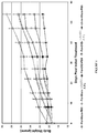

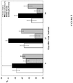


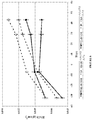


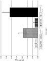

Classifications
-
- A—HUMAN NECESSITIES
- A61—MEDICAL OR VETERINARY SCIENCE; HYGIENE
- A61K—PREPARATIONS FOR MEDICAL, DENTAL OR TOILETRY PURPOSES
- A61K38/00—Medicinal preparations containing peptides
- A61K38/16—Peptides having more than 20 amino acids; Gastrins; Somatostatins; Melanotropins; Derivatives thereof
- A61K38/17—Peptides having more than 20 amino acids; Gastrins; Somatostatins; Melanotropins; Derivatives thereof from animals; from humans
- A61K38/177—Receptors; Cell surface antigens; Cell surface determinants
- A61K38/179—Receptors; Cell surface antigens; Cell surface determinants for growth factors; for growth regulators
-
- A—HUMAN NECESSITIES
- A61—MEDICAL OR VETERINARY SCIENCE; HYGIENE
- A61K—PREPARATIONS FOR MEDICAL, DENTAL OR TOILETRY PURPOSES
- A61K38/00—Medicinal preparations containing peptides
- A61K38/16—Peptides having more than 20 amino acids; Gastrins; Somatostatins; Melanotropins; Derivatives thereof
- A61K38/17—Peptides having more than 20 amino acids; Gastrins; Somatostatins; Melanotropins; Derivatives thereof from animals; from humans
- A61K38/177—Receptors; Cell surface antigens; Cell surface determinants
- A61K38/1796—Receptors; Cell surface antigens; Cell surface determinants for hormones
-
- A—HUMAN NECESSITIES
- A61—MEDICAL OR VETERINARY SCIENCE; HYGIENE
- A61P—SPECIFIC THERAPEUTIC ACTIVITY OF CHEMICAL COMPOUNDS OR MEDICINAL PREPARATIONS
- A61P13/00—Drugs for disorders of the urinary system
- A61P13/12—Drugs for disorders of the urinary system of the kidneys
-
- A—HUMAN NECESSITIES
- A61—MEDICAL OR VETERINARY SCIENCE; HYGIENE
- A61P—SPECIFIC THERAPEUTIC ACTIVITY OF CHEMICAL COMPOUNDS OR MEDICINAL PREPARATIONS
- A61P19/00—Drugs for skeletal disorders
- A61P19/08—Drugs for skeletal disorders for bone diseases, e.g. rachitism, Paget's disease
-
- A—HUMAN NECESSITIES
- A61—MEDICAL OR VETERINARY SCIENCE; HYGIENE
- A61P—SPECIFIC THERAPEUTIC ACTIVITY OF CHEMICAL COMPOUNDS OR MEDICINAL PREPARATIONS
- A61P19/00—Drugs for skeletal disorders
- A61P19/08—Drugs for skeletal disorders for bone diseases, e.g. rachitism, Paget's disease
- A61P19/10—Drugs for skeletal disorders for bone diseases, e.g. rachitism, Paget's disease for osteoporosis
-
- A—HUMAN NECESSITIES
- A61—MEDICAL OR VETERINARY SCIENCE; HYGIENE
- A61P—SPECIFIC THERAPEUTIC ACTIVITY OF CHEMICAL COMPOUNDS OR MEDICINAL PREPARATIONS
- A61P3/00—Drugs for disorders of the metabolism
- A61P3/12—Drugs for disorders of the metabolism for electrolyte homeostasis
-
- A—HUMAN NECESSITIES
- A61—MEDICAL OR VETERINARY SCIENCE; HYGIENE
- A61P—SPECIFIC THERAPEUTIC ACTIVITY OF CHEMICAL COMPOUNDS OR MEDICINAL PREPARATIONS
- A61P3/00—Drugs for disorders of the metabolism
- A61P3/12—Drugs for disorders of the metabolism for electrolyte homeostasis
- A61P3/14—Drugs for disorders of the metabolism for electrolyte homeostasis for calcium homeostasis
-
- A—HUMAN NECESSITIES
- A61—MEDICAL OR VETERINARY SCIENCE; HYGIENE
- A61P—SPECIFIC THERAPEUTIC ACTIVITY OF CHEMICAL COMPOUNDS OR MEDICINAL PREPARATIONS
- A61P43/00—Drugs for specific purposes, not provided for in groups A61P1/00-A61P41/00
-
- A—HUMAN NECESSITIES
- A61—MEDICAL OR VETERINARY SCIENCE; HYGIENE
- A61P—SPECIFIC THERAPEUTIC ACTIVITY OF CHEMICAL COMPOUNDS OR MEDICINAL PREPARATIONS
- A61P9/00—Drugs for disorders of the cardiovascular system
- A61P9/10—Drugs for disorders of the cardiovascular system for treating ischaemic or atherosclerotic diseases, e.g. antianginal drugs, coronary vasodilators, drugs for myocardial infarction, retinopathy, cerebrovascula insufficiency, renal arteriosclerosis
-
- A—HUMAN NECESSITIES
- A61—MEDICAL OR VETERINARY SCIENCE; HYGIENE
- A61P—SPECIFIC THERAPEUTIC ACTIVITY OF CHEMICAL COMPOUNDS OR MEDICINAL PREPARATIONS
- A61P9/00—Drugs for disorders of the cardiovascular system
- A61P9/14—Vasoprotectives; Antihaemorrhoidals; Drugs for varicose therapy; Capillary stabilisers
Definitions
- the technical field of the present invention relates to vascular calcification.
- the present invention relates to an ActRII inhibitor for use in a method for treating vascular calcification in a subject as defined in the claims.
- Bone growth and mineralization are dependent on the activities of two cell types, osteoclasts and osteoblasts, although chondrocytes and cells of the vasculature also participate in critical aspects of these processes.
- Developmentally, bone formation occurs through two mechanisms, endochondral ossification and intramembranous ossification, with the former responsible for longitudinal bone formation and the later responsible for the formation of topologically flat bones, such as the bones of the skull.
- Endochondral ossification requires the sequential formation and degradation of cartilaginous structures in the growth plates that serve as templates for the formation of osteoblasts, osteoclasts, the vasculature and subsequent mineralization.
- intramembranous ossification bone is formed directly in the connective tissues. Both processes require the infiltration of osteoblasts and subsequent matrix deposition.
- Chronic kidney disease is associated with a progressive deterioration in mineral homeostasis, with a disruption of normal serum and tissue concentrations of phosphorus and calcium, and changes in circulating hormones, such as parathyroid hormone, 25-hydroxyvitamin D, 1,25-dihydroxyvitamin D, other vitamin D metabolites, fibroblast growth factor-23, and growth hormone.
- CKD-MBD Chronic Kidney Disease-Mineral and Bone Disorder
- Kidney Disease Improving Global Outcomes (KDIGO) CKD-MBD Work Group, In: Kidney Int Suppl. (2009) 76 (Suppl 113):S1-130, page S3 .
- the mineral and hormone homeostasis that is disrupted in chronic kidney disease is critical for initial bone formation during growth (bone modeling) and bone structure and function during adulthood (bone remodeling).
- bone abnormalities are found in patients with chronic kidney disease.
- extraskeletal calcification may be found in patients with chronic kidney disease.
- CDK-MBD chronic kidney disease-related mineral and bone disorders
- CKD-MBD Chronic Kidney Disease-Mineral and Bone Disorder (CKD-MBD), Kidney Disease: Improving Global Outcomes (KDIGO) CKD-MBD Work Group, In: Kidney Int Suppl. (2009) 76 (Suppl 113):S1-130, page S34 ).
- vascular calcification a condition characterized by increased vessel stiffening.
- Patients with vascular calcification have an increased risk of myocardial infarction, and vascular calcification is particularly prevalent in patients suffering from kidney disease, e.g., CKD-MBD. See, e.g., Shanahan et al., 2011, Circ. Res. 109:697-711 .
- ActRIIA and ActRIIB Two related type II receptors, ActRIIA and ActRIIB, have been identified as the type II receptors for activins ( Mathews and Vale, 1991, Cell 65:973-982 ; Attisano et al., 1992, Cell 68: 97-108 ). Besides activins, ActRIIA and ActRIIB can biochemically interact with several other TGF-beta family proteins, including BMP7, Nodal, GDF8, and GDF11 ( Yamashita et al., 1995, J. Cell Biol. 130:217-226 ; Lee and McPherron, 2001, Proc. Natl. Acad. Sci. 98:9306-9311 ; Yeo and Whitman, 2001, Mol.
- ALK4 is the primary type I receptor for activins, particularly for activin A, and ALK-7 may serve as a receptor for activins as well, particularly for activin B.
- the present invention relates to an ActRII inhibitor for use in a method for treating vascular calcification, i.e., the accumulation of calcium salts in the vasculature of the subject, e.g., calcification of arteries of the subject, wherein the ActRIIA inhibitor is a polypeptide selected from the group consisting of: a. a polypeptide at least 90% identical to SEQ ID NO:2; b. a polypeptide at least 95% identical to SEQ ID NO:2; c. a polypeptide at least 98% identical to SEQ ID NO:2; d. SEQ ID NO:2; e. a polypeptide at least 90% identical to SEQ ID NO:3; f.
- the ActRIIA inhibitor is a polypeptide selected from the group consisting of: a. a polypeptide at least 90% identical to SEQ ID NO:2; b. a polypeptide at least 95% identical to SEQ ID NO:2; c. a polypeptide at least
- SEQ ID NO:7 q. a polypeptide at least 90% identical to SEQ ID NO:12; r. a polypeptide at least 95% identical to SEQ ID NO:12; s. a polypeptide at least 98% identical to SEQ ID NO:12; and t.
- SEQ ID NO:12 u) 90% identical to SEQ ID NO: 17; v) 95% identical to SEQ ID NO: 17; w) 98% identical to SEQ ID NO: 17; x) SEQ ID NO: 17; y) 90% identical to SEQ ID NO:20; z) 95% identical to SEQ ID N0:20; aa) 98% identical to SEQ ID N0:20; bb) SEQ ID N0:20; cc) 90% identical to SEQ ID NO:21; dd) 95% identical to SEQ ID NO:21; ee) 98% identical to SEQ ID NO:21; ff) SEQ ID NO:21; gg) 90% identical to SEQ ID NO:23; hh) 95% identical to SEQ ID NO:23; ii) 98% identical to SEQ ID NO:23; jj) SEQ ID NO:23; kk) 90% identical to SEQ ID NO:25; 11) 95% identical to SEQ ID NO:25; mm) 98% identical
- the subject to be treated with the methods provided herein is less than 18 years old. In certain embodiments, the subject to be treated with the methods provided herein has end stage renal disease. In certain embodiments, the subject to be treated with the methods provided herein undergoes dialysis.
- CKD-MBD Chronic Kidney Disease-Mineral and Bone Disorders
- the method comprises administering an inhibitor of ActRII to a patient in need of treatment.
- the inhibitor of ActRII can be an inhibitor of ActRIIA and / or ActRIIB.
- CKD-MBD can be diagnosed as a systemic disorder of mineral and bone metabolism due to chronic kidney disease and manifested by either one or a combination of (1) abnormalities of calcium; phosphorus; calcium x phosphorus product; alkaline phosphatases (total or bone specific); bicarbonate; parathyroid hormone ("PTH”); 1-84 PTH, 1-84-PTH/7-84 PTH ratio; osteocalcin; osteoprotegrin; tartrate-resistant acid phosphatase isoform 5b ("TRAP-5b”); pyridinoline and deoxypyridinoline; procollagen type 1 amino-terminal extension peptides; C-terminal crosslinks; C-terminal crosslinks of collagen; fibroblast growth factor 23 (“FGF23”); Fetulin-A; or vitamin D metabolism; (2) abnormalities of bone turnover, mineralization, volume, linear growth, or strength; and (3) vascular or other soft tissue calcification.
- PTH parathyroid hormone
- Kidney International 69:1945-1953 Guidelines for the diagnosis of CKD-MBD can be found, e.g., in KDIGO clinical practice guidelines for the prevention, diagnosis, evaluation, and treatment of Chronic Kidney Disease-Mineral and Bone Disorder (CKD-MBD), Kidney Disease: Improving Global Outcomes (KDIGO) CKD-MBD Work Group, In: Kidney Int Suppl. (2009) 76 (Suppl 113):S1-130 .
- KDIGO Chronic Kidney Disease-Mineral and Bone Disorder
- methods for the treatment of CKD-MBD characterized by hyperphosphatemia and / or hypercalcemia are disclosed herein.
- methods for the treatment of CKD-MBD characterized by extraskeletal calcification such as, but not limited to atherosclerotic calcification.
- CKD-MBD CKD-MBD
- the kidney disease is end stage kidney disease.
- methods for the treatment of CKD-MBD characterized by a glomerular filtration rate of less than 60ml/min/1.73m 2 in adults or less than 89 ml/min/1.73m 2 in pediatric patients. See, Moe et al., 2006, Kidney International 69:1945-1953 .
- provided herein are methods for the treatment in adults of CKD-MBD characterized by a glomerular filtration rate of less than 50ml/min/1.73m 2 , 40ml/min/1.73m 2 , 30ml/min/1.73m 2 , 20ml/min/1.73m 2 , or less than 10ml/min/1.73m 2 .
- a glomerular filtration rate of less than 60 ml/min/1.73m 2 in adult patients and less than 89 ml/min/1.73m 2 in pediatric patients results in detectable abnormalities in calcium levels, phosphorus levels, PTH levels, and vitamin D metabolism; and abnormal levels of these markers result in bone disease.
- CKD-MBD a bone pathology associated with chronic kidney disease
- the CKD-MBD is low-turnover CKD-MBD.
- Low-turnover CKD-MBD can be diagnosed by the histological features set forth in Table 1 below. See National Kidney Foundation, Kidney Disease Outcomes Quality Initiative Guidelines at the website of the National Kidney Foundation. Table 1.
- a method for treating extraskeletal calcification in a subject comprises administering a therapeutically effective amount of an ActRII inhibitor to the subject.
- a method for preventing extraskeletal calcification in a subject wherein said method comprises administering a therapeutically effective amount of an ActRII inhibitor to the subject.
- the extraskeletal calcification treated or prevented in a subject by the methods described herein is vascular calcification, i.e., the accumulation of calcium salts in the vasculature of the subject, e.g., calcification of arteries of the subject.
- the methods of of treatment or prevention of extraskeletal calcification are performed on a subject that is at risk of suffering from extraskeletal calcification, e.g., vascular calcification (i.e., the at risk subject is administered an ActRII inhibitor in accordance with the methods described herein).
- the subject at risk of suffering from extraskeletal calcification e.g., vascular calcification
- has hypercholesterolemia e.g., vascular calcification
- the subject at risk of suffering from extraskeletal calcification e.g., vascular calcification
- has hypertension e.g., vascular calcification
- the subject at risk of suffering from extraskeletal calcification has diabetes.
- the subject at risk of suffering from extraskeletal calcification e.g., vascular calcification
- has renal disease e.g., end-stage renal disease.
- the subject at risk of suffering from extraskeletal calcification e.g., vascular calcification
- the subject at risk of suffering from extraskeletal calcification, e.g., vascular calcification has increased oxidative stress, e.g., an imbalance between oxidant production and antioxidant activity in the vasculature.
- the subject at risk of suffering from extraskeletal calcification e.g., vascular calcification
- has a calcification inhibitor deficiency e.g., a deficiency in one or more of fetuin-A, matrix gla protein (MGP), and osteoprotegerin (OPG)).
- a calcification inhibitor deficiency e.g., a deficiency in one or more of fetuin-A, matrix gla protein (MGP), and osteoprotegerin (OPG)
- the subjects suffering from vascular calcification treated in accordance with the methods described herein have Media calcification (also known as Mönckeberg's sclerosis or media calcinosis).
- Media calcification is characterized by diffuse mineral deposits within the arterial tunica media.
- the subjects suffering from media calcification are elderly.
- the subjects suffering from media calcification have a disorder that causes the Media calcification, e.g., diabetes, renal disease (e.g., CKD).
- the subjects suffering from vascular calcification treated in accordance with the methods described herein have Intima calcification.
- Intima calcification is associated with atherosclerosis and progresses as atherosclerotic plaques progress.
- a subject suffering from, or at risk of suffering from, a form of CKD-MBD and/or extraskeletal calcification e.g., vascular calcification
- Levels of FGF23 can be detected using methods known in the art, e.g., ELISA, using samples from the subjects, e.g, blood, serum.
- the level of FGF23 (e.g., the level detectable in the serum) in a subject suffering from, or at risk of suffering from, a form of CKD-MBD and/or extraskeletal calcification, e.g., vascular calcification is about 5%, 10%, 15%, 20%, 25%, 30%, 35%, 40%, 45%, 50%, or greater than 50%, greater than the level of FGF23 (e.g., the level detectable in the serum) in a subject not suffering from, or not at risk of suffering from, a form of CKD-MBD and/or extraskeletal calcification, e.g., vascular calcification.
- the level of FGF23 (e.g., the level detectable in the serum) in a subject suffering from, or at risk of suffering from, a form of CKD-MBD and/or extraskeletal calcification, e.g., vascular calcification is about 5-10%, 10-20%, 20-30%, 30-40%, 40-50%, 50-60%, 50-75%, or 75-100%, greater than the level of FGF23 (e.g., the level detectable in the serum) in a subject not suffering from, or not at risk of suffering from, a form of CKD-MBD and/or extraskeletal calcification, e.g., vascular calcification.
- levels of FGF23 in a subject suffering from, or at risk of suffering from, a form of CKD-MBD and/or extraskeletal calcification can be used to monitor the effectiveness of a method described herein, e.g., a method of treating a form of CKD-MBD and/or a method of treating extraskeletal calcification (e.g., vascular calcification), wherein such methods comprise administration of a therapeutically effective amount of an ActRII inhibitor described herein.
- a subject treated in accordance with one or more of the methods described herein has a decreased level of FGF23 (e.g., as detected in the serum of the subject) as compared to the level of FGF23 detected in the subject prior to being treated with a method described herein.
- the level of FGF23 (e.g., the level detectable in the serum) in a subject suffering from, or at risk of suffering from, a form of CKD-MBD and/or extraskeletal calcification, e.g., vascular calcification, treated with a method described herein is decreased by about 5%, 10%, 15%, 20%, 25%, 30%, 35%, 40%, 45%, 50%, or greater than 50%, relative to the level of FGF23 (e.g., the level detectable in the serum) detected in the subject prior to treatment with a method described herein.
- the level of FGF23 (e.g., the level detectable in the serum) in a subject suffering from, or at risk of suffering from, a form of CKD-MBD and/or extraskeletal calcification, e.g., vascular calcification, is decreased by about 5-10%, 10-20%, 20-30%, 30-40%, 40-50%, 50-60%, 50-75%, or 75-100%, relative to the level of FGF23 (e.g., the level detectable in the serum) detected in the subject prior to treatment with a method described herein.
- a method of treating a form of CKD-MBD and/or extraskeletal calcification, e.g., vascular calcification comprising: (i) administering an ActRII inhibitor to an individual having a form of CKD-MBD and/or extraskeletal calcification, e.g., vascular calcification; (ii) determining an amount of FGF23 in a tissue sample (e.g., serum) of said individual after the adminstration of the ActRII inhibitor; and (iii) if the amount of FGF23 in said tissue sample is decreased by no more than about 5%, 10%, 15%, 20%, or 25%, or by about 5-10%, 10-20%, 20-30%, as compared to the amount of FGF23 determined in a sample of the same tissue type from said individual (e.g., a different sample of serum from the same individual) prior to administration of the ActRII inhibitor, repeating the administration of the ActRII inhibitor.
- a tissue sample e.g., serum
- the dose of the ActRII inhibitor administered can be increased. In certain embodiments, if the amount of FGF23 is not decreased following administration of the ActRII inhibitor, the frequency of administration of the ActRII inhibitor administered can be increased.
- a subject suffering from, or at risk of suffering from, a form of CKD-MBD and/or extraskeletal calcification has increased levels of sclerostin, a protein increased in subjects suffering from, or at risk of suffering from, CKD-MBD (see, e.g., Graciolli et al., 2010, J Am Soc Nephrol 21:774A ), relative to sclerostin levels in subjects that are not suffering from, or not at risk of suffering from, a form of CKD-MBD and/or extraskeletal calcification, e.g., vascular calcification.
- Levels of sclerostin can be detected using methods known in the art, e.g., ELISA, using samples from the subjects, e.g, blood, serum.
- the level of sclerostin (e.g., the level detectable in the serum) in a subject suffering from, or at risk of suffering from, a form of CKD-MBD and/or extraskeletal calcification, e.g., vascular calcification is about 5%, 10%, 15%, 20%, 25%, 30%, 35%, 40%, 45%, 50%, or greater than 50%, greater than the level of sclerostin (e.g., the level detectable in the serum) in a subject not suffering from, or not at risk of suffering from, a form of CKD-MBD and/or extraskeletal calcification, e.g., vascular calcification.
- the level of sclerostin (e.g., the level detectable in the serum) in a subject suffering from, or at risk of suffering from, a form of CKD-MBD and/or extraskeletal calcification, e.g., vascular calcification is about 5-10%, 10-20%, 20-30%, 30-40%, 40-50%, 50-60%, 50-75%, or 75-100%, greater than the level of sclerostin (e.g., the level detectable in the serum) in a subject not suffering from, or not at risk of suffering from, a form of CKD-MBD and/or extraskeletal calcification, e.g., vascular calcification.
- levels of sclerostin in a subject suffering from, or at risk of suffering from, a form of CKD-MBD and/or extraskeletal calcification can be used to monitor the effectiveness of a method described herein, e.g., a method of treating a form of CKD-MBD and/or a method of treating extraskeletal calcification (e.g., vascular calcification), wherein such methods comprise administration of a therapeutically effective amount of an ActRII inhibitor described herein.
- a subject treated in accordance with one or more of the methods described herein has a decreased level of sclerostin (e.g., as detected in the serum of the subject) as compared to the level of sclerostin detected in the subject prior to being treated with a method described herein.
- the level of sclerostin (e.g., the level detectable in the serum) in a subject suffering from, or at risk of suffering from, a form of CKD-MBD and/or extraskeletal calcification, e.g., vascular calcification, treated with a method described herein is decreased by about 5%, 10%, 15%, 20%, 25%, 30%, 35%, 40%, 45%, 50%, or greater than 50%, relative to the level of sclerostin (e.g., the level detectable in the serum) detected in the subject prior to treatment with a method described herein.
- the level of sclerostin (e.g., the level detectable in the serum) in a subject suffering from, or at risk of suffering from, a form of CKD-MBD and/or extraskeletal calcification, e.g., vascular calcification is decreased by about 5-10%, 10-20%, 20-30%, 30-40%, 40-50%, 50-60%, 50-75%, or 75-100%, relative to the level of sclerostin (e.g., the level detectable in the serum) detected in the subject prior to treatment with a method described herein.
- a method of treating a form of CKD-MBD and/or extraskeletal calcification comprising: (i) administering an ActRII inhibitor to an individual having a form of CKD-MBD and/or extraskeletal calcification, e.g., vascular calcification; (ii) determining an amount of sclerostin in a tissue sample (e.g., serum) of said individual after the adminstration of the ActRII inhibitor; and (iii) if the amount of sclerostin in said tissue sample is decreased by no more than about 5%, 10%, 15%, 20%, or 25%, or by about 5-10%, 10-20%, 20-30%, as compared to the amount of sclerostin determined in a sample of the same tissue from said individual (e.g., a different sample of serum from the same individual) prior to administration of the ActRII inhibitor, repeating the administration of the ActRII inhibitor.
- a tissue sample e.g., serum
- the dose of the ActRII inhibitor administered can be increased. In certain embodiments, if the amount of sclerostin is not decreased following administration of the ActRII inhibitor, the frequency of administration of the ActRII inhibitor administered can be increased.
- the subject suffering from vascular calcification treated in accordance with the methods described herein is less than 18 years old. In a specific embodiment, the subject suffering from vascular calcification treated in accordance with the methods described herein is less than 13 years old. In another specific embodiment, the subject suffering from vascular calcification treated in accordance with the methods described herein is less than 12, less than 11, less than 10, less than 9, less than 8, less than 7, less than 6, or less than 5 years old.
- the subject suffering from vascular calcification treated in accordance with the methods described herein is 1-3 years old, 3-5 years old, 5-7 years old, 7-9 years old, 9-11 years old, 11-13 years old, 13-15 years old, 15-20 years old, 20-25 years old, 25-30 years old, or greater than 30 years old.
- the subject suffering from vascular calcification treated in accordance with the methods described herein is 30-35 years old, 35-40 years old, 40-45 years old, 45-50 years old, 50-55 years old, 55-60 years old, or greater than 60 years old.
- the subject suffering from vascular calcification treated in accordance with the methods described herein is 60-65 years old, 65-70 years old, 70-75 years old, 75-80 years old, or greater than 80 years old.
- the subject suffering from vascular calcification treated in accordance with the methods described herein has end stage renal disease. In certain embodiments, the subject suffering from vascular calcification treated in accordance with the methods described herein undergoes dialysis.
- the effectiveness of treatment or prevention of extraskeletal calcification is assessed using one or more assays known to those of skill in the art. Exemplary assays are described in Section 5.3(a)(iv). In accordance with such embodiments, one of skill in the art will understand that a subject being treated with an ActRII inhibitor as described herein may have their treatment regimen adjusted based on the outcome of the assays.
- a subject being treated by a method described herein that displays increases in levels of calcium may be administered an increased dose of ActRII inhibitor, or a may be administered an ActRII inhibitor more frequently (i.e., the time between dose administrations may be decreased).
- a subject being treated by a method described herein that displays decreases in levels of calcium e.g., vascular calcium (e.g., arterial calcium) may be administered a decreased dose of ActRII inhibitor, or a may be administered an ActRII inhibitor less frequently (i.e., the time between dose administrations may be increased).
- the methods provided herein result in the improvement of the symptoms of one or more of the following: hyperphosphatemia, secondary hyperparathyroidism (due to increase in phosphorus), and extraskeletal calcification, e.g., vascular calcification. Any method known to the skilled artisan to determine the degree of these symptoms can be used with the methods provided herein. In specific embodiments, the methods described herein result in the improvement of one or more symptoms of vascular calcification.
- Exemplary symptoms include, without limitation, increases in the levels of vascular (e.g., arterial) calcium, increased apoptosis of vascular smooth muscle cells, loss of arterial elasticity, an increase in PWV (pulse wave velocity), development of left ventricular hypertrophy, decrease in coronary artery perfusion, and myocardial ischaemia.
- vascular e.g., arterial
- PWV pulse wave velocity
- the methods described herein result in a decrease in the levels of vascular calcium, e.g., arterial calcium, in a subject by at least 5%, 10%, 15%, 20%, 25%, 30%, 35%, 40%, 45% or 50%. In certain embodiments, the methods described herein result in a decrease in the levels of vascular calcium, e.g., arterial calcium, in a subject by 5%-10%, 10%-15%, 15%-20%, 20%-25%, 25%-30%, 30%-35%, 35%-40%, 40%-45%, or 45%-50%.
- vascular calcium in a subject comprising: (i) administering an ActRII inhibitor to a subject in need of reduction vascular calcium levels (e.g., a subject having a form of CKD-MBD and/or extraskeletal calcification, e.g., vascular calcification); (ii) determining an amount of vascular calcium in a tissue sample (e.g., serum) of said subject after the adminstration of the ActRII inhibitor; and (iii) if the amount of vascular calcium in said tissue sample is decreased by no more than about 5%, 10%, 15%, 20%, or 25%, or by about 5-10%, 10-20%, 20-30%, as compared to the amount of vascular calcium determined in a sample of the same tissue from said subject (e.g., a different sample of serum from the same individual) prior to administration of the ActRII inhibitor, repeating the administration of the ActRII inhibitor.
- an ActRII inhibitor to a subject in need of reduction vascular calcium levels (e.g., a subject
- the dose of the ActRII inhibitor administered can be increased. In certain embodiments, if the amount of vascular calcium is not decreased following administration of the ActRII inhibitor, the frequency of administration of the ActRII inhibitor administered can be increased.
- the methods described herein result in a decrease in the progression of the Agatston score of a subject having or at risk of developing vascular calcification.
- the methods described herein result in a 5%, 10%, 15%, 20%, 25%, 30%, or greater than 30% decrease in the Agatston score of a subject having or at risk of developing vascular calcification as compared to the Agatston score of the subject prior to administration of an ActRII inhibitor in accordance with the methods described herein (see, e.g., Section 5.3(a)(iv)).
- the methods described herein result in a 5%-10%, 10%-15%, 15%-20%, 20%-25%, 25%-30%, 30%-35%, 35%-40%, 40%-45%, or 45%-50% decrease in the Agatston score of a subject having or at risk of developing vascular calcification as compared to the Agatston score of the subject prior to administration of an ActRII inhibitor in accordance with the methods described herein (see, e.g., Section 5.3(a)(iv)).
- the methods described herein result in a decrease in the levels of calcium in the vasculature of a subject, e.g., a decrease in the levels of calcium in one or more arteries of the subject, e.g., a subject having or at risk of developing vascular calcification.
- the methods described herein result in a decrease in the levels of phosphorus in the vasculature of a subject, e.g., a decrease in the levels of phosphorus in one or more arteries of the subject, e.g., a subject having or at risk of developing vascular calcification.
- Low bone turnover can be diagnosed using the tests set forth in Section 5.3(a) below.
- Biochemical markers of bone turnover include: serum or urine collagen crosslinks (N-telopeptide or C-telopeptide), bone-specific alkaline phosphatase, serum osteocalcin and/or propeptide type 1 collagen, 25 hydroxyvitamin D, and parathyroid hormone ("PTH").
- the low turnover bone disorder is adynamic bone disorder.
- a patient to be treated with the methods provided herein has a reduction in bone-turnover of at least 10%, 20%, 25%, 30%, 40%, 50%, 60%, 70%, 75%, 80%, 85%, 90%, 95%, 98%, 99% or of 100%. In certain embodiments, a patient to be treated with the methods described herein has a reduction in bone-turnover of at most 10%, 20%, 25%, 30%, 40%, 50%, 60%, 70%, 75%, 80%, 85%, 90%, 95%, 98%, 99% or of 100%.
- a patient to be treated with the methods described herein has a reduction in bone-turnover of at between 10% and 25%, 20% and 35%, 30% and 45%, 40% and 55%, 50% and 65%, 60% and 75%, 70% and 85%, 80% and 95%, 90% and 100%.
- the reduction in bone turnover is compared to historical data of the same patient.
- the reduction in bone turnover is compared to the average bone turnover in a population without bone disorders.
- the population without bone disorders can be of the same age and / or same sex as the patient.
- a method of treating a low turnover bone disorder comprising: (i) administering an ActRII inhibitor to a subject having a low turnover bone disorder; (ii) determining the level of bone-turnover in said subject after the adminstration of the ActRII inhibitor (e.g., by using one or more of the tests set forth in Section 5.3(a) below and/or by measuring one or more biochemical markers of bone turnover); and (iii) if the level of bone turnover in the subject is decreased by no more than about 5%, 10%, 15%, 20%, or 25%, or by about 5-10%, 10-20%, 20-30%, as compared to the level of bone turnover in the subject prior to administration of the ActRII inhibitor, repeating the administration of the ActRII inhibitor.
- the dose of the ActRII inhibitor administered can be increased. In certain embodiments, if the level of bone turnover is not decreased following administration of the ActRII inhibitor, the frequency of administration of the ActRII inhibitor administered can be increased.
- ActRIIA refers to a family of activin receptor type IIa (ActRIIA) proteins from any species and variants derived from such ActRIIA proteins by mutagenesis or other modification. Reference to ActRIIA herein is understood to be a reference to any one of the currently identified forms. Members of the ActRIIA family are generally transmembrane proteins, composed of a ligand-binding extracellular domain with a cysteine-rich region, a transmembrane domain, and a cytoplasmic domain with predicted serine/threonine kinase activity.
- ActRIIA inhibitors to be used in the compositions and methods described herein include, without limitation, activin-binding soluble ActRIIA polypeptides; antibodies that bind to activin (particularly the activin A or B subunits, also referred to as ßA or ßB) and disrupt ActRIIA binding; antibodies that bind to ActRIIA and disrupt activin binding; non-antibody proteins selected for activin or ActRIIA binding (see e.g., WO/2002/088171 , WO/2006/055689 , WO/2002/032925 , WO/2005/037989 , US 2003/0133939 , and US 2005/0238646 , for examples of such proteins and methods for design and selection of same); and randomized peptides selected for activin or ActRIIA binding, which can be conjugated to an Fc domain.
- two or more different proteins (or other moieties) with activin or ActRIIA binding activity may be linked together to create a bifunctional or multifunctional binding molecule that inhibits ActRIIA and thus can be used in the compositions and methods described herein.
- activin-ActRIIA signaling axis antagonists that inhibit ActRIIA include nucleic acid aptamers, small molecules and other agents are used in the compositions and methods described herein include.
- ActRIIA polypeptide includes polypeptides comprising any naturally occurring polypeptide of an ActRIIA family member as well as any variants thereof (including mutants, fragments, fusions, and peptidomimetic forms) that retain a useful activity.
- ActRIIA polypeptides include polypeptides derived from the sequence of any known ActRIIA having a sequence at least about 80% identical to the sequence of an ActRIIA polypeptide, and optionally at least 85%, 90%, 95%, 97%, 98%, 99% or greater identity.
- an ActRIIA polypeptide may bind to and inhibit the function of an ActRIIA protein and/or activin.
- An ActRIIB polypeptide may be selected for its ability to promote bone growth and bone mineralization.
- Examples of ActRIIA polypeptides include human ActRIIA precursor polypeptide (SEQ ID NO: 1) and soluble human ActRIIA polypeptides (e.g., SEQ ID NOs: 2, 3, 7 and 12).
- SEQ ID NO: 1 human ActRIIA precursor polypeptide
- soluble human ActRIIA polypeptides e.g., SEQ ID NOs: 2, 3, 7 and 12
- the signal peptide of the human ActRIIA precursor polypeptide located at amino acid positions 1 to 20; the extracellular domain is located at amino acid positions 21 to 135 and the N-linked glycosylation sites of the human ActRIIA precursor polypeptide (SEQ ID NO: 1) are located at amino acid positions 43 and 56 of SEQ ID NO:1.
- the nucleic acid sequence encoding the human ActRIIB precursor polypeptide of SEQ ID NO:1 is disclosed as SEQ ID NO:4 (nucleotides 164-1705 of Genbank entry NM_001616).
- the nucleic acid sequence encoding the soluble human ActRIIA polypeptide of SEQ ID NO:2 is disclosed as SEQ ID NO:5. See Table 6 for a description of the sequences.
- the ActRIIA polypeptides used in the compositions and methods described herein are soluble ActRIIA polypeptides.
- An extracellular domain of an ActRIIA protein can bind to activin and is generally soluble, and thus can be termed a soluble, activin-binding ActRIIA polypeptide.
- soluble ActRIIA polypeptide generally refers to polypeptides comprising an extracellular domain of an ActRIIA protein, including any naturally occurring extracellular domain of an ActRIIA protein as well as any variants thereof (including mutants, fragments and peptidomimetic forms).
- Soluble ActRIIA polypeptides can bind to activin; however, the wild type ActRIIA protein does not exhibit significant selectivity in binding to activin versus GDF8/11.
- Native or altered ActRIIA proteins may be given added specificity for activin by coupling them with a second, activin-selective binding agent.
- Examples of soluble, activin-binding ActRIIA polypeptides include the soluble polypeptides illustrated in SEQ ID NOs: 2, 3, 7, 12 and 13.
- soluble, activin-binding ActRIIA polypeptides comprise a signal sequence in addition to the extracellular domain of an ActRIIA protein, for example, the honey bee mellitin leader sequence (SEQ ID NO: 8), the tissue plasminogen activator (TPA) leader (SEQ ID NO: 9) or the native ActRIIA leader (SEQ ID NO: 10).
- the ActRIIA-hFc polypeptide illustrated in SEQ ID NO:13 uses a TPA leader.
- the inhibitors of ActRIIA used in the compositions and methods described herein comprise a conjugate/fusion protein comprising an activin-binding domain of ActRIIA linked to an Fc portion of an antibody.
- the activin-binding domain is linked to an Fc portion of an antibody via a linker, e.g., a peptide linker.
- the Fc domain has one or more mutations at residues such as Asp-265, lysine 322, and Asn-434.
- the mutant Fc domain having one or more of these mutations (e.g., an Asp-265 mutation) has a reduced ability to bind to the Fc ⁇ receptor relative to a wild-type Fc domain.
- mutant Fc domain having one or more of these mutations has an increased ability to bind to the MHC class I- related Fc-receptor (FcRN) relative to a wild-type Fc domain.
- FcRN MHC class I- related Fc-receptor
- Exemplary fusion proteins comprising a soluble extracellular domain of ActRIIA fused to an Fc domain are set forth in SEQ ID NOs: 6, 7, 12, and 13.
- the ActRIIA inhibitors used in the compositions and methods described herein comprise the extracellular domain of ActRIIA, or a portion thereof, linked to an Fc portion of an antibody, wherein said ActRIIA inhibitor comprises an amino acid sequence that is at least 75% identical to an amino acid sequence selected from SEQ ID NOs: 6, 7, 12, and 13.
- the ActRIIA inhibitors used in the compositions and methods described herein comprise the extracellular domain of ActRIIA, or a portion thereof, linked to an Fc portion of an antibody, wherein said ActRIIA inhibitor comprises an amino acid sequence that is at least 80%, 85%, 90%, 95%, 96%, 97%, 98%, or 99% identical to an amino acid sequence selected from SEQ ID NOs: 6, 7, 12, and 13.
- the inhibitors of ActRIIA used in the compositions and methods described herein comprise a truncated form of an extracellular domain of ActRIIA.
- the truncation can be at the carboxy terminus and/or the amino terminus of the ActRIIA polypeptide.
- the truncation can be 1, 2, 3, 4, 5, 6, 7, 8, 9, 10, 11, 12, 13, 14, 15, 16, 17, 18, 19, 20, 21, 22, 23, 24, or 25 amino acids long relative to the mature ActRIIB polypeptide extracellular domain.
- the truncation can be 1, 2, 3, 4, 5, 6, 7, 8, 9, 10, 11, 12, 13, 14, 15, 16, 17, 18, 19, 20, 21, 22, 23, 24, or 25 N-terminal amino acids of the mature ActRIIA polypeptide extracellular domain.
- the truncation can be 1, 2, 3, 4, 5, 6, 7, 8, 9, 10, 11, 12, 13, 14, 15, 16, 17, 18, 19, 20, 21, 22, 23, 24, or 25 C-terminal amino acids of the mature ActRIIA polypeptide extracellular domain.
- truncated forms of ActRIIA include polypeptides with amino acids 20-119; 20-128; 20-129; 20-130; 20-131; 20-132; 20-133; 20-134; 20-131; 21-131; 22-131; 23-131; 24-131; and 25-131, wherein the amino acid positions refer to the amino acid positions in SEQ ID NO:1.
- the inhibitors of ActRIIA used in the compositions and methods described herein comprise an extracellular domain of ActRIIA with one or more amino acid substitutions. In certain embodiments, the inhibitors of ActRIIA used in the compositions and methods described herein comprise a truncated form of an ActRIIA extracellular domain that also carries an amino acid substitution.
- the ActRIIA inhibitor to be used in the compositions and methods described herein is a fusion protein between the extracellular domain of the human ActRIIA receptor and the Fc portion of IgG1.
- the ActRIIA inhibitor to be used in the compositions and methods described herein is a fusion protein between a truncated extracellular domain of the human ActRIIA receptor and the Fc portion of IgG1.
- the ActRIIA inhibitor to be used in the compositions and methods described herein is a fusion protein between a truncated extracellular domain of the human ActRIIA receptor and the Fc portion of IgG1, wherein the truncated extracellular domain of the human ActRIIA receptor possesses one or more amino acid substitutions.
- Functionally active fragments of ActRIIA polypeptides can be obtained, for example, by screening polypeptides recombinantly produced from the corresponding fragment of the nucleic acid encoding an ActRIIA polypeptide.
- fragments can be chemically synthesized using techniques known in the art such as conventional Merrifield solid phase f-Moc or t-Boc chemistry. The fragments can be produced (recombinantly or by chemical synthesis) and tested to identify those peptidyl fragments that can function as antagonists (inhibitors) of ActRIIA protein or signaling mediated by activin.
- a functional variant of the ActRIIA polypeptides can be obtained, for example, by screening libraries of modified polypeptides recombinantly produced from the corresponding mutagenized nucleic acids encoding an ActRIIA polypeptide. The variants can be produced and tested to identify those that can function as antagonists (inhibitors) of ActRIIA protein or signaling mediated by activin.
- a functional variant of the ActRIIA polypeptides comprises an amino acid sequence that is at least 75% identical to an amino acid sequence selected from SEQ ID NOs: 2 or 3. In certain cases, the functional variant has an amino acid sequence at least 80%, 85%, 90%, 95%, 97%, 98%, 99% or 100% identical to an amino acid sequence selected from SEQ ID NOs: 2 or 3.
- Functional variants may be generated, for example, by modifying the structure of an ActRIIA polypeptide for such purposes as enhancing therapeutic efficacy, or stability (e.g., ex vivo shelf life and resistance to proteolytic degradation in vivo).
- modified ActRIIA polypeptides when selected to retain activin binding, can be considered functional equivalents of the naturally-occurring ActRIIA polypeptides.
- Modified ActRIIA polypeptides can also be produced, for instance, by amino acid substitution, deletion, or addition.
- the ActRIIA inhibitor to be used in the compositions and methods described herein may comprise an ActRIIA polypeptide having one or more specific mutations that can alter the glycosylation of the polypeptide. Such mutations may introduce or eliminate one or more glycosylation sites, such as O-linked or N-linked glycosylation sites.
- Asparagine-linked glycosylation recognition sites generally comprise a tripeptide sequence, asparagine-X-threonine (or asparagines-X-serine) (where "X" is any amino acid) which is specifically recognized by appropriate cellular glycosylation enzymes.
- the alteration may also be made by the addition of, or substitution by, one or more serine or threonine residues to the sequence of the wild-type ActRIIA polypeptide (for O-linked glycosylation sites).
- a variety of amino acid substitutions or deletions at one or both of the first or third amino acid positions of a glycosylation recognition site (and/or amino acid deletion at the second position) results in non-glycosylation at the modified tripeptide sequence.
- Another means of increasing the number of carbohydrate moieties on an ActRIIA polypeptide is by chemical or enzymatic coupling of glycosides to the ActRIIA polypeptide.
- the sugar(s) may be attached to (a) arginine and histidine; (b) free carboxyl groups; (c) free sulfhydryl groups such as those of cysteine; (d) free hydroxyl groups such as those of serine, threonine, or hydroxyproline; (e) aromatic residues such as those of phenylalanine, tyrosine, or tryptophan; or (f) the amide group of glutamine.
- Removal of one or more carbohydrate moieties present on an ActRIIA polypeptide may be accomplished chemically and/or enzymatically.
- Chemical deglycosylation may involve, for example, exposure of the ActRIIA polypeptide to the compound trifluoromethanesulfonic acid, or an equivalent compound. This treatment results in the cleavage of most or all sugars except the linking sugar (N-acetylglucosamine or N-acetylgalactosamine), while leaving the amino acid sequence intact.
- Chemical deglycosylation is further described by Hakimuddin et al. (1987) Arch. Biochem. Biophys. 259:52 and by Edge et al. (1981) Anal. Biochem. 118:131 .
- Enzymatic cleavage of carbohydrate moieties on ActRIIA polypeptides can be achieved by the use of a variety of endo- and exo-glycosidases as described by Thotakura et al. (1987) Meth. Enzymol. 138:350 .
- the sequence of an ActRIIA polypeptide may be adjusted, as appropriate, depending on the type of expression system used, as mammalian, yeast, insect and plant cells may all introduce differing glycosylation patterns that can be affected by the amino acid sequence of the peptide.
- ActRIIA proteins for use in humans can be expressed in a mammalian cell line that provides proper glycosylation, such as HEK293 or CHO cell lines, although other expression systems, such as other mammalian expression cell lines, yeast cell lines with engineered glycosylation enzymes and insect cells, are expected to be useful as well.
- mutants particularly sets of combinatorial mutants of an ActRIIA polypeptide, as well as truncation mutants; pools of combinatorial mutants are especially useful for identifying functional variant sequences.
- the purpose of screening such combinatorial libraries may be to generate, for example, ActRIIA polypeptide variants which can act as either agonists or antagonist, or alternatively, which possess novel activities all together.
- a variety of screening assays are described below, and such assays may be used to evaluate variants.
- an ActRIIA polypeptide variant may be screened for ability to bind to an ActRIIA ligand, to prevent binding of an ActRIIA ligand to an ActRIIA polypeptide or to interfere with signaling caused by an ActRIIA ligand.
- Combinatorially-derived variants can be generated which have a selective or generally increased potency relative to a naturally occurring ActRIIA polypeptide.
- mutagenesis can give rise to variants which have intracellular half-lives dramatically different than the corresponding a wild-type ActRIIA polypeptide.
- the altered protein can be rendered either more stable or less stable to proteolytic degradation or other cellular processes which result in destruction of, or otherwise inactivation of a native ActRIIA polypeptide.
- Such variants, and the genes which encode them can be utilized to alter ActRIIA polypeptide levels by modulating the half-life of the ActRIIA polypeptides.
- a short half-life can give rise to more transient biological effects and can allow tighter control of recombinant ActRIIA polypeptide levels within the patient.
- mutations may be made in the linker (if any) and/or the Fc portion to alter the half-life of the protein.
- a combinatorial library may be produced by way of a degenerate library of genes encoding a library of polypeptides which each include at least a portion of potential ActRIIA polypeptide sequences.
- a mixture of synthetic oligonucleotides can be enzymatically ligated into gene sequences such that the degenerate set of potential ActRIIA polypeptide nucleotide sequences are expressible as individual polypeptides, or alternatively, as a set of larger fusion proteins (e.g., for phage display).
- the library of potential homologs can be generated from a degenerate oligonucleotide sequence.
- Chemical synthesis of a degenerate gene sequence can be carried out in an automatic DNA synthesizer, and the synthetic genes then be ligated into an appropriate vector for expression.
- the synthesis of degenerate oligonucleotides is well known in the art (see for example, Narang, S A (1983) Tetrahedron 39:3 ; Itakura et al., (1981) Recombinant DNA, Proc. 3rd Cleveland Sympos. Macromolecules, ed. AG Walton, Amsterdam: Elsevier pp 273-289 ; Itakura et al., (1984) Annu. Rev.
- ActRIIA polypeptide variants can be generated and isolated from a library by screening using, for example, alanine scanning mutagenesis and the like ( Ruf et al., (1994) Biochemistry 33:1565-1572 ; Wang et al., (1994) J. Biol. Chem. 269:3095-3099 ; Balint et al., (1993) Gene 137:109-118 ; Grodberg et al., (1993) Eur. J. Biochem. 218:597-601 ; Nagashima et al., (1993) J. Biol. Chem.
- a wide range of techniques are known in the art for screening gene products of combinatorial libraries made by point mutations and truncations, and, for that matter, for screening cDNA libraries for gene products having a certain property. Such techniques will be generally adaptable for rapid screening of the gene libraries generated by the combinatorial mutagenesis of ActRIIA polypeptides.
- the most widely used techniques for screening large gene libraries typically comprises cloning the gene library into replicable expression vectors, transforming appropriate cells with the resulting library of vectors, and expressing the combinatorial genes under conditions in which detection of a desired activity facilitates relatively easy isolation of the vector encoding the gene whose product was detected.
- Preferred assays include activin binding assays and activin-mediated cell signaling assays.
- ActRIIA polypeptides used in the inhibitors of the methods and compositions described herein may further comprise post-translational modifications in addition to any that are naturally present in the ActRIIA polypeptides.
- modifications may include, but are not limited to, acetylation, carboxylation, glycosylation, phosphorylation, lipidation, and acylation.
- the modified ActRIIA polypeptides may contain non-amino acid elements, such as polyethylene glycols, lipids, poly- or mono-saccharide, and phosphates. Effects of such non-amino acid elements on the functionality of a ActRIIA polypeptide may be tested by any method known to the skilled artisan.
- an ActRIIA polypeptide When an ActRIIA polypeptide is produced in cells by cleaving a nascent form of the ActRIIA polypeptide, post-translational processing may also be important for correct folding and/or function of the protein.
- Different cells such as CHO, HeLa, MDCK, 293, W138, NIH-3T3 or HEK293 have specific cellular machinery and characteristic mechanisms for such post-translational activities and may be chosen to ensure the correct modification and processing of the ActRIIA polypeptides.
- fusion proteins having at least a portion of the ActRIIA polypeptides and one or more fusion domains.
- fusion domains include, but are not limited to, polyhistidine, Glu-Glu, glutathione S transferase (GST), thioredoxin, protein A, protein G, an immunoglobulin heavy chain constant region (Fc), maltose binding protein (MBP), or human serum albumin.
- a fusion domain may be selected so as to confer a desired property. For example, some fusion domains are particularly useful for isolation of the fusion proteins by affinity chromatography.
- matrices for affinity chromatography such as glutathione-, amylase-, and nickel- or cobalt-conjugated resins are used. Many of such matrices are available in "kit” form, such as the Pharmacia GST purification system and the QIAexpress.TM. system (Qiagen) useful with (HIS6) fusion partners.
- a fusion domain may be selected so as to facilitate detection of the ActRIIA polypeptides. Examples of such detection domains include the various fluorescent proteins (e.g., GFP) as well as "epitope tags," which are usually short peptide sequences for which a specific antibody is available.
- fusion domains have a protease cleavage site, such as for Factor Xa or Thrombin, which allows the relevant protease to partially digest the fusion proteins and thereby liberate the recombinant proteins therefrom. The liberated proteins can then be isolated from the fusion domain by subsequent chromatographic separation.
- an ActRIIA polypeptide is fused with a domain that stabilizes the ActRIIA polypeptide in vivo (a "stabilizer" domain).
- stabilizing is meant anything that increases serum half life, regardless of whether this is because of decreased destruction, decreased clearance by the kidney, or other pharmacokinetic effect. Fusions with the Fc portion of an immunoglobulin are known to confer desirable pharmacokinetic properties on a wide range of proteins. Likewise, fusions to human serum albumin can confer desirable properties. Other types of fusion domains that may be selected include multimerizing (e.g., dimerizing, tetramerizing) domains and functional domains (that confer an additional biological function, such as further stimulation of bone growth or muscle growth, as desired).
- an ActRIIA polypeptide may be placed C-terminal to a heterologous domain, or, alternatively, a heterologous domain may be placed C-terminal to an ActRIIA polypeptide.
- the ActRIIA polypeptide domain and the heterologous domain need not be adjacent in a fusion protein, and additional domains or amino acid sequences may be included C- or N-terminal to either domain or between the domains.
- the ActRIIA polypeptides used in the inhibitors of the methods and compositions described herein may contain one or more modifications that are capable of stabilizing the ActRIIA polypeptides.
- modifications may enhance the in vitro half life of the ActRIIA polypeptides, enhance circulatory half life of the ActRIIA polypeptides or reduce proteolytic degradation of the ActRIIA polypeptides.
- Such stabilizing modifications may include, but are not limited to, fusion proteins (including, for example, fusion proteins comprising an ActRIIA polypeptide and a stabilizer domain), modifications of a glycosylation site (including, for example, addition of a glycosylation site to an ActRIIA polypeptide), and modifications of carbohydrate moiety (including, for example, removal of carbohydrate moieties from an ActRIIA polypeptide).
- fusion proteins including, for example, fusion proteins comprising an ActRIIA polypeptide and a stabilizer domain
- modifications of a glycosylation site including, for example, addition of a glycosylation site to an ActRIIA polypeptide
- modifications of carbohydrate moiety including, for example, removal of carbohydrate moieties from an ActRIIA polypeptide.
- an ActRIIA polypeptide is fused to a stabilizer domain such as an IgG molecule (e.g., an Fc domain).
- stabilizer domain not only refers to a fusion domain (e.g., Fc) as in the case of fusion proteins, but also includes nonproteinaceous modifications such as a carbohydrate moiety, or nonproteinaceous polymer, such as polyethylene glycol.
- isolated and/or purified forms of ActRIIA polypeptides which are isolated from, or otherwise substantially free of, other proteins can be used with the methods and compositions described herein.
- ActRIIA polypeptides can generally be produced by expression from recombinant nucleic acids.
- the ActRIIA polypeptides used in the compositions and methods described herein are generated using isolated and/or recombinant nucleic acids encoding any of the ActRIIA polypeptides (e.g., soluble ActRIIA polypeptides), including fragments, functional variants and fusion proteins disclosed herein.
- SEQ ID NO: 4 encodes the naturally occurring human ActRIIA precursor polypeptide
- SEQ ID NO: 5 encodes the processed extracellular domain of ActRIIA.
- Such nucleic acids may be single-stranded or double stranded.
- Such nucleic acids may be DNA or RNA molecules. These nucleic acids may be used, for example, in methods for making ActRIIA polypeptides or as direct therapeutic agents (e.g., in a gene therapy approach).
- nucleic acids encoding ActRIIA polypeptides may include nucleic acids that are variants of SEQ ID NO: 4 or 5.
- Variant nucleotide sequences include sequences that differ by one or more nucleotide substitutions, additions or deletions, such as allelic variants.
- isolated or recombinant nucleic acid sequences encoding ActRIIA polypeptides may be least 80%, 85%, 90%, 95%, 97%, 98%, 99% or 100% identical to SEQ ID NO: 4 or 5.
- nucleic acid sequences complementary to SEQ ID NO: 4 or 5, and variants of SEQ ID NO: 4 or 5 may be used in the production of ActRIIA polypeptides suitable for use in the methods and compositions described herein.
- such nucleic acid sequences can be isolated, recombinant, and/or fused to a heterologous nucleotide sequence, or be from a DNA library.
- nucleic acids used in the production of ActRIIA polypeptides suitable for use in the methods and compositions described herein may include nucleotide sequences that hybridize under highly stringent conditions to the nucleotide sequence designated in SEQ ID NO: 4 or 5, complement sequence of SEQ ID NO: 4 or 5, or fragments thereof.
- SSC sodium chloride/sodium citrate
- the salt concentration in the wash step can be selected from a low stringency of about 2.0 times SSC at 50 degree Celsius to a high stringency of about 0.2 times SSC at 50 degree Celsius.
- the temperature in the wash step can be increased from low stringency conditions at room temperature, about 22 degree Celsius, to high stringency conditions at about 65 degree Celsius. Both temperature and salt may be varied, or temperature or salt concentration may be held constant while the other variable is changed.
- nucleic acids which hybridize under low stringency conditions of 6 times SSC at room temperature followed by a wash at 2 times SSC at room temperature can be used with the methods and compositions described herein.
- Isolated nucleic acids which differ from the nucleic acids as set forth in SEQ ID NOs: 4 or 5 due to degeneracy in the genetic code also can be used in the production of ActRIIA polypeptides suitable for use in the methods and compositions described herein.
- a number of amino acids are designated by more than one triplet. Codons that specify the same amino acid, or synonyms (for example, CAU and CAC are synonyms for histidine) may result in "silent" mutations which do not affect the amino acid sequence of the protein.
- DNA sequence polymorphisms that do lead to changes in the amino acid sequences of the subject proteins will exist among mammalian cells.
- these variations in one or more nucleotides (up to about 3-5% of the nucleotides) of the nucleic acids encoding a particular protein may exist among individuals of a given species due to natural allelic variation.
- the recombinant nucleic acids may be operably linked to one or more regulatory nucleotide sequences in an expression construct.
- Regulatory nucleotide sequences will generally be appropriate to the host cell used for expression. Numerous types of appropriate expression vectors and suitable regulatory sequences are known in the art for a variety of host cells.
- said one or more regulatory nucleotide sequences may include, but are not limited to, promoter sequences, leader or signal sequences, ribosomal binding sites, transcriptional start and termination sequences, translational start and termination sequences, and enhancer or activator sequences. Constitutive or inducible promoters as known in the art are contemplated herein.
- the promoters may be either naturally occurring promoters, or hybrid promoters that combine elements of more than one promoter.
- An expression construct may be present in a cell on an episome, such as a plasmid, or the expression construct may be inserted in a chromosome.
- the expression vector contains a selectable marker gene to allow the selection of transformed host cells. Selectable marker genes are well known in the art and will vary with the host cell used.
- the a nucleic acid used in the production of ActRIIA polypeptides suitable for use in the methods and compositions described herein can be provided in an expression vector comprising a nucleotide sequence encoding an ActRIIA polypeptide and operably linked to at least one regulatory sequence.
- Regulatory sequences are art-recognized and are selected to direct expression of the ActRIIA polypeptide. Accordingly, the term regulatory sequence includes promoters, enhancers, and other expression control elements. Exemplary regulatory sequences are described in Goeddel; Gene Expression Technology: Methods in Enzymology, Academic Press, San Diego, Calif. (1990 ).
- any of a wide variety of expression control sequences that control the expression of a DNA sequence when operatively linked to it may be used in these vectors to express DNA sequences encoding an ActRIIA polypeptide.
- useful expression control sequences include, for example, the early and late promoters of SV40, tet promoter, adenovirus or cytomegalovirus immediate early promoter, RSV promoters, the lac system, the trp system, the TAC or TRC system, T7 promoter whose expression is directed by T7 RNA polymerase, the major operator and promoter regions of phage lambda, the control regions for fd coat protein, the promoter for 3-phosphoglycerate kinase or other glycolytic enzymes, the promoters of acid phosphatase, e.g., Pho5, the promoters of the yeast ⁇ -mating factors, the polyhedron promoter of the baculovirus system and other sequences known to control the expression of genes of prok
- the design of the expression vector may depend on such factors as the choice of the host cell to be transformed and/or the type of protein desired to be expressed. Moreover, the vector's copy number, the ability to control that copy number and the expression of any other protein encoded by the vector, such as antibiotic markers, should also be considered.
- a recombinant nucleic acid used in the production of ActRIIA polypeptides suitable for use in the methods and compositions described herein can be produced by ligating the cloned gene, or a portion thereof, into a vector suitable for expression in either prokaryotic cells, eukaryotic cells (yeast, avian, insect or mammalian), or both.
- Expression vehicles for production of a recombinant ActRIIA polypeptide include plasmids and other vectors.
- suitable vectors include plasmids of the types: pBR322-derived plasmids, pEMBL-derived plasmids, pEX-derived plasmids, pBTac-derived plasmids and pUC-derived plasmids for expression in prokaryotic cells, such as E. coli.
- Some mammalian expression vectors contain both prokaryotic sequences to facilitate the propagation of the vector in bacteria, and one or more eukaryotic transcription units that are expressed in eukaryotic cells.
- the pcDNAI/amp, pcDNAI/neo, pRc/CMV, pSV2gpt, pSV2neo, pSV2-dhfr, pTk2, pRSVneo, pMSG, pSVT7, pko-neo and pHyg derived vectors are examples of mammalian expression vectors suitable for transfection of eukaryotic cells.
- vectors are modified with sequences from bacterial plasmids, such as pBR322, to facilitate replication and drug resistance selection in both prokaryotic and eukaryotic cells.
- bacterial plasmids such as pBR322
- derivatives of viruses such as the bovine papilloma virus (BPV-1), or Epstein-Barr virus (pHEBo, pREP-derived and p205) can be used for transient expression of proteins in eukaryotic cells.
- BBV-1 bovine papilloma virus
- pHEBo Epstein-Barr virus
- pREP-derived and p205 Epstein-Barr virus
- examples of other viral (including retroviral) expression systems can be found below in the description of gene therapy delivery systems.
- the various methods employed in the preparation of the plasmids and in transformation of host organisms are well known in the art.
- baculovirus expression systems include pVL-derived vectors (such as pVL1392, pVL1393 and pVL941), pAcUW-derived vectors (such as pAcUW1), and pBlueBac-derived vectors (such as the ⁇ -gal containing pBlueBac III).
- pVL-derived vectors such as pVL1392, pVL1393 and pVL941
- pAcUW-derived vectors such as pAcUW1
- pBlueBac-derived vectors such as the ⁇ -gal containing pBlueBac III.
- Vectors can be designed for production of the subject ActRIIA polypeptides in CHO cells, such as a Pcmv-Script vector (Stratagene, La Jolla, Calif.), pcDNA4 vectors (Invitrogen, Carlsbad, Calif.) and pCI-neo vectors (Promega, Madison, Wis.).
- the subject gene constructs can be used to cause expression of the subject ActRIIA polypeptides in cells propagated in culture, e.g., to produce proteins, including fusion proteins or variant proteins, for purification.
- Host cells transfected with a recombinant gene including a coding sequence (e.g., SEQ ID NO: 4 or 5) for one or more of the subject ActRIIA polypeptides can be used in the production of ActRIIA polypeptides suitable for use in the methods and compositions described herein.
- the host cell may be any prokaryotic or eukaryotic cell.
- an ActRIIA polypeptide provided herein may be expressed in bacterial cells such as E. coli, insect cells (e.g., using a baculovirus expression system), yeast, or mammalian cells. Other suitable host cells are known to those skilled in the art.
- a host cell transfected with an expression vector encoding an ActRIIA polypeptide can be cultured under appropriate conditions to allow expression of the ActRIIA polypeptide to occur.
- the ActRIIA polypeptide may be secreted and isolated from a mixture of cells and medium containing the ActRIIA polypeptide.
- the ActRIIA polypeptide may be retained cytoplasmically or in a membrane fraction and the cells harvested, lysed and the protein isolated.
- a cell culture includes host cells, media and other byproducts. Suitable media for cell culture are well known in the art.
- the subject ActRIIA polypeptides can be isolated from cell culture medium, host cells, or both, using techniques known in the art for purifying proteins, including ion-exchange chromatography, gel filtration chromatography, ultrafiltration, electrophoresis, immunoaffinity purification with antibodies specific for particular epitopes of the ActRIIA polypeptides and affinity purification with an agent that binds to a domain fused to the ActRIIA polypeptide (e.g., a protein A column may be used to purify an ActRIIA-Fc fusion).
- the ActRIIA polypeptide is a fusion protein containing a domain which facilitates its purification.
- purification is achieved by a series of column chromatography steps, including, for example, three or more of the following, in any order: protein A chromatography, Q sepharose chromatography, phenylsepharose chromatography, size exclusion chromatography, and cation exchange chromatography.
- the purification could be completed with viral filtration and buffer exchange.
- ActRIIA-hFc protein was purified to a purity of >98% as determined by size exclusion chromatography and >95% as determined by SDS PAGE. This level of purity was sufficient to achieve desirable effects on bone in mice and an acceptable safety profile in mice, rats and non-human primates.
- a fusion gene coding for a purification leader sequence such as a poly-(His)/enterokinase cleavage site sequence at the N-terminus of the desired portion of a recombinant ActRIIA polypeptide, can allow purification of the expressed fusion protein by affinity chromatography using a Ni2+ metal resin.
- the purification leader sequence can then be subsequently removed by treatment with enterokinase to provide the purified ActRIIA polypeptide (e.g., see Hochuli et al., (1987) J. Chromatography 411:177 ; and Janknecht et al., PNAS USA 88:8972 ).
- fusion genes are well known. Essentially, the joining of various DNA fragments coding for different polypeptide sequences is performed in accordance with conventional techniques, employing blunt-ended or stagger-ended termini for ligation, restriction enzyme digestion to provide for appropriate termini, filling-in of cohesive ends as appropriate, alkaline phosphatase treatment to avoid undesirable joining, and enzymatic ligation.
- the fusion gene can be synthesized by conventional techniques including automated DNA synthesizers.
- PCR amplification of gene fragments can be carried out using anchor primers which give rise to complementary overhangs between two consecutive gene fragments which can subsequently be annealed to generate a chimeric gene sequence (see, for example, Current Protocols in Molecular Biology, eds. Ausubel et al., John Wiley & Sons: 1992 ).
- ActRIIA-Fc fusion protein can be expressed in stably transfected CHO-DUKX B1 1 cells from a pAID4 vector (SV40 ori/enhancer, CMV promoter), using a tissue plasminogen leader sequence of SEQ ID NO:9.
- the Fc portion is a human IgG1 Fc sequence, as shown in SEQ ID NO:7.
- the protein contained upon expression, has, on average, between about 1.5 and 2.5 moles of sialic acid per molecule of ActRIIA-Fc fusion protein.
- the long serum half-life of an ActRIIA-Fc fusion can be 25-32 days in human patients.
- the CHO cell expressed material can have a higher affinity for activin B ligand than that reported for an ActRIIA-hFc fusion protein expressed in human 293 cells ( del Re et al., J Biol Chem. 2004 Dec 17;279(51):53126-35 ).
- the use of the TPA leader sequence provided greater production than other leader sequences and, unlike ActRIIA-Fc expressed with a native leader, may provide a highly pure N-terminal sequence. Use of the native leader sequence may result in two major species of ActRIIA-Fc, each having a different N-terminal sequence.
- ActRIIB refers to a family of activin receptor type IIB (ActRIIB) proteins from any species and variants derived from such ActRIIB proteins by mutagenesis or other modification. Reference to ActRIIB herein is understood to be a reference to any one of the currently identified forms of the receptor. Members of the ActRIIB family are generally transmembrane proteins, composed of a ligand-binding extracellular domain with a cysteine-rich region, a transmembrane domain, and a cytoplasmic domain with predicted serine/threonine kinase activity.
- ActRIIB inhibitors to be used in the compositions and methods described herein include, without limitation, activin-binding soluble ActRIIB polypeptides; antibodies that bind to activin (particularly the activin A or B subunits, also referred to as ßA or ßB) and disrupt ActRIIB binding; antibodies that bind to ActRIIB and disrupt activin binding; non-antibody proteins selected for activin or ActRIIB binding; and randomized peptides selected for activin or ActRIIB binding, which can be conjugated to an Fc domain.
- activin-binding soluble ActRIIB polypeptides include, without limitation, activin-binding soluble ActRIIB polypeptides; antibodies that bind to activin (particularly the activin A or B subunits, also referred to as ßA or ßB) and disrupt ActRIIB binding; antibodies that bind to ActRIIB and disrupt activin binding; non-antibody proteins selected for activin or ActRIIB binding; and randomized peptide
- two or more different proteins (or other moieties) with activin or ActRIIB binding activity may be linked together to create a bifunctional or multifunctional binding molecule that inhibits ActRIIB and thus can be used in the compositions and methods described herein include.
- activin-ActRIIB signaling axis antagonists that inhibit ActRIIB include nucleic acid aptamers, small molecules and other agents are used in the compositions and methods described herein include.
- ActRIIB polypeptide refers to polypeptides comprising any naturally occurring polypeptide of an ActRIIB family member as well as any variants thereof (including mutants, fragments, fusions, and peptidomimetic forms) that retain a useful activity.
- ActRIIB polypeptides include polypeptides derived from the sequence of any known ActRIIB receptor having a sequence at least about 80% identical to the sequence of an ActRIIB polypeptide, and optionally at least 85%, 90%, 95%, 96%, 97%, 98%, 99% or greater identity.
- an ActRIIB polypeptide may bind to and inhibit the function of an ActRIIB protein and/or activin.
- an example of an ActRIIB polypeptide includes the human ActRIIB precursor polypeptide (SEQ ID NO:16 or SEQ ID NO:28).
- the signal peptide of the ActRIIB precursor polypeptide is located at amino acids 1 to 18; the extracellular domain is located at amino acids 19 to 134 and the potential N-linked glycosylation sites are located at amino acid positions 42 and 65.
- SEQ ID NO:19 provides an alanine at the codon corresponding to amino acid position 64, but could be readily modified by one of skill in the art using methods known in the art to provide an arginine at the codon corresponding to amino acid position 64 instead). See Table 6 for a description of the sequences.
- the numbering of amino acids for all of the ActRIIB-related polypeptides described herein is based on the amino acid numbering for SEQ ID NO:16 and SEQ ID NO:28 (which only differ in the amino acid expressed at position 64), unless specifically designated otherwise.
- SEQ ID NO:16 and SEQ ID NO:28 which only differ in the amino acid expressed at position 64
- position 79 refers to the 79th amino acid in SEQ ID NO:16 or SEQ ID NO:28, from which the ActRIIB polypeptide is derived.
- an ActRIIB polypeptide is described as having an alanine or an arginine at amino acid position 64, then it is to be understood that position 64 refers to the 64th amino acid in SEQ ID NO:16 or SEQ ID NO:28, from which the ActRIIB polypeptide is derived.
- the inhibitors of ActRIIB used in the compositions and methods described herein comprise polypeptides comprising an activin-binding domain of ActRIIB.
- the activin-binding domains of ActRIIB comprise the extracellular domain of ActRIIB, or a portion thereof.
- the extracellular domain or portion thereof of ActRIIB is soluble.
- Illustrative modified forms of ActRIIB polypeptides are disclosed in U.S. Patent Application Publication Nos. 20090005308 and 20100068215 .
- the ActRIIB polypeptides used in the compositions and methods described herein are soluble ActRIIB polypeptides.
- the term "soluble ActRIIB polypeptide” generally refers to polypeptides comprising an extracellular domain of an ActRIIB protein, including any naturally occurring extracellular domain of an ActRIIB protein as well as any variants thereof (including mutants, fragments and peptidomimetic forms). Soluble ActRIIB polypeptides can bind to activin; however, the wild type ActRIIB protein does not exhibit significant selectivity in binding to activin versus GDF8/11. In certain embodiments, altered forms of ActRIIB with different binding properties can be used in the methods provided herein.
- exemplary soluble ActRIIB polypeptides include the extracellular domain of a human ActRIIB polypeptide (e.g., SEQ ID NOs: 17, 18, 23, 26, 27, 29, 30, 31, 32, 33, 36, 37, 42, and 43).
- an Fc fusion protein with an arginine at position 64 of the ActRIIB precursor amino acid sequence (herein referred to as "R64”) has an affinity for activin and GDF-11 in the low nanomolar to high picomolar range (see, e.g., U.S. Patent Application Publication No.
- an ActRIIB precursor amino acid sequence with an arginine at position 64 is presented in SEQ ID NO:28.
- the ActRIIB polypeptides used in accordance with the compositions and methods described herein may comprise either (i) an alanine at the position corresponding to amino acid 64 of the ActRIIB precursor amino acid sequence, i.e., SEQ ID NO: 16; or (ii) an arginine at position 64 of the ActRIIB precursor amino acid sequence, i.e., SEQ ID NO: 28.
- the ActRIIB polypeptides used in accordance with the compositions and methods described herein may comprise an amino acid that is not alanine or arginine at the position corresponding to amino acid 64 of the ActRIIB precursor amino acid sequence, i.e., SEQ ID NO: 16 or SEQ ID NO:28.
- An ActRIIB-Fc fusion protein containing amino acids 20-119 of SEQ ID NO: 28 i.e., SEQ ID NO:32
- ActRIIB(20-119)-Fc has reduced binding to GDF-11 and activin relative to an ActRIIB-Fc fusion protein containing amino acids 20-134 of SEQ ID NO: 28 (i.e., SEQ ID NO:31), "ActRIIB(20-134)-Fc", which includes the proline knot region and the complete juxtamembrane domain.
- ActRIIB-Fc fusion protein containing amino acids 20-129 of SEQ ID NO: 28, "ActRIIB(20-129)-Fc" retains similar but somewhat reduced activity relative to the non-truncated extracellular domain of ActRIIB, even though the proline knot region is disrupted.
- ActRIIB polypeptides comprising extracellular domains that stop at amino acid 134, 133, 132, 131, 130 and 129 of SEQ ID NO: 28 (or SEQ ID NO:16) are all expected to be active, but constructs stopping at amino acid 134 or 133 may be most active.
- the ActRIIB polypeptides used in accordance with the methods and compositions described herein may end as early as amino acid 109 (i.e., the final cysteine) of SEQ ID NO:28 (or SEQ ID NO:16), however, forms ending at or between amino acid positions 109 and 119 of SEQ ID NO:28 (or SEQ ID NO:16) are expected to have reduced ligand binding ability.
- Amino acid 29 of SEQ ID NO:16 and SEQ ID NO:28 represents the initial cysteine in the ActRIIB precursor sequence. It is expected that an ActRIIB polypeptide beginning at amino acid 29 of the N-terminus of SEQ ID NO:16 or SEQ ID NO:28, or before these amino acid positions, will retain ligand binding activity.
- An alanine to asparagine mutation at position 24 of SEQ ID NO:16 or SEQ ID NO:28 introduces an N-linked glycosylation sequence without substantially affecting ligand binding. This confirms that mutations in the region between the signal cleavage peptide and the cysteine cross-linked region, corresponding to amino acids 20-29 of SEQ ID NO:16 or SEQ ID NO:28, are well tolerated.
- ActRIIB polypeptides beginning at amino acid position 20, 21, 22, 23 and 24 of SEQ ID NO:16 or SEQ ID NO:28 will retain activity, and ActRIIB polypeptides beginning at amino acid positions 25, 26, 27, 28 and 29 of SEQ ID NO:16 or SEQ ID NO:28 are also expected to retain activity.
- An ActRIIB polypeptide beginning at amino acid position 22, 23, 24 or 25 of SEQ ID NO:16 or SEQ ID NO:28 will have the most activity.
- the active portions (i.e., ActRIIB polypeptides) of the ActRIIB precursor protein (i.e., SEQ ID NO:16 or SEQ ID NO:28) to be used in accordance with the methods and compositions described herein will generally comprise amino acids 29-109 of SEQ ID NO:16 or SEQ ID NO:28, and such ActRIIB polypeptides may, for example, begin at a residue corresponding to any one of amino acids 19-29 of SEQ ID NO:16 or SEQ ID NO:28 and end at a position corresponding to any one of amino acids 109-134 of SEQ ID NO:16 or SEQ ID NO:28.
- ActRIIB polypeptides encompassed herein include those that begin at an amino acid position from 19-29, 20-29 or 21-29 of SEQ ID NO:16 or SEQ ID NO:28 and end at an amino acid position from 119-134, 119-133 or 129-134, 129-133 of SEQ ID NO:16 or SEQ ID NO:28.
- ActRIIB polypeptides encompassed herein include those that begin at an amino acid position from 20-24 (or 21-24, or 22-25) of SEQ ID NO:16 or SEQ ID NO:28 and end at an amino acid position from 109-134 (or 109-133), 119-134 (or 119-133) or 129-134 (or 129-133) of SEQ ID NO:16 or SEQ ID NO:28.
- Variant ActRIIB polypeptides falling within these ranges are also contemplated, particularly those having at least 80%, 85%, 90%, 91%, 92%, 93%, 94%, 95%, 96%, 97%, 98%, or 99% sequence identity or sequence homology to the corresponding portion of SEQ ID NO:16 or SEQ ID NO:28.
- the inhibitors of ActRIIB used in the compositions and methods described herein comprise a truncated form of an extracellular domain of ActRIIB.
- the truncation can be at the carboxy terminus and/or the amino terminus of the ActRIIB polypeptide.
- the truncation can be 1, 2, 3, 4, 5, 6, 7, 8, 9, 10, 11, 12, 13, 14, 15, 16, 17, 18, 19, 20, 21, 22, 23, 24, or 25 amino acids long relative to the mature ActRIIB polypeptide extracellular domain.
- the truncation can be 1, 2, 3, 4, 5, 6, 7, 8, 9, 10, 11, 12, 13, 14, 15, 16, 17, 18, 19, 20, 21, 22, 23, 24, or 25 N-terminal amino acids of the mature ActRIIB polypeptide extracellular domain.
- the truncation can be 1, 2, 3, 4, 5, 6, 7, 8, 9, 10, 11, 12, 13, 14, 15, 16, 17, 18, 19, 20, 21, 22, 23, 24, or 25 C-terminal amino acids of the mature ActRIIB polypeptide extracellular domain.
- truncated forms of ActRIIB include polypeptides with amino acids 20-119; 20-128; 20-129; 20-130; 20-131; 20-132; 20-133; 20-134; 20-131; 21-131; 22-131; 23-131; 24-131; and 25-131, wherein the amino acid positions refer to the amino acid positions in SEQ ID NO:16 or SEQ ID NO:28.
- Additional exemplary truncated forms of ActRIIB include (i) polypeptides beginning at amino acids at any of amino acids 21-29 of SEQ ID NO:16 or SEQ ID NO:28 (optionally beginning at 22-25 of SEQ ID NO:16 or SEQ ID NO:28) and ending at any of amino acids 109-134 of SEQ ID NO:16 or SEQ ID NO:28; (ii) polypeptides beginning at any of amino acids 20-29 of SEQ ID NO:16 or SEQ ID NO:28 (optionally beginning at 22-25 of SEQ ID NO:16 or SEQ ID NO:28) and ending at any of amino acids 109-133 of SEQ ID NO:16 or SEQ ID NO:28; (iii) polypeptides beginning at any of amino acids 20-24 of SEQ ID NO:16 or SEQ ID NO:28 (optionally beginning at 22-25 of SEQ ID NO:16 or SEQ ID NO:28) and ending at any of amino acids 109-133 of SEQ ID NO:16 or SEQ ID NO:28;
- an ActRIIB polypeptides comprises, consists essentially of, or consists of, an amino acid sequence beginning at amino acid position 25 of SEQ ID NO:16 or SEQ ID NO:28 and ending at amino acid position 131 of SEQ ID NO:16 or SEQ ID NO:28.
- an ActRIIB polypeptide consists of, or consists essentially of, the amino acid sequence of SEQ ID NO:17, 18, 23, 26, 27, 29, 30, 31, 32, 33, 36, 37, 42, or 43.
- any of the ActRIIB polypeptides used in the compositions and methods described herein may be produced as a homodimer. Any of the ActRIIB polypeptides used in the compositions and methods described herein may be formulated as a fusion protein having a heterologous portion that comprises a constant region from an IgG heavy chain, such as an Fc domain. Any of the ActRIIB polypeptides used in the compositions and methods described herein may comprise an acidic amino acid at the position corresponding to position 79 of SEQ ID NO:16 or SEQ ID NO:28, optionally in combination with one or more additional amino acid substitutions, deletions or insertions relative to SEQ ID NO:16 or SEQ ID NO:28.
- the inhibitors of ActRIIB used in the compositions and methods described herein comprise an extracellular domain of ActRIIB with one or more amino acid substitutions/mutations.
- an amino acid substitution/mutation can be, for example, an exchange from the leucine at amino acid position 79 of SEQ ID NO:16 or SEQ ID NO:28 to an acidic amino acid, such as aspartic acid or glutamic acid.
- position L79 of SEQ ID NO:16 or SEQ ID NO:28 may be altered in ActRIIB extracellular domain polypeptides to confer altered activin-myostatin (GDF-11) binding properties.
- L79A and L79P mutations reduce GDF-11 binding to a greater extent than activin binding.
- L79E and L79D mutations retain GDF-11 binding, while demonstrating greatly reduced activin binding.
- the inhibitors of ActRIIB used in the compositions and methods described herein comprise a truncated form of an ActRIIB extracellular domain that also carries an amino acid substitution, e.g., an exchange from the leucine at amino acid position 79 of SEQ ID NO:16 or SEQ ID NO:28 to an acidic amino acid, such as aspartic acid or glutamic acid.
- the truncated form of an extracellular domain of ActRIIB polypeptide that also carries an amino acid substitution used in the compositions and methods described herein is SEQ ID NO:23.
- Forms of ActRIIB that are truncated and/or carry one or more amino acid substitutions can be linked to an Fc domain of an antibody as discussed above.
- Functionally active fragments of ActRIIB polypeptides can be obtained, for example, by screening polypeptides recombinantly produced from the corresponding fragment of the nucleic acid encoding an ActRIIB polypeptide.
- fragments can be chemically synthesized using techniques known in the art such as conventional Merrifield solid phase f-Moc or t-Boc chemistry. The fragments can be produced (recombinantly or by chemical synthesis) and tested to identify those peptidyl fragments that can function as antagonists (inhibitors) of ActRIIB protein or signaling mediated by activin.
- a functional variant of the ActRIIB polypeptides can be obtained, for example, by screening libraries of modified polypeptides recombinantly produced from the corresponding mutagenized nucleic acids encoding an ActRIIB polypeptide. The variants can be produced and tested to identify those that can function as antagonists (inhibitors) of ActRIIB protein or signaling mediated by activin.
- a functional variant of the ActRIIB polypeptides comprises an amino acid sequence that is at least 75% identical to an amino acid sequence selected from SEQ ID NO:17, 18, 23, 26, 27, 29, 30, 31, 32, 33, 36, 37, 42, and 43.
- the functional variant has an amino acid sequence at least 80%, 85%, 90%, 95%, 96%, 97%, 98%, or 99% identical to an amino acid sequence selected from SEQ ID NO:17, 18, 23, 26, 27, 29, 30, 31, 32, 33, 36, 37, 42, and 43.
- Functional variants may be generated, for example, by modifying the structure of an ActRIIB polypeptide for such purposes as enhancing therapeutic efficacy, or stability (e.g., ex vivo shelf life and resistance to proteolytic degradation in vivo).
- modified ActRIIB polypeptides when selected to retain activin binding, are considered functional equivalents of the naturally-occurring ActRIIB polypeptides.
- Modified ActRIIB polypeptides can also be produced, for instance, by amino acid substitution, deletion, or addition.
- ActRIIB polypeptide mutants particularly sets of combinatorial mutants of an ActRIIB polypeptide, as well as truncation mutants; pools of combinatorial mutants are especially useful for identifying functional variant sequences can be used in the methods and compositions described herein.
- the purpose of screening such combinatorial libraries may be to generate, for example, ActRIIB polypeptide variants which can act as either agonists or antagonist, or alternatively, which possess novel activities all together.
- the ligand binding pocket of ActRIIB is defined by residues Y31, N33, N35, L38 through T41, E47, E50, Q53 through K55, L57, H58, Y60, S62, K74, W78 through N83, Y85, R87, A92, and E94 through F101 of SEQ ID NO:16 or SEQ ID NO:28.
- R40A, K55A, F82A and mutations at position L79 is a K in Xenopus, indicating that basic amino acids at this position will be tolerated.
- an ActRIIB polypeptide for use in the methods and compositions described herein is one that comprises amino acids 29-109 of SEQ ID NO:16 or SEQ ID NO:28, but optionally beginning at an amino acid position ranging from 20-24 or 22-25 of SEQ ID NO:16 or SEQ ID NO:28 and ending at an amino acid position ranging from 129-134 of SEQ ID NO:16 or SEQ ID NO:28, and comprising no more than 1, 2, 5, or 15 conservative amino acid changes in the ligand binding pocket, and zero, one or more non-conservative alterations at amino acid positions 40, 53, 55, 74, 79 and/or 82 of SEQ ID NO:16 or SEQ ID NO:28 in the ligand binding pocket.
- Such an ActRIIB polypeptide may retain greater than 80%, 90%, 95% or 99% sequence identity or sequence homology to the sequence of amino acids 29-109 of SEQ ID NO:16 or SEQ ID NO:28.
- Sites outside the binding pocket, at which variability may be particularly well tolerated, include the amino and carboxy termini of the extracellular domain of ActRIIB, and positions 42-46 and 65-73.
- An asparagine to alanine alteration at position 65 of SEQ ID NO:16 or SEQ ID NO:28 (N65A) actually improves ligand binding in the A64 background, and is thus expected to have no detrimental effect on ligand binding in the R64 background.
- the positively-charged amino acid residue Asp (D80) of the ligand-binding domain of ActRIIB can be mutated to a different amino acid residue such that the variant ActRIIB polypeptide preferentially binds to GDF8, but not activin.
- the D80 residue is changed to an amino acid residue selected from the group consisting of: an uncharged amino acid residue, a negative amino acid residue, and a hydrophobic amino acid residue.
- the hydrophobic residue L79 can be altered to the acidic amino acids aspartic acid or glutamic acid to greatly reduce activin binding while retaining GDF11 binding.
- most of the described mutations, variants or modifications may be made at the nucleic acid level or, in some cases, by post translational modification or chemical synthesis. Such techniques are well known in the art.
- the inhibitors of ActRIIB used in the compositions and methods described herein comprise a conjugate/fusion protein comprising an extracellular domain (e.g., an activin-binding domain) of an ActRIIB receptor linked to an Fc portion of an antibody.
- conjugate/fusion proteins may comprise any of the ActRIIB polypeptides disclosed herein (e.g., any of SEQ ID NOs:17, 18, 23, 26, 27, 29, 30, 31, 32, 33, 36, 37, 42, or 43), any ActRIIB polypeptides known in the art, or any ActRIIB polypeptides generated using methods known in the art and/or provided herein.
- the extracellular domain is linked to an Fc portion of an antibody via a linker, e.g., a peptide linker.
- linkers include short polypeptide sequences such as 2-10, 2-5, 2-4, 2-3 amino acid residues (e.g., glycine residues), such as, for example, a Gly-Gly-Gly linker.
- the linker comprises the amino acid sequence Gly-Gly-Gly (GGG).
- the linker comprises the amino acid sequence Thr-Gly-Gly-Gly (TGGG).
- the Fc domain has one or more mutations at residues such as Asp-265, lysine 322, and Asn-434.
- the mutant Fc domain having one or more of these mutations has a reduced ability to bind to the Fc ⁇ receptor relative to a wild-type Fc domain.
- the mutant Fc domain having one or more of these mutations e.g., an Asn-434 mutation
- Exemplary fusion proteins comprising a soluble extracellular domain of ActRIIB fused to an Fc domain are set forth in SEQ ID NOs:20, 21, 24, 25, 34, 35, 38, 39, 40, 41, 44, 46, and 47.
- the ActRIIB inhibitors used in the compositions and methods described herein comprise the extracellular domain of ActRIIB, or a portion thereof, linked to an Fc portion of an antibody, wherein said ActRIIB inhibitor comprises an amino acid sequence that is at least 75% identical to an amino acid sequence selected from SEQ ID NOs:20, 21, 24, 25, 34, 35, 38, 39, 40, 41, 44, 46, and 47.
- the ActRIIB inhibitors used in the compositions and methods described herein comprise the extracellular domain of ActRIIB, or a portion thereof, linked to an Fc portion of an antibody, wherein said ActRIIB inhibitor comprises an amino acid sequence that is at least 80%, 85%, 90%, 95%, 96%, 97%, 98%, or 99% identical to an amino acid sequence selected from SEQ ID NOs:20, 21, 24, 25, 34, 35, 38, 39, 40, 41, 44, 46, and 47.
- the ActRIIB inhibitor to be used in the compositions and methods described herein is a fusion protein between the extracellular domain of the human ActRIIB receptor and the Fc portion of IgG1.
- the ActRIIB inhibitor to be used in the compositions and methods described herein is a fusion protein between a truncated extracellular domain of the human ActRIIB receptor and the Fc portion of IgG1.
- the ActRIIB inhibitor to be used in the compositions and methods described herein is a fusion protein between a truncated extracellular domain of the human ActRIIB receptor and the Fc portion of IgG1, wherein the truncated extracellular domain of the human ActRIIB receptor possesses an amino acid substitution at the amino acid position corresponding to amino acid 79 of SEQ ID NO:16 or SEQ ID NO:28.
- the amino acid substitution at the amino acid position corresponding to amino acid 79 of SEQ ID NO:16 or SEQ ID NO:28 is substitution of Leucine for Aspartic Acid (i.e., an L79D mutation).
- the ActRIIB inhibitor to be used in the compositions and methods described herein is SEQ ID NO:24 or 25, which represents a fusion protein between the extracellular domain of the human ActRIIB receptor and the Fc portion of IgG1, wherein said ActRIIB extracellular domain comprises amino acids 25-131 of SEQ ID NO:28 with an L79D mutation.
- the nucleic acid sequence encoding the ActRIIB-Fc fusion protein of SEQ ID NO:24 is presented in SEQ ID NO:45.
- the ActRIIB inhibitor to be used in the compositions and methods described herein is SEQ ID NO:34 or 35, which represents a fusion protein between the extracellular domain of the human ActRIIB receptor and the Fc portion of IgG1, wherein said ActRIIB extracellular domain comprises amino acids 25-131 of SEQ ID NO:16 with an L79D mutation.
- Asparagine-linked glycosylation recognition sites generally comprise a tripeptide sequence, asparagine-X-threonine (or asparagine-X-serine) (where "X" is any amino acid) which is specifically recognized by appropriate cellular glycosylation enzymes.
- the alteration may also be made by the addition of, or substitution by, one or more serine or threonine residues to the sequence of the wild-type ActRIIB polypeptide (for O-linked glycosylation sites).
- a variety of amino acid substitutions or deletions at one or both of the first or third amino acid positions of a glycosylation recognition site (and/or amino acid deletion at the second position) results in non-glycosylation at the modified tripeptide sequence.
- Another means of increasing the number of carbohydrate moieties on an ActRIIB polypeptide is by chemical or enzymatic coupling of glycosides to the ActRIIB polypeptide.
- the sugar(s) may be attached to (a) arginine and histidine; (b) free carboxyl groups; (c) free sulfhydryl groups such as those of cysteine; (d) free hydroxyl groups such as those of serine, threonine, or hydroxyproline; (e) aromatic residues such as those of phenylalanine, tyrosine, or tryptophan; or (f) the amide group of glutamine.
- WO 87/05330 published Sep. 11, 1987 , and in Aplin and Wriston (1981) CRC Crit. Rev. Biochem., pp. 259-306 , incorporated by reference herein.
- Removal of one or more carbohydrate moieties present on an ActRIIB polypeptide may be accomplished chemically and/or enzymatically. Chemical deglycosylation may involve, for example, exposure of the ActRIIB polypeptide to the compound trifluoromethanesulfonic acid, or an equivalent compound. This treatment results in the cleavage of most or all sugars except the linking sugar (N-acetylglucosamine or N-acetylgalactosamine), while leaving the amino acid sequence intact.
- ActRIIB polypeptide may be adjusted, as appropriate, depending on the type of expression system used, as mammalian, yeast, insect and plant cells may all introduce differing glycosylation patterns that can be affected by the amino acid sequence of the peptide.
- ActRIIB proteins for use in humans will be expressed in a mammalian cell line that provides proper glycosylation, such as HEK293 or CHO cell lines, although other expression systems, such as other mammalian expression cell lines, yeast cell lines with engineered glycosylation enzymes and insect cells, are expected to be useful as well.
- mutated ActRIIB polypeptides comprising the addition of a further N-linked glycosylation site (N-X-S/T) that increases the serum half-life of an ActRIIB-Fc fusion protein, relative to the ActRIIB(R64)-Fc form can be used in the methods and compositions described herein.
- introduction of an asparagine at position 24 of SEQ ID NO:16 or SEQ ID NO:28 (A24N) results in the creation of an NXT sequence that confers a longer half-life.
- NX(T/S) sequences can be found at 42-44 (NQS) and 65-67 (NSS), although the latter may not be efficiently glycosylated with the R at position 64 (i.e., in R64 polypeptides).
- N-X-S/T sequences may be generally introduced at positions outside the ligand binding pocket of ActRIIB, which is detailed above. Particularly suitable sites for the introduction of non-endogenous N-X-S/T sequences include amino acids 20-29, 20-24, 22-25, 109-134, 120-134 or 129-134 of SEQ ID NO:16 or SEQ ID NO:28.
- N-X-S/T sequences may also be introduced into the linker between the ActRIIB sequence and the Fc or other fusion component.
- Such a site may be introduced with minimal effort by introducing an N in the correct position with respect to a pre-existing S or T, or by introducing an S or T at a position corresponding to a pre-existing N.
- desirable alterations that would create an N-linked glycosylation site are: A24N, R64N, S67N (possibly combined with an N65A alteration), E106N, R112N, G120N, E123N, P129N, A132N, R112S and R112T (with all amino acid positions corresponding to the positions they can be found in SEQ ID NO:16 or SEQ ID NO:28).
- an S that is predicted to be glycosylated may be altered to a T without creating an immunogenic site, because of the protection afforded by the glycosylation.
- any T that is predicted to be glycosylated may be altered to an S.
- the alterations S67T and S44T are encompassed herein.
- an S26T alteration may be used.
- an ActRIIB polypeptide may include one or more additional, non-endogenous N-linked glycosylation consensus sequences.
- an ActRIIB polypeptide variant may be screened for ability to bind to an ActRIIB ligand, to prevent binding of an ActRIIB ligand to an ActRIIB polypeptide or to interfere with signaling caused by an ActRIIB ligand.
- the activity of an ActRIIB polypeptide or its variants may also be tested in a cell-based or in vivo assay.
- Combinatorially-derived variants can be generated which have a selective or generally increased potency relative to a naturally occurring ActRIIB polypeptide.
- mutagenesis can give rise to variants which have intracellular half-lives dramatically different than the corresponding wild-type ActRIIB polypeptide.
- the altered protein can be rendered either more stable or less stable to proteolytic degradation or other cellular processes which result in destruction of, or otherwise inactivation of a native ActRIIB polypeptide.
- Such variants, and the genes which encode them can be utilized to alter ActRIIB polypeptide levels by modulating the half-life of the ActRIIB polypeptides.
- a short half-life can give rise to more transient biological effects and can allow tighter control of recombinant ActRIIB polypeptide levels within the patient.
- mutations may be made in the linker (if any) and/or the Fc portion to alter the half-life of the protein.
- a combinatorial library may be produced by way of a degenerate library of genes encoding a library of polypeptides which each include at least a portion of potential ActRIIB polypeptide sequences.
- a mixture of synthetic oligonucleotides can be enzymatically ligated into gene sequences such that the degenerate set of potential ActRIIB polypeptide nucleotide sequences are expressible as individual polypeptides, or alternatively, as a set of larger fusion proteins (e.g., for phage display).
- the library of potential homologs can be generated from a degenerate oligonucleotide sequence.
- Chemical synthesis of a degenerate gene sequence can be carried out in an automatic DNA synthesizer, and the synthetic genes then be ligated into an appropriate vector for expression.
- the synthesis of degenerate oligonucleotides is well known in the art (see for example, Narang, S A (1983) Tetrahedron 39:3 ; Itakura et al., (1981) Recombinant DNA, Proc. 3rd Cleveland Sympos. Macromolecules, ed. AG Walton, Amsterdam: Elsevier pp 273-289 ; Itakura et al., (1984) Annu. Rev.
- ActRIIB polypeptide variants can be generated and isolated from a library by screening using, for example, alanine scanning mutagenesis and the like ( Ruf et al., (1994) Biochemistry 33:1565-1572 ; Wang et al., (1994) J. Biol. Chem. 269:3095-3099 ; Balint et al., (1993) Gene 137:109-118 ; Grodberg et al., (1993) Eur. J. Biochem. 218:597-601 ; Nagashima et al., (1993) J. Biol. Chem.
- a wide range of techniques are known in the art for screening gene products of combinatorial libraries made by point mutations and truncations, and, for that matter, for screening cDNA libraries for gene products having a certain property. Such techniques will be generally adaptable for rapid screening of the gene libraries generated by the combinatorial mutagenesis of ActRIIB polypeptides.
- the most widely used techniques for screening large gene libraries typically comprises cloning the gene library into replicable expression vectors, transforming appropriate cells with the resulting library of vectors, and expressing the combinatorial genes under conditions in which detection of a desired activity facilitates relatively easy isolation of the vector encoding the gene whose product was detected.
- Preferred assays include activin binding assays and activin-mediated cell signaling assays.
- ActRIIB polypeptides used in the methods and compositions described herein may further comprise post-translational modifications in addition to any that are naturally present in the ActRIIB polypeptides.
- modifications include, but are not limited to, acetylation, carboxylation, glycosylation, phosphorylation, lipidation, and acylation.
- the modified ActRIIB polypeptides may contain non-amino acid elements, such as polyethylene glycols, lipids, poly- or mono-saccharide, and phosphates. Effects of such non-amino acid elements on the functionality of a ActRIIB polypeptide may be tested by any method known to the skilled artisan.
- an ActRIIB polypeptide When an ActRIIB polypeptide is produced in cells by cleaving a nascent form of the ActRIIB polypeptide, post-translational processing may also be important for correct folding and/or function of the protein.
- Different cells such as CHO, HeLa, MDCK, 293, W138, NIH-3T3 or HEK293 have specific cellular machinery and characteristic mechanisms for such post-translational activities and may be chosen to ensure the correct modification and processing of the ActRIIB polypeptides.
- fusion proteins having at least a portion of the ActRIIB polypeptides and one or more fusion domains.
- fusion domains include, but are not limited to, polyhistidine, Glu-Glu, glutathione S transferase (GST), thioredoxin, protein A, protein G, an immunoglobulin heavy chain constant region (Fc), maltose binding protein (MBP), or human serum albumin.
- a fusion domain may be selected so as to confer a desired property. For example, some fusion domains are particularly useful for isolation of the fusion proteins by affinity chromatography.
- matrices for affinity chromatography such as glutathione-, amylase-, and nickel- or cobalt-conjugated resins are used.
- Many of such matrices are available in "kit” form, such as the Pharmacia GST purification system and the QIAexpressTM system (Qiagen) useful with (HIS6) fusion partners.
- a fusion domain may be selected so as to facilitate detection of the ActRIIB polypeptides. Examples of such detection domains include the various fluorescent proteins (e.g., GFP) as well as "epitope tags," which are usually short peptide sequences for which a specific antibody is available.
- fusion domains have a protease cleavage site, such as for Factor Xa or Thrombin, which allows the relevant protease to partially digest the fusion proteins and thereby liberate the recombinant proteins therefrom. The liberated proteins can then be isolated from the fusion domain by subsequent chromatographic separation.
- an ActRIIB polypeptide is fused with a domain that stabilizes the ActRIIB polypeptide in vivo (a "stabilizer" domain).
- stabilizing is meant anything that increases serum half life, regardless of whether this is because of decreased destruction, decreased clearance by the kidney, or other pharmacokinetic effect. Fusions with the Fc portion of an immunoglobulin are known to confer desirable pharmacokinetic properties on a wide range of proteins. Likewise, fusions to human serum albumin can confer desirable properties. Other types of fusion domains that may be selected include multimerizing (e.g., dimerizing, tetramerizing) domains and functional domains (that confer an additional biological function, such as further stimulation of bone growth or muscle growth, as desired).
- an ActRIIB polypeptide may be placed C-terminal to a heterologous domain, or, alternatively, a heterologous domain may be placed C-terminal to an ActRIIB polypeptide.
- the ActRIIB polypeptide domain and the heterologous domain need not be adjacent in a fusion protein, and additional domains or amino acid sequences may be included C- or N-terminal to either domain or between the domains.
- the ActRIIB polypeptides used in the methods and compositions described herein contain one or more modifications that are capable of stabilizing the ActRIIB polypeptides.
- modifications enhance the in vitro half life of the ActRIIB polypeptides, enhance circulatory half life of the ActRIIB polypeptides or reduce proteolytic degradation of the ActRIIB polypeptides.
- Such stabilizing modifications include, but are not limited to, fusion proteins (including, for example, fusion proteins comprising an ActRIIB polypeptide and a stabilizer domain), modifications of a glycosylation site (including, for example, addition of a glycosylation site to an ActRIIB polypeptide), and modifications of carbohydrate moiety (including, for example, removal of carbohydrate moieties from an ActRIIB polypeptide).
- fusion proteins including, for example, fusion proteins comprising an ActRIIB polypeptide and a stabilizer domain
- modifications of a glycosylation site including, for example, addition of a glycosylation site to an ActRIIB polypeptide
- modifications of carbohydrate moiety including, for example, removal of carbohydrate moieties from an ActRIIB polypeptide.
- an ActRIIB polypeptide is fused to a stabilizer domain such as an IgG molecule (e.g., an Fc domain).
- stabilizer domain not only refers to a fusion domain (e.g., Fc) as in the case of fusion proteins, but also includes nonproteinaceous modifications such as a carbohydrate moiety, or nonproteinaceous polymer, such as polyethylene glycol.
- the methods and compositions described herein use isolated or purified ActRIIB polypeptides, i.e., ActRIIB polypeptides which are isolated from, or otherwise substantially free of, other proteins can be used with the methods and compositions described herein.
- ActRIIB polypeptides will generally be produced by expression from recombinant nucleic acids.
- the ActRIIB polypeptides used in the methods and compositions described herein are encoded by isolated and/or recombinant nucleic acids, including fragments, functional variants and fusion proteins disclosed herein.
- SEQ ID NO:19 encodes the naturally occurring human ActRIIB precursor polypeptide.
- the subject nucleic acids may be single-stranded or double stranded.
- Such nucleic acids may be DNA or RNA molecules. These nucleic acids may be used, for example, in methods for making ActRIIB polypeptides or as direct therapeutic agents (e.g., in a gene therapy approach).
- nucleic acids that can be used to produce ActRIIB polypeptides suitable for use in the methods and compositions described herein are further understood to include nucleic acids that are variants of SEQ ID NO: 19 as well as variants of those nucleic acid sequences that encode soluble ActRIIB polypeptides (e.g., nucleic acids that encode SEQ ID NOs: 17, 18, 23, 26, 27, 29, 30, 31, 32, 33, 36, 37, 42, and 43).
- Variant nucleotide sequences include sequences that differ by one or more nucleotide substitutions, additions or deletions, such as allelic variants.
- the isolated or recombinant nucleic acid sequences that can be used to produce ActRIIB polypeptides suitable for use in the methods and compositions described herein are at least 80%, 85%, 90%, 95%, 97%, 98%, 99% or 100% identical to SEQ ID NO:19 or those nucleic acid sequences that encode soluble ActRIIB polypeptides (e.g., nucleic acids that encode SEQ ID NOs: 17, 18, 23, 26, 27, 29, 30, 31, 32, 33, 36, 37, 42, and 43).
- nucleic acid sequences complementary to SEQ ID NO:19 or those nucleic acid sequences that encode soluble ActRIIB polypeptides e.g., nucleic acids that encode SEQ ID NOs: 17, 18, 23, 26, 27, 29, 30, 31, 32, 33, 36, 37, 42, and 43
- variants of SEQ ID NO:19 or those nucleic acid sequences that encode soluble ActRIIB polypeptides e.g., nucleic acids that encode SEQ ID NOs: 17, 18, 23, 26, 27, 29, 30, 31, 32, 33, 36, 37, 42, and 43
- the nucleic acid sequences can be isolated, recombinant, and/or fused with a heterologous nucleotide sequence, or in a DNA library.
- nucleic acids that can be used to produce ActRIIB polypeptides suitable for use in the methods and compositions described herein include nucleotide sequences that hybridize under highly stringent conditions to the nucleotide sequence designated in SEQ ID NO:19 or those nucleic acid sequences that encode soluble ActRIIB polypeptides (e.g., nucleic acids that encode SEQ ID NOs: 17, 18, 23, 26, 27, 29, 30, 31, 32, 33, 36, 37, 42, and 43), complement sequence of SEQ ID NO:19 or those nucleic acid sequences that encode soluble ActRIIB polypeptides (e.g., nucleic acids that encode SEQ ID NOs: 17, 18, 23, 26, 27, 29, 30, 31, 32, 33, 36, 37, 42, and 43), or fragments thereof.
- appropriate stringency conditions which promote DNA hybridization can be varied. For example, one can perform the hybridization at 6.0 times sodium chloride/sodium citrate (SSC) at about 45 degree Celsius, followed by a wash of 2.0 times SSC at 50 degree Celsius.
- the salt concentration in the wash step can be selected from a low stringency of about 2.0 times SSC at 50 degree Celsius to a high stringency of about 0.2 times SSC at 50 degree Celsius.
- the temperature in the wash step can be increased from low stringency conditions at room temperature, about 22 degree Celsius, to high stringency conditions at about 65 degree Celsius. Both temperature and salt may be varied, or temperature or salt concentration may be held constant while the other variable is changed.
- nucleic acids which hybridize under low stringency conditions of 6 times SSC at room temperature followed by a wash at 2 times SSC at room temperature can be used with the methods and compositions described herein.
- Isolated nucleic acids which differ from the nucleic acids as set forth in SEQ ID NO:19 or those nucleic acid sequences that encode soluble ActRIIB polypeptides (e.g., nucleic acids that encode SEQ ID NOs: 17, 18, 23, 26, 27, 29, 30, 31, 32, 33, 36, 37, 42, and 43) due to degeneracy in the genetic code can also be used to produce ActRIIB polypeptides suitable for use in the methods and compositions described herein. For example, a number of amino acids are designated by more than one triplet. Codons that specify the same amino acid, or synonyms (for example, CAU and CAC are synonyms for histidine) may result in "silent" mutations which do not affect the amino acid sequence of the protein.
- DNA sequence polymorphisms that do lead to changes in the amino acid sequences of the subject proteins will exist among mammalian cells.
- these variations in one or more nucleotides (up to about 3-5% of the nucleotides) of the nucleic acids encoding a particular protein may exist among individuals of a given species due to natural allelic variation. Any and all such nucleotide variations and resulting amino acid polymorphisms can be used with the methods and compositions described herein.
- the recombinant nucleic acids that can be used to produce ActRIIB polypeptides suitable for use in the methods and compositions described herein may be operably linked to one or more regulatory nucleotide sequences in an expression construct.
- Regulatory nucleotide sequences will generally be appropriate to the host cell used for expression. Numerous types of appropriate expression vectors and suitable regulatory sequences are known in the art for a variety of host cells.
- said one or more regulatory nucleotide sequences may include, but are not limited to, promoter sequences, leader or signal sequences, ribosomal binding sites, transcriptional start and termination sequences, translational start and termination sequences, and enhancer or activator sequences.
- promoters as known in the art can be used with the methods and compositions described herein.
- the promoters may be either naturally occurring promoters, or hybrid promoters that combine elements of more than one promoter.
- An expression construct may be present in a cell on an episome, such as a plasmid, or the expression construct may be inserted in a chromosome.
- the expression vector contains a selectable marker gene to allow the selection of transformed host cells. Selectable marker genes are well known in the art and will vary with the host cell used.
- nucleic acids that can be used to produce ActRIIB polypeptides suitable for use in the methods and compositions described herein are provided in an expression vector comprising a nucleotide sequence encoding an ActRIIB polypeptide and operably linked to at least one regulatory sequence.
- regulatory sequences are art-recognized and are selected to direct expression of the ActRIIB polypeptide. Accordingly, the term regulatory sequence includes promoters, enhancers, and other expression control elements. Exemplary regulatory sequences are described in Goeddel; Gene Expression Technology: Methods in Enzymology, Academic Press, San Diego, Calif. (1990 ).
- any of a wide variety of expression control sequences that control the expression of a DNA sequence when operatively linked to it may be used in these vectors to express DNA sequences encoding an ActRIIB polypeptide.
- useful expression control sequences include, for example, the early and late promoters of SV40, tet promoter, adenovirus or cytomegalovirus immediate early promoter, RSV promoters, the lac system, the trp system, the TAC or TRC system, T7 promoter whose expression is directed by T7 RNA polymerase, the major operator and promoter regions of phage lambda, the control regions for fd coat protein, the promoter for 3-phosphoglycerate kinase or other glycolytic enzymes, the promoters of acid phosphatase, e.g., Pho5, the promoters of the yeast ⁇ -mating factors, the polyhedron promoter of the baculovirus system and other sequences known to control the expression of genes of prok
- the design of the expression vector may depend on such factors as the choice of the host cell to be transformed and/or the type of protein desired to be expressed. Moreover, the vector's copy number, the ability to control that copy number and the expression of any other protein encoded by the vector, such as antibiotic markers, should also be considered.
- a recombinant nucleic acid can be produced by ligating the cloned gene, or a portion thereof, into a vector suitable for expression in either prokaryotic cells, eukaryotic cells (yeast, avian, insect or mammalian), or both.
- Expression vehicles for production of a recombinant ActRIIB polypeptide include plasmids and other vectors.
- suitable vectors include plasmids of the types: pBR322-derived plasmids, pEMBL-derived plasmids, pEX-derived plasmids, pBTac-derived plasmids and pUC-derived plasmids for expression in prokaryotic cells, such as E. coli.
- Some mammalian expression vectors contain both prokaryotic sequences to facilitate the propagation of the vector in bacteria, and one or more eukaryotic transcription units that are expressed in eukaryotic cells.
- the pcDNAI/amp, pcDNAI/neo, pRc/CMV, pSV2gpt, pSV2neo, pSV2-dhfr, pTk2, pRSVneo, pMSG, pSVT7, pko-neo and pHyg derived vectors are examples of mammalian expression vectors suitable for transfection of eukaryotic cells.
- vectors are modified with sequences from bacterial plasmids, such as pBR322, to facilitate replication and drug resistance selection in both prokaryotic and eukaryotic cells.
- bacterial plasmids such as pBR322
- derivatives of viruses such as the bovine papilloma virus (BPV-1), or Epstein-Barr virus (pHEBo, pREP-derived and p205) can be used for transient expression of proteins in eukaryotic cells.
- BBV-1 bovine papilloma virus
- pHEBo Epstein-Barr virus
- pREP-derived and p205 Epstein-Barr virus
- examples of other viral (including retroviral) expression systems can be found below in the description of gene therapy delivery systems.
- the various methods employed in the preparation of the plasmids and in transformation of host organisms are well known in the art.
- baculovirus expression systems include pVL-derived vectors (such as pVL1392, pVL1393 and pVL941), pAcUW-derived vectors (such as pAcUW1), and pBlueBac-derived vectors (such as the ⁇ -gal containing pBlueBac III).
- pVL-derived vectors such as pVL1392, pVL1393 and pVL941
- pAcUW-derived vectors such as pAcUW1
- pBlueBac-derived vectors such as the ⁇ -gal containing pBlueBac III.
- a vector can be designed for production of the ActRIIB polypeptides used in the methods and compositions described herein in CHO cells, such as a Pcmv-Script vector (Stratagene, La Jolla, Calif.), pcDNA4 vectors (Invitrogen, Carlsbad, Calif.) and pCI-neo vectors (Promega, Madison, Wis.).
- a Pcmv-Script vector (Stratagene, La Jolla, Calif.)
- pcDNA4 vectors Invitrogen, Carlsbad, Calif.
- pCI-neo vectors Promega, Madison, Wis.
- Host cells transfected with a recombinant gene including a coding sequence e.g., SEQ ID NO: 19 or those nucleic acid sequences that encode soluble ActRIIB polypeptides (e.g., nucleic acids that encode SEQ ID NOs: 17, 18, 23, 26, 27, 29, 30, 31, 32, 33, 36, 37, 42, and 43)) for one or more of the subject ActRIIB polypeptides can be used to produce ActRIIB polypeptides suitable for use in the methods and compositions described herein.
- the host cell may be any prokaryotic or eukaryotic cell.
- an ActRIIB polypeptide may be expressed in bacterial cells such as E. coli, insect cells (e.g., using a baculovirus expression system), yeast, or mammalian cells. Other suitable host cells are known to those skilled in the art.
- a host cell transfected with an expression vector encoding an ActRIIB polypeptide can be cultured under appropriate conditions to allow expression of the ActRIIB polypeptide to occur.
- the ActRIIB polypeptide may be secreted and isolated from a mixture of cells and medium containing the ActRIIB polypeptide.
- the ActRIIB polypeptide may be retained cytoplasmically or in a membrane fraction and the cells harvested, lysed and the protein isolated.
- a cell culture includes host cells, media and other byproducts. Suitable media for cell culture are well known in the art.
- the subject ActRIIB polypeptides can be isolated from cell culture medium, host cells, or both, using techniques known in the art for purifying proteins, including ion-exchange chromatography, gel filtration chromatography, ultrafiltration, electrophoresis, immunoaffinity purification with antibodies specific for particular epitopes of the ActRIIB polypeptides and affinity purification with an agent that binds to a domain fused to the ActRIIB polypeptide (e.g., a protein A column may be used to purify an ActRIIB-Fc fusion).
- the ActRIIB polypeptide is a fusion protein containing a domain which facilitates its purification.
- purification is achieved by a series of column chromatography steps, including, for example, three or more of the following, in any order: protein A chromatography, Q sepharose chromatography, phenylsepharose chromatography, size exclusion chromatography, and cation exchange chromatography.
- the purification could be completed with viral filtration and buffer exchange.
- ActRIIB -hFc protein was purified to a purity of >98% as determined by size exclusion chromatography and >95% as determined by SDS PAGE. This level of purity was sufficient to achieve desirable effects on bone in mice and an acceptable safety profile in mice, rats and non-human primates.
- a fusion gene coding for a purification leader sequence such as a poly-(His)/enterokinase cleavage site sequence at the N-terminus of the desired portion of the recombinant ActRIIB polypeptide, can allow purification of the expressed fusion protein by affinity chromatography using a Ni2+ metal resin.
- the purification leader sequence can then be subsequently removed by treatment with enterokinase to provide the purified ActRIIB polypeptide (e.g., see Hochuli et al., (1987) J. Chromatography 411:177 ; and Janknecht et al., PNAS USA 88:8972 ).
- fusion genes are well known. Essentially, the joining of various DNA fragments coding for different polypeptide sequences is performed in accordance with conventional techniques, employing blunt-ended or stagger-ended termini for ligation, restriction enzyme digestion to provide for appropriate termini, filling-in of cohesive ends as appropriate, alkaline phosphatase treatment to avoid undesirable joining, and enzymatic ligation.
- the fusion gene can be synthesized by conventional techniques including automated DNA synthesizers.
- PCR amplification of gene fragments can be carried out using anchor primers which give rise to complementary overhangs between two consecutive gene fragments which can subsequently be annealed to generate a chimeric gene sequence (see, for example, Current Protocols in Molecular Biology, eds. Ausubel et al., John Wiley & Sons: 1992 ).
- ActRIIB -Fc fusion protein can be expressed in stably transfected CHO-DUKX B1 cells from a pAID4 vector (SV40 ori/enhancer, CMV promoter), using a tissue plasminogen leader sequence of SEQ ID NO: 8.
- the Fc portion can comprise a human IgG1 Fc sequence, as shown in SEQ ID NO:7.
- the protein contained upon expression, has, on average, between about 1.5 and 2.5 moles of sialic acid per molecule of ActRIIB-Fc fusion protein.
- the long serum half-life of an ActRIIB-Fc fusion can be 25-32 days in human patients.
- the CHO cell expressed material can have a higher affinity for activin B ligand than that reported for an ActRIIB-hFc fusion protein expressed in human 293 cells ( del Re et al., J Biol Chem. 2004 Dec 17;279(51):53126-35 ).
- the use of the TPA leader sequence provided greater production than other leader sequences and, unlike ActRIIB-Fc expressed with a native leader, may provide a highly pure N-terminal sequence. Use of the native leader sequence may result in two major species of ActRIIB-Fc, each having a different N-terminal sequence.
- the inhibitors of ActRII receptors used in the compositions and methods described herein are nucleic acid compounds.
- nucleic acid compounds that inhibit ActRII receptors include antisense nucleic acids, siRNA or RNAi constructs and catalytic nucleic acid constructs.
- a nucleic acid compound may be single- or double-stranded.
- a double-stranded compound may also include regions of overhang or non-complementarity, where one or the other of the strands is single-stranded.
- a single-stranded compound may include regions of self-complementarity, meaning that the compound may form a so-called "hairpin” or "stem-loop” structure, with a region of double helical structure.
- the nucleic acid compounds that inhibit ActRII receptors may comprise a nucleotide sequence that is complementary to a region consisting of no more than 1000, no more than 500, no more than 250, no more than 100 or no more than 50, 35, 30, 25, 22, 20 or 18 nucleotides of the full-length ActRII receptor nucleic acid sequence or activin nucleic acid sequence (e.g., the nucleic acid sequence of an activin A or activin B subunit, also referred to as ⁇ A or ⁇ B).
- the region of complementarity will be at least 8 nucleotides, and optionally at least 10 or at least 15 nucleotides, and optionally between 15 and 25 nucleotides.
- a region of complementarity may fall within an intron, a coding sequence or a noncoding sequence of the target transcript, such as the coding sequence portion.
- a nucleic acid compound that inhibits an ActRII receptor will have a length of about 8 to about 500 nucleotides or base pairs in length, and optionally the length will be about 14 to about 50 nucleotides.
- a nucleic acid compound that inhibits an ActRII receptor may be a DNA (particularly for use as an antisense), an RNA, or an RNA:DNA hybrid. Any one strand may include a mixture of DNA and RNA, as well as modified forms that cannot readily be classified as either DNA or RNA.
- a double stranded nucleic acid compound may be DNA:DNA, DNA:RNA, or RNA:RNA, and any one strand may also include a mixture of DNA and RNA, as well as modified forms that cannot readily be classified as either DNA or RNA.
- the nucleic acid compounds that inhibit an ActRII receptor may include any of a variety of modifications, including one or modifications to the backbone (the sugar-phosphate portion in a natural nucleic acid, including internucleotide linkages) or the base portion (the purine or pyrimidine portion of a natural nucleic acid).
- an antisense nucleic acid compound will have a length of about 15 to about 30 nucleotides and will often contain one or more modifications to improve certain characteristics, such as stability in the serum, stability in a cell, or stability in a place where the compound is likely to be delivered, such as, e.g., the stomach in the case of orally delivered compounds and the lung for inhaled compounds.
- the strand complementary to the target transcript will generally be RNA or modifications thereof.
- the other strand may be RNA, DNA, or any other variation.
- the duplex portion of double stranded or single stranded "hairpin" RNAi construct may, in certain embodiments, have a length of 18 to 40 nucleotides in length and optionally about 21 to 23 nucleotides in length, so long as it serves as a Dicer substrate.
- Catalytic or enzymatic nucleic acids may be ribozymes or DNA enzymes and may also contain modified forms.
- nucleic acid compounds that inhibit ActRII receptors may inhibit expression of their target by about 50%, 60%, 70%, 75%, 80%, 85%, 90%, 95%, 99%, or more under physiological conditions and at a concentration where a nonsense or sense control has little or no effect. Concentrations for testing the effect of nucleic acid compounds include 1,5, 10 micromolar, or more.
- the inhibitors of ActRII receptors used in the compositions and methods described herein are antibodies.
- Such antibodies include antibodies that bind to activin (particularly the activin A or B subunits, also referred to as ⁇ A or ⁇ B) and disrupt ActRII receptor binding; and antibodies that bind to ActRII receptor polypeptides (e.g., a soluble ActRIIA or soluble ActRIIB polypeptide) and disrupt activin binding.
- anti-protein/anti-peptide antisera or monoclonal antibodies can be made by standard protocols (see, for example, Antibodies: A Laboratory Manual ed. by Harlow and Lane (Cold Spring Harbor Press: 1988 )).
- a mammal such as a mouse, a hamster or rabbit can be immunized with an immunogenic form of the ActRII receptor polypeptide, an antigenic fragment which is capable of eliciting an antibody response, or a fusion protein.
- Techniques for conferring immunogenicity on a protein or peptide include conjugation to carriers or other techniques well known in the art.
- An immunogenic portion of an ActRII receptor or activin polypeptide can be administered in the presence of adjuvant.
- the progress of immunization can be monitored by detection of antibody titers in plasma or serum.
- Standard ELISA or other immunoassays can be used with the immunogen as antigen to assess the levels of antibodies.
- antibody-producing cells can be harvested from an immunized animal and fused by standard somatic cell fusion procedures with immortalizing cells such as myeloma cells to yield hybridoma cells.
- Hybridoma cells can be screened immunochemically for production of antibodies specifically reactive with an ActRII receptor polypeptide and monoclonal antibodies isolated from a culture comprising such hybridoma cells.
- antibody as used herein is intended to include fragments thereof which are also specifically reactive with a subject polypeptide. Antibodies can be fragmented using conventional techniques and the fragments screened for utility in the same manner as described above for whole antibodies. For example, F(ab)2 fragments can be generated by treating antibody with pepsin. The resulting F(ab)2 fragment can be treated to reduce disulfide bridges to produce Fab fragments.
- An antibody is further intended to include bispecific, single-chain, chimeric, humanized and fully human molecules having affinity for an ActRII receptor or activin polypeptide conferred by at least one CDR region of the antibody.
- An antibody may further comprise a label attached thereto and able to be detected (e.g., the label can be a radioisotope, fluorescent compound, enzyme or enzyme co-factor).
- the antibody is a recombinant antibody, which term encompasses any antibody generated in part by techniques of molecular biology, including CDR-grafted or chimeric antibodies, human or other antibodies assembled from library-selected antibody domains, single chain antibodies and single domain antibodies (e.g., human VH proteins or camelid VHH proteins).
- an antibody can be a monoclonal antibody, and in certain embodiments.
- a method for generating a monoclonal antibody that binds specifically to an ActRII receptor polypeptide or activin polypeptide may comprise administering to a mouse an amount of an immunogenic composition comprising the antigen polypeptide effective to stimulate a detectable immune response, obtaining antibody-producing cells (e.g., cells from the spleen) from the mouse and fusing the antibody-producing cells with myeloma cells to obtain antibody-producing hybridomas, and testing the antibody-producing hybridomas to identify a hybridoma that produces a monoclonal antibody that binds specifically to the antigen.
- antibody-producing cells e.g., cells from the spleen
- a hybridoma can be propagated in a cell culture, optionally in culture conditions where the hybridoma-derived cells produce the monoclonal antibody that binds specifically to the antigen.
- the monoclonal antibody may be purified from the cell culture.
- the adjective "specifically reactive with” as used in reference to an antibody is intended to mean, as is generally understood in the art, that the antibody is sufficiently selective between the antigen of interest (e.g., an ActRII receptor polypeptide) and other antigens that are not of interest that the antibody is useful for, at minimum, detecting the presence of the antigen of interest in a particular type of biological sample. In certain methods employing the antibody, such as therapeutic applications, a higher degree of specificity in binding may be desirable. Monoclonal antibodies generally have a greater tendency (as compared to polyclonal antibodies) to discriminate effectively between the desired antigens and cross-reacting polypeptides. One characteristic that influences the specificity of an antibody:antigen interaction is the affinity of the antibody for the antigen.
- antibodies will have an affinity (a dissociation constant) of about 10-6, 10-7, 10-8, 10-9 or less. Given the extraordinarily tight binding between activin and an ActRII receptor, it is expected that a neutralizing anti-activin or anti-ActRII receptor antibody would generally have a dissociation constant of 10-10 or less.
- the techniques used to screen antibodies in order to identify a desirable antibody may influence the properties of the antibody obtained. For example, if an antibody is to be used for binding an antigen in solution, it may be desirable to test solution binding.
- a variety of different techniques are available for testing interaction between antibodies and antigens to identify particularly desirable antibodies. Such techniques include ELISAs, surface plasmon resonance binding assays (e.g., the Biacore.TM. binding assay, Biacore AB, Uppsala, Sweden), sandwich assays (e.g., the paramagnetic bead system of IGEN International, Inc., Gaithersburg, Md.), Western blots, immunoprecipitation assays, and immunohistochemistry.
- ActRII receptor inhibitors to be used in the compositions and methods described herein include alternative forms of activin, particularly those with alterations in the type I receptor binding domain can bind to type II receptors and fail to form an active ternary complex.
- nucleic acids such as antisense molecules, siRNAs or ribozymes that inhibit activin A, B, C or E, or, particularly, ActRII receptor expression, can be used in the compositions and methods described herein.
- the inhibitors of ActRII receptors used in the compositions and methods described herein are non-antibody proteins with ActRII receptor antagonist activity, including inhibin (i.e., inhibin alpha subunit), follistatin (e.g., follistatin-288 and follistatin-315), Cerberus, follistatin related protein ("FSRP"), endoglin, activin C, alpha(2)-macroglobulin, and an M108A (methionine to alanine change at position 108) mutant activin A.
- inhibin i.e., inhibin alpha subunit
- follistatin e.g., follistatin-288 and follistatin-315
- Cerberus Cerberus
- follistatin related protein follistatin related protein
- endoglin activin C
- alpha(2)-macroglobulin alpha(2)-macroglobulin
- M108A methionine to alanine
- the ActRII receptor inhibitor to be used in the compositions and methods described herein is a follistatin polypeptide that antagonizes activin bioactivity and/or binds to activin.
- follistatin polypeptide includes polypeptides comprising any naturally occurring polypeptide of follistatin as well as any variants thereof (including mutants, fragments, fusions, and peptidomimetic forms) that retain a useful activity, and further includes any functional monomer or multimer of follistatin. Variants of follistatin polypeptides that retain activin binding properties can be identified based on previous studies involving follistatin and activin interactions.
- WO2008/030367 discloses specific follistatin domains ("FSDs") that are shown to be important for activin binding.
- Follistatin polypeptides include polypeptides derived from the sequence of any known follistatin having a sequence at least about 80% identical to the sequence of a follistatin polypeptide, and optionally at least 85%, 90%, 95%, 96%, 97%, 98%, 99% or greater identity.
- follistatin polypeptides include the mature follistatin polypeptide or shorter isoforms or other variants of the human follistatin precursor polypeptide as described, for example, in WO2005/025601 .
- the ActRII receptor inhibitor to be used in the compositions and methods described herein is a follistatin-like related gene (FLRG) that antagonizes activin bioactivity and/or binds to activin.
- FLRG polypeptide includes polypeptides comprising any naturally occurring polypeptide of FLRG as well as any variants thereof (including mutants, fragments, fusions, and peptidomimetic forms) that retain a useful activity. Variants of FLRG polypeptides that retain activin binding properties can be identified using routine methods to assay FLRG and activin interactions. See, for example, U.S. Pat. No. 6,537,966 , which is included by reference herein in its entirety.
- FLRG polypeptides include polypeptides derived from the sequence of any known FLRG having a sequence at least about 80% identical to the sequence of an FLRG polypeptide, and optionally at least 85%, 90%, 95%, 96%, 97%, 98%, 99% or greater identity.
- follistatin polypeptides and FLRG polypeptides include fusion proteins having at least a portion of the follistatin polypeptides or FLRG polypeptides and one or more fusion domains, such as, for example, domains that facilitate isolation, detection, stabilization or multimerization of the polypeptide. Suitable fusion domains are discussed in detail above with reference to the ActRIIA and ActRIIB polypeptides.
- an ActRII receptor inhibitor is a fusion protein comprising an activin binding portion of a follistatin polypeptide fused to an Fc domain.
- an ActRII receptor inhibitor is a fusion protein comprising an activin binding portion of an FLRG polypeptide fused to an Fc domain.
- Circulating markers of bone turnover are markers of bone formation such as bone specific alkaline phosphatase (bAP), osteocalcin, procollagen type I C-terminal propeptide (PICP) and insulin-like growth factor-1 (IGF-1), some being markers of bone resorption such as pyridinoline, deoxypyridinoline, tartrate-resistant acid phosphatase (TRAP), TRAP type 5b, pyridinoline, deoxypyridinoline and procollagen type I C-terminal telopeptide (ICTP), serum or urine collagen cross-links (N-telopeptide or C-telopeptide), and 25 hydroxyvitamin D.
- bAP bone specific alkaline phosphatase
- PICP procollagen type I C-terminal propeptide
- IGF-1 insulin-like growth factor-1
- some being markers of bone resorption such as pyridinoline, deoxypyridinoline, tartrate-resistant acid phosphatase (TRAP),
- Assays to measure the entire parathyroid hormone (PTH) molecule can also be used.
- the skilled artisan is aware of imaging methods allowing the assessment of bone mineral density (BMD). See, e.g ., Tilman B. Drueke and Sharon M. Moe, Disturbances of bone and mineral metabolism in chronic kidney disease: an international initiative to improve diagnosis and treatment, Nephrol Dial Transplant (2004) 19: 534-536 ; Okuno S, Inaba M., Biochemical markers of bone turnover. New aspect. Dialysis and bone metabolic marker, Clin Calcium.
- S-NTX type I collagen N-telopeptide
- Quantitative computed tomography can also be used to determine bone turnover.
- a mouse model for adynamic bone disease in a renal setting is to use a mouse nephrectomy model, such as the 5/6 nephrectomy model used in Sections 6.2 and 6.3, wherein the mice are fed a low phosphate diet.
- mice are subjected to electrocautery of one kidney and nephrectomy of the other kidney.
- the mice are fed low-phosphate chow supplemented with calcitriol. See, e.g., Lund et al., 2004, J Am Soc Nephrol 15:349-369 .
- a diagnostic test that can be used to determine the type of bone disease associated with CKD is iliac crest bone biopsy with double tetracycline labeling and bone histomorphometric analysis. See, e.g ., National Kidney Foundation: NKF KDOQI Guidelines.
- Non-contrast computed tomography for imaging the extent of coronary artery calcification (CAC) and contrast CT for noninvasive coronary angiography (CTA) are developments generally used to diagnose obstructive coronary disease. Radionuclide stress testing, coronary artery calcium scanning, and noninvasive coronary angiography for diagnostic and prognostic cardiac assessment can also be used.
- Coronary calcium screening results from asymptomatic patients can be used as a comparison.
- calcium screening results obtained prior to the onset of kidney disease can be used as a comparison when vascular calcification is related to the kidney disease.
- CAC coronary artery calcification
- SPECT myocardial perfusion single photon emission computed tomography
- Multi-detector computed tomography (MDCT) also can be used to detect vascular calcification (see, e.g., Burrill et al., 2007, Postgrad. Med. J. 83(985):698-704 ).
- FDG fluorine 18 fluorodeoxyglucose
- PET positron emission tomography
- CT computed tomography
- ultrafast CT can be used to detect the presence of atherosclerotic coronary disease. See, e.g., Breen JF, Sheedy PF 2nd, Schwartz RS, Stanson AW, Kaufmann RB, Moll PP, Rumberger JA, Coronary artery calcification detected with ultrafast CT as an indication of coronary artery disease, Radiology. 1992 Nov;185(2):435-9 .
- Electron-beam computed tomography scanning can also be used to diagnose coronary artery disease. See: Schmermund A, Baumgart D, Sack S, Möhlenkamp S, Grönemeyer D, Seibel R, Erbel R., Assessment of coronary calcification by electron-beam computed tomography in symptomatic patients with normal, abnormal or equivocal exercise stress test, Eur Heart J. 2000 Oct;21(20):1674-82 .
- thromboembolic pulmonary hypertension is associated with atherosclerotic plaques with glycophorin-rich pultaceous cores, and plexogenic pulmonary hypertension with fibrous plaques.
- Thromboembolic material plays a critical role in the formation of pultaceous cores, of which erythrocyte membrane derived glycophorin is a major component.
- chronic thromboembolic and plexogenic pulmonary hypertension primary and secondary (Eisenmenger syndrome) are investigated.
- Agatston scoring a calcium scoring system based on density measurements of deposited calcium plaques, can be used to quantify vascular calcification.
- levels of vascular calcification can be measured by multi-detector computed tomography (MDCT) and attenuations in the rate of progression in the Agatston score can be assessed (see, e.g., Sharma et al., 2010, Vasc. Health Risk Manag. 6:603-611 ).
- MDCT multi-detector computed tomography
- vascular calcification can be assessed using the methods described in Adragao et al., 2004, Nephrol. Dial. Transplant 19:1480-1488 .
- lesion-specific calcium score comprises a method of calcium measurement that results from a CT test for coronary artery calcification. This method is described by, e.g., Akram and Voros, 2008, Int. J. cardiovac. Imaging 14:743-749 .
- Glomerular filtration rate can be determined by any method known to the skilled artisan to determine kidney disease. See website of the National Kidney Foundation.
- Abnormally elevated levels of phosphate in the blood can be determined by any method known to the skilled artisan.
- ActRII polypeptide variants may be tested for their ability to inhibit ActRII.
- compounds can be tested for their ability to inhibit ActRII. Once inhibitors of ActRII activity are confirmed, these compounds can be used with the methods described herein.
- ActRII can be ActRIIA or ActRIIB. The assays below are described for ActRIIA but can be performed analogously for ActRIIB.
- an ActRIIA polypeptide variant on the expression of genes involved in bone production or bone destruction may be assessed. This may, as needed, be performed in the presence of one or more recombinant ActRIIA ligand proteins (e.g., activin), and cells may be transfected so as to produce an ActRIIA polypeptide and/or variants thereof, and optionally, an ActRIIA ligand.
- an ActRIIA polypeptide may be administered to a mouse or other animal, and one or more bone properties, such as density or volume may be assessed. The healing rate for bone fractures may also be evaluated.
- Dual-energy x-ray absorptiometry is a well-established, non-invasive, quantitative technique for assessing bone density in an animal.
- central DEXA systems may be used to evaluate bone density in the spine and pelvis. These are the best predictors of overall bone density.
- Peripheral DEXA systems may be used to evaluate bone density in peripheral bones, including, for example, the bones of the hand, wrist, ankle and foot.
- Traditional x-ray imaging systems, including CAT scans may be used to evaluate bone growth and fracture healing.
- bone density can be measured using quantitative computed tomography (qCT). The mechanical strength of bone may also be evaluated.
- qCT quantitative computed tomography
- ActRIIA polypeptides e.g., soluble ActRIIA polypeptides
- activin polypeptides to identify compounds (agents) which are agonist or antagonists of the activin-ActRIIA signaling pathway.
- Compounds identified through this screening can be tested to assess their ability to modulate bone growth or mineralization in vitro.
- these compounds can further be tested in animal models to assess their ability to modulate tissue growth in vivo.
- high-throughput screening of compounds can be carried out to identify agents that perturb activin or ActRIIA-mediated effects on bone.
- the assay is carried out to screen and identify compounds that specifically inhibit or reduce binding of an ActRIIA polypeptide to activin.
- the assay can be used to identify compounds that enhance binding of an ActRIIA polypeptide to activin.
- the compounds can be identified by their ability to interact with an activin or ActRIIA polypeptide.
- test compounds (agents) used herein may be created by any combinatorial chemical method.
- the subject compounds may be naturally occurring biomolecules synthesized in vivo or in vitro.
- Compounds (agents) to be tested for their ability to act as modulators of tissue growth can be produced, for example, by bacteria, yeast, plants or other organisms (e.g., natural products), produced chemically (e.g., small molecules, including peptidomimetics), or produced recombinantly.
- Test compounds contemplated herein include non-peptidyl organic molecules, peptides, polypeptides, peptidomimetics, sugars, hormones, and nucleic acid molecules.
- the test agent is a small organic molecule having a molecular weight of less than about 2,000 daltons.
- test compounds can be provided as single, discrete entities, or provided in libraries of greater complexity, such as made by combinatorial chemistry.
- libraries can comprise, for example, alcohols, alkyl halides, amines, amides, esters, aldehydes, ethers and other classes of organic compounds.
- Presentation of test compounds to the test system can be in either an isolated form or as mixtures of compounds, especially in initial screening steps.
- the compounds may be derivatized with other compounds and have derivatizing groups that facilitate isolation of the compounds.
- Non-limiting examples of derivatizing groups include biotin, fluorescein, digoxygenin, green fluorescent protein, isotopes, polyhistidine, magnetic beads, glutathione S transferase (GST), photoactivatible crosslinkers or any combinations thereof.
- the compound of interest is contacted with an isolated and purified ActRIIA polypeptide which is ordinarily capable of binding to activin.
- an isolated and purified ActRIIA polypeptide which is ordinarily capable of binding to activin.
- a composition containing an ActRIIA ligand is then added to the mixture of the compound and ActRIIA polypeptide.
- Detection and quantification of ActRIIA/activin complexes provides a means for determining the compound's efficacy at inhibiting (or potentiating) complex formation between the ActRIIA polypeptide and activin.
- the efficacy of the compound can be assessed by generating dose response curves from data obtained using various concentrations of the test compound.
- a control assay can also be performed to provide a baseline for comparison.
- a composition containing the ActRIIA polypeptide for example, isolated and purified activin is added to a composition containing the ActRIIA polypeptide, and the formation of ActRIIA/activin complex is quantitated in the absence of the test compound.
- reactants may be admixed can be varied, and can be admixed simultaneously.
- cellular extracts and lysates may be used to render a suitable cell-free assay system.
- Complex formation between the ActRIIA polypeptide and activin may be detected by a variety of techniques. For instance, modulation of the formation of complexes can be quantitated using, for example, detectably labeled proteins such as radiolabeled (e.g., 32 P, 35 S, 14 C or 3 H), fluorescently labeled (e.g., FITC), or enzymatically labeled ActRIIA polypeptide or activin, by immunoassay, or by chromatographic detection.
- detectably labeled proteins such as radiolabeled (e.g., 32 P, 35 S, 14 C or 3 H), fluorescently labeled (e.g., FITC), or enzymatically labeled ActRIIA polypeptide or activin, by immunoassay, or by chromatographic detection.
- contemplated herein is the use of fluorescence polarization assays and fluorescence resonance energy transfer (FRET) assays in measuring, either directly or indirectly, the degree of interaction between an ActRIIA polypeptide and its binding protein.
- FRET fluorescence resonance energy transfer
- other modes of detection such as those based on optical waveguides ( PCT Publication WO 96/26432 and U.S. Pat. No. 5,677,196 ), surface plasmon resonance (SPR), surface charge sensors, and surface force sensors, are compatible with many embodiments described herein.
- an interaction trap assay also known as the "two hybrid assay” can be used for identifying agents that disrupt or potentiate interaction between an ActRIIA polypeptide and its binding protein. See for example, U.S. Pat. No. 5,283,317 ; Zervos et al. (1993) Cell 72:223-232 ; Madura et al. (1993) J Biol Chem 268:12046-12054 ; Bartel et al. (1993) Biotechniques 14:920-924 ; and Iwabuchi et al. (1993) Oncogene 8:1693-1696 ).
- contemplated herein is the use of reverse two hybrid systems to identify compounds (e.g., small molecules or peptides) that dissociate interactions between an ActRIIA polypeptide and its binding protein.
- compounds e.g., small molecules or peptides
- FIG. 1 contemplated herein is the use of reverse two hybrid systems to identify compounds (e.g., small molecules or peptides) that dissociate interactions between an ActRIIA polypeptide and its binding protein.
- the subject compounds are identified by their ability to interact with an ActRIIA or activin polypeptide.
- the interaction between the compound and the ActRIIA or activin polypeptide may be covalent or non-covalent.
- such interaction can be identified at the protein level using in vitro biochemical methods, including photocrosslinking, radiolabeled ligand binding, and affinity chromatography ( Jakoby W B et al., 1974, Methods in Enzymology 46: 1 ).
- the compounds may be screened in a mechanism based assay, such as an assay to detect compounds which bind to an activin or ActRIIA polypeptide. This may include a solid phase or fluid phase binding event.
- the gene encoding an activin or ActRIIA polypeptide can be transfected with a reporter system (e.g., ⁇ -galactosidase, luciferase, or green fluorescent protein) into a cell and screened against the library preferably by a high throughput screening or with individual members of the library.
- a reporter system e.g., ⁇ -galactosidase, luciferase, or green fluorescent protein
- Other mechanism based binding assays may be used, for example, binding assays which detect changes in free energy. Binding assays can be performed with the target fixed to a well, bead or chip or captured by an immobilized antibody or resolved by capillary electrophoresis. The bound compounds may be detected usually using colorimetric or fluorescence or surface plasmon resonance.
- any compound identified can be tested in whole cells or tissues, in vitro or in vivo, to confirm their ability to modulate bone growth or mineralization.
- Various methods known in the art can be utilized for this purpose.
- the compounds can be tested for their ability to increase bone turnover.
- the effect of the ActRIIA or activin polypeptides or test compounds on bone or cartilage growth can be determined by measuring induction of Msx2 or differentiation of osteoprogenitor cells into osteoblasts in cell based assays (see, e.g., Daluiski et al., Nat Genet. 2001, 27(1):84-8 ; Hino et al., Front Biosci. 2004, 9:1520-9 ).
- cell-based assays includes analyzing the osteogenic activity of the subject ActRIIA or activin polypeptides and test compounds in mesenchymal progenitor and osteoblastic cells.
- recombinant adenoviruses expressing an activin or ActRIIA polypeptide can be constructed to infect pluripotent mesenchymal progenitor C3H10T1/2 cells, preosteoblastic C2C12 cells, and osteoblastic TE-85 cells. Osteogenic activity is then determined by measuring the induction of alkaline phosphatase, osteocalcin, and matrix mineralization (see, e.g., Cheng et al., J bone Joint Surg Am. 2003, 85-A(8): 1544-52 ).
- mice are ovariectomized, which causes the mice to lose substantial bone mineral content and bone mineral density, with the trabecular bone losing roughly 50% of bone mineral density. Bone density could be increased in the ovariectomized mice by administration of factors such as parathyroid hormone.
- fracture healing assays that are known in the art can be used. These assays include fracture technique, histological analysis, and biomechanical analysis, which are described in, for example, U.S. Pat. No. 6,521,750 , disclosing experimental protocols for causing as well as measuring the extent of fractures, and the repair process.
- an ActRII inhibitor is an inhibitor of ActRIIA as set forth in Section 5.2(a).
- an ActRII inhibitor is an inhibitor of ActRIIB as set forth in Section 5.2(b).
- an ActRII inhibitor is a combination of an ActRIIA inhibitor and an ActRIIB inhibitor.
- a therapeutically effective amount of an ActRII inhibitor is sufficient to ameliorate one symptom of CKD-MBD. In certain embodiments, a therapeutically effective amount of an ActRII inhibitor is sufficient to prevent at least one symptom of CKD-MBD from worsening.
- a therapeutically effective amount of an ActRII inhibitor improves or stabilizes kidney function. Kidney function can be measured by glomerular filtration rate. See, e.g., Section 5.4(a)(iv).
- a therapeutically effective amount of an ActRII inhibitor is a daily dose that is sufficient to stabilize the glomerular filtration rate of a CKD-MBD patient for the duration of treatment with ActRII inhibitor and for at least 3 months, 6 months, 9 months, or 12 months.
- a therapeutically effective amount of an ActRIIA inhibitor is a daily dose that is sufficient to increase the glomerular filtration rate by at least 5%, 10%, 15%, 20%, 25%, 30%, 35%, 40%, 45%, or at least 50%.
- a therapeutically effective amount of an ActRII inhibitor increases the red blood cell level and / or hemoglobin levels in the patient.
- a therapeutically effective amount of an ActRII inhibitor is effective to (a) increase red blood cell and / or hemoglobin levels in the patient; (b) improvement in bone quality and / or bone mineral density in the patient; and (c) improve kidney function in the patient.
- the ActRII inhibitor is dosed at intervals and amounts sufficient to achieve serum concentrations of 0.2 microgram/kg or greater, and serum levels of 1 microgram/kg or 2 microgram/kg or greater are desirable for achieving significant effects on bone density and strength.
- Dosing regimens may be designed to reach serum concentrations of between 0.2 and 15 microgram/kg, and optionally between 1 and 5 microgram/kg.
- serum levels of 0.2 microgram/kg may be achieved with a single dose of 0.1 mg/kg or greater and serum levels of 1 microgram/kg may be achieved with a single dose of 0.3 mg/kg or greater.
- the observed serum half-life of the molecule is between about 20 and 30 days, substantially longer than most Fc fusion proteins, and thus a sustained effective serum level may be achieved, for example, by dosing with 0.2-0.4 mg/kg on a weekly or biweekly basis, or higher doses may be used with longer intervals between dosings.
- doses of 1-3 mg/kg might be used on a monthly or bimonthly basis, and the effect on bone may be sufficiently durable that dosing is necessary only once every 3, 4, 5, 6, 9, 12 or more months.
- activin-ActRII antagonists are formulated with a pharmaceutically acceptable carrier for use with the methods described herein.
- an ActRII polypeptide can be administered alone or as a component of a pharmaceutical formulation (therapeutic composition).
- the subject compounds may be formulated for administration in any convenient way for use in human or veterinary medicine.
- ActRII can be ActRIIA or ActRIIB.
- the therapeutic methods described herein include administering the composition systemically, or locally as an implant or device.
- the therapeutic compositions used herein can be in a pyrogen-free, physiologically acceptable form.
- Therapeutically useful agents other than the ActRIIA antagonists which may also optionally be included in the composition as described above, may be administered simultaneously or sequentially with the subject compounds (e.g., ActRII polypeptides, such as ActRIIA and / or ActRIIB polypeptides (see Section 5.2)).
- ActRIIA antagonists will be administered parenterally.
- Pharmaceutical compositions suitable for parenteral administration may comprise one or more ActRII polypeptides in combination with one or more pharmaceutically acceptable sterile isotonic aqueous or nonaqueous solutions, dispersions, suspensions or emulsions, or sterile powders which may be reconstituted into sterile injectable solutions or dispersions just prior to use, which may contain antioxidants, buffers, bacteriostats, solutes which render the formulation isotonic with the blood of the intended recipient or suspending or thickening agents.
- aqueous and nonaqueous carriers examples include water, ethanol, polyols (such as glycerol, propylene glycol, polyethylene glycol, and the like), and suitable mixtures thereof, vegetable oils, such as olive oil, and injectable organic esters, such as ethyl oleate.
- polyols such as glycerol, propylene glycol, polyethylene glycol, and the like
- vegetable oils such as olive oil
- injectable organic esters such as ethyl oleate.
- Proper fluidity can be maintained, for example, by the use of coating materials, such as lecithin, by the maintenance of the required particle size in the case of dispersions, and by the use of surfactants.
- compositions used in the methods described herein may include a matrix capable of delivering one or more therapeutic compounds (e.g., ActRIIA polypeptides) to a target tissue site (e.g., bone), providing a structure for the developing tissue and optimally capable of being resorbed into the body.
- the matrix may provide slow release of the ActRIIA polypeptides.
- Such matrices may be formed of materials presently in use for other implanted medical applications.
- matrix material is based on biocompatibility, biodegradability, mechanical properties, cosmetic appearance and interface properties.
- Potential matrices for the compositions may be biodegradable and chemically defined calcium sulfate, tricalciumphosphate, hydroxyapatite, polylactic acid and polyanhydrides.
- Other potential materials are biodegradable and biologically well defined, such as bone or dermal collagen.
- Further matrices are comprised of pure proteins or extracellular matrix components.
- Other potential matrices are non-biodegradable and chemically defined, such as sintered hydroxyapatite, bioglass, aluminates, or other ceramics.
- Matrices may be comprised of combinations of any of the above mentioned types of material, such as polylactic acid and hydroxyapatite or collagen and tricalciumphosphate.
- the bioceramics may be altered in composition, such as in calcium-aluminate-phosphate and processing to alter pore size, particle size, particle shape, and biodegradability.
- compositions used in the methods described herein can be administered orally, e.g., in the form of capsules, cachets, pills, tablets, lozenges (using a flavored basis, usually sucrose and acacia or tragacanth), powders, granules, or as a solution or a suspension in an aqueous or non-aqueous liquid, or as an oil-in-water or water-in-oil liquid emulsion, or as an elixir or syrup, or as pastilles (using an inert base, such as gelatin and glycerin, or sucrose and acacia) and/or as mouth washes and the like, each containing a predetermined amount of an agent as an active ingredient.
- An agent may also be administered as a bolus, electuary or paste.
- one or more therapeutic compounds used in the methods described herein may be mixed with one or more pharmaceutically acceptable carriers, such as sodium citrate or dicalcium phosphate, and/or any of the following: (1) fillers or extenders, such as starches, lactose, sucrose, glucose, mannitol, and/or silicic acid; (2) binders, such as, for example, carboxymethylcellulose, alginates, gelatin, polyvinyl pyrrolidone, sucrose, and/or acacia; (3) humectants, such as glycerol; (4) disintegrating agents, such as agar-agar, calcium carbonate, potato or tapioca starch, alginic acid, certain silicates, and sodium carbonate; (5) solution retarding agents, such as paraffin; (6) absorption accelerators, such as quaternary ammonium compounds; (7) wetting agents
- pharmaceutically acceptable carriers such as sodium citrate or dicalcium phosphate
- compositions may also comprise buffering agents.
- Solid compositions of a similar type may also be employed as fillers in soft and hard-filled gelatin capsules using such excipients as lactose or milk sugars, as well as high molecular weight polyethylene glycols and the like.
- Liquid dosage forms for oral administration include pharmaceutically acceptable emulsions, microemulsions, solutions, suspensions, syrups, and elixirs.
- the liquid dosage forms may contain inert diluents commonly used in the art, such as water or other solvents, solubilizing agents and emulsifiers, such as ethyl alcohol, isopropyl alcohol, ethyl carbonate, ethyl acetate, benzyl alcohol, benzyl benzoate, propylene glycol, 1,3-butylene glycol, oils (in particular, cottonseed, groundnut, corn, germ, olive, castor, and sesame oils), glycerol, tetrahydrofuryl alcohol, polyethylene glycols and fatty acid esters of sorbitan, and mixtures thereof.
- the oral compositions can also include adjuvants such as wetting agents, emulsifying and suspending agents,
- Suspensions in addition to the active compounds, may contain suspending agents such as ethoxylated isostearyl alcohols, polyoxyethylene sorbitol, and sorbitan esters, microcrystalline cellulose, aluminum metahydroxide, bentonite, agar-agar and tragacanth, and mixtures thereof.
- suspending agents such as ethoxylated isostearyl alcohols, polyoxyethylene sorbitol, and sorbitan esters, microcrystalline cellulose, aluminum metahydroxide, bentonite, agar-agar and tragacanth, and mixtures thereof.
- compositions used in the methods described herein may also contain adjuvants, such as preservatives, wetting agents, emulsifying agents and dispersing agents. Prevention of the action of microorganisms may be ensured by the inclusion of various antibacterial and antifungal agents, for example, paraben, chlorobutanol, phenol sorbic acid, and the like. It may also be desirable to include isotonic agents, such as sugars, sodium chloride, and the like into the compositions. In addition, prolonged absorption of the injectable pharmaceutical form may be brought about by the inclusion of agents which delay absorption, such as aluminum monostearate and gelatin.
- adjuvants such as preservatives, wetting agents, emulsifying agents and dispersing agents.
- Prevention of the action of microorganisms may be ensured by the inclusion of various antibacterial and antifungal agents, for example, paraben, chlorobutanol, phenol sorbic acid, and the like. It may also be desirable to include isotonic agents
- the dosage regimen will be determined by the attending physician considering various factors which modify the action of the subject compounds used in the methods described herein (e.g ., ActRII polypeptides, such as ActRIIA and / or ActRIIB polypeptides (see Section 5.2)).
- the various factors include, but are not limited to, amount of bone weight desired to be formed, the degree of bone density loss, the site of bone damage, the condition of the damaged bone, the patient's age, sex, and diet, the severity of any disease that may be contributing to bone loss, time of administration, and other clinical factors.
- the dosage may vary with the type of matrix used in the reconstitution and the types of compounds in the composition.
- the addition of other known growth factors to the final composition may also affect the dosage. Progress can be monitored by periodic assessment of bone growth and/or repair, for example, X-rays (including DEXA), histomorphometric determinations, and tetracycline labeling.
- the methods described herein comprise gene therapy for the in vivo production of ActRII polypeptides.
- Such therapy would achieve its therapeutic effect by introduction of the ActRII polynucleotide sequences into cells or tissues having the disorders as listed above.
- Delivery of ActRII polynucleotide sequences can be achieved using a recombinant expression vector such as a chimeric virus or a colloidal dispersion system.
- Preferred for therapeutic delivery of ActRII polynucleotide sequences is the use of targeted liposomes.
- the ActRII polypeptides can be ActRIIA and / or ActRIIB polypeptides (see Section 5.2)).
- RNA virus such as a retrovirus
- retroviral vector is a derivative of a murine or avian retrovirus.
- retroviral vectors in which a single foreign gene can be inserted include, but are not limited to: Moloney murine leukemia virus (MoMuLV), Harvey murine sarcoma virus (HaMuSV), murine mammary tumor virus (MuMTV), and Rous Sarcoma Virus (RSV).
- MoMuLV Moloney murine leukemia virus
- HaMuSV Harvey murine sarcoma virus
- MuMTV murine mammary tumor virus
- RSV Rous Sarcoma Virus
- Retroviral vectors can transfer or incorporate a gene for a selectable marker so that transduced cells can be identified and generated.
- Retroviral vectors can be made target-specific by attaching, for example, a sugar, a glycolipid, or a protein. Preferred targeting is accomplished by using an antibody.
- specific polynucleotide sequences can be inserted into the retroviral genome or attached to a viral envelope to allow target specific delivery of the retroviral vector containing the ActRIIA polynucleotide.
- the vector is targeted to bone or cartilage.
- tissue culture cells can be directly transfected with plasmids encoding the retroviral structural genes gag, pol and env, by conventional calcium phosphate transfection. These cells are then transfected with the vector plasmid containing the genes of interest. The resulting cells release the retroviral vector into the culture medium.
- colloidal dispersion systems include macromolecule complexes, nanocapsules, microspheres, beads, and lipid-based systems including oil-in-water emulsions, micelles, mixed micelles, and liposomes.
- One colloidal system that can be used is a liposome.
- Liposomes are artificial membrane vesicles which are useful as delivery vehicles in vitro and in vivo. RNA, DNA and intact virions can be encapsulated within the aqueous interior and be delivered to cells in a biologically active form (see e.g., Fraley, et al., Trends Biochem. Sci., 6:77, 1981 ).
- compositions of the liposome is usually a combination of phospholipids, usually in combination with steroids, especially cholesterol. Other phospholipids or other lipids may also be used.
- the physical characteristics of liposomes depend on pH, ionic strength, and the presence of divalent cations.
- lipids useful in liposome production include phosphatidyl compounds, such as phosphatidylglycerol, phosphatidylcholine, phosphatidylserine, phosphatidylethanolamine, sphingolipids, cerebrosides, and gangliosides.
- Illustrative phospholipids include egg phosphatidylcholine, dipalmitoylphosphatidylcholine, and distearoylphosphatidylcholine.
- the targeting of liposomes is also possible based on, for example, organ-specificity, cell-specificity, and organelle-specificity and is known in the art.
- the ActRIIA inhibitor is substantially pure in a pharmaceutical composition. Specifically, at most 20%, 10%, 5%, 2.5%, 1%, 0.1 %, or at most 0.05% of the compounds in the pharmaceutical composition are compounds other than the ActRII inhibitor and the pharmaceutical acceptable carrier.
- a soluble ActRIIA fusion protein that has the extracellular domain of human ActRIIA fused to a human or mouse Fc domain with a minimal linker is described.
- the constructs are referred to as ActRIIA-hFc and mActRIIA-Fc, respectively.
- ActRIIA-hFc is provided as SEQ ID NO:7.
- mActRIIA-Fc is the murine counterpart to SEQ ID NO:7.
- the ActRIIA-hFc and mActRIIA-Fc proteins were expressed in CHO cell lines. Three different leader sequences were considered:
- the selected form employs the TPA leader and has the following unprocessed amino acid sequence is set forth in SEQ ID NO: 13. This polypeptide is encoded by SEQ ID NO: 14.
- position 64 in the background form is occupied by an alanine, which is generally considered the "wild type” form, although a A64R allele occurs naturally.
- the background ActRIIB-Fc fusion has the sequence disclosed as SEQ ID NO:21.
- ActRIIB-Fc has a sequence SEQ ID NO:20.
- ActRIIB a variants that may be used according to the methods described herein are described in the International Patent Application published as WO2006/012627 (see e.g., pp. 59-60), incorporated herein by reference in its entirety.
- This study was designed to study the effects of soluble mouse ActRIIA fused with mouse Fc via a minimal linker (SEQ ID NO:15) on treatment of blood and bone parameters in a mouse model of chronic kidney disease and CKD-MBD.
- mice with chronic kidney disease can become anemic and also become osteopenic.
- Mice with partial renal ablation (5/6 nephrectomy) were used as a model of CKD to test the effects of the polypeptide with the amino acid sequence of SEQ ID NO:15 in this model.
- the sham or 5/6 nephrectomy surgeries were performed at Jackson Laboratories.
- PBS vehicle
- mActRIIA-Fc mActRIIA-Fc
- Bone mineral density was determined using dual energy x-ray absorptiometry (DEXA, PIXIMus). At the conclusion of the study necropsies were conducted to collect the long bones of the hind limbs and major organs. The remnant kidney was sent for histology processing and staining with H&E or Trichrome stain. Femurs were scanned by uCT (Scanco) to determine bone microarchitecture.
- mice appeared normal and healthy throughout the study period and put on weight as the study progressed ( Figure 1 ). Bone mineral density increased in all four groups of mice, but mActRIIA-Fc treated mice (SHAM and CKD) had greater increases than either vehicle treated group ( Figure 2 ). mActRIIA-Fc treatment in CKD mice had bone mineral densities that equaled or exceeded SHAM-VEH treated mice by the end of the study. CKD mice also became anemic by the end of the study (HCT ⁇ 40%), but mActRIIA-Fc treatment prevented anemia in the CKD group (HCT > 40%; Figure 3 ).
- mActRIIA-Fc treated mice in the SHAM group also showed increases in HCT when compared to VEH controls.
- Micro CT analysis of femurs after dissection showed increases in trabecular bone in the mActRIIA-Fc treated mice, but there were no major differences between the SHAM and CKD vehicle treated groups at this time in the disease progression.
- At necropsy no major differences in organ weights were observed, although mActRIIA-Fc treated mice had a slight increase in fat pad weights. Trichrome stained histological sections of the remnant kidney did not indicate widespread fibrosis at this point in the study in the CKD mice.
- the 5/6 nephrectomy surgery in rodents is a commonly performed experimental protocol used to model chronic kidney disease.
- 2/3 of one kidney and the complete kidney on the contralateral side are removed using aseptic surgical procedures.
- As a result of the surgery the animal experiences impaired kidney function and exhibits physiologic behavior analogous to humans with chronic kidney disease.
- Sham or 5/6 nephrectomy surgery was performed at Jackson Laboratories according to standard operating procedures. Animals were allowed to recover from surgery and then shipped. Animals were acclimated to laboratory conditions for a minimum of 48 hours prior to the first measurements being made. During this period all animals were observed for any signs of clinical abnormalities that would exclude them from study. Animals were assigned a study number on their cage cards and uniquely identified by ear notching.
- ActRIIA-mIgG2aFc was diluted using Sterile PBS to a concentration of 2.0 mg/ml. The dosing concentration: was 2.0 mg/ml. ActRIIA-mIgG2aFc was stored at -65°C ⁇ 15°C, material may be thawed at room temperature, or overnight at 4°C. Thawed protein was kept on wet ice until use.
- mice Thirty C57BL/6 female mice (10 weeks old) underwent a 5/6 nephrectomy surgery in which one kidney is completely removed followed by ligation of 2 of 3 renal veins ligated in the remaining kidney two weeks later. Sham surgeries were also performed on thirty C57BL/6 females in which the animals are subject to the same abdominal surgical procedure without removal of the kidneys. After recovery from the second surgery animals were shipped and allowed to acclimate to laboratory conditions for a minimum of 48 hours. Two months after the second surgery mice were randomly assigned to one of four treatment groups with 15 mice per group (Table 2). Mice were weighed and dosed with either mActRIIA-Fc or PBS twice per week for a total of 8 weeks.
- mice Female C57BL/6 mice aged 10 weeks were given a two-stage surgery to accomplish a 5/6nephrectomy or the equivalent sham surgery.
- a comprehensive diagnostic rotor was used to analyzes the samples for serum albumin (ALB), alkaline phosphatase (ALP), alanine aminotransferase (ALT), amylase (AMY), total bilirubin (TBIL), blood urea nitrogen (BUN), total calcium (Ca++), Phosphorus (PHOS), creatinine (CRE), glucose (GLU), sodium (NA+), potassium (K+), total protein (TP) and globulin (GLOB).
- ALP alkaline phosphatase
- ALT alanine aminotransferase
- AMY amylase
- TBIL total bilirubin
- BUN blood urea nitrogen
- BUN total calcium (Ca++), Phosphorus (PHOS), creatinine (CRE), glucose (GLU), sodium (NA+), potassium (K+), total protein (TP) and globulin (GLOB).
- mice were euthanized by CO 2 inhalation.
- the kidneys and spleens were removed, weighed and stored in 10% formalin.
- the tibiae and femurs were collected and stored in 70% ethanol.
- mice After 2 months of disease progression following the 5/6 nephrectomy surgery (Day 0), 5/6 nephrectomized mice (CKD) exhibited a significant decrease in BMD compared to the sham cohorts (-5.4%, P ⁇ 0.01). Through 6 weeks of treatment the mActRIIA-Fc treated sham and CKD cohorts had significantly greater BMD compared to their VEH treated counterparts ( Figure 6 ).
- mActRIIA-Fc treated mice exhibited increases in both cortical thickness and trabecular bone volume. Analyses of the femur mid-shaft were used to quantify the mean cortical thickness in each cohort ( Figure 8 ). The CKD mice had thinner cortical bones than their sham counterparts in both the VEH (P ⁇ 0.01) and mActRIIA-Fc (P ⁇ 0.01) cohorts. mActRIIA-Fc treated mice had a significant increase in cortical thickness in both the sham (+17%, P ⁇ 0.01) and CKD (+19.2%, P ⁇ 0.01) cohorts compared to their respective VEH-treated mice.
- mActRIIA-Fc treatment increased ALP levels in both the SHAM and CKD mice due to the bone anabolic properties of the drug.
- CKD mice mActRIIA-Fc treatment decreased albumin (ALB), total protein (TP) and CRE levels compared to CKD-VEH controls, but were not different than SHAM mice.
- CKD mice were anemic, had lower BMD and thinner cortical bone structure in the femur when compared to the sham counterparts.
- mActRIIA-Fc treatment of CKD mice increased the hematocrit, BMD and cortical bone structure significantly over the VEH treated mice.
- mActRIIA-Fc was able to increase trabecular bone volume and trabecular thickness in the CKD mice to values greater than the VEH treated mice in both the sham and CKD cohorts.
- mice are subjected to electrocautery of one kidney and nephrectomy of the other kidney.
- the mice are fed low-phosphate chow supplemented with calcitriol. See, e.g., Lund et al., 2004, J Am Soc Nephrol 15:349-369 .
- This study is designed to study the effects of soluble mouse ActRIIA that is fused with mouse Fc via a minimal linker (SEQ ID NO:15) on treatment of blood and bone parameters in a mouse model of adynamic bone disorder.
- mice with electrocautery of one kidney and nephrectomy of the other kidney are used as a model of adynamic bone in CKD ("ADB") context to test the effects of the polypeptide with the amino acid sequence of SEQ ID NO:15 in this model.
- Mice receive two surgeries to 1) remove one kidney completely and 2) electrocautery of the other kidney. Sham operated mice are also included as controls. The surgeries can be conducted as described in Lund et al., 2004, J Am Soc Nephrol 15:349-369 .
- mice One group of mice is placed on low-phosphate chow supplemented with calcitriol diet. Another group of mice is placed on normal chow diet. Two weeks after the final surgery mice are divided into groups (both SHAM and ADB) and administration begins with vehicle (PBS) or mActRIIA-Fc at 10 mg/kg twice per week for 8 weeks. Complete blood counts (CBC) are taken periodically during the study to assess for anemia.
- PBS vehicle
- mActRIIA-Fc mActRIIA-Fc at 10 mg/kg twice per week for 8 weeks.
- CBC Complete blood counts
- Bone mineral density is determined using dual energy x-ray absorptiometry (DEXA, PIXIMus). At the conclusion of the study necropsies are conducted to collect the long bones of the hind limbs and major organs. The remnant kidney is sent for histology processing and staining with H&E or Trichrome stain. Femurs are scanned by uCT (Scanco) to determine bone microarchitecture. Quantitative computed tomography (QCT) can also be used to determine bone turnover.
- uCT Quantitative computed tomography
- This Example demonstrates that inhibiting ACTRIIA is effective in reducing calcium levels in the vasculature of subjects, and thus represents a means for treating vascular calcification.
- Stage 3 chronic kidney disease was induced in 14-week old ldlr - / - mice (C57B1/6J background; Jackson Laboratory) that were fed high fat diets (“CKD mice”).
- Low-density lipoprotein receptor (ldlr) is known to be involved in lipid clearance, and ldlr knockout mice represent a model of atherosclerosis.
- the ldlr deficient mice that are fed high fat/cholesterol diets develop atherosclerosis, and aortic plaque associated calcification that is stimulated by CKD induced by renal ablation.
- CKD was induced in the tdtr - / - mice by 5/6 nephrectomy (see above). As described above, the 5/6 nephrectomy comprises complete removal of one kidney followed by ligation of 2 of the 3 renal veins in the remaining kidney.
- vascular calcification is established in the CKD mice, as confirmed by chemical calcification quantitation. Briefly, hearts and aorta from the mice are dissected at sacrifice, and all extraneous tissue is removed by blunt dissection under a dissecting microscope. Tissues are desiccated for 20-24 hours at 55°C, weighed and crushed to a powder with a pestle and mortar. Calcium is eluted in 10% formic acid (10:1 v/w) for 24 hours at 4°C. Calcium content of eluate is assayed using a cresolphthalein complexone method (Sigma, St Louis), according to manufacturers instructions, and results are corrected for dry tissue weight.
- mice were divided into two experimental groups (i) mActRIIA-Fc treated mice; and (ii) CKD-3-Vehicle mice, which were administered the vehicle portion only of the mActRIIA-Fc composition (i.e., the mice were administered a saline composition without mActRIIA-Fc).
- SHAM mice consisted of ldlr -l- mice that were operated on, but in which CKD was not induced (e.g., nephrectomy was not conducted). All mice were euthanized at week 28 for assessment of aortic calcium levels in each of the four treatment groups (CKD-3-Vehicle; mActRIIA-Fc-treated; SHAM; and wild-type).
- Table 4 below, provides the aortic calcium levels observed in each mouse used in the study (column 2), as well as the average calcium levels for each of the SHAM, CKD-3-Vehicle, mActRIIA-Fc, and wild-type study groups (column 3). The results are presented in graph form in Figure 11 . As demonstrated by the data, a clear reduction in aortic calcium was observed in the mice belonging to the mActRIIA-Fc treated group compared to the vehicle-treated group. In 4 of the 5 CKD mice that were treated with mActRIIA-Fc, levels of aortic calcium were comparable to levels observed in the two negative control groups (wild-type and SHAM mice).
- Elevated vascular (e.g., arterial) calcium levels are known to be associated with vascular calcification (see, e.g., Raggi P et al., Clin J Am Soc Nephrol 2008; 3: 836-843 ).
- ActRIIA inhibition represents a suitable approach for the treatment and prevention of vascular calcification.
- This Example describes a study of the the effect of ActRII inhibition on vascular calcification in subjects with chronic kidney disease.
- mice The mouse model of early CKD-MBD described in the preceding examples can be used.
- renal ablation is added to genetic deficiency of the LDL receptor, ldlr, and mice are fed a high fat high cholesterol diet.
- stage 3 CKD the animals have CKD induced stimulation of vascular calcification, decreases in bone formation, elevated FGF23 levels, hyperphosphatemia, and elevated PTH levels.
- LDL receptor null mice on a C57B1/6J background or wild type C57B1/6J mice can be purchased from Jackson Laboratory (Bar Harbor, Maine) and bred in a pathogen-free environment. Animals can be weaned at three weeks to a chow diet having 6.75% calories as fat. At 10 weeks, some animals can be initiated on a high cholesterol (0.15%) diet containing 42% calories as fat (Harlan Teklad, Madison WI, Product No.
- TD88137 a diet that has been shown to generate atherosclerosis with vascular calcification in this genetic background (see, e.g., Towler et al., 1998, J Biol Chem 273:30427-30434 ). Calcium content in all diets can be 0.6%. Animals can be given access to water ad libitum, and maintained according to local and national animal care guidelines. mActRIIA-Fc can be administered IP (10 mg/kg) twice weekly.
- Surgical Procedures A two-step procedure can be utilized to create CKD as previously described (see, e.g., Davies et al., 2003, J Am Soc Nephrol 14:1559-1567 ; and Davies et al., 2005, J Am Soc Nephrol 16:917-928 ). Briefly, electrocautery can be applied to the right kidney through a 2 cm flank incision at 10 weeks post-natal, followed by left total nephrectomy through a similar incision 2 weeks later. Control animals can receive sham operations in which the appropriate kidney is exposed and mobilized but not treated in any other way. Intraperitoneal anesthesia (xylazine 13 mg/kg and ketamine 87 mg/kg) can be used for all procedures.
- Saphenous vein blood samples can be taken at 1 week following the second surgery to assess baseline post-surgical renal function. Animals can be sacrificed under anesthesia at 20 weeks, or 26 weeks depending on the group after blood is taken by intracardiac stab. The heart and aorta can be dissected en bloc.
- Resected specimens can be fixed in formalin, and then divided as follows: the heart, ascending aorta and aortic arch can be separated from the descending aorta, and bisected sagittally through the aortic outflow tract.
- the descending aorta can be bisected coronally, approximately halfway along its length. All four pieces can be embedded in the same wax block.
- Slices (5 ⁇ m thick) can be cut and stained with hematoxilin and eosin, trichrome, Alizarin Red and von Kossa.
- Tissue sections can be prepared as above, deparaffinized in xylene, and rehydrated in graded ethanols. Endogenous peroxidase activity can be blocked by incubation in 3% hydrogen peroxide (Sigma, St Louis MO), and non-specific binding can be blocked with a 10-minute incubation with a proprietary solution of casein in PBS ('Background SNIPER', BioCare Medical, Walnut Creek CA). Antigen retrieval can be performed with a 5-minute incubation with citrate buffer ('Decloaker' BioCare Medical, Walnut Creek CA) at 100° C.
- Sections can be incubated with affinity-purified goat polyclonal antibody against mouse osteocalcin (OC) (Biogenesis Inc, Brentwood NH) overnight, then incubated with biotinylated mouse anti-goat secondary antibody for 10 minutes prior to streptavidin-conjugated peroxidase staining (all reagents, BioCare Medical, Walnut Creek CA), and counterstained with 0.1% Hematoxylin (Sigma).
- OC mouse osteocalcin
- RNA can be extracted from tissue samples using the RNAqueous-4PCR kit (Ambion), according to the manufacturer's instructions.
- RT-PCR can be performed using the One-step RT-PCR Kit (Qiagen, Valencia CA) according to manufacturer's instructions. Conditions can be: 50°C for 30 min, 95°C for 15 min, then 35-40 cycles of 94°C for 1 min, 60°C for 1 min & 72°C for 1 min, then 72°C for 10 min. Primer specifc to murine osteocalcin and murin GAPDH can be selected.
- Hearts and aorta can be dissected at sacrifice, and all extraneous tissue removed by blunt dissection under a dissecting microscope. Tissues can be desiccated for 20-24 hours at 55 °C, weighed and crushed to a powder with a pestle and mortar. Calcium can be eluted in 10% formic acid (10:1 v/w) for 24 hours at 4°C. Calcium content of eluate can be assayed using a cresolphthalein complexone method (Sigma, St Louis), according to manufacturers instructions, and results can be corrected for dry tissue weight.
- Bone Histomorphometry Bone formation rate can be determined by double fluorescence labeling. All mice can receive intraperitoneal calcein (20mg/kg) 7d and 2d before they are sacrificed. Both femurs can be dissected at the time the animals are sacrificed and placed in 70% ethanol. The specimens can be implanted undecalcified in a plastic embedding kit H7000 (Energy Beam Sciences). Bones can be sectioned longitudinally through the frontal plane in 10- ⁇ m sections with JB-4 microtome (Energy Beam Sciences). Unstained sections can be used for calcein-labeled fluorescence analysis.
- Slides can be examined at X400 magnification with a Leitz microscope attached to an Osteomeasure Image Analyzer (Osteometrics, Atlanta GA).
- Ten contiguous 0.0225-mm 2 fields of the distal femur, 150 ⁇ m proximal to the growth plate, can be examined per animal.
- Plasma samples can be obtained at 2 and 8 weeks of CKD by capillary tube aspiration of the saphenous vein, and with a different procedure (intracardiac puncture) at the time of sacrifice (12 weeks CKD) and transferred to heparinized tubes. After centrifugation (400X g for 5 minutes), plasma can be removed, aliquoted and frozen at -80°C. Intact PTH levels (performed only at sacrifice because of the volume of blood required) can be measured by two-site immunoradiometric assay (IRMA) using a commercially available kit (Immutopics, San Clemente, CA). Blood urea nitrogen (BUN), serum calcium and phosphorus can be measured using standard multichannel analyzer techniques.
- IRMA immunoradiometric assay
- FGF23 murine ELISA assay can be purchased from the Kainos company.
- Measurements of DKK1 and osteocalcin Commercial ELISA assays for DKK1 and undercarboxylated osteocalcin can be used.
- OPG and sRANKL The ratio of OPG to RANKL can be determined in serum assays. These assays have been shown to correlate well with bone turnover and excess bone resorption (see, e.g., Geusens et al., 2006, Arthritis & Rheumatism 54:1772-17775 ).
- the levels of sRANKL in the serum can be determined by a radioimmunoassay (Linco Research, St. Louis MO). Levels of serum OPG can be measured by an ELISA method (OSTEOmedical NL, Marishof, NL).
- the intra- and interassay coefficients of variation (CV) are less than 10% for both tests, according to the manufacturers.
- the detection limit for sRANKL is 0.08 pmol/1, and for OPG is 0.14 pmol/l.
- Serum P1NP and osteocalcin can be used as markers of osteoblast activity and tartrate resistant acid phosphatase form 5b (TRACP 5b) (mouseTRAP, IDS Ltd, Bolden, UK) can be used as a marker of osteoclast levels.
- TRACP 5b tartrate resistant acid phosphatase form 5b
- Serum assays for TNF alpha, and c reactive protein can be used to follow the levels of inflamation and the response to mActRIIA-Fc.
- Data can be analyzed for statistical significance (P ⁇ .05) using ANOVA. Comparison can be made between animals treated with vehicle (control group) and those treated with mActRIIA-Fc. Comparison can also be made between sham-operated mice and CKD mice treated with Vehicle and mActRIIA-Fc. These analyses can be performed with the SPSS 11.0 statistical package (Needham Heights, MA).
- mice used in the study can be placed into one of eight groups as shown in Table 5, below.
- Table 5 Group Description of Group # Animals A Wild type 10 B LDLR High Fat/CKD vehicle treated euth 22 wks 10 C LDLR High Fat/CKD RAP-011 treated euth 22 wks 10 D LDLR High Fat/CKD vehicle teated euth 28 wks 10 E LDLR High Fat/CKD RAP-011 treated euth 28 wks 10 F LDLR High Fat/sham operation euth 28 wks 10 G LDLR High Fat/sham operation euth 20 wks 10 H LDLR High Fat/CKD euth at 14 weeks 10
- Group H in Table 5 One group of animals (Group H in Table 5) can be sacrificed at 14 weeks to measure the baseline vascular calcification and histomorphometry at the time of instituting therapy.
- Groups C and E can be used to assess the efficacy of treatment with mActRIIA-Fc compared to vehicle treated groups (Groups B and D) over variable periods of CKD.
- Groups F and G are age matched sham operated high fat fed animals that can be used as the control for the CKD effects. Group sizes of 10 animals per group after randomization into the treatment groups can be sufficient to obtain statistical significance.
- GFR glomerular filtration rate
- Aortas from the ldlr- / - high fat fed CKD animals can be analyzed. Total aortic calcium levels and von Kossa stained microscopic sections can be obtained. Aortas can be processed to obtain RNA for analysis of aortic gene expression. Aortas can be processed for immunohistochemistry.
- vascular calcification is established and adynamic bone disorder is present despite secondary hyperparathyroidism.
- vascular calcification is progressive and the effects of the presence of parathyroid hormone begin to increase osteoblast surfaces.
Description
- The technical field of the present invention relates to vascular calcification. The present invention relates to an ActRII inhibitor for use in a method for treating vascular calcification in a subject as defined in the claims.
- Bone growth and mineralization are dependent on the activities of two cell types, osteoclasts and osteoblasts, although chondrocytes and cells of the vasculature also participate in critical aspects of these processes. Developmentally, bone formation occurs through two mechanisms, endochondral ossification and intramembranous ossification, with the former responsible for longitudinal bone formation and the later responsible for the formation of topologically flat bones, such as the bones of the skull. Endochondral ossification requires the sequential formation and degradation of cartilaginous structures in the growth plates that serve as templates for the formation of osteoblasts, osteoclasts, the vasculature and subsequent mineralization. During intramembranous ossification, bone is formed directly in the connective tissues. Both processes require the infiltration of osteoblasts and subsequent matrix deposition.
- Chronic kidney disease is associated with a progressive deterioration in mineral homeostasis, with a disruption of normal serum and tissue concentrations of phosphorus and calcium, and changes in circulating hormones, such as parathyroid hormone, 25-hydroxyvitamin D, 1,25-dihydroxyvitamin D, other vitamin D metabolites, fibroblast growth factor-23, and growth hormone. See, Chronic Kidney Disease-Mineral and Bone Disorder (CKD-MBD), Kidney Disease: Improving Global Outcomes (KDIGO) CKD-MBD Work Group, In: Kidney Int Suppl. (2009) 76 (Suppl 113):S1-130, page S3. The mineral and hormone homeostasis that is disrupted in chronic kidney disease is critical for initial bone formation during growth (bone modeling) and bone structure and function during adulthood (bone remodeling). As a result, bone abnormalities are found in patients with chronic kidney disease. In addition, similarly due to the disruption in mineral and endocrine functions, extraskeletal calcification may be found in patients with chronic kidney disease. These syndromes are termed chronic kidney disease-related mineral and bone disorders ("CDK-MBD").
- Bone undergoes continuous turnover. Bone turnover is the process of resorption followed by replacement of bone. Osteoblasts and osteoclasts are the cells necessary for bone turnover. Low turnover and adynamic bone diseases are characterized by reduced or absent resorption and replacement of bone. CKD-MBD can be characterized by low turnover or adynamic bone. (Chronic Kidney Disease-Mineral and Bone Disorder (CKD-MBD), Kidney Disease: Improving Global Outcomes (KDIGO) CKD-MBD Work Group, In: Kidney Int Suppl. (2009) 76 (Suppl 113):S1-130, page S34).
- Increased calcium levels in the vasculature can lead to vascular calcification, a condition characterized by increased vessel stiffening. Patients with vascular calcification have an increased risk of myocardial infarction, and vascular calcification is particularly prevalent in patients suffering from kidney disease, e.g., CKD-MBD. See, e.g., Shanahan et al., 2011, Circ. Res. 109:697-711.
- Two related type II receptors, ActRIIA and ActRIIB, have been identified as the type II receptors for activins (Mathews and Vale, 1991, Cell 65:973-982; Attisano et al., 1992, Cell 68: 97-108). Besides activins, ActRIIA and ActRIIB can biochemically interact with several other TGF-beta family proteins, including BMP7, Nodal, GDF8, and GDF11 (Yamashita et al., 1995, J. Cell Biol. 130:217-226; Lee and McPherron, 2001, Proc. Natl. Acad. Sci. 98:9306-9311; Yeo and Whitman, 2001, Mol. Cell 7: 949-957; Oh et al., 2002, Genes Dev. 16:2749-54). ALK4 is the primary type I receptor for activins, particularly for activin A, and ALK-7 may serve as a receptor for activins as well, particularly for activin B.
- The present invention relates to an ActRII inhibitor for use in a method for treating vascular calcification, i.e., the accumulation of calcium salts in the vasculature of the subject, e.g., calcification of arteries of the subject, wherein the ActRIIA inhibitor is a polypeptide selected from the group consisting of: a. a polypeptide at least 90% identical to SEQ ID NO:2; b. a polypeptide at least 95% identical to SEQ ID NO:2; c. a polypeptide at least 98% identical to SEQ ID NO:2; d. SEQ ID NO:2; e. a polypeptide at least 90% identical to SEQ ID NO:3; f. a polypeptide at least 95% identical to SEQ ID NO:3; g. a polypeptide at least 98% identical to SEQ ID NO:3; h. SEQ ID NO:3; i. a polypeptide at least 90% identical to SEQ ID NO:6; j. a polypeptide at least 95% identical to SEQ ID NO:6; k. a polypeptide at least 98% identical to SEQ ID NO:6; 1. SEQ ID NO:6; m. a polypeptide at least 90% identical to SEQ ID NO:7; n. a polypeptide at least 95% identical to SEQ ID NO:7; o. a polypeptide at least 98% identical to SEQ ID NO:7; p. SEQ ID NO:7; q. a polypeptide at least 90% identical to SEQ ID NO:12; r. a polypeptide at least 95% identical to SEQ ID NO:12; s. a polypeptide at least 98% identical to SEQ ID NO:12; and t. SEQ ID NO:12; u) 90% identical to SEQ ID NO: 17; v) 95% identical to SEQ ID NO: 17; w) 98% identical to SEQ ID NO: 17; x) SEQ ID NO: 17; y) 90% identical to SEQ ID NO:20; z) 95% identical to SEQ ID N0:20; aa) 98% identical to SEQ ID N0:20; bb) SEQ ID N0:20; cc) 90% identical to SEQ ID NO:21; dd) 95% identical to SEQ ID NO:21; ee) 98% identical to SEQ ID NO:21; ff) SEQ ID NO:21; gg) 90% identical to SEQ ID NO:23; hh) 95% identical to SEQ ID NO:23; ii) 98% identical to SEQ ID NO:23; jj) SEQ ID NO:23; kk) 90% identical to SEQ ID NO:25; 11) 95% identical to SEQ ID NO:25; mm) 98% identical to SEQ ID NO:25; and nn) SEQ ID NO:25; or wherein the ActRII inhibitor is a polypeptide comprising the amino acid sequence of SEQ ID N0:7.
- In certain embodiments, the subject to be treated with the methods provided herein is less than 18 years old. In certain embodiments, the subject to be treated with the methods provided herein has end stage renal disease. In certain embodiments, the subject to be treated with the methods provided herein undergoes dialysis.
-
-
Figure 1 : Mouse body weight following partial nephrectomy. -
Figure 2 : Changes in BMD by DEXA Scan following partial nephrectomy in mice. -
Figure 3 : The murine counterpart of SEQ ID NO 7 ("mActRIIA-Fc") hematocrit changes following partial nephrectomy in mice. -
Figure 4 : MicroCT 3D image of representative bones following partial nephrectomy in mice. -
Figure 5 : mActRIIA-Fc treatment Increases Hematocrit. -
Figure 6 : mActRIIA-Fc increases Bone Mineral Density. -
Figure 7 : Representative microCT Scans of Femurs. -
Figure 8 : mActRIIA-Fc increases Cortical Thickness of the Femur Mid-Shaft. -
Figure 9 : mActRIIA-Fc Increases Trabecular Bone Volume. -
Figure 10 : mActRIIA-Fc Increases Trabecular Thickness in the Distal Femur. -
Figure 11 : mActRIIA-Fc causes a reduction in the levels of aortic calcium in a CKD mouse model. - Disclosed herein is a method for the treatment of Chronic Kidney Disease-Mineral and Bone Disorders (CKD-MBD) wherein the method comprises administering an inhibitor of ActRII to a patient in need of treatment. The inhibitor of ActRII can be an inhibitor of ActRIIA and / or ActRIIB.
- CKD-MBD can be diagnosed as a systemic disorder of mineral and bone metabolism due to chronic kidney disease and manifested by either one or a combination of (1) abnormalities of calcium; phosphorus; calcium x phosphorus product; alkaline phosphatases (total or bone specific); bicarbonate; parathyroid hormone ("PTH"); 1-84 PTH, 1-84-PTH/7-84 PTH ratio; osteocalcin; osteoprotegrin; tartrate-resistant acid phosphatase isoform 5b ("TRAP-5b"); pyridinoline and deoxypyridinoline;
procollagen type 1 amino-terminal extension peptides; C-terminal crosslinks; C-terminal crosslinks of collagen; fibroblast growth factor 23 ("FGF23"); Fetulin-A; or vitamin D metabolism; (2) abnormalities of bone turnover, mineralization, volume, linear growth, or strength; and (3) vascular or other soft tissue calcification. See Nickolas, 2008, Kidney International 74:721-731; and Moe et al., 2006, Kidney International 69:1945-1953. Guidelines for the diagnosis of CKD-MBD can be found, e.g., in KDIGO clinical practice guidelines for the prevention, diagnosis, evaluation, and treatment of Chronic Kidney Disease-Mineral and Bone Disorder (CKD-MBD), Kidney Disease: Improving Global Outcomes (KDIGO) CKD-MBD Work Group, In: Kidney Int Suppl. (2009) 76 (Suppl 113):S1-130. - Disclosed herein are methods for the treatment of low bone turnover forms of CKD-MBD wherein the method comprises administering an inhibitor of ActRII to a patient in need of treatment. Disclosed herein are methods for the treatment of CKD-MBD characterized by hyperphosphatemia and / or hypercalcemia. In certain embodiments, provided herein are methods for the treatment of CKD-MBD characterized by extraskeletal calcification, such as, but not limited to atherosclerotic calcification.
- Disclosed herein are methods for the treatment of CKD-MBD, wherein the chronic kidney disease has reached stage 3, stage 4, stage 5, or stage 5D. In a specific embodiment, the kidney disease is end stage kidney disease. In certain embodiments, provided herein are methods for the treatment of CKD-MBD characterized by a glomerular filtration rate of less than 60ml/min/1.73m2 in adults or less than 89 ml/min/1.73m2 in pediatric patients. See, Moe et al., 2006, Kidney International 69:1945-1953. In certain embodiments, provided herein are methods for the treatment in adults of CKD-MBD characterized by a glomerular filtration rate of less than 50ml/min/1.73m2, 40ml/min/1.73m2, 30ml/min/1.73m2, 20ml/min/1.73m2, or less than 10ml/min/1.73m2. Disclosed herein are methods for the treatment in pediatric patients of CKD-MBD characterized by a glomerular filtration rate of less than 80ml/min/1.73m2, 70ml/min/1.73m2, 60ml/min/1.73m2, 50ml/min/1.73m2, 40ml/min/1.73m2, 30ml/min/1.73m2, 20ml/min/1.73m2, or less than 10ml/min/1.73m2.
- Without being bound by theory, a glomerular filtration rate of less than 60 ml/min/1.73m2 in adult patients and less than 89 ml/min/1.73m2 in pediatric patients results in detectable abnormalities in calcium levels, phosphorus levels, PTH levels, and vitamin D metabolism; and abnormal levels of these markers result in bone disease.
- Disclosed herein are methods for the treatment of a bone pathology associated with chronic kidney disease, i.e., CKD-MBD. See Moe et al., 2006, Kidney International 69:1945-1953. In certain embodiments, the CKD-MBD is low-turnover CKD-MBD. Low-turnover CKD-MBD can be diagnosed by the histological features set forth in Table 1 below. See National Kidney Foundation, Kidney Disease Outcomes Quality Initiative Guidelines at the website of the National Kidney Foundation.
Table 1. Histological Features of Low-Turnover CKD-MBD Feature Adynamic Osteomalacia Bone Formation Trabecular bone volume Normal, low Variable Low, normal or high Osteoid volume Normal, low High-very high Osteoid seam thickness Normal, low High-very high Number of osteoblasts Low Low Bone formation rate Low-very low Low-very low Mineralization lag time Normal Prolonged Bone Resorption Eroded bone perimeter Normal, low Variable Often low, may be high Number of osteoclasts Low Low, may be normal or high Marrow fibrosis Absent Absent - Disclosed herein is a method for treating extraskeletal calcification in a subject, wherein said method comprises administering a therapeutically effective amount of an ActRII inhibitor to the subject. Disclosed herein is a method for preventing extraskeletal calcification in a subject, wherein said method comprises administering a therapeutically effective amount of an ActRII inhibitor to the subject. The extraskeletal calcification treated or prevented in a subject by the methods described herein is vascular calcification, i.e., the accumulation of calcium salts in the vasculature of the subject, e.g., calcification of arteries of the subject.
- In certain embodiments, the methods of of treatment or prevention of extraskeletal calcification, e.g., vascular calcification, are performed on a subject that is at risk of suffering from extraskeletal calcification, e.g., vascular calcification (i.e., the at risk subject is administered an ActRII inhibitor in accordance with the methods described herein). In a specific embodiment, the subject at risk of suffering from extraskeletal calcification, e.g., vascular calcification, has hypercholesterolemia. In another specific embodiment, the subject at risk of suffering from extraskeletal calcification, e.g., vascular calcification, has hypertension. In another specific embodiment, the subject at risk of suffering from extraskeletal calcification, e.g., vascular calcification, has diabetes. In another specific embodiment, the subject at risk of suffering from extraskeletal calcification, e.g., vascular calcification, has renal disease (e.g., end-stage renal disease). In another specific embodiment, the subject at risk of suffering from extraskeletal calcification, e.g., vascular calcification, has chronic kidney disease. In another specific embodiment, the subject at risk of suffering from extraskeletal calcification, e.g., vascular calcification, has increased oxidative stress, e.g., an imbalance between oxidant production and antioxidant activity in the vasculature. In another specific embodiment, the subject at risk of suffering from extraskeletal calcification, e.g., vascular calcification, has a calcification inhibitor deficiency (e.g., a deficiency in one or more of fetuin-A, matrix gla protein (MGP), and osteoprotegerin (OPG)).
- In certain embodiments, the subjects suffering from vascular calcification treated in accordance with the methods described herein have Media calcification (also known as Mönckeberg's sclerosis or media calcinosis). Media calcification is characterized by diffuse mineral deposits within the arterial tunica media. In a specific embodiment, the subjects suffering from media calcification are elderly. In a specific embodiment, the subjects suffering from media calcification have a disorder that causes the Media calcification, e.g., diabetes, renal disease (e.g., CKD).
- In certain embodiments, the subjects suffering from vascular calcification treated in accordance with the methods described herein have Intima calcification. Intima calcification is associated with atherosclerosis and progresses as atherosclerotic plaques progress.
- In certain embodiments, a subject suffering from, or at risk of suffering from, a form of CKD-MBD and/or extraskeletal calcification, e.g., vascular calcification, has increased levels of FGF23, a hormone produced by osteocytes in response to decreased mechanical loading, decreases in bone formation and to excess phosphorus in the exchangable pool (see, e.g., Hruska and Mathew, 2011, Advances in Chronic Kidney Disease 18(2):98-104), relative to FGF23 levels in subjects that are not suffering from, or not at risk of suffering from, a form of CKD-MBD and/or extraskeletal calcification, e.g., vascular calcification. Levels of FGF23 can be detected using methods known in the art, e.g., ELISA, using samples from the subjects, e.g, blood, serum. In a specific embodiment, the level of FGF23 (e.g., the level detectable in the serum) in a subject suffering from, or at risk of suffering from, a form of CKD-MBD and/or extraskeletal calcification, e.g., vascular calcification, is about 5%, 10%, 15%, 20%, 25%, 30%, 35%, 40%, 45%, 50%, or greater than 50%, greater than the level of FGF23 (e.g., the level detectable in the serum) in a subject not suffering from, or not at risk of suffering from, a form of CKD-MBD and/or extraskeletal calcification, e.g., vascular calcification. In another specific embodiment, the level of FGF23 (e.g., the level detectable in the serum) in a subject suffering from, or at risk of suffering from, a form of CKD-MBD and/or extraskeletal calcification, e.g., vascular calcification, is about 5-10%, 10-20%, 20-30%, 30-40%, 40-50%, 50-60%, 50-75%, or 75-100%, greater than the level of FGF23 (e.g., the level detectable in the serum) in a subject not suffering from, or not at risk of suffering from, a form of CKD-MBD and/or extraskeletal calcification, e.g., vascular calcification.
- In certain embodiments, levels of FGF23 in a subject suffering from, or at risk of suffering from, a form of CKD-MBD and/or extraskeletal calcification, e.g., vascular calcification, can be used to monitor the effectiveness of a method described herein, e.g., a method of treating a form of CKD-MBD and/or a method of treating extraskeletal calcification (e.g., vascular calcification), wherein such methods comprise administration of a therapeutically effective amount of an ActRII inhibitor described herein. In a specific embodiment, a subject treated in accordance with one or more of the methods described herein has a decreased level of FGF23 (e.g., as detected in the serum of the subject) as compared to the level of FGF23 detected in the subject prior to being treated with a method described herein. In another specific embodiment, the level of FGF23 (e.g., the level detectable in the serum) in a subject suffering from, or at risk of suffering from, a form of CKD-MBD and/or extraskeletal calcification, e.g., vascular calcification, treated with a method described herein is decreased by about 5%, 10%, 15%, 20%, 25%, 30%, 35%, 40%, 45%, 50%, or greater than 50%, relative to the level of FGF23 (e.g., the level detectable in the serum) detected in the subject prior to treatment with a method described herein. In another specific embodiment, the level of FGF23 (e.g., the level detectable in the serum) in a subject suffering from, or at risk of suffering from, a form of CKD-MBD and/or extraskeletal calcification, e.g., vascular calcification, is decreased by about 5-10%, 10-20%, 20-30%, 30-40%, 40-50%, 50-60%, 50-75%, or 75-100%, relative to the level of FGF23 (e.g., the level detectable in the serum) detected in the subject prior to treatment with a method described herein.
- In a specific embodiment, disclosed herein is a method of treating a form of CKD-MBD and/or extraskeletal calcification, e.g., vascular calcification, comprising: (i) administering an ActRII inhibitor to an individual having a form of CKD-MBD and/or extraskeletal calcification, e.g., vascular calcification; (ii) determining an amount of FGF23 in a tissue sample (e.g., serum) of said individual after the adminstration of the ActRII inhibitor; and (iii) if the amount of FGF23 in said tissue sample is decreased by no more than about 5%, 10%, 15%, 20%, or 25%, or by about 5-10%, 10-20%, 20-30%, as compared to the amount of FGF23 determined in a sample of the same tissue type from said individual (e.g., a different sample of serum from the same individual) prior to administration of the ActRII inhibitor, repeating the administration of the ActRII inhibitor. In certain embodiments, if the amount of FGF23 is not decreased following administration of the ActRII inhibitor, the dose of the ActRII inhibitor administered can be increased. In certain embodiments, if the amount of FGF23 is not decreased following administration of the ActRII inhibitor, the frequency of administration of the ActRII inhibitor administered can be increased.
- In certain embodiments, a subject suffering from, or at risk of suffering from, a form of CKD-MBD and/or extraskeletal calcification, e.g., vascular calcification, has increased levels of sclerostin, a protein increased in subjects suffering from, or at risk of suffering from, CKD-MBD (see, e.g., Graciolli et al., 2010, J Am Soc Nephrol 21:774A), relative to sclerostin levels in subjects that are not suffering from, or not at risk of suffering from, a form of CKD-MBD and/or extraskeletal calcification, e.g., vascular calcification. Levels of sclerostin can be detected using methods known in the art, e.g., ELISA, using samples from the subjects, e.g, blood, serum. In a specific embodiment, the level of sclerostin (e.g., the level detectable in the serum) in a subject suffering from, or at risk of suffering from, a form of CKD-MBD and/or extraskeletal calcification, e.g., vascular calcification, is about 5%, 10%, 15%, 20%, 25%, 30%, 35%, 40%, 45%, 50%, or greater than 50%, greater than the level of sclerostin (e.g., the level detectable in the serum) in a subject not suffering from, or not at risk of suffering from, a form of CKD-MBD and/or extraskeletal calcification, e.g., vascular calcification. In another specific embodiment, the level of sclerostin (e.g., the level detectable in the serum) in a subject suffering from, or at risk of suffering from, a form of CKD-MBD and/or extraskeletal calcification, e.g., vascular calcification, is about 5-10%, 10-20%, 20-30%, 30-40%, 40-50%, 50-60%, 50-75%, or 75-100%, greater than the level of sclerostin (e.g., the level detectable in the serum) in a subject not suffering from, or not at risk of suffering from, a form of CKD-MBD and/or extraskeletal calcification, e.g., vascular calcification.
- In certain embodiments, levels of sclerostin in a subject suffering from, or at risk of suffering from, a form of CKD-MBD and/or extraskeletal calcification, e.g., vascular calcification, can be used to monitor the effectiveness of a method described herein, e.g., a method of treating a form of CKD-MBD and/or a method of treating extraskeletal calcification (e.g., vascular calcification), wherein such methods comprise administration of a therapeutically effective amount of an ActRII inhibitor described herein. In a specific embodiment, a subject treated in accordance with one or more of the methods described herein has a decreased level of sclerostin (e.g., as detected in the serum of the subject) as compared to the level of sclerostin detected in the subject prior to being treated with a method described herein. In another specific embodiment, the level of sclerostin (e.g., the level detectable in the serum) in a subject suffering from, or at risk of suffering from, a form of CKD-MBD and/or extraskeletal calcification, e.g., vascular calcification, treated with a method described herein is decreased by about 5%, 10%, 15%, 20%, 25%, 30%, 35%, 40%, 45%, 50%, or greater than 50%, relative to the level of sclerostin (e.g., the level detectable in the serum) detected in the subject prior to treatment with a method described herein. In another specific embodiment, the level of sclerostin (e.g., the level detectable in the serum) in a subject suffering from, or at risk of suffering from, a form of CKD-MBD and/or extraskeletal calcification, e.g., vascular calcification, is decreased by about 5-10%, 10-20%, 20-30%, 30-40%, 40-50%, 50-60%, 50-75%, or 75-100%, relative to the level of sclerostin (e.g., the level detectable in the serum) detected in the subject prior to treatment with a method described herein.
- Disclosed herein is a method of treating a form of CKD-MBD and/or extraskeletal calcification, e.g., vascular calcification, comprising: (i) administering an ActRII inhibitor to an individual having a form of CKD-MBD and/or extraskeletal calcification, e.g., vascular calcification; (ii) determining an amount of sclerostin in a tissue sample (e.g., serum) of said individual after the adminstration of the ActRII inhibitor; and (iii) if the amount of sclerostin in said tissue sample is decreased by no more than about 5%, 10%, 15%, 20%, or 25%, or by about 5-10%, 10-20%, 20-30%, as compared to the amount of sclerostin determined in a sample of the same tissue from said individual (e.g., a different sample of serum from the same individual) prior to administration of the ActRII inhibitor, repeating the administration of the ActRII inhibitor. In certain embodiments, if the amount of sclerostin is not decreased following administration of the ActRII inhibitor, the dose of the ActRII inhibitor administered can be increased. In certain embodiments, if the amount of sclerostin is not decreased following administration of the ActRII inhibitor, the frequency of administration of the ActRII inhibitor administered can be increased.
- In certain embodiments, the subject suffering from vascular calcification treated in accordance with the methods described herein is less than 18 years old. In a specific embodiment, the subject suffering from vascular calcification treated in accordance with the methods described herein is less than 13 years old. In another specific embodiment, the subject suffering from vascular calcification treated in accordance with the methods described herein is less than 12, less than 11, less than 10, less than 9, less than 8, less than 7, less than 6, or less than 5 years old. In another specific embodiment, the subject suffering from vascular calcification treated in accordance with the methods described herein is 1-3 years old, 3-5 years old, 5-7 years old, 7-9 years old, 9-11 years old, 11-13 years old, 13-15 years old, 15-20 years old, 20-25 years old, 25-30 years old, or greater than 30 years old. In another specific embodiment, the subject suffering from vascular calcification treated in accordance with the methods described herein is 30-35 years old, 35-40 years old, 40-45 years old, 45-50 years old, 50-55 years old, 55-60 years old, or greater than 60 years old. In another specific embodiment, the subject suffering from vascular calcification treated in accordance with the methods described herein is 60-65 years old, 65-70 years old, 70-75 years old, 75-80 years old, or greater than 80 years old.
- In certain embodiments, the subject suffering from vascular calcification treated in accordance with the methods described herein has end stage renal disease. In certain embodiments, the subject suffering from vascular calcification treated in accordance with the methods described herein undergoes dialysis.
- In certain embodiments, the effectiveness of treatment or prevention of extraskeletal calcification, e.g., vascular calcification, is assessed using one or more assays known to those of skill in the art. Exemplary assays are described in Section 5.3(a)(iv). In accordance with such embodiments, one of skill in the art will understand that a subject being treated with an ActRII inhibitor as described herein may have their treatment regimen adjusted based on the outcome of the assays. For example, a subject being treated by a method described herein that displays increases in levels of calcium, e.g., vascular calcium (e.g., arterial calcium) may be administered an increased dose of ActRII inhibitor, or a may be administered an ActRII inhibitor more frequently (i.e., the time between dose administrations may be decreased). Conversely, a subject being treated by a method described herein that displays decreases in levels of calcium, e.g., vascular calcium (e.g., arterial calcium) may be administered a decreased dose of ActRII inhibitor, or a may be administered an ActRII inhibitor less frequently (i.e., the time between dose administrations may be increased).
- In certain embodiments, the methods provided herein result in the improvement of the symptoms of one or more of the following: hyperphosphatemia, secondary hyperparathyroidism (due to increase in phosphorus), and extraskeletal calcification, e.g., vascular calcification. Any method known to the skilled artisan to determine the degree of these symptoms can be used with the methods provided herein. In specific embodiments, the methods described herein result in the improvement of one or more symptoms of vascular calcification. Exemplary symptoms include, without limitation, increases in the levels of vascular (e.g., arterial) calcium, increased apoptosis of vascular smooth muscle cells, loss of arterial elasticity, an increase in PWV (pulse wave velocity), development of left ventricular hypertrophy, decrease in coronary artery perfusion, and myocardial ischaemia.
- In certain embodiments, the methods described herein result in a decrease in the levels of vascular calcium, e.g., arterial calcium, in a subject by at least 5%, 10%, 15%, 20%, 25%, 30%, 35%, 40%, 45% or 50%. In certain embodiments, the methods described herein result in a decrease in the levels of vascular calcium, e.g., arterial calcium, in a subject by 5%-10%, 10%-15%, 15%-20%, 20%-25%, 25%-30%, 30%-35%, 35%-40%, 40%-45%, or 45%-50%.
- Disclosed herein is a method of reducing the levels of vascular calcium in a subject, comprising: (i) administering an ActRII inhibitor to a subject in need of reduction vascular calcium levels (e.g., a subject having a form of CKD-MBD and/or extraskeletal calcification, e.g., vascular calcification); (ii) determining an amount of vascular calcium in a tissue sample (e.g., serum) of said subject after the adminstration of the ActRII inhibitor; and (iii) if the amount of vascular calcium in said tissue sample is decreased by no more than about 5%, 10%, 15%, 20%, or 25%, or by about 5-10%, 10-20%, 20-30%, as compared to the amount of vascular calcium determined in a sample of the same tissue from said subject (e.g., a different sample of serum from the same individual) prior to administration of the ActRII inhibitor, repeating the administration of the ActRII inhibitor. In certain embodiments, if the amount of vascular calcium is not decreased following administration of the ActRII inhibitor, the dose of the ActRII inhibitor administered can be increased. In certain embodiments, if the amount of vascular calcium is not decreased following administration of the ActRII inhibitor, the frequency of administration of the ActRII inhibitor administered can be increased.
- In certain embodiments, the methods described herein result in a decrease in the progression of the Agatston score of a subject having or at risk of developing vascular calcification. In a specific embodiment, the methods described herein result in a 5%, 10%, 15%, 20%, 25%, 30%, or greater than 30% decrease in the Agatston score of a subject having or at risk of developing vascular calcification as compared to the Agatston score of the subject prior to administration of an ActRII inhibitor in accordance with the methods described herein (see, e.g., Section 5.3(a)(iv)). In another specific embodiment, the methods described herein result in a 5%-10%, 10%-15%, 15%-20%, 20%-25%, 25%-30%, 30%-35%, 35%-40%, 40%-45%, or 45%-50% decrease in the Agatston score of a subject having or at risk of developing vascular calcification as compared to the Agatston score of the subject prior to administration of an ActRII inhibitor in accordance with the methods described herein (see, e.g., Section 5.3(a)(iv)).
- In another specific embodiment, the methods described herein result in a decrease in the levels of calcium in the vasculature of a subject, e.g., a decrease in the levels of calcium in one or more arteries of the subject, e.g., a subject having or at risk of developing vascular calcification. In another specific embodiment, the methods described herein result in a decrease in the levels of phosphorus in the vasculature of a subject, e.g., a decrease in the levels of phosphorus in one or more arteries of the subject, e.g., a subject having or at risk of developing vascular calcification.
- Disclosed herein are methods for the treatment of low turnover bone disorders. Low bone turnover can be diagnosed using the tests set forth in Section 5.3(a) below. Biochemical markers of bone turnover include: serum or urine collagen crosslinks (N-telopeptide or C-telopeptide), bone-specific alkaline phosphatase, serum osteocalcin and/or
propeptide type 1 collagen, 25 hydroxyvitamin D, and parathyroid hormone ("PTH"). In a specific embodiment, the low turnover bone disorder is adynamic bone disorder. In certain embodiments, a patient to be treated with the methods provided herein has a reduction in bone-turnover of at least 10%, 20%, 25%, 30%, 40%, 50%, 60%, 70%, 75%, 80%, 85%, 90%, 95%, 98%, 99% or of 100%. In certain embodiments, a patient to be treated with the methods described herein has a reduction in bone-turnover of at most 10%, 20%, 25%, 30%, 40%, 50%, 60%, 70%, 75%, 80%, 85%, 90%, 95%, 98%, 99% or of 100%. In certain embodiments, a patient to be treated with the methods described herein has a reduction in bone-turnover of at between 10% and 25%, 20% and 35%, 30% and 45%, 40% and 55%, 50% and 65%, 60% and 75%, 70% and 85%, 80% and 95%, 90% and 100%. In certain embodiments, the reduction in bone turnover is compared to historical data of the same patient. In other embodiments, the reduction in bone turnover is compared to the average bone turnover in a population without bone disorders. The population without bone disorders can be of the same age and / or same sex as the patient. - Disclosed herein is a method of treating a low turnover bone disorder, e.g., adynamic bone disorder, comprising: (i) administering an ActRII inhibitor to a subject having a low turnover bone disorder; (ii) determining the level of bone-turnover in said subject after the adminstration of the ActRII inhibitor (e.g., by using one or more of the tests set forth in Section 5.3(a) below and/or by measuring one or more biochemical markers of bone turnover); and (iii) if the level of bone turnover in the subject is decreased by no more than about 5%, 10%, 15%, 20%, or 25%, or by about 5-10%, 10-20%, 20-30%, as compared to the level of bone turnover in the subject prior to administration of the ActRII inhibitor, repeating the administration of the ActRII inhibitor. In certain embodiments, if the level of bone turnover is not decreased following administration of the ActRII inhibitor, the dose of the ActRII inhibitor administered can be increased. In certain embodiments, if the level of bone turnover is not decreased following administration of the ActRII inhibitor, the frequency of administration of the ActRII inhibitor administered can be increased.
- As used herein, the term "ActRIIA" refers to a family of activin receptor type IIa (ActRIIA) proteins from any species and variants derived from such ActRIIA proteins by mutagenesis or other modification. Reference to ActRIIA herein is understood to be a reference to any one of the currently identified forms. Members of the ActRIIA family are generally transmembrane proteins, composed of a ligand-binding extracellular domain with a cysteine-rich region, a transmembrane domain, and a cytoplasmic domain with predicted serine/threonine kinase activity.
- ActRIIA inhibitors to be used in the compositions and methods described herein include, without limitation, activin-binding soluble ActRIIA polypeptides; antibodies that bind to activin (particularly the activin A or B subunits, also referred to as ßA or ßB) and disrupt ActRIIA binding; antibodies that bind to ActRIIA and disrupt activin binding; non-antibody proteins selected for activin or ActRIIA binding (see e.g.,
WO/2002/088171 ,WO/2006/055689 ,WO/2002/032925 ,WO/2005/037989 ,US 2003/0133939 , andUS 2005/0238646 , for examples of such proteins and methods for design and selection of same); and randomized peptides selected for activin or ActRIIA binding, which can be conjugated to an Fc domain. - In certain embodiments, two or more different proteins (or other moieties) with activin or ActRIIA binding activity, especially activin binders that block the type I (e.g., a soluble type I activin receptor) and type II (e.g., a soluble type II activin receptor) binding sites, respectively, may be linked together to create a bifunctional or multifunctional binding molecule that inhibits ActRIIA and thus can be used in the compositions and methods described herein. In certain embodiments, Activin-ActRIIA signaling axis antagonists that inhibit ActRIIA include nucleic acid aptamers, small molecules and other agents are used in the compositions and methods described herein include.
- The term "ActRIIA polypeptide" includes polypeptides comprising any naturally occurring polypeptide of an ActRIIA family member as well as any variants thereof (including mutants, fragments, fusions, and peptidomimetic forms) that retain a useful activity. For example, ActRIIA polypeptides include polypeptides derived from the sequence of any known ActRIIA having a sequence at least about 80% identical to the sequence of an ActRIIA polypeptide, and optionally at least 85%, 90%, 95%, 97%, 98%, 99% or greater identity. For example, an ActRIIA polypeptide may bind to and inhibit the function of an ActRIIA protein and/or activin. An ActRIIB polypeptide may be selected for its ability to promote bone growth and bone mineralization. Examples of ActRIIA polypeptides include human ActRIIA precursor polypeptide (SEQ ID NO: 1) and soluble human ActRIIA polypeptides (e.g., SEQ ID NOs: 2, 3, 7 and 12). With respect to the ActRIIA precursor polypeptide whose amino acid sequence is depicted at SEQ ID NO:1, the signal peptide of the human ActRIIA precursor polypeptide located at
amino acid positions 1 to 20; the extracellular domain is located at amino acid positions 21 to 135 and the N-linked glycosylation sites of the human ActRIIA precursor polypeptide (SEQ ID NO: 1) are located at amino acid positions 43 and 56 of SEQ ID NO:1. The nucleic acid sequence encoding the human ActRIIB precursor polypeptide of SEQ ID NO:1 is disclosed as SEQ ID NO:4 (nucleotides 164-1705 of Genbank entry NM_001616). The nucleic acid sequence encoding the soluble human ActRIIA polypeptide of SEQ ID NO:2 is disclosed as SEQ ID NO:5. See Table 6 for a description of the sequences. - In specific embodiments, the ActRIIA polypeptides used in the compositions and methods described herein are soluble ActRIIA polypeptides. An extracellular domain of an ActRIIA protein can bind to activin and is generally soluble, and thus can be termed a soluble, activin-binding ActRIIA polypeptide. Thus, as used herein, the term "soluble ActRIIA polypeptide" generally refers to polypeptides comprising an extracellular domain of an ActRIIA protein, including any naturally occurring extracellular domain of an ActRIIA protein as well as any variants thereof (including mutants, fragments and peptidomimetic forms). Soluble ActRIIA polypeptides can bind to activin; however, the wild type ActRIIA protein does not exhibit significant selectivity in binding to activin versus GDF8/11. Native or altered ActRIIA proteins may be given added specificity for activin by coupling them with a second, activin-selective binding agent. Examples of soluble, activin-binding ActRIIA polypeptides include the soluble polypeptides illustrated in SEQ ID NOs: 2, 3, 7, 12 and 13. Other examples of soluble, activin-binding ActRIIA polypeptides comprise a signal sequence in addition to the extracellular domain of an ActRIIA protein, for example, the honey bee mellitin leader sequence (SEQ ID NO: 8), the tissue plasminogen activator (TPA) leader (SEQ ID NO: 9) or the native ActRIIA leader (SEQ ID NO: 10). The ActRIIA-hFc polypeptide illustrated in SEQ ID NO:13 uses a TPA leader.
- In certain embodiments, the inhibitors of ActRIIA used in the compositions and methods described herein comprise a conjugate/fusion protein comprising an activin-binding domain of ActRIIA linked to an Fc portion of an antibody. In certain embodiments, the activin-binding domain is linked to an Fc portion of an antibody via a linker, e.g., a peptide linker. Optionally, the Fc domain has one or more mutations at residues such as Asp-265, lysine 322, and Asn-434. In certain cases, the mutant Fc domain having one or more of these mutations (e.g., an Asp-265 mutation) has a reduced ability to bind to the Fcγ receptor relative to a wild-type Fc domain. In other cases, the mutant Fc domain having one or more of these mutations (e.g., an Asn-434 mutation) has an increased ability to bind to the MHC class I- related Fc-receptor (FcRN) relative to a wild-type Fc domain. Exemplary fusion proteins comprising a soluble extracellular domain of ActRIIA fused to an Fc domain are set forth in SEQ ID NOs: 6, 7, 12, and 13.
- In a specific embodiment, the ActRIIA inhibitors used in the compositions and methods described herein comprise the extracellular domain of ActRIIA, or a portion thereof, linked to an Fc portion of an antibody, wherein said ActRIIA inhibitor comprises an amino acid sequence that is at least 75% identical to an amino acid sequence selected from SEQ ID NOs: 6, 7, 12, and 13. In another specific embodiment, the ActRIIA inhibitors used in the compositions and methods described herein comprise the extracellular domain of ActRIIA, or a portion thereof, linked to an Fc portion of an antibody, wherein said ActRIIA inhibitor comprises an amino acid sequence that is at least 80%, 85%, 90%, 95%, 96%, 97%, 98%, or 99% identical to an amino acid sequence selected from SEQ ID NOs: 6, 7, 12, and 13.
- In certain embodiments, the inhibitors of ActRIIA used in the compositions and methods described herein comprise a truncated form of an extracellular domain of ActRIIA. The truncation can be at the carboxy terminus and/or the amino terminus of the ActRIIA polypeptide. In certain embodiments, the truncation can be 1, 2, 3, 4, 5, 6, 7, 8, 9, 10, 11, 12, 13, 14, 15, 16, 17, 18, 19, 20, 21, 22, 23, 24, or 25 amino acids long relative to the mature ActRIIB polypeptide extracellular domain. In certain embodiments, the truncation can be 1, 2, 3, 4, 5, 6, 7, 8, 9, 10, 11, 12, 13, 14, 15, 16, 17, 18, 19, 20, 21, 22, 23, 24, or 25 N-terminal amino acids of the mature ActRIIA polypeptide extracellular domain. In certain embodiments, the truncation can be 1, 2, 3, 4, 5, 6, 7, 8, 9, 10, 11, 12, 13, 14, 15, 16, 17, 18, 19, 20, 21, 22, 23, 24, or 25 C-terminal amino acids of the mature ActRIIA polypeptide extracellular domain. For example, truncated forms of ActRIIA include polypeptides with amino acids 20-119; 20-128; 20-129; 20-130; 20-131; 20-132; 20-133; 20-134; 20-131; 21-131; 22-131; 23-131; 24-131; and 25-131, wherein the amino acid positions refer to the amino acid positions in SEQ ID NO:1.
- In certain embodiments, the inhibitors of ActRIIA used in the compositions and methods described herein comprise an extracellular domain of ActRIIA with one or more amino acid substitutions. In certain embodiments, the inhibitors of ActRIIA used in the compositions and methods described herein comprise a truncated form of an ActRIIA extracellular domain that also carries an amino acid substitution.
- In a specific embodiment, the ActRIIA inhibitor to be used in the compositions and methods described herein is a fusion protein between the extracellular domain of the human ActRIIA receptor and the Fc portion of IgG1. In another specific embodiment, the ActRIIA inhibitor to be used in the compositions and methods described herein is a fusion protein between a truncated extracellular domain of the human ActRIIA receptor and the Fc portion of IgG1. In another specific embodiment, the ActRIIA inhibitor to be used in the compositions and methods described herein is a fusion protein between a truncated extracellular domain of the human ActRIIA receptor and the Fc portion of IgG1, wherein the truncated extracellular domain of the human ActRIIA receptor possesses one or more amino acid substitutions.
- Functionally active fragments of ActRIIA polypeptides can be obtained, for example, by screening polypeptides recombinantly produced from the corresponding fragment of the nucleic acid encoding an ActRIIA polypeptide. In addition, fragments can be chemically synthesized using techniques known in the art such as conventional Merrifield solid phase f-Moc or t-Boc chemistry. The fragments can be produced (recombinantly or by chemical synthesis) and tested to identify those peptidyl fragments that can function as antagonists (inhibitors) of ActRIIA protein or signaling mediated by activin.
- In addition, functionally active variants of ActRIIA polypeptides can be obtained, for example, by screening libraries of modified polypeptides recombinantly produced from the corresponding mutagenized nucleic acids encoding an ActRIIA polypeptide. The variants can be produced and tested to identify those that can function as antagonists (inhibitors) of ActRIIA protein or signaling mediated by activin. In certain embodiments, a functional variant of the ActRIIA polypeptides comprises an amino acid sequence that is at least 75% identical to an amino acid sequence selected from SEQ ID NOs: 2 or 3. In certain cases, the functional variant has an amino acid sequence at least 80%, 85%, 90%, 95%, 97%, 98%, 99% or 100% identical to an amino acid sequence selected from SEQ ID NOs: 2 or 3.
- Functional variants may be generated, for example, by modifying the structure of an ActRIIA polypeptide for such purposes as enhancing therapeutic efficacy, or stability (e.g., ex vivo shelf life and resistance to proteolytic degradation in vivo). Such modified ActRIIA polypeptides when selected to retain activin binding, can be considered functional equivalents of the naturally-occurring ActRIIA polypeptides. Modified ActRIIA polypeptides can also be produced, for instance, by amino acid substitution, deletion, or addition. For instance, it is reasonable to expect that an isolated replacement of a leucine with an isoleucine or valine, an aspartate with a glutamate, a threonine with a serine, or a similar replacement of an amino acid with a structurally related amino acid (e.g., conservative mutations) will not have a major effect on the biological activity of the resulting molecule. Conservative replacements are those that take place within a family of amino acids that are related in their side chains. Whether a change in the amino acid sequence of an ActRIIA polypeptide results in a functional homolog can be readily determined by assessing the ability of the variant ActRIIA polypeptide to produce a response in cells in a fashion similar to the wild-type ActRIIA polypeptide.
- In certain embodiments, the ActRIIA inhibitor to be used in the compositions and methods described herein may comprise an ActRIIA polypeptide having one or more specific mutations that can alter the glycosylation of the polypeptide. Such mutations may introduce or eliminate one or more glycosylation sites, such as O-linked or N-linked glycosylation sites. Asparagine-linked glycosylation recognition sites generally comprise a tripeptide sequence, asparagine-X-threonine (or asparagines-X-serine) (where "X" is any amino acid) which is specifically recognized by appropriate cellular glycosylation enzymes. The alteration may also be made by the addition of, or substitution by, one or more serine or threonine residues to the sequence of the wild-type ActRIIA polypeptide (for O-linked glycosylation sites). A variety of amino acid substitutions or deletions at one or both of the first or third amino acid positions of a glycosylation recognition site (and/or amino acid deletion at the second position) results in non-glycosylation at the modified tripeptide sequence. Another means of increasing the number of carbohydrate moieties on an ActRIIA polypeptide is by chemical or enzymatic coupling of glycosides to the ActRIIA polypeptide. Depending on the coupling mode used, the sugar(s) may be attached to (a) arginine and histidine; (b) free carboxyl groups; (c) free sulfhydryl groups such as those of cysteine; (d) free hydroxyl groups such as those of serine, threonine, or hydroxyproline; (e) aromatic residues such as those of phenylalanine, tyrosine, or tryptophan; or (f) the amide group of glutamine. These methods are described in
WO 87/05330 published Sep. 11, 1987 - Further disclosed herein are methods of generating mutants, particularly sets of combinatorial mutants of an ActRIIA polypeptide, as well as truncation mutants; pools of combinatorial mutants are especially useful for identifying functional variant sequences. The purpose of screening such combinatorial libraries may be to generate, for example, ActRIIA polypeptide variants which can act as either agonists or antagonist, or alternatively, which possess novel activities all together. A variety of screening assays are described below, and such assays may be used to evaluate variants. For example, an ActRIIA polypeptide variant may be screened for ability to bind to an ActRIIA ligand, to prevent binding of an ActRIIA ligand to an ActRIIA polypeptide or to interfere with signaling caused by an ActRIIA ligand.
- Combinatorially-derived variants can be generated which have a selective or generally increased potency relative to a naturally occurring ActRIIA polypeptide. Likewise, mutagenesis can give rise to variants which have intracellular half-lives dramatically different than the corresponding a wild-type ActRIIA polypeptide. For example, the altered protein can be rendered either more stable or less stable to proteolytic degradation or other cellular processes which result in destruction of, or otherwise inactivation of a native ActRIIA polypeptide. Such variants, and the genes which encode them, can be utilized to alter ActRIIA polypeptide levels by modulating the half-life of the ActRIIA polypeptides. For instance, a short half-life can give rise to more transient biological effects and can allow tighter control of recombinant ActRIIA polypeptide levels within the patient. In an Fc fusion protein, mutations may be made in the linker (if any) and/or the Fc portion to alter the half-life of the protein.
- A combinatorial library may be produced by way of a degenerate library of genes encoding a library of polypeptides which each include at least a portion of potential ActRIIA polypeptide sequences. For instance, a mixture of synthetic oligonucleotides can be enzymatically ligated into gene sequences such that the degenerate set of potential ActRIIA polypeptide nucleotide sequences are expressible as individual polypeptides, or alternatively, as a set of larger fusion proteins (e.g., for phage display).
- There are many ways by which the library of potential homologs can be generated from a degenerate oligonucleotide sequence. Chemical synthesis of a degenerate gene sequence can be carried out in an automatic DNA synthesizer, and the synthetic genes then be ligated into an appropriate vector for expression. The synthesis of degenerate oligonucleotides is well known in the art (see for example, Narang, S A (1983) Tetrahedron 39:3; Itakura et al., (1981) Recombinant DNA, Proc. 3rd Cleveland Sympos. Macromolecules, ed. AG Walton, Amsterdam: Elsevier pp 273-289; Itakura et al., (1984) Annu. Rev. Biochem. 53:323; Itakura et al., (1984) Science 198:1056; Ike et al., (1983) Nucleic Acid Res. 11:477). Such techniques have been employed in the directed evolution of other proteins (see, for example, Scott et al., (1990) Science 249:386-390; Roberts et al., (1992) PNAS USA 89:2429-2433; Devlin et al., (1990) Science 249: 404-406; Cwirla et al., (1990) PNAS USA 87: 6378-6382; as well as
U.S. Pat. Nos. 5,223,409 ,5,198,346 , and5,096,815 ). - Alternatively, other forms of mutagenesis can be utilized to generate a combinatorial library. For example, ActRIIA polypeptide variants can be generated and isolated from a library by screening using, for example, alanine scanning mutagenesis and the like (Ruf et al., (1994) Biochemistry 33:1565-1572; Wang et al., (1994) J. Biol. Chem. 269:3095-3099; Balint et al., (1993) Gene 137:109-118; Grodberg et al., (1993) Eur. J. Biochem. 218:597-601; Nagashima et al., (1993) J. Biol. Chem. 268:2888-2892; Lowman et al., (1991) Biochemistry 30:10832-10838; and Cunningham et al., (1989) Science 244:1081-1085), by linker scanning mutagenesis (Gustin et al., (1993) Virology 193:653-660; Brown et al., (1992) Mol. Cell Biol. 12:2644-2652; McKnight et al., (1982) Science 232:316); by saturation mutagenesis (Meyers et al., (1986) Science 232:613); by PCR mutagenesis (Leung et al., (1989) Method Cell Mol Biol 1:11-19); or by random mutagenesis, including chemical mutagenesis, etc. (Miller et al., (1992) A Short Course in Bacterial Genetics, CSHL Press, Cold Spring Harbor, N.Y.; and Greener et al., (1994) Strategies in Mol Biol 7:32-34). Linker scanning mutagenesis, particularly in a combinatorial setting, is an attractive method for identifying truncated (bioactive) forms of ActRIIA polypeptides.
- A wide range of techniques are known in the art for screening gene products of combinatorial libraries made by point mutations and truncations, and, for that matter, for screening cDNA libraries for gene products having a certain property. Such techniques will be generally adaptable for rapid screening of the gene libraries generated by the combinatorial mutagenesis of ActRIIA polypeptides. The most widely used techniques for screening large gene libraries typically comprises cloning the gene library into replicable expression vectors, transforming appropriate cells with the resulting library of vectors, and expressing the combinatorial genes under conditions in which detection of a desired activity facilitates relatively easy isolation of the vector encoding the gene whose product was detected. Preferred assays include activin binding assays and activin-mediated cell signaling assays.
- In certain embodiments, ActRIIA polypeptides used in the inhibitors of the methods and compositions described herein may further comprise post-translational modifications in addition to any that are naturally present in the ActRIIA polypeptides. Such modifications may include, but are not limited to, acetylation, carboxylation, glycosylation, phosphorylation, lipidation, and acylation. As a result, the modified ActRIIA polypeptides may contain non-amino acid elements, such as polyethylene glycols, lipids, poly- or mono-saccharide, and phosphates. Effects of such non-amino acid elements on the functionality of a ActRIIA polypeptide may be tested by any method known to the skilled artisan. When an ActRIIA polypeptide is produced in cells by cleaving a nascent form of the ActRIIA polypeptide, post-translational processing may also be important for correct folding and/or function of the protein. Different cells (such as CHO, HeLa, MDCK, 293, W138, NIH-3T3 or HEK293) have specific cellular machinery and characteristic mechanisms for such post-translational activities and may be chosen to ensure the correct modification and processing of the ActRIIA polypeptides.
- In certain aspects, functional variants or modified forms of the ActRIIA polypeptides used in the inhibitors of the methods and compositions described herein include fusion proteins having at least a portion of the ActRIIA polypeptides and one or more fusion domains. Well known examples of such fusion domains include, but are not limited to, polyhistidine, Glu-Glu, glutathione S transferase (GST), thioredoxin, protein A, protein G, an immunoglobulin heavy chain constant region (Fc), maltose binding protein (MBP), or human serum albumin. A fusion domain may be selected so as to confer a desired property. For example, some fusion domains are particularly useful for isolation of the fusion proteins by affinity chromatography. For the purpose of affinity purification, relevant matrices for affinity chromatography, such as glutathione-, amylase-, and nickel- or cobalt-conjugated resins are used. Many of such matrices are available in "kit" form, such as the Pharmacia GST purification system and the QIAexpress.TM. system (Qiagen) useful with (HIS6) fusion partners. As another example, a fusion domain may be selected so as to facilitate detection of the ActRIIA polypeptides. Examples of such detection domains include the various fluorescent proteins (e.g., GFP) as well as "epitope tags," which are usually short peptide sequences for which a specific antibody is available. Well known epitope tags for which specific monoclonal antibodies are readily available include FLAG, influenza virus hemagglutinin (HA), and c-myc tags. In some cases, the fusion domains have a protease cleavage site, such as for Factor Xa or Thrombin, which allows the relevant protease to partially digest the fusion proteins and thereby liberate the recombinant proteins therefrom. The liberated proteins can then be isolated from the fusion domain by subsequent chromatographic separation. In certain preferred embodiments, an ActRIIA polypeptide is fused with a domain that stabilizes the ActRIIA polypeptide in vivo (a "stabilizer" domain). By "stabilizing" is meant anything that increases serum half life, regardless of whether this is because of decreased destruction, decreased clearance by the kidney, or other pharmacokinetic effect. Fusions with the Fc portion of an immunoglobulin are known to confer desirable pharmacokinetic properties on a wide range of proteins. Likewise, fusions to human serum albumin can confer desirable properties. Other types of fusion domains that may be selected include multimerizing (e.g., dimerizing, tetramerizing) domains and functional domains (that confer an additional biological function, such as further stimulation of bone growth or muscle growth, as desired).
- It is understood that different elements of the fusion proteins may be arranged in any manner that is consistent with the desired functionality. For example, an ActRIIA polypeptide may be placed C-terminal to a heterologous domain, or, alternatively, a heterologous domain may be placed C-terminal to an ActRIIA polypeptide. The ActRIIA polypeptide domain and the heterologous domain need not be adjacent in a fusion protein, and additional domains or amino acid sequences may be included C- or N-terminal to either domain or between the domains.
- In certain embodiments, the ActRIIA polypeptides used in the inhibitors of the methods and compositions described herein may contain one or more modifications that are capable of stabilizing the ActRIIA polypeptides. For example, such modifications may enhance the in vitro half life of the ActRIIA polypeptides, enhance circulatory half life of the ActRIIA polypeptides or reduce proteolytic degradation of the ActRIIA polypeptides. Such stabilizing modifications may include, but are not limited to, fusion proteins (including, for example, fusion proteins comprising an ActRIIA polypeptide and a stabilizer domain), modifications of a glycosylation site (including, for example, addition of a glycosylation site to an ActRIIA polypeptide), and modifications of carbohydrate moiety (including, for example, removal of carbohydrate moieties from an ActRIIA polypeptide). In the case of fusion proteins, an ActRIIA polypeptide is fused to a stabilizer domain such as an IgG molecule (e.g., an Fc domain). As used herein, the term "stabilizer domain" not only refers to a fusion domain (e.g., Fc) as in the case of fusion proteins, but also includes nonproteinaceous modifications such as a carbohydrate moiety, or nonproteinaceous polymer, such as polyethylene glycol.
- In certain embodiments, isolated and/or purified forms of ActRIIA polypeptides, which are isolated from, or otherwise substantially free of, other proteins can be used with the methods and compositions described herein. ActRIIA polypeptides can generally be produced by expression from recombinant nucleic acids.
- In certain aspects, the ActRIIA polypeptides used in the compositions and methods described herein are generated using isolated and/or recombinant nucleic acids encoding any of the ActRIIA polypeptides (e.g., soluble ActRIIA polypeptides), including fragments, functional variants and fusion proteins disclosed herein. For example, SEQ ID NO: 4 encodes the naturally occurring human ActRIIA precursor polypeptide, while SEQ ID NO: 5 encodes the processed extracellular domain of ActRIIA. Such nucleic acids may be single-stranded or double stranded. Such nucleic acids may be DNA or RNA molecules. These nucleic acids may be used, for example, in methods for making ActRIIA polypeptides or as direct therapeutic agents (e.g., in a gene therapy approach).
- In certain aspects, nucleic acids encoding ActRIIA polypeptides may include nucleic acids that are variants of SEQ ID NO: 4 or 5. Variant nucleotide sequences include sequences that differ by one or more nucleotide substitutions, additions or deletions, such as allelic variants.
- In certain embodiments, isolated or recombinant nucleic acid sequences encoding ActRIIA polypeptides may be least 80%, 85%, 90%, 95%, 97%, 98%, 99% or 100% identical to SEQ ID NO: 4 or 5. One of ordinary skill in the art will appreciate that nucleic acid sequences complementary to SEQ ID NO: 4 or 5, and variants of SEQ ID NO: 4 or 5 may be used in the production of ActRIIA polypeptides suitable for use in the methods and compositions described herein. In further embodiments, such nucleic acid sequences can be isolated, recombinant, and/or fused to a heterologous nucleotide sequence, or be from a DNA library.
- In other embodiments, nucleic acids used in the production of ActRIIA polypeptides suitable for use in the methods and compositions described herein may include nucleotide sequences that hybridize under highly stringent conditions to the nucleotide sequence designated in SEQ ID NO: 4 or 5, complement sequence of SEQ ID NO: 4 or 5, or fragments thereof. One of ordinary skill in the art will understand that appropriate stringency conditions which promote DNA hybridization can be varied. For example, one can perform the hybridization at 6.0 times sodium chloride/sodium citrate (SSC) at about 45 degree Celsius, followed by a wash of 2.0 times SSC at 50 degree Celsius. For example, the salt concentration in the wash step can be selected from a low stringency of about 2.0 times SSC at 50 degree Celsius to a high stringency of about 0.2 times SSC at 50 degree Celsius. In addition, the temperature in the wash step can be increased from low stringency conditions at room temperature, about 22 degree Celsius, to high stringency conditions at about 65 degree Celsius. Both temperature and salt may be varied, or temperature or salt concentration may be held constant while the other variable is changed. In one embodiment, nucleic acids which hybridize under low stringency conditions of 6 times SSC at room temperature followed by a wash at 2 times SSC at room temperature can be used with the methods and compositions described herein.
- Isolated nucleic acids which differ from the nucleic acids as set forth in SEQ ID NOs: 4 or 5 due to degeneracy in the genetic code also can be used in the production of ActRIIA polypeptides suitable for use in the methods and compositions described herein. For example, a number of amino acids are designated by more than one triplet. Codons that specify the same amino acid, or synonyms (for example, CAU and CAC are synonyms for histidine) may result in "silent" mutations which do not affect the amino acid sequence of the protein. However, it is expected that DNA sequence polymorphisms that do lead to changes in the amino acid sequences of the subject proteins will exist among mammalian cells. One skilled in the art will appreciate that these variations in one or more nucleotides (up to about 3-5% of the nucleotides) of the nucleic acids encoding a particular protein may exist among individuals of a given species due to natural allelic variation.
- In certain embodiments, the recombinant nucleic acids may be operably linked to one or more regulatory nucleotide sequences in an expression construct. Regulatory nucleotide sequences will generally be appropriate to the host cell used for expression. Numerous types of appropriate expression vectors and suitable regulatory sequences are known in the art for a variety of host cells. Typically, said one or more regulatory nucleotide sequences may include, but are not limited to, promoter sequences, leader or signal sequences, ribosomal binding sites, transcriptional start and termination sequences, translational start and termination sequences, and enhancer or activator sequences. Constitutive or inducible promoters as known in the art are contemplated herein. The promoters may be either naturally occurring promoters, or hybrid promoters that combine elements of more than one promoter. An expression construct may be present in a cell on an episome, such as a plasmid, or the expression construct may be inserted in a chromosome. In a preferred embodiment, the expression vector contains a selectable marker gene to allow the selection of transformed host cells. Selectable marker genes are well known in the art and will vary with the host cell used.
- In certain aspects, the a nucleic acid used in the production of ActRIIA polypeptides suitable for use in the methods and compositions described herein can be provided in an expression vector comprising a nucleotide sequence encoding an ActRIIA polypeptide and operably linked to at least one regulatory sequence. Regulatory sequences are art-recognized and are selected to direct expression of the ActRIIA polypeptide. Accordingly, the term regulatory sequence includes promoters, enhancers, and other expression control elements. Exemplary regulatory sequences are described in Goeddel; Gene Expression Technology: Methods in Enzymology, Academic Press, San Diego, Calif. (1990). For instance, any of a wide variety of expression control sequences that control the expression of a DNA sequence when operatively linked to it may be used in these vectors to express DNA sequences encoding an ActRIIA polypeptide. Such useful expression control sequences, include, for example, the early and late promoters of SV40, tet promoter, adenovirus or cytomegalovirus immediate early promoter, RSV promoters, the lac system, the trp system, the TAC or TRC system, T7 promoter whose expression is directed by T7 RNA polymerase, the major operator and promoter regions of phage lambda, the control regions for fd coat protein, the promoter for 3-phosphoglycerate kinase or other glycolytic enzymes, the promoters of acid phosphatase, e.g., Pho5, the promoters of the yeast α-mating factors, the polyhedron promoter of the baculovirus system and other sequences known to control the expression of genes of prokaryotic or eukaryotic cells or their viruses, and various combinations thereof. It should be understood that the design of the expression vector may depend on such factors as the choice of the host cell to be transformed and/or the type of protein desired to be expressed. Moreover, the vector's copy number, the ability to control that copy number and the expression of any other protein encoded by the vector, such as antibiotic markers, should also be considered.
- A recombinant nucleic acid used in the production of ActRIIA polypeptides suitable for use in the methods and compositions described herein can be produced by ligating the cloned gene, or a portion thereof, into a vector suitable for expression in either prokaryotic cells, eukaryotic cells (yeast, avian, insect or mammalian), or both. Expression vehicles for production of a recombinant ActRIIA polypeptide include plasmids and other vectors. For instance, suitable vectors include plasmids of the types: pBR322-derived plasmids, pEMBL-derived plasmids, pEX-derived plasmids, pBTac-derived plasmids and pUC-derived plasmids for expression in prokaryotic cells, such as E. coli.
- Some mammalian expression vectors contain both prokaryotic sequences to facilitate the propagation of the vector in bacteria, and one or more eukaryotic transcription units that are expressed in eukaryotic cells. The pcDNAI/amp, pcDNAI/neo, pRc/CMV, pSV2gpt, pSV2neo, pSV2-dhfr, pTk2, pRSVneo, pMSG, pSVT7, pko-neo and pHyg derived vectors are examples of mammalian expression vectors suitable for transfection of eukaryotic cells. Some of these vectors are modified with sequences from bacterial plasmids, such as pBR322, to facilitate replication and drug resistance selection in both prokaryotic and eukaryotic cells. Alternatively, derivatives of viruses such as the bovine papilloma virus (BPV-1), or Epstein-Barr virus (pHEBo, pREP-derived and p205) can be used for transient expression of proteins in eukaryotic cells. Examples of other viral (including retroviral) expression systems can be found below in the description of gene therapy delivery systems. The various methods employed in the preparation of the plasmids and in transformation of host organisms are well known in the art. For other suitable expression systems for both prokaryotic and eukaryotic cells, as well as general recombinant procedures, see Molecular Cloning A Laboratory Manual, 3rd Ed., ed. by Sambrook, Fritsch and Maniatis (Cold Spring Harbor Laboratory Press, 2001). In some instances, it may be desirable to express the recombinant polypeptides by the use of a baculovirus expression system. Examples of such baculovirus expression systems include pVL-derived vectors (such as pVL1392, pVL1393 and pVL941), pAcUW-derived vectors (such as pAcUW1), and pBlueBac-derived vectors (such as the β-gal containing pBlueBac III).
- Vectors can be designed for production of the subject ActRIIA polypeptides in CHO cells, such as a Pcmv-Script vector (Stratagene, La Jolla, Calif.), pcDNA4 vectors (Invitrogen, Carlsbad, Calif.) and pCI-neo vectors (Promega, Madison, Wis.). As will be apparent, the subject gene constructs can be used to cause expression of the subject ActRIIA polypeptides in cells propagated in culture, e.g., to produce proteins, including fusion proteins or variant proteins, for purification.
- Host cells transfected with a recombinant gene including a coding sequence (e.g., SEQ ID NO: 4 or 5) for one or more of the subject ActRIIA polypeptides can be used in the production of ActRIIA polypeptides suitable for use in the methods and compositions described herein. The host cell may be any prokaryotic or eukaryotic cell. For example, an ActRIIA polypeptide provided herein may be expressed in bacterial cells such as E. coli, insect cells (e.g., using a baculovirus expression system), yeast, or mammalian cells. Other suitable host cells are known to those skilled in the art.
- Accordingly, disclosed herein are methods of producing the ActRIIA polypeptides. For example, a host cell transfected with an expression vector encoding an ActRIIA polypeptide can be cultured under appropriate conditions to allow expression of the ActRIIA polypeptide to occur. The ActRIIA polypeptide may be secreted and isolated from a mixture of cells and medium containing the ActRIIA polypeptide. Alternatively, the ActRIIA polypeptide may be retained cytoplasmically or in a membrane fraction and the cells harvested, lysed and the protein isolated. A cell culture includes host cells, media and other byproducts. Suitable media for cell culture are well known in the art. The subject ActRIIA polypeptides can be isolated from cell culture medium, host cells, or both, using techniques known in the art for purifying proteins, including ion-exchange chromatography, gel filtration chromatography, ultrafiltration, electrophoresis, immunoaffinity purification with antibodies specific for particular epitopes of the ActRIIA polypeptides and affinity purification with an agent that binds to a domain fused to the ActRIIA polypeptide (e.g., a protein A column may be used to purify an ActRIIA-Fc fusion). In a preferred embodiment, the ActRIIA polypeptide is a fusion protein containing a domain which facilitates its purification. In one embodiment, purification is achieved by a series of column chromatography steps, including, for example, three or more of the following, in any order: protein A chromatography, Q sepharose chromatography, phenylsepharose chromatography, size exclusion chromatography, and cation exchange chromatography. The purification could be completed with viral filtration and buffer exchange. As demonstrated herein, ActRIIA-hFc protein was purified to a purity of >98% as determined by size exclusion chromatography and >95% as determined by SDS PAGE. This level of purity was sufficient to achieve desirable effects on bone in mice and an acceptable safety profile in mice, rats and non-human primates.
- In another embodiment, a fusion gene coding for a purification leader sequence, such as a poly-(His)/enterokinase cleavage site sequence at the N-terminus of the desired portion of a recombinant ActRIIA polypeptide, can allow purification of the expressed fusion protein by affinity chromatography using a Ni2+ metal resin. The purification leader sequence can then be subsequently removed by treatment with enterokinase to provide the purified ActRIIA polypeptide (e.g., see Hochuli et al., (1987) J. Chromatography 411:177; and Janknecht et al., PNAS USA 88:8972).
- Techniques for making fusion genes are well known. Essentially, the joining of various DNA fragments coding for different polypeptide sequences is performed in accordance with conventional techniques, employing blunt-ended or stagger-ended termini for ligation, restriction enzyme digestion to provide for appropriate termini, filling-in of cohesive ends as appropriate, alkaline phosphatase treatment to avoid undesirable joining, and enzymatic ligation. In another embodiment, the fusion gene can be synthesized by conventional techniques including automated DNA synthesizers. Alternatively, PCR amplification of gene fragments can be carried out using anchor primers which give rise to complementary overhangs between two consecutive gene fragments which can subsequently be annealed to generate a chimeric gene sequence (see, for example, Current Protocols in Molecular Biology, eds. Ausubel et al., John Wiley & Sons: 1992).
- ActRIIA-Fc fusion protein can be expressed in stably transfected CHO-
DUKX B1 1 cells from a pAID4 vector (SV40 ori/enhancer, CMV promoter), using a tissue plasminogen leader sequence of SEQ ID NO:9. The Fc portion is a human IgG1 Fc sequence, as shown in SEQ ID NO:7. In certain embodiments, upon expression, the protein contained has, on average, between about 1.5 and 2.5 moles of sialic acid per molecule of ActRIIA-Fc fusion protein. - In certain embodiments, the long serum half-life of an ActRIIA-Fc fusion can be 25-32 days in human patients. Additionally, the CHO cell expressed material can have a higher affinity for activin B ligand than that reported for an ActRIIA-hFc fusion protein expressed in human 293 cells (del Re et al., J Biol Chem. 2004 Dec 17;279(51):53126-35). Additionally, without being bound by theory, the use of the TPA leader sequence provided greater production than other leader sequences and, unlike ActRIIA-Fc expressed with a native leader, may provide a highly pure N-terminal sequence. Use of the native leader sequence may result in two major species of ActRIIA-Fc, each having a different N-terminal sequence.
- As used herein, the term "ActRIIB" refers to a family of activin receptor type IIB (ActRIIB) proteins from any species and variants derived from such ActRIIB proteins by mutagenesis or other modification. Reference to ActRIIB herein is understood to be a reference to any one of the currently identified forms of the receptor. Members of the ActRIIB family are generally transmembrane proteins, composed of a ligand-binding extracellular domain with a cysteine-rich region, a transmembrane domain, and a cytoplasmic domain with predicted serine/threonine kinase activity.
- ActRIIB inhibitors to be used in the compositions and methods described herein include, without limitation, activin-binding soluble ActRIIB polypeptides; antibodies that bind to activin (particularly the activin A or B subunits, also referred to as ßA or ßB) and disrupt ActRIIB binding; antibodies that bind to ActRIIB and disrupt activin binding; non-antibody proteins selected for activin or ActRIIB binding; and randomized peptides selected for activin or ActRIIB binding, which can be conjugated to an Fc domain.
- In certain embodiments, two or more different proteins (or other moieties) with activin or ActRIIB binding activity, especially activin binders that block the type I (e.g., a soluble type I activin receptor) and type II (e.g., a soluble type II activin receptor) binding sites, respectively, may be linked together to create a bifunctional or multifunctional binding molecule that inhibits ActRIIB and thus can be used in the compositions and methods described herein include. In certain embodiments, Activin-ActRIIB signaling axis antagonists that inhibit ActRIIB include nucleic acid aptamers, small molecules and other agents are used in the compositions and methods described herein include.
- As used herein, the term "ActRIIB polypeptide" refers to polypeptides comprising any naturally occurring polypeptide of an ActRIIB family member as well as any variants thereof (including mutants, fragments, fusions, and peptidomimetic forms) that retain a useful activity. For example, ActRIIB polypeptides include polypeptides derived from the sequence of any known ActRIIB receptor having a sequence at least about 80% identical to the sequence of an ActRIIB polypeptide, and optionally at least 85%, 90%, 95%, 96%, 97%, 98%, 99% or greater identity. For example, an ActRIIB polypeptide may bind to and inhibit the function of an ActRIIB protein and/or activin. An example of an ActRIIB polypeptide includes the human ActRIIB precursor polypeptide (SEQ ID NO:16 or SEQ ID NO:28). With respect to the ActRIIB precursor polypeptide whose amino acid sequence is depicted as SEQ ID NO:16 or SEQ ID NO:28 (i.e., the human ActRIIB precursor polypeptide), the signal peptide of the ActRIIB precursor polypeptide is located at
amino acids 1 to 18; the extracellular domain is located at amino acids 19 to 134 and the potential N-linked glycosylation sites are located at amino acid positions 42 and 65. The nucleic acid sequence encoding the human ActRIIB precursor polypeptide of SEQ ID NO:16 is disclosed as SEQ ID NO:19 (SEQ ID NO:19 provides an alanine at the codon corresponding to amino acid position 64, but could be readily modified by one of skill in the art using methods known in the art to provide an arginine at the codon corresponding to amino acid position 64 instead). See Table 6 for a description of the sequences. - The numbering of amino acids for all of the ActRIIB-related polypeptides described herein is based on the amino acid numbering for SEQ ID NO:16 and SEQ ID NO:28 (which only differ in the amino acid expressed at position 64), unless specifically designated otherwise. For example, if an ActRIIB polypeptide is described as having a substitution/mutation at amino acid position 79, then it is to be understood that position 79 refers to the 79th amino acid in SEQ ID NO:16 or SEQ ID NO:28, from which the ActRIIB polypeptide is derived. Likewise, if an ActRIIB polypeptide is described as having an alanine or an arginine at amino acid position 64, then it is to be understood that position 64 refers to the 64th amino acid in SEQ ID NO:16 or SEQ ID NO:28, from which the ActRIIB polypeptide is derived.
- In certain embodiments, the inhibitors of ActRIIB used in the compositions and methods described herein comprise polypeptides comprising an activin-binding domain of ActRIIB. In some embodiments, the activin-binding domains of ActRIIB comprise the extracellular domain of ActRIIB, or a portion thereof. In specific embodiments, the extracellular domain or portion thereof of ActRIIB is soluble. Illustrative modified forms of ActRIIB polypeptides are disclosed in
U.S. Patent Application Publication Nos. 20090005308 and20100068215 . - In specific embodiments, the ActRIIB polypeptides used in the compositions and methods described herein are soluble ActRIIB polypeptides. The term "soluble ActRIIB polypeptide" generally refers to polypeptides comprising an extracellular domain of an ActRIIB protein, including any naturally occurring extracellular domain of an ActRIIB protein as well as any variants thereof (including mutants, fragments and peptidomimetic forms). Soluble ActRIIB polypeptides can bind to activin; however, the wild type ActRIIB protein does not exhibit significant selectivity in binding to activin versus GDF8/11. In certain embodiments, altered forms of ActRIIB with different binding properties can be used in the methods provided herein. Such altered forms are disclosed, e.g., in international patent application publication Nos.
WO 2006/012627 andWO 2010/019261 , the disclosures of which are incorporated herein by reference in their entireties. Native or altered ActRIIB proteins may be given added specificity for activin by coupling them with a second, activin-selective binding agent. Exemplary soluble ActRIIB polypeptides include the extracellular domain of a human ActRIIB polypeptide (e.g., SEQ ID NOs: 17, 18, 23, 26, 27, 29, 30, 31, 32, 33, 36, 37, 42, and 43). - An Fc fusion protein having the ActRIIB extracellular sequence disclosed by Hilden et al. (Blood, 1994, 83(8):2163-70), which has an alanine at the position corresponding to amino acid 64 of the ActRIIB precursor amino acid sequence, i.e., SEQ ID NO: 16 (herein referred to as "A64"), has been demonstrated to possess a relatively low affinity for activin and GDF-11. By contrast, an Fc fusion protein with an arginine at position 64 of the ActRIIB precursor amino acid sequence (herein referred to as "R64") has an affinity for activin and GDF-11 in the low nanomolar to high picomolar range (see, e.g.,
U.S. Patent Application Publication No. 20100068215 ). An ActRIIB precursor amino acid sequence with an arginine at position 64 is presented in SEQ ID NO:28. As such, in certain embodiments, the ActRIIB polypeptides used in accordance with the compositions and methods described herein may comprise either (i) an alanine at the position corresponding to amino acid 64 of the ActRIIB precursor amino acid sequence, i.e., SEQ ID NO: 16; or (ii) an arginine at position 64 of the ActRIIB precursor amino acid sequence, i.e., SEQ ID NO: 28. In other embodiments, the ActRIIB polypeptides used in accordance with the compositions and methods described herein may comprise an amino acid that is not alanine or arginine at the position corresponding to amino acid 64 of the ActRIIB precursor amino acid sequence, i.e., SEQ ID NO: 16 or SEQ ID NO:28. - It has been shown that a deletion of the proline knot at the C-terminus of the extracellular domain of ActRIIB reduces the affinity of the receptor for activin (see, e.g., Attisano et al., Cell, 1992, 68(1):97-108). An ActRIIB-Fc fusion protein containing amino acids 20-119 of SEQ ID NO: 28 (i.e., SEQ ID NO:32), "ActRIIB(20-119)-Fc" has reduced binding to GDF-11 and activin relative to an ActRIIB-Fc fusion protein containing amino acids 20-134 of SEQ ID NO: 28 (i.e., SEQ ID NO:31), "ActRIIB(20-134)-Fc", which includes the proline knot region and the complete juxtamembrane domain. However, an ActRIIB-Fc fusion protein containing amino acids 20-129 of SEQ ID NO: 28, "ActRIIB(20-129)-Fc" retains similar but somewhat reduced activity relative to the non-truncated extracellular domain of ActRIIB, even though the proline knot region is disrupted. Thus, ActRIIB polypeptides comprising extracellular domains that stop at amino acid 134, 133, 132, 131, 130 and 129 of SEQ ID NO: 28 (or SEQ ID NO:16) are all expected to be active, but constructs stopping at amino acid 134 or 133 may be most active. Similarly, mutations at any of residues 129-134 are not expected to alter ligand binding affinity by large margins, as indicated by the fact that mutations of P129 and P130 of SEQ ID NO: 28 do not substantially decrease ligand binding. Therefore, the ActRIIB polypeptides used in accordance with the methods and compositions described herein may end as early as amino acid 109 (i.e., the final cysteine) of SEQ ID NO:28 (or SEQ ID NO:16), however, forms ending at or between amino acid positions 109 and 119 of SEQ ID NO:28 (or SEQ ID NO:16) are expected to have reduced ligand binding ability.
- Amino acid 29 of SEQ ID NO:16 and SEQ ID NO:28 represents the initial cysteine in the ActRIIB precursor sequence. It is expected that an ActRIIB polypeptide beginning at amino acid 29 of the N-terminus of SEQ ID NO:16 or SEQ ID NO:28, or before these amino acid positions, will retain ligand binding activity. An alanine to asparagine mutation at
position 24 of SEQ ID NO:16 or SEQ ID NO:28 introduces an N-linked glycosylation sequence without substantially affecting ligand binding. This confirms that mutations in the region between the signal cleavage peptide and the cysteine cross-linked region, corresponding to amino acids 20-29 of SEQ ID NO:16 or SEQ ID NO:28, are well tolerated. In particular, ActRIIB polypeptides beginning atamino acid position amino acid position 22, 23, 24 or 25 of SEQ ID NO:16 or SEQ ID NO:28 will have the most activity. - Taken together, the active portions (i.e., ActRIIB polypeptides) of the ActRIIB precursor protein (i.e., SEQ ID NO:16 or SEQ ID NO:28) to be used in accordance with the methods and compositions described herein will generally comprise amino acids 29-109 of SEQ ID NO:16 or SEQ ID NO:28, and such ActRIIB polypeptides may, for example, begin at a residue corresponding to any one of amino acids 19-29 of SEQ ID NO:16 or SEQ ID NO:28 and end at a position corresponding to any one of amino acids 109-134 of SEQ ID NO:16 or SEQ ID NO:28. Specific examples of ActRIIB polypeptides encompassed herein include those that begin at an amino acid position from 19-29, 20-29 or 21-29 of SEQ ID NO:16 or SEQ ID NO:28 and end at an amino acid position from 119-134, 119-133 or 129-134, 129-133 of SEQ ID NO:16 or SEQ ID NO:28. Other specific examples of ActRIIB polypeptides encompassed herein include those that begin at an amino acid position from 20-24 (or 21-24, or 22-25) of SEQ ID NO:16 or SEQ ID NO:28 and end at an amino acid position from 109-134 (or 109-133), 119-134 (or 119-133) or 129-134 (or 129-133) of SEQ ID NO:16 or SEQ ID NO:28. Variant ActRIIB polypeptides falling within these ranges are also contemplated, particularly those having at least 80%, 85%, 90%, 91%, 92%, 93%, 94%, 95%, 96%, 97%, 98%, or 99% sequence identity or sequence homology to the corresponding portion of SEQ ID NO:16 or SEQ ID NO:28.
- In certain embodiments, the inhibitors of ActRIIB used in the compositions and methods described herein comprise a truncated form of an extracellular domain of ActRIIB. The truncation can be at the carboxy terminus and/or the amino terminus of the ActRIIB polypeptide. In certain embodiments, the truncation can be 1, 2, 3, 4, 5, 6, 7, 8, 9, 10, 11, 12, 13, 14, 15, 16, 17, 18, 19, 20, 21, 22, 23, 24, or 25 amino acids long relative to the mature ActRIIB polypeptide extracellular domain. In certain embodiments, the truncation can be 1, 2, 3, 4, 5, 6, 7, 8, 9, 10, 11, 12, 13, 14, 15, 16, 17, 18, 19, 20, 21, 22, 23, 24, or 25 N-terminal amino acids of the mature ActRIIB polypeptide extracellular domain. In certain embodiments, the truncation can be 1, 2, 3, 4, 5, 6, 7, 8, 9, 10, 11, 12, 13, 14, 15, 16, 17, 18, 19, 20, 21, 22, 23, 24, or 25 C-terminal amino acids of the mature ActRIIB polypeptide extracellular domain. For example, truncated forms of ActRIIB include polypeptides with amino acids 20-119; 20-128; 20-129; 20-130; 20-131; 20-132; 20-133; 20-134; 20-131; 21-131; 22-131; 23-131; 24-131; and 25-131, wherein the amino acid positions refer to the amino acid positions in SEQ ID NO:16 or SEQ ID NO:28.
- Additional exemplary truncated forms of ActRIIB include (i) polypeptides beginning at amino acids at any of amino acids 21-29 of SEQ ID NO:16 or SEQ ID NO:28 (optionally beginning at 22-25 of SEQ ID NO:16 or SEQ ID NO:28) and ending at any of amino acids 109-134 of SEQ ID NO:16 or SEQ ID NO:28; (ii) polypeptides beginning at any of amino acids 20-29 of SEQ ID NO:16 or SEQ ID NO:28 (optionally beginning at 22-25 of SEQ ID NO:16 or SEQ ID NO:28) and ending at any of amino acids 109-133 of SEQ ID NO:16 or SEQ ID NO:28; (iii) polypeptides beginning at any of amino acids 20-24 of SEQ ID NO:16 or SEQ ID NO:28 (optionally beginning at 22-25 of SEQ ID NO:16 or SEQ ID NO:28) and ending at any of amino acids 109-133 of SEQ ID NO:16 or SEQ ID NO:28; (iv) polypeptides beginning at any of amino acids 21-24 of SEQ ID NO:16 or SEQ ID NO:28 and ending at any of amino acids 109-134 of SEQ ID NO:16 or SEQ ID NO:28; (v) polypeptides beginning at any of amino acids 20-24 of SEQ ID NO:16 or SEQ ID NO:28 and ending at any of amino acids 118-133 of SEQ ID NO:16 or SEQ ID NO:28; (vi) polypeptides beginning at any of amino acids 21-24 of SEQ ID NO:16 or SEQ ID NO:28 and ending at any of amino acids 118-134 of SEQ ID NO:16 or SEQ ID NO:28; (vii) polypeptides beginning at any of amino acids 20-24 of SEQ ID NO:16 or SEQ ID NO:28 and ending at any of amino acids 128-133 of SEQ ID NO:16 or SEQ ID NO:28; (viii) polypeptides beginning at any of amino acids 20-24 of SEQ ID NO:16 or SEQ ID NO:28 and ending at any of amino acids 128-133 of SEQ ID NO:16 or SEQ ID NO:28; (ix) polypeptides beginning at any of amino acids 21-29 of SEQ ID NO:16 or SEQ ID NO:28 and ending at any of amino acids 118-134 of SEQ ID NO:16 or SEQ ID NO:28; (x) polypeptides beginning at any of amino acids 20-29 of SEQ ID NO:16 or SEQ ID NO:28 and ending at any of amino acids 118-133 of SEQ ID NO:16 or SEQ ID NO:28; (xi) polypeptides beginning at any of amino acids 21-29 of SEQ ID NO:16 or SEQ ID NO:28 and ending at any of amino acids 128-134 of SEQ ID NO:16 or SEQ ID NO:28; and (xii) polypeptides beginning at any of amino acids 20-29 of SEQ ID NO:16 or SEQ ID NO:28 and ending at any of amino acids 128-133 of SEQ ID NO:16 or SEQ ID NO:28. In a specific embodiment, an ActRIIB polypeptides comprises, consists essentially of, or consists of, an amino acid sequence beginning at amino acid position 25 of SEQ ID NO:16 or SEQ ID NO:28 and ending at amino acid position 131 of SEQ ID NO:16 or SEQ ID NO:28. In another specific embodiment, an ActRIIB polypeptide consists of, or consists essentially of, the amino acid sequence of SEQ ID NO:17, 18, 23, 26, 27, 29, 30, 31, 32, 33, 36, 37, 42, or 43.
- Any of the ActRIIB polypeptides used in the compositions and methods described herein may be produced as a homodimer. Any of the ActRIIB polypeptides used in the compositions and methods described herein may be formulated as a fusion protein having a heterologous portion that comprises a constant region from an IgG heavy chain, such as an Fc domain. Any of the ActRIIB polypeptides used in the compositions and methods described herein may comprise an acidic amino acid at the position corresponding to position 79 of SEQ ID NO:16 or SEQ ID NO:28, optionally in combination with one or more additional amino acid substitutions, deletions or insertions relative to SEQ ID NO:16 or SEQ ID NO:28.
- In specific embodiments, the inhibitors of ActRIIB used in the compositions and methods described herein comprise an extracellular domain of ActRIIB with one or more amino acid substitutions/mutations. Such an amino acid substitution/mutation can be, for example, an exchange from the leucine at amino acid position 79 of SEQ ID NO:16 or SEQ ID NO:28 to an acidic amino acid, such as aspartic acid or glutamic acid. For example, position L79 of SEQ ID NO:16 or SEQ ID NO:28 may be altered in ActRIIB extracellular domain polypeptides to confer altered activin-myostatin (GDF-11) binding properties. L79A and L79P mutations reduce GDF-11 binding to a greater extent than activin binding. L79E and L79D mutations retain GDF-11 binding, while demonstrating greatly reduced activin binding.
- In certain embodiments, the inhibitors of ActRIIB used in the compositions and methods described herein comprise a truncated form of an ActRIIB extracellular domain that also carries an amino acid substitution, e.g., an exchange from the leucine at amino acid position 79 of SEQ ID NO:16 or SEQ ID NO:28 to an acidic amino acid, such as aspartic acid or glutamic acid. In a specific embodiment, the truncated form of an extracellular domain of ActRIIB polypeptide that also carries an amino acid substitution used in the compositions and methods described herein is SEQ ID NO:23. Forms of ActRIIB that are truncated and/or carry one or more amino acid substitutions can be linked to an Fc domain of an antibody as discussed above.
- Functionally active fragments of ActRIIB polypeptides can be obtained, for example, by screening polypeptides recombinantly produced from the corresponding fragment of the nucleic acid encoding an ActRIIB polypeptide. In addition, fragments can be chemically synthesized using techniques known in the art such as conventional Merrifield solid phase f-Moc or t-Boc chemistry. The fragments can be produced (recombinantly or by chemical synthesis) and tested to identify those peptidyl fragments that can function as antagonists (inhibitors) of ActRIIB protein or signaling mediated by activin.
- In addition, functionally active variants of ActRIIB polypeptides can be obtained, for example, by screening libraries of modified polypeptides recombinantly produced from the corresponding mutagenized nucleic acids encoding an ActRIIB polypeptide. The variants can be produced and tested to identify those that can function as antagonists (inhibitors) of ActRIIB protein or signaling mediated by activin. In certain embodiments, a functional variant of the ActRIIB polypeptides comprises an amino acid sequence that is at least 75% identical to an amino acid sequence selected from SEQ ID NO:17, 18, 23, 26, 27, 29, 30, 31, 32, 33, 36, 37, 42, and 43. In certain embodiments, the functional variant has an amino acid sequence at least 80%, 85%, 90%, 95%, 96%, 97%, 98%, or 99% identical to an amino acid sequence selected from SEQ ID NO:17, 18, 23, 26, 27, 29, 30, 31, 32, 33, 36, 37, 42, and 43.
- Functional variants may be generated, for example, by modifying the structure of an ActRIIB polypeptide for such purposes as enhancing therapeutic efficacy, or stability (e.g., ex vivo shelf life and resistance to proteolytic degradation in vivo). Such modified ActRIIB polypeptides when selected to retain activin binding, are considered functional equivalents of the naturally-occurring ActRIIB polypeptides. Modified ActRIIB polypeptides can also be produced, for instance, by amino acid substitution, deletion, or addition. For instance, it is reasonable to expect that an isolated replacement of a leucine with an isoleucine or valine, an aspartate with a glutamate, a threonine with a serine, or a similar replacement of an amino acid with a structurally related amino acid (e.g., conservative mutations) will not have a major effect on the biological activity of the resulting molecule. Conservative replacements are those that take place within a family of amino acids that are related in their side chains. Whether a change in the amino acid sequence of an ActRIIB polypeptide results in a functional homolog can be readily determined by assessing the ability of the variant ActRIIB polypeptide to produce a response in cells in a fashion similar to the wild-type ActRIIB polypeptide.
- ActRIIB polypeptide mutants, particularly sets of combinatorial mutants of an ActRIIB polypeptide, as well as truncation mutants; pools of combinatorial mutants are especially useful for identifying functional variant sequences can be used in the methods and compositions described herein. The purpose of screening such combinatorial libraries may be to generate, for example, ActRIIB polypeptide variants which can act as either agonists or antagonist, or alternatively, which possess novel activities all together.
- It has been demonstrated that the ligand binding pocket of ActRIIB is defined by residues Y31, N33, N35, L38 through T41, E47, E50, Q53 through K55, L57, H58, Y60, S62, K74, W78 through N83, Y85, R87, A92, and E94 through F101 of SEQ ID NO:16 or SEQ ID NO:28. At these positions, it is expected that conservative mutations will be tolerated, although a K74A mutation is well-tolerated, as are R40A, K55A, F82A and mutations at position L79. R40 is a K in Xenopus, indicating that basic amino acids at this position will be tolerated. Q53 is R in bovine ActRIIB and K in Xenopus ActRIIB, and therefore amino acids including R, K, Q, N and H will be tolerated at this position. Thus, a general formula for an ActRIIB polypeptide for use in the methods and compositions described herein is one that comprises amino acids 29-109 of SEQ ID NO:16 or SEQ ID NO:28, but optionally beginning at an amino acid position ranging from 20-24 or 22-25 of SEQ ID NO:16 or SEQ ID NO:28 and ending at an amino acid position ranging from 129-134 of SEQ ID NO:16 or SEQ ID NO:28, and comprising no more than 1, 2, 5, or 15 conservative amino acid changes in the ligand binding pocket, and zero, one or more non-conservative alterations at amino acid positions 40, 53, 55, 74, 79 and/or 82 of SEQ ID NO:16 or SEQ ID NO:28 in the ligand binding pocket. Such an ActRIIB polypeptide may retain greater than 80%, 90%, 95% or 99% sequence identity or sequence homology to the sequence of amino acids 29-109 of SEQ ID NO:16 or SEQ ID NO:28. Sites outside the binding pocket, at which variability may be particularly well tolerated, include the amino and carboxy termini of the extracellular domain of ActRIIB, and positions 42-46 and 65-73. An asparagine to alanine alteration at position 65 of SEQ ID NO:16 or SEQ ID NO:28 (N65A) actually improves ligand binding in the A64 background, and is thus expected to have no detrimental effect on ligand binding in the R64 background. This change probably eliminates glycosylation at N65 in the A64 background, thus demonstrating that a significant change in this region is likely to be tolerated. While an R64A change is poorly tolerated, R64K is well-tolerated, and thus another basic residue, such as H may be tolerated at position 64.
- As a specific example of an ActRIIB polypeptide with a mutation in the ligand binding domain, the positively-charged amino acid residue Asp (D80) of the ligand-binding domain of ActRIIB can be mutated to a different amino acid residue such that the variant ActRIIB polypeptide preferentially binds to GDF8, but not activin. In a specific embodiment, the D80 residue is changed to an amino acid residue selected from the group consisting of: an uncharged amino acid residue, a negative amino acid residue, and a hydrophobic amino acid residue. As a further specific example, the hydrophobic residue L79 can be altered to the acidic amino acids aspartic acid or glutamic acid to greatly reduce activin binding while retaining GDF11 binding. As will be recognized by one of skill in the art, most of the described mutations, variants or modifications may be made at the nucleic acid level or, in some cases, by post translational modification or chemical synthesis. Such techniques are well known in the art.
- In specific embodiments, the inhibitors of ActRIIB used in the compositions and methods described herein comprise a conjugate/fusion protein comprising an extracellular domain (e.g., an activin-binding domain) of an ActRIIB receptor linked to an Fc portion of an antibody. Such conjugate/fusion proteins may comprise any of the ActRIIB polypeptides disclosed herein (e.g., any of SEQ ID NOs:17, 18, 23, 26, 27, 29, 30, 31, 32, 33, 36, 37, 42, or 43), any ActRIIB polypeptides known in the art, or any ActRIIB polypeptides generated using methods known in the art and/or provided herein.
- In certain embodiments, the extracellular domain is linked to an Fc portion of an antibody via a linker, e.g., a peptide linker. Exemplary linkers include short polypeptide sequences such as 2-10, 2-5, 2-4, 2-3 amino acid residues (e.g., glycine residues), such as, for example, a Gly-Gly-Gly linker. In a specific embodiment, the linker comprises the amino acid sequence Gly-Gly-Gly (GGG). In another specific embodiment, the linker comprises the amino acid sequence Thr-Gly-Gly-Gly (TGGG). Optionally, the Fc domain has one or more mutations at residues such as Asp-265, lysine 322, and Asn-434. In certain cases, the mutant Fc domain having one or more of these mutations (e.g., an Asp-265 mutation) has a reduced ability to bind to the Fcγ receptor relative to a wild-type Fc domain. In other cases, the mutant Fc domain having one or more of these mutations (e.g., an Asn-434 mutation) has an increased ability to bind to the MHC class I- related Fc-receptor (FcRN) relative to a wild-type Fc domain. Exemplary fusion proteins comprising a soluble extracellular domain of ActRIIB fused to an Fc domain are set forth in SEQ ID NOs:20, 21, 24, 25, 34, 35, 38, 39, 40, 41, 44, 46, and 47.
- In a specific embodiment, the ActRIIB inhibitors used in the compositions and methods described herein comprise the extracellular domain of ActRIIB, or a portion thereof, linked to an Fc portion of an antibody, wherein said ActRIIB inhibitor comprises an amino acid sequence that is at least 75% identical to an amino acid sequence selected from SEQ ID NOs:20, 21, 24, 25, 34, 35, 38, 39, 40, 41, 44, 46, and 47. In another specific embodiment, the ActRIIB inhibitors used in the compositions and methods described herein comprise the extracellular domain of ActRIIB, or a portion thereof, linked to an Fc portion of an antibody, wherein said ActRIIB inhibitor comprises an amino acid sequence that is at least 80%, 85%, 90%, 95%, 96%, 97%, 98%, or 99% identical to an amino acid sequence selected from SEQ ID NOs:20, 21, 24, 25, 34, 35, 38, 39, 40, 41, 44, 46, and 47.
- In a specific embodiment, the ActRIIB inhibitor to be used in the compositions and methods described herein is a fusion protein between the extracellular domain of the human ActRIIB receptor and the Fc portion of IgG1. In another specific embodiment, the ActRIIB inhibitor to be used in the compositions and methods described herein is a fusion protein between a truncated extracellular domain of the human ActRIIB receptor and the Fc portion of IgG1. In another specific embodiment, the ActRIIB inhibitor to be used in the compositions and methods described herein is a fusion protein between a truncated extracellular domain of the human ActRIIB receptor and the Fc portion of IgG1, wherein the truncated extracellular domain of the human ActRIIB receptor possesses an amino acid substitution at the amino acid position corresponding to amino acid 79 of SEQ ID NO:16 or SEQ ID NO:28. In one embodiment, the amino acid substitution at the amino acid position corresponding to amino acid 79 of SEQ ID NO:16 or SEQ ID NO:28 is substitution of Leucine for Aspartic Acid (i.e., an L79D mutation).
- In a specific embodiment, the ActRIIB inhibitor to be used in the compositions and methods described herein is SEQ ID NO:24 or 25, which represents a fusion protein between the extracellular domain of the human ActRIIB receptor and the Fc portion of IgG1, wherein said ActRIIB extracellular domain comprises amino acids 25-131 of SEQ ID NO:28 with an L79D mutation. The nucleic acid sequence encoding the ActRIIB-Fc fusion protein of SEQ ID NO:24 is presented in SEQ ID NO:45.
- In another specific embodiment, the ActRIIB inhibitor to be used in the compositions and methods described herein is SEQ ID NO:34 or 35, which represents a fusion protein between the extracellular domain of the human ActRIIB receptor and the Fc portion of IgG1, wherein said ActRIIB extracellular domain comprises amino acids 25-131 of SEQ ID NO:16 with an L79D mutation.
- Asparagine-linked glycosylation recognition sites generally comprise a tripeptide sequence, asparagine-X-threonine (or asparagine-X-serine) (where "X" is any amino acid) which is specifically recognized by appropriate cellular glycosylation enzymes. The alteration may also be made by the addition of, or substitution by, one or more serine or threonine residues to the sequence of the wild-type ActRIIB polypeptide (for O-linked glycosylation sites). A variety of amino acid substitutions or deletions at one or both of the first or third amino acid positions of a glycosylation recognition site (and/or amino acid deletion at the second position) results in non-glycosylation at the modified tripeptide sequence. Another means of increasing the number of carbohydrate moieties on an ActRIIB polypeptide is by chemical or enzymatic coupling of glycosides to the ActRIIB polypeptide. Depending on the coupling mode used, the sugar(s) may be attached to (a) arginine and histidine; (b) free carboxyl groups; (c) free sulfhydryl groups such as those of cysteine; (d) free hydroxyl groups such as those of serine, threonine, or hydroxyproline; (e) aromatic residues such as those of phenylalanine, tyrosine, or tryptophan; or (f) the amide group of glutamine. These methods are described in International Patent Application No.
WO 87/05330 published Sep. 11, 1987 - In specific embodiments, mutated ActRIIB polypeptides comprising the addition of a further N-linked glycosylation site (N-X-S/T) that increases the serum half-life of an ActRIIB-Fc fusion protein, relative to the ActRIIB(R64)-Fc form can be used in the methods and compositions described herein. In a specific embodiment, introduction of an asparagine at
position 24 of SEQ ID NO:16 or SEQ ID NO:28 (A24N) results in the creation of an NXT sequence that confers a longer half-life. Other NX(T/S) sequences can be found at 42-44 (NQS) and 65-67 (NSS), although the latter may not be efficiently glycosylated with the R at position 64 (i.e., in R64 polypeptides). N-X-S/T sequences may be generally introduced at positions outside the ligand binding pocket of ActRIIB, which is detailed above. Particularly suitable sites for the introduction of non-endogenous N-X-S/T sequences include amino acids 20-29, 20-24, 22-25, 109-134, 120-134 or 129-134 of SEQ ID NO:16 or SEQ ID NO:28. N-X-S/T sequences may also be introduced into the linker between the ActRIIB sequence and the Fc or other fusion component. Such a site may be introduced with minimal effort by introducing an N in the correct position with respect to a pre-existing S or T, or by introducing an S or T at a position corresponding to a pre-existing N. Thus, desirable alterations that would create an N-linked glycosylation site are: A24N, R64N, S67N (possibly combined with an N65A alteration), E106N, R112N, G120N, E123N, P129N, A132N, R112S and R112T (with all amino acid positions corresponding to the positions they can be found in SEQ ID NO:16 or SEQ ID NO:28). Any S that is predicted to be glycosylated may be altered to a T without creating an immunogenic site, because of the protection afforded by the glycosylation. Likewise, any T that is predicted to be glycosylated may be altered to an S. Thus the alterations S67T and S44T are encompassed herein. Likewise, in an A24N variant, an S26T alteration may be used. Accordingly, an ActRIIB polypeptide may include one or more additional, non-endogenous N-linked glycosylation consensus sequences. - A variety of screening assays may be used to evaluate ActRIIB polypeptide variants. For example, an ActRIIB polypeptide variant may be screened for ability to bind to an ActRIIB ligand, to prevent binding of an ActRIIB ligand to an ActRIIB polypeptide or to interfere with signaling caused by an ActRIIB ligand. The activity of an ActRIIB polypeptide or its variants may also be tested in a cell-based or in vivo assay.
- Combinatorially-derived variants can be generated which have a selective or generally increased potency relative to a naturally occurring ActRIIB polypeptide. Likewise, mutagenesis can give rise to variants which have intracellular half-lives dramatically different than the corresponding wild-type ActRIIB polypeptide. For example, the altered protein can be rendered either more stable or less stable to proteolytic degradation or other cellular processes which result in destruction of, or otherwise inactivation of a native ActRIIB polypeptide. Such variants, and the genes which encode them, can be utilized to alter ActRIIB polypeptide levels by modulating the half-life of the ActRIIB polypeptides. For instance, a short half-life can give rise to more transient biological effects and can allow tighter control of recombinant ActRIIB polypeptide levels within the patient. In an Fc fusion protein, mutations may be made in the linker (if any) and/or the Fc portion to alter the half-life of the protein.
- A combinatorial library may be produced by way of a degenerate library of genes encoding a library of polypeptides which each include at least a portion of potential ActRIIB polypeptide sequences. For instance, a mixture of synthetic oligonucleotides can be enzymatically ligated into gene sequences such that the degenerate set of potential ActRIIB polypeptide nucleotide sequences are expressible as individual polypeptides, or alternatively, as a set of larger fusion proteins (e.g., for phage display).
- There are many ways by which the library of potential homologs can be generated from a degenerate oligonucleotide sequence. Chemical synthesis of a degenerate gene sequence can be carried out in an automatic DNA synthesizer, and the synthetic genes then be ligated into an appropriate vector for expression. The synthesis of degenerate oligonucleotides is well known in the art (see for example, Narang, S A (1983) Tetrahedron 39:3; Itakura et al., (1981) Recombinant DNA, Proc. 3rd Cleveland Sympos. Macromolecules, ed. AG Walton, Amsterdam: Elsevier pp 273-289; Itakura et al., (1984) Annu. Rev. Biochem. 53:323; Itakura et al., (1984) Science 198:1056; Ike et al., (1983) Nucleic Acid Res. 11:477). Such techniques have been employed in the directed evolution of other proteins (see, for example, Scott et al., (1990) Science 249:386-390; Roberts et al., (1992) PNAS USA 89:2429-2433; Devlin et al., (1990) Science 249: 404-406; Cwirla et al., (1990) PNAS USA 87: 6378-6382; as well as
U.S. Pat. Nos. 5,223,409 ,5,198,346 , and5,096,815 ). - Alternatively, other forms of mutagenesis can be utilized to generate a combinatorial library. For example, ActRIIB polypeptide variants can be generated and isolated from a library by screening using, for example, alanine scanning mutagenesis and the like (Ruf et al., (1994) Biochemistry 33:1565-1572; Wang et al., (1994) J. Biol. Chem. 269:3095-3099; Balint et al., (1993) Gene 137:109-118; Grodberg et al., (1993) Eur. J. Biochem. 218:597-601; Nagashima et al., (1993) J. Biol. Chem. 268:2888-2892; Lowman et al., (1991) Biochemistry 30:10832-10838; and Cunningham et al., (1989) Science 244:1081-1085), by linker scanning mutagenesis (Gustin et al., (1993) Virology 193:653-660; Brown et al., (1992) Mol. Cell Biol. 12:2644-2652; McKnight et al., (1982) Science 232:316); by saturation mutagenesis (Meyers et al., (1986) Science 232:613); by PCR mutagenesis (Leung et al., (1989) Method Cell Mol Biol 1:11-19); or by random mutagenesis, including chemical mutagenesis, etc. (Miller et al., (1992) A Short Course in Bacterial Genetics, CSHL Press, Cold Spring Harbor, N.Y.; and Greener et al., (1994) Strategies in Mol Biol 7:32-34). Linker scanning mutagenesis, particularly in a combinatorial setting, is an attractive method for identifying truncated (bioactive) forms of ActRIIB polypeptides.
- A wide range of techniques are known in the art for screening gene products of combinatorial libraries made by point mutations and truncations, and, for that matter, for screening cDNA libraries for gene products having a certain property. Such techniques will be generally adaptable for rapid screening of the gene libraries generated by the combinatorial mutagenesis of ActRIIB polypeptides. The most widely used techniques for screening large gene libraries typically comprises cloning the gene library into replicable expression vectors, transforming appropriate cells with the resulting library of vectors, and expressing the combinatorial genes under conditions in which detection of a desired activity facilitates relatively easy isolation of the vector encoding the gene whose product was detected. Preferred assays include activin binding assays and activin-mediated cell signaling assays.
- In certain embodiments, ActRIIB polypeptides used in the methods and compositions described herein may further comprise post-translational modifications in addition to any that are naturally present in the ActRIIB polypeptides. Such modifications include, but are not limited to, acetylation, carboxylation, glycosylation, phosphorylation, lipidation, and acylation. As a result, the modified ActRIIB polypeptides may contain non-amino acid elements, such as polyethylene glycols, lipids, poly- or mono-saccharide, and phosphates. Effects of such non-amino acid elements on the functionality of a ActRIIB polypeptide may be tested by any method known to the skilled artisan. When an ActRIIB polypeptide is produced in cells by cleaving a nascent form of the ActRIIB polypeptide, post-translational processing may also be important for correct folding and/or function of the protein. Different cells (such as CHO, HeLa, MDCK, 293, W138, NIH-3T3 or HEK293) have specific cellular machinery and characteristic mechanisms for such post-translational activities and may be chosen to ensure the correct modification and processing of the ActRIIB polypeptides.
- In certain aspects, functional variants or modified forms of the ActRIIB polypeptides include fusion proteins having at least a portion of the ActRIIB polypeptides and one or more fusion domains. Well known examples of such fusion domains include, but are not limited to, polyhistidine, Glu-Glu, glutathione S transferase (GST), thioredoxin, protein A, protein G, an immunoglobulin heavy chain constant region (Fc), maltose binding protein (MBP), or human serum albumin. A fusion domain may be selected so as to confer a desired property. For example, some fusion domains are particularly useful for isolation of the fusion proteins by affinity chromatography. For the purpose of affinity purification, relevant matrices for affinity chromatography, such as glutathione-, amylase-, and nickel- or cobalt-conjugated resins are used. Many of such matrices are available in "kit" form, such as the Pharmacia GST purification system and the QIAexpressTM system (Qiagen) useful with (HIS6) fusion partners. As another example, a fusion domain may be selected so as to facilitate detection of the ActRIIB polypeptides. Examples of such detection domains include the various fluorescent proteins (e.g., GFP) as well as "epitope tags," which are usually short peptide sequences for which a specific antibody is available. Well known epitope tags for which specific monoclonal antibodies are readily available include FLAG, influenza virus hemagglutinin (HA), and c-myc tags. In some cases, the fusion domains have a protease cleavage site, such as for Factor Xa or Thrombin, which allows the relevant protease to partially digest the fusion proteins and thereby liberate the recombinant proteins therefrom. The liberated proteins can then be isolated from the fusion domain by subsequent chromatographic separation. In certain preferred embodiments, an ActRIIB polypeptide is fused with a domain that stabilizes the ActRIIB polypeptide in vivo (a "stabilizer" domain). By "stabilizing" is meant anything that increases serum half life, regardless of whether this is because of decreased destruction, decreased clearance by the kidney, or other pharmacokinetic effect. Fusions with the Fc portion of an immunoglobulin are known to confer desirable pharmacokinetic properties on a wide range of proteins. Likewise, fusions to human serum albumin can confer desirable properties. Other types of fusion domains that may be selected include multimerizing (e.g., dimerizing, tetramerizing) domains and functional domains (that confer an additional biological function, such as further stimulation of bone growth or muscle growth, as desired).
- It is understood that different elements of the fusion proteins may be arranged in any manner that is consistent with the desired functionality. For example, an ActRIIB polypeptide may be placed C-terminal to a heterologous domain, or, alternatively, a heterologous domain may be placed C-terminal to an ActRIIB polypeptide. The ActRIIB polypeptide domain and the heterologous domain need not be adjacent in a fusion protein, and additional domains or amino acid sequences may be included C- or N-terminal to either domain or between the domains.
- In certain embodiments, the ActRIIB polypeptides used in the methods and compositions described herein contain one or more modifications that are capable of stabilizing the ActRIIB polypeptides. For example, such modifications enhance the in vitro half life of the ActRIIB polypeptides, enhance circulatory half life of the ActRIIB polypeptides or reduce proteolytic degradation of the ActRIIB polypeptides. Such stabilizing modifications include, but are not limited to, fusion proteins (including, for example, fusion proteins comprising an ActRIIB polypeptide and a stabilizer domain), modifications of a glycosylation site (including, for example, addition of a glycosylation site to an ActRIIB polypeptide), and modifications of carbohydrate moiety (including, for example, removal of carbohydrate moieties from an ActRIIB polypeptide). In the case of fusion proteins, an ActRIIB polypeptide is fused to a stabilizer domain such as an IgG molecule (e.g., an Fc domain). As used herein, the term "stabilizer domain" not only refers to a fusion domain (e.g., Fc) as in the case of fusion proteins, but also includes nonproteinaceous modifications such as a carbohydrate moiety, or nonproteinaceous polymer, such as polyethylene glycol.
- In certain embodiments, the methods and compositions described herein use isolated or purified ActRIIB polypeptides, i.e., ActRIIB polypeptides which are isolated from, or otherwise substantially free of, other proteins can be used with the methods and compositions described herein. ActRIIB polypeptides will generally be produced by expression from recombinant nucleic acids.
- In certain aspects, the ActRIIB polypeptides used in the methods and compositions described herein are encoded by isolated and/or recombinant nucleic acids, including fragments, functional variants and fusion proteins disclosed herein. For example, SEQ ID NO:19 encodes the naturally occurring human ActRIIB precursor polypeptide. The subject nucleic acids may be single-stranded or double stranded. Such nucleic acids may be DNA or RNA molecules. These nucleic acids may be used, for example, in methods for making ActRIIB polypeptides or as direct therapeutic agents (e.g., in a gene therapy approach).
- In certain aspects, the nucleic acids that can be used to produce ActRIIB polypeptides suitable for use in the methods and compositions described herein are further understood to include nucleic acids that are variants of SEQ ID NO: 19 as well as variants of those nucleic acid sequences that encode soluble ActRIIB polypeptides (e.g., nucleic acids that encode SEQ ID NOs: 17, 18, 23, 26, 27, 29, 30, 31, 32, 33, 36, 37, 42, and 43). Variant nucleotide sequences include sequences that differ by one or more nucleotide substitutions, additions or deletions, such as allelic variants.
- In certain embodiments, the isolated or recombinant nucleic acid sequences that can be used to produce ActRIIB polypeptides suitable for use in the methods and compositions described herein are at least 80%, 85%, 90%, 95%, 97%, 98%, 99% or 100% identical to SEQ ID NO:19 or those nucleic acid sequences that encode soluble ActRIIB polypeptides (e.g., nucleic acids that encode SEQ ID NOs: 17, 18, 23, 26, 27, 29, 30, 31, 32, 33, 36, 37, 42, and 43). One of ordinary skill in the art will appreciate that nucleic acid sequences complementary to SEQ ID NO:19 or those nucleic acid sequences that encode soluble ActRIIB polypeptides (e.g., nucleic acids that encode SEQ ID NOs: 17, 18, 23, 26, 27, 29, 30, 31, 32, 33, 36, 37, 42, and 43), and variants of SEQ ID NO:19 or those nucleic acid sequences that encode soluble ActRIIB polypeptides (e.g., nucleic acids that encode SEQ ID NOs: 17, 18, 23, 26, 27, 29, 30, 31, 32, 33, 36, 37, 42, and 43) can be used with the methods and compositions described herein. In further embodiments, the nucleic acid sequences can be isolated, recombinant, and/or fused with a heterologous nucleotide sequence, or in a DNA library.
- In other embodiments, nucleic acids that can be used to produce ActRIIB polypeptides suitable for use in the methods and compositions described herein include nucleotide sequences that hybridize under highly stringent conditions to the nucleotide sequence designated in SEQ ID NO:19 or those nucleic acid sequences that encode soluble ActRIIB polypeptides (e.g., nucleic acids that encode SEQ ID NOs: 17, 18, 23, 26, 27, 29, 30, 31, 32, 33, 36, 37, 42, and 43), complement sequence of SEQ ID NO:19 or those nucleic acid sequences that encode soluble ActRIIB polypeptides (e.g., nucleic acids that encode SEQ ID NOs: 17, 18, 23, 26, 27, 29, 30, 31, 32, 33, 36, 37, 42, and 43), or fragments thereof. One of ordinary skill in the art will understand that appropriate stringency conditions which promote DNA hybridization can be varied. For example, one can perform the hybridization at 6.0 times sodium chloride/sodium citrate (SSC) at about 45 degree Celsius, followed by a wash of 2.0 times SSC at 50 degree Celsius. For example, the salt concentration in the wash step can be selected from a low stringency of about 2.0 times SSC at 50 degree Celsius to a high stringency of about 0.2 times SSC at 50 degree Celsius. In addition, the temperature in the wash step can be increased from low stringency conditions at room temperature, about 22 degree Celsius, to high stringency conditions at about 65 degree Celsius. Both temperature and salt may be varied, or temperature or salt concentration may be held constant while the other variable is changed. In one embodiment, nucleic acids which hybridize under low stringency conditions of 6 times SSC at room temperature followed by a wash at 2 times SSC at room temperature can be used with the methods and compositions described herein.
- Isolated nucleic acids which differ from the nucleic acids as set forth in SEQ ID NO:19 or those nucleic acid sequences that encode soluble ActRIIB polypeptides (e.g., nucleic acids that encode SEQ ID NOs: 17, 18, 23, 26, 27, 29, 30, 31, 32, 33, 36, 37, 42, and 43) due to degeneracy in the genetic code can also be used to produce ActRIIB polypeptides suitable for use in the methods and compositions described herein. For example, a number of amino acids are designated by more than one triplet. Codons that specify the same amino acid, or synonyms (for example, CAU and CAC are synonyms for histidine) may result in "silent" mutations which do not affect the amino acid sequence of the protein. However, it is expected that DNA sequence polymorphisms that do lead to changes in the amino acid sequences of the subject proteins will exist among mammalian cells. One skilled in the art will appreciate that these variations in one or more nucleotides (up to about 3-5% of the nucleotides) of the nucleic acids encoding a particular protein may exist among individuals of a given species due to natural allelic variation. Any and all such nucleotide variations and resulting amino acid polymorphisms can be used with the methods and compositions described herein.
- In certain embodiments, the recombinant nucleic acids that can be used to produce ActRIIB polypeptides suitable for use in the methods and compositions described herein may be operably linked to one or more regulatory nucleotide sequences in an expression construct. Regulatory nucleotide sequences will generally be appropriate to the host cell used for expression. Numerous types of appropriate expression vectors and suitable regulatory sequences are known in the art for a variety of host cells. Typically, said one or more regulatory nucleotide sequences may include, but are not limited to, promoter sequences, leader or signal sequences, ribosomal binding sites, transcriptional start and termination sequences, translational start and termination sequences, and enhancer or activator sequences. Constitutive or inducible promoters as known in the art can be used with the methods and compositions described herein. The promoters may be either naturally occurring promoters, or hybrid promoters that combine elements of more than one promoter. An expression construct may be present in a cell on an episome, such as a plasmid, or the expression construct may be inserted in a chromosome. In a preferred embodiment, the expression vector contains a selectable marker gene to allow the selection of transformed host cells. Selectable marker genes are well known in the art and will vary with the host cell used.
- In certain aspects, the nucleic acids that can be used to produce ActRIIB polypeptides suitable for use in the methods and compositions described herein are provided in an expression vector comprising a nucleotide sequence encoding an ActRIIB polypeptide and operably linked to at least one regulatory sequence. Regulatory sequences are art-recognized and are selected to direct expression of the ActRIIB polypeptide. Accordingly, the term regulatory sequence includes promoters, enhancers, and other expression control elements. Exemplary regulatory sequences are described in Goeddel; Gene Expression Technology: Methods in Enzymology, Academic Press, San Diego, Calif. (1990). For instance, any of a wide variety of expression control sequences that control the expression of a DNA sequence when operatively linked to it may be used in these vectors to express DNA sequences encoding an ActRIIB polypeptide. Such useful expression control sequences, include, for example, the early and late promoters of SV40, tet promoter, adenovirus or cytomegalovirus immediate early promoter, RSV promoters, the lac system, the trp system, the TAC or TRC system, T7 promoter whose expression is directed by T7 RNA polymerase, the major operator and promoter regions of phage lambda, the control regions for fd coat protein, the promoter for 3-phosphoglycerate kinase or other glycolytic enzymes, the promoters of acid phosphatase, e.g., Pho5, the promoters of the yeast α-mating factors, the polyhedron promoter of the baculovirus system and other sequences known to control the expression of genes of prokaryotic or eukaryotic cells or their viruses, and various combinations thereof. It should be understood that the design of the expression vector may depend on such factors as the choice of the host cell to be transformed and/or the type of protein desired to be expressed. Moreover, the vector's copy number, the ability to control that copy number and the expression of any other protein encoded by the vector, such as antibiotic markers, should also be considered.
- A recombinant nucleic acid can be produced by ligating the cloned gene, or a portion thereof, into a vector suitable for expression in either prokaryotic cells, eukaryotic cells (yeast, avian, insect or mammalian), or both. Expression vehicles for production of a recombinant ActRIIB polypeptide include plasmids and other vectors. For instance, suitable vectors include plasmids of the types: pBR322-derived plasmids, pEMBL-derived plasmids, pEX-derived plasmids, pBTac-derived plasmids and pUC-derived plasmids for expression in prokaryotic cells, such as E. coli.
- Some mammalian expression vectors contain both prokaryotic sequences to facilitate the propagation of the vector in bacteria, and one or more eukaryotic transcription units that are expressed in eukaryotic cells. The pcDNAI/amp, pcDNAI/neo, pRc/CMV, pSV2gpt, pSV2neo, pSV2-dhfr, pTk2, pRSVneo, pMSG, pSVT7, pko-neo and pHyg derived vectors are examples of mammalian expression vectors suitable for transfection of eukaryotic cells. Some of these vectors are modified with sequences from bacterial plasmids, such as pBR322, to facilitate replication and drug resistance selection in both prokaryotic and eukaryotic cells. Alternatively, derivatives of viruses such as the bovine papilloma virus (BPV-1), or Epstein-Barr virus (pHEBo, pREP-derived and p205) can be used for transient expression of proteins in eukaryotic cells. Examples of other viral (including retroviral) expression systems can be found below in the description of gene therapy delivery systems. The various methods employed in the preparation of the plasmids and in transformation of host organisms are well known in the art. For other suitable expression systems for both prokaryotic and eukaryotic cells, as well as general recombinant procedures, see Molecular Cloning A Laboratory Manual, 3rd Ed., ed. by Sambrook, Fritsch and Maniatis (Cold Spring Harbor Laboratory Press, 2001). In some instances, it may be desirable to express the recombinant polypeptides by the use of a baculovirus expression system. Examples of such baculovirus expression systems include pVL-derived vectors (such as pVL1392, pVL1393 and pVL941), pAcUW-derived vectors (such as pAcUW1), and pBlueBac-derived vectors (such as the β-gal containing pBlueBac III).
- In one embodiment, a vector can be designed for production of the ActRIIB polypeptides used in the methods and compositions described herein in CHO cells, such as a Pcmv-Script vector (Stratagene, La Jolla, Calif.), pcDNA4 vectors (Invitrogen, Carlsbad, Calif.) and pCI-neo vectors (Promega, Madison, Wis.). As will be apparent, the subject gene constructs can be used to cause expression of the subject ActRIIB polypeptides in cells propagated in culture, e.g., to produce proteins, including fusion proteins or variant proteins, for purification.
- Host cells transfected with a recombinant gene including a coding sequence (e.g., SEQ ID NO: 19 or those nucleic acid sequences that encode soluble ActRIIB polypeptides (e.g., nucleic acids that encode SEQ ID NOs: 17, 18, 23, 26, 27, 29, 30, 31, 32, 33, 36, 37, 42, and 43)) for one or more of the subject ActRIIB polypeptides can be used to produce ActRIIB polypeptides suitable for use in the methods and compositions described herein. The host cell may be any prokaryotic or eukaryotic cell. For example, an ActRIIB polypeptide may be expressed in bacterial cells such as E. coli, insect cells (e.g., using a baculovirus expression system), yeast, or mammalian cells. Other suitable host cells are known to those skilled in the art.
- Accordingly, disclosed herein are methods of producing the ActRIIB polypeptides used in the methods and compositions described herein. For example, a host cell transfected with an expression vector encoding an ActRIIB polypeptide can be cultured under appropriate conditions to allow expression of the ActRIIB polypeptide to occur. The ActRIIB polypeptide may be secreted and isolated from a mixture of cells and medium containing the ActRIIB polypeptide. Alternatively, the ActRIIB polypeptide may be retained cytoplasmically or in a membrane fraction and the cells harvested, lysed and the protein isolated. A cell culture includes host cells, media and other byproducts. Suitable media for cell culture are well known in the art. The subject ActRIIB polypeptides can be isolated from cell culture medium, host cells, or both, using techniques known in the art for purifying proteins, including ion-exchange chromatography, gel filtration chromatography, ultrafiltration, electrophoresis, immunoaffinity purification with antibodies specific for particular epitopes of the ActRIIB polypeptides and affinity purification with an agent that binds to a domain fused to the ActRIIB polypeptide (e.g., a protein A column may be used to purify an ActRIIB-Fc fusion). In a preferred embodiment, the ActRIIB polypeptide is a fusion protein containing a domain which facilitates its purification. In a preferred embodiment, purification is achieved by a series of column chromatography steps, including, for example, three or more of the following, in any order: protein A chromatography, Q sepharose chromatography, phenylsepharose chromatography, size exclusion chromatography, and cation exchange chromatography. The purification could be completed with viral filtration and buffer exchange. As demonstrated herein, ActRIIB -hFc protein was purified to a purity of >98% as determined by size exclusion chromatography and >95% as determined by SDS PAGE. This level of purity was sufficient to achieve desirable effects on bone in mice and an acceptable safety profile in mice, rats and non-human primates.
- In another embodiment, a fusion gene coding for a purification leader sequence, such as a poly-(His)/enterokinase cleavage site sequence at the N-terminus of the desired portion of the recombinant ActRIIB polypeptide, can allow purification of the expressed fusion protein by affinity chromatography using a Ni2+ metal resin. The purification leader sequence can then be subsequently removed by treatment with enterokinase to provide the purified ActRIIB polypeptide (e.g., see Hochuli et al., (1987) J. Chromatography 411:177; and Janknecht et al., PNAS USA 88:8972).
- Techniques for making fusion genes are well known. Essentially, the joining of various DNA fragments coding for different polypeptide sequences is performed in accordance with conventional techniques, employing blunt-ended or stagger-ended termini for ligation, restriction enzyme digestion to provide for appropriate termini, filling-in of cohesive ends as appropriate, alkaline phosphatase treatment to avoid undesirable joining, and enzymatic ligation. In another embodiment, the fusion gene can be synthesized by conventional techniques including automated DNA synthesizers. Alternatively, PCR amplification of gene fragments can be carried out using anchor primers which give rise to complementary overhangs between two consecutive gene fragments which can subsequently be annealed to generate a chimeric gene sequence (see, for example, Current Protocols in Molecular Biology, eds. Ausubel et al., John Wiley & Sons: 1992).
- ActRIIB -Fc fusion protein can be expressed in stably transfected CHO-DUKX B1 cells from a pAID4 vector (SV40 ori/enhancer, CMV promoter), using a tissue plasminogen leader sequence of SEQ ID NO: 8. The Fc portion can comprise a human IgG1 Fc sequence, as shown in SEQ ID NO:7. In certain embodiments, upon expression, the protein contained has, on average, between about 1.5 and 2.5 moles of sialic acid per molecule of ActRIIB-Fc fusion protein.
- In certain embodiments, the long serum half-life of an ActRIIB-Fc fusion can be 25-32 days in human patients. Additionally, the CHO cell expressed material can have a higher affinity for activin B ligand than that reported for an ActRIIB-hFc fusion protein expressed in human 293 cells (del Re et al., J Biol Chem. 2004 Dec 17;279(51):53126-35). Additionally, without being bound by theory, the use of the TPA leader sequence provided greater production than other leader sequences and, unlike ActRIIB-Fc expressed with a native leader, may provide a highly pure N-terminal sequence. Use of the native leader sequence may result in two major species of ActRIIB-Fc, each having a different N-terminal sequence.
- In certain embodiments, the inhibitors of ActRII receptors used in the compositions and methods described herein are nucleic acid compounds.
- Examples of categories of nucleic acid compounds that inhibit ActRII receptors include antisense nucleic acids, siRNA or RNAi constructs and catalytic nucleic acid constructs. A nucleic acid compound may be single- or double-stranded. A double-stranded compound may also include regions of overhang or non-complementarity, where one or the other of the strands is single-stranded. A single-stranded compound may include regions of self-complementarity, meaning that the compound may form a so-called "hairpin" or "stem-loop" structure, with a region of double helical structure.
- In certain embodiments, the nucleic acid compounds that inhibit ActRII receptors may comprise a nucleotide sequence that is complementary to a region consisting of no more than 1000, no more than 500, no more than 250, no more than 100 or no more than 50, 35, 30, 25, 22, 20 or 18 nucleotides of the full-length ActRII receptor nucleic acid sequence or activin nucleic acid sequence (e.g., the nucleic acid sequence of an activin A or activin B subunit, also referred to as βA or βB). In specific embodiments, the region of complementarity will be at least 8 nucleotides, and optionally at least 10 or at least 15 nucleotides, and optionally between 15 and 25 nucleotides. A region of complementarity may fall within an intron, a coding sequence or a noncoding sequence of the target transcript, such as the coding sequence portion. Generally, a nucleic acid compound that inhibits an ActRII receptor will have a length of about 8 to about 500 nucleotides or base pairs in length, and optionally the length will be about 14 to about 50 nucleotides. A nucleic acid compound that inhibits an ActRII receptor may be a DNA (particularly for use as an antisense), an RNA, or an RNA:DNA hybrid. Any one strand may include a mixture of DNA and RNA, as well as modified forms that cannot readily be classified as either DNA or RNA. Likewise, a double stranded nucleic acid compound may be DNA:DNA, DNA:RNA, or RNA:RNA, and any one strand may also include a mixture of DNA and RNA, as well as modified forms that cannot readily be classified as either DNA or RNA.
- The nucleic acid compounds that inhibit an ActRII receptor may include any of a variety of modifications, including one or modifications to the backbone (the sugar-phosphate portion in a natural nucleic acid, including internucleotide linkages) or the base portion (the purine or pyrimidine portion of a natural nucleic acid). In certain embodiments, an antisense nucleic acid compound will have a length of about 15 to about 30 nucleotides and will often contain one or more modifications to improve certain characteristics, such as stability in the serum, stability in a cell, or stability in a place where the compound is likely to be delivered, such as, e.g., the stomach in the case of orally delivered compounds and the lung for inhaled compounds. In the case of an RNAi construct, the strand complementary to the target transcript will generally be RNA or modifications thereof. The other strand may be RNA, DNA, or any other variation. The duplex portion of double stranded or single stranded "hairpin" RNAi construct may, in certain embodiments, have a length of 18 to 40 nucleotides in length and optionally about 21 to 23 nucleotides in length, so long as it serves as a Dicer substrate. Catalytic or enzymatic nucleic acids may be ribozymes or DNA enzymes and may also contain modified forms. In certain embodiments, nucleic acid compounds that inhibit ActRII receptors may inhibit expression of their target by about 50%, 60%, 70%, 75%, 80%, 85%, 90%, 95%, 99%, or more under physiological conditions and at a concentration where a nonsense or sense control has little or no effect. Concentrations for testing the effect of nucleic acid compounds include 1,5, 10 micromolar, or more.
- In other embodiments, the inhibitors of ActRII receptors used in the compositions and methods described herein are antibodies. Such antibodies include antibodies that bind to activin (particularly the activin A or B subunits, also referred to as βA or βB) and disrupt ActRII receptor binding; and antibodies that bind to ActRII receptor polypeptides (e.g., a soluble ActRIIA or soluble ActRIIB polypeptide) and disrupt activin binding.
- By using immunogens derived from an ActRII receptor polypeptide or an activin polypeptide, anti-protein/anti-peptide antisera or monoclonal antibodies can be made by standard protocols (see, for example, Antibodies: A Laboratory Manual ed. by Harlow and Lane (Cold Spring Harbor Press: 1988)). A mammal, such as a mouse, a hamster or rabbit can be immunized with an immunogenic form of the ActRII receptor polypeptide, an antigenic fragment which is capable of eliciting an antibody response, or a fusion protein. Techniques for conferring immunogenicity on a protein or peptide include conjugation to carriers or other techniques well known in the art. An immunogenic portion of an ActRII receptor or activin polypeptide can be administered in the presence of adjuvant. The progress of immunization can be monitored by detection of antibody titers in plasma or serum. Standard ELISA or other immunoassays can be used with the immunogen as antigen to assess the levels of antibodies.
- Following immunization of an animal with an antigenic preparation of an ActRII receptor polypeptide, antisera can be obtained and, if desired, polyclonal antibodies can be isolated from the serum. To produce monoclonal antibodies, antibody-producing cells (lymphocytes) can be harvested from an immunized animal and fused by standard somatic cell fusion procedures with immortalizing cells such as myeloma cells to yield hybridoma cells. Such techniques are well known in the art, and include, for example, the hybridoma technique (originally developed by Kohler and Milstein, (1975) Nature, 256: 495-497), the human B cell hybridoma technique (Kozbar et al., (1983) Immunology Today, 4: 72), and the EBV-hybridoma technique to produce human monoclonal antibodies (Cole et al., (1985) Monoclonal Antibodies and Cancer Therapy, Alan R. Liss, Inc. pp. 77-96). Hybridoma cells can be screened immunochemically for production of antibodies specifically reactive with an ActRII receptor polypeptide and monoclonal antibodies isolated from a culture comprising such hybridoma cells.
- The term "antibody" as used herein is intended to include fragments thereof which are also specifically reactive with a subject polypeptide. Antibodies can be fragmented using conventional techniques and the fragments screened for utility in the same manner as described above for whole antibodies. For example, F(ab)2 fragments can be generated by treating antibody with pepsin. The resulting F(ab)2 fragment can be treated to reduce disulfide bridges to produce Fab fragments. An antibody is further intended to include bispecific, single-chain, chimeric, humanized and fully human molecules having affinity for an ActRII receptor or activin polypeptide conferred by at least one CDR region of the antibody. An antibody may further comprise a label attached thereto and able to be detected (e.g., the label can be a radioisotope, fluorescent compound, enzyme or enzyme co-factor).
- In certain embodiments, the antibody is a recombinant antibody, which term encompasses any antibody generated in part by techniques of molecular biology, including CDR-grafted or chimeric antibodies, human or other antibodies assembled from library-selected antibody domains, single chain antibodies and single domain antibodies (e.g., human VH proteins or camelid VHH proteins). In certain embodiments, an antibody can be a monoclonal antibody, and in certain embodiments. For example, a method for generating a monoclonal antibody that binds specifically to an ActRII receptor polypeptide or activin polypeptide may comprise administering to a mouse an amount of an immunogenic composition comprising the antigen polypeptide effective to stimulate a detectable immune response, obtaining antibody-producing cells (e.g., cells from the spleen) from the mouse and fusing the antibody-producing cells with myeloma cells to obtain antibody-producing hybridomas, and testing the antibody-producing hybridomas to identify a hybridoma that produces a monoclonal antibody that binds specifically to the antigen. Once obtained, a hybridoma can be propagated in a cell culture, optionally in culture conditions where the hybridoma-derived cells produce the monoclonal antibody that binds specifically to the antigen. The monoclonal antibody may be purified from the cell culture.
- The adjective "specifically reactive with" as used in reference to an antibody is intended to mean, as is generally understood in the art, that the antibody is sufficiently selective between the antigen of interest (e.g., an ActRII receptor polypeptide) and other antigens that are not of interest that the antibody is useful for, at minimum, detecting the presence of the antigen of interest in a particular type of biological sample. In certain methods employing the antibody, such as therapeutic applications, a higher degree of specificity in binding may be desirable. Monoclonal antibodies generally have a greater tendency (as compared to polyclonal antibodies) to discriminate effectively between the desired antigens and cross-reacting polypeptides. One characteristic that influences the specificity of an antibody:antigen interaction is the affinity of the antibody for the antigen. Although the desired specificity may be reached with a range of different affinities, generally preferred antibodies will have an affinity (a dissociation constant) of about 10-6, 10-7, 10-8, 10-9 or less. Given the extraordinarily tight binding between activin and an ActRII receptor, it is expected that a neutralizing anti-activin or anti-ActRII receptor antibody would generally have a dissociation constant of 10-10 or less.
- In addition, the techniques used to screen antibodies in order to identify a desirable antibody may influence the properties of the antibody obtained. For example, if an antibody is to be used for binding an antigen in solution, it may be desirable to test solution binding. A variety of different techniques are available for testing interaction between antibodies and antigens to identify particularly desirable antibodies. Such techniques include ELISAs, surface plasmon resonance binding assays (e.g., the Biacore.TM. binding assay, Biacore AB, Uppsala, Sweden), sandwich assays (e.g., the paramagnetic bead system of IGEN International, Inc., Gaithersburg, Md.), Western blots, immunoprecipitation assays, and immunohistochemistry.
- In certain embodiments, ActRII receptor inhibitors to be used in the compositions and methods described herein include alternative forms of activin, particularly those with alterations in the type I receptor binding domain can bind to type II receptors and fail to form an active ternary complex. In certain embodiments, nucleic acids, such as antisense molecules, siRNAs or ribozymes that inhibit activin A, B, C or E, or, particularly, ActRII receptor expression, can be used in the compositions and methods described herein.
- In other embodiments, the inhibitors of ActRII receptors used in the compositions and methods described herein are non-antibody proteins with ActRII receptor antagonist activity, including inhibin (i.e., inhibin alpha subunit), follistatin (e.g., follistatin-288 and follistatin-315), Cerberus, follistatin related protein ("FSRP"), endoglin, activin C, alpha(2)-macroglobulin, and an M108A (methionine to alanine change at position 108) mutant activin A.
- In a specific embodiment, the ActRII receptor inhibitor to be used in the compositions and methods described herein is a follistatin polypeptide that antagonizes activin bioactivity and/or binds to activin. The term "follistatin polypeptide" includes polypeptides comprising any naturally occurring polypeptide of follistatin as well as any variants thereof (including mutants, fragments, fusions, and peptidomimetic forms) that retain a useful activity, and further includes any functional monomer or multimer of follistatin. Variants of follistatin polypeptides that retain activin binding properties can be identified based on previous studies involving follistatin and activin interactions. For example,
WO2008/030367 , discloses specific follistatin domains ("FSDs") that are shown to be important for activin binding. Follistatin polypeptides include polypeptides derived from the sequence of any known follistatin having a sequence at least about 80% identical to the sequence of a follistatin polypeptide, and optionally at least 85%, 90%, 95%, 96%, 97%, 98%, 99% or greater identity. Examples of follistatin polypeptides include the mature follistatin polypeptide or shorter isoforms or other variants of the human follistatin precursor polypeptide as described, for example, inWO2005/025601 . - In a specific embodiment, the ActRII receptor inhibitor to be used in the compositions and methods described herein is a follistatin-like related gene (FLRG) that antagonizes activin bioactivity and/or binds to activin. The term "FLRG polypeptide" includes polypeptides comprising any naturally occurring polypeptide of FLRG as well as any variants thereof (including mutants, fragments, fusions, and peptidomimetic forms) that retain a useful activity. Variants of FLRG polypeptides that retain activin binding properties can be identified using routine methods to assay FLRG and activin interactions. See, for example,
U.S. Pat. No. 6,537,966 , which is included by reference herein in its entirety. FLRG polypeptides include polypeptides derived from the sequence of any known FLRG having a sequence at least about 80% identical to the sequence of an FLRG polypeptide, and optionally at least 85%, 90%, 95%, 96%, 97%, 98%, 99% or greater identity. - In certain embodiments, functional variants or modified forms of the follistatin polypeptides and FLRG polypeptides include fusion proteins having at least a portion of the follistatin polypeptides or FLRG polypeptides and one or more fusion domains, such as, for example, domains that facilitate isolation, detection, stabilization or multimerization of the polypeptide. Suitable fusion domains are discussed in detail above with reference to the ActRIIA and ActRIIB polypeptides. In one embodiment, an ActRII receptor inhibitor is a fusion protein comprising an activin binding portion of a follistatin polypeptide fused to an Fc domain. In another embodiment, an ActRII receptor inhibitor is a fusion protein comprising an activin binding portion of an FLRG polypeptide fused to an Fc domain.
- Various circulating markers of bone turnover can be used to diagnose bone disorders, such as low bone turnover. Circulating markers of bone turnover are markers of bone formation such as bone specific alkaline phosphatase (bAP), osteocalcin, procollagen type I C-terminal propeptide (PICP) and insulin-like growth factor-1 (IGF-1), some being markers of bone resorption such as pyridinoline, deoxypyridinoline, tartrate-resistant acid phosphatase (TRAP), TRAP type 5b, pyridinoline, deoxypyridinoline and procollagen type I C-terminal telopeptide (ICTP), serum or urine collagen cross-links (N-telopeptide or C-telopeptide), and 25 hydroxyvitamin D. Assays to measure the entire parathyroid hormone (PTH) molecule can also be used. The skilled artisan is aware of imaging methods allowing the assessment of bone mineral density (BMD). See, e.g., Tilman B. Drueke and Sharon M. Moe, Disturbances of bone and mineral metabolism in chronic kidney disease: an international initiative to improve diagnosis and treatment, Nephrol Dial Transplant (2004) 19: 534-536; Okuno S, Inaba M., Biochemical markers of bone turnover. New aspect. Dialysis and bone metabolic marker, Clin Calcium. 2009 Aug; 19(8): 1084-91; Herberth J, Monier-Faugere MC, Mawad HW, Branscum AJ, Herberth Z, Wang G, Cantor T, Malluche HH, The five most commonly used intact parathyroid hormone assays are useful for screening but not for diagnosing bone turnover abnormalities in CKD-5 patients, Clin Nephrol. 2009 Jul;72(1):5-14; Lehmann G, Ott U, Kaemmerer D, Schuetze J, Wolf G., Bone histomorphometry and biochemical markers of bone turnover in patients with chronic kidney disease Stages 3-5, Clin Nephrol. 2008 Oct;70(4):296-305; Drüeke TB., Is parathyroid hormone measurement useful for the diagnosis of renal bone disease?, Kidney Int. 2008 Mar;73(6):674-6; Yamada S, Inaba M, Kurajoh M, Shidara K, Imanishi Y, Ishimura E, Nishizawa Y., Utility of serum tartrate-resistant acid phosphatase (TRACP5b) as a bone resorption marker in patients with chronic kidney disease: independence from renal dysfunction., Clin Endocrinol (Oxf). 2008 Aug;69(2):189-96. Epub 2008 Jan 23. See also, Paul D. Miller, Diagnosis and Treatment of Osteoporosis in Chronic Renal Disease, 2009.
- Another marker for monitoring bone resorption in CKD patients with mild renal dysfunction is serum concentration of type I collagen N-telopeptide (S-NTX). See, e.g., Hamano T, Fujii N, Nagasawa Y, Isaka Y, Moriyama T, Okada N, Imai E, Horio M, Ito T., Serum NTX is a practical marker for assessing antiresorptive therapy for glucocorticoid treated patients with chronic kidney disease., Bone. 2006 Nov;39(5):1067-72. Epub 2006 .
- Quantitative computed tomography (QCT) can also be used to determine bone turnover.
- A mouse model for adynamic bone disease in a renal setting is to use a mouse nephrectomy model, such as the 5/6 nephrectomy model used in Sections 6.2 and 6.3, wherein the mice are fed a low phosphate diet.
- In another mouse model, mice are subjected to electrocautery of one kidney and nephrectomy of the other kidney. The mice are fed low-phosphate chow supplemented with calcitriol. See, e.g., Lund et al., 2004, J Am Soc Nephrol 15:349-369.
- A diagnostic test that can be used to determine the type of bone disease associated with CKD is iliac crest bone biopsy with double tetracycline labeling and bone histomorphometric analysis. See, e.g., National Kidney Foundation: NKF KDOQI Guidelines.
- Non-contrast computed tomography (CT) for imaging the extent of coronary artery calcification (CAC) and contrast CT for noninvasive coronary angiography (CTA) are developments generally used to diagnose obstructive coronary disease. Radionuclide stress testing, coronary artery calcium scanning, and noninvasive coronary angiography for diagnostic and prognostic cardiac assessment can also be used. See: Berman DS, Shaw LJ, Hachamovitch R, Friedman JD, Polk DM, Hayes SW, Thomson LE, Germano G, Wong ND, Kang X, Rozanski A., Comparative use of radionuclide stress testing, coronary artery calcium scanning, and noninvasive coronary angiography for diagnostic and prognostic cardiac assessment, Semin Nucl Med. 2007 Jan;37(1):2-16.
- Coronary calcium screening results from asymptomatic patients can be used as a comparison. For example, calcium screening results obtained prior to the onset of kidney disease can be used as a comparison when vascular calcification is related to the kidney disease.
- Possible methods of detecting and quantifying coronary artery calcification (CAC) include, but are not limited to, x-ray computed tomography and myocardial perfusion single photon emission computed tomography (SPECT). Moser KW, O'Keefe JH Jr, Bateman TM, McGhie IA., Coronary calcium screening in asymptomatic patients as a guide to risk factor modification and stress myocardial perfusion imaging, J Nucl Cardiol. 2003 Nov-Dec;10(6):590-8. Multi-detector computed tomography (MDCT) also can be used to detect vascular calcification (see, e.g., Burrill et al., 2007, Postgrad. Med. J. 83(985):698-704).
- Another diagnostic method for vascular calcification is
fluorine 18 fluorodeoxyglucose (FDG) uptake in the thoracic aortic wall at combined positron emission tomography (PET)/computed tomography (CT). See: Tatsumi M, Cohade C, Nakamoto Y, Wahl RL., Fluorodeoxyglucose uptake in the aortic wall at PET/CT: possible finding for active atherosclerosis, Radiology. 2003 Dec;229(3):831-7. Epub 2003 . - In even another embodiment, ultrafast CT can be used to detect the presence of atherosclerotic coronary disease. See, e.g., Breen JF, Sheedy PF 2nd, Schwartz RS, Stanson AW, Kaufmann RB, Moll PP, Rumberger JA, Coronary artery calcification detected with ultrafast CT as an indication of coronary artery disease, Radiology. 1992 Nov;185(2):435-9.
- Electron-beam computed tomography scanning can also be used to diagnose coronary artery disease. See: Schmermund A, Baumgart D, Sack S, Möhlenkamp S, Grönemeyer D, Seibel R, Erbel R., Assessment of coronary calcification by electron-beam computed tomography in symptomatic patients with normal, abnormal or equivocal exercise stress test, Eur Heart J. 2000 Oct;21(20):1674-82.
- Another test for vascular calcification regards the plaque composition in plexogenic and thromboembolic pulmonary hypertension. Chronic thromboembolic pulmonary hypertension is associated with atherosclerotic plaques with glycophorin-rich pultaceous cores, and plexogenic pulmonary hypertension with fibrous plaques. Thromboembolic material plays a critical role in the formation of pultaceous cores, of which erythrocyte membrane derived glycophorin is a major component. Thereby, chronic thromboembolic and plexogenic pulmonary hypertension (primary and secondary (Eisenmenger syndrome)) are investigated. See: Arbustini E, Morbini P, D'Armini AM, Repetto A, Minzioni G, Piovella F, Viganó M, Tavazzi L, Plaque composition in plexogenic and thromboembolic pulmonary hypertension: the critical role of thrombotic material in pultaceous core formation, Heart. 2002 Aug;88(2): 177-82.
- Agatston scoring, a calcium scoring system based on density measurements of deposited calcium plaques, can be used to quantify vascular calcification. In this system, levels of vascular calcification can be measured by multi-detector computed tomography (MDCT) and attenuations in the rate of progression in the Agatston score can be assessed (see, e.g., Sharma et al., 2010, Vasc. Health Risk Manag. 6:603-611).
- Further, vascular calcification can be assessed using the methods described in Adragao et al., 2004, Nephrol. Dial. Transplant 19:1480-1488.
- Another assay for use in quantifying vascular calcification in a subject is the lesion-specific calcium score, which comprises a method of calcium measurement that results from a CT test for coronary artery calcification. This method is described by, e.g., Akram and Voros, 2008, Int. J. cardiovac. Imaging 14:743-749.
- Glomerular filtration rate can be determined by any method known to the skilled artisan to determine kidney disease. See website of the National Kidney Foundation.
- Secondary hyperparathyroidism occurs when the parathyroid glands produce too much parathyroid hormone (PTH) because of too low calcium levels or increased phosphorus levels. Calcium, phosphorus, and PTH levels can be determined from blood samples.
- Abnormally elevated levels of phosphate in the blood can be determined by any method known to the skilled artisan.
- Various ActRII polypeptide variants, or soluble ActRII polypeptide variants, may be tested for their ability to inhibit ActRII. In addition, compounds can be tested for their ability to inhibit ActRII. Once inhibitors of ActRII activity are confirmed, these compounds can be used with the methods described herein. ActRII can be ActRIIA or ActRIIB. The assays below are described for ActRIIA but can be performed analogously for ActRIIB.
- For example, the effect of an ActRIIA polypeptide variant on the expression of genes involved in bone production or bone destruction may be assessed. This may, as needed, be performed in the presence of one or more recombinant ActRIIA ligand proteins (e.g., activin), and cells may be transfected so as to produce an ActRIIA polypeptide and/or variants thereof, and optionally, an ActRIIA ligand. Likewise, an ActRIIA polypeptide may be administered to a mouse or other animal, and one or more bone properties, such as density or volume may be assessed. The healing rate for bone fractures may also be evaluated. Dual-energy x-ray absorptiometry (DEXA) is a well-established, non-invasive, quantitative technique for assessing bone density in an animal. In humans central DEXA systems may be used to evaluate bone density in the spine and pelvis. These are the best predictors of overall bone density. Peripheral DEXA systems may be used to evaluate bone density in peripheral bones, including, for example, the bones of the hand, wrist, ankle and foot. Traditional x-ray imaging systems, including CAT scans, may be used to evaluate bone growth and fracture healing. In addition, bone density can be measured using quantitative computed tomography (qCT). The mechanical strength of bone may also be evaluated.
- In certain aspects, disclosed herein is the use of ActRIIA polypeptides (e.g., soluble ActRIIA polypeptides) and activin polypeptides to identify compounds (agents) which are agonist or antagonists of the activin-ActRIIA signaling pathway. Compounds identified through this screening can be tested to assess their ability to modulate bone growth or mineralization in vitro. Optionally, these compounds can further be tested in animal models to assess their ability to modulate tissue growth in vivo.
- There are numerous approaches to screening for therapeutic agents for modulating tissue growth by targeting activin and ActRIIA polypeptides. In certain embodiments, high-throughput screening of compounds can be carried out to identify agents that perturb activin or ActRIIA-mediated effects on bone. In certain embodiments, the assay is carried out to screen and identify compounds that specifically inhibit or reduce binding of an ActRIIA polypeptide to activin. Alternatively, the assay can be used to identify compounds that enhance binding of an ActRIIA polypeptide to activin. In a further embodiment, the compounds can be identified by their ability to interact with an activin or ActRIIA polypeptide.
- A variety of assay formats will suffice and, in light of the present disclosure, those not expressly described herein will nevertheless be comprehended by one of ordinary skill in the art. As described herein, the test compounds (agents) used herein may be created by any combinatorial chemical method. Alternatively, the subject compounds may be naturally occurring biomolecules synthesized in vivo or in vitro. Compounds (agents) to be tested for their ability to act as modulators of tissue growth can be produced, for example, by bacteria, yeast, plants or other organisms (e.g., natural products), produced chemically (e.g., small molecules, including peptidomimetics), or produced recombinantly. Test compounds contemplated herein include non-peptidyl organic molecules, peptides, polypeptides, peptidomimetics, sugars, hormones, and nucleic acid molecules. In a specific embodiment, the test agent is a small organic molecule having a molecular weight of less than about 2,000 daltons.
- The test compounds can be provided as single, discrete entities, or provided in libraries of greater complexity, such as made by combinatorial chemistry. These libraries can comprise, for example, alcohols, alkyl halides, amines, amides, esters, aldehydes, ethers and other classes of organic compounds. Presentation of test compounds to the test system can be in either an isolated form or as mixtures of compounds, especially in initial screening steps. Optionally, the compounds may be derivatized with other compounds and have derivatizing groups that facilitate isolation of the compounds. Non-limiting examples of derivatizing groups include biotin, fluorescein, digoxygenin, green fluorescent protein, isotopes, polyhistidine, magnetic beads, glutathione S transferase (GST), photoactivatible crosslinkers or any combinations thereof.
- In many drug screening programs which test libraries of compounds and natural extracts, high throughput assays are desirable in order to maximize the number of compounds surveyed in a given period of time. Assays which are performed in cell-free systems, such as may be derived with purified or semi-purified proteins, are often preferred as "primary" screens in that they can be generated to permit rapid development and relatively easy detection of an alteration in a molecular target which is mediated by a test compound. Moreover, the effects of cellular toxicity or bioavailability of the test compound can be generally ignored in the in vitro system, the assay instead being focused primarily on the effect of the drug on the molecular target as may be manifest in an alteration of binding affinity between an ActRIIA polypeptide and activin.
- Merely to illustrate, in an exemplary screening assay, the compound of interest is contacted with an isolated and purified ActRIIA polypeptide which is ordinarily capable of binding to activin. To the mixture of the compound and ActRIIA polypeptide is then added a composition containing an ActRIIA ligand. Detection and quantification of ActRIIA/activin complexes provides a means for determining the compound's efficacy at inhibiting (or potentiating) complex formation between the ActRIIA polypeptide and activin. The efficacy of the compound can be assessed by generating dose response curves from data obtained using various concentrations of the test compound. Moreover, a control assay can also be performed to provide a baseline for comparison. For example, in a control assay, isolated and purified activin is added to a composition containing the ActRIIA polypeptide, and the formation of ActRIIA/activin complex is quantitated in the absence of the test compound. It will be understood that, in general, the order in which the reactants may be admixed can be varied, and can be admixed simultaneously. Moreover, in place of purified proteins, cellular extracts and lysates may be used to render a suitable cell-free assay system.
- Complex formation between the ActRIIA polypeptide and activin may be detected by a variety of techniques. For instance, modulation of the formation of complexes can be quantitated using, for example, detectably labeled proteins such as radiolabeled (e.g., 32P, 35S, 14C or 3H), fluorescently labeled (e.g., FITC), or enzymatically labeled ActRIIA polypeptide or activin, by immunoassay, or by chromatographic detection.
- In certain embodiments, contemplated herein is the use of fluorescence polarization assays and fluorescence resonance energy transfer (FRET) assays in measuring, either directly or indirectly, the degree of interaction between an ActRIIA polypeptide and its binding protein. Further, other modes of detection, such as those based on optical waveguides (
PCT Publication WO 96/26432 U.S. Pat. No. 5,677,196 ), surface plasmon resonance (SPR), surface charge sensors, and surface force sensors, are compatible with many embodiments described herein. - Moreover, an interaction trap assay, also known as the "two hybrid assay," can be used for identifying agents that disrupt or potentiate interaction between an ActRIIA polypeptide and its binding protein. See for example,
U.S. Pat. No. 5,283,317 ; Zervos et al. (1993) Cell 72:223-232; Madura et al. (1993) J Biol Chem 268:12046-12054; Bartel et al. (1993) Biotechniques 14:920-924; and Iwabuchi et al. (1993) Oncogene 8:1693-1696). In a specific embodiment, contemplated herein is the use of reverse two hybrid systems to identify compounds (e.g., small molecules or peptides) that dissociate interactions between an ActRIIA polypeptide and its binding protein. See for example, Vidal and Legrain, (1999) Nucleic Acids Res 27:919-29; Vidal and Legrain, (1999) Trends Biotechnol 17:374-81; andU.S. Pat. Nos. 5,525,490 ;5,955,280 ; and5,965,368 . - In certain embodiments, the subject compounds are identified by their ability to interact with an ActRIIA or activin polypeptide. The interaction between the compound and the ActRIIA or activin polypeptide may be covalent or non-covalent. For example, such interaction can be identified at the protein level using in vitro biochemical methods, including photocrosslinking, radiolabeled ligand binding, and affinity chromatography (Jakoby W B et al., 1974, Methods in Enzymology 46: 1). In certain cases, the compounds may be screened in a mechanism based assay, such as an assay to detect compounds which bind to an activin or ActRIIA polypeptide. This may include a solid phase or fluid phase binding event. Alternatively, the gene encoding an activin or ActRIIA polypeptide can be transfected with a reporter system (e.g., β-galactosidase, luciferase, or green fluorescent protein) into a cell and screened against the library preferably by a high throughput screening or with individual members of the library. Other mechanism based binding assays may be used, for example, binding assays which detect changes in free energy. Binding assays can be performed with the target fixed to a well, bead or chip or captured by an immobilized antibody or resolved by capillary electrophoresis. The bound compounds may be detected usually using colorimetric or fluorescence or surface plasmon resonance.
- In certain aspects, disclosed herein are methods and agents for modulating (stimulating or inhibiting) bone formation and increasing bone mass. Therefore, any compound identified can be tested in whole cells or tissues, in vitro or in vivo, to confirm their ability to modulate bone growth or mineralization. Various methods known in the art can be utilized for this purpose. In particular, the compounds can be tested for their ability to increase bone turnover.
- For example, the effect of the ActRIIA or activin polypeptides or test compounds on bone or cartilage growth can be determined by measuring induction of Msx2 or differentiation of osteoprogenitor cells into osteoblasts in cell based assays (see, e.g., Daluiski et al., Nat Genet. 2001, 27(1):84-8; Hino et al., Front Biosci. 2004, 9:1520-9). Another example of cell-based assays includes analyzing the osteogenic activity of the subject ActRIIA or activin polypeptides and test compounds in mesenchymal progenitor and osteoblastic cells. To illustrate, recombinant adenoviruses expressing an activin or ActRIIA polypeptide can be constructed to infect pluripotent mesenchymal progenitor C3H10T1/2 cells, preosteoblastic C2C12 cells, and osteoblastic TE-85 cells. Osteogenic activity is then determined by measuring the induction of alkaline phosphatase, osteocalcin, and matrix mineralization (see, e.g., Cheng et al., J bone Joint Surg Am. 2003, 85-A(8): 1544-52).
- Also disclosed herein are in vivo assays to measure bone or cartilage growth. For example, Namkung-Matthai et al., Bone, 28:80-86 (2001) discloses a rat osteoporotic model in which bone repair during the early period after fracture is studied. Kubo et al., Steroid Biochemistry & Molecular Biology, 68:197-202 (1999) also discloses a rat osteoporotic model in which bone repair during the late period after fracture is studied. Andersson et al., J. Endocrinol. 170:529-537 describe a mouse osteoporosis model in which mice are ovariectomized, which causes the mice to lose substantial bone mineral content and bone mineral density, with the trabecular bone losing roughly 50% of bone mineral density. Bone density could be increased in the ovariectomized mice by administration of factors such as parathyroid hormone. In certain aspects, fracture healing assays that are known in the art can be used. These assays include fracture technique, histological analysis, and biomechanical analysis, which are described in, for example,
U.S. Pat. No. 6,521,750 , disclosing experimental protocols for causing as well as measuring the extent of fractures, and the repair process. - Disclosed herein are methods for the treatment of CKD-MBD and / or low turnover bone disease, wherein the methods comprise administering to a patient in need of treatment a therapeutically effective amount of an inhibitor of ActRII (see Section 5.2). In certain embodiments, an ActRII inhibitor is an inhibitor of ActRIIA as set forth in Section 5.2(a). In other embodiments, an ActRII inhibitor is an inhibitor of ActRIIB as set forth in Section 5.2(b). In certain embodiments, an ActRII inhibitor is a combination of an ActRIIA inhibitor and an ActRIIB inhibitor.
- In certain embodiments, a therapeutically effective amount of an ActRII inhibitor is sufficient to ameliorate one symptom of CKD-MBD. In certain embodiments, a therapeutically effective amount of an ActRII inhibitor is sufficient to prevent at least one symptom of CKD-MBD from worsening.
- In certain embodiments, a therapeutically effective amount of an ActRII inhibitor improves or stabilizes kidney function. Kidney function can be measured by glomerular filtration rate. See, e.g., Section 5.4(a)(iv). In certain embodiments, a therapeutically effective amount of an ActRII inhibitor is a daily dose that is sufficient to stabilize the glomerular filtration rate of a CKD-MBD patient for the duration of treatment with ActRII inhibitor and for at least 3 months, 6 months, 9 months, or 12 months. In certain embodiments, a therapeutically effective amount of an ActRIIA inhibitor is a daily dose that is sufficient to increase the glomerular filtration rate by at least 5%, 10%, 15%, 20%, 25%, 30%, 35%, 40%, 45%, or at least 50%.
- In certain embodiments, a therapeutically effective amount of an ActRII inhibitor increases the red blood cell level and / or hemoglobin levels in the patient.
- In certain embodiments, a therapeutically effective amount of an ActRII inhibitor is effective to (a) increase red blood cell and / or hemoglobin levels in the patient; (b) improvement in bone quality and / or bone mineral density in the patient; and (c) improve kidney function in the patient.
- In certain embodiments, a therapeutically effective amount of an ActRII inhibitor is effective to (a) increase red blood cell and / or hemoglobin levels in the patient; (b) increase the bone turnover in the patient; and (c) improve kidney function in the patient.
- In certain embodiments, the ActRII inhibitor is dosed at intervals and amounts sufficient to achieve serum concentrations of 0.2 microgram/kg or greater, and serum levels of 1 microgram/kg or 2 microgram/kg or greater are desirable for achieving significant effects on bone density and strength. Dosing regimens may be designed to reach serum concentrations of between 0.2 and 15 microgram/kg, and optionally between 1 and 5 microgram/kg. In humans, serum levels of 0.2 microgram/kg may be achieved with a single dose of 0.1 mg/kg or greater and serum levels of 1 microgram/kg may be achieved with a single dose of 0.3 mg/kg or greater. The observed serum half-life of the molecule is between about 20 and 30 days, substantially longer than most Fc fusion proteins, and thus a sustained effective serum level may be achieved, for example, by dosing with 0.2-0.4 mg/kg on a weekly or biweekly basis, or higher doses may be used with longer intervals between dosings. For example, doses of 1-3 mg/kg might be used on a monthly or bimonthly basis, and the effect on bone may be sufficiently durable that dosing is necessary only once every 3, 4, 5, 6, 9, 12 or more months.
- In certain embodiments, activin-ActRII antagonists (e.g., ActRII polypeptides) are formulated with a pharmaceutically acceptable carrier for use with the methods described herein. For example, an ActRII polypeptide can be administered alone or as a component of a pharmaceutical formulation (therapeutic composition). The subject compounds may be formulated for administration in any convenient way for use in human or veterinary medicine. ActRII can be ActRIIA or ActRIIB.
- In certain embodiments, the therapeutic methods described herein include administering the composition systemically, or locally as an implant or device. When administered, the therapeutic compositions used herein can be in a pyrogen-free, physiologically acceptable form. Therapeutically useful agents other than the ActRIIA antagonists which may also optionally be included in the composition as described above, may be administered simultaneously or sequentially with the subject compounds (e.g., ActRII polypeptides, such as ActRIIA and / or ActRIIB polypeptides (see Section 5.2)).
- Typically, ActRIIA antagonists will be administered parenterally. Pharmaceutical compositions suitable for parenteral administration may comprise one or more ActRII polypeptides in combination with one or more pharmaceutically acceptable sterile isotonic aqueous or nonaqueous solutions, dispersions, suspensions or emulsions, or sterile powders which may be reconstituted into sterile injectable solutions or dispersions just prior to use, which may contain antioxidants, buffers, bacteriostats, solutes which render the formulation isotonic with the blood of the intended recipient or suspending or thickening agents. Examples of suitable aqueous and nonaqueous carriers which may be employed in the pharmaceutical compositions used in the methods described herein include water, ethanol, polyols (such as glycerol, propylene glycol, polyethylene glycol, and the like), and suitable mixtures thereof, vegetable oils, such as olive oil, and injectable organic esters, such as ethyl oleate. Proper fluidity can be maintained, for example, by the use of coating materials, such as lecithin, by the maintenance of the required particle size in the case of dispersions, and by the use of surfactants.
- Further, the composition may be encapsulated or injected in a form for delivery to a target tissue site (e.g., bone). In certain embodiments, compositions used in the methods described herein may include a matrix capable of delivering one or more therapeutic compounds (e.g., ActRIIA polypeptides) to a target tissue site (e.g., bone), providing a structure for the developing tissue and optimally capable of being resorbed into the body. For example, the matrix may provide slow release of the ActRIIA polypeptides. Such matrices may be formed of materials presently in use for other implanted medical applications.
- The choice of matrix material is based on biocompatibility, biodegradability, mechanical properties, cosmetic appearance and interface properties. The particular application of the subject compositions will define the appropriate formulation. Potential matrices for the compositions may be biodegradable and chemically defined calcium sulfate, tricalciumphosphate, hydroxyapatite, polylactic acid and polyanhydrides. Other potential materials are biodegradable and biologically well defined, such as bone or dermal collagen. Further matrices are comprised of pure proteins or extracellular matrix components. Other potential matrices are non-biodegradable and chemically defined, such as sintered hydroxyapatite, bioglass, aluminates, or other ceramics. Matrices may be comprised of combinations of any of the above mentioned types of material, such as polylactic acid and hydroxyapatite or collagen and tricalciumphosphate. The bioceramics may be altered in composition, such as in calcium-aluminate-phosphate and processing to alter pore size, particle size, particle shape, and biodegradability.
- In certain embodiments, the compositions used in the methods described herein can be administered orally, e.g., in the form of capsules, cachets, pills, tablets, lozenges (using a flavored basis, usually sucrose and acacia or tragacanth), powders, granules, or as a solution or a suspension in an aqueous or non-aqueous liquid, or as an oil-in-water or water-in-oil liquid emulsion, or as an elixir or syrup, or as pastilles (using an inert base, such as gelatin and glycerin, or sucrose and acacia) and/or as mouth washes and the like, each containing a predetermined amount of an agent as an active ingredient. An agent may also be administered as a bolus, electuary or paste.
- In solid dosage forms for oral administration (capsules, tablets, pills, dragees, powders, granules, and the like), one or more therapeutic compounds used in the methods described herein may be mixed with one or more pharmaceutically acceptable carriers, such as sodium citrate or dicalcium phosphate, and/or any of the following: (1) fillers or extenders, such as starches, lactose, sucrose, glucose, mannitol, and/or silicic acid; (2) binders, such as, for example, carboxymethylcellulose, alginates, gelatin, polyvinyl pyrrolidone, sucrose, and/or acacia; (3) humectants, such as glycerol; (4) disintegrating agents, such as agar-agar, calcium carbonate, potato or tapioca starch, alginic acid, certain silicates, and sodium carbonate; (5) solution retarding agents, such as paraffin; (6) absorption accelerators, such as quaternary ammonium compounds; (7) wetting agents, such as, for example, cetyl alcohol and glycerol monostearate; (8) absorbents, such as kaolin and bentonite clay; (9) lubricants, such as talc, calcium stearate, magnesium stearate, solid polyethylene glycols, sodium lauryl sulfate, and mixtures thereof; and (10) coloring agents. In the case of capsules, tablets and pills, the pharmaceutical compositions may also comprise buffering agents. Solid compositions of a similar type may also be employed as fillers in soft and hard-filled gelatin capsules using such excipients as lactose or milk sugars, as well as high molecular weight polyethylene glycols and the like.
- Liquid dosage forms for oral administration include pharmaceutically acceptable emulsions, microemulsions, solutions, suspensions, syrups, and elixirs. In addition to the active ingredient, the liquid dosage forms may contain inert diluents commonly used in the art, such as water or other solvents, solubilizing agents and emulsifiers, such as ethyl alcohol, isopropyl alcohol, ethyl carbonate, ethyl acetate, benzyl alcohol, benzyl benzoate, propylene glycol, 1,3-butylene glycol, oils (in particular, cottonseed, groundnut, corn, germ, olive, castor, and sesame oils), glycerol, tetrahydrofuryl alcohol, polyethylene glycols and fatty acid esters of sorbitan, and mixtures thereof. Besides inert diluents, the oral compositions can also include adjuvants such as wetting agents, emulsifying and suspending agents, sweetening, flavoring, coloring, perfuming, and preservative agents.
- Suspensions, in addition to the active compounds, may contain suspending agents such as ethoxylated isostearyl alcohols, polyoxyethylene sorbitol, and sorbitan esters, microcrystalline cellulose, aluminum metahydroxide, bentonite, agar-agar and tragacanth, and mixtures thereof.
- The compositions used in the methods described herein may also contain adjuvants, such as preservatives, wetting agents, emulsifying agents and dispersing agents. Prevention of the action of microorganisms may be ensured by the inclusion of various antibacterial and antifungal agents, for example, paraben, chlorobutanol, phenol sorbic acid, and the like. It may also be desirable to include isotonic agents, such as sugars, sodium chloride, and the like into the compositions. In addition, prolonged absorption of the injectable pharmaceutical form may be brought about by the inclusion of agents which delay absorption, such as aluminum monostearate and gelatin.
- It is understood that the dosage regimen will be determined by the attending physician considering various factors which modify the action of the subject compounds used in the methods described herein (e.g., ActRII polypeptides, such as ActRIIA and / or ActRIIB polypeptides (see Section 5.2)). The various factors include, but are not limited to, amount of bone weight desired to be formed, the degree of bone density loss, the site of bone damage, the condition of the damaged bone, the patient's age, sex, and diet, the severity of any disease that may be contributing to bone loss, time of administration, and other clinical factors. Optionally, the dosage may vary with the type of matrix used in the reconstitution and the types of compounds in the composition. The addition of other known growth factors to the final composition, may also affect the dosage. Progress can be monitored by periodic assessment of bone growth and/or repair, for example, X-rays (including DEXA), histomorphometric determinations, and tetracycline labeling.
- In certain embodiments, the methods described herein comprise gene therapy for the in vivo production of ActRII polypeptides. Such therapy would achieve its therapeutic effect by introduction of the ActRII polynucleotide sequences into cells or tissues having the disorders as listed above. Delivery of ActRII polynucleotide sequences can be achieved using a recombinant expression vector such as a chimeric virus or a colloidal dispersion system. Preferred for therapeutic delivery of ActRII polynucleotide sequences is the use of targeted liposomes. The ActRII polypeptides can be ActRIIA and / or ActRIIB polypeptides (see Section 5.2)).
- Various viral vectors which can be utilized for gene therapy as taught herein include adenovirus, herpes virus, vaccinia, or, preferably, an RNA virus such as a retrovirus. Preferably, the retroviral vector is a derivative of a murine or avian retrovirus. Examples of retroviral vectors in which a single foreign gene can be inserted include, but are not limited to: Moloney murine leukemia virus (MoMuLV), Harvey murine sarcoma virus (HaMuSV), murine mammary tumor virus (MuMTV), and Rous Sarcoma Virus (RSV). A number of additional retroviral vectors can incorporate multiple genes. All of these vectors can transfer or incorporate a gene for a selectable marker so that transduced cells can be identified and generated. Retroviral vectors can be made target-specific by attaching, for example, a sugar, a glycolipid, or a protein. Preferred targeting is accomplished by using an antibody. Those of skill in the art will recognize that specific polynucleotide sequences can be inserted into the retroviral genome or attached to a viral envelope to allow target specific delivery of the retroviral vector containing the ActRIIA polynucleotide. In a preferred embodiment, the vector is targeted to bone or cartilage.
- Alternatively, tissue culture cells can be directly transfected with plasmids encoding the retroviral structural genes gag, pol and env, by conventional calcium phosphate transfection. These cells are then transfected with the vector plasmid containing the genes of interest. The resulting cells release the retroviral vector into the culture medium.
- Another targeted delivery system for ActRIIA polynucleotides is a colloidal dispersion system. Colloidal dispersion systems include macromolecule complexes, nanocapsules, microspheres, beads, and lipid-based systems including oil-in-water emulsions, micelles, mixed micelles, and liposomes. One colloidal system that can be used is a liposome. Liposomes are artificial membrane vesicles which are useful as delivery vehicles in vitro and in vivo. RNA, DNA and intact virions can be encapsulated within the aqueous interior and be delivered to cells in a biologically active form (see e.g., Fraley, et al., Trends Biochem. Sci., 6:77, 1981). Methods for efficient gene transfer using a liposome vehicle, are known in the art, see e.g., Mannino, et al., Biotechniques, 6:682, 1988. The composition of the liposome is usually a combination of phospholipids, usually in combination with steroids, especially cholesterol. Other phospholipids or other lipids may also be used. The physical characteristics of liposomes depend on pH, ionic strength, and the presence of divalent cations.
- Examples of lipids useful in liposome production include phosphatidyl compounds, such as phosphatidylglycerol, phosphatidylcholine, phosphatidylserine, phosphatidylethanolamine, sphingolipids, cerebrosides, and gangliosides. Illustrative phospholipids include egg phosphatidylcholine, dipalmitoylphosphatidylcholine, and distearoylphosphatidylcholine. The targeting of liposomes is also possible based on, for example, organ-specificity, cell-specificity, and organelle-specificity and is known in the art.
- In certain embodiments, the ActRIIA inhibitor is substantially pure in a pharmaceutical composition. Specifically, at most 20%, 10%, 5%, 2.5%, 1%, 0.1 %, or at most 0.05% of the compounds in the pharmaceutical composition are compounds other than the ActRII inhibitor and the pharmaceutical acceptable carrier.
- A soluble ActRIIA fusion protein that has the extracellular domain of human ActRIIA fused to a human or mouse Fc domain with a minimal linker is described. The constructs are referred to as ActRIIA-hFc and mActRIIA-Fc, respectively. ActRIIA-hFc is provided as SEQ ID NO:7. mActRIIA-Fc is the murine counterpart to SEQ ID NO:7.
- The ActRIIA-hFc and mActRIIA-Fc proteins were expressed in CHO cell lines. Three different leader sequences were considered:
- (i) Honey bee mellitin (HBML): SEQ ID NO: 8
- (ii) Tissue Plasminogen Activator (TPA): SEQ ID NO: 9
- (iii) Native ActRIIA: SEQ ID NO: 10
- The selected form employs the TPA leader and has the following unprocessed amino acid sequence is set forth in SEQ ID NO: 13. This polypeptide is encoded by SEQ ID NO: 14.
- Co-crystal structure of an extracellular domain of human ActRIIB fused to a human Fc domain and Activin did not show any role for the final (C-terminal) 15 amino acids (referred to as the "tail" herein) of the extracellular domain in ligand binding. This sequence failed to resolve on the crystal structure, suggesting that these residues are present in a flexible loop that did not pack uniformly in the crystal. Thompson et al. EMBO J. 2003 . This sequence is also poorly conserved between ActRIIB and ActRIIA. Accordingly, these residues were omitted in the basic, or background, ActRIIB-Fc fusion construct. Additionally, position 64 in the background form is occupied by an alanine, which is generally considered the "wild type" form, although a A64R allele occurs naturally. Thus, the background ActRIIB-Fc fusion has the sequence disclosed as SEQ ID NO:21.
- Surprisingly, the C-terminal tail was found to enhance activin and GDF-11 binding, thus a preferred version of ActRIIB-Fc has a sequence SEQ ID NO:20.
- A variety of ActRIIB a variants that may be used according to the methods described herein are described in the International Patent Application published as
WO2006/012627 (see e.g., pp. 59-60), incorporated herein by reference in its entirety. - This study was designed to study the effects of soluble mouse ActRIIA fused with mouse Fc via a minimal linker (SEQ ID NO:15) on treatment of blood and bone parameters in a mouse model of chronic kidney disease and CKD-MBD.
- Patients with chronic kidney disease (CKD) can become anemic and also become osteopenic. Mice with partial renal ablation (5/6 nephrectomy) were used as a model of CKD to test the effects of the polypeptide with the amino acid sequence of SEQ ID NO:15 in this model. Mice received two surgeries to 1) remove one kidney completely and 2) to ligate 2 of the 3 renal arteries in the remaining kidney. Sham operated mice were also included as controls. The sham or 5/6 nephrectomy surgeries were performed at Jackson Laboratories.
- After mice were received they were placed on high fat diet for the duration of the study. Two weeks after the final surgery mice were divided into groups (both SHAM and CKD) and began dosing with vehicle (PBS) or mActRIIA-Fc at 10 mg/kg twice per week for 8 weeks. Complete blood counts (CBC) were taken periodically during the study to assess for anemia.
- Bone mineral density was determined using dual energy x-ray absorptiometry (DEXA, PIXIMus). At the conclusion of the study necropsies were conducted to collect the long bones of the hind limbs and major organs. The remnant kidney was sent for histology processing and staining with H&E or Trichrome stain. Femurs were scanned by uCT (Scanco) to determine bone microarchitecture.
- Mice appeared normal and healthy throughout the study period and put on weight as the study progressed (
Figure 1 ). Bone mineral density increased in all four groups of mice, but mActRIIA-Fc treated mice (SHAM and CKD) had greater increases than either vehicle treated group (Figure 2 ). mActRIIA-Fc treatment in CKD mice had bone mineral densities that equaled or exceeded SHAM-VEH treated mice by the end of the study. CKD mice also became anemic by the end of the study (HCT < 40%), but mActRIIA-Fc treatment prevented anemia in the CKD group (HCT > 40%;Figure 3 ). mActRIIA-Fc treated mice in the SHAM group also showed increases in HCT when compared to VEH controls. Micro CT analysis of femurs after dissection showed increases in trabecular bone in the mActRIIA-Fc treated mice, but there were no major differences between the SHAM and CKD vehicle treated groups at this time in the disease progression. At necropsy, no major differences in organ weights were observed, although mActRIIA-Fc treated mice had a slight increase in fat pad weights. Trichrome stained histological sections of the remnant kidney did not indicate widespread fibrosis at this point in the study in the CKD mice. - The 5/6 nephrectomy surgery in rodents is a commonly performed experimental protocol used to model chronic kidney disease. In this two-phase surgery 2/3 of one kidney and the complete kidney on the contralateral side are removed using aseptic surgical procedures. As a result of the surgery the animal experiences impaired kidney function and exhibits physiologic behavior analogous to humans with chronic kidney disease.
- Sham or 5/6 nephrectomy surgery was performed at Jackson Laboratories according to standard operating procedures. Animals were allowed to recover from surgery and then shipped. Animals were acclimated to laboratory conditions for a minimum of 48 hours prior to the first measurements being made. During this period all animals were observed for any signs of clinical abnormalities that would exclude them from study. Animals were assigned a study number on their cage cards and uniquely identified by ear notching.
- ActRIIA-mIgG2aFc was diluted using Sterile PBS to a concentration of 2.0 mg/ml. The dosing concentration: was 2.0 mg/ml. ActRIIA-mIgG2aFc was stored at -65°C ± 15°C, material may be thawed at room temperature, or overnight at 4°C. Thawed protein was kept on wet ice until use.
- Thirty C57BL/6 female mice (10 weeks old) underwent a 5/6 nephrectomy surgery in which one kidney is completely removed followed by ligation of 2 of 3 renal veins ligated in the remaining kidney two weeks later. Sham surgeries were also performed on thirty C57BL/6 females in which the animals are subject to the same abdominal surgical procedure without removal of the kidneys. After recovery from the second surgery animals were shipped and allowed to acclimate to laboratory conditions for a minimum of 48 hours. Two months after the second surgery mice were randomly assigned to one of four treatment groups with 15 mice per group (Table 2). Mice were weighed and dosed with either mActRIIA-Fc or PBS twice per week for a total of 8 weeks. Longitudinal measurements of bone mineral density (BMD) and hematological parameters were made at baseline, an interim time point and at the conclusion of the study. At necropsy bones were collected and stored for histological examination or for analysis by microCT scanning.
Table 2: Group N Mice Diet Treatment Surgery Concentration Route 1 15 C57BL/6 Chow PBS Sham volume S.C. 2 15 C57BL/6 Chow mActRIIA- Fc Sham 10 mg/kg S.C. 3 15 C57BL/6 Chow PBS 5/6 Nephr. volume S.C. 4 15 C57BL/6 Chow mActRIIA-Fc 5/6 Nephr. 10 mg/kg S.C. - Female C57BL/6 mice aged 10 weeks were given a two-stage surgery to accomplish a 5/6nephrectomy or the equivalent sham surgery.
- Dosing in the current study commenced one month after the completion of the 5/6 nephrectomy surgery. Mice were weighed and administered either PBS or mActRIIA-Fc at 10 mg/kg twice per week by subcutaneous injection.
- Longitudinal measurements of BMD were made monthly on anesthetized mice using DXA scanning (Lunar PIXIMus, GE Medical Systems). During DXA scan analysis of BMD the mouse head was eliminated from the region of interest prevent quantification artifacts associated with the skull.
- Longitudinal measurements of complete blood counts (HM2, VetScan) were made on blood collected by monthly submandibular bleeding. At the termination of the study a terminal bleed was performed, blood was collected and divided into either an EDTA containing tube for CBC analysis or into a serum separation tube for serum collection. Serum was frozen at -80° for future analyses.
- Frozen serum was defrosted and 100 microliter were analyzed using a Vetscan VS2 analyzer (Abaxis, Inc.). A comprehensive diagnostic rotor was used to analyzes the samples for serum albumin (ALB), alkaline phosphatase (ALP), alanine aminotransferase (ALT), amylase (AMY), total bilirubin (TBIL), blood urea nitrogen (BUN), total calcium (Ca++), Phosphorus (PHOS), creatinine (CRE), glucose (GLU), sodium (NA+), potassium (K+), total protein (TP) and globulin (GLOB).
- At the conclusion of the study mice were euthanized by CO2 inhalation. The kidneys and spleens were removed, weighed and stored in 10% formalin. The tibiae and femurs were collected and stored in 70% ethanol.
- At the termination of the experiment the left femur and tibia from each mouse were dissected and fixed in 70% ethanol. Bones were scanned using a Scanco microCT (VivaCT75, Scanco) at 55 kV, 145 microA and a voxel size of 20 microm. Scanned images were reconstructed using the incorporated Scanco software. Trabecular bone volume (BV/TV) and trabecular thickness (Tb.Th) were assessed in a 400 microm section of bone which was positioned 200 microm from the distal tip of the femur. Cortical thickness was measured in a 200 microm section of bone centered at the mid-line of the femur.
- Comparisons between mActRIIA-Fc and vehicle treated mice and tissues were performed by Student's t-Test using Microsoft Excel. Data are expressed as mean ± SEM.
- We investigated the ability of mActRIIA-Fc to prevent anemia and bone loss in a mouse model of chronic kidney disease. After 2 months of disease progression following the 5/6 nephrectomy surgery (Day 0), 5/6 nephrectomized mice (CKD) exhibited a significant decrease in hematocrit compared to the sham cohorts (-5.4%, P<0.01). Longitudinal blood sampling and subsequent CBC analysis showed that mActRIIA-Fc treated mice in both the CKD and sham cohorts displayed significant increases in hematocrit compared to their VEH treated counterparts after 4 and 8 weeks of treatment (
Figure 5 ). - After 2 months of disease progression following the 5/6 nephrectomy surgery (Day 0), 5/6 nephrectomized mice (CKD) exhibited a significant decrease in BMD compared to the sham cohorts (-5.4%, P<0.01). Through 6 weeks of treatment the mActRIIA-Fc treated sham and CKD cohorts had significantly greater BMD compared to their VEH treated counterparts (
Figure 6 ). - At the conclusion of the study the hind limbs were collected and fixed in 70% ethanol. The right femur was microCT scanned (VivaCT 75, Scanco) to quantify cortical and trabecular bone structure.
Figure 7 shows cross-sectional images of femurs from each treatment group. Nephrectomized mice exhibited decreased cortical thickness and no obvious changes to trabecular bone structure. - mActRIIA-Fc treated mice exhibited increases in both cortical thickness and trabecular bone volume. Analyses of the femur mid-shaft were used to quantify the mean cortical thickness in each cohort (
Figure 8 ). The CKD mice had thinner cortical bones than their sham counterparts in both the VEH (P<0.01) and mActRIIA-Fc (P<0.01) cohorts. mActRIIA-Fc treated mice had a significant increase in cortical thickness in both the sham (+17%, P<0.01) and CKD (+19.2%, P<0.01) cohorts compared to their respective VEH-treated mice. As evidenced by the sample images inFigure 7 , analyses of the distal femur revealed dramatic increases in trabecular bone volume and thickness in mActRIIA-Fc treated mice. mActRIIA-Fc was able to significantly increase trabecular bone volume (Figure 9 ) and trabecular thickness (Figure 10 ) over VEH treated mice in both the sham and CKD cohorts. Measurements of trabecular bone volume demonstrated at week 8 that mActRIIA-Fc treated mice had a significant increase in trabecular bone volume in both the sham (+549%, P<0.001) and CKD (+827%, P<0.001) cohorts compared to their respective VEH-treated mice. Measurements of trabecular thickness demonstrated at week 8 that mActRIIA-Fc treated mice had a significant increase in trabecular thickness in the CKD (+62%, P<0.001) cohorts compared to their respective VEH-treated mice. - At terminal sacrifice whole blood was taken from all animals and processed for serum. Serum samples were analyzed using a Vetscan VS2 analyzer (Abaxis, Inc) using a comprehensive profile rotor. Mean values for the analyates from each group are shown in Table 3. Comparison of the SHAM and CKD vehicle control groups showed increases in blood urea nitrogen (BUN) and creatinine (CRE) as expected due to impaired renal function. Additionally the ALT and amylase (AMY) were increased in CKD mice due to altered kidney function or suggestive of the nephrectomy also altering liver function. Calcium (CA++) and total alkaline phosphates (ALP) levels also increased as expected due to increased bone turnover. mActRIIA-Fc treatment increased ALP levels in both the SHAM and CKD mice due to the bone anabolic properties of the drug. In CKD mice mActRIIA-Fc treatment decreased albumin (ALB), total protein (TP) and CRE levels compared to CKD-VEH controls, but were not different than SHAM mice. These changes are not thought to be relevant to the model or the treatment at this point.
Table 3 SHAM VEH SHAM mActRIIA -Fc CKD VEH CKD mActRIIA -Fc AMY U/L 865.45 ± 39.41 803.38 ± 66.06 1486.18 ± 53.82 1418.42 ± 36.68 TBIL mg/dL 0.25 ± 0.02 0.23 ± 0.02 0.23 ± 0.01 0.27 ± 0.01 u BUN mg/dL 27.92 ± 1.39 29.20 ± 1.26 52.75 ± 2.66 51.50 ± 2.10 CA++ mg/dL 10.18 ± 0.16 10.38 ± 0.12 11.00 ±.13 11.33 ± 0.13 PHOS mg/dL 8.58 ± 0.17 8.96 ± 0.28 8.28 ± 0.36 7.96 ± 0.26 CRE mg/dL 0.33 ± 0.05 0.40 ± 0.05 0.44 ± 0.05 0.31 ± 0.02 u GLU mg/dL 198.50 ± 6.52 260.90 ± 28.79* 223.67 ± 13.53 260.86 ± 14.98 NA+ mmol/L 156.50 ± 0.77 157.60 ± 0.73 158.58 ± 2.37 155.64 ± 0.34 K+ mmol/L 7.65 ± 0.14 7.85 ± 0.15 7.98 ± 0.14 7.77 ± 0.13 TP g/dL 5.66 ± 0.05 5.42 ± 0.057 * 5.73 ± 0.08 5.47 ± 0.07 u GLOB g/dL 1.79 ± 0.08 1.67 ± 0.06 1.73 ± 0.07 1.97 ± 0.06 u *= p<0.05 vs SHAM VEH; ++ = p<0.05 vs CKD VEH - Treatment with mActRIIA-Fc was able to prevent anemia and bone loss in a 5/6 nephrectomy model of chronic kidney disease. CKD mice were anemic, had lower BMD and thinner cortical bone structure in the femur when compared to the sham counterparts. mActRIIA-Fc treatment of CKD mice increased the hematocrit, BMD and cortical bone structure significantly over the VEH treated mice. Furthermore, mActRIIA-Fc was able to increase trabecular bone volume and trabecular thickness in the CKD mice to values greater than the VEH treated mice in both the sham and CKD cohorts. These data demonstrate that blocking Activin receptor IIA signaling by mActRIIA-Fc administration can prevent anemia and bone loss in the 5/6 nephrectomy model of chronic kidney disease.
- Mice are subjected to electrocautery of one kidney and nephrectomy of the other kidney. The mice are fed low-phosphate chow supplemented with calcitriol. See, e.g., Lund et al., 2004, J Am Soc Nephrol 15:349-369.
- This study is designed to study the effects of soluble mouse ActRIIA that is fused with mouse Fc via a minimal linker (SEQ ID NO:15) on treatment of blood and bone parameters in a mouse model of adynamic bone disorder.
- Mice with electrocautery of one kidney and nephrectomy of the other kidney are used as a model of adynamic bone in CKD ("ADB") context to test the effects of the polypeptide with the amino acid sequence of SEQ ID NO:15 in this model. Mice receive two surgeries to 1) remove one kidney completely and 2) electrocautery of the other kidney. Sham operated mice are also included as controls. The surgeries can be conducted as described in Lund et al., 2004, J Am Soc Nephrol 15:349-369.
- One group of mice is placed on low-phosphate chow supplemented with calcitriol diet. Another group of mice is placed on normal chow diet. Two weeks after the final surgery mice are divided into groups (both SHAM and ADB) and administration begins with vehicle (PBS) or mActRIIA-Fc at 10 mg/kg twice per week for 8 weeks. Complete blood counts (CBC) are taken periodically during the study to assess for anemia.
- Bone mineral density is determined using dual energy x-ray absorptiometry (DEXA, PIXIMus). At the conclusion of the study necropsies are conducted to collect the long bones of the hind limbs and major organs. The remnant kidney is sent for histology processing and staining with H&E or Trichrome stain. Femurs are scanned by uCT (Scanco) to determine bone microarchitecture. Quantitative computed tomography (QCT) can also be used to determine bone turnover.
- This Example demonstrates that inhibiting ACTRIIA is effective in reducing calcium levels in the vasculature of subjects, and thus represents a means for treating vascular calcification.
- Stage 3 chronic kidney disease (CKD) was induced in 14-week old ldlr -/- mice (C57B1/6J background; Jackson Laboratory) that were fed high fat diets ("CKD mice"). Low-density lipoprotein receptor (ldlr) is known to be involved in lipid clearance, and ldlr knockout mice represent a model of atherosclerosis. The ldlr deficient mice that are fed high fat/cholesterol diets develop atherosclerosis, and aortic plaque associated calcification that is stimulated by CKD induced by renal ablation. CKD was induced in the tdtr -/- mice by 5/6 nephrectomy (see above). As described above, the 5/6 nephrectomy comprises complete removal of one kidney followed by ligation of 2 of the 3 renal veins in the remaining kidney.
- By week 22, vascular calcification is established in the CKD mice, as confirmed by chemical calcification quantitation. Briefly, hearts and aorta from the mice are dissected at sacrifice, and all extraneous tissue is removed by blunt dissection under a dissecting microscope. Tissues are desiccated for 20-24 hours at 55°C, weighed and crushed to a powder with a pestle and mortar. Calcium is eluted in 10% formic acid (10:1 v/w) for 24 hours at 4°C. Calcium content of eluate is assayed using a cresolphthalein complexone method (Sigma, St Louis), according to manufacturers instructions, and results are corrected for dry tissue weight.
- The CKD mice were divided into two experimental groups (i) mActRIIA-Fc treated mice; and (ii) CKD-3-Vehicle mice, which were administered the vehicle portion only of the mActRIIA-Fc composition (i.e., the mice were administered a saline composition without mActRIIA-Fc). mActRIIA-Fc-treated mice (n=5) were administered 10 mg/kg of mActRIIA-Fc twice per week for 6 weeks. CKD-3-Vehicle mice (n=6; vehicle=saline) were administered vehicle only on the same days that mActRIIA-Fc was administered to the mActRIIA-Fc-treated mice. Wild-type mice (n=6; C57B1/6J background) and SHAM mice (n=8; C57B1/6J background) were used as negative controls. SHAM mice consisted of ldlr-l- mice that were operated on, but in which CKD was not induced (e.g., nephrectomy was not conducted). All mice were euthanized at
week 28 for assessment of aortic calcium levels in each of the four treatment groups (CKD-3-Vehicle; mActRIIA-Fc-treated; SHAM; and wild-type). - Table 4, below, provides the aortic calcium levels observed in each mouse used in the study (column 2), as well as the average calcium levels for each of the SHAM, CKD-3-Vehicle, mActRIIA-Fc, and wild-type study groups (column 3). The results are presented in graph form in
Figure 11 . As demonstrated by the data, a clear reduction in aortic calcium was observed in the mice belonging to the mActRIIA-Fc treated group compared to the vehicle-treated group. In 4 of the 5 CKD mice that were treated with mActRIIA-Fc, levels of aortic calcium were comparable to levels observed in the two negative control groups (wild-type and SHAM mice). - Elevated vascular (e.g., arterial) calcium levels are known to be associated with vascular calcification (see, e.g., Raggi P et al., Clin J Am Soc Nephrol 2008; 3: 836-843). Thus, the foregoing results indicate that ActRIIA inhibition represents a suitable approach for the treatment and prevention of vascular calcification.
Table 4: Aortic Calcium Levels Experimental Group Subject Specific Ca2+ Levels (mg/g) Average Ca2+ mg/g Wild-type (n = 6) 0.25,0.11,0.26, 0.36,0.31,0.35 0.27 ± 0.09 Sham (n = 8) 0.28, 0.18, 0.24, 0.16, 0.13, 0.25, 0.26, 0.27 0.22 ± 0.06 CKD-3-Vehicle (n = 6) 0.58,0.17,0.51, 0.56,0.31,0.99 0.52 ± 0.28 mActRIIA-Fc (n = 5) 0.83,0.28,0.19, 0.13,0.04 0.29 ± 0.31 - This Example describes a study of the the effect of ActRII inhibition on vascular calcification in subjects with chronic kidney disease.
- The mouse model of early CKD-MBD described in the preceding examples can be used. In this model, renal ablation is added to genetic deficiency of the LDL receptor, ldlr, and mice are fed a high fat high cholesterol diet. In stage 3 CKD, the animals have CKD induced stimulation of vascular calcification, decreases in bone formation, elevated FGF23 levels, hyperphosphatemia, and elevated PTH levels.
- Animals and diets: LDL receptor null (LDLR-/-) mice on a C57B1/6J background or wild type C57B1/6J mice can be purchased from Jackson Laboratory (Bar Harbor, Maine) and bred in a pathogen-free environment. Animals can be weaned at three weeks to a chow diet having 6.75% calories as fat. At 10 weeks, some animals can be initiated on a high cholesterol (0.15%) diet containing 42% calories as fat (Harlan Teklad, Madison WI, Product No. TD88137), a diet that has been shown to generate atherosclerosis with vascular calcification in this genetic background (see, e.g., Towler et al., 1998, J Biol Chem 273:30427-30434). Calcium content in all diets can be 0.6%. Animals can be given access to water ad libitum, and maintained according to local and national animal care guidelines. mActRIIA-Fc can be administered IP (10 mg/kg) twice weekly.
- Surgical Procedures: A two-step procedure can be utilized to create CKD as previously described (see, e.g., Davies et al., 2003, J Am Soc Nephrol 14:1559-1567; and Davies et al., 2005, J Am Soc Nephrol 16:917-928). Briefly, electrocautery can be applied to the right kidney through a 2 cm flank incision at 10 weeks post-natal, followed by left total nephrectomy through a similar incision 2 weeks later. Control animals can receive sham operations in which the appropriate kidney is exposed and mobilized but not treated in any other way. Intraperitoneal anesthesia (xylazine 13 mg/kg and ketamine 87 mg/kg) can be used for all procedures. Saphenous vein blood samples can be taken at 1 week following the second surgery to assess baseline post-surgical renal function. Animals can be sacrificed under anesthesia at 20 weeks, or 26 weeks depending on the group after blood is taken by intracardiac stab. The heart and aorta can be dissected en bloc.
- Tissue Reparation: Resected specimens can be fixed in formalin, and then divided as follows: the heart, ascending aorta and aortic arch can be separated from the descending aorta, and bisected sagittally through the aortic outflow tract. The descending aorta can be bisected coronally, approximately halfway along its length. All four pieces can be embedded in the same wax block. Slices (5 µm thick) can be cut and stained with hematoxilin and eosin, trichrome, Alizarin Red and von Kossa.
- Immunohistochemistry: Tissue sections can be prepared as above, deparaffinized in xylene, and rehydrated in graded ethanols. Endogenous peroxidase activity can be blocked by incubation in 3% hydrogen peroxide (Sigma, St Louis MO), and non-specific binding can be blocked with a 10-minute incubation with a proprietary solution of casein in PBS ('Background SNIPER', BioCare Medical, Walnut Creek CA). Antigen retrieval can be performed with a 5-minute incubation with citrate buffer ('Decloaker' BioCare Medical, Walnut Creek CA) at 100° C. Sections can be incubated with affinity-purified goat polyclonal antibody against mouse osteocalcin (OC) (Biogenesis Inc, Brentwood NH) overnight, then incubated with biotinylated mouse anti-goat secondary antibody for 10 minutes prior to streptavidin-conjugated peroxidase staining (all reagents, BioCare Medical, Walnut Creek CA), and counterstained with 0.1% Hematoxylin (Sigma).
- RT PCR: RNA can be extracted from tissue samples using the RNAqueous-4PCR kit (Ambion), according to the manufacturer's instructions. RT-PCR can be performed using the One-step RT-PCR Kit (Qiagen, Valencia CA) according to manufacturer's instructions. Conditions can be: 50°C for 30 min, 95°C for 15 min, then 35-40 cycles of 94°C for 1 min, 60°C for 1 min & 72°C for 1 min, then 72°C for 10 min. Primer specifc to murine osteocalcin and murin GAPDH can be selected.
- Chemical Calcification Quantitation: Hearts and aorta can be dissected at sacrifice, and all extraneous tissue removed by blunt dissection under a dissecting microscope. Tissues can be desiccated for 20-24 hours at 55 °C, weighed and crushed to a powder with a pestle and mortar. Calcium can be eluted in 10% formic acid (10:1 v/w) for 24 hours at 4°C. Calcium content of eluate can be assayed using a cresolphthalein complexone method (Sigma, St Louis), according to manufacturers instructions, and results can be corrected for dry tissue weight.
- Bone Histomorphometry: Bone formation rate can be determined by double fluorescence labeling. All mice can receive intraperitoneal calcein (20mg/kg) 7d and 2d before they are sacrificed. Both femurs can be dissected at the time the animals are sacrificed and placed in 70% ethanol. The specimens can be implanted undecalcified in a plastic embedding kit H7000 (Energy Beam Sciences). Bones can be sectioned longitudinally through the frontal plane in 10-µm sections with JB-4 microtome (Energy Beam Sciences). Unstained sections can be used for calcein-labeled fluorescence analysis. Slides can be examined at X400 magnification with a Leitz microscope attached to an Osteomeasure Image Analyzer (Osteometrics, Atlanta GA). Ten contiguous 0.0225-mm2 fields of the distal femur, 150 µm proximal to the growth plate, can be examined per animal.
- Measurements of Parathyroid Hormone and Serum Chemistry: Blood samples can be obtained at 2 and 8 weeks of CKD by capillary tube aspiration of the saphenous vein, and with a different procedure (intracardiac puncture) at the time of sacrifice (12 weeks CKD) and transferred to heparinized tubes. After centrifugation (400X g for 5 minutes), plasma can be removed, aliquoted and frozen at -80°C. Intact PTH levels (performed only at sacrifice because of the volume of blood required) can be measured by two-site immunoradiometric assay (IRMA) using a commercially available kit (Immutopics, San Clemente, CA). Blood urea nitrogen (BUN), serum calcium and phosphorus can be measured using standard multichannel analyzer techniques.
- Measurements of FGF23: An FGF23 murine ELISA assay can be purchased from the Kainos company.
- Measurements of DKK1 and osteocalcin: Commercial ELISA assays for DKK1 and undercarboxylated osteocalcin can be used.
- Measurements of OPG and sRANKL: The ratio of OPG to RANKL can be determined in serum assays. These assays have been shown to correlate well with bone turnover and excess bone resorption (see, e.g., Geusens et al., 2006, Arthritis & Rheumatism 54:1772-17775). The levels of sRANKL in the serum can be determined by a radioimmunoassay (Linco Research, St. Louis MO). Levels of serum OPG can be measured by an ELISA method (OSTEOmedical NL, Marishof, NL). The intra- and interassay coefficients of variation (CV) are less than 10% for both tests, according to the manufacturers. The detection limit for sRANKL is 0.08 pmol/1, and for OPG is 0.14 pmol/l.
- Measurements of Markers of Bone Turnover: Serum P1NP and osteocalcin can be used as markers of osteoblast activity and tartrate resistant acid phosphatase form 5b (TRACP 5b) (mouseTRAP, IDS Ltd, Bolden, UK) can be used as a marker of osteoclast levels.
- Measurements ofMarkers of inflamation: Serum assays for TNF alpha, and c reactive protein can be used to follow the levels of inflamation and the response to mActRIIA-Fc.
- Statistical Analysis: Data can be analyzed for statistical significance (P<.05) using ANOVA. Comparison can be made between animals treated with vehicle (control group) and those treated with mActRIIA-Fc. Comparison can also be made between sham-operated mice and CKD mice treated with Vehicle and mActRIIA-Fc. These analyses can be performed with the SPSS 11.0 statistical package (Needham Heights, MA).
- Mice used in the study can be placed into one of eight groups as shown in Table 5, below.
Table 5 Group Description of Group # Animals A Wild type 10 B LDLR High Fat/CKD vehicle treated euth 22 wks 10 C LDLR High Fat/CKD RAP-011 treated euth 22 wks 10 D LDLR High Fat/CKD vehicle teated euth 28 wks 10 E LDLR High Fat/CKD RAP-011 treated euth 28 wks 10 F LDLR High Fat/sham operation euth 28 wks 10 G LDLR High Fat/sham operation euth 20 wks 10 H LDLR High Fat/CKD euth at 14 weeks 10 - One group of animals (Group H in Table 5) can be sacrificed at 14 weeks to measure the baseline vascular calcification and histomorphometry at the time of instituting therapy. Groups C and E can be used to assess the efficacy of treatment with mActRIIA-Fc compared to vehicle treated groups (Groups B and D) over variable periods of CKD. Groups F and G are age matched sham operated high fat fed animals that can be used as the control for the CKD effects. Group sizes of 10 animals per group after randomization into the treatment groups can be sufficient to obtain statistical significance.
- At 16-18 weeks, glomerular filtration rate (GFR) can be measured by injection of inulin into the mice and measurment of its disappearance. At euthanasia, blood can be drawn by intracardiac stab, and serum DKK1, FGF23, osteocalcin, PTH and calcitriol levels can be determined, along with serum calcium, Pi, blood urea nitrogen (BUN), glucose, and cholesterol levels.
- Aortas from the ldlr-/- high fat fed CKD animals can be analyzed. Total aortic calcium levels and von Kossa stained microscopic sections can be obtained. Aortas can be processed to obtain RNA for analysis of aortic gene expression. Aortas can be processed for immunohistochemistry. At 22 weeks in the model of CKD described above, the euthanasia age for groups B and C, vascular calcification is established and adynamic bone disorder is present despite secondary hyperparathyroidism. Between 22 and 28 weeks, vascular calcification is progressive and the effects of the presence of parathyroid hormone begin to increase osteoblast surfaces.
- The study described in this example can be used to determine the effects of ActRII inhibition on vascular calcification, bone remodeling rates, and secondary hyperparathyroidism observed in subjects having CKD.
Table 6: Sequence Information SEQ ID NO: Description Sequence 1 human ActRIIA precursor polypeptide 
2 human ActRIIA soluble (extracellular), processed polypeptide sequence 
3 human ActRIIA soluble (extracellular), processed polypeptide sequence with the C-terminal 15 amino acids deleted 
4 nucleic acid sequence encoding human ActRIIA precursor protein 

5 nucleic acid sequence encoding a human ActRIIA soluble (extracellular) polypeptide 
6 fusion protein comprising a soluble extracellular domain of ActRIIA fused to an Fc domain 
7 Extracellular domain of 
human ActRIIA fused to a human Fc domain 
8 Leader sequence of Honey bee mellitin (HBML) MKFLVNVALVFMVVYISYIYA 9 Leader sequence of Tissue Plasminogen Activator (TPA) MDAMKRGLCCVLLLCGAVFVSP 10 Native ActRIIA leader MGAAAKLAFAVFLISCSSGA 11 ActRIIA-hFc and ActRIIA-mFc N-terminal sequence ILGRSETQE 12 ActRIIA-Fc Protein with deletion of the C-terminal 15 amino acids of the extracellular domain of ActRIIA 
13 Unprocessed ActRIIA-hFc with TPA leader sequence 
14 Nucleic acid sequence encoding Unprocessed ActRIIA-hFc with TPA leader sequence 
15 human ActRIIB soluble (extracellular), processed polypeptide sequence with the N-terminal 6 amino acids of the EC domain deleted and the C-terminal 4 amino acids of the EC domain deleted (amino acids 25-130 of SEQ ID NO:28) and with an L79D 
mutation 16 human ActRIIB precursor protein sequence (A64) 
17 human ActRIIB soluble (extracellular), processed polypeptide sequence (amino acids 19-134 of SEQ ID NO:16) 
18 human ActRIIB soluble (extracellular), processed polypeptide sequence with the C-terminal 15 amino acids deleted (amino acids 19-119 of SEQ ID NO:16) 
19 nucleic acid sequence encoding a human ActRIIB (A64) precursor protein 

20 fusion protein comprising a soluble extracellular domain of ActRIIB (A64; SEQ ID NO:17) fused to an Fc domain 
21 fusion protein comprising a soluble extracellular domain of 
ActRIIB (A64) with the C-terminal 15 amino acids deleted (SEQ ID NO:18) fused to an Fc domain 
22 human ActRIIB soluble (extracellular), processed polypeptide sequence with the N-terminal 6 amino acids of the EC domain deleted and the C-terminal 5 amino acids of the EC domain deleted (amino acids 25-129 of SEQ ID NO:28) and with an L79D mutation 
23 human ActRIIB soluble (extracellular), processed polypeptide sequence with the N-terminal 6 amino acids of the EC domain deleted and the C-terminal 3 amino acids of the EC domain deleted (amino acids 25-131 of SEQ ID NO:28) and with an L79D mutation 
24 Unprocessed ActRIIB-Fc fusion protein with the 
N-terminal 6 amino acids of the EC domain deleted and the C-terminal 3 amino acids of the EC domain deleted (amino acids 25-131 of SEQ ID NO:28) and with an L79D mutation and with TPA leader sequence 
25 Processed ActRIIB-Fc fusion protein with the N-terminal 6 amino acids of the EC domain deleted and the C-terminal 3 amino acids of the EC domain deleted (amino acids 25-131 of SEQ ID NO:28) and with an L79D mutation 
26 human ActRIIB soluble (extracellular), processed polypeptide sequence (amino acids 20-134 of SEQ ID NO:16) 
27 human ActRIIB soluble (extracellular), processed polypeptide sequence with the C-terminal 15 amino acids deleted (amino acids 20-119 of SEQ ID NO:16) 
28 human ActRIIB precursor protein sequence (R64) 
29 human ActRIIB soluble (extracellular), processed polypeptide sequence (amino acids 19-134 of SEQ ID NO:28) 
30 human ActRIIB soluble (extracellular), processed polypeptide sequence with the C-terminal 15 amino acids deleted (amino acids 19-119 of SEQ ID NO:28) 
31 human ActRIIB soluble (extracellular), processed polypeptide sequence (amino acids 20-134 of SEQ ID NO:28) 
32 human ActRIIB soluble (extracellular), processed polypeptide 
sequence with the C-terminal 15 amino acids deleted (amino acids 20-119 of SEQ ID NO:28) 33 human ActRIIB soluble (extracellular), processed polypeptide sequence with the N-terminal 6 amino acids of the EC domain deleted and the C-terminal 3 amino acids of the EC domain deleted (amino acids 25-131 of SEQ ID NO:16) and with an L79D mutation 
34 Unprocessed ActRIIB-Fc fusion protein with the N-terminal 6 amino acids of the EC domain deleted and the C-terminal 3 amino acids of the EC domain deleted (amino acids 25-131 of SEQ ID NO:16) and with an L79D mutation and with TPA leader sequence 
35 Processed ActRIIB-Fc fusion protein with the N-terminal 6 amino acids of the EC domain 
deleted and the C-terminal 3 amino acids of the EC domain deleted (amino acids 25-131 of SEQ ID NO:16) and with an L79D mutation 
36 human ActRIIB soluble (extracellular), processed polypeptide sequence (amino acids 20-134 of SEQ ID NO:28) with L79D mutation 
37 human ActRIIB soluble (extracellular), processed polypeptide sequence (amino acids 20-134 of SEQ ID NO:16) with L79D mutation 
38 human ActRIIB soluble (extracellular), processed polypeptide sequence (amino acids 20-134 of SEQ ID NO:28) with L79D mutation fused to an Fc domain with a GGG linker 
39 human ActRIIB soluble (extracellular), processed polypeptide sequence (amino acids 20-134 of SEQ ID NO:16) with L79D mutation 
fused to an Fc domain 
40 human ActRIIB soluble (extracellular), processed polypeptide sequence (amino acids 20-134 of SEQ ID NO:28) with L79D mutation fused to an Fc domain and with TPA leader sequence 
41 human ActRIIB soluble (extracellular), processed polypeptide sequence (amino acids 20-134 of SEQ ID NO:16) with L79D mutation fused to an Fc domain and with TPA leader sequence 
42 human ActRIIB soluble (extracellular), processed polypeptide sequence having a variant C-terminal sequence (disclosed in WO2007/053775 )
43 human ActRIIB soluble (extracellular), processed polypeptide sequence having a variant C-terminal sequence (disclosed in 
WO2007/053775 ) having an L79D mutation44 human ActRIIB soluble (extracellular), processed polypeptide sequence having a variant C-terminal sequence (disclosed in WO2007/053775 ) having an L79D mutation fused to an Fc domain with a TGGG linker
45 Nucleic Acid Sequence Encoding SEQ ID NO:24 

46 fusion protein comprising a soluble extracellular domain of ActRIIB (R64; SEQ ID NO:29) fused to an Fc domain 
47 fusion protein comprising a soluble extracellular domain of ActRIIB (R64) with the C-terminal 15 amino acids deleted (SEQ ID NO:30) fused to an Fc domain 
48 full-length, unprocessed precursor protein GDF11, i.e., GDF11 preproprotein 
49 Nucleic acid sequence encoding SEQ ID NO: 48 

50 GDF11 propeptide of human GDF11 protein 
51 Nucleic acid sequence encoding SEQ ID NO: 50 

52 Mature human GDF11 protein 
53 Nucleic acid sequence encoding SEQ ID NO: 52 
54 Extracellular domain of murine ActRIIA fused to a murine Fc domain ("mActRIIA-Fc") Murine counterpart of SEQ ID NO: 7. Comprises murine IgG2a fused to the extracellular domain of AcrRIIA. -
- <110> Celgene Corporation, Washington University, Victor Schorr Sloan, Keith Hruska, Yifu Fang
- <120> ACTIVIN-ACTRII ANTAGONISTS AND USES FOR TREATING BONE AND OTHER DISORDERS
- <130> 12827-375-228
- <140>
<141> - <150> 61/721,898
<151> 2012-11-02 - <150> 61/740,665
<151> 2012-12-21 - <160> 53
- <170> FastSEQ for Windows Version 4.0
- <210> 1
<211> 513
<212> PRT
<213> Homo sapiens - <220>
<223> human ActRIIA precursor polypeptide - <400> 1


- <210> 2
<211> 115
<212> PRT
<213> Homo sapiens - <220>
<223> human ActRIIA soluble (extracellular), processed polypeptide - <400> 2

- <210> 3
<211> 100
<212> PRT
<213> Artificial Sequence - <220>
<223> human ActRIIA soluble (extracellular), processed polypeptide with the C-terminal 15 amino acids deleted - <400> 3

- <210> 4
<211> 1542
<212> DNA
<213> Homo sapiens - <220>
<223> human ActRIIA precursor - <400> 4

- <210> 5
<211> 345
<212> DNA
<213> Homo sapiens - <220>
<223> human ActRIIA soluble (extracellular) polypeptide - <400> 5

- <210> 6
<211> 225
<212> PRT
<213> Artificial Sequence - <220>
<223> Synthetic construct - fusion protein comprising a soluble extracellular domain of ActRIIA fused to an Fc domain - <220>
<221> MOD_RES
<222> (43)..(43)
<223> Asp or Ala - <220>
<221> MOD_RES
<222> (100)..(100)
<223> Lys or Ala - <220>
<221> MOD_RES
<222> (212)..(212)
<223> Asn or Ala - <400> 6


- <210> 7
<211> 344
<212> PRT
<213> Artificial Sequence - <220>
<223> Synthetic construct - Extracellular domain of human ActRIIA fused to a human Fc domain - <400> 7


- <210> 8
<211> 21
<212> PRT
<213> Apis mellifera - <220>
<223> Leader sequence of Honey bee mellitin - <400> 8

- <210> 9
<211> 22
<212> PRT
<213> Unknown - <220>
<223> Leader sequence of Tissue Plasminogen Activator (TPA) - <400> 9

- <210> 10
<211> 20
<212> PRT
<213> Unknown - <220>
<223> Native ActRIIA leader - <400> 10

- <210> 11
<211> 9
<212> PRT
<213> Artificial Sequence - <220>
<223> Synthetic construct - ActRIIA-hFc and ActRIIA-mFc N-terminal sequence - <400> 11

- <210> 12
<211> 329
<212> PRT
<213> Artificial Sequence - <220>
<223> Synthetic construct - ActRIIA-Fc Protein with deletion of the C-terminal 15 amino acids of the extracellular domain of ActRIIA - <400> 12

- <210> 13
<211> 369
<212> PRT
<213> Artificial Sequence - <220>
<223> Synthetic construct - Unprocessed ActRIIA-hFc with TPA leader sequence - <400> 13

- <210> 14
<211> 1114
<212> DNA
<213> Artificial Sequence - <220>
<223> Synthetic construct - Unprocessed ActRIIA-hFc with TPA leader sequence - <400> 14


- <210> 15
<211> 106
<212> PRT
<213> Artificial Sequence - <220>
<223> Synthetic construct - human ActRIIB soluble (extracellular), processed polypeptide sequence with the N-terminal 6 amino acid of the EC domain deleted and the C-terminal 4 amino acids of the EC domain deleted - <400> 15

- <210> 16
<211> 512
<212> PRT
<213> Homo sapiens - <220>
<223> human ActRIIB precursor protein - <400> 16


- <210> 17
<211> 116
<212> PRT
<213> Homo sapiens - <220>
<223> human ActRIIB soluble (extracellular), processed polypeptide - <400> 17

- <210> 18
<211> 101
<212> PRT
<213> Artificial Sequence - <220>
<223> human ActRIIB soluble (extracellular), processed polypeptide sequence with the C-terminal 15 amino acids deleted - <400> 18

- <210> 19
<211> 1539
<212> DNA
<213> Homo sapiens - <220>
<223> human ActRIIB (A64) precursor - <400> 19


- <210> 20
<211> 344
<212> PRT
<213> Artificial Sequence - <220>
<223> Synthetic construct - fusion protein comprising a soluble extracellular domain of ActRIIB fused to an Fc domain - <400> 20


- <210> 21
<211> 329
<212> PRT
<213> Artificial Sequence - <220>
<223> Synthetic construct - fusion protein comprising a soluble extracellular domain of ActRIIB (A64) with the C-terminal 15 amino acids deleted fused to an Fc domain - <400> 21


- <210> 22
<211> 105
<212> PRT
<213> Artificial Sequence - <220>
<223> human ActRIIB soluble (extracellular), processed polypeptide with the N-terminal 6 amino acids of the EC domain deleted and the C-terminal 5 amino acids of the EC domain deleted and with an L79D mutation - <400> 22

- <210> 23
<211> 107
<212> PRT
<213> Artificial Sequence - <220>
<223> human ActRIIB soluble (extracellular), processed polypeptide sequence with the N-terminal 6 amino acids of the EC domain deleted and the C-terminal 3 amino acids of the EC domain deleted and with an L79D mutatioN - <400> 23

- <210> 24
<211> 360
<212> PRT
<213> Artificial Sequence - <220>
<223> Unprocessed ActRIIB-Fc fusion protein with the N-terminal 6 amino acids of the EC domain deleted and the C-terminal 3 amino acids of the EC domain deleted and with an L79D mutation and with TPA leader sequence - <400> 24

- <210> 25
<211> 335
<212> PRT
<213> Artificial Sequence - <220>
<223> Processed ActRIIB-Fc fusion protein with the N-terminal 6 amino acids of the EC domain deleted and the C-terminal 3 amino acids of the EC domain deleted and with an L79D mutation - <400> 25

- <210> 26
<211> 115
<212> PRT
<213> Homo sapiens - <220>
<223> human ActRIIB soluble (extracellular) processed polypeptide - <400> 26


- <210> 27
<211> 100
<212> PRT
<213> Artificial Sequence - <220>
<223> human ActRIIB soluble (extracellular) processed polypeptide sequence with the C-terminal 15 amino acids deleted - <400> 27

- <210> 28
<211> 512
<212> PRT
<213> Homo sapiens - <220>
<223> human ActRIIB precursor protein - <400> 28


- <210> 29
<211> 116
<212> PRT
<213> Homo sapiens - <220>
<223> human ActRIIB soluble (extracellular) processed polypeptide - <400> 29


- <210> 30
<211> 101
<212> PRT
<213> Artificial Sequence - <220>
<223> human ActRIIB soluble (extracellular), processed polypeptide with the C-terminal 15 amino acids deleted - <400> 30

- <210> 31
<211> 115
<212> PRT
<213> Homo sapiens - <220>
<223> human ActRIIB soluble (extracellular), processed polypeptide - <400> 31


- <210> 32
<211> 100
<212> PRT
<213> Artificial Sequence - <220>
<223> human ActRIIB soluble (extracellular), processed polypeptide with the C-terminal 15 amino acids deleted - <400> 32

- <210> 33
<211> 107
<212> PRT
<213> Artificial Sequence - <220>
<223> human ActRIIB soluble (extracellular), processed polypeptide sequence with the N-terminal 6 amino acids of the EC domain deleted and the C-terminal 3 amino acids of the EC domain deleted and with an L79D mutation - <400> 33

- <210> 34
<211> 360
<212> PRT
<213> Artificial Sequence - <220>
<223> Unprocessed ActRIIB-Fc fusion protein with the N-terminal 6 amino acids of the EC domain deleted and the C-terminal 3 amino acids of the EC domain deleted and with an L79D mutation and with TPA leader sequence - <400> 34

- <210> 35
<211> 335
<212> PRT
<213> Artificial Sequence - <220>
<223> Processed ActRIIB-Fc fusion protein with the N-terminal 6 amino acids of the EC domain deleted and the C-terminal 3 amino acids of the EC domain deleted and with an L79D mutation - <400> 35

- <210> 36
<211> 115
<212> PRT
<213> Artificial Sequence - <220>
<223> human ActRIIB soluble (extracellular), processed polypeptide sequence with L79D mutation - <400> 36


- <210> 37
<211> 115
<212> PRT
<213> Artificial Sequence - <220>
<223> human ActRIIB soluble (extracellular), processed polypeptide sequence with L79D mutation - <400> 37

- <210> 38
<211> 343
<212> PRT
<213> Artificial Sequence - <220>
<223> human ActRIIB soluble (extracellular), processed polypeptide sequence with L79D mutation fused to an Fc domain with a GGG linker - <400> 38


- <210> 39
<211> 343
<212> PRT
<213> Artificial Sequence - <220>
<223> human ActRIIB soluble (extracellular), processed polypeptide sequence with L79D mutation fused to an Fc domain - <400> 39


- <210> 40
<211> 368
<212> PRT
<213> Artificial Sequence - <220>
<223> human ActRIIB soluble (extracellular), processed polypeptide sequence with L79D mutation fused to an Fc domain and with TPA leader sequence - <400> 40


- <210> 41
<211> 368
<212> PRT
<213> Artificial Sequence - <220>
<223> human ActRIIB soluble (extracellular), processed polypeptide sequence with L79D mutation fused to an Fc domain and with TPA leader sequence - <400> 41


- <210> 42
<211> 141
<212> PRT
<213> Artificial Sequence - <220>
<223> human ActRIIB soluble (extracellular), processed polypeptide having a variant C-terminal sequence - <400> 42

- <210> 43
<211> 141
<212> PRT
<213> Artificial Sequence - <220>
<223> human ActRIIB soluble (extracellular), processed polypeptide having a variant C-terminal sequence having an L79D mutation - <400> 43


- <210> 44
<211> 370
<212> PRT
<213> Artificial Sequence - <220>
<223> human ActRIIB soluble (extracellular), processed polypeptide sequence having a variant C-terminal sequence having an L79D mutation fused to an Fc domain with a TGGG linker - <400> 44


- <210> 45
<211> 1083
<212> DNA
<213> Artificial Sequence - <220>
<223> Unprocessed ActRIIB-Fc fusion protein with the N-terminal 6 amino acids of the EC domain deleted and the C-terminal 3 amino acids of the EC domain deleted and with an L79D mutation and with TPA leader sequence - <400> 45

- <210> 46
<211> 344
<212> PRT
<213> Artificial Sequence - <220>
<223> fusion protein comprising a soluble extracellular domain of ActRIIB fused to an Fc domain - <400> 46


- <210> 47
<211> 329
<212> PRT
<213> Artificial Sequence - <220>
<223> fusion protein comprising a soluble extracellular domain of ActRIIB (R64) with the C-terminal 15 amino acids deleted fused to an Fc domain - <400> 47


- <210> 48
<211> 407
<212> PRT
<213> Unknown - <220>
<223> full-length, unprocessed precursor protein GDF11 (GDF11 preproprotein) - <400> 48


- <210> 49
<211> 1221
<212> DNA
<213> Unknown - <220>
<223> full-length, unprocessed precursor protein GDF11 (GDF11 preproprotein) - <400> 49


- <210> 50
<211> 274
<212> PRT
<213> Homo sapiens - <220>
<223> GDF11 propeptide of human GDF11 protein - <400> 50

- <210> 51
<211> 822
<212> DNA
<213> Homo sapiens - <220>
<223> GDF11 propeptide of human GDF11 protein - <400> 51


- <210> 52
<211> 109
<212> PRT
<213> Homo sapiens - <220>
<223> Mature human GDF11 protein - <400> 52

- <210> 53
<211> 327
<212> DNA
<213> Homo sapiens - <220>
<223> GDF11 propeptide of human GDF11 protein - <400> 53

Claims (13)
- An ActRII inhibitor for use in a method for treating vascular calcification in a subject, wherein said ActRII inhibitor is a polypeptide comprising an amino acid sequence selected from the group consisting of:a) 90% identical to SEQ ID NO:2;b) 95% identical to SEQ ID NO:2;c) 98% identical to SEQ ID NO:2;d) SEQ ID NO:2;e) 90% identical to SEQ ID NO:3;f) 95% identical to SEQ ID NO:3;g) 98% identical to SEQ ID NO:3;h) SEQ ID NO:3;i) 90% identical to SEQ ID NO:6;j) 95% identical to SEQ ID NO:6;k) 98% identical to SEQ ID NO:6;1) SEQ ID NO:6;m) 90% identical to SEQ ID NO:7;n) 95% identical to SEQ ID NO:7;o) 98% identical to SEQ ID NO:7;p) SEQ ID NO:7;q) 90% identical to SEQ ID NO:12;r) 95% identical to SEQ ID NO:12;s) 98% identical to SEQ ID NO:12;t) SEQ ID NO: 12;u) 90% identical to SEQ ID NO:17;v) 95% identical to SEQ ID NO:17;w) 98% identical to SEQ ID NO:17;x) SEQ ID NO: 17;y) 90% identical to SEQ ID NO:20;z) 95% identical to SEQ ID NO:20;aa) 98% identical to SEQ ID NO:20;bb) SEQ ID NO:20;cc) 90% identical to SEQ ID NO:21;dd) 95% identical to SEQ ID NO:21;ee) 98% identical to SEQ ID NO:21;ff) SEQ ID NO:21;gg) 90% identical to SEQ ID NO:23;hh) 95% identical to SEQ ID NO:23;ii) 98% identical to SEQ ID NO:23;jj) SEQ ID NO:23;kk) 90% identical to SEQ ID NO:25;ll) 95% identical to SEQ ID NO:25;mm) 98% identical to SEQ ID NO:25; andnn) SEQ ID NO:25;or
wherein the ActRII inhibitor is a polypeptide comprising the amino acid sequence of SEQ ID NO:7. - The ActRII inhibitor for use of claim 1, wherein said ActRII inhibitor is a polypeptide comprising the amino acid sequence SEQ ID NO: 7.
- The ActRII inhibitor for use of claim 1, wherein said ActRII inhibitor is a polypeptide comprising the amino acid sequence SEQ ID NO: 25.
- The ActRII inhibitor for use of any one of claims 1 to 3, wherein the method is for reducing vascular calcium levels in the subject.
- The ActRII inhibitor for use of any one of claim 1 to 3, wherein said ActRII inhibitor reduces or ameliorates one or more symptoms of vascular calcification, wherein said one or more symptoms are an increase in the levels of vascular calcium, increased apoptosis of vascular smooth muscle cells, loss of arterial elasticity, an increase in pulse wave velocity, development of left ventricular hypertrophy, decrease in coronary artery perfusion, and myocardial ischaemia.
- The ActRII inhibitor for use of any one of claims 1 to 5, wherein the ActRII inhibitor is administered parentally.
- The ActRII inhibitor for use of any one of claims 1 to 6, wherein the subject is less than 18 years old.
- The ActRII inhibitor for use of any one of claims 1 to 6, wherein the subject has end stage renal disease.
- The ActRII inhibitor for use of any one of claims 1 to 6, wherein the subject is undergoing dialysis.
- The ActRII inhibitor for use of any one of claims 1 to 6, wherein the subject has chronic kidney disease.
- The ActRII inhibitor for use of any one of claims 1 to 6, wherein the subject has hypertension.
- The ActRII inhibitor for use of any one of claims 1 to 6, wherein the subject has hypercholesterolemia.
- The ActRII inhibitor for use of any one of claims 1 to 6, wherein the subject has diabetes.
Priority Applications (2)
| Application Number | Priority Date | Filing Date | Title |
|---|---|---|---|
| EP21179133.0A EP3964224A1 (en) | 2012-11-02 | 2013-11-01 | Activin-actrii antagonists and uses for use in treating renal disease |
| EP17190589.6A EP3308796B1 (en) | 2012-11-02 | 2013-11-01 | Activin-actrii antagonists and uses for treating bone and other disorders |
Applications Claiming Priority (3)
| Application Number | Priority Date | Filing Date | Title |
|---|---|---|---|
| US201261721898P | 2012-11-02 | 2012-11-02 | |
| US201261740665P | 2012-12-21 | 2012-12-21 | |
| PCT/US2013/068009 WO2014071158A1 (en) | 2012-11-02 | 2013-11-01 | Activin-actrii antagonists and uses for treating bone and other disorders |
Related Child Applications (2)
| Application Number | Title | Priority Date | Filing Date |
|---|---|---|---|
| EP21179133.0A Division EP3964224A1 (en) | 2012-11-02 | 2013-11-01 | Activin-actrii antagonists and uses for use in treating renal disease |
| EP17190589.6A Division EP3308796B1 (en) | 2012-11-02 | 2013-11-01 | Activin-actrii antagonists and uses for treating bone and other disorders |
Publications (3)
| Publication Number | Publication Date |
|---|---|
| EP2914277A1 EP2914277A1 (en) | 2015-09-09 |
| EP2914277A4 EP2914277A4 (en) | 2016-05-25 |
| EP2914277B1 true EP2914277B1 (en) | 2017-09-13 |
Family
ID=50628091
Family Applications (3)
| Application Number | Title | Priority Date | Filing Date |
|---|---|---|---|
| EP17190589.6A Active EP3308796B1 (en) | 2012-11-02 | 2013-11-01 | Activin-actrii antagonists and uses for treating bone and other disorders |
| EP21179133.0A Pending EP3964224A1 (en) | 2012-11-02 | 2013-11-01 | Activin-actrii antagonists and uses for use in treating renal disease |
| EP13851864.2A Active EP2914277B1 (en) | 2012-11-02 | 2013-11-01 | Activin-actrii antagonists and uses for treating bone and other disorders |
Family Applications Before (2)
| Application Number | Title | Priority Date | Filing Date |
|---|---|---|---|
| EP17190589.6A Active EP3308796B1 (en) | 2012-11-02 | 2013-11-01 | Activin-actrii antagonists and uses for treating bone and other disorders |
| EP21179133.0A Pending EP3964224A1 (en) | 2012-11-02 | 2013-11-01 | Activin-actrii antagonists and uses for use in treating renal disease |
Country Status (16)
| Country | Link |
|---|---|
| US (3) | US10195249B2 (en) |
| EP (3) | EP3308796B1 (en) |
| JP (4) | JP2016504275A (en) |
| KR (3) | KR20220156979A (en) |
| CN (1) | CN104936605A (en) |
| AU (3) | AU2013337677B2 (en) |
| BR (1) | BR112015009948B1 (en) |
| CA (2) | CA2890217C (en) |
| ES (2) | ES2652437T3 (en) |
| HK (2) | HK1214525A1 (en) |
| IL (2) | IL238443B (en) |
| MX (2) | MX366336B (en) |
| NZ (1) | NZ707477A (en) |
| RU (2) | RU2018145985A (en) |
| WO (1) | WO2014071158A1 (en) |
| ZA (1) | ZA201503135B (en) |
Families Citing this family (25)
| Publication number | Priority date | Publication date | Assignee | Title |
|---|---|---|---|---|
| US8128933B2 (en) | 2005-11-23 | 2012-03-06 | Acceleron Pharma, Inc. | Method of promoting bone growth by an anti-activin B antibody |
| KR101557375B1 (en) | 2005-11-23 | 2015-10-08 | 악셀레론 파마 인코포레이티드 | Activin-actrπa antagonists and uses for promoting bone growth |
| US8895016B2 (en) | 2006-12-18 | 2014-11-25 | Acceleron Pharma, Inc. | Antagonists of activin-actriia and uses for increasing red blood cell levels |
| MX2009008222A (en) | 2007-02-01 | 2009-10-12 | Acceleron Pharma Inc | Activin-actriia antagonists and uses for treating or preventing breast cancer. |
| TWI432449B (en) | 2007-02-02 | 2014-04-01 | Acceleron Pharma Inc | Variants derived from actriib and uses therefor |
| EP2481415B1 (en) | 2007-02-09 | 2019-09-11 | Acceleron Pharma Inc. | Pharmaceutical compositions comprising Activin-ActRIIA antagonists |
| EP3243524A1 (en) | 2007-09-18 | 2017-11-15 | Acceleron Pharma Inc. | Activin-actriia antagonists and uses for decreasing or inhibiting fsh secretion |
| US8216997B2 (en) | 2008-08-14 | 2012-07-10 | Acceleron Pharma, Inc. | Methods for increasing red blood cell levels and treating anemia using a combination of GDF traps and erythropoietin receptor activators |
| TW201803586A (en) | 2008-08-14 | 2018-02-01 | 艾瑟勒朗法瑪公司 | Use of GDF traps to increase red blood cell levels |
| ES2836534T3 (en) | 2009-06-12 | 2021-06-25 | Acceleron Pharma Inc | Truncated ActRIIB-Fc fusion proteins |
| JP6267425B2 (en) | 2009-11-17 | 2018-01-24 | アクセルロン ファーマ, インコーポレイテッド | ACTRIIB protein and its variants and uses thereof for utrophin induction for the treatment of muscular dystrophy |
| JP2014502260A (en) | 2010-11-08 | 2014-01-30 | アクセルロン ファーマ, インコーポレイテッド | ACTRIIA binders and uses thereof |
| MX366336B (en) | 2012-11-02 | 2019-07-05 | Celgene Corp | Activin-actrii antagonists and uses for treating bone and other disorders. |
| TN2016000553A1 (en) | 2014-06-13 | 2018-04-04 | Acceleron Pharma Inc | Methods and compositions for treating ulcers |
| MA41052A (en) * | 2014-10-09 | 2017-08-15 | Celgene Corp | TREATMENT OF CARDIOVASCULAR DISEASE USING ACTRII LIGAND TRAPS |
| MD4801C1 (en) | 2014-12-03 | 2022-10-31 | Celgene Corporation | Activin-ActRII antagonists and uses for treating myelodysplastic syndromes |
| BR112017022658A2 (en) | 2015-04-22 | 2018-07-17 | Alivegen Usa Inc | isolated protein, isolated nucleic acid molecule, recombinant vector, host cell, method of producing a hybrid actriib protein, pharmaceutical composition, methods of treating myostatin or activin-related disorders, muscle wasting disease, disease cardiovascular disease, metabolic disorders, cancer cells, kidney disease, inflammatory / autoimmune disease, fibrosis disease, anemia, pain, aging condition, bone disorders, muscle wasting or metabolic disorder or fibrotic or inflammatory or activin-related in individuals, and a method of inducing stem cell growth for tissue repair or organ regeneration in an individual |
| EP3298034A4 (en) | 2015-05-20 | 2019-02-13 | Celgene Corporation | In vitro cell culture methods for beta-thalassemia using activin type ii receptor ligand traps |
| US20180086806A9 (en) * | 2016-04-06 | 2018-03-29 | Acceleron Pharma, Inc. | Bmprii polypeptides and uses thereof |
| PT3496739T (en) | 2016-07-15 | 2021-06-21 | Acceleron Pharma Inc | Compositions and methods for treating pulmonary hypertension |
| AU2017340504A1 (en) * | 2016-10-05 | 2019-04-11 | Acceleron Pharma, Inc. | Compositions and method for treating kidney disease |
| EP3522912A4 (en) * | 2016-10-05 | 2020-10-14 | Acceleron Pharma Inc. | Tgf-beta superfamily type i and type ii receptor heteromultimers and uses thereof |
| CA3054837A1 (en) | 2017-03-24 | 2018-09-27 | Novartis Ag | Methods for preventing and treating heart disease |
| CN107320718B (en) * | 2017-05-23 | 2020-12-11 | 余红 | Application of somatotropin releasing hormone agonist in preparing anti-vascular calcification medicine |
| US20210380663A1 (en) * | 2018-05-03 | 2021-12-09 | Acceleron Pharma Inc. | NOVEL BINDERS OF TGFBeta-SUPERFAMILY LIGANDS AND USES THEREOF |
Family Cites Families (236)
| Publication number | Priority date | Publication date | Assignee | Title |
|---|---|---|---|---|
| JPH0637520B2 (en) | 1985-07-03 | 1994-05-18 | 味の素株式会社 | Polypeptide |
| EP0272253A4 (en) | 1986-03-07 | 1990-02-05 | Massachusetts Inst Technology | Method for enhancing glycoprotein stability. |
| US4973577A (en) | 1986-04-04 | 1990-11-27 | The Salk Institute For Biological Studies | FSH-releasing peptides |
| US5080891A (en) | 1987-08-03 | 1992-01-14 | Ddi Pharmaceuticals, Inc. | Conjugates of superoxide dismutase coupled to high molecular weight polyalkylene glycols |
| US5223409A (en) | 1988-09-02 | 1993-06-29 | Protein Engineering Corp. | Directed evolution of novel binding proteins |
| US5096815A (en) | 1989-01-06 | 1992-03-17 | Protein Engineering Corporation | Generation and selection of novel dna-binding proteins and polypeptides |
| US5198346A (en) | 1989-01-06 | 1993-03-30 | Protein Engineering Corp. | Generation and selection of novel DNA-binding proteins and polypeptides |
| US5078486A (en) | 1989-10-13 | 1992-01-07 | Evans David W | Self-calibrating vision test apparatus |
| EP0548276A4 (en) | 1990-09-13 | 1993-12-29 | Children's Hospital Medical Center Of Northern California | Method for increasing red blood cell production by treatment with activin or activin-related peptides |
| US5208219A (en) | 1991-02-14 | 1993-05-04 | Celtrix Pharmaceuticals Inc. | Method for inducing bone growth |
| US5118667A (en) | 1991-05-03 | 1992-06-02 | Celtrix Pharmaceuticals, Inc. | Bone growth factors and inhibitors of bone resorption for promoting bone formation |
| US6162896A (en) | 1991-05-10 | 2000-12-19 | The Salk Institute For Biological Studies | Recombinant vertebrate activin receptors |
| WO1992020793A1 (en) | 1991-05-10 | 1992-11-26 | The Salk Institute For Biological Studies | CLONING AND RECOMBINANT PRODUCTION OF RECEPTOR(S) OF THE ACTIVIN/TGF-β SUPERFAMILY |
| US20050186593A1 (en) | 1991-05-10 | 2005-08-25 | The Salk Institute For Biological Studies | Cloning and recombinant production of CRF receptor(s) |
| US5885794A (en) | 1991-05-10 | 1999-03-23 | The Salk Institute For Biological Studies | Recombinant production of vertebrate activin receptor polypeptides and identification of receptor DNAs in the activin/TGF-β superfamily |
| US6287816B1 (en) | 1991-06-25 | 2001-09-11 | Genetics Institute, Inc. | BMP-9 compositions |
| EP0592562B1 (en) | 1991-06-25 | 1999-01-07 | Genetics Institute, Inc. | Bmp-9 compositions |
| US6692925B1 (en) | 1992-11-17 | 2004-02-17 | Ludwig Institute For Cancer Research | Proteins having serine/threonine kinase domains, corresponding nucleic acid molecules, and their use |
| CA2153652A1 (en) | 1993-01-12 | 1994-07-21 | Se-Jin Lee | Growth differentiation factor-3 |
| AU677849B2 (en) | 1993-05-12 | 1997-05-08 | Genetics Institute, Llc | BMP-10 compositions |
| US5637480A (en) | 1993-05-12 | 1997-06-10 | Genetics Institute, Inc. | DNA molecules encoding bone morphogenetic protein-10 |
| US5677196A (en) | 1993-05-18 | 1997-10-14 | University Of Utah Research Foundation | Apparatus and methods for multi-analyte homogeneous fluoro-immunoassays |
| US5831050A (en) | 1993-06-07 | 1998-11-03 | Creative Biomolecules, Inc. | Morphogen cell surface receptor |
| CA2174098C (en) | 1993-10-14 | 2011-01-25 | Douglas A. Melton | Method of inducing and maintaining neuronal cells |
| US5525490A (en) | 1994-03-29 | 1996-06-11 | Onyx Pharmaceuticals, Inc. | Reverse two-hybrid method |
| US5658876A (en) | 1994-04-28 | 1997-08-19 | The General Hospital Corporation | Activin antagonists as novel contraceptives |
| EP0776337B1 (en) | 1994-07-08 | 2005-09-21 | The Johns Hopkins University School Of Medicine | Growth differentiation factor-11 |
| US5885574A (en) | 1994-07-26 | 1999-03-23 | Amgen Inc. | Antibodies which activate an erythropoietin receptor |
| US5545616A (en) | 1994-09-22 | 1996-08-13 | Genentech, Inc. | Method for predicting and/or preventing preterm labor |
| US5760010A (en) | 1995-01-01 | 1998-06-02 | Klein; Ira | Method of treating liver disorders with a macrolide antibiotic |
| US5814565A (en) | 1995-02-23 | 1998-09-29 | University Of Utah Research Foundation | Integrated optic waveguide immunosensor |
| NZ306767A (en) | 1995-04-11 | 2000-03-27 | Univ Johns Hopkins | Method of identifying molecular interactions employing counterselection and at least two hybrid molecules or two hybrid systems |
| US6132988A (en) | 1995-10-27 | 2000-10-17 | Takeda Chemical Industries, Ltd. | DNA encoding a neuronal cell-specific receptor protein |
| GB9526131D0 (en) | 1995-12-21 | 1996-02-21 | Celltech Therapeutics Ltd | Recombinant chimeric receptors |
| US6004780A (en) | 1996-03-26 | 1999-12-21 | Human Genome Sciences, Inc. | Growth factor HTTER36 |
| US20050244867A1 (en) | 1996-03-26 | 2005-11-03 | Human Genome Sciences, Inc. | Growth factor HTTER36 |
| PL189756B1 (en) | 1996-10-25 | 2005-09-30 | Searle & Co | Novel agonists of erythropoietin |
| US6605699B1 (en) | 1997-01-21 | 2003-08-12 | Human Genome Sciences, Inc. | Galectin-11 polypeptides |
| US6034062A (en) | 1997-03-13 | 2000-03-07 | Genetics Institute, Inc. | Bone morphogenetic protein (BMP)-9 compositions and their uses |
| US6231880B1 (en) | 1997-05-30 | 2001-05-15 | Susan P. Perrine | Compositions and administration of compositions for the treatment of blood disorders |
| CA2297489A1 (en) | 1997-07-30 | 1999-02-11 | Emory University | Novel bone mineralization proteins, dna, vectors, expression systems |
| US6656475B1 (en) | 1997-08-01 | 2003-12-02 | The Johns Hopkins University School Of Medicine | Growth differentiation factor receptors, agonists and antagonists thereof, and methods of using same |
| US6696260B1 (en) | 1997-08-01 | 2004-02-24 | The Johns Hopkins University School Of Medicine | Methods to identify growth differentiation factor (GDF) binding proteins |
| US6891082B2 (en) | 1997-08-01 | 2005-05-10 | The Johns Hopkins University School Of Medicine | Transgenic non-human animals expressing a truncated activintype II receptor |
| AU8666398A (en) | 1997-08-01 | 1999-02-22 | Johns Hopkins University School Of Medicine, The | Methods to identify growth differentiation factor (gdf) receptors |
| AU8921698A (en) | 1997-08-29 | 1999-03-16 | Human Genome Sciences, Inc. | Follistatin-3 |
| US6953662B2 (en) | 1997-08-29 | 2005-10-11 | Human Genome Sciences, Inc. | Follistatin-3 |
| RU2221809C2 (en) | 1997-10-03 | 2004-01-20 | Тугаи Сейяку Кабусики Кайся | Method for preparing natural humanized antibody |
| US6696411B1 (en) | 1998-04-22 | 2004-02-24 | Cornell Research Foundation, Inc. | Canine erythropoietin gene and recombinant protein |
| BR9913857A (en) | 1998-09-17 | 2001-06-12 | Lilly Co Eli | Protein formulations |
| WO2000017350A1 (en) | 1998-09-22 | 2000-03-30 | Long Yu | New human growth differentiation factor encoding sequence and polypeptide encoded by such dna sequence and producing method thereof |
| US6472179B2 (en) | 1998-09-25 | 2002-10-29 | Regeneron Pharmaceuticals, Inc. | Receptor based antagonists and methods of making and using |
| US6548634B1 (en) | 1998-09-30 | 2003-04-15 | Chiron Corporation | Synthetic peptides having FGF receptor affinity |
| US6238860B1 (en) | 1998-11-05 | 2001-05-29 | Dyax Corp. | Binding moieties for human parvovirus B19 |
| US6777205B1 (en) | 1998-11-06 | 2004-08-17 | Sterrenbeld Biotechnologie North America, Inc. | Host cells expressing recombinant human erythropoietin |
| US20040009535A1 (en) | 1998-11-27 | 2004-01-15 | Celltech R&D, Inc. | Compositions and methods for increasing bone mineralization |
| NZ513642A (en) | 1999-01-21 | 2004-02-27 | Metamorphix Inc | Growth differentiation factor inhibitors and uses therefor |
| US6914128B1 (en) | 1999-03-25 | 2005-07-05 | Abbott Gmbh & Co. Kg | Human antibodies that bind human IL-12 and methods for producing |
| EP1174149A1 (en) | 1999-04-19 | 2002-01-23 | Kyowa Hakko Kogyo Co., Ltd. | Proliferation inhibitor for androgen-independent tumor |
| US6468543B1 (en) | 1999-05-03 | 2002-10-22 | Zymogenetics, Inc. | Methods for promoting growth of bone using ZVEGF4 |
| JP2003513681A (en) | 1999-11-12 | 2003-04-15 | マキシゲン・ホールディングス・リミテッド | Interferon gamma conjugate |
| CN1250288C (en) | 1999-12-15 | 2006-04-12 | 研究发展基金会 | Beta glycan as an inhibin receptor and uses thereof |
| US20030224501A1 (en) | 2000-03-17 | 2003-12-04 | Young Paul E. | Bone morphogenic protein polynucleotides, polypeptides, and antibodies |
| JP4487376B2 (en) | 2000-03-31 | 2010-06-23 | 味の素株式会社 | Kidney disease treatment |
| DE60109625T3 (en) | 2000-05-15 | 2017-08-03 | F. Hoffmann-La Roche Ag | LIQUID MEDICINE PREPARATION CONTAINING AN ERYTHROPOIETIN DERIVATIVE |
| US6627424B1 (en) | 2000-05-26 | 2003-09-30 | Mj Bioworks, Inc. | Nucleic acid modifying enzymes |
| WO2002008277A2 (en) | 2000-07-19 | 2002-01-31 | Eli Lilly And Company | Nucleic acids, vectors, host cells, polypeptides, and uses thereof |
| US6632180B1 (en) | 2000-09-07 | 2003-10-14 | John H. Laragh | Method for evaluating and treating hypertension |
| DE10045591A1 (en) | 2000-09-15 | 2002-04-04 | Klaus Pfizenmaier | Site-specific, antibody-mediated activation of proapoptotic cytokines - AMAIZe (Antibody-Mediated Apoptosis Inducing Cytokines) |
| CA2418835A1 (en) | 2000-10-16 | 2002-04-25 | Phylos, Inc. | Protein scaffolds for antibody mimics and other binding proteins |
| US7087224B2 (en) | 2000-10-31 | 2006-08-08 | Amgen Inc. | Method of treating anemia by administering IL-1ra |
| ATE510847T1 (en) | 2000-11-20 | 2011-06-15 | Univ Illinois | MEMBRANE STRUCTURE PROTEINS |
| US20030082233A1 (en) | 2000-12-01 | 2003-05-01 | Lyons Karen M. | Method and composition for modulating bone growth |
| EP1370287A2 (en) | 2000-12-01 | 2003-12-17 | Wyeth | Method and composition for modulating bone growth |
| US7754208B2 (en) | 2001-01-17 | 2010-07-13 | Trubion Pharmaceuticals, Inc. | Binding domain-immunoglobulin fusion proteins |
| US20030133939A1 (en) | 2001-01-17 | 2003-07-17 | Genecraft, Inc. | Binding domain-immunoglobulin fusion proteins |
| TWI329129B (en) | 2001-02-08 | 2010-08-21 | Wyeth Corp | Modified and stabilized gdf propeptides and uses thereof |
| US20040132675A1 (en) | 2002-02-08 | 2004-07-08 | Calvin Kuo | Method for treating cancer and increasing hematocrit levels |
| US7294472B2 (en) | 2001-03-14 | 2007-11-13 | Caden Biosciences | Method for identifying modulators of G protein coupled receptor signaling |
| US20040121008A1 (en) | 2001-03-16 | 2004-06-24 | Keiko Shiraishi | Process for producing sustained release preparation |
| JP4369662B2 (en) | 2001-04-26 | 2009-11-25 | アビディア インコーポレイテッド | Combinatorial library of monomer domains |
| ES2334772T7 (en) | 2001-05-24 | 2012-11-19 | Zymogenetics, Inc. | TACI-IMMUNOGLOBULIN HYBRID PROTEINS. |
| IL159015A0 (en) | 2001-05-25 | 2004-05-12 | Genset Sa | Polypeptides, their preparation and use |
| AUPR638101A0 (en) | 2001-07-13 | 2001-08-09 | Bioa Pty Limited | Composition and method for treatment of disease |
| ATE448481T1 (en) | 2001-07-17 | 2009-11-15 | Teijin Ltd | SELECTION PROCESS FOR A SUBSTANCE AND MEDICINAL CHARACTERIZED BY TESTING A PPARD-ACTIVATE EFFECT |
| US6855344B2 (en) | 2001-07-17 | 2005-02-15 | Integrated Chinese Medicine Holdings, Ltd. | Compositions and methods for prostate and kidney health and disorders, an herbal preparation |
| KR100453877B1 (en) | 2001-07-26 | 2004-10-20 | 메덱스젠 주식회사 | METHOD OF MANUFACTURING Ig-FUSION PROTEINS BY CONCATAMERIZATION, TNFR/Fc FUSION PROTEINS MANUFACTURED BY THE METHOD, DNA CODING THE PROTEINS, VECTORS INCLUDING THE DNA, AND CELLS TRANSFORMED BY THE VECTOR |
| US7320789B2 (en) | 2001-09-26 | 2008-01-22 | Wyeth | Antibody inhibitors of GDF-8 and uses thereof |
| US6784154B2 (en) | 2001-11-01 | 2004-08-31 | University Of Utah Research Foundation | Method of use of erythropoietin to treat ischemic acute renal failure |
| CN1602360A (en) | 2001-12-06 | 2005-03-30 | 法布罗根股份有限公司 | Methods of increasing endogenous erythropoietin (EPO) |
| US20060234918A1 (en) | 2001-12-19 | 2006-10-19 | Voyager Pharmaceutical Corporation | Methods for treating and preventing cancers that express the hypothalamic-pituitary-gonadal axis of hormones and receptors |
| US20030144203A1 (en) | 2001-12-19 | 2003-07-31 | Voyager Pharmaceutical Corporation | Methods for slowing senescence and treating and preventing diseases associated with senescence |
| US6998118B2 (en) | 2001-12-21 | 2006-02-14 | The Salk Institute For Biological Studies | Targeted retrograde gene delivery for neuronal protection |
| AU2003216250A1 (en) | 2002-02-11 | 2003-09-04 | Genentech, Inc. | Antibody variants with faster antigen association rates |
| WO2003072714A2 (en) | 2002-02-21 | 2003-09-04 | Wyeth | Follistatin domain containing proteins |
| PL375045A1 (en) | 2002-02-21 | 2005-11-14 | Wyeth | Gasp1: a follistatin domain containing protein |
| US20030219846A1 (en) | 2002-02-28 | 2003-11-27 | Pfizer Inc. | Assay for activity of the ActRIIB kinase |
| AU2003232485A1 (en) | 2002-04-18 | 2003-10-27 | Mtm Laboratories Ag | Neopeptides and methods useful for detection and treatment of cancer |
| DE10234192B4 (en) | 2002-07-26 | 2009-11-26 | Epoplus Gmbh Co.Kg | Use of erythropoietin |
| US20050271637A1 (en) | 2002-08-16 | 2005-12-08 | Bodine Peter Van N | BMP-2 estrogen responsive element and methods of using the same |
| JP2006510606A (en) | 2002-10-15 | 2006-03-30 | セルジーン・コーポレーション | Methods of using selective cytokine inhibitors for treating and managing myelodysplastic syndrome and compositions comprising the same |
| US7261893B2 (en) | 2002-10-22 | 2007-08-28 | Wyeth | Neutralizing antibodies against GDF-8 and uses therefor |
| US20040223966A1 (en) | 2002-10-25 | 2004-11-11 | Wolfman Neil M. | ActRIIB fusion polypeptides and uses therefor |
| AU2002953327A0 (en) | 2002-12-12 | 2003-01-09 | Monash University | Methods of diagnosing prognosing and treating activin associated diseases and conditions |
| WO2004064770A2 (en) | 2003-01-17 | 2004-08-05 | Government Of The United States Of America As Represented By The Secretary, Department Of Health Andhuman Services | Use of smad3 inhibitor in the treatment of fibrosis dependent on epithelial to mesenchymal transition as in the eye and kidney |
| EA009939B1 (en) | 2003-02-07 | 2008-04-28 | Прометик Байосайенсиз, Инк. | Medium-chain length fatty acids, glycerides and analogues as stimulators of erythropoiesis |
| US20040197828A1 (en) | 2003-03-26 | 2004-10-07 | Gaddy Dana P. | Method for diagnosis and treatment of bone turnover |
| WO2005028517A2 (en) | 2003-05-09 | 2005-03-31 | The General Hospital Corporation | SOLUBLE TGF-β TYPE III RECEPTOR FUSION PROTEINS |
| AU2004245025A1 (en) | 2003-06-02 | 2004-12-16 | Wyeth | Use of myostatin (GDF8) inhibitors in conjunction with corticosteroids for treating neuromuscular disorders |
| CN1835974A (en) | 2003-06-16 | 2006-09-20 | 细胞技术研究与发展公司 | Antibodies specific for sclerostin and methods for increasing bone mineralization |
| US7611465B2 (en) | 2003-07-15 | 2009-11-03 | Board Of Regents, The University Of Texas System | Rapid and accurate detection of bone quality using ultrasound critical angle reflectometry |
| WO2005009460A2 (en) | 2003-07-25 | 2005-02-03 | Medexis, S.A. | Pharmaceutical composition comprising activin a, alk-4 or derivatives thereof for the treatment of ophthalmic disorders or cancer |
| DE10335211A1 (en) | 2003-08-01 | 2005-02-17 | Robert Bosch Gmbh | Fuel injection device for an internal combustion engine |
| WO2005025601A1 (en) | 2003-09-15 | 2005-03-24 | Monash University | Follistatin isoforms and uses thereof |
| US8895540B2 (en) | 2003-11-26 | 2014-11-25 | DePuy Synthes Products, LLC | Local intraosseous administration of bone forming agents and anti-resorptive agents, and devices therefor |
| AU2005206277B2 (en) | 2004-01-22 | 2011-06-23 | Merck Patent Gmbh | Anti-cancer antibodies with reduced complement fixation |
| US20050197292A1 (en) | 2004-01-30 | 2005-09-08 | Glennda Smithson | Compositions and methods for treating T-cell mediated pathological conditions |
| AU2005229072A1 (en) | 2004-03-26 | 2005-10-13 | Acceleron Pharma Inc. | BMP-3 propeptides and related methods |
| EP1730186A2 (en) | 2004-03-31 | 2006-12-13 | Xencor, Inc. | Bmp-7 variants with improved properties |
| US7741284B2 (en) | 2004-05-12 | 2010-06-22 | Acceleron Pharma Inc. | BMP10 propeptides and related methods |
| US7465706B2 (en) | 2004-06-24 | 2008-12-16 | Acceleron Pharma Inc. | GDF3 propeptides and related methods |
| ES2710048T3 (en) | 2004-07-23 | 2019-04-22 | Acceleron Pharma Inc | Antagonist antibodies to the ActRII receptor |
| US8617815B2 (en) | 2004-08-05 | 2013-12-31 | The Regents Of The University Of California | Molecules with effects on cellular development and function |
| WO2006020884A2 (en) | 2004-08-12 | 2006-02-23 | Wyeth | Combination therapy for diabetes, obesity, and cardiovascular diseases using gdf-8 inhibitors |
| WO2006039400A2 (en) | 2004-09-29 | 2006-04-13 | Mount Sinai School Of Medicine Of New York University | Fsh and fsh receptor modulator compositions and methods for inhibiting osteoclastic bone resorption and bone loss in osteoporosis |
| US7879569B2 (en) | 2004-11-09 | 2011-02-01 | The Trustees Of The University Of Pennsylvania | Methods for diagnosis of myelodysplastic syndromes (MDS) |
| JP2008520207A (en) | 2004-11-16 | 2008-06-19 | アヴィディア リサーチ インスティテュート | Protein backbone and uses thereof |
| NZ538097A (en) | 2005-02-07 | 2006-07-28 | Ovita Ltd | Method and compositions for improving wound healing |
| ES2392096T3 (en) | 2005-02-16 | 2012-12-04 | The General Hospital Corporation | Use of bmp antagonists to regulate hepcidin-mediated iron metabolism and treat iron deficiency |
| CN101198321A (en) | 2005-04-26 | 2008-06-11 | 味之素株式会社 | Hematopoietic factor production promoter |
| JP2007099764A (en) | 2005-09-09 | 2007-04-19 | Ajinomoto Co Inc | Hypoglycaemic agent |
| EP1931697B1 (en) | 2005-09-28 | 2010-09-15 | ZymoGenetics, Inc. | Il-17a and il-17f antagonists and methods of using the same |
| US8067562B2 (en) | 2005-11-01 | 2011-11-29 | Amgen Inc. | Isolated nucleic acid molecule comprising the amino acid sequence of SEQ ID NO:1 |
| US8128933B2 (en) | 2005-11-23 | 2012-03-06 | Acceleron Pharma, Inc. | Method of promoting bone growth by an anti-activin B antibody |
| KR101557375B1 (en) | 2005-11-23 | 2015-10-08 | 악셀레론 파마 인코포레이티드 | Activin-actrπa antagonists and uses for promoting bone growth |
| WO2007067616A2 (en) | 2005-12-06 | 2007-06-14 | Amgen Inc | Uses of myostatin antagonists |
| CA2632936A1 (en) | 2005-12-20 | 2007-06-28 | Merck Frosst Canada Ltd. | Heteroaromatic compounds as inhibitors of stearoyl-coenzyme a delta-9 desaturase |
| WO2007075702A2 (en) | 2005-12-21 | 2007-07-05 | Schering Corporation | Treatment of nonalcoholic fatty liver disease using cholesterol lowering agents and h3 receptor antagonist/inverse agonist |
| EP1973909A2 (en) | 2005-12-22 | 2008-10-01 | Biogen Idec MA Inc. | Transforming growth factor modulators |
| JP4822562B2 (en) | 2006-01-20 | 2011-11-24 | ベックマン コールター, インコーポレイテッド | Low hemoglobin concentration cell percentage and method of use in detecting iron deficiency |
| WO2007087505A2 (en) | 2006-01-25 | 2007-08-02 | Wellstat Therapeutics Corporation | Compounds for the treatment of metabolic disorders |
| CN101394738A (en) | 2006-02-28 | 2009-03-25 | 维尔斯达医疗公司 | Compounds for the treatment of metabolic disorders |
| US8008074B2 (en) | 2006-03-20 | 2011-08-30 | The Uab Research Foundation | Compositions and methods for improving bone mass through modulation of receptors of PTH and fragments thereof |
| CA2650131A1 (en) | 2006-04-14 | 2007-10-25 | Amgen Inc. | Agonist erythropoietin receptor antibodies |
| WO2007123391A1 (en) | 2006-04-20 | 2007-11-01 | Academisch Ziekenhuis Leiden | Therapeutic intervention in a genetic disease in an individual by modifying expression of an aberrantly expressed gene. |
| EP2023913A2 (en) | 2006-05-09 | 2009-02-18 | Colorado State University Research Foundation | Methods for treating blood disorders |
| PL2048948T3 (en) | 2006-07-21 | 2012-07-31 | Lyne Laboratories Inc | Liquid compositions of calcium acetate |
| GB0615129D0 (en) | 2006-07-29 | 2006-09-06 | Univ Cardiff | Anti-cancer activity of BMP-9 and BMP-10 and their use in cancer therapies |
| WO2008030367A2 (en) | 2006-09-01 | 2008-03-13 | The General Hospital Corporation | Selective myostatin inhibitors |
| CL2007002567A1 (en) | 2006-09-08 | 2008-02-01 | Amgen Inc | ISOLATED PROTEINS FROM LINK TO ACTIVINE TO HUMAN. |
| US7547781B2 (en) | 2006-09-11 | 2009-06-16 | Curis, Inc. | Quinazoline based EGFR inhibitors containing a zinc binding moiety |
| JP5468187B2 (en) | 2006-09-26 | 2014-04-09 | ポリプラスチックス株式会社 | Polyacetal resin composition |
| WO2008060139A1 (en) | 2006-11-17 | 2008-05-22 | Erasmus University Medical Center Rotterdam | Methods for controlling mineralization of extracellular matrix, therapeutic methods based thereon and medicaments for use therein |
| WO2008073292A2 (en) | 2006-12-08 | 2008-06-19 | Caritas St. Elizabeth's Medical Center Of Boston, Inc. | Method for protecting renal tubular epithelial cells from radiocontrast nephro parhy (rcn) |
| WO2008072723A1 (en) | 2006-12-14 | 2008-06-19 | Forerunner Pharma Research Co., Ltd. | ANTI-Claudin-3 MONOCLONAL ANTIBODY, AND TREATMENT AND DIAGNOSIS OF CANCER USING THE SAME |
| US20100028332A1 (en) | 2006-12-18 | 2010-02-04 | Acceleron Pharma Inc. | Antagonists of actriib and uses for increasing red blood cell levels |
| BRPI0720476B1 (en) * | 2006-12-18 | 2022-05-31 | Acceleron Pharma Inc | Use of an actii polypeptide to treat anemia in a human patient |
| US8895016B2 (en) | 2006-12-18 | 2014-11-25 | Acceleron Pharma, Inc. | Antagonists of activin-actriia and uses for increasing red blood cell levels |
| MX2009008222A (en) | 2007-02-01 | 2009-10-12 | Acceleron Pharma Inc | Activin-actriia antagonists and uses for treating or preventing breast cancer. |
| TWI432449B (en) | 2007-02-02 | 2014-04-01 | Acceleron Pharma Inc | Variants derived from actriib and uses therefor |
| EP2481415B1 (en) | 2007-02-09 | 2019-09-11 | Acceleron Pharma Inc. | Pharmaceutical compositions comprising Activin-ActRIIA antagonists |
| TWI454479B (en) | 2007-03-06 | 2014-10-01 | Amgen Inc | Variant activin receptor polypeptides and uses thereof |
| US8501678B2 (en) | 2007-03-06 | 2013-08-06 | Atara Biotherapeutics, Inc. | Variant activin receptor polypeptides and uses thereof |
| EP2150560A1 (en) | 2007-06-01 | 2010-02-10 | Wyeth | Methods and compositions for modulating bmp-10 activity |
| WO2009009059A1 (en) | 2007-07-09 | 2009-01-15 | Biogen Idec Ma Inc. | Spiro compounds as antagonists of tgf-beta |
| GB0715087D0 (en) | 2007-08-03 | 2007-09-12 | Summit Corp Plc | Drug combinations for the treatment of duchenne muscular dystrophy |
| HUE033195T2 (en) | 2007-08-03 | 2017-11-28 | Summit (Oxford) Ltd | Drug combinations for the treatment of duchenne muscular dystrophy |
| GB0715938D0 (en) | 2007-08-15 | 2007-09-26 | Vastox Plc | Method of treatment of duchenne muscular dystrophy |
| WO2009025651A1 (en) | 2007-08-17 | 2009-02-26 | University Of Maine System Board Of Trustees | Biologically active peptide and method of using the same |
| WO2009035629A1 (en) | 2007-09-13 | 2009-03-19 | Ludwig Institute Of Cancer Research | Method for modifying cellular immune response by modulating activin activity |
| EP3243524A1 (en) | 2007-09-18 | 2017-11-15 | Acceleron Pharma Inc. | Activin-actriia antagonists and uses for decreasing or inhibiting fsh secretion |
| PE20091163A1 (en) | 2007-11-01 | 2009-08-09 | Wyeth Corp | ANTIBODIES FOR GDF8 |
| EP2220118A2 (en) | 2007-11-21 | 2010-08-25 | Amgen Inc. | Wise binding antibodies and epitopes |
| US8507501B2 (en) * | 2008-03-13 | 2013-08-13 | The Brigham And Women's Hospital, Inc. | Inhibitors of the BMP signaling pathway |
| WO2009146148A2 (en) | 2008-04-04 | 2009-12-03 | Tactair Fluid Controls, Inc. | Locking mechanism with bi-modal actuator |
| WO2009137075A1 (en) | 2008-05-06 | 2009-11-12 | Acceleron Pharma Inc. | Anti-activin antibodies and uses for promoting bone growth |
| US20110104133A1 (en) | 2008-05-06 | 2011-05-05 | Joslin Diabetes Center, Inc. | Methods and compositions for inducing brown adipogenesis |
| CN102083439A (en) | 2008-05-30 | 2011-06-01 | 萨马保健系统有限责任公司 | Methods for using TGF-beta receptor inhibitors or activin-like kinase (ALK) 5 inhibitors A-83-01 and SB-431542 to treat eye disease and wound healing conditions |
| EP3415161A1 (en) | 2008-06-26 | 2018-12-19 | Acceleron Pharma Inc. | The use of an actriib antagonist for the treatment of anemia |
| DK2318028T3 (en) | 2008-06-26 | 2020-05-04 | Acceleron Pharma Inc | ANTAGONISTS OF SOLVABLE ACTIVIN ACTIVIA AND APPLICATIONS TO INCREASE RED BLOOD CELL LEVELS |
| US8637611B2 (en) | 2008-08-13 | 2014-01-28 | Designer Molecules, Inc. | Amide-extended crosslinking compounds and methods for use thereof |
| US8216997B2 (en) | 2008-08-14 | 2012-07-10 | Acceleron Pharma, Inc. | Methods for increasing red blood cell levels and treating anemia using a combination of GDF traps and erythropoietin receptor activators |
| TW201803586A (en) | 2008-08-14 | 2018-02-01 | 艾瑟勒朗法瑪公司 | Use of GDF traps to increase red blood cell levels |
| WO2010059861A1 (en) | 2008-11-20 | 2010-05-27 | University Of Southern California | Compositions and methods to modulate hair growth |
| UA107921C2 (en) | 2008-11-26 | 2015-03-10 | Amgen Inc | Variants of polypeptide of receptor ivr activin and their use |
| US8138142B2 (en) | 2009-01-13 | 2012-03-20 | Acceleron Pharma Inc. | Methods for increasing adiponectin in a patient in need thereof |
| CN102307598A (en) | 2009-02-05 | 2012-01-04 | 迪格纳生物技术公司 | Pharmaceutical compositions comprising TGF-beta1 inhibitor peptides |
| US8110355B2 (en) | 2009-02-20 | 2012-02-07 | GenRemedy, LLC | Methods for identifying agents that inhibit cell migration, promote cell adhesion and prevent metastasis |
| CN102695511A (en) | 2009-04-17 | 2012-09-26 | 舒玛健康系统有限责任公司 | Use of transforming growth factor-Beta receptor inhibitors to suppress ocular scarring |
| EP3275900A1 (en) | 2009-04-27 | 2018-01-31 | Novartis AG | Compositions and methods for increasing muscle growth |
| BRPI1010587A2 (en) | 2009-06-08 | 2019-04-09 | Acceleron Pharma Inc. | Methods to Increase Thermogenic Adipocytes |
| ES2836534T3 (en) | 2009-06-12 | 2021-06-25 | Acceleron Pharma Inc | Truncated ActRIIB-Fc fusion proteins |
| NZ623113A (en) | 2009-08-13 | 2015-10-30 | Acceleron Pharma Inc | Combined use of gdf traps and erythropoietin receptor activators to increase red blood cell levels |
| IN2012DN02766A (en) | 2009-09-09 | 2015-09-18 | Acceleron Pharma Inc | |
| EP3818988A1 (en) | 2009-11-03 | 2021-05-12 | Acceleron Pharma Inc. | Methods for treating fatty liver disease |
| JP6267425B2 (en) | 2009-11-17 | 2018-01-24 | アクセルロン ファーマ, インコーポレイテッド | ACTRIIB protein and its variants and uses thereof for utrophin induction for the treatment of muscular dystrophy |
| WO2012027065A2 (en) | 2010-08-27 | 2012-03-01 | Celgene Corporation | Combination therapy for treatment of disease |
| US8580922B2 (en) | 2011-03-04 | 2013-11-12 | Shire Human Genetic Therapies, Inc. | Peptide linkers for polypeptide compositions and methods for using same |
| PL2726099T3 (en) | 2011-07-01 | 2018-12-31 | Novartis Ag | Method for treating metabolic disorders |
| BR112014009528B1 (en) | 2011-10-17 | 2020-12-29 | Acceleron Pharma, Inc. | uses of actriib polypeptides for the manufacture of a drug to treat ineffective erythropoiesis |
| WO2013063536A1 (en) | 2011-10-27 | 2013-05-02 | Acceleron Pharma, Inc. | Actriib binding agents and uses thereof |
| US8765385B2 (en) | 2011-10-27 | 2014-07-01 | Ravindra Kumar | Method of detection of neutralizing anti-actriib antibodies |
| EP2771017A4 (en) | 2011-10-28 | 2015-04-01 | Paranta Biosciences Ltd | A method of treating mucus hypersecretion |
| KR20140108562A (en) | 2011-12-19 | 2014-09-11 | 암젠 인코퍼레이티드 | Variant activin receptor polypeptides, alone or in combination with chemotherapy, and uses thereof |
| EP2830642B1 (en) | 2012-03-30 | 2019-08-28 | Shire Human Genetic Therapies, Inc. | Subcutaneous administration of iduronate- 2-sulfatase |
| US10501512B2 (en) | 2012-04-02 | 2019-12-10 | Modernatx, Inc. | Modified polynucleotides |
| WO2013186777A2 (en) | 2012-06-14 | 2013-12-19 | The Medical Researth, Infrastructure, And Health Services Fund Of The Tel Aviv Medical Center | Use of blocking agents of bone morphogenie protein (bmp) signaling for the treatment of neuroinflammatory and neurodegenerative diseases |
| KR102143476B1 (en) | 2012-07-02 | 2020-08-12 | 쿄와 기린 가부시키가이샤 | Therapeutic agent for anemia including renal anemia and cancer-induced anemia which contains anti-bmp9 antibody as active ingredient |
| EP2912462B1 (en) | 2012-10-24 | 2019-08-07 | Celgene Corporation | Biomarker for use in treating anemia |
| JP6401172B2 (en) | 2012-10-24 | 2018-10-10 | セルジーン コーポレイション | How to treat anemia |
| WO2014064292A1 (en) | 2012-10-26 | 2014-05-01 | Universite Pierre Et Marie Curie (Paris 6) | A method for preventing or treating atrial fibrillation |
| MX366336B (en) | 2012-11-02 | 2019-07-05 | Celgene Corp | Activin-actrii antagonists and uses for treating bone and other disorders. |
| CA2890471C (en) | 2012-11-08 | 2021-07-27 | Clearside Biomedical, Inc. | Methods and devices for the treatment of ocular diseases in human subjects |
| CN103047581B (en) | 2012-12-04 | 2015-04-01 | 京东方科技集团股份有限公司 | Backlight module and display device with same |
| WO2014093531A1 (en) | 2012-12-11 | 2014-06-19 | Los Angeles Biomedical Research Institute At Harbor-Ucla Medical Center | Modulation of myofiber repair by anti-myostatin in strategies with stem cells |
| US20140220033A1 (en) | 2013-02-01 | 2014-08-07 | Santa Maria Biotherapeutics, Inc. | Administration of an Anti-Activin-A Compound to a Subject |
| TW201920262A (en) | 2013-07-30 | 2019-06-01 | 美商再生元醫藥公司 | Anti-activin A antibodies and uses thereof |
| MX2016001969A (en) | 2013-08-14 | 2016-06-02 | Novartis Ag | Methods of treating sporadic inclusion body myositis. |
| EP3084000A4 (en) | 2013-12-16 | 2017-09-20 | Paranta Biosciences Limited | Method of diagnosis and treatment |
| WO2015108972A1 (en) | 2014-01-14 | 2015-07-23 | Santa Maria Biotherapeutics, Inc. | Activin inhibitor response prediction and uses for treatment |
| EP3100056A2 (en) | 2014-01-27 | 2016-12-07 | Novartis AG | Biomarkers predictive of muscle atrophy, method and use |
| CA2942954A1 (en) | 2014-03-21 | 2015-09-24 | Acceleron Pharma, Inc. | Methods for increasing red blood cell levels and treating ineffective erythropoiesis |
| JP6601687B2 (en) | 2014-03-31 | 2019-11-06 | 大日本住友製薬株式会社 | Preventive and therapeutic agent for progressive ossification fibrodysplasia |
| JP6649895B2 (en) | 2014-04-18 | 2020-02-19 | アクセルロン ファーマ, インコーポレイテッド | Method for increasing red blood cell levels and treating sickle cell disease |
| TW201622746A (en) | 2014-04-24 | 2016-07-01 | 諾華公司 | Methods of improving or accelerating physical recovery after surgery for hip fracture |
| TN2016000553A1 (en) | 2014-06-13 | 2018-04-04 | Acceleron Pharma Inc | Methods and compositions for treating ulcers |
| TW201625675A (en) | 2014-06-13 | 2016-07-16 | 聖塔瑪麗亞生物治療公司 | Formulated receptor polypeptides and related methods |
| MA41052A (en) | 2014-10-09 | 2017-08-15 | Celgene Corp | TREATMENT OF CARDIOVASCULAR DISEASE USING ACTRII LIGAND TRAPS |
| MA41119A (en) | 2014-12-03 | 2017-10-10 | Acceleron Pharma Inc | METHODS OF TREATMENT OF MYELODYSPLASIC SYNDROMES AND SIDEROBLASTIC ANEMIA |
| MD4801C1 (en) | 2014-12-03 | 2022-10-31 | Celgene Corporation | Activin-ActRII antagonists and uses for treating myelodysplastic syndromes |
| TWI762444B (en) | 2015-05-13 | 2022-05-01 | 美商西建公司 | Treatment of beta-thalassemia using actrii ligand traps |
| EP3298034A4 (en) | 2015-05-20 | 2019-02-13 | Celgene Corporation | In vitro cell culture methods for beta-thalassemia using activin type ii receptor ligand traps |
| EP4218792A1 (en) | 2015-08-04 | 2023-08-02 | Acceleron Pharma Inc. | Composition for treating myeloproliferative disorders |
| EP3370754A4 (en) | 2015-11-04 | 2019-10-23 | Acceleron Pharma Inc. | Methods for increasing red blood cell levels and treating ineffective erythropoiesis |
| AU2016359695A1 (en) | 2015-11-23 | 2018-06-14 | Acceleron Pharma Inc. | Methods for treating eye disorders |
| KR20190040972A (en) | 2016-07-27 | 2019-04-19 | 악셀레론 파마 인코포레이티드 | Methods and compositions for treatment of osteoporosis |
-
2013
- 2013-11-01 MX MX2015005538A patent/MX366336B/en active IP Right Grant
- 2013-11-01 CN CN201380069246.8A patent/CN104936605A/en active Pending
- 2013-11-01 KR KR1020227039730A patent/KR20220156979A/en not_active Application Discontinuation
- 2013-11-01 BR BR112015009948-3A patent/BR112015009948B1/en active IP Right Grant
- 2013-11-01 JP JP2015540809A patent/JP2016504275A/en active Pending
- 2013-11-01 AU AU2013337677A patent/AU2013337677B2/en active Active
- 2013-11-01 EP EP17190589.6A patent/EP3308796B1/en active Active
- 2013-11-01 CA CA2890217A patent/CA2890217C/en active Active
- 2013-11-01 ES ES13851864.2T patent/ES2652437T3/en active Active
- 2013-11-01 EP EP21179133.0A patent/EP3964224A1/en active Pending
- 2013-11-01 US US14/438,146 patent/US10195249B2/en active Active
- 2013-11-01 CA CA3120678A patent/CA3120678A1/en active Pending
- 2013-11-01 NZ NZ70747713A patent/NZ707477A/en unknown
- 2013-11-01 EP EP13851864.2A patent/EP2914277B1/en active Active
- 2013-11-01 ES ES17190589T patent/ES2884095T3/en active Active
- 2013-11-01 RU RU2018145985A patent/RU2018145985A/en unknown
- 2013-11-01 RU RU2015120804A patent/RU2678117C2/en active
- 2013-11-01 WO PCT/US2013/068009 patent/WO2014071158A1/en active Application Filing
- 2013-11-01 KR KR1020157014476A patent/KR102279522B1/en active IP Right Grant
- 2013-11-01 KR KR1020217021923A patent/KR20210091358A/en active Application Filing
-
2015
- 2015-04-22 IL IL238443A patent/IL238443B/en active IP Right Grant
- 2015-04-30 MX MX2019008085A patent/MX2019008085A/en unknown
- 2015-05-07 ZA ZA2015/03135A patent/ZA201503135B/en unknown
-
2016
- 2016-03-07 HK HK16102559.1A patent/HK1214525A1/en unknown
-
2018
- 2018-07-02 JP JP2018125703A patent/JP6714646B2/en not_active Expired - Fee Related
- 2018-09-24 AU AU2018236696A patent/AU2018236696A1/en not_active Abandoned
- 2018-10-15 HK HK18113150.9A patent/HK1254086A1/en unknown
-
2019
- 2019-01-02 US US16/238,351 patent/US20190262423A1/en not_active Abandoned
- 2019-06-03 IL IL267066A patent/IL267066B/en unknown
-
2020
- 2020-06-05 JP JP2020098360A patent/JP2020143164A/en active Pending
- 2020-08-07 AU AU2020213397A patent/AU2020213397A1/en not_active Abandoned
-
2022
- 2022-10-27 JP JP2022171957A patent/JP2023011741A/en active Pending
-
2023
- 2023-01-18 US US18/156,015 patent/US20230293634A1/en active Pending
Non-Patent Citations (1)
| Title |
|---|
| None * |
Also Published As
Similar Documents
| Publication | Publication Date | Title |
|---|---|---|
| EP2914277B1 (en) | Activin-actrii antagonists and uses for treating bone and other disorders | |
| JP6132861B2 (en) | Activin-ACTRIIA antagonist and use thereof for promoting bone growth in cancer patients | |
| EP3227675B1 (en) | Activin-actrii antagonists and uses for treating myelodysplastic syndrome | |
| EP3058986B1 (en) | Bmp-alk3 antagonists and uses for promoting bone growth | |
| US11813308B2 (en) | Treatment of cardiovascular disease using ActRII ligand traps | |
| US20230142386A1 (en) | Activin-actrii antagonists and uses for treating anemia | |
| NZ747422B2 (en) | Activin-actrii antagonists and uses for treating bone and other disorders |
Legal Events
| Date | Code | Title | Description |
|---|---|---|---|
| PUAI | Public reference made under article 153(3) epc to a published international application that has entered the european phase |
Free format text: ORIGINAL CODE: 0009012 |
|
| 17P | Request for examination filed |
Effective date: 20150528 |
|
| AK | Designated contracting states |
Kind code of ref document: A1 Designated state(s): AL AT BE BG CH CY CZ DE DK EE ES FI FR GB GR HR HU IE IS IT LI LT LU LV MC MK MT NL NO PL PT RO RS SE SI SK SM TR |
|
| AX | Request for extension of the european patent |
Extension state: BA ME |
|
| RIN1 | Information on inventor provided before grant (corrected) |
Inventor name: SLOAN, VICTOR, SCHORR Inventor name: HRUSKA, KEITH Inventor name: STEVENS, RANDALL Inventor name: FANG, YIFU Inventor name: SMITH, WILLIAM Inventor name: SUNG, VICTORIA |
|
| RIN1 | Information on inventor provided before grant (corrected) |
Inventor name: SMITH, WILLIAM Inventor name: SUNG, VICTORIA Inventor name: STEVENS, RANDALL Inventor name: HRUSKA, KEITH Inventor name: SLOAN, VICTOR, SCHORR Inventor name: FANG, YIFU |
|
| RA4 | Supplementary search report drawn up and despatched (corrected) |
Effective date: 20160426 |
|
| RIC1 | Information provided on ipc code assigned before grant |
Ipc: A61P 19/10 20060101ALI20160420BHEP Ipc: A61K 38/00 20060101AFI20160420BHEP Ipc: A61K 38/22 20060101ALI20160420BHEP Ipc: A61K 38/29 20060101ALI20160420BHEP Ipc: A61P 19/08 20060101ALI20160420BHEP Ipc: A61K 38/27 20060101ALI20160420BHEP Ipc: A61K 38/17 20060101ALI20160420BHEP |
|
| REG | Reference to a national code |
Ref country code: HK Ref legal event code: DE Ref document number: 1214525 Country of ref document: HK |
|
| GRAP | Despatch of communication of intention to grant a patent |
Free format text: ORIGINAL CODE: EPIDOSNIGR1 |
|
| STAA | Information on the status of an ep patent application or granted ep patent |
Free format text: STATUS: GRANT OF PATENT IS INTENDED |
|
| RIC1 | Information provided on ipc code assigned before grant |
Ipc: A61K 38/27 20060101ALI20170127BHEP Ipc: A61K 38/00 20060101AFI20170127BHEP Ipc: A61P 19/08 20060101ALI20170127BHEP Ipc: A61K 38/17 20060101ALI20170127BHEP Ipc: A61P 19/10 20060101ALI20170127BHEP Ipc: A61K 38/29 20060101ALI20170127BHEP Ipc: A61K 38/22 20060101ALI20170127BHEP |
|
| GRAJ | Information related to disapproval of communication of intention to grant by the applicant or resumption of examination proceedings by the epo deleted |
Free format text: ORIGINAL CODE: EPIDOSDIGR1 |
|
| STAA | Information on the status of an ep patent application or granted ep patent |
Free format text: STATUS: REQUEST FOR EXAMINATION WAS MADE |
|
| INTG | Intention to grant announced |
Effective date: 20170303 |
|
| GRAP | Despatch of communication of intention to grant a patent |
Free format text: ORIGINAL CODE: EPIDOSNIGR1 |
|
| STAA | Information on the status of an ep patent application or granted ep patent |
Free format text: STATUS: GRANT OF PATENT IS INTENDED |
|
| INTC | Intention to grant announced (deleted) | ||
| INTG | Intention to grant announced |
Effective date: 20170424 |
|
| GRAS | Grant fee paid |
Free format text: ORIGINAL CODE: EPIDOSNIGR3 |
|
| GRAA | (expected) grant |
Free format text: ORIGINAL CODE: 0009210 |
|
| STAA | Information on the status of an ep patent application or granted ep patent |
Free format text: STATUS: THE PATENT HAS BEEN GRANTED |
|
| AK | Designated contracting states |
Kind code of ref document: B1 Designated state(s): AL AT BE BG CH CY CZ DE DK EE ES FI FR GB GR HR HU IE IS IT LI LT LU LV MC MK MT NL NO PL PT RO RS SE SI SK SM TR |
|
| AX | Request for extension of the european patent |
Extension state: BA ME |
|
| REG | Reference to a national code |
Ref country code: GB Ref legal event code: FG4D |
|
| REG | Reference to a national code |
Ref country code: CH Ref legal event code: EP |
|
| REG | Reference to a national code |
Ref country code: IE Ref legal event code: FG4D |
|
| REG | Reference to a national code |
Ref country code: AT Ref legal event code: REF Ref document number: 927510 Country of ref document: AT Kind code of ref document: T Effective date: 20171015 |
|
| REG | Reference to a national code |
Ref country code: DE Ref legal event code: R096 Ref document number: 602013026703 Country of ref document: DE |
|
| REG | Reference to a national code |
Ref country code: FR Ref legal event code: PLFP Year of fee payment: 5 |
|
| REG | Reference to a national code |
Ref country code: DE Ref legal event code: R082 Ref document number: 602013026703 Country of ref document: DE Representative=s name: JONES DAY RECHTSANWAELTE PATENTANWAELTE, DE Ref country code: NL Ref legal event code: MP Effective date: 20170913 Ref country code: DE Ref legal event code: R081 Ref document number: 602013026703 Country of ref document: DE Owner name: CELGENE CORPORATION, SUMMIT, US Free format text: FORMER OWNERS: CELGENE CORPORATION, SUMMIT, N.J., US; FANG, YIFU, ST. LOUIS, MO., US; HRUSKA, KEITH, ST. LOUIS, MO., US; SLOAN, VICTOR SCHORR, FLEMINGTON, N.J., US; WASHINGTON UNIVERSITY, ST. LOUIS, MO., US Ref country code: DE Ref legal event code: R081 Ref document number: 602013026703 Country of ref document: DE Owner name: WASHINGTON UNIVERSITY, ST. LOUIS, US Free format text: FORMER OWNERS: CELGENE CORPORATION, SUMMIT, N.J., US; FANG, YIFU, ST. LOUIS, MO., US; HRUSKA, KEITH, ST. LOUIS, MO., US; SLOAN, VICTOR SCHORR, FLEMINGTON, N.J., US; WASHINGTON UNIVERSITY, ST. LOUIS, MO., US |
|
| REG | Reference to a national code |
Ref country code: LT Ref legal event code: MG4D |
|
| PG25 | Lapsed in a contracting state [announced via postgrant information from national office to epo] |
Ref country code: FI Free format text: LAPSE BECAUSE OF FAILURE TO SUBMIT A TRANSLATION OF THE DESCRIPTION OR TO PAY THE FEE WITHIN THE PRESCRIBED TIME-LIMIT Effective date: 20170913 Ref country code: NO Free format text: LAPSE BECAUSE OF FAILURE TO SUBMIT A TRANSLATION OF THE DESCRIPTION OR TO PAY THE FEE WITHIN THE PRESCRIBED TIME-LIMIT Effective date: 20171213 Ref country code: LT Free format text: LAPSE BECAUSE OF FAILURE TO SUBMIT A TRANSLATION OF THE DESCRIPTION OR TO PAY THE FEE WITHIN THE PRESCRIBED TIME-LIMIT Effective date: 20170913 Ref country code: SE Free format text: LAPSE BECAUSE OF FAILURE TO SUBMIT A TRANSLATION OF THE DESCRIPTION OR TO PAY THE FEE WITHIN THE PRESCRIBED TIME-LIMIT Effective date: 20170913 Ref country code: HR Free format text: LAPSE BECAUSE OF FAILURE TO SUBMIT A TRANSLATION OF THE DESCRIPTION OR TO PAY THE FEE WITHIN THE PRESCRIBED TIME-LIMIT Effective date: 20170913 |
|
| RAP2 | Party data changed (patent owner data changed or rights of a patent transferred) |
Owner name: WASHINGTON UNIVERSITY Owner name: CELGENE CORPORATION |
|
| REG | Reference to a national code |
Ref country code: CH Ref legal event code: NV Representative=s name: GEVERS SA, CH Ref country code: CH Ref legal event code: PUEA Owner name: CELGENE CORPORATION, US Free format text: FORMER OWNER: CELGENE CORPORATION, US |
|
| REG | Reference to a national code |
Ref country code: ES Ref legal event code: FG2A Ref document number: 2652437 Country of ref document: ES Kind code of ref document: T3 Effective date: 20180202 |
|
| REG | Reference to a national code |
Ref country code: AT Ref legal event code: MK05 Ref document number: 927510 Country of ref document: AT Kind code of ref document: T Effective date: 20170913 |
|
| PG25 | Lapsed in a contracting state [announced via postgrant information from national office to epo] |
Ref country code: RS Free format text: LAPSE BECAUSE OF FAILURE TO SUBMIT A TRANSLATION OF THE DESCRIPTION OR TO PAY THE FEE WITHIN THE PRESCRIBED TIME-LIMIT Effective date: 20170913 Ref country code: LV Free format text: LAPSE BECAUSE OF FAILURE TO SUBMIT A TRANSLATION OF THE DESCRIPTION OR TO PAY THE FEE WITHIN THE PRESCRIBED TIME-LIMIT Effective date: 20170913 Ref country code: BG Free format text: LAPSE BECAUSE OF FAILURE TO SUBMIT A TRANSLATION OF THE DESCRIPTION OR TO PAY THE FEE WITHIN THE PRESCRIBED TIME-LIMIT Effective date: 20171213 Ref country code: GR Free format text: LAPSE BECAUSE OF FAILURE TO SUBMIT A TRANSLATION OF THE DESCRIPTION OR TO PAY THE FEE WITHIN THE PRESCRIBED TIME-LIMIT Effective date: 20171214 |
|
| REG | Reference to a national code |
Ref country code: GB Ref legal event code: 732E Free format text: REGISTERED BETWEEN 20180215 AND 20180221 |
|
| PG25 | Lapsed in a contracting state [announced via postgrant information from national office to epo] |
Ref country code: NL Free format text: LAPSE BECAUSE OF FAILURE TO SUBMIT A TRANSLATION OF THE DESCRIPTION OR TO PAY THE FEE WITHIN THE PRESCRIBED TIME-LIMIT Effective date: 20170913 |
|
| PG25 | Lapsed in a contracting state [announced via postgrant information from national office to epo] |
Ref country code: RO Free format text: LAPSE BECAUSE OF FAILURE TO SUBMIT A TRANSLATION OF THE DESCRIPTION OR TO PAY THE FEE WITHIN THE PRESCRIBED TIME-LIMIT Effective date: 20170913 Ref country code: PL Free format text: LAPSE BECAUSE OF FAILURE TO SUBMIT A TRANSLATION OF THE DESCRIPTION OR TO PAY THE FEE WITHIN THE PRESCRIBED TIME-LIMIT Effective date: 20170913 Ref country code: CZ Free format text: LAPSE BECAUSE OF FAILURE TO SUBMIT A TRANSLATION OF THE DESCRIPTION OR TO PAY THE FEE WITHIN THE PRESCRIBED TIME-LIMIT Effective date: 20170913 |
|
| PG25 | Lapsed in a contracting state [announced via postgrant information from national office to epo] |
Ref country code: SK Free format text: LAPSE BECAUSE OF FAILURE TO SUBMIT A TRANSLATION OF THE DESCRIPTION OR TO PAY THE FEE WITHIN THE PRESCRIBED TIME-LIMIT Effective date: 20170913 Ref country code: AT Free format text: LAPSE BECAUSE OF FAILURE TO SUBMIT A TRANSLATION OF THE DESCRIPTION OR TO PAY THE FEE WITHIN THE PRESCRIBED TIME-LIMIT Effective date: 20170913 Ref country code: EE Free format text: LAPSE BECAUSE OF FAILURE TO SUBMIT A TRANSLATION OF THE DESCRIPTION OR TO PAY THE FEE WITHIN THE PRESCRIBED TIME-LIMIT Effective date: 20170913 Ref country code: SM Free format text: LAPSE BECAUSE OF FAILURE TO SUBMIT A TRANSLATION OF THE DESCRIPTION OR TO PAY THE FEE WITHIN THE PRESCRIBED TIME-LIMIT Effective date: 20170913 Ref country code: IS Free format text: LAPSE BECAUSE OF FAILURE TO SUBMIT A TRANSLATION OF THE DESCRIPTION OR TO PAY THE FEE WITHIN THE PRESCRIBED TIME-LIMIT Effective date: 20180113 |
|
| REG | Reference to a national code |
Ref country code: DE Ref legal event code: R097 Ref document number: 602013026703 Country of ref document: DE |
|
| PG25 | Lapsed in a contracting state [announced via postgrant information from national office to epo] |
Ref country code: MC Free format text: LAPSE BECAUSE OF FAILURE TO SUBMIT A TRANSLATION OF THE DESCRIPTION OR TO PAY THE FEE WITHIN THE PRESCRIBED TIME-LIMIT Effective date: 20170913 |
|
| PLBE | No opposition filed within time limit |
Free format text: ORIGINAL CODE: 0009261 |
|
| STAA | Information on the status of an ep patent application or granted ep patent |
Free format text: STATUS: NO OPPOSITION FILED WITHIN TIME LIMIT |
|
| PG25 | Lapsed in a contracting state [announced via postgrant information from national office to epo] |
Ref country code: DK Free format text: LAPSE BECAUSE OF FAILURE TO SUBMIT A TRANSLATION OF THE DESCRIPTION OR TO PAY THE FEE WITHIN THE PRESCRIBED TIME-LIMIT Effective date: 20170913 |
|
| 26N | No opposition filed |
Effective date: 20180614 |
|
| PG25 | Lapsed in a contracting state [announced via postgrant information from national office to epo] |
Ref country code: LU Free format text: LAPSE BECAUSE OF NON-PAYMENT OF DUE FEES Effective date: 20171101 |
|
| REG | Reference to a national code |
Ref country code: BE Ref legal event code: MM Effective date: 20171130 |
|
| REG | Reference to a national code |
Ref country code: IE Ref legal event code: MM4A |
|
| PG25 | Lapsed in a contracting state [announced via postgrant information from national office to epo] |
Ref country code: MT Free format text: LAPSE BECAUSE OF NON-PAYMENT OF DUE FEES Effective date: 20171101 |
|
| PG25 | Lapsed in a contracting state [announced via postgrant information from national office to epo] |
Ref country code: IE Free format text: LAPSE BECAUSE OF NON-PAYMENT OF DUE FEES Effective date: 20171101 |
|
| PG25 | Lapsed in a contracting state [announced via postgrant information from national office to epo] |
Ref country code: SI Free format text: LAPSE BECAUSE OF FAILURE TO SUBMIT A TRANSLATION OF THE DESCRIPTION OR TO PAY THE FEE WITHIN THE PRESCRIBED TIME-LIMIT Effective date: 20170913 Ref country code: BE Free format text: LAPSE BECAUSE OF NON-PAYMENT OF DUE FEES Effective date: 20171130 |
|
| REG | Reference to a national code |
Ref country code: CH Ref legal event code: NV Representative=s name: BOVARD SA NEUCHATEL CONSEILS EN PROPRIETE INTE, CH |
|
| PG25 | Lapsed in a contracting state [announced via postgrant information from national office to epo] |
Ref country code: HU Free format text: LAPSE BECAUSE OF FAILURE TO SUBMIT A TRANSLATION OF THE DESCRIPTION OR TO PAY THE FEE WITHIN THE PRESCRIBED TIME-LIMIT; INVALID AB INITIO Effective date: 20131101 |
|
| PG25 | Lapsed in a contracting state [announced via postgrant information from national office to epo] |
Ref country code: CY Free format text: LAPSE BECAUSE OF FAILURE TO SUBMIT A TRANSLATION OF THE DESCRIPTION OR TO PAY THE FEE WITHIN THE PRESCRIBED TIME-LIMIT Effective date: 20170913 |
|
| PG25 | Lapsed in a contracting state [announced via postgrant information from national office to epo] |
Ref country code: MK Free format text: LAPSE BECAUSE OF FAILURE TO SUBMIT A TRANSLATION OF THE DESCRIPTION OR TO PAY THE FEE WITHIN THE PRESCRIBED TIME-LIMIT Effective date: 20170913 |
|
| PG25 | Lapsed in a contracting state [announced via postgrant information from national office to epo] |
Ref country code: TR Free format text: LAPSE BECAUSE OF FAILURE TO SUBMIT A TRANSLATION OF THE DESCRIPTION OR TO PAY THE FEE WITHIN THE PRESCRIBED TIME-LIMIT Effective date: 20170913 |
|
| PG25 | Lapsed in a contracting state [announced via postgrant information from national office to epo] |
Ref country code: PT Free format text: LAPSE BECAUSE OF FAILURE TO SUBMIT A TRANSLATION OF THE DESCRIPTION OR TO PAY THE FEE WITHIN THE PRESCRIBED TIME-LIMIT Effective date: 20170913 |
|
| PG25 | Lapsed in a contracting state [announced via postgrant information from national office to epo] |
Ref country code: AL Free format text: LAPSE BECAUSE OF FAILURE TO SUBMIT A TRANSLATION OF THE DESCRIPTION OR TO PAY THE FEE WITHIN THE PRESCRIBED TIME-LIMIT Effective date: 20170913 |
|
| PGFP | Annual fee paid to national office [announced via postgrant information from national office to epo] |
Ref country code: CH Payment date: 20221103 Year of fee payment: 10 |
|
| P01 | Opt-out of the competence of the unified patent court (upc) registered |
Effective date: 20230515 |
|
| PGFP | Annual fee paid to national office [announced via postgrant information from national office to epo] |
Ref country code: GB Payment date: 20230928 Year of fee payment: 11 |
|
| PGFP | Annual fee paid to national office [announced via postgrant information from national office to epo] |
Ref country code: FR Payment date: 20230929 Year of fee payment: 11 |
|
| PGFP | Annual fee paid to national office [announced via postgrant information from national office to epo] |
Ref country code: ES Payment date: 20231207 Year of fee payment: 11 |
|
| PGFP | Annual fee paid to national office [announced via postgrant information from national office to epo] |
Ref country code: IT Payment date: 20231010 Year of fee payment: 11 Ref country code: DE Payment date: 20230929 Year of fee payment: 11 |