EP2908137A1 - Methods for in vitro investigating mitochondrial replication dysfunction in a biological sample, kits and uses thereof, therapeutic methods against progeroid-like syndromes or symptomes and screening method for identifying particular protease inhibitor(s) and/or nitroso-redox stress scavenger compound(s) - Google Patents
Methods for in vitro investigating mitochondrial replication dysfunction in a biological sample, kits and uses thereof, therapeutic methods against progeroid-like syndromes or symptomes and screening method for identifying particular protease inhibitor(s) and/or nitroso-redox stress scavenger compound(s) Download PDFInfo
- Publication number
- EP2908137A1 EP2908137A1 EP14305203.3A EP14305203A EP2908137A1 EP 2908137 A1 EP2908137 A1 EP 2908137A1 EP 14305203 A EP14305203 A EP 14305203A EP 2908137 A1 EP2908137 A1 EP 2908137A1
- Authority
- EP
- European Patent Office
- Prior art keywords
- polg
- protein
- htra3
- ageing
- syndrome
- Prior art date
- Legal status (The legal status is an assumption and is not a legal conclusion. Google has not performed a legal analysis and makes no representation as to the accuracy of the status listed.)
- Withdrawn
Links
Images


Classifications
-
- G—PHYSICS
- G01—MEASURING; TESTING
- G01N—INVESTIGATING OR ANALYSING MATERIALS BY DETERMINING THEIR CHEMICAL OR PHYSICAL PROPERTIES
- G01N33/00—Investigating or analysing materials by specific methods not covered by groups G01N1/00 - G01N31/00
- G01N33/48—Biological material, e.g. blood, urine; Haemocytometers
- G01N33/50—Chemical analysis of biological material, e.g. blood, urine; Testing involving biospecific ligand binding methods; Immunological testing
- G01N33/68—Chemical analysis of biological material, e.g. blood, urine; Testing involving biospecific ligand binding methods; Immunological testing involving proteins, peptides or amino acids
- G01N33/6893—Chemical analysis of biological material, e.g. blood, urine; Testing involving biospecific ligand binding methods; Immunological testing involving proteins, peptides or amino acids related to diseases not provided for elsewhere
-
- A—HUMAN NECESSITIES
- A61—MEDICAL OR VETERINARY SCIENCE; HYGIENE
- A61P—SPECIFIC THERAPEUTIC ACTIVITY OF CHEMICAL COMPOUNDS OR MEDICINAL PREPARATIONS
- A61P25/00—Drugs for disorders of the nervous system
-
- A—HUMAN NECESSITIES
- A61—MEDICAL OR VETERINARY SCIENCE; HYGIENE
- A61P—SPECIFIC THERAPEUTIC ACTIVITY OF CHEMICAL COMPOUNDS OR MEDICINAL PREPARATIONS
- A61P25/00—Drugs for disorders of the nervous system
- A61P25/14—Drugs for disorders of the nervous system for treating abnormal movements, e.g. chorea, dyskinesia
- A61P25/16—Anti-Parkinson drugs
-
- A—HUMAN NECESSITIES
- A61—MEDICAL OR VETERINARY SCIENCE; HYGIENE
- A61P—SPECIFIC THERAPEUTIC ACTIVITY OF CHEMICAL COMPOUNDS OR MEDICINAL PREPARATIONS
- A61P25/00—Drugs for disorders of the nervous system
- A61P25/28—Drugs for disorders of the nervous system for treating neurodegenerative disorders of the central nervous system, e.g. nootropic agents, cognition enhancers, drugs for treating Alzheimer's disease or other forms of dementia
-
- A—HUMAN NECESSITIES
- A61—MEDICAL OR VETERINARY SCIENCE; HYGIENE
- A61P—SPECIFIC THERAPEUTIC ACTIVITY OF CHEMICAL COMPOUNDS OR MEDICINAL PREPARATIONS
- A61P43/00—Drugs for specific purposes, not provided for in groups A61P1/00-A61P41/00
-
- C—CHEMISTRY; METALLURGY
- C12—BIOCHEMISTRY; BEER; SPIRITS; WINE; VINEGAR; MICROBIOLOGY; ENZYMOLOGY; MUTATION OR GENETIC ENGINEERING
- C12Q—MEASURING OR TESTING PROCESSES INVOLVING ENZYMES, NUCLEIC ACIDS OR MICROORGANISMS; COMPOSITIONS OR TEST PAPERS THEREFOR; PROCESSES OF PREPARING SUCH COMPOSITIONS; CONDITION-RESPONSIVE CONTROL IN MICROBIOLOGICAL OR ENZYMOLOGICAL PROCESSES
- C12Q1/00—Measuring or testing processes involving enzymes, nucleic acids or microorganisms; Compositions therefor; Processes of preparing such compositions
- C12Q1/68—Measuring or testing processes involving enzymes, nucleic acids or microorganisms; Compositions therefor; Processes of preparing such compositions involving nucleic acids
- C12Q1/6876—Nucleic acid products used in the analysis of nucleic acids, e.g. primers or probes
- C12Q1/6883—Nucleic acid products used in the analysis of nucleic acids, e.g. primers or probes for diseases caused by alterations of genetic material
-
- G—PHYSICS
- G01—MEASURING; TESTING
- G01N—INVESTIGATING OR ANALYSING MATERIALS BY DETERMINING THEIR CHEMICAL OR PHYSICAL PROPERTIES
- G01N33/00—Investigating or analysing materials by specific methods not covered by groups G01N1/00 - G01N31/00
- G01N33/48—Biological material, e.g. blood, urine; Haemocytometers
- G01N33/50—Chemical analysis of biological material, e.g. blood, urine; Testing involving biospecific ligand binding methods; Immunological testing
- G01N33/68—Chemical analysis of biological material, e.g. blood, urine; Testing involving biospecific ligand binding methods; Immunological testing involving proteins, peptides or amino acids
- G01N33/6872—Intracellular protein regulatory factors and their receptors, e.g. including ion channels
-
- C—CHEMISTRY; METALLURGY
- C12—BIOCHEMISTRY; BEER; SPIRITS; WINE; VINEGAR; MICROBIOLOGY; ENZYMOLOGY; MUTATION OR GENETIC ENGINEERING
- C12Q—MEASURING OR TESTING PROCESSES INVOLVING ENZYMES, NUCLEIC ACIDS OR MICROORGANISMS; COMPOSITIONS OR TEST PAPERS THEREFOR; PROCESSES OF PREPARING SUCH COMPOSITIONS; CONDITION-RESPONSIVE CONTROL IN MICROBIOLOGICAL OR ENZYMOLOGICAL PROCESSES
- C12Q2600/00—Oligonucleotides characterized by their use
- C12Q2600/158—Expression markers
-
- G—PHYSICS
- G01—MEASURING; TESTING
- G01N—INVESTIGATING OR ANALYSING MATERIALS BY DETERMINING THEIR CHEMICAL OR PHYSICAL PROPERTIES
- G01N2333/00—Assays involving biological materials from specific organisms or of a specific nature
- G01N2333/90—Enzymes; Proenzymes
- G01N2333/91—Transferases (2.)
- G01N2333/912—Transferases (2.) transferring phosphorus containing groups, e.g. kinases (2.7)
- G01N2333/91205—Phosphotransferases in general
- G01N2333/91245—Nucleotidyltransferases (2.7.7)
- G01N2333/9125—Nucleotidyltransferases (2.7.7) with a definite EC number (2.7.7.-)
- G01N2333/9126—DNA-directed DNA polymerase (2.7.7.7)
-
- G—PHYSICS
- G01—MEASURING; TESTING
- G01N—INVESTIGATING OR ANALYSING MATERIALS BY DETERMINING THEIR CHEMICAL OR PHYSICAL PROPERTIES
- G01N2333/00—Assays involving biological materials from specific organisms or of a specific nature
- G01N2333/90—Enzymes; Proenzymes
- G01N2333/914—Hydrolases (3)
- G01N2333/948—Hydrolases (3) acting on peptide bonds (3.4)
- G01N2333/95—Proteinases, i.e. endopeptidases (3.4.21-3.4.99)
- G01N2333/964—Proteinases, i.e. endopeptidases (3.4.21-3.4.99) derived from animal tissue
- G01N2333/96402—Proteinases, i.e. endopeptidases (3.4.21-3.4.99) derived from animal tissue from non-mammals
- G01N2333/96405—Proteinases, i.e. endopeptidases (3.4.21-3.4.99) derived from animal tissue from non-mammals in general
- G01N2333/96408—Proteinases, i.e. endopeptidases (3.4.21-3.4.99) derived from animal tissue from non-mammals in general with EC number
- G01N2333/96411—Serine endopeptidases (3.4.21)
-
- G—PHYSICS
- G01—MEASURING; TESTING
- G01N—INVESTIGATING OR ANALYSING MATERIALS BY DETERMINING THEIR CHEMICAL OR PHYSICAL PROPERTIES
- G01N2333/00—Assays involving biological materials from specific organisms or of a specific nature
- G01N2333/90—Enzymes; Proenzymes
- G01N2333/914—Hydrolases (3)
- G01N2333/948—Hydrolases (3) acting on peptide bonds (3.4)
- G01N2333/95—Proteinases, i.e. endopeptidases (3.4.21-3.4.99)
- G01N2333/964—Proteinases, i.e. endopeptidases (3.4.21-3.4.99) derived from animal tissue
- G01N2333/96425—Proteinases, i.e. endopeptidases (3.4.21-3.4.99) derived from animal tissue from mammals
- G01N2333/96427—Proteinases, i.e. endopeptidases (3.4.21-3.4.99) derived from animal tissue from mammals in general
- G01N2333/9643—Proteinases, i.e. endopeptidases (3.4.21-3.4.99) derived from animal tissue from mammals in general with EC number
- G01N2333/96433—Serine endopeptidases (3.4.21)
-
- G—PHYSICS
- G01—MEASURING; TESTING
- G01N—INVESTIGATING OR ANALYSING MATERIALS BY DETERMINING THEIR CHEMICAL OR PHYSICAL PROPERTIES
- G01N2800/00—Detection or diagnosis of diseases
- G01N2800/04—Endocrine or metabolic disorders
-
- G—PHYSICS
- G01—MEASURING; TESTING
- G01N—INVESTIGATING OR ANALYSING MATERIALS BY DETERMINING THEIR CHEMICAL OR PHYSICAL PROPERTIES
- G01N2800/00—Detection or diagnosis of diseases
- G01N2800/28—Neurological disorders
- G01N2800/2814—Dementia; Cognitive disorders
- G01N2800/2821—Alzheimer
-
- G—PHYSICS
- G01—MEASURING; TESTING
- G01N—INVESTIGATING OR ANALYSING MATERIALS BY DETERMINING THEIR CHEMICAL OR PHYSICAL PROPERTIES
- G01N2800/00—Detection or diagnosis of diseases
- G01N2800/28—Neurological disorders
- G01N2800/2835—Movement disorders, e.g. Parkinson, Huntington, Tourette
-
- G—PHYSICS
- G01—MEASURING; TESTING
- G01N—INVESTIGATING OR ANALYSING MATERIALS BY DETERMINING THEIR CHEMICAL OR PHYSICAL PROPERTIES
- G01N2800/00—Detection or diagnosis of diseases
- G01N2800/70—Mechanisms involved in disease identification
- G01N2800/7004—Stress
- G01N2800/7009—Oxidative stress
-
- G—PHYSICS
- G01—MEASURING; TESTING
- G01N—INVESTIGATING OR ANALYSING MATERIALS BY DETERMINING THEIR CHEMICAL OR PHYSICAL PROPERTIES
- G01N2800/00—Detection or diagnosis of diseases
- G01N2800/70—Mechanisms involved in disease identification
- G01N2800/7042—Aging, e.g. cellular aging
Definitions
- the present invention relates to the field of in vitro testing methods for investigating impaired mitochondrial DNA (mtDNA) replication phenomema in biological samples collected from individuals, animals or humans, and in particular relates to methods that can be applied to the monitoring and/or diagnosing of the health status of a subject susceptible of suffering from physiological ageing (also referred to as chronological ageing or organismal ageing herein), or physiopathological ageing, in particular premature ageing or accelerated ageing or of a progeroid syndrome, such as Cockayne syndrome (CS), or neurodegenerative disorders or symptoms thereof.
- physiological ageing also referred to as chronological ageing or organismal ageing herein
- physiopathological ageing in particular premature ageing or accelerated ageing or of a progeroid syndrome, such as Cockayne syndrome (CS), or neurodegenerative disorders or symptoms thereof.
- CS Cockayne syndrome
- the invention also relates to kits for performing the methods of the invention and their uses.
- the present invention also relates to a method for treating or delaying the symptoms of a subject suffering from physiological ageing or pathophysiological ageing, in this last case in particular premature ageing or accelerated ageing, or of a progeroid syndrome, such as Cockayne syndrome (CS), or neurodegenerative disorders, through administration of protease inhibitor(s), in particular serine protease inhibitor(s), having influence on the pathways associated with mtDNA replication, dysfunction and/or mismanagement of oxidative stress at the mitochondrial level, in particular defective pathways leading to abnormal levels of functional POLG protein, in particular abnormally low levels of POLG protein.
- protease inhibitor(s) in particular serine protease inhibitor(s)
- CS Cockayne syndrome
- the present invention also relates to a nitroso-redox stress scavenger compound for use in a patient in need thereof to treat or delay Cockayne syndrome (CS) or symptoms thereof, and/or restore level(s) of protein(s) as disclosed herein, in particular to treat or delay Cockayne syndrome (CS) or symptoms thereof, said nitroso-redox stress scavenger compound being MnTBAP.
- the present invention also relates to a screening method for identifying particular protease inhibitor(s) and/or nitroso-redox stress scavenger compound(s) of interest within the context of the invention.
- the present invention is of particular relevance in the context of diseases and symptoms directly associated with physiological ageing or physiopathological ageing, in particular premature ageing syndromes, especially progeroid syndromes - such as Cockayne syndrome (CS) -, which are a group of diseases all characterized by signs of premature ageing, and in the context of analysis of mitochondrial dysfunctions associated either with precocious ageing or observed during the onset and the establishment of neurodegenerative disorders.
- CS Cockayne syndrome
- the present invention relies on experiments emphasizing the relevance of the pathways associated with the management of oxidative stress in altered cells, in particular at the mitochondrial level.
- Cockayne syndrome (OMIM entry 216400 http://www.omim.org/entry/216400, OMIM entry 133540 http://www.omim.org/entry/133540), also called Weber-Cockayne syndrome or Neill-Dingwall syndrome, is a rare genetic disorder characterized by neurological abnormalities and several growth and developmental defects, which include photosensitivity, hypersensitivity to oxidative damage, skeletal abnormalities, hearing loss, pigmentary retinopathy, progressive neurological disorders, mental retardation and premature ageing 1 .
- CS congenital short stature
- CS is due to mutation in genes CSA (or ERCCB ) 3 and/or CSB (or ERCC6 ) 4 , which are required for nucleotide excision repair (NER), a DNA repair mechanism that removes bulky DNA adducts such as UV-induced DNA damage.
- NER nucleotide excision repair
- CSB is implicated in a sub type of NER, TC-NER (or transcription-coupled NER) also called TCR, that acts specifically on lesions located on the transcribed strand of expressed genes 6 .
- TC-NER transcription-coupled NER
- TCR transcription-coupled NER
- the other NER subtype GG-NER acts anywhere throughout the genome, and is normal in CS patients.
- CS cerebral spastic syndrome
- type I which is characterized by normal foetal growth and abnormalities appearing in the first two years or later and degeneration between 10-20 years
- type II which is associated with little neurological development early after birth and death usually during the first decade.
- TC-NER is impaired in CSA and CSB mutants, which results in the inability to repair the DNA lesions or clearing stalled RNA polymerase II in front of DNA damage, thereby blocking transcription after UV damage resulting in global transcription arrest.
- CSB-mutated cells which have been extensively studied, have a transcription defect beyond the TC-NER impairment 7 .
- CSB appears as a transcription factor implicated in the activation of several genes and networks.
- CSB cells i.e. cells from patients known to have an impaired CSB gene, as disclosed in Nardo et al, 2009 PNAS 106 (15) :620914, are also hypersensitive to oxidative damage.
- CSA and CSB appear to be involved in the repair of oxidative DNA lesions, produced by endogenous reactive oxygen species and normally repaired by the base excision repair (BER) pathway 8 .
- BER base excision repair
- CSA/CSB modulate the BER pathway by direct interaction with BER proteins, and also by modulating the expression of BER genes. Both nuclear and mitochondrial BER are involved.
- CSA and CSB have been detected not only in the nucleus, but also in mitochondria 9,10 .
- UV S S UV-sensitive syndrome
- NER xerodema pigmentosum
- TTD trichothiodystropy
- Cockayne syndrome is considered to pertain to progeroid syndromes, which are a group of diseases all characterized by signs of premature ageing. These syndromes include: Hutchinson-Guilford progeria syndrome (HGPS), Werner syndrome (WS), Bloom syndrome (BS), Rothmund-Thomson syndrome (RTS), Fanconi anemia (FA), Ataxia telangiectasia (A-T), Cockayne syndrome (CS), Xeroderma pigmentosum (XP) and trichothiodystropy (TTD) 13 .
- Hutchinson-Guilford progeria syndrome HGPS
- Werner syndrome WS
- Bloom syndrome BS
- Rothmund-Thomson syndrome RTS
- Fanconi anemia FA
- A-T Ataxia telangiectasia
- CS Cockayne syndrome
- XP Xeroderma pigmentosum
- TTD trichothiodystropy
- segmental progeroid syndromes are classified as segmental progeroid syndromes as multiple organs and tissues replicate phenotypes associated with normal ageing 14 .
- HGPS and WS are two of the best characterized human progeroid diseases 15 .
- HGPS which is one of the most severe forms of progeria, has an incidence of 1 in 4-8 million births and distinct clinical symptoms are developed during the first year of life, and patients die at a median age of 11-13 years.
- HGPS is also called "progeria of the childhood”.
- WS has an incidence of 1 per million births (but 1:100,000 in Japan), and Werner heterozygotes are 1/180 in the general population. Symptoms appear in the first-second decade and the life expectancy reaches 47-54 years. WS is also called "progeria of the adult”.
- progeroid syndromes include defects in distinct repair systems such as NER, BER, and double strand break repair (DSB) 13,15 .
- the exact mechanism(s) by which these mutations lead to progeria is/are not known yet.
- the extent of progeria is different among these syndromes. At present, there is no also no indication why mutation in some genes result in more severe progeria syndromes than mutations in other genes.
- HGPS is mutated in LMNA that encodes for the four different types of lamins. Mutations activate a cryptic splice site that lead to deletion of 50 aminoacids (the deleted protein is called progerin) that cannot undergo further processing.
- Lamins constitute the major component of the nuclear lamina, which provide structure and shape to the nucleus and are also involved in chromatin organization and DNA replication, transcription, and repair. The prevalent view is that lamin mutations lead to deficient DNA damage response, probably by sequestering replication and repair factors, leading to stalled DNA replication forks that collapse into DSBs. Moreover, lamin defects would increase DNA damage signalling at the level of telomeres and reduce the telomere length, leading to early cell senescence.
- the Werner syndrome is associated with mutation of WRN, an ATPase-helicase (of the family of RECQ helicases, which unwinds the DNA double strands by hydrolyzing ATP).
- WRN protein is involved in DNA replication, recombination and telomere maintenance and its impairment results in chromosomal aberrations.
- Bloom syndrome and the Rothmunds-Thomson syndrome are due to mutation in the RECQ helicases BLM and RECQ4, respectively.
- the RECQ4 and BLM helicases are necessary to maintain genome integrity, but they differ in their functions and in their interaction partners 16 .
- Bloom syndrome is characterized by a very high level of spontaneous sister chromatid exchanges (SCE).
- FA proteins form a nuclear-localized complex with E3 ubiquitin ligase and thereby catalyze monoubiquitination in target proteins. This monoubiquitination does not lead to proteasomal degradation but it can alter cellular localization or the function of the target protein.
- Ataxia-telangiectasia is due to mutation in the ATM gene, a serine threonine kinase that is important at the level of DNA damage (in particular DSB) signalling and activation of DNA repair mechanisms.
- All progeroid diseases display clinical features mimicking physiological ageing at an early age. They might provide insights into the process of normal human ageing and/or dysfunctions linked to nomal human ageing (also referred to as physiological or chronological or organismal ageing herein), which is itself characterized by dysfunction of several physiological processes, as well as insights into physiopathological ageing, in particular premature ageing, as disclosed herein 15 .
- age within the expression "physiological ageing at an early age” recited above, it is meant an age that is earlier than the age of normal onset of the symptoms of physiological ageing or an age that is not consistent with increased frequency of a condition or a disease that is generally related to aging.
- Clinical symptoms of physiological ageing or physiopathological ageing, in particular premature aging include skin atrophy with loss of cutaneous elasticity, dysfunction of cutaneous appendices, degeneration of the central nervous system, neurodegenerative symptoms, diabetes mellitus, changes in the volume of the adipose tissue, pigmentary changes with hyper- and hypopigmentation of the skin (poikiloderma), regional skin fibrosis, premature hair graying or hair loss, osteoporosis, and in certain cases tumors typical of those seen in patients of older age 13 . These symptoms are also associated with physiological ageing, although they appear at a later age in normal individuals.
- inventors' investigations focused on the mechanism(s) underlying the etiology of CS in particular, which are also applicable to all diseases displaying symptoms of physiological ageing or physiopathological ageing, in particular premature ageing. These investigations have put in light dysfunctional mitochondrial pathways, in particular associated with ageing in general, and a completely new mechanism that may in particular explain defects in CS cells.
- Oxidative damage is known to affect replication and transcription of mitochondrial DNA resulting in a decline of the mitochondrial function 18 .
- a large set of data suggests that oxidative damage is also associated with physiological ageing. 18 Therefore, alterations in syndromes of precocious ageing like CS are considered informative also for understanding physiological ageing, since they recapitulate the dysfunction(s) observed in physiological ageing ( Dreesen and Stewart, 2011 Aging, 3:889-895 ; Scaffidi and Misteli, 2006 Science 312: 1059-1063 ).
- the present invention is based on the findings of new elements paving the way to a better diagnosis and treatment of symptoms of physiopathological ageing, in particular premature ageing or accelerated ageing or diagnosis and treatment of progeroid syndrome(s), such as Cockayne syndrome (CS), or neurodegenerative disorders or associated symptoms.
- progeroid syndrome(s) such as Cockayne syndrome (CS)
- CS Cockayne syndrome
- the invention therefore relates to an in vitro method for investigating mtDNA replication dysfunction (defective mtDNA replication or, differently said, mtDNA replication impairment or dysfunction of the mtDNA replication apparatus and/or machinery) in a biological sample removed from a subject susceptible of suffering from physiological ageing or physiopathological conditions related to physiological ageing, or physiopathological ageing or associated symptoms or conditions, in particular premature ageing or accelerated ageing, or of a progeroid syndrome, such as Cockayne syndrome (CS), or neurodegenerative disorders or symptoms thereof, said method comprising the steps of:
- Mitochondrial DNA replication is catalyzed by a mitochondria-specific mitochondrial complex comprising the so-called mitochondrial DNA polymerase gamma ( ⁇ ) holoenzyme, which is an heterotrimer consisting of a single 140 kDa catalytic unit (encoded by the POLG gene at the nuclear chromosomal locus 15q25) and a 55 kDa accessory subunit that forms a tight dimer (encoded by the POLG2 gene at nuclear chromosomal locus 17q).
- mitochondrial DNA polymerase gamma ( ⁇ ) holoenzyme which is an heterotrimer consisting of a single 140 kDa catalytic unit (encoded by the POLG gene at the nuclear chromosomal locus 15q25) and a 55 kDa accessory subunit that forms a tight dimer (encoded by the POLG2 gene at nuclear chromosomal locus 17q).
- the POLG protein (SEQ ID NO:1) (NCBI Reference POLG: Gene ID 5428 http://www.ncbi.nlm.nih.gov/gene/5428, Primary source HGNC:9179, POLG protein NCBI Reference Sequence: NP_001119603.1 http://www.ncbi.nlm.nih.gov/protein/NP_001119603.1) is therefore the catalytic subunit of the so-called mitochondrial DNA polymerase gamma ( ⁇ ), needed for mtDNA replication.
- Human POLG (SEQ ID NO:2) is discussed in Stumpf and Copeland, 2011, Cell. Mol. Life Science 68 :219-233 or Ropp & Copeland, 1996 Genomics 36 :449-458 .
- POLG protein as referred to herein, it is meant the native form of the protein having a sequence as disclosed in databases and/or literature and/or herein, but also isoforms or variants thereof having a polypeptidic sequence showing 60% or 70% or 80% or 90% or 95% and up to 99% identity with the polypeptidic sequence of the native POLG protein.
- POLG2 protein (SEQ ID NO:3) (NCBI Reference POLG2: Gene ID 11232 http://www.ncbi.nlm.nih.gov/gene/11232, Primary source HGNC:9180, POLG2 protein NCBI Reference Sequence: NP_009146.2 http://www.ncbi.nlm.nih.gov/protein/NP_009146.2), also called mitochondrial DNA polymerase subunit gamma-2, is a protein that in humans is encoded by the POLG2 gene, and is an accessory protein that increases the processivity of the catalytic subunit of the POLG protein.
- POLG2 protein as referred to herein, it is meant the native form of the protein having a sequence as disclosed in databases and/or literature and/or herein, but also isoforms or variants thereof having a polypeptidic sequence showing 60%, 70% or 80% or 90% or 95% and up to 99% identity with the polypeptidic sequence of the native POLG2 protein.
- HTRA3 protein (SEQ ID NO:5) (NCBI Reference HTRA3: Gene ID 94031 http://www.ncbi.nlm.nih.gov/gene/94031, Primary source HGNC:9180, HTRA3 protein NCBI Reference Sequence: NP_444272.1 http://www.ncbi.nim.nih.gov/protein/NP_444272.1) is a serine peptidase (or serine protease) that is a member of the mammalian HTRA family.
- Human HTRA3 (SEQ ID NO:6) and human HTRA3 are discussed in Nie et al, 2003 Biochemical Journal 371 :39-48 or Narkiewicz et al, 2009 Oncology reports 21 : 1529-1237 .
- db core
- g E NSG00000170801
- r 4:8271492-8308838
- t ENST00000307358
- protein transcripts splicing variants
- HTRA3 protein as referred to herein, it is meant the native form of the protein having a sequence as disclosed in databases and/or literature and/or herein, but also isoforms or variants thereof having a polypeptidic sequence showing 60%, 70% or 80% or 90% or 95% and up to 99% identity with the polypeptidic sequence of the native HTRA3 protein.
- step d) of the method of investigating the existence of a mtDNA replication dysfunction of the invention is made if the level of each species selected to carry out the above disclosed steps is as follows:
- mtDNA replication dysfunction it is meant that mtDNA replication is altered or is totally impaired in cells of the assayed biological sample. This expression is used herein as a synonym of defective mtDNA replication or mtDNA replication impairment. Indeed, the inventors showed that in cells of a sample wherein mtDNA replication is dysfunctional, only the amount of the enzyme responsible for synthesizing of mtDNA that is POLG, is lower or much lower than in healthy control cells. Moreover the mtDNA content and the levels of TFAM, which is a factor involved in mtDNA transcription and maintenance is generally altered in one sense or the other, indicating that, within the context of the invention, it is the process of mtDNA replication that is affected, and not POLG that is defective in itself.
- mtDNA replication dysfunction can also be used the expression “dysfunction of the mtDNA replication apparatus and / or machinery".
- POLG is the key replication enzyme for mtDNA replication. If POLG declines, it is a fact that the replication apparatus is dysfunctional, as illustrated above. In addition, it is stated by the inventors that POLG decrease results in altered mtDNA content (essentially decrease but also increase). Therefore, there is a direct relationship between the levels of the species monitored within the present invention and the fact that mtDNA replication is impaired, which is also correlated with a dysfunctional mtDNA replication apparatus and/or machinery. Impaired mtDNA replication is indeed evidenced by a dysfunctional mtDNA replication apparatus and/or machinery, as illustrated above.
- biological sample it is meant a sample originating from the sampling of biological tissue(s) or fluid(s), especially body tissue(s) or fluid(s), which is therefore substantially constituted of cells, for example bodily fluid such as a cerebrospinal fluid, saliva, mucus, urine or blood sample, or include a cell lysate of the same origin, and/or include a conditioned culture medium, and is optionally derived from a tissue (e.g., a tissue homogenate), a biopsy.
- tissue(s) or fluid(s) especially body tissue(s) or fluid(s)
- bodily fluid such as a cerebrospinal fluid, saliva, mucus, urine or blood sample
- a cell lysate of the same origin or include a cell lysate of the same origin, and/or include a conditioned culture medium, and is optionally derived from a tissue (e.g., a tissue homogenate), a biopsy.
- the assayed biological sample comprises or contains fibroblasts or culture(s) thereof, or consists of isolated cells, in particular fibroblasts, or culture(s) thereof.
- fibroblasts or culture(s) thereof.
- other cells should also be considered as much as they grow in culture or they are used as isolated cells from a biological sample (for example from a body fluid), as described above.
- the in vitro method of the invention is used within a prenatal testing procedure, wherein the tested subject is an embryo or a foetus.
- samples generally used for this type of testing such as amniotic tissue
- parent(s) material such as fibroblasts, cells from biopsies.
- Parents may be heterozygotes for the CSA or CSB mutation and their POLG/HTRA3 values might be different from controls (and from CS). This seems indeed the case for the parent that the inventors have tested (CS358), as described herein, and who has no CS phenotype.
- physiological ageing or physiopathological conditions related to physiological ageing, or physiopathological ageing or associated symptoms or conditions, in particular premature ageing or accelerated ageing, or of a progeroid syndrome, such as Cockayne syndrome (CS), or neurodegenerative disorders or symptoms thereof it is meant all conditions disclosed above and herein, in particular a condition resulting in skin atrophy with loss of cutaneous elasticity, dysfunction of cutaneous appendices, degeneration of the central nervous system, neurodegenerative symptoms, diabetes mellitus, changes in the volume of the adipose tissue, pigmentary changes with hyper- and hypopigmentation of the skin (poikiloderma), regional skin fibrosis, premature hair graying or hair loss, osteoporosis, muscle atrophy, weight loss, alopecia, kyphosis, anaemia, reduced fertility, and in certain cases tumors typical of those seen in patients of older age, as well as symptoms typical of neurodegenerative disorders like ophtalmoplegia, ataxic neuropathy, inflammation, cerebell
- marker specific for at least one species selected in the group of: POLG protein, POLG RNA, POLG2 protein, protease(s) which have POLG as a target, in particular serine protease(s) such as HTRA3 protein and, HTRA3 RNA it is meant a marker suitable for directly or indirectly specifically revealing the qualitative and/or quantitative presence of, respectively, one of the following species: POLG RNA, POLG2 protein, protease(s) which have POLG as a target, in particular serine protease(s) such as HTRA3 protein and, HTRA3 RNA, when further performing detection methods, in particular methods such as immunofluorescence, Western Blotting, ELISA or a PCR-based amplification method, such as RT-qPCR.
- detection methods in particular methods such as immunofluorescence, Western Blotting, ELISA or a PCR-based amplification method, such as RT-qPCR.
- the specificity of the marker is assessed with respect to its ability to react with its target but not to react in a detectable or in a functionally effective manner with other compounds of the sample.
- nucleotide probes specifically binding to DNAs or RNAs and carrying a ligand at one end, which is recognized by a specific antibody have been developed.
- Molecular beacons antisense nucleotide probes targeting DNA or RNA with a fluorophore and a quencher at opposite ends, which emit a signal when hybridizing their targets (and are thus detected by fluorescence) have also been developed.
- such marker(s) may be an antibody specific for said protein(s), in particular POLG2 and/or HTRA3 protein(s) or a combination of several antibodies altogether specific for said protein(s), and, optionally, one or several of secondary antibody(ies) or reagent(s) (such as dye(s)) to reveal a complex between specific antibody(ies) recited above and its(their) target.
- secondary antibody(ies) or reagent(s) such as dye(s)
- such marker(s) may be at least one pair of specific oligonucleotide primers specific for hybridization (by base pairing) with the cDNA corresponding to the RNA target (e.g. POLG or HTRA3 RNA) or at least one pair of specific oligonucleotide primers specific for directly hybridizing with the corresponding target RNA, and, optionally, at least one label or marker for detection of nucleic acids, in particular a dye detectable in a real-time PCR equipment, for revealing the HTRA3 RNA or cDNA marker.
- the pair of specific oligonucleotide primers that is used is capable to hybridize both with the target RNA and the cDNA synthesized using the target RNA as a template.
- a "marker specific for POLG protein” is a marker suitable for specifically targeting and optionally revealing the qualitative and/or quantitative presence of POLG protein when further performing, in particular, immunofluorescence, Western Blotting or ELISA detection methods.
- such a marker may be an antibody specific for POLG protein or a combination of several antibodies specific for POLG protein, and, optionally, one or several secondary antibody(ies) or reagent(s) to reveal a complex between specific antibody(ies) recited above and its(their) target, as in particular described herein within the Materials and Methods section.
- Other markers may include detectable molecules having a binding capacity or interaction capacity with POLG.
- condition enabling said marker(s) to react with their respective targets it is meant either “conditions enabling a marker to react with its respective target RNA and / orcDNA”, or “conditions enabling a marker to react with its respective target protein”.
- condition enabling a marker to react with its respective target RNA and / orcDNA conditions enabling hybridization of primers to their nucleic acid target(s) for further performing an amplification method, as known by a person skilled in the art and/or described in notices provided by manufacturers when commercial kits are used, as in particular described herein within the Materials and Methods section.
- condition enabling said marker to react with a protein in particular the POLG protein, it is meant, in particular, but not exclusively, conditions enabling an immunological reaction to take place, as known by a person skilled in the art and/or described in notices provided by manufacturers when commercial kits and/or reagents are used, as in particular described herein within the Materials and Methods section.
- determining the level of at least one species selected in step a) from the group of: POLG protein, POLG RNA, POLG2 protein, protease(s) which have POLG as a target, in particular serine protease(s) such as HTRA3 protein, and HTRA3 RNA, or any combination of these species in said biological sample it is meant obtaining an absolute or relative value representative of the amount of target species in the assayed sample, in particular by either:
- determining the level of POLG protein it is meant obtaining an absolute or relative value, in particular by interpretation of the results (raw data or transformed data) obtained through the immunofluorescence, Western Blotting or ELISA detection methods mentioned above, or as described herein within the Materials and Methods section, which is suitable for evaluating the amount of POLG protein present in the assayed sample.
- marker(s) used within the present invention to obtain level(s) of specific species(s) as disclosed herein are means suitable for revealing, directly or not, said level(s), if necessary after further steps based on the formation of an immunological complex between a particular marker and its target (e.g. when antibodies or labeled antibodies or set of antibodies are used), and/or further steps based on the hybridization between a particular marker and a nucleic acid target (e.g. when primers are used), and subsequent nucleic acid amplification and counting, according to methods known in the art and/or disclosed herein.
- the level of POLG protein determined in step b) is decreased with respect to the normal threshold value introduced in step c) by at least 10%, in particular when the detection method that is used is not immunofluorescence.
- the level of POLG protein determined in step b) is decreased with respect to the normal threshold value introduced in step c) by at least 20%, or at least 30% or at least 40%, and up to 80% or up to 90%, in particular when the detection method that is used is immunofluorescence staining.
- the level of POLG2 protein determined in step b) is increased with respect to the normal threshold value introduced in step c) by at least 15%, in particular when the detection method that is used is not immunofluorescence.
- the level of POLG2 protein determined in step b) is increased with respect to the normal threshold value introduced in step c) by at least 25%, or at least 35% or at least 45%, in particular when the detection method that is used is immunofluorescence staining.
- the level of HTRA3 protein, in particular the long isoform of HTRA3 protein, and/or HTRA3 RNA determined in step b) is increased with respect to the normal threshold value introduced in step c) by at least 2 folds, in particular when the detection method that is used is not immunofluorescence staining.
- the level of increase with respect to a normal threshold value may depend on the quantification sensitivity of the detection method that is used.
- the level of HTRA3 protein, in particular the long isoform of HTRA3 protein, and/or HTRA3 RNA determined in step b) is increased with respect to the normal threshold value introduced in step c) by at least 5 folds, or at least 10 folds, in particular when the detection method that is used for detecting the level of HTRA3 protein is immunofluorescence staining.
- said level of HTRA3 is increased by at least 50 or at least 60 or at least 70 folds when the detection method that is used is immunofluorescence staining.
- RNA level of HTRA3 RNA is determined, if necessary in all combination(s) with other parameter(s) (in particular, species) as disclosed herein, said level of HTRA3 RNA determined in step b) is increased with respect to the normal threshold value introduced in step c) by at least 2 folds.
- normal threshold value(s) determined for healthy subject(s) for POLG level(s) of POLG protein found by assaying one biological sample from an healthy subject or alternatively found by assaying several biological samples from several distinct healthy subjects, the resulting normal threshold value being then determined as the mathematical mean of the levels of POLG protein values of all the assayed healthy subjects biological samples, or alternatively found by assaying a pool of biological samples from several distinct healthy subjects.
- an in vitro method for investigating mtDNA replication dysfunction of the invention also encompasses determination of the level(s) of other markers (and consequently, species), which are: POLG2 protein, protease(s) which have POLG as a target in a sample provided in conditions enabling said marker to react with said protease(s), in particular serine protease(s) such as HTRA3 protein, or a marker specific for HTRA3 RNA in a sample provided in conditions enabling said marker to react with HTRA3 RNA, as an additional parameter (species) to POLG level determination or as an alternative to a determination of the level of POLG protein.
- markers which are: POLG2 protein, protease(s) which have POLG as a target in a sample provided in conditions enabling said marker to react with said protease(s), in particular serine protease(s) such as HTRA3 protein, or a marker specific for HTRA3 RNA in a sample provided in conditions enabling said marker to react with HTRA3
- Such markers may be an antibody specific for a protein selected amongst: POLG2 and/or HTRA3 or a combination of several antibodies specific for POLG2 and/or HTRA3, and, optionally, one or several of secondary antibody(ies) or reagent(s) to reveal a complex between specific antibody(ies) recited above and its(their) target.
- such a marker may be at least one pair of specific oligonucleotide primers specific for hybridization with HTRA3 cDNA or at least one pair of specific oligonucleotide primers specific for hybridization with HTRA3 RNA, and, optionally, at least one label or marker for detection of nucleic acids, in particular a dye detectable in a real-time PCR equipment, for revealing the HTRA3 RNA or cDNA marker.
- the in vitro method for investigating mtDNA replication dysfunction of the invention encompasses a determination of any combination of the above-mentioned species, in particular a combination of determination of POLG and POLG2 or a combination of determination of POLG and HTRA3 or a combination of determination of determination of POLG, POLG2 and HTRA3.
- the method of the invention comprises, in addition to a determination if the level(s) of POLG protein, the steps of:
- HTRA3 protein or HTRA3 protein level(s) may be determined by immunofluorescence, by Western Blotting or by ELISA testing.
- the level(s) of HTRA3 RNA or POLG transcripts may be determined by reverse transcription polymerase chain reaction (RT-qPCR).
- steps d) above of the existence of a mtDNA replication dysfunction is made if the level of each species selected to carry out the above disclosed steps is as follows:
- the level of POLG transcripts (synonym for POLG RNAs) is also determined and compared with a normal threshold value determined for healthy subject(s), conclusion being made of the existence of a mtDNA replication dysfunction if the level of POLG protein is decreased with respect to the normal threshold value by at least 10%, in particular at least 20%, especially when immunofluorescence is used for detection, and the level of POLG transcripts is within the range of normal threshold value determined for this species on normal (in particular non CS) cells, in combination with another species or not.
- the inventors particularly found out that, in cells of CS patients, the levels of POLG RNA do not change compared to controls, in contrast to POLG protein levels. This indicates that variation in POLG protein level results from degradation of the protein (likely due to the action of proteases, in particular HTRA3) rather than its insufficient expression.
- the invention also relates to an in vitro method for investigating mtDNA replication dysfunction in a biological sample removed from a subject susceptible of suffering or suffering from Cockayne syndrome (CS), or symptoms thereof, said method comprising the steps of:
- a marker for revealing the level of POLG transcripts may be at least one pair of specific oligonucleotide primers specific for hybridization with POLG cDNA, or at least one pair of specific oligonucleotide primers specific for hybridization with POLG RNA, and, optionally, at least one label or marker for detection of nucleic acids, in particular a dye detectable in a real-time PCR equipment.
- the Material and Methods section provides examples of such markers or dyes that may suitably be used.
- Performing the above method involving a marker specific for POLG RNA also enables performing a cross check that can be used, if necessary, in combination with other parameters (in particular, species) enabling to partially investigate mtDNA replication dysfunction, in particular in a biological sample removed from a subject susceptible of suffering or suffering from Cockayne syndrome (CS), or symptoms thereof, and ultimately investigate the occurrence of CS in a subject.
- Other parameters may be the protein level(s)) disclosed herein.
- the above method involving a marker specific for POLG RNA may be performed according to all the features disclosed herein.
- the invention also encompasses a method, for monitoring or diagnosing the health status of a subject susceptible of suffering from physiological ageing, or physiopathological or accelerated ageing or a progeroid syndrome, such as Cockayne syndrome (CS), or neurodegenerative disorders or symptoms thereof, said method comprising performing the method of determination of defective mtDNA replication as disclosed above encompassing the determination of POLG and alternatively or optionally one or several other species as disclosed herein and further comprising the following step:
- the conclusion is the presence or a risk of occurrence or of a presence of physiological or accelerated ageing or a progeroid syndrome, such as Cockayne syndrome (CS), or neurodegenerative disorders or symptoms thereof if conclusion is made of the existence of a mtDNA replication dysfunction.
- CS Cockayne syndrome
- the assayed biological sample is from a subject in need of being and/or diagnosed with physiological or accelerated ageing or a progeroid syndrome, such as Cockayne syndrome (CS), or neurodegenerative disorders or symptoms thereof, and/or a subject having a family history of physiological or accelerated ageing or progeroid syndrome(s), such as Cockayne syndrome (CS), or neurodegenerative disorders or symptoms thereof.
- a progeroid syndrome such as Cockayne syndrome (CS)
- CS Cockayne syndrome
- CS Cockayne syndrome
- CS Cockayne syndrome
- Progeroid syndromes referred to above may be selected amongst: Hutchinson-Guilford progeria syndrome (HGPS), Werner syndrome (WS), Bloom syndrome (BS), Rothmund-Thomson syndrome (RTS), Fanconi anemia (FA), Ataxia telangiectasia (A-T), Cockayne syndrome (CS), Xeroderma pigmentosum (XP) and trichothiodystropy (TTD), and the neurodegenerative disorder is selected amongst Alzheimer and Parkinson diseases.
- HGPS Hutchinson-Guilford progeria syndrome
- WS Werner syndrome
- Bloom syndrome BS
- Rothmund-Thomson syndrome RTS
- Fanconi anemia FA
- A-T Ataxia telangiectasia
- CS Cockayne syndrome
- XP Xeroderma pigmentosum
- TTD trichothiodystropy
- the neurodegenerative disorder is selected amongst Alzheimer and Parkinson diseases.
- the assayed biological sample is from a subject known to bear a mutation in the CSB or CSA gene associated with a risk of Cockayne syndrome (CS), in particular is known to be homozygous for a mutation in the CSB or CSA gene associated with a risk of Cockayne syndrome (CS). Accordingly, the present invention may reveal the extent of mitochondrial impairment on the basis of the assayed sample.
- the invention also relates to a protease inhibitor which interacts with protease(s) degrading POLG for use in restoring POLG levels in a patient in need thereof to treat or delay physiological or physiopathological ageing, in particular premature ageing, or accelerated ageing or a progeroid syndrome, such as Cockayne syndrome (CS), or neurodegenerative disorders or symptoms thereof, as defined above.
- a protease inhibitor which interacts with protease(s) degrading POLG for use in restoring POLG levels in a patient in need thereof to treat or delay physiological or physiopathological ageing, in particular premature ageing, or accelerated ageing or a progeroid syndrome, such as Cockayne syndrome (CS), or neurodegenerative disorders or symptoms thereof, as defined above.
- CS Cockayne syndrome
- protease inhibitor which interacts with protease(s) degrading POLG
- a protease inhibitor having as target protease(s) degrading POLG (global proteasome inhibitors which target cysteine and serine proteases, or more specific serine protease inhibitors).
- Such a protease inhibitor may be specific of a particular protease degrading POLG or having a broad range of specificity, i.e., specificity for several proteases degrading POLG.
- Such a protease inhibitor may also target cysteine and serine protease(s).
- the invention also relates to a protease inhibitor targeting HTRA2 and/or HTRA3 for use in restoring POLG levels in a patient in need thereof to treat or delay physiological or physiopathological ageing, in particular premature ageing, or accelerated ageing or a progeroid syndrome, such as Cockayne syndrome (CS), or neurodegenerative disorders or symptoms thereof, as defined above.
- a protease inhibitor targeting HTRA2 and/or HTRA3 for use in restoring POLG levels in a patient in need thereof to treat or delay physiological or physiopathological ageing, in particular premature ageing, or accelerated ageing or a progeroid syndrome, such as Cockayne syndrome (CS), or neurodegenerative disorders or symptoms thereof, as defined above.
- CS Cockayne syndrome
- HTRA2 protein NCBI Reference Sequence: NP_037379.1 http://www.ncbi.nlm.nih.gov/protein/NP_037379.1) is provided herein under SEQ ID NO:7.
- the DNA sequence of human HTRA2 is provided under SEQ ID NO:8.
- HTRA2 protein as referred to herein, it is meant the native form of the protein having a sequence as disclosed in databases and/or literature and/or herein, but also isoforms or variants thereof having a polypeptidic sequence showing 60% or 70% or 80% or 90% or 95% and up to 99% identity with the polypeptidic sequence of the native HTRA2 protein.
- restoring POLG levels in a patient in need thereof it is meant obtaining POLG levels values equal to or close to the normal threshold value(s) determined for healthy subject(s) for POLG, as defined above.
- the physiopathological ageing in particular premature ageing, or accelerated ageing or a progeroid syndrome, such as Cockayne syndrome (CS), or neurodegenerative disorders or symptoms thereof recited above are associated with mtDNA replication dysfunction, and mtDNA replication dysfunction is in particular determined according to the method for investigating mtDNA replication dysfunction of the invention as defined herein.
- CS Cockayne syndrome
- the physiopathological ageing in particular premature ageing, or accelerated ageing or a progeroid syndrome, such as Cockayne syndrome (CS), or neurodegenerative disorders or symptoms thereof are associated with an abnormal expression of a functional protease, in particular an abnormal expression of functional POLG, an abnormal expression being defined by reference to a normal expression value determined for healthy subject(s), as introduced above, said activity value(s) corresponding to level(s) of expressed functional protease(s), for example determined by immunofluorescence or Western Blotting or ELISA testing.
- CS Cockayne syndrome
- a protease that has the capability to perform its function with the same performances than in a healthy cell.
- the capability to function with the same performances than in a healthy cell may be tested in living cells with mutants or knock down/silenced or knock out gene(s) coding for the protease of interest, and assessing the levels of POLG or any other protein of interest by Western Blot (WB) or Immunofluorescence (IF).
- Functional POLG may also be detected by sequencing the corresponding gene or checking that there is no large mtDNA depletion (as it is the case for pathological POLG mutations).
- the protease of interest should be biochemically isolated and tested in vitro with a labeled substrate, the activity of which may be monitored to check its disappearance after contact with the protease of interest. Isolation of the protease of interest may encompass the use of sample containing several proteins, or the isolated protein of interest alone.
- an abnormal expression of a functional protease is an expression that is increased by reference to a predetermined normal expression value (e.g. at least a 2-fold increase for HTRA3 RNA; and/or at least a 10-fold of immunofluorescence signal for HTRA3 protein).
- a predetermined normal expression value e.g. at least a 2-fold increase for HTRA3 RNA; and/or at least a 10-fold of immunofluorescence signal for HTRA3 protein.
- a protease inhibitor to be administered to a patient in need thereof is a proteasome inhibitor, for example MG132 or is a serine protease inhibitor, for example Soybean trypsin inhibitor (KSTI).
- a proteasome inhibitor for example MG132
- KSTI Soybean trypsin inhibitor
- MG132 is a molecule also known under its IUPAC name: benzyl N-[(2S)-4-methyl-1-[[(2S)-4-methyl-1-[[(2S)-4-methyl-1-oxopentan-2-yl]amino]-1-oxopentan-2-yl]amino]-1-oxopentan-2-yl]carbamate.
- KSTI is one of the two major trypsin inhibitors in soybeans, as disclosed in Reza Roosta et al, 2011 Advances in Environmental Biology, 5(1): 145-153 , which describes its isolation and characterization.
- the protease inhibitor used for increasing POLG levels in a patient in need thereof is administered to a patient diagnosed with Cockayne syndrome (CS).
- CS Cockayne syndrome
- the invention also relates to a nitroso-redox stress scavenger compound or a composition comprising or consisting essentially of such a compound for use in a patient in need thereof:
- HTRA2 and/or HTRA3 and/or POLG levels values taken alone or in all combinations of these species, equal to or close to the normal threshold value(s) determined for healthy subject(s) for HTRA2 and/or HTRA3 and/or POLG, respectively or in combination, as defined above and also illustrated in the Examples.
- a nitroso-redox stress scavenger compound according to the present invention as illustrated by formulas (I) and (II) above, is known in the literature under the common name MnTBAP.
- MnTBAP has the molecular formula C 48 H 28 ClMnN 4 O 8 and encompasses compounds corresponding to the IUPAC name Chloro[4,4',4",4"'-(5,10,15,20-porphyrintetrayl- ⁇ 2 N 21 N 23 )tetrabenzoato(2-)]manganese or systematic names Chloro [4,4',4",4"'-(5,10, 15,20-porphyrintetrayl- ⁇ 2 N 21 ,N 23 ) tetrabenzoato(2-)] Manganese or Manganese (3+) chloride 5,10,15,20-tetrakis(4-carboxyphenyl)porphine-21,23-diide (1:1:1) or any one of the names Mn(III) meso-Tetra (4-carboxyphenyl) porphine chloride or Manganese(III)-tetrakis(4-benzoic acid
- MnTBAP can be found under its salt form (II) or as a complex (I) or mixtures thereof.
- Commercial preparations of MnTBAP exist that may contain MnTBAP with amounts of Mn-free ligand. Such preparations are also suitable for carrying out the present invention.
- nitroso-redox stress it is meant the alteration of the nitroso-redox balance.
- the nitroso-redox balance consists in the interaction between nitric oxide (NO) and reactive oxygen species (ROS) production.
- NO nitric oxide
- ROS reactive oxygen species
- the nitroso-redox balance has relevant signaling function in the organism and its impairment may result in dysfunctions.
- a nitroso-redox stress scavenger compound according to the present disclosure is used for treating or delaying Cockayne syndrome (CS) or symptoms thereof, and/or restoring the level(s) of protein(s) selected in the group of: HTRA2, HTRA3 and POLG, or combinations thereof, in particular to treat or delay Cockayne syndrome (CS) or symptoms thereof, wherein the Cockayne syndrome (CS) or symptoms thereof are associated with mtDNA replication dysfunction, in particular mtDNA replication dysfunction that is determined according to the method for investigating mtDNA replication dysfunction of the invention as defined herein.
- a nitroso-redox stress scavenger compound according to the present disclosure is used for restoring the level(s) of protein(s) selected in the group of: HTRA2, HTRA3 and POLG, or combinations thereof, in order to treat or delay Cockayne syndrome (CS) or symptoms thereof.
- the Cockayne syndrome (CS), or symptoms thereof are associated with an abnormal expression of a functional protease, in particular an abnormal expression of functional POLG, an abnormal expression being defined by reference to a normal expression value determined for healthy subject(s), as introduced above, said activity value(s) corresponding to level(s) of expressed functional protease(s), for example determined by immunofluorescence or Western Blotting or ELISA testing. Definitions of "functional protease” and "abnormal expression of a functional protease” are the same than provided above.
- the invention also relates to a kit suitable for carrying out a method of the invention, comprising:
- kits of the invention a kit suitable for carrying out a method of the invention, comprises:
- the invention also relates to the use of a kit as defined above for investigating mtDNA replication dysfunction, and/or monitoring or diagnosing the health status of a subject susceptible of suffering from physiological or accelerated ageing or a progeroid syndrome, such as Cockayne syndrome (CS), or neurodegenerative disorders or symptoms thereof.
- a progeroid syndrome such as Cockayne syndrome (CS)
- CS Cockayne syndrome
- Another aspect of the invention is an in vitro process for screening protease inhibitor(s) for identifying protease inhibitor(s) capable of restoring POLG level in a cell, and/or for screening nitroso-redox stress scavenger compound(s) for identifying nitroso-redox stress scavenger compound(s) capable of restoring POLG level in a cell, comprising the steps of:
- Nitroso-redox stress scavenger compound(s) may be ROS and/or peroxynitrite (or nitro-oxidative molecules) scavengers, as disclosed herein. Administration of such nitroso-redox stress scavenger compound(s) to a cell may ultimately lead to reduce ROS levels in said cell.
- step iii) The change(s) in properties referred to in step iii) above are also mentioned in the present disclosure.
- Fluorescence quantification and image analysis were carried out using Perkin-Elmer Ultraview RS Nipkow-spinning disk confocal microscope. Three-dimensional reconstruction of all the z-stacks was achieved using the 3D-volume rendering of IMARIS software (Bitplane). A regular fluorescence microscope can also be used, including for fluorescence quantification, although in this last case it will quantify one section of the cell only and not the entire volume; this quantification may be sufficient for comparative studies (normal versus patient cells). Confocal acquisition (even in the absence of spinning disk) in 3D allows quantification of the entire volume, and differences among samples are therefore more robust.
- Protein extraction and Western blot Cells were lysed by lysis solution (20 mM Tris, 18 mM NaCl, 0.5 % Lauryl ⁇ Maltoside, 1 mM MgCl 2 , 200 mM Na 4 P 2 O 7 , 1 mM EGTA, 20 mM NaF, 2 mM NaVO 4 , 1 mM Pefabloc (Sigma), 1 mM Aprotinin (Sigma), 1 mM Leupeptin (Sigma). Protein content was determined with the Bradford reagent (Sigma) and 30 ⁇ g of protein were loaded for SDS-PAGE. After blotting, Hybond ECL nitrocellulose filters were probed with anti-POL ⁇ or anti-HTRA3 antibodies. Detection was performed using Odyssey Infrared Imaging system scanner and Odyssey application software v 3.0 (LI-COR Biosciences).
- POLG2 is an accessory protein that increases the processivity of the catalytic subunit of POLG 20 .
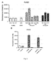
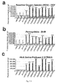
- the inventors observed that POLG2 levels essentially increased in cells from CS patients compared to healthy individuals and to UV S S, although the effect was particularly strong for CSB mutations than for CSA mutations ( Figure 3A ). In one CS case (patient 539) there was no significant increase of POLG2. It is hypothesized that increased levels of the accessory protein may result as compensation of the decreased levels of POLG to reinforce the DNA polymerase complex.
- silencing of CSB in HeLa cells results in increased levels of POLG2 ( Figure 3B ), and reversion of the silencing for the loss of plasmid results in dropping the levels of POLG2, showing that increased levels of POLG2 are dependent on impaired CSB.
- HTRA3 high-temperature requirement factor A3
- HTRA3 is a nuclear-encoded mitochondrial serine protease that degrades damaged proteins, and has a function during development, and possibly as tumor suppressor 21 .
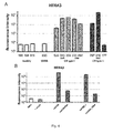
- the inventors observed dramatically high levels of HTRA3 protein, assessed by immunofluorescence, in fibroblasts from CS patients, compared to controls and UV S S fibroblasts ( Figure 4A ).
- CBS-silenced HeLa cells result in highly increased levels of HTRA3, and reversion of the silencing greatly decreases HTRA3 levels ( Figure 4B ).
- the precocious ageing phenotype can be ascribed to the mismanagement of oxidative stress in CS and cells affected by ageing, let it be physiological or physiopathological, as described herein.
- cells from CS patients are characterized by lower levels (e.g. at least a 20% decrease, in particular when tested by immunofluorescence) of the nuclear encoded mitochondrial DNA polymerase gamma (POLG), by higher levels of the accessory factor POLG2 (e.g. at least a 25% increase, in particular when tested by immunofluorescence), and by dramatically higher levels of the serine protease HTRA3 (e.g. at least a 10-folds increase, when tested by immunofluorescence). They also showed that alterations in the levels of these proteins are linked to impairment of CSB.
- POLG nuclear encoded mitochondrial DNA polymerase gamma
- CSB impairment directly or indirectly increases Htra3 levels, and this serine protease in turn degrades its targets, which include POLG.
- POLG2 the accessory protein
- replication of mitochondrial DNA is affected when CSB is not operating, leading to a decline in the mitochondrial function and thereby to enhanced production of oxidative stress.
- Increased oxidative stress and affected mitochondrial function which cumulate with time, contribute to leading to precocious ageing phenotype.
- HTRA3 is a serine protease.
- MG132 that is a specific proteasome inhibitor
- KSTI Soybean trypsin inhibitor
- MG132 is a potent, reversible, and cell-permeable proteasome inhibitor that reduces the degradation of ubiquitin-conjugated proteins in mammalian cells. MG132 is known for its induction of apoptosis and to specifically target cancer cells versus normal cells, although the reasons for this specificity have not been elucidated 23,24 . Soybean trypsin inhibitor is a natural serine protease inhibitor 25,26 . It is mentioned as Kunitz soybean trypsin inhibitor (KSTI).
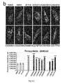
- the inventors treated fibroblasts from healthy individuals, from UV S S and from Cockayne syndrome of type I and II (the last being the more severe), with protease inhibitors MG132 (5 ⁇ M) and KSTI (100 ⁇ g/ml) for 5 hours and then tested for POLG levels.
- Significant modifications in two CS type I fibroblasts (539 and 548) were not observed whereas increased levels of POLG in the presence of either inhibitors in other CS type I and type II fibroblasts (359, 797, and 816) where POLG levels exceeded those of untreated healthy individuals, were found.
- POLG increased to levels of healthy individual after treatment with MG132.
- the limited increase of POLG levels in cells from patient 177 is considered to be interesting, given that these cells do not display an increase in HTRA3 levels either (not shown).
- Data from cells 177 suggest that their defect may be due to another protease, which is also targeted by protease inhibitors tested here.
- protease inhibitors in CS cells it is possible to restore POLG levels at least as high as in normal cells.
- the fact that in two cases the inventors did not observe increase in POLG levels suggests that other proteases could be targeted using additional protease inhibitors.
- HTRA3 may therefore not be the only protease interacting with POLG, suggesting that treatment aimed at increasing POLG levels can be effective through protease inhibitors having a different specificity than only specificity for HTRA3 as a target, in particular protease inhibitors having a large-range specificity.
- the POLG substrate could be improperly modified by other enzymatic activities, or be poorly modified by these activities, so that the protein becomes a poor target for being degraded by HTRA3 or other proteases.
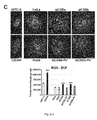
- the inventors also carried out experiments aimed at assessing the relative levels of oxidative stress in Cockayne syndrome (CS) fibroblasts, thereby revealing a preminent nitroso-redox imbalance in said fibroblasts.
- CS Cockayne syndrome
- the inventors assessed the relative levels of oxidative stress using the fluorescent probe dichlorofluorescein diacetate (DCFHDA), which prevalently detects reactive oxygen species (ROS) 29 . They reported that whereas UV S S cells display moderate (25%) increase of signal compared to controls, all CS cells are characterized by higher levels (1.6 to 2-fold) of oxidative stress ( Figure 6a ), in agreement with a previous finding 30 .
- DCFHDA fluorescent probe dichlorofluorescein diacetate
- ROS reactive oxygen species
- altered mitochondrial parameters it is in particular meant HTRA2 and/or HTRA3 and/or POLG protein(s) level(s), as illustrated below and in Figure 7 .
- the inventors quantified fluorescence intensities on cells as summarized in Table 1 before and after treatment with MnTBAP (purchased from Millipore) of DCF and of DHR123 per cell ( Figure 7 (a) and (b) ), as well as fluorescence intensities of HTRA2 ( Figure 7 (c) ), HTRA3 ( Figure 7 (d) ), POLG ( figure 7(e) ) per cell. They also evaluated the mtDNA content and ATP levels in cells as summarized in Table 1.
- Inventors' data point to a completely new mechanism to explain defects in CS cells, which are also relevant for the process of precocious ageing in other diseases, and also for the process of physiological ageing. Inventors' data do not exclude that DNA repairs alterations take place in these cells, and that these alterations may lead to the symptoms of precocious ageing and tumours. Inventors' data show that cells from patients with CS display dramatically reduced POLG, the nuclear-coded DNA polymerase that replicates mitochondrial DNA, compared to cells from normal individuals and UV S S patients (these patients carry a mutation in CSA, as it is the case for several CS patients, the other being mutated in CSB).
- the inventors thus assume that as a consequence the mtDNA is not properly replicated and thereby mitochondria are dysfunctional, in spite of no apparent modifications in shape and network, compared to cells from normal individuals. Such dysfunction will lead to an increase of oxidative stress, which is essentially produced by mitochondria, likely leading to dysfunctions observed in CS cells.
- the inventors showed that POLG decrease is associated with the CSB mutation as silenced CSB in HeLa cells behave as CS cells in this aspect, and restoration of the regular levels of CSB results in returning (at least) to POLG values as in non-silenced cells.
- the decrease in POLG levels in CS cells is associated with the increase in POLG2, a co-factor of POLG that does not contain the catalytic subunit.
- the inventors also showed that reduced levels of POLG protein, in particular by at least 20% (but not of POLG transcripts that are not affected) are due to increased levels (in particular by at least a 10-folds increase) of HTRA3 (transcript and protein), a serine protease that has POLG and other proteins as potential target.
- HTRA3 transcription and protein
- HTRA3 serine protease that has POLG and other proteins as potential target.
- POLG mitochondrial DNA polymerase gamma
- POLG, POLG2 protein and HTRA3 protein and transcript levels can be used as distinct markers for the diagnosis of Cockayne syndrome, as well as markers of mitochondrial dysfunctions associated with ageing in general, as described herein, and in neurodegenerative disorders.
- POLG can be detected by sequencing the gene or checking that there is no large mtDNA depletion (as it is the case for pathological POLG mutations), POLG mutations are associated with severe pathological phenotypes in the child, characterized by various levels of muscle and nerve impairment, but not with precocious ageing.
- POLG is functional for providing a treatment within the context of the invention. Indeed the treatment of the invention can only improve mitochondrial function if POLG is functional.
- a treatment according to the invention would not be efficient.
- POLG levels increase in most of CS cells in the presence of at least one protease inhibitor resulting in POLG levels at least as high as in untreated healthy cells.
- protease inhibitors to increase POLG levels, whose reduction is a major indication of the CS phenotype, for treating Cockayne syndrome patients, and in particular for targeting the precocious ageing phenotype.
- HTRA3 Although it is not clearly elucidated what modulates HTRA3 levels, the inventors postulate, by analogy with HTRA2, whose expression increases in tissues undergoing oxidative stress 27 , that HTRA3 expression is also promoted in the presence of stress. CS cells have been reported to accumulate oxidative stress 28 . Alteration of ROS levels may also affect the nitroso-redox balance, as ROS and NO are linked. Nitroso-redox imbalance plays a key role in cell and organ failure, and this could also be the case for the aetiology of CS ( Nediani et al, 2011 Antioxidants & Redox signaling 14 (2) 289-331 ; Takahashi, 2012 J. of Reproduction and Development 58 (1) :1-9 ; Taverne et al, 2012 J.Appl. Physiol 112 : 1644-1652 )
- ROS and peroxynitrite scavenging rescues altered mitochondrial parameters in fibroblasts from Cockayne Syndrome (CS) patients the inventors reasoned that if Reactive Oxygen Species (ROS) and peroxynitrite induce serine proteases accumulation thereby resulting in POLG depletion, original parameters would be restored in CS fibroblasts treated with ROS and peroxynitrite scavengers.
- ROS Reactive Oxygen Species
- peroxynitrite induce serine proteases accumulation thereby resulting in POLG depletion
- Manganase(III)tetrakis(4-benzoic acid)porphyrin is a synthetic metalloporphyrin which mimics superoxide dismutase and scavenges ROS and peroxynitrite 32 .
- Treatment with MNTBAP for 24h decreased by two thirds the levels of ROS, measured by DCFHDA, in control and UV S S fibroblasts, confirming the ROS scavenger effect of this molecule ( Figure 7a ).
- treatment resulted in decrease of ROS by 80-95% in CS cells.
- MNTBAP greatly reduced the levels of and peroxynitrite, measured by DHR123, in all control and patient cells ( Figure 7b ).
- MNTBAP increased the levels of HTRA3 in control cells, but did reduce in UV S S and, to a largest extent in CS cells, where it restored control levels of HTRA3 ( Figure 7d ).
- overexpression of HTRA3 in CSA/CSB impaired cells is promoted by high ROS and peroxynitrite levels, whereas nitro-oxidative stress represses HTRA3 expression in CSA and CSB proficient fibroblasts.
- MnTBAP to rescue altered mitochondrial parameters, in particular POLG levels, whose reduction is a major indication of the CS phenotype, and/or use MnTBAP for treating Cockayne syndrome patients.
- CSB mutation(s) result(s) in increased ROS levels and POLG/POLG2/HTRA3 alterations.
- POLG/POLG2 alterations induce mitochondrial impairment, revealed notably by altered mitochondrial DNA content and altered mitochondrial mass, reduced mitochondrial respiration, which in turn can generate more ROS.
- the inventors consider that there is a relationship between CSB and the regulation of the levels of ROS.
- CSB mutation(s) increase(s) ROS levels by affecting the expression of ROS-regulating factors. ROS levels also increase because of dysfunctional mitochondria (due to POLG/POLG2/HTRA3 alterations, as shown by the inventors), which is a fact also dependent of the CSB mutation(s).
- Ataxia telangiectasia A-T
- Base excision repair BER
- Bloom syndrome BS
- Cockayne syndrome CS
- DNA polymerase gamma POLG
- DNA polymerase subunit gamma-2 POLG2
- Double strand break repair DSB
- GG-NER Fanconi anemia
- HTRA2 High-temperature requirement factor A2
- HTRA3 High-temperature requirement factor A3
- Hutchinson-Guilford progeria syndrome HGPS
- Kunitz Soybean trypsin inhibitor KSTI
- Mitochondrial transcription factor A TFAM
- Nucleotide excision repair NER
- Rothmund-Thomson syndrome RTS
- ROS Reactive oxygen species
- SCE Spontaneous sister chromatid exchanges
Abstract
The invention relates to a method for in vitro investigating mitochondrial replication dysfunction in a biological sample removed from a subject susceptible of suffering from physiological ageing or physiopathological conditions related to physiological ageing, or physiopathological ageing or associated symptoms or conditions, in particular premature ageing or accelerated ageing, or of a progeroid syndrome, such as Cockayne syndrome (CS), or neurodegenerative disorders or symptoms thereof, in which the levels of at least one species selected in the group of: POLG protein, POLG RNA, POLG2 protein, protease(s) which have POLG as a target, in particular serine protease(s) such as HTRA3 protein and, HTRA3 RNA, or any combination of these species, are investigated. The invention also relates to kits and uses thereof, therapeutic methods against progeroid-like syndromes or symptomes and screening method for identifying particular protease inhibitor(s) and/or nitroso-redox stress scavenger compound(s) having relevance for the symptoms discussed herein.
Description
- The present invention relates to the field of in vitro testing methods for investigating impaired mitochondrial DNA (mtDNA) replication phenomema in biological samples collected from individuals, animals or humans, and in particular relates to methods that can be applied to the monitoring and/or diagnosing of the health status of a subject susceptible of suffering from physiological ageing (also referred to as chronological ageing or organismal ageing herein), or physiopathological ageing, in particular premature ageing or accelerated ageing or of a progeroid syndrome, such as Cockayne syndrome (CS), or neurodegenerative disorders or symptoms thereof. The invention also relates to kits for performing the methods of the invention and their uses.
- The present invention also relates to a method for treating or delaying the symptoms of a subject suffering from physiological ageing or pathophysiological ageing, in this last case in particular premature ageing or accelerated ageing, or of a progeroid syndrome, such as Cockayne syndrome (CS), or neurodegenerative disorders, through administration of protease inhibitor(s), in particular serine protease inhibitor(s), having influence on the pathways associated with mtDNA replication, dysfunction and/or mismanagement of oxidative stress at the mitochondrial level, in particular defective pathways leading to abnormal levels of functional POLG protein, in particular abnormally low levels of POLG protein.
- The present invention also relates to a nitroso-redox stress scavenger compound for use in a patient in need thereof to treat or delay Cockayne syndrome (CS) or symptoms thereof, and/or restore level(s) of protein(s) as disclosed herein, in particular to treat or delay Cockayne syndrome (CS) or symptoms thereof, said nitroso-redox stress scavenger compound being MnTBAP.
- The present invention also relates to a screening method for identifying particular protease inhibitor(s) and/or nitroso-redox stress scavenger compound(s) of interest within the context of the invention.
- The present invention is of particular relevance in the context of diseases and symptoms directly associated with physiological ageing or physiopathological ageing, in particular premature ageing syndromes, especially progeroid syndromes - such as Cockayne syndrome (CS) -, which are a group of diseases all characterized by signs of premature ageing, and in the context of analysis of mitochondrial dysfunctions associated either with precocious ageing or observed during the onset and the establishment of neurodegenerative disorders.
- The present invention relies on experiments emphasizing the relevance of the pathways associated with the management of oxidative stress in altered cells, in particular at the mitochondrial level.
- Cockayne syndrome (CS), (OMIM entry 216400 http://www.omim.org/entry/216400, OMIM entry 133540 http://www.omim.org/entry/133540), also called Weber-Cockayne syndrome or Neill-Dingwall syndrome, is a rare genetic disorder characterized by neurological abnormalities and several growth and developmental defects, which include photosensitivity, hypersensitivity to oxidative damage, skeletal abnormalities, hearing loss, pigmentary retinopathy, progressive neurological disorders, mental retardation and premature ageing1.
- CS is an autosomal recessive disorder with an incidence of 1 per 2.7 million births in western Europe2. The incidence is extremely high in immigrant populations, likely for the consanguineous marriageS2.
- CS is due to mutation in genes CSA (or ERCCB)3 and/or CSB (or ERCC6)4 , which are required for nucleotide excision repair (NER), a DNA repair mechanism that removes bulky DNA adducts such as UV-induced DNA damage. About 70% of identified CS patients carry a mutation in CSB5. In particular, CSB is implicated in a sub type of NER, TC-NER (or transcription-coupled NER) also called TCR, that acts specifically on lesions located on the transcribed strand of expressed genes6. The other NER subtype GG-NER (or global genome NER) acts anywhere throughout the genome, and is normal in CS patients. The absence of TCR in cells treated by genotoxic agents leads to apoptosis. Mutations in the CSA or CSB genes are known to affect the proper performance repair of altered DNA structures resulting from UV damage, via transcription-coupled nucleotide excision repair (TC-NER) pathway. In a CS diagnosis situation however, the situation is complicated by the fact that some mutations in CSA exist, which affect the TC-NER pathway but do not affect the oxidative stress response, and are accordingly associated with a much milder syndrome (UV-sensitivity syndrome, UVSS) with no sign of precocious ageing11.
- There are at least two forms of CS: type I, which is characterized by normal foetal growth and abnormalities appearing in the first two years or later and degeneration between 10-20 years, and type II, which is associated with little neurological development early after birth and death usually during the first decade.
- It is important to note that currently neither large-scale diagnosis method nor a pre-natal test exists for CS, other than genotyping for CSA and CSB mutations. In spite of the devastating phenotype of CS, to date there is no indication of relevant, and easy to detect, cellular alterations in CS cells compared to UVSS and to normal cells.
- Turning to the molecular mechanism(s) involved in the CS, the current prevalent view is that TC-NER is impaired in CSA and CSB mutants, which results in the inability to repair the DNA lesions or clearing stalled RNA polymerase II in front of DNA damage, thereby blocking transcription after UV damage resulting in global transcription arrest. However, it seems that CSB-mutated cells, which have been extensively studied, have a transcription defect beyond the TC-NER impairment7. In this context, CSB appears as a transcription factor implicated in the activation of several genes and networks.
- The severe phenotype of CS patients is however hard to reconcile with a sole defect in TC-NER and transcription7.
- In fact, CSB cells, i.e. cells from patients known to have an impaired CSB gene, as disclosed in Nardo et al, 2009 PNAS 106 (15) :620914, are also hypersensitive to oxidative damage. Indeed CSA and CSB appear to be involved in the repair of oxidative DNA lesions, produced by endogenous reactive oxygen species and normally repaired by the base excision repair (BER) pathway8. CSA/CSB modulate the BER pathway by direct interaction with BER proteins, and also by modulating the expression of BER genes. Both nuclear and mitochondrial BER are involved. Indeed CSA and CSB have been detected not only in the nucleus, but also in mitochondria9,10. The implication of CSA/CSB in the repair of oxidative stress could explain developmental defects and the neurological degeneration observed in CS. However, the impairment of BER due to CSA or CSB deficiency is mild, suggesting an additional role of the CS proteins in the response to oxidative stress and more in general in the etiology of CS8.
- Interestingly, a mutation in CSA has been associated with the UV-sensitive syndrome (UVSS), an autosomal recessive disorder characterized by mild cutaneous symptoms and defective TC-NER, but not with other symptoms of the CS, in particular no hypersensitivity to oxidative damage and no precocious ageing11. Thus defects in TC-NER and oxidative damage repair have been uncoupled in the phenotype of CSA mutations, suggesting that much of CS symptoms are due to the management of oxidative stress rather than to transcription-related DNA repair.
- In fact, defects in NER are associated not only with CS, but also with xerodema pigmentosum (XP), characterized by increased sensitivity to tumors in sun-exposed areas of the skin, and trichothiodystropy (TTD), characterized by developmental and neurological abnormalities, and premature ageing12. CS and TTD are cancer-free disorders. XP and TTD are essentially associated with defective GG-NER12.
- More generally, Cockayne syndrome is considered to pertain to progeroid syndromes, which are a group of diseases all characterized by signs of premature ageing. These syndromes include: Hutchinson-Guilford progeria syndrome (HGPS), Werner syndrome (WS), Bloom syndrome (BS), Rothmund-Thomson syndrome (RTS), Fanconi anemia (FA), Ataxia telangiectasia (A-T), Cockayne syndrome (CS), Xeroderma pigmentosum (XP) and trichothiodystropy (TTD)13.
- Several of these syndromes are classified as segmental progeroid syndromes as multiple organs and tissues replicate phenotypes associated with normal ageing14.
- HGPS and WS are two of the best characterized human progeroid diseases15. HGPS, which is one of the most severe forms of progeria, has an incidence of 1 in 4-8 million births and distinct clinical symptoms are developed during the first year of life, and patients die at a median age of 11-13 years. HGPS is also called "progeria of the childhood". WS has an incidence of 1 per million births (but 1:100,000 in Japan), and Werner heterozygotes are 1/180 in the general population. Symptoms appear in the first-second decade and the life expectancy reaches 47-54 years. WS is also called "progeria of the adult".
- Most progeroid syndromes include defects in distinct repair systems such as NER, BER, and double strand break repair (DSB)13,15. The exact mechanism(s) by which these mutations lead to progeria is/are not known yet. Moreover, the extent of progeria is different among these syndromes. At present, there is no also no indication why mutation in some genes result in more severe progeria syndromes than mutations in other genes.
- HGPS is mutated in LMNA that encodes for the four different types of lamins. Mutations activate a cryptic splice site that lead to deletion of 50 aminoacids (the deleted protein is called progerin) that cannot undergo further processing. Lamins constitute the major component of the nuclear lamina, which provide structure and shape to the nucleus and are also involved in chromatin organization and DNA replication, transcription, and repair. The prevalent view is that lamin mutations lead to deficient DNA damage response, probably by sequestering replication and repair factors, leading to stalled DNA replication forks that collapse into DSBs. Moreover, lamin defects would increase DNA damage signalling at the level of telomeres and reduce the telomere length, leading to early cell senescence.
- The Werner syndrome is associated with mutation of WRN, an ATPase-helicase (of the family of RECQ helicases, which unwinds the DNA double strands by hydrolyzing ATP). WRN protein is involved in DNA replication, recombination and telomere maintenance and its impairment results in chromosomal aberrations.
- The Bloom syndrome and the Rothmunds-Thomson syndrome are due to mutation in the RECQ helicases BLM and RECQ4, respectively. As with the WRN helicase, the RECQ4 and BLM helicases are necessary to maintain genome integrity, but they differ in their functions and in their interaction partners16. Bloom syndrome is characterized by a very high level of spontaneous sister chromatid exchanges (SCE).
- At least 16 genes responsible for the Fanconi anemia have been identified, and they are all linked to a DNA damage signalling pathway (the FA pathway) that is activated in response to DNA damage, in particular DSBs. Eight FA proteins form a nuclear-localized complex with E3 ubiquitin ligase and thereby catalyze monoubiquitination in target proteins. This monoubiquitination does not lead to proteasomal degradation but it can alter cellular localization or the function of the target protein.
- Ataxia-telangiectasia is due to mutation in the ATM gene, a serine threonine kinase that is important at the level of DNA damage (in particular DSB) signalling and activation of DNA repair mechanisms.
- All progeroid diseases display clinical features mimicking physiological ageing at an early age. They might provide insights into the process of normal human ageing and/or dysfunctions linked to nomal human ageing (also referred to as physiological or chronological or organismal ageing herein), which is itself characterized by dysfunction of several physiological processes, as well as insights into physiopathological ageing, in particular premature ageing, as disclosed herein15.
- By "early age" within the expression "physiological ageing at an early age" recited above, it is meant an age that is earlier than the age of normal onset of the symptoms of physiological ageing or an age that is not consistent with increased frequency of a condition or a disease that is generally related to aging.
- Clinical symptoms of physiological ageing or physiopathological ageing, in particular premature aging, include skin atrophy with loss of cutaneous elasticity, dysfunction of cutaneous appendices, degeneration of the central nervous system, neurodegenerative symptoms, diabetes mellitus, changes in the volume of the adipose tissue, pigmentary changes with hyper- and hypopigmentation of the skin (poikiloderma), regional skin fibrosis, premature hair graying or hair loss, osteoporosis, and in certain cases tumors typical of those seen in patients of older age13. These symptoms are also associated with physiological ageing, although they appear at a later age in normal individuals.
- In this context, inventors' investigations focused on the mechanism(s) underlying the etiology of CS in particular, which are also applicable to all diseases displaying symptoms of physiological ageing or physiopathological ageing, in particular premature ageing. These investigations have put in light dysfunctional mitochondrial pathways, in particular associated with ageing in general, and a completely new mechanism that may in particular explain defects in CS cells.
- Back to the considerations introduced above regarding the management of oxidative stress, mitochondria are largely responsible for the production of oxidative stress, and CS patients are highly sensitive to oxidative damage. Oxidative damage is known to affect replication and transcription of mitochondrial DNA resulting in a decline of the mitochondrial function18. A large set of data suggests that oxidative damage is also associated with physiological ageing.18 Therefore, alterations in syndromes of precocious ageing like CS are considered informative also for understanding physiological ageing, since they recapitulate the dysfunction(s) observed in physiological ageing (Dreesen and Stewart, 2011 Aging, 3:889-895; Scaffidi and Misteli, 2006 Science 312: 1059-1063).
- The present invention is based on the findings of new elements paving the way to a better diagnosis and treatment of symptoms of physiopathological ageing, in particular premature ageing or accelerated ageing or diagnosis and treatment of progeroid syndrome(s), such as Cockayne syndrome (CS), or neurodegenerative disorders or associated symptoms.
- The invention therefore relates to an in vitro method for investigating mtDNA replication dysfunction (defective mtDNA replication or, differently said, mtDNA replication impairment or dysfunction of the mtDNA replication apparatus and/or machinery) in a biological sample removed from a subject susceptible of suffering from physiological ageing or physiopathological conditions related to physiological ageing, or physiopathological ageing or associated symptoms or conditions, in particular premature ageing or accelerated ageing, or of a progeroid syndrome, such as Cockayne syndrome (CS), or neurodegenerative disorders or symptoms thereof, said method comprising the steps of:
- a. contacting said biological sample with at least one marker specific for at least one species selected in the group of: POLG protein, POLG RNA, POLG2 protein, protease(s) which have POLG as a target, in particular serine protease(s) such as HTRA3 protein and, HTRA3 RNA, or any combination of these species, in conditions enabling said marker(s) to react with their respective targets (in particular, species), and
- b. determining the level of at least one species selected in step a) from the group of: POLG protein, POLG RNA, POLG2 protein, protease(s) which have POLG as a target, in particular serine protease(s) such as HTRA3 protein, and HTRA3 RNA, or any combination of these species in said biological sample through measurement of the marker(s) that has(have) reacted with its(their) respective species in step a), or through measurement of the reaction product(s) obtained after reacting the marker(s) with its(their) respective species in step a), and
- c. comparing the level(s) determined in step b) with respective normal threshold value(s) determined for healthy subject(s) for each species selected in the group set forth in step a) and b) to carry out said steps, and
- d. from the comparison made in step c), concluding about the existence of a dysfunction in mtDNA replication.
- Within the context of the invention, it has been shown by the inventors that dysfunctional mtDNA replication can be correlated to the observed levels of expressed POLG, although not due to a mutation affecting POLG in way rendering POLG protein dysfunctional in itself. It is documented that POLG mutations are associated to mtDNA depletion syndromes. However, within the context of the invention, impaired mtDNA replication due to reduced level of the key replicating enzyme (which otherwise works well), which is POLG, can be evidenced.
- Mitochondrial DNA replication is catalyzed by a mitochondria-specific mitochondrial complex comprising the so-called mitochondrial DNA polymerase gamma (γ) holoenzyme, which is an heterotrimer consisting of a single 140 kDa catalytic unit (encoded by the POLG gene at the nuclear chromosomal locus 15q25) and a 55 kDa accessory subunit that forms a tight dimer (encoded by the POLG2 gene at nuclear chromosomal locus 17q).
- The POLG protein (SEQ ID NO:1) (NCBI Reference POLG: Gene ID 5428 http://www.ncbi.nlm.nih.gov/gene/5428, Primary source HGNC:9179, POLG protein NCBI Reference Sequence: NP_001119603.1 http://www.ncbi.nlm.nih.gov/protein/NP_001119603.1) is therefore the catalytic subunit of the so-called mitochondrial DNA polymerase gamma (γ), needed for mtDNA replication. Human POLG (SEQ ID NO:2) is discussed in Stumpf and Copeland, 2011, Cell. Mol. Life Science 68 :219-233 or Ropp & Copeland, 1996 Genomics 36 :449-458. As shown on page http://www.ensembl.org/Homo_sapiens/Gene/Family?family=ENSFM00610 000964734;g=ENSG00000140521;r=15:89859534-89878092, there are several protein transcripts (splicing variants) corresponding to human POLG. Accordingly, by POLG protein as referred to herein, it is meant the native form of the protein having a sequence as disclosed in databases and/or literature and/or herein, but also isoforms or variants thereof having a polypeptidic sequence showing 60% or 70% or 80% or 90% or 95% and up to 99% identity with the polypeptidic sequence of the native POLG protein.
- POLG2 protein (SEQ ID NO:3) (NCBI Reference POLG2: Gene ID 11232 http://www.ncbi.nlm.nih.gov/gene/11232, Primary source HGNC:9180, POLG2 protein NCBI Reference Sequence: NP_009146.2 http://www.ncbi.nlm.nih.gov/protein/NP_009146.2), also called mitochondrial DNA polymerase subunit gamma-2, is a protein that in humans is encoded by the POLG2 gene, and is an accessory protein that increases the processivity of the catalytic subunit of the POLG protein. Human POLG2 (SEQ ID NO:4) is discussed in Young et al, 2011 Human Molecular Genetics 20 (15) :3052-3066. As shown on page http://www.ensembl.org/Homo_sapiens/Gene/Family/Genes?cdb=compara; db=core;family=ENSFM00250000007196;g=ENSG00000256525;r=17:624 73902-62493154, there are several protein transcripts (splicing variants) corresponding to human POLG2. Accordingly, by POLG2 protein as referred to herein, it is meant the native form of the protein having a sequence as disclosed in databases and/or literature and/or herein, but also isoforms or variants thereof having a polypeptidic sequence showing 60%, 70% or 80% or 90% or 95% and up to 99% identity with the polypeptidic sequence of the native POLG2 protein.
- HTRA3 protein (SEQ ID NO:5) (NCBI Reference HTRA3: Gene ID 94031 http://www.ncbi.nlm.nih.gov/gene/94031, Primary source HGNC:9180, HTRA3 protein NCBI Reference Sequence: NP_444272.1 http://www.ncbi.nim.nih.gov/protein/NP_444272.1) is a serine peptidase (or serine protease) that is a member of the mammalian HTRA family. Human HTRA3 (SEQ ID NO:6) and human HTRA3 are discussed in Nie et al, 2003 Biochemical Journal 371 :39-48 or Narkiewicz et al, 2009 Oncology reports 21 : 1529-1237. As shown on page http://www.ensembl.org/Homo_sapiens/Transcript/Summary?db=core;g=E NSG00000170801;r=4:8271492-8308838;t=ENST00000307358, there are several protein transcripts (splicing variants) corresponding to human HTRA3. More particularly, there are two (2) transcripts for HTRA3 (different splicing), giving a long (L) and a short (S) form, which are both discussed herein. Accordingly, by HTRA3 protein as referred to herein, it is meant the native form of the protein having a sequence as disclosed in databases and/or literature and/or herein, but also isoforms or variants thereof having a polypeptidic sequence showing 60%, 70% or 80% or 90% or 95% and up to 99% identity with the polypeptidic sequence of the native HTRA3 protein.
- In a particular embodiment, the conclusion of step d) of the method of investigating the existence of a mtDNA replication dysfunction of the invention is made if the level of each species selected to carry out the above disclosed steps is as follows:
- the level of POLG protein determined in step b) is decreased with respect to the normal threshold value introduced in step c) by at least 10%, and/or
- the level of POLG2 protein determined in step b) is increased with respect to the normal threshold value introduced in step c) by at least 15%, and/or
- the level of HTRA3 protein, in particular the long isoform of HTRA3 protein, and/or HTRA3 RNA determined in step b) is increased with respect to the normal threshold value introduced in step c) by at least 2 folds, or at least two of the above measurements meet the above thresholds.
- By "mtDNA replication dysfunction", it is meant that mtDNA replication is altered or is totally impaired in cells of the assayed biological sample. This expression is used herein as a synonym of defective mtDNA replication or mtDNA replication impairment. Indeed, the inventors showed that in cells of a sample wherein mtDNA replication is dysfunctional, only the amount of the enzyme responsible for synthesizing of mtDNA that is POLG, is lower or much lower than in healthy control cells. Moreover the mtDNA content and the levels of TFAM, which is a factor involved in mtDNA transcription and maintenance is generally altered in one sense or the other, indicating that, within the context of the invention, it is the process of mtDNA replication that is affected, and not POLG that is defective in itself. Finally, the inventors have also evidenced that ATP production by mitochondria is reduced in all tested CS samples that have POLG defects, showing that the mitochondrial function itself is affected in the tested cases. These methods may be used for demonstrating that mtDNA replication is impaired in a given assayed sample.
- As a synonym for "mtDNA replication dysfunction" can also be used the expression "dysfunction of the mtDNA replication apparatus and/or machinery". Indeed, POLG is the key replication enzyme for mtDNA replication. If POLG declines, it is a fact that the replication apparatus is dysfunctional, as illustrated above. In addition, it is stated by the inventors that POLG decrease results in altered mtDNA content (essentially decrease but also increase). Therefore, there is a direct relationship between the levels of the species monitored within the present invention and the fact that mtDNA replication is impaired, which is also correlated with a dysfunctional mtDNA replication apparatus and/or machinery. Impaired mtDNA replication is indeed evidenced by a dysfunctional mtDNA replication apparatus and/or machinery, as illustrated above.
- By "biological sample" it is meant a sample originating from the sampling of biological tissue(s) or fluid(s), especially body tissue(s) or fluid(s), which is therefore substantially constituted of cells, for example bodily fluid such as a cerebrospinal fluid, saliva, mucus, urine or blood sample, or include a cell lysate of the same origin, and/or include a conditioned culture medium, and is optionally derived from a tissue (e.g., a tissue homogenate), a biopsy.
- In a particular embodiment, the assayed biological sample comprises or contains fibroblasts or culture(s) thereof, or consists of isolated cells, in particular fibroblasts, or culture(s) thereof. However, other cells should also be considered as much as they grow in culture or they are used as isolated cells from a biological sample (for example from a body fluid), as described above.
- According to a particular embodiment, the in vitro method of the invention is used within a prenatal testing procedure, wherein the tested subject is an embryo or a foetus. In addition to samples generally used for this type of testing (such as amniotic tissue), it is also possible to test parent(s) material (such as fibroblasts, cells from biopsies). Parents may be heterozygotes for the CSA or CSB mutation and their POLG/HTRA3 values might be different from controls (and from CS). This seems indeed the case for the parent that the inventors have tested (CS358), as described herein, and who has no CS phenotype.
- By "physiological ageing or physiopathological conditions related to physiological ageing, or physiopathological ageing or associated symptoms or conditions, in particular premature ageing or accelerated ageing, or of a progeroid syndrome, such as Cockayne syndrome (CS), or neurodegenerative disorders or symptoms thereof", it is meant all conditions disclosed above and herein, in particular a condition resulting in skin atrophy with loss of cutaneous elasticity, dysfunction of cutaneous appendices, degeneration of the central nervous system, neurodegenerative symptoms, diabetes mellitus, changes in the volume of the adipose tissue, pigmentary changes with hyper- and hypopigmentation of the skin (poikiloderma), regional skin fibrosis, premature hair graying or hair loss, osteoporosis, muscle atrophy, weight loss, alopecia, kyphosis, anaemia, reduced fertility, and in certain cases tumors typical of those seen in patients of older age, as well as symptoms typical of neurodegenerative disorders like ophtalmoplegia, ataxic neuropathy, inflammation, cerebellar ataxia, cachexia, neuron loss, deterioration of cognitive and kinetic body response.
- By "marker specific for at least one species selected in the group of: POLG protein, POLG RNA, POLG2 protein, protease(s) which have POLG as a target, in particular serine protease(s) such as HTRA3 protein and, HTRA3 RNA", it is meant a marker suitable for directly or indirectly specifically revealing the qualitative and/or quantitative presence of, respectively, one of the following species: POLG RNA, POLG2 protein, protease(s) which have POLG as a target, in particular serine protease(s) such as HTRA3 protein and, HTRA3 RNA, when further performing detection methods, in particular methods such as immunofluorescence, Western Blotting, ELISA or a PCR-based amplification method, such as RT-qPCR.
- The specificity of the marker is assessed with respect to its ability to react with its target but not to react in a detectable or in a functionally effective manner with other compounds of the sample.
- For example, nucleotide probes specifically binding to DNAs or RNAs and carrying a ligand at one end, which is recognized by a specific antibody, have been developed. Molecular beacons (antisense nucleotide probes targeting DNA or RNA with a fluorophore and a quencher at opposite ends), which emit a signal when hybridizing their targets (and are thus detected by fluorescence) have also been developed. These embodiments are encompassed in the definition provided above with respect to the markers that can appropriately be used within the present invention.
- In the case of, but not exclusively, proteins, in particular POLG, POLG2, protease(s) as identified above, HTRA3, such marker(s) may be an antibody specific for said protein(s), in particular POLG2 and/or HTRA3 protein(s) or a combination of several antibodies altogether specific for said protein(s), and, optionally, one or several of secondary antibody(ies) or reagent(s) (such as dye(s)) to reveal a complex between specific antibody(ies) recited above and its(their) target.
- In case of RNA determination, in particular POLG RNA or HTRA3 RNA, such marker(s) may be at least one pair of specific oligonucleotide primers specific for hybridization (by base pairing) with the cDNA corresponding to the RNA target (e.g. POLG or HTRA3 RNA) or at least one pair of specific oligonucleotide primers specific for directly hybridizing with the corresponding target RNA, and, optionally, at least one label or marker for detection of nucleic acids, in particular a dye detectable in a real-time PCR equipment, for revealing the HTRA3 RNA or cDNA marker. According to a particular embodiment, the pair of specific oligonucleotide primers that is used is capable to hybridize both with the target RNA and the cDNA synthesized using the target RNA as a template.
- A "marker specific for POLG protein" is a marker suitable for specifically targeting and optionally revealing the qualitative and/or quantitative presence of POLG protein when further performing, in particular, immunofluorescence, Western Blotting or ELISA detection methods.
- In the case of POLG protein level measurement, such a marker may be an antibody specific for POLG protein or a combination of several antibodies specific for POLG protein, and, optionally, one or several secondary antibody(ies) or reagent(s) to reveal a complex between specific antibody(ies) recited above and its(their) target, as in particular described herein within the Materials and Methods section. Other markers may include detectable molecules having a binding capacity or interaction capacity with POLG.
- By "conditions enabling said marker(s) to react with their respective targets", it is meant either "conditions enabling a marker to react with its respective target RNA and/orcDNA", or "conditions enabling a marker to react with its respective target protein".
- By "conditions enabling a marker to react with its respective target RNA and/orcDNA", it is meant conditions enabling hybridization of primers to their nucleic acid target(s) for further performing an amplification method, as known by a person skilled in the art and/or described in notices provided by manufacturers when commercial kits are used, as in particular described herein within the Materials and Methods section.
- By "conditions enabling said marker to react with a protein", in particular the POLG protein, it is meant, in particular, but not exclusively, conditions enabling an immunological reaction to take place, as known by a person skilled in the art and/or described in notices provided by manufacturers when commercial kits and/or reagents are used, as in particular described herein within the Materials and Methods section.
- By "measurement of the marker(s) that has(have) reacted with its(their) respective species it is therefore in particular made reference to the process of measurement of level(s) of protein(s), as described above, in particular by Immunofluorescence (IF), Western Blot or ELISA detection methods.
- By "measurement of the reaction product(s) obtained after reacting the marker(s) with its(their) respective species", it is therefore in particular made reference to the process of measurement of level(s) of RNA(s), as described above, in particular as reflected by the amplification product obtained after performing a PCR-based amplification method, such as RT-qPCR, on said RNA present in the assayed sample.
- By "determining the level of at least one species selected in step a) from the group of: POLG protein, POLG RNA, POLG2 protein, protease(s) which have POLG as a target, in particular serine protease(s) such as HTRA3 protein, and HTRA3 RNA, or any combination of these species in said biological sample", it is meant obtaining an absolute or relative value representative of the amount of target species in the assayed sample, in particular by either:
- interpreting the results (raw data or transformed data) obtained through immunofluorescence, Western Blotting or ELISA detection methods as mentioned above, or as described herein within the Materials and Methods section, in order to evaluate the amount of target protein present in the assayed sample, or
- interpreting the results (raw data or transformed data) obtained after performing a PCR-based amplification method, such as RT-qPCR, on nucleic acids present in the assayed sample, the level being deduced from the quantity of amplicons present after amplification and the amount of PCR cycles, as in particular described in the Material and Methods section herein.
- In particular, by "determining the level of POLG protein", it is meant obtaining an absolute or relative value, in particular by interpretation of the results (raw data or transformed data) obtained through the immunofluorescence, Western Blotting or ELISA detection methods mentioned above, or as described herein within the Materials and Methods section, which is suitable for evaluating the amount of POLG protein present in the assayed sample.
- It will be understood from the above that marker(s) used within the present invention to obtain level(s) of specific species(s) as disclosed herein, are means suitable for revealing, directly or not, said level(s), if necessary after further steps based on the formation of an immunological complex between a particular marker and its target (e.g. when antibodies or labeled antibodies or set of antibodies are used), and/or further steps based on the hybridization between a particular marker and a nucleic acid target (e.g. when primers are used), and subsequent nucleic acid amplification and counting, according to methods known in the art and/or disclosed herein.
- According to a particular embodiment, the level of POLG protein determined in step b) is decreased with respect to the normal threshold value introduced in step c) by at least 10%, in particular when the detection method that is used is not immunofluorescence.
- In a general manner, percentages are evaluated with respect to the unit value(s) appropriate in the method carried out for the described measurement(s).
- According to a particular embodiment, the level of POLG protein determined in step b) is decreased with respect to the normal threshold value introduced in step c) by at least 20%, or at least 30% or at least 40%, and up to 80% or up to 90%, in particular when the detection method that is used is immunofluorescence staining.
- According to a particular embodiment, the level of POLG2 protein determined in step b) is increased with respect to the normal threshold value introduced in step c) by at least 15%, in particular when the detection method that is used is not immunofluorescence.
- According to a particular embodiment, the level of POLG2 protein determined in step b) is increased with respect to the normal threshold value introduced in step c) by at least 25%, or at least 35% or at least 45%, in particular when the detection method that is used is immunofluorescence staining.
- According to a particular embodiment, the level of HTRA3 protein, in particular the long isoform of HTRA3 protein, and/or HTRA3 RNA determined in step b) is increased with respect to the normal threshold value introduced in step c) by at least 2 folds, in particular when the detection method that is used is not immunofluorescence staining.
- The level of increase with respect to a normal threshold value may depend on the quantification sensitivity of the detection method that is used.
- According to a particular embodiment, the level of HTRA3 protein, in particular the long isoform of HTRA3 protein, and/or HTRA3 RNA determined in step b) is increased with respect to the normal threshold value introduced in step c) by at least 5 folds, or at least 10 folds, in particular when the detection method that is used for detecting the level of HTRA3 protein is immunofluorescence staining. In a specific embodiment, said level of HTRA3 is increased by at least 50 or at least 60 or at least 70 folds when the detection method that is used is immunofluorescence staining. In a particular embodiment, the inventors found huge increase of HTRA3 RNA (2 to 160-fold) and HTRA3 protein by IF (60 to 3000-fold). However, when RT-qPCR is used for detecting RNA, obtained values may be lower, which is consistent with the fact that as one single RNA molecule can be used to produce multiple proteins. Therefore, according to a particular embodiment wherein the level of HTRA3 RNA is determined, if necessary in all combination(s) with other parameter(s) (in particular, species) as disclosed herein, said level of HTRA3 RNA determined in step b) is increased with respect to the normal threshold value introduced in step c) by at least 2 folds.
- According to particular embodiments, all the levels indicated above can be used in combination, in particular when meeting the above-mentioned thresholds, for concluding according to the explanations provided herein and/or deducible by a person skilled in the art.
- By "normal threshold value(s) determined for healthy subject(s) for POLG" it is meant level(s) of POLG protein found by assaying one biological sample from an healthy subject or alternatively found by assaying several biological samples from several distinct healthy subjects, the resulting normal threshold value being then determined as the mathematical mean of the levels of POLG protein values of all the assayed healthy subjects biological samples, or alternatively found by assaying a pool of biological samples from several distinct healthy subjects.
- According to another embodiment, an in vitro method for investigating mtDNA replication dysfunction of the invention also encompasses determination of the level(s) of other markers (and consequently, species), which are: POLG2 protein, protease(s) which have POLG as a target in a sample provided in conditions enabling said marker to react with said protease(s), in particular serine protease(s) such as HTRA3 protein, or a marker specific for HTRA3 RNA in a sample provided in conditions enabling said marker to react with HTRA3 RNA, as an additional parameter (species) to POLG level determination or as an alternative to a determination of the level of POLG protein.
- Such markers may be an antibody specific for a protein selected amongst: POLG2 and/or HTRA3 or a combination of several antibodies specific for POLG2 and/or HTRA3, and, optionally, one or several of secondary antibody(ies) or reagent(s) to reveal a complex between specific antibody(ies) recited above and its(their) target.
- In case of HTRA3 RNA determination, such a marker may be at least one pair of specific oligonucleotide primers specific for hybridization with HTRA3 cDNA or at least one pair of specific oligonucleotide primers specific for hybridization with HTRA3 RNA, and, optionally, at least one label or marker for detection of nucleic acids, in particular a dye detectable in a real-time PCR equipment, for revealing the HTRA3 RNA or cDNA marker.
- According to a particular embodiment, the in vitro method for investigating mtDNA replication dysfunction of the invention encompasses a determination of any combination of the above-mentioned species, in particular a combination of determination of POLG and POLG2 or a combination of determination of POLG and HTRA3 or a combination of determination of determination of POLG, POLG2 and HTRA3.
- In a specific embodiment, the method of the invention comprises, in addition to a determination if the level(s) of POLG protein, the steps of:
- a. further contacting the assayed biological sample with any one of the following markers: a marker specific for POLG2 protein in conditions enabling said marker to react with POLG2 protein, a marker specific for protease(s) which have POLG as a target in conditions enabling said marker to react with said protease(s), in particular serine protease(s) such as HTRA3 protein, a marker specific for HTRA3 RNA in conditions enabling said marker to react with HTRA3 RNA, or any combination of these markers and/or species, and
- b. further determining the level of the marker selected in step a) from the group of: POLG2 protein, protease(s) which have POLG as a target, in particular serine protease(s) such as HTRA3 protein and HTRA3 RNA or any combination of these markers and/or species through measurement of the marker(s) that has(have) reacted with its(their) respective species(s) in step a), or though measurement of the reaction product(s) obtained after reacting the marker(s) with its(their) respective species(s) in step a), and
- c. comparing the levels determined in step b) with respective normal threshold value(s) determined for healthy subject(s) for each species set forth in step b), and
- d. from the comparison made in step c), concluding about the existence of a dysfunction in mtDNA replication.
- POLG2 protein or HTRA3 protein level(s) may be determined by immunofluorescence, by Western Blotting or by ELISA testing. The level(s) of HTRA3 RNA or POLG transcripts may be determined by reverse transcription polymerase chain reaction (RT-qPCR).
- According to another particular embodiment, the conclusion of steps d) above of the existence of a mtDNA replication dysfunction is made if the level of each species selected to carry out the above disclosed steps is as follows:
- the level of POLG protein determined in step b) is decreased with respect to the normal threshold value introduced in step c) by at least 20% to up to 90% when the level of POLG protein is determined by immunofluorescence, and/or
- the level of POLG2 protein determined in step b) is increased with respect to the normal threshold value introduced in step c) by at least 25% when the level of POLG2 protein is determined by immunofluorescence, and/or
- the level of HTRA3 protein, in particular the long isoform of HTRA3 protein, and/or HTRA3 RNA determined in step b) is increased with respect to the normal threshold value introduced in step c) by by at least 10 folds for HTRA3 protein and at least 2 folds for HTRA3 RNA when the levels are determined by immunofluorescence or a dye detectable in a real-time PCR equipment, or at least two of the above measurements meet the above thresholds.
- In another aspect of the method of the invention, the level of POLG transcripts (synonym for POLG RNAs) is also determined and compared with a normal threshold value determined for healthy subject(s), conclusion being made of the existence of a mtDNA replication dysfunction if the level of POLG protein is decreased with respect to the normal threshold value by at least 10%, in particular at least 20%, especially when immunofluorescence is used for detection, and the level of POLG transcripts is within the range of normal threshold value determined for this species on normal (in particular non CS) cells, in combination with another species or not.
- Indeed, the inventors particularly found out that, in cells of CS patients, the levels of POLG RNA do not change compared to controls, in contrast to POLG protein levels. This indicates that variation in POLG protein level results from degradation of the protein (likely due to the action of proteases, in particular HTRA3) rather than its insufficient expression.
- Moreover, it is surprising that in CS cells, where POLG protein levels are low, the corresponding RNAs level is normal. Therefore, a double check with these two species enables to be quite confident that variation in POLG protein level does not result from mutation(s) affecting PolG gene but rather POLG protein degradation after it is expressed.
- As a consequence, the invention also relates to an in vitro method for investigating mtDNA replication dysfunction in a biological sample removed from a subject susceptible of suffering or suffering from Cockayne syndrome (CS), or symptoms thereof, said method comprising the steps of:
- a. contacting said biological sample with a marker specific for POLG RNA, in conditions enabling said marker(s) to recognize and optionally react with its target, and
- b. determining the level of POLG RNA in said biological sample, and
- c. comparing the level(s) determined in step b) with a normal threshold value determined for healthy subject(s) for POLG RNA, and
- d. from the comparison made in step c), concluding about the existence of a dysfunction in mtDNA replication if the level of POLG RNA is within the range of the normal threshold value determined for this species on normal cells in step c).
- According to an aspect of the invention, a marker for revealing the level of POLG transcripts (synonym for POLG RNAs) may be at least one pair of specific oligonucleotide primers specific for hybridization with POLG cDNA, or at least one pair of specific oligonucleotide primers specific for hybridization with POLG RNA, and, optionally, at least one label or marker for detection of nucleic acids, in particular a dye detectable in a real-time PCR equipment. In this respect, the Material and Methods section provides examples of such markers or dyes that may suitably be used.
- Performing the above method involving a marker specific for POLG RNA also enables performing a cross check that can be used, if necessary, in combination with other parameters (in particular, species) enabling to partially investigate mtDNA replication dysfunction, in particular in a biological sample removed from a subject susceptible of suffering or suffering from Cockayne syndrome (CS), or symptoms thereof, and ultimately investigate the occurrence of CS in a subject. Other parameters may be the protein level(s)) disclosed herein. The above method involving a marker specific for POLG RNA may be performed according to all the features disclosed herein.
- The invention also encompasses a method, for monitoring or diagnosing the health status of a subject susceptible of suffering from physiological ageing, or physiopathological or accelerated ageing or a progeroid syndrome, such as Cockayne syndrome (CS), or neurodegenerative disorders or symptoms thereof, said method comprising performing the method of determination of defective mtDNA replication as disclosed above encompassing the determination of POLG and alternatively or optionally one or several other species as disclosed herein and further comprising the following step:
- e. concluding about the health status of a subject from which the tested biological sample has been removed on the basis of the existence of a mtDNA replication dysfunction.
- According to a particular embodiment, the conclusion is the presence or a risk of occurrence or of a presence of physiological or accelerated ageing or a progeroid syndrome, such as Cockayne syndrome (CS), or neurodegenerative disorders or symptoms thereof if conclusion is made of the existence of a mtDNA replication dysfunction.
- In particular embodiments of the invention, the assayed biological sample is from a subject in need of being and/or diagnosed with physiological or accelerated ageing or a progeroid syndrome, such as Cockayne syndrome (CS), or neurodegenerative disorders or symptoms thereof, and/or a subject having a family history of physiological or accelerated ageing or progeroid syndrome(s), such as Cockayne syndrome (CS), or neurodegenerative disorders or symptoms thereof.
- Progeroid syndromes referred to above may be selected amongst: Hutchinson-Guilford progeria syndrome (HGPS), Werner syndrome (WS), Bloom syndrome (BS), Rothmund-Thomson syndrome (RTS), Fanconi anemia (FA), Ataxia telangiectasia (A-T), Cockayne syndrome (CS), Xeroderma pigmentosum (XP) and trichothiodystropy (TTD), and the neurodegenerative disorder is selected amongst Alzheimer and Parkinson diseases.
- According to another embodiment, the assayed biological sample is from a subject known to bear a mutation in the CSB or CSA gene associated with a risk of Cockayne syndrome (CS), in particular is known to be homozygous for a mutation in the CSB or CSA gene associated with a risk of Cockayne syndrome (CS). Accordingly, the present invention may reveal the extent of mitochondrial impairment on the basis of the assayed sample.
- The invention also relates to a protease inhibitor which interacts with protease(s) degrading POLG for use in restoring POLG levels in a patient in need thereof to treat or delay physiological or physiopathological ageing, in particular premature ageing, or accelerated ageing or a progeroid syndrome, such as Cockayne syndrome (CS), or neurodegenerative disorders or symptoms thereof, as defined above.
- By "protease inhibitor which interacts with protease(s) degrading POLG" it is meant a protease inhibitor having as target protease(s) degrading POLG (global proteasome inhibitors which target cysteine and serine proteases, or more specific serine protease inhibitors). Such a protease inhibitor may be specific of a particular protease degrading POLG or having a broad range of specificity, i.e., specificity for several proteases degrading POLG. Such a protease inhibitor may also target cysteine and serine protease(s).
- Since the inventors also found that the level(s) of HTRA2 protein increase(s) in CS patients, and according to a particular embodiment, the invention also relates to a protease inhibitor targeting HTRA2 and/or HTRA3 for use in restoring POLG levels in a patient in need thereof to treat or delay physiological or physiopathological ageing, in particular premature ageing, or accelerated ageing or a progeroid syndrome, such as Cockayne syndrome (CS), or neurodegenerative disorders or symptoms thereof, as defined above.
- The sequence of human HTRA2 protein (HTRA2 protein NCBI Reference Sequence: NP_037379.1 http://www.ncbi.nlm.nih.gov/protein/NP_037379.1), is provided herein under SEQ ID NO:7. The DNA sequence of human HTRA2 is provided under SEQ ID NO:8. As shown on page http://www.ensembl.org/Homo_sapiens/Transcript/Summary?db=core;g=E NSG00000115317;r=2:74757117-74760459;t=ENST00000352222, there are several protein transcripts (splicing variants) corresponding to human HTRA2. Accordingly, by HTRA2 protein as referred to herein, it is meant the native form of the protein having a sequence as disclosed in databases and/or literature and/or herein, but also isoforms or variants thereof having a polypeptidic sequence showing 60% or 70% or 80% or 90% or 95% and up to 99% identity with the polypeptidic sequence of the native HTRA2 protein.
- By "restoring POLG levels in a patient in need thereof" it is meant obtaining POLG levels values equal to or close to the normal threshold value(s) determined for healthy subject(s) for POLG, as defined above.
- According to a particular embodiment, the physiopathological ageing, in particular premature ageing, or accelerated ageing or a progeroid syndrome, such as Cockayne syndrome (CS), or neurodegenerative disorders or symptoms thereof recited above are associated with mtDNA replication dysfunction, and mtDNA replication dysfunction is in particular determined according to the method for investigating mtDNA replication dysfunction of the invention as defined herein.
- In a particular embodiment, the physiopathological ageing, in particular premature ageing, or accelerated ageing or a progeroid syndrome, such as Cockayne syndrome (CS), or neurodegenerative disorders or symptoms thereof are associated with an abnormal expression of a functional protease, in particular an abnormal expression of functional POLG, an abnormal expression being defined by reference to a normal expression value determined for healthy subject(s), as introduced above, said activity value(s) corresponding to level(s) of expressed functional protease(s), for example determined by immunofluorescence or Western Blotting or ELISA testing.
- By "functional protease", it is meant a protease that has the capability to perform its function with the same performances than in a healthy cell. The capability to function with the same performances than in a healthy cell may be tested in living cells with mutants or knock down/silenced or knock out gene(s) coding for the protease of interest, and assessing the levels of POLG or any other protein of interest by Western Blot (WB) or Immunofluorescence (IF). Functional POLG may also be detected by sequencing the corresponding gene or checking that there is no large mtDNA depletion (as it is the case for pathological POLG mutations). For such a testing in protein extract(s), the protease of interest should be biochemically isolated and tested in vitro with a labeled substrate, the activity of which may be monitored to check its disappearance after contact with the protease of interest. Isolation of the protease of interest may encompass the use of sample containing several proteins, or the isolated protein of interest alone.
- In a particular embodiment, an abnormal expression of a functional protease is an expression that is increased by reference to a predetermined normal expression value (e.g. at least a 2-fold increase for HTRA3 RNA; and/or at least a 10-fold of immunofluorescence signal for HTRA3 protein). Similarly to the determination of levels of proteins in healthy subjects referred to above, one skilled in the art can use common methods to determine said predetermined normal expression value by retrieving data from experiments carried on sample(s) containing healthy cells, or pools of such samples.
- In a specific embodiment of the invention, a protease inhibitor to be administered to a patient in need thereof is a proteasome inhibitor, for example MG132 or is a serine protease inhibitor, for example Soybean trypsin inhibitor (KSTI).
- MG132 is a molecule also known under its IUPAC name: benzyl N-[(2S)-4-methyl-1-[[(2S)-4-methyl-1-[[(2S)-4-methyl-1-oxopentan-2-yl]amino]-1-oxopentan-2-yl]amino]-1-oxopentan-2-yl]carbamate.
- KSTI is one of the two major trypsin inhibitors in soybeans, as disclosed in Reza Roosta et al, 2011 Advances in Environmental Biology, 5(1): 145-153, which describes its isolation and characterization.
- In a particular embodiment of the invention, the protease inhibitor used for increasing POLG levels in a patient in need thereof is administered to a patient diagnosed with Cockayne syndrome (CS).
- Since management of oxidative stress is altered in CS defective cells and involved in the new mechanism described herein explaining defects in CS cells, the invention also relates to a nitroso-redox stress scavenger compound or a composition comprising or consisting essentially of such a compound for use in a patient in need thereof:
- a. to treat or delay Cockayne syndrome (CS) or symptoms thereof, and/or
- b. to restore the level(s) of protein(s) selected in the group of: HTRA2, HTRA3 and POLG, or combinations thereof, in particular to treat or delay Cockayne syndrome (CS) or symptoms thereof,
- By "restoring the level(s) of protein(s) selected in the group of: HTRA2, HTRA3 and POLG, or combinations thereof" it is meant obtaining HTRA2 and/or HTRA3 and/or POLG levels values, taken alone or in all combinations of these species, equal to or close to the normal threshold value(s) determined for healthy subject(s) for HTRA2 and/or HTRA3 and/or POLG, respectively or in combination, as defined above and also illustrated in the Examples.
- A nitroso-redox stress scavenger compound according to the present invention, as illustrated by formulas (I) and (II) above, is known in the literature under the common name MnTBAP.
- According to the invention, and as disclosed herein, MnTBAP has the molecular formula C48H28ClMnN4O8 and encompasses compounds corresponding to the IUPAC name Chloro[4,4',4",4"'-(5,10,15,20-porphyrintetrayl-κ2N21N23)tetrabenzoato(2-)]manganese or systematic names Chloro [4,4',4",4"'-(5,10, 15,20-porphyrintetrayl-κ2N21,N23) tetrabenzoato(2-)] Manganese or Manganese (3+)
chloride 5,10,15,20-tetrakis(4-carboxyphenyl)porphine-21,23-diide (1:1:1) or any one of the names Mn(III) meso-Tetra (4-carboxyphenyl) porphine chloride or Manganese(III)-tetrakis(4-benzoic acid)porphyrin or Mn(III) tetrakis(4-benzoic acid)porphyrin or their equivalents depending on the nomenclature referred to for the name used. - MnTBAP can be found under its salt form (II) or as a complex (I) or mixtures thereof. Commercial preparations of MnTBAP exist that may contain MnTBAP with amounts of Mn-free ligand. Such preparations are also suitable for carrying out the present invention.
- By "nitroso-redox stress", it is meant the alteration of the nitroso-redox balance. The nitroso-redox balance consists in the interaction between nitric oxide (NO) and reactive oxygen species (ROS) production. The nitroso-redox balance has relevant signaling function in the organism and its impairment may result in dysfunctions.
- According to a particular embodiment, a nitroso-redox stress scavenger compound according to the present disclosure is used for treating or delaying Cockayne syndrome (CS) or symptoms thereof, and/or restoring the level(s) of protein(s) selected in the group of: HTRA2, HTRA3 and POLG, or combinations thereof, in particular to treat or delay Cockayne syndrome (CS) or symptoms thereof, wherein the Cockayne syndrome (CS) or symptoms thereof are associated with mtDNA replication dysfunction, in particular mtDNA replication dysfunction that is determined according to the method for investigating mtDNA replication dysfunction of the invention as defined herein.
- According to a particular embodiment, a nitroso-redox stress scavenger compound according to the present disclosure is used for restoring the level(s) of protein(s) selected in the group of: HTRA2, HTRA3 and POLG, or combinations thereof, in order to treat or delay Cockayne syndrome (CS) or symptoms thereof.
- In a particular embodiment, the Cockayne syndrome (CS), or symptoms thereof, are associated with an abnormal expression of a functional protease, in particular an abnormal expression of functional POLG, an abnormal expression being defined by reference to a normal expression value determined for healthy subject(s), as introduced above, said activity value(s) corresponding to level(s) of expressed functional protease(s), for example determined by immunofluorescence or Western Blotting or ELISA testing. Definitions of "functional protease" and "abnormal expression of a functional protease" are the same than provided above.
- The invention also relates to a kit suitable for carrying out a method of the invention, comprising:
- at least one pair of specific oligonucleotide primers specific for hybridization with HTRA3 and/or POLG cDNA, and/or at least one pair of specific oligonucleotide primers specific for hybridization with HTRA3 RNA and/or POLG RNA, and, optionally, one or several of the following reagents,
- nucleotides (e.g. dATP, dCTP, dGTP, dUTP),
- a reverse transcriptase enzyme,
- a DNA polymerase, in particular a thermostable DNA polymerase, such as a Taq DNA Polymerase,
- at least one label or marker for detection of nucleic acids, in particular a dye detectable in a real-time PCR equipment,
- optionally, a buffer solution,
- optionally, reagents necessary for the hybridation of the primers to their targets,
- optionally, a reference label or marker and,
- a notice providing instructions for use and expected values for interpretation of results.
- In another aspect, a kit of the invention a kit suitable for carrying out a method of the invention, comprises:
- At least one antibody specific for a protein selected amongst: POLG, POLG2, HTRA3 or a combination of several antibodies specific for POLG, POLG2, HTRA3, and, optionally, one or several of the following reagents,
- a secondary antibody or reagent to reveal a complex between specific antibody(ies) recited above and its(their) target,
- optionally, a buffer solution,
- optionally, an assay plate, and
- a notice providing instructions for use and expected values for interpretation of results.
- The invention also relates to the use of a kit as defined above for investigating mtDNA replication dysfunction, and/or monitoring or diagnosing the health status of a subject susceptible of suffering from physiological or accelerated ageing or a progeroid syndrome, such as Cockayne syndrome (CS), or neurodegenerative disorders or symptoms thereof.
- Examples of reagents suitable for being used within kits according to the present invention are also described in the Material and Methods section herein.
- Another aspect of the invention is an in vitro process for screening protease inhibitor(s) for identifying protease inhibitor(s) capable of restoring POLG level in a cell, and/or for screening nitroso-redox stress scavenger compound(s) for identifying nitroso-redox stress scavenger compound(s) capable of restoring POLG level in a cell, comprising the steps of:
- i. contacting a cell or a cell culture, in particular a fibroblast or a culture thereof, having a decreased level of POLG by at least 10%, in particular at least 20% with respect to a normal threshold value determined for cells characteristic of healthy subject(s) with a marker specifically recognizing POLG,
- ii. contacting said cell(s) with protease inhibitor(s) and/or nitroso-redox stress scavenger compound(s) to assay, wherein steps i. and ii. can be inverted,
- iii. measuring and/or visualizing the change(s) in properties, in particular the level of POLG of the cell(s) contacted in steps i. and ii., and,
- iv. optionally, recording or quantifying the change(s) in properties, in particular the level of POLG, of the cell(s) contacted in steps i) and ii).
- A definition of "nitroso-redox stress scavenger compound(s)" is provided above. The functional achievement(s) of such compound(s) is also disclosed herein. Nitroso-redox stress scavenger compound(s) may be ROS and/or peroxynitrite (or nitro-oxidative molecules) scavengers, as disclosed herein. Administration of such nitroso-redox stress scavenger compound(s) to a cell may ultimately lead to reduce ROS levels in said cell.
- The change(s) in properties referred to in step iii) above are also mentioned in the present disclosure.
- The invention will be further described herein, referring to the following figures, material and methods section, and experimental section.
-
-
Figure 1 . Decreased POLG levels in cells from CS patients. (A) RT-qPCR of POLG in fibroblasts from patients with mutated CSB and associated with type I or Type II CS or with UVSS syndromes, compared to a controlhealthy individual 194; values of 194 were confirmed in other healthy individuals, not shown. Each number corresponds to a patient, and the disease as well as the mutated gene is indicated below. Value of control =1; mean ± standard deviation. No relevant differences in POLG transcripts are detected among the different samples. (B) 3D-reconstruction of human fibroblasts immunolabelled for POLG (light grey spots), and stained with Hoechst (nuclei, dark grey), and measured in panel B. (C) Fluorescence intensity quantification of POLG by immunofluorescence. n=30 cells from 3 independent experiments; mean ± SEM. All CS samples are significantly different compared to healthy samples (p<0.001) (D) Western blot of POLG and of the housekeeping gene GAPDH in cells from control, UVSS, and CS patients. -
Figure 2 . Silencing of CSB gene results in stable decreased POLG levels in HeLa cells. Fluorescence intensity quantification of POLG by immunofluorescence. Two independent HeLa cell lines silenced for CSB were used ("+siCSB" and "+siCSB_1"); the cell line transfected with empty plasmid (no siRNA sequence) is indicated with a hatched column; reversion of the silencing by loss of the siRNA plasmid (clones "+siCSB/Rev" and "+siCSB_1/Rev") was obtained by growing cells for 21 days in the absence of the selection antibiotic. n= 30 cells from 3 independent experiments; mean ± SEM. CSB levels were tested in all samples by RT-qPCR and by Western blot. siCSB and siCSB_1 resulted in silencing by 68% and 43%, respectively) -
Figure 3 . Increased levels of POLG2 in fibroblasts from CS patients compared to controls. (A) Quantification of POLG2 immunofluorescence in fibroblasts from different individuals. CS samples but 359 are significantly different compared to healthy samples (p<0.001). (B) Quantification of POLG2 immunofluorescence in HeLa cells, either silenced for CSB or after reversion of silencing, as indicated inFigure 2 . Control untreated cells, and in the presence of empty plasmid are also measured. n= 30 cells from 3 independent experiments; mean ± SEM. CS samples but 359 are significantly different compared to healthy samples (p<0.001). -
Figure 4 . CSB impairment results in increased HTRA3 levels. (A) Quantification of HTRA3 immunofluorescence in fibroblasts from different individuals. Results expressed in logarithmic scale; fold increase compared to the mean of three controls is indicated within each column. CS samples but 177 are significantly different compared to healthy samples (p<0.001). (B) Quantification of HTRA3 immunofluorescence in HeLa cells, either silenced for CSB or after reversion of gene silencing, as indicated inFigure 2 . Control untreated cells and in the presence of empty plasmid are also measured. n= 30 cells from 3 independent experiments; mean ±SEM. -
Figure 5 . Increasing POLG levels in CS cells after treatment with protease inhibitors. Columns indicate POLG immunofluorescence in skin fibroblasts from healthy patients (198, 194, and 911), from UVSS, and from CS type I (539, 548, and 359) and type II (797, 816, and 177). The gene mutated is indicated with an asterisk. For each sample cells were untreated (white) or treated with ethanol (at the same concentration as for dissolving the protease inhibitor), or with MG132 or with KSTI. n=30 cells from 3 independent experiments, mean ± SEM. P<0.001 (***) compared to untreated cells (black stars) or to healthy individuals (grey stars, squared) corresponding tohealthy samples -
Figure 6 . Increased nitroso-redox stress in CS cells. (a and c) 3D-reconstructions of DCF-treated cells (light gray staining) for detecting ROS levels, and counterstained with Hoechst (nuclei, dark grey), upper panel. Fluorescence intensity quantification of DCF per cell, lower panel. (b and d) 3D-reconstructions of DHR123-treated cells (light gray staining) for detecting peroxynitrite levels, and counterstained with Hoechst (nuclei, dark grey), upper panel. Fluorescence intensity quantification of DHR123 per cell, lower panel. (a) and (b), human primary fibroblasts; (c) and (d), immortalized fibroblasts and HeLa cells silenced for CSB and their revertants (see Table 1). Scale bar=10µm. Immunofluorescence, n=30 cells from three independent experiments; t-test, *** p≤0.001 versus 198VI (primary fibroblasts), or MRC-5 (immortalized fibroblasts) or HeLa (silenced cell lines). -
Figure 7 . Scavenger of nitroso-redox stress restores original mitochondrial parameters in patient cells. (a) Fluorescence intensity quantification of DCF per cell. (b) Fluorescence intensity quantification of DHR123 per cell. Fluorescence intensity quantification of (c) HTRA2, and (d) HTRA3, and (e) POLG per cell with below POLG immunoblots and band intensity quantitation normalized to GAPDH and tountreated control 198. (f) qPCR of mtDNA content. (g) Fraction of glycolysis and OXPHOS (± oligomycin) in ATP synthesis. (h) Total ATP level per cell. Immunofluorescence, n=30 cells from three independent experiments; n=3 independent experiments for immunoblot, ATP tests, and qPCR. t-test, *** p≤0.001 versus 198VI. Untreated controls (healthy individuals) 198, 194, and 911: white columns; UVSS, CS-I, and CS-II patients: light, medium, and dark grey columns, respectively. For each sample, untreated cells are shown on the left, and cells treated with MnTBAP on the right (pached columns). - Immunofluorescence staining. Cells plated on slides were fixed with 2% PFA and permeabilized with 0.5% Triton X-100. The slides were incubated in blocking buffer (
BSA 5% in PBS) for 1h then with the primary antibody (POLγ and HTRA3 purchased from SantaCruz Biotechnology, or as available from any other provider) for 1h at room temperature. A second, fluorescent antibody (goat anti-mouse and goat anti-rabbit Alexa® Fluor 488, Alexa® Fluor 555, conjugated secondary antibodies from Invitrogen or by any other provider), was incubated for 1-2h at room temperature, and DNA was stained with 10 µg/ml Hoechst. - Fluorescence quantification and image analysis. Image analysis was carried out using Perkin-Elmer Ultraview RS Nipkow-spinning disk confocal microscope. Three-dimensional reconstruction of all the z-stacks was achieved using the 3D-volume rendering of IMARIS software (Bitplane). A regular fluorescence microscope can also be used, including for fluorescence quantification, although in this last case it will quantify one section of the cell only and not the entire volume; this quantification may be sufficient for comparative studies (normal versus patient cells). Confocal acquisition (even in the absence of spinning disk) in 3D allows quantification of the entire volume, and differences among samples are therefore more robust.
- RT-qPCR. Total RNA was isolated from HeLa cells using the RNAeasy Mini kit (Qiagen), treated with DNasel (Qiagen), then reverse-transcribed using Superscript®III Reverse transcriptase (Invitrogen). Real-time quantitative PCR was performed using Power Sybr Green PCR Master Mix (Applied Biosystems) and the rate of dye incorporation was monitored using the StepOne™ Plus RealTime PCR system (Applied Biosystems). Three biological replicates were used for each condition. Data were analyzed by StepOne Plus RT PCR software v2.1 and Microsoft excel. TBP transcript levels were used for normalisation of each target (=ΔCT). Real-time PCR CT values were analyzed using the 2-ΔΔCt method to calculate the fold expression (Schmittgen and Livak, 2008). Custom primers were designed using the Primer3Plus online software (http://www.bioinformatics.nl/cgibin/primer3plus.cgi). Customs primers used by the inventors (SEQ ID NO: 9 to 18) are listed in the table below.
Primer's sequence Literature/Reference POLG forward 5'GAGAAGGCCCAGCAGATGTA Setzer et al, 2008. American J. Pathology 172 :681-90 POLG reverse 5'ATCCGACAGCCGATACCA POLG2 forward 5' GAGCTGTTGACGGAAAGGAG Armstrong et al, 2010. Stem Cells 28 :661-673 POLG2 reverse 5' GTTCTTCCGCAACTCTACGC Long HTRA3 forward 5'ATGCGGACGATCACACCAAG Nie et al, 2006 Biology of reproduction 74 : 366-374 Long HTRA3 reverse 5'CGCTGCCCTCCGTTGTCTG Short HTRA3 forward 5'GAGGGCTGGTCACATGAAGA Short HTRA3 reverse 5' GCTCCGCTAATTTCCAGT HTRA2_forward 5' TTTGCCATCCCTTCTGATCG Sequence NM_013247, 1590-1777 HTRA2_reverse 5'ACACCATGCTGAACATCGGG - Protein extraction and Western blot. Cells were lysed by lysis solution (20 mM Tris, 18 mM NaCl, 0.5 % Lauryl β Maltoside, 1 mM MgCl2, 200 mM Na4P2O7, 1 mM EGTA, 20 mM NaF, 2 mM NaVO4, 1 mM Pefabloc (Sigma), 1 mM Aprotinin (Sigma), 1 mM Leupeptin (Sigma). Protein content was determined with the Bradford reagent (Sigma) and 30 µg of protein were loaded for SDS-PAGE. After blotting, Hybond ECL nitrocellulose filters were probed with anti-POLγ or anti-HTRA3 antibodies. Detection was performed using Odyssey Infrared Imaging system scanner and Odyssey application software v 3.0 (LI-COR Biosciences).
- ELISA methods have been performed according to standard methods as known by the person skilled in the field and according to the recommendations of the fabricant(s) when standard kits were used.
- The inventors found that POLG protein levels, measured by immunofluorescence and Western blot, are reduced in fibroblasts from CS patients (either from type I or type II, the latter displaying the most severe phenotype) compared to fibroblasts from healthy individual and from UVSS patients (
Figure 1 ). mRNA levels are not remarkably different in these individuals. - Silencing of CSB in HeLa cells using replicative pEBVsiRNA plasmids19 resulted in decreased polG levels (tested by RT-qPCR and immunofluorescence) at levels compatible with those observed in CS patients (
Figure 2 ). Moreover, reversing the silencing by loss of the siRNA coding plasmid resulted in restoring original POLG levels. These experiments show that alteration of POLG levels are directly due to CSB inactivation. - POLG2 is an accessory protein that increases the processivity of the catalytic subunit of POLG20. The inventors observed that POLG2 levels essentially increased in cells from CS patients compared to healthy individuals and to UVSS, although the effect was particularly strong for CSB mutations than for CSA mutations (
Figure 3A ). In one CS case (patient 539) there was no significant increase of POLG2. It is hypothesized that increased levels of the accessory protein may result as compensation of the decreased levels of POLG to reinforce the DNA polymerase complex. Moreover, silencing of CSB in HeLa cells results in increased levels of POLG2 (Figure 3B ), and reversion of the silencing for the loss of plasmid results in dropping the levels of POLG2, showing that increased levels of POLG2 are dependent on impaired CSB. - Reduced levels of POLG in CSB altered cells (in patients and by gene silencing) in the presence of regular levels of POLG transcripts indicate that either translation is impaired or protein degradation is enhanced. HTRA3 (high-temperature requirement factor A3) is a nuclear-encoded mitochondrial serine protease that degrades damaged proteins, and has a function during development, and possibly as tumor suppressor21. The inventors observed dramatically high levels of HTRA3 protein, assessed by immunofluorescence, in fibroblasts from CS patients, compared to controls and UVSS fibroblasts (
Figure 4A ). Moreover, CBS-silenced HeLa cells result in highly increased levels of HTRA3, and reversion of the silencing greatly decreases HTRA3 levels (Figure 4B ). These data indicate that HTRA3 levels are dependent on CSB. - For the reasons developed above, the precocious ageing phenotype can be ascribed to the mismanagement of oxidative stress in CS and cells affected by ageing, let it be physiological or physiopathological, as described herein.
- Through the preceding experiments, the inventors showed that cells from CS patients, compared to cells from healthy individuals and from UVSS, are characterized by lower levels (e.g. at least a 20% decrease, in particular when tested by immunofluorescence) of the nuclear encoded mitochondrial DNA polymerase gamma (POLG), by higher levels of the accessory factor POLG2 (e.g. at least a 25% increase, in particular when tested by immunofluorescence), and by dramatically higher levels of the serine protease HTRA3 (e.g. at least a 10-folds increase, when tested by immunofluorescence). They also showed that alterations in the levels of these proteins are linked to impairment of CSB.
- Thus, CSB impairment directly or indirectly increases Htra3 levels, and this serine protease in turn degrades its targets, which include POLG. In spite of increased levels of the accessory protein POLG2, likely to compensate for the impaired mitochondrial DNA replication complex, replication of mitochondrial DNA is affected when CSB is not operating, leading to a decline in the mitochondrial function and thereby to enhanced production of oxidative stress. Increased oxidative stress and affected mitochondrial function, which cumulate with time, contribute to leading to precocious ageing phenotype.
- HTRA3 is a serine protease. The inventors hypothesized that inhibition of proteases should decrease HTRA3 levels and help restoring correct POLG levels. They thus tested two protease inhibitors, MG132 that is a specific proteasome inhibitor, and Soybean trypsin inhibitor (KSTI), a natural serine protease inhibitor.
- MG132 is a potent, reversible, and cell-permeable proteasome inhibitor that reduces the degradation of ubiquitin-conjugated proteins in mammalian cells. MG132 is known for its induction of apoptosis and to specifically target cancer cells versus normal cells, although the reasons for this specificity have not been elucidated23,24. Soybean trypsin inhibitor is a natural serine protease inhibitor25,26. It is mentioned as Kunitz soybean trypsin inhibitor (KSTI).
- The inventors treated fibroblasts from healthy individuals, from UVSS and from Cockayne syndrome of type I and II (the last being the more severe), with protease inhibitors MG132 (5 µM) and KSTI (100 µg/ml) for 5 hours and then tested for POLG levels. The inventors found that treatment with either MG132 or KSTI resulted in increased levels of POLG immunofluorescence in fibroblasts of healthy individuals and of UVSS (
Figure 5 ). Significant modifications in two CS type I fibroblasts (539 and 548) were not observed whereas increased levels of POLG in the presence of either inhibitors in other CS type I and type II fibroblasts (359, 797, and 816) where POLG levels exceeded those of untreated healthy individuals, were found. Moreover, in one case, (177, CS type II), POLG increased to levels of healthy individual after treatment with MG132. The limited increase of POLG levels in cells frompatient 177 is considered to be interesting, given that these cells do not display an increase in HTRA3 levels either (not shown). Data fromcells 177 suggest that their defect may be due to another protease, which is also targeted by protease inhibitors tested here. Thus, by treatment with protease inhibitors in CS cells it is possible to restore POLG levels at least as high as in normal cells. The fact that in two cases the inventors did not observe increase in POLG levels suggests that other proteases could be targeted using additional protease inhibitors. HTRA3 may therefore not be the only protease interacting with POLG, suggesting that treatment aimed at increasing POLG levels can be effective through protease inhibitors having a different specificity than only specificity for HTRA3 as a target, in particular protease inhibitors having a large-range specificity. Alternatively, the POLG substrate could be improperly modified by other enzymatic activities, or be poorly modified by these activities, so that the protein becomes a poor target for being degraded by HTRA3 or other proteases. - In addition, the inventors also carried out experiments aimed at assessing the relative levels of oxidative stress in Cockayne syndrome (CS) fibroblasts, thereby revealing a preminent nitroso-redox imbalance in said fibroblasts.
- The inventors assessed the relative levels of oxidative stress using the fluorescent probe dichlorofluorescein diacetate (DCFHDA), which prevalently detects reactive oxygen species (ROS)29. They reported that whereas UVSS cells display moderate (25%) increase of signal compared to controls, all CS cells are characterized by higher levels (1.6 to 2-fold) of oxidative stress (
Figure 6a ), in agreement with a previous finding30. - High levels of ROS react with nitric oxide (NO), thereby quenching NO and promoting the formation of peroxynitrite (ONOO-), which is a powerful oxidant and nitration agent31. Using the fluorescent probe dihydrorhodamine 123 (DHR123), which selectively detects peroxynitrite29, the inventors observed that UVSS and CS cells significantly accumulate peroxynitrite compared to normal fibroblasts (
Figure 6b ). - CSB-dependent accumulation of ROS and peroxynitrite was confirmed in CSB-silenced and CSB deficient cells, as well as restoration of CSB-proficient values in cells CSB overexpressing revertants (
Figure 6c ,d ). - In another experiment, the inventors also showed that ROS and peroxynitrite scavenging rescues altered mitochondrial parameters in fibroblasts from Cockayne Syndrome (CS) patients. By altered mitochondrial parameters, it is in particular meant HTRA2 and/or HTRA3 and/or POLG protein(s) level(s), as illustrated below and in
Figure 7 . - The inventors quantified fluorescence intensities on cells as summarized in Table 1 before and after treatment with MnTBAP (purchased from Millipore) of DCF and of DHR123 per cell (
Figure 7 (a) and (b) ), as well as fluorescence intensities of HTRA2 (Figure 7 (c) ), HTRA3 (Figure 7 (d) ), POLG (figure 7(e) ) per cell. They also evaluated the mtDNA content and ATP levels in cells as summarized in Table 1. - Inventors' data point to a completely new mechanism to explain defects in CS cells, which are also relevant for the process of precocious ageing in other diseases, and also for the process of physiological ageing. Inventors' data do not exclude that DNA repairs alterations take place in these cells, and that these alterations may lead to the symptoms of precocious ageing and tumours. Inventors' data show that cells from patients with CS display dramatically reduced POLG, the nuclear-coded DNA polymerase that replicates mitochondrial DNA, compared to cells from normal individuals and UVSS patients (these patients carry a mutation in CSA, as it is the case for several CS patients, the other being mutated in CSB). The inventors thus assume that as a consequence the mtDNA is not properly replicated and thereby mitochondria are dysfunctional, in spite of no apparent modifications in shape and network, compared to cells from normal individuals. Such dysfunction will lead to an increase of oxidative stress, which is essentially produced by mitochondria, likely leading to dysfunctions observed in CS cells. The inventors showed that POLG decrease is associated with the CSB mutation as silenced CSB in HeLa cells behave as CS cells in this aspect, and restoration of the regular levels of CSB results in returning (at least) to POLG values as in non-silenced cells.
- The decrease in POLG levels in CS cells is associated with the increase in POLG2, a co-factor of POLG that does not contain the catalytic subunit. The inventors also showed that reduced levels of POLG protein, in particular by at least 20% (but not of POLG transcripts that are not affected) are due to increased levels (in particular by at least a 10-folds increase) of HTRA3 (transcript and protein), a serine protease that has POLG and other proteins as potential target. Furthermore, by inhibiting HTRA3 with specific (e.g. Soybean trypsin inhibitor (KSTI)), or large-range (e.g. MG-132) inhibitors of proteases the inventors restored normal POLG levels in CS cells (from patients) as well as in CSB-silenced HeLa cells.
- Inventors' data show that CSA and CSB mutations in cells form Cockayne syndrome patients, and CSB-silencing are associated with decrease of the mitochondrial DNA polymerase POLG and with the increase in the accessory factor POLG2. In turn, decrease of POLG is associated with dramatic increase in the serine protease HTRA3. POLG, POLG2 and HTRA3 levels should be considered as markers of Cockayne syndrome and possibly of events of precocious ageing or physiological ageing in general, the symptoms of which are described here above. Indeed, increased levels of HTRA3 or another protease that targets POLG, generated from misregulation of this protease, which could be also age-driven (in normal subjects), results in decreased levels of POLG, which in turn induce increased levels of POLG2 to compensate the impairment of the mtDNA replication complex. MtDNA replication is thereby impaired and mitochondria cannot fully ensure their function, leading to progressive dysfunction of the organelle, with reduced ATP production by mitochondria, decreased antioxidative response, and thereby increased oxidative stress, and finally altering global cellular activity, which together leads to the aged cellular and organism phenotype. Moreover, POLG mutations that affect the exonuclease activity of the polymerase, which corrects errors produced during DNA synthesis, have also been correlated to ageing phenotype in the literature. Reduced efficiency of mtDNA replication, as in the presence of altered levels of POLG and POLG2 could also lead to inefficient accuracy of the mtDNA copy, and therefore contribute to the ageing phenotype.
Mitochondrial dysfunction has been also associated with neurodegenerative diseases as overproduction of oxidative stress is a central feature of all neurodegenerative disorders (Lin et al, 2006, Nature 443: 787-95). Due to their high energy demand, muscle and nerve are the most affected tissue when mitochondrial function is impaired. - The inventors found that cells from CS patients are associated with very low levels of mitochondrial DNA polymerase gamma (POLG), which is responsible for replication of the mitochondrial genome. Levels of POLG are not altered in cells of healthy individuals and UVSS patients. Low levels concern the protein POLG and not its transcript, which is produced at regular levels in CS cells. The inventors demonstrated that silencing CSB resulted in low levels of POLG, thereby linking the levels of this polymerase to CSA and CSB impairment.
- They also found that in CS patients lower levels of POLG are associated with higher levels of the associated protein POLG2, compared to healthy individuals.
- Moreover, they found that decreased levels of POLG in CS cells (from patients and after silencing) are associated with higher levels of HTRA3, a serine-protease that has POLG as potential target.
- Therefore, POLG, POLG2 protein and HTRA3 protein and transcript levels can be used as distinct markers for the diagnosis of Cockayne syndrome, as well as markers of mitochondrial dysfunctions associated with ageing in general, as described herein, and in neurodegenerative disorders.
- In the experiments provided herein, the inventors showed that anti-proteases rescue low POLG levels. Administration of anti-proteases is therefore a possible therapy for CS patients. It is important to recall here that absolutely no treatment is proposed to these patients, whom maximum life expectancy is around 20 years.
- Although functional POLG can be detected by sequencing the gene or checking that there is no large mtDNA depletion (as it is the case for pathological POLG mutations), POLG mutations are associated with severe pathological phenotypes in the child, characterized by various levels of muscle and nerve impairment, but not with precocious ageing. In addition, it is not necessary to be sure that POLG is functional for providing a treatment within the context of the invention. Indeed the treatment of the invention can only improve mitochondrial function if POLG is functional. Would POLG not be functional, a treatment according to the invention would not be efficient.
- The inventors found that treating primary fibroblasts with either inhibitor increases the levels of POLG in healthy individuals, demonstrating that POLG is indeed degraded by a protease that is targeted by MG132 or KSTI.
- Importantly, the inventors found that POLG levels increase in most of CS cells in the presence of at least one protease inhibitor resulting in POLG levels at least as high as in untreated healthy cells.
- It is therefore proposed to use protease inhibitors to increase POLG levels, whose reduction is a major indication of the CS phenotype, for treating Cockayne syndrome patients, and in particular for targeting the precocious ageing phenotype.
- Regarding the ROS imbalance in Cockayne syndrome (CS) fibroblasts, the inventors showed that cells from Cockayne syndrome patients, mutated in CSA or CSB, and CSB-deficient immortalized fibroblasts (CsiAN), as well as CSB-silenced HeLa cells (siCSBA and si CSBb) display high levels of the serine proteases HTRA2 and HTRA3, and in turn low levels of the mitochondrial DNA polymerase POLG. These alterations seem at the base of the mitochondrial impairment observed in CS cells. HTRA3 and POLG levels are not altered in cells from a UVSS patient (no precocious ageing), which are mutated in CSA. Although it is not clearly elucidated what modulates HTRA3 levels, the inventors postulate, by analogy with HTRA2, whose expression increases in tissues undergoing oxidative stress27, that HTRA3 expression is also promoted in the presence of stress. CS cells have been reported to accumulate oxidative stress28. Alteration of ROS levels may also affect the nitroso-redox balance, as ROS and NO are linked. Nitroso-redox imbalance plays a key role in cell and organ failure, and this could also be the case for the aetiology of CS (Nediani et al, 2011 Antioxidants & Redox signaling 14 (2) 289-331; Takahashi, 2012 J. of Reproduction and Development 58 (1) :1-9; Taverne et al, 2012 J.Appl. Physiol 112 : 1644-1652)
- Regarding the fact that ROS and peroxynitrite scavenging rescues altered mitochondrial parameters in fibroblasts from Cockayne Syndrome (CS) patients, the inventors reasoned that if Reactive Oxygen Species (ROS) and peroxynitrite induce serine proteases accumulation thereby resulting in POLG depletion, original parameters would be restored in CS fibroblasts treated with ROS and peroxynitrite scavengers.
- Manganase(III)tetrakis(4-benzoic acid)porphyrin (MNTBAP) is a synthetic metalloporphyrin which mimics superoxide dismutase and scavenges ROS and peroxynitrite32. Treatment with MNTBAP for 24h decreased by two thirds the levels of ROS, measured by DCFHDA, in control and UVSS fibroblasts, confirming the ROS scavenger effect of this molecule (
Figure 7a ). Importantly, treatment resulted in decrease of ROS by 80-95% in CS cells. Similarly, MNTBAP greatly reduced the levels of and peroxynitrite, measured by DHR123, in all control and patient cells (Figure 7b ). - The inventors observed that the ROS and peroxynitrite scavenging action of MNTBAP was able to reduce by one half the levels of serine protease HTRA2 already in control cells. HTRA2 reduction was dramatically higher in UVSS and all CS cells, which originally displayed elevate levels of this protein (
Figure 7c ). - Importantly, MNTBAP increased the levels of HTRA3 in control cells, but did reduce in UVSS and, to a largest extent in CS cells, where it restored control levels of HTRA3 (
Figure 7d ). Thus, overexpression of HTRA3 in CSA/CSB impaired cells is promoted by high ROS and peroxynitrite levels, whereas nitro-oxidative stress represses HTRA3 expression in CSA and CSB proficient fibroblasts. - Scavenging of nitro-oxidative molecules by MNTBAP resulted in increased levels of POLG in normal fibroblasts and, to a larger extent UVSS and CS cells, indicating that patient cells are particularly sensitive to ROS and peroxynitrite levels in the context of POLG regulation (
Figure 7e ). Moreover, in the presence of MNTBAP the mtDNA content of patient cells becomes close to the value of controls, independently of the original alteration (Figure 7f ): indeed the mtDNA content increases in CS cells with originally low content, and it decreases in CS cells with originally high content. Intriguingly, MNTABP treatment does not change the mtDNA content in control fibroblasts. - The inventors also observed that the glycolytic shift reported in CS cells is attenuated after 24h treatment with MNTBAP, increasing the fraction of ATP produced by mitochondria (
Figure 7g ). In UVSS and CS cells, total ATP levels remain relatively high in the presence of MNTABP, whereas control fibroblasts display about 75% reduction compared to untreated cells. These data indicate that scavenging nitro-oxidative molecules has a restoring effect in cells with altered nitroso-redox balance, but may severely alter key parameters in cells with a normal balance. - As a consequence, it is therefore proposed to use MnTBAP to rescue altered mitochondrial parameters, in particular POLG levels, whose reduction is a major indication of the CS phenotype, and/or use MnTBAP for treating Cockayne syndrome patients.
- As a short summary of the inventors' findings and conclusions disclosed herein, it is reminded that data in the literature30 and inventors' data show that CS cells display increased oxidative stress, and thereby display alteration of the oxidative stress management. Increased oxidative stress results in altered ROS levels, which in turn affects the nitroso-redox balance since these two parameters are linked (Nediani et al, 2011 Antioxidants & Redox signaling 14 (2) 289-331; Takahashi, 2012 J. of Reproduction and Development 58 (1) :1-9; Taverne et al, 2012 J.Appl. Physiol 112 : 1644-1652). Also, CSB mutation(s) result(s) in increased ROS levels and POLG/POLG2/HTRA3 alterations. According to inventors' data, POLG/POLG2 alterations induce mitochondrial impairment, revealed notably by altered mitochondrial DNA content and altered mitochondrial mass, reduced mitochondrial respiration, which in turn can generate more ROS. Moreover, the inventors consider that there is a relationship between CSB and the regulation of the levels of ROS. Ultimately, CSB mutation(s) increase(s) ROS levels by affecting the expression of ROS-regulating factors. ROS levels also increase because of dysfunctional mitochondria (due to POLG/POLG2/HTRA3 alterations, as shown by the inventors), which is a fact also dependent of the CSB mutation(s). The combination of these two aspects results in unbalanced ROS levels (and thereby nitroso-redox levels), which act by promoting ageing. Promoting ageing by ROS is part of the largely accepted free radical and mitochondrial theories of ageing, well discussed in the literature18 (Cui, H., Kong, Y. & Zhang, H. Oxidative stress, mitochondrial dysfunction, and aging. Journal of signal transduction 2012, 646354, doi: 10.1155/2012/646354 (2012)). Therefore, either blocking HTRA3 degradation or scavenging ROS by MnTBAP results in reducing ROS levels, thereby restoring « normal » conditions (restoring mitochondrial function).
- Acronyms used within the present disclosure: Ataxia telangiectasia (A-T); Base excision repair (BER); Bloom syndrome (BS); Cockayne syndrome (CS) ; DNA polymerase gamma (POLG); DNA polymerase subunit gamma-2 (POLG2); Double strand break repair (DSB); Global genome NER (GG-NER); Fanconi anemia (FA); High-temperature requirement factor A2 (HTRA2); High-temperature requirement factor A3 (HTRA3); Hutchinson-Guilford progeria syndrome (HGPS); Kunitz Soybean trypsin inhibitor (KSTI ); Manganese (III) tetrakis (4-benzoic acid)porphyrin (MnaTBP); Mitochondrial transcription factor A (TFAM); Nucleotide excision repair (NER); Rothmund-Thomson syndrome (RTS); Reactive oxygen species (ROS); Spontaneous sister chromatid exchanges (SCE); Transcription-coupled NER (TC-NER also called TCR); Trichothiodystropy (TTD); Xerodema Pigmentosum (XP); Werner syndrome (WS)
-
- 1. Nance MA, Berry SA. Cockayne syndrome: review of 140 cases. American journal of medical genetics 1992, 42(1): 68-84.
- 2. Kleijer WJ, Laugel V, Berneburg M, Nardo T, Fawcett H, Gratchev A, et al. Incidence of DNA repair deficiency disorders in western Europe: Xeroderma pigmentosum, Cockayne syndrome and trichothiodystrophy. DNA repair 2008, 7(5): 744-750.
- 3. Henning KA, Li L, lyer N, McDaniel LD, Reagan MS, Legerski R, et al. The Cockayne syndrome group A gene encodes a WD repeat protein that interacts with CSB protein and a subunit of RNA polymerase II TFIIH. Cell 1995, 82(4): 555-564.
- 4. Troelstra C, van Gool A, de Wit J, Vermeulen W, Bootsma D, Hoeijmakers JH. ERCC6, a member of a subfamily of putative helicases, is involved in Cockayne's syndrome and preferential repair of active genes. Cell 1992, 71(6): 939-953.
- 5. Laugel V, Dalloz C, Durand M, Sauvanaud F, Kristensen U, Vincent MC, et al. Mutation update for the CSB/ERCC6 and CSA/ERCC8 genes involved in Cockayne syndrome. Human mutation 2010, 31(2): 113-126.
- 6. Ganesan A, Spivak G, Hanawalt PC. Transcription-coupled DNA repair in prokaryotes. Progress in molecular biology and translational science 2012, 110: 25-40.
- 7. Velez-Cruz R, Egly JM. Cockayne syndrome group B (CSB) protein: at the crossroads of transcriptional networks. Mechanisms of ageing and development 2013, 134(5-6): 234-242.
- 8. Khobta A, Epe B. Repair of oxidatively generated DNA damage in Cockayne syndrome. Mechanisms of ageing and development 2013, 134(5-6): 253-260.
- 9. Aamann MD, Sorensen MM, Hvitby C, Berquist BR, Muftuoglu M, Tian J, et al. Cockayne syndrome group B protein promotes mitochondrial DNA stability by supporting the DNA repair association with the mitochondrial membrane. FASEB journal : official publication of the Federation of American Societies for Experimental Biology 2010, 24(7): 2334-2346.
- 10. Kamenisch Y, Fousteri M, Knoch J, von Thaler AK, Fehrenbacher B, Kato H, et al. Proteins of nucleotide and base excision repair pathways interact in mitochondria to protect from loss of subcutaneous fat, a hallmark of aging. The Journal of experimental medicine 2010, 207(2): 379-390.
- 11. Nardo T, Oneda R, Spivak G, Vaz B, Mortier L, Thomas P, et al. A UV-sensitive syndrome patient with a specific CSA mutation reveals separable roles for CSA in response to UV and oxidative DNA damage. Proceedings of the National Academy of Sciences of the United States of America 2009, 106(15): 6209-6214.
- 12. Lehmann AR. DNA repair-deficient diseases, xeroderma pigmentosum, Cockayne syndrome and trichothiodystrophy. Biochimie 2003,85(11): 1101-1111.
- 13. Kamenisch Y, Berneburg M. Progeroid syndromes and UV-induced oxidative DNA damage. The journal of investigative dermatology Symposium proceedings / the Society for Investigative Dermatology, Inc [and] European Society for Dermatological Research 2009, 14(1): 8-14.
- 14. Hofer AC, Tran RT, Aziz OZ, Wright W, Novelli G, Shay J, et al. Shared phenotypes among segmental progeroid syndromes suggest underlying pathways of aging. The journals of gerontology Series A, Biological sciences and medical sciences 2005, 60(1): 10-20.
- 15. Coppede F. Premature aging syndrome. Advances in experimental medicine and biology 2012, 724: 317-331.
- 16. Killoran MP, Keck JL. Sit down, relax and unwind: structural insights into RecQ helicase mechanisms. Nucleic acids research 2006, 34(15): 4098-4105.
- 17. Stumpf JD, Copeland WC. Mitochondrial DNA replication and disease: insights from DNA polymerase gamma mutations. Cellular and molecular life sciences : CMLS 2011, 68(2): 219-233.
- 18. Cui, H., Kong, Y. & Zhang, H. Oxidative stress, mitochondrial dysfunction, and aging. Journal of signal transduction 2012, 646354, doi: 10.1155/2012/646354 (2012).
- 19. Biard, D. S. Untangling the relationships between DNA repair pathways by silencing more than 20 DNA repair genes in human stable clones. Nucleic acids research 35, 3535-3550, doi: 10.1093/nar/gkm 195 (2007).
- 20. Lim, S. E., Longley, M.J. & Copeland, W. C. The mitochondrial p55 accessory subunit of human DNA polymerase gamma enhances DNA binding, promotes processive DNA synthesis, and confers N-ethylmaleimide resistance. The Journal of biological chemistry 274,38197-38203 (1999).
- 21. Bovvden, M. A. et al. High-temperature requirement factor A3 (Htra3): a novel serine protease and its potential role in ovarian function and ovarian cancers. Molecular and cellular endocrinology 327, 13-18, doi: 10.1016/j .mce.2010.06.001 (2010).
- 22. Dynon, K. et al. HTRA3 as an early marker for preeclampsia: specific monoclonal antibodies and sensitive high-throughput assays for serum screening. PIoS one 7, e45956, doi: 10.1371 /journal .pone.0045956 (2012).
- 23. Gartel, A. L. A new target for proteasome inhibitors: FoxMI. Expert opinion on investigational drugs 19, 235-242, doi: 10.1517/13543780903563364 (2010).
- 24. Shah, J. J. & Orlowski, R. Z. Proteasome inhibitors in the treatment of multiple myeloma. Leukemia 23, 1964-1979, doi: 10.1038/leu.2009.173 (2009).
- 25. Kunitz, M. Crystallization of a Trypsin Inhibitor from Soybean. Science 101, 668-669, doi: 10.1126/science. 101.2635.668 (1945).
- 26. Papastoitsis, G. & Wilson, K. A. Initiation of the degradation of the soybean kunitz and bowman-birk trypsin inhibitors by a cysteine protease. Plant physiology 96, 1086-1092 (1991).
- 27. Ding, X. et al. Enhanced HtrA2/Omi expression in oxidative injury to retinal pigment epithelial cells and murine models of neurodegeneration. Investigative ophthalmology & ).
- 28. Pascucci, B. et al. An altered redox balance mediates the hypersensitivity of Cockayne syndrome primary fibroblasts to oxidative stress. Aging Cell 11, 520-529, doi:10.1111/j.1474-9726.2012.00815.x (2012).
- 29. Deng, T., Xu, K., Zhang, L. & Zheng, X. Dynamic determination of Ox-LDL-induced oxidative/nitrosative stress in single macrophage by using fluorescent probes. Cell biology international 32, 1425-1432, doi:10.1016/j.cellbi.2008.08.013 (2008).
- 30. Nardo, T. et al. A UV-sensitive syndrome patient with a specific CSA mutation reveals separable roles for CSA in response to UV and oxidative DNA damage. Proc Natl ).
- 31. Wink, D.A., & Mitchell J.B. Chemical biology of nitric oxide : Insights into regulatory, cytotoxic, and cytoprotective mechnaisms of nitric oxide. Free Radic. Biol. Med. 25, 436-56 (1998)
- 32. Batinic-Haberle, I. et al. Pure MnTBAP selectively scavenges peroxynitrite over superoxide: comparison of pure and commercial MnTBAP samples to MnTE-2-PyP in two models of oxidative stress injury, an SOD-specific Escherichia coli model and carrageenan-induced pleurisy. Free Radic Biol Med 46, 192-201, doi:10.1016/j.freeradbiomed.2008.09.042 (2009).


| (a) | ||||
| Patient's number | Diagnosis | Mutation | CSA/CSB | |
| 198VI | Wildtype | - | ||
| 194VI | Wildtype | - | ||
| 911VI | Wildtype | - | ||
| UVSS1VI | UVSS | csa | ||
| CS539VI | CS type I | csb | Absence of CSB | |
| CS548VI | CS type I | csb | Absence of CSB | |
| CS359VI | CS type I | csb | Absence of CSB | |
| CS333VI | CS type I | csa | Absence of CSA | |
| CS466VI | CS type I | csa | Absence of CSA | |
| CS797VI | CS type II - | csb | 50% CSB loss | |
| CS816VI | CS type II - COFS | csb | Truncated forms | |
| CS177VI | CS type II - COFS | csb | Absence of CSB | |
| (b) | |||
| Model | Characteristics | CSB RNA | CSB protein |
| MRC-5 | SV-40 transformed human fibroblasts | Wildtype | Wildtype |
| CSIAN | SV-40 transformed CSB-deficient human fibroblasts | Absence of CSB | Absence of CSB |
| HeLa | Human cell line | Wildtype | Wildtype |
| mock | mock | Wildtype | Wildtype |
| siCSBa | SiCSB cell line | Extinction 89% | Absence of CSB |
| siCSBa-RV | Reverted siCSB | Overexpression 282X | High CSB level |
| siCSBb | SiCSB cell line | Extinction 82% | Absence of CSB |
| siCSBb-RV | Reverted siCSB | Overexpression 82X | |


Claims (22)
- An in vitro method for investigating mtDNA replication dysfunction in a biological sample removed from a subject susceptible of suffering from physiological ageing or physiopathological conditions related to physiological ageing, or physiopathological ageing or associated symptoms or conditions, in particular premature ageing or accelerated ageing, or of a progeroid syndrome, such as Cockayne syndrome (CS), or neurodegenerative disorders or symptoms thereof, said method comprising the steps of:a. contacting said biological sample with at least one marker specific for at least one species selected in the group of: POLG protein, POLG RNA, POLG2 protein, protease(s) which have POLG as a target, in particular serine protease(s) such as HTRA3 protein and, HTRA3 RNA, or any combination of these species, in conditions enabling said marker(s) to react with their respective targets, andb. determining the level of at least one species selected in step a) from the group of: POLG protein, POLG RNA, POLG2 protein, protease(s) which have POLG as a target, in particular serine protease(s) such as HTRA3 protein, and HTRA3 RNA, or any combination of these species in said biological sample through measurement of the marker(s) that has(have) reacted with its(their) respective species in step a), or through measurement of the reaction product(s) obtained after reacting the marker(s) with its(their) respective species in step a), andc. comparing the level(s) determined in step b) with respective normal threshold value(s) determined for healthy subject(s) for each species selected in the group set forth in step a) and b) to carry out said steps, andd. from the comparison made in step c), concluding about the existence of a dysfunction in mtDNA replication.
- The method of claim 1, wherein conclusion is made of the existence of a mtDNA replication dysfunction if the level of each species selected to carry out the above disclosed steps is as follows:- the level of POLG protein determined in step b) is decreased with respect to the normal threshold value introduced in step c) by at least 10%, and/or- the level of POLG2 protein determined in step b) is increased with respect to the normal threshold value introduced in step c) by at least 15% , and/or- the level of HTRA3 protein, in particular the long isoform of HTRA3 protein, and/or HTRA3 RNA determined in step b) is increased with respect to the normal threshold value introduced in step c) by by at least 2-folds,or when at least two of the above measurements meet the above thresholds.
- The method of any one of claim 1 or 2, wherein the level of POLG transcripts, corresponding to POLG RNA level(s), is also determined and compared with a normal threshold value determined for healthy subject(s), conclusion being made of the existence of a mtDNA replication dysfunction if the level of POLG protein is decreased with respect to the normal threshold value by at least 10%, in particular at least 20%, and the level of POLG transcripts is within the range of normal threshold value determined for this species on normal cells, in combination with another species and/or marker(s) or not.
- The method of any one of claim 1 or 3, for monitoring or diagnosing the health status of a subject susceptible of suffering from physiological ageing, physiopathological or accelerated ageing or a progeroid syndrome, such as Cockayne syndrome (CS), or neurodegenerative disorders or symptoms thereof, said method further comprising the following step:e. concluding about the health status of a subject from which the tested biological sample has been removed on the basis of the existence of a mtDNA replication dysfunction.
- The method of claim 4, wherein the conclusion is the presence or a risk of occurrence or of a presence of physiopathological or accelerated ageing or a progeroid syndrome, such as Cockayne syndrome (CS), or neurodegenerative disorders or symptoms thereof if conclusion is made of the existence of a mtDNA replication dysfunction.
- The method of any one of claims 1 to 5, wherein the biological sample is from a subject in need of being diagnosed and/or diagnosed with physiopathological or accelerated ageing or a progeroid syndrome, such as Cockayne syndrome (CS), or neurodegenerative disorders or symptoms thereof, and/or a subject having a family history of physiological or accelerated ageing or progeroid syndrome(s), such as Cockayne syndrome (CS), or neurodegenerative disorders or symptoms thereof.
- The method of any one of claims 1 to 6, wherein the progeroid syndrome is selected amongst: Hutchinson-Guilford progeria syndrome (HGPS), Werner syndrome (WS), Bloom syndrome (BS), Rothmund-Thomson syndrome (RTS), Fanconi anemia (FA), Ataxia telangiectasia (A-T), Cockayne syndrome (CS), Xeroderma pigmentosum (XP) and trichothiodystropy (TTD), and the neurodegenerative disorder is selected amongst Alzheimer and Parkinson diseases.
- The method of any one of claims 1 to 7, wherein the biological sample is from a subject known to be homozygous for a mutation in the CSB or CSA gene associated with a risk of Cockayne syndrome (CS).
- The method of any one of claims 1 to 8, wherein the level of POLG protein, POLG2 protein, HTRA3 protein is determined by immunofluorescence, by Western Blotting or by ELISA testing, and/or the level of HTRA3 RNA or POLG RNA is determined by reverse transcription polymerase chain reaction (RT-qPCR).
- The method of any one of claims 1 to 9, which is performed on a biological sample consisting of isolated cells, in particular fibroblasts, or culture(s) thereof.
- Protease inhibitor which interacts with protease(s) degrading POLG for use in restoring POLG levels in a patient in need thereof to treat or delay physiological ageing or physiopathological ageing, in particular premature ageing, or accelerated ageing or a progeroid syndrome, such as Cockayne syndrome (CS), or neurodegenerative disorders or symptoms thereof.
- Protease inhibitor for use according to claim 11, wherein the physiological or physiopathological ageing, in particular premature ageing, or accelerated ageing or a progeroid syndrome, such as Cockayne syndrome (CS), or neurodegenerative disorders or symptoms thereof are associated with mtDNA replication dysfunction, in particular mtDNA replication dysfunction determined according to the method of any one of claims 1 to 10.
- Protease inhibitor for use according to claim 11 or 12, wherein the physiological or physiopathological ageing, in particular premature ageing, or accelerated ageing or a progeroid syndrome, such as Cockayne syndrome (CS), or neurodegenerative disorders or symptoms thereof are associated with an abnormal expression of a functional protease, in particular an abnormal expression of functional POLG, an abnormal expression being defined by reference to a normal expression value determined for healthy subject(s), said activity value(s) corresponding to level(s) of expressed functional protease(s), for example determined by immunofluorescence or Western Blotting or ELISA testing.
- Protease inhibitor for use according to any one of claims 11 to 13, wherein an abnormal expression of a functional protease is an expression that is increased by reference to a predetermined normal expression value.
- Protease inhibitor for use according to any one of claims 11 to 14, wherein the protease inhibitor is a proteasome inhibitor, for example MG132 or is a serine protease inhibitor, for example Soybean trypsin inhibitor (KSTI).
- Protease inhibitor for use according to any one of claims 11 to 15, for increasing POLG levels in a patient in need thereof, in particular a patient diagnosed with Cockayne syndrome (CS).
- Nitroso-redox stress scavenger compound for use in a patient in need thereof:a. to treat or delay Cockayne syndrome (CS) or symptoms thereof, and/orb. to restore the level(s) of protein(s) selected in the group of: HTRA2, HTRA3 and POLG, or combinations thereof, in particular to treat or delay Cockayne syndrome (CS) or symptoms thereof,wherein the nitroso-redox stress scavenger compound is selected amongst:


- Nitroso-redox stress scavenger compound for use according to claim 17, wherein the Cockayne syndrome (CS) or symptoms thereof are associated with mtDNA replication dysfunction, in particular mtDNA replication dysfunction that is determined according to the method of any one of claims 1 to 10.
- Kit suitable for carrying out a method as defined in any one of claims 1 to 10, comprising:- at least one pair of specific oligonucleotide primers specific for hybridization with HTRA3 and/or POLG cDNA, and/or at least one pair of specific oligonucleotide primers specific for hybridization with HTRA3 RNA and/or POLG RNA, and, optionally, one or several of the following reagents,- nucleotides (e.g. dATP, dCTP, dGTP, dUTP),- a reverse transcriptase enzyme,- a DNA polymerase, in particular a thermostable DNA polymerase, such as a Taq DNA Polymerase,- at least one label or marker for detection of nucleic acids, in particular a dye detectable in a real-time PCR equipment,- optionally, a buffer solution,- optionally, reagents necessary for the hybridation of the primers to their targets,- optionally, a reference label or marker and,- a notice providing instructions for use and expected values for interpretation of results.
- Kit suitable for carrying out a method as defined in any one of claims 1 to 10, comprising:- at least one antibody specific for a protein selected amongst: POLG, POLG2, HTRA3 or a combination of several antibodies specific for POLG, POLG2, HTRA3, and, optionally, one or several of the following reagents,- a secondary antibody or reagent to reveal a complex between specific antibody(ies) recited above and its(their) target,- optionally, a buffer solution,- optionally, an assay plate, and- a notice providing instructions for use and expected values for interpretation of results.
- Use of a kit according to any one of claims 19 to 20 for investigating mtDNA replication dysfunction, and/or monitoring or diagnosing the health status of a subject susceptible of suffering from physiological or accelerated ageing or a progeroid syndrome, such as Cockayne syndrome (CS), or neurodegenerative disorders or symptoms thereof.
- In vitro process for screening protease inhibitor(s) for identifying protease inhibitor(s) capable of restoring POLG level in a cell, and/or for screening nitroso-redox stress scavenger compound(s) for identifying nitroso-redox stress scavenger compound(s) capable of restoring POLG level in a cell, comprising the steps of:i. contacting a cell or a cell culture, in particular a fibroblast or a culture thereof, having a decreased level of POLG by at least 10% with respect to a normal threshold value determined for cells characteristic of healthy subject(s) with a marker specifically recognizing POLG,ii. contacting said cell(s) with protease inhibitor(s) and/or nitroso-redox stress scavenger compound(s) to assay, wherein steps i. and ii. can be inverted,iii. measuring and/or visualizing the change(s) in properties, in particular the level of POLG of the cell(s) contacted in steps i. and ii., and,iv. optionally, recording or quantifying the change(s) in properties, in particular the level of POLG, of the cell(s) contacted in steps i) and ii).
Priority Applications (7)
| Application Number | Priority Date | Filing Date | Title |
|---|---|---|---|
| EP14305203.3A EP2908137A1 (en) | 2014-02-14 | 2014-02-14 | Methods for in vitro investigating mitochondrial replication dysfunction in a biological sample, kits and uses thereof, therapeutic methods against progeroid-like syndromes or symptomes and screening method for identifying particular protease inhibitor(s) and/or nitroso-redox stress scavenger compound(s) |
| JP2016551745A JP6842795B2 (en) | 2014-02-14 | 2015-02-13 | Methods for investigating mitochondrial replication dysfunction in biological samples in vitro, kits and uses thereof, therapeutic methods for progeria-like syndromes or conditions, and specific protease inhibitors and / or nitroso oxidation-reduction stress scavenger compounds. Screening method for identification |
| PCT/EP2015/053167 WO2015121459A1 (en) | 2014-02-14 | 2015-02-13 | Methods for in vitro investigating mitochondrial replication dysfunction in a biological sample, kits and uses thereof, therapeutic methods against progeroid-like syndromes or symptomes and screening method for identifying particular protease inhibitor(s) and/or nitroso-redox stress scavenger compound(s) |
| EP15705284.6A EP3105596B1 (en) | 2014-02-14 | 2015-02-13 | Methods for in vitro investigating mitochondrial replication dysfunction in a biological sample, kits and uses thereof, therapeutic methods against progeroid-like syndromes or symptomes and screening method for identifying particular protease inhibitor(s) and/or nitroso-redox stress scavenger compound(s) |
| US15/118,333 US10338082B2 (en) | 2014-02-14 | 2015-02-13 | Methods for in vitro investigating mitochondrial replication dysfunction in a biological sample, kits and uses thereof, therapeutic methods against progeroid-like syndromes or symptomes and screening method for identifying particular protease inhibitor(s) and/or nitroso-redox stress scavenger compound(s) |
| US16/447,890 US11287433B2 (en) | 2014-02-14 | 2019-06-20 | Methods for in vitro investigating mitochondrial replication dysfunction in a biological sample, kits and uses thereof, therapeutic methods against progeroid-like syndromes or symptoms and screening method for identifying particular protease inhibitor(s) and/or nitroso-redox stress scavenger compound(s) |
| JP2021023536A JP2021099351A (en) | 2014-02-14 | 2021-02-17 | Method for in vitro investigating mitochondrial replication dysfunction in biological sample, kit and use thereof, therapeutic method against progeroid-like syndrome or symptoms, and screening method for identifying particular protease inhibitor(s) and/or nitroso-redox stress scavenger compound(s) |
Applications Claiming Priority (1)
| Application Number | Priority Date | Filing Date | Title |
|---|---|---|---|
| EP14305203.3A EP2908137A1 (en) | 2014-02-14 | 2014-02-14 | Methods for in vitro investigating mitochondrial replication dysfunction in a biological sample, kits and uses thereof, therapeutic methods against progeroid-like syndromes or symptomes and screening method for identifying particular protease inhibitor(s) and/or nitroso-redox stress scavenger compound(s) |
Publications (1)
| Publication Number | Publication Date |
|---|---|
| EP2908137A1 true EP2908137A1 (en) | 2015-08-19 |
Family
ID=50159185
Family Applications (2)
| Application Number | Title | Priority Date | Filing Date |
|---|---|---|---|
| EP14305203.3A Withdrawn EP2908137A1 (en) | 2014-02-14 | 2014-02-14 | Methods for in vitro investigating mitochondrial replication dysfunction in a biological sample, kits and uses thereof, therapeutic methods against progeroid-like syndromes or symptomes and screening method for identifying particular protease inhibitor(s) and/or nitroso-redox stress scavenger compound(s) |
| EP15705284.6A Active EP3105596B1 (en) | 2014-02-14 | 2015-02-13 | Methods for in vitro investigating mitochondrial replication dysfunction in a biological sample, kits and uses thereof, therapeutic methods against progeroid-like syndromes or symptomes and screening method for identifying particular protease inhibitor(s) and/or nitroso-redox stress scavenger compound(s) |
Family Applications After (1)
| Application Number | Title | Priority Date | Filing Date |
|---|---|---|---|
| EP15705284.6A Active EP3105596B1 (en) | 2014-02-14 | 2015-02-13 | Methods for in vitro investigating mitochondrial replication dysfunction in a biological sample, kits and uses thereof, therapeutic methods against progeroid-like syndromes or symptomes and screening method for identifying particular protease inhibitor(s) and/or nitroso-redox stress scavenger compound(s) |
Country Status (4)
| Country | Link |
|---|---|
| US (2) | US10338082B2 (en) |
| EP (2) | EP2908137A1 (en) |
| JP (2) | JP6842795B2 (en) |
| WO (1) | WO2015121459A1 (en) |
Families Citing this family (1)
| Publication number | Priority date | Publication date | Assignee | Title |
|---|---|---|---|---|
| EP4059496A1 (en) | 2021-03-18 | 2022-09-21 | Institut Pasteur | Mntbap and m(iii) n-substituted pyridylporphyrins (mnps) for use in reversing sepsis-induced microglial cells alteration(s), associated long-term cognitive impairment, and/or for treating sepsis or sepsis-associated encephalopathy (sae), and/or related short and/or long-term symptoms or complications thereof |
Citations (6)
| Publication number | Priority date | Publication date | Assignee | Title |
|---|---|---|---|---|
| WO2004104225A1 (en) * | 2003-05-22 | 2004-12-02 | Licentia Oy | Diagnosis and prediction of parkinson’s disease |
| US20060212954A1 (en) * | 2005-03-21 | 2006-09-21 | Prolla Tomas A | Mouse model for aging |
| DE102007024382A1 (en) * | 2007-05-23 | 2008-11-27 | Johann Wolfgang Goethe-Universität Frankfurt am Main | Diagnosing a neurodegenerative disease comprises the gene expression level of a specified marker in a biological sample |
| US20100130597A1 (en) * | 2007-07-06 | 2010-05-27 | The United State Of America, As Represented By The Secretary Of The Dept Of Health And Human Service | Dna-pkcs modulates energy regulation and brain function |
| KR20100079360A (en) * | 2008-12-31 | 2010-07-08 | 동아대학교 산학협력단 | Pharmaceutical composition for treatment of neurological disorders associated with demyelination containing proteasome inhibitor |
| WO2013149306A1 (en) * | 2012-04-05 | 2013-10-10 | Prince Henry's Institute Of Medical Research | Antibodies to htra3 |
Family Cites Families (8)
| Publication number | Priority date | Publication date | Assignee | Title |
|---|---|---|---|---|
| US6601580B1 (en) * | 2000-06-28 | 2003-08-05 | The General Hospital Corporation | Enhancing therapeutic effectiveness of nitric oxide inhalation |
| US8829051B2 (en) * | 2004-05-24 | 2014-09-09 | Geoffrey C. GURTNER | Method of treating or preventing pathologic effects of acute increases in hyperglycemia and/or acute increases of free fatty acid flux |
| US9603872B2 (en) * | 2007-05-02 | 2017-03-28 | The McLeon Hospital Corporation | Methods and compositions for mitochondrial replacement therapy |
| US20110071049A1 (en) * | 2008-03-12 | 2011-03-24 | Nathaniel Heintz | Methods and compositions for translational profiling and molecular phenotyping |
| US20120010178A1 (en) * | 2008-10-21 | 2012-01-12 | President And Fellows Of Harvard College | Methods and compounds for treatment of neurodegenerative disorders |
| SG10201602286RA (en) * | 2010-12-23 | 2016-04-28 | Amazentis Sa | Compositions and methods for improving mitochondrial function and treating neurodegenerative diseases and cognitive disorders |
| US20150065556A1 (en) * | 2013-08-05 | 2015-03-05 | Whitehead Institute For Biomedical Research | Therapeutic targets for mitochondrial disorders |
| CN107405378B (en) | 2015-01-14 | 2021-09-24 | 埃克斯-马赛大学 | Proteasome inhibitors for treating disorders or cancers associated with accumulation of non-degraded abnormal proteins |
-
2014
- 2014-02-14 EP EP14305203.3A patent/EP2908137A1/en not_active Withdrawn
-
2015
- 2015-02-13 EP EP15705284.6A patent/EP3105596B1/en active Active
- 2015-02-13 JP JP2016551745A patent/JP6842795B2/en active Active
- 2015-02-13 WO PCT/EP2015/053167 patent/WO2015121459A1/en active Application Filing
- 2015-02-13 US US15/118,333 patent/US10338082B2/en not_active Expired - Fee Related
-
2019
- 2019-06-20 US US16/447,890 patent/US11287433B2/en active Active
-
2021
- 2021-02-17 JP JP2021023536A patent/JP2021099351A/en active Pending
Patent Citations (6)
| Publication number | Priority date | Publication date | Assignee | Title |
|---|---|---|---|---|
| WO2004104225A1 (en) * | 2003-05-22 | 2004-12-02 | Licentia Oy | Diagnosis and prediction of parkinson’s disease |
| US20060212954A1 (en) * | 2005-03-21 | 2006-09-21 | Prolla Tomas A | Mouse model for aging |
| DE102007024382A1 (en) * | 2007-05-23 | 2008-11-27 | Johann Wolfgang Goethe-Universität Frankfurt am Main | Diagnosing a neurodegenerative disease comprises the gene expression level of a specified marker in a biological sample |
| US20100130597A1 (en) * | 2007-07-06 | 2010-05-27 | The United State Of America, As Represented By The Secretary Of The Dept Of Health And Human Service | Dna-pkcs modulates energy regulation and brain function |
| KR20100079360A (en) * | 2008-12-31 | 2010-07-08 | 동아대학교 산학협력단 | Pharmaceutical composition for treatment of neurological disorders associated with demyelination containing proteasome inhibitor |
| WO2013149306A1 (en) * | 2012-04-05 | 2013-10-10 | Prince Henry's Institute Of Medical Research | Antibodies to htra3 |
Non-Patent Citations (50)
| Title |
|---|
| AAMANN MD; SORENSEN MM; HVITBY C; BERQUIST BR; MUFTUOGLU M; TIAN J ET AL.: "Cockayne syndrome group B protein promotes mitochondrial DNA stability by supporting the DNA repair association with the mitochondrial membrane", FASEB JOURNAL : OFFICIAL PUBLICATION OF THE FEDERATION OF AMERICAN SOCIETIES FOR EXPERIMENTAL BIOLOGY, vol. 24, no. 7, 2010, pages 2334 - 2346 |
| B. R. BERQUIST ET AL: "Human Cockayne syndrome B protein reciprocally communicates with mitochondrial proteins and promotes transcriptional elongation", NUCLEIC ACIDS RESEARCH, vol. 40, no. 17, 28 June 2012 (2012-06-28), pages 8392 - 8405, XP055126977, ISSN: 0305-1048, DOI: 10.1093/nar/gks565 * |
| BATINIC-HABERLE ET AL.: "Pure MnTBAP selectively scavenges peroxynitrite over superoxide: comparison of pure and commercial MnTBAP samples to MnTE-2-PyP in two models of oxidative stress injury, an SOD-specific Escherichia coli model and carrageenan-induced pleurisy", FREE RADIC BIOL MED, vol. 46, 2009, pages 192 - 201, XP025845727, DOI: doi:10.1016/j.freeradbiomed.2008.09.042 |
| BIARD, D. S.: "Untangling the relationships between DNA repair pathways by silencing more than 20 DNA repair genes in human stable clones", NUCLEIC ACIDS RESEARCH, vol. 35, 2007, pages 3535 - 3550 |
| BOVVDEN, M. A. ET AL.: "et al. High-temperature requirement factor A3 (Htra3): a novel serine protease and its potential role in ovarian function and ovarian cancers", MOLECULAR AND CELLULAR ENDOCRINOLOGY, vol. 327, 2010, pages 13 - 18, XP027197609 |
| BOWDEN M A ET AL: "High-temperature requirement factor A3 (Htra3): A novel serine protease and its potential role in ovarian function and ovarian cancers", MOLECULAR AND CELLULAR ENDOCRINOLOGY, ELSEVIER IRELAND LTD, IE, vol. 327, no. 1-2, 7 October 2010 (2010-10-07), pages 13 - 18, XP027197609, ISSN: 0303-7207, [retrieved on 20100806] * |
| COPPEDE F.: "Premature aging syndrome", ADVANCES IN EXPERIMENTAL MEDICINE AND BIOLOGY, vol. 724, 2012, pages 317 - 331 |
| CUI, H.; KONG, Y.; ZHANG, H.: "Oxidative stress, mitochondrial dysfunction, and aging", JOURNAL OF SIGNAL TRANSDUCTION 2012, 2012, pages 646354 |
| CUI, H.; KONG, Y.; ZHANG, H.: "Oxidative stress, mitochondrial dysfunction, and aging", JOURNAL OF SIGNAL TRANSDUCTION, 2012, pages 646354 |
| DENG, T.; XU, K.; ZHANG, L.; ZHENG, X.: "Dynamic determination of Ox-LDL-induced oxidative/nitrosative stress in single macrophage by using fluorescent probes", CELL BIOLOGY INTERNATIONAL, vol. 32, 2008, pages 1425 - 1432, XP025574247, DOI: doi:10.1016/j.cellbi.2008.08.013 |
| DING, X. ET AL.: "Enhanced HtrA2/Omi expression in oxidative injury to retinal pigment epithelial cells and murine models of neurodegeneration", INVESTIGATIVE OPHTHALMOLOGY & VISUAL SCIENCE, vol. 50, 2009, pages 4957 - 4966 |
| DREESEN; STEWART, AGING, vol. 3, 2011, pages 889 - 895 |
| DYNON, K. ET AL.: "HTRA3 as an early marker for preeclampsia: specific monoclonal antibodies and sensitive high-throughput assays for serum screening", PLOS ONE, vol. 7, 2012, pages E45956 |
| GANESAN A; SPIVAK G; HANAWALT PC: "Transcription-coupled DNA repair in prokaryotes", PROGRESS IN MOLECULAR BIOLOGY AND TRANSLATIONAL SCIENCE, vol. 110, 2012, pages 25 - 40 |
| GARTEL, A. L.: "A new target for proteasome inhibitors: FoxMI", EXPERT OPINION ON INVESTIGATIONAL DRUGS, vol. 19, 2010, pages 235 - 242 |
| HENNING KA; LI L; LYER N; MCDANIEL LD; REAGAN MS; LEGERSKI R ET AL.: "The Cockayne syndrome group A gene encodes a WD repeat protein that interacts with CSB protein and a subunit of RNA polymerase II TFIIH", CELL, vol. 82, no. 4, 1995, pages 555 - 564 |
| HOFER AC; TRAN RT; AZIZ OZ; WRIGHT W; NOVELLI G; SHAY J ET AL.: "Shared phenotypes among segmental progeroid syndromes suggest underlying pathways of aging", THE JOURNALS OF GERONTOLOGY SERIES A, BIOLOGICAL SCIENCES AND MEDICAL SCIENCES, vol. 60, no. 1, 2005, pages 10 - 20 |
| KAMENISCH Y; BERNEBURG M: "The journal of investigative dermatology Symposium proceedings", vol. 14, 2009, SOCIETY FOR INVESTIGATIVE DERMATOLOGY, INC [AND] EUROPEAN SOCIETY FOR DERMATOLOGICAL RESEARCH, article "Progeroid syndromes and UV-induced oxidative DNA damage", pages: 8 - 14 |
| KAMENISCH Y; FOUSTERI M; KNOCH J; VON THALER AK; FEHRENBACHER B; KATO H ET AL.: "Proteins of nucleotide and base excision repair pathways interact in mitochondria to protect from loss of subcutaneous fat, a hallmark of aging", THE JOURNAL OF EXPERIMENTAL MEDICINE, vol. 207, no. 2, 2010, pages 379 - 390 |
| KHOBTA A; EPE B: "Repair of oxidatively generated DNA damage in Cockayne syndrome", MECHANISMS OF AGEING AND DEVELOPMENT, vol. 134, no. 5-6, 2013, pages 253 - 260, XP028546388, DOI: doi:10.1016/j.mad.2013.03.001 |
| KILLORAN MP; KECK JL: "Sit down, relax and unwind: structural insights into RecQ helicase mechanisms", NUCLEIC ACIDS RESEARCH, vol. 34, no. 15, 2006, pages 4098 - 4105 |
| KLEIJER WJ; LAUGEL V; BERNEBURG M; NARDO T; FAWCETT H; GRATCHEV A ET AL.: "Incidence of DNA repair deficiency disorders in western Europe: Xeroderma pigmentosum, Cockayne syndrome and trichothiodystrophy", DNA REPAIR, vol. 7, no. 5, 2008, pages 744 - 750, XP022621073, DOI: doi:10.1016/j.dnarep.2008.01.014 |
| KUNITZ, M.: "Crystallization of a Trypsin Inhibitor from Soybean", SCIENCE, vol. 101, 1945, pages 668 - 669 |
| LAUGEL V; DALLOZ C; DURAND M; SAUVANAUD F; KRISTENSEN U; VINCENT MC ET AL.: "Mutation update for the CSB/ERCC6 and CSA/ERCC8 genes involved in Cockayne syndrome", HUMAN MUTATION, vol. 31, no. 2, 2010, pages 113 - 126 |
| LEHMANN AR: "DNA repair-deficient diseases, xeroderma pigmentosum, Cockayne syndrome and trichothiodystrophy", BIOCHIMIE, vol. 85, no. 11, 2003, pages 1101 - 1111 |
| LIM, S. E.; LONGLEY, M.J.; COPELAND, W. C.: "The mitochondrial p55 accessory subunit of human DNA polymerase gamma enhances DNA binding, promotes processive DNA synthesis, and confers N-ethylmaleimide resistance", THE JOURNAL OF BIOLOGICAL CHEMISTRY, vol. 274, 1999, pages 38197 - 38203 |
| LIN ET AL., NATURE, vol. 443, 2006, pages 787 - 95 |
| LIN MICHAEL T ET AL: "Mitochondrial dysfunction and oxidative stress in neurodegenerative diseases", NATURE, NATURE PUBLISHING GROUP, UNITED KINGDOM, vol. 443, no. 7113, 19 October 2006 (2006-10-19), pages 787 - 795, XP009134768, ISSN: 0028-0836, DOI: 10.1038/NATURE05292 * |
| NANCE MA; BERRY SA: "Cockayne syndrome: review of 140 cases", AMERICAN JOURNAL OF MEDICAL GENETICS, vol. 42, no. 1, 1992, pages 68 - 84 |
| NARDO T; ONEDA R; SPIVAK G; VAZ B; MORTIER L; THOMAS P ET AL.: "A UV-sensitive syndrome patient with a specific CSA mutation reveals separable roles for CSA in response to UV and oxidative DNA damage", PROCEEDINGS OF THE NATIONAL ACADEMY OF SCIENCES OF THE UNITED STATES OF AMERICA, vol. 106, no. 15, 2009, pages 6209 - 6214 |
| NARDO, T. ET AL.: "A UV-sensitive syndrome patient with a specific CSA mutation reveals separable roles for CSA in response to UV and oxidative DNA damage", PROC NATL ACAD SCI U S A, vol. 106, 2009, pages 6209 - 6214 |
| NARKIEWICZ ET AL., ONCOLOGY REPORTS, vol. 21, 2009, pages 1529 - 1237 |
| NEDIANI ET AL., ANTIOXIDANTS & REDOX SIGNALING, vol. 14, no. 2, 2011, pages 289 - 331 |
| NIE ET AL., BIOCHEMICAL JOURNAL, vol. 371, 2003, pages 39 - 48 |
| PAPASTOITSIS, G.; WILSON, K. A.: "Initiation of the degradation of the soybean kunitz and bowman-birk trypsin inhibitors by a cysteine protease", PLANT PHYSIOLOGY, vol. 96, 1991, pages 1086 - 1092 |
| PASCUCCI, B. ET AL.: "An altered redox balance mediates the hypersensitivity of Cockayne syndrome primary fibroblasts to oxidative stress", AGING CELL, vol. 11, 2012, pages 520 - 529, XP055183420, DOI: doi:10.1111/j.1474-9726.2012.00815.x |
| PETER J. MCKINNON: "DNA repair deficiency and neurological disease", NATURE REVIEWS NEUROSCIENCE, vol. 10, no. 2, 15 January 2009 (2009-01-15), pages 100 - 112, XP055126980, ISSN: 1471-003X, DOI: 10.1038/nrn2559 * |
| REZA ROOSTA ET AL., ADVANCES IN ENVIRONMENTAL BIOLOGY, vol. 5, no. 1, 2011, pages 145 - 153 |
| ROPP PH A ET AL: "Cloning and characterization of the human mitochondrial DNA polymerase, DNA polymerase gamma", GENOMICS, ACADEMIC PRESS, SAN DIEGO, US, vol. 36, no. 3, 1 September 1996 (1996-09-01), pages 449 - 458, XP002901097, ISSN: 0888-7543, DOI: 10.1006/GENO.1996.0490 * |
| ROPP; COPELAND, GENOMICS, vol. 36, 1996, pages 449 - 458 |
| SCAFFIDI; MISTELI, SCIENCE, vol. 312, 2006, pages 1059 - 1063 |
| SHAH, J. J.; ORLOWSKI, R. Z.: "Proteasome inhibitors in the treatment of multiple myeloma", LEUKEMIA, vol. 23, 2009, pages 1964 - 1979, XP055203797, DOI: doi:10.1038/leu.2009.173 |
| STUMPF JD: "Copeland WC. Mitochondrial DNA replication and disease: insights from DNA polymerase gamma mutations", CELLULAR AND MOLECULAR LIFE SCIENCES : CMLS, vol. 68, no. 2, 2011, pages 219 - 233 |
| STUMPF; COPELAND, CELL. MOL. LIFE SCIENCE, vol. 68, 2011, pages 219 - 233 |
| TAKAHASHI, J. OF REPRODUCTION AND DEVELOPMENT, vol. 58, no. 1, 2012, pages 1 - 9 |
| TAVERNE ET AL., J.APPL. PHYSIOL, vol. 112, 2012, pages 1644 - 1652 |
| TROELSTRA C; VAN GOOL A; DE WIT J; VERMEULEN W; BOOTSMA D; HOEIJMAKERS JH: "ERCC6, a member of a subfamily of putative helicases, is involved in Cockayne's syndrome and preferential repair of active genes", CELL, vol. 71, no. 6, 1992, pages 939 - 953, XP024245541, DOI: doi:10.1016/0092-8674(92)90390-X |
| VELEZ-CRUZ R; EGLY JM: "Cockayne syndrome group B (CSB) protein: at the crossroads of transcriptional networks", MECHANISMS OF AGEING AND DEVELOPMENT, vol. 134, no. 5-6, 2013, pages 234 - 242, XP028546390, DOI: doi:10.1016/j.mad.2013.03.004 |
| WINK, D.A.; MITCHELL J.B.: "Chemical biology of nitric oxide : Insights into regulatory, cytotoxic, and cytoprotective mechnaisms of nitric oxide", FREE RADIC. BIOL. MED., vol. 25, 1998, pages 436 - 56 |
| YOUNG ET AL., HUMAN MOLECULAR GENETICS, vol. 20, no. 15, 2011, pages 3052 - 3066 |
Also Published As
| Publication number | Publication date |
|---|---|
| JP2021099351A (en) | 2021-07-01 |
| US20200049718A1 (en) | 2020-02-13 |
| EP3105596A1 (en) | 2016-12-21 |
| JP6842795B2 (en) | 2021-03-17 |
| US10338082B2 (en) | 2019-07-02 |
| JP2017512979A (en) | 2017-05-25 |
| US11287433B2 (en) | 2022-03-29 |
| EP3105596B1 (en) | 2020-05-27 |
| US20170242034A1 (en) | 2017-08-24 |
| WO2015121459A1 (en) | 2015-08-20 |
Similar Documents
| Publication | Publication Date | Title |
|---|---|---|
| He et al. | Monoallelic mutations in the translation initiation codon of KLHL24 cause skin fragility | |
| Romano et al. | Bi-allelic mutations in LSS, encoding lanosterol synthase, cause autosomal-recessive hypotrichosis simplex | |
| DK2013365T3 (en) | Use of the ornithine transcarbamylase (OTC) as a marker for the diagnosis of brain damage | |
| Harmuth et al. | Mitochondrial morphology, function and homeostasis are impaired by expression of an N-terminal calpain cleavage fragment of ataxin-3 | |
| US9068228B2 (en) | Method for screening ameliorants of dry skin caused by atopic dermatitis using bleomycin hydrolase activity as indicator | |
| Trease et al. | Hyperphosphorylated human tau accumulates at the synapse, localizing on synaptic mitochondrial outer membranes and disrupting respiration in a mouse model of tauopathy | |
| US11287433B2 (en) | Methods for in vitro investigating mitochondrial replication dysfunction in a biological sample, kits and uses thereof, therapeutic methods against progeroid-like syndromes or symptoms and screening method for identifying particular protease inhibitor(s) and/or nitroso-redox stress scavenger compound(s) | |
| Kühlein et al. | Nerve injury evoked loss of latexin expression in spinal cord neurons contributes to the development of neuropathic pain | |
| Duarte et al. | Characterization of a rare Unverricht–Lundborg disease mutation | |
| Kuma et al. | A novel UMOD gene mutation associated with uromodulin-associated kidney disease in a young woman with moderate kidney dysfunction | |
| US11041205B2 (en) | Molecular targets for ALS and related disorders | |
| EP2521919B1 (en) | Method for diagnosing and monitoring schizophrenia and tauopathies | |
| JP5923450B2 (en) | MITF as a marker for predisposition to cancer | |
| EP1891234B1 (en) | Use of slc39a12 proteins as target in diagnosis and drug screening in alzheimer's disease | |
| Taha et al. | Screening and evaluation of TBX20 and CITED2 mutations in children with congenital cardiac septal defects: Correlation with cardiac troponin T and caspase-3 | |
| US20230183805A1 (en) | Residual enzyme activity in metabolic disease | |
| Beke et al. | Study of patterns of inheritance of premature ovarian failure syndrome carrying maternal and paternal premutations | |
| Cassidy et al. | Exploring the enigmatic association between PNLIP variants and risk of chronic pancreatitis in a large Chinese cohort | |
| Matsumoto et al. | Ambrin Fatima, Jan Hoeber, Jens Schuster, Eriko Koshimizu, 2 Carolina Maya-Gonzalez, Boris Keren, 3 Cyril Mignot, 3 Talia Akram, Zafar Ali, 4 Satoko Miyatake, 2, 5 Junpei Tanigawa, 6 Takayoshi Koike, 7 Mitsuhiro Kato, 8 Yoshiko Murakami, 9 Uzma Abdullah, 10 Muhammad Akhtar Ali, 11 Rein Fadoul, Loora Laan, Casimiro Castillejo-López, Maarika Liik, 12 Zhe Jin, 13 Bryndis Birnir, 13 | |
| EP2053397A1 (en) | PPM1E proteins and nucleic acids as targets for neurodegenerative diseases | |
| Magri et al. | Concurrent mutations in AFG3L2 and paraplegin cause mitochondrial dysfunction in patients with spinocerebellar degeneration | |
| Heikkilä et al. | Yinghong He, Kristin Maier, Juna Leppert, Ingrid Hausser, 2 Agnes Schwieger-Briel, 3, 4 Lisa Weibel, 3, 4 Martin Theiler, 3, 4 Dimitra Kiritsi, Hauke Busch, 5, 6, 7 Melanie Boerries, 5, 6, 7 Katariina Hannula-Jouppi, 8, 9 | |
| US20130345074A1 (en) | Biomarker and method for evaluating risk for Parkinson's disease |
Legal Events
| Date | Code | Title | Description |
|---|---|---|---|
| PUAI | Public reference made under article 153(3) epc to a published international application that has entered the european phase |
Free format text: ORIGINAL CODE: 0009012 |
|
| AK | Designated contracting states |
Kind code of ref document: A1 Designated state(s): AL AT BE BG CH CY CZ DE DK EE ES FI FR GB GR HR HU IE IS IT LI LT LU LV MC MK MT NL NO PL PT RO RS SE SI SK SM TR |
|
| AX | Request for extension of the european patent |
Extension state: BA ME |
|
| STAA | Information on the status of an ep patent application or granted ep patent |
Free format text: STATUS: THE APPLICATION IS DEEMED TO BE WITHDRAWN |
|
| 18D | Application deemed to be withdrawn |
Effective date: 20160220 |