EP2906952B1 - Sialyltransferase st3gal6 als marker für multiples myelom - Google Patents
Sialyltransferase st3gal6 als marker für multiples myelom Download PDFInfo
- Publication number
- EP2906952B1 EP2906952B1 EP13779547.2A EP13779547A EP2906952B1 EP 2906952 B1 EP2906952 B1 EP 2906952B1 EP 13779547 A EP13779547 A EP 13779547A EP 2906952 B1 EP2906952 B1 EP 2906952B1
- Authority
- EP
- European Patent Office
- Prior art keywords
- activity
- st3gal6
- level
- multiple myeloma
- sample
- Prior art date
- Legal status (The legal status is an assumption and is not a legal conclusion. Google has not performed a legal analysis and makes no representation as to the accuracy of the status listed.)
- Active
Links
Images








Classifications
-
- C—CHEMISTRY; METALLURGY
- C12—BIOCHEMISTRY; BEER; SPIRITS; WINE; VINEGAR; MICROBIOLOGY; ENZYMOLOGY; MUTATION OR GENETIC ENGINEERING
- C12Q—MEASURING OR TESTING PROCESSES INVOLVING ENZYMES, NUCLEIC ACIDS OR MICROORGANISMS; COMPOSITIONS OR TEST PAPERS THEREFOR; PROCESSES OF PREPARING SUCH COMPOSITIONS; CONDITION-RESPONSIVE CONTROL IN MICROBIOLOGICAL OR ENZYMOLOGICAL PROCESSES
- C12Q1/00—Measuring or testing processes involving enzymes, nucleic acids or microorganisms; Compositions therefor; Processes of preparing such compositions
- C12Q1/68—Measuring or testing processes involving enzymes, nucleic acids or microorganisms; Compositions therefor; Processes of preparing such compositions involving nucleic acids
- C12Q1/6876—Nucleic acid products used in the analysis of nucleic acids, e.g. primers or probes
- C12Q1/6883—Nucleic acid products used in the analysis of nucleic acids, e.g. primers or probes for diseases caused by alterations of genetic material
- C12Q1/6886—Nucleic acid products used in the analysis of nucleic acids, e.g. primers or probes for diseases caused by alterations of genetic material for cancer
-
- A—HUMAN NECESSITIES
- A61—MEDICAL OR VETERINARY SCIENCE; HYGIENE
- A61K—PREPARATIONS FOR MEDICAL, DENTAL OR TOILETRY PURPOSES
- A61K31/00—Medicinal preparations containing organic active ingredients
- A61K31/70—Carbohydrates; Sugars; Derivatives thereof
- A61K31/7088—Compounds having three or more nucleosides or nucleotides
- A61K31/7105—Natural ribonucleic acids, i.e. containing only riboses attached to adenine, guanine, cytosine or uracil and having 3'-5' phosphodiester links
-
- G—PHYSICS
- G01—MEASURING; TESTING
- G01N—INVESTIGATING OR ANALYSING MATERIALS BY DETERMINING THEIR CHEMICAL OR PHYSICAL PROPERTIES
- G01N33/00—Investigating or analysing materials by specific methods not covered by groups G01N1/00 - G01N31/00
- G01N33/48—Biological material, e.g. blood, urine; Haemocytometers
- G01N33/50—Chemical analysis of biological material, e.g. blood, urine; Testing involving biospecific ligand binding methods; Immunological testing
- G01N33/53—Immunoassay; Biospecific binding assay; Materials therefor
- G01N33/575—Immunoassay; Biospecific binding assay; Materials therefor for cancer
- G01N33/5752—Immunoassay; Biospecific binding assay; Materials therefor for cancer of the lungs
-
- G—PHYSICS
- G01—MEASURING; TESTING
- G01N—INVESTIGATING OR ANALYSING MATERIALS BY DETERMINING THEIR CHEMICAL OR PHYSICAL PROPERTIES
- G01N33/00—Investigating or analysing materials by specific methods not covered by groups G01N1/00 - G01N31/00
- G01N33/48—Biological material, e.g. blood, urine; Haemocytometers
- G01N33/50—Chemical analysis of biological material, e.g. blood, urine; Testing involving biospecific ligand binding methods; Immunological testing
- G01N33/53—Immunoassay; Biospecific binding assay; Materials therefor
- G01N33/575—Immunoassay; Biospecific binding assay; Materials therefor for cancer
- G01N33/57557—Immunoassay; Biospecific binding assay; Materials therefor for cancer of other specific parts of the body, e.g. brain
-
- C—CHEMISTRY; METALLURGY
- C12—BIOCHEMISTRY; BEER; SPIRITS; WINE; VINEGAR; MICROBIOLOGY; ENZYMOLOGY; MUTATION OR GENETIC ENGINEERING
- C12Q—MEASURING OR TESTING PROCESSES INVOLVING ENZYMES, NUCLEIC ACIDS OR MICROORGANISMS; COMPOSITIONS OR TEST PAPERS THEREFOR; PROCESSES OF PREPARING SUCH COMPOSITIONS; CONDITION-RESPONSIVE CONTROL IN MICROBIOLOGICAL OR ENZYMOLOGICAL PROCESSES
- C12Q2600/00—Oligonucleotides characterized by their use
- C12Q2600/106—Pharmacogenomics, i.e. genetic variability in individual responses to drugs and drug metabolism
-
- C—CHEMISTRY; METALLURGY
- C12—BIOCHEMISTRY; BEER; SPIRITS; WINE; VINEGAR; MICROBIOLOGY; ENZYMOLOGY; MUTATION OR GENETIC ENGINEERING
- C12Q—MEASURING OR TESTING PROCESSES INVOLVING ENZYMES, NUCLEIC ACIDS OR MICROORGANISMS; COMPOSITIONS OR TEST PAPERS THEREFOR; PROCESSES OF PREPARING SUCH COMPOSITIONS; CONDITION-RESPONSIVE CONTROL IN MICROBIOLOGICAL OR ENZYMOLOGICAL PROCESSES
- C12Q2600/00—Oligonucleotides characterized by their use
- C12Q2600/118—Prognosis of disease development
-
- C—CHEMISTRY; METALLURGY
- C12—BIOCHEMISTRY; BEER; SPIRITS; WINE; VINEGAR; MICROBIOLOGY; ENZYMOLOGY; MUTATION OR GENETIC ENGINEERING
- C12Q—MEASURING OR TESTING PROCESSES INVOLVING ENZYMES, NUCLEIC ACIDS OR MICROORGANISMS; COMPOSITIONS OR TEST PAPERS THEREFOR; PROCESSES OF PREPARING SUCH COMPOSITIONS; CONDITION-RESPONSIVE CONTROL IN MICROBIOLOGICAL OR ENZYMOLOGICAL PROCESSES
- C12Q2600/00—Oligonucleotides characterized by their use
- C12Q2600/158—Expression markers
-
- G—PHYSICS
- G01—MEASURING; TESTING
- G01N—INVESTIGATING OR ANALYSING MATERIALS BY DETERMINING THEIR CHEMICAL OR PHYSICAL PROPERTIES
- G01N2333/00—Assays involving biological materials from specific organisms or of a specific nature
- G01N2333/90—Enzymes; Proenzymes
- G01N2333/91—Transferases (2.)
-
- G—PHYSICS
- G01—MEASURING; TESTING
- G01N—INVESTIGATING OR ANALYSING MATERIALS BY DETERMINING THEIR CHEMICAL OR PHYSICAL PROPERTIES
- G01N2333/00—Assays involving biological materials from specific organisms or of a specific nature
- G01N2333/90—Enzymes; Proenzymes
- G01N2333/91—Transferases (2.)
- G01N2333/91091—Glycosyltransferases (2.4)
- G01N2333/91148—Glycosyltransferases (2.4) transferring other glycosyl groups (2.4.99)
-
- G—PHYSICS
- G01—MEASURING; TESTING
- G01N—INVESTIGATING OR ANALYSING MATERIALS BY DETERMINING THEIR CHEMICAL OR PHYSICAL PROPERTIES
- G01N2333/00—Assays involving biological materials from specific organisms or of a specific nature
- G01N2333/90—Enzymes; Proenzymes
- G01N2333/914—Hydrolases (3)
- G01N2333/924—Hydrolases (3) acting on glycosyl compounds (3.2)
-
- G—PHYSICS
- G01—MEASURING; TESTING
- G01N—INVESTIGATING OR ANALYSING MATERIALS BY DETERMINING THEIR CHEMICAL OR PHYSICAL PROPERTIES
- G01N2400/00—Assays, e.g. immunoassays or enzyme assays, involving carbohydrates
Definitions
- the present invention relates to markers for Multiple Myeloma.
- the invention relates to the sialyltransferase ST3GAL6 for use as a biomarker for multiple myeloma, and especially as a marker for myelomas with inferior survival rates.
- the inventors have shown that glycosylation gene expression is dysregulated in Multiple Myeloma and that overexpression of the sialyltransferase ST3GAL6 is associated with inferior survival rates in patients.
- Glycosylation is a stepwise procedure of covalent attachment of oligosaccharide chains to proteins or lipids. Alterations in glycosylation are associated with malignant transformation, with growing evidence implicating the dysregulation of glycosylation genes (glyco-genes) in multiple myeloma (MM).
- P selectin glycoprotein ligand-1 PSGL1
- the generation of functional selectin ligands requires post-translational modification of scaffold proteins by glycosyltransferases and sialyltransferases generating selectin ligands such as Sialyl Lewis X (sLex).
- ST3Gal sialyltransferases are a family of enzymes that add sialic acid to the terminus of type I, II & III glycan chainsThere are at least 20 different human sialyltransferases, in four major sub-groups:-ST6GalNAc I to VI, ST3Gal I to VI, ST6Gal I to II and ST8Sia I to VI. Each of these sialyltransferase genes is differentially expressed in a tissue-, cell type-, and stage-specific manner to regulate the sialylation pattern of cells.
- sialyltransferase differ in their substrate specificity, tissue distribution and various biochemical parameters: they specifically catalyze the addition of sialic acid onto different sugars with different sugar linkages (alpha 2, 3; alpha 2,6; alpha 2,8).
- specificity of the sialyltransferase is determined by the structure of the underlying core sugar: e.g. ST3GAL6 adds sialic acid onto the galactose part of an underlying core sugar structure, but only when galactose is joined to N -acetyl glucosamine in beta 1,4 linkage whereas ST3GAL1 will only catalyze the addition of sialic acid to galactose joined to N-acetyl galactosamine in beta 1,3 linkage etc.
- ST3GAL6 can specifically sialylate type II glycan chains (Gal ⁇ 1-4GlcNAc) which form critical components of sialyl Lewis X (sLe X ).
- sLe X is the carbohydrate ligand on glycoproteins and glycolipids which determines selective binding activity for selectins.
- ST3GAL6 is a sialyltransferase which catalyses the transfer of sialic acid from cytidine 5-prime monophospho-N-acetylneuraminic acid (CMP-NeuAc) to terminal positions of glycoprotein and glycolipid carbohydrate groups. Terminal Neu5Ac residues are key determinants of carbohydrate structures, such as the Sialyl Lewis X.
- Sialyl Lewis X is a tetrasaccaride carbohydrate known to play a vital role in cell to-cell recognition.
- Cellular trafficking in MM is mediated in the bone marrow by selectins such as P-selectin.
- selectins such as P-selectin.
- the common minimum binding determinant for all selectins is sialyl-Lewis X, the sialyation of this tetrasaccaride, mediated by ST3GAL6, is therefore potentially critical to myeloma biology. It has recently been shown that MM cells express high levels of P-selectin Glycoprotein Ligand-1 (PSGL-1), leading to an increased interaction of MM cells with microenvironmental cells expressing P-selectin(1).
- PSGL-1 P-selectin Glycoprotein Ligand-1
- Selectins are a family of cell adhesion molecules which have been shown to play an important role in cancer biology. There are three members of the selectin family: P-selectin expressed on activated platelets and endothelial cells, L-selectin present on leukocytes and E-selectin expressed on activated endothelial cells. Besides the accepted roles of selectins in physiological processes, such as inflammation, immune response and hemostasis, there is accumulating evidence for the potential of selectins to contribute to a number of pathophysiological processes, including cancer metastasis. Cancer cell interactions with selectins are possible due to a frequent presence of carbohydrate determinants--selectin ligands on the cell surface of tumor cells from various type of cancer.
- ST3GAL6 may have an important role in multiple myeloma disease biology.
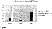
- ST3GAL6 is upregulated in comparison to non-malignant human cell lines. It has also been demonstrated that ST3GAL6 is induced by hypoxia in myeloma cell lines MM1S, RPMI8226 and U266. Hypoxia has been shown to stimulate MM cell homing to the bone marrow microenvironment and has been associated with multiple myeloma cell dissemination. Therefore it is possible that this gene may play a role in the mechanism of these hypoxia-induced processes.
- ST3GAL6 codes for a transmembrane protein localized to the Golgi apparatus which has enzymatic activity catalyzing the transfer of sialic acid to terminal positions of glycoprotein and glycolipid carbohydrate groups.
- the present inventors have demonstrated expression of the protein in bone marrow plasma cells from patients with multiple myeloma and demonstrated differential expression compared to normal healthy bone marrow using immunohistochemistry and a specific antibody to ST3GAL6.
- the citation does not disclose the prognostic significance of any sialyltransferase or it's potential use as a biomarker of transition from MGUS to MM with increasing levels associated with disease progression.
- FUCA1 is a gene that encodes the lysosomal enzyme alpha-L-fucosidase, which is involved in the degradation of fucose-containing glycoproteins and glycolipids. Mutations in this gene are associated with fucosidosis (FUCA1D), which is an autosomal recessive lysosomal storage disease.
- a still further object is to provide a method of identifying therapeutic agents for the treatment of multiple myeloma.
- a method of determining the prognosis of a multiple myeloma patient comprising determining the levels or activity of ST3GAL6 in a sample obtained from the patient and comparing the level determined with the level or activity in a control sample, an increased level or activity relative to the control being predictive of a reduced survival rate.
- the control sample may be the level of ST3GAL6 in at least one healthy individual. If a healthy individual is used as a control, then a prognosis of MM is made on the basis of an increased level of ST3GAL6 in the test sample.
- the skilled person will realise that the method would also work using control levels from MM patients, in which case prognosis can be determinedif the levels of ST3GAL6 in the test sample are in the same range as those of MM patients.
- the sample tested may be a blood, plasma or serum sample or a tissue sample.
- the tissue sample could be a bone marrow sample.
- the invention also provides methods, kits and uses of the markers FUCA1 and/orST3GAL1 in combination with ST3GAL6, in the diagnosis and assessment of prognosis of multiple myeloma as described herein for ST3GAL6.
- a decreased level or activity of FUCA1 relative to the control is predictive of a reduced survival rate
- an increased level or activity of ST3GAL1 relative to the control is predictive of a reduced survival rate.
- the level of ST3GAL6, FUCA1 and ST3GAL1 may be determined by immunohistochemistry, by RNA/DNA analysis, any form of immunoassay, including but not limited to an ELISA assay, a competitive or inhibition ELISA, a sandwich ELISA assay, a micro-array based assay, a functionalised nanoparticle assay, other rapid assay platform such as QDots, Fl tags and electro sensors, or a flow cytometry assay or a real-time chip-based assays.
- any form of immunoassay including but not limited to an ELISA assay, a competitive or inhibition ELISA, a sandwich ELISA assay, a micro-array based assay, a functionalised nanoparticle assay, other rapid assay platform such as QDots, Fl tags and electro sensors, or a flow cytometry assay or a real-time chip-based assays.
- the invention also provides a method of diagnosing multiple myeloma in a patient comprising determining the levels or activity of ST3GAL6 in a sample obtained from the patient and comparing the level or activity determined with the level or activity in a control sample, an increased level or activity relative to the control indicating the presence of multiple myeloma in the patient.
- the method may further comprise a step selected from:-
- the invention provides a kit for diagnosing multiple myeloma in a patient comprising an antibody against ST3GAL6, or reagents for detecting the expression of DNA or RNA encoding ST3GAL6.
- a kit for diagnosing multiple myeloma in a patient comprising an antibody against ST3GAL6, or reagents for detecting the expression of DNA or RNA encoding ST3GAL6.
- Such a kit may also be used for determining prognosis of a patient with multiple myeloma comprising an antibody against ST3GAL6, or reagents for detecting the expression of DNA or RNA encoding ST3GAL6.
- ST3GAL6 for use as a marker for diagnosing multiple myeloma or for determining prognosis of a patient with multiple myeloma.
- the invention provides a method of screening a therapeutic agent for suitability for the treatment of multiple myeloma comprising testing a candidate therapeutic agent for the ability to reduce the expression of ST3GAL6 levels or activity of ST3GAL6 in a model system, wherein a reduction of expression or activity of ST3GAL6 indicates suitability for treatment of multiple myeloma.
- the invention also provides a modulator of ST3GAL6 in an amount sufficient to decrease ST3GAL6 expression or activity for use in a method of reducing or alleviating multiple myeloma wherein the modulator of ST3GAL6 is interfering RNA specific for ST3GAL6 or a ST3GAL6 enzyme inhibitor or the modulator of ST3GAL6 is a glycosyltransferase inhibitor.
- the modulator of ST3GAL6 may be interfering RNA specific for ST3GAL6 or a ST3GAL6 enzyme inhibitor or the modulator of ST3GAL6 may be a glycosyltransferase inhibitor.
- a method of reducing or alleviating multiple myeloma may comprise administration of a modulator of ST3GAL6 in an amount sufficient to reduce ST3GAL6 expression or activity.
- the modulator may be interfering RNA specific for ST3GAL6, a selectin inhibitor or a glycosyltransferase inhibitor.
- the invention also provides a pharmaceutical composition comprising interfering RNA specific for ST3GAL6, a selectin inhibitor or a glycosyltransferase inhibitor together with a pharmaceutically acceptable carrier or excipient.
- sialyl Lewis X antigen on the selectin ligands of affected cells e.g. P-selectin ligand or CD44.
- the increased expression of sialyl Lewis X as a result of ST3GAL6 enzyme activity leads to increased interaction of myeloma cells with selectins (both P and E) present on endothelial cells and bone marrow stromal cells, leading to increase cell trafficking, dissemination and drug resistance.
- ST3GAL6 is not diagnostic of myeloma but may help identify patients with a poorer prognosis and may also identify patients with earlier stage disease (such as MGUS) who may actually be at higher risk of progression to active disease than their counterparts with normal ST3GAL6.
- ST3GAL6 overexpression may serve as a biomarker of response for treatment of patients with selectin inhibitors such that patients with ST3GAL6 overexpression could eventually be targeted for treatment with selectin inhibitors or other drugs interfering with the generation of selectin ligands (such as glycosyltransferase inhibitors or RNA interference approaches).
- ST3GAL6 overexpression has been associated with metastatic risk in colon cancer and breast cancer but not in other haematopoietic malignancies. It is quite likely it may play a role in other haematopoietic maligancies including lymphomas and acute leukaemia (both myeloid and lymphoid).
- ST3GAL6 may serve as a target in multiple myeloma since reduced expression of the gene may result in inhibition of selectin mediated cellular adhesion and trafficking. Targeting at the gene level could be mediated by a specific inhibitor of ST3GAL6 which may also have potential to induce down regulation of the gene.
- Direct enzyme inhibition can be achieved by global inhibition of sialyltransferases using fluorinated analogs of sialic acid as has previously been demonstrated in myeloid cells, where complete loss of selectin binding and impaired leucocyte rolling was demonstrated.
- the sialyltransferase inhibitor AL10 inhibits adhesion, migration, actin polymerization and invasion of alpha-2,3-ST-overexpressing A549 and CL1.5 human lung cells by effectively attenuating total cell surface sialylation.
- This compound is a lithocholic acid analogue derived from soyasaponin I by chemical synthesis in high purity and good yield.
- a similar approach to sialyltransferase inhibition could be adopted and developed in multiple myeloma.
- Flow cytometry analysis of CD138 microbead selected plasma cells from blood and bone marrow of multiple myeloma patients can be used to detect the presence of alpha 2,3 sialic acid on the cell surface. This can act as a surrogate marker of ST3GAL6 expression and a validation of the cell surface glycosylation status. This widely available low cost technique can be performed immediately following bone marrow aspiration and would allow efficient establishment of the alpha 2,3 sialylation status of individual patients.
- the inventors have performed this technique using multiple myeloma cell line RPMI8226 and demonstrated a significant shift in median fluorescent intensity for Maackia amurensis agglutinin (MAA) which is specific for alpha 2,3 bound sialic acids.
- MAA Maackia amurensis agglutinin
- RPMI cells were harvested incubated with MAA lectin for 30mins at 4 degrees (200,000cells/well in 100uL). Following a washing step cells were incubated with streptavadin APC and incubated x 30mins at 4degrees.
- MM cells were treated with mouse anti human CD138 antibody or with an isotype control for 1 hour on ice.
- Expression of MAA lectin in CD138 positive cells was determined by flow cytometry analysis (BD Biosciences FACS Canto) and quantified as a ratio of the mean fluorescence intensity (MFI) of the detected target to the MFI of the isotype control.
- MFI mean fluorescence intensity
- RT_PCR Real time quantitative PCR
- MM cell lines RPMI8226 and MMIR and in primary MM patient CD138 positive cells was used to examine expression of the ST3GAL6 gene.
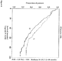
- the prognostic significance of the ST3GAL6 gene was analyzed using Kaplan Meier survival estimates.
- Membrane protein extracts from MM cell lines were applied directly to lectin microarrays following fluorescent labelling to generate cell surface glycan profiles. Protein expression was assessed by immunohistochemistry (IHC) on primary MM bone marrow sections from MM patients and healthy controls. Lectin based flow cytometry was carried out to assess for the binding of sialic acid lectins to MM cell line RPMI8226.
- IHC immunohistochemistry
- Bone marrow aspirates were obtained from multiple myeloma patients at Galway University Hospital following informed consent.
- the mononuclear fraction of the bone marrow aspirate was isolated using Ficoll PaqueTM Plus (STEMCELL Technologies) and high speed zero brake centrifugation.
- PC isolation from mononuclear cell fraction was performed by immunomagnetic bead selection with monoclonal mouse antihuman CD138 antibodies using the AutoMACs automated separation system (Miltenyi-Biotec, Auburn, CA).
- PC purity of more than 95% homogeneity was confirmed by 2-color flow cytometry using CD1381/CD452 and CD381/CD452 criteria (Becton Dickinson, San Jose, CA).
- RT_PCR Real Time Quantitative Polymerase Chain Reaction
- RT-PCR amplification was carried out with 100ng of cDNA in a 10- ⁇ l reaction mixture containing 5ul SYBR green mix (Bio-Rad, Mississauga, ON) and 0.2ul qPCR primers (SA Bioscences, Frederick, MD). Data quantification was carried out by the deltadeltaCT method.
- Glycan-binding proteins conjugated to microarrays were used to probe compositions and differential presence of glycan residues on glycoconjugates and cells.
- the glycan samples were fluorescently tagged prior to hybridization step to co-incubate glycan samples and lectins. After hybridization, slides were washed extensively and scanned in a microarray scanner. The lectin-glycan binding data is analyzed using commercially available software.
- This assay employs the quantitative sandwich enzyme immunoassay technique.
- Antibody specific for ST3GAL6 has been pre-coated onto a microplate.
- Standards and samples are pipetted into the wells and any ST3GAL6 present is bound by the immobilized antibody.
- a biotin-conjugated antibody specific for ST3GAL6 is added to the wells.
- avidin conjugated Horseradish Peroxidase (HRP) is added to the wells.
- HRP Horseradish Peroxidase
- a substrate solution is added to the wells and color develops in proportion to the amount of ST3GAL6 bound in the initial step. The color development is stopped and the intensity of the color is measured.
- BM aspirates from 55 MM patients and 3 healthy subjects were rinsed with PBS, fixed with 4% formaldehyde in PBS, dehydrated with ethanol, embedded in paraffin, and sectioned. Tissues were then immunostained with mouse anti-human ST3GAL.
- Green Fluourescent Protein (GFP) Luc + -MM1s cells have been generated using lentiviral infection and cultured at 37°C in RPMI 1640 medium containing 10% FBS (Sigma-Aldrich), 2mM L-glutamine, 100 U/mL of penicillin, 100 ⁇ g/mL of streptomycin (Invitrogen) and 100 ⁇ g/mL Geneticin. Plasmids were routinely amplified in the Escherichia coli DH5_ strain and isolated from cultures by using the Qiagen Plasmid Spin Midiprep Kit (Qiagen, Valencia, CA).
- Scrambled shRNA control plasmids can be generated using the same methods used to generate lentivirus. Scrambled lentivirus is used to infect MM1s cell to act as a control in these experiments. Stable knockdown of ST3GAL6 can be achieved in these cells using a plasmid generated lentiviral vector which infects the MM1s cells.
- IRNA screening is performed in cultured MM cell lines as a lethality study. Using technology available from the Broad Institute Boston MA, USA siRNAs targeting individual human silayltransferase genes are preprinted on 384-well plates alongside staggered negative and positive control siRNA. Primary screening experiments are conducted in duplicate on separate occasions for several cultured MM cell lines. For each experiment, plates preloaded with siRNA and frozen were thawed at room temperature and 20 _L/well-diluted Lipofectamine 2000 or Dharmafect 3 solution is added to the relevant wells. After 30 minutes, MM cells are added to each well.
- Cell viability is determined at 96 hours by CellTiter-Glo luminescence assay read on a BMG Polarstar machine using excitation 544-nm/emission 590-nm filters.
- Surviving knock down cells can be analysed by the following methods.
- BMSCs are cultured overnight to confluence in 96-well plates (5_103 cells/well) before initiating the adhesion assay.
- MM1s cells are serum-starved for 3 hours, prelabeled with GFP, added to the BMSCs (1_105 cells/well), and allowed to adhere for 2 hours at 37°C.
- Nonadherent cells can be aspirated away, the BMSCs washed, and fluorescence intensity measured using a fluorescent-plate reader (Ex/Em_485/520 nm).
- HUVECs (5_103 cells/basket) are incubated overnight in the upper chamber of 8-micron pore filters (Costar; Corning) before performing the adhesion assay.
- MM1s cells are serum-starved for 3 hours and then added to the upper chamber of the basket (2_ 105cells/well), and left to migrate for 4 hours at 37°C toward the lower chamber, which contains 0 or 30nM of SDF1_. This analysis is performed separately for ST3GAL6 knockdown cells vs scrambled control.
- mice may be pretreated with vehicle or a drug of interest 1 hour before the cell injection.
- a skin flap is made in the scalps of the mice to expose the underlying skull surface and Evan's blue dye is injected intravenously immediately before imaging to visualize blood vessels.
- High-resolution images with cellular detail are captured 30 minutes after cell injection through the intact mouse skull at depths of up to 250_m from the surface of the skull using a 30_0.9-numerical aperture water-immersion objective lens (Lomo). Multiple imaging depths were acquired and a maximum intensity z-projection is performed in ImageJ software to merge the images.
- GFP labelled MM cells and Evan's blue dye are excited with 491-nm and 638-nm single-photon lasers and detected via 528/19-nm and 680/25-nm bandpass filters, respectively.
- RPMI-8226 cell lines were purchased from ATCC (Manassas VA) cultured in RPMI 1640 (Sigma Chemical, St. Louis, MO) containing 10% foetal bovine serum, 2 mmol/L L-glutamine (Life Technologies, Grand Island, NY), 100 units/mL penicillin, and 100 Ag/mL streptomycin (Life Technologies).GFP-Luciferin-Neo MM1s cells were generated using a lentiviral infection method as described [1]. Primary MM cells were obtained from bone marrow samples from patients using CD138+ microbead selection (Miltenyi Biotech, Auburn CA) as previously described [2].
- Bone marrow stromal cells were obtained from healthy donor and MM patient fresh bone marrow samples and isolated as described [3]. BMSC's were then cultured in DMEM (Sigma Chemical, St Louis, MO) supplemented with 10% heat-inactivated foetal bovine serum, 100 units/Ml penicillin, 10 Ag/mL streptomycin (Life Technologies; 0.01%), and 2 mmol/L L-glutamine (1%; Life Technologies). Informed consent was obtained from all patients in accordance with the Declaration of Helsinki. Approval of these studies was obtained by the Dana-Farber Cancer Institute Institutional Review Board. Bortezomib was obtained from Millennium Pharmaceutical (Cambridge, MA), and SDF-1 was obtained from R&D Systems (Minneapolis, MN).
- ST3GAL6 knockdown was assessed by flow cytometry and RT-PCR using the following primers Forward Primer; TTG CCT CTC TGC TGA GGT TT Reverse Primer; CCT CCA TTA CCA ACC ACC AC [5].
- the resultant stable ST3GAL6 knockdown (shST3GAL6) cell line is compared to the scrambled control cell line in all subsequent functional assays.
- the sense and antisense oligonucleotide sequence for construction of ST3GAL6 shRNA were as follows: clone no 10402; [NM_006100.2-1332s1c1, target sequence CCTTTGCACTACTATGGGAAT (A2), NM_006100.2-1110s1c1 target sequence CCAGCCTTAAACCTGATTTAT (A3)]
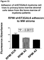
- Adhesion assays Assays for adhesion of MM cells to normal and MM BMSC's were performed using 96-well plates (5 x 10 3 /well). Normal or MM BMSC's (5,000/well) were seeded in 96-well plates overnight in supplemented DMEM media to establish a confluent monolayer. MM cells were serum starved overnight, prelabelled with calcein AM and added to the BMSC's and allowed to adhere for 2 hours at 37°C. Nonadherent cells were aspirated off, BMSCs were washed with PBS, and fluorescence intensity was measured using a fluorescent-plate reader (Ex/Em_485/520 nm).
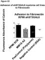
- Fibronectin adhesion assays were performed using an in vitro adhesion assay coated with fibronectin following the manufacturer recommendations (EMD Biosciences, San Diego, CA) as previously described [6]. Bovine serum albumin (BSA)-coated wells served as negative controls. In some of the above experiments MM cells were pre-incubated with increasing concentrations of bortezomib (0, 2.5, and 5 nM) prior to assessment of adhesion.
- BSA bovine serum albumin
- Migration assays were performed as previously described [6] [7]. Migration ability of MM scrambled control cells vs. shST3GAL6 cells was assessed using a transwell migration assay plate (pore size 0.8_m; Corning Life Sciences, Acton, MA) according to the manufacturer's instructions. MM cells were serum starved for 4 hours and following calcein AM labelling were then placed in the upper migration chambers. SDF-1_ (30nM) was added to 500_uL of RPMI 1640 in the lower chambers. After 4 hours at 37°C cells that migrated to the lower chambers were counted on a fluorescent plate reader (Molecular Devices).
- Immunoblotting was performed as previously described [8]. Briefly, whole-cell lysates were subjected to sodium dodecyl sulfate-polyacrylamide gel electrophoresis (SDS-PAGE) and transferred to polyvinyldene fluoride (PVDF) membrane (Bio-Rad Laboratories, Hercules, CA). The antibodies used for immunoblotting included anti-ST3GAL6 produced in rabbit (Sigma-Aldrich, MO, USA) and anti-Actin (Cell Signaling Technology)
- Immunohistochemistry To detect ST3GAL6, BM aspirates from 50 MM patients and 10 healthy subjects were rinsed with PBS, fixed with 4% formaldehyde in PBS, dehydrated with ethanol, embedded in paraffin, and sectioned. Tissues were then immunostained with mouse anti-human ST3GAL6 antibody and mouse-antihuman-CD138.
- mice Female, 6 to 7 weeks old, severe combined immunodeficient beige (SCID-Bg) mice were obtained from Taconic Laboratories. Anesthesia was performed by intraperitoneal injections of ketamine (Bedford Laboratories, Bedford, OH) /xylazine (Lloyd Laboratories, Shenandoah, IA) at 80 mg/kg/12 mg/kg body weight prior to in-vivo imaging sessions. Following this mice were killed by inhalation of CO2.
- SCID-Bg severe combined immunodeficient beige mice
- mice To detect tumor burden on a weekly basis following injection mice were injected with 75 mg/kg luciferin (Xenogen, Hopkinton, MA) and imaged for bioluminescence 5 minutes after the injection.
- the home-built bioluminescence system used an electron multiplying CCD (Andor Technology, Harbor, United Kingdom) with an exposure time of 15 seconds, an electron multiplication gain of 500-voltage gain _ 200, 5-by-5 binning, and background subtraction. Images were analyzed with the use of Image J software (National Institutes of Health, Bethesda, MD).
- In-vivo confocal Using the above SCID-Bg xenograft mice homing of MM cells to distant bone marrow niches was tracked in vivo, by using in vivo confocal microscopy [9]. Briefly, MM cells homing to the BM were imaged in vivo using a Zeiss 710 confocal system (Carl Zeiss Microimaging, Jena, Germany) on an upright examiner stand with a custom stage. A skin flap was made in the scalp of the mice to expose the underlying dorsal skull surface.
- High-resolution images with cellular detail were be obtained through the intact mouse skull at depths of up to 250 ⁇ m from the surface of the skull using a 10x 0.45NA Plan-Apo objective (Carl Zeiss Microimaging). Multiple imaging depths were acquired, and a maximum intensity z-projection was performed in Image J to merge the images.
- GFP was excited with the 488nm line on an Argon laser. Blood vessels were imaged using Evans Blue (Sigma-Aldrich, St. Louis, MO) excited with a 633 nm laser. Emission signals were be collected by the Zeiss internal confocal Quasar detectors.
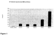
- RT-PCR analysis validated the over expression of ST3GAL6 in MM cell lines and primary samples compared to healthy controls.
- IHC for ST3GAL6 on primary bone marrow sections from MM patients (n 27), demonstrated specific Golgi staining compared to controls.
- FIG. 1 quantitative PCR was used to detect mRNA for ST3GAL6 in CD138 plasma cells from normal donors as well as 5 different multiple myeloma cell lines.
- Figure 2 demonstrates the effect of hypoxia (cells grown in hypoxic chamber with 1% O 2 ) on the gene expression of ST3GAL6 as assessed by quantitative PCR on 3 different multiple myeloma cell lines.
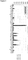
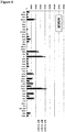
- Figures 3 and 4 demonstrate the pattern of lectin binding to membrane proteins from the multiple myeloma cell lines RPMI8226 and MM1R using a lectin array. This pattern of lectin binding indicates that the membrane proteins are heavily sialylated.
- Figure 5 a and b show co-expression of CD138 and ST3GAL6 by immunohistochemistry.
- Figure 5a from a patient with multiple myeloma shows an excess of bone marrow plasma cells with expression of ST3GAL6 in CD138 positive cells.
- Figure 5b from a normal bone marrow, shows absence of ST3GAL6 in CD138 cells, which are present at low frequency in the marrow.
- Figure 7 shows protein expression of ST3GAL6 by Western blotting in normal CD138 bone marrow derived plasma cells compared with ST3GAL6 protein expression in 3 multiple myeloma cell lines. While no appreciable band can be seen in the normal plasma cells, a strong band is seen in all 3 cell lines.
- lentivaral stocks were produced from hairpin-pLKO.1 plasmids and transfcted into myeloma cell lines MM1s-green fluorescent protein-luciferase positive (GFP-Luc) and RPMI-8226.
- the MM1s-GFP-Luc cell line was chosen in order to create a stable shST3GAL6 cell line that was amenable to later in-vivo imaging and Confocal imaging.
- the RPMI-8226 cell line was chosen as a representative cell line of multiple myeloma which had not been previously manipulated.
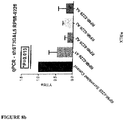
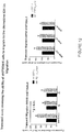
- QPCR data for the MM1s cell line demonstrates the reduced level of mRNA detectable in cells that had been infected with a silencing lentiviral construct to ST3GAL6 in comparison to the ST3GAL6 mRNA level in a scrambled control.
- Figure 8 (b) demonstrates the effective reduction of the mRNA level in the RPMI-8226 cell line.
- MM cells were serum starved overnigh and (5x10 5/ml ) following this were incubated with calcein-AM as described for 30 mins at 37°C, washed and re-suspended in serum free clear RPMI media. 100ul of cells were added to BMSC coated 96-well plates (3x10 4 /well) for 2 hours at 37°C. After a gentle wash with PBS the fluorescence was read using a fluorescent plate reader (ex/em 485/520nm).Results of the migration studies are shown in Figure 12 .
- cells (5X10 5 ) were serum starved x 4 hours in clear serum free RPMI media, labelled with calcein-AM as described and added to the upper wells of transwell plates (Costar Corning pore size 0.8 ⁇ m) with serial concentrations of SDF-1 ⁇ (0-30nM) added to the 500ul of RPMI 1640 serum free media in the lower chambers. After 4 hours at 37°C the upper chambers were removed and cells that had migrated to the lower chambers were counted on a fluorescent plate reader.
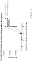
- FIG. 13 shows bioluminescent imaging of SCID-Bg mice weeks 1-4 post 1x10 6 MM1s-GFP- Luc positive cells/mouse injected IV via tail vein.
- Scrambled control group received cells that were infected with a standard scrambled control lentivirus (shGFP). Animals were imaged each week for monitoring of tumour development 5 minutes post intraperitoneal injection of luciferase enzyme as per standard protocol. At week four animals were sacrificed for confocal imaging.
- In-vivo confocal imaging was performed on SDID-Bg xenograft mice skull bones.
- red represents vessels stained with Evans Blue dye
- green represents myeloma cells (MM1s-GFP+ cell line) and can be taken to represent tumour burden in the skull bone marrow.
- Mouse A was injected with scrambled control cells
- mouse B was injected with MM1s-GFP+ cells that had been infected with shRNA for ST3GAL6 resulting in a stable knockdown of this gene in the cell line.
- a number of other glycosylation genes are differentially regulated between normal and malignant plasma cells in MM.
- MM cell lines show low levels of FUCA1 transcripts by QPCR.
- the OPM2 cell line which contains t(4,:14) showed increased ST3GAL1 transcripts relative to NBM.
- myeloma cells express sialytransferases and sLex/a. Since FUCA1 participates in N-glycan degradation with removal of fucose residues, this raises the possibility that a reduction in FUCA1 may lead to excessive fucosylation of cancer related glycans involved in adhesion and trafficking, such as sLex.
- Lectin arrays of MM cell lines revealed in high levels of a-1,3/a-1,6 linked fucose, as determined by binding of Aleuria Aurantia Lectin (AAL) (data not shown).
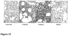
- the HECA-452 antibody recognizes a functional trisaccharide domain shared by sialyl Lewis a and sLex and known to bind to E-selectin.
- Preliminary immunohistochemistry revealed increased HECA452 staining (D) in bone marrow samples, which had both strong ST3GAL6 (A)+/ST3GAL1 (B) and low FUCA1 staining (C).
- the sialyltransferase ST3GAL6 is differentially regulated in all stages of MM with potential effects on MM biology and survival. Upregulation of ST3GAL6 may play an important role in MM cell trafficking and in this analysis is associated with inferior survival. Studies are ongoing to address the roles of ST3GAL6 over expression and altered sialylation in MM cell adhesion and trafficking.
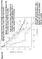
- the present inventors have shown that this glycogene is overexpressed in multiple myeloma cell lines and in CD138 cells from myeloma patients when compared to their healthy counterparts. We have also demonstrated an association between increased expression of this gene and reduced median survival following analysis of a publically available transcriptomic dataset.
- Altered glycosylation gene expression patters may identify patients at high risk of disease progression and early death.
- Our data implicates sialytransferases and selectin ligands as potential therapeutic targets in Multiple Myeloma.
- low expression of the FUCA1 gene is an adverse prognostic factor in MM and when combined with high sislyltransferase gene expression identifies patients at increased risk of early disease progression and death.
Landscapes
- Health & Medical Sciences (AREA)
- Life Sciences & Earth Sciences (AREA)
- Chemical & Material Sciences (AREA)
- Immunology (AREA)
- Engineering & Computer Science (AREA)
- Molecular Biology (AREA)
- General Health & Medical Sciences (AREA)
- Biochemistry (AREA)
- Hematology (AREA)
- Urology & Nephrology (AREA)
- Biomedical Technology (AREA)
- Pathology (AREA)
- Analytical Chemistry (AREA)
- Medicinal Chemistry (AREA)
- Microbiology (AREA)
- Physics & Mathematics (AREA)
- Biotechnology (AREA)
- Proteomics, Peptides & Aminoacids (AREA)
- Organic Chemistry (AREA)
- General Physics & Mathematics (AREA)
- Food Science & Technology (AREA)
- Cell Biology (AREA)
- Zoology (AREA)
- Genetics & Genomics (AREA)
- Wood Science & Technology (AREA)
- Epidemiology (AREA)
- Veterinary Medicine (AREA)
- Pharmacology & Pharmacy (AREA)
- Public Health (AREA)
- Animal Behavior & Ethology (AREA)
- Oncology (AREA)
- Biophysics (AREA)
- Hospice & Palliative Care (AREA)
- Bioinformatics & Cheminformatics (AREA)
- General Engineering & Computer Science (AREA)
- Measuring Or Testing Involving Enzymes Or Micro-Organisms (AREA)
Claims (18)
- Verfahren zum Bestimmen der Prognose eines Patienten mit Multiplem Myelom, umfassend das Bestimmen der Spiegel an oder die Aktivität von ST3GAL6 in einer von dem Patienten erhaltenen Probe und das Vergleichen des bestimmten Spiegels oder der bestimmten Aktivität mit dem Spiegel oder der Aktivität in einer Kontrollprobe, wobei ein erhöhter Spiegel oder eine erhöhte Aktivität im Vergleich zur Kontrolle prädikativ für eine verringerte Überlebensrate ist.
- Verfahren nach Anspruch 1, des Weiteren umfassend das Bestimmen der Spiegel oder der Aktivität von FUCA1 in der Probe und das Vergleichen des bestimmten Spiegels oder der bestimmten Aktivität mit dem Spiegel oder der Aktivität in einer Kontrollprobe, wobei ein verringerter Spiegel oder eine verringerte Aktivität im Verhältnis zur Kontrolle prädikativ für eine verringerte Überlebensrate ist.
- Verfahren nach Anspruch 1 oder 2, des Weiteren umfassend das Bestimmen der Spiegel oder der Aktivität von ST3GAL1 in der Probe und das Vergleichen des bestimmten Spiegels oder der bestimmten Aktivität mit dem Spiegel oder der Aktivität in einer Kontrollprobe, wobei ein erhöhter Spiegel oder eine erhöhte Aktivität im Verhältnis zur Kontrolle prädikativ für eine verringerte Überlebensrate ist.
- Verfahren zur Diagnose von Multiplem Myelom in einem Patienten, umfassend das Bestimmen der Spiegel oder der Aktivität von ST3GAL6 in einer vom Patienten erhaltenen Probe und das Vergleichen des bestimmten Spiegels oder der bestimmten Aktivität mit dem Spiegel oder der Aktivität in einer Kontrollprobe, wobei ein erhöhter Spiegel oder eine erhöhte Aktivität im Verhältnis zur Kontrolle auf die Anwesenheit eines Multiplen Myeloms im Patienten hinweist.
- Verfahren nach Anspruch 4, des Weiteren umfassend einen Schritt ausgewählt aus: das Bestimmen der Spiegel oder der Aktivität von FUCA1 in der Probe und das Vergleichen des bestimmten Spiegels oder der bestimmten Aktivität mit dem Spiegel oder der Aktivität in einer Kontrollprobe, wobei ein verringerter Spiegel oder eine verringerte Aktivität im Verhältnis zur Kontrolle auf die Anwesenheit eines Multiplen Myeloms im Patienten hinweist, das Bestimmen der Spiegel oder der Aktivität von ST3GAL1 in der Probe und das Vergleichen des bestimmten Spiegels oder der bestimmten Aktivität mit dem Spiegel oder der Aktivität in einer Kontrollprobe, wobei ein erhöhter Spiegel oder eine erhöhte Aktivität im Verhältnis zur Kontrolle auf die Anwesenheit eines Multiplen Myeloms im Patienten hinweist, oder
das Bestimmen der Spiegel oder der Aktivität von FUCA1 und ST3GAL1 in der Probe und das Vergleichen des bestimmten Spiegels oder der bestimmten Aktivität mit dem Spiegel oder der Aktivität in einer Kontrollprobe, wobei ein verringerter Spiegel oder eine verringerte Aktivität von FUCA1 und ein erhöhter Spiegel oder eine erhöhte Aktivität von ST3GAL1 im Verhältnis zur Kontrolle auf die Anwesenheit eines Multiplen Myeloms im Patienten hinweist. - Verfahren nach Anspruch 4 oder 5, wobei die Diagnose monoclonale Gammopathie unklarer Signifikanz ist.
- Verfahren zum Bestimmen des Behandlungsschemas für einen Patienten mit Multiplem Myelom, umfassend das Bestimmen der Spiegel oder der Aktivität von ST3GAL6 in einer vom Patienten erhaltenen Probe und das Vergleichen des bestimmten Spiegels oder der bestimmten Aktivität mit dem Spiegel oder der Aktivität in einer Kontrollprobe, wobei ein erhöhter Spiegel oder eine erhöhte Aktivität im Vergleich zur Kontrolle darauf hinweist, dass eine Behandlung mit einem ST3GAL6-Inhibitor für den Patienten vorteilhaft wäre.
- Verfahren nach Anspruch 7, des Weiteren umfassend das Bestimmen der Spiegel oder der Aktivität von FUCA1 in der Probe und Vergleichen der bestimmten Spiegel oder der bestimmten Aktivität mit dem Spiegel oder der Aktivität in einer Kontrollprobe, wobei ein verringerter Spiegel oder eine verringerte Aktivität im Verhältnis zur Kontrolle darauf hinweist, dass eine Behandlung mit einem FUCA1-Potentiator für den Patienten vorteilhaft wäre und/oder des Weiteren umfassend das Bestimmen der Spiegel oder der Aktivität von ST3GAL1 in der Probe und das Vergleichen des bestimmten Spiegels oder der bestimmten Aktivität mit dem Spiegel oder der Aktivität in einer Kontrollprobe, wobei ein erhöhter Spiegel oder eine erhöhte Aktivität im Verhältnis zur Kontrolle darauf hinweist, dass eine Behandlung mit einem ST3GAL1-Inhibitor für den Patienten vorteilhaft wäre.
- Verfahren nach Anspruch 1 bis 8, wobei die Probe eine Blut-, Plasma- oder Serumprobe oder eine Gewebe-Probe ist.
- Verfahren nach Anspruch 1 bis 9, wobei der Spiegel von ST3GAL6 oder FUCA1 oder ST3GAL1 bestimmt wird durch immunhistochemische, immuncytochemische und/oder durch RNA/DNA-Analyse, jegliche Form von Immunassay, einschließlich, jedoch nicht beschränkt auf, einem ELISA-Assay, einem kompetitiven oder inhibitorischen ELISA, einem Sandwich-ELISA-Assay, einem Mikroarray-basierten Assay, einem funktionalisierten Nanopartikel-Assay, anderen schnellen Assay-Plattformen wie QDots, Fl tags und elektrischen Sensoren oder einem Durchflusszytometrie-Assay oder einem Echtzeit-Chip-basierten Assay.
- Verfahren nach einem der vorangehenden Ansprüche, wobei die Kontrolle der Spiegel oder die Aktivität von ST3GAL6 und/oder FUCA1 und/oder ST3GAL6 in mindestens einem gesunden Individuum ist.
- Verwendung von (einem) ST3GAL6 als Marker für die Diagnose von Multiplem Myelom oder für das Bestimmen der Prognose eines Patienten mit Multiplem Myelom oder für das Bestimmen eines Behandlungsschemas.
- Verwendung nach Anspruch 12, des Weiteren umfassend die Verwendung von FUCA1 oder ST3GAL1 oder einer Kombination von beiden als zusätzliche(n) Marker.
- Verfahren zum Screenen eines therapeutischen Agens auf die Eignung für die Behandlung von Multiplem Myelom, umfassend das Testen eines therapeutischen Agens-Kandidaten auf die Fähigkeit(a) die Expression von ST3GAL6-Spiegeln oder der ST3GAL6-Aktivität in einem Modellsystem zu reduzieren,wobei die Reduktion der Expression oder der Aktivität von ST3GAL6 auf die Eignung für die Behandlung von Multiplem Myelom hinweist, wobei das Modellsystem eine SCID-Maus ist.
- Verfahren nach Anspruch 14, des Weiteren umfassend entweder:(a) das Testen eines therapeutischen Agens-Kandidaten auf die Fähigkeit, die Expression der FUCA1-Spiegel oder der FUCA1-Aktivität in dem Modellsystem zu erhöhen, wobei eine Reduktion der Expression oder der Aktivität von ST3GAL6 und eine Erhöhung der Expression oder Aktivität von FUCA1 auf eine Eignung für die Behandlung von Multiplem Myelom hinweist, oder(b) das Testen eines therapeutischen Agens-Kandidaten auf die Fähigkeit, die Expression von ST3GAL1-Spiegeln oder der ST3GAL1-Aktivität in einem Modellsystem zu verringern, wobei eine Reduktion der Expression oder der Aktivität von ST3GAL6 und ST3GAL1 auf die Eignung für die Behandlung von Multiplem Myelom hinweist.
- Modulator von ST3GAL6 in einer Menge, die ausreichend ist, um die ST3GAL6-Expression oder -Aktivität zu verringern, zur Verwendung in einem Verfahren, das Multiples Myelom reduziert oder lindert, wobei der Modulator von ST3GAL6 interferierende RNA, die spezifisch ist für ST3GAL6, oder ein ST3GAL6-Enzym-Inhibitor ist oder der Modulator von ST3GAL6 ein Glycosyltransferase-Inhibitor ist.
- Verfahren nach einem der Ansprüche 14 oder 15, wobei der Modulator von ST3GAL6 interferierende RNA, die spezifisch ist für ST3GAL6, oder ein ST3GAL6-Enzym-Inhibitor ist oder der Modulator von ST3GAL6 ein Glycosyltransferase-Inhibitor ist.
- Arzneimittel, umfassend die interferierende RNA, die spezifisch ist für ST3GAL6, oder einen ST3GAL6-Enzym-Inhibitor oder einen Glycosyltransferase-Inhibitor, zusammen mit einem pharmazeutisch verträglichen Träger oder Exzipienten zur Verwendung in einem Verfahren zum Reduzieren oder Lindern von Multiplem Myelom.
Priority Applications (1)
| Application Number | Priority Date | Filing Date | Title |
|---|---|---|---|
| EP13779547.2A EP2906952B1 (de) | 2012-10-15 | 2013-10-15 | Sialyltransferase st3gal6 als marker für multiples myelom |
Applications Claiming Priority (3)
| Application Number | Priority Date | Filing Date | Title |
|---|---|---|---|
| EP12188596.6A EP2720044A1 (de) | 2012-10-15 | 2012-10-15 | Sialyltransferase ST3GAL6 als Marker für multiples Myelom |
| EP13779547.2A EP2906952B1 (de) | 2012-10-15 | 2013-10-15 | Sialyltransferase st3gal6 als marker für multiples myelom |
| PCT/EP2013/071555 WO2014060436A1 (en) | 2012-10-15 | 2013-10-15 | Sialyltransferase st3gal6 as a marker for multiple myeloma |
Publications (2)
| Publication Number | Publication Date |
|---|---|
| EP2906952A1 EP2906952A1 (de) | 2015-08-19 |
| EP2906952B1 true EP2906952B1 (de) | 2019-12-11 |
Family
ID=47022570
Family Applications (2)
| Application Number | Title | Priority Date | Filing Date |
|---|---|---|---|
| EP12188596.6A Withdrawn EP2720044A1 (de) | 2012-10-15 | 2012-10-15 | Sialyltransferase ST3GAL6 als Marker für multiples Myelom |
| EP13779547.2A Active EP2906952B1 (de) | 2012-10-15 | 2013-10-15 | Sialyltransferase st3gal6 als marker für multiples myelom |
Family Applications Before (1)
| Application Number | Title | Priority Date | Filing Date |
|---|---|---|---|
| EP12188596.6A Withdrawn EP2720044A1 (de) | 2012-10-15 | 2012-10-15 | Sialyltransferase ST3GAL6 als Marker für multiples Myelom |
Country Status (3)
| Country | Link |
|---|---|
| US (2) | US20150354007A1 (de) |
| EP (2) | EP2720044A1 (de) |
| WO (1) | WO2014060436A1 (de) |
Families Citing this family (5)
| Publication number | Priority date | Publication date | Assignee | Title |
|---|---|---|---|---|
| CN114341368A (zh) * | 2019-07-12 | 2022-04-12 | 糖模拟物有限公司 | 使用基因表达作为多种肿瘤类型的e-选择素抑制剂功效和临床结果的指标的方法 |
| GB201916150D0 (en) | 2019-11-06 | 2019-12-18 | Univ Of Ireland Galway | Treatment of multiple myeloma |
| CN114354927B (zh) * | 2021-03-18 | 2025-04-22 | 中国人民解放军陆军特色医学中心 | 唾液酸转移酶st3gal4作为egfr-tki耐药标志物的新应用 |
| CN113981086A (zh) * | 2021-11-11 | 2022-01-28 | 四川省医学科学院·四川省人民医院 | St3gal6-as1基因的应用及特异性引物对和试剂盒 |
| WO2025068553A1 (en) * | 2023-09-28 | 2025-04-03 | Universität Basel | Catalytic moieties for treating cancer |
Family Cites Families (5)
| Publication number | Priority date | Publication date | Assignee | Title |
|---|---|---|---|---|
| ATE378421T1 (de) * | 1999-05-24 | 2007-11-15 | Abaron Biosciences Inc | Glycosyltransferase inhibitoren |
| EP2001487A4 (de) * | 2006-03-15 | 2010-09-15 | Tufts College | Fluorinierte kohlenhydrate und ihre verwendung bei der visualisierung von tumoren, bei der gewebebehandlung und bei der krebs-chemotherapie |
| US8940719B2 (en) * | 2006-07-03 | 2015-01-27 | Academia Sinica | Lithocholic acid analogues that inhibit sialyltransferase |
| WO2008039475A2 (en) * | 2006-09-26 | 2008-04-03 | The Board Of Trustees Of The University Of Arkansas | A gene expression profiling based identification of genomic signatures of multiple myeloma and uses thereof |
| EP2236607A1 (de) * | 2009-04-02 | 2010-10-06 | Sanofi-Aventis | Verwendung von Hemmern der St3Gla6-Aktivität zur Adipogenesemodulation |
-
2012
- 2012-10-15 EP EP12188596.6A patent/EP2720044A1/de not_active Withdrawn
-
2013
- 2013-10-15 WO PCT/EP2013/071555 patent/WO2014060436A1/en not_active Ceased
- 2013-10-15 US US14/435,001 patent/US20150354007A1/en not_active Abandoned
- 2013-10-15 EP EP13779547.2A patent/EP2906952B1/de active Active
-
2017
- 2017-05-17 US US15/597,961 patent/US11242566B2/en not_active Expired - Fee Related
Non-Patent Citations (1)
| Title |
|---|
| None * |
Also Published As
| Publication number | Publication date |
|---|---|
| EP2906952A1 (de) | 2015-08-19 |
| US20150354007A1 (en) | 2015-12-10 |
| EP2720044A1 (de) | 2014-04-16 |
| WO2014060436A1 (en) | 2014-04-24 |
| US20170327899A1 (en) | 2017-11-16 |
| US11242566B2 (en) | 2022-02-08 |
Similar Documents
| Publication | Publication Date | Title |
|---|---|---|
| US11242566B2 (en) | Sialyltransferase ST3GAL6 as a marker for multiple myeloma | |
| US20140274788A1 (en) | Leukemia stem cell markers | |
| Berghuis et al. | Sialic acids in gynecological cancer development and progression: Impact on diagnosis and treatment | |
| Xi et al. | Glycosylated modification of MUC1 maybe a new target to promote drug sensitivity and efficacy for breast cancer chemotherapy | |
| Tada et al. | Fucosyltransferase 8 plays a crucial role in the invasion and metastasis of pancreatic ductal adenocarcinoma | |
| WO2012096545A2 (ko) | 췌장암 암줄기세포 특성을 이용한 췌장암 신규 바이오마커 및 그의 용도 | |
| Garnham et al. | ST3 beta-galactoside alpha-2, 3-sialyltransferase 1 (ST3Gal1) synthesis of Siglec ligands mediates anti-tumour immunity in prostate cancer | |
| Vecchio et al. | Human α-L-fucosidase-1 attenuates the invasive properties of thyroid cancer | |
| US11452775B2 (en) | Detection of arginine methylation of EGFR for prediction of resistance to therapy | |
| WO2023060066A2 (en) | Supermere nanoparticles and methods of isolation and use thereof | |
| Kim et al. | Priming a vascular-selective cytokine response permits CD8+ T-cell entry into tumors | |
| Chen et al. | miR‐190b promotes tumor growth and metastasis via suppressing NLRC3 in bladder carcinoma | |
| US20150094217A1 (en) | Test for diagnosing resistance to azacitidine | |
| US9487831B2 (en) | Use of annexin A3 as a diagnostic and prognostic biomarker and therapeutic target for treating hepatocellular carcinoma | |
| Ao et al. | Targeting GFPT2 to reinvigorate immunotherapy in EGFR-mutated NSCLC | |
| US11858998B2 (en) | Glycome factors driving melanoma progression | |
| Diallo et al. | Targeted therapy for KIF3C: A study on the mechanism of combined therapy with KIF3C signaling pathway, afatinib, and MT-DC (ac) phosphoramide in regulating gastric cancer cell proliferation | |
| KR102941329B1 (ko) | 간암 진단을 위한 바이오마커 조성물 | |
| Anim | Modulatory Effects of Deacetylated Sialic Acids (A Sugar Residue) in NK-Mediated Cytotoxicity and Targeted Therapy by Receptor Tyrosine Kinase Inhibitors | |
| US11493517B2 (en) | Eukaryotic translation initiation factors (EIFS) as novel biomarkers in head and neck squamous cell carcinoma (HNSCC) | |
| Ma et al. | PFKFB4-Mediated HSPB1 phosphorylation suppresses ferroptosis to Promote gastric cancer progression | |
| Liu et al. | SPOCK1 acts as a negative regulator of ferroptosis in non-small cell lung cancer | |
| US20220081483A1 (en) | Methods and compositions for reducing numbers or eliminating hiv-infected cells | |
| Mohammed | The Role of Galectin-3 in Melanoma Progression | |
| Secomandi | High expression of lysosomal cathepsin D inhibits neuroblastoma cell growth, migration and invasion, and confers better prognosis in neuroblastoma patients |
Legal Events
| Date | Code | Title | Description |
|---|---|---|---|
| PUAI | Public reference made under article 153(3) epc to a published international application that has entered the european phase |
Free format text: ORIGINAL CODE: 0009012 |
|
| 17P | Request for examination filed |
Effective date: 20150407 |
|
| AK | Designated contracting states |
Kind code of ref document: A1 Designated state(s): AL AT BE BG CH CY CZ DE DK EE ES FI FR GB GR HR HU IE IS IT LI LT LU LV MC MK MT NL NO PL PT RO RS SE SI SK SM TR |
|
| AX | Request for extension of the european patent |
Extension state: BA ME |
|
| DAX | Request for extension of the european patent (deleted) | ||
| 17Q | First examination report despatched |
Effective date: 20160401 |
|
| STAA | Information on the status of an ep patent application or granted ep patent |
Free format text: STATUS: EXAMINATION IS IN PROGRESS |
|
| GRAP | Despatch of communication of intention to grant a patent |
Free format text: ORIGINAL CODE: EPIDOSNIGR1 |
|
| STAA | Information on the status of an ep patent application or granted ep patent |
Free format text: STATUS: GRANT OF PATENT IS INTENDED |
|
| INTG | Intention to grant announced |
Effective date: 20190208 |
|
| GRAJ | Information related to disapproval of communication of intention to grant by the applicant or resumption of examination proceedings by the epo deleted |
Free format text: ORIGINAL CODE: EPIDOSDIGR1 |
|
| STAA | Information on the status of an ep patent application or granted ep patent |
Free format text: STATUS: EXAMINATION IS IN PROGRESS |
|
| RIN1 | Information on inventor provided before grant (corrected) |
Inventor name: JOSHI, LOKESH Inventor name: O'DWYER, MICHAEL Inventor name: GLAVEY, SIOBHAN |
|
| INTC | Intention to grant announced (deleted) | ||
| GRAJ | Information related to disapproval of communication of intention to grant by the applicant or resumption of examination proceedings by the epo deleted |
Free format text: ORIGINAL CODE: EPIDOSDIGR1 |
|
| GRAR | Information related to intention to grant a patent recorded |
Free format text: ORIGINAL CODE: EPIDOSNIGR71 |
|
| GRAS | Grant fee paid |
Free format text: ORIGINAL CODE: EPIDOSNIGR3 |
|
| STAA | Information on the status of an ep patent application or granted ep patent |
Free format text: STATUS: GRANT OF PATENT IS INTENDED |
|
| INTG | Intention to grant announced |
Effective date: 20190913 |
|
| GRAA | (expected) grant |
Free format text: ORIGINAL CODE: 0009210 |
|
| STAA | Information on the status of an ep patent application or granted ep patent |
Free format text: STATUS: THE PATENT HAS BEEN GRANTED |
|
| AK | Designated contracting states |
Kind code of ref document: B1 Designated state(s): AL AT BE BG CH CY CZ DE DK EE ES FI FR GB GR HR HU IE IS IT LI LT LU LV MC MK MT NL NO PL PT RO RS SE SI SK SM TR |
|
| REG | Reference to a national code |
Ref country code: GB Ref legal event code: FG4D |
|
| REG | Reference to a national code |
Ref country code: CH Ref legal event code: EP |
|
| REG | Reference to a national code |
Ref country code: AT Ref legal event code: REF Ref document number: 1212698 Country of ref document: AT Kind code of ref document: T Effective date: 20191215 |
|
| REG | Reference to a national code |
Ref country code: DE Ref legal event code: R096 Ref document number: 602013063911 Country of ref document: DE |
|
| REG | Reference to a national code |
Ref country code: IE Ref legal event code: FG4D |
|
| REG | Reference to a national code |
Ref country code: NL Ref legal event code: MP Effective date: 20191211 |
|
| REG | Reference to a national code |
Ref country code: LT Ref legal event code: MG4D |
|
| PG25 | Lapsed in a contracting state [announced via postgrant information from national office to epo] |
Ref country code: LV Free format text: LAPSE BECAUSE OF FAILURE TO SUBMIT A TRANSLATION OF THE DESCRIPTION OR TO PAY THE FEE WITHIN THE PRESCRIBED TIME-LIMIT Effective date: 20191211 Ref country code: SE Free format text: LAPSE BECAUSE OF FAILURE TO SUBMIT A TRANSLATION OF THE DESCRIPTION OR TO PAY THE FEE WITHIN THE PRESCRIBED TIME-LIMIT Effective date: 20191211 Ref country code: BG Free format text: LAPSE BECAUSE OF FAILURE TO SUBMIT A TRANSLATION OF THE DESCRIPTION OR TO PAY THE FEE WITHIN THE PRESCRIBED TIME-LIMIT Effective date: 20200311 Ref country code: FI Free format text: LAPSE BECAUSE OF FAILURE TO SUBMIT A TRANSLATION OF THE DESCRIPTION OR TO PAY THE FEE WITHIN THE PRESCRIBED TIME-LIMIT Effective date: 20191211 Ref country code: GR Free format text: LAPSE BECAUSE OF FAILURE TO SUBMIT A TRANSLATION OF THE DESCRIPTION OR TO PAY THE FEE WITHIN THE PRESCRIBED TIME-LIMIT Effective date: 20200312 Ref country code: NO Free format text: LAPSE BECAUSE OF FAILURE TO SUBMIT A TRANSLATION OF THE DESCRIPTION OR TO PAY THE FEE WITHIN THE PRESCRIBED TIME-LIMIT Effective date: 20200311 Ref country code: LT Free format text: LAPSE BECAUSE OF FAILURE TO SUBMIT A TRANSLATION OF THE DESCRIPTION OR TO PAY THE FEE WITHIN THE PRESCRIBED TIME-LIMIT Effective date: 20191211 |
|
| PG25 | Lapsed in a contracting state [announced via postgrant information from national office to epo] |
Ref country code: HR Free format text: LAPSE BECAUSE OF FAILURE TO SUBMIT A TRANSLATION OF THE DESCRIPTION OR TO PAY THE FEE WITHIN THE PRESCRIBED TIME-LIMIT Effective date: 20191211 Ref country code: RS Free format text: LAPSE BECAUSE OF FAILURE TO SUBMIT A TRANSLATION OF THE DESCRIPTION OR TO PAY THE FEE WITHIN THE PRESCRIBED TIME-LIMIT Effective date: 20191211 |
|
| PG25 | Lapsed in a contracting state [announced via postgrant information from national office to epo] |
Ref country code: AL Free format text: LAPSE BECAUSE OF FAILURE TO SUBMIT A TRANSLATION OF THE DESCRIPTION OR TO PAY THE FEE WITHIN THE PRESCRIBED TIME-LIMIT Effective date: 20191211 |
|
| PG25 | Lapsed in a contracting state [announced via postgrant information from national office to epo] |
Ref country code: RO Free format text: LAPSE BECAUSE OF FAILURE TO SUBMIT A TRANSLATION OF THE DESCRIPTION OR TO PAY THE FEE WITHIN THE PRESCRIBED TIME-LIMIT Effective date: 20191211 Ref country code: EE Free format text: LAPSE BECAUSE OF FAILURE TO SUBMIT A TRANSLATION OF THE DESCRIPTION OR TO PAY THE FEE WITHIN THE PRESCRIBED TIME-LIMIT Effective date: 20191211 Ref country code: CZ Free format text: LAPSE BECAUSE OF FAILURE TO SUBMIT A TRANSLATION OF THE DESCRIPTION OR TO PAY THE FEE WITHIN THE PRESCRIBED TIME-LIMIT Effective date: 20191211 Ref country code: NL Free format text: LAPSE BECAUSE OF FAILURE TO SUBMIT A TRANSLATION OF THE DESCRIPTION OR TO PAY THE FEE WITHIN THE PRESCRIBED TIME-LIMIT Effective date: 20191211 Ref country code: ES Free format text: LAPSE BECAUSE OF FAILURE TO SUBMIT A TRANSLATION OF THE DESCRIPTION OR TO PAY THE FEE WITHIN THE PRESCRIBED TIME-LIMIT Effective date: 20191211 Ref country code: PT Free format text: LAPSE BECAUSE OF FAILURE TO SUBMIT A TRANSLATION OF THE DESCRIPTION OR TO PAY THE FEE WITHIN THE PRESCRIBED TIME-LIMIT Effective date: 20200506 |
|
| PG25 | Lapsed in a contracting state [announced via postgrant information from national office to epo] |
Ref country code: SM Free format text: LAPSE BECAUSE OF FAILURE TO SUBMIT A TRANSLATION OF THE DESCRIPTION OR TO PAY THE FEE WITHIN THE PRESCRIBED TIME-LIMIT Effective date: 20191211 Ref country code: IS Free format text: LAPSE BECAUSE OF FAILURE TO SUBMIT A TRANSLATION OF THE DESCRIPTION OR TO PAY THE FEE WITHIN THE PRESCRIBED TIME-LIMIT Effective date: 20200411 Ref country code: SK Free format text: LAPSE BECAUSE OF FAILURE TO SUBMIT A TRANSLATION OF THE DESCRIPTION OR TO PAY THE FEE WITHIN THE PRESCRIBED TIME-LIMIT Effective date: 20191211 |
|
| REG | Reference to a national code |
Ref country code: DE Ref legal event code: R097 Ref document number: 602013063911 Country of ref document: DE |
|
| REG | Reference to a national code |
Ref country code: AT Ref legal event code: MK05 Ref document number: 1212698 Country of ref document: AT Kind code of ref document: T Effective date: 20191211 |
|
| PLBE | No opposition filed within time limit |
Free format text: ORIGINAL CODE: 0009261 |
|
| STAA | Information on the status of an ep patent application or granted ep patent |
Free format text: STATUS: NO OPPOSITION FILED WITHIN TIME LIMIT |
|
| PG25 | Lapsed in a contracting state [announced via postgrant information from national office to epo] |
Ref country code: DK Free format text: LAPSE BECAUSE OF FAILURE TO SUBMIT A TRANSLATION OF THE DESCRIPTION OR TO PAY THE FEE WITHIN THE PRESCRIBED TIME-LIMIT Effective date: 20191211 |
|
| 26N | No opposition filed |
Effective date: 20200914 |
|
| PG25 | Lapsed in a contracting state [announced via postgrant information from national office to epo] |
Ref country code: SI Free format text: LAPSE BECAUSE OF FAILURE TO SUBMIT A TRANSLATION OF THE DESCRIPTION OR TO PAY THE FEE WITHIN THE PRESCRIBED TIME-LIMIT Effective date: 20191211 Ref country code: AT Free format text: LAPSE BECAUSE OF FAILURE TO SUBMIT A TRANSLATION OF THE DESCRIPTION OR TO PAY THE FEE WITHIN THE PRESCRIBED TIME-LIMIT Effective date: 20191211 Ref country code: PL Free format text: LAPSE BECAUSE OF FAILURE TO SUBMIT A TRANSLATION OF THE DESCRIPTION OR TO PAY THE FEE WITHIN THE PRESCRIBED TIME-LIMIT Effective date: 20191211 |
|
| PG25 | Lapsed in a contracting state [announced via postgrant information from national office to epo] |
Ref country code: IT Free format text: LAPSE BECAUSE OF FAILURE TO SUBMIT A TRANSLATION OF THE DESCRIPTION OR TO PAY THE FEE WITHIN THE PRESCRIBED TIME-LIMIT Effective date: 20191211 |
|
| REG | Reference to a national code |
Ref country code: CH Ref legal event code: PL |
|
| PG25 | Lapsed in a contracting state [announced via postgrant information from national office to epo] |
Ref country code: MC Free format text: LAPSE BECAUSE OF FAILURE TO SUBMIT A TRANSLATION OF THE DESCRIPTION OR TO PAY THE FEE WITHIN THE PRESCRIBED TIME-LIMIT Effective date: 20191211 Ref country code: LU Free format text: LAPSE BECAUSE OF NON-PAYMENT OF DUE FEES Effective date: 20201015 |
|
| REG | Reference to a national code |
Ref country code: BE Ref legal event code: MM Effective date: 20201031 |
|
| PG25 | Lapsed in a contracting state [announced via postgrant information from national office to epo] |
Ref country code: CH Free format text: LAPSE BECAUSE OF NON-PAYMENT OF DUE FEES Effective date: 20201031 Ref country code: BE Free format text: LAPSE BECAUSE OF NON-PAYMENT OF DUE FEES Effective date: 20201031 Ref country code: LI Free format text: LAPSE BECAUSE OF NON-PAYMENT OF DUE FEES Effective date: 20201031 |
|
| PGFP | Annual fee paid to national office [announced via postgrant information from national office to epo] |
Ref country code: GB Payment date: 20211013 Year of fee payment: 9 Ref country code: IE Payment date: 20211013 Year of fee payment: 9 Ref country code: DE Payment date: 20211013 Year of fee payment: 9 |
|
| PGFP | Annual fee paid to national office [announced via postgrant information from national office to epo] |
Ref country code: FR Payment date: 20211013 Year of fee payment: 9 |
|
| PG25 | Lapsed in a contracting state [announced via postgrant information from national office to epo] |
Ref country code: TR Free format text: LAPSE BECAUSE OF FAILURE TO SUBMIT A TRANSLATION OF THE DESCRIPTION OR TO PAY THE FEE WITHIN THE PRESCRIBED TIME-LIMIT Effective date: 20191211 Ref country code: MT Free format text: LAPSE BECAUSE OF FAILURE TO SUBMIT A TRANSLATION OF THE DESCRIPTION OR TO PAY THE FEE WITHIN THE PRESCRIBED TIME-LIMIT Effective date: 20191211 Ref country code: CY Free format text: LAPSE BECAUSE OF FAILURE TO SUBMIT A TRANSLATION OF THE DESCRIPTION OR TO PAY THE FEE WITHIN THE PRESCRIBED TIME-LIMIT Effective date: 20191211 |
|
| PG25 | Lapsed in a contracting state [announced via postgrant information from national office to epo] |
Ref country code: MK Free format text: LAPSE BECAUSE OF FAILURE TO SUBMIT A TRANSLATION OF THE DESCRIPTION OR TO PAY THE FEE WITHIN THE PRESCRIBED TIME-LIMIT Effective date: 20191211 |
|
| REG | Reference to a national code |
Ref country code: DE Ref legal event code: R119 Ref document number: 602013063911 Country of ref document: DE |
|
| GBPC | Gb: european patent ceased through non-payment of renewal fee |
Effective date: 20221015 |
|
| PG25 | Lapsed in a contracting state [announced via postgrant information from national office to epo] |
Ref country code: FR Free format text: LAPSE BECAUSE OF NON-PAYMENT OF DUE FEES Effective date: 20221031 Ref country code: DE Free format text: LAPSE BECAUSE OF NON-PAYMENT OF DUE FEES Effective date: 20230503 |
|
| PG25 | Lapsed in a contracting state [announced via postgrant information from national office to epo] |
Ref country code: IE Free format text: LAPSE BECAUSE OF NON-PAYMENT OF DUE FEES Effective date: 20221015 Ref country code: GB Free format text: LAPSE BECAUSE OF NON-PAYMENT OF DUE FEES Effective date: 20221015 |