EP2906264B1 - System zur reparatur von weichen geweben mit einem nanofasermaterial - Google Patents
System zur reparatur von weichen geweben mit einem nanofasermaterial Download PDFInfo
- Publication number
- EP2906264B1 EP2906264B1 EP13845008.5A EP13845008A EP2906264B1 EP 2906264 B1 EP2906264 B1 EP 2906264B1 EP 13845008 A EP13845008 A EP 13845008A EP 2906264 B1 EP2906264 B1 EP 2906264B1
- Authority
- EP
- European Patent Office
- Prior art keywords
- implant
- bone
- nanofiber
- anchoring system
- insert
- Prior art date
- Legal status (The legal status is an assumption and is not a legal conclusion. Google has not performed a legal analysis and makes no representation as to the accuracy of the status listed.)
- Active
Links
Images

Classifications
-
- A—HUMAN NECESSITIES
- A61—MEDICAL OR VETERINARY SCIENCE; HYGIENE
- A61B—DIAGNOSIS; SURGERY; IDENTIFICATION
- A61B17/00—Surgical instruments, devices or methods
- A61B17/04—Surgical instruments, devices or methods for suturing wounds; Holders or packages for needles or suture materials
- A61B17/0401—Suture anchors, buttons or pledgets, i.e. means for attaching sutures to bone, cartilage or soft tissue; Instruments for applying or removing suture anchors
-
- A—HUMAN NECESSITIES
- A61—MEDICAL OR VETERINARY SCIENCE; HYGIENE
- A61B—DIAGNOSIS; SURGERY; IDENTIFICATION
- A61B17/00—Surgical instruments, devices or methods
- A61B2017/00831—Material properties
- A61B2017/00884—Material properties enhancing wound closure
-
- A—HUMAN NECESSITIES
- A61—MEDICAL OR VETERINARY SCIENCE; HYGIENE
- A61B—DIAGNOSIS; SURGERY; IDENTIFICATION
- A61B17/00—Surgical instruments, devices or methods
- A61B17/04—Surgical instruments, devices or methods for suturing wounds; Holders or packages for needles or suture materials
- A61B17/0401—Suture anchors, buttons or pledgets, i.e. means for attaching sutures to bone, cartilage or soft tissue; Instruments for applying or removing suture anchors
- A61B2017/044—Suture anchors, buttons or pledgets, i.e. means for attaching sutures to bone, cartilage or soft tissue; Instruments for applying or removing suture anchors with a threaded shaft, e.g. screws
-
- A—HUMAN NECESSITIES
- A61—MEDICAL OR VETERINARY SCIENCE; HYGIENE
- A61B—DIAGNOSIS; SURGERY; IDENTIFICATION
- A61B17/00—Surgical instruments, devices or methods
- A61B17/04—Surgical instruments, devices or methods for suturing wounds; Holders or packages for needles or suture materials
- A61B17/0401—Suture anchors, buttons or pledgets, i.e. means for attaching sutures to bone, cartilage or soft tissue; Instruments for applying or removing suture anchors
- A61B2017/0445—Suture anchors, buttons or pledgets, i.e. means for attaching sutures to bone, cartilage or soft tissue; Instruments for applying or removing suture anchors cannulated, e.g. with a longitudinal through-hole for passage of an instrument
-
- A—HUMAN NECESSITIES
- A61—MEDICAL OR VETERINARY SCIENCE; HYGIENE
- A61L—METHODS OR APPARATUS FOR STERILISING MATERIALS OR OBJECTS IN GENERAL; DISINFECTION, STERILISATION OR DEODORISATION OF AIR; CHEMICAL ASPECTS OF BANDAGES, DRESSINGS, ABSORBENT PADS OR SURGICAL ARTICLES; MATERIALS FOR BANDAGES, DRESSINGS, ABSORBENT PADS OR SURGICAL ARTICLES
- A61L2400/00—Materials characterised by their function or physical properties
- A61L2400/12—Nanosized materials, e.g. nanofibres, nanoparticles, nanowires, nanotubes; Nanostructured surfaces
-
- A—HUMAN NECESSITIES
- A61—MEDICAL OR VETERINARY SCIENCE; HYGIENE
- A61L—METHODS OR APPARATUS FOR STERILISING MATERIALS OR OBJECTS IN GENERAL; DISINFECTION, STERILISATION OR DEODORISATION OF AIR; CHEMICAL ASPECTS OF BANDAGES, DRESSINGS, ABSORBENT PADS OR SURGICAL ARTICLES; MATERIALS FOR BANDAGES, DRESSINGS, ABSORBENT PADS OR SURGICAL ARTICLES
- A61L2430/00—Materials or treatment for tissue regeneration
- A61L2430/34—Materials or treatment for tissue regeneration for soft tissue reconstruction
Definitions
- Rotator cuff repair is the most common surgical repair performed in the shoulder, with more than 270,000 repairs performed annually in the United States, as of 2006, with that number expected to increase annually with concurrent increase in the aging population. Advances in rotator cuff repair technique have focused principally on transition from open repair, to mini-open repair, and more recently to fully arthroscopic repair. Moreover, advances have been made in suture patterns or arthroscopic repairs to better recreate the natural footprint insertion of the rotator cuff to improve time-zero mechanical properties, and in hopes of improving the healing rates.
- nanofiber scaffolds Various techniques have been employed to improve interface healing, including mesenchymal stem cells, xenograft, allograft, and acellular nanofiber scaffolds. Advances in nanofiber technology may hold promise in improving the bone-tissue interface healing of many soft tissue injuries, and have several advantages over other proposed methods. Issues of procurement, scalability, ease of use, and integration with currently performed surgical repair methods favor the nanofiber scaffolds.
- Nanofiber scaffolds are typically made from materials with well-known biologic properties.
- poly-lactide-co-glycolide PLGA
- PLGA poly-lactide-co-glycolide
- PLGA can be fashioned via electrospinning into nanofiber sheets, which in turn can be interposed between a torn tendon and the underlying bone attachment site during surgical tissue repair.
- other polymers that are non-absorbable have been used as nanofiber scaffolds as well. When used in this manner it should be noted that the nanofiber is not acting as a structural graft under tension. The interposed fibers are used only as a scaffold to support ingrowth of host cells.
- Implantation of sheets of material as studied by Moffat, Derwin, MacGillivray, Funakoshi, and others requires an open surgical procedure.
- the current standard-of-care for rotator cuff repair is an arthroscopic procedure, growing from less than ten percent of all rotator cuff repairs in 1996 to almost sixty percent of all rotator cuff repairs in 2006.
- the trend has continued in the past 6 years, with current estimates suggesting that greater than 85% of rotator cuff repairs are performed arthroscopically.
- Further improvements to the procedure that are potentially offered by devices and/or materials as described by Moffat must be compatible with arthroscopic implantation methods in order to be widely accepted.
- Rotator cuff repair surgery has evolved from predominately being performed with an open procedure to an arthroscopic procedure during the past 15 years.
- the current state-of-the art arthroscopic procedure generally utilizes one of the following approaches:
- nanofiber scaffolds Various techniques have been employed to improve interface healing, including mesenchymal stem cells, xenografts, allografts, and acellular nanofiber scaffolds. Advances in nanofiber technology may hold promise in improving the bone-tissue interface healing of many soft tissue injuries, and have several advantages over other proposed methods. Issues of procurement, scalability, ease of use, and integration with currently performed surgical repair methods favor the nanofiber scaffolds.
- a product that combines the current arthroscopically-placed suture anchor implants with a nanofiber scaffold, as disclosed and described herein, will allow the surgeon to repair the rotator cuff using current arthroscopic methods.
- the invention provides an anchoring system as defined in the appended claims.
- a combination of a nanofiber scaffold material and an arthroscopically deployable suture anchor is intended to improve soft tissue-to-bone repair.
- the suture anchor is deployed into a bone tunnel using common arthroscopic surgical techniques.
- the nanofiber material extends from a location within the bone tunnel, out of the proximal end of the implant, to a portion of the material external to the bone surface, once deployed.
- the implant also includes pre-loaded sutures or has the ability to accept and lock sutures to the implant.
- the implant is placed into the bone, the material is deployed above the anchor onto the surface of the bone, suture is passed through the soft tissue, and knots are tied to secure the tissue against the bone, sandwiching the material between the bone and tissue.
- the suture anchor with nanofiber material improves the current repair in at least the following ways:
- the insert may further comprise a portion extending distally from the head for securement to the anchor body.
- the insert distal portion may comprise a tube of material and the head may comprise a plurality of strips extending from a proximal end of the tube.
- the insert distal portion may comprise the nanofiber material, which is mono-phasic in one embodiment.
- an insert for use in a soft tissue anchoring system.
- the insert comprises an extended portion having an extended surface for engaging the soft tissue to be anchored, and a second portion for securing the extended portion to a bone anchor.
- the extended portion of the insert comprises a nanofiber material.
- the extended surface of the insert comprises a head formed of the nanofiber material, the nanofiber material being flexible so that the head is extendable from an undeployed retracted position to a deployed extended position for insertion into a procedural site.
- the second portion extends distally from the head for securement to a body of the bone anchor.
- the insert distal portion comprises a tube of material and the head comprises a plurality of strips extending from a proximal end of the tube.
- the insert second portion comprises the nanofiber material, wherein in one example, the nanofiber material is mono-phasic.
- a method for securing soft tissue to bone which comprises a step of inserting an implantable anchor having a body into a desired bone site, deploying a portion of a nanofiber insert secured to the anchor to an extended configuration, and approximating the soft tissue to the bone so that the soft tissue engages the extended portion of the nanofiber insert.
- the inserting step comprises placing the body of the implantable anchor into a bone tunnel and securing the body in place by engaging external features on the anchor body with adjacent bone.
- the approximating step comprises tensioning suture extending from the anchor body through the soft tissue to draw the soft tissue into close proximity with the bone, and then knotting free ends of the suture.
- the deploying step comprises removing a sheath constraining the nanofiber insert portion so that it expands to its extended configuration.
- the inventive system and methods utilize nanofiber material which is incorporated into the suture anchor and deployed into the bone using standard arthroscopic surgical techniques. Once deployed, the material is located between the soft tissue and bone.
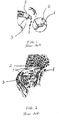
- Figs. 4 and 5 illustrate the implementation of a soft tissue anchoring system 10 in accordance with the present invention. What is illustrated is a portion of soft tissue 12 which is to be attached to a portion of bone 14 using the anchoring system 10.
- the soft tissue 12 may comprise a rotator cuff tendon, to be secured to the humerus, as discussed above in conjunction with Figs. 1-3 , or the invention is equally applicable to any other site wherein attachment of soft tissue to bone is desired.
- a bone tunnel 16 is created in the bone 14 and a suture anchor 18 is placed within the bone tunnel 16, as shown, using common surgical techniques, which may or may not be arthroscopic.
- the illustrated suture anchor 18 comprises a hollow anchor body, having threads 20 or other suitable structure for engaging adjacent bone forming walls of the tunnel 16 to fix the anchor 18 in place within the tunnel.
- This type of corkscrew suture anchor is well known in the art.
- Other suitable types of suture anchors may also be employed.
- the suture anchor also has an eyelet 22 or other suitable structure for securing suture 24 to the anchor, which suture extends through the soft tissue 12 and secured thereto by means of a knot 26 or other suitable means.
- the soft tissue 12 is secured to the adjacent bone 14 by extending the free ends of the suture 24 through the soft tissue 12, securing the anchor 18 within the bone tunnel 16, tensioning the suture 24 until the soft tissue 12 is approximated to the adjacent bone 14, then creating the suture knot 26 to secure the soft tissue in place.
- This basic technique is well known in the art.
- the present inventive system comprises a member or insert 28 which is comprised of a nanofiber material.
- the nanofiber material is, in one embodiment, a monophasic nanofiber scaffold, which are known in the art, as described in the prior art references discussed in the Background portion of this application.
- a multi-phasic nanofiber scaffold such as disclosed and described in U.S. Published Patent Application No. 2010/0292791 to Lu et al. may be used.
- the nanofiber scaffold member 28 extends into the bone tunne116 through the hollow center of the anchor 18, and expands outwardly at the bone surface to maximize surface area contact between the tissue and bone.
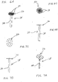
- FIG. 6A-6C There are many configurations which the inventive nanofiber member 28 may assume. Two such alternative examples are illustrated in Figs. 6A-6C and Figs. 7A-7C , respectively.
- Each configuration shows the material in the deployed state. Prior to deployment of the anchor into the bone, the material is rolled in a cylindrical fashion around the implant inserter shaft and held in place (and protected) by a tubular sheath. This allows the use of traditional arthroscopic surgical techniques to place the implant into the bone. Once the implant is placed into the bone, the sheath is retracted and the nanofiber material is spread out to maximize the surface area contact between the tissue and bone.
- the nanofiber material can be manufactured with the fibers organized in a random orientation (unaligned) or aligned in one direction (aligned). There are three primary reasons why fiber alignment is important when coupled with the suture anchor:
- the material may be attached to an implant.
- the material may be looped around the same eyelet as the suture or passed around a secondary eyelet.
- Another method for attachment is mechanically fastening the material to the anchor using a cleat, screw or post.
- the material may also be pinched between two halves of an implant.
- the material may be attached to a portion of an implant using a knot or adhesive.
- the material may also be bonded to the implant with the use of solvent.
- the nanofiber insert 28 comprises a distal portion or shaft 30 for attaching the insert 28 to the anchor 18, and a proximal head 32.
- the insert 28 when the suture anchor is deployed, is disposed so that the distal portion 30 extends through the suture anchor 18, as shown.
- a proximal end of the distal portion 30 extends proximally of the proximal end of the anchor body, and the head 32 is disposed at a proximal end of the insert distal portion, as shown.
- Figs. 6A-6C illustrates an embodiment wherein the insert distal portion 30 comprises a tube of material with strips 34 cut in the proximal end to allow the material to be deployed and spread out radially, as shown, to form the proximal head 32 and increase the surface contact between the tissue and bone.
- the tube comprises the distal portion 30 which is secured to the implant.
- Figs. 7A-7C illustrates an alternative embodiment, wherein the insert 28 comprises a die-cut sheet material in the deployed condition. The long portions form the distal portion 30 which is disposed within the implant that is deployed into bone.
- An additional embodiment is an implant as previously described, with nanofiber material fixed solely to the proximal end (proximal defined as the end of the implant that is adjacent to the soft tissue, and distal defined as the end of the implant farthest in the bone).
- the nanofiber material covers just the surface area of the proximal end of the implant or possibly extends further proximally and/or radially away from the central axis of the implant.
- Another additional embodiment is an implant as previously described, wherein the nanofiber material is fixed mechanically, with an adhesive, or by solvent bonding.
- Yet another additional embodiment is an implant as previously described wherein the method of attachment of the material to the implant is via the use of a suture tether that is attached to the implant and the material.
- the material may be either fixed or movable.
- the suture is configured such that the surgeon pulls on the free end of the suture which moves the material closer to the implant, allowing the surgeon to position the material into a desired location.
- the position of the material relative to the implant is set prior to insertion of the implant into the bone or after the implant is deployed into the bone. Once the material is in position it is locked in place or reversibly movable. This may also be incorporated into two or more implants to allow the material to be placed in an adjustable location determined by the surgeon on the bone in between two or more implants.
- Still another additional embodiment is an implant as previously described, wherein the nanofiber material is contained internal to the implant, along its central axis. The material extends at or near the distal tip and at, near or beyond the proximal end of the implant.
- Another additional embodiment is an implant as previously described wherein the material may also be contained externally to the implant or within external channels.
- Yet another additional embodiment is two or more implants as previously described with a bridge of nanofiber material strung between each implant.
- This configuration might best be described as a blanket of nanofiber material anchored at each implant, with the nanofiber material incorporated within or along the exterior of the implants.
- Surgical specialties that could utilize the invention include sports medicine, trauma, spine, foot and ankle, hand, hip, and extremities.
- nanofiber scaffolds promote cell attachment and growth in both aligned and unaligned orientations.
- the present invention improves the ease of use of nanofiber scaffolds for surgeons by pre-attaching the scaffold to a current, state-of-the-art suture anchor that can be implanted using standard arthroscopic procedures.
- nanofiber scaffolds in sheet form as proposed by Moffat will be substantially diminished due to the fact that surgeons will be reluctant to use a product that requires an open surgical procedure versus an arthroscopic procedure.
- the present invention facilitates arthroscopic use of nanofiber scaffolds, potentially increasing their value by several fold.
Landscapes
- Health & Medical Sciences (AREA)
- Surgery (AREA)
- Life Sciences & Earth Sciences (AREA)
- Medical Informatics (AREA)
- Animal Behavior & Ethology (AREA)
- Engineering & Computer Science (AREA)
- Biomedical Technology (AREA)
- Heart & Thoracic Surgery (AREA)
- Rheumatology (AREA)
- Molecular Biology (AREA)
- Nuclear Medicine, Radiotherapy & Molecular Imaging (AREA)
- General Health & Medical Sciences (AREA)
- Public Health (AREA)
- Veterinary Medicine (AREA)
- Surgical Instruments (AREA)
- Prostheses (AREA)
- Materials For Medical Uses (AREA)
Claims (9)
- Verankerungssystem (10) zum Sichern von weichem Gewebe (12) an einem Knochen (14), umfassend ein Implantat (18), das einen Körper zur Sicherung in einem Knochenkanal (16) und einen Einsatz (28) aufweist, umfassend ein Nanofasermaterial, das an dem Implantatkörper befestigt ist, und eine vergrößerte Oberfläche zum Berühren des weichen Gewebes (12) aufweist, dadurch gekennzeichnet, dass sich das Nanofasermaterial von einer Stelle innerhalb des Implantatkörpers aus einem proximalen Ende des Implantatkörpers heraus in einen Abschnitt des Materials außerhalb des Implantatkörpers erstreckt.
- Verankerungssystem (10) nach Anspruch 1, wobei das Implantat (18) ferner externe Oberflächenmerkmale (20) zum Sichern des Implantats (18) innerhalb eines umgebenden Knochens (14) umfasst.
- Verankerungssystem (10) nach Anspruch 2, wobei die externen Oberflächenmerkmale (20) Gewinde (20) umfassen.
- Verankerungssystem (10) nach Anspruch 1, wobei die vergrößerte Oberfläche des Einsatzes (28) einen Kopf (32), gebildet aus dem Nanofasermaterial, umfasst, wobei das Nanofasermaterial flexibel ist, sodass der Kopf (32) von einer unausgefahrenen, eingezogenen Position in eine ausgefahrene, vergrößerte Position vergrößerbar ist.
- Verankerungssystem (10) nach Anspruch 4, wobei der Einsatz (28) ferner einen Abschnitt (30), der sich distal von dem Kopf (32) aus erstreckt, zur Sicherung des Implantatkörpers umfasst.
- Verankerungssystem (10) nach Anspruch 5, wobei der distale Einsatzabschnitt (30) eine Röhre von Material umfasst, und der Kopf (32) eine Vielzahl von Streifen (34), die sich von einem proximalen Ende der Röhre erstrecken, umfasst.
- Verankerungssystem (10) nach Anspruch 5, wobei der distale Einsatzabschnitt (30) das Nanofasermaterial umfasst.
- Verankerungssystem (10) nach Anspruch 1, wobei das Nanofasermaterial monophasisch ist.
- Verankerungssystem (10) nach Anspruch 1, wobei das Implantat (18) mit Nahtmaterial (24) vorbeladen ist.
Priority Applications (1)
| Application Number | Priority Date | Filing Date | Title |
|---|---|---|---|
| EP18156124.2A EP3338648B1 (de) | 2012-10-12 | 2013-10-11 | System zur reparatur von weichgeweben unter verwendung von nanofasermaterial |
Applications Claiming Priority (2)
| Application Number | Priority Date | Filing Date | Title |
|---|---|---|---|
| US201261713230P | 2012-10-12 | 2012-10-12 | |
| PCT/US2013/064706 WO2014059378A1 (en) | 2012-10-12 | 2013-10-11 | Systems and methods for repairing soft tissues using nanofiber material |
Related Child Applications (2)
| Application Number | Title | Priority Date | Filing Date |
|---|---|---|---|
| EP18156124.2A Division-Into EP3338648B1 (de) | 2012-10-12 | 2013-10-11 | System zur reparatur von weichgeweben unter verwendung von nanofasermaterial |
| EP18156124.2A Division EP3338648B1 (de) | 2012-10-12 | 2013-10-11 | System zur reparatur von weichgeweben unter verwendung von nanofasermaterial |
Publications (3)
| Publication Number | Publication Date |
|---|---|
| EP2906264A1 EP2906264A1 (de) | 2015-08-19 |
| EP2906264A4 EP2906264A4 (de) | 2016-06-15 |
| EP2906264B1 true EP2906264B1 (de) | 2018-05-02 |
Family
ID=50476057
Family Applications (2)
| Application Number | Title | Priority Date | Filing Date |
|---|---|---|---|
| EP13845008.5A Active EP2906264B1 (de) | 2012-10-12 | 2013-10-11 | System zur reparatur von weichen geweben mit einem nanofasermaterial |
| EP18156124.2A Active EP3338648B1 (de) | 2012-10-12 | 2013-10-11 | System zur reparatur von weichgeweben unter verwendung von nanofasermaterial |
Family Applications After (1)
| Application Number | Title | Priority Date | Filing Date |
|---|---|---|---|
| EP18156124.2A Active EP3338648B1 (de) | 2012-10-12 | 2013-10-11 | System zur reparatur von weichgeweben unter verwendung von nanofasermaterial |
Country Status (5)
| Country | Link |
|---|---|
| US (6) | US9901334B2 (de) |
| EP (2) | EP2906264B1 (de) |
| JP (1) | JP6290906B2 (de) |
| AU (2) | AU2013328971B2 (de) |
| WO (1) | WO2014059378A1 (de) |
Families Citing this family (11)
| Publication number | Priority date | Publication date | Assignee | Title |
|---|---|---|---|---|
| JP6290906B2 (ja) | 2012-10-12 | 2018-03-07 | カイエン メディカル インコーポレイテッド | 軟部組織を硬骨に固定するための固着システム及び軟部組織固着システムにおいて使用される挿入物 |
| WO2014145864A1 (en) | 2013-03-15 | 2014-09-18 | Nanofiber Solutions, Llc | Biocompatible fiber textiles for implantation |
| EP3297558B1 (de) | 2015-05-22 | 2023-11-22 | Cayenne Medical, Inc. | Systeme zur reparatur von weichgewebe |
| US10820918B2 (en) | 2015-07-17 | 2020-11-03 | Crossroads Extremity Systems, Llc | Transosseous guide and method |
| US9962174B2 (en) | 2015-07-17 | 2018-05-08 | Kator, Llc | Transosseous method |
| US10258401B2 (en) | 2015-07-17 | 2019-04-16 | Kator, Llc | Transosseous guide |
| US12383253B2 (en) | 2015-08-04 | 2025-08-12 | Crossroads Extremity Systems, Llc | Suture anchor |
| US10143462B2 (en) | 2015-08-04 | 2018-12-04 | Kator, Llc | Transosseous suture anchor method |
| US10898608B2 (en) | 2017-02-02 | 2021-01-26 | Nanofiber Solutions, Llc | Methods of improving bone-soft tissue healing using electrospun fibers |
| CN111227993A (zh) * | 2020-03-12 | 2020-06-05 | 上海市第六人民医院 | 一种翻页式分段功能化可再生肩袖补片 |
| CN113208675B (zh) * | 2021-05-24 | 2022-05-10 | 杭州锐健马斯汀医疗器材有限公司 | 一种复合缝线锚植入装置 |
Family Cites Families (49)
| Publication number | Priority date | Publication date | Assignee | Title |
|---|---|---|---|---|
| US5578057A (en) | 1993-07-28 | 1996-11-26 | Mitek Surgical Products, Inc. | Anchoring device installation tool assembly and method |
| AU6019898A (en) * | 1997-01-09 | 1998-08-03 | Cohesion Technologies, Inc. | Devices for tissue repair and methods for preparation and use thereof |
| US6159234A (en) * | 1997-08-01 | 2000-12-12 | Peter M. Bonutti | Method and apparatus for securing a suture |
| US6123711A (en) * | 1999-06-10 | 2000-09-26 | Winters; Thomas F. | Tissue fixation device and method |
| US7887551B2 (en) * | 1999-12-02 | 2011-02-15 | Smith & Nephew, Inc. | Soft tissue attachment and repair |
| US6780198B1 (en) | 2001-12-06 | 2004-08-24 | Opus Medical, Inc. | Bone anchor insertion device |
| US20050038498A1 (en) | 2003-04-17 | 2005-02-17 | Nanosys, Inc. | Medical device applications of nanostructured surfaces |
| US20060149266A1 (en) * | 2004-12-10 | 2006-07-06 | New York Society For The Ruptured And Crippled Maintaining The Hospital For Special Surgery | Anchor for screw fixation of soft tissue to bone |
| CA2593781C (en) * | 2005-01-14 | 2011-05-17 | National Research Council Of Canada | Tie layer and method for forming thermoplastics |
| US8951285B2 (en) | 2005-07-05 | 2015-02-10 | Mitralign, Inc. | Tissue anchor, anchoring system and methods of using the same |
| US7704271B2 (en) * | 2005-12-19 | 2010-04-27 | Abdou M Samy | Devices and methods for inter-vertebral orthopedic device placement |
| CA2640268A1 (en) | 2006-01-25 | 2007-08-02 | Children's Medical Center Corporation | Methods and procedures for ligament repair |
| US8562647B2 (en) | 2006-09-29 | 2013-10-22 | Biomet Sports Medicine, Llc | Method and apparatus for securing soft tissue to bone |
| US20070255317A1 (en) * | 2006-03-22 | 2007-11-01 | Fanton Gary S | Suture passer devices and uses thereof |
| TWI367365B (en) * | 2006-04-28 | 2012-07-01 | Au Optronics Corp | A backlight module and the light diffusing module thereof |
| US20080154314A1 (en) * | 2006-08-16 | 2008-06-26 | Mcdevitt Dennis M | Composite interference screw for attaching a graft ligament to a bone, and other apparatus for making attachments to bone |
| WO2008028194A2 (en) * | 2006-09-01 | 2008-03-06 | Cornell Research Foundation, Inc. | Calcium phosphate nanofibers |
| US20100047309A1 (en) * | 2006-12-06 | 2010-02-25 | Lu Helen H | Graft collar and scaffold apparatuses for musculoskeletal tissue engineering and related methods |
| JP2010517638A (ja) * | 2007-02-02 | 2010-05-27 | トアニエ, インコーポレイテッド | 腱および靱帯を修復するシステムおよび方法 |
| US8753391B2 (en) | 2007-02-12 | 2014-06-17 | The Trustees Of Columbia University In The City Of New York | Fully synthetic implantable multi-phased scaffold |
| WO2008100534A2 (en) | 2007-02-12 | 2008-08-21 | Trustees Of Columbia University In The City Of New York | Biomimetic nanofiber scaffold for soft tissue and soft tissue-to-bone repair, augmentation and replacement |
| US7794484B2 (en) | 2007-05-07 | 2010-09-14 | Biomet Sports Medicine, Llc | Fixation device for delivery of biological material between soft tissue and bone |
| US8080060B2 (en) * | 2007-05-30 | 2011-12-20 | Alphatec Spine, Inc. | Processes and systems for loading medical implants with simulative growth agents |
| US20090062846A1 (en) | 2007-08-31 | 2009-03-05 | Ken Christopher G M | Closure medical device |
| US8613756B2 (en) | 2009-10-30 | 2013-12-24 | Depuy Mitek, Llc | Knotless suture anchor |
| WO2011117198A2 (en) | 2010-03-23 | 2011-09-29 | Basf Se | Pyridazine compounds for controlling invertebrate pests |
| WO2011137159A1 (en) | 2010-04-27 | 2011-11-03 | Synthes Usa, Llc | Anchor assembly including expandable anchor |
| US8858577B2 (en) | 2010-05-19 | 2014-10-14 | University Of Utah Research Foundation | Tissue stabilization system |
| US8961561B2 (en) | 2010-08-25 | 2015-02-24 | Daniel S. Schulman | Surgical system including suture anchor and insertion device and method for using |
| JP2012052262A (ja) | 2010-09-01 | 2012-03-15 | Central Glass Co Ltd | ゴム補強用ガラス繊維コード |
| FR2965168A1 (fr) | 2010-09-23 | 2012-03-30 | Tornier Inc | Composant d'implant de suture et dispositif d'implant de suture comprenant un tel composant |
| WO2012061017A1 (en) * | 2010-10-25 | 2012-05-10 | Boston Scientific Scimed, Inc. | Fibrous containment for hemostasis plug |
| US8758402B2 (en) | 2010-12-17 | 2014-06-24 | Boston Scientific Scimed, Inc. | Tissue puncture closure device |
| US8795334B2 (en) | 2011-01-28 | 2014-08-05 | Smith & Nephew, Inc. | Tissue repair |
| WO2012145059A1 (en) | 2011-02-15 | 2012-10-26 | Rotation Medical, Inc. | Methods and apparatus for fixing sheet-like materials to a target tissue |
| US9421008B2 (en) | 2011-09-23 | 2016-08-23 | Arthrex, Inc. | Soft suture-based anchors |
| GB2509679B (en) | 2011-11-14 | 2018-09-05 | Arthrocare Corp | Tissue repair assembly |
| WO2013096224A1 (en) | 2011-12-19 | 2013-06-27 | Rotation Medical, Inc. | Fasteners for affixing sheet -like materials to bone or tissue |
| US9320512B2 (en) | 2012-08-17 | 2016-04-26 | Arthrex, Inc. | Self-cinching soft anchors |
| US9597068B2 (en) | 2012-09-20 | 2017-03-21 | Depuy Mitek, Llc | Self-cinching suture anchors, systems, and methods |
| JP6290906B2 (ja) | 2012-10-12 | 2018-03-07 | カイエン メディカル インコーポレイテッド | 軟部組織を硬骨に固定するための固着システム及び軟部組織固着システムにおいて使用される挿入物 |
| US8986327B2 (en) | 2012-10-18 | 2015-03-24 | Smith & Nephew, Inc. | Flexible anchor delivery system |
| WO2014138398A1 (en) | 2013-03-06 | 2014-09-12 | Smith & Nephew, Inc. | Microanchor |
| US9173652B2 (en) | 2013-03-11 | 2015-11-03 | Linvatec Corporation | All-suture anchor inserter |
| US10182806B2 (en) | 2013-03-12 | 2019-01-22 | Arthrocare Corporation | Tissue repair assembly |
| AU2014364517B2 (en) | 2013-12-20 | 2019-06-20 | Arthrocare Corporation | Knotless all suture tissue repair |
| AU2015227067A1 (en) | 2014-03-05 | 2016-09-01 | Cayenne Medical, Inc. | All-suture suture anchor systems and methods |
| US9700291B2 (en) | 2014-06-03 | 2017-07-11 | Biomet Sports Medicine, Llc | Capsule retractor |
| EP3297558B1 (de) | 2015-05-22 | 2023-11-22 | Cayenne Medical, Inc. | Systeme zur reparatur von weichgewebe |
-
2013
- 2013-10-11 JP JP2015536971A patent/JP6290906B2/ja active Active
- 2013-10-11 US US14/052,624 patent/US9901334B2/en active Active
- 2013-10-11 WO PCT/US2013/064706 patent/WO2014059378A1/en not_active Ceased
- 2013-10-11 EP EP13845008.5A patent/EP2906264B1/de active Active
- 2013-10-11 EP EP18156124.2A patent/EP3338648B1/de active Active
- 2013-10-11 AU AU2013328971A patent/AU2013328971B2/en active Active
-
2017
- 2017-04-18 AU AU2017202516A patent/AU2017202516B2/en active Active
- 2017-10-30 US US15/797,980 patent/US10631849B2/en active Active
-
2020
- 2020-03-11 US US16/815,507 patent/US11534155B2/en active Active
-
2021
- 2021-12-29 US US17/565,425 patent/US11759198B2/en active Active
-
2023
- 2023-08-16 US US18/234,626 patent/US12185937B2/en active Active
- 2023-10-27 US US18/495,876 patent/US12357296B2/en active Active
Non-Patent Citations (1)
| Title |
|---|
| None * |
Also Published As
| Publication number | Publication date |
|---|---|
| AU2013328971A1 (en) | 2015-05-07 |
| US10631849B2 (en) | 2020-04-28 |
| US9901334B2 (en) | 2018-02-27 |
| US20200205804A1 (en) | 2020-07-02 |
| AU2013328971B2 (en) | 2017-01-19 |
| EP2906264A1 (de) | 2015-08-19 |
| JP2015533572A (ja) | 2015-11-26 |
| US12185937B2 (en) | 2025-01-07 |
| US12357296B2 (en) | 2025-07-15 |
| WO2014059378A1 (en) | 2014-04-17 |
| US11534155B2 (en) | 2022-12-27 |
| AU2017202516A1 (en) | 2017-05-04 |
| US20230389915A1 (en) | 2023-12-07 |
| AU2017202516B2 (en) | 2018-06-07 |
| JP6290906B2 (ja) | 2018-03-07 |
| EP3338648B1 (de) | 2019-09-25 |
| US20180042601A1 (en) | 2018-02-15 |
| EP3338648A1 (de) | 2018-06-27 |
| US20240050084A1 (en) | 2024-02-15 |
| US11759198B2 (en) | 2023-09-19 |
| US20220117596A1 (en) | 2022-04-21 |
| US20140107700A1 (en) | 2014-04-17 |
| EP2906264A4 (de) | 2016-06-15 |
Similar Documents
| Publication | Publication Date | Title |
|---|---|---|
| US12185937B2 (en) | Systems and methods for repairing soft tissues using nanofiber material | |
| US12185933B2 (en) | Systems and methods for repairing soft tissues | |
| US8419794B2 (en) | Method for double row fixation of tendon to bone | |
| ES2252825T3 (es) | Ayuda quirurgica para implantacion de tejido conectivo. | |
| US11992204B1 (en) | Surgical sheath, staple, and scaffold bone anchor devices | |
| US20250195205A1 (en) | Reinforced medical implant and method of use |
Legal Events
| Date | Code | Title | Description |
|---|---|---|---|
| PUAI | Public reference made under article 153(3) epc to a published international application that has entered the european phase |
Free format text: ORIGINAL CODE: 0009012 |
|
| 17P | Request for examination filed |
Effective date: 20150429 |
|
| AK | Designated contracting states |
Kind code of ref document: A1 Designated state(s): AL AT BE BG CH CY CZ DE DK EE ES FI FR GB GR HR HU IE IS IT LI LT LU LV MC MK MT NL NO PL PT RO RS SE SI SK SM TR |
|
| AX | Request for extension of the european patent |
Extension state: BA ME |
|
| DAX | Request for extension of the european patent (deleted) | ||
| REG | Reference to a national code |
Ref country code: DE Ref legal event code: R079 Ref document number: 602013037039 Country of ref document: DE Free format text: PREVIOUS MAIN CLASS: A61L0027560000 Ipc: A61B0017040000 |
|
| RA4 | Supplementary search report drawn up and despatched (corrected) |
Effective date: 20160517 |
|
| RIC1 | Information provided on ipc code assigned before grant |
Ipc: A61B 17/04 20060101AFI20160510BHEP Ipc: A61B 17/00 20060101ALI20160510BHEP |
|
| 17Q | First examination report despatched |
Effective date: 20170323 |
|
| GRAP | Despatch of communication of intention to grant a patent |
Free format text: ORIGINAL CODE: EPIDOSNIGR1 |
|
| INTG | Intention to grant announced |
Effective date: 20171207 |
|
| GRAS | Grant fee paid |
Free format text: ORIGINAL CODE: EPIDOSNIGR3 |
|
| GRAA | (expected) grant |
Free format text: ORIGINAL CODE: 0009210 |
|
| AK | Designated contracting states |
Kind code of ref document: B1 Designated state(s): AL AT BE BG CH CY CZ DE DK EE ES FI FR GB GR HR HU IE IS IT LI LT LU LV MC MK MT NL NO PL PT RO RS SE SI SK SM TR |
|
| REG | Reference to a national code |
Ref country code: GB Ref legal event code: FG4D |
|
| REG | Reference to a national code |
Ref country code: CH Ref legal event code: EP Ref country code: AT Ref legal event code: REF Ref document number: 994388 Country of ref document: AT Kind code of ref document: T Effective date: 20180515 |
|
| REG | Reference to a national code |
Ref country code: DE Ref legal event code: R096 Ref document number: 602013037039 Country of ref document: DE Ref country code: IE Ref legal event code: FG4D |
|
| REG | Reference to a national code |
Ref country code: DE Ref legal event code: R096 Ref document number: 602013037039 Country of ref document: DE |
|
| REG | Reference to a national code |
Ref country code: NL Ref legal event code: MP Effective date: 20180502 |
|
| REG | Reference to a national code |
Ref country code: LT Ref legal event code: MG4D |
|
| PG25 | Lapsed in a contracting state [announced via postgrant information from national office to epo] |
Ref country code: SE Free format text: LAPSE BECAUSE OF FAILURE TO SUBMIT A TRANSLATION OF THE DESCRIPTION OR TO PAY THE FEE WITHIN THE PRESCRIBED TIME-LIMIT Effective date: 20180502 Ref country code: NO Free format text: LAPSE BECAUSE OF FAILURE TO SUBMIT A TRANSLATION OF THE DESCRIPTION OR TO PAY THE FEE WITHIN THE PRESCRIBED TIME-LIMIT Effective date: 20180802 Ref country code: ES Free format text: LAPSE BECAUSE OF FAILURE TO SUBMIT A TRANSLATION OF THE DESCRIPTION OR TO PAY THE FEE WITHIN THE PRESCRIBED TIME-LIMIT Effective date: 20180502 Ref country code: LT Free format text: LAPSE BECAUSE OF FAILURE TO SUBMIT A TRANSLATION OF THE DESCRIPTION OR TO PAY THE FEE WITHIN THE PRESCRIBED TIME-LIMIT Effective date: 20180502 Ref country code: FI Free format text: LAPSE BECAUSE OF FAILURE TO SUBMIT A TRANSLATION OF THE DESCRIPTION OR TO PAY THE FEE WITHIN THE PRESCRIBED TIME-LIMIT Effective date: 20180502 Ref country code: BG Free format text: LAPSE BECAUSE OF FAILURE TO SUBMIT A TRANSLATION OF THE DESCRIPTION OR TO PAY THE FEE WITHIN THE PRESCRIBED TIME-LIMIT Effective date: 20180802 |
|
| PG25 | Lapsed in a contracting state [announced via postgrant information from national office to epo] |
Ref country code: HR Free format text: LAPSE BECAUSE OF FAILURE TO SUBMIT A TRANSLATION OF THE DESCRIPTION OR TO PAY THE FEE WITHIN THE PRESCRIBED TIME-LIMIT Effective date: 20180502 Ref country code: RS Free format text: LAPSE BECAUSE OF FAILURE TO SUBMIT A TRANSLATION OF THE DESCRIPTION OR TO PAY THE FEE WITHIN THE PRESCRIBED TIME-LIMIT Effective date: 20180502 Ref country code: LV Free format text: LAPSE BECAUSE OF FAILURE TO SUBMIT A TRANSLATION OF THE DESCRIPTION OR TO PAY THE FEE WITHIN THE PRESCRIBED TIME-LIMIT Effective date: 20180502 Ref country code: NL Free format text: LAPSE BECAUSE OF FAILURE TO SUBMIT A TRANSLATION OF THE DESCRIPTION OR TO PAY THE FEE WITHIN THE PRESCRIBED TIME-LIMIT Effective date: 20180502 Ref country code: GR Free format text: LAPSE BECAUSE OF FAILURE TO SUBMIT A TRANSLATION OF THE DESCRIPTION OR TO PAY THE FEE WITHIN THE PRESCRIBED TIME-LIMIT Effective date: 20180803 |
|
| REG | Reference to a national code |
Ref country code: AT Ref legal event code: MK05 Ref document number: 994388 Country of ref document: AT Kind code of ref document: T Effective date: 20180502 |
|
| PG25 | Lapsed in a contracting state [announced via postgrant information from national office to epo] |
Ref country code: AT Free format text: LAPSE BECAUSE OF FAILURE TO SUBMIT A TRANSLATION OF THE DESCRIPTION OR TO PAY THE FEE WITHIN THE PRESCRIBED TIME-LIMIT Effective date: 20180502 Ref country code: DK Free format text: LAPSE BECAUSE OF FAILURE TO SUBMIT A TRANSLATION OF THE DESCRIPTION OR TO PAY THE FEE WITHIN THE PRESCRIBED TIME-LIMIT Effective date: 20180502 Ref country code: SK Free format text: LAPSE BECAUSE OF FAILURE TO SUBMIT A TRANSLATION OF THE DESCRIPTION OR TO PAY THE FEE WITHIN THE PRESCRIBED TIME-LIMIT Effective date: 20180502 Ref country code: PL Free format text: LAPSE BECAUSE OF FAILURE TO SUBMIT A TRANSLATION OF THE DESCRIPTION OR TO PAY THE FEE WITHIN THE PRESCRIBED TIME-LIMIT Effective date: 20180502 Ref country code: EE Free format text: LAPSE BECAUSE OF FAILURE TO SUBMIT A TRANSLATION OF THE DESCRIPTION OR TO PAY THE FEE WITHIN THE PRESCRIBED TIME-LIMIT Effective date: 20180502 Ref country code: CZ Free format text: LAPSE BECAUSE OF FAILURE TO SUBMIT A TRANSLATION OF THE DESCRIPTION OR TO PAY THE FEE WITHIN THE PRESCRIBED TIME-LIMIT Effective date: 20180502 Ref country code: RO Free format text: LAPSE BECAUSE OF FAILURE TO SUBMIT A TRANSLATION OF THE DESCRIPTION OR TO PAY THE FEE WITHIN THE PRESCRIBED TIME-LIMIT Effective date: 20180502 |
|
| REG | Reference to a national code |
Ref country code: DE Ref legal event code: R097 Ref document number: 602013037039 Country of ref document: DE |
|
| PG25 | Lapsed in a contracting state [announced via postgrant information from national office to epo] |
Ref country code: SM Free format text: LAPSE BECAUSE OF FAILURE TO SUBMIT A TRANSLATION OF THE DESCRIPTION OR TO PAY THE FEE WITHIN THE PRESCRIBED TIME-LIMIT Effective date: 20180502 |
|
| PLBE | No opposition filed within time limit |
Free format text: ORIGINAL CODE: 0009261 |
|
| STAA | Information on the status of an ep patent application or granted ep patent |
Free format text: STATUS: NO OPPOSITION FILED WITHIN TIME LIMIT |
|
| 26N | No opposition filed |
Effective date: 20190205 |
|
| PG25 | Lapsed in a contracting state [announced via postgrant information from national office to epo] |
Ref country code: SI Free format text: LAPSE BECAUSE OF FAILURE TO SUBMIT A TRANSLATION OF THE DESCRIPTION OR TO PAY THE FEE WITHIN THE PRESCRIBED TIME-LIMIT Effective date: 20180502 |
|
| REG | Reference to a national code |
Ref country code: BE Ref legal event code: MM Effective date: 20181031 |
|
| PG25 | Lapsed in a contracting state [announced via postgrant information from national office to epo] |
Ref country code: MC Free format text: LAPSE BECAUSE OF FAILURE TO SUBMIT A TRANSLATION OF THE DESCRIPTION OR TO PAY THE FEE WITHIN THE PRESCRIBED TIME-LIMIT Effective date: 20180502 Ref country code: LU Free format text: LAPSE BECAUSE OF NON-PAYMENT OF DUE FEES Effective date: 20181011 |
|
| REG | Reference to a national code |
Ref country code: IE Ref legal event code: MM4A |
|
| PG25 | Lapsed in a contracting state [announced via postgrant information from national office to epo] |
Ref country code: BE Free format text: LAPSE BECAUSE OF NON-PAYMENT OF DUE FEES Effective date: 20181031 Ref country code: FR Free format text: LAPSE BECAUSE OF NON-PAYMENT OF DUE FEES Effective date: 20181031 |
|
| PG25 | Lapsed in a contracting state [announced via postgrant information from national office to epo] |
Ref country code: IE Free format text: LAPSE BECAUSE OF NON-PAYMENT OF DUE FEES Effective date: 20181011 |
|
| PG25 | Lapsed in a contracting state [announced via postgrant information from national office to epo] |
Ref country code: AL Free format text: LAPSE BECAUSE OF FAILURE TO SUBMIT A TRANSLATION OF THE DESCRIPTION OR TO PAY THE FEE WITHIN THE PRESCRIBED TIME-LIMIT Effective date: 20180502 |
|
| PG25 | Lapsed in a contracting state [announced via postgrant information from national office to epo] |
Ref country code: MT Free format text: LAPSE BECAUSE OF NON-PAYMENT OF DUE FEES Effective date: 20181011 |
|
| PG25 | Lapsed in a contracting state [announced via postgrant information from national office to epo] |
Ref country code: TR Free format text: LAPSE BECAUSE OF FAILURE TO SUBMIT A TRANSLATION OF THE DESCRIPTION OR TO PAY THE FEE WITHIN THE PRESCRIBED TIME-LIMIT Effective date: 20180502 |
|
| PG25 | Lapsed in a contracting state [announced via postgrant information from national office to epo] |
Ref country code: PT Free format text: LAPSE BECAUSE OF FAILURE TO SUBMIT A TRANSLATION OF THE DESCRIPTION OR TO PAY THE FEE WITHIN THE PRESCRIBED TIME-LIMIT Effective date: 20180502 |
|
| PG25 | Lapsed in a contracting state [announced via postgrant information from national office to epo] |
Ref country code: CY Free format text: LAPSE BECAUSE OF FAILURE TO SUBMIT A TRANSLATION OF THE DESCRIPTION OR TO PAY THE FEE WITHIN THE PRESCRIBED TIME-LIMIT Effective date: 20180502 Ref country code: MK Free format text: LAPSE BECAUSE OF NON-PAYMENT OF DUE FEES Effective date: 20180502 Ref country code: HU Free format text: LAPSE BECAUSE OF FAILURE TO SUBMIT A TRANSLATION OF THE DESCRIPTION OR TO PAY THE FEE WITHIN THE PRESCRIBED TIME-LIMIT; INVALID AB INITIO Effective date: 20131011 |
|
| PG25 | Lapsed in a contracting state [announced via postgrant information from national office to epo] |
Ref country code: IS Free format text: LAPSE BECAUSE OF FAILURE TO SUBMIT A TRANSLATION OF THE DESCRIPTION OR TO PAY THE FEE WITHIN THE PRESCRIBED TIME-LIMIT Effective date: 20180902 |
|
| P01 | Opt-out of the competence of the unified patent court (upc) registered |
Effective date: 20230527 |
|
| PGFP | Annual fee paid to national office [announced via postgrant information from national office to epo] |
Ref country code: DE Payment date: 20240920 Year of fee payment: 12 |
|
| PGFP | Annual fee paid to national office [announced via postgrant information from national office to epo] |
Ref country code: GB Payment date: 20241007 Year of fee payment: 12 |
|
| PGFP | Annual fee paid to national office [announced via postgrant information from national office to epo] |
Ref country code: CH Payment date: 20241101 Year of fee payment: 12 |
|
| PGFP | Annual fee paid to national office [announced via postgrant information from national office to epo] |
Ref country code: IT Payment date: 20250904 Year of fee payment: 13 |
|
| REG | Reference to a national code |
Ref country code: CH Ref legal event code: U11 Free format text: ST27 STATUS EVENT CODE: U-0-0-U10-U11 (AS PROVIDED BY THE NATIONAL OFFICE) Effective date: 20251101 |