EP2878256A1 - Ophthalmic measurement apparatus - Google Patents
Ophthalmic measurement apparatus Download PDFInfo
- Publication number
- EP2878256A1 EP2878256A1 EP14195372.9A EP14195372A EP2878256A1 EP 2878256 A1 EP2878256 A1 EP 2878256A1 EP 14195372 A EP14195372 A EP 14195372A EP 2878256 A1 EP2878256 A1 EP 2878256A1
- Authority
- EP
- European Patent Office
- Prior art keywords
- reflection image
- light receiving
- corneal
- light
- optical system
- Prior art date
- Legal status (The legal status is an assumption and is not a legal conclusion. Google has not performed a legal analysis and makes no representation as to the accuracy of the status listed.)
- Granted
Links
Images




Classifications
-
- A—HUMAN NECESSITIES
- A61—MEDICAL OR VETERINARY SCIENCE; HYGIENE
- A61B—DIAGNOSIS; SURGERY; IDENTIFICATION
- A61B3/00—Apparatus for testing the eyes; Instruments for examining the eyes
- A61B3/10—Objective types, i.e. instruments for examining the eyes independent of the patients' perceptions or reactions
- A61B3/1005—Objective types, i.e. instruments for examining the eyes independent of the patients' perceptions or reactions for measuring distances inside the eye, e.g. thickness of the cornea
Definitions
- This disclosure relates to an ophthalmic measurement apparatus for measuring corneal thickness of an examinee's eye.
- an apparatus for measuring corneal thickness by projecting measurement light to an examinee's eye from an oblique direction and utilizing reflection light of the measurement light from a corneal anterior surface of the examinee's eye and reflection light of the measurement light from a corneal posterior surface of the examinee's eye.
- Patent Document 1 discloses an apparatus for measuring corneal thickness by receiving reflection light from a corneal anterior surface and reflection light from a corneal posterior surface in different positions of a light receiving element such as a one-dimensional line sensor and others, and using a one-dimensional received light intensity distribution representing the intensity of light received in each position on the light receiving element.
- a light receiving element such as a one-dimensional line sensor and others
- Patent Document 1 JP-A-2012-143492
- scattered measurement light from a cornea influences reflection light which will enter a light receiving element.
- the measurement light may be scattered due to corneal abnormality, extraneous matters sticking to a corneal front surface, and others.
- scattered measurement light within a cornea may enter the light receiving element.
- the conventional apparatus is merely arranged to one-dimensionally determine the positions on the anterior surface and the posterior surface of a cornea. This apparatus is not designed to take into consideration the influence of scattered light that will enter the light receiving element.
- the present disclosure has been made to solve the above problems and has a purpose to provide an ophthalmic measurement apparatus capable of obtaining a good measurement value of corneal thickness.
- an ophthalmic measurement apparatus comprising: a light projecting optical system for projecting measurement light to an examinee's eye from an oblique direction; a light receiving optical system for receiving, by a light receiving element, a first cornea reflection image formed by the measurement light reflected by a corneal anterior surface of the examinee's eye and a second corneal reflection image formed by the measurement light reflected by a corneal posterior surface of the examinee's eye; and an arithmetic processing unit for obtaining corneal thickness of the examinee's eye based on positional information of the first cornea reflection image and the second cornea reflection image in a light receiving signal output from the light receiving element, characterized in that the light receiving optical system includes a two-dimensional light receiving element as the light receiving element, and the arithmetic processing unit is configured to two-dimensionally detect the positional information of at least one of the first cornea reflection image and the second cornea reflection image based on a light receiving signal from the two-dimensional light receiving element, and obtain
- an ophthalmic measurement apparatus of the present disclosure it is possible to obtain a more preferable measurement value of corneal thickness.
- FIG. 1 A schematic configuration of a measuring unit in an ophthalmic measurement apparatus 1 of the present embodiment will be first explained referring to FIG. 1 .
- the ophthalmic measurement apparatus 1 shown in FIG. 1 is configured to measure corneal thickness of an examinee's eye E.
- the apparatus is arranged to measure intraocular pressure in non-contact manner.
- the ophthalmic measurement apparatus 1 mainly includes an observation optical system 30 and a corneal thickness measurement optical system 80 (80a and 80b in FIG. 1 ).
- the ophthalmic measurement apparatus 1 further includes an alignment optical system 40, a fixation optical system 50, a corneal deformation detecting optical system 60 (60a and 60b in FIG. 1 ), and a first working distance detecting optical system 70 (60a and 70b in FIG. 1 ).
- the ophthalmic measurement apparatus 1 of the present embodiment also includes an intraocular pressure measuring unit 10.
- the measuring unit of the ophthalmic measurement apparatus 1 including those optical systems and others is housed in a cabinet not shown.
- the cabinet is three-dimensionally movable with respect to the examinee's eye E by an XYZ drive mechanism 20 (see FIG. 4 ).
- This cabinet may also be moved according to an instruction of an examiner for example through an operating member (e.g., an operating unit (a joystick) 21, see FIG. 4 ).
- the observation optical system 30 is used to observe a front image of an anterior segment of the examinee's eye E.
- the observation optical system 30 includes a beam splitter 32, an objective lens 33, a beam splitter 34, an imaging lens 35, a filter 36, and a two-dimensional imaging element 37, these components being arranged on an observation optical axis L1.
- Infrared illumination light sources 31 are used as an observation light source.
- An image of the examinee's eye illuminated by the infrared illumination light sources 31 is formed on the two-dimensional imaging element 37 via an optical path from the beam splitter 32 to the filter 36.
- the filter 36 has the property of transmitting the light from the light sources 31 and the light from an alignment light source 41 and not transmitting the light from a light source 61 mentioned later for detecting corneal deformation and visible light.
- the image formed on the two-dimensional imaging element 37 is displayed on a monitor 38.
- the alignment optical system 40 has the light source 41 and a projection lens 42.
- the alignment optical system 40 shares the configuration from the beam splitter 32 to the two-dimensional imaging element 37 arranged on the optical axis L1 with the observation optical system 30.
- Infrared light projected from the alignment infrared light source 41 via the projection lens 42 is reflected by the beam splitter 32 and then projected to the examinee's eye E from a front direction.
- a corneal luminescent spot (a corneal reflection image) formed at a corneal vertex by the light source 41 is formed on the two-dimensional imaging element 37 via the beam splitter 32 to the filter 36. Even though the details thereof will be mentioned later, this corneal luminescent spot is utilized for alignment detection in up-and-down and right-and-left directions.
- the fixation optical system 50 includes a visible light source (a fixation lamp) 51 and a projection lens 52.
- the fixation optical system 50 shares the components from the beam splitter 34 to the beam splitter 32 arranged on the optical axis L1 with the observation optical system 30 and others.
- a fixation target is presented to the examinee's eye E from front.
- the visual line of the examinee's eye E is fixed in a direction toward a fixation point located in the front direction.
- the optical axis L1 is used as a fixation optical axis.
- the visible light source 51 may include any light sources such as an LED and a laser, for example.
- a pattern light source such as a point light source, a slit light source, and a ring light source as well as a two-dimensional display such as a liquid crystal display.
- the corneal deformation detecting optical system 60 is used to detect a deformed state of the cornea Ec.
- This optical system 60 includes a light projecting optical system 60a and a light receiving optical system 60b.
- the light projecting optical system 60a is arranged to irradiate illumination light toward the cornea Ec of the eye E from an oblique direction.
- the light projecting optical system 60a includes a light source 61, a dichroic mirror 62, and a projection lens 63.
- the light receiving optical system 60b is arranged to receive, by a photodetector 67, corneal reflection light from the light projecting optical system 60a.
- the light receiving optical system 60b includes a lens 64, a beam splitter 65, and a pinhole plate 66 in addition to the photodetector 67.
- the corneal deformation detecting optical system 60 is placed so that an amount of light received by the photodetector 67 is maximum when the examinee's eye E is in a predetermined deformed state (e.g., a flattened state). Accordingly, deformation of the examinee's eye E is detected based on a light receiving signal representing the amount of light received (light receiving amount) output from the photodetector 67.
- the above deformation of the examinee's eye E is carried out by blowing a predetermined mount of compressed fluid (e.g., compressed air) from a nozzle 6 of the intraocular measuring unit 10 toward the examinee's eye E.
- compressed fluid e.g., compressed air
- This compressed fluid is supplied from a fluid supply unit 10a (see FIG. 4 ).
- the intraocular pressure is measured based on a deformation amount of the examinee's eye E detected by use of the photodetector 67 when the compressed fluid is blown onto the eye E.
- the first working distance detecting optical system 70 is utilized for alignment (rough adjustment) in a working distance direction (a Z direction).
- the first working distance detecting optical system 70 shares the light projecting optical system 60a with the corneal deformation detecting optical system.
- This optical system 70 includes a light receiving optical system 70b.
- This light receiving optical system 70b includes a condensing lens 72 and a position imaging element 73.
- Light emitted from the light projecting optical system 60a forms an index image on the cornea Ec. Corneal reflection light from the light projecting optical system 60a enters the position detecting element 73 via the components from the lens 64 to the condensing lens 72.
- a light receiving position on the position detecting element 73 varies according to a positional relationship between the measuring unit and the examinee's eye E (the cornea Ec) in the working distance direction (the Z direction). Accordingly, the information on the working distance is obtained based on an output signal from the position detecting element 73.
- the output signal from the position detecting element 73 in the present embodiment is utilized for alignment (rough adjustment) in the working distance direction (the Z direction).
- the magnification of the light receiving optical system 70b of the first working distance detecting optical system is not as large as that of a light receiving optical system 80b mentioned later.
- a distance detection range of the position detecting element 73 in the Z direction is wider than that of a two-dimensional light receiving element 87.
- the distance detection range of the position detecting element 73 is for example a range of ⁇ 3 to 4 mm from an alignment reference position.
- the corneal thickness measuring optical system 80 (80a, 80b) is used to measure the corneal thickness of the examinee's eye E.
- This optical system 80 includes a light projecting optical system 80a and a light receiving optical system 80b.
- the light projecting optical system 80a is used to project measurement light (illumination light) for measuring the corneal thickness from an oblique direction with respect to the optical axis direction of the observation optical system 30.
- the light projecting optical system 80a includes a measurement light source 81, a condensing lens 82, a light limiting member 83, a concave lens 84, the beam splitter 65, a beam splitter 71, and the lens 64. These components are placed on an optical axis L2 which is a light projection optical axis.
- the optical axis L2 is inclined with respect to the optical axis L1 which is the fixation optical axis.
- the light projecting optical system 80a irradiates illumination light (measurement light) toward the examinee's eye E from an oblique direction with respect to the fixation optical axis.
- the measurement light source 81 there is used a visible light source or an infrared (including near infrared) light source; e.g., an LED, a laser, or the like.
- the condensing lens 82 condenses the light emitted from the measurement light source 81.
- the light limiting member 83 is placed on an optical path of the light projecting optical system 80a and limits the light (measurement light) emitted from the measurement light source 81.
- the light limiting member 83 is disposed in a position conjugated with a posterior surface of the cornea Ec during measurement of the corneal thickness.
- the light limiting member 83 is a pinhole plate, but is not limited thereto.
- a slit plate or the like may also be used as the light limiting member 83.
- This light limiting member 83 allows a part of the light emitted from the measurement light source 81 to pass through and blocks a remaining part of the light.
- the light projecting optical system 80a projects a predetermined pattern of light (a spot light beam) to the cornea Ec.
- the light receiving optical system 80b has the two-dimensional light receiving element 87.
- the light receiving optical system 80b is arranged to receive, by the two-dimensional light receiving element 87, the measurement light emitted from the light projecting optical system 80a and reflected (specular reflection) by a corneal anterior surface Ec1 (a corneal front surface, a corneal epithelium) and a corneal posterior surface Ec2 (a corneal back surface, a corneal endothelium) of the examinee's eye E.
- an optical axis L3 of the light receiving optical system 80b is placed nearly symmetrical to the optical axis L2 of the light projecting optical system 80a with respect to the observation optical axis (the optical axis L1) of the observation optical system 30.
- the light receiving optical system 80b includes the lens 63, the dichroic mirror 62, a light receiving lens 85, and the two-dimensional light receiving element 87 on the optical axis L3.
- a detailed alignment state in the Z direction with respect to the examinee's eye E is detected by the light receiving optical system 80b in the present embodiment.
- the two-dimensional light receiving element 87 receives reflection light from each of the corneal anterior surface and the corneal posterior surface.
- a two-dimensional imaging element such as a two-dimensional area sensor and a CCD.
- the light receiving optical system 80b of the corneal thickness measuring optical system 80 performs imaging with high magnification with respect to the first working distance detecting optical system 70. Accordingly, in the present embodiment, the distance detection range of the two-dimensional light receiving element 87 in the Z direction is narrower than that of the position detecting element 73. For instance, it is a range of ⁇ 1 mm from the alignment reference position.
- the light emitted from the measurement light source 81 is condensed by the condensing lens 82 and illuminates the light limiting member 83 from behind.
- the light from the measurement light source 81 is limited by the light limiting member 83 and then is imaged (condensed) in a position near the cornea Ec by the lens 64.
- a pinhole image is formed near the cornea Ec.
- the light from the measurement light source 81 is formed near an intersection point with a visual axis on the cornea Ec.
- the reflection light of the measurement light by the cornea Ec travels in a direction symmetrical to the projection light beam with respect to the optical axis L1.
- the light receiving lens 85 forms an image of the reflection light on a light receiving plane of the two-dimensional light receiving element 87.
- first cornea reflection image R1 (hereinafter, simply referred to as "first reflection image R1") on the two-dimensional light receiving element 87.
- second cornea reflection image R2 (hereinafter, simply referred to as "first reflection image R1" on the two-dimensional light receiving element 87.
- the first cornea reflection image R1 is a circular reflection image.
- the second cornea reflection image R2 is a circular reflection image smaller than the first cornea reflection image R1.
- the reflection light (see a solid line) by the corneal anterior surface and the reflection light (see a broken line) by the corneal posterior surface are different in reflection optical path and therefore two reflection images R1 and R2 are formed fully or partially away from each other.
- the second reflection image R2 is fully formed away from the first reflection image R1.
- the corneal thickness is large, the reflection optical paths of two reflection images are more separated. Accordingly, two reflection images are formed in the positions more spaced apart on the two-dimensional light receiving element 87 as the corneal thickness is larger.
- the ophthalmic measurement apparatus 1 of the present embodiment is therefore arranged to measure the corneal thickness based on the positional relationship between the first reflection image R1 and the second reflection image R2 on the two-dimensional light receiving element 87.
- the light projecting optical system 60a and the light receiving optical system 80b share the optical axis L3 with each other, and the light projecting optical system 80a, the light receiving optical system 60b, and the light receiving optical system 70b share the optical axis L2 with one another. Needless to say, they are not limited thereto.
- the lens 64 shared among the light receiving optical systems 60b and 70b and the light projecting optical system 80a is placed in a position to make the reflection light emitted from the light source 61 and reflected by the cornea Ec condense on a center of a hole of the pinhole plate 66 and make the measurement light emitted from the measurement light source 81 condense on the anterior surface and the posterior surface of the cornea Ec.
- the ophthalmic measurement apparatus 1 of the present embodiment has a control unit 100 for executing control of the entire apparatus and calculation of measurement results.
- control unit 100 is connected to the fluid supply unit 10a, the XYZ drive unit 20, the operating unit 21, a storage device 105, the various light sources 31, 41, 51, 61, and 81, the various light receiving elements 37, 67, 73, 87, and others.
- control unit 100 includes a CPU 101, a ROM 102, and a RAM 103.
- the CPU 101 is a processing device (a processor) to execute various processings (e.g., control processing, arithmetic processing, etc.) related to the ophthalmic measurement apparatus 1.
- the ROM 102 is a nonvolatile storage device in which control programs, fixed data, and others are stored.
- the RAM 103 is a rewritable volatile storage device. This RAM 103 stores temporary data to be used by the ophthalmic measurement apparatus 1 in photographing and measuring the examinee's eye E.
- the storage device 105 is a rewritable nonvolatile storage device.
- the storage device 105 stores at least a program to execute an anterior segment measurement processing by the control unit 100.
- the storage device 105 may also save an image captured by the ophthalmic measurement apparatus 1 and measurement results obtained by the ophthalmic measurement apparatus 1 and others.
- the CPU 101 executes a corneal thickness measurement processing shown in FIG. 5 to measure the corneal thickness.
- the measuring unit of the ophthalmic measurement apparatus 1 is subjected to alignment (positional adjustment) in advance.
- This alignment is performed after having the examinee's eye E positioned to face the measuring unit and having an examinee gaze at a fixation target.
- the alignment in X and Y directions (up-and-down and right-and-left directions) in the present embodiment may be performed by operation of the operating unit 21 (e.g., a joystick) by an examiner based on alignment information displayed on a monitor 38.
- the examiner manipulates the operating unit 21 so as to bring a corneal reflection image (a luminescent spot) formed by the light source 41 into a predetermined position relative to a reticle not shown displayed on the monitor 38.
- the CPU 101 may also automatically perform alignment adjustment in the up-and-down and right-and-left directions based on the alignment information obtained as an imaging result of the two-dimensional imaging element 37.
- the CPU 101 controls the XYZ drive mechanism 20 so that the corneal luminescent spot by the light source 41 is formed in an alignment position (e.g., an intersection point of the optical axis L1 and an imaging plane of the two-dimensional imaging element 37) set on the two-dimensional imaging element 37 in advance. In this way, the CPU 101 moves the measuring unit to an alignment completion position in the X and Y directions.
- the alignment in the Z direction (a back-and-forth direction) will be performed.
- the alignment in the Z direction is automatically performed in such a manner that the CPU 101 controls the operation of the XYZ drive mechanism 20 based on working distance information obtained from the position detecting element 73 and the two-dimensional light receiving element 87.
- the CPU 101 causes the measuring unit to move in the Z direction based on the working distance information obtained from the position detecting element 37. In the present embodiment, consequently, the measuring unit is moved to a range where the reflection light (the first reflection image R1 or the second reflection image R2) of the light from the measurement light source 81 is detected by the two-dimensional light receiving element 87. Upon completion of the movement, the CPU 101 further minutely adjusts the positional relationship between the measuring unit and the examinee's eye E in the Z direction based on the light receiving signal from the two-dimensional light receiving element 87.
- the positional relationship is adjusted so that the corneal posterior surface of the examinee's eye E and the two-dimensional light receiving element 87 are optically conjugated with each other.
- this light receiving element 87 is conjugated with the corneal posterior surface of the examinee's eye E.
- the second reflection image R2 formed on the two-dimensional light receiving element 87 becomes clearest.
- the light emitted from the light projecting optical system 80a and reflected by the corneal posterior surface is weaker than the light reflected by the corneal anterior surface, so that the second reflection image R2 is apt to be smaller and more obscure than the first reflection image R1.
- the second reflection image R2 is made clearer by the detailed alignment adjustment, the position, the shape, and others of the second reflection image R2 can be easily detected.
- the corneal thickness measurement processing shown in FIG. 5 is executed by the CPU 101 to measure the corneal thickness.
- the corneal thickness measurement processing may be triggered by completion of the adjustment.
- the corneal thickness measurement processing may also be started according to an instruction from an examiner.
- the corneal thickness measurement processing will be explained referring to FIG. 5 .
- the CPU 101 two-dimensionally detects the positional information of the first reflection image R1 and the second reflection image R2 and obtains the corneal thickness by use of the two-dimensionally detected positional information.
- the corneal thickness is determined from a distance between the reflection images.
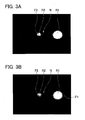
- the CPU 101 first obtains a picture image including the first reflection image R1 and the second reflection image R2 based on a light receiving signal from the two-dimensional light receiving element 87 (S1). In this processing, for example, the picture image shown in FIG. 3A is obtained as image data.
- the CPU 101 successively detects outline information of the first reflection image R1 and the second reflection image R2 included in the picture image obtained in the processing in S1 (S2).
- the corneal thickness is calculated by use of the positional information of the reflection images R1 and R2 defined based on the outline information, even though the details thereof will be described later.
- the outline information is for example based on luminance gradient information of the first reflection image R1 and the second reflection image R2.
- the positional information of the edge of each of the reflection images R1 and R2 may be detected as the outline information.
- a method to determine the position of the edge it is conceived to use a luminance distribution of each reflection image R1 and R2 in a meridian direction. For instance, it may be arranged to perform a binarization processing on the image obtained by the processing in S1 and detect a boundary portion of a resultant image as the edge.
- a region of a certain luminance range having predetermined upper and lower limits may be detected as the edge of each reflection image R1 and R2.
- the CPU 101 executes an approximate processing (fitting processing) on the outline information (e.g., the edge) of each of the reflection images R1 and R2 detected by the processing in S2 (S3).
- the approximate processing (S3) for example, straight line, curved line, figure, and others may be fitted to the outline of each of the reflection images R1 and R2.
- the figure may include for example a combination of straight lines and curved lines as well as curved figures such as ellipse and circle, and rectilinear figures such as polygon.
- the approximate processing (S3) is explained as being performed by setting of an approximate figure (figure fitting) to the outline of each of the reflection image R1 and R2.
- One example is explained below as a case where approximate ellipses F1 and F2 (see FIG. 3B ) are set respectively to the first reflection image R1 and the second reflection image R2.
- the CPU 101 determines the corneal thickness from the positional information of each of the two-dimensionally detected reflection images R1 and R2 (S4).
- the corneal thickness is obtained by use of the positional information of each of the reflection images defined based on the outline information having been subjected to the approximate processing.
- the CPU 101 can also determine the center-to-center distance between the approximate ellipses F1 and F2 and obtain the corneal thickness based on the calculated center-to-center distance.
- the center-to-center distance between the approximate ellipses F1 and F2 is converted to a measurement value of the corneal thickness by use of at least one of an arithmetic expression and a table.
- this arithmetic expression may be created by for example optical simulation or the like in consideration of difference in refractive index between air and a cornea, difference in corneal curvature, and others.
- this table may be created by for example calibration using a known eye (e.g., a schematic eye) having different thickness.
- the corneal thickness measurement (the processings in S1 to S4) may be repeated several times to obtain the measurement value of the corneal thickness from an average value, a central value, and others in each measurement.
- the positional information of the first reflection image R1 and the second reflection image R2 is two-dimensionally detected and the corneal thickness is measured by use of the two-dimensionally detected positional information.
- the measurement since the measurement is performed in consideration of the two-dimensional positional information of each of the reflection images R1 and R2, it is easier to control measurement errors of the corneal thickness resulting from corneal abnormality, extraneous matters to the cornea, noise N, and others as compared with a conventional art in which the corneal thickness is measured based on one line (one-dimensional) luminance information.
- the approximate ellipses F1 and F2 are set, for instance, even when part of each of the reflection images R1 and R2 is uneven or irregular under the influence of corneal abnormality and other reasons, the influence of unevenness is dispersed throughout the approximate ellipses F1 and F2. As a result, the measurement value of the corneal thickness obtained from the positional relationship between the approximate ellipses F1 and F2 contains less measurement errors resulting from the corneal abnormality and others.
- the approximate processing (S3) is performed on parts of the outlines (the edges) of the first reflection image R1 and the second reflection image R2 after removing a corneal inside portion therefrom.
- the set position of the approximate figure is less likely to be influenced by the noise N. This can obtain a more accurate measurement value of corneal thickness by the corneal thickness measurement processing.
- the approximate processing (S3) does not necessarily require the outline information (e.g., the edge) of the entire circumference of each of the reflection images R1 and R2.
- fitting may be performed by use of the outline information of part of each of the reflection images R1 and R2.
- each boundary of the first reflection image R1 and the second reflection image R2 may be partially vague.
- Such a vague boundary area does not need to be used for fitting.
- the vague boundary area exhibits a more gradual luminance change (luminance gradient) in the meridian direction of the reflection image than a clear boundary area. Therefore, the approximate processing may be performed in consideration of the degree of luminance change. For instance, when the degree of luminance change (e.g., luminance gradient) in the meridian direction of an image is larger than a predetermined threshold, this condition may be one of conditions for detecting the edge to be used for fitting.
- the luminance change degree in the median direction of the image when the luminance change degree in the median direction of the image is smaller than the predetermined threshold, it may be arranged not to detect the edge from the relevant region. As a result, the outlines of the reflection images R1 and R2 can undergo more preferable fitting and a more preferable measurement value of corneal thickness can be obtained.
- a strip-shaped noise N occurs between the first reflection image R1 and the second reflection image R2 on the two-dimensional light receiving element 87.
- One conceivable example of the causes for this noise N is noise light directed in the optical axis L3 direction caused by scattering of the light from the light projecting optical system 80a inside the cornea Ec (i.e., between the anterior surface and the posterior surface).
- the approximate processing may be performed after removing noise component (noise N) occurring between the first reflection image R1 and the second reflection image R2.
- noise component noise N
- the edge from which the corneal inside portion has been removed may be used for the fitting. This can perform the fitting with reduced influence of noise component, so that a more preferable measurement value of corneal thickness can be obtained.
- the noise component can also be removed through the use of a difference between the noise component and the luminance information (luminance value and luminance gradient) of each of the reflection images R1 and R2. It is to be noted that the noise component may be removed from both of the first reflection image R1 and the second reflection image R2 or from either one of them.
- the first reflection image R1 and the second reflection image R2 may partially overlap.
- the boundary of each of the reflection images R1 and R2 is also less likely to be accurately specified.
- the edge from which the corneal inside portion has been removed may also be used for the fitting. This makes it possible to perform the fitting with reduced influence of the noise component, so that a more preferable measurement value of corneal thickness can be obtained.
- the above embodiment shows the case where the approximate figures are set to the first reflection image R1 and the second reflection image R2.
- the method is not limited thereto.
- the corneal thickness measurement processing may be performed by detecting the positional information of the first cornea reflection image R1 and the second cornea reflection image R2 on at least two or more lines (L0, L1, L2, ....) along the corneal thickness direction and obtain the corneal thickness by use of the positional information on each of the lines.
- lines L0, L1, L2, .
- a plurality of lines (L0, L1, L2, ....) along the corneal thickness direction passing through each of the two reflection images R1 and R2 are set to an image obtained by use of the two-dimensional light receiving element 87.
- the CPU 101 determines a value of corneal thickness for each line from a distance between the center point of the first reflection image R1 and the center point of the second reflection image R2 on each line. Thereafter, the CPU 101 may obtain a final measurement value of corneal thickness in consideration of the value of corneal thickness on each line. For instance, an average value of corneal thicknesses on the lines may be obtained as a measurement value.
- the center point of each of the reflection images R1 and R2 may be a position at which the luminance value on a line is peak. Furthermore, it may be an intermediate position between the edges of each of the reflection images R1 and R2.
- a corneal thickness having an error beyond a fixed value with respect to the corneal thicknesses obtained for other lines may be excluded when a final measurement value of corneal thickness (e.g., an average of the corneal thicknesses on the lines) is to be determined.
- the error in corneal thickness defined herein may also be an error to for example (a reference value of) a distribution of corneal thicknesses obtained on all the lines.
- the noise N overlaps the first reflection image R1.
- the center point of the reflection image R1 is displaced toward the corneal inside as compared with the center points of the reflection image R1 on the lines L1 and L2.
- the corneal thickness obtained on the line L0 is therefore a smaller value than the corneal thickness obtained on each of the lines L1 and L2.
- a reference value an average value, a central value, etc.
- a final measurement value is calculated by use of the corneal thicknesses on the lines L1 and L2. Consequently, the ophthalmic measurement apparatus 1 can obtain a measurement value of the corneal thickness with reduced influence of noise N.
- the CPU 101 may also be configured to determine a point of the center of gravity of each image of the first reflection image R1 and the second reflection image R2 and obtain corneal thickness from a distance between the gravity center points.
- the ophthalmic measurement apparatus 1 may be configured to correct a measurement value of intraocular pressure obtained based on the light receiving signal from the photodetector 67 of the corneal deformation detecting optical system 60, by use of the corneal thickness obtained as a result of the corneal thickness measurement processing.
- a corrected value of the intraocular pressure can be easily obtained as an appropriate value.
- both the positional information of the first reflection image R1 and the second reflection image R2 are two-dimensionally detected; however, this disclosure is not limited thereto. For instance, even when the positional information of either the first reflection image R1 or the second reflection image R2 is two-dimensionally detected, a fixed level of effect can be obtained.
Landscapes
- Life Sciences & Earth Sciences (AREA)
- Health & Medical Sciences (AREA)
- Medical Informatics (AREA)
- Biophysics (AREA)
- Ophthalmology & Optometry (AREA)
- Engineering & Computer Science (AREA)
- Biomedical Technology (AREA)
- Heart & Thoracic Surgery (AREA)
- Physics & Mathematics (AREA)
- Molecular Biology (AREA)
- Surgery (AREA)
- Animal Behavior & Ethology (AREA)
- General Health & Medical Sciences (AREA)
- Public Health (AREA)
- Veterinary Medicine (AREA)
- Eye Examination Apparatus (AREA)
Abstract
Description
- This disclosure relates to an ophthalmic measurement apparatus for measuring corneal thickness of an examinee's eye.
- Conventionally, there is known an apparatus for measuring corneal thickness by projecting measurement light to an examinee's eye from an oblique direction and utilizing reflection light of the measurement light from a corneal anterior surface of the examinee's eye and reflection light of the measurement light from a corneal posterior surface of the examinee's eye.
- For instance,
Patent Document 1 discloses an apparatus for measuring corneal thickness by receiving reflection light from a corneal anterior surface and reflection light from a corneal posterior surface in different positions of a light receiving element such as a one-dimensional line sensor and others, and using a one-dimensional received light intensity distribution representing the intensity of light received in each position on the light receiving element. - Patent Document 1:
JP-A-2012-143492 - Meanwhile, there may be a case where scattered measurement light from a cornea influences reflection light which will enter a light receiving element. For instance, the measurement light may be scattered due to corneal abnormality, extraneous matters sticking to a corneal front surface, and others. Also, scattered measurement light within a cornea may enter the light receiving element. The conventional apparatus is merely arranged to one-dimensionally determine the positions on the anterior surface and the posterior surface of a cornea. This apparatus is not designed to take into consideration the influence of scattered light that will enter the light receiving element.
- The present disclosure has been made to solve the above problems and has a purpose to provide an ophthalmic measurement apparatus capable of obtaining a good measurement value of corneal thickness.
- To achieve the above purpose, one aspect of this disclosure provides an ophthalmic measurement apparatus comprising: a light projecting optical system for projecting measurement light to an examinee's eye from an oblique direction; a light receiving optical system for receiving, by a light receiving element, a first cornea reflection image formed by the measurement light reflected by a corneal anterior surface of the examinee's eye and a second corneal reflection image formed by the measurement light reflected by a corneal posterior surface of the examinee's eye; and an arithmetic processing unit for obtaining corneal thickness of the examinee's eye based on positional information of the first cornea reflection image and the second cornea reflection image in a light receiving signal output from the light receiving element, characterized in that the light receiving optical system includes a two-dimensional light receiving element as the light receiving element, and the arithmetic processing unit is configured to two-dimensionally detect the positional information of at least one of the first cornea reflection image and the second cornea reflection image based on a light receiving signal from the two-dimensional light receiving element, and obtain the corneal thickness by use of the two-dimensionally detected positional information.
- Further developments of the present disclosure are given in the dependent claims.
- According to an ophthalmic measurement apparatus of the present disclosure, it is possible to obtain a more preferable measurement value of corneal thickness.
-
-
FIG. 1 illustrates a schematic configuration of a measuring unit in an ophthalmic measurement apparatus of a present embodiment; -
FIG. 2 is a schematic diagram showing light beams emitted from a corneal thickness measurement optical system and reflected by a cornea; -
FIG. 3A is a diagram showing an image captured by use of a light receiving element of the corneal thickness measurement optical system; -
FIG. 3B is a diagram to explain an image processing to be performed on the image shown inFIG. 3A ; -
FIG. 4 is a block diagram showing a schematic configuration of a control system in the ophthalmic measurement apparatus of the present embodiment; -
FIG. 5 is a flowchart showing a corneal thickness measurement processing to be executed by a CPU in the ophthalmic measurement apparatus; and -
FIG. 6 is a schematic diagram to explain a measurement method in a modified example of the present disclosure. - A detailed description of a preferred embodiment of the present disclosure will now be given referring to the accompanying drawings. A schematic configuration of a measuring unit in an
ophthalmic measurement apparatus 1 of the present embodiment will be first explained referring toFIG. 1 . - The
ophthalmic measurement apparatus 1 shown inFIG. 1 is configured to measure corneal thickness of an examinee's eye E. In the present embodiment, the apparatus is arranged to measure intraocular pressure in non-contact manner. - The
ophthalmic measurement apparatus 1 mainly includes an observationoptical system 30 and a corneal thickness measurement optical system 80 (80a and 80b inFIG. 1 ). In the present embodiment, theophthalmic measurement apparatus 1 further includes an alignmentoptical system 40, a fixationoptical system 50, a corneal deformation detecting optical system 60 (60a and 60b inFIG. 1 ), and a first working distance detecting optical system 70 (60a and 70b inFIG. 1 ). Further, theophthalmic measurement apparatus 1 of the present embodiment also includes an intraocularpressure measuring unit 10. - In the present embodiment, the measuring unit of the
ophthalmic measurement apparatus 1 including those optical systems and others is housed in a cabinet not shown. The cabinet is three-dimensionally movable with respect to the examinee's eye E by an XYZ drive mechanism 20 (seeFIG. 4 ). This cabinet may also be moved according to an instruction of an examiner for example through an operating member (e.g., an operating unit (a joystick) 21, seeFIG. 4 ). - The observation
optical system 30 is used to observe a front image of an anterior segment of the examinee's eye E. The observationoptical system 30 includes abeam splitter 32, anobjective lens 33, abeam splitter 34, animaging lens 35, afilter 36, and a two-dimensional imaging element 37, these components being arranged on an observation optical axis L1. Infraredillumination light sources 31 are used as an observation light source. An image of the examinee's eye illuminated by the infraredillumination light sources 31 is formed on the two-dimensional imaging element 37 via an optical path from thebeam splitter 32 to thefilter 36. Herein, thefilter 36 has the property of transmitting the light from thelight sources 31 and the light from analignment light source 41 and not transmitting the light from alight source 61 mentioned later for detecting corneal deformation and visible light. The image formed on the two-dimensional imaging element 37 is displayed on amonitor 38. - The alignment
optical system 40 has thelight source 41 and aprojection lens 42. In the present embodiment, the alignmentoptical system 40 shares the configuration from thebeam splitter 32 to the two-dimensional imaging element 37 arranged on the optical axis L1 with the observationoptical system 30. Infrared light projected from the alignmentinfrared light source 41 via theprojection lens 42 is reflected by thebeam splitter 32 and then projected to the examinee's eye E from a front direction. A corneal luminescent spot (a corneal reflection image) formed at a corneal vertex by thelight source 41 is formed on the two-dimensional imaging element 37 via thebeam splitter 32 to thefilter 36. Even though the details thereof will be mentioned later, this corneal luminescent spot is utilized for alignment detection in up-and-down and right-and-left directions. - The fixation
optical system 50 includes a visible light source (a fixation lamp) 51 and aprojection lens 52. In the present embodiment, the fixationoptical system 50 shares the components from thebeam splitter 34 to thebeam splitter 32 arranged on the optical axis L1 with the observationoptical system 30 and others. Upon turn-on of thevisible light source 51, a fixation target is presented to the examinee's eye E from front. Thus, the visual line of the examinee's eye E is fixed in a direction toward a fixation point located in the front direction. In this case, the optical axis L1 is used as a fixation optical axis. Thevisible light source 51 may include any light sources such as an LED and a laser, for example. As thevisible light source 51, furthermore, there may be used a pattern light source such as a point light source, a slit light source, and a ring light source as well as a two-dimensional display such as a liquid crystal display. - The corneal deformation detecting
optical system 60 is used to detect a deformed state of the cornea Ec. Thisoptical system 60 includes a light projectingoptical system 60a and a light receivingoptical system 60b. The light projectingoptical system 60a is arranged to irradiate illumination light toward the cornea Ec of the eye E from an oblique direction. The light projectingoptical system 60a includes alight source 61, adichroic mirror 62, and aprojection lens 63. On the other hand, the light receivingoptical system 60b is arranged to receive, by aphotodetector 67, corneal reflection light from the light projectingoptical system 60a. The light receivingoptical system 60b includes alens 64, abeam splitter 65, and apinhole plate 66 in addition to thephotodetector 67. In the present embodiment, the corneal deformation detectingoptical system 60 is placed so that an amount of light received by thephotodetector 67 is maximum when the examinee's eye E is in a predetermined deformed state (e.g., a flattened state). Accordingly, deformation of the examinee's eye E is detected based on a light receiving signal representing the amount of light received (light receiving amount) output from thephotodetector 67. - The above deformation of the examinee's eye E is carried out by blowing a predetermined mount of compressed fluid (e.g., compressed air) from a
nozzle 6 of theintraocular measuring unit 10 toward the examinee's eye E. This compressed fluid is supplied from afluid supply unit 10a (seeFIG. 4 ). In theophthalmic measurement apparatus 1 in the present embodiment, the intraocular pressure is measured based on a deformation amount of the examinee's eye E detected by use of thephotodetector 67 when the compressed fluid is blown onto the eye E. - The first working distance detecting
optical system 70 is utilized for alignment (rough adjustment) in a working distance direction (a Z direction). The first working distance detectingoptical system 70 shares the light projectingoptical system 60a with the corneal deformation detecting optical system. Thisoptical system 70 includes a light receivingoptical system 70b. This light receivingoptical system 70b includes a condensinglens 72 and aposition imaging element 73. Light emitted from the light projectingoptical system 60a forms an index image on the cornea Ec. Corneal reflection light from the light projectingoptical system 60a enters theposition detecting element 73 via the components from thelens 64 to the condensinglens 72. A light receiving position on theposition detecting element 73 varies according to a positional relationship between the measuring unit and the examinee's eye E (the cornea Ec) in the working distance direction (the Z direction). Accordingly, the information on the working distance is obtained based on an output signal from theposition detecting element 73. The output signal from theposition detecting element 73 in the present embodiment is utilized for alignment (rough adjustment) in the working distance direction (the Z direction). The magnification of the light receivingoptical system 70b of the first working distance detecting optical system is not as large as that of a light receivingoptical system 80b mentioned later. Thus, a distance detection range of theposition detecting element 73 in the Z direction is wider than that of a two-dimensionallight receiving element 87. The distance detection range of theposition detecting element 73 is for example a range of ±3 to 4 mm from an alignment reference position. - The corneal thickness measuring optical system 80 (80a, 80b) is used to measure the corneal thickness of the examinee's eye E. This
optical system 80 includes a light projectingoptical system 80a and a light receivingoptical system 80b. - The light projecting
optical system 80a is used to project measurement light (illumination light) for measuring the corneal thickness from an oblique direction with respect to the optical axis direction of the observationoptical system 30. In the present embodiment, the light projectingoptical system 80a includes ameasurement light source 81, a condensinglens 82, alight limiting member 83, aconcave lens 84, thebeam splitter 65, a beam splitter 71, and thelens 64. These components are placed on an optical axis L2 which is a light projection optical axis. The optical axis L2 is inclined with respect to the optical axis L1 which is the fixation optical axis. Thus, the light projectingoptical system 80a irradiates illumination light (measurement light) toward the examinee's eye E from an oblique direction with respect to the fixation optical axis. As themeasurement light source 81, there is used a visible light source or an infrared (including near infrared) light source; e.g., an LED, a laser, or the like. The condensinglens 82 condenses the light emitted from themeasurement light source 81. - The
light limiting member 83 is placed on an optical path of the light projectingoptical system 80a and limits the light (measurement light) emitted from themeasurement light source 81. In the present embodiment, thelight limiting member 83 is disposed in a position conjugated with a posterior surface of the cornea Ec during measurement of the corneal thickness. In the present embodiment, thelight limiting member 83 is a pinhole plate, but is not limited thereto. For example, a slit plate or the like may also be used as thelight limiting member 83. Thislight limiting member 83 allows a part of the light emitted from themeasurement light source 81 to pass through and blocks a remaining part of the light. As a result, the light projectingoptical system 80a projects a predetermined pattern of light (a spot light beam) to the cornea Ec. - In the present embodiment, the light receiving
optical system 80b has the two-dimensionallight receiving element 87. The light receivingoptical system 80b is arranged to receive, by the two-dimensionallight receiving element 87, the measurement light emitted from the light projectingoptical system 80a and reflected (specular reflection) by a corneal anterior surface Ec1 (a corneal front surface, a corneal epithelium) and a corneal posterior surface Ec2 (a corneal back surface, a corneal endothelium) of the examinee's eye E. In the present embodiment, an optical axis L3 of the light receivingoptical system 80b is placed nearly symmetrical to the optical axis L2 of the light projectingoptical system 80a with respect to the observation optical axis (the optical axis L1) of the observationoptical system 30. In the present embodiment, furthermore, the light receivingoptical system 80b includes thelens 63, thedichroic mirror 62, alight receiving lens 85, and the two-dimensionallight receiving element 87 on the optical axis L3. In the present embodiment, even though the details will be mentioned later, a detailed alignment state in the Z direction with respect to the examinee's eye E is detected by the light receivingoptical system 80b in the present embodiment. - In the present embodiment, the two-dimensional
light receiving element 87 receives reflection light from each of the corneal anterior surface and the corneal posterior surface. As the two-dimensionallight receiving element 87, there can be used a two-dimensional imaging element such as a two-dimensional area sensor and a CCD. - The light receiving
optical system 80b of the corneal thickness measuringoptical system 80 performs imaging with high magnification with respect to the first working distance detectingoptical system 70. Accordingly, in the present embodiment, the distance detection range of the two-dimensionallight receiving element 87 in the Z direction is narrower than that of theposition detecting element 73. For instance, it is a range of ±1 mm from the alignment reference position. - The light emitted from the
measurement light source 81 is condensed by the condensinglens 82 and illuminates thelight limiting member 83 from behind. The light from themeasurement light source 81 is limited by thelight limiting member 83 and then is imaged (condensed) in a position near the cornea Ec by thelens 64. In the present embodiment, specifically, a pinhole image is formed near the cornea Ec. At that time, the light from themeasurement light source 81 is formed near an intersection point with a visual axis on the cornea Ec. - When the measurement light is projected onto the cornea Ec by the light projecting
optical system 80a, the reflection light of the measurement light by the cornea Ec travels in a direction symmetrical to the projection light beam with respect to the optical axis L1. Thelight receiving lens 85 forms an image of the reflection light on a light receiving plane of the two-dimensionallight receiving element 87. - As shown in
FIG. 2 , a part of the measurement light irradiated from the light projectingoptical system 80a to the cornea Ec is reflected by the corneal anterior surface Ec1. This forms, as shown inFIG. 3A , a first cornea reflection image R1 (hereinafter, simply referred to as "first reflection image R1") on the two-dimensionallight receiving element 87. Another part of the measurement light from the light projectingoptical system 80a is reflected by the corneal posterior surface Ec2. This forms, as shown inFIG. 3A , a second cornea reflection image R2 (hereinafter, simply referred to as "first reflection image R1" on the two-dimensionallight receiving element 87. In the present embodiment, the first cornea reflection image R1 is a circular reflection image. The second cornea reflection image R2 is a circular reflection image smaller than the first cornea reflection image R1. The reflection light (see a solid line) by the corneal anterior surface and the reflection light (see a broken line) by the corneal posterior surface are different in reflection optical path and therefore two reflection images R1 and R2 are formed fully or partially away from each other. For instance, inFIG. 3A , the second reflection image R2 is fully formed away from the first reflection image R1. As the corneal thickness is large, the reflection optical paths of two reflection images are more separated. Accordingly, two reflection images are formed in the positions more spaced apart on the two-dimensionallight receiving element 87 as the corneal thickness is larger. Theophthalmic measurement apparatus 1 of the present embodiment is therefore arranged to measure the corneal thickness based on the positional relationship between the first reflection image R1 and the second reflection image R2 on the two-dimensionallight receiving element 87. - In the present embodiment, for example, as shown in
FIG. 1 , the light projectingoptical system 60a and the light receivingoptical system 80b share the optical axis L3 with each other, and the light projectingoptical system 80a, the light receivingoptical system 60b, and the light receivingoptical system 70b share the optical axis L2 with one another. Needless to say, they are not limited thereto. - It is to be noted that the
lens 64 shared among the light receivingoptical systems optical system 80a is placed in a position to make the reflection light emitted from thelight source 61 and reflected by the cornea Ec condense on a center of a hole of thepinhole plate 66 and make the measurement light emitted from themeasurement light source 81 condense on the anterior surface and the posterior surface of the cornea Ec. - A control system of the
ophthalmic measurement apparatus 1 will be explained below. Theophthalmic measurement apparatus 1 of the present embodiment has acontrol unit 100 for executing control of the entire apparatus and calculation of measurement results. - In the present embodiment, the
control unit 100 is connected to thefluid supply unit 10a, theXYZ drive unit 20, the operatingunit 21, astorage device 105, the variouslight sources light receiving elements - Furthermore, the
control unit 100 includes aCPU 101, aROM 102, and aRAM 103. TheCPU 101 is a processing device (a processor) to execute various processings (e.g., control processing, arithmetic processing, etc.) related to theophthalmic measurement apparatus 1. TheROM 102 is a nonvolatile storage device in which control programs, fixed data, and others are stored. TheRAM 103 is a rewritable volatile storage device. ThisRAM 103 stores temporary data to be used by theophthalmic measurement apparatus 1 in photographing and measuring the examinee's eye E. - The
storage device 105 is a rewritable nonvolatile storage device. In the present embodiment, thestorage device 105 stores at least a program to execute an anterior segment measurement processing by thecontrol unit 100. Thestorage device 105 may also save an image captured by theophthalmic measurement apparatus 1 and measurement results obtained by theophthalmic measurement apparatus 1 and others. - Operations of the apparatus configured as above will be explained.
- The following explanation will be given mainly to the operation of measuring the corneal thickness. In the present embodiment, in the
ophthalmic measurement apparatus 1, the CPU 101 (the control unit 100) executes a corneal thickness measurement processing shown inFIG. 5 to measure the corneal thickness. - In the present embodiment, when the corneal thickness is to be measured, the measuring unit of the
ophthalmic measurement apparatus 1 is subjected to alignment (positional adjustment) in advance. This alignment is performed after having the examinee's eye E positioned to face the measuring unit and having an examinee gaze at a fixation target. The alignment in X and Y directions (up-and-down and right-and-left directions) in the present embodiment may be performed by operation of the operating unit 21 (e.g., a joystick) by an examiner based on alignment information displayed on amonitor 38. For instance, the examiner manipulates the operatingunit 21 so as to bring a corneal reflection image (a luminescent spot) formed by thelight source 41 into a predetermined position relative to a reticle not shown displayed on themonitor 38. TheCPU 101 may also automatically perform alignment adjustment in the up-and-down and right-and-left directions based on the alignment information obtained as an imaging result of the two-dimensional imaging element 37. In this case, theCPU 101 controls theXYZ drive mechanism 20 so that the corneal luminescent spot by thelight source 41 is formed in an alignment position (e.g., an intersection point of the optical axis L1 and an imaging plane of the two-dimensional imaging element 37) set on the two-dimensional imaging element 37 in advance. In this way, theCPU 101 moves the measuring unit to an alignment completion position in the X and Y directions. - Successively, the alignment in the Z direction (a back-and-forth direction) will be performed. In the present embodiment, as described above, the alignment in the Z direction is automatically performed in such a manner that the
CPU 101 controls the operation of theXYZ drive mechanism 20 based on working distance information obtained from theposition detecting element 73 and the two-dimensionallight receiving element 87. - When the corneal reflection light is detected first by the
position detecting element 73, theCPU 101 causes the measuring unit to move in the Z direction based on the working distance information obtained from theposition detecting element 37. In the present embodiment, consequently, the measuring unit is moved to a range where the reflection light (the first reflection image R1 or the second reflection image R2) of the light from themeasurement light source 81 is detected by the two-dimensionallight receiving element 87. Upon completion of the movement, theCPU 101 further minutely adjusts the positional relationship between the measuring unit and the examinee's eye E in the Z direction based on the light receiving signal from the two-dimensionallight receiving element 87. More concretely, for example, the positional relationship is adjusted so that the corneal posterior surface of the examinee's eye E and the two-dimensionallight receiving element 87 are optically conjugated with each other. In the present embodiment, for example, when the second reflection image R2 is formed at the center of the two-dimensionallight receiving element 87, thislight receiving element 87 is conjugated with the corneal posterior surface of the examinee's eye E. As the result of the detailed alignment adjustment, the second reflection image R2 formed on the two-dimensionallight receiving element 87 becomes clearest. The light emitted from the light projectingoptical system 80a and reflected by the corneal posterior surface is weaker than the light reflected by the corneal anterior surface, so that the second reflection image R2 is apt to be smaller and more obscure than the first reflection image R1. In the present embodiment, since the second reflection image R2 is made clearer by the detailed alignment adjustment, the position, the shape, and others of the second reflection image R2 can be easily detected. - In the present embodiment, after the above alignment adjustment, the corneal thickness measurement processing shown in
FIG. 5 is executed by theCPU 101 to measure the corneal thickness. The corneal thickness measurement processing may be triggered by completion of the adjustment. The corneal thickness measurement processing may also be started according to an instruction from an examiner. - Herein, the corneal thickness measurement processing will be explained referring to
FIG. 5 . In the corneal thickness measurement processing of the present embodiment, theCPU 101 two-dimensionally detects the positional information of the first reflection image R1 and the second reflection image R2 and obtains the corneal thickness by use of the two-dimensionally detected positional information. To more concrete, based on the obtained positional information of the first reflection image R1 and the second reflection image R2, the corneal thickness is determined from a distance between the reflection images. - The
CPU 101 first obtains a picture image including the first reflection image R1 and the second reflection image R2 based on a light receiving signal from the two-dimensional light receiving element 87 (S1). In this processing, for example, the picture image shown inFIG. 3A is obtained as image data. - The
CPU 101 successively detects outline information of the first reflection image R1 and the second reflection image R2 included in the picture image obtained in the processing in S1 (S2). In this corneal thickness measurement processing, the corneal thickness is calculated by use of the positional information of the reflection images R1 and R2 defined based on the outline information, even though the details thereof will be described later. - The outline information is for example based on luminance gradient information of the first reflection image R1 and the second reflection image R2. The positional information of the edge of each of the reflection images R1 and R2 may be detected as the outline information. At that time, as a method to determine the position of the edge, it is conceived to use a luminance distribution of each reflection image R1 and R2 in a meridian direction. For instance, it may be arranged to perform a binarization processing on the image obtained by the processing in S1 and detect a boundary portion of a resultant image as the edge. As another alternative, furthermore, in the luminance distribution of each reflection image R1 and R2, a region of a certain luminance range having predetermined upper and lower limits may be detected as the edge of each reflection image R1 and R2.
- The
CPU 101 executes an approximate processing (fitting processing) on the outline information (e.g., the edge) of each of the reflection images R1 and R2 detected by the processing in S2 (S3). In the approximate processing (S3), for example, straight line, curved line, figure, and others may be fitted to the outline of each of the reflection images R1 and R2. The figure may include for example a combination of straight lines and curved lines as well as curved figures such as ellipse and circle, and rectilinear figures such as polygon. In the present embodiment, the approximate processing (S3) is explained as being performed by setting of an approximate figure (figure fitting) to the outline of each of the reflection image R1 and R2. One example is explained below as a case where approximate ellipses F1 and F2 (seeFIG. 3B ) are set respectively to the first reflection image R1 and the second reflection image R2. - Subsequently, the
CPU 101 determines the corneal thickness from the positional information of each of the two-dimensionally detected reflection images R1 and R2 (S4). In the present embodiment, the corneal thickness is obtained by use of the positional information of each of the reflection images defined based on the outline information having been subjected to the approximate processing. For instance, theCPU 101 can also determine the center-to-center distance between the approximate ellipses F1 and F2 and obtain the corneal thickness based on the calculated center-to-center distance. For example, the center-to-center distance between the approximate ellipses F1 and F2 is converted to a measurement value of the corneal thickness by use of at least one of an arithmetic expression and a table. The obtained measurement value is stored in thestorage device 105 or the like. In the case of using the arithmetic expression, this arithmetic expression may be created by for example optical simulation or the like in consideration of difference in refractive index between air and a cornea, difference in corneal curvature, and others. In the case of using the table, this table may be created by for example calibration using a known eye (e.g., a schematic eye) having different thickness. - The corneal thickness measurement (the processings in S1 to S4) may be repeated several times to obtain the measurement value of the corneal thickness from an average value, a central value, and others in each measurement.
- In the corneal thickness measurement processing of the present embodiment as above, the positional information of the first reflection image R1 and the second reflection image R2 is two-dimensionally detected and the corneal thickness is measured by use of the two-dimensionally detected positional information. In the present embodiment, since the measurement is performed in consideration of the two-dimensional positional information of each of the reflection images R1 and R2, it is easier to control measurement errors of the corneal thickness resulting from corneal abnormality, extraneous matters to the cornea, noise N, and others as compared with a conventional art in which the corneal thickness is measured based on one line (one-dimensional) luminance information.
- In the present embodiment where the approximate ellipses F1 and F2 are set, for instance, even when part of each of the reflection images R1 and R2 is uneven or irregular under the influence of corneal abnormality and other reasons, the influence of unevenness is dispersed throughout the approximate ellipses F1 and F2. As a result, the measurement value of the corneal thickness obtained from the positional relationship between the approximate ellipses F1 and F2 contains less measurement errors resulting from the corneal abnormality and others.
- The approximate processing (S3) is performed on parts of the outlines (the edges) of the first reflection image R1 and the second reflection image R2 after removing a corneal inside portion therefrom. Thus, the set position of the approximate figure is less likely to be influenced by the noise N. This can obtain a more accurate measurement value of corneal thickness by the corneal thickness measurement processing.
- The present disclosure is explained along the above embodiment, but is not limited thereto and may be variously modified.
- For instance, the approximate processing (S3) does not necessarily require the outline information (e.g., the edge) of the entire circumference of each of the reflection images R1 and R2. As an alternative, fitting may be performed by use of the outline information of part of each of the reflection images R1 and R2.
- For instance, when the cornea is abnormal or extraneous matters such as dust stick to the corneal anterior surface, each boundary of the first reflection image R1 and the second reflection image R2 may be partially vague. Such a vague boundary area does not need to be used for fitting. The vague boundary area exhibits a more gradual luminance change (luminance gradient) in the meridian direction of the reflection image than a clear boundary area. Therefore, the approximate processing may be performed in consideration of the degree of luminance change. For instance, when the degree of luminance change (e.g., luminance gradient) in the meridian direction of an image is larger than a predetermined threshold, this condition may be one of conditions for detecting the edge to be used for fitting. More concretely, when the luminance change degree in the median direction of the image is smaller than the predetermined threshold, it may be arranged not to detect the edge from the relevant region. As a result, the outlines of the reflection images R1 and R2 can undergo more preferable fitting and a more preferable measurement value of corneal thickness can be obtained.
- As shown in
FIG. 3A , furthermore, there is a case where a strip-shaped noise N occurs between the first reflection image R1 and the second reflection image R2 on the two-dimensionallight receiving element 87. One conceivable example of the causes for this noise N is noise light directed in the optical axis L3 direction caused by scattering of the light from the light projectingoptical system 80a inside the cornea Ec (i.e., between the anterior surface and the posterior surface). The corneal inside portion (a left portion of the first reflection image R1 and a right portion of the second reflection image R2 inFIG. 3 ) of each of the first reflection image R1 and the second reflection image R2 overlaps the noise N and thus is apt to have a vague boundary. Therefore, the approximate processing may be performed after removing noise component (noise N) occurring between the first reflection image R1 and the second reflection image R2. For example, the edge from which the corneal inside portion has been removed may be used for the fitting. This can perform the fitting with reduced influence of noise component, so that a more preferable measurement value of corneal thickness can be obtained. - Furthermore, the noise component can also be removed through the use of a difference between the noise component and the luminance information (luminance value and luminance gradient) of each of the reflection images R1 and R2. It is to be noted that the noise component may be removed from both of the first reflection image R1 and the second reflection image R2 or from either one of them.
- When the light quantity output from the
light source 81 or the gain of the two-dimensionallight receiving element 87 is large, the first reflection image R1 and the second reflection image R2 may partially overlap. In this case, the boundary of each of the reflection images R1 and R2 is also less likely to be accurately specified. In the processing in S3, therefore, the edge from which the corneal inside portion has been removed may also be used for the fitting. This makes it possible to perform the fitting with reduced influence of the noise component, so that a more preferable measurement value of corneal thickness can be obtained. - As one example of the method of measuring the corneal thickness based on the positional relationship between the first reflection image R1 and the second reflection image R2 and their two-dimensional shapes, the above embodiment shows the case where the approximate figures are set to the first reflection image R1 and the second reflection image R2. However, the method is not limited thereto. For instance, as shown in
FIG. 6 , the corneal thickness measurement processing may be performed by detecting the positional information of the first cornea reflection image R1 and the second cornea reflection image R2 on at least two or more lines (L0, L1, L2, ....) along the corneal thickness direction and obtain the corneal thickness by use of the positional information on each of the lines. For example, inFIG. 6 , a plurality of lines (L0, L1, L2, ....) along the corneal thickness direction passing through each of the two reflection images R1 and R2 are set to an image obtained by use of the two-dimensionallight receiving element 87. For instance, theCPU 101 determines a value of corneal thickness for each line from a distance between the center point of the first reflection image R1 and the center point of the second reflection image R2 on each line. Thereafter, theCPU 101 may obtain a final measurement value of corneal thickness in consideration of the value of corneal thickness on each line. For instance, an average value of corneal thicknesses on the lines may be obtained as a measurement value. The center point of each of the reflection images R1 and R2 may be a position at which the luminance value on a line is peak. Furthermore, it may be an intermediate position between the edges of each of the reflection images R1 and R2. - Of the corneal thicknesses obtained one for each line, a corneal thickness having an error beyond a fixed value with respect to the corneal thicknesses obtained for other lines may be excluded when a final measurement value of corneal thickness (e.g., an average of the corneal thicknesses on the lines) is to be determined. The error in corneal thickness defined herein may also be an error to for example (a reference value of) a distribution of corneal thicknesses obtained on all the lines. In the example shown in
FIG. 6 , for instance, the noise N overlaps the first reflection image R1. Thus, on the line L0, the center point of the reflection image R1 is displaced toward the corneal inside as compared with the center points of the reflection image R1 on the lines L1 and L2. The corneal thickness obtained on the line L0 is therefore a smaller value than the corneal thickness obtained on each of the lines L1 and L2. At that time, for example, when the corneal thickness on the line L0 has an error more than a certain level from a reference value (an average value, a central value, etc.) of a distribution of corneal thicknesses on the lines L0 to L2, a final measurement value is calculated by use of the corneal thicknesses on the lines L1 and L2. Consequently, theophthalmic measurement apparatus 1 can obtain a measurement value of the corneal thickness with reduced influence of noise N. - In the corneal thickness measurement processing, the
CPU 101 may also be configured to determine a point of the center of gravity of each image of the first reflection image R1 and the second reflection image R2 and obtain corneal thickness from a distance between the gravity center points. - Moreover, for instance, the
ophthalmic measurement apparatus 1 may be configured to correct a measurement value of intraocular pressure obtained based on the light receiving signal from thephotodetector 67 of the corneal deformation detectingoptical system 60, by use of the corneal thickness obtained as a result of the corneal thickness measurement processing. In this way, since the measurement value of intraocular pressure is corrected by use of accurate corneal thickness obtained by the corneal thickness measurement processing, a corrected value of the intraocular pressure can be easily obtained as an appropriate value. - In the above explanation, both the positional information of the first reflection image R1 and the second reflection image R2 are two-dimensionally detected; however, this disclosure is not limited thereto. For instance, even when the positional information of either the first reflection image R1 or the second reflection image R2 is two-dimensionally detected, a fixed level of effect can be obtained.
Claims (7)
- An ophthalmic measurement apparatus (1) comprising: a light projecting optical system (80a) for projecting measurement light to an examinee's eye (E) from an oblique direction;
a light receiving optical system (80b) for receiving, by a light receiving element (87), a first cornea reflection image (R1) formed by the measurement light reflected by a corneal anterior surface (Ec1) of the examinee's eye and a second corneal reflection image (R2) formed by the measurement light reflected by a corneal posterior surface (Ec2) of the examinee's eye; and
an arithmetic processing unit (100) for obtaining corneal thickness of the examinee's eye based on positional information of the first cornea reflection image and the second cornea reflection image in a light receiving signal output from the light receiving element,
characterized in that
the light receiving optical system includes a two-dimensional light receiving element (87) as the light receiving element, and
the arithmetic processing unit is configured to two-dimensionally detect the positional information of at least one of the first cornea reflection image and the second cornea reflection image based on a light receiving signal from the two-dimensional light receiving element, and obtain the corneal thickness by use of the two-dimensionally detected positional information. - The ophthalmic measurement apparatus according to claim 1, wherein the arithmetic processing unit (100) is configured to detect two-dimensional outline information which is two-dimensional outline information of at least one of the first cornea reflection image (R1) and the second cornea reflection image (R2), and obtain the corneal thickness by use of the positional information of the reflection image defined by the detected two-dimensional outline information.
- The ophthalmic measurement apparatus according to claim 2, wherein the arithmetic processing unit (100) is configured to perform an approximate processing on the outline information of at least one of the first cornea reflection image (R1) and the second cornea reflection image (R2), and defines positional information of the reflection image based on the outline information having been subjected to the approximate processing.
- The ophthalmic measurement apparatus according to claim 3, wherein the light receiving optical system (80a) is arranged to form the first cornea reflection image (R1) and the second cornea reflection image (R2) in a point-like or slit-like shape on the two-dimensional light receiving element, and
the arithmetic processing unit is configured to perform the approximate processing after removing noise component occurring between the first cornea reflection image and the second cornea reflection image. - The ophthalmic measurement apparatus according to claim 3, wherein the light receiving optical system (80a) is arranged to form the first cornea reflection image (R1) and the second cornea reflection image (R2) in a point-like shape on the two-dimensional light receiving element (87), and
the arithmetic processing unit (100) is configured to perform the approximate processing by using outline information of the first cornea reflection image and the second cornea reflection image after removing a corneal inside portion therefrom. - The ophthalmic measurement apparatus according to claim 1, wherein the arithmetic processing unit (100) is configured to detect positional information of the first cornea reflection image (R1) and the second cornea reflection image (R2) on at least two or more lines along a direction of thickness of a cornea, and obtain the corneal thickness by use of the positional information on each of the lines.
- The ophthalmic measurement apparatus according to any one of claims 1 to 6, wherein the first cornea reflection image (R1) is a circular reflection image and the second cornea reflection image (R2) is a circular reflection image smaller than the first cornea reflection image.
Applications Claiming Priority (1)
| Application Number | Priority Date | Filing Date | Title |
|---|---|---|---|
| JP2013248477A JP6488540B2 (en) | 2013-11-29 | 2013-11-29 | Ophthalmic measuring device |
Publications (2)
| Publication Number | Publication Date |
|---|---|
| EP2878256A1 true EP2878256A1 (en) | 2015-06-03 |
| EP2878256B1 EP2878256B1 (en) | 2019-01-09 |
Family
ID=52423538
Family Applications (1)
| Application Number | Title | Priority Date | Filing Date |
|---|---|---|---|
| EP14195372.9A Active EP2878256B1 (en) | 2013-11-29 | 2014-11-28 | Ophthalmic measurement apparatus |
Country Status (2)
| Country | Link |
|---|---|
| EP (1) | EP2878256B1 (en) |
| JP (1) | JP6488540B2 (en) |
Families Citing this family (1)
| Publication number | Priority date | Publication date | Assignee | Title |
|---|---|---|---|---|
| EP4706503A3 (en) * | 2024-03-25 | 2026-04-29 | Optos plc | Ophthalmic ranging instrument |
Citations (3)
| Publication number | Priority date | Publication date | Assignee | Title |
|---|---|---|---|---|
| US20040021874A1 (en) * | 2002-06-27 | 2004-02-05 | Visx, Incorporated, A Delaware Corporation | Integrated scanning and ocular tomography system and method |
| US20110013141A1 (en) * | 2009-07-14 | 2011-01-20 | Wavetec Vision Systems, Inc. | Ophthalmic surgery measurement system |
| JP2012143492A (en) | 2011-01-14 | 2012-08-02 | Nidek Co Ltd | Device for measuring corneal thickness |
Family Cites Families (7)
| Publication number | Priority date | Publication date | Assignee | Title |
|---|---|---|---|---|
| JP2835371B2 (en) * | 1987-05-30 | 1998-12-14 | キヤノン株式会社 | Eye measurement device |
| JP2911452B2 (en) * | 1988-03-23 | 1999-06-23 | キヤノン株式会社 | Ophthalmic equipment |
| JPH0449944A (en) * | 1990-06-15 | 1992-02-19 | Canon Inc | line of sight detection device |
| JPH06163A (en) * | 1992-06-17 | 1994-01-11 | Canon Inc | Eye gaze detector |
| JP4349937B2 (en) * | 2004-03-01 | 2009-10-21 | 株式会社ニデック | Ophthalmic equipment |
| JP4948922B2 (en) * | 2006-06-30 | 2012-06-06 | 株式会社ニデック | Ophthalmic equipment |
| JP5255417B2 (en) * | 2008-12-08 | 2013-08-07 | 株式会社ニデック | Ophthalmic equipment |
-
2013
- 2013-11-29 JP JP2013248477A patent/JP6488540B2/en active Active
-
2014
- 2014-11-28 EP EP14195372.9A patent/EP2878256B1/en active Active
Patent Citations (3)
| Publication number | Priority date | Publication date | Assignee | Title |
|---|---|---|---|---|
| US20040021874A1 (en) * | 2002-06-27 | 2004-02-05 | Visx, Incorporated, A Delaware Corporation | Integrated scanning and ocular tomography system and method |
| US20110013141A1 (en) * | 2009-07-14 | 2011-01-20 | Wavetec Vision Systems, Inc. | Ophthalmic surgery measurement system |
| JP2012143492A (en) | 2011-01-14 | 2012-08-02 | Nidek Co Ltd | Device for measuring corneal thickness |
Also Published As
| Publication number | Publication date |
|---|---|
| EP2878256B1 (en) | 2019-01-09 |
| JP2015104555A (en) | 2015-06-08 |
| JP6488540B2 (en) | 2019-03-27 |
Similar Documents
| Publication | Publication Date | Title |
|---|---|---|
| CN103370002B (en) | Cornea shape determinator | |
| US9480396B2 (en) | Ophthalmic measurement apparatus and ophthalmic measurement program | |
| KR101395539B1 (en) | Ophthalmic device | |
| JP5578542B2 (en) | Eye refractive power measuring device | |
| EP2997881B1 (en) | Ophthalmic apparatus | |
| JP6238551B2 (en) | Ophthalmic apparatus, control method for ophthalmic apparatus, and program | |
| US9078601B2 (en) | Anterior segment measuring apparatus | |
| EP2415393A1 (en) | Ophthalmic apparatus | |
| CN110269588B (en) | Ophthalmologic apparatus and control method therefor | |
| CN105832285A (en) | Ophthalmological device | |
| JP2021069415A (en) | Ophthalmologic apparatus and control method of the same | |
| JP6471466B2 (en) | Ophthalmic apparatus and processing program used therefor | |
| KR101591133B1 (en) | Ophthalmological analysis instrument and method | |
| JP6634765B2 (en) | Ophthalmic apparatus and ophthalmic apparatus control program | |
| JP6589378B2 (en) | Ophthalmic measuring device | |
| US11857255B2 (en) | Ophthalmic apparatus | |
| EP2878256B1 (en) | Ophthalmic measurement apparatus | |
| US9204793B2 (en) | Ophthalmologic apparatus and ophthalmologic photographing method | |
| CN111479494B (en) | Eye refractive power measuring device | |
| JP5188534B2 (en) | Ophthalmic equipment | |
| JP3597274B2 (en) | Ophthalmic equipment | |
| JP2019155002A (en) | Ophthalmologic apparatus and method of controlling the same | |
| JP7187769B2 (en) | Ophthalmic device and ophthalmic device control program | |
| JP6823339B2 (en) | Ophthalmic equipment | |
| US12402789B2 (en) | Ophthalmic apparatus, method of controlling ophthalmic apparatus, and recording medium |
Legal Events
| Date | Code | Title | Description |
|---|---|---|---|
| PUAI | Public reference made under article 153(3) epc to a published international application that has entered the european phase |
Free format text: ORIGINAL CODE: 0009012 |
|
| 17P | Request for examination filed |
Effective date: 20141128 |
|
| AK | Designated contracting states |
Kind code of ref document: A1 Designated state(s): AL AT BE BG CH CY CZ DE DK EE ES FI FR GB GR HR HU IE IS IT LI LT LU LV MC MK MT NL NO PL PT RO RS SE SI SK SM TR |
|
| AX | Request for extension of the european patent |
Extension state: BA ME |
|
| R17P | Request for examination filed (corrected) |
Effective date: 20151203 |
|
| RBV | Designated contracting states (corrected) |
Designated state(s): AL AT BE BG CH CY CZ DE DK EE ES FI FR GB GR HR HU IE IS IT LI LT LU LV MC MK MT NL NO PL PT RO RS SE SI SK SM TR |
|
| 17Q | First examination report despatched |
Effective date: 20160322 |
|
| STAA | Information on the status of an ep patent application or granted ep patent |
Free format text: STATUS: EXAMINATION IS IN PROGRESS |
|
| GRAP | Despatch of communication of intention to grant a patent |
Free format text: ORIGINAL CODE: EPIDOSNIGR1 |
|
| STAA | Information on the status of an ep patent application or granted ep patent |
Free format text: STATUS: GRANT OF PATENT IS INTENDED |
|
| INTG | Intention to grant announced |
Effective date: 20180625 |
|
| GRAS | Grant fee paid |
Free format text: ORIGINAL CODE: EPIDOSNIGR3 |
|
| GRAA | (expected) grant |
Free format text: ORIGINAL CODE: 0009210 |
|
| STAA | Information on the status of an ep patent application or granted ep patent |
Free format text: STATUS: THE PATENT HAS BEEN GRANTED |
|
| AK | Designated contracting states |
Kind code of ref document: B1 Designated state(s): AL AT BE BG CH CY CZ DE DK EE ES FI FR GB GR HR HU IE IS IT LI LT LU LV MC MK MT NL NO PL PT RO RS SE SI SK SM TR |
|
| REG | Reference to a national code |
Ref country code: GB Ref legal event code: FG4D |
|
| REG | Reference to a national code |
Ref country code: CH Ref legal event code: EP Ref country code: AT Ref legal event code: REF Ref document number: 1086305 Country of ref document: AT Kind code of ref document: T Effective date: 20190115 |
|
| REG | Reference to a national code |
Ref country code: DE Ref legal event code: R096 Ref document number: 602014039484 Country of ref document: DE |
|
| REG | Reference to a national code |
Ref country code: IE Ref legal event code: FG4D |
|
| REG | Reference to a national code |
Ref country code: NL Ref legal event code: MP Effective date: 20190109 |
|
| REG | Reference to a national code |
Ref country code: LT Ref legal event code: MG4D |
|
| PG25 | Lapsed in a contracting state [announced via postgrant information from national office to epo] |
Ref country code: NL Free format text: LAPSE BECAUSE OF FAILURE TO SUBMIT A TRANSLATION OF THE DESCRIPTION OR TO PAY THE FEE WITHIN THE PRESCRIBED TIME-LIMIT Effective date: 20190109 |
|
| REG | Reference to a national code |
Ref country code: AT Ref legal event code: MK05 Ref document number: 1086305 Country of ref document: AT Kind code of ref document: T Effective date: 20190109 |
|
| PG25 | Lapsed in a contracting state [announced via postgrant information from national office to epo] |
Ref country code: LT Free format text: LAPSE BECAUSE OF FAILURE TO SUBMIT A TRANSLATION OF THE DESCRIPTION OR TO PAY THE FEE WITHIN THE PRESCRIBED TIME-LIMIT Effective date: 20190109 Ref country code: NO Free format text: LAPSE BECAUSE OF FAILURE TO SUBMIT A TRANSLATION OF THE DESCRIPTION OR TO PAY THE FEE WITHIN THE PRESCRIBED TIME-LIMIT Effective date: 20190409 Ref country code: FI Free format text: LAPSE BECAUSE OF FAILURE TO SUBMIT A TRANSLATION OF THE DESCRIPTION OR TO PAY THE FEE WITHIN THE PRESCRIBED TIME-LIMIT Effective date: 20190109 Ref country code: SE Free format text: LAPSE BECAUSE OF FAILURE TO SUBMIT A TRANSLATION OF THE DESCRIPTION OR TO PAY THE FEE WITHIN THE PRESCRIBED TIME-LIMIT Effective date: 20190109 Ref country code: PL Free format text: LAPSE BECAUSE OF FAILURE TO SUBMIT A TRANSLATION OF THE DESCRIPTION OR TO PAY THE FEE WITHIN THE PRESCRIBED TIME-LIMIT Effective date: 20190109 Ref country code: PT Free format text: LAPSE BECAUSE OF FAILURE TO SUBMIT A TRANSLATION OF THE DESCRIPTION OR TO PAY THE FEE WITHIN THE PRESCRIBED TIME-LIMIT Effective date: 20190509 Ref country code: ES Free format text: LAPSE BECAUSE OF FAILURE TO SUBMIT A TRANSLATION OF THE DESCRIPTION OR TO PAY THE FEE WITHIN THE PRESCRIBED TIME-LIMIT Effective date: 20190109 |
|
| PG25 | Lapsed in a contracting state [announced via postgrant information from national office to epo] |
Ref country code: GR Free format text: LAPSE BECAUSE OF FAILURE TO SUBMIT A TRANSLATION OF THE DESCRIPTION OR TO PAY THE FEE WITHIN THE PRESCRIBED TIME-LIMIT Effective date: 20190410 Ref country code: BG Free format text: LAPSE BECAUSE OF FAILURE TO SUBMIT A TRANSLATION OF THE DESCRIPTION OR TO PAY THE FEE WITHIN THE PRESCRIBED TIME-LIMIT Effective date: 20190409 Ref country code: RS Free format text: LAPSE BECAUSE OF FAILURE TO SUBMIT A TRANSLATION OF THE DESCRIPTION OR TO PAY THE FEE WITHIN THE PRESCRIBED TIME-LIMIT Effective date: 20190109 Ref country code: IS Free format text: LAPSE BECAUSE OF FAILURE TO SUBMIT A TRANSLATION OF THE DESCRIPTION OR TO PAY THE FEE WITHIN THE PRESCRIBED TIME-LIMIT Effective date: 20190509 Ref country code: LV Free format text: LAPSE BECAUSE OF FAILURE TO SUBMIT A TRANSLATION OF THE DESCRIPTION OR TO PAY THE FEE WITHIN THE PRESCRIBED TIME-LIMIT Effective date: 20190109 Ref country code: HR Free format text: LAPSE BECAUSE OF FAILURE TO SUBMIT A TRANSLATION OF THE DESCRIPTION OR TO PAY THE FEE WITHIN THE PRESCRIBED TIME-LIMIT Effective date: 20190109 |
|
| REG | Reference to a national code |
Ref country code: DE Ref legal event code: R097 Ref document number: 602014039484 Country of ref document: DE |
|
| PG25 | Lapsed in a contracting state [announced via postgrant information from national office to epo] |
Ref country code: AT Free format text: LAPSE BECAUSE OF FAILURE TO SUBMIT A TRANSLATION OF THE DESCRIPTION OR TO PAY THE FEE WITHIN THE PRESCRIBED TIME-LIMIT Effective date: 20190109 Ref country code: DK Free format text: LAPSE BECAUSE OF FAILURE TO SUBMIT A TRANSLATION OF THE DESCRIPTION OR TO PAY THE FEE WITHIN THE PRESCRIBED TIME-LIMIT Effective date: 20190109 Ref country code: AL Free format text: LAPSE BECAUSE OF FAILURE TO SUBMIT A TRANSLATION OF THE DESCRIPTION OR TO PAY THE FEE WITHIN THE PRESCRIBED TIME-LIMIT Effective date: 20190109 Ref country code: SK Free format text: LAPSE BECAUSE OF FAILURE TO SUBMIT A TRANSLATION OF THE DESCRIPTION OR TO PAY THE FEE WITHIN THE PRESCRIBED TIME-LIMIT Effective date: 20190109 Ref country code: IT Free format text: LAPSE BECAUSE OF FAILURE TO SUBMIT A TRANSLATION OF THE DESCRIPTION OR TO PAY THE FEE WITHIN THE PRESCRIBED TIME-LIMIT Effective date: 20190109 Ref country code: EE Free format text: LAPSE BECAUSE OF FAILURE TO SUBMIT A TRANSLATION OF THE DESCRIPTION OR TO PAY THE FEE WITHIN THE PRESCRIBED TIME-LIMIT Effective date: 20190109 Ref country code: RO Free format text: LAPSE BECAUSE OF FAILURE TO SUBMIT A TRANSLATION OF THE DESCRIPTION OR TO PAY THE FEE WITHIN THE PRESCRIBED TIME-LIMIT Effective date: 20190109 Ref country code: CZ Free format text: LAPSE BECAUSE OF FAILURE TO SUBMIT A TRANSLATION OF THE DESCRIPTION OR TO PAY THE FEE WITHIN THE PRESCRIBED TIME-LIMIT Effective date: 20190109 |
|
| PLBE | No opposition filed within time limit |
Free format text: ORIGINAL CODE: 0009261 |
|
| STAA | Information on the status of an ep patent application or granted ep patent |
Free format text: STATUS: NO OPPOSITION FILED WITHIN TIME LIMIT |
|
| PG25 | Lapsed in a contracting state [announced via postgrant information from national office to epo] |
Ref country code: SM Free format text: LAPSE BECAUSE OF FAILURE TO SUBMIT A TRANSLATION OF THE DESCRIPTION OR TO PAY THE FEE WITHIN THE PRESCRIBED TIME-LIMIT Effective date: 20190109 |
|
| 26N | No opposition filed |
Effective date: 20191010 |
|
| PG25 | Lapsed in a contracting state [announced via postgrant information from national office to epo] |
Ref country code: SI Free format text: LAPSE BECAUSE OF FAILURE TO SUBMIT A TRANSLATION OF THE DESCRIPTION OR TO PAY THE FEE WITHIN THE PRESCRIBED TIME-LIMIT Effective date: 20190109 |
|
| PG25 | Lapsed in a contracting state [announced via postgrant information from national office to epo] |
Ref country code: TR Free format text: LAPSE BECAUSE OF FAILURE TO SUBMIT A TRANSLATION OF THE DESCRIPTION OR TO PAY THE FEE WITHIN THE PRESCRIBED TIME-LIMIT Effective date: 20190109 |
|
| REG | Reference to a national code |
Ref country code: CH Ref legal event code: PL |
|
| PG25 | Lapsed in a contracting state [announced via postgrant information from national office to epo] |
Ref country code: LU Free format text: LAPSE BECAUSE OF NON-PAYMENT OF DUE FEES Effective date: 20191128 Ref country code: LI Free format text: LAPSE BECAUSE OF NON-PAYMENT OF DUE FEES Effective date: 20191130 Ref country code: MC Free format text: LAPSE BECAUSE OF FAILURE TO SUBMIT A TRANSLATION OF THE DESCRIPTION OR TO PAY THE FEE WITHIN THE PRESCRIBED TIME-LIMIT Effective date: 20190109 Ref country code: CH Free format text: LAPSE BECAUSE OF NON-PAYMENT OF DUE FEES Effective date: 20191130 |
|
| REG | Reference to a national code |
Ref country code: BE Ref legal event code: MM Effective date: 20191130 |
|
| GBPC | Gb: european patent ceased through non-payment of renewal fee |
Effective date: 20191128 |
|
| PG25 | Lapsed in a contracting state [announced via postgrant information from national office to epo] |
Ref country code: IE Free format text: LAPSE BECAUSE OF NON-PAYMENT OF DUE FEES Effective date: 20191128 Ref country code: GB Free format text: LAPSE BECAUSE OF NON-PAYMENT OF DUE FEES Effective date: 20191128 |
|
| PG25 | Lapsed in a contracting state [announced via postgrant information from national office to epo] |
Ref country code: BE Free format text: LAPSE BECAUSE OF NON-PAYMENT OF DUE FEES Effective date: 20191130 |
|
| PG25 | Lapsed in a contracting state [announced via postgrant information from national office to epo] |
Ref country code: CY Free format text: LAPSE BECAUSE OF FAILURE TO SUBMIT A TRANSLATION OF THE DESCRIPTION OR TO PAY THE FEE WITHIN THE PRESCRIBED TIME-LIMIT Effective date: 20190109 |
|
| PG25 | Lapsed in a contracting state [announced via postgrant information from national office to epo] |
Ref country code: HU Free format text: LAPSE BECAUSE OF FAILURE TO SUBMIT A TRANSLATION OF THE DESCRIPTION OR TO PAY THE FEE WITHIN THE PRESCRIBED TIME-LIMIT; INVALID AB INITIO Effective date: 20141128 Ref country code: MT Free format text: LAPSE BECAUSE OF FAILURE TO SUBMIT A TRANSLATION OF THE DESCRIPTION OR TO PAY THE FEE WITHIN THE PRESCRIBED TIME-LIMIT Effective date: 20190109 |
|
| PG25 | Lapsed in a contracting state [announced via postgrant information from national office to epo] |
Ref country code: MK Free format text: LAPSE BECAUSE OF FAILURE TO SUBMIT A TRANSLATION OF THE DESCRIPTION OR TO PAY THE FEE WITHIN THE PRESCRIBED TIME-LIMIT Effective date: 20190109 |
|
| P01 | Opt-out of the competence of the unified patent court (upc) registered |
Effective date: 20230509 |
|
| PGFP | Annual fee paid to national office [announced via postgrant information from national office to epo] |
Ref country code: FR Payment date: 20250930 Year of fee payment: 12 |
|
| PGFP | Annual fee paid to national office [announced via postgrant information from national office to epo] |
Ref country code: DE Payment date: 20250930 Year of fee payment: 12 |