EP2874634B1 - Use of preparations comprising exosomes derived from mesenchymal stem cells (mscs) in the prevention and therapy of inflammatory conditions - Google Patents
Use of preparations comprising exosomes derived from mesenchymal stem cells (mscs) in the prevention and therapy of inflammatory conditions Download PDFInfo
- Publication number
- EP2874634B1 EP2874634B1 EP13737623.2A EP13737623A EP2874634B1 EP 2874634 B1 EP2874634 B1 EP 2874634B1 EP 13737623 A EP13737623 A EP 13737623A EP 2874634 B1 EP2874634 B1 EP 2874634B1
- Authority
- EP
- European Patent Office
- Prior art keywords
- exosomes
- mscs
- cells
- exosome
- msc
- Prior art date
- Legal status (The legal status is an assumption and is not a legal conclusion. Google has not performed a legal analysis and makes no representation as to the accuracy of the status listed.)
- Active
Links
Images




Classifications
-
- A—HUMAN NECESSITIES
- A61—MEDICAL OR VETERINARY SCIENCE; HYGIENE
- A61K—PREPARATIONS FOR MEDICAL, DENTAL OR TOILETRY PURPOSES
- A61K35/00—Medicinal preparations containing materials or reaction products thereof with undetermined constitution
- A61K35/12—Materials from mammals; Compositions comprising non-specified tissues or cells; Compositions comprising non-embryonic stem cells; Genetically modified cells
- A61K35/28—Bone marrow; Haematopoietic stem cells; Mesenchymal stem cells of any origin, e.g. adipose-derived stem cells
-
- A—HUMAN NECESSITIES
- A61—MEDICAL OR VETERINARY SCIENCE; HYGIENE
- A61K—PREPARATIONS FOR MEDICAL, DENTAL OR TOILETRY PURPOSES
- A61K38/00—Medicinal preparations containing peptides
- A61K38/16—Peptides having more than 20 amino acids; Gastrins; Somatostatins; Melanotropins; Derivatives thereof
- A61K38/17—Peptides having more than 20 amino acids; Gastrins; Somatostatins; Melanotropins; Derivatives thereof from animals; from humans
-
- A—HUMAN NECESSITIES
- A61—MEDICAL OR VETERINARY SCIENCE; HYGIENE
- A61K—PREPARATIONS FOR MEDICAL, DENTAL OR TOILETRY PURPOSES
- A61K38/00—Medicinal preparations containing peptides
- A61K38/16—Peptides having more than 20 amino acids; Gastrins; Somatostatins; Melanotropins; Derivatives thereof
- A61K38/17—Peptides having more than 20 amino acids; Gastrins; Somatostatins; Melanotropins; Derivatives thereof from animals; from humans
- A61K38/1703—Peptides having more than 20 amino acids; Gastrins; Somatostatins; Melanotropins; Derivatives thereof from animals; from humans from vertebrates
- A61K38/1709—Peptides having more than 20 amino acids; Gastrins; Somatostatins; Melanotropins; Derivatives thereof from animals; from humans from vertebrates from mammals
-
- A—HUMAN NECESSITIES
- A61—MEDICAL OR VETERINARY SCIENCE; HYGIENE
- A61K—PREPARATIONS FOR MEDICAL, DENTAL OR TOILETRY PURPOSES
- A61K38/00—Medicinal preparations containing peptides
- A61K38/16—Peptides having more than 20 amino acids; Gastrins; Somatostatins; Melanotropins; Derivatives thereof
- A61K38/17—Peptides having more than 20 amino acids; Gastrins; Somatostatins; Melanotropins; Derivatives thereof from animals; from humans
- A61K38/18—Growth factors; Growth regulators
- A61K38/1841—Transforming growth factor [TGF]
-
- A—HUMAN NECESSITIES
- A61—MEDICAL OR VETERINARY SCIENCE; HYGIENE
- A61K—PREPARATIONS FOR MEDICAL, DENTAL OR TOILETRY PURPOSES
- A61K38/00—Medicinal preparations containing peptides
- A61K38/16—Peptides having more than 20 amino acids; Gastrins; Somatostatins; Melanotropins; Derivatives thereof
- A61K38/17—Peptides having more than 20 amino acids; Gastrins; Somatostatins; Melanotropins; Derivatives thereof from animals; from humans
- A61K38/19—Cytokines; Lymphokines; Interferons
-
- A—HUMAN NECESSITIES
- A61—MEDICAL OR VETERINARY SCIENCE; HYGIENE
- A61K—PREPARATIONS FOR MEDICAL, DENTAL OR TOILETRY PURPOSES
- A61K38/00—Medicinal preparations containing peptides
- A61K38/16—Peptides having more than 20 amino acids; Gastrins; Somatostatins; Melanotropins; Derivatives thereof
- A61K38/17—Peptides having more than 20 amino acids; Gastrins; Somatostatins; Melanotropins; Derivatives thereof from animals; from humans
- A61K38/19—Cytokines; Lymphokines; Interferons
- A61K38/20—Interleukins [IL]
- A61K38/2053—IL-8
-
- A—HUMAN NECESSITIES
- A61—MEDICAL OR VETERINARY SCIENCE; HYGIENE
- A61K—PREPARATIONS FOR MEDICAL, DENTAL OR TOILETRY PURPOSES
- A61K38/00—Medicinal preparations containing peptides
- A61K38/16—Peptides having more than 20 amino acids; Gastrins; Somatostatins; Melanotropins; Derivatives thereof
- A61K38/17—Peptides having more than 20 amino acids; Gastrins; Somatostatins; Melanotropins; Derivatives thereof from animals; from humans
- A61K38/19—Cytokines; Lymphokines; Interferons
- A61K38/20—Interleukins [IL]
- A61K38/2066—IL-10
-
- A—HUMAN NECESSITIES
- A61—MEDICAL OR VETERINARY SCIENCE; HYGIENE
- A61K—PREPARATIONS FOR MEDICAL, DENTAL OR TOILETRY PURPOSES
- A61K38/00—Medicinal preparations containing peptides
- A61K38/16—Peptides having more than 20 amino acids; Gastrins; Somatostatins; Melanotropins; Derivatives thereof
- A61K38/17—Peptides having more than 20 amino acids; Gastrins; Somatostatins; Melanotropins; Derivatives thereof from animals; from humans
- A61K38/19—Cytokines; Lymphokines; Interferons
- A61K38/21—Interferons [IFN]
- A61K38/217—IFN-gamma
-
- A—HUMAN NECESSITIES
- A61—MEDICAL OR VETERINARY SCIENCE; HYGIENE
- A61K—PREPARATIONS FOR MEDICAL, DENTAL OR TOILETRY PURPOSES
- A61K9/00—Medicinal preparations characterised by special physical form
- A61K9/0012—Galenical forms characterised by the site of application
- A61K9/0019—Injectable compositions; Intramuscular, intravenous, arterial, subcutaneous administration; Compositions to be administered through the skin in an invasive manner
-
- A—HUMAN NECESSITIES
- A61—MEDICAL OR VETERINARY SCIENCE; HYGIENE
- A61P—SPECIFIC THERAPEUTIC ACTIVITY OF CHEMICAL COMPOUNDS OR MEDICINAL PREPARATIONS
- A61P37/00—Drugs for immunological or allergic disorders
- A61P37/02—Immunomodulators
- A61P37/06—Immunosuppressants, e.g. drugs for graft rejection
-
- A—HUMAN NECESSITIES
- A61—MEDICAL OR VETERINARY SCIENCE; HYGIENE
- A61P—SPECIFIC THERAPEUTIC ACTIVITY OF CHEMICAL COMPOUNDS OR MEDICINAL PREPARATIONS
- A61P9/00—Drugs for disorders of the cardiovascular system
- A61P9/10—Drugs for disorders of the cardiovascular system for treating ischaemic or atherosclerotic diseases, e.g. antianginal drugs, coronary vasodilators, drugs for myocardial infarction, retinopathy, cerebrovascula insufficiency, renal arteriosclerosis
Definitions
- a method for producing a pharmaceutical preparation comprising exosomes that exhibit an immunomodulatory effect comprising the following steps: a) providing a cell culture medium supernatant from neonatal or adult tissue-derived mesenchymal stem-cells (MSCs), optionally from MSCs that have been cultured in platelet lysate, comprising exosomes, b) enriching said exosomes, optionally comprising polyethylene glycol precipitation, c) determining an in vitro immunomodulatory effect of said enriched exosomes by measuring TGF- ⁇ 1 levels of said enriched exosomes, d) selecting those enriched exosomes that exhibit the immunomodulatory effect and have a TGF- ⁇ 1 level that is at least ten times higher than plasma TGF- ⁇ 1 levels of healthy controls, and e) admixing said selected enriched exosomes of step d) with at least one suitable pharmaceutical excipient and/or carrier.
- MSCs tissue-derived mesenchymal stem-cells
- HSC Hematopoietic stem cells
- EPC endothelial progenitor cells
- MSC mesenchymal stem cells
- Treg regulatory T cells exerting immune modulatory and pro-regenerative activities by direct cellular action or secretion of pro-regenerative, anti-apoptotic, immunomodulatory factors.
- the suppressive activity of Tregs from umbilical cord blood (UCB) has been attributed to the observed reduced severity of graft versus host disease (GvHD) in UCB transplantation ( Tolar J, Hippen KL, Blazar BR.
- MSCs Mesenchymal stem/stromal cells
- MSCs were successfully applied to treat steroid-refractory GvHD ( Le Blanc K, Rasmusson I, Sundberg B, Gotherstrom C, Hassan M, Uzunel M et al. Treatment of severe acute graft-versus-host disease with third party haploidentical mesenchymal stem cells. Lancet 2004 May 1; 363(9419): 1439-41 ).
- MSCs can be raised from virtually every tissue within the human body, they are a very rare population comprising about 0.001 to 5 % of cells in different tissues ( Bieback K. Basic Biology of Mesenchymal Stem Cells. Transfus Med Hemother 2008; 35(3):151-2 ).
- Exosomes are naturally occurring small membrane vesicles (80-160 nm) released by a huge variety of cell species. Containing a combination of lipids and proteins as well as RNAs, exosomes participate in intercellular communication processes. They can be isolated from all body fluids including blood plasma, urine, saliva, breast milk, bronchial lavage fluid, cerebral spinal fluid, amniotic fluid and malignant ascites ( Ludwig AK, Giebel B. Exosomes: Small vesicles participating in intercellular communication. Int J Biochem Cell Biol 2011 October 19 ).
- exosomes were discovered in 1983 as small vesicles that are released by exocytosis upon fusion of so called multivesicular bodies with the plasma membrane.
- exosomes were isolated from B lymphocytes, and were demonstrated to exhibit antigen-presenting characteristics. Since then, their role in immune-biological settings has been investigated in an increasing number of studies. It turned out that exosomes derived from mature dendritic cells for example exert immune-stimulatory functions, while tumor-derived exosomes often mediate immune-suppressive functions ( Ludwig AK, Giebel B. Exosomes: Small vesicles participating in intercellular communication. Int J Biochem Cell Biol 2011 October 19 ).
- Timmers et al. (in Timmers L, Lim SK, Arslan F, Armstrong JS, Hoefer IE, Doevendans PA, Piek JJ, El Oakley RM, Choo A, Lee CN, Pasterkamp G, de Kleijn DP. Reduction of myocardial infarct size by human mesenchymal stem cell conditioned medium. Stem Cell Res. 2007 Nov; 1(2): 129-37 . Epub 2008 Mar 8) describe the use of secretions of human MSCs as a therapeutic option in acute myocardial infarct. These MSCs are raised from human Embryonic Stem Cells (hESCs).
- hESCs human Embryonic Stem Cells
- MVs microvesicles
- exosomes proteins and genetic information including DNA and microRNAs that may be of importance for cell/cell communication.
- MV modulation may emerge as a therapeutic approach in organ rejection therapy.
- Horstman et al. (in: Horstman LL, Jy W, Minagar A, Bidot CJ, Jimenez JJ, Alexander JS, Ahn YS. Cell-derived microparticles (MPs) and exosomes in neuroinflammatory disorders.
- MPs Cell-derived microparticles
- Tables include a listing of bioactive agents known to be carried on MP, many of which were heretofore considered strictly soluble, and some of which can be transferred from cell to cell via MP vectors, for example certain cytokine receptors.
- Baron and Storb (in: Baron F, Storb R. Mensenchymal Stromal Cells: A New Tool against Graft-versus-Host Disease? Biol Blood Marrow Transplant 2011 September 29 ) propose that immune modulatory activities of MSCs are thought to derive from secreted soluble factors rather than from direct intercellular interactions of engrafted MSCs with the patient's immune cells.
- WO 2009/105044 describes a particle secreted by a mesenchymal stem cell and comprising at least one biological property of a mesenchymal stem cell.
- the biological property may comprise a biological activity of a mesenchymal stem cell conditioned medium (MSC-CM) such as cardioprotection or reduction of infarct size.
- the particle may comprise a vesicle or an exosome.
- the claims encompass exosomes obtainable from human embryonic stem cells derived from human embryos.
- WO 2012/020308 is directed at compositions and methods for the treatment of tissue damage (e.g., acute or chronic) and related diseases, disorders or conditions based on the use of pathfinder cells, extracellular secretomes thereof, and/or pathfinder cell-associated micro RNAs (miRNAs).
- tissue damage e.g., acute or chronic
- pathfinder cells e.g., extracellular secretomes thereof
- miRNAs pathfinder cell-associated micro RNAs
- the present invention provides a method for treating inflammation comprising a step of administering a population of cells, or extracellular secretomes thereof, to an individual suffering from a disease, disorder or condition characterized by inflammation of one or more tissues, wherein the cells are originated from an adult tissue and wherein the cells induce an anti-inflammatory response.
- WO 2012/053976 discloses the use of exosomes that are secreted by mesenchymal stem cells in order to promote hair growth and wound healing. These effects are disclosed in connection with pro-inflammatory cytokines and proteases. WO 2012/053976 speculates about an immune-modulatory effect of the exosome-preparation.
- WO 2012/020307 discloses the therapeutic use of microvesicles including exosomes in the treatment of inflammation and lesions of cerebral trauma.
- WO 2012/087241 describes the use of exosomes for the treatment of the diseases as mentioned.
- MSCs Mesenchymal stromal cells
- GvHD severe graft-versus-host disease
- Multicenter randomized studies are ongoing to more precisely assess the impact of MSC infusion on GvHD prevention/treatment, whereas further research is performed in vitro and in animal models with the aims of determining the best way to expand MSCs ex vivo as well as the most efficient dose and schedule of MSCs administration (see Baron and Storb, 2011).
- MSCs can be raised from virtually every tissue within the human body, they are a very rare population comprising about 0.001 to 5 % of cells in different tissues.
- UCB contains MSC at low frequency.
- MSCs derived from human embryonic ESC meet ethical concerns.
- a method for producing a pharmaceutical preparation comprising exosomes that exhibit an immunomodulatory effect, comprising the following steps: a) providing a cell culture medium supernatant from neonatal or adult tissue-derived mesenchymal stem-cells (MSCs), optionally from MSCs that have been cultured in platelet lysate, comprising exosomes, b) enriching said exosomes, optionally comprising polyethylene glycol precipitation, c) determining an in vitro immunomodulatory effect of said enriched exosomes by measuring TGF- ⁇ 1 levels of said enriched exosomes, d) selecting those enriched exosomes that exhibit the immunomodulatory effect and have a TGF- ⁇ 1 level that is at least ten times higher than plasma TGF- ⁇ 1 levels of healthy controls, and e) admixing said selected enriched exosomes of step d) with at least one suitable pharmaceutical excipient and/or carrier.
- MSCs tissue-derived mesenchymal stem-cells
- exosomes as a novel, very potent tool in regenerative and in immune-modulatory therapies
- the inventors have set up new purification strategies and technologies to enrich for and to analyze exosomes.
- the inventors evaluated their applicability for use in immune-modulatory (inflammatory) therapies, such as GvHD therapy, and already treated an otherwise treatment-resistant grade IV acute GvHD patient with MSC-derived exosomes.
- immune-modulatory (inflammatory) therapies such as GvHD therapy
- GvHD symptoms of the patient decreased dramatically with the specific exosome therapy and the patient remained stable for five months.
- the pharmaceutical preparation according to the present disclosure is specifically enriched for exosomes.
- any suitable method for purifying and/or enriching can be used, such as methods comprising magnetic particles, filtration, dialysis, ultracentrifugation, ExoQuick TM (Systems Biosciences, CA, USA), and/or chromatography.
- preferred is a method that comprises polyethylene glycol precipitation and/or chromatographically enrichment using the monolithic technology (e.g. CIM ® , BIA separations, Austria) as stationary phases instead of columns packed with porous particles.
- Monoliths are continuous stationary phases that are cast as a homogeneous column in a single piece and prepared in various dimensions with agglomeration-type or fibrous microstructures. (see Iberer, G., Hahn, R., Jungbauer, A. LC-GC, 1999, 17, 998). Using these methods, surprisingly active fractions containing exosomes could be obtained.
- fractions being enriched with exosomes were particularly useful in the methods according to the present disclosure, if they exhibited strong in vitro immunomodulatory effects in activity tests, where, following the addition of said exosome fraction, a reduced IL-1 ⁇ , TNF- ⁇ and/or IFN- ⁇ cytokine response of effector cells of a donor could be found.
- ELISpot assays showed that the IL-1 ⁇ , TNF- ⁇ and/or IFN- ⁇ cytokine response of effector cells are impaired towards allogeneic cells in the presence of exosome containing fractions.
- Other methods that could be used to test for in vitro immunomodulatory effects include, for example, Luminex, ELISA, and/or flow cytometry.
- the present disclosure is thus based on the novel concept for an improved prevention and treatment of diseases, in particular in patients suffering from a having a risk of an inflammatory disease, neuronal disease, GvHD, stroke, and ischemia and associated complications, for example, for avoiding inflammatory reactions prior or during surgery, and the prevention of inflammatory conditions and reactions of patients that are connected to a life support machine.
- the diseases can be selected from pre- or postnatal damages of the nervous system, such as for example, brain damages related to hypoxia, inflammation, and/or ischemia.
- the diseases can be selected from graft-versus-host disease, or transplant rejections following organ transplantations, respectively.
- the exosomes-enriched fractions derived from adult mesenchymal stem cells (MSCs) that were enriched using a polyethylene glycol precipitation protocol are prophylactically and/or therapeutically transfused into patients, in particular neonates and/or patients receiving transplants and/or patients undergoing surgery.
- the exosomes-enriched fractions of the disclosure are derived from MSCs that are selected from human adult MSCs, and preferably from human MSCs derived from umbilical cord blood, umbilical cord tissue, placenta, bone or adipose tissue, and MSCs that have been cultured in platelet lysate.
- the pharmaceutical preparation according to the present disclosure preferably is enriched for exosomes that comprise biological factors, such as, for example, proteins, such as anti-inflammatory cytokines, IL-10, TGF- ⁇ 1, and HLA-G, and/or nucleic acids, such as, for example, miRNAs.
- biological factors such as, for example, proteins, such as anti-inflammatory cytokines, IL-10, TGF- ⁇ 1, and HLA-G
- nucleic acids such as, for example, miRNAs.
- the present invention disclosure constitutes a multimodal intervention, and thus not only a specific factor is used (and only a part of the cascade (or of the underlying clinical phenotype) would be intervened with), but biologically complex and endogenous mediators and modulators are used. These components are found in every human, and therefore no significant adverse side-effects are expected.
- a pharmaceutical preparation according to the present disclosure wherein the numbers of IL-1 ⁇ , TNF- ⁇ and/or IFN- ⁇ producing PBMC were found to be reduced in said patient more than fifty percent after the last application (p ⁇ 0.0001, One-way ANOVA), compared to the cytokines' responses before MSC-exosome administration.
- a method for producing a pharmaceutical preparation wherein said neonatal or adult tissue-derived mesenchymal stem-cells (MSCs) comprising exosomes are selected from human MSCs, and preferably from human MSCs derived from umbilical cord blood, umbilical cord tissue, placenta, bone or adipose tissue.
- the stem cells of the present invention are strictly non-embryonic derived stem cells.
- the MSCs that have been cultured in the presence of platelet lysate, and the preparation can contain said lysate and/or fractions thereof.
- the method for producing a pharmaceutical preparation according to the present invention comprises the step of specifically enriching for exosomes.
- any suitable method for purifying and/or enriching can be used, such as methods comprising magnetic particles, filtration, dialysis, ultracentrifugation, ExoQuick TM (Systems Biosciences, CA, USA), and/or chromatography.
- preferred is a method that comprises polyethylene glycol precipitation and/or a monolithic method (see above), since using these methods, surprisingly active fractions containing exosomes could be obtained.
- Preferred is a method for producing a pharmaceutical preparation according to the present invention, wherein fractions that were enriched for exosomes are further analyzed in microbiological tests, virulence tests, protein content, pyrogen tests, and particle size, in order to identify the most suitable fraction according to the invention.
- fractions that were enriched for exosomes were particularly useful in the methods according to the present invention, if they exhibited strong in vitro immunomodulatory effects in activity tests, where upon the addition of said exosome fraction, a reduced IL-1 ⁇ , TNF- ⁇ and/or IFN- ⁇ cytokine response of effector cells of a donor could be found.
- a method according to the present invention requires said levels of TGF- ⁇ 1 as measured in the activity tests are at least ten (or at least twenty) times higher (at 60.5 x 10 8 particles/mL) in the preparation than in plasma levels of healthy controls.
- exosomes have a size of between about 70 to 200 nm, preferably between about 70 to 140 nm, or more preferably between about 70 to 120 nm.
- “About” shall mean a +/- 10% deviation.
- the exosomes are positive for cellular exosome markers, and even further preferred the protein content of the pharmaceutical preparation is higher than 1 mg/ml.
- the method for producing a pharmaceutical preparation according to the present invention comprises d) admixing said enriched exosomes of step c) with at least one suitable pharmaceutical excipient and/or carrier.
- suitable pharmaceutically acceptable excipient and/or carrier can be used.
- said pharmaceutically acceptable excipient and/or carrier renders said preparation suitable for i.v. administration, such as for example, intravenous administration or infusion.
- mesenchymal stem cells became one of the most intensively studied adult stem cell entities within the last 15 years.
- MSCs mesenchymal stem cells
- MSCs might improve clinical outcomes by paracrine effects rather than by their previously proposed engraftment into damaged host tissues (Lee et al., 2009).
- the group of Lim earlier showed that the supernatants of in vitro expanded embryonic stem cell derived MSCs contain small extracellular vesicles, so called exosomes, whose infusion is sufficient to mediate a reduction of myocardial infarction sizes (see Lim et al., 2010).
- Distinct MSC exosome preparations contain different cytokine compositions
- exosome-enriched fractions derived from the four MSC preparations were analyzed for their content of anti- and pro-inflammatory as well as apoptosis-inducing molecules (Table 1).
- the exosome preparations contained high quantities of the anti-inflammatory molecules IL-10, TGF- ⁇ 1 and HLA-G.
- the pro-inflammatory cytokines IL-1 ⁇ , IL-2, IL-6, IL-17a, IL-21 and TNF- ⁇ the anti-inflammatory cytokines IL-1 ⁇ Ra and IL13, nor the apoptosis-inducing molecule sFasL were detected in any of the exosome preparations (Table 1).
- MSC1 Exo contained elevated TGF- ⁇ levels, which surpassed TGF- ⁇ plasma levels of healthy donors by more than the factor 10.
- MSC1 Exo differed from the others preparations with regard to the anti-inflammatory cytokine IL-10 and the pro-inflammatory cytokine IFN- ⁇ .
- the ratio of IL-10 to IFN- ⁇ varied between the four exosome preparations for almost two orders of magnitude (Table 1); the highest ratio was found for MSC1 Exo (1.02).
- Table 1 Cytokine contents of the MSC exosome preparations.
- Immune modulatory impacts of the MSC exosome preparations were tested in a mixed lymphocyte reaction using the HLA class I negative K562 as target cells.
- HLA-E*01:03 or HLA-B27-transfected K562 variants and cells of the HLA class I positive, respiratory syncytial virus infected (RSV) B-cell line were additionally used as allogeneic target cells.
- RSV respiratory syncytial virus infected
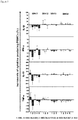
- MSC1 Exo Compared to the co-cultures without exosomes, MSC1 Exo reduced the number of IL-1 ⁇ , TNF- ⁇ and IFN- ⁇ secreting PBMCs in almost all settings ( Figure 5 ), in particular in vitro. Immune suppressive effects on the cytokine responses of the other exosome preparations (MSC2 Exo, MSC3 Exo and MSC4 Exo) were less prominent ( Figure 5 ). These findings suggest that depending on the expanded MSCs, MSC-derived exosomes exert different immune modulatory functions.
- Primary human MSCs were raised from mononuclear cells obtained by Ficoll density gradient centrifugation of bone marrow and raised mainly as described before (Hemeda et al., 2010). Instead of 10% fetal bovine serum (FBS) the inventors supplement the basal media with 5% human pooled platelet lysates. To exclude variations caused by the usage of different platelet lysate preparations, the inventors performed all experiments with the same batch of platelet lysates that the inventors produce by pooling 3040 individual human platelet samples.
- FBS fetal bovine serum
- MSCs were characterized by flow cytometry as CD90 + /CD73 + /CD105 + /CD44 + cells lacking the expression of CD45 and the endothelial marker CD31. MSC differentiation capability along the osteogenic, cartilage and adipogenic lineage was evaluated.
- conditioned media were harvested every 48 h and passed through a 0.22 ⁇ m filter membrane to remove cell debris and larger vesicles. CM were collected and stored at -20°C. After thawing, CM were unified and processed by PEG precipitation. Briefly, PEG 6000 was added to CM. Following incubation for 8-12 h at 4°C exosomes were precipitated by centrifugation for 30 min at 1500 g. Obtained pellets were resolved in 1 mL 0.9% NaCl, washed in a total volume of 45 mL 0.9% NaCl for 12 h at 4°C and precipitated by ultracentrifugation for 2 h at 100,000 g. Again pellets were resolved in 1 mL of 0.9% NaCl, diluted with 0.9% NaCl to required concentrations and stored as 1 mL aliquots at -80°C until use.
- Protein concentrations of obtained aliquots were determined. Of each lot, 10 ⁇ g of exosome containing fraction was used to perform Western blots with antibodies recognizing exosome specific marker proteins, such as Tsg101, CD63, CD81 or CD82. Additionally, the inventors measured the concentration and size of the exosome by the NTA method as described previously (Sokolova et al., 2011). The concentration of pro- and anti-inflammatory cytokines (e.g., IL-1 ⁇ , IL-6, IL-8; TNF- ⁇ , IFN- ⁇ , IL-4, IL-10, IL-13, TGF- ⁇ 1, TSG-6) and of the Fas-ligand within the exosome fractions were measured using the Luminex method. Other methods that could be used include, for example, ELIspot, ELISA, and/or flow cytometry.
- cytokines e.g., IL-1 ⁇ , IL-6, IL-8; TNF- ⁇ , IFN- ⁇ , IL-4,
- the inventors established the T-cell/MNC proliferation assay within the group.
- MNCs or magnetic cell separation enriched CD3 + cells were stained either with CFSE or PKH2 and raised in the presence of either a T cell/MNC proliferation stimulating agents (e.g. LPS, PMA, PHA, anti-CD3 beads) or of cells of the leukemic cell line K562.
- a T cell/MNC proliferation stimulating agents e.g. LPS, PMA, PHA, anti-CD3 beads
- the inventors established a protocol allowing them to document T-cell/MNC proliferation by flow cytometry.
- the inventors searched for a stimulant whose impact on the T cellslMNCs could effectively be modulated by MSC exosomes. Finally, the inventors found a stimulant, which triggered proliferation of different leukocytes within MNC fractions. Using gating strategies for B, T and NK cells the inventors analyzed impacts of MSC exosomes on these cells separately. To estimate effects of MSC-derived exosomes on the cytokine secretion of B, T and NK cells the inventors used the ELISpot assay.
- the inventors adapted a flow cytometry based cytokine secretion assay (Miltenyi, Bergisch-Gladbach), allowing them to study the effect of MSC exosomes on the release of pro-inflammatory cytokines (specifically IFN- ⁇ , TNF- ⁇ and IFN- ⁇ ) by stimulated immune cells.
- the inventors compared the immunomodulatory activities of 15-20 independently obtained MSC exosome fractions. The inventors expected that some of these fractions would show strong immunomodulatory activity, whereas others would have no effect on the immune response of stimulated immune cells. The three samples with the highest and the lowest immunomodulatory activities were compared in more detail. The inventors also comprehensively compared the expression profiles of the MSC propagations, from which corresponding exosome fractions were obtained. The inventors identified sets of candidate proteins that predicted immunomodulatory effects. The inventors validated these markers by evaluating their expression in additional MSC and exosome samples.
- MSC-derived exosomes using PEG preparations with high and low immunomodulatory capabilities in vitro were used for the in vivo approach.
- C57b16 mice were submitted to 30 minutes of intraluminal middle cerebral artery occlusion. This model is well characterized and has been extensively used in the inventors' lab before, resulting in localized infarcts of the striatum and most lateral cerebral cortex (Doeppner et al., 20096). Dissolved in a volume of 250 ⁇ L, 2 x 10 6 cell equivalents of exosomes were injected over 10 min via tail vein on days 1, 3, and 5 post-stroke.
- Post-ischemic motor and coordination deficits were analyzed using the rota rod, the tight rope and the corner turn test as previously described (Zhang et al., 2002; Doeppner et al., 2011b). All animals were trained 1-2 days before induction of stroke to ensure sufficient test performance. Actual tests are performed on days 7, 14, 28, 56 and 84. Using the rota rod test, animals were put on a treadmill with an accelerating speed of 4-40 rpm. The maximum speed was achieved after 260 s, and maximum testing time was 300 s. The time until animals drop was registered and statistically analyzed. For the tight rope test, animals were placed on a 60 cm long rope grasping the string with their forepaws.
- mice received daily intraperitoneal injections of bromodeoxyuridine (BrdU) on days 8-18, to indirectly assess post-ischemic cell proliferation.
- PrdU bromodeoxyuridine
- immunohistochemical protocols using the marker CD31 were employed (Wang et al., 2005). Tissue samples were collected for studies other than immunohisto-chemistry, such as realtime PCR, which allowed to evaluate post-ischemic inflammation, neuronal viability and astroglial scar formation. These assays included real-time PCR protocols according to the manufacturers' protocols.
- GvHD a condition, in which the new immune system recognizes the patient's tissue as 'foreign' and attacks it.
- transplanted patients receive immunosuppressant treatments. Patients refractory to such treatments have a poor prognosis, while transplanted MSCs might improve the clinical response (Gotherstrom et al., 2004; Le Blanc et al., 2004).
- the immunomodulatory activities of MSCs result from secreted soluble factors rather than direct cellular interactions between engrafted MSCs and the patient's immune cells (Baron and Storb, 2012).
- the inventors hypothesized that the effectors are MSC-derived exosomes.
- BM Human bone marrows
- Primary human MSCs were generated from mononuclear cells obtained by Ficoll (Biocoll Separating Solution, Biochrom AG, Berlin, Germany) density gradient centrifugation of bone marrow.
- the mononuclear cells were cultured in tissue culture 6 well plates at a density of 2 x 10 6 cells per well in MSC basal media (PAN) supplemented with 5% thrombocyte lysate, 1% glutamine, and 1% penicillin-streptomycin (PAA) for 24 hours. After 24 hours of incubation non-adherent cells were removed by medium exchange.
- PAN MSC basal media
- PAA penicillin-streptomycin
- Adherent cells After confluence had been reached, usually within 14 days, cells were continuously passaged after treatment with 0.25% trypsin. Adherent cells showed fibroblast-like morphology, and were able to differentiate along the adipogenic, chondrogenic, and osteogenic pathways.
- Exosomes were isolated from cell culture supernatants of MSCs derived from bone marrow. To remove cells, conditioned media were centrifuged for 5 min at 900 x g, and to remove remaining debris, another centrifugation step was performed for 1 h at 10,000 x g. To remove all particles bigger than 200 nm, the supernatants were filtered through 0.2 um pore filters. To concentrate the exosomes, the filtrate was centrifuged for 2 h, 110,000 x g, at 4°C. The obtained precipitates were resuspended in 150 ⁇ l PBS and used for further analyses.
- MSC supernatants were collected and stored at -20°C. After thawing, the supernatants were unified and 3 units/mL of heparin and approx. 3 ⁇ g/mL Actilyse TM (Boehringer Ingelheim, Germany) were added. Supernatants were incubated for 3 h at 37°C, and then filtered through 0.22 ⁇ m bottle top filters.
- vials were centrifuged at 1500 g for 30 min at 4°C. The supernatants were carefully removed (25ml pipette). To collect remaining supernatants, vials were centrifuged for another 5 min at 1500 g. Residual supernatant was removed using a 1 ml pipette. Pellets were resuspended in 1 ml NaCl 0.9%. To remove remaining PEG, resuspended precipitates were supplemented with 0.9% NaCl to a total volume of 45 ml and stored at 4°C for about 12h.
- the 45 ml suspensions were split and transferred in half into two ultracentrifugation-vials. 0.9% NaCl was added to a total volume of 38 ml.
- the preparations were centrifuged for 2 h at 110.000 g. The supernatants were removed from the sediments as obtained; the supernatants were resuspended in 1 ml 0.9% NaCl solution, and pooled. Then, the sample was filled up to 10 ml using 0.9% NaCl solution, and frozen in 1 ml aliquots in 15 ml reaction vials at -80°C.
- the protein contents were determined by the micro BCA assay (Thermo Fisher Scientific).
- Exosome specific proteins were identified by Western blot analysis. For this, purified exosomes were treated with sample buffer (DTT, 0.1% SDS, 0.1 M Tris HCl, pH 7.0) and spoiled for 5 min at 95°C. Proteins were separated on 12% SDS-PAGE gel and transferred to PVDF membranes. Membranes were blotted with antibodies to Tsg101 (Sigma Aldrich), Flotilin 1 (BD Pharmingen), and CD81 (BD Pharmingen), then incubated with appropriate HRP-conjugated secondary antibodies (Dianova) and visualized by enhanced chemiluminescence (Pierce).
- sample buffer 0.1% SDS, 0.1 M Tris HCl, pH 7.0
- Proteins were separated on 12% SDS-PAGE gel and transferred to PVDF membranes. Membranes were blotted with antibodies to Tsg101 (Sigma Aldrich), Flotilin 1 (BD Pharmingen), and CD81 (BD Pharmingen), then incuba
- NTA Nano particle tracking analysis
- NTA For particle size determination, NTA was performed with a NanoSight LM10 instrument equipped with the NTA 2.0 analytical software. All experiments were carried out at 1:2000 dilutions, resulting in particle concentrations of approx. 3 ⁇ 10 8 per mL. Each experiment was carried out in triplicate. The 50% median value (D50) is given in all cases, and the standard deviation was calculated for all data.
- PBMC peripheral blood mononuclear cells
- soluble HLA-G sHLA-G
- cytokines cytokines
- apoptosis-inducing molecules in MSC-exosome preparations were analyzed by ELISA.
- exosomes (approx. 60.5 x 10 8 particles) were solubilized in 1 ml PBS-Tween (1%) and sonicated for 60 sec.
- sHLA-G concentrations were determined as described before (Schutt et al., 2010) with minor modifications: The monoclonal antibody (mAb) G233 (Exbio, Praha, Czech Republic) was used as capture reagent and bound sHLA-G was detected by the biotinylated mAb w6/32 (Leinco Technologies, St. Louis, Missouri, USA) followed by AMDEX TM Streptavidin HRP (Amersham, Freiburg, Germany). The detection limit of sHLA-G ELISA was 0.1 ng/ml.
- Concentrations of pro-inflammatory IFN- ⁇ , TNF- ⁇ , IL-1b, IL-2, IL-6, IL-8, IL-17a, IL-21
- anti-inflammatory IL-1 ⁇ RA, IL-4, IL-10, IL-13, TGF- ⁇ 1
- soluble FasL apoptosis-inducing molecules
- PBMC and NK cells isolated by negative selection out of these PBMC (Invitrogen, Düsseldorf, Germany), were adjusted to a fixed concentration of 1x10 6 cells/mL and used as effector cells.
- 1x10 6 effector cells were mixed with approx. 60.5 x 10 8 MSC-exosomes. In this case effector cell suspensions with exosomes and without exosomes were used in cytokine stimulation experiments.
- K562 cell line served as stimulator cells and were adjusted to a concentration of 0.1x10 6 cells/mL. All ELISpot assays were performed in an effector/stimulator ratio of 6:1.
- IL-1 ⁇ and the TNF- ⁇ ELISpot assays 12.5 x 10 4 effector cells and 0.25 x 10 4 stimulator cells and for the IFN- ⁇ ELISpot assay, 75 x 10 4 effector cells and 1.5 x 10 4 stimulator cells were seeded with 200 ⁇ l culture medium containing 200U/mL IL-2 and 10% fetal calf serum onto MultiScreen-HA ELISpot plates (MAIPS4510, Millipore, Bedford, MA, USA) pre-coated with the respective capture antibody of the ELISpot Kit (eBioscience, Frankfurt, Germany).
- MAIPS4510 Millipore, Bedford, MA, USA
- Exosomes derived from four different MSC preparations were analyzed for the content of anti-, pro-, and apoptosis-inducing molecules: Besides the two pro-inflammatory cytokines IFN- ⁇ and IL-8, MSC-exosomes contained preferentially high quantities of the anti-inflammatory and/or immune suppressive molecules IL-10, TGF- ⁇ 1 and HLA-G. Especially, TGF- ⁇ 1 levels were found to be ten times higher in the MSC-exosome solution of 60.5 x 10 8 particles/mL than in plasma levels of healthy controls.
- the influence of the MSC-exosomes on the cytokine response of PBMC or NK cells of PB donors on K562 leukemic cells was proven by IL-1 ⁇ , TNF- ⁇ and IFN- ⁇ ELISpot using.
- the donors effector cells were co-cultured with HLA class I negative K562 or genetically engineered K562 cells expressing HLA-E*01:03 or HLA-B35. It was found that the IL-1 ⁇ , TNF- ⁇ and IFN- ⁇ cytokine responses of the donor's effector cells were reduced towards the different allogenic cells in the presence of MSC-exosomes compared to responses in the absence of MSC-exosomes.
- AlloSCT allogeneic stem cell transplantation
- MDS RAEB-T myelodysplastic syndrome
- PBSC peripheral blood stem cells
- infliximab (10 mg/kg BW once per week over a four week period) was initiated and 17 sessions of extracorporeal photopheresis were performed over four months. Throughout this time the basic immunosuppression with steroids, mostly in combination with either tacrolimus or MMF, was continued.
- the MSC-exosomes were administered between August, 1 st to August, 15 th 2011 in increasing doses.
- Application Date Dosage 1 2011-08-01 1 ml of a 1:10 diluted exosome aliquot (100 ⁇ l) 2 2011-08-03 2 ml of a 1:10 diluted exosome aliquot (200 ⁇ l) 3 2011-08-05 3 ml of a 1:10 diluted exosome aliquot (300 ⁇ l) 4 2011-08-08 4 ml of a 1:10 diluted exosome aliquot (400 ⁇ l) 5 2011-08-10 1 ml of undiluted exosome aliquot 6 2011-08-12 2 ml of undiluted exosome aliquot 7 2011-08-15 4 ml of undiluted exosome aliquot
- the patient received pre-medication with steroids and anti-histamins prior to each MSC-exosome application and was closely monitored in the intensive care unit. The therapy was tolerated very well, and no side effects were detected. After each administration, blood samples were taken and analyzed for their capability to produce IL-1 ⁇ , TNF- ⁇ , and IFN- ⁇ in response towards K562, and K562 transfected with HLA-E*01:03 or HLA-B27 (see above).
- the PBMC of the patient started to lessen their cytokine reaction towards the stimulator cells.
- the numbers of IL-1 ⁇ , TNF- ⁇ and IFN- ⁇ producing PBMC were found to be reduced more than fifty percent after the last application (p ⁇ 0.0001, One-way ANOVA).
- p ⁇ 0.0001, One-way ANOVA the MSC-exosomes are able to impair in-vivo the capability of PBMC to produce pro-inflammatory cytokines known to be operative in GvHD
- the inventors compared the immune-modulative activity of exosome-enriched fractions obtained from 10 independent MSC preparations within T cell assays.
- exosomes equivalents of 800.000 MSCs corresponds to 0.02 Units of the exosome enriched fractions
- PHA phytohemagglutinin
- PB-MNCs of 10 different peripheral blood donors were pooled. Aliquots of 400.000 cells were cultured in triplicates each in a total volume of 200 ⁇ l, either in the presence or absence of 0.02 U of the same exosome fractions presented in Figure 6 .
Landscapes
- Health & Medical Sciences (AREA)
- Life Sciences & Earth Sciences (AREA)
- Immunology (AREA)
- Engineering & Computer Science (AREA)
- Medicinal Chemistry (AREA)
- Pharmacology & Pharmacy (AREA)
- Animal Behavior & Ethology (AREA)
- General Health & Medical Sciences (AREA)
- Public Health (AREA)
- Veterinary Medicine (AREA)
- Chemical & Material Sciences (AREA)
- Bioinformatics & Cheminformatics (AREA)
- Epidemiology (AREA)
- Zoology (AREA)
- Proteomics, Peptides & Aminoacids (AREA)
- Gastroenterology & Hepatology (AREA)
- Cell Biology (AREA)
- Developmental Biology & Embryology (AREA)
- Biotechnology (AREA)
- Virology (AREA)
- Biomedical Technology (AREA)
- Hematology (AREA)
- Marine Sciences & Fisheries (AREA)
- Organic Chemistry (AREA)
- Nuclear Medicine, Radiotherapy & Molecular Imaging (AREA)
- General Chemical & Material Sciences (AREA)
- Chemical Kinetics & Catalysis (AREA)
- Dermatology (AREA)
- Transplantation (AREA)
- Urology & Nephrology (AREA)
- Vascular Medicine (AREA)
- Cardiology (AREA)
- Heart & Thoracic Surgery (AREA)
- Medicines Containing Material From Animals Or Micro-Organisms (AREA)
Description
- A method for producing a pharmaceutical preparation comprising exosomes that exhibit an immunomodulatory effect, comprising the following steps: a) providing a cell culture medium supernatant from neonatal or adult tissue-derived mesenchymal stem-cells (MSCs), optionally from MSCs that have been cultured in platelet lysate, comprising exosomes, b) enriching said exosomes, optionally comprising polyethylene glycol precipitation, c) determining an in vitro immunomodulatory effect of said enriched exosomes by measuring TGF-β1 levels of said enriched exosomes, d) selecting those enriched exosomes that exhibit the immunomodulatory effect and have a TGF-β1 level that is at least ten times higher than plasma TGF-β1 levels of healthy controls, and e) admixing said selected enriched exosomes of step d) with at least one suitable pharmaceutical excipient and/or carrier.
- Different cell populations appear as candidates for cell-based therapies, in particular in neonates: Hematopoietic stem cells (HSC), endothelial progenitor cells (EPC), mesenchymal stem cells (MSC) as well as regulatory T cells (Treg) exerting immune modulatory and pro-regenerative activities by direct cellular action or secretion of pro-regenerative, anti-apoptotic, immunomodulatory factors. The suppressive activity of Tregs from umbilical cord blood (UCB) has been attributed to the observed reduced severity of graft versus host disease (GvHD) in UCB transplantation (Tolar J, Hippen KL, Blazar BR. Immune regulatory cells in umbilical cord blood: T regulatory cells and mesenchymal stromal cells. Br J Haematol 2009 October; 147(2):200-6).
- Mesenchymal stem/stromal cells (MSCs) have emerged as one of the most intensely studied adult stem cell population within the last 15 years (Bieback K, Brinkmann I. Mesenchymal stromal cells from human perinatal tissues: From biology to cell therapy. World J Stem Cells 2010 August 26; 2(4):81-92). The therapeutic interest initially was based on their multilineage differentiation potential. However, it recently turned out that only few cells engraft and differentiate after either local or systemic delivery of MSCs in a number of different injury models. Subsequent to the detection of profound immune modulatory activities, MSCs were successfully applied to treat steroid-refractory GvHD (Le Blanc K, Rasmusson I, Sundberg B, Gotherstrom C, Hassan M, Uzunel M et al. Treatment of severe acute graft-versus-host disease with third party haploidentical mesenchymal stem cells. Lancet 2004 May 1; 363(9419): 1439-41).
- Since then, a number of different studies have addressed the impact of MSCs on GvHD with different outcomes. In most of these studies, the infusion of MSCs appeared to be safe. However, a positive impact on GvHD is still discussed controversially (Baron and Storb, 2011). Although MSCs can be raised from virtually every tissue within the human body, they are a very rare population comprising about 0.001 to 5 % of cells in different tissues (Bieback K. Basic Biology of Mesenchymal Stem Cells. Transfus Med Hemother 2008; 35(3):151-2).
- Exosomes are naturally occurring small membrane vesicles (80-160 nm) released by a huge variety of cell species. Containing a combination of lipids and proteins as well as RNAs, exosomes participate in intercellular communication processes. They can be isolated from all body fluids including blood plasma, urine, saliva, breast milk, bronchial lavage fluid, cerebral spinal fluid, amniotic fluid and malignant ascites (Ludwig AK, Giebel B. Exosomes: Small vesicles participating in intercellular communication. Int J Biochem Cell Biol 2011 October 19).
- Initially, exosomes were discovered in 1983 as small vesicles that are released by exocytosis upon fusion of so called multivesicular bodies with the plasma membrane. In 1996, exosomes were isolated from B lymphocytes, and were demonstrated to exhibit antigen-presenting characteristics. Since then, their role in immune-biological settings has been investigated in an increasing number of studies. It turned out that exosomes derived from mature dendritic cells for example exert immune-stimulatory functions, while tumor-derived exosomes often mediate immune-suppressive functions (Ludwig AK, Giebel B. Exosomes: Small vesicles participating in intercellular communication. Int J Biochem Cell Biol 2011 October 19).
- In 2010, Lai and co-workers provided evidence that exosomes released from MSCs mediate at least a proportion of the regenerative effects of clinically applied MSCs (Lai RC, Arslan F, Lee MM, Sze NS, Choo A, Chen TS et al. Exosome secreted by MSC reduces myocardial ischemia/reperfusion injury. Stem Cell Res 2010 May; 4(3):214-22). Since intravenously administered MSCs get rapidly trapped into the lung and are hardly found within infarcted hearts (Lee et al., 2009), the group of Lim searched for paracrine MSC effectors and showed in a first set of experiments that the supernatants of in vitro expanded MSCs are sufficient to mediate beneficial effects. Next, they purified exosomes from MSC supernatants and infused them in a murine myocardial ischemia model. Again, they observed a reduction of the infarct size (Lai et al., 2010).
- Timmers et al. (in Timmers L, Lim SK, Arslan F, Armstrong JS, Hoefer IE, Doevendans PA, Piek JJ, El Oakley RM, Choo A, Lee CN, Pasterkamp G, de Kleijn DP. Reduction of myocardial infarct size by human mesenchymal stem cell conditioned medium. Stem Cell Res. 2007 Nov; 1(2): 129-37. Epub 2008 Mar 8) describe the use of secretions of human MSCs as a therapeutic option in acute myocardial infarct. These MSCs are raised from human Embryonic Stem Cells (hESCs).
- Fleissner et al. (in: Fleissner F, Goerzig Y, Haverich A, Thum T. Microvesicles as novel biomarkers and therapeutic targets in transplantation medicine. Am J Transplant. 2012 Feb; 12(2):289-97. Epub 2011 Nov 14) describe microvesicles (MVs) including exosomes as emerging new biomarkers and potential regulators of inflammation and immunological processes. The particles contain proteins and genetic information including DNA and microRNAs that may be of importance for cell/cell communication. However, their role during and after organ transplantation and immunomodulatory effects is described to be only in its beginning of understanding. The authors speculate that MV modulation may emerge as a therapeutic approach in organ rejection therapy.
- Horstman et al. (in: Horstman LL, Jy W, Minagar A, Bidot CJ, Jimenez JJ, Alexander JS, Ahn YS. Cell-derived microparticles (MPs) and exosomes in neuroinflammatory disorders. Int Rev Neurobiol. 2007; 79:227-68) describe MPs and their possible roles in the pathophsyiology of some neuropathologies, especially those which are ischemic in nature, but probably also inflammatory ones. Tables include a listing of bioactive agents known to be carried on MP, many of which were heretofore considered strictly soluble, and some of which can be transferred from cell to cell via MP vectors, for example certain cytokine receptors.
- Baron and Storb (in: Baron F, Storb R. Mensenchymal Stromal Cells: A New Tool against Graft-versus-Host Disease? Biol Blood Marrow Transplant 2011 September 29) propose that immune modulatory activities of MSCs are thought to derive from secreted soluble factors rather than from direct intercellular interactions of engrafted MSCs with the patient's immune cells.
-
WO 2009/105044 describes a particle secreted by a mesenchymal stem cell and comprising at least one biological property of a mesenchymal stem cell. The biological property may comprise a biological activity of a mesenchymal stem cell conditioned medium (MSC-CM) such as cardioprotection or reduction of infarct size. The particle may comprise a vesicle or an exosome. The claims encompass exosomes obtainable from human embryonic stem cells derived from human embryos. -
WO 2012/020308 is directed at compositions and methods for the treatment of tissue damage (e.g., acute or chronic) and related diseases, disorders or conditions based on the use of pathfinder cells, extracellular secretomes thereof, and/or pathfinder cell-associated micro RNAs (miRNAs). In some embodiments, the present invention provides a method for treating inflammation comprising a step of administering a population of cells, or extracellular secretomes thereof, to an individual suffering from a disease, disorder or condition characterized by inflammation of one or more tissues, wherein the cells are originated from an adult tissue and wherein the cells induce an anti-inflammatory response. -
WO 2012/053976 discloses the use of exosomes that are secreted by mesenchymal stem cells in order to promote hair growth and wound healing. These effects are disclosed in connection with pro-inflammatory cytokines and proteases.WO 2012/053976 speculates about an immune-modulatory effect of the exosome-preparation. -
WO 2012/020307 discloses the therapeutic use of microvesicles including exosomes in the treatment of inflammation and lesions of cerebral trauma. Similarly,WO 2012/087241 describes the use of exosomes for the treatment of the diseases as mentioned. - The currently proposed strategies in the prevention and/or therapy based on exosomes suffer from several disadvantages. Most problematic is their unimodal approach. This is in contrast to the biological complexity of the pathophysiology of the indications to be treated. Instead, it would be required to use a therapeutic cocktail that can interact with very different pathophysiological signaling cascades, and thus provide a more complete and effective prevention or treatment.
- Mesenchymal stromal cells (MSCs) represent a heterogeneous subset of multipotent cells that can be isolated from several tissues including bone marrow and fat. MSCs exhibit immunomodulatory and anti-inflammatory properties that prompted their clinical use as prevention and/or treatment for severe graft-versus-host disease (GvHD). Although a number of phase I-II studies have suggested that MSC infusion was safe and might be effective for preventing or treating acute GvHD, definitive proof of their efficacy remains lacking thus far. Multicenter randomized studies are ongoing to more precisely assess the impact of MSC infusion on GvHD prevention/treatment, whereas further research is performed in vitro and in animal models with the aims of determining the best way to expand MSCs ex vivo as well as the most efficient dose and schedule of MSCs administration (see Baron and Storb, 2011). Although MSCs can be raised from virtually every tissue within the human body, they are a very rare population comprising about 0.001 to 5 % of cells in different tissues. Especially UCB contains MSC at low frequency. Also, MSCs derived from human embryonic ESC meet ethical concerns.
- Finally, exosomes that are obtained from MSCs of individual donors were found to be very heterogenic. Thus, it seems reasonable to assume that subtypes of populations of MSCs with quite different potential are present. Currently, this problem is completely unresolved.
- It is therefore an object of the present invention to provide a safe and more effective preparation of exosomes that is suitable for the treatment of diseases and conditions that involve inflammatory reactions. Other objects of the present invention will become apparent to the person of skill upon studying the present description of the invention.
- In a first aspect thereof, the above object is solved by a method for producing a pharmaceutical preparation comprising exosomes that exhibit an immunomodulatory effect, comprising the following steps: a) providing a cell culture medium supernatant from neonatal or adult tissue-derived mesenchymal stem-cells (MSCs), optionally from MSCs that have been cultured in platelet lysate, comprising exosomes, b) enriching said exosomes, optionally comprising polyethylene glycol precipitation, c) determining an in vitro immunomodulatory effect of said enriched exosomes by measuring TGF-β1 levels of said enriched exosomes, d) selecting those enriched exosomes that exhibit the immunomodulatory effect and have a TGF-β1 level that is at least ten times higher than plasma TGF-β1 levels of healthy controls, and e) admixing said selected enriched exosomes of step d) with at least one suitable pharmaceutical excipient and/or carrier.
- Considering exosomes as a novel, very potent tool in regenerative and in immune-modulatory therapies, the inventors have set up new purification strategies and technologies to enrich for and to analyze exosomes. Amongst other indications, the inventors evaluated their applicability for use in immune-modulatory (inflammatory) therapies, such as GvHD therapy, and already treated an otherwise treatment-resistant grade IV acute GvHD patient with MSC-derived exosomes. The GvHD symptoms of the patient decreased dramatically with the specific exosome therapy and the patient remained stable for five months.
- Furthermore, in order to prove effectiveness of the prevention and therapy of neuronal damages, in particular in neonates, experiments with LPS confronted rats were performed that showed a clear positive reaction following the administration of the inventive pharmaceutical preparation.
- The pharmaceutical preparation according to the present disclosure is specifically enriched for exosomes. For this, generally any suitable method for purifying and/or enriching can be used, such as methods comprising magnetic particles, filtration, dialysis, ultracentrifugation, ExoQuick™ (Systems Biosciences, CA, USA), and/or chromatography. Nevertheless, preferred is a method that comprises polyethylene glycol precipitation and/or chromatographically enrichment using the monolithic technology (e.g. CIM®, BIA separations, Austria) as stationary phases instead of columns packed with porous particles. Monoliths are continuous stationary phases that are cast as a homogeneous column in a single piece and prepared in various dimensions with agglomeration-type or fibrous microstructures. (see Iberer, G., Hahn, R., Jungbauer, A. LC-GC, 1999, 17, 998). Using these methods, surprisingly active fractions containing exosomes could be obtained.
- It could be found that fractions being enriched with exosomes were particularly useful in the methods according to the present disclosure, if they exhibited strong in vitro immunomodulatory effects in activity tests, where, following the addition of said exosome fraction, a reduced IL-1β, TNF-α and/or IFN-γ cytokine response of effector cells of a donor could be found. ELISpot assays showed that the IL-1β, TNF-α and/or IFN-γ cytokine response of effector cells are impaired towards allogeneic cells in the presence of exosome containing fractions. Other methods that could be used to test for in vitro immunomodulatory effects include, for example, Luminex, ELISA, and/or flow cytometry.
- The present disclosure is thus based on the novel concept for an improved prevention and treatment of diseases, in particular in patients suffering from a having a risk of an inflammatory disease, neuronal disease, GvHD, stroke, and ischemia and associated complications, for example, for avoiding inflammatory reactions prior or during surgery, and the prevention of inflammatory conditions and reactions of patients that are connected to a life support machine.
- The diseases can be selected from pre- or postnatal damages of the nervous system, such as for example, brain damages related to hypoxia, inflammation, and/or ischemia. In another embodiment, the diseases can be selected from graft-versus-host disease, or transplant rejections following organ transplantations, respectively.
- In a particularly preferred embodiment of the invention, the exosomes-enriched fractions derived from adult mesenchymal stem cells (MSCs) that were enriched using a polyethylene glycol precipitation protocol, are prophylactically and/or therapeutically transfused into patients, in particular neonates and/or patients receiving transplants and/or patients undergoing surgery.
- Preferably, the exosomes-enriched fractions of the disclosure are derived from MSCs that are selected from human adult MSCs, and preferably from human MSCs derived from umbilical cord blood, umbilical cord tissue, placenta, bone or adipose tissue, and MSCs that have been cultured in platelet lysate.
- The pharmaceutical preparation according to the present disclosure preferably is enriched for exosomes that comprise biological factors, such as, for example, proteins, such as anti-inflammatory cytokines, IL-10, TGF-β1, and HLA-G, and/or nucleic acids, such as, for example, miRNAs. This leads to the further advantage according to the disclosure that a) a multimodal (complex) intervention is performed, b) biological physiological ("self') substances are used, and c) unwanted side effects of the preparation are reduced.
- The present invention disclosure constitutes a multimodal intervention, and thus not only a specific factor is used (and only a part of the cascade (or of the underlying clinical phenotype) would be intervened with), but biologically complex and endogenous mediators and modulators are used. These components are found in every human, and therefore no significant adverse side-effects are expected.
- Further preferred is a pharmaceutical preparation according to the present disclosure, wherein the numbers of IL-1β, TNF-α and/or IFN-γ producing PBMC were found to be reduced in said patient more than fifty percent after the last application (p < 0.0001, One-way ANOVA), compared to the cytokines' responses before MSC-exosome administration.
- Preferred is a method for producing a pharmaceutical preparation according to the present invention, wherein said neonatal or adult tissue-derived mesenchymal stem-cells (MSCs) comprising exosomes are selected from human MSCs, and preferably from human MSCs derived from umbilical cord blood, umbilical cord tissue, placenta, bone or adipose tissue. The stem cells of the present invention are strictly non-embryonic derived stem cells. Most preferably, the MSCs that have been cultured in the presence of platelet lysate, and the preparation can contain said lysate and/or fractions thereof.
- The method for producing a pharmaceutical preparation according to the present invention comprises the step of specifically enriching for exosomes. For this, generally any suitable method for purifying and/or enriching can be used, such as methods comprising magnetic particles, filtration, dialysis, ultracentrifugation, ExoQuick™ (Systems Biosciences, CA, USA), and/or chromatography. Nevertheless, preferred is a method that comprises polyethylene glycol precipitation and/or a monolithic method (see above), since using these methods, surprisingly active fractions containing exosomes could be obtained.
- Preferred is a method for producing a pharmaceutical preparation according to the present invention, wherein fractions that were enriched for exosomes are further analyzed in microbiological tests, virulence tests, protein content, pyrogen tests, and particle size, in order to identify the most suitable fraction according to the invention.
- It could be found that fractions that were enriched for exosomes were particularly useful in the methods according to the present invention, if they exhibited strong in vitro immunomodulatory effects in activity tests, where upon the addition of said exosome fraction, a reduced IL-1β, TNF-α and/or IFN-γ cytokine response of effector cells of a donor could be found. A method according to the present invention requires said levels of TGF-β1 as measured in the activity tests are at least ten (or at least twenty) times higher (at 60.5 x 10⁸ particles/mL) in the preparation than in plasma levels of healthy controls. Further preferred is a method according to the present invention, wherein said exosomes have a size of between about 70 to 200 nm, preferably between about 70 to 140 nm, or more preferably between about 70 to 120 nm. "About" shall mean a +/- 10% deviation. Further preferred, the exosomes are positive for cellular exosome markers, and even further preferred the protein content of the pharmaceutical preparation is higher than 1 mg/ml.
- Furthermore, the method for producing a pharmaceutical preparation according to the present invention comprises d) admixing said enriched exosomes of step c) with at least one suitable pharmaceutical excipient and/or carrier. In general any suitable pharmaceutically acceptable excipient and/or carrier can be used. Preferably, said pharmaceutically acceptable excipient and/or carrier renders said preparation suitable for i.v. administration, such as for example, intravenous administration or infusion.
- Based on their proposed broad multi-lineage differentiation potential mesenchymal stem cells (MSCs) became one of the most intensively studied adult stem cell entities within the last 15 years. Up to now, more than 300 different NIH-registered studies have addressed potential therapeutic impacts of clinically administered MSCs in a variety of clinical settings including treatment of acute myocardial infarction, stroke and acute kidney failure.
- Several studies showed that the vast majority of intravenously administered MSCs get rapidly trapped in the lung and are rarely recovered in other tissues. Therefore, MSCs might improve clinical outcomes by paracrine effects rather than by their previously proposed engraftment into damaged host tissues (Lee et al., 2009). Indeed, the group of Lim earlier showed that the supernatants of in vitro expanded embryonic stem cell derived MSCs contain small extracellular vesicles, so called exosomes, whose infusion is sufficient to mediate a reduction of myocardial infarction sizes (see Lim et al., 2010).
- The present invention will now be described further in the following examples.
-
Figure 1 shows the effect of exosome preparations on newborn rats that were partially confronted with LPS. -
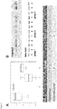
Figure 2 shows an example for the analyses of exosomes-containing fractions during the purification thereof using a monolithic column. -
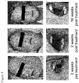
Figure 3 shows the transplantation-relevant effects of exosomes on a patient in a treatment scheme according to the examples. As symptom of GvHD the patient has diarrhea of more than 3 liters per day. After administration of MSC-derived exosome-enriched fractions, the symptoms were drastically reduced, without that other side-effects could be observed. -
Figure 4 also shows the transplantation-relevant effects of exosomes on a patient in a treatment scheme according to the examples. As symptom of the GvHD, the patient developed inflammations of the skin. After administration of MSC-derived exosome-enriched fractions, the symptoms were drastically reduced, without that other side-effects could be observed. -
Figure 5 shows that MSC-fractions, such as MSC1 Exo reduce the number of IL-1β, TNF-α and IFN-γ secreting PBMCs in almost all settings, in particular in vitro. Immune suppressive effects on the cytokine responses of the other exosome preparations (MSC2 Exo, MSC3 Exo and MSC4 Exo) are slightly less prominent. -
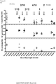
Figure 6 shows that depending on the exosome fraction, MSC-enriched exosomes suppress, do not modulate or increase the proliferation rates of treated T cells, respectively (Example III). -
Figure 7 shows that certain exosome fractions such as Exo MSC A (fraction 3 inFig. 2 ) and MSC Exo F (fraction 7 inFig. 2 ) suppress proliferation of PHA stimulated T cells and IL-1beta and TNFalpha release of stimulated T cells (see Example III). -
Figure 8 shows that the effects of the in vitro studies (A) correlate with the effect in vivo (B). - The exosome-enriched fractions derived from the four MSC preparations were analyzed for their content of anti- and pro-inflammatory as well as apoptosis-inducing molecules (Table 1). Apart from the pro-inflammatory cytokines IFN-γ and IL-8, the exosome preparations contained high quantities of the anti-inflammatory molecules IL-10, TGF-β1 and HLA-G. Neither the pro-inflammatory cytokines IL-1β, IL-2, IL-6, IL-17a, IL-21 and TNF-α, the anti-inflammatory cytokines IL-1β Ra and IL13, nor the apoptosis-inducing molecule sFasL were detected in any of the exosome preparations (Table 1).
- Especially the MSC1 exosome preparation (MSC1 Exo) contained elevated TGF-β levels, which surpassed TGF-β plasma levels of healthy donors by more than the
factor 10. In addition MSC1 Exo differed from the others preparations with regard to the anti-inflammatory cytokine IL-10 and the pro-inflammatory cytokine IFN-γ. The ratio of IL-10 to IFN-γ varied between the four exosome preparations for almost two orders of magnitude (Table 1); the highest ratio was found for MSC1 Exo (1.02).Table 1: Cytokine contents of the MSC exosome preparations. (1)Levels are given in pg/mL; (2)Levels are given in ng/mL; (3)Preparation used for treatment; (4)N=20 for healthy controls Marker MSC1 (3) MSC2 MSC3 MSC4 control (4) Pro-inflammatory IL- 1β (1)0 0 0 0 0-34 IL-2 (1) 0 Not tested Not tested Not tested 0-44 IL-6 (1) 0 Not tested Not tested Not tested 0-8 IL-8 (1) 48.2 11.4 7.8 15.7 0-31 IL- 17a (1)0 Not tested Not tested Not tested Not tested IL-21 (1) 0 Not tested Not tested Not tested Not tested TNF- α (1)0 0 1.9 0 0-10.4 IFN-γ (1) 81.5 38.2 83.5 144 0-56 Anti-inflammatory IL- 1β Ra (1)0 0 0 0 Not tested IL-4 (1) 0 15.2 14.9 69.1 0-5 IL-10 (1) 83.3 0.98 9.7 4.2 0-625 IL-13 (1) 0 Not tested Not tested Not tested Not tested TGF-β1 (1) 13516 305 135 579 903-1654 sFasL (1)0 0 0 0 90-2285 BLA-G (2) 26.4 6.3 3.1 9.8 0-56 ratio IL-10:IFN-γ 1.02 0.03 0.12 0.03 Distinct MSC exosome preparations reveal different capabilities to suppress inflammatory responses in vitro - Immune modulatory impacts of the MSC exosome preparations were tested in a mixed lymphocyte reaction using the HLA class I negative K562 as target cells. In order to reflect an allogeneic transplantation setting stable HLA-B35, HLA-E*01:03 or HLA-B27-transfected K562 variants and cells of the HLA class I positive, respiratory syncytial virus infected (RSV) B-cell line were additionally used as allogeneic target cells. In three independent experiments target cells were co-cultured with PBMCs of healthy donors, either in the presence or absence of MSC exosomes. Numbers of IL-1β, TNF-α and IFN-γ secreting PBMCs were documented within the ELISpot assay as mean percentage ± SEM.
- Compared to the co-cultures without exosomes, MSC1 Exo reduced the number of IL-1β, TNF-α and IFN-γ secreting PBMCs in almost all settings (
Figure 5 ), in particular in vitro. Immune suppressive effects on the cytokine responses of the other exosome preparations (MSC2 Exo, MSC3 Exo and MSC4 Exo) were less prominent (Figure 5 ). These findings suggest that depending on the expanded MSCs, MSC-derived exosomes exert different immune modulatory functions. - Primary human MSCs were raised from mononuclear cells obtained by Ficoll density gradient centrifugation of bone marrow and raised mainly as described before (Hemeda et al., 2010). Instead of 10% fetal bovine serum (FBS) the inventors supplement the basal media with 5% human pooled platelet lysates. To exclude variations caused by the usage of different platelet lysate preparations, the inventors performed all experiments with the same batch of platelet lysates that the inventors produce by pooling 3040 individual human platelet samples. Growth conditions in the presence of human platelet lysates instead of animal sera such as FBS comply with GMP standards and are applicable for clinical settings (Doucet et al., 2005; Schallmoser et al., 2007 & 2009; Lange et al., 2007). MSCs were characterized by flow cytometry as CD90+/CD73+/CD105+/CD44+ cells lacking the expression of CD45 and the endothelial marker CD31. MSC differentiation capability along the osteogenic, cartilage and adipogenic lineage was evaluated.
- Starting at
passage 3 conditioned media (CM) were harvested every 48 h and passed through a 0.22 µm filter membrane to remove cell debris and larger vesicles. CM were collected and stored at -20°C. After thawing, CM were unified and processed by PEG precipitation. Briefly, PEG 6000 was added to CM. Following incubation for 8-12 h at 4°C exosomes were precipitated by centrifugation for 30 min at 1500 g. Obtained pellets were resolved in 1 mL 0.9% NaCl, washed in a total volume of 45 mL 0.9% NaCl for 12 h at 4°C and precipitated by ultracentrifugation for 2 h at 100,000 g. Again pellets were resolved in 1 mL of 0.9% NaCl, diluted with 0.9% NaCl to required concentrations and stored as 1 mL aliquots at -80°C until use. - Protein concentrations of obtained aliquots were determined. Of each lot, 10 µg of exosome containing fraction was used to perform Western blots with antibodies recognizing exosome specific marker proteins, such as Tsg101, CD63, CD81 or CD82. Additionally, the inventors measured the concentration and size of the exosome by the NTA method as described previously (Sokolova et al., 2011). The concentration of pro- and anti-inflammatory cytokines (e.g., IL-1β, IL-6, IL-8; TNF-α, IFN-γ, IL-4, IL-10, IL-13, TGF- β1, TSG-6) and of the Fas-ligand within the exosome fractions were measured using the Luminex method. Other methods that could be used include, for example, ELIspot, ELISA, and/or flow cytometry.
- As an immunomodulatory read out, the inventors established the T-cell/MNC proliferation assay within the group. In analogy to the inventors' CD34+ cell studies (Beckmann et al., 2007; Giebel et al., 2006) MNCs or magnetic cell separation enriched CD3+ cells were stained either with CFSE or PKH2 and raised in the presence of either a T cell/MNC proliferation stimulating agents (e.g. LPS, PMA, PHA, anti-CD3 beads) or of cells of the leukemic cell line K562. First, the inventors established a protocol allowing them to document T-cell/MNC proliferation by flow cytometry. Next, the inventors searched for a stimulant whose impact on the T cellslMNCs could effectively be modulated by MSC exosomes. Finally, the inventors found a stimulant, which triggered proliferation of different leukocytes within MNC fractions. Using gating strategies for B, T and NK cells the inventors analyzed impacts of MSC exosomes on these cells separately. To estimate effects of MSC-derived exosomes on the cytokine secretion of B, T and NK cells the inventors used the ELISpot assay. Furthermore, the inventors adapted a flow cytometry based cytokine secretion assay (Miltenyi, Bergisch-Gladbach), allowing them to study the effect of MSC exosomes on the release of pro-inflammatory cytokines (specifically IFN-α, TNF-α and IFN-γ) by stimulated immune cells.
- Using the aforementioned assays the inventors compared the immunomodulatory activities of 15-20 independently obtained MSC exosome fractions. The inventors expected that some of these fractions would show strong immunomodulatory activity, whereas others would have no effect on the immune response of stimulated immune cells. The three samples with the highest and the lowest immunomodulatory activities were compared in more detail. The inventors also comprehensively compared the expression profiles of the MSC propagations, from which corresponding exosome fractions were obtained. The inventors identified sets of candidate proteins that predicted immunomodulatory effects. The inventors validated these markers by evaluating their expression in additional MSC and exosome samples.
- MSC-derived exosomes using PEG preparations with high and low immunomodulatory capabilities in vitro were used for the in vivo approach. C57b16 mice were submitted to 30 minutes of intraluminal middle cerebral artery occlusion. This model is well characterized and has been extensively used in the inventors' lab before, resulting in localized infarcts of the striatum and most lateral cerebral cortex (Doeppner et al., 20096). Dissolved in a volume of 250 µL, 2 x 10⁶ cell equivalents of exosomes were injected over 10 min via tail vein on
days day 1 post-stroke by intravenous injection. The latter animals receive normal saline injections ondays days - Post-ischemic motor and coordination deficits were analyzed using the rota rod, the tight rope and the corner turn test as previously described (Zhang et al., 2002; Doeppner et al., 2011b). All animals were trained 1-2 days before induction of stroke to ensure sufficient test performance. Actual tests are performed on
days - Brain injury was analyzed at the time points given using cresyl violet staining, NeuN staining, TdT-mediated dUTP-biotin nick end labeling (TUNEL) staining and analysis of astroglial scar formation using the astrocytic marker glial fibrillary acidic protein (GFAP). Since transplantation of stem cells can support endogenous repair mechanisms, postischemic neuro-regeneration after exosome treatment was analyzed as well. Therefore, mice received daily intraperitoneal injections of bromodeoxyuridine (BrdU) on days 8-18, to indirectly assess post-ischemic cell proliferation. After sacrifice of animals at the time points given, immunohistochemial double staining against BrdU and various "differentiation" markers (e.g., Dcx, NeuN, CNPase) were performed (Doeppner et al., 2010b). To evaluate axonal plasticity, animals also received ipsi- and contralateral injections of the anterograde tract tracer biotinylated dextran amine (BDA) and cascade blue labeled dextran amine into the motor cortex on
day 70 in order to determine exosome-induced pyramidal tract sprouting on day 84 post-stroke (Reitmeir et al., 2011). For analysis of angiogenesis, immunohistochemical protocols using the marker CD31 were employed (Wang et al., 2005). Tissue samples were collected for studies other than immunohisto-chemistry, such as realtime PCR, which allowed to evaluate post-ischemic inflammation, neuronal viability and astroglial scar formation. These assays included real-time PCR protocols according to the manufacturers' protocols. - In order to assess quantitative changes of cerebral immune cell infiltration, FACS analysis was performed on
day 7 post-stroke. In line with published protocols (Gelderblom et al., 2009), the inventors analyzed the relative frequency of microglia, neutrophils, macrophages, dendritic cells, natural killer cells and lymphocytes in brain tissue. Since stem cell induced neuroprotection also implies modulation of peripheral immune responses (Schwarting et al., 2008), peripheral immune cell subtypes were analyzed in the spleen, lymph nodes and blood. More specifically, a differentiation analysis of CD11b+ and CD11c+ antigen-presenting cells and of lymphocyte subpopulations was performed. - As an undesired side effect of allogeneic bone marrow transplantation, some patients with malignant hematological diseases develop GvHD, i.e. a condition, in which the new immune system recognizes the patient's tissue as 'foreign' and attacks it. Usually, transplanted patients receive immunosuppressant treatments. Patients refractory to such treatments have a poor prognosis, while transplanted MSCs might improve the clinical response (Gotherstrom et al., 2004; Le Blanc et al., 2004). Meanwhile it is assumed that the immunomodulatory activities of MSCs result from secreted soluble factors rather than direct cellular interactions between engrafted MSCs and the patient's immune cells (Baron and Storb, 2012). The inventors hypothesized that the effectors are MSC-derived exosomes.
- Given the history of a female patient with myelodysplastic syndrome that had received allogeneic bone marrow transplantation, the patient developed a refractory, severe GvHD. After written consent and approval by local ethics authorities, the inventors transfused MSC-derived exosomes for the very first time ever in man for compassionate use ('individueller Heilversuch'). Before delivery, the inventors purified exosomes from four different MSC lineages and tested their immunomodulatory properties extensively in vitro.
- The inventors observed immunosuppressive properties of these exosomes in 3 out of 4 lineages. Next, the inventors chose exosomes with the strongest immunomodulatory effect, tested their interaction with the patient's blood cells in vitro and transfused the exosomes in intervals of 2-3 days in increasing doses into the patient (
Fig. 3 ). The patient tolerated the exosome application very well and remarkably recovered from her GvHD symptoms during and after the therapy (Fig. 3 ). The patient was stable for more than 5 months. Even though the inventors cannot conclude at this state that the MSC exosomes led to the suppression of the GvHD symptoms, the patient's immune cells surprisingly showed a similar immune modulation as cells of healthy donors cells which were treated with exosomes in vitro. - Human bone marrows (BM) were obtained from unrelated donors after informed consent according to the Declaration of Helsinki. Primary human MSCs were generated from mononuclear cells obtained by Ficoll (Biocoll Separating Solution, Biochrom AG, Berlin, Germany) density gradient centrifugation of bone marrow. The mononuclear cells were cultured in tissue culture 6 well plates at a density of 2 x 10⁶ cells per well in MSC basal media (PAN) supplemented with 5% thrombocyte lysate, 1% glutamine, and 1% penicillin-streptomycin (PAA) for 24 hours. After 24 hours of incubation non-adherent cells were removed by medium exchange. After confluence had been reached, usually within 14 days, cells were continuously passaged after treatment with 0.25% trypsin. Adherent cells showed fibroblast-like morphology, and were able to differentiate along the adipogenic, chondrogenic, and osteogenic pathways.
- Exosomes were isolated from cell culture supernatants of MSCs derived from bone marrow. To remove cells, conditioned media were centrifuged for 5 min at 900 x g, and to remove remaining debris, another centrifugation step was performed for 1 h at 10,000 x g. To remove all particles bigger than 200 nm, the supernatants were filtered through 0.2 um pore filters. To concentrate the exosomes, the filtrate was centrifuged for 2 h, 110,000 x g, at 4°C. The obtained precipitates were resuspended in 150 µl PBS and used for further analyses.
- Starting at passage three, the inventors performed media exchanges every other day. MSC supernatants were collected and stored at -20°C. After thawing, the supernatants were unified and 3 units/mL of heparin and approx. 3 µg/mL Actilyse™ (Boehringer Ingelheim, Germany) were added. Supernatants were incubated for 3 h at 37°C, and then filtered through 0.22 µm bottle top filters.
- Subsequently, a fraction of the medium (300 ml) was concentrated in 300 MWCO filters (Sartorius), the supernatant was precipitated either using 10% or 12
% PEG 6000 or 10% PEG8000. For the precipitation, supernatants were transferred into 50 ml reaction vials (sterile, Greiner, Germany) prefilled with the precipitation reagent (PEG + NaCl). After mixing the vials were incubated over night (8-12 h at 4°C). - On the next day, vials were centrifuged at 1500 g for 30 min at 4°C. The supernatants were carefully removed (25ml pipette). To collect remaining supernatants, vials were centrifuged for another 5 min at 1500 g. Residual supernatant was removed using a 1 ml pipette. Pellets were resuspended in 1 ml NaCl 0.9%. To remove remaining PEG, resuspended precipitates were supplemented with 0.9% NaCl to a total volume of 45 ml and stored at 4°C for about 12h.
- The 45 ml suspensions were split and transferred in half into two ultracentrifugation-vials. 0.9% NaCl was added to a total volume of 38 ml. The preparations were centrifuged for 2 h at 110.000 g. The supernatants were removed from the sediments as obtained; the supernatants were resuspended in 1 ml 0.9% NaCl solution, and pooled. Then, the sample was filled up to 10 ml using 0.9% NaCl solution, and frozen in 1 ml aliquots in 15 ml reaction vials at -80°C.
- The columns were used according to the instructions of the manufacturer (www.biaseparations.com), with adjustments where necessary.
- The supernatants were also kept, and stored frozen at -20°C. Then, the aliquots were diluted 1:5 (0.9% NaCl solution), and used in the following assays:
- Microbiological test, virulence test, protein content, activity test, pyrogen test, and particle size.
- The protein contents were determined by the micro BCA assay (Thermo Fisher Scientific).
- Exosome specific proteins were identified by Western blot analysis. For this, purified exosomes were treated with sample buffer (DTT, 0.1% SDS, 0.1 M Tris HCl, pH 7.0) and spoiled for 5 min at 95°C. Proteins were separated on 12% SDS-PAGE gel and transferred to PVDF membranes. Membranes were blotted with antibodies to Tsg101 (Sigma Aldrich), Flotilin 1 (BD Pharmingen), and CD81 (BD Pharmingen), then incubated with appropriate HRP-conjugated secondary antibodies (Dianova) and visualized by enhanced chemiluminescence (Pierce).
- For particle size determination, NTA was performed with a NanoSight LM10 instrument equipped with the NTA 2.0 analytical software. All experiments were carried out at 1:2000 dilutions, resulting in particle concentrations of approx. 3 × 10⁸ per mL. Each experiment was carried out in triplicate. The 50% median value (D50) is given in all cases, and the standard deviation was calculated for all data.
- In order to monitor the cytokine reactivity profiles of peripheral blood mononuclear cells (PBMC), EDTA plasma samples were procured from donors as well as from the respective patient before, during and after the MSC-exosome therapy. PBMC were isolated from blood samples by Lymphoprep (Invitrogen, Karlsruhe, Germany) density gradient centrifugation and cryopreserved until further analysis.
- Levels of soluble HLA-G (sHLA-G), cytokines and apoptosis-inducing molecules in MSC-exosome preparations were analyzed by ELISA. Before performing the various ELISA formats, exosomes (approx. 60.5 x 10⁸ particles) were solubilized in 1 ml PBS-Tween (1%) and sonicated for 60 sec. sHLA-G concentrations were determined as described before (Schutt et al., 2010) with minor modifications: The monoclonal antibody (mAb) G233 (Exbio, Praha, Czech Republic) was used as capture reagent and bound sHLA-G was detected by the biotinylated mAb w6/32 (Leinco Technologies, St. Louis, Missouri, USA) followed by AMDEX™ Streptavidin HRP (Amersham, Freiburg, Germany). The detection limit of sHLA-G ELISA was 0.1 ng/ml. Concentrations of pro-inflammatory (IFN-γ, TNF-α, IL-1b, IL-2, IL-6, IL-8, IL-17a, IL-21), anti-inflammatory (IL-1βRA, IL-4, IL-10, IL-13, TGF-β1) and apoptosis-inducing molecules (soluble FasL) were determined according to the manufacturers' protocols (eBioscience, Frankfurt, Germany or BD Biosciences, San Jose, CA, USA). The assay sensitivities ranged between 2.0 and 20 pg per mL.
- After thawing, PBMC and NK cells, isolated by negative selection out of these PBMC (Invitrogen, Karlsruhe, Germany), were adjusted to a fixed concentration of 1x10⁶ cells/mL and used as effector cells. To analyze the influence of the MSC-exosomes on the cytokine response 1x10⁶ effector cells were mixed with approx. 60.5 x 10⁸ MSC-exosomes. In this case effector cell suspensions with exosomes and without exosomes were used in cytokine stimulation experiments. HLA-E*01:03, HLA-B35 or B27 transfected K562 cells and the parental HLA class I neg. K562 cell line served as stimulator cells and were adjusted to a concentration of 0.1x10⁶ cells/mL. All ELISpot assays were performed in an effector/stimulator ratio of 6:1. For the IL-1β and the TNF-α ELISpot assays, 12.5 x 10⁴ effector cells and 0.25 x 10⁴ stimulator cells and for the IFN-γ ELISpot assay, 75 x 104 effector cells and 1.5 x 10⁴ stimulator cells were seeded with 200 µl culture medium containing 200U/mL IL-2 and 10% fetal calf serum onto MultiScreen-HA ELISpot plates (MAIPS4510, Millipore, Bedford, MA, USA) pre-coated with the respective capture antibody of the ELISpot Kit (eBioscience, Frankfurt, Germany). Effector cells without stimulator cells and stimulator cells without effector cells served as controls. Each stimulation condition was performed in duplicate. After 24h incubation at 37°C under 5% CO2 numbers of cytokine producing effector cells were detected according to manufacturer's protocol. Spots were enumerated by the ELISpot reader AID EliSpot Reader Systems (Autoimmun Diagnostika GmbH, Strassberg, Germany) using AID EliSpot Software Version 6.x.
- Cell numbers, amount of harvested culture media, number of obtained exosomes and features of the obtained exosome fractions (particle numbers, protein amount etc.)
- Protein content of the sample: 1.1 mg/ml;
- Particle size: 199.5 +/- 70 nm;
- Particle number: 6.02 * 1012 Particle/ml
- Endotoxin content: 0.588 I.E./ml (+/- 50 %) corresponding to 2.94 I.E./ml of the concentrated solution.
- Detection of Flotillin-1 and Tsg101 as exosome markers was confirmed using antibodies and Western blot.
- Exosomes derived from four different MSC preparations were analyzed for the content of anti-, pro-, and apoptosis-inducing molecules: Besides the two pro-inflammatory cytokines IFN-γ and IL-8, MSC-exosomes contained preferentially high quantities of the anti-inflammatory and/or immune suppressive molecules IL-10, TGF-β1 and HLA-G. Especially, TGF-β1 levels were found to be ten times higher in the MSC-exosome solution of 60.5 x 10⁸ particles/mL than in plasma levels of healthy controls.
- The influence of the MSC-exosomes on the cytokine response of PBMC or NK cells of PB donors on K562 leukemic cells was proven by IL-1β, TNF-α and IFN-γ ELISpot using. The donors effector cells were co-cultured with HLA class I negative K562 or genetically engineered K562 cells expressing HLA-E*01:03 or HLA-B35. It was found that the IL-1β, TNF-α and IFN-γ cytokine responses of the donor's effector cells were reduced towards the different allogenic cells in the presence of MSC-exosomes compared to responses in the absence of MSC-exosomes.
- This implies that the MSC-exosomes are able to impair in vitro the capability of PBMC to produce pro-inflammatory cytokines known to be operative in GvHD
- In order to show positive effects of the preparations of the invention, experiments with acquired damages of the brain in newborn rats were undertaken. After lipopolysaccharide (LPS) administration, treated animals do no longer show weight gain compared to control animals (A), furthermore, an increased expression of apoptosis markers is found in the brains of the treated animals (B). Both the loss of weight gain and the amount of the apoptosis marker (Caspase 3) as found is markedly reduced upon the simultaneous administration of MSC derived fractions that are enriched for exosomes (A, B).
- History of the patient - The 22-year old female patient received an allogeneic stem cell transplantation (AlloSCT) due to a myelodysplastic syndrome (MDS RAEB-T) with peripheral blood stem cells (PBSC) from a HLA-identical female donor following conditioning with busulfan, cyclophosphamide und melphalan.
- After the first AlloSCT she developed mucosal GvHD but recovered hematologically with full donor chimerism. In October 2010 she suffered a relapse as a secondary acute myeloid leukemia (AML) with 57% blasts in the bone marrow examination. FLT3- and NPM-mutations were detectable. Therefore, in January 2011 a second AlloSCT with PBSC from a HLA-identical male donor following conditioning with hyper-fractionated total body irradiation with 12 Gy and cyclophosphamide was performed. GvHD prophylaxis was standard CSP and a short course of MTX. She suffered a hyperacute GvHD of the skin with general erythema and bullae, grade IV, which initially responded sufficiently to high-dose steroid-therapy. Thus, the patient could be discharged with a dual immunosuppression of CSP and steroids. Unfortunately, the GvHD worsened shortly after discharge and the patient had to be admitted to hospital again in March 2011 with exacerbated skin GvHD and now also severe GIT involvement. She had therapy-refractory nausea and vomiting, abdominal cramping and diarrhea volumes reached up to five liters/day. The intestinal GvHD was repeatedly documented be endoscopy and histological examination.
- The severe GvHD did not respond to a number of immunosuppressive lines. The patient immediately upon re-admission received high-dose steroids (5 mg/kg body weight [BW]) with no obvious improvement. Therefore, a course of anti-thymocyte globulin (10 mg/kg BW) was applied over five days which also did not lead to a response. Then, mycophenolate mofetil (MMF) and subsequently tacrolimus were added and since in some colon biopsies HHV-6, adeno and EBV virus DNA was detectable, a therapy course with cidofovir was initiated in late April 2011, too. Unfortunately, all these measures did not lead to a relief in GvHD symptoms. Therefore a course of infliximab (10 mg/kg BW once per week over a four week period) was initiated and 17 sessions of extracorporeal photopheresis were performed over four months. Throughout this time the basic immunosuppression with steroids, mostly in combination with either tacrolimus or MMF, was continued.
- Given the history of this refractory and severe GvHD and the continuous suffering of the patient an experimental approach to control the GvHD symptoms was pursued after intensive discussion. A concept for an individual treatment attempt with MSC-exosomes was developed and approved by the Ethical Committee of the University Hospital of Essen, Germany. The patient agreed to the proposed treatment and gave her written consent.
- The MSC-exosomes were administered between August, 1st to August, 15th 2011 in increasing doses.
Application Date Dosage 1 2011-08-01 1 ml of a 1:10 diluted exosome aliquot (100 µl) 2 2011-08-03 2 ml of a 1:10 diluted exosome aliquot (200 µl) 3 2011-08-05 3 ml of a 1:10 diluted exosome aliquot (300 µl) 4 2011-08-08 4 ml of a 1:10 diluted exosome aliquot (400 µl) 5 2011-08-10 1 ml of undiluted exosome aliquot 6 2011-08-12 2 ml of undiluted exosome aliquot 7 2011-08-15 4 ml of undiluted exosome aliquot - The patient received pre-medication with steroids and anti-histamins prior to each MSC-exosome application and was closely monitored in the intensive care unit. The therapy was tolerated very well, and no side effects were detected. After each administration, blood samples were taken and analyzed for their capability to produce IL-1β, TNF-α, and IFN-γ in response towards K562, and K562 transfected with HLA-E*01:03 or HLA-B27 (see above).
- After the third MSC-exosome application the PBMC of the patient started to lessen their cytokine reaction towards the stimulator cells. Compared to the cytokines' responses before MSC-exosome therapy the numbers of IL-1β, TNF-α and IFN-γ producing PBMC were found to be reduced more than fifty percent after the last application (p < 0.0001, One-way ANOVA). Thus, it seems that the MSC-exosomes are able to impair in-vivo the capability of PBMC to produce pro-inflammatory cytokines known to be operative in GvHD
- In line with the reduced cytokine reaction profile during the course of MSC-exosome therapy the clinical GvHD had improved significantly shortly after the beginning of treatment: The skin manifestation clearly lost activity, nausea, vomiting and diarrhea were dramatically reduced. The cutaneous and mucosal GvHD showed a remarkable response within two weeks which was stable even after 16 weeks after exosome therapy. The diarrhea volume, too, was objectively reduced after exosome therapy. Due to the response, the dosage of the immunosuppressive agents could be reduced.
- The inventors compared the immune-modulative activity of exosome-enriched fractions obtained from 10 independent MSC preparations within T cell assays. To measure the exosomes' impact on the proliferation of phytohemagglutinin (PHA) stimulated T cells, exosomes equivalents of 800.000 MSCs (corresponds to 0.02 Units of the exosome enriched fractions) were added into each reaction. Apart of the control, approx. 200.000 CFSE stained, living PB-MNCs of given donors were stimulated with the lectin PHA (final concentration: 200 ng/ml) for 5 days. Thereafter, cells were stained with anti-CD3 antibodies and analyzed by flow cytometry in two independent experiments. As highlighted in
Figure 6 , all non-stimulated T cells retained their CFSE label. In contrast, a large proportion of PHA stimulated T cells showed reduced CFSE intensities indicating that these cells had been activated to divide. - Depending on the exosome fraction, MSC-enriched exosomes suppressed, did not modulate, or increased the proliferation rates of treated T cells, respectively (results of a characteristic experiment is given in
Figure 6 ; increments of all experiments are given as black bars inFigure 7 ). These results demonstrate that exosome-enriched fractions obtained from independent MSC preparations differ in their ability to modulate the proliferative activity of PHA stimulated T cells. - In a second approach, PB-MNCs of 10 different peripheral blood donors were pooled. Aliquots of 400.000 cells were cultured in triplicates each in a total volume of 200 µl, either in the presence or absence of 0.02 U of the same exosome fractions presented in
Figure 6 . - The release of the pro-inflammatory cytokines IL-1beta and TNF-alpha were documented by ELISA after 3 days. Increments of the release (%) in comparison to cultures without exosomes are documented in
Figure 7 . Comparing the three parameters it becomes evident that certain exosome fractions such as Exo MSC A (fraction 3 inFig. 7 ) and MSC Exo F (fraction 7 inFig. 7 ) suppress proliferation of PHA stimulated T cells and IL-1beta and TNFalpha release of stimulated T cells. In contrast MSC ExoI promote proliferation of PHA stimulated T cells and forces and IL-1beta and TNFalpha release within the mixed lymphocyte reaction assay. - Thus, these results demonstrate that exosome enriched-fractions of different MSCs exert different immune modulatory functions.
-
- Baron F, Storb R. Mesenchymal stromal cells: a new tool against graft-versus-host disease? Biol Blood Marrow Transplant. 2012 Jun;18(6):822-40.
- Lai, R.C., Arslan, F., Lee, M.M., Sze, N.S., Choo, A., Chen, T.S., Salto-Tellez, M., Timmers, L., Lee, C.N., El Oakley, R.M., et al. (2010). Exosome secreted by MSC reduces myocardial ischemia/reperfusion injury. .
- Lee, R.H., Pulin, A.A., Seo, M.J., Kota, D.J., Ylostalo, J., Larson, B.L., Semprun-Prieto, L., Delafontaine, P., and Prockop, D.J. (2009). Intravenous hMSCs improve myocardial infarction in mice because cells embolized in lung are activated to secrete the anti-inflammatory protein TSG-6. .
- Schutt, P., Schutt, B., Switala, M., Bauer, S., Stamatis, G., Opalka, B., Eberhardt, W., Schuler, M., Horn, P.A., and Rebmann, V. (2010). Prognostic relevance of soluble human leukocyte antigen-G and total human leukocyte antigen class I molecules in lung cancer patients. Hum Immunol 71, 489-495.
Claims (8)
- A method for producing a pharmaceutical preparation comprising exosomes that exhibit an immunomodulatory effect, comprising the following steps:a) providing a cell culture medium supernatant from neonatal or adult tissue-derived mesenchymal stem-cells (MSCs), optionally from MSCs that have been cultured in platelet lysate, comprising exosomes,b) enriching said exosomes, optionally comprising polyethylene glycol precipitation,c) determining an in vitro immunomodulatory effect of said enriched exosomes by measuring TGF-β1 levels of said enriched exosomes,d) selecting those enriched exosomes that exhibit the immunomodulatory effect and have a TGF-β1 level that is at least ten times higher than plasma TGF-β1 levels of healthy controls, ande) admixing said selected enriched exosomes of step d) with at least one suitable pharmaceutical excipient and/or carrier.
- The method according to claim 1, wherein said MSCs are selected from human MSCs,.
- The method of claim 2, wherein the human MSCs are derived from umbilical cord blood, umbilical cord tissue, placenta, bone or adipose tissue, and MSCs that have been cultured in platelet lysate
- The method according to any one of claims 1 to 3, wherein said preparation as produced is suitable for intravenous administration or infusion.
- The method according to any one of claims 1 to 4, wherein said exosomes in said enriched preparation comprise cytokines, IFN-γ, IL-8, IL-10, HLA-G, and/or microRNAs.
- The method according to claim 5, wherein said levels of TGF-β1 are at least ten times higher at 60.5 x 10⁸ particles/mL in the preparation than plasma levels of healthy controls.
- The method according to any one of claims 1 to 6, wherein said exosomes have a size of between about 70 to 200 nm, are positive for cellular exosome markers, and wherein the protein content of the pharmaceutical preparation is higher than 1 mg/ml.
- The method of claim 7, wherein said exosomes have a size of between about 70 to 140 nm.
Priority Applications (1)
| Application Number | Priority Date | Filing Date | Title |
|---|---|---|---|
| EP13737623.2A EP2874634B1 (en) | 2012-07-18 | 2013-07-18 | Use of preparations comprising exosomes derived from mesenchymal stem cells (mscs) in the prevention and therapy of inflammatory conditions |
Applications Claiming Priority (3)
| Application Number | Priority Date | Filing Date | Title |
|---|---|---|---|
| EP12176968.1A EP2687219A1 (en) | 2012-07-18 | 2012-07-18 | Use of preparations comprising exosomes derived from mesenchymal stem cells (MSCs) in the prevention and therapy of inflammatory conditions |
| PCT/EP2013/065219 WO2014013029A1 (en) | 2012-07-18 | 2013-07-18 | Use of preparations comprising exosomes derived from mesenchymal stem cells (mscs) in the prevention and therapy of inflammatory conditions |
| EP13737623.2A EP2874634B1 (en) | 2012-07-18 | 2013-07-18 | Use of preparations comprising exosomes derived from mesenchymal stem cells (mscs) in the prevention and therapy of inflammatory conditions |
Publications (2)
| Publication Number | Publication Date |
|---|---|
| EP2874634A1 EP2874634A1 (en) | 2015-05-27 |
| EP2874634B1 true EP2874634B1 (en) | 2021-12-01 |
Family
ID=48794113
Family Applications (2)
| Application Number | Title | Priority Date | Filing Date |
|---|---|---|---|
| EP12176968.1A Withdrawn EP2687219A1 (en) | 2012-07-18 | 2012-07-18 | Use of preparations comprising exosomes derived from mesenchymal stem cells (MSCs) in the prevention and therapy of inflammatory conditions |
| EP13737623.2A Active EP2874634B1 (en) | 2012-07-18 | 2013-07-18 | Use of preparations comprising exosomes derived from mesenchymal stem cells (mscs) in the prevention and therapy of inflammatory conditions |
Family Applications Before (1)
| Application Number | Title | Priority Date | Filing Date |
|---|---|---|---|
| EP12176968.1A Withdrawn EP2687219A1 (en) | 2012-07-18 | 2012-07-18 | Use of preparations comprising exosomes derived from mesenchymal stem cells (MSCs) in the prevention and therapy of inflammatory conditions |
Country Status (4)
| Country | Link |
|---|---|
| US (1) | US9877989B2 (en) |
| EP (2) | EP2687219A1 (en) |
| ES (1) | ES2908429T3 (en) |
| WO (1) | WO2014013029A1 (en) |
Families Citing this family (89)
| Publication number | Priority date | Publication date | Assignee | Title |
|---|---|---|---|---|
| US11660317B2 (en) | 2004-11-08 | 2023-05-30 | The Johns Hopkins University | Compositions comprising cardiosphere-derived cells for use in cell therapy |
| US11286463B2 (en) | 2012-03-08 | 2022-03-29 | Advanced ReGen Medical Technologies, LLC | Reprogramming of aged adult stem cells |
| AU2013302799B2 (en) | 2012-08-13 | 2018-03-01 | Cedars-Sinai Medical Center | Exosomes and micro-ribonucleic acids for tissue regeneration |
| US12558316B2 (en) | 2013-03-13 | 2026-02-24 | University Of Miami | Methods and compositions for the treatment of epidermolysis bullosa |
| CA2906502A1 (en) | 2013-03-13 | 2014-10-02 | University Of Miami | Method for isolation and purification of microvesicles from cell culture supernatants and biological fluids |
| WO2016043654A1 (en) | 2014-09-15 | 2016-03-24 | Agency For Science, Technology And Research | Methods of treating graft versus host disease (gvhd) or epidermolysis bullosa (eb) with exosomes |
| SG11201601939VA (en) * | 2013-09-16 | 2016-04-28 | Agency Science Tech & Res | Method |
| CR20160307A (en) | 2013-12-20 | 2016-11-08 | Advanced Regen Medical Tech Llc | Compositions for cellular restoration and methods of making and using same |
| US10772911B2 (en) | 2013-12-20 | 2020-09-15 | Advanced ReGen Medical Technologies, LLC | Cell free compositions for cellular restoration and methods of making and using same |
| EP3120857B1 (en) * | 2014-03-18 | 2021-02-17 | Samsung Life Public Welfare Foundation | Compositions for use in treating inflammatory brain disease comprising stem-cell-derived exosomes as an active ingredient |
| WO2015153880A2 (en) * | 2014-04-02 | 2015-10-08 | The Trustees Of Columbia University In The City Of New York | Modulation of hotair and adipogenesis |
| WO2015191545A1 (en) * | 2014-06-09 | 2015-12-17 | University Of Washington | Methods of protection against ischemia reperfusion injury |
| EP3192518B1 (en) * | 2014-09-09 | 2021-01-20 | Morinaga Milk Industry Co., Ltd. | Anti-inflammatory agent |
| WO2016054591A1 (en) | 2014-10-03 | 2016-04-07 | Cedars-Sinai Medical Center | Cardiosphere-derived cells and exosomes secreted by such cells in the treatment of muscular dystrophy |
| US20170304368A1 (en) * | 2014-10-06 | 2017-10-26 | Cedars-Sinai Medical Center | Polarization of macrophages to a healing phenotype by cardiosphere-derived cells and by the exosomes secreted by such cells |
| CN118161444A (en) * | 2014-11-07 | 2024-06-11 | 胞外体干细胞株式会社 | Composition containing stem cell-derived exosomes for adipogenic differentiation induction, adipose tissue regeneration, skin whitening or wrinkle improvement |
| KR102236843B1 (en) * | 2014-12-01 | 2021-04-07 | 수마 헬스 | Camkk1 as a novel regenerative therapeutic |
| KR101784050B1 (en) * | 2014-12-23 | 2017-10-10 | 영남대학교 산학협력단 | Pharmaceutical composition for preventing or treating cell senescence comprising exosome |
| WO2016157142A1 (en) * | 2015-04-02 | 2016-10-06 | Stegi-Ra Trust | Composition for use in treating celiac disease |
| HUE063486T2 (en) * | 2015-07-31 | 2024-01-28 | Exotropin Llc | Exosome compositions and methods for preparation and use thereof for regulating and conditioning skin and hair |
| WO2017064688A1 (en) * | 2015-10-15 | 2017-04-20 | Cells For Cells, S.P.A. | Neurite regeneration therapy based on exosomes derived from menstrual stem cells |
| US11857575B2 (en) * | 2015-11-02 | 2024-01-02 | Takeda Pharmaceutical Company Limited | Mesenchymal stem cell-derived exosomes and their uses |
| US11253551B2 (en) | 2016-01-11 | 2022-02-22 | Cedars-Sinai Medical Center | Cardiosphere-derived cells and exosomes secreted by such cells in the treatment of heart failure with preserved ejection fraction |
| EP3423070B1 (en) * | 2016-03-03 | 2022-05-04 | Henry Ford Health System | 3-d collagen scaffold-generated exosomes and uses thereof |
| WO2017190000A1 (en) | 2016-04-29 | 2017-11-02 | Advanced ReGen Medical Technologies, LLC | Microrna compositions and methods of making and using same |
| WO2017210652A1 (en) | 2016-06-03 | 2017-12-07 | Cedars-Sinai Medical Center | Cdc-derived exosomes for treatment of ventricular tachyarrythmias |
| EP3254684B1 (en) | 2016-06-08 | 2019-10-23 | Lysatpharma GmbH | Human platelet lysate or fraction enriched in human platelet-derived extracellular vesicles, for use in medicine |
| KR20180003344A (en) * | 2016-06-30 | 2018-01-09 | (주)아모레퍼시픽 | Anti-inflammatory composition comprising extracellular vesicles derived from yeast |
| EP3494977A4 (en) * | 2016-08-05 | 2020-03-18 | Exostemtech Co., Ltd. | Composition for preventing or treating pulmonary fibrosis including exosome extracted from adipose-derived stem cell as active component |
| EP3515459A4 (en) | 2016-09-20 | 2020-08-05 | Cedars-Sinai Medical Center | CARDIOSPHERIC CELLS AND THEIR EXTRACELLULAR VESICLES TO DELAY OR REVERSE THE AGING PROCESS AND AGE-RELATED DISEASES |
| CN106265741A (en) * | 2016-10-11 | 2017-01-04 | 青岛大学 | A kind of biological preparation promoting skin wound healing |
| EP3308795A1 (en) | 2016-10-12 | 2018-04-18 | Unicyte EV AG | A composition of extracellular vesicles (evs) and medical uses thereof |
| US20200046779A1 (en) * | 2016-10-13 | 2020-02-13 | Vbc Holdings Llc | Anti-inflammatory exosomes from inflamed cells or tissues |
| EP3525801A1 (en) * | 2016-10-13 | 2019-08-21 | VBC Holdings LLC | Medical uses of exosomes |
| IT201600109148A1 (en) * | 2016-10-28 | 2018-04-28 | Pharmaexceed Srl | Process to isolate and freeze-dry extracellular vesicles |
| CA3042360A1 (en) | 2016-11-03 | 2018-05-11 | Exostem Biotec Ltd. | Mesenchymal stem cells populations, their products, and use thereof |
| CN106754668B (en) * | 2016-11-16 | 2020-08-25 | 沈阳细胞治疗工程技术研发中心有限公司 | Stem cell culture solution and injection |
| WO2018109525A1 (en) * | 2016-12-14 | 2018-06-21 | Università Degli Studi Di Padova | New anti-angiogenic extracellurlar vesicles |
| WO2018182356A1 (en) * | 2017-03-31 | 2018-10-04 | (주)안트로젠 | Culture containing high concentration of mesenchymal stem cell-derived high-purity exosome, and preparation method therefor |
| CA3059910A1 (en) | 2017-04-19 | 2018-10-25 | Cedars-Sinai Medical Center | Methods and compositions for treating skeletal muscular dystrophy |
| WO2018208670A1 (en) * | 2017-05-08 | 2018-11-15 | Trustees Of Tufts College | EXTRACELLULAR VESICLES COMPRISING MEMBRANE-TETHERED TGF-β, COMPOSITIONS AND METHODS OF USE THEREOF |
| CN107007541B (en) * | 2017-05-23 | 2020-11-13 | 北京希诺赛尔健康科技推广有限公司 | Application of exosomes in skin whitening preparations |
| KR101985941B1 (en) * | 2017-05-31 | 2019-06-04 | 충북대학교 산학협력단 | Method for stimulating the secretion of exosome by stem cell and cosmetic composition comprising thereof |
| WO2018225440A1 (en) * | 2017-06-09 | 2018-12-13 | 国立大学法人大阪大学 | Therapeutic agent for dilated cardiomyopathy |
| EP3654995A4 (en) * | 2017-07-17 | 2021-04-14 | Codiak BioSciences, Inc. | NANOVESICLES MADE FROM MESENCHYMAL ELECTRIC CELLS FOR ANTI-INFLAMMATIONAL APPLICATIONS |
| KR20190011213A (en) * | 2017-07-24 | 2019-02-01 | 한양대학교 에리카산학협력단 | Composition for preventing or treating osteoporosis comprising stem cell-derived exosomes |
| MX2020002085A (en) * | 2017-08-24 | 2020-07-13 | Texas A & M Univ Sys | Mesenchymal stem/stromal cell-derived extracellular vesicles and uses thereof in autoimmune diseases. |
| CN107854418A (en) * | 2017-11-10 | 2018-03-30 | 南京九圣生物科技股份有限公司 | A kind of human umbilical cord mesenchymal stem cells excretion external use creams and preparation method thereof |
| US11766402B2 (en) | 2017-11-16 | 2023-09-26 | Board Of Regents, The University Of Texas System | Methods for production of MSC-derived exosomes |
| MA51649A (en) * | 2017-11-16 | 2020-09-23 | Celularity Inc | CULTIVATION OF PLACENTA TO ISOLATE EXOSOMES |
| US11660355B2 (en) | 2017-12-20 | 2023-05-30 | Cedars-Sinai Medical Center | Engineered extracellular vesicles for enhanced tissue delivery |
| KR20200108275A (en) | 2017-12-28 | 2020-09-17 | 코디악 바이오사이언시즈, 인크. | Exosomes for immuno-oncology and anti-inflammatory therapy |
| US10717981B2 (en) | 2018-01-18 | 2020-07-21 | Advanced ReGen Medical Technologies, LLC | Therapeutic compositions and methods of making and using the same |
| WO2019152549A1 (en) | 2018-02-05 | 2019-08-08 | Cedars-Sinai Medical Center | Methods for therapeutic use of exosomes and y-rnas |
| WO2019165121A1 (en) | 2018-02-21 | 2019-08-29 | Board Of Regents, The University Of Texas System | Methods for activation and expansion of natural killer cells and uses therof |
| AU2019242912B2 (en) | 2018-03-28 | 2025-07-17 | Board Of Regents, The University Of Texas System | Identification of epigenetic alterations in DNA isolated from exosomes |
| KR102015502B1 (en) | 2018-03-29 | 2019-08-28 | 아키소스템바이오스트래티지스(주) | A method for the isolation of stem cells from human umbilical cords |
| JP2021520790A (en) | 2018-04-10 | 2021-08-26 | ブレインストーム セル セラペウティクス リミテッド | Cell type-specific exosomes and their use |
| IL277773B2 (en) * | 2018-04-18 | 2026-05-01 | Summa Health | Compositions and methods for the treatment of ischemia and cardiomyopathy |
| US12521418B2 (en) | 2018-04-30 | 2026-01-13 | Children's Medical Center Corporation | Mesenchymal stromal cell exosomes and uses thereof |
| CA3099042A1 (en) * | 2018-05-09 | 2019-11-14 | Children's Medical Center Corporation | Mesenchymal stromal cell exosome -treated monocytes and uses thereof |
| EP3801028A4 (en) | 2018-05-30 | 2022-04-13 | Direct Biologics LLC | FROZEN OR POWDERED GROWTH FACTOR AND EXTRACELLULAR VESICLE ADDITIVE WITH A MESENCHYMAL STEM CELL (MSC) PREPARATION AND METHOD OF USE |
| WO2020021312A1 (en) * | 2018-07-26 | 2020-01-30 | Uniwersytet Jagiellonski | In vivo delivery system of the genome dna modifying enzymes and the use thereof |
| RU2707281C1 (en) * | 2018-09-17 | 2019-11-26 | Гордейчук Владимир Евгеньевич | Cancer immunotherapy method |
| EP3920946A4 (en) | 2019-02-07 | 2022-11-09 | Direct Biologics LLC | Method for treating osteoarthritis with a combination of mesenchymal stem cell exosomes, synovial mesencymal stem cells, and scaffolds |
| EP3927317A4 (en) * | 2019-02-19 | 2022-08-10 | Direct Biologics, LLC | ACELLULAR INTRAVENOUS INFUSION INCLUDING EXOSOMES AND MESENCHYMATE STEM CELL GROWTH FACTORS |
| CN110124058B (en) * | 2019-06-06 | 2021-11-26 | 福建医科大学附属第一医院 | Preparation of exosome-adriamycin nano-targeting drug derived from mesenchymal stem cells and research on in-vitro anti-osteosarcoma |
| CA3146433A1 (en) | 2019-07-18 | 2021-01-21 | Direct Biologics Llc | Preparations comprising mesenchymal stem cells and cannabinoids and methods of their use |
| KR102159914B1 (en) * | 2019-07-30 | 2020-09-24 | 주식회사 엑소코바이오 | New method for producing exosomes and its application |
| CN110499287B (en) * | 2019-08-30 | 2021-07-23 | 博雅干细胞科技有限公司 | Method for simply preparing placenta mesenchymal stem cell exosome |
| CN112831464A (en) * | 2019-11-23 | 2021-05-25 | 路春光 | Industrial preparation method, quality research and application of human cell exosomes |
| KR20230004709A (en) | 2020-04-22 | 2023-01-06 | 다이렉트 바이오로직스 엘엘씨 | Methods and compositions for treating inflammatory conditions associated with infectious diseases |
| WO2021251616A1 (en) * | 2020-06-10 | 2021-12-16 | 재단법인 대구경북첨단의료산업진흥재단 | Composition comprising exosomes derived from feline mesenchymal stem cells and method of treating inflammatory diseases by using same |
| CN111925983A (en) * | 2020-08-14 | 2020-11-13 | 福建医科大学附属协和医院 | A preparation method of human adipose mesenchymal stem cell exosomes with high expression of IL-10 for the treatment of myocardial infarction |
| EP4212163A4 (en) * | 2020-09-08 | 2024-09-18 | Dexon Pharmaceuticals Inc. | CYTOKINE STORM INHIBITOR, METHOD OF USING THE CYTOKINE STORM INHIBITOR, AND METHOD OF SCREENING FOR A CYTOKINE INHIBITOR |
| CN112007049A (en) * | 2020-09-21 | 2020-12-01 | 济南磐升生物技术有限公司 | Stem cell exosome composition for treating knee osteoarthritis |
| CN115212230B (en) * | 2021-04-21 | 2024-12-27 | 时比曼生物科技(上海)有限公司 | Pharmaceutical composition containing stem cell extracellular vesicles and its application in the treatment of respiratory inflammation |
| KR20240004598A (en) * | 2021-04-23 | 2024-01-11 | 아불로션 아베 | Extracellular vesicles from mesenchymal stromal cells for disease treatment |
| US20250121010A1 (en) | 2021-08-18 | 2025-04-17 | Universität Duisburg-Essen | A biomarker indicating the therapeutic efficacy of extracellular vesicle (ev)- preparations |
| AU2023254085A1 (en) * | 2022-04-10 | 2024-10-31 | ThoraGenix Innovations, Inc. | Systems, methods, and devices of exosome delivery for bone healing |
| CN115804847B (en) * | 2022-07-26 | 2023-08-15 | 四川省医学科学院·四川省人民医院 | A pH/hydrogen peroxide/MMP9 time-sequential response microsphere, a biological carrier loaded with exosomes and its application |
| CN115364120B (en) * | 2022-09-29 | 2023-04-07 | 山东第一医科大学第一附属医院(山东省千佛山医院) | A hydrogel 3D cultured MSC-derived exosomes and its application in cerebral ischemia repair |
| WO2024103050A1 (en) * | 2022-11-11 | 2024-05-16 | The United States Government As Represented By The Department Of Veterans Affairs | Hbv specific exosomes and uses thereof |
| CN116083353B (en) * | 2022-12-02 | 2025-10-17 | 唐颐生物医学(天津)有限公司 | Umbilical mesenchymal stem cell exosome with over-expression Anxa <1 >, and preparation method and application thereof |
| WO2024200804A1 (en) * | 2023-03-31 | 2024-10-03 | Commissariat A L'energie Atomique Et Aux Energies Alternatives | Methods for determining the likelihood of occurrence of bronchiolitis obliterans syndrome in a lung transplant recipient |
| CN117511870B (en) * | 2024-01-03 | 2024-04-19 | 广东先康达生物科技有限公司 | Preparation method and application of composite cell exosome preparation with immune regulation |
| WO2025226276A1 (en) | 2024-04-25 | 2025-10-30 | Direct Biologics, Llc | Treatment of fistula with bone marrow mesenchymal stem cell derived extracellular vesicles |
| CN118388621B (en) * | 2024-05-10 | 2025-02-25 | 北京圣美细胞生命科学工程研究院有限公司 | An immune secretory polypeptide derived from autologous stem cells and method |
| CN121384765B (en) * | 2025-12-24 | 2026-05-05 | 天津昂赛细胞基因工程有限公司 | A method for quantifying and differentiating MSC immune activity using CD82 and its application |
Family Cites Families (8)
| Publication number | Priority date | Publication date | Assignee | Title |
|---|---|---|---|---|
| US6812023B1 (en) * | 2000-04-27 | 2004-11-02 | Anosys, Inc. | Methods of producing membrane vesicles |
| WO2006007529A2 (en) * | 2004-07-01 | 2006-01-19 | University Of Pittsburgh Of The Commonwealth System For Higher Education | Immunosuppressive exosomes |
| PL2254586T3 (en) | 2008-02-22 | 2015-09-30 | Agency For Science Tech And Research Astarstar | Mesenchymal stem cell particles |
| KR20120081972A (en) * | 2009-07-02 | 2012-07-20 | 아이티에이치 임뮨 세라피 홀딩스 에이비 | Exosome based treatment of cancer |
| CA2845280A1 (en) * | 2010-08-13 | 2012-02-16 | Paul Shiels | Therapeutic uses of microvesicles and related micrornas |
| EP3563858A1 (en) * | 2010-10-18 | 2019-11-06 | Agency For Science, Technology And Research | Use of exosomes to promote or enhance wound healing |
| WO2012087241A1 (en) * | 2010-12-20 | 2012-06-28 | Agency For Science, Technology And Research | Method of purifying exosomes |
| US9427450B2 (en) * | 2012-01-31 | 2016-08-30 | Xon Cells, Inc. | Therapeutic immune modulation by stem cell secreted exosomes |
-
2012
- 2012-07-18 EP EP12176968.1A patent/EP2687219A1/en not_active Withdrawn
-
2013
- 2013-07-18 ES ES13737623T patent/ES2908429T3/en active Active
- 2013-07-18 US US14/414,843 patent/US9877989B2/en active Active
- 2013-07-18 WO PCT/EP2013/065219 patent/WO2014013029A1/en not_active Ceased
- 2013-07-18 EP EP13737623.2A patent/EP2874634B1/en active Active
Non-Patent Citations (1)
| Title |
|---|
| None * |
Also Published As
| Publication number | Publication date |
|---|---|
| EP2874634A1 (en) | 2015-05-27 |
| ES2908429T3 (en) | 2022-04-29 |
| WO2014013029A1 (en) | 2014-01-23 |
| US9877989B2 (en) | 2018-01-30 |
| EP2687219A1 (en) | 2014-01-22 |
| US20150190429A1 (en) | 2015-07-09 |
Similar Documents
| Publication | Publication Date | Title |
|---|---|---|
| US9877989B2 (en) | Use of preparations comprising exosomes derived from mesenchymal stem cells (MSCs) in the prevention and therapy of inflammatory conditions | |
| US12453745B2 (en) | Method for isolation and purification of microvesicles from cell culture supernatants and biological fluids | |
| JP6524162B2 (en) | Methods and compositions related to mesenchymal stem cell exosomes | |
| CN103917275B (en) | Microcapsule bubble as immunodepressant, to be isolated from mescenchymal stem cell | |
| JP7249951B2 (en) | Generation of therapeutic cells using extracellular components of target organs | |
| KR20220024060A (en) | Exosomes for disease treatment | |
| EP3527981A2 (en) | Method for sorting highly effective stem cells for treating immune disorder | |
| US8647678B2 (en) | Anti-inflammatory macrophages and uses thereof | |
| Gortner et al. | Regenerative therapies in neonatology: clinical perspectives | |
| US20170166869A1 (en) | Biologically optimized adult mesenchymal stem cells | |
| US11738046B2 (en) | Macrophage cell therapy to treat orthopedic injury | |
| WO2014089397A1 (en) | Compositions and methods of treating and preventing pulmonary fibrosis | |
| JP2020533392A (en) | Regulatory macrophages for the treatment of angiopathy | |
| WO2014193895A1 (en) | Ex vivo perfusion of donor organs prior to transplantation using mesenchymal stem cells | |
| JP7538150B2 (en) | Angiogenic agent and method for producing extracellular vesicles having angiogenic activity | |
| US20240269192A1 (en) | Fibroblast based therapeutics of amyotrophic lateral sclerosis | |
| CN115697359A (en) | Anti-inflammatory agent and method for producing extracellular vesicles having anti-inflammatory effect | |
| EP4045064A1 (en) | Treatment of autoimmunity and transplant rejection through establishment and/or promotion of tolerogenic processes by fibroblast-mediated reprogramming of antigen presenting cells | |
| Van Riet et al. | Persistence of residual tumour cells after cytokine‐mediated ex vivo expansion of mobilized CD34+ blood cells in multiple myeloma | |
| US20230414673A1 (en) | Extracellular vesicles derived from cardiosphere-derived cells as anti-shock therapeutics | |
| Estrela | Delineation of optimal culture conditions for the production of therapeutic human mesenchymal stromal cell-derived extracellular vesicles (MSC-EVs) in static cultures | |
| Pfeffermann et al. | Physicians Poster Sessions | |
| de Witte et al. | Immunomodulation Induced by Mesenchymal Stem Cells (MSC) is Triggered through Phagocytosis of MSC by Innate Immune Cells | |
| Delgado et al. | Uses of mesenchymal stem cells | |
| Laitinen | The establishment of efficient methods to culture immunosuppressive mesenchymal stromal cells from cord blood and bone marrow |
Legal Events
| Date | Code | Title | Description |
|---|---|---|---|
| PUAI | Public reference made under article 153(3) epc to a published international application that has entered the european phase |
Free format text: ORIGINAL CODE: 0009012 |
|
| 17P | Request for examination filed |
Effective date: 20150206 |
|
| AK | Designated contracting states |
Kind code of ref document: A1 Designated state(s): AL AT BE BG CH CY CZ DE DK EE ES FI FR GB GR HR HU IE IS IT LI LT LU LV MC MK MT NL NO PL PT RO RS SE SI SK SM TR |
|
| AX | Request for extension of the european patent |
Extension state: BA ME |
|
| RIN1 | Information on inventor provided before grant (corrected) |
Inventor name: KORDELAS, LAMBROS Inventor name: HORN, PETER Inventor name: REBMANN, VERA Inventor name: FELDERHOFF-MUESER, URSULA Inventor name: DOEPPNER, THORSTEN Inventor name: KELLER, MATTHIAS Inventor name: GIEBEL, BERND Inventor name: LUDWIG, ANNA-KRISTIN Inventor name: BEELEN, DIETRICH WILHELM Inventor name: HERMANN, DIRK |
|
| DAX | Request for extension of the european patent (deleted) | ||
| STAA | Information on the status of an ep patent application or granted ep patent |
Free format text: STATUS: EXAMINATION IS IN PROGRESS |
|
| 17Q | First examination report despatched |
Effective date: 20170201 |
|
| GRAP | Despatch of communication of intention to grant a patent |
Free format text: ORIGINAL CODE: EPIDOSNIGR1 |
|
| STAA | Information on the status of an ep patent application or granted ep patent |
Free format text: STATUS: GRANT OF PATENT IS INTENDED |
|
| INTG | Intention to grant announced |
Effective date: 20210616 |
|
| GRAS | Grant fee paid |
Free format text: ORIGINAL CODE: EPIDOSNIGR3 |
|
| GRAA | (expected) grant |
Free format text: ORIGINAL CODE: 0009210 |
|
| STAA | Information on the status of an ep patent application or granted ep patent |
Free format text: STATUS: THE PATENT HAS BEEN GRANTED |
|
| AK | Designated contracting states |
Kind code of ref document: B1 Designated state(s): AL AT BE BG CH CY CZ DE DK EE ES FI FR GB GR HR HU IE IS IT LI LT LU LV MC MK MT NL NO PL PT RO RS SE SI SK SM TR |
|
| REG | Reference to a national code |
Ref country code: GB Ref legal event code: FG4D |
|
| REG | Reference to a national code |
Ref country code: AT Ref legal event code: REF Ref document number: 1451133 Country of ref document: AT Kind code of ref document: T Effective date: 20211215 Ref country code: CH Ref legal event code: EP |
|
| REG | Reference to a national code |
Ref country code: IE Ref legal event code: FG4D |
|
| REG | Reference to a national code |
Ref country code: DE Ref legal event code: R096 Ref document number: 602013080259 Country of ref document: DE |
|
| REG | Reference to a national code |
Ref country code: NL Ref legal event code: FP |
|
| REG | Reference to a national code |
Ref country code: LT Ref legal event code: MG9D |
|
| REG | Reference to a national code |
Ref country code: AT Ref legal event code: MK05 Ref document number: 1451133 Country of ref document: AT Kind code of ref document: T Effective date: 20211201 |
|
| PG25 | Lapsed in a contracting state [announced via postgrant information from national office to epo] |
Ref country code: RS Free format text: LAPSE BECAUSE OF FAILURE TO SUBMIT A TRANSLATION OF THE DESCRIPTION OR TO PAY THE FEE WITHIN THE PRESCRIBED TIME-LIMIT Effective date: 20211201 Ref country code: LT Free format text: LAPSE BECAUSE OF FAILURE TO SUBMIT A TRANSLATION OF THE DESCRIPTION OR TO PAY THE FEE WITHIN THE PRESCRIBED TIME-LIMIT Effective date: 20211201 Ref country code: FI Free format text: LAPSE BECAUSE OF FAILURE TO SUBMIT A TRANSLATION OF THE DESCRIPTION OR TO PAY THE FEE WITHIN THE PRESCRIBED TIME-LIMIT Effective date: 20211201 Ref country code: BG Free format text: LAPSE BECAUSE OF FAILURE TO SUBMIT A TRANSLATION OF THE DESCRIPTION OR TO PAY THE FEE WITHIN THE PRESCRIBED TIME-LIMIT Effective date: 20220301 Ref country code: AT Free format text: LAPSE BECAUSE OF FAILURE TO SUBMIT A TRANSLATION OF THE DESCRIPTION OR TO PAY THE FEE WITHIN THE PRESCRIBED TIME-LIMIT Effective date: 20211201 |
|
| REG | Reference to a national code |
Ref country code: ES Ref legal event code: FG2A Ref document number: 2908429 Country of ref document: ES Kind code of ref document: T3 Effective date: 20220429 |
|
| PG25 | Lapsed in a contracting state [announced via postgrant information from national office to epo] |
Ref country code: SE Free format text: LAPSE BECAUSE OF FAILURE TO SUBMIT A TRANSLATION OF THE DESCRIPTION OR TO PAY THE FEE WITHIN THE PRESCRIBED TIME-LIMIT Effective date: 20211201 Ref country code: PL Free format text: LAPSE BECAUSE OF FAILURE TO SUBMIT A TRANSLATION OF THE DESCRIPTION OR TO PAY THE FEE WITHIN THE PRESCRIBED TIME-LIMIT Effective date: 20211201 Ref country code: NO Free format text: LAPSE BECAUSE OF FAILURE TO SUBMIT A TRANSLATION OF THE DESCRIPTION OR TO PAY THE FEE WITHIN THE PRESCRIBED TIME-LIMIT Effective date: 20220301 Ref country code: LV Free format text: LAPSE BECAUSE OF FAILURE TO SUBMIT A TRANSLATION OF THE DESCRIPTION OR TO PAY THE FEE WITHIN THE PRESCRIBED TIME-LIMIT Effective date: 20211201 Ref country code: HR Free format text: LAPSE BECAUSE OF FAILURE TO SUBMIT A TRANSLATION OF THE DESCRIPTION OR TO PAY THE FEE WITHIN THE PRESCRIBED TIME-LIMIT Effective date: 20211201 Ref country code: GR Free format text: LAPSE BECAUSE OF FAILURE TO SUBMIT A TRANSLATION OF THE DESCRIPTION OR TO PAY THE FEE WITHIN THE PRESCRIBED TIME-LIMIT Effective date: 20220302 |
|
| PG25 | Lapsed in a contracting state [announced via postgrant information from national office to epo] |
Ref country code: EE Free format text: LAPSE BECAUSE OF FAILURE TO SUBMIT A TRANSLATION OF THE DESCRIPTION OR TO PAY THE FEE WITHIN THE PRESCRIBED TIME-LIMIT Effective date: 20211201 Ref country code: CZ Free format text: LAPSE BECAUSE OF FAILURE TO SUBMIT A TRANSLATION OF THE DESCRIPTION OR TO PAY THE FEE WITHIN THE PRESCRIBED TIME-LIMIT Effective date: 20211201 Ref country code: SM Free format text: LAPSE BECAUSE OF FAILURE TO SUBMIT A TRANSLATION OF THE DESCRIPTION OR TO PAY THE FEE WITHIN THE PRESCRIBED TIME-LIMIT Effective date: 20211201 Ref country code: SK Free format text: LAPSE BECAUSE OF FAILURE TO SUBMIT A TRANSLATION OF THE DESCRIPTION OR TO PAY THE FEE WITHIN THE PRESCRIBED TIME-LIMIT Effective date: 20211201 Ref country code: RO Free format text: LAPSE BECAUSE OF FAILURE TO SUBMIT A TRANSLATION OF THE DESCRIPTION OR TO PAY THE FEE WITHIN THE PRESCRIBED TIME-LIMIT Effective date: 20211201 Ref country code: PT Free format text: LAPSE BECAUSE OF FAILURE TO SUBMIT A TRANSLATION OF THE DESCRIPTION OR TO PAY THE FEE WITHIN THE PRESCRIBED TIME-LIMIT Effective date: 20220401 |
|
| REG | Reference to a national code |
Ref country code: DE Ref legal event code: R097 Ref document number: 602013080259 Country of ref document: DE |
|
| PG25 | Lapsed in a contracting state [announced via postgrant information from national office to epo] |
Ref country code: IS Free format text: LAPSE BECAUSE OF FAILURE TO SUBMIT A TRANSLATION OF THE DESCRIPTION OR TO PAY THE FEE WITHIN THE PRESCRIBED TIME-LIMIT Effective date: 20220401 |
|
| PLBE | No opposition filed within time limit |
Free format text: ORIGINAL CODE: 0009261 |
|
| STAA | Information on the status of an ep patent application or granted ep patent |
Free format text: STATUS: NO OPPOSITION FILED WITHIN TIME LIMIT |
|
| PG25 | Lapsed in a contracting state [announced via postgrant information from national office to epo] |
Ref country code: DK Free format text: LAPSE BECAUSE OF FAILURE TO SUBMIT A TRANSLATION OF THE DESCRIPTION OR TO PAY THE FEE WITHIN THE PRESCRIBED TIME-LIMIT Effective date: 20211201 Ref country code: AL Free format text: LAPSE BECAUSE OF FAILURE TO SUBMIT A TRANSLATION OF THE DESCRIPTION OR TO PAY THE FEE WITHIN THE PRESCRIBED TIME-LIMIT Effective date: 20211201 |
|
| 26N | No opposition filed |
Effective date: 20220902 |
|
| PG25 | Lapsed in a contracting state [announced via postgrant information from national office to epo] |
Ref country code: SI Free format text: LAPSE BECAUSE OF FAILURE TO SUBMIT A TRANSLATION OF THE DESCRIPTION OR TO PAY THE FEE WITHIN THE PRESCRIBED TIME-LIMIT Effective date: 20211201 |
|
| PG25 | Lapsed in a contracting state [announced via postgrant information from national office to epo] |
Ref country code: MC Free format text: LAPSE BECAUSE OF FAILURE TO SUBMIT A TRANSLATION OF THE DESCRIPTION OR TO PAY THE FEE WITHIN THE PRESCRIBED TIME-LIMIT Effective date: 20211201 |
|
| REG | Reference to a national code |
Ref country code: BE Ref legal event code: MM Effective date: 20220731 |
|
| PG25 | Lapsed in a contracting state [announced via postgrant information from national office to epo] |
Ref country code: LU Free format text: LAPSE BECAUSE OF NON-PAYMENT OF DUE FEES Effective date: 20220718 |
|
| PG25 | Lapsed in a contracting state [announced via postgrant information from national office to epo] |
Ref country code: BE Free format text: LAPSE BECAUSE OF NON-PAYMENT OF DUE FEES Effective date: 20220731 |
|
| PG25 | Lapsed in a contracting state [announced via postgrant information from national office to epo] |
Ref country code: IE Free format text: LAPSE BECAUSE OF NON-PAYMENT OF DUE FEES Effective date: 20220718 |
|
| PG25 | Lapsed in a contracting state [announced via postgrant information from national office to epo] |
Ref country code: HU Free format text: LAPSE BECAUSE OF FAILURE TO SUBMIT A TRANSLATION OF THE DESCRIPTION OR TO PAY THE FEE WITHIN THE PRESCRIBED TIME-LIMIT; INVALID AB INITIO Effective date: 20130718 |
|
| PG25 | Lapsed in a contracting state [announced via postgrant information from national office to epo] |
Ref country code: MK Free format text: LAPSE BECAUSE OF FAILURE TO SUBMIT A TRANSLATION OF THE DESCRIPTION OR TO PAY THE FEE WITHIN THE PRESCRIBED TIME-LIMIT Effective date: 20211201 Ref country code: CY Free format text: LAPSE BECAUSE OF FAILURE TO SUBMIT A TRANSLATION OF THE DESCRIPTION OR TO PAY THE FEE WITHIN THE PRESCRIBED TIME-LIMIT Effective date: 20211201 |
|
| PG25 | Lapsed in a contracting state [announced via postgrant information from national office to epo] |
Ref country code: TR Free format text: LAPSE BECAUSE OF FAILURE TO SUBMIT A TRANSLATION OF THE DESCRIPTION OR TO PAY THE FEE WITHIN THE PRESCRIBED TIME-LIMIT Effective date: 20211201 |
|
| PGFP | Annual fee paid to national office [announced via postgrant information from national office to epo] |
Ref country code: NL Payment date: 20240722 Year of fee payment: 12 |
|
| PG25 | Lapsed in a contracting state [announced via postgrant information from national office to epo] |
Ref country code: MT Free format text: LAPSE BECAUSE OF FAILURE TO SUBMIT A TRANSLATION OF THE DESCRIPTION OR TO PAY THE FEE WITHIN THE PRESCRIBED TIME-LIMIT Effective date: 20211201 |
|
| PGFP | Annual fee paid to national office [announced via postgrant information from national office to epo] |
Ref country code: ES Payment date: 20240816 Year of fee payment: 12 |
|
| PGFP | Annual fee paid to national office [announced via postgrant information from national office to epo] |
Ref country code: IT Payment date: 20240731 Year of fee payment: 12 |
|
| REG | Reference to a national code |
Ref country code: CH Ref legal event code: U11 Free format text: ST27 STATUS EVENT CODE: U-0-0-U10-U11 (AS PROVIDED BY THE NATIONAL OFFICE) Effective date: 20251229 |
|
| PGFP | Annual fee paid to national office [announced via postgrant information from national office to epo] |
Ref country code: GB Payment date: 20251230 Year of fee payment: 13 |
|
| PGFP | Annual fee paid to national office [announced via postgrant information from national office to epo] |
Ref country code: FR Payment date: 20251230 Year of fee payment: 13 |
|
| PGFP | Annual fee paid to national office [announced via postgrant information from national office to epo] |
Ref country code: CH Payment date: 20251229 Year of fee payment: 13 |
|
| REG | Reference to a national code |
Ref country code: NL Ref legal event code: MM Effective date: 20250801 |
|
| PGFP | Annual fee paid to national office [announced via postgrant information from national office to epo] |
Ref country code: DE Payment date: 20251222 Year of fee payment: 13 |
|
| PG25 | Lapsed in a contracting state [announced via postgrant information from national office to epo] |
Ref country code: NL Free format text: LAPSE BECAUSE OF NON-PAYMENT OF DUE FEES Effective date: 20250801 |