EP2853590A1 - Method for producing antigen-specific t cells - Google Patents
Method for producing antigen-specific t cells Download PDFInfo
- Publication number
- EP2853590A1 EP2853590A1 EP13794060.7A EP13794060A EP2853590A1 EP 2853590 A1 EP2853590 A1 EP 2853590A1 EP 13794060 A EP13794060 A EP 13794060A EP 2853590 A1 EP2853590 A1 EP 2853590A1
- Authority
- EP
- European Patent Office
- Prior art keywords
- cells
- cell
- ips
- human
- positive
- Prior art date
- Legal status (The legal status is an assumption and is not a legal conclusion. Google has not performed a legal analysis and makes no representation as to the accuracy of the status listed.)
- Granted
Links
- 210000001744 T-lymphocyte Anatomy 0.000 title claims abstract description 276
- 239000000427 antigen Substances 0.000 title claims abstract description 89
- 108091007433 antigens Proteins 0.000 title claims abstract description 86
- 102000036639 antigens Human genes 0.000 title claims abstract description 86
- 238000004519 manufacturing process Methods 0.000 title claims abstract description 25
- 210000004027 cell Anatomy 0.000 claims abstract description 653
- 108091008874 T cell receptors Proteins 0.000 claims abstract description 106
- 238000000034 method Methods 0.000 claims abstract description 97
- 102000016266 T-Cell Antigen Receptors Human genes 0.000 claims abstract description 77
- 230000004936 stimulating effect Effects 0.000 claims abstract description 20
- 239000008194 pharmaceutical composition Substances 0.000 claims description 10
- 108090000623 proteins and genes Proteins 0.000 abstract description 55
- 102100034922 T-cell surface glycoprotein CD8 alpha chain Human genes 0.000 description 205
- 102100036011 T-cell surface glycoprotein CD4 Human genes 0.000 description 148
- 230000008707 rearrangement Effects 0.000 description 79
- 239000002609 medium Substances 0.000 description 69
- 230000014509 gene expression Effects 0.000 description 66
- 230000000638 stimulation Effects 0.000 description 41
- 238000004458 analytical method Methods 0.000 description 35
- 101000634835 Homo sapiens M1-specific T cell receptor alpha chain Proteins 0.000 description 28
- 101000634836 Homo sapiens T cell receptor alpha chain MC.7.G5 Proteins 0.000 description 28
- 108010002586 Interleukin-7 Proteins 0.000 description 28
- 108020004999 messenger RNA Proteins 0.000 description 25
- 108010047620 Phytohemagglutinins Proteins 0.000 description 24
- 230000001885 phytohemagglutinin Effects 0.000 description 24
- 210000001266 CD8-positive T-lymphocyte Anatomy 0.000 description 23
- 210000001151 cytotoxic T lymphocyte Anatomy 0.000 description 23
- 230000004069 differentiation Effects 0.000 description 23
- 230000008672 reprogramming Effects 0.000 description 22
- 101000763322 Homo sapiens M1-specific T cell receptor beta chain Proteins 0.000 description 21
- 101000763321 Homo sapiens T cell receptor beta chain MC.7.G5 Proteins 0.000 description 21
- 238000012258 culturing Methods 0.000 description 21
- 210000005259 peripheral blood Anatomy 0.000 description 21
- 239000011886 peripheral blood Substances 0.000 description 21
- 101000914514 Homo sapiens T-cell-specific surface glycoprotein CD28 Proteins 0.000 description 20
- 102100027213 T-cell-specific surface glycoprotein CD28 Human genes 0.000 description 20
- 210000003958 hematopoietic stem cell Anatomy 0.000 description 19
- 102000017420 CD3 protein, epsilon/gamma/delta subunit Human genes 0.000 description 18
- 108050005493 CD3 protein, epsilon/gamma/delta subunit Proteins 0.000 description 18
- 238000000684 flow cytometry Methods 0.000 description 18
- 102000004127 Cytokines Human genes 0.000 description 17
- 108090000695 Cytokines Proteins 0.000 description 17
- 102000003812 Interleukin-15 Human genes 0.000 description 17
- 108090000172 Interleukin-15 Proteins 0.000 description 17
- 239000011324 bead Substances 0.000 description 17
- 108091032973 (ribonucleotides)n+m Proteins 0.000 description 16
- 108090000765 processed proteins & peptides Proteins 0.000 description 16
- 230000006698 induction Effects 0.000 description 15
- 230000008569 process Effects 0.000 description 15
- 238000003757 reverse transcription PCR Methods 0.000 description 15
- 108010002350 Interleukin-2 Proteins 0.000 description 14
- 102000000588 Interleukin-2 Human genes 0.000 description 14
- 230000001413 cellular effect Effects 0.000 description 14
- 239000013598 vector Substances 0.000 description 14
- 241000711408 Murine respirovirus Species 0.000 description 13
- 101000666668 Homo sapiens T cell receptor beta diversity 1 Proteins 0.000 description 12
- 102100020880 Kit ligand Human genes 0.000 description 12
- 101710177504 Kit ligand Proteins 0.000 description 12
- 102100029454 T cell receptor alpha chain MC.7.G5 Human genes 0.000 description 12
- 102100038401 T cell receptor beta diversity 1 Human genes 0.000 description 12
- 238000009169 immunotherapy Methods 0.000 description 12
- 102100027207 CD27 antigen Human genes 0.000 description 11
- 108020004414 DNA Proteins 0.000 description 11
- 101000914511 Homo sapiens CD27 antigen Proteins 0.000 description 11
- -1 TCRαβ Proteins 0.000 description 11
- 206010043276 Teratoma Diseases 0.000 description 11
- 210000003819 peripheral blood mononuclear cell Anatomy 0.000 description 11
- 102100036301 C-C chemokine receptor type 7 Human genes 0.000 description 10
- 101100112922 Candida albicans CDR3 gene Proteins 0.000 description 10
- 101000716065 Homo sapiens C-C chemokine receptor type 7 Proteins 0.000 description 10
- 108700018351 Major Histocompatibility Complex Proteins 0.000 description 10
- 102100035423 POU domain, class 5, transcription factor 1 Human genes 0.000 description 10
- 238000003556 assay Methods 0.000 description 10
- 230000000052 comparative effect Effects 0.000 description 10
- 239000002299 complementary DNA Substances 0.000 description 10
- 210000003317 double-positive, alpha-beta immature T lymphocyte Anatomy 0.000 description 10
- 210000003071 memory t lymphocyte Anatomy 0.000 description 10
- UCSJYZPVAKXKNQ-HZYVHMACSA-N streptomycin Chemical compound CN[C@H]1[C@H](O)[C@@H](O)[C@H](CO)O[C@H]1O[C@@H]1[C@](C=O)(O)[C@H](C)O[C@H]1O[C@@H]1[C@@H](NC(N)=N)[C@H](O)[C@@H](NC(N)=N)[C@H](O)[C@H]1O UCSJYZPVAKXKNQ-HZYVHMACSA-N 0.000 description 10
- 230000020382 suppression by virus of host antigen processing and presentation of peptide antigen via MHC class I Effects 0.000 description 10
- 108091035539 telomere Proteins 0.000 description 10
- 210000003411 telomere Anatomy 0.000 description 10
- 102000055501 telomere Human genes 0.000 description 10
- BSDCIRGNJKZPFV-GWOFURMSSA-N (2r,3s,4r,5r)-2-(hydroxymethyl)-5-(2,5,6-trichlorobenzimidazol-1-yl)oxolane-3,4-diol Chemical compound O[C@@H]1[C@H](O)[C@@H](CO)O[C@H]1N1C2=CC(Cl)=C(Cl)C=C2N=C1Cl BSDCIRGNJKZPFV-GWOFURMSSA-N 0.000 description 9
- 101001139134 Homo sapiens Krueppel-like factor 4 Proteins 0.000 description 9
- 101000687905 Homo sapiens Transcription factor SOX-2 Proteins 0.000 description 9
- 102100020677 Krueppel-like factor 4 Human genes 0.000 description 9
- ZDXPYRJPNDTMRX-VKHMYHEASA-N L-glutamine Chemical compound OC(=O)[C@@H](N)CCC(N)=O ZDXPYRJPNDTMRX-VKHMYHEASA-N 0.000 description 9
- 229930182816 L-glutamine Natural products 0.000 description 9
- 102100026967 T cell receptor beta chain MC.7.G5 Human genes 0.000 description 9
- 102100024270 Transcription factor SOX-2 Human genes 0.000 description 9
- 102000005789 Vascular Endothelial Growth Factors Human genes 0.000 description 9
- 108010019530 Vascular Endothelial Growth Factors Proteins 0.000 description 9
- 230000000694 effects Effects 0.000 description 9
- 239000003112 inhibitor Substances 0.000 description 9
- 210000001541 thymus gland Anatomy 0.000 description 9
- 102000002260 Alkaline Phosphatase Human genes 0.000 description 8
- 108020004774 Alkaline Phosphatase Proteins 0.000 description 8
- 206010028980 Neoplasm Diseases 0.000 description 8
- 230000001939 inductive effect Effects 0.000 description 8
- 239000000203 mixture Substances 0.000 description 8
- 210000000130 stem cell Anatomy 0.000 description 8
- 239000006144 Dulbecco’s modified Eagle's medium Substances 0.000 description 7
- 108010013476 HLA-A24 Antigen Proteins 0.000 description 7
- 102100031573 Hematopoietic progenitor cell antigen CD34 Human genes 0.000 description 7
- 101000777663 Homo sapiens Hematopoietic progenitor cell antigen CD34 Proteins 0.000 description 7
- 101000738771 Homo sapiens Receptor-type tyrosine-protein phosphatase C Proteins 0.000 description 7
- 101000794374 Homo sapiens T cell receptor alpha variable 8-3 Proteins 0.000 description 7
- 102100037422 Receptor-type tyrosine-protein phosphatase C Human genes 0.000 description 7
- 102100030181 T cell receptor alpha variable 8-3 Human genes 0.000 description 7
- 230000015572 biosynthetic process Effects 0.000 description 7
- 230000001461 cytolytic effect Effects 0.000 description 7
- 230000006870 function Effects 0.000 description 7
- 238000002493 microarray Methods 0.000 description 7
- 239000013642 negative control Substances 0.000 description 7
- 238000003753 real-time PCR Methods 0.000 description 7
- 210000002966 serum Anatomy 0.000 description 7
- 210000001519 tissue Anatomy 0.000 description 7
- 102000040650 (ribonucleotides)n+m Human genes 0.000 description 6
- 102100031181 Glyceraldehyde-3-phosphate dehydrogenase Human genes 0.000 description 6
- 102000001398 Granzyme Human genes 0.000 description 6
- 108060005986 Granzyme Proteins 0.000 description 6
- 101100005713 Homo sapiens CD4 gene Proteins 0.000 description 6
- 101000658380 Homo sapiens T cell receptor alpha variable 13-1 Proteins 0.000 description 6
- 101000645351 Homo sapiens T cell receptor beta joining 2-2 Proteins 0.000 description 6
- 101000763896 Homo sapiens T cell receptor beta joining 2-5 Proteins 0.000 description 6
- 101000658404 Homo sapiens T cell receptor beta variable 29-1 Proteins 0.000 description 6
- 101000844022 Homo sapiens T cell receptor beta variable 7-9 Proteins 0.000 description 6
- 101001061851 Homo sapiens V(D)J recombination-activating protein 2 Proteins 0.000 description 6
- 102100035133 Lysosome-associated membrane glycoprotein 1 Human genes 0.000 description 6
- 101710126211 POU domain, class 5, transcription factor 1 Proteins 0.000 description 6
- 102000001183 RAG-1 Human genes 0.000 description 6
- 108060006897 RAG1 Proteins 0.000 description 6
- 102100034849 T cell receptor alpha variable 13-1 Human genes 0.000 description 6
- 102100025769 T cell receptor beta joining 2-2 Human genes 0.000 description 6
- 102100026807 T cell receptor beta joining 2-5 Human genes 0.000 description 6
- 102100034879 T cell receptor beta variable 29-1 Human genes 0.000 description 6
- 102100032192 T cell receptor beta variable 7-9 Human genes 0.000 description 6
- 102100029591 V(D)J recombination-activating protein 2 Human genes 0.000 description 6
- 210000004369 blood Anatomy 0.000 description 6
- 239000008280 blood Substances 0.000 description 6
- 238000006243 chemical reaction Methods 0.000 description 6
- 238000010367 cloning Methods 0.000 description 6
- 239000012636 effector Substances 0.000 description 6
- 238000002474 experimental method Methods 0.000 description 6
- 108020004445 glyceraldehyde-3-phosphate dehydrogenase Proteins 0.000 description 6
- 238000007403 mPCR Methods 0.000 description 6
- 230000035755 proliferation Effects 0.000 description 6
- 230000009257 reactivity Effects 0.000 description 6
- XLYOFNOQVPJJNP-UHFFFAOYSA-N water Substances O XLYOFNOQVPJJNP-UHFFFAOYSA-N 0.000 description 6
- 108091003079 Bovine Serum Albumin Proteins 0.000 description 5
- 102100020715 Fms-related tyrosine kinase 3 ligand protein Human genes 0.000 description 5
- 101710162577 Fms-related tyrosine kinase 3 ligand protein Proteins 0.000 description 5
- 108010051975 Glycogen Synthase Kinase 3 beta Proteins 0.000 description 5
- 101001094700 Homo sapiens POU domain, class 5, transcription factor 1 Proteins 0.000 description 5
- 102100037850 Interferon gamma Human genes 0.000 description 5
- 102000000704 Interleukin-7 Human genes 0.000 description 5
- 229930182555 Penicillin Natural products 0.000 description 5
- JGSARLDLIJGVTE-MBNYWOFBSA-N Penicillin G Chemical compound N([C@H]1[C@H]2SC([C@@H](N2C1=O)C(O)=O)(C)C)C(=O)CC1=CC=CC=C1 JGSARLDLIJGVTE-MBNYWOFBSA-N 0.000 description 5
- 101710113649 Thyroid peroxidase Proteins 0.000 description 5
- 230000003321 amplification Effects 0.000 description 5
- 230000003013 cytotoxicity Effects 0.000 description 5
- 231100000135 cytotoxicity Toxicity 0.000 description 5
- 238000003114 enzyme-linked immunosorbent spot assay Methods 0.000 description 5
- 239000012091 fetal bovine serum Substances 0.000 description 5
- 230000001976 improved effect Effects 0.000 description 5
- 208000015181 infectious disease Diseases 0.000 description 5
- 238000001000 micrograph Methods 0.000 description 5
- 238000003199 nucleic acid amplification method Methods 0.000 description 5
- 229940049954 penicillin Drugs 0.000 description 5
- 230000002062 proliferating effect Effects 0.000 description 5
- 102000004169 proteins and genes Human genes 0.000 description 5
- 102000005962 receptors Human genes 0.000 description 5
- 108020003175 receptors Proteins 0.000 description 5
- 229960005322 streptomycin Drugs 0.000 description 5
- 230000004083 survival effect Effects 0.000 description 5
- 230000007704 transition Effects 0.000 description 5
- 108020004463 18S ribosomal RNA Proteins 0.000 description 4
- UZOVYGYOLBIAJR-UHFFFAOYSA-N 4-isocyanato-4'-methyldiphenylmethane Chemical compound C1=CC(C)=CC=C1CC1=CC=C(N=C=O)C=C1 UZOVYGYOLBIAJR-UHFFFAOYSA-N 0.000 description 4
- CIWBSHSKHKDKBQ-JLAZNSOCSA-N Ascorbic acid Chemical compound OC[C@H](O)[C@H]1OC(=O)C(O)=C1O CIWBSHSKHKDKBQ-JLAZNSOCSA-N 0.000 description 4
- 108091029430 CpG site Proteins 0.000 description 4
- 102000019058 Glycogen Synthase Kinase 3 beta Human genes 0.000 description 4
- 101001023379 Homo sapiens Lysosome-associated membrane glycoprotein 1 Proteins 0.000 description 4
- 101000581981 Homo sapiens Neural cell adhesion molecule 1 Proteins 0.000 description 4
- 101000713275 Homo sapiens Solute carrier family 22 member 3 Proteins 0.000 description 4
- 101000645329 Homo sapiens T cell receptor alpha joining 31 Proteins 0.000 description 4
- 101000772138 Homo sapiens T cell receptor alpha variable 1-2 Proteins 0.000 description 4
- 101000795920 Homo sapiens T cell receptor alpha variable 12-1 Proteins 0.000 description 4
- 101000763986 Homo sapiens T cell receptor beta joining 2-7 Proteins 0.000 description 4
- 108010074328 Interferon-gamma Proteins 0.000 description 4
- 102100027347 Neural cell adhesion molecule 1 Human genes 0.000 description 4
- 101150009575 RH10 gene Proteins 0.000 description 4
- 108020004459 Small interfering RNA Proteins 0.000 description 4
- 102100026276 T cell receptor alpha joining 31 Human genes 0.000 description 4
- 102100029308 T cell receptor alpha variable 1-2 Human genes 0.000 description 4
- 102100031722 T cell receptor alpha variable 12-1 Human genes 0.000 description 4
- 102100026919 T cell receptor beta joining 2-7 Human genes 0.000 description 4
- 102100027208 T-cell antigen CD7 Human genes 0.000 description 4
- NIJJYAXOARWZEE-UHFFFAOYSA-N Valproic acid Chemical compound CCCC(C(O)=O)CCC NIJJYAXOARWZEE-UHFFFAOYSA-N 0.000 description 4
- 150000001413 amino acids Chemical class 0.000 description 4
- 210000003484 anatomy Anatomy 0.000 description 4
- QVGXLLKOCUKJST-UHFFFAOYSA-N atomic oxygen Chemical compound [O] QVGXLLKOCUKJST-UHFFFAOYSA-N 0.000 description 4
- 210000000601 blood cell Anatomy 0.000 description 4
- 238000001514 detection method Methods 0.000 description 4
- 238000005516 engineering process Methods 0.000 description 4
- 239000013604 expression vector Substances 0.000 description 4
- 210000004602 germ cell Anatomy 0.000 description 4
- 230000036737 immune function Effects 0.000 description 4
- 230000036039 immunity Effects 0.000 description 4
- 230000002998 immunogenetic effect Effects 0.000 description 4
- 239000007924 injection Substances 0.000 description 4
- 238000002347 injection Methods 0.000 description 4
- 230000007774 longterm Effects 0.000 description 4
- 210000000822 natural killer cell Anatomy 0.000 description 4
- 229910052760 oxygen Inorganic materials 0.000 description 4
- 239000001301 oxygen Substances 0.000 description 4
- 238000002360 preparation method Methods 0.000 description 4
- 230000028327 secretion Effects 0.000 description 4
- 238000012163 sequencing technique Methods 0.000 description 4
- 239000000126 substance Substances 0.000 description 4
- DGVVWUTYPXICAM-UHFFFAOYSA-N β‐Mercaptoethanol Chemical compound OCCS DGVVWUTYPXICAM-UHFFFAOYSA-N 0.000 description 4
- 102100031585 ADP-ribosyl cyclase/cyclic ADP-ribose hydrolase 1 Human genes 0.000 description 3
- 108020004635 Complementary DNA Proteins 0.000 description 3
- 102000003974 Fibroblast growth factor 2 Human genes 0.000 description 3
- 108090000379 Fibroblast growth factor 2 Proteins 0.000 description 3
- 108700039691 Genetic Promoter Regions Proteins 0.000 description 3
- 102000003964 Histone deacetylase Human genes 0.000 description 3
- 108090000353 Histone deacetylase Proteins 0.000 description 3
- 101000777636 Homo sapiens ADP-ribosyl cyclase/cyclic ADP-ribose hydrolase 1 Proteins 0.000 description 3
- 108700021430 Kruppel-Like Factor 4 Proteins 0.000 description 3
- 241000699670 Mus sp. Species 0.000 description 3
- 102100038895 Myc proto-oncogene protein Human genes 0.000 description 3
- 101710135898 Myc proto-oncogene protein Proteins 0.000 description 3
- KHGNFPUMBJSZSM-UHFFFAOYSA-N Perforine Natural products COC1=C2CCC(O)C(CCC(C)(C)O)(OC)C2=NC2=C1C=CO2 KHGNFPUMBJSZSM-UHFFFAOYSA-N 0.000 description 3
- 239000012980 RPMI-1640 medium Substances 0.000 description 3
- 101100247004 Rattus norvegicus Qsox1 gene Proteins 0.000 description 3
- 101150086694 SLC22A3 gene Proteins 0.000 description 3
- 102100031342 T cell receptor alpha variable 38-2/delta variable 8 Human genes 0.000 description 3
- 101710153604 T cell receptor alpha variable 38-2/delta variable 8 Proteins 0.000 description 3
- 102100027188 Thyroid peroxidase Human genes 0.000 description 3
- 101710150448 Transcriptional regulator Myc Proteins 0.000 description 3
- 241000700605 Viruses Species 0.000 description 3
- 230000004913 activation Effects 0.000 description 3
- 230000000961 alloantigen Effects 0.000 description 3
- 239000003242 anti bacterial agent Substances 0.000 description 3
- 229940088710 antibiotic agent Drugs 0.000 description 3
- 239000002585 base Substances 0.000 description 3
- 238000001369 bisulfite sequencing Methods 0.000 description 3
- 239000003795 chemical substances by application Substances 0.000 description 3
- 238000011161 development Methods 0.000 description 3
- 230000018109 developmental process Effects 0.000 description 3
- 239000003814 drug Substances 0.000 description 3
- 238000004520 electroporation Methods 0.000 description 3
- 210000000981 epithelium Anatomy 0.000 description 3
- 230000001747 exhibiting effect Effects 0.000 description 3
- MHMNJMPURVTYEJ-UHFFFAOYSA-N fluorescein-5-isothiocyanate Chemical compound O1C(=O)C2=CC(N=C=S)=CC=C2C21C1=CC=C(O)C=C1OC1=CC(O)=CC=C21 MHMNJMPURVTYEJ-UHFFFAOYSA-N 0.000 description 3
- 230000003394 haemopoietic effect Effects 0.000 description 3
- 238000010212 intracellular staining Methods 0.000 description 3
- 208000032839 leukemia Diseases 0.000 description 3
- 239000003550 marker Substances 0.000 description 3
- 230000007246 mechanism Effects 0.000 description 3
- 210000003716 mesoderm Anatomy 0.000 description 3
- 230000011987 methylation Effects 0.000 description 3
- 238000007069 methylation reaction Methods 0.000 description 3
- 102000039446 nucleic acids Human genes 0.000 description 3
- 108020004707 nucleic acids Proteins 0.000 description 3
- 150000007523 nucleic acids Chemical class 0.000 description 3
- 229930192851 perforin Natural products 0.000 description 3
- 102000004196 processed proteins & peptides Human genes 0.000 description 3
- 230000004044 response Effects 0.000 description 3
- 230000011664 signaling Effects 0.000 description 3
- 241001430294 unidentified retrovirus Species 0.000 description 3
- FWBHETKCLVMNFS-UHFFFAOYSA-N 4',6-Diamino-2-phenylindol Chemical compound C1=CC(C(=N)N)=CC=C1C1=CC2=CC=C(C(N)=N)C=C2N1 FWBHETKCLVMNFS-UHFFFAOYSA-N 0.000 description 2
- 101100188553 Arabidopsis thaliana OCT4 gene Proteins 0.000 description 2
- 208000023275 Autoimmune disease Diseases 0.000 description 2
- 108060000903 Beta-catenin Proteins 0.000 description 2
- 102000015735 Beta-catenin Human genes 0.000 description 2
- LSNNMFCWUKXFEE-UHFFFAOYSA-M Bisulfite Chemical compound OS([O-])=O LSNNMFCWUKXFEE-UHFFFAOYSA-M 0.000 description 2
- 108020004705 Codon Proteins 0.000 description 2
- 206010059866 Drug resistance Diseases 0.000 description 2
- 238000011510 Elispot assay Methods 0.000 description 2
- 101000984042 Homo sapiens Protein lin-28 homolog A Proteins 0.000 description 2
- 101000914496 Homo sapiens T-cell antigen CD7 Proteins 0.000 description 2
- 101000934341 Homo sapiens T-cell surface glycoprotein CD5 Proteins 0.000 description 2
- 101710116782 Lysosome-associated membrane glycoprotein 1 Proteins 0.000 description 2
- 102000043129 MHC class I family Human genes 0.000 description 2
- 108091054437 MHC class I family Proteins 0.000 description 2
- 241000699666 Mus <mouse, genus> Species 0.000 description 2
- 238000011789 NOD SCID mouse Methods 0.000 description 2
- 208000037581 Persistent Infection Diseases 0.000 description 2
- 102100025460 Protein lin-28 homolog A Human genes 0.000 description 2
- 102100025244 T-cell surface glycoprotein CD5 Human genes 0.000 description 2
- 102000006601 Thymidine Kinase Human genes 0.000 description 2
- 108020004440 Thymidine kinase Proteins 0.000 description 2
- 102100031988 Tumor necrosis factor ligand superfamily member 6 Human genes 0.000 description 2
- 108050002568 Tumor necrosis factor ligand superfamily member 6 Proteins 0.000 description 2
- 210000000612 antigen-presenting cell Anatomy 0.000 description 2
- 238000013459 approach Methods 0.000 description 2
- 229960005070 ascorbic acid Drugs 0.000 description 2
- 235000010323 ascorbic acid Nutrition 0.000 description 2
- 239000011668 ascorbic acid Substances 0.000 description 2
- 230000008901 benefit Effects 0.000 description 2
- KQNZDYYTLMIZCT-KQPMLPITSA-N brefeldin A Chemical compound O[C@@H]1\C=C\C(=O)O[C@@H](C)CCC\C=C\[C@@H]2C[C@H](O)C[C@H]21 KQNZDYYTLMIZCT-KQPMLPITSA-N 0.000 description 2
- 201000011510 cancer Diseases 0.000 description 2
- 230000024245 cell differentiation Effects 0.000 description 2
- 239000002458 cell surface marker Substances 0.000 description 2
- 230000007969 cellular immunity Effects 0.000 description 2
- 239000003153 chemical reaction reagent Substances 0.000 description 2
- 238000000576 coating method Methods 0.000 description 2
- 210000000805 cytoplasm Anatomy 0.000 description 2
- 231100000433 cytotoxic Toxicity 0.000 description 2
- 230000001472 cytotoxic effect Effects 0.000 description 2
- 230000003247 decreasing effect Effects 0.000 description 2
- 201000010099 disease Diseases 0.000 description 2
- 230000006806 disease prevention Effects 0.000 description 2
- 208000037265 diseases, disorders, signs and symptoms Diseases 0.000 description 2
- BVTBRVFYZUCAKH-UHFFFAOYSA-L disodium selenite Chemical compound [Na+].[Na+].[O-][Se]([O-])=O BVTBRVFYZUCAKH-UHFFFAOYSA-L 0.000 description 2
- 239000002552 dosage form Substances 0.000 description 2
- 229940079593 drug Drugs 0.000 description 2
- 210000003981 ectoderm Anatomy 0.000 description 2
- 210000003162 effector t lymphocyte Anatomy 0.000 description 2
- 210000001900 endoderm Anatomy 0.000 description 2
- 239000003797 essential amino acid Substances 0.000 description 2
- 235000020776 essential amino acid Nutrition 0.000 description 2
- 210000004700 fetal blood Anatomy 0.000 description 2
- 238000001943 fluorescence-activated cell sorting Methods 0.000 description 2
- 210000001654 germ layer Anatomy 0.000 description 2
- 230000012010 growth Effects 0.000 description 2
- 238000007490 hematoxylin and eosin (H&E) staining Methods 0.000 description 2
- 238000000338 in vitro Methods 0.000 description 2
- 210000004263 induced pluripotent stem cell Anatomy 0.000 description 2
- NOESYZHRGYRDHS-UHFFFAOYSA-N insulin Chemical compound N1C(=O)C(NC(=O)C(CCC(N)=O)NC(=O)C(CCC(O)=O)NC(=O)C(C(C)C)NC(=O)C(NC(=O)CN)C(C)CC)CSSCC(C(NC(CO)C(=O)NC(CC(C)C)C(=O)NC(CC=2C=CC(O)=CC=2)C(=O)NC(CCC(N)=O)C(=O)NC(CC(C)C)C(=O)NC(CCC(O)=O)C(=O)NC(CC(N)=O)C(=O)NC(CC=2C=CC(O)=CC=2)C(=O)NC(CSSCC(NC(=O)C(C(C)C)NC(=O)C(CC(C)C)NC(=O)C(CC=2C=CC(O)=CC=2)NC(=O)C(CC(C)C)NC(=O)C(C)NC(=O)C(CCC(O)=O)NC(=O)C(C(C)C)NC(=O)C(CC(C)C)NC(=O)C(CC=2NC=NC=2)NC(=O)C(CO)NC(=O)CNC2=O)C(=O)NCC(=O)NC(CCC(O)=O)C(=O)NC(CCCNC(N)=N)C(=O)NCC(=O)NC(CC=3C=CC=CC=3)C(=O)NC(CC=3C=CC=CC=3)C(=O)NC(CC=3C=CC(O)=CC=3)C(=O)NC(C(C)O)C(=O)N3C(CCC3)C(=O)NC(CCCCN)C(=O)NC(C)C(O)=O)C(=O)NC(CC(N)=O)C(O)=O)=O)NC(=O)C(C(C)CC)NC(=O)C(CO)NC(=O)C(C(C)O)NC(=O)C1CSSCC2NC(=O)C(CC(C)C)NC(=O)C(NC(=O)C(CCC(N)=O)NC(=O)C(CC(N)=O)NC(=O)C(NC(=O)C(N)CC=1C=CC=CC=1)C(C)C)CC1=CN=CN1 NOESYZHRGYRDHS-UHFFFAOYSA-N 0.000 description 2
- 230000003834 intracellular effect Effects 0.000 description 2
- 238000001990 intravenous administration Methods 0.000 description 2
- 230000006594 latent virus infection Effects 0.000 description 2
- 239000000463 material Substances 0.000 description 2
- 230000035800 maturation Effects 0.000 description 2
- 238000000520 microinjection Methods 0.000 description 2
- 230000004048 modification Effects 0.000 description 2
- 238000012986 modification Methods 0.000 description 2
- PJUIMOJAAPLTRJ-UHFFFAOYSA-N monothioglycerol Chemical compound OCC(O)CS PJUIMOJAAPLTRJ-UHFFFAOYSA-N 0.000 description 2
- 239000002773 nucleotide Substances 0.000 description 2
- 210000004940 nucleus Anatomy 0.000 description 2
- 239000000049 pigment Substances 0.000 description 2
- 210000001778 pluripotent stem cell Anatomy 0.000 description 2
- 230000002265 prevention Effects 0.000 description 2
- 230000010076 replication Effects 0.000 description 2
- 108010056030 retronectin Proteins 0.000 description 2
- 230000001177 retroviral effect Effects 0.000 description 2
- 229960001471 sodium selenite Drugs 0.000 description 2
- 239000011781 sodium selenite Substances 0.000 description 2
- 235000015921 sodium selenite Nutrition 0.000 description 2
- 239000000243 solution Substances 0.000 description 2
- 238000010186 staining Methods 0.000 description 2
- 238000007619 statistical method Methods 0.000 description 2
- 230000001629 suppression Effects 0.000 description 2
- 210000001550 testis Anatomy 0.000 description 2
- 230000001225 therapeutic effect Effects 0.000 description 2
- 238000002560 therapeutic procedure Methods 0.000 description 2
- 229960000604 valproic acid Drugs 0.000 description 2
- 239000013603 viral vector Substances 0.000 description 2
- JUNHQBJCWZVSAT-BQYQJAHWSA-N (e)-n-hydroxy-3-[1-methyl-4-(4-methylbenzoyl)pyrrol-2-yl]prop-2-enamide Chemical compound C1=CC(C)=CC=C1C(=O)C1=CN(C)C(\C=C\C(=O)NO)=C1 JUNHQBJCWZVSAT-BQYQJAHWSA-N 0.000 description 1
- VPVLEBIVXZSOMQ-UHFFFAOYSA-N 3-[[6-(3-aminophenyl)-7H-pyrrolo[2,3-d]pyrimidin-4-yl]oxy]phenol Chemical compound NC1=CC=CC(C=2NC3=NC=NC(OC=4C=C(O)C=CC=4)=C3C=2)=C1 VPVLEBIVXZSOMQ-UHFFFAOYSA-N 0.000 description 1
- 208000030507 AIDS Diseases 0.000 description 1
- 108700028369 Alleles Proteins 0.000 description 1
- 102000006306 Antigen Receptors Human genes 0.000 description 1
- 108010083359 Antigen Receptors Proteins 0.000 description 1
- 210000004366 CD4-positive T-lymphocyte Anatomy 0.000 description 1
- AQGNHMOJWBZFQQ-UHFFFAOYSA-N CT 99021 Chemical compound CC1=CNC(C=2C(=NC(NCCNC=3N=CC(=CC=3)C#N)=NC=2)C=2C(=CC(Cl)=CC=2)Cl)=N1 AQGNHMOJWBZFQQ-UHFFFAOYSA-N 0.000 description 1
- 102000014914 Carrier Proteins Human genes 0.000 description 1
- 102100026550 Caspase-9 Human genes 0.000 description 1
- 108090000566 Caspase-9 Proteins 0.000 description 1
- 102100025064 Cellular tumor antigen p53 Human genes 0.000 description 1
- 108010047041 Complementarity Determining Regions Proteins 0.000 description 1
- 108020004394 Complementary RNA Proteins 0.000 description 1
- 101100193633 Danio rerio rag2 gene Proteins 0.000 description 1
- 241000702421 Dependoparvovirus Species 0.000 description 1
- 102100037124 Developmental pluripotency-associated 5 protein Human genes 0.000 description 1
- 229920002307 Dextran Polymers 0.000 description 1
- 101100015729 Drosophila melanogaster drk gene Proteins 0.000 description 1
- 101150066002 GFP gene Proteins 0.000 description 1
- 102000001267 GSK3 Human genes 0.000 description 1
- 108060006662 GSK3 Proteins 0.000 description 1
- 101710177291 Gag polyprotein Proteins 0.000 description 1
- 102100038104 Glycogen synthase kinase-3 beta Human genes 0.000 description 1
- 102100035364 Growth/differentiation factor 3 Human genes 0.000 description 1
- 208000031886 HIV Infections Diseases 0.000 description 1
- 229940122825 Histone methyltransferase inhibitor Drugs 0.000 description 1
- 101000983523 Homo sapiens Caspase-9 Proteins 0.000 description 1
- 101000721661 Homo sapiens Cellular tumor antigen p53 Proteins 0.000 description 1
- 101000881866 Homo sapiens Developmental pluripotency-associated protein 3 Proteins 0.000 description 1
- 101001023986 Homo sapiens Growth/differentiation factor 3 Proteins 0.000 description 1
- 101000976075 Homo sapiens Insulin Proteins 0.000 description 1
- 101000599940 Homo sapiens Interferon gamma Proteins 0.000 description 1
- 101000971801 Homo sapiens KH domain-containing protein 3 Proteins 0.000 description 1
- 101001018097 Homo sapiens L-selectin Proteins 0.000 description 1
- 101000889749 Homo sapiens Putative ATP-dependent RNA helicase TDRD12 Proteins 0.000 description 1
- 101000932478 Homo sapiens Receptor-type tyrosine-protein kinase FLT3 Proteins 0.000 description 1
- 101000766306 Homo sapiens Serotransferrin Proteins 0.000 description 1
- 101000645330 Homo sapiens T cell receptor alpha joining 3 Proteins 0.000 description 1
- 101000772109 Homo sapiens T cell receptor alpha variable 20 Proteins 0.000 description 1
- 101000772110 Homo sapiens T cell receptor alpha variable 21 Proteins 0.000 description 1
- 101000772121 Homo sapiens T cell receptor alpha variable 30 Proteins 0.000 description 1
- 101000794372 Homo sapiens T cell receptor alpha variable 8-1 Proteins 0.000 description 1
- 101000976622 Homo sapiens Zinc finger protein 42 homolog Proteins 0.000 description 1
- 241000713772 Human immunodeficiency virus 1 Species 0.000 description 1
- 108700020134 Human immunodeficiency virus 1 nef Proteins 0.000 description 1
- 102000004877 Insulin Human genes 0.000 description 1
- 108090001061 Insulin Proteins 0.000 description 1
- 239000007760 Iscove's Modified Dulbecco's Medium Substances 0.000 description 1
- 102100021450 KH domain-containing protein 3 Human genes 0.000 description 1
- 102100033467 L-selectin Human genes 0.000 description 1
- 101710128836 Large T antigen Proteins 0.000 description 1
- 241000713666 Lentivirus Species 0.000 description 1
- 239000012098 Lipofectamine RNAiMAX Substances 0.000 description 1
- 206010025323 Lymphomas Diseases 0.000 description 1
- 229940124647 MEK inhibitor Drugs 0.000 description 1
- 101710125418 Major capsid protein Proteins 0.000 description 1
- 206010064912 Malignant transformation Diseases 0.000 description 1
- 102000018697 Membrane Proteins Human genes 0.000 description 1
- 108010052285 Membrane Proteins Proteins 0.000 description 1
- 241000699660 Mus musculus Species 0.000 description 1
- 101000804949 Mus musculus Developmental pluripotency-associated protein 2 Proteins 0.000 description 1
- 101000881849 Mus musculus Developmental pluripotency-associated protein 4 Proteins 0.000 description 1
- 101100193635 Mus musculus Rag2 gene Proteins 0.000 description 1
- 229930193140 Neomycin Natural products 0.000 description 1
- 101100248186 Oryza sativa subsp. japonica RFT1 gene Proteins 0.000 description 1
- 238000009004 PCR Kit Methods 0.000 description 1
- 238000002944 PCR assay Methods 0.000 description 1
- 108010007568 Protamines Proteins 0.000 description 1
- 102000007327 Protamines Human genes 0.000 description 1
- 101710150336 Protein Rex Proteins 0.000 description 1
- 102100040195 Putative ATP-dependent RNA helicase TDRD12 Human genes 0.000 description 1
- 238000012181 QIAquick gel extraction kit Methods 0.000 description 1
- 108091036333 Rapid DNA Proteins 0.000 description 1
- 102100020718 Receptor-type tyrosine-protein kinase FLT3 Human genes 0.000 description 1
- 108700008625 Reporter Genes Proteins 0.000 description 1
- 101150099493 STAT3 gene Proteins 0.000 description 1
- 101150001847 Sox15 gene Proteins 0.000 description 1
- 241000862969 Stella Species 0.000 description 1
- 238000000692 Student's t-test Methods 0.000 description 1
- LSNNMFCWUKXFEE-UHFFFAOYSA-N Sulfurous acid Chemical group OS(O)=O LSNNMFCWUKXFEE-UHFFFAOYSA-N 0.000 description 1
- 230000006044 T cell activation Effects 0.000 description 1
- 102100026274 T cell receptor alpha joining 3 Human genes 0.000 description 1
- 102100029488 T cell receptor alpha variable 20 Human genes 0.000 description 1
- 102100029487 T cell receptor alpha variable 21 Human genes 0.000 description 1
- 102100029314 T cell receptor alpha variable 30 Human genes 0.000 description 1
- 102100030183 T cell receptor alpha variable 8-1 Human genes 0.000 description 1
- 108700042076 T-Cell Receptor alpha Genes Proteins 0.000 description 1
- 108700042077 T-Cell Receptor beta Genes Proteins 0.000 description 1
- 108010017842 Telomerase Proteins 0.000 description 1
- 239000004098 Tetracycline Substances 0.000 description 1
- 108091023040 Transcription factor Proteins 0.000 description 1
- 102000040945 Transcription factor Human genes 0.000 description 1
- 101710101305 Transducin-like enhancer protein 1 Proteins 0.000 description 1
- RTKIYFITIVXBLE-UHFFFAOYSA-N Trichostatin A Natural products ONC(=O)C=CC(C)=CC(C)C(=O)C1=CC=C(N(C)C)C=C1 RTKIYFITIVXBLE-UHFFFAOYSA-N 0.000 description 1
- 230000004156 Wnt signaling pathway Effects 0.000 description 1
- 101000929049 Xenopus tropicalis Derriere protein Proteins 0.000 description 1
- 102100023550 Zinc finger protein 42 homolog Human genes 0.000 description 1
- 230000002159 abnormal effect Effects 0.000 description 1
- 230000033289 adaptive immune response Effects 0.000 description 1
- 239000000654 additive Substances 0.000 description 1
- 239000002671 adjuvant Substances 0.000 description 1
- 230000000735 allogeneic effect Effects 0.000 description 1
- 125000000539 amino acid group Chemical group 0.000 description 1
- 210000004102 animal cell Anatomy 0.000 description 1
- 230000002421 anti-septic effect Effects 0.000 description 1
- 229940064004 antiseptic throat preparations Drugs 0.000 description 1
- 230000006907 apoptotic process Effects 0.000 description 1
- 230000001363 autoimmune Effects 0.000 description 1
- 239000011230 binding agent Substances 0.000 description 1
- 108091008324 binding proteins Proteins 0.000 description 1
- 230000003115 biocidal effect Effects 0.000 description 1
- 210000001185 bone marrow Anatomy 0.000 description 1
- JUMGSHROWPPKFX-UHFFFAOYSA-N brefeldin-A Natural products CC1CCCC=CC2(C)CC(O)CC2(C)C(O)C=CC(=O)O1 JUMGSHROWPPKFX-UHFFFAOYSA-N 0.000 description 1
- 239000006172 buffering agent Substances 0.000 description 1
- 239000004067 bulking agent Substances 0.000 description 1
- 238000010805 cDNA synthesis kit Methods 0.000 description 1
- 239000001506 calcium phosphate Substances 0.000 description 1
- 229910000389 calcium phosphate Inorganic materials 0.000 description 1
- 235000011010 calcium phosphates Nutrition 0.000 description 1
- 239000002775 capsule Substances 0.000 description 1
- 210000000845 cartilage Anatomy 0.000 description 1
- 230000032823 cell division Effects 0.000 description 1
- 230000003915 cell function Effects 0.000 description 1
- 230000004663 cell proliferation Effects 0.000 description 1
- 238000005119 centrifugation Methods 0.000 description 1
- 210000000349 chromosome Anatomy 0.000 description 1
- 230000001684 chronic effect Effects 0.000 description 1
- 210000001728 clone cell Anatomy 0.000 description 1
- 238000007621 cluster analysis Methods 0.000 description 1
- 238000000975 co-precipitation Methods 0.000 description 1
- 239000011248 coating agent Substances 0.000 description 1
- 239000003086 colorant Substances 0.000 description 1
- 238000011109 contamination Methods 0.000 description 1
- 230000016396 cytokine production Effects 0.000 description 1
- 230000001086 cytosolic effect Effects 0.000 description 1
- 230000001419 dependent effect Effects 0.000 description 1
- 239000003085 diluting agent Substances 0.000 description 1
- 238000010790 dilution Methods 0.000 description 1
- 239000012895 dilution Substances 0.000 description 1
- 230000003292 diminished effect Effects 0.000 description 1
- 239000007884 disintegrant Substances 0.000 description 1
- KAKKHKRHCKCAGH-UHFFFAOYSA-L disodium;(4-nitrophenyl) phosphate;hexahydrate Chemical compound O.O.O.O.O.O.[Na+].[Na+].[O-][N+](=O)C1=CC=C(OP([O-])([O-])=O)C=C1 KAKKHKRHCKCAGH-UHFFFAOYSA-L 0.000 description 1
- 239000003937 drug carrier Substances 0.000 description 1
- 230000008030 elimination Effects 0.000 description 1
- 238000003379 elimination reaction Methods 0.000 description 1
- 239000003995 emulsifying agent Substances 0.000 description 1
- 239000000839 emulsion Substances 0.000 description 1
- 239000003623 enhancer Substances 0.000 description 1
- 230000002708 enhancing effect Effects 0.000 description 1
- 239000013613 expression plasmid Substances 0.000 description 1
- 210000002950 fibroblast Anatomy 0.000 description 1
- 239000010408 film Substances 0.000 description 1
- 239000007850 fluorescent dye Substances 0.000 description 1
- 235000013305 food Nutrition 0.000 description 1
- 238000009472 formulation Methods 0.000 description 1
- 239000012634 fragment Substances 0.000 description 1
- 108020001507 fusion proteins Proteins 0.000 description 1
- 102000037865 fusion proteins Human genes 0.000 description 1
- 230000002068 genetic effect Effects 0.000 description 1
- 210000004907 gland Anatomy 0.000 description 1
- 210000002175 goblet cell Anatomy 0.000 description 1
- 239000008187 granular material Substances 0.000 description 1
- 210000003714 granulocyte Anatomy 0.000 description 1
- 101150098203 grb2 gene Proteins 0.000 description 1
- 235000021384 green leafy vegetables Nutrition 0.000 description 1
- 210000002443 helper t lymphocyte Anatomy 0.000 description 1
- 238000010842 high-capacity cDNA reverse transcription kit Methods 0.000 description 1
- 210000002861 immature t-cell Anatomy 0.000 description 1
- 230000028993 immune response Effects 0.000 description 1
- 210000000987 immune system Anatomy 0.000 description 1
- 238000012760 immunocytochemical staining Methods 0.000 description 1
- 238000003365 immunocytochemistry Methods 0.000 description 1
- 238000012744 immunostaining Methods 0.000 description 1
- 229960001438 immunostimulant agent Drugs 0.000 description 1
- 239000003022 immunostimulating agent Substances 0.000 description 1
- 230000003308 immunostimulating effect Effects 0.000 description 1
- 238000010348 incorporation Methods 0.000 description 1
- 230000005764 inhibitory process Effects 0.000 description 1
- 238000003780 insertion Methods 0.000 description 1
- 230000037431 insertion Effects 0.000 description 1
- 229940125396 insulin Drugs 0.000 description 1
- PBGKTOXHQIOBKM-FHFVDXKLSA-N insulin (human) Chemical compound C([C@@H](C(=O)N[C@@H](CC(C)C)C(=O)N[C@H]1CSSC[C@H]2C(=O)N[C@H](C(=O)N[C@@H](CO)C(=O)N[C@H](C(=O)N[C@H](C(N[C@@H](CO)C(=O)N[C@@H](CC(C)C)C(=O)N[C@@H](CC=3C=CC(O)=CC=3)C(=O)N[C@@H](CCC(N)=O)C(=O)N[C@@H](CC(C)C)C(=O)N[C@@H](CCC(O)=O)C(=O)N[C@@H](CC(N)=O)C(=O)N[C@@H](CC=3C=CC(O)=CC=3)C(=O)N[C@@H](CSSC[C@H](NC(=O)[C@H](C(C)C)NC(=O)[C@H](CC(C)C)NC(=O)[C@H](CC=3C=CC(O)=CC=3)NC(=O)[C@H](CC(C)C)NC(=O)[C@H](C)NC(=O)[C@H](CCC(O)=O)NC(=O)[C@H](C(C)C)NC(=O)[C@H](CC(C)C)NC(=O)[C@H](CC=3NC=NC=3)NC(=O)[C@H](CO)NC(=O)CNC1=O)C(=O)NCC(=O)N[C@@H](CCC(O)=O)C(=O)N[C@@H](CCCNC(N)=N)C(=O)NCC(=O)N[C@@H](CC=1C=CC=CC=1)C(=O)N[C@@H](CC=1C=CC=CC=1)C(=O)N[C@@H](CC=1C=CC(O)=CC=1)C(=O)N[C@@H]([C@@H](C)O)C(=O)N1[C@@H](CCC1)C(=O)N[C@@H](CCCCN)C(=O)N[C@@H]([C@@H](C)O)C(O)=O)C(=O)N[C@@H](CC(N)=O)C(O)=O)=O)CSSC[C@@H](C(N2)=O)NC(=O)[C@H](CCC(N)=O)NC(=O)[C@H](CCC(O)=O)NC(=O)[C@H](C(C)C)NC(=O)[C@@H](NC(=O)CN)[C@@H](C)CC)[C@@H](C)CC)[C@@H](C)O)NC(=O)[C@H](CCC(N)=O)NC(=O)[C@H](CC(N)=O)NC(=O)[C@@H](NC(=O)[C@@H](N)CC=1C=CC=CC=1)C(C)C)C1=CN=CN1 PBGKTOXHQIOBKM-FHFVDXKLSA-N 0.000 description 1
- 238000007918 intramuscular administration Methods 0.000 description 1
- 238000007912 intraperitoneal administration Methods 0.000 description 1
- PGHMRUGBZOYCAA-UHFFFAOYSA-N ionomycin Natural products O1C(CC(O)C(C)C(O)C(C)C=CCC(C)CC(C)C(O)=CC(=O)C(C)CC(C)CC(CCC(O)=O)C)CCC1(C)C1OC(C)(C(C)O)CC1 PGHMRUGBZOYCAA-UHFFFAOYSA-N 0.000 description 1
- PGHMRUGBZOYCAA-ADZNBVRBSA-N ionomycin Chemical compound O1[C@H](C[C@H](O)[C@H](C)[C@H](O)[C@H](C)/C=C/C[C@@H](C)C[C@@H](C)C(/O)=C/C(=O)[C@@H](C)C[C@@H](C)C[C@@H](CCC(O)=O)C)CC[C@@]1(C)[C@@H]1O[C@](C)([C@@H](C)O)CC1 PGHMRUGBZOYCAA-ADZNBVRBSA-N 0.000 description 1
- 238000002955 isolation Methods 0.000 description 1
- 230000003902 lesion Effects 0.000 description 1
- 238000001638 lipofection Methods 0.000 description 1
- 239000002502 liposome Substances 0.000 description 1
- 239000007788 liquid Substances 0.000 description 1
- 239000000314 lubricant Substances 0.000 description 1
- 210000001165 lymph node Anatomy 0.000 description 1
- 230000036212 malign transformation Effects 0.000 description 1
- 108091030789 miR-302 stem-loop Proteins 0.000 description 1
- 238000010208 microarray analysis Methods 0.000 description 1
- 239000011325 microbead Substances 0.000 description 1
- 239000002829 mitogen activated protein kinase inhibitor Substances 0.000 description 1
- 210000004877 mucosa Anatomy 0.000 description 1
- 210000003205 muscle Anatomy 0.000 description 1
- 230000035772 mutation Effects 0.000 description 1
- 210000000107 myocyte Anatomy 0.000 description 1
- FMURUEPQXKJIPS-UHFFFAOYSA-N n-(1-benzylpiperidin-4-yl)-6,7-dimethoxy-2-(4-methyl-1,4-diazepan-1-yl)quinazolin-4-amine;trihydrochloride Chemical compound Cl.Cl.Cl.C=12C=C(OC)C(OC)=CC2=NC(N2CCN(C)CCC2)=NC=1NC(CC1)CCN1CC1=CC=CC=C1 FMURUEPQXKJIPS-UHFFFAOYSA-N 0.000 description 1
- NFVJNJQRWPQVOA-UHFFFAOYSA-N n-[2-chloro-5-(trifluoromethyl)phenyl]-2-[3-(4-ethyl-5-ethylsulfanyl-1,2,4-triazol-3-yl)piperidin-1-yl]acetamide Chemical compound CCN1C(SCC)=NN=C1C1CN(CC(=O)NC=2C(=CC=C(C=2)C(F)(F)F)Cl)CCC1 NFVJNJQRWPQVOA-UHFFFAOYSA-N 0.000 description 1
- 210000000581 natural killer T-cell Anatomy 0.000 description 1
- 229960004927 neomycin Drugs 0.000 description 1
- 210000001020 neural plate Anatomy 0.000 description 1
- 125000003729 nucleotide group Chemical group 0.000 description 1
- 229940118537 p53 inhibitor Drugs 0.000 description 1
- 238000004806 packaging method and process Methods 0.000 description 1
- 238000007911 parenteral administration Methods 0.000 description 1
- 244000052769 pathogen Species 0.000 description 1
- 230000002093 peripheral effect Effects 0.000 description 1
- 230000008823 permeabilization Effects 0.000 description 1
- COLNVLDHVKWLRT-UHFFFAOYSA-N phenylalanine Natural products OC(=O)C(N)CC1=CC=CC=C1 COLNVLDHVKWLRT-UHFFFAOYSA-N 0.000 description 1
- COLNVLDHVKWLRT-QMMMGPOBSA-N phenylalanine group Chemical group N[C@@H](CC1=CC=CC=C1)C(=O)O COLNVLDHVKWLRT-QMMMGPOBSA-N 0.000 description 1
- 230000026731 phosphorylation Effects 0.000 description 1
- 238000006366 phosphorylation reaction Methods 0.000 description 1
- 239000002504 physiological saline solution Substances 0.000 description 1
- HAGVCKULCLQGRF-UHFFFAOYSA-N pifithrin Chemical compound [Br-].C1=CC(C)=CC=C1C(=O)CN1[C+](N)SC2=C1CCCC2 HAGVCKULCLQGRF-UHFFFAOYSA-N 0.000 description 1
- 230000008488 polyadenylation Effects 0.000 description 1
- 239000013641 positive control Substances 0.000 description 1
- 108010082091 pre-T cell receptor alpha Proteins 0.000 description 1
- 229950008679 protamine sulfate Drugs 0.000 description 1
- XNSAINXGIQZQOO-SRVKXCTJSA-N protirelin Chemical compound NC(=O)[C@@H]1CCCN1C(=O)[C@@H](NC(=O)[C@H]1NC(=O)CC1)CC1=CN=CN1 XNSAINXGIQZQOO-SRVKXCTJSA-N 0.000 description 1
- 238000000746 purification Methods 0.000 description 1
- 210000003289 regulatory T cell Anatomy 0.000 description 1
- 230000003716 rejuvenation Effects 0.000 description 1
- 230000003362 replicative effect Effects 0.000 description 1
- 230000002207 retinal effect Effects 0.000 description 1
- 238000010839 reverse transcription Methods 0.000 description 1
- 239000011435 rock Substances 0.000 description 1
- 230000003248 secreting effect Effects 0.000 description 1
- 230000007651 self-proliferation Effects 0.000 description 1
- 101150035777 sev gene Proteins 0.000 description 1
- 230000019491 signal transduction Effects 0.000 description 1
- MFBOGIVSZKQAPD-UHFFFAOYSA-M sodium butyrate Chemical compound [Na+].CCCC([O-])=O MFBOGIVSZKQAPD-UHFFFAOYSA-M 0.000 description 1
- 210000001082 somatic cell Anatomy 0.000 description 1
- 210000000952 spleen Anatomy 0.000 description 1
- 239000003381 stabilizer Substances 0.000 description 1
- 239000008223 sterile water Substances 0.000 description 1
- 210000003699 striated muscle Anatomy 0.000 description 1
- 238000007920 subcutaneous administration Methods 0.000 description 1
- 230000008093 supporting effect Effects 0.000 description 1
- 239000004094 surface-active agent Substances 0.000 description 1
- 239000000375 suspending agent Substances 0.000 description 1
- 239000000725 suspension Substances 0.000 description 1
- 208000024891 symptom Diseases 0.000 description 1
- 230000009885 systemic effect Effects 0.000 description 1
- 229960002180 tetracycline Drugs 0.000 description 1
- 229930101283 tetracycline Natural products 0.000 description 1
- 235000019364 tetracycline Nutrition 0.000 description 1
- 150000003522 tetracyclines Chemical class 0.000 description 1
- 230000009258 tissue cross reactivity Effects 0.000 description 1
- 238000011200 topical administration Methods 0.000 description 1
- 238000013518 transcription Methods 0.000 description 1
- 230000035897 transcription Effects 0.000 description 1
- 238000012546 transfer Methods 0.000 description 1
- 238000011830 transgenic mouse model Methods 0.000 description 1
- QORWJWZARLRLPR-UHFFFAOYSA-H tricalcium bis(phosphate) Chemical compound [Ca+2].[Ca+2].[Ca+2].[O-]P([O-])([O-])=O.[O-]P([O-])([O-])=O QORWJWZARLRLPR-UHFFFAOYSA-H 0.000 description 1
- RTKIYFITIVXBLE-QEQCGCAPSA-N trichostatin A Chemical compound ONC(=O)/C=C/C(/C)=C/[C@@H](C)C(=O)C1=CC=C(N(C)C)C=C1 RTKIYFITIVXBLE-QEQCGCAPSA-N 0.000 description 1
- 125000001493 tyrosinyl group Chemical group [H]OC1=C([H])C([H])=C(C([H])=C1[H])C([H])([H])C([H])(N([H])[H])C(*)=O 0.000 description 1
- 241000701161 unidentified adenovirus Species 0.000 description 1
- 241001529453 unidentified herpesvirus Species 0.000 description 1
- 235000015112 vegetable and seed oil Nutrition 0.000 description 1
- 239000008158 vegetable oil Substances 0.000 description 1
- 239000003981 vehicle Substances 0.000 description 1
- 230000035899 viability Effects 0.000 description 1
- 230000009385 viral infection Effects 0.000 description 1
Images


Classifications
-
- C—CHEMISTRY; METALLURGY
- C12—BIOCHEMISTRY; BEER; SPIRITS; WINE; VINEGAR; MICROBIOLOGY; ENZYMOLOGY; MUTATION OR GENETIC ENGINEERING
- C12N—MICROORGANISMS OR ENZYMES; COMPOSITIONS THEREOF; PROPAGATING, PRESERVING, OR MAINTAINING MICROORGANISMS; MUTATION OR GENETIC ENGINEERING; CULTURE MEDIA
- C12N5/00—Undifferentiated human, animal or plant cells, e.g. cell lines; Tissues; Cultivation or maintenance thereof; Culture media therefor
- C12N5/06—Animal cells or tissues; Human cells or tissues
- C12N5/0602—Vertebrate cells
- C12N5/0634—Cells from the blood or the immune system
- C12N5/0636—T lymphocytes
-
- A—HUMAN NECESSITIES
- A61—MEDICAL OR VETERINARY SCIENCE; HYGIENE
- A61K—PREPARATIONS FOR MEDICAL, DENTAL OR TOILETRY PURPOSES
- A61K35/00—Medicinal preparations containing materials or reaction products thereof with undetermined constitution
- A61K35/12—Materials from mammals; Compositions comprising non-specified tissues or cells; Compositions comprising non-embryonic stem cells; Genetically modified cells
- A61K35/14—Blood; Artificial blood
-
- A—HUMAN NECESSITIES
- A61—MEDICAL OR VETERINARY SCIENCE; HYGIENE
- A61K—PREPARATIONS FOR MEDICAL, DENTAL OR TOILETRY PURPOSES
- A61K39/00—Medicinal preparations containing antigens or antibodies
- A61K39/46—Cellular immunotherapy
- A61K39/461—Cellular immunotherapy characterised by the cell type used
- A61K39/4611—T-cells, e.g. tumor infiltrating lymphocytes [TIL], lymphokine-activated killer cells [LAK] or regulatory T cells [Treg]
-
- A—HUMAN NECESSITIES
- A61—MEDICAL OR VETERINARY SCIENCE; HYGIENE
- A61K—PREPARATIONS FOR MEDICAL, DENTAL OR TOILETRY PURPOSES
- A61K39/00—Medicinal preparations containing antigens or antibodies
- A61K39/46—Cellular immunotherapy
- A61K39/462—Cellular immunotherapy characterized by the effect or the function of the cells
- A61K39/4621—Cellular immunotherapy characterized by the effect or the function of the cells immunosuppressive or immunotolerising
-
- A—HUMAN NECESSITIES
- A61—MEDICAL OR VETERINARY SCIENCE; HYGIENE
- A61K—PREPARATIONS FOR MEDICAL, DENTAL OR TOILETRY PURPOSES
- A61K39/00—Medicinal preparations containing antigens or antibodies
- A61K39/46—Cellular immunotherapy
- A61K39/464—Cellular immunotherapy characterised by the antigen targeted or presented
- A61K39/4643—Vertebrate antigens
- A61K39/46433—Antigens related to auto-immune diseases; Preparations to induce self-tolerance
-
- A—HUMAN NECESSITIES
- A61—MEDICAL OR VETERINARY SCIENCE; HYGIENE
- A61K—PREPARATIONS FOR MEDICAL, DENTAL OR TOILETRY PURPOSES
- A61K39/00—Medicinal preparations containing antigens or antibodies
- A61K39/46—Cellular immunotherapy
- A61K39/464—Cellular immunotherapy characterised by the antigen targeted or presented
- A61K39/4643—Vertebrate antigens
- A61K39/4644—Cancer antigens
-
- A—HUMAN NECESSITIES
- A61—MEDICAL OR VETERINARY SCIENCE; HYGIENE
- A61K—PREPARATIONS FOR MEDICAL, DENTAL OR TOILETRY PURPOSES
- A61K39/00—Medicinal preparations containing antigens or antibodies
- A61K39/46—Cellular immunotherapy
- A61K39/464—Cellular immunotherapy characterised by the antigen targeted or presented
- A61K39/464838—Viral antigens
-
- A—HUMAN NECESSITIES
- A61—MEDICAL OR VETERINARY SCIENCE; HYGIENE
- A61P—SPECIFIC THERAPEUTIC ACTIVITY OF CHEMICAL COMPOUNDS OR MEDICINAL PREPARATIONS
- A61P31/00—Antiinfectives, i.e. antibiotics, antiseptics, chemotherapeutics
-
- A—HUMAN NECESSITIES
- A61—MEDICAL OR VETERINARY SCIENCE; HYGIENE
- A61P—SPECIFIC THERAPEUTIC ACTIVITY OF CHEMICAL COMPOUNDS OR MEDICINAL PREPARATIONS
- A61P31/00—Antiinfectives, i.e. antibiotics, antiseptics, chemotherapeutics
- A61P31/12—Antivirals
-
- A—HUMAN NECESSITIES
- A61—MEDICAL OR VETERINARY SCIENCE; HYGIENE
- A61P—SPECIFIC THERAPEUTIC ACTIVITY OF CHEMICAL COMPOUNDS OR MEDICINAL PREPARATIONS
- A61P35/00—Antineoplastic agents
-
- A—HUMAN NECESSITIES
- A61—MEDICAL OR VETERINARY SCIENCE; HYGIENE
- A61P—SPECIFIC THERAPEUTIC ACTIVITY OF CHEMICAL COMPOUNDS OR MEDICINAL PREPARATIONS
- A61P37/00—Drugs for immunological or allergic disorders
- A61P37/02—Immunomodulators
-
- C—CHEMISTRY; METALLURGY
- C12—BIOCHEMISTRY; BEER; SPIRITS; WINE; VINEGAR; MICROBIOLOGY; ENZYMOLOGY; MUTATION OR GENETIC ENGINEERING
- C12N—MICROORGANISMS OR ENZYMES; COMPOSITIONS THEREOF; PROPAGATING, PRESERVING, OR MAINTAINING MICROORGANISMS; MUTATION OR GENETIC ENGINEERING; CULTURE MEDIA
- C12N5/00—Undifferentiated human, animal or plant cells, e.g. cell lines; Tissues; Cultivation or maintenance thereof; Culture media therefor
- C12N5/06—Animal cells or tissues; Human cells or tissues
- C12N5/0602—Vertebrate cells
- C12N5/0696—Artificially induced pluripotent stem cells, e.g. iPS
-
- C—CHEMISTRY; METALLURGY
- C12—BIOCHEMISTRY; BEER; SPIRITS; WINE; VINEGAR; MICROBIOLOGY; ENZYMOLOGY; MUTATION OR GENETIC ENGINEERING
- C12N—MICROORGANISMS OR ENZYMES; COMPOSITIONS THEREOF; PROPAGATING, PRESERVING, OR MAINTAINING MICROORGANISMS; MUTATION OR GENETIC ENGINEERING; CULTURE MEDIA
- C12N2501/00—Active agents used in cell culture processes, e.g. differentation
- C12N2501/10—Growth factors
- C12N2501/125—Stem cell factor [SCF], c-kit ligand [KL]
-
- C—CHEMISTRY; METALLURGY
- C12—BIOCHEMISTRY; BEER; SPIRITS; WINE; VINEGAR; MICROBIOLOGY; ENZYMOLOGY; MUTATION OR GENETIC ENGINEERING
- C12N—MICROORGANISMS OR ENZYMES; COMPOSITIONS THEREOF; PROPAGATING, PRESERVING, OR MAINTAINING MICROORGANISMS; MUTATION OR GENETIC ENGINEERING; CULTURE MEDIA
- C12N2501/00—Active agents used in cell culture processes, e.g. differentation
- C12N2501/10—Growth factors
- C12N2501/165—Vascular endothelial growth factor [VEGF]
-
- C—CHEMISTRY; METALLURGY
- C12—BIOCHEMISTRY; BEER; SPIRITS; WINE; VINEGAR; MICROBIOLOGY; ENZYMOLOGY; MUTATION OR GENETIC ENGINEERING
- C12N—MICROORGANISMS OR ENZYMES; COMPOSITIONS THEREOF; PROPAGATING, PRESERVING, OR MAINTAINING MICROORGANISMS; MUTATION OR GENETIC ENGINEERING; CULTURE MEDIA
- C12N2502/00—Coculture with; Conditioned medium produced by
- C12N2502/13—Coculture with; Conditioned medium produced by connective tissue cells; generic mesenchyme cells, e.g. so-called "embryonic fibroblasts"
-
- C—CHEMISTRY; METALLURGY
- C12—BIOCHEMISTRY; BEER; SPIRITS; WINE; VINEGAR; MICROBIOLOGY; ENZYMOLOGY; MUTATION OR GENETIC ENGINEERING
- C12N—MICROORGANISMS OR ENZYMES; COMPOSITIONS THEREOF; PROPAGATING, PRESERVING, OR MAINTAINING MICROORGANISMS; MUTATION OR GENETIC ENGINEERING; CULTURE MEDIA
- C12N2506/00—Differentiation of animal cells from one lineage to another; Differentiation of pluripotent cells
- C12N2506/11—Differentiation of animal cells from one lineage to another; Differentiation of pluripotent cells from blood or immune system cells
-
- C—CHEMISTRY; METALLURGY
- C12—BIOCHEMISTRY; BEER; SPIRITS; WINE; VINEGAR; MICROBIOLOGY; ENZYMOLOGY; MUTATION OR GENETIC ENGINEERING
- C12N—MICROORGANISMS OR ENZYMES; COMPOSITIONS THEREOF; PROPAGATING, PRESERVING, OR MAINTAINING MICROORGANISMS; MUTATION OR GENETIC ENGINEERING; CULTURE MEDIA
- C12N2506/00—Differentiation of animal cells from one lineage to another; Differentiation of pluripotent cells
- C12N2506/45—Differentiation of animal cells from one lineage to another; Differentiation of pluripotent cells from artificially induced pluripotent stem cells
Landscapes
- Health & Medical Sciences (AREA)
- Life Sciences & Earth Sciences (AREA)
- Chemical & Material Sciences (AREA)
- General Health & Medical Sciences (AREA)
- Public Health (AREA)
- Immunology (AREA)
- Engineering & Computer Science (AREA)
- Medicinal Chemistry (AREA)
- Veterinary Medicine (AREA)
- Pharmacology & Pharmacy (AREA)
- Animal Behavior & Ethology (AREA)
- Cell Biology (AREA)
- Organic Chemistry (AREA)
- Biomedical Technology (AREA)
- Microbiology (AREA)
- Epidemiology (AREA)
- Bioinformatics & Cheminformatics (AREA)
- Mycology (AREA)
- Zoology (AREA)
- Biotechnology (AREA)
- Wood Science & Technology (AREA)
- Chemical Kinetics & Catalysis (AREA)
- Genetics & Genomics (AREA)
- Nuclear Medicine, Radiotherapy & Molecular Imaging (AREA)
- General Chemical & Material Sciences (AREA)
- Virology (AREA)
- Oncology (AREA)
- Biochemistry (AREA)
- General Engineering & Computer Science (AREA)
- Hematology (AREA)
- Communicable Diseases (AREA)
- Developmental Biology & Embryology (AREA)
- Transplantation (AREA)
- Micro-Organisms Or Cultivation Processes Thereof (AREA)
- Medicines Containing Material From Animals Or Micro-Organisms (AREA)
- Peptides Or Proteins (AREA)
Abstract
Description
- The invention relates to a method for producing an antigen specific T cell and, in particular, a method for producing a human CD8 or CD4 single-positive cell having antigen specificity, which comprises the steps of: differentiating an iPS cell that has been induced from a human T cell, into a CD4/CD8 double-negative cell, stimulating a T cell receptor of the CD4/CD8 double-negative cell, and differentiating the CD4/CD8 double-negative cell whose T cell receptor has been stimulated, into a CD8 single-positive cell or a CD4 single-positive cell. The invention also relates to a human CD8 or CD4 single-positive cell.
- T cells play a major role in inducing adaptive immune response and systemic immune response to pathogens. In particular, cytotoxic T cells (CTL) recognize peptide antigens such as a peptide derived from viruses and tumors, which are presented together with a major histocompatibility complex (MHC class I or HLA class I) of an antigen-presenting cell via T cell receptors (TCR) located on the surface of the CTLs, and then CTLs exhibit cytotoxicity specific to the cells presenting the non-self-peptide antigens. Further, a part of the CTLs become long-term survival memory T cells and keep its cytotoxicity to the antigens to achieve a host memory, which facilitate a next response to the same non-self-antigens. Thus, CTLs play a major role in immune system fighting against virus or microbe infections and neoplasms (tumors) (see Non-Patent
Documents 1 and 2). - CTLs are differentiated from CD8 single positive (SP) cells. CD8 SP cells are known to be differentiated from immature cells expressing neither of CD4 and CD8 (CD4/CD8 double negative (DN) cells) and no TCR via cells expressing both of CD4 and CD8 (CD4/CD8 double positive (DP) cells) in a thymus.
- When T cells such as CTLs specifically recognize peptide antigens under the HLA restriction, they start to exhibit their proliferation function and effector function. The most important advantages of T cell immunity are the specific recognition against the target cells and elimination of the cells (see Non-Patent
Documents 3 to 5). - However, T cell immunity can be prevented by incomplete recognition against antigens relating to latent virus infections or tumors/self-antigens. An exposure to long-term latent virus infections or tumors/self-antigens may impair the long-term survival capability, self-replication ability, and effector functions of T cells, and exhaust T cells, which may lead to the loss of antigen responsive T cell pool (see Non-Patent Documents 6 and 7).
- Cellular adoptive immunotherapy, which utilizes antigen specific T cells expressing a specific TCR repertoire, has been thought to be a prospective therapy for several types of cancers and chronic virus infections. However, such specific T cells loss their T cell function during ex vivo proliferation treatment of samples from patients, and thus cellular adoptive immunotherapy exhibit only an insufficient therapeutic efficacy even though the T cells massively proliferated ex vivo are used in the therapy (see Non-Patent Documents 8).
- Several approaches to treat such diseases which render antigen specificity to non-specific T cells have been developed (see Non-Patent
Documents 9 and 10). However, the therapeutic effect of the approaches would be largely dependent on antigen specificity of the T cells induced and proliferated (see Non-PatentDocuments 11 and 12). It has been known that a mispairing TCR is made upon introducing an exogenous TCR in order to induce hematopoietic stem cells or peripheral mature T cells into antigen specific T cells. - A method for inducing antigen specific immature T cells from pluripotent stem cells has been developed (see Non-Patent Document 13). However, it is thought that there are many hurdles before the implementation of the methods because no differentiation method from hematopoietic stem/progenitor cells into sufficiently functional human T cells has been established.
- None of the previous studies proved an effective method for producing T cells, especially CTL (CD8 single positive) sells having both of antigen specificity and high proliferation capability (self-proliferation capability), although there is a strong need for such method and many attempts have so far been made.
- Considering the above-mentioned situation, the inventors have succeeded in establishing iPS cells from human T cells having antigen specificity and a reconstituted TCR gene (hereinafter, also refer to as "T-iPS cells"), seeking a possibility of induced pluripotent stem cells (iPS cells) which maintain pluripotency and eternal self-replication. Further, the inventor have also succeeded in differentiating the T-iPS cells into T cells and discovered that T cells can be obtained which have the same TCR gene rearrangement pattern as the original T cell by the method (Patent Document 1).
- Patent Document 1:
WO2011/096482 -
- Non Patent Document 1: Greenberg,P.D., Adv Immunol, 1991, 49: 281-355
- Non Patent Document 2: Zhang, N. & Bevan, M.J., Immunity, 2011, 35: 161-168
- Non Patent Document 3: Jameson,S.C.&Masopust,D., Immunity, 2009, 31, 859-871
- Non Patent Document 4: MacLeod,M.K.et al., Immunology, 2010, 130, 10-15
- Non Patent Document 5: Butler,N.S.et al., Cell Microbiol, 2011, 13, 925-933
- Non Patent Document 6: Klebanoff, C.A. et al., Immunol Rev, 2006, 211, 214-224
- Non Patent Document 7: Wherry, E.J., Nat Immunol, 2011, 12, 492-499
- Non Patent Document 8: June,C.H., J Clin Invest, 2007, 117, 1466-1476
- Non Patent Document 9: Morgan,R.A.et al., Science, 2006, 314, 126-129
- Non Patent Document 10: Porter,D.L., N Engl J Med, 2011, 365, 725-733
- Non Patent Document 11: Bendle,G.M.et al., Nat Med, 2010, 16, 565-570
- Non Patent Document 12: Topalian,S.L.et al., J Clin Oncol, 2011, 29, 4828-4836
- Non Patent Document 13: Timmermans, F. et al., J Immunol, 2009, 182, 6879-6888
- As mentioned above, the inventors have developed a method for generating iPS cells from human T cells having antigen specificity and a method for differentiating the T-iPS cells into T cells. Further, it has been discovered that T cells can be obtained which have the same TCR gene rearrangement pattern as the original T cell by the method (
WO2011/096482 ). - However, as is shown in Comparative example 1 of the specification, it is now found that the ratio of T cells having the same gene rearrangement pattern as the original ones to the obtained T cells are about 25%.
- The invention provides a method for improving a ratio of the T cells having the same gene rearrangement pattern as the original ones to the T cells obtained by re-differentiation via T-iPS cells from human T cells having antigen specificity.
- As mentioned above, the inventors studied TCR expression during re-differentiation of T-iPS cells into T cells, because it is known that no TCR is expressed in a normal T cells at the most immature stage (CD4/CD8 DN stage), which express neither of CD4 and CD8 during maturation process of T cells in a thymus.
- It was surprisingly discovered that a TCR is expressed at the CD4/CD8 DN stage, which is contrary to the conventional understandings of TCR expression during maturation process of T cells in a thymus (see Example 1).
- It was previously known that a TCR signal via an antigen peptide-MHC complex stops the expression of RAG genes and prevent further assembly of TCR genes during a process, so called "positive selection", in a thymus. It was also known that the signal induced by stimulation with anti-CD3 antibody, which is like a TCR signal, results in the similar effect.
- Then, in the present invention, the inventors confirmed whether or not it is possible to suppress further rearrangements of TCR genes and improve the ratio of T cells having the same gene rearrangement pattern as the original ones by stimulating a TCR which is expressed on CD4/CD8 cells re-differentiated from T-iPS cells. In addition, the inventors confirmed whether or not the obtained cells can be differentiated into CD8 SP cells by applying molecules contributing to CD8 lineage cells to the CD4/CD8 DN cells which have been stimulated (see Example 1).
- As a result, it was discovered that it is possible to dramatically improve the frequency of occurrence of CD8 SP cells having the same gene rearrangement pattern as the original ones, by stimulating a TCR expressing the CD4/CD8 DN cell from T-iPS cells with anti-CD3 antibody and the like, and applying molecules such as IL-7 contributing to CD8 lineage cells to the stimulated CD4/CD8 DN cell. Further, it was also discovered that the produced CD8 SP cell has a teromere longer than the original one and have high proliferation capability (replication capability). Furthermore, CD4 SP cells were obtained by stimulating a TCR expressing the CD4/CD8 DN cell from T-iPS cells with anti-CD3 antibody and the like, and applying molecules such as IL-7 to the CD4/CD8 DN cells which have been stimulated.
- The invention was made based on the above-mentioned discovery and provides a method for producing a human CD8 positive cell having antigen specificity, comprising: the steps of: differentiating an iPS cell that has been induced from a human T cell, into a CD4/CD8 double-negative cell, stimulating a T cell receptor of the CD4/CD8 double-negative cell, and differentiating the CD4/CD8 double-negative cell whose T cell receptor has been stimulated, into a single positive T cell, in particular, a CD4 single-positive cell or a CD8 single-positive cell.
- The invention also provides a single positive T cell, in particular, CD4 single-positive cell or a CD8 single-positive cell, having antigen specificity which is produced by the above methods.
- The invention provides the followings:
- (1) A method for producing a human single-positive T cell having antigen specificity, which comprises the steps of:
- differentiating an iPS cell that has been induced from a human T cell, into a CD4/CD8 double-negative cell,
- stimulating a T cell receptor of the CD4/CD8 double-negative cell, and
- differentiating the CD4/CD8 double-negative cell whose T cell receptor has been stimulated, into a single-positive T cell.
- (2) The method according to above (1), wherein the single-positive T cell is a CD8 single positive T cell.
- (3) The method according to above (1) or (2), wherein the human T cell have antigen specificity.
- (4) A human single positive T cell having antigen specificity whish is produced by the method according to any one of above (1) to (3).
- (5) A pharmaceutical composition, comprising a human single positive T cell according to above (4).
- According to the invention, it is possible to dramatically improve the occurrence of appearance of T cells having the same gene rearrangement pattern as the original ones among the T cells obtained by re-differentiation from a human T cell having antigen specificity via T-iPS cells.
-
-
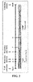
Fig. 1 represents an outline of the method for detecting rearranged TCRA genes. In brief, 34 Vα primers were designed in order to amplify all the arrangements of the Vα segments in the human TCRA gene locus, which lies in a region of more than 1,000 kb on chromosome 14 (14q11.2). In addition, 12 Jα primers were designed, downstream of the sequence of each of the segment Jα1, Jα6, Jα10, Jα17, Jα23, Jα29, Jα32, Jα36, Jα41, Jα48, Jα53, or Jα58, such that 3 to 7 different Jα segments were able to be amplified. All the Jα primers were mixed in one tube, and each of the Vα primers was used with the Jα-primers mix to perform 34 PCR reactions per sample, thereby to detect a rearranged TCRα gene. -
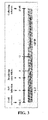
Fig. 2 is a schematic representation showing the process for producing T-cell-derived iPS (T-iPS) cells from peripheral blood T cells using retrovirus vectors, each encoding OCT3/4, SOX2, or KLF4. In the drawing, the tapering portion (a period of 7 to 11 days after the start of activation of T cells) represents a period during which the medium was gradually replaced to human iPS medium. -
Fig. 3 is a schematic representation showing the process for producing T-iPS cells from cells of a CTL clone (H25-4T cells) using an SeV polycistronically encoding OCT3/4, SOX2, and KLF4, and an SeV encoding SV40LTAg. In the drawing, the tapering portion (a period of 9 to 12 days after the start of T cell activation) represents a period during which the medium was gradually replaced to human iPS medium. -
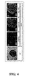
Fig. 4 represents pictures showing the results of observation under a microscope of the expression of alkaline phosphatase (AP) activity and pluripotency markers (SSEA-4, Tra-1-60, and Tra-1-81) in T-iPS cells (H254SeVT-3), cells of a cell line produced from H25-4T cells. In each of the pictures, the scale bar corresponds to 200 µm, and the pictures b to d show the result of counterstaining the nucleus with DAPI (in the pictures, blue-emitting portions). -
Fig. 5 represents pictures showing the results of observation under a microscope of the expression of AP activity and pluripotency markers (SSEA-4, Tra-1-60, and Tra-1-81) in iPS cells (TkT3V1-7), cells of a cell line produced from peripheral blood T cells. In the picture a, the scale bar corresponds to 500 µm. In each of the pictures b to e, the scale bar corresponds to 200 µm and the results of counterstaining the nucleus with DAPI are shown (in the pictures, blue-emitting portions). -
Fig. 6 represents pictures showing the results of an analysis by RT-PCR of the expression of pluripotent genes in TkT3V1-7 cells. In the drawing, "Tg" indicates the expression of the exogenous retrovirus-introduced gene, "endo" indicates the expression of the endogenous expressing gene, and "total" indicates the expression of the exogenous retrovirus-introduced gene and the endogenous expressing gene. "PB CD3+" indicates the results in the originating peripheral blood T cells of TkT3V1-7 cells, "KhES3" indicates the results (as positive control) in ES cells, which are pluripotent stem cells, and "Water" indicates the results (as negative control) of RT-PCR without added template DNA. In the RT-PCR, GAPDH was used as internal standard. -
Fig. 7 represents pictures showing the results of detection by RT-PCR of the remaining SeV RNAs (four factors cistronically expressed (tetracistronic factors): OCT4, SOX2, KLF4, and c-MYC, and T antigen). In the drawing, "SeVp(KOSM302L)" and "SeV18+SV40/TS15ΔF" indicate the results in the respective SeVs which were used for establishing the H254SeVT-3, and "Water" indicates the results of RT-PCR without added template DNA (as negative control). In addition, "n.a." indicates "not assessed." In the RT-PCR, GAPDH was used as internal standard. -
Fig. 8 represents scatter plots showing the results of a comprehensive analysis of gene expression using cDNA microarrays. The left panel shows the results of a comprehensive comparison of gene expression between CD4+ T cells (PB CD4+ T-cells) and TkT3V1-7 cells using cDNA microarrays. The right panel represents a scatter plot showing the results of a comprehensive comparison of gene expression between ES cells (KhES3) and TkT3V1-7 cells using cDNA microarrays. In each of the plots, the two parallel lines indicate that there was a fivefold difference between the corresponding samples. -
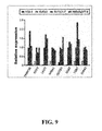
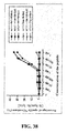
Fig. 9 represents a graph showing the results of an analysis by quantitative PCR of the expression of pluripotent genes in H25-4, KhES3, TkT3V1-7, and H254SeVT-3 cells. In each PCR reaction, the amount of expression of 18S rRNA was used as internal standard. In the graph, the abscissa axis represents, from left to right, the respective results for H25-4, KhES3, TkT3V1-7, and H254SeVT-3 cells. The ordinate axis represents a relative amount of expression when the amount of expression of each of the genes in KhES3 cells was set to be 1.0. "N.D." indicates "below detection limit (not detected)." -
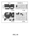
Fig. 10 is a representation showing the results of an analysis by bisulfite sequencing of the promoter region of OCT3/4 and NANOG in H25-4 and H254SeVT-3 cells. In the drawing, the open circles and the filled circles indicate a non-methylated CpG dinucleotide and a methylated CpG dinucleotide, respectively, and "%Me" indicates the percentage of methylation in the respective regions. -
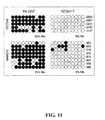
Fig. 11 is a representation showing the results of an analysis by bisulfite sequencing of the OCT3/4 and NANOG promoters in PB CD3+ and TkT3V1-7 cells. In the drawing, the open circles and the filled circles indicate a non-methylated CpG dinucleotide and a methylated CpG dinucleotide, respectively, and "%Me" indicates the percentage of methylation in the respective regions. -
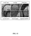
Fig. 12 represents micrographs showing representative results obtained by preparing sections from TkT3V1-7-derived teratomas formed in the testis of NOG mice, followed by HE staining of the sections for observation, which reveal that the teratomas comprise anatomical structures derived from the three germ layers. In these pictures, there are observed glands/ducts and gut-like mucosa as endoderm-derived anatomical structures, cartilage and striated muscle as mesoderm-derived anatomical structures, and neural plate and pigment epithelium as ectoderm-derived anatomical structures. In each of the micrographs, the scale bar corresponds to 100 µm. -
Fig. 13 represents micrographs showing representative results obtained by preparing sections from H254SeVT-3-derived teratomas formed in the testis of NOG mice, followed by HE staining of the sections for observation, which reveal that the H254SeVT-3 cells differentiated into an endoderm-derived cell lineage (goblet cells in gut-like epithelium), a mesoderm-derived cell lineage (smooth myocytes in muscle tissue), and an ectoderm-derived cell lineage (retinal cells in pigment epithelium). In each of the micrographs, the scale bar corresponds to 100 µm. -
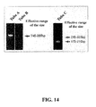
Fig. 14 represents pictures showing a result that a TCRB gene rearrangement in the genome of TkT3V1-7 cells was detected by multiplex PCR analysis. Tubes A and B indicate that the Vβ-(D)Jβ has been rearranged and Tube C indicates that the D-Jβ has been rearranged. -
Fig. 15 represents a picture showing a result that a TCRA gene rearrangement (V-Jα rearrangement) in the genome of TkT3V1-7 cells was detected by multiplex PCR analysis. -
Fig. 16 represents pictures showing a result that a TCRB gene rearrangement in the genome of H254SeVT-3 cells was detected by multiplex PCR analysis. Tubes A and B indicate that the Vβ-(D)Jβ has been rearranged and Tube C indicates that the D-Jβ has been rearranged. -
Fig. 17 represents a picture showing a result that a TCRA gene rearrangement (V-Jα rearrangement) in the genome of H254SeVT-3 cells was detected by multiplex PCR analysis. -
Fig. 18 represents dot plots showing the results of analyzing the course of development from TkT3V1-7-derived CD34+ hematopoietic stem/progenitor cells to human T-lineage cells by flow cytometry as mentioned below. In brief, in order to examine the hematopoietic differentiation potential of T-iPS cells, TkT3V1-7 cells were cultured with VEGF, SCF, and FLT-3L on C3H10T1/2 feeder cells. Onday 14 of the culture, the TkT3V1-7-derived CD34+ hematopoietic stem/progenitor cells thus generated (T-iPS-sac cells) were transferred onto OP9 (OP9-DL1) feeder cells expressing delta-like 1. Re-differentiating cells on the OP9-DL1 cells were collected, and then the expression of CD34, CD38, CD7, CD5, CD1a, CD45RA, and CD45 on the cellular surface of TkT3V1-7-derived cells was analyzed by flow cytometry every week for a period of 22 to 36 days after the induction of re-differentiation ("1 wks" to "3 wks" in the drawing). -
Fig. 19 represents dot plots showing the results of analyzing the course of development from H254SeVT-3-derived T-iPS-sac cells to human T-lineage cells by flow cytometry as mentioned below. In brief, in order to examine the hematopoietic differentiation potential of T-iPS cells, H254SeVT-3 cells were cultured with VEGF, SCF, and FLT-3L on C3H10T1/2 feeder cells. Onday 14 of the culture, the TkT3V1-7-derived T-iPS-sac cells thus generated were transferred onto OP9-DL1 feeder cells. Re-differentiating cells on the OP9-DL1 cells were collected, and then the expression of CD34, CD38, CD7, CD5, CD1a, CD45RA, and CD45 on the cellular surface of H254SeVT-3-derived cells was analyzed by flow cytometry every week for a period of 15 to 36 days after the induction of re-differentiation ("0 wks" to "3 wks" in the drawing). -
Fig. 20 represents dot plots showing the results of an analysis by flow cytometry of the existence of T-iPS-cell-derived CD4/8 DP cells established in vitro.Fig. 20a shows the results of an analysis by flow cytometry of the expression of CD45, CD56, CD3, TCRαβ, CD4, and CD8 on the cellular surface of TkT3V1-7-derived cells in a period of 35 to 42 days after the induction of re-differentiation.Fig. 20b shows the results of an analysis by flow cytometry of the expression of CD45, CD56, CD3, TCRαβ, CD4, and CD8 on the cellular surface of H254SeVT-3-derived cells in a period of 35 to 42 days after the induction of re-differentiation. Shown in the drawings are representative results of the results of at least three independent experiments. -
Fig. 21 represents pictures showing the results of RT-PCR amplification of the TCRA and TCRB genes expressed in DN-cell- or DP-cell-derived cDNA libraries synthesized by SMART method. In the drawing, "Water" indicates the results of RT-PCR without added template DNA (as negative control). GAPDH was used as internal standard in each PCR. -
Fig. 22 represents pictures showing the results of an analysis by RT-PCR of the expression of RAG1 and RAG2 in DN cells and DP cells. In the drawing, "Water" indicates the results of RT-PCR without added template DNA (as negative control). Samples used as a PCR template were prepared, based on the expression of GAPDH. -
Fig. 23 represents dot plots showing the results of an analysis by flow cytometry of floating cells over OP9-DL1 cells, which were collected on day 42 after the start of the induction of re-differentiation. DN cells and DP cells were sorted from fractionated subsets of CD45+, CD56-, CD3+, TCRαβ+, CD2+, and CD5+ cells, by gating in advance for CD1a- and CD1a+, respectively. -
Fig. 24 is a schematic representation showing the method for producing human CD8 SP cells having antigen specificity according to the present invention, in which the process of re-differentiation from T-iPS cells into the human CD8 SP cells is shown. -
Fig. 25 represents dot plots showing results of an analysis by flow cytometry of the phenotype of cells obtained by re-differentiation of T-iPS cells by the method according to the present invention (cells ondays 50 to 60 after the start of the induction of re-differentiation, as shown inFig. 24 ). Shown in the drawings are representative results of the results of at least three independent experiments.Figs. 25a and 25b show the results of an analysis of the phenotype as T cell, andFigs. 25c and 25d show the results of an analysis of the phenotype as memory cell. InFig. 25b , it is also shown that there was no contamination with CD3+/CD8+ cells from PBMCs, because a uniform expression of HLA-A24 was observed. -
Fig. 26 represents dot plots showing the results of (upper panel) an analysis of cells obtained by re-differentiation of T-iPS cells by the method according to the present invention (cells ondays 50 to 60 after the start of the induction of re-differentiation, as shown inFig. 24 ), by flow cytometry using A24/Nef-138-8(wt) teratoma, and a re-analysis of T cells expanded by culturing for 14 or more days teratoma-positive cells sorted by FACS or magnetic selection, again by flow cytometry using A24/Nef-138-8(wt) teratoma. -
Fig. 27 represents pictures showing that TCR mRNAs were detected by PCR using respective cDNA libraries, synthesized by the SMART method, of various CD8 SP cells (reT-1, reT-2.1, and reT-3) responded to A24/Nef-138-8(wt) teratoma. In the drawing, "Water" indicates the results of RT-PCR without added template DNA (as negative control). GAPDH was used as internal standard in each PCR. -
Fig. 28 represents a graph showing the results of comparing the amount of expression of major cell surface molecules among CD4+ cells, CD8+ cells, reT-2.1 cells, and H25-4 cells, using quantitative PCR. In each PCR reaction, the amount of expression of 18S rRNA was used as internal standard. In the drawing, the ordinate axis represents an expression ratio when the amount of expression of each of these genes in the CD8+ cells was set to be 1.0. The abscissa axis represents, from left to right, the results of the respective genes in CD4+ cells, CD8+ cells, reT-2.1 cells, and H25-4 cells. -
Fig. 29 represents a graph showing the results of comparing the amount of expression of major cytolytic molecules among CD4+ cells, CD8+ cells, reT-2.1 cells, and H25-4 cells, using quantitative PCR. In each PCR reaction, the amount of expression of 18S rRNA was used as internal standard. In the drawing, the ordinate axis represents an expression ratio when the amount of expression of each of these genes in the CD8+ cells was set to be 1.0. The abscissa axis represents, from left to right, the results of the respective genes in CD4+ cells, CD8+ cells, reT-2.1 cells, and H25-4 cells. -
Fig. 30 represents a graph showing the results of comparing the amount of expression of major transcription factors and signaling molecules among CD4+ cells, CD8+ cells, reT-2.1 cells, and H25-4 cells, using quantitative PCR. In each PCR reaction, the amount of expression of 18S rRNA was used as internal standard. In the drawing, the ordinate axis represents an expression ratio when the amount of expression of each of these genes in the CD8+ cells was set to be 1.0. The abscissa axis represents, from left to right, the results of the respective genes in CD4+ cells, CD8+ cells, reT-2.1 cells, and H25-4 cells. -
Fig. 31 represents a heat map showing correlation coefficients among samples obtained by a comprehensive analysis of gene expression using cDNA microarrays. -
Fig. 32 represents a heat map showing a group of genes exhibiting a more than three-times difference in their expression amount, relative to NK cells, which was obtained by a comprehensive analysis of gene expression using cDNA microarrays. In the drawing, the reds and greens indicate an increase and a decrease in expression, respectively. -
Fig. 33 represents a graph showing the expansion ratio of reT-1, reT-2.2, and reT-3 cells at two weeks after the respective cells were stimulated with PHA, IL-7, and IL-15. The ordinate axis represents an expansion ratio when the number of cells upon the stimulation was set to be 100 for the respective cells. -
Fig. 34 represents dot plots showing the results of an analysis by flow cytometry of the expression of CD45RA, CCR7, CD27, and CD28 on the cellular surface of the cells purified with A24/Nef-138-8(wt) tetramer and expanded (reT-2.1 and reT-3). In the drawings, the data of reT-2.1 cells are shown as representative results. -
Fig. 35 represents a graph showing the relative telomere length (RTL) as determined by flow-FISH. -
Fig. 36 represents histograms showing the results of an analysis of the secretion of cytolytic molecules in reT-2.1 cells when stimulated with α-CD3/CD28 beads. The left panel is a histogram showing the amount of intracellular production of granzyme B. The open histogram represented by the grey line indicates the reactivity of cells which were not stimulated and an anti-granzyme B antibody, the open histogram represented by the black line indicates the reactivity of cells which were stimulated and an anti-granzyme B antibody, and the closed histogram indicates the reactivity of cells which were stimulated and an antibody used as a negative control (an antibody of the same isotype as that of the anti-granzyme B antibody). The right panel is a histogram showing the results of a CD107a mobilization assay in reT-2.1 cells when stimulated with α-CD3/CD28 beads. The open histogram represented by the grey line indicates the reactivity of cells which were not stimulated and an anti-CD107a antibody, the open histogram represented by the black line indicates the reactivity of cells which were stimulated and an anti-CD107a antibody, and the closed histogram indicates the reactivity of cells which were stimulated and an antibody used as a negative control (an antibody of the same isotype as that of the anti-CD107a antibody). -
Fig. 37 represents a graph showing the results of an analysis by ELISPOT of IFN-γ produced in response to stimulation with Nef-138-8(wt). Shown in the drawing are representative results of the results of at least three independent experiments. The ordinate axis represents the number of spots formed in 500 cells. -
Fig. 38 represents a graph showing the results of a 51Cr release assay using various concentrations of epitope peptides, including Nef-138-8(wt). The 51Cr release assay was performed by reacting effector cells and 51 Cr-incorporated target cells at a ratio of 5: 1. The ordinate axis represents the percentage of specific 51 Cr-release (%). -
Fig. 39 represents an outline of the method for producing a human CD4 SP cell having antigen specificity from a T-iPS cell. -
Fig. 40 represents dot plots showing the result of an analysis by flow cytometry of the phenotype of the cells obtained by re-differentiation of T-iPS cells and the cells ondays 50 to 60 after the start of the induction of re-differentiation, as shown inFig. 39 ), which is the result of an analysis of phenotype of the obtained T cells with several surface expression markers and the result of an analysis by flow cytometry using A24/Nef-138-8(wt) tetramer and was a representative result among the at least triplicated experiments. - The invention provides a method for producing an antigen specific T cell and, in particular, a method for producing a human CD8 and/or CD4 single-positive cell having antigen specificity, which comprises the steps of: differentiating an iPS cell that has been induced from a human T cell (hereinafter referred to as "T-iPS cells"), into a CD4/CD8 double-negative cell, stimulating a T cell receptor of the CD4/CD8 double-negative cell, and differentiating the CD4/CD8 double-negative cell whose T cell receptor has been stimulated, into a single positive T cell, in particular, a CD8 single-positive cell and/or a CD4 single-positive cell.
- In the present invention, a "human" from which T cells are to be isolated is not limited in particular. Such a human may be a healthy individual, an individual with diminished immune function, or an individual suffering from a malignant tumor, infection, autoimmune disease, or the like. In cases where a CD8 SP cell or a CD4 SP cell obtained according to the present invention are used for a method for cell-based immunotherapy, a human from which T cells are to be isolated preferably has an HLA type identical to that of a human to which T cells obtained according to the present invention are to be administered, and more preferably is the same as a human to which T cells obtained according to the present invention are to be administered, from the viewpoint that rejection is not caused.
- In the present invention, a "T cell" refers to a cell on the surface of which an antigen receptor, called a T cell receptor (TCR), is expressed, and its progenitor cells, such as a pro-T cell on which a TCR (TCRαβ) is not expressed, and a pre-T cell on which a TCRβ and a pre-TCRα are associated. A "CD4/CD8 double-negative (DN) cell" refers to a T cell on which neither CD4 nor CD8 is expressed, a "CD4/CD double-positive (DP) cell" refers to a T cell on which both CD4 and CD8 are expressed, and a "CD8 single-positive (SP) cell" refers to a T cell on which only CD8, in terms of CD4 and CD8, is expressed. A "CD4 single-positive (SP) cell" refers to a T cell on which only CD4, in terms of CD4 and CD8, is expressed.
- In the present invention, a "T cell which is to be induced into an iPS cell" is of a type of T cell on which CD3 and CD8 are expressed and includes a cytotoxic T cell which is a CD8 positive. A "T cell which is to be induced into an iPS cell" is also of a type of T cell on which CD3 and CD4 are expressed and includes a T cell which is a CD4 positive. Human T cells of such a type include, for example, helper/regulatory T cells ,which are a subset of CD4-positive cells; cytotoxic T cells (CTLs), which are a subset of CD8-positive cells; naive T cells (CD45RA+CD62L+ cells); central memory T cells (CD45RA-CD62L+ cells); effector memory T cells (CD45RA-CD62L- cells); and terminal effector T cells (CD45RA+CD62L- cells). Note that the antigen specificity of T cells is provided by their antigen-specific, rearranged TCR gene. From the viewpoint of production efficacy, an antigen specific CD8 positive T cell, but not limited thereto, can be used as a human T cell to be induced into an iPS cell in order to obtain an antigen specific CD8 SP cells, and an antigen specific CD4 positive T cell, but not limited thereto, can be used as a human T cell to be induced into an iPS cell in order to obtain an antigen specific CD4 SP cells. In cases of carrying out immunotherapy as mentioned below, it is preferable that a human T cell which is differentiated from the iPS cell has the same or substantially the same antigen specificity as that exhibited by a T cell which is to be induced into the iPS cell.
- In the present invention, a "T cell which is to be induced into an iPS cell" can be isolated from a human tissue by known procedures. Such a human tissue is not limited in particular, if the tissue contains T cells of the above-mentioned type, and examples thereof include, for example, peripheral blood, a lymph node, bone marrow, thymus, spleen, umbilical cord blood, and a lesion site tissue. Among these, peripheral blood and umbilical cord blood are preferable from the viewpoints that their isolation is less invasive to the human body and that they are prepared with ease. Known procedures for isolating human T cells include, for example, flow cytometry using an antibody directed to a cell surface marker, such as CD4 or CD8, and a cell sorter, as shown in the below-mentioned Example. In addition, a desired type of T cell can be isolated using the secretion of cytokines or the expression of functional molecules as an indicator. In this case, for example, T cells secrete different cytokine, depending on whether they are of the Th1 or Th2 type, and thus T cells of a desired Th type can be isolated by selecting T cells using such a cytokine as an indicator. Similarly, cytotoxic (killer) T cells can be isolated using the secretion or production of granzyme, perforin, or the like as an indicator. An affinity column on which the desired antigen is immobilized, or the like can be used in isolating a "human T cell having antigen specificity" from a human tissue. Methods can be also employed in which "T cells having a desired antigen specificity" are purified from a human tissue using a multimer in which an MHC (major histocompatibility complex) having the desired antigen attached thereto, for example, an "MHC tetramer", or "Pro5TM MHC Class I Pentamer".
- An "iPS cell" in the present invention is a cell which is also called an induced pluripotent stem cell, and can be induced by introducing a cell reprogramming factor into T cells of the above-mentioned type. The "cell reprogramming factor" is not limited in particular, if the factor is one which is capable of conferring the pluripotency on somatic cells, alone or in cooperation with other factors for pluripotency, by its introduction into T cells of the above-mentioned type, and preferably is at least one protein selected from the group consisting of Oct3/4, c-Myc, Sox2, Klf4, Klf5, LIN28, Nanog, ECAT1, ESG1, Fbx15, ERas, ECAT7, ECAT8, Gdf3, Sox15, ECAT15-1, ECAT15-2, Fth117, Sal14, Rex1, Utf1, Tcl1, Stella, β-catenin, Stat3, and Grb2. Further, it is more preferable that among these proteins, Oct3/4, c-Myc, Sox2, and Klf4 ("4 factors") are introduced into T cells of the above-mentioned type, from the viewpoint that iPS cells can be efficiently established using a small number of factors. As is shown in
WO2011/096482 , OCT4, SOX2, KLF4, C-MYC and NANOG are preferably introduced into a human CD8 positive T cell or CD4 positive T cell, and OCT4, SOX2, KLF4, C-MYC, NANOG and LIN28 are more preferably introduced into a human CD8 positive T cell or CD4 positive T cell. In addition, it is more preferable that Oct3/4, Sox2, and Klf4 ("3 factors"), with c-Myc being not included, are introduce into T cells of the above-mentioned type, from the viewpoint of reducing the risk of malignant transformation of the resulting pluripotent stems. - In the present invention, methods for establishing an iPS cells induced from a human T cell (i.e., methods for introducing a cell reprogramming factor into T cells) are not limited in particular and known procedures can be selected and used as appropriate. For example, when a cell reprogramming factor as described above is introduced into T cells of the above-mentioned type in the form of proteins, such methods include ones using protein introducing reagents, fusion proteins with protein transfer domains (PTDs), electroporation, and microinjection. When a cell reprogramming factor as described above is introduced into T cells of the above-mentioned type in the form of nucleic acids encoding the cell reprogramming factor, a nucleic acid(s), such as cDNA(s), encoding the cell reprogramming factor can be inserted in an appropriate expression vector comprising a promoter that functions in T cells, which then can be introduced into T cells of the above-mentioned type by procedures using infection, lipofection, liposomes, electroporation, calcium phosphate co-precipitation, DEAE-dextran, microinjection, and electroporation.
- Examples of an "expression vector" in context of the present invention include viral vectors, such as lentiviruses, retroviruses, adenoviruses, adeno-associated viruses, herpes viruses, and sendai virus; and expression plasmids for animal cells. Sendai virus (SeV) is preferably used to introduce a nucleic acid(s) encoding a cell reprogramming factor as described above into T cells of the described-above type, from the viewpoints that insertion mutations are unlikely to be caused, that a high efficiency of gene introduction is achieved, and that an increased number of copies of gene(s) introduced is obtained.
- Promoters which are used in such expression vectors include, for example, SRα, SV40, LTR, CMV, RSV, HSV-TK promoters, and others. Such promoters may be ones which can regulate the expression of a gene inserted downstream of the promoter, for example, depending upon the presence or absence of an agent such as tetracycline. Expression vectors further may contain an enhancer, a poly adenylation signal, a selective maker gene (for example, neomycin resistance gene), an SV40 origin of replication, or others, in addition to the promoter.
- In the step of inducing an iPS cell from the human T cell induced from a human T cell, it is preferable that before a cell reprogramming factor as described above is introduced, T cells of the above-mentioned type are activated by stimulation with an anti-CD3 antibody and an anti-CD28 antibody in the presence of interleukin 2 (IL-2), as shown in the below-mentioned Example. The T cell can be stimulated and then activated with at least one substance selected from the group consisting of phytohemagglutinin (PHA), interleukin 2 (IL-2), alloantigen expressing cells, an anti-CD3 antibody and an anti-CD28 antibody, as shown in the below-mentioned Example. Such stimulation can be achieved, for example, by adding PHA, IL-2, an anti-CD3 antibody and/or an anti-CD28 antibody to a medium in which T cells of the above-mentioned type are then cultured for a specified period of time, as shown in the below-mentioned Example. In this case, the anti-CD3 antibody and the anti-CD28 antibody may be ones each having been attached to magnetic beads. Further, such stimulation may be achieved by culturing T cells of the above-mentioned type for a specified period of time in a culture dish to the surface of which an anti-CD3 antibody and an anti-CD28 antibody are attached, instead of adding these antibodies to the medium. Further, stimulation of T cells may be also achieved by adding an antigen peptide recognized by the human T cells, together with feeder cells to the medium.
- In order to achieve such stimulation as described above, the concentrations of PHA and IL-2 added to the medium are not limited in particular, and preferably are 1 to 100 µg/mL and 1 to 200 ng/mL, respectively. The concentrations of an anti-CD3 antibody and an anti-CD28 antibody added to the medium in such stimulation as described above are not limited in particular, and preferably are one to ten times the amount of culture of the T cells of the above-mentioned type. The concentrations of an anti-CD3 antibody and an anti-CD28 antibody attached to the surface of culture dishes in such stimulation as described above are not limited in particular, and their concentrations in coating processes preferably are 0.1 to 10µg/mL, more preferably 1 to 100 µg/mL for an anti-CD3 antibody and 0.1 to 10 µg/mL for an anti-CD28 antibody.
- The culture period for such stimulation is not limited in particular, if the culture period is a period of time which is sufficient to stimulate T cells of the above-mentioned type and which allows the T cells to proliferate to a number of cells which is necessary for introduction of a cell reprogramming factor as described above, and usually is 1 to 14 days, or 1 to 6 days, although the culture period may be one day. From the viewpoint of the efficiency of gene introduction, the culture period is preferably 2 to 7 days and more preferably 2 to 4 days. Further, it is preferable that the culture is carried out in culture dishes having RetroNectin coated thereon, from the viewpoint that the efficiency of gene introduction is increased.
- Media in which T cells of the above-mentioned type are cultured and to which PHA, IL-2, an anti-CD3 antibody, and/or an anti-CD28 antibody, and others are added can be, for example, known media suitable for culturing the T cells, more specifically Roswell Park Memorial Institute (RPMI) 1640 medium, minimal essential medium (α-MEM), Dulbecco's modified Eagle medium (DMEM), F12 medium, and others containing other cytokines and human serum. These media may be supplemented with amino acids necessary for culture (for example, L-glutamine), and antibiotics (for example, streptomycin, penicillin), besides PHA, IL-2, an anti-CD3 antibody and/or an anti-CD28 antibody. IL-7 and IL-15 are preferably added to a medium, from the viewpoint that apoptosis is suppressed. The concentrations of IL-7 and IL-15 added are not limited in particular, and preferably are 1 to 100 ng/mL for each of IL-7 and IL-15.
- Conditions in or after "introducing a cell reprogramming factor into T cells" in the present invention are not limited in particular. However, the T cells into which a cell reprogramming factor as described above has been introduced are preferably cultured on a layer of feeder cells. These feeder cells are not limited in particular, and include, for example, mouse embryonic fibroblasts (MEFs), STO cells, and SNL cells in which the cell division has been arrested by irradiation or antibiotic treatment.
- Further, in the process of inducing from T cells of the above-mentioned type into iPS cells, it is preferable that basic fibroblast growth factor (bFGF) is added to the medium in or after introducing a cell reprogramming factor as described above into the T cells, from the viewpoint that cell differentiation is suppressed.
- In addition, in order to achieve a higher efficiency of establishing iPS cells, it is preferable that a histone deacetylase (HDAC) inhibitor (for example, valproic acid (VPA), trichostatin A, sodium butyrate, MC 1293, M344, and other low-molecular inhibitors; an siRNA directed to HDAC), a G9a histone methyltransferase inhibitor (for example, BIX-01294 and other low-molecular inhibitors; an siRNA directed to G9a), or a p53 inhibitor (for example, Pifithrin-α (PFT-α) and other low-molecular inhibitors; an siRNA directed to p53) is added to the medium in or after introducing a cell reprogramming factor as described above into T cells of the above-mentioned type.
- Further, in "introducing a cell reprogramming factor as described above into T cells," it is preferable that, when a virus vector is used, protamine sulfate is added to the medium, from the viewpoint of facilitating binding of the vector to T cells of the above-mentioned type.
- In addition, it is preferable that in concert with the transition from T cells of the above-mentioned type to iPS cells, culturing is performed while the medium is gradually replaced from a known medium suitable for culturing the T cells to a medium suitable for culturing the iPS cells, as shown in the below-mentioned Example. As a medium suitable for such culturing of the iPS cells, known media can be selected and used as appropriate; such media include, for example, Dulbecco's modified Eagle medium/F12 medium containing KnockOut Serum Replacement, L-glutamine, non-essential amino acids, 2-mercaptoethanol, b-FGF, and the like (human iPS cell medium).
- As shown in International Publication No.
WO 2011/096482 , in cases of inducing human CD8-positive T cells or CD4-positive T cell into iPS cells, it is preferable that after transferring onto the above-mentioned feeder cells, culturing is performed under conditions of low oxygen concentrations (an oxygen concentration of 5%, for example) in a medium to which 1 to 1,000 µM of a ROCK inhibitor is further added, and it is also preferable that 1 to 1,000 µM of an MEK inhibitor (for example, PD0325901) and 1 to 1,000 µM of a GSK3 inhibitor (for example, CHIR99021) are added to the medium until there is formation of colonies as mentioned below, from the viewpoint that a higher efficiency of induction into iPS cells is achieved, as shown in the below-mentioned Example. - Selection of iPS cells thus induced from T cells of the above-mentioned type (hereinafter, also referred to as "T-iPS cells") can be performed by selecting known procedures as appropriate. Such known procedures include, for example, methods in which selection is performed by observation under a microscope of the morphology of ES/iPS-cell-resembling colonies, as shown in the below-mentioned Example, and methods in which selection is performed using recombinant T cells in which a drug-resistance gene or reporter gene (GFP gene or the like) has been targeted to the locus of a gene known to be expressed specifically in iPS cells (for example, a cell reprogramming factor as described above), whereby the drug resistance or reporter activity is used as an indicator.
- The fact that cells thus selected are iPS cells (i.e., T-cells) can be ascertained, for example, by methods in which the expression in the selected cells of markers which are specific in undifferentiated cells (such as ALP, SSEA-4, Tra-1-60, and Tra-1-81) is detected by means of immunostaining, RT-PCR, and others, or in which the selected cells are implanted into mice and then observation is made as to whether the formation of teratoma is caused by the implanted cells, as shown in the below-mentioned Example. It can be confirmed by detecting rearrangement of TCR genes by means of genomic PCR that the cells thus selected are derived from T cells, as shown in the below-mentioned Example.
- The time at which these cells are selected and harvested can be determined as appropriate with observing the condition of colony growth, and approximately 10 to 40 days, preferably 14 to 28 days, after a cell reprogramming factor as described above were introduced into T cells of the above-mentioned type. Culture conditions preferably are those of 5% CO2 and at 35 to 38°C, more preferably 37°C, unless otherwise specified above.
- In order to differentiate the iPS cells (T-iPS cells) which have been induced from human T cells into CD4/CD8 DN cells, it is preferable that the T-iPS cells are first cultured on feeder cells (preferably stroma cells, more preferably human stroma cells) in a medium which may contains cytokines, which is optionally added, serum (for example, fetal bovine serum), insulin, transferin, sodium selenite, L-glutamine, α-monothioglycerol, ascorbic acid, or the like, from the viewpoint of facilitating the induction of differentiation into mesodermal cells. Stroma cells which are to be used are preferably OP9 cells and 10T1/2 cells, such as C3H10T1/2 cells, treated by irradiation or the like, from the viewpoint of facilitating the induction of differentiation into hematopoietic cells. In cases of efficiently inducing the T-iPS cells into CD34-positive hematopoietic progenitor cells, the cytokine to be added into a medium is preferably at least one cytokine selected from the group consisting of VEGF, SCF, TPO, SCF, and FLT3L, and more preferably VEGF, SCF and TPO, or VEGF, SCF and FLT3L. Media include, for example, X-VIVO medium, Iscove's Modified Dulbecco's Medium (IMDM medium), α-MEM, or DMEM; IMDM medium is preferable from the viewpoint of resulting in a high efficiency of formation of sac structures containing hematopoietic progenitor cells and the like (also referred to as "T-iPS-sacs" or "ES-sacs"). The culture period of T-iPS cells preferably is a period of time until T-iPS-sacs are formed, and preferably 8 to 14 days, more preferably 10 to 14 days, after culturing of the T-iPS cells is started. Culture conditions are not limited in particular, and preferably are conditions of 5% CO2 and at 35 to 38°C, more preferably 37°C. In addition, it is more preferable that the T-iPS cells are cultured under conditions of low oxygen concentrations (an oxygen concentration of 5 to 20%, for example), from the viewpoints that a high efficiency of formation of T-iPS-sacs is achieved and that an increased number of blood cells contained in the T-iPS-sacs is obtained.
- In order to differentiate a T-iPS cell into CD4/CD8 DN cell, cells which are contained in T-iPS-sacs obtained as described above, such as hematopoietic progenitor cells, CD34-positive cells, and blood cells, preferably are cultured on feeder cells (preferably stroma cells and more preferably human stroma cells) in a medium containing cytokines and serum (such as, FBS). Cells encapsulated within the T-iPS-sacs can be separated by physical means, for example, by passing the cells through sterilized sieve-like apparatus (for example, cell strainers and others). Stroma cells used in the above-mentioned culture preferably are OP9-DL1, OP9-DL4, 10T1/2/DL4, or 10T1/2/DL1 cells treated by irradiation or the like, from the viewpoint that the induction of differentiation into T lymphocytes is achieved through notch signals. The cytokines which are added to the medium include, for example, IL-7, FLT3L, VEGF, SCF, TPO, IL-2, and IL-15. One or more cytokines selected from a group consisting of SCF, TPO, FLT3L and IL-7 and a combination of all of these cytokines can be added to the medium used in the induction of differentiation into CD4 SP cells. Among these cytokines, preference is given to IL-7 and FTL3 from the viewpoint of supporting the T cell differentiation at the early stage. However, it is preferable that SCF is not contained in the medium, from the viewpoint that the differentiation of the T-iPS cells into NKT cells is suppressed. The concentration of IL-7 added to the medium is preferably 0.1 to 9.5 ng/mL, more preferably 1 to 4 ng/mL, from the viewpoints of easily generating CD3-positive CD56-negative T-lineage cells and facilitating the induction of differentiation into CD8 SP T cells or CD4 SP T cells. The concentration of FLT3L added to the medium is preferably 0.1 to 100 ng/mL from the viewpoints of improving the proliferation of blood cells. Media include, for example, α-MEM medium, DMEM medium, and IMDM medium, with α-MEM medium being preferable from the viewpoint of easily maintaining feeder cells. These media may be supplemented with amino acids necessary for culture (for example, L-glutamine), and antibiotics (for example, streptomycin, penicillin), besides IL-7 and FLT3.
- The culture period described above for culturing cells which are contained in the T-iPS-sacs is preferably a period of time until T cell receptor (TCR) is expressed on the cellular surface of CD4/CD8 DN cells obtained by differentiation as described above, and is preferably 14 to 28 days after culturing of cells contained in the T-iPS-sacs is started. Culture circumstances are not limited in particular, and preferably are under conditions of 5% CO2 and at 35 to 38°C, more preferably 37°C.
- Whether or not T cell receptor (TCR) is expressed on the cellular surface of the resulting CD4/CD8 DN cells can be assessed by flow cytometry using an anti-TCRαβ antibody, an anti-CD3 antibody, an anti-CD4 antibody, and an anti-CD8 antibody, as shown in the below-mentioned Comparative Example 1 (see
Fig. 20 ). - As shown in the below-mentioned Comparative Example 1, the present inventors have demonstrated for the first time that, in T cells which have been redifferentiated from the T-iPS cells, the CD4/CD8 DP stage exhibits more intense expression of RAG1 and RAG2 than the CD4/CD8 DN stage. Further, the present inventors have demonstrated for the first time that, in TCRA mRNAs from T cells into which the T-iPS cells have been redifferentiated, the DP stage has a higher frequency of having a TCR-gene rearrangement pattern different from that in their originating human T cells than the DN stage has. These strongly suggest that further rearrangement of the TCRA gene (receptor revision) is caused by RAG1 and RAG2 in the process of redifferentiating the T-iPS cells into T cells, particularly in the process leading from the CD4/CD8 DN stage to the CD4/CD8 DP stage.
- As shown in the below-mentioned Comparative Example 1, the present inventors have also demonstrated for the first time that, in T cells obtained by redifferentiation of the T-iPS cells, TCR is expressed even at the DN stage. In a normal living body, T cells do not express TCR at the DN stage. Therefore, it was surprised that TCRs are expressed in DN cells obtained by differentiation of the T-iPS cells.
- Until now, it has been known that, in the process of positive selection, TCR signals via peptide-MHC complexes arrest the expression of the RAG genes and suppress further rearrangement of the TCR gene. It has been also shown that TCR-signal-like signals by anti-CD3 antibodies exert similar effects (i.e., suppressing further rearrangement of the TCR gene). The present inventors have succeeded in suppressing further rearrangement of the TCR gene by stimulating the TCR which is expressed on DN cells induced from the T-iPS cells, and based on this, improving the frequency of appearance of T cells having the same rearrangement pattern of the TCR gene as in their originating human T cells.
- Therefore, in the method for producing a human CD8 SP cell and/or human CD4 SP cell having antigen specificity according to the present invention, stimulating T-iPS-cell-derived CD4/CD8 DN cells via the TCR expressed on their cellular surface can suppress further rearrangement of the TCR gene, and eventually makes it possible that in CD8 SP cells and/or CD4 SP cells obtained by redifferentiation, there is achieved a highly increased frequency of appearance of T cells having the same rearrangement pattern of the TCR gene as in their originating human T cells.
- As a method for stimulating the T cell receptor of T-iPS-cell-derived CD4/CD8 DN cells, preference is given to methods of contacting T-iPS-cell-derived CD4/CD8 DN cells with at least one substances selected from the group consisting of PHA, an anti-CD3 antibody, an anti-CD28 antibody, a cell which expresses a complex of an antigen peptide to which the originating human T cells of the T-iPS cells binds specifically and an HLA restricted to the T cell receptor, an MHC multimer having the antigen peptide attached thereto, PMA, and ionomycin; more preference is given to methods in which cells expressing a specific peptide/HLA complex are contacted with T-iPS-cell-derived CD4/CD8 DN cells, from the viewpoint that physiological stimulation is applied. From the viewpoint of attaching importance to the uniformity of stimulation, more preference is given to methods in which antibodies or reagents are contacted.
- Contacting methods are carried out, for example, by culturing the above-mentioned T cells for a specified period of time with PHA and others added to the medium, as shown in the below-mentioned Example. The anti-CD3 antibody and the anti-CD28 antibody may be ones each having been attached to magnetic beads. Further, stimulation may be achieved by culturing the above-mentioned T cells for a specified period of time in a culture dish to the surface of which an anti-CD3 antibody and an anti-CD28 antibody are attached, instead of adding these antibodies to the medium. Further, stimulation may be also achieved by adding the above-mentioned antigen peptide with feeder cells to the medium.
- In order to stimulate the TCR of CD4/CD8 DN cells, the concentration of PHA added to the medium is not limited in particular, and preferably is 1 to 100 µg/mL. The concentrations of an anti-CD3 antibody and an anti-CD28 antibody added to the medium are not limited in particular, and preferably are one to ten times the amount of culture of the above-mentioned T cells. The concentrations of an anti-CD3 antibody and an anti-CD28 antibody at which these antibodies are attached to the surface of culture dishes for stimulating the TCR of CD4/CD8 DN cells are not limited in particular, and their concentration in coating processes preferably are 0.1 to 100 µg/mL for an anti-CD3 antibody and 0.1 to 10 µg/mL for an anti-CD28 antibody.
- The period for culturing cells contained in the T-iPS-sacs preferably includes a period of time around which T cell receptor (TCR) is expressed on the cellular surface of CD4/CD8 DN cells obtained by differentiation as described above, and preferably is 7 to 29 days after culturing of cells contained in the T-iPS-sacs is started. Culture circumstances are not limited in particular, and preferably are under conditions of 5% CO2 and at 35 to 38°C, more preferably 37°C.
- In such a culture period, a glycogen synthase kinase-3β (GSK3β) inhibitor may be added to the medium, from the viewpoint of enhancing the self-renewal ability and the production of CD8+ memory stem cells (stem-cell-like memory T cells (TSCMs)) having a multipotency capable of differentiating into effector memory T cells and effector T cells. Such a GSK3β inhibitor is any inhibitor which can activate the Wnt signaling pathway by suppressing the phosphorylation of β-catenin by GSK3β, and includes, for example, a 4,6-disubstituted pyrrolopyrimidine (TWS119). For TSCMs, see Luca Gattinoni et al., Nature Medicine, 2011, Vol. 17, pp.1290-1298. For the relationship between the inhibition of GSK3β and the enhancement of TSCM production, see Luca Gattinoni et al., Nature Medicine, 2009, Vol. 15, pp.808-813.
- As shown in the below-mentioned Example section, it has been demonstrated that in the above-mentioned step, the stimulation of T-iPS-cell-derived CD4/CD8 DN cells through the TCR expressed on their cellular surface makes it possible to suppress further rearrangement of the TCRA gene (receptor revision) which may be caused as the T-iPS cells differentiate from the DN stage to the DP stage and further to the SP stage such as the CD8 SP stage or the CD4 SP stage.
- Therefore, in the method for producing a human CD8 SP cell or a human CD4 SP cell having antigen specificity according to the present invention, stimulation of T-iPS-cell-derived CD4/CD8 DN cells via the TCR expressed on their cellular surface, followed by differentiation of the stimulated CD4/CD8 DN cells into CD8 SP cells or CD4 SP cells, makes it possible that, in CD8 DP cells or CD4 SP cells obtained by redifferentiation, there is achieved a highly increased frequency of appearance of T cells having the same rearrangement pattern of the TCR gene as in their originating human T cells.
- As a result of the above-mentioned "step of stimulating the T cell receptor of the CD4/CD8 DN cell", there are CD4/CD8 DN cells among the T-iPS-cell-derived T-lineage cells, but some of the T-lineage cells have been differentiated into CD4/CD8 DP cells. Therefore, the "step of differentiating the CD4/CD8 DN cell whose T cell receptor has been stimulated, into a single positive cell" includes differentiating the CD4/CD8 DN cells whose T cell receptor has been stimulated into CD8 SP cells or CD4 SP cells, and differentiating CD4/CD8 DP cells whose TCR has been stimulated at the DN stage into CD8 SP cells or CD4 SP cells.
- In the present invention, it is preferable that in order to differentiate the CD4/CD8 DN cells whose T cell receptor has been stimulated into CD8 SP cells or CD4 SP cells, the stimulated CD4/CD8 DN cells are cultured in a medium containing cytokines, serum (for example, human serum), and others. The cytokines which are added to the medium are any ones which allow the stimulated CD4/CD8 DN cells to differentiate into CD8 SP cells or CD4 SP cells, and include, for example, IL-7, IL-15, and IL-2. Among these cytokines, IL-7 and IL-15 are preferably added in combination, from the viewpoints of facilitating the production of CD8 lineage cells and further memory-type CD8+ T cells in the differentiation into CD8 SP cells. The concentrations of IL-7 and IL-15 added are not limited in particular, and preferably are 1 to 20 ng/mL. Media include, for example, RPMI-1640 medium, X-VIVO medium, DMEM medium, and α-MEM medium, with RPMI-1640 medium and X-VIVO medium being preferable from the viewpoint that both media are more suitable for growth of blood cells. These media may be supplemented in addition to IL-7 and IL-15, with amino acids necessary for culture (for example, L-glutamine), antibiotics (for example, streptomycin, penicillin), and cytokines other than IL-7 and IL-15.
- In such culture, the CD4/CD8 DN cells may be co-cultured with feeder cells. Feeder cells are not limited in particular, and preferably peripheral blood mononuclear cells (PBMCs) from the viewpoint of facilitating the differentiation into CD8 SP cells or CD4 SP cells and their proliferation through cell contact and the like. Such PBMCs are preferably allo (allogeneic) to the CD4/CD8 DN cells, from the viewpoint that the TCR is effectively stimulated. Such PBMCs are preferably auto (autologous) to the CD4/CD8 DN cells, from the viewpoint that survival is conferred while excessive stimulation of the TCR is prevented. Further, it is more preferable to use peripheral blood mononuclear cells presenting an antigen peptide to which the originating human T cells of the CD4/CD8 DN cells bind specifically, from the viewpoint of continuing to stimulate the TCR and to suppress further TCR rearrangement.
- The culture period for differentiating the CD4/CD8 DN cells into CD8 SP cells or CD4 SP cells is preferably two to four weeks. Culture circumstances are not limited in particular, and preferably are under conditions of 5% CO2 and at 35 to 38°C, more preferably 37°C.
- In connection with the present invention, it has been pointed out by Serwold, T. et al. in Proc Natl Acad Sci USA, 107, 18939-18943, 2010, Vol. 107, pp.18939-18943, that an unusually early TCR signaling in mouse thymus is likely to cause the occurrence of lymphoma. For this reason, the method for producing a human CD8 SP cell or CD4 SP cells according to the present invention may incorporate a system that uses a suicide gene into the cell, from the viewpoint of avoiding the occurrence of tumors resulting from activation of TCR signals at early (CD4/CD8 DN) stages. Examples of such a "system that uses a suicide gene" include, for example, systems using a gene encoding an inducible caspase 9 (iCasp 9) consisting of
human caspase 9 and a modified FK-binding protein, as described in Antonio Di Stasi et al., N Engl J Med, 2011, Vol. 365, pp.1673-1683; thymidine kinase (TK) genes, as described, for example, in Kaneko, S. et. al., BLOOD, 2009, Vol. 113, pp.1006-1015; Fabio Ciceri et al., THE LANCET, 2009, Vol. 10, pp.489-500; and Attilio Bondanza et al., blood, 2011, Vol. 24, pp.6469-6478. - The fact that CD8 SP cells or CD4 SP cells which have been subjected to differentiation induction as described above are derived from the T-iPS cells and from the originating T cells of the T-iPS cells can be ascertained, for example, by detecting the TCR gene rearrangement pattern by means of PCR for genomic DNA, as shown in the below-mentioned Example.
- The CD8 SP cells or CD4 SP cells thus produced can be isolated by selecting known procedures as appropriate. Such known procedures include, for example, flow cytometry using an antibody directed to a cell surface marker, such as CD4 or CD8, and a cell sorter, as shown in the below-mentioned Example. In cases where "T cells having a desired antigen specificity" are isolated from a human, methods can be employed in which purification of the T cells is carried out using an affinity column on which the desired antigen (for example, an antigen recognized by the originating T cells of CD8 SP cells, in cases of CD8 SP cells and an antigen recognized by the originating T cells of CD4 SP cells, in cases of CD4 SP cells) is immobilized. Methods can be employed in which "T cells having a desired antigen specificity" are purified using an MHC multimer (for example, an MHC tetramer) to which the desired antigen has been attached.
- The resulting CD8 SP cells do not express PD-1, and yet at the same time express CCR7 together with CD27 and CD28 (expression of CD27 and CD28 is one of the representative phenotypes of central memory T cells), have a longer telomere, relative to the original T cells, and possess an improved ability of self-replication. Therefore, the present invention can provide the production of T cells (for example, CD8 SP cells) having the same rearrangement pattern of the TCR gene as in the originating T cells and expressing CD27, CD28, and CCR7, but not PD-1. T cells which have been collected from a human express PD-1, and not CD27, CD28, nor CCR7, and in this respect are different from the resulting T cells, which can also apply to the CD4 SP cells obtained by the present method.
- The resulting CD8 SP cells or CD4 SP cells may be stimulated every one to two weeks, for maintaining them. Such stimulation for CD8 SP cells includes contacting the cells with at least one substance selected from the group consisting of an anti-CD3 antibody, an anti-CD28 antibody, IL-2, IL-7, IL-15, an antigen recognized by the CD8 SP cells, an MHC multimer having the antigen attached thereto, feeder cells which are allo to the CD8 SP cells, and feeder cells which are auto to the CD8 SP cells. Such stimulation for CD4 SP cells includes contacting the cells with at least one substance selected from the group consisting of an anti-CD3 antibody, an anti-CD28 antibody, IL-2, IL-7, IL-15, an antigen recognized by the CD4 SP cells, an MHC multimer having the antigen attached thereto, feeder cells which are allo to the CD4 SP cells, and feeder cells which are auto to the CD4 SP cells.
- The human CD8 SP cells or CD4 SP cells produced by the method according to the present invention have antigen-specific immune functions. In addition, the human CD8 SP cells produced by the method according to the present invention do not express PD-1, which is a marker of exhausted T cells (i.e., T cells having significantly decreased long-term survival capability, self-replication ability, and/or effector functions), and yet at the same time express CCR7 together with CD27 and CD28, have a longer telomere, relative to the original T cell, and possess an improved self-replication ability, which can also apply to the human CD4 SP cells obtained by the present method. Thus, the human T cells produced by the method according to the present invention can be used, for example, for treatment or prevention of diseases such as tumors, infections (for example, chronic infections), autoimmune failures, and others.
- Therefore, the present invention provides human T cells produced by the method according to the present invention, a pharmaceutical composition comprising the human T cells, and a method for cell-based immunotherapy using the human T cells (immunocytotherapy or immunotherapy). In particular, the present invention provides human CD8 SP cells and/or human CD4 SP cells produced by the method according to the present invention, a pharmaceutical composition comprising the human CD8 SP cells and/or CD4 SP cells, and a method for cell-based immunotherapy using the human CD8 SP cells and/or CD4 SP cells.
- The method for cell-based immunotherapy in a subject according to the present invention can be carried out, for example, in the following way. First, T cells are collected from a human, preferably a human having an identical HLA type to the subject, more preferably the subject to be treated. Second, T-iPS cells are prepared from the T cells, and subsequently, the T-iPS cells are differentiated into T cells, preferably CD4 SP cells or CD8 SP cells, having the same rearrangement pattern of the TCR gene as in their originating T cells. The resulting T cells express CCR7 together with CD27 and CD28, but not PD-1, and have a longer telomere, relative to the originating T cells, and possess an improved self-replication ability. Accordingly, the resulting cells may be identified by the expression of CD27, CD28, CCR7, and/or PD-1, if desired. The T cells thus obtained can be administered to the subject to be treated.
- In the method for cell-based immunotherapy according to the present invention, the administration of the resulting T cells to a subject to be treated is not limited in particular, and can be preferably carried out by parenteral administration, such as intravenous, intraperitoneal, subcutaneous, or intramuscular administration, more preferably intravenous administration. Alternatively, topical administration to an affected site can be also done.
- The pharmaceutical composition according to the present invention can be prepared by formulating the human CD8 SP cells or CD4 SP cells produced by the method according to the present invention into dosage forms by known pharmaceutical methods. For example, the pharmaceutical composition according to the present invention can be mainly administered parenterally, as capsules, liquids, film-coated preparations, suspensions, emulsions, injections (such as venous injections, drip injections, and the like), and others.
- In formulation into these dosage forms, the T cells according to the present invention can be combined as appropriate, with pharmaceutically acceptable carriers or media, in particular, sterile water and physiological saline, vegetable oils, resolvents, bases, emulsifiers, suspending agents, surfactants, stabilizers, vehicles, antiseptics, binders, diluents, tonicity agents, soothing agents, bulking agents, disintegrants, buffering agents, coating agents, lubricants, coloring agents, solution adjuvants, or other additives. The T cells according to the present invention may be also used in combination with known pharmaceutical compositions, immunostimulants, and others which are used for the treatment or prevention of the above-mentioned diseases.
- In administrating a pharmaceutical composition of the present invention, the dosage is selected as appropriate, depending upon the age, weight, symptoms, and physical condition of the subject, the type of composition (pharmaceutical, food/drink, etc.), and others.
- Products (pharmaceuticals) from the compositions of the present invention or their instructions are ones having affixed thereto an indication that use is made for the treatment or prevention of decrease in immune function. Here, by "having an indication affixed to a product or its instructions," it is meant that the indication has been affixed to the body, container, packaging, or the like of the product, or alternatively its instructions, package insert, advertising materials, or other printed materials disclosing product information.
- The method for cell-based immunotherapy according to the present invention comprises the steps of isolating T cells having a desired antigen specificity from a human; inducing iPS cells from the T cells having the desired antigen specificity; differentiating the iPS cells into CD4/CD8 double negative cells; stimulating the T cell receptor of the CD4/CD8 double negative cells; differentiating the CD4/CD8 double negative cells whose T cell receptor has been stimulated, into CD8 single positive cells and/or CD4 single positive cells; and administering to a human the resulting CD8 single positive cells and/or CD4 single positive cells.
- In cases of carrying out the method for cell-based immunotherapy according to the present invention, a human from which T cells are to be isolated preferably has an HLA type identical to that of a human to which CD8 single positive cells and/or CD4 single positive cells obtained by the method according to the present invention are to be administered, and more preferably is the same as a human to which CD8 single positive cells and/or CD4 single positive cells obtained by the method according to the present invention are to be administered, from the viewpoint that rejection is not caused. Regarding CD8 single positive cells and/or CD4 single positive cells to be administered, T cells produced by the method according to the present invention may be administered directly or in the form of formulated pharmaceutical compositions as described above.
- The present invention will be described more specifically below based on Examples, but is not limited to the below-mentioned Examples. Examples and Comparative Examples were carried out according to the below-mentioned methods.
- CTL strain specific for Nef138-8(wt) were established from PBMCs obtained from an HIV-1-infected patient who had an HLA-A24, as described in Kawana-Tachikawa, A. et al., J. Virol., 2002, Vol. 76, pp.11982-11988.
- Human iPS cells were established from peripheral blood T cells or CTL clones under conditions optimized for culturing T cells, as described in Takayama, N. et al., J. Exp. Med., 2010, Vol. 207, pp.2817-2830.
- In brief, peripheral blood T cells were first stimulated and activated with α-CD3/CD28 antibody-coated beads (manufactured by Miltenyi Biotec). CTL clones, on the other hand, were stimulated and activated with PHA (manufactured by Sigma-Aldrich). CTL clones were also stimulated and activated with irradiated PBMCs (alloantigen expressing cells) which were derived from an individual different from the HIV-1-infected patient. In these cases, there had not been observed significant differences in properties of the resulting T-iPS cells between stimulation with PHA and with alloantigen expressing cells.
- Reprogramming factors were introduced into the activated cells with retroviral vectors (pMXs retroviral vectors) or Sendai virus vectors, and then the cells were cultured with 10 ng/mL (200 U) IL-2, 5 to 10 ng/mL IL-7, and 5 to 10 ng/mL IL-15 (manufactured by Peprotech) in RH10 medium. During the culture period, the medium was gradually replaced to human iPS medium (manufactured by Wako) containing bFGF and others.
- The introduction of the viral vectors into the cells was carried out by centrifugation in a RetroNectin®-coated plate (manufactured by Takara). The composition of the RH10 medium is as mentioned below:
- RPMI-1640 supplemented with 10% human AB serum, 2 mM L-glutamine, 100 U/mL penicillin, and 100 ng/mL streptomycin.
- The composition of the human iPS medium is as mentioned below:
- DMEM/F12 FAM supplemented with 20% KSR, 2mM L-glutamine, 1% non-essential amino acids, 10 µM 2-mercaptoethanol, and 5 ng/mL b-FGF.
- Further, siRNA L527 (see Nishimura, K. et al., J Biol. Chem., 2011, Vol. 286, pp.4760-4771) was introduced into the established iPS clones by Lipofectamine RNAiMAX (manufactured by Invitrogen), in order to remove SeV viruses from the cytoplasm.
- Alkaline phosphatase activity was assessed using an Alkaline Phosphatase Substrate Kit II (manufactured by Vector Laboratories) according to its instructions for use.
- Immunocytochemical staining was carried out using the below-mentioned antibodies, as described in Takayama, N. et al., J Exp Med, 2010, Vol. 207, pp.2817-2830. The ratio in the parenthesis indicates a dilution of the indicated antibody.
SSEA-4 (1:50, FAB1435P, manufactured by R&D Systems)
Tra-1-60 (1:100, MAB4360, manufactured by Millipore)
Tra-1-81 (1:100, MAB4381, manufactured by Millipore)
HLA-A24 (1:100, BIH0964, manufactured by Veritas)
Micrographs were taken using an Axio Observer Z1 fluorescence microscope (manufactured by Carl Zeiss). - The pluripotency of human iPS cells was assessed through the formation of teratomas in a system using NOD-Scid mice, as described in Masaki, H. et al., Stem Cell Res., 2007, Vol. 1, pp.105-115.
- Genomic DNA was treated using a MethylEasy Xceed Rapid DNA Bisulphite Modification Kit (manufactured by Human Genetic Signatures) according to its instructions for use.
- Promoter regions of the human Oct3/4 and Nanog genes were amplified by PCR using EpiTaq HS (manufactured by Takara). The PCR product obtained was inserted into a pGEM-T-Easy vector (manufactured by Promega), cloned, and subjected to sequencing. The sequences of primers used for the PCR (SEQ ID NOs: 5 to 8) are shown in Table 1.
Table 1 PCR primers for bisulfite sequence Primer Sequence bisOCT4-3/F ATTTGTTTTTTGGGTAGTTAAAGGT bisOCT4-3/R CCAACTATCTTCATCTTAATAACATCC mehNANOG-F1-S TGGTTAGGTTGGTTTTAAATTTTTG mehNANOG-F1-AS AACCCACCCTTATAAATTCTCAATTA - Respective total RNAs were extracted from ES, iPS, and T cells using an RNeasy Micro Kit (manufactured by Qiagen). Then, each of the total RNAs was used as a template to perform a reverse transcription reaction using a High Capacity cDNA Reverse Transcription Kit (manufactured by Applied Biosystems) and a random 6-base primer. The RT-PCR was performed as described in Takayama, N. et al., J. Exp. Med., 2010, Vol. 207, pp.2817-2830. The names of target genes and the sequences of PCR primers used in the analysis (SEQ ID NOs:9 to 43) are listed in Table 2.
Table 2 PCR primers for RT-PCR Target gene Primer Sequence (5'-3') GAPDH hGAPDH-PF1 AACAGCCTCAAGATCATCAGC hGAPDH-PR2 TTGGCAGGTTTTTCTAGACGG 4-Oct total-OCT4-F.ips ATTCAGCCAAACGACCATC total-OCT4-R.ips GGAAAGGGACCGAGGAGTA SOX2 total-SOX2-F.ips CAGCGCATGGACAGTTAC total-SOX2-R.ips GGAGTGGGAGGAAGAGGT c-MYC total-c-MYC-F.ips AGTTTCATCTGCGACCCG total-c-MYC-R.ips CCTCATCTTCTTGTTCCTCCT KLF4 total-KLF4-F.ips GCGGGAAGGGAGAAGACA total-KLF4-R.ips CCGGATCGGATAGGTGAA endogenous 4-Oct endo-hOCT3/4-S1165 GACAGGGGGAGGGGAGGAGCTAGG endo-hOCT3/4-AS1283 CTTCCCTCCAACCAGTTGCCCCAAAC endogenous SOX2 endo-hSOX2-S1430 GGGAAATGGGAGGGGTGCAAAAGAGG endo-hSOX2-AS1555 TTGCGTGAGTGTGGATGGGATTGGTG endogenous c-MYC endo-hMYC-S253 GCGTCCTGGGAAGGGAGATCCGGAGC endo-hMYC-AS555 TTGAGGGGCATCGTCGCGGGAGGCTG endogenous KLF4 endo-hKLF4-S1128 ACGATCGTGGCCCCGGAAAAGGACC endo-hKLF4-AS1826 TGATTGTAGTGCTTTCTGGCTGGGCTCC Tg-OCT4 Tg-OCT3/4-S880 CAACGAGAGGATTTTGAGGCT Tg-SOX2 Tg-hSOX2-S614 TGCAGTACAACTCCATGACCA Tg-c-MYC Tg-hMYC-S1011 CAACAACCGAAATGCACCAGCCCCAG Tg-KLF4 Tg-hKLF4-S1180 TGCGGCAAAACCTACACAAAG Tg-Vector pMX-Tg-R TACAGGTGGGGTCTTTCATTC TERT TERT-F.ips TGTGCACCAACATCTACAAG TERT-R.ips GCGTTCTTGGCTTTCAGGAT REX1 REX1-F.ips CAGATCCTAAACAGCTCGCAGAAT REX1-R.ips GCGTACGCAAATTAAAGTCCAGA GDF3 GDF3-F.ips AAATGTTTGTGTTGCGGTCA GDF3-R.ips TCTGGCACAGGTGTCTTCAG NANOG Nanog-968S CAGCCCTGATTCTTCCACCAGTCCC Nanog-1334AS TGGAAGGTTCCCAGTCGGGTTCACC RAG1 RAG1-F.rt GAGCAAGGTACCTCAGCCAG RAG1-R.rt AACAATGGCTGAGTTGGGAC RAG2 RAG2-F.rt GATTCCTGCTACCTCCCTCC RAG2-R.rt AGCGTCCTCCAAAGAGAACA - Quantitative PCR was performed using TaqMan® Array Human Stem Cell Pluripotency Card and customized card (manufactured by Applied Biosystems).
- Genomic DNA was extracted from about 5 × 10⁶ cells using a QIAamp DNA kit (manufactured by Qiagen) according to its instructions for use.
- In order to analyze TCRB rearrangement, multiplex PCR analysis was performed according to slight modifications of the BIOMED-2 protocol (see van Dongen, J. J. et al., Leukemia, 2003, Vol. 17, pp.2257-2317). The primers used in the PCR for the analysis of rearrangement of the TCRβ gene (SEQ ID NOs:44 to 81) are shown in Table 3.
- In order to analyze rearrangement of the TCRA gene, PCR was performed using primers shown in
Fig. 1 and in Table 4 (SEQ ID NOs:82 to 127) and LATaq HS (manufactured by Takara). In this case, the PCR was performed using a program to carry out 3 cycles of amplification steps at 95°C for 30 seconds, 68°C for 45 seconds, and 72°C for 6 minutes, 15 cycles of amplification steps at 95°C for 30 seconds, 62°C for 45 seconds, and 72°C for 6 minutes, and 12 cycles of amplification steps at 95°C for 15 seconds, 62°C for 30 seconds, and 72°C for 6 minutes. A major band in the range of expected molecular sizes was purified using a QIAquick gel extraction kit (manufactured by Qiagen), and then subjected to sequencing. The use of the V, D, and J segments was identified by using an online tool (IMGT/V-Quest) for comparison to the ImMunoGeneTics (IMGT) databases (http://www.cines.fr/) (see Lefranc, M. P., Leukemia, 2003, Vol. 17, 2003, pp.260-266). The nomenclature of gene fragments (segments) followed the IMGT nomenclature.Table 3 PCR primers for detecting TCRB rearrangement Primer Sequence (5'-3') Vβ2 AACTATGTTTTGGTATCGTCA Vβ4 CACGATGTTCTGGTACCGTCAGCA Vβ5/1 CAGTGTGTCCTGGTACCAACAG Vβ6a/11 AACCCTTTATTGGTACCGACA Vβ6b/25 ATCCCTTTTTTGGTACCAACAG Vβ6c AACCCTTTATTGGTATCAACAG Vβ7 CGCTATGTATTGGTACAAGCA Vβ8a CTCCCGTTTTCTGGTACAGACAGAC Vβ9 CGCTATGTATTGGTATAAACAG Vβ10 TTATGTTTACTGGTATCGTAAGAAGC Vβ11 CAAAATGTACTGGTATCAACAA Vβ12a/3/13a/15 ATACATGTACTGGTATCGACAAGAC Vβ13b GGCCATGTACTGGTATAGACAAG Vβ13c/12b/14 GTATATGTCCTGGTATCGACAAGA Vβ16 TAACCTTTATTGGTATCGACGTGT Vβ17 GGCCATGTACTGGTACCGACA Vβ18 TCATGTTTACTGGTATCGGCAG Vβ19 TTATGTTTATTGGTATCAACAGAATCA Vβ20 CAACCTATACTGGTACCGACA Vβ21 TACCCTTTACTGGTACCGGCAG Vβ22 ATACTTCTATTGGTACAGACAAATCT Vβ23/8b CACGGTCTACTGGTACCAGCA Vβ24 CGTCATGTACTGGTACCAGCA Dβ1 GCCAAACAGCCTTACAAAGAC Dβ2 TTTCCAAGCCCCACACAGTC Jβ1.1 CTTACCTACAACTGTGAATCTGGTG Jβ1.2 CTTACCTACAACGGTTAACCTGGTC Jβ1.3 CTTACCTACAACAGTGAGCCAACTT Jβ1.4 CATACCCAAGACAGAGAGCTGGGTTC Jβ1.5 CTTACCTAGGATGGAGAGTCGAGTC Jβ1.6 CATACCTGTCACAGTGAGCCTG Jβ2.1 CCTTCTTACCTAGCACGGTGA Jβ2.2 CTTACCCAGTACGGTCAGCCT Jβ2.3 CCCGCTTACCGAGCACTGTCA Jβ2.4 CCAGCTTACCCAGCACTGAGA Jβ2.5 CGCGCACACCGAGCAC Jβ2.6 CTCGCCCAGCACGGTCAGCCT Jβ2.7 CTTACCTGTAACCGTGAGCCTG Table 4 PCR primers for detecting TCRA rearrangement Primer Sequence (5'-3') tVA1A TCTGGTATGTGCAATACCCCAACC tVA1B CTGAGGAAACCCTCTGTGCA tVA2 CCGGGCAGCAOACACTGCTTCTTA tVA3 GATGGAAGGTTTACAGCACAGCTC tVA4A CCTCCCAGGGTCCAGAGTACG tVA4B GGATTGCGCTGAAGGAAGAG tVA5 GCAACATGCTGGCGGAGCACCCAC tVA6 TGAAGGTCACCTTTGATACCACCC tVA7 AACTGCACGTACCAGACATC tVA8 ACCCTGAGTGTCCAGGAGGG tVA9 CACTGCTGACCTTAACAAAGGCG tVA10 TCCTGGTGACAGTAGTTACG tVA11 AGGCTCAAAGCCTTCTCAGCAGGG tVA12 TCCACCAGTTCCTTCAACTTCACC tVA13 TTCATCAAAACCCTTGGGGACAGC tVA14 CCGAGCAGGGAGATGATTCTCGTT tVA15 GGATAAACATCTGTCTCTGCG tVA16 AAGGGAATCCTCTGACTGTG tVA17 GATAGCCATACGTCCAGATG tVA18 TGCCACTCTTAATACCAAGGAGGG tVA19 ACACTGGCTGCAACAGCATC tVA20 TTACAAACGAAGTGGCCTCC tVA21 ACCCTGCTGAAGGTCCTACATTCC tVA22 CTTGGAGAAAGGCTCAGTTC tVA23 TGCCTCGCTGGATAAATCATCAGG tVA24 TCCCAGCTCAGCGATTCAGGCTCG tVA25 GTCCTGTCCTCTTGATAGCC tVA26 AACTGCACGTACCAGACATC tVA27 AGCCCAGCCATGCAGGCATCTACC tVA28 GAACATCACAGCCACCCAGACCGG tVA29 GCAAAGCTCCCTGTACCTTACGG tVA30 CACAGCCCCTAAACCTGAAG tVA31 AGCAAAAACTTCGGAGGCGG tVA32 AAGGAGAGGACTTGACCACG tJA1 GGCAAGACGGAACCAATGTT tJA6 GGGGAGGTGGTGACAGAGTC tJA10 TGACGGTGCAATTTTCTGCT tJA17 AGAAACGCCACGTCCAAGTT tJA23 TCCTAGCCAGGACGACGATT tJA29 TCCTGGGATCAGAGGAGGAA tJA32 TCTCCTCCCCTGACTGTGGT tJA36 TGCACCCTGAGATGGTTCTTT tJA41 CTGCCCCGAGACCTGATAAC tJA48 TTGGCTGCCACCTGTCTCTA tJA53 GAATGTTTTGGAGGGGCAAG tJA58 TTCAATGCTCCCCTCCACTT - By a method based on switch mechanism at the 5'-end of the reverse transcript (SMART method; see Du, G. et al., J. Immunol. Methods, 2006, Vol. 308, 19-35 (2006)), double-stranded cDNAs were synthesized using a Super SMART(TM) cDNA synthesis kit (manufactured by Clontech Laboratories) according to its instructions for use. The synthesized double-stranded cDNAs were amplified using an
Advantage 2 PCR Kit (manufactured by BD Clontech), and then TCRA- or TCRB-specific amplification from the amplified cDNAs was performed using primers listed in Table 5 (SEQ ID NOs:128 to 130). The PCR product obtained was inserted into a pGEM-T-Easy vector (manufactured by Promega), cloned, and subjected to sequencing.Table 5 PCR primers for amplifying SMART cDNA Primer Sequence 2nd_5' -SMART CAACGCAGAGTACGCGGG 3' -TRAC GCTGTTGTTGAAGGCGTTTG 3' -TRBC TCTCCGAGAGCCCGTAGAAC - For staining intracellular granzyme B, T cells were incubated with α-CD3/28 beads and 10 µg/mL brefeldin A (BFA, manufactured by Invitrogen).
- The incubated cells were collected, fixed in a fixation/permeabilization solution (manufactured by BD Pharmingen), and subjected to intracellular staining using an FITC-conjugated anti-granzyme B antibody (manufactured by BD Bioscience) according to its instructions for use.
- In order to detect CD107a which was transiently expressed on the cell surface, T cells were incubated with α-CD3/28 beads and cultured with an FITC-conjugated anti-CD107a antibody (manufactured by BioLegend).
- The cells thus prepared were sorted by a FACS Aria II instrument (manufactured by BD Biosciences) and analyzed using a Flowjo software (manufactured by Treestar).
- Respective total RNAs were extracted from human ES cells, T-iPS cells, redifferentiated T cells, peripheral blood T cells, and peripheral blood NK cells using an RNeasy® Micro Kit (manufactured by Qiagen).
- Fluorescently labeled complementary RNAs of each of the total RNAs and Whole
Human Genome Microarray 4×44K (G4112F, manufactured by Agilent Technologies) or SurePrint G3Human Gene Expression 8×60K (G4851A, manufactured by Agilent Technologies) were hybridized in a one-color protocol. The resulting signal data were analyzed using a GeneSpring GX software (manufactured by Agilent Technologies). - A DAKO Telomere PNA Kit/FITC (manufactured by DAKO) was used to determine the telomere length, as described in Neuber, K. et al., Immunology, 2003, Vol. 109, pp.24-31.
- Antigen-specific response properties of T cells were determined by carrying out an ELISPOT assay for IFN-γ and a standard 51Cr-release assay, using HLA-A24-expressing autologous B-LCLs as an antigen expressing cell, as described in Kawana-Tachikawa, A. et al., J. Virol., 2002, Vol. 76, pp.11982-11988; and Tsunetsugu-Yokota, Y. et al., J. Virol., 2003, Vol. 77, pp.10250-10259.
- All the data in Examples were expressed by mean ± standard deviation (mean ± S.D.). In all the statistical analyses in Examples, Excel (manufactured by Microsoft) or Prism (manufactured by Graphpad Software) was used to carry out unpaired two-tailed Student t-test. Statistical significance was determined at values of P < 0.05.
- In order to establish T-cell-derived iPS cells, the present inventors magnetically isolated CD3+ T cells from PBMCs which were obtained from healthy subjects. Then, the isolated CD3+ T cells were stimulated in the presence of 10 ng/mL IL-2, with microbeads coated with an anti-human CD3 antibody and an anti-human CD28 antibody(α-CD3/CD28 beads) in an amount of three times that of the CD3+ T cells. After that, the CD3+ T cells thus activated were subjected to introduction of retroviruses each encoding any of OCT4, SOX2, KLF4, and c-MYC (see
Fig. 2 ). - Similarly, PBMCs derived from a person with chronic HIV infection (having an HLA type of A24) were isolated and used to establish CD8+ CTL clones specific for an antigen peptide derived from the HIV-1 Nef protein (Nef-138-8(wt), RYPLTFGW, SEQ ID NO:1; see Miyazaki, E. et al., AIDS, 2009, Vol. 23, pp.651-660). Among these clones, a clone designated H25-4 was stimulated with 5 µg/mL PHA. The thus-activated cells of the H25-4 clone were subjected to introduction of two SeV vectors, an SeV vector encoding cistronically-expressed four factors (OCT4, SOX2, KLF4, and c-MYC) and the miR-302 target sequence (SeVp[KOSM302L]; see Nishimura, K. et al., J Biol Chem, 2011, Vol. 286, pp.4760-4771) and an SeV vector encoding the SV40 large T antigen (SeV18+SV40/TS15ΔF; see Fusaki, N. et al., Proc. Jpn. Acad. Ser. B Phys. Biol. Sci., 2009, Vol. 85, pp.348-362). Introducing these two SeV vectors into the PHA-activated H25-4 cells, followed by culturing for 40 days, allowed one to observe that a sufficient number of Human ES-cell-resembling colonies appeared (see
Figs. 3 and4 ). - As results, an ES-cell-resembling colonies derived from a CD3+ T cell and an ES-cell-resembling colony derived from an H25-4 clone cell (which colonies were also referred as hereinafter to "TkT3V1-7" and "H254SeVT-3," respectively) had alkaline phosphatase (AP) activity and were found to express pluripotent cell markers, SSEA-4, Tra-1-60, and Tra-1-81 (see
Fig. 4 and5 ). It was also found that even after the expression of the exogenous reprogramming factors from the incorporated proviruses (in the TkT3V1-7 clone) or the cytoplasmic SeV RNAs (in the H254SeVT-3 clone) was terminated, human ES-cell-related genes were expressed (seeFigs. 6 and7 ). - The results of comparing their gene expression profiles showed that the whole gene expression patterns in ES-like cells of these clones were similar to that in human ES cells and significantly different from that in peripheral blood T cells (see
Figs. 6 ,8 , and9 ). - In addition, by bisulfite PCR assay, slight methylation was detected in the OCT4 and NANOG promoter regions. In the light of Freberg, C. T. et al., Mol. Biol. Cell, 2007, Vol. 18, pp.1543-1553, therefore, it is demonstrated that reprogramming has been achieved in ES-like cells of these clones (see
Figs. 10 and11 ). - Further, in the light of Brivanlou, A. H. et al., Science, 2003, Vol. 300, pp.913-916, it is demonstrated that ES-like cells of these clones have pluripotency, because injection of these cells into NOD-Scid mice resulted in the formation of teratomas containing characteristic tissues, each of which was derived from any of the three germ layers (see
FIGS. 12 and13 ). - It is known that the TCRαβ gene rearrangement is involved in the process of development of normal αβ T cells in the thymus. Thus, present inventors retrospectively examined, based on TCR gene rearrangement patterns, whether the T-iPS cells established as described above were derived from αβ T cells. In brief, multiplex PCR primers for analysis of rearrangement of the TCRB gene were designed according to the BIOMED-2 consortium (see van Dongen, J. J. et al., Leukemia, 2003, Vol. 17, pp.2257-2317). In addition, primers for detection of rearrangement of the TCRA gene were independently designed (see
Fig. 1 ). These primers were used to perform PCR, thereby detecting rearrangement of the TCRαβ gene in the T-iPS cells described above. The results obtained are shown inFigs. 14 to 17 . - As shown in
Figs. 14 to 17 , the respective rearrangements of the TCRB and TCRA genes were identified as a single band in the allele of cells of each of the TkT3V1-7 and H254SeVT-3 clones. - The structure of the antigen recognition site of a TCR is composed of three complementarity determining regions (CDR1, CDR2, and CDR3). Further, in these three regions, the CDR3 has a most diverse sequence because the CDR3 extends to the V(D)J junction region into which a variety of random nucleotides (N- or P-nucleotides) are inserted (see Alt, F. W. et al., Proc. Natl. Acad. Sci. USA, 1982, Vol. 79, pp.4118-4122; and Lafaille, J. J. et al., Cell, 1989, Vol. 59, pp.859-870).
- Thus, sequencing was carried out for the CDR3 region in the TCRA and TCRB genes rearranged in T-iPS cells of the above-mentioned clones (TkT3V1-7 and H254SeVT-3) (SEQ ID NOs: 131 to 133 and 145 to 148). The results obtained are shown in Tables 6 to 9.
[Table 6] TCRA gene rearrangement in TkT3V1-7 TCRA Cell Genome/RNA Productivity Rearrangement Sequence of junction region Vα Jα 3'Vα (P)N 5'Jα TkT3V1-7 Genome Productive Unproductive TRAV38-2/DV8'01 Intact TRAJ31'01 TGTGCTTAT TGGAGT Intact AATAACAATGCCAGACTCATGTTT Table 7 TCRB gene rearrangement in TkT3V1-7 TCRB Cell Genome/RNA Productivity Rearrangement Sequence of junction region Vβ Dβ Jβ 3*Vβ N1-Dβ-N2 5*Jβ TkT3V1-7 Genome Productive Unproductive TRBV29-1*02 Germ line TRBD1*01 TRBD2*01/*02 TRBJ2-2*01 TRBJ2-7*01/*02 TATATCTCTGCAGCGTTGA TATCATGGTGTAACATTGTG TGGACAGGGA GGGACTAGTTTAC AACACCGGGGAGCTGTTT CCTACGAGCAGTACTTCG Table 8 TCRA gene rearrangement in H25-4, H254SeVT-3 or redifferenciated CD8+ T Cells TCRA Cell Genome/RNA Productivity Rearrangement Sequence of junction region Vα Jα 3*Vα (P)N 5*Jα H25-4 Genome Productive TRAV8-3*01 TRAJ10*01 TGTGCTGTGGGT T TCACGGGAGGAGGAAACAAACTCACCTTTT Unproductivea TRAV13-1*01 TRAJ29*01 TGTGCAGCAA TCC TCAGGAAACACACCTCTTGTCTTT H254SeVT-3 Genome Productive TRAV8-3*01 TRAJ10*01 TGTGCTGTGGGT T TCACGGGAGGAGGAAACAAACTCACCTTTT Unproductivea TRAV13-1*01 TRAJ29*01 TGTGCAGCAA TCC TCAGGAAACACACCTCTTGTCTTT reT-1 mRNA Productive TRAV8-3*01 TRAJ10*01 TGTGCTGTGGGT T TCACGGGAGGAGGAAACAAACTCACCTTTT Unproductivea TRAV13-1*01 TRAJ29*01 TGTGCAGCAA TCC TCAGGAAACACACCTCTTGTCTTT reT-21 mRNA Productive TRAV8-3*01 TRAJ10*01 TGTGCTGTGGGT T TCACGGGAGGAGGAAACAAACTCACCTTTT Unproductivea TRAV13-1*01 TRAJ29*01 TGTGCAGCAA TCC TCAGGAAACACACCTCTTGTCTTT reT-3 mRNA Productive TRAV8-3*01 TRAJ10*01 TGTGCTGTGGGT T TCACGGGAGGAGGAAACAAACTCACCTTTT Unproductivea TRAV13-1*01 TRAJ29*01 TGTGCAGCAA TCC TCAGGAAACACACCTCTTGTCTTT "a" indicates that out-of-frame junction is caused in the CDR3. Table 9 TCRB gene rearrangement in H25-4, H254SeVT-3 or redifferenciated CD8+ T Cells TCRB Cell Genome/RNA Productivity Rearrangement Sequence of junction region Vβ Dβ Jβ 3*Vβ N1-Dβ- N2 5*Jβ H25-4 Genome Productive TRBV7-9*01 TRBD1*01 TRBJ2-5*01 TGTGCCAGCAGCTTA CGGGACAGGGTGCCG GAGACCCAGTACTTC Unproductivea Germ line TRBJ2-7*01 TACAAAGCTGTAACATTGTG GGGACAACT CTACGAGCAGTACTTCGGGCCG H254SeVT-3 Genome TRBV7-9*01 TRBD1*01 TRBJ2-5*01 TGTGCCAGCAGCTTA CGGGACAGGGTGCCG GAGACCCAGTACTTC Productive Unproductivea Germ line TRBJ2-7*01 TACAAAGCTGTAACATTGTG GGGACAACT CTACGAGCAGTACTTCGGGCCG reT-1 mRNA Productive TRBV7-9*01 TRBD1*01 TRBJ2-5*01 TGTGCCAGCAGCTTA CGGGACAGGGTGCCG GAGACCCAGTACTTC reT-2.1 mRNA Productive TRBV7-9*01 TRBD1*01 TRBJ2-5*01 TGTGCCAGCAGCTTA CGGGACAGGGTGCCG GAGACCCAGTACTTC reT-3 mRNA Productive TRBV7-9*01 TRBD1*01 TRBJ2-5*01 TGTGCCAGCAGCTTA CGGGACAGGGTGCCG GAGACCCAGTACTTC - As shown in Tables 6 to 9, a set of respective effective rearrangements of the TCRA and TCRB genes, for example, which had an in-frame junction and in which a stop codon was not generated was identified in the TkT3V1-7 and H254SeVT-3 clones. It was also demonstrated that the respective CDR3 sequences in the H254SeVT-3 and H25-4 clones were completely identical in the TCRA and TCRB gene regions. From these results, it is revealed that an iPS cell is established from a single T cell and that the antigen specificity imprinted in the genomic DNA is kept also in the process of reprogramming.
- Next, in order to examine the hematopoietic differentiation potential of T-iPS cells of the above-mentioned clones, these T-iPS cells were redifferentiated into mesoderm-derived cells, particularly hematopoietic stem cells/progenitor cells (see International Publication No.
WO 2011/096482 ; Vodyanik, M. A. et al., Blood, 2005, Vol. 105, pp.617-626; and Takayama, N. et al., Blood, 2008, Vol. 111, pp.5298-5306). - In brief, small aggregates (each having not more than 100 cells) of T-iPS cells of the respective above-mentioned clones were transferred onto irradiated C3H10T1/2 cells and co-cultured in EB medium in the presence of 20 ng/mL VEGF, 50 ng/mL SCF, and 50 ng/mL FLT-3L (manufactured by Peprotech). The composition of the EB medium is as mentioned below:
- IMDM supplemented with 15% fetal bovine serum (FBS), a cocktail of 10 µg/mL human insulin, 5.5 µg/mL human transferrin, and 5 ng/mL sodium selenite, 2 mM L-glutamine, 0.45 mM α-monothioglycerol, and 50 µg/mL ascorbic acid.
- On
day 14 after the cells were transferred onto the C3H10T1/2 feeder cells, hematopoietic cells (CD34+ hematopoietic stem cells/progenitor cells) contained in the iPS-sacs were collected and then transferred onto irradiated OP9-DL1 cells. OP9-DL1 cells were provided from the RIKEN BioResource Center through the National BioResource Project, the Ministry of Education, Culture, Sports, Science & Technology in Japan (Japan) (see Watarai, H. et al., Blood, 2010, Vol. 115, pp.230-237). The hematopoietic cells were subjected to differentiation into cells of the T lineage in OP9 medium in the presence of 10 ng/mL FLT-3L and 1 ng/mL IL-7 (see Ikawa, T. et al., Science, 2010, Vol. 329, pp.93-96). The composition of the OP9 medium is as mentioned below: - αMEM supplemented with 15% FBS, 2 mM L-glutamine, 100 U/mL penicillin, and 100 ng/mL streptomycin.
- On days 21 to 28 after the start of culturing on OP9-DL1 feeder cells, there was observed the differentiation into CD45+, CD38+, CD7+, CD45RA+, CD3+, and TCRαβ+ cells of the T lineage (see
Figs. 18 and19 ). - As shown in
Fig. 20 , in a particular population of these T-lineage cells, there was found the differentiation into a small number of SP T-cells, which were not able to be characterized in detail by the present inventors, while there was also observed the presence of groups of cells which had been differentiated into a CD4/CD8 double-positive (DP) stage and into a mature CD4 single-positive (SP) or CD8 single-positive (SP) stage. - In the process in which T cells mature in thymus, the CD4/CD8 DN stage or the CD4/CD8 DP stage corresponds to the rearrangement stage of the β chain and the α chain, respectively (see Von Boehmer, H., Advances in Immunology, 2004, Vol. 84, pp.201-238). In addition, the negative feedback control of gene rearrangement and the suppression of further rearrangement in the TCRB gene locus are effected in an extremely strict manner (see Khor, B. et al., Current Opinion in Immunology, 2002, Vol. 14, pp.230-234). On the other hand, a relatively loose negative feedback control system and further gene rearrangement of pre-rearranged genes are also effected, and these phenomena tend to take place in the TCRA gene locus and are known as "receptor revision" (see Huang, C. et al., J. Immunol., 2001, Vol. 166, pp.2597-2601; and Krangel, M. S., Curr. Opin. Immunol., 2009, Vol. 21, pp.133-139).
- Further, in experiments using TCRα transgenic mice, genes related to the mechanism of rearrangement (for example, Rag1 and Rag2) are re-activated at the DP stage and the gene rearrangement of an endogenous gene TCRA is also observed (see Petrie, H. T. et al., J Exp. Med., 1993, Vol. 178, pp.615-622; and Padovan, E. et al., Science, 1993, Vol. 262, pp.422-424).
- Thus, in order to determine whether such receptor revision takes place also in the process of redifferentiation from T-iPS cells into cells of the T lineage, cells which were at each of the CD1a- DN and CD1a+ DP stages were collected from the above-mentioned CD45+, CD3+, TCRαβ+, and CD5+ cells of the T lineage, and subjected to analysis of the expression and sequences of TCR mRNAs (SEQ ID NOs131 to 149). The results obtained are shown in
Fig. 21 and in Tables 10 to 13. - For TkT3V1-7 clone, cells at the CD8+ SP stage, which was a further advanced stage of differentiation than the CD1a- DN and CD1a+ DP stages, were collected and subjected to analysis of TCR mRNA expression. The results obtained are shown in Tables 14 and 15.
Table 10 TCRA gene rearrangement in redifferentiated DN cells TCRA iPS cell clone Genome/RNA Productivity Rearrangement Sequence of junction region Frequency of Vα Jα 3*Vα (P) N 5*Jα cloning (%) TkT3V1-7 mRNA Productivity TRAV38-2/OV8*01 TRAJ31*01 TGTGCTTAT TGGAGT AATAACAATGCCAGACTCATGTTT 11/11 (100%) H254 SeVT-3 mRNA Productive TRAV8-3*01 TRAJ10*01 TGTGCTGTGGGT T TCACGGGAGGAGGAAACAAACTCACCTTTT 15/18 (83%) Productive TRAV12-1*01 TRAJ7*01 TGTGTGGTG T ACTATGGGAACAACAGACTCGCTTTT 1/18 (6%) Unproductivea TRAV13-1*01 TRAJ29*01 TGTGCAGCAA TCC TCAGGAAACACACCTCTTGTCTTT 2/18 (11%) "a" indicates that out-of-frame junction is caused in the CDR3. "b" indicates that the sequence is different from that in the genome of T-iPS cells. [Table 1] TCRB gene rearrangement in redifferentiated DN cells TCRB iPS cell clone Genome/RNA Productivity Rearrangement Sequence of junction region Frequency of cloning (%) β Dβ Jβ 3*Vβ N1-Dβ-N2 5*Jβ TkT3V1-7 mRNA Productive TRBV29-1*01 TRBD1*01 TRBJ2-2*01 TGCAGCGTTGA TGGACAGGGA AACACCGGGGAGCTGTTTTTT 7/7 (100%) H254SeVT-3 mRNA Productive TRBV7-9*01 TRBD1*01 TRBJ2-5*01 TGTGCCAGCAGCTTA CGGGACAGGGTGCCG GAGACCCAGTACTTC 16/16 (100%) Table 12 TCRA gene rearrangement in redifferentiated DP cells iPS cell clone Genome/RNA Productivite Rearrangement Sequence of junction region Frequency of cloning (%) Vα Jα 3*Vα (P)N 5*Jα TkT3V1-7 mRNA Productive TKAV38-2/DV8*01 TRAJ31*01 TGTGCTTAT TGGAGT AATAACAATGCCAGACTCATGTTT 8/9 (89%) Productiveb TRAV21*02 TRAJ16*01 TGTGCTG G TTCAGATGGCCAGAAGCTGCTCTTT 1/9 (11%) Productive TRAV8-3*01 TRAJ10*01 TGTGCTGTGGGT T TCACGGGAGGAGGAAACAAACTCACCTTTT 4/16 (25%) Productiveb TRAV1-2*01 TRAJ6*01 TGTGCTGTGAGAG CATCAGGAGGAAGCTACATACCTACATTT 3/16 (19%) Productiveb TRAV1-2*01 TRAJ7*01 TGTGCTGTGAGAGA TTTTTATGGGAACAACG GACTCGCTTTT 1/16 (8%) Productiveb TRAV1-2*01 TRAJ10*01 TGTGCTGTGAGAGA TCCGGGAGGAGGAAACAG ACTCACCTTT 1/16 (8%) Productiveb TRAV1-2*01 TRAJ16*01 TGT ACTGTGTTTTCACATGGCCAA AAGCTGCTCTTT 2/16 (13%) H254 SeVT-3 mRNA Productiveb TRAV8-1*01 TRAJ6*01 TGTGCCGTGAATGC ATCAGGAGGAAGCTACATACCTACATTT 1/16 (6%) Productiveb TRAV12-1*01 TRAJ6*01 TGTGTGGTG TCAGGAGGAAGCTACATACCTACATTT 1/16 (6%) Productiveb TRAV12-1*01 TRAJ9*01 TGTGTGGTGAACA ATACTGGAGGA TTCAAAACTATCTTT 1/16 (6%) Unproductivea,b TRAV8*01 TRAJ13*01 TGTGCTCTAGACA TTCTGGGGGTTACCAGAAAGTTACCTTT 1/16 (6%) Unproductivea,b,c TRAV12-1*01 TRAJ27*01 G TGTGGTGAAC AACACCAATGCAGGCAAATCAACCTTT 1/16 (6%) "a" indicates that out-of-frame junction is caused in the CDR3.
"b" indicates that the sequence is different from that in the genome of T-iPS cells.
"c" indicates that a stop codon is generated in the CDR3.Table 13 TCRB gene rearrangement in redifferentiated DP cells iPS cell clone Genome/RNA Productivity Rearrangement Sequence of junction region Frequency of cloning (%) Vβ Dβ Jβ 3*Vβ N1-Dβ-N2 5*Jβ TkT3V167 mRNA Productive TRBV29-1*01 TRBD1*01 TRBJ2-2*01 TGCAGCGTTGA TGGACAGGGA AACACCGGGGAGCTGTTTTTT 10/10 (100%) H254SeVT-3 mRNA Productive TRBV7 9*01 TRBD2*01 TRBJ2-9*01 TGTGCCAGCAGCTTA CGGGACAGGGTGCCG GAGACCCAGTACTTC 15/15 (100%) Table 14 Cell Genome/RNA Productivity Rearrangement Frequency of cloning (%) Vα Jα TkT3V1-7 Genome Productive TRAV38-2/DV8*01 TRAJ3*01 - Unproductive Intact - CD8 SP cells derived from TkT3V1-7 cells mRNA Productive TRAV38-2/DV8*01F TRAJ31*01F 75%(6/8) Unproductive TRAV30*01F TRAJ29*01F 13%(1/8) Unproductive TRAV20*02(F) TRAJ28*01F 13%(1/8) Table 15 Cell Genome/RNA Productivity Rearrangement Frequency of cloning (%) Vβ Dβ Jβ TkT3V1-7 Genome Productive TRBV29-1*01 TRBD1*01 TRBJ2-2*01 - Unproductive Germ line TRBD2*01 TRBJ2-7*01 - CD8 SP cells derived from TkT3V1-7 cells mRNA Productive TRBV29-1*01 TRBD1*01 TRBJ2-2*01 89%(8/9) Productiveb TRBV29-1*03 TRBD1*01 TRBJ2-2*01 11%(1/9) "b" indicates that the sequence is different from that in the genome of T-iPS cells. - As shown in
Fig. 21 , for both clones, the base sequence of TCRB mRNA expressed in the resulting T-lineage cells was identical to that in their originating T-iPS cells at both the DN and DP stages. - Regarding TCRA mRNA, on the contrary, the identical sequence and a different sequence were included at the DN and DP stages, as shown in Tables 10 and 12. In addition, the DP stage was found to exhibit the different sequence more frequently than the DN stage did. Particularly in redifferentiated DP cells derived from the H254SeVT-3 clone, the frequency of T cells having the same gene rearrangement pattern as in the originating T cells was one forth or so.
- Further, as shown in Tables 10, 12, and 14, it was demonstrated that in the TCRA gene of T-lineage cells derived from the TkT3V1-7 clone, the frequency of T cells having the same gene rearrangement pattern as in the originating T cells decreased to be 100%, 89%, and 75%, with differentiation to the DN stage, then to the DP stage, and then to the CD8+ SP stage, respectively.
- Furthermore, as shown in
Fig. 22 , the expression of RAG1 and RAG2 was observed at both the DN and DP stages, and it was found that the DP stage exhibited their stronger expression than the DN stage. - As shown in Comparative Example 1, it was demonstrated that in TCRA mRNA in T cells into which T-iPS cells had been redifferentiated, the DP stage exhibited a TCR gene rearrangement pattern different from that exhibited by the originating human T cells, more frequently than the DN stage. It was also demonstrated that, in T cells into which T-iPS cells had been redifferentiated, the DP stage exhibited a stronger expression of RAG1 and RAG2 than the DN stage.
- As shown in
Fig. 23 , on the other hand, it was also demonstrated for the first time that T cells into which T-iPS cells had been redifferentiated expressed TCR (TCRαβ) on their surface even at the DN stage where it is known that TCR is usually not expressed on their surface at the DN stage in thymus. - As described in Turka, L. A. et al., Science, 1991, Vol. 253, pp.778-781, it has been known in the past that in the process of "positive selection," TCR signals through peptide-MHC complexes terminate the expression of the RAG genes and suppress further rearrangement of the TCR gene. Turka et al. also found that TCR signal-like signals by anti-CD3 antibodies exerted similar effects.
- Thus, the present inventors attempted to stimulate the TCR of redifferentiated T-lineage cells before the transition from the DN stage to the DP stage had been completely finished, in order to produce a mature CD8 SP cells from T-lineage cells derived from T-iPS cells without receptor revision as shown in Comparative Example 1, based on a new finding that T cells into which T-iPS cells have been redifferentiated express TCR (TCRαβ) on their surface at the DN stage and on the previous report that activation of TCR signals can lead to the termination of the expression of the RAG genes and eventually the suppression of further rearrangement of the TCR gene.
- In brief, as shown in
Fig. 24 , small aggregates (each having not more than 100 cells) of T-iPS cells obtained in Preparation Example 1 (H254SeVT-3 cells) were transferred onto irradiated C3H10T1/2 cells and co-cultured in EB medium in the presence of 20 ng/mL VEGF, 50 ng/mL SCF, and 50 ng/mL FLT-3L (manufactured by Peprotech). - On
day 14 of the culture, hematopoietic cells contained in the iPS-sacs were collected, transferred onto irradiated OP9-DL1 cells, and then subjected to differentiation of the hematopoietic cells into cells of the T lineage in OP9 medium in the presence of 10 ng/mL FLT-3L and 1 ng/mL IL-7. - On day 35 of the culture, stimulation was applied by adding α-CD3/CD28 beads in an amount three times that of the hematopoietic cells or 5 µg/mL PHA to the OP9 medium, followed by continuing the culturing of cells committed to the T lineage on OP9-DL1 (the stimulation with α-CD3/CD28 beads or PHA is referred as hereinafter to the "first stimulation").
- Subsequently, on
day 45 of the culture, the T-lineage cells were collected and cultured with irradiated HLA-A24-PBMCs in RH10 medium in the presence of 10 ng/mL IL-7 and 10 ng/mL IL-15. - In connection with this Example, there have been reported that the IL-7 signal transduction contributes to selection of the CD8 lineage (see Chong, M. M. et al., Immunity, 2003, Vol. 18, pp.475-487; Singer, A. et al., Nat. Rev. Immunol., 2008, Vol. 8, pp.788-801; and Park, J. H. et al., Nat. Immunol., 2010, Vol. 11, pp.257-264) and additionally that IL-7 and IL-15 are required for the generation of memory-type CD8+ T cells (see Becker, T. C. et al., J. Exp. Med., 2002, Vol. 195, pp. 1541-1548; Tan, J. T. et al., J. Exp. Med., 2002, Vol. 195, pp.1523-1532; Prlic, M. et al., J. Exp. Med., 2002, Vol. 195, pp.F49-52; and Kaneko, S. et al., Blood, 2009, Vol. 113, pp.1006-1015).
- On
day 60 of the culture, CD8 SP cells appeared, and these cells were found to be derived from H254SeV-3 cells based on the expression of HLA-A24, as shown inFig. 25 . In addition, the expression of CD56, which was expressed on in vitro cultured CD8+ T cells, was found on these CD8 SP cells (for CD56, see Lu, P. H. et al., J. Immunol., 1994, Vol. 153, pp.1687-1696). Further, these CD8 SP cells also expressed CD7 and some of these cells were found to express CD2. Furthermore, most of the CD8 SP cells did not express PD-1, which is a marker of exhausted T cells. Meanwhile, some of these CD8 SP cells were found to express CCR7 together with CD27 and CD28, which are representative of the phenotype of central memory T cells.Fig. 25 ,Figs. 26 to 39 described below, and Tables 8 and 9 show the results in cases where the above-mentioned T-lineage cells were stimulated with PHA as the first stimulation. Although not shown in any drawings, it was ascertained that the stimulation of the above-mentioned T-lineage cells with α-CD3/CD28 beads as the first stimulation resulted in similar results to those obtained by the stimulation with PHA. - An examination was made as to whether redifferentiated CD8 SP cells obtained in Example 1 were capable of recognizing antigen peptides for which the specificity was exhibited by the originating H25-4 cells, by having the same rearrangement pattern as in the originating TCR gene of the H25-4 cells. In brief, all redifferentiated T cells obtained in Example 1 and A24/Nef-138-8(wt) tetramer were mixed, and then subjected to flow cytometry analysis as described in Kawana-Tachikawa, A. et al., J. Virol., 2002, Vol. 76, pp.11982-11988. "A24/Nef-138-8(wt) tetramer" is a tetramer in which Nef-138-8(wt), an antigen which is recognized by H25-4 cells, and HLA(A24) are tetramerized. The results obtained are shown in
Fig. 26 . - As will be apparent from the results shown in
Fig. 26 , most of the T-iPS-cell-derived CD8 SP cells were stained with the A24/Nef-138-8(wt) tetramer. The percentage of CD8 SP positive cells (antigen-specific CD8 cells) in the T-iPS-cell-derived CD8 SP cells (CD8 SP tetramer-positive and tetramer-negative cells) was 77%. However, although not shown in any drawings, the T-iPS-cell-derived CD8 SP cells, used as control, were not stained with an HLA-24 tetramer, which presents an HIV-1-envelop-derived peptide (RYLRDQQLL, SEQ ID NO:2). Therefore, it turned out that the frequency of T cells having the same gene rearrangement pattern as in the originating T cells, which was observed in Comparative Example 1 (one forth), was greatly improved by stimulating the TCR of redifferentiated T-lineage cells before the transition from the DN stage to the DP stage had been completely finished. - Next, as shown in
Fig. 26 , CD8+ cells responded to the A24/Nef-138-8(wt) tetramer were collected, expanded, and stimulated again with PHA (this stimulation with PHA is referred as hereinafter to the "second stimulation"). This redifferentiation experiment was repeated independently several times, thereby eventually generating CD8 SP cell lines that responded to the A24/Nef-138-8(wt) tetramer (reT-1, reT-2.1, reT-2.2, and reT-3). - From the results of an analysis of the sequences of TCRA and TCRB mRNAs in these redifferentiated CD8 SP cell lines, it turned out that their respective rearrangement patterns of the TCR genes were identical to those exhibited by their originating CD8 T-cell clone H25-4, as shown in
Fig. 27 and in Tables 8 and 9. Even in cases of antibodies recognizing the above-mentioned tetramer, their amino acid sequences (particularly of the CDR3) are often different; however, the respective rearrangement patterns of these TCR genes were completely identical between the CD8 SP cell lines responding to the A24/Nef-138-8(wt) tetramer (reT-1, reT-2.1, reT-2.2, and reT-3) and their originating CD8 T-cell clone H25-4, as shown in Tables 8 and 9. Also from such date, therefore, it was confirmed that the frequency of T cells having the same gene rearrangement pattern as in the originating T cells, which was observed in Comparative Example 1 (one forth), was greatly improved by stimulating the TCR of redifferentiated T-lineage cells before the transition from the DN stage to the DP stage had been completely finished. - Further, in order to examine that these redifferentiated CD8 SP cell lines were of the T cell lineage, the gene expression profile in cells of the reT-2.1 cell line was compared to those in peripheral blood (PB) CD4+ T cells, PB CD8+ T cells, and cells of the H25-4 clone by quantitative PCR. The results obtained are shown in
Figs. 28 to 30 . - As will be apparent from the results shown in
Fig. 28 , the respective expression patterns of CD3, CD4, and CD8 were the same in the PB CD8+ T cells, the redifferentiated CD8 SP cells, and the cells of the originating T-cell clone H25-4 (hereinafter referred as to the "H25-4 original T-cell clone"), except for the PB CD4+ T cells. - As shown in
Fig. 29 , it was also demonstrated that genes characterized by their cytotoxicity, for example, granzyme B (GZMB), perforin (PFR1), IFN-γ (IFNG), and FAS ligand (FASLG) were expressed on the PB CD8+ T cells and at a relatively high level on the CD8 SP cells, which was a population of T cells that had been already received antigen stimulation, and on the cells of the H25-4 original T-cell clone. - Further, as will be apparent from the results shown in
Fig. 30 , the expression patterns of several transcription or signaling factors and cell surface molecules were the same in the PB CD8+ T cells, the redifferentiated CD8 SP cells, and the cells of the H25-4 original T-cell clone. - The overall gene expression profile was also analyzed with cDNA microarrays for redifferentiated CD8 cells of the above-mentioned cell line, cells of the H25-4 original T-cell clone, and peripheral blood NK cells, in order to exclude the possibility that the redifferentiated CD8 SP cells might be those which acquired NK-like properties during the period of co-culturing with OP9-DL1 or PBMC cells. The results obtained are shown in
Figs. 31 and32 . - As will be apparent from the results shown in
Figs. 31 and32 , it turned out that the gene expression profile of the redifferentiated CD8 SP cells was similar to that of the cells of the H25-4 original T-cell clone. However, the correlation coefficients and cluster analysis did not find any relationship between the redifferentiated CD8 SP cells and the NK cells. Therefore, it was demonstrated that T-iPS cells were capable of redifferentiation into CD8+ T cells exhibiting the same antigen specificity as that exhibited by their originating T cells. - In Example 1, the number of T-lineage cells obtained by co-culturing T-iPS cells with OP9-DL1 cells in a 6-cm dish was less than 105 cells. However, although not shown in any drawings, the first stimulation allowed the number of cells to be increased to more than 108 cells. Based on this finding, cells of each of the cell lines reT-1, reT-2.2, and reT-3 were stimulated with 5 µg/mL PHA, 10 ng/mL IL-7, and 10 ng/mL IL-15, and then subjected to determination of the respective increase (expansion) ratios of these cell lines at two weeks after the stimulation, in order to examine the proliferative properties of CD8 SP cells of these cell lines obtained by the method according to the present invention. The results obtained are shown in
Fig. 33 . - As will be apparent from
Fig. 33 , the number of cells of the H25-4 original T-cell clone was expanded about 20 times, whereas the number of CD8+ cells of each of these cell lines was expanded 100 to 1,000 times, at two weeks after the stimulation. - In addition, as shown in
Fig. 34 , a particular population of cells was found to express central memory T-cell markers, for example, CCR7, CD27, and CD28, even after 100 to 1,000 times expansion was achieved (for central memory T-cell markers, see Romero, P. et al., J. Immunol., 2007, Vol. 178, pp.4112-4119). - Regarding enhanced proliferative (replicative) ability of the CD8 SP cells of the present invention as shown above, it is supposed that by going through a state of iPS cells having an extremely high telomerase activity, the telomere of which the length have become short in cells of the H25-4 original T-cell clone is re-elongated, so that enhanced proliferative ability can be conferred on redifferentiated T cells (see Takahashi, K. et al., Cell, 2007, Vol. 131, pp.861-872; Marion, R. M. et al., Cell Stem Cell, 2009, Vol. 4, pp.141-154; Monteiro, J. et al., J. Immunol., 1996, Vol. 156, pp.3587-3590; and Weng, N. P. et al., Immunity, 1998, Vol. 9, pp.151-157). Thus, the present inventors determined the telomere length in CD8 SP cells of each of the three cell lines obtained by the method according to the present invention. The results obtained are shown in
Fig. 35 . - As will be apparent from the results shown in
Fig. 35 , the redifferentiated T cells of each of the three cell lines actually had a longer telomere length than their originating T cells. - Therefore, it is demonstrated that by go through a state of T-iPS cells, the CD8 SP cells obtained by the method according to the present invention (cloned cytotoxic T cells) can be rejuvenated to central memory-like T cells exhibiting superior proliferative ability and viability.
- Although not shown in any drawings, there was observed, throughout this Example, neither autonomous cellular proliferation nor survival of abnormal cells in the absence of cytokines.
- A major mechanism of cytotoxicity of CTLs is secreting cytolytic molecules which are produced together with TCR signals. Thus, the present inventors examined by intracellular staining as to whether CD8 SP cells of a cell line obtained by the method according to the present invention (reT-2.2) also secreted cytolytic molecules by stimulating TCR with α-CD3/CD28 beads in an amount three times that of the CD8 SP cells. The results obtained are shown in
Fig. 36 . - As shown in
Fig. 36 , it was demonstrated that after the stimulation with α-CD3/28 beads, granzyme B, a cytolytic molecule, was produced and accumulated in granules of the redifferentiated CD8 T cells (seeFig. 36 , left panel). - CD107a, known as lysosome-associated membrane protein 1 (LAMP1), is a granulocyte membrane protein which is transiently expressed when CTLs are stimulated, in conjunction with degranulation in which cytolytic molecules are secreted, and is known to return to the cytoplasm after its secretion (Rubio, V. et al., Nat. Med., 2003, Vol. 9, pp.1377-1382). Thus, redifferentiated CD8 T cells of an above-mentioned cell line were cultured in the presence or absence of stimulation with α-CD3/28 beads, and then CD107a molecules on the cellular surface of these cells were detected with a fluorescent dye-conjugated antibody. The results obtained are shown in
Fig. 36 . - As shown in
Fig. 36 , the incorporation of the antibody was detected only when the redifferentiated T cells of an above-mentioned cell line were stimulated with α-CD3/28 beads (seeFig. 36 , right panel). - Next, function assays, an ELISPOT (Enzyme-Linked ImmunoSpot) assay and a 51Cr-release assay, were carried out using HLA-A24-positive B-LCL cells (cells derived from the patient from which the H25-4 T-cell clone was derived) acting as an antigen-presenting cell, in order to assess whether or not redifferentiated CD8 T cells of the three cell lines of the present invention were capable of exerting cytotoxicity by recognizing a particular peptide in MHC complexes. The results obtained are shown in
Figs. 37 and38 . - Regarding the peptides which were used in these assays, Gag-28-9(wt) (KYKLKHIVW, SEQ ID NO:3) is an antigen peptide of the HIV-1 Gag protein (amino acid residues 28 to 36) and Nef-138-8(2F) (RFPLTFGW, SEQ ID NO:4) is a one-residue mutant of Nef-138-8(wt) in which a tyrosine residue is substituted with phenylalanine, which both are present on HLA-A24.
- As shown in
Fig. 37 , from the results of evaluating the cytokine production capability per cell by ELISPOT, it turned out that the redifferentiated CD8 T cells of each of the cell lines of the present invention remarkably produced IFN-γ, responding to the stimulation with a specific antigen, Nef-138-8(wt). - As shown in
Fig. 38 , from the results of examining the cytolytic ability by 51Cr-release assay, it turned out that only in the presence of Nef-138-8(wt), the redifferentiated CD8 T cells of each of the cell lines of the present invention lyzed B-LCLs into which 51Cr was incorporated. - Therefore, it is demonstrated that the CD8 SP cells obtained by the method according to the present invention release cytotoxic molecules and specifically kill target cells expressing a specific antigen, and thus are functional, antigen-specific T cells.
- In this Example, the inventors attempted producing a mature CD4 SP cell from a T lineage cell derived from a T-iPS cell, by using a method wherein stimulating a TCR of the re-differentiated T lineage cell before the completion of the transition from the DN stage to the DP stage.
- As shown in
Fig. 39 , small aggregates (less than 100 cells) of T-iPS cells (H254SeVt-3) obtained in Preparation Example 1 were transferred on irradiated C3H10T1/2 cells, and co-cultured in EB medium containing 20 ng/mL of VEGF. Onday 14, hematopoietic cells contained in the iPS-sacs were collected and transferred onto irradiated OP9-DL1 cells, and then co-cultured in EB medium containing 20ng/mL of SCF, 10 ng/mL of TPO, 10 ng/mL FLT-3L and 10ng/mL of IL-7. These cells were transferred onto irradiated OP9-DL1 cells and differentiated the hematopoietic cells into T lineage cells in OP9 medium in the presence of 5ng/mL FLT-3L and 1 ng/mL IL-7. On day 35, the cells directed to T lineage cells were stimulated by adding to the medium α-CD3/28 beads of three time amount of the cells or 5 µg/mL of PHA and further cultured on the OP9-DL1 cells (the stimulation withα-CD3/28 beads or PHA is defined as the first stimulation). Then, the T lineage cells were harvested onday 45, and co-cultured with irradiated HLA-A24-PBMCs in RH10 medium in the presence of 10 ng/mL of IL-7 and 10 ng/mL of IL-15. - As a result, as shown in
Fig. 40 , when the cells were stimulated with PHA as the first stimulation, CD4 SP cells appeared onday 60 as well as CD8 SP cells. The similar results were obtained when the cells were stimulated withα-CD3 as the first stimulation (data not shown). - As mentioned above, it is possible to improve the occurrence of the appearance of T cells having the same rearrangement pattern of TCR genes as the original ones among the CD8 SP cells obtained by re-differentiation from a human T cells having antigen specificity via iPS cells. Further, a human CD8 SP cell obtained by the method has antigen specific immune functions. The similar result can be also obtained in a human CD4 SP cells. Furthermore, the human CD8 SP cell does not express PD-1, which is a marker of exhausted T cells, but expresses CCR7 together with CD27 and CD28, which represent a phenotype of central memory T cells, and has a teromere longer than the original ones and high self-reproduction capability.
- Therefore, a method according to the invention and CD8 SP cells or CD4 SP cells obtained by the method are useful in the treatment or prevention of diseases, such as malignant tumors, infections (chronic infections) and autoimmune diseases.


Claims (5)
- A method for producing a human single-positive T cell having antigen specificity, which comprises the steps of:differentiating an iPS cell that has been induced from a human T cell, into a CD4/CD8 double-negative cell,stimulating a T cell receptor of the CD4/CD8 double-negative cell, anddifferentiating the CD4/CD8 double-negative cell whose T cell receptor has been stimulated, into a single-positive T cell.
- The method according to claim 1, wherein the single-positive T cell is a CD8 single positive T cell.
- The method according to claim 1 or 2, wherein the human T cell have antigen specificity.
- A human single positive T cell having antigen specificity whish is produced by the method according to any one of claim 1 to 3.
- A pharmaceutical composition, comprising a human single positive T cell according to claim 4.
Applications Claiming Priority (2)
| Application Number | Priority Date | Filing Date | Title |
|---|---|---|---|
| JP2012116639 | 2012-05-22 | ||
| PCT/JP2013/064291 WO2013176197A1 (en) | 2012-05-22 | 2013-05-22 | Method for producing antigen-specific t cells |
Publications (3)
| Publication Number | Publication Date |
|---|---|
| EP2853590A1 true EP2853590A1 (en) | 2015-04-01 |
| EP2853590A4 EP2853590A4 (en) | 2015-11-25 |
| EP2853590B1 EP2853590B1 (en) | 2018-11-07 |
Family
ID=49623879
Family Applications (1)
| Application Number | Title | Priority Date | Filing Date |
|---|---|---|---|
| EP13794060.7A Active EP2853590B1 (en) | 2012-05-22 | 2013-05-22 | Method for producing antigen-specific t cells |
Country Status (3)
| Country | Link |
|---|---|
| EP (1) | EP2853590B1 (en) |
| JP (5) | JP6164746B2 (en) |
| WO (1) | WO2013176197A1 (en) |
Cited By (15)
| Publication number | Priority date | Publication date | Assignee | Title |
|---|---|---|---|---|
| WO2017100403A1 (en) * | 2015-12-08 | 2017-06-15 | Regents Of The University Of Minnesota | Human t cell derived from t cell-derived induced pluripotent stem cell and methods of making and using |
| EP3170897A4 (en) * | 2014-07-18 | 2018-03-07 | Kyoto University | Method for inducing t cells for immunocytotherapy from pluripotent stem cells |
| EP3170895A4 (en) * | 2014-07-18 | 2018-07-11 | Kawamoto, Hiroshi | Production method for pluripotent stem cells having antigen-specific t cell receptor gene |
| WO2018148499A1 (en) * | 2017-02-10 | 2018-08-16 | La Jolla Institute For Allergy And Immunology | Flavivirus peptide sequences, epitopes, and methods and uses thereof |
| WO2018147801A1 (en) | 2017-02-07 | 2018-08-16 | Agency For Science, Technology And Research | Methods and kits for generating mimetic innate immune cells from pluripotent stem cells |
| KR20190067117A (en) * | 2017-12-06 | 2019-06-14 | 주식회사 이뮤노맥스 | Method for Generation of Cytotoxic T Lymphocytes and Use Thereof |
| CN110546267A (en) * | 2017-02-01 | 2019-12-06 | 哈佛学院董事及会员团体 | Method for improving gene editing efficiency of cells |
| US10660915B2 (en) | 2014-11-13 | 2020-05-26 | Kyoto University | Method for induction of T cells from pluripotent stem cells |
| US10787642B2 (en) | 2010-02-03 | 2020-09-29 | The University Of Tokyo | Method for reconstructing immune function using pluripotent stem cells |
| US10947505B2 (en) | 2015-11-04 | 2021-03-16 | Fate Therapeutics, Inc. | Methods and compositions for inducing hematopoietic cell differentiation |
| US11072781B2 (en) | 2015-11-04 | 2021-07-27 | Fate Therapeutics, Inc. | Genomic engineering of pluripotent cells |
| US11401504B2 (en) | 2016-04-15 | 2022-08-02 | Kyoto University | Method for inducing antigen specific CD8 positive T cells |
| US11559548B2 (en) | 2017-03-14 | 2023-01-24 | Kyoto University | Method for producing helper T cells from pluripotent stem cells |
| US11634688B2 (en) | 2015-01-26 | 2023-04-25 | Fate Therapeutics, Inc. | Methods and compositions for inducing hematopoietic cell differentiation |
| US11806393B2 (en) | 2017-02-10 | 2023-11-07 | La Jolla Institute For Allergy And Immunology | Flavivirus peptide sequences, epitopes, and methods and uses thereof |
Families Citing this family (20)
| Publication number | Priority date | Publication date | Assignee | Title |
|---|---|---|---|---|
| EP2853590B1 (en) * | 2012-05-22 | 2018-11-07 | The University of Tokyo | Method for producing antigen-specific t cells |
| KR20220147728A (en) | 2014-03-04 | 2022-11-03 | 페이트 세러퓨틱스, 인코포레이티드 | Improved reprogramming methods and cell culture platforms |
| JP7017008B2 (en) * | 2015-10-15 | 2022-02-08 | 国立大学法人京都大学 | Method for Producing CD4 Positive T Cells from Pluripotent Stem Cells |
| CA3001917A1 (en) | 2015-10-16 | 2017-04-20 | Fate Therapeutics, Inc. | Platform for the induction & maintenance of ground state pluripotency |
| WO2018124119A1 (en) * | 2016-12-27 | 2018-07-05 | 住友化学株式会社 | Detection method for chromosomal abnormalities and method for evaluating inducibility |
| JP7136454B2 (en) | 2017-01-20 | 2022-09-13 | 国立大学法人京都大学 | Method for producing CD8α+β+ cytotoxic T cells |
| JP6601850B2 (en) * | 2017-08-21 | 2019-11-06 | 株式会社大一商会 | Game machine |
| JP6601848B2 (en) * | 2017-08-21 | 2019-11-06 | 株式会社大一商会 | Game machine |
| EP3845638A4 (en) | 2018-07-26 | 2022-05-11 | Kyoto University | Method for producing foreign antigen receptor gene-introduced cell |
| WO2020027094A1 (en) | 2018-07-31 | 2020-02-06 | サイアス株式会社 | METHOD FOR PRODUCING REGENERATED T CELL POPULATION VIA iPS CELLS |
| WO2020102715A1 (en) * | 2018-11-16 | 2020-05-22 | Rapa Therapeutics, Llc | Method for t cell de-differentiation and resulting cells |
| WO2020204086A1 (en) * | 2019-04-02 | 2020-10-08 | 学校法人順天堂 | DUAL ANTIGEN-RECOGNIZING iPS CELL-DERIVED CHIMERA ANTIGEN RECEPTOR-EXPRESSING T-CELL THERAPEUTIC METHOD |
| EP3984549A4 (en) | 2019-06-14 | 2023-08-02 | Thyas Co. Ltd. | Medicinal composition |
| JP7407415B2 (en) * | 2019-10-30 | 2024-01-04 | 学校法人順天堂 | Method for producing CD4-positive regulatory T cells |
| TW202132558A (en) * | 2019-11-01 | 2021-09-01 | 國立大學法人京都大學 | Method for producing t cells |
| JPWO2021106832A1 (en) * | 2019-11-25 | 2021-06-03 | ||
| WO2021157685A1 (en) | 2020-02-07 | 2021-08-12 | 学校法人順天堂 | Cytotoxic t cells derived from human t cell-derived ips cells |
| CN115698270A (en) | 2020-09-18 | 2023-02-03 | 赛雅思株式会社 | Method for producing regenerative T cells through iPS cells |
| EP4273234A1 (en) | 2021-01-04 | 2023-11-08 | Thyas Co. Ltd. | Method for producing regenerated t cell via ips cell |
| CA3213440A1 (en) | 2021-04-16 | 2022-10-20 | Thyas Co. Ltd. | A cell bank composed of ips cells for transfecting t cell receptor gene |
Family Cites Families (5)
| Publication number | Priority date | Publication date | Assignee | Title |
|---|---|---|---|---|
| EP1312670A4 (en) * | 2000-08-16 | 2005-11-30 | Takara Bio Inc | Method of extensive culture of antigen-specific cytotoxic t cells |
| JP6043934B2 (en) * | 2008-10-31 | 2016-12-14 | 国立大学法人名古屋大学 | Antigen-specific cytotoxic T cell preparation kit |
| DK2438160T3 (en) * | 2009-06-05 | 2016-01-11 | Cellular Dynamics Int Inc | Reprogramming of T cells and hematopoietic cells |
| WO2011096482A1 (en) | 2010-02-03 | 2011-08-11 | 国立大学法人東京大学 | Method for reconstructing immune system using pluripotent stem cells |
| EP2853590B1 (en) * | 2012-05-22 | 2018-11-07 | The University of Tokyo | Method for producing antigen-specific t cells |
-
2013
- 2013-05-22 EP EP13794060.7A patent/EP2853590B1/en active Active
- 2013-05-22 JP JP2014516839A patent/JP6164746B2/en active Active
- 2013-05-22 WO PCT/JP2013/064291 patent/WO2013176197A1/en active Application Filing
-
2017
- 2017-04-06 JP JP2017075652A patent/JP2017158559A/en active Pending
-
2018
- 2018-07-31 JP JP2018143504A patent/JP2018186828A/en active Pending
-
2020
- 2020-09-09 JP JP2020151166A patent/JP2021000108A/en active Pending
-
2022
- 2022-08-10 JP JP2022128283A patent/JP2022163165A/en active Pending
Cited By (19)
| Publication number | Priority date | Publication date | Assignee | Title |
|---|---|---|---|---|
| US10787642B2 (en) | 2010-02-03 | 2020-09-29 | The University Of Tokyo | Method for reconstructing immune function using pluripotent stem cells |
| EP3170897A4 (en) * | 2014-07-18 | 2018-03-07 | Kyoto University | Method for inducing t cells for immunocytotherapy from pluripotent stem cells |
| EP3170895A4 (en) * | 2014-07-18 | 2018-07-11 | Kawamoto, Hiroshi | Production method for pluripotent stem cells having antigen-specific t cell receptor gene |
| US10660915B2 (en) | 2014-11-13 | 2020-05-26 | Kyoto University | Method for induction of T cells from pluripotent stem cells |
| US11634688B2 (en) | 2015-01-26 | 2023-04-25 | Fate Therapeutics, Inc. | Methods and compositions for inducing hematopoietic cell differentiation |
| US11162075B2 (en) | 2015-11-04 | 2021-11-02 | Fate Therapeutics, Inc. | Methods and compositions for inducing hematopoietic cell differentiation |
| US11072781B2 (en) | 2015-11-04 | 2021-07-27 | Fate Therapeutics, Inc. | Genomic engineering of pluripotent cells |
| US11352607B2 (en) | 2015-11-04 | 2022-06-07 | Fate Therapeutics, Inc. | Genomic engineering of pluripotent cells |
| US11162076B2 (en) | 2015-11-04 | 2021-11-02 | Fate Therapeutics, Inc. | Methods and compositions for inducing hematopoietic cell differentiation |
| US10947505B2 (en) | 2015-11-04 | 2021-03-16 | Fate Therapeutics, Inc. | Methods and compositions for inducing hematopoietic cell differentiation |
| WO2017100403A1 (en) * | 2015-12-08 | 2017-06-15 | Regents Of The University Of Minnesota | Human t cell derived from t cell-derived induced pluripotent stem cell and methods of making and using |
| US11401504B2 (en) | 2016-04-15 | 2022-08-02 | Kyoto University | Method for inducing antigen specific CD8 positive T cells |
| CN110546267A (en) * | 2017-02-01 | 2019-12-06 | 哈佛学院董事及会员团体 | Method for improving gene editing efficiency of cells |
| WO2018147801A1 (en) | 2017-02-07 | 2018-08-16 | Agency For Science, Technology And Research | Methods and kits for generating mimetic innate immune cells from pluripotent stem cells |
| WO2018148499A1 (en) * | 2017-02-10 | 2018-08-16 | La Jolla Institute For Allergy And Immunology | Flavivirus peptide sequences, epitopes, and methods and uses thereof |
| US11806393B2 (en) | 2017-02-10 | 2023-11-07 | La Jolla Institute For Allergy And Immunology | Flavivirus peptide sequences, epitopes, and methods and uses thereof |
| US11559548B2 (en) | 2017-03-14 | 2023-01-24 | Kyoto University | Method for producing helper T cells from pluripotent stem cells |
| KR20190067117A (en) * | 2017-12-06 | 2019-06-14 | 주식회사 이뮤노맥스 | Method for Generation of Cytotoxic T Lymphocytes and Use Thereof |
| WO2019112327A3 (en) * | 2017-12-06 | 2019-07-25 | 주식회사 이뮤노맥스 | Method for producing cytotoxic t cells and use thereof |
Also Published As
| Publication number | Publication date |
|---|---|
| WO2013176197A1 (en) | 2013-11-28 |
| EP2853590B1 (en) | 2018-11-07 |
| JP2018186828A (en) | 2018-11-29 |
| JP2017158559A (en) | 2017-09-14 |
| JP2021000108A (en) | 2021-01-07 |
| JP2022163165A (en) | 2022-10-25 |
| JP6164746B2 (en) | 2017-07-19 |
| EP2853590A4 (en) | 2015-11-25 |
| JPWO2013176197A1 (en) | 2016-01-14 |
Similar Documents
| Publication | Publication Date | Title |
|---|---|---|
| US20210032595A1 (en) | Method for reconstructing immune function using pluripotent stem cells | |
| EP2853590B1 (en) | Method for producing antigen-specific t cells | |
| JP6884386B2 (en) | Immune function reconstruction method using pluripotent stem cells | |
| JP7440027B2 (en) | Method for inducing T cells for immune cell therapy from pluripotent stem cells | |
| JP6948072B2 (en) | How to induce CD8 positive T cells | |
| WO2016010155A1 (en) | Production method for pluripotent stem cells having antigen-specific t cell receptor gene | |
| JP6275646B2 (en) | MAIT-like cell and method for producing the same | |
| WO2016010153A1 (en) | Method for inducing t cells for immunotherapy | |
| WO2017159087A1 (en) | Method for inducing ny-eso1 antigen-specific t cells for immune cell therapy | |
| US20230149466A1 (en) | Immunotherapeutic methods and compositions for targeting cancer fibroblasts | |
| JP2022152704A (en) | T-cell receptor of tax antigen-specific cytotoxic t lymphocyte encoded by htlv-1, or functional fragment thereof |
Legal Events
| Date | Code | Title | Description |
|---|---|---|---|
| PUAI | Public reference made under article 153(3) epc to a published international application that has entered the european phase |
Free format text: ORIGINAL CODE: 0009012 |
|
| 17P | Request for examination filed |
Effective date: 20141209 |
|
| AK | Designated contracting states |
Kind code of ref document: A1 Designated state(s): AL AT BE BG CH CY CZ DE DK EE ES FI FR GB GR HR HU IE IS IT LI LT LU LV MC MK MT NL NO PL PT RO RS SE SI SK SM TR |
|
| AX | Request for extension of the european patent |
Extension state: BA ME |
|
| DAX | Request for extension of the european patent (deleted) | ||
| RA4 | Supplementary search report drawn up and despatched (corrected) |
Effective date: 20151028 |
|
| RIC1 | Information provided on ipc code assigned before grant |
Ipc: A61P 35/00 20060101ALI20151022BHEP Ipc: C12N 5/0783 20100101AFI20151022BHEP Ipc: C12N 15/09 20060101ALI20151022BHEP Ipc: A61K 35/17 20150101ALI20151022BHEP |
|
| RIC1 | Information provided on ipc code assigned before grant |
Ipc: C12N 5/0783 20100101AFI20151105BHEP Ipc: C12N 15/09 20060101ALI20151105BHEP Ipc: A61P 35/00 20060101ALI20151105BHEP Ipc: A61K 35/17 20150101ALI20151105BHEP |
|
| 17Q | First examination report despatched |
Effective date: 20160705 |
|
| STAA | Information on the status of an ep patent application or granted ep patent |
Free format text: STATUS: EXAMINATION IS IN PROGRESS |
|
| GRAP | Despatch of communication of intention to grant a patent |
Free format text: ORIGINAL CODE: EPIDOSNIGR1 |
|
| STAA | Information on the status of an ep patent application or granted ep patent |
Free format text: STATUS: GRANT OF PATENT IS INTENDED |
|
| INTG | Intention to grant announced |
Effective date: 20180702 |
|
| GRAS | Grant fee paid |
Free format text: ORIGINAL CODE: EPIDOSNIGR3 |
|
| GRAA | (expected) grant |
Free format text: ORIGINAL CODE: 0009210 |
|
| STAA | Information on the status of an ep patent application or granted ep patent |
Free format text: STATUS: THE PATENT HAS BEEN GRANTED |
|
| AK | Designated contracting states |
Kind code of ref document: B1 Designated state(s): AL AT BE BG CH CY CZ DE DK EE ES FI FR GB GR HR HU IE IS IT LI LT LU LV MC MK MT NL NO PL PT RO RS SE SI SK SM TR |
|
| REG | Reference to a national code |
Ref country code: GB Ref legal event code: FG4D |
|
| REG | Reference to a national code |
Ref country code: CH Ref legal event code: EP Ref country code: AT Ref legal event code: REF Ref document number: 1062091 Country of ref document: AT Kind code of ref document: T Effective date: 20181115 |
|
| REG | Reference to a national code |
Ref country code: IE Ref legal event code: FG4D |
|
| REG | Reference to a national code |
Ref country code: DE Ref legal event code: R096 Ref document number: 602013046383 Country of ref document: DE |
|
| REG | Reference to a national code |
Ref country code: CH Ref legal event code: NV Representative=s name: VALIPAT S.A. GEVERS SA, CH |
|
| REG | Reference to a national code |
Ref country code: NL Ref legal event code: FP |
|
| REG | Reference to a national code |
Ref country code: LT Ref legal event code: MG4D |
|
| REG | Reference to a national code |
Ref country code: CH Ref legal event code: PCAR Free format text: NEW ADDRESS: RUE DES NOYERS 11, 2000 NEUCHATEL (CH) |
|
| REG | Reference to a national code |
Ref country code: AT Ref legal event code: MK05 Ref document number: 1062091 Country of ref document: AT Kind code of ref document: T Effective date: 20181107 |
|
| PG25 | Lapsed in a contracting state [announced via postgrant information from national office to epo] |
Ref country code: FI Free format text: LAPSE BECAUSE OF FAILURE TO SUBMIT A TRANSLATION OF THE DESCRIPTION OR TO PAY THE FEE WITHIN THE PRESCRIBED TIME-LIMIT Effective date: 20181107 Ref country code: BG Free format text: LAPSE BECAUSE OF FAILURE TO SUBMIT A TRANSLATION OF THE DESCRIPTION OR TO PAY THE FEE WITHIN THE PRESCRIBED TIME-LIMIT Effective date: 20190207 Ref country code: HR Free format text: LAPSE BECAUSE OF FAILURE TO SUBMIT A TRANSLATION OF THE DESCRIPTION OR TO PAY THE FEE WITHIN THE PRESCRIBED TIME-LIMIT Effective date: 20181107 Ref country code: AT Free format text: LAPSE BECAUSE OF FAILURE TO SUBMIT A TRANSLATION OF THE DESCRIPTION OR TO PAY THE FEE WITHIN THE PRESCRIBED TIME-LIMIT Effective date: 20181107 Ref country code: LV Free format text: LAPSE BECAUSE OF FAILURE TO SUBMIT A TRANSLATION OF THE DESCRIPTION OR TO PAY THE FEE WITHIN THE PRESCRIBED TIME-LIMIT Effective date: 20181107 Ref country code: LT Free format text: LAPSE BECAUSE OF FAILURE TO SUBMIT A TRANSLATION OF THE DESCRIPTION OR TO PAY THE FEE WITHIN THE PRESCRIBED TIME-LIMIT Effective date: 20181107 Ref country code: ES Free format text: LAPSE BECAUSE OF FAILURE TO SUBMIT A TRANSLATION OF THE DESCRIPTION OR TO PAY THE FEE WITHIN THE PRESCRIBED TIME-LIMIT Effective date: 20181107 Ref country code: IS Free format text: LAPSE BECAUSE OF FAILURE TO SUBMIT A TRANSLATION OF THE DESCRIPTION OR TO PAY THE FEE WITHIN THE PRESCRIBED TIME-LIMIT Effective date: 20190307 Ref country code: NO Free format text: LAPSE BECAUSE OF FAILURE TO SUBMIT A TRANSLATION OF THE DESCRIPTION OR TO PAY THE FEE WITHIN THE PRESCRIBED TIME-LIMIT Effective date: 20190207 |
|
| PG25 | Lapsed in a contracting state [announced via postgrant information from national office to epo] |
Ref country code: AL Free format text: LAPSE BECAUSE OF FAILURE TO SUBMIT A TRANSLATION OF THE DESCRIPTION OR TO PAY THE FEE WITHIN THE PRESCRIBED TIME-LIMIT Effective date: 20181107 Ref country code: PT Free format text: LAPSE BECAUSE OF FAILURE TO SUBMIT A TRANSLATION OF THE DESCRIPTION OR TO PAY THE FEE WITHIN THE PRESCRIBED TIME-LIMIT Effective date: 20190307 Ref country code: GR Free format text: LAPSE BECAUSE OF FAILURE TO SUBMIT A TRANSLATION OF THE DESCRIPTION OR TO PAY THE FEE WITHIN THE PRESCRIBED TIME-LIMIT Effective date: 20190208 Ref country code: RS Free format text: LAPSE BECAUSE OF FAILURE TO SUBMIT A TRANSLATION OF THE DESCRIPTION OR TO PAY THE FEE WITHIN THE PRESCRIBED TIME-LIMIT Effective date: 20181107 Ref country code: SE Free format text: LAPSE BECAUSE OF FAILURE TO SUBMIT A TRANSLATION OF THE DESCRIPTION OR TO PAY THE FEE WITHIN THE PRESCRIBED TIME-LIMIT Effective date: 20181107 |
|
| PG25 | Lapsed in a contracting state [announced via postgrant information from national office to epo] |
Ref country code: CZ Free format text: LAPSE BECAUSE OF FAILURE TO SUBMIT A TRANSLATION OF THE DESCRIPTION OR TO PAY THE FEE WITHIN THE PRESCRIBED TIME-LIMIT Effective date: 20181107 Ref country code: DK Free format text: LAPSE BECAUSE OF FAILURE TO SUBMIT A TRANSLATION OF THE DESCRIPTION OR TO PAY THE FEE WITHIN THE PRESCRIBED TIME-LIMIT Effective date: 20181107 Ref country code: PL Free format text: LAPSE BECAUSE OF FAILURE TO SUBMIT A TRANSLATION OF THE DESCRIPTION OR TO PAY THE FEE WITHIN THE PRESCRIBED TIME-LIMIT Effective date: 20181107 |
|
| REG | Reference to a national code |
Ref country code: DE Ref legal event code: R097 Ref document number: 602013046383 Country of ref document: DE |
|
| PG25 | Lapsed in a contracting state [announced via postgrant information from national office to epo] |
Ref country code: SM Free format text: LAPSE BECAUSE OF FAILURE TO SUBMIT A TRANSLATION OF THE DESCRIPTION OR TO PAY THE FEE WITHIN THE PRESCRIBED TIME-LIMIT Effective date: 20181107 Ref country code: EE Free format text: LAPSE BECAUSE OF FAILURE TO SUBMIT A TRANSLATION OF THE DESCRIPTION OR TO PAY THE FEE WITHIN THE PRESCRIBED TIME-LIMIT Effective date: 20181107 Ref country code: SK Free format text: LAPSE BECAUSE OF FAILURE TO SUBMIT A TRANSLATION OF THE DESCRIPTION OR TO PAY THE FEE WITHIN THE PRESCRIBED TIME-LIMIT Effective date: 20181107 Ref country code: RO Free format text: LAPSE BECAUSE OF FAILURE TO SUBMIT A TRANSLATION OF THE DESCRIPTION OR TO PAY THE FEE WITHIN THE PRESCRIBED TIME-LIMIT Effective date: 20181107 |
|
| PLBE | No opposition filed within time limit |
Free format text: ORIGINAL CODE: 0009261 |
|
| STAA | Information on the status of an ep patent application or granted ep patent |
Free format text: STATUS: NO OPPOSITION FILED WITHIN TIME LIMIT |
|
| 26N | No opposition filed |
Effective date: 20190808 |
|
| PG25 | Lapsed in a contracting state [announced via postgrant information from national office to epo] |
Ref country code: SI Free format text: LAPSE BECAUSE OF FAILURE TO SUBMIT A TRANSLATION OF THE DESCRIPTION OR TO PAY THE FEE WITHIN THE PRESCRIBED TIME-LIMIT Effective date: 20181107 |
|
| PG25 | Lapsed in a contracting state [announced via postgrant information from national office to epo] |
Ref country code: MC Free format text: LAPSE BECAUSE OF FAILURE TO SUBMIT A TRANSLATION OF THE DESCRIPTION OR TO PAY THE FEE WITHIN THE PRESCRIBED TIME-LIMIT Effective date: 20181107 |
|
| REG | Reference to a national code |
Ref country code: BE Ref legal event code: MM Effective date: 20190531 |
|
| PG25 | Lapsed in a contracting state [announced via postgrant information from national office to epo] |
Ref country code: LU Free format text: LAPSE BECAUSE OF NON-PAYMENT OF DUE FEES Effective date: 20190522 |
|
| PG25 | Lapsed in a contracting state [announced via postgrant information from national office to epo] |
Ref country code: TR Free format text: LAPSE BECAUSE OF FAILURE TO SUBMIT A TRANSLATION OF THE DESCRIPTION OR TO PAY THE FEE WITHIN THE PRESCRIBED TIME-LIMIT Effective date: 20181107 |
|
| PG25 | Lapsed in a contracting state [announced via postgrant information from national office to epo] |
Ref country code: IE Free format text: LAPSE BECAUSE OF NON-PAYMENT OF DUE FEES Effective date: 20190522 |
|
| PG25 | Lapsed in a contracting state [announced via postgrant information from national office to epo] |
Ref country code: BE Free format text: LAPSE BECAUSE OF NON-PAYMENT OF DUE FEES Effective date: 20190531 |
|
| PG25 | Lapsed in a contracting state [announced via postgrant information from national office to epo] |
Ref country code: CY Free format text: LAPSE BECAUSE OF FAILURE TO SUBMIT A TRANSLATION OF THE DESCRIPTION OR TO PAY THE FEE WITHIN THE PRESCRIBED TIME-LIMIT Effective date: 20181107 |
|
| PG25 | Lapsed in a contracting state [announced via postgrant information from national office to epo] |
Ref country code: MT Free format text: LAPSE BECAUSE OF FAILURE TO SUBMIT A TRANSLATION OF THE DESCRIPTION OR TO PAY THE FEE WITHIN THE PRESCRIBED TIME-LIMIT Effective date: 20181107 Ref country code: HU Free format text: LAPSE BECAUSE OF FAILURE TO SUBMIT A TRANSLATION OF THE DESCRIPTION OR TO PAY THE FEE WITHIN THE PRESCRIBED TIME-LIMIT; INVALID AB INITIO Effective date: 20130522 |
|
| PG25 | Lapsed in a contracting state [announced via postgrant information from national office to epo] |
Ref country code: MK Free format text: LAPSE BECAUSE OF FAILURE TO SUBMIT A TRANSLATION OF THE DESCRIPTION OR TO PAY THE FEE WITHIN THE PRESCRIBED TIME-LIMIT Effective date: 20181107 |
|
| REG | Reference to a national code |
Ref country code: FR Ref legal event code: PLFP Year of fee payment: 11 |
|
| PGFP | Annual fee paid to national office [announced via postgrant information from national office to epo] |
Ref country code: GB Payment date: 20230330 Year of fee payment: 11 |
|
| PGFP | Annual fee paid to national office [announced via postgrant information from national office to epo] |
Ref country code: NL Payment date: 20230417 Year of fee payment: 11 |
|
| PGFP | Annual fee paid to national office [announced via postgrant information from national office to epo] |
Ref country code: IT Payment date: 20230412 Year of fee payment: 11 Ref country code: FR Payment date: 20230411 Year of fee payment: 11 Ref country code: DE Payment date: 20230331 Year of fee payment: 11 Ref country code: CH Payment date: 20230602 Year of fee payment: 11 |