EP2830479B1 - System for assessing central nervous system integrity - Google Patents
System for assessing central nervous system integrity Download PDFInfo
- Publication number
- EP2830479B1 EP2830479B1 EP13770301.3A EP13770301A EP2830479B1 EP 2830479 B1 EP2830479 B1 EP 2830479B1 EP 13770301 A EP13770301 A EP 13770301A EP 2830479 B1 EP2830479 B1 EP 2830479B1
- Authority
- EP
- European Patent Office
- Prior art keywords
- eye
- eye movement
- tracking
- patient
- patients
- Prior art date
- Legal status (The legal status is an assumption and is not a legal conclusion. Google has not performed a legal analysis and makes no representation as to the accuracy of the status listed.)
- Active
Links
Images


Classifications
-
- A—HUMAN NECESSITIES
- A61—MEDICAL OR VETERINARY SCIENCE; HYGIENE
- A61B—DIAGNOSIS; SURGERY; IDENTIFICATION
- A61B3/00—Apparatus for testing the eyes; Instruments for examining the eyes
- A61B3/10—Objective types, i.e. instruments for examining the eyes independent of the patients' perceptions or reactions
- A61B3/113—Objective types, i.e. instruments for examining the eyes independent of the patients' perceptions or reactions for determining or recording eye movement
-
- A—HUMAN NECESSITIES
- A61—MEDICAL OR VETERINARY SCIENCE; HYGIENE
- A61B—DIAGNOSIS; SURGERY; IDENTIFICATION
- A61B3/00—Apparatus for testing the eyes; Instruments for examining the eyes
- A61B3/0016—Operational features thereof
- A61B3/0025—Operational features thereof characterised by electronic signal processing, e.g. eye models
-
- A—HUMAN NECESSITIES
- A61—MEDICAL OR VETERINARY SCIENCE; HYGIENE
- A61B—DIAGNOSIS; SURGERY; IDENTIFICATION
- A61B5/00—Measuring for diagnostic purposes; Identification of persons
- A61B5/03—Measuring fluid pressure within the body other than blood pressure, e.g. cerebral pressure ; Measuring pressure in body tissues or organs
- A61B5/031—Intracranial pressure
-
- A—HUMAN NECESSITIES
- A61—MEDICAL OR VETERINARY SCIENCE; HYGIENE
- A61B—DIAGNOSIS; SURGERY; IDENTIFICATION
- A61B5/00—Measuring for diagnostic purposes; Identification of persons
- A61B5/40—Detecting, measuring or recording for evaluating the nervous system
- A61B5/4058—Detecting, measuring or recording for evaluating the nervous system for evaluating the central nervous system
- A61B5/4064—Evaluating the brain
-
- A—HUMAN NECESSITIES
- A61—MEDICAL OR VETERINARY SCIENCE; HYGIENE
- A61B—DIAGNOSIS; SURGERY; IDENTIFICATION
- A61B5/00—Measuring for diagnostic purposes; Identification of persons
- A61B5/40—Detecting, measuring or recording for evaluating the nervous system
- A61B5/4076—Diagnosing or monitoring particular conditions of the nervous system
-
- A—HUMAN NECESSITIES
- A61—MEDICAL OR VETERINARY SCIENCE; HYGIENE
- A61B—DIAGNOSIS; SURGERY; IDENTIFICATION
- A61B5/00—Measuring for diagnostic purposes; Identification of persons
- A61B5/72—Signal processing specially adapted for physiological signals or for diagnostic purposes
- A61B5/7235—Details of waveform analysis
- A61B5/7246—Details of waveform analysis using correlation, e.g. template matching or determination of similarity
-
- A—HUMAN NECESSITIES
- A61—MEDICAL OR VETERINARY SCIENCE; HYGIENE
- A61B—DIAGNOSIS; SURGERY; IDENTIFICATION
- A61B5/00—Measuring for diagnostic purposes; Identification of persons
- A61B5/72—Signal processing specially adapted for physiological signals or for diagnostic purposes
- A61B5/7235—Details of waveform analysis
- A61B5/7264—Classification of physiological signals or data, e.g. using neural networks, statistical classifiers, expert systems or fuzzy systems
-
- A—HUMAN NECESSITIES
- A61—MEDICAL OR VETERINARY SCIENCE; HYGIENE
- A61B—DIAGNOSIS; SURGERY; IDENTIFICATION
- A61B3/00—Apparatus for testing the eyes; Instruments for examining the eyes
- A61B3/02—Subjective types, i.e. testing apparatus requiring the active assistance of the patient
- A61B3/024—Subjective types, i.e. testing apparatus requiring the active assistance of the patient for determining the visual field, e.g. perimeter types
-
- A—HUMAN NECESSITIES
- A61—MEDICAL OR VETERINARY SCIENCE; HYGIENE
- A61B—DIAGNOSIS; SURGERY; IDENTIFICATION
- A61B5/00—Measuring for diagnostic purposes; Identification of persons
- A61B5/72—Signal processing specially adapted for physiological signals or for diagnostic purposes
- A61B5/7271—Specific aspects of physiological measurement analysis
- A61B5/7275—Determining trends in physiological measurement data; Predicting development of a medical condition based on physiological measurements, e.g. determining a risk factor
-
- G—PHYSICS
- G16—INFORMATION AND COMMUNICATION TECHNOLOGY [ICT] SPECIALLY ADAPTED FOR SPECIFIC APPLICATION FIELDS
- G16H—HEALTHCARE INFORMATICS, i.e. INFORMATION AND COMMUNICATION TECHNOLOGY [ICT] SPECIALLY ADAPTED FOR THE HANDLING OR PROCESSING OF MEDICAL OR HEALTHCARE DATA
- G16H50/00—ICT specially adapted for medical diagnosis, medical simulation or medical data mining; ICT specially adapted for detecting, monitoring or modelling epidemics or pandemics
- G16H50/20—ICT specially adapted for medical diagnosis, medical simulation or medical data mining; ICT specially adapted for detecting, monitoring or modelling epidemics or pandemics for computer-aided diagnosis, e.g. based on medical expert systems
Definitions
- the present invention relates to a system for identifying one or more symptoms that may indicate a pathology.
- ICP intracranial pressure
- Visual fields may be impaired in treated hydrocephalus ( Zeiner et al., Childs Nerv Syst. 1985; 1(2):115-122 ), and there is increased latency in light-flash evoked responses in acutely hydrocephalic children relative to their post treatment state ( Sjostrom et al., Childs Nerv Syst. 1995; 11(7):381-387 ).
- Clinically apparent disruption of ocular motility may precede computed tomography (CT) findings in some acute hydrocephalics ( Tzekov et al., Pediatric Neurosurgery 1991; 17(6):317-320 and Chou et al., Neurosurgery Clinics of North America 1999; 10(4):587-608 ).
- the optic nerve (II) is most frequently analyzed because it can be visualized directly with ophthalmoscopy, and indirectly with ultrasound. Edema of the optic nerve appears earlier than ocular fundus changes, and resolves after treatment of elevated ICP ( Gangemi et al., Neurochirurgia 1987; 30(2):53-55 ). Fluctuating elevated neural pressure leads to impaired axonal transport along the optic nerve after as little as 30 minutes in a rabbit model ( Balaratnasingam et al., Brain Research 2011; 1417:67-76 ). Axoplasmic flow stasis and intraneuronal ischemia may occur in the optic nerve exposed to chronically elevated ICP ( Lee et al., Current Neurology and Neuroscience Reports. Feb 23 2012 ).
- the diagnosis of elevated intracranial pressure relies on history, physical exam, radiographic imaging, and possibly direct invasive assessment of the subarachnoid space or structures contiguous with it via cannulated needle tap of a shunt or monitoring device placement.
- Chemical dilatation of the pupil to assess for papilledema may be unpleasant for the examinee, relies on the experience of the examiner and obfuscates further examination of the pupillary reflex.
- Papilledema is not always a sensitive marker for hydrocephalus, and in one study was present in as few as 14% of patients with a shunt malfunction ( Nazir et al., J Aapos 2009; 13(1):63-66 ) consistent with the relatively short intracranial course of II relative to cranial nerves III and IV. Compartmentalization of subarachnoid spaces is hypothesized to explain why papilledema may be present in a patient without elevated ICP, and not occur in patients with elevated ICP ( Killer et al., Clinical & Experimental Ophthalmology 2009; 37(5):444-447 ).
- Automated eye movement tracking has been used for marketing and advertising research, the development of assistive devices for immobile individuals, and for video games. Calibration of the device requires the subject to have relatively intact ocular motility that implies function of cranial nerves II (optic), III (oculomotor), IV (trochlear) and VI (abducens) and their associated nuclei as well as sufficient cerebral function to enable cognition and volition for calibration. Calibrated eye movement tracking has been utilized to detect cognitive impairment secondary to axonal shearing after mild traumatic brain injury ( Lee et al., Brain research. 2011; 1399:59-65 ; Contreras et al., Brain Research 2011; 1398:55-63 and Maruta et al., The Journal of Head Trauma Rehabilitation 2010; 25(4):293-305 ).
- the invention is defined by a system according to claim 1. Embodiments are defined by claims 2-3.
- Subject or “patient” refers to a mammal, preferably a human, in need of or undergoing treatment or screening for a condition, disorder or disease such as, for instance, increased intracranial pressure.
- assessing central nervous system integrity is meant identifying one or more symptoms that may indicate a pathology of or affecting the central nervous system, or identifying, assessing, quantifying or diagnosing a pathology of the central nervous system.
- the pathology may be, for instance, one or more of increased intracranial pressure, hydrocephalus, concussion, dementia, schizophrenia, amyotrophic lateral sclerosis, muscular sclerosis, autism and Fragile X disease.
- the methods described herein are distinct from conventional methods. As applied to determining intracranial pressure, a conventional ICP monitor determines the brain's pressure number in one spot, an O 2 monitor determines an oxygenation number in one spot, imaging reveals what the brain looks like, but the methods described herein provide methods for testing for physiologic function of the cranial nerves that may reflect factors that may delay axoplasmic transport such as elevated intracranial pressure.
- the methods described herein may be used to detect elevated intracranial pressure and assess or determine the severity of the same. Similarly, the methods described herein may be used to localize the intracranial cause of such intracranial pressure and to monitor progression of lesions or diffuse processes within the cranium.
- the methods described herein provide high sensitivity. No patient yet evaluated with an abnormal physical exam or films consistent with elevated ICP has had normal eye movement tracking.
- the methods described herein may be used to reduce the need for CT scans among potential shunt malfunction patients, patients with lesions causing elevated intracranial pressure, and may be used to screen patient populations such as emergency room ER populations, sports participants, soldiers or other combatants, nursing home residents or other populations at risk for falling for elevated intracranial pressure.
- High resolution automated eye movement tracking occurring over, for instance, about 220 seconds, is a powerful tool for detecting subclinically apparent ocular motility dysfunction, and thus aid in the rapid diagnosis of elevated intracranial pressure.
- the IVth nerve would be most vulnerable (median length 33 mm ( Hanson et al., Neurology 2004; 62(1):33-36 )), the IIIrd nerve would be second most vulnerable (26 mm ( Adler et al., Journal of Neurosurgery 2002; 96(6):1103-1112 )) and IInd and VIth would be approximately equally least vulnerable (5 to 16 mm for II ( Murali, R. Injuries of the Cranial Nerves. In: Golfinos PCaJ, ed. Head Injury. 4th ed. New York: McGraw Hill; 2000 ), and 11 mm median length for VI ( Hanson et al., Neurology 2004; 62(1):33-36 )).
- the abducens nerve (VI) exits the brainstem from its tethering at the medullopontine junction and courses intracranially before entering Dorello's canal, where it is again tethered by fibrous and osseous structures. Elevation of supratentorial ICP forces the parahippocampal gyri down past the free edge of the tentorium while the brainstem with the tethered VIth nerve moves caudally toward the foramen magnum, stretching the nerve where it enters Dorello's canal ( Hanson et al., Neurology 2004; 62(1):33-36 ).
- the data presented herein does not feature a calibration step in eye movement tracking. Thus patients need not reliably follow instructions, and the data does not filter out the possible effects of cranial neuropathy. Unlike other studies ( Contreras et al., Brain research 2011; 1398:55-63 ; Maruta et al., The Journal of Head Trauma Rehabilitation 2010; 25(4):293-305 ; Contreras et al., Journal of Biological Physics 2008; 34(3-4):381-392 and Trojano et al., J Neurol 2012 ;(published online; ahead of print)) the data presented herein does not use saccade count or spatial accuracy as the measure. In addition to results based on the moving aperture's periodic envelope presented in this paper, the methodology also affords a very fine-scale data showing eye movements in response to the successive frames of the movie itself.
- the methods described herein build on pre-existing methods that rely on intact ocular motility to address clinical questions.
- the methods described herein differ in several ways.
- the present methods feature diagnosing specific clinical conditions related to vision and ocular motility reflecting the function of cranial nerves II, III, IV,VI and associated nuclei rather than measuring cognitive impairment due to primarily cortical mild to moderate traumatic brain injury.
- the present methods use more fine-scale information, using, for instance, about 100,000 measurements to pull out subtle differences that can be lost through the somewhat arbitrary thresholding of velocity measures into saccades.
- the present methods do not use measurements of spatial accuracy, which requires transforming the raw data by a series of scaling and rotating processes whose effectiveness depends on the ability of their subjects to follow precise commands reliably. In such methods previously used, it is necessary to exclude the vast majority of neurologically compromised patients. Further, such methods previously used lose any information related to the function of cranial nerves II, III, IV and VI, because the spatial distortions expected to result from damage to these nerves is reversed in the process of spatial calibration.
- Trojano et al. J Neurol 2012 ;(published online; ahead of print) recently described uncalibrated eye movement measurements in a population of minimally conscious and persistently vegetative patients.
- the methods described herein differ in several ways.
- First, Trojano et al. report data from 11 rather than 25 healthy control subjects.
- Second, Trojano et al. evaluate chronic disorders of consciousness rather than acute changes in intracranial pressure.
- Third, Trojano et al. sample eye movements at 60 Hz rather than 500 Hz, effectively reducing the power of the data 100-fold.
- Trojano et al. report differences in on-target and off-target fixations between the groups, despite not having spatially calibrated the data, making these values noisy.
- Finally, Trojano et al. use static stimuli moving in a quasi-periodic way.
- the methods described herein use moving images shown within an aperture that moves periodically and allows assessing both coarse and fine eye movement characteristics in both controls and patients.
- the data presented herein are consistent with compartmentalization of subarachnoid spaces, as several of the patients demonstrate elevated ICP on one side of the brain, but not the other.
- the methods for ICP assessment described herein represent a significant advantage over conventional radiographic studies because while the latter depict how the brain appears, our technique captures how well it functions.
- CT scanning may require brief sedation in a pediatric population and risks radiation exposure, while MR may require prolonged sedation.
- Brain imaging may not be diagnostic of elevated ICP in patients with chronically enlarged ventricles without classic findings such as transependymal flow on T2 weighted MR imaging ( Mizrachi et al., J Neuroophthalmol. 2006; 26(4):260-263 ).
- Patients with non-compliant and slit ventricles may also have elevated ICP in the absence of radiographic abnormality ( Engel et al., Neurosurgery 1979; 5(5):549-552 ).
- Shunt tapping risks infection and malfunction, particularly in patients with slit ventricles.
- Invasive monitoring risks intracranial hemorrhage.
- additional low-risk, rapid techniques for assessment of hydrocephalus or elevated ICP may be useful to those assessing populations at risk for these pathologies.
- the methods described herein provide a useful adjunct for diagnosis of elevated ICP and the prospective monitoring of such patients at risk for its development.
- No patients with elevated ICP by history, physical examination and radiology have demonstrated normal ocular motility, demonstrating that the methods described herein are sensitive.
- the data presented herein demonstrate that patients with grossly intact extraocular movements on physical exam, and relatively minimal changes in pathology, may have profound disruption on high resolution tracking.
- the data presented herein demonstrates in part that it is possible to diagnose elevated intracranial pressure by analysis of eye movements during watching of a video.
- the methods described herein are significantly different from other technologies since imaging studies enable one to see the brain and invasive techniques enable determination of an arbitrary pressure or oxygenation number.
- the methods described herein actually assess physiologic functioning.
- the methods described herein have many clinical applications including, for instance, i) assessing function of cranial nerves II, III, IV and VI, and perhaps even VII, VIII, and/or X; ii) detecting and quantitatively monitoring any process impeding or improving the function of the above (e.g. demonstrating elevated ICP or increased brain mass effect, that may be applied to such things as aneurysms, multiple sclerosis, sarcoidosis, tumors, aging, alcohol abuse, intoxicants/narcotics, etc.), iii) localizing pathology and identifying the nature of that pathology within the brain (e.g.
- the methods described herein may provide means for in-person screening such as to, for example, assess vision, assess ocular motility, and assess cognitive dysfunction all relatively simultaneously (e.g. for a driver's or pilot's license, employment etc.). Further, the methods described herein may be used to assess variance, which appears to increase with cognitive decay. This could be used, for instance, to target advertising by stratification of intelligence. Still further, the methods described herein may be used for intelligence or neurologic function testing.
- the computer system or computing device 1000 is used to implement a device that includes the processor 106 and the display 108, the eye movement/gaze tracker component 104, etc.
- the computing system 1000 includes a bus 1005 or other communication component for communicating information and a processor 1010 or processing circuit coupled to the bus 1005 for processing information.
- the computing system 1000 includes one or more processors 1010 coupled to the bus for processing information.
- the computing system 1000 also includes main memory 1015, such as a random access memory (RAM) or other dynamic storage device, coupled to the bus 1005 for storing information, and instructions to be executed by the processor 1010.
- Main memory 1015 can also be used for storing position information, temporary variables, or other intermediate information during execution of instructions by the processor 1010.
- the computing system 1000 may further include a read only memory (ROM) 1010 or other static storage device coupled to the bus 1005 for storing static information and instructions for the processor 1010.
- ROM read only memory
- a storage device 1025 such as a solid state device, magnetic disk or optical disk, is coupled to the bus 1005 for persistently storing information and instructions.
- the computing system 1000 may be coupled via the bus 1005 to a display 1035, such as a liquid crystal display, or active matrix display, for displaying information to a user.
- a display 1035 such as a liquid crystal display, or active matrix display
- An input device 1030 such as a keyboard including alphanumeric and other keys, may be coupled to the bus 1005 for communicating information and command selections to the processor 1010.
- the input device 1030 has a touch screen display 1035.
- the input device 1030 can include a cursor control, such as a mouse, a trackball, or cursor direction keys, for communicating direction information and command selections to the processor 1010 and for controlling cursor movement on the display 1035.
- the processes described herein can be implemented by the computing system 1000 in response to the processor 1010 executing an arrangement of instructions contained in main memory 1015. Such instructions can be read into main memory 1015 from another computer-readable medium, such as the storage device 1025. Execution of the arrangement of instructions contained in main memory 1015 causes the computing system 1000 to perform the illustrative processes described herein. One or more processors in a multi-processing arrangement may also be employed to execute the instructions contained in main memory 1015. In alternative implementations, hard-wired circuitry may be used in place of (not falling under the scope of the claims) or in combination with software instructions to effect illustrative implementations.
- Implementations of the observer matter and the operations described herein can be implemented in digital electronic circuitry, or in computer software, firmware, or hardware, including the structures disclosed in this specification and their structural equivalents, or in combinations of one or more of them.
- the observer matter described herein can be implemented as one or more computer programs, i.e., one or more modules of computer program instructions, encoded on one or more computer storage media for execution by, or to control the operation of, data processing apparatus.
- the program instructions can be encoded on an artificially-generated propagated signal, e.g., a machine-generated electrical, optical, or electromagnetic signal that is generated to encode information for transmission to suitable receiver apparatus for execution by a data processing apparatus.
- a computer storage medium can be, or be included in, a computer-readable storage device, a computer-readable storage substrate, a random or serial access memory array or device, or a combination of one or more of them.
- a computer storage medium is not a propagated signal, a computer storage medium can be a source or destination of computer program instructions encoded in an artificially-generated propagated signal.
- the computer storage medium can also be, or be included in, one or more separate components or media (e.g., multiple CDs, disks, or other storage devices). Accordingly, the computer storage medium is both tangible and non-transitory.
- the operations described herein can be performed by a data processing apparatus on data stored on one or more computer-readable storage devices or received from other sources.
- the term "data processing apparatus” or “computing device” encompasses all kinds of apparatus, devices, and machines for processing data, including by way of example a programmable processor, a computer, a system on a chip, or multiple ones, or combinations of the foregoing.
- the apparatus can include special purpose logic circuitry, e.g., an FPGA (field programmable gate array) or an ASIC (application-specific integrated circuit).
- the apparatus can also include, in addition to hardware, code that creates an execution environment for the computer program in question, e.g., code that constitutes processor firmware, a protocol stack, a database management system, an operating system, a cross-platform runtime environment, a virtual machine, or a combination of one or more of them.
- the apparatus and execution environment can realize various different computing model infrastructures, such as web services, distributed computing and grid computing infrastructures.
- a computer program (also known as a program, software, software application, script, or code) can be written in any form of programming language, including compiled or interpreted languages, declarative or procedural languages, and it can be deployed in any form, including as a stand-alone program or as a module, component, subroutine, object, or other unit suitable for use in a computing environment.
- a computer program may, but need not, correspond to a file in a file system.
- a program can be stored in a portion of a file that holds other programs or data (e.g., one or more scripts stored in a markup language document), in a single file dedicated to the program in question, or in multiple coordinated files (e.g., files that store one or more modules, sub-programs, or portions of code).
- a computer program can be deployed to be executed on one computer or on multiple computers that are located at one site or distributed across multiple sites and interconnected by a communication network.
- processors suitable for the execution of a computer program include, by way of example, both general and special purpose microprocessors, and any one or more processors of any kind of digital computer.
- a processor will receive instructions and data from a read-only memory or a random access memory or both.
- the essential elements of a computer are a processor for performing actions in accordance with instructions and one or more memory devices for storing instructions and data.
- a computer will also include, or be operatively coupled to receive data from or transfer data to, or both, one or more mass storage devices for storing data, e.g., magnetic, magneto-optical disks, or optical disks.
- mass storage devices for storing data, e.g., magnetic, magneto-optical disks, or optical disks.
- a computer need not have such devices.
- a computer can be embedded in another device, e.g., a mobile telephone, a personal digital assistant (PDA), a mobile audio or video player, a game console, a Global Positioning System (GPS) receiver, or a portable storage device (e.g., a universal serial bus (USB) flash drive), to name just a few.
- Devices suitable for storing computer program instructions and data include all forms of non-volatile memory, media and memory devices, including by way of example semiconductor memory devices, e.g., EPROM, EEPROM, and flash memory devices; magnetic disks, e.g., internal hard disks or removable disks; magneto-optical disks; and CD-ROM and DVD-ROM disks.
- the processor and the memory can be supplemented by, or incorporated in, special purpose logic circuitry.
- implementations of the observer matter described in this specification are implemented on a computer having a display device, e.g., a CRT (cathode ray tube) or LCD (liquid crystal display) monitor, for displaying information to the user and a keyboard and a pointing device, e.g., a mouse or a trackball, by which the user can provide input to the computer.
- a display device e.g., a CRT (cathode ray tube) or LCD (liquid crystal display) monitor
- keyboard and a pointing device e.g., a mouse or a trackball
- Other kinds of devices can be used to provide for interaction with a user as well; for example, feedback provided to the user can be any form of sensory feedback, e.g., visual feedback, auditory feedback, or tactile feedback; and input from the user can be received in any form, including acoustic, speech, or tactile input.
- the raw data is preprocessed as follows: for the x (horizontal) and y (vertical) vectors independently, the mean is subtracted and divided by the standard deviation (which is the square root of the variance). This puts all the data in the same relative frame (zero-mean, max and min about 1 and -1). This is the reason the boxes look square (even if the stimulus presentation monitor is not square).
- Eye movements contain clinically important information about neurological integrity.
- Clinical devices may take advantage of the relative ease of automated eye-movement tracking, for applications such as assessing recovery following clinical intervention.
- Eye movements have long been known to contain clinically relevant information about neurological integrity.
- Assessment of ocular motility is a standard part of any neurological exam, because it is easy and informative.
- problems with the standard clinical exam including that it is normally administered by an expert, and generally is only qualitative, not quantitative.
- the calibration process itself may reduce the sensitivity of the eye tracking test. For example, consider a patient with impaired vertical ocular motility. Because the calibration process assumes that the eyes cover the full range of locations mapped out by the calibration points, it assigns the maximum pupil angle up and down incorrectly to the 'top' and 'bottom' of the monitor, respectively. In such instances, all future measurements for that observer are adjusted to conform to that incorrect assignment. Thus, impaired ocular motility may be undetected in tests that begin with a spatial calibration of the eye tracker.
- Eye movement measurements may reflect severity of damage to the brain, as well as recovery following clinical intervention.
- the methods described herein were used to test 35 patients from a neurosurgery clinic as well as a control set of healthy volunteers. The success of the method involves two features. First, the methods described herein do not use spatial measures of accuracy as a variable of interest. By looking at eye movement trajectories in the time domain rather than the spatial domain, it is possible to quantify measures that do not rely on spatial calibration. Second, the measures are easily visualized and evaluated, making them immediately useful to the clinician or researcher.
- test patients Because of the potential for uncalibrated eye-tracking to serve as an initial screen, the patient population was not restricted to a specific pathology. Rather, an arbitrary sample of patients who came through the clinic was recruited. The resulting sample was representative of the range of disorders seen in the clinic. Twenty two percent had disorders of the spine, and the rest of the group had central nervous system tumors, hemorrhages, vascular lesions and/or trauma. This heterogeneous group is referred to collectively as the "test patients.”
- Eye Movement Tracking Observers' eye movements were recoreded using an Eyelink 1000 monocular eye tracker (500 Hz sampling, SR Research). All observers were seated approximately 55 cm from the screen. Some test patients were tracked on multiple visits at different stages of diagnosis, surgery, and recovery. This resulted in a total of 77 eye-movement timecourses for the test patient population. There were 45 eye movement timecourses for the neurologically intact control population.
- the visual stimulus provided as a music video that played continuously while it moved clockwise along the outer edges of a computer monitor. Observers were instructed to watch the video. The stimulus was expected to evoke smooth pursuit eye movements as well as possible saccades and microsaccades as the observers scanned the video.
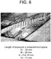
- the video was presented in a square aperture with an area approximately 1/8 of the size of the screen (about 16° of visual angle). This square aperture started at the upper left hand corner of the screen and moved at a constant speed, taking 10 seconds to traverse each edge of the monitor. A full cycle took 40 seconds, and five full cycles were played, for a total of 200 seconds. A countdown video played in the starting position for 10 seconds before the music video began, to give observers time to orient to the stimulus. Only the 200 seconds of the music video were used for analyses.
- the eye tracker sampled eye position at 500 Hz, yielding 100,000 samples of eye position over 200 seconds.
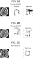
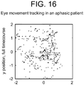
- Timecourses T he normalized x- and y- timecourses were plotted across time ( Figure 1a ). The clockwise movement of the visual stimulus alternated between horizontal changes and vertical changes, and the x- and y- timecourses in neurologically intact observers show the same alternation.
- Phase was calculated as phase of the sine function that best fit the data.
- the 8 following complementary procedures were used to assess the statistical significance of any differences in these two measures (phase difference and model fit) as compared between the neurologically intact control observers and the test patient observers.
- phase difference and model fit were defined: (1) The sinusoidal model fit the data of 1 the healthy observers better than it did the patients (p ⁇ .001; Table 1); and (2) Healthy observers showed the hypothesized 45 degree phase difference between the horizontal and vertical trajectories, reflecting the pattern of movement of the visual stimulus; test patients did not (p ⁇ .01; Table 1).
- model fit was used to classify individual timecourses as coming from the group of neurologically intact controls vs. the group of test patients.
- Statistical thresholds were defined by converting correlation coefficients to normally-distributed z-scores, using the Fisher transformation. Each timecourse from all observers was classified based on the probability that it came from the null distribution (see Methods). The classification results exhibited 96% specificity and 52% sensitivity. Given that the test patients were a heterogeneous group selected from a neurosurgery clinic that treats patients who might have damage to either or both the peripheral and the central nervous systems, there is no reason to expect that the entire population would have problems that affected the ocular-motor system. Thus 52% sensitivity could be considered high for this test patient population.
- Uncalibrated tracking may provide a quantitative measure of the ability to fixate, attend, and follow a stimulus. These date demonstrate that it is possible to collect reliable highfrequency eye movement data without first completing a spatial calibration for each observer. Many patients are not capable of calibrated eye tracking. The ability to track eye movements in these populations provides new insights about a variety of disorders that disturb the ocular-motor system, including but not limited to brain injury, stroke, and psychiatric disorders. Possible applications include clinical screening, diagnosis, monitoring the efficacy of treatment, and tracking progression of impairment and recovery.
- Eye Movement Tracking The subjects' eye movements were recorded using an Eyelink 1000 monocular eye tracker (500 Hz sampling, SR Research). Healthy volunteers were seated 55 cm from the screen with their head stabilized using a chinrest. Subjects could be seated or lying down, on chairs, hospital beds, or stretchers. Stimulus was presented on average 55 cm from patient eyes, with the presentation monitor adjusted to match gaze direction. Some subjects used a chinrest, when it was comfortable for them to do so.
- Integrity For the integrity measure, each patient's pair of values from arms 1 (the top of the box) and 3 (the bottom of the box) was z-scored using the mean and standard deviation calculated from the control population. The resulting score indicated how different the patient values were compared with the control values, in units of standard deviations. Because 95% of all values in a normal distribution lie within two standard deviations of the mean, a z-score of 2 was used as a significance threshold. Patients with z-scores above 2 in either or both arms were thus judged to have significant disturbances of ocular motility.
- Relative variance Because relative variance is a ratio, it cannot be analyzed using z-scores, since the assumption of a normal distribution does not hold for ratios. Instead, 5,000 point distributions were generated using a bootstrapping method that took 5,000 samples from 25 values randomly chosen with replacement from the 45 control values. For each subject, the relative variance in arms 1 and 3 were compared respectively with the corresponding control distribution, and the percent of the control distribution with variance below that of the test value was determined. A p-value of .05 (a widely accepted measure of statistical significance) corresponds to 95% of control values falling below the test value. Thus, subjects with variance higher than 95% of the values in the control distributions were determined to have significant disturbances of ocular motility.
- the units of relative variance are related to size in degree of visual angle, but are not exactly identical to degrees of visual angle, because there was no spatial calibration. These may be referred to as time-degrees units.
- Control distributions As expected, the control distributions for the integrity measurements were normally distributed with a mean of 0.2 and an average standard deviation of 0.05 (5% deviation). The control distributions of relative variance peaked at 0.25 (reflecting equal variance across the four arms).
- Patient measurements The integrity measures for the 'top' vs. 'bottom' arms of the trajectory for each subject, in units of standard deviation, as compared with the control distributions as described above were calculated.
- Subjects with cranial nerve palsies or mass effect showed defects in integrity of eye tracing box trajectory.
- Subjects with relatively greater cranial nerve II palsies due to either compression or papilledema showed streaking vertical lines due to scanning vision.
- Eye movement tracking for neuropsychiatric and brain injury research has traditionally been based on spatial calibration.
- the eye-tracker measures the relative position of pupil and corneal reflection for a period of about 400-800 ms while the subject looks at a target or targets of known position to generate meaningful spatial coordinates during subsequent pupil movement.
- Calibration requires subject cooperation and precludes detection of anatomically dysfunctional ocular motility.
- We developed a novel technique for eye movement tracking without spatial calibration which can, theoretically, assess the function of the oculomotor nerve (cranial nerve III).
- the oculomotor nerve innervates the superior and inferior recti muscles of the orbit, raising and lowering the pupil. Compression of the nerve while it courses through the tentorial notch medial to the temporal lobe occurs with impending transtentorial herniation, a common mechanism of death from supratentorial mass effect due to tumors, stroke, hemorrhage or trauma.
- Ptosis in this patient may be a consequence of disruption of the superior division of the oculomotor nerve en route to the levator palpebrae superioris.
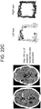
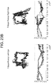
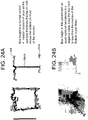
- a patient without a known IIIrd palsy, but with a history of inferior orbital wall fracture resulting in transient diplopia five years prior also had decreased vertical amplitude on eye-tracking ( Figure 20 , bottom row).
- Exclusion criteria were: visual acuity worse than light perception, neurologic or hemodynamic instability, papilledema or conduction abnormality of the optic nerve, bilateral compression of optic nerves resulting in decreased visual acuity, visual field deficit, sellar lesion, and cavernous sinus invasion and dementia.
- the methods described herein differ from a report of uncalibrated tracking using static stimuli for on-target and off-target fixations in a population of minimally conscious and persistently vegetative patients that have open eyes.
- Trojano et al. J. Neurol, (2012 )
- the moving images shown within an aperture that moves periodically allows assessing both coarse and fine eye movement characteristics in both controls and neurologically impaired subjects.
- Maruta et al. Journal of Head Trauma Rehabilitation 2010; 25: 293-305
- Trojano, et al. J. Neurol, 2012 Contreras, et al., Brain Research 2011; 1398: 55-63 ; Contreras, et al.
- Uncalibrated ocular motility tracking assessment does not require that the subject explicitly consent to being tracked prior to assessment of their central nervous system functioning, and thus raises ethical considerations.
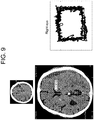
- each CT volume was realigned and resampled to a standard (template) coordinate system by marking in 3D space the the left eye lens (LE), right eye lens (RE) and the junction of the superior colliculi (C).
- LE left eye lens
- RE right eye lens
- C superior colliculi
- these three corresponding structures LE' RE' C' were positioned symmetrically such that the principal axis of the brain stem was in the z direction (across plane).
- the rigid body (volume-preserving) transformation T that best mapped the triangle ⁇ LE RE C> onto the triangle ⁇ LE' RE' C'> was then computed.
- the transformation T was used to resample the CT volume to the template space.
- the cistern was segmented into the template space by placing a cylinder of radius 20 pixels centered on the center of the brain stem extending from pineal gland to tuberculum sella.
- Pv (A_brain - A) / (A_brain ⁇ A water).
- A_brain 48 Hu
- A_water 0 Hu.
- Pv was constrained to [0-1].
- the cistern volume was computed as the sum of Pv values within the cylinder multiplied by voxel volume excluding the suprasellar cistern.
- Ratios were compared using a two-tailed student t-test, paired, two-sample for pre vs post-operative data comparisons and unpaired t-tests for comparison between the unequal n groups of surgical patients versus ophthalmologic clinic controls.
- Mean pupil size was calculated by obtaining the mean of the 100,000 data points per eye tracking trajectory using MATLAB programming. Means were compared using paired two-tailed t-tests for comparison between pre and postoperative data.
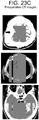
- Case 1 ( Figure 22 ) The patient is an 86 year old right handed male with a medical history of hypertension, hyperlipidemia mild chronic renal insufficiency and an ophthalmologic history of bilateral cataract surgery (2 years and 8 years prior), pseudophakia and scleral buckling. He had a baseline visual acuity of 20/25 (right eye) and 20/30 (left eye). The patient took 81 mg of aspirin per day prophylactically. The patient fell and presented several weeks later with headache but was otherwise neurologically well. A head CT was performed and demonstrated a small left sided subdural hematoma. Aspirin was discontinued and platelets were transfused. The headache resolved spontaneously, and the patient elected to observe this lesion.
- Perimesencephalic cistern compression (on immediate preoperative CT scan): Right cistern 80% of baseline, left cistern 61% of baseline.
- Case 2 ( Figure 23 and Figure 24 ) The patient is a 62 year old right handed male with a past medical history of goiter/iatrogenic hypothyroidism, hypertension and hyperlipidemia and an ophthalmologic history of glaucoma controlled with latonoprost drops. He had 20/20 vision bilaterally. He presented with a fall and loss of consciousness persisting a few minutes. At presentation he was awake, alert, and had fluent speech. His pupils were equal and extra-ocular movements remained intact. His head CT showed a small frontoparietal epidural hematoma underlying a stellate skull fracture. Eye movement tracking was performed.
- Perimesencephalic cistern compression (on preoperative CT scan): Right cistern 27% of baseline, left cistern 35% of baseline.
- Case 3 ( Figure 25 ) 74 year old diabetic hypertensive male with renal insufficiency, bilateral cataracts and 20/25 vision bilaterally, presented with impaired mobility due to right lower extremity weakness. He denied head trauma but reported having fallen three months prior without hitting his head. On examination he was awake and alert with a right pronator drift and 4/5 right sided hemiparesis of the upper and lower extremities. His left side was intact. His pupils appeared equal and he had intact extraocular motility on examination. Eye movement tracking was performed. Twist-drill drainage of the subdural hematoma was performed and 130 cc of fluid was extracted. He was discharged to home after inpatient rehabilitation.
- Perimesencephalic cistern compression (on immediate preoperative CT scan): Right cistern 86% of baseline, left cistern 70% of baseline.
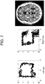
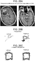
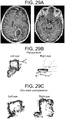
- Case 4 ( Figure 26 ) 63 year old 100 pack-year smoking male who had declined to see a physician for the duration of his adult life, and thus reported no relevant medical or ophthalmologic history, presented to the emergency room with a cough and flu-like illness. On examination he was noted to be disoriented and have a mild L hemiparesis. Pupils were equal and reactive, extraocular movements appeared intact. Head CT demonstrated a right frontal mass, and chest radiograph showed a large left upper lobe chest mass. The patient was administered decadron 10 mg po q6 hours and a CT chest/abdomen/pelvis and brain MRI (shown) was obtained.
- Eye movement tracking was performed 48 hours after admission, directly prior to right frontal craniotomy for resection of a moderately well-differentiated squamous cell carcinoma metastasis. A radiographic gross total resection was performed. The patient went home one week after his craniotomy and ultimately underwent radiation therapy to his whole brain. He declined treatment for the lung mass and expired 7 months after his initial diagnosis.
- Perimesencephalic cistern compression (on immediate preoperative CT scan): Right cistern 77% of baseline, left cistern 73% of baseline.
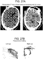
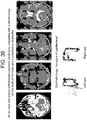
- Case 5 ( Figure 27 ) The patient is a 63 year old male with diabetes, hypertension, hypercholesterolemia, atrial fibrilliation on coumadin, congestive heart failure, post-traumatic stress disorder, renal failure on dialysis, chronic obstructive pulmonary disease and coronary artery disease. He presented with confusion while receiving hemodialysis for his renal failure. Ophthalmic history was significant for proliferative retinopathy. He had visual acuity of 20/25 in the right eye and 20/40 in the left eye. On physical examination at presentation he was neurologically well, without neglect or pronator drift. Extraocular movements were intact, pupils were equal. Head CT demonstrated a right sided mixed-density subdural hemorrhage. Eye tracking was performed. Coumadin was stopped and fresh frozen plasma administered. Two days after presentation twist-drill drainage was performed and 176 cc of subdural fluid was evacuated. The patient remained neurologically well and returned to his assisted living residence two days later.
- Perimesencephalic cistern compression (on immediate preoperative CT scan): Right cistern 36% of baseline, left cistern 29% of baseline.
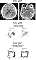
- Case 6 ( Figure 28 ) 67 year old male with a past medical history of prostate cancer, hypertension, hyperlipidemia, alcoholism in remission, and gunshot wound to the left shoulder with retained missile fragment. His ophthalmic baseline was 20/40 vision in the right eye and 20/50 in the left eye. He presented with 2 months of stuttering and right arm and hand weakness. He had a witnessed seizure on the day of presentation that began with shaking of the right upper extremity and progressed to generalized tonic-clonic activity. On examination he had intact pupils and extraocular movements. His speech was slow with paraphasic errors and difficulty with repetition and naming. Head CT with contrast revealed a left fronto-temporal cystic mass.
- Case 7 65 year old male with a past medical history of hypertension, hyperlipidemia, coronary artery disease and post-traumatic stress disorder, with no known ophthalmic disorders and visual acuity of 20/20 bilaterally presented 2 weeks after left parietal craniotomy for a esophageal junction metastasis by an outside surgeon with worsening right hand coordination and ataxia.
- CT revealed edema at the surgical site and MRI revealed a peripherally enhancing collection in the previous tumor cavity. Eye tracking was performed, and then the patient was taken to the operating room for re-exploration craniotomy and evacuation of an abscess deep to the dura He was treated with antibiotics for 12 weeks postoperatively.
- Dementia is a disease with numerous etiologies and devastating consequences.
- Spatially calibrated eye movement tracking of demented subjects reveals impaired smooth pursuit function.
- We developed a novel technique for eye movement tracking during watching of a moving video that does not rely on spatial calibration.
- subjects with normal pressure hydrocephalus demonstrated decreased tracking variability after cerebrospinal fluid diversion relative to their preoperative state. This decrease in variability correlated with improvements in gait and was not seen in surgical patients without normal pressure hydrocephalus or serially tracked control subjects.
- Our results suggest that cerebrospinal fluid diversion improves the ability of normal pressure hydrocephalics to perform less variable non-spatially calibrated eye movement tracking, and that eye tracking while watching a movie or television can be used to assess shunt function.
- Impaired smooth pursuit eye movement tracking is seen in demented patients versus normal elderly controls ( Hutton, et al., Neurology (1984) 34: 99-102 ) and has been shown to be an early indicator of presenile and Alzheimer's dementia ( Muller, et al., Int J Psychophysiol (1991) 11: 167-177 ; Muller, et al., European archives of psychiatry and clinical neuroscience (1991) 241: 46-48 )
- Demented patients also have impaired eye-hand visuomotor coordination ( Verheij, et al., J Alzheimers Dis (2012) 30: 131-143 ) and impaired visual memory ( Lagun, et ⁇ /., Journal of neuroscience methods (2011) 201: 196-203 ).
- NPH Normal pressure hydrocephalus
- the eye-tracker measures the relative position of pupil and corneal reflection for a period of about 400-800 ms while the subject looks at a target or targets of known position to generate meaningful spatial coordinates during subsequent pupil movement.
- an EyeLink eyetracking camera at a relatively fixed distance from a computer monitor over a fixed period of time.
- the visual stimulus was the Shakira World Cup soccer music video Waka-Waka played continuously in a square aperture with an area approximately 1/8 the screen size while moving clockwise along the outer edges of the monitor for five complete cycles of 40 seconds each.
- the eye tracker sampled pupil position at 500 Hz, yielding 100,000 samples over 200 seconds.
- Case 1 ( Figure 31 ): 68 year old male with a past medical history of HIV infection, diabetes, hypertension, and stroke presented after 2 falls to his neurologist. A large volume lumbar puncture was performed. The opening pressure was 3 cm. The patient's gait was dramatically improved by the tap. A Codman shunt with Certas programmable valve set to 4 was placed and the patient continued to demonstrate progressive improvement clinically. Serial tracking was performed and paralleled the clinical improvement in gait ( Figure 29 ).
- Case 2 ( Figure 32 ): A 57 year old construction worker with presented with increasing gait disturbance and memory problems progressive over 5 years. He was fired from his job as a construction worker after falling at work. His mini mental status exam improved by 3 points, as did his gait after a large volume lumbar puncture. A Codman shunt with Certas programmable valve set to 5 was placed and the patient continued to demonstrate progressive improvement clinically. Serial tracking was performed and paralleled the clinical improvement in gait ( Figure 3 ).
- Case 3 ( Figure 33 ): An 87 year old male World War II veteran with a past medical history of asthma, hypertension, posttraumatic stress disorder and benign prostatic hypertrophy had undergone shunting for normal pressure hydrocephalus at the age of 73 for a gait apraxia. A medium pressure PS medical valve was placed at that time. He underwent three subsequent distal shunt revisions without changing of the valve. He now presented again with progressive gait apraxia and shuntogram demonstrating distal malfunction. Since he had already failed three intraperitoneal shunts, the shunt was now revised and placed in the pleural space. Neither the valve, nor the shunt tubing was changed. The patient demonstrated improvement in his gait which paralled the improvement in tracking ( Figure 31 ).
- Cranial nerve VI is considered highly susceptible to neuropathy due to its anatomical vulnerability ( Hanson et al., Neurology 2004; 62(1):33-36 ).
- the abducens nerve (VI) exits the brainstem from its tethering at the medullopontine junction and courses intracranially before entering Dorello's canal, where it is again tethered by fibrous and osseous structures.
- Posterior fossa lesions pushing the cerebellum and brainstem forward may directly compress the VIth nerve against the clivus ( Hanson et al., Neurology 2004; 62(1):33-36 ).
- Subjects' eye movements were recorded with an Eyelink 1000 eye tracker at a relatively fixed distance from a computer monitor over a fixed period of time.
- the visual stimulus provided was the Shakira music video Waka-Waka played continuously in a square aperture with an area approximately 1/8 the screen size while moving clockwise along the outer edges of the monitor for five complete cycles of 40 seconds each.
- the eye tracker sampled pupil position at 500 Hz, yielding 100,000 samples over 200 seconds.
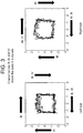
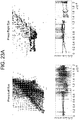
- Scatterplots of the entire time series were created by plotting the 100,000 (x,y) pairs representing the two orthogonal components of the instantaneous angle of pupil reflection over time to create 'box trajectories' that reflected the temporal nature of the pupillary movement.
- these figures look like boxes, reflecting the timing of the aperture as it moved around the screen ( Figure 20 ).
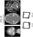
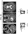
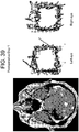
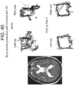
- the second patient was a 56 year old male who presented with a lung mass and headaches. He did not have papilledema on clinical examination. MRI revealed a large intraxial mass near the cerebellopontine angle, but interestingly, pushing the left side of brainstem up against the clivus rather than the right. Note that there was no transependymal flow on MRI to suggest hydrocephalus ( Figure 37 right). Eye tracking of the right eye demonstrated a box narrower than it was wide (increased aspect ratio) consistent with CN VI palsy. Postoperatively his aspect ratio returned to normal ( Figure 38 )
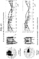
- a patient with ocular histoplasmosis resulting in central optic nerve atrophy ( Figure 41 ) showed extensive y-variability.
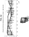
- a 25 year old female patient being evaluated for optic neuritis also had increased y-variability ( Figure 42 ).
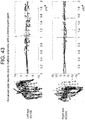
- a patient with disconjugate due to multiple sclerosis demonstrated multiple cranial neuropathies ( Figure 43 ). This pattern was not seen in the healthy control subjects nor in other patients including several with tumors impinging on the optic nerve, chiasm or tract or with poor visual acuity due to known ocular non-neuronal pathology.
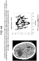
- Papilledema or swelling of the optic disc, is caused by elevated intracranial pressure due to large brain tumors as well as other pathologies. It is thought to indicate delayed axoplasmic transport along the optic nerve.
- Eye movement tracking of the left eye of a patient with a large right frontal brain tumor presenting with an examination consistent with papilledema demonstrated an increased vertical range box trajectory with no roof or floor similar to those seen in the central optic nerve atrophy and ocular histoplasmosis patients ( Figure 44 ).
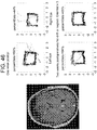
- This statistically significant deviation in y-variability resolved with steroids over 24 hours Figure 45 ).
- the height of the patient's box trajectory remained decreased after the steroids and before resection, suggesting a component of IIIrd nerve palsy.
- the eye tracking trajectory returned to normal by one week after resection ( Figure 46 ).
Landscapes
- Health & Medical Sciences (AREA)
- Life Sciences & Earth Sciences (AREA)
- Engineering & Computer Science (AREA)
- Physics & Mathematics (AREA)
- General Health & Medical Sciences (AREA)
- Molecular Biology (AREA)
- Biophysics (AREA)
- Veterinary Medicine (AREA)
- Biomedical Technology (AREA)
- Heart & Thoracic Surgery (AREA)
- Medical Informatics (AREA)
- Public Health (AREA)
- Surgery (AREA)
- Animal Behavior & Ethology (AREA)
- Pathology (AREA)
- Neurology (AREA)
- Physiology (AREA)
- Neurosurgery (AREA)
- Artificial Intelligence (AREA)
- Signal Processing (AREA)
- Computer Vision & Pattern Recognition (AREA)
- Psychiatry (AREA)
- Ophthalmology & Optometry (AREA)
- Human Computer Interaction (AREA)
- Hematology (AREA)
- Psychology (AREA)
- Evolutionary Computation (AREA)
- Fuzzy Systems (AREA)
- Mathematical Physics (AREA)
- Eye Examination Apparatus (AREA)
- Measurement Of The Respiration, Hearing Ability, Form, And Blood Characteristics Of Living Organisms (AREA)
Applications Claiming Priority (3)
| Application Number | Priority Date | Filing Date | Title |
|---|---|---|---|
| US201261615463P | 2012-03-26 | 2012-03-26 | |
| US201261710213P | 2012-10-05 | 2012-10-05 | |
| PCT/US2013/033672 WO2013148557A1 (en) | 2012-03-26 | 2013-03-25 | Methods and kits for assessing central nervous system integrity |
Publications (3)
| Publication Number | Publication Date |
|---|---|
| EP2830479A1 EP2830479A1 (en) | 2015-02-04 |
| EP2830479A4 EP2830479A4 (en) | 2015-11-25 |
| EP2830479B1 true EP2830479B1 (en) | 2021-11-17 |
Family
ID=49261144
Family Applications (1)
| Application Number | Title | Priority Date | Filing Date |
|---|---|---|---|
| EP13770301.3A Active EP2830479B1 (en) | 2012-03-26 | 2013-03-25 | System for assessing central nervous system integrity |
Country Status (8)
| Country | Link |
|---|---|
| US (5) | US9642522B2 (enExample) |
| EP (1) | EP2830479B1 (enExample) |
| JP (2) | JP6559062B2 (enExample) |
| CN (1) | CN104780828B (enExample) |
| AU (1) | AU2013239981B2 (enExample) |
| BR (1) | BR112014023637B1 (enExample) |
| CA (1) | CA2867866C (enExample) |
| WO (1) | WO2013148557A1 (enExample) |
Families Citing this family (44)
| Publication number | Priority date | Publication date | Assignee | Title |
|---|---|---|---|---|
| US10463248B2 (en) * | 2011-03-02 | 2019-11-05 | Brien Holden Vision Institute Limited | Systems, methods, and devices for measuring eye movement and pupil response |
| BR112014023637B1 (pt) | 2012-03-26 | 2021-12-21 | New York University | Métodos para avaliação da integridade do sistema nervoso central e para detecção, diagnóstico ou avaliação de pressão intracraniana aumentada |
| AU2014281725B2 (en) * | 2013-06-17 | 2019-10-10 | New York University | Methods and kits for assessing neurological and ophthalmic function and localizing neurological lesions |
| US9895100B2 (en) | 2013-10-04 | 2018-02-20 | Indiana University Research And Technology Corporation | Eye movement monitoring of brain function |
| JP6287486B2 (ja) * | 2014-03-31 | 2018-03-07 | 富士通株式会社 | 情報処理装置、方法及びプログラム |
| US20150307048A1 (en) * | 2014-04-23 | 2015-10-29 | Creative Inovation Services, LLC | Automobile alert information system, methods, and apparatus |
| JP2017529891A (ja) * | 2014-08-04 | 2017-10-12 | ニューヨーク ユニバーシティ | 薬物使用、薬物乱用および昏睡、核間性眼筋麻痺、注意欠陥多動性障害(adhd)、慢性外傷性脳症、統合失調症スペクトラム障害、およびアルコール摂取を診断、評価、または定量化するための方法およびキット |
| US10231617B2 (en) * | 2014-08-21 | 2019-03-19 | Dignity Health | Systems and methods for using eye movements to determine traumatic brain injury |
| US10045730B2 (en) * | 2014-09-11 | 2018-08-14 | The Mitre Corporation | Methods and systems for rapid screening of mild traumatic brain injury |
| US20170042462A1 (en) * | 2015-08-10 | 2017-02-16 | Neuro Kinetics, Inc. | Automated Data Acquisition, Appraisal and Analysis in Noninvasive Rapid Screening of Neuro-Otologic Conditions Using Combination of Subject's Objective Oculomotor Vestibular and Reaction Time Analytic Variables |
| CA2901477C (en) | 2015-08-25 | 2023-07-18 | Evolution Optiks Limited | Vision correction system, method and graphical user interface for implementation on electronic devices having a graphical display |
| US11064881B2 (en) | 2015-11-13 | 2021-07-20 | Hennepin Healthcare System, Inc | Method for predicting convergence disorders caused by concussion or other neuropathology |
| JP6693105B2 (ja) * | 2015-12-01 | 2020-05-13 | 株式会社Jvcケンウッド | 視線検出装置及び視線検出方法 |
| WO2017127798A1 (en) | 2016-01-22 | 2017-07-27 | The Arizona Board Of Regents On Behalf Of The University Of Arizona | Ocular cranial nerve monitoring system |
| EP3493728B1 (en) | 2016-08-02 | 2025-11-05 | New York University | Methods and kits for assessing neurological function and localizing neurological lesions |
| US10863902B2 (en) * | 2016-10-03 | 2020-12-15 | Oculogica Inc. | Method for detecting glaucoma |
| TWI602068B (zh) * | 2016-10-17 | 2017-10-11 | Data processing device and method thereof | |
| US10201274B2 (en) | 2016-10-20 | 2019-02-12 | Oculogica Inc | Eye tracking system with biometric identification |
| US11141095B2 (en) | 2017-02-17 | 2021-10-12 | Oculogica Inc. | Method and system for detecting concussion |
| CN114403802A (zh) * | 2017-09-13 | 2022-04-29 | 奥库洛吉卡公司 | 眼睛跟踪系统 |
| CA3086037A1 (en) * | 2017-11-30 | 2019-06-06 | Viewmind S.A. | System and method for detecting neurological disorders and for measuring general cognitive performance |
| EP3781066A4 (en) * | 2018-04-20 | 2022-01-05 | The United States Government as represented by the Department of Veterans Affairs | PROCESSES AND KITS FOR THE OPTIMIZATION OF NEUROSURGICAL INTERVENTION SITE |
| US12290373B2 (en) | 2018-04-27 | 2025-05-06 | C. Light Technologies, Inc. | Method of detection, prognostication, and monitoring of neurological disorders |
| JP6945493B2 (ja) * | 2018-05-09 | 2021-10-06 | 富士フイルム株式会社 | 医用画像処理装置、方法およびプログラム |
| KR102250999B1 (ko) | 2018-06-29 | 2021-05-12 | 고려대학교 산학협력단 | 시야장애 평가 및 안구 운동장애 평가 결과에 기반한 뇌병변 진단 장치 |
| US11327563B2 (en) | 2018-10-22 | 2022-05-10 | Evolution Optiks Limited | Light field vision-based testing device, adjusted pixel rendering method therefor, and online vision-based testing management system and method using same |
| US11500460B2 (en) | 2018-10-22 | 2022-11-15 | Evolution Optiks Limited | Light field device, optical aberration compensation or simulation rendering |
| US11966507B2 (en) | 2018-10-22 | 2024-04-23 | Evolution Optiks Limited | Light field vision testing device, adjusted pixel rendering method therefor, and vision testing system and method using same |
| US11500461B2 (en) | 2019-11-01 | 2022-11-15 | Evolution Optiks Limited | Light field vision-based testing device, system and method |
| WO2020219711A1 (en) | 2019-04-23 | 2020-10-29 | Evolution Optiks Limited | Light field display and vibrating light field shaping layer and vision testing and/or correction device |
| US11635617B2 (en) | 2019-04-23 | 2023-04-25 | Evolution Optiks Limited | Digital display device comprising a complementary light field display or display portion, and vision correction system and method using same |
| US11902498B2 (en) | 2019-08-26 | 2024-02-13 | Evolution Optiks Limited | Binocular light field display, adjusted pixel rendering method therefor, and vision correction system and method using same |
| US11823598B2 (en) | 2019-11-01 | 2023-11-21 | Evolution Optiks Limited | Light field device, variable perception pixel rendering method therefor, and variable perception system and method using same |
| US11487361B1 (en) | 2019-11-01 | 2022-11-01 | Evolution Optiks Limited | Light field device and vision testing system using same |
| US12360592B2 (en) | 2019-11-01 | 2025-07-15 | Evolution Optiks Limited | Light field device and vision testing system using same |
| US12112665B2 (en) | 2019-11-01 | 2024-10-08 | Evolution Optiks Limited | Light field device, variable perception pixel rendering method therefor, and variable perception system and method using same |
| CN110897608B (zh) * | 2019-12-15 | 2022-05-03 | 深圳市具安科技有限公司 | 斑马鱼眼动分析方法、装置及计算机设备 |
| JP7565566B2 (ja) * | 2020-11-24 | 2024-10-11 | 学校法人東京医科大学 | 身体状態の推定装置、身体状態の推定装置の作動方法、プログラム、および記録媒体 |
| US12390172B2 (en) | 2021-02-19 | 2025-08-19 | Canon Medical Systems Corporation | Method and apparatus for displaying medical images |
| US11429188B1 (en) * | 2021-06-21 | 2022-08-30 | Sensie, LLC | Measuring self awareness utilizing a mobile computing device |
| WO2023281622A1 (ja) * | 2021-07-06 | 2023-01-12 | 日本電気株式会社 | 回復度推定装置、回復度推定方法、及び、記録媒体 |
| JP7711753B2 (ja) * | 2021-07-06 | 2025-07-23 | 日本電気株式会社 | 回復度推定装置、回復度推定方法、及び、プログラム |
| WO2024013546A1 (en) * | 2022-07-12 | 2024-01-18 | Mcdowell Nicola Jean | A method of identifying a higher visual perceptual difficulty |
| WO2025034122A1 (en) * | 2023-08-04 | 2025-02-13 | VRF Vault Limited | Methods and systems for assessing the presence of traumatic brain injury |
Family Cites Families (73)
| Publication number | Priority date | Publication date | Assignee | Title |
|---|---|---|---|---|
| US4576184A (en) | 1981-08-31 | 1986-03-18 | Pharmometrics Corporation | Drug abuse detection |
| US4889422A (en) | 1986-01-28 | 1989-12-26 | George Pavlidis | Method and means for detecting dyslexia |
| US4838681A (en) | 1986-01-28 | 1989-06-13 | George Pavlidis | Method and means for detecting dyslexia |
| US6622036B1 (en) * | 2000-02-09 | 2003-09-16 | Cns Response | Method for classifying and treating physiologic brain imbalances using quantitative EEG |
| US20020024633A1 (en) | 1999-04-09 | 2002-02-28 | Daehoon Kim | Pupil evaluation system |
| US6247813B1 (en) * | 1999-04-09 | 2001-06-19 | Iritech, Inc. | Iris identification system and method of identifying a person through iris recognition |
| US6820979B1 (en) * | 1999-04-23 | 2004-11-23 | Neuroptics, Inc. | Pupilometer with pupil irregularity detection, pupil tracking, and pupil response detection capability, glaucoma screening capability, intracranial pressure detection capability, and ocular aberration measurement capability |
| US6116736A (en) | 1999-04-23 | 2000-09-12 | Neuroptics, Inc. | Pupilometer with pupil irregularity detection capability |
| US6346887B1 (en) | 1999-09-14 | 2002-02-12 | The United States Of America As Represented By The Secretary Of The Navy | Eye activity monitor |
| EP1246667B1 (en) | 1999-12-30 | 2005-03-23 | Medtronic, Inc. | User authentication in medical device systems |
| BR0108476A (pt) | 2000-02-11 | 2003-04-22 | Marcio Marc Abreu | Sistema e método para comunicar informação de revocação de produto, advertências relacionadas ao produto ou outra informação relacionada aos usuários de produtos |
| US20020169364A1 (en) * | 2000-02-14 | 2002-11-14 | Baumzweiger William E. | Brainstem and limbic disorder (BALD) |
| US6652458B2 (en) | 2000-06-20 | 2003-11-25 | The Mclean Hospital Corporation | ADHD detection by eye saccades |
| EP1219243A1 (en) | 2000-12-28 | 2002-07-03 | Matsushita Electric Works, Ltd. | Non-invasive brain function examination |
| DE10132378A1 (de) | 2001-07-06 | 2003-04-24 | Zeiss Carl Meditec Ag | Verfahren und Vorrichtung zur Verfolgung von Augenbewegungen |
| JP4543594B2 (ja) | 2001-07-31 | 2010-09-15 | パナソニック電工株式会社 | 脳機能検査装置および脳機能検査システム |
| US8211031B2 (en) * | 2002-01-15 | 2012-07-03 | Orsan Medical Technologies Ltd. | Non-invasive intracranial monitor |
| US7147605B2 (en) * | 2002-07-08 | 2006-12-12 | Uab Vittamed | Method and apparatus for noninvasive determination of the absolute value of intracranial pressure |
| US7575321B2 (en) * | 2003-10-30 | 2009-08-18 | Welch Allyn, Inc. | Apparatus and method of diagnosis of optically identifiable ophthalmic conditions |
| DE60323410D1 (de) | 2003-11-30 | 2008-10-16 | Volvo Technology Corp | Verfahren und system zum erkennen einer fahrerbeeinträchtigung |
| US7384399B2 (en) | 2004-02-11 | 2008-06-10 | Jamshid Ghajar | Cognition and motor timing diagnosis and training system and method |
| US7819818B2 (en) * | 2004-02-11 | 2010-10-26 | Jamshid Ghajar | Cognition and motor timing diagnosis using smooth eye pursuit analysis |
| CN102670163B (zh) | 2004-04-01 | 2016-04-13 | 威廉·C·托奇 | 控制计算装置的系统及方法 |
| WO2007075460A2 (en) | 2005-12-16 | 2007-07-05 | Novavision, Inc. | Adjustable device for vision testing and therapy |
| WO2007070953A1 (en) * | 2005-12-20 | 2007-06-28 | Neuro Vision Technology Pty Ltd | Apparatus and method for assessment and rehabilitation after acquired brain injury |
| US7535991B2 (en) | 2006-10-16 | 2009-05-19 | Oraya Therapeutics, Inc. | Portable orthovoltage radiotherapy |
| WO2009052833A1 (en) | 2007-10-23 | 2009-04-30 | Mindmetic Ltd. | Method, system and computer program for automated interpretation of measurements in response to stimuli |
| CA2704777C (en) * | 2007-11-01 | 2017-09-12 | Catholic Healthcare West | Method of detecting neurological disease |
| WO2009085204A2 (en) | 2007-12-23 | 2009-07-09 | Oraya Therapeutics, Inc. | Methods and devices for detecting, controlling, and predicting radiation delivery |
| US8265743B2 (en) * | 2007-12-27 | 2012-09-11 | Teledyne Scientific & Imaging, Llc | Fixation-locked measurement of brain responses to stimuli |
| WO2010006180A1 (en) * | 2008-07-09 | 2010-01-14 | Mckinley Laurence M | Optic function monitoring process and apparatus |
| WO2010042557A2 (en) | 2008-10-06 | 2010-04-15 | Neuro Kinetics, Inc. | Method and apparatus for corrective secondary saccades analysis with video oculography system |
| WO2010062400A1 (en) | 2008-11-28 | 2010-06-03 | Neuroptics, Inc. | Methods, systems, and devices for monitoring anisocoria and asymmetry of pupillary reaction to stimulus |
| US8808195B2 (en) * | 2009-01-15 | 2014-08-19 | Po-He Tseng | Eye-tracking method and system for screening human diseases |
| US20100280372A1 (en) * | 2009-05-03 | 2010-11-04 | Pieter Poolman | Observation device and method |
| GB0917600D0 (en) * | 2009-10-07 | 2009-11-25 | Univ Edinburgh | Testing apparatus and method |
| US9229227B2 (en) | 2010-02-28 | 2016-01-05 | Microsoft Technology Licensing, Llc | See-through near-eye display glasses with a light transmissive wedge shaped illumination system |
| US8732795B2 (en) | 2010-05-21 | 2014-05-20 | Epic Systems Corporation | System and method for user authentication |
| US9691289B2 (en) * | 2010-12-22 | 2017-06-27 | Brightstar Learning | Monotonous game-like task to promote effortless automatic recognition of sight words |
| CA2827498C (en) | 2011-01-28 | 2020-09-22 | Dignity Health | Method of detecting neurological disease |
| CA2754835C (en) | 2011-10-07 | 2021-06-15 | Baycrest Centre For Geriatric Care | Methods and systems for assessing cognitive function |
| ITVR20110201A1 (it) * | 2011-11-02 | 2013-05-03 | Milano Politecnico | Dispositivo per il monitoraggio della posizione e deimovimenti dell'occhio, particolarmente adatto per la radioterapia oculare |
| US9078612B2 (en) | 2011-12-02 | 2015-07-14 | Third Eye Diagnostics, Inc. | Devices and methods for noninvasive measurement of intracranial pressure |
| US20130208952A1 (en) | 2012-02-13 | 2013-08-15 | Geoffrey Auchinleck | Method and Apparatus for Improving Accuracy of Biometric Identification in Specimen Collection Applications |
| BR112014023637B1 (pt) | 2012-03-26 | 2021-12-21 | New York University | Métodos para avaliação da integridade do sistema nervoso central e para detecção, diagnóstico ou avaliação de pressão intracraniana aumentada |
| US9101312B2 (en) | 2012-04-18 | 2015-08-11 | TBI Diagnostics LLC | System for the physiological evaluation of brain function |
| US8668337B2 (en) | 2012-04-18 | 2014-03-11 | TBI Diagnostics LLC | System for the physiological evaluation of brain function |
| US9004687B2 (en) | 2012-05-18 | 2015-04-14 | Sync-Think, Inc. | Eye tracking headset and system for neuropsychological testing including the detection of brain damage |
| US8808179B1 (en) | 2012-08-06 | 2014-08-19 | James Z. Cinberg | Method and associated apparatus for detecting minor traumatic brain injury |
| US8585589B1 (en) | 2012-08-06 | 2013-11-19 | James Z. Cinberg | Method and associated apparatus for detecting minor traumatic brain injury |
| US8951046B2 (en) | 2012-08-10 | 2015-02-10 | Sync-Think, Inc. | Desktop-based opto-cognitive device and system for cognitive assessment |
| US10085688B2 (en) | 2012-11-20 | 2018-10-02 | El-Mar Inc. | Method of identifying an individual with a disorder or efficacy of a treatment of a disorder |
| US9265458B2 (en) | 2012-12-04 | 2016-02-23 | Sync-Think, Inc. | Application of smooth pursuit cognitive testing paradigms to clinical drug development |
| US9380976B2 (en) | 2013-03-11 | 2016-07-05 | Sync-Think, Inc. | Optical neuroinformatics |
| BR112015022523A2 (pt) | 2013-03-11 | 2017-07-18 | Childrens Healthcare Atlanta Inc | sistemas e métodos para detecção de condições de desenvolvimento e cognitivas |
| US9247870B2 (en) * | 2013-03-15 | 2016-02-02 | Neuro Kinetics, Inc. | Method and apparatus for system synchronization in video oculography based neuro-otologic testing and evaluation |
| AU2014281725B2 (en) | 2013-06-17 | 2019-10-10 | New York University | Methods and kits for assessing neurological and ophthalmic function and localizing neurological lesions |
| US9895100B2 (en) | 2013-10-04 | 2018-02-20 | Indiana University Research And Technology Corporation | Eye movement monitoring of brain function |
| ES2848833T3 (es) | 2013-10-17 | 2021-08-12 | Childrens Healthcare Atlanta Inc | Métodos para valorar el desarrollo de bebés y niños por medio de seguimiento de ojos |
| US9958939B2 (en) | 2013-10-31 | 2018-05-01 | Sync-Think, Inc. | System and method for dynamic content delivery based on gaze analytics |
| US9721476B2 (en) | 2013-11-06 | 2017-08-01 | Sync-Think, Inc. | System and method for dynamic cognitive training |
| US9459451B2 (en) | 2013-12-26 | 2016-10-04 | Microsoft Technology Licensing, Llc | Eye tracking apparatus, method and system |
| WO2015164807A1 (en) | 2014-04-25 | 2015-10-29 | Texas State University | Detection of brain injury and subject state with eye movement biometrics |
| JP6550460B2 (ja) | 2014-05-09 | 2019-07-24 | グーグル エルエルシー | 眼信号を識別するためのシステムおよび方法、ならびに連続バイオメトリック認証 |
| WO2016009334A1 (en) | 2014-07-17 | 2016-01-21 | Bitoun Pierre | Measurement of ocular parameters using vibrations induced in the eye |
| JP2017529891A (ja) | 2014-08-04 | 2017-10-12 | ニューヨーク ユニバーシティ | 薬物使用、薬物乱用および昏睡、核間性眼筋麻痺、注意欠陥多動性障害(adhd)、慢性外傷性脳症、統合失調症スペクトラム障害、およびアルコール摂取を診断、評価、または定量化するための方法およびキット |
| WO2016090376A1 (en) | 2014-12-05 | 2016-06-09 | Texas State University | Eye tracking via patterned contact lenses |
| US20160213248A1 (en) | 2015-01-22 | 2016-07-28 | Ovard, Llc | Gaze stabilization system and method |
| US20170091392A1 (en) | 2015-05-01 | 2017-03-30 | Steven C. White | Biometric identification telemedicine software |
| US11064881B2 (en) | 2015-11-13 | 2021-07-20 | Hennepin Healthcare System, Inc | Method for predicting convergence disorders caused by concussion or other neuropathology |
| US10863902B2 (en) | 2016-10-03 | 2020-12-15 | Oculogica Inc. | Method for detecting glaucoma |
| US10201274B2 (en) | 2016-10-20 | 2019-02-12 | Oculogica Inc | Eye tracking system with biometric identification |
| US11141095B2 (en) | 2017-02-17 | 2021-10-12 | Oculogica Inc. | Method and system for detecting concussion |
-
2013
- 2013-03-25 BR BR112014023637-2A patent/BR112014023637B1/pt active IP Right Grant
- 2013-03-25 AU AU2013239981A patent/AU2013239981B2/en active Active
- 2013-03-25 EP EP13770301.3A patent/EP2830479B1/en active Active
- 2013-03-25 JP JP2015503430A patent/JP6559062B2/ja active Active
- 2013-03-25 WO PCT/US2013/033672 patent/WO2013148557A1/en not_active Ceased
- 2013-03-25 US US14/387,892 patent/US9642522B2/en active Active
- 2013-03-25 CA CA2867866A patent/CA2867866C/en active Active
- 2013-03-25 CN CN201380027719.8A patent/CN104780828B/zh active Active
-
2017
- 2017-02-10 US US15/429,413 patent/US10219694B2/en active Active
-
2018
- 2018-04-12 JP JP2018077014A patent/JP2018143779A/ja active Pending
-
2019
- 2019-01-29 US US16/260,379 patent/US11304601B2/en active Active
-
2022
- 2022-03-17 US US17/697,860 patent/US12201361B2/en active Active
-
2024
- 2024-12-13 US US18/979,814 patent/US20250107709A1/en active Pending
Non-Patent Citations (1)
| Title |
|---|
| None * |
Also Published As
| Publication number | Publication date |
|---|---|
| US20220202289A1 (en) | 2022-06-30 |
| US20250107709A1 (en) | 2025-04-03 |
| BR112014023637B1 (pt) | 2021-12-21 |
| US20190150732A1 (en) | 2019-05-23 |
| US20150190050A1 (en) | 2015-07-09 |
| WO2013148557A9 (en) | 2014-11-27 |
| US11304601B2 (en) | 2022-04-19 |
| EP2830479A1 (en) | 2015-02-04 |
| CA2867866A1 (en) | 2013-10-03 |
| JP2018143779A (ja) | 2018-09-20 |
| CN104780828B (zh) | 2017-10-27 |
| CA2867866C (en) | 2022-06-21 |
| AU2013239981B2 (en) | 2017-07-06 |
| US20170340204A1 (en) | 2017-11-30 |
| US10219694B2 (en) | 2019-03-05 |
| AU2013239981A1 (en) | 2014-10-09 |
| CN104780828A (zh) | 2015-07-15 |
| WO2013148557A1 (en) | 2013-10-03 |
| US12201361B2 (en) | 2025-01-21 |
| JP2015512299A (ja) | 2015-04-27 |
| EP2830479A4 (en) | 2015-11-25 |
| JP6559062B2 (ja) | 2019-08-14 |
| BR112014023637A2 (pt) | 2017-06-20 |
| US9642522B2 (en) | 2017-05-09 |
Similar Documents
| Publication | Publication Date | Title |
|---|---|---|
| US12201361B2 (en) | Methods and kits for assessing central nervous system integrity | |
| US20250057486A1 (en) | Methods And Kits For Assessing Neurological And Ophthalmic Function And Localizing Neurological Lesions | |
| US20250098995A1 (en) | Methods and kits for diagnosing, assessing or quantitating drug use, drug abuse and narcosis, internuclear ophthalmoplegia, attention deficit hyperactivity disorder (adhd), chronic traumaticenceph alopathy,schizophrenia spectrum disorders and alcohol consumption | |
| US11944449B2 (en) | Methods and kits for assessing neurological function and localizing neurological lesions | |
| Kelly et al. | Comparison of pattern visual-evoked potentials to perimetry in the detection of visual loss in children with optic pathway gliomas |
Legal Events
| Date | Code | Title | Description |
|---|---|---|---|
| PUAI | Public reference made under article 153(3) epc to a published international application that has entered the european phase |
Free format text: ORIGINAL CODE: 0009012 |
|
| 17P | Request for examination filed |
Effective date: 20140917 |
|
| AK | Designated contracting states |
Kind code of ref document: A1 Designated state(s): AL AT BE BG CH CY CZ DE DK EE ES FI FR GB GR HR HU IE IS IT LI LT LU LV MC MK MT NL NO PL PT RO RS SE SI SK SM TR |
|
| AX | Request for extension of the european patent |
Extension state: BA ME |
|
| DAX | Request for extension of the european patent (deleted) | ||
| RA4 | Supplementary search report drawn up and despatched (corrected) |
Effective date: 20151023 |
|
| RIC1 | Information provided on ipc code assigned before grant |
Ipc: A61B 5/00 20060101ALI20151019BHEP Ipc: A61B 5/03 20060101ALI20151019BHEP Ipc: A61B 3/113 20060101AFI20151019BHEP |
|
| STAA | Information on the status of an ep patent application or granted ep patent |
Free format text: STATUS: EXAMINATION IS IN PROGRESS |
|
| 17Q | First examination report despatched |
Effective date: 20200731 |
|
| GRAP | Despatch of communication of intention to grant a patent |
Free format text: ORIGINAL CODE: EPIDOSNIGR1 |
|
| STAA | Information on the status of an ep patent application or granted ep patent |
Free format text: STATUS: GRANT OF PATENT IS INTENDED |
|
| INTG | Intention to grant announced |
Effective date: 20210720 |
|
| GRAS | Grant fee paid |
Free format text: ORIGINAL CODE: EPIDOSNIGR3 |
|
| GRAA | (expected) grant |
Free format text: ORIGINAL CODE: 0009210 |
|
| STAA | Information on the status of an ep patent application or granted ep patent |
Free format text: STATUS: THE PATENT HAS BEEN GRANTED |
|
| AK | Designated contracting states |
Kind code of ref document: B1 Designated state(s): AL AT BE BG CH CY CZ DE DK EE ES FI FR GB GR HR HU IE IS IT LI LT LU LV MC MK MT NL NO PL PT RO RS SE SI SK SM TR |
|
| REG | Reference to a national code |
Ref country code: GB Ref legal event code: FG4D |
|
| REG | Reference to a national code |
Ref country code: IE Ref legal event code: FG4D |
|
| REG | Reference to a national code |
Ref country code: DE Ref legal event code: R096 Ref document number: 602013080100 Country of ref document: DE |
|
| REG | Reference to a national code |
Ref country code: AT Ref legal event code: REF Ref document number: 1447389 Country of ref document: AT Kind code of ref document: T Effective date: 20211215 |
|
| REG | Reference to a national code |
Ref country code: LT Ref legal event code: MG9D |
|
| REG | Reference to a national code |
Ref country code: NL Ref legal event code: MP Effective date: 20211117 |
|
| REG | Reference to a national code |
Ref country code: AT Ref legal event code: MK05 Ref document number: 1447389 Country of ref document: AT Kind code of ref document: T Effective date: 20211117 |
|
| PG25 | Lapsed in a contracting state [announced via postgrant information from national office to epo] |
Ref country code: RS Free format text: LAPSE BECAUSE OF FAILURE TO SUBMIT A TRANSLATION OF THE DESCRIPTION OR TO PAY THE FEE WITHIN THE PRESCRIBED TIME-LIMIT Effective date: 20211117 Ref country code: LT Free format text: LAPSE BECAUSE OF FAILURE TO SUBMIT A TRANSLATION OF THE DESCRIPTION OR TO PAY THE FEE WITHIN THE PRESCRIBED TIME-LIMIT Effective date: 20211117 Ref country code: FI Free format text: LAPSE BECAUSE OF FAILURE TO SUBMIT A TRANSLATION OF THE DESCRIPTION OR TO PAY THE FEE WITHIN THE PRESCRIBED TIME-LIMIT Effective date: 20211117 Ref country code: BG Free format text: LAPSE BECAUSE OF FAILURE TO SUBMIT A TRANSLATION OF THE DESCRIPTION OR TO PAY THE FEE WITHIN THE PRESCRIBED TIME-LIMIT Effective date: 20220217 Ref country code: AT Free format text: LAPSE BECAUSE OF FAILURE TO SUBMIT A TRANSLATION OF THE DESCRIPTION OR TO PAY THE FEE WITHIN THE PRESCRIBED TIME-LIMIT Effective date: 20211117 |
|
| PG25 | Lapsed in a contracting state [announced via postgrant information from national office to epo] |
Ref country code: IS Free format text: LAPSE BECAUSE OF FAILURE TO SUBMIT A TRANSLATION OF THE DESCRIPTION OR TO PAY THE FEE WITHIN THE PRESCRIBED TIME-LIMIT Effective date: 20220317 Ref country code: SE Free format text: LAPSE BECAUSE OF FAILURE TO SUBMIT A TRANSLATION OF THE DESCRIPTION OR TO PAY THE FEE WITHIN THE PRESCRIBED TIME-LIMIT Effective date: 20211117 Ref country code: PT Free format text: LAPSE BECAUSE OF FAILURE TO SUBMIT A TRANSLATION OF THE DESCRIPTION OR TO PAY THE FEE WITHIN THE PRESCRIBED TIME-LIMIT Effective date: 20220317 Ref country code: PL Free format text: LAPSE BECAUSE OF FAILURE TO SUBMIT A TRANSLATION OF THE DESCRIPTION OR TO PAY THE FEE WITHIN THE PRESCRIBED TIME-LIMIT Effective date: 20211117 Ref country code: NO Free format text: LAPSE BECAUSE OF FAILURE TO SUBMIT A TRANSLATION OF THE DESCRIPTION OR TO PAY THE FEE WITHIN THE PRESCRIBED TIME-LIMIT Effective date: 20220217 Ref country code: NL Free format text: LAPSE BECAUSE OF FAILURE TO SUBMIT A TRANSLATION OF THE DESCRIPTION OR TO PAY THE FEE WITHIN THE PRESCRIBED TIME-LIMIT Effective date: 20211117 Ref country code: LV Free format text: LAPSE BECAUSE OF FAILURE TO SUBMIT A TRANSLATION OF THE DESCRIPTION OR TO PAY THE FEE WITHIN THE PRESCRIBED TIME-LIMIT Effective date: 20211117 Ref country code: HR Free format text: LAPSE BECAUSE OF FAILURE TO SUBMIT A TRANSLATION OF THE DESCRIPTION OR TO PAY THE FEE WITHIN THE PRESCRIBED TIME-LIMIT Effective date: 20211117 Ref country code: GR Free format text: LAPSE BECAUSE OF FAILURE TO SUBMIT A TRANSLATION OF THE DESCRIPTION OR TO PAY THE FEE WITHIN THE PRESCRIBED TIME-LIMIT Effective date: 20220218 Ref country code: ES Free format text: LAPSE BECAUSE OF FAILURE TO SUBMIT A TRANSLATION OF THE DESCRIPTION OR TO PAY THE FEE WITHIN THE PRESCRIBED TIME-LIMIT Effective date: 20211117 |
|
| PG25 | Lapsed in a contracting state [announced via postgrant information from national office to epo] |
Ref country code: SM Free format text: LAPSE BECAUSE OF FAILURE TO SUBMIT A TRANSLATION OF THE DESCRIPTION OR TO PAY THE FEE WITHIN THE PRESCRIBED TIME-LIMIT Effective date: 20211117 Ref country code: SK Free format text: LAPSE BECAUSE OF FAILURE TO SUBMIT A TRANSLATION OF THE DESCRIPTION OR TO PAY THE FEE WITHIN THE PRESCRIBED TIME-LIMIT Effective date: 20211117 Ref country code: RO Free format text: LAPSE BECAUSE OF FAILURE TO SUBMIT A TRANSLATION OF THE DESCRIPTION OR TO PAY THE FEE WITHIN THE PRESCRIBED TIME-LIMIT Effective date: 20211117 Ref country code: EE Free format text: LAPSE BECAUSE OF FAILURE TO SUBMIT A TRANSLATION OF THE DESCRIPTION OR TO PAY THE FEE WITHIN THE PRESCRIBED TIME-LIMIT Effective date: 20211117 Ref country code: DK Free format text: LAPSE BECAUSE OF FAILURE TO SUBMIT A TRANSLATION OF THE DESCRIPTION OR TO PAY THE FEE WITHIN THE PRESCRIBED TIME-LIMIT Effective date: 20211117 Ref country code: CZ Free format text: LAPSE BECAUSE OF FAILURE TO SUBMIT A TRANSLATION OF THE DESCRIPTION OR TO PAY THE FEE WITHIN THE PRESCRIBED TIME-LIMIT Effective date: 20211117 |
|
| REG | Reference to a national code |
Ref country code: DE Ref legal event code: R097 Ref document number: 602013080100 Country of ref document: DE |
|
| PLBE | No opposition filed within time limit |
Free format text: ORIGINAL CODE: 0009261 |
|
| STAA | Information on the status of an ep patent application or granted ep patent |
Free format text: STATUS: NO OPPOSITION FILED WITHIN TIME LIMIT |
|
| 26N | No opposition filed |
Effective date: 20220818 |
|
| PG25 | Lapsed in a contracting state [announced via postgrant information from national office to epo] |
Ref country code: MC Free format text: LAPSE BECAUSE OF FAILURE TO SUBMIT A TRANSLATION OF THE DESCRIPTION OR TO PAY THE FEE WITHIN THE PRESCRIBED TIME-LIMIT Effective date: 20211117 Ref country code: AL Free format text: LAPSE BECAUSE OF FAILURE TO SUBMIT A TRANSLATION OF THE DESCRIPTION OR TO PAY THE FEE WITHIN THE PRESCRIBED TIME-LIMIT Effective date: 20211117 |
|
| REG | Reference to a national code |
Ref country code: CH Ref legal event code: PL |
|
| PG25 | Lapsed in a contracting state [announced via postgrant information from national office to epo] |
Ref country code: SI Free format text: LAPSE BECAUSE OF FAILURE TO SUBMIT A TRANSLATION OF THE DESCRIPTION OR TO PAY THE FEE WITHIN THE PRESCRIBED TIME-LIMIT Effective date: 20211117 |
|
| REG | Reference to a national code |
Ref country code: BE Ref legal event code: MM Effective date: 20220331 |
|
| PG25 | Lapsed in a contracting state [announced via postgrant information from national office to epo] |
Ref country code: LU Free format text: LAPSE BECAUSE OF NON-PAYMENT OF DUE FEES Effective date: 20220325 Ref country code: LI Free format text: LAPSE BECAUSE OF NON-PAYMENT OF DUE FEES Effective date: 20220331 Ref country code: IE Free format text: LAPSE BECAUSE OF NON-PAYMENT OF DUE FEES Effective date: 20220325 Ref country code: CH Free format text: LAPSE BECAUSE OF NON-PAYMENT OF DUE FEES Effective date: 20220331 |
|
| PG25 | Lapsed in a contracting state [announced via postgrant information from national office to epo] |
Ref country code: BE Free format text: LAPSE BECAUSE OF NON-PAYMENT OF DUE FEES Effective date: 20220331 |
|
| PG25 | Lapsed in a contracting state [announced via postgrant information from national office to epo] |
Ref country code: IT Free format text: LAPSE BECAUSE OF FAILURE TO SUBMIT A TRANSLATION OF THE DESCRIPTION OR TO PAY THE FEE WITHIN THE PRESCRIBED TIME-LIMIT Effective date: 20211117 |
|
| P01 | Opt-out of the competence of the unified patent court (upc) registered |
Effective date: 20230526 |
|
| PG25 | Lapsed in a contracting state [announced via postgrant information from national office to epo] |
Ref country code: HU Free format text: LAPSE BECAUSE OF FAILURE TO SUBMIT A TRANSLATION OF THE DESCRIPTION OR TO PAY THE FEE WITHIN THE PRESCRIBED TIME-LIMIT; INVALID AB INITIO Effective date: 20130325 |
|
| PG25 | Lapsed in a contracting state [announced via postgrant information from national office to epo] |
Ref country code: MK Free format text: LAPSE BECAUSE OF FAILURE TO SUBMIT A TRANSLATION OF THE DESCRIPTION OR TO PAY THE FEE WITHIN THE PRESCRIBED TIME-LIMIT Effective date: 20211117 Ref country code: CY Free format text: LAPSE BECAUSE OF FAILURE TO SUBMIT A TRANSLATION OF THE DESCRIPTION OR TO PAY THE FEE WITHIN THE PRESCRIBED TIME-LIMIT Effective date: 20211117 |
|
| PG25 | Lapsed in a contracting state [announced via postgrant information from national office to epo] |
Ref country code: TR Free format text: LAPSE BECAUSE OF FAILURE TO SUBMIT A TRANSLATION OF THE DESCRIPTION OR TO PAY THE FEE WITHIN THE PRESCRIBED TIME-LIMIT Effective date: 20211117 |
|
| PG25 | Lapsed in a contracting state [announced via postgrant information from national office to epo] |
Ref country code: MT Free format text: LAPSE BECAUSE OF FAILURE TO SUBMIT A TRANSLATION OF THE DESCRIPTION OR TO PAY THE FEE WITHIN THE PRESCRIBED TIME-LIMIT Effective date: 20211117 |
|
| PGFP | Annual fee paid to national office [announced via postgrant information from national office to epo] |
Ref country code: DE Payment date: 20241231 Year of fee payment: 13 |
|
| PGFP | Annual fee paid to national office [announced via postgrant information from national office to epo] |
Ref country code: FR Payment date: 20250210 Year of fee payment: 13 |
|
| PGFP | Annual fee paid to national office [announced via postgrant information from national office to epo] |
Ref country code: GB Payment date: 20250102 Year of fee payment: 13 |