EP2823302B1 - Verfahren und vorrichtung zur feststellung von änderungen der neuronalen konnektivität und/oder morphologie - Google Patents
Verfahren und vorrichtung zur feststellung von änderungen der neuronalen konnektivität und/oder morphologie Download PDFInfo
- Publication number
- EP2823302B1 EP2823302B1 EP13720562.1A EP13720562A EP2823302B1 EP 2823302 B1 EP2823302 B1 EP 2823302B1 EP 13720562 A EP13720562 A EP 13720562A EP 2823302 B1 EP2823302 B1 EP 2823302B1
- Authority
- EP
- European Patent Office
- Prior art keywords
- spots
- adhesion
- neuronal cells
- neuronal
- process according
- Prior art date
- Legal status (The legal status is an assumption and is not a legal conclusion. Google has not performed a legal analysis and makes no representation as to the accuracy of the status listed.)
- Not-in-force
Links
Images

Classifications
-
- G—PHYSICS
- G01—MEASURING; TESTING
- G01N—INVESTIGATING OR ANALYSING MATERIALS BY DETERMINING THEIR CHEMICAL OR PHYSICAL PROPERTIES
- G01N33/00—Investigating or analysing materials by specific methods not covered by groups G01N1/00 - G01N31/00
- G01N33/48—Biological material, e.g. blood, urine; Haemocytometers
- G01N33/50—Chemical analysis of biological material, e.g. blood, urine; Testing involving biospecific ligand binding methods; Immunological testing
- G01N33/5005—Chemical analysis of biological material, e.g. blood, urine; Testing involving biospecific ligand binding methods; Immunological testing involving human or animal cells
- G01N33/5008—Chemical analysis of biological material, e.g. blood, urine; Testing involving biospecific ligand binding methods; Immunological testing involving human or animal cells for testing or evaluating the effect of chemical or biological compounds, e.g. drugs, cosmetics
- G01N33/5044—Chemical analysis of biological material, e.g. blood, urine; Testing involving biospecific ligand binding methods; Immunological testing involving human or animal cells for testing or evaluating the effect of chemical or biological compounds, e.g. drugs, cosmetics involving specific cell types
- G01N33/5058—Neurological cells
-
- G—PHYSICS
- G01—MEASURING; TESTING
- G01N—INVESTIGATING OR ANALYSING MATERIALS BY DETERMINING THEIR CHEMICAL OR PHYSICAL PROPERTIES
- G01N33/00—Investigating or analysing materials by specific methods not covered by groups G01N1/00 - G01N31/00
- G01N33/48—Biological material, e.g. blood, urine; Haemocytometers
- G01N33/50—Chemical analysis of biological material, e.g. blood, urine; Testing involving biospecific ligand binding methods; Immunological testing
- G01N33/5005—Chemical analysis of biological material, e.g. blood, urine; Testing involving biospecific ligand binding methods; Immunological testing involving human or animal cells
- G01N33/5008—Chemical analysis of biological material, e.g. blood, urine; Testing involving biospecific ligand binding methods; Immunological testing involving human or animal cells for testing or evaluating the effect of chemical or biological compounds, e.g. drugs, cosmetics
- G01N33/502—Chemical analysis of biological material, e.g. blood, urine; Testing involving biospecific ligand binding methods; Immunological testing involving human or animal cells for testing or evaluating the effect of chemical or biological compounds, e.g. drugs, cosmetics for testing non-proliferative effects
- G01N33/5029—Chemical analysis of biological material, e.g. blood, urine; Testing involving biospecific ligand binding methods; Immunological testing involving human or animal cells for testing or evaluating the effect of chemical or biological compounds, e.g. drugs, cosmetics for testing non-proliferative effects on cell motility
-
- G—PHYSICS
- G01—MEASURING; TESTING
- G01N—INVESTIGATING OR ANALYSING MATERIALS BY DETERMINING THEIR CHEMICAL OR PHYSICAL PROPERTIES
- G01N33/00—Investigating or analysing materials by specific methods not covered by groups G01N1/00 - G01N31/00
- G01N33/48—Biological material, e.g. blood, urine; Haemocytometers
- G01N33/50—Chemical analysis of biological material, e.g. blood, urine; Testing involving biospecific ligand binding methods; Immunological testing
- G01N33/5005—Chemical analysis of biological material, e.g. blood, urine; Testing involving biospecific ligand binding methods; Immunological testing involving human or animal cells
- G01N33/5008—Chemical analysis of biological material, e.g. blood, urine; Testing involving biospecific ligand binding methods; Immunological testing involving human or animal cells for testing or evaluating the effect of chemical or biological compounds, e.g. drugs, cosmetics
- G01N33/502—Chemical analysis of biological material, e.g. blood, urine; Testing involving biospecific ligand binding methods; Immunological testing involving human or animal cells for testing or evaluating the effect of chemical or biological compounds, e.g. drugs, cosmetics for testing non-proliferative effects
- G01N33/5032—Chemical analysis of biological material, e.g. blood, urine; Testing involving biospecific ligand binding methods; Immunological testing involving human or animal cells for testing or evaluating the effect of chemical or biological compounds, e.g. drugs, cosmetics for testing non-proliferative effects on intercellular interactions
-
- G—PHYSICS
- G01—MEASURING; TESTING
- G01N—INVESTIGATING OR ANALYSING MATERIALS BY DETERMINING THEIR CHEMICAL OR PHYSICAL PROPERTIES
- G01N2500/00—Screening for compounds of potential therapeutic value
- G01N2500/10—Screening for compounds of potential therapeutic value involving cells
Definitions
- the present invention relates to a bioassay process and to a device for carrying out the assay for detecting and/or quantifying the ability of neuronal cells to form neuritic extensions and/or to connect other neurons.
- the process and the device which are the subject of the invention are useful in particular as a research tool applied to animal models for the study of the functional properties of neuronal cells, and also as a tool in the diagnosis of neuronal or neurodegenerative diseases associated with undesired alterations in the neuronal connectivity and/or morphology on cells from animal models or non-embryonic human cells.
- the techniques for standard patterning of materials are based on photolithography and electron beam lithography techniques typical of the microelectronics industry, taking advantage of the relevant technology developed in the last 50 years.
- the techniques for standard patterning do not permit a morphological neuronal bioassay, or indeed a quantification of neuritic extension and the ability of the neurons to interconnect.
- Messa et al., 2009 deals with sensing properties of neurons. It discloses a method and device comprising a grid of an adhesion-promoting substance, such as L1-Fc, on an adhesion-inhibiting surface, such as glass. Neuronal cells are applied to the surface and growth is determined by immunofluorescence.
- the present invention solves the problem of creating networks of neuronal cells well characterized in terms of behaviour with regard to their morphology and their topology. Furthermore, the invention solves the problem of providing a technique for the production of patterns of neuronal cells based on soft organic materials on simple physical (mechanical) contact methodologies with the use of an immersion nib tip.
- the technique for preparation of the pattern used in the invention is able to decouple the topographical (morphological) character of the pattern from the physico-chemical properties (i.e. chemical affinity, contact rigidity, electrical charge or surface electrical potential).
- Another purpose of the invention is that of providing an assay which simplifies the morphological evaluation of the neuronal branching, currently difficult and costly in terms of time, since it is based on visualization techniques and subsequent image analysis, such as the Sholl analysis.
- the bioassay according to the invention is based on the discovery of the fact that discontinuous patterns (for example spots ) of adhesion promoting sites (for example polylysine) for neuronal cells, defined on a continuous substrate having neuronal cell adhesion inhibiting properties (for example agarose), are capable of stabilizing and directing the formation of neuronal extensions and of physical connections depending on the distance between said sites.
- discontinuous patterns for example spots
- adhesion promoting sites for example polylysine
- a continuous substrate having neuronal cell adhesion inhibiting properties for example agarose
- the aforesaid substrate is generally deposited onto a flat solid support, typically of glass, which is a material of primary choice for the biosciences because of its ease of integration with standard optical microscopy, on account of its transparency.
- a flat solid support typically of glass
- optical microscopy on account of its transparency.
- the substrate inhibiting the adhesion of the neuronal cells is typically formed from a substance forming a hydrogel, such as agarose or polyethylene glycol, which is deposited onto the support, preferably by means of a spin-coating technique, which makes it possible to obtain a uniform, homogeneous and controlled thickness.
- the thickness of the inhibiting substrate is preferably less than 100 nm, more preferably less than 40 nm.
- Thicknesses of the order of 10 nm to 20 nm have been found particularly advantageous in the case of use of agarose as substrate, in that in this case a better neuronal vitality has been observed and it has been observed that the adhesion promoting substance (in the present case polylysine, PDL) is not only capable of providing adhesion sites for the neurons, but also allows the growth of a confined network which extends to cover the surface of the model between the spots of that substance.
- the adhesion promoting substance in the present case polylysine, PDL
- PDL polylysine
- the adhesion promoting substance for the neuronal cells is in general a substance of protein nature or a polypeptide with exposed amino groups; polymers containing amino groups, such as for example polyethyleneimine (PEI) or nano materials, such as for example carbon nanotubes, which can act as adhesion sites, can also be used.
- polymers containing amino groups such as for example polyethyleneimine (PEI) or nano materials, such as for example carbon nanotubes, which can act as adhesion sites, can also be used.
- the spots in the kit and method of the present invention are individually separated from one another and surrounded by the adhesion inhibiting substrate material, preferably exhibit a maximum size of the order of 5 to 40 ⁇ m, more preferably from 5 to 25 ⁇ m.
- the maximum size of the individual spots is such as to accommodate a single cell body, at least statistically, in the adhesion phase, (although, in the growth phase, the cells may be able to shift and possibly amalgamate with one another).
- the cells can be fixed (for example with paraformaldehyde) with addition of a blocking solution comprising marker antibodies of tubulin and hence of the cytoskeleton (for example anti- ⁇ IIITub) and marker antibodies of the glial cells.
- the marker antibody of tubulin is important for highlighting the dendrites.and the axons. Transfection or infection techniques could be used for "life" detection by causing expression of the fluorescent proteins in the cells under examination.
- the glial cell marker (anti-GFAP) is useful for distinguishing neurons from glia.
- the measurement of the connections can include the use of optical techniques (microscopy or integrated with the use of waveguides) or electrical and/or electrochemical techniques such as impedance measurement or functional techniques with microelectrodes. Measurement with microelectrodes would also make it possible to check the functionality of the physical neuritic connection.
- the assay envisages the prearrangement of a plurality of pairs of spots with a step variation in their distance, the critical distance cannot coincide with a maximum distance in the absolute sense.
- the device for carrying out the assay, or the corresponding kit can provide for various embodiments.
- kits for carrying out the assay comprising a plurality of supports, each of which exhibits at least one respective pattern, for example with a matrix configuration, of equally spaced adhesion spots, and where each pattern exhibits a step between the spots different compared to the step of the other patterns, for example with a constant increment in step.
- the critical distance between the spots is determined by carrying out the assay in parallel or sequentially, under the same conditions, using more supports.
- each pattern is formed from a plurality of spots equally spaced according to a first predetermined step, and exhibits a different equal spacing step, for example in increasing order, compared to the other patterns.
- the creation on a support of a plurality of spots according to one or more patterns can also be envisaged.
- the step or distance between the spots of a defined pattern can lie between 10 ⁇ m and 200 ⁇ m and, in the case wherein several distinct patterns may be created, for example in a matrix, the step increment between patterns can be varied with increments such as to cover the range indicated above.
- the configuration of the patterns is not particularly critical and can comprise patterns with a linear progression (e.g. rectilinear) or, as already indicated, for example in a hexagonal or square matrix.
- the operation of comparison of the aforesaid critical distance, determined for a first type of neuronal cells, with the critical distance determined, under the same conditions, for comparison or reference neuronal cells (for example by comparison between wild-type cells and transgenic cells) can be provided for.
- the reference cells can be healthy cells.
- plural of spots preferably involve a number greater than 2, for example 3, 4, 5 ... 10.
- a layer of agarose gel is deposited onto a glass support, for example a glass slide.
- the deposition of agarose with a controlled thickness is obtained by spin-coating (800 rpm, 30 s).
- the glass slides are pretreated for 20 minutes in a UV/ozone chamber to make the surface hydrophilic and facilitate the diffusion of agarose.
- the agarose layer is allowed to dry in air at ambient temperature for at least 6 hours. The thickness of the resulting layer was measured with a profilometer.
- the thin layers obtained by spin coating have a thickness of about 10 nm.
- the adhesion proteins are deposited onto it by means of the nanoarrayer.
- the protein selected is poly-D-lysine (PDL), MW 70,000 (Sigma).
- PDL poly-D-lysine
- the PDL is dissolved in Milli-Q water at a concentration of 1 mg/mL; aliquots of 25 microlitres are frozen and kept at -20°C. One aliquot is diluted with Milli-Q water to give a final concentration of 0.3, 0.5 or 0.7 mg/mL.
- glycerol 5% w/w
- Microdispensing of the proteins are produced by the use of the nanoarrayer NanoEnabler (Bioforce), equipped with SPT C30 S cantilevers.
- the relative humidity in the chamber is maintained at 50 ⁇ 5%.
- the contact force and the withdrawal distance are 0.002 nN and 40 ⁇ m, respectively.
- the surface of the device was characterized by means of atomic force microscopy (AFM) techniques with the use of an MFP-3D (Asylum Research, USA). From the analyses performed it emerges that the agarose regions and the spots of PDL exhibit similar roughness, independently of the concentration of PDL, indicating that the PDL released by the droplets of solution is completely absorbed by the porous matrix of agarose.
- AFM atomic force microscopy
- the ratio between the contact rigidity assessed on the spots of PDL (K PDL ) and that measured on the surrounding agarose (K agar ), which gives the stiffening coefficient S K PDL /K agar , was measured.
- the substrates were sterilized with UV radiation for 30 minutes for the next phase of preparation of the culture of neuronal cells.
- the resulting tissue was resuspended in Neurobasal medium with addition of 2% B-27 and 1% Glutamax-I (Invitrogen, Carlsbad, CA, USA), and the cells were plated onto agarose/PDL at a density of 70 cells/mm 2 .
- the cultures were kept in a 5% CO 2 incubator at 37°C and taken out only to change the culture medium.
- cultures were prepared on glass slides in multiwell plates (Corning, Lowell, MA, USA) containing culture medium. Each well was coated with 0.1 mg/mL of PDL and drops of paraffin were used to hold the glass slides. The bottom of the well was seeded with hippocampal cultures with a density of 150 cells/mm 2 . After the seeding, the glass slides were transferred into the wells with sterile forceps and placed on the drops of paraffin with the cell culture side tuned towards the culture medium. The culture medium was changed weekly.
- the cells were fixed with paraformaldehyde (PFA).
- the fixed samples were permeabilized with 0.1% (v/v) Triton X-100 in 1X PBS for 15 mins.
- a blocking solution (1X PBS, 1% BSA, 5% FBS) was added for 45 minutes at ambient temperature to block nonspecific reactions.
- the samples were incubated for 3 hours at ambient temperature with the primary monoclonal antibodies (mAb) rabbit anti-mouse ⁇ IIITub and anti-GFAP (Millipore, Billerica, MA, USA) diluted in blocking solution.
- mAb primary monoclonal antibodies
- the samples were washed and incubated with a secondary antibody of goat anti-rabbit labelled with Alexa488 or with monoclonal antibody goat anti-mouse labelled with Alexa546 (Invitrogen, Carlsbad, CA, USA) for 45 minutes at ambient temperature.
- the samples were then mounted using the antifade reagent Prolong containing DAPI (Invitrogen, Carlsbad, CA, USA) and stored at 4°C.
- the samples were visualized by broad field fluorescence microscopy using the imaging system Neurolucida (Bioscience mbf, Williston, VT, USA) mounted on a BX51 Olympus microscope.
- the images were analyzed with ImageJ and processed with Adobe Photoshop CS3 Suite (Adobe, San Jose, CA, USA).
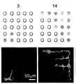
- the morphology of neural networks developed at 14 and 21 DIV was analyzed. From the immunofluorescence images it was confirmed that on substrates with a thin layer of agarose of 10-20 nm good neuronal vitality is observed and the drops of PDL deposited, as well as providing adhesion sites for the neurons, confine the neuronal growth on the surface of the model. So as to have a reference parameter for comparing different degrees of development of the networks on the bioprinted surfaces, the number of spots in which a physical connection between the neurons took place ( fig. 1a ) was analyzed. The spots covered with neurons which do not display extensions towards the neurons present on other spots were not considered as connected.
- a ratio of connected spots / total spots is obtained which is compared for the different steps used to create the matrixes.
- a step is defined as the theoretical centre-centre distance between adjacent spots. All the data were analyzed using OrginPro (OriginLab Corp, Northampton, MA, USA). The statistical significance on the connectivity values was determined by the ANOVA test.
- an empirical connectivity parameter c was defined as the number of adjacent adhesion spots continuously connected by neurites in a matrix, normalized for the total number of spots in the matrix itself.
- the upper panel in fig. 1a shows the connectivity values associated with each of the matrices illustrated.
- the statistical analysis was performed considering the case of a favourable interface, that is a layer of agarose of thickness t ⁇ 20 nm, considering a connectivity threshold c>0.25.
- the results of the mean connectivity value over the population of the matrix which satisfy the criteria are plotted as a function of the inter-spot distance as in fig.1b ).
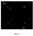
- the neurites develop on the lower arm, connecting spots with a step less than the connectivity distance threshold. Moreover, when they adhere on the spots of PDL of the upper arm, the neurons do not exhibit neuritic extensions and do not survive to 21 DIV.
- the bioassay according to the invention can be implemented by providing a device or a kit which comprises one or more solid supports on which there are created one or more patterns of adhesion spots for neuronal cells, according to arrangements previously described and defined in the appended claims, onto which, during performance of the bioassay, the neuronal cells subjected to the assay can be adhered.
- the kit can if necessary comprise reagents for detection by immunoassay, particularly by immunofluorescence, of the interconnecting neuronal extensions between the spots formed after an incubation period.
- the results obtained according to the invention indicate that the use of the bioassay based on matrixes or patterns of adhered neuronal cells represents a facilitated and simplified experimental model in comparison with in vivo studies, for contributing to the study of functional properties dependent on topology, such as plasticity, regeneration and dynamics of the networks.
- systems of models based on transgenic animals are used in order to investigate such alterations.
- cerebral diseases such as epilepsy, autism or Alzheimer's disease
- the bioassay which is the subject of the invention is capable of providing useful information for the diagnosis of neurodegenerative diseases.
- a test was performed wherein neural cultures from wild-type animals and from genetically modified animals and carriers of DiGeorge syndrome were compared.
- This disease is known to introduce dysfunctionalities in neuronal growth and in particular in the development of the dendritic branchings.
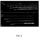
- the cells were plated and grown up to 14 DIV on substrates with an agarose coating and decorated with patterns of poly-D-lysine according to the technique described; the preselected patterns were series of 5 parallel lines of equidistant spots; each line contained up to 30 spots and the distances between spots were different, in particular steadily increasing, from line to line and equalled 30 ⁇ m, 40 ⁇ m, 50 ⁇ m and 70 ⁇ m.
- Fig.3 shows the immunofluorescence images obtained from the neurons belonging to the two types of animals on two typical identical patterns.
- the neurons belonging to the wild-type mice ( Fig.3a ) grow dendrites and connect spots until they cover distances of 50 ⁇ m.
- the branchings of the neurons of the genetically modified animals manage only to cover distances equal to 40 ⁇ m ( Fig. 3b ).
- electrodes corresponding to the spots could be inserted with the aim of quantifying not only the connectivity, but also the functional activity of the neuronal connections which have been formed.
Landscapes
- Health & Medical Sciences (AREA)
- Life Sciences & Earth Sciences (AREA)
- Engineering & Computer Science (AREA)
- Biomedical Technology (AREA)
- Immunology (AREA)
- Chemical & Material Sciences (AREA)
- Hematology (AREA)
- Urology & Nephrology (AREA)
- Molecular Biology (AREA)
- Cell Biology (AREA)
- Analytical Chemistry (AREA)
- Biochemistry (AREA)
- Biotechnology (AREA)
- Tropical Medicine & Parasitology (AREA)
- Bioinformatics & Cheminformatics (AREA)
- Food Science & Technology (AREA)
- Medicinal Chemistry (AREA)
- Physics & Mathematics (AREA)
- Toxicology (AREA)
- Microbiology (AREA)
- General Health & Medical Sciences (AREA)
- General Physics & Mathematics (AREA)
- Pathology (AREA)
- Neurology (AREA)
- Neurosurgery (AREA)
- Measuring Or Testing Involving Enzymes Or Micro-Organisms (AREA)
- Investigating Or Analysing Biological Materials (AREA)
- Investigating Or Analysing Materials By The Use Of Chemical Reactions (AREA)
- Investigating Or Analyzing Materials By The Use Of Electric Means (AREA)
- Apparatus Associated With Microorganisms And Enzymes (AREA)
Claims (15)
- Bioassay-Verfahren zur Bestimmung von Veränderungen in der neuronalen Konnektivität und/oder -Morphologie, dadurch gekennzeichnet, dass es die folgenden Abläufe aufweist:- Ermitteln des kritischen Abstands, der dem entspricht, bei dem ein Paar von beabstandeten einzelnen Nervenzellen, die dem Assay unterzogen werden, noch in der Lage ist, wechselseitig verbindende neuritische Fortsätze auszubilden, nach einer vorgegebenen Inkubationszeit unter Bedingungen, die die Ausbildung solcher neuritischer Fortsätze begünstigen,
wobei jede der einzelnen Nervenzellen an einer entsprechenden Stelle einer das Anhaften der Nervenzellen begünstigenden Substanz angebunden wird, wobei die Stellen voneinander beabstandet auf einem das Anhaften von Nervenzellen hemmenden Substrat angeordnet sind, jede Stelle von dem das Anhaften hemmenden Substrat umgeben ist, und wobei die Stellen mindestens ein Muster oder eine Vielzahl von Mustern bilden, die jeweils von einer Mehrzahl getrennter Stellen gebildet werden, so dass das mindestens eine Muster von Stellen gebildet wird, die in einer zunehmend ansteigenden Schrittweite voneinander beabstandet sind, oder jedes der Mehrzahl von Mustern durch äquidistante Stellen gebildet wird, wobei jedes Muster eine Schrittweite zwischen den Stellen aufweist, die sich von der Schrittweite der anderen Muster unterscheidet, und- Vergleichen des ermittelten kritischen Abstands mit dem unter denselben Bedingungen für Vergleichszellen bestimmten kritischen Abstand, wobei eine Abnahme des kritischen Abstands für die dem Assay unterworfenen Nervenzellen im Vergleich zu dem kritischen Abstand für die Vergleichszellen einen Hinweis auf eine Veränderung in der neuronalen Konnektivität und/oder -Morphologie ist. - Verfahren nach Anspruch 1, dadurch gekennzeichnet, dass es die folgenden Abläufe aufweist:a) Erstellen von einer Mehrzahl von Paaren von Stellen aus einer das Anhaften der Nervenzellen fördernden Substanz auf einem das Anhaften von Nervenzellen hemmenden Substrat, wobei jede der Stellen von den anderen Stellen getrennt und unterscheidbar ist und wobei mindestens zwei der Paare einen unterschiedlichen Abstand zwischen den Stellen aufweisen;b) Auftragen und Ermöglichen des Anhaftens einer einzelnen dem Assay unterworfenen Nervenzelle an jeweils jeder der Stellen;c) Ermitteln der möglichen Ausbildung neuritischer Fortsätze, die an den Stellen des Paares anhaftende benachbarte Nervenzellen miteinander verbinden, nach vorgegebener Inkubationszeit; undd) Bestimmen des kritischen Abstands als dem maximalen Abstand zwischen den Stellen der Paare, die denen entsprechen, zwischen denen die Ausbildung der verbindenden neuritischen Fortsätze auftritt, wobei der kritische Abstand durch Vergleich des neuritischen Fortsatzverhaltens zwischen Paaren von Stellen mit verschiedenen Abständen bestimmt wird.
- Verfahren nach Anspruch 1 oder 2, dadurch gekennzeichnet, dass die Stellen der das Anhaften fördernden Substanz gemäß mindestens einem Muster, das zwischen aufeinanderfolgenden Stellen einen zunehmenden Abstandsschritt aufweist, auf dem Substrat angeordnet sind.
- Verfahren nach Anspruch 1 oder 2, wobei eine Mehrzahl von Mustern auf dem zuvor genannten Substrat aufgebracht wird, von denen jedes eine Mehrzahl von gleichmäßig beabstandeten Stellen aufweist, wobei jedes Muster einen gleichen Abstandsschritt aufweist, der von dem gleichen Abstandsschritt der anderen Muster verschieden ist.
- Verfahren nach einem der Ansprüche 2 bis 4, dadurch gekennzeichnet, dass es den Schritt des Wiederholens der Abläufe a), b) und c) unter den gleichen Bedingungen unter der Verwendung eines Substrats umfasst, das mindestens ein Muster mit gleich beabstandeten Stellen mit einer Abnahme oder Zunahme des gleichen Abstandsschritts zwischen den Stellen bei jeder Wiederholung aufweist, um den kritischen Abstand zu bestimmen, der dem entspricht, bei dem die Ausbildung der Fortsätze nachgewiesen bzw. nicht nachgewiesen wird.
- Verfahren nach einem der vorangehenden Ansprüche, dadurch gekennzeichnet, dass der Abstand zwischen den Stellen des Paares im Bereich zwischen 5 µm und 200 µm liegt.
- Verfahren nach einem der vorangehenden Ansprüche, dadurch gekennzeichnet, dass die das Anhaften hemmende Substanz eine Schicht aus Agarose oder Polyethylenglykol mit einer Dicke von weniger als 40 nm ist.
- Verfahren nach einem der vorangehenden Ansprüche, wobei die das Anhaften fördernde Substanz ein Polypeptid mit freiliegenden Amino-Gruppen oder ein Polymer mit Amino-Gruppen, oder Nanopartikeln, die Stellen zur Zellanhaftung bereitstellen, ist.
- Verfahren nach einem der vorangehenden Ansprüche, wobei der Schritt zum Nachweis der Ausbildung der neuritischen Fortsätze durch einen Immunfluoreszenz-Assay oder durch einen auf optischen oder elektrischen Methoden basierenden Assay ausgeführt wird, wie beispielsweise Impedanzmessungen an in dem Substrat eingebetteten Elektroden oder in dem Substrat eingebetteter Optik.
- Verfahren nach einem der vorangehenden Ansprüche, das zur Bestimmung des kritischen Verbindungsabstands von Nervenzellen eines Wildtyp-Tieres und von genetisch veränderten Nervenzellen desselben Tieres der Reihe nach oder parallel angewendet wird.
- Verfahren nach einem der Ansprüche 1 bis 9 zur Bestimmung von Veränderungen im Wachstum neuritischer Fortsätze von menschlichen Nervenzellen in Kulturen, die aus durch zellbasierte Umprogrammierung erhaltenen nicht embryonischen Stammzellen abgeleitet sind.
- Verfahren nach einem der Ansprüche 1 bis 10 zur Bestimmung der Wirkung eines exogenen Wirkstoffs, wie beispielsweise eines Medikaments oder eines toxikologischen Wirkstoffs auf Nervenzellen, das den Vergleich zwischen dem kritischen Verbindungsabstand zwischen Nervenzellen, die der Aktivität des Wirkstoffs ausgesetzt oder nicht ausgesetzt sind, aufweist.
- Satz zur Ausführung eines Verfahrens nach einem der Ansprüche 1 bis 12, dadurch gekennzeichnet, dass er einen oder mehrere feste Träger aufweist, die mindestens ein Muster oder eine Mehrzahl von Mustern aufweisen, die jeweils durch eine Mehrzahl getrennter Stellen einer das Anhaften von Nervenzellen fördernden Substanz ausgebildet sind und auf einem das Anhaften der Nervenzellen hemmenden Substrat angeordnet sind, wobei die das Anhaften hemmende Substanz als eine Beschichtung des Trägers aufgetragen ist und wobei mindestens ein Muster durch Stellen ausgebildet ist, die mit einer zunehmend ansteigenden Schrittweite voneinander beabstandet sind, oder jedes der Mehrzahl von Mustern durch zueinander äquidistante Stellen ausgebildet ist, wobei jedes Muster eine Schrittweite zwischen den Stellen aufweist, die von der Schrittweite der anderen Muster verschieden ist, und wahlweise Reagenzien zum Nachweis von verbindenden neuritischen Fortsätzen zwischen den Stellen durch einen Immuno-Assay, und wobei die Stellen eine maximale Größe aufweisen, um im Durchschnitt das Anhaften einer einzelnen Nervenzelle an jeder der Stellen zu erlauben, und wobei jede Stelle von der das Anhaften hemmenden Substanz umgeben ist.
- Satz nach Anspruch 13, wobei das das Anhaften hemmende Substrat eine Schicht aus Agarose oder Polyethylenglykol, vorzugsweise mit einer Dicke von weniger als 40 nm ist.
- Satz nach Anspruch 13 oder 14, wobei die das Anhaften fördernde Substanz ein Polypeptid mit einer freiliegenden Amino-Gruppe oder ein Polymer mit Amino-Gruppen ist.
Applications Claiming Priority (2)
| Application Number | Priority Date | Filing Date | Title |
|---|---|---|---|
| IT000197A ITTO20120197A1 (it) | 2012-03-07 | 2012-03-07 | Biosaggio e dispositivo per rivelare e/o quantificare la capacita' di cellule neuronali di formare estensioni neuritiche e/o connettere altri neuroni |
| PCT/IB2013/051804 WO2013132455A1 (en) | 2012-03-07 | 2013-03-07 | Process and device for the determination of alterations in neuronal connectivity and/or morphology |
Publications (2)
| Publication Number | Publication Date |
|---|---|
| EP2823302A1 EP2823302A1 (de) | 2015-01-14 |
| EP2823302B1 true EP2823302B1 (de) | 2017-05-10 |
Family
ID=46028083
Family Applications (1)
| Application Number | Title | Priority Date | Filing Date |
|---|---|---|---|
| EP13720562.1A Not-in-force EP2823302B1 (de) | 2012-03-07 | 2013-03-07 | Verfahren und vorrichtung zur feststellung von änderungen der neuronalen konnektivität und/oder morphologie |
Country Status (5)
| Country | Link |
|---|---|
| US (1) | US20150051109A1 (de) |
| EP (1) | EP2823302B1 (de) |
| JP (1) | JP6190394B2 (de) |
| IT (1) | ITTO20120197A1 (de) |
| WO (1) | WO2013132455A1 (de) |
Families Citing this family (4)
| Publication number | Priority date | Publication date | Assignee | Title |
|---|---|---|---|---|
| SG10201700633QA (en) | 2016-02-03 | 2017-09-28 | Gen Electric | System and method for protecting a wireless power transfer system |
| EP3203606A1 (de) | 2016-02-03 | 2017-08-09 | General Electric Company | Verfahren und system zum schutz eines systems zur drahtlosen stromübertragung |
| SG10201707385XA (en) | 2016-09-30 | 2018-04-27 | Gen Electric | Over voltage protection for a wireless power transfer system |
| CN112945671A (zh) * | 2021-02-07 | 2021-06-11 | 南昌大学附属口腔医院(江西省口腔医院) | 一种粘附载玻片及其制备方法和应用 |
Family Cites Families (1)
| Publication number | Priority date | Publication date | Assignee | Title |
|---|---|---|---|---|
| FR2937050A1 (fr) * | 2008-10-10 | 2010-04-16 | Inst Curie | Dispositif de culture cellulaire |
-
2012
- 2012-03-07 IT IT000197A patent/ITTO20120197A1/it unknown
-
2013
- 2013-03-07 WO PCT/IB2013/051804 patent/WO2013132455A1/en not_active Ceased
- 2013-03-07 US US14/383,501 patent/US20150051109A1/en not_active Abandoned
- 2013-03-07 JP JP2014560506A patent/JP6190394B2/ja not_active Expired - Fee Related
- 2013-03-07 EP EP13720562.1A patent/EP2823302B1/de not_active Not-in-force
Non-Patent Citations (1)
| Title |
|---|
| None * |
Also Published As
| Publication number | Publication date |
|---|---|
| JP6190394B2 (ja) | 2017-08-30 |
| JP2015513404A (ja) | 2015-05-14 |
| WO2013132455A1 (en) | 2013-09-12 |
| WO2013132455A8 (en) | 2014-01-03 |
| ITTO20120197A1 (it) | 2013-09-08 |
| EP2823302A1 (de) | 2015-01-14 |
| US20150051109A1 (en) | 2015-02-19 |
Similar Documents
| Publication | Publication Date | Title |
|---|---|---|
| EP2621584B1 (de) | Nanowires für elektrophysiologische anwendungen | |
| JP6151644B2 (ja) | 操作装置、堆積方法、注入方法、検出装置及び検出方法 | |
| CN104760922B (zh) | 一种超微平面电极阵列传感器及其制备方法 | |
| Vogt et al. | Micropatterned substrates for the growth of functional neuronal networks of defined geometry | |
| US9719982B2 (en) | Devices comprising muscle thin films and uses thereof in high throughput assays for determining contractile function | |
| US20210371782A1 (en) | Methods for optical micropatterning of hydrogels and uses thereof | |
| Petrelli et al. | Nano-volume drop patterning for rapid on-chip neuronal connect-ability assays | |
| Zhang et al. | Quantifying molecular-level cell adhesion on electroactive conducting polymers using electrochemical-single cell force spectroscopy | |
| US20150051109A1 (en) | Process and device for the determination of alterations in neuronal connectivity and/or morphology | |
| US20180264466A1 (en) | Position-defined cell culture and characterization platform | |
| US20260071163A1 (en) | Organoids, embryos, and other tissues containing electronics, and methods thereof | |
| Mateus et al. | Improved in vitro electrophysiology using 3D-structured microelectrode arrays with a micro-mushrooms islets architecture capable of promoting topotaxis | |
| US20170069860A1 (en) | Cell-based electromechanical biocomputing | |
| Maïno et al. | Impedance spectroscopy of the cell/nanovolcano interface enables optimization for electrophysiology | |
| Barizuddin et al. | Automated targeting of cells to electrochemical electrodes using a surface chemistry approach for the measurement of quantal exocytosis | |
| US12540938B1 (en) | Real-time patch-clamp sensing of transport across a barrier tissue | |
| Kim et al. | Enhancement of cardiac contractility using gold-coated SU-8 cantilevers and their application to drug-induced cardiac toxicity tests | |
| Lakard et al. | Synthesis of polymer materials for use as cell culture substrates | |
| EP2675888B1 (de) | Auf neuronalem netzwerk basierender biosensor | |
| Ngalim et al. | Creating adhesive and soluble gradients for imaging cell migration with fluorescence microscopy | |
| Ma et al. | Atomic force and confocal microscopy for the study of cortical cells cultured on silicon wafers | |
| WO2026080666A1 (en) | Devices and methods for multiplexed chemical and electrical measurements of neurons using cmos electrode arrays | |
| Timmerman et al. | Synaptrode: neural interface at the synapse level | |
| Wu | Semiconducting silicon nanowire array fabrication for high throughput screening in the biosciences | |
| Woodhams | Graphene Microelectrode Arrays to Combine Electrophysiology with Fluorescence Imaging of Amyloid Proteins |
Legal Events
| Date | Code | Title | Description |
|---|---|---|---|
| PUAI | Public reference made under article 153(3) epc to a published international application that has entered the european phase |
Free format text: ORIGINAL CODE: 0009012 |
|
| 17P | Request for examination filed |
Effective date: 20140819 |
|
| AK | Designated contracting states |
Kind code of ref document: A1 Designated state(s): AL AT BE BG CH CY CZ DE DK EE ES FI FR GB GR HR HU IE IS IT LI LT LU LV MC MK MT NL NO PL PT RO RS SE SI SK SM TR |
|
| AX | Request for extension of the european patent |
Extension state: BA ME |
|
| DAX | Request for extension of the european patent (deleted) | ||
| GRAP | Despatch of communication of intention to grant a patent |
Free format text: ORIGINAL CODE: EPIDOSNIGR1 |
|
| INTG | Intention to grant announced |
Effective date: 20161014 |
|
| GRAS | Grant fee paid |
Free format text: ORIGINAL CODE: EPIDOSNIGR3 |
|
| GRAJ | Information related to disapproval of communication of intention to grant by the applicant or resumption of examination proceedings by the epo deleted |
Free format text: ORIGINAL CODE: EPIDOSDIGR1 |
|
| GRAL | Information related to payment of fee for publishing/printing deleted |
Free format text: ORIGINAL CODE: EPIDOSDIGR3 |
|
| INTC | Intention to grant announced (deleted) | ||
| GRAR | Information related to intention to grant a patent recorded |
Free format text: ORIGINAL CODE: EPIDOSNIGR71 |
|
| GRAA | (expected) grant |
Free format text: ORIGINAL CODE: 0009210 |
|
| INTG | Intention to grant announced |
Effective date: 20170330 |
|
| AK | Designated contracting states |
Kind code of ref document: B1 Designated state(s): AL AT BE BG CH CY CZ DE DK EE ES FI FR GB GR HR HU IE IS IT LI LT LU LV MC MK MT NL NO PL PT RO RS SE SI SK SM TR |
|
| REG | Reference to a national code |
Ref country code: GB Ref legal event code: FG4D |
|
| REG | Reference to a national code |
Ref country code: AT Ref legal event code: REF Ref document number: 892888 Country of ref document: AT Kind code of ref document: T Effective date: 20170515 Ref country code: CH Ref legal event code: EP |
|
| REG | Reference to a national code |
Ref country code: IE Ref legal event code: FG4D |
|
| REG | Reference to a national code |
Ref country code: DE Ref legal event code: R096 Ref document number: 602013020941 Country of ref document: DE |
|
| REG | Reference to a national code |
Ref country code: NL Ref legal event code: MP Effective date: 20170510 |
|
| REG | Reference to a national code |
Ref country code: LT Ref legal event code: MG4D |
|
| REG | Reference to a national code |
Ref country code: AT Ref legal event code: MK05 Ref document number: 892888 Country of ref document: AT Kind code of ref document: T Effective date: 20170510 |
|
| PG25 | Lapsed in a contracting state [announced via postgrant information from national office to epo] |
Ref country code: FI Free format text: LAPSE BECAUSE OF FAILURE TO SUBMIT A TRANSLATION OF THE DESCRIPTION OR TO PAY THE FEE WITHIN THE PRESCRIBED TIME-LIMIT Effective date: 20170510 Ref country code: LT Free format text: LAPSE BECAUSE OF FAILURE TO SUBMIT A TRANSLATION OF THE DESCRIPTION OR TO PAY THE FEE WITHIN THE PRESCRIBED TIME-LIMIT Effective date: 20170510 Ref country code: ES Free format text: LAPSE BECAUSE OF FAILURE TO SUBMIT A TRANSLATION OF THE DESCRIPTION OR TO PAY THE FEE WITHIN THE PRESCRIBED TIME-LIMIT Effective date: 20170510 Ref country code: NO Free format text: LAPSE BECAUSE OF FAILURE TO SUBMIT A TRANSLATION OF THE DESCRIPTION OR TO PAY THE FEE WITHIN THE PRESCRIBED TIME-LIMIT Effective date: 20170810 Ref country code: AT Free format text: LAPSE BECAUSE OF FAILURE TO SUBMIT A TRANSLATION OF THE DESCRIPTION OR TO PAY THE FEE WITHIN THE PRESCRIBED TIME-LIMIT Effective date: 20170510 Ref country code: GR Free format text: LAPSE BECAUSE OF FAILURE TO SUBMIT A TRANSLATION OF THE DESCRIPTION OR TO PAY THE FEE WITHIN THE PRESCRIBED TIME-LIMIT Effective date: 20170811 Ref country code: HR Free format text: LAPSE BECAUSE OF FAILURE TO SUBMIT A TRANSLATION OF THE DESCRIPTION OR TO PAY THE FEE WITHIN THE PRESCRIBED TIME-LIMIT Effective date: 20170510 |
|
| PG25 | Lapsed in a contracting state [announced via postgrant information from national office to epo] |
Ref country code: IS Free format text: LAPSE BECAUSE OF FAILURE TO SUBMIT A TRANSLATION OF THE DESCRIPTION OR TO PAY THE FEE WITHIN THE PRESCRIBED TIME-LIMIT Effective date: 20170910 Ref country code: PL Free format text: LAPSE BECAUSE OF FAILURE TO SUBMIT A TRANSLATION OF THE DESCRIPTION OR TO PAY THE FEE WITHIN THE PRESCRIBED TIME-LIMIT Effective date: 20170510 Ref country code: RS Free format text: LAPSE BECAUSE OF FAILURE TO SUBMIT A TRANSLATION OF THE DESCRIPTION OR TO PAY THE FEE WITHIN THE PRESCRIBED TIME-LIMIT Effective date: 20170510 Ref country code: BG Free format text: LAPSE BECAUSE OF FAILURE TO SUBMIT A TRANSLATION OF THE DESCRIPTION OR TO PAY THE FEE WITHIN THE PRESCRIBED TIME-LIMIT Effective date: 20170810 Ref country code: SE Free format text: LAPSE BECAUSE OF FAILURE TO SUBMIT A TRANSLATION OF THE DESCRIPTION OR TO PAY THE FEE WITHIN THE PRESCRIBED TIME-LIMIT Effective date: 20170510 Ref country code: NL Free format text: LAPSE BECAUSE OF FAILURE TO SUBMIT A TRANSLATION OF THE DESCRIPTION OR TO PAY THE FEE WITHIN THE PRESCRIBED TIME-LIMIT Effective date: 20170510 Ref country code: LV Free format text: LAPSE BECAUSE OF FAILURE TO SUBMIT A TRANSLATION OF THE DESCRIPTION OR TO PAY THE FEE WITHIN THE PRESCRIBED TIME-LIMIT Effective date: 20170510 |
|
| PG25 | Lapsed in a contracting state [announced via postgrant information from national office to epo] |
Ref country code: CZ Free format text: LAPSE BECAUSE OF FAILURE TO SUBMIT A TRANSLATION OF THE DESCRIPTION OR TO PAY THE FEE WITHIN THE PRESCRIBED TIME-LIMIT Effective date: 20170510 Ref country code: SK Free format text: LAPSE BECAUSE OF FAILURE TO SUBMIT A TRANSLATION OF THE DESCRIPTION OR TO PAY THE FEE WITHIN THE PRESCRIBED TIME-LIMIT Effective date: 20170510 Ref country code: DK Free format text: LAPSE BECAUSE OF FAILURE TO SUBMIT A TRANSLATION OF THE DESCRIPTION OR TO PAY THE FEE WITHIN THE PRESCRIBED TIME-LIMIT Effective date: 20170510 Ref country code: RO Free format text: LAPSE BECAUSE OF FAILURE TO SUBMIT A TRANSLATION OF THE DESCRIPTION OR TO PAY THE FEE WITHIN THE PRESCRIBED TIME-LIMIT Effective date: 20170510 Ref country code: EE Free format text: LAPSE BECAUSE OF FAILURE TO SUBMIT A TRANSLATION OF THE DESCRIPTION OR TO PAY THE FEE WITHIN THE PRESCRIBED TIME-LIMIT Effective date: 20170510 |
|
| REG | Reference to a national code |
Ref country code: DE Ref legal event code: R097 Ref document number: 602013020941 Country of ref document: DE |
|
| PG25 | Lapsed in a contracting state [announced via postgrant information from national office to epo] |
Ref country code: SM Free format text: LAPSE BECAUSE OF FAILURE TO SUBMIT A TRANSLATION OF THE DESCRIPTION OR TO PAY THE FEE WITHIN THE PRESCRIBED TIME-LIMIT Effective date: 20170510 |
|
| PLBE | No opposition filed within time limit |
Free format text: ORIGINAL CODE: 0009261 |
|
| STAA | Information on the status of an ep patent application or granted ep patent |
Free format text: STATUS: NO OPPOSITION FILED WITHIN TIME LIMIT |
|
| REG | Reference to a national code |
Ref country code: FR Ref legal event code: PLFP Year of fee payment: 6 |
|
| 26N | No opposition filed |
Effective date: 20180213 |
|
| PG25 | Lapsed in a contracting state [announced via postgrant information from national office to epo] |
Ref country code: SI Free format text: LAPSE BECAUSE OF FAILURE TO SUBMIT A TRANSLATION OF THE DESCRIPTION OR TO PAY THE FEE WITHIN THE PRESCRIBED TIME-LIMIT Effective date: 20170510 |
|
| REG | Reference to a national code |
Ref country code: CH Ref legal event code: PL |
|
| PG25 | Lapsed in a contracting state [announced via postgrant information from national office to epo] |
Ref country code: MC Free format text: LAPSE BECAUSE OF FAILURE TO SUBMIT A TRANSLATION OF THE DESCRIPTION OR TO PAY THE FEE WITHIN THE PRESCRIBED TIME-LIMIT Effective date: 20170510 |
|
| REG | Reference to a national code |
Ref country code: BE Ref legal event code: MM Effective date: 20180331 |
|
| REG | Reference to a national code |
Ref country code: IE Ref legal event code: MM4A |
|
| PG25 | Lapsed in a contracting state [announced via postgrant information from national office to epo] |
Ref country code: LU Free format text: LAPSE BECAUSE OF NON-PAYMENT OF DUE FEES Effective date: 20180307 |
|
| PG25 | Lapsed in a contracting state [announced via postgrant information from national office to epo] |
Ref country code: IE Free format text: LAPSE BECAUSE OF NON-PAYMENT OF DUE FEES Effective date: 20180307 |
|
| PG25 | Lapsed in a contracting state [announced via postgrant information from national office to epo] |
Ref country code: LI Free format text: LAPSE BECAUSE OF NON-PAYMENT OF DUE FEES Effective date: 20180331 Ref country code: CH Free format text: LAPSE BECAUSE OF NON-PAYMENT OF DUE FEES Effective date: 20180331 Ref country code: BE Free format text: LAPSE BECAUSE OF NON-PAYMENT OF DUE FEES Effective date: 20180331 |
|
| PG25 | Lapsed in a contracting state [announced via postgrant information from national office to epo] |
Ref country code: MT Free format text: LAPSE BECAUSE OF NON-PAYMENT OF DUE FEES Effective date: 20180307 |
|
| PG25 | Lapsed in a contracting state [announced via postgrant information from national office to epo] |
Ref country code: TR Free format text: LAPSE BECAUSE OF FAILURE TO SUBMIT A TRANSLATION OF THE DESCRIPTION OR TO PAY THE FEE WITHIN THE PRESCRIBED TIME-LIMIT Effective date: 20170510 |
|
| PG25 | Lapsed in a contracting state [announced via postgrant information from national office to epo] |
Ref country code: PT Free format text: LAPSE BECAUSE OF FAILURE TO SUBMIT A TRANSLATION OF THE DESCRIPTION OR TO PAY THE FEE WITHIN THE PRESCRIBED TIME-LIMIT Effective date: 20170510 Ref country code: HU Free format text: LAPSE BECAUSE OF FAILURE TO SUBMIT A TRANSLATION OF THE DESCRIPTION OR TO PAY THE FEE WITHIN THE PRESCRIBED TIME-LIMIT; INVALID AB INITIO Effective date: 20130307 |
|
| PG25 | Lapsed in a contracting state [announced via postgrant information from national office to epo] |
Ref country code: CY Free format text: LAPSE BECAUSE OF FAILURE TO SUBMIT A TRANSLATION OF THE DESCRIPTION OR TO PAY THE FEE WITHIN THE PRESCRIBED TIME-LIMIT Effective date: 20170510 Ref country code: MK Free format text: LAPSE BECAUSE OF NON-PAYMENT OF DUE FEES Effective date: 20170510 |
|
| PG25 | Lapsed in a contracting state [announced via postgrant information from national office to epo] |
Ref country code: AL Free format text: LAPSE BECAUSE OF FAILURE TO SUBMIT A TRANSLATION OF THE DESCRIPTION OR TO PAY THE FEE WITHIN THE PRESCRIBED TIME-LIMIT Effective date: 20170510 |
|
| PGFP | Annual fee paid to national office [announced via postgrant information from national office to epo] |
Ref country code: GB Payment date: 20220321 Year of fee payment: 10 Ref country code: DE Payment date: 20220322 Year of fee payment: 10 |
|
| PGFP | Annual fee paid to national office [announced via postgrant information from national office to epo] |
Ref country code: FR Payment date: 20220331 Year of fee payment: 10 |
|
| P01 | Opt-out of the competence of the unified patent court (upc) registered |
Effective date: 20230811 |
|
| REG | Reference to a national code |
Ref country code: DE Ref legal event code: R119 Ref document number: 602013020941 Country of ref document: DE |
|
| GBPC | Gb: european patent ceased through non-payment of renewal fee |
Effective date: 20230307 |
|
| PG25 | Lapsed in a contracting state [announced via postgrant information from national office to epo] |
Ref country code: GB Free format text: LAPSE BECAUSE OF NON-PAYMENT OF DUE FEES Effective date: 20230307 |
|
| PG25 | Lapsed in a contracting state [announced via postgrant information from national office to epo] |
Ref country code: GB Free format text: LAPSE BECAUSE OF NON-PAYMENT OF DUE FEES Effective date: 20230307 Ref country code: FR Free format text: LAPSE BECAUSE OF NON-PAYMENT OF DUE FEES Effective date: 20230331 Ref country code: DE Free format text: LAPSE BECAUSE OF NON-PAYMENT OF DUE FEES Effective date: 20231003 |
|
| PGFP | Annual fee paid to national office [announced via postgrant information from national office to epo] |
Ref country code: IT Payment date: 20240124 Year of fee payment: 12 |
|
| PG25 | Lapsed in a contracting state [announced via postgrant information from national office to epo] |
Ref country code: IT Free format text: LAPSE BECAUSE OF NON-PAYMENT OF DUE FEES Effective date: 20250307 |