EP2817036B1 - Resorbable cellulose based biomaterial and implant - Google Patents
Resorbable cellulose based biomaterial and implant Download PDFInfo
- Publication number
- EP2817036B1 EP2817036B1 EP13708020.6A EP13708020A EP2817036B1 EP 2817036 B1 EP2817036 B1 EP 2817036B1 EP 13708020 A EP13708020 A EP 13708020A EP 2817036 B1 EP2817036 B1 EP 2817036B1
- Authority
- EP
- European Patent Office
- Prior art keywords
- cellulose
- implant
- irradiated
- oxidized
- weeks
- Prior art date
- Legal status (The legal status is an assumption and is not a legal conclusion. Google has not performed a legal analysis and makes no representation as to the accuracy of the status listed.)
- Active
Links
- 229920002678 cellulose Polymers 0.000 title claims description 196
- 239000001913 cellulose Substances 0.000 title claims description 194
- 239000007943 implant Substances 0.000 title claims description 155
- 239000012620 biological material Substances 0.000 title claims description 19
- 229920002201 Oxidized cellulose Polymers 0.000 claims description 90
- 229940107304 oxidized cellulose Drugs 0.000 claims description 89
- 238000007254 oxidation reaction Methods 0.000 claims description 62
- 238000006731 degradation reaction Methods 0.000 claims description 61
- 230000015556 catabolic process Effects 0.000 claims description 58
- 230000003647 oxidation Effects 0.000 claims description 56
- 239000007800 oxidant agent Substances 0.000 claims description 41
- 239000000463 material Substances 0.000 claims description 32
- 210000001519 tissue Anatomy 0.000 claims description 28
- 238000000338 in vitro Methods 0.000 claims description 26
- XLYOFNOQVPJJNP-UHFFFAOYSA-N water Substances O XLYOFNOQVPJJNP-UHFFFAOYSA-N 0.000 claims description 26
- 239000013543 active substance Substances 0.000 claims description 25
- 239000000203 mixture Substances 0.000 claims description 13
- 239000012530 fluid Substances 0.000 claims description 12
- 230000001698 pyrogenic effect Effects 0.000 claims description 10
- 230000007704 transition Effects 0.000 claims description 9
- 230000000813 microbial effect Effects 0.000 claims description 8
- 235000002837 Acetobacter xylinum Nutrition 0.000 claims description 7
- 210000004872 soft tissue Anatomy 0.000 claims description 7
- 230000036571 hydration Effects 0.000 claims description 6
- 238000006703 hydration reaction Methods 0.000 claims description 6
- 230000005865 ionizing radiation Effects 0.000 claims description 6
- 230000003416 augmentation Effects 0.000 claims description 4
- 210000000988 bone and bone Anatomy 0.000 claims description 4
- 239000007795 chemical reaction product Substances 0.000 claims description 4
- 239000002243 precursor Substances 0.000 claims description 3
- 239000003242 anti bacterial agent Substances 0.000 claims description 2
- 210000001185 bone marrow Anatomy 0.000 claims description 2
- 239000004068 calcium phosphate ceramic Substances 0.000 claims description 2
- 230000000921 morphogenic effect Effects 0.000 claims description 2
- 230000002188 osteogenic effect Effects 0.000 claims description 2
- 230000002138 osteoinductive effect Effects 0.000 claims description 2
- 102000004169 proteins and genes Human genes 0.000 claims description 2
- 108090000623 proteins and genes Proteins 0.000 claims description 2
- 150000003384 small molecules Chemical class 0.000 claims description 2
- 210000000130 stem cell Anatomy 0.000 claims description 2
- 241001136169 Komagataeibacter xylinus Species 0.000 claims 1
- 235000010980 cellulose Nutrition 0.000 description 188
- 239000000523 sample Substances 0.000 description 50
- 238000000034 method Methods 0.000 description 43
- 239000012528 membrane Substances 0.000 description 39
- 210000004379 membrane Anatomy 0.000 description 38
- HEMHJVSKTPXQMS-UHFFFAOYSA-M Sodium hydroxide Chemical compound [OH-].[Na+] HEMHJVSKTPXQMS-UHFFFAOYSA-M 0.000 description 36
- 238000012360 testing method Methods 0.000 description 27
- CURLTUGMZLYLDI-UHFFFAOYSA-N Carbon dioxide Chemical compound O=C=O CURLTUGMZLYLDI-UHFFFAOYSA-N 0.000 description 22
- 229920001340 Microbial cellulose Polymers 0.000 description 20
- KHIWWQKSHDUIBK-UHFFFAOYSA-N periodic acid Chemical compound OI(=O)(=O)=O KHIWWQKSHDUIBK-UHFFFAOYSA-N 0.000 description 18
- 238000002513 implantation Methods 0.000 description 17
- 230000005855 radiation Effects 0.000 description 17
- 239000012890 simulated body fluid Substances 0.000 description 16
- JQWHASGSAFIOCM-UHFFFAOYSA-M sodium periodate Chemical compound [Na+].[O-]I(=O)(=O)=O JQWHASGSAFIOCM-UHFFFAOYSA-M 0.000 description 16
- 241001465754 Metazoa Species 0.000 description 15
- 241000283690 Bos taurus Species 0.000 description 14
- KHIWWQKSHDUIBK-UHFFFAOYSA-M periodate Chemical compound [O-]I(=O)(=O)=O KHIWWQKSHDUIBK-UHFFFAOYSA-M 0.000 description 14
- 229910002092 carbon dioxide Inorganic materials 0.000 description 13
- 102000008186 Collagen Human genes 0.000 description 12
- 108010035532 Collagen Proteins 0.000 description 12
- 229920001436 collagen Polymers 0.000 description 12
- 238000001727 in vivo Methods 0.000 description 12
- 206010061218 Inflammation Diseases 0.000 description 11
- 230000004054 inflammatory process Effects 0.000 description 11
- 238000005259 measurement Methods 0.000 description 10
- 230000008569 process Effects 0.000 description 10
- 238000011282 treatment Methods 0.000 description 10
- LFQSCWFLJHTTHZ-UHFFFAOYSA-N Ethanol Chemical compound CCO LFQSCWFLJHTTHZ-UHFFFAOYSA-N 0.000 description 9
- 206010016654 Fibrosis Diseases 0.000 description 9
- OKKJLVBELUTLKV-UHFFFAOYSA-N Methanol Chemical compound OC OKKJLVBELUTLKV-UHFFFAOYSA-N 0.000 description 9
- 238000006243 chemical reaction Methods 0.000 description 9
- 230000004761 fibrosis Effects 0.000 description 9
- 208000015181 infectious disease Diseases 0.000 description 9
- 206010018852 Haematoma Diseases 0.000 description 8
- FXHOOIRPVKKKFG-UHFFFAOYSA-N N,N-Dimethylacetamide Chemical compound CN(C)C(C)=O FXHOOIRPVKKKFG-UHFFFAOYSA-N 0.000 description 8
- 238000002441 X-ray diffraction Methods 0.000 description 8
- 239000001569 carbon dioxide Substances 0.000 description 8
- WQYVRQLZKVEZGA-UHFFFAOYSA-N hypochlorite Chemical compound Cl[O-] WQYVRQLZKVEZGA-UHFFFAOYSA-N 0.000 description 8
- 238000000352 supercritical drying Methods 0.000 description 8
- 210000002435 tendon Anatomy 0.000 description 8
- 241001269524 Dura Species 0.000 description 7
- 238000000149 argon plasma sintering Methods 0.000 description 7
- 239000002085 irritant Substances 0.000 description 7
- 230000010349 pulsation Effects 0.000 description 7
- 239000000243 solution Substances 0.000 description 7
- 239000000126 substance Substances 0.000 description 7
- MGWGWNFMUOTEHG-UHFFFAOYSA-N 4-(3,5-dimethylphenyl)-1,3-thiazol-2-amine Chemical compound CC1=CC(C)=CC(C=2N=C(N)SC=2)=C1 MGWGWNFMUOTEHG-UHFFFAOYSA-N 0.000 description 6
- 244000235858 Acetobacter xylinum Species 0.000 description 6
- 206010040102 Seroma Diseases 0.000 description 6
- 231100000021 irritant Toxicity 0.000 description 6
- JCXJVPUVTGWSNB-UHFFFAOYSA-N nitrogen dioxide Inorganic materials O=[N]=O JCXJVPUVTGWSNB-UHFFFAOYSA-N 0.000 description 6
- 238000000746 purification Methods 0.000 description 6
- ZNZYKNKBJPZETN-WELNAUFTSA-N Dialdehyde 11678 Chemical group N1C2=CC=CC=C2C2=C1[C@H](C[C@H](/C(=C/O)C(=O)OC)[C@@H](C=C)C=O)NCC2 ZNZYKNKBJPZETN-WELNAUFTSA-N 0.000 description 5
- 206010052428 Wound Diseases 0.000 description 5
- 208000027418 Wounds and injury Diseases 0.000 description 5
- 230000015572 biosynthetic process Effects 0.000 description 5
- 210000004027 cell Anatomy 0.000 description 5
- 238000000855 fermentation Methods 0.000 description 5
- 230000004151 fermentation Effects 0.000 description 5
- 238000005227 gel permeation chromatography Methods 0.000 description 5
- 239000008188 pellet Substances 0.000 description 5
- 238000012545 processing Methods 0.000 description 5
- 210000003625 skull Anatomy 0.000 description 5
- 238000001356 surgical procedure Methods 0.000 description 5
- 239000000725 suspension Substances 0.000 description 5
- 229920002749 Bacterial cellulose Polymers 0.000 description 4
- 241000196324 Embryophyta Species 0.000 description 4
- WQZGKKKJIJFFOK-GASJEMHNSA-N Glucose Chemical group OC[C@H]1OC(O)[C@H](O)[C@@H](O)[C@@H]1O WQZGKKKJIJFFOK-GASJEMHNSA-N 0.000 description 4
- AEMRFAOFKBGASW-UHFFFAOYSA-N Glycolic acid Chemical compound OCC(O)=O AEMRFAOFKBGASW-UHFFFAOYSA-N 0.000 description 4
- 241000283973 Oryctolagus cuniculus Species 0.000 description 4
- FAPWRFPIFSIZLT-UHFFFAOYSA-M Sodium chloride Chemical compound [Na+].[Cl-] FAPWRFPIFSIZLT-UHFFFAOYSA-M 0.000 description 4
- 239000005016 bacterial cellulose Substances 0.000 description 4
- 210000001175 cerebrospinal fluid Anatomy 0.000 description 4
- SOCTUWSJJQCPFX-UHFFFAOYSA-N dichromate(2-) Chemical compound [O-][Cr](=O)(=O)O[Cr]([O-])(=O)=O SOCTUWSJJQCPFX-UHFFFAOYSA-N 0.000 description 4
- 238000009826 distribution Methods 0.000 description 4
- 238000001035 drying Methods 0.000 description 4
- 210000001951 dura mater Anatomy 0.000 description 4
- 238000011156 evaluation Methods 0.000 description 4
- 125000002791 glucosyl group Chemical group C1([C@H](O)[C@@H](O)[C@H](O)[C@H](O1)CO)* 0.000 description 4
- 238000011534 incubation Methods 0.000 description 4
- 238000002347 injection Methods 0.000 description 4
- 239000007924 injection Substances 0.000 description 4
- 239000007788 liquid Substances 0.000 description 4
- KJFMBFZCATUALV-UHFFFAOYSA-N phenolphthalein Chemical compound C1=CC(O)=CC=C1C1(C=2C=CC(O)=CC=2)C2=CC=CC=C2C(=O)O1 KJFMBFZCATUALV-UHFFFAOYSA-N 0.000 description 4
- 229920000642 polymer Polymers 0.000 description 4
- 238000007920 subcutaneous administration Methods 0.000 description 4
- QTBSBXVTEAMEQO-UHFFFAOYSA-N Acetic acid Chemical compound CC(O)=O QTBSBXVTEAMEQO-UHFFFAOYSA-N 0.000 description 3
- LYCAIKOWRPUZTN-UHFFFAOYSA-N Ethylene glycol Chemical compound OCCO LYCAIKOWRPUZTN-UHFFFAOYSA-N 0.000 description 3
- 206010073306 Exposure to radiation Diseases 0.000 description 3
- 238000011887 Necropsy Methods 0.000 description 3
- 238000005119 centrifugation Methods 0.000 description 3
- 238000012668 chain scission Methods 0.000 description 3
- 238000001514 detection method Methods 0.000 description 3
- 238000002845 discoloration Methods 0.000 description 3
- 238000002474 experimental method Methods 0.000 description 3
- 238000013467 fragmentation Methods 0.000 description 3
- 238000006062 fragmentation reaction Methods 0.000 description 3
- 239000007789 gas Substances 0.000 description 3
- 239000002874 hemostatic agent Substances 0.000 description 3
- 230000007062 hydrolysis Effects 0.000 description 3
- 238000006460 hydrolysis reaction Methods 0.000 description 3
- 230000001678 irradiating effect Effects 0.000 description 3
- 230000001788 irregular Effects 0.000 description 3
- 210000002540 macrophage Anatomy 0.000 description 3
- 238000004519 manufacturing process Methods 0.000 description 3
- 230000007246 mechanism Effects 0.000 description 3
- 230000001590 oxidative effect Effects 0.000 description 3
- 210000003516 pericardium Anatomy 0.000 description 3
- 239000008104 plant cellulose Substances 0.000 description 3
- 238000003825 pressing Methods 0.000 description 3
- 239000000047 product Substances 0.000 description 3
- 230000008439 repair process Effects 0.000 description 3
- 238000001878 scanning electron micrograph Methods 0.000 description 3
- 239000002002 slurry Substances 0.000 description 3
- 239000011780 sodium chloride Substances 0.000 description 3
- 238000003786 synthesis reaction Methods 0.000 description 3
- 238000005406 washing Methods 0.000 description 3
- UFYGCFHQAXXBCF-UHFFFAOYSA-N 2,4-dihydroxybutanoic acid Chemical compound OCCC(O)C(O)=O UFYGCFHQAXXBCF-UHFFFAOYSA-N 0.000 description 2
- IJGRMHOSHXDMSA-UHFFFAOYSA-N Atomic nitrogen Chemical compound N#N IJGRMHOSHXDMSA-UHFFFAOYSA-N 0.000 description 2
- 241000894006 Bacteria Species 0.000 description 2
- 229920000742 Cotton Polymers 0.000 description 2
- WSFSSNUMVMOOMR-UHFFFAOYSA-N Formaldehyde Chemical compound O=C WSFSSNUMVMOOMR-UHFFFAOYSA-N 0.000 description 2
- 150000001299 aldehydes Chemical class 0.000 description 2
- 238000004458 analytical method Methods 0.000 description 2
- 239000003125 aqueous solvent Substances 0.000 description 2
- 229920002988 biodegradable polymer Polymers 0.000 description 2
- 239000004621 biodegradable polymer Substances 0.000 description 2
- 230000004071 biological effect Effects 0.000 description 2
- 210000004369 blood Anatomy 0.000 description 2
- 239000008280 blood Substances 0.000 description 2
- 230000009172 bursting Effects 0.000 description 2
- 238000004364 calculation method Methods 0.000 description 2
- 230000000739 chaotic effect Effects 0.000 description 2
- 230000001684 chronic effect Effects 0.000 description 2
- 230000007423 decrease Effects 0.000 description 2
- 230000007547 defect Effects 0.000 description 2
- 238000009795 derivation Methods 0.000 description 2
- WFPZPJSADLPSON-UHFFFAOYSA-N dinitrogen tetraoxide Chemical compound [O-][N+](=O)[N+]([O-])=O WFPZPJSADLPSON-UHFFFAOYSA-N 0.000 description 2
- 239000003792 electrolyte Substances 0.000 description 2
- 150000004676 glycans Chemical class 0.000 description 2
- 230000035876 healing Effects 0.000 description 2
- 230000008595 infiltration Effects 0.000 description 2
- 238000001764 infiltration Methods 0.000 description 2
- 230000028709 inflammatory response Effects 0.000 description 2
- 230000007794 irritation Effects 0.000 description 2
- KWGKDLIKAYFUFQ-UHFFFAOYSA-M lithium chloride Chemical compound [Li+].[Cl-] KWGKDLIKAYFUFQ-UHFFFAOYSA-M 0.000 description 2
- 239000002609 medium Substances 0.000 description 2
- 230000007935 neutral effect Effects 0.000 description 2
- 235000015097 nutrients Nutrition 0.000 description 2
- 239000002245 particle Substances 0.000 description 2
- 150000002978 peroxides Chemical class 0.000 description 2
- -1 polyglactin Polymers 0.000 description 2
- 229920001282 polysaccharide Polymers 0.000 description 2
- 239000004810 polytetrafluoroethylene Substances 0.000 description 2
- 229920001343 polytetrafluoroethylene Polymers 0.000 description 2
- 239000011148 porous material Substances 0.000 description 2
- 150000003254 radicals Chemical class 0.000 description 2
- 230000035484 reaction time Effects 0.000 description 2
- 230000004044 response Effects 0.000 description 2
- 210000003491 skin Anatomy 0.000 description 2
- 239000002904 solvent Substances 0.000 description 2
- 238000010186 staining Methods 0.000 description 2
- 229910001220 stainless steel Inorganic materials 0.000 description 2
- 239000010935 stainless steel Substances 0.000 description 2
- 238000003756 stirring Methods 0.000 description 2
- 210000004304 subcutaneous tissue Anatomy 0.000 description 2
- 230000017423 tissue regeneration Effects 0.000 description 2
- 238000004448 titration Methods 0.000 description 2
- 229920002818 (Hydroxyethyl)methacrylate Polymers 0.000 description 1
- 241000589220 Acetobacter Species 0.000 description 1
- OKTJSMMVPCPJKN-UHFFFAOYSA-N Carbon Chemical compound [C] OKTJSMMVPCPJKN-UHFFFAOYSA-N 0.000 description 1
- 229920003043 Cellulose fiber Polymers 0.000 description 1
- 206010008164 Cerebrospinal fluid leakage Diseases 0.000 description 1
- 206010010356 Congenital anomaly Diseases 0.000 description 1
- 238000005033 Fourier transform infrared spectroscopy Methods 0.000 description 1
- 229920001503 Glucan Polymers 0.000 description 1
- SXRSQZLOMIGNAQ-UHFFFAOYSA-N Glutaraldehyde Chemical compound O=CCCCC=O SXRSQZLOMIGNAQ-UHFFFAOYSA-N 0.000 description 1
- WOBHKFSMXKNTIM-UHFFFAOYSA-N Hydroxyethyl methacrylate Chemical compound CC(=C)C(=O)OCCO WOBHKFSMXKNTIM-UHFFFAOYSA-N 0.000 description 1
- 208000005230 Leg Ulcer Diseases 0.000 description 1
- 239000004677 Nylon Substances 0.000 description 1
- 206010057249 Phagocytosis Diseases 0.000 description 1
- 239000004743 Polypropylene Substances 0.000 description 1
- 102000029797 Prion Human genes 0.000 description 1
- 108091000054 Prion Proteins 0.000 description 1
- 239000004792 Prolene Substances 0.000 description 1
- 229930006000 Sucrose Natural products 0.000 description 1
- CZMRCDWAGMRECN-UGDNZRGBSA-N Sucrose Chemical compound O[C@H]1[C@H](O)[C@@H](CO)O[C@@]1(CO)O[C@@H]1[C@H](O)[C@@H](O)[C@H](O)[C@@H](CO)O1 CZMRCDWAGMRECN-UGDNZRGBSA-N 0.000 description 1
- QAOWNCQODCNURD-UHFFFAOYSA-L Sulfate Chemical compound [O-]S([O-])(=O)=O QAOWNCQODCNURD-UHFFFAOYSA-L 0.000 description 1
- 208000002847 Surgical Wound Diseases 0.000 description 1
- 240000008042 Zea mays Species 0.000 description 1
- 235000005824 Zea mays ssp. parviglumis Nutrition 0.000 description 1
- 235000002017 Zea mays subsp mays Nutrition 0.000 description 1
- 239000002253 acid Substances 0.000 description 1
- 150000003863 ammonium salts Chemical class 0.000 description 1
- 229940030225 antihemorrhagics Drugs 0.000 description 1
- 238000013459 approach Methods 0.000 description 1
- 230000003190 augmentative effect Effects 0.000 description 1
- 230000001580 bacterial effect Effects 0.000 description 1
- 230000004888 barrier function Effects 0.000 description 1
- 230000009286 beneficial effect Effects 0.000 description 1
- 229920000249 biocompatible polymer Polymers 0.000 description 1
- 238000006065 biodegradation reaction Methods 0.000 description 1
- 229920001222 biopolymer Polymers 0.000 description 1
- 210000004204 blood vessel Anatomy 0.000 description 1
- 239000002639 bone cement Substances 0.000 description 1
- 239000001506 calcium phosphate Substances 0.000 description 1
- 229910000389 calcium phosphate Inorganic materials 0.000 description 1
- 235000011010 calcium phosphates Nutrition 0.000 description 1
- 239000002775 capsule Substances 0.000 description 1
- 150000001720 carbohydrates Chemical class 0.000 description 1
- 229910052799 carbon Inorganic materials 0.000 description 1
- 210000000845 cartilage Anatomy 0.000 description 1
- 125000002091 cationic group Chemical group 0.000 description 1
- 239000003518 caustics Substances 0.000 description 1
- 230000001413 cellular effect Effects 0.000 description 1
- 230000008859 change Effects 0.000 description 1
- 150000001805 chlorine compounds Chemical class 0.000 description 1
- KRVSOGSZCMJSLX-UHFFFAOYSA-L chromic acid Substances O[Cr](O)(=O)=O KRVSOGSZCMJSLX-UHFFFAOYSA-L 0.000 description 1
- 208000037976 chronic inflammation Diseases 0.000 description 1
- 230000006020 chronic inflammation Effects 0.000 description 1
- 238000004140 cleaning Methods 0.000 description 1
- 238000003776 cleavage reaction Methods 0.000 description 1
- 230000000052 comparative effect Effects 0.000 description 1
- 238000007906 compression Methods 0.000 description 1
- 230000006835 compression Effects 0.000 description 1
- 239000013068 control sample Substances 0.000 description 1
- 235000005822 corn Nutrition 0.000 description 1
- 238000004132 cross linking Methods 0.000 description 1
- 238000005520 cutting process Methods 0.000 description 1
- 230000006378 damage Effects 0.000 description 1
- 230000007812 deficiency Effects 0.000 description 1
- 239000007857 degradation product Substances 0.000 description 1
- 230000003111 delayed effect Effects 0.000 description 1
- 210000004207 dermis Anatomy 0.000 description 1
- 238000011161 development Methods 0.000 description 1
- 230000018109 developmental process Effects 0.000 description 1
- 201000010099 disease Diseases 0.000 description 1
- 208000037265 diseases, disorders, signs and symptoms Diseases 0.000 description 1
- 238000006073 displacement reaction Methods 0.000 description 1
- 238000004090 dissolution Methods 0.000 description 1
- 230000000694 effects Effects 0.000 description 1
- 239000008151 electrolyte solution Substances 0.000 description 1
- 239000002158 endotoxin Substances 0.000 description 1
- 230000002255 enzymatic effect Effects 0.000 description 1
- 210000003979 eosinophil Anatomy 0.000 description 1
- 238000001704 evaporation Methods 0.000 description 1
- 230000008020 evaporation Effects 0.000 description 1
- 230000001815 facial effect Effects 0.000 description 1
- 239000000835 fiber Substances 0.000 description 1
- 238000001914 filtration Methods 0.000 description 1
- 239000011888 foil Substances 0.000 description 1
- 235000013305 food Nutrition 0.000 description 1
- 239000012634 fragment Substances 0.000 description 1
- AWJWCTOOIBYHON-UHFFFAOYSA-N furo[3,4-b]pyrazine-5,7-dione Chemical compound C1=CN=C2C(=O)OC(=O)C2=N1 AWJWCTOOIBYHON-UHFFFAOYSA-N 0.000 description 1
- 239000011521 glass Substances 0.000 description 1
- PCHJSUWPFVWCPO-UHFFFAOYSA-N gold Chemical compound [Au] PCHJSUWPFVWCPO-UHFFFAOYSA-N 0.000 description 1
- 239000010931 gold Substances 0.000 description 1
- 229910052737 gold Inorganic materials 0.000 description 1
- 239000001963 growth medium Substances 0.000 description 1
- 238000007490 hematoxylin and eosin (H&E) staining Methods 0.000 description 1
- 230000002439 hemostatic effect Effects 0.000 description 1
- 238000004128 high performance liquid chromatography Methods 0.000 description 1
- 239000001257 hydrogen Substances 0.000 description 1
- 229910052739 hydrogen Inorganic materials 0.000 description 1
- 230000028993 immune response Effects 0.000 description 1
- 230000002757 inflammatory effect Effects 0.000 description 1
- 239000010985 leather Substances 0.000 description 1
- 238000012454 limulus amebocyte lysate test Methods 0.000 description 1
- 239000011159 matrix material Substances 0.000 description 1
- 230000005499 meniscus Effects 0.000 description 1
- 229910052751 metal Inorganic materials 0.000 description 1
- 239000002184 metal Substances 0.000 description 1
- 244000005700 microbiome Species 0.000 description 1
- 238000007431 microscopic evaluation Methods 0.000 description 1
- 230000005012 migration Effects 0.000 description 1
- 238000013508 migration Methods 0.000 description 1
- 230000003278 mimic effect Effects 0.000 description 1
- 230000004660 morphological change Effects 0.000 description 1
- 239000002121 nanofiber Substances 0.000 description 1
- 239000002086 nanomaterial Substances 0.000 description 1
- 230000001613 neoplastic effect Effects 0.000 description 1
- 238000006386 neutralization reaction Methods 0.000 description 1
- 210000000440 neutrophil Anatomy 0.000 description 1
- 238000011587 new zealand white rabbit Methods 0.000 description 1
- 229910052757 nitrogen Inorganic materials 0.000 description 1
- 229920001778 nylon Polymers 0.000 description 1
- 150000002482 oligosaccharides Polymers 0.000 description 1
- 239000003960 organic solvent Substances 0.000 description 1
- 238000004806 packaging method and process Methods 0.000 description 1
- 206010033675 panniculitis Diseases 0.000 description 1
- 239000012188 paraffin wax Substances 0.000 description 1
- 230000007170 pathology Effects 0.000 description 1
- 230000035515 penetration Effects 0.000 description 1
- 230000008782 phagocytosis Effects 0.000 description 1
- 230000000704 physical effect Effects 0.000 description 1
- 230000004962 physiological condition Effects 0.000 description 1
- 210000004180 plasmocyte Anatomy 0.000 description 1
- 229920001155 polypropylene Polymers 0.000 description 1
- 239000005017 polysaccharide Substances 0.000 description 1
- 150000004804 polysaccharides Polymers 0.000 description 1
- 229920001296 polysiloxane Polymers 0.000 description 1
- 229920002635 polyurethane Polymers 0.000 description 1
- 239000004814 polyurethane Substances 0.000 description 1
- 238000002360 preparation method Methods 0.000 description 1
- 230000000750 progressive effect Effects 0.000 description 1
- 238000011555 rabbit model Methods 0.000 description 1
- 239000002994 raw material Substances 0.000 description 1
- 230000009257 reactivity Effects 0.000 description 1
- 230000001172 regenerating effect Effects 0.000 description 1
- 230000003014 reinforcing effect Effects 0.000 description 1
- 239000012488 sample solution Substances 0.000 description 1
- 230000007017 scission Effects 0.000 description 1
- 238000000926 separation method Methods 0.000 description 1
- 231100000323 severe irritant Toxicity 0.000 description 1
- 238000002791 soaking Methods 0.000 description 1
- 239000008259 solid foam Substances 0.000 description 1
- 230000006641 stabilisation Effects 0.000 description 1
- 238000011105 stabilization Methods 0.000 description 1
- 238000007655 standard test method Methods 0.000 description 1
- 230000003068 static effect Effects 0.000 description 1
- 230000001954 sterilising effect Effects 0.000 description 1
- 238000004659 sterilization and disinfection Methods 0.000 description 1
- 239000005720 sucrose Substances 0.000 description 1
- 239000006228 supernatant Substances 0.000 description 1
- 229920002994 synthetic fiber Polymers 0.000 description 1
- 238000010998 test method Methods 0.000 description 1
- 230000008467 tissue growth Effects 0.000 description 1
- 231100000331 toxic Toxicity 0.000 description 1
- 230000002588 toxic effect Effects 0.000 description 1
- 230000000472 traumatic effect Effects 0.000 description 1
- QORWJWZARLRLPR-UHFFFAOYSA-H tricalcium bis(phosphate) Chemical compound [Ca+2].[Ca+2].[Ca+2].[O-]P([O-])([O-])=O.[O-]P([O-])([O-])=O QORWJWZARLRLPR-UHFFFAOYSA-H 0.000 description 1
- 238000002460 vibrational spectroscopy Methods 0.000 description 1
- 230000003442 weekly effect Effects 0.000 description 1
- 238000005303 weighing Methods 0.000 description 1
- 230000004580 weight loss Effects 0.000 description 1
Images


Classifications
-
- C—CHEMISTRY; METALLURGY
- C08—ORGANIC MACROMOLECULAR COMPOUNDS; THEIR PREPARATION OR CHEMICAL WORKING-UP; COMPOSITIONS BASED THEREON
- C08B—POLYSACCHARIDES; DERIVATIVES THEREOF
- C08B15/00—Preparation of other cellulose derivatives or modified cellulose, e.g. complexes
- C08B15/02—Oxycellulose; Hydrocellulose; Cellulosehydrate, e.g. microcrystalline cellulose
-
- A—HUMAN NECESSITIES
- A61—MEDICAL OR VETERINARY SCIENCE; HYGIENE
- A61L—METHODS OR APPARATUS FOR STERILISING MATERIALS OR OBJECTS IN GENERAL; DISINFECTION, STERILISATION OR DEODORISATION OF AIR; CHEMICAL ASPECTS OF BANDAGES, DRESSINGS, ABSORBENT PADS OR SURGICAL ARTICLES; MATERIALS FOR BANDAGES, DRESSINGS, ABSORBENT PADS OR SURGICAL ARTICLES
- A61L27/00—Materials for grafts or prostheses or for coating grafts or prostheses
- A61L27/14—Macromolecular materials
- A61L27/20—Polysaccharides
-
- A—HUMAN NECESSITIES
- A61—MEDICAL OR VETERINARY SCIENCE; HYGIENE
- A61L—METHODS OR APPARATUS FOR STERILISING MATERIALS OR OBJECTS IN GENERAL; DISINFECTION, STERILISATION OR DEODORISATION OF AIR; CHEMICAL ASPECTS OF BANDAGES, DRESSINGS, ABSORBENT PADS OR SURGICAL ARTICLES; MATERIALS FOR BANDAGES, DRESSINGS, ABSORBENT PADS OR SURGICAL ARTICLES
- A61L27/00—Materials for grafts or prostheses or for coating grafts or prostheses
- A61L27/50—Materials characterised by their function or physical properties, e.g. injectable or lubricating compositions, shape-memory materials, surface modified materials
- A61L27/58—Materials at least partially resorbable by the body
-
- A—HUMAN NECESSITIES
- A61—MEDICAL OR VETERINARY SCIENCE; HYGIENE
- A61L—METHODS OR APPARATUS FOR STERILISING MATERIALS OR OBJECTS IN GENERAL; DISINFECTION, STERILISATION OR DEODORISATION OF AIR; CHEMICAL ASPECTS OF BANDAGES, DRESSINGS, ABSORBENT PADS OR SURGICAL ARTICLES; MATERIALS FOR BANDAGES, DRESSINGS, ABSORBENT PADS OR SURGICAL ARTICLES
- A61L31/00—Materials for other surgical articles, e.g. stents, stent-grafts, shunts, surgical drapes, guide wires, materials for adhesion prevention, occluding devices, surgical gloves, tissue fixation devices
- A61L31/04—Macromolecular materials
- A61L31/042—Polysaccharides
-
- A—HUMAN NECESSITIES
- A61—MEDICAL OR VETERINARY SCIENCE; HYGIENE
- A61L—METHODS OR APPARATUS FOR STERILISING MATERIALS OR OBJECTS IN GENERAL; DISINFECTION, STERILISATION OR DEODORISATION OF AIR; CHEMICAL ASPECTS OF BANDAGES, DRESSINGS, ABSORBENT PADS OR SURGICAL ARTICLES; MATERIALS FOR BANDAGES, DRESSINGS, ABSORBENT PADS OR SURGICAL ARTICLES
- A61L31/00—Materials for other surgical articles, e.g. stents, stent-grafts, shunts, surgical drapes, guide wires, materials for adhesion prevention, occluding devices, surgical gloves, tissue fixation devices
- A61L31/14—Materials characterised by their function or physical properties, e.g. injectable or lubricating compositions, shape-memory materials, surface modified materials
- A61L31/148—Materials at least partially resorbable by the body
Definitions
- the present disclosure relates to a resorbable, porous and conformable biomaterial for use as a medical implant and a controlled oxidation process of cellulose irradiated with ionizing radiation to provide the same.
- the implant can be formed as a sheet or patch for use in tissue replacement or augmentation, particularly for soft tissue indications and more particularly for use with dura mater.
- dura duraplasty
- traumatic, neoplastic, or inflammatory destruction, surgical excision, or congenital absence are used in cranial surgery when primary closure of native dura is not possible.
- numerous materials have been used including metal foils, human tissues, animal tissues (porcine dermis, bovine collagen and pericardium) and polymers (PTFE, polyglactin, hydroxyethylmethacrylate).
- Animal tissues remain the best of the currently available materials with bovine pericardium and bovine collagen being the market leaders (e.g., Duragen®, Duraform®).
- the animal material carries the possibility of infection by prions that may cause mad cow disease.
- bovine collagen often resorbs within two weeks, prior to complete healing of the dura. Additionally, bovine pericardium is sometimes cross-linked with glutaraldehyde, which has natural biotoxicity. Synthetic materials have handling deficiencies and may cause cerebrospinal fluid (CSF) leakage if not properly sutured in place.
- CSF cerebrospinal fluid
- Cellulose of various origins has been proven to be a versatile biomaterial. Synthesized by just about every type of plant and a select number of bacteria, it is a natural, renewable, biocompatible, and biodegradable polymer used in a wide variety of applications.
- Oxidized plant cellulose has been successfully used as a resorbable hemostat (Johnson and Johnson's Surgicel® since 1949 and more recently by Gelita Medical's Gelitacel® since 2006). Products consisting of plant based oxidized cellulose are commonly used as hemostatic agents, wound dressings and anti-adhesion barriers (see U.S. Pat. No. 6,800,753 ; Stilwell et al., 1997).
- Plant cellulose is oxidized most effectively through the use of nitrogen dioxide gas vapor. However, there are toxic effects to be considered from the use of nitrogen dioxide gas; whereas sodium metaperiodate has proven to be more selective when oxidizing highly crystalline celluloses with minimal side reactivity (see Nevell T., Oxidation, Methods in Carbohydrate Chemistry, New York: Academic Press 1963; 3: 164-185 ).
- the mechanism of oxidation using periodate relies on cleavage of the C2-C3 bond in the glucopyranose ring and formation of dialdehyde groups.
- dialdehyde cellulose is believed to degrade by hydrolysis under physiological conditions seen in the body into 2,4-dihydroxybutyric acid and glycolic acid (see Singh et al, 1982). Both of these degradation products are known to be biocompatible and biodegradable and can be metabolized by the body (see Devi et al., 1986; Singh et al., 1982). Once the degradation process is initiated it continues along the glucan chains that comprise the cellulose network (see Stilwell et al., 1997).
- Bacterially-derived cellulose possesses unique physical and mechanical properties which results from its three-dimensional structure. Due to its handling characteristics, biocompatibility, and safety, it is already used in several medical devices, for example as described in U.S. Pat. Nos. 7,374,775 and 7,510,725 .
- One type of microbial cellulose synthesized by Acetobacter xylinum (reclassified as Gluconacetobacter xylinus) is characterized by a highly crystalline three-dimensional network consisting of pure cellulose nanofibers.
- Microbial cellulose has long been recognized as a biomaterial with potential applications for temporary wound coverage, for treatment of chronic wounds and bums, and as a scaffold for tissue growth, synthetic blood vessels, as well as many other biomedical applications ( Fontana et al., Acetobacter cellulose pellicle as a temporary skin substitute, Appl Biochem Biotechnol 1990; 24/25: 253-264 ; Alvarez et al, Effectiveness of a Biocellulose Wound Dressing for the Treatment of Chronic Venous Leg Ulcers: Results of a Single Center Random, Wounds 2004; 16: 224-233 ; Czaja et al., The future prospects of microbial cellulose in biomedical applications, Biomacromolecules 2007; 8(1): 1-12 ; Klemm et al., Cellulose: Fascinating Biopolymer and Sustainable Raw Material, Angew Chem, Int Ed 2005; 44: 3358-3393 ; Bodin et al., Bacterial cellulose as a potential
- the ideal material should be able to prevent CSF leakage, have good biocompatibility, be free of potential risk of infection, have good intra-operative handling, have mechanical properties similar to dura, have a resorption profile beneficial to tissue regrowth, and be readily available and storable.
- WO 2010/052583 (A2 ) relates to an implant comprising: a sheet of bacterial cellulose having a macro-pattern positioned on at least a portion thereof and a method for making such an implant.
- EP 2198895 (A2 ) relates to polysaccharide materials and more particularly to microbial-derived cellulose having the porosity and containing pores of the desired size making it suitable for cellular infiltration during implantation and other desirable properties for medical and surgical applications.
- EP 2198895 also relates to the use of porous microbial-derived cellulose as tissue engineering matrices, human tissue substitutes, and reinforcing scaffolds for regenerating injured tissues and augmenting surgical procedures.
- EP 2198895 outlines methods during and after fermentation to create porous microbial cellulose capable of allowing cell infiltration while preserving the physical properties of the microbial-cellulose.
- the present disclosure describes an irradiated oxidized cellulose for use as a resorbable biomaterial that is formed from a precursor rective mixture of an irradiated cellulose and an oxidizing agent.
- the reaction product thereof is a resorbable biomaterial that is non-pyrogenic and can be porous.
- the irradiated cellulose is microbial-derived cellulose, and in a preferred embodiment is derived from Gluconacetobacter xylinus.
- the resorbable biomaterial as described can have a variable range of degree of oxidation, which can, according to one embodiment, be in the range of about 0 percent to about 99 percent oxidation, for example in the range of about 20 percent to about 70 percent.
- the present disclosure additionally describes a medical implant for use in tissue repair, replacement or augmentation formed from a porous body of irradiated-oxidized cellulose, that, according to one embodiment, can be formed by reacting irradiated cellulose with an oxidizing agent.
- the oxidized cellulose body that forms the implant has a chaotic, heterogeneous three-dimensional fibrillar network that can allow the implant to rapidly transition from a first rigid (dehydrated) state to a second hydrated state upon contact with biocompatible fluids (e.g., water, saline, blood, cerebrospinal fluid etc.).
- biocompatible fluids e.g., water, saline, blood, cerebrospinal fluid etc.
- the implant in the hydrated state can, according to one embodiment, have a surface that is conformable to an anatomical surface, preferably a soft tissue surface, and more preferably to a dural tissue surface.
- the surface of the implant can be conformable to a secondary medical device.
- the implant can be a scaffold or carrier for an active agent.
- the active agent can be impregnated within the porous body of the implant, or coated onto a surface of the implant, or both.
- the active agent can be impregnated within and/or coated onto the implant substantially at or near the time of implantation (i.e., intraoperatively).
- the active agent can be impregnated within and/or coated onto the implant prior to the time of implantation (i.e., preoperatively).
- more than one active agent can be impregnated within and/or coated onto the implant, and further the more than one active agents can be impregnated within and/or coated onto the implant at different time periods. For example, some active agents can be preoperatively combined with the implant, while other active agents can be combined intraoperatively.
- the present disclosure further describes a method of producing a body of oxidized cellulose that is porous and resorbable including:
- the body of oxidized cellulose formed can be, according to one embodiment, porous, non-pyrogenic, and resorbable.
- the method can further include the step of partially dehydrating the body of irradiated cellulose, preferably by mechanically pressing the cellulose body.
- the method can further include the step of at least partially dehydrating the body of oxidized cellulose, preferably by critical point drying using supercritical carbon dioxide.
- the step of irradiating the non-pyrogenic body can include one, or alternatively more than one, doses or exposures of radiations.
- body of cellulose and derivations and variations thereof, for example “cellulose body,” “body of irradiated cellulose,” “body of oxidized cellulose,” “body of microbial cellulose,” etc. is meant to describe a mass of cellulose in any type of shape or spatial arrangement, and is not intended to limit the mass of cellulose to any particular orientations or configurations, unless otherwise explicitly stated herein.
- Non-limiting examples of bodies of cellulose according to the present disclosure can include a sheet of cellulose, a cellulose membrane, a pellicle of cellulose, a cellulose film, a cellulose patch and/or a cellulose sample.
- “native cellulose”, and derivations and variations thereof, is meant to describe cellulose, both plant and microbial originated forms, that are in an unadulterated state.
- “native cellulose” refers to celluloses of any origin that have not been subjected to any forms of oxidation or irradiation.
- a resorbable biomaterial of irradiated oxidized cellulose is described that is formed from a precursor rective mixture of an irradiated cellulose and an oxidizing agent.
- the reaction product thereof is a resorbable biomaterial that is non-pyrogenic and can be porous.
- Cellulose can be derived from either plant or microbial sources.
- the irradiated cellulose is a microbial-derived cellulose, and preferably is derived from Gluconacetobacter xylinus.
- Any suitable oxidizing agent can be used in the reactive mixture to react with the irradiated cellulose according to the present disclosure.
- suitable oxidizing agents can include metaperiodate, hypochlorite, dichromate, peroxide, permanganate or nitrogen dioxide.
- a preferred oxidizing agent is sodium metaperiodate.
- the oxidizing agent can have, according to one embodiment, a concentration range of about 0.01M to about 10.0M, preferably about 0.05M to about 1.0M, and more preferably from about 0.1M to about 0.5M.
- Irradiation of a cellulose body can effect changes in a subsequent oxidation reaction by providing chemical, structural and morphological changes within the cellulose body's fiber network.
- radiation treatment can, among other things, increase cationic permselectivity, membrane conductivity, and cause interhydrogen bonding changes.
- Radiating cellulose's chemical structure of glucopyranose chains can decrease cellulose crystallinity and average molecular weight, and increases the available surface area. Without being bound by any particular theory, it is believed that the chemical and physical changes of the cellulose membrane that may result from treatment with irradiation make it more amenable to chemical treatment; i.e., oxidation.
- irradiation of the cellulose membrane prior to oxidation results in a porous biomaterial having shorter and more efficiently oxidized glucopyranose chains which are more easily accessible by biocompatible fluids.
- a non-irradiated oxidized cellulose has, on average, longer glucopyranose chains that contain more randomly scattered dialdehyde groups and also continues to maintain a relatively high crystalline structure. Shorter glucose chains formed from irradiation can therefore result in a body of cellulose having a greater overall amount of oxidized cellulose than can be achieved in oxidation of a corresponding non-irradiated cellulose body.
- oxidized glucose chains can lead to a more rapid and homogenous degradation of the irradiated oxidized cellulose body.
- in vivo degradation of oxidized cellulose occurs primarily by hydrolysis into 2,4-dihydroxybutyric acid and glycolic acid. Up to 90% of the degradation of the cellulose body can occur in this manner.
- additional degradation which can account for the remaining 10% of the cellulose body, also occurs where hydrolysis of the dialdehyde groups has fractured the large glucose chains into smaller poly or oligosaccharide units which are further cleared from the body through phagocytosis.
- the non-pyrogenic resorbable biomaterial as described can have a variable range of degree of oxidation, which can, according to one embodiment, be in the range of about 0 percent to about 99 percent oxidation, for example in a range of about 20 percent to about 70 percent.
- the degree of oxidation of the irradiated oxidized cellulose can depend on the oxidizing agent selected, the concentration range of the oxidizing agent, reaction temperature, and the time period of the reaction between the irradiated cellulose and the oxidizing agent. According to one embodiment, the degree of oxidation is in the range of about 15 percent to about 80 percent, and in another embodiment is in the range of about 20 to about 70 percent.
- an implant having sufficient mechanical strength, conformability to anatomical surfaces, and resorption profile for use in tissue repair, replacement and/or augmentation procedures, particularly soft tissue applications, and more particularly for use as a dural replacement patch.
- the implant includes a porous body of irradiated oxidized cellulose formed by reacting irradiated cellulose with an oxidizing agent.
- the porous body of cellulose is non-pyrogenic and has a heterogeneous three-dimensional fibrillar network of cellulose that can transition from a first rigid (dehydrated) state to a second hydrated state upon contact with biocompatible fluids (e.g., water, saline, blood, CSF etc.).
- biocompatible fluids e.g., water, saline, blood, CSF etc.
- FIG. 2 is a top view of an implant 10 according to one embodiment of the present disclosure in a hydrated state and a non-irradiated oxidized cellulose implant 20 in a hydrated state.
- the implant 10 in the second hydrated state can, according to one embodiment, be translucent, as shown in Fig. 2 , and be in the form of a cellulose patch.
- translucent refers to the ability of the implant, in a hydrated state, to allow light to pass through in a diffused manner so that a field is illuminated but objects cannot necessarily be seen distinctly through the implant.
- the porous characteristic of the implant both permits rapid uptake of fluid (hydration) as well as allowing tissue ingrowth when implanted.
- the implant in the hydrated state has sufficient durability and burst strength (explained in further detail below) to be manipulated and implanted to a desired anatomical location and exhibits desired adherence and attachment to both regular and irregular contoured anatomical surfaces.
- a surface of the implant in the hydrated state is conformable (explained in further detail below) to an anatomical surface, preferably a surface of a soft tissue and more preferably a dural tissue surface.
- the implant can, in a further embodiment, adhere to an anatomical surface without the aid of suturing or securing devices; i.e., the implant can be self-adhering/self-securing. It should be appreciated, however, that the implant can be secured to an anatomical surface with the aid of suturing or securing devices if so desired.
- the implant surface in the hydrated state can be conformable to the anatomical surface, the secondary medical device surface, and/or both surfaces.
- suitable secondary medical devices can include, but are not limited to, bone screws, bone plates, metallic and polymer meshes, as well as metallic and polymer plates and caps such as those used in cranial surgeries.
- the porous body of irradiated oxidized cellulose that forms the implant has the ability, according to one embodiment, to transition from a first rigid (dehydrated) state to a second hydrated state upon contact with a biocompatible fluid.
- An implant in the second hydrated state has conformability to an anatomical surface as is described below in further detail.
- the transition can occur in a short time period.
- the implant can transition from a first rigid state to a second hydrated state within about less than 10 minutes.
- the implant can transition from a first rigid state to a second hydrated state (e.g., fully hydrated) within about less than five minutes, within about less than 30 seconds, within about less than 10 seconds, within about less than 5 seconds, or within about less than 2 seconds.
- the porous body of irradiated oxidized cellulose that forms the implant can further, according to some embodiments, hold and retain a quantity (measured in either mass or volume) of biocompatible fluid in the second hydrated state that is greater than the dry mass of the implant in the first rigid state.
- the amount of hydration that the implant can achieve in transitioning from the first rigid state to the second hydrated state can be measured by its Water Holding Capacity (WHC) value.
- WHC Water Holding Capacity
- the WHC value will be explained in detail further below, but generally is a measurement of the mass of the biocompatible fluid the implant in its second hydrated state retains relative to the dry mass of the implant in its first rigid state. The higher the WHC value is, the greater the ability of the implant to take up biocompatible fluids.
- the ability of the implant to take up a sufficiently large quantity of fluid, relative to its dry weight size can have a direct correlation to the implant surface's ability to conform to both regular and irregular anatomical surfaces and secondary medical device surfaces.
- the implant has a WHC of at least about 7.0, where the oxidizing agent has a concentration of 0.3M or greater.
- the ratio of the WHC value of the implant to its surface area is at least about 2.7:1.
- the implant has a variable range of degradation profiles that can be manipulated to align with the clinical indication for which it is intended to be implanted.
- the porous body that forms the implant can have a degradation profile that substantially matches the natural tissue replacement rate of native dura mater.
- In vitro degradation testing done under conditions simulating an in vivo environment, can be done to evaluate an implant's degradation profile with respect to a desired clinical indication, for example, as a dura replacement or a hemostat. In vitro testing can be conducted for any length of time as is desired, for example, one day, one week, four weeks, two months, six months, one year, or multiple years.
- the porous body has a one week in vitro degradation profile (as explained in further detail below) under simulated body fluid (SBF) conditions in the range of about zero to about 90 percent.
- the porous body has a one week in vitro degradation profile in the range of about zero to 40 percent, when the oxidizing agent has a concentration of approximately 0.1M.
- the porous body has a one week in vitro degradation profile in the range of about 20 to 90 percent, when the oxidizing agent has a concentration of approximately 0.3M.
- the porous body has a one week in vitro degradation profile in the range of about zero to 60 percent, when the porous body has been oxidized for at least one hour.
- the porous body has a one week in vitro degradation profile in the range of about 15 to 80 percent, when the porous body has been oxidized for at least three hours.
- the porous body has an in vitro degradation rate, measured over four weeks, of about 80% to about 100%.
- the implant can be a scaffold or carrier for one or more active agents.
- the active agent or agents can be impregnated within the porous body of cellulose that forms the implant, coated onto a surface of the implant, and/or both.
- the active agent or agents can be impregnated within and/or coated onto the implant substantially at or near the time of implantation (i.e., intraoperatively).
- the active agent or agents can be impregnated within and/or coated onto the implant prior to the time of implantation (i.e., preoperatively).
- more than one active agent can be impregnated within and/or coated onto the implant, and further the more than one active agents can be impregnated within and/or coated onto the implant at different time periods.
- some active agents can be preoperatively combined with the implant, while other active agents can be combined intraoperatively.
- Active agents that can be utilized with the implant include any compositions suitable for treatment at the anatomical location, such as, bone marrow, autograft, osteoinductive small molecules, osteogenic material, stem cells, bone morphogenic proteins, antibacterial agents, calcium phosphate ceramics, and mixtures and blends thereof.
- the present disclosure further describes a method of producing a body of oxidized cellulose that is porous and resorbable including
- the body of oxidized cellulose formed can be, according to one embodiment, porous, non-pyrogenic, and resorbable.
- the method can further include the step of partially dehydrating the body of irradiated cellulose, preferably by mechanically pressing the cellulose body.
- the method can further include the step of at least partially dehydrating the body of oxidized cellulose, preferably by critical point drying using supercritical carbon dioxide.
- the method can include contacting the non-pyrogenic body of cellulose, the irradiated body of cellulose, and/or the body of oxidized cellulose with one or more active agents.
- any suitable oxidizing agent can be used in reacting with the irradiated body of cellulose according to the present method.
- suitable oxidizing agents can include metaperiodate, hypochlorite, dichromate, peroxide, permanganate or nitrogen dioxide.
- a preferred oxidizing agent is sodium metaperiodate.
- the cellulose and metaperiodate react in a molar ratio range of 1:1 to about 1:160 of cellulose to metaperiodate, and in another embodiment, the cellulose and metaperiodate react in a molar ratio range of 1:1 to about 1:120 of cellulose to metaperiodate. In a preferred embodiment, the cellulose and metaperiodate react in a molar ratio of about 1:120 of cellulose to metaperiodate.
- the molar concentration range of the oxidizing agent can vary as desired. According to one embodiment of the method, the oxidizing agent has a concentration range of about 0.05M to about 1.0M in the reaction, and in another embodiment, the oxidizing agent has a concentration range of about 0.1M to about 0.4M in the reaction. Likewise the reaction time between the irradiated body of cellulose and the oxidizing agent can vary as desired. According to one embodiment of the method, the oxidizing agent and the cellulose react for about 0.1 hours to about 72 hours, and in another embodiment, the oxidizing agent and the cellulose react for about 3 hours to about 12 hours.
- the oxidizing agent can react with the cellulose at a concentration and time range of about 0.1M for about 5 hours, to about 0.5M for about 12 hours.
- the oxidizing agent can be present in a concentration range of about 0.2M to about 0.4M for about 5 hours.
- the body of oxidized cellulose has a degree of oxidation of at least about 25% after one hour of reacting between the oxidizing agent and the cellulose.
- the body of oxidized cellulose has a degree of oxidation of at least about 40% after two hours of reacting between the oxidizing agent and the cellulose.
- the body of oxidized cellulose has a degree of oxidation of at least about 45% after two hours of reacting between the oxidizing agent and the cellulose.
- bodies of oxidized cellulose formed according to the embodiments of the method described herein have a degree of oxidation in the range of about 20% to about 70%.
- a method or methods of production can be utilized in the following manner.
- Gluconacetobacter xylinus (Acetobacter xylinum) cells are cultured (incubated) in a bioreactor containing a liquid nutrient medium at about 30°C at an initial pH of about 4.1-4.5.
- Cellulose production can be achieved using, for example, sucrose as a carbon source, ammonium salts as a nitrogen source, and corn steep liquor as nutrient source.
- the fermentation process is typically carried out in a shallow bioreactor with a lid which reduces evaporation.
- Such systems are able to provide oxygen-limiting conditions that help ensure formation of a uniform cellulose membrane.
- Dimensions of the bioreactor can vary depending on the desired shape, size, thickness and yield of the cellulose being synthesized.
- the main fermentation process following the incubation step, is typically carried out under stationary conditions for a period of about 8-120 hours, preferably 24-72 hours, during which the bacteria in the culture medium synthesize and deposit thin layers of cellulose sheets containing the microorganisms, thus forming a cellulose membrane.
- the fermentation can be stopped, at which point the membrane can be harvested from the bioreactor.
- the main fermentation is stopped after a relatively short period to yield a uniform, low cellulose content membrane (pellicle).
- the excess medium contained in the pellicle is then removed by standard separation techniques such as compression or centrifugation, which results in a partially dehydrated pellicle.
- the partially dehydrated cellulose pellicle can then be subject to a purification processing that renders the cellulose nonpyrogenic.
- the purification method is a chemical purification of the cellulose membrane.
- the cellulose is subjected to a series of caustic (e.g., concentrated sodium hydroxide) chemical wash steps to convert the cellulose membrane into a nonpyrogenic material, followed by soaking and/or rinsing with filtered water, until a neutral pH is achieved.
- a short soak in diluted acetic acid can also be conducted to ensure neutralization of the remaining sodium hydroxide.
- Purification processes using various exposure times, concentrations and temperatures, as well as mechanical techniques including pressing, can be utilized on the unpurified cellulose membrane. Processing times in sodium hydroxide of about 1 to about 12 hours have been studied in conjunction with temperature variations of about 30°C to about 100°C to optimize the process. A preferred or recommended temperature processing occurs at or near 70°C.
- the amount of endotoxins left in the cellulose body after processing may be measured by Limulus Amebocyte Lysate (LAL) test.
- LAL Limulus Amebocyte Lysate
- the cleaning process described herein is capable of providing a nonpyrogenic cellulose membrane ( ⁇ 0.06 EU/ml), which meets the FDA requirements for dura substitute materials.
- the pellicle can be mechanically compressed to a desired weight and thickness.
- the non-pyrogenic cellulose membrane is irradiated with ionizing radiation.
- the radiation is ⁇ -radiation.
- the cellulose membrane can absorb transmitted radiation in a range of about 10 kGy to about 100 kGy, and more preferably about 20 kGy to about 40 kGy.
- the cellulose membrane can absorb transmitted ⁇ -radiation in a range of about 20 kGy to about 26.5 kGy.
- the radiation is provided in a single exposure or dosage.
- the radiation can be provided through more than one exposure.
- the cellulose body according the disclosure can be irradiated once, twice, or three times according to the disclosure.
- the radiation transmitted and absorbed by the cellulose body for each of the multiple dosages can be of varying ranges. It should be appreciated by one skilled in the art that the number of exposures and the intensity of the radiation can be varied as desired.
- the cellulose membrane may be presoaked in an electrolyte solution in order to promote a more uniform oxidation and increase the rate of oxidation.
- the electrolyte may be from the sulfate or chloride series, preferably NaCl.
- the electrolyte concentration may be in the range from about 0.001M to about 1.0M, preferably about 0.05M to about 0.1M, and more preferably about 0.2M to about 0.4M.
- the presoak may last in the range of 30 minutes to 1 month, preferably 10 hours to 24 hours.
- the cellulose membrane is then reacted with a suitable oxidizing agent, which could include, for example, chromic acid, hypochlorite, dichromate, nitrogen dioxide, nitrogen tetroxide, or sodium metaperiodate.
- a suitable oxidizing agent which could include, for example, chromic acid, hypochlorite, dichromate, nitrogen dioxide, nitrogen tetroxide, or sodium metaperiodate.
- the oxidizing agent is sodium metaperiodate. It should be noted that when selecting metaperiodate, the reaction is preferably conducted in the dark.
- the oxidation reaction with the oxidizing agent is for a time period in the range of about 30 minutes to 72 hours, preferably about 2-16 hours, and more preferably about 2-6 hours.
- the oxidation reaction can typically proceed at a temperature range of 18°C to 60°C, preferably 30°C to 50°C, and more preferably at about 40°C.
- the oxidation reaction with the oxidizing agent is for a time period of at least about one hour, and in yet another embodiment for at least about 3 hours.
- the container(s) are placed on a shaker and agitated at 20-500 rpm, preferably 350-450 rpm.
- the molar ratio between cellulose and metaperiodate can be maintained at the range of 1:1-1:160, preferably 1:1-1:120, and more preferably at about 1:120.
- the oxidized cellulose membrane Upon completion of the oxidation reaction, the oxidized cellulose membrane can be washed multiple times in filtered water on an ice-bath to remove excess metaperiodate. Alternatively, it can be washed in ethylene glycol to neutralize metaperiodate followed by multiple rinses in DI water.
- the cellulose membrane can be ground up to form a slurry and then homogenized into a fine suspension of cellulose fibers.
- the homogenized suspension is then oxidized with sodium metaperiodate as described previously.
- An oxidized cellulose suspension is then recovered and washed to remove the excess of metaperiodate.
- the suspension is then placed in a mold and cross-linked to form a stable oxidized cellulose membrane again.
- the cellulose membrane can undergo critical point drying prior to being oxidized.
- Critical point drying is a stepwise process wherein water in the cellulose membrane is exchanged with a non-aqueous solvent that is soluble with water, for example ethanol. The ethanol is then displaced with liquid carbon dioxide. This drying process can enhance the penetration of the oxidizing agent into the cellulose membrane. The dried membrane is reacted with the oxidizing agent, as described above, and recovered and washed in a manner as described above.
- the cellulose membrane can be further dried by critical point drying utilizing supercritical carbon dioxide.
- a non-aqueous solvent e.g., ethanol
- the solvent is then replaced with liquid carbon dioxide through a process called critical point drying.
- critical point drying the cellulose membranes are loaded onto a holder, sandwiched between stainless steel mesh plates, and then soaked in a chamber containing supercritical carbon dioxide under pressure.
- the holder is designed to allow the CO 2 to circulate through the cellulose membrane while mesh plates stabilize the membrane to prevent the membrane from waving during the drying process.
- the liquid CO 2 temperature is increased above the critical temperature for carbon dioxide so that the CO 2 forms a supercritical fluid/gas. Due to the fact that no surface tension exists during such transition, the resulting product is a dried membrane which maintains its shape, thickness and 3-D nanostructure. The dried product undergoes cutting, packaging and sterilization.
- irradiated cellulose used in the examples below was irradiated in a range of about 20 - 26.5 kGy.
- native cellulose used in the examples had a similar cellulose content (measured in g/cm 2 ) as the oxidized celluloses (both irradiated and non-irradiated) prior to their undergoing either irradiation and/or oxidation.
- the percentage of oxidized cellulose in a cellulose membrane was determined by measuring the amount of aldehyde content present. For example, the oxidized samples were reacted with 10 ml 0.05M NaOH at 70°C for 15-25 minutes in a stirred beaker. The suspension was then cooled to room temperature and 10 ml of 0.05M HCl was added to neutralize NaOH. The excess of acid was titrated with 0.01M NaOH using phenolphthalein as an indicator.
- Oxidation % M NaOH Tit * V NaOH Tit * MW oxidized cellulose / M oxidized cellulose * 100 / 2 TABLE 1 M NaOH Tit Molarity of NaOH used for titration w/ phenolphthalein indicator V NaOH Tit Volume of NaOH used in titration step MW oxidized cellulose Molecular weight of oxidized cellulose (162g/mol) M oxidized cellulose Mass in grams of oxidized cellulose sample 100 Used to covert to percentage 2 To account for dialdehyde nature of oxidized cellulose Fig.
- FIG. 3 is a graphical representation displaying the degree of oxidation calculated according to the methodology described above for both an irradiated oxidized cellulose according to the present disclosure and a non-irradiated oxidized cellulose.
- Sodium periodate was used as the oxidizing agent at a constant concentration of 0.3M and constant temperature of 40°C. Percentage of oxidation was measured in samples over a time period of 0-4 hours.
- a conformable sample was defined as: 1) displaying rapid rehydration (transition from the first rigid state to second hydrated state), for example, within 30 seconds, within 20 seconds, within 10 seconds, and preferably within 5 seconds; 2) complete attachment to the surface of the model; and 3) adherence to the surface during simulated pulsation for up to 1 minute.
- the cranial pulsation model used is shown and described in WD Losquadro et al., "Polylactide-co-glycolide Fiber-Reinforced Calcium Phosphate Bone Cement," Arch Facial Plast Surg, 11(2), Mar./April 2009, pp. 105-106 .
- the pulsation model was designed and manufactured by Synthes, Inc. The model consisted of 6 anatomically correct adult skulls having various diameter openings that simulated cranial defects. The skulls were made from solid foam polyurethane and dura mater made from silicone. Each skull was attached to an individual water pump with water sealed from the external environment and the water from the pump capable of being forced into the interior of the simulated dura mater material to mimic dural pulsations.
- the skull model was housed in a closed water bath maintained at a constant 37°C and 95%-100% relative humidity using a circulating water heater. Water within the bath reached the base of the model skulls but did not bathe the defect area.
- the closed water pump was programmed to simulate intraoperative observations of dura pulsation displacement of about 1.7mm to 2.0mm.
- Oxidized cellulose samples of various sizes were tested for ball burst strength using a manual burst tester, made by Synthes, USA and calibrated at 11.4 kg (25 lbs.).
- the testing methodology used to measure burst strength was based upon the procedures described ASTM D2207-00 (Reapproved 2010), "Standard Test Method for Bursting Strength of Leather by the Ball Method.”
- the dry samples are rehydrated in the SBF for 5 minutes and then sandwiched in a stainless steel holder containing a central opening of 2.54 cm (1 inch) diameter.
- the test method is designed to measure the bursting strength of the sample by measuring the force required to force a spherical ended plunger through the oxidized cellulose membrane; that is, the plunger is used to penetrate the samples until failure while force is measured digitally.
- Figs. 4 and 5 Data relating to the above experiments including cellulose content, surface area, burst strength and conformability for an irradiated oxidized cellulose sample and a non-irradiated oxidized cellulose sample are graphically depicted in Figs. 4 and 5 , respectively.
- the samples were oxidized with sodium metaperiodate at a constant concentration of 0.3M at 40°C over a time range of about 0-5 hours.
- the irradiated oxidized samples tested and depicted in Fig. 4 were conformable when rehydrated at all values as measured according to the standards as previously described for conformability.
- the non-irradiated oxidized samples tested and depicted in Fig. 5 were conformable when rehydrated only within values to the left of the dashed vertical line, i.e., at an oxidation time of less than 2 hours.
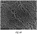
- Figs. 6A-6C are SEM images of samples of native cellulose, non-radiated oxidized cellulose, and radiated oxidized cellulose samples, respectively. The SEM images show that native cellulose, as shown in Fig. 6A have a fibrillar, 3-dimensionally oriented and ordered structure of cellulose chains. The non-radiated oxidized cellulose, as shown in Fig.
- FIG. 6B is a more compact structure than the native cellulose, with regions of larger fibrils stacked together.
- the radiated oxidized sample as shown in Fig. 6C , is less ordered generally than the previous cellulose samples, having a more chaotic structure with generally smaller fibrils and generally higher incidence of heterogenic regions than the other cellulose samples.
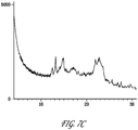
- Dried cellulose membrane samples including native, non-irradiated oxidized, and irradiated oxidized samples, were placed in XRD sample cup holders, placed into the XRD magazine and then into the device for measurement. Oxidation was carried out at 0.3M periodate, 40°C, 3 hrs.
- X-ray diffraction spectra were recorded using Ni filtered Cu-K ⁇ radiation produced by the PANalytical XRD System. Scans were performed over the 4-90° 2 ⁇ range, but analyzed from 4-40° 2 ⁇ range. The data were analyzed with the HighScore Plus XRD software.
- Figs 7A-7C are XRD spectrographs of the native, non-irradiated oxidized, and irradiated oxidized samples, respectively.
- the native sample, Fig. 7A has a highly ordered crystalline structure, followed by the non-radiated cellulose sample, Fig. 7B , with the irradiated sample, Fig. 7C showing the least ordered crystalline structure.
- Table 3 shows the individual sample results for each sample of radiated oxidized cellulose and non-radiated oxidized cellulose at the given oxidation parameters.
- Table 4 provides a summary of the average WHC and WHC/SA values for each of the radiated and non-radiated oxidized samples at the given oxidation parameters.
- Table 3 Sample Conditions Dry M ass (g) Surface Area (cm2) Wet Mass (g) WHC Average WHC WHC/Surface Area(SA) Avg.
- the supernatant was decanted and DI water was added to wash the pellet from residual SBF.
- the tubes were stirred briefly and centrifuged again to collect pellet.
- the DI water washing step was repeated twice.
- the pellet was then dried in the oven at 60°C to constant weight. The percent of degradation was calculated as difference between the dry pellet weight and original sample weight.
- Samples of cellulose of the type used in the in vitro degradation were submitted to Polymer Solutions Incorporated (PSI) (Blacksburg, VA) for analysis of molecular weight distributions using GPC with light scattering detection.
- PSI Polymer Solutions Incorporated
- Three types of samples were submitted: 1) a sample of native microbial cellulose, identified as “Native Cellulose (wet);” 2) a sample of irradiated oxidized microbial cellulose, identified as "Oxidized Cellulose (wet);” and 3) a residual sample of irradiated oxidized microbial cellulose that had been subjected to a seven day in vitro degradation process as described above, identified as "Implant Residual Content.”
- wet is used to indicate that the "Native Cellulose” sample and the "Oxidized Cellulose” sample did not undergo the step of critical point drying with supercritical CO 2 that was previously described. Both the "Oxidized Sample” and the “Implant Residual Content” sample were oxidized at 0.3M periodate, 40°C, 3 hrs.
- the molecular weight distributions of the cellulose samples were analyzed using gel permeation chromatography (GPC) with light scattering detection. Approximately half of the 4 x 5 cm piece of Native Cellulose (wet) and the entire 2.2 x 3.0 cm piece of Oxidized Cellulose (wet) were placed in separate 40-mL glass scintillation vials. A piece of Whatman #1 filter paper was ground for about 5 minutes in a small blade-type coffee mill, and approximately 20 mg of the resulting "fluff' was weighed into a 40-mL scintillation vial.
- GPC gel permeation chromatography
- the Whatman filter paper was included as a control for the dissolution process, and also for use in estimating the specific refractive index increment (dn/dc) of cellulose in DMAc. 10 mL of pure water and a disposable stir bar were added to each vial. Each vial was stirred for approximately 5 hours at 50°C. The Native Cellulose (wet) and Oxidized Cellulose (wet) samples did not disintegrate. Therefore, the wet cellulose pieces were placed in a small food processor with 60 to 70 mL of pure water and processed for 60 to 90 seconds, resulting in slurries of very small fibrous particles. The slurries were then vacuum filtered on 47-mm 0.2- ⁇ m nylon membranes, just until excess water was removed.
- dn/dc specific refractive index increment
- the wet cellulose samples were then transferred to Whatman Vecta-Spin centrifuge filters, which contained 10- ⁇ m polypropylene mesh filters.
- the water was centrifuged off and replaced with HPLC grade methanol and soaked overnight. The following day the methanol was spun off, and an additional 3-hour soak with fresh methanol was performed, followed by a 20-minute centrifugation.
- the solvent exchange process was then repeated using dried N,N-dimethylacetamide (DMAc) for 3 exchanges with soak times of 75 minutes, overnight, and 30 minutes, with 20 minutes centrifugation after each soak.
- DMAc dried N,N-dimethylacetamide
- the DMAc-wet samples and Whatman filter paper control were then transferred into 40-mL scintillation vials.
- 20 mg of the Implant Residual Content sample was weighed into a 40-mL scintillation vial as well.
- 2 mL of a solution of 8% lithium chloride in DMAc and a stir bar were added.
- the samples were stirred for 3 days at room temperature, and were then placed in a refrigerator at 4°C for three additional days.
- the Native Cellulose and the Whatman filter paper control were completely dissolved.
- the Oxidized Cellulose sample formed a cloudy solution with numerous gel-like particles.
- the Implant Residual Content sample was mostly dissolved, but with a very small percentage of the original sample that would not dissolve.
- the Native Cellulose and Oxidized Cellulose solutions were diluted with 14 mL of DMAc.
- the Whatman cellulose control and the Implant Residual Content sample were diluted with 30 mL of DMAc.
- the diluted solutions were stored at approximately 4°C for an additional day before being filtered through 0.45- ⁇ m pore size PTFE syringe filters into GPC autosampler vials. Following filtration, duplicate GPC injections of each sample solution were performed under parameters listed in Table 5 below and molecular weights were calculated using dual-angle light scattering.
- the molecular weight averages (Mn, Mw, Mz) and polydispersity (Mw/Mn) are presented for duplicate injections of each sample in Table 6.
- the molecular weight distribution plots of all samples are compared and graphically depicted in Fig. 10 .
- the specific refractive index increment (dn/dc) value of cellulose in DMAc, used for the light scattering molecular weight calculations, was estimated from the RI detector peak area for duplicate injections of the Whatman filter paper control.
- the bacterial cellulose samples were assumed to have the same dn/dc value as the Whatman filter paper (cotton cellulose).
- the cellulose bodies were sent to Sterigenics (Charlotte, NC) to undergo radiation exposure at various dosages.
- the samples were irradiated with gamma radiation using the ExCell® system, a high-precision, low-volume irradiator. Each exposure of radiation was intended to irradiate the samples in the range of about 20 kGy to about 26.5 kGy. Actual dosage levels for each treatment were measured to be about 23 kGy.
- the samples were oxidized using 0.3M periodate at 40°C for three hours.
- Fig. 11 is a top view of the four samples after radiation exposure and subsequent oxidation. Sample 1, 41 , was not radiated.
- Sample 2 42 , was exposed to one treatment at a dose of 23 kGy.
- Sample 3, 43 was exposed to two separate treatments, each at a dose of 23 kGy.
- Sample 4, 44 was exposed to three separate treatments, each at a dose of 23 kGy.
- the samples were measured for in vitro degradation, as previously described, for one week at SBF conditions at 55°C. Table 7 shows the measured percent of sample degradation of each sample after one week at SBF conditions, along with the sample weight, surface area, and cellulose content, prior to the start of the in vitro degradation test.
- TD 1-TD 4 The in vivo study evaluated in vivo degradation rate and safety/biocompatibility of four irradiated oxidized cellulose implants according to the present disclosure (identified as TD 1-TD 4), each having a different oxidation profile, and compared them to 1) a commercially available cross-linked bovine tendon collagen, identified as CD 1, and 2) a native microbial cellulose, identified as CD 2.
- TD 1 having a 55% oxidation profile, oxidized at 0.4M periodate, 40°C, 3 hrs.
- TD 2 having a 84% oxidation profile, oxidized at 0.4M periodate, 40°C, 4 hrs.
- TD 3 having a 50% oxidation profile, oxidized at 0.3M periodate, 40°C, 3 hrs.
- TD 4 having a 94% oxidation profile, oxidized at 0.3M periodate, 40°C, 5 hrs. All TD samples used in the in vivo studies were irradiated prior to oxidation according to the process described herein.
- a pair of small skin staples were used to mark the location of the device and placed at the two corners of the test or control device closest to the incision site, but not associated with the material.
- a pair of 4-0 Prolene sutures was used to tack down the implant to the underlying subcutaneous tissue in order to prevent implant migration after implantation.
- Necropsy was limited to gross observations of the implantation sites and peri-implant tissues, with limited tissue collection (consisting of collection from the operative sites of the implant surrounded by peri-implant tissues). The degradation of the implants at each site for each measurement period (2 weeks, 4 weeks, 12 weeks or 26 weeks) was recorded and is shown below in Tables 8-12, respectively.
- Table 8 Week 2 Terminal Assessment a TD1 TD2 TD3 TD4 CD1 CD2 2 weeks 2 weeks 2 weeks 2 weeks 2 weeks 2 weeks Inflammation b 0.75 1.25 0.75 0.50 0.00 0.00 0.00 Infection b 0.00 0.00 0.00 0.00 0.00 Fibrosis b 0.00 0.00 0.25 0.00 0.00 Seroma b 0.00 0.25 0.00 0.00 0.00 Hematoma b 0.00 0.00 0.00 0.00 0.00 0.00 0.00 0.00 0.00 0.00 0.00 0.00 0.00 0.00 0.00 b 1.25 1.00 0.50 0.75 0.00 0.00 Presence of Implant c 1.00 1.00 1.00 1.00 0.25 0.00 Implant Degradation d 2.50 2.50 2.50 2.75 0.25 0.00 Implant Measurement e 27.0 65.3 25.0 47.0 91.8 145.0 % of Original Implant Area Remaining 17.3 41.8 25.0 37.6 58.7 92.8 Table 9 : Week 4 Terminal Assessment a TD1 TD2 TD3 TD4 CD1 CD2 4
- the control implants did not show any inflammation of note. After two weeks, there was some gross inflammation noted around all test material implants, with the least amount being around TD4. Inflammation increased slightly at all test material sites after four weeks, with the most inflammation being observed around TD4. At 12 weeks, inflammation at all sites was similar to that observed at two weeks, with the least amount observed around TD4. Inflammation was not observed around any of the implants at 26 weeks. No infection was observed at any time point. The TD2 implant site in one animal was noted to have a possible infection, but when examined microscopically, there was no evidence of infection, or evidence of bacterial colonies. There was little to no fibrosis observed grossly around any of the implants, except perhaps around the native microbial cellulose implant after 12 weeks.
- Figs 12A-F The representative necropsy images of test material TD1 are shown in Figs 12A-F.
- Fig. 12A shows an embodiment of the implant in a first rigid state immediately after placement in position in subcutaneous pocket. After placement, the implant rapidly transitioned to the second hydrated state by absorbing moisture from the surrounding tissue, and subsequently conformed and adhered to the tissue surface as shown in Fig. 12B . It should be noted that after hydration the implant displayed translucency and is nearly indistinguishable from the underlying tissue.
- Fig. 12C shows the implant 2 weeks after implantation where the implant was measurably thinner.
- Fig. 12D shows the implant 4 weeks after implantation where the implant was moderately degraded with one comparatively large piece remaining.
- Fig. 12A shows an embodiment of the implant in a first rigid state immediately after placement in position in subcutaneous pocket. After placement, the implant rapidly transitioned to the second hydrated state by absorbing moisture from the surrounding tissue, and subsequently conformed and adhere
- FIG. 12E shows the implant 12 weeks after implantation where the implant was severely degraded, there was discoloration of the tissue, and the portion of the degraded implant remaining was very diffuse and thin.
- Fig. 12F shows the implant 26 weeks after implantation where the implant was severely degraded; the portion of implant remaining was very diffuse and thin. Stay sutures are visible, and arrows indicate diffuse small areas of discoloration that may indicate fragments of remaining TD1 implant material.
- the native microbial cellulose implant showed no sign of degradation over the entire period of study.
- Cross-linked bovine tendon collagen on the other hand, showed some degradation at 2 weeks, was significantly degraded at 4 weeks, and was essentially not present at 12 and 26 weeks. All of the test devices showed marked degradation at all time points, but interestingly, while they initially appeared to degrade quickly, they did not continue to degrade as rapidly.
- the in vivo study showed that at two weeks, it appeared that TD1 and TD3 showed the most rapid degradation. After four weeks, degradation of TD1, TD2 and TD3 were similar, while TD4 showed less degradation. At 12 weeks, degradation of TD2, TD3 and TD4 were similar, while TD1 appeared considerably more degraded than any of the other test devices.
- no cross-linked bovine tendon collagen was present, there were some remnants of all test devices still present (in the form of tissue discoloration), and the native microbial cellulose was still present as implanted.
- tissue samples from the in vivo implant sites were fixed in 10% neutral buffered formalin (NBF) and sections through the approximate center of the implant site were taken and embedded in paraffin. Hematoxylin and eosin (H & E) staining and Schiff staining (PAS) were performed. PAS staining was used to evaluate aldehyde (oxidized cellulose) presence. All slides were examined and reviewed by two board certified veterinary pathologists. Evaluation of the tissue response to the test and control devices, including scoring the degree of vascularization, fibrosis, and immune response, of the test and control devices and scoring the degree of irritation of the tissue at the implant site were performed, following the ISO 10993 (2007), part 6, annex E guidelines for evaluation of local biological effects after implantation.
- TD1 and TD4 demonstrated notable loss of material apparent by 12 weeks, and this was comparable in degradation to the cross-linked bovine tendon collagen.
- TD2 and TD3 had delayed loss of implant, with notable loss not occurring until the 26-week time point.
- the native microbial cellulose implant showed little to no sign of degradation over the entire period of study.
- the inflammatory response to the implant materials was consistent with a foreign body response, characterized by variable numbers of macrophages, foreign body giant cells and with minimal to mild numbers (a score of 1 to 2) of neutrophils. Eosinophils were not uncommon and plasma cells were rarely seen. Fibrosis generally consisted of narrow to moderately thick bands, with the exception of the native microbial cellulose, which presented with increased fibrous capsule formation around the implant at 12 weeks.
- the total irritancy score was calculated from the sum of the overall inflammatory response (times two), vascularity, and fibrosis pathology scores. The total irritancy score was used to determine the following severity grade for irritant status:
- Average ranked irritation scores were calculated for each test device at each time point by subtracting the average irritancy score for either CD1 or CD2 from each test device, and were based upon the guidelines as described in ISO 10993, part 6, Annex E (informative) "Examples of evaluation of local biological effects after implantation" for scoring of histology. Tables 12 and 13, below, show the average irritancy scores for the samples against each of control CD1 and CD2 respectively.
- TD4 The inflammatory reaction to TD4 (including the numbers of macrophages and giant cells) was most prominent at the early time points of all the test materials. These findings are consistent with a very rapidly absorbed material. At 12 and 26 weeks, macrophages and giant cells again predominated for all the test materials, but the highest scores were seen in proximity to TD2, and to a lesser extent, TD3. This finding likely indicates that these materials were resorbing more slowly than was the TD4 material.
- test materials were compared to the control implants (native microbial cellulose and cross-linked bovine tendon collagen) and were considered to be either non-irritants or slight irritants at 2, 12 or 26 weeks.
- TD1 and TD4 were considered to be moderate irritants when compared to the native microbial cellulose.
- Table 12 TD1-TD4 against CD1 TD1 TD2 TD3 TD4 2 weeks 2.75 0.25 4.50 4.75 4 weeks 2.75 0.50 0.00 6.50 12 weeks 0.75 0.00 0.50 0.00 26 weeks 2.75 8.50 3.50 1.50
- Table 13 TD1-TD4 against CD2 TD1 TD2 TD3 TD4 2 weeks 0.25 0.00 2.00 2.25 4 weeks 8.50 6.25 5.50 12.25 12 weeks 5.00 2.25 4.75 3.75 26 weeks 0.00 3.33 0.00 0.00
Applications Claiming Priority (2)
| Application Number | Priority Date | Filing Date | Title |
|---|---|---|---|
| US201261601653P | 2012-02-22 | 2012-02-22 | |
| PCT/US2013/027230 WO2013126635A1 (en) | 2012-02-22 | 2013-02-22 | Resorbable cellulose based biomaterial and implant |
Publications (2)
| Publication Number | Publication Date |
|---|---|
| EP2817036A1 EP2817036A1 (en) | 2014-12-31 |
| EP2817036B1 true EP2817036B1 (en) | 2018-03-28 |
Family
ID=47833418
Family Applications (1)
| Application Number | Title | Priority Date | Filing Date |
|---|---|---|---|
| EP13708020.6A Active EP2817036B1 (en) | 2012-02-22 | 2013-02-22 | Resorbable cellulose based biomaterial and implant |
Country Status (9)
| Country | Link |
|---|---|
| US (3) | US9090713B2 (zh) |
| EP (1) | EP2817036B1 (zh) |
| JP (1) | JP6195851B2 (zh) |
| KR (1) | KR20140129234A (zh) |
| CN (1) | CN104271166B (zh) |
| AU (1) | AU2013222371B2 (zh) |
| CA (1) | CA2865106C (zh) |
| IN (1) | IN2014DN06940A (zh) |
| WO (1) | WO2013126635A1 (zh) |
Families Citing this family (12)
| Publication number | Priority date | Publication date | Assignee | Title |
|---|---|---|---|---|
| KR101684268B1 (ko) * | 2015-04-15 | 2016-12-12 | 한국원자력연구원 | 방사선 기술을 이용한 박테리아 셀룰로오스의 생분해 조절 및 이를 이용한 흡수성 치주조직 재생유도재 |
| WO2017100771A1 (en) * | 2015-12-11 | 2017-06-15 | The University Of Iowa Research Foundation | Methods of producing biosynthetic bacterial cellulose membranes |
| AU2017204355B2 (en) * | 2016-07-08 | 2021-09-09 | Mako Surgical Corp. | Scaffold for alloprosthetic composite implant |
| WO2018165072A1 (en) | 2017-03-07 | 2018-09-13 | DePuy Synthes Products, Inc. | Bioresorbable exopolysaccharides |
| US10767205B2 (en) | 2017-03-07 | 2020-09-08 | DePuy Synthes Products, Inc. | Bacterium for the production of an exopolysaccharide comprising N-acetylglucosamine and D-glucose |
| JP6299939B1 (ja) * | 2017-04-24 | 2018-03-28 | 王子ホールディングス株式会社 | 増粘剤、組成物及びシート |
| WO2018198162A1 (ja) * | 2017-04-24 | 2018-11-01 | 王子ホールディングス株式会社 | 増粘剤、組成物及びシート |
| ES2875792T3 (es) | 2017-05-24 | 2021-11-11 | JeNaCell GmbH | Método para producir celulosa sin tejer sintetizada de manera bacteriana |
| JP7084809B2 (ja) * | 2017-07-21 | 2022-06-15 | 花王株式会社 | アスファルト組成物及びその製造方法、並びにアスファルト用添加剤 |
| CN108424467B (zh) * | 2018-04-16 | 2020-10-13 | 陕西科技大学 | 一种纤维素基复合抗菌材料的制备方法 |
| CA3136628A1 (en) * | 2019-04-11 | 2020-10-15 | DePuy Synthes Products, Inc. | Bacterial derived nanocellulose textile material |
| WO2021236024A1 (en) | 2020-05-21 | 2021-11-25 | Ptt Public Company Limited | Preparation of biocellulose wound dressing comprising honey as an antimicrobial active ingredient |
Family Cites Families (15)
| Publication number | Priority date | Publication date | Assignee | Title |
|---|---|---|---|---|
| WO2003020191A1 (en) | 2001-09-04 | 2003-03-13 | University Of Iowa Research Foundation | Cellulose membranes for biodegradable scaffolds |
| US7824701B2 (en) * | 2002-10-18 | 2010-11-02 | Ethicon, Inc. | Biocompatible scaffold for ligament or tendon repair |
| US8110222B2 (en) * | 2002-11-15 | 2012-02-07 | Ut-Battelle, Llc. | Composite material |
| DE10323175A1 (de) * | 2003-05-22 | 2004-12-23 | TÜV Industrie Service GmbH - TÜV Rheinland Group | Prüfhebel |
| US8198261B2 (en) * | 2003-08-22 | 2012-06-12 | Xylos Corporation | Thermally modified microbial-derived cellulose for in vivo implantation |
| WO2005018492A2 (en) * | 2003-08-22 | 2005-03-03 | Xylos Corporation | Dura substiture and a process for producing the same |
| US20070128243A1 (en) * | 2005-12-02 | 2007-06-07 | Xylos Corporation | Implantable microbial cellulose materials for various medical applications |
| US7709631B2 (en) * | 2006-03-13 | 2010-05-04 | Xylos Corporation | Oxidized microbial cellulose and use thereof |
| US20070286884A1 (en) * | 2006-06-13 | 2007-12-13 | Xylos Corporation | Implantable microbial cellulose materials for hard tissue repair and regeneration |
| CA2659612A1 (fr) * | 2006-08-02 | 2008-02-07 | Khorionyx | Preparation implantable, utilisable notamment pour le complement tissulaire et la cicatrisation |
| CA2741519C (en) * | 2008-11-07 | 2017-03-21 | Sofradim Production | Composite mesh including a 3d mesh and a non porous film of oxidized cellulose from bacterial cellulose origin |
| US9107978B2 (en) * | 2008-11-07 | 2015-08-18 | Sofradim Production | Template for bacterial cellulose implant processed within bioreactor |
| US8679779B2 (en) | 2008-11-07 | 2014-03-25 | Sofradim Production | Medical devices with definable porosity produced by bacterial polymer bio-synthesis |
| US20100158985A1 (en) * | 2008-12-19 | 2010-06-24 | Xylos Corporation | Porous structures of microbial-derived cellulose for in vivo implantation |
| CN102352051B (zh) * | 2011-09-30 | 2013-09-25 | 北京科技大学 | 一种胶原修饰细菌纤维素复合膜的制备方法 |
-
2013
- 2013-02-22 CA CA2865106A patent/CA2865106C/en active Active
- 2013-02-22 JP JP2014558842A patent/JP6195851B2/ja active Active
- 2013-02-22 AU AU2013222371A patent/AU2013222371B2/en active Active
- 2013-02-22 EP EP13708020.6A patent/EP2817036B1/en active Active
- 2013-02-22 US US13/773,923 patent/US9090713B2/en not_active Expired - Fee Related
- 2013-02-22 IN IN6940DEN2014 patent/IN2014DN06940A/en unknown
- 2013-02-22 CN CN201380010634.9A patent/CN104271166B/zh active Active
- 2013-02-22 WO PCT/US2013/027230 patent/WO2013126635A1/en active Application Filing
- 2013-02-22 KR KR1020147026348A patent/KR20140129234A/ko not_active Application Discontinuation
-
2015
- 2015-06-10 US US14/735,591 patent/US9670289B2/en active Active
-
2017
- 2017-05-25 US US15/604,795 patent/US20170260297A1/en not_active Abandoned
Also Published As
| Publication number | Publication date |
|---|---|
| CA2865106C (en) | 2020-06-02 |
| US9670289B2 (en) | 2017-06-06 |
| AU2013222371B2 (en) | 2016-08-25 |
| US20170260297A1 (en) | 2017-09-14 |
| EP2817036A1 (en) | 2014-12-31 |
| AU2013222371A1 (en) | 2014-08-28 |
| US20130224278A1 (en) | 2013-08-29 |
| CN104271166A (zh) | 2015-01-07 |
| CN104271166B (zh) | 2016-12-07 |
| US20150266978A1 (en) | 2015-09-24 |
| CA2865106A1 (en) | 2013-08-29 |
| JP2015510438A (ja) | 2015-04-09 |
| KR20140129234A (ko) | 2014-11-06 |
| WO2013126635A1 (en) | 2013-08-29 |
| JP6195851B2 (ja) | 2017-09-13 |
| IN2014DN06940A (zh) | 2015-04-10 |
| US9090713B2 (en) | 2015-07-28 |
Similar Documents
| Publication | Publication Date | Title |
|---|---|---|
| EP2817036B1 (en) | Resorbable cellulose based biomaterial and implant | |
| Naahidi et al. | Biocompatibility of hydrogel-based scaffolds for tissue engineering applications | |
| US6638538B1 (en) | Hyaluronic acid gel composition, process for producing the same, and medical material containing the same | |
| Czaja et al. | Oxidation of γ‐irradiated microbial cellulose results in bioresorbable, highly conformable biomaterial | |
| Ibrahim et al. | The impact of hyaluronic acid oligomer content on physical, mechanical, and biologic properties of divinyl sulfone‐crosslinked hyaluronic acid hydrogels | |
| CA2645764A1 (en) | Oxidized microbial cellulose and use thereof | |
| Zhou et al. | An antibacterial chitosan-based hydrogel as a potential degradable bio-scaffold for alveolar ridge preservation | |
| US20070286884A1 (en) | Implantable microbial cellulose materials for hard tissue repair and regeneration | |
| WO2007146946A2 (en) | Implantable microbial cellulose materials for hard tissue repair and regeneration | |
| EP2070557A1 (en) | Implantable microbial cellulose materials for hard tissue repair and regeneration | |
| US20100159046A1 (en) | Minimal tissue attachment implantable materials | |
| CN102516412B (zh) | 乙醇酸接枝壳聚糖共聚物及其制备方法以及作为巩膜扣带材料的应用 | |
| Darwis et al. | In-vitro degradation behaviour of irradiated bacterial cellulose membrane | |
| EP3946484B1 (en) | Composition comprising a cross-linked polyol | |
| Stangel-Wójcikiewicz et al. | Microwave-assisted synthesis and characterization of novel chitosan-based biomaterials for pelvic organ prolapse treatment. | |
| Wray et al. | Biomaterials for Scaffolds: Natural Polymers’ | |
| BR112014020822B1 (pt) | biomaterial e implante reabsorvível à base de celulose | |
| Badylak | Naturally occurring scaffold materials | |
| Al-Arafat et al. | Effects of Gamma Irradiation on the Properties of Hydroxyapatite-Collagen-Chitosan-Mg-ZnO Scaffolds for Bone Tissue Engineering | |
| Waly et al. | Evaluation of Hybrid Chitosan-Cellulose Biodegradable Scaffolds for Tissue Engineering Applications | |
| Poursamar | Investigation into the development of a gelatin-based biocomposite for wound-management | |
| Revathy | Gelatin methacrylate based antimicrobial hydrogels for tissue engineered constructs | |
| Tm et al. | Hybrid chitosan/amniotic membrane-based hydrogels for articular cartilage tissue engineering application |
Legal Events
| Date | Code | Title | Description |
|---|---|---|---|
| PUAI | Public reference made under article 153(3) epc to a published international application that has entered the european phase |
Free format text: ORIGINAL CODE: 0009012 |
|
| 17P | Request for examination filed |
Effective date: 20140821 |
|
| AK | Designated contracting states |
Kind code of ref document: A1 Designated state(s): AL AT BE BG CH CY CZ DE DK EE ES FI FR GB GR HR HU IE IS IT LI LT LU LV MC MK MT NL NO PL PT RO RS SE SI SK SM TR |
|
| AX | Request for extension of the european patent |
Extension state: BA ME |
|
| DAX | Request for extension of the european patent (deleted) | ||
| 17Q | First examination report despatched |
Effective date: 20151130 |
|
| GRAP | Despatch of communication of intention to grant a patent |
Free format text: ORIGINAL CODE: EPIDOSNIGR1 |
|
| STAA | Information on the status of an ep patent application or granted ep patent |
Free format text: STATUS: GRANT OF PATENT IS INTENDED |
|
| INTG | Intention to grant announced |
Effective date: 20171009 |
|
| GRAS | Grant fee paid |
Free format text: ORIGINAL CODE: EPIDOSNIGR3 |
|
| GRAA | (expected) grant |
Free format text: ORIGINAL CODE: 0009210 |
|
| STAA | Information on the status of an ep patent application or granted ep patent |
Free format text: STATUS: THE PATENT HAS BEEN GRANTED |
|
| AK | Designated contracting states |
Kind code of ref document: B1 Designated state(s): AL AT BE BG CH CY CZ DE DK EE ES FI FR GB GR HR HU IE IS IT LI LT LU LV MC MK MT NL NO PL PT RO RS SE SI SK SM TR |
|
| REG | Reference to a national code |
Ref country code: GB Ref legal event code: FG4D |
|
| REG | Reference to a national code |
Ref country code: CH Ref legal event code: EP Ref country code: CH Ref legal event code: NV Representative=s name: E. BLUM AND CO. AG PATENT- UND MARKENANWAELTE , CH |
|
| REG | Reference to a national code |
Ref country code: AT Ref legal event code: REF Ref document number: 982815 Country of ref document: AT Kind code of ref document: T Effective date: 20180415 |
|
| REG | Reference to a national code |
Ref country code: IE Ref legal event code: FG4D |
|
| REG | Reference to a national code |
Ref country code: DE Ref legal event code: R096 Ref document number: 602013035031 Country of ref document: DE |
|
| PG25 | Lapsed in a contracting state [announced via postgrant information from national office to epo] |
Ref country code: HR Free format text: LAPSE BECAUSE OF FAILURE TO SUBMIT A TRANSLATION OF THE DESCRIPTION OR TO PAY THE FEE WITHIN THE PRESCRIBED TIME-LIMIT Effective date: 20180328 Ref country code: LT Free format text: LAPSE BECAUSE OF FAILURE TO SUBMIT A TRANSLATION OF THE DESCRIPTION OR TO PAY THE FEE WITHIN THE PRESCRIBED TIME-LIMIT Effective date: 20180328 Ref country code: NO Free format text: LAPSE BECAUSE OF FAILURE TO SUBMIT A TRANSLATION OF THE DESCRIPTION OR TO PAY THE FEE WITHIN THE PRESCRIBED TIME-LIMIT Effective date: 20180628 Ref country code: FI Free format text: LAPSE BECAUSE OF FAILURE TO SUBMIT A TRANSLATION OF THE DESCRIPTION OR TO PAY THE FEE WITHIN THE PRESCRIBED TIME-LIMIT Effective date: 20180328 |
|
| REG | Reference to a national code |
Ref country code: NL Ref legal event code: MP Effective date: 20180328 |
|
| REG | Reference to a national code |
Ref country code: LT Ref legal event code: MG4D |
|
| PG25 | Lapsed in a contracting state [announced via postgrant information from national office to epo] |
Ref country code: GR Free format text: LAPSE BECAUSE OF FAILURE TO SUBMIT A TRANSLATION OF THE DESCRIPTION OR TO PAY THE FEE WITHIN THE PRESCRIBED TIME-LIMIT Effective date: 20180629 Ref country code: RS Free format text: LAPSE BECAUSE OF FAILURE TO SUBMIT A TRANSLATION OF THE DESCRIPTION OR TO PAY THE FEE WITHIN THE PRESCRIBED TIME-LIMIT Effective date: 20180328 Ref country code: LV Free format text: LAPSE BECAUSE OF FAILURE TO SUBMIT A TRANSLATION OF THE DESCRIPTION OR TO PAY THE FEE WITHIN THE PRESCRIBED TIME-LIMIT Effective date: 20180328 Ref country code: SE Free format text: LAPSE BECAUSE OF FAILURE TO SUBMIT A TRANSLATION OF THE DESCRIPTION OR TO PAY THE FEE WITHIN THE PRESCRIBED TIME-LIMIT Effective date: 20180328 Ref country code: BG Free format text: LAPSE BECAUSE OF FAILURE TO SUBMIT A TRANSLATION OF THE DESCRIPTION OR TO PAY THE FEE WITHIN THE PRESCRIBED TIME-LIMIT Effective date: 20180628 |
|
| PG25 | Lapsed in a contracting state [announced via postgrant information from national office to epo] |
Ref country code: PL Free format text: LAPSE BECAUSE OF FAILURE TO SUBMIT A TRANSLATION OF THE DESCRIPTION OR TO PAY THE FEE WITHIN THE PRESCRIBED TIME-LIMIT Effective date: 20180328 Ref country code: AL Free format text: LAPSE BECAUSE OF FAILURE TO SUBMIT A TRANSLATION OF THE DESCRIPTION OR TO PAY THE FEE WITHIN THE PRESCRIBED TIME-LIMIT Effective date: 20180328 Ref country code: ES Free format text: LAPSE BECAUSE OF FAILURE TO SUBMIT A TRANSLATION OF THE DESCRIPTION OR TO PAY THE FEE WITHIN THE PRESCRIBED TIME-LIMIT Effective date: 20180328 Ref country code: EE Free format text: LAPSE BECAUSE OF FAILURE TO SUBMIT A TRANSLATION OF THE DESCRIPTION OR TO PAY THE FEE WITHIN THE PRESCRIBED TIME-LIMIT Effective date: 20180328 Ref country code: NL Free format text: LAPSE BECAUSE OF FAILURE TO SUBMIT A TRANSLATION OF THE DESCRIPTION OR TO PAY THE FEE WITHIN THE PRESCRIBED TIME-LIMIT Effective date: 20180328 Ref country code: RO Free format text: LAPSE BECAUSE OF FAILURE TO SUBMIT A TRANSLATION OF THE DESCRIPTION OR TO PAY THE FEE WITHIN THE PRESCRIBED TIME-LIMIT Effective date: 20180328 |
|
| PG25 | Lapsed in a contracting state [announced via postgrant information from national office to epo] |
Ref country code: CZ Free format text: LAPSE BECAUSE OF FAILURE TO SUBMIT A TRANSLATION OF THE DESCRIPTION OR TO PAY THE FEE WITHIN THE PRESCRIBED TIME-LIMIT Effective date: 20180328 Ref country code: SK Free format text: LAPSE BECAUSE OF FAILURE TO SUBMIT A TRANSLATION OF THE DESCRIPTION OR TO PAY THE FEE WITHIN THE PRESCRIBED TIME-LIMIT Effective date: 20180328 Ref country code: SM Free format text: LAPSE BECAUSE OF FAILURE TO SUBMIT A TRANSLATION OF THE DESCRIPTION OR TO PAY THE FEE WITHIN THE PRESCRIBED TIME-LIMIT Effective date: 20180328 |
|
| REG | Reference to a national code |
Ref country code: AT Ref legal event code: MK05 Ref document number: 982815 Country of ref document: AT Kind code of ref document: T Effective date: 20180328 |
|
| PG25 | Lapsed in a contracting state [announced via postgrant information from national office to epo] |
Ref country code: PT Free format text: LAPSE BECAUSE OF FAILURE TO SUBMIT A TRANSLATION OF THE DESCRIPTION OR TO PAY THE FEE WITHIN THE PRESCRIBED TIME-LIMIT Effective date: 20180730 |
|
| REG | Reference to a national code |
Ref country code: DE Ref legal event code: R097 Ref document number: 602013035031 Country of ref document: DE |
|
| PG25 | Lapsed in a contracting state [announced via postgrant information from national office to epo] |
Ref country code: DK Free format text: LAPSE BECAUSE OF FAILURE TO SUBMIT A TRANSLATION OF THE DESCRIPTION OR TO PAY THE FEE WITHIN THE PRESCRIBED TIME-LIMIT Effective date: 20180328 Ref country code: AT Free format text: LAPSE BECAUSE OF FAILURE TO SUBMIT A TRANSLATION OF THE DESCRIPTION OR TO PAY THE FEE WITHIN THE PRESCRIBED TIME-LIMIT Effective date: 20180328 |
|
| PLBE | No opposition filed within time limit |
Free format text: ORIGINAL CODE: 0009261 |
|
| STAA | Information on the status of an ep patent application or granted ep patent |
Free format text: STATUS: NO OPPOSITION FILED WITHIN TIME LIMIT |
|
| 26N | No opposition filed |
Effective date: 20190103 |
|
| PG25 | Lapsed in a contracting state [announced via postgrant information from national office to epo] |
Ref country code: SI Free format text: LAPSE BECAUSE OF FAILURE TO SUBMIT A TRANSLATION OF THE DESCRIPTION OR TO PAY THE FEE WITHIN THE PRESCRIBED TIME-LIMIT Effective date: 20180328 |
|
| PG25 | Lapsed in a contracting state [announced via postgrant information from national office to epo] |
Ref country code: LU Free format text: LAPSE BECAUSE OF NON-PAYMENT OF DUE FEES Effective date: 20190222 Ref country code: MC Free format text: LAPSE BECAUSE OF FAILURE TO SUBMIT A TRANSLATION OF THE DESCRIPTION OR TO PAY THE FEE WITHIN THE PRESCRIBED TIME-LIMIT Effective date: 20180328 |
|
| REG | Reference to a national code |
Ref country code: BE Ref legal event code: MM Effective date: 20190228 |
|
| REG | Reference to a national code |
Ref country code: IE Ref legal event code: MM4A |
|
| PG25 | Lapsed in a contracting state [announced via postgrant information from national office to epo] |
Ref country code: IE Free format text: LAPSE BECAUSE OF NON-PAYMENT OF DUE FEES Effective date: 20190222 |
|
| PG25 | Lapsed in a contracting state [announced via postgrant information from national office to epo] |
Ref country code: BE Free format text: LAPSE BECAUSE OF NON-PAYMENT OF DUE FEES Effective date: 20190228 |
|
| PG25 | Lapsed in a contracting state [announced via postgrant information from national office to epo] |
Ref country code: TR Free format text: LAPSE BECAUSE OF FAILURE TO SUBMIT A TRANSLATION OF THE DESCRIPTION OR TO PAY THE FEE WITHIN THE PRESCRIBED TIME-LIMIT Effective date: 20180328 |
|
| PG25 | Lapsed in a contracting state [announced via postgrant information from national office to epo] |
Ref country code: MT Free format text: LAPSE BECAUSE OF NON-PAYMENT OF DUE FEES Effective date: 20190222 |
|
| PG25 | Lapsed in a contracting state [announced via postgrant information from national office to epo] |
Ref country code: CY Free format text: LAPSE BECAUSE OF FAILURE TO SUBMIT A TRANSLATION OF THE DESCRIPTION OR TO PAY THE FEE WITHIN THE PRESCRIBED TIME-LIMIT Effective date: 20180328 |
|
| PG25 | Lapsed in a contracting state [announced via postgrant information from national office to epo] |
Ref country code: IS Free format text: LAPSE BECAUSE OF FAILURE TO SUBMIT A TRANSLATION OF THE DESCRIPTION OR TO PAY THE FEE WITHIN THE PRESCRIBED TIME-LIMIT Effective date: 20180728 |
|
| PG25 | Lapsed in a contracting state [announced via postgrant information from national office to epo] |
Ref country code: HU Free format text: LAPSE BECAUSE OF FAILURE TO SUBMIT A TRANSLATION OF THE DESCRIPTION OR TO PAY THE FEE WITHIN THE PRESCRIBED TIME-LIMIT; INVALID AB INITIO Effective date: 20130222 |
|
| PG25 | Lapsed in a contracting state [announced via postgrant information from national office to epo] |
Ref country code: MK Free format text: LAPSE BECAUSE OF FAILURE TO SUBMIT A TRANSLATION OF THE DESCRIPTION OR TO PAY THE FEE WITHIN THE PRESCRIBED TIME-LIMIT Effective date: 20180328 |
|
| PGFP | Annual fee paid to national office [announced via postgrant information from national office to epo] |
Ref country code: CH Payment date: 20230307 Year of fee payment: 11 |
|
| PGFP | Annual fee paid to national office [announced via postgrant information from national office to epo] |
Ref country code: IT Payment date: 20230110 Year of fee payment: 11 Ref country code: DE Payment date: 20221229 Year of fee payment: 11 |
|
| PGFP | Annual fee paid to national office [announced via postgrant information from national office to epo] |
Ref country code: FR Payment date: 20231229 Year of fee payment: 12 |
|
| PGFP | Annual fee paid to national office [announced via postgrant information from national office to epo] |
Ref country code: DE Payment date: 20231229 Year of fee payment: 12 Ref country code: GB Payment date: 20240108 Year of fee payment: 12 Ref country code: CH Payment date: 20240301 Year of fee payment: 12 |