EP2759604B1 - Control nucleic acids for multiple parameters - Google Patents
Control nucleic acids for multiple parameters Download PDFInfo
- Publication number
- EP2759604B1 EP2759604B1 EP14150845.7A EP14150845A EP2759604B1 EP 2759604 B1 EP2759604 B1 EP 2759604B1 EP 14150845 A EP14150845 A EP 14150845A EP 2759604 B1 EP2759604 B1 EP 2759604B1
- Authority
- EP
- European Patent Office
- Prior art keywords
- nucleic acid
- nucleic acids
- internal control
- target nucleic
- dna
- Prior art date
- Legal status (The legal status is an assumption and is not a legal conclusion. Google has not performed a legal analysis and makes no representation as to the accuracy of the status listed.)
- Active
Links
- 150000007523 nucleic acids Chemical class 0.000 title claims description 369
- 102000039446 nucleic acids Human genes 0.000 title claims description 364
- 108020004707 nucleic acids Proteins 0.000 title claims description 364
- 238000000034 method Methods 0.000 claims description 172
- 238000003199 nucleic acid amplification method Methods 0.000 claims description 125
- 230000003321 amplification Effects 0.000 claims description 122
- 230000008569 process Effects 0.000 claims description 101
- 238000006243 chemical reaction Methods 0.000 claims description 97
- 238000001514 detection method Methods 0.000 claims description 47
- 239000003153 chemical reaction reagent Substances 0.000 claims description 46
- 238000000926 separation method Methods 0.000 claims description 45
- 239000012530 fluid Substances 0.000 claims description 41
- 239000000463 material Substances 0.000 claims description 38
- 230000000694 effects Effects 0.000 claims description 33
- 239000007787 solid Substances 0.000 claims description 33
- 102100034343 Integrase Human genes 0.000 claims description 18
- 108010092799 RNA-directed DNA polymerase Proteins 0.000 claims description 18
- 239000011541 reaction mixture Substances 0.000 claims description 10
- 239000011534 wash buffer Substances 0.000 claims description 10
- 238000005406 washing Methods 0.000 claims description 9
- 238000013518 transcription Methods 0.000 claims description 6
- 230000035897 transcription Effects 0.000 claims description 6
- FGUUSXIOTUKUDN-IBGZPJMESA-N C1(=CC=CC=C1)N1C2=C(NC([C@H](C1)NC=1OC(=NN=1)C1=CC=CC=C1)=O)C=CC=C2 Chemical compound C1(=CC=CC=C1)N1C2=C(NC([C@H](C1)NC=1OC(=NN=1)C1=CC=CC=C1)=O)C=CC=C2 FGUUSXIOTUKUDN-IBGZPJMESA-N 0.000 claims description 5
- 108091032973 (ribonucleotides)n+m Proteins 0.000 claims description 4
- 230000001580 bacterial effect Effects 0.000 claims description 3
- 239000000523 sample Substances 0.000 description 212
- 102000053602 DNA Human genes 0.000 description 127
- 108020004414 DNA Proteins 0.000 description 125
- 239000013615 primer Substances 0.000 description 114
- 238000004458 analytical method Methods 0.000 description 76
- 229920002477 rna polymer Polymers 0.000 description 58
- 238000003556 assay Methods 0.000 description 45
- 238000012545 processing Methods 0.000 description 45
- 238000003752 polymerase chain reaction Methods 0.000 description 40
- 239000007788 liquid Substances 0.000 description 34
- 108010014303 DNA-directed DNA polymerase Proteins 0.000 description 27
- 102000016928 DNA-directed DNA polymerase Human genes 0.000 description 27
- 241000700721 Hepatitis B virus Species 0.000 description 27
- 108091034117 Oligonucleotide Proteins 0.000 description 27
- 229940088598 enzyme Drugs 0.000 description 27
- 230000005291 magnetic effect Effects 0.000 description 27
- 239000000047 product Substances 0.000 description 27
- 238000010839 reverse transcription Methods 0.000 description 27
- 102000004190 Enzymes Human genes 0.000 description 26
- 108090000790 Enzymes Proteins 0.000 description 26
- PEDCQBHIVMGVHV-UHFFFAOYSA-N Glycerine Chemical compound OCC(O)CO PEDCQBHIVMGVHV-UHFFFAOYSA-N 0.000 description 26
- PXIPVTKHYLBLMZ-UHFFFAOYSA-N Sodium azide Chemical compound [Na+].[N-]=[N+]=[N-] PXIPVTKHYLBLMZ-UHFFFAOYSA-N 0.000 description 26
- 125000003729 nucleotide group Chemical group 0.000 description 24
- 241000725303 Human immunodeficiency virus Species 0.000 description 23
- 230000008901 benefit Effects 0.000 description 23
- 239000006249 magnetic particle Substances 0.000 description 23
- 238000012360 testing method Methods 0.000 description 20
- 239000000543 intermediate Substances 0.000 description 19
- 239000002245 particle Substances 0.000 description 19
- 210000002966 serum Anatomy 0.000 description 19
- IAZDPXIOMUYVGZ-UHFFFAOYSA-N Dimethylsulphoxide Chemical compound CS(C)=O IAZDPXIOMUYVGZ-UHFFFAOYSA-N 0.000 description 18
- 239000002773 nucleotide Substances 0.000 description 18
- 238000002360 preparation method Methods 0.000 description 18
- 238000000137 annealing Methods 0.000 description 17
- KCXVZYZYPLLWCC-UHFFFAOYSA-N EDTA Chemical compound OC(=O)CN(CC(O)=O)CCN(CC(O)=O)CC(O)=O KCXVZYZYPLLWCC-UHFFFAOYSA-N 0.000 description 16
- 241000711549 Hepacivirus C Species 0.000 description 16
- SEQKRHFRPICQDD-UHFFFAOYSA-N N-tris(hydroxymethyl)methylglycine Chemical compound OCC(CO)(CO)[NH2+]CC([O-])=O SEQKRHFRPICQDD-UHFFFAOYSA-N 0.000 description 16
- JLCPHMBAVCMARE-UHFFFAOYSA-N [3-[[3-[[3-[[3-[[3-[[3-[[3-[[3-[[3-[[3-[[3-[[5-(2-amino-6-oxo-1H-purin-9-yl)-3-[[3-[[3-[[3-[[3-[[3-[[5-(2-amino-6-oxo-1H-purin-9-yl)-3-[[5-(2-amino-6-oxo-1H-purin-9-yl)-3-hydroxyoxolan-2-yl]methoxy-hydroxyphosphoryl]oxyoxolan-2-yl]methoxy-hydroxyphosphoryl]oxy-5-(5-methyl-2,4-dioxopyrimidin-1-yl)oxolan-2-yl]methoxy-hydroxyphosphoryl]oxy-5-(6-aminopurin-9-yl)oxolan-2-yl]methoxy-hydroxyphosphoryl]oxy-5-(6-aminopurin-9-yl)oxolan-2-yl]methoxy-hydroxyphosphoryl]oxy-5-(6-aminopurin-9-yl)oxolan-2-yl]methoxy-hydroxyphosphoryl]oxy-5-(6-aminopurin-9-yl)oxolan-2-yl]methoxy-hydroxyphosphoryl]oxyoxolan-2-yl]methoxy-hydroxyphosphoryl]oxy-5-(5-methyl-2,4-dioxopyrimidin-1-yl)oxolan-2-yl]methoxy-hydroxyphosphoryl]oxy-5-(4-amino-2-oxopyrimidin-1-yl)oxolan-2-yl]methoxy-hydroxyphosphoryl]oxy-5-(5-methyl-2,4-dioxopyrimidin-1-yl)oxolan-2-yl]methoxy-hydroxyphosphoryl]oxy-5-(5-methyl-2,4-dioxopyrimidin-1-yl)oxolan-2-yl]methoxy-hydroxyphosphoryl]oxy-5-(6-aminopurin-9-yl)oxolan-2-yl]methoxy-hydroxyphosphoryl]oxy-5-(6-aminopurin-9-yl)oxolan-2-yl]methoxy-hydroxyphosphoryl]oxy-5-(4-amino-2-oxopyrimidin-1-yl)oxolan-2-yl]methoxy-hydroxyphosphoryl]oxy-5-(4-amino-2-oxopyrimidin-1-yl)oxolan-2-yl]methoxy-hydroxyphosphoryl]oxy-5-(4-amino-2-oxopyrimidin-1-yl)oxolan-2-yl]methoxy-hydroxyphosphoryl]oxy-5-(6-aminopurin-9-yl)oxolan-2-yl]methoxy-hydroxyphosphoryl]oxy-5-(4-amino-2-oxopyrimidin-1-yl)oxolan-2-yl]methyl [5-(6-aminopurin-9-yl)-2-(hydroxymethyl)oxolan-3-yl] hydrogen phosphate Chemical class Cc1cn(C2CC(OP(O)(=O)OCC3OC(CC3OP(O)(=O)OCC3OC(CC3O)n3cnc4c3nc(N)[nH]c4=O)n3cnc4c3nc(N)[nH]c4=O)C(COP(O)(=O)OC3CC(OC3COP(O)(=O)OC3CC(OC3COP(O)(=O)OC3CC(OC3COP(O)(=O)OC3CC(OC3COP(O)(=O)OC3CC(OC3COP(O)(=O)OC3CC(OC3COP(O)(=O)OC3CC(OC3COP(O)(=O)OC3CC(OC3COP(O)(=O)OC3CC(OC3COP(O)(=O)OC3CC(OC3COP(O)(=O)OC3CC(OC3COP(O)(=O)OC3CC(OC3COP(O)(=O)OC3CC(OC3COP(O)(=O)OC3CC(OC3COP(O)(=O)OC3CC(OC3COP(O)(=O)OC3CC(OC3COP(O)(=O)OC3CC(OC3CO)n3cnc4c(N)ncnc34)n3ccc(N)nc3=O)n3cnc4c(N)ncnc34)n3ccc(N)nc3=O)n3ccc(N)nc3=O)n3ccc(N)nc3=O)n3cnc4c(N)ncnc34)n3cnc4c(N)ncnc34)n3cc(C)c(=O)[nH]c3=O)n3cc(C)c(=O)[nH]c3=O)n3ccc(N)nc3=O)n3cc(C)c(=O)[nH]c3=O)n3cnc4c3nc(N)[nH]c4=O)n3cnc4c(N)ncnc34)n3cnc4c(N)ncnc34)n3cnc4c(N)ncnc34)n3cnc4c(N)ncnc34)O2)c(=O)[nH]c1=O JLCPHMBAVCMARE-UHFFFAOYSA-N 0.000 description 16
- 230000027455 binding Effects 0.000 description 15
- 239000000975 dye Substances 0.000 description 15
- 239000002299 complementary DNA Substances 0.000 description 14
- 238000010438 heat treatment Methods 0.000 description 14
- 239000011521 glass Substances 0.000 description 13
- LFQSCWFLJHTTHZ-UHFFFAOYSA-N Ethanol Chemical compound CCO LFQSCWFLJHTTHZ-UHFFFAOYSA-N 0.000 description 12
- 239000007983 Tris buffer Substances 0.000 description 12
- 230000015572 biosynthetic process Effects 0.000 description 12
- 230000001351 cycling effect Effects 0.000 description 11
- 230000009089 cytolysis Effects 0.000 description 11
- 238000002474 experimental method Methods 0.000 description 11
- LENZDBCJOHFCAS-UHFFFAOYSA-N tris Chemical compound OCC(N)(CO)CO LENZDBCJOHFCAS-UHFFFAOYSA-N 0.000 description 11
- 241000606153 Chlamydia trachomatis Species 0.000 description 10
- 101150040913 DUT gene Proteins 0.000 description 10
- 241000700605 Viruses Species 0.000 description 10
- 229940038705 chlamydia trachomatis Drugs 0.000 description 10
- 238000011534 incubation Methods 0.000 description 10
- 238000012546 transfer Methods 0.000 description 10
- 239000007997 Tricine buffer Substances 0.000 description 9
- 102000006943 Uracil-DNA Glycosidase Human genes 0.000 description 9
- 108010072685 Uracil-DNA Glycosidase Proteins 0.000 description 9
- 230000002860 competitive effect Effects 0.000 description 9
- 235000011187 glycerol Nutrition 0.000 description 9
- 238000009396 hybridization Methods 0.000 description 9
- 238000002955 isolation Methods 0.000 description 9
- 238000005259 measurement Methods 0.000 description 9
- SCVFZCLFOSHCOH-UHFFFAOYSA-M potassium acetate Chemical compound [K+].CC([O-])=O SCVFZCLFOSHCOH-UHFFFAOYSA-M 0.000 description 9
- 238000000746 purification Methods 0.000 description 9
- 238000003753 real-time PCR Methods 0.000 description 9
- 230000035945 sensitivity Effects 0.000 description 9
- 230000003612 virological effect Effects 0.000 description 9
- QTBSBXVTEAMEQO-UHFFFAOYSA-N Acetic acid Chemical compound CC(O)=O QTBSBXVTEAMEQO-UHFFFAOYSA-N 0.000 description 8
- 108091005804 Peptidases Proteins 0.000 description 8
- UZMAPBJVXOGOFT-UHFFFAOYSA-N Syringetin Natural products COC1=C(O)C(OC)=CC(C2=C(C(=O)C3=C(O)C=C(O)C=C3O2)O)=C1 UZMAPBJVXOGOFT-UHFFFAOYSA-N 0.000 description 8
- 241000710886 West Nile virus Species 0.000 description 8
- 239000012491 analyte Substances 0.000 description 8
- 230000000295 complement effect Effects 0.000 description 8
- 150000001875 compounds Chemical class 0.000 description 8
- 230000000875 corresponding effect Effects 0.000 description 8
- KCFYHBSOLOXZIF-UHFFFAOYSA-N dihydrochrysin Natural products COC1=C(O)C(OC)=CC(C2OC3=CC(O)=CC(O)=C3C(=O)C2)=C1 KCFYHBSOLOXZIF-UHFFFAOYSA-N 0.000 description 8
- 238000010790 dilution Methods 0.000 description 8
- 239000012895 dilution Substances 0.000 description 8
- 238000002866 fluorescence resonance energy transfer Methods 0.000 description 8
- 230000035772 mutation Effects 0.000 description 8
- 239000002777 nucleoside Substances 0.000 description 8
- 239000000126 substance Substances 0.000 description 8
- 108091023037 Aptamer Proteins 0.000 description 7
- AHCYMLUZIRLXAA-SHYZEUOFSA-N Deoxyuridine 5'-triphosphate Chemical compound O1[C@H](COP(O)(=O)OP(O)(=O)OP(O)(O)=O)[C@@H](O)C[C@@H]1N1C(=O)NC(=O)C=C1 AHCYMLUZIRLXAA-SHYZEUOFSA-N 0.000 description 7
- 239000004365 Protease Substances 0.000 description 7
- -1 RNA and DNA Chemical class 0.000 description 7
- 239000000872 buffer Substances 0.000 description 7
- 239000003795 chemical substances by application Substances 0.000 description 7
- 239000000203 mixture Substances 0.000 description 7
- 235000000346 sugar Nutrition 0.000 description 7
- KWYUFKZDYYNOTN-UHFFFAOYSA-M Potassium hydroxide Chemical compound [OH-].[K+] KWYUFKZDYYNOTN-UHFFFAOYSA-M 0.000 description 6
- SUYVUBYJARFZHO-RRKCRQDMSA-N dATP Chemical compound C1=NC=2C(N)=NC=NC=2N1[C@H]1C[C@H](O)[C@@H](COP(O)(=O)OP(O)(=O)OP(O)(O)=O)O1 SUYVUBYJARFZHO-RRKCRQDMSA-N 0.000 description 6
- SUYVUBYJARFZHO-UHFFFAOYSA-N dATP Natural products C1=NC=2C(N)=NC=NC=2N1C1CC(O)C(COP(O)(=O)OP(O)(=O)OP(O)(O)=O)O1 SUYVUBYJARFZHO-UHFFFAOYSA-N 0.000 description 6
- RGWHQCVHVJXOKC-SHYZEUOFSA-J dCTP(4-) Chemical compound O=C1N=C(N)C=CN1[C@@H]1O[C@H](COP([O-])(=O)OP([O-])(=O)OP([O-])([O-])=O)[C@@H](O)C1 RGWHQCVHVJXOKC-SHYZEUOFSA-J 0.000 description 6
- HAAZLUGHYHWQIW-KVQBGUIXSA-N dGTP Chemical compound C1=NC=2C(=O)NC(N)=NC=2N1[C@H]1C[C@H](O)[C@@H](COP(O)(=O)OP(O)(=O)OP(O)(O)=O)O1 HAAZLUGHYHWQIW-KVQBGUIXSA-N 0.000 description 6
- 238000004925 denaturation Methods 0.000 description 6
- 230000036425 denaturation Effects 0.000 description 6
- 238000000605 extraction Methods 0.000 description 6
- 238000007885 magnetic separation Methods 0.000 description 6
- LXCFILQKKLGQFO-UHFFFAOYSA-N methylparaben Chemical compound COC(=O)C1=CC=C(O)C=C1 LXCFILQKKLGQFO-UHFFFAOYSA-N 0.000 description 6
- 238000012986 modification Methods 0.000 description 6
- 230000004048 modification Effects 0.000 description 6
- 239000000243 solution Substances 0.000 description 6
- 238000003786 synthesis reaction Methods 0.000 description 6
- 230000004568 DNA-binding Effects 0.000 description 5
- 208000031886 HIV Infections Diseases 0.000 description 5
- 241000560067 HIV-1 group M Species 0.000 description 5
- 241000560056 HIV-1 group O Species 0.000 description 5
- 241000713340 Human immunodeficiency virus 2 Species 0.000 description 5
- 241000710842 Japanese encephalitis virus Species 0.000 description 5
- 229910021380 Manganese Chloride Inorganic materials 0.000 description 5
- GLFNIEUTAYBVOC-UHFFFAOYSA-L Manganese chloride Chemical compound Cl[Mn]Cl GLFNIEUTAYBVOC-UHFFFAOYSA-L 0.000 description 5
- 108091028043 Nucleic acid sequence Proteins 0.000 description 5
- 102000035195 Peptidases Human genes 0.000 description 5
- 229920001213 Polysorbate 20 Polymers 0.000 description 5
- VYPSYNLAJGMNEJ-UHFFFAOYSA-N Silicium dioxide Chemical compound O=[Si]=O VYPSYNLAJGMNEJ-UHFFFAOYSA-N 0.000 description 5
- 241000710888 St. Louis encephalitis virus Species 0.000 description 5
- 238000004364 calculation method Methods 0.000 description 5
- 230000003196 chaotropic effect Effects 0.000 description 5
- 230000001276 controlling effect Effects 0.000 description 5
- 238000012864 cross contamination Methods 0.000 description 5
- 230000000593 degrading effect Effects 0.000 description 5
- 238000003745 diagnosis Methods 0.000 description 5
- 239000012149 elution buffer Substances 0.000 description 5
- 239000003365 glass fiber Substances 0.000 description 5
- 239000011565 manganese chloride Substances 0.000 description 5
- 235000002867 manganese chloride Nutrition 0.000 description 5
- 229940099607 manganese chloride Drugs 0.000 description 5
- 230000007246 mechanism Effects 0.000 description 5
- 239000000256 polyoxyethylene sorbitan monolaurate Substances 0.000 description 5
- 235000010486 polyoxyethylene sorbitan monolaurate Nutrition 0.000 description 5
- 108090000623 proteins and genes Proteins 0.000 description 5
- 230000004044 response Effects 0.000 description 5
- 238000011282 treatment Methods 0.000 description 5
- 239000001226 triphosphate Substances 0.000 description 5
- 235000011178 triphosphate Nutrition 0.000 description 5
- KDCGOANMDULRCW-UHFFFAOYSA-N 7H-purine Chemical compound N1=CNC2=NC=NC2=C1 KDCGOANMDULRCW-UHFFFAOYSA-N 0.000 description 4
- 241000894006 Bacteria Species 0.000 description 4
- 241000701806 Human papillomavirus Species 0.000 description 4
- 229910019142 PO4 Inorganic materials 0.000 description 4
- 102100037111 Uracil-DNA glycosylase Human genes 0.000 description 4
- 210000004369 blood Anatomy 0.000 description 4
- 239000008280 blood Substances 0.000 description 4
- 238000001816 cooling Methods 0.000 description 4
- 230000001419 dependent effect Effects 0.000 description 4
- 238000005516 engineering process Methods 0.000 description 4
- 230000006870 function Effects 0.000 description 4
- 238000007834 ligase chain reaction Methods 0.000 description 4
- 230000036963 noncompetitive effect Effects 0.000 description 4
- 125000003835 nucleoside group Chemical group 0.000 description 4
- NBIIXXVUZAFLBC-UHFFFAOYSA-K phosphate Chemical compound [O-]P([O-])([O-])=O NBIIXXVUZAFLBC-UHFFFAOYSA-K 0.000 description 4
- 239000010452 phosphate Substances 0.000 description 4
- 125000002467 phosphate group Chemical group [H]OP(=O)(O[H])O[*] 0.000 description 4
- 238000001556 precipitation Methods 0.000 description 4
- 102000004169 proteins and genes Human genes 0.000 description 4
- 238000011002 quantification Methods 0.000 description 4
- 230000009467 reduction Effects 0.000 description 4
- 230000002829 reductive effect Effects 0.000 description 4
- 238000001179 sorption measurement Methods 0.000 description 4
- 238000003860 storage Methods 0.000 description 4
- XLYOFNOQVPJJNP-UHFFFAOYSA-N water Chemical compound O XLYOFNOQVPJJNP-UHFFFAOYSA-N 0.000 description 4
- 108091093088 Amplicon Proteins 0.000 description 3
- OKKJLVBELUTLKV-UHFFFAOYSA-N Methanol Chemical compound OC OKKJLVBELUTLKV-UHFFFAOYSA-N 0.000 description 3
- 102100037486 Reverse transcriptase/ribonuclease H Human genes 0.000 description 3
- 102000006382 Ribonucleases Human genes 0.000 description 3
- 108010083644 Ribonucleases Proteins 0.000 description 3
- 241000589499 Thermus thermophilus Species 0.000 description 3
- ISAKRJDGNUQOIC-UHFFFAOYSA-N Uracil Chemical compound O=C1C=CNC(=O)N1 ISAKRJDGNUQOIC-UHFFFAOYSA-N 0.000 description 3
- 229960000583 acetic acid Drugs 0.000 description 3
- 239000002253 acid Substances 0.000 description 3
- 150000007513 acids Chemical class 0.000 description 3
- 150000001413 amino acids Chemical class 0.000 description 3
- 239000003242 anti bacterial agent Substances 0.000 description 3
- 230000003466 anti-cipated effect Effects 0.000 description 3
- 229940088710 antibiotic agent Drugs 0.000 description 3
- 239000012472 biological sample Substances 0.000 description 3
- 238000011109 contamination Methods 0.000 description 3
- 230000007423 decrease Effects 0.000 description 3
- 239000003085 diluting agent Substances 0.000 description 3
- 238000009826 distribution Methods 0.000 description 3
- 241001493065 dsRNA viruses Species 0.000 description 3
- 230000005284 excitation Effects 0.000 description 3
- 230000002349 favourable effect Effects 0.000 description 3
- 239000007850 fluorescent dye Substances 0.000 description 3
- 230000002068 genetic effect Effects 0.000 description 3
- 230000001965 increasing effect Effects 0.000 description 3
- 230000000977 initiatory effect Effects 0.000 description 3
- 230000003993 interaction Effects 0.000 description 3
- 238000009830 intercalation Methods 0.000 description 3
- 239000011159 matrix material Substances 0.000 description 3
- 239000004292 methyl p-hydroxybenzoate Substances 0.000 description 3
- 235000010270 methyl p-hydroxybenzoate Nutrition 0.000 description 3
- 229960002216 methylparaben Drugs 0.000 description 3
- 150000003833 nucleoside derivatives Chemical class 0.000 description 3
- 230000003287 optical effect Effects 0.000 description 3
- 244000052769 pathogen Species 0.000 description 3
- 150000004713 phosphodiesters Chemical class 0.000 description 3
- 239000013612 plasmid Substances 0.000 description 3
- 229920000642 polymer Polymers 0.000 description 3
- 235000019419 proteases Nutrition 0.000 description 3
- 238000004445 quantitative analysis Methods 0.000 description 3
- 230000002441 reversible effect Effects 0.000 description 3
- 150000003839 salts Chemical class 0.000 description 3
- 238000007789 sealing Methods 0.000 description 3
- FVAUCKIRQBBSSJ-UHFFFAOYSA-M sodium iodide Chemical compound [Na+].[I-] FVAUCKIRQBBSSJ-UHFFFAOYSA-M 0.000 description 3
- 108010068698 spleen exonuclease Proteins 0.000 description 3
- 125000001424 substituent group Chemical group 0.000 description 3
- 238000002560 therapeutic procedure Methods 0.000 description 3
- 125000002264 triphosphate group Chemical class [H]OP(=O)(O[H])OP(=O)(O[H])OP(=O)(O[H])O* 0.000 description 3
- YBJHBAHKTGYVGT-ZKWXMUAHSA-N (+)-Biotin Chemical compound N1C(=O)N[C@@H]2[C@H](CCCCC(=O)O)SC[C@@H]21 YBJHBAHKTGYVGT-ZKWXMUAHSA-N 0.000 description 2
- 108020004635 Complementary DNA Proteins 0.000 description 2
- 241000701022 Cytomegalovirus Species 0.000 description 2
- 239000003155 DNA primer Substances 0.000 description 2
- 241000588724 Escherichia coli Species 0.000 description 2
- 241000701959 Escherichia virus Lambda Species 0.000 description 2
- 241000713772 Human immunodeficiency virus 1 Species 0.000 description 2
- 241000588652 Neisseria gonorrhoeae Species 0.000 description 2
- CZPWVGJYEJSRLH-UHFFFAOYSA-N Pyrimidine Chemical compound C1=CN=CN=C1 CZPWVGJYEJSRLH-UHFFFAOYSA-N 0.000 description 2
- 241000589500 Thermus aquaticus Species 0.000 description 2
- 241000589497 Thermus sp. Species 0.000 description 2
- 230000009471 action Effects 0.000 description 2
- 125000000217 alkyl group Chemical group 0.000 description 2
- 239000008366 buffered solution Substances 0.000 description 2
- 238000010804 cDNA synthesis Methods 0.000 description 2
- 230000015556 catabolic process Effects 0.000 description 2
- 150000001768 cations Chemical class 0.000 description 2
- 230000001413 cellular effect Effects 0.000 description 2
- 239000000356 contaminant Substances 0.000 description 2
- ZYGHJZDHTFUPRJ-UHFFFAOYSA-N coumarin Chemical compound C1=CC=C2OC(=O)C=CC2=C1 ZYGHJZDHTFUPRJ-UHFFFAOYSA-N 0.000 description 2
- 230000001186 cumulative effect Effects 0.000 description 2
- 238000006731 degradation reaction Methods 0.000 description 2
- 239000005549 deoxyribonucleoside Substances 0.000 description 2
- 238000006073 displacement reaction Methods 0.000 description 2
- 239000002158 endotoxin Substances 0.000 description 2
- 230000002255 enzymatic effect Effects 0.000 description 2
- 238000006911 enzymatic reaction Methods 0.000 description 2
- GNBHRKFJIUUOQI-UHFFFAOYSA-N fluorescein Chemical compound O1C(=O)C2=CC=CC=C2C21C1=CC=C(O)C=C1OC1=CC(O)=CC=C21 GNBHRKFJIUUOQI-UHFFFAOYSA-N 0.000 description 2
- 239000011888 foil Substances 0.000 description 2
- ZJYYHGLJYGJLLN-UHFFFAOYSA-N guanidinium thiocyanate Chemical compound SC#N.NC(N)=N ZJYYHGLJYGJLLN-UHFFFAOYSA-N 0.000 description 2
- 125000000623 heterocyclic group Chemical group 0.000 description 2
- 208000015181 infectious disease Diseases 0.000 description 2
- 230000005764 inhibitory process Effects 0.000 description 2
- 230000000670 limiting effect Effects 0.000 description 2
- 239000011572 manganese Substances 0.000 description 2
- 230000001404 mediated effect Effects 0.000 description 2
- 108010003855 mesentericopeptidase Proteins 0.000 description 2
- 238000010369 molecular cloning Methods 0.000 description 2
- 238000012544 monitoring process Methods 0.000 description 2
- 230000007935 neutral effect Effects 0.000 description 2
- 230000001717 pathogenic effect Effects 0.000 description 2
- 235000011056 potassium acetate Nutrition 0.000 description 2
- 238000012207 quantitative assay Methods 0.000 description 2
- 238000012113 quantitative test Methods 0.000 description 2
- 238000011897 real-time detection Methods 0.000 description 2
- 230000008439 repair process Effects 0.000 description 2
- 125000006853 reporter group Chemical group 0.000 description 2
- 238000012552 review Methods 0.000 description 2
- 239000012266 salt solution Substances 0.000 description 2
- 238000013207 serial dilution Methods 0.000 description 2
- 239000000377 silicon dioxide Substances 0.000 description 2
- 239000001509 sodium citrate Substances 0.000 description 2
- NLJMYIDDQXHKNR-UHFFFAOYSA-K sodium citrate Chemical compound O.O.[Na+].[Na+].[Na+].[O-]C(=O)CC(O)(CC([O-])=O)C([O-])=O NLJMYIDDQXHKNR-UHFFFAOYSA-K 0.000 description 2
- 239000011343 solid material Substances 0.000 description 2
- 238000001228 spectrum Methods 0.000 description 2
- 239000007858 starting material Substances 0.000 description 2
- 239000000725 suspension Substances 0.000 description 2
- 230000002194 synthesizing effect Effects 0.000 description 2
- RWQNBRDOKXIBIV-UHFFFAOYSA-N thymine Chemical compound CC1=CNC(=O)NC1=O RWQNBRDOKXIBIV-UHFFFAOYSA-N 0.000 description 2
- 230000032258 transport Effects 0.000 description 2
- 102000040650 (ribonucleotides)n+m Human genes 0.000 description 1
- VHJLVAABSRFDPM-UHFFFAOYSA-N 1,4-dithiothreitol Chemical compound SCC(O)C(O)CS VHJLVAABSRFDPM-UHFFFAOYSA-N 0.000 description 1
- JKMHFZQWWAIEOD-UHFFFAOYSA-N 2-[4-(2-hydroxyethyl)piperazin-1-yl]ethanesulfonic acid Chemical compound OCC[NH+]1CCN(CCS([O-])(=O)=O)CC1 JKMHFZQWWAIEOD-UHFFFAOYSA-N 0.000 description 1
- QKNYBSVHEMOAJP-UHFFFAOYSA-N 2-amino-2-(hydroxymethyl)propane-1,3-diol;hydron;chloride Chemical compound Cl.OCC(N)(CO)CO QKNYBSVHEMOAJP-UHFFFAOYSA-N 0.000 description 1
- QTBSBXVTEAMEQO-UHFFFAOYSA-M Acetate Chemical compound CC([O-])=O QTBSBXVTEAMEQO-UHFFFAOYSA-M 0.000 description 1
- 108091005508 Acid proteases Proteins 0.000 description 1
- 229920000936 Agarose Polymers 0.000 description 1
- 101100393868 Arabidopsis thaliana GT11 gene Proteins 0.000 description 1
- 241000796533 Arna Species 0.000 description 1
- 108091005658 Basic proteases Proteins 0.000 description 1
- UXVMQQNJUSDDNG-UHFFFAOYSA-L Calcium chloride Chemical compound [Cl-].[Cl-].[Ca+2] UXVMQQNJUSDDNG-UHFFFAOYSA-L 0.000 description 1
- 108090000565 Capsid Proteins Proteins 0.000 description 1
- 101710132601 Capsid protein Proteins 0.000 description 1
- LZZYPRNAOMGNLH-UHFFFAOYSA-M Cetrimonium bromide Chemical compound [Br-].CCCCCCCCCCCCCCCC[N+](C)(C)C LZZYPRNAOMGNLH-UHFFFAOYSA-M 0.000 description 1
- 241000122205 Chamaeleonidae Species 0.000 description 1
- 101710094648 Coat protein Proteins 0.000 description 1
- 108020004394 Complementary RNA Proteins 0.000 description 1
- 241000264368 Coptotermes lacteus Species 0.000 description 1
- 230000006820 DNA synthesis Effects 0.000 description 1
- 241000450599 DNA viruses Species 0.000 description 1
- 102000016911 Deoxyribonucleases Human genes 0.000 description 1
- 108010053770 Deoxyribonucleases Proteins 0.000 description 1
- 241000196324 Embryophyta Species 0.000 description 1
- 108010067770 Endopeptidase K Proteins 0.000 description 1
- 241001524679 Escherichia virus M13 Species 0.000 description 1
- 108060002716 Exonuclease Proteins 0.000 description 1
- 241000193385 Geobacillus stearothermophilus Species 0.000 description 1
- 102100021181 Golgi phosphoprotein 3 Human genes 0.000 description 1
- 241000285387 HBV genotype A Species 0.000 description 1
- 239000007995 HEPES buffer Substances 0.000 description 1
- 241000282412 Homo Species 0.000 description 1
- 101710125418 Major capsid protein Proteins 0.000 description 1
- 241001465754 Metazoa Species 0.000 description 1
- 241000203367 Methanothermus fervidus Species 0.000 description 1
- 206010067482 No adverse event Diseases 0.000 description 1
- 108020004711 Nucleic Acid Probes Proteins 0.000 description 1
- 101710141454 Nucleoprotein Proteins 0.000 description 1
- 238000012408 PCR amplification Methods 0.000 description 1
- 238000002944 PCR assay Methods 0.000 description 1
- 241001523956 Parengyodontium album Species 0.000 description 1
- 101710083689 Probable capsid protein Proteins 0.000 description 1
- 206010036790 Productive cough Diseases 0.000 description 1
- 206010040047 Sepsis Diseases 0.000 description 1
- 108020004682 Single-Stranded DNA Proteins 0.000 description 1
- 108010090804 Streptavidin Proteins 0.000 description 1
- 108010056079 Subtilisins Proteins 0.000 description 1
- 102000005158 Subtilisins Human genes 0.000 description 1
- 241000042515 Tetraselmis rubens Species 0.000 description 1
- 241000191098 Thermoflexibacter ruber Species 0.000 description 1
- 241000204666 Thermotoga maritima Species 0.000 description 1
- 241000204664 Thermotoga neapolitana Species 0.000 description 1
- 241000589596 Thermus Species 0.000 description 1
- 241000589501 Thermus caldophilus Species 0.000 description 1
- 241000589498 Thermus filiformis Species 0.000 description 1
- 108010085671 Thermus thermophilus DNA polymerase Proteins 0.000 description 1
- 108010020713 Tth polymerase Proteins 0.000 description 1
- 108020000999 Viral RNA Proteins 0.000 description 1
- 235000009754 Vitis X bourquina Nutrition 0.000 description 1
- 235000012333 Vitis X labruscana Nutrition 0.000 description 1
- 240000006365 Vitis vinifera Species 0.000 description 1
- 235000014787 Vitis vinifera Nutrition 0.000 description 1
- 238000000862 absorption spectrum Methods 0.000 description 1
- CUJRVFIICFDLGR-UHFFFAOYSA-N acetylacetonate Chemical compound CC(=O)[CH-]C(C)=O CUJRVFIICFDLGR-UHFFFAOYSA-N 0.000 description 1
- 230000002378 acidificating effect Effects 0.000 description 1
- 125000002015 acyclic group Chemical group 0.000 description 1
- 238000007259 addition reaction Methods 0.000 description 1
- 239000003463 adsorbent Substances 0.000 description 1
- 238000001042 affinity chromatography Methods 0.000 description 1
- 239000011543 agarose gel Substances 0.000 description 1
- 239000003570 air Substances 0.000 description 1
- 125000003342 alkenyl group Chemical group 0.000 description 1
- 125000006193 alkinyl group Chemical group 0.000 description 1
- 125000003275 alpha amino acid group Chemical group 0.000 description 1
- 239000012080 ambient air Substances 0.000 description 1
- 150000001408 amides Chemical class 0.000 description 1
- 125000000129 anionic group Chemical group 0.000 description 1
- 238000011203 antimicrobial therapy Methods 0.000 description 1
- 229940027998 antiseptic and disinfectant acridine derivative Drugs 0.000 description 1
- 239000003443 antiviral agent Substances 0.000 description 1
- 238000013459 approach Methods 0.000 description 1
- 125000003118 aryl group Chemical group 0.000 description 1
- 238000011948 assay development Methods 0.000 description 1
- 125000001797 benzyl group Chemical group [H]C1=C([H])C([H])=C(C([H])=C1[H])C([H])([H])* 0.000 description 1
- 239000012620 biological material Substances 0.000 description 1
- 229960002685 biotin Drugs 0.000 description 1
- 235000020958 biotin Nutrition 0.000 description 1
- 239000011616 biotin Substances 0.000 description 1
- 239000000337 buffer salt Substances 0.000 description 1
- 125000000484 butyl group Chemical group [H]C([*])([H])C([H])([H])C([H])([H])C([H])([H])[H] 0.000 description 1
- VSGNNIFQASZAOI-UHFFFAOYSA-L calcium acetate Chemical compound [Ca+2].CC([O-])=O.CC([O-])=O VSGNNIFQASZAOI-UHFFFAOYSA-L 0.000 description 1
- 239000001639 calcium acetate Substances 0.000 description 1
- 235000011092 calcium acetate Nutrition 0.000 description 1
- 229960005147 calcium acetate Drugs 0.000 description 1
- 239000001110 calcium chloride Substances 0.000 description 1
- 229910001628 calcium chloride Inorganic materials 0.000 description 1
- 125000002837 carbocyclic group Chemical group 0.000 description 1
- 125000002091 cationic group Chemical group 0.000 description 1
- 238000004113 cell culture Methods 0.000 description 1
- 239000002738 chelating agent Substances 0.000 description 1
- 238000007385 chemical modification Methods 0.000 description 1
- YTRQFSDWAXHJCC-UHFFFAOYSA-N chloroform;phenol Chemical compound ClC(Cl)Cl.OC1=CC=CC=C1 YTRQFSDWAXHJCC-UHFFFAOYSA-N 0.000 description 1
- 238000010367 cloning Methods 0.000 description 1
- 239000003184 complementary RNA Substances 0.000 description 1
- 238000012790 confirmation Methods 0.000 description 1
- 238000010276 construction Methods 0.000 description 1
- 238000007796 conventional method Methods 0.000 description 1
- 230000002596 correlated effect Effects 0.000 description 1
- 229960000956 coumarin Drugs 0.000 description 1
- 235000001671 coumarin Nutrition 0.000 description 1
- KCDCNGXPPGQERR-UHFFFAOYSA-N coumarin 343 Chemical compound C1CCC2=C(OC(C(C(=O)O)=C3)=O)C3=CC3=C2N1CCC3 KCDCNGXPPGQERR-UHFFFAOYSA-N 0.000 description 1
- 230000008878 coupling Effects 0.000 description 1
- 238000010168 coupling process Methods 0.000 description 1
- 238000005859 coupling reaction Methods 0.000 description 1
- 239000012228 culture supernatant Substances 0.000 description 1
- 125000002704 decyl group Chemical group [H]C([H])([H])C([H])([H])C([H])([H])C([H])([H])C([H])([H])C([H])([H])C([H])([H])C([H])([H])C([H])([H])C([H])([H])* 0.000 description 1
- 229940119679 deoxyribonucleases Drugs 0.000 description 1
- 230000010460 detection of virus Effects 0.000 description 1
- 239000003599 detergent Substances 0.000 description 1
- 238000011161 development Methods 0.000 description 1
- 238000002405 diagnostic procedure Methods 0.000 description 1
- NAGJZTKCGNOGPW-UHFFFAOYSA-K dioxido-sulfanylidene-sulfido-$l^{5}-phosphane Chemical compound [O-]P([O-])([S-])=S NAGJZTKCGNOGPW-UHFFFAOYSA-K 0.000 description 1
- XPPKVPWEQAFLFU-UHFFFAOYSA-J diphosphate(4-) Chemical compound [O-]P([O-])(=O)OP([O-])([O-])=O XPPKVPWEQAFLFU-UHFFFAOYSA-J 0.000 description 1
- 235000011180 diphosphates Nutrition 0.000 description 1
- 201000010099 disease Diseases 0.000 description 1
- 208000037265 diseases, disorders, signs and symptoms Diseases 0.000 description 1
- BFMYDTVEBKDAKJ-UHFFFAOYSA-L disodium;(2',7'-dibromo-3',6'-dioxido-3-oxospiro[2-benzofuran-1,9'-xanthene]-4'-yl)mercury;hydrate Chemical compound O.[Na+].[Na+].O1C(=O)C2=CC=CC=C2C21C1=CC(Br)=C([O-])C([Hg])=C1OC1=C2C=C(Br)C([O-])=C1 BFMYDTVEBKDAKJ-UHFFFAOYSA-L 0.000 description 1
- 239000012153 distilled water Substances 0.000 description 1
- 238000011143 downstream manufacturing Methods 0.000 description 1
- 229940079593 drug Drugs 0.000 description 1
- 239000003814 drug Substances 0.000 description 1
- 239000000428 dust Substances 0.000 description 1
- 238000001962 electrophoresis Methods 0.000 description 1
- 238000010828 elution Methods 0.000 description 1
- 238000000295 emission spectrum Methods 0.000 description 1
- 230000002708 enhancing effect Effects 0.000 description 1
- ZMMJGEGLRURXTF-UHFFFAOYSA-N ethidium bromide Chemical compound [Br-].C12=CC(N)=CC=C2C2=CC=C(N)C=C2[N+](CC)=C1C1=CC=CC=C1 ZMMJGEGLRURXTF-UHFFFAOYSA-N 0.000 description 1
- 229960005542 ethidium bromide Drugs 0.000 description 1
- 125000001495 ethyl group Chemical group [H]C([H])([H])C([H])([H])* 0.000 description 1
- 230000000763 evoking effect Effects 0.000 description 1
- 102000013165 exonuclease Human genes 0.000 description 1
- 230000005293 ferrimagnetic effect Effects 0.000 description 1
- 230000005294 ferromagnetic effect Effects 0.000 description 1
- 239000000835 fiber Substances 0.000 description 1
- 238000001914 filtration Methods 0.000 description 1
- 239000005308 flint glass Substances 0.000 description 1
- 238000001506 fluorescence spectroscopy Methods 0.000 description 1
- 239000012634 fragment Substances 0.000 description 1
- 239000012362 glacial acetic acid Substances 0.000 description 1
- 244000144993 groups of animals Species 0.000 description 1
- 208000002672 hepatitis B Diseases 0.000 description 1
- 125000003187 heptyl group Chemical group [H]C([*])([H])C([H])([H])C([H])([H])C([H])([H])C([H])([H])C([H])([H])C([H])([H])[H] 0.000 description 1
- 125000004051 hexyl group Chemical group [H]C([H])([H])C([H])([H])C([H])([H])C([H])([H])C([H])([H])C([H])([H])* 0.000 description 1
- 238000000338 in vitro Methods 0.000 description 1
- 230000000415 inactivating effect Effects 0.000 description 1
- 230000002779 inactivation Effects 0.000 description 1
- 239000003112 inhibitor Substances 0.000 description 1
- 230000002401 inhibitory effect Effects 0.000 description 1
- 230000010354 integration Effects 0.000 description 1
- 230000002427 irreversible effect Effects 0.000 description 1
- 229920006008 lipopolysaccharide Polymers 0.000 description 1
- 238000000622 liquid--liquid extraction Methods 0.000 description 1
- 239000006166 lysate Substances 0.000 description 1
- UOGMEBQRZBEZQT-UHFFFAOYSA-L manganese(2+);diacetate Chemical compound [Mn+2].CC([O-])=O.CC([O-])=O UOGMEBQRZBEZQT-UHFFFAOYSA-L 0.000 description 1
- SQQMAOCOWKFBNP-UHFFFAOYSA-L manganese(II) sulfate Chemical compound [Mn+2].[O-]S([O-])(=O)=O SQQMAOCOWKFBNP-UHFFFAOYSA-L 0.000 description 1
- 238000004519 manufacturing process Methods 0.000 description 1
- 238000002844 melting Methods 0.000 description 1
- 230000008018 melting Effects 0.000 description 1
- 238000011880 melting curve analysis Methods 0.000 description 1
- 108020004999 messenger RNA Proteins 0.000 description 1
- 229910052751 metal Inorganic materials 0.000 description 1
- 239000002184 metal Substances 0.000 description 1
- 229910021645 metal ion Inorganic materials 0.000 description 1
- 229910044991 metal oxide Inorganic materials 0.000 description 1
- 150000004706 metal oxides Chemical class 0.000 description 1
- 239000006175 metal-ion buffer Substances 0.000 description 1
- MYWUZJCMWCOHBA-VIFPVBQESA-N methamphetamine Chemical compound CN[C@@H](C)CC1=CC=CC=C1 MYWUZJCMWCOHBA-VIFPVBQESA-N 0.000 description 1
- 125000002496 methyl group Chemical group [H]C([H])([H])* 0.000 description 1
- YACKEPLHDIMKIO-UHFFFAOYSA-N methylphosphonic acid Chemical compound CP(O)(O)=O YACKEPLHDIMKIO-UHFFFAOYSA-N 0.000 description 1
- 230000000813 microbial effect Effects 0.000 description 1
- 230000003278 mimic effect Effects 0.000 description 1
- 239000003068 molecular probe Substances 0.000 description 1
- 238000007837 multiplex assay Methods 0.000 description 1
- 125000001624 naphthyl group Chemical group 0.000 description 1
- 229930014626 natural product Natural products 0.000 description 1
- 125000001400 nonyl group Chemical group [H]C([*])([H])C([H])([H])C([H])([H])C([H])([H])C([H])([H])C([H])([H])C([H])([H])C([H])([H])C([H])([H])[H] 0.000 description 1
- 239000002853 nucleic acid probe Substances 0.000 description 1
- 238000001668 nucleic acid synthesis Methods 0.000 description 1
- 125000002347 octyl group Chemical group [H]C([*])([H])C([H])([H])C([H])([H])C([H])([H])C([H])([H])C([H])([H])C([H])([H])C([H])([H])[H] 0.000 description 1
- 238000005457 optimization Methods 0.000 description 1
- 150000001282 organosilanes Chemical class 0.000 description 1
- 230000036961 partial effect Effects 0.000 description 1
- 239000008188 pellet Substances 0.000 description 1
- 125000001147 pentyl group Chemical group C(CCCC)* 0.000 description 1
- VLTRZXGMWDSKGL-UHFFFAOYSA-M perchlorate Inorganic materials [O-]Cl(=O)(=O)=O VLTRZXGMWDSKGL-UHFFFAOYSA-M 0.000 description 1
- VLTRZXGMWDSKGL-UHFFFAOYSA-N perchloric acid Chemical compound OCl(=O)(=O)=O VLTRZXGMWDSKGL-UHFFFAOYSA-N 0.000 description 1
- 125000001997 phenyl group Chemical group [H]C1=C([H])C([H])=C(*)C([H])=C1[H] 0.000 description 1
- PTMHPRAIXMAOOB-UHFFFAOYSA-L phosphoramidate Chemical compound NP([O-])([O-])=O PTMHPRAIXMAOOB-UHFFFAOYSA-L 0.000 description 1
- 150000008300 phosphoramidites Chemical class 0.000 description 1
- 238000011197 physicochemical method Methods 0.000 description 1
- 239000000049 pigment Substances 0.000 description 1
- 239000004033 plastic Substances 0.000 description 1
- 238000006116 polymerization reaction Methods 0.000 description 1
- 102000040430 polynucleotide Human genes 0.000 description 1
- 108091033319 polynucleotide Proteins 0.000 description 1
- 239000002157 polynucleotide Substances 0.000 description 1
- 229920000136 polysorbate Polymers 0.000 description 1
- 239000005373 porous glass Substances 0.000 description 1
- 230000001376 precipitating effect Effects 0.000 description 1
- 125000001436 propyl group Chemical group [H]C([*])([H])C([H])([H])C([H])([H])[H] 0.000 description 1
- 235000019833 protease Nutrition 0.000 description 1
- 150000003212 purines Chemical class 0.000 description 1
- 150000003230 pyrimidines Chemical class 0.000 description 1
- 238000004451 qualitative analysis Methods 0.000 description 1
- 238000012205 qualitative assay Methods 0.000 description 1
- 238000011160 research Methods 0.000 description 1
- 239000001022 rhodamine dye Substances 0.000 description 1
- 125000000548 ribosyl group Chemical group C1([C@H](O)[C@H](O)[C@H](O1)CO)* 0.000 description 1
- 238000012216 screening Methods 0.000 description 1
- 239000003566 sealing material Substances 0.000 description 1
- 239000000741 silica gel Substances 0.000 description 1
- 229910002027 silica gel Inorganic materials 0.000 description 1
- 235000009518 sodium iodide Nutrition 0.000 description 1
- BAZAXWOYCMUHIX-UHFFFAOYSA-M sodium perchlorate Chemical compound [Na+].[O-]Cl(=O)(=O)=O BAZAXWOYCMUHIX-UHFFFAOYSA-M 0.000 description 1
- 229910001488 sodium perchlorate Inorganic materials 0.000 description 1
- 239000002904 solvent Substances 0.000 description 1
- 238000000638 solvent extraction Methods 0.000 description 1
- 241000894007 species Species 0.000 description 1
- 230000009870 specific binding Effects 0.000 description 1
- 210000003802 sputum Anatomy 0.000 description 1
- 208000024794 sputum Diseases 0.000 description 1
- 239000011550 stock solution Substances 0.000 description 1
- 239000012536 storage buffer Substances 0.000 description 1
- 239000000758 substrate Substances 0.000 description 1
- 150000008163 sugars Chemical class 0.000 description 1
- 238000009120 supportive therapy Methods 0.000 description 1
- 210000004243 sweat Anatomy 0.000 description 1
- 230000008685 targeting Effects 0.000 description 1
- 125000000999 tert-butyl group Chemical group [H]C([H])([H])C(*)(C([H])([H])[H])C([H])([H])[H] 0.000 description 1
- ANRHNWWPFJCPAZ-UHFFFAOYSA-M thionine Chemical compound [Cl-].C1=CC(N)=CC2=[S+]C3=CC(N)=CC=C3N=C21 ANRHNWWPFJCPAZ-UHFFFAOYSA-M 0.000 description 1
- RYYWUUFWQRZTIU-UHFFFAOYSA-K thiophosphate Chemical compound [O-]P([O-])([O-])=S RYYWUUFWQRZTIU-UHFFFAOYSA-K 0.000 description 1
- 229940113082 thymine Drugs 0.000 description 1
- 231100000331 toxic Toxicity 0.000 description 1
- 230000002588 toxic effect Effects 0.000 description 1
- 229940035893 uracil Drugs 0.000 description 1
- 210000002700 urine Anatomy 0.000 description 1
- 229910052724 xenon Inorganic materials 0.000 description 1
- FHNFHKCVQCLJFQ-UHFFFAOYSA-N xenon atom Chemical compound [Xe] FHNFHKCVQCLJFQ-UHFFFAOYSA-N 0.000 description 1
Images


Classifications
-
- C—CHEMISTRY; METALLURGY
- C12—BIOCHEMISTRY; BEER; SPIRITS; WINE; VINEGAR; MICROBIOLOGY; ENZYMOLOGY; MUTATION OR GENETIC ENGINEERING
- C12Q—MEASURING OR TESTING PROCESSES INVOLVING ENZYMES, NUCLEIC ACIDS OR MICROORGANISMS; COMPOSITIONS OR TEST PAPERS THEREFOR; PROCESSES OF PREPARING SUCH COMPOSITIONS; CONDITION-RESPONSIVE CONTROL IN MICROBIOLOGICAL OR ENZYMOLOGICAL PROCESSES
- C12Q1/00—Measuring or testing processes involving enzymes, nucleic acids or microorganisms; Compositions therefor; Processes of preparing such compositions
- C12Q1/68—Measuring or testing processes involving enzymes, nucleic acids or microorganisms; Compositions therefor; Processes of preparing such compositions involving nucleic acids
- C12Q1/6876—Nucleic acid products used in the analysis of nucleic acids, e.g. primers or probes
- C12Q1/6888—Nucleic acid products used in the analysis of nucleic acids, e.g. primers or probes for detection or identification of organisms
-
- C—CHEMISTRY; METALLURGY
- C12—BIOCHEMISTRY; BEER; SPIRITS; WINE; VINEGAR; MICROBIOLOGY; ENZYMOLOGY; MUTATION OR GENETIC ENGINEERING
- C12Q—MEASURING OR TESTING PROCESSES INVOLVING ENZYMES, NUCLEIC ACIDS OR MICROORGANISMS; COMPOSITIONS OR TEST PAPERS THEREFOR; PROCESSES OF PREPARING SUCH COMPOSITIONS; CONDITION-RESPONSIVE CONTROL IN MICROBIOLOGICAL OR ENZYMOLOGICAL PROCESSES
- C12Q1/00—Measuring or testing processes involving enzymes, nucleic acids or microorganisms; Compositions therefor; Processes of preparing such compositions
- C12Q1/70—Measuring or testing processes involving enzymes, nucleic acids or microorganisms; Compositions therefor; Processes of preparing such compositions involving virus or bacteriophage
- C12Q1/701—Specific hybridization probes
Definitions
- the present invention belongs to the field of in-vitro diagnostics. Within this field, it particularly concerns the amplification of at least a first and a second target nucleic acid that may be present in at least one fluid sample using an internal control nucleic acid for qualitative and/or quantitative purposes.
- nucleic acid amplification and detection are the detection of viruses such as Human Papilloma Virus (HPV), West Nile Virus (WNV) or the routine screening of blood donations for the presence of Human Immunodeficiency Virus (HIV), Hepatitis-B (HBV) and/or C Virus (HCV).
- viruses such as Human Papilloma Virus (HPV), West Nile Virus (WNV) or the routine screening of blood donations for the presence of Human Immunodeficiency Virus (HIV), Hepatitis-B (HBV) and/or C Virus (HCV).
- HPV Human Papilloma Virus
- WNV West Nile Virus
- HCV Human Immunodeficiency Virus
- HBV Hepatitis-B
- HCV C Virus
- said amplification techniques are suitable for bacterial targets such as mycobacteria, or the analysis of oncology markers.
- PCR Polymerase Chain Reaction
- Other amplification reactions comprise, among others, the Ligase Chain Reaction, Polymerase Ligase Chain Reaction, Gap-LCR, Repair Chain Reaction, 3SR, NASBA, Strand Displacement Amplification (SDA), Transcription Mediated Amplification (TMA), and Q ⁇ -amplification.
- control nucleic acids are usually designed in a specific manner. In brief, these controls usually resemble the target nucleic acid for which they serve as control in order to mimic their properties during the process. This circumstance applies for both qualitative and quantitative assays.
- the present invention provides a controlled amplification method using a different approach that displays various advantages.
- the present invention provides a method for the controlled amplification of at least a first and a second target nucleic acid that may be present in a fluid sample, as defined in the appended claims.
- the this disclosure relates to a process for isolating and simultaneously amplifying at least a first and a second target nucleic acid that may be present in one or more fluid samples, said process comprising the automated steps of:
- Said method allows for the development of simultaneous assays on a plurality of parameters and/or nucleic acid types while using the same internal control nucleic acid sequence for said different parameters and/or nucleic acid types. Therefore, it contributes to reducing the overall complexity of the corresponding experiments on various levels: For instance, only one internal control nucleic acid sequence has to be designed and added to the respective amplification mixes, thus saving the time and costs for designing and synthesizing or buying multiple control nucleic acid sequences.
- the assay or assays can be streamlined, and the risk of handling errors is reduced.
- the more different control nucleic acid sequences are employed in one assay or parallel assays carried out simultaneously under the same conditions, the more complex it may result to adjust the respective conditions.
- control can be dispensed from a single source e.g. into different vessels containing said different target nucleic acids.
- the single control nucleic acid sequence may also serve as a qualitative and as a quantitative control.
- the testing of a particular biological sample for other nucleic acids in possible subsequent experiments need not involve another sample preparation procedure with the addition of a different internal control nucleic acid, since the control used in the invention can be used to control the amplification of different nucleic acids.
- the control used in the invention can be used to control the amplification of different nucleic acids.
- the internal control nucleic acid can be competitive, non-competitive or partially competitive.
- a competitive internal control nucleic acid carries essentially the same primer binding sites as the target and thus competes for the same primers with the target. While this principle allows a good mimicry of the respective target nucleic acid due to their similar structure, it can lower the amplification efficiency with regard to the target nucleic acid or acids and thus lead to a less sensitive assay.
- a non-competitive internal control nucleic acid has different primer binding sites than the target and thus binds to different primers.
- Advantages of such a setup comprise, among others, the fact that the single amplification events of the different nucleic acids in the reaction mixture can take place independently from each other without any competition effects. Thus, no adverse effects occur regarding the limit of detection of the assay as can be the case in a competitive setup.
- the respective control nucleic acid and at least one of the target nucleic acids compete for the same primers, while at least one other target nucleic acid binds to different primers.
- the method described above involves a distinct set of primers for each of said target nucleic acids and for said internal control nucleic acid renders the method considerably flexible.
- the internal control nucleic acid has a sequence different from any target sequences, in order not to compete for their primers and/or probes.
- the sequence of the internal control nucleic acid is different from the other nucleic acid sequences in the fluid sample.
- the internal control nucleic acid does preferably not have a sequence which also endogenously occurs within humans.
- the difference in sequence should thus be at least significant enough to not allow the binding of primers and/or probes to the respective endogenous nucleic acid or acids under stringent conditions and thus render the setup competitive.
- the sequence of the internal control nucleic acid used in the invention is preferably derived from a source different from the origin of the fluid sample.
- it is derived from a naturally occurring genome, preferably a plant genome, further preferably from a grape genome.
- a nucleic acid derived from a naturally occurring genome is scrambled.
- "scrambling" means introducing base mutations in a sequence to a certain extent.
- the sequence of the internal control nucleic acid used in the invention is substantially altered with respect to the naturally occurring gene it is derived from.
- the internally controlled amplification of at least a first and a second target nucleic acid takes place in at least two different reaction vessels, allowing for the simultaneous amplification of a higher number of different target nucleic acids, since signals in different reaction vessels can be detected independently from each other.
- the internal control nucleic acid serves as a control for the different target nucleic acids within a vessel as well as different target nucleic acids in different vessel.
- one preferred aspect of the disclosure relates to the process described supra, wherein at least two target nucleic acids are amplified in the same reaction vessel.
- a further preferred embodiment of the disclosure is the process described above, wherein the first target nucleic acid is absent from the second reaction vessel.
- an advantageous and thus preferred embodiment of the invention is the process described above, wherein the first target nucleic acid and the second target nucleic acid are from different organisms.
- a further preferred aspect of the disclosure is the process described above, wherein the first and/or the second target nucleic acid is a non-viral nucleic acid.
- a preferred aspect of the disclosure is the process described supra, wherein the first and/or the second target nucleic acid is a bacterial nucleic acid.
- the method described above is useful for qualitatively or quantitatively controlling the amplification of at least a first and a second target nucleic acid.
- a nucleic acid in a biological sample is crucial e.g. for recognizing an infection of an individual.
- an assay for detection of a microbial infection is that false-negative or false-positive results be avoided, since such results would almost inevitably lead to severe consequences with regard to treatment of the respective patient.
- a qualitative internal control nucleic acid is added to the detection mix. Said control is particularly important for confirming the validity of a test result: At least in the case of a negative result with regard to the respective target nucleic acid, the qualitative internal control reaction has to perform reactive within given settings, i.e. the qualitative internal control must be detected, otherwise the test itself is considered to be inoperative.
- said qualitative internal control does not necessarily have to be detected in case of a positive result.
- it is especially important that the sensitivity of the reaction is guaranteed and therefore strictly controlled As a consequence, the concentration of the qualitative internal control must be relatively low so that even in a situation e.g. of slight inhibition the qualitative internal control is not be detected and therefore the test is invalidated.
- a preferred aspect of the disclosure is the process described above, wherein the presence of an amplification product of said internal control nucleic acid is indicative of an amplification occurring in the reaction mixture even in the absence of amplification products for one or more of said target nucleic acids.
- nucleic acid On the other hand and in addition to mere detection of the presence or absence of a nucleic acid in a sample, it is often important to determine the quantity of said nucleic acid. As an example, stage and severity of a viral disease may be assessed on the basis of the viral load. Further, monitoring of any therapy requires information on the quantity of a pathogen present in an individual in order to evaluate the therapy's success. For a quantitative assay, it is necessary to introduce a quantitative standard nucleic acid serving as a reference for determining the absolute quantity of a target nucleic acid. Quantitation can be effectuated either by referencing to an external calibration or by implementing an internal quantitative standard.
- an internal control nucleic acid added to the test reaction itself is of advantage.
- said internal control nucleic acid has at least the following two functions in a quantitative test:
- the internally controlled process described above requires considerably less hands-on time and testing is much simpler to perform than real-time PCR methods used in the prior art.
- the process offers a major advantage e.g. in the field of clinical virology as it permits parallel amplification of several viruses in parallel experiments.
- the process is particularly useful in the management of post-transplant patients, in whom frequent viral monitoring is required. Thereby said process facilitates cost-effective diagnosis and contributes to a decrease in the use of antiviral agents and in viral complications and hospitalizations. This equally applies to the field of clinical microbiology. In general, efficiencies will be gained in faster turnaround time and improved testing flexibility. Consequently, this leads to a decrease in the number of tests requested on a patient to make a diagnosis, and potentially shorter hospital stays (e.g.
- a diagnosis can be provided sooner, patients requiring antimicrobial therapy will receive it sooner and thus recover earlier). In addition, patients show less morbidity and therefore cause fewer costs related to supportive therapy (e.g., intensive care related to a delay in diagnosis of sepsis).
- supportive therapy e.g., intensive care related to a delay in diagnosis of sepsis.
- Providing a negative result sooner can have important implications for the overprescription of antibiotics. For example, if a test result obtained by the process according to the invention is able to rule out the pathogen more quickly than with a standard real-time PCR method, then the clinician will not be forced to use empirical antibiotics. Alternatively, if empirical antibiotics are used, the duration of the respective treatment can be shortened.
- nucleic acids Before nucleic acids may be analyzed in a diagnostic assay e.g. by amplification, they typically have to be purified, isolated or extracted from biological samples containing complex mixtures of different components. Often, for the first steps, processes are used which allow the enrichment of the nucleic acids. To release the contents of cells or viral particles, they may be treated with enzymes or with chemicals to dissolve, degrade or denature the cellular walls or viral particles. This process is commonly referred to as lysis. The resulting solution containing such lysed material is referred to as lysate.
- a problem often encountered during lysis is that other enzymes degrading the component of interest, e.g. deoxyribonucleases or ribonucleases degrading nucleic acids, come into contact with the component of interest during the lysis procedure.
- degrading enzymes may also be present outside the cells or may have been spatially separated in different cellular compartments prior to lysis.
- the component of interest becomes exposed to said degrading enzymes.
- Other components released during this process may e.g. be endotoxins belonging to the family of lipopolysaccharides which are toxic to cells and can cause problems for products intended to be used in human or animal therapy.
- chaotropic agents such as guanidinium thiocyanate or anionic, cationic, zwitterionic or non-ionic detergents when nucleic acids are intended to be set free.
- proteases which rapidly degrade the previously described enzymes or unwanted proteins. However, this may produce another problem as said substances or enzymes can interfere with reagents or components in subsequent steps.
- Enzymes which can be advantageously used in such lysis or sample preparation processes mentioned above are enzymes which cleave the amide linkages in protein substrates and which are classified as proteases, or (interchangeably) peptidases (see Walsh, 1979, Enzymatic Reaction Mechanisms. W. H. Freeman and Company, San Francisco, Chapter 3 ).
- Proteases used in the prior art comprise alkaline proteases ( WO 98/04730 ) or acid proteases ( US 5,386,024 ).
- a protease which has been widely used for sample preparation in the isolation of nucleic acids in the prior art is proteinase K from Tritirachium album (see e.g. Sambrook J.
- the component of interest is further enriched. If the non-proteinaceous components of interest are e.g. nucleic acids, they are normally extracted from the complex lysis mixtures before they are used in a probe-based assay.
- nucleic acids particularly interesting for purification purposes is the adsorption of nucleic acids to a glass surface although other surfaces are possible.
- Many procedures for isolating nucleic acids from their natural environment have been proposed in recent years by the use of their binding behavior to glass surfaces. If unmodified nucleic acids are the target, a direct binding of the nucleic acids to a material with a silica surface is preferred because, among other reasons, the nucleic acids do not have to be modified, and even native nucleic acids can be bound.
- the nucleic acids bound to the glass fiber filters are washed and then eluted with a methanol-containing Tris/EDTA buffer.
- a similar procedure for purifying DNA from lambda phages is described in Jakobi R. et al., Anal. Biochem. 175 (1988) 196-201 .
- the procedure entails the selective binding of nucleic acids to glass surfaces in chaotropic salt solutions and separating the nucleic acids from contaminants such as agarose, proteins or cell residue. To separate the glass particles from the contaminants, the particles may be either centrifuged or fluids are drawn through glass fiber filters. This is a limiting step, however, that prevents the procedure from being used to process large quantities of samples.
- nucleic acids are agglutinated along with the magnetic particles.
- the agglutinate is separated from the original solvent by applying a magnetic field and performing a wash step. After one wash step, the nucleic acids are dissolved in a Tris buffer.
- This procedure has a disadvantage, however, in that the precipitation is not selective for nucleic acids. Rather, a variety of solid and dissolved substances are agglutinated as well.
- Magnetic, porous glass is also commercially available that contains magnetic particles in a porous, particular glass matrix and is covered with a layer containing streptavidin.
- This product can be used to isolate biological materials, e.g., proteins or nucleic acids, if they are modified in a complex preparation step so that they bind covalently to biotin.
- Magnetizable particular adsorbents proved to be very efficient and suitable for automatic sample preparation.
- Ferrimagnetic and ferromagnetic as well as superparamagnetic pigments are used for this purpose.
- solid support material comprises any of the solid materials mentioned above in connection with the immobilization of nucleic acids, e.g. magnetic glass particles, glass fibers, glass fiber filters, filter paper etc., while the solid support material is not limited to these materials.
- the solid support material comprises one or more of the materials selected from silica, metal, metal oxides, plastic, polymers and nucleic acids.
- the solid support material is magnetic glass particles.
- Immobilize in the context of the invention, means to capture objects such as e.g. nucleic acids in a reversible or irreversible manner.
- immobilized on the solid support material means that the object or objects are associated with the solid support material for the purpose of their separation from any surrounding media, and can be recovered e.g. by separation from the solid support material at a later point.
- immobilization can e.g. comprise the adsorption of nucleic acids to glass or other suitable surfaces of solid materials as described supra.
- nucleic acids can be “immobilized” specifically by binding to capture probes, wherein nucleic acids are bound to essentially complementary nucleic acids attached to a solid support by base-pairing. In the latter case, such specific immobilization leads to the predominant binding of target nucleic acids.
- analysis may be performed e.g. via the simultaneous amplification described supra.
- “Simultaneously”, in the sense of the invention, means that two actions, such as amplifying a first and a second or more nucleic acids, are performed at the same time and under the same physical conditions.
- simultaneous amplification of the at least first and second target nucleic acids is performed in one vessel.
- simultaneous amplification is performed with at least one nucleic acid in one vessel and at least a second nucleic acid in a second vessel, at the same time and under the same physical conditions, particularly with respect to temperature and incubation time wherein the internal control nucleic acid mentioned above is present each of said vessels.
- a “fluid sample” is any fluid material that can be subjected to a diagnostic assay targeting nucleic acids and is preferably derived from a biological source. Also preferably, said fluid sample is derived from a human and is a body liquid. In a preferred embodiment of the invention, the fluid sample is human blood, urine, sputum, sweat, swab, pipettable stool, or spinal fluid. Most preferably, the fluid sample is human blood.
- reaction vessel comprises, but is not limited to, tubes or the wells of plates such as microwell, deepwell or other types of multiwell plates, in which a reaction for the analysis of the fluid sample such as e.g. reverse transcription or a polymerase chain reaction takes place.
- the outer limits or walls of such vessels are chemically inert such that they do not interfere with the analytical reaction taking place within.
- the isolation of the nucleic acids as described above is also carried out in a multiwell plate.
- multiwell plates in analytical systems allow parallel separation and analyzing or storage of multiple samples.
- Multiwell plates may be optimized for maximal liquid uptake, or for maximal heat transfer.
- a preferred multiwell plate for use in the context of the present invention is optimized for incubating or separating an analyte in an automated analyzer.
- the multiwell plate is constructed and arranged to contact a magnetic device and/or a heating device.
- Said preferred multiwell plate which is interchangeably termed "processing plate” in the context of the disclosure, comprises:
- adjacent vessels within one row are joined on the longer side of said almost rectangular shape.
- the multiwell plate comprises a continuous space which is located between adjacent rows of vessels. Said continuous space is constructed and arranged to accommodate a plate-shaped magnetic device.
- the bottom part of the vessels comprises a spherical bottom.
- the bottom part of said vessels comprises a conical part located between said central part and said spherical bottom.
- the top surface comprises ribs, wherein said ribs surround the openings of the vessels.
- one shorter side of said upper part of the vessels comprises a recess, said recess comprising a bent surface extending from the rib to the inside of the vessel.
- the vessels comprise a rounded inside shape.
- the base preferably comprises a rim comprising recesses. Latch clips on a station of an analyzer can engage with said recesses to fix the plate on a station.
- the vessels comprise an essentially constant wall thickness.
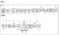
- the preferred processing plate (101) in the context of the present invention is a 1-component plate. Its top surface (110) comprises multiple vessels (103) ( Fig. 5 , Fig. 6 ). Each vessel has an opening (108) at the top and is closed at the bottom end (112).
- the top surface (110) comprises ribs (104) which are preferably elevated relative to the top surface (110) and surround the openings (108) of the vessels (103). This prevents contamination of the contents of the vessels (103) with droplets of liquid that may fall onto the top surface (110) of the plate (101). Views of a preferred process plate are shown in Figs. 3 to 8 .
- the footprint of the processing plate (101) preferably comprises a length and width of the base corresponding to ANSI SBS footprint format. More preferably, the length is 127.76 mm +/- 0.25 mm, and the width is 85.48 mm +/- 0.25 mm. Thus, the plate (101) has two opposing shorter side walls (109) and two opposing longer side walls (118).
- the processing plate (101) comprises form locking elements (106) for interacting with a handler (500, Fig. 12 ).
- the processing plate (101) can be gripped, transported and positioned quickly and safely at high speed while maintaining the correct orientation and position.
- the form locking elements (106) for gripping are located within the upper central part, preferably the upper central third of the processing plate (101). This has the advantage that a potential distortion of the processing plate (101) has only a minor effect on the form locking elements (106) and that the handling of the plate (101) is more robust.
- the processing plate (101) preferably comprises hardware-identifiers (102) and (115).
- the hardware identifiers (102) and (115) are unique for the processing plate (101) and different from hardware identifiers of other consumables used in the same system.
- the hardware identifiers (102, 115) preferably comprise ridges (119) and/or recesses (125) on the side walls of the consumables, wherein said pattern of ridges (119) and/or recesses (125) is unique for a specific type of consumable, preferably the processing plate (101). This unique pattern is also referred to herein as a unique "surface geometry".
- the hardware-identifiers (102, 115) ensure that the user can only load the processing plate (101) into the appropriate stacker position of an analytical instrument in the proper orientation.
- guiding elements (116) and (117) are comprised ( Fig. 3, Fig. 4 ). They prevent canting of the processing plate (101).
- the guiding elements (116, 117) allow the user to load the processing plates (101) with guiding elements (116, 117) as a stack into an analytical instrument which is then transferred vertically within the instrument in a stacker without canting of the plates.
- the center part (120) of the vessels (103) has an almost rectangular cross section ( Fig. 6, Fig. 7 ). They are separated along the longer side (118) of the almost rectangular shape by a common wall (113) ( Fig. 3 ).
- the row of vessels (103) formed thereby has the advantage that despite the limited space available, they have a large volume, preferably of 4 ml. Another advantage is that because of the essentially constant wall thickness, the production is very economical. A further advantage is that the vessels (103) strengthen each other and, thus, a high stability of the shape can be obtained.
- a continuous space (121) is located ( Fig. 6, Fig. 7 ).
- the space (121) can accommodate magnets (202, 203) or heating devices (128) ( Fig. 11 ).
- These magnets (202, 203) and heating devices (128) are preferably solid devices.
- magnetic particles (216) comprised in liquids (215) which can be held in the vessels (103) can be separated from the liquid (215) by exerting a magnetic field on the vessels (103) when the magnets (202, 203) are brought into proximity of the vessels (103).
- the contents of the vessels (103) can be incubated at an elevated, controlled temperature when the processing plate (101) is placed on the heating device (128).
- the almost rectangular shape of the central part (120) of the vessels (103) also optimizes the contact between the vessel wall (109) and a flat shaped magnet (202) or heating device (128) by optimizing the contact surface between vessel (103) and magnet (202) or heating device (128) and thus enhancing energy transfer into the vessel (103).
- the space (121) is even more pronounced and can accommodate further magnets (203).
- the combination of the large magnets (202) in the upper area and the smaller magnets (203) in the conical area of the vessels allows separation of magnetic particles (216) in larger or small volumes of liquid (215).
- the small magnets (203) thus, make it easier to sequester the magnetic particles (216) during eluate pipetting. This makes it possible to pipette the eluate with minimal loss by reducing the dead volume of the magnetic particle (216) pellet. Furthermore, the presence of magnetic particles (216) in the transferred eluate is minimized.
- one of the shorter side walls (109) of the vessel (103) comprises an reagent inlet channel (105) which extends to the circumferential rib (104) ( Figs. 3, 4 , 7 ).
- the reagents are pipetted onto the reagent inlet channel (105) and drain off the channel (105) into the vessel (103).
- contact between the pipet needle or tip (3, 4) and liquid contained in the vessel is prevented.
- splashes resulting from liquid being directly dispensed into another liquid (215) contained in the vessels (103), which may cause contamination of the pipet needle or tip (3, 4) or neighboring vessels (103) is prevented.
- the shape On the inside, on the bottom of the vessels (111, 112), the shape becomes conical (111) and ends with a spherical bottom (112) ( Fig. 6. Fig. 7 ).
- the combination of spherical bottom (112), rounded inside shape (114), conical part (111) and refined surface of the vessels (103) leads to favorable fluidics which facilitate an effective separation and purification of analytes in the processing plate (101).
- the spherical bottom (112) allows an essentially complete use of the separated eluate and a reduction of dead-volume which reduces the carryover of reagents or sample cross-contamination.
- the rim on the base (129) of the processing plate (101) comprises recesses (107) for engagement with latch clips (124) on the processing station (201) or heating device (128) or analytical instrument (126) ( Fig. 5 , Fig. 9 ).
- the engagement of the latch clips (124) with the recesses (107) allows positioning and fixation of the processing plate (101) on the processing station (201).
- the presence of the recesses (107) allows the latch force to act on the processing plate (101) almost vertically to the base (129). Thus, only small forces acting sideways can occur. This reduces the occurrence of strain, and, thus, the deformation of the processing plate (101).
- the vertical latch forces can also neutralize any deformations of the processing plate (101) leading to a more precise positioning of the spherical bottoms (111) within the processing station (201).
- the precise interface between the processing plate (101) and the processing station (201) or heating device (128) within an analyzer reduces dead-volumes and also reduces the risk of sample cross-contamination.
- a “separation station” is a device or a component of an analytical system allowing for the isolation of the solid support material from the other material present in the fluid sample.
- a separation station can e.g. comprise, but is not limited to, a centrifuge, a rack with filter tubes, a magnet, or other suitable components.
- the separation station comprises one or more magnets.
- one or more magnets are used for the separation of magnetic particles, preferably magnetic glass particles, as a solid support. If, for example, the fluid sample and the solid support material are combined together in the wells of a multiwell plate, then one or more magnets comprised by the separation station can e.g.
- the separation station is a device that comprises a multiwell plate comprising vessels with an opening at the top surface of the multiwell plate and a closed bottom.
- the vessels comprise an upper part, a center part and a bottom part, wherein the upper part is joined to the top surface of the multiwell plate and preferably comprises two longer and two shorter sides.
- the center part has a substantially rectangular cross-section with two longer sides, wherein said vessels are aligned in rows.
- a continuous space is located between two adjacent rows for selectively contacting at least one magnet mounted on a fixture with the side walls in at least two Z-positions.
- the device further comprises a magnetic separation station comprising at least one fixture.
- the fixture comprises at least one magnet generating a magnetic field.
- a moving mechanism is present which vertically moves said at least one fixture comprising at least one magnet at least between first and second positions with respect to the vessels of the multiwell plate.
- said at least two Z-positions of the vessels comprise the side walls and the bottom part of said vessels.
- the magnetic field of said at least one magnet preferably draws the magnetic particles to an inner surface of the vessel adjacent to said at least one magnet when said at least one magnet is in said first position.
- the fixture comprising said at least one magnet comprises a frame.
- the vessels have preferred features as described above in the context of multiwell plate/ processing plate.
- One such preferred feature is that at least a part of said vessels has a substantially rectangular cross-section orthogonal to the axis of said vessels.
- said at least one magnet is adjacent to said part of said vessels.
- Adjacent is understood to mean either in close proximity such as to exert a magnetic field on the contents of the vessel, or in physical contact with the vessel.
- the separation station comprises a frame to receive the multiwell plate, and latch-clips to attach the multiwell plate.
- the separation station comprises two types of magnets. This preferred embodiment is further described below.
- a second preferred embodiment is described below, which comprises a spring which exerts a pressure on the frame comprising the magnets such that the magnets are pressed against the vessels of the multiwell plate.
- the first magnets are preferably constructed and arranged to interact with vessels of a multiwell plate for exerting a magnetic field on a large volume of liquid comprising magnetic particles held in said vessels.
- Said second magnets preferably are constructed and arranged to interact with vessels of a multiwell plate for exerting a magnetic field on a small volume of liquid comprising magnetic particles held in said vessels. Said first and second magnets can be moved to different Z-positions.
- the separation station is further a method of isolating and purifying a nucleic acid.
- the method comprises the steps of binding a nucleic acid to magnetic particles in a vessel of a multiwell plate.
- the vessel comprises an upper opening, a central part and a bottom part.
- the bound material is then separated from unbound material contained in a liquid when the major part of the liquid is located above the section where the conical part of the vessel is replaced by the central part with the rectangular shape, by moving a magnet from a second position to a first position and, in said first position, applying a magnetic field to the central part and, optionally, additionally applying a magnetic field to the bottom part of said vessel.
- the magnetic particles can optionally be washed with a washing solution.
- a small volume of liquid, wherein the major part of the liquid is located below the section where the conical part of the vessel is replaced by the central part with the rectangular shape is separated from said magnetic particles by selectively applying a magnetic field to the bottom part of said vessel.
- a magnetic separation station for separating a nucleic acid bound to magnetic particles, said separation station comprising first magnets which are constructed and arranged to interact with vessels of a multiwell plate for exerting a magnetic field on a large volume of liquid comprising magnetic particles held in said vessels, and second magnets constructed and arranged to interact with vessels of a multiwell plate for exerting a magnetic field on a small volume of liquid comprising magnetic particles held in said vessels, and wherein said first and second magnets can be moved to different Z-positions.
- Preferred embodiments of the magnetic separation station are described herein.
- a first preferred embodiment of a separation station (201) useful for the present invention is described below.
- the first preferred embodiment of said separation station (201) comprises at least two types of magnets (202, 203).
- the first, long type of magnet (202) is constructed and arranged to fit into the space (121) of the processing plate (101). Magnet (202), thus, exerts a magnetic field on the liquid (215) in the vessel (103) to sequester magnetic particles (216) on the inside of the vessel wall. This allows separation of the magnetic particles (216) and any material bound thereto and the liquid (215) inside the vessel (103) when a large volume of liquid (215) is present.
- Magnet (202) has an elongated structure and is constructed and arranged to interact with the essentially rectangular central part (120) of the vessel.
- magnet (202) is used when the major part of the liquid (215) is located above the section where the conical part (111) of the vessel (103) is replaced by the central part (120) with the rectangular shape.
- the preferred construction of the magnets (202) comprises fixtures (204, 204a) comprising magnets (202) which fit into the space (121) between the rows of vessels (103) in the processing plate (101).
- Another preferred embodiment of magnets (202) comprises magnets (202) arranged on fixtures (204, 204a).
- the magnets (203) of the preferred separation station (201) are smaller, and can interact with the conical part (111) of the vessel (103). This is shown in Fig. 10 .
- Magnets (203) are preferably arranged on a base (205) which can be moved into the space (121) of the processing plate (101). Each magnet (202, 203) is preferably constructed to interact with two vessels (103) in two adjacent rows. In a preferred embodiment, the processing plate (101) has 6 rows of 8 vessels (103).
- a separation station (201) which can interact with the preferred processing plate (101) has three fixtures (204, 204a) comprising magnets (202) and four bases (205) comprising magnets (203). An embodiment is also included wherein the separation station has four magnetic fixtures (204, 204a) comprising magnets (202) and three magnetic bases (205) comprising magnets (203).
- the magnets (202, 203) are movable.
- the separation station (201) comprises a mechanism to move the fixtures (204, 204a) and the bases (205). All fixtures (204, 204a) are interconnected by a base (217) and are, thus, moved coordinately. All magnets (203) are joined to one base (218) and are, thus, moved coordinately.
- the mechanism for moving the magnetic plates (202) and (203) is constructed and arranged to move the two types of magnetic plates (202, 203) to a total of four end positions:
- the base (217) of magnets (202) is connected to a positioning wheel (206).
- the base (217) comprises a bottom end (207) which is flexibly in contact with a connecting element (208) by a moving element (209).
- Said moving element is constructed and arranged to move the connecting element (208) along a rail (212) from one side to the other.
- Said moving element (209) is fixed to the connecting element (208) with a pin (220).
- Said connecting element (208) is fixed to the positioning wheel (206) by screw (210).
- Connecting element (208) is also connected to axis (211).
- Said connecting element (208) is preferably a rectangular plate.
- the base (218) is mounted on a bottom part (219) and is connected, at its lower end, with pin (213) to a moving element (214), which is preferably a wheel, which interacts with the positioning wheel (206).
- wheel (214) moves along positioning wheel (206).
- the magnets (203) are in their lowermost position.
- the location of the magnets (203) is controlled by the shape of the positioning wheel (206).
- moving element (209) moves along the central, rounded upper or lower part (212a) of rail (212)
- the small type of magnets (203) are moved up and down.
- the moving element (209) is located on the side (212b) of bottom end (207) and moves up or down, the magnets (202) are moved up- or downwards.
- the positioning wheel can be rotated by any motor (224).
- a spring (225) is attached to the base (222) of the separation station and the base (218) of magnets (203) to ensure that magnets (203) are moved into the lowermost position when they are moved downwards.
- pin as used herein relates to any fixation element, including screws or pins.
- the separation station (230) comprises at least one fixture (231) comprising at least one magnet (232), preferably a number of magnets equal to a number of vessels (103) in a row (123).
- the separation station (230) comprises a number of fixtures (231) equal to the number of rows (123) of the multiwell plate (101) hereinbefore described. More preferably, six fixtures (231) are mounted on the separation station (230). At least one magnet (232) is mounted on one fixture (231). Preferably, the number of magnets (232) equals the number of vessels (103) in one row (123). Most preferably, eight magnets (232) are mounted on one fixture (231).
- one type of magnet (232) is comprised on said fixture (231). More preferably, the magnet (232) is mounted on one side oriented towards the vessels with which the magnet interacts.
- the fixture (231) is mounted on a base (233).
- said mount is flexible.
- the base (233) comprises springs (234) mounted thereon.
- the number of springs (234) is at least one spring per fixture (231) mounted on said base (233).
- the base further comprises a chamfer (236) which limits the movement of the spring and, consequently, the fixture (231) comprising the magnets (232).
- any one of said springs (234) is constructed and arranged to interact with a fixture (231). More preferably, said spring (234) is a yoke spring. Said interaction controls the horizontal movement of the fixtures (231).
- the separation station (230) comprises a frame (235).
- the base (233) with fixtures (231) is connected to the frame (235) by a moving mechanism as described hereinbefore for the magnets (232) of the first embodiment.
- said base (233) and fixture (231) is constructed and arranged to move vertically (in Z-direction).
- the multiwell plate (101) hereinbefore described is inserted into the separation station (230).
- the fixture (231) comprising the magnets (232) is moved vertically. Any one fixture (232) is, thus, moved into a space (121) between two rows (123) of vessels (103).
- the vertical movement brings the magnets (232) mounted on a fixture (231) into contact with the vessels (103).
- the Z-position is chosen depending on the volume of liquid (215) inside the vessels (103).
- the magnets (232) contact the vessels (103) in a center position (120) where the vessels (103) are of an almost rectangular shape.
- the magnets (232) preferably contact the conical part (111) of the vessels (103).
- a spring is attached to the base (233) of any one frame (231) ( Fig. 9 a), b) ).
- the spring presses the magnets (232) against the vessels (103). This ensures a contact between magnets (232) and vessels (103) during magnetic separation.
- the magnet (232) contacts the vessel (103) on the side wall (109) located underneath the inlet (105). This has the advantage that liquid which is added by pipetting flows over the sequestered magnetic particles and ensures that particles are resuspended and that all samples in all vessels are treated identically.
- This embodiment is particularly suited to separate a liquid (215) comprised in a multiwell plate (101) as hereinbefore described, from magnetic particles (216) when different levels of liquid (215) are contained in the vessels (103) of said multiwell plate (101).
- wash buffer is a fluid that is designed to remove undesired components, especially in a purification procedure. Such buffers are well known in the art. In the context of the purification of nucleic acids, the wash buffer is suited to wash the solid support material in order to separate the immobilized nucleic acid from any unwanted components.
- the wash buffer may, for example, contain ethanol and/ or chaotropic agents in a buffered solution or solutions with an acidic pH without ethanol and/ or chaotropic agents as described above. Often the washing solution or other solutions are provided as stock solutions which have to be diluted before use.
- the washing in the process according to the invention requires a more or less intensive contact of the solid support material and the nucleic acids immobilized thereon with the wash buffer.
- Another advantageous method is aspirating and dispensing the suspension comprising wash buffer and solid support material one or more times.
- This method is preferably carried out using a pipet, wherein said pipet preferably comprises a disposable pipet tip into which said suspension is aspirated and from which it is dispensed again.
- a pipet tip can be used several times before being discarded and replaced.
- Disposable pipet tips useful for the invention preferably have a volume of at least 10 ⁇ l, more preferably at least 15 ⁇ l, more preferably at least 100 ⁇ l, more preferably at least 500 ⁇ l, more preferably of at least 1 ml, even more preferably of about 1 ml.
- Pipets used in the context of the invention can also be pipetting needles.
- washing in step d. comprises aspirating and dispensing the wash buffer comprising the solid support material.
- a preferred aspect of the disclosure is the process described above, wherein said process further comprises in step d. the step of eluting the purified nucleic acids from the solid support material with an elution buffer after washing said solid support material.
- An "elution buffer” in the context of the invention is a suitable liquid for separating the nucleic acids from the solid support.
- a liquid may e.g. be distilled water or aqueous salt solutions, such as e.g. Tris buffers like Tris HCl, or HEPES, or other suitable buffers known to the skilled artisan.
- the pH value of such an elution buffer is preferably alkaline or neutral.
- Said elution buffer may contain further components such as e.g.
- step d. is carried out at a temperature between 70°C and 90°C, more preferably at a temperature of 80°C.
- Amplification reagents in the context of the disclosure, are chemical or biochemical components that enable the amplification of nucleic acids.
- Such reagents comprise, but are not limited to, nucleic acid polymerases, buffers, mononucleotides such as nucleoside triphosphates, oligonucleotides e.g. as oligonucleotide primers, salts and their respective solutions, detection probes, dyes, and more.
- nucleoside is a base-sugar combination.
- the base portion of the nucleoside is normally a heterocyclic base.
- the two most common classes of such heterocyclic bases are purines and pyrimidines.
- Nucleotides are nucleosides that further include a phosphate group covalently linked to the sugar portion of the nucleoside.
- the phosphate group can be linked to either the 2'-, 3'- or 5'-hydroxyl moiety of the sugar.
- a nucleotide is the monomeric unit of an "oligonucleotide", which can be more generally denoted as an "oligomeric compound", or a "polynucleotide”, more generally denoted as a "polymeric compound”.
- Another general expression for the aforementioned is deoxyribonucleic acid (DNA) and ribonucleic acid (RNA).
- an "oligomeric compound” is a compound consisting of "monomeric units” which may be nucleotides alone or non-natural compounds (see below), more specifically modified nucleotides (or nucleotide analogs) or non-nucleotide compounds, alone or combinations thereof.
- Oligonucleotides and modified oligonucleotides are subgroups of oligomeric compounds.
- oligonucleotide refers to components formed from a plurality of nucleotides as their monomeric units.
- the phosphate groups are commonly referred to as forming the internucleoside backbone of the oligonucleotide.
- the normal linkage or backbone of RNA and DNA is a 3' to 5' phosphodiester linkage.
- Oligonucleotides and modified oligonucleotides (see below) useful for the invention may be synthesized as principally described in the art and known to the expert in the field.
- Chemical synthesis methods may include, for example, the phosphotriester method described by Narang S. A. et al., Methods in Enzymology 68 (1979) 90-98 , the phosphodiester method disclosed by Brown E. L., et al., Methods in Enzymology 68 (1979) 109-151 , the phosphoramidite method disclosed in Beaucage et al., Tetrahedron Letters 22 (1981) 1859 , the H-phosphonate method disclosed in Garegg et al., Chem. Scr. 25 (1985) 280-282 and the solid support method disclosed in US 4,458,066 .
- the oligonucleotides may be chemically modified, i.e. the primer and/ or the probe comprise a modified nucleotide or a non-nucleotide compound.
- the probe or the primer is then a modified oligonucleotide.
- Modified nucleotides differ from a natural nucleotide by some modification but still consist of a base, a pentofuranosyl sugar, a phosphate portion, base-like, pentofuranosyl sugar-like and phosphate-like portion or combinations thereof.
- a label may be attached to the base portion of a nucleotide whereby a modified nucleotide is obtained.
- a natural base in a nucleotide may also be replaced by e.g. a 7-deazapurine whereby a modified nucleotide is obtained as well.
- modified oligonucleotide (or “oligonucleotide analog”), belonging to another specific subgroup of oligomeric compounds, possesses one or more nucleotides and one or more modified nucleotides as monomeric units.
- modified oligonucleotide (or “oligonucleotide analog”) refers to structures that function in a manner substantially similar to oligonucleotides and can be used interchangeably in the context of the present invention.
- a modified oligonucleotide (or an oligonucleotide analog) can for example be made by chemical modification of oligonucleotides by appropriate modification of the phosphate backbone, ribose unit or the nucleotide bases ( Uhlmann and Peyman, Chemical Reviews 90 (1990) 543 ; Verma S., and Eckstein F., Annu. Rev. Biochem. 67 (1998) 99-134 ).
- Representative modifications include phosphorothioate, phosphorodithioate, methyl phosphonate, phosphotriester or phosphoramidate inter-nucleoside linkages in place of phosphodiester internucleoside linkages; deaza- or azapurines and -pyrimidines in place of natural purine and pyrimidine bases, pyrimidine bases having substituent groups at the 5 or 6 position; purine bases having altered substituent groups at the 2, 6 or 8 positions or 7 position as 7-deazapurines; bases carrying alkyl-, alkenyl-, alkinyl or aryl-moieties, e.g.
- exemplary modifications are disclosed in Verma S., and Eckstein F., Annu. Rev. Biochem. 67 (1998) 99-134 or WO 02/12263 .
- modification can be made wherein nucleoside units are joined via groups that substitute for the internucleoside phosphate or sugar phosphate linkages.
- Such linkages include those disclosed in Verma S., and Eckstein F., Annu. Rev. Biochem. 67 (1998) 99-134 .
- oligonucleosides When other than phosphate linkages are utilized to link the nucleoside units, such structures have also been described as "oligonucleosides".
- nucleic acid as well as the “target nucleic acid” is a polymeric compound of nucleotides as known to the expert skilled in the art.
- target nucleic acid is used herein to denote a nucleic acid in a sample which should be analyzed, i.e. the presence, non-presence and/or amount thereof in a sample should be determined.
- primer is used herein as known to the expert skilled in the art and refers to oligomeric compounds, primarily to oligonucleotides, but also to modified oligonucleotides that are able to prime DNA synthesis by a template-dependent DNA polymerase, i.e. the 3'-end of the e.g. primer provides a free 3'-OH group whereto further nucleotides may be attached by a template-dependent DNA polymerase establishing 3'- to 5'-phosphodiester linkage whereby deoxynucleoside triphosphates are used and whereby pyrophosphate is released.
- a “probe” also denotes a natural or modified oligonucleotide.
- a probe serves the purpose to detect an analyte or amplificate.
- probes can be used to detect the amplificates of the target nucleic acids.
- probes typically carry labels.
- Labeled nucleic acid binding compounds labeled probes or just probes.
- Preferred labels according to the invention are fluorescent labels, which are e.g. fluorescent dyes such as a fluorescein dye, a rhodamine dye, a cyanine dye, and a coumarin dye.
- Preferred fluorescent dyes according to the invention are FAM, HEX, JA270, CAL635, Coumarin343, Quasar705, Cyan500, CY5.5, LC-Red 640, LC-Red 705.
- any primer and/or probe may be chemically modified, i.e. the primer and/ or the probe comprise a modified nucleotide or a non-nucleotide compound.
- the probe or the primer is then a modified oligonucleotide.
- PCR Polymerase Chain Reaction
- Primers useful for nucleic acid analysis include oligonucleotides capable of acting as a point of initiation of nucleic acid synthesis within the nucleic acid sequences of the target nucleic acids.
- a primer can be purified from a restriction digest by conventional methods, or it can be produced synthetically.
- the primer is preferably single-stranded for maximum efficiency in amplification, but the primer can be double-stranded.
- Double-stranded primers are first denatured, i.e., treated to separate the strands.
- One method of denaturing double stranded nucleic acids is by heating.
- a "thermostable polymerase” is a polymerase enzyme that is heat stable, i.e., it is an enzyme that catalyzes the formation of primer extension products complementary to a template and does not irreversibly denature when subjected to the elevated temperatures for the time necessary to effect denaturation of double-stranded template nucleic acids.
- the synthesis is initiated at the 3' end of each primer and proceeds in the 5' to 3' direction along the template strand.
- Thermostable polymerases have e.g. been isolated from Thermus flavus, T. ruber, T. thermophilus, T. aquaticus, T. lacteus, T. rubens, Bacillus stearothermophilus, and Methanothermus fervidus. Nonetheless, polymerases that are not thermostable also can be employed in PCR assays provided the enzyme is replenished.
- Strand separation can be accomplished by any suitable denaturing method including physical, chemical or enzymatic means.
- One method of separating the nucleic acid strands involves heating the nucleic acid until it is predominately denatured (e.g., greater than 50%, 60%, 70%, 80%, 90% or 95% denatured).
- the heating conditions necessary for denaturing template nucleic acid will depend, e.g., on the buffer salt concentration and the length and nucleotide composition of the nucleic acids being denatured, but typically range from about 90°C to about 105°C for a time depending on features of the reaction such as temperature and the nucleic acid length. Denaturation is typically performed for about 5 sec to 9 min. In order to not expose the respective polymerase like e.g. the Z05 DNA Polymerase to such high temperatures for too long and thus risking a loss of functional enzyme, it is preferred to use short denaturation steps.
- the denaturation step is up to 30 sec, further preferably up to 20 sec, further preferably up to 10 sec, further preferably up to 5 sec, most preferably about 5 sec.
- the reaction mixture is allowed to cool to a temperature that promotes annealing of each primer to its target sequence on the target nucleic acids.
- the temperature for annealing is preferably from about 35°C to about 70°C, further preferably about 45°C to about 65°C; further preferably about 50°C to about 60°C, further preferably about 55°C to about 58°C.
- Annealing times can be from about 10 sec to about 1 min (e.g., about 20 sec to about 50 sec; about 30 sec to about 40 sec).
- primers may also bind to targets having single mismatches, so variants of certain sequences can also be amplified. This can be desirable if e.g.
- the process described above comprises annealing at different temperatures, preferably first at a lower, then at a higher temperature. If, e.g., a first incubation takes place at 55°C for about 5 cycles, non-exactly matching target sequences may be (pre-)amplified. This can be followed e.g. by about 45 cycles at 58°C, providing for higher specificity throughout the major part of the experiment. This way, potentially important genetic variants are not missed, while the specificity remains relatively high.
- the reaction mixture is then adjusted to a temperature at which the activity of the polymerase is promoted or optimized, i.e., a temperature sufficient for extension to occur from the annealed primer to generate products complementary to the nucleic acid to be analyzed.
- the temperature should be sufficient to synthesize an extension product from each primer that is annealed to a nucleic acid template, but should not be so high as to denature an extension product from its complementary template (e.g., the temperature for extension generally ranges from about 40° to 80°C (e.g., about 50°C to about 70°C; about 60°C).
- Extension times can be from about 10 sec to about 5 min, preferably about 15 sec to 2 min, further preferably about 20 sec to about 1 min, further preferably about 25 sec to about 35 sec.
- the newly synthesized strands form a double-stranded molecule that can be used in the succeeding steps of the reaction.
- the steps of strand separation, annealing, and elongation can be repeated as often as needed to produce the desired quantity of amplification products corresponding to the target nucleic acids.
- the limiting factors in the reaction are the amounts of primers, thermostable enzyme, and nucleoside triphosphates present in the reaction.
- the cycling steps i.e., denaturation, annealing, and extension
- cycling steps For use in detection, the number of cycling steps will depend, e.g., on the nature of the sample. If the sample is a complex mixture of nucleic acids, more cycling steps will be required to amplify the target sequence sufficient for detection. Generally, the cycling steps are repeated at least about 20 times, but may be repeated as many as 40, 60, or even 100 times.
- a PCR can be carried out in which the steps of annealing and extension are performed in the same step (one-step PCR) or, as described above, in separate steps (two-step PCR).
- steps of annealing and extension are performed in the same step (one-step PCR) or, as described above, in separate steps (two-step PCR).
- Performing annealing and extension together and thus under the same physical and chemical conditions, with a suitable enzyme such as, for example, the Z05 DNA polymerase, bears the advantage of saving the time for an additional step in each cycle, and also abolishing the need for an additional temperature adjustment between annealing and extension.
- the one-step PCR reduces the overall complexity of the respective assay.
- nucleic acid amplification methods comprise the Ligase Chain Reaction (LCR; Wu D. Y. and Wallace R. B., Genomics 4 (1989) 560-69 ; and Barany F., Proc. Natl. Acad. Sci. USA 88 (1991)189-193 ); Polymerase Ligase Chain Reaction ( Barany F., PCR Methods and Applic. 1 (1991) 5-16 ); Gap-LCR ( WO 90/01069 ); Repair Chain Reaction ( EP 0439182 A2 ), 3SR ( Kwoh D.Y. et al., Proc. Natl. Acad. Sci.
- the internal control nucleic acid used in the present disclosure preferably exhibits the following properties relating to its sequence:
- sequence is the primary structure of a nucleic acid, i.e. the specific arrangement of the single nucleobases of which the respective nucleic acids consists. It has to be understood that the term “sequence” does not denote a specific type of nucleic acid such as RNA or DNA, but applies to both as well as to other types of nucleic acids such as e.g. PNA or others. Where nucleobases correspond to each other, particularly in the case of uracil (present in RNA) and thymine (present in DNA), these bases can be considered equivalent between RNA and DNA sequences, as well-known in the pertinent art.
- nucleic acids are often DNA which can be derived e.g. from DNA viruses like e.g. Hepatitis B Virus (HBV), Cytomegalovirus (CMV) and others, or bacteria like e.g. Chlamydia trachomatis (CT), Neisseria gonorrhoeae (NG) and others.
- HBV Hepatitis B Virus
- CMV Cytomegalovirus
- CT Chlamydia trachomatis
- NG Neisseria gonorrhoeae
- a preferred aspect of the invention is the method described above, wherein said internal control nucleic acid is DNA.
- nucleic acids relevant for clinical diagnostics are ribonucleic acids, like e.g. the nucleic acids from RNA viruses such as for example Human Immunodeficiency Virus (HIV), Hepatitis C Virus (HCV), the West Nile Virus (WNV), Human Papilloma Virus (HPV), Japanese Encephalitis Virus (JEV), St. Louis Encephalitis Virus (SLEV) and others.
- the present invention can be readily applied to such nucleic acids.
- the internal control nucleic acid is RNA, as the internal control nucleic acid preferably mimics the most sensitive target of an assay involving multiple targets, and RNA targets usually have to be more closely controlled.
- RNA RNA
- RNA Since RNA is more prone to degradation than DNA due to influences such as alkaline pH, ribonucleases etc., internal control nucleic acids made of RNA are preferably provided as armored particles. Armored particles such as especially armored RNA are described e.g. in EP910643 .
- the RNA which can be produced chemically or, preferably, heterologously e.g. by bacteria such as e.g. E. coli, is at least partially encapsulated in a viral coat protein. The latter confers resistance of the RNA towards external influences, in particular ribonucleases. It must be understood that internal control DNA can also be provided as an armored particle.
- RNA control nucleic acids are armored with the MS2 coat protein in E. coli.
- DNA control nucleic acids are armored using lambda phage GT11.
- a preferred aspect of the disclosure is the method described above, wherein said internal control nucleic acid is an armored nucleic acid.
- RNA templates are transcribed into DNA prior to amplification and detection.
- a preferred aspect of the disclosure is the process described above, wherein said amplification reagents comprise a polymerase with reverse transcriptase activity, said process further comprising between step e. and step f. the step of incubating in said reaction vessels said purified nucleic acids with said one or more amplification reagents for a period of time and under conditions suitable for transcription of RNA by said polymerase with reverse transcriptase activity to occur.
- a "polymerase with reverse transcriptase activity” is a nucleic acid polymerase capable of synthesizing DNA based on an RNA template. It is also capable of the formation of a double-stranded DNA once the RNA has been reverse transcribed into a single strand cDNA. In a preferred embodiment of the invention, the polymerase with reverse transcriptase activity is thermostable.
- the process according to the comprises incubating a sample containing an RNA template with an oligonucleotide primer sufficiently complementary to said RNA template to hybridize with the latter, and a preferably thermostable DNA polymerase in the presence of at least all four natural or modified deoxyribonucleoside triphosphates, in an appropriate buffer comprising a metal ion buffer which, in a preferred embodiment, buffers both the pH and the metal ion concentration.
- This incubation is performed at a temperature sufficient for said primer to hybridize to said RNA template and said DNA polymerase to catalyze the polymerization of said deoxyribonucleoside triphosphates to form a cDNA sequence complementary to the sequence of said RNA template.
- cDNA refers to a complementary DNA molecule synthesized using a ribonucleic acid strand (RNA) as a template.
- RNA ribonucleic acid strand
- the RNA may e.g. be mRNA, tRNA, rRNA, or another form of RNA, such as viral RNA.
- the cDNA may be single-stranded, double-stranded or may be hydrogen-bonded to a complementary RNA molecule as in an RNA/cDNA hybrid.
- a primer suitable for annealing to an RNA template may also be suitable for amplification by PCR.
- a second primer complementary to the reverse transcribed cDNA strand, provides an initiation site for the synthesis of an extension product.
- the first extension reaction is reverse transcription using an RNA template, and a DNA strand is produced.
- the second extension reaction using the DNA template, produces a double-stranded DNA molecule.
- synthesis of a complementary DNA strand from an RNA template by a DNA polymerase provides the starting material for amplification.
- Thermostable DNA polymerases can be used in a coupled, one-enzyme reverse transcription/amplification reaction.
- homogeneous it is meant that following the reverse transcription (RT) step, there is no need to open the reaction vessel or otherwise adjust reaction components prior to the amplification step.
- a non-homogeneous RT/PCR reaction following reverse transcription and prior to amplification one or more of the reaction components such as the amplification reagents are e.g. adjusted, added, or diluted, for which the reaction vessel has to be opened, or at least its contents have to be manipulated. While both homogeneous and non-homogeneous embodiments are comprised by the scope of the invention, the homogeneous format for RT/PCR is preferred.
- Reverse transcription is an important step in an RT/PCR. It is, for example, known in the art that RNA templates show a tendency towards the formation of secondary structures that may hamper primer binding and/or elongation of the cDNA strand by the respective reverse transcriptase. Thus, relatively high temperatures for an RT reaction are advantageous with respect to efficiency of the transcription. On the other hand, raising the incubation temperature also implies higher specificity, i.e. the RT primers will not anneal to sequences that exhibit mismatches to the expected sequence or sequences. Particularly in the case of multiple different target RNAs, it can be desirable to also transcribe and subsequently amplify and detect sequences with single mismatches, e.g. in the case of the possible presence of unknown or rare substrains or subspecies of organisms in the fluid sample.
- a preferred aspect of the disclosure is the process described above, wherein said incubation of the polymerase with reverse transcriptase activity is carried out at different temperatures from 30°C to 75°C, preferably from 45°C to 70°C, further preferably from 55°C to 65°C.
- RT steps can damage the DNA templates that may be present in the fluid sample. If the fluid sample contains both RNA and DNA species, it is thus favorable to keep the duration of the RT steps as short as possible, but at the same time ensuring the synthesis of sufficient amounts of cDNA for the subsequent amplification and optional detection of amplificates.
- a preferred aspect of the disclosure is the process described above, wherein the period of time for incubation of the polymerase with reverse transcriptase activity is up to 30 minutes, 20 minutes, 15 minutes, 12.5 minutes, 10 minutes, 5 minutes, or 1 minute.
- a further preferred aspect of the disclosure is the process described above, wherein the polymerase with reverse transcriptase activity and comprising a mutation is selected from the group consisting of
- enzymes carrying a mutation in the polymerase domain that enhances their reverse transcription efficiency in terms of a faster extension rate.
- a preferred aspect of the disclosure is the process described above, wherein the polymerase with reverse transcriptase activity is a polymerase comprising a mutation conferring an improved nucleic acid extension rate and/or an improved reverse transcriptase activity relative to the respective wildtype polymerase.
- the polymerase with reverse transcriptase activity is a polymerase comprising a mutation conferring an improved reverse transcriptase activity relative to the respective wildtype polymerase.
- polymerases carrying point mutations that render them particularly useful in the context of the invention are disclosed in WO 2008/046612 .
- preferred polymerases to be used in the context of the present invention are mutated DNA polymerases comprising at least the following motif in the polymerase domain: T-G-R-L-S-S-X b7 -X b8 -P-N-L-Q-N; wherein X b7 is an amino acid selected from S or T and wherein X b8 is an amino acid selected from G, T, R, K, or L, wherein the polymerase comprises 3'-5' exonuclease activity and has an improved nucleic acid extension rate and/or an improved reverse transcription efficiency relative to the wildtype DNA polymerase, wherein in said wildtype DNA polymerase X b8 is an amino acid selected from D, E or N.
- thermostable DNA polymerase from Thermus species Z05 (described e.g. in US 5,455,170 ), said variations comprising mutations in the polymerase domain as compared with the respective wildtype enzyme Z05.
- a mutant Z05 DNA polymerase wherein the amino acid at position 580 is selected from the group consisting of G, T, R, K and L.
- Mn2+ is preferred as the divalent cation and is typically included as a salt, for example, manganese chloride (MnCl2), manganese acetate (Mn(OAc)2), or manganese sulfate (MnSO4).
- MnCl2 is included in a reaction containing 50 mM Tricine buffer, for example, the MnCl2 is generally present at a concentration of 0.5-7.0 mM; 0.8-1.4 mM is preferred when 200 mM of each dGTP, dATP, dUTP, and, dCTP are utilized; and 2.5-3.5 mM MnCl2 is most preferred.
- Mg2+ is also preferred in the context of the present invention.
- the internally controlled process described above is particularly useful for the simultaneous amplification of target nucleic acids derived from both organisms having an RNA or organisms having a DNA genome. This advantage considerably increases the spectrum of different organisms, especially pathogens, that can be analyzed under identical physical conditions.
- a preferred aspect of the disclosure is the process described above, wherein the at least two target nucleic acids comprise RNA and DNA.
- An "organism”, as used herein, means any living single- or multicellular life form.
- a virus is an organism.
- enzymes like Tth polymerase or, preferably, the mutant Z05 DNA polymerase mentioned above are suited to carry out the subsequent step of amplification of the target nucleic acids. Exploiting the same enzyme for both reverse transcription an amplification contributes to the ease of carrying out the process and facilitates its automation, since the fluid sample does not have to be manipulated between the RT and the amplification step.
- the same polymerase with reverse transcriptase activity is used for reverse transcription and for the amplification in step f.
- the enzyme is the mutant Z05 DNA polymerase described supra.
- steps above 90°C are up to 20 sec, preferably up to 15 sec, more preferably up to 10 sec, more preferably up to 5 sec and most preferably 5 sec long. This also reduces the time-to-result and cuts down the overall required time of the assay.
- Sealing can be e.g. achieved by applying a foil that is preferably transparent, a cap, or by oil added to the reaction vessels and forming a lipophilic phase as a sealing layer at the top of the fluid.
- a preferred aspect of the disclosure is the process described above, further comprising after step e. the step of sealing the at least two reaction vessels.
- reaction vessels For the ease of handling and to facilitate automation, it is preferable to combine the at least two reaction vessels in an integral arrangement, so they can be manipulated together.
- Integral arrangements can e.g. be vials or tubes reversibly or irreversibly attached to each other or arranged in a rack.
- the integral arrangement is a multiwell plate.
- the target of the amplification step can be an RNA/DNA hybrid molecule.
- the target can be a single-stranded or double-stranded nucleic acid. Although the most widely used PCR procedure uses a double-stranded target, this is not a necessity.
- the reaction mixture contains a double-stranded DNA molecule consisting of the single-stranded target and a newly synthesized complementary strand.
- the reaction mixture contains a double-stranded cDNA molecule. At this point, successive cycles of amplification proceed as described above.
- a preferred aspect of the invention is the process described above, wherein the amplification reaction in step f. consists of multiple cycling steps.
- nucleic acid detection methods are known to the expert in the field and are described in standard textbooks as Sambrook J. et al., Molecular Cloning: A Laboratory Manual, Cold Spring Harbor Laboratory Press, Cold Spring Harbor, New York, 1989 and Ausubel F. et al.: Current Protocols in Molecular Biology 1987, J. Wiley and Sons, NY .
- the detection methods may include but are not limited to the binding or intercalating of specific dyes as ethidium bromide which intercalates into the double-stranded DNA and changes its fluorescence thereafter.
- the purified nucleic acid may also be separated by electrophoretic methods optionally after a restriction digest and visualized thereafter.
- probe-based assays which exploit the oligonucleotide hybridization to specific sequences and subsequent detection of the hybrid.
- nucleic acid probes it is preferable to detect the amplified target nucleic acids during or after the amplification reaction in order to evaluate the result of the analysis. Particularly for detection in real time, it is advantageous to use nucleic acid probes.
- a cycling step comprises an amplification step and a hybridization step, said hybridization step comprising hybridizing the amplified nucleic acids with probes.
- a preferred aspect of the invention is the process described above, wherein the probes are labeled with a donor fluorescent moiety and a corresponding acceptor fluorescent moiety.
- the methods set out above are preferably based on Fluorescence Resonance Energy Transfer (FRET) between a donor fluorescent moiety and an acceptor fluorescent moiety.
- FRET Fluorescence Resonance Energy Transfer
- a representative donor fluorescent moiety is fluorescein, and representative corresponding acceptor fluorescent moieties include LC-Red 640, LC-Red 705, Cy5, and Cy5.5.
- detection includes exciting the sample at a wavelength absorbed by the donor fluorescent moiety and visualizing and/or measuring the wavelength emitted by the corresponding acceptor fluorescent moiety.
- detection is preferably followed by quantitating the FRET.
- detection is performed after each cycling step. Most preferably, detection is performed in real time.
- real-time PCR instrumentation e.g., LightCyclerTM or TaqMan®
- PCR amplification and detection of the amplification product can be combined in a single closed cuvette with dramatically reduced cycling time. Since detection occurs concurrently with amplification, the real-time PCR methods obviate the need for manipulation of the amplification product, and diminish the risk of cross-contamination between amplification products.
- Real-time PCR greatly reduces turn-around time and is an attractive alternative to conventional PCR techniques in the clinical laboratory.
- the LightCyclerTM instrument is a rapid thermal cycler combined with a microvolume fluorometer utilizing high quality optics. This rapid thermocycling technique uses thin glass cuvettes as reaction vessels. Heating and cooling of the reaction chamber are controlled by alternating heated and ambient air. Due to the low mass of air and the high ratio of surface area to volume of the cuvettes, very rapid temperature exchange rates can be achieved within the thermal chamber.
- TaqMan® technology utilizes a single-stranded hybridization probe labeled with two fluorescent moieties.
- a first fluorescent moiety When a first fluorescent moiety is excited with light of a suitable wavelength, the absorbed energy is transferred to a second fluorescent moiety according to the principles of FRET.
- the second fluorescent moiety is generally a quencher molecule.
- Typical fluorescent dyes used in this format are for example, among others, FAM, HEX, CY5, JA270, Cyan and CY5.5.
- the labeled hybridization probe binds to the target nucleic acid (i.e., the amplification product) and is degraded by the 5' to 3' exonuclease activity of the Taq or another suitable polymerase as known by the skilled artisan, such as the preferred mutant Z05 polymerase, during the subsequent elongation phase.
- the excited fluorescent moiety and the quencher moiety become spatially separated from one another.
- the fluorescence emission from the first fluorescent moiety can be detected.
- the intensity of the emitted signal can be correlated with the number of original target nucleic acid molecules.
- an amplification product can be detected using a double-stranded DNA binding dye such as a fluorescent DNA binding dye (e.g., SYBRGREEN I® or SYBRGOLD® (Molecular Probes)).
- a fluorescent DNA binding dye e.g., SYBRGREEN I® or SYBRGOLD® (Molecular Probes)
- Upon interaction with the double-stranded nucleic acid such fluorescent DNA binding dyes emit a fluorescence signal after excitation with light at a suitable wavelength.
- a double-stranded DNA binding dye such as a nucleic acid intercalating dye also can be used.
- a melting curve analysis is usually performed for confirmation of the presence of the amplification product.
- Molecular beacons in conjunction with FRET can also be used to detect the presence of an amplification product using the real-time PCR methods of the invention.
- Molecular beacon technology uses a hybridization probe labeled with a first fluorescent moiety and a second fluorescent moiety.
- the second fluorescent moiety is generally a quencher, and the fluorescent labels are typically located at each end of the probe.
- Molecular beacon technology uses a probe oligonucleotide having sequences that permit secondary structure formation (e.g. a hairpin). As a result of secondary structure formation within the probe, both fluorescent moieties are in spatial proximity when the probe is in solution. After hybridization to the amplification products, the secondary structure of the probe is disrupted and the fluorescent moieties become separated from one another such that after excitation with light of a suitable wavelength, the emission of the first fluorescent moiety can be detected.
- probes comprise a nucleic acid sequence that permits secondary structure formation, wherein said secondary structure formation results in spatial proximity between said first and second fluorescent moiety.
- Efficient FRET can only take place when the fluorescent moieties are in direct local proximity and when the emission spectrum of the donor fluorescent moiety overlaps with the absorption spectrum of the acceptor fluorescent moiety.
- said donor and acceptor fluorescent moieties are within no more than 5 nucleotides of each other on said probe.
- said acceptor fluorescent moiety is a quencher.
- the labeled hybridization probe binds to the target nucleic acid (i.e., the amplification product) and is degraded by the 5' -to 3'-exonuclease activity of the Taq or another suitable polymerase as known by the skilled artisan, such as the preferred mutant Z05 polymerase, during the subsequent elongation phase.
- amplification employs a polymerase enzyme having 5' -to 3'-exonuclease activity.
- a preferred aspect of the invention is the process described above, wherein the amplified fragments comprise up to 450 bases, preferably up to 300 bases, further preferably up to 200 bases, and further preferably up to 150 bases.
- the internal control nucleic acid used in the present disclosure can serve as a "quantitative standard nucleic acid" which is apt to be and used as a reference in order to quantify, i.e. to determine the quantity of the target nucleic acids.
- quantitative standard nucleic acids undergo all possible sample preparation steps along with the target nucleic acids.
- a quantitative standard nucleic acid is processed throughout the method within the same reaction mixture. It must generate, directly or indirectly, a detectable signal both in the presence or absence of the target nucleic acid.
- the concentration of the quantitative standard nucleic acid has to be carefully optimized in each test in order not to interfere with sensitivity but in order to generate a detectable signal also e.g. at very high target concentrations.
- the concentration range for the "quantitative standard nucleic acid” is preferably 20-5000x LOD, more preferably 20-1000x LOD, most preferably 20-5000x LOD.
- the final concentration of the quantitative standard nucleic acid in the reaction mixture is dependent on the quantitative measuring range accomplished.
- LID Limit of detection
- IU/ml International units/ml
- Probit Analysis is a method of analyzing the relationship between a stimulus (dose) and the quantal (all or nothing) response.
- dose dose
- quantal response experiment groups of animals are given different doses of a drug. The percent dying at each dose level is recorded. These data may then be analyzed using Probit Analysis.
- the Probit Model assumes that the percent response is related to the log dose as the cumulative normal distribution. That is, the log doses may be used as variables to read the percent dying from the cumulative normal. Using the normal distribution, rather than other probability distributions, influences the predicted response rate at the high and low ends of possible doses, but has little influence near the middle.
- the Probit Analysis can be applied at distinct "hitrates".
- a "hitrate” is commonly expressed in percent [%] and indicates the percentage of positive results at a specific concentration of an analyte.
- an LOD can be determined at 95% hitrate, which means that the LOD is calculated for a setting in which 95% of the valid results are positive.
- the process described above provides an LOD of 1 to 100 cp/ml or 0.5 to 50 IU/ml, more preferably of 1 to 75 cp/ml or 0.5 to 30 IU/ml, more preferably of 1 to 25 cp/ml or 1 to 20 IU/ml.
- the process described above preferably provides the following LODs:
- a titer is calculated from input data of instrument-corrected fluorescence values from an entire PCR run.
- a set of samples containing a target nucleic acid and an internal control nucleic acid serving as a quantitative standard nucleic acid undergo PCR on a thermocycler using a specified temperature profile. At selected temperatures and times during the PCR profile samples are illuminated by filtered light and the filtered fluorescence data are collected for each sample for the target nucleic acid and the internal control nucleic acid.
- the fluorescence readings are processed to yield one set of dye concentration data for the internal control nucleic acid and one set of dye concentration data for the target nucleic acid. Each set of dye concentration data is processed in the same manner.
- the elbow values are calculated for the internal control nucleic acid and the target nucleic acid. The elbow value is defined as the point where the fluorescence of the target nucleic acid or the internal control nucleic acid crosses a predefined threshold (fluorescence concentration).
- T 10 a CTQS ⁇ CTtarget 2 + b CTQS ⁇ CTtarget + c
- the internal control nucleic acid can serve as a "qualitative internal control nucleic acid".
- a “qualitative internal control nucleic acid” is particularly useful for confirming the validity of the test result of a qualitative detection assay: Even in the case of a negative result, the qualitative internal control must be detected, otherwise the test itself is considered to be inoperative. However, in a qualitative setup, it does not necessarily have to be detected in case of a positive result. As a consequence, its concentration must be relatively low. It has to be carefully adapted to the respective assay and its sensitivity.
- the concentration range for the qualitative internal nucleic acid i.e. the second control nucleic acid, will comprise a range of 1 copy per reaction to 1000 copies per reaction.
- its concentration is preferably between the LOD of an assay and the 25fold value of the LOD, more preferably between the LOD and 10x LOD. More preferably, it is between 2x and 10x LOD. Even more preferably, it is between 5x and 10x LOD. Most preferably, it is 5x or 10x LOD.
- the internal control nucleic acid as used in the present disclosure is not restricted to a particular sequence. It can be advantageous to add different internal control nucleic acids to a fluid samples, but to use only one of them for amplification e.g. by adding only primers for one of said internal control nucleic acids. In such embodiments, the internal control nucleic acid to be amplified in a certain experiment can be chosen by the person skilled in the art, thus increasing flexibility of the analysis to be carried out. In particularly advantageous embodiments, said different internal control nucleic acids can be comprised by a single nucleic acid construct, e.g. a plasmid or a different suitable nucleic acid molecule.
- a preferred aspect of the disclosure is the process described above, wherein more than one internal control nucleic acid is added in step a., but only one of said internal control nucleic acids is amplified in step f.
- results described above may be adulterated and, for example, comprise false-positives, in the case of cross-contamination with nucleic acids from sources other than the fluid sample.
- amplificates of former experiments may contribute to such undesired effects.
- One particular method for minimizing the effects of cross-contamination of nucleic acid amplification is described in U.S. Patent No. 5,035,996 .
- the method involves the introduction of unconventional nucleotide bases, such as dUTP, into the amplified product and exposing carryover products to enzymatic and/or physicochemical treatment to render the product DNA incapable of serving as a template for subsequent amplifications. Enzymes for such treatments are known in the art.
- uracil-DNA glycosylase also known as uracil-N-glycosylase or UNG
- UNG will remove uracil residues from PCR products containing that base.
- the enzyme treatment results in degradation of the contaminating carryover PCR product and serves to "sterilize" the amplification reaction.
- a preferred aspect of the disclosure is the process described above, further comprising between step d) and step e) the steps of
- the enzyme is uracil-N-glycosylase.
- a further aspect of the disclosure is an analytical system (440) for isolating and simultaneously amplifying at least two target nucleic acids that may be present in a fluid sample, said analytical system comprising the following modules:
- An “analytical system” is an arrangement of components such as instruments interacting with each other with the ultimate aim to analyze a given sample.
- the analytical system (440, Fig. 11 ) of the present invention is a system (440) comprising a module (401) for isolating and/or purifying an analyte. Further, the system (440) additionally comprises a module (403) for analyzing said analyte to obtain a detectable signal.
- the detectable signal can be detected in the same module (401, 402, 403) or, alternatively, in a separate module.
- module as used herein relates to any spatially defined location within the analyzer (400). Two modules (401, 403) can be separated by walls, or can be in open relationship. Any one module (401, 402, 403) can be either autonomously controlled, or control of the module (401, 402, 403) can be shared with other modules. Preferably, all modules are controlled centrally. Transfer between modules (401, 402, 403) can be manual, but is preferably automated. Thus, a number of different embodiments of automated analyzers (400) are encompassed by the present invention.
- An “amplification station” comprises a temperature-controlled incubator for incubating the contents of at least two reaction vessels. It further comprises a variety of reaction vessels like tubes or plates, in which a reaction for the analysis of the sample such as PCR takes place. The outer limits or walls of such vessels are chemically inert such that they do not interfere with the amplification reaction taking place within. For the ease of handling and to facilitate automation, it is preferable to combine the at least two reaction vessels in an integral arrangement, so they can be manipulated together.
- a preferred aspect of the invention is the analytical system described above, wherein the at least two reaction vessels are combined in an integral arrangement.
- Integral arrangements can e.g. be vials or tubes reversibly or irreversibly attached to each other or arranged in a rack.
- the integral arrangement is a multiwell plate.
- said multiwell plate is held in a holding station.
- one handler transports a multiwell vessel from a holding station to an air-lock (460), and a second handler transports said multiwell plate from said air-lock to said amplification station, wherein both handlers interact with said multiwell plate by a form-locking interaction.
- the analytical system is fully automated.
- At least two reaction vessels combined in an integral arrangement are transported between stations of the system.
- the purified target nucleic acid is transferred from said separation station to said amplification station.
- a pipettor comprising pipets with attached pipet tips transfers the liquid comprising the purified nucleic acid.
- the purified nucleic acid is transferred from said separation station to a reaction vessel in an integral arrangement held in a holding station.
- said reaction vessel in an integral arrangement is then transferred from said holding station to said amplification station.
- the analytical system according to the invention preferably further comprises a pipetting unit.
- Said pipetting unit comprises at least one pipet, preferably multiple pipets.
- said multiple pipets are combined in one or more integral arrangements, within which the pipets can preferably be manipulated individually.
- Pipets used in the context of the invention are preferably pipets comprising pipet tips as described supra. In another preferred embodiment, the pipets are pipetting needles.
- reaction vessel or arrangement of reaction vessels used for sample preparation in the separation station and containing the fluid comprising the purified target nucleic acids may be transferred from the separation station to the amplification station.
- the analytical system according to the invention preferably further comprises a transfer unit, said transfer unit preferably comprising a robotic device, said device preferably comprising a handler.
- the analytical system (440) described above further comprises one or more elements selected from the group consisting of:
- a “detection module” can e.g. be an optical detection unit for detecting the result or the effect of the amplification procedure.
- An optical detection unit may comprise a light source, e.g. a xenon lamp, optics such as mirrors, lenses, optical filters, fiber optics for guiding and filtering the light, one or more reference channels, or a CCD camera or a different camera.
- a “sealer” (410) is constructed and arranged to seal any vessels used in connection with the analytical system according to the invention.
- a sealer can, for example, seal tubes with appropriate caps, or multiwell plates with foil, or other suitable sealing materials.
- a “storage module” (1008) stores the necessary reagents to bring about a chemical or biological reaction important for analysis of the fluid sample. It can also comprise further components useful for the method of the invention, e.g. disposables such as pipet tips or vessels to be used as reaction vessels within the separation station and/or the amplification station.
- the analytical system according to the invention further comprises a control unit for controlling system components.
- control unit may comprise software for ensuring that the different components of said analytical system work and interact correctly and with the correct timing, e.g. moving and manipulating components such as pipets in a coordinated manner.
- the control unit may also comprise a processor running a real-time operating system (RTOS), which is a multi-tasking operating system intended for real-time applications.
- RTOS real-time operating system
- the system processor is capable of managing real-time constraints, i.e. operational deadlines from event to system response regardless of system load. It controls in real time that different units within the system operate and respond correctly according to given instructions.
- the present invention relates to an analytical system (440) for processing an analyte, comprising
- the position of said pipetting units (702) of the first pipetting device (700) are variable. Preferred embodiments of said first pipetting device (700) are described hereinafter.
- the tip rack (70) comprises pipette tips (3, 4) in an ax(nxm) arrangement.
- a first type (4) and a second type (3) of pipette tips are comprised in the tip rack (70).
- the first type of pipette tips (4) is arranged in an nxm arrangement
- the second type of pipette tips (3) is arranged in the nxm arrangement.
- n denotes the number of rows and m the number of columns, wherein n is preferably 6 and m is preferably 8.
- the analytical system (440) of the present invention comprises a control unit (1006) for allocating sample types and individual tests to individual positions of said processing plate (101).
- said positions are separate cells (401, 402).
- the system additionally comprises a transfer system (480) for transferring said process plate (101) and said rack (70) between first (402) and second (401) positions.
- a transfer system (480) for transferring said process plate (101) and said rack (70) between first (402) and second (401) positions.
- Preferred embodiments of said transfer system (480) are conveyor belts or, more preferably, one or more handler.
- said pipette units of said second pipetting device (35) are engaged to pipette tips (3, 4) which were used in the first position (402).
- a preferred embodiment of the system (440) of the present invention additionally comprises a third station (403) comprising a temperature-controlled incubator for incubating said analyte with reagents necessary to obtain a detectable signal. Further preferred embodiments of this system are described hereinafter.
- More optimal control of the allocation of samples and tests to the nxm arrangement is achieved with a first processor (1004) which is comprised in said first position (402) to which said control unit (1006) transfers instructions for allocating sample types and individual tests to specific positions in the nxm arrangement of vessels (103) of the process plate (101), and a second processor (1005) which is comprised in said second position (401) to which said control unit (1006) transfers instructions for allocating sample types and individual tests to specific positions in the nxm arrangement of vessels (103) of the process plate.
- a first processor (1004) which is comprised in said first position (402) to which said control unit (1006) transfers instructions for allocating sample types and individual tests to specific positions in the nxm arrangement of vessels (103) of the process plate (101)
- a second processor (1005) which is comprised in said second position (401) to which said control unit (1006) transfers instructions for allocating sample types and individual tests to specific positions in the nxm arrangement of vessels (103) of the process plate.
- said system additionally comprises a first processor located in said first position, and a second processor located in said second position.
- said first processor (1004) controls said first pipetting device (700) and said second processor (1005) controls said second pipetting device (35).
- This example describes a process for isolating and simultaneously amplifying at least a first and a second target nucleic acid using a single generic internal control nucleic acid.
- realtime PCR is carried out simultaneously and under identical conditions on a panel of several different targets comprising bacteria (Chlamydia trachomatis, CT) as well as a DNA virus (HBV) and an RNA virus (HIV). All samples were processed and analyzed within the same experiment, i.e. on the same deepwell plate (for sample preparation) or multiwell plate (for amplification and detection), respectively.
- bacteria Chlamydia trachomatis, CT
- HBV DNA virus
- HIV RNA virus
- Reagent Manufacturer PreservCyt Thin Prep K3 EDTA Plasma, PCR neg. Roche
- Each respective sample (500 ul) and each respective specimen diluent (350 ul) were pipetted manually into a deepwell plate, wherein each sample was added to three different wells for triplicate analysis.
- 50 ul of an internal control nucleic acid were manually added to each well containing an HIV or HBV sample.
- an RNA serving as a qualitative control was added (100 armored particles/sample).
- an RNA serving as a quantitative standard was added (500 armored particles/sample).
- a DNA serving as a quantitative standard was added (1E4 copies/sample).
- the sequence of said control nucleic acids was identical in all cases and selected from the group of SEQ ID NOs 45-48.
- the respective control nucleic acid was stored in the following buffer: IC/IQS - Storage Buffer Conc. or pH Tris (mM) 10 EDTA (mM) 0.1 Sodium Azide (w/v, %) 0.05 Poly rA RNA (mg/l) 20 pH 8
- the process head of the Hamilton Star apparatus added the respective mastermixes (Mmxs) containing amplification reagents to each well, mixed the fluids containing the isolated nucleic acids with the Mmx and transferred each resulting mixture to a corresponding well of a microwell plate in which the amplification was carried out.
- Mmxs mastermixes
- the microwell plate was sealed with an automated plate sealer (see above), and the plate was transferred to a LightCycler 480 (see above).
- the Pre-PCR program comprises initial denaturing and incubation at 55, 60 and 65°C for reverse transcription of RNA templates. Incubating at three temperatures combines the advantageous effects that at lower temperatures slightly mismatched target sequences (such as genetic variants of an organism) are also transcribed, while at higher temperatures the formation of RNA secondary structures is suppressed, thus leading to a more efficient transcription.
- PCR cycling is divided into two measurements, wherein both measurements apply a one-step setup (combining annealing and extension).
- the first 5 cycles at 55°C allow for an increased inclusivity by pre-amplifying slightly mismatched target sequences, whereas the 45 cycles of the second measurement provide for an increased specificity by using an annealing/extension temperature of 58°C.
- the respective generic internal control nucleic acid was selected from SEQ ID NOs 45-49 and was armored RNA for RNA targets and lambda-packaged DNA for DNA targets. For qualitative RNA assays, 300 particles were added per sample, for quantitative RNA assays 3000 and for all DNA assays 500.
- R1 R2: Reagent Conc. in 50 ul-PCR (uM) DMSO (%) 5.4 NaN3/Ri, buffered with 10 mM Tris at pH7 0.027 KOAc (pH 7.0) 120'000 Glycerol (%) 3 Tween 20 (%) 0.015 Tricine pH 8.0 60'000 NTQ21-46A - Aptamer 0.2222 Uracil-N-Glycosylase (U/uL) 0.2 dGTP 400.0 dATP 400.0 dCTP 400.0 dUTP 800.0 ZO5-D Polymerase (U/ul)* 0.9 Primers/probes selected from SEQ ID NOs 1-35 0.125-0.3 SEQ ID NO 36 0.100 SEQ ID NO 37 0.100 SEQ ID NO 38 0.150 Primers/probes selected from SEQ ID NOs 60-76 0.050-0.250 SEQ ID NO 42 0.200 SEQ ID NO 43 0.200 SEQ ID NO 44 0.100
- HIV-1 group M For each detected virus (HIV-1 group M, HIV-1 group O, HIV-2, HBV and HCV), at several concentrations/levels at and around the anticipated LOD for EDTA-plasma. One panel per virus and concentration was tested with at least 20 valid replicates per concentration. The LOD was determined by PROBIT analysis (see Table 1-5).
- Table 1 HIV-1 Group M Hit rates and Probit LOD from individual panel Concentration Number of replicates Number of positives Hit rate 32 cp/mL 21 21 100 % 16 cp/mL 21 21 100 % 8 cp/mL 21 21 100 % 4 cp/mL 21 20 95 % 2 cp/mL 21 15 71 % 1 cp/mL 21 9 43 % 0 cp/mL (neg. control) 12 0 0 % LOD by PROBIT analysis (95% hitrate) 4.06 cp/mL 95% confidence interval for LOD by PROBIT analysis 2.85-9.24 cp/mL
- HIV-1 Group M LOD in IU/mL is LOD by PROBIT analysis (95% hitrate): 6.77 IU/mL 95% confidence interval for LOD by PROBIT analysis: 4.75 - 15.4 IU/mL
- Table 2 HIV-1 Group O Hit rates and Probit LOD from individual panel Concentration Number of replicates Number of positives Hit rate 21 21 100% 20 20 100% 21 21 100% 21 19 90% 21 15 71% 21 12 57% 12 0 0 LOD by PROBIT analysis (95% hitrate) 14.9cp/mL 95% confidence interval for LOD by PROBIT analysis 10.9-31.5 cp/mL
- HIV-1 Group O LOD is LOD by PROBIT analysis (95% hitrate): 8.8 cp/mL 95% confidence interval for LOD by PROBIT analysis: 6.4 - 18.5 cp/mL
- Table 3 HIV-2 Hit rates and Probit LOD from individual panel Concentration Number of replicates Number of positives Hit rate 4 cp/mL 21 21 100 % 2 cp/mL 21 21 100 % 1 cp/mL 21 20 95 % 0.5 cp/mL 21 13 62 % 0.25 cp/mL 21 13 62 % 0.125 cp/mL 21 7 33 % 0 cp/mL (neg. control) 12 0 0 % LOD by PROBIT analysis (95% hitrate) 1.29 cp/mL 95% confidence interval for LOD by PROBIT analysis - 3.11 cp/mL
- HIV-2 LOD is LOD by PROBIT analysis (95% hitrate): 34.44 cp/mL 95% confidence interval for LOD by PROBIT analysis: 21.89 - 83.04 cp/mL
- Table 4 HBV Hit rates and Probit LOD from individual panel Concentration Number of replicates Number of positives Hit rate 7.6 IU/mL 21 21 100 % 3.8 IU/mL 21 21 100 % 1.9 IU/mL 21 20 95 % 0.95 IU/mL 21 14 67 % 0.6 IU/mL 19 12 63 % 0.4 IU/mL 21 12 57 % 0 IU/mL (neg. control) 12 0 0 % LOD by PROBIT analysis (95% hitrate) 2.27 IU/mL 95% confidence interval for LOD by PROBIT analysis 1.48-6.54 IU/mL
- Table 5 HCV Hit rates and Probit LOD from individual panel Concentration Number of replicates Number of positives Hit rate 24 IU/mL 21 21 100 % 12 IU/mL 21 21 100 % 6 IU/mL 21 21 100 % 3 IU/mL 21 17 81 % 1.5 IU/mL 21 14 67 % 0.75 IU/mL 21 9 43 % 0 IU/mL (neg. control) 18 0 0% LOD by PROBIT analysis (95% hitrate) 4.76 IU/mL 95% confidence interval for LOD by PROBIT analysis 3.14-11.61 IU/mL
- R1 R2: Reagent Conc. in 50 ul-PCR (uM) DMSO (%) 5.4 NaN3/Ri, buffered with 10 mM Tris at pH7 0.027 K acetate pH 7.0 120'000 Glycerol (%) 3 Tween 20 (%) 0.015 Tricine pH 8.0 60'000 NTQ21-46A - Aptamer 0.2222 Uracil-N-Glycosylase (U/uL) 0.2 dGTP 400.0 dATP 400.0 dCTP 400.0 dUTP 800.0 Z05-D Polymerase (U/ul)* 0.9 Primers/probes selected from SEQ ID NOs 53-59 0.08-0.4 SEQ ID NO 42 0.150 SEQ ID NO 43 0.150 SEQ ID NO 44 0.100
- R1 R2: Reagent Final Conc. in 50 ul-PCR (uM) Glycerol (%, w/v) 3% Tricine 60 mM DMSO (%, v/v) 5.4% KOAc 120 mM Tween 20 (v/v) 0.015% Aptamer NTQ21-46 A 0.222 ⁇ M ZO5D Polymerase 0.9 U/ ⁇ L (45 U/rxn) Uracil-N-Glycosylase 0.2 U/ ⁇ L (10 U/rxn) Sodium Azide (w/v) 0.027% dCTPs 400 ⁇ M dGTPs 400 ⁇ M dATPs 400 ⁇ M dUTPs 800 ⁇ M SEQ ID NO 36 1.2 ⁇ M SEQ ID NO 37 1.2 ⁇ M SEQ ID NO 50 0.6 ⁇ M SEQ ID NO 51 0.6 ⁇ M SEQ ID NO 38 0.1 ⁇ M SEQ ID NO 52 M
- HBV Secondary Standard (representing Genotype A), i.e., two in HBV negative serum for sample input volumes of 200 ⁇ L and 500 ⁇ L, and two in HBV negative EDTA-plasma for sample input volumes of 200 ⁇ L and 500 ⁇ L.
- Each panel included 7 concentration levels at and around the anticipated LOD.
- One panel per matrix was tested with ⁇ 21 replicates per concentration level. At least 20 replicates needed to be valid.
- the LOD was determined by PROBIT analysis at 95% hit rate and by ⁇ 95% hit rate analysis. Table 9: LOD analysis for 200 ⁇ L input volume in EDTA-plasma.
- EDTA-plasma The PROBIT analysis at 95 % hit rate resulted in an LOD of 8.2 IU/mL for 200 ⁇ L sample input volume and 2.3 IU/mL for 500 ⁇ L sample input volume for EDTA-plasma.
- the 95 % confidence interval range for these concentrations was 4.8 - 26.0 IU/mL for 200 ⁇ L sample input volume and 1.6 - 4.2 IU/mL for 500 ⁇ L sample input volume.
- Serum The PROBIT analysis at 95 % hit rate resulted in an LOD of 9.02 IU/mL for 200 ⁇ L sample input volume and 4.1 IU/mL for 500 ⁇ L sample input volume for serum.
- the 95 % confidence interval range for these concentrations was 6.2 - 19.0 IU/mL for 200 ⁇ L sample input volume and 2.4 - 10.0 IU/mL for 500 ⁇ L sample input volume.
- One EDTA-plasma panel and one serum panel were prepared by using HBV genotype A (provided by RMD Research Pleasanton, linearized plasmid, pHBV-PC_ADW2). Each of the panels was analyzed at 12 concentration levels for the determination of the expected dynamic range (4 - 2E+09 IU/mL) of the assay. All concentration levels/panel members (PM) were tested in 21 replicates.
- concentration levels were selected as follows: One level below expected Lower Limit of Quantitation (LLOQ), one at expected LLOQ, one above expected LLOQ, several concentrations at intermediates levels, at expected Upper Limit of Quantitation (ULOQ) and one above expected ULOQ.:
- LLOQ Lower Limit of Quantitation
- ULOQ Upper Limit of Quantitation
- Table 14 Linearity in Serum Nominal Titer (IU/mL) Assigned Titer (IU/mL) Assigned Log10 Titer Mean Log 10 Titer observed Replicates 4.00E+00 3.30E+00 0.52 0.7 21 1.00E+01 8.30E+00 0.92 0.99 21 5.00E+01 4.10E+01 1.62 1.73 21 1.00E+02 8.30E+01 1.92 2.03 21 1.00E+03 8.30E+02 2.92 2.93 21 1.00E+04 8.30E+03 3.92 3.8 21 1.00E+05 8.30E+04 4.92 4.78 21 1.00E+06 8.30E+05 5.92 5.75 21 1.00E+07 8.30E+06 6.92 6.73 21 1.00E+08 8.30E+07 7.92 7.78 21 1.00E+09 8.30E+08 8.92 8.92 21 2.00E+09 1.70E+09 9.22 9.22 21 A graphical depiction of this result is shown in Fig. 14 .
- the linear range defined as the concentration range for which the log10 deviation of the mean log10 observed titers is within ⁇ 0.3 of the log10 nominal titer was determined as: 3.5E+00 IU/mL - 1.7E+09 IU/mL for EDTA-plasma and 3.3E+00 IU/mL - 1.7E+09 IU/mL for serum.
- the Lower Limit of Quantitation was found to be: 4.0E+00 IU/mL for EDTA-plasma and serum.
- R1 R2: Reagent Final Conc. in 50 ul-PCR Glycerol (%, w/v) 3% Tricine 60 mM DMSO (%, v/v) 5.4% KOAc 120 mM Tween 20 (v/v) 0.015% NTQ21-46 A 0.222 ⁇ M ZO5D 0.9 U/ ⁇ L (45 U/rxn) UNG 0.2 U/ ⁇ L (10 U/rxn) Sodium Azide (w/v) 0.027 dCTPs 400 ⁇ M dGTPs 400 ⁇ M dATPs 400 ⁇ M dUTPs 800 ⁇ M Primers/probes selected from SEQ ID NOs 60-76 0.1 ⁇ M SEQ ID NO 42 0.3 ⁇ M SEQ ID NO 43 0.3 ⁇ M SEQ ID NO 44 ⁇ M
- a dilution panel was prepared with Roche HCV Secondary Standard in HCV negative EDTA plasma and serum using a sample input volumes of 200 ⁇ L and 500 ⁇ L. Each concentration level was tested with 21 replicates. At least ⁇ 20 replicates have to be valid. The LOD was determined by PROBIT analysis at 95% hit rate and by ⁇ 95% hit rate analysis.
- Table 15 Hit rates and Probit with 200 ⁇ L sample process input volume for EDTA-plasma Concentration Number of replicates Number of positives Hit rate 55 IU/mL 21 21 100% 38 IU/mL 21 21 100% 25 IU/mL 21 20 95 % 12.5 IU/mL 21 19 90% 6 IU/mL 21 15 71 % 3 IU/mL 21 6 29% 0 IU/mL (neg.
- the linear range defined as the concentration range for which the log10 deviation of the mean log10 observed titers is within ⁇ 0.3 of the log10 nominal titer was determined as: 4.87E+00 IU/mL - 1.22E+08 IU/mL for EDTA-plasma and 3.90E+00 IU/mL - 9.92E+07 IU/mL for serum.
- R1 R2: Reagent Final Conc. in 50 ul-PCR Glycerol (%, w/v) 3% Tricine 60 mM DMSO (%, v/v) 5.4% KOAc 120 mM Tween 20 (v/v) 0.02% Aptamer NTQ21-46 A 0.222 ⁇ M ZO5D Polymerase 0.9 U/ ⁇ L (45 U/rxn) UNG 0.2 U/ ⁇ L (10 U/rxn) Sodium Azide (w/v) 0.027 dCTPs 400 ⁇ M dGTPs 400 ⁇ M dATPs 400 ⁇ M dUTPs 800 ⁇ M Primers/probes selected from SEQ ID NOs 1-35 0.1 ⁇ M-0.3 ⁇ M SEQ ID NO 50 0.3 ⁇ M SEQ ID NO 51 0.3 ⁇ M SEQ ID NO 52 ⁇ M
- a dilution panel was prepared with HIV-1M Secondary Standard in HIV-1 negative EDTA plasma for sample input volumes of 200 ⁇ L and 500 ⁇ L. Each concentration level was tested with 21 replicates. At least ⁇ 20 replicates have to be valid. The LOD was determined by PROBIT analysis at 95% hit rate and by ⁇ 95% hit rate analysis.
- Table 21 LOD analysis for 200 ⁇ L input volume in EDTA-plasma Concentration Number of replicates Number of positives Hit rate 200 cp/mL 21 21 100 % 100 cp/mL 21 21 100 % 80 cp/mL 21 21 100 % 50 cp/mL 21 20 95.2 % 30 cp/mL 21 18 85.7 % 20 cp/mL 21 17 81.0 % 10 cp/mL 21 8 38.1 % 0 cp /mL (neg.
- the samples used in the Linearity / Dynamic Range / Accuracy study consisted of a dilution panel of an HIV-1 cell culture supernatant material, HIV-1 group M subtype B.
- the linearity panel was prepared by serial dilution. This panel was analyzed at 10 concentration levels.
- concentrations were selected as follows: One level below expected Lower Limit of Quantitation (LLoQ), one at LLoQ, one above LLoQ, several concentrations at intermediate levels, at expected Upper Limit of Quantitation (ULoQ) and one above ULoQ. For all concentrations 21 replicates were tested. The linearity study was done with 500 ⁇ L input volume):
- the linear range defined as the concentration range for which the log10 deviation of the mean log10 observed titers is within ⁇ 0.3 of the log10 nominal titer was determined as 1.5E+01 cp/mL - 2.0E+07 cp/mL
Landscapes
- Chemical & Material Sciences (AREA)
- Life Sciences & Earth Sciences (AREA)
- Organic Chemistry (AREA)
- Health & Medical Sciences (AREA)
- Proteomics, Peptides & Aminoacids (AREA)
- Zoology (AREA)
- Analytical Chemistry (AREA)
- Wood Science & Technology (AREA)
- Engineering & Computer Science (AREA)
- Immunology (AREA)
- Biotechnology (AREA)
- Microbiology (AREA)
- Molecular Biology (AREA)
- Physics & Mathematics (AREA)
- Biophysics (AREA)
- Biochemistry (AREA)
- Bioinformatics & Cheminformatics (AREA)
- General Engineering & Computer Science (AREA)
- General Health & Medical Sciences (AREA)
- Genetics & Genomics (AREA)
- Virology (AREA)
- Measuring Or Testing Involving Enzymes Or Micro-Organisms (AREA)
Priority Applications (1)
| Application Number | Priority Date | Filing Date | Title |
|---|---|---|---|
| EP14150845.7A EP2759604B1 (en) | 2010-07-29 | 2011-07-27 | Control nucleic acids for multiple parameters |
Applications Claiming Priority (4)
| Application Number | Priority Date | Filing Date | Title |
|---|---|---|---|
| US36897910P | 2010-07-29 | 2010-07-29 | |
| EP10175538 | 2010-09-07 | ||
| EP14150845.7A EP2759604B1 (en) | 2010-07-29 | 2011-07-27 | Control nucleic acids for multiple parameters |
| EP11738216.8A EP2598656B1 (en) | 2010-07-29 | 2011-07-27 | Control nucleic acids for multiple parameters |
Related Parent Applications (2)
| Application Number | Title | Priority Date | Filing Date |
|---|---|---|---|
| EP11738216.8A Division EP2598656B1 (en) | 2010-07-29 | 2011-07-27 | Control nucleic acids for multiple parameters |
| EP11738216.8A Division-Into EP2598656B1 (en) | 2010-07-29 | 2011-07-27 | Control nucleic acids for multiple parameters |
Publications (3)
| Publication Number | Publication Date |
|---|---|
| EP2759604A2 EP2759604A2 (en) | 2014-07-30 |
| EP2759604A3 EP2759604A3 (en) | 2014-08-13 |
| EP2759604B1 true EP2759604B1 (en) | 2016-09-21 |
Family
ID=43125580
Family Applications (2)
| Application Number | Title | Priority Date | Filing Date |
|---|---|---|---|
| EP11738216.8A Active EP2598656B1 (en) | 2010-07-29 | 2011-07-27 | Control nucleic acids for multiple parameters |
| EP14150845.7A Active EP2759604B1 (en) | 2010-07-29 | 2011-07-27 | Control nucleic acids for multiple parameters |
Family Applications Before (1)
| Application Number | Title | Priority Date | Filing Date |
|---|---|---|---|
| EP11738216.8A Active EP2598656B1 (en) | 2010-07-29 | 2011-07-27 | Control nucleic acids for multiple parameters |
Country Status (6)
| Country | Link |
|---|---|
| EP (2) | EP2598656B1 (ja) |
| JP (3) | JP5952275B2 (ja) |
| CN (2) | CN107604097A (ja) |
| CA (1) | CA2802567C (ja) |
| ES (2) | ES2606688T3 (ja) |
| WO (1) | WO2012013734A1 (ja) |
Families Citing this family (9)
| Publication number | Priority date | Publication date | Assignee | Title |
|---|---|---|---|---|
| US8609340B2 (en) | 2010-07-29 | 2013-12-17 | Roche Molecular Systems, Inc. | Control nucleic acids for multiple parameters |
| EP2660332B1 (en) * | 2012-05-01 | 2017-02-22 | F. Hoffmann-La Roche AG | Positive control concept |
| EP2990490B1 (en) * | 2014-08-27 | 2020-09-23 | F. Hoffmann-La Roche AG | An analysis method and system for analyzing a nucleic acid amplification reaction |
| ES2644949T3 (es) | 2014-08-28 | 2017-12-01 | F. Hoffmann-La Roche Ag | Oligonucleótidos para controlar la amplificación de ácidos nucleicos |
| US10422012B2 (en) | 2016-10-10 | 2019-09-24 | Roche Molecular Systems, Inc. | Devices comprising bacteriophage PHI6 internal control compositions |
| JP2018164421A (ja) * | 2017-03-28 | 2018-10-25 | 東洋紡株式会社 | 核酸増幅方法 |
| WO2019093530A1 (ja) * | 2017-11-13 | 2019-05-16 | 株式会社リコー | 検出判定用デバイス |
| JP6993571B2 (ja) * | 2018-01-17 | 2022-01-13 | 富士通株式会社 | 最適化装置及び最適化装置の制御方法 |
| CN114829015B (zh) * | 2019-12-20 | 2024-04-26 | 贝克曼库尔特有限公司 | 用于自动机械式液体处理系统的热循环仪 |
Family Cites Families (27)
| Publication number | Priority date | Publication date | Assignee | Title |
|---|---|---|---|---|
| DE373444C (de) | 1923-04-12 | Otto Ulrich | Handstaubsauger | |
| US4458066A (en) | 1980-02-29 | 1984-07-03 | University Patents, Inc. | Process for preparing polynucleotides |
| US4683195A (en) | 1986-01-30 | 1987-07-28 | Cetus Corporation | Process for amplifying, detecting, and/or-cloning nucleic acid sequences |
| US4965188A (en) | 1986-08-22 | 1990-10-23 | Cetus Corporation | Process for amplifying, detecting, and/or cloning nucleic acid sequences using a thermostable enzyme |
| US4800159A (en) | 1986-02-07 | 1989-01-24 | Cetus Corporation | Process for amplifying, detecting, and/or cloning nucleic acid sequences |
| US5455170A (en) | 1986-08-22 | 1995-10-03 | Hoffmann-La Roche Inc. | Mutated thermostable nucleic acid polymerase enzyme from Thermus species Z05 |
| DE3734442A1 (de) | 1987-10-12 | 1989-04-27 | Kernforschungsanlage Juelich | Verfahren und vorrichtung zur bestrahlung grosser flaechen mit ionen |
| US5130238A (en) | 1988-06-24 | 1992-07-14 | Cangene Corporation | Enhanced nucleic acid amplification process |
| DE68926504T2 (de) | 1988-07-20 | 1996-09-12 | David Segev | Verfahren zur amplifizierung und zum nachweis von nukleinsäuresequenzen |
| US5035996A (en) | 1989-06-01 | 1991-07-30 | Life Technologies, Inc. | Process for controlling contamination of nucleic acid amplification reactions |
| KR950013953B1 (ko) | 1990-01-26 | 1995-11-18 | 애보트 래보라토리즈 | 리가제 연쇄 반응에 적용가능한 표적 핵산의 증폭 방법 |
| WO1992008808A1 (en) | 1990-11-14 | 1992-05-29 | Siska Diagnostics, Inc. | Non-isotopic detection of nucleic acids using a polystyrene support-based sandwich hybridization assay and compositions useful therefor |
| US5386024A (en) | 1993-02-10 | 1995-01-31 | Gen-Probe Incorporated | Method to prepare nucleic acids from a biological sample using low pH and acid protease |
| JP4281877B2 (ja) | 1996-06-04 | 2009-06-17 | ユニバーシティ オブ ユタ リサーチ ファウンデーション | 生物学的プロセスを実行し且つモニタリングするためのシステムと方法 |
| PT912766E (pt) | 1996-06-04 | 2009-07-16 | Univ Utah Res Found | Monitorização da hibridização durante a pcr |
| US5939262A (en) | 1996-07-03 | 1999-08-17 | Ambion, Inc. | Ribonuclease resistant RNA preparation and utilization |
| US5981235A (en) | 1996-07-29 | 1999-11-09 | Promega Corporation | Methods for isolating nucleic acids using alkaline protease |
| PL202358B1 (pl) | 1999-11-17 | 2009-06-30 | Roche Diagnostics Gmbh | Sposób wytwarzania kompozycji magnetycznych cząstek szklanych, magnetyczne cząstki szklane, kompozycja magnetycznych cząstek szklanych, zawiesina z magnetycznymi cząstkami szklanymi, probówka z kompozycją lub zawiesiną magnetycznych cząstek szklanych, zestaw części z tą probówką, zastosowanie kompozycji, zawiesiny i zestawu zawierających magnetyczne cząstki szklane, sposób izolowania materiału biologicznego |
| US7238795B2 (en) | 2000-08-03 | 2007-07-03 | Roche Molecular Systems, Inc. | Nucleic acid binding compounds containing pyrazolo[3,4-d]pyrimidine analogues of purin-2,6-diamine and their uses |
| AU2001278783B2 (en) * | 2000-08-23 | 2006-04-27 | Takara Bio Inc. | Method of amplifying nucleic acid |
| ES2305023T3 (es) | 2000-10-31 | 2008-11-01 | F. Hoffmann-La Roche Ag | Metodos para el analisis de componentes no proteinaceos usando una proteasa de una cepa de bacilo. |
| WO2004104229A2 (en) * | 2003-05-16 | 2004-12-02 | Vickery Michael C L | Internal control nucleic acid molecule for nucleic acid amplification systems |
| JP4268944B2 (ja) * | 2005-01-21 | 2009-05-27 | 株式会社カイノス | 核酸の検出あるいは定量方法 |
| US8962293B2 (en) | 2006-10-18 | 2015-02-24 | Roche Molecular Systems, Inc. | DNA polymerases and related methods |
| FR2922897B1 (fr) * | 2007-10-25 | 2009-11-27 | Bio Rad Pasteur | Mesure d'une population d'acides nucleiques,en particulier par pcr en temps reel. |
| CA2718946A1 (en) * | 2008-03-20 | 2009-09-24 | Siemens Healthcare Diagnostics Inc. | Non-competitive internal controls for use in nucleic acid tests |
| EP2402462A1 (en) * | 2008-06-06 | 2012-01-04 | F. Hoffmann-La Roche AG | Internally controlled multiplex detection and quantification of microbial nucleic acids |
-
2011
- 2011-07-27 CN CN201710970241.2A patent/CN107604097A/zh active Pending
- 2011-07-27 JP JP2013521144A patent/JP5952275B2/ja active Active
- 2011-07-27 ES ES14150845.7T patent/ES2606688T3/es active Active
- 2011-07-27 WO PCT/EP2011/062959 patent/WO2012013734A1/en active Application Filing
- 2011-07-27 EP EP11738216.8A patent/EP2598656B1/en active Active
- 2011-07-27 CN CN2011800465060A patent/CN103154269A/zh active Pending
- 2011-07-27 CA CA2802567A patent/CA2802567C/en active Active
- 2011-07-27 ES ES11738216.8T patent/ES2640522T3/es active Active
- 2011-07-27 EP EP14150845.7A patent/EP2759604B1/en active Active
-
2016
- 2016-06-09 JP JP2016115465A patent/JP6563862B2/ja active Active
-
2017
- 2017-08-25 JP JP2017162047A patent/JP6606138B2/ja active Active
Non-Patent Citations (2)
| Title |
|---|
| N. A. LISTER ET AL: "Validation of Roche COBAS Amplicor Assay for Detection of Chlamydia trachomatis in Rectal and Pharyngeal Specimens by an omp1 PCR Assay", JOURNAL OF CLINICAL MICROBIOLOGY, vol. 42, no. 1, 1 January 2004 (2004-01-01), pages 239 - 241, XP055180688, ISSN: 0095-1137, DOI: 10.1128/JCM.42.1.239-241.2004 * |
| S.J. DE MARTINO ET AL: "Detection of Chlamydia trachomatis DNA using MagNA Pure DNA extraction and Cobas Amplicor CT/NG amplification", CLINICAL MICROBIOLOGY AND INFECTION, vol. 12, no. 6, 1 June 2006 (2006-06-01), pages 576 - 579, XP055180687, ISSN: 1198-743X, DOI: 10.1111/j.1469-0691.2006.01437.x * |
Also Published As
| Publication number | Publication date |
|---|---|
| JP5952275B2 (ja) | 2016-07-13 |
| CA2802567C (en) | 2017-12-05 |
| ES2606688T3 (es) | 2017-03-27 |
| JP2013543371A (ja) | 2013-12-05 |
| JP6563862B2 (ja) | 2019-08-21 |
| JP6606138B2 (ja) | 2019-11-13 |
| CN107604097A (zh) | 2018-01-19 |
| CN103154269A (zh) | 2013-06-12 |
| ES2640522T3 (es) | 2017-11-03 |
| EP2598656A1 (en) | 2013-06-05 |
| WO2012013734A1 (en) | 2012-02-02 |
| JP2016165305A (ja) | 2016-09-15 |
| CA2802567A1 (en) | 2012-02-02 |
| EP2759604A2 (en) | 2014-07-30 |
| JP2017209112A (ja) | 2017-11-30 |
| EP2759604A3 (en) | 2014-08-13 |
| EP2598656B1 (en) | 2017-07-12 |
Similar Documents
| Publication | Publication Date | Title |
|---|---|---|
| US20200190560A1 (en) | Generic sample preparation | |
| EP2759604B1 (en) | Control nucleic acids for multiple parameters | |
| US20180208969A1 (en) | Generic matrix for control nucleic acids | |
| EP2598654B2 (en) | Generic sample preparation | |
| US9234250B2 (en) | Control nucleic acids for multiple parameters | |
| EP2598653B1 (en) | Generic pcr | |
| US20160230208A1 (en) | Generic PCR | |
| US20170283888A1 (en) | Use of rnase h for the selective amplification of viral dna | |
| JP6754874B2 (ja) | 多数のパラメーターのための対照核酸 |
Legal Events
| Date | Code | Title | Description |
|---|---|---|---|
| PUAL | Search report despatched |
Free format text: ORIGINAL CODE: 0009013 |
|
| PUAI | Public reference made under article 153(3) epc to a published international application that has entered the european phase |
Free format text: ORIGINAL CODE: 0009012 |
|
| 17P | Request for examination filed |
Effective date: 20140110 |
|
| AC | Divisional application: reference to earlier application |
Ref document number: 2598656 Country of ref document: EP Kind code of ref document: P |
|
| AK | Designated contracting states |
Kind code of ref document: A2 Designated state(s): AL AT BE BG CH CY CZ DE DK EE ES FI FR GB GR HR HU IE IS IT LI LT LU LV MC MK MT NL NO PL PT RO RS SE SI SK SM TR |
|
| AK | Designated contracting states |
Kind code of ref document: A3 Designated state(s): AL AT BE BG CH CY CZ DE DK EE ES FI FR GB GR HR HU IE IS IT LI LT LU LV MC MK MT NL NO PL PT RO RS SE SI SK SM TR |
|
| RIC1 | Information provided on ipc code assigned before grant |
Ipc: C12Q 1/70 20060101ALI20140708BHEP Ipc: C12Q 1/68 20060101AFI20140708BHEP |
|
| R17P | Request for examination filed (corrected) |
Effective date: 20150213 |
|
| RBV | Designated contracting states (corrected) |
Designated state(s): AL AT BE BG CH CY CZ DE DK EE ES FI FR GB GR HR HU IE IS IT LI LT LU LV MC MK MT NL NO PL PT RO RS SE SI SK SM TR |
|
| 17Q | First examination report despatched |
Effective date: 20150409 |
|
| GRAP | Despatch of communication of intention to grant a patent |
Free format text: ORIGINAL CODE: EPIDOSNIGR1 |
|
| INTG | Intention to grant announced |
Effective date: 20160415 |
|
| GRAS | Grant fee paid |
Free format text: ORIGINAL CODE: EPIDOSNIGR3 |
|
| GRAA | (expected) grant |
Free format text: ORIGINAL CODE: 0009210 |
|
| AC | Divisional application: reference to earlier application |
Ref document number: 2598656 Country of ref document: EP Kind code of ref document: P |
|
| AK | Designated contracting states |
Kind code of ref document: B1 Designated state(s): AL AT BE BG CH CY CZ DE DK EE ES FI FR GB GR HR HU IE IS IT LI LT LU LV MC MK MT NL NO PL PT RO RS SE SI SK SM TR |
|
| REG | Reference to a national code |
Ref country code: GB Ref legal event code: FG4D |
|
| RIN1 | Information on inventor provided before grant (corrected) |
Inventor name: WILL, STEPHEN, GORDON Inventor name: GLAUBITZ, JOACHIM Inventor name: EICKHOFF, MEIKE Inventor name: ZIMMERMANN, DIRK Inventor name: HITZIGER, NICLAS Inventor name: FISS, ELLEN H. |
|
| REG | Reference to a national code |
Ref country code: CH Ref legal event code: EP |
|
| REG | Reference to a national code |
Ref country code: AT Ref legal event code: REF Ref document number: 831101 Country of ref document: AT Kind code of ref document: T Effective date: 20161015 |
|
| REG | Reference to a national code |
Ref country code: IE Ref legal event code: FG4D |
|
| REG | Reference to a national code |
Ref country code: DE Ref legal event code: R096 Ref document number: 602011030673 Country of ref document: DE |
|
| REG | Reference to a national code |
Ref country code: LT Ref legal event code: MG4D Ref country code: NL Ref legal event code: MP Effective date: 20160921 |
|
| PG25 | Lapsed in a contracting state [announced via postgrant information from national office to epo] |
Ref country code: LT Free format text: LAPSE BECAUSE OF FAILURE TO SUBMIT A TRANSLATION OF THE DESCRIPTION OR TO PAY THE FEE WITHIN THE PRESCRIBED TIME-LIMIT Effective date: 20160921 Ref country code: NO Free format text: LAPSE BECAUSE OF FAILURE TO SUBMIT A TRANSLATION OF THE DESCRIPTION OR TO PAY THE FEE WITHIN THE PRESCRIBED TIME-LIMIT Effective date: 20161221 Ref country code: FI Free format text: LAPSE BECAUSE OF FAILURE TO SUBMIT A TRANSLATION OF THE DESCRIPTION OR TO PAY THE FEE WITHIN THE PRESCRIBED TIME-LIMIT Effective date: 20160921 Ref country code: RS Free format text: LAPSE BECAUSE OF FAILURE TO SUBMIT A TRANSLATION OF THE DESCRIPTION OR TO PAY THE FEE WITHIN THE PRESCRIBED TIME-LIMIT Effective date: 20160921 |
|
| REG | Reference to a national code |
Ref country code: AT Ref legal event code: MK05 Ref document number: 831101 Country of ref document: AT Kind code of ref document: T Effective date: 20160921 |
|
| PG25 | Lapsed in a contracting state [announced via postgrant information from national office to epo] |
Ref country code: SE Free format text: LAPSE BECAUSE OF FAILURE TO SUBMIT A TRANSLATION OF THE DESCRIPTION OR TO PAY THE FEE WITHIN THE PRESCRIBED TIME-LIMIT Effective date: 20160921 Ref country code: LV Free format text: LAPSE BECAUSE OF FAILURE TO SUBMIT A TRANSLATION OF THE DESCRIPTION OR TO PAY THE FEE WITHIN THE PRESCRIBED TIME-LIMIT Effective date: 20160921 Ref country code: NL Free format text: LAPSE BECAUSE OF FAILURE TO SUBMIT A TRANSLATION OF THE DESCRIPTION OR TO PAY THE FEE WITHIN THE PRESCRIBED TIME-LIMIT Effective date: 20160921 Ref country code: GR Free format text: LAPSE BECAUSE OF FAILURE TO SUBMIT A TRANSLATION OF THE DESCRIPTION OR TO PAY THE FEE WITHIN THE PRESCRIBED TIME-LIMIT Effective date: 20161222 |
|
| PG25 | Lapsed in a contracting state [announced via postgrant information from national office to epo] |
Ref country code: EE Free format text: LAPSE BECAUSE OF FAILURE TO SUBMIT A TRANSLATION OF THE DESCRIPTION OR TO PAY THE FEE WITHIN THE PRESCRIBED TIME-LIMIT Effective date: 20160921 Ref country code: RO Free format text: LAPSE BECAUSE OF FAILURE TO SUBMIT A TRANSLATION OF THE DESCRIPTION OR TO PAY THE FEE WITHIN THE PRESCRIBED TIME-LIMIT Effective date: 20160921 |
|
| PG25 | Lapsed in a contracting state [announced via postgrant information from national office to epo] |
Ref country code: BE Free format text: LAPSE BECAUSE OF FAILURE TO SUBMIT A TRANSLATION OF THE DESCRIPTION OR TO PAY THE FEE WITHIN THE PRESCRIBED TIME-LIMIT Effective date: 20160921 Ref country code: BG Free format text: LAPSE BECAUSE OF FAILURE TO SUBMIT A TRANSLATION OF THE DESCRIPTION OR TO PAY THE FEE WITHIN THE PRESCRIBED TIME-LIMIT Effective date: 20161221 Ref country code: PL Free format text: LAPSE BECAUSE OF FAILURE TO SUBMIT A TRANSLATION OF THE DESCRIPTION OR TO PAY THE FEE WITHIN THE PRESCRIBED TIME-LIMIT Effective date: 20160921 Ref country code: AT Free format text: LAPSE BECAUSE OF FAILURE TO SUBMIT A TRANSLATION OF THE DESCRIPTION OR TO PAY THE FEE WITHIN THE PRESCRIBED TIME-LIMIT Effective date: 20160921 Ref country code: IS Free format text: LAPSE BECAUSE OF FAILURE TO SUBMIT A TRANSLATION OF THE DESCRIPTION OR TO PAY THE FEE WITHIN THE PRESCRIBED TIME-LIMIT Effective date: 20170121 Ref country code: CZ Free format text: LAPSE BECAUSE OF FAILURE TO SUBMIT A TRANSLATION OF THE DESCRIPTION OR TO PAY THE FEE WITHIN THE PRESCRIBED TIME-LIMIT Effective date: 20160921 Ref country code: SK Free format text: LAPSE BECAUSE OF FAILURE TO SUBMIT A TRANSLATION OF THE DESCRIPTION OR TO PAY THE FEE WITHIN THE PRESCRIBED TIME-LIMIT Effective date: 20160921 Ref country code: SM Free format text: LAPSE BECAUSE OF FAILURE TO SUBMIT A TRANSLATION OF THE DESCRIPTION OR TO PAY THE FEE WITHIN THE PRESCRIBED TIME-LIMIT Effective date: 20160921 Ref country code: PT Free format text: LAPSE BECAUSE OF FAILURE TO SUBMIT A TRANSLATION OF THE DESCRIPTION OR TO PAY THE FEE WITHIN THE PRESCRIBED TIME-LIMIT Effective date: 20170123 |
|
| REG | Reference to a national code |
Ref country code: FR Ref legal event code: PLFP Year of fee payment: 7 |
|
| REG | Reference to a national code |
Ref country code: DE Ref legal event code: R097 Ref document number: 602011030673 Country of ref document: DE |
|
| PLBE | No opposition filed within time limit |
Free format text: ORIGINAL CODE: 0009261 |
|
| STAA | Information on the status of an ep patent application or granted ep patent |
Free format text: STATUS: NO OPPOSITION FILED WITHIN TIME LIMIT |
|
| PG25 | Lapsed in a contracting state [announced via postgrant information from national office to epo] |
Ref country code: DK Free format text: LAPSE BECAUSE OF FAILURE TO SUBMIT A TRANSLATION OF THE DESCRIPTION OR TO PAY THE FEE WITHIN THE PRESCRIBED TIME-LIMIT Effective date: 20160921 |
|
| 26N | No opposition filed |
Effective date: 20170622 |
|
| PG25 | Lapsed in a contracting state [announced via postgrant information from national office to epo] |
Ref country code: SI Free format text: LAPSE BECAUSE OF FAILURE TO SUBMIT A TRANSLATION OF THE DESCRIPTION OR TO PAY THE FEE WITHIN THE PRESCRIBED TIME-LIMIT Effective date: 20160921 |
|
| REG | Reference to a national code |
Ref country code: IE Ref legal event code: MM4A |
|
| PG25 | Lapsed in a contracting state [announced via postgrant information from national office to epo] |
Ref country code: IE Free format text: LAPSE BECAUSE OF NON-PAYMENT OF DUE FEES Effective date: 20170727 |
|
| REG | Reference to a national code |
Ref country code: FR Ref legal event code: PLFP Year of fee payment: 8 |
|
| PG25 | Lapsed in a contracting state [announced via postgrant information from national office to epo] |
Ref country code: LU Free format text: LAPSE BECAUSE OF NON-PAYMENT OF DUE FEES Effective date: 20170727 |
|
| PG25 | Lapsed in a contracting state [announced via postgrant information from national office to epo] |
Ref country code: MT Free format text: LAPSE BECAUSE OF NON-PAYMENT OF DUE FEES Effective date: 20170727 |
|
| PG25 | Lapsed in a contracting state [announced via postgrant information from national office to epo] |
Ref country code: AL Free format text: LAPSE BECAUSE OF FAILURE TO SUBMIT A TRANSLATION OF THE DESCRIPTION OR TO PAY THE FEE WITHIN THE PRESCRIBED TIME-LIMIT Effective date: 20160921 |
|
| PG25 | Lapsed in a contracting state [announced via postgrant information from national office to epo] |
Ref country code: HU Free format text: LAPSE BECAUSE OF FAILURE TO SUBMIT A TRANSLATION OF THE DESCRIPTION OR TO PAY THE FEE WITHIN THE PRESCRIBED TIME-LIMIT; INVALID AB INITIO Effective date: 20110727 Ref country code: MC Free format text: LAPSE BECAUSE OF FAILURE TO SUBMIT A TRANSLATION OF THE DESCRIPTION OR TO PAY THE FEE WITHIN THE PRESCRIBED TIME-LIMIT Effective date: 20160921 |
|
| PG25 | Lapsed in a contracting state [announced via postgrant information from national office to epo] |
Ref country code: CY Free format text: LAPSE BECAUSE OF NON-PAYMENT OF DUE FEES Effective date: 20160921 |
|
| PG25 | Lapsed in a contracting state [announced via postgrant information from national office to epo] |
Ref country code: MK Free format text: LAPSE BECAUSE OF FAILURE TO SUBMIT A TRANSLATION OF THE DESCRIPTION OR TO PAY THE FEE WITHIN THE PRESCRIBED TIME-LIMIT Effective date: 20160921 |
|
| PG25 | Lapsed in a contracting state [announced via postgrant information from national office to epo] |
Ref country code: TR Free format text: LAPSE BECAUSE OF FAILURE TO SUBMIT A TRANSLATION OF THE DESCRIPTION OR TO PAY THE FEE WITHIN THE PRESCRIBED TIME-LIMIT Effective date: 20160921 |
|
| PG25 | Lapsed in a contracting state [announced via postgrant information from national office to epo] |
Ref country code: HR Free format text: LAPSE BECAUSE OF FAILURE TO SUBMIT A TRANSLATION OF THE DESCRIPTION OR TO PAY THE FEE WITHIN THE PRESCRIBED TIME-LIMIT Effective date: 20160921 |
|
| PGFP | Annual fee paid to national office [announced via postgrant information from national office to epo] |
Ref country code: IT Payment date: 20230620 Year of fee payment: 13 Ref country code: FR Payment date: 20230621 Year of fee payment: 13 |
|
| PGFP | Annual fee paid to national office [announced via postgrant information from national office to epo] |
Ref country code: GB Payment date: 20230620 Year of fee payment: 13 Ref country code: ES Payment date: 20230801 Year of fee payment: 13 Ref country code: CH Payment date: 20230801 Year of fee payment: 13 |
|
| PGFP | Annual fee paid to national office [announced via postgrant information from national office to epo] |
Ref country code: DE Payment date: 20230620 Year of fee payment: 13 |