EP2759304B1 - Fungal immunomodulatory protein for use in the treatment of cancer - Google Patents
Fungal immunomodulatory protein for use in the treatment of cancer Download PDFInfo
- Publication number
- EP2759304B1 EP2759304B1 EP14150952.1A EP14150952A EP2759304B1 EP 2759304 B1 EP2759304 B1 EP 2759304B1 EP 14150952 A EP14150952 A EP 14150952A EP 2759304 B1 EP2759304 B1 EP 2759304B1
- Authority
- EP
- European Patent Office
- Prior art keywords
- cells
- fip
- gts
- cancer
- treated
- Prior art date
- Legal status (The legal status is an assumption and is not a legal conclusion. Google has not performed a legal analysis and makes no representation as to the accuracy of the status listed.)
- Ceased
Links
Images


Classifications
-
- A—HUMAN NECESSITIES
- A61—MEDICAL OR VETERINARY SCIENCE; HYGIENE
- A61K—PREPARATIONS FOR MEDICAL, DENTAL OR TOILETRY PURPOSES
- A61K38/00—Medicinal preparations containing peptides
- A61K38/16—Peptides having more than 20 amino acids; Gastrins; Somatostatins; Melanotropins; Derivatives thereof
-
- A—HUMAN NECESSITIES
- A61—MEDICAL OR VETERINARY SCIENCE; HYGIENE
- A61K—PREPARATIONS FOR MEDICAL, DENTAL OR TOILETRY PURPOSES
- A61K36/00—Medicinal preparations of undetermined constitution containing material from algae, lichens, fungi or plants, or derivatives thereof, e.g. traditional herbal medicines
- A61K36/06—Fungi, e.g. yeasts
-
- A—HUMAN NECESSITIES
- A61—MEDICAL OR VETERINARY SCIENCE; HYGIENE
- A61P—SPECIFIC THERAPEUTIC ACTIVITY OF CHEMICAL COMPOUNDS OR MEDICINAL PREPARATIONS
- A61P3/00—Drugs for disorders of the metabolism
- A61P3/08—Drugs for disorders of the metabolism for glucose homeostasis
- A61P3/10—Drugs for disorders of the metabolism for glucose homeostasis for hyperglycaemia, e.g. antidiabetics
-
- A—HUMAN NECESSITIES
- A61—MEDICAL OR VETERINARY SCIENCE; HYGIENE
- A61P—SPECIFIC THERAPEUTIC ACTIVITY OF CHEMICAL COMPOUNDS OR MEDICINAL PREPARATIONS
- A61P35/00—Antineoplastic agents
-
- A—HUMAN NECESSITIES
- A61—MEDICAL OR VETERINARY SCIENCE; HYGIENE
- A61P—SPECIFIC THERAPEUTIC ACTIVITY OF CHEMICAL COMPOUNDS OR MEDICINAL PREPARATIONS
- A61P35/00—Antineoplastic agents
- A61P35/04—Antineoplastic agents specific for metastasis
-
- A—HUMAN NECESSITIES
- A61—MEDICAL OR VETERINARY SCIENCE; HYGIENE
- A61P—SPECIFIC THERAPEUTIC ACTIVITY OF CHEMICAL COMPOUNDS OR MEDICINAL PREPARATIONS
- A61P37/00—Drugs for immunological or allergic disorders
- A61P37/02—Immunomodulators
-
- A—HUMAN NECESSITIES
- A61—MEDICAL OR VETERINARY SCIENCE; HYGIENE
- A61P—SPECIFIC THERAPEUTIC ACTIVITY OF CHEMICAL COMPOUNDS OR MEDICINAL PREPARATIONS
- A61P37/00—Drugs for immunological or allergic disorders
- A61P37/02—Immunomodulators
- A61P37/04—Immunostimulants
-
- C—CHEMISTRY; METALLURGY
- C07—ORGANIC CHEMISTRY
- C07K—PEPTIDES
- C07K14/00—Peptides having more than 20 amino acids; Gastrins; Somatostatins; Melanotropins; Derivatives thereof
- C07K14/37—Peptides having more than 20 amino acids; Gastrins; Somatostatins; Melanotropins; Derivatives thereof from fungi
- C07K14/375—Peptides having more than 20 amino acids; Gastrins; Somatostatins; Melanotropins; Derivatives thereof from fungi from Basidiomycetes
-
- G—PHYSICS
- G01—MEASURING; TESTING
- G01N—INVESTIGATING OR ANALYSING MATERIALS BY DETERMINING THEIR CHEMICAL OR PHYSICAL PROPERTIES
- G01N33/00—Investigating or analysing materials by specific methods not covered by groups G01N1/00 - G01N31/00
- G01N33/48—Biological material, e.g. blood, urine; Haemocytometers
- G01N33/50—Chemical analysis of biological material, e.g. blood, urine; Testing involving biospecific ligand binding methods; Immunological testing
- G01N33/53—Immunoassay; Biospecific binding assay; Materials therefor
- G01N33/575—Immunoassay; Biospecific binding assay; Materials therefor for cancer
- G01N33/57557—Immunoassay; Biospecific binding assay; Materials therefor for cancer of other specific parts of the body, e.g. brain
-
- G—PHYSICS
- G01—MEASURING; TESTING
- G01N—INVESTIGATING OR ANALYSING MATERIALS BY DETERMINING THEIR CHEMICAL OR PHYSICAL PROPERTIES
- G01N2333/00—Assays involving biological materials from specific organisms or of a specific nature
- G01N2333/37—Assays involving biological materials from specific organisms or of a specific nature from fungi
-
- G—PHYSICS
- G01—MEASURING; TESTING
- G01N—INVESTIGATING OR ANALYSING MATERIALS BY DETERMINING THEIR CHEMICAL OR PHYSICAL PROPERTIES
- G01N2333/00—Assays involving biological materials from specific organisms or of a specific nature
- G01N2333/90—Enzymes; Proenzymes
- G01N2333/91—Transferases (2.)
- G01N2333/912—Transferases (2.) transferring phosphorus containing groups, e.g. kinases (2.7)
- G01N2333/91205—Phosphotransferases in general
- G01N2333/91245—Nucleotidyltransferases (2.7.7)
- G01N2333/9125—Nucleotidyltransferases (2.7.7) with a definite EC number (2.7.7.-)
-
- Y—GENERAL TAGGING OF NEW TECHNOLOGICAL DEVELOPMENTS; GENERAL TAGGING OF CROSS-SECTIONAL TECHNOLOGIES SPANNING OVER SEVERAL SECTIONS OF THE IPC; TECHNICAL SUBJECTS COVERED BY FORMER USPC CROSS-REFERENCE ART COLLECTIONS [XRACs] AND DIGESTS
- Y02—TECHNOLOGIES OR APPLICATIONS FOR MITIGATION OR ADAPTATION AGAINST CLIMATE CHANGE
- Y02A—TECHNOLOGIES FOR ADAPTATION TO CLIMATE CHANGE
- Y02A50/00—TECHNOLOGIES FOR ADAPTATION TO CLIMATE CHANGE in human health protection, e.g. against extreme weather
- Y02A50/30—Against vector-borne diseases, e.g. mosquito-borne, fly-borne, tick-borne or waterborne diseases whose impact is exacerbated by climate change
Definitions
- This present invention relates to fungal immunomodulatory proteins for use in the treatment of cancer.
- Ganoderma is a rare and valuable herb in Chinese medicine. It has been known in China for over 5,000 years as "Ling Zhi”. There are a variety of ganodermas, including G. lucidum (red), G. applanatum (brown), G tsugae (red), G. sinense (black), and G . oregonense (dark brown).
- Ling Zhi has anti-allergy ( Chen H.Y et al., J. Med. Mycol. 1992; 33:505-512 ), hepatoprotective ( Lin J.M. et al., Am J Chin Med. 1993; 21(1):59-69 ) and anti-tumor effects ( Wasser SP, Crit Rev Immunol 1999. 19:65-96 ) and immune advantages ( Kino, J Biol. Chem. 1989. 264(1) : 472-8 ).
- Ling Zhi is used restrictedly in the form of extract of raw material ( Homer W.E. et al., Allergy 1993; 48:110-116 ) or small molecules ( Kawagishi H., et al., Phytochemistry 1993; 32: 239-241 ).
- Lin et al. have purified an immunomodulatory protein from the mycelium of Ganoderma tsugae, named FIP- gts ( Lin, W.H., et al., J Biol Chem. 1997. 272, 20044-20048 .).
- the FIP- gts found in the fruit body of Ganoderma tsugae has no immunomodulatory effect; only the protein found in the mycelium has the effect.
- the DNA sequence of FIP -gts was found to be identical to the sequence of LZ-8 in Ganoderma lucidium. Both proteins exhibited the same immunoactivity, demonstrating that they are the same protein.
- FIP- gts was predicted to have two ⁇ -helices, seven ⁇ -sheets and one ⁇ -turn.
- the molecular weight of FIP- gts was determined to be 13 kD using SDS-PAGE analysis. Connecting the amino acids with 20 ⁇ M glutaraldegyde (protein conjugate), FIP -gts was found to form a 26 kD homodimer.
- BFSA Blast-formation stimulatory activity assay
- Natural Killer (NK) cells are yet another type of lethal lymphocyte. Like cytotoxic T cells, they contain granules filled with potent chemicals. They are called “natural” killers because they, unlike cytotoxic T cells, do not need to recognize a specific antigen before swinging into action. They target tumor cells and protect against a wide variety of infectious microbes. In several immunodeficiency diseases, including AIDS, natural killer cell function is abnormal. Natural killer cells may also contribute to immunoregulation by secreting high levels of influential lymphokines.
- the killer binds to its target, aims its weapons, and then delivers a lethal burst of chemicals that produces holes in the target cell's membrane. Fluids seep in and leak out, and the cell bursts.
- immuno-anti-cancer therapy consisted of three forms: operations, chemotherapy, or radiation. In all these forms, however, resulting side effects were frequent and harmful. Thus, these three forms are not the best way for cancer patients, especially those people in the end stages of cancer. Overdoses of chemotherapy and radiation, for example, could actually prove harmful and shorten lives.
- NK cells constitute an important component of the innate immune system, providing surveillance against certain viruses, intracellular bacteria and transformed cells ( Trinchieri G. Adv Immunol 1989; 47:187- 376 .; French AR, Yokoyama WM. Curr Opin Immunol 2003; 15:45 - 51 .; Smyth MJ et al., Nat Immunol 2001; 2:293 - 9 .).
- NK cells exert cell-mediated cytotoxicity and stand as a bridge between innate and adaptive immune responses through the release of various cytokines (such as IFNg, GM-CSF and TNF-h) and chemokines (e.g. MIP-1 family and RANTES) ( Biron CA.
- NK cell killing of virus-infected or malignant transformed cells do not need pre-sensitization and is independent of MHC restriction, thus NK cells are considered as promising candidates for adoptive transfer treatment of malignant tumors, especially those of the haematopoietic origin ( Robertson MJ, Ritz J. Blood 1990; 76:2421- 38 .).
- malignant cells often have developed strategies that counteract immune surveillance of the hosts, including down-regulation of MHC class I molecules to avoid immune recognition, increased expression of Fas-L to kill responsive lymphocytes and production of suppressive cytokines such as TGF-h ( Garcia-Lora A et al., J Cell Physiol 2003; 195:346-55 .; Kim R et al., Cancer 2004; 100:2281- 91 .). Therefore, mobilizing NK cells is important to increase the capacity of the host to limit the development of malignant tumors while adaptive immunity is at the states of "anergy" or "tolerance".
- ADCC antibody-dependent cellular cytotoxicity
- G lucidum has polysaccharides which, through an immune-modulatory mechanism, have in vitro and in vivo anticancer effects ( Wang SY, et al., Int J Cancer 1997; 70(6):699-705 .).
- Telomerase is a cellular reverse transcriptase that catalyzes the synthesis and extension of telomeric DNA ( Greider CW, et al., Nature 1989; 337(6205):331-337 .). This enzyme is specifically activated in most malignant tumors but is usually inactive in normal somatic cells, with the result that telomeres are progressively shortened during cell division in normal cells ( Kim NW, et al., Science 1994; 266(5193):2011-2015 .).
- telomere activation may therefore be a rate-limiting or critical step in cellular immortalization and oncogenesis ( Harley CB, et al., Curr Opin Genet Dev 1995; 5(2):249-255 .), as more than 90% of human cancer cells in vivo show the presence of telomerase activity.
- telomerase As a ribonucleoprotein complex, telomerase in humans consists of two major subunits. These are the RNA template and the reverse transcriptase subunit, encoded by hTR and hTERT genes, respectively.
- telomerase activity is an important prognostic factor in lung cancer patients.
- the present invention is directed to fungal immunomodulatory proteins for use in the treatment of prostate cancer, breast cancer, lung cancer, non-small lung cell cancer or melanoma, wherein the protein having the amino acid sequence of SEQ ID No:1 or SEQ ID No:3
- the present invention provides the fungal immunomodulatory proteins for use in the treatment of prostate cancer, breast cancer, lung cancer, non-small lung cell cancer or melanoma, wherein the protein having the amino acid sequence of SEQ ID No:1 or SEQ ID No:3
- the proteins for use of the present invention could be obtained from Ganoderma species, Flammulina velutipes or a recombinant microorganism (such as recombinant Escherichia coli or Yeast).
- the preferred embodiment of Ganoderma species is Ganoderma tsugae.
- the proteins for use of the present invention could be applied as adjuvant for alleviating the pain or side effects of a patient suffering cancer.
- the proteins for use of the present invention for the treatment of cancers according to claim 1 is directed to stimulate or activate immunological function.
- the medical use of the present invention is made by the mechanism selecting from the group consist of activation of natural killer cells, activation of macrophages and multiplication of serum antibodies.
- the preferred embodiment of the function of the present invention is made by activation of natural killer cells and inhibition of telomerase.
- the fungal immunomodulatory protein of the present invention can be designed as the following dosage forms consisting of oral solution, oral sachet, oral tablet or oral pellet, which is administered orally.
- FIP- gts of the present invention of which cDNA sequence is identical to LZ-8 (SEQ ID NO: 1), exhibited anti-cancer effect. It was also disclosed that cancer cells treated with FIP- gts showed reduced viability, demonstrating the utility of FIP- gts as an anticancer agent.
- MMP-2 is an important enzyme involved in the tumor cell metastasis. Suppression of MMP-2 is a sign of FIP- gts suppressing the tumor cell metastasis.
- immunotherapy is not limited but to stimulate or activate immunological function (such as activate natural killer cells and macrophages or increase production of cytokines, serum IgG or IgM antibody).
- Cancers the fungal immunomodulatory protein of the invention could treat are selected from the group consisting of lung cancer, bone cancer, breast cancer, hepatocellular carcinomas, non-small lung cell cancer, ovarian cancer and gastrointestinal cancer.
- the FIP- gts according to the invention could be fused to an antitumor agent or a detectable label.
- the FIP- gts is suitable for use in a method of treatment of the human or animal body by chemotherapy or surgery (e.g. radioimmunoguided surgery, RIGS), or in a method of diagnosis practiced on the human or animal body.
- the FIP- gts is suitable for use in treatment by surgery or therapy of a tumor, or in diagnosis of a tumor.
- the antitumor agent linked to the FIP- gts may be any agent that destroys or damages a tumor to which the FIP- gts has bound or in the environment of the cell to which the FIP- gts has bound.
- the antitumor agent may be a toxic agent such as a chemotherapeutic agent or a radioisotope, an enzyme that activates a prodrug or a cytokine.
- chemotherapeutic agents include anthracyclines (e.g. daunomycin and doxorubicin), methotrexate, vindesine, neocarzinostatin, cis-platinum, chlorambucil, cytosine arabinoside, 5-fluorouridine, melphalan, ricin and calicheamicin.
- Suitable radioisotopes for use as anti-virus agents are also known to those skilled in the art.
- the antitumor agent that is attached to the FIP -gts may also be an enzyme that activates a prodrug. This allows activation of an inactive prodrug to its active, cytotoxic form at the directed site.
- the FIP- gts -enzyme conjugate can be administered to the patient and allowed to localize in the region of the tumor to be treated. The prodrug is then administered to the patient so that conversion to the cytotoxic drug is localized in the region of the tumor cells to be treated under the influence of the localized enzyme.
- the present invention also provides a a fungal immunomodulatory protein according to claim 1 for use in immunotherapy in a patient in need of such treatment, comprising administering to said patient an effective amount of the polypeptide of the present invention.
- the detectable label attached to the FIP- gts may be an imaging agent for site imaging such as a short-lived radioisotope, for example 111 In, 125 I or 99 mTc.
- the FIP- gts according to the invention containing a detectable label is useful for RIGS in addition to being useful for diagnosis.
- RIGS comprises administering a labeled protein to a patient and thereafter surgically removing any tissue to which the protein binds.
- the labeled FIP- gts guides the surgeon towards tissue.
- telomerase inhibition research focuses on (1) direct targeting of core telomerase components ( Kondo S, et al., Oncogene 1998; 16(25):3323-3330 .; Hahn WC, et al., Nat Med 1999; 5(10):1164-1170 .); (2) telomere targeting ( Rezler EM, et al., Curr Opin Pharmacol 2002; 2(4):415-423 .; Zhang RG, et al., Cell Res 2002; 12(1):55-62 .); (3) natural compounds and small molecules as telomerase inhibitors ( Lyu SY, et al., Arch Pharm Res 2002; 25(1):93-101 .; Naasani I, et al., Biochem Biophys Res Common 1998; 249(2):391-396 .) and (4) interference with regulatory mechanisms of telomerase ( Kawagoe J, et al., J Biol Chem 2003;278(44):43363-43372 .).
- the subjects or patients, to which these methods are directed can be any vertebrate animals, most preferred patients are humans having cancer or at risk for cancer. Nonetheless, the utility of the methods toward any vertebrate can be determined without undue experimentation by administering the composition comprising FIP- gts to a cultured cancer cell specific to the vertebrate in question and performing a simple cellular invasion assay, heal wounded assay described in the example.
- composition comprising FIP- gts may be administered to a vertebrate by any suitable route known in the art including, for example, intravenous, subcutaneous, intratumoral, intramuscular, transdermal, intrathecal, or intracerebral. Administration can be either rapid as by injection, or over a period of time as by slow infusion or administration of a slow release formulation.
- compositions comprising FIP- gts are usually employed in the form of pharmaceutical preparations.
- Such preparations are made in a manner well known in the pharmaceutical art.
- One preferred preparation utilizes a vehicle of physiological saline solution; it is contemplated that other pharmaceutically acceptable carriers such as physiological concentrations of other non-toxic salts or compounds, 5% aqueous glucose solution, sterile water or the like may also be used. It may also be desirable that a suitable buffer be present in the composition.
- Such solutions can, if desired, be lyophilized and stored in a sterile ampoule ready for reconstitution by the addition of sterile water for ready injection.
- the primary solvent can be aqueous or alternatively non-aqueous.
- the carrier can also contain other pharmaceutically-acceptable excipients for modifying or maintaining the pH, osmolarity, viscosity, clarity, color, sterility, stability, rate of dissolution, or odor of the formulation.
- the carrier may contain still other pharmaceutically-acceptable excipients for modifying or maintaining release or absorption or penetration across the blood-brain barrier.
- excipients are those substances usually and customarily employed to formulate dosages for parenteral administration in either unit dosage or multi-dose form or for direct infusion by continuous or periodic infusion.
- compositions that comprises FIP- gts are to be administered orally.
- Such formulations are preferably formulated with suitable carriers, excipients, lubricants, emulsifying agents, suspending agents, sweetening agents, flavor agents, preserving agents and pressed as tablet or encapsulated as solid capsule or soft capsule.
- suitable carriers excipients, lubricants, emulsifying agents, suspending agents, sweetening agents, flavor agents, preserving agents and pressed as tablet or encapsulated as solid capsule or soft capsule.
- such formulations are designed as following dosage forms, either oral solution, or oral sachet, or oral pellet.
- such formulations are designed as enema, or suppository, or implant, or patch, or cream, or ointment dosage forms.
- suitable carriers include lactose, dextrose, sucrose, sorbitol, mannitol, starches, gum acacia, calcium phosphate, alginates, calcium silicate, microcrystalline cellulose, polyvinylpyrrolidone, cellulose, gelatin, syrup, methyl cellulose, methyl- and propylhydroxybenzoates, talc, magnesium, stearate, water, mineral oil, and the like.
- the formulations can additionally include lubricating agents, wetting agents, emulsifying and suspending agents, preserving agents, sweetening agents or flavoring agents.
- compositions may be formulated so as to provide rapid, sustained, or delayed release of the active ingredients after administration to the patient by employing procedures well known in the art.
- the formulations can also contain substances that diminish proteolytic, nucleic acid and other degradation and/or substances that promote absorption such as, for example, surface active agents.
- Compositions may be complexed with polyethylene glycol (i.e., PEGylated), albumin or the like to help promote stability in the bloodstream.
- compositions comprising FIP- gts are administered to vertebrates in an amount effective to decrease the growth or metastasis of a tumor within the vertebrate.
- the specific dose is calculated according to the approximate body weight or body surface area of the patient or the volume of body space to be occupied.
- the dose will also be calculated dependent upon the particular route of administration selected. Further refinement of the calculations necessary to determine the appropriate dosage for treatment is routinely made by those of ordinary skill in the art. Such calculations can be made without undue experimentation by one skilled in the art in light of the activity disclosed herein, i.e., the gelatin-zymography assay.
- the amount of the composition actually administered will be determined by a practitioner, in the light of the relevant circumstances including the condition or conditions to be treated, the choice of composition to be administered, the age, weight, and response of the individual patient, the severity of the patient's symptoms, and the chosen route of administration. Dose administration can be repeated depending upon the pharmacokinetic parameters of the dosage formulation and the route of administration used.
- compositions comprising FIP- gts are fed to BALB/c mice to examine the effect on natural killer activity, macrophage activity, cytokines and serum antibody production. As compared with various dosage groups, it demonstrates that FIP -gts promotes the activities of natural killer cells, macrophage activity, cytokines and serum antibody production.
- Human pulmonary epithelial cancer cell A549 was a highly vicious pulmonary cancer cell line with high migratory capability. A549 cells as the model system was applied to examine the effect of treating or preventing cancer cells with FIP- gts .
- A375 cells treated with FIP- gts seemed to lose cell-to-cell adhesion, as cells were no longer closely attached to each other but widely dispersed ( Figure 2 ).
- A375 cells were treated with FIP- gts 0, 4 and 16 ⁇ g/ml and observed after 24, 48 and 72 hours. It has been found that more cells changed to round shape when higher concentrations of FIP- gts were used ( Figure 2 ). It was shown that 16 ⁇ g/ml of FIP- gts could significantly inhibit cell adhesion and cell growth. Given the above, it proved that FIP- gts changed cell migratory and adhesion capability by rearranging the cell frame.
- Trypan blue was used to examine cell viability.
- the same concentration of FIP- gts in Example 1 was used to treat A549 cells. After treating cells with FIP- gts for 48 hours, trypan blue was added. Live cells could repel the trypan blue; therefore, the number of viable cells was measured by the number of cells not labeled by trypan blue.
- H1355 and A549 cells were inoculated to 6 cm culture dishes.
- H1355 cell line was a common cellular model for studying metastasis. Cells were grown at 37°C for 16 hours. Medium was removed and FIP- gts at the concentrations of 0, 2,4 and 10 ⁇ g/ml were treated.
- Cells were collected at 48 hours after FIP- gts treatment. Cells were collected by removing the old culture medium into 15 ml centrifuge tube. Cells were washed with 1 ⁇ PBS twice. Cells were resuspended in 0.5 ml TE buffer after centrifugation at room temperature for 1 min. The solution was neutralized by adding the original culture medium. Cells were transferred to a 15 ml centrifugation tube, and centrifuged at 800 rpm for 5 min. Supernatant was then discarded and cells were dispersed with 0.5 ml 1 ⁇ PBS. 20 ⁇ l of cell culture was added with 5 ⁇ l Trypan blue solution. Cell numbers were counted with cell counters.
- the survival rate of cells treated with 0 ⁇ g/ml FIP- gts for 48 hours was considered 100 %, and the survival rate of cells treated with 1, 2, 4 and 10 ⁇ g/ml FIP- gts for 48 hours were 98.2 %, 94.8 %, 80.0 % and 60.3%, respectively ( Figure 3 ).
- the result was consistent with the observation of the MTS assay (described below).
- the purpose of the present experiment was to examine the cytotoxicity of FIP- gts by colony formation assay.
- A549 or A375 cells were treated with 0, 0.4, 2 and 10 ⁇ g/ml FIP- gts for 24 hours. And then 400 cells/60 mm dishes were grown for 12 days.
- A549 or A375 cells were inoculated into 60 mm culture dish.
- Cells were grown at 37°C for 16 hr.
- FIP- gts at the concentrations of 0.4, 1, 2 and 10 ⁇ g/ml were treated to A549 cells ( Figure 4 ).
- ml TE buffer was added and culture was let aside at 37°C for 1 min to distach the cell. Cell numbers were counted and series cell dilution was performed with the original culture medium. 400 cells/plate cells were inoculated into 6 cm culture plates. Cells were grown in 37°C incubators for 12 days.
- the survival rate of A549 cells treated with 0 ⁇ g/ml FIP- gt s was considered 100 %, and the survival rate of cells treated with 0.4, 1, 2 and 10 ⁇ g/ml FIP- gts were 97.3 %, 91.5 %, 69.6 % and 39.0 %, respectively ( Figure 4A, 4B ). All experiment groups except cells treated with 1 ⁇ g/ml FIP- gts showed significant decrease of survival rate (analyzed by student T test, p ⁇ 0.05). It proved that FIP- gts showed cytotoxicity to A549 cells and suppressed colony formation.
- Cells were distributed into 60 mm culture dish, 5 ⁇ 10 5 cells/dish with 5 ml culture medium, grown at 37°C for 16 hours. Old culture medium was discarded and washed twice with 1 ⁇ PBS. Cells were treated with different concentrations of FIP- gts (0, 2, 4 and 10 ⁇ g/ml) at different time interval (24 hours and 48 hours). Cells were collected by the following procedures:
- FACS Fluorescence-Activated Cell Sorter
- A549 cells treated with 0, 1, 2, 4 and 10 ⁇ g/ml FIP- gts for 24 hours showed a proportion of 58.2 %, 59.1%, 62.0%, 64.0% and 75.5 % cells at the G1 phase, respectively.
- the increase of G1 phase also caused the decrease of cells at their S phase.
- A549 cells treated with the same concentrations of FIP- gts as above showed a proportion of 32.8 %, 30.9 %, 30.1 %, 27.2 % and 18.2 % at their S phase ( Figures 5A and 5B ), respectively.
- the cells arrested at G1 phase also increased as the time of FIP- gts treatment increased.
- A549 cells treated with the same concentrations of FIP -gts as described above for 48 hours showed a proportion of 60.2 %, 68.8 %, 72.6 %, 76.1 % and 82.1 % at their G1 phase, respectively, and the same cells showed an even lower proportion of cells at their S phase, respectively 31.8 %, 25.1%, 23.0 %, 20.0 % and 13.8 %.
- FIP- gts caused A549 cells to arrest at G1 phase ( Figures 5A and 5B ).
- p53 further activated other downstream genes including p21.
- p21 was the major checkpoint protein of the G1 phase in cell cycle (Zhong, X. et al, 2004. Int. J. Cancer). Using western blot, it has been found that the expression of p53 protein was induced after treating cells with FIP- gts for 48 hours. Gene p21 was also induced, demonstrating that FIP -gts caused cells to arrest at G1 phase by activating p21.
- A549 5 ⁇ 10 5 cells/plate were inoculated to 60 mm culture dish. Cells were grown at 37 °C for 16 hours. Cells were treated with 0, 2,4 and 10 ⁇ g/ml FIP- gts and grown at 37 °C incubator for 48 hours. Cells were first washed twice with PBS.
- the reaction was left on ice and cells were homogenized using ultrasonice homogenizer at 4 °C two times with an interval more than 10 minutes. Cells were centrifuged again at 12000 rpm, 4°C for 20 min. Supernatant was carefully moved to another sterilized 1.5 mlmicron and the protein content was quantified.
- 2X SDS sample buffer 200 mM Tris pH6.8, 8 % SDS, 40 % Glycerol, 2.86 M 2-mercaptoethenol and appropiate amount of bromophenol blue
- Bio-Rad solution was applied to quantify protein concentration.
- First Bio-Rad reagent was diluted with dd H 2 O 4:1, this was the Bio-Rad protein detection reagent.
- the sample, the supernatant of the cell culture 2 ⁇ l and the diluted Bio-Rad protein detection reagent 498 ⁇ l was mixed.
- Sample was reacted in 1.5 ml micron at 37°C for 20 min.
- the absorbance peak was measured by spectrophotometer at 595 nm and compared with the absorbance peack of standard sample bovine serum albumin (BSA) to get the protein quantity ( ⁇ g/ ⁇ l).
- BSA standard sample bovine serum albumin
- the standard BSA quantity was measured by adding 2 ⁇ g, 4 ⁇ g, 6 ⁇ g, 8 ⁇ g and 10 ⁇ g BSA into diluted Bio-Rad protein detection reagent 498 ⁇ l, 496 ⁇ l, 494 ⁇ l, 492 ⁇ l and 490 ⁇ l, respectively. Sample was reacted in 1.5 ml micron at 37 °C for 20 min. The sample absorbance peak at 595 nm was also measured. The BSA measurement was measured to obtain the standard ccorrelation of protein quantity with peak absorbance. The protein quantity of the sample protein thus could be determined by putting in the peak absorbacnce of the sample.
- the running gel was prepared as follows: 15 % 12.5 % 10 % dd H 2 O 6.3 ml 7.6 ml 8.8 ml 1 . 5M Tris pH 8.8 5 ml 5 ml 5 ml ( 38 . 67:1 .
- the sample was run on SDS-PAGE, and Hybond-P membrane (Pharmacia) was prepared 20 minutes before electrophoresis finishes.
- the membrane was wet with methanol for 15 second, washed with ddH 2 O for 10 min, and then the membrane was transferred to the transfer buffer (20 % methanol, 192 mM Glycine, 25 mM Tris-HCl, pH 9.2) for at least 10 min.
- the gel was carefully taken off after electrophoresis and transferred to Hoefer Semiphor following standard protocol.
- the transferred-membrane was blocked in shaking TTBS buffer (50 mM Tris, 0.2 % Tween 20, 150 mM NaCl, pH 7.5) with 5% nonfat milk powder for 1 hour.
- Anti-rabbit IgG-HRP (1 : 5000, Cell Signaling #7074) or anti-mouse IgG-HRP secondary antibody (1 : 10000, Chemicon AP124P) was diluted with 1X TTBS buffer with 3 % nonfat milk powder. Sample was shaken at room temperature for 1 hour. The washing procedure was repeated once.
- E.C.L color development reagent (NEN, NEL105) was mixed 1 : 1 with Enhanced luminol reagent and Oxidizing reagent. The membrane was put face up into the container with color development reagent and let react for 5 min to develop the HRP color. The fluorescence was exposed on X-ray film for 3 ⁇ 5 min, and then developed and fixed the image.
- procaspase-3 was decreased when cells were treated with 10 ⁇ g/ml FIP- gts ( Figure 6 ).
- procaspase-3 was activated into caspase-3 when cells were treated with FIP- gts , causing cells to undergo apoptosis.
- the decrease of cells was not a result of enhanced apoptosis, but the result of suppression of proliferation.
- MMP-2 and MMP-9 were highly expressed in many vicious cancers ( Johnsen, M., et al., Curr Opin Cell Biol, 1998. 10, 667-671 ). Therefore the expression of MMP-2 and MMP-9 and the metastasis of cancer cells were highly correlated ( Curran, S. and Murray, G.I. Eur J Cancer, 2000. 36, 1621-1630 ., Liabakk, N.B., et al., Cancer Res, 1996. 56, 190-196 .).
- A549 cells were grown 1 ⁇ 10 5 cells/well in 24 well plate. Serum-free medium 200 ⁇ l/well were added the other day and cells were treated with 0, 2, 4 and 10 ⁇ g/ml FIP- gts for 24 hours. Medium was removed and cells were washed with 1X PBS. Cells were collected with CE buffer and proteins were quantified using Bio-Rad. 2 % gelatin was prepared by dissolving 2 g Gelatin /100 ml ddH 2 O at 55°C.
- the gel was prepared as described in the western blot experiment. Gel was put in electrohporesis chamber with electrophoresis buffer. Culture media was loaded with 5X dye (0.1% SDS, 104 mM Tris-HCl pH 6.8, 50 % Glycerol (or 25 g sucrose), 0.125% bromophenol blue) into the gel and perform electrophoresis.
- 5X dye (0.1% SDS, 104 mM Tris-HCl pH 6.8, 50 % Glycerol (or 25 g sucrose), 0.125% bromophenol blue
- TIMP-1 tissue inhibitor of metalloproteinases
- RT-PCR was performed by using Promega RT-PCR kit as follows: 1 ⁇ g total RNA was heated at 70°C. After 10 minutes, the heated RNA was cooled in ice bath. Then, 25mM MgCl 2 4 ⁇ l, 5 ⁇ MMLV buffer 4 ⁇ l, 10mM dNTP Mixture 2 ⁇ l, Recombinant RNasin Ribonuclease inhibitor 0.5 ⁇ l, MMLV Reverse transcriptase 1 ⁇ l, Oligo (dT) 15 Primer 1 ⁇ l and Nuclease-Free Water were added until the final volume was 20 ⁇ l.
- Example 9 Increased cytokine expression.
- PBMCs Human peripheral blood mononuclear cells
- FIP -gts Human peripheral blood mononuclear cells
- mice 4- to 5-week-old, were purchased from National Laboratory Animal Center in Taiwan.
- Lower dosage 200 microgram /kg/day; higher dosage: 600 microgram/kg/day; positive dosage (a commercial Ling-Zhi powder purchased from Taiwan' Department of Health, Health Food Accredited No.A00003): 300 milligram/kg/day.
- Negative control Distilled water.
- the higher dosage of FIP- gts was formulated. Then, the lower dosage groups were diluted from the higher dosage group. From the first day for test, each group was fed with test materials by oral route once a day over 6 weeks.
- Macrophages activity non-specific immunomodulation function test
- mice After sacrificed, the splenocytes of mice were taken from to proceed with assay of natural killer cells activity. To compare with negative control group, each group showed no statistical significance under the ratio of Effector/ Target (E/T ratio) was 12.5. To compare with negative control group, higher dosage group and positive control group showed significant differences under E/T ratio was 25.0. To compare with negative control group, lower dosage group, higher dosage group and positive control group showed significant differences under E/T ratio was 50. It appeared that FIP -gts promoted the activity of natural killer cells (Table 7).
- Macrophage Activity non-specific immunomodulation test
- MOI-30 MOI 50 A 10 42.17 ⁇ 8.89 46.33 ⁇ 12.57 B 10 54.85 ⁇ 8.73* 52.30 ⁇ 9.05 C 10 53.09 ⁇ 15.73* 49.74 ⁇ 9.18 D 10 51.07 ⁇ 8.43 46.11 ⁇ 6.66
- This test is directed to macrophage phagocytosis identified by flowcytometry.
- the symbol (*) indicates statistical significance, as compared with negative control group.
- MOI means multiplicity of infection.
- A means negative control group.
- B means lower dosage group.
- C means higher dosage group.
- D means positive control group.
- IL-2 secretion in lower dose group of FIP- gts was significantly higher than that in negative control group.
- OVA stimulation interleukin-2 (IL-2) secretion in each dose and positive group were significantly higher than that in negative control group.
- tumor necrosis factor-alpha (TNF- ⁇ ) secretion in each group and positive group were significantly higher than that in negative control group.
- TNF- ⁇ tumor necrosis factor-alpha
- IFN- ⁇ interferon-gamma
- Anti-OVA immunoglobulin production in serum was measured by ELISA.
- anti-OVA immunoglobulin G IgG
- anti-OVA immunoglobulin M IgM
- anti-OVA immunoglobulin E IgE
- anti-OVA IgG production in each dose of FIP- gts and positive group was significantly higher than that in negative control group.
- Anti-OVA IgM production in higher dose of FIP- gts and positive group was significantly higher than that in negative control group. There was no significant difference between each group in anti-OVA IgE.
Landscapes
- Health & Medical Sciences (AREA)
- Life Sciences & Earth Sciences (AREA)
- Chemical & Material Sciences (AREA)
- Medicinal Chemistry (AREA)
- Immunology (AREA)
- Engineering & Computer Science (AREA)
- General Health & Medical Sciences (AREA)
- Animal Behavior & Ethology (AREA)
- Organic Chemistry (AREA)
- Pharmacology & Pharmacy (AREA)
- Public Health (AREA)
- Veterinary Medicine (AREA)
- Mycology (AREA)
- Molecular Biology (AREA)
- Nuclear Medicine, Radiotherapy & Molecular Imaging (AREA)
- General Chemical & Material Sciences (AREA)
- Chemical Kinetics & Catalysis (AREA)
- Bioinformatics & Cheminformatics (AREA)
- Hematology (AREA)
- Biomedical Technology (AREA)
- Epidemiology (AREA)
- Natural Medicines & Medicinal Plants (AREA)
- Biotechnology (AREA)
- Urology & Nephrology (AREA)
- Gastroenterology & Hepatology (AREA)
- Microbiology (AREA)
- Proteomics, Peptides & Aminoacids (AREA)
- Biochemistry (AREA)
- Diabetes (AREA)
- Analytical Chemistry (AREA)
- Botany (AREA)
- Pathology (AREA)
- General Physics & Mathematics (AREA)
- Biophysics (AREA)
- Genetics & Genomics (AREA)
- Alternative & Traditional Medicine (AREA)
- Medical Informatics (AREA)
- Cell Biology (AREA)
- Physics & Mathematics (AREA)
- Food Science & Technology (AREA)
Description
- This present invention relates to fungal immunomodulatory proteins for use in the treatment of cancer.
- Ganoderma is a rare and valuable herb in Chinese medicine. It has been known in China for over 5,000 years as "Ling Zhi". There are a variety of ganodermas, including G. lucidum (red), G. applanatum (brown), G tsugae (red), G. sinense (black), and G. oregonense (dark brown).
- It has been known that Ling Zhi has anti-allergy (Chen H.Y et al., J. Med. Mycol. 1992; 33:505-512), hepatoprotective (Lin J.M. et al., Am J Chin Med. 1993; 21(1):59-69) and anti-tumor effects (Wasser SP, Crit Rev Immunol 1999. 19:65-96) and immune advantages (Kino, J Biol. Chem. 1989. 264(1) : 472-8). However, Ling Zhi is used restrictedly in the form of extract of raw material (Homer W.E. et al., Allergy 1993; 48:110-116) or small molecules (Kawagishi H., et al., Phytochemistry 1993; 32: 239-241).
- Several proteins from edible fungi such as Ganoderma Lucidium (Ling zhi or Reishi), Volvariella Volvacea (Chinese Mushroom), Flammulina Velutipes (Golden needle mushroom) share similar amino acid sequences and immunomodulatory functions. These proteins were named fungal immunomodulatory proteins (FIPs) (Ko J.L., Eur. J. Biochem. 1995; 228:224-249).
- Lin et al. have purified an immunomodulatory protein from the mycelium of Ganoderma tsugae, named FIP-gts ( Lin, W.H., et al., J Biol Chem. 1997. 272, 20044-20048.). The FIP-gts found in the fruit body of Ganoderma tsugae has no immunomodulatory effect; only the protein found in the mycelium has the effect. After cloning, the DNA sequence of FIP-gts was found to be identical to the sequence of LZ-8 in Ganoderma lucidium. Both proteins exhibited the same immunoactivity, demonstrating that they are the same protein.
- Analyzing the secondary structure with Garnier analysis, FIP-gts was predicted to have two α -helices, seven β -sheets and one β -turn. The molecular weight of FIP-gts was determined to be 13 kD using SDS-PAGE analysis. Connecting the amino acids with 20 µM glutaraldegyde (protein conjugate), FIP-gts was found to form a 26 kD homodimer.
- In addition, three fungal proteins were found by Blast-formation stimulatory activity assay (BFSA). Except proteins found in Ganoderma lucidium, blood clotting proteins found in Flammulina velutipes and Volvariella volvacea have partial immunomodulate activity. Their molecular weights were around 13 kD, and neither of them contains histidine, cysteine or methionine. They are a kind of lectins that are linked to carbohydrates.
- Natural Killer (NK) cells are yet another type of lethal lymphocyte. Like cytotoxic T cells, they contain granules filled with potent chemicals. They are called "natural" killers because they, unlike cytotoxic T cells, do not need to recognize a specific antigen before swinging into action. They target tumor cells and protect against a wide variety of infectious microbes. In several immunodeficiency diseases, including AIDS, natural killer cell function is abnormal. Natural killer cells may also contribute to immunoregulation by secreting high levels of influential lymphokines.
- Both cytotoxic T cells and natural killer cells kill on contact. The killer binds to its target, aims its weapons, and then delivers a lethal burst of chemicals that produces holes in the target cell's membrane. Fluids seep in and leak out, and the cell bursts.
- Until recently, immuno-anti-cancer therapy consisted of three forms: operations, chemotherapy, or radiation. In all these forms, however, resulting side effects were frequent and harmful. Thus, these three forms are not the best way for cancer patients, especially those people in the end stages of cancer. Overdoses of chemotherapy and radiation, for example, could actually prove harmful and shorten lives.
- In recent years, however, a fourth anti-cancer immuno-therapy has become popular. This fourth way actually strengthens each patient's own natural anti-cancer immuno-power. This fourth way uses the body's own NK (natural killer) cells, which are the strongest and most effective immune cells in the body. There are almost 50,000 times stronger than Killer T- cell, This NK immuno-therapy would undoubtedly become more and more popular in the future.
- NK cells constitute an important component of the innate immune system, providing surveillance against certain viruses, intracellular bacteria and transformed cells (Trinchieri G. Adv Immunol 1989; 47:187- 376.; French AR, Yokoyama WM. Curr Opin Immunol 2003; 15:45 - 51.; Smyth MJ et al., Nat Immunol 2001; 2:293 - 9.). NK cells exert cell-mediated cytotoxicity and stand as a bridge between innate and adaptive immune responses through the release of various cytokines (such as IFNg, GM-CSF and TNF-h) and chemokines (e.g. MIP-1 family and RANTES) (Biron CA. Curr Opin Immunol 1997; 9:24- 34.; Biron CA et al., Annu Rev Immunol 1999; 17:189- 220.). Unlike T cells, NK cell killing of virus-infected or malignant transformed cells do not need pre-sensitization and is independent of MHC restriction, thus NK cells are considered as promising candidates for adoptive transfer treatment of malignant tumors, especially those of the haematopoietic origin (Robertson MJ, Ritz J. Blood 1990; 76:2421- 38.). Tumor cells that lose or express altered MHC class I antigen escape detection by cytotoxic CD8+ T cells, but they are likely susceptible to be eliminated by NK cells. However, malignant cells often have developed strategies that counteract immune surveillance of the hosts, including down-regulation of MHC class I molecules to avoid immune recognition, increased expression of Fas-L to kill responsive lymphocytes and production of suppressive cytokines such as TGF-h (Garcia-Lora A et al., J Cell Physiol 2003; 195:346-55.; Kim R et al., Cancer 2004; 100:2281- 91.). Therefore, mobilizing NK cells is important to increase the capacity of the host to limit the development of malignant tumors while adaptive immunity is at the states of "anergy" or "tolerance".
- Macrophages and neutrophils both can be regarded as heroes and villains on tumor development. These cells are capable of phagocytosis of and antibody-dependent cellular cytotoxicity (ADCC) towards tumor cells, and secretion of tumor-growth inhibitory cytokines (Marek Jakóbisiak et al., Immunology Letters December 15,2003 pp: 103-122).
- Recently, herbal therapies have increasingly been considered viable alternative treatments for malignancies (Eisenberg DM, et al., Jama 1998; 280(18):1569-1575.; Risberg T, et al., J Clin Oncol 1998; 16(1):6-12.). Of these therapies, medicinal mushrooms have a long history of use in folk medicine worldwide and in Asia Ganoderma tsugae (G tsugae), a basidiomycetes mushroom, is one of the most popular chemopreventive mushrooms. Many bioactive components have been identified from the different parts of this mushroom, including the fruiting body, mycelia, spores and culture media.
- Two major categories of bioactive ingredients are polysaccharides and triterpenes. G. lucidum has polysaccharides which, through an immune-modulatory mechanism, have in vitro and in vivo anticancer effects (Wang SY, et al., Int J Cancer 1997; 70(6):699-705.). Some researchers have reported that triterpenes generally possess antioxidation (Zhu M, Chang Q, et al., Phytother Res 1999; 13(6):529-531.), hepatoprotection (Kim DH, et al., Biol Pharm Bull 1999; 22(2):162-164.) and anti-hypertension (Kabir Y, et al., J Nutr Sci Vitaminol (Tokyo) 1988; 34(4):433-438.) bioactivity. Recently, cytotoxic activity against tumor cells was reported from Ganoderma spp. One Ganoderma tsugae triterpene was found to induce cell apoptosis and cell cycle arrest in human hepatoma Hep3B cells, but the molecular mechanism was not investigated (Gan KH, et al., JNat Prod 1998; 61(4):485-487).
- Telomerase is a cellular reverse transcriptase that catalyzes the synthesis and extension of telomeric DNA (Greider CW, et al., Nature 1989; 337(6205):331-337.). This enzyme is specifically activated in most malignant tumors but is usually inactive in normal somatic cells, with the result that telomeres are progressively shortened during cell division in normal cells (Kim NW, et al., Science 1994; 266(5193):2011-2015.). Cells require a mechanism to maintain telomere stability to overcome replicative senescence, and telomerase activation may therefore be a rate-limiting or critical step in cellular immortalization and oncogenesis (Harley CB, et al., Curr Opin Genet Dev 1995; 5(2):249-255.), as more than 90% of human cancer cells in vivo show the presence of telomerase activity. As a ribonucleoprotein complex, telomerase in humans consists of two major subunits. These are the RNA template and the reverse transcriptase subunit, encoded by hTR and hTERT genes, respectively. Interestingly, lung cancer patients without telomerase activity survive for a significantly better prognosis than those with telomerase activity (Wu TC, et al., Lung Cancer 2003; 41(2):163-169.). This suggests that telomerase activity is an important prognostic factor in lung cancer patients.
- The present invention is directed to fungal immunomodulatory proteins for use in the treatment of prostate cancer, breast cancer, lung cancer, non-small lung cell cancer or melanoma, wherein the protein having the amino acid sequence of SEQ ID No:1or

SEQ ID No:3
-
-
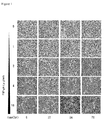
Figure 1 discloses the morphological change of A549 cells treated with FIP-gts. A549 cells were treated with different concentrations of FIP-gts (0, 1, 2, 4 and 10 µg/ml) at different durations (6, 12,24 and 72 hrs). -
Figure 2 discloses the morphology changes of human melanoma cancer cell line A375 after they were treated with FIP-gts. Cells were treated with 0, 4 and 16 µg/ml FIP-gts for 0, 24 and 48 hours and photographed with a phase-contrast microscope (x100). -
Figure 3 discloses the growth rate of A549 cells treated with FIP-gts at different times. A549 cells were treated with 0, 1, 2, 4 and 10 µg/ml FIP-gts and viable cell numbers were measured using trypan blue dye exclusion method at 48 hrs. The data shown here are mean ± standard deviation of triplicate experiments (significance calculated using student T test, * p<0.05). -
Figure 4 shows the effect of FIP-gts on the colony formation of A549 cells. (A) Anchorage-independent growth of A549 cells treated with 0, 0.4, 1 and 2 µg/ml FIP-gts was assessed by the colony formation assay. (B) The colony number was counted under a dissection microscope. The number of cells has to be greater than 50 cells per colony. The data shown here are mean ± standard deviation of triplicate experiments (significance calculated using student T test, * p<0.05). -
Figure 5 shows the stage of A549 cells in cell cycle treated with different doses and time courses of FIP-gts. Cells were resuspended in 10% DMEM medium at 2X 10⁶ cells/ml. (A) Cells were detected by Flow cytometer and acquired by Cellquest. (B) Acquisition were analyzed and quantified by ModFit LT 3.0. The data shown here are mean ± standard deviation of triplicate experiments (significance calculated using student T test, * p<0.05). -
Figure 6 shows the expression of, p21 and procaspase-3 of A549 cells treated with 0, 2, 4 and 10µg/ml FIP-gts, respectively. Cell lysates were collected at 48 hrs and expression were determined by Western blot analysis. -
Figure 7 shows the migration of A549 cell treated with FIP-gts into the wound. Wounds were made by scarifying confluent A549 cells by a pipette tip (arrowheads show the size of the initial wound). After incubation for 72 h or 96 h cells were fixed and stained by Geimsa stain. -
Figure 8 shows the activity of MMP-2 treated with FIP-gts. (A) A549 cells were treated with 0, 1, 2, 4 and 10 µg/ml FIP-gts for 24 hrs. The conditioned media were collected and MMP-2 activity was determined by gelatin zymography. (B) The activity of MMP-2 was quantified by densitomertic analysis. The densitomertic data shown here are mean ± standard deviation of triplicate experiments (significance calculated using student T test, * p<0.05). - Unless defined otherwise, all technical and scientific terms used herein have the same meaning as commonly understood by one of ordinary skill in the art to which this invention belongs. Although any methods or materials similar or equivalent to those described herein can be used in the practice or testing of the present invention, the preferred methods and materials are described. For the purposes of the present invention, the following terms are defined below.
- The present invention provides the fungal immunomodulatory proteins for use in the treatment of prostate cancer, breast cancer, lung cancer, non-small lung cell cancer or melanoma, wherein the protein having the amino acid sequence of SEQ ID No:1or

SEQ ID No:3
- The proteins for use of the present invention could be obtained from Ganoderma species, Flammulina velutipes or a recombinant microorganism (such as recombinant Escherichia coli or Yeast). The preferred embodiment of Ganoderma species is Ganoderma tsugae.
- The proteins for use of the present invention could be applied as adjuvant for alleviating the pain or side effects of a patient suffering cancer.
- The proteins for use of the present invention for the treatment of cancers according to
claim 1 is directed to stimulate or activate immunological function. - Accordingly, the medical use of the present invention is made by the mechanism selecting from the group consist of activation of natural killer cells, activation of macrophages and multiplication of serum antibodies. The preferred embodiment of the function of the present invention is made by activation of natural killer cells and inhibition of telomerase.
- The fungal immunomodulatory protein of the present invention can be designed as the following dosage forms consisting of oral solution, oral sachet, oral tablet or oral pellet, which is administered orally.
- It has been found that FIP-gts of the present invention, of which cDNA sequence is identical to LZ-8 (SEQ ID NO: 1), exhibited anti-cancer effect. It was also disclosed that cancer cells treated with FIP-gts showed reduced viability, demonstrating the utility of FIP-gts as an anticancer agent.
- It has been found that cancer cells treated with FIP-gts of the present invention show a decrease of MMP-2 expression. MMP-2 is an important enzyme involved in the tumor cell metastasis. Suppression of MMP-2 is a sign of FIP-gts suppressing the tumor cell metastasis.
- The term "immunotherapy" is not limited but to stimulate or activate immunological function (such as activate natural killer cells and macrophages or increase production of cytokines, serum IgG or IgM antibody).
- Cancers the fungal immunomodulatory protein of the invention could treat are selected from the group consisting of lung cancer, bone cancer, breast cancer, hepatocellular carcinomas, non-small lung cell cancer, ovarian cancer and gastrointestinal cancer.
- In another embodiment, the FIP-gts according to the invention could be fused to an antitumor agent or a detectable label. This allows the FIP-gts to target the agent or detectable label to the tumor cells and hence allows damage/destruction or detection of the tumor. Thus, the FIP-gts is suitable for use in a method of treatment of the human or animal body by chemotherapy or surgery (e.g. radioimmunoguided surgery, RIGS), or in a method of diagnosis practiced on the human or animal body. In particular, the FIP-gts is suitable for use in treatment by surgery or therapy of a tumor, or in diagnosis of a tumor.
- The antitumor agent linked to the FIP-gts may be any agent that destroys or damages a tumor to which the FIP-gts has bound or in the environment of the cell to which the FIP-gts has bound. For example, the antitumor agent may be a toxic agent such as a chemotherapeutic agent or a radioisotope, an enzyme that activates a prodrug or a cytokine.
- Suitable chemotherapeutic agents are known to those skilled in the art and include anthracyclines (e.g. daunomycin and doxorubicin), methotrexate, vindesine, neocarzinostatin, cis-platinum, chlorambucil, cytosine arabinoside, 5-fluorouridine, melphalan, ricin and calicheamicin.
- Suitable radioisotopes for use as anti-virus agents are also known to those skilled in the art.
- The antitumor agent that is attached to the FIP-gts may also be an enzyme that activates a prodrug. This allows activation of an inactive prodrug to its active, cytotoxic form at the directed site. In clinical practice, the FIP-gts-enzyme conjugate can be administered to the patient and allowed to localize in the region of the tumor to be treated. The prodrug is then administered to the patient so that conversion to the cytotoxic drug is localized in the region of the tumor cells to be treated under the influence of the localized enzyme.
- Accordingly, the present invention also provides a a fungal immunomodulatory protein according to
claim 1 for use in immunotherapy in a patient in need of such treatment, comprising administering to said patient an effective amount of the polypeptide of the present invention. - The detectable label attached to the FIP-gts may be an imaging agent for site imaging such as a short-lived radioisotope, for example 111In, 125I or 99 mTc.
- The FIP-gts according to the invention containing a detectable label is useful for RIGS in addition to being useful for diagnosis. RIGS comprises administering a labeled protein to a patient and thereafter surgically removing any tissue to which the protein binds. Thus, the labeled FIP-gts guides the surgeon towards tissue.
- At present, telomerase inhibition research focuses on (1) direct targeting of core telomerase components (Kondo S, et al., Oncogene 1998; 16(25):3323-3330.; Hahn WC, et al., Nat Med 1999; 5(10):1164-1170.); (2) telomere targeting (Rezler EM, et al., Curr Opin Pharmacol 2002; 2(4):415-423.; Zhang RG, et al., Cell Res 2002; 12(1):55-62.); (3) natural compounds and small molecules as telomerase inhibitors (Lyu SY, et al., Arch Pharm Res 2002; 25(1):93-101.; Naasani I, et al., Biochem Biophys Res Common 1998; 249(2):391-396.) and (4) interference with regulatory mechanisms of telomerase (Kawagoe J, et al., J Biol Chem 2003;278(44):43363-43372.).
- Those skilled in the art may reasonably expect that the subjects or patients, to which these methods are directed, can be any vertebrate animals, most preferred patients are humans having cancer or at risk for cancer. Nonetheless, the utility of the methods toward any vertebrate can be determined without undue experimentation by administering the composition comprising FIP-gts to a cultured cancer cell specific to the vertebrate in question and performing a simple cellular invasion assay, heal wounded assay described in the example.
- The composition comprising FIP-gts may be administered to a vertebrate by any suitable route known in the art including, for example, intravenous, subcutaneous, intratumoral, intramuscular, transdermal, intrathecal, or intracerebral. Administration can be either rapid as by injection, or over a period of time as by slow infusion or administration of a slow release formulation.
- It is contemplated that the compositions comprising FIP-gts are usually employed in the form of pharmaceutical preparations. Such preparations are made in a manner well known in the pharmaceutical art. One preferred preparation utilizes a vehicle of physiological saline solution; it is contemplated that other pharmaceutically acceptable carriers such as physiological concentrations of other non-toxic salts or compounds, 5% aqueous glucose solution, sterile water or the like may also be used. It may also be desirable that a suitable buffer be present in the composition. Such solutions can, if desired, be lyophilized and stored in a sterile ampoule ready for reconstitution by the addition of sterile water for ready injection. The primary solvent can be aqueous or alternatively non-aqueous.
- The carrier can also contain other pharmaceutically-acceptable excipients for modifying or maintaining the pH, osmolarity, viscosity, clarity, color, sterility, stability, rate of dissolution, or odor of the formulation. Similarly, the carrier may contain still other pharmaceutically-acceptable excipients for modifying or maintaining release or absorption or penetration across the blood-brain barrier. Such excipients are those substances usually and customarily employed to formulate dosages for parenteral administration in either unit dosage or multi-dose form or for direct infusion by continuous or periodic infusion.
- It is also contemplated that certain formulations comprising the compositions that comprises FIP-gts are to be administered orally. Such formulations are preferably formulated with suitable carriers, excipients, lubricants, emulsifying agents, suspending agents, sweetening agents, flavor agents, preserving agents and pressed as tablet or encapsulated as solid capsule or soft capsule. Or it is contemplated that such formulations are designed as following dosage forms, either oral solution, or oral sachet, or oral pellet. Or apart from being administered orally, it is contemplated that such formulations are designed as enema, or suppository, or implant, or patch, or cream, or ointment dosage forms. Some examples of suitable carriers, excipients, and diluents include lactose, dextrose, sucrose, sorbitol, mannitol, starches, gum acacia, calcium phosphate, alginates, calcium silicate, microcrystalline cellulose, polyvinylpyrrolidone, cellulose, gelatin, syrup, methyl cellulose, methyl- and propylhydroxybenzoates, talc, magnesium, stearate, water, mineral oil, and the like. The formulations can additionally include lubricating agents, wetting agents, emulsifying and suspending agents, preserving agents, sweetening agents or flavoring agents. The compositions may be formulated so as to provide rapid, sustained, or delayed release of the active ingredients after administration to the patient by employing procedures well known in the art. The formulations can also contain substances that diminish proteolytic, nucleic acid and other degradation and/or substances that promote absorption such as, for example, surface active agents. Compositions may be complexed with polyethylene glycol (i.e., PEGylated), albumin or the like to help promote stability in the bloodstream.
- The compositions comprising FIP-gts are administered to vertebrates in an amount effective to decrease the growth or metastasis of a tumor within the vertebrate. The specific dose is calculated according to the approximate body weight or body surface area of the patient or the volume of body space to be occupied. The dose will also be calculated dependent upon the particular route of administration selected. Further refinement of the calculations necessary to determine the appropriate dosage for treatment is routinely made by those of ordinary skill in the art. Such calculations can be made without undue experimentation by one skilled in the art in light of the activity disclosed herein, i.e., the gelatin-zymography assay. It will be understood that the amount of the composition actually administered will be determined by a practitioner, in the light of the relevant circumstances including the condition or conditions to be treated, the choice of composition to be administered, the age, weight, and response of the individual patient, the severity of the patient's symptoms, and the chosen route of administration. Dose administration can be repeated depending upon the pharmacokinetic parameters of the dosage formulation and the route of administration used.
- The compositions comprising FIP-gts are fed to BALB/c mice to examine the effect on natural killer activity, macrophage activity, cytokines and serum antibody production. As compared with various dosage groups, it demonstrates that FIP-gts promotes the activities of natural killer cells, macrophage activity, cytokines and serum antibody production.
- The following examples are included in the specification to help illustrate the invention.
- Human pulmonary epithelial cancer cell A549 was a highly vicious pulmonary cancer cell line with high migratory capability. A549 cells as the model system was applied to examine the effect of treating or preventing cancer cells with FIP-gts.
- First, the change of cell morphology after cells were treated with FIP-gts under the microscope. A549 Cells were treated with 0, 1, 2, 4 and 10 µg/ml FIP-gts, respectively, and photographed at different time interval (
Figure 1 ), - It was found that after cells were treated with 2, 4 and 10 µg/ml FIP-gts, the morphology of cells clearly changed at 6 hours. Cells transformed from adhering with little tentacles to round and loosly-attached cells. Some cells showed signs of moving when the culture dish was shaked. After treated with FIP-gts for 72 hours, A549 cells not treated or treated wtih low concentrations of FIP-gts expanded and covered the whole culture dish, whereas cells treated with high concentrations of FIP-gts showed round shape and left many spaces uncovered (
Figure 2 ). Normal lung cell line BEAS-2B cells treated with 0, 2, 4 and 10 µg/ml FIP-gts did not show morphology changes. And cells treated or not treated with FIP-gts exhibited similar growth rate in 24 hours, filling the culture dish almost concurrently. The control, BEAS 2B cell, were not affected by FIP-gts and filled the whole culture dish in 24 hours. - Using human melanoma cancer cell line A375, it has been found that A375 cells treated with FIP-gts seemed to lose cell-to-cell adhesion, as cells were no longer closely attached to each other but widely dispersed (
Figure 2 ). A375 cells were treated with FIP-gts Figure 2 ). It was shown that 16 µg/ml of FIP-gts could significantly inhibit cell adhesion and cell growth. Given the above, it proved that FIP-gts changed cell migratory and adhesion capability by rearranging the cell frame. - Trypan blue was used to examine cell viability. The same concentration of FIP-gts in Example 1 was used to treat A549 cells. After treating cells with FIP-gts for 48 hours, trypan blue was added. Live cells could repel the trypan blue; therefore, the number of viable cells was measured by the number of cells not labeled by trypan blue.
- 2 ×10⁵ human lung epidermoid carcinoma cell line H1355 and A549 cells were inoculated to 6 cm culture dishes. H1355 cell line was a common cellular model for studying metastasis. Cells were grown at 37°C for 16 hours. Medium was removed and FIP-gts at the concentrations of 0, 2,4 and 10 µg/ml were treated.
- Cells were collected at 48 hours after FIP-gts treatment. Cells were collected by removing the old culture medium into 15 ml centrifuge tube. Cells were washed with 1×PBS twice. Cells were resuspended in 0.5 ml TE buffer after centrifugation at room temperature for 1 min. The solution was neutralized by adding the original culture medium. Cells were transferred to a 15 ml centrifugation tube, and centrifuged at 800 rpm for 5 min. Supernatant was then discarded and cells were dispersed with 0.5
ml 1 ×PBS. 20µl of cell culture was added with 5 µl Trypan blue solution. Cell numbers were counted with cell counters. - The survival rate of cells treated with 0 µg/ml FIP-gts for 48 hours was considered 100 %, and the survival rate of cells treated with 1, 2, 4 and 10 µg/ml FIP-gts for 48 hours were 98.2 %, 94.8 %, 80.0 % and 60.3%, respectively (
Figure 3 ). The result was consistent with the observation of the MTS assay (described below). These two experiments demonstrated that FIP-gts had cytotoxicity to A549 cells, and might suppress cell growth or cause the decrease of cells survival rate. - The purpose of the present experiment was to examine the cytotoxicity of FIP-gts by colony formation assay. A549 or A375 cells were treated with 0, 0.4, 2 and 10 µg/ml FIP-gts for 24 hours. And then 400 cells/60 mm dishes were grown for 12 days.
- 2×10⁵ A549 or A375 cells were inoculated into 60 mm culture dish. Cells were grown at 37°C for 16 hr. FIP-gts at the concentrations of 0.4, 1, 2 and 10 µg/ml were treated to A549 cells (
Figure 4 ). After growing for 24 hours and washing with 1×PBS twice, cells were subcultured to new plates. ml TE buffer was added and culture was let aside at 37°C for 1 min to distach the cell. Cell numbers were counted and series cell dilution was performed with the original culture medium. 400 cells/plate cells were inoculated into 6 cm culture plates. Cells were grown in 37°C incubators for 12 days. Cells were washed with 1×PBS twice and 0°C, 95% ethanol 2 ml was added into each plate. Culture was let aside at room temperature for 20 min. Ethanol was then discarded and 2 ml/plate 10% Geimsa stain (Geimsa stain diluted with ddH2O) was added to each plate. Reaction was let aside at room temperature for 30 min. Dye was recollected and the remaining dye was gently washed out with tap water. Colony numbers were measured after dry. - The survival rate of A549 cells treated with 0 µg/ml FIP-gts was considered 100 %, and the survival rate of cells treated with 0.4, 1, 2 and 10 µg/ml FIP-gts were 97.3 %, 91.5 %, 69.6 % and 39.0 %, respectively (
Figure 4A, 4B ). All experiment groups except cells treated with 1 µg/ml FIP-gts showed significant decrease of survival rate (analyzed by student T test, p<0.05). It proved that FIP-gts showed cytotoxicity to A549 cells and suppressed colony formation. - It has been found that cells treated with FIP-gts showed lower survival rate. The effect might be the result of growth suppression or increased apoptosis. Traditionally anti-cancer drug suppressed cancer by modulating cell cycle, particularly arrested the cell at G1 phase. Therefore it examined whether treated A549 cells with FIP-gts modulate cell cycle, and if normal cell lines and cancer cell lines were differently affected by FIP-gts.
- Cells were distributed into 60 mm culture dish, 5×10⁵ cells/dish with 5 ml culture medium, grown at 37°C for 16 hours. Old culture medium was discarded and washed twice with 1×PBS. Cells were treated with different concentrations of FIP-gts (0, 2, 4 and 10µg/ml) at different time interval (24 hours and 48 hours). Cells were collected by the following procedures:
- a.
- Old culture medium was moved to 15 ml centrifuge tube.
- b.
- Cells were washed with cold 1×PBS twice.
- c.
- Cells were distached by treating cells with 1×Trypsin-EDTA.
- d.
- Old culture medium was added to stop the reaction, medium was moved to 15 microcentrifuge tube.
- e.
- Cells were centrifuged at 800 rpm for 5 min. Supernatant discarded, pellet washed with 1×PBS twice.
- f.
- 1 ml 70 % cold ethanol was slowly added to the culture. Cells were left at 4°C overnight to stabilize.
- g.
- Culture was centrifuged at 800 rpm for 5min. Supernatant discarded.
- h.
- Culture was washed with ice cold 1×PBS twice. Supernatant was discarded and let dry as mich as possible.
- i.
- 1 ml propidium iodide (PI) mixture to each tube was added to each tube (avoid light) :
- j.
- Sample was let aside at room temperature for 30 min.
- k.
- Culture was filtered with 40 µm nylon mesh to avoid oversized cell clusters or debris stuck the entrance hole of flow cytometer. Single cell suspension in flow cytometer tube was collected.
- To measure the DNA in cells, the experiment used Fluorescence-Activated Cell Sorter (FACS) system to sort cells and analyzed with FACSCalibur (BECTON DICKINSON). The absorbance of red fluorescence at 617 nm determined the DNA content of cells labeled with PI. The measurement was analyzed by program CELL Quest. The statistics were computed and cell numbers at different stages of cell cycle were displayed using program Mod Fit 3.0.
- Using flow cytometer, it has been found that cells treated with FIP-gts showed a profound arrest at G1 phase. At most more than 30 % cells were found to be at G1 phase. The G1 phase arrested decreased the proportion of cells at S phase. In other words, cell growth was suppressed by FIP-gts. Fewer cells were found to be at SubG1 phase. The highest proportion of cells at subG1 phase (1.6 %) were found at the second day after cells were treated wtih high concentraitions of FIP-gts. The phenomenon suggested that FIP-gts lowered the cell curvival rate by causing G1 phase arrest and minor apoptosis.
- The results showed that the higher concentrations of FIP-gts treated, the more cells arrested at the G1 phase. A549 cells treated with 0, 1, 2, 4 and 10 µg/ml FIP-gts for 24 hours showed a proportion of 58.2 %, 59.1%, 62.0%, 64.0% and 75.5 % cells at the G1 phase, respectively. The increase of G1 phase also caused the decrease of cells at their S phase. A549 cells treated with the same concentrations of FIP-gts as above showed a proportion of 32.8 %, 30.9 %, 30.1 %, 27.2 % and 18.2 % at their S phase (
Figures 5A and 5B ), respectively. The cells arrested at G1 phase also increased as the time of FIP-gts treatment increased. A549 cells treated with the same concentrations of FIP-gts as described above for 48 hours showed a proportion of 60.2 %, 68.8 %, 72.6 %, 76.1 % and 82.1 % at their G1 phase, respectively, and the same cells showed an even lower proportion of cells at their S phase, respectively 31.8 %, 25.1%, 23.0 %, 20.0 % and 13.8 %. Thus the experiment demonstrated that FIP-gts caused A549 cells to arrest at G1 phase (Figures 5A and 5B ). - It has been found that few cells were at their SubG1 phase when A549 cells were treated with 10 µg/mlFIP-gts. Moreover, fewer cells went through apoptosis when they were treated with FIP-gts. 0.9% and 1.6% of A549 cells treated with FIP-

gts 10 µg/ml for 24 hours and 48 hours, respectively, were at their SubG1 phase (Figure 5B ). - External signals, such as UV, cisplatin could activate and stabilize p53 protein. p53 further activated other downstream genes including p21. p21 was the major checkpoint protein of the G1 phase in cell cycle (Zhong, X. et al, 2004. Int. J. Cancer). Using western blot, it has been found that the expression of p53 protein was induced after treating cells with FIP-gts for 48 hours. Gene p21 was also induced, demonstrating that FIP-gts caused cells to arrest at G1 phase by activating p21.
- There were three routes of apoptosis: through ER, death receptor or mitochodria. All three pathways resulted in the cleavage of procaspase 3 (32 kD) into the active caspase 3 (17 kD).
Caspase 3 was the final executor of the caspase series. It led to cell apoptosis, DNA breakage, nucleus condense and the formation of inclusions (Di Pietro, R., et al. (2004) Int J Immunopathol Pharmacol 17 (2)181-190). It has been found that when cells were treated with high concentrations of FIP-gts, there was a slight decrease of procaspase-3, the result of cleavage of procaspase-3 into active caspase-3. - From the results of the flow cytometer it has been found that FIP-gts caused cells to arrest at G1 phase and initiate minor apoptosis at high concentrations. Therefore the change of protein expression was studied when cells were treated with FIP-gts.
-
A549 5×10⁵ cells/plate were inoculated to 60 mm culture dish. Cells were grown at 37 °C for 16 hours. Cells were treated with 0, 2,4 and 10 µg/ml FIP-gts and grown at 37 °C incubator for 48 hours. Cells were first washed twice with PBS. Supernatant was discarded and 100 µl cell buffer (10 mM EDTA, 10 mM EGTA, 5 mM NaF, 10 % glycerol, 1 mM DTT, 400 mM KCl, 0.4 % Triton X-100, 20 mM sodium β-glycerophosphate, 0.1 mM Na3VO4, 1 mM PMSF/DMSO, 3 µg/ml aprotinin, 2 µg/ml pepstatin A, 2 µg/ml Leupeptin, 1X phosphatase inhibitor cocktail I (Sigma, P2850) were added ; 1X phosphatase inhibitor cocktail II (Sigma, P5726)) was also added to dissolve cells. The reaction was left on ice and cells were homogenized using ultrasonice homogenizer at 4 °C two times with an interval more than 10 minutes. Cells were centrifuged again at 12000 rpm, 4°C for 20 min. Supernatant was carefully moved to another sterilized 1.5 mlmicron and the protein content was quantified. The whole process of treating FIP-gts, centrifuging cells and adding 2X SDS sample buffer (200 mM Tris pH6.8, 8 % SDS, 40 % Glycerol, 2.86 M 2-mercaptoethenol and appropiate amount of bromophenol blue), to heat at 95°C, must be finished in 2 hours. - Bio-Rad solution was applied to quantify protein concentration. First Bio-Rad reagent was diluted with dd H2O 4:1, this was the Bio-Rad protein detection reagent. The sample, the supernatant of the
cell culture 2 µl and the diluted Bio-Rad protein detection reagent 498 µl was mixed. Sample was reacted in 1.5 ml micron at 37°C for 20 min. The absorbance peak was measured by spectrophotometer at 595 nm and compared with the absorbance peack of standard sample bovine serum albumin (BSA) to get the protein quantity (µg/µl). The standard BSA quantity was measured by adding 2 µg, 4 µg, 6 µg, 8 µg and 10 µg BSA into diluted Bio-Rad protein detection reagent 498 µl, 496 µl, 494 µl, 492 µl and 490 µl, respectively. Sample was reacted in 1.5 ml micron at 37 °C for 20 min. The sample absorbance peak at 595 nm was also measured. The BSA measurement was measured to obtain the standard ccorrelation of protein quantity with peak absorbance. The protein quantity of the sample protein thus could be determined by putting in the peak absorbacnce of the sample. -
Table 1 The running gel was prepared as follows: 15 % 12.5 % 10 % dd H2O 6.3 ml 7.6 ml 8.8 ml 1.5M Tris pH 8.8 5 ml 5 ml 5 ml (38.67:1.33)Acrymide:Bis 7.5 ml 6.2 ml 5 ml 10% SDS 0.2 ml 0.2 ml 0.2 ml APS(10 mg/ml) 1 ml 1 ml 1 ml TEMED 10 µl 10µl 10µl Total Volume 20 ml 20 ml 20 ml Table 2 The 3% stacking gel was prepared as follows: dd H2O 3.54 ml 0.5M Tris pH 8.8 1.5 ml (38.67:1.33)Acrymide:Bis 0.45 ml 10% SDS 0.06 ml APS(10 mg/ml) 0.3 ml TEMED 15 µl Total Volume 6ml - The sample was run on SDS-PAGE, and Hybond-P membrane (Pharmacia) was prepared 20 minutes before electrophoresis finishes. The membrane was wet with methanol for 15 second, washed with ddH2O for 10 min, and then the membrane was transferred to the transfer buffer (20 % methanol, 192 mM Glycine, 25 mM Tris-HCl, pH 9.2) for at least 10 min. The gel was carefully taken off after electrophoresis and transferred to Hoefer Semiphor following standard protocol. The transferred-membrane was blocked in shaking TTBS buffer (50 mM Tris, 0.2
% Tween 20, 150 mM NaCl, pH 7.5) with 5% nonfat milk powder for 1 hour. - Specific primary antibody was added to block Hybond-P membrane. 1 X TTBS buffer with 3 % BSA was added to dilute the following primary polyclonal antibody: anti-caspase-3 (1: 500, Cayman), anti-COX-2(1 : 1000, Cayman #160106). 1X TTBS buffer with 5% nonfat milk powder was used to dilute the following primary antibody: anti-BAX (1 : 8000, R&D), p53 (1 : 500, DAKO), p21 (1 : 500, Zymed). Sample was shaken at 4 °C overnight (at least 16 hours). The membrane was taken out the other day. The membrane was washed with 100 ml 1X TTBS buffer with 3 % nonfat milk powder twice, 10 min each time. Anti-rabbit IgG-HRP (1 : 5000, Cell Signaling #7074) or anti-mouse IgG-HRP secondary antibody (1 : 10000, Chemicon AP124P) was diluted with 1X TTBS buffer with 3 % nonfat milk powder. Sample was shaken at room temperature for 1 hour. The washing procedure was repeated once. E.C.L, color development reagent (NEN, NEL105) was mixed 1 : 1 with Enhanced luminol reagent and Oxidizing reagent. The membrane was put face up into the container with color development reagent and let react for 5 min to develop the HRP color. The fluorescence was exposed on X-ray film for 3∼5 min, and then developed and fixed the image.
- The most important checkpoint of G1 phase was p21. It has been found that after treating with 0, 2, 4 and 10 µg/ml FIP-gts for 48 hours, p21 expression was significantly induced (
Figure 6 ). It was also know that p21 was activated by p53, another well-known oncogene. The western blot result also showed that the expression of p53 was induced by FIP-gts (Figure 6 ). Therefore it proved that FIP-gts induced the expression of p53, increased the amount of p21 and caused G1 pause. - It has been found that procaspase-3 was decreased when cells were treated with 10 µg/ml FIP-gts (
Figure 6 ). Thus procaspase-3 was activated into caspase-3 when cells were treated with FIP-gts, causing cells to undergo apoptosis. Moreover, the decrease of cells was not a result of enhanced apoptosis, but the result of suppression of proliferation. - Using wound healing assay, it has been found that FIP-gts could effectively suppress the mobility of breast cancer cells.
- 2×10⁵ A549 cells were gown in 24 well culture dish. Cells were grown until almost cover the culture plate and cells were treated with culture medium containing 0.5% FBS for 24 hours to suppress cell growth. The plate was scarified with blue tip and cells were washed with 1 ×PBS. Finally different concentrations of 1, 2, 4 and 10µg/ml FIP-gts were added. Pictures were taken every 24 hour and cell migration was monitored.
- Usually cancer cells obtained mobility before they performed metastasis. Wound healing assay was applied to examine whether treating cells with 0, 2 ,4 and 10 µg/ml FIP-gts would increase the mobility of cells. It has been found that when cells were treated with FIP-gts for 48 hours, no significant mobility was observed. The invention identified cell migration that covered the line when cells were treated with 0, 1 and 2 µg/ml FIP-gts for 72 hours. Cells treated with 4 and 10 µg/ml FIP-gts almost showed no sign of migration. Compared cells treated or not treated with FIP-gts for 96 hours, cells not treated with FIP-gts showed substantial mobility and covered 1/3 of the scarified line, whereas cells treated with low concentrations of FIP-gts showed some migration, and cells treated with high concentrations showed no migration (
Figure 7 ). - Using wound healing assay, it has been found that cell mobility were spuppressed when treated with FIP-gts. When the FIP-gts treated exceeded 4 and 10 µg/ml, A549 cells showed almost no mobility.
- During metastasis, metaloproteinase digested extracellular matrix, dissociated cells and extracellular matrix and provided cells mobility. It was known that metalloproteinases MMP-2 and MMP-9 were highly expressed in many vicious cancers (Johnsen, M., et al., Curr Opin Cell Biol, 1998. 10, 667-671). Therefore the expression of MMP-2 and MMP-9 and the metastasis of cancer cells were highly correlated (Curran, S. and Murray, G.I. Eur J Cancer, 2000. 36, 1621-1630., Liabakk, N.B., et al., Cancer Res, 1996. 56, 190-196.).
- In order to avoid the interference of MMP-2 and MMP-9 in the serum, serum starvation on cell cultures and treated A549 cells with FIP-gts were applied to analyze the activity of MMP. To further improved the accuracy of gelatin zymography assay, Bio-Rad also been used to quantify protein concentration as a measure of cell density.
- It has been found that cells treated with high concentrations of FIP-gts can suppress the expression of MMP-2. It also found that the effect of FIP-gts was dose-dependent.
- A549 cells were grown 1 ×10⁵ cells/well in 24 well plate. Serum-free medium 200 µ l/well were added the other day and cells were treated with 0, 2, 4 and 10µg/ml FIP-gts for 24 hours. Medium was removed and cells were washed with 1X PBS. Cells were collected with CE buffer and proteins were quantified using Bio-Rad. 2 % gelatin was prepared by dissolving 2 g Gelatin /100 ml ddH2O at 55°C.
Table 3 8% SDS-PAGE gel was prepared with 0.1% Gelatin: 8% ddH2O 3.0 ml 1.5M Tris pH 8.8 2.0 ml (38.67:1.33) Acrymide:Bis 2.2 ml 10 % SDS 0.08 ml APS (10 mg/ml) 0.4 ml 2 % Gelatin 0.4 ml TEMED 10µl Total Volume 8ml - The gel was prepared as described in the western blot experiment. Gel was put in electrohporesis chamber with electrophoresis buffer. Culture media was loaded with 5X dye (0.1% SDS, 104 mM Tris-HCl pH 6.8, 50 % Glycerol (or 25 g sucrose), 0.125% bromophenol blue) into the gel and perform electrophoresis. Gel was then washed with washing buffer (40mM Tris-HCl pH 8.5, 0.2 M NaCl, 10 mM CaCl2, 2.5% Triton X-100) at room temperature for 30 minutes twice, and reaction buffer (40mM Tris-HCl pH 8.5, 0.2M NaCl, 10 mM CaCl2, 0.01% NaN3) was added. Let react at 37°C incubator for 12 hours. The membrane was dyed with Coomassie blue (0.2 % Coomassie blue R-250, 50 % methanol, 10 % acetic acid) for 30 minutes. Gel was destained with 10% acetic acid and 20% methanol. Membrane was dried in 50 % ddH2O, 50 % methanol and 0.33 % glycerol for 30 minutes. The membrane was then sealed in glass paper.
- Because cell mobility is correlated to the expression of MMP, gelatin-zymograpghy was applied to analyze whether the activity of MMP-2 is altered by treating cells with FIP-gts. It has been found that the expression of MMP-2 significantly decreased when the amount of FIP-gts treated increases (student T test, *p<0.05). The expression of MMP when cells were not treated with FIP-gts was considered 100 %. It has been found that the expression of MMP treated with 1, 2 ,4 and 10 µg/ml were 95.7 %, 90.3 %, 73.6 % and 29.8 %, respectively (
Figure 8 ). Thus it concluded that FIP-gts modulates cell migratory through regulating the expression of MMP-2. - Since treated cells with high concentrations of FIP-gts shortly could caused the decrease of metalloproteinases expression, RT-PCR was applied to measure the mRNA expression of TIMP-1 (Tissue inhibitor of metalloproteinases), the inhibitor of metalloproteinases, after treating cells with FIP-gts. It has been found that the mRNA expression of TIMP-1 and PAI increased when cells were treated with FIP-
gts - RT-PCR was performed by using Promega RT-PCR kit as follows:
1µg total RNA was heated at 70°C. After 10 minutes, the heated RNA was cooled in ice bath. Then,25mM MgCl 2 4 µl, 5×MMLV buffer 4µl, 10mM dNTP Mixture 2µl, Recombinant RNasin Ribonuclease inhibitor 0.5 µl,MMLV Reverse transcriptase 1 µl, Oligo (dT)15Primer 1 µl and Nuclease-Free Water were added until the final volume was 20 µl.Table 4 Primers for performing RT-PCR: Enzyme Sequence 5'→3' Position (bp) Temp (°C) MMP-2 5'-GGCCCTGTCACTCCTGAGAT-3' 1337-1356 62°C 5'-GGCATCCAGGTTATCGGGGA-3' 2026-2007 PAI-1 5'-GGATCCAGCCACTGGAAAGGCAACATG-3' 1470-1490 55°C 5'-GGATCCGTGCCGGACCACAAAGAGGAA-3' 1236-1216 TIMP-1 5'-TGGAGAGACACT'GCCAACTTG-3' 1700-1720 58°C 5'-AGGCTGTGCCTTCCTACAGA-3' 2224-2204 - Human peripheral blood mononuclear cells (PBMCs) were treated with 0, 1.25, 2.5, 5 and 10 µg/ml FIP-gts. After 48 hours the cytokines expression of human PBMCs was measured by ELISA. It has been found that the expression of cytokines IL-2, IFN-γ, TNF-α and IL-4 increased as the concentrations of FIP-gts treated increased (Table 5).
Table 5. The increased cytokine expression of human PBMCs treated with FIP-gts. FIP-gts (µg/ml) 0 1.25 2.5 5 10 IL-2 (pg/ml) 116 316 272 425 1218 IFN-γ (pg/ml) 70 4135 4578 4378 4372 TNF-α (pg/ml) 89 1174 2076 3525 2219 IL-4 (pg/ml) 5 3 7 13 39 - The effects of FIP-gts on 3 cancer cell lines: human prostate cancer cell line PC3, human breast cancer cell line MDA231 and human melanoma cancer cell line A375 were assessed (Table 6). The effects of FIP-gts were assessed by observing the morphology changes of cells treated with FIP-gts, following protocol described in Example 1; by measuring the inhibition of cell proliferation, following protocol described in Example 3; and by measuring the inhibition of colony formation, following the protocol described in Example 4.
Table 6 Effects of FIP-gts on different cancer cell lines Cell line Cell line origin Morphology change Inhibition of cell growth Inhibition of colony growth PC3 Human prostate cancer n.d. + + MDA231 Human breast cancer n.d. + + A375 Human melanoma cancer + + + - BALB/c male mice, 4- to 5-week-old, were purchased from National Laboratory Animal Center in Taiwan.
- Lower dosage: 200 microgram /kg/day; higher dosage: 600 microgram/kg/day; positive dosage (a commercial Ling-Zhi powder purchased from Taiwan' Department of Health, Health Food Accredited No.A00003): 300 milligram/kg/day. Negative control: Distilled water.
- At first, the higher dosage of FIP-gts was formulated. Then, the lower dosage groups were diluted from the higher dosage group. From the first day for test, each group was fed with test materials by oral route once a day over 6 weeks.
- After feeding FIP-gts experimental animals over six weeks, splenocytes was took out from the animals. Assay of natural killer cells activity by flowcytometry was made to compare various dosages groups with negative control group to check whether there was difference between the groups.
- After feeding FIP-gts experimental animals over six weeks, macrophages in abdomen were took out from the animals. E. coli were labeled with fluorescence. Then, the macrophages were made to phagocytise the labeled E. coli. Assay of the macrophage activity by flowcytometry was made to compare various dosages groups with negative control group to check whether there was difference between the groups.
- During feeding FIP-gts, animals' blood was collected before FIP-gts treated and animal sacrifice. The concentrations of various immunoglobulins in serum were determined and various dosages groups with negative control group were compared to check whether there was difference between the groups.
- After ten weeks of FIP-gts administration and injecting ovalbumin (OVA) twice, animals were sacrificed. Splenocytes were stimulated by Concanavalin A (Con A), Lipopolysaccharide (LPS) or OVA. After 48∼72 hours incubation, cytokine were measured by ELISA. When compared each dose group to negative group, p < 0.05 was an indication of significant difference.
- Before study of FIP-gts, first immunize, second boost, and before sacrifice, blood was collected from retro-orbital. Anti-OVA immunoglobulin production in serum was measured by ELISA. When compared each dose group to negative group, p<0.05 was an indication of significant difference.
- After sacrificed, the splenocytes of mice were taken from to proceed with assay of natural killer cells activity. To compare with negative control group, each group showed no statistical significance under the ratio of Effector/ Target (E/T ratio) was 12.5. To compare with negative control group, higher dosage group and positive control group showed significant differences under E/T ratio was 25.0. To compare with negative control group, lower dosage group, higher dosage group and positive control group showed significant differences under E/T ratio was 50. It appeared that FIP-gts promoted the activity of natural killer cells (Table 7).
Table 7 Group animal number E/T ratio 12.5 25.0 50.0 A 10 17.5± 8.69 25.5± 8.16 26.9± 6.57 B 10 25.7± 8.59 34.9± 8.20* 38.8± 6.80* C 10 23.9± 10.52 32.8± 7.92* 38.1± 7.66* D 10 25.2± 9.85 33.9± 10.16* 38.7± 9.22* This test is directed to the cytotoxicity assay of natural killer cells identified by flowcytometry.
The symbol (*) indicates statistical significance, as compared with negative control group.
E means effector cell. T means target cell.
A means negative control group. B means lower dosage group.
C means higher dosage group. D means positive control group. - After sacrificed, the macrophages in abdomen of mice were collected. FITC-E. coli were added to make phagocytosis by the macrophages. Then, the activity of the macrophages was analyzed by flowcytometry. To compare with negative control group, the lower dosage group and higher dosage group showed statistical significance under Multiplicity of infection (MOI)=30. It appeared that FIP-gts promoted the activity of the macrophages in abdomen (Table 8).
Table 8. Group animal number MOI-30 MOI=50 A 10 42.17± 8.89 46.33± 12.57 B 10 54.85± 8.73* 52.30± 9.05 C 10 53.09± 15.73* 49.74± 9.18 D 10 51.07± 8.43 46.11± 6.66 This test is directed to macrophage phagocytosis identified by flowcytometry.
The symbol (*) indicates statistical significance, as compared with negative control group.
MOI means multiplicity of infection.
A means negative control group. B means lower dosage group.
C means higher dosage group. D means positive control group. - During feeding FIP-gts, animals' blood was collected before FIP-gts treated and animal sacrifice. The concentrations of various immunoglobulins in serum were determined and various dosages groups with negative control group were compared to check whether there was difference between the groups. The concentration of immunoglobulins in serum before treatment demonstrated that immunoglobulin G (IgG) of higher dosage group and positive control group showed statistical significance as compared with negative control group. Various dosages groups of IgM showed no statistical significance as compared with negative control group (Table 9).
Table 9 Antibody production Group animal number Ig G (µg/ml) before treatment animal sacrifice A 10 431.06± 103.42 980.11± 163.89 B 10 504.22± 114.57 1324.55± 249.15* Group animal number Ig M (µg/ml) before treatment animal sacrifice A 10 356.87± 24.59 461.84± 103..5 B 10 333.17± 54.36 500.54± 46.09 This test is directed to the condition of serum antibody production identified by ELISA.
The symbol (*) indicates statistical significance (p<0.05), as compared with negative control group.
A means negative control group. B means higher dosage group. - Splenocytes were stimulated by stimulant (Con A or LPS or OVA), after 48∼72 hours incubation, cytokine was measured by ELISA. After Con A stimulation, interleukin-2 (IL-2) secretion in lower dose group of FIP-gts was significantly higher than that in negative control group. After OVA stimulation, interleukin-2 (IL-2) secretion in each dose and positive group were significantly higher than that in negative control group.
- After Con A stimulation, tumor necrosis factor-alpha (TNF-α) secretion in each group and positive group were significantly higher than that in negative control group. After OVA stimulation, interferon-gamma (IFN-γ) secretion in higher dose of FIP-gts and positive group was significantly higher than that in negative control group.
- The results are shown in Table 10. The FIP-gts could promote cytokine secretion.
Table 10 Spleen cytokine secretion Group Animal Number Medium only Con A LPS OVA IL-2 (pg/ml) A 10 10.71± 3.30 3224.48± 257.61 19.74±6.64 16.6812.96 B 10 11.01±2.91 3674.83± 800.84 21.52±5.78 25.58±7.18 C 10 12.28± 7.90 3783.18± 892.71 22.47± 4.76 26.90± 7.01* D 10 11.60±5.26 3364.50± 494.03 21.05±6.05 28.52± 14.01* TNF-α (pg/ml) A 10 4.14±5.21 14.26± 3.51 169.25± 104.31 4.93±5.91 B 10 3.68± 4.67 18.29±4.06* 186.97±84.15 6.00±6.46 C 10 3.22± 5.92 19.94±4.47* 243.64± 146.35 6.15±6.62 D 10 4.72± 6.02 18.77± 3.63* 227.41±122.00 5.90±6.10 IFN-γ (pg/ml) A 10 8.29± 6.19 2297.73±842.49 14.95±9.64 4.16±4.44 B 10 8.19±5.19 2451.23± 879.89 16.78± 6.33 10.54± 4.35 C 10 8.45± 5.53 2404.08±623.07 20.70-L 16.67 18.67± 11,70 * D 10 8.30± 5.37 2269.64±328.37 15.12::1: 8.55 13.05± 10.52* Spleen cytokine secretion were measured by ELISA
*:°indication of statistical significance when compare to negative control
A: Negative control B: Lower dose C: Higher dose D: Positive control
5. Anti-OVA immunoglobulin production in serum (specific immunomodulation) Before study of FIP-gts, first immunize, second boost, and before sacrifice, blood was collected from retro-orbital. Anti-OVA immunoglobulin production in serum was measured by ELISA. - Before study, first immunize and second boost, there was no significant difference between each group in anti-OVA immunoglobulin G (IgG), anti-OVA immunoglobulin M (IgM) and anti-OVA immunoglobulin E (IgE).
- Before animal sacrifice, anti-OVA IgG production in each dose of FIP-gts and positive group was significantly higher than that in negative control group. Anti-OVA IgM production in higher dose of FIP-gts and positive group was significantly higher than that in negative control group. There was no significant difference between each group in anti-OVA IgE.
- The results are shown in Table 11. The FIP-gts could promote anti-OVA immunoglobulin production in sera.
Table 11 Immunoglobulin production in serum (specific immunomodulation test) Group Animal Number Before study OVA Pre_OVA OVA_2 OVA_4 Anti-OVA IgG, EU A 10 0.0012± 0.0010 0.0040± 0.0015 0.3531± 0.0497 0.6018± 0.0600 B 10 0.0013± 0.0007 0.0044± 0.0014 0.3691± 0.0390 0.6928± 0.0983* C 10 0.0017± 0.0011 0.0045± 0.0016 0.3588± 0.0427 0.7091± 0.0869* D 10 0.0014± 0.0007 0.0051± 0.0023 0.3699± 0.0452 0.7084± 0.0669* Anti-OVA igM, EU A 10 0.0444± 0.0137 0.0527± 0.0159 0.1402± 0.0551 0.1404± 0.0334 B 10 0.0471± 0.0178 0.0647± 0.0249 0.1490± 0.0459 0.2254± 0.0764 C 10 0.0450±0.0139 0.0587± 0.0201 0.1472± 0.0344 0.2655± 0.1472* D 10 0.0432± 0.0204 0.0650± 0.0217 0.1469± 0.0376 0.2571± 0.1665* Anti-OVA IgE, EU A 10 0.0045± 0.0020 0.0110± 0.0023 0.0374± 0.0085 0.1247± 0.0542 B 10 0.0052± 0.0036 0.0128± 0.0037 0.0414± 0.0065 0.1416± 0.0634 C 10 0.0065± 0.0066 0.0114± 0.0031 0.0444± 0.0075 0.1470± 0.0521 D 10 0.0040± 0.0017 0.0110± 0.0015 0.0437± 0.0087 0.1321± 0.0516 Anti-OVA immunoglobulin production in serum were measured by ELISA
*: indication of statistical significance when compare to negative control
- A: Negative control B: Lower dose C: Higher dose D: Positive control
-
- <110> Yeastern Biotech Co., Ltd.
<120> SEQ ID No:1
<160> 1
<170> PatentIn version 3.3
<210> 1
<211> 111
<212> PRT
<213> Ganoderma specisis
<221> PEPTIDE
<222> (1)..(111)
<400> 1
- <110> Yeastern Biotech Co., Ltd.
<120> SEQ ID No:2
<160> 1
<170> PatentIn version 3.3
<210> 1
<211> 111
<212> PRT
<213> Ganoderma tsugae
<221> PEPTIDE
<222> (1)..(111)
<400> 1
- <110> Yeastern Biotech Co., Ltd.
<120> SEQ ID No:3
<160> 1
<170> PatentIn version 3.3 - <210> 1
<211> 114
<212> PRT
<213> Flammulina velutipes
<221> PEPTIDE
<222> (1)..(114)
<400> 1
| 1XPBS | 550 |
| 5% Triton X-100 | 200 µl |
| 250 µg/ml propidium iodide | 200 µl |
| 0.5mg/ml RNase A | 50 µl |
Claims (8)
- Fungal immunomodulatory protein for use in the treatment of prostate cancer, breast cancer, lung cancer, non-small cell lung cancer or melanoma, wherein the fungal immunomodulatory protein having the amino acid sequence of SEQ ID No:1

- Fungal immunomodulatory protein for use according to Claim 1, wherein the fungal immunomodulatory protein suppresses a proliferation of the cancer.
- Fungal immunomodulatory protein for use according to Claim 2, wherein the cancer is arrested at G1 phase.
- Fungal immunomodulatory protein for use according to Claim 1, which is applied as suppressor of a viability of the cancer cells.
- Fungal immunomodulatory protein for use according to Claim 4, wherein the treated cancer cells show a decrease of MMP-2 expression.
- Fungal immunomodulatory protein for use according to Claim 1, wherein the protein is obtained from Ganoderma species or Flammulina velutipes.
- Fungal immunomodulatory protein for use according to Claim 4, wherein the Ganoderma species is Ganoderma tsugae.
- Fungal immunomodulatory protein for use according to Claim 1, wherein the protein is provided from a recombinant Escherichia coli or Yeast.
Applications Claiming Priority (2)
| Application Number | Priority Date | Filing Date | Title |
|---|---|---|---|
| US11/233,364 US20070071766A1 (en) | 2005-09-23 | 2005-09-23 | Compositions comprising fungal immunomodulatory protein and use thereof |
| EP06017781A EP1800690A3 (en) | 2005-09-23 | 2006-08-25 | Use of fungal immunomodulatory protein |
Related Parent Applications (1)
| Application Number | Title | Priority Date | Filing Date |
|---|---|---|---|
| EP06017781A Division EP1800690A3 (en) | 2005-09-23 | 2006-08-25 | Use of fungal immunomodulatory protein |
Publications (2)
| Publication Number | Publication Date |
|---|---|
| EP2759304A1 EP2759304A1 (en) | 2014-07-30 |
| EP2759304B1 true EP2759304B1 (en) | 2016-09-21 |
Family
ID=37894299
Family Applications (2)
| Application Number | Title | Priority Date | Filing Date |
|---|---|---|---|
| EP06017781A Withdrawn EP1800690A3 (en) | 2005-09-23 | 2006-08-25 | Use of fungal immunomodulatory protein |
| EP14150952.1A Ceased EP2759304B1 (en) | 2005-09-23 | 2006-08-25 | Fungal immunomodulatory protein for use in the treatment of cancer |
Family Applications Before (1)
| Application Number | Title | Priority Date | Filing Date |
|---|---|---|---|
| EP06017781A Withdrawn EP1800690A3 (en) | 2005-09-23 | 2006-08-25 | Use of fungal immunomodulatory protein |
Country Status (4)
| Country | Link |
|---|---|
| US (1) | US20070071766A1 (en) |
| EP (2) | EP1800690A3 (en) |
| JP (2) | JP5117696B2 (en) |
| CN (3) | CN1939532B (en) |
Families Citing this family (20)
| Publication number | Priority date | Publication date | Assignee | Title |
|---|---|---|---|---|
| CN101475632B (en) * | 2008-01-03 | 2012-01-04 | 张喜田 | Recombinant Ganoderma lucidum immunoregulatory protein with anti-tumor effect and its pharmaceutical preparation |
| US8628785B2 (en) | 2008-05-16 | 2014-01-14 | Yeastern Biotech Co., Ltd | Method for augmenting the immunogenicity of an antigen |
| TWI459955B (en) * | 2008-05-16 | 2014-11-11 | Yeastern Biotech Co Ltd | Medicinal composition for activating dendritic cells and macrophages |
| TWI606835B (en) * | 2010-04-23 | 2017-12-01 | 中央研究院 | Reishi polysaccharide-based compositions and methods for treatment of cancer |
| CN102199202B (en) * | 2011-03-22 | 2013-05-01 | 上海交通大学 | Gene of recombinant fungal immunomodulatory protein between ganodermas, protein coded thereby, and application thereof |
| CN102199201B (en) * | 2011-03-22 | 2013-05-01 | 上海交通大学 | Recombinant fungal immunomodulatory protein gene in Ganoderma lucidum, protein coded whereby and application thereof |
| CN102241751B (en) * | 2011-04-20 | 2013-06-26 | 中国农业科学院饲料研究所 | Novel fungal immunomodulatory protein FIP-NHA with antineoplastic activity and gene thereof |
| CN103243116B (en) * | 2013-05-08 | 2015-05-27 | 上海交通大学 | Method for preparing intragenus recombinant fungal immunomodulatory proteins and application of intragenus recombinant fungal immunomodulatory proteins |
| CN104212828A (en) * | 2013-06-03 | 2014-12-17 | 怀化学院 | Production method of recombiantn Poria cocos immunomodulatory protein WCFIP1 and its antibody |
| CN103417953B (en) * | 2013-08-16 | 2015-06-17 | 张喜田 | Application of recombined ganoderma lucidum immnoregulation protein (rLZ-8) in preparation of medicine for treating melanin tumor |
| CN103739684B (en) * | 2014-01-09 | 2016-10-05 | 上海交通大学 | The preparation method and applications of ganoderma atrum fungal immunomodulatory protein |
| TWI608846B (en) * | 2014-03-13 | 2017-12-21 | 益生生技開發股份有限公司 | Combination therapy for ameliorating adverse side effects caused by chemotherapy |
| CN104001154B (en) * | 2014-06-10 | 2015-07-01 | 张喜田 | Application of recombinant ganoderma lucidum immunoregulatory protein in preparation of medicine for treating androgenetic alopecia |
| TWI657821B (en) * | 2014-09-02 | 2019-05-01 | 益生生技開發股份有限公司 | Methods and compositions for treating c-met associated cancers |
| CN107287166B (en) * | 2016-04-13 | 2021-04-09 | 蘑法生物科技股份有限公司 | Monoclonal antibody against mushroom immunomodulatory protein and its application |
| TWI702292B (en) * | 2018-12-28 | 2020-08-21 | 薩摩亞商康多富國際有限公司 | Method for determining a set of personalized metabolic disease healthy foods and non-transitory computer readable storage medium |
| US11141458B2 (en) * | 2019-07-17 | 2021-10-12 | Mycomagic Biotechnology Co., Ltd. | Composition and methods for promoting and treating chronic wound healing |
| CN112646009B (en) * | 2019-08-27 | 2022-02-08 | 上海交通大学 | Ganoderma lucidum immunomodulatory protein mutant and application |
| CN113893337A (en) * | 2021-08-31 | 2022-01-07 | 中山大学 | Application of DTX2 protein in preparation of preparation for regulating telomerase activity |
| CN113768812A (en) * | 2021-09-08 | 2021-12-10 | 上海交通大学 | Application of recombinant fungal immunomodulatory protein rFIP-glu |
Family Cites Families (5)
| Publication number | Priority date | Publication date | Assignee | Title |
|---|---|---|---|---|
| JP2000270713A (en) * | 1999-03-23 | 2000-10-03 | Kennetto:Kk | Animal model of diabetes and its production, and selection of animal model of diabetes |
| GB0308988D0 (en) * | 2003-04-17 | 2003-05-28 | Univ Singapore | Molecule |
| CA2528669A1 (en) * | 2003-06-09 | 2005-01-20 | The Regents Of The University Of Michigan | Compositions and methods for treating and diagnosing cancer |
| TWI328038B (en) * | 2003-09-17 | 2010-08-01 | Yeastern Biotech Co Ltd | Fungal immunomodulatory protein (fip) prepared by microorganisms and uses thereof |
| JP2005224205A (en) * | 2004-02-16 | 2005-08-25 | Hokko Chem Ind Co Ltd | healthy food |
-
2005
- 2005-09-23 US US11/233,364 patent/US20070071766A1/en not_active Abandoned
-
2006
- 2006-08-25 EP EP06017781A patent/EP1800690A3/en not_active Withdrawn
- 2006-08-25 EP EP14150952.1A patent/EP2759304B1/en not_active Ceased
- 2006-09-21 CN CN2006101270427A patent/CN1939532B/en not_active Expired - Fee Related
- 2006-09-21 CN CN2010101120159A patent/CN101926979B/en not_active Expired - Fee Related
- 2006-09-21 CN CN2009101376694A patent/CN101628110B/en not_active Expired - Fee Related
- 2006-09-21 JP JP2006256232A patent/JP5117696B2/en not_active Expired - Fee Related
-
2012
- 2012-09-10 JP JP2012198201A patent/JP5512769B2/en not_active Expired - Fee Related
Also Published As
| Publication number | Publication date |
|---|---|
| JP5117696B2 (en) | 2013-01-16 |
| HK1144373A1 (en) | 2011-02-18 |
| JP5512769B2 (en) | 2014-06-04 |
| CN101926979B (en) | 2012-09-26 |
| EP2759304A1 (en) | 2014-07-30 |
| US20070071766A1 (en) | 2007-03-29 |
| CN1939532B (en) | 2012-10-31 |
| JP2012250991A (en) | 2012-12-20 |
| EP1800690A2 (en) | 2007-06-27 |
| CN101628110B (en) | 2012-09-12 |
| CN101628110A (en) | 2010-01-20 |
| JP2007084547A (en) | 2007-04-05 |
| EP1800690A3 (en) | 2007-07-04 |
| CN1939532A (en) | 2007-04-04 |
| CN101926979A (en) | 2010-12-29 |
Similar Documents
| Publication | Publication Date | Title |
|---|---|---|
| JP5512769B2 (en) | Use of fungal immunoregulatory proteins | |
| JP5265333B2 (en) | Cytotoxic factors that regulate cell death | |
| Zhang et al. | GLIS, a bioactive proteoglycan fraction from Ganoderma lucidum, displays anti-tumour activity by increasing both humoral and cellular immune response | |
| KR20050105435A (en) | Active fraction having anti-cancer and anti-metastasis isolated from acanthopanax species and fruits | |
| Feng et al. | Enhancement of the immune responses to foot-and-mouth disease vaccination in mice by oral administration of a novel polysaccharide from the roots of Radix Cyathulae officinalis Kuan (RC) | |
| Shin et al. | Immunostimulatory effects of cordycepin‐enriched WIB‐801CE from Cordyceps militaris in splenocytes and cyclophosphamide‐induced immunosuppressed mice | |
| KR20170054310A (en) | Dendritic Cell Therapeutic Agent and Immunotherapeutic Agent Comprising a Peptide Derived from Telomerase, and Method for Treatment Using the Same | |
| US8629096B2 (en) | Compositions comprising fungal immunomodulatory protein and use thereof | |
| US20100009915A1 (en) | Compositions comprising fungal immunomodulatory protein and use thereof | |
| KR20220020147A (en) | Method for preparing Phellinus linteus mycelia extract or Phellinus linteus mycelia extract powder and composition for enhancing immunity comprising them as an active ingredient | |
| Cao et al. | The effects of telocinobufagin isolated from Chan Su on the activation and cytokine secretion of immunocytes in vitro | |
| US20130236445A1 (en) | Bromelain proteinases (Bromelainases) as chemotherapeutic agents in treating and/or preventing various types of cancer in a mammal | |
| Jeong et al. | Studies on immunopotentiating activities of antitumor polysaccharide from aerial parts of Taraxacum platycarpum | |
| KR100278928B1 (en) | A novel use of polysaccharide substance isolated from phellinus linteus | |
| KR102257437B1 (en) | Anti-viral methods and the use of an anti-viral agent | |
| KR100607907B1 (en) | Pharmaceutical composition containing the extract of S. mushrooms with liver metastasis suppression and immunomodulatory effect | |
| Ha et al. | Effects of Cordyceps militaris extract on tumor immunity | |
| Farhangnia et al. | The Role of Inflammasome in Cancers and Potential Therapeutic Targets | |
| HK1144373B (en) | Use of fungal immunomodulatory protein | |
| KR101266527B1 (en) | Composition for inhibition of trasplant rejection containing the cordyceps mycellia extract as an active ingredient | |
| JP2009276245A (en) | Screening method of improving agent for persistent skin inflammatory disease, and the improving agent | |
| TWI360423B (en) | Compositions comprising fungal immunomodulatory pr | |
| WO2000032212A1 (en) | Lak activity potentiator orginating in shiitake mushroom hyphae extract and lak activity potentiating preparations containing the same | |
| KR101022809B1 (en) | Treatment of Candida albicans-associated diseases by immunomodulation | |
| KR100848211B1 (en) | Mixed Composition of Polysaccharides and Theanine Extracted from Felinus linteus Mycelium |
Legal Events
| Date | Code | Title | Description |
|---|---|---|---|
| PUAI | Public reference made under article 153(3) epc to a published international application that has entered the european phase |
Free format text: ORIGINAL CODE: 0009012 |
|
| 17P | Request for examination filed |
Effective date: 20140113 |
|
| AC | Divisional application: reference to earlier application |
Ref document number: 1800690 Country of ref document: EP Kind code of ref document: P |
|
| AK | Designated contracting states |
Kind code of ref document: A1 Designated state(s): AT BE BG CH CY CZ DE DK EE ES FI FR GB GR HU IE IS IT LI LT LU LV MC NL PL PT RO SE SI SK TR |
|
| R17P | Request for examination filed (corrected) |
Effective date: 20150130 |
|
| RBV | Designated contracting states (corrected) |
Designated state(s): AT BE BG CH CY CZ DE DK EE ES FI FR GB GR HU IE IS IT LI LT LU LV MC NL PL PT RO SE SI SK TR |
|
| RIC1 | Information provided on ipc code assigned before grant |
Ipc: A61K 38/16 20060101AFI20150430BHEP Ipc: A61P 35/00 20060101ALI20150430BHEP |
|
| GRAP | Despatch of communication of intention to grant a patent |
Free format text: ORIGINAL CODE: EPIDOSNIGR1 |
|
| INTG | Intention to grant announced |
Effective date: 20151119 |
|
| GRAS | Grant fee paid |
Free format text: ORIGINAL CODE: EPIDOSNIGR3 |
|
| GRAJ | Information related to disapproval of communication of intention to grant by the applicant or resumption of examination proceedings by the epo deleted |
Free format text: ORIGINAL CODE: EPIDOSDIGR1 |
|
| GRAP | Despatch of communication of intention to grant a patent |
Free format text: ORIGINAL CODE: EPIDOSNIGR1 |
|
| GRAP | Despatch of communication of intention to grant a patent |
Free format text: ORIGINAL CODE: EPIDOSNIGR1 |
|
| GRAS | Grant fee paid |
Free format text: ORIGINAL CODE: EPIDOSNIGR3 |
|
| INTG | Intention to grant announced |
Effective date: 20160713 |
|
| GRAA | (expected) grant |
Free format text: ORIGINAL CODE: 0009210 |
|
| AC | Divisional application: reference to earlier application |
Ref document number: 1800690 Country of ref document: EP Kind code of ref document: P |
|
| AK | Designated contracting states |
Kind code of ref document: B1 Designated state(s): AT BE BG CH CY CZ DE DK EE ES FI FR GB GR HU IE IS IT LI LT LU LV MC NL PL PT RO SE SI SK TR |
|
| REG | Reference to a national code |
Ref country code: GB Ref legal event code: FG4D |
|
| REG | Reference to a national code |
Ref country code: CH Ref legal event code: EP |
|
| REG | Reference to a national code |
Ref country code: AT Ref legal event code: REF Ref document number: 830499 Country of ref document: AT Kind code of ref document: T Effective date: 20161015 |
|
| REG | Reference to a national code |
Ref country code: IE Ref legal event code: FG4D |
|
| REG | Reference to a national code |
Ref country code: CH Ref legal event code: NV Representative=s name: PATENTANWAELTE SCHAAD, BALASS, MENZL AND PARTN, CH |
|
| REG | Reference to a national code |
Ref country code: DE Ref legal event code: R096 Ref document number: 602006050378 Country of ref document: DE |
|
| REG | Reference to a national code |
Ref country code: NL Ref legal event code: FP |
|
| REG | Reference to a national code |
Ref country code: LT Ref legal event code: MG4D |
|
| PG25 | Lapsed in a contracting state [announced via postgrant information from national office to epo] |
Ref country code: LT Free format text: LAPSE BECAUSE OF FAILURE TO SUBMIT A TRANSLATION OF THE DESCRIPTION OR TO PAY THE FEE WITHIN THE PRESCRIBED TIME-LIMIT Effective date: 20160921 Ref country code: FI Free format text: LAPSE BECAUSE OF FAILURE TO SUBMIT A TRANSLATION OF THE DESCRIPTION OR TO PAY THE FEE WITHIN THE PRESCRIBED TIME-LIMIT Effective date: 20160921 |
|
| REG | Reference to a national code |
Ref country code: AT Ref legal event code: MK05 Ref document number: 830499 Country of ref document: AT Kind code of ref document: T Effective date: 20160921 |
|
| PG25 | Lapsed in a contracting state [announced via postgrant information from national office to epo] |
Ref country code: GR Free format text: LAPSE BECAUSE OF FAILURE TO SUBMIT A TRANSLATION OF THE DESCRIPTION OR TO PAY THE FEE WITHIN THE PRESCRIBED TIME-LIMIT Effective date: 20161222 Ref country code: LV Free format text: LAPSE BECAUSE OF FAILURE TO SUBMIT A TRANSLATION OF THE DESCRIPTION OR TO PAY THE FEE WITHIN THE PRESCRIBED TIME-LIMIT Effective date: 20160921 Ref country code: SE Free format text: LAPSE BECAUSE OF FAILURE TO SUBMIT A TRANSLATION OF THE DESCRIPTION OR TO PAY THE FEE WITHIN THE PRESCRIBED TIME-LIMIT Effective date: 20160921 |
|
| PG25 | Lapsed in a contracting state [announced via postgrant information from national office to epo] |
Ref country code: EE Free format text: LAPSE BECAUSE OF FAILURE TO SUBMIT A TRANSLATION OF THE DESCRIPTION OR TO PAY THE FEE WITHIN THE PRESCRIBED TIME-LIMIT Effective date: 20160921 Ref country code: RO Free format text: LAPSE BECAUSE OF FAILURE TO SUBMIT A TRANSLATION OF THE DESCRIPTION OR TO PAY THE FEE WITHIN THE PRESCRIBED TIME-LIMIT Effective date: 20160921 |
|
| PG25 | Lapsed in a contracting state [announced via postgrant information from national office to epo] |
Ref country code: PT Free format text: LAPSE BECAUSE OF FAILURE TO SUBMIT A TRANSLATION OF THE DESCRIPTION OR TO PAY THE FEE WITHIN THE PRESCRIBED TIME-LIMIT Effective date: 20170123 Ref country code: ES Free format text: LAPSE BECAUSE OF FAILURE TO SUBMIT A TRANSLATION OF THE DESCRIPTION OR TO PAY THE FEE WITHIN THE PRESCRIBED TIME-LIMIT Effective date: 20160921 Ref country code: IS Free format text: LAPSE BECAUSE OF FAILURE TO SUBMIT A TRANSLATION OF THE DESCRIPTION OR TO PAY THE FEE WITHIN THE PRESCRIBED TIME-LIMIT Effective date: 20170121 Ref country code: CZ Free format text: LAPSE BECAUSE OF FAILURE TO SUBMIT A TRANSLATION OF THE DESCRIPTION OR TO PAY THE FEE WITHIN THE PRESCRIBED TIME-LIMIT Effective date: 20160921 Ref country code: AT Free format text: LAPSE BECAUSE OF FAILURE TO SUBMIT A TRANSLATION OF THE DESCRIPTION OR TO PAY THE FEE WITHIN THE PRESCRIBED TIME-LIMIT Effective date: 20160921 Ref country code: BE Free format text: LAPSE BECAUSE OF FAILURE TO SUBMIT A TRANSLATION OF THE DESCRIPTION OR TO PAY THE FEE WITHIN THE PRESCRIBED TIME-LIMIT Effective date: 20160921 Ref country code: PL Free format text: LAPSE BECAUSE OF FAILURE TO SUBMIT A TRANSLATION OF THE DESCRIPTION OR TO PAY THE FEE WITHIN THE PRESCRIBED TIME-LIMIT Effective date: 20160921 Ref country code: SK Free format text: LAPSE BECAUSE OF FAILURE TO SUBMIT A TRANSLATION OF THE DESCRIPTION OR TO PAY THE FEE WITHIN THE PRESCRIBED TIME-LIMIT Effective date: 20160921 Ref country code: BG Free format text: LAPSE BECAUSE OF FAILURE TO SUBMIT A TRANSLATION OF THE DESCRIPTION OR TO PAY THE FEE WITHIN THE PRESCRIBED TIME-LIMIT Effective date: 20161221 |
|
| REG | Reference to a national code |
Ref country code: DE Ref legal event code: R097 Ref document number: 602006050378 Country of ref document: DE |
|
| PG25 | Lapsed in a contracting state [announced via postgrant information from national office to epo] |
Ref country code: IT Free format text: LAPSE BECAUSE OF FAILURE TO SUBMIT A TRANSLATION OF THE DESCRIPTION OR TO PAY THE FEE WITHIN THE PRESCRIBED TIME-LIMIT Effective date: 20160921 |
|
| PLBE | No opposition filed within time limit |
Free format text: ORIGINAL CODE: 0009261 |
|
| STAA | Information on the status of an ep patent application or granted ep patent |
Free format text: STATUS: NO OPPOSITION FILED WITHIN TIME LIMIT |
|
| PG25 | Lapsed in a contracting state [announced via postgrant information from national office to epo] |
Ref country code: DK Free format text: LAPSE BECAUSE OF FAILURE TO SUBMIT A TRANSLATION OF THE DESCRIPTION OR TO PAY THE FEE WITHIN THE PRESCRIBED TIME-LIMIT Effective date: 20160921 |
|
| REG | Reference to a national code |
Ref country code: FR Ref legal event code: PLFP Year of fee payment: 12 |
|
| 26N | No opposition filed |
Effective date: 20170622 |
|
| PG25 | Lapsed in a contracting state [announced via postgrant information from national office to epo] |
Ref country code: SI Free format text: LAPSE BECAUSE OF FAILURE TO SUBMIT A TRANSLATION OF THE DESCRIPTION OR TO PAY THE FEE WITHIN THE PRESCRIBED TIME-LIMIT Effective date: 20160921 |
|
| PG25 | Lapsed in a contracting state [announced via postgrant information from national office to epo] |
Ref country code: MC Free format text: LAPSE BECAUSE OF FAILURE TO SUBMIT A TRANSLATION OF THE DESCRIPTION OR TO PAY THE FEE WITHIN THE PRESCRIBED TIME-LIMIT Effective date: 20160921 |
|
| PG25 | Lapsed in a contracting state [announced via postgrant information from national office to epo] |
Ref country code: LU Free format text: LAPSE BECAUSE OF NON-PAYMENT OF DUE FEES Effective date: 20170825 |
|
| REG | Reference to a national code |
Ref country code: FR Ref legal event code: PLFP Year of fee payment: 13 |
|
| PG25 | Lapsed in a contracting state [announced via postgrant information from national office to epo] |
Ref country code: HU Free format text: LAPSE BECAUSE OF FAILURE TO SUBMIT A TRANSLATION OF THE DESCRIPTION OR TO PAY THE FEE WITHIN THE PRESCRIBED TIME-LIMIT; INVALID AB INITIO Effective date: 20060825 |
|
| PG25 | Lapsed in a contracting state [announced via postgrant information from national office to epo] |
Ref country code: CY Free format text: LAPSE BECAUSE OF NON-PAYMENT OF DUE FEES Effective date: 20160921 |
|
| PGFP | Annual fee paid to national office [announced via postgrant information from national office to epo] |
Ref country code: IE Payment date: 20190802 Year of fee payment: 14 Ref country code: NL Payment date: 20190829 Year of fee payment: 14 |
|
| PG25 | Lapsed in a contracting state [announced via postgrant information from national office to epo] |
Ref country code: TR Free format text: LAPSE BECAUSE OF FAILURE TO SUBMIT A TRANSLATION OF THE DESCRIPTION OR TO PAY THE FEE WITHIN THE PRESCRIBED TIME-LIMIT Effective date: 20160921 |
|
| PGFP | Annual fee paid to national office [announced via postgrant information from national office to epo] |
Ref country code: FR Payment date: 20200710 Year of fee payment: 15 Ref country code: GB Payment date: 20200708 Year of fee payment: 15 Ref country code: DE Payment date: 20200708 Year of fee payment: 15 |
|
| PGFP | Annual fee paid to national office [announced via postgrant information from national office to epo] |
Ref country code: CH Payment date: 20200821 Year of fee payment: 15 |
|
| REG | Reference to a national code |
Ref country code: NL Ref legal event code: MM Effective date: 20200901 |
|
| PG25 | Lapsed in a contracting state [announced via postgrant information from national office to epo] |
Ref country code: IE Free format text: LAPSE BECAUSE OF NON-PAYMENT OF DUE FEES Effective date: 20200825 |
|
| PG25 | Lapsed in a contracting state [announced via postgrant information from national office to epo] |
Ref country code: NL Free format text: LAPSE BECAUSE OF NON-PAYMENT OF DUE FEES Effective date: 20200901 |
|
| REG | Reference to a national code |
Ref country code: DE Ref legal event code: R119 Ref document number: 602006050378 Country of ref document: DE |
|
| REG | Reference to a national code |
Ref country code: CH Ref legal event code: PL |
|
| GBPC | Gb: european patent ceased through non-payment of renewal fee |
Effective date: 20210825 |
|
| PG25 | Lapsed in a contracting state [announced via postgrant information from national office to epo] |
Ref country code: LI Free format text: LAPSE BECAUSE OF NON-PAYMENT OF DUE FEES Effective date: 20210831 Ref country code: CH Free format text: LAPSE BECAUSE OF NON-PAYMENT OF DUE FEES Effective date: 20210831 |
|
| PG25 | Lapsed in a contracting state [announced via postgrant information from national office to epo] |
Ref country code: GB Free format text: LAPSE BECAUSE OF NON-PAYMENT OF DUE FEES Effective date: 20210825 Ref country code: FR Free format text: LAPSE BECAUSE OF NON-PAYMENT OF DUE FEES Effective date: 20210831 Ref country code: DE Free format text: LAPSE BECAUSE OF NON-PAYMENT OF DUE FEES Effective date: 20220301 |