EP2758432B1 - Neutralising antibodies to the major exotoxins tcda and tcdb of clostridium difficile - Google Patents
Neutralising antibodies to the major exotoxins tcda and tcdb of clostridium difficile Download PDFInfo
- Publication number
- EP2758432B1 EP2758432B1 EP12761789.2A EP12761789A EP2758432B1 EP 2758432 B1 EP2758432 B1 EP 2758432B1 EP 12761789 A EP12761789 A EP 12761789A EP 2758432 B1 EP2758432 B1 EP 2758432B1
- Authority
- EP
- European Patent Office
- Prior art keywords
- seq
- antibody
- cdr
- tcdb
- tcda
- Prior art date
- Legal status (The legal status is an assumption and is not a legal conclusion. Google has not performed a legal analysis and makes no representation as to the accuracy of the status listed.)
- Active
Links
Images


Classifications
-
- C—CHEMISTRY; METALLURGY
- C07—ORGANIC CHEMISTRY
- C07K—PEPTIDES
- C07K16/00—Immunoglobulins [IGs], e.g. monoclonal or polyclonal antibodies
- C07K16/12—Immunoglobulins [IGs], e.g. monoclonal or polyclonal antibodies against material from bacteria
- C07K16/1267—Immunoglobulins [IGs], e.g. monoclonal or polyclonal antibodies against material from bacteria from Gram-positive bacteria
- C07K16/1282—Immunoglobulins [IGs], e.g. monoclonal or polyclonal antibodies against material from bacteria from Gram-positive bacteria from Clostridium (G)
-
- A—HUMAN NECESSITIES
- A61—MEDICAL OR VETERINARY SCIENCE; HYGIENE
- A61K—PREPARATIONS FOR MEDICAL, DENTAL OR TOILETRY PURPOSES
- A61K39/00—Medicinal preparations containing antigens or antibodies
- A61K39/395—Antibodies; Immunoglobulins; Immune serum, e.g. antilymphocytic serum
- A61K39/40—Antibodies; Immunoglobulins; Immune serum, e.g. antilymphocytic serum bacterial
-
- A—HUMAN NECESSITIES
- A61—MEDICAL OR VETERINARY SCIENCE; HYGIENE
- A61K—PREPARATIONS FOR MEDICAL, DENTAL OR TOILETRY PURPOSES
- A61K45/00—Medicinal preparations containing active ingredients not provided for in groups A61K31/00 - A61K41/00
- A61K45/06—Mixtures of active ingredients without chemical characterisation, e.g. antiphlogistics and cardiaca
-
- A—HUMAN NECESSITIES
- A61—MEDICAL OR VETERINARY SCIENCE; HYGIENE
- A61P—SPECIFIC THERAPEUTIC ACTIVITY OF CHEMICAL COMPOUNDS OR MEDICINAL PREPARATIONS
- A61P31/00—Antiinfectives, i.e. antibiotics, antiseptics, chemotherapeutics
- A61P31/04—Antibacterial agents
-
- G—PHYSICS
- G01—MEASURING; TESTING
- G01N—INVESTIGATING OR ANALYSING MATERIALS BY DETERMINING THEIR CHEMICAL OR PHYSICAL PROPERTIES
- G01N33/00—Investigating or analysing materials by specific methods not covered by groups G01N1/00 - G01N31/00
- G01N33/48—Biological material, e.g. blood, urine; Haemocytometers
- G01N33/50—Chemical analysis of biological material, e.g. blood, urine; Testing involving biospecific ligand binding methods; Immunological testing
- G01N33/53—Immunoassay; Biospecific binding assay; Materials therefor
- G01N33/569—Immunoassay; Biospecific binding assay; Materials therefor for microorganisms, e.g. protozoa, bacteria, viruses
- G01N33/56911—Bacteria
-
- G—PHYSICS
- G01—MEASURING; TESTING
- G01N—INVESTIGATING OR ANALYSING MATERIALS BY DETERMINING THEIR CHEMICAL OR PHYSICAL PROPERTIES
- G01N33/00—Investigating or analysing materials by specific methods not covered by groups G01N1/00 - G01N31/00
- G01N33/48—Biological material, e.g. blood, urine; Haemocytometers
- G01N33/50—Chemical analysis of biological material, e.g. blood, urine; Testing involving biospecific ligand binding methods; Immunological testing
- G01N33/53—Immunoassay; Biospecific binding assay; Materials therefor
- G01N33/573—Immunoassay; Biospecific binding assay; Materials therefor for enzymes or isoenzymes
-
- G—PHYSICS
- G01—MEASURING; TESTING
- G01N—INVESTIGATING OR ANALYSING MATERIALS BY DETERMINING THEIR CHEMICAL OR PHYSICAL PROPERTIES
- G01N33/00—Investigating or analysing materials by specific methods not covered by groups G01N1/00 - G01N31/00
- G01N33/48—Biological material, e.g. blood, urine; Haemocytometers
- G01N33/50—Chemical analysis of biological material, e.g. blood, urine; Testing involving biospecific ligand binding methods; Immunological testing
- G01N33/68—Chemical analysis of biological material, e.g. blood, urine; Testing involving biospecific ligand binding methods; Immunological testing involving proteins, peptides or amino acids
- G01N33/6854—Immunoglobulins
-
- A—HUMAN NECESSITIES
- A61—MEDICAL OR VETERINARY SCIENCE; HYGIENE
- A61K—PREPARATIONS FOR MEDICAL, DENTAL OR TOILETRY PURPOSES
- A61K39/00—Medicinal preparations containing antigens or antibodies
- A61K2039/505—Medicinal preparations containing antigens or antibodies comprising antibodies
-
- A—HUMAN NECESSITIES
- A61—MEDICAL OR VETERINARY SCIENCE; HYGIENE
- A61K—PREPARATIONS FOR MEDICAL, DENTAL OR TOILETRY PURPOSES
- A61K39/00—Medicinal preparations containing antigens or antibodies
- A61K2039/505—Medicinal preparations containing antigens or antibodies comprising antibodies
- A61K2039/507—Comprising a combination of two or more separate antibodies
-
- A—HUMAN NECESSITIES
- A61—MEDICAL OR VETERINARY SCIENCE; HYGIENE
- A61K—PREPARATIONS FOR MEDICAL, DENTAL OR TOILETRY PURPOSES
- A61K39/00—Medicinal preparations containing antigens or antibodies
- A61K2039/545—Medicinal preparations containing antigens or antibodies characterised by the dose, timing or administration schedule
-
- C—CHEMISTRY; METALLURGY
- C07—ORGANIC CHEMISTRY
- C07K—PEPTIDES
- C07K2317/00—Immunoglobulins specific features
- C07K2317/70—Immunoglobulins specific features characterized by effect upon binding to a cell or to an antigen
- C07K2317/76—Antagonist effect on antigen, e.g. neutralization or inhibition of binding
-
- C—CHEMISTRY; METALLURGY
- C07—ORGANIC CHEMISTRY
- C07K—PEPTIDES
- C07K2317/00—Immunoglobulins specific features
- C07K2317/90—Immunoglobulins specific features characterized by (pharmaco)kinetic aspects or by stability of the immunoglobulin
- C07K2317/92—Affinity (KD), association rate (Ka), dissociation rate (Kd) or EC50 value
-
- C—CHEMISTRY; METALLURGY
- C07—ORGANIC CHEMISTRY
- C07K—PEPTIDES
- C07K2317/00—Immunoglobulins specific features
- C07K2317/90—Immunoglobulins specific features characterized by (pharmaco)kinetic aspects or by stability of the immunoglobulin
- C07K2317/94—Stability, e.g. half-life, pH, temperature or enzyme-resistance
-
- G—PHYSICS
- G01—MEASURING; TESTING
- G01N—INVESTIGATING OR ANALYSING MATERIALS BY DETERMINING THEIR CHEMICAL OR PHYSICAL PROPERTIES
- G01N2333/00—Assays involving biological materials from specific organisms or of a specific nature
- G01N2333/90—Enzymes; Proenzymes
- G01N2333/91—Transferases (2.)
- G01N2333/91091—Glycosyltransferases (2.4)
- G01N2333/91097—Hexosyltransferases (general) (2.4.1)
Definitions
- the present invention relates to antibodies to exotoxins of Clostridium difficile, for example TcdA and TcdB, pharmaceutical compositions comprising the same, processes of producing said antibodies and compositions and use of the antibodies and compositions in treatment and/or prophylaxis, in particular treatment or prophylaxis of Clostridium difficile infection, pseudomembranous colitis, fulminant colitis and/or toxic mega colon.
- TcdA and TcdB have been established as the major pathogenicity determinants of Clostridium difficile in a large number of in vitro and in vivo studies.
- Non-toxigenic strains are not pathogenic to animals and man (1, 2).
- To date a clear understanding of the role of binary toxin has yet to be established (3).
- TcdA is a more powerful enterotoxin than TcdB, whilst TcdB is typically observed to be ⁇ 1000x more cytotoxic than TcdA (4). Whilst both toxins are capable of inducing an inflammatory response, TcdA appears to aid the migration of the more inflammatory TcdB deeper into the gut mucosa (5).
- TcdA initiates early (i.e. before TcdB) and rapid (i.e. 1-3 hours) gut damage through loss of tight junctions and destruction of villi tips and hence diarrhoea, probably through albumin driven fluid loss.
- This damage to the integrity of the gut lining enables TcdB to exert its superior molar potency (TcdB is typically cited as being 1000x more cytotoxic than TcdA) more rapidly and effectively (i.e. deeper into tissue, alternative cellular targets and damaging systemically accessed organs).
- Either toxin can be effective alone in vitro on human or animals cells and tissues. Either toxin can be effective alone in vivo in animals depending upon other eliciting factors such as mechanical damage, barrier overload and host specific sensitivities. It is now clear that in hamsters at least either TcdA or TcdB alone delivered by a Clostridium difficile gut infection can cause death (1). It is well established that A-B+ strains are capable of causing symptoms and death in humans (6,7). However, the majority ( ⁇ 95%) of clinical strains are A+B+ hence drugs aimed at treating Clostridium difficile infections (CDI) must be capable of neutralising the activities of and clearing both toxins effectively.
- CDI Clostridium difficile infections
- CDI is most typically a nosocomial infection of older patients or those with complicating co-morbidities.
- an increase in community acquired infections has been noted. Infection is almost always associated with or induced by use of broad spectrum antibiotics. Healthcare associated costs are estimated to be in excess of $1bn per annum in the US alone. These costs are primarily due to patients having longer hospitals stays.
- Current therapies involve the use of antibiotics such as clindamycin, vancomycin or fidaxomicin which kill the Clostridium difficile cells within the gut.
- Current therapies address the bacterial infection but do not deal with or prevent directly the significant pathogenesis caused by TcdA and TcdB which are major contributors to CDI symptoms and mortality.
- CDI symptoms in humans include mild to severe diarrhoea, pseudomembranous colitis (PMC) and fulminant colitis or so called toxic mega colon. Death results in 5-15% of patients receiving current best care. Thus, at the present time there is no specific therapy available to patients to prevent the damage and injury caused by C. difficile toxins after infection.
- Mabs monoclonal antibodies
- serum derived polyclonal antibodies or serum derived hyper-immune sera can offer efficacy, safety, manufacturing and regulatory advantages over serum derived polyclonal antibodies or serum derived hyper-immune sera. For these reasons Mabs are usually the preferred option for therapeutic products.
- Mabs are required with superior affinity, toxin neutralisation, superior prevention of loss of TEER (trans-epithelial electrical resistance), antigen decoration and antigen immune clearance.
- the present invention provides neutralising monoclonal antibody or binding fragment specific to TcdA, which comprises a heavy chain comprising a sequence given in SEQ ID NO:49, and a light chain comprising a sequence given in SEQ ID NO:47.
- the disclosure also provides a Mab(s) with a very high level of potency in vitro and in vivo which have the potential to have an impact upon duration and severity of diarrhoea and death rate in humans suffering from Clostridium difficile infection (CDI).
- CDI Clostridium difficile infection
- composition which comprises a further monoclonal antibody or binding fragment thereof specific to TcdA independently selected from:
- the antibodies of the present disclosure are useful because they are likely to provide a means of treating the severity and duration of symptoms of a primary infection such as diarrhoea in a patient or preventing death and not just prevent the reoccurrence of disease symptoms.
- the antibodies according to the present disclosure show no reduction in potency in the presence of high concentrations of toxin.
- Specific as employed herein is intended to refer to an antibody that only recognises the antigen to which it is specific or an antibody that has significantly higher binding affinity to the antigen to which is specific compared to binding to antigens to which it is non-specific, for example 5, 6, 7, 8, 9, 10 times higher binding affinity.
- Binding affinity may be measured by standard assays such as surface plasmon resonance, such as BIAcore.
- the EC 50 is less than 75, 70, 60, 65, 55, 50, 45, 40, 35, 30, 25, 20, 15, 10, 9, 8, 7, 6, 5, 4, 3, 2, 1.5 ng/ml Clostridium difficile infection in cell culture assays and the patient. This is significantly lower (more potent) than known antibodies and is thought to be a major factor as to why the antibodies of the present disclosure have a significant and positive impact on survival of subjects receiving treatment.
- potency is the ability of the antibody to elicit an appropriate biological response, for example neutralisation of the deleterious toxin effects, at a given dose or concentration.
- Examples of potency include the percent maximal neutralisation of toxin activity (extent of protection), the lowest relative concentration of Mab to antigen (e.g. EC 50 ), the speed and durability of neutralisation activity.
- neutralisation might be observed as one or more of the following: prevention of binding of toxin to cells, immunoprecipitation of toxin from solution, prevention of loss of cell form and shape, prevention of loss of cytoskeletal structures, prevention of loss of cell monolayer tight junctions and trans-epithelial electrical resistance, prevention of cell death, apoptosis and production of pro-inflammatory cytokines such as TNF ⁇ , IL-1 ⁇ , IL-6 and MIP1 ⁇ .
- tissue section and explant assays neutralisation may, for example be observed as prevention of necrosis and/or oedematous fluid accumulation.
- neutralisation may be observed as one or more of the following: prevention of fluid accumulation in ligated ileal loops and prevention of gut tissue necrosis, diarrhoea, pseudo-membrane formation of death of animals,
- the present disclosure includes an antibody (for example an anti-toxin A antibody) comprising a CDR, such as 1, 2, 3, 4, 5 or 6 CDRs, selected from: QASQSISNALA SEQ ID NO: 1 SASSLAS SEQ ID NO: 2 QYTHYSHTSKNP SEQ ID NO: 3 GFTISSYYMS SEQ ID NO: 4 IISSGGHFTWYANWAKG SEQ ID NO: 5 AYVSGSSFNGYAL SEQ ID NO: 6
- a CDR such as 1, 2, 3, 4, 5 or 6 CDRs
- sequences 1 to 3 are in a light chain of the antibody.
- sequences 4 to 6 are in a heavy chain of the antibody.
- SEQ ID NO: 1 is CDR L1
- SEQ ID NO: 2 is CDR L2
- SEQ ID NO; 3 is CDR L3.
- SEQ ID NO: 4 is CDR HI
- SEQ ID NO: 5 is CDR H2
- SEQ ID NO; 6 is CDR H3.
- SEQ ID NO: 1 is CDR L1
- SEQ ID NO: 2 is CDR L2
- SEQ ID NO; 3 is CDR L3
- SEQ ID NO: 4 is CDR HI
- SEQ ID NO: 5 is CDR H2
- SEQ ID NO; 6 is CDR H3.
- variable region such as a light chain variable region with the following sequence (Antibody 922 anti-toxin A antibody;Light chain Variable region sequence) SEQ ID NO: 7: wherein the CDRs are underlined and construct is referred to herein as 922.g1 VK (gL1).
- sequence Antibody 922 anti-toxin A antibody;Light chain Variable region sequence
- SEQ ID NO: 7 wherein the CDRs are underlined and construct is referred to herein as 922.g1 VK (gL1).
- SEQ ID NO: 7 The polynucleotide sequence encoding SEQ ID NO: 7 is shown in Figure 1 and SEQ ID NO: 8 therein.
- variable region such as a heavy chain variable region with the following sequence (Antibody 922 anti-toxin A antibody heavy chain variable region sequence) SEQ ID NO: 9: wherein the CDRs are underlined and construct is referred to herein as 922.g1 VH (gH1)
- polynucleotide sequence encoding SEQ ID NO: 9 is shown in Figure 1 and SEQ ID NO: 10 therein.
- the antibody comprises the variable regions shown in SEQ ID NO: 7 and 9.
- the present disclosure also provides an antibody (for example an anti-toxin A antibody) comprising a CDR, such as 1, 2, 3, 4, 5 or 6 CDRs, selected from: QASQSISNYLA SEQ ID NO: 11 SASTLAS SEQ ID NO: 12 QYSHYGTGVFGA SEQ ID NO: 13 AFSLSNYYMS SEQ ID NO: 14 IISSGSNALKWYASWPKG SEQ ID NO: 15 NYVGSGSYYGMDL SEQ ID NO: 16
- a CDR such as 1, 2, 3, 4, 5 or 6 CDRs
- sequences 11 to 13 are in a light chain of the antibody.
- sequences 14 to 16 are in a heavy chain of the antibody.
- SEQ ID NO: 11 is CDR L1
- SEQ ID NO: 12 is CDR L2
- SEQ ID NO: 13 is CDR L3.
- SEQ ID NO: 14 is CDR HI
- SEQ ID NO: 15 is CDR H2
- SEQ ID NO; 16 is CDR H3.
- SEQ ID NO: 11 is CDR L1
- SEQ ID NO: 12 is CDR L2
- SEQ ID NO: 13 is CDR L3
- SEQ ID NO: 14 is CDR H1
- SEQ ID NO: 15 is CDR H2
- SEQ ID NO; 16 is CDR H3.
- variable region such as a light chain variable region with the following sequence (Antibody 923 anti-toxin A antibody; Light chain Variable region sequence) SEQ ID NO: 17: wherein the CDRs are underlined and construct is referred to herein as CA923.g1 gL1
- polynucleotide sequence encoding SEQ ID NO: 17 is shown in Figure 1 and SEQ ID NO: 18 therein.
- variable region such as a heavy chain variable region with the following sequence (Antibody 923 anti-toxin A antibody heavy chain variable region sequence) SEQ ID NO: 19: wherein the CDRs are underlined and construct is referred to herein as CA923.g1 gH1
- polynucleotide sequence encoding SEQ ID NO: 19 is shown in Figure 2 and SEQ ID NO: 20 therein.
- an antibody according to the invention comprises variable regions shown in SEQ ID NO 17: and SEQ ID NO: 19.
- the present disclosure also provides an antibody (for example an anti-toxin A antibody) comprising a CDR, such as 1, 2, 3, 4, 5 or 6 CDRs, selected from: QASQSISSYFS SEQ ID NO: 21 GASTLAS SEQ ID NO: 22 QCTDYSGIYFGG SEQ ID NO: 23 GFSLSSYYMS SEQ ID NO: 24 IISSGSSTTFTWYASWAKG SEQ ID NO: 25 AYVGSSSYYGFDP SEQ ID NO: 26
- a CDR such as 1, 2, 3, 4, 5 or 6 CDRs
- sequences 21 to 23 are in a light chain of the antibody.
- sequences 24 to 26 are in a heavy chain of the antibody.
- SEQ ID NO: 21 is CDR L1
- SEQ ID NO: 22 is CDR L2
- SEQ ID NO; 23 is CDR L3.
- SEQ ID NO: 24 is CDR HI
- SEQ ID NO: 25 is CDR H2
- SEQ ID NO; 26 is CDR H3.
- SEQ ID NO: 21 is CDR L1
- SEQ ID NO: 22 is CDR L2
- SEQ ID NO; 23 is CDR L3
- SEQ ID NO: 24 is CDR HI
- SEQ ID NO: 25 is CDR H2
- SEQ ID NO; 26 is CDR H3.
- variable region such as a light chain variable region with the following sequence (Antibody 993 anti-toxin A antibody; Light chain Variable region sequence) SEQ ID NO: 27: wherein the CDRs are underlined and construct is referred to herein as CA993.g1 gL1
- SEQ ID NO: 27 The polynucleotide sequence encoding SEQ ID NO: 27 is shown in Figure 2 and SEQ ID NO: 28 therein.
- variable region such as a heavy chain variable region with the following sequence (Antibody 993 anti-toxin A antibody heavy chain variable region sequence) SEQ ID NO: 29: wherein the CDRs are underlined and construct is referred to herein as CA993.g1 gH1
- SEQ ID NO: 29 The polynucleotide sequence encoding SEQ ID NO: 29 is shown in Figure 2 and SEQ ID NO: 30 therein.
- an antibody according to the invention comprises variable regions shown in SEQ ID NO: 27 and SEQ ID NO: 29.
- an antibody for example an anti-toxin A antibody
- a CDR such as 1, 2, 3, 4, 5 or 6 CDRs, selected from: QASQSINNYFS SEQ ID NO: 31 GAANLAS SEQ ID NO: 32 QNNYGVHIYGAA SEQ ID NO: 33 GFSLSNYDMI SEQ ID NO: 34 FINTGGITYYASWAKG SEQ ID NO: 35 VDDYIGAWGAGL SEQ ID NO: 36
- sequences 31 to 33 are in a light chain of the antibody.
- sequences 34 to 36 are in a heavy chain of the antibody.
- SEQ ID NO: 31 is CDR L1
- SEQ ID NO: 32 is CDR L2
- SEQ ID NO; 33 is CDR L3.
- SEQ ID NO: 34 is CDR HI
- SEQ ID NO: 35 is CDR H2
- SEQ ID NO: 36 is CDR H3.
- SEQ ID NO: 31 is CDR L1
- SEQ ID NO: 32 is CDR L2
- SEQ ID NO; 33 is CDR L3
- SEQ ID NO: 34 is CDR H1
- SEQ ID NO: 35 is CDR H2
- SEQ ID NO; 36 is CDR H3.
- variable region such as a light chain variable region with the following sequence (Antibody 995 anti-toxin A antibody; Light chain Variable region sequence) SEQ ID NO: 37: wherein the CDRs are underlined
- SEQ ID NO: 37 The polynucleotide sequence encoding SEQ ID NO: 37 is shown in Figure 3 and SEQ ID NO: 38 therein.
- variable region such as a heavy chain variable region with the following sequence (Antibody 995 anti-toxin A antibody heavy chain variable region sequence) SEQ ID NO: 39 wherein the CDRs are underlined
- SEQ ID NO: 39 The polynucleotide sequence encoding SEQ ID NO: 39 is shown in Figure 3 and SEQ ID NO: 40 therein.
- an antibody according to the invention comprises variable regions shown in SEQ ID NO: 37 and SEQ ID NO: 39.
- the present disclosure also provides an antibody (for example an anti-toxin A antibody) comprising a CDR, such as 1, 2, 3, 4, 5 or 6 CDRs, selected from: QASQSISSYLS SEQ ID NO: 41 RASTLAS SEQ ID NO: 42 LGVYGYSNDDGIA SEQ ID NO: 43 GIDLSSHHMC SEQ ID NO: 44 VIYHFGSTYYANWATG SEQ ID NO: 45 AS IAGYSAFDP SEQ ID NO: 46
- a CDR such as 1, 2, 3, 4, 5 or 6 CDRs
- sequences 41 to 43 are in a light chain of the antibody. In one embodiment sequences 44 to 46 are in a heavy chain of the antibody.
- SEQ ID NO: 41 is CDR L1
- SEQ ID NO: 42 is CDR L2
- SEQ ID NO; 43 is CDR L3.
- SEQ ID NO: 44 is CDR HI
- SEQ ID NO: 45 is CDR H2
- SEQ ID NO: 46 is CDR H3.
- SEQ ID NO: 41 is CDR L1
- SEQ ID NO: 42 is CDR L2
- SEQ ID NO; 43 is CDR L3
- SEQ ID NO: 44 is CDR HI
- SEQ ID NO: 45 is CDR H2
- SEQ ID NO; 46 is CDR H3.
- variable region such as a light chain variable region with the following sequence (Antibody 997 anti-toxin A antibody; Light chain Variable region sequence) SEQ ID NO: 47: wherein the CDRs are underlined
- SEQ ID NO: 47 The polynucleotide sequence encoding SEQ ID NO: 47 is shown in Figure 3 and SEQ ID NO: 48 therein.
- variable region such as a heavy chain variable region with the following sequence (Antibody 997 anti-toxin A antibody heavy chain variable region sequence) SEQ ID NO: 49: wherein the CDRs are underlined
- SEQ ID NO: 49 The polynucleotide sequence encoding SEQ ID NO: 49 is shown in Figure 4 and SEQ ID NO: 50 therein.
- an antibody according to the invention comprises variable regions shown in SEQ ID NO: 47 and SEQ ID NO: 49.
- the present disclosure also provides an antibody (for example an anti-toxin A antibody) comprising a CDR, such as 1, 2, 3, 4, 5 or 6 CDRs, selected from: QASQSIYSYLA SEQ ID NO: 51 DASTLAS SEQ ID NO: 52 QGNAYTSNSHDNA SEQ ID NO: 53 GIDLSSDAVG SEQ ID NO: 54 IIATFDSTYYASWAKG SEQ ID NO: 55 TGSWYYISGWGSYYYGMDL SEQ ID NO: 56
- a CDR such as 1, 2, 3, 4, 5 or 6 CDRs
- sequences 51 to 53 are in a light chain of the antibody.
- sequences 54 to 56 are in a heavy chain of the antibody.
- SEQ ID NO: 51 is CDR L1
- SEQ ID NO: 52 is CDR L2
- SEQ ID NO: 53 is CDR L3.
- SEQ ID NO: 54 is CDR HI
- SEQ ID NO: 55 is CDR H2
- SEQ ID NO; 56 is CDR H3.
- SEQ ID NO: 51 is CDR L1
- SEQ ID NO: 52 is CDR L2
- SEQ ID NO; 53 is CDR L3
- SEQ ID NO: 54 is CDR H1
- SEQ ID NO: 55 is CDR H2
- SEQ ID NO: 56 is CDR H3.
- variable region such as a light chain variable region with the following sequence (Antibody 1000 anti-toxin A antibody; Light chain Variable region sequence) SEQ ID NO: 57: wherein the CDRs are underlined.
- polynucleotide sequence encoding SEQ ID NO: 57 is shown in Figure 4 and SEQ ID NO: 58 therein.
- variable region such as a heavy chain variable region with the following sequence (Antibody 1000 anti-toxin A antibody heavy chain variable region sequence) SEQ ID NO: 59: wherein the CDRs are underlined.
- polynucleotide sequence encoding SEQ ID NO: 59 is shown in Figure 4 and SEQ ID NO: 60 therein.
- an antibody according to the invention comprises variable regions shown in SEQ ID NO: 57 and SEQ ID NO: 59.
- the present disclosure also provides an antibody (for example an anti-toxin B antibody) comprising a CDR, such as 1, 2, 3, 4, 5 or 6 CDRs, selected from: RASKSVSTLMH SEQ ID NO: 61 LASNLES SEQ ID NO: 62 QQTWNDPWT SEQ ID NO: 63 GFTFSNYGMA SEQ ID NO: 64 SISSSGGSTYYRDSVKG SEQ ID NO: 65 VIRGYVMDA SEQ ID NO: 66
- a CDR such as 1, 2, 3, 4, 5 or 6 CDRs
- sequences 61 to 63 are in a light chain of the antibody.
- sequences 64 to 66 are in a heavy chain of the antibody.
- SEQ ID NO: 61 is CDR L1
- SEQ ID NO: 62 is CDR L2
- SEQ ID NO: 63 is CDR L3.
- SEQ ID NO: 64 is CDR HI
- SEQ ID NO: 65 is CDR H2
- SEQ ID NO: 66 is CDR H3.
- SEQ ID NO: 61 is CDR L1
- SEQ ID NO: 62 is CDR L2
- SEQ ID NO; 63 is CDR L3
- SEQ ID NO: 64 is CDR H1
- SEQ ID NO: 65 is CDR H2
- SEQ ID NO: 66 is CDR H3.
- variable region such as a light chain variable region with the following sequence (Antibody 926 anti-toxin B antibody; Light chain Variable region sequence) SEQ ID NO: 67: wherein the CDRs are underlined.
- polynucleotide sequence encoding SEQ ID NO: 67 is shown in Figure 5 and SEQ ID NO: 68 therein.
- variable region such as a heavy chain variable region with the following sequence (Antibody 926 anti-toxin B antibody heavy chain variable region sequence) SEQ ID NO: 69: wherein the CDRs are underlined.
- SEQ ID NO: 69 The polynucleotide sequence encoding SEQ ID NO: 69 is shown in Figure 5 and SEQ ID NO: 70 therein.
- the present disclosure also provides an antibody (for example an anti-toxin B antibody) comprising a CDR, such as 1, 2, 3, 4, 5 or 6 CDRs, selected from: RASGSVSTLMH SEQ ID NO: 71 KASNLAS SEQ ID NO: 72 HQSWNSDT SEQ ID NO: 73 GFTFSNYGMA SEQ ID NO: 74 TINYDGRTTHYRDSVKG SEQ ID NO: 75 ISRSHYFDC SEQ ID NO: 76
- a CDR such as 1, 2, 3, 4, 5 or 6 CDRs
- sequences 71 to 73 are in a light chain of the antibody.
- sequences 74 to 76 are in a heavy chain of the antibody.
- SEQ ID NO: 71 is CDR L1
- SEQ ID NO: 72 is CDR L2
- SEQ ID NO: 73 is CDR L3.
- SEQ ID NO: 74 is CDR HI
- SEQ ID NO: 75 is CDR H2
- SEQ ID NO: 76 is CDR H3.
- SEQ ID NO: 71 is CDR L1
- SEQ ID NO: 72 is CDR L2
- SEQ ID NO; 73 is CDR L3
- SEQ ID NO: 74 is CDR H1
- SEQ ID NO: 75 is CDR H2
- SEQ ID NO: 76 is CDR H3.
- variable region such as a light chain variable region with the following sequence (Antibody 927 anti-toxin B antibody; Light chain Variable region sequence) SEQ ID NO: 77: wherein the CDRs are underlined
- polynucleotide sequence encoding SEQ ID NO: 77 is shown in Figure 5 and SEQ ID NO: 78 therein.
- variable region such as a heavy chain variable region with the following sequence (Antibody 927 anti-toxin B antibody heavy chain variable region sequence) SEQ ID NO: 79: wherein the CDRs are underlined.
- polynucleotide sequence encoding SEQ ID NO: 79 is shown in Figure 5 and SEQ ID NO: 80 therein.
- an antibody according to the invention comprises variable regions shown in SEQ ID NO: 77 and SEQ ID NO: 79.
- the present disclosure also provides an antibody (for example an anti-toxin B antibody) comprising a CDR, such as 1, 2, 3, 4, 5 or 6 CDRs, selected from: KASKSISNHLA SEQ ID NO: 81 SGSTLQS SEQ ID NO: 82 QQYDEYPYT SEQ ID NO: 83 GFSLQSYTIS SEQ ID NO: 84 AISGGGSTYYNLPLKS SEQ ID NO: 85 PRWYPRSYFDY SEQ ID NO: 86
- a CDR such as 1, 2, 3, 4, 5 or 6 CDRs
- sequences 81 to 83 are in a light chain of the antibody.
- sequences 84 to 86 are in a heavy chain of the antibody.
- SEQ ID NO: 81 is CDR L1
- SEQ ID NO: 82 is CDR L2
- SEQ ID NO: 83 is CDR L3.
- SEQ ID NO: 84 is CDR HI
- SEQ ID NO: 85 is CDR H2
- SEQ ID NO: 86 is CDR H3.
- SEQ ID NO: 81 is CDR L1
- SEQ ID NO: 82 is CDR L2
- SEQ ID NO; 83 is CDR L3
- SEQ ID NO: 84 is CDR H1
- SEQ ID NO: 85 is CDR H2
- SEQ ID NO: 86 is CDR H3.
- variable region such as a light chain variable region with the following sequence (Antibody 1099 anti-toxin B antibody; Light chain Variable region sequence) SEQ ID NO: 87: wherein the CDRs are underlined.
- RT last two amino acids
- polynucleotide sequence encoding SEQ ID NO: 87 is shown in Figure 6 and SEQ ID NO: 88 therein. In one embodiment the codons encoding the last two amino acids (RT) are omitted.
- variable region such as a heavy chain variable region with the following sequence (Antibody 1099 anti-toxin B antibody heavy chain variable region sequence) SEQ ID NO: 89: wherein the CDRs are underlined
- polynucleotide sequence encoding SEQ ID NO: 89 is shown in Figure 6 and SEQ ID NO: 90 therein.
- an antibody according to the invention comprises variable regions shown in SEQ ID NO 87: and SEQ ID NO: 89.
- the present disclosure also provides an antibody (for example an anti-toxin B antibody) comprising a CDR, such as 1, 2, 3, 4, 5 or 6 CDRs, selected from: RASQRISTSIH SEQ ID NO: 91 YASQSIS SEQ ID NO: 92 QQSYSSLYT SEQ ID NO: 93 GFTFSDSYMA SEQ ID NO: 94 SISYGGTIIQYGDSVKG SEQ ID NO: 95 RQGTYARYLDF SEQ ID NO: 96
- a CDR such as 1, 2, 3, 4, 5 or 6 CDRs
- sequences 91 to93 are in a light chain of the antibody.
- sequences 94 to 96 are in a heavy chain of the antibody.
- SEQ ID NO: 91 is CDR L1
- SEQ ID NO: 92 is CDR L2
- SEQ ID NO; 93 is CDR L3.
- SEQ ID NO: 94 is CDR HI
- SEQ ID NO: 95 is CDR H2
- SEQ ID NO: 96 is CDR H3.
- SEQ ID NO: 91 is CDR L1
- SEQ ID NO: 92 is CDR L2
- SEQ ID NO; 93 is CDR L3
- SEQ ID NO: 94 is CDR H1
- SEQ ID NO: 95 is CDR H2
- SEQ ID NO: 96 is CDR H3.
- variable region such as a light chain variable region with the following sequence (Antibody 1102 anti-toxin B antibody; Light chain Variable region sequence) SEQ ID NO: 97: wherein the CDRs are underlined
- polynucleotide sequence encoding SEQ ID NO: 97 is shown in Figure 6 and SEQ ID NO: 98 therein.
- variable region such as a heavy chain variable region with the following sequence (Antibody 1102 anti-toxin B antibody heavy chain variable region sequence) SEQ ID NO: 99: wherein the CDRs are underlined.
- polynucleotide sequence encoding SEQ ID NO: 99 is shown in Figure 7 and SEQ ID NO: 100 therein.
- an antibody according to the invention comprises variable regions shown in SEQ ID NO 97: and SEQ ID NO: 99.
- the present disclosure also provides an antibody (for example an anti-toxin B antibody) comprising a CDR, such as 1, 2, 3, 4, 5 or 6 CDRs, selected from: RASESVSTLLH SEQ ID NO: 101 KASNLAS SEQ ID NO: 102 HQSWNSPPT SEQ ID NO: 103 GFTFSNYGMA SEQ ID NO: 104 IINYDASTTHYRDSVKG SEQ ID NO: 105 YGRSHYFDY SEQ ID NO: 106
- a CDR such as 1, 2, 3, 4, 5 or 6 CDRs
- sequences 101 to 103 are in a light chain of the antibody.
- sequences 104 to 106 are in a heavy chain of the antibody.
- SEQ ID NO: 101 is CDR L1
- SEQ ID NO: 102 is CDR L2
- SEQ ID NO: 103 is CDR L3.
- SEQ ID NO: 104 is CDR HI
- SEQ ID NO: 105 is CDR H2
- SEQ ID NO: 106 is CDR H3.
- SEQ ID NO: 101 is CDR L1
- SEQ ID NO: 102 is CDR L2
- SEQ ID NO; 103 is CDR L3
- SEQ ID NO: 104 is CDR HI
- SEQ ID NO: 105 is CDR H2
- SEQ ID NO; 106 is CDR H3.
- variable region such as a light chain variable region with the following sequence (Antibody 1114 anti-toxin B antibody; Light chain Variable region sequence) SEQ ID NO: 107: wherein the CDRs are underlined.
- polynucleotide sequence encoding SEQ ID NO: 107 is shown in Figure 7 and SEQ ID NO: 108 therein.
- variable region such as a heavy chain variable region with the following sequence (Antibody 1114 anti-toxin B antibody heavy chain variable region sequence) SEQ ID NO: 109: wherein the CDRs are underlined.
- polynucleotide sequence encoding SEQ ID NO: 109 is shown in Figure 7 and SEQ ID NO: 110 therein.
- an antibody according to the invention comprises variable regions shown in SEQ ID NO: 107 and SEQ ID NO: 109.
- the present disclosure also provides an antibody (for example an anti-toxin B antibody) comprising a CDR, such as 1, 2, 3, 4, 5 or 6 CDRs, selected from: RASESVSTLLH SEQ ID NO: 111 KASNLAS SEQ ID NO: 112 HQSWNSPPT SEQ ID NO: 113 GFTFSNYGMA SEQ ID NO: 114 IINYDASTTHYRDSVK SEQ ID NO: 115 YGRSHYFDY SEQ ID NO: 116
- a CDR such as 1, 2, 3, 4, 5 or 6 CDRs
- sequences 111 to 113 are in a light chain of the antibody.
- sequences 114 to 116 are in a heavy chain of the antibody.
- SEQ ID NO: 111 is CDR L1
- SEQ ID NO: 112 is CDR L2
- SEQ ID NO: 113 is CDR L3.
- SEQ ID NO: 114 is CDR HI
- SEQ ID NO: 115 is CDR H2
- SEQ ID NO: 116 is CDR H3.
- SEQ ID NO: 111 is CDR L1
- SEQ ID NO: 112 is CDR L2
- SEQ ID NO; 113 is CDR L3
- SEQ ID NO: 114 is CDR H1
- SEQ ID NO: 115 is CDR H2
- SEQ ID NO: 116 is CDR H3.
- variable region such as a light chain variable region with the following sequence (Antibody 1114 graft 8 anti-toxin B antibody; Light chain Variable region sequence) SEQ ID NO: 117: wherein the CDRs are underlined.
- polynucleotide sequence encoding SEQ ID NO: 117 is shown in Figure 8 and SEQ ID NO: 118 therein.
- variable region such as a heavy chain variable region with the following sequence (Antibody 1114 graft 8 anti-toxin B antibody heavy chain variable region sequence) SEQ ID NO: 119: wherein the CDRs are underlined.
- polynucleotide sequence encoding SEQ ID NO: 119 is shown in Figure 8 and SEQ ID NO: 120 therein.
- an antibody according to the invention comprises variable regions shown in SEQ ID NO: 117 and SEQ ID NO: 119.
- the present disclosure also provides an antibody (for example an anti-toxin B antibody) comprising a CDR, such as 1, 2, 3, 4, 5 or 6 CDRs, selected from: KASQNIYMYLN SEQ ID NO: 121 NTNKLHT SEQ ID NO: 122 LQHKSFPYT SEQ ID NO: 123 GFTFRDSFMA SEQ ID NO: 124 SISYEGDKTYYGDSVKG SEQ ID NO: 125 LTITTSGDS SEQ ID NO: 126
- a CDR such as 1, 2, 3, 4, 5 or 6 CDRs
- sequences 121 to 123 are in a light chain of the antibody.
- sequences 124 to 126 are in a heavy chain of the antibody.
- SEQ ID NO: 121 is CDR L1
- SEQ ID NO: 122 is CDR L2
- SEQ ID NO: 123 is CDR L3.
- SEQ ID NO: 124 is CDR HI
- SEQ ID NO: 125 is CDR H2
- SEQ ID NO: 126 is CDR H3.
- SEQ ID NO: 121 is CDR L1
- SEQ ID NO: 122 is CDR L2
- SEQ ID NO: 123 is CDR L3
- SEQ ID NO: 124 is CDR HI
- SEQ ID NO: 125 is CDR H2
- SEQ ID NO: 126 is CDR H3.
- variable region such as a light chain variable region with the following sequence (Antibody 1125 anti-toxin B antibody; Light chain Variable region sequence) SEQ ID NO: 127: wherein the CDRs are underlined.
- polynucleotide sequence encoding SEQ ID NO: 127 is shown in Figure 8 and SEQ ID NO: 128 therein.
- variable region such as a heavy chain variable region with the following sequence (Antibody 1125 anti-toxin B antibody heavy chain variable region sequence) SEQ ID NO: 129: wherein the CDRs are underlined.
- the last amino acid (S) of SEQ ID NO: 129 is omitted.
- polynucleotide sequence encoding SEQ ID NO: 129 is shown in Figure 9 and SEQ ID NO: 130 therein. In one embodiment the codon AGC encoding the last amino acid S is omitted.
- an antibody according to the invention comprises variable regions shown in SEQ ID NO: 127 and SEQ ID NO: 129.
- the present disclosure also provides antibody (for example an anti-toxin B antibody) comprising a CDR, such as 1, 2, 3, 4, 5 or 6 CDRs, selected from: KASQHVGTNVD SEQ ID NO: 131 GASIRYT SEQ ID NO: 132 LQYNYNPYT SEQ ID NO: 133 GFIFSNFGMS SEQ ID NO: 134 SISPSGGNAYYRDSVKG SEQ ID NO: 135 RAYSSPFAF SEQ ID NO: 136
- a CDR such as 1, 2, 3, 4, 5 or 6 CDRs
- sequences 131 to 133 are in a light chain of the antibody.
- sequences 134 to 136 are in a heavy chain of the antibody.
- SEQ ID NO: 131 is CDR L1
- SEQ ID NO: 132 is CDR L2
- SEQ ID NO: 133 is CDR L3.
- SEQ ID NO: 134 is CDR HI
- SEQ ID NO: 135 is CDR H2
- SEQ ID NO: 136 is CDR H3.
- SEQ ID NO: 131 is CDR L1
- SEQ ID NO: 132 is CDR L2
- SEQ ID NO: 133 is CDR L3
- SEQ ID NO: 134 is CDR H1
- SEQ ID NO: 135 is CDR H2
- SEQ ID NO: 136 is CDR H3.
- variable region such as a light chain variable region with the following sequence (Antibody 1129 anti-toxin B antibody; Light chain Variable region sequence) SEQ ID NO: 137: wherein the CDRs are underlined.
- polynucleotide sequence encoding SEQ ID NO: 137 is shown in Figure 8 and SEQ ID NO: 138 therein.
- variable region such as a heavy chain variable region with the following sequence (Antibody 1129 anti-toxin B antibody heavy chain variable region sequence) SEQ ID NO: 139: wherein the CDRs are underlined.
- the last amino acid (S) of SEQ ID NO: 139 is omitted.
- polynucleotide sequence encoding SEQ ID NO: 139 is shown in Figure 8 and SEQ ID NO: 140 therein. In one embodiment the codon AGC encoding the last amino acid S is omitted.
- an antibody according to the invention comprises variable regions shown in SEQ ID NO: 137 and SEQ ID NO: 139.
- the present disclosure also provides an antibody (for example an anti-toxin B antibody) comprising a CDR, such as 1, 2, 3, 4, 5 or 6 CDRs, selected from: KASKSISNHLA SEQ ID NO: 141 SGSTLQP SEQ ID NO: 142 QQYDEYPYT SEQ ID NO: 143 GFSLNSYTIT SEQ ID NO: 144 AISGGGSTYFNSALKS SEQ ID NO: 145 PRWYPRSYFDY SEQ ID NO: 146
- a CDR such as 1, 2, 3, 4, 5 or 6 CDRs
- sequences 141 to 143 are in a light chain of the antibody.
- sequences 144 to 146 are in a heavy chain of the antibody.
- SEQ ID NO: 141 is CDR L1
- SEQ ID NO: 142 is CDR L2
- SEQ ID NO: 143 is CDR L3.
- SEQ ID NO: 144 is CDR HI
- SEQ ID NO: 145 is CDR H2
- SEQ ID NO: 146 is CDR H3.
- SEQ ID NO: 141 is CDR L1
- SEQ ID NO: 142 is CDR L2
- SEQ ID NO: 143 is CDR L3
- SEQ ID NO: 144 is CDR HI
- SEQ ID NO: 145 is CDR H2
- SEQ ID NO: 146 is CDR H3.
- variable region such as a light chain variable region with the following sequence (Antibody 1134 anti-toxin B antibody; Light chain Variable region sequence): SEQ ID NO: 147 wherein the CDRs are underlined.
- polynucleotide sequence encoding SEQ ID NO: 147 is shown in Figure 9 and SEQ ID NO: 148 therein.
- variable region such as a heavy chain variable region with the following sequence (Antibody 1134 anti-toxin B antibody heavy chain variable region sequence) SEQ ID NO: 149: wherein the CDRs are underlined
- polynucleotide sequence encoding SEQ ID NO: 149 is shown in Figure 9 and SEQ ID NO: 150 therein.
- an antibody according to the invention comprises variable regions shown in SEQ ID NO 147: and SEQ ID NO: 149.
- the present disclosure also provides antibody (for example an anti-toxin B antibody) comprising a CDR, such as 1, 2, 3, 4, 5 or 6 CDRs, selected from: KASQNVGNNVA SEQ ID NO: 151 YASNRFT SEQ ID NO: 152 QRVYQSTWT SEQ ID NO: 153 GFSLTSYYVH SEQ ID NO: 154 CIRTGGNTEYQSEFKS SEQ ID NO: 155 GNYGFAY SEQ ID NO: 156
- a CDR such as 1, 2, 3, 4, 5 or 6 CDRs
- sequences 151 to 153 are in a light chain of the antibody.
- sequences 154 to 156 are in a heavy chain of the antibody.
- SEQ ID NO: 151 is CDR L1
- SEQ ID NO: 152 is CDR L2
- SEQ ID NO: 153 is CDR L3.
- SEQ ID NO: 154 is CDR HI
- SEQ ID NO: 155 is CDR H2
- SEQ ID NO: 156 is CDR H3.
- SEQ ID NO: 151 is CDR L1
- SEQ ID NO: 152 is CDR L2
- SEQ ID NO: 153 is CDR L3
- SEQ ID NO: 154 is CDR HI
- SEQ ID NO: 155 is CDR H2
- SEQ ID NO; 156 is CDR H3.
- variable region such as a light chain variable region with the following sequence (Antibody 1151 anti-toxin B antibody; Light chain Variable region sequence) SEQ ID NO: 157: wherein the CDRs are underlined.
- polynucleotide sequence encoding SEQ ID NO: 157 is shown in Figure 9 and SEQ ID NO: 158 therein.
- variable region such as a heavy chain variable region with the following sequence (Antibody 1151 anti-toxin B antibody heavy chain variable region sequence) SEQ ID NO: 159: wherein the CDRs are underlined.
- polynucleotide sequence encoding SEQ ID NO: 159 is shown in Figure 9 and SEQ ID NO: 160 therein.
- an antibody according to the invention comprises variable regions shown in SEQ ID NO: 157 and SEQ ID NO: 159.
- the present disclosure also provides an antibody (for example an anti-toxin B antibody) comprising a CDR, such as 1, 2, 3, 4, 5 or 6 CDRs, selected from: KASQNINKYLD SEQ ID NO: 161 NIQSLHT SEQ ID NO: 162 FQHNSGW SEQ ID NO: 163 GFTFTQAAMF SEQ ID NO: 164 RISTKSNNFATYYPDSVKG SEQ ID NO: 165 PAYYYDGTVPFAY SEQ ID NO: 166
- a CDR such as 1, 2, 3, 4, 5 or 6 CDRs
- sequences 161 to 163 are in a light chain of the antibody.
- sequences 164 to 166 are in a heavy chain of the antibody.
- SEQ ID NO: 161 is CDR L1
- SEQ ID NO: 162 is CDR L2
- SEQ ID NO: 163 is CDR L3.
- SEQ ID NO: 164 is CDR HI
- SEQ ID NO: 165 is CDR H2
- SEQ ID NO: 166 is CDR H3.
- SEQ ID NO: 161 is CDR L1
- SEQ ID NO: 162 is CDR L2
- SEQ ID NO: 163 is CDR L3
- SEQ ID NO: 164 is CDR HI
- SEQ ID NO: 165 is CDR H2
- SEQ ID NO: 166 is CDR H3.
- variable region such as a light chain variable region with the following sequence (Antibody 1153 anti-toxin B antibody; Light chain Variable region sequence) SEQ ID NO: 167: wherein the CDRs are underlined.
- polynucleotide sequence encoding SEQ ID NO: 167 is shown in Figure 10 and SEQ ID NO: 168 therein.
- variable region such as a heavy chain variable region with the following sequence (Antibody 1153 anti-toxin B antibody heavy chain variable region sequence) SEQ ID NO: 169: wherein the CDRs are underlined.
- polynucleotide sequence encoding SEQ ID NO: 169 is shown in Figure 10 and SEQ ID NO: 170 therein.
- an antibody according to the invention comprises variable regions shown in SEQ ID NO: 167 and SEQ ID NO: 169.
- the present disclosure also provides an antibody comprising 6 CDRs independently selected from SEQ ID NOs 1, 2, 3, 4, 5, 6, 11, 12, 13, 14, 15, 16, 21, 22, 23, 24, 25, 26, 31, 32, 33, 34, 35, 36, 41, 42, 43, 44, 45, 46, 51, 52, 53, 54, 55, 56, 61, 62, 63, 64, 65, 66, 71, 72, 73, 74, 75, 76, 81, 82, 83, 84, 85, 86, 91, 92, 93, 94, 95, 96, 101, 102, 103, 104, 105, 106, 111, 112, 113, 114, 115, 116, 121,122, 123, 124, 125, 126, 131, 132, 133, 134, 135, 136, 141, 142, 143, 144, 145, 146, 151, 152, 153, 154, 155, 156, 161, 162, 163, 164,
- the present disclosure also provides an anti-TcdA antibody comprising 6 CDRs independently selected from SEQ ID NOs 1, 2, 3, 4, 5, 6, 11, 12, 13, 14, 15, 16, 21, 22, 23, 24, 25, 26, 31, 32, 33, 34, 35, 36, 41, 42, 43, 44, 45, 46, 51, 52, 53, 54, 55 and 56.
- the present disclosure also provides an anti-TcdB antibody comprising 6 CDRs independently selected from SEQ ID NOs 61, 62, 63, 64, 65, 66, 71, 72, 73, 74, 75, 76, 81, 82, 83, 84, 85, 86, 91, 92, 93, 94, 95, 96, 101, 102, 103, 104, 105, 106, 111, 112, 113, 114, 115, 116, 121,122, 123, 124, 125, 126, 131, 132, 133, 134, 135, 136, 141, 142, 143, 144, 145, 146, 151, 152, 153, 154, 155, 156, 161, 162, 163, 164, 165 and 166.
- an antibody which comprises two variable regions independently selected from SEQ ID NOs: 7, 9, 17, 19, 27, 29, 37, 39, 47, 49, 57, 59, 67, 69, 77, 79, 87, 89, 97, 99, 107, 109, 117, 119, 127, 129, 137, 139, 147, 149, 157 and 159.
- the present disclosure also provides an antibody which comprises two variable regions independently selected from SEQ ID NOs: 7, 9, 17, 19, 27, 29, 37, 39, 47, 49, 57 and 59.
- an antibody which comprises two variable regions independently selected from SEQ ID NOs: 67, 69, 77, 79, 87, 89, 97, 99, 107, 109, 117, 119, 127, 129, 137, 139, 147, 149, 157 and 159.
- the antibodies according to the invention are humanized.
- the present disclosure also provides anantibody or antibodies are directed to the C terminal "cell binding" portion of the TcdA and/or TcdB toxin.
- an antibody according to the invention is suitable for neutralising toxin A or toxin B.
- Neutralising as employed herein is intended to refer to the elimination or reduction of harmful/deleterious effects of the target toxin, for example at least a 50% reduction in the relevant harmful effect.
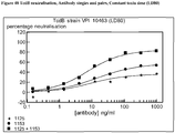
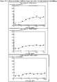
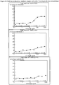
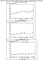
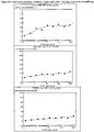
- the inventors have established by using internal comparisons between antibodies discovered in this application and by comparison against antibodies well described in the art (Babcock et al. 2006; Lowy et al., 2010) that some antibodies have the desirable characteristic of maintaining effective neutralization (for example low EC 50 and high % protection) even at high toxin concentrations. Other antibodies including those described in the art do not maintain effective toxin neutralization at high toxin concentrations.
- Effective toxin concentrations can be defined as a 'lethal dose' (LD) in titration studies in the absence of neutralizing antibodies.
- Neutralisation assays are typically conducted at an LD of 50% of complete cell killing (i.e. an LD 50 ) but may be more rigorously conducted at an LD 80 .
- Assays may also be performed under considerably more challenging conditions such as LD 90 , LD 95 and/or LD max (LD max is the maximal toxin quantity which can be included in an assay as constrained by assay volume and maximum toxin concentration / solubility).
- LD max is the maximal toxin quantity which can be included in an assay as constrained by assay volume and maximum toxin concentration / solubility.
- Such assays aim to mimic the early stages of infection of humans when C. difficile growth in the bowel is rampant and diarrhea and other symptoms lead one to hypothesise that toxin concentrations are at their highest.
- Antibodies which effectively neutralize damaging toxin activities under high toxin concentration conditions are thought by the present inventors to have special clinical value for the control of symptoms in human infections.
- the antibody or antibodies of the present disclosure have useful, for example low EC 50 values and/or high % protection from cell death for one or more the LD 80 , LD 90 , LD 95 and/or LD max .
- the EC 50 in the one or more of the latter situations is 15ng/ml or less, for example 10ng/ml or less, such as 5ng/ml or less, in particular 1ng/ml or less.
- the % protection from cell death is >90%, or >75% or >50%.
- the present disclosure provides an antibody or a combination of antibodies which maintain toxin neutralization even in the presence of high levels of toxin, for example as measured in an assay provided herein.
- the harmful effect of toxin may, for example be measured in a suitable in vitro assay.
- the neutralization is measured in an assay given in Example 1 below.
- an antibody or antibodies identified in a neutralization assay for example wherein the potency of the antibody is maintained in the presence of high levels of toxin.
- Toxin A is used interchangeably with TcdA.
- Toxin B is used interchangeably with TcdB.
- an antibody according to the invention is a monoclonal antibody or binding fragment thereof.
- a monoclonal antibody according to the invention is capable of neutralising TcdA with very high potency and affinity.
- a monoclonal antibody according to the invention is capable of neutralising TcdA with very high potency and affinity and high avidity.
- Avidity as employed herein refers to the combined strength of multiple binding affinities.
- a monoclonal antibody according to the invention is capable of neutralising TcdA with very high potency and affinity and high avidity and high valency of binding.
- Valency of binding refers to the ability for a monoclonal antibody to bind to an antigen multiple times. High valency of binding hence results in high levels of decoration of antigen with antibodies and / or high levels of cross-linking of toxin molecules, which is thought to be advantageous.
- Anti-TcdA Mabs according to the present disclosure may be suitable for neutralising the early effects of TcdA, for example on cells such as loss of tight junctions.
- Tight junction as employed herein is intended to refer to impermeable zone of connection between cells within a monolayer or anatomical tissue structure. Fluid loss does not occur when tight junctions retain their structural and functional integrity. Loss of tight junctions is an indication that the cell has been compromised by toxin and is well documented as being an early step in the toxic effects of TcdA and TcdB (25) and results in loss of fluid containing serum, immunoglobulin and ions (26, 3). Loss of tight junctions is thought to be a first step on the onset of diarrhoea in humans.
- TEER trans epithelial electric resistance assay
- TEER loss can be employed to identify antibodies that slow or prevent damage to the tight junctions and hence is a surrogate for protection against tissue damage leading to diarrhoea.
- Caco-2 cells are employed since they are derived from human colon cells and are known to form differentiated monolayers with cells connected by tight junctions.
- a kit is commercially available from Becton-Dickinson named the Caco-2 BioCoat HTS plate system (BD Biosciences/ 354802). The instructions in the kit are suitable for testing in the present context. The resistance of the membrane changes when the membrane has been compromised.
- the antibody will be pre-incubated with the toxin before addition to the TEER system to establish if the antibody can prevent or slow the damage to the membrane caused by the toxin.
- the assay may be performed over a suitable period, for example 24 hours taking measurements at certain time-points. The present inventors have established that the 4 hour time point is particularly discriminating for therapeutically useful antibodies.
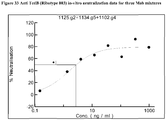
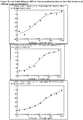
- the concentration of toxin employed in the TEER assay is generally in the range 100-200ng/ml, most preferably 125ng/ml
- the concentration of antibody (for example IgG1) employed in the TEER assay is generally in the range of 4 to 2000ng/ml, for example 50 to 1000ng/ml, such as 100 to 500ng/ml.
- the EC 50 of the antibody in the TEER assay employed in said condition is at least 200ng/ml, for example less than 100ng/ml, such as about 60-80ng/ml.
- an anti-TcdA antibody or an anti-TcdB antibody suitable for use as a therapeutic agent in the treatment or prevention of C. difficile infection, wherein said antibody was screened and selected employing a TEER assay.
- a method of screening an antibody in a TEER assay for the ability to slow or prevent loss of tight junctions comprising the step of identifying an appropriate antibody or antibodies and expressing suitable quantities of same. In one embodiment the method comprises the further step of formulating said antibody or antibodies in a pharmaceutical formulation. In one embodiment the method comprises the further step of administering said antibody or antibodies or said formulation to a patient in need thereof.
- multiple antibodies of the disclosure may be capable of binding to the target toxin (TcdA or TcdB), which may aid immune clearance of the toxin.
- Multiple antibodies as employed herein is intended to refer to multiple copies of an antibody with the same sequence or an antibody with the same amino acid sequence or an antibody specific to the same target antigen but with a different amino acid sequence.
- the antibodies according to the invention are specific to the target antigen, for example specific to an epitope in the target antigen.
- the antibodies of the invention are able to bind to the target antigen in two or more locations, for example two or three locations, such as four, five, six, seven, eight, nine, ten or more locations, for example the toxin may comprise repeating domains and thus an antibody may be specific to an epitope and in fact that epitope may be present in the antigen several times i.e. in more than one location. Thus given antibodies may bind the same epitope multiple times in different locations in the antigen.
- the antibody binds to the given antigen multiple times, for example 2 to 20 times such as 3, 4, 5, 6, 7, 8, 9, 10, 11, 12, 13, 14, 15 or 16 times. In one embodiment the antibody binds the given antigen at least 3 times. This multiple binding is thought to be important in neutralisation and/or clearance of the toxin. Whilst not wishing to be bound by theory it is thought that multiple binding, for example 3 more times, i.e. by decoration with 3 or more Fc fragments is important in triggering rapid clearance of the toxin (24) primarily via the liver and spleen (27, 28).
- the anti-TcdA antibody binds 3 or more times, for example 3 to 16 times.
- the anti-TcdA antibody binds 12 times.
- the anti-TcdA antibody binds 2 times.
- an anti-TcdA antibody binds in the catalytic-terminal cell binding domain of TcdA.
- the anti-Tcd B antibody binds 2 or more times, for example 2 times.
- an anti-TcdB antibody binds in the catalytic-terminal cell binding domain of TcdB.
- the antibody or antibodies according to disclosure are capable of cross-linking toxin molecules, for example one arm of the antibody molecule binds one toxin molecule and another of the antibody binds a epitope in a different toxin molecule, thereby forming a sort of immune complex.
- the formation of the latter may also facilitate activation of the immune system to clear the relate toxin and thereby minimise the deleterious in vivo effects of the same.
- an innate immune response such as complement is activated.
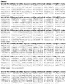
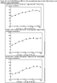
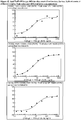
- the antibody or antibodies of the disclosure have high potency against toxins derived from strains of different ribotypes, for example 003, 027, 078.
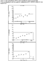
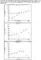
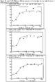
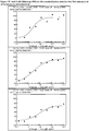
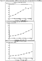
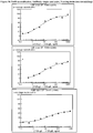
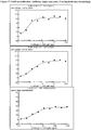
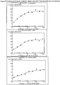
- antibodies against TcdA may have an EC 50 in the range of 0.1 - 100ng/ml, such as 1 to 10ng/ml and a maximal inhibition in the range of 50-100% at toxin concentrations of LD 80-95 , for example against toxins from strains of ribotypes 003, 027 and 078.
- antibodies against TcdA may have an EC 50 in the range of 0.1 - 100ng/ml, such as 1 to 10ng/ml and a maximal inhibition in the range of 60-100%, 70-100%, 80-100% or 90-100% at toxin concentrations of LD 80-95 , for example against toxins from strains of ribotypes 003, 027 and 078.
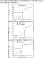
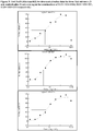
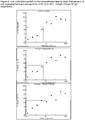
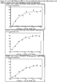
- antibodies against TcdB may have EC 50 in the range of 0.1 - 100ng/ml, such as 1 to 10ng/ml and a maximal inhibition in the range of 50-100% at toxin concentrations of LD 80-95 , for example against toxins from strains of ribotype 003.
- antibodies against TcdB may have EC 50 in the range of 0.1 - 100ng/ml, such as 1 to 10ng/ml and a maximal inhibition in the range of 60-100%, 70-100%, 80-100% or 90-100% at toxin concentrations of LD 80-95 , for example against toxins from strains of ribotype 003.
- combinations of antibodies according to the invention for example combinations of antibodies specific to TcdA, combinations of antibodies specific to TcdB or combinations of antibodies to specific to TcdA and antibodies specific to TcdB.
- Combinations of antibodies specific to TcdA will generally refer to combinations of antibodies directed to different epitopes on the target antigen TcdA, or at least with different binding properties.
- Combinations of antibodies specific to TcdB will generally refer to combinations of antibodies directed to different epitopes on the target antigen TcdB, or at least with different binding properties.
- the combinations may comprise 2, 3, 4, 5, 6, 7, 8, 9, 10, 11, 12, 13, 14 or 15 distinct antibodies, for example 2, 3, 4 or 5 antibodies.
- anti-TcdA antibody comprising a heavy variable region with a sequence as shown in SEQ ID NO:49 and a light variable region with a sequence shown in SEQ ID NO: 47 and two anti-TcdB antibodies the first with a heavy variable region shown in SEQ ID NO: 129 and a light variable region shown in SEQ ID NO: 127, and the second with a heavy variable region shown in SEQ ID NO: 159 and light variable region shown in SEQ ID NO: 157.
- Distinct antibodies as employed herein is intended to refer to antibodies with different amino acid sequences, which may bind the same epitope or different epitopes on the target antigen.
- TcdA also provided by the present disclosure is a specific region or epitope of TcdA which is bound by an antibody provided by the present invention, in particular an antibody comprising the heavy chain sequence given in SEQ ID NO:49 and the light chain sequence given in SEQ ID NO:47.
- TcdB also provided by the present disclosure is a specific region or epitope of TcdB which is bound by an antibody provided by the present invention, in particular an antibody comprising the heavy chain sequence given in SEQ ID NO:129 and the light chain sequence given in SEQ ID NO: 127 or an antibody comprising the heavy chain sequence given in SEQ ID NO: 159 and the light chain sequence given in SEQ ID NO: 157.
- This specific region or epitope of the TcdA or TcdB toxins can be identified by any suitable epitope mapping method known in the art in combination with any one of the antibodies provided by the present invention. Examples of such methods include screening peptides of varying lengths derived from the toxins for binding to the antibody of the present invention with the smallest fragment that can specifically bind to the antibody containing the sequence of the epitope recognised by the antibody.
- the peptides may be produced synthetically or by proteolytic digestion of the toxin polypeptide.
- Peptides that bind the antibody can be identified by, for example, mass spectrometric analysis. In another example, NMR spectroscopy or X-ray crystallography can be used to identify the epitope bound by an antibody of the present invention. Once identified, the epitopic fragment which binds an antibody of the present invention can be used, if required, as an immunogen to obtain additional antagonistic antibodies which bind the same epitope.
- Antibodies which cross-block the binding of an antibody according to the present invention may be similarly useful in neutralizing toxin activity. Accordingly, the present invention also provides a neutralizing antibody having specificity for TcdA or TcdB, which cross-blocks the binding of any one of the antibodies described above to TcdA or TcdB and/or is cross-blocked from binding these toxins by any one of those antibodies. In one embodiment, such an antibody binds to the same epitope as an antibody described herein above. In another embodiment the cross-blocking neutralising antibody binds to an epitope which borders and/or overlaps with the epitope bound by an antibody described herein above. In another embodiment the cross-blocking neutralising antibody of this aspect of the invention does not bind to the same epitope as an antibody of the present invention or an epitope that borders and/or overlaps with said epitope.
- Cross-blocking antibodies can be identified using any suitable method in the art, for example by using competition ELISA or BIAcore assays where binding of the cross blocking antibody to TcdA or TcdB prevents the binding of an antibody of the present invention or vice versa.
- a method of generating an anti-TcdA or anti-TcdB antibody comprising the steps of immunizing a host with a suitable antigen, for example an antigen shown in any one of SEQ ID Nos 173 to 194 or a combination thereof.
- the said method may also comprise one or more the following steps, for example identifying an antibody of interest (in particular using a functional assay such as TEER assay), expressing the antibody of interest, and optionally formulating the antibody as a pharmaceutically acceptable composition.
- the present disclosure provides a method of immunizing a host with an amino acid sequence shown in SEQ ID Nos 173 to 194 or a combination thereof.
- the antibodies according to the invention have an affinity to the target antigen of 10nM or less, for example InM or less such as 900pM, in particular 800pM, 700pM, 600pM or500pM, such as 60pM.
- the affinity is for TcdA or TcdB or a fragment thereof.
- the fragment is TcdA123 corresponding to residues S1827-D2249 of TcdA.
- the fragment is TcdA456 corresponding to residues G2205-R2608.
- the fragment is TcdB1234 corresponding to residues S1833-E2366 of TcdB.
- antibodies according to the invention or a combination thereof have an EC 50 of 200ng/ml or less, for example 150ng/ml or less such as 100ng/ml or less, such as in the range 0.1 to 10ng/ml.
- the individual component antibodies of mixtures are not required to have an EC 50 in said range provided that when they are used in combination with one or more antibodies the combination has an EC 50 in said range.
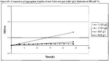
- the antibodies of the invention are stable, for example are thermally stable at temperatures above 50°C such as 60 or 70°C.
- the antibodies and combinations according to the present invention also have one or more of the following advantageous properties: slow off rate, high affinity, high potency, the ability to bind multiple times to the target antigen, to neutralise the toxin by a mechanism which reduces the loss of measurable TEER activity, to stimulate or assist the hosts natural immune response, to catalyse or assist in immune clearance of the pathogen (or toxin) and/or to educate the immune system to respond appropriately to the pathogen (or toxin).
- the Kabat residue designations do not always correspond directly with the linear numbering of the amino acid residues.
- the actual linear amino acid sequence may contain fewer or additional amino acids than in the strict Kabat numbering corresponding to a shortening of, or insertion into, a structural component, whether framework or complementarity determining region (CDR), of the basic variable domain structure.
- CDR complementarity determining region
- the correct Kabat numbering of residues may be determined for a given antibody by alignment of residues of homology in the sequence of the antibody with a "standard" Kabat numbered sequence.
- the CDRs of the heavy chain variable domain are located at residues 31-35 (CDR-H1), residues 50-65 (CDR-H2) and residues 95-102 (CDR-H3) according to the Kabat numbering system.
- CDR-H1 Chothia, C. and Lesk, A.M. J. Mol. Biol., 196, 901 - 917 (1987 )
- the loop equivalent to CDR-H1 extends from residue 26 to residue 32.
- CDR-H1' as employed herein is intended to refer to residues 26 to 35, as described by a combination of the Kabat numbering system and Chothia's topological loop definition.
- the CDRs of the light chain variable domain are located at residues 24-34 (CDR-L1), residues 50-56 (CDR-L2) and residues 89-97 (CDR-L3) according to the Kabat numbering system.
- Antibodies for use in the present invention may be obtained using any suitable method known in the art.
- the toxin A and/or toxin B polypeptide/protein including fusion proteins, for example toxin-Fc fusions proteins or cells (recombinantly or naturally) expressing the polypeptide (such as activated T cells) can be used to produce antibodies which specifically recognise the target toxins.
- the toxin polypeptide may be the full length polypeptide or a biologically active fragment or derivative thereof.
- Polypeptides may be prepared by processes well known in the art from genetically engineered host cells comprising expression systems or they may be recovered from natural biological sources.
- polypeptides includes peptides, polypeptides and proteins. These are used interchangeably unless otherwise specified.
- sequence for TcdA from ribotype 027 is given in SEQ ID NO: 171 (Uniprot accession number C9YJ37) and the sequence for TcdB from ribotype 027 is given is SEQ ID NO: 172 (Uniprot accession number C9YJ35).
- the antigen polypeptide may in some instances be part of a larger protein such as a fusion protein for example fused to an affinity tag.
- Antibodies generated against the antigen polypeptide may be obtained, where immunisation of an animal is necessary, by administering the polypeptides to an animal, preferably a non-human animal, using well-known and routine protocols, see for example Handbook of Experimental Immunology, D. M. Weir (ed.), Vol 4, Blackwell Scientific Publishers, Oxford, England, 1986 ). Many warm-blooded animals, such as rabbits, mice, rats, sheep, cows, camels or pigs may be immunized. However, mice, rabbits, pigs and rats are generally most suitable.
- Monoclonal antibodies may be prepared by any method known in the art such as the hybridoma technique ( Kohler & Milstein, 1975, Nature, 256:495-497 ), the trioma technique, the human B-cell hybridoma technique ( Kozbor et al., 1983, Immunology Today, 4:72 ) and the EBV-hybridoma technique ( Cole et al., Monoclonal Antibodies and Cancer Therapy, pp77-96, Alan R Liss, Inc., 1985 ).
- Antibodies for use in the invention may also be generated using single lymphocyte antibody methods by cloning and expressing immunoglobulin variable region cDNAs generated from single lymphocytes selected for the production of specific antibodies by, for example, the methods described by Babcook, J. et al., 1996, Proc. Natl. Acad. Sci. USA 93(15):7843-78481 ; WO92/02551 ; WO2004/051268 and International Patent Application number WO2004/106377 .
- Humanised antibodies are antibody molecules having one or more complementarity determining regions (CDRs) from a non-human species and a framework region from a human immunoglobulin molecule (see, e.g. US 5,585,089 ; WO91/09967 ). It will be appreciated that it may only be necessary to transfer the specificity determining residues of the CDRs rather than the entire CDR (see for example, Kashmiri et al., 2005, Methods, 36, 25-34 ). Humanised antibodies may optionally further comprise one or more framework residues derived from the non-human species from which the CDRs were derived.
- CDRs complementarity determining regions
- the term 'humanised antibody molecule' refers to an antibody molecule wherein the heavy and/or light chain contains one or more CDRs (including, if desired, one or more modified CDRs) from a donor antibody (e.g. a murine monoclonal antibody) grafted into a heavy and/or light chain variable region framework of an acceptor antibody (e.g. a human antibody).
- a donor antibody e.g. a murine monoclonal antibody
- acceptor antibody e.g. a human antibody
- only one or more of the specificity determining residues from any one of the CDRs described herein above are transferred to the human antibody framework (see for example, Kashmiri et al., 2005, Methods, 36, 25-34 ).
- only the specificity determining residues from one or more of the CDRs described herein above are transferred to the human antibody framework.
- only the specificity determining residues from each of the CDRs described herein above are transferred to the human antibody framework.
- any appropriate acceptor variable region framework sequence may be used having regard to the class/type of the donor antibody from which the CDRs are derived, including mouse, primate and human framework regions.
- the humanised antibody according to the present invention has a variable domain comprising human acceptor framework regions as well as one or more of the CDRs provided herein.
- variable domain comprises human acceptor framework regions and non-human donor CDRs.
- human frameworks which can be used in the present invention are KOL, NEWM, REI, EU, TUR, TEI, LAY and POM (Kabat et al., supra).
- KOL and NEWM can be used for the heavy chain
- REI can be used for the light chain and EU
- LAY and POM can be used for both the heavy chain and the light chain.
- human germline sequences may be used; these are available at: http://vbase.mrc-cpe.cam.ac.uk/
- the acceptor heavy and light chains do not necessarily need to be derived from the same antibody and may, if desired, comprise composite chains having framework regions derived from different chains.
- the framework regions need not have exactly the same sequence as those of the acceptor antibody. For instance, unusual residues may be changed to more frequently-occurring residues for that acceptor chain class or type. Alternatively, selected residues in the acceptor framework regions may be changed so that they correspond to the residue found at the same position in the donor antibody (see Reichmann et al., 1998, Nature, 332, 323-324 ). Such changes should be kept to the minimum necessary to recover the affinity of the donor antibody.
- a protocol for selecting residues in the acceptor framework regions which may need to be changed is set forth in WO 91/09967 .
- the antibody sequences disclosed in the present specification are humanised.
- the invention also provides sequences which are 80%, 90%, 91%, 92%, 93% 94%, 95% 96%, 97%, 98% or 99% similar to a sequence or antibody disclosed herein.
- Identity indicates that at any particular position in the aligned sequences, the amino acid residue is identical between the sequences.
- similarity indicates that, at any particular position in the aligned sequences, the amino acid residue is of a similar type between the sequences.
- leucine may be substituted for isoleucine or valine.
- Other amino acids which can often be substituted for one another include but are not limited to:
- the antibody molecules of the present invention include a complete antibody molecule having full length heavy and light chains or a fragment thereof and may be, but are not limited to Fab, modified Fab, Fab', modified Fab', F(ab')2, Fv, Fab-Fv, Fab-dsFv, single domain antibodies (e.g. VH or VL or VHH), scFv, bi, tri or tetra-valent antibodies, Bis-scFv, diabodies, triabodies, tetrabodies and epitope-binding fragments of any of the above (see for example Holliger and Hudson, 2005, Nature Biotech.
- bispecific and multispecific antibody variants are especially considered in this example since the aim is to neutralise two independent target proteins: TcdA and TcdB.
- Variable regions from antibodies disclosed herein may be configured in such a way as to produce a single antibody variant which is capable of binding to and neutralising TcdA and TcdB.
- the antibody according to the present disclosure is provided as TcdA or TcdB binding antibody fusion protein which comprises an immunoglobulin moiety, for example a Fab or Fab' fragment, and one or two single domain antibodies (dAb) linked directly or indirectly thereto, for example as described in WO2009/040562 .
- the fusion protein comprises two domain antibodies, for example as a variable heavy (VH) and variable light (VL) pairing, optionally linked by a disulphide bond, for example as described in WO2010/035012 .
- VH variable heavy
- VL variable light
- the Fab or Fab' element of the fusion protein has the same or similar specificity to the single domain antibody or antibodies. In one embodiment the Fab or Fab' has a different specificity to the single domain antibody or antibodies, that is to say the fusion protein is multivalent. In one embodiment a multivalent fusion protein according to the present invention has an albumin binding site, for example a VH/VL pair therein provides an albumin binding site.
- the multivalent fusion protein according to the invention binds TcdA and TcdB.
- the multivalent fusion protein according to the invention binds TcdB in multiple positions, for example has distinct binding regions specific for two different epitopes.
- the constant region domains of the antibody molecule of the present invention may be selected having regard to the proposed function of the antibody molecule, and in particular the effector functions which may be required.
- the constant region domains may be human IgA, IgD, IgE, IgG or IgM domains.
- human IgG constant region domains may be used, especially of the IgG1 and IgG3 isotypes when the antibody molecule is intended for therapeutic uses and antibody effector functions are required.
- IgG2 and IgG4 isotypes may be used when the antibody molecule is intended for therapeutic purposes and antibody effector functions are not required, e.g. for simply neutralising or agonising an antigen.
- sequence variants of these constant region domains may also be used.
- IgG4 molecules in which the serine at position 241 has been changed to proline as described in Angal et al., Molecular Immunology, 1993, 30 (1), 105-108 may be used.
- antibodies may undergo a variety of posttranslational modifications. The type and extent of these modifications often depends on the host cell line used to express the antibody as well as the culture conditions. Such modifications may include variations in glycosylation, methionine oxidation, diketopiperazine formation, aspartate isomerization and asparagine deamidation.
- the antibody heavy chain comprises a CH1 domain and the antibody light chain comprises a CL domain, either kappa or lambda.
- Biological molecules such as antibodies or fragments, contain acidic and/or basic functional groups, thereby giving the molecule a net positive or negative charge.
- the amount of overall "observed" charge will depend on the absolute amino acid sequence of the entity, the local environment of the charged groups in the 3D structure and the environmental conditions of the molecule.
- the isoelectric point (pI) is the pH at which a particular molecule or solvent accessible surface thereof carries no net electrical charge.
- the antibody and fragments of the invention may be engineered to have an appropriate isoelectric point. This may lead to antibodies and/or fragments with more robust properties, in particular suitable solubility and/or stability profiles and/or improved purification characteristics.
- the invention provides a humanised antibody engineered to have an isoelectric point different to that of the originally identified antibody from which it is derived.
- the antibody may, for example be engineered by replacing an amino acid residue such as replacing an acidic amino acid residue with one or more basic amino acid residues.
- basic amino acid residues may be introduced or acidic amino acid residues can be removed.
- acidic residues may be introduced to lower the pI, as required. It is important that when manipulating the pI care must be taken to retain the desirable activity of the antibody or fragment.
- the engineered antibody or fragment has the same or substantially the same activity as the "unmodified" antibody or fragment.
- affinity of antibodies provided by the present invention may be altered using any suitable method known in the art.
- the affinity of the antibodies or variants thereof may be measured using any suitable method known in the art, including BIAcore, using an appropriate isolated natural or recombinant protein or a suitable fusion protein/polypeptide.
- the present invention therefore also relates to variants of the antibody molecules of the present invention, which have an improved affinity for TcdA or TcdB, as appropriate.
- variants can be obtained by a number of affinity maturation protocols including mutating the CDRs ( Yang et al., J. Mol. Biol., 254, 392-403, 1995 ), chain shuffling ( Marks et al., Bio/Technology, 10, 779-783, 1992 ), use of mutator strains of E. coli ( Low et al., J. Mol. Biol., 250, 359-368, 1996 ), DNA shuffling ( Patten et al., Curr. Opin.
- Improved affinity as employed herein in this context refers to an improvement refers to an improvement over the starting molecule.
- an antibody for use in the present invention may be conjugated to one or more effector molecule(s).
- the effector molecule may comprise a single effector molecule or two or more such molecules so linked as to form a single moiety that can be attached to the antibodies of the present invention.
- this may be prepared by standard chemical or recombinant DNA procedures in which the antibody fragment is linked either directly or via a coupling agent to the effector molecule.
- Techniques for conjugating such effector molecules to antibodies are well known in the art (see, Hellstrom et al., Controlled Drug Delivery, 2nd Ed., Robinson et al., eds., 1987, pp.
- effector molecule includes, for example, biologically active proteins, for example enzymes, other antibody or antibody fragments, synthetic or naturally occurring polymers, nucleic acids and fragments thereof e.g. DNA, RNA and fragments thereof, radionuclides, particularly radioiodide, radioisotopes, chelated metals, nanoparticles and reporter groups such as fluorescent compounds or compounds which may be detected by NMR or ESR spectroscopy.
- biologically active proteins for example enzymes, other antibody or antibody fragments, synthetic or naturally occurring polymers, nucleic acids and fragments thereof e.g. DNA, RNA and fragments thereof, radionuclides, particularly radioiodide, radioisotopes, chelated metals, nanoparticles and reporter groups such as fluorescent compounds or compounds which may be detected by NMR or ESR spectroscopy.
- effector molecules may include chelated radionuclides such as 111In and 90Y, Lu177, Bismuth213, Californium252, Iridium192 and Tungsten188/Rhenium188; or drugs such as but not limited to, alkylphosphocholines, topoisomerase I inhibitors, taxoids and suramin.
- chelated radionuclides such as 111In and 90Y, Lu177, Bismuth213, Californium252, Iridium192 and Tungsten188/Rhenium188
- drugs such as but not limited to, alkylphosphocholines, topoisomerase I inhibitors, taxoids and suramin.
- effector molecules include proteins, peptides and enzymes.
- Enzymes of interest include, but are not limited to, proteolytic enzymes, hydrolases, lyases, isomerases, transferases.
- Proteins, polypeptides and peptides of interest include, but are not limited to, immunoglobulins, toxins such as abrin, ricin A, pseudomonas exotoxin, or diphtheria toxin, a protein such as insulin, tumour necrosis factor, ⁇ -interferon, ⁇ -interferon, nerve growth factor, platelet derived growth factor or tissue plasminogen activator, a thrombotic agent or an anti-angiogenic agent, e.g.
- angiostatin or endostatin or, a biological response modifier such as a lymphokine, interleukin-1 (IL-1), interleukin-2 (IL-2), granulocyte macrophage colony stimulating factor (GM-CSF), granulocyte colony stimulating factor (G-CSF), nerve growth factor (NGF) or other growth factor and immunoglobulins.
- IL-1 interleukin-1
- IL-2 interleukin-2
- GM-CSF granulocyte macrophage colony stimulating factor
- G-CSF granulocyte colony stimulating factor
- NGF nerve growth factor
- effector molecules may include detectable substances useful for example in diagnosis.
- detectable substances include various enzymes, prosthetic groups, fluorescent materials, luminescent materials, bioluminescent materials, radioactive nuclides, positron emitting metals (for use in positron emission tomography), and nonradioactive paramagnetic metal ions. See generally U.S. Patent No. 4,741,900 for metal ions which can be conjugated to antibodies for use as diagnostics.
- Suitable enzymes include horseradish peroxidase, alkaline phosphatase, beta-galactosidase, or acetylcholinesterase; suitable prosthetic groups include streptavidin, avidin and biotin; suitable fluorescent materials include umbelliferone, fluorescein, fluorescein isothiocyanate, rhodamine, dichlorotriazinylamine fluorescein, dansyl chloride and phycoerythrin; suitable luminescent materials include luminol; suitable bioluminescent materials include luciferase, luciferin, and aequorin; and suitable radioactive nuclides include 125I, 131I, 111In and 99Tc.
- the effector molecule may increase the half-life of the antibody in vivo, and/or reduce immunogenicity of the antibody and/or enhance the delivery of an antibody across an epithelial barrier to the immune system.
- suitable effector molecules of this type include polymers, albumin, albumin binding proteins or albumin binding compounds such as those described in WO05/117984 .
- the effector molecule is a polymer it may, in general, be a synthetic or a naturally occurring polymer, for example an optionally substituted straight or branched chain polyalkylene, polyalkenylene or polyoxyalkylene polymer or a branched or unbranched polysaccharide, e.g. a homo- or hetero- polysaccharide.
- synthetic polymers include optionally substituted straight or branched chain poly(ethyleneglycol), poly(propyleneglycol) poly(vinylalcohol) or derivatives thereof, especially optionally substituted poly(ethyleneglycol) such as methoxypoly(ethyleneglycol) or derivatives thereof.
- Specific naturally occurring polymers include lactose, amylose, dextran, glycogen or derivatives thereof.
- Derivatives as used herein is intended to include reactive derivatives, for example thiol-selective reactive groups such as maleimides and the like.
- the reactive group may be linked directly or through a linker segment to the polymer. It will be appreciated that the residue of such a group will in some instances form part of the product as the linking group between the antibody fragment and the polymer.
- the size of the polymer may be varied as desired, but will generally be in an average molecular weight range from 500Da to 50000Da, for example from 5000 to 40000Da such as from 20000 to 40000Da.
- the polymer size may in particular be selected on the basis of the intended use of the product for example ability to localize to certain tissues such as tumors or extend circulating half-life (for review see Chapman, 2002, Advanced Drug Delivery Reviews, 54, 531-545 ).
- a small molecular weight polymer for example with a molecular weight of around 5000Da.
- a higher molecular weight polymer for example having a molecular weight in the range from 20000Da to 40000Da.
- Suitable polymers include a polyalkylene polymer, such as a poly(ethyleneglycol) or, especially, a methoxypoly(ethyleneglycol) or a derivative thereof, and especially with a molecular weight in the range from about 15000Da to about 40000Da.
- antibodies for use in the present invention are attached to poly(ethyleneglycol) (PEG) moieties.
- the antibody is an antibody fragment and the PEG molecules may be attached through any available amino acid side-chain or terminal amino acid functional group located in the antibody fragment, for example any free amino, imino, thiol, hydroxyl or carboxyl group.
- Such amino acids may occur naturally in the antibody fragment or may be engineered into the fragment using recombinant DNA methods (see for example US 5,219,996 ; US 5,667,425 ; WO98/25971 , WO2008/038024 ).
- the antibody molecule of the present invention is a modified Fab fragment wherein the modification is the addition to the C-terminal end of its heavy chain one or more amino acids to allow the attachment of an effector molecule.
- the additional amino acids form a modified hinge region containing one or more cysteine residues to which the effector molecule may be attached. Multiple sites can be used to attach two or more PEG molecules.
- PEG molecules are covalently linked through a thiol group of at least one cysteine residue located in the antibody fragment.
- Each polymer molecule attached to the modified antibody fragment may be covalently linked to the sulphur atom of a cysteine residue located in the fragment.
- the covalent linkage will generally be a disulphide bond or, in particular, a sulphur-carbon bond.
- thiol group is used as the point of attachment
- appropriately activated effector molecules for example thiol selective derivatives such as maleimides and cysteine derivatives may be used.
- An activated polymer may be used as the starting material in the preparation of polymer-modified antibody fragments as described above.
- the activated polymer may be any polymer containing a thiol reactive group such as an ⁇ -halocarboxylic acid or ester, e.g. iodoacetamide, an imide, e.g. maleimide, a vinyl sulphone or a disulphide.
- a thiol reactive group such as an ⁇ -halocarboxylic acid or ester, e.g. iodoacetamide, an imide, e.g. maleimide, a vinyl sulphone or a disulphide.
- Such starting materials may be obtained commercially (for example from Nektar, formerly Shearwater Polymers Inc., Huntsville, AL, USA) or may be prepared from commercially available starting materials using conventional chemical procedures.
- Particular PEG molecules include 20K methoxy-PEG-amine (obtainable from Nektar, formerly Shearwater; Rapp Polymere; and SunBio) and M-PEG-SPA (obtainable from Nektar, formerly Shear
- the antibody is a modified Fab fragment or diFab which is PEGylated, i.e. has PEG (poly(ethyleneglycol)) covalently attached thereto, e.g. according to the method disclosed in EP 0948544 or EP1090037 [see also " Poly(ethyleneglycol) Chemistry, Biotechnical and Biomedical Applications", 1992, J. Milton Harris (ed), Plenum Press, New York , “ Poly(ethyleneglycol) Chemistry and Biological Applications", 1997, J. Milton Harris and S. Zalipsky (eds), American Chemical Society, Washington DC and " Bioconjugation Protein Coupling Techniques for the Biomedical Sciences", 1998, M. Aslam and A.
- PEG poly(ethyleneglycol)
- PEG is attached to a cysteine in the hinge region.
- a PEG modified Fab fragment has a maleimide group covalently linked to a single thiol group in a modified hinge region.
- a lysine residue may be covalently linked to the maleimide group and to each of the amine groups on the lysine residue may be attached a methoxypoly(ethyleneglycol) polymer having a molecular weight of approximately 20,000Da.
- the total molecular weight of the PEG attached to the Fab fragment may therefore be approximately 40,000Da.
- PEG molecules include 2-[3-(N-maleimido)propionamido]ethyl amide of N,N'-bis(methoxypoly(ethylene glycol) MW 20,000) modified lysine, also known as PEG2MAL40K (obtainable from Nektar, formerly Shearwater).
- PEG linkers include NOF who supply GL2-400MA2 (wherein m in the structure below is 5) and GL2-400MA (where m is 2) and n is approximately 450: m is 2 or 5
- each PEG is about 20,000Da.
- PEG effector molecules of the following type are available from Dr Reddy, NOF and Jenkem.
- an antibody which is PEGylated (for example with a PEG described herein), attached through a cysteine amino acid residue at or about amino acid 226 in the chain, for example amino acid 226 of the heavy chain (by sequential numbering).
- one certain antibodies according to the present disclosure have the following properties: Antibody Affinity (pM) Valency of binding EC 50 (ng/ml) TcdA 123 TcdA 456 TcdA, est. CA922 4.06 2.59 16 1.21 CA923 64.7 312 12 160.42 CA995 nil 119 1 37.64 CA997 132 66.8 12 6.25 CA1000 73.3 84.1 2 19.73
- compositions such as a pharmaceutical composition of antibody or combination of antibodies defined herein.
- the present invention also provides a composition that comprises at least two antibodies according to the invention, for example wherein at least one antibody therein is specific to TcdA and at least one antibody therein is specific to TcdB or alternatively at least two antibodies specific to TcdA or at least two antibodies specific to TcdB.
- composition that comprises multiple antibodies specific to TcdA and optionally one or more antibodies specific to TcdB.
- the disclosure also provides a composition that comprises multiple antibodies specific to TcdB and optionally one or more antibodies specific to TcdA.
- composition comprising 2, 3, 4, 5, 6, 7, 8, 9, 10, 11, 12, 13, 14 or 15 antibodies according to the invention i.e. distinct antibodies.
- the invention describes one particular mixture comprising 3 Mabs, one Mab of which is specific for TcdA and two Mabs of which are specific for TcdB.
- This mixture demonstrated very high levels of protection from death and gut inflammation from a lethal infective oral dose of Clostridium difficile in hamsters.
- composition comprising a combination of one anti-TcdA antibody comprising a heavy variable region with a sequence as shown in SEQ ID NO:49 and a light variable region with a sequence shown in SEQ ID NO: 47 and two anti-TcdB the first with a heavy variable region shown in SEQ ID NO: 129 and a light variable region shown in SEQ ID NO: 127, and the second with a heavy variable region shown in SEQ ID NO: 159 and light variable region shown in SEQ ID NO: 157.
- composition comprises 3 antibodies, such as one anti-TcdA and two anti-TcdB antibodies the antibodies are in the ratio of 50%, 25% and 25% respectively of the total antibody content thereof.
- composition comprising 2, 3, 4 or 5 antibodies specific to TcdA and optionally 1, 2, 3, 4 or 5 antibodies specific to TcdB.
- compositions provided according to the invention are well defined, for example are mixtures of monoclonal antibodies rather than simply polyclonal compositions derived from an immunised or immune competent host.
- composition of antibodies has an EC 50 of 200ng/ml or less, for example 150ng/ml or less, such as 100ng/ml or less, such as 0.1 to 10ng/ml.
- the antibodies described herein have very high levels of biophysical stability and so are suitable for inclusion in mixtures of antibodies.
- a pharmaceutical formulation or composition according to the invention further comprises a pharmaceutically acceptable excipient.
- Pharmaceutically acceptable carriers in therapeutic compositions may additionally contain liquids such as water, saline, glycerol and ethanol. Additionally, auxiliary substances, such as wetting or emulsifying agents or pH buffering substances, may be present in such compositions. Such carriers enable the pharmaceutical compositions to be formulated as tablets, pills, dragees, capsules, liquids, gels, syrups, slurries and suspensions, for ingestion by the patient.
- Suitable forms for administration include forms suitable for parenteral administration, e.g. by injection or infusion, for example by bolus injection or continuous infusion.
- parenteral administration e.g. by injection or infusion, for example by bolus injection or continuous infusion.
- the product may take the form of a suspension, solution or emulsion in an oily or aqueous vehicle and it may contain formulatory agents, such as suspending, preservative, stabilising and/or dispersing agents.
- the antibody molecule may be in dry form, for reconstitution before use with an appropriate sterile liquid.
- compositions of the invention can be administered directly to the subject.
- the subjects to be treated can be animals. However, in one or more embodiments the compositions are adapted for administration to human subjects.
- the pH of the final formulation is not similar to the value of the isoelectric point of the antibody or fragment, for example if the pH of the formulation is 7 then a pI of from 8-9 or above may be appropriate. Whilst not wishing to be bound by theory it is thought that this may ultimately provide a final formulation with improved stability, for example the antibody or fragment remains in solution.
- composition or formulation of the present disclosure comprises 1-200mg/mL of antibodies, that this to say the combined antibody content, for example 150mg/mL or less, such as 100mg/mL or less, in particular 90, 80, 70 , 60, 50, 40, 30, 20, 10mg/mL or less.
- composition or formulation according to the present disclosure comprises 20mg/mL of each antibody therein.
- a formulation comprising:
- the pharmaceutical formulation at a pH in the range of 4.0 to 7.0 comprises: 1 to 200mg/mL of an antibody according to the present disclosure, 1 to 100mM of a buffer, 0.001 to 1% of a surfactant,
- composition or formulation according to the present disclosure comprises the buffer phosphate buffered saline.
- the formulation at approximately pH6 may comprise 1 to 50mg/mL of antibody, 20mM L-histidine HCl, 240 mM trehalose and 0.02% polysorbate 20.
- a formulation at approximately pH 5.5 may comprise 1 to 50mg/mL of antibody, 20mM citrate buffer, 240mM sucrose, 20mM arginine, and 0.02% polysorbate 20.
- compositions of this invention may be administered by any number of routes including, but not limited to, oral, intravenous, intramuscular, intra-arterial, intramedullary, intrathecal, intraventricular, transdermal, transcutaneous (for example, see WO98/20734 ), subcutaneous, intraperitoneal, intranasal, enteral, topical, sublingual, intravaginal or rectal routes. Hyposprays may also be used to administer the pharmaceutical compositions of the invention.
- the therapeutic compositions may be prepared as injectables, either as liquid solutions or suspensions. Solid forms suitable for solution in, or suspension in, liquid vehicles prior to injection may also be prepared.
- Direct delivery of the compositions will generally be accomplished by injection, subcutaneously, intraperitoneally, intravenously or intramuscularly, or delivered to the interstitial space of a tissue.
- compositions can also be administered into a lesion or directly into the gastrointestinal tract by for examples, encapsulated oral dosage for swallowing, through a nasogastric tube to the stomach or ileum, through a rectal tube or enema solutions or by rectal capsule.
- Dosage treatment may be a single dose schedule or a multiple dose schedule.
- the active ingredient in the composition will be an antibody molecule. As such, it will be susceptible to degradation in the gastrointestinal tract. Thus, if the composition is to be administered by a route using the gastrointestinal tract, the composition will need to contain agents which protect the antibody from degradation but which release the antibody once it has been absorbed from the gastrointestinal tract.
- the present invention also provides an antibody or antibody combination or a composition comprising the same, as described herein, for treatment, for example for the treatment or prophylaxis of C. difficile infection or complications associated with the same such as diarrhoea, colitis in particular pseudomembranous colitis, bloating, abdominal pain and toxic megacolon.
- Prophylaxis can also be achieved by the administration of pre-formed complexes of inactivated toxin antigen (toxoid) and antibody in order to create a vaccine.
- toxoid inactivated toxin antigen
- the antibodies, combinations thereof and compositions comprising the same according to the invention are suitable for treating infection with so-called super strains of C. difficile, i.e. hypervirulent strains such as ribotype 027.
- the antibodies and compositions according to the present invention are suitable for use in the treatment or prophylaxis of acute and/or chronic effects of the relevant C. difficile toxins during primary infection.
- the antibodies and compositions according to the present invention are suitable for use in the treatment or prophylaxis of effects of the relevant C. difficile toxins during secondary infection or re-infection.
- International guidelines enshrine time intervals after a primary infection which hence defines a secondary (or recurrent) infection as being distinct from a continuation of existing symptoms sometimes described as a relapse (29).
- Secondary infections can be the result of the same strain or ribotype as the primary infection. In such cases recurrence rather than relapse relies on agreed temporal constraints.
- research also clearly shows that secondary infection can also be the result of infection of a strain or ribotype distinct from that of the primary infection.
- the antibodies, combinations thereof and compositions comprising the same according to the invention are suitable for use in the prevention of damage, for example long term structural damage to the epithelium of the colon.
- the antibodies, combinations and composition are suitable for preventing C. difficile infection including recurrence of infection, in particular nosocomial infection.
- the antibodies, combinations thereof and compositions comprising the same according to the invention are suitable for reducing the risk of recurrence of C. difficile infection.
- the antibodies of the present disclosure can be administered prophylactically to prevent infection or re-infection because in the absence of toxin to which the antibody is specific the antibody is simply to be cleared from the body without causing adverse interactions with the subject's body tissues.
- the antibodies of the present disclosure seem to elicit a rapid response after administration, for example within one or two days of administration rapid clearance of the target toxin is invoked, this may prevent vital organs such as the lungs, heart and kidneys being damaged. This is the first time that agents have been made available with can be employed to prevent damage or injury to a patient by toxins A and/or B in the acute C. difficile infection stage.
- the antibodies, combinations thereof and compositions comprising the same according to the invention are suitable for preventing damage to vital organs.
- the antibody, combinations or formulations described herein are suitable for preventing death of an infected patient, if administered within an appropriate time frame before irreparable damage has been done by the toxins.
- the antibodies of the present disclosure have fast on-rates, which facilitates the rapid effect in vivo.
- the patient population is over 60, such as over 65 years of age.
- the patient population is 5 years old or less.
- the antibodies according the invention may be employed in combination with antibiotic treatment for example metronidazole, vancomycin or fidaxomicin.
- antibiotic treatment for example metronidazole, vancomycin or fidaxomicin.
- a range of in vitro data exemplify the properties of the Mabs and Mab mixtures. We show that one mixture of 3 Mabs (50% molar quantities of anti-TcdA and 50% molar quantities of anti-TcdB components) was able to protect hamsters from a lethal CDI.
- a method of treating a patient in need thereof by administering a therapeutically effective amount of an antibody as described herein or antibody combination or a composition comprising the same, for example in the treatment or prophylaxis of C. difficile infection or complications associated with the same such as diarrhoea, colitis in particular pseudomembranous colitis, bloating, abdominal pain and toxic megacolon.
- the antibody, combination or formulation is administered by a parenteral route, for example subcutaneously, intraperitoneally, intravenously or intramuscularly.
- a parenteral route for example subcutaneously, intraperitoneally, intravenously or intramuscularly.
- the data in the Examples generated in hamsters indicates that the doses administered by this route reach the gut and thus are able to generate a therapeutic effect.
- the antibody, combination or formulation is administered orally, for example an enterically coated formulation.
- an antibody, combination or formulation as described herein for the manufacture of a medicament for the treatment or prophylaxis of C. difficile infection.
- the dose administered is in the range 1 to 1000mg/Kg, for example 10 to 75mg/Kg, such 20 to 50mg/Kg.
- the half-life of the antibody or antibodies in mice and hamsters in vivo is in the range 6 to 8 days in healthy (uninfected) animals and hence are expected to have half-lives in humans in the range of 14-28 days.
- the antibody or antibodies are given as one dose only.
- the antibody or antibodies are given as a weekly or biweekly dose.
- the antibody or antibodies are given as once daily doses.
- complex comprising TcdA or an immunogenic fragment thereof according to the present invention, complexed with one or more anti-TcdA antibodies defined herein.
- the complex may be employed as the antigen in a vaccine formulation, for example suitable for generating protective antibodies to toxin A in vivo after administration to a human.
- the present disclosure also provides a complex comprising TcdB or an immunogenic fragment thereof, complexed with one or more anti-TcdB antibodies defined herein.
- the complex may be employed as the antigen in a vaccine formulation, for example suitable for generating protective antibodies to toxin B in vivo after administration to a human.
- Th1-type immunostimulants which may be formulated to produce adjuvants suitable for use in the present invention include and are not restricted to the following.
- a complex comprising TcdA or an immunogenic fragment thereof according to the present invention and TcdB or an immunogenic fragment thereof, wherein each toxin or fragment is complexed with one or more antibodies specific thereto, wherein the complex is suitable for administration as a vaccine formulation.
- Antibody antigen complexes are known to be taken up by the immune system in an Fc receptor mediated process (27, 28) and pre-formed complexes of antibody:antigen complexes have been successfully use as vaccines in human clinical trials (22).
- the vaccine formulation further comprises an adjuvant as an immunostimulant.
- Monophosphoryl lipid A in particular 3-de-O-acylated monophosphoryl lipid A (3D-MPL), is a preferred Th1-type immunostimulant for use in the invention.
- 3D-MPL is a well known adjuvant manufactured by Ribi Immunochem, Montana. Chemically it is often supplied as a mixture of 3-de-O-acylated monophosphoryl lipid A with either 4, 5, or 6 acylated chains. It can be purified and prepared by the methods taught in GB 2122204B , which reference also discloses the preparation of diphosphoryl lipid A, and 3-O-deacylated variants thereof.
- a preferred form of 3D-MPL is in the form of a particulate formulation having a small particle size less than 0.2mm in diameter, and its method of manufacture is disclosed in EP 0 689 454 .
- Saponins are also preferred Th1 immunostimulants in accordance with the invention. Saponins are well known adjuvants and are taught in: Lacaille-Dubois, M and Wagner H. (1996. A review of the biological and pharmacological activities of saponins. Phytomedicine vol 2 pp 363-386 ). For example, Quil A (derived from the bark of the South American tree Quillaja Saponaria Molina), and fractions thereof, are described in US 5,057,540 and "Saponins as vaccine adjuvants", Kensil, C. R., Crit Rev Ther Drug Carrier Syst, 1996, 12 (1-2):1-55 ; and EP 0 362 279 B1 .
- haemolytic saponins QS21 and QS17 HPLC purified fractions of Quil A
- QS7 a non-haemolytic fraction of Quil-A
- Use of QS21 is further described in Kensil et al. (1991. J. Immunology vol 146, 431-437 ). Combinations of QS21 and polysorbate or cyclodextrin are also known ( WO 99/10008 ).
- Particulate adjuvant systems comprising fractions of QuilA, such as QS21 and QS7 are described in WO 96/33739 and WO 96/11711 .
- One such system is known as an Iscorn and may contain one or more saponins.
- CpG immunostimulatory oligonucleotide containing unmethylated CpG dinucleotides
- CpG is an abbreviation for cytosine-guanosine dinucleotide motifs present in DNA.
- CpG is known in the art as being an adjuvant when administered by both systemic and mucosal routes ( WO 96/02555 , EP 468520 , Davis et al., J.Immunol, 1998, 160(2):870-876 ; McCluskie and Davis, J.Immunol., 1998, 161(9):4463-6 ).
- WO 96/02555 EP 468520 , Davis et al., J.Immunol, 1998, 160(2):870-876 ; McCluskie and Davis, J.Immunol., 1998, 161(9):4463-6 .
- the immunostimulatory sequence is often: Purine, Purine, C, G, pyrimidine, pyrimidine; wherein the CG motif is not methylated, but other unmethylated CpG sequences are known to be immunostimulatory and may be used in the present invention.
- a palindromic sequence is present.
- Several of these motifs can be present in the same oligonucleotide.
- the presence of one or more of these immunostimulatory sequences containing oligonucleotides can activate various immune subsets, including natural killer cells (which produce interferon g and have cytolytic activity) and macrophages (Wooldrige et al Vol 89 (no. 8), 1977).
- natural killer cells which produce interferon g and have cytolytic activity
- macrophages Wangrige et al Vol 89 (no. 8), 1977.
- Other unmethylated CpG containing sequences not having this consensus sequence have also now been shown to be immunomodulatory.
- CpG when formulated into vaccines is generally administered in free solution together with free antigen ( WO 96/02555 ; McCluskie and Davis, supra ) or covalently conjugated to an antigen ( WO 98/16247 ), or formulated with a carrier such as aluminium hydroxide ((Hepatitis surface antigen) Davis et al. supra ; Brazolot-Millan et al., Proc.Natl.Acad.Sci., USA, 1998, 95(26), 15553-8 ).
- a carrier such as aluminium hydroxide ((Hepatitis surface antigen) Davis et al. supra ; Brazolot-Millan et al., Proc.Natl.Acad.Sci., USA, 1998, 95(26), 15553-8 ).
- Such immunostimulants as described above may be formulated together with carriers, such as for example liposomes, oil in water emulsions, and or metallic salts, including aluminium salts (such as aluminium hydroxide).
- carriers such as for example liposomes, oil in water emulsions, and or metallic salts, including aluminium salts (such as aluminium hydroxide).
- 3D-MPL may be formulated with aluminium hydroxide ( EP 0 689 454 ) or oil in water emulsions ( WO 95/17210 );
- QS21 may be advantageously formulated with cholesterol containing liposomes ( WO 96/33739 ), oil in water emulsions ( WO 95/17210 ) or alum ( WO 98/15287 );
- CpG may be formulated with alum (Davis et al. supra ; Brazolot-Millan supra ) or with other cationic carriers.
- Combinations of immunostimulants are also preferred, in particular a combination of a monophosphoryl lipid A and a saponin derivative ( WO 94/00153 ; WO 95/17210 ; WO 96/33739 ; WO 98/56414 ; WO 99/12565 ; WO 99/11241 ), more particularly the combination of QS21 and 3D-MPL as disclosed in WO 94/00153 .
- a combination of CpG plus a saponin such as QS21 also forms a potent adjuvant for use in the present invention.
- the saponin may be formulated in a liposome or in an Iscorn and combined with an immunostimulatory oligonucleotide.
- suitable adjuvant systems include, for example, a combination of monophosphoryl lipid A, preferably 3D-MPL, together with an aluminium salt.
- the adjuvant is a combination of QS21 and 3D-MPL in an oil in water or liposomal formulation.
- An enhanced system involves the combination of a monophosphoryl lipid A and a saponin derivative particularly the combination of QS21 and 3D-MPL as disclosed in WO 94/00153 , or a less reactogenic composition where the QS21 is quenched in cholesterol containing liposomes (DQ) as disclosed in WO 96/33739 .
- This combination may additionally comprise an immunostimulatory oligonucleotide.
- a particularly potent adjuvant formulation involving QS21, 3D-MPL & tocopherol in an oil in water emulsion is described in WO 95/17210 and is another preferred formulation for use in the invention.
- Another preferred formulation comprises a CpG oligonucleotide alone or together with an aluminium salt.
- a method of manufacture of a vaccine formulation as herein described wherein the method comprises admixing a polypeptide according to the invention with a suitable adjuvant.
- Embodiments and preferences may be combined as technically appropriate.
- a range of different immunogens and screening reagents were either purchased or produced by conventional E. coli expression techniques in order to provide a diverse and broad immune response and to facilitate identification and characterisation of monoclonal antibodies (listed in Table 1).
- sequences were based on ribotype 027.
- the sequence for TcdA from ribotype 027 is given in SEQ ID NO: 171 (Uniprot accession number C9YJ37) and the sequence for TcdB from ribotype 027 is given is SEQ ID NO: 172 (Uniprot accession number C9YJ35).
- Sprague Dawley rats and half-lop rabbits were immunised with either synthetic peptides mapping to regions common to both TcdA and TdcdB full-length toxin, formaldehyde-inactivated toxoid A, binding domain fragments of Toxin A (CROPs1,2,3 or CROPs4,5,6) or binding domain fragment of Toxin B (CROPs1,2,3,4), or in some cases, a combination of the above.
- animals were sacrificed and PBMC, spleen and bone marrow harvested. Sera were monitored for binding to Toxin A domains, toxin B domains, toxin or toxoid by ELISA. Sera titres of 2 such immunisations are shown in figure 11 .
- UCB SLAM was used as a means to generate monoclonal antibodies.
- B cells were cultured directly from immunised animals (Zubler et al., 1985). This step enabled sampling of a large percentage of the B cell repertoire.
- SLAM lymphocyte antibody method
- UCB SLAM modified version of SLAM (UCB SLAM (Tickle et al. 2009)) that utilises a fluorescence-based method to identify antigen-specific B cells from culture wells.
- B cell cultures were set up and supernatants were first screened for their ability to bind a relevant purified toxin domain (binding, translocation or catalytic) in a bead-based assay using an Applied Biosystem 8200 cellular detection system.
- Heavy and light variable region gene pairs were isolated from single cells harvested by micromanipulation from approximately 100 toxin-neutralising wells following reverse transcription (RT)-PCR. These V-region genes were then cloned as mouse IgG1/kappa full-length antibodies for rat variable regions and rabbit IgG/kappa full-length antibodies for rabbit variable regions. Antibodies were re-expressed in a HEK-293 transient expression system. These recombinant antibodies were then retested for their ability to neutralise toxin in cell based assays. Recombinant antibodies were also screened by BIAcore to determine affinity for a given toxin domain and to also determine the specificity and approximate the number of binding events of antibody to toxin. Based on in vitro activity in cell based assays and affinity measurements, lead candidates were selected for humanisation. Unless otherwise stated, all the data herein was generated using the humanised antibodies.
- TcdA E . coli -produced toxin fragments
- A C. difficile -derived toxin or toxoid
- B synthetic peptides
- Table 1 Toxin A (TcdA) sequence related reagents for screening and immunizations. Fragment Residue number
- Source TcdA catalytic M1-E659 UCB
- E. coli expression TcdA translocation K577-D1350 UCB
- Toxin B sequence related reagents for screening and immunizations.
- Toxin Domain Amino acid Sequence Catalytic SPVEKNLHFVWIGGEVSD SEQ ID NO: 173 Catalytic NLAAASDIVRL SEQ ID NO: 174 Catalytic CGGVYLDVDMLPGIH SEQ ID NO: 175 Catalytic CGGVYLDVDMLPGIHSDLFK SEQ ID NO: 176 Catalytic CWEMIKLEAIMKYK SEQ ID NO: 177 Catalytic CTNLVIEQVKNR SEQ ID NO: 178 Catalytic PEARSTISLSGP SEQ ID NO: 179 Catalytic CSNLIVKQIENR SEQ ID NO: 180 Catalytic TEQEINSLWSFDQA SEQ ID NO: 181 Catalytic TEQEINSLWSFDPEARSTISLSGPC SEQ ID NO: 182 Translocation NVEETYPGKLLLC SEQ ID NO: 183 Translocation Acetyl-CANQYEVRINS
- the assay uses CACO-2 cells grown, and screened in MEM + 20% FCS, 2mM Q, and NEAA.Unless stated all Ab combinations are in equal ratios.
- CA927 had a TcdB neutralisation potential which could result in a defined ECso whilst the TcdB neutralisation potential of both CA1125 and CA1151 were insufficient under these assay conditions to result in a defined EC 50 .
- CA927 was not found to be the most effective Mab to use within a combination.
- the best CA927 containing combination had an EC 50 of 13.5ng/ml whereas other two Mab combinations had EC 50 's as low as 2.59 and 4.71ng/ml.
- CA1099 had the lowest TcdB neutralisation EC 50 under the assay conditions used. However, CA1099 was not found to be the most effective Mab to use within a combination. The best CA1099 containing combination had an EC 50 of 6ng/ml whereas other two Mab combinations had EC 50 's as low as 2 and 1ng/ml. We might speculate that the most effective pairings of Mabs are defined by their cooperative binding modalities especially as defined by having non-overlapping epitopes. TABLE 6 Anti-TcdB Mab combinations and relative Mab ratios at constant toxin concentration.
- the assay uses CACO-2 cells grown, and screened in MEM + 20% FCS, 2mM Q, and NEAA.
- the EC 50 of the preferred combination of CA1125 and CA1151 in equimolar ratios is largely unaffected by an increasing [TcdB].
- the three relative molar ratios of Mabs tested i.e. 25:75 vs 50:50 vs 75:25
- have very similar EC 50 's to each other suggesting that CA1125 and CA1151 have especially complementary modes of action.
- the CA1125:CA1134 ratio of 25:75 is notably less potent than 50:50 and 75:25.
- Antibody IC50 (ng/ml) 1099 2 1102 N/A 1114 103 1125 N/A 1134 8 1151 182 1153 260 926 N/A 927 N/A 1099 + 1125 6 1114 + 1125 7 1151 + 1125 2 1134 + 1125 1 1102 + 1125 6 1125 + 1153 12 926 + 1125 42 927 + 1125 4 TABLE 9 TcdB neutralisation - 1 or 2 anti-TcdB Mabs at various TcdB doses.
- the number of moles of binding events of anti- C . difficile TcdB antibodies to TcdB 1234 was determined by Surface Plasmon Resonance (SPR) on a Biacore 3000 (GE Healthcare). Streptavidin was immobilized on a CM5 sensor chip (GE Healthcare) to a level of ⁇ 4000RU via amine coupling and biotinylated TcdB 1234 was bound at 500-600RU. Two 20 ⁇ l injections of the same anti-TcdB antibody mixtures (final concentration of each antibody was 500nM) were injected over this surface at 10 ⁇ l/min and the saturating binding response recorded. The surface was regenerated after every cycle using HCl.
- SPR Surface Plasmon Resonance
- Immobilized CA1125 when bound to TcdB 1234 , does not allow CA1125 to bind further supporting the idea that CA1125 has one binding site on TcdB 1234 and that after this has been saturated that no other binding site for CA1125 can be found. However, when TcdB 1234 has been saturated by CA1125, CA1151 can still bind. This demonstrates that CA1151 binds at alternative sites to that occupied by CA1125. Together these data show that CA1125 is a single binder of TcdB 1234 whereas 1151 IgG binds to TcdB 1234 more than once, most likely twice. Hence a mixture of CA1125 and CA1151 can bind to TcdB 1234 approximately 3 times.
- All antibodies combinations have an additive binding response showing that there are 2 or more non-competitive sites on TcdB 1234 bound by these combinations.
- the number of moles of binding events of anti- C.difficile TcdA antibodies to TcdA 123 and A 456 were determined by Surface Plasmon Resonance (SPR) on a Biacore 3000 (GE Healthcare). Streptavidin was immobilized on a CM5 sensor chip (GE Healthcare) via amine coupling to a level of ⁇ 4000RU and biotinylated TcdA 123 was bound to one flowcell and TcdA 456 was bound to a different flowcell to a response of ⁇ 500RU. Two 30 ⁇ l injections of the same anti-TcdA antibody at 1 ⁇ M were injected over both flowcells at 10 ⁇ l/min and the saturating binding response recorded.
- SPR Surface Plasmon Resonance
- Antibodies CA997 and CA1000 bind to TcdA 123 in a ratio of six CA997's to one CA1000 whereas they bind to TcdA 456 in a ratio of three CA997's to one CA1000 (Table 2).
- TcdA 123 and TcdA 456 The maximum antibody response for CA997, corrected for molecular weight and immobilized toxin level is similar for TcdA 123 and TcdA 456 . This suggests that CA997 binds TcdA 456 six times and CA1000 binds twice to TcdA 456 . Hence antibody CA997 likely binds to TcdA whole toxin (TcdA) approximately 12 times.
- Increased valency of binding to TcdA and TcdB may have two important effects in vivo.
- the first is that any Mab or Mab mixture which is capable of binding TcdB more than once will have increased potential to form inter-toxin binding events and hence immunoprecipitation.
- Immunoprecipitation can contribute to potency by reducing the solubility of toxin and forming very large macromolecular complexes which hence reduce the effective working concentration of toxin.
- Such large protein complexes may be taken up by macrophages and monocytes resident in the tissue and may contribute to an augmented host immune respone.
- Antigen:antibody complexes bearing Fc fragments have been specifically shown to be capable of priming a host immune respone against a gut pathogen (21).
- soluble antigen:antibody complexes have been successfully used as a vaccine directed against the antigen in human clinical trials (22).
- immune decoration of toxin with Fc bearing IgG may contribute to immune clearance using normal mechanisms through the liver and spleen.
- higher levels of Fc decoration of antigen lead to faster and more complete levels of clearance (23)
- presence of 2 or more Mab Fc domains per toxin, especially 3 Fc domains per toxin may represent a critical number of Fcs required for very rapid and substantial clearance of toxin (24).
- the anti-TcdA Mab CA997 is likely capable of binding to TcdA up to 12 times and the combination of CA1125 and CA1151 is likely capable of binding to TcdB 3 times.
- the 3 Mab mixture is very likely to be capable of providing for these kinds of additional potency mechanisms in vivo.
- Example 6 Mab neutralisation of loss of TEER caused by TcdA.
- C.difficile monolayer integrity assay is performed using the Becton-Dickinson (BD) Caco-2 BioCoat HTS plate system.
- Toxin A added to antibodies at a concentration of 125ng/ml and plate incubated for 1hr at 37°C. 1ml of Caco-2 growth medium (MEM + 20% FCS, 2mM Q, NEAA) added to each well of a standard 24-well TC plate. BioCoat insert plate transferred to the 24-well TC plate. Entero-STIM medium removed from inserts and replaced with 400 ⁇ l of toxin:Ab mixture.
- Caco-2 growth medium MEM + 20% FCS, 2mM Q, NEAA
- Loss of tight junctions between gut cells is the key early effect of TcdA on cell monolayers and gut tissue sections and is the primary cause of diarrhoea.
- Albumin and other serum proteins are lost into the gut lumen along with accompanying serum fluid.
- the loss of trans-epithelial electrical resistance in differentiated cultured cells which have formed a monolayer is a useful surrogate for the protection against the acute effects of TcdA.
- Three antibodies shown have good levels of protection against TEER loss, Figure 62 . It is notable and surprising that the abilities of these Mabs in TEER assays do not reflect those seen in toxin neutralization as measured in a cell proliferation assay.
- CA997 had the best performance in the TEER assay since it had both high levels of protection and maintained this at the lower Mab concs.
- CA997 had a substantial potential to neutralize TEER loss with maximal inhibition approaching 80% and an EC 50 of approximately 80ng/ml at 4h.
- Example 7 Affinity of anti- C difficile toxin antibodies for sub-domains of TcdA and TcdB: TcdA 123 , TcdA 456 and TcdB 1234 .
- TcdA123, TcdA456 or TcdB1234 were titrated over captured purified antibodies at doubling dilutions from 12.5nM at a flow rate of 30 ⁇ L/min.
- a single concentration of 12.5nM of TcdA123 or TcdA456 and 50nM of TcdB1234 was passed over the antibodies at 30ul/min.
- the anti-TcdA affinities are particularly high compared to the published affinities of other Mabs. We demonstrate that affinities as low as 4pM are achievable.
- the preferred CA997 has an affinity of 132pM, CA1125 122pM and CA115 551pM. CA995 clearly shows that it does not bind to CROPs A 123 and hence that demonstrates that the Mab shown here have properties which are different from each other in surprising and unexpected ways.
- CA922, 923, 997 and 1000 do bind at least once to CROPs A123 and A456. Hence these 4 Mabs confirming that each must bind to holotoxin at least twice. We have been unable to derive affinities for the binding of these Mabs to holotoxin due to technical constraints. However, given the high affinities and valencies demonstrated for the anti-TcdA Mabs it is possible to speculate that the functional affinities against holotoxin may be even stronger than those illustrated for binding to toxin sub-domains.
- the anti-TcdB Mabs also demonstrated strong affinities reaching as low as 31pM.
- CA1125, 1151, 927, 1099, 1134 and 1153 show affinities which surpass those demonstrated by others.
- Antibody combinations need to be made up of Mabs having high levels of stability in order to mitigate potential risks of aggregation during long term storage.
- Thermal stability is used as one measure.
- Tm Thermal stability
- Mab mixtures are measuring their propensity to aggregate due to physical stress such as agitation or shaking. Aggregates are undesirable components of drug compositions since they may reduce storage life time and may pose a safety risk to patients at certain levels.
- the Tm data show that all 5 anti-TcdA Mabs have high Tm stability, whilst three (CA922, 923 and 997) have very high Tm's in the range of 79-81°C. Of the anti-TcdB Mabs tested all but two have very high Tm's.
- CA997, CA1125 and CA1151 which were tested in the hamster infection study (Example 9) had very high Tm's (79.2°C, 79.3°C and 80.8°C respectively) which makes them suitable for use in a Mab mixture.
- CA997 and 922 had the lowest propensity to aggregate of the 5 anti-TcdA Mabs.
- CA115 and 1151 had the lowest aggregation propensities of the anti-TcdB Mabs.
- the use of CA997, 1125 and 1151 as a Mab mixture may have special value since they are more likely to survive co-formulation and storage at high protein concentrations.
- Samples were prepared by mixing the following: 30ul Protein sample at 2mg/ml, 0.35% Methylcellulose, 4% pH3-10 ampholytes (Pharmalyte), synthetic pI markers (4.65 and 9.77), 1ul of each stock solution, and HPLC grade water to make up the final volume to 200ul. The mixture was then analysed using iCE280 IEF analyzer(pre-focusing at 1500V for 1 min followed by focusing at 3000V for 6mins). The calibrated electropherograms were then integrated using Empower software (from Waters)
- Tm Thermal stability
- This method uses Sypro orange fluorescent dye to monitor the unfolding process of protein domains.
- the dye binds to exposed hydrophobic regions that become exposed as a consequence of unfolding which results in a change to the emission spectrum.
- the sample (5ul at 1mg/ml) is mixed with a 5ul of a stock solution of Sypro orange (30x) and the volume made up to 50ul with PBS,pH 7.40.
- the plate is placed in a 7900HT fast real-time PCR system containing a heating device for accurate temperature control.
- the temperature is increased from 20°C to 99° C (Ramp rate of 1.1°C/min).
- a CCD device simultaneously monitors the fluorescence changes in the wells.
- An algorithm is used to process intensity data and take into account multiple transitions.
- antibody samples are subjected to mechanical stress generated by processes such as pumping and filtration. This may cause denaturation and consequently aggregation due to exposure of the protein to air-liquid interfaces and shear forces resulting in the ultimate loss of bioactivity. Stress by vortexing is a method to screen the robustness of the antibody samples for prediction of aggregation stability.
- Both anti-TcdA and anti-TcdB IgG1 molecules were subjected to stress by agitation, by vortexing using an Eppendorf Thermomixer Comfort at 25 °C, 1400rpm.
- Sample size was 250uL, (x3 per sample) in a 1.5 mL conical Eppendorf-style capped tube (plastic), in PBS pH 7.4.
- Each sample was brought to a concentration of 1mg/ml (using extinction coefficient calculated from sequence) and aggregation was monitored by absorbance at 340nm and/or 595nm, by use of a Varian Cary 50-Bio spectrophotometer, measured at intervals for up to 24 hours.
- Table 14 provides a summary of the measured pI and Tm data for both anti-TcdA and anti-TcdB IgG1 molecules.
- Table 14 Compilation of pI and Tm Data measured pI Tm(Fab) in PBS Tm(CH2) Anti-TcdA IgG1 CA164_00922.g1 8.8 81 69.2 CA164_0923.g1 9.2 79 69.3 CA164_0995.g1 8.5 71 no data* CA164_0997.g1 8.3 79.2 68.4 CA164_01000.g1 7.74 70.5 no data* Anti-TcdB IgG1 CA164_01125.g1 9.2 79.3 69.4 CA164_01125.g2 9.2 79.5 69.3 CA164_01134.g4 9.3 78.4 69.4 CA164_01134.g5 9.2 76.4 69.2 CA164_01134.g6 9.2 76.6 69.6 CA
- the measured pI of the molecules were high (except for CA164_01000.g1_P3) and away from the pH of formulation buffers such as PBS, pH 7.4 and 50m sodium acetate/125mM sodium chloride, pH 5. This may mean that buffers with pH's suitable for co-formulation of two or more Mabs can be selected.
- Tm Thermal Stability
- Tm(CH2) Tm(CH2)
- Tm(Fab) Tm(Fab)
- Figure 67 shows the effect of agitation via vortexing on different anti-TcdA IgG1 molecules in PBS,pH 7.4.
- CA922.g1 and CA977.g1 were preferable based on the analyses above, although apart from CA1000) all molecules could be considered suitable candidates for use as therapeutic IgG1.
- the biophysical characteristics could be grouped within the family of grafts based on the aggregation stability and Tm, such that the CA1125 grafts potentially proved more stable.
- the CA1102 grafts showed poorest Tm data and also showed the greatest tendancy to aggregate via stress by agitation.
- CA1151.g4 showed that this molecule exhibited slightly increased aggregation stability relative to CA11125.g2 and seemed equivalent to the TcdA molecules (CA922.g1 and CA997.g1.All four molecules showed equivalent Tm values.
- CA997, CA1125 and CA1151 show very high levels of thermostability and very low levels of aggregate formation after agitation.
- the study protocol was approved by the Ricerca IACUC committee. Active and control components (composition and dose) were blinded to Ricerca staff until after completion of the planned 28 day study period.
- the drug combination tested was composed of one anti-TcdA antibody (CA997.g1) which constituted 50% of the injected protein and two anti-TcdB antibodies (CA1125.g2 and CA1151.g4) which together constituted 50% of the injected protein but which alone constituted 25% of the injected protein.
- Hamsters were sensitised (day -1) with 50mg/kg of Clindamycin phosphate in PBS (s.c.) before being challenged 1 day later (day 0) with 3.4 x 10⁶ c.f.u. of vegetative cells from strain ATCC43596. Vancomycin was dosed at 5mg/kg twice a day for 5 days (p.o.) on days 1, 2, 3, 4,5.
- the photographs in Figure 66 show typical gross pathologies for the swelling and bloody effusions of caeca caused by TcdA and TcdB (left image, PBS control, animal death on day 2) and a normal stool filled caeca after protection by UCB high dose Mabs (right image, UCB high dose, animal surviving to day 28). These data show that after protection with a high dose of UCB Mabs the large intestine can return to normal morphology and function.
- Clinical infections are caused by a variety of different strains. Strain differences are characterized using a number of different methods of which ribotyping is a key one. Different ribotype strains are observed to have different pathogenicity, infection and sporulation properties. All of the TcdA neutralization shown above used TcdA purified from strain known as VPI10463. However, the predominant aggressively pathogenic strain associated with out-breaks is called ribotype 027. Other key ribotypes include 078, 001, 106. Amino acid sequence difference have been observed between toxins produced by different ribotypes and hence it is important that Mabs are capable of neutralizing toxin from a diverse set of clinical isolates.
- CA922, 997 and 1000 were tested for their ability to neutralize TcdA from strains 027 and 078 and compared to their abilities against TcdA from VPI10463. Mabs were tested at 4 [TcdA] and found to be capable of neutralizing all toxins without significant difference at LD 80 , LD 90 and LD 95
- Table 16 EC50 values (ng/ml) - TcdA strain VPI 10463
- Antibody LD80 LD90 LD95 LDmax CA164_922 0.27 0.9 1.2 >500 CA164_997 1 2.5 3.5 25.4
- Table 17 EC50 values (ng/ml) - TcdA ribotype 027
- Antibody LD80 LD90 LD95 LDmax CA164_922 0.19 0.25 0.41 1.46 CA164_997 0.92 1.27 1.75 7.19 CA164_1000 2.25 2.49 3.52 16.32
- Table 18 EC50 values (ng/m
- the hamster PK was found a half-life similar to Mabs in mice or rats. (t1 ⁇ 2 6-8 days). i.p. and s.c. dosing were essentially the same.
- hIgG1 Mab The pharmacokinetics and distribution to the gut of a hIgG1 Mab was studied in 'normal' (non-infected) golden Syrian hamsters. Purified Mab was administered to male hamsters (120-135g) by CARE Research LLC, Fort Collins, Colorado, USA and samples were assayed by UCB Pharma.
- the colon section was flushed with wash buffer (50% (v/v) PBS containing 50% (v/v) Sigma protease inhibitor cocktail (P2714) before being opened and separation and removal of the mucosa from the underlying muscle.
- Mucosal samples were placed in 0.5ml of wash buffer homogenized until visually uniform and stored at 4°C before immediate shipping on wet ice.
- Serum samples were diluted in sample-conjugate buffer (1% (w/v) BSA, 0.2% Tween in PBS) and after washing were revealed with goat anti-human kappa-HRP (Cambridge Bioscience 2060-05) in sample-conjugate buffer and TMB with a 2.5M H 2 SO 4 stop solution.
- hIgG1 could be found in 'scrapings' of the gut i.e that hIgG1 gets into the vasculature of healthy gut - and so could be protective in 'prophylactic dosing'. This effect would be even more profound in humans since they have a cognate hFcRn.
- Humanised Mabs were quantified using liquid chromatography tandem mass spectrometry (LC-MS/MS) analysis following tryptic digestion. Quantitation was achieved by comparison to authentic standard material spiked at known concentrations into blank matrix, with spiked horse myoglobin used as the internal standard.
- LC-MS/MS liquid chromatography tandem mass spectrometry
- a unique ("proteotypic") peptide common to all of the humanised Mabs investigated was selected (DTLMISR, a CH2 region peptide) and both samples and calibration samples were tryptically digested as outlined. Tryptic digest of 5 ⁇ l serum samples was performed overnight using sequencing grade modified Trypsin (Promega, Southampton, UK) following denaturation / reduction with acetonitrile / Tris (2-carboxyethyl) phosphine and carbamido-methylation with iodoacetamide (Sigma-Aldrich, Poole, UK).
- the LC-MS/MS system consisted of a CTC HTS-x Autosampler (CTC Analytics, Zwingen, Switzerland), a Agilent 1290 LC system (Agilent Technologies, Stockport, UK) and a Sciex 5500 QTrap MS system (AB Sciex, Warrington, UK), equipped with a Turbo V ion source operated in electrospray mode. Analytes were separated using an Onyx Monolithic C18 column (100x4.6 mm, Phenomenex, Macclesfield, UK) with a gradient of 2 to 95 % (v/v) water/acetonitrile (0.1 % formic acid) delivered at 1.5 mL/min over 6 minutes.
- the injection volume was 10 ⁇ L; all of the eluent was introduced into the mass spectrometer source.
- the source temperature of the mass spectrometer was maintained at 600 °C and other source parameters (e.g. collision energy, declustering potential, curtain gas pressure etc.) were optimized to achieve maximum sensitivity for the peptide of interest. Selective transitions for each proteotypic peptide of interest were monitored.
- Plasma concentrations calculated based on the peptides monitored are outlined below.
- Total h-IgG was quantified in all samples using a peptide common to all analytes of interest. This was done using a combined standard curve of all five analytes. The validity of this approach is demonstrated by the fact that the sum of the concentrations observed for CA164_00725 and CA164_00726 are in good agreement (within experimental error) of the concentration observed for total h-IgG.
- the total concentration of h-IgG in the samples of animals dosed with CA164_00997, CA164_01125 and CA164_01151 was determined.
- Antibody CA726 is prior art antibody CDA1 as described (15) A summary of this data is presented in Figure 72.
Description
- The present invention relates to antibodies to exotoxins of Clostridium difficile, for example TcdA and TcdB, pharmaceutical compositions comprising the same, processes of producing said antibodies and compositions and use of the antibodies and compositions in treatment and/or prophylaxis, in particular treatment or prophylaxis of Clostridium difficile infection, pseudomembranous colitis, fulminant colitis and/or toxic mega colon.
- The two major exotoxins TcdA and TcdB have been established as the major pathogenicity determinants of Clostridium difficile in a large number of in vitro and in vivo studies. Non-toxigenic strains are not pathogenic to animals and man (1, 2). To date a clear understanding of the role of binary toxin has yet to be established (3).
- Both toxins are entero- and cyto-toxic, but the balance of evidence suggests that TcdA is a more powerful enterotoxin than TcdB, whilst TcdB is typically observed to be ∼1000x more cytotoxic than TcdA (4). Whilst both toxins are capable of inducing an inflammatory response, TcdA appears to aid the migration of the more inflammatory TcdB deeper into the gut mucosa (5).
- In toto, a large collection of data generated for over 30 years support a model where both toxins are likely to be important in the human disease process. It is probable that TcdA initiates early (i.e. before TcdB) and rapid (i.e. 1-3 hours) gut damage through loss of tight junctions and destruction of villi tips and hence diarrhoea, probably through albumin driven fluid loss. This damage to the integrity of the gut lining enables TcdB to exert its superior molar potency (TcdB is typically cited as being 1000x more cytotoxic than TcdA) more rapidly and effectively (i.e. deeper into tissue, alternative cellular targets and damaging systemically accessed organs). Either toxin can be effective alone in vitro on human or animals cells and tissues. Either toxin can be effective alone in vivo in animals depending upon other eliciting factors such as mechanical damage, barrier overload and host specific sensitivities. It is now clear that in hamsters at least either TcdA or TcdB alone delivered by a Clostridium difficile gut infection can cause death (1). It is well established that A-B+ strains are capable of causing symptoms and death in humans (6,7). However, the majority (∼95%) of clinical strains are A+B+ hence drugs aimed at treating Clostridium difficile infections (CDI) must be capable of neutralising the activities of and clearing both toxins effectively.
- CDI is most typically a nosocomial infection of older patients or those with complicating co-morbidities. However, an increase in community acquired infections has been noted. Infection is almost always associated with or induced by use of broad spectrum antibiotics. Healthcare associated costs are estimated to be in excess of $1bn per annum in the US alone. These costs are primarily due to patients having longer hospitals stays. Current therapies involve the use of antibiotics such as clindamycin, vancomycin or fidaxomicin which kill the Clostridium difficile cells within the gut. Current therapies address the bacterial infection but do not deal with or prevent directly the significant pathogenesis caused by TcdA and TcdB which are major contributors to CDI symptoms and mortality.
- CDI symptoms in humans include mild to severe diarrhoea, pseudomembranous colitis (PMC) and fulminant colitis or so called toxic mega colon. Death results in 5-15% of patients receiving current best care. Thus, at the present time there is no specific therapy available to patients to prevent the damage and injury caused by C. difficile toxins after infection.
- Raising an antibody response through vaccination and parenteral administration of polyclonal and monoclonal antibodies have all been shown to be capable of protecting animals from symptoms of diarrhoea and death (8-15). Early studies in hamsters suggested that antibodies against TcdA alone were all that was necessary for protection. However, use of strains functionally deleted for TcdA or TcdB demonstrate that either toxin is capable of causing disease in hamsters, but that both toxins together are more effective (1).
- For therapeutic applications, monoclonal antibodies (Mabs) can offer efficacy, safety, manufacturing and regulatory advantages over serum derived polyclonal antibodies or serum derived hyper-immune sera. For these reasons Mabs are usually the preferred option for therapeutic products.
- There have been a number of attempts to generate protective Mabs against TcdA and TcdB. The most advanced of these in the clinic is a mixture of 2 IgG1 Mabs, one against each TcdA and TcdB originally called CDA1 and MDX1388 developed by MBL and Medarex. They were demonstrated to be unable to fully protect hamsters in models of acute or relapse infections (15). This Mab combination is now being developed as MK3415A by Merck Inc. In a human phase II trial MK3415A resulted in a statistically significant reduction in disease recurrence (p = 0.006) (see also Lowy et al., NEJM (2010) 362: 197-205) but did not affect the duration / severity of diarrhoea or death rates (16). See also Babcock et al Infection and Immunity Nov 2006, page 6339 to 6347 and
WO2006/121422 . This may mean that these antibodies may only be useful for preventing recurrence of infection. Recurrence of infection results in approximately 25% of patients. Thus, there likely to be a significant patient population in which these antibodies are not effective. - In order to be able to have a positive influence upon diarrhoea (for example as a result of acute damage to gut tight junctions due to TcdA) and death (for example resulting from prolonged poor nutritional status, dehydration stress and initiation of an inflammatory cascade, widespread anatomical damage to the gut lining and possibly damage to distant organs due to systemic toxin TcdB more so than TcdA) Mabs are required with superior affinity, toxin neutralisation, superior prevention of loss of TEER (trans-epithelial electrical resistance), antigen decoration and antigen immune clearance.
- The present invention provides neutralising monoclonal antibody or binding fragment specific to TcdA, which comprises a heavy chain comprising a sequence given in SEQ ID NO:49, and a light chain comprising a sequence given in SEQ ID NO:47.
- The disclosure also provides a Mab(s) with a very high level of potency in vitro and in vivo which have the potential to have an impact upon duration and severity of diarrhoea and death rate in humans suffering from Clostridium difficile infection (CDI).
- Also provided is a pharmaceutical composition according to the present invention which comprises a further monoclonal antibody or binding fragment thereof specific to TcdA independently selected from:
- i) a heavy chain wherein the variable domain of the heavy chain comprises a sequence given in SEQ ID NO:4 for CDR-H1, a sequence given in SEQ ID NO: 5 for CDR-H2 and a sequence given in SEQ ID NO: 6 for CDR-H3, and a light chain wherein the variable domain of the light chain comprises a sequence given in SEQ ID NO: 1 for CDR-L1, a sequence given in in SEQ ID NO:2 for CDR-L2 and a sequence given in SEQ ID NO:3 for CDR-L3;
- ii) a heavy chain wherein the variable domain of the heavy chain comprises a sequence given in SEQ ID NO:34 for CDR-H1, a sequence given in SEQ ID NO:35 for CDR-H2 and a sequence given in SEQ ID NO:36 for CDR-H3, and a light chain wherein the variable domain of the light chain comprises at least one of a CDR having the sequence given in SEQ ID NO:31 for CDR-L1, a CDR having the sequence given in in SEQ ID NO:32 for CDR-L2 and a CDR having the sequence given in SEQ ID NO:33 for CDR-L3; and
- iii) heavy chain wherein the variable domain of the heavy chain comprises a sequence given in SEQ ID NO:54 for CDR-H1, a sequence given in SEQ ID NO:55 for CDR-H2 and sequence given in SEQ ID NO:56 for CDR-H3 and a light chain wherein the variable domain of the light chain comprises a sequence given in SEQ ID NO:51 for CDR-L1, a sequence given in in SEQ ID NO:52 for CDR-L2 and a sequence given in SEQ ID NO:53 for CDR-L3.
- The antibodies of the present disclosure are useful because they are likely to provide a means of treating the severity and duration of symptoms of a primary infection such as diarrhoea in a patient or preventing death and not just prevent the reoccurrence of disease symptoms.
- In at least some embodiments the antibodies according to the present disclosure show no reduction in potency in the presence of high concentrations of toxin.
- Specific as employed herein is intended to refer to an antibody that only recognises the antigen to which it is specific or an antibody that has significantly higher binding affinity to the antigen to which is specific compared to binding to antigens to which it is non-specific, for example 5, 6, 7, 8, 9, 10 times higher binding affinity.
- Binding affinity may be measured by standard assays such as surface plasmon resonance, such as BIAcore.
- In one embodiment the EC50 is less than 75, 70, 60, 65, 55, 50, 45, 40, 35, 30, 25, 20, 15, 10, 9, 8, 7, 6, 5, 4, 3, 2, 1.5 ng/ml Clostridium difficile infection in cell culture assays and the patient. This is significantly lower (more potent) than known antibodies and is thought to be a major factor as to why the antibodies of the present disclosure have a significant and positive impact on survival of subjects receiving treatment.
- As employed herein potency is the ability of the antibody to elicit an appropriate biological response, for example neutralisation of the deleterious toxin effects, at a given dose or concentration. Examples of potency include the percent maximal neutralisation of toxin activity (extent of protection), the lowest relative concentration of Mab to antigen (e.g. EC50), the speed and durability of neutralisation activity.
- In cell culture assays neutralisation might be observed as one or more of the following: prevention of binding of toxin to cells, immunoprecipitation of toxin from solution, prevention of loss of cell form and shape, prevention of loss of cytoskeletal structures, prevention of loss of cell monolayer tight junctions and trans-epithelial electrical resistance, prevention of cell death, apoptosis and production of pro-inflammatory cytokines such as TNFα, IL-1β, IL-6 and MIP1α.
- In tissue section and explant assays neutralisation may, for example be observed as prevention of necrosis and/or oedematous fluid accumulation.
- In in vivo assays neutralisation may be observed as one or more of the following: prevention of fluid accumulation in ligated ileal loops and prevention of gut tissue necrosis, diarrhoea, pseudo-membrane formation of death of animals,
- The present disclosure includes an antibody (for example an anti-toxin A antibody) comprising a CDR, such as 1, 2, 3, 4, 5 or 6 CDRs, selected from:
QASQSISNALA SEQ ID NO: 1 SASSLAS SEQ ID NO: 2 QYTHYSHTSKNP SEQ ID NO: 3 GFTISSYYMS SEQ ID NO: 4 IISSGGHFTWYANWAKG SEQ ID NO: 5 AYVSGSSFNGYAL SEQ ID NO: 6 - In one
embodiment sequences 1 to 3 are in a light chain of the antibody. - In one
embodiment sequences 4 to 6 are in a heavy chain of the antibody. - In one embodiment SEQ ID NO: 1 is CDR L1, SEQ ID NO: 2 is CDR L2 and SEQ ID NO; 3 is CDR L3.
- In one embodiment SEQ ID NO: 4 is CDR HI, SEQ ID NO: 5 is CDR H2 and SEQ ID NO; 6 is CDR H3.
- In one embodiment SEQ ID NO: 1 is CDR L1, SEQ ID NO: 2 is CDR L2, SEQ ID NO; 3 is CDR L3, SEQ ID NO: 4 is CDR HI, SEQ ID NO: 5 is CDR H2 and SEQ ID NO; 6 is CDR H3.
- In one embodiment there is provided a variable region, such as a light chain variable region with the following sequence (
Antibody 922 anti-toxin A antibody;Light chain Variable region sequence) SEQ ID NO: 7:wherein the CDRs are underlined and construct is referred to herein as 922.g1 VK (gL1).
- The polynucleotide sequence encoding SEQ ID NO: 7 is shown in
Figure 1 and SEQ ID NO: 8 therein. - In one embodiment there is provided a variable region, such as a heavy chain variable region with the following sequence (
Antibody 922 anti-toxin A antibody heavy chain variable region sequence) SEQ ID NO: 9:wherein the CDRs are underlined and construct is referred to herein as 922.g1 VH (gH1)
- The polynucleotide sequence encoding SEQ ID NO: 9 is shown in
Figure 1 and SEQ ID NO: 10 therein. - In one embodiment the antibody comprises the variable regions shown in SEQ ID NO: 7 and 9.
- The present disclosure also provides an antibody (for example an anti-toxin A antibody) comprising a CDR, such as 1, 2, 3, 4, 5 or 6 CDRs, selected from:
QASQSISNYLA SEQ ID NO: 11 SASTLAS SEQ ID NO: 12 QYSHYGTGVFGA SEQ ID NO: 13 AFSLSNYYMS SEQ ID NO: 14 IISSGSNALKWYASWPKG SEQ ID NO: 15 NYVGSGSYYGMDL SEQ ID NO: 16 - In one
embodiment sequences 11 to 13 are in a light chain of the antibody. - In one embodiment sequences 14 to 16 are in a heavy chain of the antibody.
- In one embodiment SEQ ID NO: 11 is CDR L1, SEQ ID NO: 12 is CDR L2 and SEQ ID NO: 13 is CDR L3.
- In one embodiment SEQ ID NO: 14 is CDR HI, SEQ ID NO: 15 is CDR H2 and SEQ ID NO; 16 is CDR H3.
- In one embodiment SEQ ID NO: 11 is CDR L1, SEQ ID NO: 12 is CDR L2, SEQ ID NO: 13 is CDR L3, SEQ ID NO: 14 is CDR H1, SEQ ID NO: 15 is CDR H2 and SEQ ID NO; 16 is CDR H3.
- In one embodiment there is provided a variable region, such as a light chain variable region with the following sequence (
Antibody 923 anti-toxin A antibody; Light chain Variable region sequence) SEQ ID NO: 17:wherein the CDRs are underlined and construct is referred to herein as CA923.g1 gL1
- The polynucleotide sequence encoding SEQ ID NO: 17 is shown in
Figure 1 and SEQ ID NO: 18 therein. - In one embodiment there is provided a variable region, such as a heavy chain variable region with the following sequence (
Antibody 923 anti-toxin A antibody heavy chain variable region sequence) SEQ ID NO: 19:wherein the CDRs are underlined and construct is referred to herein as CA923.g1 gH1
- The polynucleotide sequence encoding SEQ ID NO: 19 is shown in
Figure 2 and SEQ ID NO: 20 therein. - In one embodiment an antibody according to the invention comprises variable regions shown in SEQ ID NO 17: and SEQ ID NO: 19.
- The present disclosure also provides an antibody (for example an anti-toxin A antibody) comprising a CDR, such as 1, 2, 3, 4, 5 or 6 CDRs, selected from:
QASQSISSYFS SEQ ID NO: 21 GASTLAS SEQ ID NO: 22 QCTDYSGIYFGG SEQ ID NO: 23 GFSLSSYYMS SEQ ID NO: 24 IISSGSSTTFTWYASWAKG SEQ ID NO: 25 AYVGSSSYYGFDP SEQ ID NO: 26 - In one embodiment sequences 21 to 23 are in a light chain of the antibody.
- In one
embodiment sequences 24 to 26 are in a heavy chain of the antibody. - In one embodiment SEQ ID NO: 21 is CDR L1, SEQ ID NO: 22 is CDR L2 and SEQ ID NO; 23 is CDR L3.
- In one embodiment SEQ ID NO: 24 is CDR HI, SEQ ID NO: 25 is CDR H2 and SEQ ID NO; 26 is CDR H3.
- In one embodiment SEQ ID NO: 21 is CDR L1, SEQ ID NO: 22 is CDR L2, SEQ ID NO; 23 is CDR L3, SEQ ID NO: 24 is CDR HI, SEQ ID NO: 25 is CDR H2 and SEQ ID NO; 26 is CDR H3.
- In one embodiment there is provided a variable region, such as a light chain variable region with the following sequence (Antibody 993 anti-toxin A antibody; Light chain Variable region sequence) SEQ ID NO: 27:wherein the CDRs are underlined and construct is referred to herein as CA993.g1 gL1

- The polynucleotide sequence encoding SEQ ID NO: 27 is shown in
Figure 2 and SEQ ID NO: 28 therein. - In one embodiment there is provided a variable region, such as a heavy chain variable region with the following sequence (Antibody 993 anti-toxin A antibody heavy chain variable region sequence) SEQ ID NO: 29:wherein the CDRs are underlined and construct is referred to herein as CA993.g1 gH1

- The polynucleotide sequence encoding SEQ ID NO: 29 is shown in
Figure 2 and SEQ ID NO: 30 therein. - In one embodiment an antibody according to the invention comprises variable regions shown in SEQ ID NO: 27 and SEQ ID NO: 29.
- The present disclosure provides an antibody (for example an anti-toxin A antibody) comprising a CDR, such as 1, 2, 3, 4, 5 or 6 CDRs, selected from:
QASQSINNYFS SEQ ID NO: 31 GAANLAS SEQ ID NO: 32 QNNYGVHIYGAA SEQ ID NO: 33 GFSLSNYDMI SEQ ID NO: 34 FINTGGITYYASWAKG SEQ ID NO: 35 VDDYIGAWGAGL SEQ ID NO: 36 - In one embodiment sequences 31 to 33 are in a light chain of the antibody.
- In one embodiment sequences 34 to 36 are in a heavy chain of the antibody.
- In one embodiment SEQ ID NO: 31 is CDR L1, SEQ ID NO: 32 is CDR L2 and SEQ ID NO; 33 is CDR L3.
- In one embodiment SEQ ID NO: 34 is CDR HI, SEQ ID NO: 35 is CDR H2 and SEQ ID NO: 36 is CDR H3.
- In one embodiment SEQ ID NO: 31 is CDR L1, SEQ ID NO: 32 is CDR L2, SEQ ID NO; 33 is CDR L3, SEQ ID NO: 34 is CDR H1, SEQ ID NO: 35 is CDR H2 and SEQ ID NO; 36 is CDR H3.
- In one embodiment there is provided a variable region, such as a light chain variable region with the following sequence (
Antibody 995 anti-toxin A antibody; Light chain Variable region sequence) SEQ ID NO: 37:wherein the CDRs are underlined
- The polynucleotide sequence encoding SEQ ID NO: 37 is shown in
Figure 3 and SEQ ID NO: 38 therein. - In one embodiment there is provided a variable region, such as a heavy chain variable region with the following sequence (
Antibody 995 anti-toxin A antibody heavy chain variable region sequence) SEQ ID NO: 39wherein the CDRs are underlined
- The polynucleotide sequence encoding SEQ ID NO: 39 is shown in
Figure 3 and SEQ ID NO: 40 therein. - In one embodiment an antibody according to the invention comprises variable regions shown in SEQ ID NO: 37 and SEQ ID NO: 39.
- The present disclosure also provides an antibody (for example an anti-toxin A antibody) comprising a CDR, such as 1, 2, 3, 4, 5 or 6 CDRs, selected from:
QASQSISSYLS SEQ ID NO: 41 RASTLAS SEQ ID NO: 42 LGVYGYSNDDGIA SEQ ID NO: 43 GIDLSSHHMC SEQ ID NO: 44 VIYHFGSTYYANWATG SEQ ID NO: 45 AS IAGYSAFDP SEQ ID NO: 46 - In one
embodiment sequences 41 to 43 are in a light chain of the antibody.
In oneembodiment sequences 44 to 46 are in a heavy chain of the antibody. - In one embodiment SEQ ID NO: 41 is CDR L1, SEQ ID NO: 42 is CDR L2 and SEQ ID NO; 43 is CDR L3.
- In one embodiment SEQ ID NO: 44 is CDR HI, SEQ ID NO: 45 is CDR H2 and SEQ ID NO: 46 is CDR H3.
- In one embodiment SEQ ID NO: 41 is CDR L1, SEQ ID NO: 42 is CDR L2, SEQ ID NO; 43 is CDR L3, SEQ ID NO: 44 is CDR HI, SEQ ID NO: 45 is CDR H2 and SEQ ID NO; 46 is CDR H3.
- In one embodiment there is provided a variable region, such as a light chain variable region with the following sequence (
Antibody 997 anti-toxin A antibody; Light chain Variable region sequence) SEQ ID NO: 47:wherein the CDRs are underlined
- The polynucleotide sequence encoding SEQ ID NO: 47 is shown in
Figure 3 and SEQ ID NO: 48 therein. - In one embodiment there is provided a variable region, such as a heavy chain variable region with the following sequence (
Antibody 997 anti-toxin A antibody heavy chain variable region sequence) SEQ ID NO: 49:wherein the CDRs are underlined
- The polynucleotide sequence encoding SEQ ID NO: 49 is shown in
Figure 4 and SEQ ID NO: 50 therein. - In one embodiment an antibody according to the invention comprises variable regions shown in SEQ ID NO: 47 and SEQ ID NO: 49.
- The present disclosure also provides an antibody (for example an anti-toxin A antibody) comprising a CDR, such as 1, 2, 3, 4, 5 or 6 CDRs, selected from:
QASQSIYSYLA SEQ ID NO: 51 DASTLAS SEQ ID NO: 52 QGNAYTSNSHDNA SEQ ID NO: 53 GIDLSSDAVG SEQ ID NO: 54 IIATFDSTYYASWAKG SEQ ID NO: 55 TGSWYYISGWGSYYYGMDL SEQ ID NO: 56 - In one embodiment sequences 51 to 53 are in a light chain of the antibody.
- In one embodiment sequences 54 to 56 are in a heavy chain of the antibody.
- In one embodiment SEQ ID NO: 51 is CDR L1, SEQ ID NO: 52 is CDR L2 and SEQ ID NO: 53 is CDR L3.
- In one embodiment SEQ ID NO: 54 is CDR HI, SEQ ID NO: 55 is CDR H2 and SEQ ID NO; 56 is CDR H3.
- In one embodiment SEQ ID NO: 51 is CDR L1, SEQ ID NO: 52 is CDR L2, SEQ ID NO; 53 is CDR L3, SEQ ID NO: 54 is CDR H1, SEQ ID NO: 55 is CDR H2 and SEQ ID NO: 56 is CDR H3.
- In one embodiment there is provided a variable region, such as a light chain variable region with the following sequence (
Antibody 1000 anti-toxin A antibody; Light chain Variable region sequence) SEQ ID NO: 57:wherein the CDRs are underlined.
- The polynucleotide sequence encoding SEQ ID NO: 57 is shown in
Figure 4 and SEQ ID NO: 58 therein. - In one embodiment there is provided a variable region, such as a heavy chain variable region with the following sequence (
Antibody 1000 anti-toxin A antibody heavy chain variable region sequence) SEQ ID NO: 59:wherein the CDRs are underlined.
- The polynucleotide sequence encoding SEQ ID NO: 59 is shown in
Figure 4 and SEQ ID NO: 60 therein. - In one embodiment an antibody according to the invention comprises variable regions shown in SEQ ID NO: 57 and SEQ ID NO: 59.
- The present disclosure also provides an antibody (for example an anti-toxin B antibody) comprising a CDR, such as 1, 2, 3, 4, 5 or 6 CDRs, selected from:
RASKSVSTLMH SEQ ID NO: 61 LASNLES SEQ ID NO: 62 QQTWNDPWT SEQ ID NO: 63 GFTFSNYGMA SEQ ID NO: 64 SISSSGGSTYYRDSVKG SEQ ID NO: 65 VIRGYVMDA SEQ ID NO: 66 - In one embodiment sequences 61 to 63 are in a light chain of the antibody.
- In one embodiment sequences 64 to 66 are in a heavy chain of the antibody.
- In one embodiment SEQ ID NO: 61 is CDR L1, SEQ ID NO: 62 is CDR L2 and SEQ ID NO: 63 is CDR L3.
- In one embodiment SEQ ID NO: 64 is CDR HI, SEQ ID NO: 65 is CDR H2 and SEQ ID NO: 66 is CDR H3.
- In one embodiment SEQ ID NO: 61 is CDR L1, SEQ ID NO: 62 is CDR L2, SEQ ID NO; 63 is CDR L3, SEQ ID NO: 64 is CDR H1, SEQ ID NO: 65 is CDR H2 and SEQ ID NO: 66 is CDR H3.
- In one embodiment there is provided a variable region, such as a light chain variable region with the following sequence (
Antibody 926 anti-toxin B antibody; Light chain Variable region sequence) SEQ ID NO: 67:wherein the CDRs are underlined.
- The polynucleotide sequence encoding SEQ ID NO: 67 is shown in
Figure 5 and SEQ ID NO: 68 therein. - In one embodiment there is provided a variable region, such as a heavy chain variable region with the following sequence (
Antibody 926 anti-toxin B antibody heavy chain variable region sequence) SEQ ID NO: 69:wherein the CDRs are underlined.
- The polynucleotide sequence encoding SEQ ID NO: 69 is shown in
Figure 5 and SEQ ID NO: 70 therein. - The present disclosure also provides an antibody (for example an anti-toxin B antibody) comprising a CDR, such as 1, 2, 3, 4, 5 or 6 CDRs, selected from:
RASGSVSTLMH SEQ ID NO: 71 KASNLAS SEQ ID NO: 72 HQSWNSDT SEQ ID NO: 73 GFTFSNYGMA SEQ ID NO: 74 TINYDGRTTHYRDSVKG SEQ ID NO: 75 ISRSHYFDC SEQ ID NO: 76 - In one embodiment sequences 71 to 73 are in a light chain of the antibody.
- In one embodiment sequences 74 to 76 are in a heavy chain of the antibody.
- In one embodiment SEQ ID NO: 71 is CDR L1, SEQ ID NO: 72 is CDR L2 and SEQ ID NO: 73 is CDR L3.
- In one embodiment SEQ ID NO: 74 is CDR HI, SEQ ID NO: 75 is CDR H2 and SEQ ID NO: 76 is CDR H3.
- In one embodiment SEQ ID NO: 71 is CDR L1, SEQ ID NO: 72 is CDR L2, SEQ ID NO; 73 is CDR L3, SEQ ID NO: 74 is CDR H1, SEQ ID NO: 75 is CDR H2 and SEQ ID NO: 76 is CDR H3.
- In one embodiment there is provided a variable region, such as a light chain variable region with the following sequence (
Antibody 927 anti-toxin B antibody; Light chain Variable region sequence) SEQ ID NO: 77:wherein the CDRs are underlined
- The polynucleotide sequence encoding SEQ ID NO: 77 is shown in
Figure 5 and SEQ ID NO: 78 therein. - In one embodiment there is provided a variable region, such as a heavy chain variable region with the following sequence (
Antibody 927 anti-toxin B antibody heavy chain variable region sequence) SEQ ID NO: 79:wherein the CDRs are underlined.
- The polynucleotide sequence encoding SEQ ID NO: 79 is shown in
Figure 5 and SEQ ID NO: 80 therein. - In one embodiment an antibody according to the invention comprises variable regions shown in SEQ ID NO: 77 and SEQ ID NO: 79.
- The present disclosure also provides an antibody (for example an anti-toxin B antibody) comprising a CDR, such as 1, 2, 3, 4, 5 or 6 CDRs, selected from:
KASKSISNHLA SEQ ID NO: 81 SGSTLQS SEQ ID NO: 82 QQYDEYPYT SEQ ID NO: 83 GFSLQSYTIS SEQ ID NO: 84 AISGGGSTYYNLPLKS SEQ ID NO: 85 PRWYPRSYFDY SEQ ID NO: 86 - In one embodiment sequences 81 to 83 are in a light chain of the antibody.
- In one embodiment sequences 84 to 86 are in a heavy chain of the antibody.
- In one embodiment SEQ ID NO: 81 is CDR L1, SEQ ID NO: 82 is CDR L2 and SEQ ID NO: 83 is CDR L3.
- In one embodiment SEQ ID NO: 84 is CDR HI, SEQ ID NO: 85 is CDR H2 and SEQ ID NO: 86 is CDR H3.
- In one embodiment SEQ ID NO: 81 is CDR L1, SEQ ID NO: 82 is CDR L2, SEQ ID NO; 83 is CDR L3, SEQ ID NO: 84 is CDR H1, SEQ ID NO: 85 is CDR H2 and SEQ ID NO: 86 is CDR H3.
- In one embodiment there is provided a variable region, such as a light chain variable region with the following sequence (
Antibody 1099 anti-toxin B antibody; Light chain Variable region sequence) SEQ ID NO: 87:wherein the CDRs are underlined.
- In one embodiment the last two amino acids (RT) of SEQ ID NO: 87 are omitted.
- The polynucleotide sequence encoding SEQ ID NO: 87 is shown in
Figure 6 and SEQ ID NO: 88 therein. In one embodiment the codons encoding the last two amino acids (RT) are omitted. - In one embodiment there is provided a variable region, such as a heavy chain variable region with the following sequence (
Antibody 1099 anti-toxin B antibody heavy chain variable region sequence) SEQ ID NO: 89:wherein the CDRs are underlined
- The polynucleotide sequence encoding SEQ ID NO: 89 is shown in
Figure 6 and SEQ ID NO: 90 therein. - In one embodiment an antibody according to the invention comprises variable regions shown in SEQ ID NO 87: and SEQ ID NO: 89.
- The present disclosure also provides an antibody (for example an anti-toxin B antibody) comprising a CDR, such as 1, 2, 3, 4, 5 or 6 CDRs, selected from:
RASQRISTSIH SEQ ID NO: 91 YASQSIS SEQ ID NO: 92 QQSYSSLYT SEQ ID NO: 93 GFTFSDSYMA SEQ ID NO: 94 SISYGGTIIQYGDSVKG SEQ ID NO: 95 RQGTYARYLDF SEQ ID NO: 96 - In one embodiment sequences 91 to93 are in a light chain of the antibody.
- In one embodiment sequences 94 to 96 are in a heavy chain of the antibody.
- In one embodiment SEQ ID NO: 91 is CDR L1, SEQ ID NO: 92 is CDR L2 and SEQ ID NO; 93 is CDR L3.
In one embodiment SEQ ID NO: 94 is CDR HI, SEQ ID NO: 95 is CDR H2 and SEQ ID NO: 96 is CDR H3. - In one embodiment SEQ ID NO: 91 is CDR L1, SEQ ID NO: 92 is CDR L2, SEQ ID NO; 93 is CDR L3, SEQ ID NO: 94 is CDR H1, SEQ ID NO: 95 is CDR H2 and SEQ ID NO: 96 is CDR H3.
- In one embodiment there is provided a variable region, such as a light chain variable region with the following sequence (
Antibody 1102 anti-toxin B antibody; Light chain Variable region sequence) SEQ ID NO: 97:wherein the CDRs are underlined
- The polynucleotide sequence encoding SEQ ID NO: 97 is shown in
Figure 6 and SEQ ID NO: 98 therein. - In one embodiment there is provided a variable region, such as a heavy chain variable region with the following sequence (
Antibody 1102 anti-toxin B antibody heavy chain variable region sequence) SEQ ID NO: 99:wherein the CDRs are underlined.
- The polynucleotide sequence encoding SEQ ID NO: 99 is shown in
Figure 7 and SEQ ID NO: 100 therein. - In one embodiment an antibody according to the invention comprises variable regions shown in SEQ ID NO 97: and SEQ ID NO: 99.
- The present disclosure also provides an antibody (for example an anti-toxin B antibody) comprising a CDR, such as 1, 2, 3, 4, 5 or 6 CDRs, selected from:
RASESVSTLLH SEQ ID NO: 101 KASNLAS SEQ ID NO: 102 HQSWNSPPT SEQ ID NO: 103 GFTFSNYGMA SEQ ID NO: 104 IINYDASTTHYRDSVKG SEQ ID NO: 105 YGRSHYFDY SEQ ID NO: 106 - In one embodiment sequences 101 to 103 are in a light chain of the antibody.
- In one embodiment sequences 104 to 106 are in a heavy chain of the antibody.
- In one embodiment SEQ ID NO: 101 is CDR L1, SEQ ID NO: 102 is CDR L2 and SEQ ID NO: 103 is CDR L3.
- In one embodiment SEQ ID NO: 104 is CDR HI, SEQ ID NO: 105 is CDR H2 and SEQ ID NO: 106 is CDR H3.
- In one embodiment SEQ ID NO: 101 is CDR L1, SEQ ID NO: 102 is CDR L2, SEQ ID NO; 103 is CDR L3, SEQ ID NO: 104 is CDR HI, SEQ ID NO: 105 is CDR H2 and SEQ ID NO; 106 is CDR H3.
- In one embodiment there is provided a variable region, such as a light chain variable region with the following sequence (
Antibody 1114 anti-toxin B antibody; Light chain Variable region sequence) SEQ ID NO: 107:wherein the CDRs are underlined.
- The polynucleotide sequence encoding SEQ ID NO: 107 is shown in
Figure 7 and SEQ ID NO: 108 therein. - In one embodiment there is provided a variable region, such as a heavy chain variable region with the following sequence (
Antibody 1114 anti-toxin B antibody heavy chain variable region sequence) SEQ ID NO: 109:wherein the CDRs are underlined.
- The polynucleotide sequence encoding SEQ ID NO: 109 is shown in
Figure 7 and SEQ ID NO: 110 therein. - In one embodiment an antibody according to the invention comprises variable regions shown in SEQ ID NO: 107 and SEQ ID NO: 109.
- The present disclosure also provides an antibody (for example an anti-toxin B antibody) comprising a CDR, such as 1, 2, 3, 4, 5 or 6 CDRs, selected from:
RASESVSTLLH SEQ ID NO: 111 KASNLAS SEQ ID NO: 112 HQSWNSPPT SEQ ID NO: 113 GFTFSNYGMA SEQ ID NO: 114 IINYDASTTHYRDSVK SEQ ID NO: 115 YGRSHYFDY SEQ ID NO: 116 - In one embodiment sequences 111 to 113 are in a light chain of the antibody.
- In one embodiment sequences 114 to 116 are in a heavy chain of the antibody.
- In one embodiment SEQ ID NO: 111 is CDR L1, SEQ ID NO: 112 is CDR L2 and SEQ ID NO: 113 is CDR L3.
- In one embodiment SEQ ID NO: 114 is CDR HI, SEQ ID NO: 115 is CDR H2 and SEQ ID NO: 116 is CDR H3.
- In one embodiment SEQ ID NO: 111 is CDR L1, SEQ ID NO: 112 is CDR L2, SEQ ID NO; 113 is CDR L3, SEQ ID NO: 114 is CDR H1, SEQ ID NO: 115 is CDR H2 and SEQ ID NO: 116 is CDR H3.
- In one embodiment there is provided a variable region, such as a light chain variable region with the following sequence (
Antibody 1114graft 8 anti-toxin B antibody; Light chain Variable region sequence) SEQ ID NO: 117:wherein the CDRs are underlined.
- The polynucleotide sequence encoding SEQ ID NO: 117 is shown in
Figure 8 and SEQ ID NO: 118 therein. - In one embodiment there is provided a variable region, such as a heavy chain variable region with the following sequence (
Antibody 1114graft 8 anti-toxin B antibody heavy chain variable region sequence) SEQ ID NO: 119:wherein the CDRs are underlined.
- The polynucleotide sequence encoding SEQ ID NO: 119 is shown in
Figure 8 and SEQ ID NO: 120 therein. - In one embodiment an antibody according to the invention comprises variable regions shown in SEQ ID NO: 117 and SEQ ID NO: 119.
- The present disclosure also provides an antibody (for example an anti-toxin B antibody) comprising a CDR, such as 1, 2, 3, 4, 5 or 6 CDRs, selected from:
KASQNIYMYLN SEQ ID NO: 121 NTNKLHT SEQ ID NO: 122 LQHKSFPYT SEQ ID NO: 123 GFTFRDSFMA SEQ ID NO: 124 SISYEGDKTYYGDSVKG SEQ ID NO: 125 LTITTSGDS SEQ ID NO: 126 - In one embodiment sequences 121 to 123 are in a light chain of the antibody.
- In one embodiment sequences 124 to 126 are in a heavy chain of the antibody.
- In one embodiment SEQ ID NO: 121 is CDR L1, SEQ ID NO: 122 is CDR L2 and SEQ ID NO: 123 is CDR L3.
- In one embodiment SEQ ID NO: 124 is CDR HI, SEQ ID NO: 125 is CDR H2 and SEQ ID NO: 126 is CDR H3.
- In one embodiment SEQ ID NO: 121 is CDR L1, SEQ ID NO: 122 is CDR L2, SEQ ID NO: 123 is CDR L3, SEQ ID NO: 124 is CDR HI, SEQ ID NO: 125 is CDR H2 and SEQ ID NO: 126 is CDR H3.
- In one embodiment there is provided a variable region, such as a light chain variable region with the following sequence (
Antibody 1125 anti-toxin B antibody; Light chain Variable region sequence) SEQ ID NO: 127:wherein the CDRs are underlined.
- The polynucleotide sequence encoding SEQ ID NO: 127 is shown in
Figure 8 and SEQ ID NO: 128 therein. - In one embodiment there is provided a variable region, such as a heavy chain variable region with the following sequence (
Antibody 1125 anti-toxin B antibody heavy chain variable region sequence) SEQ ID NO: 129:wherein the CDRs are underlined.
- In one embodiment the last amino acid (S) of SEQ ID NO: 129 is omitted.
- The polynucleotide sequence encoding SEQ ID NO: 129 is shown in
Figure 9 and SEQ ID NO: 130 therein. In one embodiment the codon AGC encoding the last amino acid S is omitted. - In one embodiment an antibody according to the invention comprises variable regions shown in SEQ ID NO: 127 and SEQ ID NO: 129.
- The present disclosure also provides antibody (for example an anti-toxin B antibody) comprising a CDR, such as 1, 2, 3, 4, 5 or 6 CDRs, selected from:
KASQHVGTNVD SEQ ID NO: 131 GASIRYT SEQ ID NO: 132 LQYNYNPYT SEQ ID NO: 133 GFIFSNFGMS SEQ ID NO: 134 SISPSGGNAYYRDSVKG SEQ ID NO: 135 RAYSSPFAF SEQ ID NO: 136 - In one embodiment sequences 131 to 133 are in a light chain of the antibody.
- In one embodiment sequences 134 to 136 are in a heavy chain of the antibody.
- In one embodiment SEQ ID NO: 131 is CDR L1, SEQ ID NO: 132 is CDR L2 and SEQ ID NO: 133 is CDR L3.
- In one embodiment SEQ ID NO: 134 is CDR HI, SEQ ID NO: 135 is CDR H2 and SEQ ID NO: 136 is CDR H3.
- In one embodiment SEQ ID NO: 131 is CDR L1, SEQ ID NO: 132 is CDR L2, SEQ ID NO: 133 is CDR L3, SEQ ID NO: 134 is CDR H1, SEQ ID NO: 135 is CDR H2 and SEQ ID NO: 136 is CDR H3.
- In one embodiment there is provided a variable region, such as a light chain variable region with the following sequence (Antibody 1129 anti-toxin B antibody; Light chain Variable region sequence) SEQ ID NO: 137:wherein the CDRs are underlined.

- The polynucleotide sequence encoding SEQ ID NO: 137 is shown in
Figure 8 and SEQ ID NO: 138 therein. - In one embodiment there is provided a variable region, such as a heavy chain variable region with the following sequence (Antibody 1129 anti-toxin B antibody heavy chain variable region sequence) SEQ ID NO: 139:wherein the CDRs are underlined.

- In one embodiment the last amino acid (S) of SEQ ID NO: 139 is omitted.
- The polynucleotide sequence encoding SEQ ID NO: 139 is shown in
Figure 8 and SEQ ID NO: 140 therein. In one embodiment the codon AGC encoding the last amino acid S is omitted. - In one embodiment an antibody according to the invention comprises variable regions shown in SEQ ID NO: 137 and SEQ ID NO: 139.
- The present disclosure also provides an antibody (for example an anti-toxin B antibody) comprising a CDR, such as 1, 2, 3, 4, 5 or 6 CDRs, selected from:
KASKSISNHLA SEQ ID NO: 141 SGSTLQP SEQ ID NO: 142 QQYDEYPYT SEQ ID NO: 143 GFSLNSYTIT SEQ ID NO: 144 AISGGGSTYFNSALKS SEQ ID NO: 145 PRWYPRSYFDY SEQ ID NO: 146 - In one embodiment sequences 141 to 143 are in a light chain of the antibody.
- In one
embodiment sequences 144 to 146 are in a heavy chain of the antibody. - In one embodiment SEQ ID NO: 141 is CDR L1, SEQ ID NO: 142 is CDR L2 and SEQ ID NO: 143 is CDR L3.
- In one embodiment SEQ ID NO: 144 is CDR HI, SEQ ID NO: 145 is CDR H2 and SEQ ID NO: 146 is CDR H3.
- In one embodiment SEQ ID NO: 141 is CDR L1, SEQ ID NO: 142 is CDR L2, SEQ ID NO: 143 is CDR L3, SEQ ID NO: 144 is CDR HI, SEQ ID NO: 145 is CDR H2 and SEQ ID NO: 146 is CDR H3.
- In one embodiment there is provided a variable region, such as a light chain variable region with the following sequence (
Antibody 1134 anti-toxin B antibody; Light chain Variable region sequence):SEQ ID NO: 147 wherein the CDRs are underlined.
- The polynucleotide sequence encoding SEQ ID NO: 147 is shown in
Figure 9 and SEQ ID NO: 148 therein. - In one embodiment there is provided a variable region, such as a heavy chain variable region with the following sequence (
Antibody 1134 anti-toxin B antibody heavy chain variable region sequence) SEQ ID NO: 149:wherein the CDRs are underlined
- The polynucleotide sequence encoding SEQ ID NO: 149 is shown in
Figure 9 and SEQ ID NO: 150 therein. - In one embodiment an antibody according to the invention comprises variable regions shown in SEQ ID NO 147: and SEQ ID NO: 149.
- The present disclosure also provides antibody (for example an anti-toxin B antibody) comprising a CDR, such as 1, 2, 3, 4, 5 or 6 CDRs, selected from:
KASQNVGNNVA SEQ ID NO: 151 YASNRFT SEQ ID NO: 152 QRVYQSTWT SEQ ID NO: 153 GFSLTSYYVH SEQ ID NO: 154 CIRTGGNTEYQSEFKS SEQ ID NO: 155 GNYGFAY SEQ ID NO: 156 - In one embodiment sequences 151 to 153 are in a light chain of the antibody.
- In one embodiment sequences 154 to 156 are in a heavy chain of the antibody.
- In one embodiment SEQ ID NO: 151 is CDR L1, SEQ ID NO: 152 is CDR L2 and SEQ ID NO: 153 is CDR L3.
- In one embodiment SEQ ID NO: 154 is CDR HI, SEQ ID NO: 155 is CDR H2 and SEQ ID NO: 156 is CDR H3.
- In one embodiment SEQ ID NO: 151 is CDR L1, SEQ ID NO: 152 is CDR L2, SEQ ID NO; 153 is CDR L3, SEQ ID NO: 154 is CDR HI, SEQ ID NO: 155 is CDR H2 and SEQ ID NO; 156 is CDR H3.
- In one embodiment there is provided a variable region, such as a light chain variable region with the following sequence (
Antibody 1151 anti-toxin B antibody; Light chain Variable region sequence) SEQ ID NO: 157:wherein the CDRs are underlined.
- The polynucleotide sequence encoding SEQ ID NO: 157 is shown in
Figure 9 and SEQ ID NO: 158 therein. - In one embodiment there is provided a variable region, such as a heavy chain variable region with the following sequence (
Antibody 1151 anti-toxin B antibody heavy chain variable region sequence) SEQ ID NO: 159:wherein the CDRs are underlined.
- The polynucleotide sequence encoding SEQ ID NO: 159 is shown in
Figure 9 and SEQ ID NO: 160 therein. - In one embodiment an antibody according to the invention comprises variable regions shown in SEQ ID NO: 157 and SEQ ID NO: 159.
- The present disclosure also provides an antibody (for example an anti-toxin B antibody) comprising a CDR, such as 1, 2, 3, 4, 5 or 6 CDRs, selected from:
KASQNINKYLD SEQ ID NO: 161 NIQSLHT SEQ ID NO: 162 FQHNSGW SEQ ID NO: 163 GFTFTQAAMF SEQ ID NO: 164 RISTKSNNFATYYPDSVKG SEQ ID NO: 165 PAYYYDGTVPFAY SEQ ID NO: 166 - In one embodiment sequences 161 to 163 are in a light chain of the antibody.
- In one embodiment sequences 164 to 166 are in a heavy chain of the antibody.
- In one embodiment SEQ ID NO: 161 is CDR L1, SEQ ID NO: 162 is CDR L2 and SEQ ID NO: 163 is CDR L3.
- In one embodiment SEQ ID NO: 164 is CDR HI, SEQ ID NO: 165 is CDR H2 and SEQ ID NO: 166 is CDR H3.
- In one embodiment SEQ ID NO: 161 is CDR L1, SEQ ID NO: 162 is CDR L2, SEQ ID NO: 163 is CDR L3, SEQ ID NO: 164 is CDR HI, SEQ ID NO: 165 is CDR H2 and SEQ ID NO: 166 is CDR H3.
- In one embodiment there is provided a variable region, such as a light chain variable region with the following sequence (
Antibody 1153 anti-toxin B antibody; Light chain Variable region sequence) SEQ ID NO: 167:wherein the CDRs are underlined.
- The polynucleotide sequence encoding SEQ ID NO: 167 is shown in
Figure 10 and SEQ ID NO: 168 therein. - In one embodiment there is provided a variable region, such as a heavy chain variable region with the following sequence (
Antibody 1153 anti-toxin B antibody heavy chain variable region sequence) SEQ ID NO: 169:wherein the CDRs are underlined.
- The polynucleotide sequence encoding SEQ ID NO: 169 is shown in
Figure 10 and SEQ ID NO: 170 therein. - In one embodiment an antibody according to the invention comprises variable regions shown in SEQ ID NO: 167 and SEQ ID NO: 169.
- The present disclosure also provides an antibody comprising 6 CDRs independently selected from
SEQ ID NOs - The present disclosure also provides an anti-TcdA antibody comprising 6 CDRs independently selected from
SEQ ID NOs - The present disclosure also provides an anti-TcdB antibody comprising 6 CDRs independently selected from
SEQ ID NOs - In one embodiment there is provided an antibody which comprises two variable regions independently selected from SEQ ID NOs: 7, 9, 17, 19, 27, 29, 37, 39, 47, 49, 57, 59, 67, 69, 77, 79, 87, 89, 97, 99, 107, 109, 117, 119, 127, 129, 137, 139, 147, 149, 157 and 159.
- The present disclosure also provides an antibody which comprises two variable regions independently selected from SEQ ID NOs: 7, 9, 17, 19, 27, 29, 37, 39, 47, 49, 57 and 59.
- In one embodiment there is provided an antibody which comprises two variable regions independently selected from SEQ ID NOs: 67, 69, 77, 79, 87, 89, 97, 99, 107, 109, 117, 119, 127, 129, 137, 139, 147, 149, 157 and 159.
- In one embodiment the antibodies according to the invention are humanized.
- The present disclosure also provides anantibody or antibodies are directed to the C terminal "cell binding" portion of the TcdA and/or TcdB toxin.
- In one embodiment an antibody according to the invention is suitable for neutralising toxin A or toxin B.
- Neutralising as employed herein is intended to refer to the elimination or reduction of harmful/deleterious effects of the target toxin, for example at least a 50% reduction in the relevant harmful effect.
- The inventors have established by using internal comparisons between antibodies discovered in this application and by comparison against antibodies well described in the art (Babcock et al. 2006; Lowy et al., 2010) that some antibodies have the desirable characteristic of maintaining effective neutralization (for example low EC50 and high % protection) even at high toxin concentrations. Other antibodies including those described in the art do not maintain effective toxin neutralization at high toxin concentrations.
- Effective toxin concentrations can be defined as a 'lethal dose' (LD) in titration studies in the absence of neutralizing antibodies. Neutralisation assays are typically conducted at an LD of 50% of complete cell killing (i.e. an LD50) but may be more rigorously conducted at an LD80.
- Assays may also be performed under considerably more challenging conditions such as LD90, LD95 and/or LDmax (LDmax is the maximal toxin quantity which can be included in an assay as constrained by assay volume and maximum toxin concentration / solubility). Such assays aim to mimic the early stages of infection of humans when C. difficile growth in the bowel is rampant and diarrhea and other symptoms lead one to hypothesise that toxin concentrations are at their highest. Antibodies which effectively neutralize damaging toxin activities under high toxin concentration conditions are thought by the present inventors to have special clinical value for the control of symptoms in human infections. In one embodiment the antibody or antibodies of the present disclosure have useful, for example low EC50 values and/or high % protection from cell death for one or more the LD80, LD90, LD95 and/or LDmax. In one embodiment the EC50 in the one or more of the latter situations is 15ng/ml or less, for example 10ng/ml or less, such as 5ng/ml or less, in particular 1ng/ml or less. In one embodiment the % protection from cell death is >90%, or >75% or >50%.
- Thus, in one embodiment the present disclosure provides an antibody or a combination of antibodies which maintain toxin neutralization even in the presence of high levels of toxin, for example as measured in an assay provided herein.
- The harmful effect of toxin may, for example be measured in a suitable in vitro assay. In one embodiment the neutralization is measured in an assay given in Example 1 below. Also provided is an antibody or antibodies identified in a neutralization assay, for example wherein the potency of the antibody is maintained in the presence of high levels of toxin.
- Toxin A is used interchangeably with TcdA.
- Toxin B is used interchangeably with TcdB.
- In one embodiment an antibody according to the invention is a monoclonal antibody or binding fragment thereof.
- In one embodiment a monoclonal antibody according to the invention is capable of neutralising TcdA with very high potency and affinity.
- In one embodiment a monoclonal antibody according to the invention is capable of neutralising TcdA with very high potency and affinity and high avidity.
- Avidity as employed herein refers to the combined strength of multiple binding affinities.
- In one embodiment a monoclonal antibody according to the invention is capable of neutralising TcdA with very high potency and affinity and high avidity and high valency of binding.
- Valency of binding as employed herein refers to the ability for a monoclonal antibody to bind to an antigen multiple times. High valency of binding hence results in high levels of decoration of antigen with antibodies and / or high levels of cross-linking of toxin molecules, which is thought to be advantageous.
- Anti-TcdA Mabs according to the present disclosure may be suitable for neutralising the early effects of TcdA, for example on cells such as loss of tight junctions.
- Tight junction as employed herein is intended to refer to impermeable zone of connection between cells within a monolayer or anatomical tissue structure. Fluid loss does not occur when tight junctions retain their structural and functional integrity. Loss of tight junctions is an indication that the cell has been compromised by toxin and is well documented as being an early step in the toxic effects of TcdA and TcdB (25) and results in loss of fluid containing serum, immunoglobulin and ions (26, 3). Loss of tight junctions is thought to be a first step on the onset of diarrhoea in humans.
- The TEER assay system, can be used to measure the loss of tight junction in vitro. TEER is an acronym for trans epithelial electric resistance assay and it is generally employed to measure the permeability of a differentiated cell layer representative of a gut endothelial lining. However, in the context of screening for antibodies TEER loss can be employed to identify antibodies that slow or prevent damage to the tight junctions and hence is a surrogate for protection against tissue damage leading to diarrhoea.
- Often Caco-2 cells are employed since they are derived from human colon cells and are known to form differentiated monolayers with cells connected by tight junctions. A kit is commercially available from Becton-Dickinson named the Caco-2 BioCoat HTS plate system (BD Biosciences/ 354802). The instructions in the kit are suitable for testing in the present context. The resistance of the membrane changes when the membrane has been compromised.
- Generally, the antibody will be pre-incubated with the toxin before addition to the TEER system to establish if the antibody can prevent or slow the damage to the membrane caused by the toxin. The assay may be performed over a suitable period, for example 24 hours taking measurements at certain time-points. The present inventors have established that the 4 hour time point is particularly discriminating for therapeutically useful antibodies. The concentration of toxin employed in the TEER assay is generally in the range 100-200ng/ml, most preferably 125ng/ml
- The concentration of antibody (for example IgG1) employed in the TEER assay is generally in the range of 4 to 2000ng/ml, for example 50 to 1000ng/ml, such as 100 to 500ng/ml.
- In one embodiment the EC50 of the antibody in the TEER assay employed in said condition is at least 200ng/ml, for example less than 100ng/ml, such as about 60-80ng/ml.
- In one embodiment there is provided an anti-TcdA antibody or an anti-TcdB antibody suitable for use as a therapeutic agent in the treatment or prevention of C. difficile infection, wherein said antibody was screened and selected employing a TEER assay.
- In one aspect there is provided a method of screening an antibody in a TEER assay for the ability to slow or prevent loss of tight junctions. In one embodiment the antibody or antibodies screened are anti-TcdA antibodies. In one embodiment the antibody or antibodies screened are anti-TcdB antibodies. In one embodiment the antibody or antibodies screened are a combination of anti-TcdA and anti-TcdB antibodies. In one embodiment the method comprises the step of identifying an appropriate antibody or antibodies and expressing suitable quantities of same. In one embodiment the method comprises the further step of formulating said antibody or antibodies in a pharmaceutical formulation. In one embodiment the method comprises the further step of administering said antibody or antibodies or said formulation to a patient in need thereof.
- In one embodiment multiple antibodies of the disclosure may be capable of binding to the target toxin (TcdA or TcdB), which may aid immune clearance of the toxin.
- Multiple antibodies as employed herein is intended to refer to multiple copies of an antibody with the same sequence or an antibody with the same amino acid sequence or an antibody specific to the same target antigen but with a different amino acid sequence.
- In one embodiment the antibodies according to the invention are specific to the target antigen, for example specific to an epitope in the target antigen.
- In one embodiment the antibodies of the invention are able to bind to the target antigen in two or more locations, for example two or three locations, such as four, five, six, seven, eight, nine, ten or more locations, for example the toxin may comprise repeating domains and thus an antibody may be specific to an epitope and in fact that epitope may be present in the antigen several times i.e. in more than one location. Thus given antibodies may bind the same epitope multiple times in different locations in the antigen.
- In one embodiment the antibody binds to the given antigen multiple times, for example 2 to 20 times such as 3, 4, 5, 6, 7, 8, 9, 10, 11, 12, 13, 14, 15 or 16 times. In one embodiment the antibody binds the given antigen at least 3 times. This multiple binding is thought to be important in neutralisation and/or clearance of the toxin. Whilst not wishing to be bound by theory it is thought that multiple binding, for example 3 more times, i.e. by decoration with 3 or more Fc fragments is important in triggering rapid clearance of the toxin (24) primarily via the liver and spleen (27, 28).
- In one embodiment the anti-TcdA antibody binds 3 or more times, for example 3 to 16 times.
- In one embodiment the anti-TcdA antibody binds 12 times.
- In one embodiment the anti-TcdA antibody binds 2 times.
- In one embodiment an anti-TcdA antibody binds in the catalytic-terminal cell binding domain of TcdA.
- In one embodiment the anti-Tcd B antibody binds 2 or more times, for example 2 times.
- In one embodiment an anti-TcdB antibody binds in the catalytic-terminal cell binding domain of TcdB.
- In one embodiment the antibody or antibodies according to disclosure are capable of cross-linking toxin molecules, for example one arm of the antibody molecule binds one toxin molecule and another of the antibody binds a epitope in a different toxin molecule, thereby forming a sort of immune complex. The formation of the latter may also facilitate activation of the immune system to clear the relate toxin and thereby minimise the deleterious in vivo effects of the same.
- In one embodiment an innate immune response, such as complement is activated.
- In one embodiment the antibody or antibodies of the disclosure have high potency against toxins derived from strains of different ribotypes, for example 003, 027, 078.
- In one embodiment antibodies against TcdA may have an EC50 in the range of 0.1 - 100ng/ml, such as 1 to 10ng/ml and a maximal inhibition in the range of 50-100% at toxin concentrations of LD80-95, for example against toxins from strains of ribotypes 003, 027 and 078.
- In one embodiment antibodies against TcdA may have an EC50 in the range of 0.1 - 100ng/ml, such as 1 to 10ng/ml and a maximal inhibition in the range of 60-100%, 70-100%, 80-100% or 90-100% at toxin concentrations of LD80-95, for example against toxins from strains of ribotypes 003, 027 and 078.
- In one embodiment antibodies against TcdB may have EC50 in the range of 0.1 - 100ng/ml, such as 1 to 10ng/ml and a maximal inhibition in the range of 50-100% at toxin concentrations of LD80-95, for example against toxins from strains of ribotype 003.
- In one embodiment antibodies against TcdB may have EC50 in the range of 0.1 - 100ng/ml, such as 1 to 10ng/ml and a maximal inhibition in the range of 60-100%, 70-100%, 80-100% or 90-100% at toxin concentrations of LD80-95, for example against toxins from strains of ribotype 003.
- In one embodiment there are provided combinations of antibodies according to the invention, for example combinations of antibodies specific to TcdA, combinations of antibodies specific to TcdB or combinations of antibodies to specific to TcdA and antibodies specific to TcdB.
- Combinations of antibodies specific to TcdA will generally refer to combinations of antibodies directed to different epitopes on the target antigen TcdA, or at least with different binding properties.
- Combinations of antibodies specific to TcdB will generally refer to combinations of antibodies directed to different epitopes on the target antigen TcdB, or at least with different binding properties.
- The combinations may comprise 2, 3, 4, 5, 6, 7, 8, 9, 10, 11, 12, 13, 14 or 15 distinct antibodies, for example 2, 3, 4 or 5 antibodies.
- In one embodiment there is provided a combination of one anti-TcdA antibody according to the present disclosure and two anti-TcdB, for example wherein the anti-TcdA antibody is 997 and where the anti-TcdB antibodies are 1125 and 1151
- There is also provided a combination of one anti-TcdA antibody comprising a heavy variable region with a sequence as shown in SEQ ID NO:49 and a light variable region with a sequence shown in SEQ ID NO: 47 and two anti-TcdB antibodies the first with a heavy variable region shown in SEQ ID NO: 129 and a light variable region shown in SEQ ID NO: 127, and the second with a heavy variable region shown in SEQ ID NO: 159 and light variable region shown in SEQ ID NO: 157.
- Distinct antibodies as employed herein is intended to refer to antibodies with different amino acid sequences, which may bind the same epitope or different epitopes on the target antigen.
- Also provided by the present disclosure is a specific region or epitope of TcdA which is bound by an antibody provided by the present invention, in particular an antibody comprising the heavy chain sequence given in SEQ ID NO:49 and the light chain sequence given in SEQ ID NO:47.
- Also provided by the present disclosure is a specific region or epitope of TcdB which is bound by an antibody provided by the present invention, in particular an antibody comprising the heavy chain sequence given in SEQ ID NO:129 and the light chain sequence given in SEQ ID NO: 127 or an antibody comprising the heavy chain sequence given in SEQ ID NO: 159 and the light chain sequence given in SEQ ID NO: 157.
- This specific region or epitope of the TcdA or TcdB toxins can be identified by any suitable epitope mapping method known in the art in combination with any one of the antibodies provided by the present invention. Examples of such methods include screening peptides of varying lengths derived from the toxins for binding to the antibody of the present invention with the smallest fragment that can specifically bind to the antibody containing the sequence of the epitope recognised by the antibody. The peptides may be produced synthetically or by proteolytic digestion of the toxin polypeptide. Peptides that bind the antibody can be identified by, for example, mass spectrometric analysis. In another example, NMR spectroscopy or X-ray crystallography can be used to identify the epitope bound by an antibody of the present invention. Once identified, the epitopic fragment which binds an antibody of the present invention can be used, if required, as an immunogen to obtain additional antagonistic antibodies which bind the same epitope.
- Antibodies which cross-block the binding of an antibody according to the present invention may be similarly useful in neutralizing toxin activity. Accordingly, the present invention also provides a neutralizing antibody having specificity for TcdA or TcdB, which cross-blocks the binding of any one of the antibodies described above to TcdA or TcdB and/or is cross-blocked from binding these toxins by any one of those antibodies. In one embodiment, such an antibody binds to the same epitope as an antibody described herein above. In another embodiment the cross-blocking neutralising antibody binds to an epitope which borders and/or overlaps with the epitope bound by an antibody described herein above. In another embodiment the cross-blocking neutralising antibody of this aspect of the invention does not bind to the same epitope as an antibody of the present invention or an epitope that borders and/or overlaps with said epitope.
- Cross-blocking antibodies can be identified using any suitable method in the art, for example by using competition ELISA or BIAcore assays where binding of the cross blocking antibody to TcdA or TcdB prevents the binding of an antibody of the present invention or vice versa.
- In one embodiment there is provided a method of generating an anti-TcdA or anti-TcdB antibody, in particular a neutralizing antibody and/or an antibody which cross-blocks the binding of an antibody described herein, said method comprising the steps of immunizing a host with a suitable antigen, for example an antigen shown in any one of SEQ ID Nos 173 to 194 or a combination thereof. The said method may also comprise one or more the following steps, for example identifying an antibody of interest (in particular using a functional assay such as TEER assay), expressing the antibody of interest, and optionally formulating the antibody as a pharmaceutically acceptable composition.
- Thus, in one aspect the present disclosure provides a method of immunizing a host with an amino acid sequence shown in SEQ ID Nos 173 to 194 or a combination thereof.
- In one embodiment the antibodies according to the invention have an affinity to the target antigen of 10nM or less, for example InM or less such as 900pM, in particular 800pM, 700pM, 600pM or500pM, such as 60pM.
- In one embodiment the affinity is for TcdA or TcdB or a fragment thereof. In one example the fragment is TcdA123 corresponding to residues S1827-D2249 of TcdA. In one example the fragment is TcdA456 corresponding to residues G2205-R2608. In one embodiment the fragment is TcdB1234 corresponding to residues S1833-E2366 of TcdB.
- In one embodiment antibodies according to the invention or a combination thereof have an EC50 of 200ng/ml or less, for example 150ng/ml or less such as 100ng/ml or less, such as in the range 0.1 to 10ng/ml.
- The individual component antibodies of mixtures are not required to have an EC50 in said range provided that when they are used in combination with one or more antibodies the combination has an EC50 in said range.
- Advantageously, the antibodies of the invention are stable, for example are thermally stable at temperatures above 50°C such as 60 or 70°C.
- The antibodies and combinations according to the present invention also have one or more of the following advantageous properties: slow off rate, high affinity, high potency, the ability to bind multiple times to the target antigen, to neutralise the toxin by a mechanism which reduces the loss of measurable TEER activity, to stimulate or assist the hosts natural immune response, to catalyse or assist in immune clearance of the pathogen (or toxin) and/or to educate the immune system to respond appropriately to the pathogen (or toxin).
- The residues in antibody variable domains are conventionally numbered according to a system devised by Kabat et al. This system is set forth in Kabat et al., 1987, in Sequences of Proteins of Immunological Interest, US Department of Health and Human Services, NIH, USA (hereafter "Kabat et al. (supra)"). This numbering system is used in the present specification except where otherwise indicated.
- The Kabat residue designations do not always correspond directly with the linear numbering of the amino acid residues. The actual linear amino acid sequence may contain fewer or additional amino acids than in the strict Kabat numbering corresponding to a shortening of, or insertion into, a structural component, whether framework or complementarity determining region (CDR), of the basic variable domain structure. The correct Kabat numbering of residues may be determined for a given antibody by alignment of residues of homology in the sequence of the antibody with a "standard" Kabat numbered sequence.
- The CDRs of the heavy chain variable domain are located at residues 31-35 (CDR-H1), residues 50-65 (CDR-H2) and residues 95-102 (CDR-H3) according to the Kabat numbering system. However, according to Chothia (Chothia, C. and Lesk, A.M. J. Mol. Biol., 196, 901 - 917 (1987)), the loop equivalent to CDR-H1 extends from residue 26 to residue 32. Thus unless indicated otherwise 'CDR-H1' as employed herein is intended to refer to residues 26 to 35, as described by a combination of the Kabat numbering system and Chothia's topological loop definition.
- The CDRs of the light chain variable domain are located at residues 24-34 (CDR-L1), residues 50-56 (CDR-L2) and residues 89-97 (CDR-L3) according to the Kabat numbering system.
- Antibodies for use in the present invention may be obtained using any suitable method known in the art. The toxin A and/or toxin B polypeptide/protein including fusion proteins, for example toxin-Fc fusions proteins or cells (recombinantly or naturally) expressing the polypeptide (such as activated T cells) can be used to produce antibodies which specifically recognise the target toxins. The toxin polypeptide may be the full length polypeptide or a biologically active fragment or derivative thereof.
- Polypeptides may be prepared by processes well known in the art from genetically engineered host cells comprising expression systems or they may be recovered from natural biological sources. In the present application, the term "polypeptides" includes peptides, polypeptides and proteins. These are used interchangeably unless otherwise specified. The sequence for TcdA from ribotype 027 is given in SEQ ID NO: 171 (Uniprot accession number C9YJ37) and the sequence for TcdB from ribotype 027 is given is SEQ ID NO: 172 (Uniprot accession number C9YJ35).
- The antigen polypeptide may in some instances be part of a larger protein such as a fusion protein for example fused to an affinity tag.
- Antibodies generated against the antigen polypeptide may be obtained, where immunisation of an animal is necessary, by administering the polypeptides to an animal, preferably a non-human animal, using well-known and routine protocols, see for example Handbook of Experimental Immunology, D. M. Weir (ed.), ). Many warm-blooded animals, such as rabbits, mice, rats, sheep, cows, camels or pigs may be immunized. However, mice, rabbits, pigs and rats are generally most suitable.
- Monoclonal antibodies may be prepared by any method known in the art such as the hybridoma technique (Kohler & Milstein, 1975, Nature, 256:495-497), the trioma technique, the human B-cell hybridoma technique (Kozbor et al., 1983, Immunology Today, 4:72) and the EBV-hybridoma technique (Cole et al., Monoclonal Antibodies and Cancer Therapy, pp77-96, Alan R Liss, Inc., 1985).
- Antibodies for use in the invention may also be generated using single lymphocyte antibody methods by cloning and expressing immunoglobulin variable region cDNAs generated from single lymphocytes selected for the production of specific antibodies by, for example, the methods described by Babcook, J. et al., 1996, Proc. Natl. Acad. Sci. USA 93(15):7843-78481;
WO92/02551 WO2004/051268 and International Patent Application numberWO2004/106377 . - Humanised antibodies (which include CDR-grafted antibodies) are antibody molecules having one or more complementarity determining regions (CDRs) from a non-human species and a framework region from a human immunoglobulin molecule (see, e.g.
US 5,585,089 ;WO91/09967 - As used herein, the term 'humanised antibody molecule' refers to an antibody molecule wherein the heavy and/or light chain contains one or more CDRs (including, if desired, one or more modified CDRs) from a donor antibody (e.g. a murine monoclonal antibody) grafted into a heavy and/or light chain variable region framework of an acceptor antibody (e.g. a human antibody). For a review, see Vaughan et al, Nature Biotechnology, 16, 535-539, 1998. In one embodiment rather than the entire CDR being transferred, only one or more of the specificity determining residues from any one of the CDRs described herein above are transferred to the human antibody framework (see for example, Kashmiri et al., 2005, Methods, 36, 25-34). In one embodiment only the specificity determining residues from one or more of the CDRs described herein above are transferred to the human antibody framework. In another embodiment only the specificity determining residues from each of the CDRs described herein above are transferred to the human antibody framework.
- When the CDRs or specificity determining residues are grafted, any appropriate acceptor variable region framework sequence may be used having regard to the class/type of the donor antibody from which the CDRs are derived, including mouse, primate and human framework regions. Suitably, the humanised antibody according to the present invention has a variable domain comprising human acceptor framework regions as well as one or more of the CDRs provided herein.
- Thus, provided in one embodiment is a humanised antibody which binds toxin A or toxin B wherein the variable domain comprises human acceptor framework regions and non-human donor CDRs.
- Examples of human frameworks which can be used in the present invention are KOL, NEWM, REI, EU, TUR, TEI, LAY and POM (Kabat et al., supra). For example, KOL and NEWM can be used for the heavy chain, REI can be used for the light chain and EU, LAY and POM can be used for both the heavy chain and the light chain. Alternatively, human germline sequences may be used; these are available at: http://vbase.mrc-cpe.cam.ac.uk/
- In a humanised antibody of the present invention, the acceptor heavy and light chains do not necessarily need to be derived from the same antibody and may, if desired, comprise composite chains having framework regions derived from different chains.
- Also, in a humanised antibody of the present invention, the framework regions need not have exactly the same sequence as those of the acceptor antibody. For instance, unusual residues may be changed to more frequently-occurring residues for that acceptor chain class or type. Alternatively, selected residues in the acceptor framework regions may be changed so that they correspond to the residue found at the same position in the donor antibody (see Reichmann et al., 1998, Nature, 332, 323-324). Such changes should be kept to the minimum necessary to recover the affinity of the donor antibody. A protocol for selecting residues in the acceptor framework regions which may need to be changed is set forth in
WO 91/09967 - Generally, the antibody sequences disclosed in the present specification are humanised.
- The invention also provides sequences which are 80%, 90%, 91%, 92%, 93% 94%, 95% 96%, 97%, 98% or 99% similar to a sequence or antibody disclosed herein.
- "Identity", as used herein, indicates that at any particular position in the aligned sequences, the amino acid residue is identical between the sequences. "Similarity", as used herein, indicates that, at any particular position in the aligned sequences, the amino acid residue is of a similar type between the sequences. For example, leucine may be substituted for isoleucine or valine. Other amino acids which can often be substituted for one another include but are not limited to:
- phenylalanine, tyrosine and tryptophan (amino acids having aromatic side chains);
- lysine, arginine and histidine (amino acids having basic side chains);
- aspartate and glutamate (amino acids having acidic side chains);
- asparagine and glutamine (amino acids having amide side chains); and
- cysteine and methionine (amino acids having sulphur-containing side chains).
- The antibody molecules of the present invention include a complete antibody molecule having full length heavy and light chains or a fragment thereof and may be, but are not limited to Fab, modified Fab, Fab', modified Fab', F(ab')2, Fv, Fab-Fv, Fab-dsFv, single domain antibodies (e.g. VH or VL or VHH), scFv, bi, tri or tetra-valent antibodies, Bis-scFv, diabodies, triabodies, tetrabodies and epitope-binding fragments of any of the above (see for example Holliger and Hudson, 2005, Nature Biotech. 23(9):1126-1136; Adair and Lawson, 2005, Drug Design Reviews - Online 2(3), 209-217). The methods for creating and manufacturing these antibody fragments are well known in the art (see for example Verma et al., 1998, Journal of Immunological Methods, 216, 165-181). Other antibody fragments for use in the present invention include the Fab and Fab' fragments described in International patent applications
WO2005/003169 ,WO2005/003170 andWO2005/003171 . Multi-valent antibodies may comprise multiple specificities e.g bispecific or may be monospecific (see for exampleWO 92/22853 WO05/113605 - In one embodiment the antibody according to the present disclosure is provided as TcdA or TcdB binding antibody fusion protein which comprises an immunoglobulin moiety, for example a Fab or Fab' fragment, and one or two single domain antibodies (dAb) linked directly or indirectly thereto, for example as described in
WO2009/040562 . - In one embodiment the fusion protein comprises two domain antibodies, for example as a variable heavy (VH) and variable light (VL) pairing, optionally linked by a disulphide bond, for example as described in
WO2010/035012 . - In one embodiment the Fab or Fab' element of the fusion protein has the same or similar specificity to the single domain antibody or antibodies. In one embodiment the Fab or Fab' has a different specificity to the single domain antibody or antibodies, that is to say the fusion protein is multivalent. In one embodiment a multivalent fusion protein according to the present invention has an albumin binding site, for example a VH/VL pair therein provides an albumin binding site.
- In one embodiment the multivalent fusion protein according to the invention binds TcdA and TcdB.
- In one embodiment the multivalent fusion protein according to the invention binds TcdB in multiple positions, for example has distinct binding regions specific for two different epitopes.
- The constant region domains of the antibody molecule of the present invention, if present, may be selected having regard to the proposed function of the antibody molecule, and in particular the effector functions which may be required. For example, the constant region domains may be human IgA, IgD, IgE, IgG or IgM domains. In particular, human IgG constant region domains may be used, especially of the IgG1 and IgG3 isotypes when the antibody molecule is intended for therapeutic uses and antibody effector functions are required. Alternatively, IgG2 and IgG4 isotypes may be used when the antibody molecule is intended for therapeutic purposes and antibody effector functions are not required, e.g. for simply neutralising or agonising an antigen. It will be appreciated that sequence variants of these constant region domains may also be used. For example IgG4 molecules in which the serine at position 241 has been changed to proline as described in Angal et al., Molecular Immunology, 1993, 30 (1), 105-108 may be used. It will also be understood by one skilled in the art that antibodies may undergo a variety of posttranslational modifications. The type and extent of these modifications often depends on the host cell line used to express the antibody as well as the culture conditions. Such modifications may include variations in glycosylation, methionine oxidation, diketopiperazine formation, aspartate isomerization and asparagine deamidation. A frequent modification is the loss of a carboxy-terminal basic residue (such as lysine or arginine) due to the action of carboxypeptidases (as described in Harris, RJ. Journal of Chromatography 705:129-134, 1995).
- In one embodiment the antibody heavy chain comprises a CH1 domain and the antibody light chain comprises a CL domain, either kappa or lambda.
- Biological molecules, such as antibodies or fragments, contain acidic and/or basic functional groups, thereby giving the molecule a net positive or negative charge. The amount of overall "observed" charge will depend on the absolute amino acid sequence of the entity, the local environment of the charged groups in the 3D structure and the environmental conditions of the molecule. The isoelectric point (pI) is the pH at which a particular molecule or solvent accessible surface thereof carries no net electrical charge. In one example, the antibody and fragments of the invention may be engineered to have an appropriate isoelectric point. This may lead to antibodies and/or fragments with more robust properties, in particular suitable solubility and/or stability profiles and/or improved purification characteristics.
- Thus, in one aspect the invention provides a humanised antibody engineered to have an isoelectric point different to that of the originally identified antibody from which it is derived. The antibody may, for example be engineered by replacing an amino acid residue such as replacing an acidic amino acid residue with one or more basic amino acid residues. Alternatively, basic amino acid residues may be introduced or acidic amino acid residues can be removed. Alternatively, if the molecule has an unacceptably high pI value acidic residues may be introduced to lower the pI, as required. It is important that when manipulating the pI care must be taken to retain the desirable activity of the antibody or fragment. Thus in one embodiment the engineered antibody or fragment has the same or substantially the same activity as the "unmodified" antibody or fragment.
- Programs such as ** ExPASY http://www.expasy.ch/tools/pi_tool.html, and http://www.iut-arles.up.univ-mrs.fr/w3bb/d_abim/compo-p.html, may be used to predict the isoelectric point of the antibody or fragment.
- It will be appreciated that the affinity of antibodies provided by the present invention may be altered using any suitable method known in the art. The affinity of the antibodies or variants thereof may be measured using any suitable method known in the art, including BIAcore, using an appropriate isolated natural or recombinant protein or a suitable fusion protein/polypeptide.
- The present invention therefore also relates to variants of the antibody molecules of the present invention, which have an improved affinity for TcdA or TcdB, as appropriate. Such variants can be obtained by a number of affinity maturation protocols including mutating the CDRs (Yang et al., J. Mol. Biol., 254, 392-403, 1995), chain shuffling (Marks et al., Bio/Technology, 10, 779-783, 1992), use of mutator strains of E. coli (Low et al., J. Mol. Biol., 250, 359-368, 1996), DNA shuffling (Patten et al., Curr. Opin. Biotechnol., 8, 724-733, 1997), phage display (Thompson et al., J. Mol. Biol., 256, 77-88, 1996) and sexual PCR (Crameri et al., Nature, 391, 288-291, 1998). Vaughan et al. (supra) discusses these methods of affinity maturation.
- Improved affinity as employed herein in this context refers to an improvement refers to an improvement over the starting molecule.
- If desired an antibody for use in the present invention may be conjugated to one or more effector molecule(s). It will be appreciated that the effector molecule may comprise a single effector molecule or two or more such molecules so linked as to form a single moiety that can be attached to the antibodies of the present invention. Where it is desired to obtain an antibody fragment linked to an effector molecule, this may be prepared by standard chemical or recombinant DNA procedures in which the antibody fragment is linked either directly or via a coupling agent to the effector molecule. Techniques for conjugating such effector molecules to antibodies are well known in the art (see, Hellstrom et al., Controlled Drug Delivery, 2nd Ed., Robinson et al., eds., 1987, pp. 623-53; Thorpe et al., 1982 , Immunol. Rev., 62:119-58 and Dubowchik et al., 1999, Pharmacology and Therapeutics, 83, 67-123). Particular chemical procedures include, for example, those described in
WO93/06231 WO92/22583 WO89/00195 WO89/01476 WO03/031581 WO 86/01533 EP0392745 . - The term effector molecule as used herein includes, for example, biologically active proteins, for example enzymes, other antibody or antibody fragments, synthetic or naturally occurring polymers, nucleic acids and fragments thereof e.g. DNA, RNA and fragments thereof, radionuclides, particularly radioiodide, radioisotopes, chelated metals, nanoparticles and reporter groups such as fluorescent compounds or compounds which may be detected by NMR or ESR spectroscopy.
- Other effector molecules may include chelated radionuclides such as 111In and 90Y, Lu177, Bismuth213, Californium252, Iridium192 and Tungsten188/Rhenium188; or drugs such as but not limited to, alkylphosphocholines, topoisomerase I inhibitors, taxoids and suramin.
- Other effector molecules include proteins, peptides and enzymes. Enzymes of interest include, but are not limited to, proteolytic enzymes, hydrolases, lyases, isomerases, transferases. Proteins, polypeptides and peptides of interest include, but are not limited to, immunoglobulins, toxins such as abrin, ricin A, pseudomonas exotoxin, or diphtheria toxin, a protein such as insulin, tumour necrosis factor, α-interferon, β-interferon, nerve growth factor, platelet derived growth factor or tissue plasminogen activator, a thrombotic agent or an anti-angiogenic agent, e.g. angiostatin or endostatin, or, a biological response modifier such as a lymphokine, interleukin-1 (IL-1), interleukin-2 (IL-2), granulocyte macrophage colony stimulating factor (GM-CSF), granulocyte colony stimulating factor (G-CSF), nerve growth factor (NGF) or other growth factor and immunoglobulins.
- Other effector molecules may include detectable substances useful for example in diagnosis. Examples of detectable substances include various enzymes, prosthetic groups, fluorescent materials, luminescent materials, bioluminescent materials, radioactive nuclides, positron emitting metals (for use in positron emission tomography), and nonradioactive paramagnetic metal ions. See generally
U.S. Patent No. 4,741,900 for metal ions which can be conjugated to antibodies for use as diagnostics. Suitable enzymes include horseradish peroxidase, alkaline phosphatase, beta-galactosidase, or acetylcholinesterase; suitable prosthetic groups include streptavidin, avidin and biotin; suitable fluorescent materials include umbelliferone, fluorescein, fluorescein isothiocyanate, rhodamine, dichlorotriazinylamine fluorescein, dansyl chloride and phycoerythrin; suitable luminescent materials include luminol; suitable bioluminescent materials include luciferase, luciferin, and aequorin; and suitable radioactive nuclides include 125I, 131I, 111In and 99Tc. - In another example the effector molecule may increase the half-life of the antibody in vivo, and/or reduce immunogenicity of the antibody and/or enhance the delivery of an antibody across an epithelial barrier to the immune system. Examples of suitable effector molecules of this type include polymers, albumin, albumin binding proteins or albumin binding compounds such as those described in
WO05/117984 - Where the effector molecule is a polymer it may, in general, be a synthetic or a naturally occurring polymer, for example an optionally substituted straight or branched chain polyalkylene, polyalkenylene or polyoxyalkylene polymer or a branched or unbranched polysaccharide, e.g. a homo- or hetero- polysaccharide.
- Specific optional substituents which may be present on the above-mentioned synthetic polymers include one or more hydroxy, methyl or methoxy groups.
- Specific examples of synthetic polymers include optionally substituted straight or branched chain poly(ethyleneglycol), poly(propyleneglycol) poly(vinylalcohol) or derivatives thereof, especially optionally substituted poly(ethyleneglycol) such as methoxypoly(ethyleneglycol) or derivatives thereof.
- Specific naturally occurring polymers include lactose, amylose, dextran, glycogen or derivatives thereof.
- "Derivatives" as used herein is intended to include reactive derivatives, for example thiol-selective reactive groups such as maleimides and the like. The reactive group may be linked directly or through a linker segment to the polymer. It will be appreciated that the residue of such a group will in some instances form part of the product as the linking group between the antibody fragment and the polymer.
- The size of the polymer may be varied as desired, but will generally be in an average molecular weight range from 500Da to 50000Da, for example from 5000 to 40000Da such as from 20000 to 40000Da. The polymer size may in particular be selected on the basis of the intended use of the product for example ability to localize to certain tissues such as tumors or extend circulating half-life (for review see Chapman, 2002, Advanced Drug Delivery Reviews, 54, 531-545). Thus, for example, where the product is intended to leave the circulation and penetrate tissue, for example for use in the treatment of a tumour, it may be advantageous to use a small molecular weight polymer, for example with a molecular weight of around 5000Da. For applications where the product remains in the circulation, it may be advantageous to use a higher molecular weight polymer, for example having a molecular weight in the range from 20000Da to 40000Da.
- Suitable polymers include a polyalkylene polymer, such as a poly(ethyleneglycol) or, especially, a methoxypoly(ethyleneglycol) or a derivative thereof, and especially with a molecular weight in the range from about 15000Da to about 40000Da.
- In one example antibodies for use in the present invention are attached to poly(ethyleneglycol) (PEG) moieties. In one particular example the antibody is an antibody fragment and the PEG molecules may be attached through any available amino acid side-chain or terminal amino acid functional group located in the antibody fragment, for example any free amino, imino, thiol, hydroxyl or carboxyl group. Such amino acids may occur naturally in the antibody fragment or may be engineered into the fragment using recombinant DNA methods (see for example
US 5,219,996 ;US 5,667,425 ;WO98/25971 WO2008/038024 ). In one example the antibody molecule of the present invention is a modified Fab fragment wherein the modification is the addition to the C-terminal end of its heavy chain one or more amino acids to allow the attachment of an effector molecule. Suitably, the additional amino acids form a modified hinge region containing one or more cysteine residues to which the effector molecule may be attached. Multiple sites can be used to attach two or more PEG molecules. - Suitably PEG molecules are covalently linked through a thiol group of at least one cysteine residue located in the antibody fragment. Each polymer molecule attached to the modified antibody fragment may be covalently linked to the sulphur atom of a cysteine residue located in the fragment. The covalent linkage will generally be a disulphide bond or, in particular, a sulphur-carbon bond. Where a thiol group is used as the point of attachment appropriately activated effector molecules, for example thiol selective derivatives such as maleimides and cysteine derivatives may be used. An activated polymer may be used as the starting material in the preparation of polymer-modified antibody fragments as described above. The activated polymer may be any polymer containing a thiol reactive group such as an α-halocarboxylic acid or ester, e.g. iodoacetamide, an imide, e.g. maleimide, a vinyl sulphone or a disulphide. Such starting materials may be obtained commercially (for example from Nektar, formerly Shearwater Polymers Inc., Huntsville, AL, USA) or may be prepared from commercially available starting materials using conventional chemical procedures. Particular PEG molecules include 20K methoxy-PEG-amine (obtainable from Nektar, formerly Shearwater; Rapp Polymere; and SunBio) and M-PEG-SPA (obtainable from Nektar, formerly Shearwater).
- In one embodiment, the antibody is a modified Fab fragment or diFab which is PEGylated, i.e. has PEG (poly(ethyleneglycol)) covalently attached thereto, e.g. according to the method disclosed in
EP 0948544 orEP1090037 [see also "Poly(ethyleneglycol) Chemistry, Biotechnical and Biomedical Applications", 1992, J. Milton Harris (ed), Plenum Press, New York, "Poly(ethyleneglycol) Chemistry and Biological Applications", 1997, J. Milton Harris and S. Zalipsky (eds), American Chemical Society, Washington DC and "Bioconjugation Protein Coupling Techniques for the Biomedical Sciences", 1998, M. Aslam and A. Dent, Grove Publishers, New York; Chapman, A. 2002, Advanced Drug Delivery Reviews 2002, 54:531-545]. In one example PEG is attached to a cysteine in the hinge region. In one example, a PEG modified Fab fragment has a maleimide group covalently linked to a single thiol group in a modified hinge region. A lysine residue may be covalently linked to the maleimide group and to each of the amine groups on the lysine residue may be attached a methoxypoly(ethyleneglycol) polymer having a molecular weight of approximately 20,000Da. The total molecular weight of the PEG attached to the Fab fragment may therefore be approximately 40,000Da. - Particular PEG molecules include 2-[3-(N-maleimido)propionamido]ethyl amide of N,N'-bis(methoxypoly(ethylene glycol) MW 20,000) modified lysine, also known as PEG2MAL40K (obtainable from Nektar, formerly Shearwater).
- Alternative sources of PEG linkers include NOF who supply GL2-400MA2 (wherein m in the structure below is 5) and GL2-400MA (where m is 2) and n is approximately 450:

- That is to say each PEG is about 20,000Da.
- Further alternative PEG effector molecules of the following type:

- In one embodiment there is provided an antibody which is PEGylated (for example with a PEG described herein), attached through a cysteine amino acid residue at or about amino acid 226 in the chain, for example amino acid 226 of the heavy chain (by sequential numbering).
- In one embodiment one certain antibodies according to the present disclosure have the following properties:
Antibody Affinity (pM) Valency of binding EC50 (ng/ml) TcdA123 TcdA456 TcdA, est. CA922 4.06 2.59 16 1.21 CA923 64.7 312 12 160.42 CA995 nil 119 1 37.64 CA997 132 66.8 12 6.25 CA1000 73.3 84.1 2 19.73 - The present invention also provides compositions such as a pharmaceutical composition of antibody or combination of antibodies defined herein.
- The present invention also provides a composition that comprises at least two antibodies according to the invention, for example wherein at least one antibody therein is specific to TcdA and at least one antibody therein is specific to TcdB or alternatively at least two antibodies specific to TcdA or at least two antibodies specific to TcdB.
- In one embodiment there is provided a composition that comprises multiple antibodies specific to TcdA and optionally one or more antibodies specific to TcdB.
- The disclosure also provides a composition that comprises multiple antibodies specific to TcdB and optionally one or more antibodies specific to TcdA.
- Thus, in one embodiment there is provided a composition comprising 2, 3, 4, 5, 6, 7, 8, 9, 10, 11, 12, 13, 14 or 15 antibodies according to the invention i.e. distinct antibodies.
- The invention describes one particular mixture comprising 3 Mabs, one Mab of which is specific for TcdA and two Mabs of which are specific for TcdB. This mixture demonstrated very high levels of protection from death and gut inflammation from a lethal infective oral dose of Clostridium difficile in hamsters.
- In particular there is provided a composition comprising a combination of one anti-TcdA antibody comprising a heavy variable region with a sequence as shown in SEQ ID NO:49 and a light variable region with a sequence shown in SEQ ID NO: 47 and two anti-TcdB the first with a heavy variable region shown in SEQ ID NO: 129 and a light variable region shown in SEQ ID NO: 127, and the second with a heavy variable region shown in SEQ ID NO: 159 and light variable region shown in SEQ ID NO: 157.
- In one embodiment wherein the composition comprises 3 antibodies, such as one anti-TcdA and two anti-TcdB antibodies the antibodies are in the ratio of 50%, 25% and 25% respectively of the total antibody content thereof.
- In one embodiment there is provided a composition comprising 2, 3, 4 or 5 antibodies specific to TcdA and optionally 1, 2, 3, 4 or 5 antibodies specific to TcdB.
- In one embodiment the compositions provided according to the invention are well defined, for example are mixtures of monoclonal antibodies rather than simply polyclonal compositions derived from an immunised or immune competent host.
- In one embodiment the composition of antibodies has an EC50 of 200ng/ml or less, for example 150ng/ml or less, such as 100ng/ml or less, such as 0.1 to 10ng/ml.
- Advantageously the antibodies described herein have very high levels of biophysical stability and so are suitable for inclusion in mixtures of antibodies.
- In one aspect a pharmaceutical formulation or composition according to the invention further comprises a pharmaceutically acceptable excipient.
- Pharmaceutically acceptable carriers in therapeutic compositions may additionally contain liquids such as water, saline, glycerol and ethanol. Additionally, auxiliary substances, such as wetting or emulsifying agents or pH buffering substances, may be present in such compositions. Such carriers enable the pharmaceutical compositions to be formulated as tablets, pills, dragees, capsules, liquids, gels, syrups, slurries and suspensions, for ingestion by the patient.
- Suitable forms for administration include forms suitable for parenteral administration, e.g. by injection or infusion, for example by bolus injection or continuous infusion. Where the product is for injection or infusion, it may take the form of a suspension, solution or emulsion in an oily or aqueous vehicle and it may contain formulatory agents, such as suspending, preservative, stabilising and/or dispersing agents. Alternatively, the antibody molecule may be in dry form, for reconstitution before use with an appropriate sterile liquid.
- Once formulated, the compositions of the invention can be administered directly to the subject. The subjects to be treated can be animals. However, in one or more embodiments the compositions are adapted for administration to human subjects.
- Suitably in formulations according to the present disclosure, the pH of the final formulation is not similar to the value of the isoelectric point of the antibody or fragment, for example if the pH of the formulation is 7 then a pI of from 8-9 or above may be appropriate. Whilst not wishing to be bound by theory it is thought that this may ultimately provide a final formulation with improved stability, for example the antibody or fragment remains in solution.
- In one embodiment the composition or formulation of the present disclosure comprises 1-200mg/mL of antibodies, that this to say the combined antibody content, for example 150mg/mL or less, such as 100mg/mL or less, in particular 90, 80, 70 , 60, 50, 40, 30, 20, 10mg/mL or less.
- In one embodiment a composition or formulation according to the present disclosure comprises 20mg/mL of each antibody therein.
In one embodiment there is provided a formulation comprising: - 33mg/mL or less of one anti-TcdA antibody comprising a heavy variable region with a sequence as shown in SEQ ID NO: 49 and a light variable region with a sequence shown in SEQ ID NO: 47, and
- 28mg/mL or less of a first anti-TcdB with a heavy variable region shown in SEQ ID NO: 129 and a light variable region shown in SEQ ID NO: 127, and
- 25mg/mL of a second anti-TcdB with a heavy variable region shown in SEQ ID NO: 159 and light variable region shown in SEQ ID NO: 157.
- In one embodiment the pharmaceutical formulation at a pH in the range of 4.0 to 7.0 comprises: 1 to 200mg/mL of an antibody according to the present disclosure, 1 to 100mM of a buffer, 0.001 to 1% of a surfactant,
- a) 10 to 500mM of a stabiliser,
- b) 5 to 500 mM of a tonicity agent, or
- c) 10 to 500mM of a stabiliser and 5 to 500 mM of a tonicity agent.
- In one embodiment the composition or formulation according to the present disclosure comprises the buffer phosphate buffered saline.
- For example the formulation at approximately pH6 may comprise 1 to 50mg/mL of antibody, 20mM L-histidine HCl, 240 mM trehalose and 0.02
% polysorbate 20. Alternatively a formulation at approximately pH 5.5 may comprise 1 to 50mg/mL of antibody, 20mM citrate buffer, 240mM sucrose, 20mM arginine, and 0.02% polysorbate 20. - The pharmaceutical compositions of this invention may be administered by any number of routes including, but not limited to, oral, intravenous, intramuscular, intra-arterial, intramedullary, intrathecal, intraventricular, transdermal, transcutaneous (for example, see
WO98/20734 - Direct delivery of the compositions will generally be accomplished by injection, subcutaneously, intraperitoneally, intravenously or intramuscularly, or delivered to the interstitial space of a tissue.
- The compositions can also be administered into a lesion or directly into the gastrointestinal tract by for examples, encapsulated oral dosage for swallowing, through a nasogastric tube to the stomach or ileum, through a rectal tube or enema solutions or by rectal capsule. Dosage treatment may be a single dose schedule or a multiple dose schedule.
- It will be appreciated that the active ingredient in the composition will be an antibody molecule. As such, it will be susceptible to degradation in the gastrointestinal tract. Thus, if the composition is to be administered by a route using the gastrointestinal tract, the composition will need to contain agents which protect the antibody from degradation but which release the antibody once it has been absorbed from the gastrointestinal tract.
- A thorough discussion of pharmaceutically acceptable carriers is available in Remington's Pharmaceutical Sciences (Mack Publishing Company, N.J. 1991).
- The present invention also provides an antibody or antibody combination or a composition comprising the same, as described herein, for treatment, for example for the treatment or prophylaxis of C. difficile infection or complications associated with the same such as diarrhoea, colitis in particular pseudomembranous colitis, bloating, abdominal pain and toxic megacolon.
- Prophylaxis can also be achieved by the administration of pre-formed complexes of inactivated toxin antigen (toxoid) and antibody in order to create a vaccine.
- In one embodiment the antibodies, combinations thereof and compositions comprising the same according to the invention are suitable for treating infection with so-called super strains of C. difficile, i.e. hypervirulent strains such as ribotype 027.
- The antibodies and compositions according to the present invention are suitable for use in the treatment or prophylaxis of acute and/or chronic effects of the relevant C. difficile toxins during primary infection.
- The antibodies and compositions according to the present invention are suitable for use in the treatment or prophylaxis of effects of the relevant C. difficile toxins during secondary infection or re-infection. International guidelines enshrine time intervals after a primary infection which hence defines a secondary (or recurrent) infection as being distinct from a continuation of existing symptoms sometimes described as a relapse (29). Research has shown that secondary infections can be the result of the same strain or ribotype as the primary infection. In such cases recurrence rather than relapse relies on agreed temporal constraints. However, research also clearly shows that secondary infection can also be the result of infection of a strain or ribotype distinct from that of the primary infection. In one study, 48% of disease recurrences were the result of a second strain distinct from that having caused the first infection (30). In another study, more than 56% of disease recurrences were the result of a second strain distinct from that having caused the first infection (31).
- In one embodiment the antibodies, combinations thereof and compositions comprising the same according to the invention are suitable for use in the prevention of damage, for example long term structural damage to the epithelium of the colon.
- In one embodiment the antibodies, combinations and composition are suitable for preventing C. difficile infection including recurrence of infection, in particular nosocomial infection.
- In one embodiment the antibodies, combinations thereof and compositions comprising the same according to the invention are suitable for reducing the risk of recurrence of C. difficile infection.
- Advantageously, the antibodies of the present disclosure can be administered prophylactically to prevent infection or re-infection because in the absence of toxin to which the antibody is specific the antibody is simply to be cleared from the body without causing adverse interactions with the subject's body tissues.
- Advantageously the antibodies of the present disclosure seem to elicit a rapid response after administration, for example within one or two days of administration rapid clearance of the target toxin is invoked, this may prevent vital organs such as the lungs, heart and kidneys being damaged. This is the first time that agents have been made available with can be employed to prevent damage or injury to a patient by toxins A and/or B in the acute C. difficile infection stage.
- Thus, in one embodiment the antibodies, combinations thereof and compositions comprising the same according to the invention are suitable for preventing damage to vital organs.
- In one embodiment the antibody, combinations or formulations described herein are suitable for preventing death of an infected patient, if administered within an appropriate time frame before irreparable damage has been done by the toxins.
- The antibodies of the present disclosure have fast on-rates, which facilitates the rapid effect in vivo.
- In one embodiment the patient population is over 60, such as over 65 years of age.
- In one embodiment the patient population is 5 years old or less.
- The antibodies according the invention may be employed in combination with antibiotic treatment for example metronidazole, vancomycin or fidaxomicin. A range of in vitro data exemplify the properties of the Mabs and Mab mixtures. We show that one mixture of 3 Mabs (50% molar quantities of anti-TcdA and 50% molar quantities of anti-TcdB components) was able to protect hamsters from a lethal CDI.
- In one embodiment there is provided a method of treating a patient in need thereof by administering a therapeutically effective amount of an antibody as described herein or antibody combination or a composition comprising the same, for example in the treatment or prophylaxis of C. difficile infection or complications associated with the same such as diarrhoea, colitis in particular pseudomembranous colitis, bloating, abdominal pain and toxic megacolon.
- In one embodiment the antibody, combination or formulation is administered by a parenteral route, for example subcutaneously, intraperitoneally, intravenously or intramuscularly. The data in the Examples generated in hamsters indicates that the doses administered by this route reach the gut and thus are able to generate a therapeutic effect.
- In one embodiment the antibody, combination or formulation is administered orally, for example an enterically coated formulation.
- In one embodiment there is provided use of an antibody, combination or formulation as described herein for the manufacture of a medicament for the treatment or prophylaxis of C. difficile infection.
- In one embodiment the dose administered is in the
range 1 to 1000mg/Kg, for example 10 to 75mg/Kg, such 20 to 50mg/Kg. - In one embodiment the half-life of the antibody or antibodies in mice and hamsters in vivo is in the range 6 to 8 days in healthy (uninfected) animals and hence are expected to have half-lives in humans in the range of 14-28 days.
- In one embodiment the antibody or antibodies are given as one dose only.
- In one embodiment the antibody or antibodies are given as a weekly or biweekly dose.
- In one embodiment the antibody or antibodies are given as once daily doses.
- In one embodiment there is provided complex comprising TcdA or an immunogenic fragment thereof according to the present invention, complexed with one or more anti-TcdA antibodies defined herein. The complex may be employed as the antigen in a vaccine formulation, for example suitable for generating protective antibodies to toxin A in vivo after administration to a human.
- The present disclosure also provides a complex comprising TcdB or an immunogenic fragment thereof, complexed with one or more anti-TcdB antibodies defined herein. The complex may be employed as the antigen in a vaccine formulation, for example suitable for generating protective antibodies to toxin B in vivo after administration to a human.
- Th1-type immunostimulants which may be formulated to produce adjuvants suitable for use in the present invention include and are not restricted to the following.
- In one embodiment there is provided a complex comprising TcdA or an immunogenic fragment thereof according to the present invention and TcdB or an immunogenic fragment thereof, wherein each toxin or fragment is complexed with one or more antibodies specific thereto, wherein the complex is suitable for administration as a vaccine formulation.
- Antibody: antigen complexes are known to be taken up by the immune system in an Fc receptor mediated process (27, 28) and pre-formed complexes of antibody:antigen complexes have been successfully use as vaccines in human clinical trials (22).
- In one or more embodiments the vaccine formulation further comprises an adjuvant as an immunostimulant.
- Monophosphoryl lipid A, in particular 3-de-O-acylated monophosphoryl lipid A (3D-MPL), is a preferred Th1-type immunostimulant for use in the invention. 3D-MPL is a well known adjuvant manufactured by Ribi Immunochem, Montana. Chemically it is often supplied as a mixture of 3-de-O-acylated monophosphoryl lipid A with either 4, 5, or 6 acylated chains. It can be purified and prepared by the methods taught in
GB 2122204B US 6,005,099 andEP 0 729 473 B1EP 0 549 074 B1EP 0 689 454 - Saponins are also preferred Th1 immunostimulants in accordance with the invention. Saponins are well known adjuvants and are taught in: Lacaille-Dubois, M and Wagner H. (1996. A review of the biological and pharmacological activities of saponins. ). For example, Quil A (derived from the bark of the South American tree Quillaja Saponaria Molina), and fractions thereof, are described in
US 5,057,540 and "Saponins as vaccine adjuvants", Kensil, C. R., Crit Rev Ther Drug Carrier Syst, 1996, 12 (1-2):1-55; andEP 0 362 279 B1US Patent No. 5,057,540 andEP 0 362 279 B1WO 99/10008 WO 96/33739 WO 96/11711 - Another preferred immunostimulant is an immunostimulatory oligonucleotide containing unmethylated CpG dinucleotides ("CpG"). CpG is an abbreviation for cytosine-guanosine dinucleotide motifs present in DNA. CpG is known in the art as being an adjuvant when administered by both systemic and mucosal routes (
WO 96/02555 EP 468520 - In certain combinations of the six nucleotides a palindromic sequence is present. Several of these motifs, either as repeats of one motif or a combination of different motifs, can be present in the same oligonucleotide. The presence of one or more of these immunostimulatory sequences containing oligonucleotides can activate various immune subsets, including natural killer cells (which produce interferon g and have cytolytic activity) and macrophages (Wooldrige et al Vol 89 (no. 8), 1977). Other unmethylated CpG containing sequences not having this consensus sequence have also now been shown to be immunomodulatory.
- CpG when formulated into vaccines, is generally administered in free solution together with free antigen (
WO 96/02555 WO 98/16247 - Such immunostimulants as described above may be formulated together with carriers, such as for example liposomes, oil in water emulsions, and or metallic salts, including aluminium salts (such as aluminium hydroxide). For example, 3D-MPL may be formulated with aluminium hydroxide (
EP 0 689 454WO 95/17210 WO 96/33739 WO 95/17210 WO 98/15287 - Combinations of immunostimulants are also preferred, in particular a combination of a monophosphoryl lipid A and a saponin derivative (
WO 94/00153 WO 95/17210 WO 96/33739 WO 98/56414 WO 99/12565 WO 99/11241 WO 94/00153 - Thus, suitable adjuvant systems include, for example, a combination of monophosphoryl lipid A, preferably 3D-MPL, together with an aluminium salt.
- Thus, is one embodiment the adjuvant is a combination of QS21 and 3D-MPL in an oil in water or liposomal formulation.
- An enhanced system involves the combination of a monophosphoryl lipid A and a saponin derivative particularly the combination of QS21 and 3D-MPL as disclosed in
WO 94/00153 WO 96/33739 - A particularly potent adjuvant formulation involving QS21, 3D-MPL & tocopherol in an oil in water emulsion is described in
WO 95/17210 - Another preferred formulation comprises a CpG oligonucleotide alone or together with an aluminium salt.
- In a further aspect of the present invention there is provided a method of manufacture of a vaccine formulation as herein described, wherein the method comprises admixing a polypeptide according to the invention with a suitable adjuvant.
- Particularly suitable adjuvant combinations for use in the formulations according to the invention are as follows:
- i) 3D-MPL + QS21 in a liposome
- ii) Alum + 3D-MPL
- iii) Alum + QS21 in a liposome + 3D-MPL
- iv) Alum + CpG
- v) 3D-MPL + QS21 + oil in water emulsion
- vi) CpG
- As used herein, the term "comprising" in context of the present specification should be interpreted as "including".
- Embodiments and preferences may be combined as technically appropriate.
- The disclosure herein describes embodiments comprising certain integers. The disclosure also extends to the same embodiments consisting or consisting essentially of said integers.
-
- Fig 1-10
- shows various antibody and fragment sequences
- Fig 11
- shows sera titres for TcdA and TcdB
- Fig 12
- shows anti TcdA (Ribotype 003) in-vitro neutralization data for single Mabs
- Fig 13
- shows anti TcdA (Ribotype 003) in-vitro neutralization data for single and paired Mabs
- Fig 14-15
- shows anti TcdA (Ribotype 003) in-vitro neutralization data for paired Mabs
- Fig 16-18
- shows anti TcdA (Ribotype 003) in-vitro neutralization data for three Mab mixtures
- Fig 19-20
- shows anti TcdA (Ribotype 003) in-vitro neutralization data for four and five Mab mixtures
- Fig 21-22
- shows anti TcdA (Ribotype 003) in-vitro neutralization data for single and paired Mabs at different TcdA concentrations
- Fig 23-24
- shows anti TcdA (Ribotype 003) in-vitro neutralization data for single and to five Mab mixturess at different TcdA concentrations
- Fig 25-26
- shows anti TcdB (Ribotype 003) in-vitro neutralization data for single Mabs
- Fig 27-30
- shows anti TcdB (Ribotype 003) in-vitro neutralization data for paired Mabs
- Fig 31-33
- shows anti TcdB (Ribotype 003) in-vitro neutralization data for three Mab mixtures
- Fig 34-40
- shows anti TcdB (Ribotype 003) in-vitro neutralization data for two Mab mixtures at different toxin concentrations
- Fig 41-45
- shows anti TcdB (Ribotype 003) in-vitro neutralization data for two Mab mixtures at different relative Mab ratios and different toxin concentrations
- Fig 46-59
- shows TcdB neutralisation data for single antibodies and pairs of antibodies
- Fig 60
- shows the amino acid sequence for TcdA
- Fig 61
- shows the amino acid sequence for TcdB
- Fig 62
- shows TEER assay data for TcdA in a histogram format
- Fig 62A
- shows TEER assay data for TcdA in line graph format
- Fig 63
- shows a meier-kaplan curve for the combination of
antibodies day 11, ∼82% protection today 28. 5mg/kg' dose resulted in non-durable and incomplete protection. - Fig 64
- shows bodyweight changes for vancomycin and vehicle treated hamsters
- Fig 65
- shows the bodyweight for low dose antibodies 5mg/Kg and high dose antibodies 50mg/Kg
- Fig 66
- shows photographs of a colon where the animal received treatment with antibodies according to the present disclosure vs a control
- Fig 67-68
- show effects of vortexing on antibody stability
- Fig 69
- shows a comparison of aggregation stability for various antibodies
- Fig 70-73
- show neutralisation of TcdA for various ribotypes
- A range of different immunogens and screening reagents were either purchased or produced by conventional E. coli expression techniques in order to provide a diverse and broad immune response and to facilitate identification and characterisation of monoclonal antibodies (listed in Table 1). In cases where recombinant proteins or peptides were generated, sequences were based on ribotype 027. The sequence for TcdA from ribotype 027 is given in SEQ ID NO: 171 (Uniprot accession number C9YJ37) and the sequence for TcdB from ribotype 027 is given is SEQ ID NO: 172 (Uniprot accession number C9YJ35).
- Sprague Dawley rats and half-lop rabbits were immunised with either synthetic peptides mapping to regions common to both TcdA and TdcdB full-length toxin, formaldehyde-inactivated toxoid A, binding domain fragments of Toxin A (CROPs1,2,3 or CROPs4,5,6) or binding domain fragment of Toxin B (CROPs1,2,3,4), or in some cases, a combination of the above. Following 2 to 6 immunisations, animals were sacrificed and PBMC, spleen and bone marrow harvested. Sera were monitored for binding to Toxin A domains, toxin B domains, toxin or toxoid by ELISA. Sera titres of 2 such immunisations are shown in
figure 11 . - UCB SLAM was used as a means to generate monoclonal antibodies. B cells were cultured directly from immunised animals (Zubler et al., 1985). This step enabled sampling of a large percentage of the B cell repertoire. By incorporating the selected lymphocyte antibody method (SLAM) (Babcook et al., 1996) it was possible to deconvolute positive culture wells and identify antigen-specific antibody-secreting cells. Here we used a modified version of SLAM (UCB SLAM (Tickle et al. 2009)) that utilises a fluorescence-based method to identify antigen-specific B cells from culture wells. B cell cultures were set up and supernatants were first screened for their ability to bind a relevant purified toxin domain (binding, translocation or catalytic) in a bead-based assay using an Applied Biosystem 8200 cellular detection system.
- This was a homogeneous assay using B cell culture supernatant containing IgG, biotinylated toxin domains coated onto streptavidin beads and a goat anti-rat/rabbit Fc-Cy5 conjugate. Cell cultures positive for binding to TcdA or TcdB components from this assay were selected for use in cell-based functional assays to identify neutralisers of toxin-induced cytotoxicity. Approximately 12,000 toxin-specific positives were identified in the primary binding screen from a total of 40 x 50-plate experiments. This equated to the screening of approximately 0.5 billion B cells. Heavy and light variable region gene pairs were isolated from single cells harvested by micromanipulation from approximately 100 toxin-neutralising wells following reverse transcription (RT)-PCR. These V-region genes were then cloned as mouse IgG1/kappa full-length antibodies for rat variable regions and rabbit IgG/kappa full-length antibodies for rabbit variable regions. Antibodies were re-expressed in a HEK-293 transient expression system. These recombinant antibodies were then retested for their ability to neutralise toxin in cell based assays. Recombinant antibodies were also screened by BIAcore to determine affinity for a given toxin domain and to also determine the specificity and approximate the number of binding events of antibody to toxin. Based on in vitro activity in cell based assays and affinity measurements, lead candidates were selected for humanisation. Unless otherwise stated, all the data herein was generated using the humanised antibodies.
- A panel of recombinant, E. coli-produced toxin fragments (TcdA), C. difficile-derived toxin or toxoid (A) and synthetic peptides (B) were generated or purchased from commercial sources.
Table 1. Toxin A (TcdA) sequence related reagents for screening and immunizations. Fragment Residue number Source TcdA catalytic M1-E659 UCB E. coli expression TcdA translocation K577-D1350 UCB E. coli expression TcdA CROPS123 (TcdA123) S1827-D2249 UCB E. coli expression TcdA CROPS 456 (TcdA456) G2205-R2608 UCB E. coli expression TcdA CROP1 S1827-N1978 UCB E. coli expression TcdA CROP2 G1966-N2133 UCB E. coli expression TcdA CROP3 G2100-D2249 UCB E. coli expression TcdA CROP4 G2213-N2381 UCB E. coli expression TcdA CROP5 G2328-N2494 UCB E. coli expression TcdA CROP6 G2462-N2609 UCB E. coli expression TcdA CROP7 R2554-D2701 UCB E. coli expression TcdB catalytic M1-A593 UCB E. coli expression TcdB translocation R576-D1349 UCB E. coli expression TcdB binding (TcdB 1234) S1833-E2366 UCB E. coli expression TcdB CROP1 S1833-S1981 UCB E. coli expression TcdB CROP2 G1968-D2113 UCB E. coli expression TcdB CROP3 G2100-E2247 UCB E. coli expression TcdB CROP4 G2234-E2366 UCB E. coli expression Toxin A Full length purchased Toxin B Full length purchased Toxoid A Full length purchased Table 2. Toxin B (TcdB) sequence related reagents for screening and immunizations. Toxin Domain Amino acid Sequence Catalytic SPVEKNLHFVWIGGEVSD SEQ ID NO: 173 Catalytic NLAAASDIVRL SEQ ID NO: 174 Catalytic CGGVYLDVDMLPGIH SEQ ID NO: 175 Catalytic CGGVYLDVDMLPGIHSDLFK SEQ ID NO: 176 Catalytic CWEMIKLEAIMKYK SEQ ID NO: 177 Catalytic CTNLVIEQVKNR SEQ ID NO: 178 Catalytic PEARSTISLSGP SEQ ID NO: 179 Catalytic CSNLIVKQIENR SEQ ID NO: 180 Catalytic TEQEINSLWSFDQA SEQ ID NO: 181 Catalytic TEQEINSLWSFDPEARSTISLSGPC SEQ ID NO: 182 Translocation NVEETYPGKLLLC SEQ ID NO: 183 Translocation Acetyl-CANQYEVRINSEGR SEQ ID NO: 184 Translocation VNTLNAAFFIQSLIC SEQ ID NO: 185 Translocation YAQLFSTGLNTIC SEQ ID NO: 186 Translocation CAGISAGIPSLVNNEL SEQ ID NO: 187 Translocation DDLVISEIDFNNNSIC SEQ ID NO: 188 Translocation MEGGSGHTVT SEQ ID NO: 189 Translocation AVNDTINVLPTITEGIPIVSTILDGINLGAAIKEL SEQ ID NO: 190 Binding CGFEYFAPANTDANNIEGQA SEQ ID NO: 191 Binding CGYKYFAPANTVNDNIYGQA SEQ ID NO: 192 Binding CKYYFNTNTAEA SEQ ID NO: 193 Binding CKYYFDEDTAEA SEQ ID NO: 194 - Separate light chain and heavy chain mammalian expression plasmids were combined in equimolar ratios and used to transfect HEK-293 or CHO-S cells. For small scale expression studies lipofectamine and HEK-293 cells were used whereas for production of larger batches of IgG electroporation into CHO-S was preferred.
- Culture supernatants were loaded onto a MabSelect SuRe column (in PBS pH 7.4). Antibody was eluted with 100% 0.1M Sodium Citrate pH 3.4 buffer. Samples were neutralized to pH7.4 with Tris.Cl pH8.0. Aggregate was removed by Superdex 200 Gel Filtration column in PBS pH 7.4.
TABLE 3 Antibody Cell type Volume of SN (L) Expression type Amount purified (mg) CA164_00997. g1_ P3 CHO 10 Transient 755.93 CA164_00922.g1_ P3 CHO 0.5 Transient 129.36 CA164_01125. g2_ P3 CHO 10 Transient 498.96 CA164_01151. g4_ P3 CHO 5 Transient 262.43 - All neutralisation screening assays were run in 96 well polystyrene plates. The assay uses CACO-2 cells grown, and screened in MEM + 20% FCS, 2mM Q, and NEAA. Any antibody combinations are at equal molar ratios unless stated otherwise. Day 1: Cells were plated @ 3000 per well in 50 µl media, and incubated for 24 hrs; Day 2: Purified samples of humanised Mab were added to 96 well round bottomed polypropylene sterile plates; Spike PP plates with toxin A at a concentration sufficient to generated the appropriate lethal dose i.e. LD80 or above and incubate for 1 hr, at 37oC; Add 50 µl of this mixture to cell plates and incubate for 96 hrs; Day 5: Add Methylene blue (0.5
% Methylene Blue 50% ethanol); Incubate for 1 hr at room temperature; Lyse the cells with 1% N-Lauryl Sarcosine, and Read on the BIOTEK Synergy2 plate reader at 405nm. The results are shown inFig 12 to 24 . EC50 and % maximum neutralization of TcdA activity shown confirm that the selected antibodies have very high potencies as single agents. Combinations of 2 to 5 of these did not improve upon the best EC50 or % maximum neutralization. Lack of any synergy when combining Mabs CA922, 923, 995, 997 and 1000 is an important observation and may be due to the fact the each antibody alone has exceptionally high levels of affinity and potency. Supporting data in Example 5 also show that some of the Mabs (e.g. CA997) are capable of binding to TcdA subdomains many times. Hence it seems probable that these 5 Mabs represent that the maximum affinity, potency and valency that is achievable when targeting the C-terminal cell binding domain of TcdA. The antibodies were also effective at neutralising very high toxin concentrations ranging from LD80 to greater than LD95 (LDmax) but some modest increases in EC50 (i.e. decreases in potency) were observed with very high levels of [TcdA]. These data are also surprising since others have shown substantial reductions in potency when testing elevated TcdA concentrations (20). - The high potency and affinity of the Mabs described here, e.g. for CA997; is not due solely to their high valency of binding. Others (20 and
WO06/071877 US7625559 ). Thus Mabs described herein to have surprising affinity, potency and valency.TABLE 4 Anti TcdA Antibody Final (highest) Mab conc.ng/ml EC50(ng/ml) 922 500 1.21 923 500 160.42 995 500 37.64 997 500 6.25 1000 500 19.73 922+923 500 3.58 922+925 500 3.326 922+997 500 2.88 922+1000 500 2.64 923+995 500 60.23 923+997 500 7.54 923+1000 500 9.24 995+997 500 7.29 995+1000 500 19.63 997+1000 500 4.46 922+923+995 500 4.72 922+923+997 500 3.23 922+923+1000 500 3.21 922+995+997 500 2.22 922+995+1000 500 2.85 922+997+1000 500 2.22 923+995+997 500 5.04 923+995+1000 500 9.49 995+997+1000 500 5.84 922+923+995+997 500 2.75 922+923+995+1000 500 3.75 922+995+997+1000 500 3.46 923+995+997+1000 500 4.81 922+923+997+1000 500 3.06 922+923+995+997+1000 500 4.72 TABLE 5 Anti TcdA single, paired, and triplet Mab combinations at various TcdA concentrations, where TcdA is at its LD80, LD90, LD95 and LDmax. Toxin TcdA Sample Final Mab conc.ng/ml EC50 (ng/ml) @ 3000 pg/ml (LDMAX) 922 500 4.89 997 500 10.99 1000 500 50.17 922+997 500 7.18 922+1000 500 6.99 997+1000 500 9.437 922+997+1000 500 10.80 922+997+1000+995 500 15.03 922+997+1000+995+923 500 7.16 @ 1000 pg/ml (LD95) 922 500 1.24 997 500 3.42 1000 500 9.60 922+997 500 1.85 922+1000 500 2.51 997+1000 500 3.61 922+997+1000 500 2.40 922+997+1000+995 500 2.74 922+997+1000+995+923 500 2.38 @ 700 pg/ml (LD90) 922 500 0.84 997 500 2.40 1000 500 6.23 922+997 500 1.19 922+1000 500 1.33 997+1000 500 2.68 922+997+1000 500 1.84 922+997+1000+995 500 2.17 922+997+1000+995+923 500 2.06 @ 350 pg/ml (LD80) 922 500 0.39 997 500 1.18 1000 500 2.76 922+997 500 0.67 922+1000 500 0.85 997+1000 500 2.06 922+997+1000 500 0.83 922+997+1000+995 500 0.97 922+997+1000+995+923 500 0.98 - All neutralisation screening assays were run in 96 well polystyrene plates.
- The assay uses CACO-2 cells grown, and screened in MEM + 20% FCS, 2mM Q, and NEAA.Unless stated all Ab combinations are in equal ratios.
- Day 1: Cells are plated @ 3000 per well in 50 µl media, and incubated for 24 hrs
- Day 2: Purified samples of humanised Mab were added to 96 well round bottomed polypropylene sterile plates
- Spike PP plates with toxin B lot # 031 and incubate for 1 hr, at 37oC
- Add 50 µl of this mixture to cell plates
- Incubate for 96 hrs
- Day 5: Add Methylene blue (0.5
%Methylene Blue 50% ethanol) - Incubate for 1 hr at room temperature
- Lyse the cells with 1% N-Lauryl Sarcosine
- Read on the BIOTEK Synergy2 plate reader at 405nm
- The data in
Figures 25 to 33 show that single Mabs alone were relatively ineffective at neutralizing TcdB, both in terms of % maximum neutralization and activity (EC50). However, when the antibodies were combined in two's and three's considerable improvements in both % maximum neutralization and activity (EC50) were observed. 1125 and 1151 were selected as a best pairing, although other good pairings were observed: 1125+1153, 1125+1134. - The most effective pairs of Mabs were selected empirically and were found retrospectively to make unexpected and surprising combinations when regarding the individual potencies of each Mab. For example, in Table 6 only CA927 had a TcdB neutralisation potential which could result in a defined ECso whilst the TcdB neutralisation potential of both CA1125 and CA1151 were insufficient under these assay conditions to result in a defined EC50. However, CA927 was not found to be the most effective Mab to use within a combination. The best CA927 containing combination had an EC50 of 13.5ng/ml whereas other two Mab combinations had EC50's as low as 2.59 and 4.71ng/ml. In another example, in Table 8 CA1099 had the lowest TcdB neutralisation EC50 under the assay conditions used. However, CA1099 was not found to be the most effective Mab to use within a combination. The best CA1099 containing combination had an EC50 of 6ng/ml whereas other two Mab combinations had EC50's as low as 2 and 1ng/ml. We might speculate that the most effective pairings of Mabs are defined by their cooperative binding modalities especially as defined by having non-overlapping epitopes.
TABLE 6 Anti-TcdB Mab combinations and relative Mab ratios at constant toxin concentration. Sample Final Mab conc.ng/ml EC50(ng/ml) 1125. g2 1000 >1000 1134.g5 1000 >1000 927.g2 1000 12.89 1153. g8 1000 >1000 1102.g4 1000 >1000 927+1099 1000 >1000 927+1102 1000 >1000 927+1114 1000 >111.111 927+1125 1000 13.55 927+1134 1000 51.58 1099+1114 1000 >1000 1102+1114 1000 >333.333 1102+1125 1000 15.51 1114+1134 1000 19.70 1114+1151 1000 25.69 1114+1153 1000 27.48 1125+1134 1000 2.59 1125+1151 1000 4.71 1125+1153 1000 21.23 1125+1134+1114 1000 3.77 1125+1134+927 1000 2.63 1125+1151+1114 1000 4.90 1125+1151+927 1000 5.69 1125.g2+1134.g5+ 927.g2 1000 5.83 1125.g2+1134.g5+ 1153.g8 1000 9.89 1125.g2+1134.g5+ 1102.g4 1000 2.72 - All neutralisation screening assays were run in 96 well polystyrene plates.
- The assay uses CACO-2 cells grown, and screened in MEM + 20% FCS, 2mM Q, and NEAA.
- Day 1: Cells are plated @ 3000 per well in 50 µl media, and incubated for 24 hrs
- Day 2: Purified samples of humanised Mab were added to 96 well round bottomed polypropylene sterile plates
- Spike PP plates with toxin B (VPI 10463) and incubate for 1 hr, at 37oC
- Add 50 µl of this mixture to cell plates
- Incubate for 72 hrs
- Day 5: Add Methylene blue (0.5
%Methylene Blue 50% ETOH) - Incubate for 1 hr at room temperature
- Lyse the cells with 1% N-Lauryl Sarcosine
- Read on the BIOTEK Synergy2 plate reader at 405nm
- The results are shown in
Figures 34 to 45 . - These data show that the best pair of Mabs for neutralizing TcdB at a range of toxin concentrations was CA1125 and CA1151. Moreover, the 1125+1151 combination was largely unaffected by changes in the relative molar ratios which is in contrast to 1125+1153.
TABLE 7 Anti-TcdB Mab combinations and relative Mab ratios at 3 different toxin concs. EC50 values (ng/ml) Antibody combination TcdB LD60 TcdB LD77 TcdB LD85 1125.g2 + 927.g2 (50:50) 2.8 6 11.3 1125.g2 + 1102.g4 (50:50) 4 13 44 1125.g2 + 1114.g8 (50:50) 3.5 7.1 25.4 1125.g2 + 1134.g5 (50:50) 0.48 1.4 4 1125.g2 + 1151.g4 (50:50) 0.85 0.85 1.5 1125.g2 + 1153.g8 (50:50) 2.7 5.2 25.2 1125.g2 + 1134.g5 (25:75) <0.15 0.84 7.2 1125.g2 + 1151.g4 (25:75) 0.73 1 2.1 1125.g2 + 1153.g8 (25:75) 7 10 27 1125.g2 + 1134.g5 (75:25) 0.66 1.2 2.5 1125.g2 + 1151.g4 (75:25) 1.4 1.2 8.3 1125.g2 + 1153.g8 (75:25) 2.9 7.5 30 - The data show that even the most active specific paired combinations have surprisingly and non-predictably different properties relative to each other. The EC50 of the preferred combination of CA1125 and CA1151 in equimolar ratios is largely unaffected by an increasing [TcdB]. The three relative molar ratios of Mabs tested (i.e. 25:75 vs 50:50 vs 75:25) have very similar EC50's to each other, suggesting that CA1125 and CA1151 have especially complementary modes of action. This is in contrast to the combination of CA1125 with CA1134 where the increase in EC50 (i.e. reduction of potency) with higher [TcdB] is more substantial and where the three Mab molar ratios are not equally effective: The CA1125:CA1134 ratio of 25:75 is notably less potent than 50:50 and 75:25.
- This suggests that the combined potency of CA1125+CA1134 is more dependent upon the CA1125 component. The ECso of all three molar combinations of CA1125 and CA1153 is substantially affected by increasing [TcdB] suggesting that CA1153 is a less suitable partner for combination with CA1125. In toto, these data show that CA1125 and CA1151 are a particularly favourable combination since the highest potency is maintained across a range of Mab and TcdB molar ratios.
TABLE 8 TcdB neutralisation - 1 or 2 anti-TcdB Mabs at constant toxin dose (LD80). Antibody IC50 (ng/ml) 1099 2 1102 N/ A 1114 103 1125 N/ A 1134 8 1151 182 1153 260 926 N/A 927 N/ A 1099 + 1125 6 1114 + 1125 7 1151 + 1125 2 1134 + 1125 1 1102 + 1125 6 1125 + 1153 12 926 + 1125 42 927 + 1125 4 TABLE 9 TcdB neutralisation - 1 or 2 anti-TcdB Mabs at various TcdB doses. EC50 values (ng/ml) Maximum neutralisation Antibody combination TcdB LD75 TcdB LD86 TcdB LD90 TcdB LD75 TcdB LD86 TcdB LD90 1125.g2 n/a n/a n/a 40% 25% 15% 1114.g8 n/a n/a n/a 45% 25% 15% 1134.g5 n/a n/a n/a 45% 25% 15% 1151.g4 n/a n/a n/a 45% 25% 20% 1153.g8 28.3 n/a n/a 65% 35% 28% 1125.g2 + 1114.g8 (50:50) 10.1 243.8 n/a 85% 65% 40% 1125.g2 + 1134.g5 (50:50) 1.7 22.6 n/a 87% 60% 40% 1125.g2 + 1153.g8 (50:50) 6.1 32.2 n/a 95% 75% 48% 1125.g2 + 1151.g4 (50:50) 0.8 2.8 19.1 85% 80% 55% 1125.g2 + 1151.g4 (25:75) 1.2 2.8 47.2 85% 75% 60% 1125.g2 + 1151.g4 (75:25) 2.9 3.8 2.6 75% 70% 60% - These data show that combination of Mabs, especially CA1125 and CA1151 improve both the potency as measured by EC50 but also as measured by % maximum protection. The % maximum protection is of particular relevance in this assay method since the Mab:TcdB mixture is incubated with cells for a long time (72h). Since TcdB is toxic to Caco-2 cells in the range of pg/ml in 2-4h this measure may be considered to be a very difficult test of Mab neutralisation ability and may reflect the ability of Mab mixture with regard to their binding kinetics or modalities. In turn this may reflect the ability of Mab mixtures to protect against the effects of TcdB during an established infection when there may be substantial quantities of TcdB within tissues for many hours.
- Selected data from Tables 6-9 are further illustrated in
Figures 46-59 . - The number of moles of binding events of anti-C. difficile TcdB antibodies to TcdB1234 was determined by Surface Plasmon Resonance (SPR) on a Biacore 3000 (GE Healthcare). Streptavidin was immobilized on a CM5 sensor chip (GE Healthcare) to a level of ∼4000RU via amine coupling and biotinylated TcdB1234 was bound at 500-600RU. Two 20µl injections of the same anti-TcdB antibody mixtures (final concentration of each antibody was 500nM) were injected over this surface at 10µl/min and the saturating binding response recorded. The surface was regenerated after every cycle using HCl. All the data was corrected for background binding using the response to the streptavidin only reference flowcell.
Table 10: Surface plasmon resonance analysis of the number of IgG binding sites on TcdB1234 Antibody combination No. of binding cycle repeats Binding Response (RU) Binding relative to CA927 average response CA1125.g2 10 750 0.9 CA1151.g4 10 1232 1.6 CA1125_CA1151 4 1941 2.5 CA1125_CA927 3 1570 2.0 CA1151_CA927 3 1959 2.5 CA927 8 791 1.0 - All responses have been expressed relative to a multiple of CA927 average response (final column table 10) since CA927 appears to be representative of a Mab which binds to TcdB1234 once only.
- Immobilized CA1125, when bound to TcdB1234, does not allow CA1125 to bind further supporting the idea that CA1125 has one binding site on TcdB1234 and that after this has been saturated that no other binding site for CA1125 can be found. However, when TcdB1234 has been saturated by CA1125, CA1151 can still bind. This demonstrates that CA1151 binds at alternative sites to that occupied by CA1125. Together these data show that CA1125 is a single binder of TcdB1234 whereas 1151 IgG binds to TcdB1234 more than once, most likely twice. Hence a mixture of CA1125 and CA1151 can bind to TcdB1234 approximately 3 times.
- All antibodies combinations have an additive binding response showing that there are 2 or more non-competitive sites on TcdB1234 bound by these combinations.
- The number of moles of binding events of anti-C.difficile TcdA antibodies to TcdA123 and A456 were determined by Surface Plasmon Resonance (SPR) on a Biacore 3000 (GE Healthcare). Streptavidin was immobilized on a CM5 sensor chip (GE Healthcare) via amine coupling to a level of ∼4000RU and biotinylated TcdA123 was bound to one flowcell and TcdA456 was bound to a different flowcell to a response of ∼500RU. Two 30µl injections of the same anti-TcdA antibody at 1µM were injected over both flowcells at 10µl/min and the saturating binding response recorded.
- The surface was regenerated after every cycle using HCl. All the data was corrected for background binding using the response to the streptavidin only reference flowcell.
Table 11: SPR analysis of the binding responses of IgGs to immobilised TcdA123 and TcdA456 CA997 CA1000 CA997/CA1000 ratio TcdA123 1069 166 6 TcdA456 1285 407 3 - Antibodies CA997 and CA1000 bind to TcdA123 in a ratio of six CA997's to one CA1000 whereas they bind to TcdA456 in a ratio of three CA997's to one CA1000 (Table 2).
- The maximum antibody response for CA997, corrected for molecular weight and immobilized toxin level is similar for TcdA123 and TcdA456. This suggests that CA997 binds TcdA456 six times and CA1000 binds twice to TcdA456. Hence antibody CA997 likely binds to TcdA whole toxin (TcdA) approximately 12 times.
- Overall CA997 binds six times or more to A123 and six times or more to A456, whereas CA1000 binds at least once to A123 and twice to A456.
- Increased valency of binding to TcdA and TcdB may have two important effects in vivo. The first is that any Mab or Mab mixture which is capable of binding TcdB more than once will have increased potential to form inter-toxin binding events and hence immunoprecipitation. Immunoprecipitation can contribute to potency by reducing the solubility of toxin and forming very large macromolecular complexes which hence reduce the effective working concentration of toxin. Such large protein complexes may be taken up by macrophages and monocytes resident in the tissue and may contribute to an augmented host immune respone. Antigen:antibody complexes bearing Fc fragments have been specifically shown to be capable of priming a host immune respone against a gut pathogen (21). Also, soluble antigen:antibody complexes have been successfully used as a vaccine directed against the antigen in human clinical trials (22). In addition, immune decoration of toxin with Fc bearing IgG may contribute to immune clearance using normal mechanisms through the liver and spleen. In general, higher levels of Fc decoration of antigen lead to faster and more complete levels of clearance (23) Critically, it may be that presence of 2 or more Mab Fc domains per toxin, especially 3 Fc domains per toxin may represent a critical number of Fcs required for very rapid and substantial clearance of toxin (24). The anti-TcdA Mab CA997 is likely capable of binding to TcdA up to 12 times and the combination of CA1125 and CA1151 is likely capable of binding to
TcdB 3 times. Hence the 3 Mab mixture is very likely to be capable of providing for these kinds of additional potency mechanisms in vivo. - C.difficile monolayer integrity assay is performed using the Becton-Dickinson (BD) Caco-2 BioCoat HTS plate system.
- Day 1 - Caco-2 cells seeded @ 2x10⁵/ml per well of the plate insert in 500µl Basal seeding medium (provided by BD). 35ml of Basal seeding medium added to the feeder tray. Cells incubated for 24 hours at 37°C. Day 2 - Basal seeding medium removed from inserts and feeder tray, and replaced with Entero-STIM differentiation medium (supplied by BD). 500µl added per well insert and 35ml to the feeder tray. Cells incubate for a further 72hrs at 37°C. Day 5 - Antibodies prepared at 2x concentration relative to that to be used in the assay well in a polypropylene plate and toxin A. Toxin A added to antibodies at a concentration of 125ng/ml and plate incubated for 1hr at 37°C. 1ml of Caco-2 growth medium (MEM + 20% FCS, 2mM Q, NEAA) added to each well of a standard 24-well TC plate. BioCoat insert plate transferred to the 24-well TC plate. Entero-STIM medium removed from inserts and replaced with 400µl of toxin:Ab mixture.
- Loss of tight junctions between gut cells is the key early effect of TcdA on cell monolayers and gut tissue sections and is the primary cause of diarrhoea. Albumin and other serum proteins are lost into the gut lumen along with accompanying serum fluid. The loss of trans-epithelial electrical resistance in differentiated cultured cells which have formed a monolayer is a useful surrogate for the protection against the acute effects of TcdA. Three antibodies shown have good levels of protection against TEER loss,
Figure 62 . It is notable and surprising that the abilities of these Mabs in TEER assays do not reflect those seen in toxin neutralization as measured in a cell proliferation assay. CA922 has the best performance in a cell proliferation assay (EC50 = 1.21ng/ml) and yet this is considerably out-performed in the TEER assay by an antibody (CA1000) which has > 10x lower potency in a cell proliferation assay (EC50 = 19.73ng/ml). CA997 had the best performance in the TEER assay since it had both high levels of protection and maintained this at the lower Mab concs. CA997 had a substantial potential to neutralize TEER loss with maximal inhibition approaching 80% and an EC50 of approximately 80ng/ml at 4h. These observations are unexpected since the Mabs in question all had high affinities for TcdA domains (CA922 ∼4pM, CA997 ∼132pM, CA1000 ∼73pM). These data suggest that CA997 and CA1000 recognise epitopes important in TEER loss or neutralize TcdA by different mechanism to other Mabs. Furthermore, since CA1000 is estimated to bind to holotoxin twice (once in TcdA123 and once in TcdA456) CA1000 may define 'TEER critical' epitopes within the TcdA cell binding regions which might have particular value for defining vaccine immunogens. Results are shown inFigures 62 . - Kinetic constants for the interactions of anti-C. difficile TcdA and TcdB antibodies were determined by surface plasmon resonance conducted on a BIAcore 3000 using CM5 sensor chips.
- All experiments were performed at 25°C. Affinipure F(ab')2 fragment goat anti-human IgG, Fc fragment specific (Jackson ImmunoResearch) was immobilised on a CM5 Sensor Chip (GE) via amine coupling chemistry to a capture level of ≈7000 response units (RUs). HBS-EP buffer (10mM HEPES pH 7.4, 0.15 M NaCl, 3 mM EDTA, 0.005 % Surfactant P20, Biacore AB) was used as the running buffer with a flow rate of 10 µL/min. A 10 µL injection of each antibody at 1ug/ml or lower was used for capture by the immobilised anti-human IgG, Fc. TcdA123, TcdA456 or TcdB1234 were titrated over captured purified antibodies at doubling dilutions from 12.5nM at a flow rate of 30 µL/min. For antibodies present in culture supernatants, a single concentration of 12.5nM of TcdA123 or TcdA456 and 50nM of TcdB1234 was passed over the antibodies at 30ul/min. Kinetics were calculated on n=2 The surface was regenerated at a flowrate of 10µL/min by two 10 µL injections of 40 mM HCl, and a 5 µL injection of 5 mM NaOH.
- Double referenced background subtracted binding curves were analysed using the BIAevaluation software (version 3.2) following standard procedures. Kinetic parameters were determined from the fitting algorithm.
TABLE 12 Anti-TcdA Mab affinities and binding kinetics Antibody ID ka (1/Ms) kd (1/s) KD (M) KD(pM) Material/Assay TcdA123 CA164_00922.g1 1.09E+06 4.43E-06 4.06E-12 4.06 Purified Mab 5 point titrationCA164_00923.g1 5.36E+05 3.47E-05 6.47E-11 64.7 CA164_00995.g1 No binding No binding CA164_00997.g1 7.84E+05 1.03E-04 1.32E-10 132 CA164_01000.g1 1.33E+05 9.78E-06 7.33E-11 73.3 CA164_00993.g1 9.00E+05 5.00E-06 5.56E-12 5.56 Supernatant 2x 1 point titrationTcdA456 CA164_00922.g1 1.29E+06 3.33E-06 2.59E-12 2.59 Purified Mab 5 point titrationCA164_00923.g1 6.16E+05 1.92E-04 3.12E-10 312 CA164_00995.g1 2.87E+05 3.42E-05 1.19E-10 119 CA164_00997.g1 9.21E+05 6.15E-05 6.68E-11 66.8 CA164_01000.g1 3.55E+05 2.98E-05 8.41E-11 84.1 CA164_00993.g1 1.25E+06 5.00E-06 4.00E-12 4.00 Supernatant 2x 1 point titration

- The anti-TcdA affinities are particularly high compared to the published affinities of other Mabs. We demonstrate that affinities as low as 4pM are achievable. The preferred CA997 has an affinity of 132pM, CA1125 122pM and CA115 551pM. CA995 clearly shows that it does not bind to CROPs A123 and hence that demonstrates that the Mab shown here have properties which are different from each other in surprising and unexpected ways. CA922, 923, 997 and 1000 do bind at least once to CROPs A123 and A456. Hence these 4 Mabs confirming that each must bind to holotoxin at least twice. We have been unable to derive affinities for the binding of these Mabs to holotoxin due to technical constraints. However, given the high affinities and valencies demonstrated for the anti-TcdA Mabs it is possible to speculate that the functional affinities against holotoxin may be even stronger than those illustrated for binding to toxin sub-domains.
- The anti-TcdB Mabs also demonstrated strong affinities reaching as low as 31pM. In particular CA1125, 1151, 927, 1099, 1134 and 1153 show affinities which surpass those demonstrated by others.
-
- Anti-TcdA IgG1:
- CA164_00922.g1
- CA164_0923.g1
- CA164_0995.g1
- CA164_0997.g1
- CA164_01000.g1
- Anti-TcdB IgG1
- CA164_01125.g1
- CA164_01125.g2
- CA164_01134.g4
- CA164_01134.g5
- CA164_01134.g6
- CA164_01102.g1
- CA164_01102.g4
- CA164_01151.g4
- Antibody combinations need to be made up of Mabs having high levels of stability in order to mitigate potential risks of aggregation during long term storage. Thermal stability (Tm) is used as one measure. Of special value to Mab mixtures is measuring their propensity to aggregate due to physical stress such as agitation or shaking. Aggregates are undesirable components of drug compositions since they may reduce storage life time and may pose a safety risk to patients at certain levels. The Tm data show that all 5 anti-TcdA Mabs have high Tm stability, whilst three (CA922, 923 and 997) have very high Tm's in the range of 79-81°C. Of the anti-TcdB Mabs tested all but two have very high Tm's. Of note is that CA997, CA1125 and CA1151 which were tested in the hamster infection study (Example 9) had very high Tm's (79.2°C, 79.3°C and 80.8°C respectively) which makes them suitable for use in a Mab mixture.
- In the shaking aggregation assay, CA997 and 922 had the lowest propensity to aggregate of the 5 anti-TcdA Mabs. Similarly, CA115 and 1151 had the lowest aggregation propensities of the anti-TcdB Mabs. Hence the use of CA997, 1125 and 1151 as a Mab mixture may have special value since they are more likely to survive co-formulation and storage at high protein concentrations.
- Samples were prepared by mixing the following: 30ul Protein sample at 2mg/ml, 0.35% Methylcellulose, 4% pH3-10 ampholytes (Pharmalyte), synthetic pI markers (4.65 and 9.77), 1ul of each stock solution, and HPLC grade water to make up the final volume to 200ul. The mixture was then analysed using iCE280 IEF analyzer(pre-focusing at 1500V for 1 min followed by focusing at 3000V for 6mins). The calibrated electropherograms were then integrated using Empower software (from Waters)
- This method uses Sypro orange fluorescent dye to monitor the unfolding process of protein domains. The dye binds to exposed hydrophobic regions that become exposed as a consequence of unfolding which results in a change to the emission spectrum.
- The sample (5ul at 1mg/ml) is mixed with a 5ul of a stock solution of Sypro orange (30x) and the volume made up to 50ul with PBS,pH 7.40.
- 10ul aliquots of this solution is applied towells in a 384 well plate (n=4).
- The plate is placed in a 7900HT fast real-time PCR system containing a heating device for accurate temperature control. The temperature is increased from 20°C to 99° C (Ramp rate of 1.1°C/min).A CCD device simultaneously monitors the fluorescence changes in the wells. An algorithm is used to process intensity data and take into account multiple transitions.
- During manufacture antibody samples are subjected to mechanical stress generated by processes such as pumping and filtration. This may cause denaturation and consequently aggregation due to exposure of the protein to air-liquid interfaces and shear forces resulting in the ultimate loss of bioactivity. Stress by vortexing is a method to screen the robustness of the antibody samples for prediction of aggregation stability.
- Both anti-TcdA and anti-TcdB IgG1 molecules were subjected to stress by agitation, by vortexing using an Eppendorf Thermomixer Comfort at 25 °C, 1400rpm. Sample size was 250uL, (x3 per sample) in a 1.5 mL conical Eppendorf-style capped tube (plastic), in PBS pH 7.4. Each sample was brought to a concentration of 1mg/ml (using extinction coefficient calculated from sequence) and aggregation was monitored by absorbance at 340nm and/or 595nm, by use of a Varian Cary 50-Bio spectrophotometer, measured at intervals for up to 24 hours.
- Results Table 14 provides a summary of the measured pI and Tm data for both anti-TcdA and anti-TcdB IgG1 molecules.
Table 14 : Compilation of pI and Tm Data measured pI Tm(Fab) in PBS Tm(CH2) Anti-TcdA IgG1 CA164_00922.g1 8.8 81 69.2 CA164_0923.g1 9.2 79 69.3 CA164_0995.g1 8.5 71 no data* CA164_0997.g1 8.3 79.2 68.4 CA164_01000.g1 7.74 70.5 no data* Anti-TcdB IgG1 CA164_01125.g1 9.2 79.3 69.4 CA164_01125.g2 9.2 79.5 69.3 CA164_01134.g4 9.3 78.4 69.4 CA164_01134.g5 9.2 76.4 69.2 CA164_01134.g6 9.2 76.6 69.6 CA164_01102.g1 9.1 69 no data* CA164_01102.g4 9.1 69.1 no data* CA164_01151.g4 9.2 80.8 69.8 *denotes that it was not possible to discern the Fab and CH2 domains. - The measured pI of the molecules were high (except for CA164_01000.g1_P3) and away from the pH of formulation buffers such as PBS, pH 7.4 and 50m sodium acetate/125mM sodium chloride,
pH 5. This may mean that buffers with pH's suitable for co-formulation of two or more Mabs can be selected. - Since all of the molecules are IgG1, the Tm of the Fc domain (Tm(CH2)) are the same. The difference in thermal stability between the molecules can be determined by the Tm of the Fab' domain (Tm(Fab)).
- For the anti-TcdA molecules, the rank order (most stable first) was CA922≥997>923>995>1000 and for the anti-TcdB molecules (most stable first) was CA1151.g4>1125.g1,g4>1134.g4>1134.g5≥1134.g6>1102.g1=1102.g4.
- It was possible to determine different aggregation stability between the different antibodies,
Figure 67 shows the effect of agitation via vortexing on different anti-TcdA IgG1 molecules in PBS,pH 7.4. - It was possible to determine a ranking order (most aggregation stable first) :
CA922≥997>923≥995>1000
Figure 68 shows the effect of agitation via vortexing on different anti-TcdB molecules. - It was possible to rank the order of aggregation stability, such that the CA1125 grafts appeared more stable than the CA1134 molecules which were more stable than the CA1102 molecules.
- A further study was performed to compare directly the aggregation stability of the anti-TcdB molecule (CA1151.g4) with the more stable molecule CA1125.g2 (see
Figure 2 ) and more aggregation stable anti-TcdA molecules (CA922.g1 and CA997.g1). The results can be seen inFigure 69 . - Further results for these 4 Mabs are also shown in
Figures 67 and68 . - For the anti-TcdA molecules, CA922.g1 and CA977.g1, CA922 were preferable based on the analyses above, although apart from CA1000) all molecules could be considered suitable candidates for use as therapeutic IgG1.
- For the anti- TcdB molecules, the biophysical characteristics could be grouped within the family of grafts based on the aggregation stability and Tm, such that the CA1125 grafts potentially proved more stable. The CA1102 grafts showed poorest Tm data and also showed the greatest tendancy to aggregate via stress by agitation.
- A study using CA1151.g4 showed that this molecule exhibited slightly increased aggregation stability relative to CA11125.g2 and seemed equivalent to the TcdA molecules (CA922.g1 and CA997.g1.All four molecules showed equivalent Tm values. CA997, CA1125 and CA1151 show very high levels of thermostability and very low levels of aggregate formation after agitation.
- The hamster infection study was performed by Ricerca Biosciences LLC, Cleveland, Ohio, USA.
- The study protocol was approved by the Ricerca IACUC committee. Active and control components (composition and dose) were blinded to Ricerca staff until after completion of the planned 28 day study period.
- Golden Syrian male hamsters (weight 82-103g, 54 days old) were individually housed in HEPA filtered disposable cages and fed Teklad Global Diet 2016 and water ad libitum. After acclimatisation, hamsters were pre-dosed (i.p.) with Mab mixtures or PBS (vehicle control) once a day for each of 4 days: days -3, -2, -1 and 0. Two doses of Mab were investigated: high dose = 50mg/kg each of anti-TcdA and anti-TcdB components and low dose 5mg/kg each of anti-TcdA and anti-TcdB components.
- The drug combination tested was composed of one anti-TcdA antibody (CA997.g1) which constituted 50% of the injected protein and two anti-TcdB antibodies (CA1125.g2 and CA1151.g4) which together constituted 50% of the injected protein but which alone constituted 25% of the injected protein. Hamsters were sensitised (day -1) with 50mg/kg of Clindamycin phosphate in PBS (s.c.) before being challenged 1 day later (day 0) with 3.4 x 10⁶ c.f.u. of vegetative cells from strain ATCC43596. Vancomycin was dosed at 5mg/kg twice a day for 5 days (p.o.) on
days - Viability checks were performed on animals twice a day, animals found to be in extremis were euthanised and counted as dead. Body weights were determined on each day of dosing, then twice weekly and before euthanising survivors. Gross necropsy was performed on all animals. Survival curves were created by the method of Kaplan and Meier. Survival curves were analysed using the P value from the log rank test compared to the Bonferroni corrected threshold of P = 0.005. The Vancomycin treated group were not included in the analysis. All statistical tests were done with Prism v5.04. All groups contained 11 animals, except the Vancomycin control group which contained 5 animals.
- Survival curves can be seen in
Figure 63 . Hamsters receiving PBS (control) all died on days +2 and +3, whilst those receiving vancomycin treatment for 5 days all died on days +10 and +11. Hamsters receiving the high dose of UCB Mab mixture all survived until day +11, thereafter only two animals died until the end of the 28 day study. Hamsters receiving the low dose of UCB Mab mixture all survived until day +3, thereafter animals were lost fairly steadily until day +16 when all had died. The data show exceptional levels and duration of protection when compared to published data for use of anti-toxin Mabs in hamsters (18). These in vivo data support the in vitro observations of very high-level performance for neutralization and stability. - There is no apparent link between death and body weight during the acute phase (days 1-5) of the infection,
Figures 64-65 . Hence it may be supposed that hamsters die of overwhelming direct and indirect effects of TcdA and TcdB. Hamsters which survive the acute period due to partial protection (UCB low dose) of neutralizing Mabs lose weight, presumably due to gut damage and altered nutritional status. It was notable that many of the hamsters which went on to survive the 28 period of the study due to the protective effects of the UCB high dose Mabs recovered from weight loss and indeed even gained weight. This may be taken as evidence of the superior protective effects of the UCB Mabs enabling the gut to function as normal.Table 15. Gross pathology scores Group Black caecum Dark red caecum Red caecum Pink caecum Normal caecum Anogenital staining 'wet-tail' Red small intestine PBS control 1 9 1 0 0 1 1 UCB low 0 4 5 2 0 4 1 UCB high 0 0 1 1 9 3 0 - It is clear that UCB Mabs were able to protect the large and small intestines from the bloody effusions caused by TcdA and TcdB.
- The results are shown in
Figures 63 to 66 - The photographs in
Figure 66 show typical gross pathologies for the swelling and bloody effusions of caeca caused by TcdA and TcdB (left image, PBS control, animal death on day 2) and a normal stool filled caeca after protection by UCB high dose Mabs (right image, UCB high dose, animal surviving to day 28). These data show that after protection with a high dose of UCB Mabs the large intestine can return to normal morphology and function. - Clinical infections are caused by a variety of different strains. Strain differences are characterized using a number of different methods of which ribotyping is a key one. Different ribotype strains are observed to have different pathogenicity, infection and sporulation properties. All of the TcdA neutralization shown above used TcdA purified from strain known as VPI10463. However, the predominant aggressively pathogenic strain associated with out-breaks is called ribotype 027. Other key ribotypes include 078, 001, 106. Amino acid sequence difference have been observed between toxins produced by different ribotypes and hence it is important that Mabs are capable of neutralizing toxin from a diverse set of clinical isolates. CA922, 997 and 1000 were tested for their ability to neutralize TcdA from strains 027 and 078 and compared to their abilities against TcdA from VPI10463. Mabs were tested at 4 [TcdA] and found to be capable of neutralizing all toxins without significant difference at LD80, LD90 and LD95
Table 16 EC50 values (ng/ml) - TcdA strain VPI 10463Antibody LD80 LD90 LD95 LDmax CA164_922 0.27 0.9 1.2 >500 CA164_997 1 2.5 3.5 25.4 CA164_1000 3.6 13.5 19.3 >500 Table 17 EC50 values (ng/ml) - TcdA ribotype 027 Antibody LD80 LD90 LD95 LDmax CA164_922 0.19 0.25 0.41 1.46 CA164_997 0.92 1.27 1.75 7.19 CA164_1000 2.25 2.49 3.52 16.32 Table 18 EC50 values (ng/ml) - TcdA ribotype 078 Antibody LD80 LD90 LD95 LDmax CA164_922 0.11 0.12 0.25 0.68 CA164_997 0.33 0.64 1.11 2.57 CA164_1000 2.04 2.41 5.03 14.16 - A PK study of a human IgG1 (20mg/kg) in healthy hamsters. The hamster PK was found a half-life similar to Mabs in mice or rats. (t½ 6-8 days). i.p. and s.c. dosing were essentially the same.
- The pharmacokinetics and distribution to the gut of a hIgG1 Mab was studied in 'normal' (non-infected) golden Syrian hamsters. Purified Mab was administered to male hamsters (120-135g) by CARE Research LLC, Fort Collins, Colorado, USA and samples were assayed by UCB Pharma.
- The study was approved by the CARE IACUC committee. Eight animals each received a single dose of 20 mg/kg of IgG1, four were dosed i.p., four were dosed s.c. Blood was collected at 1, 3, 8, 24, 48, 72, 103 and 168 hours post-dose, serum was separated before storage at -80°C. Blood was also taken from two untreated hamsters in order to provide assay controls. Following euthanasia, a 2cm length of colon was cut from the caeca junction onwards from each hamster. The colon section was flushed with wash buffer (50% (v/v) PBS containing 50% (v/v) Sigma protease inhibitor cocktail (P2714) before being opened and separation and removal of the mucosa from the underlying muscle. Mucosal samples were placed in 0.5ml of wash buffer homogenized until visually uniform and stored at 4°C before immediate shipping on wet ice. For the anti-human IgG1
ELISA Nunc maxisorp 96 well plates were coated overnight in 0.1M NaHCO3 pH 8.3 with Goat F(ab')2 anti-human IgG-Fcγ fragment (Jackson 109-006-098), plates were washed with PBS-Tween (PBS/0.1% (v/v) Tween 20) and then blocked with 1.0% (w/v) BSA & 0.1% (v/v) Tween in PBS. - Serum samples were diluted in sample-conjugate buffer (1% (w/v) BSA, 0.2% Tween in PBS) and after washing were revealed with goat anti-human kappa-HRP (Cambridge Bioscience 2060-05) in sample-conjugate buffer and TMB with a 2.5M H2SO4 stop solution.
- Serum samples collected from blood taken at 168 hour time point and colon samples were removed after this.
20m/k IP at 168 hourSample ng/mL per cm mucosa serum µg/mL 1001 23.2 75.0 1002 13.7 90.8 1003 21.8 70.5 1004 53.8 119.4 Sample ng/mL per cm mucosa serum ng/mL 2001 41.4 108.7 2002 62.1 76.6 2003 35.6 163.7 2004 37.3 153.3 Hamster i.p. Hamster s.c. Mean SE of mean Mean SE of mean Cmax: µg/mL 202 12 186 21 Tmax: hr 36 7 76 16 AUC(last): hr·µg/mL 22626 1378 22371 2258 AUC (inf): hr·µg/mL 43287 7169 61290 17637 % Extrapolation: 43.7 9.2 54 11.7 CL/F mL/hr/kg 0.50 0.07 0.43 0.13 MRTinf h 223 53 310 88 t½,z: h 149.2 36.9 188.5 61.9 - The data is also shown in
Figure 70 and71 Hamster ID Mean SE IP serum kinetics C max: µg/mL 202 12 T max: hr 36 7 AUC(last): hr·µg/mL 22626 1378 AUC(inf): hr·ng/mL 43287 7169 % Extrapolation: 43.7 9.2 CL/F mL/hr/kg 0.50 0.07 MRTinf h 223 53 t½,Z: h 149.2 36.9 SC serum kinetics Hamster ID Mean SE Cmax: µg/mL 186 21 T max: hr 76 16 AUC(last): hr·µg/mL 22371 2258 AUC(inf): hr·µg/mL 61290 17637 % Extrapolation: 54 11.7 CL/F mL/hr/kg 0.43 0.13 MRTinf h 310 88 t½: h 188.5 61.9 - It was also shown that hIgG1 could be found in 'scrapings' of the gut i.e that hIgG1 gets into the vasculature of healthy gut - and so could be protective in 'prophylactic dosing'. This effect would be even more profound in humans since they have a cognate hFcRn.
- This study was to determine the serum concentration of CA725.0, CA726.0, CA997.g1 CA1125.g2, and CA01151.g4 following i.p. administration (various doses detailed below) in the Golden Syrian Hamster.
- Humanised Mabs were quantified using liquid chromatography tandem mass spectrometry (LC-MS/MS) analysis following tryptic digestion. Quantitation was achieved by comparison to authentic standard material spiked at known concentrations into blank matrix, with spiked horse myoglobin used as the internal standard.
- A unique ("proteotypic") peptide common to all of the humanised Mabs investigated was selected (DTLMISR, a CH2 region peptide) and both samples and calibration samples were tryptically digested as outlined. Tryptic digest of 5 µl serum samples was performed overnight using sequencing grade modified Trypsin (Promega, Southampton, UK) following denaturation / reduction with acetonitrile / Tris (2-carboxyethyl) phosphine and carbamido-methylation with iodoacetamide (Sigma-Aldrich, Poole, UK).
- The LC-MS/MS system consisted of a CTC HTS-x Autosampler (CTC Analytics, Zwingen, Switzerland), a Agilent 1290 LC system (Agilent Technologies, Stockport, UK) and a Sciex 5500 QTrap MS system (AB Sciex, Warrington, UK), equipped with a Turbo V ion source operated in electrospray mode. Analytes were separated using an Onyx Monolithic C18 column (100x4.6 mm, Phenomenex, Macclesfield, UK) with a gradient of 2 to 95 % (v/v) water/acetonitrile (0.1 % formic acid) delivered at 1.5 mL/min over 6 minutes. The injection volume was 10 µL; all of the eluent was introduced into the mass spectrometer source. The source temperature of the mass spectrometer was maintained at 600 °C and other source parameters (e.g. collision energy, declustering potential, curtain gas pressure etc.) were optimized to achieve maximum sensitivity for the peptide of interest. Selective transitions for each proteotypic peptide of interest were monitored.
- Unique ("proteotypic) peptides were selected for all of the analytes of interest; samples were analysed following tryptic digestion.
- Plasma concentrations calculated based on the peptides monitored are outlined below.
- For CA164_00997 and CA16401151, interfering peaks were observed in the MRM traces. For this reason, these two analytes could not be quantified in the samples.
- Total h-IgG was quantified in all samples using a peptide common to all analytes of interest. This was done using a combined standard curve of all five analytes. The validity of this approach is demonstrated by the fact that the sum of the concentrations observed for CA164_00725 and CA164_00726 are in good agreement (within experimental error) of the concentration observed for total h-IgG.
- Using this approach, the total concentration of h-IgG in the samples of animals dosed with CA164_00997, CA164_01125 and CA164_01151 was determined.
- Overall the data obtained indicate that the exposure of all five analytes of interest was similar for a given dose.
Study groups Blinded labels Actual Treatments Dose days Treatment components Grp Treatment Anti-toxin A Anti-toxin B 4 Treatment 3Vehicle PBS 5mL/kg i.p. 3,-2,-1,0 2 Vancomycin Vancomycin 5mg/kg b.i.d.p.o. 1,2,3,4,5 1 Treatment 1UCB LD* 3,-2,-1,0 CA997.g1_P3 CA1125.g2_P3 CA1151.g4_P3 5mg/kgA5mg/kg i.p. 5mg/kg 2.5mg/kg 2.5mg/ kg 5 Treatment 4UCB HD* 3,-2,-1,0 CA997.g1_P3 CA1125.g2_P3 CA1151.g4_P3 50mg/kgA50mg/kg i.p. 50mg/kg 25mg/kg 25mg/kg 6 Treatment 5Competitor LD* 3,-2,-1,0 CA726_P3 CA725_P3 5mg/kgA5mg/kg i.p. 5mg/kg 5mg/ kg 3 Treatment 2Competitor HD* 3,-2,-1,0 CA726_P3 CA725_P3 50mg/kgA50mg/kg i.p. 50mg/kg 50mg/kg Table 19 Group/time Day Animal No Dose Serum conc µg/mLtotal h- IgG 1 1 44 5 mg/ kg 997, 2.5 mg/kg 1125, 2.5 mg/kg 1151280 1 45 302 1 46 182 6 45 61 6 47 71 6 49 45 3 1 60 50 mg/ kg 725, 50 mg/kg 7263040 1 61 3330 1 62 2990 6 62 583 6 63 913 6 64 1240 28 64 199 28 65 36 4 1 71 Vehicle nd 1 72 nd 1 73 nd 5 1 82 50 mg/ kg kg kg 11513050 1 83 2790 1 84 2370 6 82 838 6 83 645 6 84 855 28 82 116 28 83 65 28 84 66 28 85 44 28 86 101 28 87 89 28 88 27 28 89 31 28 90 66 1 93 5 mg/ kg 725, 5 mg/kg 726335 1 94 322 1 95 260 200 103 6 202 62 203 79 28 203 nd nd - not detected (LOQ = 2.5 µg/mL for all analytes
na - not analysed: interference in the sample was observed for 997 and 1151Table 20 Antibody CA725 is prior art antibody MDX1388. Antibody CA726 is prior art antibody CDA1 as described (15) A summary of this data is presented in Figure 72. Group Caecal pathology Small intestine pathology Black Dark Red Red Pink Normal Dark Red Red PBS control 1 9 1 0 0 0 1 MDX high 50mg/ Kg x4 0 1 4 4 2 1 0 UCB high 50mg/ Kg x4 0 0 1 1 9 0 0 -
- 1. Kuehne, S et al., "The role of toxin A and toxin B in Clostridium difficile Infection" Nature (2010) 467: 711-713.
- 2. Davies AH et al., "Super toxins from a super bug: structure and function of Clostridium difficile toxins" Biochem. J (2011) 436: 517-526.
- 3. Rothman, S et al., "Differential Cytotoxic Effects of Toxins A and B Isolated from Clostridium difficile" Infect. Imm. (1984) 46: 324-331.
- 4. Du, T and Alfa, MJ "Translocation of Clostridium difficile toxin B across polarized Caco-2 cell monolayers is enhanced by toxin A" Can J Infect Dis. (2004) 15: 83-88.
- 5. Kim, Iaconis and Rolfe. "Immunization of Adult Hamsters against Clostridium diffcile-Associated Ileocecitis and Transfer of Protection to Infant Hamsters" Infect. Imm. (1987) 55:2984-2992
- 6. Rupnik JCM (2003) 41:1118-1125
- 7. Chaves-Olarte JBC (1999) 274:11046-11052.
- 8. Lylerly, DM et al., "Passive Immunization of Hamsters against Disease Caused by Clostridium difficile by Use of Bovine Immunoglobulin G Concentrate" Infection and Immunity (1991) 59:2215-2218.
- 9. Lylerly, DM et al., "Vaccination against Lethal Clostridium difficile Enterocolitis with a Nontoxic Recombinant Peptide of Toxin A" Current Microbiology (1990) 21:29-32.
- 10. Lylerly, DM et al., "Characterization of Toxins A and B of Clostridium difficile with Monoclonal Antibodies" Infect. Imm. (1986) 54:70-76.
- 11. Corthier et al., "Protection against Experimental Pseudomembranous Colitis in Gnotobiotic Mice by Use of Monoclonal Antibodies against Clostridium difficile Toxin A" Infect. Imm. (1991) 59: 1192-1195.
- 12. Kink JA and Williams JA, "Antibodies to Recombinant Clostridium difficile Toxins A and B Are an Effective Treatment and Prevent Relapse of C. difficile-Associated Disease in a Hamster Model of Infection" Infect. Imm. (1998) 66:2018-2025.
- 13. Ma D, et al., Progenics inc. ASM Poster 27th May 2010
- 14.
Hansen, G and Demarest, SJ. WO 2006/0718877 A2 - 15. Babcock GJ, et al., "Human monoclonal antibodies directed against toxins A and B prevent Clostridium difficile-induced mortality in hamster" Infect. Imm.(2006) 74:6339-6347.
- 16. Lowy I et al., "Treatment with Monoclonal Antibodies against Clostridium difficile Toxins" NEJM (2010) 362: 197-205.
- 17. Zubler, R. H., Erard, F., Lees, R. K., Van, L. M., Mingari, C., Moretta, L. & MacDonald, H. R. (1985). Mutant EL-4 thymoma cells polyclonally activate murine and human B cells via direct cell interaction. J. Immunol. 134, 3662-3668
- 18. Babcook, J. S., Leslie, K. B., Olsen, O. A., Salmon, R. A. & Schrader, J. W. (1996). A novel strategy for generating monoclonal antibodies from single, isolated lymphocytes producing antibodies of defined specificities. Proc. Natl. Acad. Sci. U. S. A 93, 7843-7848
- 19. Tickle, S., Adams, R., Brown, D., Griffiths, M., Lightwood, D. & Lawson, A. (2010). High-Throughput Screening for High Affinity Antibodies ., pp. 303-307.
- 20. Demarest et al., mAbs (2010) 2:190-198
- 21. Yoshida et al., J. Clin. Invest. (2006) 116: 2142-2151
- 22. Xu et al., Vaccine (2005) 23:2658-2664.
- 23. Yousaf et al., Clin. Exp. Immunol. (1986) 66:654-660
- 24. Mannik et al., J. Exp. Med. (1971) 133: 713-739
- 25. Nusrat et al., Infection and Immunity (2001) 69:1329-1336
- 26. Lima et al., Infect Immun (1988) 56:582-588
- 27. Ravichandran et al J of Pharmacology and Experimental Therapeutics. (2006) 318: 1343-1351
- 28. Takahashi et al., (2009) Vaccine 27:2616-2619
- 29. Cohen et al., Infect. Cont. and Hosp. Epidem. (2010) 31: 431-455
- 30. Barbut et al., J. Clin. Microbiol. (2000) 38: 2386-2388
- 31. Wilcox et al., J. Hospital Infection (1998) 38: 93-100.
-
- <110> UCB Pharma S.A.
- <120> Antibodies capable of neutralising the major exotoxind TcdA and TcdB of Clostridium difficile
- <130> G0159-WO
- <150>
US 61/535,532
<151> 2011-09-16 - <150>
US 61/638,731
<151> 2012-04-26 - <160> 194
- <170> PatentIn version 3.5
- <210> 1
<211> 11
<212> PRT
<213> Artificial - <220>
<223> Antibody CDR - <400> 1

- <210> 2
<211> 7
<212> PRT
<213> Artificial - <220>
<223> Antibody CDR - <400> 2

- <210> 3
<211> 12
<212> PRT
<213> Artificial - <220>
<223> Antibody CDR - <400> 3

- <210> 4
<211> 10
<212> PRT
<213> Artificial - <220>
<223> Antibody CDR - <400> 4

- <210> 5
<211> 17
<212> PRT
<213> Artificial - <220>
<223> Antibody CDR - <400> 5

- <210> 6
<211> 13
<212> PRT
<213> Artificial - <220>
<223> Antibody CDR - <400> 6

- <210> 7
<211> 110
<212> PRT
<213> Artificial - <220>
<223> Antibody variable region - <400> 7


- <210> 8
<211> 330
<212> DNA
<213> Artificial - <220>
<223> Polynucleotide encoding variable region of anti-TcdA antibody 922.g1 - <400> 8

- <210> 9
<211> 119
<212> PRT
<213> Artificial - <220>
<223> Antibody variable region foranti-TcdA antibody 922 - <400> 9


- <210> 10
<211> 357
<212> DNA
<213> Artificial - <220>
<223> Polynucleotide encoding variable region of anti-TcdA antibody 922 (heavy chain variable region) - <400> 10

- <210> 11
<211> 11
<212> PRT
<213> Artificial - <220>
<223> Antibody CDR - <400> 11

- <210> 12
<211> 7
<212> PRT
<213> Artificial - <220>
<223> Antibody CDR - <400> 12

- <210> 13
<211> 12
<212> PRT
<213> Artificial - <220>
<223> Antibody CDR - <400> 13

- <210> 14
<211> 10
<212> PRT
<213> Artificial - <220>
<223> Antibody CDR - <400> 14

- <210> 15
<211> 18
<212> PRT
<213> Artificial - <220>
<223> Antibody CDR - <400> 15

- <210> 16
<211> 13
<212> PRT
<213> Artificial - <220>
<223> Antibody CDR - <400> 16

- <210> 17
<211> 110
<212> PRT
<213> Artificial - <220>
<223> Anitbody ariable region ofanti-TcdA antibody 923 - <400> 17

- <210> 18
<211> 330
<212> DNA
<213> Artificial - <220>
<223> Polynucleotide encoding antibody variable region for anti-TcdA antibody 923.g1 - <400> 18


- <210> 19
<211> 120
<212> PRT
<213> Artificial - <220>
<223> Antibody variable region foranti-TcdA antibody 923 - <400> 19

- <210> 20
<211> 360
<212> DNA
<213> Artificial - <220>
<223> Polynucleotide encoding antibody variable region for anti-TcdA antibody 923.g1 - <400> 20


- <210> 21
<211> 11
<212> PRT
<213> Artificial - <220>
<223> Antibody CDR - <400> 21

- <210> 22
<211> 7
<212> PRT
<213> Artificial - <220>
<223> Antibody CDR - <400> 22

- <210> 23
<211> 12
<212> PRT
<213> Artificial - <220>
<223> Antibody CDR - <400> 23

- <210> 24
<211> 10
<212> PRT
<213> Artificial - <220>
<223> Antibody CDR - <400> 24

- <210> 25
<211> 19
<212> PRT
<213> Artificial - <220>
<223> Antibody CDR - <400> 25

- <210> 26
<211> 13
<212> PRT
<213> Artificial - <220>
<223> Antibody CDR - <400> 26

- <210> 27
<211> 110
<212> PRT
<213> Artificial - <220>
<223> Antibody variable region for anti-TcdA antibody 993 - <400> 27


- <210> 28
<211> 330
<212> DNA
<213> Artificial - <220>
<223> Polynucleotide encoding antibody variable region for anti-TcdA antibody 933.g1 - <400> 28

- <210> 29
<211> 121
<212> PRT
<213> Artificial - <220>
<223> Antibody variable region for anti-TcdA antibody 993 (heavy chain) - <400> 29


- <210> 30
<211> 363
<212> DNA
<213> Artificial - <220>
<223> Polynucleotide encoding antibody variable region for anti-TcdA antibody 993.g1 - <400> 30

- <210> 31
<211> 11
<212> PRT
<213> Artificial - <220>
<223> Antibody CDR - <400> 31

- <210> 32
<211> 7
<212> PRT
<213> Artificial - <220>
<223> Antibody CDR - <400> 32

- <210> 33
<211> 12
<212> PRT
<213> Artificial - <220>
<223> Antibody CDR - <400> 33

- <210> 34
<211> 10
<212> PRT
<213> Artificial - <220>
<223> Antibody CDR - <400> 34

- <210> 35
<211> 16
<212> PRT
<213> Artificial - <220>
<223> Antibody CDR - <400> 35

- <210> 36
<211> 12
<212> PRT
<213> Artificial - <220>
<223> Antibody CDR - <400> 36

- <210> 37
<211> 110
<212> PRT
<213> Artificial - <220>
<223> Antibody variable region foranti-TcdA antibody 995 - <400> 37

- <210> 38
<211> 330
<212> DNA
<213> Artificial - <220>
<223> Poly nucleotide encoding antibody variable region for anti-TcdA antibody 995.g1 - <400> 38

- <210> 39
<211> 117
<212> PRT
<213> Artificial - <220>
<223> Antibody variable region for anti-TcdA antibody 995 (heavy chain) - <400> 39


- <210> 40
<211> 351
<212> DNA
<213> Artificial - <220>
<223> Polynucleotide encoding antibody variable region for anti-TcdA antibody - <400> 40

- <210> 41
<211> 11
<212> PRT
<213> Artificial - <220>
<223> Antibody CDR - <400> 41

- <210> 42
<211> 7
<212> PRT
<213> Artificial - <220>
<223> Antibody CDR - <400> 42

- <210> 43
<211> 13
<212> PRT
<213> Artificial - <220>
<223> Antibody CDR - <400> 43

- <210> 44
<211> 10
<212> PRT
<213> Artificial - <220>
<223> Antibody CDR - <400> 44

- <210> 45
<211> 16
<212> PRT
<213> Artificial - <220>
<223> Antibody CDR - <400> 45

- <210> 46
<211> 11
<212> PRT
<213> Artificial - <220>
<223> Antibody CDR - <400> 46

- <210> 47
<211> 111
<212> PRT
<213> Artificial - <220>
<223> Antibody variable region foranti-TcdA antibody 997 - <400> 47

- <210> 48
<211> 333
<212> DNA
<213> Artificial - <220>
<223> Polynucleotide sequence encoding antibody variable region for anti-TcdA antibody 997.g1 - <400> 48


- <210> 49
<211> 116
<212> PRT
<213> Artificial - <220>
<223> Antibody variable region for anti-TcdA antibody 997 (heavy chain) - <400> 49

- <210> 50
<211> 348
<212> DNA
<213> Artificial - <220>
<223> Polynucleotide sequence encoding antibody variable region for anti-TcdA anitbody 997.g1 (heavy chain) - <400> 50


- <210> 51
<211> 11
<212> PRT
<213> Artificial - <220>
<223> Antibody CDR - <400> 51

- <210> 52
<211> 7
<212> PRT
<213> Artificial - <220>
<223> Antibody CDR - <400> 52

- <210> 53
<211> 13
<212> PRT
<213> Artificial - <220>
<223> Antibody CDR - <400> 53

- <210> 54
<211> 10
<212> PRT
<213> Artificial - <220>
<223> Antibody CDR - <400> 54

- <210> 55
<211> 16
<212> PRT
<213> Artificial - <220>
<223> Antibody CDR - <400> 55

- <210> 56
<211> 19
<212> PRT
<213> Artificial - <220>
<223> Antibody CDR - <400> 56

- <210> 57
<211> 111
<212> PRT
<213> Artificial - <220>
<223> Antibody variable region foranti-TcdA antibody 1000 - <400> 57


- <210> 58
<211> 333
<212> DNA
<213> Artificial - <220>
<223> Polynucleotide encoding antibody variable region for anti-TcdA antibody 1000.gl - <400> 58

- <210> 59
<211> 125
<212> PRT
<213> Artificial - <220>
<223> Antibody variable region for anti-TcdA antibody 1000 (heavy chain) - <400> 59


- <210> 60
<211> 375
<212> DNA
<213> Artificial - <220>
<223> Polynucleotide encoding antibody variable region for anti-TcdA antibody 1000.gl (heavy chain) - <400> 60

- <210> 61
<211> 11
<212> PRT
<213> Artificial - <220>
<223> Antibody CDR - <400> 61

- <210> 62
<211> 7
<212> PRT
<213> Artificial - <220>
<223> Antibody CDR - <400> 62

- <210> 63
<211> 9
<212> PRT
<213> Artificial - <220>
<223> Antibody CDR - <400> 63

- <210> 64
<211> 10
<212> PRT
<213> Artificial - <220>
<223> Antibody CDR - <400> 64

- <210> 65
<211> 17
<212> PRT
<213> Artificial - <220>
<223> Antibody CDR - <400> 65

- <210> 66
<211> 9
<212> PRT
<213> Artificial - <220>
<223> Antibody CDR - <400> 66

- <210> 67
<211> 107
<212> PRT
<213> Artificial - <220>
<223> Antibody variable region foranti-TcdB antibody 926 - <400> 67

- <210> 68
<211> 321
<212> DNA
<213> Artificial - <220>
<223> Polynucleotide encoding antibody variable region for anti-TcdB antibody 926.g1 - <400> 68

- <210> 69
<211> 117
<212> PRT
<213> Artificial - <220>
<223> Antibody variable region for anti-TcdB antibody 926 (heavy chain) - <400> 69

- <210> 70
<211> 351
<212> DNA
<213> Artificial - <220>
<223> Polynucleotide encoding antibody variable region for anti-TcdBantibody 926.g1 (heavy chain) - <400> 70

- <210> 71
<211> 11
<212> PRT
<213> Artificial - <220>
<223> Antibody CDR - <400> 71

- <210> 72
<211> 7
<212> PRT
<213> Artificial - <220>
<223> Antibody CDR - <400> 72

- <210> 73
<211> 8
<212> PRT
<213> Artificial - <220>
<223> Antibody CDR - <400> 73

- <210> 74
<211> 10
<212> PRT
<213> Artificial - <220>
<223> Antibody CDR - <400> 74

- <210> 75
<211> 17
<212> PRT
<213> Artificial - <220>
<223> Antibody CDR - <400> 75

- <210> 76
<211> 9
<212> PRT
<213> Artificial - <220>
<223> Antibody CDR - <400> 76

- <210> 77
<211> 106
<212> PRT
<213> Artificial - <220>
<223> Antibody variable region foranti-TcdB antibody 927 - <400> 77

- <210> 78
<211> 318
<212> DNA
<213> Artificial - <220>
<223> Polynucleotide sequence encoding antbody variable region for anti-TcdB antibody 927.g2 - <400> 78

- <210> 79
<211> 117
<212> PRT
<213> Artificial - <220>
<223> Antibody variable region for anti-TcdB antibody 927 (heavy chain) - <400> 79


- <210> 80
<211> 351
<212> DNA
<213> Artificial - <220>
<223> Polynucleotide encoding antibody variable region for anti-TcdB antibody 927.g2 (heavy chain) - <400> 80

- <210> 81
<211> 11
<212> PRT
<213> Artificial - <220>
<223> Antibody CDR - <400> 81

- <210> 82
<211> 7
<212> PRT
<213> Artificial - <220>
<223> Antibody CDR - <400> 82

- <210> 83
<211> 9
<212> PRT
<213> Artificial - <220>
<223> Antibody CDR - <400> 83

- <210> 84
<211> 10
<212> PRT
<213> Artificial - <220>
<223> Antibody CDR - <400> 84

- <210> 85
<211> 16
<212> PRT
<213> Artificial - <220>
<223> Antibody CDR - <400> 85

- <210> 86
<211> 11
<212> PRT
<213> Artificial - <220>
<223> Antibody CDR - <400> 86

- <210> 87
<211> 109
<212> PRT
<213> Artificial - <220>
<223> Antibody variable region foranti-TcdB antibody 1099 - <400> 87


- <210> 88
<211> 327
<212> DNA
<213> Artificial - <220>
<223> Polynucleotide encoding antibody variable region for anti-TcdB antibody 1099.g2 - <400> 88

- <210> 89
<211> 118
<212> PRT
<213> Artificial - <220>
<223> Antibody variable region for anti-TcdB antibody 1099 (heavy chain) - <400> 89


- <210> 90
<211> 354
<212> DNA
<213> Artificial - <220>
<223> Polynucleotide encoding antibody variable region for anti-TcdB antibody 1099.g2 (heavh chain) - <400> 90

- <210> 91
<211> 11
<212> PRT
<213> Artificial - <220>
<223> Antibody CDR - <400> 91

- <210> 92
<211> 7
<212> PRT
<213> Artificial - <220>
<223> Antibody CDR - <400> 92

- <210> 93
<211> 9
<212> PRT
<213> Artificial - <220>
<223> Antibody CDR - <400> 93

- <210> 94
<211> 10
<212> PRT
<213> Artificial - <220>
<223> Antibody CDRs - <400> 94

- <210> 95
<211> 17
<212> PRT
<213> Artificial - <220>
<223> Antibody CDR - <400> 95

- <210> 96
<211> 11
<212> PRT
<213> Artificial - <220>
<223> Antibody CDR - <400> 96

- <210> 97
<211> 107
<212> PRT
<213> Artificial - <220>
<223> Antibody variable region foranti-TcdB antibody 1102 - <400> 97

- <210> 98
<211> 321
<212> DNA
<213> Artificial - <220>
<223> Polynucleotide encoding antibody variable region for anti-TcdB antibody 1102.g4 - <400> 98


- <210> 99
<211> 119
<212> PRT
<213> Artificial - <220>
<223> Antibody variabl region for anti-TcdB antibody 1102 (heavy chain) - <400> 99

- <210> 100
<211> 357
<212> DNA
<213> Artificial - <220>
<223> Polynucleotide encoding antibody variable region for anti-TcdB antibody 1002.g4 (heavy chain) - <400> 100


- <210> 101
<211> 11
<212> PRT
<213> Artificial - <220>
<223> Antibody CDR - <400> 101

- <210> 102
<211> 7
<212> PRT
<213> Artificial - <220>
<223> Antibody CDR - <400> 102

- <210> 103
<211> 9
<212> PRT
<213> Artificial - <220>
<223> Antibody CDR - <400> 103

- <210> 104
<211> 10
<212> PRT
<213> Artificial - <220>
<223> Anitbody CDR - <400> 104

- <210> 105
<211> 17
<212> PRT
<213> Artificial - <220>
<223> Antibody CDR - <400> 105

- <210> 106
<211> 9
<212> PRT
<213> Artificial - <220>
<223> Antibody CDR - <400> 106

- <210> 107
<211> 107
<212> PRT
<213> Artificial - <220>
<223> Antibody variable region foranti-TcdB anitbody 1114 - <400> 107


- <210> 108
<211> 321
<212> DNA
<213> Artificial - <220>
<223> Polynucleotide sequence encoding antibody variable region for anti-TcdB antibody 1114.g2 - <400> 108

- <210> 109
<211> 117
<212> PRT
<213> Artificial - <220>
<223> Antibody variable region for anti-TcdB antibody 1114 (heavy chain) - <400> 109


- <210> 110
<211> 351
<212> DNA
<213> Artificial - <220>
<223> Polynucleotide encoding anitbody variable region for anti-TcdB antibody 1114.g2 (heavy chain) - <400> 110

- <210> 111
<211> 11
<212> PRT
<213> Artificial - <220>
<223> Antibody CDR - <400> 111

- <210> 112
<211> 7
<212> PRT
<213> Artificial - <220>
<223> Anitbody CDR - <400> 112

- <210> 113
<211> 9
<212> PRT
<213> Artificial - <220>
<223> Antibody CDR - <400> 113

- <210> 114
<211> 10
<212> PRT
<213> Artificial - <220>
<223> Antibody CDR - <400> 114

- <210> 115
<211> 16
<212> PRT
<213> Artificial - <220>
<223> Antibody CDR - <400> 115

- <210> 116
<211> 9
<212> PRT
<213> Artificial - <220>
<223> Antibody CDR - <400> 116

- <210> 117
<211> 107
<212> PRT
<213> Artificial - <220>
<223> Antibody variable region foranti-TcdB antibody 1114graft 8 - <400> 117

- <210> 118
<211> 321
<212> DNA
<213> Artificial - <220>
<223> Polynucleotide encoding antibody variable region for anti-TcdB antibody 1114.g8 - <400> 118

- <210> 119
<211> 117
<212> PRT
<213> Artificial - <220>
<223> Antibody variable region foranti-TcdB antibody 1114 graft 8 (heavy chain) - <400> 119

- <210> 120
<211> 351
<212> DNA
<213> Artificial - <220>
<223> Polynucleotide encoding antibody variable region for anti-TcdB antibody 1114.g8 - <400> 120

- <210> 121
<211> 11
<212> PRT
<213> Artificial - <220>
<223> Antibody CDR - <400> 121

- <210> 122
<211> 7
<212> PRT
<213> Artificial - <220>
<223> Antibody CDR - <400> 122

- <210> 123
<211> 9
<212> PRT
<213> Artificial - <220>
<223> Antibody CDR - <400> 123

- <210> 124
<211> 10
<212> PRT
<213> Artificial - <220>
<223> Antibody CDR - <400> 124

- <210> 125
<211> 17
<212> PRT
<213> Artificial - <220>
<223> Antibody CDR - <400> 125


- <210> 126
<211> 9
<212> PRT
<213> Artificial - <220>
<223> Antibody CDR - <400> 126

- <210> 127
<211> 107
<212> PRT
<213> Artificial - <220>
<223> Antibody variable region foranti-TcdB antibody 1125 - <400> 127

- <210> 128
<211> 321
<212> DNA
<213> Artificial - <220>
<223> Polynucleotide sequence encoding antibody variable region for anti-TcdB antibody 1125.g2 - <400> 128

- <210> 129
<211> 118
<212> PRT
<213> Artificial - <220>
<223> Antibody variable region for anti-TcdB antibody 1125 (heavy chain) - <400> 129

- <210> 130
<211> 354
<212> DNA
<213> Artificial - <220>
<223> Polynucleotide encoding antibody variable region for anti-TcdB antibody 1125.g2 (heavy chain) - <400> 130

- <210> 131
<211> 11
<212> PRT
<213> Artificial - <220>
<223> Antibody CDR - <400> 131

- <210> 132
<211> 7
<212> PRT
<213> Artificial - <220>
<223> Antibody CDR - <400> 132

- <210> 133
<211> 9
<212> PRT
<213> Artificial - <220>
<223> Antibody CDR - <400> 133

- <210> 134
<211> 10
<212> PRT
<213> Artificial - <220>
<223> Antibody CDR - <400> 134

- <210> 135
<211> 17
<212> PRT
<213> Artificial - <220>
<223> Antibody CDR - <400> 135

- <210> 136
<211> 9
<212> PRT
<213> Artificial - <220>
<223> Antibody CDR - <400> 136

- <210> 137
<211> 107
<212> PRT
<213> Artificial - <220>
<223> Antibody variable region for anti-TcdB antibody 1129 - <400> 137


- <210> 138
<211> 321
<212> DNA
<213> Artificial - <220>
<223> Polynucleotide sequence encoding antibody variable region for anti-TcdB antibody 1129.g1 - <400> 138

- <210> 139
<211> 118
<212> PRT
<213> Artificial - <220>
<223> Antibody variable region for anti-TcdB antibody 1129 (heavy chain) - <400> 139


- <210> 140
<211> 354
<212> DNA
<213> Artificial - <220>
<223> Polynucleotide sequence encoding antibody variable region for anti-TcdB antibody 1129.g1(heavy chain) - <400> 140

- <210> 141
<211> 11
<212> PRT
<213> Artificial - <220>
<223> Antibody CDR - <400> 141

- <210> 142
<211> 7
<212> PRT
<213> Artificial - <220>
<223> Antibody CDR - <400> 142

- <210> 143
<211> 9
<212> PRT
<213> Artificial - <220>
<223> Antibody CDR - <400> 143

- <210> 144
<211> 10
<212> PRT
<213> Artificial - <220>
<223> Antibody CDR - <400> 144

- <210> 145
<211> 16
<212> PRT
<213> Artificial - <220>
<223> Antibody CDR - <400> 145

- <210> 146
<211> 11
<212> PRT
<213> Artificial - <220>
<223> Antibody CDR - <400> 146

- <210> 147
<211> 107
<212> PRT
<213> Artificial - <220>
<223> Antibody variable region foranti-TcdB antibody 1134 - <400> 147

- <210> 148
<211> 321
<212> DNA
<213> Artificial - <220>
<223> Polynucleotide encoding antibody variable region for anti-TcdB antibody 1134.g5 - <400> 148


- <210> 149
<211> 118
<212> PRT
<213> Artificial - <220>
<223> Antibody variable region for anti-TcdB antibody 1134 (heavy chain) - <400> 149

- <210> 150
<211> 354
<212> DNA
<213> Artificial - <220>
<223> Polynucleotide encoding antibody variable region for anti-TcdB antibody 1134.g5 (heavy chain) - <400> 150


- <210> 151
<211> 11
<212> PRT
<213> Artificial - <220>
<223> Antibody CDR - <400> 151

- <210> 152
<211> 7
<212> PRT
<213> Artificial - <220>
<223> Antibody CDR - <400> 152

- <210> 153
<211> 9
<212> PRT
<213> Artificial - <220>
<223> Antibody CDR - <400> 153

- <210> 154
<211> 10
<212> PRT
<213> Artificial - <220>
<223> Antibody CDR - <400> 154

- <210> 155
<211> 16
<212> PRT
<213> Artificial - <220>
<223> Antibody CDR - <400> 155

- <210> 156
<211> 7
<212> PRT
<213> Artificial - <220>
<223> Antibody CDR - <400> 156

- <210> 157
<211> 107
<212> PRT
<213> Artificial - <220>
<223> Antibody variable region foranti-TcdB antibody 1151 - <400> 157


- <210> 158
<211> 321
<212> DNA
<213> Artificial - <220>
<223> Polynucleotide encoding antibody variable region for anti-TcdB antibody 1151.g1 - <400> 158

- <210> 159
<211> 114
<212> PRT
<213> Artificial - <220>
<223> Antibody variable region for anti-TcdB antibody 1151 (heavy chain) - <400> 159


- <210> 160
<211> 342
<212> DNA
<213> Artificial - <220>
<223> Polynucleotide sequence encoding antibody variable region for anti-TcdB antibody 1151.g4 (heavy chain) - <400> 160

- <210> 161
<211> 11
<212> PRT
<213> Artificial - <220>
<223> Antibody CDR - <400> 161

- <210> 162
<211> 7
<212> PRT
<213> Artificial - <220>
<223> Antibody CDR - <400> 162

- <210> 163
<211> 7
<212> PRT
<213> Artificial - <220>
<223> Antibody CDR - <400> 163

- <210> 164
<211> 10
<212> PRT
<213> Artificial - <220>
<223> Antibody CDR - <400> 164

- <210> 165
<211> 19
<212> PRT
<213> Artificial - <220>
<223> Antibody CDR - <400> 165

- <210> 166
<211> 13
<212> PRT
<213> Artificial - <220>
<223> Antibody CDR - <400> 166

- <210> 167
<211> 106
<212> PRT
<213> Artificial - <220>
<223> Antibody variable region for anti-TcdB antibody 1153.g8 - <400> 167

- <210> 168
<211> 318
<212> DNA
<213> Artificial - <220>
<223> Polynucleotide sequence encoding antibody variable region for anti-TcdB antibody 1153.g8 - <400> 168

- <210> 169
<211> 123
<212> PRT
<213> Artificial - <220>
<223> Antibody variable region for anti-TcdB antibody 1153 (graft 8 heavy chain) - <400> 169

- <210> 170
<211> 369
<212> DNA
<213> Artificial - <220>
<223> Polynucleotide sequence encoding antibody variable region for anti-TcdB antibody 1153.g8 (heavy chain) - <400> 170

- <210> 171
<211> 2710
<212> PRT
<213> Clostridia - <400> 171


- <210> 173
<211> 18
<212> PRT
<213> Artificial - <220>
<223> Fragment from C. difficile toxin TcdB - <400> 173

- <210> 174
<211> 11
<212> PRT
<213> Artificial - <220>
<223> Fragment from C. difficile toxin TcdB - <400> 174

- <210> 175
<211> 15
<212> PRT
<213> Artificial - <220>
<223> Fragment of C. difficile toxin TcdB - <400> 175

- <210> 176
<211> 20
<212> PRT
<213> Artificial - <220>
<223> Fragment from C. difficile toxin TcdB - <400> 176

- <210> 177
<211> 14
<212> PRT
<213> Artificial - <220>
<223> Fragment of C. difficile toxin TcdB - <400> 177

- <210> 178
<211> 12
<212> PRT
<213> Artificial - <220>
<223> Fragment of C. difficile toxin TcdB - <400> 178

- <210> 179
<211> 12
<212> PRT
<213> Artificial - <220>
<223> Fragment of C. difficile toxin TcdB - <400> 179

- <210> 180
<211> 12
<212> PRT
<213> Artificial - <220>
<223> Fragment of C. difficile toxin TcdB - <400> 180


- <210> 181
<211> 14
<212> PRT
<213> Artificial - <220>
<223> Fragment of C. difficile toxin TcdB - <400> 181

- <210> 182
<211> 25
<212> PRT
<213> Artificial - <220>
<223> Fragment of C. difficile toxin TcdB - <400> 182

- <210> 183
<211> 13
<212> PRT
<213> Artificial - <220>
<223> Fragment of C. difficile toxin TcdB - <400> 183

- <210> 184
<211> 14
<212> PRT
<213> Artificial - <220>
<223> Fragment of C. difficile toxin TcdB - <400> 184

- <210> 185
<211> 15
<212> PRT
<213> Artificial - <220>
<223> Fragment of C. difficile toxin TcdB - <400> 185

- <210> 186
<211> 13
<212> PRT
<213> Artificial - <220>
<223> Fragment of C. difficile toxin TcdB - <400> 186

- <210> 187
<211> 16
<212> PRT
<213> Artificial - <220>
<223> Fragment of C. difficile toxin TcdB - <400> 187

- <210> 188
<211> 16
<212> PRT
<213> Artificial - <220>
<223> Fragment of C. difficile toxin TcdB - <400> 188

- <210> 189
<211> 10
<212> PRT
<213> Artificial - <220>
<223> Fragment of C. difficile toxin TcdB - <400> 189

- <210> 190
<211> 35
<212> PRT
<213> Artificial - <220>
<223> Fragment of C. difficile toxin TcdB - <400> 190

- <210> 191
<211> 20
<212> PRT
<213> Artificial - <220>
<223> Fragment of C. difficile toxin TcdB - <400> 191

- <210> 192
<211> 20
<212> PRT
<213> Artificial - <220>
<223> Fragment of C. difficile toxin TcdB - <400> 192

- <210> 193
<211> 12
<212> PRT
<213> Artificial - <220>
<223> Fragment of C. difficile toxin TcdB - <400> 193

- <210> 194
<211> 12
<212> PRT
<213> Artificial - <220>
<223> Fragment of C. difficile toxin TcdB - <400> 194

Claims (10)
- A neutralising monoclonal antibody or binding fragment specific to TcdA, which comprises:a heavy chain comprising a sequence given in SEQ ID NO:49, anda light chain comprising a sequence given in SEQ ID NO:47.
- A pharmaceutical composition comprising a monoclonal antibody or binding fragment according to claim 1.
- A pharmaceutical composition according to claim 2, which comprises a further monoclonal antibody or binding fragment thereof specific to TcdA independently selected from:i) a heavy chain wherein the variable domain of the heavy chain comprises a sequence given in SEQ ID NO:4 for CDR-H1, a sequence given in SEQ ID NO: 5 for CDR-H2 and a sequence given in SEQ ID NO: 6 for CDR-H3, and a light chain wherein the variable domain of the light chain comprises a sequence given in SEQ ID NO:1 for CDR-L1, a sequence given in in SEQ ID NO:2 for CDR-L2 and a sequence given in SEQ ID NO:3 for CDR-L3;ii) a heavy chain wherein the variable domain of the heavy chain comprises a sequence given in SEQ ID NO:34 for CDR-H1, a sequence given in SEQ ID NO:35 for CDR-H2 and a sequence given in SEQ ID NO:36 for CDR-H3, and a light chain wherein the variable domain of the light chain comprises at least one of a CDR having the sequence given in SEQ ID NO:31 for CDR-L1, a CDR having the sequence given in in SEQ ID NO:32 for CDR-L2 and a CDR having the sequence given in SEQ ID NO:33 for CDR-L3; andiii) heavy chain wherein the variable domain of the heavy chain comprises a sequence given in SEQ ID NO:54 for CDR-H1, a sequence given in SEQ ID NO:55 for CDR-H2 and sequence given in SEQ ID NO:56 for CDR-H3 and a light chain wherein the variable domain of the light chain comprises a sequence given in SEQ ID NO:51 for CDR-L1, a sequence given in in SEQ ID NO:52 for CDR-L2 and a sequence given in SEQ ID NO:53 for CDR-L3.
- A pharmaceutical composition according to claim 2 or 3, comprising a monoclonal antibody or binding fragment thereof which specifically binds TcdB comprising a heavy chain wherein the variable domain of the heavy chain comprises at least one of a CDR having the sequence given in SEQ ID NO:124 for CDR-H1, a CDR having the sequence given in in SEQ ID NO:125 for CDR-H2 and a CDR having the sequence given in SEQ ID NO:126 for CDR-H3 and a light chain wherein the variable domain of the light chain comprises at least one of a CDR having the sequence given in SEQ ID NO:121 for CDR-L1, a CDR having the sequence given in in SEQ ID NO:122 for CDR-L2 and a CDR having the sequence given in SEQ ID NO:123 for CDR-L3.
- A pharmaceutical composition according to claim 4 comprising a monoclonal antibody having a heavy chain comprising the sequence given in SEQ ID NO:129 and a light chain comprising the sequence given in SEQ ID NO:127.
- A pharmaceutical composition according to any one of claims 2 to 5, comprising a monoclonal antibody or binding fragment thereof which specifically binds TcdB comprising a heavy chain wherein the variable domain of the heavy chain comprises at least one of a CDR having the sequence given in SEQ ID NO:154 for CDR-H1, a CDR having the sequence given in in SEQ ID NO:155 for CDR-H2 and a CDR having the sequence given in SEQ ID NO:156 for CDR-H3 and a light chain wherein the variable domain of the light chain comprises at least one of a CDR having the sequence given in SEQ ID NO:151 for CDR-L1, a CDR having the sequence given in in SEQ ID NO:152 for CDR-L2 and a CDR having the sequence given in SEQ ID NO:153 for CDR-L3.
- A pharmaceutical composition according to claim 6 comprising a monoclonal antibody having a heavy chain comprising the sequence given in SEQ ID NO:159 and a light chain comprising the sequence given in SEQ ID NO:157.
- A monoclonal antibody or binding fragment according to claim 1 or a pharmaceutical composition according to any one of claims 2 to 7 for use in treatment.
- A monoclonal antibody or binding fragment or pharmaceutical composition for use according to claim 8, for the treatment or prophylaxis of Clostridium difficile infection.
- An antibody or binding fragment or pharmaceutical composition for use according to claim 9 for use in a combination therapy, further comprising a compound selected from the group comprising metronidazole, vancomycin, clindamycin, fidaxomicin and combinations thereof.
Priority Applications (4)
| Application Number | Priority Date | Filing Date | Title |
|---|---|---|---|
| SI201231613T SI2758432T1 (en) | 2011-09-16 | 2012-09-10 | Neutralising antibodies to the major exotoxins tcda and tcdb of clostridium difficile |
| EP19160911.4A EP3617227A3 (en) | 2011-09-16 | 2012-09-10 | Neutralising antibodies to the major exotoxin tcda of clostridium difficile |
| PL12761789T PL2758432T3 (en) | 2011-09-16 | 2012-09-10 | Neutralising antibodies to the major exotoxins tcda and tcdb of clostridium difficile |
| HRP20190917TT HRP20190917T1 (en) | 2011-09-16 | 2019-05-17 | Neutralising antibodies to the major exotoxins tcda and tcdb of clostridium difficile |
Applications Claiming Priority (3)
| Application Number | Priority Date | Filing Date | Title |
|---|---|---|---|
| US201161535532P | 2011-09-16 | 2011-09-16 | |
| US201261638731P | 2012-04-26 | 2012-04-26 | |
| PCT/GB2012/052222 WO2013038156A1 (en) | 2011-09-16 | 2012-09-10 | Neutralising antibodies to the major exotoxins tcda and tcdb of clostridium difficile |
Related Child Applications (1)
| Application Number | Title | Priority Date | Filing Date |
|---|---|---|---|
| EP19160911.4A Division EP3617227A3 (en) | 2011-09-16 | 2012-09-10 | Neutralising antibodies to the major exotoxin tcda of clostridium difficile |
Publications (2)
| Publication Number | Publication Date |
|---|---|
| EP2758432A1 EP2758432A1 (en) | 2014-07-30 |
| EP2758432B1 true EP2758432B1 (en) | 2019-03-06 |
Family
ID=46881094
Family Applications (2)
| Application Number | Title | Priority Date | Filing Date |
|---|---|---|---|
| EP12761789.2A Active EP2758432B1 (en) | 2011-09-16 | 2012-09-10 | Neutralising antibodies to the major exotoxins tcda and tcdb of clostridium difficile |
| EP19160911.4A Withdrawn EP3617227A3 (en) | 2011-09-16 | 2012-09-10 | Neutralising antibodies to the major exotoxin tcda of clostridium difficile |
Family Applications After (1)
| Application Number | Title | Priority Date | Filing Date |
|---|---|---|---|
| EP19160911.4A Withdrawn EP3617227A3 (en) | 2011-09-16 | 2012-09-10 | Neutralising antibodies to the major exotoxin tcda of clostridium difficile |
Country Status (29)
| Country | Link |
|---|---|
| US (2) | US10752676B2 (en) |
| EP (2) | EP2758432B1 (en) |
| JP (3) | JP6152107B2 (en) |
| KR (1) | KR20140063825A (en) |
| CN (2) | CN108424449A (en) |
| AU (1) | AU2012307157B2 (en) |
| BR (1) | BR112014006175B1 (en) |
| CA (1) | CA2848611A1 (en) |
| CL (1) | CL2014000491A1 (en) |
| CO (1) | CO6930310A2 (en) |
| CY (1) | CY1121673T1 (en) |
| DK (1) | DK2758432T3 (en) |
| EA (1) | EA032475B1 (en) |
| EC (1) | ECSP14013316A (en) |
| ES (1) | ES2729278T3 (en) |
| HK (1) | HK1199463A1 (en) |
| HR (1) | HRP20190917T1 (en) |
| HU (1) | HUE043661T2 (en) |
| IL (1) | IL231167A0 (en) |
| LT (1) | LT2758432T (en) |
| MX (1) | MX2014002769A (en) |
| PE (1) | PE20141547A1 (en) |
| PL (1) | PL2758432T3 (en) |
| PT (1) | PT2758432T (en) |
| SG (2) | SG11201400193SA (en) |
| SI (1) | SI2758432T1 (en) |
| TN (1) | TN2014000101A1 (en) |
| WO (1) | WO2013038156A1 (en) |
| ZA (1) | ZA201402760B (en) |
Families Citing this family (14)
| Publication number | Priority date | Publication date | Assignee | Title |
|---|---|---|---|---|
| CA2848611A1 (en) * | 2011-09-16 | 2013-03-21 | Ucb Pharma S.A. | Neutralising antibodies to the major exotoxins tcda and tcdb of clostridium difficile |
| US10117933B2 (en) * | 2012-11-28 | 2018-11-06 | Emergent Biosolutions Canada Inc. | Antibodies against Clostridium difficile |
| CN105451762A (en) * | 2013-04-22 | 2016-03-30 | 俄克拉荷马州大学评议会 | Clostridium difficile vaccine and methods of use |
| CA2939969A1 (en) | 2014-02-18 | 2015-08-27 | The Hospital For Sick Children | Compositions and methods for treating or preventing clostridium infection |
| US9717711B2 (en) | 2014-06-16 | 2017-08-01 | The Lauridsen Group | Methods and compositions for treating Clostridium difficile associated disease |
| WO2016178773A1 (en) * | 2015-04-06 | 2016-11-10 | Saureus, Inc. | System and method for detecting clostridium difficile toxins |
| JP2018517670A (en) * | 2015-04-15 | 2018-07-05 | メディミューン,エルエルシー | Method for treating Clostridium difficile infection and related diseases |
| EP3344276B1 (en) | 2015-09-03 | 2020-04-22 | The Board of Regents of the University of Oklahoma | Peptide inhibitors of clostridium difficile tcdb toxin |
| AU2016333512B2 (en) | 2015-10-02 | 2022-11-17 | F. Hoffmann-La Roche Ag | Bispecific anti-human CD20/human transferrin receptor antibodies and methods of use |
| AR106189A1 (en) | 2015-10-02 | 2017-12-20 | Hoffmann La Roche | BIESPECTIFIC ANTIBODIES AGAINST HUMAN A-b AND THE HUMAN TRANSFERRINE RECEIVER AND METHODS OF USE |
| USD864160S1 (en) | 2016-12-06 | 2019-10-22 | Samsung Electronics Co., Ltd. | Audio device |
| CN110607314B (en) * | 2019-10-31 | 2022-09-23 | 四川农业大学 | TcdB RBD gene, recombinant RBD protein and application |
| CN111763263A (en) * | 2020-07-09 | 2020-10-13 | 宁夏医科大学 | Preparation method and detection kit of clostridium difficile antigen and polyclonal antibody |
| CN112500484B (en) * | 2021-01-29 | 2021-06-08 | 迈杰转化医学研究(苏州)有限公司 | anti-TROP 2 antibody and application thereof |
Family Cites Families (64)
| Publication number | Priority date | Publication date | Assignee | Title |
|---|---|---|---|---|
| US4436727A (en) | 1982-05-26 | 1984-03-13 | Ribi Immunochem Research, Inc. | Refined detoxified endotoxin product |
| US4741900A (en) | 1982-11-16 | 1988-05-03 | Cytogen Corporation | Antibody-metal ion complexes |
| GB8422238D0 (en) | 1984-09-03 | 1984-10-10 | Neuberger M S | Chimeric proteins |
| DK336987D0 (en) | 1987-07-01 | 1987-07-01 | Novo Industri As | immobilization |
| US5057540A (en) | 1987-05-29 | 1991-10-15 | Cambridge Biotech Corporation | Saponin adjuvant |
| CA1331443C (en) | 1987-05-29 | 1994-08-16 | Charlotte A. Kensil | Saponin adjuvant |
| GB8719042D0 (en) | 1987-08-12 | 1987-09-16 | Parker D | Conjugate compounds |
| GB8720833D0 (en) | 1987-09-04 | 1987-10-14 | Celltech Ltd | Recombinant dna product |
| US5677425A (en) | 1987-09-04 | 1997-10-14 | Celltech Therapeutics Limited | Recombinant antibody |
| US5530101A (en) | 1988-12-28 | 1996-06-25 | Protein Design Labs, Inc. | Humanized immunoglobulins |
| GB8907617D0 (en) | 1989-04-05 | 1989-05-17 | Celltech Ltd | Drug delivery system |
| GB8928874D0 (en) | 1989-12-21 | 1990-02-28 | Celltech Ltd | Humanised antibodies |
| EP0468520A3 (en) | 1990-07-27 | 1992-07-01 | Mitsui Toatsu Chemicals, Inc. | Immunostimulatory remedies containing palindromic dna sequences |
| WO1992002551A1 (en) | 1990-08-02 | 1992-02-20 | B.R. Centre Limited | Methods for the production of proteins with a desired function |
| GB9112536D0 (en) | 1991-06-11 | 1991-07-31 | Celltech Ltd | Chemical compounds |
| GB9113120D0 (en) | 1991-06-18 | 1991-08-07 | Kodak Ltd | Photographic processing apparatus |
| GB9120467D0 (en) | 1991-09-26 | 1991-11-06 | Celltech Ltd | Anti-hmfg antibodies and process for their production |
| JP3723231B2 (en) | 1991-12-23 | 2005-12-07 | ディミナコ アクチェンゲゼルシャフト | Adjuvant |
| UA40597C2 (en) | 1992-06-25 | 2001-08-15 | Смітклайн Бічем Байолоджікалс С.А. | Vaccine composition, method for treatment of mammals, diseased or receptive to the infection, method for treatment of mammals with cancer, method for production of vaccine composition, composition of adjuvants |
| ATE204762T1 (en) | 1993-03-23 | 2001-09-15 | Smithkline Beecham Biolog | VACCINE COMPOSITIONS CONTAINING 3-0-DEAZYLATED MONOPHOSPHORYL LIPID A |
| RU2154068C2 (en) | 1993-11-17 | 2000-08-10 | Лаборатуар Ом С.А. | Glucosamine disaccharides, method of preparing thereof, and pharmaceutical composition |
| GB9326253D0 (en) | 1993-12-23 | 1994-02-23 | Smithkline Beecham Biolog | Vaccines |
| FR2716640B1 (en) | 1994-02-28 | 1996-05-03 | Procedes Machines Speciales | Device for centering and blocking a workpiece with a view to running it in using an expansion lapper. |
| CA2560114A1 (en) | 1994-07-15 | 1996-02-01 | The University Of Iowa Research Foundation | Immunomodulatory oligonucleotides |
| AUPM873294A0 (en) | 1994-10-12 | 1994-11-03 | Csl Limited | Saponin preparations and use thereof in iscoms |
| UA56132C2 (en) | 1995-04-25 | 2003-05-15 | Смітклайн Бічем Байолоджікалс С.А. | Vaccine composition (variants), method for stabilizing qs21 providing resistance against hydrolysis (variants), method for manufacturing vaccine |
| GB9620795D0 (en) | 1996-10-05 | 1996-11-20 | Smithkline Beecham Plc | Vaccines |
| ES2241042T3 (en) | 1996-10-11 | 2005-10-16 | The Regents Of The University Of California | IMMUNO STIMULATOR POLINUCLEOTIDE CONJUGATES / IMMUNOMODULATOR MOLECULA. |
| US5980898A (en) | 1996-11-14 | 1999-11-09 | The United States Of America As Represented By The U.S. Army Medical Research & Material Command | Adjuvant for transcutaneous immunization |
| GB9625640D0 (en) | 1996-12-10 | 1997-01-29 | Celltech Therapeutics Ltd | Biological products |
| GB9711990D0 (en) | 1997-06-11 | 1997-08-06 | Smithkline Beecham Biolog | Vaccine |
| WO1999010008A1 (en) | 1997-08-29 | 1999-03-04 | Aquila Biopharmaceuticals, Inc. | Compositions comprising the adjuvant qs-21 and polysorbate or cyclodextrin as excipient |
| GB9718901D0 (en) | 1997-09-05 | 1997-11-12 | Smithkline Beecham Biolog | Vaccine |
| EP1009382B1 (en) | 1997-09-05 | 2003-06-18 | GlaxoSmithKline Biologicals S.A. | Oil in water emulsions containing saponins |
| GB9812545D0 (en) | 1998-06-10 | 1998-08-05 | Celltech Therapeutics Ltd | Biological products |
| US6908963B2 (en) | 2001-10-09 | 2005-06-21 | Nektar Therapeutics Al, Corporation | Thioester polymer derivatives and method of modifying the N-terminus of a polypeptide therewith |
| US7226597B2 (en) * | 2002-06-17 | 2007-06-05 | The Board Of Regents Of The University Of Oklahoma | Mutants of Clostridium difficile toxin B and methods of use |
| JP5271475B2 (en) * | 2002-11-26 | 2013-08-21 | ザ ユニヴァーシティー オヴ シカゴ | Materials and methods for prevention and treatment of microbial mediated epithelial diseases |
| EP1570267B1 (en) | 2002-12-03 | 2011-10-12 | UCB Pharma, S.A. | Assay for identifying antibody producing cells |
| GB0312481D0 (en) | 2003-05-30 | 2003-07-09 | Celltech R&D Ltd | Antibodies |
| GB0315457D0 (en) | 2003-07-01 | 2003-08-06 | Celltech R&D Ltd | Biological products |
| ES2551439T5 (en) | 2003-07-01 | 2018-11-08 | Ucb Biopharma Sprl | Fab fragments of modified antibodies |
| GB0315450D0 (en) | 2003-07-01 | 2003-08-06 | Celltech R&D Ltd | Biological products |
| PT2857418T (en) * | 2004-02-06 | 2017-10-20 | Univ Massachusetts | Antibodies against clostridium difficile toxins and uses thereof |
| GB0411186D0 (en) | 2004-05-19 | 2004-06-23 | Celltech R&D Ltd | Biological products |
| GB0412181D0 (en) | 2004-06-01 | 2004-06-30 | Celltech R&D Ltd | Biological products |
| EP1830635B1 (en) | 2004-12-27 | 2015-10-28 | Huntsman Corporation Australia Pty Limited | Reduced foam dispersions and formulations therefor |
| US20090087478A1 (en) | 2004-12-27 | 2009-04-02 | Progenics Pharmaceuticals (Nevada), Inc. | Orally Deliverable and Anti-Toxin Antibodies and Methods for Making and Using Them |
| US8852600B2 (en) * | 2006-06-08 | 2014-10-07 | The Rockefeller University | Codon-optimized DNA molecules encoding the receptor binding domains of Clostridium difficile toxins A and B, and methods of use thereof |
| GB0619291D0 (en) | 2006-09-29 | 2006-11-08 | Ucb Sa | Altered antibodies |
| EP2195341B1 (en) * | 2007-09-26 | 2017-03-22 | UCB Biopharma SPRL | Dual specificity antibody fusions |
| BRPI0906060A2 (en) * | 2008-02-28 | 2015-12-01 | 3M Innovative Properties Co | isolated antibodies, methods of preparing an anti-clostridium difficile antibody, composition and method for detecting the presence of a clostridium difficile spore in a sample |
| MX347972B (en) * | 2008-06-25 | 2017-05-22 | Esbatech Alcon Biomed Res Unit | Stable and soluble antibodies inhibiting vegf. |
| PT2334705T (en) | 2008-09-26 | 2017-03-22 | Ucb Biopharma Sprl | Biological products |
| US8709428B2 (en) * | 2009-02-20 | 2014-04-29 | Health Protection Agency | Antibodies to Clostridium difficile toxins |
| ES2757675T3 (en) * | 2010-04-15 | 2020-04-29 | Progenics Pharm Inc | Antibodies for the treatment of Clostridium difficile infection and disease |
| EP2632949B1 (en) * | 2010-10-25 | 2017-09-13 | National Research Council of Canada | Clostridium difficile-specific antibodies and uses thereof |
| US9815889B2 (en) * | 2011-08-02 | 2017-11-14 | Max-Planck-Gesellschaft Zur Foerderung Der Wissenschaften E.V. | Antibodies for prevention and treatment of diseases caused by clostridium difficile |
| CA2848611A1 (en) * | 2011-09-16 | 2013-03-21 | Ucb Pharma S.A. | Neutralising antibodies to the major exotoxins tcda and tcdb of clostridium difficile |
| UA112203C2 (en) * | 2011-11-11 | 2016-08-10 | Юсб Фарма С.А. | Fusion protein of a biospecific antibody that binds to human OX40 and serum human albumin |
| US20150259402A1 (en) * | 2012-10-19 | 2015-09-17 | Evec Inc. | Human antibody specific to toxin produced from clostridium difficile, or antigen-binding fragment thereof |
| GB201411320D0 (en) * | 2014-06-25 | 2014-08-06 | Ucb Biopharma Sprl | Antibody construct |
| GB201411420D0 (en) * | 2014-06-26 | 2014-08-13 | Ucb Biopharma Sprl | Antibody constructs |
| US9815886B2 (en) * | 2014-10-28 | 2017-11-14 | Adma Biologics, Inc. | Compositions and methods for the treatment of immunodeficiency |
-
2012
- 2012-09-10 CA CA2848611A patent/CA2848611A1/en not_active Abandoned
- 2012-09-10 KR KR1020147010064A patent/KR20140063825A/en not_active Application Discontinuation
- 2012-09-10 WO PCT/GB2012/052222 patent/WO2013038156A1/en active Application Filing
- 2012-09-10 CN CN201810242539.6A patent/CN108424449A/en active Pending
- 2012-09-10 AU AU2012307157A patent/AU2012307157B2/en not_active Expired - Fee Related
- 2012-09-10 DK DK12761789.2T patent/DK2758432T3/en active
- 2012-09-10 SI SI201231613T patent/SI2758432T1/en unknown
- 2012-09-10 MX MX2014002769A patent/MX2014002769A/en unknown
- 2012-09-10 PT PT12761789T patent/PT2758432T/en unknown
- 2012-09-10 PE PE2014000353A patent/PE20141547A1/en not_active Application Discontinuation
- 2012-09-10 JP JP2014530307A patent/JP6152107B2/en not_active Expired - Fee Related
- 2012-09-10 EP EP12761789.2A patent/EP2758432B1/en active Active
- 2012-09-10 EA EA201400345A patent/EA032475B1/en not_active IP Right Cessation
- 2012-09-10 SG SG11201400193SA patent/SG11201400193SA/en unknown
- 2012-09-10 LT LTEP12761789.2T patent/LT2758432T/en unknown
- 2012-09-10 ES ES12761789T patent/ES2729278T3/en active Active
- 2012-09-10 US US14/344,637 patent/US10752676B2/en active Active
- 2012-09-10 HU HUE12761789A patent/HUE043661T2/en unknown
- 2012-09-10 EP EP19160911.4A patent/EP3617227A3/en not_active Withdrawn
- 2012-09-10 CN CN201280049537.6A patent/CN103958544B/en not_active Expired - Fee Related
- 2012-09-10 SG SG10201701055WA patent/SG10201701055WA/en unknown
- 2012-09-10 BR BR112014006175-0A patent/BR112014006175B1/en not_active IP Right Cessation
- 2012-09-10 PL PL12761789T patent/PL2758432T3/en unknown
-
2014
- 2014-02-26 IL IL231167A patent/IL231167A0/en unknown
- 2014-02-27 CL CL2014000491A patent/CL2014000491A1/en unknown
- 2014-03-07 TN TNP2014000101A patent/TN2014000101A1/en unknown
- 2014-04-15 ZA ZA2014/02760A patent/ZA201402760B/en unknown
- 2014-04-15 CO CO14082286A patent/CO6930310A2/en unknown
- 2014-04-16 EC ECSP14013316 patent/ECSP14013316A/en unknown
- 2014-12-31 HK HK14113105.9A patent/HK1199463A1/en unknown
-
2017
- 2017-05-26 JP JP2017104698A patent/JP6603685B2/en not_active Expired - Fee Related
-
2019
- 2019-05-17 HR HRP20190917TT patent/HRP20190917T1/en unknown
- 2019-06-05 CY CY20191100596T patent/CY1121673T1/en unknown
- 2019-10-11 JP JP2019187271A patent/JP2020037555A/en not_active Ceased
-
2020
- 2020-07-02 US US16/919,576 patent/US20200377578A1/en not_active Abandoned
Non-Patent Citations (1)
| Title |
|---|
| N. L. DAVIES ET AL: "A Mixture of Functionally Oligoclonal Humanized Monoclonal Antibodies That Neutralize Clostridium difficile TcdA and TcdB with High Levels of In Vitro Potency Shows In Vivo Protection in a Hamster Infection Model", CLINICAL AND VACCINE IMMUNOLOGY, vol. 20, no. 3, 1 March 2013 (2013-03-01), US, pages 377 - 390, XP055405885, ISSN: 1556-6811, DOI: 10.1128/CVI.00625-12 * |
Also Published As
Similar Documents
| Publication | Publication Date | Title |
|---|---|---|
| US20200377578A1 (en) | Neutralising antibodies to the major exotoxins tcda and tcdb of clostridium difficile | |
| US11746145B2 (en) | Tau-binding antibodies | |
| AU2015342936B2 (en) | Anti-pro/latent-Myostatin antibodies and uses thereof | |
| ES2689470T3 (en) | Single-connector Fab-Fv antibodies and methods to produce them | |
| US11518803B2 (en) | Antagonist antibodies that bind to human TGFB1, TGFB2 and to TGFB3 and their use for the treatment of lung fibrosis | |
| EP2718320B1 (en) | Anti-pseudomonas psl binding molecules and uses thereof | |
| JP2014511378A (en) | PCSK9 antagonist | |
| ES2926700T3 (en) | Anti-Eotaxin-2 antibodies recognizing additional CCR3-binding chemokines | |
| CN113366016B (en) | Monoclonal antibody for resisting human interleukin 5 (IL-5) and application thereof | |
| CN116217731A (en) | ALK7 binding proteins and uses thereof | |
| NZ623293B2 (en) | Neutralising antibodies to the major exotoxins tcda and tcdb of clostridium difficile | |
| WO2023288277A1 (en) | Ltbp complex-specific inhibitors of tgfb1 and uses thereof | |
| EP3094645A1 (en) | Immunoglobulin against the anthrax toxin | |
| CN115594762A (en) | Ferritin heavy chain antibody and application thereof |
Legal Events
| Date | Code | Title | Description |
|---|---|---|---|
| PUAI | Public reference made under article 153(3) epc to a published international application that has entered the european phase |
Free format text: ORIGINAL CODE: 0009012 |
|
| 17P | Request for examination filed |
Effective date: 20140416 |
|
| AK | Designated contracting states |
Kind code of ref document: A1 Designated state(s): AL AT BE BG CH CY CZ DE DK EE ES FI FR GB GR HR HU IE IS IT LI LT LU LV MC MK MT NL NO PL PT RO RS SE SI SK SM TR |
|
| AX | Request for extension of the european patent |
Extension state: BA ME |
|
| RAP1 | Party data changed (applicant data changed or rights of an application transferred) |
Owner name: UCB BIOPHARMA SPRL |
|
| 17Q | First examination report despatched |
Effective date: 20161221 |
|
| GRAP | Despatch of communication of intention to grant a patent |
Free format text: ORIGINAL CODE: EPIDOSNIGR1 |
|
| INTG | Intention to grant announced |
Effective date: 20180919 |
|
| GRAS | Grant fee paid |
Free format text: ORIGINAL CODE: EPIDOSNIGR3 |
|
| GRAA | (expected) grant |
Free format text: ORIGINAL CODE: 0009210 |
|
| AK | Designated contracting states |
Kind code of ref document: B1 Designated state(s): AL AT BE BG CH CY CZ DE DK EE ES FI FR GB GR HR HU IE IS IT LI LT LU LV MC MK MT NL NO PL PT RO RS SE SI SK SM TR |
|
| AX | Request for extension of the european patent |
Extension state: BA ME |
|
| REG | Reference to a national code |
Ref country code: GB Ref legal event code: FG4D |
|
| REG | Reference to a national code |
Ref country code: CH Ref legal event code: EP Ref country code: AT Ref legal event code: REF Ref document number: 1104413 Country of ref document: AT Kind code of ref document: T Effective date: 20190315 |
|
| REG | Reference to a national code |
Ref country code: DE Ref legal event code: R096 Ref document number: 602012057479 Country of ref document: DE |
|
| REG | Reference to a national code |
Ref country code: IE Ref legal event code: FG4D |
|
| REG | Reference to a national code |
Ref country code: HR Ref legal event code: TUEP Ref document number: P20190917 Country of ref document: HR |
|
| REG | Reference to a national code |
Ref country code: DK Ref legal event code: T3 Effective date: 20190528 |
|
| REG | Reference to a national code |
Ref country code: RO Ref legal event code: EPE |
|
| REG | Reference to a national code |
Ref country code: SE Ref legal event code: TRGR |
|
| REG | Reference to a national code |
Ref country code: NL Ref legal event code: FP |
|
| REG | Reference to a national code |
Ref country code: PT Ref legal event code: SC4A Ref document number: 2758432 Country of ref document: PT Date of ref document: 20190614 Kind code of ref document: T Free format text: AVAILABILITY OF NATIONAL TRANSLATION Effective date: 20190604 |
|
| REG | Reference to a national code |
Ref country code: NO Ref legal event code: T2 Effective date: 20190306 |
|
| REG | Reference to a national code |
Ref country code: HR Ref legal event code: T1PR Ref document number: P20190917 Country of ref document: HR |
|
| REG | Reference to a national code |
Ref country code: EE Ref legal event code: FG4A Ref document number: E017636 Country of ref document: EE Effective date: 20190606 |
|
| REG | Reference to a national code |
Ref country code: GR Ref legal event code: EP Ref document number: 20190401670 Country of ref document: GR Effective date: 20190708 |
|
| REG | Reference to a national code |
Ref country code: HU Ref legal event code: AG4A Ref document number: E043661 Country of ref document: HU |
|
| PG25 | Lapsed in a contracting state [announced via postgrant information from national office to epo] |
Ref country code: RS Free format text: LAPSE BECAUSE OF FAILURE TO SUBMIT A TRANSLATION OF THE DESCRIPTION OR TO PAY THE FEE WITHIN THE PRESCRIBED TIME-LIMIT Effective date: 20190306 |
|
| REG | Reference to a national code |
Ref country code: SK Ref legal event code: T3 Ref document number: E 31171 Country of ref document: SK |
|
| REG | Reference to a national code |
Ref country code: HR Ref legal event code: ODRP Ref document number: P20190917 Country of ref document: HR Payment date: 20190808 Year of fee payment: 8 |
|
| PG25 | Lapsed in a contracting state [announced via postgrant information from national office to epo] |
Ref country code: AL Free format text: LAPSE BECAUSE OF FAILURE TO SUBMIT A TRANSLATION OF THE DESCRIPTION OR TO PAY THE FEE WITHIN THE PRESCRIBED TIME-LIMIT Effective date: 20190306 |
|
| REG | Reference to a national code |
Ref country code: ES Ref legal event code: FG2A Ref document number: 2729278 Country of ref document: ES Kind code of ref document: T3 Effective date: 20191031 |
|
| PG25 | Lapsed in a contracting state [announced via postgrant information from national office to epo] |
Ref country code: SM Free format text: LAPSE BECAUSE OF FAILURE TO SUBMIT A TRANSLATION OF THE DESCRIPTION OR TO PAY THE FEE WITHIN THE PRESCRIBED TIME-LIMIT Effective date: 20190306 |
|
| REG | Reference to a national code |
Ref country code: DE Ref legal event code: R097 Ref document number: 602012057479 Country of ref document: DE |
|
| RAP2 | Party data changed (patent owner data changed or rights of a patent transferred) |
Owner name: UCB BIOPHARMA SRL |
|
| PLBE | No opposition filed within time limit |
Free format text: ORIGINAL CODE: 0009261 |
|
| STAA | Information on the status of an ep patent application or granted ep patent |
Free format text: STATUS: NO OPPOSITION FILED WITHIN TIME LIMIT |
|
| 26N | No opposition filed |
Effective date: 20191209 |
|
| PG25 | Lapsed in a contracting state [announced via postgrant information from national office to epo] |
Ref country code: MC Free format text: LAPSE BECAUSE OF FAILURE TO SUBMIT A TRANSLATION OF THE DESCRIPTION OR TO PAY THE FEE WITHIN THE PRESCRIBED TIME-LIMIT Effective date: 20190306 |
|
| REG | Reference to a national code |
Ref country code: AT Ref legal event code: UEP Ref document number: 1104413 Country of ref document: AT Kind code of ref document: T Effective date: 20190306 |
|
| REG | Reference to a national code |
Ref country code: HR Ref legal event code: ODRP Ref document number: P20190917 Country of ref document: HR Payment date: 20200821 Year of fee payment: 9 |
|
| PGFP | Annual fee paid to national office [announced via postgrant information from national office to epo] |
Ref country code: SK Payment date: 20200817 Year of fee payment: 9 Ref country code: LU Payment date: 20200824 Year of fee payment: 9 Ref country code: GR Payment date: 20200813 Year of fee payment: 9 Ref country code: FR Payment date: 20200812 Year of fee payment: 9 Ref country code: EE Payment date: 20200828 Year of fee payment: 9 Ref country code: RO Payment date: 20200831 Year of fee payment: 9 Ref country code: DE Payment date: 20200826 Year of fee payment: 9 Ref country code: FI Payment date: 20200909 Year of fee payment: 9 Ref country code: NL Payment date: 20200915 Year of fee payment: 9 Ref country code: GB Payment date: 20200902 Year of fee payment: 9 Ref country code: PT Payment date: 20200914 Year of fee payment: 9 Ref country code: TR Payment date: 20200908 Year of fee payment: 9 Ref country code: NO Payment date: 20200910 Year of fee payment: 9 Ref country code: DK Payment date: 20200910 Year of fee payment: 9 Ref country code: LT Payment date: 20200825 Year of fee payment: 9 Ref country code: IS Payment date: 20200806 Year of fee payment: 9 Ref country code: CZ Payment date: 20200831 Year of fee payment: 9 Ref country code: IE Payment date: 20200909 Year of fee payment: 9 |
|
| PGFP | Annual fee paid to national office [announced via postgrant information from national office to epo] |
Ref country code: MK Payment date: 20200811 Year of fee payment: 9 Ref country code: IT Payment date: 20200812 Year of fee payment: 9 Ref country code: BE Payment date: 20200817 Year of fee payment: 9 Ref country code: HU Payment date: 20200817 Year of fee payment: 9 Ref country code: AT Payment date: 20200825 Year of fee payment: 9 Ref country code: LV Payment date: 20200909 Year of fee payment: 9 Ref country code: SI Payment date: 20200819 Year of fee payment: 9 Ref country code: SE Payment date: 20200910 Year of fee payment: 9 Ref country code: CH Payment date: 20200916 Year of fee payment: 9 Ref country code: BG Payment date: 20200819 Year of fee payment: 9 Ref country code: PL Payment date: 20200812 Year of fee payment: 9 |
|
| PGFP | Annual fee paid to national office [announced via postgrant information from national office to epo] |
Ref country code: HR Payment date: 20200821 Year of fee payment: 9 Ref country code: MT Payment date: 20200922 Year of fee payment: 9 |
|
| PGFP | Annual fee paid to national office [announced via postgrant information from national office to epo] |
Ref country code: ES Payment date: 20201015 Year of fee payment: 9 Ref country code: CY Payment date: 20200807 Year of fee payment: 9 |
|
| REG | Reference to a national code |
Ref country code: DE Ref legal event code: R119 Ref document number: 602012057479 Country of ref document: DE |
|
| REG | Reference to a national code |
Ref country code: LT Ref legal event code: MM4D Effective date: 20210910 |
|
| REG | Reference to a national code |
Ref country code: FI Ref legal event code: MAE |
|
| REG | Reference to a national code |
Ref country code: EE Ref legal event code: MM4A Ref document number: E017636 Country of ref document: EE Effective date: 20210930 |
|
| REG | Reference to a national code |
Ref country code: HR Ref legal event code: PBON Ref document number: P20190917 Country of ref document: HR Effective date: 20210910 |
|
| REG | Reference to a national code |
Ref country code: NO Ref legal event code: MMEP |
|
| PG25 | Lapsed in a contracting state [announced via postgrant information from national office to epo] |
Ref country code: FI Free format text: LAPSE BECAUSE OF NON-PAYMENT OF DUE FEES Effective date: 20210910 |
|
| REG | Reference to a national code |
Ref country code: CH Ref legal event code: PL |
|
| REG | Reference to a national code |
Ref country code: DK Ref legal event code: EBP Effective date: 20210930 |
|
| REG | Reference to a national code |
Ref country code: SE Ref legal event code: EUG |
|
| REG | Reference to a national code |
Ref country code: NL Ref legal event code: MM Effective date: 20211001 |
|
| REG | Reference to a national code |
Ref country code: SK Ref legal event code: MM4A Ref document number: E 31171 Country of ref document: SK Effective date: 20210910 |
|
| REG | Reference to a national code |
Ref country code: AT Ref legal event code: MM01 Ref document number: 1104413 Country of ref document: AT Kind code of ref document: T Effective date: 20210910 |
|
| REG | Reference to a national code |
Ref country code: BE Ref legal event code: MM Effective date: 20210930 |
|
| GBPC | Gb: european patent ceased through non-payment of renewal fee |
Effective date: 20210910 |
|
| PG25 | Lapsed in a contracting state [announced via postgrant information from national office to epo] |
Ref country code: RO Free format text: LAPSE BECAUSE OF NON-PAYMENT OF DUE FEES Effective date: 20210910 Ref country code: PT Free format text: LAPSE BECAUSE OF NON-PAYMENT OF DUE FEES Effective date: 20220310 Ref country code: LV Free format text: LAPSE BECAUSE OF NON-PAYMENT OF DUE FEES Effective date: 20210910 Ref country code: CZ Free format text: LAPSE BECAUSE OF NON-PAYMENT OF DUE FEES Effective date: 20210910 Ref country code: CY Free format text: LAPSE BECAUSE OF NON-PAYMENT OF DUE FEES Effective date: 20210910 |
|
| PG25 | Lapsed in a contracting state [announced via postgrant information from national office to epo] |
Ref country code: NL Free format text: LAPSE BECAUSE OF NON-PAYMENT OF DUE FEES Effective date: 20211001 |
|
| REG | Reference to a national code |
Ref country code: SI Ref legal event code: KO00 Effective date: 20220519 |
|
| PG25 | Lapsed in a contracting state [announced via postgrant information from national office to epo] |
Ref country code: SK Free format text: LAPSE BECAUSE OF NON-PAYMENT OF DUE FEES Effective date: 20210910 Ref country code: SE Free format text: LAPSE BECAUSE OF NON-PAYMENT OF DUE FEES Effective date: 20210911 Ref country code: NO Free format text: LAPSE BECAUSE OF NON-PAYMENT OF DUE FEES Effective date: 20210930 Ref country code: LU Free format text: LAPSE BECAUSE OF NON-PAYMENT OF DUE FEES Effective date: 20210910 Ref country code: LT Free format text: LAPSE BECAUSE OF NON-PAYMENT OF DUE FEES Effective date: 20210910 Ref country code: IE Free format text: LAPSE BECAUSE OF NON-PAYMENT OF DUE FEES Effective date: 20210910 Ref country code: HU Free format text: LAPSE BECAUSE OF NON-PAYMENT OF DUE FEES Effective date: 20210911 Ref country code: HR Free format text: LAPSE BECAUSE OF NON-PAYMENT OF DUE FEES Effective date: 20210910 Ref country code: GB Free format text: LAPSE BECAUSE OF NON-PAYMENT OF DUE FEES Effective date: 20210910 Ref country code: FR Free format text: LAPSE BECAUSE OF NON-PAYMENT OF DUE FEES Effective date: 20210930 Ref country code: EE Free format text: LAPSE BECAUSE OF NON-PAYMENT OF DUE FEES Effective date: 20210930 Ref country code: DE Free format text: LAPSE BECAUSE OF NON-PAYMENT OF DUE FEES Effective date: 20220401 Ref country code: BG Free format text: LAPSE BECAUSE OF NON-PAYMENT OF DUE FEES Effective date: 20220331 Ref country code: BE Free format text: LAPSE BECAUSE OF NON-PAYMENT OF DUE FEES Effective date: 20210930 |
|
| PG25 | Lapsed in a contracting state [announced via postgrant information from national office to epo] |
Ref country code: LI Free format text: LAPSE BECAUSE OF NON-PAYMENT OF DUE FEES Effective date: 20210930 Ref country code: GR Free format text: LAPSE BECAUSE OF NON-PAYMENT OF DUE FEES Effective date: 20220407 Ref country code: CH Free format text: LAPSE BECAUSE OF NON-PAYMENT OF DUE FEES Effective date: 20210930 Ref country code: AT Free format text: LAPSE BECAUSE OF NON-PAYMENT OF DUE FEES Effective date: 20210910 |
|
| PG25 | Lapsed in a contracting state [announced via postgrant information from national office to epo] |
Ref country code: IT Free format text: LAPSE BECAUSE OF NON-PAYMENT OF DUE FEES Effective date: 20210910 Ref country code: DK Free format text: LAPSE BECAUSE OF NON-PAYMENT OF DUE FEES Effective date: 20210930 |
|
| REG | Reference to a national code |
Ref country code: ES Ref legal event code: FD2A Effective date: 20221107 |
|
| PG25 | Lapsed in a contracting state [announced via postgrant information from national office to epo] |
Ref country code: ES Free format text: LAPSE BECAUSE OF NON-PAYMENT OF DUE FEES Effective date: 20210911 |
|
| PG25 | Lapsed in a contracting state [announced via postgrant information from national office to epo] |
Ref country code: PL Free format text: LAPSE BECAUSE OF NON-PAYMENT OF DUE FEES Effective date: 20210910 |
|
| REG | Reference to a national code |
Ref country code: EE Ref legal event code: HC1A Ref document number: E017636 Country of ref document: EE |