EP2757955B1 - Methods of spatial normalization of positron emission tomography images - Google Patents
Methods of spatial normalization of positron emission tomography images Download PDFInfo
- Publication number
- EP2757955B1 EP2757955B1 EP12766824.2A EP12766824A EP2757955B1 EP 2757955 B1 EP2757955 B1 EP 2757955B1 EP 12766824 A EP12766824 A EP 12766824A EP 2757955 B1 EP2757955 B1 EP 2757955B1
- Authority
- EP
- European Patent Office
- Prior art keywords
- image
- adaptive template
- pet
- template image
- uptake
- Prior art date
- Legal status (The legal status is an assumption and is not a legal conclusion. Google has not performed a legal analysis and makes no representation as to the accuracy of the status listed.)
- Active
Links
Images




Classifications
-
- G—PHYSICS
- G06—COMPUTING OR CALCULATING; COUNTING
- G06T—IMAGE DATA PROCESSING OR GENERATION, IN GENERAL
- G06T3/00—Geometric image transformations in the plane of the image
- G06T3/14—Transformations for image registration, e.g. adjusting or mapping for alignment of images
-
- A—HUMAN NECESSITIES
- A61—MEDICAL OR VETERINARY SCIENCE; HYGIENE
- A61B—DIAGNOSIS; SURGERY; IDENTIFICATION
- A61B6/00—Apparatus or devices for radiation diagnosis; Apparatus or devices for radiation diagnosis combined with radiation therapy equipment
- A61B6/02—Arrangements for diagnosis sequentially in different planes; Stereoscopic radiation diagnosis
- A61B6/03—Computed tomography [CT]
- A61B6/037—Emission tomography
-
- A—HUMAN NECESSITIES
- A61—MEDICAL OR VETERINARY SCIENCE; HYGIENE
- A61B—DIAGNOSIS; SURGERY; IDENTIFICATION
- A61B6/00—Apparatus or devices for radiation diagnosis; Apparatus or devices for radiation diagnosis combined with radiation therapy equipment
- A61B6/50—Apparatus or devices for radiation diagnosis; Apparatus or devices for radiation diagnosis combined with radiation therapy equipment specially adapted for specific body parts; specially adapted for specific clinical applications
- A61B6/501—Apparatus or devices for radiation diagnosis; Apparatus or devices for radiation diagnosis combined with radiation therapy equipment specially adapted for specific body parts; specially adapted for specific clinical applications for diagnosis of the head, e.g. neuroimaging or craniography
-
- A—HUMAN NECESSITIES
- A61—MEDICAL OR VETERINARY SCIENCE; HYGIENE
- A61B—DIAGNOSIS; SURGERY; IDENTIFICATION
- A61B6/00—Apparatus or devices for radiation diagnosis; Apparatus or devices for radiation diagnosis combined with radiation therapy equipment
- A61B6/52—Devices using data or image processing specially adapted for radiation diagnosis
- A61B6/5211—Devices using data or image processing specially adapted for radiation diagnosis involving processing of medical diagnostic data
- A61B6/5217—Devices using data or image processing specially adapted for radiation diagnosis involving processing of medical diagnostic data extracting a diagnostic or physiological parameter from medical diagnostic data
-
- G—PHYSICS
- G06—COMPUTING OR CALCULATING; COUNTING
- G06T—IMAGE DATA PROCESSING OR GENERATION, IN GENERAL
- G06T7/00—Image analysis
- G06T7/30—Determination of transform parameters for the alignment of images, i.e. image registration
- G06T7/33—Determination of transform parameters for the alignment of images, i.e. image registration using feature-based methods
- G06T7/337—Determination of transform parameters for the alignment of images, i.e. image registration using feature-based methods involving reference images or patches
-
- G—PHYSICS
- G16—INFORMATION AND COMMUNICATION TECHNOLOGY [ICT] SPECIALLY ADAPTED FOR SPECIFIC APPLICATION FIELDS
- G16H—HEALTHCARE INFORMATICS, i.e. INFORMATION AND COMMUNICATION TECHNOLOGY [ICT] SPECIALLY ADAPTED FOR THE HANDLING OR PROCESSING OF MEDICAL OR HEALTHCARE DATA
- G16H50/00—ICT specially adapted for medical diagnosis, medical simulation or medical data mining; ICT specially adapted for detecting, monitoring or modelling epidemics or pandemics
- G16H50/30—ICT specially adapted for medical diagnosis, medical simulation or medical data mining; ICT specially adapted for detecting, monitoring or modelling epidemics or pandemics for calculating health indices; for individual health risk assessment
-
- G—PHYSICS
- G06—COMPUTING OR CALCULATING; COUNTING
- G06T—IMAGE DATA PROCESSING OR GENERATION, IN GENERAL
- G06T2207/00—Indexing scheme for image analysis or image enhancement
- G06T2207/10—Image acquisition modality
- G06T2207/10072—Tomographic images
- G06T2207/10104—Positron emission tomography [PET]
-
- G—PHYSICS
- G06—COMPUTING OR CALCULATING; COUNTING
- G06T—IMAGE DATA PROCESSING OR GENERATION, IN GENERAL
- G06T2207/00—Indexing scheme for image analysis or image enhancement
- G06T2207/10—Image acquisition modality
- G06T2207/10072—Tomographic images
- G06T2207/10108—Single photon emission computed tomography [SPECT]
-
- G—PHYSICS
- G06—COMPUTING OR CALCULATING; COUNTING
- G06T—IMAGE DATA PROCESSING OR GENERATION, IN GENERAL
- G06T2207/00—Indexing scheme for image analysis or image enhancement
- G06T2207/20—Special algorithmic details
- G06T2207/20004—Adaptive image processing
-
- G—PHYSICS
- G06—COMPUTING OR CALCULATING; COUNTING
- G06T—IMAGE DATA PROCESSING OR GENERATION, IN GENERAL
- G06T2207/00—Indexing scheme for image analysis or image enhancement
- G06T2207/30—Subject of image; Context of image processing
- G06T2207/30004—Biomedical image processing
- G06T2207/30016—Brain
Definitions
- the present invention relates to the field of positron emission tomography (PET) image analysis and single photon emission tomography (SPECT). More specifically, the present invention relates to a method of spatial normalization of PET and SPECT images.
- PET positron emission tomography
- SPECT single photon emission tomography
- either the individual PET/SPECT image or a co-registered anatomical image may be used to find the spatial transformation between an individual image and a template image located in the reference space.
- anatomical information will enhance the possibility of making a good spatial normalization.
- there is a trade-off in making a method dependent on an anatomical image For instance when there is no anatomical image available, the options would either be to disallow the analysis completely, or to use an alternative PET/SPECT-based method which would produce different results.
- the present invention provides a model-controlled adaptive template image integrated with a spatial normalization method.
- the present invention may be applied to imaging agents such as, by way of illustration and not of limitation, amyloid imaging agents.
- the present invention provides an adaptive template image to be registered to a PET image
- the adaptive template image includes a template image model wherein the values for each voxel in a template image vary (ie, have a variability) according to one or more control parameters.
- the present invention is tuned to the specific problem of registering imaging data from patients with Alzheimer's Disease (AD), MCI, as well as individuals expressing a normal uptake pattern, a high uptake pattern, or a low uptake pattern.
- AD Alzheimer's Disease
- the variability of values for each voxel ranges between a value corresponding to the normal level of uptake of an imaging agent and an abnormal level of uptake of the imaging agent.
- the level of uptake of the imaging agent may be indicate/be detected for the grey matter of the brain.
- the abnormal level of uptake may result from a relatively high level of uptake or a relatively low level of uptake of the imaging agent.
- the variability of values for each voxel ranges between a value corresponding to a normal level of amyloid in grey matter and a high amyloid level in grey matter.
- the imaging agent may be [ 18 F]Flutemetamol (Flutemetamol), where high levels of uptake in gray matter are indicative of high levels of amyloid.
- the imaging agent could be DaTSCAN ® , sold by GE Healthcare of Amersham, U.K., where low levels of uptake in the striatum are indicative of low levels of dopamine transport.
- the present invention provides a method of registering a PET or SPECT image to an adaptive template image comprising the steps of:
- the present invention provides a method of constructing an adaptive template comprising the steps of: Calculating a mean image from a set of input PET/SPECT images showing uptake of an imaging agent in a region of interest, the set of input PET/SPECT images having been transformed into a reference space; and
- a non-transitory computer readable storage medium comprising computer readable program code including instructions for registering a PET image to an adaptive template image, wherein execution of the computer readable program code causes a processor to carry out the steps of the method of registering a PET image to an adaptive template image of the present invention.
- non-transitory computer readable storage medium comprising computer readable program code including instructions for using an adaptive template the present invention.
- the present invention even further provides A system for registering a PET image to an adaptive template comprising:
- PET positron emission tomography
- This system is further contemplated to include a display for displaying an image of the brain based on the SSP data set.
- the present invention even yet further provides a positron emission tomography (PET) system comprising: an image processor for registering a PET image to an adaptive template image that is programmed to:
- PET positron emission tomography
- the present invention even still yet further provides a computer-implemented method of registering a registering a PET image to an adaptive template image, the method comprising:
- the present invention also still further provides a computer-implemented method of constructing an adaptive template comprising the steps of:
- the present invention provides a spatial normalization method when using imaging agents. While a specific embodiment for scans with the [ 18 F]-Flutemetamol amyloid imaging agent, which only depends on the Flutemetamol scan itself, are described in detail, the present invention is also applicable to other imaging agents used for other imaging applications. For example, the present invention may also be employed for DaTSCAN imaging where low uptake in the brain is indicative of disease.
- the uptake pattern in Flutemetamol scans can differ much between normal subjects (A ⁇ -) and subjects with high amyloid in gray matter (A ⁇ +). Therefore the spatial normalization method has been extended with an adaptive template mechanism, which during the image registration process alters the template image along with the spatial transformation parameters to make it more similar to the scan being registered. By using this extension, the method can accurately spatially normalize scans ranging over the whole amyloid scale from A ⁇ - to very high A ⁇ +.
- the present invention solves this by creating a model over how the typical uptake pattern in the whole image varies along the scale going from A ⁇ - to A ⁇ +.
- a one parameter linear regression model for each voxel in the image is used to express the dependence, but generally a more advanced model expressing uptake pattern dependence on multiple parameters, is also contemplated by the present invention.
- the model is built once for all in a separate pre-processing step and is then fixed as an internal part of the method.
- the adaptive template method has potential to be used for other imaging agents as well as for both PET and SPECT applications. While the examples provided hereinbelow are directed PET applications, the present invention also applies to SPECT applications.
- the present invention may alter one or more of the parameters for the image, the image template, or both.
- parameters for spatial transformation may include translation along each axis (providing three possible parameters), scaling along each axis (providing another three possible parameters), and rotation about each axis (providing an additional three parameters), for a total of up to nine parameters which may be altered by the present invention.
- parameters which account for shape differences between the image and the adaptive template provide additional parameters which may be altered by the present invention.
- An illustration of typical Flutemetamol uptake patterns in an A ⁇ + and A ⁇ - scan is shown in Figure 1 .
- the A ⁇ + have much more activity in cortical gray matter and even though white matter activity is about the same in the two cases the relative activity pattern between white and gray matter is to some extent reversed. That is from having highest activity in white matter in A ⁇ - to the opposite with highest, or at least as high, activity in gray matter in A ⁇ +.
- Spatial normalization is the process of transforming a scan from patient space to a standard space thus allowing for comparison of data from different subjects. Typically this is performed through an iterative process where the patient scan is compared with a template image and where it is geometrically transformed to be as similar to the template as possible.
- a similarity metric for a spatial normalization method which would be able to perform accurate and robust registrations of both of these types of images, by comparing them to a common template image.
- the present invention provides an extension to be used together with a standard image registration method, where the number of possible template images is infinite. Basically, no definite selection of one specific template image occurs. Instead the intensities in the template are continuously altered during the registration, to make the template converge along with the spatial transformation parameters, to optimally fit the image being registered.
- An outline of the complete image registration procedure including the novel extension is shown in Figure 2 .
- Step 100 is a pre-processing step performed once during a design phase to prepare the adaptive template of the present invention.
- a moving image ie, the patient image
- the step of choosing starting parameters 120 for the moving image is performed.
- step 120 Including within step 120 is the step of choosing a template control start parameter.
- An adaptive template is then built 130 using the control parameter for the adaptive template and the model from step 100.
- a transforming step 140 is performed in which the moving image is adjusted using the start parameters.
- a comparison step 150 is performed which compares the similarity between the transformed moving image and the adaptive template.
- the similarity metric is evaluated from the voxel values in the template image and the transformed moving image. If the moving image and the adaptive template are found to be sufficiently or maximally converged, the registration of the moving image is completed, step 160. Should the moving image and the adaptive template not be sufficiently or maximally converged, the step of choosing new parameters 170, for both the moving image and the template control parameter, is performed. Then transforming step 140 is repeated, this time using the parameters chosen during step 170. Additionally, step 130 is repeated in which the existing adaptive template is also modified by the new template control parameter chosen during step 170. The comparison step 150 is again performed using the newly transformed moving image and adaptive template. If the method has not converged, another set of adaptive template and transformation parameters is selected by the optimization method and a new evaluation of the similarity metric is made.
- the optimization method may be any numerical optimization method.
- Convergence may be sufficiently achieved when the value of a function for describing the similarity between the images has reached a level deemed suitable for proceeding. Convergence is desirably achieved when the value of a function for describing the similarity between the images has reached its maximum, ie, showing maximum similarity between the template image in reference space and the transformed image.
- the similarity metric could be based, for example, on correlation or on mutual information, although the present invention contemplates that any suitable similarity metric may be applied.
- the pre-processing step 100 is performed once and the extra parameter or parameters controlling the template model are added to the registration parameters and altered by the optimization method in a similar way as the parameters controlling the geometry change.
- the following will now in detail describe the creation of a one-parameter linear template model, which has been found to be suitable for Flutemetamol PET scans.
- a non-transitory computer-readable storage medium with an executable program for performing the method of the present invention includes computer-readable program code includes instructions for performing a method of the present invention, such that execution of the computer-readable program code causes a processor to perform the steps of the method of the present invention.
- the present invention is contemplated to use an adaptive template model of the present invention for performing the spatial normalization method of the present invention.
- the starting parameters may be fixed for all procedures and the updated parameters may be selected automatically by the process.
- MNI Montreal Neurological Institute
- this mean image will also be located in the reference space and can be used for spatial normalization of new unseen images by using an image registration method to find the transformation making the new images match the template mean image.
- the problem with this approach is that the mean image will only be representative for one typical image pattern for a specific level of amyloid, i.e. either the pattern of a A ⁇ -, A ⁇ +, or something in between.
- the present invention instead calculates a regression model for each voxel in the template image, where the dependent variable which is the voxel intensities in the input images is regressed on some variable which ideally for each input image expresses a "true" value of where on a scale from A ⁇ - to very severe A ⁇ + it is located.
- the present invention calculates for all input images a mean standard uptake ratio value (SUVR) inside a large composite region (COM) covering regions known to be most affected by A ⁇ in Alzheimer's disease patients. These regions were the prefrontal cortex, parietal cortex, precuneus, lateral temporal cortex, and the anterior and posterior cingulate cortices.
- SUVR mean standard uptake ratio value
- COM composite region
- the present invention uses a good sampling over the whole spectrum from A ⁇ - subjects with very low Flutemetamol uptake inside COM to A ⁇ + subjects with a very high Flutemetamol uptake.
- the intercept image will correspond to a pattern of a fully normal subject located in the lower part of the COM SUVR scale.
- the slope image on the other hand, will have the highest values for the parts of the image where the largest changes occur when going from low to high on the COM SUVR scale.
- the present invention creates synthetic simulated images, along the whole COM SUVR scale over the range from A ⁇ - to A ⁇ +. This is done by adding multiplicatively scaled portions of I Slope to I 0 according to Equation 3: A value of the scale factor x of 0.0 will correspond to I 0 itself, which represents a pattern of an A ⁇ - subject, and a value of 1.0 will correspond to an A ⁇ + subject in the high end of the COM SUVR scale. From this formula the present invention can create synthetic images for any x along the linear path defined by I Slope. Figure 5 shows such synthetic images covering values of x from 0.0 to 1.0 in steps of 0.2.
- Such synthetic images are used as the adaptive template in a spatial normalization method.
- the slope image, I Slope , and intercept image, I 0 is the adaptive template model is controlled by one single parameter x.
- This parameter x is then adjusted by the image registration algorithm in a similar way as any of the other parameters, and the template image is updated accordingly any time the x parameter is changed.
- System 200 includes a scanner 210 for performing PET or SPECT scans, a computer 220 for receiving scan images, e.g, the patient image (aka, the moving image), and a database 230 for providing the adaptive template.
- Computer 220 typically includes a display 222, an input device 224 such as a keyboard 224a and a mouse 224b, and a processor 226.
- Processor 226 typically includes software for performing the method of the instant invention using the adaptive template from database 230 and the moving image from scanner 210.
- processor 226 is contemplated to include non-transitory computer readable storage medium with an executable program for using an adaptive template of the present invention and for registering a PET image to an adaptive template image according to a method of the instant invention using the moving image from scanner 210.
- the non-transitory computer readable storage medium includes computer-readable program code including instructions for using an adaptive template of the present invention and for registering a PET image to an adaptive template image.
- Connections between scanner 210, computer 220, and database 230 are contemplated to be by any means known to the art, such as hardwire, wireless, or any combination thereof. Additionally, the present invention contemplates that processor 226 and database 230 are connected such that processor 226 may return the upgraded adaptive template to database 230 upon completion of the method of the present invention.
Landscapes
- Health & Medical Sciences (AREA)
- Engineering & Computer Science (AREA)
- Life Sciences & Earth Sciences (AREA)
- Medical Informatics (AREA)
- Physics & Mathematics (AREA)
- Public Health (AREA)
- Pathology (AREA)
- General Health & Medical Sciences (AREA)
- Biomedical Technology (AREA)
- Surgery (AREA)
- Veterinary Medicine (AREA)
- Nuclear Medicine, Radiotherapy & Molecular Imaging (AREA)
- Radiology & Medical Imaging (AREA)
- High Energy & Nuclear Physics (AREA)
- Heart & Thoracic Surgery (AREA)
- Molecular Biology (AREA)
- Biophysics (AREA)
- Animal Behavior & Ethology (AREA)
- Optics & Photonics (AREA)
- Computer Vision & Pattern Recognition (AREA)
- General Physics & Mathematics (AREA)
- Theoretical Computer Science (AREA)
- Neurology (AREA)
- Neurosurgery (AREA)
- Dentistry (AREA)
- Oral & Maxillofacial Surgery (AREA)
- Physiology (AREA)
- Data Mining & Analysis (AREA)
- Primary Health Care (AREA)
- Epidemiology (AREA)
- Databases & Information Systems (AREA)
- Nuclear Medicine (AREA)
Description
- The present invention relates to the field of positron emission tomography (PET) image analysis and single photon emission tomography (SPECT). More specifically, the present invention relates to a method of spatial normalization of PET and SPECT images.
- When making automated inter-individual quantitative analysis from PET and SPECT imaging data it is important to have a robust and accurate spatial normalization method, which can be used to transform images from different individuals into a common reference space where comparisons between individuals can be made. This means that different organ images, for example brain images, are transformed into a standard anatomical space in which the organs from different individuals have the same position, size and shape, so as to allow for comparison across different individuals. This is relevant for both volume-of-interest (VOI) analysis as well as voxel-based analysis from imaging data.
- For spatial normalization of PET and SPECT images, either the individual PET/SPECT image or a co-registered anatomical image may be used to find the spatial transformation between an individual image and a template image located in the reference space. In most applications, the use of anatomical information will enhance the possibility of making a good spatial normalization. However, there is a trade-off in making a method dependent on an anatomical image. For instance when there is no anatomical image available, the options would either be to disallow the analysis completely, or to use an alternative PET/SPECT-based method which would produce different results.
- There is therefore a need in the art for a spatial normalization method specifically for PET and SPECT images, such as images using amyloid imaging agents, which only depends on the PET or SPECT image itself.
- Fripp et al., 'Generative atlases and atlas selection for Cll-PIB PET-PET registration of elderly, mild cognitive impaired and Alzheimer disease patients ', Biomedical Imaging: from nano to macro, 2008, IEEE International Symposium on Biomedical Imaging, May 2008, relates to investigations on performing PET-PET affine image registration without performing a co-registered MR scan to atlas affine registration.
- In view of the needs of the prior art, the present invention provides a model-controlled adaptive template image integrated with a spatial normalization method. The present invention may be applied to imaging agents such as, by way of illustration and not of limitation, amyloid imaging agents.
- In one embodiment, the present invention provides an adaptive template image to be registered to a PET image, the adaptive template image includes a template image model wherein the values for each voxel in a template image vary (ie, have a variability) according to one or more control parameters.
- In another embodiment, the present invention is tuned to the specific problem of registering imaging data from patients with Alzheimer's Disease (AD), MCI, as well as individuals expressing a normal uptake pattern, a high uptake pattern, or a low uptake pattern.
- In one embodiment, the variability of values for each voxel ranges between a value corresponding to the normal level of uptake of an imaging agent and an abnormal level of uptake of the imaging agent.
- For example, the level of uptake of the imaging agent may be indicate/be detected for the grey matter of the brain. The abnormal level of uptake may result from a relatively high level of uptake or a relatively low level of uptake of the imaging agent.
- In another embodiment, the variability of values for each voxel ranges between a value corresponding to a normal level of amyloid in grey matter and a high amyloid level in grey matter. It is further contemplated that the imaging agent may be [18F]Flutemetamol (Flutemetamol), where high levels of uptake in gray matter are indicative of high levels of amyloid. In is also contemplated that the imaging agent could be DaTSCAN®, sold by GE Healthcare of Amersham, U.K., where low levels of uptake in the striatum are indicative of low levels of dopamine transport.
- In another embodiment, the present invention provides a method of registering a PET or SPECT image to an adaptive template image comprising the steps of:
- Spatially normalizing an individual PET image to an adaptive template image located in the reference space;
- Comparing the spatially normalized individual PET image to the adaptive template image to determine whether both are sufficiently converged; and
- Altering the parameter controlling the template image and the parameters controlling the spatial transformation for the individual PET image so as render an altered template image and an updated spatially transformed individual PET image in the event that the comparing step does not determine that the spatially normalized individual PET image is sufficiently converged to the adaptive template image.
- In a further embodiment, the present invention provides a method of constructing an adaptive template comprising the steps of:
Calculating a mean image from a set of input PET/SPECT images showing uptake of an imaging agent in a region of interest, the set of input PET/SPECT images having been transformed into a reference space; and - Calculating a regression model for each voxel in a set of images having been transformed into a reference space, where the dependent variable represents the voxel intensities in the input images and is regressed on some variable which for each input image represents a "true" value of where on a scale from a normal level of uptake to a high level of uptake of the imaging agent the voxel is located.
- Provided herein, but not forming part of the claimed invention is a non-transitory computer readable storage medium comprising computer readable program code including instructions for registering a PET image to an adaptive template image, wherein execution of the computer readable program code causes a processor to carry out the steps of the method of registering a PET image to an adaptive template image of the present invention.
- Provided herein, but not forming part of the claimed invention is a non-transitory computer readable storage medium comprising computer readable program code including instructions for using an adaptive template the present invention.
- The present invention even further provides A system for registering a PET image to an adaptive template comprising:
- means for transforming a moving image using selected start parameters;
- a source for a template model;
- means for modifying the template model using a control parameter so as to provide an adaptive template;
- means for comparing the transformed moving image with the adaptive template;
- means for selecting new parameters to apply to the transformed image and a new control parameter for the adaptive template;
- means for transforming the transformed moving image using the new parameters;
- means for adjusting the adaptive template using the new template control parameter; and
- means for storing the transformed moving image when it has sufficiently converged with the adaptive template.
- The present invention even still further provides a positron emission tomography (PET) system comprising:
- a storage device;
- a detector for detecting positron emissions from a brain of a subject, wherein the detector generates signals representing the positron emissions that are stored in the storage device;
- an image processor for, wherein the image processor is programmed to:
- Spatially normalize an individual PET image to an adaptive template image located in the reference space;
- Compare the spatially normalized individual PET image to the adaptive template image to determine whether both are sufficiently converged; and
- Alter the parameter controlling the template image and the parameters controlling the spatial transformation for the individual PET image so as render an altered template image and an updated spatially transformed individual PET image in the event that the comparing step does not determine that the spatially normalized individual PET image is sufficiently converged to the adaptive template image.
- This system is further contemplated to include a display for displaying an image of the brain based on the SSP data set.
- The present invention even yet further provides a positron emission tomography (PET) system comprising:
an image processor for registering a PET image to an adaptive template image that is programmed to: - Spatially normalize an individual PET image to an adaptive template image located in the reference space;
- Compare the spatially normalized individual PET image to the adaptive template image to determine whether both are sufficiently converged; and
- Alter the parameter controlling the template image and the parameters controlling the spatial transformation for the individual PET image so as render an altered template image and an updated spatially transformed individual PET image in the event that the comparing step does not determine that the spatially normalized individual PET image is sufficiently converged to the adaptive template image.
- The present invention even still yet further provides a computer-implemented method of registering a registering a PET image to an adaptive template image, the method comprising:
- Spatially normalizing an individual PET image to an adaptive template image located in the reference space;
- Comparing the spatially normalized individual PET image to the adaptive template image to determine whether both are sufficiently converged; and
- Altering the parameter controlling the template image and the parameters controlling the spatial transformation for the individual PET image so as render an altered template image and an updated spatially transformed individual PET image in the event that the comparing step does not determine that the spatially normalized individual PET image is sufficiently converged to the adaptive template image.
- The present invention also still further provides a computer-implemented method of constructing an adaptive template comprising the steps of:
- Calculating a mean image from a set of input PET images showing uptake of an imaging agent in a region of interest, the set of input PET images having been transformed into a reference space; and
- Calculating a regression model for each voxel in the set of images having been transformed into a reference space, where the dependent variable represents the voxel intensities in the input images and is regressed on some variable which for each input image represents a "true" value of where on a scale from a normal level of uptake to a high level of uptake of the imaging agent the voxel is located.
-
-
Figure 1 provides an illustration of typical Flutemetamol uptake patterns in an amyloid positive (Aβ+) and amyloid negative (Aβ-) scan. -
Figure 2 depicts an outline of the complete image registration procedure including the novel extension. -
Figure 3 depicts an illustration of the calculated mean SUVR values of the composite region COMSUVR for all subjects in two different groups of Aβ- and Aβ+ scans. -
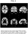
Figure 4 depicts the resulting slope and intercept images according to one embodiment of the present invention. -
Figure 5 shows template images of the present invention. -
Figure 6 depicts a system for performing the instant invention. - The present invention provides a spatial normalization method when using imaging agents. While a specific embodiment for scans with the [18F]-Flutemetamol amyloid imaging agent, which only depends on the Flutemetamol scan itself, are described in detail, the present invention is also applicable to other imaging agents used for other imaging applications. For example, the present invention may also be employed for DaTSCAN imaging where low uptake in the brain is indicative of disease.
- The uptake pattern in Flutemetamol scans can differ much between normal subjects (Aβ-) and subjects with high amyloid in gray matter (Aβ+). Therefore the spatial normalization method has been extended with an adaptive template mechanism, which during the image registration process alters the template image along with the spatial transformation parameters to make it more similar to the scan being registered. By using this extension, the method can accurately spatially normalize scans ranging over the whole amyloid scale from Aβ- to very high Aβ+.
- The present invention solves this by creating a model over how the typical uptake pattern in the whole image varies along the scale going from Aβ- to Aβ+. In the model presented hereinbelow, a one parameter linear regression model for each voxel in the image is used to express the dependence, but generally a more advanced model expressing uptake pattern dependence on multiple parameters, is also contemplated by the present invention. The model is built once for all in a separate pre-processing step and is then fixed as an internal part of the method. Although the method described below has been developed for Flutemetamol it should be noted that other PET amyloid tracers exhibit exactly the same characteristic. Moreover, the method described is general and there is nothing limiting the method of the present invention to PET amyloid scans only. As long as a model over the typical variation modes of the images being registered can be created, the adaptive template method has potential to be used for other imaging agents as well as for both PET and SPECT applications. While the examples provided hereinbelow are directed PET applications, the present invention also applies to SPECT applications.
- As noted hereinabove, the present invention may alter one or more of the parameters for the image, the image template, or both. For example, parameters for spatial transformation may include translation along each axis (providing three possible parameters), scaling along each axis (providing another three possible parameters), and rotation about each axis (providing an additional three parameters), for a total of up to nine parameters which may be altered by the present invention. Additionally, parameters which account for shape differences between the image and the adaptive template provide additional parameters which may be altered by the present invention. An illustration of typical Flutemetamol uptake patterns in an Aβ+ and Aβ- scan is shown in
Figure 1 . It can be noted that the Aβ+ have much more activity in cortical gray matter and even though white matter activity is about the same in the two cases the relative activity pattern between white and gray matter is to some extent reversed. That is from having highest activity in white matter in Aβ- to the opposite with highest, or at least as high, activity in gray matter in Aβ+. - Spatial normalization is the process of transforming a scan from patient space to a standard space thus allowing for comparison of data from different subjects. Typically this is performed through an iterative process where the patient scan is compared with a template image and where it is geometrically transformed to be as similar to the template as possible. However, by observing the patterns above it can be seen that there is no obvious way of selecting a similarity metric for a spatial normalization method which would be able to perform accurate and robust registrations of both of these types of images, by comparing them to a common template image.
- Therefore, the present invention provides an extension to be used together with a standard image registration method, where the number of possible template images is infinite. Basically, no definite selection of one specific template image occurs. Instead the intensities in the template are continuously altered during the registration, to make the template converge along with the spatial transformation parameters, to optimally fit the image being registered. An outline of the complete image registration procedure including the novel extension is shown in
Figure 2 . - First, the step of providing a
template model 100 is performed. Step 100 is a pre-processing step performed once during a design phase to prepare the adaptive template of the present invention. Separately, a moving image (ie, the patient image) 110, such as a PET or SPECT scan, is taken of a subject and the step of choosingstarting parameters 120 for the moving image is performed. Including withinstep 120 is the step of choosing a template control start parameter. In short the following steps are then iterated until convergence is reached. An adaptive template is then built 130 using the control parameter for the adaptive template and the model fromstep 100. A transformingstep 140 is performed in which the moving image is adjusted using the start parameters. Then, acomparison step 150 is performed which compares the similarity between the transformed moving image and the adaptive template. In this step, the similarity metric is evaluated from the voxel values in the template image and the transformed moving image. If the moving image and the adaptive template are found to be sufficiently or maximally converged, the registration of the moving image is completed,step 160. Should the moving image and the adaptive template not be sufficiently or maximally converged, the step of choosingnew parameters 170, for both the moving image and the template control parameter, is performed. Then transformingstep 140 is repeated, this time using the parameters chosen duringstep 170. Additionally,step 130 is repeated in which the existing adaptive template is also modified by the new template control parameter chosen duringstep 170. Thecomparison step 150 is again performed using the newly transformed moving image and adaptive template. If the method has not converged, another set of adaptive template and transformation parameters is selected by the optimization method and a new evaluation of the similarity metric is made. The present invention contemplates that the optimization method may be any numerical optimization method. - Convergence may be sufficiently achieved when the value of a function for describing the similarity between the images has reached a level deemed suitable for proceeding. Convergence is desirably achieved when the value of a function for describing the similarity between the images has reached its maximum, ie, showing maximum similarity between the template image in reference space and the transformed image. The similarity metric could be based, for example, on correlation or on mutual information, although the present invention contemplates that any suitable similarity metric may be applied.
- The
pre-processing step 100 is performed once and the extra parameter or parameters controlling the template model are added to the registration parameters and altered by the optimization method in a similar way as the parameters controlling the geometry change. The following will now in detail describe the creation of a one-parameter linear template model, which has been found to be suitable for Flutemetamol PET scans. - Provided herein but not forming part of the claimed invention is a non-transitory computer-readable storage medium with an executable program for performing the method of the present invention. This computer-readable storage medium includes computer-readable program code includes instructions for performing a method of the present invention, such that execution of the computer-readable program code causes a processor to perform the steps of the method of the present invention. Additionally, the present invention is contemplated to use an adaptive template model of the present invention for performing the spatial normalization method of the present invention. The starting parameters may be fixed for all procedures and the updated parameters may be selected automatically by the process.
- In the following example, Montreal Neurological Institute (MNI) reference space sampled to a uniform 2x2x2 mm voxel size is used as standard space for the template image. A set of Flutemetamol input images is provided to create a template of the present invention, which images have already have been spatially normalized to this space using an MR-based spatial normalization method.
- Normally when creating a template image for spatial normalization one takes these spatially-normalized input images and calculates a mean image from these. It follows that this mean image will also be located in the reference space and can be used for spatial normalization of new unseen images by using an image registration method to find the transformation making the new images match the template mean image.
- However, as described hereinabove, the problem with this approach is that the mean image will only be representative for one typical image pattern for a specific level of amyloid, i.e. either the pattern of a Aβ-, Aβ+, or something in between. To be able to create a model of the image pattern variability the present invention instead calculates a regression model for each voxel in the template image, where the dependent variable which is the voxel intensities in the input images is regressed on some variable which ideally for each input image expresses a "true" value of where on a scale from Aβ- to very severe Aβ+ it is located.
- In practice, it is impossible to extract such true values but, being principally interested in finding a robust estimated value for each scan on a one-dimensional scale of how much Aβ the gray matter contains, this approach provides such a robust estimate. Therefore the present invention calculates for all input images a mean standard uptake ratio value (SUVR) inside a large composite region (COM) covering regions known to be most affected by Aβ in Alzheimer's disease patients. These regions were the prefrontal cortex, parietal cortex, precuneus, lateral temporal cortex, and the anterior and posterior cingulate cortices.
- An illustration of the calculated SUVR values of the composite region COMSUVR for all subjects in two different groups of Aβ- and Aβ+ scans is shown in
Figure 3 . - It is thus seen that the present invention uses a good sampling over the whole spectrum from Aβ- subjects with very low Flutemetamol uptake inside COM to Aβ+ subjects with a very high Flutemetamol uptake.
- In the next step, the linear regression model was applied for each voxel i in the template image matrix to express the dependence of that particular voxel, yi, on the COMSUVR values according to Equation 1:

- Applying this model over all input images regressed on the corresponding COMSUVR values provides an intercept image and a slope image according to Equation 2:

- The intercept image will correspond to a pattern of a fully normal subject located in the lower part of the COMSUVR scale. The slope image, on the other hand, will have the highest values for the parts of the image where the largest changes occur when going from low to high on the COMSUVR scale. These resulting images are shown in
Figure 4 . - From these images the present invention creates synthetic simulated images, along the whole COMSUVR scale over the range from Aβ- to Aβ+. This is done by adding multiplicatively scaled portions of ISlope to I0 according to Equation 3:

Figure 5 shows such synthetic images covering values of x from 0.0 to 1.0 in steps of 0.2. - Such synthetic images are used as the adaptive template in a spatial normalization method. The slope image, ISlope , and intercept image, I0 , is the adaptive template model is controlled by one single parameter x. This parameter x is then adjusted by the image registration algorithm in a similar way as any of the other parameters, and the template image is updated accordingly any time the x parameter is changed.
- Referring now to
Figure 6 , the method of the present invention is contemplated to be performed bysystem 200.System 200 includes ascanner 210 for performing PET or SPECT scans, acomputer 220 for receiving scan images, e.g, the patient image (aka, the moving image), and adatabase 230 for providing the adaptive template.Computer 220 typically includes adisplay 222, aninput device 224 such as akeyboard 224a and amouse 224b, and aprocessor 226.Processor 226 typically includes software for performing the method of the instant invention using the adaptive template fromdatabase 230 and the moving image fromscanner 210. Additionally,processor 226 is contemplated to include non-transitory computer readable storage medium with an executable program for using an adaptive template of the present invention and for registering a PET image to an adaptive template image according to a method of the instant invention using the moving image fromscanner 210. The non-transitory computer readable storage medium includes computer-readable program code including instructions for using an adaptive template of the present invention and for registering a PET image to an adaptive template image. Connections betweenscanner 210,computer 220, anddatabase 230 are contemplated to be by any means known to the art, such as hardwire, wireless, or any combination thereof. Additionally, the present invention contemplates thatprocessor 226 anddatabase 230 are connected such thatprocessor 226 may return the upgraded adaptive template todatabase 230 upon completion of the method of the present invention.
Claims (13)
- An adaptive template image to be registered to a PET or a SPECT image during a standard image registration method, comprising:a template image model wherein the values for each voxel in the adaptive template image vary according to one or more control parameters, and the adaptive template image is altered during the image registration process;wherein the variability of values for each voxel ranges from a value corresponding to the normal level of uptake of an imaging agent to an abnormal level of uptake of the imaging agent; andwherein the value of the voxels are defined by the equation:
 wherein Ii template is the value of the voxel i in the adaptive template image, Ii 0 represents a value corresponding to a normal level of uptake of an imaging agent within that voxel, Ii slope corresponds to the maximum additional amount of uptake of the imaging agent corresponding to an abnormal (high) level of uptake of the imaging agent in that voxel with respect to a control parameter x.
 - An adaptive template image of claim 1, wherein the level of uptake is measured in the gray matter of a brain and the imaging agent comprises an amyloid imaging agent.
- An adaptive template image of claim 1, wherein the variability of values for each voxel ranges from a value corresponding to a normal level of amyloid in grey matter to a raised amyloid level in grey matter.
- An adaptive template of claim 1, wherein the imaging agent comprises a dopamine transport agent and the level of uptake is determined for the striatum.
- An adaptive template image of claim 1, wherein the control parameters are selectable so as to alter the value of voxels in the adaptive image template.
- An adaptive template image of claim 1, wherein I0 and Islope are determined using regression analysis of multiple images spanning different levels of disease state.
- A method of registering a PET image to an adaptive template image of claim 1 during a standard image registration method, comprising the steps of:Spatially normalizing an individual PET image to an adaptive template image located in the reference space;Comparing the spatially normalized individual PET image to the adaptive template image to determine whether both are sufficiently converged; andAltering the parameter controlling the adaptive template image wherein the values for each voxel in the adaptive template image vary and altering the parameters controlling the spatial transformation for the individual PET image, so as to render an altered template image and an updated spatially transformed individual PET image during the image registration process in the event that the comparing step does not determine that the spatially normalized individual PET image is sufficiently converged to the adaptive template image.
- A method of claim 7, wherein the altering step further comprises determining whether the spatially-normalized individual PET image is maximally converged to the adaptive template image.
- A method of claim 7, wherein said spatial normalization step includes the steps of:
Performing linear and non-linear transformations on the individual PET image to find a match between the adaptive template image and the individual PET image, and wherein said linear transformation includes one or more of the group consisting of translation, rotation, and scaling. - A method of constructing an adaptive template image of claim 1 for registering a PET image during a standard image registration method, comprising the steps of:Calculating a mean image from a set of input PET images showing uptake of an imaging agent in a region of interest, the set of input PET images having been transformed into a reference space; andCalculating a regression model for each voxel in the set of input PET images having been transformed into said reference space, where the dependent variable represents the voxel intensities in the input PET images and is regressed on some variable which for each input image represents a "true" value of where on a scale from a normal level of uptake to a high level of uptake of the imaging agent the voxel is located,wherein the intensities in the adaptive template image are continuously altered during the image registration process to make the adaptive template image converge to optimally fit the PET image being registered.
- A method of claim 10, wherein the normal level of uptake corresponds to Aβ- and the high level of uptake corresponds to Aβ+ and the imaging agent is an amyloid imaging agent.
- A positron emission tomography (PET) system comprising:
an image processor for registering a PET image to an adaptive template image of claim 1 during a standard image registration method, the image processor programmed to:Spatially normalize an individual PET image to said
adaptive template image located in the reference space;Compare the spatially normalized individual PET image to the adaptive template image to determine whether both are sufficiently converged; andAlter the parameter controlling the template image wherein values for each voxel in the adaptive template image vary and alter the parameters controlling the spatialtransformation for the individual PET image so as to render an altered template image andan updated spatially transformed individual PET image during the image registration process in the event that the comparing step does not determine that the spatially normalized individual PET image is sufficiently converged to the adaptive template image. - A positron emission tomography (PET) system as claimed in claim 12, further comprising:a storage device; anda detector for detecting positron emissions from a brain of a subject, wherein the detector generates signals representing the positron emissions that are stored in the storage device.
Applications Claiming Priority (2)
| Application Number | Priority Date | Filing Date | Title |
|---|---|---|---|
| US201161536702P | 2011-09-20 | 2011-09-20 | |
| PCT/US2012/056182 WO2013043775A1 (en) | 2011-09-20 | 2012-09-20 | Methods of spatial normalization of positron emission tomography images |
Publications (2)
| Publication Number | Publication Date |
|---|---|
| EP2757955A1 EP2757955A1 (en) | 2014-07-30 |
| EP2757955B1 true EP2757955B1 (en) | 2022-06-15 |
Family
ID=46964089
Family Applications (1)
| Application Number | Title | Priority Date | Filing Date |
|---|---|---|---|
| EP12766824.2A Active EP2757955B1 (en) | 2011-09-20 | 2012-09-20 | Methods of spatial normalization of positron emission tomography images |
Country Status (8)
| Country | Link |
|---|---|
| US (3) | US10614547B2 (en) |
| EP (1) | EP2757955B1 (en) |
| JP (1) | JP6106675B2 (en) |
| CN (1) | CN103930032B (en) |
| AU (1) | AU2012312482B2 (en) |
| CA (1) | CA2849119C (en) |
| ES (1) | ES2922280T3 (en) |
| WO (1) | WO2013043775A1 (en) |
Families Citing this family (15)
| Publication number | Priority date | Publication date | Assignee | Title |
|---|---|---|---|---|
| AU2012312482B2 (en) | 2011-09-20 | 2017-06-01 | Ge Healthcare Limited | Methods of spatial normalization of positron emission tomography images |
| WO2013134559A1 (en) | 2012-03-07 | 2013-09-12 | Speir Technologies Inc. | Methods and systems for tracking and guiding sensors and instruments |
| JP6697438B2 (en) * | 2014-07-15 | 2020-05-20 | コーニンクレッカ フィリップス エヌ ヴェKoninklijke Philips N.V. | Imaging data statistical test including localization regularization with individualized template images |
| US10617401B2 (en) | 2014-11-14 | 2020-04-14 | Ziteo, Inc. | Systems for localization of targets inside a body |
| CN107407629B (en) * | 2015-03-05 | 2020-09-01 | 生物辐射实验室股份有限公司 | Optimized spectral matching and display |
| TWI587841B (en) * | 2015-03-06 | 2017-06-21 | 國立陽明大學 | System and method of quantitative analysis nuclear medicine brain imaging |
| WO2019149529A1 (en) * | 2018-02-02 | 2019-08-08 | Koninklijke Philips N.V. | Correcting standardized uptake values in pre-treatment and post-treatment positron emission tomography studies |
| KR102219890B1 (en) * | 2018-02-28 | 2021-02-24 | 서울대학교산학협력단 | Apparatus for spatial normalization of medical image using deep learning and method thereof |
| WO2019168310A1 (en) * | 2018-02-28 | 2019-09-06 | 서울대학교산학협력단 | Device for spatial normalization of medical image using deep learning and method therefor |
| IL287027B2 (en) * | 2019-04-09 | 2025-06-01 | Ziteo Inc | Methods and systems for high-throughput and versatile molecular imaging |
| KR102261111B1 (en) * | 2019-09-10 | 2021-06-04 | 인하대학교 산학협력단 | Generalization of intensity distribution of medical images using gans |
| JP2022021076A (en) * | 2020-07-21 | 2022-02-02 | 浜松ホトニクス株式会社 | Brain image diagnostic system and brain image diagnostic method |
| CN113450314A (en) * | 2021-06-04 | 2021-09-28 | 迈格生命科技(深圳)有限公司 | Image processing method, device, storage medium and product |
| US12181455B2 (en) | 2021-07-28 | 2024-12-31 | John Wiley & Sons, Inc. | Adaptive search mass spectrometer spectral analysis |
| KR102603177B1 (en) * | 2022-06-03 | 2023-11-17 | 주식회사 브라이토닉스이미징 | System for spatial normalization of image, quantification using spatial normalization and method thereof |
Family Cites Families (7)
| Publication number | Priority date | Publication date | Assignee | Title |
|---|---|---|---|---|
| US5948384A (en) * | 1990-09-14 | 1999-09-07 | Syngenix Limited | Particulate agents |
| JP4581088B2 (en) | 2005-05-17 | 2010-11-17 | 国立大学法人 筑波大学 | Computer-aided diagnosis apparatus and method |
| DE102006033383A1 (en) * | 2006-07-12 | 2008-01-17 | Eberhard-Karls-Universität Tübingen Universitätsklinikum | Method for determining a property map for an object, in particular for a living being, based on at least a first image, in particular nuclear magnetic resonance image |
| ES2414614T3 (en) * | 2007-01-30 | 2013-07-22 | Ge Healthcare Limited | Tools to help diagnose neurodegenerative diseases |
| JP2010540512A (en) * | 2007-09-26 | 2010-12-24 | コーニンクレッカ フィリップス エレクトロニクス エヌ ヴィ | Methods for specifically diagnosing different types of dementia |
| WO2009146388A1 (en) * | 2008-05-28 | 2009-12-03 | The Trustees Of Columbia University In The City Of New York | Voxel-based methods for assessing subjects using positron emission tomography |
| AU2012312482B2 (en) | 2011-09-20 | 2017-06-01 | Ge Healthcare Limited | Methods of spatial normalization of positron emission tomography images |
-
2012
- 2012-09-20 AU AU2012312482A patent/AU2012312482B2/en active Active
- 2012-09-20 US US14/344,934 patent/US10614547B2/en active Active
- 2012-09-20 JP JP2014530963A patent/JP6106675B2/en active Active
- 2012-09-20 EP EP12766824.2A patent/EP2757955B1/en active Active
- 2012-09-20 CA CA2849119A patent/CA2849119C/en active Active
- 2012-09-20 WO PCT/US2012/056182 patent/WO2013043775A1/en not_active Ceased
- 2012-09-20 CN CN201280056787.2A patent/CN103930032B/en active Active
- 2012-09-20 ES ES12766824T patent/ES2922280T3/en active Active
-
2020
- 2020-04-06 US US16/841,329 patent/US11488282B2/en active Active
-
2022
- 2022-09-27 US US17/954,034 patent/US20230026994A1/en not_active Abandoned
Also Published As
| Publication number | Publication date |
|---|---|
| AU2012312482B2 (en) | 2017-06-01 |
| US10614547B2 (en) | 2020-04-07 |
| CN103930032B (en) | 2018-10-12 |
| US20230026994A1 (en) | 2023-01-26 |
| JP2014530352A (en) | 2014-11-17 |
| CA2849119A1 (en) | 2013-03-28 |
| ES2922280T3 (en) | 2022-09-12 |
| US20140350392A1 (en) | 2014-11-27 |
| CN103930032A (en) | 2014-07-16 |
| EP2757955A1 (en) | 2014-07-30 |
| US20200234400A1 (en) | 2020-07-23 |
| WO2013043775A1 (en) | 2013-03-28 |
| JP6106675B2 (en) | 2017-04-05 |
| US11488282B2 (en) | 2022-11-01 |
| CA2849119C (en) | 2022-06-07 |
| AU2012312482A1 (en) | 2014-04-17 |
Similar Documents
| Publication | Publication Date | Title |
|---|---|---|
| US20230026994A1 (en) | Methods of spatial normalization of positron emission tomography images | |
| JP2014530352A5 (en) | ||
| CN107038728B (en) | Contour automated determination based on iterative reconstruction | |
| JP6567179B2 (en) | Pseudo CT generation from MR data using feature regression model | |
| JP7030050B2 (en) | Pseudo-CT generation from MR data using tissue parameter estimation | |
| US9865048B2 (en) | Radiotherapy information generation apparatus and radiotherapy information generation method | |
| CN101171608B (en) | Virtual lesion quantification | |
| EP2369551A1 (en) | Imaging system and method | |
| CN107865658B (en) | Method and apparatus for correcting synthetic electron density maps | |
| Pevsner et al. | Evaluation of an automated deformable image matching method for quantifying lung motion in respiration‐correlated CT images | |
| CN105989621B (en) | Method and system for performing joint estimation techniques in image reconstruction | |
| Park et al. | Deformable registration of CT and cone-beam CT with local intensity matching | |
| Wang et al. | Automated segmentation of CBCT image using spiral CT atlases and convex optimization | |
| JP2014532177A (en) | Variable depth stereotactic surface projection | |
| EP2742483B1 (en) | Image processing method | |
| Park et al. | MRI-based computed tomography metal artifact correction method for improving proton range calculation accuracy | |
| Schönmeyer et al. | Automated segmentation of lateral ventricles from human and primate magnetic resonance images using cognition network technology | |
| DE102016215105A1 (en) | Method and data processing unit for determining an acquisition data processing algorithm | |
| EP3170145B1 (en) | Imaging data statistical testing including a stereotactical normalization with a personalized template image | |
| CN117173065B (en) | Anatomical medical image fusion method based on texture awareness and pixel intensity correlation | |
| Gao et al. | 3D CBCT reconstruction from two orthogonal x‑ray projections via a geometry‑constrained diffusion model for image‑guided head‑and‑neck radiotherapy | |
| JP2005164334A (en) | Nuclear medicine diagnostic equipment | |
| JP6035167B2 (en) | Medical image processing program, medical image processing method, and medical image processing apparatus | |
| Li | Multimodal intra-and inter-subject nonrigid registration of small animal images. | |
| WO2010112442A1 (en) | Method and device for reducing position-related gray value variations by means of a registration of image data sets |
Legal Events
| Date | Code | Title | Description |
|---|---|---|---|
| PUAI | Public reference made under article 153(3) epc to a published international application that has entered the european phase |
Free format text: ORIGINAL CODE: 0009012 |
|
| 17P | Request for examination filed |
Effective date: 20140314 |
|
| AK | Designated contracting states |
Kind code of ref document: A1 Designated state(s): AL AT BE BG CH CY CZ DE DK EE ES FI FR GB GR HR HU IE IS IT LI LT LU LV MC MK MT NL NO PL PT RO RS SE SI SK SM TR |
|
| DAX | Request for extension of the european patent (deleted) | ||
| STAA | Information on the status of an ep patent application or granted ep patent |
Free format text: STATUS: EXAMINATION IS IN PROGRESS |
|
| 17Q | First examination report despatched |
Effective date: 20171106 |
|
| RAP3 | Party data changed (applicant data changed or rights of an application transferred) |
Owner name: GE HEALTHCARE LIMITED |
|
| GRAP | Despatch of communication of intention to grant a patent |
Free format text: ORIGINAL CODE: EPIDOSNIGR1 |
|
| STAA | Information on the status of an ep patent application or granted ep patent |
Free format text: STATUS: GRANT OF PATENT IS INTENDED |
|
| INTG | Intention to grant announced |
Effective date: 20220111 |
|
| GRAS | Grant fee paid |
Free format text: ORIGINAL CODE: EPIDOSNIGR3 |
|
| GRAA | (expected) grant |
Free format text: ORIGINAL CODE: 0009210 |
|
| STAA | Information on the status of an ep patent application or granted ep patent |
Free format text: STATUS: THE PATENT HAS BEEN GRANTED |
|
| AK | Designated contracting states |
Kind code of ref document: B1 Designated state(s): AL AT BE BG CH CY CZ DE DK EE ES FI FR GB GR HR HU IE IS IT LI LT LU LV MC MK MT NL NO PL PT RO RS SE SI SK SM TR |
|
| REG | Reference to a national code |
Ref country code: CH Ref legal event code: EP Ref country code: GB Ref legal event code: FG4D |
|
| REG | Reference to a national code |
Ref country code: IE Ref legal event code: FG4D |
|
| REG | Reference to a national code |
Ref country code: DE Ref legal event code: R096 Ref document number: 602012078366 Country of ref document: DE |
|
| REG | Reference to a national code |
Ref country code: AT Ref legal event code: REF Ref document number: 1497893 Country of ref document: AT Kind code of ref document: T Effective date: 20220715 |
|
| REG | Reference to a national code |
Ref country code: ES Ref legal event code: FG2A Ref document number: 2922280 Country of ref document: ES Kind code of ref document: T3 Effective date: 20220912 |
|
| REG | Reference to a national code |
Ref country code: LT Ref legal event code: MG9D |
|
| REG | Reference to a national code |
Ref country code: NL Ref legal event code: MP Effective date: 20220615 |
|
| PG25 | Lapsed in a contracting state [announced via postgrant information from national office to epo] |
Ref country code: SE Free format text: LAPSE BECAUSE OF FAILURE TO SUBMIT A TRANSLATION OF THE DESCRIPTION OR TO PAY THE FEE WITHIN THE PRESCRIBED TIME-LIMIT Effective date: 20220615 Ref country code: NO Free format text: LAPSE BECAUSE OF FAILURE TO SUBMIT A TRANSLATION OF THE DESCRIPTION OR TO PAY THE FEE WITHIN THE PRESCRIBED TIME-LIMIT Effective date: 20220915 Ref country code: LT Free format text: LAPSE BECAUSE OF FAILURE TO SUBMIT A TRANSLATION OF THE DESCRIPTION OR TO PAY THE FEE WITHIN THE PRESCRIBED TIME-LIMIT Effective date: 20220615 Ref country code: HR Free format text: LAPSE BECAUSE OF FAILURE TO SUBMIT A TRANSLATION OF THE DESCRIPTION OR TO PAY THE FEE WITHIN THE PRESCRIBED TIME-LIMIT Effective date: 20220615 Ref country code: GR Free format text: LAPSE BECAUSE OF FAILURE TO SUBMIT A TRANSLATION OF THE DESCRIPTION OR TO PAY THE FEE WITHIN THE PRESCRIBED TIME-LIMIT Effective date: 20220916 Ref country code: FI Free format text: LAPSE BECAUSE OF FAILURE TO SUBMIT A TRANSLATION OF THE DESCRIPTION OR TO PAY THE FEE WITHIN THE PRESCRIBED TIME-LIMIT Effective date: 20220615 Ref country code: BG Free format text: LAPSE BECAUSE OF FAILURE TO SUBMIT A TRANSLATION OF THE DESCRIPTION OR TO PAY THE FEE WITHIN THE PRESCRIBED TIME-LIMIT Effective date: 20220915 |
|
| REG | Reference to a national code |
Ref country code: AT Ref legal event code: MK05 Ref document number: 1497893 Country of ref document: AT Kind code of ref document: T Effective date: 20220615 |
|
| PG25 | Lapsed in a contracting state [announced via postgrant information from national office to epo] |
Ref country code: RS Free format text: LAPSE BECAUSE OF FAILURE TO SUBMIT A TRANSLATION OF THE DESCRIPTION OR TO PAY THE FEE WITHIN THE PRESCRIBED TIME-LIMIT Effective date: 20220615 Ref country code: LV Free format text: LAPSE BECAUSE OF FAILURE TO SUBMIT A TRANSLATION OF THE DESCRIPTION OR TO PAY THE FEE WITHIN THE PRESCRIBED TIME-LIMIT Effective date: 20220615 |
|
| PG25 | Lapsed in a contracting state [announced via postgrant information from national office to epo] |
Ref country code: NL Free format text: LAPSE BECAUSE OF FAILURE TO SUBMIT A TRANSLATION OF THE DESCRIPTION OR TO PAY THE FEE WITHIN THE PRESCRIBED TIME-LIMIT Effective date: 20220615 |
|
| PG25 | Lapsed in a contracting state [announced via postgrant information from national office to epo] |
Ref country code: SM Free format text: LAPSE BECAUSE OF FAILURE TO SUBMIT A TRANSLATION OF THE DESCRIPTION OR TO PAY THE FEE WITHIN THE PRESCRIBED TIME-LIMIT Effective date: 20220615 Ref country code: SK Free format text: LAPSE BECAUSE OF FAILURE TO SUBMIT A TRANSLATION OF THE DESCRIPTION OR TO PAY THE FEE WITHIN THE PRESCRIBED TIME-LIMIT Effective date: 20220615 Ref country code: RO Free format text: LAPSE BECAUSE OF FAILURE TO SUBMIT A TRANSLATION OF THE DESCRIPTION OR TO PAY THE FEE WITHIN THE PRESCRIBED TIME-LIMIT Effective date: 20220615 Ref country code: PT Free format text: LAPSE BECAUSE OF FAILURE TO SUBMIT A TRANSLATION OF THE DESCRIPTION OR TO PAY THE FEE WITHIN THE PRESCRIBED TIME-LIMIT Effective date: 20221017 Ref country code: EE Free format text: LAPSE BECAUSE OF FAILURE TO SUBMIT A TRANSLATION OF THE DESCRIPTION OR TO PAY THE FEE WITHIN THE PRESCRIBED TIME-LIMIT Effective date: 20220615 Ref country code: CZ Free format text: LAPSE BECAUSE OF FAILURE TO SUBMIT A TRANSLATION OF THE DESCRIPTION OR TO PAY THE FEE WITHIN THE PRESCRIBED TIME-LIMIT Effective date: 20220615 Ref country code: AT Free format text: LAPSE BECAUSE OF FAILURE TO SUBMIT A TRANSLATION OF THE DESCRIPTION OR TO PAY THE FEE WITHIN THE PRESCRIBED TIME-LIMIT Effective date: 20220615 |
|
| PG25 | Lapsed in a contracting state [announced via postgrant information from national office to epo] |
Ref country code: PL Free format text: LAPSE BECAUSE OF FAILURE TO SUBMIT A TRANSLATION OF THE DESCRIPTION OR TO PAY THE FEE WITHIN THE PRESCRIBED TIME-LIMIT Effective date: 20220615 Ref country code: IS Free format text: LAPSE BECAUSE OF FAILURE TO SUBMIT A TRANSLATION OF THE DESCRIPTION OR TO PAY THE FEE WITHIN THE PRESCRIBED TIME-LIMIT Effective date: 20221015 |
|
| REG | Reference to a national code |
Ref country code: DE Ref legal event code: R097 Ref document number: 602012078366 Country of ref document: DE |
|
| PG25 | Lapsed in a contracting state [announced via postgrant information from national office to epo] |
Ref country code: AL Free format text: LAPSE BECAUSE OF FAILURE TO SUBMIT A TRANSLATION OF THE DESCRIPTION OR TO PAY THE FEE WITHIN THE PRESCRIBED TIME-LIMIT Effective date: 20220615 |
|
| PLBE | No opposition filed within time limit |
Free format text: ORIGINAL CODE: 0009261 |
|
| STAA | Information on the status of an ep patent application or granted ep patent |
Free format text: STATUS: NO OPPOSITION FILED WITHIN TIME LIMIT |
|
| PG25 | Lapsed in a contracting state [announced via postgrant information from national office to epo] |
Ref country code: MC Free format text: LAPSE BECAUSE OF FAILURE TO SUBMIT A TRANSLATION OF THE DESCRIPTION OR TO PAY THE FEE WITHIN THE PRESCRIBED TIME-LIMIT Effective date: 20220615 Ref country code: DK Free format text: LAPSE BECAUSE OF FAILURE TO SUBMIT A TRANSLATION OF THE DESCRIPTION OR TO PAY THE FEE WITHIN THE PRESCRIBED TIME-LIMIT Effective date: 20220615 |
|
| 26N | No opposition filed |
Effective date: 20230316 |
|
| REG | Reference to a national code |
Ref country code: BE Ref legal event code: MM Effective date: 20220930 |
|
| PG25 | Lapsed in a contracting state [announced via postgrant information from national office to epo] |
Ref country code: SI Free format text: LAPSE BECAUSE OF FAILURE TO SUBMIT A TRANSLATION OF THE DESCRIPTION OR TO PAY THE FEE WITHIN THE PRESCRIBED TIME-LIMIT Effective date: 20220615 |
|
| PG25 | Lapsed in a contracting state [announced via postgrant information from national office to epo] |
Ref country code: LU Free format text: LAPSE BECAUSE OF NON-PAYMENT OF DUE FEES Effective date: 20220920 |
|
| P01 | Opt-out of the competence of the unified patent court (upc) registered |
Effective date: 20230529 |
|
| PG25 | Lapsed in a contracting state [announced via postgrant information from national office to epo] |
Ref country code: IE Free format text: LAPSE BECAUSE OF NON-PAYMENT OF DUE FEES Effective date: 20220920 |
|
| PG25 | Lapsed in a contracting state [announced via postgrant information from national office to epo] |
Ref country code: BE Free format text: LAPSE BECAUSE OF NON-PAYMENT OF DUE FEES Effective date: 20220930 |
|
| PG25 | Lapsed in a contracting state [announced via postgrant information from national office to epo] |
Ref country code: HU Free format text: LAPSE BECAUSE OF FAILURE TO SUBMIT A TRANSLATION OF THE DESCRIPTION OR TO PAY THE FEE WITHIN THE PRESCRIBED TIME-LIMIT; INVALID AB INITIO Effective date: 20120920 |
|
| PG25 | Lapsed in a contracting state [announced via postgrant information from national office to epo] |
Ref country code: CY Free format text: LAPSE BECAUSE OF FAILURE TO SUBMIT A TRANSLATION OF THE DESCRIPTION OR TO PAY THE FEE WITHIN THE PRESCRIBED TIME-LIMIT Effective date: 20220615 |
|
| PG25 | Lapsed in a contracting state [announced via postgrant information from national office to epo] |
Ref country code: MK Free format text: LAPSE BECAUSE OF FAILURE TO SUBMIT A TRANSLATION OF THE DESCRIPTION OR TO PAY THE FEE WITHIN THE PRESCRIBED TIME-LIMIT Effective date: 20220615 |
|
| PG25 | Lapsed in a contracting state [announced via postgrant information from national office to epo] |
Ref country code: MT Free format text: LAPSE BECAUSE OF FAILURE TO SUBMIT A TRANSLATION OF THE DESCRIPTION OR TO PAY THE FEE WITHIN THE PRESCRIBED TIME-LIMIT Effective date: 20220615 |
|
| PG25 | Lapsed in a contracting state [announced via postgrant information from national office to epo] |
Ref country code: BG Free format text: LAPSE BECAUSE OF FAILURE TO SUBMIT A TRANSLATION OF THE DESCRIPTION OR TO PAY THE FEE WITHIN THE PRESCRIBED TIME-LIMIT Effective date: 20220615 |
|
| PG25 | Lapsed in a contracting state [announced via postgrant information from national office to epo] |
Ref country code: BG Free format text: LAPSE BECAUSE OF FAILURE TO SUBMIT A TRANSLATION OF THE DESCRIPTION OR TO PAY THE FEE WITHIN THE PRESCRIBED TIME-LIMIT Effective date: 20220615 |
|
| REG | Reference to a national code |
Ref country code: CH Ref legal event code: U11 Free format text: ST27 STATUS EVENT CODE: U-0-0-U10-U11 (AS PROVIDED BY THE NATIONAL OFFICE) Effective date: 20251001 |
|
| PGFP | Annual fee paid to national office [announced via postgrant information from national office to epo] |
Ref country code: DE Payment date: 20250820 Year of fee payment: 14 |
|
| PGFP | Annual fee paid to national office [announced via postgrant information from national office to epo] |
Ref country code: IT Payment date: 20250820 Year of fee payment: 14 |
|
| PGFP | Annual fee paid to national office [announced via postgrant information from national office to epo] |
Ref country code: GB Payment date: 20250822 Year of fee payment: 14 |
|
| PGFP | Annual fee paid to national office [announced via postgrant information from national office to epo] |
Ref country code: FR Payment date: 20250820 Year of fee payment: 14 |
|
| PG25 | Lapsed in a contracting state [announced via postgrant information from national office to epo] |
Ref country code: TR Free format text: LAPSE BECAUSE OF FAILURE TO SUBMIT A TRANSLATION OF THE DESCRIPTION OR TO PAY THE FEE WITHIN THE PRESCRIBED TIME-LIMIT Effective date: 20220615 |
|
| PGFP | Annual fee paid to national office [announced via postgrant information from national office to epo] |
Ref country code: CH Payment date: 20251001 Year of fee payment: 14 |
|
| PGFP | Annual fee paid to national office [announced via postgrant information from national office to epo] |
Ref country code: ES Payment date: 20251001 Year of fee payment: 14 |