EP2754718B1 - Vascular endothelial myostatin mutant that mutates at atp binding sites - Google Patents
Vascular endothelial myostatin mutant that mutates at atp binding sites Download PDFInfo
- Publication number
- EP2754718B1 EP2754718B1 EP12830317.9A EP12830317A EP2754718B1 EP 2754718 B1 EP2754718 B1 EP 2754718B1 EP 12830317 A EP12830317 A EP 12830317A EP 2754718 B1 EP2754718 B1 EP 2754718B1
- Authority
- EP
- European Patent Office
- Prior art keywords
- mutant
- seq
- endostatin
- amino acid
- activity
- Prior art date
- Legal status (The legal status is an assumption and is not a legal conclusion. Google has not performed a legal analysis and makes no representation as to the accuracy of the status listed.)
- Active
Links
- 230000003511 endothelial effect Effects 0.000 title description 2
- 230000002792 vascular Effects 0.000 title description 2
- 102000004472 Myostatin Human genes 0.000 title 1
- 108010056852 Myostatin Proteins 0.000 title 1
- 102100031162 Collagen alpha-1(XVIII) chain Human genes 0.000 claims description 315
- 108010079505 Endostatins Proteins 0.000 claims description 315
- 230000000694 effects Effects 0.000 claims description 155
- 108091006112 ATPases Proteins 0.000 claims description 116
- 102000057290 Adenosine Triphosphatases Human genes 0.000 claims description 116
- 150000001413 amino acids Chemical class 0.000 claims description 56
- 238000000034 method Methods 0.000 claims description 53
- 230000002401 inhibitory effect Effects 0.000 claims description 47
- 206010028980 Neoplasm Diseases 0.000 claims description 44
- 230000035772 mutation Effects 0.000 claims description 39
- 125000000539 amino acid group Chemical group 0.000 claims description 33
- 230000010595 endothelial cell migration Effects 0.000 claims description 33
- 229920001223 polyethylene glycol Polymers 0.000 claims description 30
- 230000004071 biological effect Effects 0.000 claims description 23
- -1 Poly(ethylene glycol) Polymers 0.000 claims description 19
- 229910019142 PO4 Inorganic materials 0.000 claims description 12
- 238000012217 deletion Methods 0.000 claims description 12
- 230000037430 deletion Effects 0.000 claims description 12
- 230000004048 modification Effects 0.000 claims description 12
- 238000012986 modification Methods 0.000 claims description 12
- NBIIXXVUZAFLBC-UHFFFAOYSA-K phosphate Chemical compound [O-]P([O-])([O-])=O NBIIXXVUZAFLBC-UHFFFAOYSA-K 0.000 claims description 11
- 239000010452 phosphate Substances 0.000 claims description 11
- 230000001965 increasing effect Effects 0.000 claims description 10
- 239000008194 pharmaceutical composition Substances 0.000 claims description 10
- FDZZZRQASAIRJF-UHFFFAOYSA-M malachite green Chemical compound [Cl-].C1=CC(N(C)C)=CC=C1C(C=1C=CC=CC=1)=C1C=CC(=[N+](C)C)C=C1 FDZZZRQASAIRJF-UHFFFAOYSA-M 0.000 claims description 8
- 229940107698 malachite green Drugs 0.000 claims description 8
- 230000003247 decreasing effect Effects 0.000 claims description 7
- 230000003527 anti-angiogenesis Effects 0.000 claims description 5
- 238000003556 assay Methods 0.000 claims description 5
- 230000007423 decrease Effects 0.000 claims description 5
- 125000003277 amino group Chemical group 0.000 claims description 4
- 238000010353 genetic engineering Methods 0.000 claims description 3
- 230000036961 partial effect Effects 0.000 claims description 3
- 238000000338 in vitro Methods 0.000 claims 2
- ZKHQWZAMYRWXGA-UHFFFAOYSA-N Adenosine triphosphate Natural products C1=NC=2C(N)=NC=NC=2N1C1OC(COP(O)(=O)OP(O)(=O)OP(O)(O)=O)C(O)C1O ZKHQWZAMYRWXGA-UHFFFAOYSA-N 0.000 description 98
- ZKHQWZAMYRWXGA-KQYNXXCUSA-J ATP(4-) Chemical compound C1=NC=2C(N)=NC=NC=2N1[C@@H]1O[C@H](COP([O-])(=O)OP([O-])(=O)OP([O-])([O-])=O)[C@@H](O)[C@H]1O ZKHQWZAMYRWXGA-KQYNXXCUSA-J 0.000 description 97
- 241000282414 Homo sapiens Species 0.000 description 81
- 241000588724 Escherichia coli Species 0.000 description 35
- 238000003259 recombinant expression Methods 0.000 description 33
- 210000004027 cell Anatomy 0.000 description 28
- 108090000623 proteins and genes Proteins 0.000 description 26
- 102000004169 proteins and genes Human genes 0.000 description 25
- 230000000875 corresponding effect Effects 0.000 description 22
- 210000002889 endothelial cell Anatomy 0.000 description 22
- 239000000047 product Substances 0.000 description 20
- 230000005764 inhibitory process Effects 0.000 description 18
- 229920001427 mPEG Polymers 0.000 description 15
- 230000002829 reductive effect Effects 0.000 description 15
- 238000006243 chemical reaction Methods 0.000 description 14
- 230000033115 angiogenesis Effects 0.000 description 13
- 238000011282 treatment Methods 0.000 description 12
- 235000021317 phosphate Nutrition 0.000 description 11
- 239000013642 negative control Substances 0.000 description 10
- 238000002474 experimental method Methods 0.000 description 9
- 239000000523 sample Substances 0.000 description 9
- 239000000872 buffer Substances 0.000 description 8
- 239000013592 cell lysate Substances 0.000 description 8
- 201000010099 disease Diseases 0.000 description 8
- 208000037265 diseases, disorders, signs and symptoms Diseases 0.000 description 8
- 239000003814 drug Substances 0.000 description 8
- 210000003556 vascular endothelial cell Anatomy 0.000 description 8
- 102000003505 Myosin Human genes 0.000 description 7
- 108060008487 Myosin Proteins 0.000 description 7
- 238000003149 assay kit Methods 0.000 description 7
- 230000008859 change Effects 0.000 description 7
- 102220580831 Serine/threonine-protein kinase STK11_K96R_mutation Human genes 0.000 description 6
- 230000015556 catabolic process Effects 0.000 description 6
- 238000006731 degradation reaction Methods 0.000 description 6
- 238000001514 detection method Methods 0.000 description 6
- 230000005012 migration Effects 0.000 description 6
- 238000013508 migration Methods 0.000 description 6
- 238000002360 preparation method Methods 0.000 description 6
- 230000008569 process Effects 0.000 description 6
- 230000004614 tumor growth Effects 0.000 description 6
- 239000012130 whole-cell lysate Substances 0.000 description 6
- 241000699670 Mus sp. Species 0.000 description 5
- 240000004808 Saccharomyces cerevisiae Species 0.000 description 5
- 238000007792 addition Methods 0.000 description 5
- 201000011510 cancer Diseases 0.000 description 5
- 239000013078 crystal Substances 0.000 description 5
- 229940079593 drug Drugs 0.000 description 5
- 230000002062 proliferating effect Effects 0.000 description 5
- 241000894006 Bacteria Species 0.000 description 4
- 102000004190 Enzymes Human genes 0.000 description 4
- 108090000790 Enzymes Proteins 0.000 description 4
- 241001465754 Metazoa Species 0.000 description 4
- 241000699666 Mus <mouse, genus> Species 0.000 description 4
- 230000029918 bioluminescence Effects 0.000 description 4
- 238000005415 bioluminescence Methods 0.000 description 4
- 210000004899 c-terminal region Anatomy 0.000 description 4
- 230000002380 cytological effect Effects 0.000 description 4
- 230000012202 endocytosis Effects 0.000 description 4
- 230000007246 mechanism Effects 0.000 description 4
- 102220207377 rs769255656 Human genes 0.000 description 4
- 241001529936 Murinae Species 0.000 description 3
- 102000008300 Mutant Proteins Human genes 0.000 description 3
- 108010021466 Mutant Proteins Proteins 0.000 description 3
- 230000000259 anti-tumor effect Effects 0.000 description 3
- 238000013459 approach Methods 0.000 description 3
- 238000004166 bioassay Methods 0.000 description 3
- 230000015572 biosynthetic process Effects 0.000 description 3
- 210000004369 blood Anatomy 0.000 description 3
- 239000008280 blood Substances 0.000 description 3
- 239000008004 cell lysis buffer Substances 0.000 description 3
- 239000003085 diluting agent Substances 0.000 description 3
- 238000001952 enzyme assay Methods 0.000 description 3
- 230000003993 interaction Effects 0.000 description 3
- 239000013612 plasmid Substances 0.000 description 3
- 235000015277 pork Nutrition 0.000 description 3
- 239000013641 positive control Substances 0.000 description 3
- 230000035755 proliferation Effects 0.000 description 3
- 102200043604 rs111947397 Human genes 0.000 description 3
- 238000002965 ELISA Methods 0.000 description 2
- 108090000331 Firefly luciferases Proteins 0.000 description 2
- WQZGKKKJIJFFOK-GASJEMHNSA-N Glucose Natural products OC[C@H]1OC(O)[C@H](O)[C@@H](O)[C@@H]1O WQZGKKKJIJFFOK-GASJEMHNSA-N 0.000 description 2
- 101500026378 Homo sapiens Endostatin Proteins 0.000 description 2
- 206010058467 Lung neoplasm malignant Diseases 0.000 description 2
- 102000000424 Matrix Metalloproteinase 2 Human genes 0.000 description 2
- 108010016165 Matrix Metalloproteinase 2 Proteins 0.000 description 2
- 102100021010 Nucleolin Human genes 0.000 description 2
- NBIIXXVUZAFLBC-UHFFFAOYSA-N Phosphoric acid Chemical compound OP(O)(O)=O NBIIXXVUZAFLBC-UHFFFAOYSA-N 0.000 description 2
- PXIPVTKHYLBLMZ-UHFFFAOYSA-N Sodium azide Chemical compound [Na+].[N-]=[N+]=[N-] PXIPVTKHYLBLMZ-UHFFFAOYSA-N 0.000 description 2
- 238000002835 absorbance Methods 0.000 description 2
- OIRDTQYFTABQOQ-KQYNXXCUSA-N adenosine Chemical compound C1=NC=2C(N)=NC=NC=2N1[C@@H]1O[C@H](CO)[C@@H](O)[C@H]1O OIRDTQYFTABQOQ-KQYNXXCUSA-N 0.000 description 2
- 125000003275 alpha amino acid group Chemical group 0.000 description 2
- 230000006037 cell lysis Effects 0.000 description 2
- 210000000170 cell membrane Anatomy 0.000 description 2
- 230000012292 cell migration Effects 0.000 description 2
- 230000001413 cellular effect Effects 0.000 description 2
- 238000002288 cocrystallisation Methods 0.000 description 2
- 238000010276 construction Methods 0.000 description 2
- 238000007796 conventional method Methods 0.000 description 2
- 238000013461 design Methods 0.000 description 2
- 239000012470 diluted sample Substances 0.000 description 2
- 238000009510 drug design Methods 0.000 description 2
- 239000008103 glucose Substances 0.000 description 2
- 239000012535 impurity Substances 0.000 description 2
- 238000001727 in vivo Methods 0.000 description 2
- 230000001939 inductive effect Effects 0.000 description 2
- 201000005202 lung cancer Diseases 0.000 description 2
- 208000020816 lung neoplasm Diseases 0.000 description 2
- 239000006166 lysate Substances 0.000 description 2
- 230000037353 metabolic pathway Effects 0.000 description 2
- 238000010232 migration assay Methods 0.000 description 2
- 102000035118 modified proteins Human genes 0.000 description 2
- 108091005573 modified proteins Proteins 0.000 description 2
- 238000010369 molecular cloning Methods 0.000 description 2
- 208000002154 non-small cell lung carcinoma Diseases 0.000 description 2
- 108010044762 nucleolin Proteins 0.000 description 2
- 239000008188 pellet Substances 0.000 description 2
- 238000000746 purification Methods 0.000 description 2
- 238000003908 quality control method Methods 0.000 description 2
- 102000005962 receptors Human genes 0.000 description 2
- 108020003175 receptors Proteins 0.000 description 2
- 230000009467 reduction Effects 0.000 description 2
- 238000011160 research Methods 0.000 description 2
- 102220103167 rs765304898 Human genes 0.000 description 2
- 238000002864 sequence alignment Methods 0.000 description 2
- 230000000707 stereoselective effect Effects 0.000 description 2
- 230000001225 therapeutic effect Effects 0.000 description 2
- 210000001519 tissue Anatomy 0.000 description 2
- 230000001875 tumorinhibitory effect Effects 0.000 description 2
- BIIBYWQGRFWQKM-JVVROLKMSA-N (2S)-N-[4-(cyclopropylamino)-3,4-dioxo-1-[(3S)-2-oxopyrrolidin-3-yl]butan-2-yl]-2-[[(E)-3-(2,4-dichlorophenyl)prop-2-enoyl]amino]-4,4-dimethylpentanamide Chemical compound CC(C)(C)C[C@@H](C(NC(C[C@H](CCN1)C1=O)C(C(NC1CC1)=O)=O)=O)NC(/C=C/C(C=CC(Cl)=C1)=C1Cl)=O BIIBYWQGRFWQKM-JVVROLKMSA-N 0.000 description 1
- VEEGZPWAAPPXRB-BJMVGYQFSA-N (3e)-3-(1h-imidazol-5-ylmethylidene)-1h-indol-2-one Chemical compound O=C1NC2=CC=CC=C2\C1=C/C1=CN=CN1 VEEGZPWAAPPXRB-BJMVGYQFSA-N 0.000 description 1
- JKMHFZQWWAIEOD-UHFFFAOYSA-N 2-[4-(2-hydroxyethyl)piperazin-1-yl]ethanesulfonic acid Chemical compound OCC[NH+]1CCN(CCS([O-])(=O)=O)CC1 JKMHFZQWWAIEOD-UHFFFAOYSA-N 0.000 description 1
- BFSVOASYOCHEOV-UHFFFAOYSA-N 2-diethylaminoethanol Chemical compound CCN(CC)CCO BFSVOASYOCHEOV-UHFFFAOYSA-N 0.000 description 1
- 125000004042 4-aminobutyl group Chemical group [H]C([*])([H])C([H])([H])C([H])([H])C([H])([H])N([H])[H] 0.000 description 1
- 231100000582 ATP assay Toxicity 0.000 description 1
- 230000002407 ATP formation Effects 0.000 description 1
- 101000693922 Bos taurus Albumin Proteins 0.000 description 1
- 108091003079 Bovine Serum Albumin Proteins 0.000 description 1
- 206010006187 Breast cancer Diseases 0.000 description 1
- 208000026310 Breast neoplasm Diseases 0.000 description 1
- 239000002126 C01EB10 - Adenosine Substances 0.000 description 1
- 108010001463 Collagen Type XVIII Proteins 0.000 description 1
- 102000047200 Collagen Type XVIII Human genes 0.000 description 1
- IGXWBGJHJZYPQS-SSDOTTSWSA-N D-Luciferin Chemical compound OC(=O)[C@H]1CSC(C=2SC3=CC=C(O)C=C3N=2)=N1 IGXWBGJHJZYPQS-SSDOTTSWSA-N 0.000 description 1
- 230000004544 DNA amplification Effects 0.000 description 1
- CYCGRDQQIOGCKX-UHFFFAOYSA-N Dehydro-luciferin Natural products OC(=O)C1=CSC(C=2SC3=CC(O)=CC=C3N=2)=N1 CYCGRDQQIOGCKX-UHFFFAOYSA-N 0.000 description 1
- 206010059866 Drug resistance Diseases 0.000 description 1
- 239000006144 Dulbecco’s modified Eagle's medium Substances 0.000 description 1
- KCXVZYZYPLLWCC-UHFFFAOYSA-N EDTA Chemical compound OC(=O)CN(CC(O)=O)CCN(CC(O)=O)CC(O)=O KCXVZYZYPLLWCC-UHFFFAOYSA-N 0.000 description 1
- BJGNCJDXODQBOB-UHFFFAOYSA-N Fivefly Luciferin Natural products OC(=O)C1CSC(C=2SC3=CC(O)=CC=C3N=2)=N1 BJGNCJDXODQBOB-UHFFFAOYSA-N 0.000 description 1
- SXRSQZLOMIGNAQ-UHFFFAOYSA-N Glutaraldehyde Chemical compound O=CCCCC=O SXRSQZLOMIGNAQ-UHFFFAOYSA-N 0.000 description 1
- 102000010956 Glypican Human genes 0.000 description 1
- 108050001154 Glypican Proteins 0.000 description 1
- 239000007995 HEPES buffer Substances 0.000 description 1
- 102100034051 Heat shock protein HSP 90-alpha Human genes 0.000 description 1
- 101001016865 Homo sapiens Heat shock protein HSP 90-alpha Proteins 0.000 description 1
- 101000744542 Homo sapiens Ras-related protein Rab-33A Proteins 0.000 description 1
- 102000018071 Immunoglobulin Fc Fragments Human genes 0.000 description 1
- 108010091135 Immunoglobulin Fc Fragments Proteins 0.000 description 1
- 238000012404 In vitro experiment Methods 0.000 description 1
- 102000007547 Laminin Human genes 0.000 description 1
- 108010085895 Laminin Proteins 0.000 description 1
- 102000003960 Ligases Human genes 0.000 description 1
- 108090000364 Ligases Proteins 0.000 description 1
- 108090001060 Lipase Proteins 0.000 description 1
- 102000004882 Lipase Human genes 0.000 description 1
- 239000004367 Lipase Substances 0.000 description 1
- DDWFXDSYGUXRAY-UHFFFAOYSA-N Luciferin Natural products CCc1c(C)c(CC2NC(=O)C(=C2C=C)C)[nH]c1Cc3[nH]c4C(=C5/NC(CC(=O)O)C(C)C5CC(=O)O)CC(=O)c4c3C DDWFXDSYGUXRAY-UHFFFAOYSA-N 0.000 description 1
- 206010027476 Metastases Diseases 0.000 description 1
- 241000699660 Mus musculus Species 0.000 description 1
- 101500026380 Mus musculus Endostatin Proteins 0.000 description 1
- 229910020820 NaAc-HAc Inorganic materials 0.000 description 1
- 108010075285 Nucleoside-Triphosphatase Proteins 0.000 description 1
- 102000008021 Nucleoside-Triphosphatase Human genes 0.000 description 1
- 208000008589 Obesity Diseases 0.000 description 1
- 102000045595 Phosphoprotein Phosphatases Human genes 0.000 description 1
- 108700019535 Phosphoprotein Phosphatases Proteins 0.000 description 1
- FAPWRFPIFSIZLT-UHFFFAOYSA-M Sodium chloride Chemical compound [Na+].[Cl-] FAPWRFPIFSIZLT-UHFFFAOYSA-M 0.000 description 1
- 102220532327 Testis-expressed protein 10_K76R_mutation Human genes 0.000 description 1
- 102000005937 Tropomyosin Human genes 0.000 description 1
- 108010030743 Tropomyosin Proteins 0.000 description 1
- 230000006682 Warburg effect Effects 0.000 description 1
- 238000010521 absorption reaction Methods 0.000 description 1
- 230000002378 acidificating effect Effects 0.000 description 1
- 230000009471 action Effects 0.000 description 1
- 229960005305 adenosine Drugs 0.000 description 1
- 230000006536 aerobic glycolysis Effects 0.000 description 1
- 125000003172 aldehyde group Chemical group 0.000 description 1
- 230000004075 alteration Effects 0.000 description 1
- 229910000147 aluminium phosphate Inorganic materials 0.000 description 1
- 238000004458 analytical method Methods 0.000 description 1
- 229940121369 angiogenesis inhibitor Drugs 0.000 description 1
- 239000004037 angiogenesis inhibitor Substances 0.000 description 1
- 238000010171 animal model Methods 0.000 description 1
- 230000006907 apoptotic process Effects 0.000 description 1
- QVGXLLKOCUKJST-UHFFFAOYSA-N atomic oxygen Chemical compound [O] QVGXLLKOCUKJST-UHFFFAOYSA-N 0.000 description 1
- WQZGKKKJIJFFOK-VFUOTHLCSA-N beta-D-glucose Chemical compound OC[C@H]1O[C@@H](O)[C@H](O)[C@@H](O)[C@@H]1O WQZGKKKJIJFFOK-VFUOTHLCSA-N 0.000 description 1
- 238000010256 biochemical assay Methods 0.000 description 1
- 238000002306 biochemical method Methods 0.000 description 1
- 230000008827 biological function Effects 0.000 description 1
- 210000004204 blood vessel Anatomy 0.000 description 1
- 229940098773 bovine serum albumin Drugs 0.000 description 1
- 239000007853 buffer solution Substances 0.000 description 1
- 238000004113 cell culture Methods 0.000 description 1
- 230000004663 cell proliferation Effects 0.000 description 1
- 238000005119 centrifugation Methods 0.000 description 1
- 210000003763 chloroplast Anatomy 0.000 description 1
- 238000003776 cleavage reaction Methods 0.000 description 1
- 239000002299 complementary DNA Substances 0.000 description 1
- 230000021615 conjugation Effects 0.000 description 1
- 230000008878 coupling Effects 0.000 description 1
- 238000010168 coupling process Methods 0.000 description 1
- 238000005859 coupling reaction Methods 0.000 description 1
- 206010012601 diabetes mellitus Diseases 0.000 description 1
- 235000014113 dietary fatty acids Nutrition 0.000 description 1
- 238000007865 diluting Methods 0.000 description 1
- 238000010790 dilution Methods 0.000 description 1
- 239000012895 dilution Substances 0.000 description 1
- 238000009509 drug development Methods 0.000 description 1
- 238000007876 drug discovery Methods 0.000 description 1
- 238000007877 drug screening Methods 0.000 description 1
- 230000008030 elimination Effects 0.000 description 1
- 238000003379 elimination reaction Methods 0.000 description 1
- 238000010828 elution Methods 0.000 description 1
- 108700008165 endostar Proteins 0.000 description 1
- 238000005516 engineering process Methods 0.000 description 1
- 230000001747 exhibiting effect Effects 0.000 description 1
- 229930195729 fatty acid Natural products 0.000 description 1
- 239000000194 fatty acid Substances 0.000 description 1
- 150000004665 fatty acids Chemical class 0.000 description 1
- 230000002349 favourable effect Effects 0.000 description 1
- 239000012634 fragment Substances 0.000 description 1
- 230000006870 function Effects 0.000 description 1
- 230000012010 growth Effects 0.000 description 1
- 102000056129 human FREAC-11 Human genes 0.000 description 1
- 108700018893 human FREAC-11 Proteins 0.000 description 1
- 102000054888 human RAB33A Human genes 0.000 description 1
- 230000007062 hydrolysis Effects 0.000 description 1
- 238000006460 hydrolysis reaction Methods 0.000 description 1
- 210000003000 inclusion body Anatomy 0.000 description 1
- 102000006495 integrins Human genes 0.000 description 1
- 108010044426 integrins Proteins 0.000 description 1
- 230000002452 interceptive effect Effects 0.000 description 1
- 230000003834 intracellular effect Effects 0.000 description 1
- BPHPUYQFMNQIOC-NXRLNHOXSA-N isopropyl beta-D-thiogalactopyranoside Chemical compound CC(C)S[C@@H]1O[C@H](CO)[C@H](O)[C@H](O)[C@H]1O BPHPUYQFMNQIOC-NXRLNHOXSA-N 0.000 description 1
- 239000003446 ligand Substances 0.000 description 1
- 235000019421 lipase Nutrition 0.000 description 1
- 210000004924 lung microvascular endothelial cell Anatomy 0.000 description 1
- 208000002780 macular degeneration Diseases 0.000 description 1
- 230000001404 mediated effect Effects 0.000 description 1
- 239000002609 medium Substances 0.000 description 1
- 239000012528 membrane Substances 0.000 description 1
- 230000004060 metabolic process Effects 0.000 description 1
- 230000009401 metastasis Effects 0.000 description 1
- 230000002438 mitochondrial effect Effects 0.000 description 1
- 230000004879 molecular function Effects 0.000 description 1
- MEFBJEMVZONFCJ-UHFFFAOYSA-N molybdate Chemical compound [O-][Mo]([O-])(=O)=O MEFBJEMVZONFCJ-UHFFFAOYSA-N 0.000 description 1
- 238000010172 mouse model Methods 0.000 description 1
- 108020004707 nucleic acids Proteins 0.000 description 1
- 102000039446 nucleic acids Human genes 0.000 description 1
- 150000007523 nucleic acids Chemical class 0.000 description 1
- 238000011580 nude mouse model Methods 0.000 description 1
- 235000016709 nutrition Nutrition 0.000 description 1
- 230000035764 nutrition Effects 0.000 description 1
- 229960000988 nystatin Drugs 0.000 description 1
- VQOXZBDYSJBXMA-NQTDYLQESA-N nystatin A1 Chemical compound O[C@H]1[C@@H](N)[C@H](O)[C@@H](C)O[C@H]1O[C@H]1/C=C/C=C/C=C/C=C/CC/C=C/C=C/[C@H](C)[C@@H](O)[C@@H](C)[C@H](C)OC(=O)C[C@H](O)C[C@H](O)C[C@H](O)CC[C@@H](O)[C@H](O)C[C@](O)(C[C@H](O)[C@H]2C(O)=O)O[C@H]2C1 VQOXZBDYSJBXMA-NQTDYLQESA-N 0.000 description 1
- 235000020824 obesity Nutrition 0.000 description 1
- 230000003647 oxidation Effects 0.000 description 1
- 238000007254 oxidation reaction Methods 0.000 description 1
- 230000010627 oxidative phosphorylation Effects 0.000 description 1
- 229910052760 oxygen Inorganic materials 0.000 description 1
- 239000001301 oxygen Substances 0.000 description 1
- 230000037361 pathway Effects 0.000 description 1
- 150000003013 phosphoric acid derivatives Chemical class 0.000 description 1
- 230000029553 photosynthesis Effects 0.000 description 1
- 238000010672 photosynthesis Methods 0.000 description 1
- 230000004962 physiological condition Effects 0.000 description 1
- 210000004508 polar body Anatomy 0.000 description 1
- 239000011148 porous material Substances 0.000 description 1
- 239000002244 precipitate Substances 0.000 description 1
- 125000002924 primary amino group Chemical group [H]N([H])* 0.000 description 1
- 238000001742 protein purification Methods 0.000 description 1
- 238000001243 protein synthesis Methods 0.000 description 1
- 230000035484 reaction time Effects 0.000 description 1
- 108091008146 restriction endonucleases Proteins 0.000 description 1
- 102220076678 rs146651027 Human genes 0.000 description 1
- 230000007017 scission Effects 0.000 description 1
- 230000035945 sensitivity Effects 0.000 description 1
- 230000019491 signal transduction Effects 0.000 description 1
- 239000011780 sodium chloride Substances 0.000 description 1
- 239000008279 sol Substances 0.000 description 1
- 239000000243 solution Substances 0.000 description 1
- 241000894007 species Species 0.000 description 1
- 239000000126 substance Substances 0.000 description 1
- 238000006467 substitution reaction Methods 0.000 description 1
- 238000012360 testing method Methods 0.000 description 1
- 230000009466 transformation Effects 0.000 description 1
- 230000014616 translation Effects 0.000 description 1
- 208000029729 tumor suppressor gene on chromosome 11 Diseases 0.000 description 1
- 210000003606 umbilical vein Anatomy 0.000 description 1
- 210000003462 vein Anatomy 0.000 description 1
- XLYOFNOQVPJJNP-UHFFFAOYSA-N water Substances O XLYOFNOQVPJJNP-UHFFFAOYSA-N 0.000 description 1
Images


Classifications
-
- A—HUMAN NECESSITIES
- A61—MEDICAL OR VETERINARY SCIENCE; HYGIENE
- A61K—PREPARATIONS FOR MEDICAL, DENTAL OR TOILETRY PURPOSES
- A61K38/00—Medicinal preparations containing peptides
- A61K38/16—Peptides having more than 20 amino acids; Gastrins; Somatostatins; Melanotropins; Derivatives thereof
- A61K38/17—Peptides having more than 20 amino acids; Gastrins; Somatostatins; Melanotropins; Derivatives thereof from animals; from humans
- A61K38/18—Growth factors; Growth regulators
-
- C—CHEMISTRY; METALLURGY
- C12—BIOCHEMISTRY; BEER; SPIRITS; WINE; VINEGAR; MICROBIOLOGY; ENZYMOLOGY; MUTATION OR GENETIC ENGINEERING
- C12N—MICROORGANISMS OR ENZYMES; COMPOSITIONS THEREOF; PROPAGATING, PRESERVING, OR MAINTAINING MICROORGANISMS; MUTATION OR GENETIC ENGINEERING; CULTURE MEDIA
- C12N9/00—Enzymes; Proenzymes; Compositions thereof; Processes for preparing, activating, inhibiting, separating or purifying enzymes
- C12N9/14—Hydrolases (3)
-
- A—HUMAN NECESSITIES
- A61—MEDICAL OR VETERINARY SCIENCE; HYGIENE
- A61K—PREPARATIONS FOR MEDICAL, DENTAL OR TOILETRY PURPOSES
- A61K47/00—Medicinal preparations characterised by the non-active ingredients used, e.g. carriers or inert additives; Targeting or modifying agents chemically bound to the active ingredient
- A61K47/50—Medicinal preparations characterised by the non-active ingredients used, e.g. carriers or inert additives; Targeting or modifying agents chemically bound to the active ingredient the non-active ingredient being chemically bound to the active ingredient, e.g. polymer-drug conjugates
- A61K47/51—Medicinal preparations characterised by the non-active ingredients used, e.g. carriers or inert additives; Targeting or modifying agents chemically bound to the active ingredient the non-active ingredient being chemically bound to the active ingredient, e.g. polymer-drug conjugates the non-active ingredient being a modifying agent
- A61K47/56—Medicinal preparations characterised by the non-active ingredients used, e.g. carriers or inert additives; Targeting or modifying agents chemically bound to the active ingredient the non-active ingredient being chemically bound to the active ingredient, e.g. polymer-drug conjugates the non-active ingredient being a modifying agent the modifying agent being an organic macromolecular compound, e.g. an oligomeric, polymeric or dendrimeric molecule
- A61K47/59—Medicinal preparations characterised by the non-active ingredients used, e.g. carriers or inert additives; Targeting or modifying agents chemically bound to the active ingredient the non-active ingredient being chemically bound to the active ingredient, e.g. polymer-drug conjugates the non-active ingredient being a modifying agent the modifying agent being an organic macromolecular compound, e.g. an oligomeric, polymeric or dendrimeric molecule obtained otherwise than by reactions only involving carbon-to-carbon unsaturated bonds, e.g. polyureas or polyurethanes
- A61K47/60—Medicinal preparations characterised by the non-active ingredients used, e.g. carriers or inert additives; Targeting or modifying agents chemically bound to the active ingredient the non-active ingredient being chemically bound to the active ingredient, e.g. polymer-drug conjugates the non-active ingredient being a modifying agent the modifying agent being an organic macromolecular compound, e.g. an oligomeric, polymeric or dendrimeric molecule obtained otherwise than by reactions only involving carbon-to-carbon unsaturated bonds, e.g. polyureas or polyurethanes the organic macromolecular compound being a polyoxyalkylene oligomer, polymer or dendrimer, e.g. PEG, PPG, PEO or polyglycerol
-
- A—HUMAN NECESSITIES
- A61—MEDICAL OR VETERINARY SCIENCE; HYGIENE
- A61P—SPECIFIC THERAPEUTIC ACTIVITY OF CHEMICAL COMPOUNDS OR MEDICINAL PREPARATIONS
- A61P35/00—Antineoplastic agents
-
- A—HUMAN NECESSITIES
- A61—MEDICAL OR VETERINARY SCIENCE; HYGIENE
- A61P—SPECIFIC THERAPEUTIC ACTIVITY OF CHEMICAL COMPOUNDS OR MEDICINAL PREPARATIONS
- A61P35/00—Antineoplastic agents
- A61P35/04—Antineoplastic agents specific for metastasis
-
- A—HUMAN NECESSITIES
- A61—MEDICAL OR VETERINARY SCIENCE; HYGIENE
- A61P—SPECIFIC THERAPEUTIC ACTIVITY OF CHEMICAL COMPOUNDS OR MEDICINAL PREPARATIONS
- A61P9/00—Drugs for disorders of the cardiovascular system
-
- C—CHEMISTRY; METALLURGY
- C07—ORGANIC CHEMISTRY
- C07K—PEPTIDES
- C07K14/00—Peptides having more than 20 amino acids; Gastrins; Somatostatins; Melanotropins; Derivatives thereof
- C07K14/435—Peptides having more than 20 amino acids; Gastrins; Somatostatins; Melanotropins; Derivatives thereof from animals; from humans
- C07K14/475—Growth factors; Growth regulators
-
- C—CHEMISTRY; METALLURGY
- C12—BIOCHEMISTRY; BEER; SPIRITS; WINE; VINEGAR; MICROBIOLOGY; ENZYMOLOGY; MUTATION OR GENETIC ENGINEERING
- C12Q—MEASURING OR TESTING PROCESSES INVOLVING ENZYMES, NUCLEIC ACIDS OR MICROORGANISMS; COMPOSITIONS OR TEST PAPERS THEREFOR; PROCESSES OF PREPARING SUCH COMPOSITIONS; CONDITION-RESPONSIVE CONTROL IN MICROBIOLOGICAL OR ENZYMOLOGICAL PROCESSES
- C12Q1/00—Measuring or testing processes involving enzymes, nucleic acids or microorganisms; Compositions therefor; Processes of preparing such compositions
-
- Y—GENERAL TAGGING OF NEW TECHNOLOGICAL DEVELOPMENTS; GENERAL TAGGING OF CROSS-SECTIONAL TECHNOLOGIES SPANNING OVER SEVERAL SECTIONS OF THE IPC; TECHNICAL SUBJECTS COVERED BY FORMER USPC CROSS-REFERENCE ART COLLECTIONS [XRACs] AND DIGESTS
- Y02—TECHNOLOGIES OR APPLICATIONS FOR MITIGATION OR ADAPTATION AGAINST CLIMATE CHANGE
- Y02A—TECHNOLOGIES FOR ADAPTATION TO CLIMATE CHANGE
- Y02A50/00—TECHNOLOGIES FOR ADAPTATION TO CLIMATE CHANGE in human health protection, e.g. against extreme weather
- Y02A50/30—Against vector-borne diseases, e.g. mosquito-borne, fly-borne, tick-borne or waterborne diseases whose impact is exacerbated by climate change
Definitions
- the present invention relates to a new anti-tumor therapeutic.
- this invention provides a mutant of endostatin, which has reduced ATPase activity and enhanced angiogenesis inhibiting activity.
- This invention also provides the use of the mutant in treating angiogenesis related diseases including tumor.
- Endostatin is a 20-kDa cleavage fragment of the C-terminus of collagen XVIII, which had inhibitory activities on the proliferation, migration of vascular endothelial cells, and the formation of blood vessels in vivo.
- the recombinant endostatin can inhibit the growth and metastasis of various types of tumors in mice, and can even cure the tumor without inducing drug resistance ( Folkman J. et al. Cell 1997; 88:277-285 ; Folkman J. et al. Nature 1997; 390:404-407 ).
- the mechanism underlying the inhibitory capacity of ES is that it suppresses the angiogenesis in tumor tissues and blocks the supply of nutrition and oxygen.
- the recombinant human endostatin (Endu) expressed by E. coli has become an anti-tumor therapeutic and its anti-tumor effect has been widely tested in clinical trail mainly focused on non-small-cell lung carcinoma.
- the primary biological function of ES is that is inhibits activities of endothelial cells, including inhibiting proliferation, migration and tube formation of endothelial cells and inducing apoptosis of endothelial cell, etc.
- the mechanism study of molecular function shows that nucleolin locating on the surface of plasma membrane is the functional receptor of ES and mediates the endocytosis of ES and its downstream signal pathway ( Shi HB, et al., Blood, 2007, 110:2899-2906 ).
- nucleolin is also expressed on the plasma membrane of highly proliferative breast cancer cell line MDA-MB-435 and can mediate the endocytosis of its ligand protein in MDA-MB-435 ( Sven Chridtian, et al., JBC, 2003, 163(4):871-878 ).
- integrins, tropomyosin, glypican, laminin and matrix metalloproteinase 2 (MMP2) are all observed to be the potential receptors of ES ( Sudhakar, A., et al., 2003, Proc. Natl. Acad. Sci. USA 100:4766-4771 ; Javaherian, K., et al., 2002, J. Biol.
- nystatin dramatically increased the endocytosis and absorption of ES in endothelial cells, and therefore enhanced the biological activities of ES on inhibiting endothelial cells migration and animal tumor growth ( Chen Y, et al., 2011, Blood, 117:6392-6403 ).
- the classical method to detect the biological activities of ES is based on its activity of inhibiting the endothelial cells, including the inhibition of migration, proliferation and tube formation of endothelial cells and other experiments.
- Endothelial cells mainly comprise human vascular endothelial cells (HMEC) and human umbilical vein endothelial cells (HUVEC).
- HMEC human vascular endothelial cells
- HAVEC human umbilical vein endothelial cells
- these methods require high quality of cell culture and complicated techniques, are very subjective, and exhibit low accuracy and reproducibility ( Li YH, et al., 2011, Chin J Biological March, Vol. 24 No. 3:320-323 ). Therefore, to explore and develop new methods of evaluating the biological activities of ES and its mutants is of great importance in the ES drug discovery and quality control.
- Adenosine triphosphate is an essential energy supply to organisms, participating in multi physiological and biological reactions and plays an important role in maintaining normal organic activities.
- ATP can be produced in many cellular metabolic pathways: in the most classical pathway it is produced by adenosine triphosphate synthetase through oxidative phosphorylation in mitochondrial under normal conditions, or produced in chloroplast through photosynthesis in plant.
- the source for ATP synthesis is mainly glucose and fatty acid. Under normal physiological conditions, the molar concentration of ATP in cell and blood are 1-10 mM and 100 ⁇ M, respectively.
- ATPase also named adenosine triphosphotase
- ATPase is an enzyme that catalyzes ATP to produce ADP and Pi and releasing energy. Under most conditions, the energy produced in this reaction can be transferred to another energy-required reaction and this process has been widely utilized in all known forms of lives.
- high-energy bond contained in the GTP can provide energy for protein synthesis, as well.
- Hsp90, myosin and other proteins all depend on ATP to perform biological activities, and thus all these proteins have ATPase activities. Although various kinds of ATPase are different in terms of sequence and tertiary structure, usually all these proteins have P-loop structure as the ATP binding motif ( Andrea T.
- cancer cells and highly proliferative cells including endothelial cells have abnormally strong metabolism and the metabolic pathways are greatly different from normal mature cells.
- cancer cells and proliferative cells demand large amount of ATP; on the other hand, the efficacy of using glucose to produce ATP is very low in these cells. This is because most cancer cells and highly proliferative cells produce ATP through aerobic glycolysis (the Warburg effect). Although this pattern exhibits low efficacy to produce ATP, the numerous mediates synthesized in this process can be used as building blocks that are more better for cell proliferation ( Matthew G., et al., 2009, Science, 324:1029-1033 ).
- This invention discloses new activity of ES, namely ATPase activity, and discloses the new use of ES and ES drug design based on this new activity.
- This invention is based on the discovery that ES exhibits strong ATPase activity.
- the in vitro experiments showed that the ATPase activity of ES is only slightly lower than that of Myosin (extract of pork heart), which is known to have naturally high ATPase activity, without significant differences in degenerating ATP from the endothelial cell lysate.
- this invention provides a new method of detecting and evaluating the biological activity of ES.
- This method makes it possible to determine the conformation and biological activity of recombinantly produced ES through detecting the extracellular ATPase activity of ES by means of biochemical assays.
- this new approach based on enzyme activity is more sensitive and precise, easy to operate and reliable in reproducibility, and thus can be widely used to detect the biological activity and evaluate the quality of ES and its variants.
- this invention provides a method of detecting the biological activity of endostatin or a variant, mutant or PEG modified product thereof, including detecting the ATPase activity of the endostatin or a variant, mutant or PEG modified product thereof.
- detecting the ATPase activity of the endostatin or a variant, mutant or PEG modified product thereof For example, malachite green phosphate assay and ATP bioluminescence assay can be used to detect the ATPase activity of endostatin or a variant, mutant or PEG modified product thereof and thereby determining the conformation and biological activity of a recombinantly produced ES product.
- ES can enter into the endothelial cell through nucleoclin-mediated endocytosis.
- the ATPase activity of ES was detected in endothelial cell lysate. The result shows that ES can execute ATPase activity in the endothelial cell lysate.
- the inventors found that the 89 th -95 th amino acid residues Gly-Ser-Glu-Gly-Pro-Leu-Lys in the wild type ES sequence contains the conserved GXXGXXK sequence of classical ATP-binding motif ( Driscoll, W. J., et al., 1995, Proc. Natl. Acad. Sci. U.S.A., 92:12328-12332 ).
- the three amino acids, two Gs and one K, are highly conserved in various species. ATPase activity of ES can be changed through point mutation in the ATP-binding motif.
- the inventors Based on the known ES crystal structure, the inventors discovered that the ATP binding motif is close to the C-terminal of ES in the tertiary structure and the N-terminal is also very close to C-terminal in the tertiary structure. Therefore, in one example, the inventors compared ES variants with different N-terminal sequences and discovered that the ES variant with a deletion of four amino acids from the N-terminal (N-4) exhibited significantly higher ATPase activity than that of the full length ES. But it was previously reported that the N-4 exhibited significantly lower cytological activity and tumor inhibiting activity than ES ( Fu Y. et al. Biochemistry 2010; 49:6420-6429 ).
- murine ES could completely cure mouse tumor ( Folkman J. et al. Nature 1997; 390:404-407 ).
- murine ES dose not contain the classical ATP binding motif of human ES ( Figure 1 ).
- ATPase activity of MM was significantly lower than that of human ES, which is only about one fifth of human ES.
- tumor inhibitory effect of MM was higher than human ES.
- mutants of ES comprising the sequence as shown in SEQ ID NO.6-11, 13, 14, 15-27 and 30-31 all exhibited reduced ATPase activity but equivalent or significantly higher inhibitory effect on endothelial cell migration.
- ES is a vascular inhibiting protein and its essential function is to inhibit angiogenesis through inhibiting endothelial cell activity and thus can be used to treat angiogenesis related diseases (e.g., tumor, macular degeneration, obesity, and diabetes)
- angiogenesis related diseases e.g., tumor, macular degeneration, obesity, and diabetes
- this invention also provides a method of increasing the biological activity of endostatin, comprising reducing the ATPase activity of endostatin and its variants.
- genetic engineering approaches can be adopted to introduce mutations in the ATP binding motif GXXGXXK of endostatin or its variants to obtain an endostatin mutant with a reduced ATPase activity but an increased biological activity, for example, an increased inhibitory effect on endothelial cell migration and tumor growth.
- This invention also provides endostatin mutants which are mutated in the ATP binding motif and exhibit enhanced anti-angiogenesis activity and decreased ATPase activity as compared to the wild type endostatin or its variants.
- the ATPase activity of the mutants is reduced at least about 30%, such as at least about 50%, at least about 70% or at least about 90%, as compared to the wild type endostatin or its variants.
- the ATPase activity of the mutants is only about 60-70%, such as about 50-60%, 40-50%, 30-40%, 20-30%, 10-20% or no more than 10% or even lower, as compared to the wild type endostatin or its variants.
- the mutant does not have ATPase activity.
- the mutant comprises a mutation in the ATP combining motif.
- the mutant comprises a mutation in the sequence corresponding to the Gly-Ser-Glu-Gly-Pro-Leu-Lys motif consisting of the 89 th -95 th amino acid residues of SEQ ID NO.1, wherein the mutation is selected from the group consisting of one or several amino acid replacements, deletions or additions or a combination thereof, and the mutation results in a decrease or elimination of the ATPase activity in the mutant.
- the mutant comprises a partial or complete deletion of the sequence corresponding to the Gly-Ser-Glu-Gly-Pro-Leu-Lys motif consisting of the 89 th -95 th amino acid residues of SEQ ID NO.1.
- the endostatin mutant of the invention comprises the following mutations: (a) Gly residue corresponding to the amino acid residue 89 of SEQ ID NO.1 is replaced with an uncharged or aromatic amino acid or deleted; or (b) Gly residue corresponding to the amino acid residue 92 of SEQ ID NO.1 is replaced with an uncharged amino acid or deleted; or (c) Lys residue corresponding to the amino acid residue 95 of SEQ ID NO.1 is replaced with a positive charged or uncharged amino acid or deleted; or (d) any combination of (a)-(c).
- the endostatin mutant of the invention comprises the following mutations: (a) Gly residue corresponding to the amino acid residue 89 of SEQ ID NO.1 is replaced with either Ala or Pro or deleted; or (b) Gly residue corresponding to the amino acid residue 92 of SEQ ID NO.1 is replaced with Ala or deleted; or (c) Lys residue corresponding to the amino acid residue 95 of SEQ ID NO.1 is replaced with either Arg or Gln or deleted; or (d) any combination of (a)-(c).
- the endostatin mutant of the invention comprises a sequence selected from the group consisting of SEQ ID NOs.6-11, 13, 14, 15-27 and 30-31.
- the endostatin mutant of the invention comprises a sequence selected from the group consisting of SEQ ID NO.6, SEQ ID NO.10, SEQ ID NO.27 and SEQ ID NO.30.
- the endostatin mutant of the invention is a mutant of the human endostatin.
- the invention also provides a pharmaceutical composition, which comprises the above mentioned endostatin mutant of the invention.
- the endostatin mutant may be covalently linked to the PEG molecule.
- the molecular weight of the PEG is such as 5-40KD, for example, 5-20KD, or 20-40KD.
- the molecular weight of PEG is 20KD, for example the 20 kD monomethoxy Poly(ethylene glycol), or monomethoxy Poly(ethylene glycol)-aldehyde (mPEG-ALD).
- the PEG molecule is covalently linked to the N-terminal ⁇ amino group of the endostatin.
- Also disclosed is a method of treating a tumor comprising administering the aforementioned endostatin mutants or the pharmaceutical composition of the present invention to tumor patients. Also disclosed is use of the aforementioned endostatin mutants in preparation of a medicament for the treatment of a angiogenesis related disease.
- the aforementioned angiogenesis related disease is tumor.
- ES refers to natural endostatin, for example, the human endostatin with the sequence of SEQ ID NO.1 ( FIG. 10 ).
- ES variants refer to a molecule comprising an addition or a deletion of 1-15 amino acids at either N-terminal or C-terminal of a natural ES molecule.
- ES variant can be a naturally occurring variant, for example, when the human ES is recombinantly expressed in E.coli, the first amino acid M can be randomly deleted, producing an ES variant with the sequence of SEQ ID NO.2 ( FIG. 11 ).
- ES variant when the ES is recombinantly expressed in yeast, due to random cutting of the N-terminal, an ES variant with a deletion of four amino acids from N-terminal can be produced, which variant has the sequence of SEQ ID NO.3 ( FIG. 12 ). Further, the C-terminal K can also be randomly deleted. ES variant can also be a artificial variant. For example, to improve the expression and stability, Endu, an ES variant having the SEQ ID NO.4 sequence, has an addition of nine amino acids with the sequence of MGGSHHHHH at N-terminal of wild type ES ( FIG. 13 ), and the first amino acid M can be randomly deleted during the recombinant expression.
- ES variants refer to a naturally occurring or artificial variant of ES, which has the same or similar activity of inhibiting angiogenesis and has the same or similar ATP binding motif and ATPase activity with the corresponding wild type ES.
- ES mutant refer to a mutated protein obtained by modifying the ATP binding sites of a natural ES or ES variants, for example, by modifying the ATP binding motif by means of amino acid point mutation.
- PEG modified ES, Endu, and N-4 are respectively named mPEG-ES, mPEG-Endu and mPEG-N-4. These products are ES, Endu, and N-4 respectively modified by 20 kD monomethoxy Poly(ethylene glycol)-aldehyde (mPEG-ALD), the coupling site for the activated mPEG-ALD aldehyde group is the N-terminal ⁇ amino group of ES, Endu, and N-4.
- mPEG-ALD monomethoxy Poly(ethylene glycol)-aldehyde
- This ATP assay is a well approved and widely used method, which has extreme sensitivity, and the principle is as follows.
- Firefly luciferase catalyzes the oxidation of luciferin to emit photons with the energy in ATP. Therefore, in the luminous reaction catalyzed by firefly luciferase, luminous intensity has well linear relationship with ATP concentration in the detection systems.
- bioluminescence analyzer Boshold Technologies Centro LB 960
- the principle is as follows. In acidic condition, the reaction between malachite green, molybdate and phosphoric acid can generate green substance, which can be detected in 600-660 nm wavelength range. The absorbance has well linear relationship with the phosphate concentration over a certain range. ADP and Pi are released during ATP hydrolysis catalyzed by ATPase. Therefore it is possible to calculate ATPase activity by the phosphate concentration detected by this kit.
- This method is convenient and expeditious, widely used to analyze the activities of phosphatase, lipase and nucleoside triphosphatase, and phosphate concentration as well as in high-throughput drug screening.
- ATP-binding motif refers to the classical primary sequence which can bind to ATP in proteins with ATPase activity.
- ATP-binding motif usually has a P-loop structure, which have some classical sequences, including GXXGXXK, (G/A)XXXXGK(T/S), GXXXXGKS and GXXGXGKS.
- GXXGXXK G/A
- amino acid residues which are not replaced by X are relatively conservative.
- these ATP-binding motifs can also bind to GTP.
- These sites refer to the sites which can bind to ATP in proteins with ATPase activity, including classical ATP-binding motifs and other amino acid sites involved in ATP binding. These amino acid residues could be far from ATP-binding motifs on primary sequences, but they participate in the interaction between ES and ATP/GTP in tertiary structure. Alternatively, missing or replacement on these sites can indirectly disturb the interaction between ES and ATP/GTP by interfering protein conformation.
- the invention Based on the discovery of a new ES activity, i.e., ATPase activity, the invention provides a new method for evaluating the biological activity of ES. Compared with the current assay based on endothelial cell migration, this method is more convenient and accurate and proves to be well reproducible. This invention provides an important research approach for studying the action mechanism of ES, ES variants and ES mutants, as well as for drug development and quality control.
- this invention provides a method for detecting the bioactivities of ES, ES variant, ES mutant and PEG modified ES product.
- the method comprises detecting the ATPase activity of ES, ES variant, ES mutant and PEG modified ES as mentioned above.
- this invention provides a method for improving the biological activity of ES, comprising reducing the ATPase activity of ES and ES variants.
- the ATP-binding motif GXXGXXK of ES and ES variants could be mutated by means of genetic engineering, and thereby obtaining an ES mutant with lower ATPase activity but improved biological activity, such as the activity of inhibiting endothelial cell migration and tumor.
- ES001-ES-K96R SEQ ID NO.5
- Figure 14 ES003-ES-G90A
- Figure 15 ESO04-ES-G93A&K96R
- Figure 16 ES005-ES-G90A&G93A&K96R
- Figure 17 ES006-ES-G93A
- Figure 18 ES007-ES-G90A&E92K&G93A&K96R
- Figure 19 ES008-ES-E92Q&P94Q&K96Q (SEQ ID NO.11)
- ES can be endocytosed by endothelial cells and degrade intracellular ATP
- living cells can rapidly compensate the ATP consumption. So, instead of detecting the ATP concentration in living cells, current methods usually are designed to detect ATP degradation in whole cell lysate.
- this invention also provides an ES mutant with improved anti-angiogenesis activity.
- the mutant comprises a mutation at ATP-binding sites. Compared with the wild type ES or its variants, the mutant exibits reduced ATPase activity.
- the ATPase activity of the mutant is reduced at least about 30%, such as at least about 50%, at least about 70% or at least about 90%, as compared to the wild type endostatin or its variants.
- the ATPase activity of the mutants is only about 60-70%, such as about 50-60%, 40-50%, 30-40%, 20-30%, 10-20% or no more than 10% or even lower, as compared to the wild type endostatin or its variants.
- the mutant had no ATPase activity.
- the mutant comprises a mutation in the ATP-binding motif as compared with the corresponding wild type endostatin or a variant thereof.
- the mutant comprises a mutation in the sequence corresponding to the Gly-Ser-Glu-Gly-Pro-Leu-Lys motif consisting of amino acid residues 89-95 of SEQ ID NO.1, wherein said mutation is one or several amino acid replacement, deletion or addition, and said mutation results in a decrease or deletion of ATPase activity of said mutant.
- the mutant comprises a partial or complete deletion of the sequence corresponding to the Gly-Ser-Glu-Gly-Pro-Leu-Lys motif consisting of the 89 th -95 th amino acid residues of SEQ ID NO.1.
- the ES mutant in the invention comprises the following mutations: (a) Gly residue corresponding to the amino acid residue 89 of SEQ ID NO.1 is replaced with an uncharged or aromatic amino acid or deleted; or (b) Gly residue corresponding to the amino acid residue 92 of SEQ ID NO.1 is replaced with an uncharged amino acid or deleted; or (c) Lys residue corresponding to the amino acid residue 95 of SEQ ID NO.1 is replaced with a positive charged or uncharged amino acid or deleted; or (d) any combination of (a) - (c).
- the ES mutant of the invention comprises the following mutations: (a) Gly residue corresponding to the amino acid residue 89 of SEQ ID NO.1 is replaced with either Ala or Pro or deleted; or (b) Gly residue corresponding to the amino acid residue 92 of SEQ ID NO.1 is replaced with Ala or deleted; or (c) Lys residue corresponding to the amino acid residue 95 of SEQ ID NO.1 is replaced with either Arg or Gln or deleted; or (d) any combination of (a) - (c).
- the ES mutant of the invention comprises a sequence selected from the group consisting of SEQ ID NOs.6-11, 13, 14, 15-27 and 30-31.
- the ES mutant of the invention comprises a sequence selected from the group consisting of SEQ ID NO.6, SEQ ID NO.10, SEQ ID NO.27 and SEQ ID NO.30.
- the ES mutant of this invention is a mutant of the human ES.
- This invention also provides a pharmaceutical composition comprising the mentioned above ES mutant.
- the ES mutant may be covalently linked to a PEG molecule.
- the molecular weight of the PEG is such as 5-40 kD, for example, 5-20 kD, or 20-40 kD.
- the PEG has a molecular weight of 20 kD, for example, 20 kD mPEG or mPEG-ALD.
- the PEG molecule is covalently linked to the N-terminal alpha amino group of the ES.
- a method of treating a tumor comprising administering the aforementioned endostatin mutant or pharmaceutical composition of the present invention to a patient having a tumor.
- the aforementioned angiogenesis related disease is tumor.
- the gene of Endostatin was amplified from cDNA of lung cancer cell A549, and then was cloned into pET30a plasmid to obtain a recombinant plasmid.
- the 5'-primer for gene amplification was GGAATTCCATATGCACAGCCACCGCGACTTC, and the 3'-primer was CCGCTCGAGTTACTTGGAGGCAGTCATGAAGCTG.
- the restriction endonucleases were NdeI and XhoI respectively.
- the above recombinant plasmid was transformed into E. coli via conventional techniques in the art for further protein expression.
- Example 2 Construction of the strains producing ES or Endu mutants containing a mutated ATP binding site
- mutant ES003 was taken as an example for illustrating the expression and preparation of recombinant ES, ES mutant and Endu mutant.
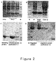
- the strains for producing ES and its mutants were cultured in a shake flask containing LB medium over night, and then inoculated into a 5L fermenter (Sartorius). IPTG was added at the appropriate time, and then the bacteria were harvested after about 4 hours ( figure 2A ). The bacteria were resuspended in a buffer solution and deeply broken by a high pressure homogenizer, and the broken bacteria were centrifuged to collect pellets. This process was repeated for three times.
- DEAE column or Q column GE Healthcare
- CM column or SP column GE Healthcare
- the renatured and un-renatured proteins were purified respectively, so as to obtain the renatured proteins with purity greater than 95% ( figure 2B, C ).
- the renatured proteins were concentrated and then dialyzed with PBS or NaAc-HAc.
- the modification of N-terminal of the renatured proteins was performed by using monomethoxy Poly(ethylene glycol)-aldehyde (mPEG-ALD, 20kDa, Beijing JenKem Technology Co., Ltd.).
- the modified proteins were purified by using CM column or SP column (GE Healthcare), and eluted with a pH gradient of 4.0 to 10.0 to obtain the target component ( figure 2D ).
- Example 4 ES and its variants are high efficient ATP enzyme
- the sample diluent buffer was prepared from 50mM HEPES, IMm EDTA and 0.02% NaN 3 (pH 7.4). ES, its variant Endu and N-4 were diluted to a final concentration of 500 ⁇ g/ml with the sample diluent buffer.
- Group 1 negative control, the sample diluent buffer added with the same volume of a protein-free buffer; and Group 2, ES, its variant Endu and N-4, with a final concentration of 500 ⁇ g/ml.
- the two groups of samples were diluted to appropriate ratio respectively, and then were added to a 96-well ELISA plate successively.
- the absorbance of the sample in each group was determined by using a Malachite Green Phosphate Assay Kit (BioAssay Systems) and a microplate reader (Multiskan mk3, Thermo Scientific).
- the concentration of phosphates in the reaction system was calculated and then converted to ATPase activity of ES.
- ATPase activity nM / mg / min ⁇ phosphate concentration nM / ml / reaction time 30 min / ES or its variant concentration mg / ml .
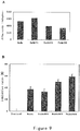
- Example 5 ES, Endu and its mPEG modified products, acting as ATPase, can significantly decrease the amounts of ATP in the whole cell homogenate of human vascular endothelial cells.
- the human vascular endothelial cells was first collected and then prepared into whole cell lysate with cell lysis buffer. The precipitate, impurities and cell debris were removed by centrifugation at low temperature (The above operation was done on the ice at a low temperature).
- the cell lysate was averagely divided into four groups, and each of them was subjected to a different treatment.
- Group 1 negative control, added with the same volume of a protein-free buffer;
- Group 2 treated by ES (50 ⁇ g/ml);
- Group 3 treated by ES (100 ⁇ g/ml);
- Group four treated by ES (200 ⁇ g/ml).
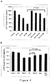
- PEG modified ES could also significantly degraded and reduced the level of ATP in the lysate of of human vascular endothelial cells, while the ATP degradation activity of mPEG-ES is only a little lower than that of ES under the circumstances that the doses (50 ⁇ g/ml, 100 ⁇ g/ml, 200 ⁇ g/ml respectively) and treating time of ES and mPEG-ES are same ( figure 4A ).
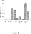
- Group one negative control with no treatment
- group two negative control with bovine serum albumin BSA (100 ⁇ g/ml), which is a well known protein with no ATPase activity and usually be used for negative control in these kinds of experiments
- group three positive control, treated with pork heart myosin (100 ⁇ g/ml), which is a well known protein with high ATPase activity and is used for positive control
- group four treated with ES (100 ⁇ g/ml); group five: treated with mPEG-ES (100 ⁇ g/ml); group six: treated with Endu (100 ⁇ g/ml); group seven: treated with mPEG-Endu (100 ⁇ g/ml).
- ES, mPEG-ES, Endu and mPEG-Endu all showed respective ATPase activity, and among them, ES with natural sequence has the highest ATPase activity and is approximate to myosin; mPEG-ES has the second highest ATPase activity and is slightly lower than ES; Endu and mPEG-Endu have a respectively lower ATPase activity ( figure 4B ).
- Example 6 evaluating ATPase activity is a convenient and accurate method with high repeatability for determining ES activity.
- ES, variants and PEG modified products thereof were established according to the method mentioned in example 4.
- ES, mPEG-ES, Endu and mPEG-Endu were diluted into a series of concentration gradients (showed in Figure 5 ) with sample diluting buffer on ice bath, respectively.
- the diluted samples were added to 96-well ELISA plate.
- OD630 was detected by using Malachite Green Phosphate Assay Kit (Malachite Green Phosphate Assay Kit, Bio Assay System).
- the concentration of diluted sample was calculated according to the dilution factor.
- a curve was plotted with the sample concentrations on the X axis and the corresponding ⁇ OD630 on the Y axis. Detection and plotting of mPEG-ES, Endu and mPEG-Endu were performed similarly in parallel. The results showed excellent linearity between the sample concentrations and the corresponding ⁇ OD630 for ES, mPEG-ES, Endu and mPEG-Endu, all with R 2 greater than 0.99 ( Figure 5 ). Thus, within the determined linear range, the method can be widely used for detecting the activity of ES, variants and PEG modified products thereof.
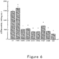
- Example 7 mutation in the ATP binding site of ES results in the change of ATPase activity
- ATPase activity of the ES that has mutation in the ATP binding site was detected using the methods mentioned in example 4.
- Mutant ES001 has a higher ATPase activity compared with ES, while the activities of mutants ES003-ES008 were dramatically decreased.
- ATPase activities of mutants ES003-ES008 were similar to that of mouse endostain (MM) ( Figure 6 ).
- Example 8 mutation in the ATP binding site of ES results in the change of its ATPase activity in the whole cell lysate
- the human vascular endothelial cells were collected and whole cell lysate was prepared with the cell lysis buffer. Pellets, impurities and debris in the cell homogenate were removed by centrifuge at low temperature. The whole cell lysate was aliquoted into several groups for different treatments as follows:
- ATP bioluminescent detection kit Sigma-Aldrich. The results showed that wild type human ES has obvious ATP degradation activity while mouse MM has low ATPase activity since it lacks the typical ATP binding domain.
- ES mutants ES003, ES006, ES007 and ES008 have dramatically decreased ATP degradation activity compared with wild type ES due to the different mutations in the ATP binding site.
- ES003 and ES008 have the most significant reduction of activity ( Figure 7A ).
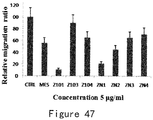
- Example 9 mutation in the ATP binding site results in the change of the endothelial cell migration inhibiting activity of ES
- HMEC human microvascular endothelial cells
- the number of cells completely migrate through the membrane to the bottom layer were counted from 5 fields randomly selected from each hole, and then averaged and compared with the control group to determine the reduction of migrated cells (the inhibition rate of each protein).
- Each group has three duplications and the experiments were independently repeated at least twice.
- HMEC endothelial cells
- Example10 Mutation in the ATP-binding site leads to the change of ATPase activity and endothelial cell migration inhibiting activity of Endu
- Example 11 Mutants with various decreases of ATP activity were obtained by mutating the ATP-binding motif and the adjacent sequenceof the wild type ES.
- ATP-binding motif of ES was mutated with two-step PCR, using the cycles and primers described in example 1. Mutation sites were summarized as follows: Name mutation sites sequence number ES010 MES-R5M SEQ ID NO.15 ( Figure 24 ) ES011 MES-R5Q SEQ ID NO.16 ( Figure 25 ) ES012 MES-R5Q&E92Q&P94Q&K96Q SEQ ID NO.17 ( Figure 26 ) S01 MES- ⁇ N2-5(HSHR)&Insert S97 SEQ ID NO.18( Figure 27 ) S02 MES- ⁇ N2-5(HSHR)&Insert T97 SEQ ID NO.19 ( Figure 28 ) S09 MES-Insert S97 SEQ ID NO.20 ( Figure 29 ) S10 MES-Insert T97 SEQ ID NO.21 ( Figure 30 ) S12 MES- ⁇ C1-4 SEQ ID NO.22 ( Figure 31 ) Z005 MES- ⁇
- ATPase activity of ES variants, mutants and the mPEG modified products thereof in examples 2 and 11 were measured with the method described in example 4, and the results were shown in Table 1.
- Table 1 Number Sample ATPase activity (nM /mg/min) Sample ATPase activity (nM/mg/min) 1 ES 14804 mPEG-ES 2664 2 Endu 5353 mPEG-Endu 1641 3 N-4 25448 mPEG-N-4 13555 4 MM 2856 mPEG-MM 277 5 ES001 16361 mPEG-001 5359 6 ES003 5200 mPEG-003 1116 7 ES004 5585 mPEG-004 570 8 ES005 4038 mPEG-005 1097 9 ES006 4069 mPEG-006 773 10 ES007 7137 mPEG-007 3059 11 ES008 4250 mPEG-008 1957 12 ES010
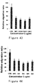
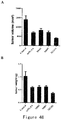
- Proliferating A549 cells were cultured and subcutaneously injected into 6 to 8-week nude mice (Vital River Laboratory Animal Technology Co. Ltd.) at. Drug treatment was started when 80-100 mm 3 tumor volume was achieved. Tumor-bearing mice were divided into five groups and treated with different administration respectively. In view of the increased anti-angiogenesis activity of mutants, a lower dose (12mg/kg, common dose was 24mg/mL) was administered to treat tumor-bearing mice.
- Group 1 negative control group without drug treatment, only saline at equal dose was injected;
- Group 2 mPEG-ES administration group;
- Group 3 M003 administration group, M003 was administered;
- Group 4 M007 administration group, M007 was administered;
- Group 5 MZ101 administration group, MZ101 was administered.
- the four Endostatin mutant above, i.e. mPEG-ES, M003 (mPEG-ES003), M007(mPEG-ES007) and MZ101 (mPEG-Z101) were all injected in caudal vein once a week at a dose of 12mg/kg, the treatment time was 21 days (three weeks).
- Tumor growth results shown in Figure 48A , revealed that compared with negative control (Group 1), tumor volume inhibition rate of mPEG-ES administration (Group 2) was 45%; tumor volume inhibition rates of M003 administration (Group 3) and M007 administration (Group 4) were approximately equal to mPEG-ES administration; tumor volume inhibition rate of MZ101 administration (Group 5) was 71.2%, which group has the smallest tumor volume and the highest drug inhibition rate.
- tumor weight inhibition rate of every drug treatment group was accordant with the tumor volume results.
- tumor weight inhibition rate of MS03 administration was 42%
- tumor weight inhibition rate of M003 administration was approximately equal to mPEG-ES administration
- tumor volume inhibition rate in MZ101 administration was 64%, which group has the smallest tumor weight and the highest drug inhibition rate.
- Endostatin mutants had favorable tumor growth inhibition effects at the dose of 12mg/kg/week in tumor-bearing mice model.
- the inhibition rate of mPEG-ES was about 40%; the inhibition rates of M003 and M007 were approximately equal to and slightly lower than mPEG-ES; the inhibition effect of MZ101 was better than mPEG-ES, displaying the best curative effect and the highest tumor inhibition rate (about 60-70%).
Description
- The present invention relates to a new anti-tumor therapeutic. In particular, this invention provides a mutant of endostatin, which has reduced ATPase activity and enhanced angiogenesis inhibiting activity. This invention also provides the use of the mutant in treating angiogenesis related diseases including tumor.
- In 1997, Professor Folkman from of Harvard University discovered the endogenous angiogenesis inhibitor - Endostatin (ES). Endostatin is a 20-kDa cleavage fragment of the C-terminus of collagen XVIII, which had inhibitory activities on the proliferation, migration of vascular endothelial cells, and the formation of blood vessels in vivo. The recombinant endostatin can inhibit the growth and metastasis of various types of tumors in mice, and can even cure the tumor without inducing drug resistance (Folkman J. et al. Cell 1997; 88:277-285; Folkman J. et al. Nature 1997; 390:404-407).
- The mechanism underlying the inhibitory capacity of ES is that it suppresses the angiogenesis in tumor tissues and blocks the supply of nutrition and oxygen. In China, the recombinant human endostatin (Endu) expressed by E. coli has become an anti-tumor therapeutic and its anti-tumor effect has been widely tested in clinical trail mainly focused on non-small-cell lung carcinoma. Endu, a variant of ES, has additional amino acid sequence (MGGSHHHHH) on N-terminal of ES, exhibiting more thermal dynamic stability and biological activity compared with wild type human ES expressed by yeast (Fu Y. et al. Biochemistry 2010; 49:6420-6429). Other report showed that the 27 amino acids on N-terminal of ES have the similar inhibitory activities on angiogenesis compared with the complete ES (Robert TjinThamSjin, et al., Cancer Res. 2005; 65(9):3656-63). Therefore, there are many researchers design medicaments based on the N-terminal 27 amino acids activities.
- Furthermore, to prolong the half-life of ES in vivo, many molecular modifications and drug design have been made to ES, including single site or multiple sites PEG modifications and conjugation with antibody Fc fragment (Tong-Young Lee, et al., Clin Cancer Res 2008; 14(5): 1487-1493). Multiple sites PEG modifications of ES are usually implemented on the ε amino of lysine side chain. Although this may prolong the half-life of ES, but its biological activities are apparently reduced (Guoying Mou, dissertation of Shandong University, CNKI, 2005). Compared with this modification technique, single site PEG modification on the N-terminal can not only enhance the stability, but also the biological activities of ES (
CN100475270C ). The related product has entered into clinical trail. - Since the discovery of ES, research projects from different laboratories focused on its tumor inhibitory activities have obtained different results. Professor Folkman's lab cured tumor in mice completely using ES (Folkman J. et al., 1997, Nature, 390:404-407), but many other labs could not repeat this result (News Focus, 2002, Science, 295:2198-2199). Meanwhile, since the ES produced in the prokaryotic expressing system containing polar body that is very hard to refold, many researchers diverted to use yeast to produce resolvable ES, but this did not achieve ideal results. Subsequent studies observed that the yeast expressed ES was N-terminal truncated and the truncated forms were identified as N-1, N-3, and N-4. The integrity of N-terminal is very important to the stability and biological activity of ES, this explains the confusing results obtained from yeast expressed ES (Fu Y. et al. Biochemistry 2010; 49:6420-6429).
- The primary biological function of ES is that is inhibits activities of endothelial cells, including inhibiting proliferation, migration and tube formation of endothelial cells and inducing apoptosis of endothelial cell, etc. The mechanism study of molecular function shows that nucleolin locating on the surface of plasma membrane is the functional receptor of ES and mediates the endocytosis of ES and its downstream signal pathway (Shi HB, et al., Blood, 2007, 110:2899-2906). Other report shows that nucleolin is also expressed on the plasma membrane of highly proliferative breast cancer cell line MDA-MB-435 and can mediate the endocytosis of its ligand protein in MDA-MB-435 (Sven Chridtian, et al., JBC, 2003, 163(4):871-878). In other studies, integrins, tropomyosin, glypican, laminin and matrix metalloproteinase 2 (MMP2) are all observed to be the potential receptors of ES (Sudhakar, A., et al., 2003, Proc. Natl. Acad. Sci. USA 100:4766-4771; Javaherian, K., et al., 2002, J. Biol. Chem., 277:45211-45218; Karumanchi, S., et al., 2001, Mol. Cell, 7:811-822; Lee, S. J., et al., 2002, FEBS Lett., 519:147-152; MacDonald, N. J., et al., 2001,J. Biol. Chem., 276:25190-25196; Kim, Y. M., et al., 2002, J. Biol. Chem., 277:27872-27879). Moreover, the treatment of nystatin dramatically increased the endocytosis and absorption of ES in endothelial cells, and therefore enhanced the biological activities of ES on inhibiting endothelial cells migration and animal tumor growth (Chen Y, et al., 2011, Blood, 117:6392-6403).
- The classical method to detect the biological activities of ES is based on its activity of inhibiting the endothelial cells, including the inhibition of migration, proliferation and tube formation of endothelial cells and other experiments. Commonly used endothelial cells mainly comprise human vascular endothelial cells (HMEC) and human umbilical vein endothelial cells (HUVEC). However, these methods require high quality of cell culture and complicated techniques, are very subjective, and exhibit low accuracy and reproducibility (Li YH, et al., 2011, Chin J Biological March, Vol. 24 No. 3:320-323). Therefore, to explore and develop new methods of evaluating the biological activities of ES and its mutants is of great importance in the ES drug discovery and quality control.
- Adenosine triphosphate (ATP) is an essential energy supply to organisms, participating in multi physiological and biological reactions and plays an important role in maintaining normal organic activities. ATP can be produced in many cellular metabolic pathways: in the most classical pathway it is produced by adenosine triphosphate synthetase through oxidative phosphorylation in mitochondrial under normal conditions, or produced in chloroplast through photosynthesis in plant. The source for ATP synthesis is mainly glucose and fatty acid. Under normal physiological conditions, the molar concentration of ATP in cell and blood are 1-10 mM and 100 µM, respectively.
- ATPase, also named adenosine triphosphotase, is an enzyme that catalyzes ATP to produce ADP and Pi and releasing energy. Under most conditions, the energy produced in this reaction can be transferred to another energy-required reaction and this process has been widely utilized in all known forms of lives. In addition, high-energy bond contained in the GTP can provide energy for protein synthesis, as well. Hsp90, myosin and other proteins all depend on ATP to perform biological activities, and thus all these proteins have ATPase activities. Although various kinds of ATPase are different in terms of sequence and tertiary structure, usually all these proteins have P-loop structure as the ATP binding motif (Andrea T. Deyrup, et al., 1998, JBC, 273(16):9450-9456). This P-loop structure exhibits the following typical sequences: GXXHXXK (Driscoll, W. J., et al., 1995 Proc. Natl. Acad. Sci. U.S.A., 92:12328-12332), (G/A)XXXXGK(T/S) (Walker, J., et al., 1982, EMBO J., 1:945-951), GXXXXGKS (Satishchandran, C., et al., 1992, Biochemistry, 31:11684-11688) and GXXGXGKS (Thomas, P.M., et al., 1995, Am. J. Hum. Genet., 59:510-518). Except for X, the remaining amino acid residues are relatively conserved. Generally, GTP also can bind to the ATP binding motif of these ATPases, and thus ATP and GTP can be alternative in many cases.
- Cancer cells and highly proliferative cells including endothelial cells have abnormally strong metabolism and the metabolic pathways are greatly different from normal mature cells. On one hand, cancer cells and proliferative cells demand large amount of ATP; on the other hand, the efficacy of using glucose to produce ATP is very low in these cells. This is because most cancer cells and highly proliferative cells produce ATP through aerobic glycolysis (the Warburg effect). Although this pattern exhibits low efficacy to produce ATP, the numerous mediates synthesized in this process can be used as building blocks that are more better for cell proliferation (Matthew G., et al., 2009, Science, 324:1029-1033).
- The present invention is defined by the claims. This invention discloses new activity of ES, namely ATPase activity, and discloses the new use of ES and ES drug design based on this new activity.
- This invention is based on the discovery that ES exhibits strong ATPase activity. The in vitro experiments showed that the ATPase activity of ES is only slightly lower than that of Myosin (extract of pork heart), which is known to have naturally high ATPase activity, without significant differences in degenerating ATP from the endothelial cell lysate.
- Based the ATPase activity of ES, this invention provides a new method of detecting
and evaluating the biological activity of ES. This method makes it possible to determine the conformation and biological activity of recombinantly produced ES through detecting the extracellular ATPase activity of ES by means of biochemical assays. Compared with the present cytological detection method, this new approach based on enzyme activity is more sensitive and precise, easy to operate and reliable in reproducibility, and thus can be widely used to detect the biological activity and evaluate the quality of ES and its variants. - Therefore, this invention provides a method of detecting the biological activity of endostatin or a variant, mutant or PEG modified product thereof, including detecting the ATPase activity of the endostatin or a variant, mutant or PEG modified product thereof. For example, malachite green phosphate assay and ATP bioluminescence assay can be used to detect the ATPase activity of endostatin or a variant, mutant or PEG modified product thereof and thereby determining the conformation and biological activity of a recombinantly produced ES product.
- It has been shown that ES can enter into the endothelial cell through nucleoclin-mediated endocytosis. In one example of this invention, to detect whether ES can execute ATPase activity intracellularly, the ATPase activity of ES was detected in endothelial cell lysate. The result shows that ES can execute ATPase activity in the endothelial cell lysate.
- The inventors found that the 89th-95th amino acid residues Gly-Ser-Glu-Gly-Pro-Leu-Lys in the wild type ES sequence (SEQ ID NO.1) contains the conserved GXXGXXK sequence of classical ATP-binding motif (Driscoll, W. J., et al., 1995, Proc. Natl. Acad. Sci. U.S.A., 92:12328-12332). The three amino acids, two Gs and one K, are highly conserved in various species. ATPase activity of ES can be changed through point mutation in the ATP-binding motif. Although the crystal structure of ES has been known, there is no report on the crystal structure of the complex of ES with ATP or GTP. Using cocrystallization technique, it is possible in the future to identify other amino acid residues in the ES protein capable of interacting with ATP or GTP in addition to the classical binding motif sequence, and to change the ATPase activity of ES and its inhibitory effect on endothelial activity by deletion or substitution and other modifications of such amino acid residues.
- Based on the known ES crystal structure, the inventors discovered that the ATP binding motif is close to the C-terminal of ES in the tertiary structure and the N-terminal is also very close to C-terminal in the tertiary structure. Therefore, in one example, the inventors compared ES variants with different N-terminal sequences and discovered that the ES variant with a deletion of four amino acids from the N-terminal (N-4) exhibited significantly higher ATPase activity than that of the full length ES. But it was previously reported that the N-4 exhibited significantly lower cytological activity and tumor inhibiting activity than ES (Fu Y. et al. Biochemistry 2010; 49:6420-6429).
- It was also reported that murine ES (MM) could completely cure mouse tumor (Folkman J. et al. Nature 1997; 390:404-407). However, through amino acid sequence alignment analysis, we found that murine ES dose not contain the classical ATP binding motif of human ES (
Figure 1 ). Thus, we detected the ATPase activity of MM and discovered that it was significantly lower than that of human ES, which is only about one fifth of human ES. However, the tumor inhibitory effect of MM was higher than human ES. - Therefore, to further identify the relationship between the ATPase activity and the cytological activity of ES, we introduced point mutations to some amino acids of the ATP binding motif. We observed that these mutations not only changed the ATPase activity but also the inhibitory effect of ES on endothelial cell migration. Furthermore, some mutants of ES exhibited reduced ATPase activity, but the inhibitory effect on endothelial cell migration was significantly enhanced. Except for a few cases, the ATPase activity is negatively related to the cytological activity.
- In some examples of this invention, mutants of ES comprising the sequence as shown in SEQ ID NO.6-11, 13, 14, 15-27 and 30-31 all exhibited reduced ATPase activity but equivalent or significantly higher inhibitory effect on endothelial cell migration. In view that ES is a vascular inhibiting protein and its essential function is to inhibit angiogenesis through inhibiting endothelial cell activity and thus can be used to treat angiogenesis related diseases (e.g., tumor, macular degeneration, obesity, and diabetes), we consider that these mutants of ES may process stronger activity to inhibit angiogenesis related diseases (e.g., tumor).
- In addition, based on the correlation between anti-angiogenesis activity and the ATPase activity of ES, it might be possible to design ES mutants by further changing (reducing) the ATPase activity through molecular cloning techniques, so as to obtain ES mendicants which are more effective to inhibit tumor and angiogenesis related disease.
- Therefore, this invention also provides a method of increasing the biological activity of endostatin, comprising reducing the ATPase activity of endostatin and its variants. In particular, genetic engineering approaches can be adopted to introduce mutations in the ATP binding motif GXXGXXK of endostatin or its variants to obtain an endostatin mutant with a reduced ATPase activity but an increased biological activity, for example, an increased inhibitory effect on endothelial cell migration and tumor growth.
- This invention also provides endostatin mutants which are mutated in the ATP binding motif and exhibit enhanced anti-angiogenesis activity and decreased ATPase activity as compared to the wild type endostatin or its variants.
- Preferably, the ATPase activity of the mutants is reduced at least about 30%, such as at least about 50%, at least about 70% or at least about 90%, as compared to the wild type endostatin or its variants. For example, the ATPase activity of the mutants is only about 60-70%, such as about 50-60%, 40-50%, 30-40%, 20-30%, 10-20% or no more than 10% or even lower, as compared to the wild type endostatin or its variants. In one embodiment, the mutant does not have ATPase activity.
- In some embodiments, as compared with the corresponding wild type endostatin or its variant, the mutant comprises a mutation in the ATP combining motif. For example, the mutant comprises a mutation in the sequence corresponding to the Gly-Ser-Glu-Gly-Pro-Leu-Lys motif consisting of the 89th-95th amino acid residues of SEQ ID NO.1, wherein the mutation is selected from the group consisting of one or several amino acid replacements, deletions or additions or a combination thereof, and the mutation results in a decrease or elimination of the ATPase activity in the mutant.
- In some embodiments, the mutant comprises a partial or complete deletion of the sequence corresponding to the Gly-Ser-Glu-Gly-Pro-Leu-Lys motif consisting of the 89th-95th amino acid residues of SEQ ID NO.1.
- Preferably, the endostatin mutant of the invention comprises the following mutations: (a) Gly residue corresponding to the amino acid residue 89 of SEQ ID NO.1 is replaced with an uncharged or aromatic amino acid or deleted; or (b) Gly residue corresponding to the amino acid residue 92 of SEQ ID NO.1 is replaced with an uncharged amino acid or deleted; or (c) Lys residue corresponding to the amino acid residue 95 of SEQ ID NO.1 is replaced with a positive charged or uncharged amino acid or deleted; or (d) any combination of (a)-(c).
- More preferably, the endostatin mutant of the invention comprises the following mutations: (a) Gly residue corresponding to the amino acid residue 89 of SEQ ID NO.1 is replaced with either Ala or Pro or deleted; or (b) Gly residue corresponding to the amino acid residue 92 of SEQ ID NO.1 is replaced with Ala or deleted; or (c) Lys residue corresponding to the amino acid residue 95 of SEQ ID NO.1 is replaced with either Arg or Gln or deleted; or (d) any combination of (a)-(c).
- Of course, the above replacement can also be made with charged amino acids, with the prerequisite that it does not affect charge distribution and conformation of the mutant protein.
- In a particular embodiment, the endostatin mutant of the invention comprises a sequence selected from the group consisting of SEQ ID NOs.6-11, 13, 14, 15-27 and 30-31. Preferably, the endostatin mutant of the invention comprises a sequence selected from the group consisting of SEQ ID NO.6, SEQ ID NO.10, SEQ ID NO.27 and SEQ ID NO.30.
- Preferably, the endostatin mutant of the invention is a mutant of the human endostatin.
- The invention also provides a pharmaceutical composition, which comprises the above mentioned endostatin mutant of the invention. In the pharmaceutical composition of the invention, the endostatin mutant may be covalently linked to the PEG molecule. The molecular weight of the PEG is such as 5-40KD, for example, 5-20KD, or 20-40KD. Preferably, the molecular weight of PEG is 20KD, for example the 20 kD monomethoxy Poly(ethylene glycol), or monomethoxy Poly(ethylene glycol)-aldehyde (mPEG-ALD). Preferably, the PEG molecule is covalently linked to the N-terminal α amino group of the endostatin. Also disclosed is a method of treating a tumor, comprising administering the aforementioned endostatin mutants or the pharmaceutical composition of the present invention to tumor patients. Also disclosed is use of the aforementioned endostatin mutants in preparation of a medicament for the treatment of a angiogenesis related disease. Preferably, the aforementioned angiogenesis related disease is tumor.
-
-
FIG. 1 shows the sequence alignment of human and murine ES. -
FIG. 2 shows the preparation of ES, ES mutant, ES variant and their mPEG modified products.- (A) Expression of engineering bacterium.
- (B) Purification of inclusion body protein.
- (C) Purification of refolded protein.
- (D) Modified protein purification.
-
FIG. 3 shows the ATPase activity of ES, ES variant and their mPEG modified products. -
FIG. 4 shows the ATPase activity of ES, ES variant and their mPEG modified products in endothelial cell lysate.- (A) ES and mPEG-ES can biodegrade ATP in endothelial cell lysate.
- (B) ES, Endu and their mPEG modified products can biodegrade ATP in endothelial cell lysate.
-
FIG. 5 shows that the ATP enzyme activity assay allows fast and accurate detection of the biological activity of ES, ES variant and their mPEG modified products.- (A) standard curve for detecting the biological activity of ES and Endu based on the ATP enzyme activity assay.
- (B) standard curve for detecting the biological activity of mPEG-ES and mPEG-Endu based on the ATP enzyme activity assay.
-
FIG. 6 shows the comparison of ATPase activity of ES mutants. -
FIG.7 shows the comparison of the activity of ES mutants on biodegrading ATP in endothelial cell lysate. -
FIG.8 shows the comparison of the activity of ES mutants on inhibiting endothelial cell migration. -
FIG.9 shows the comparison of the ATPase activity and endothelial cell migration inhibiting activity of Endu mutants.- (A) The ATPase activity of Endu mutants.
- (B) The activity of Endu mutants on inhibiting endothelial cell migration.
-
FIG. 10 shows the sequence of native human ES. -
FIG. 11 depicts the sequence of recombinant human ES expressed in E.coli, in which the first amino acid M at the N-terminal can be randomly deleted during recombinant expression. -
FIG. 12 depicts the sequence of recombinant human N-4 expressed in E.coli, in which the first amino acid M at the N-terminal and the last amino acid K can be randomly deleted during recombinant expression. -
FIG. 13 depicts the sequence of recombinant human Endu expressed in E.coli, in which the first amino acid M at the N-terminal is randomly deleted during recombinant expression. -
FIG. 14 depicts the sequence of recombinant human ES001 expressed in E.coli, in which the first amino acid M at the N-terminal can be randomly deleted during recombinant expression. -
FIG. 15 depicts the sequence of recombinant human ES003 expressed in E.coli, in which the first amino acid M at the N-terminal can be randomly deleted during recombinant expression. -
FIG. 16 depicts the sequence of recombinant humanES004 expressed in E.coli, in which the first amino acid M at the N-terminal can be randomly deleted during recombinant expression. -
FIG. 17 depicts the sequence of recombinant human ES005 expressed in E.coli, in which the first amino acid M at the N-terminal can be randomly deleted during recombinant expression. -
FIG.18 depicts the sequence of recombinant human ES006 expressed in E.coli, in which the first amino acid M at the N-terminal can be randomly deleted during recombinant expression. -
FIG. 19 depicts the sequence of recombinant human ES007 expressed in E.coli, in which the first amino acid M at the N-terminal can be randomly deleted during recombinant expression. -
FIG. 20 depicts the sequence of recombinant human ES008 expressed in E.coli, in which the first amino acid M at the N-terminal can be randomly deleted during recombinant expression. -
FIG. 21 depicts the sequence of recombinant human Endu001 expressed in E.coli, in which the first amino acid M at the N-terminal can be randomly deleted during recombinant expression. -
FIG. 22 depicts the sequence of recombinant human Endu003 expressed in E.coli, in which the first amino acid M at the N-terminal can be randomly deleted during recombinant expression. -
FIG.23 depicts the sequence of recombinant human Endu008 expressed in E.coli, in which the first amino acid M at the N-terminal can be randomly deleted during recombinant expression. -
FIG. 24 depicts the sequence of recombinant human ES010 expressed in E.coli, in which the first amino acid M at the N-terminal can be randomly deleted during recombinant expression. -
FIG. 25 depicts the sequence of recombinant human ES011 expressed in E.coli, in which the first amino acid M at the N-terminal can be randomly deleted during recombinant expression. -
FIG. 26 depicts the sequence of recombinant human ES012 expressed in E.coli, in which the first amino acid M at the N-terminal can be randomly deleted during recombinant expression. -
FIG. 27 depicts the sequence of recombinant human S01 expressed in E.coli, in which the first amino acid M at the N-terminal can be randomly deleted during recombinant expression. -
FIG. 28 depicts the sequence of recombinant human S02 expressed in E.coli, in which the first amino acid M at the N-terminal can be randomly deleted during recombinant expression. -
FIG. 29 depicts the sequence of recombinant human S09 expressed in E.coli, in which the first amino acid M at the N-terminal can be randomly deleted during recombinant expression. -
FIG. 30 depicts the sequence of recombinant human S10 expressed in E.coli, in which the first amino acid M at the N-terminal is randomly deleted during recombinant expression. -
FIG. 31 depicts the sequence of recombinant human S12 expressed in E.coli, in which the first amino acid M at the N-terminal is randomly deleted during recombinant expression. -
FIG. 32 depicts the sequence of recombinant human Z005 expressed in E.coli, in which the first amino acid M at the N-terminal is randomly deleted during recombinant expression. -
FIG. 33 depicts the sequence of recombinant human Z006 expressed in E.coli, in which the first amino acid M at the N-terminal is randomly deleted during recombinant expression. -
FIG. 34 depicts the sequence of recombinant human Z008 expressed in E.coli, in which the first amino acid M at the N-terminal is randomly deleted during recombinant expression. -
FIG. 35 depicts the sequence of recombinant human Z009 expressed in E.coli, in which the first amino acid M at the N-terminal is randomly deleted during recombinant expression. -
FIG. 36 depicts the sequence of recombinant human Z101 expressed in E.coli, in which the first amino acid M at the N-terminal is randomly deleted during recombinant expression. -
FIG. 37 depicts the sequence of recombinant human Z103 expressed in E.coli, in which the first amino acid M at the N-terminal is randomly deleted during recombinant expression. -
FIG. 38 depicts sequence of recombinant human Z104 expressed in E.coli, in which the first amino acid M at the N-terminal can be randomly deleted during recombinant expression. -
FIG. 39 depicts the sequence of recombinant human ZN1 expressed in E.coli, in which the first amino acid M at the N-terminal is randomly deleted during recombinant expression. -
FIG. 40 depicts the sequence of recombinant human ZN2 expressed in E.coli, in which the first amino acid M at the N-terminal is randomly deleted during recombinant expression. -
FIG. 41 depicts the sequence of recombinant human ZN3 expressed in E.coli, in which the first amino acid M at the N-terminal is randomly deleted during recombinant expression. -
FIG. 42 depicts the sequence of recombinant human ZN4 expressed in E.coli, in which the first amino acid M at the N-terminal is randomly deleted during recombinant expression. -
FIG. 43 shows the comparison of the endothelial cell migration inhibiting effect of ES mutants ES010, ES011, ES012. -
FIG. 44 shows the comparison of the endothelial cell migration inhibiting effect of ES mutants S01, S02, S09, S10. -
FIG. 45 shows the comparison of the endothelial cell migration inhibiting effect of ES mutants S12. -
FIG. 46 shows the comparison of the endothelial cell migration inhibiting effect of ES mutants Z005, Z006, Z008, Z009. -
FIG. 47 shows the comparison of the endothelial cell migration inhibiting effect of ES mutants Z101, Z103, Z104, ZN1, ZN2, ZN3, ZN4. -
FIG. 48 shows the inhibitory effect of ES mutants on non-small cell lung cancer A549 tumor growth at the animal level, (A) tumor volume, and (B) tumor weight. - Unless otherwise indicated, the scientific and technical terms used in this description should have the meaning that a skilled person generally understands in this field. Normally, the nomenclature and techniques used in this description about cell and tissue culture, molecular biology, immunology, microbiology, genetics, and protein and nucleic acid chemistry are known and commonly used in this field.
- Unless otherwise indicated, the methods and techniques used in this description normally are conducted according to commonly known and conventional methods of this field and described in this description or methods described in the cited references. For example, see Sambook J. and Russell D. Molecular Cloning: A Laboratory Manual, the third edition, Cold Spring Harbor Laboratory Press, Cold Spring Harbor, N.Y. (2000); Ausubel et al, Short Protocols in Molecular Biology: A Compendium of Methods from Current Protocols in Molecular Biology, Wiley, John& Sons, Inc. (2002); Harlow and Lane Using Antibodies: A Laboratory Manual, Cold Spring Harbor Laboratory Press, Cold Spring Harbor, N.Y. (1998); and Coligan et al, Short Protocols in Protein Science, Wiley, John& Sons, Inc. (2003).
- ES , ES variants, ES mutants and their mPEG modified products ES (Endostatin) refers to natural endostatin, for example, the human endostatin with the sequence of SEQ ID NO.1 (
FIG. 10 ). ES variants refer to a molecule comprising an addition or a deletion of 1-15 amino acids at either N-terminal or C-terminal of a natural ES molecule. ES variant can be a naturally occurring variant, for example, when the human ES is recombinantly expressed in E.coli, the first amino acid M can be randomly deleted, producing an ES variant with the sequence of SEQ ID NO.2 (FIG. 11 ). For another example, when the ES is recombinantly expressed in yeast, due to random cutting of the N-terminal, an ES variant with a deletion of four amino acids from N-terminal can be produced, which variant has the sequence of SEQ ID NO.3 (FIG. 12 ). Further, the C-terminal K can also be randomly deleted. ES variant can also be a artificial variant. For example, to improve the expression and stability, Endu, an ES variant having the SEQ ID NO.4 sequence, has an addition of nine amino acids with the sequence of MGGSHHHHH at N-terminal of wild type ES (FIG. 13 ), and the first amino acid M can be randomly deleted during the recombinant expression. In this application, ES variants refer to a naturally occurring or artificial variant of ES, which has the same or similar activity of inhibiting angiogenesis and has the same or similar ATP binding motif and ATPase activity with the corresponding wild type ES. - In this application, ES mutant refer to a mutated protein obtained by modifying the ATP binding sites of a natural ES or ES variants, for example, by modifying the ATP binding motif by means of amino acid point mutation.
- Except for Endu which was purchased from Medgenn, and the ES, ES variants, ES mutants used in this invention were provided by PROTGEN.
- PEG modified ES, Endu, and N-4 are respectively named mPEG-ES, mPEG-Endu and mPEG-N-4. These products are ES, Endu, and N-4 respectively modified by 20 kD monomethoxy Poly(ethylene glycol)-aldehyde (mPEG-ALD), the coupling site for the activated mPEG-ALD aldehyde group is the N-terminal α amino group of ES, Endu, and N-4.
- This ATP assay is a well approved and widely used method, which has extreme sensitivity, and the principle is as follows. Firefly luciferase catalyzes the oxidation of luciferin to emit photons with the energy in ATP. Therefore, in the luminous reaction catalyzed by firefly luciferase, luminous intensity has well linear relationship with ATP concentration in the detection systems. By using bioluminescence analyzer (Berthold Technologies Centro LB 960) to detect luminous intensities in the reaction system, the ATP concentrations in reaction systems can be accurately calculated.
- This is a well approved and widely used method to test ATPase activity. The principle is as follows. In acidic condition, the reaction between malachite green, molybdate and phosphoric acid can generate green substance, which can be detected in 600-660 nm wavelength range. The absorbance has well linear relationship with the phosphate concentration over a certain range. ADP and Pi are released during ATP hydrolysis catalyzed by ATPase. Therefore it is possible to calculate ATPase activity by the phosphate concentration detected by this kit. This method is convenient and expeditious, widely used to analyze the activities of phosphatase, lipase and nucleoside triphosphatase, and phosphate concentration as well as in high-throughput drug screening.
- These motifs refer to the classical primary sequence which can bind to ATP in proteins with ATPase activity. ATP-binding motif usually has a P-loop structure, which have some classical sequences, including GXXGXXK, (G/A)XXXXGK(T/S), GXXXXGKS and GXXGXGKS. Among these, amino acid residues which are not replaced by X are relatively conservative. Generally, these ATP-binding motifs can also bind to GTP.
- These sites refer to the sites which can bind to ATP in proteins with ATPase activity, including classical ATP-binding motifs and other amino acid sites involved in ATP binding. These amino acid residues could be far from ATP-binding motifs on primary sequences, but they participate in the interaction between ES and ATP/GTP in tertiary structure. Alternatively, missing or replacement on these sites can indirectly disturb the interaction between ES and ATP/GTP by interfering protein conformation.
- Based on the discovery of a new ES activity, i.e., ATPase activity, the invention provides a new method for evaluating the biological activity of ES. Compared with the current assay based on endothelial cell migration, this method is more convenient and accurate and proves to be well reproducible. This invention provides an important research approach for studying the action mechanism of ES, ES variants and ES mutants, as well as for drug development and quality control.
- Therefore, this invention provides a method for detecting the bioactivities of ES, ES variant, ES mutant and PEG modified ES product. The method comprises detecting the ATPase activity of ES, ES variant, ES mutant and PEG modified ES as mentioned above. For example, it is possible to use malachite green phosphate assay kits or ATP bioluminescence assay kit to detect the ATPase activity of ES, ES variant, ES mutant and PEG modified ES, so as to determine the conformation and bioactivity of recombinant produced ES.
- Meanwhile, compared with some known P-loop structure sequences in ATP-binding motifs, we found that in ES primary sequence, the amino acid residues from 89-95 having the sequence of Gly-Ser-Glu-Gly-Pro-Leu-Lys conform to the classical ATP-binding motif of GXXGXXK, which is the structure basis for ES ATPase activity (
Figure 10 ). - Based on crystal structure of ES, we found that ES ATP-binding motif was close to N-terminal in tertiary structure, so the stereo specific blockade changes induced by N-terminal sequence alteration may influence the ATPase ability of ES. So in one example of this invention, we detected the ATPase activities of ES variant Endu (with 9 additional amino acid residues at N-terminal) and N-4 (with 4 amino acid deletion at N-terminal). The results showed that ES variants, Endu and N-4, had ATPase activities. ATPase activity of Endu was significantly reduced compared with ES, while ATPase activity of N-4 was significantly increased (
Figure 3 ). These results indicated that stereo specific blockade changes induced by different N-terminal integrity indeed have impact on the ATPase activity of ES. Therefore, apart from the classical ATP-binding motif GXXGXXK, other ATP-interacting sites confirmed by cocrystallization analysis could be potential target sites for modifying the ATPase activity. - It has been reported that after single-point modification by mPEG at the N-terminal alpha-amino, the activities of ES and Endu to inhibit endothelial cell migration were significantly improved (
CN 100475270C ). So in one example of this invention, we detected the ATPase activities of mPEG-ES, mPEG-Endu, and mPEG-N-4, and found that the ATPase activities were significantly reduced (Figure 3 ). So in this group of ES, ES variants and their mPEG modification products, the ATPase activities were negatively related to the activities of inhibiting endothelial cell migration, thus the higher ATPase activity, the lower activity of inhibiting endothelial cell migration. This result was confirmed by ES variant N-4: the ATPase activity of N-4 was higher than ES and Endu (Figure 3 ), while other cellular activities were significantly reduced (Fu Y. et al. Biochemistry 2010; 49:6420-6429). - From the above data, we found that in ES, ES variants and their mPEG modification products, ATPase activities were negatively related to the activities of inhibiting endothelial cell migration. Based on this discovery, in order to obtain ES with higher activity of inhibiting endothelial cell migration, we introduced point mutations at ATP-binding sites in ES to reduce its ATPase activity.
- Accordingly, this invention provides a method for improving the biological activity of ES, comprising reducing the ATPase activity of ES and ES variants. In particular, the ATP-binding motif GXXGXXK of ES and ES variants could be mutated by means of genetic engineering, and thereby obtaining an ES mutant with lower ATPase activity but improved biological activity, such as the activity of inhibiting endothelial cell migration and tumor.
- In one example of this invention, the following mutations were introduced into ES ATP-binding sites:
ES001-ES-K96R (SEQ ID NO.5) ( Figure 14 )ES003-ES-G90A (SEQ ID NO.6) ( Figure 15 )ESO04-ES-G93A&K96R (SEQ ID NO.7) ( Figure 16 )ES005-ES-G90A&G93A&K96R (SEQ ID NO.8) ( Figure 17 )ES006-ES-G93A (SEQ ID NO.9) ( Figure 18 )ES007-ES-G90A&E92K&G93A&K96R (SEQ ID NO.10) ( Figure 19 )ES008-ES-E92Q&P94Q&K96Q (SEQ ID NO.11) ( Figure 20 ) - As detected by using biochemistry methods, the ATPase activities of the mutants were significantly increased as compared with ES, while the ATPase activities of mutants ES003, ES004, ES005, ES006, and ES007 were significantly reduced (
Figure 6 ). Although ES can be endocytosed by endothelial cells and degrade intracellular ATP, living cells can rapidly compensate the ATP consumption. So, instead of detecting the ATP concentration in living cells, current methods usually are designed to detect ATP degradation in whole cell lysate. In whole cell lysate, the ATPase activities of mutants ES003, ES006, ES007, and ES008 were still significantly lower than ES (Figure 7A ), but the ATPase activities of mutants ES001, ES004, and ES005 were equal to ES (Figure 7B ). This may be due to the P-loop structure changes induced by these mutations which may influence the interaction between the whole protein and ATP. - Subsequently, we continued to verify the activities of these ES mutants to inhibit endothelial cell migration. The results were basically in agreement with our expectation. Except for a very few mutants, the ATPase activity of most ES mutants were negatively related to the activity of inhibiting endothelial cell migration (
Figure 8 ). - In addition, in one example of this invention, the following mutations were introduced into an ES variant, Endu:
Endu001---Endu-K104R (SEQ ID NO.12) ( Figure 21 )Endu003-Endu-G98A (SEQ ID NO.13) ( Figure 22 )Endu008-Endu-E100Q&P102Q&K104Q (SEQ ID NO.14) ( Figure 23 ) - We found that when compared with ES, mutations in ATP-binding sites had similar impact on Endu in terms of the ATPase activities and inhibition of endothelial cell migration. Therefore, it is believed that the strategy of changing the ATPase activity and the inhibition of endothelial cells migration by introducing mutations in ATP-binding sites also applies to ES variants.
- Therefore, this invention also provides an ES mutant with improved anti-angiogenesis activity. The mutant comprises a mutation at ATP-binding sites. Compared with the wild type ES or its variants, the mutant exibits reduced ATPase activity.
- Preferably, the ATPase activity of the mutant is reduced at least about 30%, such as at least about 50%, at least about 70% or at least about 90%, as compared to the wild type endostatin or its variants. For example, the ATPase activity of the mutants is only about 60-70%, such as about 50-60%, 40-50%, 30-40%, 20-30%, 10-20% or no more than 10% or even lower, as compared to the wild type endostatin or its variants. In one example, the mutant had no ATPase activity.
- In some embodiments, the mutant comprises a mutation in the ATP-binding motif as compared with the corresponding wild type endostatin or a variant thereof. For example, the mutant comprises a mutation in the sequence corresponding to the Gly-Ser-Glu-Gly-Pro-Leu-Lys motif consisting of amino acid residues 89-95 of SEQ ID NO.1, wherein said mutation is one or several amino acid replacement, deletion or addition, and said mutation results in a decrease or deletion of ATPase activity of said mutant.
- In some embodiments, the mutant comprises a partial or complete deletion of the sequence corresponding to the Gly-Ser-Glu-Gly-Pro-Leu-Lys motif consisting of the 89th-95th amino acid residues of SEQ ID NO.1.
- Preferably, the ES mutant in the invention comprises the following mutations: (a) Gly residue corresponding to the amino acid residue 89 of SEQ ID NO.1 is replaced with an uncharged or aromatic amino acid or deleted; or (b) Gly residue corresponding to the amino acid residue 92 of SEQ ID NO.1 is replaced with an uncharged amino acid or deleted; or (c) Lys residue corresponding to the amino acid residue 95 of SEQ ID NO.1 is replaced with a positive charged or uncharged amino acid or deleted; or (d) any combination of (a) - (c).
- More preferably, the ES mutant of the invention comprises the following mutations: (a) Gly residue corresponding to the amino acid residue 89 of SEQ ID NO.1 is replaced with either Ala or Pro or deleted; or (b) Gly residue corresponding to the amino acid residue 92 of SEQ ID NO.1 is replaced with Ala or deleted; or (c) Lys residue corresponding to the amino acid residue 95 of SEQ ID NO.1 is replaced with either Arg or Gln or deleted; or (d) any combination of (a) - (c).
- In a particular embodiment, the ES mutant of the invention comprises a sequence selected from the group consisting of SEQ ID NOs.6-11, 13, 14, 15-27 and 30-31. Preferably, the ES mutant of the invention comprises a sequence selected from the group consisting of SEQ ID NO.6, SEQ ID NO.10, SEQ ID NO.27 and SEQ ID NO.30.
- Preferably, the ES mutant of this invention is a mutant of the human ES.
- This invention also provides a pharmaceutical composition comprising the mentioned above ES mutant. In the pharmaceutical composition of the invention, the ES mutant may be covalently linked to a PEG molecule. The molecular weight of the PEG is such as 5-40 kD, for example, 5-20 kD, or 20-40 kD. Preferably, the PEG has a molecular weight of 20 kD, for example, 20 kD mPEG or mPEG-ALD. Preferably, the PEG molecule is covalently linked to the N-terminal alpha amino group of the ES. Also disclosed is a method of treating a tumor, comprising administering the aforementioned endostatin mutant or pharmaceutical composition of the present invention to a patient having a tumor. Also disclosed is use of the aforementioned endostatin mutants in preparation of a medicament for the treatment of an angiogenesis related disease. Preferably, the aforementioned angiogenesis related disease is tumor.
- This invention will be further elucidated with the following non-exclusive examples.
- The gene of Endostatin was amplified from cDNA of lung cancer cell A549, and then was cloned into pET30a plasmid to obtain a recombinant plasmid. The 5'-primer for gene amplification was GGAATTCCATATGCACAGCCACCGCGACTTC, and the 3'-primer was CCGCTCGAGTTACTTGGAGGCAGTCATGAAGCTG. The restriction endonucleases were NdeI and XhoI respectively. The above recombinant plasmid was transformed into E. coli via conventional techniques in the art for further protein expression.
- The ATP binding site of wild type human ES was modified by mutation. The detail mutation process, the primer pairs and the transformation process were the same as example 1. The mutants were listed as follows:
ES001-ES-K96R (SEQ ID NO.5) ( figure 14 )ES003-ES-G90A (SEQ ID NO.6) ( figure 15 )ES004-ES-G93A&K96R (SEQ ID NO.7) ( figure 16 )ES005-ES-G90A&G93A&K96R (SEQ ID NO.8) ( figure 17 )ES006-ES-G93A (SEQ ID NO.9) ( figure 18 )ES007-ES-G90A&E92K&G93A&K96R (SEQ ID NO.10) ( figure 19 )ES008--ES-E92Q&P94Q&K96Q (SEQ ID NO.11) ( figure 20 ) - As controls, we also constructed the mutants containing a mutated ATP binding site, i.e. Endu001, Endu003 and Endu008, by using the same protocol as the above based on the sequence of wild type Endu.
- Endu001-Endu-K104R (SEQ ID NO.12) (
figure 21 ) - Endu003-Endu-G98A (SEQ ID NO.13) (
figure 22 ) - Endu008-Endu-E100Q&P102Q&K104Q (SEQ ID NO.14) (
figure 23 ) - The preparation of mutant ES003 was taken as an example for illustrating the expression and preparation of recombinant ES, ES mutant and Endu mutant. Specifically, the strains for producing ES and its mutants were cultured in a shake flask containing LB medium over night, and then inoculated into a 5L fermenter (Sartorius). IPTG was added at the appropriate time, and then the bacteria were harvested after about 4 hours (
figure 2A ). The bacteria were resuspended in a buffer solution and deeply broken by a high pressure homogenizer, and the broken bacteria were centrifuged to collect pellets. This process was repeated for three times. Then, DEAE column or Q column (GE Healthcare), and CM column or SP column (GE Healthcare) were used for the elution of proteins with a pH gradient of 4.0 to 10.0. The renatured and un-renatured proteins were purified respectively, so as to obtain the renatured proteins with purity greater than 95% (figure 2B, C ). The renatured proteins were concentrated and then dialyzed with PBS or NaAc-HAc. The modification of N-terminal of the renatured proteins was performed by using monomethoxy Poly(ethylene glycol)-aldehyde (mPEG-ALD, 20kDa, Beijing JenKem Technology Co., Ltd.). The modified proteins were purified by using CM column or SP column (GE Healthcare), and eluted with a pH gradient of 4.0 to 10.0 to obtain the target component (figure 2D ). - The sample diluent buffer was prepared from 50mM HEPES, IMm EDTA and 0.02% NaN3 (pH 7.4). ES, its variant Endu and N-4 were diluted to a final concentration of 500µg/ml with the sample diluent buffer. Group 1: negative control, the sample diluent buffer added with the same volume of a protein-free buffer; and
Group 2, ES, its variant Endu and N-4, with a final concentration of 500 µg/ml. - 500 µM ATP was added to the control, and the reaction was performed at water bath at 37°C for 30 min and then on ice for 5 min to terminate the reaction. The same procedures were also adopted to the samples of ES and its variant at the same time.
- The two groups of samples were diluted to appropriate ratio respectively, and then were added to a 96-well ELISA plate successively. The absorbance of the sample in each group was determined by using a Malachite Green Phosphate Assay Kit (BioAssay Systems) and a microplate reader (Multiskan mk3, Thermo Scientific). The concentration of phosphates in the reaction system was calculated and then converted to ATPase activity of ES.

- The results showed that ES, Endu and N-4 have high ATPase activity, and the N-4 has the highest ATPase activity.
- The same method was used to detect the ATPase activities of mPEG modified ES, Endu and N-4, and the results showed that the ATPase activities of mPEG-ES, mPEG-Endu and mPEG-N-4 are decreased compared with those of ES, Endu and N-4.
- All the above experiments adopted Myosin (extracted from pork heart, Sigma) as a positive control, which had been well-known to have high ATPase activity. The results showed that ES and its variant are high efficient ATP enzyme (
figure 3 ). - Example 5: ES, Endu and its mPEG modified products, acting as ATPase, can significantly decrease the amounts of ATP in the whole cell homogenate of human vascular endothelial cells.
- The human vascular endothelial cells was first collected and then prepared into whole cell lysate with cell lysis buffer. The precipitate, impurities and cell debris were removed by centrifugation at low temperature (The above operation was done on the ice at a low temperature). The cell lysate was averagely divided into four groups, and each of them was subjected to a different treatment. Group 1: negative control, added with the same volume of a protein-free buffer; Group 2: treated by ES (50µg/ml); Group 3: treated by ES (100µg/ml); Group four: treated by ES (200µg/ml). Each group was placed at room temperature to allow the reaction to start immediately following the addition of ES, and then was placed back to ice to terminate the reaction after 25 min. The amount of ATP of the cell homogenate in each group was detected by using a ATP bioluminescent detection kit (Sigma-Aldrich). The results showed that, compared with the control group, ES can significantly degraded and reduced the level of ATP in the lysate of of human vascular endothelial cells. The results were consistent with those of example 4 and further demonstrated that ES could also have the ATP degradation activity in a relative complex system such as cell lysis buffer. At the same time, we found that PEG modified ES (mPEG-ES) could also significantly degraded and reduced the level of ATP in the lysate of of human vascular endothelial cells, while the ATP degradation activity of mPEG-ES is only a little lower than that of ES under the circumstances that the doses (50µg/ml, 100µg/ml, 200µg/ml respectively) and treating time of ES and mPEG-ES are same (
figure 4A ). - ES could also be replaced by other proteins with same mechanism or its variant Endu. In the parallel comparison experiment on Endu and mPEG modified Endu (mPEG-Endu) (which is to add 20kDa mPEG-ALD modification on the ES with additional amino acids MGGSHHHHH on N-terminal), we got the similar results. The whole cell lysis components of human vascular endothelial cells were obtained by the same method mentioned above, and was averagely divided into seven groups with different treatment as followed. Group one: negative control with no treatment; group two: negative control with bovine serum albumin BSA (100µg/ml), which is a well known protein with no ATPase activity and usually be used for negative control in these kinds of experiments; group three: positive control, treated with pork heart myosin (100µg/ml), which is a well known protein with high ATPase activity and is used for positive control; group four: treated with ES (100µg/ml); group five: treated with mPEG-ES (100µg/ml); group six: treated with Endu (100µg/ml); group seven: treated with mPEG-Endu (100µg/ml). Each group was placed at room temperature immediately after adding ES, Endu or mPEG modified products, reacting for 25 min, and was placed back to ice to terminate the reaction. The results showed that when added the same dose myosin, BSA, ES, mPEG-ES, Endu, mPEG-Endu, and under the same reaction condition, myosin showed the highest ATP degradation activity, which also means ATPase activity. ES, mPEG-ES, Endu and mPEG-Endu all showed respective ATPase activity, and among them, ES with natural sequence has the highest ATPase activity and is approximate to myosin; mPEG-ES has the second highest ATPase activity and is slightly lower than ES; Endu and mPEG-Endu have a respectively lower ATPase activity (
figure 4B ). - The method for determining ATPase activity of ES, variants and PEG modified products thereof was established according to the method mentioned in example 4. ES, mPEG-ES, Endu and mPEG-Endu were diluted into a series of concentration gradients (showed in
Figure 5 ) with sample diluting buffer on ice bath, respectively. The diluted samples were added to 96-well ELISA plate. OD630 was detected by using Malachite Green Phosphate Assay Kit (Malachite Green Phosphate Assay Kit, Bio Assay System). The concentration of diluted sample was calculated according to the dilution factor. Then, ΔOD630 was calculated according the following formula:
ΔOD630=S1 (OD630)-S2(OD630)
- A curve was plotted with the sample concentrations on the X axis and the corresponding ΔOD630 on the Y axis. Detection and plotting of mPEG-ES, Endu and mPEG-Endu were performed similarly in parallel. The results showed excellent linearity between the sample concentrations and the corresponding ΔOD630 for ES, mPEG-ES, Endu and mPEG-Endu, all with R2 greater than 0.99 (
Figure 5 ). Thus, within the determined linear range, the method can be widely used for detecting the activity of ES, variants and PEG modified products thereof. - ATPase activity of the ES that has mutation in the ATP binding site was detected using the methods mentioned in example 4. Mutant ES001 has a higher ATPase activity compared with ES, while the activities of mutants ES003-ES008 were dramatically decreased. ATPase activities of mutants ES003-ES008 were similar to that of mouse endostain (MM) (
Figure 6 ). - According to the methods mentioned in example 5, the human vascular endothelial cells were collected and whole cell lysate was prepared with the cell lysis buffer. Pellets, impurities and debris in the cell homogenate were removed by centrifuge at low temperature. The whole cell lysate was aliquoted into several groups for different treatments as follows:
- Group 1: negative control, treated with equal volume of buffer without ES;
- Group 2: treated with ES (100µg/ml);
- Group 3: treated with mouse endostain MM (100µg/ml);
- Group 4: treated with ES mutant ES003 (100µg/ml);
- Group 5: treated with ES mutant ES006 (100µg/ml);
- Group 6: treated with ES mutant ES007 (100µg/ml);
- Group 7: treated with ES mutant ES008 (100µg/ml).
- The amount of ATP in each group was detected using ATP bioluminescent detection kit (Sigma-Aldrich). The results showed that wild type human ES has obvious ATP degradation activity while mouse MM has low ATPase activity since it lacks the typical ATP binding domain. ES mutants ES003, ES006, ES007 and ES008 have dramatically decreased ATP degradation activity compared with wild type ES due to the different mutations in the ATP binding site. ES003 and ES008 have the most significant reduction of activity (
Figure 7A ). - In another experiment, we also detected ATPase activity of ES mutants ES001, ES004, ES005 in whole cell lysis solution with similar methods. ES001, ES004, ES005 have equal or higher ATPase activity when compared with ES (
Figure 7B ). - Method for determining cell migration: human microvascular endothelial cells (HMEC, from ATCC) were inoculated into the upper layer of Transwell™ basket (8µm pore diameter, Costar) containing DMEM (Hyclone) with 1%FBS, 2×10⁴ cells per well. ES of the same concentration (20µg/ml) was added into both upper layer and bottom layer of the basket. The basket was incubated at 37°C, 5% CO2 for 6 hours to allow the cells to migrate. Then, the cells were fixed with glutaraldehyde and stained with crystal violet. The number of cells completely migrate through the membrane to the bottom layer were counted from 5 fields randomly selected from each hole, and then averaged and compared with the control group to determine the reduction of migrated cells (the inhibition rate of each protein). Each group has three duplications and the experiments were independently repeated at least twice.
- The endothelial cells (HMEC) were divided into the following groups for different treatments:
- Group 1: negative control, treated with equal volume of buffer without ES;
- Group 2: treated with ES (20µg/ml);
- Group 3: treated with mouse endostatin MM (20µg/ml);
- Group 4: treated with ES mutant ES003 (20µg/ml);
- Group 5: treated with ES mutant ES006 (20µg/ml);
- Group 6: treated with ES mutant ES007 (20µg/ml);
- Group 7: treated with ES mutant ES008 (20µg/ml).
- The results showed that the endothelial cell migration inhibiting activity of MM, mutants ES003, ES006, ES007 and ES008 was significantly increased when compared with ES (
Figure 8A ). - In another experiment, we also compared the endothelial cell migration inhibiting activity of ES001, ES003, ES004, and ES005 with similar methods. While ES003 showed higher inhibiting activity, other mutants all exhibited lower inhibiting activity when compared with ES (
Figure 8B ). - Based on the methods described in examples 4 and 9, ATPase activity (
Figure 9A ) and endothelial cell migration inhibiting activity (Figure 9B ) of Endu mutants were compared in this example. The results revealed that the change of ATPase activity and endothelial cell migration inhibiting activity caused by the mutation in ATP-binding site of Endu is similar to the change of corresponding activities of ES caused by mutation of the same type. - In this example, ATP-binding motif of ES was mutated with two-step PCR, using the cycles and primers described in example 1. Mutation sites were summarized as follows:
Name mutation sites sequence number ES010 MES-R5M SEQ ID NO.15 ( Figure 24 )ES011 MES-R5Q SEQ ID NO.16 ( Figure 25 )ES012 MES-R5Q&E92Q&P94Q&K96Q SEQ ID NO.17 ( Figure 26 )S01 MES-ΔN2-5(HSHR)&Insert S97 SEQ ID NO.18( Figure 27 )S02 MES-ΔN2-5(HSHR)&Insert T97 SEQ ID NO.19 ( Figure 28 )S09 MES-Insert S97 SEQ ID NO.20 ( Figure 29 )S10 MES-Insert T97 SEQ ID NO.21 ( Figure 30 )S12 MES-ΔC1-4 SEQ ID NO.22 ( Figure 31 )Z005 MES-ΔG90&ΔG93&K96Q SEQ ID NO.23 ( Figure 32 )Z006 MES-ΔG90&R5Q SEQ ID NO.24 ( Figure 33 )Z008 MES-ΔG90&R5Q &ΔG93 SEQ ID NO.25 ( Figure 34 )Z009 MES-ΔG90&R5Q&K96Q SEQ ID NO.26 ( Figure 35 )Z101 MES-ΔG90&K107R&K118R&K184R SEQ ID NO.27 ( Figure 36 )Z103 ES008-K76R&K107R &K184R SEQ ID NO.28 ( Figure 37 )Z104 ES008-K76R&K118R &K184R SEQ ID NO.29 ( Figure 38 )ZN1 Z101-K76 SEQ ID NO.30 ( Figure 39 )ZN2 MES-G90A&K76R&K107R&K118R&K184R SEQ ID NO.31 ( Figure 40 )ZN3 ZN2-G93A SEQ ID NO.32 ( Figure 41 )ZN4 ZN2-A90P SEQ ID NO.33 ( Figure 42 ) - ATPase activity of ES variants, mutants and the mPEG modified products thereof in examples 2 and 11 were measured with the method described in example 4, and the results were shown in Table 1.
Table 1 Number Sample ATPase activity (nM /mg/min) Sample ATPase activity (nM/mg/min) 1 ES 14804 mPEG-ES 2664 2 Endu 5353 mPEG-Endu 1641 3 N-4 25448 mPEG-N-4 13555 4 MM 2856 mPEG-MM 277 5 ES001 16361 mPEG-001 5359 6 ES003 5200 mPEG-003 1116 7 ES004 5585 mPEG-004 570 8 ES005 4038 mPEG-005 1097 9 ES006 4069 mPEG-006 773 10 ES007 7137 mPEG-007 3059 11 ES008 4250 mPEG-008 1957 12 ES010 8809 mPEG-010 2561 13 ES011 4764 mPEG-011 1191 14 ES012 451 mPEG-012 113 15 S01 10202 mPEG-SOl 7010 16 S02 2283 mPEG-S02 2066 17 S09 1876 mPEG-S09 723 18 S10 1465 mPEG-S10 646 19 S12 1500 mPEG- S12 200 20 Z005 10400 mPEG-Z005 5706 21 A006 533 mPEG-Z006 79 22 Z008 424 mPEG-Z008 382 23 Z009 10495 mPEG-Z009 5389 24 Z101 5434 mPEG-Z101 2439 25 Z103 1473 mPEG-Z103 499 26 Z104 3192 mPEG-Z104 1919 27 ZN1 7402 mZN1 2211 28 ZN2 6227 mZN2 2448 29 ZN3 5319 mZN3 3672 30 ZN4 4157 mZN4 2450 - Cell migration assays were estimated with the Transwell Assay described in example 9. Considering that endothelial cell migration inhibiting activities of many mutant proteins were significantly enhanced, decreased dose (5µg/mL) was selected to treat cells in this example to show the differences between activities of various mutant proteins more significantly, however, significant inhibitory effects were also observed, as shown in
Figures 43-47 . Except Z103, Z104, ZN3 and ZN4, of which both ATPase activity and endothelial cell migration inhibiting activity were reduced compared with ES, all other mutants showed equal or significantly increased endothelial cell migration inhibiting activity, which is consistent with the negative correlation between ATPase activity and endothelial cell migration inhibiting activity. The exception of the four mutants Z103, Z104, ZN3 and ZN4 may be caused by the effect of overmuch mutation sites on the protein integral structure. - Proliferating A549 cells (ATCC CCL-185) were cultured and subcutaneously injected into 6 to 8-week nude mice (Vital River Laboratory Animal Technology Co. Ltd.) at. Drug treatment was started when 80-100 mm3 tumor volume was achieved. Tumor-bearing mice were divided into five groups and treated with different administration respectively. In view of the increased anti-angiogenesis activity of mutants, a lower dose (12mg/kg, common dose was 24mg/mL) was administered to treat tumor-bearing mice. Group 1: negative control group without drug treatment, only saline at equal dose was injected; Group 2: mPEG-ES administration group; Group 3: M003 administration group, M003 was administered; Group 4: M007 administration group, M007 was administered; Group 5: MZ101 administration group, MZ101 was administered. The four Endostatin mutant above, i.e. mPEG-ES, M003 (mPEG-ES003), M007(mPEG-ES007) and MZ101 (mPEG-Z101) were all injected in caudal vein once a week at a dose of 12mg/kg, the treatment time was 21 days (three weeks). During the experiment, long radius A and short radius B of tumors in every group were measured with Electronic Vernier caliper and tumor volumes were calculated through the formula V=0.5×A×B2(mm3).
- Tumor growth results, shown in
Figure 48A , revealed that compared with negative control (Group 1), tumor volume inhibition rate of mPEG-ES administration (Group 2) was 45%; tumor volume inhibition rates of M003 administration (Group 3) and M007 administration (Group 4) were approximately equal to mPEG-ES administration; tumor volume inhibition rate of MZ101 administration (Group 5) was 71.2%, which group has the smallest tumor volume and the highest drug inhibition rate. - Once the experiment ended up, tumor was dissected from the tumor-bearing mice and weighed. As shown in
Figure 48B , tumor weight inhibition rate of every drug treatment group was accordant with the tumor volume results. Compared with negative control, tumor weight inhibition rate of MS03 administration (group 2) was 42%; tumor weight inhibition rate of M003 administration (group 3) and M007 administration (group 4) were approximately equal to mPEG-ES administration; tumor volume inhibition rate in MZ101 administration (group 5) was 64%, which group has the smallest tumor weight and the highest drug inhibition rate. - Results in this example demonstrated that Endostatin mutants had favorable tumor growth inhibition effects at the dose of 12mg/kg/week in tumor-bearing mice model. The inhibition rate of mPEG-ES was about 40%; the inhibition rates of M003 and M007 were approximately equal to and slightly lower than mPEG-ES; the inhibition effect of MZ101 was better than mPEG-ES, displaying the best curative effect and the highest tumor inhibition rate (about 60-70%).
-
- <110> Tsinghua University
PROTGEN LTD. - <120> ENDOSTATIN MUTANTS WITH MUTATIONS AT ATP BINDING SITES
- <130> P2012TC72E
- <150> 201110280441.8
<151> 2011-09-09 - <160> 33
- <170> PatentIn version 3.3
- <210> 1
<211> 183
<212> PRT
<213> Homo sapiens - <400> 1


- <210> 2
<211> 184
<212> PRT
<213> Homo sapiens - <400> 2


- <210> 3
<211> 180
<212> PRT
<213> Homo sapiens - <400> 3

- <210> 4
<211> 192
<212> PRT
<213> Homo sapiens - <400> 4

- <210> 5
<211> 184
<212> PRT
<213> Homo sapiens - <400> 5


- <210> 6
<211> 184
<212> PRT
<213> Homo sapiens - <400> 6


- <210> 7
<211> 184
<212> PRT
<213> Homo sapiens - <400> 7


- <210> 8
<211> 184
<212> PRT
<213> Homo sapiens - <400> 8


- <210> 9
<211> 184
<212> PRT
<213> Homo sapiens - <400> 9


- <210> 10
<211> 184
<212> PRT
<213> Homo sapiens - <400> 10


- <210> 11
<211> 184
<212> PRT
<213> Homo sapiens - <400> 11

- <210> 12
<211> 192
<212> PRT
<213> Homo sapiens - <400> 12

- <210> 13
<211> 192
<212> PRT
<213> Homo sapiens - <400> 13

- <210> 14
<211> 192
<212> PRT
<213> Homo sapiens - <400> 14


- <210> 15
<211> 184
<212> PRT
<213> Homo sapiens - <400> 15


- <210> 16
<211> 184
<212> PRT
<213> Homo sapiens - <400> 16


- <210> 17
<211> 184
<212> PRT
<213> Homo sapiens - <400> 17


- <210> 18
<211> 181
<212> PRT
<213> Homo sapiens - <400> 18


- <210> 19
<211> 181
<212> PRT
<213> Homo sapiens - <400> 19


- <210> 20
<211> 185
<212> PRT
<213> Homo sapiens - <400> 20

- <210> 21
<211> 185
<212> PRT
<213> Homo sapiens - <400> 21

- <210> 22
<211> 180
<212> PRT
<213> Homo sapiens - <400> 22


- <210> 23
<211> 182
<212> PRT
<213> Homo sapiens - <400> 23


- <210> 24
<211> 183
<212> PRT
<213> Homo sapiens - <400> 24


- <210> 25
<211> 182
<212> PRT
<213> Homo sapiens - <400> 25


- <210> 26
<211> 183
<212> PRT
<213> Homo sapiens - <400> 26


- <210> 27
<211> 183
<212> PRT
<213> Homo sapiens - <400> 27


- <210> 28
<211> 184
<212> PRT
<213> Homo sapiens - <400> 28

- <210> 29
<211> 185
<212> PRT
<213> Homo sapiens - <400> 29

- <210> 30
<211> 183
<212> PRT
<213> Homo sapiens - <400> 30


- <210> 31
<211> 184
<212> PRT
<213> Homo sapiens - <400> 31


- <210> 32
<211> 184
<212> PRT
<213> Homo sapiens - <400> 32


- <210> 33
<211> 184
<212> PRT
<213> Homo sapiens - <400> 33


Claims (16)
- An in vitro method for detecting the biological activity of endostatin, or a variant, mutant, or PEG modified product thereof, comprising the step of detecting the ATPase activity of said endostatin, variant, mutant, or PEG modified product.
- The method of claim 1, wherein said endostatin comprises the sequence as shown in SEQ ID NO.1 or SEQ ID NO.2; or
wherein said endostatin variant comprises the sequence as shown in SEQ ID NO.3 or SEQ ID NO.4; or
wherein said endostatin mutant comprises the sequence selected from the group consisting of SEQ ID NOs.6-11, 15-27 and 30-31; or
wherein said endostatin variant mutant comprises the sequence as shown in SEQ ID NO.13 or SEQ ID NO.14; or
wherein said PEG modified product of endostatin, variant or mutant is a product obtained through single and site-directed modification with Monomethoxy Poly(ethylene glycol), preferably Monomethoxy Poly(ethylene glycol)-Aldehyde (mPEG-ALD), at the N-terminal of the endostatin, variant or mutant. - The method of claim 1, comprising detecting the ATPase activity of endostatin by Malachite Green Phosphate Assay or ATP BioLuminizer Assay.
- An in vitro method for improving the biological activity of endostatin, comprising introducing a mutation into the ATP-binding motif GXXGXXK of the endostatin or a variant thereof by genetic engineering, and thereby obtaining an endostatin mutant with decreased ATPase activity.
- The method of claim 4, wherein said endostatin mutant has an enhanced endothelial cell migration inhibiting activity; and/or
wherein said endostatin mutant has an enhanced tumor inhibiting activity. - The method of anyone of claims 4 to 5, wherein said endostatin mutant comprising a sequence selected from the group consisting of SEQ ID NOs.6-11, 13, 14, 17, 23-27 and 30-31.
- A mutant of endostatin or a variant thereof, wherein said mutant has increased anti-angiogenesis activity, wherein said mutant comprises a partial or complete deletion of the sequence corresponding to the Gly-Ser-Glu-Gly-Pro-Leu-Lys motif consisting of amino acid residues 89-95 of SEQ ID NO.1 or wherein said mutant comprises one or several amino acid residue replacement or deletion in the sequence corresponding to amino acid residues 89, 92 and 95 of SEQ ID NO.1,
wherein said mutant has decreased ATPase activity as compared with the corresponding wild type endostatin or a variant thereof. - The mutant of claim 7, wherein said mutant has a decrease of at least 30%, preferably at least 50%, more preferably at least 70%, and even more preferably at least 90% in the ATPase activity as compared with the corresponding wild type endostatin or a variant thereof, and most preferably said mutant has no ATPase activity.
- The mutant of claim 7 or 8, wherein(a) Gly residue corresponding to the amino acid residue 89 of SEQ ID NO.1 is replaced with an uncharged or aromatic amino acid or deleted; or(b) Gly residue corresponding to the amino acid residue 92 of SEQ ID NO.1 is replaced with an uncharged amino acid or deleted; or(c) Lys residue corresponding to the amino acid residue 95 of SEQ ID NO.1 is replaced with a positive charged or uncharged amino acid or deleted; or(d) any combination of (a)-(c).
- The mutant of claim 9, wherein(a) Gly residue corresponding to the amino acid residue 89 of SEQ ID NO.1 is replaced with either Ala or Pro or deleted; or(b) Gly residue corresponding to the amino acid residue 92 of SEQ ID NO.1 is replaced with Ala or deleted; or(c) Lys residue corresponding to the amino acid residue 95 of SEQ ID NO.1 is replaced with either Arg or Gln or deleted; or(d) any combination of (a)-(c).
- The mutant of claim 10, wherein said mutant comprises a sequence selected from the group consisting of SEQ ID NOs.6-11, 13, 14, 17, 23-27, and 30-31.
- A mutant of endostatin or a variant thereof, wherein said mutant comprises a sequence selected from the group consisting of SEQ ID NOs. 15, 16 and 18-21, or said mutant consists of SEQ ID NO. 22.
- A pharmaceutical composition comprising the mutant of any one of claims 7 to 12.
- The pharmaceutical composition of claim 13, wherein said mutant is covalently linked to a PEG molecule, preferably Monomethoxy Poly(ethylene glycol), and more preferably Monomethoxy Poly(ethylene glycol)-Aldehyde (mPEG-ALD).
- The pharmaceutical composition of claim 14, wherein the molecular weight of said PEG is 5-40 kD.
- The pharmaceutical composition of claim 15, wherein said PEG is covalently linked to the α amino group at N-terminal of said mutant.
Applications Claiming Priority (2)
| Application Number | Priority Date | Filing Date | Title |
|---|---|---|---|
| CN201110280441 | 2011-09-09 | ||
| PCT/CN2012/081210 WO2013034116A1 (en) | 2011-09-09 | 2012-09-10 | Vascular endothelial myostatin mutant that mutates at atp binding sites |
Publications (3)
| Publication Number | Publication Date |
|---|---|
| EP2754718A1 EP2754718A1 (en) | 2014-07-16 |
| EP2754718A4 EP2754718A4 (en) | 2015-04-22 |
| EP2754718B1 true EP2754718B1 (en) | 2017-12-13 |
Family
ID=47831548
Family Applications (1)
| Application Number | Title | Priority Date | Filing Date |
|---|---|---|---|
| EP12830317.9A Active EP2754718B1 (en) | 2011-09-09 | 2012-09-10 | Vascular endothelial myostatin mutant that mutates at atp binding sites |
Country Status (10)
| Country | Link |
|---|---|
| US (1) | US10647968B2 (en) |
| EP (1) | EP2754718B1 (en) |
| JP (1) | JP6336389B2 (en) |
| CN (1) | CN108291248B (en) |
| AU (1) | AU2012306826B2 (en) |
| CA (1) | CA2848118C (en) |
| HK (1) | HK1251864A1 (en) |
| IL (1) | IL231419A0 (en) |
| RU (1) | RU2014113924A (en) |
| WO (1) | WO2013034116A1 (en) |
Cited By (1)
| Publication number | Priority date | Publication date | Assignee | Title |
|---|---|---|---|---|
| US10869910B2 (en) | 2015-02-13 | 2020-12-22 | Tsinghua University | Molecular design of recombinant protein drug |
Families Citing this family (5)
| Publication number | Priority date | Publication date | Assignee | Title |
|---|---|---|---|---|
| CN104926933B (en) * | 2014-03-19 | 2018-09-07 | 北京仁和天通生物科技有限公司 | Endostatin mutant, the cross-linking agent of Endostatin mutant and polyethylene glycol and their application |
| WO2016070798A1 (en) * | 2014-11-03 | 2016-05-12 | 清华大学 | Drug for inhibiting adipose cell differentiation and insulin resistance |
| CN110498851B (en) * | 2015-11-10 | 2022-10-11 | 孙嘉琳 | Derivative of antineoplastic protein endostatin and application thereof |
| CA3043380A1 (en) * | 2016-11-10 | 2018-05-17 | Beijing Protgen Ltd. | Pegylated endostatin analogue and application thereof |
| CN114645025B (en) * | 2022-04-13 | 2023-12-22 | 南开大学 | Purification method of human ATP synthetase |
Citations (2)
| Publication number | Priority date | Publication date | Assignee | Title |
|---|---|---|---|---|
| WO2003102148A2 (en) * | 2002-05-31 | 2003-12-11 | Greenville Hospital System | Human prolactin antagonist-angiogenesis inhibitor fusion proteins |
| CN101890155A (en) * | 2010-05-25 | 2010-11-24 | 江苏先声药物研究有限公司 | Medicinal composition and application thereof |
Family Cites Families (7)
| Publication number | Priority date | Publication date | Assignee | Title |
|---|---|---|---|---|
| CN1322246A (en) * | 1997-12-08 | 2001-11-14 | 贝斯以色列护理医疗中心 | Restin and methods of use thereof |
| AU4982200A (en) * | 1999-05-06 | 2000-11-21 | Burnham Institute, The | Antiangiogenic endostatin peptides, endostatin variants and methods of use |
| CN1237072C (en) * | 2000-05-22 | 2006-01-18 | 烟台荣昌生物工程有限公司 | Endothelial inhibine producing process |
| CN100475270C (en) * | 2006-01-20 | 2009-04-08 | 清华大学 | Medicine for treating tumor, and application thereof |
| CN101143894A (en) * | 2007-06-22 | 2008-03-19 | 中国药科大学 | Highly effective polypeptide for inhibiting angiogenesis, physical chemistry modifying method and application thereof |
| WO2009145489A2 (en) * | 2008-04-04 | 2009-12-03 | 주식회사 프로셀제약 | Cell-permeable endostatin recombinant protein, a polynucleotide coated with the same, and an anti-cancer preparation containing the same as an active component |
| CN101256139A (en) * | 2008-04-18 | 2008-09-03 | 山东先声麦得津生物制药有限公司 | Method for testing vascellum esoderma inhibin bioactivity |
-
2012
- 2012-09-10 CN CN201280078261.4A patent/CN108291248B/en active Active
- 2012-09-10 AU AU2012306826A patent/AU2012306826B2/en active Active
- 2012-09-10 CA CA2848118A patent/CA2848118C/en active Active
- 2012-09-10 JP JP2014528847A patent/JP6336389B2/en active Active
- 2012-09-10 RU RU2014113924/10A patent/RU2014113924A/en not_active Application Discontinuation
- 2012-09-10 EP EP12830317.9A patent/EP2754718B1/en active Active
- 2012-09-10 US US14/343,694 patent/US10647968B2/en active Active
- 2012-09-10 WO PCT/CN2012/081210 patent/WO2013034116A1/en active Application Filing
-
2014
- 2014-03-09 IL IL231419A patent/IL231419A0/en active IP Right Grant
-
2018
- 2018-08-23 HK HK18110856.2A patent/HK1251864A1/en unknown
Patent Citations (2)
| Publication number | Priority date | Publication date | Assignee | Title |
|---|---|---|---|---|
| WO2003102148A2 (en) * | 2002-05-31 | 2003-12-11 | Greenville Hospital System | Human prolactin antagonist-angiogenesis inhibitor fusion proteins |
| CN101890155A (en) * | 2010-05-25 | 2010-11-24 | 江苏先声药物研究有限公司 | Medicinal composition and application thereof |
Cited By (1)
| Publication number | Priority date | Publication date | Assignee | Title |
|---|---|---|---|---|
| US10869910B2 (en) | 2015-02-13 | 2020-12-22 | Tsinghua University | Molecular design of recombinant protein drug |
Also Published As
| Publication number | Publication date |
|---|---|
| US20140308263A1 (en) | 2014-10-16 |
| IL231419A0 (en) | 2014-04-30 |
| JP2014531897A (en) | 2014-12-04 |
| WO2013034116A1 (en) | 2013-03-14 |
| CA2848118C (en) | 2023-09-05 |
| CN108291248B (en) | 2024-03-08 |
| JP6336389B2 (en) | 2018-06-06 |
| CN108291248A (en) | 2018-07-17 |
| AU2012306826B2 (en) | 2018-01-04 |
| EP2754718A4 (en) | 2015-04-22 |
| EP2754718A1 (en) | 2014-07-16 |
| RU2014113924A (en) | 2015-10-20 |
| CA2848118A1 (en) | 2013-03-14 |
| US20150197733A9 (en) | 2015-07-16 |
| AU2012306826A1 (en) | 2014-04-03 |
| US10647968B2 (en) | 2020-05-12 |
| HK1251864A1 (en) | 2019-04-26 |
Similar Documents
| Publication | Publication Date | Title |
|---|---|---|
| EP2754718B1 (en) | Vascular endothelial myostatin mutant that mutates at atp binding sites | |
| Li et al. | Kinetics comparisons of mammalian Atg4 homologues indicate selective preferences toward diverse Atg8 substrates | |
| US8334101B2 (en) | Intracellular DNA receptor | |
| KR20110136825A (en) | Mirac proteins | |
| CN1886500A (en) | Phosphokinase and the usage thereof | |
| CN101875693B (en) | Albumin variant having anti-angiogenesis activity and preparation method thereof | |
| CN101643511B (en) | Fusion protein for inhibiting telomerase activity, preparation and application thereof | |
| KR20210035806A (en) | Solubilized apyrase, methods and uses | |
| Zhang et al. | Ctenopharyngodon idella PERK (EIF2AK3) decreases cell viability by phosphorylating eIF2α under ER stress | |
| CN107530407A (en) | Propionyl CoA carboxylase composition and application thereof | |
| KR101123130B1 (en) | Inhibitors of cell migration, invasion, or angiogenesis by blocking the function of PTK7 protein | |
| US20140205578A1 (en) | Methods for increasing insulin sensitivity and treating diabetes | |
| RU2791992C2 (en) | Solubilized apirases, methods and applications | |
| JPWO2006070804A1 (en) | Method for inhibiting telomerase activity and inhibitor | |
| JP2006510343A (en) | Regulation of human TRP channels | |
| WO2023108666A1 (en) | Ultra-high affinity small protein targeting s protein of covid-19 virus and use | |
| JPWO2005110476A1 (en) | Method for inhibiting telomerase activity and inhibitor | |
| Rolfes | Apoptosis and cell cycle regulation in a basal model system: insights from the placozoan Trichoplax adhaerens | |
| ES2272471T3 (en) | REGULATION OF A PROTEIN VARIANT NIP45 THAT INTERACTS WITH NF-AT. | |
| EP1294393B1 (en) | Glycosylphosphatidylinositol specific phospholipase d proteins for the treatment and diagnosis of athersclerosis | |
| 이현채 | The Role of Adenylyl Cyclase-Associated Protein1 (CAP1) in Transendothelial Migration of Monocytes to Promote Chronic Inflammation | |
| US20060269537A1 (en) | Glycosyl phosphatidyl inositol specific phospholipase D proteins and uses thereof | |
| JPWO2003063907A1 (en) | Circadian rhythm disorder improvement agent | |
| CN114848849A (en) | Application of PCSK9 protein inhibitor in treatment of cardiovascular diseases | |
| Rolfes | Apoptosis and cell cycle regulation in a basal model system |
Legal Events
| Date | Code | Title | Description |
|---|---|---|---|
| PUAI | Public reference made under article 153(3) epc to a published international application that has entered the european phase |
Free format text: ORIGINAL CODE: 0009012 |
|
| 17P | Request for examination filed |
Effective date: 20140328 |
|
| AK | Designated contracting states |
Kind code of ref document: A1 Designated state(s): AL AT BE BG CH CY CZ DE DK EE ES FI FR GB GR HR HU IE IS IT LI LT LU LV MC MK MT NL NO PL PT RO RS SE SI SK SM TR |
|
| AX | Request for extension of the european patent |
Extension state: BA ME |
|
| REG | Reference to a national code |
Ref country code: HK Ref legal event code: DE Ref document number: 1194771 Country of ref document: HK |
|
| RA4 | Supplementary search report drawn up and despatched (corrected) |
Effective date: 20150323 |
|
| RIC1 | Information provided on ipc code assigned before grant |
Ipc: A61K 47/48 20060101ALI20150317BHEP Ipc: C12Q 1/42 20060101AFI20150317BHEP Ipc: C07K 14/435 20060101ALI20150317BHEP Ipc: A61K 38/16 20060101ALI20150317BHEP Ipc: A61K 38/17 20060101ALI20150317BHEP Ipc: A61P 35/04 20060101ALI20150317BHEP |
|
| 17Q | First examination report despatched |
Effective date: 20160211 |
|
| GRAP | Despatch of communication of intention to grant a patent |
Free format text: ORIGINAL CODE: EPIDOSNIGR1 |
|
| INTG | Intention to grant announced |
Effective date: 20161006 |
|
| GRAJ | Information related to disapproval of communication of intention to grant by the applicant or resumption of examination proceedings by the epo deleted |
Free format text: ORIGINAL CODE: EPIDOSDIGR1 |
|
| INTC | Intention to grant announced (deleted) | ||
| GRAR | Information related to intention to grant a patent recorded |
Free format text: ORIGINAL CODE: EPIDOSNIGR71 |
|
| GRAS | Grant fee paid |
Free format text: ORIGINAL CODE: EPIDOSNIGR3 |
|
| GRAA | (expected) grant |
Free format text: ORIGINAL CODE: 0009210 |
|
| RIC1 | Information provided on ipc code assigned before grant |
Ipc: A61K 38/17 20060101ALI20171011BHEP Ipc: A61P 35/04 20060101ALI20171011BHEP Ipc: C12Q 1/42 20060101AFI20171011BHEP Ipc: A61K 38/16 20060101ALI20171011BHEP Ipc: C07K 14/435 20060101ALI20171011BHEP |
|
| AK | Designated contracting states |
Kind code of ref document: B1 Designated state(s): AL AT BE BG CH CY CZ DE DK EE ES FI FR GB GR HR HU IE IS IT LI LT LU LV MC MK MT NL NO PL PT RO RS SE SI SK SM TR |
|
| AX | Request for extension of the european patent |
Extension state: BA ME |
|
| INTG | Intention to grant announced |
Effective date: 20171106 |
|
| REG | Reference to a national code |
Ref country code: GB Ref legal event code: FG4D |
|
| REG | Reference to a national code |
Ref country code: AT Ref legal event code: REF Ref document number: 954402 Country of ref document: AT Kind code of ref document: T Effective date: 20171215 Ref country code: CH Ref legal event code: EP |
|
| REG | Reference to a national code |
Ref country code: IE Ref legal event code: FG4D |
|
| REG | Reference to a national code |
Ref country code: DE Ref legal event code: R096 Ref document number: 602012040954 Country of ref document: DE |
|
| REG | Reference to a national code |
Ref country code: NL Ref legal event code: MP Effective date: 20171213 |
|
| REG | Reference to a national code |
Ref country code: LT Ref legal event code: MG4D |
|
| PG25 | Lapsed in a contracting state [announced via postgrant information from national office to epo] |
Ref country code: LT Free format text: LAPSE BECAUSE OF FAILURE TO SUBMIT A TRANSLATION OF THE DESCRIPTION OR TO PAY THE FEE WITHIN THE PRESCRIBED TIME-LIMIT Effective date: 20171213 Ref country code: SE Free format text: LAPSE BECAUSE OF FAILURE TO SUBMIT A TRANSLATION OF THE DESCRIPTION OR TO PAY THE FEE WITHIN THE PRESCRIBED TIME-LIMIT Effective date: 20171213 Ref country code: NO Free format text: LAPSE BECAUSE OF FAILURE TO SUBMIT A TRANSLATION OF THE DESCRIPTION OR TO PAY THE FEE WITHIN THE PRESCRIBED TIME-LIMIT Effective date: 20180313 Ref country code: FI Free format text: LAPSE BECAUSE OF FAILURE TO SUBMIT A TRANSLATION OF THE DESCRIPTION OR TO PAY THE FEE WITHIN THE PRESCRIBED TIME-LIMIT Effective date: 20171213 |
|
| REG | Reference to a national code |
Ref country code: AT Ref legal event code: MK05 Ref document number: 954402 Country of ref document: AT Kind code of ref document: T Effective date: 20171213 |
|
| PG25 | Lapsed in a contracting state [announced via postgrant information from national office to epo] |
Ref country code: HR Free format text: LAPSE BECAUSE OF FAILURE TO SUBMIT A TRANSLATION OF THE DESCRIPTION OR TO PAY THE FEE WITHIN THE PRESCRIBED TIME-LIMIT Effective date: 20171213 Ref country code: GR Free format text: LAPSE BECAUSE OF FAILURE TO SUBMIT A TRANSLATION OF THE DESCRIPTION OR TO PAY THE FEE WITHIN THE PRESCRIBED TIME-LIMIT Effective date: 20180314 Ref country code: RS Free format text: LAPSE BECAUSE OF FAILURE TO SUBMIT A TRANSLATION OF THE DESCRIPTION OR TO PAY THE FEE WITHIN THE PRESCRIBED TIME-LIMIT Effective date: 20171213 Ref country code: BG Free format text: LAPSE BECAUSE OF FAILURE TO SUBMIT A TRANSLATION OF THE DESCRIPTION OR TO PAY THE FEE WITHIN THE PRESCRIBED TIME-LIMIT Effective date: 20180313 Ref country code: LV Free format text: LAPSE BECAUSE OF FAILURE TO SUBMIT A TRANSLATION OF THE DESCRIPTION OR TO PAY THE FEE WITHIN THE PRESCRIBED TIME-LIMIT Effective date: 20171213 |
|
| PG25 | Lapsed in a contracting state [announced via postgrant information from national office to epo] |
Ref country code: NL Free format text: LAPSE BECAUSE OF FAILURE TO SUBMIT A TRANSLATION OF THE DESCRIPTION OR TO PAY THE FEE WITHIN THE PRESCRIBED TIME-LIMIT Effective date: 20171213 |
|
| PG25 | Lapsed in a contracting state [announced via postgrant information from national office to epo] |
Ref country code: EE Free format text: LAPSE BECAUSE OF FAILURE TO SUBMIT A TRANSLATION OF THE DESCRIPTION OR TO PAY THE FEE WITHIN THE PRESCRIBED TIME-LIMIT Effective date: 20171213 Ref country code: CY Free format text: LAPSE BECAUSE OF FAILURE TO SUBMIT A TRANSLATION OF THE DESCRIPTION OR TO PAY THE FEE WITHIN THE PRESCRIBED TIME-LIMIT Effective date: 20171213 Ref country code: ES Free format text: LAPSE BECAUSE OF FAILURE TO SUBMIT A TRANSLATION OF THE DESCRIPTION OR TO PAY THE FEE WITHIN THE PRESCRIBED TIME-LIMIT Effective date: 20171213 Ref country code: CZ Free format text: LAPSE BECAUSE OF FAILURE TO SUBMIT A TRANSLATION OF THE DESCRIPTION OR TO PAY THE FEE WITHIN THE PRESCRIBED TIME-LIMIT Effective date: 20171213 Ref country code: SK Free format text: LAPSE BECAUSE OF FAILURE TO SUBMIT A TRANSLATION OF THE DESCRIPTION OR TO PAY THE FEE WITHIN THE PRESCRIBED TIME-LIMIT Effective date: 20171213 |
|
| REG | Reference to a national code |
Ref country code: HK Ref legal event code: GR Ref document number: 1194771 Country of ref document: HK |
|
| PG25 | Lapsed in a contracting state [announced via postgrant information from national office to epo] |
Ref country code: AT Free format text: LAPSE BECAUSE OF FAILURE TO SUBMIT A TRANSLATION OF THE DESCRIPTION OR TO PAY THE FEE WITHIN THE PRESCRIBED TIME-LIMIT Effective date: 20171213 Ref country code: PL Free format text: LAPSE BECAUSE OF FAILURE TO SUBMIT A TRANSLATION OF THE DESCRIPTION OR TO PAY THE FEE WITHIN THE PRESCRIBED TIME-LIMIT Effective date: 20171213 Ref country code: IT Free format text: LAPSE BECAUSE OF FAILURE TO SUBMIT A TRANSLATION OF THE DESCRIPTION OR TO PAY THE FEE WITHIN THE PRESCRIBED TIME-LIMIT Effective date: 20171213 Ref country code: RO Free format text: LAPSE BECAUSE OF FAILURE TO SUBMIT A TRANSLATION OF THE DESCRIPTION OR TO PAY THE FEE WITHIN THE PRESCRIBED TIME-LIMIT Effective date: 20171213 Ref country code: IS Free format text: LAPSE BECAUSE OF FAILURE TO SUBMIT A TRANSLATION OF THE DESCRIPTION OR TO PAY THE FEE WITHIN THE PRESCRIBED TIME-LIMIT Effective date: 20180413 Ref country code: SM Free format text: LAPSE BECAUSE OF FAILURE TO SUBMIT A TRANSLATION OF THE DESCRIPTION OR TO PAY THE FEE WITHIN THE PRESCRIBED TIME-LIMIT Effective date: 20171213 |
|
| REG | Reference to a national code |
Ref country code: DE Ref legal event code: R097 Ref document number: 602012040954 Country of ref document: DE |
|
| REG | Reference to a national code |
Ref country code: FR Ref legal event code: PLFP Year of fee payment: 7 |
|
| PLBE | No opposition filed within time limit |
Free format text: ORIGINAL CODE: 0009261 |
|
| STAA | Information on the status of an ep patent application or granted ep patent |
Free format text: STATUS: NO OPPOSITION FILED WITHIN TIME LIMIT |
|
| 26N | No opposition filed |
Effective date: 20180914 |
|
| PG25 | Lapsed in a contracting state [announced via postgrant information from national office to epo] |
Ref country code: DK Free format text: LAPSE BECAUSE OF FAILURE TO SUBMIT A TRANSLATION OF THE DESCRIPTION OR TO PAY THE FEE WITHIN THE PRESCRIBED TIME-LIMIT Effective date: 20171213 |
|
| PG25 | Lapsed in a contracting state [announced via postgrant information from national office to epo] |
Ref country code: SI Free format text: LAPSE BECAUSE OF FAILURE TO SUBMIT A TRANSLATION OF THE DESCRIPTION OR TO PAY THE FEE WITHIN THE PRESCRIBED TIME-LIMIT Effective date: 20171213 |
|
| REG | Reference to a national code |
Ref country code: BE Ref legal event code: MM Effective date: 20180930 |
|
| REG | Reference to a national code |
Ref country code: IE Ref legal event code: MM4A |
|
| PG25 | Lapsed in a contracting state [announced via postgrant information from national office to epo] |
Ref country code: IE Free format text: LAPSE BECAUSE OF NON-PAYMENT OF DUE FEES Effective date: 20180910 |
|
| PG25 | Lapsed in a contracting state [announced via postgrant information from national office to epo] |
Ref country code: BE Free format text: LAPSE BECAUSE OF NON-PAYMENT OF DUE FEES Effective date: 20180930 |
|
| PG25 | Lapsed in a contracting state [announced via postgrant information from national office to epo] |
Ref country code: MT Free format text: LAPSE BECAUSE OF NON-PAYMENT OF DUE FEES Effective date: 20180910 |
|
| PG25 | Lapsed in a contracting state [announced via postgrant information from national office to epo] |
Ref country code: TR Free format text: LAPSE BECAUSE OF FAILURE TO SUBMIT A TRANSLATION OF THE DESCRIPTION OR TO PAY THE FEE WITHIN THE PRESCRIBED TIME-LIMIT Effective date: 20171213 |
|
| PG25 | Lapsed in a contracting state [announced via postgrant information from national office to epo] |
Ref country code: HU Free format text: LAPSE BECAUSE OF FAILURE TO SUBMIT A TRANSLATION OF THE DESCRIPTION OR TO PAY THE FEE WITHIN THE PRESCRIBED TIME-LIMIT; INVALID AB INITIO Effective date: 20120910 Ref country code: PT Free format text: LAPSE BECAUSE OF FAILURE TO SUBMIT A TRANSLATION OF THE DESCRIPTION OR TO PAY THE FEE WITHIN THE PRESCRIBED TIME-LIMIT Effective date: 20171213 |
|
| PG25 | Lapsed in a contracting state [announced via postgrant information from national office to epo] |
Ref country code: MK Free format text: LAPSE BECAUSE OF NON-PAYMENT OF DUE FEES Effective date: 20171213 |
|
| PG25 | Lapsed in a contracting state [announced via postgrant information from national office to epo] |
Ref country code: AL Free format text: LAPSE BECAUSE OF FAILURE TO SUBMIT A TRANSLATION OF THE DESCRIPTION OR TO PAY THE FEE WITHIN THE PRESCRIBED TIME-LIMIT Effective date: 20171213 |
|
| P01 | Opt-out of the competence of the unified patent court (upc) registered |
Effective date: 20230515 |
|
| PGFP | Annual fee paid to national office [announced via postgrant information from national office to epo] |
Ref country code: MC Payment date: 20230918 Year of fee payment: 12 Ref country code: LU Payment date: 20230918 Year of fee payment: 12 Ref country code: GB Payment date: 20230911 Year of fee payment: 12 |
|
| PGFP | Annual fee paid to national office [announced via postgrant information from national office to epo] |
Ref country code: FR Payment date: 20230911 Year of fee payment: 12 Ref country code: DE Payment date: 20230919 Year of fee payment: 12 |
|
| PGFP | Annual fee paid to national office [announced via postgrant information from national office to epo] |
Ref country code: CH Payment date: 20231001 Year of fee payment: 12 |