EP2740803A1 - Method for detecting novel fgfr4 mutant - Google Patents
Method for detecting novel fgfr4 mutant Download PDFInfo
- Publication number
- EP2740803A1 EP2740803A1 EP12822338.5A EP12822338A EP2740803A1 EP 2740803 A1 EP2740803 A1 EP 2740803A1 EP 12822338 A EP12822338 A EP 12822338A EP 2740803 A1 EP2740803 A1 EP 2740803A1
- Authority
- EP
- European Patent Office
- Prior art keywords
- fgfr4
- mutation
- subject
- seq
- cancer
- Prior art date
- Legal status (The legal status is an assumption and is not a legal conclusion. Google has not performed a legal analysis and makes no representation as to the accuracy of the status listed.)
- Withdrawn
Links
Classifications
-
- C—CHEMISTRY; METALLURGY
- C12—BIOCHEMISTRY; BEER; SPIRITS; WINE; VINEGAR; MICROBIOLOGY; ENZYMOLOGY; MUTATION OR GENETIC ENGINEERING
- C12Q—MEASURING OR TESTING PROCESSES INVOLVING ENZYMES, NUCLEIC ACIDS OR MICROORGANISMS; COMPOSITIONS OR TEST PAPERS THEREFOR; PROCESSES OF PREPARING SUCH COMPOSITIONS; CONDITION-RESPONSIVE CONTROL IN MICROBIOLOGICAL OR ENZYMOLOGICAL PROCESSES
- C12Q1/00—Measuring or testing processes involving enzymes, nucleic acids or microorganisms; Compositions therefor; Processes of preparing such compositions
- C12Q1/68—Measuring or testing processes involving enzymes, nucleic acids or microorganisms; Compositions therefor; Processes of preparing such compositions involving nucleic acids
- C12Q1/6876—Nucleic acid products used in the analysis of nucleic acids, e.g. primers or probes
- C12Q1/6883—Nucleic acid products used in the analysis of nucleic acids, e.g. primers or probes for diseases caused by alterations of genetic material
- C12Q1/6886—Nucleic acid products used in the analysis of nucleic acids, e.g. primers or probes for diseases caused by alterations of genetic material for cancer
-
- C—CHEMISTRY; METALLURGY
- C12—BIOCHEMISTRY; BEER; SPIRITS; WINE; VINEGAR; MICROBIOLOGY; ENZYMOLOGY; MUTATION OR GENETIC ENGINEERING
- C12Q—MEASURING OR TESTING PROCESSES INVOLVING ENZYMES, NUCLEIC ACIDS OR MICROORGANISMS; COMPOSITIONS OR TEST PAPERS THEREFOR; PROCESSES OF PREPARING SUCH COMPOSITIONS; CONDITION-RESPONSIVE CONTROL IN MICROBIOLOGICAL OR ENZYMOLOGICAL PROCESSES
- C12Q2600/00—Oligonucleotides characterized by their use
- C12Q2600/156—Polymorphic or mutational markers
Definitions
- the present invention relates to a method for detecting a novel mutant of an FGFR4, and a method for detecting cancer using the presence of the mutant as an indicator.
- a fibroblast growth factor receptor is one kind of a receptor tyrosine kinase, and as a ligand of FGFR, a fibroblast growth factor (FGF) has been known.
- FGF fibroblast growth factor
- the receptors are made up of a transmembrane protein having an extracellular domain, a transmembrane domain and a cytoplasmic domain.
- the extracellular domain includes two or three immunoglobulin (Ig) domains.
- FGFR is a monomeric tyrosine kinase receptor, which is activated by dimerization which occurs on a cell surface in a complex of an FGFR dimer, an FGF, and a heparin glycan or a proteoglycan.
- FGF 22 kinds of known FGFs are present, and each has an ability to bind to one or a plurality of FGFRs. FGF binds to FGFR, and thereby the receptor tyrosine kinase is activated, and a signal transduction is carried out downstream.
- complicated biological functions such as a cell migration and a cell proliferation are controlled.
- FGFR4 gene variant 1 (GenBank Accession number: NM_002011.3), variant 2 (GenBank Accession number: NM_022963.2) and variant 3 (GenBank Accession number: NM_213647.1)) have been known so far.
- the variant 1 and the variant 3 encode an isoform 1 of FGFR4
- the variant 2 encodes an isoform 2 of FGFR4.
- the FGFR4 isoform 1 is consisting of full length 802 amino acids
- the isoform 2 is consisting of full length 762 amino acids.
- the isoform 1 and the isoform 2 have a different amino acid sequence in a region in the transmembrane domain, and have the same amino acid sequence in an extracellular domain and a cytoplasmic domain.
- Non-Patent Document 1 a tyrosine kinase domain of FGFR is present in the cytoplasmic domain, and in various cancers, a point mutation in the tyrosine kinase domain of FGFR is related to activation of cancer. It has been reported that among these, for FGFR4 in rhabdomyoma, an amino acid mutation (N535K, N535D) was found at position 535 in the isoform 1, an introduction of these point mutations causes a constitutive activation of FGFR4, cytoplasmic signals are abnormally activated, and thereby causing canceration and a cell proliferation (Non-Patent Document 1).
- Non-Patent Document 1 The Journal of Clinical Investigation (United Kingdom), 2009, vol.11, p.3395-3407 .
- An object of the present invention is to elucidate a novel genetic mutation which is a cause of cancer, and thereby, to provide a method for detecting the genetic mutation, or a protein which has the mutation, a method for detecting the presence of cancer in a subject, a method for diagnosing cancer in a subject, a method for identifying a mutation-positive cancer patient, and a primer set, a probe and a detecting kit for the methods.
- the present invention found that an FGFR4 with a mutation in a tyrosine kinase domain is present in a specimen obtained from a stomach cancer patient (Examples 1 and 2), the mutation in an FGFR4 gene is an activating mutation (Example 5), the infected cells of the mutant FGFR4 gene have a tumorigenicity, and the gene is a responsible gene of cancer (Example 7).
- the present inventors constructed a method for detecting cancer of the mutant FGFR4 gene, provided a primer set, a probe, and a detecting kit for the method, and enabled to select a cancer patient which becomes subjected to an FGFR4 inhibitor therapy by detecting the mutation FGFR4 gene.
- the present invention relates to the following [1] to [15].
- the present invention relates to the following [16] to [18].
- the present invention relates to the following [19] to [23].
- the present invention relates to the following [24] to [27].
- the present invention relates to the following [28] to [31].
- the present invention relates to the following [32] to [34].
- the present invention relates to the following [35] to [37].
- the present invention relates to the following [38] to [45].
- the present invention relates to the following [46] to [50].
- the present invention relates to the following [51] to [54].
- the present invention relates to the following [55] to [56].
- the present invention relates to the following [57] to [59].
- the detection method of the present invention is a method for detecting a novel mutant of FGFR4, and can distinguish whether a cancer patient (particularly, a stomach cancer patient) is a subject to which a FGFR4 inhibitor is applied or not.
- the method can be used as a method for detecting and diagnosing cancer (particularly, stomach cancer) in a subject.
- a primer set, a probe and a detecting kit of the present invention can be used in the method of the present invention.
- the detection method of the present invention is a method for detecting a fibroblast growth factor receptor 4 (FGFR4) mutant in a subject, which comprises a step of detecting the presence of a mutation of glycine at position 183 in a FGFR4 tyrosine kinase domain to cysteine in a sample obtained from the subject.
- FGFR4 fibroblast growth factor receptor 4
- sample obtained from a subject materials collected from the subject (sample isolated from a biological body), and specifically, any collected cellular tissues, body fluid (blood, oral mucus, circulating tumor cells, exosomes and the like), and samples obtained from a biopsy are used.
- the samples obtained from a biopsy are preferably used.
- Genome DNA or RNA extracted from a collected sample can be used, and the transcription product thereof (product resulting from transcription and translation of a genome; for example, mRNA, cDNA and protein) can also be used.
- the prepared mRNA or cDNA are preferably used.
- FGFR4 isoform 1 is a FGFR4 consisting of full length 802 amino acids, and for example, a wild-type FGFR4 isoform 1 is consisting of the amino acid sequence represented by SEQ ID NO: 2, and is encoded by polynucleotide consisting of the base sequence represented by SEQ ID NO: 1.
- FGFR4 isoform 2 is a FGFR4 consisting of amino acids of full length 762 amino acids, and for example, a wild-type FGFR4 isoform 2 is consisting of the amino acid sequence represented by SEQ ID NO: 4, and is encoded by polynucleotide consisting of the base sequence represented by SEQ ID NO: 3.
- the isoform 1 and the isoform 2 of FGFR4 have a common tyrosine kinase domain in a cytoplasmic region.
- the tyrosine kinase domain of FGFR4 means a region consisting of 311 amino acids which corresponds to amino acids at position 454 to 764 of FGFR4 isoform 1 and amino acids at position 414 to 724 of FGFR4 isoform 2, respectively.
- the tyrosine kinase domain of the wild-type FGFR4 isoform 1 and isoform 2 is consisting of the amino acid sequence represented by SEQ ID NO: 10, and is encoded by polynucleotide consisting of the base sequence represented by SEQ ID NO: 9.
- a mutation which is a detection target in the method of the present invention is a mutation in a tyrosine kinase domain of FGFR4, and specifically, a mutation of glycine at position 183 in a FGFR4 tyrosine kinase domain to cysteine
- a mutation of glycine at position 183 in a FGFR4 tyrosine kinase domain consisting of the amino acid sequence represented by SEQ ID NO: 10 to cysteine is exemplified.
- the detection target mutation is caused by a mutation in the tyrosine kinase domain coding region of a FGFR4 gene.
- the presence of the gene mutation may be detected.
- a mutation of guanine at position 547 in a tyrosine kinase domain coding region of FGFR4 gene consisting of the base sequence represented by SEQ ID NO: 9 to thymine is exemplified.
- the above-described detection target mutation corresponds to a mutation of glycine at position 636 in FGFR4 isoform 1 to cysteine, and corresponds to a mutation of glycine at position 596 in FGFR4 isoform 2 to cysteine.
- a mutation in each FGFR4 gene corresponding to these mutations a mutation of guanine at position 1906 in FGFR4 isoform 1 gene to thymine, and a mutation of guanine at position 1786 in FGFR4 isoform 2 gene to thymine are exemplified.
- a mutation of glycine at position 636 in FGFR4 consisting of the amino acid sequence represented by SEQ ID NO: 2 to cysteine and a mutation of glycine at position 596 in FGFR4 consisting of the amino acid sequence represented by SEQ ID NO: 4 to cysteine are exemplified.
- a mutation in each FGFR4 gene corresponding to these a mutation of guanine at position 1906 in FGFR4 gene consisting of the base sequence represented by SEQ ID NO: 1 to thymine, and a mutation of guanine at position 1786 in FGFR4 gene consisting of the base sequence represented by SEQ ID NO: 3 to thymine are exemplified.
- any one of a protein having the detection target mutation (also referred to as a "mutated protein") or a polynucleotide encoding a protein having the detection target mutation (also referred to as a "mutated polynucleotide”) may be used as a detection target.
- a mutated protein a protein consisting of the amino acid sequence represented by SEQ ID NO: 6 or SEQ ID NO: 8 is exemplified.
- a mutated polynucleotide a polynucleotide consisting of the base sequence represented by SEQ ID NO: 5 or SEQ ID NO: 7 is exemplified.
- the detection step can be performed by detecting the presence of the a mutated polynucleotide in genome DNA of a sample obtained from a subject, or preparing a transcription product (for example, mRNA or cDNA) of genome DNA extracted from a sample obtained from a subject, and detecting the presence of mRNA or cDNA corresponding to a mutated polynucleotide.
- a transcription product for example, mRNA or cDNA
- the extraction of genome DNA, and the preparation of mRNA and cDNA can be performed by a method known in the related art, and can be simply performed using a commercially available kit.
- the detection step comprises a step of amplifying a region comprising a portion corresponding to the detection target mutation (also referred to as a "detection target mutation region") in the mutated polynucleotide using a known nucleic acid amplification method (a PCR method, an RT-PCR method, an LCR method (ligase chain reaction), an SDA method (strand displacement amplification), an NASBA method (nucleic acid sequence-based amplification), an ICAN method (isothermal and chimeric primer-initiated amplification of nucleic acids), an LAMP method (loop-mediated isothermal amplification), a TMA method (gen-probe's TMA system) and the like) with respect to nucleic acid derived from a sample obtained from a subject (for example, mRNA or cDNA), and after the amplification, the presence or absence of the mutation can be confirmed by sequencing of the a known nucleic acid amplification method (a PCR method, an RT
- a primer used in the nucleic acid amplification method is not particularly limited as long as the detection target mutation region in the mutated polynucleotide can be amplified, and is designed on the basis of the base sequence of the mutated polynucleotide.
- softwares for primer designing for example, primer Express; PE Biosystems
- the sense primer and the antisense primer are appropriately set such that the size of the amplified product when amplifying mRNA or cDNA as a target is 1 kb or less.
- RNA sequences can also be used.
- RFLP method restriction fragment length polymorphism analysis method
- TaqMan method an allele-specific primer PCR (ASP-PCR) method
- SSCP method an allele-specific primer PCR (SSCP) method
- RFLP method if change occurs in a restriction enzyme recognition site on the basis of the mutation, after amplifying the detection target mutation region by PCR, the difference of nucleic acid fragments after the restriction enzyme treatment is analyzed by an electrophoresis.
- the TaqMan method is a method of adding a probe (TaqMan probe) in which 5' terminal is modified with a fluorescent substance (FAM, VIC and the like), and 3' terminal is modified with a quencher (quenching substance) to PCR reaction system.
- a probe TaqMan probe
- FAM fluorescent substance
- VIC VIC and the like
- quencher quenching substance
- a primer is designed so as to have the detection target mutation site at 3' terminal, and the presence or absence of nucleic acid amplification is detected by PCR.
- the detection target mutation region is PCR-amplified, it is separated into single-strand DNA, and then it is separated by the electrophoresis in a non-denatured gel. A high-dimensional structure of DNA is changed by the mutation, which reflects the difference of mobility on the gel.
- a method based on a probe hybridization can also be used, and for example, a DNA chip method, an invader method, a melting curve analysis method and the like are exemplified.
- DNA chip method DNA comprising the detection target mutation region is disposed on a substrate, nucleic acid derived from a subject is hybridized to the DNA chip, and the presence or absence of hybridization is confirmed.
- the invader method After two kinds of nucleic acid (invader oligo and signal probe) that hybridize before and after the detection target mutation site are reacted with nucleic acid derived from a subject, Clevase enzyme which recognizes a triple chain structure of DNA formed is acted, whereby a Flap in the signal probe is liberated. Then, the Flap is hybridized with an FRET probe for detection, and the same enzyme is acted again thereto, whereby fluorescence of the fluorescent substance liberated from the FRET probe is detected.
- the melting curve analysis method after fluorescent labeling probe is hybridized to the detection target mutation region, temperature is gradually increased, and the change of fluorescence by the temperature increase is measured to create a melting curve. Thus, the change in the melting temperature due to the presence or absence of a mutation is confirmed.
- a method based on primer extension can also be used, and for example, a SNaPshot method and a pyrosequencing method are exemplified.
- SNaPshot method using the primer adjacent to the detection target mutation site and a ddNTP labeled with a fluorescent label to extend only one base from the primer, the base which is incorporated is detected.
- pyrosequencing method each dNTP is added in a primer extension reaction, and dNTP is incorporated by the reaction of the polymerase, whereby pyrophosphate is generated.
- the generated pyrophosphate is detected by a fluorescence reaction by luciferase, and the base sequence is determined from an emission peak pattern.
- primers and the probes may be used on the basis of the gene mutation analysis method to be used, and without being particularly limited, for example, these may be prepared by a chemical synthesis.
- the presence of the mutated protein in a sample obtained from a subject may be detected, and for example, it can be performed using an antibody which specifically recognizes the mutated protein. Preparation of such an antibody and detection of protein using the antibody can be performed using methods known in the related art.
- a subject has a high probability for cancer (particularly, stomach cancer) when the detection target mutation is detected from a sample obtained from the subject.
- the detection step in the method of the present invention can be used in the detection method of the presence of cancer (particularly, stomach cancer) in a subject, or in a diagnosis method of cancer in a subject.
- the diagnosis method of the invention may further comprise a step of determining that a subject has a high probability for cancer (particularly, stomach cancer) when the mutation is detected from a sample obtained from the subject.
- the detection step can also be used in a method for identifying a subject (a patient with cancer such as a stomach cancer) to which treatment by a FGFR4 inhibitor is applied.
- the identification method of the invention may further comprise a step of determining that a subject is a patient subjected to treatment by a FGFR4 inhibitor when the mutation is detected from a sample obtained from the subject.
- the primer set of the present invention is a primer set for detecting the presence of a mutation of glycine at position 183 in a FGFR4 tyrosine kinase domain to cysteine in a sample obtained from the subject, which comprises a sense primer and an antisense primer designed to amplify a detection target mutation region in a mutated polynucleotide in the detection step of the present invention.
- the antisense primer is consisting of nucleic acid molecule (preferably, nucleic acid molecule of at least 16 bases) hybridizing under stringent condition (preferably, under more stringent condition) to the mutated polynucleotide
- the sense primer is consisting of nucleic acid molecule (preferably, nucleic acid molecule of at least 16 bases) hybridizing under stringent condition (preferably, under more stringent condition) to a complementary strand of the mutated polynucleotide.
- the primer set of the present invention comprises a sense primer and an antisense primer designed to amplify a region comprising a base at position 1906 in FGFR4 gene consisting of the base sequence represented by SEQ ID NO: 1, and a region comprising a base at position 1786 in FGFR4 gene consisting of the base sequence represented by SEQ ID NO: 3.
- primer sets selected from a group consisting of the following (1) and (2) are exemplified:
- the primer set of the present invention comprises a sense primer and an antisense primer designed to amplify a region comprising the base at position 547 in a tyrosine kinase domain coding region of FGFR4 gene consisting of the base sequence represented by SEQ ID NO: 9, and as specific aspects, primer sets shown below are exemplified.
- the primer set of the present invention can be used in the method of the present invention, and for example, a step of amplifying nucleic acid in a sample obtained from a subject using the primer set of the present invention may be included in the method of the present invention.
- the probe of the present invention is a probe for detecting the presence of a mutation of glycine at position 183 in a FGFR4 tyrosine kinase domain to cysteine in a sample obtained from the subject, which is designed to hybridize to the detection target mutation region in the mutated polynucleotide in the detection step of the present invention.
- the probe of the present invention is consisting of nucleic acid molecule (preferably, nucleic acid molecule of at least 16 bases) hybridizing under stringent condition (preferably, under more stringent condition) to the detection target mutation region of the mutated polynucleotide or complementary strand thereof.
- the probe of the present invention is designed to hybridize to a region comprising a mutation of guanine at position 1906 in FGFR4 gene consisting of the base sequence represented by SEQ ID NO: 1 to thymine, and a mutation of guanine at position 1786 in FGFR4 gene consisting of the base sequence represented by SEQ ID NO: 3 to thymine.
- the probe of the present invention can be used in the method of the present invention, and for example, a step of hybridizing the probe of the present invention to nucleic acid in a sample obtained from a subject may be included in the method of the present invention.
- the "stringent condition” means “5 x SSPE, 5 x Denhardt's solution, 0.5% of SDS, 50% of formamide, 200 ⁇ g/mL of salmon sperm DNA and overnight at 42°C” as a condition for hybridizing, and "0.5 x SSC, 0.1% of SDS and 42°C” as a condition for washing.
- the “more stringent condition” means “5 x SSPE, 5 x Denhardt's solution, 0.5% of SDS, 50% of formamide, 200 ⁇ g/mL of salmon sperm DNA and overnight at 42°C” as a condition for hybridizing, and "0.2 x SSC, 0.1% of SDS and 65°C” as a condition for washing.
- a distance between the selected positions of the sense primer and the antisense primer be 1 kb or less, or the size of the amplified product amplified by the sense primer and the antisense primer be 1 kb or less.
- the primer and the probe of the present invention normally have a chain length of 15 to 40 bases, preferably 16 to 24 bases, more preferably 18 to 24 bases, and particularly preferably 20 to 24 bases.
- the primer and the probe of the present invention can be easily prepared by those skilled in the related art by a chemical synthesis method, and may be labeled with a fluorescent label or the like depending on the detection method.
- the detecting kit of the present invention is a kit for detecting the presence of a mutation of glycine at position 183 in a FGFR4 tyrosine kinase domain to cysteine in a sample obtained from the subject, which comprises at least one of the primer set of the present invention or the probe of the present invention.
- the primer set, the probe and the detecting kit of the present invention can also be used in the method of the present invention described above.
- the method of the present invention may also comprise a step of amplifying the detection target mutation region using the primer set of the present invention, and may comprise a step of hybridizing the probe of the present invention to the detection target mutation region.
- the treatment method of the present invention is a method for treating cancer comprising a step of administering a FGFR4 inhibitor to a cancer (particularly, stomach cancer) patient which is positive for FGFR4 mutation of the present invention.
- a patient who is identified to be a subject to which treatment by a FGFR4 inhibitor is applied, using the above-described method can be treated.
- a FGFR4 inhibitor various FGFR4 inhibitors known to those skilled in the related art such as PD173074 (for example, Clin Cancer Res.2005 Feb 1; 11 (3):1336-41 ) and an anti-FGFR4 antibody (for example, MAbs.2011; 3 (4): 376-86 ) can be used.
- FGFR4 mutant identified by the inventors was detected in a stomach cancer patient, and it was also confirmed that FGFR4 mutant is a responsible gene of cancer. Accordingly, a method of screening substance which inhibits an activity or an expression of FGFR4 mutant of the present invention can be used as a method of screening therapeutic agent of cancer (preferably, stomach cancer) which is positive for FGFR4 mutant of the present invention.
- the screening method of the present invention can be performed by contacting a test substance with FGFR4 mutant of the present invention and analyzing whether the activity thereof is inhibited or not, or by contacting a test substance with cells expressing FGFR4 mutant of the present invention and analyzing whether the activity or the expression thereof is inhibited or not.
- Whether the activity (that is, autophosphorylation activity) of FGFR4 mutant of the present invention is inhibited or not can be determined by analyzing changes in the tyrosine phosphorylation level of FGFR4 mutant of the present invention with which the test substance is contacted, and this can be performed using the assay methods known in the related art.
- whether the expression of FGFR4 mutant of the present invention is inhibited or not can be determined by analyzing whether the expression of mRNA or protein in the cells expressing FGFR4 mutant of the present invention with which the test substance is contacted is suppressed or not, and this can be performed using the methods known in the related art such as a quantitative PCR method, an ELISA method or the like.
- an inhibitory activity with respect to a tumor proliferation may be confirmed when cells expressing FGFR4 mutant of the present invention is inoculated to a nude mouse, and a test substance is administered into the mouse.
- the present invention will be described in more detail with examples, however, the present invention is not limited to the examples. Moreover, unless otherwise specified, it can be performed according to known methods in the related art. In addition, in the case of using the commercially available reagents and kit, the present invention can follow the instructions from the commercially available products.
- RNA RNA derived from a stomach cancer patient tissue
- RNA reverse transcriptase
- oligo (dT) primer oligo (dT) 20 primer, Life Technologies Corp.
- PCR 35 cycles of 98°C for 30 seconds, 52°C for 30 seconds and 68°C for 30 seconds was repeated) was performed using primers of SEQ ID NO: 11 and SEQ ID NO: 12, the above-obtained cDNA as a template and DNA polymerase (KOD-Plus-Ver.2, Toyobo Co., Ltd.). Thereafter, the above-described PCR product was purified using a column (PCR Purification Kit, QIAGEN). Thereafter, a sequence reaction was performed (Big Dye Terminator v3.1 Cycle Sequencing Kit, Life Technologies Corp.) using a primer of SEQ ID NO: 13 and the purified PCR product as a template.
- the mutation is equivalent to a mutation to cysteine (C) from glycine (G) at an amino acid corresponding to position 636 in the amino acid sequence of the isoform 1 (SEQ ID NO: 2), and corresponding to position 596 in the amino acid sequence of the isoform 2 (SEQ ID NO: 4), respectively.
- FGFR4 mutant having the mutation is also referred to as a G636C-FGFR4.
- RNA derived from a stomach cancer patient tissue (Asterand in U.S.) using the same method as in Example 1.
- a wild-type FGFR4 (isoform 1) was cloned.
- the PCR (30 cycles of 98°C for 10 seconds, 55°C for 15 seconds and 68°C for 2 minutes was repeated 30 times) was performed using primers of SEQ ID NO: 14 (SpeI is included as a restriction enzyme site) and SEQ ID NO: 15 (XhoI is included as a restriction enzyme site), the full length FGFR4 (UltimateTM ORFCard for Clone ID IOH13371, Life Technologies Corp.) as a template, and DNA polymerase (PrimeSTAR GXL DNA Polymerase, Takara Bio Inc.).
- the PCR product was cloned into the cloning vector (TOPO XL PCR Cloning Kit, Life Technologies Corp.) (FGFR4/TOPO XL).
- the PCR (30 cycles of 98°C for 10 seconds, 55°C for 15 seconds and 68°C for 2 minutes) was performed using FGFR4/TOPO XL and G636C-FGFR4/TOPO XL as templates, primers of SEQ ID NO: 18 and SEQ ID NO: 19, and DNA polymerase (PrimeSTAR GXL DNA Polymerase, Takara Bio Inc.). After the amplified DNA fragments were subjected to the electrophoresis, they were excised from the gel and subjected to column purification.
- PCR fragments were cloned into the pTracerTM-CMV Bsd (Life Technologies Corp.) digested by EcoRV using an In-Fusion cloning system (In-Fusion (registered trademark) Advantage PCR Cloning Kit w/Cloning Enhancer, Takara Bio Inc.).
- Vectors prepared for wild-type and mutant FGFR4 are referred to as FGFR4/pTracer-CMV-Bsd and G636C-FGFR4/pTracer-CMV-Bsd.
- HEK293 cells were seeded by 2 x 10 5 cells in 24 well plate, using transfection reagent (Lipofectamine 2000, Life Technologies Corp.), 400 ng of FGFR4/pTracer-CMV-Bsd or G636C-FGFR4/pTracer-CMV-Bsd was transfected thereto, respectively. The next day, the cells were dissolved in SDS sample buffer, and were subjected to Western blotting.
- FGFR4/TOPO XL was digested with SpeI and XhoI, nucleic acid fragments was cloned into the SpeI-SalI site present in the multiple cloning site of a lentiviral expression vector (pLenti6.3/V5-TOPO, Life Technologies Corp.) which was digested by SpeI and SalI (named as FGFR4/pLenti6.3).
- a lentiviral expression vector pLenti6.3/V5-TOPO, Life Technologies Corp.
- the lentivirus expression vector G636C-FGFR4/pLenti6.3 was constructed from G636C-FGFR4/TOPO XL.
- FGFR4/pLenti6.3 or G636C-FGFR4/pLenti6.3 was introduced into 293FT cells packaging cell (Life Technologies Corp.) using transfection reagent (Lipofectamine 2000, Life Technologies Corp.).
- the culture supernatant 3 days after introduction was collected as a lentivirus, polybrene (Sigma-Aldrich Co. LLC.) was added thereto at a concentration of 6 ⁇ g/ml, and the resultant was added to mouse NIH3T3 cells.
- DMEM medium Invitrogen Corp.
- Blastcidin InvivoGen
- the cell strain was transplanted into a nude mouse to study a tumor forming ability in vivo, and tumorigenicity was studied.
- Each cell of FGFR4/NIH3T3-e7, FGFR4/NIH3T3-f7, G636C-FGFR4/NIH3T3-c1, and G636C-FGFR4/NIH3T3-c2 is inoculated under the skin of a nude mouse by 3 x 10 6 cells, and observation was performed for 22 days.
- tumor formation was not observed.
- the detection method of the present invention is a method for detecting a novel mutant of FGFR4, and it is useful for distinguish whether a cancer patient (particularly, a stomach cancer patient) is a subject to which a FGFR4 inhibitor is applied or not.
- the method is useful for detecting and diagnosing cancer (particularly, stomach cancer) in a subject.
- a primer set, a probe and a detecting kit of the present invention can be used in the method of the present invention.
Landscapes
- Chemical & Material Sciences (AREA)
- Life Sciences & Earth Sciences (AREA)
- Health & Medical Sciences (AREA)
- Organic Chemistry (AREA)
- Proteomics, Peptides & Aminoacids (AREA)
- Engineering & Computer Science (AREA)
- Immunology (AREA)
- Pathology (AREA)
- Analytical Chemistry (AREA)
- Zoology (AREA)
- Genetics & Genomics (AREA)
- Wood Science & Technology (AREA)
- Physics & Mathematics (AREA)
- Biotechnology (AREA)
- Microbiology (AREA)
- Molecular Biology (AREA)
- Hospice & Palliative Care (AREA)
- Biophysics (AREA)
- Oncology (AREA)
- Biochemistry (AREA)
- Bioinformatics & Cheminformatics (AREA)
- General Engineering & Computer Science (AREA)
- General Health & Medical Sciences (AREA)
- Measuring Or Testing Involving Enzymes Or Micro-Organisms (AREA)
Abstract
Description
- The present invention relates to a method for detecting a novel mutant of an FGFR4, and a method for detecting cancer using the presence of the mutant as an indicator.
- A fibroblast growth factor receptor (FGFR) is one kind of a receptor tyrosine kinase, and as a ligand of FGFR, a fibroblast growth factor (FGF) has been known. In the FGFR family, the presence of four receptor types, that is, an FGFR1, an FGFR2, an FGFR3 and an FGFR4 has been known. The receptors are made up of a transmembrane protein having an extracellular domain, a transmembrane domain and a cytoplasmic domain. The extracellular domain includes two or three immunoglobulin (Ig) domains. FGFR is a monomeric tyrosine kinase receptor, which is activated by dimerization which occurs on a cell surface in a complex of an FGFR dimer, an FGF, and a heparin glycan or a proteoglycan. For the FGF, 22 kinds of known FGFs are present, and each has an ability to bind to one or a plurality of FGFRs. FGF binds to FGFR, and thereby the receptor tyrosine kinase is activated, and a signal transduction is carried out downstream. Depending on difference in site or period of FGFR expression, complicated biological functions such as a cell migration and a cell proliferation are controlled.
- Among the FGFR family, in a FGFR4 gene of human, three human splicing variants (FGFR4 gene variant 1 (GenBank Accession number: NM_002011.3), variant 2 (GenBank Accession number: NM_022963.2) and variant 3 (GenBank Accession number: NM_213647.1)) have been known so far. Among these, the variant 1 and the variant 3 encode an isoform 1 of FGFR4, and the variant 2 encodes an isoform 2 of FGFR4. The FGFR4 isoform 1 is consisting of full length 802 amino acids, and the isoform 2 is consisting of full length 762 amino acids. The isoform 1 and the isoform 2 have a different amino acid sequence in a region in the transmembrane domain, and have the same amino acid sequence in an extracellular domain and a cytoplasmic domain.
- It is confirmed that a tyrosine kinase domain of FGFR is present in the cytoplasmic domain, and in various cancers, a point mutation in the tyrosine kinase domain of FGFR is related to activation of cancer. It has been reported that among these, for FGFR4 in rhabdomyoma, an amino acid mutation (N535K, N535D) was found at position 535 in the isoform 1, an introduction of these point mutations causes a constitutive activation of FGFR4, cytoplasmic signals are abnormally activated, and thereby causing canceration and a cell proliferation (Non-Patent Document 1).
- However, it has not been reported that an activating point mutation other than the N535K mutation and the N535D mutation is present in the tyrosine kinase domain of FGFR4.
- Non-Patent Document 1: The Journal of Clinical Investigation (United Kingdom), 2009, vol.11, p.3395-3407.
- An object of the present invention is to elucidate a novel genetic mutation which is a cause of cancer, and thereby, to provide a method for detecting the genetic mutation, or a protein which has the mutation, a method for detecting the presence of cancer in a subject, a method for diagnosing cancer in a subject, a method for identifying a mutation-positive cancer patient, and a primer set, a probe and a detecting kit for the methods.
- The present invention found that an FGFR4 with a mutation in a tyrosine kinase domain is present in a specimen obtained from a stomach cancer patient (Examples 1 and 2), the mutation in an FGFR4 gene is an activating mutation (Example 5), the infected cells of the mutant FGFR4 gene have a tumorigenicity, and the gene is a responsible gene of cancer (Example 7). Based on these knowledges, the present inventors constructed a method for detecting cancer of the mutant FGFR4 gene, provided a primer set, a probe, and a detecting kit for the method, and enabled to select a cancer patient which becomes subjected to an FGFR4 inhibitor therapy by detecting the mutation FGFR4 gene.
- That is, the present invention relates to the following [1] to [15].
- [1] A method for detecting a fibroblast growth factor receptor 4 (FGFR4) mutant in a subject, which comprises a step of detecting the presence of a mutation of glycine at position 183 in a FGFR4 tyrosine kinase domain to cysteine in a sample obtained from the subject.
- [2] The method described in [1], wherein the mutation is a mutation in a FGFR4 described in the following (1) or (2):
- (1) a mutation of glycine at position 636 in FGFR4 isoform 1 to cysteine; or
- (2) a mutation of glycine at position 596 in FGFR4 isoform 2 to cysteine.
- [3] The method described in [1] or [2], wherein the mutation is a mutation in FGFR4 described in the following (1) or (2):
- (1) a mutation of glycine at position 636 in FGFR4 consisting of the amino acid sequence represented by SEQ ID NO: 2 to cysteine; or
- (2) a mutation of glycine at position 596 in FGFR4 consisting of the amino acid sequence represented by SEQ ID NO: 4 to cysteine.
- [4] The method described in [1] or [2], which comprises a step of detecting the presence of a mutation in FGFR4 gene described in the following (1) or (2):
- (1) a mutation of guanine at position 1906 in FGFR4 isoform 1 gene to thymine; or
- (2) a mutation of guanine at position 1786 in FGFR4 isoform 2 gene to thymine.
- [5] The method described in any one of [1] to [4], which comprises a step of detecting the presence of a mutation in FGFR4 gene described in the following (1) or (2):
- (1) a mutation of guanine at position 1906 in FGFR4 gene consisting of the base sequence represented by SEQ ID NO: 1 to thymine; or
- (2) a mutation of guanine at position 1786 in FGFR4 gene consisting of the base sequence represented by SEQ ID NO: 3 to thymine.
- [6] The method described in [2], wherein the mutation is a mutation of glycine at position 636 in FGFR4 isoform 1 to cysteine.
- [7] The method described in [6], wherein the mutation is a mutation of glycine at position 636 in FGFR4 consisting of the amino acid sequence represented by SEQ ID NO: 2 to cysteine.
- [8] The method described in [6], which comprises a step of detecting the presence of a mutation of guanine at position 1906 in FGFR4 isoform 1 gene to thymine.
- [9] The method described in any one of [6] to [8], which comprises a step of detecting the presence of a mutation of guanine at position 1906 in FGFR4 gene consisting of the base sequence represented by SEQ ID NO: 1 to thymine.
- [10] The method described in any one of [1] to [9], which comprises a step of amplifying nucleic acids in a sample obtained from the subject by using a primer set which is designed to amplify a region in FGFR4 gene described in the following (1) or (2):
- (1) a region comprising a base at position 1906 in FGFR4 gene consisting of the base sequence represented by SEQ ID NO: 1; or
- (2) a region comprising a base at position 1786 in FGFR4 gene consisting of the base sequence represented by SEQ ID NO: 3.
- [11] The method described in [10], wherein the primer set is selected from a group consisting of the following (1) and (2):
- (1) a primer set of a sense primer consisting of oligonucleotide of at least any continuous 16 bases between the base number 1 and the base number 1905 of SEQ ID NO: 1 and an antisense primer consisting of oligonucleotide which is complementary to an oligonucleotide of at least any continuous 16 bases between the base number 1907 and the base number 2409 of SEQ ID NO: 1.
- (2) a primer set of a sense primer consisting of oligonucleotide of at least any continuous 16 bases between the base number 1 and the base number 1785 of SEQ ID NO: 3 and an antisense primer consisting of oligonucleotide which is complementary to an oligonucleotide of at least any continuous 16 bases between the base number 1787 and the base number 2289 of SEQ ID NO: 3.
- [12] The method described in any one of [1] to [9], which comprises a step of hybridizing a probe which is designed to hybridize to a region comprising a mutation in FGFR4 gene described in the following (1) or (2) with nucleic acids in a sample obtained from the subject under stringent condition:
- (1) a mutation of guanine at position 1906 in FGFR4 gene consisting of the base sequence represented by SEQ ID NO: 1 to thymine; or
- (2) a mutation of guanine at position 1786 in FGFR4 gene consisting of the base sequence represented by SEQ ID NO: 3 to thymine.
- [13] The method described in any one of [1] to [12], which comprises a step of obtaining a sample from the subject.
- [14] The method described in any one of [1] to [13], wherein the subject is a cancer patient.
- [15] The method described in [14], wherein the cancer is stomach cancer.
- In addition, the present invention relates to the following [16] to [18].
- [16] A method for detecting the presence of cancer in a subject, which comprises the step described in any one of [1] to [12].
- [17] The method described in [16], which comprises a step of obtaining a sample from the subject.
- [18] The method described in [16] or [17], wherein the cancer is stomach cancer.
- In addition, the present invention relates to the following [19] to [23].
- [19] A method for diagnosing cancer in a subject, which comprises the step described in any one of [1] to [12].
- [20] The method described in [19], which comprises a step of obtaining a sample from the subject.
- [21] The method described in [19] or [20], which further comprises a step of determining that the subject has a high probability for cancer when the mutation is detected from the sample obtained from the subject.
- [22] The method described in [19] or [20], wherein the cancer is stomach cancer.
- [23] The method described in [22], which further comprises a step of determining that the subject has a high probability for stomach cancer when the mutation is detected from the sample obtained from the subject.
- In addition, the present invention relates to the following [24] to [27].
- [24] A method for identifying a subject who is a patient subjected to treatment by a FGFR4 inhibitor, wherein the subject is a cancer patient, which comprises the step described in any one of [1] to [12].
- [25] The method described in [24], which comprises a step of obtaining a sample from the subject.
- [26] The method described in [24] or [25], which further comprises a step of determining that the subject is a patient subjected to treatment by a FGFR4 inhibitor when the mutation is detected from the sample obtained from the subject.
- [27] The method described in any one of [24] to [26], wherein the cancer is stomach cancer.
- In addition, the present invention relates to the following [28] to [31].
- [28] A primer set for detecting the presence of a mutation of glycine at position 183 in a FGFR4 tyrosine kinase domain to cysteine in a sample obtained from a subject, which is designed to amplify a region in FGFR4 gene described in the following (1) or (2):
- (1) a region comprising a base at position 1906 in FGFR4 gene consisting of the base sequence represented by SEQ ID NO: 1; or
- (2) a region comprising a base at position 1786 in FGFR4 gene consisting of the base sequence represented by SEQ ID NO: 3.
- [29] The primer set described in [28], which is selected from a group consisting of the following (1) and (2):
- (1) a primer set of a sense primer consisting of oligonucleotide of at least any continuous 16 bases between the base number 1 and the base number 1905 of SEQ ID NO: 1 and an antisense primer consisting of oligonucleotide which is complementary to an oligonucleotide of at least any continuous 16 bases between the base number 1907 and the base number 2409 of SEQ ID NO: 1.
- (2) a primer set of a sense primer consisting of oligonucleotide of at least any continuous 16 bases between the base number 1 and the base number 1785 of SEQ ID NO: 3 and an antisense primer consisting of oligonucleotide which is complementary to an oligonucleotide of at least any continuous 16 bases between the base number 1787 and the base number 2289 of SEQ ID NO: 3.
- [30] The primer set described in [28] or [29], wherein the subject is cancer patient.
- [31] The primer set described in [30], wherein the cancer is stomach cancer.
- In addition, the present invention relates to the following [32] to [34].
- [32] A probe for detecting the presence of a mutation of glycine at position 183 in a FGFR4 tyrosine kinase domain to cysteine in a sample obtained from a subject, which is designed to hybridize a region comprising a mutation in FGFR4 gene described in the following (1) or (2) under stringent condition:
- (1) a mutation of guanine at position 1906 in FGFR4 gene consisting of the base sequence represented by SEQ ID NO: 1 to thymine; or
- (2) a mutation of guanine at position 1786 in FGFR4 gene consisting of the base sequence represented by SEQ ID NO: 3 to thymine.
- [33] The probe described in [32], wherein the subject is cancer patient.
- [34] The probe described in [33], wherein the cancer is stomach cancer.
- In addition, the present invention relates to the following [35] to [37].
- [35] A detecting kit for detecting the presence of a mutation of glycine at position 183 in a FGFR4 tyrosine kinase domain to cysteine in a sample obtained from a subject, which comprises at least one of a primer set described in [28] or [29], or a probe described in [32].
- [36] The detecting kit described in [35], wherein the subject is a cancer patient.
- [37] The detecting kit described in [36], wherein the cancer is stomach cancer.
- In addition, the present invention relates to the following [38] to [45].
- [38] A method for detecting the presence of cancer in a subject, which comprises a step of amplifying nucleic acid in a sample obtained from the subject, using the primer set described in [28] or [29].
- [39] The method described in [38], which comprises the step of amplifying a region in FGFR4 gene described in the following (1) or (2), using the primer set:
- (1) a region comprising a base at position 1906 in FGFR4 isoform 1 gene; or
- (2) a region comprising a base at position 1786 in FGFR4 isoform 2 gene.
- [40] The method described in [38] or [39], which comprises the step of amplifying a region in FGFR4 gene described in the following (1) or (2), using the primer set:
- (1) a region comprising a base at position 1906 in FGFR4 gene consisting of the base sequence represented by SEQ ID NO: 1; or
- (2) a region comprising a base at position 1786 in FGFR4 gene consisting of the base sequence represented by SEQ ID NO: 3.
- [41] A method for detecting the presence of cancer in a subject, which comprises the step of hybridizing the probe described in [32] to nucleic acid in a sample obtained from the subject.
- [42] The method described in [41], which comprises a step of hybridizing the probe to a region comprising a mutation in FGFR4 gene described in the following (1) or (2):
- (1) a mutation of guanine at position 1906 in FGFR4 gene to thymine; or
- (2) a mutation of guanine at position 1786 in FGFR4 gene to thymine.
- [43] The method described in [41] or [42], which comprises a step of hybridizing the probe to a region comprising a mutation in FGFR4 gene described in the following (1) or (2):
- (1) a mutation of guanine at position 1906 in FGFR4 gene consisting of the base sequence represented by SEQ ID NO: 1 to thymine; or
- (2) a mutation of guanine at position 1786 in FGFR4 gene consisting of the base sequence represented by SEQ ID NO: 3 to thymine.
- [44] The method described in any one of [38] to [43], which comprises a step of obtaining a sample from the subject.
- [45] The method described in any one of [38] to [44], wherein the cancer is stomach cancer.
- In addition, the present invention relates to the following [46] to [50].
- [46] A method for diagnosing cancer in a subject, which comprises the step described in any one of [38] to [43].
- [47] The method described in [46], which comprises a step of obtaining a sample from the subject.
- [48] The method described in [46] or [47], which further comprises a step of determining that the subject has a high probability for cancer when the mutation is detected from the sample obtained from the subject.
- [49] The method described in [46] or [47], wherein the cancer is stomach cancer.
- [50] The method described in [49], which further comprises a step of determining that the subject has a high probability for cancer when the mutation is detected from the sample obtained from the subject.
- In addition, the present invention relates to the following [51] to [54].
- [51] A method for identifying a subject who is a patient subjected to treatment by a FGFR4 inhibitor, wherein the subject is a cancer patient, which comprises the step described in any one of [38] to [43].
- [52] The method described in [51], which comprises a step of obtaining a sample from the subject.
- [53] The method described in [51] or [52], which further comprises a step of determining that the subject is a patient subjected to treatment by a FGFR4 inhibitor when the mutation is detected from the sample obtained from the subject.
- [54] The method described in any one of [51] to [53], wherein the cancer is stomach cancer.
- In addition, the present invention relates to the following [55] to [56].
- [55] A method for treating cancer comprising a step of administering a FGFR4 inhibitor to a patient, wherein the patient is identified by any one of the methods [24] to [26] and [51] to [53].
- [56] The method described in [55], wherein the subject cancer is stomach cancer.
- In addition, the present invention relates to the following [57] to [59].
- [57] A FGFR4 mutant polypeptide having a mutation of glycine at position 183 in FGFR4 tyrosine kinase domain to cysteine, or a polypeptide encoding the polypeptide.
- [58] A screening method of a cancer therapeutic agent, which comprises a step of evaluating whether a test substance inhibits an activity or an expression of the polynucleotide or polynucleotide or not.
- [59] The method described in [58], wherein the cancer is stomach cancer.
- The detection method of the present invention is a method for detecting a novel mutant of FGFR4, and can distinguish whether a cancer patient (particularly, a stomach cancer patient) is a subject to which a FGFR4 inhibitor is applied or not. In addition, the method can be used as a method for detecting and diagnosing cancer (particularly, stomach cancer) in a subject. In addition, a primer set, a probe and a detecting kit of the present invention can be used in the method of the present invention.
- The detection method of the present invention is a method for detecting a fibroblast growth factor receptor 4 (FGFR4) mutant in a subject, which comprises a step of detecting the presence of a mutation of glycine at position 183 in a FGFR4 tyrosine kinase domain to cysteine in a sample obtained from the subject.
- As a sample obtained from a subject, materials collected from the subject (sample isolated from a biological body), and specifically, any collected cellular tissues, body fluid (blood, oral mucus, circulating tumor cells, exosomes and the like), and samples obtained from a biopsy are used. The samples obtained from a biopsy are preferably used. Genome DNA or RNA extracted from a collected sample can be used, and the transcription product thereof (product resulting from transcription and translation of a genome; for example, mRNA, cDNA and protein) can also be used. In particular, the prepared mRNA or cDNA are preferably used.
- It has been known that two isoforms, that is, isoform 1 and isoform 2 are present in FGFR4. FGFR4 isoform 1 is a FGFR4 consisting of full length 802 amino acids, and for example, a wild-type FGFR4 isoform 1 is consisting of the amino acid sequence represented by SEQ ID NO: 2, and is encoded by polynucleotide consisting of the base sequence represented by SEQ ID NO: 1. FGFR4 isoform 2 is a FGFR4 consisting of amino acids of full length 762 amino acids, and for example, a wild-type FGFR4 isoform 2 is consisting of the amino acid sequence represented by SEQ ID NO: 4, and is encoded by polynucleotide consisting of the base sequence represented by SEQ ID NO: 3. The isoform 1 and the isoform 2 of FGFR4 have a common tyrosine kinase domain in a cytoplasmic region. When used herein, the tyrosine kinase domain of FGFR4 means a region consisting of 311 amino acids which corresponds to amino acids at position 454 to 764 of FGFR4 isoform 1 and amino acids at position 414 to 724 of FGFR4 isoform 2, respectively. The tyrosine kinase domain of the wild-type FGFR4 isoform 1 and isoform 2 is consisting of the amino acid sequence represented by SEQ ID NO: 10, and is encoded by polynucleotide consisting of the base sequence represented by SEQ ID NO: 9.
- A mutation which is a detection target in the method of the present invention (referred to as detection target mutation) is a mutation in a tyrosine kinase domain of FGFR4, and specifically, a mutation of glycine at position 183 in a FGFR4 tyrosine kinase domain to cysteine As the representative example of the detection target mutation, a mutation of glycine at position 183 in a FGFR4 tyrosine kinase domain consisting of the amino acid sequence represented by SEQ ID NO: 10 to cysteine is exemplified. The detection target mutation is caused by a mutation in the tyrosine kinase domain coding region of a FGFR4 gene. Therefore, in the method of the present invention, the presence of the gene mutation may be detected. As the representative example of the gene mutation, a mutation of guanine at position 547 in a tyrosine kinase domain coding region of FGFR4 gene consisting of the base sequence represented by SEQ ID NO: 9 to thymine is exemplified.
- The above-described detection target mutation corresponds to a mutation of glycine at position 636 in FGFR4 isoform 1 to cysteine, and corresponds to a mutation of glycine at position 596 in FGFR4 isoform 2 to cysteine. In addition, as a mutation in each FGFR4 gene corresponding to these mutations, a mutation of guanine at position 1906 in FGFR4 isoform 1 gene to thymine, and a mutation of guanine at position 1786 in FGFR4 isoform 2 gene to thymine are exemplified.
- As more specific examples of the detection target mutation in the method of the present invention, a mutation of glycine at position 636 in FGFR4 consisting of the amino acid sequence represented by SEQ ID NO: 2 to cysteine, and a mutation of glycine at position 596 in FGFR4 consisting of the amino acid sequence represented by SEQ ID NO: 4 to cysteine are exemplified. In addition, as a mutation in each FGFR4 gene corresponding to these, a mutation of guanine at position 1906 in FGFR4 gene consisting of the base sequence represented by SEQ ID NO: 1 to thymine, and a mutation of guanine at position 1786 in FGFR4 gene consisting of the base sequence represented by SEQ ID NO: 3 to thymine are exemplified.
- In the method of the present invention, any one of a protein having the detection target mutation (also referred to as a "mutated protein") or a polynucleotide encoding a protein having the detection target mutation (also referred to as a "mutated polynucleotide") may be used as a detection target. As an example of a mutated protein, a protein consisting of the amino acid sequence represented by SEQ ID NO: 6 or SEQ ID NO: 8 is exemplified. In addition, as an example of a mutated polynucleotide, a polynucleotide consisting of the base sequence represented by SEQ ID NO: 5 or SEQ ID NO: 7 is exemplified.
- When the presence of a mutated polynucleotide is detected in the detection method of the present invention, the detection step can be performed by detecting the presence of the a mutated polynucleotide in genome DNA of a sample obtained from a subject, or preparing a transcription product (for example, mRNA or cDNA) of genome DNA extracted from a sample obtained from a subject, and detecting the presence of mRNA or cDNA corresponding to a mutated polynucleotide. The extraction of genome DNA, and the preparation of mRNA and cDNA can be performed by a method known in the related art, and can be simply performed using a commercially available kit.
- In the detection step, gene mutation analysis methods known in the related art can be used. For example, the detection step comprises a step of amplifying a region comprising a portion corresponding to the detection target mutation (also referred to as a "detection target mutation region") in the mutated polynucleotide using a known nucleic acid amplification method (a PCR method, an RT-PCR method, an LCR method (ligase chain reaction), an SDA method (strand displacement amplification), an NASBA method (nucleic acid sequence-based amplification), an ICAN method (isothermal and chimeric primer-initiated amplification of nucleic acids), an LAMP method (loop-mediated isothermal amplification), a TMA method (gen-probe's TMA system) and the like) with respect to nucleic acid derived from a sample obtained from a subject (for example, mRNA or cDNA), and after the amplification, the presence or absence of the mutation can be confirmed by sequencing of the amplified product. As a sequencing method, a method known in the related art such as a direct sequencing or the like can be used.
- A primer used in the nucleic acid amplification method is not particularly limited as long as the detection target mutation region in the mutated polynucleotide can be amplified, and is designed on the basis of the base sequence of the mutated polynucleotide. In designing a primer, softwares for primer designing (for example, primer Express; PE Biosystems) or the like can be used. In addition, since if the size of amplified is huge, amplification efficiency is reduced, the sense primer and the antisense primer are appropriately set such that the size of the amplified product when amplifying mRNA or cDNA as a target is 1 kb or less.
- As other known gene mutation analysis method using a nucleic acid amplification method, methods on the basis of PCR such as an RFLP method (restriction fragment length polymorphism analysis method), a TaqMan method, an allele-specific primer PCR (ASP-PCR) method and an SSCP method can also be used. In the RFLP method, if change occurs in a restriction enzyme recognition site on the basis of the mutation, after amplifying the detection target mutation region by PCR, the difference of nucleic acid fragments after the restriction enzyme treatment is analyzed by an electrophoresis. The TaqMan method is a method of adding a probe (TaqMan probe) in which 5' terminal is modified with a fluorescent substance (FAM, VIC and the like), and 3' terminal is modified with a quencher (quenching substance) to PCR reaction system. In this method, when the TaqMan probe is hybridized to the detection target mutation region, and a PCR reaction from a primer is performed, the TaqMan probe is decomposed by the 5'→3' exonuclease activity of DNA polymerase in an extension reaction. By this, fluorescent substance is liberated from the probe, suppression by the quencher is removed, and thus fluorescence from the fluorescent substance is emitted, whereby the mutation of interest is detected. In the ASP-PCR method, a primer is designed so as to have the detection target mutation site at 3' terminal, and the presence or absence of nucleic acid amplification is detected by PCR. In the SSCP method, after the detection target mutation region is PCR-amplified, it is separated into single-strand DNA, and then it is separated by the electrophoresis in a non-denatured gel. A high-dimensional structure of DNA is changed by the mutation, which reflects the difference of mobility on the gel.
- In addition, as other known gene mutation analysis method, a method based on a probe hybridization can also be used, and for example, a DNA chip method, an invader method, a melting curve analysis method and the like are exemplified. In the DNA chip method, DNA comprising the detection target mutation region is disposed on a substrate, nucleic acid derived from a subject is hybridized to the DNA chip, and the presence or absence of hybridization is confirmed. In the invader method, after two kinds of nucleic acid (invader oligo and signal probe) that hybridize before and after the detection target mutation site are reacted with nucleic acid derived from a subject, Clevase enzyme which recognizes a triple chain structure of DNA formed is acted, whereby a Flap in the signal probe is liberated. Then, the Flap is hybridized with an FRET probe for detection, and the same enzyme is acted again thereto, whereby fluorescence of the fluorescent substance liberated from the FRET probe is detected. In the melting curve analysis method, after fluorescent labeling probe is hybridized to the detection target mutation region, temperature is gradually increased, and the change of fluorescence by the temperature increase is measured to create a melting curve. Thus, the change in the melting temperature due to the presence or absence of a mutation is confirmed.
- In addition, as other known gene mutation analysis method, a method based on primer extension can also be used, and for example, a SNaPshot method and a pyrosequencing method are exemplified. In the SNaPshot method, using the primer adjacent to the detection target mutation site and a ddNTP labeled with a fluorescent label to extend only one base from the primer, the base which is incorporated is detected. In the pyrosequencing method, each dNTP is added in a primer extension reaction, and dNTP is incorporated by the reaction of the polymerase, whereby pyrophosphate is generated. The generated pyrophosphate is detected by a fluorescence reaction by luciferase, and the base sequence is determined from an emission peak pattern.
- It is possible for those skilled in the related art to appropriately design the primers and the probes to be used on the basis of the gene mutation analysis method to be used, and without being particularly limited, for example, these may be prepared by a chemical synthesis.
- In the detection method of the present invention, the presence of the mutated protein in a sample obtained from a subject may be detected, and for example, it can be performed using an antibody which specifically recognizes the mutated protein. Preparation of such an antibody and detection of protein using the antibody can be performed using methods known in the related art.
- In the method of the present invention, it can be determined that a subject has a high probability for cancer (particularly, stomach cancer) when the detection target mutation is detected from a sample obtained from the subject.
- In addition, the detection step in the method of the present invention can be used in the detection method of the presence of cancer (particularly, stomach cancer) in a subject, or in a diagnosis method of cancer in a subject. In addition to the detection step, the diagnosis method of the invention may further comprise a step of determining that a subject has a high probability for cancer (particularly, stomach cancer) when the mutation is detected from a sample obtained from the subject. In addition, the detection step can also be used in a method for identifying a subject (a patient with cancer such as a stomach cancer) to which treatment by a FGFR4 inhibitor is applied. In addition to the detection step, the identification method of the invention may further comprise a step of determining that a subject is a patient subjected to treatment by a FGFR4 inhibitor when the mutation is detected from a sample obtained from the subject.
- The primer set of the present invention is a primer set for detecting the presence of a mutation of glycine at position 183 in a FGFR4 tyrosine kinase domain to cysteine in a sample obtained from the subject, which comprises a sense primer and an antisense primer designed to amplify a detection target mutation region in a mutated polynucleotide in the detection step of the present invention. The antisense primer is consisting of nucleic acid molecule (preferably, nucleic acid molecule of at least 16 bases) hybridizing under stringent condition (preferably, under more stringent condition) to the mutated polynucleotide, and the sense primer is consisting of nucleic acid molecule (preferably, nucleic acid molecule of at least 16 bases) hybridizing under stringent condition (preferably, under more stringent condition) to a complementary strand of the mutated polynucleotide.
- Preferably, the primer set of the present invention comprises a sense primer and an antisense primer designed to amplify a region comprising a base at position 1906 in FGFR4 gene consisting of the base sequence represented by SEQ ID NO: 1, and a region comprising a base at position 1786 in FGFR4 gene consisting of the base sequence represented by SEQ ID NO: 3.
- As specific aspects of the primer set of the present invention, primer sets selected from a group consisting of the following (1) and (2) are exemplified:
- (1) a primer set of a sense primer consisting of oligonucleotide of at least any continuous 16 bases between the base number I and the base number 1905 of SEQ ID NO: 1 and an antisense primer consisting of oligonucleotide which is complementary to an oligonucleotide of at least any continuous 16 bases between the base number 1907 and a base number 2409 of SEQ ID NO: 1; and
- (2) a primer set of a sense primer consisting of oligonucleotide of at least any continuous 16 bases between the base number 1 and the base number 1785 of SEQ ID NO: 3 and an antisense primer consisting of oligonucleotide which is complementary to an oligonucleotide of at least any continuous 16 bases between the base number 1787 and the base number 2289 of SEQ ID NO: 3.
- More preferably, the primer set of the present invention comprises a sense primer and an antisense primer designed to amplify a region comprising the base at position 547 in a tyrosine kinase domain coding region of FGFR4 gene consisting of the base sequence represented by SEQ ID NO: 9, and as specific aspects, primer sets shown below are exemplified.
- A primer set of a sense primer consisting of oligonucleotide of at least any continuous 16 bases between the base number 1 and a base number 546 of SEQ ID NO: 9, and an antisense primer consisting of oligonucleotide which is complementary to an oligonucleotide of at least any continuous 16 bases between a base number 548 and a base number 933 of SEQ ID NO: 9.
- The primer set of the present invention can be used in the method of the present invention, and for example, a step of amplifying nucleic acid in a sample obtained from a subject using the primer set of the present invention may be included in the method of the present invention.
- The probe of the present invention is a probe for detecting the presence of a mutation of glycine at position 183 in a FGFR4 tyrosine kinase domain to cysteine in a sample obtained from the subject, which is designed to hybridize to the detection target mutation region in the mutated polynucleotide in the detection step of the present invention. The probe of the present invention is consisting of nucleic acid molecule (preferably, nucleic acid molecule of at least 16 bases) hybridizing under stringent condition (preferably, under more stringent condition) to the detection target mutation region of the mutated polynucleotide or complementary strand thereof.
- Preferably, the probe of the present invention is designed to hybridize to a region comprising a mutation of guanine at position 1906 in FGFR4 gene consisting of the base sequence represented by SEQ ID NO: 1 to thymine, and a mutation of guanine at position 1786 in FGFR4 gene consisting of the base sequence represented by SEQ ID NO: 3 to thymine.
- The probe of the present invention can be used in the method of the present invention, and for example, a step of hybridizing the probe of the present invention to nucleic acid in a sample obtained from a subject may be included in the method of the present invention.
- The "stringent condition" means "5 x SSPE, 5 x Denhardt's solution, 0.5% of SDS, 50% of formamide, 200 µg/mL of salmon sperm DNA and overnight at 42°C" as a condition for hybridizing, and "0.5 x SSC, 0.1% of SDS and 42°C" as a condition for washing. The "more stringent condition" means "5 x SSPE, 5 x Denhardt's solution, 0.5% of SDS, 50% of formamide, 200 µg/mL of salmon sperm DNA and overnight at 42°C" as a condition for hybridizing, and "0.2 x SSC, 0.1% of SDS and 65°C" as a condition for washing.
- In the primer set, it is preferable that a distance between the selected positions of the sense primer and the antisense primer be 1 kb or less, or the size of the amplified product amplified by the sense primer and the antisense primer be 1 kb or less. In addition, the primer and the probe of the present invention normally have a chain length of 15 to 40 bases, preferably 16 to 24 bases, more preferably 18 to 24 bases, and particularly preferably 20 to 24 bases.
- For example, the primer and the probe of the present invention can be easily prepared by those skilled in the related art by a chemical synthesis method, and may be labeled with a fluorescent label or the like depending on the detection method.
- The detecting kit of the present invention is a kit for detecting the presence of a mutation of glycine at position 183 in a FGFR4 tyrosine kinase domain to cysteine in a sample obtained from the subject, which comprises at least one of the primer set of the present invention or the probe of the present invention. The primer set, the probe and the detecting kit of the present invention can also be used in the method of the present invention described above. For example, the method of the present invention may also comprise a step of amplifying the detection target mutation region using the primer set of the present invention, and may comprise a step of hybridizing the probe of the present invention to the detection target mutation region.
- The treatment method of the present invention is a method for treating cancer comprising a step of administering a FGFR4 inhibitor to a cancer (particularly, stomach cancer) patient which is positive for FGFR4 mutation of the present invention. In the treatment method of the present invention, a patient who is identified to be a subject to which treatment by a FGFR4 inhibitor is applied, using the above-described method, can be treated. As a FGFR4 inhibitor, various FGFR4 inhibitors known to those skilled in the related art such as PD173074 (for example, Clin Cancer Res.2005 Feb 1; 11 (3):1336-41) and an anti-FGFR4 antibody (for example, MAbs.2011; 3 (4): 376-86) can be used.
- The presence of FGFR4 mutant identified by the inventors was detected in a stomach cancer patient, and it was also confirmed that FGFR4 mutant is a responsible gene of cancer. Accordingly, a method of screening substance which inhibits an activity or an expression of FGFR4 mutant of the present invention can be used as a method of screening therapeutic agent of cancer (preferably, stomach cancer) which is positive for FGFR4 mutant of the present invention.
- For example, the screening method of the present invention can be performed by contacting a test substance with FGFR4 mutant of the present invention and analyzing whether the activity thereof is inhibited or not, or by contacting a test substance with cells expressing FGFR4 mutant of the present invention and analyzing whether the activity or the expression thereof is inhibited or not. Whether the activity (that is, autophosphorylation activity) of FGFR4 mutant of the present invention is inhibited or not can be determined by analyzing changes in the tyrosine phosphorylation level of FGFR4 mutant of the present invention with which the test substance is contacted, and this can be performed using the assay methods known in the related art. In addition, whether the expression of FGFR4 mutant of the present invention is inhibited or not can be determined by analyzing whether the expression of mRNA or protein in the cells expressing FGFR4 mutant of the present invention with which the test substance is contacted is suppressed or not, and this can be performed using the methods known in the related art such as a quantitative PCR method, an ELISA method or the like. Alternatively, as in Examples described below, an inhibitory activity with respect to a tumor proliferation may be confirmed when cells expressing FGFR4 mutant of the present invention is inoculated to a nude mouse, and a test substance is administered into the mouse.
- Hereinafter, the present invention will be described in more detail with examples, however, the present invention is not limited to the examples. Moreover, unless otherwise specified, it can be performed according to known methods in the related art. In addition, in the case of using the commercially available reagents and kit, the present invention can follow the instructions from the commercially available products.
- A reverse transcription reaction was performed with respect to St041 specimen RNA (Asterand in U.S.), which is RNA derived from a stomach cancer patient tissue, using a reverse transcriptase (SuperScript III, Life Technologies Corp.) and an oligo (dT) primer (oligo (dT) 20 primer, Life Technologies Corp.) according to the protocol of the kit, whereby cDNA was synthesized.
- Then, PCR (35 cycles of 98°C for 30 seconds, 52°C for 30 seconds and 68°C for 30 seconds was repeated) was performed using primers of SEQ ID NO: 11 and SEQ ID NO: 12, the above-obtained cDNA as a template and DNA polymerase (KOD-Plus-Ver.2, Toyobo Co., Ltd.). Thereafter, the above-described PCR product was purified using a column (PCR Purification Kit, QIAGEN). Thereafter, a sequence reaction was performed (Big Dye Terminator v3.1 Cycle Sequencing Kit, Life Technologies Corp.) using a primer of SEQ ID NO: 13 and the purified PCR product as a template.
- As a result, substitution to thymine (T) from guanine (G) was found at position 1906 in coding region of FGFR4 isoform 1 (GenBank accession number: NM_002011.3 and GenBank accession number: NM_213647.1) (SEQ ID NO: 1) and at position 1786 in coding region of FGFR4 isoform 2 (GenBank accession number: NM_022963.2) (SEQ ID NO: 3), respectively. The mutation is equivalent to a mutation to cysteine (C) from glycine (G) at an amino acid corresponding to position 636 in the amino acid sequence of the isoform 1 (SEQ ID NO: 2), and corresponding to position 596 in the amino acid sequence of the isoform 2 (SEQ ID NO: 4), respectively. FGFR4 mutant having the mutation is also referred to as a G636C-FGFR4.
- The presence or absence of a G636C-FGFR4 mutation was confirmed with respect to 83 samples of RNA derived from a stomach cancer patient tissue (Asterand in U.S.) using the same method as in Example 1.
- As a result, the above-described G636C-FGFR4 mutation was detected in the sample St041. In view of the above, it is possible to detect the presence of G636C-FGFR4 in a sample derived from a stomach cancer clinical specimen, and select a patient positive for G636C-FGFR4 by the above-described method.
- In order to express the full length ORF of G636C-FGFR4 as a protein, first, a wild-type FGFR4 (isoform 1) was cloned. The PCR (30 cycles of 98°C for 10 seconds, 55°C for 15 seconds and 68°C for 2 minutes was repeated 30 times) was performed using primers of SEQ ID NO: 14 (SpeI is included as a restriction enzyme site) and SEQ ID NO: 15 (XhoI is included as a restriction enzyme site), the full length FGFR4 (Ultimate™ ORFCard for Clone ID IOH13371, Life Technologies Corp.) as a template, and DNA polymerase (PrimeSTAR GXL DNA Polymerase, Takara Bio Inc.). The PCR product was cloned into the cloning vector (TOPO XL PCR Cloning Kit, Life Technologies Corp.) (FGFR4/TOPO XL).
- Then, using FGFR4/TOPO XL as a template, and primers of SEQ ID NO: 16 and SEQ ID NO: 17, G636C mutation was introduced by a Mutagenesis method (PrimeSTAR (registered trademark) Mutagenesis Basal Kit, Takara Bio Inc.) (G636C-FGFR4/TOPO XL). The base sequence and the amino acid sequence of the prepared G636C-FGFR4 are shown in SEQ ID NO: 5 and SEQ ID NO: 6, respectively.
- The PCR (30 cycles of 98°C for 10 seconds, 55°C for 15 seconds and 68°C for 2 minutes) was performed using FGFR4/TOPO XL and G636C-FGFR4/TOPO XL as templates, primers of SEQ ID NO: 18 and SEQ ID NO: 19, and DNA polymerase (PrimeSTAR GXL DNA Polymerase, Takara Bio Inc.). After the amplified DNA fragments were subjected to the electrophoresis, they were excised from the gel and subjected to column purification. These PCR fragments were cloned into the pTracer™-CMV Bsd (Life Technologies Corp.) digested by EcoRV using an In-Fusion cloning system (In-Fusion (registered trademark) Advantage PCR Cloning Kit w/Cloning Enhancer, Takara Bio Inc.). Vectors prepared for wild-type and mutant FGFR4 are referred to as FGFR4/pTracer-CMV-Bsd and G636C-FGFR4/pTracer-CMV-Bsd.
- After HEK293 cells were seeded by 2 x 10⁵ cells in 24 well plate, using transfection reagent (Lipofectamine 2000, Life Technologies Corp.), 400 ng of FGFR4/pTracer-CMV-Bsd or G636C-FGFR4/pTracer-CMV-Bsd was transfected thereto, respectively. The next day, the cells were dissolved in SDS sample buffer, and were subjected to Western blotting. As a result of detection by the anti-phosphorylated FGFR4 antibody (Cell Signaling Technology, Inc) and an anti-phosphorylated ERK1/2 antibody (Cell Signaling Technology, Inc), enhancement of autophosphorylation of FGFR4 and enhancement of phosphorylation of ERK in HEK293 cells into which G636C-FGFR4/pTracer-CMV-Bsd was introduced was observed, compared to FGFR4/pTracer-CMV-Bsd. In view of the above, it was found that the G636C mutation is an activating mutation.
- FGFR4/TOPO XL was digested with SpeI and XhoI, nucleic acid fragments was cloned into the SpeI-SalI site present in the multiple cloning site of a lentiviral expression vector (pLenti6.3/V5-TOPO, Life Technologies Corp.) which was digested by SpeI and SalI (named as FGFR4/pLenti6.3). In the same manner, the lentivirus expression vector G636C-FGFR4/pLenti6.3 was constructed from G636C-FGFR4/TOPO XL.
- Together with 9 µg of plasmid for packaging (ViraPower™ Packaging Mix, Life Technologies Corp.), 3 µg of the above-described FGFR4/pLenti6.3 or G636C-FGFR4/pLenti6.3 was introduced into 293FT cells packaging cell (Life Technologies Corp.) using transfection reagent (Lipofectamine 2000, Life Technologies Corp.). The culture supernatant 3 days after introduction was collected as a lentivirus, polybrene (Sigma-Aldrich Co. LLC.) was added thereto at a concentration of 6 µg/ml, and the resultant was added to mouse NIH3T3 cells. After two days, the culture supernatant of the NIH3T3 cells was replaced with DMEM medium (Invitrogen Corp.) to which 10% bovine serum (Invitrogen Corp.) and 5 µg/ml of Blastcidin (InvivoGen) were added, and further cultured for 2 weeks, thereby obtaining two strains each of NIH3T3 cells stably expressing wild-type FGFR4 and G636C-FGFR4, respectively (named as FGFR4/NIH3T3-e7 and FGFR4/NIH3T3-f7, and G636C-FGFR4/NIH3T3-c1 and G636C-FGFR4/NIH3T3-c2, respectively).
- The cell strain was transplanted into a nude mouse to study a tumor forming ability in vivo, and tumorigenicity was studied. Each cell of FGFR4/NIH3T3-e7, FGFR4/NIH3T3-f7, G636C-FGFR4/NIH3T3-c1, and G636C-FGFR4/NIH3T3-c2 is inoculated under the skin of a nude mouse by 3 x 10⁶ cells, and observation was performed for 22 days. As a result, in the mouse into which FGFR4/NIH3T3-e7 or FGFR4/NIH3T3-f7 was transplanted, tumor formation was not observed. On the other hand, in the mouse into which G636C-FGFR4/NIH3T3-c1 or G636C-FGFR4/NIH3T3-c2 was transplanted, tumor formation was observed. In view of the above, it was found that the G636C-FGFR4 is a responsible gene of cancer.
- The detection method of the present invention is a method for detecting a novel mutant of FGFR4, and it is useful for distinguish whether a cancer patient (particularly, a stomach cancer patient) is a subject to which a FGFR4 inhibitor is applied or not. In addition, the method is useful for detecting and diagnosing cancer (particularly, stomach cancer) in a subject. In addition, a primer set, a probe and a detecting kit of the present invention can be used in the method of the present invention.


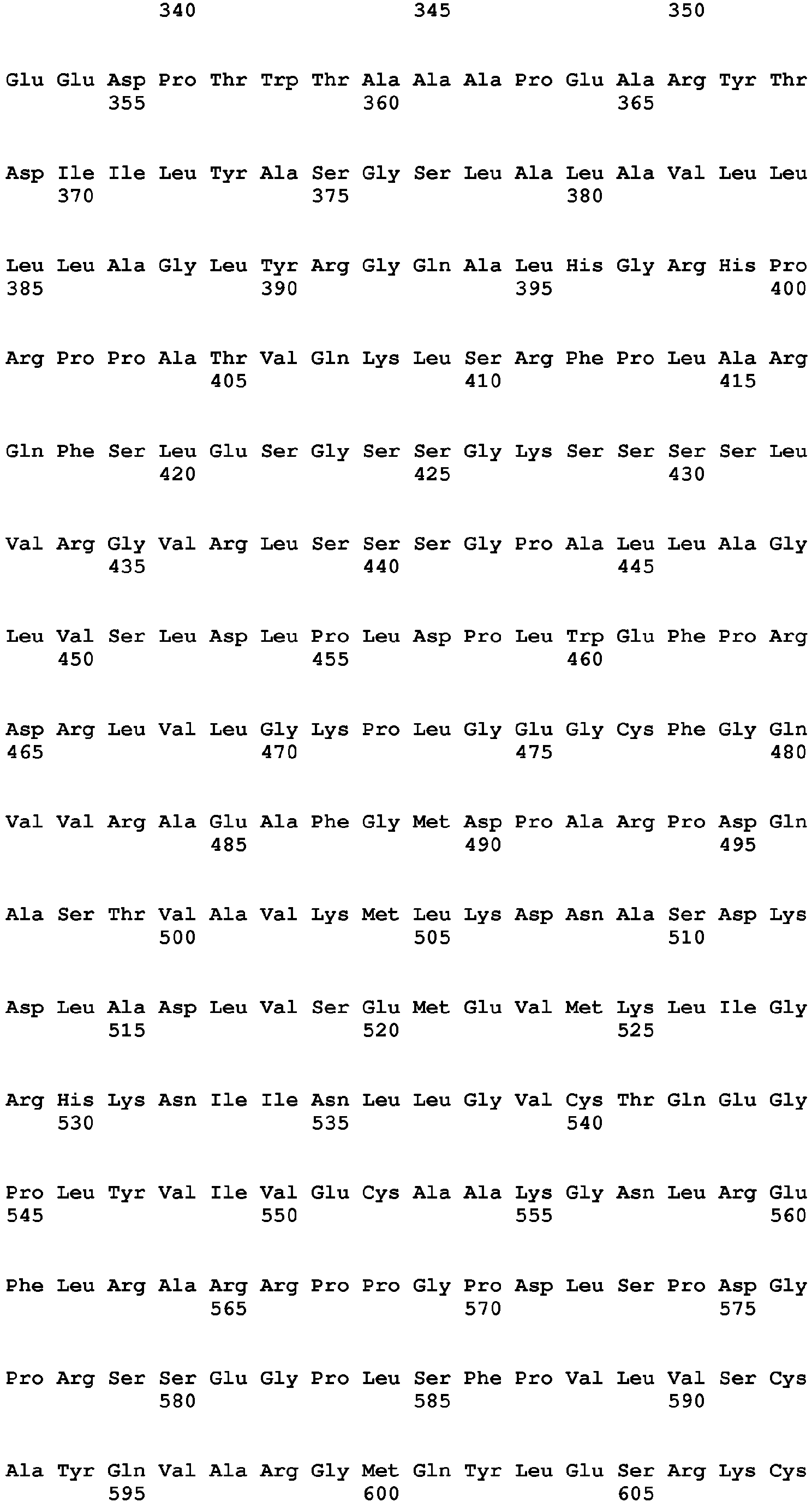


Claims (14)
- A method for detecting a fibroblast growth factor receptor 4 (FGFR4) mutant in a subject, which comprises a step of detecting the presence of a mutation of glycine at position 183 in a FGFR4 tyrosine kinase domain to cysteine in a sample obtained from the subj ect.
- The method according to claim 1, wherein the mutation is a mutation in a FGFR4 described in the following (1) or (2):(1) a mutation of glycine at position 636 in FGFR4 isoform 1 to cysteine; or(2) a mutation of glycine at position 596 in FGFR4 isoform 2 to cysteine.
- The method according to claim 1, wherein the mutation is a mutation in FGFR4 described in the following (1) or (2):(1) a mutation of glycine at position 636 in FGFR4 consisting of the amino acid sequence represented by SEQ ID NO: 2 to cysteine; or(2) a mutation of glycine at position 596 in FGFR4 consisting of the amino acid sequence represented by SEQ ID NO: 4 to cysteine.
- The method according to claim 1, which comprises a step of detecting the presence of a mutation in FGFR4 gene described in the following (1) or (2):(1) a mutation of guanine at position 1906 in FGFR4 isoform 1 gene to thymine; or(2) a mutation of guanine at position 1786 in FGFR4 isoform 2 gene to thymine.
- The method according to claim 1, which comprises a step of detecting the presence of a mutation in FGFR4 gene described in the following (1) or (2):(1) a mutation of guanine at position 1906 in FGFR4 gene consisting of the base sequence represented by SEQ ID NO: 1 to thymine; or(2) a mutation of guanine at position 1786 in FGFR4 gene consisting of the base sequence represented by SEQ ID NO: 3 to thymine.
- The method according to claim 1, which comprises a step of amplifying nucleic acids in a sample obtained from the subject by using a primer set which is designed to amplify a region in FGFR4 gene described in the following (1) or (2):(1) a region comprising a base at position 1906 in FGFR4 gene consisting of the base sequence represented by SEQ ID NO: 1; or(2) a region comprising a base at position 1786 in FGFR4 gene consisting of the base sequence represented by SEQ ID NO: 3.
- The method according to claim 1, which comprises a step of hybridizing a probe which is designed to hybridize to a region comprising a mutation in FGFR4 gene described in the following (1) or (2) with nucleic acids in a sample obtained from the subject under stringent condition:(1) a mutation of guanine at position 1906 in FGFR4 gene consisting of the base sequence represented by SEQ ID NO: 1 to thymine; or(2) a mutation of guanine at position 1786 in FGFR4 gene consisting of the base sequence represented by SEQ ID NO: 3 to thymine.
- The method according to any one of claims 1 to 7, which comprises a step of obtaining a sample from the subject.
- The method according to any one of claims 1 to 7, wherein the subject is a cancer patient.
- The method according to claim 9, wherein the cancer is stomach cancer.
- A method for detecting the presence of cancer in a subject, which comprises the step according any one of claims 1 to 7.
- A method for diagnosing cancer in a subject, which comprises the step according to any one of claims 1 to 7.
- A method for identifying a subject to which treatment by a FGFR4 inhibitor is applied, wherein the subject is a cancer patient, which comprises the step according to any one of claims 1 to 7.
- A method for treating cancer comprising a step of administering a FGFR4 inhibitor to a patient, wherein the patient is identified by the method according to claim 13.
Applications Claiming Priority (2)
| Application Number | Priority Date | Filing Date | Title |
|---|---|---|---|
| JP2011171507 | 2011-08-05 | ||
| PCT/JP2012/069869 WO2013021950A1 (en) | 2011-08-05 | 2012-08-03 | Method for detecting novel fgfr4 mutant |
Publications (2)
| Publication Number | Publication Date |
|---|---|
| EP2740803A1 true EP2740803A1 (en) | 2014-06-11 |
| EP2740803A4 EP2740803A4 (en) | 2015-03-25 |
Family
ID=47668458
Family Applications (1)
| Application Number | Title | Priority Date | Filing Date |
|---|---|---|---|
| EP12822338.5A Withdrawn EP2740803A4 (en) | 2011-08-05 | 2012-08-03 | METHOD FOR DETECTING A NEW MUTANT OF FGFR4 |
Country Status (4)
| Country | Link |
|---|---|
| US (1) | US9267176B2 (en) |
| EP (1) | EP2740803A4 (en) |
| JP (1) | JP6036693B2 (en) |
| WO (1) | WO2013021950A1 (en) |
Families Citing this family (14)
| Publication number | Priority date | Publication date | Assignee | Title |
|---|---|---|---|---|
| EP3365335B1 (en) | 2015-10-23 | 2024-02-14 | Array Biopharma, Inc. | 2-aryl- and 2-heteroaryl-substituted 2-pyridazin-3(2h)-one compounds as inhibitors of fgfr tyrosine kinases |
| US12351571B2 (en) | 2018-12-19 | 2025-07-08 | Array Biopharma Inc. | Substituted quinoxaline compounds as inhibitors of FGFR tyrosine kinases |
| WO2020131627A1 (en) | 2018-12-19 | 2020-06-25 | Array Biopharma Inc. | Substituted pyrazolo[1,5-a]pyridine compounds as inhibitors of fgfr tyrosine kinases |
| WO2021138391A1 (en) | 2019-12-30 | 2021-07-08 | Tyra Biosciences, Inc. | Indazole compounds |
| US20230115945A1 (en) | 2019-12-30 | 2023-04-13 | Tyra Biosciences, Inc. | Aminopyrimidine compounds |
| MX2023007793A (en) | 2020-12-30 | 2023-09-22 | Tyra Biosciences Inc | Indazole compounds as kinase inhibitors. |
| EP4297873A1 (en) | 2021-02-26 | 2024-01-03 | Tyra Biosciences, Inc. | Aminopyrimidine compounds and methods of their use |
| IL317558A (en) | 2022-06-29 | 2025-02-01 | Tyra Biosciences Inc | Indazole compounds |
| AU2023300357A1 (en) | 2022-06-29 | 2025-01-09 | Tyra Biosciences, Inc. | Polymorphic compounds and uses thereof |
| WO2024138112A1 (en) | 2022-12-22 | 2024-06-27 | Tyra Biosciences, Inc. | Indazole compounds |
| AU2024400764A1 (en) | 2023-12-15 | 2026-07-09 | Tyra Biosciences, Inc. | Indazole compounds for the treatment of cancer |
| WO2025222097A1 (en) | 2024-04-19 | 2025-10-23 | Tyra Biosciences, Inc. | Indazole-based protac degraders and their anticancer activity |
| WO2026035809A1 (en) | 2024-08-06 | 2026-02-12 | Tyra Biosciences, Inc. | Indazole compounds for the treatment of cancer |
| WO2026143223A2 (en) | 2024-12-27 | 2026-07-02 | Tyra Biosciences, Inc. | Fgfr inhibitors |
-
2012
- 2012-08-03 JP JP2013528014A patent/JP6036693B2/en not_active Expired - Fee Related
- 2012-08-03 US US14/236,993 patent/US9267176B2/en not_active Expired - Fee Related
- 2012-08-03 WO PCT/JP2012/069869 patent/WO2013021950A1/en not_active Ceased
- 2012-08-03 EP EP12822338.5A patent/EP2740803A4/en not_active Withdrawn
Non-Patent Citations (6)
| Title |
|---|
| EZZAT SHEREEN ET AL: "Dual inhibition of RET and FGFR4 restrains medullary thyroid cancer cell growth", CLINICAL CANCER RESEARCH, THE AMERICAN ASSOCIATION FOR CANCER RESEARCH, US, vol. 11, no. 3, 1 February 2005 (2005-02-01), pages 1336-1341, XP002426782, ISSN: 1078-0432 * |
| JAMES G. TAYLOR VI ET AL: "Identification of FGFR4-activating mutations in human rhabdomyosarcomas that promote metastasis in xenotransplanted models", JOURNAL OF CLINICAL INVESTIGATION, 5 October 2009 (2009-10-05), XP055086226, ISSN: 0021-9738, DOI: 10.1172/JCI39703 * |
| JØRGEN WESCHE ET AL: "Fibroblast growth factors and their receptors in cancer", BIOCHEMICAL JOURNAL, vol. 61, no. 2, 28 June 2011 (2011-06-28), pages 8371-213, XP055142382, ISSN: 0264-6021, DOI: 10.1182/blood-2003-12-4290 * |
| ROIDL A ET AL: "The FGFR4 Y367C mutant is a dominant oncogene in MDA-MB453 breast cancer cells", ONCOGENE, NATURE PUBLISHING GROUP, GB, 30 November 2009 (2009-11-30), pages 1-10, XP002560350, ISSN: 0950-9232, DOI: 10.1038/ONC.2009.432 [retrieved on 2009-11-30] * |
| See also references of WO2013021950A1 * |
| YANWEI YE ET AL: "The Fibroblast Growth Factor Receptor-4 Arg388 Allele is Associated with Gastric Cancer Progression", ANNALS OF SURGICAL ONCOLOGY, SPRINGER-VERLAG, NE, vol. 17, no. 12, 16 September 2010 (2010-09-16), pages 3354-3361, XP019866609, ISSN: 1534-4681, DOI: 10.1245/S10434-010-1323-6 * |
Also Published As
| Publication number | Publication date |
|---|---|
| JP6036693B2 (en) | 2016-11-30 |
| EP2740803A4 (en) | 2015-03-25 |
| US20140220035A1 (en) | 2014-08-07 |
| WO2013021950A1 (en) | 2013-02-14 |
| JPWO2013021950A1 (en) | 2015-03-05 |
| US9267176B2 (en) | 2016-02-23 |
Similar Documents
| Publication | Publication Date | Title |
|---|---|---|
| US9267176B2 (en) | Method for detecting novel FGFR4 mutant | |
| AU2021277633B2 (en) | Use of FGFR mutant gene panels in identifying cancer patients that will be responsive to treatment with an FGFR inhibitor | |
| KR102106962B1 (en) | Novel fgfr3 fusion product | |
| JP2014082984A (en) | Method for detecting novel ntrk2 activating mutation | |
| US9109259B2 (en) | Detection method for novel ROS1 fusions | |
| JP2013226108A (en) | Method for detecting new ntrk2 activating mutation | |
| US20130137111A1 (en) | Detection method of novel ret fusion | |
| JP2015109806A (en) | Method for detecting new ret fused body | |
| JPWO2015012397A1 (en) | NTRK3 fusion detection method | |
| KR20130094342A (en) | Methods and compositions for detecting mutation in the human epidermal growth factor receptor gene | |
| EP3498835A1 (en) | Detection of cldn18-arhgap6 fusion gene or cldn18-arhgap26 fusion gene in pancreatic cancer | |
| WO2010124265A1 (en) | Polymorphisms in angiogenesis pathway genes associated with tumor recurrence in surgery treated cancer patients | |
| EP2744913B1 (en) | Prognostic methodology | |
| JP2014530013A (en) | Mutations in the SF3B1 gene and chronic lymphocytic leukemia | |
| CN102899406A (en) | Sequencing primer for qualitative detection of genetic typing of uridinediphosphoglucuronate glucuronosyltransferase 1A1 and kit thereof | |
| WO2010071405A1 (en) | Markers for detecting predisposition for risk, incidence and progression of osteoarthritis | |
| WO2015129655A1 (en) | Method for detecting dnajb1-prkaca gene | |
| WO2012096325A1 (en) | Method for detecting novel braf fusion product | |
| KR101911074B1 (en) | Single Nucleotide Polymorphisms Determining Trypanosomiasis-resistance of N'Dama breeds and Use Thereof | |
| EP3810808A1 (en) | Methods and kits for detecting egfr mutations | |
| JP6762540B2 (en) | Method for detecting the RP2-ARHGAP6 gene | |
| WO2012115068A1 (en) | Method of detecting mutant fgfr4 in breast cancer | |
| KR20200002933A (en) | Method for measuring expression level of ABL1 T315I mutation | |
| CN120384128A (en) | A group of gastric cancer detection markers and their uses | |
| KR20130022435A (en) | Snp for diagnosing susceptibility to gastric cancer and method for diagnosing susceptibility to gastric cancer using the lox gene snp |
Legal Events
| Date | Code | Title | Description |
|---|---|---|---|
| PUAI | Public reference made under article 153(3) epc to a published international application that has entered the european phase |
Free format text: ORIGINAL CODE: 0009012 |
|
| 17P | Request for examination filed |
Effective date: 20140131 |
|
| AK | Designated contracting states |
Kind code of ref document: A1 Designated state(s): AL AT BE BG CH CY CZ DE DK EE ES FI FR GB GR HR HU IE IS IT LI LT LU LV MC MK MT NL NO PL PT RO RS SE SI SK SM TR |
|
| DAX | Request for extension of the european patent (deleted) | ||
| A4 | Supplementary search report drawn up and despatched |
Effective date: 20150220 |
|
| RIC1 | Information provided on ipc code assigned before grant |
Ipc: C12N 15/09 20060101ALI20150216BHEP Ipc: C12Q 1/68 20060101AFI20150216BHEP |
|
| 17Q | First examination report despatched |
Effective date: 20161020 |
|
| GRAP | Despatch of communication of intention to grant a patent |
Free format text: ORIGINAL CODE: EPIDOSNIGR1 |
|
| INTG | Intention to grant announced |
Effective date: 20170721 |
|
| STAA | Information on the status of an ep patent application or granted ep patent |
Free format text: STATUS: THE APPLICATION IS DEEMED TO BE WITHDRAWN |
|
| 18D | Application deemed to be withdrawn |
Effective date: 20171201 |