EP2725094A1 - Epithelial tissue model - Google Patents
Epithelial tissue model Download PDFInfo
- Publication number
- EP2725094A1 EP2725094A1 EP12190442.9A EP12190442A EP2725094A1 EP 2725094 A1 EP2725094 A1 EP 2725094A1 EP 12190442 A EP12190442 A EP 12190442A EP 2725094 A1 EP2725094 A1 EP 2725094A1
- Authority
- EP
- European Patent Office
- Prior art keywords
- vial
- epithelial tissue
- segment
- container
- tissue
- Prior art date
- Legal status (The legal status is an assumption and is not a legal conclusion. Google has not performed a legal analysis and makes no representation as to the accuracy of the status listed.)
- Withdrawn
Links
- 210000000981 epithelium Anatomy 0.000 title claims abstract description 107
- 238000013334 tissue model Methods 0.000 title description 6
- 238000000034 method Methods 0.000 claims abstract description 31
- 230000032258 transport Effects 0.000 claims abstract description 27
- 238000010521 absorption reaction Methods 0.000 claims abstract description 15
- 230000028327 secretion Effects 0.000 claims abstract description 13
- 239000000126 substance Substances 0.000 claims description 15
- 239000000463 material Substances 0.000 claims description 11
- 239000003550 marker Substances 0.000 claims description 10
- 239000004743 Polypropylene Substances 0.000 claims description 4
- -1 polypropylene Polymers 0.000 claims description 4
- 229920001155 polypropylene Polymers 0.000 claims description 4
- 239000004677 Nylon Substances 0.000 claims description 2
- 239000004809 Teflon Substances 0.000 claims description 2
- 229920006362 Teflon® Polymers 0.000 claims description 2
- 238000001816 cooling Methods 0.000 claims description 2
- 229920001778 nylon Polymers 0.000 claims description 2
- 210000001519 tissue Anatomy 0.000 abstract description 50
- 238000005259 measurement Methods 0.000 abstract description 6
- 230000000968 intestinal effect Effects 0.000 description 32
- 238000011534 incubation Methods 0.000 description 24
- 210000004027 cell Anatomy 0.000 description 16
- 150000001875 compounds Chemical class 0.000 description 16
- 239000000872 buffer Substances 0.000 description 15
- 230000035699 permeability Effects 0.000 description 15
- 241001465754 Metazoa Species 0.000 description 12
- 239000011521 glass Substances 0.000 description 12
- RYYVLZVUVIJVGH-YZRHJBSPSA-N 1,3,7-trimethylpurine-2,6-dione Chemical compound CN1C(=O)N([14CH3])C(=O)C2=C1N=CN2C RYYVLZVUVIJVGH-YZRHJBSPSA-N 0.000 description 11
- 102000003855 L-lactate dehydrogenase Human genes 0.000 description 10
- 108700023483 L-lactate dehydrogenases Proteins 0.000 description 10
- RYYVLZVUVIJVGH-UHFFFAOYSA-N caffeine Chemical compound CN1C(=O)N(C)C(=O)C2=C1N=CN2C RYYVLZVUVIJVGH-UHFFFAOYSA-N 0.000 description 10
- 238000004458 analytical method Methods 0.000 description 9
- CURLTUGMZLYLDI-UHFFFAOYSA-N Carbon dioxide Chemical compound O=C=O CURLTUGMZLYLDI-UHFFFAOYSA-N 0.000 description 8
- 239000002158 endotoxin Substances 0.000 description 8
- 239000000594 mannitol Substances 0.000 description 8
- 238000012360 testing method Methods 0.000 description 8
- UQVKZNNCIHJZLS-UHFFFAOYSA-N PhIP Chemical compound C1=C2N(C)C(N)=NC2=NC=C1C1=CC=CC=C1 UQVKZNNCIHJZLS-UHFFFAOYSA-N 0.000 description 7
- UIIMBOGNXHQVGW-UHFFFAOYSA-M Sodium bicarbonate Chemical compound [Na+].OC([O-])=O UIIMBOGNXHQVGW-UHFFFAOYSA-M 0.000 description 6
- 229920006008 lipopolysaccharide Polymers 0.000 description 6
- LPHGQDQBBGAPDZ-UHFFFAOYSA-N Isocaffeine Natural products CN1C(=O)N(C)C(=O)C2=C1N(C)C=N2 LPHGQDQBBGAPDZ-UHFFFAOYSA-N 0.000 description 5
- 229960001948 caffeine Drugs 0.000 description 5
- VJEONQKOZGKCAK-UHFFFAOYSA-N caffeine Natural products CN1C(=O)N(C)C(=O)C2=C1C=CN2C VJEONQKOZGKCAK-UHFFFAOYSA-N 0.000 description 5
- 230000006870 function Effects 0.000 description 5
- 210000004907 gland Anatomy 0.000 description 5
- 210000005127 stratified epithelium Anatomy 0.000 description 5
- 238000002965 ELISA Methods 0.000 description 4
- 229910002092 carbon dioxide Inorganic materials 0.000 description 4
- 238000009792 diffusion process Methods 0.000 description 4
- 230000000694 effects Effects 0.000 description 4
- 238000004128 high performance liquid chromatography Methods 0.000 description 4
- 210000000936 intestine Anatomy 0.000 description 4
- 230000003834 intracellular effect Effects 0.000 description 4
- 238000004895 liquid chromatography mass spectrometry Methods 0.000 description 4
- 238000001294 liquid chromatography-tandem mass spectrometry Methods 0.000 description 4
- 210000004379 membrane Anatomy 0.000 description 4
- 239000012528 membrane Substances 0.000 description 4
- 108090000623 proteins and genes Proteins 0.000 description 4
- 210000001187 pylorus Anatomy 0.000 description 4
- 238000003127 radioimmunoassay Methods 0.000 description 4
- 239000000243 solution Substances 0.000 description 4
- 102100022595 Broad substrate specificity ATP-binding cassette transporter ABCG2 Human genes 0.000 description 3
- 229920002307 Dextran Polymers 0.000 description 3
- 102000004190 Enzymes Human genes 0.000 description 3
- 108090000790 Enzymes Proteins 0.000 description 3
- HEMHJVSKTPXQMS-UHFFFAOYSA-M Sodium hydroxide Chemical compound [OH-].[Na+] HEMHJVSKTPXQMS-UHFFFAOYSA-M 0.000 description 3
- 241000282887 Suidae Species 0.000 description 3
- 241000282898 Sus scrofa Species 0.000 description 3
- 238000002835 absorbance Methods 0.000 description 3
- 230000037396 body weight Effects 0.000 description 3
- 238000011161 development Methods 0.000 description 3
- 210000001198 duodenum Anatomy 0.000 description 3
- 238000002474 experimental method Methods 0.000 description 3
- 210000001035 gastrointestinal tract Anatomy 0.000 description 3
- 210000003405 ileum Anatomy 0.000 description 3
- 210000001630 jejunum Anatomy 0.000 description 3
- 229910000030 sodium bicarbonate Inorganic materials 0.000 description 3
- JKMHFZQWWAIEOD-UHFFFAOYSA-N 2-[4-(2-hydroxyethyl)piperazin-1-yl]ethanesulfonic acid Chemical compound OCC[NH+]1CCN(CCS([O-])(=O)=O)CC1 JKMHFZQWWAIEOD-UHFFFAOYSA-N 0.000 description 2
- 241000282412 Homo Species 0.000 description 2
- 101000823298 Homo sapiens Broad substrate specificity ATP-binding cassette transporter ABCG2 Proteins 0.000 description 2
- TWRXJAOTZQYOKJ-UHFFFAOYSA-L Magnesium chloride Chemical compound [Mg+2].[Cl-].[Cl-] TWRXJAOTZQYOKJ-UHFFFAOYSA-L 0.000 description 2
- 241000124008 Mammalia Species 0.000 description 2
- 239000004793 Polystyrene Substances 0.000 description 2
- XUIMIQQOPSSXEZ-UHFFFAOYSA-N Silicon Chemical group [Si] XUIMIQQOPSSXEZ-UHFFFAOYSA-N 0.000 description 2
- FAPWRFPIFSIZLT-UHFFFAOYSA-M Sodium chloride Chemical compound [Na+].[Cl-] FAPWRFPIFSIZLT-UHFFFAOYSA-M 0.000 description 2
- 241000251539 Vertebrata <Metazoa> Species 0.000 description 2
- 230000003187 abdominal effect Effects 0.000 description 2
- XAGFODPZIPBFFR-UHFFFAOYSA-N aluminium Chemical compound [Al] XAGFODPZIPBFFR-UHFFFAOYSA-N 0.000 description 2
- 229910052782 aluminium Inorganic materials 0.000 description 2
- 210000004082 barrier epithelial cell Anatomy 0.000 description 2
- 230000004888 barrier function Effects 0.000 description 2
- 210000002469 basement membrane Anatomy 0.000 description 2
- 239000008280 blood Substances 0.000 description 2
- 210000004369 blood Anatomy 0.000 description 2
- 235000021152 breakfast Nutrition 0.000 description 2
- 238000002788 crimping Methods 0.000 description 2
- 239000003814 drug Substances 0.000 description 2
- 230000004890 epithelial barrier function Effects 0.000 description 2
- 210000002919 epithelial cell Anatomy 0.000 description 2
- MHMNJMPURVTYEJ-UHFFFAOYSA-N fluorescein-5-isothiocyanate Chemical compound O1C(=O)C2=CC(N=C=S)=CC=C2C21C1=CC=C(O)C=C1OC1=CC(O)=CC=C21 MHMNJMPURVTYEJ-UHFFFAOYSA-N 0.000 description 2
- 235000013305 food Nutrition 0.000 description 2
- 229920001903 high density polyethylene Polymers 0.000 description 2
- 239000004700 high-density polyethylene Substances 0.000 description 2
- 239000005556 hormone Substances 0.000 description 2
- 229940088597 hormone Drugs 0.000 description 2
- 230000006872 improvement Effects 0.000 description 2
- 238000000338 in vitro Methods 0.000 description 2
- 238000001727 in vivo Methods 0.000 description 2
- 230000003993 interaction Effects 0.000 description 2
- 210000002429 large intestine Anatomy 0.000 description 2
- 239000007788 liquid Substances 0.000 description 2
- 238000005567 liquid scintillation counting Methods 0.000 description 2
- 229920001684 low density polyethylene Polymers 0.000 description 2
- 239000004702 low-density polyethylene Substances 0.000 description 2
- 235000016709 nutrition Nutrition 0.000 description 2
- 229920000306 polymethylpentene Polymers 0.000 description 2
- 239000011116 polymethylpentene Substances 0.000 description 2
- 229920002223 polystyrene Polymers 0.000 description 2
- 230000008569 process Effects 0.000 description 2
- 230000002285 radioactive effect Effects 0.000 description 2
- 210000005122 simple epithelium Anatomy 0.000 description 2
- 210000003491 skin Anatomy 0.000 description 2
- 210000000813 small intestine Anatomy 0.000 description 2
- 235000017557 sodium bicarbonate Nutrition 0.000 description 2
- 241000894007 species Species 0.000 description 2
- 230000008719 thickening Effects 0.000 description 2
- 230000005945 translocation Effects 0.000 description 2
- 230000035899 viability Effects 0.000 description 2
- XLYOFNOQVPJJNP-UHFFFAOYSA-N water Substances O XLYOFNOQVPJJNP-UHFFFAOYSA-N 0.000 description 2
- 238000012604 3D cell culture Methods 0.000 description 1
- 206010002091 Anaesthesia Diseases 0.000 description 1
- 208000027496 Behcet disease Diseases 0.000 description 1
- BVKZGUZCCUSVTD-UHFFFAOYSA-M Bicarbonate Chemical compound OC([O-])=O BVKZGUZCCUSVTD-UHFFFAOYSA-M 0.000 description 1
- 108010078791 Carrier Proteins Proteins 0.000 description 1
- 206010009895 Colitis ischaemic Diseases 0.000 description 1
- 206010056979 Colitis microscopic Diseases 0.000 description 1
- 206010009900 Colitis ulcerative Diseases 0.000 description 1
- 208000011231 Crohn disease Diseases 0.000 description 1
- FBPFZTCFMRRESA-KVTDHHQDSA-N D-Mannitol Chemical compound OC[C@@H](O)[C@@H](O)[C@H](O)[C@H](O)CO FBPFZTCFMRRESA-KVTDHHQDSA-N 0.000 description 1
- 102000010834 Extracellular Matrix Proteins Human genes 0.000 description 1
- 108010037362 Extracellular Matrix Proteins Proteins 0.000 description 1
- WQZGKKKJIJFFOK-GASJEMHNSA-N Glucose Natural products OC[C@H]1OC(O)[C@H](O)[C@@H](O)[C@@H]1O WQZGKKKJIJFFOK-GASJEMHNSA-N 0.000 description 1
- 239000007995 HEPES buffer Substances 0.000 description 1
- 208000022559 Inflammatory bowel disease Diseases 0.000 description 1
- 208000005615 Interstitial Cystitis Diseases 0.000 description 1
- 102000004310 Ion Channels Human genes 0.000 description 1
- 102000011782 Keratins Human genes 0.000 description 1
- 108010076876 Keratins Proteins 0.000 description 1
- 229930195725 Mannitol Natural products 0.000 description 1
- 108010090306 Member 2 Subfamily G ATP Binding Cassette Transporter Proteins 0.000 description 1
- 108090000301 Membrane transport proteins Proteins 0.000 description 1
- 102000003939 Membrane transport proteins Human genes 0.000 description 1
- 241000736262 Microbiota Species 0.000 description 1
- 102000035195 Peptidases Human genes 0.000 description 1
- 108091005804 Peptidases Proteins 0.000 description 1
- 241000288906 Primates Species 0.000 description 1
- 238000012356 Product development Methods 0.000 description 1
- 239000004365 Protease Substances 0.000 description 1
- 201000004681 Psoriasis Diseases 0.000 description 1
- 241000283984 Rodentia Species 0.000 description 1
- 206010039710 Scleroderma Diseases 0.000 description 1
- 229910021607 Silver chloride Inorganic materials 0.000 description 1
- 102000000591 Tight Junction Proteins Human genes 0.000 description 1
- 108010002321 Tight Junction Proteins Proteins 0.000 description 1
- 201000006704 Ulcerative Colitis Diseases 0.000 description 1
- 230000009056 active transport Effects 0.000 description 1
- 230000037005 anaesthesia Effects 0.000 description 1
- 210000000436 anus Anatomy 0.000 description 1
- 238000013459 approach Methods 0.000 description 1
- 239000007864 aqueous solution Substances 0.000 description 1
- 238000003556 assay Methods 0.000 description 1
- QVGXLLKOCUKJST-UHFFFAOYSA-N atomic oxygen Chemical compound [O] QVGXLLKOCUKJST-UHFFFAOYSA-N 0.000 description 1
- 210000001142 back Anatomy 0.000 description 1
- 239000001569 carbon dioxide Substances 0.000 description 1
- 238000004113 cell culture Methods 0.000 description 1
- 230000005779 cell damage Effects 0.000 description 1
- 208000037887 cell injury Diseases 0.000 description 1
- 230000001413 cellular effect Effects 0.000 description 1
- 230000007248 cellular mechanism Effects 0.000 description 1
- 239000000919 ceramic Substances 0.000 description 1
- 230000008859 change Effects 0.000 description 1
- 239000003153 chemical reaction reagent Substances 0.000 description 1
- 208000008609 collagenous colitis Diseases 0.000 description 1
- 210000001072 colon Anatomy 0.000 description 1
- 210000002808 connective tissue Anatomy 0.000 description 1
- 238000011109 contamination Methods 0.000 description 1
- 239000006071 cream Substances 0.000 description 1
- 230000001086 cytosolic effect Effects 0.000 description 1
- 230000001419 dependent effect Effects 0.000 description 1
- BNIILDVGGAEEIG-UHFFFAOYSA-L disodium hydrogen phosphate Chemical compound [Na+].[Na+].OP([O-])([O-])=O BNIILDVGGAEEIG-UHFFFAOYSA-L 0.000 description 1
- 229910000397 disodium phosphate Inorganic materials 0.000 description 1
- 201000008243 diversion colitis Diseases 0.000 description 1
- 239000008151 electrolyte solution Substances 0.000 description 1
- 230000008030 elimination Effects 0.000 description 1
- 238000003379 elimination reaction Methods 0.000 description 1
- 210000003038 endothelium Anatomy 0.000 description 1
- 238000005516 engineering process Methods 0.000 description 1
- 239000003797 essential amino acid Substances 0.000 description 1
- 235000020776 essential amino acid Nutrition 0.000 description 1
- 238000001704 evaporation Methods 0.000 description 1
- 230000008020 evaporation Effects 0.000 description 1
- 210000002744 extracellular matrix Anatomy 0.000 description 1
- 239000012530 fluid Substances 0.000 description 1
- GNBHRKFJIUUOQI-UHFFFAOYSA-N fluorescein Chemical compound O1C(=O)C2=CC=CC=C2C21C1=CC=C(O)C=C1OC1=CC(O)=CC=C21 GNBHRKFJIUUOQI-UHFFFAOYSA-N 0.000 description 1
- 230000004907 flux Effects 0.000 description 1
- 239000007789 gas Substances 0.000 description 1
- 238000003197 gene knockdown Methods 0.000 description 1
- 230000000762 glandular Effects 0.000 description 1
- 210000002175 goblet cell Anatomy 0.000 description 1
- 239000003102 growth factor Substances 0.000 description 1
- 210000001983 hard palate Anatomy 0.000 description 1
- 201000000615 hard palate cancer Diseases 0.000 description 1
- 238000000126 in silico method Methods 0.000 description 1
- 208000027138 indeterminate colitis Diseases 0.000 description 1
- 210000005027 intestinal barrier Anatomy 0.000 description 1
- 230000007358 intestinal barrier function Effects 0.000 description 1
- 238000011835 investigation Methods 0.000 description 1
- 230000037427 ion transport Effects 0.000 description 1
- YWXYYJSYQOXTPL-SLPGGIOYSA-N isosorbide mononitrate Chemical compound [O-][N+](=O)O[C@@H]1CO[C@@H]2[C@@H](O)CO[C@@H]21 YWXYYJSYQOXTPL-SLPGGIOYSA-N 0.000 description 1
- 210000005128 keratinized epithelium Anatomy 0.000 description 1
- 210000000738 kidney tubule Anatomy 0.000 description 1
- 210000004072 lung Anatomy 0.000 description 1
- 206010025135 lupus erythematosus Diseases 0.000 description 1
- 208000004341 lymphocytic colitis Diseases 0.000 description 1
- 229910001629 magnesium chloride Inorganic materials 0.000 description 1
- 235000010355 mannitol Nutrition 0.000 description 1
- 230000001404 mediated effect Effects 0.000 description 1
- 230000004060 metabolic process Effects 0.000 description 1
- 239000008267 milk Substances 0.000 description 1
- 210000004080 milk Anatomy 0.000 description 1
- 235000013336 milk Nutrition 0.000 description 1
- 230000003278 mimic effect Effects 0.000 description 1
- 210000004877 mucosa Anatomy 0.000 description 1
- 210000003097 mucus Anatomy 0.000 description 1
- 210000003205 muscle Anatomy 0.000 description 1
- 230000003387 muscular Effects 0.000 description 1
- 239000003471 mutagenic agent Substances 0.000 description 1
- 231100000707 mutagenic chemical Toxicity 0.000 description 1
- 230000003505 mutagenic effect Effects 0.000 description 1
- 230000001537 neural effect Effects 0.000 description 1
- 239000002858 neurotransmitter agent Substances 0.000 description 1
- 230000008520 organization Effects 0.000 description 1
- 210000001672 ovary Anatomy 0.000 description 1
- 229910052760 oxygen Inorganic materials 0.000 description 1
- 239000001301 oxygen Substances 0.000 description 1
- 230000007310 pathophysiology Effects 0.000 description 1
- 210000003200 peritoneal cavity Anatomy 0.000 description 1
- 230000035479 physiological effects, processes and functions Effects 0.000 description 1
- 230000035790 physiological processes and functions Effects 0.000 description 1
- 210000003281 pleural cavity Anatomy 0.000 description 1
- 238000002360 preparation method Methods 0.000 description 1
- 238000012545 processing Methods 0.000 description 1
- 102000004169 proteins and genes Human genes 0.000 description 1
- 238000004080 punching Methods 0.000 description 1
- 210000000664 rectum Anatomy 0.000 description 1
- 238000002271 resection Methods 0.000 description 1
- 210000003296 saliva Anatomy 0.000 description 1
- 210000003079 salivary gland Anatomy 0.000 description 1
- 230000003248 secreting effect Effects 0.000 description 1
- HKZLPVFGJNLROG-UHFFFAOYSA-M silver monochloride Chemical compound [Cl-].[Ag+] HKZLPVFGJNLROG-UHFFFAOYSA-M 0.000 description 1
- 210000005125 simple columnar epithelium Anatomy 0.000 description 1
- 210000005124 simple cuboidal epithelium Anatomy 0.000 description 1
- 239000011780 sodium chloride Substances 0.000 description 1
- AJPJDKMHJJGVTQ-UHFFFAOYSA-M sodium dihydrogen phosphate Chemical compound [Na+].OP(O)([O-])=O AJPJDKMHJJGVTQ-UHFFFAOYSA-M 0.000 description 1
- 229910000162 sodium phosphate Inorganic materials 0.000 description 1
- 210000002784 stomach Anatomy 0.000 description 1
- 239000000758 substrate Substances 0.000 description 1
- 210000004243 sweat Anatomy 0.000 description 1
- 210000004876 tela submucosa Anatomy 0.000 description 1
- 210000001550 testis Anatomy 0.000 description 1
- 210000001578 tight junction Anatomy 0.000 description 1
- 230000036962 time dependent Effects 0.000 description 1
- 238000012546 transfer Methods 0.000 description 1
- 230000009261 transgenic effect Effects 0.000 description 1
- 210000001215 vagina Anatomy 0.000 description 1
Images







Classifications
-
- G—PHYSICS
- G01—MEASURING; TESTING
- G01N—INVESTIGATING OR ANALYSING MATERIALS BY DETERMINING THEIR CHEMICAL OR PHYSICAL PROPERTIES
- G01N33/00—Investigating or analysing materials by specific methods not covered by groups G01N1/00 - G01N31/00
- G01N33/48—Biological material, e.g. blood, urine; Haemocytometers
- G01N33/50—Chemical analysis of biological material, e.g. blood, urine; Testing involving biospecific ligand binding methods; Immunological testing
- G01N33/5005—Chemical analysis of biological material, e.g. blood, urine; Testing involving biospecific ligand binding methods; Immunological testing involving human or animal cells
- G01N33/5008—Chemical analysis of biological material, e.g. blood, urine; Testing involving biospecific ligand binding methods; Immunological testing involving human or animal cells for testing or evaluating the effect of chemical or biological compounds, e.g. drugs, cosmetics
- G01N33/502—Chemical analysis of biological material, e.g. blood, urine; Testing involving biospecific ligand binding methods; Immunological testing involving human or animal cells for testing or evaluating the effect of chemical or biological compounds, e.g. drugs, cosmetics for testing non-proliferative effects
- G01N33/5035—Chemical analysis of biological material, e.g. blood, urine; Testing involving biospecific ligand binding methods; Immunological testing involving human or animal cells for testing or evaluating the effect of chemical or biological compounds, e.g. drugs, cosmetics for testing non-proliferative effects on sub-cellular localization
-
- B—PERFORMING OPERATIONS; TRANSPORTING
- B01—PHYSICAL OR CHEMICAL PROCESSES OR APPARATUS IN GENERAL
- B01L—CHEMICAL OR PHYSICAL LABORATORY APPARATUS FOR GENERAL USE
- B01L3/00—Containers or dishes for laboratory use, e.g. laboratory glassware; Droppers
- B01L3/50—Containers for the purpose of retaining a material to be analysed, e.g. test tubes
- B01L3/502—Containers for the purpose of retaining a material to be analysed, e.g. test tubes with fluid transport, e.g. in multi-compartment structures
- B01L3/5021—Test tubes specially adapted for centrifugation purposes
-
- B—PERFORMING OPERATIONS; TRANSPORTING
- B01—PHYSICAL OR CHEMICAL PROCESSES OR APPARATUS IN GENERAL
- B01L—CHEMICAL OR PHYSICAL LABORATORY APPARATUS FOR GENERAL USE
- B01L3/00—Containers or dishes for laboratory use, e.g. laboratory glassware; Droppers
- B01L3/50—Containers for the purpose of retaining a material to be analysed, e.g. test tubes
- B01L3/502—Containers for the purpose of retaining a material to be analysed, e.g. test tubes with fluid transport, e.g. in multi-compartment structures
- B01L3/5025—Containers for the purpose of retaining a material to be analysed, e.g. test tubes with fluid transport, e.g. in multi-compartment structures for parallel transport of multiple samples
- B01L3/50255—Multi-well filtration
-
- C—CHEMISTRY; METALLURGY
- C12—BIOCHEMISTRY; BEER; SPIRITS; WINE; VINEGAR; MICROBIOLOGY; ENZYMOLOGY; MUTATION OR GENETIC ENGINEERING
- C12M—APPARATUS FOR ENZYMOLOGY OR MICROBIOLOGY; APPARATUS FOR CULTURING MICROORGANISMS FOR PRODUCING BIOMASS, FOR GROWING CELLS OR FOR OBTAINING FERMENTATION OR METABOLIC PRODUCTS, i.e. BIOREACTORS OR FERMENTERS
- C12M21/00—Bioreactors or fermenters specially adapted for specific uses
- C12M21/08—Bioreactors or fermenters specially adapted for specific uses for producing artificial tissue or for ex-vivo cultivation of tissue
-
- C—CHEMISTRY; METALLURGY
- C12—BIOCHEMISTRY; BEER; SPIRITS; WINE; VINEGAR; MICROBIOLOGY; ENZYMOLOGY; MUTATION OR GENETIC ENGINEERING
- C12M—APPARATUS FOR ENZYMOLOGY OR MICROBIOLOGY; APPARATUS FOR CULTURING MICROORGANISMS FOR PRODUCING BIOMASS, FOR GROWING CELLS OR FOR OBTAINING FERMENTATION OR METABOLIC PRODUCTS, i.e. BIOREACTORS OR FERMENTERS
- C12M25/00—Means for supporting, enclosing or fixing the microorganisms, e.g. immunocoatings
- C12M25/02—Membranes; Filters
- C12M25/04—Membranes; Filters in combination with well or multiwell plates, i.e. culture inserts
-
- C—CHEMISTRY; METALLURGY
- C12—BIOCHEMISTRY; BEER; SPIRITS; WINE; VINEGAR; MICROBIOLOGY; ENZYMOLOGY; MUTATION OR GENETIC ENGINEERING
- C12Q—MEASURING OR TESTING PROCESSES INVOLVING ENZYMES, NUCLEIC ACIDS OR MICROORGANISMS; COMPOSITIONS OR TEST PAPERS THEREFOR; PROCESSES OF PREPARING SUCH COMPOSITIONS; CONDITION-RESPONSIVE CONTROL IN MICROBIOLOGICAL OR ENZYMOLOGICAL PROCESSES
- C12Q1/00—Measuring or testing processes involving enzymes, nucleic acids or microorganisms; Compositions therefor; Processes of preparing such compositions
- C12Q1/26—Measuring or testing processes involving enzymes, nucleic acids or microorganisms; Compositions therefor; Processes of preparing such compositions involving oxidoreductase
- C12Q1/32—Measuring or testing processes involving enzymes, nucleic acids or microorganisms; Compositions therefor; Processes of preparing such compositions involving oxidoreductase involving dehydrogenase
-
- B—PERFORMING OPERATIONS; TRANSPORTING
- B01—PHYSICAL OR CHEMICAL PROCESSES OR APPARATUS IN GENERAL
- B01L—CHEMICAL OR PHYSICAL LABORATORY APPARATUS FOR GENERAL USE
- B01L2300/00—Additional constructional details
- B01L2300/06—Auxiliary integrated devices, integrated components
- B01L2300/0609—Holders integrated in container to position an object
-
- B—PERFORMING OPERATIONS; TRANSPORTING
- B01—PHYSICAL OR CHEMICAL PROCESSES OR APPARATUS IN GENERAL
- B01L—CHEMICAL OR PHYSICAL LABORATORY APPARATUS FOR GENERAL USE
- B01L2300/00—Additional constructional details
- B01L2300/08—Geometry, shape and general structure
- B01L2300/0809—Geometry, shape and general structure rectangular shaped
- B01L2300/0829—Multi-well plates; Microtitration plates
-
- G—PHYSICS
- G01—MEASURING; TESTING
- G01N—INVESTIGATING OR ANALYSING MATERIALS BY DETERMINING THEIR CHEMICAL OR PHYSICAL PROPERTIES
- G01N2333/00—Assays involving biological materials from specific organisms or of a specific nature
- G01N2333/90—Enzymes; Proenzymes
- G01N2333/902—Oxidoreductases (1.)
- G01N2333/904—Oxidoreductases (1.) acting on CHOH groups as donors, e.g. glucose oxidase, lactate dehydrogenase (1.1)
Definitions
- the invention relates to an improvement of a device for measuring absorption, transport and/or physiological functions across an epithelial tissue. More specifically, it relates to an improvement which allows the horizontal mounting of epithelial tissues with varying thickness, allowing direct contact of a compound under investigation with the epithelial tissue.
- the improved device can be assembled into multiwell formats, allowing the simultaneous analyses of multiple measurements.
- standardized multiwell formats allow the use of robotics for automation of various liquid handlings.
- the present invention is directed to a novel device that allows horizontal mounting of a segment of epithelial tissue.
- the horizontal mounting enables the direct contact of test compounds with the epithelial tissue.
- segments of epithelial tissue with varying thickness can easily be mounted in the device.
- Multiple devices can easily be arrayed allowing medium throughput analyses of absorption, transport and/or secretion across the epithelial tissue.
- the device according to the invention is generated from disposable material, which will reduce a risk of contamination of the mounted device.
- the invention therefore provides a vial for holding a segment of epithelial tissue, the vial having a neck and a first open end at the top of the neck, whereby a rim surrounds the open end, the rim having an upper surface facing the open end, whereby the neck and the rim of the first open end of the vial are suited for capping with a crimp cap, the vial characterized by having a second open end distal to the neck.
- Crimp cap vials having a closed end distal to the open crimp capped end are known in the art.
- 2 ml (12 x 32 mm) crimp top vials are available from Agilent Technologies (Santa Clara, CA).
- Crimp top vials ranging between 0.1 ml and 1.2 ml are available from Fisher Scientific (Pittsburgh, PA).
- Preferred crimp top vials are headspace vials, for example 20 ml headspace vials from Alltech Associates, Inc (Deerfield, Illinois).
- Possible internal diameters of the crimp cap vials at the open crimp capped end range from 4.05 mm (0.75 ml crimp top vial) to 12.75 mm (20 ml crimp top vial).
- Corresponding exposure surface areas could range from 0.52 cm 2 (0.75 ml crimp top vial) to 3.17 cm 2 (20 ml crimp top vial), based on the opening in the crimp caps.
- a vial according to the invention can easily be prepared by removing the closed end distal from a known crimp cap vial, for example with a glass cutter.
- the resulting internal diameters of the opened distal end range from 6.55 mm (0.75 ml crimp top vial) to 20.65 mm (20 ml crimp top vial).
- a vial according to the invention may be composed of any material known in the art, including glass, polypropylene, low-density polyethylene, high-density polyethylene, polymethyl-pentene and polystyrene.
- a preferred vial according to the invention is composed of glass such as, for example, borosilicate clear glass Type 1, Class A or 51A amber glass.
- Said crimp cap is, for example, a Poly CrimpTM Seal, which is available from F.J. Finneran (Vineland, NJ) or, preferably, a standard crimp cap, such as a standard aluminum crimp cap, for example aluminum knurled caps from Perkin Elmer (the Netherlands).
- Methods for providing a crimp cap onto a crimp cap vial are known in the art, including vial crimping hand tools and automated vial crimpers. Suitable crimping tools are available, for example, from Kebby Industries, Inc (Rockford, IL) and from Alltech Associates, Inc (Deerfield, Illinois) such as a hand crimper and a hand decapper.
- the invention further provides a vial according to the invention comprising a segment of epithelial tissue, the segment being positioned on the upper surface of the rim, with the apical side of the epithelial tissue facing the first open end, whereby a ring of inert material is positioned on one side, preferably the apical side, of the epithelial tissue, and whereby a crimp cap provides a tight seal of the first end of the vial.
- Epithelial tissue is specialized tissue that forms the lining of all internal and external body surfaces. Epithelial tissue is made up of cells closely packed and ranged in one or more layers. Epithelial tissue that occurs on surfaces on the interior of the body is known as endothelium. Epithelial tissue, regardless of the type, is usually separated from the underlying tissue by a thin sheet of connective tissue, which is termed the basement membrane. The basement membrane provides structural support for the epithelium and also binds it to neighboring structures.
- Epithelial tissue which is only one cell thick is known as simple epithelium. If it is two or more cells thick such as the skin, it is known as stratified epithelium.
- Simple epithelium can be subdivided according to the shape and function of its cells. Squamous (pavement) epithelium lines areas where passive diffusion of gases occur, for example capillaries, the pericardial, pleural, and peritoneal cavities, as well as the alveoli of the lungs. Simple cuboidal epithelium is found in glands and in the lining of the kidney tubules as well as in the ducts of the glands. This epithelium also constitutes the germinal epithelium which produces the egg cells in the female ovary and the sperm cells in the male testes.
- Glandular epithelium with goblet cells is often present in specialised gland cells which are capable of synthesising and secreting certain substances such as enzymes, hormones, milk, mucus, sweat, wax and saliva.
- a portion of the epithelial tissue often becomes invaginated, thereby forming a multicellular gland.
- An example of a multicellular gland is the salivary gland.
- Stratified epithelium is composed of several layers of cells.
- the top cells are often flat and scaly and it may or may not be keratinized (i.e. containing a tough, resistant protein called keratin).
- the mammalian skin comprises dry, keratinized, stratified epithelium. Further examples of keratinized stratified epithelium are provided by the dorsum of tongue and the hard palate.
- the lining of the mouth cavity is an example of an un-keratinized, stratified epithelium.
- a preferred epithelial tissue is provided by the stratified squamous, non-keratinized epithelium of the oesophagus, the simple columnar epithelium of the stomach, small intestine, large intestine and rectum, and the stratified squamous epithelium of the anus, vagina, and skin.
- Said epithelial tissue is preferably an epithelial tissue from a vertebrate, preferably a mammal, more preferably from a rodent such as a mouse or a rat, more preferably from a mammal of the Suidae family including a wild and domestic pig, more preferably from a primate, most preferably from a human.
- a most preferred epithelial tissue is provided by the epithelium of the small intestine, large intestine and skin from a pig or human.
- oral bioavailability is an important issue as this process can hamper the efficacy of the active compound.
- in vitro studies mainly cell lines
- animal studies are performed prior to the human studies. The desired degree of accuracy and predictive power of the assay is dependent on the stage in product development.
- a compound's kinetics over a epithelial barrier may be depending on mucosal interaction, passive diffusion, carrier-mediated or active transport and/or metabolism. Transporters and metabolizing enzymes can play a role in the uptake and efflux of compounds thereby affecting the kinetic behaviour in the body.
- a further preferred epithelial tissue is an epithelial tissue from a diseased vertebrate, such as intestinal tissue from a person suffering from an inflammatory bowel disease such as Crohn's disease and ulcerative colitis, collagenous colitis, lymphocytic colitis, ischaemic colitis, diversion colitis, Behçet's disease, indeterminate colitis, or intestinal interstitial cystitis, and skin tissue from a person suffering from psoriasis, lupus, or scleroderma. This will allow to determine effects of diseased tissue on, for example, absorption, transport and/or secretion across an epithelial tissue.
- a further preferred epithelial tissue is an epithelial tissue from a transgenic animal, for example a knock-in mouse or a knock-out or knock-down mouse, in which the effect of the additional expression of a gene, or the effect of the downmodulation or elimination of the expression of a gene, on the epithelial tissue can be determined.
- the basolateral side of the epithelial tissue is covered by a support, when the epithelial tissue is mounted into a vial of the invention.
- Said support is preferably a mesh of an inert material, preferably a nylon or polypropylene mesh.
- a preferred mesh is a woven mesh, for example NITEX 06-390/47 (opening 390 ⁇ m; 47% open area; thickness 310 ⁇ m) from Sefar B.V. (Lochem, the Netherlands).
- the epithelial tissue is covered by a ring of an inert material, preferably a teflon or silicon ring, or a combined silicon/teflon ring, for example a septum such as an O-ring of Interscience B.V. (Breda, the Netherlands) Said ring will assist in obtaining a tight seal by the crimp cap at the first end of the vial.
- the size of the ring is preferably such that the ring covers substantially only the upper surface of the rim. It is further preferred that only a small portion of the apical side of the epithelial tissue is covered by the ring. It is further preferred that a substantial part of the first open end of the vial, more preferably the complete the first open end of the vial, is covered only by the epithelial tissue.
- the invention further provides a device comprising the vial according to the invention and a container for holding the vial, the container having a closed bottom and side walls, whereby the container is open on one end, whereby the vial is mounted into the container with the first open end of the vial facing the closed bottom of the container, and whereby the crimp cap is not in contact with the closed bottom of the container.
- the dimensions of said container are such that the vial can be easily positioned into the container.
- the dimensions of the container are preferably only slightly increased compared to the dimensions of the vial so as to limit the amount of medium that is to be deposited in the container.
- the width of the container is preferably between 13 and 25 mm, more preferred between 14 and 20 mm, more preferred about 15 mm, whereby the height is the container is preferably less than the height of the vial, such as, for example 20 mm.
- a preferred container for a 20 ml vial is a 25ml beaker from Fisher Scientific (Landsmeer, the Netherlands).
- the vial and the container form a matched assembly as a device according to the invention.
- the vial in a matched assembly preferably has a marker, for example a notch or a thickening, at a vertical position at the side of the vial which indicates that the vial is mounted into the container with the first open end of the vial facing the closed bottom of the container while the crimp cap is not in contact with the closed bottom of the container.
- a preferred marker is provided by a thickening on the side of the vial which ensures that the vial can not be positioned into the container such that the crimp cap is in contact with the closed bottom of the container.
- Said container may be composed of any material known in the art, including glass, polypropylene, low-density polyethylene, high-density polyethylene, polymethyl-pentene and polystyrene.
- a preferred container is composed of glass such as, for example, borosilicate clear glass Type 1, Class A or 51A amber glass.
- the invention further provides a multitude of devices according to the invention.
- Said multitude is preferably arranged in an arrayed format, such as for example, in a line or in a square. It is further preferred the distances between the devices is constant in the arrayed format.
- a preferred multitude of devices comprises an arrayed format such as 6, 12, 24, 48 or 96 devices, which are preferably in a (2 * 3), (4 * 3), (6 * 4), (6 * 8) and (12 * 8) format, respectively.
- the outer dimensions of the multitude of devices are such that the multitude of devices can easily be handled. It is preferred that the multitude of devices preferably in an arrayed format of 6, 12, 24, 48 or 96 devices, have a footprint (length x width) of 127.76 x 85.48 mm, which allows the use of, for example, multichannel pipettes and/or robotics for liquid handling.
- a preferred method for generating a vial comprising epithelial tissue according to the invention comprises preparing a segment of an epithelial tissue, mounting the basolateral side of the epithelial segment on a support, mounting the supported segment on a vial according to the invention, whereby the apical side of the epithelial tissue faces the first open end of the vial, mounting a ring of inert material onto the apical side of the epithelial tissue, and providing a crimp cap to the first open end to obtain a tight seal of the first end of the vial. It is preferred that the epithelial tissue is cooled, for example on ice, during the generation of the vial.
- a segment of an epithelial tissue is preferably prepared by isolating a piece of epithelial tissue, for example intestinal tissue.
- the outer muscular layers are preferably removed.
- Segments of the resulting mucosa and submucosa layers can be prepared by methods known in the art, for example by punching with a hollow punch, for example a 1/8-inch to 2-inch hollow punch from Mayhew (Turner Falls, MA 01376).
- Segments of other epithelial tissue, such as skin tissue are preferably prepared by similar methods. It is preferred that most, or substantially all, of the hypodermal layer is removed before mounting of a segment into a vial.
- a preferred method for generating a multitude of vials comprising epithelial tissue according to the invention comprises (a) performing the method for generating a vial comprising epithelial tissue according to prepare a first device, (b) cooling at least the first open end comprising the segment of an epithelial tissue, and repeating steps (a) and (b) until the multitude of vials is prepared.
- multiple segments of an epithelial tissue are prepared and collected in a receptacle comprising a physiological fluid, followed by mounting the prepared segments on a support and mounting the supported segments on vials according to the invention. It is preferred that the epithelial tissue is cooled, for example on ice, during the generation of the vials.
- the invention further provides a method for measuring absorption, transport and/or secretion across an epithelial tissue, the method comprising providing the device comprising an epithelial tissue according to the invention, providing a physiologically relevant buffered medium in the vial onto the apical site of the segment of epithelial tissue, providing a physiologically relevant buffered medium to the container, whereby the height of the buffered medium in the vial is substantially equal to the height of the buffered medium in the container, applying a substance to the medium at the apical site of the segment of epithelial tissue, and measuring the amount of substance that appears in the medium of the container.
- the invention further provides a method for measuring absorption, transport and/or secretion across an epithelial tissue, the method comprising providing a multitude of devices according to claim 7 or 8 comprising an epithelial tissue, providing a relevant buffered medium in the vial onto the apical site of the segment of epithelial tissue in the vials, providing a relevant buffered medium the containers, whereby the height of the buffered medium in the vials is substantially equal to the height of the buffered medium in the containers, applying a substance to the medium at the apical site of the segment of epithelial tissue in the vials, and measuring the amount of label that appears in the medium of the containers.
- a physiologically relevant buffered medium is preferably Krebs-Ringer Bicarbonate (KRB) buffer (10 mM D-Glucose; 0.5 mM MgCl 2 ; 4.6 mM KCl; 120 mM NaCl; 0.7 mM Na 2 HPO 4 ; 1.5 mM NaH 2 PO 4 ), with 25 mM 4-(2-hydroxyethyl)-1-piperazineethanesulfonic acid (HEPES) and 15 mM NaHCO 3 , pH adjusted to 7.4 with NaOH.
- the incubation buffer can further be enriched for example with essential amino acids, hormones and/or growth factors.
- the substance is preferably labeled (for example radioactive or fluorescent) prior to applying the substance to the medium at the apical or basolateral site of the segment of epithelial tissue.
- a preferred radioactive label is, for example, 3 H or 14 C.
- a substance can be measured by analytical techniques, for example using Absorbance, Fluorescence, High Pressure Liquid Chromatography (HPLC), Liquid chromatography-Mass Spectrometry (LCMS), Liquid chromatography-Tandem Mass Spectrometry (LC-MSMS), Enzyme-linked immunosorbent assay (ELISA), Radio-Immuno assay (RIA).
- analytical techniques for example using Absorbance, Fluorescence, High Pressure Liquid Chromatography (HPLC), Liquid chromatography-Mass Spectrometry (LCMS), Liquid chromatography-Tandem Mass Spectrometry (LC-MSMS), Enzyme-linked immunosorbent assay (ELISA), Radio-Immuno assay (RIA).
- test compound can further be added that either enhances or inhibits secretion in order to identify general or specific cellular mechanisms, neuronal circuits, neurotransmitters and -receptors and/or ion channels.
- a preferred method for measuring absorption, transport and/or secretion across an epithelial tissue of the invention further comprises adding a marker to the apical site of the segment of epithelial tissue, and measuring the amount of marker that appears in the medium of the container.
- Said marker for example fluorescein isothiocyanate-dextran (FD4; Sigma-Aldrich Chemie B.V., Zwijndrecht, the Netherlands) is used as a control for indicating, for example, leakage of the seal of the first end of the vial comprising the epithelial tissue.
- FD4 fluorescein isothiocyanate-dextran
- Said methods for measuring absorption, transport and/or secretion across an epithelial tissue are preferably performed in an incubator. It is further preferred that the devices are moved during the measurements, preferably on a shaking or rocker platform.
- the incubator preferably provides high oxygen conditions, such as more than 50% 02, more preferred about 70% 02.
- the incubator preferably also provides a humidified atmosphere to prevent evaporation from the vials and/or the containers.
- the incubator preferably also provides for carbon dioxide in the atmosphere, such as for example 5% CO2, which helps to maintain the pH of the buffered medium at the preset value. It is preferred that the device is allowed to acclimatize in the incubator for a limited period of time, for example 10 minutes, 20 minutes, 30 minutes, 40 minutes, 50 minutes or 60 or more minute, preferentially 30 minutes, prior to the start of the measurements.
- electrodes can be provided to the vials and container to make electrophysiological measurements such as, for example, net ion transport across an epithelium and/or the barrier function between the apical and basolateral sides.
- a preferred electrode is an Ag/AgCl reference electrode which is contained in a glass barrel that terminates in a micro-porous ceramic tip.
- the barrel is filled with a suitable electrolyte solution, preferably the buffer used for the external media or 3M KCl, and is preferably refillable.
- the present methods and devices are designed for transepithelial permeation studies from the apical side (for example the outside of skin, or the lumen side of intestinal tissue) to the basolateral side (for example the side facing the hypodermis of skin, or the blood side of intestinal tissue). It will be clear to a person skilled in the art that methods and devices for transepithelial permeation studies from the basolateral side to the apical side are also possible using the methods and devices according to the invention.
- a vial according to the invention comprising a segment of epithelial tissue, the segment being positioned on the upper surface of the rim, with the basolateral side of the epithelial tissue facing the first open end, whereby a ring of inert material is positioned on the basolateral side of the epithelial tissue, and whereby a crimp cap provides a tight seal of the first end of the vial.
- a substance for example a labeled substance, can be applied to the medium at the basolateral site of the segment of epithelial tissue in the container, followed by measuring the amount of label that appears at the apical side in the medium of the vials.
- the gastro-intestinal tract is the primary site for the intake, processing, and absorption of pharmaceuticals and nutritional products.
- Epithelial Tissue Model using intestinal wall segments derived from pigs, the complex integrated physiology occurring in intact intestinal tissue can be studied.
- the gastrointestinal tract of pigs closely resembles that of humans, and having a whole intestine available offers the opportunity to work with different parts of the intestine, such as the duodenum, jejunum, ileum, or colon.
- a functional integrity marker FITC conjugated dextran (FD4; average molecular weight, 4000) was used to study the intestinal tight junction barrier function of a porcine intestinal jejunal tissue.
- FD4 50 ⁇ M
- FD4 was determined by measuring fluorescence in the basolateral compartment as is known in the art.
- Leakage was expressed as % of the dose applied to the apical compartment. Leakage of FD4 of viable tissue remained below 0.3% after a two hour incubation period, whereas leakage from non-viable, damaged or wrongly capped segments were clearly enhanced up to 7% of the dose ( Figure 6 ).
- LDH lactate dehydrogenase
- the permeability of a compound across an epithelial barrier can be presented as the apparent permeability (Papp) value.
- the Papp value is defined as the initial flux of a compound across a membrane (normalized by membrane surface area and starting concentration) and is typically computed by adapting a straight line to the initial portion of the recorded amounts in the receiver compartment, disregarding the first few points when lagging of the transfer process through the membrane is evident.
Landscapes
- Health & Medical Sciences (AREA)
- Chemical & Material Sciences (AREA)
- Life Sciences & Earth Sciences (AREA)
- Engineering & Computer Science (AREA)
- Organic Chemistry (AREA)
- Zoology (AREA)
- Wood Science & Technology (AREA)
- General Health & Medical Sciences (AREA)
- Bioinformatics & Cheminformatics (AREA)
- Biomedical Technology (AREA)
- Immunology (AREA)
- Genetics & Genomics (AREA)
- Analytical Chemistry (AREA)
- Microbiology (AREA)
- Biotechnology (AREA)
- Hematology (AREA)
- Biochemistry (AREA)
- Molecular Biology (AREA)
- General Engineering & Computer Science (AREA)
- Sustainable Development (AREA)
- Clinical Laboratory Science (AREA)
- Chemical Kinetics & Catalysis (AREA)
- Physics & Mathematics (AREA)
- Urology & Nephrology (AREA)
- Proteomics, Peptides & Aminoacids (AREA)
- Tropical Medicine & Parasitology (AREA)
- Cell Biology (AREA)
- Food Science & Technology (AREA)
- Medicinal Chemistry (AREA)
- Toxicology (AREA)
- General Physics & Mathematics (AREA)
- Pathology (AREA)
- Biophysics (AREA)
- Apparatus Associated With Microorganisms And Enzymes (AREA)
- Investigating Or Analysing Biological Materials (AREA)
Abstract
The invention relates to a novel vial for holding a segment of epithelial tissue. More specifically, the invention relates to a vial which is easy to assemble and allows horizontal alignment of the tissue sample. The invention further relates a device comprising the vial, to methods for generating the device, and to a multitude of said devices which allow medium throughput measurements of absorption, transport and/or secretion across an epithelial tissue.
Description
- The invention relates to an improvement of a device for measuring absorption, transport and/or physiological functions across an epithelial tissue. More specifically, it relates to an improvement which allows the horizontal mounting of epithelial tissues with varying thickness, allowing direct contact of a compound under investigation with the epithelial tissue. In addition, the improved device can be assembled into multiwell formats, allowing the simultaneous analyses of multiple measurements. Moreover, standardized multiwell formats allow the use of robotics for automation of various liquid handlings.
- To understand how epithelial tissue functions, as well as its pathophysiology, it is crucial to use models that mimic the natural tissue in view of diversity of cell types, structure, mechanical properties and biochemical microenvironment. Unfortunately, most studies on cell and tissue regulation have relied on the analysis of cells that were grown in two-dimensional (2D) cell-culture models. However, these models fail to reconstitute the in vivo cellular microenvironment and, as a result, they do not maintain their differentiated functions.
- Efforts to address these limitations led to the development of 3D cell-culture models in which cells are grown embedded in an extracellular matrix. This approach enhances expression of differentiated functions and improves tissue organization (Pampaloni et al. (2007). Nat Rev Mol Cell Biol 8: 839-84). Nevertheless, also these 3D culture models fail to reconstitute features of living tissues that are crucial for their function.
- In addition, current methods and means do not allow medium throughput permeation studies, such as analyses of absorption, transport and/or secretion, across an epithelial tissue. For example, known transwell plates are not suited for measuring absorption, transport and/or secretion across a sample of an epithelial tissue as the tissue sample will not sufficiently adhere to the membranes of the transwell plates so that leakage can not be avoided. In addition, the mounting of segments of epithelial tissues onto commercial systems, for example Ussing chambers (Rozehnal et al. (2012) Eur J Pharm Sci 46: 367-73), Franz cell systems (Flynn et al. (1999) Pharm Res 16: 1325-30), NaviCyte chambers (Palamakula et al. (2005) Pharmazie 60: 212-4) and EasyMount Verticle chambers (Iglicar et al. (2009) J Pharm Pharmacol 61: 1211-8), is difficult. The vertical alignment of the tissue sample in an Ussing chamber, NaviCyte chamber or EasyMount Vertical chamber system imposes experimental conditions that are substantially different from the in vivo situation in case interactions are studied with test substances which are not completely dissolved in an aqueous solution, for example digested food compounds, microbiota, cream. Moreover, these systems do not allow the simultaneous preparation and analyses of multiple segments of epithelial tissues at the same time, thereby having a low throughput (ranging from 6-12 segments per system per day).
- Hence, there is a clear need for the development of methods and means that allow permeation studies, such as analyses of absorption, transport and/or secretion, across an epithelial tissue at medium throughput (ranging from 24-48-72 segments per system per day).
- The present invention is directed to a novel device that allows horizontal mounting of a segment of epithelial tissue. The horizontal mounting enables the direct contact of test compounds with the epithelial tissue. Moreover, segments of epithelial tissue with varying thickness can easily be mounted in the device. Multiple devices can easily be arrayed allowing medium throughput analyses of absorption, transport and/or secretion across the epithelial tissue. In addition, the device according to the invention is generated from disposable material, which will reduce a risk of contamination of the mounted device.
- The invention therefore provides a vial for holding a segment of epithelial tissue, the vial having a neck and a first open end at the top of the neck, whereby a rim surrounds the open end, the rim having an upper surface facing the open end, whereby the neck and the rim of the first open end of the vial are suited for capping with a crimp cap, the vial characterized by having a second open end distal to the neck.
- Crimp cap vials having a closed end distal to the open crimp capped end are known in the art. For example, 2 ml (12 x 32 mm) crimp top vials are available from Agilent Technologies (Santa Clara, CA). Crimp top vials ranging between 0.1 ml and 1.2 ml are available from Fisher Scientific (Pittsburgh, PA). Preferred crimp top vials are headspace vials, for example 20 ml headspace vials from Alltech Associates, Inc (Deerfield, Illinois).
- Possible internal diameters of the crimp cap vials at the open crimp capped end range from 4.05 mm (0.75 ml crimp top vial) to 12.75 mm (20 ml crimp top vial). Corresponding exposure surface areas could range from 0.52 cm2 (0.75 ml crimp top vial) to 3.17 cm2 (20 ml crimp top vial), based on the opening in the crimp caps.
- A vial according to the invention can easily be prepared by removing the closed end distal from a known crimp cap vial, for example with a glass cutter. The resulting internal diameters of the opened distal end range from 6.55 mm (0.75 ml crimp top vial) to 20.65 mm (20 ml crimp top vial).
- A vial according to the invention may be composed of any material known in the art, including glass, polypropylene, low-density polyethylene, high-density polyethylene, polymethyl-pentene and polystyrene. A preferred vial according to the invention is composed of glass such as, for example, borosilicate
clear glass Type 1, Class A or 51A amber glass. - Said crimp cap is, for example, a Poly Crimp™ Seal, which is available from F.J. Finneran (Vineland, NJ) or, preferably, a standard crimp cap, such as a standard aluminum crimp cap, for example aluminum knurled caps from Perkin Elmer (the Netherlands). Methods for providing a crimp cap onto a crimp cap vial are known in the art, including vial crimping hand tools and automated vial crimpers. Suitable crimping tools are available, for example, from Kebby Industries, Inc (Rockford, IL) and from Alltech Associates, Inc (Deerfield, Illinois) such as a hand crimper and a hand decapper.
- The invention further provides a vial according to the invention comprising a segment of epithelial tissue, the segment being positioned on the upper surface of the rim, with the apical side of the epithelial tissue facing the first open end, whereby a ring of inert material is positioned on one side, preferably the apical side, of the epithelial tissue, and whereby a crimp cap provides a tight seal of the first end of the vial.
- Epithelial tissue is specialized tissue that forms the lining of all internal and external body surfaces. Epithelial tissue is made up of cells closely packed and ranged in one or more layers. Epithelial tissue that occurs on surfaces on the interior of the body is known as endothelium. Epithelial tissue, regardless of the type, is usually separated from the underlying tissue by a thin sheet of connective tissue, which is termed the basement membrane. The basement membrane provides structural support for the epithelium and also binds it to neighboring structures.
- There are two types of epithelial tissue depending on the number of layers of which it is composed. Epithelial tissue which is only one cell thick is known as simple epithelium. If it is two or more cells thick such as the skin, it is known as stratified epithelium.
- Simple epithelium can be subdivided according to the shape and function of its cells. Squamous (pavement) epithelium lines areas where passive diffusion of gases occur, for example capillaries, the pericardial, pleural, and peritoneal cavities, as well as the alveoli of the lungs. Simple cuboidal epithelium is found in glands and in the lining of the kidney tubules as well as in the ducts of the glands. This epithelium also constitutes the germinal epithelium which produces the egg cells in the female ovary and the sperm cells in the male testes. Glandular epithelium with goblet cells is often present in specialised gland cells which are capable of synthesising and secreting certain substances such as enzymes, hormones, milk, mucus, sweat, wax and saliva. A portion of the epithelial tissue often becomes invaginated, thereby forming a multicellular gland. An example of a multicellular gland is the salivary gland.
- Stratified epithelium is composed of several layers of cells. The top cells are often flat and scaly and it may or may not be keratinized (i.e. containing a tough, resistant protein called keratin). The mammalian skin comprises dry, keratinized, stratified epithelium. Further examples of keratinized stratified epithelium are provided by the dorsum of tongue and the hard palate. The lining of the mouth cavity is an example of an un-keratinized, stratified epithelium.
- A preferred epithelial tissue is provided by the stratified squamous, non-keratinized epithelium of the oesophagus, the simple columnar epithelium of the stomach, small intestine, large intestine and rectum, and the stratified squamous epithelium of the anus, vagina, and skin.
- Said epithelial tissue is preferably an epithelial tissue from a vertebrate, preferably a mammal, more preferably from a rodent such as a mouse or a rat, more preferably from a mammal of the Suidae family including a wild and domestic pig, more preferably from a primate, most preferably from a human.
- A most preferred epithelial tissue is provided by the epithelium of the small intestine, large intestine and skin from a pig or human. In the development of pharmaceuticals and nutritional products, oral bioavailability is an important issue as this process can hamper the efficacy of the active compound. To reveal predictive information on the fraction absorbed in humans, currently in silico tools, in vitro studies (mainly cell lines) and animal studies are performed prior to the human studies. The desired degree of accuracy and predictive power of the assay is dependent on the stage in product development.
- After oral dosing, the compound has to cross the intestinal barrier before it will enter the blood stream. A compound's kinetics over a epithelial barrier may be depending on mucosal interaction, passive diffusion, carrier-mediated or active transport and/or metabolism. Transporters and metabolizing enzymes can play a role in the uptake and efflux of compounds thereby affecting the kinetic behaviour in the body.
- A further preferred epithelial tissue is an epithelial tissue from a diseased vertebrate, such as intestinal tissue from a person suffering from an inflammatory bowel disease such as Crohn's disease and ulcerative colitis, collagenous colitis, lymphocytic colitis, ischaemic colitis, diversion colitis, Behçet's disease, indeterminate colitis, or intestinal interstitial cystitis, and skin tissue from a person suffering from psoriasis, lupus, or scleroderma. This will allow to determine effects of diseased tissue on, for example, absorption, transport and/or secretion across an epithelial tissue.
- Yet a further preferred epithelial tissue is an epithelial tissue from a transgenic animal, for example a knock-in mouse or a knock-out or knock-down mouse, in which the effect of the additional expression of a gene, or the effect of the downmodulation or elimination of the expression of a gene, on the epithelial tissue can be determined.
- It is preferred that the basolateral side of the epithelial tissue is covered by a support, when the epithelial tissue is mounted into a vial of the invention. Said support is preferably a mesh of an inert material, preferably a nylon or polypropylene mesh. A preferred mesh is a woven mesh, for example NITEX 06-390/47 (opening 390 µm; 47% open area; thickness 310 µm) from Sefar B.V. (Lochem, the Netherlands).
- It is preferred that the epithelial tissue is covered by a ring of an inert material, preferably a teflon or silicon ring, or a combined silicon/teflon ring, for example a septum such as an O-ring of Interscience B.V. (Breda, the Netherlands) Said ring will assist in obtaining a tight seal by the crimp cap at the first end of the vial. The size of the ring is preferably such that the ring covers substantially only the upper surface of the rim. It is further preferred that only a small portion of the apical side of the epithelial tissue is covered by the ring. It is further preferred that a substantial part of the first open end of the vial, more preferably the complete the first open end of the vial, is covered only by the epithelial tissue.
- The invention further provides a device comprising the vial according to the invention and a container for holding the vial, the container having a closed bottom and side walls, whereby the container is open on one end, whereby the vial is mounted into the container with the first open end of the vial facing the closed bottom of the container, and whereby the crimp cap is not in contact with the closed bottom of the container.
- The dimensions of said container are such that the vial can be easily positioned into the container. However, the dimensions of the container are preferably only slightly increased compared to the dimensions of the vial so as to limit the amount of medium that is to be deposited in the container. For example, when a vial of 12 mm width and 32 mm height (outside sizes) is used, the width of the container is preferably between 13 and 25 mm, more preferred between 14 and 20 mm, more preferred about 15 mm, whereby the height is the container is preferably less than the height of the vial, such as, for example 20 mm. A preferred container for a 20 ml vial is a 25ml beaker from Fisher Scientific (Landsmeer, the Netherlands).
- It is preferred that the vial and the container form a matched assembly as a device according to the invention. The vial in a matched assembly preferably has a marker, for example a notch or a thickening, at a vertical position at the side of the vial which indicates that the vial is mounted into the container with the first open end of the vial facing the closed bottom of the container while the crimp cap is not in contact with the closed bottom of the container. A preferred marker is provided by a thickening on the side of the vial which ensures that the vial can not be positioned into the container such that the crimp cap is in contact with the closed bottom of the container.
- Said container may be composed of any material known in the art, including glass, polypropylene, low-density polyethylene, high-density polyethylene, polymethyl-pentene and polystyrene. A preferred container is composed of glass such as, for example, borosilicate
clear glass Type 1, Class A or 51A amber glass. - The invention further provides a multitude of devices according to the invention. Said multitude is preferably arranged in an arrayed format, such as for example, in a line or in a square. It is further preferred the distances between the devices is constant in the arrayed format.
- A preferred multitude of devices comprises an arrayed format such as 6, 12, 24, 48 or 96 devices, which are preferably in a (2 * 3), (4 * 3), (6 * 4), (6 * 8) and (12 * 8) format, respectively. The outer dimensions of the multitude of devices are such that the multitude of devices can easily be handled. It is preferred that the multitude of devices preferably in an arrayed format of 6, 12, 24, 48 or 96 devices, have a footprint (length x width) of 127.76 x 85.48 mm, which allows the use of, for example, multichannel pipettes and/or robotics for liquid handling.
- A preferred method for generating a vial comprising epithelial tissue according to the invention comprises preparing a segment of an epithelial tissue, mounting the basolateral side of the epithelial segment on a support, mounting the supported segment on a vial according to the invention, whereby the apical side of the epithelial tissue faces the first open end of the vial, mounting a ring of inert material onto the apical side of the epithelial tissue, and providing a crimp cap to the first open end to obtain a tight seal of the first end of the vial. It is preferred that the epithelial tissue is cooled, for example on ice, during the generation of the vial.
- A segment of an epithelial tissue is preferably prepared by isolating a piece of epithelial tissue, for example intestinal tissue. The outer muscular layers are preferably removed. Segments of the resulting mucosa and submucosa layers can be prepared by methods known in the art, for example by punching with a hollow punch, for example a 1/8-inch to 2-inch hollow punch from Mayhew (Turner Falls, MA 01376). Segments of other epithelial tissue, such as skin tissue, are preferably prepared by similar methods. It is preferred that most, or substantially all, of the hypodermal layer is removed before mounting of a segment into a vial.
- A preferred method for generating a multitude of vials comprising epithelial tissue according to the invention comprises (a) performing the method for generating a vial comprising epithelial tissue according to prepare a first device, (b) cooling at least the first open end comprising the segment of an epithelial tissue, and repeating steps (a) and (b) until the multitude of vials is prepared. In a more preferred method for generating a multitude of vials comprising epithelial tissue according to the invention, multiple segments of an epithelial tissue are prepared and collected in a receptacle comprising a physiological fluid, followed by mounting the prepared segments on a support and mounting the supported segments on vials according to the invention. It is preferred that the epithelial tissue is cooled, for example on ice, during the generation of the vials.
- The invention further provides a method for measuring absorption, transport and/or secretion across an epithelial tissue, the method comprising providing the device comprising an epithelial tissue according to the invention, providing a physiologically relevant buffered medium in the vial onto the apical site of the segment of epithelial tissue, providing a physiologically relevant buffered medium to the container, whereby the height of the buffered medium in the vial is substantially equal to the height of the buffered medium in the container, applying a substance to the medium at the apical site of the segment of epithelial tissue, and measuring the amount of substance that appears in the medium of the container.
- The invention further provides a method for measuring absorption, transport and/or secretion across an epithelial tissue, the method comprising providing a multitude of devices according to
claim 7 or 8 comprising an epithelial tissue, providing a relevant buffered medium in the vial onto the apical site of the segment of epithelial tissue in the vials, providing a relevant buffered medium the containers, whereby the height of the buffered medium in the vials is substantially equal to the height of the buffered medium in the containers, applying a substance to the medium at the apical site of the segment of epithelial tissue in the vials, and measuring the amount of label that appears in the medium of the containers. - A physiologically relevant buffered medium is preferably Krebs-Ringer Bicarbonate (KRB) buffer (10 mM D-Glucose; 0.5 mM MgCl2; 4.6 mM KCl; 120 mM NaCl; 0.7 mM Na2HPO4; 1.5 mM NaH2PO4), with 25 mM 4-(2-hydroxyethyl)-1-piperazineethanesulfonic acid (HEPES) and 15 mM NaHCO3, pH adjusted to 7.4 with NaOH. The incubation buffer can further be enriched for example with essential amino acids, hormones and/or growth factors.
- The substance is preferably labeled (for example radioactive or fluorescent) prior to applying the substance to the medium at the apical or basolateral site of the segment of epithelial tissue. A preferred radioactive label is, for example, 3H or 14C.
- A substance can be measured by analytical techniques, for example using Absorbance, Fluorescence, High Pressure Liquid Chromatography (HPLC), Liquid chromatography-Mass Spectrometry (LCMS), Liquid chromatography-Tandem Mass Spectrometry (LC-MSMS), Enzyme-linked immunosorbent assay (ELISA), Radio-Immuno assay (RIA).
- A test compound can further be added that either enhances or inhibits secretion in order to identify general or specific cellular mechanisms, neuronal circuits, neurotransmitters and -receptors and/or ion channels.
- A preferred method for measuring absorption, transport and/or secretion across an epithelial tissue of the invention further comprises adding a marker to the apical site of the segment of epithelial tissue, and measuring the amount of marker that appears in the medium of the container. Said marker, for example fluorescein isothiocyanate-dextran (FD4; Sigma-Aldrich Chemie B.V., Zwijndrecht, the Netherlands) is used as a control for indicating, for example, leakage of the seal of the first end of the vial comprising the epithelial tissue. A leakage of FD4 below 0.5% of the apical dose in the medium of the container indicates no leakage of the seal.
- Said methods for measuring absorption, transport and/or secretion across an epithelial tissue are preferably performed in an incubator. It is further preferred that the devices are moved during the measurements, preferably on a shaking or rocker platform. The incubator preferably provides high oxygen conditions, such as more than 50% 02, more preferred about 70% 02. The incubator preferably also provides a humidified atmosphere to prevent evaporation from the vials and/or the containers. The incubator preferably also provides for carbon dioxide in the atmosphere, such as for example 5% CO2, which helps to maintain the pH of the buffered medium at the preset value. It is preferred that the device is allowed to acclimatize in the incubator for a limited period of time, for example 10 minutes, 20 minutes, 30 minutes, 40 minutes, 50 minutes or 60 or more minute, preferentially 30 minutes, prior to the start of the measurements.
- When required, electrodes can be provided to the vials and container to make electrophysiological measurements such as, for example, net ion transport across an epithelium and/or the barrier function between the apical and basolateral sides. A preferred electrode is an Ag/AgCl reference electrode which is contained in a glass barrel that terminates in a micro-porous ceramic tip. The barrel is filled with a suitable electrolyte solution, preferably the buffer used for the external media or 3M KCl, and is preferably refillable.
- The present methods and devices are designed for transepithelial permeation studies from the apical side (for example the outside of skin, or the lumen side of intestinal tissue) to the basolateral side (for example the side facing the hypodermis of skin, or the blood side of intestinal tissue). It will be clear to a person skilled in the art that methods and devices for transepithelial permeation studies from the basolateral side to the apical side are also possible using the methods and devices according to the invention. This can be achieved, for example, by providing a vial according to the invention comprising a segment of epithelial tissue, the segment being positioned on the upper surface of the rim, with the basolateral side of the epithelial tissue facing the first open end, whereby a ring of inert material is positioned on the basolateral side of the epithelial tissue, and whereby a crimp cap provides a tight seal of the first end of the vial. As an alternative, a substance, for example a labeled substance, can be applied to the medium at the basolateral site of the segment of epithelial tissue in the container, followed by measuring the amount of label that appears at the apical side in the medium of the vials.
-
-
Figure 1 . Schematic view of the different parts of a prototype of the Intestinal Epithelial Model. -
Figure 2 . Schematic view of a prototype of the epithelial tissue system mounted together. -
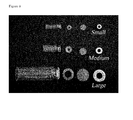
Figure 3 . Photograph of an example of different sizes which can be used in the Epithelial Tissue Model. -
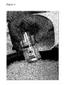
Figure 4 . Photograph of a prototype of the epithelial tissue system mounted together. Please note the possibility to shake the tissue on, for exampl,e a rocker platform. -
Figure 5 . The intestinal tissue model mounted together mounted in a 24 vial setting. The whole model can be placed on a rocker platform into an atmosphere controlled incubator. -
Figure 6 . Leakage of FITC conjugated dextran (FD4) as an integrity check of the tissue mounted into the epithelial tissue model. The fluorescent FD4 was added to all dose solutions and each vial indicating FD4 leakage above 0.5% was excluded from the study. FD4 data are presented as the percentage in the basolateral compartment at the end of the incubation in relation to the dose solution (% of dose solution). Data are presented as mean ± standard deviation of at least three incubations. -
Figure 7 . Leakage of intracellular lactate dehydrogenase (LDH) to the apical and basolateral compartment during incubation time. Data are presented as the percentage of the total intracellular LDH as measured in parallel segments at the start of the experiment. LDH leakage remains below 10% during incubation time indicating no loss of viability. Data are presented as mean ± standard deviation of at least three incubations. -
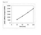
Figure 8 . The transport of [14C]-caffeine across porcine intestinal tisue in time (A) and its related calculated apparent permeability values (Papp; B). No change in Papp values indicate linear absorption of caffeine within the incubation time. - Data are presented as mean ± standard deviation of at least three incubations.
-
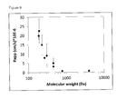
Figure 9 . The apparant permeability (Papp) values of a range of compounds as measured across porcine jejunal tissue with different molecular weights. Data are presented as mean ± standard deviation of at least two incubations. -
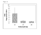
Figure 10 . The apparant permeability (Papp) values of 2-amino-1-methyl-6-phenylimidazo[4,5-b]pyridine (PhIP) across porcine jejunal tissue derived from animals with a different bodyweight. Data are presented as mean ± standard deviation of at least three incubations. -
Figure 11 . The translocation of the bacterial endotoxin [14C]-lipopolysaccharide (LPS) across porcine ileal tissue in time. Data are presented as mean ± standard deviation of four incubations. -
Figure12 . The apparent permeability (Papp) values of [3H]-mannitol and [14C]-caffeine across porcine intestinal tissue after exposure to KRB buffer, on to instinal lumen samples derived from a computer controlled TNO Intestinal Model (TIM; Minekus, M. (1998). PhD Thesis, University of Utrecht, The Netherlands; ISBN 90-393-1666-X) that was fed with a glass of water (TIM-water) or with a standard FDA breakfast (TIM-fed). Data are presented as mean ± standard deviation of at least three incubations. -
Figure 13 . The apparent permeability (Papp) values of [3H]-mannitol and [14C]-caffeine across three different segments of the porcine intestinal tract. The segments were labelled as duodenum (sample collected 10 cm after pylorus), mid jejunum (taken at approx. 5 m after pylorus), ileum (taken 1.5 m before the ileocecal junction). [3H]-mannitol is known to be a marker for the paracellular route, and [14C]-caffeine is known to be a marker for the transcellular route. Data are presented as mean ± standard deviation of four incubations. -
Figure 14 . The apparent permeability (Papp) values of [3H]-mannitol and [14C]-caffeine across porcine intestinal tissue mounted in a medium (2 mL crimp vial) and large system (20 mL crimp vial), respectively. Data are presented as mean ± standard deviation of at least three incubations. -
Figure 15 . The apparent permeability (Papp) values of [14C]-caffeine across abdominal skin tissue derived from two different species, rat and human. Data are presented as mean ± standard deviation of at least three incubation. - The gastro-intestinal tract is the primary site for the intake, processing, and absorption of pharmaceuticals and nutritional products. With the herein presented Epithelial Tissue Model using intestinal wall segments derived from pigs, the complex integrated physiology occurring in intact intestinal tissue can be studied. The gastrointestinal tract of pigs closely resembles that of humans, and having a whole intestine available offers the opportunity to work with different parts of the intestine, such as the duodenum, jejunum, ileum, or colon.
-
- 1. After anesthesia of the animal and resection of intestinal tissue, the tissue was directly added to ice-cold carbonated (5% CO2 and 95% 02) KRB buffer, pH 7.4, further referred to as KRB buffer.
- 2. Intestinal tissue was transported to the test site in ice-cold KRB buffer
- 3. At the test site, the intestinal tissue was cut open, rinsed with ice-cold KRB buffer when needed, placed on a petridish, submerged in ice-cold KRB buffer and muscle layers were stripped of.
- 4. Intestinal segments with a diameter of 20 mm were punched, added to ice-cold KRB buffer, and kept on ice.
- 5. After collecting all the segments needed for the incubation, the system was assembled (including a total of 24 segments) and placed on ice. KRB buffer with 15 mM NaHCO3 was added to the apical compartment and KRB buffer with 15 mM NaHCO3 was then added to the basolateral beaker compartment. The different parts of the epithelial tissue model and a scheme of a single completed vial is presented in
Figure 1 . - 6. The segments were acclimatized for 30 minutes at room temperature.
- 7. After acclimatization, the system was placed in an incubator at 37°C (25% air, 70% 02, 5% CO2) for 60 minutes.
- 8. Thereafter buffer from both compartments was removed, and fresh, pre-warmed KRB buffer, pH 7.4, was added to the basolateral compartment (beaker) and dose solutions containing for example various test substances were added to the apical compartment.
- 9. The basolateral compartment beaker was replaced by a new beaker containing fresh, pre-warmed buffer at for example 30, 60, 90 and 120 minutes.
- 10. Subsequently, samples were taken from the apical and basolateral compartments for further analysis either using liquid scintillation counting (LSC), UV-Absorbance, Fluorescence, High Pressure Liquid Chromatography (HPLC), Liquid chromatography-Mass Spectrometry (LCMS), Liquid chromatography-Tandem Mass Spectrometry (LC-MSMS), Enzyme-Linked Immunosorbent Assay (ELISA), or Radio-Immuno Assay (RIA).
- 11. After the incubation time, if needed (parts of) the epithelial tissues were collected for further analysis.
- A functional integrity marker FITC conjugated dextran (FD4; average molecular weight, 4000) was used to study the intestinal tight junction barrier function of a porcine intestinal jejunal tissue. At the start of the incubation FD4 (50 µM) was added to the apical compartment, and the leakage of FD4 across the epithelial tissue to the basolateral compartment was measured after a two hour incubation time. FD4 was determined by measuring fluorescence in the basolateral compartment as is known in the art. Leakage was expressed as % of the dose applied to the apical compartment. Leakage of FD4 of viable tissue remained below 0.3% after a two hour incubation period, whereas leakage from non-viable, damaged or wrongly capped segments were clearly enhanced up to 7% of the dose (
Figure 6 ). - To check the viability of tissue due to the possible presence of active proteases, the leakage of intracellular enzyme lactate dehydrogenase (LDH) to the apical and basolateral compartment during incubation time was measured. LDH is a stable cytosolic enzyme that is released upon cell damage. Leakage of LDH levels was determined as the percentage of total intracellular LDH, which were determined in parallel intestinal tissue segments at the start of the experiment. LDH activity was determined using a Roche reagent kit (Roche Diagnostics, USA) on a UV-Absorbance Spectrophotometer (
Figure 7 ). - Transport of [14C]-caffeine across porcine jejunal tissue. Caffeine is known to be readily absorbed via passive diffusion into the epithelial cells and as such is transported via the so-called transcellular route. A clear time-dependent transport of caffeine is observed during incubation time of 120 minutes (
Figure 8A ). The permeability of a compound across an epithelial barrier can be presented as the apparent permeability (Papp) value. The Papp value is defined as the initial flux of a compound across a membrane (normalized by membrane surface area and starting concentration) and is typically computed by adapting a straight line to the initial portion of the recorded amounts in the receiver compartment, disregarding the first few points when lagging of the transfer process through the membrane is evident. - The apparent permeability value (Papp) is calculated using the following equation:

where dQ/dt refers to the permeability rate (mol/s), A is the surface area filter insert (cm2) and C0 is the initial concentration (mol/L). - The linear transport of caffeine over time is clearly reflected in a constant Papp value over time (
Figure 8B ). - Measurement of the transport of a set of compounds with increasing molecular weights across porcine intestinal jejunal tissue from the apical to the basolateral side. The mean calculated Papp value of each compound is presented in
Figure 9 . Each dot represents the mean Papp value of an individual compound. - Use of porcine jejunal tissue derived from animals with a different age reflected in bodyweight. The transport of [14C]-PhIP (2-amino-1-methyl-6-phenylimidazo[4,5-b]pyridine), a known food mutagen, was measured across porcine jejunal tissue derived from animals with a different age. PhIP is known to have a very low permeability due to fact that it is a high affinity substrate of the breast cancer resistance protein (BCRP, gene code ABCG2), which is an efflux membrane transporter expressed at the apical side of the intestine. It is known that intestinal tissue from younger animals usually have a higher permeability of the intestinal tissue for this compound. A higher transport of [14C]-PhIP was measured across intestinal tissue derived from an animal with a body weight of 12 kg in comparison to an animal of 17 or 20 kg , which indicates a higher permeability for PhIP in younger animals (
Figure 10 ). - Translocation of the bacterial endotoxin lipopolysaccharide (LPS) across the porcine ileal tissue during incubation time. For ([14C]-LPS a clear lag time of transport was observed between 0 and 60 minutes. The apparent permeability value of LPS was calculated over the time frame between 60 and 120 minutes (
Figure 11 ). - Combination of exposure of intestinal lumen samples derived from a computer controlled TNO Intestinal Model (fed a glass of water (TIM-water) or fed a standard FDA breakfast (TIM-fed) on the transport of [3H]-mannitol and [14C]-caffeine across porcine intestinal tissue (
Figure 12 ). - Use of different tissues derived from the same animal. In this example, the transport of [3H]-mannitol and [14C]-caffeine was measured across intestinal segments derived from three different segments from the intestine of one single pig. As indicated, caffeine is transported via the transcellular route due to its rapid passive diffusion in the epithelial cells. In contrast, the sugar mannitol is known to be transported solely via the paracellular route. Different segments of intestinal tissue were derived from one single animal. The segments were labelled as duodenum (sample collected 10 cm after pylorus), mid jejunum (taken at approx. 5 m after pylorus), ileum (taken 1.5 m before the ileocecal junction). Data are presented in
Figure 13 . - Use of systems with various width and volumina. The transport of [3H]-mannitol and [14C]-caffeine across porcine jejunal intestinal tissue was measured in a medium and large system, using a crimp vial of 2 and 20 mL, respectively (
Figure 14 ). - Use of skin tissue derived from different species. In this example rat and human abdominal skin tissue was used. The transport of [14C]-caffeine was measured across skin tissue and the Papp values were calculated of transport between 4 and 5 hours after the start of the experiment (
Figure 15 ).
Claims (14)
- A vial for holding a segment of epithelial tissue, the vial having a neck and a first open end at the top of the neck, whereby a rim surrounds the open end, the rim having an upper surface facing the open end,
whereby the neck and the rim of the first open end of the vial are suited for capping with a crimp cap,
the vial characterized by having a second open end distal to the neck. - The vial of claim 1, further comprising a segment of epithelial tissue which is positioned on the upper surface of the rim, with the apical side of the epithelial tissue facing the first open end,
whereby a ring of inert material is positioned on the apical side of the epithelial tissue, and
whereby a crimp cap provides a tight seal of the first end of the vial. - The vial of claim 2, whereby the basolateral side of the epithelial tissue is covered by a support.
- The vial of claim 3, whereby the support is a nylon or polypropylene mesh.
- The vial of any one of claims 2-4, whereby the ring of inert material is a teflon ring.
- A device comprising the vial according to any of claims 1-5 and a container for holding the vial, whereby the container is open on one end, whereby the vial is mounted into the container with the first open end of the vial facing the closed end of the container, and whereby the crimp cap is not in contact with the closed end of the container.
- A multitude of devices according to claim 6.
- The multitude of devices according to claim 7, wherein said multitude comprises 6, 12, 24 or 48 devices having a footprint (length x width) of 127.76 x 85.48 mm.
- A method for generating the vial of claims 2-5, comprising preparing a segment of an epithelial tissue,
mounting the basolateral side of the epithelial segment on a support, mounting the supported segment on a vial of claim 1, whereby the apical side of the epithelial tissue faces the first open end of the vial,
mounting a ring of inert material onto the apical side of the epithelial tissue, and
providing a crimp cap to the first open end to obtain a tight seal of the first end of the vial. - A method for generating a multitude of vials according to any of claims 2-5, comprising(a) performing the method of claim 9 to prepare a first device,(b) cooling at least the first open end comprising the segment of an epithelial tissue,repeating steps (a) and (b) until the multitude of vials is prepared.
- A method for measuring absorption, transport and/or secretion across an epithelial tissue, the method comprising
providing the device of claim 6 comprising an epithelial tissue,
providing a relevant buffered medium in the vial onto the apical site of the segment of epithelial tissue,
providing a relevant buffered medium to the container,
whereby the height of the buffered medium in the vial is substantially equal to the height of the buffered medium in the container,
applying a substance to the medium at the apical site of the segment of epithelial tissue, and
measuring the amount of substance that appears in the medium of the container. - The method of claim 11, whereby the substance is labeled.
- The method of claim 11 or 12, further comprising adding a marker to the apical site of the segment of epithelial tissue, and measuring the amount of marker that that appears in the medium of the container.
- A method for measuring absorption, transport and/or secretion across an epithelial tissue, the method comprising
providing a multitude of devices according to claim 7 or 8 comprising an epithelial tissue,
providing a relevant buffered medium in the vial onto the apical site of the segment of epithelial tissue in the vials,
providing a relevant buffered medium the containers,
whereby the height of the buffered medium in the vials is substantially equal to the height of the buffered medium in the containers,
applying a substance to the medium at the apical site of the segment of epithelial tissue in the vials, and
measuring the amount of label that appears in the medium of the containers.
Priority Applications (4)
| Application Number | Priority Date | Filing Date | Title |
|---|---|---|---|
| EP12190442.9A EP2725094A1 (en) | 2012-10-29 | 2012-10-29 | Epithelial tissue model |
| PCT/NL2013/050761 WO2014069995A1 (en) | 2012-10-29 | 2013-10-29 | Epithelial tissue model |
| US14/439,207 US10690657B2 (en) | 2012-10-29 | 2013-10-29 | Epithelial tissue model |
| EP13786326.2A EP2912159B1 (en) | 2012-10-29 | 2013-10-29 | Epithelial tissue model |
Applications Claiming Priority (1)
| Application Number | Priority Date | Filing Date | Title |
|---|---|---|---|
| EP12190442.9A EP2725094A1 (en) | 2012-10-29 | 2012-10-29 | Epithelial tissue model |
Publications (1)
| Publication Number | Publication Date |
|---|---|
| EP2725094A1 true EP2725094A1 (en) | 2014-04-30 |
Family
ID=47172423
Family Applications (2)
| Application Number | Title | Priority Date | Filing Date |
|---|---|---|---|
| EP12190442.9A Withdrawn EP2725094A1 (en) | 2012-10-29 | 2012-10-29 | Epithelial tissue model |
| EP13786326.2A Active EP2912159B1 (en) | 2012-10-29 | 2013-10-29 | Epithelial tissue model |
Family Applications After (1)
| Application Number | Title | Priority Date | Filing Date |
|---|---|---|---|
| EP13786326.2A Active EP2912159B1 (en) | 2012-10-29 | 2013-10-29 | Epithelial tissue model |
Country Status (3)
| Country | Link |
|---|---|
| US (1) | US10690657B2 (en) |
| EP (2) | EP2725094A1 (en) |
| WO (1) | WO2014069995A1 (en) |
Families Citing this family (6)
| Publication number | Priority date | Publication date | Assignee | Title |
|---|---|---|---|---|
| WO2016180836A1 (en) * | 2015-05-11 | 2016-11-17 | CSEM Centre Suisse d'Electronique et de Microtechnique SA - Recherche et Développement | Well inserts with brittle membranes |
| US10323221B2 (en) * | 2016-01-06 | 2019-06-18 | Northeastern University | Device for controlled apical flow in cell culture inserts |
| EP3474012A1 (en) * | 2017-10-19 | 2019-04-24 | Galderma Research & Development | Method for the generation of a skin organoculture model and skin model |
| WO2021119326A1 (en) | 2019-12-10 | 2021-06-17 | Massachusetts Institute Of Technology | Methods and compositions for investigating multiple drug-drug transporter interactions |
| EP3854868A1 (en) | 2020-01-24 | 2021-07-28 | Nederlandse Organisatie voor toegepast- natuurwetenschappelijk Onderzoek TNO | Microfluidic device for analyzing a membrane |
| WO2022197254A1 (en) * | 2021-03-16 | 2022-09-22 | Agency For Science, Technology And Research | A well insert and a device for 3d cell culture and in vitro tissue model |
Citations (6)
| Publication number | Priority date | Publication date | Assignee | Title |
|---|---|---|---|---|
| GB2082091A (en) * | 1980-08-21 | 1982-03-03 | Squibb & Sons Inc | Container for centrifuging blood |
| US4686190A (en) * | 1986-02-19 | 1987-08-11 | The Research Foundation Of State University Of New York | Device useful for growing and/or testing biological materials |
| EP0295762A2 (en) * | 1987-03-31 | 1988-12-21 | Organogenesis Inc. | Tissue equivalent test systems |
| US6251681B1 (en) * | 1998-11-30 | 2001-06-26 | Richard J. Davies | Method for the detection of cancer and premalignancy conditions thereof |
| DE10309348A1 (en) * | 2003-03-03 | 2004-09-23 | Chromtech Gesellschaft für analytische Meßtechnik mbH | Sample holder for chromatography analysis has polytetrafluoroethylene cap and closure with sealed outlet channel |
| US20080076170A1 (en) * | 2006-09-27 | 2008-03-27 | Tuija Annala | Cell culture insert and cell culture vessel |
Family Cites Families (15)
| Publication number | Priority date | Publication date | Assignee | Title |
|---|---|---|---|---|
| US4264204A (en) * | 1978-12-21 | 1981-04-28 | Mccormick James B | Method and apparatus for preparing fluid specimens |
| IL60645A (en) | 1980-07-21 | 1984-02-29 | Cais Michael | Method and device for mass transfer and separation through selective barriers |
| GB8311730D0 (en) * | 1983-04-29 | 1983-06-02 | Bagshawe K D | Handling of reaction mixtures |
| US4669763A (en) | 1984-12-19 | 1987-06-02 | Phillips Edwin D | Gripping saddle and O-ring apparatus |
| FR2593824B1 (en) | 1986-02-03 | 1988-11-04 | Inst Francais Du Petrole | CATALYTIC REFORMING PROCESS THROUGH AT LEAST THREE CATALYST BEDS |
| US4939152A (en) * | 1987-09-30 | 1990-07-03 | Syntex (U.S.A.) Inc. | Cell culture vial |
| US5022411A (en) * | 1989-09-18 | 1991-06-11 | La Mina Ltd. | Modular fluid testing device |
| US5358690A (en) * | 1989-01-10 | 1994-10-25 | Lamina, Ltd. | Environmental sample collection and membrane testing device |
| US5077012A (en) * | 1989-01-10 | 1991-12-31 | La Mina Ltd. | Device for detecting disease markers |
| US5662230A (en) * | 1990-07-13 | 1997-09-02 | J. G. Finneran Associates | Crimp top seal for vials |
| DE4229325C2 (en) * | 1992-09-02 | 1995-12-21 | Heraeus Instr Gmbh | Culture vessel for cell cultures |
| WO2003019131A2 (en) * | 2001-08-29 | 2003-03-06 | Hexal Pharma Gmbh | Method and device for preparing a sample of biological origin in order to determine at least one constituent contained therein |
| US20090196798A1 (en) * | 2008-02-06 | 2009-08-06 | Robert Sassa | Barrier with Low Extractables and Resealing Properties |
| JP5922672B2 (en) * | 2010-12-15 | 2016-05-24 | ドラッグモード アーペーエスDrugmode Aps | Bioreactor with lid for easy access to incubation cavity |
| EP2548943B1 (en) | 2011-07-21 | 2016-05-11 | CSEM Centre Suisse d'Electronique et de Microtechnique SA - Recherche et Développement | Clamping insert for cell culture |
-
2012
- 2012-10-29 EP EP12190442.9A patent/EP2725094A1/en not_active Withdrawn
-
2013
- 2013-10-29 US US14/439,207 patent/US10690657B2/en active Active
- 2013-10-29 WO PCT/NL2013/050761 patent/WO2014069995A1/en not_active Ceased
- 2013-10-29 EP EP13786326.2A patent/EP2912159B1/en active Active
Patent Citations (6)
| Publication number | Priority date | Publication date | Assignee | Title |
|---|---|---|---|---|
| GB2082091A (en) * | 1980-08-21 | 1982-03-03 | Squibb & Sons Inc | Container for centrifuging blood |
| US4686190A (en) * | 1986-02-19 | 1987-08-11 | The Research Foundation Of State University Of New York | Device useful for growing and/or testing biological materials |
| EP0295762A2 (en) * | 1987-03-31 | 1988-12-21 | Organogenesis Inc. | Tissue equivalent test systems |
| US6251681B1 (en) * | 1998-11-30 | 2001-06-26 | Richard J. Davies | Method for the detection of cancer and premalignancy conditions thereof |
| DE10309348A1 (en) * | 2003-03-03 | 2004-09-23 | Chromtech Gesellschaft für analytische Meßtechnik mbH | Sample holder for chromatography analysis has polytetrafluoroethylene cap and closure with sealed outlet channel |
| US20080076170A1 (en) * | 2006-09-27 | 2008-03-27 | Tuija Annala | Cell culture insert and cell culture vessel |
Non-Patent Citations (8)
| Title |
|---|
| FLYNN ET AL., PHARM RES, vol. 16, 1999, pages 1325 - 30 |
| HARVARD APPARATUS: "Diffusion Chamber System User's Manual", 16 March 2012 (2012-03-16), pages 1 - 28, XP055056548, Retrieved from the Internet <URL:https://www.warneronline.com/Documents/uploader/6600xx_Navicyte_Manual1.pdf> [retrieved on 20130314] * |
| IGLICAR ET AL., J PHARM PHARMACOL, vol. 61, 2009, pages 1211 - 8 |
| MINEKUS, M.: "PhD Thesis", 1998, UNIVERSITY OF UTRECHT |
| PALAMAKULA ET AL., PHARMAZIE, vol. 60, 2005, pages 212 - 4 |
| PAMPALONI ET AL., NAT REV MOL CELL BIOL, vol. 8, 2007, pages 839 - 84 |
| ROZEHNAL ET AL., EUR J PHARM SCI, vol. 46, 2012, pages 367 - 73 |
| WARNERONLINE: "Warneronline.com Navicyte horizontal multichamber ussing chamber", 16 March 2012 (2012-03-16), Internet archive, XP055056848, Retrieved from the Internet <URL:http://web.archive.org/web/20120316161654/http://www.warneronline.com/product_cat_info.cfm?id=1424> [retrieved on 20130318] * |
Also Published As
| Publication number | Publication date |
|---|---|
| EP2912159A1 (en) | 2015-09-02 |
| WO2014069995A1 (en) | 2014-05-08 |
| US10690657B2 (en) | 2020-06-23 |
| EP2912159B1 (en) | 2020-09-16 |
| US20150276713A1 (en) | 2015-10-01 |
Similar Documents
| Publication | Publication Date | Title |
|---|---|---|
| US10690657B2 (en) | Epithelial tissue model | |
| Wilson et al. | Principles and techniques of practical biochemistry | |
| US12036548B2 (en) | Organs-on-chips as a platform for epigenetics discovery | |
| JP2005524850A (en) | Collection device | |
| EP3003555B1 (en) | Controlled transfer biological sample collection devices and methods of using such devices | |
| CA2900947A1 (en) | Measuring embryo development and implantation potential with timing and first cytokinesis phenotype parameters | |
| US20100196996A1 (en) | Process For Preserving Biological Materials For Extended Periods Of Time | |
| WO1994006902A1 (en) | Plate for cell cultures with a system for laterally diffusing molecules through a barrier membrane | |
| Vanstokstraeten et al. | Culturomics to investigate the endometrial microbiome: proof-of-concept | |
| Ogawa et al. | Impact of homocysteine as a preconceptional screening factor for in vitro fertilization and prevention of miscarriage with folic acid supplementation following frozen-thawed embryo transfer: a hospital-based retrospective cohort study | |
| Gao et al. | Estimating intestinal mucosal permeation of compounds using Caco‐2 cell monolayers | |
| CN101402989A (en) | Syringe-shaped microorganism culture device | |
| EP2198303A1 (en) | Methods and compositions based on culturing microorganisms in low sedimental fluid shear conditions | |
| JP2019520825A (en) | High-throughput microbiologically applied high resolution system, kit, apparatus and method using magnetic particles | |
| JP6386193B2 (en) | System for concomitant assessment of drug dissolution, absorption and permeation and method of using the system | |
| ES2882050T3 (en) | Procedure for the isolation and analysis of microorganisms contained in a sample | |
| Clegg | Biology for the IB Diploma Second Edition | |
| Jacobs et al. | Borrelia burgdorferi migration assays for evaluation of chemoattractants in tick saliva | |
| Cheng et al. | PIEZO1 channels as regulators of integrin-mediated focal adhesions in cardiac fibroblasts | |
| CN118401308A (en) | Matrix tray and method of use | |
| WO2014188365A1 (en) | A portable device for maintaining viability of biological material and a method thereof | |
| Dube et al. | Structural dynamics of antimicrobial drug transport by integral membrane protein MipA using deep learning methods and Anton 2 simulations | |
| WO2021160790A1 (en) | Sampling kit for the transport of sputum | |
| Arjes et al. | Three-dimensional biofilm growth supports a mutualism involving matrix and nutrient sharing | |
| Chintapalli et al. | Molecular insights into myoglobin binding patterns with the outer mitochondrial membrane: A comparison of oxygenated and deoxygenated states |
Legal Events
| Date | Code | Title | Description |
|---|---|---|---|
| PUAI | Public reference made under article 153(3) epc to a published international application that has entered the european phase |
Free format text: ORIGINAL CODE: 0009012 |
|
| 17P | Request for examination filed |
Effective date: 20121029 |
|
| AK | Designated contracting states |
Kind code of ref document: A1 Designated state(s): AL AT BE BG CH CY CZ DE DK EE ES FI FR GB GR HR HU IE IS IT LI LT LU LV MC MK MT NL NO PL PT RO RS SE SI SK SM TR |
|
| AX | Request for extension of the european patent |
Extension state: BA ME |
|
| STAA | Information on the status of an ep patent application or granted ep patent |
Free format text: STATUS: THE APPLICATION IS DEEMED TO BE WITHDRAWN |
|
| 18D | Application deemed to be withdrawn |
Effective date: 20141031 |