EP2720039A1 - Agent for improving cancer cell adhesiveness - Google Patents
Agent for improving cancer cell adhesiveness Download PDFInfo
- Publication number
- EP2720039A1 EP2720039A1 EP12801028.7A EP12801028A EP2720039A1 EP 2720039 A1 EP2720039 A1 EP 2720039A1 EP 12801028 A EP12801028 A EP 12801028A EP 2720039 A1 EP2720039 A1 EP 2720039A1
- Authority
- EP
- European Patent Office
- Prior art keywords
- cancer cell
- polymer
- agent
- substrate
- cells
- Prior art date
- Legal status (The legal status is an assumption and is not a legal conclusion. Google has not performed a legal analysis and makes no representation as to the accuracy of the status listed.)
- Granted
Links
- 0 CC(C)(C)N*[N+]([O-])OC(C)(C)NNOC(C)(C)C Chemical compound CC(C)(C)N*[N+]([O-])OC(C)(C)NNOC(C)(C)C 0.000 description 1
Images


Classifications
-
- G—PHYSICS
- G01—MEASURING; TESTING
- G01N—INVESTIGATING OR ANALYSING MATERIALS BY DETERMINING THEIR CHEMICAL OR PHYSICAL PROPERTIES
- G01N1/00—Sampling; Preparing specimens for investigation
- G01N1/28—Preparing specimens for investigation including physical details of (bio-)chemical methods covered elsewhere, e.g. G01N33/50, C12Q
- G01N1/40—Concentrating samples
- G01N1/405—Concentrating samples by adsorption or absorption
Definitions
- the present invention relates to an agent for improving cancer cell adhesiveness. More particularly, it relates to an agent for improving cancer cell adhesiveness, a cancer cell enrichment filter and a method for testing cancer cells.
- Enrichment of cancer cells is extremely significant in research and clinical aspects, and if cancer cells present in blood can be enriched, the enriched cancer cells can be applied to the diagnosis of cancer. For example, the most significant factor of a prognosis and a treatment of cancer is whether or not cancer cells have metastasized at the time of first medical examination or treatment. If cancer cells are spread to peripheral blood at an early stage, it is a useful method for determining disease progression of the cancer to detect circulating tumor cells (hereinafter sometimes referred to as the "CTCs").
- CTCs circulating tumor cells
- CTCs In a general cancer patient who has just started suffering from a metastasis, however, merely one CTC is present in his/her blood per ten billion blood cells. On the contrary, there are an overwhelmingly large number of blood components such as erythrocytes and leucocytes in blood. Accordingly, it is extremely difficult to enrich CTCs in a very low concentration so as to highly sensitively, highly efficiently and highly specifically analyze the CTCs. If the CTCs are enriched by a method using, for example, magnetic beads, density gradient centrifugation, a microchannel or a flow cytometer, it is necessary to perform a complicated treatment as well as the recovery rate is poor in some cases.
- Cancer cells such as CTCs have a larger size than blood cells present in blood, such as erythrocytes, leucocytes and blood platelets. Therefore, theoretically, cancer cells can be enriched by removing these blood cell components by applying mechanical filtration.
- a membrane filter described in each of Non Patent Literatures 1 and 2 has, however, randomly distributed pores and the pore size is ununiform, and therefore, a recovery rate of cancer cells is poor in some cases. Furthermore, even in using a filter, as described in each of Non Patent Literatures 3 to 5 and Patent Literature 1, having a pore size and a thickness accurately controlled, it is sometimes difficult to enrich cancer cells with accuracy sufficiently high for precise analysis.
- the present inventors have first found that a material surface coated with the polymer comprising the constitutional unit represented by the following formula (1) unexpectedly shows improved adhesiveness to cancer cells while showing lowered adhesiveness to blood cell components, and thus, the present invention was accomplished.
- the present invention provides an agent for improving cancer cell adhesiveness consisting of a polymer comprising a constitutional unit represented by the following formula (1):
- R 1 is a hydrogen atom or a methyl group
- R 2 is a methyl group or an ethyl group
- m is 1 to 3.
- the agent for improving cancer cell adhesiveness of the present invention When the agent for improving cancer cell adhesiveness of the present invention is applied to a surface of a cancer cell enrichment filter, the adhesiveness of cancer cells to the surface of the filter can be improved, so as to improve an enrichment ratio of cancer cells.
- the agent for improving cancer cell adhesiveness of the present invention can simultaneously satisfy characteristics in a trade-off relationship, that is, non-adhesiveness of blood cell components and adhesiveness of cancer cells.
- the present invention provides a method for improving adhesiveness of cancer cells to a substrate comprising a step of coating the substrate with a polymer comprising a constitutional unit represented by the following formula (1):
- R 1 is a hydrogen atom or a methyl group
- R 2 is a methyl group or an ethyl group
- m is 1 to 3.
- the present invention further provides use of a polymer comprising a constitutional unit represented by the following formula (1) for improving cancer cell adhesiveness:
- R 1 is a hydrogen atom or a methyl group
- R 2 is a methyl group or an ethyl group
- m is 1 to 3.
- the present invention further provides application of a polymer comprising a constitutional unit represented by the following formula (1) to improvement of cancer cell adhesiveness:
- R 1 is a hydrogen atom or a methyl group
- R 2 is a methyl group or an ethyl group
- m is 1 to 3.
- the present invention further provides application of a polymer comprising a constitutional unit represented by the following formula (1) to production of an agent for improving cancer cell adhesiveness:
- R 1 is a hydrogen atom or a methyl group
- R 2 is a methyl group or an ethyl group
- m is 1 to 3.
- the present invention further provides a cancer cell enrichment filter consisting of a substrate having a plurality of through holes, wherein at least a part of the substrate is coated with a polymer comprising a constitutional unit represented by the following formula (1):
- R 1 is a hydrogen atom or a methyl group
- R 2 is a methyl group or an ethyl group
- m is 1 to 3.
- the polymer may consist of a constitutional unit represented by the above formula (1). Furthermore, it is preferable that R 1 is a hydrogen atom, R 2 is a methyl group, and m is 1. Alternatively, R 1 may be a hydrogen atom, R 2 may be an ethyl group, and m may be 2, or R 1 may be a methyl group, R 2 may be a methyl group, and m may be 2. Besides, the polymer preferably has a number average molecular weight of 10,000 to 300,000.
- Such a polymer can further improve the cancer cell adhesiveness.
- the present invention further provides a cancer cell enrichment filter consisting of a substrate which has a plurality of through holes and at least a part of which is coated with the above polymer.
- cancer cells present in a blood sample can be enriched to a high enrichment ratio.
- the through holes preferably have an average pore size of 5 ⁇ m or more and less than 30 ⁇ m and an average aperture ratio of 5% or more and less than 50%, and are preferably formed by an electrocasting method.
- the substrate is preferably made of a metal.
- the metal is preferably selected from the group consisting of copper, nickel, a copper-nickel alloy, and any of these metals and alloy having a gold-plated surface.
- cancer cells present in a blood sample can be enriched to a further higher enrichment ratio.
- the present invention further provides a method for detecting presence of cancer cells comprising a filtration step of filtering peripheral blood with the cancer cell enrichment filter.
- the presence of cancer cells such as CTCs in peripheral blood collected from a patient can be easily detected.
- the test method may further include a step of analyzing a gene of cells having been enriched in the filtration step, and may further include a step of culturing cells having been enriched in the filtration step.
- the step of analyzing a gene of the enriched cells or the step of culturing the cells can be performed.
- the method for detecting presence of cancer cells of the present invention further includes such a step, the presence of cancer cells such as CTCs in peripheral blood can be more accurately detected.
- the present invention provides an agent for improving cancer cell adhesiveness, a cancer cell enrichment filter whose surface is coated with the agent for improving cancer cell adhesiveness and a method for testing cancer cells comprising a step of filtering peripheral blood with the cancer cell enrichment filter.
- an agent for improving cancer cell adhesiveness consists of a polymer comprising a constitutional unit represented by the following formula (1):
- R 1 is a hydrogen atom or a methyl group
- R 2 is a methyl group or an ethyl group
- m is 1 to 3.
- a number average molecular weight of the polymer is preferably 10,000 to 300,000. If the number average molecular weight is 10,000 or less, the polymer is in the form of a liquid and hence is difficult to handle in some cases. Furthermore, in some cases, the polymer is difficult to attain a number average molecular weight of 300,000 or more by general radical polymerization.
- the polymer can be typically produced by polymerization, performed by a general method, of a solution of a monomer represented by the following formula (2) with an appropriate initiator added.
- a temperature at which the polymerization is performed is preferably 40°C to 100°C, more preferably 60°C to 90°C, and further more preferably 70°C to 80°C.
- a pressure at which the polymerization is performed is preferably normal pressure.
- R 3 is a hydrogen atom or a methyl group
- R 4 is a methyl group or an ethyl group
- n is 1 to 3.
- a solvent capable of dissolving the monomer represented by the above formula (2) can be used as a solvent.
- the solvent include aliphatic or aromatic organic solvents, and more specifically, examples include ether solvents such as dioxane, tetrahydrofuran and diethyl ether; halogenated aromatic hydrocarbons such as o-dichlorobenzene; amides such as N,N-dimethylformamide; sulfoxides such as dimethyl sulfoxide; aromatic hydrocarbons such as benzene and toluene; and aliphatic hydrocarbons such as hexane and pentane, among which an ether solvent such as dioxane is preferably used.
- an agent for improving cancer cell adhesiveness consists of a polymer consisting of a constitutional unit represented by the above formula (1).
- a combination of R 1 , R 2 and m of the above formula (1) is preferably any one of the following combinations (a) to (h):
- an agent for improving cancer cell adhesiveness is a random copolymer, a block copolymer or a graft copolymer of a monomer represented by the above formula (2) and another polymerizable monomer.
- Examples of the monomer polymerizable with the monomer represented by the above formula (2) include: alkylacrylamides such as acrylamide, t-butyl acrylamide, n-butyl acrylamide, i-butyl acrylamide, hexyl acrylamide and heptyl acrylamide; N,N-dialkylacrylamides such as N,N-dimethylacrylamide and N,N-diethylacrylamide; amino alkyl acrylates such as amino methyl acrylate, amino ethyl acrylate and amino isopropyl acrylate; diamino alkyl acrylate such as diamino methyl acrylate, diamino ethyl acrylate and diamino butyl acrylate; N,N-dialkyl methacrylamides such as methacrylamide, N,N-dimethylmethacrylamide and N,N-diethylmethacrylamide; amino alkyl methacrylates such as amino methyl me
- the monomer to be polymerized with the monomer represented by the above formula (2) may be one or more monomers selected from the group consisting of alkyl acrylate, alkyl methacrylate, alkoxy (meth)acrylate, alkoxy alkyl (meth)acrylate, glycidyl methacrylate and propylene.
- the copolymer of the monomer represented by the above formula (2) and the above-described polymerizable monomer can be any one of a random copolymer, a block copolymer and a graft copolymer, and may be produced by any of random polymerization, ion polymerization, polymerization utilizing a macromer and the like.
- the content of the monomer represented by the above formula (2) is preferably 30 to 99% by mass and more preferably 50 to 99% by mass in the resulting copolymer.
- the polymer comprising the constitutional unit represented by the above formula (1) can be typically produced by polymerization performed by random polymerization, ion polymerization, photopolymerization, polymerization utilizing a macromer or the like with an initiator appropriate for the solution of the monomer represented by the above formula (2) added thereto, and as occasion demands, with one or more of the polymerizable monomers added thereto.
- a temperature at which the polymerization is performed is preferably 40°C to 100°C, more preferably 60°C to 90°C and further more preferably 70°C to 80°C.
- a pressure at which the polymerization is performed is preferably normal pressure.
- a solvent capable of dissolving the monomer represented by the above formula (2) and the polymerizable monomer can be used as a solvent.
- the solvent include aliphatic or aromatic organic solvents, and more specifically, examples include ether solvents such as dioxane, tetrahydrofuran and diethyl ether; halogenated aromatic hydrocarbons such as o-dichlorobenzene; amides such as N,N-dimethylformamide; sulfoxides such as dimethyl sulfoxide; aromatic hydrocarbons such as benzene and toluene; and aliphatic hydrocarbons such as hexane and pentane, among which an ether solvent such as dioxane is preferably used.
- the agent for improving cancer cell adhesiveness When the agent for improving cancer cell adhesiveness is applied to a surface of a substrate of a cancer cell enrichment filter or the like, blood compatibility is imparted, and hence, if the filter is brought into contact with blood, activation of blood components can be inhibited, so as to reduce adhesion of blood cell components to the filter. Furthermore, since the cancer cell enrichment filter to which the agent for improving cancer cell adhesiveness is applied is good in adhesiveness to various cancer cells, cancer cells can be efficiently enriched.
- the polymer comprising the constitutional unit represented by the above formula (1) has, in its molecule, an ether bond and an ester bond as a polar group, such a polar group does not show strong electrostatic interaction with a biological component differently from a nitrogen atom (an amino group or an imino group), a carboxyl group and the like. Furthermore, since the polymer has no large hydrophobic group, the hydrophobic interaction is also small. It is therefore presumed that the polymer comprising the constitutional unit represented by the above formula (1) has low activity against blood and shows excellent blood compatibility.
- the surface to which the agent for improving cancer cell adhesiveness is applied can be provided with a suitable surface structure.
- the surface to which the agent for improving cancer cell adhesiveness is applied has moderate hydrophilicity, even when it comes into contact with blood, blood platelets are merely slightly adhered thereto, and thus, excellent blood compatibility is shown. Moreover, it is presumed that interaction with a biological component via a hydrogen bond derived from a hydroxyl group, denaturation of adsorbed protein and the like can be inhibited.
- water in contact with the substrate to which the agent for improving cancer cell adhesiveness is applied includes (1) free water that has weak interaction with the agent for improving cancer cell adhesiveness and is molten at 0°C; (2) unfreezable water that has strong interaction with the agent for improving cancer cell adhesiveness and does not freeze even at -100°C; and (3) intermediate water that has interaction intermediate between the free water and the unfreezable water and freezes at a temperature lower than 0°C.
- Normal blood cells form a hydration shell with the unfreezable water, the intermediate water, the free water and the like in contact with the agent for improving cancer cell adhesiveness, and hence are stabilized owing to this hydration structure, but the unfreezable water strongly affected by the structure of the agent for improving cancer cell adhesiveness is probably camouflaged by the intermediate water, so that adhesion of the normal blood cells to the substrate surface is inhibited.
- the hydration structure is probably disordered as compared with that in the normal blood cells. This seems to disturb the structure of the intermediate water on the substrate surface, so that the adhesiveness of the cancer cells to the substrate surface can be improved.
- the agent for improving cancer cell adhesiveness is applicable to all the types of cancer cells, and is preferably applied to epithelial cell-derived cancers that easily infiltrate into blood vessels, and among such cancers, the agent is particularly preferably applied to lung cancers, digestive cancers such as colorectal cancers, stomach cancers and esophageal cancers, breast cancers and prostatic cancers, which are regarded to produce a large number of CTCs.
- the present invention provides a cancer cell enrichment filter consisting of a substrate which has a plurality of through holes and at least a part of which is coated with the agent for improving cancer cell adhesiveness.
- This filter is capable of enriching cancer cells such as CTCs present in blood.
- the opening shape of each through hole include a circular shape, an elliptic shape, a rectangular shape, a rounded rectangular shape and a polygonal shape.
- a rounded rectangular shape is a shape having two long sides in the same length and two semicircles. From the viewpoint that cancer cells can be efficiently captured, a circular shape, a rectangular shape or a rounded rectangular shape is preferably employed. Besides, from the viewpoint of preventing clogging of the filter, a rounded rectangular shape is particularly preferably employed.
- FIG. 1 is a schematic diagram illustrating one embodiment of the cancer cell enrichment filter.
- the filter 100 consists of a substrate 20 having a plurality of through holes 10.
- the opening shape of each through hole 10 is a rounded rectangular shape.
- CTCs are captured on a surface of a face 30 of the substrate 20.
- At least a part of the face 30 is coated with the agent for improving cancer cell adhesiveness.
- the agent for improving cancer cell adhesiveness is preferably coated over the whole of the face 30.
- a part or the whole of a face opposite to the face 30 may be coated with the agent for improving cancer cell adhesiveness.
- a coating method is the most generally employed.
- the coating method is performed by allowing a solution of the polymer comprising the constitutional unit represented by the above formula (1) to adhere to the substrate surface by a dipping method, a spraying method, a spin coating method or the like, and then removing (drying) the solvent.
- a thickness of a film attained after drying the solvent is preferably 0.01 ⁇ m to 1.0 mm, more preferably 0.1 to 100 ⁇ m and further more preferably 0.5 to 50 ⁇ m.
- the film thickness is smaller than 0.01 ⁇ m, non-adhesiveness to blood cell components and adhesiveness to cancer cells may not be sufficiently exhibited in some cases. Alternatively, if the film thickness exceeds 1.0 mm, it is apprehended that balance between these adhesiveness characteristics may be lost in some cases.
- the substrate may be heated after coating with the agent for improving cancer cell adhesiveness.
- the polymer comprising the constitutional unit represented by the above formula (1) may be crosslinked.
- An example of a crosslinking method includes precedent addition of a crosslinkable monomer to the materials of the polymer.
- electron beams, y rays or light irradiation may be used.
- a layer of the polymer comprising the constitutional unit represented by the above formula (1) by plasma graft polymerization plasma-initiated polymerization is performed with the monomer represented by the above formula (2) supplied after irradiation of low-temperature plasma performed under reduced pressure of approximately 1.3 x 10 -1 Pa, preferably 1.3 to 133.3 Pa, and under an atmosphere of argon, nitrogen, air, any of various monomers or the like for 1 to 300 seconds, preferably 2 to 30 seconds.
- the material and the shape of the substrate are not especially limited, and for example, a porous body, fiber, nonwoven fabric, a film, a sheet or a tube may be employed.
- the material of the substrate include natural polymers such as cotton and hemp; synthetic polymers such as nylon, polyester, polyacrylonitrile, polyolefin, halogenated polyolefin, polyurethane, polyamide, polysulfone, polyether sulfone, poly(meth)acrylate, a halogenated polyolefin ethylene-polyvinyl alcohol copolymer and a butadiene-acrylonitrile copolymer; and mixtures thereof.
- the material also include metals, ceramics and composite materials thereof, and the substrate may include a plurality of substrates.
- the metal examples include but are not limited to noble metals such as gold and silver; base metals such as copper, aluminum, tungsten, nickel and chromium; and alloys of these metals. Such a metal may be singly used, or may be used as an alloy with another metal or as a metal oxide for imparting functionality. From the viewpoint of the price and availability, nickel, copper or a metal containing any of them as a principal component is preferably used.
- a principal component means a component occupying 50% by weight or more among materials contained in the substrate.
- Such a metal may be subjected to photolithography or the like for forming through holes therein, so as to be used as a screen filter.
- a general CTC has a diameter of 10 ⁇ m or more.
- a diameter of a cell means a length of the longest straight line out of straight lines each connecting arbitrary two points on the outline of the cell in observation with a microscope.
- the through holes of the cancer cell enrichment filter have an average pore size of preferably 5 ⁇ m or more and less than 30 ⁇ m and an average aperture ratio of preferably 5% or more and less than 50%.
- the average pore size and the average aperture ratio are respectively more preferably 5 ⁇ m or more and less than 15 ⁇ m and 10% or more and less than 40% and particularly preferably 5 ⁇ m or more and less than 10 ⁇ m and 20% or more and less than 40%.
- an aperture ratio means an area occupied by the through holes in the whole area of the filter. From the viewpoint of preventing clogging, the average aperture ratio is preferably larger, but if it exceeds the upper limit, the filter may be degraded in its strength or may be difficult to be worked in some cases. Alternatively, if the aperture ratio is lower than 5%, the cancer cell enrichment performance of the filter may be degraded in some cases.
- a pore size in an opening shape other than a circular shape is defined as the maximum value of diameters of spheres that can pass through the through hole in such a shape.
- the opening shape is a rectangular shape
- the pore size of the through hole corresponds to the length of the short side of the rectangle
- the opening shape is a polygonal shape, it corresponds to the diameter of the inscribed circle of the polygon.
- the opening shape is a rectangular shape or a rounded rectangular shape, even when CTCs and leucocytes are captured by the through holes, there remains a space in the long side direction of the opening shape. Since a liquid can pass through this space, the clogging of the filter can be prevented.
- the thickness of the substrate of the filter is preferably 3 to 100 ⁇ m, more preferably 5 to 50 ⁇ m and particularly preferably 10 to 30 ⁇ m. If the thickness of the substrate is smaller than 3 ⁇ m, the filter may be degraded in its strength and hence may be difficult to handle in some cases. Alternatively, if the thickness of the substrate exceeds 100 ⁇ m, the materials may be consumed in an amount more than necessary or it may take longer time to work the substrate, and hence, such a large thickness may be disadvantageous in cost and precise working of the substrate itself may become difficult in some cases.
- the method for producing the filter of this embodiment is not especially limited, and the filter is produced by, for example, an electrocasting method (an electroforming method).
- the electrocasting method is a method in which a thick electroplating is provided on a matrix and is then peeled off.
- a photosensitive resist film (a photosensitive layer) is adhered onto a support of stainless steel or the like.
- a mask having a pattern of the opening shape of the through holes of the filter is fixed on the photosensitive layer.
- light activating light beams
- the support After the light irradiation, if the support remains on the photosensitive layer, the support is removed, and then, development is carried out by removing an unexposed portion by wet development with a developer such as an alkaline aqueous solution, a water-based developer or an organic solvent, or by dry development or the like, and thus, a resist pattern is formed. Thereafter, the developed resist pattern is used as a mask, so as to plate portions not masked but exposed on the substrate.
- Examples of a plating method include copper plating, solder plating, nickel plating and gold plating. After the plating, the resulting plating layer is peeled off from the support and the photosensitive layer, and thus, the plating layer is obtained. When at least a part of the plating layer is coated with the agent for improving cancer cell adhesiveness by the aforementioned method, the cancer cell enrichment filter is obtained.
- the present invention provides a method for detecting presence of cancer cells comprising a filtration step of filtering a sample with the cancer cell enrichment filter.
- a sample for enriching cancer cells such as CTCs
- blood pooled in a bone marrow, a spleen, a liver or the like, lymph, a tissue fluid, cord blood or the like may be used, but peripheral blood circulating through a body can be most easily used. It is useful means for determining the progression of a cancer to detect the presence of CTCs in peripheral blood.
- the method for detecting presence of cancer cells of this embodiment can be practiced by, for example, incorporating the cancer cell enrichment filter in a flow channel, introducing peripheral blood into the flow channel for enriching cells including CTCs, and determining whether or not there are CTCs in the enriched cells.
- Blood is introduced into the flow channel by, for example, a method in which a pressure is applied from an inlet direction of the flow channel, a method in which a pressure is reduced from an outlet direction of the flow channel, or a method in which a peristaltic pump is used.
- the area of the used filter is suitably 1 to 10 cm 2 if, for example, CTCs are enriched from 1 mL of blood.
- CTCs are enriched by the aforementioned method
- blood cells such as leucocytes are simultaneously enriched. Therefore, it is necessary to determine whether or not cancer cells are contained in collected cells.
- cells can be verified as cancer cells by dyeing them with an antibody against a fluorescent labeled cancer marker.
- An example of the antibody against a cancer marker includes an anti-EpCAM antibody.
- cells can be verified as cancer cells by analyzing a gene of the cells having been enriched by the aforementioned method.
- the cells can be verified as cancer cells by analyzing mutation of a gene such as p53, K-RAS, H-RAS, N-RAS, BRAF or APC.
- the gene analysis result can be used for, for example, determining a subsequent treatment course of a patient.
- the cells can be verified as cancer cells by measuring telomerase activity or the like of the cells having been enriched by the aforementioned method.
- the enriched cells can be cultured for performing more detailed analysis. Examples
- Polymethoxyethyl acrylate having a constitutional unit of the above formula (1) in which R 1 is hydrogen, R 2 is a methyl group and m is 1 was synthesized. Specifically, 15 g of methoxyethyl acrylate was polymerized in 60 g of 1,4-dioxane at 75°C for 10 hours with nitrogen bubbling by using azobisisobutyronitrile (0.1 % by mass) as an initiator. After the polymerization was completed, the resultant was added dropwise to n-hexane to form a precipitate, and thus the product was isolated.
- the product was dissolved in tetrahydrofuran, the resulting solution was purified with n-hexane twice, and the resultant was dried under reduced pressure for a whole day and night. Thus, a clear and colorless polymer with high viscosity was obtained. The yield rate was 76%.
- the polymer was found to have a number average molecular weight of 15,000 and a molecular weight distribution (Mw/Mn) of 3.4.
- the molecular weight of the polymer was calculated in terms of standard polystyrene molecular weight under the following GPC measurement conditions:
- a commercially available titanium plate was immersed, as a cathode, in an electrolytic bath for electrolytic nickel plating (that is, an aqueous solution of 450 g/L of nickel sulfamate, 5 g/L of nickel chloride and 30 g/L of boric acid, 55°C), and an anode was immersed in the same electrolytic bath. With a voltage applied to the both electrodes, nickel plating was performed to attain a film thickness of 10 ⁇ m, and thus, a nickel substrate was prepared.
- an electrolytic bath for electrolytic nickel plating that is, an aqueous solution of 450 g/L of nickel sulfamate, 5 g/L of nickel chloride and 30 g/L of boric acid, 55°C
- the polymer synthesized in Experiment Example I-1 was dissolved in chloroform, so as to obtain a plurality of polymer solutions different in the concentration in a range from 0 to 5% by mass.
- Each of the polymer solutions was applied (casted) onto the nickel substrate prepared in Experiment Example I-2 and the solvent was dried, so as to coat the surface of the nickel substrate.
- Two types of substrates that is, one coated with the polymer once and the other coated with the polymer twice, were prepared.
- As a positive control a polyethylene terephthalate substrate to which 40 ⁇ L of polymer solution was applied was used. A contact angle of water was measured on each of these substrates, so as to verify that the substrate was coated with the polymer.
- FIG. 1 is a graph showing the results obtained in Experiment Example I-3. The abscissa indicates the concentration of the polymer and the ordinate indicates the contact angle of water.
- A corresponds to the result obtained in applying the polymer solution once
- B corresponds to the result obtained in applying the polymer solution twice
- C corresponds to the result of the positive control.
- the polymer synthesized in Experiment Example I-1 was dissolved in chloroform for obtaining a solution in a concentration of 4% by mass. This solution was applied onto the nickel substrate prepared in Experiment Example I-2 and the solvent was dried, so as to coat the surface of the nickel substrate with the polymer, and thus, a nickel substrate of Example I-1 was prepared. The polymer solution was applied twice. Through X-ray photoelectron spectroscopy (Shimadzu Corporation, ESCA-1000), a peak of nickel derived from the nickel substrate was not observed but peaks of carbon and oxygen derived from the polymer were detected, and thus, it was verified that the nickel substrate was coated with the polymer. Furthermore, the nickel substrate prepared in Experiment Example I-2 was used as a nickel substrate of Comparative Example I-1.
- Example I-1 and Comparative Example I-1 Onto each of the nickel substrates of Example I-1 and Comparative Example I-1, 1.0 mL of cancer cell suspension adjusted to 10,000 cells/mL in a medium containing 10% of blood serum was dropped with a pipette, and the resulting substrates were allowed to stand still at 37°C for 60 minutes.
- human fibrosarcoma cell line HT-1080 was used as the cancer cells.
- the substrates were rinsed with a physiological buffered saline solution, and the number of cells adhered onto each substrate was counted.
- the cells were fixed with formaldehyde, and then, cell nuclei were dyed with 4,6-diamino-2-phenylindole (DAPI). The number of cell nuclei was counted by using a confocal laser scanning microscope (Olympus Corporation, FV-1000), and the thus obtained number was defined as the number of cells.
- DAPI 4,6-diamino-2-phenylindole
- Figure 3 is a graph showing the results of Experiment Example I-4. The experiment was carried out five times on each substrate, and the results are shown as a mean ⁇ standard deviation. In the nickel substrate of Example I-1, the number of adhered cancer cells was larger than in the nickel substrate of Comparative Example I-1. With respect to a large number of other cancer cells, similar results were verified.
- Example I-4 In the same manner as in Experiment Example I-4, the nickel substrates of Example I-1 and Comparative Example I-1 were used for an experiment.
- Example I-1 and Comparative Example I-1 Onto each of the nickel substrates of Example I-1 and Comparative Example I-1, 0.2 mL of fresh human platelet-rich plasma anticoagulated with sodium citrate was dropped with a pipette, and the resulting substrates were allowed to stand still at 37°C for 60 minutes. Subsequently, each substrate was rinsed with a phosphate buffer solution, and after fixation with glutaraldehyde, the substrate was observed with a scanning electron microscope, so as to count the number of blood platelets adhered in an area of 1 ⁇ 10 4 ⁇ m 2 .
- Figure 4 is a graph showing the results obtained in Experiment Example I-5. The experiment was carried out five times on each substrate, and the results are shown as a mean ⁇ standard deviation. In the nickel substrate of Example I-1, the number of adhered blood platelets was smaller than in the nickel substrate of Comparative Example I-1.
- Example I-1 shows improved adhesiveness to cancer cells but shows low adhesiveness to a blood cell component. Accordingly, when the agent for improving cancer cell adhesiveness of the present invention is applied to the surface of a cancer cell enrichment filter, the adhesiveness of cancer cells to the filter can be improved so as to improve the enrichment ratio of cancer cells.
- Poly[2-(2-ethoxyethoxy)ethyl acrylate] having a constitutional unit represented by the above formula (1) in which R 1 is hydrogen, R 2 is an ethyl group and m is 2 was synthesized. Specifically, 15 g of 2-(2-ethoxyethoxy)ethyl acrylate was polymerized in 60 g of 1,4-dioxane at 75°C for 10 hours with nitrogen bubbling by using azobisisobutyronitrile (0.1% by mass) as an initiator. After the polymerization was completed, the resultant was added dropwise to n-hexane to form a precipitate, and thus the product was isolated.
- the product was dissolved in tetrahydrofuran, and the resulting solution was purified with n-hexane twice. The resulting purified product was dried under reduced pressure for a whole day and night. Thus, a clear and colorless syrup-like polymer was obtained.
- the yield amount (the yield rate) was 11.4 g (76.0%).
- the structure of the thus obtained polymer was verified by 1H-NMR.
- the polymer was found to have a number average molecular weight (Mn) of 12,000 and a molecular weight distribution (Mw/Mn) of 3.9.
- Mn number average molecular weight
- Mw/Mn molecular weight distribution
- the molecular weight of the polymer was calculated in terms of standard polystyrene molecular weight under the same GPC measurement conditions as in Experiment Example I-1.
- Poly[2-(2-methoxyethoxy)ethyl methacrylate] having a constitutional unit represented by the above formula (1) in which R 1 is a methyl group, R 2 is a methyl group and m is 2 was synthesized. Specifically, 10 g of 2-(2-methoxyethoxy)ethyl methacrylate was polymerized in 50 g of 1,4-dioxane at 80°C for 8 hours with nitrogen bubbling by using azobisisobutyronitrile (0.1% by mass) as an initiator. After the polymerization was completed, the resultant was added dropwise to n-hexane to form a precipitate, and thus the product was isolated.
- the product was dissolved in tetrahydrofuran, and the resulting solution was purified with n-hexane twice. The resulting purified product was dried under reduced pressure for a whole day and night. Thus, a clear and colorless syrup-like polymer was obtained.
- the yield amount (the yield rate) was 8.2 g (82.0%).
- the structure of the thus obtained polymer was verified by 1H-NMR.
- the polymer was found to have a number average molecular weight (Mn) of 104,000 and a molecular weight distribution (Mw/Mn) of 4.6.
- Mn number average molecular weight
- Mw/Mn molecular weight distribution
- the molecular weight of the polymer was calculated in terms of standard polystyrene molecular weight under the same GPC measurement conditions as in Experiment Example I-1.
- Poly(2-methacryloyloxyethyl phosphorylcholine-co-butyl methacrylate) represented by the following formula (3) was synthesized.
- the poly(2-methacryloyloxyethyl phosphorylcholine-co-butyl methacrylate) is a copolymer of 2-methacryloyloxyethyl phosphorylcholine (MPC) and butyl methacrylate (BMA), and a ratio of MPC:BMA was 40:60 mol% to 1:99 mol%.
- X and Y are 1 to 10,000, and X:Y is 40:60 mol% to 1:99 mol%.
- the monomers were dissolved in ethanol, and the polymerization was performed at 75°C for 10 hours with nitrogen bubbling by using azobisisobutyronitrile (0.1 % by mass) as an initiator. After the polymerization was completed, the resultant was added dropwise to diethyl ether to form a precipitate, and thus the product was isolated. The product was dissolved in ethanol, the resulting solution was purified with diethyl ether twice, and the resultant was dried under reduced pressure for a whole day and night. Thus, a white powder polymer was obtained. The yield rate was 51 %.
- the polymer As a result of the analysis of the thus obtained polymer by the gel permeation chromatography (GPC), the polymer was found to have a number average molecular weight of 240,000 and a molecular weight distribution (Mw/Mn) of 2.9.
- the molecular weight of the polymer was calculated in terms of standard poly(oxyethylene) molecular weight under the same GPC measurement conditions as in Experiment Example I-1 except that ethanol/chloroform (2/8) was used as the eluent.
- a nickel substrate was prepared in the same manner as in Experiment Example I-2.
- the nickel substrate was coated with each of the polymers in the same manner as in (Experiment Example I-3), and a static contact angle of water was measured.
- the contact angle was substantially stabilized at 25°.
- the contact angle was substantially stabilized at 38°.
- the contact angle was substantially stabilized at 104°.
- Example II-1 The four polymers synthesized in Experiment Examples I-1 and II-1 were respectively dissolved in chloroform for obtaining solutions each in a concentration of 4% by mass. Each of these solutions was applied to the nickel substrate prepared in Experiment Example II-2 and the solvent was dried, so as to coat the surface of the nickel substrate with the polymer, and thus, nickel substrates of Example II-1 (using polymethoxyethyl acrylate), Example II-2 (using poly[2-(2-ethoxyethoxy)ethyl acrylate]), Example II-3 (using poly[2-(2-methoxyethoxy)ethyl methacrylate] and Comparative Example II-1 (using poly(2-methacryloyloxyethyl phosphorylcholine-co-butyl methacrylate)) were prepared.
- Example II-1 Onto each of the nickel substrates of Examples II-1 to II-3 and Comparative Example II-1, 1.0 mL of cancer cell suspension adjusted to 10,000 cells/mL in a medium containing 10% of blood serum was dropped with a pipette, and the resulting substrates were allowed to stand still at 37°C for 60 minutes.
- the cancer cells human mammary adenocarcinoma cell line MDA-MB-231 was used. Subsequently, the substrates were rinsed with a physiological buffered saline solution, and the number of cells adhered onto each substrate was counted.
- the cells were fixed with formaldehyde, and then, cell nuclei were dyed with 4,6-diamino-2-phenylindole (DAPI).
- DAPI 4,6-diamino-2-phenylindole
- the number of cell nuclei was counted by using a confocal laser scanning microscope (Olympus Corporation, FV-1000), and the thus obtained number was defined as the number of cells.
- Figure 5 is a graph showing the results of Experiment Example II-4. The experiment was carried out five times on each substrate, and the results are shown as a mean ⁇ standard deviation.
- the nickel substrate of Example II-1 captured 2000 or more cancer cells.
- the nickel substrate of Example II-2 captured 1500 or more cancer cells.
- the nickel substrate of Example II-3 captured 500 or more cancer cells.
- the nickel substrate of Comparative Example II-1 captured merely approximately 140 cancer cells.
- Poly[2-(2-ethoxyethoxy)ethyl methacrylate] having a constitutional unit represented by the above formula (1) in which R 1 is a methyl group, R 2 is an ethyl group and m is 2 was synthesized. Specifically, 15.0 g (7.4 x 10 -2 mol) of 2-(2-ethoxyethoxy)ethyl methacrylate was dissolved in 58.2 mL of 1,4-dioxane to be subjected to N 2 purge for 2 hours.
- the polymer As a result of analysis for the molecular weight by the GPC, the polymer was found to have a number average molecular weight (Mn) of 142,000 and a molecular weight distribution (Mw/Mn) of 6.06.
- Mn number average molecular weight
- Mw/Mn molecular weight distribution
- the molecular weight of the polymer was calculated in terms of standard polystyrene molecular weight under the same GPC measurement conditions as in Experiment Example I-1.
- a nickel substrate was prepared in the same manner as in Experiment Example I-2.
- Example III-1 The five polymers synthesized in Experiment Examples I-1, II-1 and III-1 were respectively dissolved in chloroform for obtaining solutions each in a concentration of 4% by mass. Each of these solutions was applied to the nickel substrate prepared in Experiment Example III-2 and the solvent was dried, so as to coat the surface of the nickel substrate with the polymer, and thus, nickel substrates of Example III-1 (using polymethoxyethyl acrylate), Example III-2 (using poly[2-(2-ethoxyethoxy)ethyl acrylate]), Example III-3 (using poly[2-(2-methoxyethoxy)ethyl methacrylate], Comparative Example III-1 (using poly(2-methacryloyloxyethyl phosphorylcholine-co-butyl methacrylate)) and Example III-4 (using poly[2-(2-ethoxyethoxy)ethyl methacrylate]) were prepared.
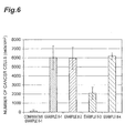
- Figure 6 is a graph showing the results of Experiment Example III-4. The experiment was carried out five times on each substrate, and the results are shown as a mean ⁇ standard deviation.
- the nickel substrates of Example III-1 and III-4 captured 6000 or more cancer cells per cm 2 .
- the nickel substrate of Example III-2 captured 5000 or more cancer cells per cm 2 .
- the nickel substrate of Example III-3 captured 2000 or more cancer cells per cm 2 .
- the nickel substrate of Comparative Example III-1 captured merely approximately 130 cancer cells per cm 2 .
Abstract
Description
- The present invention relates to an agent for improving cancer cell adhesiveness. More particularly, it relates to an agent for improving cancer cell adhesiveness, a cancer cell enrichment filter and a method for testing cancer cells.
- Enrichment of cancer cells is extremely significant in research and clinical aspects, and if cancer cells present in blood can be enriched, the enriched cancer cells can be applied to the diagnosis of cancer. For example, the most significant factor of a prognosis and a treatment of cancer is whether or not cancer cells have metastasized at the time of first medical examination or treatment. If cancer cells are spread to peripheral blood at an early stage, it is a useful method for determining disease progression of the cancer to detect circulating tumor cells (hereinafter sometimes referred to as the "CTCs").
- In a general cancer patient who has just started suffering from a metastasis, however, merely one CTC is present in his/her blood per ten billion blood cells. On the contrary, there are an overwhelmingly large number of blood components such as erythrocytes and leucocytes in blood. Accordingly, it is extremely difficult to enrich CTCs in a very low concentration so as to highly sensitively, highly efficiently and highly specifically analyze the CTCs. If the CTCs are enriched by a method using, for example, magnetic beads, density gradient centrifugation, a microchannel or a flow cytometer, it is necessary to perform a complicated treatment as well as the recovery rate is poor in some cases.
- As another technique to segregate/enrich CTCs, a method using a polycarbonate filter has been proposed (see, for example,
Non Patent Literatures 1 and 2). Furthermore, a technique to segregate/enrich CTCs by utilizing a difference in size has also been proposed (see, for example,Non Patent Literatures 3 to 5 and Patent Literature 1). - It is known that contact of blood with a surface of a medical material for an artificial heart-lung machine or the like causes an adverse reaction such as induction of thrombus formation or hemolysis due to activation of a complement system or activation of blood platelets. It has been reported that it is effective, for solving this problem, to coat a surface of a medical material with a prescribed compound (see, for example, Patent Literature 2).
-
- Patent Literature 1:
US Patent Application Publication No. 2001/0019029 - Patent Literature 2: International Publication No.
WO2004/087228 -
- Non Patent Literature 1: Vona G, et al., "Enrichment, immunomorphological, and genetic characterization of fetal cells circulating in material blood", Am J Pathol. 160(1): 51-8 (2002)
- Non Patent Literature 2: Kahn HJ, et al., "Enumeration of circulating tumor cells in the blood of breast cancer patients after filtration enrichment: correlation with disease stage", Breast Cancer ResTreat; 86(3): 237-47 (2004)
- Non Patent Literature 3: Wilding P, et al., "Integrated Cell Isolation and Polymerase Chain Reaction Analysis Using Silicon Microfilter Chambers", Anal Biochem. 257(2): 95-100 (1998)
- Non Patent Literature 4: Yuen PK, et al., "Microchip module for blood sample preparation and nucleic acid amplification reactions", Genome Res. 11(3): 405-12 (2001)
- Non Patent Literature 5: Mohamed H, et al., "Development of a rare cell fractionation device: application for cancer detection.", IEEE Trans Nanobioscience 3(4): 251-6 (2004)
- Cancer cells such as CTCs have a larger size than blood cells present in blood, such as erythrocytes, leucocytes and blood platelets. Therefore, theoretically, cancer cells can be enriched by removing these blood cell components by applying mechanical filtration.
- A membrane filter described in each of
Non Patent Literatures Non Patent Literatures 3 to 5 andPatent Literature 1, having a pore size and a thickness accurately controlled, it is sometimes difficult to enrich cancer cells with accuracy sufficiently high for precise analysis. - The present invention was achieved in consideration of the aforementioned problems, and an object of the present invention is to provide an agent for improving cancer cell adhesiveness that is capable of improving adhesiveness of cancer cells. Another object of the present invention is to provide a cancer cell enrichment filter whose surface is coated with the agent for improving cancer cell adhesiveness. Still another object of the present invention is to provide a method for testing cancer cells comprising a step of filtering peripheral blood with the cancer cell enrichment filter.
- There has been a conventional problem in which contact of blood with a surface of a medical material for an artificial heart-lung machine or the like causes an adverse reaction such as induction of thrombus formation or hemolysis due to activation of a complement system or activation of blood platelets. It is known that even if blood comes into contact with a surface of a medical material, adhesiveness of blood cell components to the surface of the medical material can be lowered by coating the surface of the medical material with a polymer comprising a constitutional unit represented by the following formula (1), so that the adverse reaction such as the induction of thrombus formation or hemolysis can be inhibited. Such a property to inhibit the thrombus formation or hemolysis is designated as blood compatibility.
- The present inventors have first found that a material surface coated with the polymer comprising the constitutional unit represented by the following formula (1) unexpectedly shows improved adhesiveness to cancer cells while showing lowered adhesiveness to blood cell components, and thus, the present invention was accomplished.
- Specifically, the present invention provides an agent for improving cancer cell adhesiveness consisting of a polymer comprising a constitutional unit represented by the following formula (1):

- In the formula (1), R1 is a hydrogen atom or a methyl group, R2 is a methyl group or an ethyl group, and m is 1 to 3.
- When the agent for improving cancer cell adhesiveness of the present invention is applied to a surface of a cancer cell enrichment filter, the adhesiveness of cancer cells to the surface of the filter can be improved, so as to improve an enrichment ratio of cancer cells.
- When the surface of a cancer cell enrichment filter is subjected to a surface treatment for improving cell adhesiveness in order to improve the enrichment ratio of cancer cells, not only the adhesiveness of cancer cells such as CTCs but also the adhesiveness of the blood cell components is improved in general. Therefore, the enrichment ratio of cancer cells is not improved as a result, and hence desired enrichment performance cannot be attained. On the contrary, the agent for improving cancer cell adhesiveness of the present invention can simultaneously satisfy characteristics in a trade-off relationship, that is, non-adhesiveness of blood cell components and adhesiveness of cancer cells.
- Furthermore, the present invention provides a method for improving adhesiveness of cancer cells to a substrate comprising a step of coating the substrate with a polymer comprising a constitutional unit represented by the following formula (1):

- In the formula (1), R1 is a hydrogen atom or a methyl group, R2 is a methyl group or an ethyl group, and m is 1 to 3.
- The present invention further provides use of a polymer comprising a constitutional unit represented by the following formula (1) for improving cancer cell adhesiveness:

- In the formula (1), R1 is a hydrogen atom or a methyl group, R2 is a methyl group or an ethyl group, and m is 1 to 3.
- The present invention further provides application of a polymer comprising a constitutional unit represented by the following formula (1) to improvement of cancer cell adhesiveness:

- In the formula (1), R1 is a hydrogen atom or a methyl group, R2 is a methyl group or an ethyl group, and m is 1 to 3.
- The present invention further provides application of a polymer comprising a constitutional unit represented by the following formula (1) to production of an agent for improving cancer cell adhesiveness:

- In the formula (1), R1 is a hydrogen atom or a methyl group, R2 is a methyl group or an ethyl group, and m is 1 to 3.
- The present invention further provides a cancer cell enrichment filter consisting of a substrate having a plurality of through holes, wherein at least a part of the substrate is coated with a polymer comprising a constitutional unit represented by the following formula (1):

- In the formula (1), R1 is a hydrogen atom or a methyl group, R2 is a methyl group or an ethyl group, and m is 1 to 3.
- The polymer may consist of a constitutional unit represented by the above formula (1). Furthermore, it is preferable that R1 is a hydrogen atom, R2 is a methyl group, and m is 1. Alternatively, R1 may be a hydrogen atom, R2 may be an ethyl group, and m may be 2, or R1 may be a methyl group, R2 may be a methyl group, and m may be 2. Besides, the polymer preferably has a number average molecular weight of 10,000 to 300,000.
- Such a polymer can further improve the cancer cell adhesiveness.
- The present invention further provides a cancer cell enrichment filter consisting of a substrate which has a plurality of through holes and at least a part of which is coated with the above polymer.
- According to the cancer cell enrichment filter of the present invention, cancer cells present in a blood sample can be enriched to a high enrichment ratio.
- The through holes preferably have an average pore size of 5 µm or more and less than 30 µm and an average aperture ratio of 5% or more and less than 50%, and are preferably formed by an electrocasting method. The substrate is preferably made of a metal. The metal is preferably selected from the group consisting of copper, nickel, a copper-nickel alloy, and any of these metals and alloy having a gold-plated surface.
- According to this cancer cell enrichment filter, cancer cells present in a blood sample can be enriched to a further higher enrichment ratio.
- The present invention further provides a method for detecting presence of cancer cells comprising a filtration step of filtering peripheral blood with the cancer cell enrichment filter.
- According to the method of the present invention, the presence of cancer cells such as CTCs in peripheral blood collected from a patient can be easily detected.
- The test method may further include a step of analyzing a gene of cells having been enriched in the filtration step, and may further include a step of culturing cells having been enriched in the filtration step.
- Since the cells are merely slightly damaged in the filtration step, the step of analyzing a gene of the enriched cells or the step of culturing the cells can be performed. When the method for detecting presence of cancer cells of the present invention further includes such a step, the presence of cancer cells such as CTCs in peripheral blood can be more accurately detected.
- The present invention provides an agent for improving cancer cell adhesiveness, a cancer cell enrichment filter whose surface is coated with the agent for improving cancer cell adhesiveness and a method for testing cancer cells comprising a step of filtering peripheral blood with the cancer cell enrichment filter.
-
-
Figure 1 is a schematic diagram illustrating an embodiment of a cancer cell enrichment filter. -
Figure 2 is a graph showing results of Experiment Example I-3, in which A corresponds to a result obtained in applying a polymer solution once, B corresponds to a result obtained in applying the polymer solution twice and C corresponds to a result obtained by a positive control. -
Figure 3 is a graph showing results of Experiment Example I-4. -
Figure 4 is a graph showing results of Experiment Example I-5. -
Figure 5 is a graph showing results of Experiment Example II-4. -
Figure 6 is a graph showing results of Experiment Example III-4. - Preferred embodiments of the present invention will now be described with reference to accompanying drawings if necessary. It is noted that a like reference numeral is used to refer to a like element shown in a drawing, so as to avoid redundancy. Besides, a part of the drawing is exaggerated to be easily understood, and a size ratio in the drawing does not necessarily accord with that mentioned in the description.
- In one embodiment, an agent for improving cancer cell adhesiveness consists of a polymer comprising a constitutional unit represented by the following formula (1):

- In the above formula (1), R1 is a hydrogen atom or a methyl group, R2 is a methyl group or an ethyl group, and m is 1 to 3. A number average molecular weight of the polymer is preferably 10,000 to 300,000. If the number average molecular weight is 10,000 or less, the polymer is in the form of a liquid and hence is difficult to handle in some cases. Furthermore, in some cases, the polymer is difficult to attain a number average molecular weight of 300,000 or more by general radical polymerization.
- The polymer can be typically produced by polymerization, performed by a general method, of a solution of a monomer represented by the following formula (2) with an appropriate initiator added. A temperature at which the polymerization is performed is preferably 40°C to 100°C, more preferably 60°C to 90°C, and further more preferably 70°C to 80°C. A pressure at which the polymerization is performed is preferably normal pressure.

- In the above formula (2), R3 is a hydrogen atom or a methyl group, R4 is a methyl group or an ethyl group, and n is 1 to 3.
- In the polymerization, a solvent capable of dissolving the monomer represented by the above formula (2) can be used as a solvent. Examples of the solvent include aliphatic or aromatic organic solvents, and more specifically, examples include ether solvents such as dioxane, tetrahydrofuran and diethyl ether; halogenated aromatic hydrocarbons such as o-dichlorobenzene; amides such as N,N-dimethylformamide; sulfoxides such as dimethyl sulfoxide; aromatic hydrocarbons such as benzene and toluene; and aliphatic hydrocarbons such as hexane and pentane, among which an ether solvent such as dioxane is preferably used.
- In one embodiment, an agent for improving cancer cell adhesiveness, consists of a polymer consisting of a constitutional unit represented by the above formula (1).
- In the agent for improving cancer cell adhesiveness, a combination of R1, R2 and m of the above formula (1) is preferably any one of the following combinations (a) to (h):
- (a) R1 is a hydrogen atom, R2 is a methyl group, and m is 1 to 2;
- (b) R1 is a hydrogen atom, R2 is a methyl group, and m is 3;
- (c) R1 is a hydrogen atom, R2 is an ethyl group, and m is 1 to 2;
- (d) R1 is a hydrogen atom, R2 is an ethyl group, and m is 3;
- (e) R1 is a methyl group, R2 is a methyl group, and m is 1 to 2;
- (f) R1 is a methyl group, R2 is a methyl group, and m is 3;
- (g) R1 is a methyl group, R2 is an ethyl group, and m is 1 to 2; and
- (h) R1 is a methyl group, R2 is an ethyl group, and m is 3.
- When the combination of R1, R2 and m is any one of the above combinations, it is possible to further improve an effect of lowering adhesiveness of blood cell components as well as improving adhesiveness of cancer cells to a substrate coated with the agent for improving cancer cell adhesiveness.
- In one embodiment, an agent for improving cancer cell adhesiveness is a random copolymer, a block copolymer or a graft copolymer of a monomer represented by the above formula (2) and another polymerizable monomer.
- Examples of the monomer polymerizable with the monomer represented by the above formula (2) include: alkylacrylamides such as acrylamide, t-butyl acrylamide, n-butyl acrylamide, i-butyl acrylamide, hexyl acrylamide and heptyl acrylamide; N,N-dialkylacrylamides such as N,N-dimethylacrylamide and N,N-diethylacrylamide; amino alkyl acrylates such as amino methyl acrylate, amino ethyl acrylate and amino isopropyl acrylate; diamino alkyl acrylate such as diamino methyl acrylate, diamino ethyl acrylate and diamino butyl acrylate; N,N-dialkyl methacrylamides such as methacrylamide, N,N-dimethylmethacrylamide and N,N-diethylmethacrylamide; amino alkyl methacrylates such as amino methyl methacrylate and amino ethyl methacrylate; diamino alkyl methacrylates such as diamino methyl methacrylate and diamino ethyl methacrylate; alkyl acrylates such as methyl acrylate, ethyl acrylate, isopropyl acrylate, butyl acrylate, hexyl acrylate and 2-ethylhexyl acrylate; alkyl methacrylates such as methyl methacrylate, ethyl methacrylate, butyl methacrylate and hexyl methacrylate; alkoxy (meth)acrylates such as methoxy (meth)acrylate; alkoxy alkyl (meth)acrylate such as methoxy ethyl (meth)acrylate; and glycidyl methacrylate and propylene.
- The monomer to be polymerized with the monomer represented by the above formula (2) may be one or more monomers selected from the group consisting of alkyl acrylate, alkyl methacrylate, alkoxy (meth)acrylate, alkoxy alkyl (meth)acrylate, glycidyl methacrylate and propylene.
- The copolymer of the monomer represented by the above formula (2) and the above-described polymerizable monomer can be any one of a random copolymer, a block copolymer and a graft copolymer, and may be produced by any of random polymerization, ion polymerization, polymerization utilizing a macromer and the like.
- When the monomer represented by the above formula (2) and the polymerizable monomer are copolymerized with each other, the content of the monomer represented by the above formula (2) is preferably 30 to 99% by mass and more preferably 50 to 99% by mass in the resulting copolymer.
- The polymer comprising the constitutional unit represented by the above formula (1) can be typically produced by polymerization performed by random polymerization, ion polymerization, photopolymerization, polymerization utilizing a macromer or the like with an initiator appropriate for the solution of the monomer represented by the above formula (2) added thereto, and as occasion demands, with one or more of the polymerizable monomers added thereto. A temperature at which the polymerization is performed is preferably 40°C to 100°C, more preferably 60°C to 90°C and further more preferably 70°C to 80°C. A pressure at which the polymerization is performed is preferably normal pressure.
- In the polymerization, a solvent capable of dissolving the monomer represented by the above formula (2) and the polymerizable monomer can be used as a solvent. Examples of the solvent include aliphatic or aromatic organic solvents, and more specifically, examples include ether solvents such as dioxane, tetrahydrofuran and diethyl ether; halogenated aromatic hydrocarbons such as o-dichlorobenzene; amides such as N,N-dimethylformamide; sulfoxides such as dimethyl sulfoxide; aromatic hydrocarbons such as benzene and toluene; and aliphatic hydrocarbons such as hexane and pentane, among which an ether solvent such as dioxane is preferably used.
- When the agent for improving cancer cell adhesiveness is applied to a surface of a substrate of a cancer cell enrichment filter or the like, blood compatibility is imparted, and hence, if the filter is brought into contact with blood, activation of blood components can be inhibited, so as to reduce adhesion of blood cell components to the filter. Furthermore, since the cancer cell enrichment filter to which the agent for improving cancer cell adhesiveness is applied is good in adhesiveness to various cancer cells, cancer cells can be efficiently enriched.
- Although the polymer comprising the constitutional unit represented by the above formula (1) has, in its molecule, an ether bond and an ester bond as a polar group, such a polar group does not show strong electrostatic interaction with a biological component differently from a nitrogen atom (an amino group or an imino group), a carboxyl group and the like. Furthermore, since the polymer has no large hydrophobic group, the hydrophobic interaction is also small. It is therefore presumed that the polymer comprising the constitutional unit represented by the above formula (1) has low activity against blood and shows excellent blood compatibility.
- In contact between a medical material surface and protein present in a biological tissue or blood, it is preferable that adsorption/denaturation or activation of the protein does not occur, and for this purpose, it is regarded useful to reduce the hydrophobic interaction and the electrostatic interaction, that is, large interaction working between substances. Also from this point of view, the surface to which the agent for improving cancer cell adhesiveness is applied can be provided with a suitable surface structure.
- Furthermore, since the surface to which the agent for improving cancer cell adhesiveness is applied has moderate hydrophilicity, even when it comes into contact with blood, blood platelets are merely slightly adhered thereto, and thus, excellent blood compatibility is shown. Moreover, it is presumed that interaction with a biological component via a hydrogen bond derived from a hydroxyl group, denaturation of adsorbed protein and the like can be inhibited.
- By applying the agent for improving cancer cell adhesiveness to the surface of the substrate of the cancer cell enrichment filter or the like, adhesion to the filter of blood cell components such as erythrocytes, leucocytes and blood platelets can be reduced, and at the same time, cancer cells can be selectively and efficiently captured. The details of the mechanism of showing this function has not been clarified yet, but the present inventors presume that the mechanism can be explained by using concept of intermediate water.
- Specifically, it seems that water in contact with the substrate to which the agent for improving cancer cell adhesiveness is applied includes (1) free water that has weak interaction with the agent for improving cancer cell adhesiveness and is molten at 0°C; (2) unfreezable water that has strong interaction with the agent for improving cancer cell adhesiveness and does not freeze even at -100°C; and (3) intermediate water that has interaction intermediate between the free water and the unfreezable water and freezes at a temperature lower than 0°C. Normal blood cells form a hydration shell with the unfreezable water, the intermediate water, the free water and the like in contact with the agent for improving cancer cell adhesiveness, and hence are stabilized owing to this hydration structure, but the unfreezable water strongly affected by the structure of the agent for improving cancer cell adhesiveness is probably camouflaged by the intermediate water, so that adhesion of the normal blood cells to the substrate surface is inhibited. On the other hand, since cancer cells are different from the normal cells in expression of sugar chains on cell surfaces, the hydration structure is probably disordered as compared with that in the normal blood cells. This seems to disturb the structure of the intermediate water on the substrate surface, so that the adhesiveness of the cancer cells to the substrate surface can be improved.
- The agent for improving cancer cell adhesiveness is applicable to all the types of cancer cells, and is preferably applied to epithelial cell-derived cancers that easily infiltrate into blood vessels, and among such cancers, the agent is particularly preferably applied to lung cancers, digestive cancers such as colorectal cancers, stomach cancers and esophageal cancers, breast cancers and prostatic cancers, which are regarded to produce a large number of CTCs.
- In one embodiment, the present invention provides a cancer cell enrichment filter consisting of a substrate which has a plurality of through holes and at least a part of which is coated with the agent for improving cancer cell adhesiveness. This filter is capable of enriching cancer cells such as CTCs present in blood. Examples of the opening shape of each through hole include a circular shape, an elliptic shape, a rectangular shape, a rounded rectangular shape and a polygonal shape. A rounded rectangular shape is a shape having two long sides in the same length and two semicircles. From the viewpoint that cancer cells can be efficiently captured, a circular shape, a rectangular shape or a rounded rectangular shape is preferably employed. Besides, from the viewpoint of preventing clogging of the filter, a rounded rectangular shape is particularly preferably employed.
-
Figure 1 is a schematic diagram illustrating one embodiment of the cancer cell enrichment filter. Thefilter 100 consists of asubstrate 20 having a plurality of throughholes 10. The opening shape of each throughhole 10 is a rounded rectangular shape. CTCs are captured on a surface of aface 30 of thesubstrate 20. At least a part of theface 30 is coated with the agent for improving cancer cell adhesiveness. The agent for improving cancer cell adhesiveness is preferably coated over the whole of theface 30. A part or the whole of a face opposite to theface 30 may be coated with the agent for improving cancer cell adhesiveness. - As a method for applying the agent for improving cancer cell adhesiveness to the surface of the substrate of the cancer cell enrichment filter or the like, a coating method is the most generally employed. The coating method is performed by allowing a solution of the polymer comprising the constitutional unit represented by the above formula (1) to adhere to the substrate surface by a dipping method, a spraying method, a spin coating method or the like, and then removing (drying) the solvent. A thickness of a film attained after drying the solvent is preferably 0.01 µm to 1.0 mm, more preferably 0.1 to 100 µm and further more preferably 0.5 to 50 µm. If the film thickness is smaller than 0.01 µm, non-adhesiveness to blood cell components and adhesiveness to cancer cells may not be sufficiently exhibited in some cases. Alternatively, if the film thickness exceeds 1.0 mm, it is apprehended that balance between these adhesiveness characteristics may be lost in some cases.
- In order to more strongly fix the agent for improving cancer cell adhesiveness on the substrate, the substrate may be heated after coating with the agent for improving cancer cell adhesiveness. Furthermore, the polymer comprising the constitutional unit represented by the above formula (1) may be crosslinked. An example of a crosslinking method includes precedent addition of a crosslinkable monomer to the materials of the polymer. For the crosslinkage, electron beams, y rays or light irradiation may be used.
- For forming, on the substrate surface, a layer of the polymer comprising the constitutional unit represented by the above formula (1) by plasma graft polymerization, plasma-initiated polymerization is performed with the monomer represented by the above formula (2) supplied after irradiation of low-temperature plasma performed under reduced pressure of approximately 1.3 x 10⁻¹Pa, preferably 1.3 to 133.3 Pa, and under an atmosphere of argon, nitrogen, air, any of various monomers or the like for 1 to 300 seconds, preferably 2 to 30 seconds.
- In one embodiment, the material and the shape of the substrate are not especially limited, and for example, a porous body, fiber, nonwoven fabric, a film, a sheet or a tube may be employed. Examples of the material of the substrate include natural polymers such as cotton and hemp; synthetic polymers such as nylon, polyester, polyacrylonitrile, polyolefin, halogenated polyolefin, polyurethane, polyamide, polysulfone, polyether sulfone, poly(meth)acrylate, a halogenated polyolefin ethylene-polyvinyl alcohol copolymer and a butadiene-acrylonitrile copolymer; and mixtures thereof. Examples of the material also include metals, ceramics and composite materials thereof, and the substrate may include a plurality of substrates.
- Examples of the metal include but are not limited to noble metals such as gold and silver; base metals such as copper, aluminum, tungsten, nickel and chromium; and alloys of these metals. Such a metal may be singly used, or may be used as an alloy with another metal or as a metal oxide for imparting functionality. From the viewpoint of the price and availability, nickel, copper or a metal containing any of them as a principal component is preferably used. Here, a principal component means a component occupying 50% by weight or more among materials contained in the substrate. Such a metal may be subjected to photolithography or the like for forming through holes therein, so as to be used as a screen filter.
- A general CTC has a diameter of 10 µm or more. Here, a diameter of a cell means a length of the longest straight line out of straight lines each connecting arbitrary two points on the outline of the cell in observation with a microscope. From the viewpoint of the permeability of blood and CTC capturing performance, the through holes of the cancer cell enrichment filter have an average pore size of preferably 5 µm or more and less than 30 µm and an average aperture ratio of preferably 5% or more and less than 50%. Furthermore, the average pore size and the average aperture ratio are respectively more preferably 5 µm or more and less than 15 µm and 10% or more and less than 40% and particularly preferably 5 µm or more and less than 10 µm and 20% or more and less than 40%. Here, an aperture ratio means an area occupied by the through holes in the whole area of the filter. From the viewpoint of preventing clogging, the average aperture ratio is preferably larger, but if it exceeds the upper limit, the filter may be degraded in its strength or may be difficult to be worked in some cases. Alternatively, if the aperture ratio is lower than 5%, the cancer cell enrichment performance of the filter may be degraded in some cases.
- Herein, a pore size in an opening shape other than a circular shape, such as an elliptic shape, a rectangular shape or a rounded rectangular shape, is defined as the maximum value of diameters of spheres that can pass through the through hole in such a shape. If the opening shape is a rectangular shape, the pore size of the through hole corresponds to the length of the short side of the rectangle, and if the opening shape is a polygonal shape, it corresponds to the diameter of the inscribed circle of the polygon. If the opening shape is a rectangular shape or a rounded rectangular shape, even when CTCs and leucocytes are captured by the through holes, there remains a space in the long side direction of the opening shape. Since a liquid can pass through this space, the clogging of the filter can be prevented.
- The thickness of the substrate of the filter is preferably 3 to 100 µm, more preferably 5 to 50 µm and particularly preferably 10 to 30 µm. If the thickness of the substrate is smaller than 3 µm, the filter may be degraded in its strength and hence may be difficult to handle in some cases. Alternatively, if the thickness of the substrate exceeds 100 µm, the materials may be consumed in an amount more than necessary or it may take longer time to work the substrate, and hence, such a large thickness may be disadvantageous in cost and precise working of the substrate itself may become difficult in some cases.
- Subsequently, a method for producing the cancer cell enrichment filter of this embodiment will be described. The method for producing the filter of this embodiment is not especially limited, and the filter is produced by, for example, an electrocasting method (an electroforming method). The electrocasting method is a method in which a thick electroplating is provided on a matrix and is then peeled off. First, a photosensitive resist film (a photosensitive layer) is adhered onto a support of stainless steel or the like. Next, a mask having a pattern of the opening shape of the through holes of the filter is fixed on the photosensitive layer. Subsequently, light (activating light beams) is irradiated through the mask. After the light irradiation, if the support remains on the photosensitive layer, the support is removed, and then, development is carried out by removing an unexposed portion by wet development with a developer such as an alkaline aqueous solution, a water-based developer or an organic solvent, or by dry development or the like, and thus, a resist pattern is formed. Thereafter, the developed resist pattern is used as a mask, so as to plate portions not masked but exposed on the substrate. Examples of a plating method include copper plating, solder plating, nickel plating and gold plating. After the plating, the resulting plating layer is peeled off from the support and the photosensitive layer, and thus, the plating layer is obtained. When at least a part of the plating layer is coated with the agent for improving cancer cell adhesiveness by the aforementioned method, the cancer cell enrichment filter is obtained.
- In one embodiment, the present invention provides a method for detecting presence of cancer cells comprising a filtration step of filtering a sample with the cancer cell enrichment filter. As a sample for enriching cancer cells such as CTCs, blood pooled in a bone marrow, a spleen, a liver or the like, lymph, a tissue fluid, cord blood or the like may be used, but peripheral blood circulating through a body can be most easily used. It is useful means for determining the progression of a cancer to detect the presence of CTCs in peripheral blood.
- The method for detecting presence of cancer cells of this embodiment can be practiced by, for example, incorporating the cancer cell enrichment filter in a flow channel, introducing peripheral blood into the flow channel for enriching cells including CTCs, and determining whether or not there are CTCs in the enriched cells. Blood is introduced into the flow channel by, for example, a method in which a pressure is applied from an inlet direction of the flow channel, a method in which a pressure is reduced from an outlet direction of the flow channel, or a method in which a peristaltic pump is used. Furthermore, the area of the used filter is suitably 1 to 10 cm2 if, for example, CTCs are enriched from 1 mL of blood.
- When CTCs are enriched by the aforementioned method, not only the CTCs but also blood cells such as leucocytes are simultaneously enriched. Therefore, it is necessary to determine whether or not cancer cells are contained in collected cells. For example, after enriching the CTCs by the aforementioned method, cells can be verified as cancer cells by dyeing them with an antibody against a fluorescent labeled cancer marker. An example of the antibody against a cancer marker includes an anti-EpCAM antibody.
- Alternatively, cells can be verified as cancer cells by analyzing a gene of the cells having been enriched by the aforementioned method. For example, the cells can be verified as cancer cells by analyzing mutation of a gene such as p53, K-RAS, H-RAS, N-RAS, BRAF or APC. Besides, the gene analysis result can be used for, for example, determining a subsequent treatment course of a patient. Alternatively, the cells can be verified as cancer cells by measuring telomerase activity or the like of the cells having been enriched by the aforementioned method.
- Since the filtration step merely slightly damages the cells, the enriched cells can be cultured for performing more detailed analysis. Examples
- The present invention will now be described based on examples. It is noted that the present invention is not limited to the following examples.
- Polymethoxyethyl acrylate having a constitutional unit of the above formula (1) in which R1 is hydrogen, R2 is a methyl group and m is 1 was synthesized. Specifically, 15 g of methoxyethyl acrylate was polymerized in 60 g of 1,4-dioxane at 75°C for 10 hours with nitrogen bubbling by using azobisisobutyronitrile (0.1 % by mass) as an initiator. After the polymerization was completed, the resultant was added dropwise to n-hexane to form a precipitate, and thus the product was isolated. The product was dissolved in tetrahydrofuran, the resulting solution was purified with n-hexane twice, and the resultant was dried under reduced pressure for a whole day and night. Thus, a clear and colorless polymer with high viscosity was obtained. The yield rate was 76%. As a result of analysis of the thus obtained polymer by gel permeation chromatography (GPC), the polymer was found to have a number average molecular weight of 15,000 and a molecular weight distribution (Mw/Mn) of 3.4.
- The molecular weight of the polymer was calculated in terms of standard polystyrene molecular weight under the following GPC measurement conditions:
- Pump: PU Intelligent HPLC Pump (manufactured by Jasco Corporation)
- Column: GPC K804 (manufactured by Showa Denko K.K., Shodex)
- Eluent: chloroform
- Measurement temperature: room temperature
- Flow rate: 1.0 mL/min
- Detector: Jasco RI-1530 RI (manufactured by Jasco Corporation)
- A commercially available titanium plate was immersed, as a cathode, in an electrolytic bath for electrolytic nickel plating (that is, an aqueous solution of 450 g/L of nickel sulfamate, 5 g/L of nickel chloride and 30 g/L of boric acid, 55°C), and an anode was immersed in the same electrolytic bath. With a voltage applied to the both electrodes, nickel plating was performed to attain a film thickness of 10 µm, and thus, a nickel substrate was prepared.
- The polymer synthesized in Experiment Example I-1 was dissolved in chloroform, so as to obtain a plurality of polymer solutions different in the concentration in a range from 0 to 5% by mass. Each of the polymer solutions was applied (casted) onto the nickel substrate prepared in Experiment Example I-2 and the solvent was dried, so as to coat the surface of the nickel substrate. Two types of substrates, that is, one coated with the polymer once and the other coated with the polymer twice, were prepared. As a positive control, a polyethylene terephthalate substrate to which 40 µL of polymer solution was applied was used. A contact angle of water was measured on each of these substrates, so as to verify that the substrate was coated with the polymer. For measuring a contact angle, a contact angle measuring apparatus (Erma Inc., Model G-1-100) was used. A static contact angle was measured 30 seconds after dropping 2 µl of pure water onto the surface of each substrate.
Figure 2 is a graph showing the results obtained in Experiment Example I-3. The abscissa indicates the concentration of the polymer and the ordinate indicates the contact angle of water. InFigure 2 , A corresponds to the result obtained in applying the polymer solution once, B corresponds to the result obtained in applying the polymer solution twice, and C corresponds to the result of the positive control. - The polymer synthesized in Experiment Example I-1 was dissolved in chloroform for obtaining a solution in a concentration of 4% by mass. This solution was applied onto the nickel substrate prepared in Experiment Example I-2 and the solvent was dried, so as to coat the surface of the nickel substrate with the polymer, and thus, a nickel substrate of Example I-1 was prepared. The polymer solution was applied twice. Through X-ray photoelectron spectroscopy (Shimadzu Corporation, ESCA-1000), a peak of nickel derived from the nickel substrate was not observed but peaks of carbon and oxygen derived from the polymer were detected, and thus, it was verified that the nickel substrate was coated with the polymer. Furthermore, the nickel substrate prepared in Experiment Example I-2 was used as a nickel substrate of Comparative Example I-1.
- Onto each of the nickel substrates of Example I-1 and Comparative Example I-1, 1.0 mL of cancer cell suspension adjusted to 10,000 cells/mL in a medium containing 10% of blood serum was dropped with a pipette, and the resulting substrates were allowed to stand still at 37°C for 60 minutes. As the cancer cells, human fibrosarcoma cell line HT-1080 was used. Subsequently, the substrates were rinsed with a physiological buffered saline solution, and the number of cells adhered onto each substrate was counted. In order that the cells could be easily counted, the cells were fixed with formaldehyde, and then, cell nuclei were dyed with 4,6-diamino-2-phenylindole (DAPI). The number of cell nuclei was counted by using a confocal laser scanning microscope (Olympus Corporation, FV-1000), and the thus obtained number was defined as the number of cells.
-
Figure 3 is a graph showing the results of Experiment Example I-4. The experiment was carried out five times on each substrate, and the results are shown as a mean ± standard deviation. In the nickel substrate of Example I-1, the number of adhered cancer cells was larger than in the nickel substrate of Comparative Example I-1. With respect to a large number of other cancer cells, similar results were verified. - In the same manner as in Experiment Example I-4, the nickel substrates of Example I-1 and Comparative Example I-1 were used for an experiment.
- Onto each of the nickel substrates of Example I-1 and Comparative Example I-1, 0.2 mL of fresh human platelet-rich plasma anticoagulated with sodium citrate was dropped with a pipette, and the resulting substrates were allowed to stand still at 37°C for 60 minutes. Subsequently, each substrate was rinsed with a phosphate buffer solution, and after fixation with glutaraldehyde, the substrate was observed with a scanning electron microscope, so as to count the number of blood platelets adhered in an area of 1×104 µm2.
-
Figure 4 is a graph showing the results obtained in Experiment Example I-5. The experiment was carried out five times on each substrate, and the results are shown as a mean ± standard deviation. In the nickel substrate of Example I-1, the number of adhered blood platelets was smaller than in the nickel substrate of Comparative Example I-1. - It was revealed, based on the results of Experiment Examples I-4 and I-5, that the nickel substrate of Example I-1 shows improved adhesiveness to cancer cells but shows low adhesiveness to a blood cell component. Accordingly, when the agent for improving cancer cell adhesiveness of the present invention is applied to the surface of a cancer cell enrichment filter, the adhesiveness of cancer cells to the filter can be improved so as to improve the enrichment ratio of cancer cells.
- Poly[2-(2-ethoxyethoxy)ethyl acrylate] having a constitutional unit represented by the above formula (1) in which R1 is hydrogen, R2 is an ethyl group and m is 2 was synthesized. Specifically, 15 g of 2-(2-ethoxyethoxy)ethyl acrylate was polymerized in 60 g of 1,4-dioxane at 75°C for 10 hours with nitrogen bubbling by using azobisisobutyronitrile (0.1% by mass) as an initiator. After the polymerization was completed, the resultant was added dropwise to n-hexane to form a precipitate, and thus the product was isolated. The product was dissolved in tetrahydrofuran, and the resulting solution was purified with n-hexane twice. The resulting purified product was dried under reduced pressure for a whole day and night. Thus, a clear and colorless syrup-like polymer was obtained. The yield amount (the yield rate) was 11.4 g (76.0%). The structure of the thus obtained polymer was verified by 1H-NMR. As a result of analysis for the molecular weight by the GPC, the polymer was found to have a number average molecular weight (Mn) of 12,000 and a molecular weight distribution (Mw/Mn) of 3.9. The molecular weight of the polymer was calculated in terms of standard polystyrene molecular weight under the same GPC measurement conditions as in Experiment Example I-1.
- Poly[2-(2-methoxyethoxy)ethyl methacrylate] having a constitutional unit represented by the above formula (1) in which R1 is a methyl group, R2 is a methyl group and m is 2 was synthesized. Specifically, 10 g of 2-(2-methoxyethoxy)ethyl methacrylate was polymerized in 50 g of 1,4-dioxane at 80°C for 8 hours with nitrogen bubbling by using azobisisobutyronitrile (0.1% by mass) as an initiator. After the polymerization was completed, the resultant was added dropwise to n-hexane to form a precipitate, and thus the product was isolated. The product was dissolved in tetrahydrofuran, and the resulting solution was purified with n-hexane twice. The resulting purified product was dried under reduced pressure for a whole day and night. Thus, a clear and colorless syrup-like polymer was obtained. The yield amount (the yield rate) was 8.2 g (82.0%). The structure of the thus obtained polymer was verified by 1H-NMR. As a result of analysis for the molecular weight by the GPC, the polymer was found to have a number average molecular weight (Mn) of 104,000 and a molecular weight distribution (Mw/Mn) of 4.6. The molecular weight of the polymer was calculated in terms of standard polystyrene molecular weight under the same GPC measurement conditions as in Experiment Example I-1.
- Poly(2-methacryloyloxyethyl phosphorylcholine-co-butyl methacrylate) represented by the following formula (3) was synthesized. The poly(2-methacryloyloxyethyl phosphorylcholine-co-butyl methacrylate) is a copolymer of 2-methacryloyloxyethyl phosphorylcholine (MPC) and butyl methacrylate (BMA), and a ratio of MPC:BMA was 40:60 mol% to 1:99 mol%.

- In the above formula (3), X and Y are 1 to 10,000, and X:Y is 40:60 mol% to 1:99 mol%.
- Specifically, the monomers were dissolved in ethanol, and the polymerization was performed at 75°C for 10 hours with nitrogen bubbling by using azobisisobutyronitrile (0.1 % by mass) as an initiator. After the polymerization was completed, the resultant was added dropwise to diethyl ether to form a precipitate, and thus the product was isolated. The product was dissolved in ethanol, the resulting solution was purified with diethyl ether twice, and the resultant was dried under reduced pressure for a whole day and night. Thus, a white powder polymer was obtained. The yield rate was 51 %. As a result of the analysis of the thus obtained polymer by the gel permeation chromatography (GPC), the polymer was found to have a number average molecular weight of 240,000 and a molecular weight distribution (Mw/Mn) of 2.9. The molecular weight of the polymer was calculated in terms of standard poly(oxyethylene) molecular weight under the same GPC measurement conditions as in Experiment Example I-1 except that ethanol/chloroform (2/8) was used as the eluent.
- A nickel substrate was prepared in the same manner as in Experiment Example I-2.
- The nickel substrate was coated with each of the polymers in the same manner as in (Experiment Example I-3), and a static contact angle of water was measured. As a result, in using the poly[2-(2-ethoxyethoxy)ethyl acrylate] in a concentration of 0.2% wt/vol or more, the contact angle was substantially stabilized at 25°. In using the poly[2-(2-methoxyethoxy)ethyl methacrylate] in a concentration of 0.5% wt/vol or more, the contact angle was substantially stabilized at 38°. In using the poly(2-methacryloyloxyethyl phosphorylcholine-co-butyl methacrylate) in a concentration of 0.1% wt/vol or more, the contact angle was substantially stabilized at 104°.
- The four polymers synthesized in Experiment Examples I-1 and II-1 were respectively dissolved in chloroform for obtaining solutions each in a concentration of 4% by mass. Each of these solutions was applied to the nickel substrate prepared in Experiment Example II-2 and the solvent was dried, so as to coat the surface of the nickel substrate with the polymer, and thus, nickel substrates of Example II-1 (using polymethoxyethyl acrylate), Example II-2 (using poly[2-(2-ethoxyethoxy)ethyl acrylate]), Example II-3 (using poly[2-(2-methoxyethoxy)ethyl methacrylate] and Comparative Example II-1 (using poly(2-methacryloyloxyethyl phosphorylcholine-co-butyl methacrylate)) were prepared. Each polymer solution was applied twice. Through X-ray photoelectron spectroscopy (Shimadzu Corporation, ESCA-1000), a peak of nickel derived from the nickel substrate was not observed but peaks of carbon and oxygen derived from the polymer were detected, and thus, it was verified that each nickel substrate was coated with the corresponding polymer.
- Onto each of the nickel substrates of Examples II-1 to II-3 and Comparative Example II-1, 1.0 mL of cancer cell suspension adjusted to 10,000 cells/mL in a medium containing 10% of blood serum was dropped with a pipette, and the resulting substrates were allowed to stand still at 37°C for 60 minutes. As the cancer cells, human mammary adenocarcinoma cell line MDA-MB-231 was used. Subsequently, the substrates were rinsed with a physiological buffered saline solution, and the number of cells adhered onto each substrate was counted. In order that the cells could be easily counted, the cells were fixed with formaldehyde, and then, cell nuclei were dyed with 4,6-diamino-2-phenylindole (DAPI). The number of cell nuclei was counted by using a confocal laser scanning microscope (Olympus Corporation, FV-1000), and the thus obtained number was defined as the number of cells.
-
Figure 5 is a graph showing the results of Experiment Example II-4. The experiment was carried out five times on each substrate, and the results are shown as a mean ± standard deviation. The nickel substrate of Example II-1 captured 2000 or more cancer cells. The nickel substrate of Example II-2 captured 1500 or more cancer cells. The nickel substrate of Example II-3 captured 500 or more cancer cells. On the contrary, the nickel substrate of Comparative Example II-1 captured merely approximately 140 cancer cells. - Poly[2-(2-ethoxyethoxy)ethyl methacrylate] having a constitutional unit represented by the above formula (1) in which R1 is a methyl group, R2 is an ethyl group and m is 2 was synthesized. Specifically, 15.0 g (7.4 x 10⁻² mol) of 2-(2-ethoxyethoxy)ethyl methacrylate was dissolved in 58.2 mL of 1,4-dioxane to be subjected to N2 purge for 2 hours. To the resultant, 15.1 mg (9.2 x 10⁻² mmol) of azobisisobutyronitrile used as an initiator dissolved in a small amount of 1,4-dioxane was added, and the polymerization was performed under a nitrogen atmosphere at 75°C for 2 hours and 10 minutes. Purification was performed by using hexane. The reaction solution was added dropwise to 1500 mL of hexane serving as a poor solvent, and the solvent was removed by decantation. To the thus obtained crude polymer, approximately 50 mL of tetrahydrofuran (THF) was added for dissolving, and the resulting solution was added dropwise to 1000 mL of hexane again for forming a precipitate, and then the solvent was removed by the decantation. This operation was repeated once again, so that the monomer and the initiator contained in the polymer could be completely removed. The resultant was dried under reduced pressure overnight, and the mass of the resulting polymer was measured. Thus, a clear and colorless syrup-like polymer, poly[2-(2-ethoxyethoxy)ethyl methacrylate], was obtained. The yield amount was 5.36 g and the yield rate was 35.7%. As a result of analysis for the molecular weight by the GPC, the polymer was found to have a number average molecular weight (Mn) of 142,000 and a molecular weight distribution (Mw/Mn) of 6.06. The molecular weight of the polymer was calculated in terms of standard polystyrene molecular weight under the same GPC measurement conditions as in Experiment Example I-1.
- A nickel substrate was prepared in the same manner as in Experiment Example I-2.
- With the nickel substrate coated with the polymer in the same manner as in Experiment (Example I-3), a static contact angle of water was measured, resulting in finding that the contact angle was substantially stabilized at 78° in using the poly[2-(2-ethoxyethoxy)ethyl methacrylate] in a concentration of 0.5% wt/vol or more.
- The five polymers synthesized in Experiment Examples I-1, II-1 and III-1 were respectively dissolved in chloroform for obtaining solutions each in a concentration of 4% by mass. Each of these solutions was applied to the nickel substrate prepared in Experiment Example III-2 and the solvent was dried, so as to coat the surface of the nickel substrate with the polymer, and thus, nickel substrates of Example III-1 (using polymethoxyethyl acrylate), Example III-2 (using poly[2-(2-ethoxyethoxy)ethyl acrylate]), Example III-3 (using poly[2-(2-methoxyethoxy)ethyl methacrylate], Comparative Example III-1 (using poly(2-methacryloyloxyethyl phosphorylcholine-co-butyl methacrylate)) and Example III-4 (using poly[2-(2-ethoxyethoxy)ethyl methacrylate]) were prepared. Each polymer solution was applied twice. Through the X-ray photoelectron spectroscopy (Shimadzu Corporation, ESCA-1000), a peak of nickel derived from the nickel substrate was not observed but peaks of carbon and oxygen derived from the polymer were detected, and thus, it was verified that each nickel substrate was coated with the corresponding polymer.
- Onto each of the nickel substrates of Examples III-1 to III-4 and Comparative Example III-1, 1.0 mL of cancer cell suspension adjusted to 10,000 cells/mL in a medium containing 10% of blood serum was dropped with a pipette, and the resulting substrates were allowed to stand still at 37°C for 60 minutes. As the cancer cells, lung cancer cell line A549 was used. Subsequently, the substrates were rinsed with a physiological buffered saline solution, and the number of cells adhered onto each substrate was counted. In order that the cells could be easily counted, the cells were fixed with formaldehyde, and then, cell nuclei were dyed with 4,6-diamino-2-phenylindole (DAPI). The number of cell nuclei was counted by using a confocal laser scanning microscope (Olympus Corporation, FV-1000), and the thus obtained number was defined as the number of cells.
-
Figure 6 is a graph showing the results of Experiment Example III-4. The experiment was carried out five times on each substrate, and the results are shown as a mean ± standard deviation. The nickel substrates of Example III-1 and III-4 captured 6000 or more cancer cells per cm2. The nickel substrate of Example III-2 captured 5000 or more cancer cells per cm2. The nickel substrate of Example III-3 captured 2000 or more cancer cells per cm2. On the contrary, the nickel substrate of Comparative Example III-1 captured merely approximately 130 cancer cells per cm2. - 10 ... through hole, 20 ... substrate, 30 ... face, 100 ... filter.
Claims (14)
- An agent for improving cancer cell adhesiveness, consisting of:a polymer comprising a constitutional unit represented by the following formula (1):

- The agent for improving cancer cell adhesiveness according to claim 1, consisting of a polymer consisting of the constitutional unit represented by the formula (1).
- The agent for improving cancer cell adhesiveness according to claim 1 or 2, wherein R1 is a hydrogen atom, R2 is a methyl group and m is 1.
- The agent for improving cancer cell adhesiveness according to claim 1 or 2, wherein R1 is a hydrogen atom, R2 is an ethyl group and m is 2.
- The agent for improving cancer cell adhesiveness according to claim 1 or 2, wherein R1 is a methyl group, R2 is a methyl group and m is 2.
- The agent for improving cancer cell adhesiveness according to any one of claims 1 to 5, wherein a number average molecular weight of the polymer is 10,000 to 300,000.
- A cancer cell enrichment filter consisting of:a substrate having a plurality of through holes, wherein at least a part of the substrate is coated with the agent for improving cancer cell adhesiveness according to any one of claims 1 to 6.
- The cancer cell enrichment filter according to claim 7,
wherein the through holes have an average pore size of 5 µm or more and less than 30 µm and an average aperture ratio of 5% or more and less than 50%. - The cancer cell enrichment filter according to claim 7 or 8,
wherein the substrate is made of a metal. - The cancer cell enrichment filter according to claim 9,
wherein the metal is selected from the group consisting of copper, nickel, a copper-nickel alloy, and copper, nickel or copper-nickel alloy having a gold-plated surface. - The cancer cell enrichment filter according to claim 9 or 10,
wherein the through holes are formed by an electrocasting method. - A method for detecting presence of cancer cells, comprising:a filtration step of filtering peripheral blood with the cancer cell enrichment filter according to any one of claims 7 to 11.
- The method according to claim 12, further comprising:a step of analyzing a gene of cells having been enriched in the filtration step.
- The method according to claim 12 or 13, further comprising:a step of culturing cells having been enriched in the filtration step.
Applications Claiming Priority (3)
| Application Number | Priority Date | Filing Date | Title |
|---|---|---|---|
| JP2011131241 | 2011-06-13 | ||
| JP2012123228 | 2012-05-30 | ||
| PCT/JP2012/064932 WO2012173097A1 (en) | 2011-06-13 | 2012-06-11 | Agent for improving cancer cell adhesiveness |
Publications (3)
| Publication Number | Publication Date |
|---|---|
| EP2720039A1 true EP2720039A1 (en) | 2014-04-16 |
| EP2720039A4 EP2720039A4 (en) | 2015-01-28 |
| EP2720039B1 EP2720039B1 (en) | 2017-07-26 |
Family
ID=47357084
Family Applications (1)
| Application Number | Title | Priority Date | Filing Date |
|---|---|---|---|
| EP12801028.7A Not-in-force EP2720039B1 (en) | 2011-06-13 | 2012-06-11 | Agent for improving cancer cell adhesiveness |
Country Status (7)
| Country | Link |
|---|---|
| US (1) | US9372136B2 (en) |
| EP (1) | EP2720039B1 (en) |
| JP (1) | JP5975532B2 (en) |
| CN (1) | CN103635801A (en) |
| CA (1) | CA2839313C (en) |
| TW (1) | TWI516294B (en) |
| WO (1) | WO2012173097A1 (en) |
Cited By (7)
| Publication number | Priority date | Publication date | Assignee | Title |
|---|---|---|---|---|
| EP2851418A4 (en) * | 2012-05-14 | 2015-12-23 | Hitachi Chemical Co Ltd | Cancer cell-trapping metal filter, cancer cell-trapping metal filter sheet, cancer cell-trapping device, and manufacturing methods therefor |
| EP3244208A1 (en) * | 2016-05-09 | 2017-11-15 | Sumitomo Rubber Industries, Ltd. | Medical analysis device and cell analysis method |
| EP3301444A1 (en) * | 2016-09-29 | 2018-04-04 | Sumitomo Rubber Industries, Ltd. | Method for capturing cancer cells |
| US10641760B2 (en) | 2016-09-29 | 2020-05-05 | Sumitomo Rubber Industries, Ltd. | Medical analysis device and cell analysis method |
| US11226330B2 (en) | 2018-02-14 | 2022-01-18 | Sumitomo Rubber Industries, Ltd. | Method for capturing specific cells |
| US11573232B2 (en) | 2018-02-14 | 2023-02-07 | Sumitomo Rubber Industries, Ltd. | Method for capturing specific cells |
| US11614440B2 (en) | 2019-01-24 | 2023-03-28 | Sumitomo Rubber Industries, Ltd. | Specific cell fractionating and capturing methods |
Families Citing this family (25)
| Publication number | Priority date | Publication date | Assignee | Title |
|---|---|---|---|---|
| JP6032207B2 (en) * | 2011-10-14 | 2016-11-24 | 日立化成株式会社 | Metal filter manufacturing method |
| JP6299750B2 (en) * | 2013-04-04 | 2018-03-28 | 日立化成株式会社 | Filter for capturing biological material |
| USD748267S1 (en) | 2014-04-04 | 2016-01-26 | Hitachi Chemical Company, Ltd. | Filter sheet for a cell collecting cartridge |
| USD750785S1 (en) | 2014-04-04 | 2016-03-01 | Hitachi Chemical Company, Ltd. | Filter sheet for a cell collecting cartridge |
| USD746467S1 (en) | 2014-04-04 | 2015-12-29 | Hitachi Chemical Company, Ltd. | Filter sheet for a cell collecting cartridge |
| USD748801S1 (en) | 2014-04-04 | 2016-02-02 | Hitachi Chemical Company, Ltd. | Filter sheet for a cell collecting cartridge |
| USD747487S1 (en) | 2014-04-04 | 2016-01-12 | Hitachi Chemical Company, Ltd. | Filter sheet for a cell collecting cartridge |
| JP6390707B2 (en) * | 2014-09-03 | 2018-09-19 | 日立化成株式会社 | Biological material capturing filter and biological material capturing system |
| JP2016052300A (en) * | 2014-09-03 | 2016-04-14 | 日立化成株式会社 | Biological material capture system |
| US20160169781A1 (en) * | 2014-12-10 | 2016-06-16 | Hitachi Chemical Company, Ltd. | Cell-trapping system |
| JP2016131561A (en) * | 2015-01-22 | 2016-07-25 | 国立大学法人山形大学 | Method of recovering cells, and polymer used for the same |
| JP6584221B2 (en) * | 2015-08-24 | 2019-10-02 | 旭化成メディカル株式会社 | Medical materials |
| JP6601768B2 (en) * | 2015-12-02 | 2019-11-06 | 国立大学法人山形大学 | Cancer cell adhesive and cancer cell detection method |
| US9739783B1 (en) * | 2016-03-15 | 2017-08-22 | Anixa Diagnostics Corporation | Convolutional neural networks for cancer diagnosis |
| US10360499B2 (en) | 2017-02-28 | 2019-07-23 | Anixa Diagnostics Corporation | Methods for using artificial neural network analysis on flow cytometry data for cancer diagnosis |
| US11164082B2 (en) | 2017-02-28 | 2021-11-02 | Anixa Diagnostics Corporation | Methods for using artificial neural network analysis on flow cytometry data for cancer diagnosis |
| US9934364B1 (en) | 2017-02-28 | 2018-04-03 | Anixa Diagnostics Corporation | Methods for using artificial neural network analysis on flow cytometry data for cancer diagnosis |
| WO2018220835A1 (en) * | 2017-06-02 | 2018-12-06 | 日立化成株式会社 | Method for separating rare cells in blood sample and method for analyzing genes of said cells |
| JP7170254B2 (en) * | 2018-02-14 | 2022-11-14 | 住友ゴム工業株式会社 | Specific cell capture method |
| JP7199065B2 (en) * | 2019-01-23 | 2023-01-05 | 富士フイルム株式会社 | Cell adhesion sheet |
| JP6895662B2 (en) * | 2019-01-24 | 2021-06-30 | 住友ゴム工業株式会社 | Fractionation method and capture method of specific cells |
| JP6886667B2 (en) * | 2019-03-27 | 2021-06-16 | 住友ゴム工業株式会社 | Cancer cell capture method |
| US20230251263A1 (en) * | 2020-07-09 | 2023-08-10 | Maruzen Petrochemical Co., Ltd. | Composition for cancer cell adhesion, cancer cell collection filter, and method for detecting cancer cell |
| CN112870852B (en) * | 2020-12-30 | 2022-11-04 | 新疆浩天能环保科技有限公司 | Ternary purifies pore ceramic filter core |
| TWI827188B (en) * | 2022-08-04 | 2023-12-21 | 國立清華大學 | Chip and method for isolating red blood cell from whole blood |
Citations (3)
| Publication number | Priority date | Publication date | Assignee | Title |
|---|---|---|---|---|
| WO2000002599A1 (en) * | 1998-07-08 | 2000-01-20 | Advanced Biocompatible Coatings Inc. | Biocompatible metallic stents with hydroxy methacrylate coating |
| WO2004087228A1 (en) * | 2003-03-28 | 2004-10-14 | Japan Science And Technology Agency | Polymers exhibiting both biocompatibility and temperature response |
| US7781551B2 (en) * | 2006-11-21 | 2010-08-24 | Abbott Laboratories | Zwitterionic copolymers, method of making and use on medical devices |
Family Cites Families (5)
| Publication number | Priority date | Publication date | Assignee | Title |
|---|---|---|---|---|
| EP1059979A4 (en) | 1997-11-07 | 2001-05-30 | California Inst Of Techn | Micromachined membrane particle filter using parylene reinforcement |
| EP1706720A4 (en) * | 2003-10-31 | 2007-02-28 | Vitatex Inc | Blood test prototypes and methods for the detection of circulating tumor and endothelial cells |
| WO2006116327A1 (en) | 2005-04-21 | 2006-11-02 | California Institute Of Technology | Uses of parylene membrane filters |
| CA2657621A1 (en) * | 2006-07-14 | 2008-01-17 | Aviva Biosciences Corporation | Methods and compositions for detecting rare cells from a biological sample |
| CN101769930A (en) * | 2010-01-08 | 2010-07-07 | 卢小明 | Applying method for fluoric compound in identifying circulatory tumor cells |
-
2012
- 2012-06-11 CA CA2839313A patent/CA2839313C/en not_active Expired - Fee Related
- 2012-06-11 CN CN201280029309.2A patent/CN103635801A/en active Pending
- 2012-06-11 US US14/126,049 patent/US9372136B2/en active Active
- 2012-06-11 WO PCT/JP2012/064932 patent/WO2012173097A1/en active Application Filing
- 2012-06-11 EP EP12801028.7A patent/EP2720039B1/en not_active Not-in-force
- 2012-06-11 JP JP2013520549A patent/JP5975532B2/en not_active Expired - Fee Related
- 2012-06-13 TW TW101121020A patent/TWI516294B/en not_active IP Right Cessation
Patent Citations (3)
| Publication number | Priority date | Publication date | Assignee | Title |
|---|---|---|---|---|
| WO2000002599A1 (en) * | 1998-07-08 | 2000-01-20 | Advanced Biocompatible Coatings Inc. | Biocompatible metallic stents with hydroxy methacrylate coating |
| WO2004087228A1 (en) * | 2003-03-28 | 2004-10-14 | Japan Science And Technology Agency | Polymers exhibiting both biocompatibility and temperature response |
| US7781551B2 (en) * | 2006-11-21 | 2010-08-24 | Abbott Laboratories | Zwitterionic copolymers, method of making and use on medical devices |
Non-Patent Citations (7)
| Title |
|---|
| LO C L ET AL: "Mixed micelle systems formed from critical micelle concentration and temperature-sensitive diblock copolymers for doxorubicin delivery", BIOMATERIALS, ELSEVIER SCIENCE PUBLISHERS BV., BARKING, GB, vol. 30, no. 23-24, 1 August 2009 (2009-08-01), pages 3961-3970, XP026149161, ISSN: 0142-9612, DOI: 10.1016/J.BIOMATERIALS.2009.04.002 [retrieved on 2009-04-29] * |
| MASATO HOSOKAWA ET AL: "Development of microfluidic device for rapid detection of circulating tumor cells", CHEMICAL SENSES, IRL PRESS, OXFORD, GB, vol. 26, no. Suppl.B, 2 September 2010 (2010-09-02), pages 40-42, XP008173145, ISSN: 0379-864X * |
| SASCHA DIETRICH ET AL: "Au nanoparticles stabilised by PEGylated low generation PAMAM dendrimers: Design, characterisation and properties", JOURNAL OF COLLOID AND INTERFACE SCIENCE, ACADEMIC PRESS, NEW YORK, NY, US, vol. 359, no. 2, 24 March 2011 (2011-03-24), pages 454-460, XP028209409, ISSN: 0021-9797, DOI: 10.1016/J.JCIS.2011.03.092 [retrieved on 2011-04-02] * |
| See also references of WO2012173097A1 * |
| SIYANG ZHENG ET AL: "3D microfilter device for viable circulating tumor cell (CTC) enrichment from blood", BIOMEDICAL MICRODEVICES, KLUWER ACADEMIC PUBLISHERS, BO, vol. 13, no. 1, 27 October 2010 (2010-10-27), pages 203-213, XP019877255, ISSN: 1572-8781, DOI: 10.1007/S10544-010-9485-3 * |
| Takashi Hoshiba ET AL: "Cancer cell attachment on a blood compatible polymer, poly (2-methoxyethyl acrylate) (PMEA)", , 26 March 2014 (2014-03-26), XP055158629, Retrieved from the Internet: URL:http://abstracts.biomaterials.org/data/papers/2014/0332-000409.pdf [retrieved on 2014-12-15] * |
| TAKASHI HOSHIBA ET AL: "Regulation of cancer cell adhesion on biocompatible polymer-coated surfaces", JOURNAL OF NANOMEDICINE & NANOTECHNOLOGY, vol. 01, no. S1, 1 January 2012 (2012-01-01), XP055158615, DOI: 10.4172/2157-7439.S1.07 * |
Cited By (13)
| Publication number | Priority date | Publication date | Assignee | Title |
|---|---|---|---|---|
| US10246684B2 (en) | 2012-05-14 | 2019-04-02 | Hitachi Chemical Company, Ltd. | Cancer cell-trapping metal filter, cancer cell-trapping metal filter sheet, cancer cell-trapping device, and manufacturing methods therefor |
| EP2851418A4 (en) * | 2012-05-14 | 2015-12-23 | Hitachi Chemical Co Ltd | Cancer cell-trapping metal filter, cancer cell-trapping metal filter sheet, cancer cell-trapping device, and manufacturing methods therefor |
| US10786812B2 (en) | 2016-05-09 | 2020-09-29 | Sumitomo Rubber Industries, Ltd. | Medical analysis device and cell analysis method |
| EP3244208A1 (en) * | 2016-05-09 | 2017-11-15 | Sumitomo Rubber Industries, Ltd. | Medical analysis device and cell analysis method |
| EP3301444A1 (en) * | 2016-09-29 | 2018-04-04 | Sumitomo Rubber Industries, Ltd. | Method for capturing cancer cells |
| US10620186B2 (en) | 2016-09-29 | 2020-04-14 | Sumitomo Rubber Industries, Ltd. | Method for capturing cancer cells |
| US10641760B2 (en) | 2016-09-29 | 2020-05-05 | Sumitomo Rubber Industries, Ltd. | Medical analysis device and cell analysis method |
| EP3301442B1 (en) * | 2016-09-29 | 2020-11-11 | Sumitomo Rubber Industries, Ltd. | Medical analysis device and cell analysis method |
| EP3812766A1 (en) * | 2016-09-29 | 2021-04-28 | Sumitomo Rubber Industries, Ltd. | Medical analysis device and cell analysis method |
| US11360078B2 (en) | 2016-09-29 | 2022-06-14 | Sumitomo Rubber Industries, Ltd. | Medical analysis device and cell analysis method |
| US11226330B2 (en) | 2018-02-14 | 2022-01-18 | Sumitomo Rubber Industries, Ltd. | Method for capturing specific cells |
| US11573232B2 (en) | 2018-02-14 | 2023-02-07 | Sumitomo Rubber Industries, Ltd. | Method for capturing specific cells |
| US11614440B2 (en) | 2019-01-24 | 2023-03-28 | Sumitomo Rubber Industries, Ltd. | Specific cell fractionating and capturing methods |
Also Published As
| Publication number | Publication date |
|---|---|
| JPWO2012173097A1 (en) | 2015-02-23 |
| CA2839313C (en) | 2018-03-13 |
| US9372136B2 (en) | 2016-06-21 |
| US20140178890A1 (en) | 2014-06-26 |
| CA2839313A1 (en) | 2012-12-20 |
| TWI516294B (en) | 2016-01-11 |
| WO2012173097A1 (en) | 2012-12-20 |
| EP2720039B1 (en) | 2017-07-26 |
| EP2720039A4 (en) | 2015-01-28 |
| TW201302252A (en) | 2013-01-16 |
| JP5975532B2 (en) | 2016-08-23 |
| CN103635801A (en) | 2014-03-12 |
Similar Documents
| Publication | Publication Date | Title |
|---|---|---|
| US9372136B2 (en) | Agent for improving cancer cell adhesiveness | |
| JP5961889B2 (en) | Peripheral circulating tumor cell separation device, rare cell separation device, peripheral circulating tumor cell separation method and rare cell separation method | |
| US8136676B2 (en) | Polymers useful as medical materials | |
| US20080190842A1 (en) | Polymers Useful as Medical Materials | |
| US10786812B2 (en) | Medical analysis device and cell analysis method | |
| EP3301444A1 (en) | Method for capturing cancer cells | |
| US11360078B2 (en) | Medical analysis device and cell analysis method | |
| JP2016131561A (en) | Method of recovering cells, and polymer used for the same | |
| JP6601768B2 (en) | Cancer cell adhesive and cancer cell detection method | |
| JP4838594B2 (en) | Cell array sorter, manufacturing method thereof, and cell sorting method using the same | |
| EP3490696B1 (en) | Flow capture device for removing cells from blood | |
| JP6080035B2 (en) | Membrane, substrate and cell collection method | |
| Lu et al. | Ultrathin parylene-C semipermeable membranes for biomedical applications | |
| CN116004381B (en) | Low-adhesion pore plate for three-dimensional cell culture, multi-organ micro-fluidic chip and application thereof | |
| JPH09292394A (en) | Filtration membrane and filtration unit for measurement of low-molecule substance concentration in blood |
Legal Events
| Date | Code | Title | Description |
|---|---|---|---|
| PUAI | Public reference made under article 153(3) epc to a published international application that has entered the european phase |
Free format text: ORIGINAL CODE: 0009012 |
|
| 17P | Request for examination filed |
Effective date: 20131231 |
|
| AK | Designated contracting states |
Kind code of ref document: A1 Designated state(s): AL AT BE BG CH CY CZ DE DK EE ES FI FR GB GR HR HU IE IS IT LI LT LU LV MC MK MT NL NO PL PT RO RS SE SI SK SM TR |
|
| DAX | Request for extension of the european patent (deleted) | ||
| A4 | Supplementary search report drawn up and despatched |
Effective date: 20150108 |
|
| RIC1 | Information provided on ipc code assigned before grant |
Ipc: B01D 29/01 20060101ALI20141222BHEP Ipc: G01N 1/40 20060101ALI20141222BHEP Ipc: B01D 39/14 20060101ALI20141222BHEP Ipc: G01N 33/48 20060101AFI20141222BHEP Ipc: G01N 1/28 20060101ALI20141222BHEP Ipc: G01N 1/00 20060101ALI20141222BHEP Ipc: G01N 33/49 20060101ALI20141222BHEP |
|
| 17Q | First examination report despatched |
Effective date: 20151203 |
|
| REG | Reference to a national code |
Ref country code: DE Ref legal event code: R079 Ref document number: 602012035079 Country of ref document: DE Free format text: PREVIOUS MAIN CLASS: G01N0033480000 Ipc: G01N0001400000 |
|
| RIC1 | Information provided on ipc code assigned before grant |
Ipc: G01N 33/50 20060101ALI20170209BHEP Ipc: G01N 1/40 20060101AFI20170209BHEP |
|
| GRAP | Despatch of communication of intention to grant a patent |
Free format text: ORIGINAL CODE: EPIDOSNIGR1 |
|
| INTG | Intention to grant announced |
Effective date: 20170317 |
|
| GRAS | Grant fee paid |
Free format text: ORIGINAL CODE: EPIDOSNIGR3 |
|
| GRAA | (expected) grant |
Free format text: ORIGINAL CODE: 0009210 |
|
| AK | Designated contracting states |
Kind code of ref document: B1 Designated state(s): AL AT BE BG CH CY CZ DE DK EE ES FI FR GB GR HR HU IE IS IT LI LT LU LV MC MK MT NL NO PL PT RO RS SE SI SK SM TR |
|
| REG | Reference to a national code |
Ref country code: GB Ref legal event code: FG4D |
|
| REG | Reference to a national code |
Ref country code: CH Ref legal event code: EP |
|
| REG | Reference to a national code |
Ref country code: AT Ref legal event code: REF Ref document number: 912770 Country of ref document: AT Kind code of ref document: T Effective date: 20170815 |
|
| REG | Reference to a national code |
Ref country code: IE Ref legal event code: FG4D |
|
| REG | Reference to a national code |
Ref country code: DE Ref legal event code: R096 Ref document number: 602012035079 Country of ref document: DE |
|
| REG | Reference to a national code |
Ref country code: NL Ref legal event code: MP Effective date: 20170726 |
|
| REG | Reference to a national code |
Ref country code: LT Ref legal event code: MG4D |
|
| REG | Reference to a national code |
Ref country code: AT Ref legal event code: MK05 Ref document number: 912770 Country of ref document: AT Kind code of ref document: T Effective date: 20170726 |
|
| PG25 | Lapsed in a contracting state [announced via postgrant information from national office to epo] |
Ref country code: NL Free format text: LAPSE BECAUSE OF FAILURE TO SUBMIT A TRANSLATION OF THE DESCRIPTION OR TO PAY THE FEE WITHIN THE PRESCRIBED TIME-LIMIT Effective date: 20170726 Ref country code: HR Free format text: LAPSE BECAUSE OF FAILURE TO SUBMIT A TRANSLATION OF THE DESCRIPTION OR TO PAY THE FEE WITHIN THE PRESCRIBED TIME-LIMIT Effective date: 20170726 Ref country code: LT Free format text: LAPSE BECAUSE OF FAILURE TO SUBMIT A TRANSLATION OF THE DESCRIPTION OR TO PAY THE FEE WITHIN THE PRESCRIBED TIME-LIMIT Effective date: 20170726 Ref country code: AT Free format text: LAPSE BECAUSE OF FAILURE TO SUBMIT A TRANSLATION OF THE DESCRIPTION OR TO PAY THE FEE WITHIN THE PRESCRIBED TIME-LIMIT Effective date: 20170726 Ref country code: NO Free format text: LAPSE BECAUSE OF FAILURE TO SUBMIT A TRANSLATION OF THE DESCRIPTION OR TO PAY THE FEE WITHIN THE PRESCRIBED TIME-LIMIT Effective date: 20171026 Ref country code: SE Free format text: LAPSE BECAUSE OF FAILURE TO SUBMIT A TRANSLATION OF THE DESCRIPTION OR TO PAY THE FEE WITHIN THE PRESCRIBED TIME-LIMIT Effective date: 20170726 Ref country code: FI Free format text: LAPSE BECAUSE OF FAILURE TO SUBMIT A TRANSLATION OF THE DESCRIPTION OR TO PAY THE FEE WITHIN THE PRESCRIBED TIME-LIMIT Effective date: 20170726 |
|
| PG25 | Lapsed in a contracting state [announced via postgrant information from national office to epo] |
Ref country code: IS Free format text: LAPSE BECAUSE OF FAILURE TO SUBMIT A TRANSLATION OF THE DESCRIPTION OR TO PAY THE FEE WITHIN THE PRESCRIBED TIME-LIMIT Effective date: 20171126 Ref country code: RS Free format text: LAPSE BECAUSE OF FAILURE TO SUBMIT A TRANSLATION OF THE DESCRIPTION OR TO PAY THE FEE WITHIN THE PRESCRIBED TIME-LIMIT Effective date: 20170726 Ref country code: ES Free format text: LAPSE BECAUSE OF FAILURE TO SUBMIT A TRANSLATION OF THE DESCRIPTION OR TO PAY THE FEE WITHIN THE PRESCRIBED TIME-LIMIT Effective date: 20170726 Ref country code: LV Free format text: LAPSE BECAUSE OF FAILURE TO SUBMIT A TRANSLATION OF THE DESCRIPTION OR TO PAY THE FEE WITHIN THE PRESCRIBED TIME-LIMIT Effective date: 20170726 Ref country code: PL Free format text: LAPSE BECAUSE OF FAILURE TO SUBMIT A TRANSLATION OF THE DESCRIPTION OR TO PAY THE FEE WITHIN THE PRESCRIBED TIME-LIMIT Effective date: 20170726 Ref country code: GR Free format text: LAPSE BECAUSE OF FAILURE TO SUBMIT A TRANSLATION OF THE DESCRIPTION OR TO PAY THE FEE WITHIN THE PRESCRIBED TIME-LIMIT Effective date: 20171027 Ref country code: BG Free format text: LAPSE BECAUSE OF FAILURE TO SUBMIT A TRANSLATION OF THE DESCRIPTION OR TO PAY THE FEE WITHIN THE PRESCRIBED TIME-LIMIT Effective date: 20171026 |
|
| PG25 | Lapsed in a contracting state [announced via postgrant information from national office to epo] |
Ref country code: DK Free format text: LAPSE BECAUSE OF FAILURE TO SUBMIT A TRANSLATION OF THE DESCRIPTION OR TO PAY THE FEE WITHIN THE PRESCRIBED TIME-LIMIT Effective date: 20170726 Ref country code: CZ Free format text: LAPSE BECAUSE OF FAILURE TO SUBMIT A TRANSLATION OF THE DESCRIPTION OR TO PAY THE FEE WITHIN THE PRESCRIBED TIME-LIMIT Effective date: 20170726 Ref country code: RO Free format text: LAPSE BECAUSE OF FAILURE TO SUBMIT A TRANSLATION OF THE DESCRIPTION OR TO PAY THE FEE WITHIN THE PRESCRIBED TIME-LIMIT Effective date: 20170726 |
|
| REG | Reference to a national code |
Ref country code: DE Ref legal event code: R097 Ref document number: 602012035079 Country of ref document: DE |
|
| PG25 | Lapsed in a contracting state [announced via postgrant information from national office to epo] |
Ref country code: SM Free format text: LAPSE BECAUSE OF FAILURE TO SUBMIT A TRANSLATION OF THE DESCRIPTION OR TO PAY THE FEE WITHIN THE PRESCRIBED TIME-LIMIT Effective date: 20170726 Ref country code: EE Free format text: LAPSE BECAUSE OF FAILURE TO SUBMIT A TRANSLATION OF THE DESCRIPTION OR TO PAY THE FEE WITHIN THE PRESCRIBED TIME-LIMIT Effective date: 20170726 Ref country code: SK Free format text: LAPSE BECAUSE OF FAILURE TO SUBMIT A TRANSLATION OF THE DESCRIPTION OR TO PAY THE FEE WITHIN THE PRESCRIBED TIME-LIMIT Effective date: 20170726 Ref country code: IT Free format text: LAPSE BECAUSE OF FAILURE TO SUBMIT A TRANSLATION OF THE DESCRIPTION OR TO PAY THE FEE WITHIN THE PRESCRIBED TIME-LIMIT Effective date: 20170726 |
|
| PLBE | No opposition filed within time limit |
Free format text: ORIGINAL CODE: 0009261 |
|
| STAA | Information on the status of an ep patent application or granted ep patent |
Free format text: STATUS: NO OPPOSITION FILED WITHIN TIME LIMIT |
|
| REG | Reference to a national code |
Ref country code: FR Ref legal event code: PLFP Year of fee payment: 7 |
|
| 26N | No opposition filed |
Effective date: 20180430 |
|
| PG25 | Lapsed in a contracting state [announced via postgrant information from national office to epo] |
Ref country code: SI Free format text: LAPSE BECAUSE OF FAILURE TO SUBMIT A TRANSLATION OF THE DESCRIPTION OR TO PAY THE FEE WITHIN THE PRESCRIBED TIME-LIMIT Effective date: 20170726 |
|
| REG | Reference to a national code |
Ref country code: CH Ref legal event code: PL |
|
| REG | Reference to a national code |
Ref country code: BE Ref legal event code: MM Effective date: 20180630 |
|
| REG | Reference to a national code |
Ref country code: IE Ref legal event code: MM4A |
|
| PG25 | Lapsed in a contracting state [announced via postgrant information from national office to epo] |
Ref country code: MC Free format text: LAPSE BECAUSE OF FAILURE TO SUBMIT A TRANSLATION OF THE DESCRIPTION OR TO PAY THE FEE WITHIN THE PRESCRIBED TIME-LIMIT Effective date: 20170726 Ref country code: LU Free format text: LAPSE BECAUSE OF NON-PAYMENT OF DUE FEES Effective date: 20180611 |
|
| PG25 | Lapsed in a contracting state [announced via postgrant information from national office to epo] |
Ref country code: IE Free format text: LAPSE BECAUSE OF NON-PAYMENT OF DUE FEES Effective date: 20180611 Ref country code: LI Free format text: LAPSE BECAUSE OF NON-PAYMENT OF DUE FEES Effective date: 20180630 Ref country code: CH Free format text: LAPSE BECAUSE OF NON-PAYMENT OF DUE FEES Effective date: 20180630 |
|
| PG25 | Lapsed in a contracting state [announced via postgrant information from national office to epo] |
Ref country code: BE Free format text: LAPSE BECAUSE OF NON-PAYMENT OF DUE FEES Effective date: 20180630 |
|
| PGFP | Annual fee paid to national office [announced via postgrant information from national office to epo] |
Ref country code: DE Payment date: 20190619 Year of fee payment: 8 |
|
| PGFP | Annual fee paid to national office [announced via postgrant information from national office to epo] |
Ref country code: FR Payment date: 20190619 Year of fee payment: 8 |
|
| PGFP | Annual fee paid to national office [announced via postgrant information from national office to epo] |
Ref country code: GB Payment date: 20190619 Year of fee payment: 8 |
|
| PG25 | Lapsed in a contracting state [announced via postgrant information from national office to epo] |
Ref country code: MT Free format text: LAPSE BECAUSE OF NON-PAYMENT OF DUE FEES Effective date: 20180611 |
|
| PG25 | Lapsed in a contracting state [announced via postgrant information from national office to epo] |
Ref country code: TR Free format text: LAPSE BECAUSE OF FAILURE TO SUBMIT A TRANSLATION OF THE DESCRIPTION OR TO PAY THE FEE WITHIN THE PRESCRIBED TIME-LIMIT Effective date: 20170726 |
|
| PG25 | Lapsed in a contracting state [announced via postgrant information from national office to epo] |
Ref country code: PT Free format text: LAPSE BECAUSE OF FAILURE TO SUBMIT A TRANSLATION OF THE DESCRIPTION OR TO PAY THE FEE WITHIN THE PRESCRIBED TIME-LIMIT Effective date: 20170726 Ref country code: HU Free format text: LAPSE BECAUSE OF FAILURE TO SUBMIT A TRANSLATION OF THE DESCRIPTION OR TO PAY THE FEE WITHIN THE PRESCRIBED TIME-LIMIT; INVALID AB INITIO Effective date: 20120611 |
|
| PG25 | Lapsed in a contracting state [announced via postgrant information from national office to epo] |
Ref country code: MK Free format text: LAPSE BECAUSE OF NON-PAYMENT OF DUE FEES Effective date: 20170726 Ref country code: CY Free format text: LAPSE BECAUSE OF FAILURE TO SUBMIT A TRANSLATION OF THE DESCRIPTION OR TO PAY THE FEE WITHIN THE PRESCRIBED TIME-LIMIT Effective date: 20170726 |
|
| PG25 | Lapsed in a contracting state [announced via postgrant information from national office to epo] |
Ref country code: AL Free format text: LAPSE BECAUSE OF FAILURE TO SUBMIT A TRANSLATION OF THE DESCRIPTION OR TO PAY THE FEE WITHIN THE PRESCRIBED TIME-LIMIT Effective date: 20170726 |
|
| REG | Reference to a national code |
Ref country code: DE Ref legal event code: R119 Ref document number: 602012035079 Country of ref document: DE |
|
| GBPC | Gb: european patent ceased through non-payment of renewal fee |
Effective date: 20200611 |
|
| PG25 | Lapsed in a contracting state [announced via postgrant information from national office to epo] |
Ref country code: GB Free format text: LAPSE BECAUSE OF NON-PAYMENT OF DUE FEES Effective date: 20200611 Ref country code: FR Free format text: LAPSE BECAUSE OF NON-PAYMENT OF DUE FEES Effective date: 20200630 |
|
| PG25 | Lapsed in a contracting state [announced via postgrant information from national office to epo] |
Ref country code: DE Free format text: LAPSE BECAUSE OF NON-PAYMENT OF DUE FEES Effective date: 20210101 |