EP2705134B1 - White blood cell analysis system and method - Google Patents
White blood cell analysis system and method Download PDFInfo
- Publication number
- EP2705134B1 EP2705134B1 EP12779974.0A EP12779974A EP2705134B1 EP 2705134 B1 EP2705134 B1 EP 2705134B1 EP 12779974 A EP12779974 A EP 12779974A EP 2705134 B1 EP2705134 B1 EP 2705134B1
- Authority
- EP
- European Patent Office
- Prior art keywords
- wbc
- previous
- signals
- fluorescence
- sample
- Prior art date
- Legal status (The legal status is an assumption and is not a legal conclusion. Google has not performed a legal analysis and makes no representation as to the accuracy of the status listed.)
- Active
Links
- 210000000265 leukocyte Anatomy 0.000 title claims description 129
- 238000000034 method Methods 0.000 title claims description 57
- 238000004458 analytical method Methods 0.000 title claims description 41
- 210000003743 erythrocyte Anatomy 0.000 claims description 56
- 210000004369 blood Anatomy 0.000 claims description 54
- 239000008280 blood Substances 0.000 claims description 54
- 239000007850 fluorescent dye Substances 0.000 claims description 34
- 239000003153 chemical reaction reagent Substances 0.000 claims description 28
- 210000004027 cell Anatomy 0.000 claims description 21
- 239000002245 particle Substances 0.000 claims description 20
- 230000005284 excitation Effects 0.000 claims description 17
- 239000012634 fragment Substances 0.000 claims description 16
- 238000011534 incubation Methods 0.000 claims description 13
- 150000007523 nucleic acids Chemical class 0.000 claims description 10
- 102000039446 nucleic acids Human genes 0.000 claims description 10
- 108020004707 nucleic acids Proteins 0.000 claims description 10
- 239000000872 buffer Substances 0.000 claims description 9
- 150000003839 salts Chemical class 0.000 claims description 9
- DPKHZNPWBDQZCN-UHFFFAOYSA-N acridine orange free base Chemical compound C1=CC(N(C)C)=CC2=NC3=CC(N(C)C)=CC=C3C=C21 DPKHZNPWBDQZCN-UHFFFAOYSA-N 0.000 claims description 8
- DZBUGLKDJFMEHC-UHFFFAOYSA-N benzoquinolinylidene Natural products C1=CC=CC2=CC3=CC=CC=C3N=C21 DZBUGLKDJFMEHC-UHFFFAOYSA-N 0.000 claims description 8
- 239000004599 antimicrobial Substances 0.000 claims description 7
- 239000004094 surface-active agent Substances 0.000 claims description 7
- 230000002934 lysing effect Effects 0.000 claims description 6
- 239000003795 chemical substances by application Substances 0.000 claims description 4
- 238000007865 diluting Methods 0.000 claims description 4
- 239000012528 membrane Substances 0.000 claims description 4
- 230000000717 retained effect Effects 0.000 claims 1
- 239000000523 sample Substances 0.000 description 58
- 238000005259 measurement Methods 0.000 description 23
- 239000000975 dye Substances 0.000 description 17
- 210000004698 lymphocyte Anatomy 0.000 description 14
- 230000003287 optical effect Effects 0.000 description 13
- 230000009089 cytolysis Effects 0.000 description 10
- 210000000170 cell membrane Anatomy 0.000 description 9
- 210000000601 blood cell Anatomy 0.000 description 7
- 238000003556 assay Methods 0.000 description 6
- 230000006870 function Effects 0.000 description 6
- 238000010186 staining Methods 0.000 description 6
- 238000001514 detection method Methods 0.000 description 5
- 210000003651 basophil Anatomy 0.000 description 4
- 210000003979 eosinophil Anatomy 0.000 description 4
- 210000001616 monocyte Anatomy 0.000 description 4
- 210000000440 neutrophil Anatomy 0.000 description 4
- 241001522301 Apogonichthyoides nigripinnis Species 0.000 description 3
- 238000004891 communication Methods 0.000 description 3
- 238000011161 development Methods 0.000 description 3
- 238000000295 emission spectrum Methods 0.000 description 3
- 238000012216 screening Methods 0.000 description 3
- 238000000926 separation method Methods 0.000 description 3
- 230000001960 triggered effect Effects 0.000 description 3
- JKMHFZQWWAIEOD-UHFFFAOYSA-N 2-[4-(2-hydroxyethyl)piperazin-1-yl]ethanesulfonic acid Chemical compound OCC[NH+]1CCN(CCS([O-])(=O)=O)CC1 JKMHFZQWWAIEOD-UHFFFAOYSA-N 0.000 description 2
- -1 NaCl or Na2SO4 Chemical class 0.000 description 2
- FAPWRFPIFSIZLT-UHFFFAOYSA-M Sodium chloride Chemical compound [Na+].[Cl-] FAPWRFPIFSIZLT-UHFFFAOYSA-M 0.000 description 2
- 230000001427 coherent effect Effects 0.000 description 2
- 238000004590 computer program Methods 0.000 description 2
- 238000010586 diagram Methods 0.000 description 2
- 230000004069 differentiation Effects 0.000 description 2
- LOKCTEFSRHRXRJ-UHFFFAOYSA-I dipotassium trisodium dihydrogen phosphate hydrogen phosphate dichloride Chemical compound P(=O)(O)(O)[O-].[K+].P(=O)(O)([O-])[O-].[Na+].[Na+].[Cl-].[K+].[Cl-].[Na+] LOKCTEFSRHRXRJ-UHFFFAOYSA-I 0.000 description 2
- 238000005516 engineering process Methods 0.000 description 2
- 238000012757 fluorescence staining Methods 0.000 description 2
- 238000007654 immersion Methods 0.000 description 2
- 239000002953 phosphate buffered saline Substances 0.000 description 2
- 230000010287 polarization Effects 0.000 description 2
- 238000004321 preservation Methods 0.000 description 2
- 229910052594 sapphire Inorganic materials 0.000 description 2
- 239000010980 sapphire Substances 0.000 description 2
- DBMJYWPMRSOUGB-UHFFFAOYSA-N 5-hexyl-6-phenylphenanthridin-5-ium-3,8-diamine;iodide Chemical compound [I-].C12=CC(N)=CC=C2C2=CC=C(N)C=C2[N+](CCCCCC)=C1C1=CC=CC=C1 DBMJYWPMRSOUGB-UHFFFAOYSA-N 0.000 description 1
- 239000007995 HEPES buffer Substances 0.000 description 1
- 239000007832 Na2SO4 Substances 0.000 description 1
- PMZURENOXWZQFD-UHFFFAOYSA-L Sodium Sulfate Chemical compound [Na+].[Na+].[O-]S([O-])(=O)=O PMZURENOXWZQFD-UHFFFAOYSA-L 0.000 description 1
- 238000010521 absorption reaction Methods 0.000 description 1
- 230000003213 activating effect Effects 0.000 description 1
- 230000002411 adverse Effects 0.000 description 1
- 230000004075 alteration Effects 0.000 description 1
- 238000005452 bending Methods 0.000 description 1
- 230000008859 change Effects 0.000 description 1
- 239000012141 concentrate Substances 0.000 description 1
- 238000011109 contamination Methods 0.000 description 1
- 238000012937 correction Methods 0.000 description 1
- 239000012470 diluted sample Substances 0.000 description 1
- 230000000694 effects Effects 0.000 description 1
- 238000000684 flow cytometry Methods 0.000 description 1
- 238000005286 illumination Methods 0.000 description 1
- 238000003384 imaging method Methods 0.000 description 1
- 239000007788 liquid Substances 0.000 description 1
- 230000007246 mechanism Effects 0.000 description 1
- QSHDDOUJBYECFT-UHFFFAOYSA-N mercury Chemical compound [Hg] QSHDDOUJBYECFT-UHFFFAOYSA-N 0.000 description 1
- 229910052753 mercury Inorganic materials 0.000 description 1
- 238000000386 microscopy Methods 0.000 description 1
- 238000002156 mixing Methods 0.000 description 1
- 230000000149 penetrating effect Effects 0.000 description 1
- 230000035515 penetration Effects 0.000 description 1
- 239000001397 quillaja saponaria molina bark Substances 0.000 description 1
- 239000011541 reaction mixture Substances 0.000 description 1
- 229930182490 saponin Natural products 0.000 description 1
- 150000007949 saponins Chemical class 0.000 description 1
- 230000035945 sensitivity Effects 0.000 description 1
- 239000011780 sodium chloride Substances 0.000 description 1
- 229910052938 sodium sulfate Inorganic materials 0.000 description 1
- 238000011282 treatment Methods 0.000 description 1
- 229940078162 triadine Drugs 0.000 description 1
- GPRLSGONYQIRFK-MNYXATJNSA-N triton Chemical compound [3H+] GPRLSGONYQIRFK-MNYXATJNSA-N 0.000 description 1
- 229910052724 xenon Inorganic materials 0.000 description 1
- FHNFHKCVQCLJFQ-UHFFFAOYSA-N xenon atom Chemical compound [Xe] FHNFHKCVQCLJFQ-UHFFFAOYSA-N 0.000 description 1
Images


Classifications
-
- G—PHYSICS
- G01—MEASURING; TESTING
- G01N—INVESTIGATING OR ANALYSING MATERIALS BY DETERMINING THEIR CHEMICAL OR PHYSICAL PROPERTIES
- G01N15/00—Investigating characteristics of particles; Investigating permeability, pore-volume, or surface-area of porous materials
- G01N15/10—Investigating individual particles
- G01N15/14—Electro-optical investigation, e.g. flow cytometers
- G01N15/1434—Electro-optical investigation, e.g. flow cytometers using an analyser being characterised by its optical arrangement
-
- G—PHYSICS
- G01—MEASURING; TESTING
- G01N—INVESTIGATING OR ANALYSING MATERIALS BY DETERMINING THEIR CHEMICAL OR PHYSICAL PROPERTIES
- G01N15/00—Investigating characteristics of particles; Investigating permeability, pore-volume, or surface-area of porous materials
- G01N15/10—Investigating individual particles
- G01N15/14—Electro-optical investigation, e.g. flow cytometers
- G01N15/1456—Electro-optical investigation, e.g. flow cytometers without spatial resolution of the texture or inner structure of the particle, e.g. processing of pulse signals
- G01N15/1459—Electro-optical investigation, e.g. flow cytometers without spatial resolution of the texture or inner structure of the particle, e.g. processing of pulse signals the analysis being performed on a sample stream
-
- G—PHYSICS
- G01—MEASURING; TESTING
- G01N—INVESTIGATING OR ANALYSING MATERIALS BY DETERMINING THEIR CHEMICAL OR PHYSICAL PROPERTIES
- G01N15/00—Investigating characteristics of particles; Investigating permeability, pore-volume, or surface-area of porous materials
- G01N15/02—Investigating particle size or size distribution
- G01N15/0205—Investigating particle size or size distribution by optical means, e.g. by light scattering, diffraction, holography or imaging
-
- G—PHYSICS
- G01—MEASURING; TESTING
- G01N—INVESTIGATING OR ANALYSING MATERIALS BY DETERMINING THEIR CHEMICAL OR PHYSICAL PROPERTIES
- G01N21/00—Investigating or analysing materials by the use of optical means, i.e. using sub-millimetre waves, infrared, visible or ultraviolet light
- G01N21/62—Systems in which the material investigated is excited whereby it emits light or causes a change in wavelength of the incident light
- G01N21/63—Systems in which the material investigated is excited whereby it emits light or causes a change in wavelength of the incident light optically excited
- G01N21/64—Fluorescence; Phosphorescence
- G01N21/6428—Measuring fluorescence of fluorescent products of reactions or of fluorochrome labelled reactive substances, e.g. measuring quenching effects, using measuring "optrodes"
-
- G—PHYSICS
- G01—MEASURING; TESTING
- G01N—INVESTIGATING OR ANALYSING MATERIALS BY DETERMINING THEIR CHEMICAL OR PHYSICAL PROPERTIES
- G01N21/00—Investigating or analysing materials by the use of optical means, i.e. using sub-millimetre waves, infrared, visible or ultraviolet light
- G01N21/84—Systems specially adapted for particular applications
- G01N21/88—Investigating the presence of flaws or contamination
- G01N21/91—Investigating the presence of flaws or contamination using penetration of dyes, e.g. fluorescent ink
-
- G—PHYSICS
- G01—MEASURING; TESTING
- G01N—INVESTIGATING OR ANALYSING MATERIALS BY DETERMINING THEIR CHEMICAL OR PHYSICAL PROPERTIES
- G01N33/00—Investigating or analysing materials by specific methods not covered by groups G01N1/00 - G01N31/00
- G01N33/48—Biological material, e.g. blood, urine; Haemocytometers
- G01N33/483—Physical analysis of biological material
- G01N33/487—Physical analysis of biological material of liquid biological material
- G01N33/49—Blood
-
- G01N2015/016—
-
- G—PHYSICS
- G01—MEASURING; TESTING
- G01N—INVESTIGATING OR ANALYSING MATERIALS BY DETERMINING THEIR CHEMICAL OR PHYSICAL PROPERTIES
- G01N15/00—Investigating characteristics of particles; Investigating permeability, pore-volume, or surface-area of porous materials
- G01N15/10—Investigating individual particles
- G01N2015/1006—Investigating individual particles for cytology
-
- G—PHYSICS
- G01—MEASURING; TESTING
- G01N—INVESTIGATING OR ANALYSING MATERIALS BY DETERMINING THEIR CHEMICAL OR PHYSICAL PROPERTIES
- G01N15/00—Investigating characteristics of particles; Investigating permeability, pore-volume, or surface-area of porous materials
- G01N15/10—Investigating individual particles
- G01N15/14—Electro-optical investigation, e.g. flow cytometers
- G01N2015/1477—Multiparameters
-
- G—PHYSICS
- G01—MEASURING; TESTING
- G01N—INVESTIGATING OR ANALYSING MATERIALS BY DETERMINING THEIR CHEMICAL OR PHYSICAL PROPERTIES
- G01N15/00—Investigating characteristics of particles; Investigating permeability, pore-volume, or surface-area of porous materials
- G01N15/10—Investigating individual particles
- G01N15/14—Electro-optical investigation, e.g. flow cytometers
- G01N2015/1488—Methods for deciding
-
- G—PHYSICS
- G01—MEASURING; TESTING
- G01N—INVESTIGATING OR ANALYSING MATERIALS BY DETERMINING THEIR CHEMICAL OR PHYSICAL PROPERTIES
- G01N21/00—Investigating or analysing materials by the use of optical means, i.e. using sub-millimetre waves, infrared, visible or ultraviolet light
- G01N21/17—Systems in which incident light is modified in accordance with the properties of the material investigated
- G01N2021/1738—Optionally different kinds of measurements; Method being valid for different kinds of measurement
- G01N2021/174—Optionally different kinds of measurements; Method being valid for different kinds of measurement either absorption-reflection or emission-fluorescence
-
- G—PHYSICS
- G01—MEASURING; TESTING
- G01N—INVESTIGATING OR ANALYSING MATERIALS BY DETERMINING THEIR CHEMICAL OR PHYSICAL PROPERTIES
- G01N21/00—Investigating or analysing materials by the use of optical means, i.e. using sub-millimetre waves, infrared, visible or ultraviolet light
- G01N21/62—Systems in which the material investigated is excited whereby it emits light or causes a change in wavelength of the incident light
- G01N21/63—Systems in which the material investigated is excited whereby it emits light or causes a change in wavelength of the incident light optically excited
- G01N21/64—Fluorescence; Phosphorescence
- G01N21/6428—Measuring fluorescence of fluorescent products of reactions or of fluorochrome labelled reactive substances, e.g. measuring quenching effects, using measuring "optrodes"
- G01N2021/6439—Measuring fluorescence of fluorescent products of reactions or of fluorochrome labelled reactive substances, e.g. measuring quenching effects, using measuring "optrodes" with indicators, stains, dyes, tags, labels, marks
Definitions
- This invention relates to hematology systems and methods. More specifically, this invention relates to systems and methods for analyzing blood samples to identifying, classify, and/or quantify white blood cells (WBC) and WBC sub-populations in a sample of blood.
- WBC white blood cells
- lysis of red blood cells is required to eliminate interference from RBCs, and concentrate WBCs, before counting and classifying WBCs and WBC sub-populations. More specifically, accurate and efficient WBC analysis requires: (1) complete lysis of RBCs in less than 30 seconds; (2) the breaking of large fragments of RBCs into smaller pieces after lysis; and (3) the preservation of WBCs for accurate counting and proper classification. If the blood sample is "under-lysed,” unlysed RBCs, even in very small concentrations, interfere with WBC counting and differential analysis. Similarly, larger fragments of lysed RBCs can interfere with WBC counting and differential analysis.
- the difficulty of WBC analysis may be compounded by the presence of two special types of samples; namely, samples containing lysis-resistant red blood cells (rstRBCs) and samples containing fragile lymphocytes.
- rstRBCs lysis-resistant red blood cells
- the WBC count and percentage of lymphocytes are falsely reported "high," on account of the contribution of particles other than true lymphocytes, consequently posing a risk of improper diagnoses and treatments for patients.
- damaged lymphocytes may not show their characteristics in a WBC differential analysis.
- exposed nuclei of WBCs may be counted as nucleated red blood cells (nRBCs), resulting in a false positive count of nRBCs in certain assays.
- WBC white blood cell
- the systems and methods disclosed screen WBCs by means of fluorescence staining and a fluorescence triggering strategy.
- RBCs unlysed RBCs
- RBC fragments are substantially or completely eliminated, thereby ensuring accurate counting and differentiation of WBCs and WBC sub-populations.
- the systems and methods also enable development of relatively milder WBC reagent(s), suitable for assays of samples containing fragile lymphocytes (or other fragile WBCs), including aged samples.
- the systems and methods disclosed herein include: (a) staining a blood sample with an exclusive, cell membrane permeable, fluorescent dye, which corresponds in emission spectrum to an excitation source of a hematology instrument; (b) using a fluorescence trigger to screen the blood sample for WBCs; and (c) using a combination of measurements of (1) axial light loss, (2) intermediate angle scatter, (3) 90° polarized side scatter, (4) 90° depolarized side scatter, and (5) fluorescence emission to perform a differential analysis.
- WBC white blood cell
- the systems and methods disclosed screen WBCs by means of fluorescence staining and a fluorescence triggering strategy.
- RBCs unlysed red blood cells
- rstRBCs lysis-resistant red blood cells
- the systems and methods disclosed thereby ensure accurate counting and differentiation of WBCs and WBC sub-populations.
- the systems and methods also enable development of relatively milder WBC reagent(s), suitable for assays of samples containing fragile lymphocytes (or other fragile WBCs), including aged samples.
- the systems and methods disclosed herein include: (a) staining a blood sample with an exclusive, cell membrane permeable, fluorescent dye, which corresponds in emission spectrum to an excitation source of a hematology instrument; (b) using a fluorescence trigger to screen the blood sample for WBCs; and (c) using a combination of measurements of (1) axial light loss, (2) intermediate angle scatter, (3) 90° polarized side scatter, (4) 90° depolarized side scatter, and (5) fluorescence emission to perform a WBC differential analysis.
- fluorescence information means data collected from a fluorescence channel of a hematology analyzer.
- fluorescence channel means a detection device, such as a photomultiplier tube, set at an appropriate wavelength band for measuring the quantity of fluorescence emitted from a sample.
- WBCs contain a relatively high concentration of DNA in their nuclei. Mature RBCs, however, do not contain DNA. Therefore, a fluorescent dye is selected to differentiate two classes of blood cells; namely, the blood cells containing nucleic acids and the blood cells not containing nucleic acids.
- the purpose of the dye is to penetrate into live cells easily, bind DNA with high affinity, and emit strong fluorescence with adequate Stokes shift when the dye is excited by an appropriate source of light.
- the peak absorption of the dye in the visible band substantially matches the wavelength of the source of light (within 50 nm of the wavelength of the source of light, more preferably, within 25 nm of the wavelength of the source of light), in order to be properly excite the dye and achieve optimal results.
- the fluorescent dye selected is preferably: 1) capable of binding nucleic acids, 2) capable of penetrating cell membranes of WBCs, 3) excitable at a selected wavelength when subjected to a source of light, 4) emits fluorescence upon excitation by the source of light, and 5) is biostable and soluble in a liquid.
- the dye may be selected from group consisting of: acridine orange, SYBR 11, SYBR Green series dye, hexidium iodide, SYTO 11, SYTO 12, SYTO 13, SYTO 14, SYTO 16, SYTO 21, SYTO RNA Select, SYTO 24, SYTO 25 and any equivalents thereof.
- the dye is used to "activate" WBCs and "screen out” unlysed RBCs and fragments of RBCs based on a fluorescence trigger configured in the hematology analyzer.
- the dye is typically present at a concentration of from about 0.1 ng/mL to about 0.1 mg/mL. While various dyes are available, the dye selected is generally paired with the excitation source of the hematology analyzer such that a single exclusive dye is used to stain and excite fluorescence emission in all WBC sub-populations intended to be identified, quantified, and/or analyzed. As such, a single (i.e., exclusive) dye can be used to identify, quantify, and analyze all the WBC subpopulations at once.
- a fluorescent dye is provided in a WBC reagent, with combinations of 1) at least one surfactant, 2) at least one buffer, 3) at least one salt, and/or 4) at least antimicrobial agent, in sufficient quantities for carrying out staining and activating up to 1,000 x 10 3 WBCs per microliter.
- the at least one surfactant such as "TRITON” X-100 or saponin, is used to destroy the membranes of RBC, and reduce the sizes of fragments of RBCs.
- the at least one surfactant is typically present at a concentration of from about 0.001% to about 5%.
- the at least one antimicrobial agent such as those from “TRIADINE” or “PROCLIN” families, is used to prevent the contamination of the reagent from microbes.
- the concentration of the at least one antimicrobial agent is sufficient to preserve the reagent for the shelf life required.
- the at least one buffer such as phosphate buffered saline (PBS) or 4-(2-hydroxyethyl)-1-piperazineethanesulfonic acid (HEPES), is used to adjust the pH of reaction mixture for controlling lysis of RBCs and preserving WBCs.
- the at least one buffer is typically present at a concentration of from about 0.01% to about 3%.
- the pH typically ranges from about 3 to about 12.
- the at least one salt such as NaCl or Na 2 SO 4 , is used to adjust the osmolality to increase the effect of lysing and/or optimize WBC preservation.
- the at least one salt may be present at a concentration of from about 0.01% to about 3%.
- the at least one buffer can serve as the at least one salt, or the at least one salt can serve as the at least one buffer.
- lower osmolality, or hypotonicity is used to accelerate the lysis of RBCs.
- the osmolality typically ranges from about 20 to about 250 mOsm.
- Lysis of RBCs occurs at a temperature above room temperature (e.g., between about 30°C to about 50°C, such as about 40°C) over a relatively short period of time (e.g., less than about 25 seconds, less than about 17 seconds, or even less than about 9 seconds), following mixing of the sample of blood and the WBC reagent at a ratio of about one part by volume sample to about 35 parts by volume WBC reagent.
- the data for analysis is collected with a plurality of optical channels and at least one fluorescence channel.
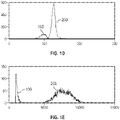
- FIGs. 1A-E show the separation of true WBCs from unlysed RBCs and RBC fragments, in histograms of collected optical information and a histogram of fluorescence information.
- the histogram in FIG. 1A shows a measurement of axial light loss (ALL).
- the histogram in FIG. 1B shows a measurement of intermediate angle scatter (IAS).
- the histogram in FIG. 1C shows a measurement of 90° polarized side scatter (PSS).
- PSD 90° polarized side scatter
- DSS 90° depolarized side scatter
- the histogram in FIG. IE shows a measurement of fluorescence (FL1).
- the horizontal axis indicates the value of the detection channel (or the names of the channels, i.e., ALL, IAS, PSS, DSS or FL1).
- the vertical axis indicates counts of components of the sample of blood.
- the lines 100 indicate residues of RBCs and lines 200 indicate WBCs.
- “residues of RBCs” is synonymous with "fragments of RBCs.”
- fluorescence information shows much better separation between the two groups of particles (i.e., WBCs and residues of RBCs) than do any of the optical channels, thereby facilitating the following analysis.
- Blood cells emit different magnitudes of fluorescence signals upon excitation of the fluorescent dye by a source of light.
- the differences in magnitude of fluorescence signals arise from the quantity of nucleic acids, namely DNA, inside the cells.
- the greater the quantity of DNA the greater the likelihood of higher fluorescence signals.
- efficacy of penetration of cell membranes, size of the dye, binding kinetics between the dye and DNA, affinity between the dye and DNA, and other factors affect the fluorescence signals. Mature RBCs emit minimal fluorescence signals because there is no DNA within mature RBCs.
- Nucleated red blood cells emit very strong fluorescence signals, because not only is DNA inside nuclei of nRBCs, but also the staining is easier because membranes of nRBCs are destroyed during the lysis procedure. Unlysed RBCs or RBC fragments do not emit fluorescence, although they may emit very weak auto-fluorescence. As shown with reference to FIG. IE, the cells that emit much stronger fluorescence signals are the cells having nuclei, namely, WBCs (and nRBCs when present).
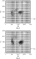
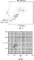
- FIG. 2 is a cytogram showing the use of a fluorescent trigger for eliminating any fragments of RBCs (nuclei-free particles) and collecting nuclei-containing events (e.g., WBCs and/or nRBCs).
- the fluorescent dye was acridine orange and the concentration of the fluorescent dye was 3 ⁇ g/mL.
- the voltage of the fluorescent photomultiplier tube was set at 350 volts.
- FIG. 3 is a cytogram showing the use of a fluorescent trigger for eliminating any fragments of RBCs (nuclei-free particles) and collecting nuclei-containing events (e.g., WBCs and/or nRBCs).
- the fluorescent dye was acridine orange and the concentration of the fluorescent dye was 0.03 ⁇ g/mL.
- the voltage of the fluorescent photomultiplier tube was set at 500 volts.
- a WBC assay using acridine orange staining even with drastically different concentrations of the dyes, i.e., 3 ⁇ g/mL in FIG. 2 and 0.03 ⁇ g/mL in FIG. 3
- only the events above the FL1 trigger are nuclei-containing events (e.g., WBCs and/or nRBCs, if present) and, consequently, are captured for further analysis.
- the WBC differential analysis is conducted by means of Multiple Angle Polarized Scattering Separation technology (MAPSS), with enhancement from fluorescence information.
- MAPSS Multiple Angle Polarized Scattering Separation technology
- At least one photodiode, or at least one photomultiplier tube, or both at least one photodiode and at least one photomultiplier tube, are needed to detect light scattered by each blood cell passing through a flow cell.
- Two or more photodiodes are used for measuring ALL signals, which measure about 0° scatter, and IAS signals, which measure low angle (e.g., about 3° to about 15°) scatter.
- Two or more photomultiplier tubes are used for detecting 90° PSS signals and 90° DSS signals.
- Each event captured on the system thus exhibits a plurality of dimensions of information, such as ALL, IAS (one or more channels), PSS, DSS, and fluorescence (one or more channels).
- the information from these detection channels is used for further analysis of blood cells.
- FIG. 4 is a schematic diagram illustrating the illumination and detection optics of an apparatus suitable for hematology analysis (including flow cytometry).
- an apparatus 10 comprises a source of light 12, a front mirror 14 and a rear mirror 16 for beam bending, a beam expander module 18 containing a first cylindrical lens 20 and a second cylindrical lens 22, a focusing lens 24, a fine beam adjuster 26, a flow cell 28, a forward scatter lens 30, a bulls-eye detector 32, a first photomultiplier tube 34, a second photomultiplier tube 36, and a third photomultiplier tube 38.
- the bulls-eye detector 32 has an inner detector 32a for 0° light scatter and an outer detector 32b for 7° light scatter.
- the source of light is preferably a laser.
- other sources of light can be used, such as, for example, lamps (e.g., mercury, xenon).
- the source of light 12 can be a vertically polarized air-cooled Coherent Cube laser, commercially available from Coherent, Inc., Santa Clara, CA. Lasers having wavelengths ranging from 350 nm to 700 nm can be used. Operating conditions for the laser are substantially similar to those of lasers currently used with "CELL-DYN" automated hematology analyzers.
- the forward optical path system shown in FIG. 4 includes a spherical plano-convex lens 30 and a two-element photo-diode detector 32 located in the back focal plane of the lens.
- each point within the two-element photodiode detector 32 maps to a specific collection angle of light from cells moving through the flow cell 28.
- the detector 32 can be a bulls-eye detector capable of detecting axial light loss (ALL) and intermediate angle forward scatter (IAS).
- ALL axial light loss
- IAS intermediate angle forward scatter
- the first photomultiplier tube 34 (PMT1) measures depolarized side scatter (DSS).
- the second photomultiplier tube 36 (PMT2) measures polarized side scatter (PSS), and the third photomultiplier tube 38 (PMT3) measures fluorescence emission from 440 nm to 680 nm, depending upon the fluorescent dye selected and the source of light employed.
- the photomultiplier tube collects fluorescent signals in a broad range of wavelengths in order to increase the strength of the signal. Side-scatter and fluorescent emissions are directed to these photomultiplier tubes by dichroic beam splitters 40 and 42, which transmit and reflect efficiently at the required wavelengths to enable efficient detection.
- U.S. Patent No. 5,631,165 describes various additional details relating to the photomultiplier tubes at column 43, line 53 though column 44, line 4.
- Sensitivity is enhanced at photomultiplier tubes 34, 36, and 38, when measuring fluorescence, by using an immersion collection system.
- the immersion collection system is one that optically couples the first lens 30 to the flow cell 28 by means of a refractive index matching layer, enabling collection of light over a wide angle.
- U.S. Patent No. 5,631,165 describes various additional details of this optical system at column 44, lines 5-31.
- the condenser 44 is an optical lens system with aberration correction sufficient for diffraction limited imaging used in high resolution microscopy.
- U.S. Patent No. 5,631,165 describes various additional details of this optical system at column 44, lines 32-60.
- a slit 46, a field lens 48, and a second slit 50 are described in U.S. Patent No. 5,631,165 , at column 44, line 63 through column 45, line 26.
- Optical filters 52 or 56 and a polarizer 52 or 56 which are inserted into the light paths of the photomultiplier tubes to change the wavelength or the polarization or both the wavelength and the polarization of the detected light, are also described in U.S. Patent No. 5,631,165 , at column 44, line 63 through column 45, line 26.
- Optical filters that are suitable for use herein include band-pass filters and long-pass filters.
- the photomultiplier tubes 34, 36, and 38 detect either side-scatter (light scattered in a cone whose axis is approximately perpendicular to the incident laser beam) or fluorescence (light emitted from the cells at a different wavelength from that of the incident laser beam).
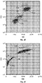
- FIGs. 5A-J show an example of an enhanced five-part WBC differential analysis.
- Neutrophils (NE), lymphocytes (LY), monocytes (MO), eosinophils (EO) and basophils (BA) were separated using MAPSS technology and a FL1 channel.
- the cytogram in FIG. 5A shows ALL vs. IAS.
- the cytogram in FIG. 5B shows 90° PSS vs. ALL.
- the cytogram in FIG. 5C shows 90° PSS vs. IAS.
- the cytogram in FIG. 5D shows 90° DSS vs. ALL.
- the cytogram in FIG. 5E shows 90° DSS vs. IAS.
- the cytogram in FIG. 5F shows 90° DSS vs.
- the cytogram in FIG. 5G shows FL1 vs. ALL.
- the cytogram in FIG. 5H shows FL1 vs. IAS.
- the cytogram in FIG. 5I shows FL1 vs. 90° PSS.
- the cytogram in FIG. 5J shows FL1 vs. 90° DSS.
- the FL1 channel further distinguishes the cell sub-populations ( FIGs. 5G through 5J , inclusive).
- basophils show relatively low FL1 signals
- monocytes show relatively high FL1 signals, relative to other WBC sub-populations, i.e., neutrophils, eosinophils, and lymphocytes.
- dots 510 represent neutrophils
- dots 520 represent eosinophils
- dots 530 represent lymphocytes
- dots 540 represent basophils
- dots 550 represent monocytes.
- the combined quantitative information from all optical dimensions and the fluorescence dimension provides an enhanced, and more reliable, differential analysis for samples of blood containing WBCs.
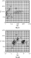
- FIG. 6A is a cytogram illustrating analysis of a sample of whole blood containing rstRBCs, using a traditional method.
- FIG. 6A shows a cytogram of ALL vs. IAS, using a commercially available "CELL-DYN" Sapphire TM hematology analyzer.
- FIG. 6B is a cytogram showing ALL vs. IAS, using an FL1 trigger-enhanced hematology analyzer.
- the sample of whole blood was the same as that analyzed in FIG. 6A .
- the concentration of the fluorescent dye, acridine orange, was 3 ⁇ g/mL. The results show that the method described herein is accurate and efficient for the two most challenging cases mentioned previously.
- FIG. 6B shows the results of an analysis of WBCs of the sample containing rstRBCs by the method described herein.
- the analysis of WBCs was accurate because no RBCs or residues of RBCs were recognized.
- FIG. 7A is a cytogram illustrating analysis of an aged (28 hours old) sample of whole blood, which contains more fragile WBCs, using a traditional method.
- FIG. 7A is a cytogram showing ALL vs. IAS, using a "CELL-DYN" Sapphire TM hematology analyzer.
- FIG. 7B is a cytogram showing ALL vs. IAS, using an FL1 trigger-enhanced hematology analyzer.
- the sample of whole blood was the same as that analyzed in FIG. 7A .
- the concentration of the fluorescent dye, acridine orange, was 3 ⁇ g/mL.
- the methods described herein enhances WBC analysis for hematology analyzers.
- the methods described herein provides a more accurate WBC count and a more accurate classification of WBC sub-populations, because the interference from unlysed RBCs and RBC fragments is substantially eliminated.
- the use of fluorescence provides further information to improve differential analysis of WBCs.
- the methods described herein shows advantages over traditional methods when analyzing samples having rstRBCs and samples having fragile WBCs.
- a hematology analyzer for conducting a WBC differential analysis on a blood sample that has been dyed with a fluorescent dye.
- the analyzer comprises an excitation source positioned to excite particles within the blood sample.
- the analyzer further comprises a plurality of detectors including: (1) an axial light loss detector positioned to measure axial light loss from the excited blood sample, (2) an intermediate angle scatter detector positioned to measure intermediate angle scatter from the excited blood sample, (3) a polarized side scatter detector positioned to measure 90° polarized side scatter from the excited blood sample, (4) a depolarized side scatter detector positioned to measure 90° depolarized side scatter from the excited blood sample, and (5) a fluorescence detector positioned to measure fluorescence emitted from the excited blood sample.
- the analyzer further comprises a processor configured to receive the measurements of (1) axial light loss, (2) intermediate angle scatter, (3) 90° polarized side scatter, (4) 90° depolarized side scatter, and (5) fluorescence from the plurality of detectors.
- the processor is also configured to perform a WBC differential analysis of the blood sample, based on all five measurements, for particles that emit fluorescence above a fluorescence threshold.
- the processor may be further configured to pre-screen the received measurements to remove from consideration any particles that do not meet the fluorescence threshold.
- the axial light loss detector may measure axial light loss at 0° scatter.
- the intermediate angle scatter detector may measure light angle scatter at about 3° to about 15°.
- the plurality of detectors may include one or more photomultiplier tubes.
- the excitation source may be a laser configured to emit light at a wavelength corresponding to the fluorescent dye. Alternatively, the fluorescent dye may be selected to correspond with the excitation source.
- the fluorescent dye may be cell membrane permeable, and nucleic acid binding.
- the hematology analyzer may further comprise an incubation subsystem for diluting the blood sample with a WBC reagent.
- the WBC reagent may include the fluorescent dye and one or more lysing agents.
- the WBC reagent may include (a) at least one surfactant, (b) at least one buffer or at least one salt, (c) at least one antimicrobial agent, and (d) the fluorescent dye.
- the incubation subsystem may be configured to incubate the blood sample with the WBC reagent for a period of time of less than about 25 seconds, less than about 17 seconds, or less than about 9 seconds.
- the incubation subsystem is configured to incubate the blood sample with the WBC reagent at a temperature ranging from about 30°C to about 50°C, such as about 40°C.
- a method of configuring a hematology analyzer to perform a WBC differential analysis on a blood sample that has been dyed with a fluorescent dye includes positioning an excitation source to excite particles within the blood sample.
- the method further includes positioning a plurality of detectors to measure (1) axial light loss, (2) intermediate angle scatter, (3) 90° polarized side scatter, (4) 90° depolarized side scatter, and (5) fluorescence from the excited blood sample.
- the method further comprises configuring a processor configured to receive the measurements of (1) axial light loss, (2) intermediate angle scatter, (3) 90° polarized side scatter, (4) 90° depolarized side scatter, and (5) fluorescence from the plurality of detectors.
- the method also includes configuring the processor to perform a WBC differential analysis of the blood sample, based on all five measurements, for particles that emit fluorescence above a fluorescence threshold.
- the method may include configuring the processor to pre-screen the received measurements to remove from consideration any particles that do not meet the fluorescence threshold.
- the axial light loss detector may measure axial light loss at 0° scatter.
- the intermediate angle scatter detector may measure light angle scatter at about 3° to about 15°.
- the plurality of detectors may include one or more photomultiplier tubes.
- the method may also include configuring the excitation source to emit light at a wavelength corresponding to the fluorescent dye.
- the fluorescent dye may be selected to correspond with the excitation source.
- the fluorescent dye may be cell membrane permeable, and nucleic acid binding.
- the method may further comprise configuring an incubation subsystem of the hematology analyzer to incubate the blood sample with a WBC reagent.
- the WBC reagent may include the fluorescent dye and a lysing agent.
- the WBC reagent may include (a) at least one surfactant, (b) at least one buffer or at least one salt, (c) at least one antimicrobial agent, and (d) the fluorescent dye.
- the incubation subsystem is configured to incubate the blood sample with the WBC reagent for a period of time of less than about 25 seconds, less than about 17 seconds, or less than about 9 seconds.
- the incubation subsystem is configured to incubate the blood sample with the WBC reagent at a temperature ranging from about 30°C to about 50°C, such as about 40°C.
- a hematology analyzer for performing a WBC differential analysis, comprising: (1) means for exciting particles within a blood sample, which includes positioning a laser light source to excite the blood sample as it transverses a flowcell of the hematology analyzer, or equivalents thereof; (2) means for measuring a plurality of light scatter signals from the excited particles within the blood sample, which includes a plurality of detectors (as discussed above), or equivalents thereof; (3) means for measuring a fluorescence signal from the excited particles within the blood sample, which includes fluorescence detectors (as discussed above), or equivalents thereof; (4) means for screening the excited particles to remove from consideration any particles that do not meet a fluorescence threshold, which includes a processor configured with a fluorescence trigger (as discussed above), or equivalents thereof; and (5) means for performing a WBC differential analysis, based on the plurality of light scatter signals and the fluorescence signal, for the particles passing the means for screening, which includes a processor configured to perform a WBC differential (
- the hematology analyzer further comprises means for incubating the blood sample with a WBC reagent for an incubation period of less than about 25 seconds, which includes an incubation subsystem (as discussed above), or equivalents thereof.
- the hematology analyzer further comprises means for incubating the blood sample with a WBC reagent at a temperature ranging from about 30°C to about 50°C, which includes an incubation subsystem (as discussed above), or equivalents thereof.
- a method of performing a WBC analysis with an automated hematology analyzer comprises: (a) diluting a sample of whole blood with a WBC reagent, wherein the WBC reagent includes a RBCs lysing agent and a fluorescent dye that penetrates WBC membranes and binds to WBC nucleic acids; (b) incubating the diluted blood sample of step (a) for an incubation period of less than about 25 seconds, at a temperature ranging from about 30°C to about 50°C; (c) delivering the incubated sample from step (b) to a flow cell in the hematology analyzer; (d) exciting the incubated sample from step (c) with an excitation source as the incubated sample traverses the flow cell; (e) collecting a plurality of light scatter signals and a fluorescence emission signal from the excited sample; and (f) performing a WBC differential based on all the signals collected in step (e), while removing from consideration any
- the WBC reagent may include (a) at least one surfactant, (b) at least one buffer or at least one salt, (c) at least one antimicrobial agent, and (d) at least one fluorescent dye.
- the excitation source may have a wavelength of from about 350 nm to about 700 nm.
- the fluorescence emission may be collected at a wavelength of from about 360 nm to about 750 nm, by a band-pass filter or a long-pass filter.
- a method for counting and classifying WBCs by means of an automated hematology analyzer comprising the steps of: (a) diluting a sample of whole blood with at least one WBC reagent; (b) incubating the diluted sample of step (a) for sufficient period of time within a selected temperature range to lyse RBCs, to preserve WBCs, to allow at least one fluorescent dye to penetrate cell membranes of the WBCs, and bind nucleic acids within the nuclei of the WBCs; (c) delivering the incubated sample from step (b) to a flow cell in a stream; (d) exciting the incubated sample from step (c) by means of a source of light as the incubated sample traverses the flow cell; (e) collecting a plurality of optical scatter signals and at least one fluorescence emission signal simultaneously; and (f) differentiating and quantifying WBCs by means of the signals collected in step (e).
- inventions include, but are not limited to: (1) use of at least one fluorescent dye to bind and stain nucleic acids in WBCs and other nuclei-containing cells in a given sample of blood during the procedure for lysing RBCs, and to induce fluorescent emissions upon excitation by photons from a given source of light, such as a laser beam at an appropriate wavelength; (2) use of a fluorescent trigger to separate and collect events that emit strong fluorescence (e.g., events involving WBCs and other nuclei-containing cells); (3) use of a plurality of optical channels and at least one channel for fluorescence for collecting data and analyzing the data so collected in order to identify each cell population and reveal additional information.
- a fluorescent trigger to separate and collect events that emit strong fluorescence (e.g., events involving WBCs and other nuclei-containing cells)
- use of a plurality of optical channels and at least one channel for fluorescence for collecting data and analyzing the data so collected in order to identify each cell population and reveal additional information.
- the systems and methods disclosed herein include: (a) staining a blood sample with an exclusive, cell membrane permeable, fluorescent dye, which corresponds in emission spectrum to an excitation source of a hematology instrument; (b) using a fluorescence trigger to screen the blood sample for WBCs; and (c) using a combination of measurements to perform a differential analysis.
- the combination of measurements may include one or more measurements selected from the group consisting of: axial light loss, intermediate angle scatter, 90° polarized side scatter, 90° depolarized side scatter, one or more fluorescence emission measurements, multiple ring intermediate angle scatter, and any combinations or equivalents thereof.
- the invention is directed toward one or more computer systems capable of carrying out the functionality described herein.
- any of the method/analysis steps discussed herein may be implemented in a computer system having one or more processors, a data communication infrastructure (e.g., a communications bus, cross-over bar, or network), a display interface, and/or a storage or memory unit.
- the storage or memory unit may include computer-readable storage medium with instructions (e.g., control logic or software) that, when executed, cause the processor(s) to perform one or more of the functions described herein.
- computer-readable storage medium “computer program medium,” and “computer usable medium” are used to generally refer to media such as a removable storage drive, removable storage units, data transmitted via a communications interface, and/or a hard disk installed in a hard disk drive.
- Such computer program products provide computer software, instructions, and/or data to a computer system, which also serve to transform the computer system from a general purpose computer into a special purpose computer programmed to perform the particular functions described herein.
- the processor, associated components, and equivalent systems and sub-systems thus serve as examples of "means for” performing select operations and functions.
- Such "means for” performing select operations and functions also serve to transform a general purpose computer into a special purpose computer programmed to perform said select operations and functions.
Description
- This invention relates to hematology systems and methods. More specifically, this invention relates to systems and methods for analyzing blood samples to identifying, classify, and/or quantify white blood cells (WBC) and WBC sub-populations in a sample of blood.
- The development of an accurate and efficient hematology assay for analysis of WBCs, including counting and classification, has been a challenge. One reason for the difficulty in developing an accurate and efficient assay is the relatively low concentration of WBCs (approximately 0.1% to 0.2%) amongst total blood cells in a sample. Attempts to engineer advanced methods for analyzing WBCs, and formulating robust WBC reagent(s), have remained one of the top priorities in the area of automated hematology analyzers.
- Typically, lysis of red blood cells (RBCs) is required to eliminate interference from RBCs, and concentrate WBCs, before counting and classifying WBCs and WBC sub-populations. More specifically, accurate and efficient WBC analysis requires: (1) complete lysis of RBCs in less than 30 seconds; (2) the breaking of large fragments of RBCs into smaller pieces after lysis; and (3) the preservation of WBCs for accurate counting and proper classification. If the blood sample is "under-lysed," unlysed RBCs, even in very small concentrations, interfere with WBC counting and differential analysis. Similarly, larger fragments of lysed RBCs can interfere with WBC counting and differential analysis. In practice, it is difficult to separate unlysed RBCs and/or larger fragments of lysed RBCs from lymphocytes (the smallest WBCs). If the blood sample is "over-lysed," the classification of WBCs may be adversely affected on account of excessive damage to cell membranes of WBCs.
- In some instances, the difficulty of WBC analysis may be compounded by the presence of two special types of samples; namely, samples containing lysis-resistant red blood cells (rstRBCs) and samples containing fragile lymphocytes. In the case of samples containing rstRBCs, the WBC count and percentage of lymphocytes are falsely reported "high," on account of the contribution of particles other than true lymphocytes, consequently posing a risk of improper diagnoses and treatments for patients. In the case of samples containing fragile lymphocytes, damaged lymphocytes may not show their characteristics in a WBC differential analysis. In addition, exposed nuclei of WBCs may be counted as nucleated red blood cells (nRBCs), resulting in a false positive count of nRBCs in certain assays.
- Provided herein are systems and methods for analyzing blood samples, and more specifically for performing a white blood cell (WBC) differential analysis. In general, the systems and methods disclosed screen WBCs by means of fluorescence staining and a fluorescence triggering strategy. As such, interference from unlysed RBCs (e.g., rstRBCs) and RBC fragments is substantially or completely eliminated, thereby ensuring accurate counting and differentiation of WBCs and WBC sub-populations. The systems and methods also enable development of relatively milder WBC reagent(s), suitable for assays of samples containing fragile lymphocytes (or other fragile WBCs), including aged samples.
- In one embodiment, for example, the systems and methods disclosed herein include: (a) staining a blood sample with an exclusive, cell membrane permeable, fluorescent dye, which corresponds in emission spectrum to an excitation source of a hematology instrument; (b) using a fluorescence trigger to screen the blood sample for WBCs; and (c) using a combination of measurements of (1) axial light loss, (2) intermediate angle scatter, (3) 90° polarized side scatter, (4) 90° depolarized side scatter, and (5) fluorescence emission to perform a differential analysis.
- The accompanying drawings, which are incorporated herein, form part of the specification. Together with this written description, the drawings further serve to explain the principles of, and to enable a person skilled in the relevant art(s), to make and use the systems and methods presented. In the drawings, like reference numbers indicate identical or functionally similar elements.
-
FIGs. 1A-1E show histograms of a sample of whole blood, showing WBCs and residues of RBCs following lysis. -
FIG. 1A is a histogram showing the measurement of an axial light loss signal. -
FIG. 1B is a histogram showing the measurement of intermediate angle scatter. -
FIG. 1C is a histogram showing the measurement of 90° polarized side scatter. - FIG. ID is a histogram showing the measurement of 90° depolarized side scatter.
- FIG. IE is a histogram showing the measurement of fluorescence.
-
FIG. 2 is a cytogram showing the use of a fluorescent trigger for eliminating any fragments of RBCs from consideration. -
FIG. 3 is another cytogram showing the use of a fluorescent trigger for eliminating any fragments of RBCs from consideration. -
FIG. 4 is a schematic diagram illustrating a hematology instrument. -
FIGs. 5A-5J show cytograms of a five-part WBC differential analysis. -
FIG. 5A is a cytogram depicting axial light loss vs. intermediate angle scatter. -
FIG. 5B is a cytogram depicting 90° polarized side scatter vs. axial light loss. -
FIG. 5C is a cytogram depicting 90° polarized side scatter vs. intermediate angle scatter. -
FIG. 5D is a cytogram depicting 90° depolarized side scatter vs. axial light loss. -
FIG. 5E is a cytogram depicting 90° depolarized side scatter vs. intermediate angle scatter. -
FIG. 5F is a cytogram depicting 90° depolarized side scatter vs. 90° polarized side scatter. -
FIG. 5G is a cytogram depicting fluorescence vs. axial light loss. -
FIG. 5H is a cytogram depicting fluorescence vs. intermediate angle scatter. -
FIG. 5I is a cytogram depicting fluorescence vs. 90° polarized side scatter. -
FIG. 5J is a cytogram depicting fluorescence vs. 90° depolarized side scatter. -
FIG. 6A is a cytogram illustrating analysis of a sample of whole blood containing lyse-resistant RBCs, using a traditional method. -
FIG. 6B is a cytogram showing axial light loss vs. intermediate angle scatter made by a fluorescence-triggered hematology analyzer, in accordance with an embodiment presented. -
FIG. 7A is a cytogram illustrating analysis of an aged sample of whole blood, using a traditional method. -
FIG. 7B is a cytogram showing axial light loss vs. intermediate angle scatter made by a fluorescence-triggered fluorescence-triggered hematology analyzer, in accordance with an embodiment presented. - Provided herein are systems and methods for analyzing blood samples, and more specifically for performing a white blood cell (WBC) differential analysis to identify, classify, and count WBCs and WBC sub-populations. In general, the systems and methods disclosed screen WBCs by means of fluorescence staining and a fluorescence triggering strategy. As such, interference from unlysed red blood cells (RBCs), such as lysis-resistant red blood cells (rstRBCs), and RBC fragments is substantially eliminated. The systems and methods disclosed thereby ensure accurate counting and differentiation of WBCs and WBC sub-populations. The systems and methods also enable development of relatively milder WBC reagent(s), suitable for assays of samples containing fragile lymphocytes (or other fragile WBCs), including aged samples.
- In one embodiment, for example, the systems and methods disclosed herein include: (a) staining a blood sample with an exclusive, cell membrane permeable, fluorescent dye, which corresponds in emission spectrum to an excitation source of a hematology instrument; (b) using a fluorescence trigger to screen the blood sample for WBCs; and (c) using a combination of measurements of (1) axial light loss, (2) intermediate angle scatter, (3) 90° polarized side scatter, (4) 90° depolarized side scatter, and (5) fluorescence emission to perform a WBC differential analysis.
- As used herein, the expression "fluorescence information" means data collected from a fluorescence channel of a hematology analyzer. As used herein, the expression "fluorescence channel" means a detection device, such as a photomultiplier tube, set at an appropriate wavelength band for measuring the quantity of fluorescence emitted from a sample.
- WBCs contain a relatively high concentration of DNA in their nuclei. Mature RBCs, however, do not contain DNA. Therefore, a fluorescent dye is selected to differentiate two classes of blood cells; namely, the blood cells containing nucleic acids and the blood cells not containing nucleic acids. The purpose of the dye is to penetrate into live cells easily, bind DNA with high affinity, and emit strong fluorescence with adequate Stokes shift when the dye is excited by an appropriate source of light. The peak absorption of the dye in the visible band substantially matches the wavelength of the source of light (within 50 nm of the wavelength of the source of light, more preferably, within 25 nm of the wavelength of the source of light), in order to be properly excite the dye and achieve optimal results.
- The fluorescent dye selected is preferably: 1) capable of binding nucleic acids, 2) capable of penetrating cell membranes of WBCs, 3) excitable at a selected wavelength when subjected to a source of light, 4) emits fluorescence upon excitation by the source of light, and 5) is biostable and soluble in a liquid. The dye may be selected from group consisting of: acridine orange, SYBR 11, SYBR Green series dye, hexidium iodide, SYTO 11,
SYTO 12, SYTO 13, SYTO 14, SYTO 16, SYTO 21, SYTO RNA Select,SYTO 24, SYTO 25 and any equivalents thereof. The dye is used to "activate" WBCs and "screen out" unlysed RBCs and fragments of RBCs based on a fluorescence trigger configured in the hematology analyzer. The dye is typically present at a concentration of from about 0.1 ng/mL to about 0.1 mg/mL. While various dyes are available, the dye selected is generally paired with the excitation source of the hematology analyzer such that a single exclusive dye is used to stain and excite fluorescence emission in all WBC sub-populations intended to be identified, quantified, and/or analyzed. As such, a single (i.e., exclusive) dye can be used to identify, quantify, and analyze all the WBC subpopulations at once. - In one embodiment, a fluorescent dye is provided in a WBC reagent, with combinations of 1) at least one surfactant, 2) at least one buffer, 3) at least one salt, and/or 4) at least antimicrobial agent, in sufficient quantities for carrying out staining and activating up to 1,000 x 103 WBCs per microliter. The at least one surfactant, such as "TRITON" X-100 or saponin, is used to destroy the membranes of RBC, and reduce the sizes of fragments of RBCs. The at least one surfactant is typically present at a concentration of from about 0.001% to about 5%. The at least one antimicrobial agent, such as those from "TRIADINE" or "PROCLIN" families, is used to prevent the contamination of the reagent from microbes. The concentration of the at least one antimicrobial agent is sufficient to preserve the reagent for the shelf life required. The at least one buffer, such as phosphate buffered saline (PBS) or 4-(2-hydroxyethyl)-1-piperazineethanesulfonic acid (HEPES), is used to adjust the pH of reaction mixture for controlling lysis of RBCs and preserving WBCs. The at least one buffer is typically present at a concentration of from about 0.01% to about 3%. The pH typically ranges from about 3 to about 12. The at least one salt, such as NaCl or Na2SO4, is used to adjust the osmolality to increase the effect of lysing and/or optimize WBC preservation. The at least one salt may be present at a concentration of from about 0.01% to about 3%. In certain cases, the at least one buffer can serve as the at least one salt, or the at least one salt can serve as the at least one buffer. In general, lower osmolality, or hypotonicity, is used to accelerate the lysis of RBCs. The osmolality typically ranges from about 20 to about 250 mOsm.
- Lysis of RBCs occurs at a temperature above room temperature (e.g., between about 30°C to about 50°C, such as about 40°C) over a relatively short period of time (e.g., less than about 25 seconds, less than about 17 seconds, or even less than about 9 seconds), following mixing of the sample of blood and the WBC reagent at a ratio of about one part by volume sample to about 35 parts by volume WBC reagent. The data for analysis is collected with a plurality of optical channels and at least one fluorescence channel.
-
FIGs. 1A-E show the separation of true WBCs from unlysed RBCs and RBC fragments, in histograms of collected optical information and a histogram of fluorescence information. The histogram inFIG. 1A shows a measurement of axial light loss (ALL). The histogram inFIG. 1B shows a measurement of intermediate angle scatter (IAS). The histogram inFIG. 1C shows a measurement of 90° polarized side scatter (PSS). The histogram in FIG. ID shows a measurement of 90° depolarized side scatter (DSS). The histogram in FIG. IE shows a measurement of fluorescence (FL1). In the histograms, the horizontal axis indicates the value of the detection channel (or the names of the channels, i.e., ALL, IAS, PSS, DSS or FL1). The vertical axis indicates counts of components of the sample of blood. In the histograms, thelines 100 indicate residues of RBCs andlines 200 indicate WBCs. As used herein, "residues of RBCs" is synonymous with "fragments of RBCs." As shown by comparing FIG. IE toFIGs. 1A-1D , fluorescence information shows much better separation between the two groups of particles (i.e., WBCs and residues of RBCs) than do any of the optical channels, thereby facilitating the following analysis. - Blood cells emit different magnitudes of fluorescence signals upon excitation of the fluorescent dye by a source of light. The differences in magnitude of fluorescence signals arise from the quantity of nucleic acids, namely DNA, inside the cells. The greater the quantity of DNA, the greater the likelihood of higher fluorescence signals. Also, efficacy of penetration of cell membranes, size of the dye, binding kinetics between the dye and DNA, affinity between the dye and DNA, and other factors, affect the fluorescence signals. Mature RBCs emit minimal fluorescence signals because there is no DNA within mature RBCs. Nucleated red blood cells (nRBCs) emit very strong fluorescence signals, because not only is DNA inside nuclei of nRBCs, but also the staining is easier because membranes of nRBCs are destroyed during the lysis procedure. Unlysed RBCs or RBC fragments do not emit fluorescence, although they may emit very weak auto-fluorescence. As shown with reference to FIG. IE, the cells that emit much stronger fluorescence signals are the cells having nuclei, namely, WBCs (and nRBCs when present).
- As such, the systems and methods presented herein use a fluorescence trigger for collecting and analyzing WBCs. For example, a fluorescence trigger, usually set between signals from RBCs and signals from WBCs, can be used to collect signals from WBCs separately for further analysis. Two examples of using an FL1 trigger are shown in
FIG. 2 and FIG. 3. FIG. 2 is a cytogram showing the use of a fluorescent trigger for eliminating any fragments of RBCs (nuclei-free particles) and collecting nuclei-containing events (e.g., WBCs and/or nRBCs). The fluorescent dye was acridine orange and the concentration of the fluorescent dye was 3 µg/mL. The voltage of the fluorescent photomultiplier tube was set at 350 volts.FIG. 3 is a cytogram showing the use of a fluorescent trigger for eliminating any fragments of RBCs (nuclei-free particles) and collecting nuclei-containing events (e.g., WBCs and/or nRBCs). The fluorescent dye was acridine orange and the concentration of the fluorescent dye was 0.03 µg/mL. The voltage of the fluorescent photomultiplier tube was set at 500 volts. In a WBC assay using acridine orange staining (even with drastically different concentrations of the dyes, i.e., 3 µg/mL inFIG. 2 and 0.03 µg/mL inFIG. 3 ) and a properly set FL1 trigger, only the events above the FL1 trigger are nuclei-containing events (e.g., WBCs and/or nRBCs, if present) and, consequently, are captured for further analysis. - In one embodiment, the WBC differential analysis is conducted by means of Multiple Angle Polarized Scattering Separation technology (MAPSS), with enhancement from fluorescence information. At least one photodiode, or at least one photomultiplier tube, or both at least one photodiode and at least one photomultiplier tube, are needed to detect light scattered by each blood cell passing through a flow cell. Two or more photodiodes are used for measuring ALL signals, which measure about 0° scatter, and IAS signals, which measure low angle (e.g., about 3° to about 15°) scatter. Two or more photomultiplier tubes are used for detecting 90° PSS signals and 90° DSS signals. Additional photomultiplier tubes are needed for FL1 measurements within appropriate wavelength range(s), depending on the choice of wavelength of the source of light. Each event captured on the system thus exhibits a plurality of dimensions of information, such as ALL, IAS (one or more channels), PSS, DSS, and fluorescence (one or more channels). The information from these detection channels is used for further analysis of blood cells.
-
FIG. 4 is a schematic diagram illustrating the illumination and detection optics of an apparatus suitable for hematology analysis (including flow cytometry). Referring now toFIG. 4 , anapparatus 10 comprises a source of light 12, a front mirror 14 and a rear mirror 16 for beam bending, abeam expander module 18 containing a firstcylindrical lens 20 and a secondcylindrical lens 22, a focusinglens 24, afine beam adjuster 26, aflow cell 28, aforward scatter lens 30, a bulls-eye detector 32, afirst photomultiplier tube 34, asecond photomultiplier tube 36, and athird photomultiplier tube 38. The bulls-eye detector 32 has aninner detector 32a for 0° light scatter and anouter detector 32b for 7° light scatter. - In the discussion that follows, the source of light is preferably a laser. However, other sources of light can be used, such as, for example, lamps (e.g., mercury, xenon). The source of light 12 can be a vertically polarized air-cooled Coherent Cube laser, commercially available from Coherent, Inc., Santa Clara, CA. Lasers having wavelengths ranging from 350 nm to 700 nm can be used. Operating conditions for the laser are substantially similar to those of lasers currently used with "CELL-DYN" automated hematology analyzers.
- Additional details relating to the flow cell, the lenses, the focusing lens, the fine-beam adjust mechanism and the laser focusing lens can be found in
U.S. Patent No. 5,631,165 , incorporated herein by reference, particularly at column 41, line 32 through column 43, line 11. The forward optical path system shown inFIG. 4 includes a spherical plano-convex lens 30 and a two-element photo-diode detector 32 located in the back focal plane of the lens. In this configuration, each point within the two-element photodiode detector 32 maps to a specific collection angle of light from cells moving through theflow cell 28. The detector 32 can be a bulls-eye detector capable of detecting axial light loss (ALL) and intermediate angle forward scatter (IAS).U.S. Patent No. 5,631,165 describes various alternatives to this detector at column 43, lines 12-52. - The first photomultiplier tube 34 (PMT1) measures depolarized side scatter (DSS). The second photomultiplier tube 36 (PMT2) measures polarized side scatter (PSS), and the third photomultiplier tube 38 (PMT3) measures fluorescence emission from 440 nm to 680 nm, depending upon the fluorescent dye selected and the source of light employed. The photomultiplier tube collects fluorescent signals in a broad range of wavelengths in order to increase the strength of the signal. Side-scatter and fluorescent emissions are directed to these photomultiplier tubes by
dichroic beam splitters U.S. Patent No. 5,631,165 describes various additional details relating to the photomultiplier tubes at column 43, line 53 thoughcolumn 44, line 4. - Sensitivity is enhanced at
photomultiplier tubes first lens 30 to theflow cell 28 by means of a refractive index matching layer, enabling collection of light over a wide angle.U.S. Patent No. 5,631,165 describes various additional details of this optical system atcolumn 44, lines 5-31. - The
condenser 44 is an optical lens system with aberration correction sufficient for diffraction limited imaging used in high resolution microscopy.U.S. Patent No. 5,631,165 describes various additional details of this optical system atcolumn 44, lines 32-60. - The functions of other components shown in
FIG. 4 , i.e., aslit 46, afield lens 48, and asecond slit 50, are described inU.S. Patent No. 5,631,165 , atcolumn 44, line 63 through column 45,line 26.Optical filters polarizer U.S. Patent No. 5,631,165 , atcolumn 44, line 63 through column 45,line 26. Optical filters that are suitable for use herein include band-pass filters and long-pass filters. - The
photomultiplier tubes -
FIGs. 5A-J show an example of an enhanced five-part WBC differential analysis. Neutrophils (NE), lymphocytes (LY), monocytes (MO), eosinophils (EO) and basophils (BA) were separated using MAPSS technology and a FL1 channel. The cytogram inFIG. 5A shows ALL vs. IAS. The cytogram inFIG. 5B shows 90° PSS vs. ALL. The cytogram inFIG. 5C shows 90° PSS vs. IAS. The cytogram inFIG. 5D shows 90° DSS vs. ALL. The cytogram inFIG. 5E shows 90° DSS vs. IAS. The cytogram inFIG. 5F shows 90° DSS vs. 90° PSS. The cytogram inFIG. 5G shows FL1 vs. ALL. The cytogram inFIG. 5H shows FL1 vs. IAS. The cytogram inFIG. 5I shows FL1 vs. 90° PSS. The cytogram inFIG. 5J shows FL1 vs. 90° DSS. - In addition to the information collected from the four traditional MAPSS channels (ALL, IAS, PSS, DSS), the FL1 channel further distinguishes the cell sub-populations (
FIGs. 5G through 5J , inclusive). For the case in which acridine orange is used as the dye for screening WBCs, basophils show relatively low FL1 signals, and monocytes show relatively high FL1 signals, relative to other WBC sub-populations, i.e., neutrophils, eosinophils, and lymphocytes. In the cytograms,dots 510 represent neutrophils,dots 520 represent eosinophils,dots 530 represent lymphocytes,dots 540 represent basophils, anddots 550 represent monocytes. The combined quantitative information from all optical dimensions and the fluorescence dimension provides an enhanced, and more reliable, differential analysis for samples of blood containing WBCs. -
FIG. 6A is a cytogram illustrating analysis of a sample of whole blood containing rstRBCs, using a traditional method.FIG. 6A shows a cytogram of ALL vs. IAS, using a commercially available "CELL-DYN" Sapphire™ hematology analyzer.FIG. 6B is a cytogram showing ALL vs. IAS, using an FL1 trigger-enhanced hematology analyzer. The sample of whole blood was the same as that analyzed inFIG. 6A . The concentration of the fluorescent dye, acridine orange, was 3 µg/mL. The results show that the method described herein is accurate and efficient for the two most challenging cases mentioned previously. In the traditional method, unlysed RBCs, i.e., the events appearing in the lower left of the cytogram, were recognized as lymphocytes, thereby resulting in a higher count of WBCs and a higher percentage of lymphocytes.FIG. 6B shows the results of an analysis of WBCs of the sample containing rstRBCs by the method described herein. In the method described herein, the analysis of WBCs was accurate because no RBCs or residues of RBCs were recognized. -
FIG. 7A is a cytogram illustrating analysis of an aged (28 hours old) sample of whole blood, which contains more fragile WBCs, using a traditional method.FIG. 7A is a cytogram showing ALL vs. IAS, using a "CELL-DYN" Sapphire™ hematology analyzer.FIG. 7B is a cytogram showing ALL vs. IAS, using an FL1 trigger-enhanced hematology analyzer. The sample of whole blood was the same as that analyzed inFIG. 7A . The concentration of the fluorescent dye, acridine orange, was 3 µg/mL. - The methods described herein enhances WBC analysis for hematology analyzers. The methods described herein provides a more accurate WBC count and a more accurate classification of WBC sub-populations, because the interference from unlysed RBCs and RBC fragments is substantially eliminated. The use of fluorescence provides further information to improve differential analysis of WBCs. The methods described herein shows advantages over traditional methods when analyzing samples having rstRBCs and samples having fragile WBCs.
- In one embodiment, there is provided a hematology analyzer for conducting a WBC differential analysis on a blood sample that has been dyed with a fluorescent dye. The analyzer comprises an excitation source positioned to excite particles within the blood sample. The analyzer further comprises a plurality of detectors including: (1) an axial light loss detector positioned to measure axial light loss from the excited blood sample, (2) an intermediate angle scatter detector positioned to measure intermediate angle scatter from the excited blood sample, (3) a polarized side scatter detector positioned to measure 90° polarized side scatter from the excited blood sample, (4) a depolarized side scatter detector positioned to measure 90° depolarized side scatter from the excited blood sample, and (5) a fluorescence detector positioned to measure fluorescence emitted from the excited blood sample. The analyzer further comprises a processor configured to receive the measurements of (1) axial light loss, (2) intermediate angle scatter, (3) 90° polarized side scatter, (4) 90° depolarized side scatter, and (5) fluorescence from the plurality of detectors. The processor is also configured to perform a WBC differential analysis of the blood sample, based on all five measurements, for particles that emit fluorescence above a fluorescence threshold. The processor may be further configured to pre-screen the received measurements to remove from consideration any particles that do not meet the fluorescence threshold. The axial light loss detector may measure axial light loss at 0° scatter. The intermediate angle scatter detector may measure light angle scatter at about 3° to about 15°. The plurality of detectors may include one or more photomultiplier tubes. The excitation source may be a laser configured to emit light at a wavelength corresponding to the fluorescent dye. Alternatively, the fluorescent dye may be selected to correspond with the excitation source. The fluorescent dye may be cell membrane permeable, and nucleic acid binding.
- The hematology analyzer may further comprise an incubation subsystem for diluting the blood sample with a WBC reagent. The WBC reagent may include the fluorescent dye and one or more lysing agents. Alternatively, the WBC reagent may include (a) at least one surfactant, (b) at least one buffer or at least one salt, (c) at least one antimicrobial agent, and (d) the fluorescent dye. The incubation subsystem may be configured to incubate the blood sample with the WBC reagent for a period of time of less than about 25 seconds, less than about 17 seconds, or less than about 9 seconds. The incubation subsystem is configured to incubate the blood sample with the WBC reagent at a temperature ranging from about 30°C to about 50°C, such as about 40°C.
- In another embodiment, there is provided a method of configuring a hematology analyzer to perform a WBC differential analysis on a blood sample that has been dyed with a fluorescent dye. The method includes positioning an excitation source to excite particles within the blood sample. The method further includes positioning a plurality of detectors to measure (1) axial light loss, (2) intermediate angle scatter, (3) 90° polarized side scatter, (4) 90° depolarized side scatter, and (5) fluorescence from the excited blood sample. The method further comprises configuring a processor configured to receive the measurements of (1) axial light loss, (2) intermediate angle scatter, (3) 90° polarized side scatter, (4) 90° depolarized side scatter, and (5) fluorescence from the plurality of detectors. The method also includes configuring the processor to perform a WBC differential analysis of the blood sample, based on all five measurements, for particles that emit fluorescence above a fluorescence threshold. The method may include configuring the processor to pre-screen the received measurements to remove from consideration any particles that do not meet the fluorescence threshold. The axial light loss detector may measure axial light loss at 0° scatter. The intermediate angle scatter detector may measure light angle scatter at about 3° to about 15°. The plurality of detectors may include one or more photomultiplier tubes. The method may also include configuring the excitation source to emit light at a wavelength corresponding to the fluorescent dye. Alternatively, the fluorescent dye may be selected to correspond with the excitation source. The fluorescent dye may be cell membrane permeable, and nucleic acid binding.
- The method may further comprise configuring an incubation subsystem of the hematology analyzer to incubate the blood sample with a WBC reagent. The WBC reagent may include the fluorescent dye and a lysing agent. Alternatively, the WBC reagent may include (a) at least one surfactant, (b) at least one buffer or at least one salt, (c) at least one antimicrobial agent, and (d) the fluorescent dye. The incubation subsystem is configured to incubate the blood sample with the WBC reagent for a period of time of less than about 25 seconds, less than about 17 seconds, or less than about 9 seconds. The incubation subsystem is configured to incubate the blood sample with the WBC reagent at a temperature ranging from about 30°C to about 50°C, such as about 40°C.
- In another embodiment, there is provided a hematology analyzer for performing a WBC differential analysis, comprising: (1) means for exciting particles within a blood sample, which includes positioning a laser light source to excite the blood sample as it transverses a flowcell of the hematology analyzer, or equivalents thereof; (2) means for measuring a plurality of light scatter signals from the excited particles within the blood sample, which includes a plurality of detectors (as discussed above), or equivalents thereof; (3) means for measuring a fluorescence signal from the excited particles within the blood sample, which includes fluorescence detectors (as discussed above), or equivalents thereof; (4) means for screening the excited particles to remove from consideration any particles that do not meet a fluorescence threshold, which includes a processor configured with a fluorescence trigger (as discussed above), or equivalents thereof; and (5) means for performing a WBC differential analysis, based on the plurality of light scatter signals and the fluorescence signal, for the particles passing the means for screening, which includes a processor configured to perform a WBC differential (as discussed above), or equivalents thereof. The hematology analyzer further comprises means for incubating the blood sample with a WBC reagent for an incubation period of less than about 25 seconds, which includes an incubation subsystem (as discussed above), or equivalents thereof. The hematology analyzer further comprises means for incubating the blood sample with a WBC reagent at a temperature ranging from about 30°C to about 50°C, which includes an incubation subsystem (as discussed above), or equivalents thereof.
- In another embodiment, there is provided a method of performing a WBC analysis with an automated hematology analyzer. The method comprises: (a) diluting a sample of whole blood with a WBC reagent, wherein the WBC reagent includes a RBCs lysing agent and a fluorescent dye that penetrates WBC membranes and binds to WBC nucleic acids; (b) incubating the diluted blood sample of step (a) for an incubation period of less than about 25 seconds, at a temperature ranging from about 30°C to about 50°C; (c) delivering the incubated sample from step (b) to a flow cell in the hematology analyzer; (d) exciting the incubated sample from step (c) with an excitation source as the incubated sample traverses the flow cell; (e) collecting a plurality of light scatter signals and a fluorescence emission signal from the excited sample; and (f) performing a WBC differential based on all the signals collected in step (e), while removing from consideration any particles within the diluted blood sample that do not meet a fluorescence threshold based on the fluorescence emission signal. The WBC reagent may include (a) at least one surfactant, (b) at least one buffer or at least one salt, (c) at least one antimicrobial agent, and (d) at least one fluorescent dye. The excitation source may have a wavelength of from about 350 nm to about 700 nm. The fluorescence emission may be collected at a wavelength of from about 360 nm to about 750 nm, by a band-pass filter or a long-pass filter.
- In yet another embodiment, there is provided a method for counting and classifying WBCs by means of an automated hematology analyzer, the method comprising the steps of: (a) diluting a sample of whole blood with at least one WBC reagent; (b) incubating the diluted sample of step (a) for sufficient period of time within a selected temperature range to lyse RBCs, to preserve WBCs, to allow at least one fluorescent dye to penetrate cell membranes of the WBCs, and bind nucleic acids within the nuclei of the WBCs; (c) delivering the incubated sample from step (b) to a flow cell in a stream; (d) exciting the incubated sample from step (c) by means of a source of light as the incubated sample traverses the flow cell; (e) collecting a plurality of optical scatter signals and at least one fluorescence emission signal simultaneously; and (f) differentiating and quantifying WBCs by means of the signals collected in step (e). Features of the embodiment include, but are not limited to: (1) use of at least one fluorescent dye to bind and stain nucleic acids in WBCs and other nuclei-containing cells in a given sample of blood during the procedure for lysing RBCs, and to induce fluorescent emissions upon excitation by photons from a given source of light, such as a laser beam at an appropriate wavelength; (2) use of a fluorescent trigger to separate and collect events that emit strong fluorescence (e.g., events involving WBCs and other nuclei-containing cells); (3) use of a plurality of optical channels and at least one channel for fluorescence for collecting data and analyzing the data so collected in order to identify each cell population and reveal additional information.
- In yet another embodiment, the systems and methods disclosed herein include: (a) staining a blood sample with an exclusive, cell membrane permeable, fluorescent dye, which corresponds in emission spectrum to an excitation source of a hematology instrument; (b) using a fluorescence trigger to screen the blood sample for WBCs; and (c) using a combination of measurements to perform a differential analysis. The combination of measurements may include one or more measurements selected from the group consisting of: axial light loss, intermediate angle scatter, 90° polarized side scatter, 90° depolarized side scatter, one or more fluorescence emission measurements, multiple ring intermediate angle scatter, and any combinations or equivalents thereof.
- In one embodiment, the invention is directed toward one or more computer systems capable of carrying out the functionality described herein. For example, any of the method/analysis steps discussed herein may be implemented in a computer system having one or more processors, a data communication infrastructure (e.g., a communications bus, cross-over bar, or network), a display interface, and/or a storage or memory unit. The storage or memory unit may include computer-readable storage medium with instructions (e.g., control logic or software) that, when executed, cause the processor(s) to perform one or more of the functions described herein. The terms "computer-readable storage medium," "computer program medium," and "computer usable medium" are used to generally refer to media such as a removable storage drive, removable storage units, data transmitted via a communications interface, and/or a hard disk installed in a hard disk drive. Such computer program products provide computer software, instructions, and/or data to a computer system, which also serve to transform the computer system from a general purpose computer into a special purpose computer programmed to perform the particular functions described herein. Where appropriate, the processor, associated components, and equivalent systems and sub-systems thus serve as examples of "means for" performing select operations and functions. Such "means for" performing select operations and functions also serve to transform a general purpose computer into a special purpose computer programmed to perform said select operations and functions.
Claims (13)
- A method of performing a white blood cell (WBC) analysis with an automated hematology analyzer, the method comprising:(a) diluting a sample of whole blood with a WBC reagent having an osmolality of from 20 to 250 mOsm, wherein the WBC reagent includes a red blood cells (RBC) lysing agent and acridine orange as a fluorescent dye that penetrates WBC membranes and binds to WBC nucleic acids;(b) incubating the diluted blood sample of step (a) for an incubation period of less than 25 seconds and at a temperature ranging from 30°C to 50°C;(c) delivering the incubated sample from step (b) to a flow cell in the hematology analyzer;(d) exciting the incubated sample from step (c) with an excitation source as the incubated sample traverses the flow cell;(e) collecting a plurality of light scatter signals and a fluorescence emission signal from the excited sample;(f) prior to performing a WBC analysis, excluding nuclei-free particles and retaining nuclei-containing particles using only a fluorescence trigger that is limited to fluorescence emission signals and is set to a fluorescence magnitude that is greater than fluorescence emission signals from RBCs, including RBC fragments, and is less than fluorescence emission signals from WBCs; and(g) performing a WBC differential on the nuclei-containing particles retained in step (f).
- The method of any one of the previous claims, wherein the WBC reagent includes (a) at least one surfactant, (b) at least one buffer or at least one salt, (c) at least one antimicrobial agent, and (d) at least one fluorescent dye.
- The method of any one of the previous claims, wherein the excitation source has a wavelength that ranges from 350 nm to 700 nm.
- The method of any one of the previous claims, wherein fluorescence emission is collected at a wavelength that ranges from 360 nm to 750 nm, by a band-pass filter or a long-pass filter.
- The method according to any one of the previous claims, wherein the nuclei-containing events comprise signals from WBCs and nucleated red blood cells (nRBCs).
- The method according to any one of the previous claims, wherein performing the WBC differential comprises distinguishing the nuclei-containing events from one another based on the magnitude of the fluorescence emission signal associated with each event.
- The method according to any one of the previous claims, wherein performing the WBC differential comprises distinguishing the nuclei-containing events from one another based on the plurality of light scatter signals associated with each event.
- The method according to any one of the previous claims, wherein the plurality of light scatter signals includes one or more of: axial light loss (ALL) signals, intermediate angle scatter (IAS) signals, polarized side scatter (PSS) signals, and depolarized side scatter (DSS) signals.
- The method according to any one of the previous claims, wherein the incubation period of time is less than 17 seconds.
- The method according to any one of the previous claims, wherein the incubation period of time is less than 9 seconds.
- The method according to any one of the previous claims, wherein the diluted blood sample is incubated at a temperature of 40°C.
- The method according to any one of the previous claims, wherein the WBC reagent contains a single fluorescent dye that is used to identify, quantify, and analyze a plurality of WBC subpopulations at once.
- A hematology analyzer comprising a processor and a computer-readable medium comprising instructions that, when executed by the processor, cause the hematology analyzer to carry out the method of any one of the previous claims.
Applications Claiming Priority (2)
| Application Number | Priority Date | Filing Date | Title |
|---|---|---|---|
| US201161482541P | 2011-05-04 | 2011-05-04 | |
| PCT/US2012/035158 WO2012151102A2 (en) | 2011-05-04 | 2012-04-26 | White blood cell analysis system and method |
Publications (3)
| Publication Number | Publication Date |
|---|---|
| EP2705134A2 EP2705134A2 (en) | 2014-03-12 |
| EP2705134A4 EP2705134A4 (en) | 2015-06-17 |
| EP2705134B1 true EP2705134B1 (en) | 2022-08-24 |
Family
ID=47090459
Family Applications (1)
| Application Number | Title | Priority Date | Filing Date |
|---|---|---|---|
| EP12779974.0A Active EP2705134B1 (en) | 2011-05-04 | 2012-04-26 | White blood cell analysis system and method |
Country Status (6)
| Country | Link |
|---|---|
| US (4) | US9091624B2 (en) |
| EP (1) | EP2705134B1 (en) |
| JP (1) | JP6104233B2 (en) |
| CN (1) | CN103987834B (en) |
| ES (1) | ES2927316T3 (en) |
| WO (1) | WO2012151102A2 (en) |
Families Citing this family (8)
| Publication number | Priority date | Publication date | Assignee | Title |
|---|---|---|---|---|
| US8980568B2 (en) | 2001-10-11 | 2015-03-17 | Aviva Biosciences Corporation | Methods and compositions for detecting non-hematopoietic cells from a blood sample |
| US8986944B2 (en) | 2001-10-11 | 2015-03-24 | Aviva Biosciences Corporation | Methods and compositions for separating rare cells from fluid samples |
| CA2657621A1 (en) | 2006-07-14 | 2008-01-17 | Aviva Biosciences Corporation | Methods and compositions for detecting rare cells from a biological sample |
| EP2798087B1 (en) * | 2011-12-27 | 2018-03-07 | Abbott Laboratories | Cellular analysis of body fluids |
| ES2869919T3 (en) * | 2013-03-12 | 2021-10-26 | Abbott Lab | White blood cell analysis system |
| WO2014186228A1 (en) * | 2013-05-13 | 2014-11-20 | Chiranjit Deka | Apparatus and methods for cellular analysis |
| EP3371591A4 (en) | 2015-11-02 | 2018-11-14 | Chiranjit Deka | Light scatter based apparatus and methods for hematology analysis using only three detectors |
| CN112424582A (en) * | 2018-08-29 | 2021-02-26 | 深圳迈瑞生物医疗电子股份有限公司 | Method for testing blood sample, blood sample testing instrument and storage medium |
Family Cites Families (37)
| Publication number | Priority date | Publication date | Assignee | Title |
|---|---|---|---|---|
| US4661913A (en) | 1984-09-11 | 1987-04-28 | Becton, Dickinson And Company | Apparatus and method for the detection and classification of articles using flow cytometry techniques |
| US4727020A (en) | 1985-02-25 | 1988-02-23 | Becton, Dickinson And Company | Method for analysis of subpopulations of blood cells |
| US5175109A (en) * | 1986-09-10 | 1992-12-29 | Toa Medical Electronics Co., Ltd. | Reagent for classifying leukocytes by flow cytometry |
| US4882284A (en) | 1987-04-13 | 1989-11-21 | Ortho Pharmaceutical Corporation | Method for quantitating and differentiating white blood cells |
| IE76732B1 (en) * | 1990-08-07 | 1997-11-05 | Becton Dickinson Co | One step test for absolute counts |
| JPH07113632B2 (en) | 1991-04-22 | 1995-12-06 | 株式会社日立製作所 | White blood cell analysis method |
| US5776709A (en) | 1991-08-28 | 1998-07-07 | Becton Dickinson And Company | Method for preparation and analysis of leukocytes in whole blood |
| ES2102518T3 (en) | 1991-08-28 | 1997-08-01 | Becton Dickinson Co | GRAVITATION ATTRACTION MOTOR FOR SELF-ADAPTIVE GROUPING OF N-DIMENSIONAL DATA CURRENTS. |
| DE69329353T2 (en) * | 1992-03-05 | 2001-01-04 | Becton Dickinson Co | Process for the production and analysis of leukocytes in whole blood using flow cytometry |
| US5812419A (en) * | 1994-08-01 | 1998-09-22 | Abbott Laboratories | Fully automated analysis method with optical system for blood cell analyzer |
| US5631730A (en) | 1994-08-01 | 1997-05-20 | Abbott Laboratories | Pseudo telecentric optical design for flow cytometric blood cell analyzer |
| US5656499A (en) * | 1994-08-01 | 1997-08-12 | Abbott Laboratories | Method for performing automated hematology and cytometry analysis |
| US5631165A (en) * | 1994-08-01 | 1997-05-20 | Abbott Laboratories | Method for performing automated hematology and cytometry analysis |
| US5891734A (en) * | 1994-08-01 | 1999-04-06 | Abbott Laboratories | Method for performing automated analysis |
| US5559037A (en) | 1994-12-15 | 1996-09-24 | Abbott Laboratories | Method for rapid and simultaneous analysis of nucleated red blood cells |
| US5879900A (en) * | 1994-12-15 | 1999-03-09 | Abbott Laboratories | Method for simultaneous analysis of cell viability, nucleated red blood cells and white blood cell differentials |
| JP4279900B2 (en) * | 1995-12-15 | 2009-06-17 | アボツト・ラボラトリーズ | Simultaneous analysis of cell viability, nucleated red blood cells, and white blood cell classification |
| US6197593B1 (en) * | 1998-10-20 | 2001-03-06 | Coulter International Corp. | Method for enumerating blood cells |
| US6228652B1 (en) * | 1999-02-16 | 2001-05-08 | Coulter International Corp. | Method and apparatus for analyzing cells in a whole blood sample |
| US6900023B1 (en) | 1999-09-02 | 2005-05-31 | Sysmex Corporation | Method for classifying and counting leukocytes |
| US6368864B1 (en) | 2000-05-05 | 2002-04-09 | Coulter International Corp. | Dyes and methods of reticulocyte enumeration |
| FR2821428B1 (en) | 2001-02-23 | 2004-08-06 | Abx Sa | REAGENT AND METHOD FOR THE IDENTIFICATION AND COUNTING OF BIOLOGICAL CELLS |
| EP1442141A4 (en) | 2001-10-17 | 2005-05-18 | Univ Utah Res Found | Methods for identifying differentially expressed genes by multivariate analysis of microarry data |
| JP2003310299A (en) | 2002-04-25 | 2003-11-05 | Sysmex Corp | Method for treating milk sample, reagent and method each for measuring somatic cell contained in milk sample |
| US7674598B2 (en) | 2004-05-21 | 2010-03-09 | Beckman Coulter, Inc. | Method for a fully automated monoclonal antibody-based extended differential |
| US7625712B2 (en) | 2004-05-21 | 2009-12-01 | Beckman Coulter, Inc. | Method for a fully automated monoclonal antibody-based extended differential |
| US7299135B2 (en) | 2005-11-10 | 2007-11-20 | Idexx Laboratories, Inc. | Methods for identifying discrete populations (e.g., clusters) of data within a flow cytometer multi-dimensional data set |
| US20090061478A1 (en) | 2006-01-30 | 2009-03-05 | Lene Have Poulsen | High-Speed Quantification of Antigen Specific T-Cells in Whole Blood by Flow Cytometry |
| JP4796443B2 (en) | 2006-06-08 | 2011-10-19 | シスメックス株式会社 | Reagent for sample analysis, reagent kit for sample analysis, and sample analysis method |
| JP4914656B2 (en) | 2006-06-26 | 2012-04-11 | シスメックス株式会社 | Reagent for sample analysis, reagent kit for sample analysis, and sample analysis method |
| US7674622B2 (en) * | 2006-12-22 | 2010-03-09 | Abbott Laboratories, Inc. | Method for determination of nucleated red blood cells and leukocytes in a whole blood sample in an automated hematology analyzer |
| ES2684695T3 (en) * | 2007-06-25 | 2018-10-04 | Sysmex Corporation | Reagent and reagent kit for the analysis of primitive leukocytes |
| CN101349644B (en) * | 2007-07-20 | 2012-06-27 | 深圳迈瑞生物医疗电子股份有限公司 | Leukocytes classification agent and use method thereof |
| US8148101B2 (en) | 2008-07-22 | 2012-04-03 | Abbott Laboratories | Method for classifying and counting bacteria in body fluids |
| JP5871792B2 (en) * | 2009-04-27 | 2016-03-01 | アボット・ラボラトリーズAbbott Laboratories | Method for discriminating red blood cells from white blood cells by using forward scattering from a laser in an automated hematology analyzer |
| US8911669B2 (en) * | 2009-08-24 | 2014-12-16 | Abbott Laboratories | Method for flagging a sample |
| JP5530691B2 (en) * | 2009-09-25 | 2014-06-25 | シスメックス株式会社 | Blood cell counter, diagnosis support apparatus, diagnosis support method, and computer program |
-
2012
- 2012-04-26 CN CN201280023386.7A patent/CN103987834B/en active Active
- 2012-04-26 JP JP2014509318A patent/JP6104233B2/en not_active Expired - Fee Related
- 2012-04-26 WO PCT/US2012/035158 patent/WO2012151102A2/en active Application Filing
- 2012-04-26 ES ES12779974T patent/ES2927316T3/en active Active
- 2012-04-26 EP EP12779974.0A patent/EP2705134B1/en active Active
- 2012-04-26 US US13/456,729 patent/US9091624B2/en not_active Expired - Fee Related
-
2015
- 2015-07-24 US US14/808,205 patent/US9778162B2/en active Active
-
2017
- 2017-09-14 US US15/704,972 patent/US10481071B2/en active Active
-
2019
- 2019-10-15 US US16/653,452 patent/US20200141857A1/en not_active Abandoned
Also Published As
| Publication number | Publication date |
|---|---|
| ES2927316T3 (en) | 2022-11-04 |
| EP2705134A2 (en) | 2014-03-12 |
| US20200141857A1 (en) | 2020-05-07 |
| JP2014520251A (en) | 2014-08-21 |
| CN103987834A (en) | 2014-08-13 |
| US9778162B2 (en) | 2017-10-03 |
| US20160018310A1 (en) | 2016-01-21 |
| CN103987834B (en) | 2017-03-29 |
| US9091624B2 (en) | 2015-07-28 |
| JP6104233B2 (en) | 2017-03-29 |
| US20120282598A1 (en) | 2012-11-08 |
| WO2012151102A2 (en) | 2012-11-08 |
| US10481071B2 (en) | 2019-11-19 |
| US20180128731A1 (en) | 2018-05-10 |
| EP2705134A4 (en) | 2015-06-17 |
| WO2012151102A3 (en) | 2014-05-08 |
Similar Documents
| Publication | Publication Date | Title |
|---|---|---|
| US10481072B2 (en) | Nucleated red blood cell analysis system and method | |
| US10481071B2 (en) | White blood cell analysis system and method | |
| US10481073B2 (en) | Basophil analysis system and method | |
| US20210123912A1 (en) | Reagents, Systems and Methods for Analyzing White Blood Cells |
Legal Events
| Date | Code | Title | Description |
|---|---|---|---|
| PUAI | Public reference made under article 153(3) epc to a published international application that has entered the european phase |
Free format text: ORIGINAL CODE: 0009012 |
|
| STAA | Information on the status of an ep patent application or granted ep patent |
Free format text: STATUS: REQUEST FOR EXAMINATION WAS MADE |
|
| 17P | Request for examination filed |
Effective date: 20131119 |
|
| AK | Designated contracting states |
Kind code of ref document: A2 Designated state(s): AL AT BE BG CH CY CZ DE DK EE ES FI FR GB GR HR HU IE IS IT LI LT LU LV MC MK MT NL NO PL PT RO RS SE SI SK SM TR |
|
| R17D | Deferred search report published (corrected) |
Effective date: 20140508 |
|
| RIC1 | Information provided on ipc code assigned before grant |
Ipc: G01N 33/53 20060101AFI20150106BHEP Ipc: C12M 1/34 20060101ALI20150106BHEP |
|
| DAX | Request for extension of the european patent (deleted) | ||
| A4 | Supplementary search report drawn up and despatched |
Effective date: 20150515 |
|
| RIC1 | Information provided on ipc code assigned before grant |
Ipc: C12M 1/34 20060101ALI20150508BHEP Ipc: G01N 33/53 20060101AFI20150508BHEP |
|
| STAA | Information on the status of an ep patent application or granted ep patent |
Free format text: STATUS: EXAMINATION IS IN PROGRESS |
|
| 17Q | First examination report despatched |
Effective date: 20210324 |
|
| STAA | Information on the status of an ep patent application or granted ep patent |
Free format text: STATUS: EXAMINATION IS IN PROGRESS |
|
| REG | Reference to a national code |
Ref country code: DE Ref legal event code: R079 Ref document number: 602012078651 Country of ref document: DE Free format text: PREVIOUS MAIN CLASS: C12M0001340000 Ipc: G01N0015140000 |
|
| RIC1 | Information provided on ipc code assigned before grant |
Ipc: G01N 33/49 20060101ALI20220311BHEP Ipc: G01N 15/10 20060101ALI20220311BHEP Ipc: G01N 15/14 20060101AFI20220311BHEP |
|
| GRAP | Despatch of communication of intention to grant a patent |
Free format text: ORIGINAL CODE: EPIDOSNIGR1 |
|
| STAA | Information on the status of an ep patent application or granted ep patent |
Free format text: STATUS: GRANT OF PATENT IS INTENDED |
|
| INTG | Intention to grant announced |
Effective date: 20220422 |
|
| GRAS | Grant fee paid |
Free format text: ORIGINAL CODE: EPIDOSNIGR3 |
|
| GRAA | (expected) grant |
Free format text: ORIGINAL CODE: 0009210 |
|
| STAA | Information on the status of an ep patent application or granted ep patent |
Free format text: STATUS: THE PATENT HAS BEEN GRANTED |
|
| AK | Designated contracting states |
Kind code of ref document: B1 Designated state(s): AL AT BE BG CH CY CZ DE DK EE ES FI FR GB GR HR HU IE IS IT LI LT LU LV MC MK MT NL NO PL PT RO RS SE SI SK SM TR |
|
| REG | Reference to a national code |
Ref country code: GB Ref legal event code: FG4D |
|
| REG | Reference to a national code |
Ref country code: CH Ref legal event code: EP |
|
| REG | Reference to a national code |
Ref country code: DE Ref legal event code: R096 Ref document number: 602012078651 Country of ref document: DE |
|
| REG | Reference to a national code |
Ref country code: IE Ref legal event code: FG4D |
|
| REG | Reference to a national code |
Ref country code: AT Ref legal event code: REF Ref document number: 1513978 Country of ref document: AT Kind code of ref document: T Effective date: 20220915 |
|
| REG | Reference to a national code |
Ref country code: ES Ref legal event code: FG2A Ref document number: 2927316 Country of ref document: ES Kind code of ref document: T3 Effective date: 20221104 |
|
| REG | Reference to a national code |
Ref country code: NL Ref legal event code: FP |
|
| REG | Reference to a national code |
Ref country code: LT Ref legal event code: MG9D |
|
| PG25 | Lapsed in a contracting state [announced via postgrant information from national office to epo] |
Ref country code: SE Free format text: LAPSE BECAUSE OF FAILURE TO SUBMIT A TRANSLATION OF THE DESCRIPTION OR TO PAY THE FEE WITHIN THE PRESCRIBED TIME-LIMIT Effective date: 20220824 Ref country code: RS Free format text: LAPSE BECAUSE OF FAILURE TO SUBMIT A TRANSLATION OF THE DESCRIPTION OR TO PAY THE FEE WITHIN THE PRESCRIBED TIME-LIMIT Effective date: 20220824 Ref country code: PT Free format text: LAPSE BECAUSE OF FAILURE TO SUBMIT A TRANSLATION OF THE DESCRIPTION OR TO PAY THE FEE WITHIN THE PRESCRIBED TIME-LIMIT Effective date: 20221226 Ref country code: NO Free format text: LAPSE BECAUSE OF FAILURE TO SUBMIT A TRANSLATION OF THE DESCRIPTION OR TO PAY THE FEE WITHIN THE PRESCRIBED TIME-LIMIT Effective date: 20221124 Ref country code: LV Free format text: LAPSE BECAUSE OF FAILURE TO SUBMIT A TRANSLATION OF THE DESCRIPTION OR TO PAY THE FEE WITHIN THE PRESCRIBED TIME-LIMIT Effective date: 20220824 Ref country code: LT Free format text: LAPSE BECAUSE OF FAILURE TO SUBMIT A TRANSLATION OF THE DESCRIPTION OR TO PAY THE FEE WITHIN THE PRESCRIBED TIME-LIMIT Effective date: 20220824 Ref country code: FI Free format text: LAPSE BECAUSE OF FAILURE TO SUBMIT A TRANSLATION OF THE DESCRIPTION OR TO PAY THE FEE WITHIN THE PRESCRIBED TIME-LIMIT Effective date: 20220824 |
|
| REG | Reference to a national code |
Ref country code: AT Ref legal event code: MK05 Ref document number: 1513978 Country of ref document: AT Kind code of ref document: T Effective date: 20220824 |
|
| PG25 | Lapsed in a contracting state [announced via postgrant information from national office to epo] |
Ref country code: PL Free format text: LAPSE BECAUSE OF FAILURE TO SUBMIT A TRANSLATION OF THE DESCRIPTION OR TO PAY THE FEE WITHIN THE PRESCRIBED TIME-LIMIT Effective date: 20220824 Ref country code: IS Free format text: LAPSE BECAUSE OF FAILURE TO SUBMIT A TRANSLATION OF THE DESCRIPTION OR TO PAY THE FEE WITHIN THE PRESCRIBED TIME-LIMIT Effective date: 20221224 Ref country code: HR Free format text: LAPSE BECAUSE OF FAILURE TO SUBMIT A TRANSLATION OF THE DESCRIPTION OR TO PAY THE FEE WITHIN THE PRESCRIBED TIME-LIMIT Effective date: 20220824 Ref country code: GR Free format text: LAPSE BECAUSE OF FAILURE TO SUBMIT A TRANSLATION OF THE DESCRIPTION OR TO PAY THE FEE WITHIN THE PRESCRIBED TIME-LIMIT Effective date: 20221125 |
|
| PG25 | Lapsed in a contracting state [announced via postgrant information from national office to epo] |
Ref country code: SM Free format text: LAPSE BECAUSE OF FAILURE TO SUBMIT A TRANSLATION OF THE DESCRIPTION OR TO PAY THE FEE WITHIN THE PRESCRIBED TIME-LIMIT Effective date: 20220824 Ref country code: RO Free format text: LAPSE BECAUSE OF FAILURE TO SUBMIT A TRANSLATION OF THE DESCRIPTION OR TO PAY THE FEE WITHIN THE PRESCRIBED TIME-LIMIT Effective date: 20220824 Ref country code: DK Free format text: LAPSE BECAUSE OF FAILURE TO SUBMIT A TRANSLATION OF THE DESCRIPTION OR TO PAY THE FEE WITHIN THE PRESCRIBED TIME-LIMIT Effective date: 20220824 Ref country code: CZ Free format text: LAPSE BECAUSE OF FAILURE TO SUBMIT A TRANSLATION OF THE DESCRIPTION OR TO PAY THE FEE WITHIN THE PRESCRIBED TIME-LIMIT Effective date: 20220824 Ref country code: AT Free format text: LAPSE BECAUSE OF FAILURE TO SUBMIT A TRANSLATION OF THE DESCRIPTION OR TO PAY THE FEE WITHIN THE PRESCRIBED TIME-LIMIT Effective date: 20220824 |
|
| REG | Reference to a national code |
Ref country code: DE Ref legal event code: R097 Ref document number: 602012078651 Country of ref document: DE |
|
| PG25 | Lapsed in a contracting state [announced via postgrant information from national office to epo] |
Ref country code: SK Free format text: LAPSE BECAUSE OF FAILURE TO SUBMIT A TRANSLATION OF THE DESCRIPTION OR TO PAY THE FEE WITHIN THE PRESCRIBED TIME-LIMIT Effective date: 20220824 Ref country code: EE Free format text: LAPSE BECAUSE OF FAILURE TO SUBMIT A TRANSLATION OF THE DESCRIPTION OR TO PAY THE FEE WITHIN THE PRESCRIBED TIME-LIMIT Effective date: 20220824 |
|
| PG25 | Lapsed in a contracting state [announced via postgrant information from national office to epo] |
Ref country code: AL Free format text: LAPSE BECAUSE OF FAILURE TO SUBMIT A TRANSLATION OF THE DESCRIPTION OR TO PAY THE FEE WITHIN THE PRESCRIBED TIME-LIMIT Effective date: 20220824 |
|
| PLBE | No opposition filed within time limit |
Free format text: ORIGINAL CODE: 0009261 |
|
| STAA | Information on the status of an ep patent application or granted ep patent |
Free format text: STATUS: NO OPPOSITION FILED WITHIN TIME LIMIT |
|
| 26N | No opposition filed |
Effective date: 20230525 |
|
| PG25 | Lapsed in a contracting state [announced via postgrant information from national office to epo] |
Ref country code: SI Free format text: LAPSE BECAUSE OF FAILURE TO SUBMIT A TRANSLATION OF THE DESCRIPTION OR TO PAY THE FEE WITHIN THE PRESCRIBED TIME-LIMIT Effective date: 20220824 |
|
| REG | Reference to a national code |
Ref country code: DE Ref legal event code: R119 Ref document number: 602012078651 Country of ref document: DE |
|
| REG | Reference to a national code |
Ref country code: CH Ref legal event code: PL |
|
| REG | Reference to a national code |
Ref country code: NL Ref legal event code: MM Effective date: 20230501 |
|
| GBPC | Gb: european patent ceased through non-payment of renewal fee |
Effective date: 20230426 |
|
| PG25 | Lapsed in a contracting state [announced via postgrant information from national office to epo] |
Ref country code: LU Free format text: LAPSE BECAUSE OF NON-PAYMENT OF DUE FEES Effective date: 20230426 |
|
| REG | Reference to a national code |
Ref country code: BE Ref legal event code: MM Effective date: 20230430 |
|
| PG25 | Lapsed in a contracting state [announced via postgrant information from national office to epo] |
Ref country code: MC Free format text: LAPSE BECAUSE OF FAILURE TO SUBMIT A TRANSLATION OF THE DESCRIPTION OR TO PAY THE FEE WITHIN THE PRESCRIBED TIME-LIMIT Effective date: 20220824 |
|
| PG25 | Lapsed in a contracting state [announced via postgrant information from national office to epo] |
Ref country code: GB Free format text: LAPSE BECAUSE OF NON-PAYMENT OF DUE FEES Effective date: 20230426 |
|
| PG25 | Lapsed in a contracting state [announced via postgrant information from national office to epo] |
Ref country code: NL Free format text: LAPSE BECAUSE OF NON-PAYMENT OF DUE FEES Effective date: 20230501 Ref country code: MC Free format text: LAPSE BECAUSE OF FAILURE TO SUBMIT A TRANSLATION OF THE DESCRIPTION OR TO PAY THE FEE WITHIN THE PRESCRIBED TIME-LIMIT Effective date: 20220824 Ref country code: LI Free format text: LAPSE BECAUSE OF NON-PAYMENT OF DUE FEES Effective date: 20230430 Ref country code: GB Free format text: LAPSE BECAUSE OF NON-PAYMENT OF DUE FEES Effective date: 20230426 Ref country code: FR Free format text: LAPSE BECAUSE OF NON-PAYMENT OF DUE FEES Effective date: 20230430 Ref country code: DE Free format text: LAPSE BECAUSE OF NON-PAYMENT OF DUE FEES Effective date: 20231103 Ref country code: CH Free format text: LAPSE BECAUSE OF NON-PAYMENT OF DUE FEES Effective date: 20230430 |
|
| REG | Reference to a national code |
Ref country code: IE Ref legal event code: MM4A |
|
| PG25 | Lapsed in a contracting state [announced via postgrant information from national office to epo] |
Ref country code: BE Free format text: LAPSE BECAUSE OF NON-PAYMENT OF DUE FEES Effective date: 20230430 |