EP2686334B1 - Ion exchange chromatography with improved selectivity for the separation of polypeptide monomers, aggregates and fragments by modulation of the mobile phase - Google Patents
Ion exchange chromatography with improved selectivity for the separation of polypeptide monomers, aggregates and fragments by modulation of the mobile phase Download PDFInfo
- Publication number
- EP2686334B1 EP2686334B1 EP12708546.2A EP12708546A EP2686334B1 EP 2686334 B1 EP2686334 B1 EP 2686334B1 EP 12708546 A EP12708546 A EP 12708546A EP 2686334 B1 EP2686334 B1 EP 2686334B1
- Authority
- EP
- European Patent Office
- Prior art keywords
- fraction
- antibody
- solution
- poly
- ethylene glycol
- Prior art date
- Legal status (The legal status is an assumption and is not a legal conclusion. Google has not performed a legal analysis and makes no representation as to the accuracy of the status listed.)
- Active
Links
- 238000004255 ion exchange chromatography Methods 0.000 title claims description 42
- 108090000765 processed proteins & peptides Proteins 0.000 title description 152
- 102000004196 processed proteins & peptides Human genes 0.000 title description 150
- 229920001184 polypeptide Polymers 0.000 title description 149
- 239000000178 monomer Substances 0.000 title description 83
- 238000000926 separation method Methods 0.000 title description 5
- 239000012615 aggregate Substances 0.000 title description 2
- 239000012634 fragment Substances 0.000 title description 2
- 239000000463 material Substances 0.000 claims description 171
- 229920001223 polyethylene glycol Polymers 0.000 claims description 147
- 238000004587 chromatography analysis Methods 0.000 claims description 128
- 238000000034 method Methods 0.000 claims description 90
- 210000004027 cell Anatomy 0.000 claims description 76
- FBPFZTCFMRRESA-FSIIMWSLSA-N D-Glucitol Natural products OC[C@H](O)[C@H](O)[C@@H](O)[C@H](O)CO FBPFZTCFMRRESA-FSIIMWSLSA-N 0.000 claims description 66
- 239000000600 sorbitol Substances 0.000 claims description 40
- 238000011068 loading method Methods 0.000 claims description 37
- 238000005277 cation exchange chromatography Methods 0.000 claims description 35
- 238000002360 preparation method Methods 0.000 claims description 16
- 238000004519 manufacturing process Methods 0.000 claims description 13
- 238000005341 cation exchange Methods 0.000 claims description 6
- 210000004962 mammalian cell Anatomy 0.000 claims description 6
- 108020004707 nucleic acids Proteins 0.000 claims description 5
- 102000039446 nucleic acids Human genes 0.000 claims description 5
- 150000007523 nucleic acids Chemical class 0.000 claims description 5
- 239000000243 solution Substances 0.000 description 198
- 238000010828 elution Methods 0.000 description 122
- 239000000654 additive Substances 0.000 description 112
- 230000000996 additive effect Effects 0.000 description 106
- 229920000831 ionic polymer Polymers 0.000 description 97
- 235000018102 proteins Nutrition 0.000 description 94
- 102000004169 proteins and genes Human genes 0.000 description 94
- 108090000623 proteins and genes Proteins 0.000 description 94
- 239000001509 sodium citrate Substances 0.000 description 76
- NLJMYIDDQXHKNR-UHFFFAOYSA-K sodium citrate Chemical compound O.O.[Na+].[Na+].[Na+].[O-]C(=O)CC(O)(CC([O-])=O)C([O-])=O NLJMYIDDQXHKNR-UHFFFAOYSA-K 0.000 description 76
- 239000000872 buffer Substances 0.000 description 71
- FAPWRFPIFSIZLT-UHFFFAOYSA-M Sodium chloride Chemical compound [Na+].[Cl-] FAPWRFPIFSIZLT-UHFFFAOYSA-M 0.000 description 64
- FBPFZTCFMRRESA-JGWLITMVSA-N D-glucitol Chemical group OC[C@H](O)[C@@H](O)[C@H](O)[C@H](O)CO FBPFZTCFMRRESA-JGWLITMVSA-N 0.000 description 57
- 229960002920 sorbitol Drugs 0.000 description 57
- 235000002639 sodium chloride Nutrition 0.000 description 51
- DHMQDGOQFOQNFH-UHFFFAOYSA-N Glycine Chemical compound NCC(O)=O DHMQDGOQFOQNFH-UHFFFAOYSA-N 0.000 description 49
- 239000000126 substance Substances 0.000 description 43
- -1 poly (oxypropylene) Polymers 0.000 description 39
- PEDCQBHIVMGVHV-UHFFFAOYSA-N Glycerine Chemical compound OCC(O)CO PEDCQBHIVMGVHV-UHFFFAOYSA-N 0.000 description 35
- 230000001143 conditioned effect Effects 0.000 description 32
- 239000011780 sodium chloride Substances 0.000 description 32
- 235000010356 sorbitol Nutrition 0.000 description 31
- 108020004414 DNA Proteins 0.000 description 27
- 238000004458 analytical method Methods 0.000 description 25
- 239000008366 buffered solution Substances 0.000 description 25
- 238000011067 equilibration Methods 0.000 description 25
- 239000004471 Glycine Substances 0.000 description 24
- KWIUHFFTVRNATP-UHFFFAOYSA-N glycine betaine Chemical compound C[N+](C)(C)CC([O-])=O KWIUHFFTVRNATP-UHFFFAOYSA-N 0.000 description 20
- 150000003839 salts Chemical class 0.000 description 19
- 102000008394 Immunoglobulin Fragments Human genes 0.000 description 18
- 108010021625 Immunoglobulin Fragments Proteins 0.000 description 18
- 229920000642 polymer Polymers 0.000 description 18
- 229920001451 polypropylene glycol Polymers 0.000 description 18
- 150000001875 compounds Chemical class 0.000 description 16
- 238000001179 sorption measurement Methods 0.000 description 14
- 239000012071 phase Substances 0.000 description 13
- 102000005962 receptors Human genes 0.000 description 13
- 108020003175 receptors Proteins 0.000 description 13
- 238000011084 recovery Methods 0.000 description 13
- 235000011187 glycerol Nutrition 0.000 description 12
- 229960005150 glycerol Drugs 0.000 description 12
- CZMRCDWAGMRECN-UGDNZRGBSA-N Sucrose Chemical compound O[C@H]1[C@H](O)[C@@H](CO)O[C@@]1(CO)O[C@@H]1[C@H](O)[C@@H](O)[C@H](O)[C@@H](CO)O1 CZMRCDWAGMRECN-UGDNZRGBSA-N 0.000 description 11
- 229930006000 Sucrose Natural products 0.000 description 11
- TVXBFESIOXBWNM-UHFFFAOYSA-N Xylitol Natural products OCCC(O)C(O)C(O)CCO TVXBFESIOXBWNM-UHFFFAOYSA-N 0.000 description 11
- HEBKCHPVOIAQTA-UHFFFAOYSA-N meso ribitol Natural products OCC(O)C(O)C(O)CO HEBKCHPVOIAQTA-UHFFFAOYSA-N 0.000 description 11
- 229960004793 sucrose Drugs 0.000 description 11
- 239000000811 xylitol Substances 0.000 description 11
- HEBKCHPVOIAQTA-SCDXWVJYSA-N xylitol Chemical compound OC[C@H](O)[C@@H](O)[C@H](O)CO HEBKCHPVOIAQTA-SCDXWVJYSA-N 0.000 description 11
- 229960002675 xylitol Drugs 0.000 description 11
- 235000010447 xylitol Nutrition 0.000 description 11
- ONIBWKKTOPOVIA-BYPYZUCNSA-N L-Proline Chemical compound OC(=O)[C@@H]1CCCN1 ONIBWKKTOPOVIA-BYPYZUCNSA-N 0.000 description 10
- XSQUKJJJFZCRTK-UHFFFAOYSA-N Urea Chemical compound NC(N)=O XSQUKJJJFZCRTK-UHFFFAOYSA-N 0.000 description 10
- 235000001014 amino acid Nutrition 0.000 description 10
- 229960003237 betaine Drugs 0.000 description 10
- 229920005862 polyol Polymers 0.000 description 10
- 150000003077 polyols Chemical class 0.000 description 10
- 229960002429 proline Drugs 0.000 description 10
- RFSUNEUAIZKAJO-ARQDHWQXSA-N Fructose Chemical compound OC[C@H]1O[C@](O)(CO)[C@@H](O)[C@@H]1O RFSUNEUAIZKAJO-ARQDHWQXSA-N 0.000 description 9
- 229920002684 Sepharose Polymers 0.000 description 9
- 125000003275 alpha amino acid group Chemical group 0.000 description 9
- 238000000746 purification Methods 0.000 description 9
- 239000012064 sodium phosphate buffer Substances 0.000 description 9
- 238000006467 substitution reaction Methods 0.000 description 9
- 230000001225 therapeutic effect Effects 0.000 description 9
- 108010073929 Vascular Endothelial Growth Factor A Proteins 0.000 description 8
- 102000005789 Vascular Endothelial Growth Factors Human genes 0.000 description 8
- 108010019530 Vascular Endothelial Growth Factors Proteins 0.000 description 8
- 229940024606 amino acid Drugs 0.000 description 8
- 150000001413 amino acids Chemical class 0.000 description 8
- 238000005342 ion exchange Methods 0.000 description 8
- 239000006228 supernatant Substances 0.000 description 8
- RFSUNEUAIZKAJO-VRPWFDPXSA-N D-Fructose Natural products OC[C@H]1OC(O)(CO)[C@@H](O)[C@@H]1O RFSUNEUAIZKAJO-VRPWFDPXSA-N 0.000 description 7
- 229930182821 L-proline Natural products 0.000 description 7
- 230000008859 change Effects 0.000 description 7
- 238000011210 chromatographic step Methods 0.000 description 7
- KRKNYBCHXYNGOX-UHFFFAOYSA-N citric acid Chemical compound OC(=O)CC(O)(C(O)=O)CC(O)=O KRKNYBCHXYNGOX-UHFFFAOYSA-N 0.000 description 7
- 235000013681 dietary sucrose Nutrition 0.000 description 7
- 230000000694 effects Effects 0.000 description 7
- LENZDBCJOHFCAS-UHFFFAOYSA-N tris Chemical compound OCC(N)(CO)CO LENZDBCJOHFCAS-UHFFFAOYSA-N 0.000 description 7
- 108060003951 Immunoglobulin Proteins 0.000 description 6
- WCUXLLCKKVVCTQ-UHFFFAOYSA-M Potassium chloride Chemical compound [Cl-].[K+] WCUXLLCKKVVCTQ-UHFFFAOYSA-M 0.000 description 6
- 235000013877 carbamide Nutrition 0.000 description 6
- 102000018358 immunoglobulin Human genes 0.000 description 6
- 238000012986 modification Methods 0.000 description 6
- 230000004048 modification Effects 0.000 description 6
- 230000009467 reduction Effects 0.000 description 6
- 229920001030 Polyethylene Glycol 4000 Polymers 0.000 description 5
- 238000001042 affinity chromatography Methods 0.000 description 5
- 239000004202 carbamide Substances 0.000 description 5
- 238000009826 distribution Methods 0.000 description 5
- 230000003993 interaction Effects 0.000 description 5
- 150000002500 ions Chemical class 0.000 description 5
- 239000000203 mixture Chemical class 0.000 description 5
- 238000001556 precipitation Methods 0.000 description 5
- 241000894007 species Species 0.000 description 5
- 235000000346 sugar Nutrition 0.000 description 5
- NWUYHJFMYQTDRP-UHFFFAOYSA-N 1,2-bis(ethenyl)benzene;1-ethenyl-2-ethylbenzene;styrene Chemical compound C=CC1=CC=CC=C1.CCC1=CC=CC=C1C=C.C=CC1=CC=CC=C1C=C NWUYHJFMYQTDRP-UHFFFAOYSA-N 0.000 description 4
- 230000005526 G1 to G0 transition Effects 0.000 description 4
- WQZGKKKJIJFFOK-GASJEMHNSA-N Glucose Natural products OC[C@H]1OC(O)[C@H](O)[C@@H](O)[C@@H]1O WQZGKKKJIJFFOK-GASJEMHNSA-N 0.000 description 4
- LRQKBLKVPFOOQJ-YFKPBYRVSA-N L-norleucine Chemical compound CCCC[C@H]([NH3+])C([O-])=O LRQKBLKVPFOOQJ-YFKPBYRVSA-N 0.000 description 4
- 239000007987 MES buffer Substances 0.000 description 4
- NBIIXXVUZAFLBC-UHFFFAOYSA-N Phosphoric acid Chemical compound OP(O)(O)=O NBIIXXVUZAFLBC-UHFFFAOYSA-N 0.000 description 4
- RVGRUAULSDPKGF-UHFFFAOYSA-N Poloxamer Chemical compound C1CO1.CC1CO1 RVGRUAULSDPKGF-UHFFFAOYSA-N 0.000 description 4
- 229920003171 Poly (ethylene oxide) Polymers 0.000 description 4
- PMZURENOXWZQFD-UHFFFAOYSA-L Sodium Sulfate Chemical compound [Na+].[Na+].[O-]S([O-])(=O)=O PMZURENOXWZQFD-UHFFFAOYSA-L 0.000 description 4
- 125000000539 amino acid group Chemical group 0.000 description 4
- 239000000427 antigen Substances 0.000 description 4
- 102000036639 antigens Human genes 0.000 description 4
- 108091007433 antigens Proteins 0.000 description 4
- WQZGKKKJIJFFOK-VFUOTHLCSA-N beta-D-glucose Chemical compound OC[C@H]1O[C@@H](O)[C@H](O)[C@@H](O)[C@@H]1O WQZGKKKJIJFFOK-VFUOTHLCSA-N 0.000 description 4
- 238000004113 cell culture Methods 0.000 description 4
- 238000011143 downstream manufacturing Methods 0.000 description 4
- 230000014759 maintenance of location Effects 0.000 description 4
- 230000008569 process Effects 0.000 description 4
- 239000000047 product Substances 0.000 description 4
- 239000011347 resin Substances 0.000 description 4
- 229920005989 resin Polymers 0.000 description 4
- 239000007787 solid Substances 0.000 description 4
- 125000001424 substituent group Chemical group 0.000 description 4
- 239000005720 sucrose Substances 0.000 description 4
- 150000008163 sugars Chemical class 0.000 description 4
- 229920000428 triblock copolymer Polymers 0.000 description 4
- 150000003672 ureas Chemical class 0.000 description 4
- GUQQBLRVXOUDTN-XOHPMCGNSA-N 3-[dimethyl-[3-[[(4r)-4-[(3r,5s,7r,8r,9s,10s,12s,13r,14s,17r)-3,7,12-trihydroxy-10,13-dimethyl-2,3,4,5,6,7,8,9,11,12,14,15,16,17-tetradecahydro-1h-cyclopenta[a]phenanthren-17-yl]pentanoyl]amino]propyl]azaniumyl]-2-hydroxypropane-1-sulfonate Chemical compound C([C@H]1C[C@H]2O)[C@H](O)CC[C@]1(C)[C@@H]1[C@@H]2[C@@H]2CC[C@H]([C@@H](CCC(=O)NCCC[N+](C)(C)CC(O)CS([O-])(=O)=O)C)[C@@]2(C)[C@@H](O)C1 GUQQBLRVXOUDTN-XOHPMCGNSA-N 0.000 description 3
- QTBSBXVTEAMEQO-UHFFFAOYSA-N Acetic acid Chemical compound CC(O)=O QTBSBXVTEAMEQO-UHFFFAOYSA-N 0.000 description 3
- VYZAMTAEIAYCRO-UHFFFAOYSA-N Chromium Chemical compound [Cr] VYZAMTAEIAYCRO-UHFFFAOYSA-N 0.000 description 3
- LYCAIKOWRPUZTN-UHFFFAOYSA-N Ethylene glycol Chemical compound OCCO LYCAIKOWRPUZTN-UHFFFAOYSA-N 0.000 description 3
- WHUUTDBJXJRKMK-VKHMYHEASA-N L-glutamic acid Chemical compound OC(=O)[C@@H](N)CCC(O)=O WHUUTDBJXJRKMK-VKHMYHEASA-N 0.000 description 3
- 108091007491 NSP3 Papain-like protease domains Proteins 0.000 description 3
- 229920002565 Polyethylene Glycol 400 Polymers 0.000 description 3
- 239000002202 Polyethylene glycol Substances 0.000 description 3
- ONIBWKKTOPOVIA-UHFFFAOYSA-N Proline Natural products OC(=O)C1CCCN1 ONIBWKKTOPOVIA-UHFFFAOYSA-N 0.000 description 3
- DNIAPMSPPWPWGF-UHFFFAOYSA-N Propylene glycol Chemical compound CC(O)CO DNIAPMSPPWPWGF-UHFFFAOYSA-N 0.000 description 3
- 238000009825 accumulation Methods 0.000 description 3
- 230000002378 acidificating effect Effects 0.000 description 3
- 239000003463 adsorbent Substances 0.000 description 3
- 238000005349 anion exchange Methods 0.000 description 3
- 230000000890 antigenic effect Effects 0.000 description 3
- 125000000837 carbohydrate group Chemical group 0.000 description 3
- 239000003729 cation exchange resin Substances 0.000 description 3
- 239000003795 chemical substances by application Substances 0.000 description 3
- 210000004978 chinese hamster ovary cell Anatomy 0.000 description 3
- 238000013375 chromatographic separation Methods 0.000 description 3
- 201000010099 disease Diseases 0.000 description 3
- 208000037265 diseases, disorders, signs and symptoms Diseases 0.000 description 3
- 239000008103 glucose Substances 0.000 description 3
- 229940127121 immunoconjugate Drugs 0.000 description 3
- 229940072221 immunoglobulins Drugs 0.000 description 3
- 239000012535 impurity Substances 0.000 description 3
- 229930014626 natural product Natural products 0.000 description 3
- 239000001103 potassium chloride Substances 0.000 description 3
- 235000011164 potassium chloride Nutrition 0.000 description 3
- 239000007974 sodium acetate buffer Substances 0.000 description 3
- QTBSBXVTEAMEQO-UHFFFAOYSA-M Acetate Chemical compound CC([O-])=O QTBSBXVTEAMEQO-UHFFFAOYSA-M 0.000 description 2
- 102100022014 Angiopoietin-1 receptor Human genes 0.000 description 2
- 241000700199 Cavia porcellus Species 0.000 description 2
- KRKNYBCHXYNGOX-UHFFFAOYSA-K Citrate Chemical compound [O-]C(=O)CC(O)(CC([O-])=O)C([O-])=O KRKNYBCHXYNGOX-UHFFFAOYSA-K 0.000 description 2
- 229920000896 Ethulose Polymers 0.000 description 2
- 239000001859 Ethyl hydroxyethyl cellulose Substances 0.000 description 2
- 102000018233 Fibroblast Growth Factor Human genes 0.000 description 2
- 108050007372 Fibroblast Growth Factor Proteins 0.000 description 2
- 229930091371 Fructose Natural products 0.000 description 2
- 239000005715 Fructose Substances 0.000 description 2
- 101000753291 Homo sapiens Angiopoietin-1 receptor Proteins 0.000 description 2
- 101001106413 Homo sapiens Macrophage-stimulating protein receptor Proteins 0.000 description 2
- 101001103036 Homo sapiens Nuclear receptor ROR-alpha Proteins 0.000 description 2
- 108010001127 Insulin Receptor Proteins 0.000 description 2
- 102100036721 Insulin receptor Human genes 0.000 description 2
- 108090000723 Insulin-Like Growth Factor I Proteins 0.000 description 2
- OUYCCCASQSFEME-QMMMGPOBSA-N L-tyrosine Chemical compound OC(=O)[C@@H](N)CC1=CC=C(O)C=C1 OUYCCCASQSFEME-QMMMGPOBSA-N 0.000 description 2
- 102000007651 Macrophage Colony-Stimulating Factor Human genes 0.000 description 2
- 108010046938 Macrophage Colony-Stimulating Factor Proteins 0.000 description 2
- 102100021435 Macrophage-stimulating protein receptor Human genes 0.000 description 2
- 241001465754 Metazoa Species 0.000 description 2
- YNAVUWVOSKDBBP-UHFFFAOYSA-N Morpholine Chemical compound C1COCCN1 YNAVUWVOSKDBBP-UHFFFAOYSA-N 0.000 description 2
- 241000699666 Mus <mouse, genus> Species 0.000 description 2
- SEQKRHFRPICQDD-UHFFFAOYSA-N N-tris(hydroxymethyl)methylglycine Chemical compound OCC(CO)(CO)[NH2+]CC([O-])=O SEQKRHFRPICQDD-UHFFFAOYSA-N 0.000 description 2
- 241000283973 Oryctolagus cuniculus Species 0.000 description 2
- 229910019142 PO4 Inorganic materials 0.000 description 2
- 241001494479 Pecora Species 0.000 description 2
- 108010038512 Platelet-Derived Growth Factor Proteins 0.000 description 2
- 102000010780 Platelet-Derived Growth Factor Human genes 0.000 description 2
- 229920000463 Poly(ethylene glycol)-block-poly(propylene glycol)-block-poly(ethylene glycol) Polymers 0.000 description 2
- 239000004721 Polyphenylene oxide Substances 0.000 description 2
- 241000700159 Rattus Species 0.000 description 2
- 241000283984 Rodentia Species 0.000 description 2
- 102000013275 Somatomedins Human genes 0.000 description 2
- 210000001744 T-lymphocyte Anatomy 0.000 description 2
- 239000002253 acid Substances 0.000 description 2
- 229910000147 aluminium phosphate Inorganic materials 0.000 description 2
- 238000005571 anion exchange chromatography Methods 0.000 description 2
- 229920001400 block copolymer Polymers 0.000 description 2
- 239000012928 buffer substance Substances 0.000 description 2
- 230000003139 buffering effect Effects 0.000 description 2
- 229920001429 chelating resin Polymers 0.000 description 2
- 238000011097 chromatography purification Methods 0.000 description 2
- 238000004440 column chromatography Methods 0.000 description 2
- 239000000356 contaminant Substances 0.000 description 2
- 230000003247 decreasing effect Effects 0.000 description 2
- 238000012217 deletion Methods 0.000 description 2
- 230000037430 deletion Effects 0.000 description 2
- 239000000539 dimer Substances 0.000 description 2
- 239000006167 equilibration buffer Substances 0.000 description 2
- 235000019326 ethyl hydroxyethyl cellulose Nutrition 0.000 description 2
- 230000007717 exclusion Effects 0.000 description 2
- 229940126864 fibroblast growth factor Drugs 0.000 description 2
- 239000012530 fluid Substances 0.000 description 2
- 238000005194 fractionation Methods 0.000 description 2
- HNDVDQJCIGZPNO-UHFFFAOYSA-N histidine Natural products OC(=O)C(N)CC1=CN=CN1 HNDVDQJCIGZPNO-UHFFFAOYSA-N 0.000 description 2
- 230000002209 hydrophobic effect Effects 0.000 description 2
- 238000004191 hydrophobic interaction chromatography Methods 0.000 description 2
- 229910052588 hydroxylapatite Inorganic materials 0.000 description 2
- 238000003780 insertion Methods 0.000 description 2
- 230000037431 insertion Effects 0.000 description 2
- 239000003456 ion exchange resin Substances 0.000 description 2
- 229920003303 ion-exchange polymer Polymers 0.000 description 2
- 239000007788 liquid Substances 0.000 description 2
- 229920002521 macromolecule Polymers 0.000 description 2
- 239000002609 medium Substances 0.000 description 2
- 238000012434 mixed-mode chromatography Methods 0.000 description 2
- 230000007935 neutral effect Effects 0.000 description 2
- 229920000620 organic polymer Polymers 0.000 description 2
- XYJRXVWERLGGKC-UHFFFAOYSA-D pentacalcium;hydroxide;triphosphate Chemical compound [OH-].[Ca+2].[Ca+2].[Ca+2].[Ca+2].[Ca+2].[O-]P([O-])([O-])=O.[O-]P([O-])([O-])=O.[O-]P([O-])([O-])=O XYJRXVWERLGGKC-UHFFFAOYSA-D 0.000 description 2
- NBIIXXVUZAFLBC-UHFFFAOYSA-K phosphate Chemical compound [O-]P([O-])([O-])=O NBIIXXVUZAFLBC-UHFFFAOYSA-K 0.000 description 2
- 239000010452 phosphate Substances 0.000 description 2
- 229920001983 poloxamer Polymers 0.000 description 2
- 229960000502 poloxamer Drugs 0.000 description 2
- 229920000570 polyether Polymers 0.000 description 2
- OTYBMLCTZGSZBG-UHFFFAOYSA-L potassium sulfate Chemical compound [K+].[K+].[O-]S([O-])(=O)=O OTYBMLCTZGSZBG-UHFFFAOYSA-L 0.000 description 2
- 229910052939 potassium sulfate Inorganic materials 0.000 description 2
- 235000011151 potassium sulphates Nutrition 0.000 description 2
- 238000001742 protein purification Methods 0.000 description 2
- 238000001542 size-exclusion chromatography Methods 0.000 description 2
- 235000011083 sodium citrates Nutrition 0.000 description 2
- PRWXGRGLHYDWPS-UHFFFAOYSA-L sodium malonate Chemical compound [Na+].[Na+].[O-]C(=O)CC([O-])=O PRWXGRGLHYDWPS-UHFFFAOYSA-L 0.000 description 2
- 229910052938 sodium sulfate Inorganic materials 0.000 description 2
- 235000011152 sodium sulphate Nutrition 0.000 description 2
- 239000008137 solubility enhancer Substances 0.000 description 2
- 239000002904 solvent Substances 0.000 description 2
- LWIHDJKSTIGBAC-UHFFFAOYSA-K tripotassium phosphate Chemical compound [K+].[K+].[K+].[O-]P([O-])([O-])=O LWIHDJKSTIGBAC-UHFFFAOYSA-K 0.000 description 2
- HDTRYLNUVZCQOY-UHFFFAOYSA-N α-D-glucopyranosyl-α-D-glucopyranoside Natural products OC1C(O)C(O)C(CO)OC1OC1C(O)C(O)C(O)C(CO)O1 HDTRYLNUVZCQOY-UHFFFAOYSA-N 0.000 description 1
- IHPYMWDTONKSCO-UHFFFAOYSA-N 2,2'-piperazine-1,4-diylbisethanesulfonic acid Chemical compound OS(=O)(=O)CCN1CCN(CCS(O)(=O)=O)CC1 IHPYMWDTONKSCO-UHFFFAOYSA-N 0.000 description 1
- STMDPCBYJCIZOD-UHFFFAOYSA-N 2-(2,4-dinitroanilino)-4-methylpentanoic acid Chemical compound CC(C)CC(C(O)=O)NC1=CC=C([N+]([O-])=O)C=C1[N+]([O-])=O STMDPCBYJCIZOD-UHFFFAOYSA-N 0.000 description 1
- JKMHFZQWWAIEOD-UHFFFAOYSA-N 2-[4-(2-hydroxyethyl)piperazin-1-yl]ethanesulfonic acid Chemical compound OCC[NH+]1CCN(CCS([O-])(=O)=O)CC1 JKMHFZQWWAIEOD-UHFFFAOYSA-N 0.000 description 1
- DVLFYONBTKHTER-UHFFFAOYSA-N 3-(N-morpholino)propanesulfonic acid Chemical compound OS(=O)(=O)CCCN1CCOCC1 DVLFYONBTKHTER-UHFFFAOYSA-N 0.000 description 1
- PXRKCOCTEMYUEG-UHFFFAOYSA-N 5-aminoisoindole-1,3-dione Chemical class NC1=CC=C2C(=O)NC(=O)C2=C1 PXRKCOCTEMYUEG-UHFFFAOYSA-N 0.000 description 1
- WQVFQXXBNHHPLX-ZKWXMUAHSA-N Ala-Ala-His Chemical compound C[C@H](N)C(=O)N[C@@H](C)C(=O)N[C@@H](Cc1cnc[nH]1)C(O)=O WQVFQXXBNHHPLX-ZKWXMUAHSA-N 0.000 description 1
- YYSWCHMLFJLLBJ-ZLUOBGJFSA-N Ala-Ala-Ser Chemical compound C[C@H](N)C(=O)N[C@@H](C)C(=O)N[C@@H](CO)C(O)=O YYSWCHMLFJLLBJ-ZLUOBGJFSA-N 0.000 description 1
- YYAVDNKUWLAFCV-ACZMJKKPSA-N Ala-Ser-Gln Chemical compound [H]N[C@@H](C)C(=O)N[C@@H](CO)C(=O)N[C@@H](CCC(N)=O)C(O)=O YYAVDNKUWLAFCV-ACZMJKKPSA-N 0.000 description 1
- PTVGLOCPAVYPFG-CIUDSAMLSA-N Arg-Gln-Asp Chemical compound [H]N[C@@H](CCCNC(N)=N)C(=O)N[C@@H](CCC(N)=O)C(=O)N[C@@H](CC(O)=O)C(O)=O PTVGLOCPAVYPFG-CIUDSAMLSA-N 0.000 description 1
- 239000004475 Arginine Substances 0.000 description 1
- 206010003445 Ascites Diseases 0.000 description 1
- PTNFNTOBUDWHNZ-GUBZILKMSA-N Asn-Arg-Met Chemical compound [H]N[C@@H](CC(N)=O)C(=O)N[C@@H](CCCNC(N)=N)C(=O)N[C@@H](CCSC)C(O)=O PTNFNTOBUDWHNZ-GUBZILKMSA-N 0.000 description 1
- MECFLTFREHAZLH-ACZMJKKPSA-N Asn-Glu-Cys Chemical compound C(CC(=O)O)[C@@H](C(=O)N[C@@H](CS)C(=O)O)NC(=O)[C@H](CC(=O)N)N MECFLTFREHAZLH-ACZMJKKPSA-N 0.000 description 1
- KHCNTVRVAYCPQE-CIUDSAMLSA-N Asn-Lys-Asn Chemical compound [H]N[C@@H](CC(N)=O)C(=O)N[C@@H](CCCCN)C(=O)N[C@@H](CC(N)=O)C(O)=O KHCNTVRVAYCPQE-CIUDSAMLSA-N 0.000 description 1
- 102100024222 B-lymphocyte antigen CD19 Human genes 0.000 description 1
- 102100022005 B-lymphocyte antigen CD20 Human genes 0.000 description 1
- LSNNMFCWUKXFEE-UHFFFAOYSA-M Bisulfite Chemical compound OS([O-])=O LSNNMFCWUKXFEE-UHFFFAOYSA-M 0.000 description 1
- 241000283690 Bos taurus Species 0.000 description 1
- 101100481403 Bos taurus TIE1 gene Proteins 0.000 description 1
- 206010006187 Breast cancer Diseases 0.000 description 1
- 208000026310 Breast neoplasm Diseases 0.000 description 1
- 102100024217 CAMPATH-1 antigen Human genes 0.000 description 1
- 239000008001 CAPS buffer Substances 0.000 description 1
- 108010065524 CD52 Antigen Proteins 0.000 description 1
- 102100024423 Carbonic anhydrase 9 Human genes 0.000 description 1
- ZEOWTGPWHLSLOG-UHFFFAOYSA-N Cc1ccc(cc1-c1ccc2c(n[nH]c2c1)-c1cnn(c1)C1CC1)C(=O)Nc1cccc(c1)C(F)(F)F Chemical compound Cc1ccc(cc1-c1ccc2c(n[nH]c2c1)-c1cnn(c1)C1CC1)C(=O)Nc1cccc(c1)C(F)(F)F ZEOWTGPWHLSLOG-UHFFFAOYSA-N 0.000 description 1
- 206010009944 Colon cancer Diseases 0.000 description 1
- 208000001333 Colorectal Neoplasms Diseases 0.000 description 1
- 208000035473 Communicable disease Diseases 0.000 description 1
- FBPFZTCFMRRESA-KVTDHHQDSA-N D-Mannitol Chemical compound OC[C@@H](O)[C@@H](O)[C@H](O)[C@H](O)CO FBPFZTCFMRRESA-KVTDHHQDSA-N 0.000 description 1
- 101100314281 Danio rerio trappc11 gene Proteins 0.000 description 1
- 229920002307 Dextran Polymers 0.000 description 1
- 102100036723 Discoidin domain-containing receptor 2 Human genes 0.000 description 1
- 108010049959 Discoidins Proteins 0.000 description 1
- 102000001301 EGF receptor Human genes 0.000 description 1
- 108060006698 EGF receptor Proteins 0.000 description 1
- 102000012545 EGF-like domains Human genes 0.000 description 1
- 108050002150 EGF-like domains Proteins 0.000 description 1
- 108010055196 EphA2 Receptor Proteins 0.000 description 1
- 108010055191 EphA3 Receptor Proteins 0.000 description 1
- 108010055179 EphA4 Receptor Proteins 0.000 description 1
- 108010055182 EphA5 Receptor Proteins 0.000 description 1
- 108010055207 EphA6 Receptor Proteins 0.000 description 1
- 108010055153 EphA7 Receptor Proteins 0.000 description 1
- 108010055155 EphA8 Receptor Proteins 0.000 description 1
- 108010055334 EphB2 Receptor Proteins 0.000 description 1
- 102100030322 Ephrin type-A receptor 1 Human genes 0.000 description 1
- 102100030340 Ephrin type-A receptor 2 Human genes 0.000 description 1
- 102100030324 Ephrin type-A receptor 3 Human genes 0.000 description 1
- 102100021616 Ephrin type-A receptor 4 Human genes 0.000 description 1
- 102100021605 Ephrin type-A receptor 5 Human genes 0.000 description 1
- 102100021606 Ephrin type-A receptor 7 Human genes 0.000 description 1
- 102100021601 Ephrin type-A receptor 8 Human genes 0.000 description 1
- 102100030779 Ephrin type-B receptor 1 Human genes 0.000 description 1
- 102100031968 Ephrin type-B receptor 2 Human genes 0.000 description 1
- 102100031982 Ephrin type-B receptor 3 Human genes 0.000 description 1
- 102100031983 Ephrin type-B receptor 4 Human genes 0.000 description 1
- 102100031984 Ephrin type-B receptor 6 Human genes 0.000 description 1
- 102100036725 Epithelial discoidin domain-containing receptor 1 Human genes 0.000 description 1
- 101710131668 Epithelial discoidin domain-containing receptor 1 Proteins 0.000 description 1
- 241000402754 Erythranthe moschata Species 0.000 description 1
- 102000003951 Erythropoietin Human genes 0.000 description 1
- 108090000394 Erythropoietin Proteins 0.000 description 1
- 101100480905 Escherichia phage P1 tec gene Proteins 0.000 description 1
- RYECOJGRJDOGPP-UHFFFAOYSA-N Ethylurea Chemical compound CCNC(N)=O RYECOJGRJDOGPP-UHFFFAOYSA-N 0.000 description 1
- 101150017750 FGFRL1 gene Proteins 0.000 description 1
- 101150040897 Fgr gene Proteins 0.000 description 1
- 102100023593 Fibroblast growth factor receptor 1 Human genes 0.000 description 1
- 101710182386 Fibroblast growth factor receptor 1 Proteins 0.000 description 1
- 102100023600 Fibroblast growth factor receptor 2 Human genes 0.000 description 1
- 101710182389 Fibroblast growth factor receptor 2 Proteins 0.000 description 1
- 102100027842 Fibroblast growth factor receptor 3 Human genes 0.000 description 1
- 101710182396 Fibroblast growth factor receptor 3 Proteins 0.000 description 1
- 102100027844 Fibroblast growth factor receptor 4 Human genes 0.000 description 1
- 102100026149 Fibroblast growth factor receptor-like 1 Human genes 0.000 description 1
- WQWMZOIPXWSZNE-WDSKDSINSA-N Gln-Asp-Gly Chemical compound [H]N[C@@H](CCC(N)=O)C(=O)N[C@@H](CC(O)=O)C(=O)NCC(O)=O WQWMZOIPXWSZNE-WDSKDSINSA-N 0.000 description 1
- YYOBUPFZLKQUAX-FXQIFTODSA-N Glu-Asn-Glu Chemical compound [H]N[C@@H](CCC(O)=O)C(=O)N[C@@H](CC(N)=O)C(=O)N[C@@H](CCC(O)=O)C(O)=O YYOBUPFZLKQUAX-FXQIFTODSA-N 0.000 description 1
- 102100041003 Glutamate carboxypeptidase 2 Human genes 0.000 description 1
- 239000007995 HEPES buffer Substances 0.000 description 1
- 102000006354 HLA-DR Antigens Human genes 0.000 description 1
- 108010058597 HLA-DR Antigens Proteins 0.000 description 1
- 101000980825 Homo sapiens B-lymphocyte antigen CD19 Proteins 0.000 description 1
- 101000897405 Homo sapiens B-lymphocyte antigen CD20 Proteins 0.000 description 1
- 101100165850 Homo sapiens CA9 gene Proteins 0.000 description 1
- 101000914324 Homo sapiens Carcinoembryonic antigen-related cell adhesion molecule 5 Proteins 0.000 description 1
- 101000914321 Homo sapiens Carcinoembryonic antigen-related cell adhesion molecule 7 Proteins 0.000 description 1
- 101000929429 Homo sapiens Discoidin domain-containing receptor 2 Proteins 0.000 description 1
- 101000938354 Homo sapiens Ephrin type-A receptor 1 Proteins 0.000 description 1
- 101001064150 Homo sapiens Ephrin type-B receptor 1 Proteins 0.000 description 1
- 101001064458 Homo sapiens Ephrin type-B receptor 3 Proteins 0.000 description 1
- 101001064451 Homo sapiens Ephrin type-B receptor 6 Proteins 0.000 description 1
- 101000917134 Homo sapiens Fibroblast growth factor receptor 4 Proteins 0.000 description 1
- 101000892862 Homo sapiens Glutamate carboxypeptidase 2 Proteins 0.000 description 1
- 101001103039 Homo sapiens Inactive tyrosine-protein kinase transmembrane receptor ROR1 Proteins 0.000 description 1
- 101000934338 Homo sapiens Myeloid cell surface antigen CD33 Proteins 0.000 description 1
- 101000581981 Homo sapiens Neural cell adhesion molecule 1 Proteins 0.000 description 1
- 101000617725 Homo sapiens Pregnancy-specific beta-1-glycoprotein 2 Proteins 0.000 description 1
- 101000686031 Homo sapiens Proto-oncogene tyrosine-protein kinase ROS Proteins 0.000 description 1
- 101000579425 Homo sapiens Proto-oncogene tyrosine-protein kinase receptor Ret Proteins 0.000 description 1
- 101001012157 Homo sapiens Receptor tyrosine-protein kinase erbB-2 Proteins 0.000 description 1
- 101000611183 Homo sapiens Tumor necrosis factor Proteins 0.000 description 1
- 101001052849 Homo sapiens Tyrosine-protein kinase Fer Proteins 0.000 description 1
- 101000727826 Homo sapiens Tyrosine-protein kinase RYK Proteins 0.000 description 1
- 101001103033 Homo sapiens Tyrosine-protein kinase transmembrane receptor ROR2 Proteins 0.000 description 1
- 101000851018 Homo sapiens Vascular endothelial growth factor receptor 1 Proteins 0.000 description 1
- 101000851007 Homo sapiens Vascular endothelial growth factor receptor 2 Proteins 0.000 description 1
- 102000038455 IGF Type 1 Receptor Human genes 0.000 description 1
- 108010031794 IGF Type 1 Receptor Proteins 0.000 description 1
- IOVUXUSIGXCREV-DKIMLUQUSA-N Ile-Leu-Phe Chemical compound CC[C@H](C)[C@H](N)C(=O)N[C@@H](CC(C)C)C(=O)N[C@H](C(O)=O)CC1=CC=CC=C1 IOVUXUSIGXCREV-DKIMLUQUSA-N 0.000 description 1
- 102100039615 Inactive tyrosine-protein kinase transmembrane receptor ROR1 Human genes 0.000 description 1
- 102000010781 Interleukin-6 Receptors Human genes 0.000 description 1
- 108010038501 Interleukin-6 Receptors Proteins 0.000 description 1
- QNAYBMKLOCPYGJ-REOHCLBHSA-N L-alanine Chemical compound C[C@H](N)C(O)=O QNAYBMKLOCPYGJ-REOHCLBHSA-N 0.000 description 1
- 241001082241 Lythrum hyssopifolia Species 0.000 description 1
- 239000007993 MOPS buffer Substances 0.000 description 1
- 241000124008 Mammalia Species 0.000 description 1
- 229930195725 Mannitol Natural products 0.000 description 1
- 101100405128 Mus musculus Nr4a3 gene Proteins 0.000 description 1
- 101100480907 Mus musculus Tec gene Proteins 0.000 description 1
- 101000807562 Mus musculus Tyrosine-protein kinase receptor UFO Proteins 0.000 description 1
- 102100025243 Myeloid cell surface antigen CD33 Human genes 0.000 description 1
- FSVCELGFZIQNCK-UHFFFAOYSA-N N,N-bis(2-hydroxyethyl)glycine Chemical compound OCCN(CCO)CC(O)=O FSVCELGFZIQNCK-UHFFFAOYSA-N 0.000 description 1
- XGEGHDBEHXKFPX-UHFFFAOYSA-N N-methylthiourea Natural products CNC(N)=O XGEGHDBEHXKFPX-UHFFFAOYSA-N 0.000 description 1
- 206010028980 Neoplasm Diseases 0.000 description 1
- 102100027347 Neural cell adhesion molecule 1 Human genes 0.000 description 1
- 208000015914 Non-Hodgkin lymphomas Diseases 0.000 description 1
- 101100381429 Oryza sativa subsp. japonica BADH2 gene Proteins 0.000 description 1
- 239000007990 PIPES buffer Substances 0.000 description 1
- WEMYTDDMDBLPMI-DKIMLUQUSA-N Phe-Ile-Lys Chemical compound CC[C@H](C)[C@@H](C(=O)N[C@@H](CCCCN)C(=O)O)NC(=O)[C@H](CC1=CC=CC=C1)N WEMYTDDMDBLPMI-DKIMLUQUSA-N 0.000 description 1
- KIQUCMUULDXTAZ-HJOGWXRNSA-N Phe-Tyr-Tyr Chemical compound N[C@@H](Cc1ccccc1)C(=O)N[C@@H](Cc1ccc(O)cc1)C(=O)N[C@@H](Cc1ccc(O)cc1)C(O)=O KIQUCMUULDXTAZ-HJOGWXRNSA-N 0.000 description 1
- FQYUMYWMJTYZTK-UHFFFAOYSA-N Phenyl glycidyl ether Chemical compound C1OC1COC1=CC=CC=C1 FQYUMYWMJTYZTK-UHFFFAOYSA-N 0.000 description 1
- 102100022019 Pregnancy-specific beta-1-glycoprotein 2 Human genes 0.000 description 1
- 102000004022 Protein-Tyrosine Kinases Human genes 0.000 description 1
- 108090000412 Protein-Tyrosine Kinases Proteins 0.000 description 1
- 108010014608 Proto-Oncogene Proteins c-kit Proteins 0.000 description 1
- 102000016971 Proto-Oncogene Proteins c-kit Human genes 0.000 description 1
- 102100023347 Proto-oncogene tyrosine-protein kinase ROS Human genes 0.000 description 1
- 102100028286 Proto-oncogene tyrosine-protein kinase receptor Ret Human genes 0.000 description 1
- 108700003853 RON Proteins 0.000 description 1
- MUPFEKGTMRGPLJ-RMMQSMQOSA-N Raffinose Natural products O(C[C@H]1[C@@H](O)[C@H](O)[C@@H](O)[C@@H](O[C@@]2(CO)[C@H](O)[C@@H](O)[C@@H](CO)O2)O1)[C@@H]1[C@H](O)[C@@H](O)[C@@H](O)[C@@H](CO)O1 MUPFEKGTMRGPLJ-RMMQSMQOSA-N 0.000 description 1
- 102100030086 Receptor tyrosine-protein kinase erbB-2 Human genes 0.000 description 1
- 102100029986 Receptor tyrosine-protein kinase erbB-3 Human genes 0.000 description 1
- 101710100969 Receptor tyrosine-protein kinase erbB-3 Proteins 0.000 description 1
- 102100029981 Receptor tyrosine-protein kinase erbB-4 Human genes 0.000 description 1
- 101710100963 Receptor tyrosine-protein kinase erbB-4 Proteins 0.000 description 1
- 240000004808 Saccharomyces cerevisiae Species 0.000 description 1
- QMCDMHWAKMUGJE-IHRRRGAJSA-N Ser-Phe-Val Chemical compound [H]N[C@@H](CO)C(=O)N[C@@H](CC1=CC=CC=C1)C(=O)N[C@@H](C(C)C)C(O)=O QMCDMHWAKMUGJE-IHRRRGAJSA-N 0.000 description 1
- DKGRNFUXVTYRAS-UBHSHLNASA-N Ser-Ser-Trp Chemical compound [H]N[C@@H](CO)C(=O)N[C@@H](CO)C(=O)N[C@@H](CC1=CNC2=C1C=CC=C2)C(O)=O DKGRNFUXVTYRAS-UBHSHLNASA-N 0.000 description 1
- 229920002472 Starch Polymers 0.000 description 1
- UZMAPBJVXOGOFT-UHFFFAOYSA-N Syringetin Natural products COC1=C(O)C(OC)=CC(C2=C(C(=O)C3=C(O)C=C(O)C=C3O2)O)=C1 UZMAPBJVXOGOFT-UHFFFAOYSA-N 0.000 description 1
- COYHRQWNJDJCNA-NUJDXYNKSA-N Thr-Thr-Thr Chemical compound C[C@@H](O)[C@H](N)C(=O)N[C@@H]([C@@H](C)O)C(=O)N[C@@H]([C@@H](C)O)C(O)=O COYHRQWNJDJCNA-NUJDXYNKSA-N 0.000 description 1
- HDTRYLNUVZCQOY-WSWWMNSNSA-N Trehalose Natural products O[C@@H]1[C@@H](O)[C@@H](O)[C@@H](CO)O[C@@H]1O[C@@H]1[C@H](O)[C@@H](O)[C@@H](O)[C@@H](CO)O1 HDTRYLNUVZCQOY-WSWWMNSNSA-N 0.000 description 1
- XSTXAVWGXDQKEL-UHFFFAOYSA-N Trichloroethylene Chemical compound ClC=C(Cl)Cl XSTXAVWGXDQKEL-UHFFFAOYSA-N 0.000 description 1
- 239000007997 Tricine buffer Substances 0.000 description 1
- 239000007983 Tris buffer Substances 0.000 description 1
- KHPLUFDSWGDRHD-SLFFLAALSA-N Tyr-Tyr-Pro Chemical compound C1C[C@@H](N(C1)C(=O)[C@H](CC2=CC=C(C=C2)O)NC(=O)[C@H](CC3=CC=C(C=C3)O)N)C(=O)O KHPLUFDSWGDRHD-SLFFLAALSA-N 0.000 description 1
- 102100024537 Tyrosine-protein kinase Fer Human genes 0.000 description 1
- 102100029759 Tyrosine-protein kinase RYK Human genes 0.000 description 1
- 102100039616 Tyrosine-protein kinase transmembrane receptor ROR2 Human genes 0.000 description 1
- MUPFEKGTMRGPLJ-UHFFFAOYSA-N UNPD196149 Natural products OC1C(O)C(CO)OC1(CO)OC1C(O)C(O)C(O)C(COC2C(C(O)C(O)C(CO)O2)O)O1 MUPFEKGTMRGPLJ-UHFFFAOYSA-N 0.000 description 1
- 238000005411 Van der Waals force Methods 0.000 description 1
- 102100033178 Vascular endothelial growth factor receptor 1 Human genes 0.000 description 1
- 102100033177 Vascular endothelial growth factor receptor 2 Human genes 0.000 description 1
- 241000700605 Viruses Species 0.000 description 1
- 238000010521 absorption reaction Methods 0.000 description 1
- VJHCJDRQFCCTHL-UHFFFAOYSA-N acetic acid 2,3,4,5,6-pentahydroxyhexanal Chemical compound CC(O)=O.OCC(O)C(O)C(O)C(O)C=O VJHCJDRQFCCTHL-UHFFFAOYSA-N 0.000 description 1
- 239000002156 adsorbate Substances 0.000 description 1
- 235000004279 alanine Nutrition 0.000 description 1
- 229960003767 alanine Drugs 0.000 description 1
- 229930013930 alkaloid Natural products 0.000 description 1
- HDTRYLNUVZCQOY-LIZSDCNHSA-N alpha,alpha-trehalose Chemical compound O[C@@H]1[C@@H](O)[C@H](O)[C@@H](CO)O[C@@H]1O[C@@H]1[C@H](O)[C@@H](O)[C@H](O)[C@@H](CO)O1 HDTRYLNUVZCQOY-LIZSDCNHSA-N 0.000 description 1
- MXKCYTKUIDTFLY-ZNNSSXPHSA-N alpha-L-Fucp-(1->2)-beta-D-Galp-(1->4)-[alpha-L-Fucp-(1->3)]-beta-D-GlcpNAc-(1->3)-D-Galp Chemical compound O[C@H]1[C@H](O)[C@H](O)[C@H](C)O[C@H]1O[C@H]1[C@H](O[C@H]2[C@@H]([C@@H](NC(C)=O)[C@H](O[C@H]3[C@H]([C@@H](CO)OC(O)[C@@H]3O)O)O[C@@H]2CO)O[C@H]2[C@H]([C@H](O)[C@H](O)[C@H](C)O2)O)O[C@H](CO)[C@H](O)[C@@H]1O MXKCYTKUIDTFLY-ZNNSSXPHSA-N 0.000 description 1
- 210000004102 animal cell Anatomy 0.000 description 1
- 238000010171 animal model Methods 0.000 description 1
- 239000003957 anion exchange resin Substances 0.000 description 1
- 238000011091 antibody purification Methods 0.000 description 1
- 239000012062 aqueous buffer Substances 0.000 description 1
- 239000007864 aqueous solution Substances 0.000 description 1
- ODKSFYDXXFIFQN-UHFFFAOYSA-N arginine Natural products OC(=O)C(N)CCCNC(N)=N ODKSFYDXXFIFQN-UHFFFAOYSA-N 0.000 description 1
- 125000003118 aryl group Chemical group 0.000 description 1
- 230000001580 bacterial effect Effects 0.000 description 1
- 239000007998 bicine buffer Substances 0.000 description 1
- 239000012148 binding buffer Substances 0.000 description 1
- 230000004071 biological effect Effects 0.000 description 1
- 239000000337 buffer salt Substances 0.000 description 1
- 239000007853 buffer solution Substances 0.000 description 1
- 125000003178 carboxy group Chemical group [H]OC(*)=O 0.000 description 1
- 125000002057 carboxymethyl group Chemical group [H]OC(=O)C([H])([H])[*] 0.000 description 1
- 229940023913 cation exchange resins Drugs 0.000 description 1
- 239000006143 cell culture medium Substances 0.000 description 1
- 239000013592 cell lysate Substances 0.000 description 1
- 239000001913 cellulose Substances 0.000 description 1
- 229920002678 cellulose Polymers 0.000 description 1
- 235000010980 cellulose Nutrition 0.000 description 1
- 208000015114 central nervous system disease Diseases 0.000 description 1
- 230000000295 complement effect Effects 0.000 description 1
- 239000012045 crude solution Substances 0.000 description 1
- 238000002425 crystallisation Methods 0.000 description 1
- 230000008025 crystallization Effects 0.000 description 1
- 238000001514 detection method Methods 0.000 description 1
- KCFYHBSOLOXZIF-UHFFFAOYSA-N dihydrochrysin Natural products COC1=C(O)C(OC)=CC(C2OC3=CC(O)=CC(O)=C3C(=O)C2)=C1 KCFYHBSOLOXZIF-UHFFFAOYSA-N 0.000 description 1
- 238000006073 displacement reaction Methods 0.000 description 1
- 238000004090 dissolution Methods 0.000 description 1
- 229940079593 drug Drugs 0.000 description 1
- 239000003814 drug Substances 0.000 description 1
- 239000003480 eluent Substances 0.000 description 1
- 239000002158 endotoxin Substances 0.000 description 1
- 238000005516 engineering process Methods 0.000 description 1
- 102000052116 epidermal growth factor receptor activity proteins Human genes 0.000 description 1
- 108700015053 epidermal growth factor receptor activity proteins Proteins 0.000 description 1
- 229940105423 erythropoietin Drugs 0.000 description 1
- 238000001914 filtration Methods 0.000 description 1
- 125000000524 functional group Chemical group 0.000 description 1
- 102000037865 fusion proteins Human genes 0.000 description 1
- 108020001507 fusion proteins Proteins 0.000 description 1
- 238000010353 genetic engineering Methods 0.000 description 1
- 229930195712 glutamate Natural products 0.000 description 1
- 230000002489 hematologic effect Effects 0.000 description 1
- 239000005556 hormone Substances 0.000 description 1
- 229940088597 hormone Drugs 0.000 description 1
- 102000057041 human TNF Human genes 0.000 description 1
- 239000001257 hydrogen Substances 0.000 description 1
- 229910052739 hydrogen Inorganic materials 0.000 description 1
- 125000002887 hydroxy group Chemical group [H]O* 0.000 description 1
- 208000026278 immune system disease Diseases 0.000 description 1
- 230000006872 improvement Effects 0.000 description 1
- 238000011534 incubation Methods 0.000 description 1
- 239000000543 intermediate Substances 0.000 description 1
- 150000002605 large molecules Chemical class 0.000 description 1
- 239000003446 ligand Substances 0.000 description 1
- 239000006166 lysate Substances 0.000 description 1
- 230000036210 malignancy Effects 0.000 description 1
- 239000000594 mannitol Substances 0.000 description 1
- 235000010355 mannitol Nutrition 0.000 description 1
- 239000011159 matrix material Substances 0.000 description 1
- 238000005374 membrane filtration Methods 0.000 description 1
- MYWUZJCMWCOHBA-VIFPVBQESA-N methamphetamine Chemical compound CN[C@@H](C)CC1=CC=CC=C1 MYWUZJCMWCOHBA-VIFPVBQESA-N 0.000 description 1
- XGEGHDBEHXKFPX-NJFSPNSNSA-N methylurea Chemical compound [14CH3]NC(N)=O XGEGHDBEHXKFPX-NJFSPNSNSA-N 0.000 description 1
- 235000013336 milk Nutrition 0.000 description 1
- 239000008267 milk Substances 0.000 description 1
- 210000004080 milk Anatomy 0.000 description 1
- 239000003607 modifier Substances 0.000 description 1
- 238000010369 molecular cloning Methods 0.000 description 1
- 230000035772 mutation Effects 0.000 description 1
- YOHYSYJDKVYCJI-UHFFFAOYSA-N n-[3-[[6-[3-(trifluoromethyl)anilino]pyrimidin-4-yl]amino]phenyl]cyclopropanecarboxamide Chemical compound FC(F)(F)C1=CC=CC(NC=2N=CN=C(NC=3C=C(NC(=O)C4CC4)C=CC=3)C=2)=C1 YOHYSYJDKVYCJI-UHFFFAOYSA-N 0.000 description 1
- 239000002773 nucleotide Substances 0.000 description 1
- 125000003729 nucleotide group Chemical group 0.000 description 1
- 230000000771 oncological effect Effects 0.000 description 1
- 239000003960 organic solvent Substances 0.000 description 1
- 125000004430 oxygen atom Chemical group O* 0.000 description 1
- 230000035515 penetration Effects 0.000 description 1
- 238000010647 peptide synthesis reaction Methods 0.000 description 1
- 239000008194 pharmaceutical composition Substances 0.000 description 1
- 230000000704 physical effect Effects 0.000 description 1
- 238000004375 physisorption Methods 0.000 description 1
- 239000000419 plant extract Substances 0.000 description 1
- 238000005498 polishing Methods 0.000 description 1
- 229920001481 poly(stearyl methacrylate) Polymers 0.000 description 1
- 229920001467 poly(styrenesulfonates) Polymers 0.000 description 1
- 239000001267 polyvinylpyrrolidone Substances 0.000 description 1
- 229920000036 polyvinylpyrrolidone Polymers 0.000 description 1
- 235000013855 polyvinylpyrrolidone Nutrition 0.000 description 1
- 239000001508 potassium citrate Substances 0.000 description 1
- 229960002635 potassium citrate Drugs 0.000 description 1
- QEEAPRPFLLJWCF-UHFFFAOYSA-K potassium citrate (anhydrous) Chemical compound [K+].[K+].[K+].[O-]C(=O)CC(O)(CC([O-])=O)C([O-])=O QEEAPRPFLLJWCF-UHFFFAOYSA-K 0.000 description 1
- 235000011082 potassium citrates Nutrition 0.000 description 1
- 229910000160 potassium phosphate Inorganic materials 0.000 description 1
- 235000011009 potassium phosphates Nutrition 0.000 description 1
- OXCMYAYHXIHQOA-UHFFFAOYSA-N potassium;[2-butyl-5-chloro-3-[[4-[2-(1,2,4-triaza-3-azanidacyclopenta-1,4-dien-5-yl)phenyl]phenyl]methyl]imidazol-4-yl]methanol Chemical compound [K+].CCCCC1=NC(Cl)=C(CO)N1CC1=CC=C(C=2C(=CC=CC=2)C2=N[N-]N=N2)C=C1 OXCMYAYHXIHQOA-UHFFFAOYSA-N 0.000 description 1
- 239000002244 precipitate Substances 0.000 description 1
- 125000002924 primary amino group Chemical group [H]N([H])* 0.000 description 1
- MUPFEKGTMRGPLJ-ZQSKZDJDSA-N raffinose Chemical compound O[C@H]1[C@H](O)[C@@H](CO)O[C@@]1(CO)O[C@@H]1[C@H](O)[C@@H](O)[C@H](O)[C@@H](CO[C@@H]2[C@@H]([C@@H](O)[C@@H](O)[C@@H](CO)O2)O)O1 MUPFEKGTMRGPLJ-ZQSKZDJDSA-N 0.000 description 1
- 230000000717 retained effect Effects 0.000 description 1
- 239000012146 running buffer Substances 0.000 description 1
- 238000011012 sanitization Methods 0.000 description 1
- 210000002966 serum Anatomy 0.000 description 1
- 239000001488 sodium phosphate Substances 0.000 description 1
- 229910000162 sodium phosphate Inorganic materials 0.000 description 1
- 235000011008 sodium phosphates Nutrition 0.000 description 1
- 239000007790 solid phase Substances 0.000 description 1
- 239000008107 starch Substances 0.000 description 1
- 235000019698 starch Nutrition 0.000 description 1
- 239000007858 starting material Substances 0.000 description 1
- 238000003860 storage Methods 0.000 description 1
- 238000012799 strong cation exchange Methods 0.000 description 1
- IIACRCGMVDHOTQ-UHFFFAOYSA-N sulfamic acid Chemical compound NS(O)(=O)=O IIACRCGMVDHOTQ-UHFFFAOYSA-N 0.000 description 1
- 239000004094 surface-active agent Substances 0.000 description 1
- 239000003053 toxin Substances 0.000 description 1
- 231100000765 toxin Toxicity 0.000 description 1
- 230000009261 transgenic effect Effects 0.000 description 1
- RYFMWSXOAZQYPI-UHFFFAOYSA-K trisodium phosphate Chemical compound [Na+].[Na+].[Na+].[O-]P([O-])([O-])=O RYFMWSXOAZQYPI-UHFFFAOYSA-K 0.000 description 1
- 208000019553 vascular disease Diseases 0.000 description 1
- 238000005406 washing Methods 0.000 description 1
- XLYOFNOQVPJJNP-UHFFFAOYSA-N water Substances O XLYOFNOQVPJJNP-UHFFFAOYSA-N 0.000 description 1
Images


Classifications
-
- C—CHEMISTRY; METALLURGY
- C07—ORGANIC CHEMISTRY
- C07K—PEPTIDES
- C07K1/00—General methods for the preparation of peptides, i.e. processes for the organic chemical preparation of peptides or proteins of any length
- C07K1/14—Extraction; Separation; Purification
- C07K1/16—Extraction; Separation; Purification by chromatography
- C07K1/18—Ion-exchange chromatography
-
- A—HUMAN NECESSITIES
- A61—MEDICAL OR VETERINARY SCIENCE; HYGIENE
- A61K—PREPARATIONS FOR MEDICAL, DENTAL OR TOILETRY PURPOSES
- A61K39/00—Medicinal preparations containing antigens or antibodies
- A61K39/395—Antibodies; Immunoglobulins; Immune serum, e.g. antilymphocytic serum
- A61K39/39591—Stabilisation, fragmentation
-
- B—PERFORMING OPERATIONS; TRANSPORTING
- B01—PHYSICAL OR CHEMICAL PROCESSES OR APPARATUS IN GENERAL
- B01D—SEPARATION
- B01D15/00—Separating processes involving the treatment of liquids with solid sorbents; Apparatus therefor
- B01D15/08—Selective adsorption, e.g. chromatography
- B01D15/26—Selective adsorption, e.g. chromatography characterised by the separation mechanism
- B01D15/36—Selective adsorption, e.g. chromatography characterised by the separation mechanism involving ionic interaction
- B01D15/361—Ion-exchange
- B01D15/362—Cation-exchange
-
- C—CHEMISTRY; METALLURGY
- C07—ORGANIC CHEMISTRY
- C07K—PEPTIDES
- C07K16/00—Immunoglobulins [IGs], e.g. monoclonal or polyclonal antibodies
-
- C—CHEMISTRY; METALLURGY
- C07—ORGANIC CHEMISTRY
- C07K—PEPTIDES
- C07K16/00—Immunoglobulins [IGs], e.g. monoclonal or polyclonal antibodies
- C07K16/18—Immunoglobulins [IGs], e.g. monoclonal or polyclonal antibodies against material from animals or humans
- C07K16/22—Immunoglobulins [IGs], e.g. monoclonal or polyclonal antibodies against material from animals or humans against growth factors ; against growth regulators
-
- C—CHEMISTRY; METALLURGY
- C07—ORGANIC CHEMISTRY
- C07K—PEPTIDES
- C07K16/00—Immunoglobulins [IGs], e.g. monoclonal or polyclonal antibodies
- C07K16/18—Immunoglobulins [IGs], e.g. monoclonal or polyclonal antibodies against material from animals or humans
- C07K16/24—Immunoglobulins [IGs], e.g. monoclonal or polyclonal antibodies against material from animals or humans against cytokines, lymphokines or interferons
- C07K16/244—Interleukins [IL]
-
- C—CHEMISTRY; METALLURGY
- C07—ORGANIC CHEMISTRY
- C07K—PEPTIDES
- C07K16/00—Immunoglobulins [IGs], e.g. monoclonal or polyclonal antibodies
- C07K16/18—Immunoglobulins [IGs], e.g. monoclonal or polyclonal antibodies against material from animals or humans
- C07K16/26—Immunoglobulins [IGs], e.g. monoclonal or polyclonal antibodies against material from animals or humans against hormones ; against hormone releasing or inhibiting factors
-
- C—CHEMISTRY; METALLURGY
- C07—ORGANIC CHEMISTRY
- C07K—PEPTIDES
- C07K16/00—Immunoglobulins [IGs], e.g. monoclonal or polyclonal antibodies
- C07K16/18—Immunoglobulins [IGs], e.g. monoclonal or polyclonal antibodies against material from animals or humans
- C07K16/28—Immunoglobulins [IGs], e.g. monoclonal or polyclonal antibodies against material from animals or humans against receptors, cell surface antigens or cell surface determinants
- C07K16/2863—Immunoglobulins [IGs], e.g. monoclonal or polyclonal antibodies against material from animals or humans against receptors, cell surface antigens or cell surface determinants against receptors for growth factors, growth regulators
-
- C—CHEMISTRY; METALLURGY
- C07—ORGANIC CHEMISTRY
- C07K—PEPTIDES
- C07K16/00—Immunoglobulins [IGs], e.g. monoclonal or polyclonal antibodies
- C07K16/18—Immunoglobulins [IGs], e.g. monoclonal or polyclonal antibodies against material from animals or humans
- C07K16/28—Immunoglobulins [IGs], e.g. monoclonal or polyclonal antibodies against material from animals or humans against receptors, cell surface antigens or cell surface determinants
- C07K16/2866—Immunoglobulins [IGs], e.g. monoclonal or polyclonal antibodies against material from animals or humans against receptors, cell surface antigens or cell surface determinants against receptors for cytokines, lymphokines, interferons
-
- C—CHEMISTRY; METALLURGY
- C07—ORGANIC CHEMISTRY
- C07K—PEPTIDES
- C07K2317/00—Immunoglobulins specific features
- C07K2317/30—Immunoglobulins specific features characterized by aspects of specificity or valency
- C07K2317/31—Immunoglobulins specific features characterized by aspects of specificity or valency multispecific
Definitions
- Polypeptides for use in pharmaceutical applications are mainly produced in mammalian cells such as CHO cells, NS0 cells, Sp2/0 cells, COS cells, HEK cells, BHK cells, PER.C6® cells, and the like.
- the downstream processing of immunoglobulins is very complicated. For example, are not only for formulated drugs but also for intermediates in downstream processing (DSP) concentrated solutions required to achieve low volumes for economic handling and application storage.
- the downstream processing of biotechnologically produced immunoglobulins typically comprises three chromatography steps: a first affinity chromatography step using e.g. Protein A, to remove non-immunoglobulin molecules, followed by two ion exchange chromatography steps.
- the purified immunoglobulin is obtained in a low concentration solution requiring a concentration step prior to formulating the antibody into the pharmaceutical formulation.
- Many proteins, including IgG, form dimers, oligomers or higher order aggregates In order to provide a therapeutic protein product with the required purity these molecule species have to be removed by the purification process.
- Gagnon, P. reports in an internet article on validated.com (http://www.validated.com/revalbio/pdffiles/ionslect.pdf) the fine-tuning of selectivity on ion exchangers. In WO 2009/149067 a process for the purification of antibodies is reported.
- the method comprises contacting a solution comprising the polypeptide of interest with a cation-exchange chromatography material wherein the contacting with the chromatography material takes place in the absence of a non-ionic polymer and an additive, and wherein the recovering takes place in the presence of a non-ionic polymer and an additive.
- the ion exchange chromatography material is a cation exchange chromatography material.
- the non-ionic polymer can be selected from the group comprising poly (ethylene glycol) (PEG), poly (propylene glycol) (PPG), PEG-PPG copolymers, and triblock copolymers composed of poly (oxypropylene) (poly (propylene oxide)) flanked by poly (oxyethylene) (poly (ethylene oxide)).
- the non-ionic polymer is poly (ethylene glycol).
- the additive can be selected from the group comprising zwitterions, amino acids, urea, urea derivatives, ampholytes, CHAPSO, natural products, sugars, and polyols.
- the additive is sorbitol.
- the non-ionic polymer can have a concentration of from about 5 % to 15 % by weight and/or the additive has a concentration of from 3 % to 25 % by weight.
- polypeptide is an antibody of class IgG, or IgD, or IgE, or IgA. In another embodiment the polypeptide is an antibody of class IgG, subclass IgG1, or subclass IgG2, or subclass IgG3, or subclass IgG4.
- polypeptide is an antibody.
- polypeptide (antibody) are applied per liter of chromatography material. In one embodiment about 30 g polypeptide (antibody) are applied per liter of chromatography material. In one embodiment about 20 g polypeptide (antibody) are applied per liter of chromatography material.
- One aspect as reported herein is a method for producing an antibody of the IgG class in monomeric form comprising the following steps:
- Another aspect as reported herein is method for producing an antibody of the IgG class preparation with reduced host cell protein content comprising the following steps:
- the method comprises contacting a solution comprising the polypeptide of interest and a cation-exchange chromatography material wherein the contacting with the chromatography material takes place in the absence of a non-ionic polymer and an additive and wherein the recovering takes place in the presence of a non-ionic polymer and an additive.
- Poly (ethylene glycol) is viscous and high viscosities of the running buffers in chromatographic applications lead to certain practical limitations during chromatographic applications.
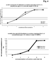
- a chromatographic separation process can be characterized by the resolution of the individual peaks in the elution chromatogram.
- the resolution value can be calculated according to Kaltenbrunner, O., et al., Biotechnol. Bioeng. 98 (2007) 201-210 , or Grushka, E., Anal. Chem. 44 (1972) 1733 -1738 .
- a “polypeptide” is a polymer of amino acid residues joined by peptide bonds, whether produced naturally or synthetically. Polypeptides of less than about 20 amino acid residues are referred to as "peptides".
- a “protein” is a macromolecule comprising one or more polypeptide chains or at least one polypeptide chain of more than 100 amino acid residues.
- a polypeptide may also comprise non-peptidic components, such as carbohydrate groups. Carbohydrate groups and other non-peptidic substituents may be added to a polypeptide by the cell in which the polypeptide is produced, and will vary with the type of cell. Polypeptides are defined herein in terms of their amino acid backbone structures; substituents such as carbohydrate groups are generally not specified, but may be present nonetheless.
- the term "applying to” denotes a partial step of a purification method in which a solution is brought in contact with a chromatography material. This denotes that either a) the solution is added to a chromatographic device in which the chromatography material is contained, or b) that the chromatography material is added to a solution comprising the polypeptide. In case a) the solution passes through the device allowing for the adsorption of the substances contained in solution to the chromatography material. Depending on the conditions, such as e.g. pH, conductivity, salt concentration, temperature, and/or flow rate, some substances of the solution adsorb to the chromatography material and other substances can be recovered from the flow-through.
- the "flow-through” denotes the solution obtained after the passage of the device, which may either be the applied solution or a buffered solution, which is used to wash the column or to cause elution of substances adsorbed to the chromatography material.
- the device is a column or a cassette.
- the chromatography material can be added, e.g. as a solid, to the solution, e.g. containing the substance of interest to be purified, allowing for an interaction between the chromatography material and the substances in solution. After the interaction the chromatography material is removed, e.g. by filtration, and the substance bound to the chromatography material are also removed therewith from the solution whereas the substances not bound to the chromatography material remain in solution.
- polypeptide (antibody) are adsorbed per liter of chromatography material. In one embodiment about 30 g polypeptide (antibody) are adsorbed per liter of chromatography material. In one embodiment about 20 g polypeptide (antibody) are adsorbed per liter of chromatography material.
- bind-and-elute mode denotes an operation mode of a chromatography step, in which a solution containing a substance of interest to be purified is applied to a chromatography material, whereby the substance of interest binds to the chromatography material.
- the substance of interest is retained on the chromatography material whereas substances not of interest are removed with the flow-through or the supernatant.
- the substance of interest is afterwards recovered from the chromatography material in a second step with an elution solution.
- the method as reported herein is operated in bind-and-elute mode.
- buffered solutions denotes a solution in which changes of pH due to the addition or release of acidic or alkaline substances is leveled by the dissolved buffer substance. Any buffer substance with such properties can be used. Generally pharmaceutically acceptable buffers substances are used.
- the buffered solution is selected from a phosphate buffered solution consisting of phosphoric acid and/or salts thereof, or an acetate buffered solution consisting of acetic acid and salts thereof, or a citrate buffered solution consisting of citric acid and/or salts thereof, or a morpholine buffered solution, or a 2-(N-morpholino) ethanesulfonic buffered solution, or a histidine buffered solution, or a glycine buffered solution, or a tris (hydroxymethyl) aminomethane (TRIS) buffered solution.
- a phosphate buffered solution consisting of phosphoric acid and/or salts thereof
- an acetate buffered solution consisting of acetic acid and salts thereof or a citrate buffered solution consisting of citric acid and/or salts thereof, or a morpholine buffered solution, or a 2-(N-morpholino) ethanesulfonic buffer
- the buffer solution is selected from a phosphate buffered solution, or an acetate buffered solution, or a citrate buffered solution, or a histidine buffered solution.
- the buffered solution may comprise an additional salt, such as e.g. sodium chloride, sodium sulphate, potassium chloride, potassium sulfate, sodium citrate, or potassium citrate.
- continuous elution and “continuous elution method” denote a method wherein the conductivity of a solution causing elution, i.e. the recovery of a bound compound from a chromatography material, is changed, i.e. raised or lowered, continuously, i.e. the concentration is changed by a sequence of small steps each not bigger than a change of 2 %, or of 1 % of the concentration of the substance causing elution.
- continuous elution one or more conditions, for example the pH, the ionic strength, concentration of a salt, and/or the flow of the mobile phase can be changed linearly or exponentially or asymptotically. In one embodiment the change is linear.
- step elution denotes a method wherein e.g. the concentration of a substance causing elution, i.e. the recovery of a bound substance from a chromatography material, is raised or lowered at once, i.e. directly from one value/level to the next value/level.
- concentration of a substance causing elution i.e. the recovery of a bound substance from a chromatography material
- step elution one or more conditions, for example the pH, the ionic strength, concentration of a salt, and/or the flow of a chromatography, can be changed all at once from a first, e.g. starting, value to a second, e.g. final, value.
- the conditions are changed incrementally, i.e. stepwise, in contrast to a linear change.
- ion exchange chromatography material denotes an immobile high molecular weight matrix that carries covalently bound charged substituents used as stationary phase in ion exchange chromatography. For overall charge neutrality not covalently bound counter ions are bound thereto.
- the "ion exchange chromatography material” has the ability to exchange its not covalently bound counter ions for similarly charged ions of the surrounding solution.
- the "ion exchange resin” is referred to as cation exchange resin or as anion exchange resin.
- the "ion exchange resin” is referred to as, e.g. in the case of cation exchange resins, sulfonic acid resin (S), or sulfopropyl resin (SP), or carboxymethyl resin (CM).
- ion exchange materials i.e. stationary phases
- Bio-Rex® e.g. type 70
- Chelex® e.g. type 100
- Macro-Prep® e.g. type CM, High S, 25 S
- AG® e.g. type 50W, MP
- WCX 2 available from Ciphergen
- Dowex® MAC-3 available from Dow chemical company
- Mustang C and Mustang S available from Pall Corporation
- Cellulose CM e.g. type 23, 52
- hyper-D partisphere available from Whatman plc.
- Amberlite® IRC e.g.
- the cation exchange material is a strong cation exchange material such as Macro-Prep® High S or 25S, or MacroCap SP, or Toyopearl® SP 650M, or Source S, or SP Sepharose, or POLYCAT A, or Mono S, or Highscreen SP.
- a substance of interest e.g. an antibody in monomeric form
- a stationary phase when brought in contact with it, e.g. an ion exchange material.
- This does not necessarily denote that 100 % of the substance of interest is bound but essentially 100 % of the substance of interest is bound, i.e. at least 50 % of the substance of interest is bound, in one embodiment at least 75 % of the substance of interest is bound, in another embodiment at least 85 % of the substance of interest is bound, in a further embodiment more than 95 % of the substance of interest is bound to the stationary phase.
- the antibody is a therapeutic antibody.
- therapeutic antibody denotes an antibody which is tested in clinical studies for approval as human therapeutic and which can be administered to an individual for the treatment of a disease.
- the therapeutic antibody is a monoclonal antibody.
- the therapeutic antibody is obtained from a great ape or an animal transformed with a human antibody locus or a human monoclonal antibody or a humanized monoclonal antibody.
- the therapeutic antibody is a human monoclonal antibody.
- the therapeutic antibody is a humanized monoclonal antibody. Therapeutic antibodies are being used widely for the treatment of various diseases such as oncological diseases (e.g.
- antibodies against ALK adhesion related kinase receptor (e.g., Axl), or ERBB receptors (e.g., EGFR, ERBB2, ERBB3, ERBB4), or erythropoietin-producing hepatocellular (EPH) receptors (e.g., EphA1; EphA2, EphA3, EphA4, EphA5, EphA6, EphA7, EphA8, EphB1, EphB2, EphB3, EphB4, EphB5, EphB6), or fibroblast growth factor (FGF) receptors (e.g., FGFR1, FGFR2, FGFR3, FGFR4, FGFR5), or Fgr, or IGF-1R, or Insulin Receptor,
- ALK adhesion related kinase receptor
- ERBB receptors e.g., EGFR, ERBB2, ERBB3, ERBB4
- EPH erythropoietin
- antibody encompasses the various forms of antibody structures including whole antibodies.
- the antibody is in one embodiment a human antibody, a humanized antibody, a chimeric antibody, or a T cell antigen depleted antibody.
- Genetic engineering of antibodies is e.g. described in Morrison, S.L., et al., Proc. Natl. Acad. Sci. USA 81 (1984) 6851-6855 ; US 5,202,238 ; US 5,204,244 ; Riechmann, L., et al., Nature 332 (1988) 323-327 ; Neuberger, M.S., et al., Nature 314 (1985) 268-270 ; Lonberg, N., Nat. Biotechnol.
- antibodies are divided in the classes: IgA, IgD, IgE, and IgG. Some of these classes are further divided into subclasses (isotypes), i.e. IgG in IgG1, IgG2, IgG3, and IgG4, or IgA in IgA1 and IgA2. According to the class to which an antibody belongs are the heavy chain constant regions of antibodies denoted as ⁇ (IgA), ⁇ (IgD), ⁇ (IgE), and ⁇ (IgG), respectively.
- antibody of human IgG1 class for example denotes an antibody in which the amino acid sequence of the constant domains is derived from the amino acid sequence of human IgG1.
- the term includes human antibodies, humanized antibodies, chimeric antibodies, and antibody conjugates.
- the term "complete antibody” denotes an antibody which comprises two light chain polypeptides (light chains) and two heavy chain polypeptides (heavy chains).
- Each of the heavy and light chain polypeptides contains a variable domain (variable region, generally the amino terminal portion) comprising binding regions that are able to interact with an antigen.
- Each of the heavy and light chain polypeptides comprises a constant region (generally the carboxyl terminal portion).
- the variable domain of a light or heavy chain in turn comprises different segments, i.e. four framework regions (FR) and three hypervariable regions (CDR).
- antibody conjugate denotes a polypeptide comprising at least one domain of an antibody heavy or light chain conjugated via a peptide bond to a further polypeptide.
- the further polypeptide can be a non-antibody peptide, such as a hormone, or toxin, or growth receptor, or antifusogenic peptide, or complement factor, or the like.
- a combination of different column chromatography steps can be employed. Generally a protein A affinity chromatography is followed by one or two additional separation steps.
- the final purification step is a so called “polishing step" for the removal of trace impurities and contaminants like aggregated antibodies, residual HCP (host cell protein), DNA (host cell nucleic acid), viruses, or endotoxins.
- antibody in monomeric form denotes an antibody molecule that is not associated with a second antibody molecule, i.e. which is neither covalently nor non-covalently bound to another antibody molecule.
- antibody in aggregated form denotes an antibody molecule which is associated, either covalently or non-covalently, with at least one additional antibody molecule, and which is eluted in a single peak from a size exclusion chromatography column.
- in monomeric form as used herein not necessarily denotes that 100 % of an antibody molecule is present in monomeric form. It denotes that an antibody is essentially in monomeric form, i.e.
- HMW high molecular weight
- elution compound denotes a salt used for recovering of a bound polypeptide from an ion exchange material, whereby the compound increases the conductivity of the buffer/solution. This can be accomplished either by an increased buffer salt concentration or by the addition of other salts, so called elution salts, to the buffered solution.
- Preferred elution salts are sodium citrate, sodium chloride, sodium sulphate, sodium phosphate, potassium chloride, potassium sulfate, potassium phosphate, as well as other salts of citric and phosphoric acid, and any mixture of these components.
- the elution compound is selected from sodium citrate, sodium chloride, potassium chloride, and mixtures thereof.
- Humanized forms of non-human (e.g. rodent) antibodies are chimeric antibodies that contain partial sequences derived from a non-human antibody and from a human antibody.
- humanized antibodies are derived from a human antibody (recipient antibody), in which residues from a hypervariable region are replaced by residues from a hypervariable region of a non-human species (donor antibody), such as mouse, rat, rabbit, sheep, guinea pig, or non-human primate, having the desired specificity and affinity.
- donor antibody such as mouse, rat, rabbit, sheep, guinea pig, or non-human primate, having the desired specificity and affinity.
- donor antibody such as mouse, rat, rabbit, sheep, guinea pig, or non-human primate
- framework region (FR) residues of the human antibody are replaced by corresponding non-human residues.
- humanized antibodies may comprise further modifications, e.g. amino acid residues that are not found in the recipient antibody or in the donor antibody. Such modifications result
- a humanized antibody will comprise substantially all of at least one, and typically two, variable domains, in which all or substantially all of the hypervariable loops correspond to those of a non-human donor antibody and all or substantially all of the FRs are those of a human recipient antibody.
- the humanized antibody optionally will also comprise at least a portion of an antibody constant region, typically that of a human antibody.
- humanized antibodies are typically human antibodies in which some hypervariable region residues and possibly some framework region residues are substituted by residues from analogous sites in rodent or non-human primate antibodies.
- monoclonal antibody refers to an antibody obtained from a population of substantially homogeneous antibodies, i.e. the individual antibodies of the population are identical except for possible naturally occurring mutations that may be present in minor amounts. Monoclonal antibodies are highly specific, being directed against a single antigenic site. Furthermore, in contrast to polyclonal antibody preparations, which include different antibodies directed against different antigenic sites (determinants or epitopes), each monoclonal antibody is directed against a single antigenic site on an antigen. In addition to their specificity, monoclonal antibodies are advantageous in that they may be synthesized uncontaminated by other antibodies.
- the modifier "monoclonal" indicates the character of the antibody as being obtained from a substantially homogeneous population of antibodies and is not to be construed as requiring production of the antibody by any particular method.
- chimeric antibody denotes an antibody comprising a variable domain, i.e. binding region, from a first species and at least a portion of a constant region derived from a different second species.
- Amino acid sequence variants of antibodies can be prepared by introducing appropriate modifications into the nucleotide sequence encoding the antibody chains, or by peptide synthesis. Such modifications include, for example, deletions from, and/or insertions into and/or substitutions of residues within the amino acid sequences of the antibody. Any combination of deletion, insertion, and substitution can be made to arrive at the final construct, provided that the final construct possesses the antigen binding properties as the parent antibody.
- Amino acids may be grouped according to common side-chain properties:
- Non-conservative substitutions will entail exchanging a member of one of these classes for another class.
- mobile phase denotes any mixtures of water and/or aqueous buffer and/or organic solvents being suitable to recover polypeptides from a chromatography column.
- to elute or “eluting”, respectively, in the present context is used as known to the expert skilled in the art and denotes the dissolution, optionally the displacement, of adsorbed substance(s) from solids or adsorbents, which are impregnated with fluids, i.e., the column material to which the substance(s) is/are adsorbed.
- adsorption denotes the accumulation of substances from a liquid, e.g. a mobile phase, at the boundary phase formed between the liquid and a solid phase, wherein the latter is able to adsorb the substances of interest at its surface. This adsorption leads to an accumulation of the adsorbed substances.
- the substance that is able to accumulate the substance of interest at its surface is referred to as adsorbent and the adsorbed material as adsorbate.
- adsorption is usually distinguished from the term “absorption” which beyond the accumulation at a surface also refers to the penetration of the accumulated substances into the interior of the adsorbing solid or fluid.
- adsorption is a physical process in which substances usually molecules adhere to a surface of the adsorbent and thus, are accumulated at the respective surface.
- the forces being responsible for this adherence are considered to be physical forces rather than chemical bonds and thus, adsorption is also known in the art as physical adsorption or physisorption, which does not necessarily exclude chemical bonding of substances to the surface.
- the physical forces involved in the adsorption of substances to a surface are in most cases van der Waals-forces, London forces or dipole/dipole interactions, for example hydrogen bonds, or dipole-induced dipole interactions, wherein these terms are used as either explained herein or as normally used in context with adsorption.
- solvents are used as eluent, i.e., eluting agent in which the substance(s) which are to be eluted are at least sufficiently soluble.
- the non-ionic polymer is a hydrophilic non-ionic polymer.
- the polymer can be selected from non-ionic polyether, such as poly (ethylene glycol), poly (propylene glycol) (PPG), PEG-PPG block copolymers, PEG-PPG copolymers, triblock copolymers composed of poly (oxypropylene) (poly (propylene oxide)) and two poly (oxyethylene) (poly (ethylene oxide)) polymers (Poloxamer, PluronicTM), and other PEG-PPG-PEG triblock polymers.
- non-ionic polyether such as poly (ethylene glycol), poly (propylene glycol) (PPG), PEG-PPG block copolymers, PEG-PPG copolymers, triblock copolymers composed of poly (oxypropylene) (poly (propylene oxide)) and two poly (oxyethylene) (poly (ethylene oxide)) polymers (Poloxamer, PluronicTM), and other PEG-
- the employed non-ionic polymer can be characterized besides its monomeric building block by its molecular weight given in Daltons (Da).
- the term "molecular weight” denotes with respect to polymers the mean molecular weight of the polymer because polymeric compounds are not obtained with a defined molecular weight but in fact they have a molecular weight distribution.
- the molecular weight of a polymer is given in the form "about X Da", whereby "X” denotes the mean value of the molecular weight of the polymer.
- the term “about” indicates that in the polymer preparation, some molecules will have a weight more and some molecules will have a weight less than the indicated mean molecular weight, i.e.
- the term about refers to a molecular weight distribution in which 95 % of the polymer molecules have a molecular weight within +/- 30 % of the indicated molecular weight.
- a molecular weight of 3,500 Da denotes a range of from 2450 kDa to 4,550 Da.
- the polymer molecules have a molecular weight within +/- 20 % of the indicated molecular weight.
- the polymer molecules have a molecular weight within +/- 10 % of the indicated molecular weight.
- the non-ionic polymer can be present in a buffered solution, such as a salt containing buffered solution.
- the solution comprising the polypeptide of interest can be obtained directly from the cultivation supernatant of a cell which comprises a nucleic acid encoding the polypeptide.
- the supernatant can be a clarified or a non-clarified cultivation supernatant.
- one or more non-ionic polymers are present in the solution applied to the chromatography material for recovering the polypeptide, especially the antibody.
- the solution applied to the chromatography material for recovering the antibody in monomeric from comprises one, or two, or three, or four, or five different non-ionic polymers. If more than one non-ionic polymer is present in the solution the sum of the concentrations of all non-ionic polymers present in the solution is preferably within the range as given herein.
- one or more additives are present in the solution applied to the chromatography material for recovering the polypeptide, especially the antibody.
- the solution applied to the chromatography material for recovering the antibody in monomeric from comprises one, or two, or three, or four, or five different additives. If more than one additive is present in the solution the sum of the concentrations of all additives present in the solution is preferably within the range as given herein.
- the solution comprising the polypeptide of interest can be the eluate from a preceding chromatography step.
- the interactions between the polypeptide of interest and the ion exchange chromatography material can be adsorption, i.e. binding of the polypeptide, or a retardation of the polypeptide in relation to other component(s) also present in the solution.
- the adsorption is followed by a step of eluting the adsorbed polypeptide of interest in monomeric form from the ion exchange chromatography material.
- Elution of adsorbed polypeptide can be effected by change of salt conditions, i.e. conductivity, and/or the pH value as compared to the adsorption step.
- the chromatography steps of the method as reported herein are performed in accordance with conventional general operating conditions and well known principles in the field.
- Conventional chromatography columns are conveniently used, the size of which are adapted for each case, as is the starting materials, buffers, matrices, including the functional groups of the ligands, etc.
- the charge of a polypeptide may be changed by changing the pH value of the surrounding solution.
- an ion exchange material may be a strong ion exchange material, which means that it is charged at all pH values, or a weak ion exchange material, which means that it is chargeable by shifting the pH.
- Adding a non-ionic polymer and an additive to the mobile phase in an ion exchange chromatography recovering step as reported herein was unexpectedly shown to increase the yield of the polypeptide in monomeric form, as compared to conventional ion exchange chromatography.
- the ion exchange chromatography is a cation exchange chromatography.
- the concentration of the non-ionic polymer is at least about 5 % by weight and the concentration of the additive is at least 3 % by weight.
- the molecular weight of the non-ionic polymer is chosen in a way that is as low as possible while resulting in combination with the additive in an increased yield of the polypeptide in monomeric form compared to the used of the non-ionic polymer alone.
- the person skilled in the art knows that the addition of any substance, i.e. of the non-ionic polymer and the additive, affects (increases) the viscosity of the resulting solution. Therefore a person skilled in the art will appreciate that due to the physicochemical properties of the non-ionic polymer and the additive an upper limit for the addition of these compounds exists.
- the non-ionic polymer used as in the method as reported herein comprises groups that are rich in oxygen atoms.
- the non-ionic polymer is an aqueous-soluble not-charged linear or branched polymer.
- Such non-ionic polymers can be polyether, such as poly (ethylene glycol) (PEG), poly (propylene glycol) (PPG), PEG-PPG block copolymers, PEG-PPG copolymers, triblock copolymers composed of a central hydrophobic chain of poly (oxypropylene) (poly(propylene oxide)) flanked by two hydrophilic chains of poly (oxyethylene) (poly(ethylene oxide)) (Poloxamer, PluronicTM), and other PEG-PPG-PEG triblock polymers, ethylhydroxyethylcellulose (EHEC) and similar polymers, polymerized allylglycidyl ether, polymerized phenyl glycidyl ether, dextran, star
- any molecular weight non-ionic polymer can be used in the method as reported herein.
- the optimal concentration and ratio of the non-ionic polymer and the additive may vary for every protein and, in most cases, there is a preferable molecular weight of the non-ionic polymer and a preferable ratio of the non-ionic polymer to the additive.
- the non-ionic polymer is poly (ethylene glycol).
- Poly (ethylene glycol) can be used as a general model non-ionic polypeptide for behavior of non-ionic polymers within the method as reported herein.
- poly (ethylene glycol) is used as an example herein it should be kept in mind that the provided information also applies to other non-ionic polymers, including to those specifically listed herein above.
- Poly (ethylene glycol) is available from a number of commercial sources. In one embodiment the poly (ethylene glycol) has a molecular weight in the range of from about 100 Da to about 40,000 Da. In one embodiment the poly (ethylene glycol) has a molecular weight between about 400 Da and about 10,000 Da. In one embodiment the poly (ethylene glycol) has a molecular weight between about 1,000 Da and about 8,000 Da. In other embodiments, a mixture of different sized poly (ethylene glycol)s is utilized.
- the poly (ethylene glycol) has a molecular weight between about 2,450 Da and about 4,550 Da. In one embodiment the poly (ethylene glycol) has a molecular weight between about 2,625 Da and about 4,375 Da. In one embodiment the poly (ethylene glycol) has a molecular weight between about 2,800 Da and about 4,200 Da. In one embodiment the poly (ethylene glycol) has a molecular weight between about 3,150 Da and about 3,850 Da.
- the poly (ethylene glycol) is a linear or branched poly (ethylene glycol).
- Lower molecular weight poly (ethylene glycols) can require a higher mass concentration to be used in order to have a similar effect compared to higher molecular weight poly (ethylene glycol)s.
- a poly (ethylene glycol) of a specified molecular weight can be employed to produce larger polypeptide in monomeric form, such as antibodies and fusion proteins, compared to those concentrations required to effect the same purity of smaller polypeptides.
- an antibody of the class IgA which is mostly present as a dimer, having a molecular weight of about 320 kDa will require a lower concentration of poly (ethylene glycol) compared to an antibody of the class IgG having a molecular weight of about 150 kDa.
- the retention of aggregates, complexes, and other large molecules can generally be increased more compared to that of the monomeric polypeptide.
- poly (ethylene glycol) be required to increase the retention of polypeptides that interact/are adsorbed strongly to the cation exchange chromatography material compared to the required concentration of poly (ethylene glycol) required to increase the adsorption of polypeptides that are normally adsorbed weakly to the cation exchange chromatography material.
- the additive is a substance that can reduce or even prevent precipitation that is induced by the presence of the non-ionic polymer.
- the additive has a conductivity that is sufficiently low to not interfere with a cation exchange chromatography. In another embodiment the additive has no detectable buffering capacity.
- the additive can be a zwitterion, or an amino acid (such as glycine, bicine, tricine, alanine, proline, Betaine), or urea, or an urea derivative (such as alkyl ureas (methyl urea, ethyl urea, etc.)), or alkylene glycols (such as ethylene glycol or propylene glycol), or an ampholyte (such as amino-sulfonic acid based buffers MES, MOPS, HEPES, PIPES and CAPS buffer), or CHAPSO, or a natural product (such as certain alkaloids and Betaines), or a sugar (such as glucose, sucrose, raffinose), or a polyol (such as glycerol, or xylitol, or sorbitol).
- an amino acid such as glycine, bicine, tricine, alanine, proline, Betaine
- urea such as alkyl urea
- the additive can be a polyol.
- the polyol can be glycerol, or xylitol, or sorbitol.
- glycine can be used as additive in the method as reported herein.
- Glycine is a zwitterionic compound, more precisely glycine is an amino acid.
- glycine does not contributes to overall conductivity and, thus, should not interfere in the cation exchange chromatography method. Additionally, glycine does not exhibit a buffering effect in aqueous solution it will also not effect the pH value of the mobile phase.
- Glycine can be used as additive in the method as reported herein in a concentration of from about 50 mM to 5 M. The glycine can have a concentration of from about 100 mM to about 4 M.
- the glycine can have a concentration of from about 250 mM to about 3 M.
- the glycine can have a concentration of from about 500 mM to about 2 M.
- the glycine can have a concentration of about 1 M. If more than one additive is used the concentrations of the individual additives can be lower than those exemplarily given in the paragraph above.
- any additive can be used at a concentration that is higher than the concentration necessary to achieve the intended effect.
- a person skilled in the art can determine the additive concentration range in that the effect is present and that can be tolerated in the method as reported herein.
- the additive When the additive is urea or a urea derivative the additive is present in a concentration up to 6 molar. In one embodiment the additive is present in a concentration below 2 molar.
- the concentration of the non-ionic polymer and the additive in the recovering solution can vary. Generally, any concentration of poly (ethylene glycol) can be used. The concentration of poly (ethylene glycol) is at least about 0.5 % by weight.
- the concentration of the non-ionic polymer can be held constant or it can be changed during the course of the chromatography. This change includes but is not limited to a gradient of increasing or decreasing concentration, or with step-wise changes in concentration.
- the non-ionic polymer can have a concentration of from about 7 % to 13 % by weight and/or the additive can have a concentration of from 3 % to 25 % by weight.
- the non-ionic polymer is poly (ethylene glycol) and the additive is a polyol.
- Figure 3 the solubility of an antibody in a solution comprising poly (ethylene glycol) of a molecular weight of about 3,500 Da at a concentration of 10 % by weight as an example of a non-ionic polymer and different polyols at varying concentration as additive are shown.
- polyol denotes a compound that has at least three hydroxyl groups, i.e. is a triol.
- a non-ionic polymer e.g. poly (ethylene glycol)
- an additive e.g. sorbitol
- anti-CSF-1R antibody [fraction of initial conc.] 15 0 10.1 ⁇ 100% 10.2 ⁇ 100% 15 10 9.01 90.1% 5.15 51.5% 20 0 10.2 ⁇ 100% 10.1 ⁇ 100% 20 10 10.3 ⁇ 100% 10.1 ⁇ 100%
- the HCP (host cell protein) content in the recovered antibody preparation can be reduced compared to methods performed in the absence of this combination.
- the recovery of the monomeric polypeptide varies depending on the combination of the concentration of the non-ionic polymer and the additive.
- chromatographic separation step comprises several sub-steps:
- the cation exchange chromatography material in the column is equilibrated prior to the applying of the solution comprising the polypeptide to the column. This can be accomplished by applying an equilibration buffer to the column to establish a suitable pH value, and/or conductivity, and/or concentration of salt inside or on the cation exchange chromatography material.
- the solution comprising the polypeptide is adjusted to conditions comparable to those to which the cation exchange chromatography column is equilibrated.
- polypeptide of interest is added to the solution and is loaded onto the column.
- polypeptide of interest is added after the column is equilibrated and/or washed.
- equilibrating comprises the applying of a binding buffer before loading of the solution onto the chromatographic material.
- the recovery of the polypeptide in monomeric form in the method as reported herein can be carried out using a linear gradient, a step gradient, or a combination of a linear and a step gradient.
- a linear gradient can be, for example, used to further improve the resolution of the polypeptide in monomeric form from the polypeptide in aggregated form and/or from other contaminants such as host cell protein.
- a step gradient can be used in order to reduce the volume of recovered polypeptide solution. Selection and use of any gradient in the recovery of the polypeptide from the cation exchange chromatography material can easily be made by a person skilled in the art.
- the application of the solution comprising the polypeptide is followed by the application of a buffered wash solution to the chromatography material, often of the same composition as the equilibration buffer.
- the concentration of the non-ionic polymer is held constant during elution while the pH value is changed and/or the concentration of the salt causing elution is increased.
- the concentration of the non-ionic polymer is increased during elution while the pH value is changed and/or the concentration of a salt causing elution is increased.
- the concentration of the non-ionic polymer is decreased during elution while the pH value and/or the concentration of the salt are held constant.
- the cation exchange chromatography material can be cleaned, sanitized, and stored in an appropriate agent.
- the method as reported herein can practiced in combination with one or more other chromatographic steps or methods, such as but not limited to protein A affinity chromatography and other forms of affinity chromatography, hydrophobic interaction chromatography, or other mixed mode chromatography.
- the method as reported herein can be practiced in a packed bed column, a fluidized/expanded bed column containing the cation exchange chromatography material, and/or a batch operation where the cation exchange chromatography material is mixed with the polypeptide preparation for a certain time.
- biologically active polypeptides in monomeric form.
- biologically active polypeptides can be selected from antibodies, antibody variant, antibody conjugates.
- the polypeptide of interest is an antibody, especially a monoclonal antibody.
- the antibody may be from any mammal, such as a mouse, rat, rabbit, guinea pig, sheep, cow, or a transgenic animal with a human immunoglobulin locus, or it may be a humanized, resurfaced, T-cell epitope depleted variant thereof.
- antibody preparation can be used, such as non-purified (crude) or partially purified antibodies from natural, synthetic, or recombinant sources.
- Crude antibody preparations can be obtained from various sources including, but not limited to, plasma, serum, ascites, or milk of an experimental animal, plant extracts, bacterial cell lysates, yeast lysates, or conditioned animal cell culture media.
- Partially purified preparations can be obtained from crude preparations that have been subjected to at least one chromatography, precipitation, other fractionation step, or any combination thereof.
- the precipitation step can include any method, such as salt or poly (ethylene glycol) precipitation. Other fractionation steps such as crystallization or membrane filtration can also be used.
- the antibody is a not-PEGylated antibody.
- a polypeptide can be obtained that is substantially free of aggregates.
- the content of the polypeptide in aggregated form as determined by size exclusion chromatography less than about 5 % based on the total peak area.
- the content of the polypeptide in aggregated form is less than about 1%.
- the amount of the polypeptide in aggregated form is less than about 0.5%.
- the amount of the polypeptide in aggregated form is less than about 0.1 %.
- This bispecific antibody comprises four different antibody chains, i.e. two different light chains and two different heavy chains.
- product-related impurities can be formed.
- One of these product related impurities is the so called antibody"
- an antibody that is missing one light chain displaying a molecular weight of about 125 kDa compared to a weight of about 150 kDa for a complete four chain antibody.
- These kinds of antibody-fragments can generally be formed during the expression of antibodies.
- the ion exchange chromatography material is a cation exchange chromatography material.
- the non-ionic polymer can be selected from the group comprising poly (ethylene glycol) (PEG), poly (propylene glycol) (PPG), PEG-PPG copolymers, and triblock copolymers composed of poly (oxypropylene) (poly (propylene oxide)) flanked by poly (oxyethylene) (poly (ethylene oxide)).
- the non-ionic polymer is poly (ethylene glycol).
- the additive can be selected from the group comprising zwitterions, amino acids, urea, urea derivatives, ampholytes, CHAPSO, natural products, sugars, and polyols.
- the additive is sorbitol.
- the non-ionic polymer can have a concentration of from about 5 % to 15 % by weight and/or the additive can have a concentration of from 3 % to 25 % by weight.
- polypeptide is an antibody of class IgG.
- polypeptide is an antibody of class IgG, subclass IgG1, or subclass IgG2, or subclass IgG3, or subclass IgG4.
- the recovering is by gradient elution.
- Each antibody (anti-IL17 antibody: for sequences see WO 2010/034443 , anti-CSF-1R antibody: for sequences see PCT/EP2010/069090 ; anti-Ang2/VEGF antibody: for sequences see WO 2010/040508 ) was purified in a first step with a protein A affinity chromatography. Elution from the protein A column was carried out under acidic conditions. Afterwards the sample was adjusted to about pH 3.5 with 1 M citric acid and incubated for ⁇ 1 hour. Subsequently the pH was adjusted to 5.0 or 5.5 by adding 1 M TRIS/HCl, pH 8-9 or 1 M TRIS solution (TRIS: tris-hydroxymethyl-amino-methane).
- the sample was depth filtrated.
- the sample is a solution with a protein concentration between 5 mg/ml and 20 mg/ml.
- This material is referred to in the following as conditioned protein A eluate, which is prepared for loading onto a cation exchange chromatography material.
- the anti-Ang2/VEGF antibody was in addition purified by anion exchange chromatography: The pH value of the conditioned protein A eluate was adjusted to 7.5 by the addition of 1M TRIS buffer solution. Subsequently the sample was processed by anion exchange chromatography at pH 7.5 in flow-through mode.
- Example 1 In the first example the method as reported herein has been carried out. Thus, in Example 1 all conditions of the method are given. In the thereafter following examples only the differences are listed. All not mentioned parameters are identical.
- the conditioned protein A eluate was applied to a chromatography column containing the ion exchange chromatography material. After the loading step at a flow rate of 90 cm/h the column was washed with wash solution 1 (1 column volume) and wash solution 2 (3 column volumes).
- wash solution 1 (1 column volume)
- wash solution 2 (3 column volumes).
- the antibody in monomeric form was recovered with a linear gradient elution method, whereby the pH value was kept constant and the conductivity was varied (increased) by the addition of sodium chloride.
- Figure 1 the elution chromatogram of the cation exchange chromatography of the anti-IL17 antibody on the cation exchange chromatography material is shown.
- the antibody in monomeric form and the antibody in aggregated form are separated.
- the resolution of the monomer peak and the aggregate peak was determined using the program PeakFit (Seasolve Software Inc.) to be 0.78.
- the resolution of the monomer peak and the aggregate peak was determined using the program PeakFit (Seasolve Software Inc.) to be 0.88.
- the resolution of the monomer peak and the aggregate peak was determined using the program PeakFit (Seasolve Software Inc.) to be 0.86.
- the resolution of the monomer peak and the aggregate peak was determined using the program PeakFit (Seasolve Software Inc.) to be 0.77.
- the resolution of the monomer peak and the aggregate peak was determined using the program PeakFit (Seasolve Software Inc.) to be 0.51.
- the resolution of the monomer peak and the aggregate peak was determined using the program PeakFit (Seasolve Software Inc.) to be 0.76.
- the resolution of the monomer peak and the aggregate peak was determined using the program PeakFit (Seasolve Software Inc.) to be 0.51.
- the resolution of the monomer peak and the aggregate peak was determined using the program PeakFit (Seasolve Software Inc.) to be 0.76.
- the resolution of the monomer peak and the aggregate peak was determined using the program PeakFit (Seasolve Software Inc.) to be 0.93.
- the resolution of the monomer peak and the aggregate peak was determined using the program PeakFit (Seasolve Software Inc.) to be 0.77.
- the resolution of the monomer peak and the aggregate peak was determined using the program PeakFit (Seasolve Software Inc.) to be 0.90.
- the resolution of the monomer peak and the aggregate peak was determined using the program PeakFit (Seasolve Software Inc.) to be 0.73.
- the resolution of the monomer peak and the aggregate peak was determined using the program PeakFit (Seasolve Software Inc.) to be 0.84.
- the resolution of the monomer peak and the aggregate peak was determined using the program PeakFit (Seasolve Software Inc.) to be 0.79.
- the resolution of the monomer peak and the aggregate peak was determined using the program PeakFit (Seasolve Software Inc.) to be 0.90.
- the resolution of the monomer peak and the aggregate peak was determined using the program PeakFit (Seasolve Software Inc.) to be 0.85.
- the resolution of the monomer peak and the aggregate peak was determined using the program PeakFit (Seasolve Software Inc.) to be 0.90.
- the resolution of the monomer peak and the aggregate peak was determined using the program PeakFit (Seasolve Software Inc.) to be 0.83.
- the resolution of the monomer peak and the aggregate peak was determined using the program PeakFit (Seasolve Software Inc.) to be 0.97.
- the resolution of the monomer peak and the aggregate peak was determined using the program PeakFit (Seasolve Software Inc.) to be 0.86.
- the resolution of the monomer peak and the aggregate peak was determined using the program PeakFit (Seasolve Software Inc.) to be 0.86.
- the resolution of the monomer peak and the aggregate peak was determined using the program PeakFit (Seasolve Software Inc.) to be 0.57.
- the resolution of the monomer peak and the aggregate peak was determined using the program PeakFit (Seasolve Software Inc.) to be 0.70.
- the conditioned anion exchange eluate was applied to a chromatography column containing the cation exchange chromatography material. After the loading step at a flow rate of 90 cm/h the column was washed with wash solution (3 column volume). The antibody was recovered with a linear gradient elution method, whereby the pH value was varied (increased) from 5.0 to 7.5.
- the 3 ⁇ 4 antibody "co-elutes" with the antibody monomer and two 3 ⁇ 4 antibody peaks are detected.
- the conditioned anion exchange eluate was applied to a chromatography column containing the cation exchange chromatography material. After the loading step at a flow rate of 90 cm/h the column was washed with wash solution (3 column volume). The antibody was recovered with a linear gradient elution method, whereby the pH value was varied (increased) from 5.0 to 7.5.
- the conditioned anion exchange eluate was applied to a chromatography column containing the cation exchange chromatography material. After the loading step at a flow rate of 90 cm/h the column was washed with wash solution (3 column volume). The antibody was recovered with a linear gradient elution method, whereby the pH value was varied (increased) from 5.0 to 7.5.
Description
- Herein is reported a method for separating polypeptide monomers, aggregates and fragments using ion exchange chromatography, whereby in the recovering step a non-ionic polymer and an additive are added to the mobile phase.
- Proteins and especially immunoglobulins play an important role in today's medical portfolio. Polypeptides for use in pharmaceutical applications are mainly produced in mammalian cells such as CHO cells, NS0 cells, Sp2/0 cells, COS cells, HEK cells, BHK cells, PER.C6® cells, and the like.
- Due to their chemical and physical properties, such as molecular weight and domain architecture including secondary modifications, the downstream processing of immunoglobulins is very complicated. For example, are not only for formulated drugs but also for intermediates in downstream processing (DSP) concentrated solutions required to achieve low volumes for economic handling and application storage. The downstream processing of biotechnologically produced immunoglobulins typically comprises three chromatography steps: a first affinity chromatography step using e.g. Protein A, to remove non-immunoglobulin molecules, followed by two ion exchange chromatography steps. The purified immunoglobulin is obtained in a low concentration solution requiring a concentration step prior to formulating the antibody into the pharmaceutical formulation. Many proteins, including IgG, form dimers, oligomers or higher order aggregates. In order to provide a therapeutic protein product with the required purity these molecule species have to be removed by the purification process.
- Different methods are well established and widely used for protein purification (Vijayalakshmi, M.A., Appl. Biochem. Biotech. 75 (1998) 93-102). Zhou, J.X., et al. (J. Chrom. A 1175 (2007) 69-80) report a pH-conductivity hybrid gradient cation-exchange chromatography for process-scale monoclonal antibody purification. The separation of antibody monomers from its multimers by use of ion exchange chromatography is reported in
EP 1 084 136US 5,429,746 relates to the application of hydrophobic interaction chromatography combination chromatography to the purification of antibody molecule proteins. - Solvent modulation of column chromatography is reported by Arakawa, T., et al. in Prot. Pep. Lett. 15 (2008) 544-555. In
EP 1 729 867WO 2004/013162 an increased dynamic binding capacity in ion exchange chromatography by addition of polyethylene glycol is reported. InWO 2005/094960 a method for chromatographic purification is reported. Enhanced capacity and purification of antibodies by mixed mode chromatography in the presence of aqueous-soluble non-ionic organic polymers is reported inWO 2008/086335 . InWO 2009/149067 a process for the purification of antibodies is reported. - Milby, K.H., et al. report the ion-exchange chromatography of proteins and the effect of neutral polymers in the mobile phase (J. Chrom. 482 (1989) 133-144). In Biotechnology Techniques (12 (1998) 289-293) Feng, X-L., et al. report that polyethylene glycol improves the purification of recombinant human tumor necrosis factor during ion exchange chromatography. A method for obtaining unique selectivities in ion-exchange chromatography by addition of organic polymers to the mobile phase is reported by Gagnon, P., et al. (J. Chrom. 743 (1996) 51-55). Gagnon, P., reports in an internet article on validated.com (http://www.validated.com/revalbio/pdffiles/ionslect.pdf) the fine-tuning of selectivity on ion exchangers. In
WO 2009/149067 a process for the purification of antibodies is reported. - It has been found that high PEG-concentrations can be employed during cation exchange chromatography in the presence of certain additives (solubility enhancers) in order to modulate selectivity and to improve resolution of antibody monomer and aggregate species, e.g. compared to methods employing PEG-only.
- Herein is reported a method for isolating or obtaining or producing a polypeptide of interest in monomeric form from other component(s), such as the polypeptide of interest in aggregated form, comprising at least one chromatographic step. In one embodiment the method comprises contacting a solution comprising the polypeptide of interest with a cation-exchange chromatography material wherein the contacting with the chromatography material takes place in the absence of a non-ionic polymer and an additive, and wherein the recovering takes place in the presence of a non-ionic polymer and an additive.
- It has been found that by using an additional additive in a method as reported herein the effect of the non-ionic polymer can be improved.
- Reported herein is a method for producing an antibody in monomeric form comprising the following steps:
- applying a solution comprising a non-ionic polymer and an additive to a chromatography material to which an antibody had been adsorbed, whereby the antibody in monomeric form remains adsorbed to the ion exchange chromatography material, and
- recovering the antibody in monomeric form from the ion exchange chromatography material by applying a solution comprising a non-ionic polymer, an additive, and an elution compound, and thereby producing the antibody in monomeric form.
- Reported herein is a method for producing a polypeptide preparation with reduced host cell protein content whereby the preparation is obtained from a mammalian cell cultivation supernatant, especially a CHO cell cultivation supernatant, comprising the following steps:
- applying a solution comprising a non-ionic polymer and an additive to a chromatography material to which the polypeptide had been adsorbed, whereby the polypeptide in monomeric form remains adsorbed to the ion exchange chromatography material, and
- recovering the polypeptide in monomeric form from the ion exchange chromatography material by applying a solution comprising a non-ionic polymer, an additive, and an elution compound, and thereby producing the polypeptide preparation with reduced host cell protein content.
- Reported herein is a method for determining the concentration of a non-ionic polymer and an additive for use in an ion exchange chromatography of a polypeptide comprising the following steps:
- determining in solution in the absence of the additive the concentration of the non-ionic polymer at which less than 50 % of the polypeptide remain in solution,
- determining in solution in the presence of a concentration of the non-ionic polymer, which has been determined in the previous step, the concentration of the additive at which more than 95 % of the polypeptide remain in solution,
- Reported herein is a method for producing an antibody in monomeric form comprising the following step:
- recovering the antibody in monomeric form from an ion exchange chromatography material by applying a solution comprising a non-ionic polymer, an additive, and an elution compound, and thereby producing the antibody in monomeric form,
- In one embodiment the ion exchange chromatography material is a cation exchange chromatography material.
- The non-ionic polymer can be selected from the group comprising poly (ethylene glycol) (PEG), poly (propylene glycol) (PPG), PEG-PPG copolymers, and triblock copolymers composed of poly (oxypropylene) (poly (propylene oxide)) flanked by poly (oxyethylene) (poly (ethylene oxide)). In a further embodiment the non-ionic polymer is poly (ethylene glycol).
- The additive can be selected from the group comprising zwitterions, amino acids, urea, urea derivatives, ampholytes, CHAPSO, natural products, sugars, and polyols. In a further embodiment the additive is sorbitol.
- The non-ionic polymer can have a concentration of from about 5 % to 15 % by weight and/or the additive has a concentration of from 3 % to 25 % by weight.
- In one embodiment the polypeptide is an antibody of class IgG, or IgD, or IgE, or IgA. In another embodiment the polypeptide is an antibody of class IgG, subclass IgG1, or subclass IgG2, or subclass IgG3, or subclass IgG4.
- In one embodiment the method comprises the following steps:
- applying a first solution that optionally comprises a non-ionic polymer and an additive to an ion exchange chromatographic material and thereby equilibrating the ion exchange chromatography material,
- applying a buffered solution comprising the polypeptide to the equilibrated chromatography material and thereby adsorbing the polypeptide to the chromatography material, whereby the solution is essentially free of a non-ionic polymer and an additive,
- applying a buffered solution comprising a non-ionic polymer and an additive to the chromatography material, whereby the polypeptide in monomeric form remains adsorbed to the ion exchange chromatography material, and
- recovering the polypeptide in monomeric form from the ion exchange chromatography material by applying a solution comprising a non-ionic polymer, an additive, and an elution compound.
- In one embodiment the polypeptide is an antibody.
- In one embodiment about 40 g polypeptide (antibody) are applied per liter of chromatography material. In one embodiment about 30 g polypeptide (antibody) are applied per liter of chromatography material. In one embodiment about 20 g polypeptide (antibody) are applied per liter of chromatography material.
- One aspect as reported herein is a method for producing an antibody of the IgG class in monomeric form comprising the following steps:
- applying a first solution that optionally comprises poly (ethylene glycol) and sorbitol to a cation exchange chromatographic material and thereby equilibrating the material,
- applying the solution comprising the antibody of the IgG class to the equilibrated chromatography material and thereby loading the chromatography material,
- producing the antibody of the IgG class in monomeric form by applying a solution to the chromatographic material comprising poly (ethylene glycol) and sorbitol and thereby desorbing/eluting the antibody of the IgG class in monomeric form from the chromatographic material,
- Another aspect as reported herein is method for producing an antibody of the IgG class preparation with reduced host cell protein content comprising the following steps:
- applying a first solution that optionally comprises poly (ethylene glycol) and sorbitol to a cation exchange chromatographic material and thereby equilibrating the material,
- applying the solution comprising the antibody of the IgG class to the equilibrated chromatography material and thereby loading the chromatography material,
- producing the antibody of the IgG class in monomeric form by applying a solution to the chromatographic material comprising poly (ethylene glycol) and sorbitol and thereby desorbing/eluting the antibody of the IgG class in monomeric form from the chromatographic material,
- Herein is reported a method for producing a polypeptide of interest in monomeric form by separating it from other component(s), such as the polypeptide of interest in aggregated form, comprising at least one chromatographic step. In one embodiment the method comprises contacting a solution comprising the polypeptide of interest and a cation-exchange chromatography material wherein the contacting with the chromatography material takes place in the absence of a non-ionic polymer and an additive and wherein the recovering takes place in the presence of a non-ionic polymer and an additive.
- It has been found that the presence of a non-ionic polymer and an additive increases the retention time difference (resolution) of a polypeptide in monomeric form compared to the polypeptide in aggregated form on a cation exchange chromatography material, thereby enabling novel selectivity for improved removal of a polypeptide in aggregated form from the polypeptide in monomeric form.
- Especially it has been found that high PEG-concentrations can be employed during cation exchange chromatography in the presence of certain additives (solubility enhancers) in order to modulate selectivity and to improve resolution of antibody monomer and aggregate species, e.g. compared to methods employing PEG-only.
- Poly (ethylene glycol) is viscous and high viscosities of the running buffers in chromatographic applications lead to certain practical limitations during chromatographic applications.
- A chromatographic separation process can be characterized by the resolution of the individual peaks in the elution chromatogram. The resolution value can be calculated according to Kaltenbrunner, O., et al., Biotechnol. Bioeng. 98 (2007) 201-210, or Grushka, E., Anal. Chem. 44 (1972) 1733 -1738.
- General chromatographic methods and their use are known to a person skilled in the art. See for example, Heftmann, E., (ed.), Chromatography, 5th edition, Part A: Fundamentals and Techniques, Elsevier Science Publishing Company, New York, (1992); Deyl, Z., (ed.), Advanced Chromatographic and Electromigration Methods in Biosciences, Vol. 60, Elsevier Science BV, Amsterdam, The Netherlands, (1998); Poole, C.F., and Poole, S.K., Chromatography Today, Elsevier Science Publishing Company, New York (1991); Scopes, Protein Purification: Principles and Practice, Springer Verlag (1982); Sambrook, J. et al., (ed.), Molecular Cloning: A Laboratory Manual, Second Edition, Cold Spring Harbor Laboratory Press, Cold Spring Harbor, N.Y. (1989); or Ausubel, F.M. et al., (eds.), Current Protocols in Molecular Biology, John Wiley & Sons, Inc., New York (1998).
- A "polypeptide" is a polymer of amino acid residues joined by peptide bonds, whether produced naturally or synthetically. Polypeptides of less than about 20 amino acid residues are referred to as "peptides". A "protein" is a macromolecule comprising one or more polypeptide chains or at least one polypeptide chain of more than 100 amino acid residues. A polypeptide may also comprise non-peptidic components, such as carbohydrate groups. Carbohydrate groups and other non-peptidic substituents may be added to a polypeptide by the cell in which the polypeptide is produced, and will vary with the type of cell. Polypeptides are defined herein in terms of their amino acid backbone structures; substituents such as carbohydrate groups are generally not specified, but may be present nonetheless.
- The term "applying to" denotes a partial step of a purification method in which a solution is brought in contact with a chromatography material. This denotes that either a) the solution is added to a chromatographic device in which the chromatography material is contained, or b) that the chromatography material is added to a solution comprising the polypeptide. In case a) the solution passes through the device allowing for the adsorption of the substances contained in solution to the chromatography material. Depending on the conditions, such as e.g. pH, conductivity, salt concentration, temperature, and/or flow rate, some substances of the solution adsorb to the chromatography material and other substances can be recovered from the flow-through. The "flow-through" denotes the solution obtained after the passage of the device, which may either be the applied solution or a buffered solution, which is used to wash the column or to cause elution of substances adsorbed to the chromatography material. In one embodiment the device is a column or a cassette. In case b) the chromatography material can be added, e.g. as a solid, to the solution, e.g. containing the substance of interest to be purified, allowing for an interaction between the chromatography material and the substances in solution. After the interaction the chromatography material is removed, e.g. by filtration, and the substance bound to the chromatography material are also removed therewith from the solution whereas the substances not bound to the chromatography material remain in solution. In one embodiment about 40 g polypeptide (antibody) are adsorbed per liter of chromatography material. In one embodiment about 30 g polypeptide (antibody) are adsorbed per liter of chromatography material. In one embodiment about 20 g polypeptide (antibody) are adsorbed per liter of chromatography material.
- The term "bind-and-elute mode" denotes an operation mode of a chromatography step, in which a solution containing a substance of interest to be purified is applied to a chromatography material, whereby the substance of interest binds to the chromatography material. Thus, the substance of interest is retained on the chromatography material whereas substances not of interest are removed with the flow-through or the supernatant. The substance of interest is afterwards recovered from the chromatography material in a second step with an elution solution. In one embodiment the method as reported herein is operated in bind-and-elute mode.
- The solutions employed in the method as reported herein are crude or buffered solutions. The term "buffered solution" denotes a solution in which changes of pH due to the addition or release of acidic or alkaline substances is leveled by the dissolved buffer substance. Any buffer substance with such properties can be used. Generally pharmaceutically acceptable buffers substances are used. In one embodiment the buffered solution is selected from a phosphate buffered solution consisting of phosphoric acid and/or salts thereof, or an acetate buffered solution consisting of acetic acid and salts thereof, or a citrate buffered solution consisting of citric acid and/or salts thereof, or a morpholine buffered solution, or a 2-(N-morpholino) ethanesulfonic buffered solution, or a histidine buffered solution, or a glycine buffered solution, or a tris (hydroxymethyl) aminomethane (TRIS) buffered solution. In another embodiment the buffer solution is selected from a phosphate buffered solution, or an acetate buffered solution, or a citrate buffered solution, or a histidine buffered solution. Optionally the buffered solution may comprise an additional salt, such as e.g. sodium chloride, sodium sulphate, potassium chloride, potassium sulfate, sodium citrate, or potassium citrate.
- The terms "continuous elution" and "continuous elution method" denote a method wherein the conductivity of a solution causing elution, i.e. the recovery of a bound compound from a chromatography material, is changed, i.e. raised or lowered, continuously, i.e. the concentration is changed by a sequence of small steps each not bigger than a change of 2 %, or of 1 % of the concentration of the substance causing elution. In this "continuous elution" one or more conditions, for example the pH, the ionic strength, concentration of a salt, and/or the flow of the mobile phase can be changed linearly or exponentially or asymptotically. In one embodiment the change is linear.
- The term "step elution" denotes a method wherein e.g. the concentration of a substance causing elution, i.e. the recovery of a bound substance from a chromatography material, is raised or lowered at once, i.e. directly from one value/level to the next value/level. In this "step elution" one or more conditions, for example the pH, the ionic strength, concentration of a salt, and/or the flow of a chromatography, can be changed all at once from a first, e.g. starting, value to a second, e.g. final, value. Thus, the conditions are changed incrementally, i.e. stepwise, in contrast to a linear change.
- The term "ion exchange chromatography material" denotes an immobile high molecular weight matrix that carries covalently bound charged substituents used as stationary phase in ion exchange chromatography. For overall charge neutrality not covalently bound counter ions are bound thereto. The "ion exchange chromatography material" has the ability to exchange its not covalently bound counter ions for similarly charged ions of the surrounding solution. Depending on the charge of its exchangeable counter ions the "ion exchange resin" is referred to as cation exchange resin or as anion exchange resin. Depending on the nature of the charged group (substituent) the "ion exchange resin" is referred to as, e.g. in the case of cation exchange resins, sulfonic acid resin (S), or sulfopropyl resin (SP), or carboxymethyl resin (CM).
- Different types of ion exchange materials, i.e. stationary phases, are available under different names and from a multitude of companies such as e.g. cation exchange materials Bio-Rex® (e.g. type 70), Chelex® (e.g. type 100), Macro-Prep® (e.g. type CM, High S, 25 S), AG® (e.g. type 50W, MP) all available from BioRad Laboratories,
WCX 2 available from Ciphergen, Dowex® MAC-3 available from Dow chemical company, Mustang C and Mustang S available from Pall Corporation, Cellulose CM (e.g. type 23, 52), hyper-D, partisphere available from Whatman plc., Amberlite® IRC (e.g. type 76, 747, 748), Amberlite® GT 73, Toyopearl® (e.g. type SP, CM, 650M) all available from Tosoh Bioscience GmbH,CM 1500 and CM 3000 available from BioChrom Labs, SP-Sepharose™, CM-Sepharose™ available from GE Healthcare, Poros resins available from Applied Biosystems or PerSeptive Biosystems, Asahipak ES (e.g. type 502C), CXpak P, IEC CM (e.g. type 825, 2825, 5025, LG), IEC SP (e.g. type 420N, 825), IEC QA (e.g. type LG, 825) available from Shoko America Inc., 50W cation exchange resin available from Eichrom Technologies Inc. In one embodiment the cation exchange material is a strong cation exchange material such as Macro-Prep® High S or 25S, or MacroCap SP, or Toyopearl® SP 650M, or Source S, or SP Sepharose, or POLYCAT A, or Mono S, or Highscreen SP. - The term "under conditions suitable for binding" and grammatical equivalents thereof denotes that a substance of interest, e.g. an antibody in monomeric form, binds to a stationary phase when brought in contact with it, e.g. an ion exchange material. This does not necessarily denote that 100 % of the substance of interest is bound but essentially 100 % of the substance of interest is bound, i.e. at least 50 % of the substance of interest is bound, in one embodiment at least 75 % of the substance of interest is bound, in another embodiment at least 85 % of the substance of interest is bound, in a further embodiment more than 95 % of the substance of interest is bound to the stationary phase.
- In one embodiment the antibody is a therapeutic antibody. The term "therapeutic antibody" denotes an antibody which is tested in clinical studies for approval as human therapeutic and which can be administered to an individual for the treatment of a disease. In another embodiment the therapeutic antibody is a monoclonal antibody. In a further embodiment the therapeutic antibody is obtained from a great ape or an animal transformed with a human antibody locus or a human monoclonal antibody or a humanized monoclonal antibody. In one embodiment the therapeutic antibody is a human monoclonal antibody. In a further embodiment the therapeutic antibody is a humanized monoclonal antibody. Therapeutic antibodies are being used widely for the treatment of various diseases such as oncological diseases (e.g. hematological and solid malignancies including non-Hodgkin's lymphoma, breast cancer, and colorectal cancer), immunological diseases, central nervous diseases, vascular diseases, or infectious diseases. Such antibodies are, in one embodiment antibodies against ALK, adhesion related kinase receptor (e.g., Axl), or ERBB receptors (e.g., EGFR, ERBB2, ERBB3, ERBB4), or erythropoietin-producing hepatocellular (EPH) receptors (e.g., EphA1; EphA2, EphA3, EphA4, EphA5, EphA6, EphA7, EphA8, EphB1, EphB2, EphB3, EphB4, EphB5, EphB6), or fibroblast growth factor (FGF) receptors (e.g., FGFR1, FGFR2, FGFR3, FGFR4, FGFR5), or Fgr, or IGF-1R, or Insulin Receptor, or LTK, or M-CSFR, or MUSK, or platelet-derived growth factor (PDGF) receptors (e.g., PDGFR-A, PDGFR-B), or RET, or ROR1, or ROR2, or ROS, or RYK, or vascular endothelial growth factor (VEGF) receptors (e.g., VEGF-R1/FLT1, VEGF-R2/FLK1, VEGF3), or tyrosine kinase with immunoglobulin-like and EGF-like domains (TIE) receptors (e.g., TIE-1, TIE-2/TEK), or Tec, or TYRO10, or insulin-like growth factor (IGF) receptors (e.g., INS-R, IGF-IR, IR-R), or Discoidin Domain (DD) receptors (e.g., DDR1, DDR2), or receptor for c-Met (MET), or recepteur d'origine nantais (RON, also known as macrophage stimulating 1 receptor), or FLT3 (fins-related tyrosine kinase 3), or colony stimulating factor 1 (CSF1) receptor, or receptor for c-kit (KIT, or SCF-R), or insulin receptor related (IRR) receptors, or CD19, or CD20, or HLA-DR, or CD33, or CD52, or G250, or GD3, or PSMA, or CD56, or CEA, or Lewis Y antigen, or IL-6 receptor.
- The term "antibody" encompasses the various forms of antibody structures including whole antibodies. The antibody is in one embodiment a human antibody, a humanized antibody, a chimeric antibody, or a T cell antigen depleted antibody. Genetic engineering of antibodies is e.g. described in Morrison, S.L., et al., Proc. Natl. Acad. Sci. USA 81 (1984) 6851-6855;
US 5,202,238 ;US 5,204,244 ; Riechmann, L., et al., Nature 332 (1988) 323-327; Neuberger, M.S., et al., Nature 314 (1985) 268-270; Lonberg, N., Nat. Biotechnol. 23 (2005) 1117-1125. Depending on the amino acid sequence of the constant region of the heavy chains, antibodies are divided in the classes: IgA, IgD, IgE, and IgG. Some of these classes are further divided into subclasses (isotypes), i.e. IgG in IgG1, IgG2, IgG3, and IgG4, or IgA in IgA1 and IgA2. According to the class to which an antibody belongs are the heavy chain constant regions of antibodies denoted as α (IgA), δ (IgD), ε (IgE), and γ (IgG), respectively. The term "antibody of human IgG1 class" for example denotes an antibody in which the amino acid sequence of the constant domains is derived from the amino acid sequence of human IgG1. The term includes human antibodies, humanized antibodies, chimeric antibodies, and antibody conjugates. - The term "complete antibody" denotes an antibody which comprises two light chain polypeptides (light chains) and two heavy chain polypeptides (heavy chains). Each of the heavy and light chain polypeptides contains a variable domain (variable region, generally the amino terminal portion) comprising binding regions that are able to interact with an antigen. Each of the heavy and light chain polypeptides comprises a constant region (generally the carboxyl terminal portion). The variable domain of a light or heavy chain in turn comprises different segments, i.e. four framework regions (FR) and three hypervariable regions (CDR).
- The term "antibody conjugate" denotes a polypeptide comprising at least one domain of an antibody heavy or light chain conjugated via a peptide bond to a further polypeptide. The further polypeptide can be a non-antibody peptide, such as a hormone, or toxin, or growth receptor, or antifusogenic peptide, or complement factor, or the like.
- For the purification of recombinantly produced antibodies a combination of different column chromatography steps can be employed. Generally a protein A affinity chromatography is followed by one or two additional separation steps. The final purification step is a so called "polishing step" for the removal of trace impurities and contaminants like aggregated antibodies, residual HCP (host cell protein), DNA (host cell nucleic acid), viruses, or endotoxins.
- The term "antibody in monomeric form" denotes an antibody molecule that is not associated with a second antibody molecule, i.e. which is neither covalently nor non-covalently bound to another antibody molecule. The term "antibody in aggregated form" denotes an antibody molecule which is associated, either covalently or non-covalently, with at least one additional antibody molecule, and which is eluted in a single peak from a size exclusion chromatography column. The term "in monomeric form" as used herein not necessarily denotes that 100 % of an antibody molecule is present in monomeric form. It denotes that an antibody is essentially in monomeric form, i.e. at least 90 % of the antibody is in monomeric from, in one embodiment at least 95 % of the antibody is in monomeric form, in another embodiment at least 99 % of the antibody is in monomeric form, in a further embodiment at least 99.5 % of the antibody is in monomeric form, and in also an embodiment more than 99.8 % of the antibody is in monomeric form determined as peak area of a size exclusion chromatogram. The term "high molecular weight (HMW) form" denotes polymeric, i.e. aggregated, antibody, whereby this aggregate is still soluble in an aqueous buffered solution.
- The term "100 %" as used herein denotes that the amount of components other than a specified component is below the detection limit of the referred to analytical method under the specified conditions.
- The terms "90 %", "95 %", "99 %", "99.5 %", and "99.8 %" as used within this application denote no exact values but values within the accuracy of the referred to analytical method under the specified conditions.
- The term "elution compound" denotes a salt used for recovering of a bound polypeptide from an ion exchange material, whereby the compound increases the conductivity of the buffer/solution. This can be accomplished either by an increased buffer salt concentration or by the addition of other salts, so called elution salts, to the buffered solution. Preferred elution salts are sodium citrate, sodium chloride, sodium sulphate, sodium phosphate, potassium chloride, potassium sulfate, potassium phosphate, as well as other salts of citric and phosphoric acid, and any mixture of these components. In one embodiment the elution compound is selected from sodium citrate, sodium chloride, potassium chloride, and mixtures thereof.
- "Humanized" forms of non-human (e.g. rodent) antibodies are chimeric antibodies that contain partial sequences derived from a non-human antibody and from a human antibody. For the most part, humanized antibodies are derived from a human antibody (recipient antibody), in which residues from a hypervariable region are replaced by residues from a hypervariable region of a non-human species (donor antibody), such as mouse, rat, rabbit, sheep, guinea pig, or non-human primate, having the desired specificity and affinity. In some instances, framework region (FR) residues of the human antibody are replaced by corresponding non-human residues. Furthermore, humanized antibodies may comprise further modifications, e.g. amino acid residues that are not found in the recipient antibody or in the donor antibody. Such modifications result in variants of such recipient or donor antibody, which are homologous but not identical to the corresponding parent sequence. These modifications can be made to further refine antibody performance.
- In general, a humanized antibody will comprise substantially all of at least one, and typically two, variable domains, in which all or substantially all of the hypervariable loops correspond to those of a non-human donor antibody and all or substantially all of the FRs are those of a human recipient antibody. The humanized antibody optionally will also comprise at least a portion of an antibody constant region, typically that of a human antibody.
- In practice, humanized antibodies are typically human antibodies in which some hypervariable region residues and possibly some framework region residues are substituted by residues from analogous sites in rodent or non-human primate antibodies.
- The term "monoclonal antibody" as used herein refers to an antibody obtained from a population of substantially homogeneous antibodies, i.e. the individual antibodies of the population are identical except for possible naturally occurring mutations that may be present in minor amounts. Monoclonal antibodies are highly specific, being directed against a single antigenic site. Furthermore, in contrast to polyclonal antibody preparations, which include different antibodies directed against different antigenic sites (determinants or epitopes), each monoclonal antibody is directed against a single antigenic site on an antigen. In addition to their specificity, monoclonal antibodies are advantageous in that they may be synthesized uncontaminated by other antibodies. The modifier "monoclonal" indicates the character of the antibody as being obtained from a substantially homogeneous population of antibodies and is not to be construed as requiring production of the antibody by any particular method.
- The term "chimeric antibody" denotes an antibody comprising a variable domain, i.e. binding region, from a first species and at least a portion of a constant region derived from a different second species.
- Amino acid sequence variants of antibodies can be prepared by introducing appropriate modifications into the nucleotide sequence encoding the antibody chains, or by peptide synthesis. Such modifications include, for example, deletions from, and/or insertions into and/or substitutions of residues within the amino acid sequences of the antibody. Any combination of deletion, insertion, and substitution can be made to arrive at the final construct, provided that the final construct possesses the antigen binding properties as the parent antibody.
- Conservative amino acid substitutions are shown in Table 1 under the heading of "preferred substitutions". More substantial changes are provided in Table 1 under the heading of "exemplary substitutions", and as described below in reference to amino acid side chain classes. Amino acid substitutions may be introduced into the antibody chains and the products screened for retention of the biological activity of the parent antibody.
Table. Original Residue Exemplary Substitutions Preferred Substitutions Ala (A) Val; Leu; Ile Val Arg (R) Lys; Gln; Asn Lys Asn (N) Gln; His; Asp, Lys; Arg Gln Asp (D) Glu; Asn Glu Cys (C) Ser; Ala Ser Gln (Q) Asn; Glu Asn Glu (E) Asp; Gln Asp Gly (G) Ala Ala His (H) Asn; Gln; Lys; Arg Arg Ile (I) Leu; Val; Met; Ala; Phe; Norleucine Leu Leu (L) Norleucine; Ile; Val; Met; Ala; Phe Ile Lys (K) Arg; Gln; Asn Arg Met (M) Leu; Phe; Ile Leu Phe (F) Trp; Leu; Val; Ile; Ala; Tyr Tyr Pro (P) Ala Ala Ser (S) Thr Thr Thr (T) Val; Ser Ser Trp (W) Tyr; Phe Tyr Tyr (Y) Trp; Phe; Thr; Ser Phe Val (V) Ile; Leu; Met; Phe; Ala; Norleucine Leu - Amino acids may be grouped according to common side-chain properties:
- (1) hydrophobic: Norleucine, Met, Ala, Val, Leu, Ile;
- (2) neutral hydrophilic: Cys, Ser, Thr, Asn, Gln;
- (3) acidic: Asp, Glu;
- (4) basic: His, Lys, Arg;
- (5) residues that influence chain orientation: Gly, Pro;
- (6) aromatic: Trp, Tyr, Phe.
- Non-conservative substitutions will entail exchanging a member of one of these classes for another class.
- The term "mobile phase" denotes any mixtures of water and/or aqueous buffer and/or organic solvents being suitable to recover polypeptides from a chromatography column. The term "to elute" or "eluting", respectively, in the present context is used as known to the expert skilled in the art and denotes the dissolution, optionally the displacement, of adsorbed substance(s) from solids or adsorbents, which are impregnated with fluids, i.e., the column material to which the substance(s) is/are adsorbed.
- The term "adsorption" denotes the accumulation of substances from a liquid, e.g. a mobile phase, at the boundary phase formed between the liquid and a solid phase, wherein the latter is able to adsorb the substances of interest at its surface. This adsorption leads to an accumulation of the adsorbed substances. The substance that is able to accumulate the substance of interest at its surface is referred to as adsorbent and the adsorbed material as adsorbate. The term adsorption is usually distinguished from the term "absorption" which beyond the accumulation at a surface also refers to the penetration of the accumulated substances into the interior of the adsorbing solid or fluid. In general, adsorption is a physical process in which substances usually molecules adhere to a surface of the adsorbent and thus, are accumulated at the respective surface. The forces being responsible for this adherence are considered to be physical forces rather than chemical bonds and thus, adsorption is also known in the art as physical adsorption or physisorption, which does not necessarily exclude chemical bonding of substances to the surface. The physical forces involved in the adsorption of substances to a surface are in most cases van der Waals-forces, London forces or dipole/dipole interactions, for example hydrogen bonds, or dipole-induced dipole interactions, wherein these terms are used as either explained herein or as normally used in context with adsorption.
- In (column) chromatography usually solvents are used as eluent, i.e., eluting agent in which the substance(s) which are to be eluted are at least sufficiently soluble.
- In one embodiment the non-ionic polymer is a hydrophilic non-ionic polymer. The polymer can be selected from non-ionic polyether, such as poly (ethylene glycol), poly (propylene glycol) (PPG), PEG-PPG block copolymers, PEG-PPG copolymers, triblock copolymers composed of poly (oxypropylene) (poly (propylene oxide)) and two poly (oxyethylene) (poly (ethylene oxide)) polymers (Poloxamer, Pluronic™), and other PEG-PPG-PEG triblock polymers.
- The employed non-ionic polymer can be characterized besides its monomeric building block by its molecular weight given in Daltons (Da). The term "molecular weight" denotes with respect to polymers the mean molecular weight of the polymer because polymeric compounds are not obtained with a defined molecular weight but in fact they have a molecular weight distribution. The molecular weight of a polymer is given in the form "about X Da", whereby "X" denotes the mean value of the molecular weight of the polymer. The term "about" indicates that in the polymer preparation, some molecules will have a weight more and some molecules will have a weight less than the indicated mean molecular weight, i.e. the term about refers to a molecular weight distribution in which 95 % of the polymer molecules have a molecular weight within +/- 30 % of the indicated molecular weight. For example, a molecular weight of 3,500 Da denotes a range of from 2450 kDa to 4,550 Da. In one embodiment the polymer molecules have a molecular weight within +/- 20 % of the indicated molecular weight. In one embodiment the polymer molecules have a molecular weight within +/- 10 % of the indicated molecular weight.
- The non-ionic polymer can be present in a buffered solution, such as a salt containing buffered solution. The solution comprising the polypeptide of interest can be obtained directly from the cultivation supernatant of a cell which comprises a nucleic acid encoding the polypeptide. The supernatant can be a clarified or a non-clarified cultivation supernatant.
- In one embodiment one or more non-ionic polymers are present in the solution applied to the chromatography material for recovering the polypeptide, especially the antibody. In one embodiment the solution applied to the chromatography material for recovering the antibody in monomeric from comprises one, or two, or three, or four, or five different non-ionic polymers. If more than one non-ionic polymer is present in the solution the sum of the concentrations of all non-ionic polymers present in the solution is preferably within the range as given herein.
- In one embodiment one or more additives are present in the solution applied to the chromatography material for recovering the polypeptide, especially the antibody. In one embodiment the solution applied to the chromatography material for recovering the antibody in monomeric from comprises one, or two, or three, or four, or five different additives. If more than one additive is present in the solution the sum of the concentrations of all additives present in the solution is preferably within the range as given herein.
- Alternatively, the solution comprising the polypeptide of interest can be the eluate from a preceding chromatography step.
- The interactions between the polypeptide of interest and the ion exchange chromatography material can be adsorption, i.e. binding of the polypeptide, or a retardation of the polypeptide in relation to other component(s) also present in the solution.
- In order to recover a polypeptide in monomeric form from the ion exchange chromatography material the adsorption is followed by a step of eluting the adsorbed polypeptide of interest in monomeric form from the ion exchange chromatography material. Elution of adsorbed polypeptide can be effected by change of salt conditions, i.e. conductivity, and/or the pH value as compared to the adsorption step.
- With the exception of the addition of a non-ionic polymer and an additive to the mobile phases during the recovering or eluting of the polypeptide of interest from the ion exchange chromatography material, the chromatography steps of the method as reported herein are performed in accordance with conventional general operating conditions and well known principles in the field. Conventional chromatography columns are conveniently used, the size of which are adapted for each case, as is the starting materials, buffers, matrices, including the functional groups of the ligands, etc.
- The charge of a polypeptide may be changed by changing the pH value of the surrounding solution. It is well known, that an ion exchange material may be a strong ion exchange material, which means that it is charged at all pH values, or a weak ion exchange material, which means that it is chargeable by shifting the pH.
- Adding a non-ionic polymer and an additive to the mobile phase in an ion exchange chromatography recovering step as reported herein was unexpectedly shown to increase the yield of the polypeptide in monomeric form, as compared to conventional ion exchange chromatography. The ion exchange chromatography is a cation exchange chromatography. The concentration of the non-ionic polymer is at least about 5 % by weight and the concentration of the additive is at least 3 % by weight.
- There are in general no molecular weight exclusions for the non-ionic polymer used in the method as reported herein. The molecular weight of the non-ionic polymer is chosen in a way that is as low as possible while resulting in combination with the additive in an increased yield of the polypeptide in monomeric form compared to the used of the non-ionic polymer alone. The person skilled in the art knows that the addition of any substance, i.e. of the non-ionic polymer and the additive, affects (increases) the viscosity of the resulting solution. Therefore a person skilled in the art will appreciate that due to the physicochemical properties of the non-ionic polymer and the additive an upper limit for the addition of these compounds exists.
- The non-ionic polymer used as in the method as reported herein comprises groups that are rich in oxygen atoms. In one embodiment the non-ionic polymer is an aqueous-soluble not-charged linear or branched polymer. Such non-ionic polymers can be polyether, such as poly (ethylene glycol) (PEG), poly (propylene glycol) (PPG), PEG-PPG block copolymers, PEG-PPG copolymers, triblock copolymers composed of a central hydrophobic chain of poly (oxypropylene) (poly(propylene oxide)) flanked by two hydrophilic chains of poly (oxyethylene) (poly(ethylene oxide)) (Poloxamer, Pluronic™), and other PEG-PPG-PEG triblock polymers, ethylhydroxyethylcellulose (EHEC) and similar polymers, polymerized allylglycidyl ether, polymerized phenyl glycidyl ether, dextran, starch, cellulose, poly vinylpyrrolidone and various surfactants and other compounds comprising these building blocks. The non-ionic polymer is poly (ethylene glycol) (PEG). The term poly (ethylene glycol) also comprises poly (ethylene glycol)s which have been modified.
- Any molecular weight non-ionic polymer can be used in the method as reported herein. However, the optimal concentration and ratio of the non-ionic polymer and the additive may vary for every protein and, in most cases, there is a preferable molecular weight of the non-ionic polymer and a preferable ratio of the non-ionic polymer to the additive.
- The non-ionic polymer is poly (ethylene glycol).
- Poly (ethylene glycol) can be used as a general model non-ionic polypeptide for behavior of non-ionic polymers within the method as reported herein. Thus, while poly (ethylene glycol) is used as an example herein it should be kept in mind that the provided information also applies to other non-ionic polymers, including to those specifically listed herein above.
- Poly (ethylene glycol) is available from a number of commercial sources. In one embodiment the poly (ethylene glycol) has a molecular weight in the range of from about 100 Da to about 40,000 Da. In one embodiment the poly (ethylene glycol) has a molecular weight between about 400 Da and about 10,000 Da. In one embodiment the poly (ethylene glycol) has a molecular weight between about 1,000 Da and about 8,000 Da. In other embodiments, a mixture of different sized poly (ethylene glycol)s is utilized.
- In one embodiment the poly (ethylene glycol) has a molecular weight between about 2,450 Da and about 4,550 Da. In one embodiment the poly (ethylene glycol) has a molecular weight between about 2,625 Da and about 4,375 Da. In one embodiment the poly (ethylene glycol) has a molecular weight between about 2,800 Da and about 4,200 Da. In one embodiment the poly (ethylene glycol) has a molecular weight between about 3,150 Da and about 3,850 Da.
- In one embodiment the poly (ethylene glycol) is a linear or branched poly (ethylene glycol).
- Lower molecular weight poly (ethylene glycols) can require a higher mass concentration to be used in order to have a similar effect compared to higher molecular weight poly (ethylene glycol)s.
- Likewise lower concentrations of a poly (ethylene glycol) of a specified molecular weight can be employed to produce larger polypeptide in monomeric form, such as antibodies and fusion proteins, compared to those concentrations required to effect the same purity of smaller polypeptides. For example, an antibody of the class IgA, which is mostly present as a dimer, having a molecular weight of about 320 kDa will require a lower concentration of poly (ethylene glycol) compared to an antibody of the class IgG having a molecular weight of about 150 kDa. The retention of aggregates, complexes, and other large molecules can generally be increased more compared to that of the monomeric polypeptide. Also can a lower concentration of poly (ethylene glycol) be required to increase the retention of polypeptides that interact/are adsorbed strongly to the cation exchange chromatography material compared to the required concentration of poly (ethylene glycol) required to increase the adsorption of polypeptides that are normally adsorbed weakly to the cation exchange chromatography material.
- The additive is a substance that can reduce or even prevent precipitation that is induced by the presence of the non-ionic polymer.
- In one embodiment the additive has a conductivity that is sufficiently low to not interfere with a cation exchange chromatography. In another embodiment the additive has no detectable buffering capacity.
- The additive can be a zwitterion, or an amino acid (such as glycine, bicine, tricine, alanine, proline, Betaine), or urea, or an urea derivative (such as alkyl ureas (methyl urea, ethyl urea, etc.)), or alkylene glycols (such as ethylene glycol or propylene glycol), or an ampholyte (such as amino-sulfonic acid based buffers MES, MOPS, HEPES, PIPES and CAPS buffer), or CHAPSO, or a natural product (such as certain alkaloids and Betaines), or a sugar (such as glucose, sucrose, raffinose), or a polyol (such as glycerol, or xylitol, or sorbitol).
- The additive can be a polyol. The polyol can be glycerol, or xylitol, or sorbitol.
- For any additive it has to be kept in mind that the individual solubility limit has to be considered when determining the respective amount or concentration employed.
- If an amino acid is employed as additive it has to be kept in mind that a charged side chain will require the use of additional acid or base in order to adjust the pH value of the solution. By the addition of surplus acid or base the ionic strength of the mobile phase is change which in turn might have an influence on the chromatography result.
- For example, glycine can be used as additive in the method as reported herein. Glycine is a zwitterionic compound, more precisely glycine is an amino acid. At pH values generally employed in cation exchange chromatography methods glycine does not contributes to overall conductivity and, thus, should not interfere in the cation exchange chromatography method. Additionally, glycine does not exhibit a buffering effect in aqueous solution it will also not effect the pH value of the mobile phase. Glycine can be used as additive in the method as reported herein in a concentration of from about 50 mM to 5 M. The glycine can have a concentration of from about 100 mM to about 4 M. The glycine can have a concentration of from about 250 mM to about 3 M. The glycine can have a concentration of from about 500 mM to about 2 M. The glycine can have a concentration of about 1 M. If more than one additive is used the concentrations of the individual additives can be lower than those exemplarily given in the paragraph above.
- It has to be pointed out that any additive can be used at a concentration that is higher than the concentration necessary to achieve the intended effect. Especially a person skilled in the art can determine the additive concentration range in that the effect is present and that can be tolerated in the method as reported herein.
- When the additive is urea or a urea derivative the additive is present in a concentration up to 6 molar. In one embodiment the additive is present in a concentration below 2 molar.
- As outlined above the concentration of the non-ionic polymer and the additive in the recovering solution can vary. Generally, any concentration of poly (ethylene glycol) can be used. The concentration of poly (ethylene glycol) is at least about 0.5 % by weight.
- The concentration of the non-ionic polymer can be held constant or it can be changed during the course of the chromatography. This change includes but is not limited to a gradient of increasing or decreasing concentration, or with step-wise changes in concentration.
- The non-ionic polymer can have a concentration of from about 7 % to 13 % by weight and/or the additive can have a concentration of from 3 % to 25 % by weight.
- The use or addition of a non-ionic polymer results in a reduction of the solubility of a polypeptide, especially of an antibody, concomitantly present in this solution. In the following Table the dependency of the solubility of an anti-IL17 antibody (5 mg/ml) as an exemplary polypeptide at room temperature (RT) and at 4°C on the concentration of added poly (ethylene glycol) 3,500 Da is shown (50 mM sodium malonate, 90 mM NaCl, pH 6.5).
Table. PEG-concent ration RT: protein conc. (UV280) [mg/ml] RT: protein conc. [fraction of initial conc.] 4°C: protein conc. (UV280) [mg/ml] 4°C: protein conc. [fraction of initial conc.] 0% 5.0 100% 5.1 100% 1% 5.1 100% 5.1 100% 2.5% 5.1 100% 5.1 100% 5.0% 5.1 100% 5.8 100% 7.5% 5.0 100% 2.1 42% 10% 2.5 50% 0.5 10% - In the following Table the dependency of the solubility of an anti-IL17 antibody (5 mg/ml) as an exemplary polypeptide at room temperature and at 4°C on the concentration of added sodium chloride is shown (50 mM sodium malonate, 7.5 wt-% poly(ethylene glycol) with MW of 3,500 Da, pH 6.5).
Table. NaCl-concent ration RT: protein conc. (UV280) [mg/ml] RT: protein conc. [fraction of initial conc.] 4°C: protein conc. (UV280) [mg/ml] 4°C: protein conc. [fraction of initial conc.] 0 mM 2.9 61% 0.7 14% 45 mM 3.3 67% 0.8 17% 90 mM 3.6 75% 0.9 19% 135 mM 4.0 83% 1.0 20% 180 mM 4.6 95% 1.6 33% - The non-ionic polymer is poly (ethylene glycol) and the additive is a polyol. In
Figure 3 the solubility of an antibody in a solution comprising poly (ethylene glycol) of a molecular weight of about 3,500 Da at a concentration of 10 % by weight as an example of a non-ionic polymer and different polyols at varying concentration as additive are shown. - In
Figure 4 the solubility of an antibody in a solution comprising poly (ethylene glycol) with a molecular weight of about 3,500 Da at a concentration of 10 % by weight as an example of a non-ionic polymer and different amino acids at varying concentration as additive is shown. - In
Figure 5 the solubility of an antibody in a solution comprising poly (ethylene glycol) with a molecular weight of about 3,500 Da at a concentration of 10 % by weight as an example of a non-ionic polymer and different sugars at varying concentration as additive is shown. - As can be seen from the following Table separations in the presence of only the additive do not provided for an improvement in the resolution value.
Table. resolution value anti-IL17 antibody anti-CSF-1R antibody None 0.46 0.64 Glycine 0.52 0.61 Glutamate 0.49 0.68 Arginine 0.53 0.73 Glycerin 0.49 0.68 Urea 0.46 0.66 Mannitol 0.46 0.69 Sucrose 0.46 0.39 Trehalose 0.45 0.60 - The term "polyol" denotes a compound that has at least three hydroxyl groups, i.e. is a triol.
- The reduction in solubility by the addition of a non-ionic polymer, e.g. poly (ethylene glycol), can be prevented by the addition of an additive, e.g. sorbitol.
- Thus, reported herein is a method for determining the concentration of a non-ionic polymer and an additive for use in an ion exchange chromatography of a polypeptide comprising the following steps:
- determining in solution in the absence of the additive the concentration of the non-ionic polymer at which less than 50 % of the polypeptide remain in solution,
- determining in solution in the presence of a concentration of the non-ionic polymer, which has been determine in the previous step, the concentration of the additive at which more than 95 % of the polypeptide remain in solution,
- In the following Table the dependency of the solubility of an anti-IL17 antibody (10 mg/ml) as an exemplary polypeptide at room temperature on the concentration of added sorbitol as an example of an additive also at different sodium chloride concentrations is shown (10 mM sodium citrate, poly(ethylene glycol) with a molecular weight of about 3,500 Da, pH 5.0).
Table. additive (sorbitol) [wt-%] sodium chloride [mM] PEG 3,500 [wt-%] c(UV280) [mg/ml] protein conc. [fraction of initial conc.] 0 0 0 10.7 ∼ 100 % 0 0 5 10.6 ∼ 100 % 0 0 10 1.96 19.6 % 0 250 0 10.3 ∼ 100 % 0 250 5 10.6 ∼ 100 % 0 250 10 1.77 17.7 % 0 500 0 10.5 ∼ 100 % 0 500 5 10.6 ∼ 100 % 0 500 10 1.57 15.7 % 7.5 0 0 11.1 ∼ 100 % 7.5 0 5 10.6 ∼ 100 % 7.5 0 10 5.26 52.6 % 7.5 250 0 10.7 ∼ 100 % 7.5 250 5 10.6 ∼ 100 % 7.5 250 10 5.83 58.3 % 7.5 500 0 10.7 ∼ 100 % 7.5 500 5 10.6 ∼ 100 % 7.5 500 10 4.66 46.6 % 15 0 0 10.7 ∼ 100 % 15 0 5 10.8 ∼ 100 % 15 0 10 11.0 ∼ 100 % 15 250 0 10.8 ∼ 100 % 15 250 5 10.7 ∼ 100 % 15 250 10 10.4 ∼ 100 % 15 500 0 10.7 ∼ 100 % 15 500 5 10.6 ∼ 100 % 15 500 10 10.5 ∼ 100 % - In the following Table the dependency of the solubility of an anti-IL17 antibody (10 mg/ml) and an anti-CSF-1R antibody (10 mg/ml) as an exemplary polypeptides at room temperature on the concentration of added glycerol as an example of an additive is shown (10 mM sodium citrate, poly(ethylene glycol) with MW of 3,500 Da, pH 5.0).
Table. additive (glycerol) [wt-%] PEG-3,500 [wt-%] c(UV280) anti-IL17 antibody [mg/ml] protein conc. anti-IL17 antibody [fraction of initial conc.] c(UV280) anti-CSF-1R antibody [mg/ml] protein conc. anti-CSF-1R antibody [fraction of initial conc.] 0 0 9.65 96.5% 9.82 98.2% 0 5 10.0 100% 10.0 100% 0 10 2.13 21.3% 0.59 5.9% 7.5 0 9.76 97.6% 9.96 99.6% 7.5 5 9.88 98.8% 10.2 ∼ 100% 7.5 10 3.28 32.8% 1.56 15.6% 15 0 9.82 98.2% 9.88 98.8% 15 5 9.92 99.2% 10.2 ∼ 100% 15 10 6.70 67.0% 3.04 30.4% 20 0 9.95 99.5% 10.2 ∼ 100% 20 5 9.89 98.9% 10.2 ∼ 100% 20 10 8.46 84.6% 5.54 55.4% 25 0 10.3 ∼ 100% 9.87 98.7% 25 5 10.0 100% 9.86 98.6% 25 10 9.92 99.2% 8.94 89.4% 30 0 10.1 ∼ 100% 9.77 97.7% 30 5 9.92 99.2% 10.1 ∼ 100% 30 10 9.85 98.5% 10.0 100% - In the following Table the dependency of the solubility of an anti-IL17 antibody (10 mg/ml) and an anti-CSF-1R antibody (10 mg/ml) as an exemplary polypeptides at room temperature on the concentration of added xylitol as an example of an additive is shown (10 mM sodium citrate, poly(ethylene glycol) with MW of 3,500 Da, pH 5.0).
Table. additive (xylitol) [wt-%] PEG-3,500 [wt-%] c(UV280) anti-IL17 antibody [mg/ml] protein conc. anti-IL17 antibody [fraction of initial conc.] c(UV280) anti-CSF-1R antibody [mg/ml] protein conc. anti-CSF-1R antibody [fraction of initial conc.] 15 0 10.1 ∼ 100% 10.2 ∼ 100% 15 10 9.01 90.1% 5.15 51.5% 20 0 10.2 ∼ 100% 10.1 ∼ 100% 20 10 10.3 ∼ 100% 10.1 ∼ 100% - In the following Table the dependency of the solubility of an anti-IL17 antibody (10 mg/ml) and an anti-CSF-1R antibody (10 mg/ml) as an exemplary polypeptides at room temperature on the concentration of added glycine as an example of an additive is shown (10 mM sodium citrate, poly(ethylene glycol) with MW of 3,500 Da, pH 5.0).
Table. additive (glycine) [wt-%] PEG-3,500 [wt-%] c(UV280) anti-IL17 antibody [mg/ml] protein conc. anti-IL17 antibody [fraction of initial conc.] c(UV280) anti-CSF-1R antibody [mg/ml] protein conc. anti-CSF-1R antibody [fraction of initial conc.] 0 0 9.82 98.2% 9.94 99.4% 0 5 10.0 100% 10.0 100% 0 10 1.60 16.0% 0.53 5.3% 1 0 9.85 98.5% 9.72 97.2% 1 10 3.80 38.0% 1.49 14.9% 2 0 9.73 97.3% 9.83 98.3% 2 10 7.18 71.8% 3.19 31.9% 4 0 9.84 98.4% 9.77 97.7% 4 5 9.96 99.6% 10.1 ∼ 100% 4 10 10.5 ∼ 100% 9.71 97.1% 8 0 9.89 98.9% 9.95 99.5% 8 5 9.87 98.7% 9.94 99.4% 8 10 9.90 99.0% 11.3 ∼ 100% - In the following Table the dependency of the solubility of an anti-IL17 antibody (10 mg/ml) and an anti-CSF-1R antibody (10 mg/ml) as an exemplary polypeptides at room temperature on the concentration of added proline as an example of an additive is shown (10 mM sodium citrate, poly(ethylene glycol) with MW of 3,500 Da, pH 5.0).
Table. additive (proline) [wt-%] PEG-3,500 [wt-%] c(UV280) anti-IL17 antibody [mg/ml] protein conc. anti-IL17 antibody [fraction of initial conc.] c(UV280) anti-CSF-1R antibody [mg/ml] protein conc. anti-CSF-1R antibody [fraction of initial conc.] 1 0 9.74 97.4% 9.75 97.5% 1 10 3.86 38.6% 1.63 16.3% 2 0 9.78 97.8% 9.99 99.9% 2 10 8.38 83.8% 3.61 36.1% 4 0 9.93 99.3% 10.0 100% 4 10 9.85 98.5% 10.1 > 100% 8 0 9.98 99.8% 10.1 > 100% 8 10 10.1 > 100% 10.2 > 100% - In the following Table the dependency of the solubility of an anti-IL17 antibody (10 mg/ml) and an anti-CSF-1R antibody (10 mg/ml) as an exemplary polypeptides at room temperature on the concentration of added Betaine as an example of an additive is shown (10 mM sodium citrate, poly(ethylene glycol) with MW of 3,500 Da, pH 5.0).
Table. additive (Betaine) [wt-%] PEG-3,500 [wt-%] c(UV280) anti-IL17 antibody [mg/ml] protein conc. anti-IL17 antibody [fraction of initial conc.] c(UV280) anti-CSF-1R antibody [mg/ml] protein conc. anti-CSF-1R antibody [fraction of initial conc.] 0 0 9.66 96.6% 9.82 98.2% 0 5 9.81 98.1% 9.97 99.7% 0 10 1.53 15.3% 0.43 4.3% 1 0 9.57 95.7% 9.83 98.3% 1 10 7.11 71.1% 2.28 22.8% 2.5 0 9.60 96.0% 9.60 96.0% 2.5 10 9.68 96.8% 9.89 98.9% 5 0 9.73 97.3% 9.81 98.1% 5 10 9.57 95.7% 10.0 100% 7.5 0 9.65 96.5% 9.86 98.6% 7.5 5 9.98 99.8% 9.99 99.9% 7.5 10 10.5 ∼ 100% 10.2 ∼ 100% 15 0 9.75 97.5% 9.87 98.7% 15 5 10.1 ∼ 100% 10.1 ∼ 100% 15 10 10.3 ∼ 100% 10.7 ∼ 100% - In the following Table the dependency of the solubility of an anti-IL17 antibody (10 mg/ml) and an anti-CSF-1R antibody (10 mg/ml) as an exemplary polypeptides at room temperature on the concentration of added sucrose as an example of an additive is shown (10 mM sodium citrate, poly(ethylene glycol) with MW of 3,500 Da, pH 5.0).
Table. additive (sucrose) [wt-%] PEG-3,500 [wt-%] c(UV280) anti-IL17 antibody [mg/ml] protein conc. anti-IL17 antibody [fraction of initial conc.] c(UV280) anti-CSF-1R antibody [mg/ml] protein conc. anti-CSF-1R antibody [fraction of initial conc.] 0 0 9.64 96.4% 9.74 97.4% 0 5 10.0 100% 10.0 100% 0 10 1.63 16.3% 0.66 6.6% 7.5 0 9.56 95.6% 9.76 97.6% 7.5 5 10.2 ∼ 100% 10.3 ∼ 100% 7.5 10 4.49 44.9% 1.58 15.8% 15 0 9.82 98.2% 9.90 99.0% 15 5 10.4 ∼ 100% 10.4 ∼ 100% 15 10 7.80 78.0% 3.33 33.3% 20 0 9.71 97.1% 9.80 98.0% 20 10 9.96 99.6% 10.2 ∼ 100% 25 0 9.65 96.5% 10.0 100% 25 10 9.73 97.3% 9.95 99.5% - In the following Table the dependency of the solubility of an anti-IL17 antibody (10 mg/ml) and an anti-CSF-1R antibody (10 mg/ml) as an exemplary polypeptides at room temperature on the concentration of added fructose as an example of an additive is shown (10 mM sodium citrate, poly(ethylene glycol) with MW of 3,500 Da, pH 5.0).
Table. additive (fructose) [wt-%] PEG-3,500 [wt-%] c(UV280) anti-IL17 antibody [mg/ml] protein conc. anti-IL17 antibody [fraction of initial conc.] c(UV280) anti-CSF-1R antibody [mg/ml] protein conc. anti-CSF-1R antibody [fraction of initial conc.] 0 0 9.79 97.9% 9.56 95.6% 0 5 9.93 99.3% 9.85 98.5% 0 10 0.98 9.8% 0.42 4.2% 7.5 0 9.76 97.6% 9.78 97.8% 7.5 5 9.97 99.7% 10.1 > 100% 7.5 10 3.85 38.5% 1.42 14.2% 15 0 10.0 100% 9.49 94.9% 15 5 10.1 > 100% 10.0 100% 15 10 8.23 82.3% 3.76 37.6% 20 0 9.65 96.5% 9.91 99.1% 20 10 9.92 99.2% 10.3 > 100% 25 0 10.0 100% 9.61 96.1% 25 10 9.99 99.9% 10.3 > 100% - In the following Table the dependency of the solubility of an anti-IL17 antibody (10 mg/ml) and an anti-CSF-1R antibody (10 mg/ml) as an exemplary polypeptides at room temperature on the concentration of added glucose as an example of an additive is shown (10 mM sodium citrate, poly(ethylene glycol) with MW of 3,500 Da, pH 5.0).
Table. additive (glucose) [wt-%] PEG-3,500 [wt-%] c(UV280) anti-IL17 antibody [mg/ml] protein conc. anti-IL17 antibody [fraction of initial conc.] c(UV280) anti-CSF-1R antibody [mg/ml] protein conc. anti-CSF-1R antibody [fraction of initial conc.] 7.5 0 9.84 98.4% 97.8 97.8% 7.5 10 5.35 53.5% 2.39 23.9% 15 0 9.98 99.8% 9.92 99.2% 15 10 9.63 96.3% 5.84 58.4% 20 0 10.1 ∼ 100% 10.1 ∼ 100% 20 10 10.0 100% 8.08 80.8% 25 0 10.5 ∼ 100% 10.4 ∼ 100% 25 10 10.4 ∼ 100% 10.3 ∼ 100% - When a non-ionic polymer is combined with an additive in a method as reported herein an improved resolution value can be achieved. In the following Table the obtained resolution value of a combination of poly (ethylene glycol) 3,500 at a concentration of 10 % by weight as an example of a non-ionic polymer and an additive at the listed concentration in a chromatographic separation of an anti-IL17 antibody is shown.
Table. resolution value anti-IL17 antibody anti-CSF- 1R antibody 5 % PEG, 3,500 Da MW 0.75 0.66 D-sorbitol, 15 wt-% 0.85 0.86 glycine, 8 wt-% 0.85 0.80 L-proline, 8 wt-% 0.83 0.97 Betaine, 8 wt-% 0.86 0.86 saccharose, 25 wt-% 0.73 0.84 D-fructose, 25 wt-% 0.79 0.91 D-glucose, 25 wt-% 0.81 0.97 xylitol, 20 wt-% 0.76 0.94 glycerol, 30 wt-% 0.77 0.90 - If 10 % by weight of poly (ethylene glycol) is used without an additive the antibody precipitates. Therefore, the reference value in the above table was determined in the presence of 5 % by weight of the non-ionic polymer.
- In the following Table the change in the host cell protein content and the host cell DNA content of a sample subjected to a method as reported herein is shown. It has to be pointed out that in order to determine of the host cell protein content and the host cell DNA content the fraction size had to be increased which resulted in a reduced resolution value. The elution is effected in all presented examples by increasing the sodium chloride concentration in the mobile phase.
Table. anti-IL17 antibody elution conditions resolution value HCP reduction by [%] host cell DNA reduction by [%] no non-ionic polymer, no additive 0.44 85.5 61.8 5 wt-% PEG-3,500 0.70 94.5 increase 10 wt-% PEG-3,500, 15 wt-% D-sorbitol 0.78 98.4 increase anti-CSF-1R antibody elution conditions resolution value HCP reduction by [%] host cell DNA reduction by [%] no non-ionic polymer, no additive 0.56 98.6 80.9 5 wt-% PEG-3,500 0.66 99.5 77.7 10 wt-% PEG-3,500, 15 wt-% D-sorbitol 0.88 99.7 17.0 - As can be seen from the table above in a chromatographic method comprising a non-ionic polymer and an additive in the recovering solution the HCP (host cell protein) content in the recovered antibody preparation can be reduced compared to methods performed in the absence of this combination.
- Thus, reported herein is a method for producing an antibody preparation with reduced host cell protein content whereby the preparation is obtained from a mammalian cell cultivation supernatant, especially a CHO cell cultivation supernatant, comprising the following steps:
- applying a solution comprising a non-ionic polymer and an additive to a chromatography material to which an antibody had been adsorbed, whereby the antibody in monomeric form remains adsorbed to an ion exchange chromatography material, and
- recovering the antibody in monomeric form from the ion exchange chromatography material by applying a solution comprising a non-ionic polymer, an additive, and an elution compound, and thereby producing the antibody preparation with reduced host cell protein content.
- Reported herein is a method for producing an antibody comprising the following steps:
- a) cultivating a mammalian cell comprising a nucleic acid encoding the antibody and recovering the antibody from the cell or the cultivation medium,
- b) purifying the antibody with a cation exchange chromatography method comprising the following steps:
- applying a solution comprising a non-ionic polymer and an additive to a chromatography material to which an antibody had been adsorbed, whereby the antibody in monomeric form remains adsorbed to an ion exchange chromatography material, and
- recovering the antibody in monomeric form from the ion exchange chromatography material by applying a solution comprising a non-ionic polymer, an additive, and an elution compound, and thereby producing the antibody.
- In the following Table the resolution values obtained with different cation exchange chromatography materials with an anti-IL17 antibody are shown. The elution is effected in all presented examples by increasing the sodium chloride concentration in the mobile phase.
Table. elution conditions chromatography material resolution value no non-ionic polymer, no additive SP Sepharose FF 0.29 no non-ionic polymer, no additive Toyopearl CM-650M 0.53 no non-ionic polymer, no additive CM Sepharose FF 0.29 5 wt-% PEG-3,500 SP Sepharose FF 0.45 5 wt-% PEG-3,500 Toyopearl CM-650M 0.67 5 wt-% PEG-3,500 CM Sepharose FF 0.44 10 wt-% PEG-3,500 SP Sepharose FF 0.51 15 wt-% D- sorbitol 10 wt-% PEG-3,500 Toyopearl CM-650M 0.76 15 wt-% D- sorbitol 10 wt-% PEG-3,500 CM Sepharose FF 0.51 15 wt-% D-sorbitol - In the following Table the resolution values obtained with a method as reported herein using poly (ethylene glycol) 400 Da MW and 10,000 Da MW as non-ionic polymer with different molecular weights and the polyol sorbitol as additive are shown with an anti-IL17 antibody.
Table. elution conditions resolution value 10 wt-% PEG-400; 15 wt-% D-sorbitol 0.52 10 wt-% PEG-400; 15% D-sorbitol 0.56 10 wt-% PEG-400; 15% D-sorbitol 0.57 average: 0.55 10 wt-% PEG-10.000; 15% D-sorbitol 0.58 10 wt-% PEG-10.000; 15% D-sorbitol 0.70 10 wt-% PEG-10.000; 15% D-sorbitol 0.65 average: 0.64 - Depending on the envisaged purity after performing a method as reported herein the recovery of the monomeric polypeptide varies depending on the combination of the concentration of the non-ionic polymer and the additive.
- In the following this interrelation is exemplified with poly (ethylene glycol) as non-ionic polymer, sorbitol as additive, and an anti-IL17 antibody as polypeptide.
- In the following Table the recovery and the final purity of monomeric anti-IL17 antibody with a purity of at least 99 % in the pooled (combined) elution fractions depending on the concentration of poly (ethylene glycol) and sorbitol is shown.
Table. recovery monomer [%] sorbitol [wt-%] → PEG-3,500 [wt-%] ↓ 0 3 5 10 15 20 30 40 0 82.3 77.9 5 87.0 84.5 6 89.0 8 82.7 9 86.1 92.6 10 86.1 88.5 89.2 88.6 90.0 84.9 88.1 15 87.1 purity monomer [%] sorbitol [wt-%] → PEG-3,500 [wt-%] ↓ 0 3 5 10 15 20 30 40 0 99.5 99.5 5 99.3 99.3 6 99.3 8 98.9 9 99.0 99.1 10 99.1 98.8 98.9 99.3 99.2 99.1 99.0 15 99.2 - The values for the empty cells were not determined.
- In the following Table the recovery and the final purity of monomeric anti-IL17 antibody with a purity of at least 99.5 % in the pooled (combined) elution fractions depending on the concentration of poly (ethylene glycol) and sorbitol is shown.
Table. recovery monomer [%] sorbitol [wt-%] → PEG-3,500 [wt-%] ↓ 0 3 5 10 15 20 30 40 0 82.3 77.9 5 81.3 78.7 6 83.3 8 81.1 9 84.3 92.0 10 84.9 85.3 88.2 87.1 87.0 81.5 82.8 15 86.0 purity monomer [%] sorbitol [wt-%] → PEG-3,500 [wt-%] ↓ 0 3 5 10 15 20 30 40 0 99.5 99.5 5 99.8 99.9 6 99.8 8 99.8 9 99.5 99.5 10 99.5 100.0 99.5 99.7 99.7 99.8 99.6 15 99.5 - The values for the empty cells were not determined.
- In the following Table the recovery and the final purity of monomeric anti-IL17 antibody with a purity of at least 99.9 % in the pooled (combined) elution fractions depending on the concentration of poly (ethylene glycol) and sorbitol is shown.
Table. recovery monomer [%] sorbitol [wt-%] → PEG-3,500 [wt-%] ↓ 0 3 5 10 15 20 30 40 0 66.3 63.9 5 67.9 78.7 6 66.0 8 75.1 9 76.9 89.1 10 80.4 85.3 83.1 84.5 81.3 76.0 64.6 15 80.9 purity monomer [%] sorbitol [wt-%] → PEG-3,500 [wt-%] ↓ 0 3 5 10 15 20 30 40 0 99.9 100.0 5 100.0 99.9 6 100.0 8 100.0 9 99.9 99.9 10 99.9 100.0 100.0 99.9 99.9 100.0 100.0 15 99.9 - The values for the empty cells were not determined.
- In the following Table the recovery and the final purity of monomeric anti-IL17 antibody with a purity of 100 % in the pooled (combined) elution fractions depending on the concentration of poly (ethylene glycol) and sorbitol is shown.
Table. recovery monomer [%] sorbitol [wt-%] → PEG-3,500 [wt-%] ↓ 0 3 5 10 15 20 30 40 0 41.1 63.9 5 67.9 67.1 6 66.0 8 75.1 9 67.0 84.7 10 75.8 85.3 83.1 68.7 70.5 76.0 64.6 15 75.5 purity monomer [%] sorbitol [wt-%] → PEG-3,500 [wt-%] ↓ 0 3 5 10 15 20 30 40 0 100.0 100.0 5 100.0 100.0 6 100.0 8 100.0 9 100.0 100.0 10 100.0 100.0 100.0 100.0 100.0 100.0 100.0 15 100.0 - The values for the empty cells were not determined.
- Generally a chromatographic separation step comprises several sub-steps:
- equilibrating the packed chromatographic material in a chromatography column,
- applying the solution comprising the polypeptide of interest to the equilibrated chromatography material under conditions suitable for the adsorption/binding of the polypeptide of interest to the chromatography material and thereby loading the chromatography column,
- optionally washing the chromatographic material without desorbing/eluting the polypeptide of interest from the chromatographic material,
- recovering the polypeptide of interest by applying a recovering solution to the chromatographic material and thereby desorbing/eluting the polypeptide of interest from the chromatographic material.
- In one embodiment the cation exchange chromatography material in the column is equilibrated prior to the applying of the solution comprising the polypeptide to the column. This can be accomplished by applying an equilibration buffer to the column to establish a suitable pH value, and/or conductivity, and/or concentration of salt inside or on the cation exchange chromatography material.
- In one embodiment the solution comprising the polypeptide is adjusted to conditions comparable to those to which the cation exchange chromatography column is equilibrated.
- Different methods for loading of the chromatography column with the polypeptide of interest are possible. In one embodiment the polypeptide of interest is added to the solution and is loaded onto the column. In another embodiment the polypeptide of interest is added after the column is equilibrated and/or washed. In one embodiment the equilibrating comprises the applying of a binding buffer before loading of the solution onto the chromatographic material.
- As outlined above is the method as reported herein performed in bind-and-elute mode.
- The recovery of the polypeptide in monomeric form in the method as reported herein can be carried out using a linear gradient, a step gradient, or a combination of a linear and a step gradient. A linear gradient can be, for example, used to further improve the resolution of the polypeptide in monomeric form from the polypeptide in aggregated form and/or from other contaminants such as host cell protein. A step gradient can be used in order to reduce the volume of recovered polypeptide solution. Selection and use of any gradient in the recovery of the polypeptide from the cation exchange chromatography material can easily be made by a person skilled in the art.
- In one embodiment the application of the solution comprising the polypeptide is followed by the application of a buffered wash solution to the chromatography material, often of the same composition as the equilibration buffer.
- In one embodiment the concentration of the non-ionic polymer is held constant during elution while the pH value is changed and/or the concentration of the salt causing elution is increased.
- In one embodiment the concentration of the non-ionic polymer is increased during elution while the pH value is changed and/or the concentration of a salt causing elution is increased.
- In one embodiment the concentration of the non-ionic polymer is decreased during elution while the pH value and/or the concentration of the salt are held constant.
- After use, the cation exchange chromatography material can be cleaned, sanitized, and stored in an appropriate agent.
- The method as reported herein can practiced in combination with one or more other chromatographic steps or methods, such as but not limited to protein A affinity chromatography and other forms of affinity chromatography, hydrophobic interaction chromatography, or other mixed mode chromatography.
- The method as reported herein can be practiced in a packed bed column, a fluidized/expanded bed column containing the cation exchange chromatography material, and/or a batch operation where the cation exchange chromatography material is mixed with the polypeptide preparation for a certain time.
- The method as reported herein is useful to produce biologically active polypeptides in monomeric form. Such biologically active polypeptides can be selected from antibodies, antibody variant, antibody conjugates. In another embodiment the polypeptide of interest is an antibody, especially a monoclonal antibody. The antibody may be from any mammal, such as a mouse, rat, rabbit, guinea pig, sheep, cow, or a transgenic animal with a human immunoglobulin locus, or it may be a humanized, resurfaced, T-cell epitope depleted variant thereof.
- It has to be understood that a person skilled in the art can practice the method as reported herein to produce any polypeptide. Thus, an antibody as used herein has to be treated as an example of a polypeptide.
- In the method as reported herein different antibody preparation can be used, such as non-purified (crude) or partially purified antibodies from natural, synthetic, or recombinant sources. Crude antibody preparations can be obtained from various sources including, but not limited to, plasma, serum, ascites, or milk of an experimental animal, plant extracts, bacterial cell lysates, yeast lysates, or conditioned animal cell culture media. Partially purified preparations can be obtained from crude preparations that have been subjected to at least one chromatography, precipitation, other fractionation step, or any combination thereof. The precipitation step can include any method, such as salt or poly (ethylene glycol) precipitation. Other fractionation steps such as crystallization or membrane filtration can also be used. In one embodiment the antibody is a not-PEGylated antibody.
- With the method as reported herein a polypeptide can be obtained that is substantially free of aggregates. In one embodiment is the content of the polypeptide in aggregated form as determined by size exclusion chromatography less than about 5 % based on the total peak area. In another embodiment the content of the polypeptide in aggregated form is less than about 1%. In a further embodiment the amount of the polypeptide in aggregated form is less than about 0.5%. In still another embodiment the amount of the polypeptide in aggregated form is less than about 0.1 %.
- Recently a new bispecific bivalent antibody format has been published (Proc. Natl. Acad. Sci. USA 108 (2011) 11187-11192), the so called "CrossMab". This bispecific antibody comprises four different antibody chains, i.e. two different light chains and two different heavy chains. During the expression different product-related impurities can be formed. One of these product related impurities is the so called antibody", an antibody that is missing one light chain, displaying a molecular weight of about 125 kDa compared to a weight of about 150 kDa for a complete four chain antibody. These kinds of antibody-fragments can generally be formed during the expression of antibodies.
- Thus, reported herein is a method for producing a full length four chain antibody comprising the following steps:
- a) cultivating a mammalian cell comprising a nucleic acid encoding the antibody and recovering the antibody from the cell or the cultivation medium,
- b) purifying the antibody with a cation exchange chromatography method comprising the following steps:
- applying a solution comprising a non-ionic polymer and an additive to a chromatography material to which an antibody had been adsorbed, whereby the four chain full length antibody in monomeric form remains adsorbed to an ion exchange chromatography material, and
- recovering the four chain full length antibody in monomeric form from the ion exchange chromatography material by applying a solution comprising a non-ionic polymer, an additive, and an elution compound, and thereby producing the antibody.
- The ion exchange chromatography material is a cation exchange chromatography material.
- The non-ionic polymer can be selected from the group comprising poly (ethylene glycol) (PEG), poly (propylene glycol) (PPG), PEG-PPG copolymers, and triblock copolymers composed of poly (oxypropylene) (poly (propylene oxide)) flanked by poly (oxyethylene) (poly (ethylene oxide)). In one embodiment the non-ionic polymer is poly (ethylene glycol).
- The additive can be selected from the group comprising zwitterions, amino acids, urea, urea derivatives, ampholytes, CHAPSO, natural products, sugars, and polyols. In a further embodiment the additive is sorbitol.
- The non-ionic polymer can have a concentration of from about 5 % to 15 % by weight and/or the additive can have a concentration of from 3 % to 25 % by weight.
- In one embodiment of all aspects as outlined before the polypeptide is an antibody of class IgG. In another embodiment the polypeptide is an antibody of class IgG, subclass IgG1, or subclass IgG2, or subclass IgG3, or subclass IgG4.
- In one embodiment of all aspects as outlined before the method comprises the following steps:
- applying a first solution that optionally comprises a non-ionic polymer and an additive to an ion exchange chromatographic material and thereby equilibrating the ion exchange chromatography material,
- applying a buffered solution comprising the polypeptide to the equilibrated chromatography material and thereby adsorbing the polypeptide to the chromatography material, whereby the solution is essentially free of a non-ionic polymer and an additive,
- applying a buffered solution comprising a non-ionic polymer and an additive to the chromatography material, whereby the polypeptide in monomeric form remains adsorbed to the ion exchange chromatography material, and
- recovering the polypeptide in monomeric form from the ion exchange chromatography material by applying a solution comprising a non-ionic polymer, an additive, and an elution compound.
- In one embodiment the recovering is by gradient elution.
- The following examples and figures are provided to aid the understanding of the present invention, the true scope of which is set forth in the appended claims.
-
- Figure 1
- Elution chromatogram of the cation exchange chromatography of the anti-IL17 antibody on the cation exchange chromatography material.
- Figure 2
- Distribution of antibody in monomeric form and antibody in aggregated form over the collected fractions.
- Figure 3
- Solubility of an anti-IL17 antibody and an anti-CSF-1R antibody in a solution comprising poly (ethylene glycol) 4000 at a concentration of 10 % by weight as an example of a non-ionic polymer and different polyols at varying concentration as additive.
- Figure 4
- Solubility of an anti-IL17 antibody and an anti-CSF-1R antibody in a solution comprising poly (ethylene glycol) 4000 at a concentration of 10 % by weight as an example of a non-ionic polymer and different amino acids at varying concentration as additive.
- Figure 5
- Solubility of an anti-IL17 antibody and an anti-CSF-1R antibody in a solution comprising poly (ethylene glycol) at a concentration of 10 % by weight as an example of a non-ionic polymer and different sugars at varying concentration as additive.
- Figure 6
- Distribution of antibody in monomeric form, ¾ antibody fragment and antibody in aggregated form over the collected fractions for examples 25 and 26.
- Each antibody (anti-IL17 antibody: for sequences see
WO 2010/034443 , anti-CSF-1R antibody: for sequences seePCT/EP2010/069090 WO 2010/040508 ) was purified in a first step with a protein A affinity chromatography. Elution from the protein A column was carried out under acidic conditions. Afterwards the sample was adjusted to about pH 3.5 with 1 M citric acid and incubated for ∼1 hour. Subsequently the pH was adjusted to 5.0 or 5.5 by adding 1 M TRIS/HCl, pH 8-9 or 1 M TRIS solution (TRIS: tris-hydroxymethyl-amino-methane). After incubation at 4°C for 12-18 hours the sample was depth filtrated. The sample is a solution with a protein concentration between 5 mg/ml and 20 mg/ml. This material is referred to in the following as conditioned protein A eluate, which is prepared for loading onto a cation exchange chromatography material. - The anti-Ang2/VEGF antibody was in addition purified by anion exchange chromatography: The pH value of the conditioned protein A eluate was adjusted to 7.5 by the addition of 1M TRIS buffer solution. Subsequently the sample was processed by anion exchange chromatography at pH 7.5 in flow-through mode.
- In the first example the method as reported herein has been carried out. Thus, in Example 1 all conditions of the method are given. In the thereafter following examples only the differences are listed. All not mentioned parameters are identical.
-
Chromatography conditions: Polypeptide: anti-IL17 antibody Exchange material: Poros 50HS Column: 8 mm internal diameter, 100 mm length, 5.03 ml volume Flow rate: 90 cm/h (= 0.75 ml/min) Equilibration solution: 10 mM sodium citrate buffer, adjusted to pH 5.0, 4 column volumes Loading: 20 g protein/l chromatography material Wash solution 1: 10 mM sodium citrate buffer, adjusted to pH 5.0, 1 column volume Wash solution 2: 10 mM sodium citrate buffer, 10 % (w/w) poly (ethylene glycol) (MW 3,500 Da), 15 % (w/w) D-sorbitol, adjusted to pH 5.0 Elution solution: 10 mM sodium citrate buffer, 10 % (w/w) poly (ethylene glycol) (MW 3,500 Da), 15 % (w/w) D-sorbitol, comprising 750 mM sodium chloride, adjusted to pH 5.0 Elution method: linear gradient from 0 % (v/v) to 100 % (v/v) elution solution in 22.5 column volumes - The conditioned protein A eluate was applied to a chromatography column containing the ion exchange chromatography material. After the loading step at a flow rate of 90 cm/h the column was washed with wash solution 1 (1 column volume) and wash solution 2 (3 column volumes). The antibody in monomeric form was recovered with a linear gradient elution method, whereby the pH value was kept constant and the conductivity was varied (increased) by the addition of sodium chloride.
- In
Figure 1 the elution chromatogram of the cation exchange chromatography of the anti-IL17 antibody on the cation exchange chromatography material is shown. The antibody in monomeric form and the antibody in aggregated form are separated. - The peak containing the antibody was collected in 3 ml fractions, which were analyzed for monomer and aggregate content as well as for CHO host cell protein content and CHO host cell DNA content. The result of the analysis is presented in the following Table.
Table. sample concentration (UV280) [mg/ml] E=1.45 fraction volume [ml] antibody amount [mg] antibody monomer (SEC) [%] antibody aggregate (SEC) [%] conditioned protein A eluate 8.93 10.7 95.67 93.35 6.65 flow-through 0.00 ∼ 90 0.00 0 0 fraction 1 0.45 3.0 1.35 100 0 fraction 2 2.92 3.0 8.76 100 0 fraction 3 6.30 3.0 18.90 100 0 fraction 4 8.45 3.0 25.35 100 0 fraction 5 6.50 3.0 19.50 99.61 0.39 fraction 6 2.21 3.0 6.63 89.70 10.30 fraction 7 1.28 3.0 3.84 53.47 46.53 fraction 8 1.41 3.0 4.23 36.58 63.42 fraction 9 0.97 3.0 2.91 30.93 69.07 fraction 10 0.45 3.0 1.35 18.96 81.04 fraction 11 0.20 3.0 0.60 20.38 79.62 sample amount of CHO HCP based on amount of antibody [ng/mg] amount of CHO HCP [ng] amount of CHO HCDNA based on amount of antibody [pg/mg] amount of CHO HCDNA [pg] conditioned protein A eluate 471 45,061 0.80 76.54 flow-through <5 ng/ml <15 <0.4 pg/ml <1.2 fraction 1 <18 <24 <9 <12.15 fraction 2 <2.74 <24 <1 <8.76 fraction 3 <1.27 <24 <0.63 <11.91 fraction 4 0.991 25 <0.96 <24.34 fraction 5 1.394 27 1 19.50 fraction 6 7.923 53 6 39.78 fraction 7 19 73 10 38.40 fraction 8 14 59 10 42.30 fraction 9 30 87 15 43.65 fraction 10 91 123 26 35.10 fraction 11 316 190 53 31.80 - The resolution of the monomer peak and the aggregate peak was determined using the program PeakFit (Seasolve Software Inc.) to be 0.78.
-
Chromatography conditions: Polypeptide: anti-CSF-1R antibody - The peak containing the antibody was collected in 3 ml fractions, which were analyzed for monomer and aggregate content. The result of the analysis is presented in the following Table.
Table. sample concentration (UV280) [mg/ml] E=1.47 fraction volume [ml] antibody amount [mg] antibody monomer (SEC) [%] antibody aggregate (SEC) [%] conditioned protein A eluate 12.9 7.71 99.48 99.19 0.81 flow-through 0.00 -60 0.00 - - fraction 10.99 3.00 2.97 100 0 fraction 25.84 3.00 17.52 100 0 fraction 38.30 3.00 24.90 100 0 fraction 47.87 3.00 23.61 100 0 fraction 55.67 3.00 17.01 100 0 fraction 61.94 3.00 5.82 100 0 fraction 70.58 3.00 1.74 94.21 5.79 fraction 80.41 3.00 1.23 87.79 12.21 fraction 90.23 1.50 0.35 84.58 15.42 sample amount of CHO HCP based on amount of antibody [ng/mg] amount of CHO HCP [ng] on amount of CHO HCDNA based amount of antibody [pg/mg] amount of CHO HCDNA [pg] conditioned protein A eluate 1,260 125,345 2 198.96 flow-through <5 ng/ml <15 <0.4 pg/ml <1.2 fraction 1<8.081 <24 <4 11.88 fraction 21.579 28 <0.68 70.08 fraction 3<0.602 <15 <0.48 16.93 fraction 4<1.017 <24 1 11.33 fraction 52.797 48 1 17.01 fraction 614 81 5 5.82 fraction 750 87 19 8.70 fraction 899 122 28 23.37 fraction 9n.a. n.a. n.a. n.a. - The resolution of the monomer peak and the aggregate peak was determined using the program PeakFit (Seasolve Software Inc.) to be 0.88.
-
Chromatography conditions: Polypeptide: anti-IL17 antibody Equilibration solution: 35 mM sodium acetate buffer, 10 % (w/w) poly (ethylene glycol) (MW 3,500 Da), 15 % (w/w) D-sorbitol adjusted to pH 5.0, 4 column volumes Loading: 20 g protein/l chromatography material Wash solution: 35 mM sodium acetate buffer, 10 % (w/w) poly (ethylene glycol) (MW 3,500 Da), 15 % (w/w) D-sorbitol adjusted to pH 5.0, 1 column volume Elution solution: 35 mM sodium acetate buffer, 10 % (w/w) poly (ethylene glycol) (MW 3,500 Da), 15 % (w/w) D-sorbitol, comprising 750 mM sodium chloride, adjusted to pH 5.0 Elution method: linear gradient from 0 % (v/v) to 100 % (v/v) elution solution in 22.5 column volumes - The peak containing the antibody was collected in 3 ml fractions, which were analyzed for monomer and aggregate content as well as for CHO host cell protein content and CHO host cell DNA content. The result of the analysis is presented in the following Table.
Table. sample concentration (UV280) [mg/ml] E=1.45 fraction volume [ml] antibody amount [mg] antibody monomer (SEC) [%] antibody aggregate (SEC) [%] conditioned protein A eluate 9.36 11.0 102.98 92.76 7.24 flow-through 0.00 ∼ 50 0.00 0 0 fraction 10.43 2.0 0.86 100 0 fraction 22.77 2.0 5.54 100 0 fraction 36.10 2.0 12.20 100 0 fraction 48.02 2.0 16.04 100 0 fraction 58.56 2.0 17.12 100 0 fraction 67.86 2.0 15.72 100 0 fraction 75.59 2.0 11.18 99.77 0.23 fraction 82.74 2.0 5.48 97.75 2.25 fraction 91.34 2.0 2.68 85.98 14.02 fraction 101.13 2.0 2.26 55.26 44.74 fraction 111.31 2.0 2.62 35.5 64.5 fraction 12 1.41 2.0 2.82 24.86 75.14 fraction 13 1.18 2.0 2.36 22.22 77.78 fraction 14 0.79 2.0 1.58 15.47 84.53 fraction 150.48 2.0 0.96 13.63 86.37 fraction 16 0.29 2.0 0.58 13.73 86.27 fraction 17 0.18 2.0 0.36 16.03 83.97 - The resolution of the monomer peak and the aggregate peak was determined using the program PeakFit (Seasolve Software Inc.) to be 0.86.
-
Chromatography conditions: Polypeptide: anti-IL17 antibody Equilibration solution: 25 mM MES buffer, 10 % (w/w) poly (ethylene glycol) (MW 3,500 Da), 15 % (w/w) D-sorbitol adjusted to pH 5.5, 4 column volumes Loading: 20 g protein/l chromatography material Wash solution: 35 mM MES buffer, 10 % (w/w) poly (ethylene glycol) (MW 3,500 Da), 15 % (w/w) D-sorbitol adjusted to pH 5.5, 4 column volumes Elution solution: 35 mM MES buffer, 10 % (w/w) poly (ethylene glycol) (MW 3,500 Da), 15 % (w/w) D-sorbitol, comprising 750 mM sodium chloride, adjusted to pH 5.5 Elution method: linear gradient from 0 % (v/v) to 100 % (v/v) elution solution in 22.5 column volumes - The peak containing the antibody was collected in 3 ml fractions, which were analyzed for monomer and aggregate content as well as for CHO host cell protein content and CHO host cell DNA content. The result of the analysis is presented in the following Table.
Table. sample concentration (UV280) [mg/ml] E=1.45 fraction volume [ml] antibody amount [mg] antibody monomer (SEC) [%] antibody aggregate (SEC) [%] conditioned protein A eluate 9.36 10.99 102.87 92.85 7.15 flow-through 0.00 ∼50 0.00 0 0 fraction 10.28 2.0 0.56 100 0 fraction 21.45 2.0 2.90 100 0 fraction 34.43 2.0 8.86 100 0 fraction 47.17 2.0 14.34 100 0 fraction 58.90 2.0 17.80 100 0 fraction 68.85 2.0 17.70 100 0 fraction 76.64 2.0 13.28 99.65 0.35 fraction 82.97 2.0 5.94 93.13 6.87 fraction 91.61 2.0 3.22 67.63 32.37 fraction 101.50 2.0 3.00 34.08 65.92 fraction 111.51 2.0 3.02 25.22 74.78 fraction 12 1.19 2.0 2.38 22.68 77.32 fraction 13 0.72 2.0 1.44 20.45 79.55 fraction 14 0.43 2.0 0.86 17.36 82.64 fraction 150.25 2.0 0.50 16.24 83.76 fraction 16 0.15 2.0 0.30 18.61 81.39 - The resolution of the monomer peak and the aggregate peak was determined using the program PeakFit (Seasolve Software Inc.) to be 0.77.
-
Chromatography conditions: Polypeptide: anti-IL17 antibody Exchange material: SP Sepharose FF Equilibration solution: 30 mM sodium citrate buffer, adjusted to pH 5.0, 4 column volumes Loading: 20 g protein/l chromatography material Wash solution 1: 30 mM sodium citrate buffer, adjusted to pH 5.0, 1 column volume Wash solution 2: 30 mM sodium citrate buffer, 10 % (w/w) poly (ethylene glycol) (MW 3,500 Da), 15 % (w/w) D-sorbitol, adjusted to pH 5.0 Elution solution: 30 mM sodium citrate buffer, 10 % (w/w) poly (ethylene glycol) (MW 3,500 Da), 15 % (w/w) D-sorbitol, comprising 750 mM sodium chloride, adjusted to pH 5.0 Elution method: linear gradient from 0 % (v/v) to 100 % (v/v) elution solution in 22.5 column volumes - The peak containing the antibody was collected in 3 ml fractions, which were analyzed for monomer and aggregate content as well as for CHO host cell protein content and CHO host cell DNA content. The result of the analysis is presented in the following Table.
Table. sample concentration (UV280) [mg/ml] E=1.45 fraction volume [ml] antibody amount [mg] antibody monomer (SEC) [%] antibody aggregate (SEC) [%] conditioned protein A eluate 9.36 10.7 100.07 92.90 7.10 flow-through 0.00 -50 0.00 - - fraction 10.18 2.00 0.36 100 0 fraction 20.74 2.00 1.48 100 0 fraction 31.97 2.00 3.94 100 0 fraction 43.62 2.00 7.24 100 0 fraction 55.15 2.00 10.30 100 0 fraction 66.17 2.00 12.34 99.65 0.35 fraction 76.25 2.00 12.50 99.23 0.77 fraction 85.64 2.00 11.28 98.11 1.89 fraction 94.53 2.00 9.06 96.18 3.82 fraction 103.34 2.00 6.68 92.74 7.26 fraction 112.37 2.00 4.74 85.49 14.51 fraction 12 1.63 2.00 3.26 77.02 22.98 fraction 13 1.15 2.00 2.30 63.27 36.73 fraction 14 0.83 2.00 1.66 48.97 51.03 fraction 150.60 2.00 1.20 36.01 63.99 fraction 16 0.44 2.00 0.88 25.04 74.96 fraction 17 0.32 2.00 0.64 19.77 80.23 fraction 18 0.23 2.00 0.46 17.00 83.00 fraction 19 0.16 2.00 0.32 16.62 83.38 - The resolution of the monomer peak and the aggregate peak was determined using the program PeakFit (Seasolve Software Inc.) to be 0.51.
-
Chromatography conditions: Polypeptide: anti-IL17 antibody Exchange material: Toyopearl CM-650 M Equilibration solution 30 mM sodium citrate buffer, adjusted to pH 5.0, 4 column volumes Loading: 20 g protein/l chromatography material Wash solution 1: 30 mM sodium citrate buffer, adjusted to pH 5.0, 1 column volume Wash solution 2: 30 mM sodium citrate buffer, 10 % (w/w) poly (ethylene glycol) (MW 3,500 Da), 15 % (w/w) D-sorbitol, adjusted to pH 5.0 Elution solution: 30 mM sodium citrate buffer, 10 % (w/w) poly (ethylene glycol) (MW 3,500 Da), 15 % (w/w) D-sorbitol, comprising 750 mM sodium chloride, adjusted to pH 5.0 Elution method: linear gradient from 0 % (v/v) to 100 % (v/v) elution solution in 22.5 column volumes - The peak containing the antibody was collected in 3 ml fractions, which were analyzed for monomer and aggregate content as well as for CHO host cell protein content and CHO host cell DNA content. The result of the analysis is presented in the following Table.
Table. sample concentration (UV280) [mg/ml] E=1.45 fraction volume [ml] antibody amount [mg] antibody monomer (SEC) [%] antibody aggregate (SEC) [%] conditioned protein A eluate 9.38 10.7 100.32 93.05 6.95 flow-through 0.00 -50 0.00 0 0 fraction 10.58 2.00 1.16 100 0 fraction 21.63 2.00 3.26 100 0 fraction 32.89 2.00 5.78 100 0 fraction 44.13 2.00 8.26 100 0 fraction 55.01 2.00 10.02 100 0 fraction 65.49 2.00 10.98 100 0 fraction 75.58 2.00 11.16 100 0 fraction 85.24 2.00 10.48 100 0 fraction 94.52 2.00 9.04 100 0 fraction 103.48 2.00 6.96 99.29 0.71 fraction 112.41 2.00 4.82 97.27 2.73 fraction 12 1.58 2.00 3.16 91.13 8.87 fraction 13 1.15 2.00 2.30 75.73 24.27 fraction 14 0.99 2.00 1.98 44.16 55.84 fraction 150.94 2.00 1.88 17.21 82.79 fraction 16 0.88 2.00 1.76 16.96 83.04 fraction 17 0.75 2.00 1.50 10.07 89.93 fraction 18 0.58 2.00 1.16 8.64 91.36 fraction 19 0.43 2.00 0.86 0.85 99.15 fraction 200.31 2.00 0.62 0 100 fraction 21 0.21 2.00 0.42 0 100 fraction 22 0.15 2.00 0.30 0 100 - The resolution of the monomer peak and the aggregate peak was determined using the program PeakFit (Seasolve Software Inc.) to be 0.76.
-
Chromatography conditions: Polypeptide: anti-IL17 antibody Exchange material: CM Sepharose FF Equilibration solution: 30 mM sodium citrate buffer, adjusted to pH 5.0, 4 column volumes Loading: 20 g protein/l chromatography material Wash solution 1: 30 mM sodium citrate buffer, adjusted to pH 5.0, 1 column volume Wash solution 2: 30 mM sodium citrate buffer, 10 % (w/w) poly (ethylene glycol) (MW 3,500 Da), 15 % (w/w) D-sorbitol, adjusted to pH 5.0 Elution solution: 30 mM sodium citrate buffer, 10 % (w/w) poly (ethylene glycol) (MW 3,500 Da), 15 % (w/w) D-sorbitol, comprising 750 mM sodium chloride, adjusted to pH 5.0 Elution method: linear gradient from 0 % (v/v) to 100 % (v/v) elution solution in 22.5 column volumes - The peak containing the antibody was collected in 3 ml fractions, which were analyzed for monomer and aggregate content as well as for CHO host cell protein content and CHO host cell DNA content. The result of the analysis is presented in the following Table.
Table. sample concentration (UV280) [mg/ml] E=1.45 fraction volume [ml] antibody amount [mg] antibody monomer (SEC) [%] antibody aggregate (SEC) [%] conditioned protein A eluate 9.55 10.7 102.02 93.04 6.96 flow-through 0.00 -50 0.00 - - fraction 10.14 2.00 0.28 98.77 1.23 fraction 20.47 2.00 0.94 100 0 fraction 31.16 2.00 2.32 100 0 fraction 42.19 2.00 4.38 100 0 fraction 53.32 2.00 6.64 100 0 fraction 64.38 2.00 8.76 100 0 fraction 74.98 2.00 9.96 99.79 0.21 fraction 85.37 2.00 10.74 99.48 0.52 fraction 95.18 2.00 10.36 98.65 1.35 fraction 104.61 2.00 9.22 98.21 1.79 fraction 113.89 2.00 7.78 96.20 3.80 fraction 12 3.01 2.00 6.02 93.77 6.23 fraction 13 2.35 2.00 4.70 89.37 10.63 fraction 14 1.76 2.00 3.52 83.97 16.03 fraction 151.30 2.00 2.60 75.39 24.61 fraction 16 0.98 2.00 1.96 63.61 36.39 fraction 17 0.74 2.00 1.48 51.68 48.32 fraction 18 0.57 2.00 1.14 37.62 62.38 fraction 19 0.44 2.00 0.88 30.42 69.58 fraction 200.33 2.00 0.66 22.95 77.05 fraction 21 0.23 2.00 0.46 21.01 78.99 fraction 22 0.19 2.00 0.38 16.76 83.24 fraction 23 0.15 2.00 0.30 15.29 84.71 - The resolution of the monomer peak and the aggregate peak was determined using the program PeakFit (Seasolve Software Inc.) to be 0.51.
-
Chromatography conditions: Polypeptide: anti-IL17 antibody Equilibration solution: 10 mM sodium citrate buffer, 10 % (w/w) poly (ethylene glycol) (MW 3,500 Da), 20 % (w/w) xylitol adjusted to pH 5.0, 2 column volumes Loading: 20 g protein/l chromatography material Wash solution: 10 mM sodium citrate buffer, 10 % (w/w) poly (ethylene glycol) (MW 3,500 Da), 20 % (w/w) xylitol adjusted to pH 5.0, 4 column volume Elution solution: 10 mM sodium citrate buffer, 10 % (w/w) poly (ethylene glycol) (MW 3,500 Da), 20 % (w/w) xylitol, comprising 750 mM sodium chloride, adjusted to pH 5.0 Elution method: linear gradient from 0 % (v/v) to 100 % (v/v) elution solution in 22.5 column volumes - The peak containing the antibody was collected in 3 ml fractions, which were analyzed for monomer and aggregate content as well as for CHO host cell protein content and CHO host cell DNA content. The result of the analysis is presented in the following Table.
Table. sample concentration (UV280) [mg/ml] E=1.45 fraction volume [ml] antibody amount [mg] antibody monomer (SEC) [%] antibody aggregate (SEC) [%] antibody fragments (SEC) [%] conditioned protein A eluate 9.36 10.77 100.24 94.23 5.77 0 flow-through 0.00 -80 0.00 - - 0 fraction 10.62 1.5 0.93 82.66 0 17.34 fraction 22.85 1.5 4.28 100 0 0 fraction 35.59 1.5 8.39 100 0 0 fraction 47.15 1.5 10.73 100 0 0 fraction 57.89 1.5 11.84 100 0 0 fraction 67.81 1.5 11.72 100 0 0 fraction 77.31 1.5 10.97 100 0 0 fraction 86.21 1.5 9.32 100 0 0 fraction 94.48 1.5 6.72 99.78 0.22 0 fraction 102.74 1.5 4.11 98.36 1.64 0 fraction 111.65 1.5 2.48 94.44 5.56 0 fraction 12 1.24 1.5 1.86 77.49 22.51 0 fraction 13 1.23 1.5 1.85 57.48 42.52 0 fraction 14 1.39 1.5 2.09 43.27 56.73 0 fraction 151.43 1.5 2.15 36.42 63.58 0 fraction 16 1.30 1.5 1.95 34.12 65.88 0 fraction 17 1.01 1.5 1.52 29.98 70.02 0 fraction 18 0.72 1.5 1.08 23.98 76.02 0 fraction 19 0.48 1.5 0.72 26.01 73.99 0 fraction 200.32 1.5 0.48 20.98 79.02 0 fraction 21 0.22 1.5 0.33 20.23 79.77 0 fraction 22 0.15 1.5 0.23 21.64 78.36 0 - The resolution of the monomer peak and the aggregate peak was determined using the program PeakFit (Seasolve Software Inc.) to be 0.76.
-
Chromatography conditions: Polypeptide: anti-CSF-1R antibody Equilibration solution: 10 mM sodium citrate buffer, 10 % (w/w) poly (ethylene glycol) (MW 3,500 Da), 20 % (w/w) xylitol adjusted to pH 5.0, 2 column volumes Loading: 20 g protein/l chromatography material Wash solution: 10 mM sodium citrate buffer, 10 % (w/w) poly (ethylene glycol) (MW 3,500 Da), 20 % (w/w) xylitol adjusted to pH 5.0, 4 column volume Elution solution: 10 mM sodium citrate buffer, 10 % (w/w) poly (ethylene glycol) (MW 3,500 Da), 20 % (w/w) xylitol, comprising 750 mM sodium chloride, adjusted to pH 5.0 Elution method: linear gradient from 0 % (v/v) to 100 % (v/v) elution solution in 22.5 column volumes - The peak containing the antibody was collected in 3 ml fractions, which were analyzed for monomer and aggregate content as well as for CHO host cell protein content and CHO host cell DNA content. The result of the analysis is presented in the following Table.
Table. sample concentration (UV280) [mg/ml] E=1.47 fraction volume [ml] antibody amount [mg] antibody monomer (SEC) [%] antibody aggregate (SEC) [%] antibody fragments (SEC) [%] conditioned protein A eluate 13.3 7.41 98.58 99.19 0.81 0 flow-through 0.00 -80 0.00 0 0 0 fraction 11.74 1.5 2.61 100 0 0 fraction 24.90 1.5 7.35 100 0 0 fraction 36.98 1.5 10.47 100 0 0 fraction 47.83 1.5 11.75 100 0 0 fraction 57.96 1.5 11.94 100 0 0 fraction 67.69 1.5 11.54 100 0 0 fraction 77.20 1.5 10.80 100 0 0 fraction 86.37 1.5 9.56 100 0 0 fraction 94.73 1.5 7.10 100 0 0 fraction 103.14 1.5 4.71 100 0 0 fraction 111.78 1.5 2.67 99.57 0.43 0 fraction 12 0.89 1.5 1.34 98.06 1.94 0 fraction 13 0.64 1.5 0.96 91.40 8.60 0 fraction 14 0.57 1.5 0.86 70.19 21.01 8.80 fraction 150.55 1.5 0.83 63.04 28.85 8.11 fraction 16 0.43 1.5 0.65 58.67 31.48 9.85 fraction 17 0.28 1.5 0.42 57.19 34.17 8.64 - The resolution of the monomer peak and the aggregate peak was determined using the program PeakFit (Seasolve Software Inc.) to be 0.93.
-
Chromatography conditions: Polypeptide: anti-IL17 antibody Equilibration solution: 10 mM sodium citrate buffer, 10 % (w/w) poly (ethylene glycol) (MW 3,500 Da), 30 % (w/w) glycerol adjusted to pH 5.0, 2 column volumes Loading: 20 g protein/l chromatography material Wash solution: 10 mM sodium citrate buffer, 10 % (w/w) poly (ethylene glycol) (MW 3,500 Da), 30 % (w/w) glycerol adjusted to pH 5.0, 4 column volume Elution solution: 10 mM sodium citrate buffer, 10 % (w/w) poly (ethylene glycol) (MW 3,500 Da), 30 % (w/w) glycerol, comprising 750 mM sodium chloride, adjusted to pH 5.0 Elution method: linear gradient from 0 % (v/v) to 100 % (v/v) elution solution in 22.5 column volumes - The peak containing the antibody was collected in 3 ml fractions, which were analyzed for monomer and aggregate content as well as for CHO host cell protein content and CHO host cell DNA content. The result of the analysis is presented in the following Table.
Table. sample concentration (UV280) [mg/ml] E=1.45 fraction volume [ml] antibody amount [mg] antibody monomer (SEC) [%] antibody aggregate (SEC) [%] antibody fragments (SEC) [%] conditioned protein A eluate 9.36 10.69 100.04 93.93 6.07 0 flow-through 0.00 -80 0.00 0 0 0 fraction 1 0.92 1.5 1.38 66.77 0 33.23 fraction 2 2.90 1.5 4.35 100 0 0 fraction 3 5.06 1.5 7.59 100 0 0 fraction 4 6.36 1.5 9.54 100 0 0 fraction 5 6.71 1.5 10.07 100 0 0 fraction 6 6.74 1.5 10.11 100 0 0 fraction 7 6.45 1.5 9.68 100 0 0 fraction 8 5.71 1.5 8.57 100 0 0 fraction 9 4.70 1.5 7.05 99.84 0.16 0 fraction 10 3.50 1.5 5.25 99.54 0.46 0 fraction 11 2.40 1.5 3.60 98.34 1.66 0 fraction 12 1.61 1.5 2.42 94.80 5.20 0 fraction 13 1.23 1.5 1.85 86.37 13.63 0 fraction 14 1.16 1.5 1.74 66.30 33.70 0 fraction 15 1.28 1.5 1.92 48.88 51.12 0 fraction 16 1.38 1.5 2.07 30.96 69.04 0 fraction 17 1.34 1.5 2.01 29.30 70.70 0 fraction 18 0.88 1.5 1.32 30.99 69.01 0 fraction 19 0.89 1.5 1.34 27.69 72.33 0 fraction 20 0.64 1.5 0.96 31.36 68.64 0 fraction 21 0.56 1.5 0.84 30.76 69.24 0 fraction 22 0.35 1.5 0.53 29.23 70.77 0 fraction 23 0.25 1.5 0.38 27.83 72.17 0 fraction 24 0.18 1.5 0.27 27.50 72.50 0 - The resolution of the monomer peak and the aggregate peak was determined using the program PeakFit (Seasolve Software Inc.) to be 0.77.
-
Chromatography conditions: Polypeptide: anti-CSF-1R antibody Equilibration solution: 10 mM sodium citrate buffer, 10 % (w/w) poly (ethylene glycol) (MW 3,500 Da), 30 % (w/w) glycerol adjusted to pH 5.0, 2 column volumes Loading: 20 g protein/l chromatography material Wash solution: 10 mM sodium citrate buffer, 10 % (w/w) poly (ethylene glycol) (MW 3,500 Da), 30 % (w/w) glycerol adjusted to pH 5.0, 4 column volume Elution solution: 10 mM sodium citrate buffer, 10 % (w/w) poly (ethylene glycol) (MW 3,500 Da), 30 % (w/w) glycerol, comprising 750 mM sodium chloride, adjusted to pH 5.0 Elution method: linear gradient from 0 % (v/v) to 100 % (v/v) elution solution in 22.5 column volumes - The peak containing the antibody was collected in 3 ml fractions, which were analyzed for monomer and aggregate content as well as for CHO host cell protein content and CHO host cell DNA content. The result of the analysis is presented in the following Table.
Table. sample concentration (UV280) [mg/ml] E=1.47 fraction volume [ml] antibody amount [mg] antibody monomer (SEC) [%] antibody aggregate (SEC) [%] antibody fragments (SEC) [%] conditioned protein A eluate 13.3 7.55 100.64 99.13 0.87 0 flow-through 0.00 -80 0.00 0 0 0 fraction 11.27 1.5 1.91 81.59 0 18.41 fraction 23.61 1.5 5.42 100 0 0 fraction 35.45 1.5 8.18 100 0 0 fraction 46.36 1.5 9.54 100 0 0 fraction 56.48 1.5 9.72 100 0 0 fraction 66.50 1.5 9.75 100 0 0 fraction 76.28 1.5 9.42 100 0 0 fraction 85.79 1.5 8.69 100 0 0 fraction 95.17 1.5 7.76 100 0 0 fraction 104.25 1.5 6.38 100 0 0 fraction 113.26 1.5 4.89 100 0 0 fraction 12 2.27 1.5 3.41 99.85 0.15 0 fraction 13 1.43 1.5 2.15 93.21 0.35 6.45 fraction 14 0.86 1.5 1.29 69.58 1.24 29.18 fraction 150.60 1.5 0.90 58.01 5.92 36.07 fraction 16 0.51 1.5 0.77 51.57 15.49 32.94 fraction 17 0.50 1.5 0.75 46.54 26.33 27.14 fraction 18 0.45 1.5 0.68 43.00 31.02 25.97 fraction 19 0.43 1.5 0.65 36.82 38.46 24.72 fraction 200.27 1.5 0.41 37.11 38.01 24.87 - The resolution of the monomer peak and the aggregate peak was determined using the program PeakFit (Seasolve Software Inc.) to be 0.90.
-
Chromatography conditions: Polypeptide: anti-IL17 antibody Equilibration solution: 10 mM sodium citrate buffer, 10 % (w/w) poly (ethylene glycol) (MW 3,500 Da), 25 % (w/w) saccharose adjusted to pH 5.0, 2 column volumes Loading: 20 g protein/l chromatography material Wash solution: 10 mM sodium citrate buffer, 10 % (w/w) poly (ethylene glycol) (MW 3,500 Da), 25 % (w/w) saccharose adjusted to pH 5.0, 4 column volume Elution solution: 10 mM sodium citrate buffer, 10 % (w/w) poly (ethylene glycol) (MW 3,500 Da), 25 % (w/w) saccharose, comprising 750 mM sodium chloride, adjusted to pH 5.0 Elution method: linear gradient from 0 % (v/v) to 100 % (v/v) elution solution in 22.5 column volumes - The peak containing the antibody was collected in 3 ml fractions, which were analyzed for monomer and aggregate content as well as for CHO host cell protein content and CHO host cell DNA content. The result of the analysis is presented in the following Table.
Table. sample concentration (UV280) [mg/ml] E=1.45 fraction volume [ml] antibody amount [mg] antibody monomer (SEC) [%] antibody aggregate (SEC) [%] antibody fragments (SEC) [%] conditioned protein A eluate 9.36 10.71 96.91 94.97 5.03 0 flow-through 0.00 -90 0.00 0 0 0 fraction 11.12 1.5 1.68 93.40 0 6.60 fraction 23.57 1.5 5.36 100 0 0 fraction 36.05 1.5 9.08 100 0 0 fraction 47.44 1.5 11.16 100 0 0 fraction 57.8 1.5 11.70 100 0 0 fraction 67.64 1.5 11.46 100 0 0 fraction 76.96 1.5 10.44 100 0 0 fraction 85.89 1.5 8.84 100 0 0 fraction 94.35 1.5 6.53 99.82 0.18 0 fraction 102.85 1.5 4.28 98.95 1.05 0 fraction 111.81 1.5 2.72 95.18 4.82 0 fraction 12 1.33 1.5 2.00 85.83 14.17 0 fraction 13 1.23 1.5 1.85 70.44 29.56 0 fraction 14 1.26 1.5 1.89 59.07 40.93 0 fraction 151.2 1.5 1.80 50.95 49.05 0 fraction 16 1.01 1.5 1.52 43.67 56.33 0 fraction 17 0.77 1.5 1.16 42.33 57.67 0 fraction 18 0.53 1.5 0.80 36.74 63.26 0 fraction 19 0.36 1.5 0.54 37.68 62.32 0 fraction 200.25 1.5 0.38 36.00 64.00 0 fraction 21 0.18 1.5 0.27 34.41 65.59 0 - The resolution of the monomer peak and the aggregate peak was determined using the program PeakFit (Seasolve Software Inc.) to be 0.73.
-
Chromatography conditions: Polypeptide: anti-CSF-1R antibody Equilibration solution: 10 mM sodium citrate buffer, 10 % (w/w) poly (ethylene glycol) (MW 3,500 Da), 25 % (w/w) saccharose adjusted to pH 5.0, 2 column volumes Loading: 20 g protein/l chromatography material Wash solution: 10 mM sodium citrate buffer, 10 % (w/w) poly (ethylene glycol) (MW 3,500 Da), 25 % (w/w) saccharose adjusted to pH 5.0, 4 column volume Elution solution: 10 mM sodium citrate buffer, 10 % (w/w) poly (ethylene glycol) (MW 3,500 Da), 25 % (w/w) saccharose, comprising 750 mM sodium chloride, adjusted to pH 5.0 Elution method: linear gradient from 0 % (v/v) to 100 % (v/v) elution solution in 22.5 column volumes - The peak containing the antibody was collected in 3 ml fractions, which were analyzed for monomer and aggregate content as well as for CHO host cell protein content and CHO host cell DNA content. The result of the analysis is presented in the following Table.
Table. sample concentration (UV280) [mg/ml] E=1.47 fraction volume [ml] antibody amount [mg] antibody monomer (SEC) [%] antibody aggregate (SEC) [%] antibody fragments (SEC) [%] conditioned protein A eluate 13.8 7.55 100.44 99.23 0.77 0 flow-through 0.00 -90 0.00 0 0 0 fraction 11.78 1.5 2.67 100 0 0 fraction 24.54 1.5 6.81 100 0 0 fraction 36.45 1.5 9.68 100 0 0 fraction 47.46 1.5 11.19 100 0 0 fraction 57.50 1.5 11.25 100 0 0 fraction 67.35 1.5 11.03 100 0 0 fraction 76.79 1.5 10.19 100 0 0 fraction 86.01 1.5 9.02 100 0 0 fraction 94.87 1.5 7.31 100 0 0 fraction 103.63 1.5 5.45 100 0 0 fraction 112.38 1.5 3.57 100 0 0 fraction 12 1.41 1.5 2.12 99.73 0.27 0 fraction 13 0.84 1.5 1.26 98.71 1.29 0 fraction 14 0.60 1.5 0.90 83.46 5.09 11.45 fraction 150.52 1.5 0.78 74.12 9.89 15.99 fraction 16 0.44 1.5 0.66 71.45 11.73 16.82 fraction 17 0.33 1.5 0.50 70.55 13.67 15.77 fraction 18 0.22 1.5 0.33 61.41 14.38 24.21 - The resolution of the monomer peak and the aggregate peak was determined using the program PeakFit (Seasolve Software Inc.) to be 0.84.
-
Chromatography conditions: Polypeptide: anti-IL17 antibody Equilibration solution 10 mM sodium citrate buffer, 10 % (w/w) poly (ethylene glycol) (MW 3,500 Da), 25 % (w/w) D-fructose adjusted to pH 5.0, 2 column volumes Loading: 20 g protein/l chromatography material Wash solution: 10 mM sodium citrate buffer, 10 % (w/w) poly (ethylene glycol) (MW 3,500 Da), 25 % (w/w) D-fructose adjusted to pH 5.0, 4 column volume Elution solution: 10 mM sodium citrate buffer, 10 % (w/w) poly (ethylene glycol) (MW 3,500 Da), 25 % (w/w) D-fructose, comprising 750 mM sodium chloride, adjusted to pH 5.0 Elution method: linear gradient from 0 % (v/v) to 100 % (v/v) elution solution in 22.5 column volumes - The peak containing the antibody was collected in 3 ml fractions, which were analyzed for monomer and aggregate content as well as for CHO host cell protein content and CHO host cell DNA content. The result of the analysis is presented in the following Table.
Table. sample concentration (UV280) [mg/ml] E=1.45 fraction volume [ml] antibody amount [mg] antibody monomer (SEC) [%] antibody aggregate (SEC) [%] antibody fragments (SEC) [%] conditioned protein A eluate 9.36 10.7 100.18 95.11 4.89 0 flow-through 0.00 -80 0.00 0 0 0 fraction 1 0.93 1.5 1.40 79.49 0 20.51 fraction 2 3.94 1.5 5.91 100 0 0 fraction 3 6.68 1.5 10.02 100 0 0 fraction 4 7.86 1.5 11.79 100 0 0 fraction 5 7.98 1.5 11.97 100 0 0 fraction 6 7.52 1.5 11.28 100 0 0 fraction 7 6.83 1.5 10.25 100 0 0 fraction 8 5.75 1.5 8.63 100 0 0 fraction 9 4.45 1.5 6.68 99.78 0.22 0 fraction 10 3.08 1.5 4.62 99.45 0.55 0 fraction 11 1.97 1.5 2.96 97.64 2.36 0 fraction 12 1.39 1.5 2.09 92.02 7.98 0 fraction 13 1.06 1.5 1.59 78.43 21.57 0 fraction 14 1.13 1.5 1.70 59.36 40.64 0 fraction 15 1.24 1.5 1.86 44.05 55.95 0 fraction 16 1.18 1.5 1.77 37.20 62.80 0 fraction 17 1.08 1.5 1.62 32.47 67.53 0 fraction 18 0.76 1.5 1.14 29.88 70.12 0 fraction 19 0.67 1.5 1.01 35.55 64.45 0 fraction 20 0.41 1.5 0.62 28.90 71.10 0 fraction 21 0.29 1.5 0.44 25.31 74.69 0 fraction 22 0.21 1.5 0.32 23.60 76.40 0 fraction 23 0.20 1.5 0.30 46.16 53.84 0 - The resolution of the monomer peak and the aggregate peak was determined using the program PeakFit (Seasolve Software Inc.) to be 0.79.
-
Chromatography conditions: Polypeptide: anti-CSF-1R antibody Equilibration solution: 10 mM sodium citrate buffer, 10 % (w/w) poly (ethylene glycol) (MW 3,500 Da), 25 % (w/w) D-fructose adjusted to pH 5.0, 2 column volumes Loading: 20 g protein/l chromatography material Wash solution: 10 mM sodium citrate buffer, 10 % (w/w) poly (ethylene glycol) (MW 3,500 Da), 25 % (w/w) D-fructose adjusted to pH 5.0, 4 column volume Elution solution: 10 mM sodium citrate buffer, 10 % (w/w) poly (ethylene glycol) (MW 3,500 Da), 25 % (w/w) D-fructose, comprising 750 mM sodium chloride, adjusted to pH 5.0 Elution method: linear gradient from 0 % (v/v) to 100 % (v/v) elution solution in 22.5 column volumes - The peak containing the antibody was collected in 3 ml fractions, which were analyzed for monomer and aggregate content as well as for CHO host cell protein content and CHO host cell DNA content. The result of the analysis is presented in the following Table.
Table. sample concentration (UV280) [mg/ml] fraction volume [ml] antibody amount [mg] antibody monomer (SEC) antibody aggregate (SEC) antibody fragments (SEC) E=1.47 [%] [%] [%] conditioned protein A eluate 13.3 7.55 100.43 99.22 0.78 0 flow-through 0.00 ∼80 0.00 0 0 0 fraction 10.54 1.5 0.81 77.09 0.22 22.69 fraction 22.58 1.5 3.87 100 0 0 fraction 35.32 1.5 7.98 100 0 0 fraction 47.29 1.5 10.94 100 0 0 fraction 57.78 1.5 11.67 100 0 0 fraction 67.96 1.5 11.94 100 0 0 fraction 77.72 1.5 11.58 100 0 0 fraction 86.87 1.5 10.31 100 0 0 fraction 96.10 1.5 9.15 100 0 0 fraction 104.87 1.5 7.31 100 0 0 fraction 113.61 1.5 5.42 100 0 0 fraction 12 2.17 1.5 3.26 99.89 0.11 0 fraction 13 1.27 1.5 1.91 99.24 0.76 0 fraction 14 0.81 1.5 1.22 89.35 4.06 6.58 fraction 150.63 1.5 0.95 80.84 11.62 7.54 fraction 16 0.59 1.5 0.89 72.37 20.76 6.86 fraction 17 0.50 1.5 0.75 66.31 25.98 7.71 fraction 18 0.40 1.5 0.60 64.08 29.11 6.81 fraction 19 0.26 1.5 0.39 61.91 31.23 6.86 fraction 200.17 1.5 0.26 58.82 32.78 8.41 - The resolution of the monomer peak and the aggregate peak was determined using the program PeakFit (Seasolve Software Inc.) to be 0.90.
-
Chromatography conditions: Polypeptide: anti-IL17 antibody Equilibration solution: 10 mM sodium citrate buffer, 10 % (w/w) poly (ethylene glycol) (MW 3,500 Da), 8 % (w/w) glycine adjusted to pH 5.0, 2 column volumes Loading: 20 g protein/l chromatography material Wash solution: 10 mM sodium citrate buffer, 10 % (w/w) poly (ethylene glycol) (MW 3,500 Da), 8 % (w/w) glycine adjusted to pH 5.0, 4 column volume Elution solution: 10 mM sodium citrate buffer, 10 % (w/w) poly (ethylene glycol) (MW 3,500 Da), 8 % (w/w) glycine, comprising 750 mM sodium chloride, adjusted to pH 5.0 Elution method: linear gradient from 0 % (v/v) to 100 % (v/v) elution solution in 22.5 column volumes - The peak containing the antibody was collected in 3 ml fractions, which were analyzed for monomer and aggregate content as well as for CHO host cell protein content and CHO host cell DNA content. The result of the analysis is presented in the following Table.
Table. sample concentration (UV280) [mg/ml] fraction volume [ml] antibody amount [mg] antibody monomer (SEC) antibody aggregate (SEC) antibody fragments (SEC) E=1.45 [%] [%] [%] conditioned protein A eluate 8.82 10.76 94.87 94.88 5.12 0 flow-through 0.00 ∼90 0.00 0 0 0 fraction 10.31 1.5 0.47 53.77 0 46.73 fraction 21.57 1.5 2.36 100 0 0 fraction 34.68 1.5 7.02 100 0 0 fraction 47.51 1.5 11.27 100 0 0 fraction 59.34 1.5 14.01 100 0 0 fraction 69.92 1.5 14.88 100 0 0 fraction 79.60 1.5 14.40 100 0 0 fraction 87.02 1.5 10.53 99.64 0.36 0 fraction 93.08 1.5 4.62 97.59 2.41 0 fraction 101.50 1.5 2.25 88.29 11.71 0 fraction 111.33 1.5 2.00 70.61 29.39 0 fraction 12 1.45 1.5 2.18 58.23 41.77 0 fraction 13 1.32 1.5 1.98 50.02 49.98 0 fraction 14 0.86 1.5 1.29 43.85 56.15 0 fraction 150.47 1.5 0.71 42.73 57.27 0 fraction 16 0.25 1.5 0.38 38.54 61.46 0 - The resolution of the monomer peak and the aggregate peak was determined using the program PeakFit (Seasolve Software Inc.) to be 0.85.
-
Chromatography conditions: Polypeptide: anti-CSF-1R antibody Equilibration solution: 10 mM sodium citrate buffer, 10 % (w/w) poly (ethylene glycol) (MW 3,500 Da), 8 % (w/w) glycine adjusted to pH 5.0, 2 column volumes Loading: 20 g protein/l chromatography material Wash solution: 10 mM sodium citrate buffer, 10 % (w/w) poly (ethylene glycol) (MW 3,500 Da), 8 % (w/w) glycine adjusted to pH 5.0, 4 column volume Elution solution: 10 mM sodium citrate buffer, 10 % (w/w) poly (ethylene glycol) (MW 3,500 Da), 8 % (w/w) glycine, comprising 750 mM sodium chloride, adjusted to pH 5.0 Elution method: linear gradient from 0 % (v/v) to 100 % (v/v) elution solution in 22.5 column volumes - The peak containing the antibody was collected in 3 ml fractions, which were analyzed for monomer and aggregate content as well as for CHO host cell protein content and CHO host cell DNA content. The result of the analysis is presented in the following Table.
Table. sample concentration (UV280) [mg/ml] fraction volume [ml] antibody amount [mg] antibody monomer (SEC) antibody aggregate (SEC) antibody fragments (SEC) E=1.47 [%] [%] [%] conditioned protein A eluate 13.3 6.87 99.16 99.28 0.72 0 flow-through 0.00 ∼90 0.00 0 0 0 fraction 10.28 1.5 0.42 66.36 0.41 33.23 fraction 21.82 1.5 2.73 100 0 0 fraction 35.02 1.5 7.53 100 0 0 fraction 47.74 1.5 11.61 100 0 0 fraction 59.05 1.5 13.58 100 0 0 fraction 69.19 1.5 13.79 100 0 0 fraction 78.67 1.5 13.01 100 0 0 fraction 87.04 1.5 10.56 100 0 0 fraction 93.82 1.5 5.73 99.90 0.10 0 fraction 101.46 1.5 2.19 99.00 1.00 0 fraction 110.70 1.5 1.05 95.61 4.39 0 fraction 12 0.56 1.5 0.84 84.11 6.96 8.92 fraction 13 0.42 1.5 0.63 79.56 8.46 11.99 fraction 14 0.25 1.5 0.38 72.00 12.71 15.29 - The resolution of the monomer peak and the aggregate peak was determined using the program PeakFit (Seasolve Software Inc.) to be 0.90.
-
Chromatography conditions: Polypeptide: anti-IL17 antibody Equilibration solution: 10 mM sodium citrate buffer, 10 % (w/w) poly (ethylene glycol) (MW 3,500 Da), 8 % (w/w) L-proline adjusted to pH 5.0, 2 column volumes Loading: 20 g protein/l chromatography material Wash solution: 10 mM sodium citrate buffer, 10 % (w/w) poly (ethylene glycol) (MW 3,500 Da), 8 % (w/w) L-proline adjusted to pH 5.0, 4 column volume Elution solution: 10 mM sodium citrate buffer, 10 % (w/w) poly (ethylene glycol) (MW 3,500 Da), 8 % (w/w) L-proline, comprising 750 mM sodium chloride, adjusted to pH 5.0 Elution method: linear gradient from 0 % (v/v) to 100 % (v/v) elution solution in 22.5 column volumes - The peak containing the antibody was collected in 3 ml fractions, which were analyzed for monomer and aggregate content as well as for CHO host cell protein content and CHO host cell DNA content. The result of the analysis is presented in the following Table.
Table. sample concentration (UV280) [mg/ml] fraction volume [ml] antibody amount [mg] antibody monomer (SEC) antibody aggregate (SEC) antibody fragments (SEC) E=1.45 [%] [%] [%] conditioned protein A eluate 9.36 10.6 93.51 94.97 5.03 0 flow-through 0.00 ∼90 0.00 0 0 0 fraction 10.26 1.5 0.39 58.65 0 41.35 fraction 20.95 1.5 1.43 100 0 0 fraction 32.58 1.5 3.87 100 0 0 fraction 44.75 1.5 7.13 100 0 0 fraction 56.24 1.5 9.36 100 0 0 fraction 67.22 1.5 10.83 100 0 0 fraction 77.96 1.5 11.94 100 0 0 fraction 87.97 1.5 11.96 100 0 0 fraction 97.35 1.5 11.03 100 0 0 fraction 105.76 1.5 8.64 100 0 0 fraction 112.89 1.5 4.34 98.95 1.05 0 fraction 12 1.18 1.5 1.77 94.31 5.69 0 fraction 13 0.68 1.5 1.02 80.42 19.58 0 fraction 14 0.63 1.5 0.95 68.26 31.74 0 fraction 150.72 1.5 1.08 50.87 49.13 0 fraction 16 0.84 1.5 1.26 60.95 39.05 0 fraction 17 0.90 1.5 1.35 75.25 24.75 0 fraction 18 0.81 1.5 1.22 78.25 21.75 0 fraction 19 0.59 1.5 0.89 79.58 20.42 0 fraction 200.38 1.5 0.57 73.76 26.24 0 fraction 21 0.24 1.5 0.36 75.45 24.55 0 fraction 22 0.16 1.5 0.24 76.72 23.28 0 - The resolution of the monomer peak and the aggregate peak was determined using the program PeakFit (Seasolve Software Inc.) to be 0.83.
-
Chromatography conditions: Polypeptide: anti-CSF-1R antibody Equilibration solution: 10 mM sodium citrate buffer, 10 % (w/w) poly (ethylene glycol) (MW 3,500 Da), 8 % (w/w) L-proline adjusted to pH 5.0, 2 column volumes Loading: 20 g protein/l chromatography material Wash solution: 10 mM sodium citrate buffer, 10 % (w/w) poly (ethylene glycol) (MW 3,500 Da), 8 % (w/w) L-proline adjusted to pH 5.0, 4 column volume Elution solution: 10 mM sodium citrate buffer, 10 % (w/w) poly (ethylene glycol) (MW 3,500 Da), 8 % (w/w) L-proline, comprising 750 mM sodium chloride, adjusted to pH 5.0 Elution method: linear gradient from 0 % (v/v) to 100 % (v/v) elution solution in 22.5 column volumes - The peak containing the antibody was collected in 3 ml fractions, which were analyzed for monomer and aggregate content as well as for CHO host cell protein content and CHO host cell DNA content. The result of the analysis is presented in the following Table.
Table. sample concentration (UV280) [mg/ml] fraction volume [ml] antibody amount [mg] antibody monomer (SEC) antibody aggregate (SEC) antibody fragments (SEC) E=1.47 [%] [%] [%] conditioned protein A eluate 13.3 7.31 97.20 99.33 0.67 0 flow-through 0.00 ∼90 0.00 0 0 0 fraction 10.51 1.5 0.77 67.91 0.38 31.71 fraction 22.31 1.5 3.47 100 0 0 fraction 34.90 1.5 7.35 100 0 0 fraction 46.60 1.5 9.90 100 0 0 fraction 57.41 1.5 11.12 100 0 0 fraction 67.70 1.5 11.55 100 0 0 fraction 77.87 1.5 11.81 100 0 0 fraction 87.21 1.5 10.82 100 0 0 fraction 96.14 1.5 9.21 100 0 0 fraction 104.35 1.5 6.53 100 0 0 fraction 112.13 1.5 3.20 99.89 0.11 0 fraction 12 0.83 1.5 1.25 99.16 0.84 0 fraction 13 0.38 1.5 0.57 79.11 5.30 15.59 fraction 14 0.27 1.5 0.41 63.62 16.62 19.76 fraction 150.24 1.5 0.36 46.29 27.44 26.27 fraction 16 0.18 1.5 0.27 68.51 31.49 20.66 - The resolution of the monomer peak and the aggregate peak was determined using the program PeakFit (Seasolve Software Inc.) to be 0.97.
-
Chromatography conditions: Polypeptide: anti-IL17 antibody Equilibration solution: 10 mM sodium citrate buffer, 10 % (w/w) poly (ethylene glycol) (MW 3,500 Da), 8 % (w/w) Betaine adjusted to pH 5.0, 2 column volumes Loading: 20 g protein/l chromatography material Wash solution: 10 mM sodium citrate buffer, 10 % (w/w) poly (ethylene glycol) (MW 3,500 Da), 8 % (w/w) Betaine adjusted to pH 5.0, 4 column volume Elution solution: 10 mM sodium citrate buffer, 10 % (w/w) poly (ethylene glycol) (MW 3,500 Da), 8 % (w/w) Betaine, comprising 750 mM sodium chloride, adjusted to pH 5.0 Elution method: linear gradient from 0 % (v/v) to 100 % (v/v) elution solution in 22.5 column volumes - The peak containing the antibody was collected in 3 ml fractions, which were analyzed for monomer and aggregate content as well as for CHO host cell protein content and CHO host cell DNA content. The result of the analysis is presented in the following Table.
Table. sample concentration (UV280) [mg/ml] fraction volume [ml] antibody amount [mg] antibody monomer (SEC) antibody aggregate (SEC) antibody fragments (SEC) E=1.45 [%] [%] [%] conditioned protein A eluate 9.24 10.81 99.86 94.16 5.84 0 flow-through 0.00 ∼90 0.00 0 0 0 fraction 10.21 1.5 0.32 51.88 0 48.12 fraction 20.61 1.5 0.92 57.58 0 42.46 fraction 31.59 1.5 2.39 100 0 0 fraction 43.25 1.5 4.88 100 0 0 fraction 55.11 1.5 7.67 100 0 0 fraction 66.63 1.5 9.95 100 0 0 fraction 77.69 1.5 11.54 100 0 0 fraction 88.26 1.5 12.39 100 0 0 fraction 98.15 1.5 12.23 100 0 0 fraction 107.88 1.5 11.82 100 0 0 fraction 115.02 1.5 7.53 100 0 0 fraction 12 1.91 1.5 2.87 95.64 4.36 0 fraction 13 0.84 1.5 1.26 85.39 14.61 0 fraction 14 0.72 1.5 1.08 68.77 31.23 0 fraction 150.81 1.5 1.22 66.73 33.27 0 fraction 16 0.89 1.5 1.34 64.45 35.55 0 fraction 17 0.88 1.5 1.32 49.18 50.82 0 fraction 18 0.77 1.5 1.16 46.31 53.69 0 fraction 19 0.53 1.5 0.80 44.49 55.51 0 fraction 200.34 1.5 0.51 35.48 64.52 0 fraction 21 0.22 1.5 0.33 33.50 66.5 0 fraction 22 0.15 1.5 0.23 32.72 67.28 0 - The resolution of the monomer peak and the aggregate peak was determined using the program PeakFit (Seasolve Software Inc.) to be 0.86.
-
Chromatography conditions: Polypeptide: anti-CSF-1R antibody Equilibration solution: 10 mM sodium citrate buffer, 10 % (w/w) poly (ethylene glycol) (MW 3,500 Da), 8 % (w/w) Betaine adjusted to pH 5.0, 2 column volumes Loading: 20 g protein/l chromatography material Wash solution: 10 mM sodium citrate buffer, 10 % (w/w) poly (ethylene glycol) (MW 3,500 Da), 8 % (w/w) Betaine adjusted to pH 5.0, 4 column volume Elution solution: 10 mM sodium citrate buffer, 10 % (w/w) poly (ethylene glycol) (MW 3,500 Da), 8 % (w/w) Betaine, comprising 750 mM sodium chloride, adjusted to pH 5.0 Elution method: linear gradient from 0 % (v/v) to 100 % (v/v) elution solution in 22.5 column volumes - The peak containing the antibody was collected in 3 ml fractions, which were analyzed for monomer and aggregate content as well as for CHO host cell protein content and CHO host cell DNA content. The result of the analysis is presented in the following Table.
Table. sample concentration (UV280) [mg/ml] fraction volume [ml] antibody amount [mg] antibody monomer (SEC) antibody aggregate (SEC) antibody fragments (SEC) E=1.47 [%] [%] [%] conditioned protein A eluate 13.8 7.55 99.16 99.22 0.78 0 flow-through 0.00 ∼90 0.00 0 0 0 fraction 10.33 1.5 0.50 71.12 0.40 28.48 fraction 21.61 1.5 2.42 100 0 0 fraction 34.31 1.5 6.47 100 0 0 fraction 46.58 1.5 9.87 100 0 0 fraction 57.75 1.5 11.63 100 0 0 fraction 68.30 1.5 12.45 100 0 0 fraction 78.40 1.5 12.60 100 0 0 fraction 88.13 1.5 12.20 100 0 0 fraction 97.34 1.5 11.01 100 0 0 fraction 105.40 1.5 8.10 100 0 0 fraction 112.61 1.5 3.92 99.82 0.18 0 fraction 12 0.92 1.5 1.38 98.56 1.44 0 fraction 13 0.42 1.5 0.63 92.33 7.67 0 fraction 14 0.34 1.5 0.51 88.25 11.75 0 fraction 150.32 1.5 0.48 85.93 14.07 0 fraction 16 0.24 1.5 0.36 82.38 17.62 0 fraction 17 0.17 1.5 0.26 77.09 22.91 0 - The resolution of the monomer peak and the aggregate peak was determined using the program PeakFit (Seasolve Software Inc.) to be 0.86.
-
Chromatography conditions: Polypeptide: anti-IL 17 antibody Equilibration solution: 10 mM sodium citrate buffer adjusted to pH 5.0, 2 column volumes Loading: 20 g protein/l chromatography material Wash solution: 10 mM sodium citrate buffer, 10 % (w/w) poly (ethylene glycol) (MW 400 Da), 15 % (w/w) sorbitol adjusted to pH 5.0, 4 column volume Elution solution: 10 mM sodium citrate buffer, 10 % (w/w) poly (ethylene glycol) (MW 400 Da), 15 % (w/w) sorbitol, comprising 750 mM sodium chloride, adjusted to pH 5.0 Elution method: linear gradient from 0 % (v/v) to 100 % (v/v) elution solution in 22.5 column volumes - The peak containing the antibody was collected in 3 ml fractions, which were analyzed for monomer and aggregate content as well as for CHO host cell protein content and CHO host cell DNA content. The result of the analysis is presented in the following Table.
Table. sample concentration (UV280) [mg/ml] E=1.47 fraction volume [ml] antibody amount [mg] antibody monomer (SEC) [%] antibody aggregate (SEC) [%] conditioned protein A eluate 9.76 10.8 105.00 93.34 6.66 flow-through 0.00 -60 0.00 - - fraction 13.19 2.0 6.38 100 0 fraction 211.2 2.0 22.40 100 0 fraction 312.2 2.0 24.40 100 0 fraction 48.89 2.0 17.78 99.64 0.36 fraction 55.22 2.0 10.44 94.65 5.35 fraction 63.45 2.0 6.90 72.24 27.76 fraction 72.33 2.0 4.66 44.63 55.37 fraction 81.46 2.0 2.92 33.75 66.25 fraction 90.86 2.0 1.72 24.38 75.62 fraction 100.46 2.0 0.92 30.67 69.33 fraction 110.27 2.0 0.54 51.34 48.66 fraction 12 0.20 2.0 0.40 64.8 35.2 fraction 13 0.17 2.0 0.34 71.58 28.42 fraction 14 0.15 2.0 0.30 75.35 24.65 - The resolution of the monomer peak and the aggregate peak was determined using the program PeakFit (Seasolve Software Inc.) to be 0.57.
-
Chromatography conditions: Polypeptide: anti-IL 17 antibody Equilibration solution: 10 mM sodium citrate buffer adjusted to pH 5.0, 2 column volumes Loading: 20 g protein/l chromatography material Wash solution: 10 mM sodium citrate buffer, 10 % (w/w) poly (ethylene glycol) (MW 10,000 Da), 15 % (w/w) sorbitol adjusted to pH 5.0, 4 column volume Elution solution: 10 mM sodium citrate buffer, 10 % (w/w) poly (ethylene glycol) (MW 10,000 Da), 15 % (w/w) sorbitol, comprising 750 mM sodium chloride, adjusted to pH 5.0 Elution method: linear gradient from 0 % (v/v) to 100 % (v/v) elution solution in 30 column volumes - The peak containing the antibody was collected in 3 ml fractions, which were analyzed for monomer and aggregate content as well as for CHO host cell protein content and CHO host cell DNA content. The result of the analysis is presented in the following Table.
Table. sample concentration (UV280) [mg/ml] E=1.47 fraction volume [ml] antibody amount [mg] antibody monomer (SEC) [%] antibody aggregate (SEC) [%] conditioned protein A eluate 9.06 10.5 95.13 93.30 6.70 flow-through 0.00 -100 0.00 - - fraction 1 0.27 2.0 0.54 100 0 fraction 2 0.56 2.0 1.12 100 0 fraction 3 1.04 2.0 2.08 100 0 fraction 4 1.59 2.0 3.18 100 0 fraction 5 2.09 2.0 4.18 100 0 fraction 6 2.46 2.0 4.92 100 0 fraction 7 2.78 2.0 5.56 100 0 fraction 8 3.03 2.0 6.06 100 0 fraction 9 3.06 2.0 6.12 100 0 fraction 10 3.15 2.0 6.30 100 0 fraction 11 3.06 2.0 6.12 100 0 fraction 12 3.04 2.0 6.08 100 0 fraction 13 3.21 2.0 6.42 100 0 fraction 14 2.85 2.0 5.70 100 0 fraction 15 2.52 2.0 5.04 99.46 0.54 fraction 16 1.07 2.0 2.14 92.89 7.11 fraction 17 0.48 2.0 0.96 74.81 25.19 fraction 18 0.33 2.0 0.66 49.32 50.68 fraction 19 0.30 2.0 0.60 22.75 77.25 fraction 20 0.28 2.0 0.56 25.32 74.68 fraction 21 0.27 2.0 0.54 17.5 82.5 fraction 22 0.28 2.0 0.56 17.29 82.71 fraction 23 0.28 2.0 0.56 13.47 86.53 fraction 24 0.28 2.0 0.56 17.42 82.58 fraction 25 0.27 2.0 0.54 14.09 85.91 fraction 26 0.27 2.0 0.54 15.36 84.64 fraction 27 0.24 2.0 0.48 11.57 88.43 fraction 28 0.23 2.0 0.46 13.01 86.99 fraction 29 0.19 2.0 0.38 12.61 87.39 fraction 30 0.14 2.0 0.28 13.29 86.71 - The resolution of the monomer peak and the aggregate peak was determined using the program PeakFit (Seasolve Software Inc.) to be 0.70.
-
Chromatography conditions: Polypeptide: Anti-Ang2/VEGF antibody Exchange material: Poros 50HS Column: 11 mm internal diameter, 250 mm length, 23 ml volume Flow rate: 90 cm/h Equilibration solution: 40 mM sodium phosphate buffer, adjusted to pH 5.0, 3 column volumes Loading: 20 g protein/l chromatography material Wash solution: 40 mM sodium phosphate buffer, adjusted to pH 5.0, 3 column volumes Elution solution: 40 mM sodium phosphate buffer, adjusted to pH 7.5 Elution method: linear gradient from 0 % (v/v) to 100 % (v/v) elution solution in 12 column volumes - The conditioned anion exchange eluate was applied to a chromatography column containing the cation exchange chromatography material. After the loading step at a flow rate of 90 cm/h the column was washed with wash solution (3 column volume). The antibody was recovered with a linear gradient elution method, whereby the pH value was varied (increased) from 5.0 to 7.5.
- The ¾ antibody "co-elutes" with the antibody monomer and two ¾ antibody peaks are detected.
-
Chromatography conditions: Polypeptide: Anti-Ang2/VEGF antibody Exchange material: Poros 50HS Column: 11 mm internal diameter, 250 mm length, 23 ml volume Flow rate: 90 cm/h Equilibration solution: 40 mM sodium phosphate buffer, 5% PEG 4000, adjusted to pH 5.0, 3 column volumes Loading: 20 g protein/l chromatography material Wash solution: 40 mM sodium phosphate buffer, 5% PEG 4000, adjusted to pH 5.0, 3 column volumes Elution solution: 40 mM sodium phosphate buffer, 5% PEG 4000, adjusted to pH 7.5 Elution method: linear gradient from 0 % (v/v) to 100 % (v/v) elution solution in 12 column volumes - The conditioned anion exchange eluate was applied to a chromatography column containing the cation exchange chromatography material. After the loading step at a flow rate of 90 cm/h the column was washed with wash solution (3 column volume). The antibody was recovered with a linear gradient elution method, whereby the pH value was varied (increased) from 5.0 to 7.5.
-
Chromatography conditions: Polypeptide: Anti-Ang2/VEGF antibody Exchange material: Poros 50HS Column: 11 mm internal diameter, 250 mm length, 23 ml volume Flow rate: 90 cm/h Equilibration solution: 40 mM sodium phosphate buffer, adjusted to pH 5.0, 3 column volumes Loading: 20 g protein/l chromatography material Wash solution: 40 mM sodium phosphate buffer, 10 % PEG 4000, 8% glycine, adjusted to pH 5.0, 3 column volumesElution solution: 40 mM sodium phosphate buffer, 10 % PEG 4000, 8% glycine, adjusted to pH 7.5Elution method: linear gradient from 0 % (v/v) to 100 % (v/v) elution solution in 12 column volumes - The conditioned anion exchange eluate was applied to a chromatography column containing the cation exchange chromatography material. After the loading step at a flow rate of 90 cm/h the column was washed with wash solution (3 column volume). The antibody was recovered with a linear gradient elution method, whereby the pH value was varied (increased) from 5.0 to 7.5.
- In
Figure 6 the distribution of antibody in monomeric form, ¾ antibody fragment and antibody in aggregated form over the collected fractions of the cation exchange chromatography of the anti-Ang2/VEGF antibody on the cation exchange chromatography material is shown. The antibody in monomeric form and the antibody in aggregated form and the ¾ antibody fragment are separated.
wherein the employed concentration of the non-ionic polymer would result in a partial or complete precipitation of the antibody in the absence of the additive.
Claims (5)
- A method for producing an antibody of the IgG class in monomeric form comprising the following steps:- applying a first solution that optionally comprises poly (ethylene glycol) and sorbitol to a cation exchange chromatographic material and thereby equilibrating the material,- applying the solution comprising the antibody of the IgG class to the equilibrated chromatography material and thereby loading the chromatography material,- producing the antibody of the IgG class in monomeric form by applying a solution to the chromatographic material comprising poly (ethylene glycol) and sorbitol and thereby desorbing/eluting the antibody of the IgG class in monomeric form from the chromatographic material,whereby the poly (ethylene glycol) has a concentration of 10 % by weight and the sorbitol has a concentration of from 5 % to 20 % by weight.
- A method for producing an antibody of the IgG class preparation with reduced host cell protein content comprising the following steps:- applying a first solution that optionally comprises poly (ethylene glycol) and sorbitol to a cation exchange chromatographic material and thereby equilibrating the material,- applying the solution comprising the antibody of the IgG class to the equilibrated chromatography material and thereby loading the chromatography material,- producing the antibody of the IgG class in monomeric form by applying a solution to the chromatographic material comprising poly (ethylene glycol) and sorbitol and thereby desorbing/eluting the antibody of the IgG class in monomeric form from the chromatographic material,whereby the poly (ethylene glycol) has a concentration of 10 % by weight and the sorbitol has a concentration of from 5 % to 20 % by weight.
- The method according to any one of the preceding claims, characterized in that the poly (ethylene glycol) has a molecular weight of 3,500 Da +/- 20 %.
- The method according to any one of the preceding claims, characterized in that the monomeric antibody has a molecular weight of from 100 kDa to 200 kDa.
- A method for producing in monomeric form a full length four chain CrossMab antibody comprising the following steps:a) cultivating a mammalian cell comprising a nucleic acid encoding the antibody and recovering the antibody from the cell or the cultivation medium,b) purifying the antibody with a cation exchange chromatography method comprising the following steps:- applying a solution comprising poly (ethylene glycol) and sorbitol to a chromatography material to which an antibody had been adsorbed, whereby the four chain full length antibody in monomeric form remains adsorbed to an ion exchange chromatography material, and- recovering the four chain full length CrossMab antibody in monomeric form from the ion exchange chromatography material by applying a solution comprising poly (ethylene glycol) and sorbitol, whereby the poly (ethylene glycol) has a concentration of 10 % by weight and the sorbitol has a concentration of from 5 % to 20 % by weight, and thereby producing the antibody.
Priority Applications (6)
| Application Number | Priority Date | Filing Date | Title |
|---|---|---|---|
| SI201231179T SI2686334T2 (en) | 2011-03-16 | 2012-03-14 | Ion exchange chromatography with improved selectivity for the separation of polypeptide monomers, aggregates and fragments by modulation of the mobile phase |
| EP17201391.4A EP3318571B1 (en) | 2011-03-16 | 2012-03-14 | Ion exchange chromatography with improved selectivity for the separation of polypeptide monomers, aggregates and fragments by modulation of the mobile phase |
| PL12708546T PL2686334T5 (en) | 2011-03-16 | 2012-03-14 | Ion exchange chromatography with improved selectivity for the separation of polypeptide monomers, aggregates and fragments by modulation of the mobile phase |
| EP12708546.2A EP2686334B2 (en) | 2011-03-16 | 2012-03-14 | Ion exchange chromatography with improved selectivity for the separation of polypeptide monomers, aggregates and fragments by modulation of the mobile phase |
| PL17201391T PL3318571T3 (en) | 2011-03-16 | 2012-03-14 | Ion exchange chromatography with improved selectivity for the separation of polypeptide monomers, aggregates and fragments by modulation of the mobile phase |
| HRP20180028TT HRP20180028T4 (en) | 2011-03-16 | 2018-01-08 | Ion exchange chromatography with improved selectivity for the separation of polypeptide monomers, aggregates and fragments by modulation of the mobile phase |
Applications Claiming Priority (3)
| Application Number | Priority Date | Filing Date | Title |
|---|---|---|---|
| EP11158523 | 2011-03-16 | ||
| PCT/EP2012/054449 WO2012123488A1 (en) | 2011-03-16 | 2012-03-14 | Ion exchange chromatography with improved selectivity for the separation of polypeptide monomers, aggregates and fragments by modulation of the mobile phase |
| EP12708546.2A EP2686334B2 (en) | 2011-03-16 | 2012-03-14 | Ion exchange chromatography with improved selectivity for the separation of polypeptide monomers, aggregates and fragments by modulation of the mobile phase |
Related Child Applications (2)
| Application Number | Title | Priority Date | Filing Date |
|---|---|---|---|
| EP17201391.4A Division EP3318571B1 (en) | 2011-03-16 | 2012-03-14 | Ion exchange chromatography with improved selectivity for the separation of polypeptide monomers, aggregates and fragments by modulation of the mobile phase |
| EP17201391.4A Division-Into EP3318571B1 (en) | 2011-03-16 | 2012-03-14 | Ion exchange chromatography with improved selectivity for the separation of polypeptide monomers, aggregates and fragments by modulation of the mobile phase |
Publications (3)
| Publication Number | Publication Date |
|---|---|
| EP2686334A1 EP2686334A1 (en) | 2014-01-22 |
| EP2686334B1 true EP2686334B1 (en) | 2017-11-15 |
| EP2686334B2 EP2686334B2 (en) | 2020-07-01 |
Family
ID=45819216
Family Applications (2)
| Application Number | Title | Priority Date | Filing Date |
|---|---|---|---|
| EP12708546.2A Active EP2686334B2 (en) | 2011-03-16 | 2012-03-14 | Ion exchange chromatography with improved selectivity for the separation of polypeptide monomers, aggregates and fragments by modulation of the mobile phase |
| EP17201391.4A Active EP3318571B1 (en) | 2011-03-16 | 2012-03-14 | Ion exchange chromatography with improved selectivity for the separation of polypeptide monomers, aggregates and fragments by modulation of the mobile phase |
Family Applications After (1)
| Application Number | Title | Priority Date | Filing Date |
|---|---|---|---|
| EP17201391.4A Active EP3318571B1 (en) | 2011-03-16 | 2012-03-14 | Ion exchange chromatography with improved selectivity for the separation of polypeptide monomers, aggregates and fragments by modulation of the mobile phase |
Country Status (9)
| Country | Link |
|---|---|
| US (3) | US9394337B2 (en) |
| EP (2) | EP2686334B2 (en) |
| JP (3) | JP5984854B2 (en) |
| ES (2) | ES2656167T5 (en) |
| HR (2) | HRP20180028T4 (en) |
| PL (2) | PL3318571T3 (en) |
| SG (1) | SG193378A1 (en) |
| SI (2) | SI3318571T1 (en) |
| WO (1) | WO2012123488A1 (en) |
Families Citing this family (6)
| Publication number | Priority date | Publication date | Assignee | Title |
|---|---|---|---|---|
| EP2686334B2 (en) | 2011-03-16 | 2020-07-01 | F.Hoffmann-La Roche Ag | Ion exchange chromatography with improved selectivity for the separation of polypeptide monomers, aggregates and fragments by modulation of the mobile phase |
| US9573810B2 (en) * | 2012-07-21 | 2017-02-21 | K-Technologies, Inc. | Processes for the removal and recovery of minor elements in wet-process phosphoric acid |
| WO2014159064A1 (en) | 2013-03-14 | 2014-10-02 | Emd Millipore Corporation | Methods of increasing protein purity using protein a based chromatography |
| GB2516808A (en) * | 2013-05-31 | 2015-02-11 | Innova Biosciences Ltd | Antibody composition and buffer system therefor |
| WO2015024896A1 (en) * | 2013-08-19 | 2015-02-26 | F. Hoffmann-La Roche Ag | Separation of bispecific antibodies and bispecific antibody production side products using hydroxyapatite chromatography |
| CN113388026B (en) * | 2021-08-03 | 2022-09-27 | 江西理工大学 | Method for synchronously extracting phycocyanin and algae oil from spirulina |
Citations (2)
| Publication number | Priority date | Publication date | Assignee | Title |
|---|---|---|---|---|
| WO2009149067A1 (en) | 2008-06-03 | 2009-12-10 | Patrys Limited | Process for purification of antibodies |
| WO2012123488A1 (en) | 2011-03-16 | 2012-09-20 | F. Hoffmann-La Roche Ag | Ion exchange chromatography with improved selectivity for the separation of polypeptide monomers, aggregates and fragments by modulation of the mobile phase |
Family Cites Families (22)
| Publication number | Priority date | Publication date | Assignee | Title |
|---|---|---|---|---|
| US5202238A (en) | 1987-10-27 | 1993-04-13 | Oncogen | Production of chimeric antibodies by homologous recombination |
| US5204244A (en) | 1987-10-27 | 1993-04-20 | Oncogen | Production of chimeric antibodies by homologous recombination |
| US5429746A (en) | 1994-02-22 | 1995-07-04 | Smith Kline Beecham Corporation | Antibody purification |
| IL139609A (en) | 1998-06-01 | 2006-06-11 | Genentech Inc | Separation of polypeptide monomers |
| PL196770B1 (en) | 1998-06-09 | 2008-01-31 | Statens Seruminstitut | Process for producing immunoglobulins for intravenous administration and other immunoglobulin products |
| DK2270044T3 (en) * | 1998-06-09 | 2015-01-26 | Csl Behring Ag | Liquid immunoglobulin G (IgG) product |
| DE19836213A1 (en) * | 1998-08-11 | 2000-02-24 | Gerhard Harry Scholz | Elution of biomolecules, e.g. nucleic acids or proteins, from chromatographic media involves a mild polyol-containing eluant |
| AU2003257188A1 (en) | 2002-08-06 | 2004-02-23 | Akzo Nobel Patent Department | Increased dynamic binding capacity in ion exchange chromatography by addition of polyethylene glycol |
| CN1208346C (en) | 2003-04-09 | 2005-06-29 | 中国科学院过程工程研究所 | Blood substitute prepared by using hematoglobin-human seralbumin conjugate and its preparation method |
| SE0400886D0 (en) | 2004-04-02 | 2004-04-02 | Amersham Biosciences Ab | Process of purification |
| KR100554481B1 (en) * | 2004-06-30 | 2006-03-03 | 씨제이 주식회사 | Process for the purification of vancomycin HCl |
| TWI391399B (en) * | 2005-05-25 | 2013-04-01 | Hoffmann La Roche | Method for determining the concentration of a salt for eluting a polypeptide |
| US8124743B2 (en) * | 2006-06-01 | 2012-02-28 | President And Fellows Of Harvard College | Purification of a bivalently active antibody using a non-chromatographic method |
| US7691980B2 (en) | 2007-01-09 | 2010-04-06 | Bio-Rad Laboratories, Inc. | Enhanced capacity and purification of antibodies by mixed mode chromatography in the presence of aqueous-soluble nonionic organic polymers |
| EP2182943B1 (en) * | 2007-07-23 | 2016-10-26 | Janssen Biotech, Inc. | Methods and compositions for treating fibrosis related disorders using il-17 antagonists |
| WO2009085200A2 (en) * | 2007-12-21 | 2009-07-09 | Amgen Inc. | Anti-amyloid antibodies and uses thereof |
| EP2287174B8 (en) | 2008-01-18 | 2016-12-07 | Bio-Rad Laboratories, Inc. | Enhanced purification of antibody fragments by apatite chromatography |
| ES2493042T3 (en) | 2008-09-29 | 2014-09-11 | Roche Glycart Ag | Antibodies against human IL-17 and their uses |
| US8268314B2 (en) | 2008-10-08 | 2012-09-18 | Hoffmann-La Roche Inc. | Bispecific anti-VEGF/anti-ANG-2 antibodies |
| MX352986B (en) | 2009-06-16 | 2017-12-15 | Genzyme Corp | Improved methods for purification of recombinant aav vectors. |
| US8394922B2 (en) * | 2009-08-03 | 2013-03-12 | Medarex, Inc. | Antiproliferative compounds, conjugates thereof, methods therefor, and uses thereof |
| AR080793A1 (en) * | 2010-03-26 | 2012-05-09 | Roche Glycart Ag | BISPECIFIC ANTIBODIES |
-
2012
- 2012-03-14 EP EP12708546.2A patent/EP2686334B2/en active Active
- 2012-03-14 SI SI201231907T patent/SI3318571T1/en unknown
- 2012-03-14 PL PL17201391T patent/PL3318571T3/en unknown
- 2012-03-14 PL PL12708546T patent/PL2686334T5/en unknown
- 2012-03-14 WO PCT/EP2012/054449 patent/WO2012123488A1/en active Application Filing
- 2012-03-14 ES ES12708546T patent/ES2656167T5/en active Active
- 2012-03-14 SI SI201231179T patent/SI2686334T2/en unknown
- 2012-03-14 ES ES17201391T patent/ES2865449T3/en active Active
- 2012-03-14 EP EP17201391.4A patent/EP3318571B1/en active Active
- 2012-03-14 JP JP2013558420A patent/JP5984854B2/en active Active
- 2012-03-14 SG SG2013067947A patent/SG193378A1/en unknown
-
2013
- 2013-09-12 US US14/025,622 patent/US9394337B2/en active Active
-
2016
- 2016-06-16 US US15/184,883 patent/US10377793B2/en active Active
- 2016-08-02 JP JP2016151624A patent/JP6273326B2/en active Active
-
2018
- 2018-01-05 JP JP2018000397A patent/JP6523491B2/en active Active
- 2018-01-08 HR HRP20180028TT patent/HRP20180028T4/en unknown
-
2019
- 2019-06-24 US US16/450,827 patent/US20200148719A1/en not_active Abandoned
-
2021
- 2021-04-13 HR HRP20210588TT patent/HRP20210588T1/en unknown
Patent Citations (2)
| Publication number | Priority date | Publication date | Assignee | Title |
|---|---|---|---|---|
| WO2009149067A1 (en) | 2008-06-03 | 2009-12-10 | Patrys Limited | Process for purification of antibodies |
| WO2012123488A1 (en) | 2011-03-16 | 2012-09-20 | F. Hoffmann-La Roche Ag | Ion exchange chromatography with improved selectivity for the separation of polypeptide monomers, aggregates and fragments by modulation of the mobile phase |
Non-Patent Citations (5)
| Title |
|---|
| BAGBY ET AL.: "OPTIMIZATION OF PROTEIN SOLUBILITY AND STABILITY FOR PROTEIN NUCLEAR MAGNETIC RESONANCE", METHODS IN ENZYMOLOGY, vol. 339, 2001, pages 20 - 41, XP001152932 |
| GAGNON ET AL.: "Nonionic Polymer Enhancement of Aggregate Removal in Ion Exchange and Hydroxyapatite Chromatography", 12TH ANNUAL WATERSIDE CONFERENCE, 23 April 2007 (2007-04-23) - 25 April 2007 (2007-04-25), XP055514580, Retrieved from the Internet <URL:http://www.validated.com/revalbio/pdffiles/PEG_IEC_MM.pdf> |
| GAGNON P ET AL.: "Method for obtaining unique selectivities in ion-exchange chromatography by addition of organic polymers to the mobile phase", JOURNAL OF CHROMATOGRAPHY A, vol. 743, 1996, pages 51 - 55, XP004020270 |
| GEKKO ET AL.: "Preferential Hydration of Bovine Serum Albumin in Polyhydric Alcohol-Water Mixtures", J. BIOCHEM., vol. 90, 1981, pages 39 - 50, XP055514609 |
| XIE & TIMASHEFF: "Mechanism of the stabilization of ribonuclease A by sorbitol: Preferential hydration is greater for the denatured than for the native protein", PROTEIN SCIENCE, vol. 6, 1997, pages 211 - 221, XP055514595 |
Also Published As
| Publication number | Publication date |
|---|---|
| PL2686334T3 (en) | 2018-03-30 |
| SI2686334T1 (en) | 2018-03-30 |
| SI3318571T1 (en) | 2021-08-31 |
| US20200148719A1 (en) | 2020-05-14 |
| PL2686334T5 (en) | 2020-10-19 |
| US10377793B2 (en) | 2019-08-13 |
| JP6273326B2 (en) | 2018-01-31 |
| JP5984854B2 (en) | 2016-09-06 |
| HRP20180028T1 (en) | 2018-02-09 |
| US20140081000A1 (en) | 2014-03-20 |
| JP2017019805A (en) | 2017-01-26 |
| ES2656167T3 (en) | 2018-02-23 |
| HRP20180028T4 (en) | 2020-10-02 |
| EP3318571A1 (en) | 2018-05-09 |
| EP3318571B1 (en) | 2021-02-17 |
| HRP20210588T1 (en) | 2021-05-28 |
| US20170107249A1 (en) | 2017-04-20 |
| ES2656167T5 (en) | 2021-03-26 |
| EP2686334A1 (en) | 2014-01-22 |
| JP6523491B2 (en) | 2019-06-05 |
| JP2018058900A (en) | 2018-04-12 |
| EP2686334B2 (en) | 2020-07-01 |
| ES2865449T3 (en) | 2021-10-15 |
| SI2686334T2 (en) | 2020-10-30 |
| WO2012123488A1 (en) | 2012-09-20 |
| PL3318571T3 (en) | 2021-07-05 |
| SG193378A1 (en) | 2013-10-30 |
| US9394337B2 (en) | 2016-07-19 |
| JP2014511829A (en) | 2014-05-19 |
Similar Documents
| Publication | Publication Date | Title |
|---|---|---|
| US10377793B2 (en) | Ion exchange chromatography with improved selectivity for the separation of polypeptide monomers, aggregates and fragments by modulation of the mobile phase | |
| ES2755386T3 (en) | Process for the purification of proteins containing Fc fragments | |
| KR101921767B1 (en) | Protein purification | |
| KR101163297B1 (en) | Purification of pegylated polypeptides | |
| EP2173450B1 (en) | Method for the regeneration of a chromatography column | |
| RU2586515C2 (en) | Antibody preparation enriched with isoforms and production method thereof | |
| KR20160106721A (en) | Method for purifying cys-linked antibody-drug conjugates | |
| US20140316114A1 (en) | Method for preparing active form of tnfr-fc fusion protein | |
| KR20190043601A (en) | How to purify antibodies | |
| JP2019504060A (en) | Method for isolating monoclonal antibody isoforms | |
| JP2016519144A (en) | Isolation of recombinant polyclonal multimers with minimal monomer separation | |
| JP7097433B2 (en) | How to purify glycosylated proteins from host cell galectin and other contaminants | |
| CN110724204B (en) | Method for purifying Fc fusion protein | |
| JP2016504337A (en) | Method to control the level of high mannose glycoforms using ion exchange chromatography | |
| CN115975046A (en) | Method for purifying fusion protein | |
| AU2012258485A1 (en) | Chromatographic methods |
Legal Events
| Date | Code | Title | Description |
|---|---|---|---|
| PUAI | Public reference made under article 153(3) epc to a published international application that has entered the european phase |
Free format text: ORIGINAL CODE: 0009012 |
|
| 17P | Request for examination filed |
Effective date: 20131016 |
|
| AK | Designated contracting states |
Kind code of ref document: A1 Designated state(s): AL AT BE BG CH CY CZ DE DK EE ES FI FR GB GR HR HU IE IS IT LI LT LU LV MC MK MT NL NO PL PT RO RS SE SI SK SM TR |
|
| DAX | Request for extension of the european patent (deleted) | ||
| 17Q | First examination report despatched |
Effective date: 20150119 |
|
| GRAP | Despatch of communication of intention to grant a patent |
Free format text: ORIGINAL CODE: EPIDOSNIGR1 |
|
| INTG | Intention to grant announced |
Effective date: 20160408 |
|
| GRAJ | Information related to disapproval of communication of intention to grant by the applicant or resumption of examination proceedings by the epo deleted |
Free format text: ORIGINAL CODE: EPIDOSDIGR1 |
|
| INTC | Intention to grant announced (deleted) | ||
| STAA | Information on the status of an ep patent application or granted ep patent |
Free format text: STATUS: EXAMINATION IS IN PROGRESS |
|
| GRAP | Despatch of communication of intention to grant a patent |
Free format text: ORIGINAL CODE: EPIDOSNIGR1 |
|
| STAA | Information on the status of an ep patent application or granted ep patent |
Free format text: STATUS: GRANT OF PATENT IS INTENDED |
|
| INTG | Intention to grant announced |
Effective date: 20170706 |
|
| GRAS | Grant fee paid |
Free format text: ORIGINAL CODE: EPIDOSNIGR3 |
|
| GRAA | (expected) grant |
Free format text: ORIGINAL CODE: 0009210 |
|
| STAA | Information on the status of an ep patent application or granted ep patent |
Free format text: STATUS: THE PATENT HAS BEEN GRANTED |
|
| RIN1 | Information on inventor provided before grant (corrected) |
Inventor name: NEUMANN, SEBASTIAN |
|
| AK | Designated contracting states |
Kind code of ref document: B1 Designated state(s): AL AT BE BG CH CY CZ DE DK EE ES FI FR GB GR HR HU IE IS IT LI LT LU LV MC MK MT NL NO PL PT RO RS SE SI SK SM TR |
|
| REG | Reference to a national code |
Ref country code: CH Ref legal event code: EP Ref country code: GB Ref legal event code: FG4D Ref country code: AT Ref legal event code: REF Ref document number: 946180 Country of ref document: AT Kind code of ref document: T Effective date: 20171115 |
|
| REG | Reference to a national code |
Ref country code: IE Ref legal event code: FG4D |
|
| REG | Reference to a national code |
Ref country code: DE Ref legal event code: R096 Ref document number: 602012039764 Country of ref document: DE |
|
| REG | Reference to a national code |
Ref country code: HR Ref legal event code: TUEP Ref document number: P20180028 Country of ref document: HR |
|
| REG | Reference to a national code |
Ref country code: RO Ref legal event code: EPE |
|
| REG | Reference to a national code |
Ref country code: NL Ref legal event code: FP |
|
| REG | Reference to a national code |
Ref country code: HR Ref legal event code: T1PR Ref document number: P20180028 Country of ref document: HR |
|
| REG | Reference to a national code |
Ref country code: SE Ref legal event code: TRGR |
|
| REG | Reference to a national code |
Ref country code: ES Ref legal event code: FG2A Ref document number: 2656167 Country of ref document: ES Kind code of ref document: T3 Effective date: 20180223 Ref country code: FR Ref legal event code: PLFP Year of fee payment: 7 |
|
| REG | Reference to a national code |
Ref country code: LT Ref legal event code: MG4D |
|
| PG25 | Lapsed in a contracting state [announced via postgrant information from national office to epo] |
Ref country code: FI Free format text: LAPSE BECAUSE OF FAILURE TO SUBMIT A TRANSLATION OF THE DESCRIPTION OR TO PAY THE FEE WITHIN THE PRESCRIBED TIME-LIMIT Effective date: 20171115 Ref country code: LT Free format text: LAPSE BECAUSE OF FAILURE TO SUBMIT A TRANSLATION OF THE DESCRIPTION OR TO PAY THE FEE WITHIN THE PRESCRIBED TIME-LIMIT Effective date: 20171115 Ref country code: NO Free format text: LAPSE BECAUSE OF FAILURE TO SUBMIT A TRANSLATION OF THE DESCRIPTION OR TO PAY THE FEE WITHIN THE PRESCRIBED TIME-LIMIT Effective date: 20180215 |
|
| PG25 | Lapsed in a contracting state [announced via postgrant information from national office to epo] |
Ref country code: LV Free format text: LAPSE BECAUSE OF FAILURE TO SUBMIT A TRANSLATION OF THE DESCRIPTION OR TO PAY THE FEE WITHIN THE PRESCRIBED TIME-LIMIT Effective date: 20171115 Ref country code: RS Free format text: LAPSE BECAUSE OF FAILURE TO SUBMIT A TRANSLATION OF THE DESCRIPTION OR TO PAY THE FEE WITHIN THE PRESCRIBED TIME-LIMIT Effective date: 20171115 Ref country code: BG Free format text: LAPSE BECAUSE OF FAILURE TO SUBMIT A TRANSLATION OF THE DESCRIPTION OR TO PAY THE FEE WITHIN THE PRESCRIBED TIME-LIMIT Effective date: 20180215 |
|
| REG | Reference to a national code |
Ref country code: GR Ref legal event code: EP Ref document number: 20180400117 Country of ref document: GR Effective date: 20180518 |
|
| PG25 | Lapsed in a contracting state [announced via postgrant information from national office to epo] |
Ref country code: EE Free format text: LAPSE BECAUSE OF FAILURE TO SUBMIT A TRANSLATION OF THE DESCRIPTION OR TO PAY THE FEE WITHIN THE PRESCRIBED TIME-LIMIT Effective date: 20171115 Ref country code: DK Free format text: LAPSE BECAUSE OF FAILURE TO SUBMIT A TRANSLATION OF THE DESCRIPTION OR TO PAY THE FEE WITHIN THE PRESCRIBED TIME-LIMIT Effective date: 20171115 Ref country code: SK Free format text: LAPSE BECAUSE OF FAILURE TO SUBMIT A TRANSLATION OF THE DESCRIPTION OR TO PAY THE FEE WITHIN THE PRESCRIBED TIME-LIMIT Effective date: 20171115 Ref country code: CZ Free format text: LAPSE BECAUSE OF FAILURE TO SUBMIT A TRANSLATION OF THE DESCRIPTION OR TO PAY THE FEE WITHIN THE PRESCRIBED TIME-LIMIT Effective date: 20171115 Ref country code: CY Free format text: LAPSE BECAUSE OF FAILURE TO SUBMIT A TRANSLATION OF THE DESCRIPTION OR TO PAY THE FEE WITHIN THE PRESCRIBED TIME-LIMIT Effective date: 20171115 |
|
| REG | Reference to a national code |
Ref country code: DE Ref legal event code: R026 Ref document number: 602012039764 Country of ref document: DE |
|
| PLBI | Opposition filed |
Free format text: ORIGINAL CODE: 0009260 |
|
| PLAX | Notice of opposition and request to file observation + time limit sent |
Free format text: ORIGINAL CODE: EPIDOSNOBS2 |
|
| PG25 | Lapsed in a contracting state [announced via postgrant information from national office to epo] |
Ref country code: SM Free format text: LAPSE BECAUSE OF FAILURE TO SUBMIT A TRANSLATION OF THE DESCRIPTION OR TO PAY THE FEE WITHIN THE PRESCRIBED TIME-LIMIT Effective date: 20171115 |
|
| 26 | Opposition filed |
Opponent name: BAYER INTELLECTUAL PROPERTY GMBH Effective date: 20180815 |
|
| PG25 | Lapsed in a contracting state [announced via postgrant information from national office to epo] |
Ref country code: MC Free format text: LAPSE BECAUSE OF FAILURE TO SUBMIT A TRANSLATION OF THE DESCRIPTION OR TO PAY THE FEE WITHIN THE PRESCRIBED TIME-LIMIT Effective date: 20171115 |
|
| PG25 | Lapsed in a contracting state [announced via postgrant information from national office to epo] |
Ref country code: LU Free format text: LAPSE BECAUSE OF NON-PAYMENT OF DUE FEES Effective date: 20180314 |
|
| PLBB | Reply of patent proprietor to notice(s) of opposition received |
Free format text: ORIGINAL CODE: EPIDOSNOBS3 |
|
| REG | Reference to a national code |
Ref country code: HR Ref legal event code: ODRP Ref document number: P20180028 Country of ref document: HR Payment date: 20190307 Year of fee payment: 8 |
|
| PG25 | Lapsed in a contracting state [announced via postgrant information from national office to epo] |
Ref country code: MT Free format text: LAPSE BECAUSE OF NON-PAYMENT OF DUE FEES Effective date: 20180314 |
|
| REG | Reference to a national code |
Ref country code: HR Ref legal event code: ODRP Ref document number: P20180028 Country of ref document: HR Payment date: 20200310 Year of fee payment: 9 |
|
| PG25 | Lapsed in a contracting state [announced via postgrant information from national office to epo] |
Ref country code: PT Free format text: LAPSE BECAUSE OF FAILURE TO SUBMIT A TRANSLATION OF THE DESCRIPTION OR TO PAY THE FEE WITHIN THE PRESCRIBED TIME-LIMIT Effective date: 20171115 Ref country code: HU Free format text: LAPSE BECAUSE OF FAILURE TO SUBMIT A TRANSLATION OF THE DESCRIPTION OR TO PAY THE FEE WITHIN THE PRESCRIBED TIME-LIMIT; INVALID AB INITIO Effective date: 20120314 |
|
| PUAH | Patent maintained in amended form |
Free format text: ORIGINAL CODE: 0009272 |
|
| STAA | Information on the status of an ep patent application or granted ep patent |
Free format text: STATUS: PATENT MAINTAINED AS AMENDED |
|
| REG | Reference to a national code |
Ref country code: CH Ref legal event code: AELC |
|
| PG25 | Lapsed in a contracting state [announced via postgrant information from national office to epo] |
Ref country code: MK Free format text: LAPSE BECAUSE OF NON-PAYMENT OF DUE FEES Effective date: 20171115 |
|
| 27A | Patent maintained in amended form |
Effective date: 20200701 |
|
| AK | Designated contracting states |
Kind code of ref document: B2 Designated state(s): AL AT BE BG CH CY CZ DE DK EE ES FI FR GB GR HR HU IE IS IT LI LT LU LV MC MK MT NL NO PL PT RO RS SE SI SK SM TR |
|
| REG | Reference to a national code |
Ref country code: DE Ref legal event code: R102 Ref document number: 602012039764 Country of ref document: DE |
|
| PG25 | Lapsed in a contracting state [announced via postgrant information from national office to epo] |
Ref country code: AL Free format text: LAPSE BECAUSE OF FAILURE TO SUBMIT A TRANSLATION OF THE DESCRIPTION OR TO PAY THE FEE WITHIN THE PRESCRIBED TIME-LIMIT Effective date: 20171115 Ref country code: IS Free format text: LAPSE BECAUSE OF FAILURE TO SUBMIT A TRANSLATION OF THE DESCRIPTION OR TO PAY THE FEE WITHIN THE PRESCRIBED TIME-LIMIT Effective date: 20180315 |
|
| REG | Reference to a national code |
Ref country code: NL Ref legal event code: FP |
|
| REG | Reference to a national code |
Ref country code: SE Ref legal event code: RPEO |
|
| REG | Reference to a national code |
Ref country code: HR Ref legal event code: T4IZ Ref document number: P20180028 Country of ref document: HR |
|
| REG | Reference to a national code |
Ref country code: GR Ref legal event code: EP Ref document number: 20200402414 Country of ref document: GR Effective date: 20201014 |
|
| REG | Reference to a national code |
Ref country code: ES Ref legal event code: DC2A Ref document number: 2656167 Country of ref document: ES Kind code of ref document: T5 Effective date: 20210326 |
|
| REG | Reference to a national code |
Ref country code: HR Ref legal event code: ODRP Ref document number: P20180028 Country of ref document: HR Payment date: 20210312 Year of fee payment: 10 |
|
| REG | Reference to a national code |
Ref country code: AT Ref legal event code: UEP Ref document number: 946180 Country of ref document: AT Kind code of ref document: T Effective date: 20200701 Ref country code: AT Ref legal event code: UEP Ref document number: 946180 Country of ref document: AT Kind code of ref document: T Effective date: 20171115 |
|
| REG | Reference to a national code |
Ref country code: HR Ref legal event code: ODRP Ref document number: P20180028 Country of ref document: HR Payment date: 20220308 Year of fee payment: 11 |
|
| PGFP | Annual fee paid to national office [announced via postgrant information from national office to epo] |
Ref country code: PL Payment date: 20221220 Year of fee payment: 12 Ref country code: HR Payment date: 20221230 Year of fee payment: 12 |
|
| REG | Reference to a national code |
Ref country code: HR Ref legal event code: ODRP Ref document number: P20180028 Country of ref document: HR Payment date: 20221230 Year of fee payment: 12 |
|
| PGFP | Annual fee paid to national office [announced via postgrant information from national office to epo] |
Ref country code: NL Payment date: 20230215 Year of fee payment: 12 |
|
| PGFP | Annual fee paid to national office [announced via postgrant information from national office to epo] |
Ref country code: RO Payment date: 20230228 Year of fee payment: 12 Ref country code: IE Payment date: 20230223 Year of fee payment: 12 Ref country code: FR Payment date: 20230209 Year of fee payment: 12 Ref country code: AT Payment date: 20230223 Year of fee payment: 12 |
|
| PGFP | Annual fee paid to national office [announced via postgrant information from national office to epo] |
Ref country code: TR Payment date: 20230313 Year of fee payment: 12 Ref country code: SI Payment date: 20230213 Year of fee payment: 12 Ref country code: SE Payment date: 20230307 Year of fee payment: 12 Ref country code: IT Payment date: 20230313 Year of fee payment: 12 Ref country code: GR Payment date: 20230223 Year of fee payment: 12 Ref country code: GB Payment date: 20230208 Year of fee payment: 12 Ref country code: DE Payment date: 20230210 Year of fee payment: 12 Ref country code: BE Payment date: 20230213 Year of fee payment: 12 |
|
| PGFP | Annual fee paid to national office [announced via postgrant information from national office to epo] |
Ref country code: ES Payment date: 20230403 Year of fee payment: 12 Ref country code: CH Payment date: 20230401 Year of fee payment: 12 |
|
| REG | Reference to a national code |
Ref country code: HR Ref legal event code: ODRP Ref document number: P20180028 Country of ref document: HR Payment date: 20240228 Year of fee payment: 13 |
|
| PGFP | Annual fee paid to national office [announced via postgrant information from national office to epo] |
Ref country code: GR Payment date: 20240222 Year of fee payment: 13 |
|
| PGFP | Annual fee paid to national office [announced via postgrant information from national office to epo] |
Ref country code: NL Payment date: 20240220 Year of fee payment: 13 Ref country code: IE Payment date: 20240222 Year of fee payment: 13 |