EP2684892A1 - Compositions and methods for duchenne muscular dystrophy gene therapy - Google Patents
Compositions and methods for duchenne muscular dystrophy gene therapy Download PDFInfo
- Publication number
- EP2684892A1 EP2684892A1 EP12305855.4A EP12305855A EP2684892A1 EP 2684892 A1 EP2684892 A1 EP 2684892A1 EP 12305855 A EP12305855 A EP 12305855A EP 2684892 A1 EP2684892 A1 EP 2684892A1
- Authority
- EP
- European Patent Office
- Prior art keywords
- nucleic acid
- sequence
- dystrophin gene
- gene
- acid construct
- Prior art date
- Legal status (The legal status is an assumption and is not a legal conclusion. Google has not performed a legal analysis and makes no representation as to the accuracy of the status listed.)
- Withdrawn
Links
- 238000000034 method Methods 0.000 title claims abstract description 38
- 206010013801 Duchenne Muscular Dystrophy Diseases 0.000 title claims abstract description 28
- 238000001415 gene therapy Methods 0.000 title abstract description 5
- 239000000203 mixture Substances 0.000 title description 7
- 239000011159 matrix material Substances 0.000 claims description 64
- 108010069091 Dystrophin Proteins 0.000 claims description 57
- 150000007523 nucleic acids Chemical class 0.000 claims description 56
- 108020004707 nucleic acids Proteins 0.000 claims description 51
- 102000039446 nucleic acids Human genes 0.000 claims description 51
- 108010042407 Endonucleases Proteins 0.000 claims description 35
- 108700024394 Exon Proteins 0.000 claims description 34
- 239000002299 complementary DNA Substances 0.000 claims description 33
- 230000006801 homologous recombination Effects 0.000 claims description 28
- 238000002744 homologous recombination Methods 0.000 claims description 28
- 239000013598 vector Substances 0.000 claims description 24
- 230000005782 double-strand break Effects 0.000 claims description 23
- 239000013603 viral vector Substances 0.000 claims description 23
- 102000001039 Dystrophin Human genes 0.000 claims description 22
- 239000013612 plasmid Substances 0.000 claims description 19
- 101710163270 Nuclease Proteins 0.000 claims description 14
- 230000035772 mutation Effects 0.000 claims description 14
- 102000004533 Endonucleases Human genes 0.000 claims description 9
- 108010017070 Zinc Finger Nucleases Proteins 0.000 claims description 9
- 238000003776 cleavage reaction Methods 0.000 claims description 9
- 230000001105 regulatory effect Effects 0.000 claims description 9
- 230000007017 scission Effects 0.000 claims description 9
- 108700028146 Genetic Enhancer Elements Proteins 0.000 claims description 8
- 238000010459 TALEN Methods 0.000 claims description 8
- 108010043645 Transcription Activator-Like Effector Nucleases Proteins 0.000 claims description 8
- 201000010099 disease Diseases 0.000 claims description 8
- 208000037265 diseases, disorders, signs and symptoms Diseases 0.000 claims description 8
- 108091028043 Nucleic acid sequence Proteins 0.000 claims description 6
- 108091093126 WHP Posttrascriptional Response Element Proteins 0.000 claims description 5
- 230000001404 mediated effect Effects 0.000 claims description 3
- 210000004027 cell Anatomy 0.000 description 61
- 230000008439 repair process Effects 0.000 description 36
- 108020004414 DNA Proteins 0.000 description 30
- 102100031780 Endonuclease Human genes 0.000 description 25
- 108090000623 proteins and genes Proteins 0.000 description 22
- 230000008685 targeting Effects 0.000 description 20
- 239000002253 acid Substances 0.000 description 13
- 150000007513 acids Chemical class 0.000 description 13
- 230000004048 modification Effects 0.000 description 12
- 238000012986 modification Methods 0.000 description 12
- 101001053946 Homo sapiens Dystrophin Proteins 0.000 description 11
- 238000012937 correction Methods 0.000 description 11
- 102000057878 human DMD Human genes 0.000 description 11
- 239000000047 product Substances 0.000 description 10
- 108091008146 restriction endonucleases Proteins 0.000 description 9
- 238000012217 deletion Methods 0.000 description 8
- 108010030074 endodeoxyribonuclease MluI Proteins 0.000 description 8
- 108020004999 messenger RNA Proteins 0.000 description 8
- 210000003205 muscle Anatomy 0.000 description 8
- 210000001519 tissue Anatomy 0.000 description 8
- 108020005067 RNA Splice Sites Proteins 0.000 description 7
- 241000700159 Rattus Species 0.000 description 7
- 230000008859 change Effects 0.000 description 7
- 230000037430 deletion Effects 0.000 description 7
- 101150015424 dmd gene Proteins 0.000 description 7
- 241000713666 Lentivirus Species 0.000 description 6
- 230000002950 deficient Effects 0.000 description 6
- 238000010586 diagram Methods 0.000 description 6
- 239000012634 fragment Substances 0.000 description 6
- 230000001939 inductive effect Effects 0.000 description 6
- 238000002347 injection Methods 0.000 description 6
- 239000007924 injection Substances 0.000 description 6
- 230000006780 non-homologous end joining Effects 0.000 description 6
- 238000003753 real-time PCR Methods 0.000 description 6
- 108091032973 (ribonucleotides)n+m Proteins 0.000 description 5
- HCHKCACWOHOZIP-UHFFFAOYSA-N Zinc Chemical compound [Zn] HCHKCACWOHOZIP-UHFFFAOYSA-N 0.000 description 5
- 238000001514 detection method Methods 0.000 description 5
- 238000005516 engineering process Methods 0.000 description 5
- 101150088071 fgfr2 gene Proteins 0.000 description 5
- 230000010354 integration Effects 0.000 description 5
- 239000008194 pharmaceutical composition Substances 0.000 description 5
- 239000013600 plasmid vector Substances 0.000 description 5
- 238000011144 upstream manufacturing Methods 0.000 description 5
- 230000003612 virological effect Effects 0.000 description 5
- 239000011701 zinc Substances 0.000 description 5
- 229910052725 zinc Inorganic materials 0.000 description 5
- 241000124008 Mammalia Species 0.000 description 4
- 238000012408 PCR amplification Methods 0.000 description 4
- 239000000872 buffer Substances 0.000 description 4
- 238000010367 cloning Methods 0.000 description 4
- 238000005520 cutting process Methods 0.000 description 4
- 238000001727 in vivo Methods 0.000 description 4
- 210000003098 myoblast Anatomy 0.000 description 4
- 230000008488 polyadenylation Effects 0.000 description 4
- 102000004169 proteins and genes Human genes 0.000 description 4
- 238000003757 reverse transcription PCR Methods 0.000 description 4
- 238000002560 therapeutic procedure Methods 0.000 description 4
- 238000010361 transduction Methods 0.000 description 4
- 230000026683 transduction Effects 0.000 description 4
- 241000282472 Canis lupus familiaris Species 0.000 description 3
- LFQSCWFLJHTTHZ-UHFFFAOYSA-N Ethanol Chemical compound CCO LFQSCWFLJHTTHZ-UHFFFAOYSA-N 0.000 description 3
- PEDCQBHIVMGVHV-UHFFFAOYSA-N Glycerine Chemical compound OCC(O)CO PEDCQBHIVMGVHV-UHFFFAOYSA-N 0.000 description 3
- 102100034349 Integrase Human genes 0.000 description 3
- 241001465754 Metazoa Species 0.000 description 3
- 108091034117 Oligonucleotide Proteins 0.000 description 3
- 241000288906 Primates Species 0.000 description 3
- 238000011529 RT qPCR Methods 0.000 description 3
- 108020004511 Recombinant DNA Proteins 0.000 description 3
- FAPWRFPIFSIZLT-UHFFFAOYSA-M Sodium chloride Chemical compound [Na+].[Cl-] FAPWRFPIFSIZLT-UHFFFAOYSA-M 0.000 description 3
- 241000713880 Spleen focus-forming virus Species 0.000 description 3
- 108010073062 Transcription Activator-Like Effectors Proteins 0.000 description 3
- 230000015572 biosynthetic process Effects 0.000 description 3
- 210000004369 blood Anatomy 0.000 description 3
- 239000008280 blood Substances 0.000 description 3
- 210000004556 brain Anatomy 0.000 description 3
- 210000000349 chromosome Anatomy 0.000 description 3
- 239000003937 drug carrier Substances 0.000 description 3
- 238000010363 gene targeting Methods 0.000 description 3
- 238000000338 in vitro Methods 0.000 description 3
- 238000001361 intraarterial administration Methods 0.000 description 3
- 238000001990 intravenous administration Methods 0.000 description 3
- 210000004962 mammalian cell Anatomy 0.000 description 3
- 238000004519 manufacturing process Methods 0.000 description 3
- 239000002609 medium Substances 0.000 description 3
- 210000000056 organ Anatomy 0.000 description 3
- 229920001184 polypeptide Polymers 0.000 description 3
- 108090000765 processed proteins & peptides Proteins 0.000 description 3
- 102000004196 processed proteins & peptides Human genes 0.000 description 3
- 238000012163 sequencing technique Methods 0.000 description 3
- 238000001356 surgical procedure Methods 0.000 description 3
- 238000003786 synthesis reaction Methods 0.000 description 3
- 230000001225 therapeutic effect Effects 0.000 description 3
- 238000004448 titration Methods 0.000 description 3
- 108091093088 Amplicon Proteins 0.000 description 2
- 241000283707 Capra Species 0.000 description 2
- 108091026890 Coding region Proteins 0.000 description 2
- 108020004705 Codon Proteins 0.000 description 2
- 230000007018 DNA scission Effects 0.000 description 2
- 108010008532 Deoxyribonuclease I Proteins 0.000 description 2
- 102000007260 Deoxyribonuclease I Human genes 0.000 description 2
- 241000702421 Dependoparvovirus Species 0.000 description 2
- KCXVZYZYPLLWCC-UHFFFAOYSA-N EDTA Chemical compound OC(=O)CN(CC(O)=O)CCN(CC(O)=O)CC(O)=O KCXVZYZYPLLWCC-UHFFFAOYSA-N 0.000 description 2
- 101710121417 Envelope glycoprotein Proteins 0.000 description 2
- 238000012413 Fluorescence activated cell sorting analysis Methods 0.000 description 2
- 241000282412 Homo Species 0.000 description 2
- 101150008942 J gene Proteins 0.000 description 2
- TWRXJAOTZQYOKJ-UHFFFAOYSA-L Magnesium chloride Chemical compound [Mg+2].[Cl-].[Cl-] TWRXJAOTZQYOKJ-UHFFFAOYSA-L 0.000 description 2
- 241000699670 Mus sp. Species 0.000 description 2
- 241001494479 Pecora Species 0.000 description 2
- 108091034057 RNA (poly(A)) Proteins 0.000 description 2
- 238000012300 Sequence Analysis Methods 0.000 description 2
- 241001492404 Woodchuck hepatitis virus Species 0.000 description 2
- 239000011543 agarose gel Substances 0.000 description 2
- 238000003491 array Methods 0.000 description 2
- 230000008901 benefit Effects 0.000 description 2
- 210000003169 central nervous system Anatomy 0.000 description 2
- 239000003153 chemical reaction reagent Substances 0.000 description 2
- 238000007796 conventional method Methods 0.000 description 2
- 238000009826 distribution Methods 0.000 description 2
- 108010050663 endodeoxyribonuclease CreI Proteins 0.000 description 2
- 239000000284 extract Substances 0.000 description 2
- 231100000221 frame shift mutation induction Toxicity 0.000 description 2
- 230000037433 frameshift Effects 0.000 description 2
- 238000007917 intracranial administration Methods 0.000 description 2
- 238000007918 intramuscular administration Methods 0.000 description 2
- 238000007913 intrathecal administration Methods 0.000 description 2
- 239000003550 marker Substances 0.000 description 2
- 238000000520 microinjection Methods 0.000 description 2
- 210000000663 muscle cell Anatomy 0.000 description 2
- 238000007857 nested PCR Methods 0.000 description 2
- 210000003061 neural cell Anatomy 0.000 description 2
- 230000037434 nonsense mutation Effects 0.000 description 2
- 230000001124 posttranscriptional effect Effects 0.000 description 2
- 230000008569 process Effects 0.000 description 2
- 208000002491 severe combined immunodeficiency Diseases 0.000 description 2
- 239000011780 sodium chloride Substances 0.000 description 2
- 239000000243 solution Substances 0.000 description 2
- 238000007920 subcutaneous administration Methods 0.000 description 2
- 239000000725 suspension Substances 0.000 description 2
- 230000009885 systemic effect Effects 0.000 description 2
- 238000013518 transcription Methods 0.000 description 2
- 230000035897 transcription Effects 0.000 description 2
- 238000012546 transfer Methods 0.000 description 2
- 238000003146 transient transfection Methods 0.000 description 2
- 102000007469 Actins Human genes 0.000 description 1
- 108010085238 Actins Proteins 0.000 description 1
- 240000003870 Ageratum houstonianum Species 0.000 description 1
- 108700028369 Alleles Proteins 0.000 description 1
- 201000006935 Becker muscular dystrophy Diseases 0.000 description 1
- 241000283690 Bos taurus Species 0.000 description 1
- 241000700198 Cavia Species 0.000 description 1
- 241000282693 Cercopithecidae Species 0.000 description 1
- 241000701022 Cytomegalovirus Species 0.000 description 1
- 230000005778 DNA damage Effects 0.000 description 1
- 231100000277 DNA damage Toxicity 0.000 description 1
- 238000012270 DNA recombination Methods 0.000 description 1
- 230000006820 DNA synthesis Effects 0.000 description 1
- 230000004568 DNA-binding Effects 0.000 description 1
- 108010053770 Deoxyribonucleases Proteins 0.000 description 1
- 102000016911 Deoxyribonucleases Human genes 0.000 description 1
- 241000255581 Drosophila <fruit fly, genus> Species 0.000 description 1
- 239000006144 Dulbecco’s modified Eagle's medium Substances 0.000 description 1
- 241000196324 Embryophyta Species 0.000 description 1
- 102000004190 Enzymes Human genes 0.000 description 1
- 108090000790 Enzymes Proteins 0.000 description 1
- 241000283086 Equidae Species 0.000 description 1
- 241000283073 Equus caballus Species 0.000 description 1
- 241000588724 Escherichia coli Species 0.000 description 1
- 102000010834 Extracellular Matrix Proteins Human genes 0.000 description 1
- 108010037362 Extracellular Matrix Proteins Proteins 0.000 description 1
- 101150039948 F9 gene Proteins 0.000 description 1
- 241000282326 Felis catus Species 0.000 description 1
- WQZGKKKJIJFFOK-GASJEMHNSA-N Glucose Natural products OC[C@H]1OC(O)[C@H](O)[C@@H](O)[C@@H]1O WQZGKKKJIJFFOK-GASJEMHNSA-N 0.000 description 1
- 101710088172 HTH-type transcriptional regulator RipA Proteins 0.000 description 1
- 108091005904 Hemoglobin subunit beta Proteins 0.000 description 1
- 208000031220 Hemophilia Diseases 0.000 description 1
- 208000009292 Hemophilia A Diseases 0.000 description 1
- 229920000209 Hexadimethrine bromide Polymers 0.000 description 1
- 241001272567 Hominoidea Species 0.000 description 1
- 101000618535 Homo sapiens DNA repair protein complementing XP-C cells Proteins 0.000 description 1
- 101000581981 Homo sapiens Neural cell adhesion molecule 1 Proteins 0.000 description 1
- 208000026350 Inborn Genetic disease Diseases 0.000 description 1
- 241000283923 Marmota monax Species 0.000 description 1
- 241000699666 Mus <mouse, genus> Species 0.000 description 1
- 206010028289 Muscle atrophy Diseases 0.000 description 1
- 229920002274 Nalgene Polymers 0.000 description 1
- 206010028980 Neoplasm Diseases 0.000 description 1
- 102100027347 Neural cell adhesion molecule 1 Human genes 0.000 description 1
- 239000000020 Nitrocellulose Substances 0.000 description 1
- 108091092724 Noncoding DNA Proteins 0.000 description 1
- 108020004485 Nonsense Codon Proteins 0.000 description 1
- 241000282579 Pan Species 0.000 description 1
- 108091093037 Peptide nucleic acid Proteins 0.000 description 1
- 241000125945 Protoparvovirus Species 0.000 description 1
- 108010092799 RNA-directed DNA polymerase Proteins 0.000 description 1
- 241000283984 Rodentia Species 0.000 description 1
- 241000282887 Suidae Species 0.000 description 1
- 241000282898 Sus scrofa Species 0.000 description 1
- 108700019146 Transgenes Proteins 0.000 description 1
- 239000007983 Tris buffer Substances 0.000 description 1
- 102000004243 Tubulin Human genes 0.000 description 1
- 108090000704 Tubulin Proteins 0.000 description 1
- 102000011856 Utrophin Human genes 0.000 description 1
- 108010075653 Utrophin Proteins 0.000 description 1
- 241000700605 Viruses Species 0.000 description 1
- 230000001594 aberrant effect Effects 0.000 description 1
- 239000003070 absorption delaying agent Substances 0.000 description 1
- 239000012190 activator Substances 0.000 description 1
- 230000000172 allergic effect Effects 0.000 description 1
- VREFGVBLTWBCJP-UHFFFAOYSA-N alprazolam Chemical compound C12=CC(Cl)=CC=C2N2C(C)=NN=C2CN=C1C1=CC=CC=C1 VREFGVBLTWBCJP-UHFFFAOYSA-N 0.000 description 1
- 208000036878 aneuploidy Diseases 0.000 description 1
- 231100001075 aneuploidy Toxicity 0.000 description 1
- 238000010171 animal model Methods 0.000 description 1
- 239000003242 anti bacterial agent Substances 0.000 description 1
- 230000000844 anti-bacterial effect Effects 0.000 description 1
- 229940121375 antifungal agent Drugs 0.000 description 1
- 239000003429 antifungal agent Substances 0.000 description 1
- 239000000074 antisense oligonucleotide Substances 0.000 description 1
- 238000012230 antisense oligonucleotides Methods 0.000 description 1
- 238000013459 approach Methods 0.000 description 1
- 208000010668 atopic eczema Diseases 0.000 description 1
- 230000009286 beneficial effect Effects 0.000 description 1
- WQZGKKKJIJFFOK-VFUOTHLCSA-N beta-D-glucose Chemical compound OC[C@H]1O[C@@H](O)[C@H](O)[C@@H](O)[C@@H]1O WQZGKKKJIJFFOK-VFUOTHLCSA-N 0.000 description 1
- 108010006025 bovine growth hormone Proteins 0.000 description 1
- 201000011510 cancer Diseases 0.000 description 1
- 210000001175 cerebrospinal fluid Anatomy 0.000 description 1
- 238000006243 chemical reaction Methods 0.000 description 1
- 238000000576 coating method Methods 0.000 description 1
- -1 coatings Substances 0.000 description 1
- 238000012790 confirmation Methods 0.000 description 1
- 238000010276 construction Methods 0.000 description 1
- 238000011443 conventional therapy Methods 0.000 description 1
- 210000004748 cultured cell Anatomy 0.000 description 1
- 210000004292 cytoskeleton Anatomy 0.000 description 1
- 230000006378 damage Effects 0.000 description 1
- 238000002716 delivery method Methods 0.000 description 1
- 229960003964 deoxycholic acid Drugs 0.000 description 1
- KXGVEGMKQFWNSR-LLQZFEROSA-N deoxycholic acid Chemical compound C([C@H]1CC2)[C@H](O)CC[C@]1(C)[C@@H]1[C@@H]2[C@@H]2CC[C@H]([C@@H](CCC(O)=O)C)[C@@]2(C)[C@@H](O)C1 KXGVEGMKQFWNSR-LLQZFEROSA-N 0.000 description 1
- 238000013461 design Methods 0.000 description 1
- 230000001627 detrimental effect Effects 0.000 description 1
- 239000008121 dextrose Substances 0.000 description 1
- LOKCTEFSRHRXRJ-UHFFFAOYSA-I dipotassium trisodium dihydrogen phosphate hydrogen phosphate dichloride Chemical compound P(=O)(O)(O)[O-].[K+].P(=O)(O)([O-])[O-].[Na+].[Na+].[Cl-].[K+].[Cl-].[Na+] LOKCTEFSRHRXRJ-UHFFFAOYSA-I 0.000 description 1
- 239000006185 dispersion Substances 0.000 description 1
- 239000002612 dispersion medium Substances 0.000 description 1
- 208000002173 dizziness Diseases 0.000 description 1
- 230000012361 double-strand break repair Effects 0.000 description 1
- 230000034431 double-strand break repair via homologous recombination Effects 0.000 description 1
- 239000003814 drug Substances 0.000 description 1
- 238000012377 drug delivery Methods 0.000 description 1
- 239000012636 effector Substances 0.000 description 1
- 230000000694 effects Effects 0.000 description 1
- 238000001976 enzyme digestion Methods 0.000 description 1
- 238000002474 experimental method Methods 0.000 description 1
- 210000002744 extracellular matrix Anatomy 0.000 description 1
- 238000000605 extraction Methods 0.000 description 1
- 210000001508 eye Anatomy 0.000 description 1
- 239000003889 eye drop Substances 0.000 description 1
- 229940012356 eye drops Drugs 0.000 description 1
- 210000003754 fetus Anatomy 0.000 description 1
- 230000002496 gastric effect Effects 0.000 description 1
- 239000000499 gel Substances 0.000 description 1
- 238000012239 gene modification Methods 0.000 description 1
- 208000016361 genetic disease Diseases 0.000 description 1
- 230000005017 genetic modification Effects 0.000 description 1
- 235000013617 genetically modified food Nutrition 0.000 description 1
- 210000002216 heart Anatomy 0.000 description 1
- 230000023597 hemostasis Effects 0.000 description 1
- 230000028993 immune response Effects 0.000 description 1
- 238000000126 in silico method Methods 0.000 description 1
- 238000010348 incorporation Methods 0.000 description 1
- 208000015181 infectious disease Diseases 0.000 description 1
- 238000001802 infusion Methods 0.000 description 1
- 239000004615 ingredient Substances 0.000 description 1
- 238000003780 insertion Methods 0.000 description 1
- 230000003834 intracellular effect Effects 0.000 description 1
- 238000007912 intraperitoneal administration Methods 0.000 description 1
- 239000007951 isotonicity adjuster Substances 0.000 description 1
- 231100000518 lethal Toxicity 0.000 description 1
- 230000001665 lethal effect Effects 0.000 description 1
- 239000002502 liposome Substances 0.000 description 1
- 230000007774 longterm Effects 0.000 description 1
- 238000009593 lumbar puncture Methods 0.000 description 1
- 229910001629 magnesium chloride Inorganic materials 0.000 description 1
- 239000000463 material Substances 0.000 description 1
- 230000007246 mechanism Effects 0.000 description 1
- 239000004530 micro-emulsion Substances 0.000 description 1
- 238000010369 molecular cloning Methods 0.000 description 1
- 229940126619 mouse monoclonal antibody Drugs 0.000 description 1
- 201000000585 muscular atrophy Diseases 0.000 description 1
- 230000000869 mutational effect Effects 0.000 description 1
- 230000017074 necrotic cell death Effects 0.000 description 1
- 238000007481 next generation sequencing Methods 0.000 description 1
- 229920001220 nitrocellulos Polymers 0.000 description 1
- 238000007899 nucleic acid hybridization Methods 0.000 description 1
- 239000002773 nucleotide Substances 0.000 description 1
- 125000003729 nucleotide group Chemical group 0.000 description 1
- 238000002515 oligonucleotide synthesis Methods 0.000 description 1
- 238000005457 optimization Methods 0.000 description 1
- 238000004806 packaging method and process Methods 0.000 description 1
- 230000010412 perfusion Effects 0.000 description 1
- 210000003668 pericyte Anatomy 0.000 description 1
- 210000001428 peripheral nervous system Anatomy 0.000 description 1
- 230000000144 pharmacologic effect Effects 0.000 description 1
- 239000002953 phosphate buffered saline Substances 0.000 description 1
- 229920000642 polymer Polymers 0.000 description 1
- 238000000751 protein extraction Methods 0.000 description 1
- 239000012264 purified product Substances 0.000 description 1
- 238000011002 quantification Methods 0.000 description 1
- 238000010814 radioimmunoprecipitation assay Methods 0.000 description 1
- 230000006798 recombination Effects 0.000 description 1
- 238000005215 recombination Methods 0.000 description 1
- 230000004044 response Effects 0.000 description 1
- 238000013207 serial dilution Methods 0.000 description 1
- 210000002363 skeletal muscle cell Anatomy 0.000 description 1
- 150000003384 small molecules Chemical class 0.000 description 1
- 239000002904 solvent Substances 0.000 description 1
- 230000037436 splice-site mutation Effects 0.000 description 1
- 239000007921 spray Substances 0.000 description 1
- 210000000130 stem cell Anatomy 0.000 description 1
- 230000001954 sterilising effect Effects 0.000 description 1
- 238000004659 sterilization and disinfection Methods 0.000 description 1
- 238000003860 storage Methods 0.000 description 1
- 239000006228 supernatant Substances 0.000 description 1
- 230000003319 supportive effect Effects 0.000 description 1
- 239000000829 suppository Substances 0.000 description 1
- 208000024891 symptom Diseases 0.000 description 1
- 230000000699 topical effect Effects 0.000 description 1
- 231100000331 toxic Toxicity 0.000 description 1
- 230000002588 toxic effect Effects 0.000 description 1
- 231100000419 toxicity Toxicity 0.000 description 1
- 230000001988 toxicity Effects 0.000 description 1
- 230000002103 transcriptional effect Effects 0.000 description 1
- 230000002463 transducing effect Effects 0.000 description 1
- 238000001890 transfection Methods 0.000 description 1
- 230000001052 transient effect Effects 0.000 description 1
- 238000013519 translation Methods 0.000 description 1
- 238000002054 transplantation Methods 0.000 description 1
- LENZDBCJOHFCAS-UHFFFAOYSA-N tris Chemical compound OCC(N)(CO)CO LENZDBCJOHFCAS-UHFFFAOYSA-N 0.000 description 1
- PIEPQKCYPFFYMG-UHFFFAOYSA-N tris acetate Chemical compound CC(O)=O.OCC(N)(CO)CO PIEPQKCYPFFYMG-UHFFFAOYSA-N 0.000 description 1
- 241000701161 unidentified adenovirus Species 0.000 description 1
- 238000010200 validation analysis Methods 0.000 description 1
- 108700026220 vif Genes Proteins 0.000 description 1
- XLYOFNOQVPJJNP-UHFFFAOYSA-N water Substances O XLYOFNOQVPJJNP-UHFFFAOYSA-N 0.000 description 1
- 238000001262 western blot Methods 0.000 description 1
Images


Classifications
-
- A—HUMAN NECESSITIES
- A61—MEDICAL OR VETERINARY SCIENCE; HYGIENE
- A61K—PREPARATIONS FOR MEDICAL, DENTAL OR TOILETRY PURPOSES
- A61K48/00—Medicinal preparations containing genetic material which is inserted into cells of the living body to treat genetic diseases; Gene therapy
- A61K48/005—Medicinal preparations containing genetic material which is inserted into cells of the living body to treat genetic diseases; Gene therapy characterised by an aspect of the 'active' part of the composition delivered, i.e. the nucleic acid delivered
-
- C—CHEMISTRY; METALLURGY
- C07—ORGANIC CHEMISTRY
- C07K—PEPTIDES
- C07K14/00—Peptides having more than 20 amino acids; Gastrins; Somatostatins; Melanotropins; Derivatives thereof
- C07K14/435—Peptides having more than 20 amino acids; Gastrins; Somatostatins; Melanotropins; Derivatives thereof from animals; from humans
- C07K14/46—Peptides having more than 20 amino acids; Gastrins; Somatostatins; Melanotropins; Derivatives thereof from animals; from humans from vertebrates
- C07K14/47—Peptides having more than 20 amino acids; Gastrins; Somatostatins; Melanotropins; Derivatives thereof from animals; from humans from vertebrates from mammals
- C07K14/4701—Peptides having more than 20 amino acids; Gastrins; Somatostatins; Melanotropins; Derivatives thereof from animals; from humans from vertebrates from mammals not used
- C07K14/4707—Muscular dystrophy
- C07K14/4708—Duchenne dystrophy
-
- A—HUMAN NECESSITIES
- A61—MEDICAL OR VETERINARY SCIENCE; HYGIENE
- A61K—PREPARATIONS FOR MEDICAL, DENTAL OR TOILETRY PURPOSES
- A61K38/00—Medicinal preparations containing peptides
- A61K38/16—Peptides having more than 20 amino acids; Gastrins; Somatostatins; Melanotropins; Derivatives thereof
- A61K38/17—Peptides having more than 20 amino acids; Gastrins; Somatostatins; Melanotropins; Derivatives thereof from animals; from humans
- A61K38/1703—Peptides having more than 20 amino acids; Gastrins; Somatostatins; Melanotropins; Derivatives thereof from animals; from humans from vertebrates
- A61K38/1709—Peptides having more than 20 amino acids; Gastrins; Somatostatins; Melanotropins; Derivatives thereof from animals; from humans from vertebrates from mammals
-
- C—CHEMISTRY; METALLURGY
- C12—BIOCHEMISTRY; BEER; SPIRITS; WINE; VINEGAR; MICROBIOLOGY; ENZYMOLOGY; MUTATION OR GENETIC ENGINEERING
- C12N—MICROORGANISMS OR ENZYMES; COMPOSITIONS THEREOF; PROPAGATING, PRESERVING, OR MAINTAINING MICROORGANISMS; MUTATION OR GENETIC ENGINEERING; CULTURE MEDIA
- C12N15/00—Mutation or genetic engineering; DNA or RNA concerning genetic engineering, vectors, e.g. plasmids, or their isolation, preparation or purification; Use of hosts therefor
- C12N15/09—Recombinant DNA-technology
- C12N15/63—Introduction of foreign genetic material using vectors; Vectors; Use of hosts therefor; Regulation of expression
- C12N15/79—Vectors or expression systems specially adapted for eukaryotic hosts
- C12N15/85—Vectors or expression systems specially adapted for eukaryotic hosts for animal cells
- C12N15/86—Viral vectors
-
- C—CHEMISTRY; METALLURGY
- C12—BIOCHEMISTRY; BEER; SPIRITS; WINE; VINEGAR; MICROBIOLOGY; ENZYMOLOGY; MUTATION OR GENETIC ENGINEERING
- C12N—MICROORGANISMS OR ENZYMES; COMPOSITIONS THEREOF; PROPAGATING, PRESERVING, OR MAINTAINING MICROORGANISMS; MUTATION OR GENETIC ENGINEERING; CULTURE MEDIA
- C12N15/00—Mutation or genetic engineering; DNA or RNA concerning genetic engineering, vectors, e.g. plasmids, or their isolation, preparation or purification; Use of hosts therefor
- C12N15/09—Recombinant DNA-technology
- C12N15/87—Introduction of foreign genetic material using processes not otherwise provided for, e.g. co-transformation
- C12N15/90—Stable introduction of foreign DNA into chromosome
- C12N15/902—Stable introduction of foreign DNA into chromosome using homologous recombination
- C12N15/907—Stable introduction of foreign DNA into chromosome using homologous recombination in mammalian cells
-
- C—CHEMISTRY; METALLURGY
- C12—BIOCHEMISTRY; BEER; SPIRITS; WINE; VINEGAR; MICROBIOLOGY; ENZYMOLOGY; MUTATION OR GENETIC ENGINEERING
- C12N—MICROORGANISMS OR ENZYMES; COMPOSITIONS THEREOF; PROPAGATING, PRESERVING, OR MAINTAINING MICROORGANISMS; MUTATION OR GENETIC ENGINEERING; CULTURE MEDIA
- C12N9/00—Enzymes; Proenzymes; Compositions thereof; Processes for preparing, activating, inhibiting, separating or purifying enzymes
- C12N9/14—Hydrolases (3)
- C12N9/16—Hydrolases (3) acting on ester bonds (3.1)
- C12N9/22—Ribonucleases RNAses, DNAses
-
- C—CHEMISTRY; METALLURGY
- C12—BIOCHEMISTRY; BEER; SPIRITS; WINE; VINEGAR; MICROBIOLOGY; ENZYMOLOGY; MUTATION OR GENETIC ENGINEERING
- C12N—MICROORGANISMS OR ENZYMES; COMPOSITIONS THEREOF; PROPAGATING, PRESERVING, OR MAINTAINING MICROORGANISMS; MUTATION OR GENETIC ENGINEERING; CULTURE MEDIA
- C12N2740/00—Reverse transcribing RNA viruses
- C12N2740/00011—Details
- C12N2740/10011—Retroviridae
- C12N2740/15011—Lentivirus, not HIV, e.g. FIV, SIV
- C12N2740/15041—Use of virus, viral particle or viral elements as a vector
- C12N2740/15043—Use of virus, viral particle or viral elements as a vector viral genome or elements thereof as genetic vector
-
- C—CHEMISTRY; METALLURGY
- C12—BIOCHEMISTRY; BEER; SPIRITS; WINE; VINEGAR; MICROBIOLOGY; ENZYMOLOGY; MUTATION OR GENETIC ENGINEERING
- C12N—MICROORGANISMS OR ENZYMES; COMPOSITIONS THEREOF; PROPAGATING, PRESERVING, OR MAINTAINING MICROORGANISMS; MUTATION OR GENETIC ENGINEERING; CULTURE MEDIA
- C12N2800/00—Nucleic acids vectors
- C12N2800/40—Systems of functionally co-operating vectors
-
- C—CHEMISTRY; METALLURGY
- C12—BIOCHEMISTRY; BEER; SPIRITS; WINE; VINEGAR; MICROBIOLOGY; ENZYMOLOGY; MUTATION OR GENETIC ENGINEERING
- C12N—MICROORGANISMS OR ENZYMES; COMPOSITIONS THEREOF; PROPAGATING, PRESERVING, OR MAINTAINING MICROORGANISMS; MUTATION OR GENETIC ENGINEERING; CULTURE MEDIA
- C12N2810/00—Vectors comprising a targeting moiety
- C12N2810/50—Vectors comprising as targeting moiety peptide derived from defined protein
- C12N2810/60—Vectors comprising as targeting moiety peptide derived from defined protein from viruses
- C12N2810/6072—Vectors comprising as targeting moiety peptide derived from defined protein from viruses negative strand RNA viruses
- C12N2810/6081—Vectors comprising as targeting moiety peptide derived from defined protein from viruses negative strand RNA viruses rhabdoviridae, e.g. VSV
-
- C—CHEMISTRY; METALLURGY
- C12—BIOCHEMISTRY; BEER; SPIRITS; WINE; VINEGAR; MICROBIOLOGY; ENZYMOLOGY; MUTATION OR GENETIC ENGINEERING
- C12N—MICROORGANISMS OR ENZYMES; COMPOSITIONS THEREOF; PROPAGATING, PRESERVING, OR MAINTAINING MICROORGANISMS; MUTATION OR GENETIC ENGINEERING; CULTURE MEDIA
- C12N2840/00—Vectors comprising a special translation-regulating system
- C12N2840/44—Vectors comprising a special translation-regulating system being a specific part of the splice mechanism, e.g. donor, acceptor
Definitions
- the present invention relates to a gene therapy method for the treatment of Duchenne muscular dystrophy, or DMD.
- DMD Duchenne muscular dystrophy
- Dystrophin is required for the assembly of the dystrophin-glycoprotein complex, and provides a mechanically strong link between the cytoskeleton and the extracellular matrix ( Gumerson et al., 2011, J Biomed Biotechnol 2011: 210797 ).
- Dystrophin-deficient DMD muscle is therefore mechanically destabilized and this is the primary cause of the myofibre necrosis and muscle wasting seen in this lethal disease ( Matsumura et al., 1993, Neuromuscul Disord 3: 533-5 ).
- conventional therapies are limited to supportive care which partially alleviates signs and symptoms, but does not directly target the disease mechanism, nor reverse the phenotype.
- gene therapy strategies including in vivo gene therapy with adeno-associated virus (AAV) vectors, cell transplantation therapy, pharmacologic rescue of DMD nonsense mutations and exon skipping strategies to repair the DMD gene reading frame. All of these strategies have problems to overcome, including targeting different muscle groups, optimization of delivery, long-term expression of the transgene, and potential immune response.
- AAV adeno-associated virus
- Gene targeting is a powerful tool for creating genetic modifications as a gene repair strategy for a variety of genetic diseases ( Carroll et al., 2011, Genetics 188: 773-82 ; Jensen et al., 2011, J Biomed Sci 18:10 ; Martin et al., 2010, Clin Cancer Res 16: 5107-13 ).
- endonucleases such as zinc finger nucleases (ZFNs), meganucleases (MNs), and transcription activator-like effector nucleases (TALENs) to cut at specific site in the DNA.
- the resulting double strand break can be repaired in two major ways; by homologous recombination (HR) or non-homologous end joining (NHEJ) ( Longhese et al., 2010, EMBO J 29: 2864-74 ).
- HR homologous recombination
- NHEJ non-homologous end joining
- MNs can be used to stimulate homologous combination (HR) up to 10000-fold in cultured cells ( Rouet et al., 1994, Mol Cell Biol 14: 8096-8106 ; Choulika et al., 1995, Mol Cell Biol 15: 1968-1973 ) in comparison to homologous recombination at a non-cleaved site.
- MNs have been used to induce HR in a variety of cell types and organisms ( Paques et al., 2007, Curr Gene Ther 7: 49-66 ) including mammalian cells, mice, plants, Drosophila, E. coli and trypanosome Boothroyd et al., 2009, Nature 459: 278-281 ).
- the present inventors demonstrate for the first time targeted cDNA knock-in and DMD gene repair in vitro in DMD patient cells through HR repair of an endonuclease-induced DSB by a targeting repair matrix.
- the work presented here demonstrates that endonuclease-enhanced genome correction therapy for DMD is possible.
- the invention relates to the treatment of Duchenne muscular dystrophy (DMD) by dystrophin gene repair through homologous recombination.
- DMD Duchenne muscular dystrophy
- the invention relates to a nucleic acid construct comprising a first and second portions which are homologous to regions 5' and 3' of a dystrophin gene sequence of interest, further comprising a third portion positioned between the first and second portion, said third portion comprising a dystrophin correcting cDNA flanked either side by splice donor and acceptor sites or other appropriate regulatory sequences (e.g. translational terminator and polyadenylation sites).
- This nucleic acid is used as a repair matrix for effecting permanent genome correction at the dystrophin gene upon cutting of the genomic target site, for example by the engineered nuclease.
- the construct may be introduced as a DNA fragment, into a plasmid or in the genome of a viral vector.
- Said nucleic acid construct, plasmid or viral vector is used in a method for repairing the dystrophin gene in the genome of a target cell.
- an endonuclease is used to induce a double-strand break (DSB) at a locus of interest in the dystrophin gene, which is repaired through homologous recombination at the site of DSB thanks to the nucleic acid construct of the invention, thereby resulting in the knock-in of missing exonic cDNA into the genome of a target cell.
- DSB double-strand break
- nucleic acid construct for the repair of the dystrophin gene in cells having a deletion of exons 45 to 52 of said gene.
- the present invention provides a nucleic acid construct comprising a first and second portions which are homologous to regions 5' and 3' of a dystrophin gene sequence of interest, further comprising a third portion positioned between the first and second portion, said third portion comprising a dystrophin correcting cDNA flanked either side by splice donor and acceptor sites or other appropriate elements (e.g. translational terminator and polyadenylation sites).
- compositions and methods of the invention are described in detail below. Although particular compositions and methods are exemplified herein, it is understood that any of a number of alternative compositions and methods are applicable and suitable for use in practicing the invention.
- nucleic acid construct refers generally to a nucleic acid which is comprised of segments joined together using recombinant DNA technology.
- recombinant refers to nucleic acids, vectors, polypeptides, or proteins that have been generated using DNA recombination (cloning) methods and are distinguishable from native or wild-type nucleic acids, vectors, polypeptides, or proteins.
- subject includes, but is not limited to, humans, nonhuman primates such as chimpanzees and other apes and monkey species; farm animals such as cattle, sheep, pigs, goats and horses; domestic mammals such as dogs and cats; laboratory animals including rodents such as mice, rats and guinea pigs, and the like.
- farm animals such as cattle, sheep, pigs, goats and horses
- domestic mammals such as dogs and cats
- laboratory animals including rodents such as mice, rats and guinea pigs, and the like.
- rodents such as mice, rats and guinea pigs, and the like.
- the term does not denote a particular age or sex. Thus, adult and newborn subjects, as well as fetuses, whether male or female, are intended to be covered.
- therapeutically effective amount refers to an amount effective, at dosages and for periods of time necessary, to achieve the desired therapeutic result.
- a therapeutically effective amount of the recombinant nucleic acid of viral vector may vary according to factors such as the disease state, age, sex, and weight of the individual and the ability of the vector to elicit a desired response in the individual.
- a therapeutically effective amount is also one in which any toxic or detrimental effect is outweighed by the therapeutically beneficial effects.
- ex vivo administration refers to a process where primary cells or an entire organ are harvested from a subject, a nucleic acid or viral vector is delivered into the cells, and the cells are readministered to the same or a different subject.
- any concentration range, percentage range, ratio range, or integer range is to be understood to include the value of any integer within the recited range and, when appropriate, fractions thereof (such as one tenth and one hundredth of an integer), unless otherwise indicated.
- any number range recited herein relating to any physical feature, such as polymer subunits, size or thickness are to be understood to include any integer within the recited range, unless otherwise indicated.
- the term "about” or “approximately” means within a statistically meaningful range of a value. Such a range can be within an order of magnitude, preferably within 50%, more preferably within 20%, more preferably still within 10%, and even more preferably within 5% of a given value or range.
- the allowable variation encompassed by the term “about” or “approximately” depends on the particular system under study, and can be readily appreciated by one of ordinary skill in the art.
- the nucleic acid construct of the invention is provided as a repair matrix for the correction of the dystrophin gene in the genome of a target cell of a subject in need thereof.
- This nucleic acid construct comprises three portions.
- the first and second portions which are also collectively referred to as "arms of homology" are homologous to regions 5' and 3' of a dystrophin gene sequence of interest and a third portion flanked either side by splice donor and acceptor sites or other appropriate elements (e.g. translational terminator and polyadenylation sites), positioned between the first and second portions, comprising a dystrophin correcting cDNA.
- the first and second portions are thus homologous to a region of interest in the dystrophin gene. These regions of interest are selected among known mutation hotspots ( Tuffery-Giraud et al., 2009, Hum Mutat 30: 934-945 ). More specifically, one may cite mutational hotspots including and spanning intron 38 to 52 and intron 2-9 of the dystrophin gene.
- the nucleic acid construct of the present invention comprises nucleic acid sequences homologous to part of intron 44. More than 25 % dystrophin mutations arise from this specific intron.
- the first and second portions are more particularly homologous to sequences found either side of a double-strand break-inducing endonuclease target site in the selected dystrophin gene sequence of interest.
- homologous is intended a sequence with enough identity to another one to lead to a homologous recombination between sequences, more particularly having at least 95 % identity, preferably 97 % identity and more preferably 99 %.
- Identity refers to sequence identity between two nucleic acid molecules. Identity can be determined by comparing a position in each sequence which may be aligned for purposes of comparison. When a position in the compared sequences is occupied by the same base, then the molecules are identical at that position.
- a degree of similarity or identity between nucleic acid sequences is a function of the number of identical or matching nucleotides at positions shared by the nucleic acid sequences.
- Various alignment algorithms and/or programs may be used to calculate the identity between two sequences, including FASTA, or BLAST which are available as a part of the GCG sequence analysis package (University of Wisconsin, Madison, Wis.), and can be used with, e.g., default settings.
- the sequence of interest is in intron 44
- the target site is located within intron 44, 2.233 kb downstream from exon 44, at position >chromosome:GRCh37:X:32364354:32364377, and has the sequence AATGTCTGATGTTCAATGTGTTGA (SEQ ID NO:1).
- the first and second portions are identical to the sequences present either side (e.g. within 100bp) of the endonuclease target site in the genomic sequence of interest.
- the first and second portions are independently from each other 0.5 kb to 8 kb in length, more particularly 1 kb to 3 kb in length.
- the arms of homology may each be 1 kb, 1.5 kb or 3 kb in length.
- Illustrative left arms of homology are shown in SEQ ID NOs: 2 and 3
- illustrative right arms of homology are shown in SEQ ID NOs: 4 and 5.
- the third portion is a cDNA block, including dystrophin exons to be introduced in the genome of the target cells.
- Representative third portions include portions comprising exons 45-52 of the dystrophin gene (see for example the sequence shown in SEQ ID NO:10), or even the full 45-79 exons (see for example the sequence shown in SEQ ID NO:11), or any therapeutically effective intermediate selection of exons.
- the nucleic acid construct of the present invention comprises regulatory sequences flanking said third portion.
- the regulatory sequences are a splice acceptor (e.g. the synthetic splice acceptor of SEQ ID NO:6) and a splice donor sequences located respectively 5' and 3' of the third portion.
- the construct of the invention may comprise, in the 5' to 3' direction, the first portion, a splice acceptor site, the third portion, a splice donor site and the second portion.
- the splice sites may either be endogenous (i.e. naturally-occurring in the wild-type dystrophin gene) or synthetic (i.e.
- a synthetic splice donor site is provided together with an intronic splicing enhancer sequence, for example the intronic splicing enhancer sequence from rat FGFR2 gene (also herein referred to as DISE element). Sequence of such splice donor site with a DISE element is shown in SEQ ID NO:7.
- the third portion of the nucleic acid construct is flanked by other regulatory sequences.
- the third portion may be flanked in 5' by a splice acceptor site and in 3' by a WPRE sequence (Woodchuck Post-transcriptional Regulatory Element; included to enhance expression of the cDNA block - see for example the sequence shown in SEQ ID NO:9) fused to a poly(A) sequence when said third portion is the full 45-79 exons, for example the bovine growth hormone poly(A) sequence (see for example SEQ ID NO:8).
- WPRE sequence Wideodchuck Post-transcriptional Regulatory Element
- the nucleic acid construct of the invention comprises a cDNA block comprising exons 45 to 52 of the dystrophin gene (third portion), flanked arms of homology to intron 44 (first and second portions), synthetic splice donor and acceptor and an intronic splicing enhancer sequence (e.g. from rat FGFR2 gene).
- Such nucleic acid constructs are shown in SEQ ID NO:12 and 13.
- the nucleic acid construct of the invention may further comprise various unique restriction enzyme (RE) sites incorporated between each element of said construct to allow its modification in a stepwise manner.
- RE unique restriction enzyme
- a PstI restriction site may be silently introduced at the start of exon 45 by a single point mutation of the second codon of said exon (CTC replaced by a CT G thereby providing a CTGCAG PstI restriction site) and a RsrII restriction site may be introduced at the end of exon 52 by a single point mutation of the penultimate codon of said exon (GAT replaced by a GA C thereby providing a CGACCG RsrII restriction site).
- Figures 2 , 6 and 7a-e illustrate a number of possible restriction sites that may be introduced in a nucleic acid construct of the invention.
- the restrictions sites incorporated into the nucleic acid construct of the invention may allow various modifications such as: (i) introduction and modification of the splice donor and acceptor; (ii) modification of the length of the arms of homology; (iii) replacing the cDNA block of the third portion (for example introducing the full 45-79 exons when the arms of homology are specific of intron 44); (iv) introduction of a transcriptional terminator in 3' of the full 45-79 exons; (v) introduction of positive and/or negative selection markers if required.
- the nucleic acid construct of the invention may be introduced in a plasmid vector or a viral vector. Accordingly, the invention also relates to a recombinant plasmid comprising the nucleic acid construct described above, and to a recombinant viral vector comprising in its genome the nucleic acid construct of the invention. Plasmid and viral vector design is known to those skilled in the art.
- a viral vector of the invention may be any viral vector efficient for transducing a cell involved in a dystrophin-related disease (a target cell).
- the target cell may be a muscle cell such as a myoblast, mesoangioblast, pericyte, or CD133+ or CD56+ progenitor cells.
- Illustrative, non-limiting, viral vectors useful for the invention include adenovirus, adeno-associated virus and lentivirus vectors.
- the nucleic acid construct may be introduced in an integration-deficient lentiviral vector, in particular pseudotyped with the the VSV-G envelope glycoprotein.
- the construct may in particular be inserted invertedly in a lentiviral backbone such as the pRRLsin.PPT plasmid.
- Lentiviral vectors are produced according to methods well known in the art. For example, plasmids carrying all necessary elements of a lentivirus are transiently transfected in a mammalian cell (e.g.
- HEK293T cells then after a period of 16 h to 72 h, in particular 40 h, the cell medium is collected, centrifuged and lentivirus vectors are purified from the supernatant.
- the person skilled in the art will refer to the examples provided below, and to his general knowledge in this field as represented in particular by Sakumo et al., 2012 Biochem J 443: 603-618 .
- the target cells the genome of which is to be corrected by the nucleic acid construct of the invention can be derived from a human, and other mammals such as primates, horse, sheep, goat, pig, dog, rat, and mouse.
- target cells to which the construct can be delivered include, but are not limited to, muscle cells, central nervous system cells and peripheral nervous system cells.
- tissues to which the construct can be delivered include muscle, heart, eye and brain.
- the nucleic acid construct, recombinant plasmid or recombinant viral vector of the invention may be incorporated into compositions, in particular pharmaceutical compositions suitable for administration to a subject.
- the pharmaceutical composition comprises a pharmaceutically acceptable carrier and a nucleic acid construct, recombinant plasmid or recombinant viral vector of the invention.
- pharmaceutically acceptable refers to molecular entities and compositions that are physiologically tolerable and do not typically produce toxicity or an allergic or similar untoward reaction, such as gastric upset, dizziness and the like, when administered to a human.
- the term "pharmaceutically acceptable” means approved by a regulatory agency of the Federal or a state government or listed in the U.S. or European Pharmacopeia, or other generally recognized pharmacopeia for use in animals, and more particularly in humans.
- pharmaceutically acceptable carrier includes any and all solvents, dispersion media, coatings, antibacterial and antifungal agents, isotonic and absorption delaying agents, and the like that are physiologically compatible.
- pharmaceutically acceptable carriers include one or more of water, saline, phosphate buffered saline, dextrose, glycerol, ethanol and the like, as well as combinations thereof.
- Pharmaceutical compositions can be delivered as, for example, ageratum, sprays, oral suspensions, suppositories, eye drops, and injectable suspensions.
- the pharmaceutical composition may be suitable for topical, systemic, intra-amniotic, intrathecal, intracranial, intraarterial, intravenous, intralymphatic, intraperitoneal, subcutaneous, tracheal, intra-tissue (e.g., intramuscular, intracardiac, intrahepatic, intrarenal, intracerebral), intrathecal, intravesical, conjunctival (e.g., extra-orbital, intraorbital, retroorbital, intraretinal), and mucosal (e.g., oral, rectal, nasal) administration.
- Passive tissue transduction via high pressure intravenous or intraarterial infusion, as well as intracellular injection, such as intranuclear microinjection or intracytoplasmic injection, are also contemplated.
- compositions for therapeutic purposes typically must be sterile and stable under the conditions of manufacture and storage.
- the composition can be formulated as a solution, microemulsion, dispersion, liposomes, or other ordered structure suitable to high product concentration.
- Sterile injectable solutions can be prepared by incorporating the construct, recombinant plasmid or recombinant viral vector in the required amount in an appropriate buffer with one or a combination of ingredients enumerated above, as required, followed by filtered sterilization.
- broad distribution can be achieved in muscle by, for example, intravenous, intra-arterial injection or hydrodynamic locoregional perfusion. Depending on dosage and mode of delivery, 20-95% of muscle fibres in a given target muscle can be transduced.
- Broad distribution of the nucleic acid construct, plasmid or viral vector of the invention can also be achieved in the CNS or PNS by injection into the cerebrospinal fluid, e.g., by lumbar puncture (e.g., Kapadia et al., 1996).
- precise delivery of the vector into specific sites of the brain to target a neural cell can be conducted using stereotactic microinjection techniques (Snyder-Keller et al., 2010).
- Particularly preferred delivery methods are those that deliver the nucleic acid construct, plasmid or viral vector to specific regions of the brain or muscle that require correction of the dystrophin gene.
- Direct injection whether subcutaneous, intracranial, intramuscular, or intradermal, can take place using standard needle and syringe methodologies, or by needle-free technologies.
- Appropriate doses will depend on the particular cell type, tissue, organ, or subject being treated. When administered to a subject, dose will depend on the particular mammal being treated (e.g., human or nonhuman primate or other mammal), age and general condition of the subject to be treated, the severity of the condition being treated, the particular therapeutic polypeptide, protein, or oligonucleotide in question, its mode of administration, among other factors. An appropriate effective amount can be readily determined by one of skill in the art.
- a “therapeutically effective dose” will fall in a relatively broad range that can be determined through clinical trials and will depend on the particular application (neural cells will require very small amounts, while systemic injection would require large amounts).
- Treatment may involve administration of a single dose or multiple doses, depending in particular on the severity of the disease.
- the nucleic acid construct of the invention is particularly useful for restoring dystrophin expression in a target cell.
- the invention thus relates to a method of treatment of a disease caused by a mutation in the dystrophin gene, comprising administering to a subject in need thereof a nucleic acid construct, recombinant plasmid or recombinant vector of the invention.
- the invention relates to a method for restoring full-length dystrophin expression in a subject in need thereof.
- the invention also relates to a method for correcting the expression of dystrophin in a subject in need thereof.
- the invention further relates to a method for altering the genomic DNA sequence of the dystrophin gene in a target cell.
- the invention relates to a method for repairing the dystrophin gene in the genome of a target cell.
- the invention also relates to a method for inserting a cDNA block of missing exons in the dystrophin gene in the genome of a target cell.
- Representative diseases treatable with the invention include Becker and Duchenne muscular dystrophies.
- the invention relates to a method of treatment of Duchenne muscular dystrophy, comprising administering to a subject in need thereof a nucleic acid construct, recombinant plasmid or recombinant vector of the invention.
- the subject presents a deletion of dystrophin exons 45 to 52 in its genome.
- the below examples show correction of deletion of exons 45-52 in patient cells by knock-in of cDNA encoding exons 45-52, following DNA cleavage by an endonuclease.
- the nucleic acid construct of the invention thus may be used to correct the dystrophin gene in up to a quarter of Duchenne muscular dystrophy patients by cDNA knock-in through homologous recombination repair of endonuclease-driven double strand break (DSB) in intron 44 of the DMD gene.
- DSD double strand break
- the cleavage in the dystrophin gene may be provided by different means.
- said cleavage is induced in the sequence of interest by the use of a nickase or a double-strand break-inducing endonuclease.
- small molecules that damage DNA or create altered structures that may induce homologous recombination and can be targeted to specific sequences through oligonucleotide base-pairing have been reported (for instance see Recombination induced by triple-helix-targeted DNA damage in mammalian cells, Faruqi AF, Seidman MM, Segal DJ, Carroll D, Glazer PM., Mol Cell Biol.
- cleavage of the sequence of interest is obtained using a double-strand break-inducing endonuclease.
- Nickases are generally less efficient than double-strand break-inducing endonucleases but may be safer in some instances (see Metzger et al., 2011, Nucl Acids Res 39(3): 326-35 .)
- the double-strand break-inducing endonuclease is specific of an intron of dystrophin, more particularly of intron 44.
- endonucleases such as zinc finger nucleases (ZFNs), meganucleases (MNs), and transcription activator-like effector nucleases (TALENs) to cut at specific site in the DNA.
- ZFNs zinc finger nucleases
- MNs meganucleases
- TALENs transcription activator-like effector nucleases
- the nuclease can be, for example, one or more zinc finger nucleases, one or more homing endonucleases (meganucleases) and/or one or more TAL-effector domain nucleases ("TALEN").
- the nucleases e.g., ZFN, Homing meganuclease and/or TALEN
- ZFN ZFN, Homing meganuclease and/or TALEN
- the nucleases may bind to and/or cleave the region of interest in a coding or non-coding region within or adjacent to the gene, such as, for example, a leader sequence, trailer sequence or intron, or within a non-transcribed region, either upstream or downstream of the coding region.
- Zinc-finger nucleases are artificial restriction enzymes generated by fusing a zinc finger DNA-binding domain to a DNA-cleavage (the Fokl nuclease) domain.
- the zinc finger domains can be engineered to target desired DNA sequences, which then enables the nuclease domain to cleave unique sequences within a complex genome.
- ZFN reagents can be used to precisely alter the genomes of higher organisms.
- the most straightforward method to generate new zinc-finger arrays is to combine smaller zinc-finger "modules" of known specificity.
- the most common modular assembly process involves combining three separate zinc fingers that can each recognize a 3 basepair DNA sequence to generate a 3-finger array that can recognize a 9 base-pair target site.
- Other procedures can utilize either 1 -finger or 2-finger modules to generate zinc-finger arrays with six or more individual zinc fingers.
- Methods of engineering zinc finger domains are known, and the ZFNs are available from a commercial supplier or can be developed through publically available reagents.
- engineered nucleases including engineered homing endonucleases, such as meganucleases ( Grizot et al., 2009, Nucl. Acids Res., 37:5405-5419 ; Gao et al, 2010 Plant J., 61: 176-187 ; Smith et al., 2006, Nucl. Acids Res. 34: e149 ; Grizot et al., 2011, Nucl. Acids Res. 39: 6124-6136 ; Daboussi et al., 2012, Nucl. Acids Res.
- meganucleases such as meganucleases ( Grizot et al., 2009, Nucl. Acids Res., 37:5405-5419 ; Gao et al, 2010 Plant J., 61: 176-187 ; Smith et al., 2006, Nucl. Acids Res. 34: e149 ; Grizot et al., 2011, Nucl. Acids
- TAL effector nucleases are particularly interesting because TAL effectors appear to be very simple to engineer ( Moscou et al, 2009, Science 326: 1501 ; Boch et al, 2009, Science 326: 1509-1512 ).
- a meganuclease is used, derived from I-CreI and engineered as described previously ( Smith et al., 2006, Nucl. Acids Res. 34: e149 ; Grizot et al., 2011, Nucl. Acids Res. 39: 6124-6136 ; Daboussi et al., 2012, Nucl. Acids Res. epub March 29, 2012 ).
- meganucleases have been designed and developed to target intron 44 within the DMD gene upstream of a mutation hotspot.
- MGN3631 and MGN3633 have previously been used as reported in Rousseau et al., 2011, J Gene Med, 13: 522-537 (named MGN3631 and MGN3633 in this article). Synthesis and validation of the meganucleases is as described in Daboussi et al., 2012 (previously cited). Results from next generation sequencing suggest that MGN3631 and MGN3633 cleavage point within its target site is between residues 9 and 10 of the following sequence: AATGTCTGATGTTCAATGTGTTGA (SEQ ID NO:1). The meganuclease is to be used along with a nucleic acid construct intended to repair the mutations resulting from mutations in this hotspot, for example for repairing deletion of exons 45-52.
- the endonuclease implemented in the invention may be introduced in the target cell by methods known in the art.
- a gene coding for a nuclease may for example be delivered by viral or non-viral methods.
- means for delivering a nuclease protein are also known.
- viral vectors carrying an endonuclease expression cassette contains all the elements necessary for expression of the endonuclease gene, such as a promoter, termination sequences and enhancer sequences.
- the promoter may be ubiquitous or tissue-specific.
- the endonuclease expression cassette may be introduced in an integration-deficient or integration-competent virus, such as a lentivirus.
- Integration-deficient lentiviruses are preferably used. Methods for the production of such vectors are known in the art (e.g. in Jacome et al., 2009, Mol Ther 17: 1083-1092 ).
- the nucleases in particular DSB-inducing endonucleases, described above can be used to rewrite the sequence of an allele by invoking the homologous recombination machinery to repair the break, in particular double-strand break, using the supplied nucleic acid construct as a template.
- the homologous recombination machinery searches for homology between the damaged chromosome and the extra-chromosomal fragment (here, a cDNA block of dystrophin) and copies the sequence of the fragment between the two broken ends of the chromosome, regardless of whether the fragment contains the original sequence.
- Immortalised human DMD myoblasts carrying a deletion of exons 45-52, have kindly been made available to us through collaboration with the AFM-IdM (Paris). These cells, together with primary human skeletal muscle cells (hSkMCs) (TCS Cellworks, Buckingham, UK), 293T cells, and a second human DMD cell line carrying a deletion of exons 48-50 (from AFM-IdM, Paris) were cultured according to published protocols ( Popplewell et al., 2011, Methods Mol Biol 709:153-78 ; Popplewell et al., 2010, Neuromuscul Disord 20:102-10 ), and used in experiments as indicated.
- hSkMCs primary human skeletal muscle cells
- 293T cells 293T cells
- a second human DMD cell line carrying a deletion of exons 48-50 from AFM-IdM, Paris
- MN-DMD31 A meganuclease (MN-DMD31), derived from I-CreI and engineered as described previously ( Smith et al., 2006, Nucl. Acids Res. 34: e149 ; Grizot et al., 2011, Nucl. Acids Res. 39: 6124-6136 ; Rousseau et al., 2011, J Gene Med, 13: 522-537 ; Daboussi et al., 2012, Nucl. Acids Res. epub March 29, 2012 ), has been designed and developed to target different intron 44 within the DMD gene, upstream of a mutation hotspot.
- the MN-DMD31 construct was subcloned into a MluI/SwaI-digested lentiviral transfer plasmids (pRRLscSeGFP- CMV-WPRE) containing two expression cassettes.
- the first cassette encodes eGFP under the control of an internal spleen focus forming virus (SFFV) promoter, while the second contains a cytomegalovirus promoter and MN-DMD31. It also contains an SV40 polyadenylation signal and woodchuck hepatitis virus (WHP) posttranscriptional regulatory element (WPRE) sequence, flanking the late reverse transcriptase.
- SFFV internal spleen focus forming virus
- WPRE woodchuck hepatitis virus
- the targeting matrix was designed to contain a cDNA block encoding exons 45 to 52, flanked arms of homology to intron 44, synthetic splicing donor and acceptor, and intronic splicing enhancer sequence from rat FGFR2 gene (DICE element).
- the different elements within the targeting matrix were designed to be flanked by unique restriction enzyme sites to allow the modification of the matrix as required.
- the flanking arms were completely homologous to intron 44 either side of the DSB produced by MN-DMD31 cleavage.
- the degree of polymorphism in the 3kb sequence either side of the DSB was elucidated by PCR amplification of harvested genomic DNA from the four cell types (DMD del 45-52, DMD del 48-50, normal hSkMCs, 293T) with overlapping primers.
- Genomic DNA was harvested using DNeasy blood and tissue kit (Qiagen, Crawley, UK), and PCR performed using 2 x PCR Mastermix (GeneSys, Camberley, UK). The PCR products were sequenced (GATC- Biotech, Switzerland), and the sequences aligned to show position of polymorphic residues using VectorNTI software (Invitrogen, Paisley, UK).
- a targeting matrix was synthesized (by GeneART, Germany) with arms homologous to DMD del 45-52.
- the matrix (S2) had 1.5kb arms of homology.
- the synthesized targeting matrix was then inserted invertedly into a lentivector backbone (pRRLsinPPT-ISce-IT). Establishment of correction synthesis and sub-cloning was confirmed by restriction enzyme digests used according to manufacturers' instructions (New England Biolabs, Herts., UK).
- lentiviral vectors For the expression of targeting matrix, integration-deficient lentiviral vectors, pseudotyped with the VSV-G envelope glycoprotein were used, while integration-competent lentiviral vectors were used for the expression of the meganuclease; these were produced as previously described ( Jacome et al., 2009, Mol Ther 17:1083-1092 ). Briefly, lentiviral vectors were generated by transient transfection of HEK293T cells with the pMD2.VSV-G, pRSV.REV and pMDLg/pRREintD64V packaging plasmids and the transfer plasmid.
- the harvested HEK293T cell medium was centrifuged at 690 x g for 10 min at room temperature and then filtered through a 0.22 ⁇ m filter (Nalgene, Rochester, NY, USA) to remove cell debris. The filtered medium was then harvested and transferred to high speed polyallomer centrifuge tubes (Beckman, Brea, CA, USA) and centrifuged at 50, 000 x g in a SW32Ti rotor (Beckman) for 2h at 4 °C.
- the vector was then resuspended in Dulbecco's modified Eagle's medium (Invitrogen, Paisley, UK), centrifuged at 1400 x g for 10 min and incubated with 5U ml -1 DNaseI (Promega, Madison, WI, USA) and 10 mM MgCl2 (Sigma, UK) for 30 min. The vector was then aliquoted and stored at -80 °C. For viral titration, HeLa cells were transduced with serial dilutions of vector stock in the presence of 8 ⁇ g/ml polybrene. eGFP expression controlled by SFFV promoter in LV-MN was evaluated by FACS analysis.
- eGFP+ cells were scored using FACS analysis 72hr after infection.
- Real-time PCR titration of total vector DNA by late reverse transcript amplicon quantification was also used. Briefly, DNA extracted from cells (DNeasy, Qiagen, Crawley, UK) 24hr after transduction was subjected to PCR amplification. The final number of LV vector molecules in the transduced cells was determined by quantitative-PCR (q-PCR), using Taqman detection of PCR products in real time with the MyiQ single-color detection system (Bio-Rad, Hercules, CA). qPCR values were normalised by quantifying copies of the gene encoding actin in the DNA extracts using SYBR green detection system and correcting for Hela cell aneuploidy.
- Total protein extraction from del45-52 myoblast cell lines 2 days and 5 days after transduction of ICLV expressing MN-DMD31 (3631 or 3633) was performed with RIPA (150 mM sodium chloride, 1% NP-40, 0.5% sodium deoxycholate, 0.1% SDS, 50 mM Tris pH 8.0) buffer. 10 ⁇ g of total protein extract was electrophoretically separated on 3-8% Tris-acetate gels (Invitrogen, Paisley, UK), and then transferred onto nitrocellulose (Invitrogen, Paisley, UK). The MN expression was revealed using a specific I-Cre-I mouse monoclonal antibody. Alpha-tubulin antibody (ab4074) (Abcam, Cambridge, UK) was used as a loading control.
- MN-DMD31 two variants
- human DMD del 45-52 cell lines were transduced with ICLV-MN at MOIs of 1 and 0.5.
- genomic DNA was purified using DNeasy blood and tissue kit (Qiagen, Crawley, UK), a ⁇ 400bp product amplified around the MN target site by PCR using 2 x PCR Mastermix (GeneSys, Camberley, UK).
- the purified products were deep gene sequenced (by GATC-Biotech, Switzerland) to assess the mutation rate as a result of non-homologous end joining (NHEJ) repair of the DSB induced by MN-DMD31 at its target site in intron 44 of the DMD gene, compared to non- treated controls. Standard PCR protocols within the knowledge of those skilled in the art were used.
- Detection of corrected dystrophin genomic DNA in human DMD del 45-52 cell lines Human DMD del 45-52 cells were transduced in 24-well plates with 1000 MOI of ICLV-MN and IDLV-targeting matrix, as assessed by qPCR. After 72h, genomic DNA was harvested using DNeasy blood and tissue kit (Qiagen, Crawley, UK). Semi-nested PCR using LongAmp polymerase (New England Biolabs, Herts., UK) was performed to show homologous recombination had occurred between intron 44 in targeting matrix and endogenous intron 44. The products were separated on 1% agarose gels in Tris-borate/EDTA buffer and HyperLadder I (Bioline, London, UK) was used as marker. Standard PCR protocols within the knowledge of those skilled in the art were used.
- Human DMD del 45-52 cells were transduced in 24-well plates with 1000 MOI of ICLV-MN and IDLV-targeting matrix, as assessed by qPCR. After 72h, RNA was harvested using QIAshredder and RNeasy extraction kit (Qiagen, Crawley, UK) and nested RT-PCR performed to examine corrected mRNA expression using GeneScript RT-PCR system kit (GeneSys, Camberley, UK) for the first round, and 2 x PCR Mastermix (GeneSys, Camberley, UK) for the second round. The products were separated on 1.2% agarose gel in Tris-borate/EDTA buffer and Hyper Ladder IV (Bioline, London, UK) was used as marker.
- QIAshredder and RNeasy extraction kit Qiagen, Crawley, UK
- nested RT-PCR performed to examine corrected mRNA expression using GeneScript RT-PCR system kit (GeneSys, Camberley, UK) for the first round
- the aim of the endonuclease-mediated gene correction is to insert a cDNA block of missing exons with flanking splice sites, or other regulatory sequence elements, into the endonuclease-induced DSB within the intron harbouring the deletion junction.
- An endonuclease has been designed and developed to target intron 44 within the DMD gene, upstream of a mutation hotspot, and packaged into a lentivirus vector to allow contemporaneous and transient endonuclease expression. Arms of homology are required to drive homologous recombination between genomic DNA and repair matrix at endonuclease-induced DSB site, and subsequent cDNA knock-in.
- sequencing of the intron 44 (6 kb) surrounding the endonuclease target site was performed on PCR amplicons produced using overlapping primers on genomic DNA from 293T cells, del 45-52 DMD cells and del 48-50 DMD cells. Alignment of the 6861 bp sequences from the three different genomic DNAs is shown in Fig. 1 .
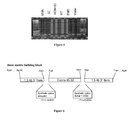
- flanking splice sites and arms of homology have been incorporated into a repair matrix ( FIG. 2 ).
- flanking splice sites synthetic branch point, polypyrimidine tract and splice acceptor site, synthetic splice donor and intronic splicing enhancer sequence from rat FGFR2 gene (DISE element)
- the synthesized targeting matrix was then inserted invertedly into an integration-deficient lentivector (IDLV) backbone to avoid aberrant and unexpected splicing of the mRNA viral genome.
- IDLV integration-deficient lentivector
- Establishment of correction synthesis and sub-cloning was confirmed by restriction enzyme digestion (data not shown).
- IDLV vectors were generated by standard transient transfection technology in 293T cells. Real-time PCR was used to accurately quantify viral genomes (data not shown). Titres of 5x10e8 were typically achieved.
- del45-52 human DMD cells were infected with 1000 of MOI of LV-endonuclease and IDLV-targeting matrix (S2) (as quantified by real-time PCR). After 72h, genomic DNA was harvested and semi-nested PCR using LongAmp polymerase was performed using second round primers to amplify from endogenous intron 44 upstream of the 1.5 kb left arm of homology to exon 45 ( Fig. 3a ) and from exon 51 to endogenous intron 44 downstream of the 1.5 kb right arm of homology ( Fig. 3b ). Highlighted products show that homologous recombination has occurred between endogenous intron 44 and the right and left arms of homology within the targeting matrix (S2), as a specific consequence of endonuclease cleavage.
- S2 IDLV-targeting matrix
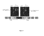
- del45-52 human DMD cells were co-transduced in 24-well plates with 1000 of MOI of LV-endonuclease and IDLV-targeting matrix S2 (as quantified using real-time PCR). After 72h, RNA was harvested and nested RT-PCR performed to examine corrected mRNA expression. Second round primers were used to amplify from exon 44 (endogenous) to exon 46 (matrix) ( Fig. 4a ), and to amplify from exon 51 (matrix) to exon 54 (endogenous) ( Fig. 4b ). The highlighted products show that exon 45 to 52 cDNA from the targeting matrix has been successfully knocked-in between endogenous exons 44 and 53 and this corrected genomic DNA was successfully transcribed to mRNA.
- del45-52 human DMD cells were transduced in 24-well plates with 1000 of MOI of LV-endonuclease (MGN) and IDLV-targeting matrix (S2). After 72h, RNA was harvested, DNase-treated and subjected to nested RT-PCR to show that splicing is efficiently occurring in the RNA between exon 52 in the targeting matrix and endogenous exon 53 ( Fig. 5 ).
- the primers used were designed to bind to exon 51 in the targeting matrix and intron 44 in the right arm of homology, so that product would be generated only if the mRNA was not effectively spliced.
- the absence of product implies that the synthetic splice donor is efficiently driving splicing of exon 52 from the cDNA block within the repair matrix to endogenous exon 53.
- the repair matrix has been designed in such a way to allow modification of the splice elements if required.
- Modifications are facilitated using the restrictions enzymes provided in the matrix.
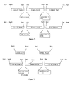
- Figure 7a illustrates the replacement of the synthetic acceptor present in the matrix of figure 6 by an endogenous (or other) splice acceptor. This can be carried out by cutting the matrix with AscI and PstI restriction enzymes, thereby removing the synthetic splice acceptor, and then ligating in the endogenous acceptor.
- Figure 7b illustrates the replacement of the synthetic splice donor present in the matrix of figure 7a by an endogenous (or other) splice donor. This can be carried out by cutting the matrix with RsrII and MluI restriction enzymes, thereby removing the synthetic splice donor, and then ligating in the endogenous donor.
- Figure 7c illustrates the modification of the length of the arms of homology.
- the matrix is cut with AscI and ApaI (or SgrAI) and a PCR amplified 3 kb sequence from delta 45-52 genomic DNA with primers with AscI and ApaI (or SgrI) restriction sites incorporated is ligated in.
- the matrix is cut at unique restriction sites with AccI or FblI or XmiI and is ligated a 2+ kb sequence of intron (i.e. 2262 bp) obtained by PCR amplification from delta 45-52 genomic DNA with primers incorporating AccI (or FblI or XmiI) and ApaI (or SgrAI).
- the matrix is cut with MluI and AgeI (or FseI) and a PCR amplified 3 kb sequence from delta 45-52 genomic DNA with primers with MluI and AgeI (or FseI) restriction sites incorporated is ligated in.
- the matrix is cut at unique restriction sites with BanIII or Bsa29I or BspXI or with BarI and is ligated a 2+ kb sequence of intron (i.e. 2098 or 2088 bp) obtained by PCR amplification from delta 45-52 genomic DNA with primers incorporating BanIII (or other) and AgeI (or FseI).
- Figure 7d illustrates the replacement of the cDNA block with a block of exons 45 to 79 and 1 kb arms of homology.
- the basic matrix of figure 7a is cut with RsrII and MluI and exon 53-79 cDNA block amplified with primers with incorporated RsrII and AvrII, and WPRE/bGHpolyA flanked with AvrII and MluI restriction sites are ligated into the cut matrix.
- Figure 7e illustrates the change of the length of the arms of homology to 3 kb within the matrix of figure 7d comprising a cDNA block encoding exons 45 to 79.
- the matrix is cut with AscI and ApaI (or SgrAI) and ligated with a PCR amplified 3 kb sequence from delta 45-52 genomic DNA with primers with AscI and ApaI (or SgrA1) restriction sites incorporated.
- the matrix is cut at a unique restriction site with AccI or FblI or XmiI and ligated with a 2+ kb of PCR amplified fragment of an intron (i.e. 2262 bp) from delta 45-52 genomic DNA obtained with primers incorporating AccI (or FblI or XmiI) and ApaI (or SgrAI).
- the 5' modified matrix is cut with MluI and AgeI (or FseI) and ligated with a PCR amplified 3 kb sequence from delta 45-52 genomic DNA with primers with MluI and AgeI (or FseI) restriction sites incorporated.
- the matrix is cut at a unique restriction site with BanIII or Bsa29I or BseCI or BspXI (position 902) or with BarI and ligated with a 2+ kb of PCR amplified fragment of an intron (i.e. 2098 or 2088 bp) from delta 45-52 genomic DNA obtained with primers incorporating BanIII (or one of the other restriction site) and AgeI (or FseI).
Abstract
The present invention relates to a gene therapy method for the treatment of Duchenne muscular dystrophy, or DMD.
Description
- The present invention relates to a gene therapy method for the treatment of Duchenne muscular dystrophy, or DMD.
- Duchenne muscular dystrophy (DMD), affecting 1 in 3500 male births is caused by nonsense or frameshift mutations in the gene encoding dystrophin, resulting in the absence of dystrophin protein (Muntoni et al., 2003, Lancet Neurol 2: 731-40). Dystrophin is required for the assembly of the dystrophin-glycoprotein complex, and provides a mechanically strong link between the cytoskeleton and the extracellular matrix (Gumerson et al., 2011, J Biomed Biotechnol 2011: 210797). Dystrophin-deficient DMD muscle is therefore mechanically destabilized and this is the primary cause of the myofibre necrosis and muscle wasting seen in this lethal disease (Matsumura et al., 1993, Neuromuscul Disord 3: 533-5). There is currently no gene repair therapy available for DMD; conventional therapies are limited to supportive care which partially alleviates signs and symptoms, but does not directly target the disease mechanism, nor reverse the phenotype. There are currently several gene therapy strategies being developed for DMD including in vivo gene therapy with adeno-associated virus (AAV) vectors, cell transplantation therapy, pharmacologic rescue of DMD nonsense mutations and exon skipping strategies to repair the DMD gene reading frame. All of these strategies have problems to overcome, including targeting different muscle groups, optimization of delivery, long-term expression of the transgene, and potential immune response.
- Gene targeting is a powerful tool for creating genetic modifications as a gene repair strategy for a variety of genetic diseases (Carroll et al., 2011, Genetics 188: 773-82; Jensen et al., 2011, J Biomed Sci 18:10; Martin et al., 2010, Clin Cancer Res 16: 5107-13). Several means are known for achieving genome surgery. For example, one can use endonucleases such as zinc finger nucleases (ZFNs), meganucleases (MNs), and transcription activator-like effector nucleases (TALENs) to cut at specific site in the DNA. The resulting double strand break (DSB) can be repaired in two major ways; by homologous recombination (HR) or non-homologous end joining (NHEJ) (Longhese et al., 2010, EMBO J 29: 2864-74). HR requires the presence of an identical or nearly identical sequence to be used as a template for repair of the break (Jensen et al., 2011, J Biol Chem 18: 10-14). For example, MNs can be used to stimulate homologous combination (HR) up to 10000-fold in cultured cells (Rouet et al., 1994, Mol Cell Biol 14: 8096-8106; Choulika et al., 1995, Mol Cell Biol 15: 1968-1973) in comparison to homologous recombination at a non-cleaved site. MNs have been used to induce HR in a variety of cell types and organisms (Paques et al., 2007, Curr Gene Ther 7: 49-66) including mammalian cells, mice, plants, Drosophila, E. coli and trypanosome Boothroyd et al., 2009, Nature 459: 278-281). Efficient gene targeting of the human XPC gene with MNs (Arnoud et al., 2007, J Mol Biol 371: 49-65), and ZFN and HR-mediated gene repair of the severe combined immune deficiency (SCID) mutation on the IL2Ry gene (Umov et al., 2005, Nature 435: 646-651; Lombardo et al., 2007, Nature Biotech 25: 1298-1306), and the homology-directed targeted gene correction of the haemophilia mutation on the F9 gene to restore haemostasis (Li et al., 2011, Nature 475: 217-221), have all been recently demonstrated. Of course, as mentioned above, besides MNs other endonucleases may be used to achieve efficient gene targeting.
- In the context of DMD, genome surgery has been limited to a proof-of-principle study in which an engineered MN restored the normal reading frame of dog micro-dystrophin sequences carrying a frameshift mutation through NHEJ repair. The MN induced micro-deletion or micro-insertion (INDELs) in the micro-dystrophin so that dystrophin expression was restored, both in myoblasts in vitro and in muscle fibres in vivo (Chapdelaine et al., 2010, Gene Ther 17: 846-858). This study reports the use of an endonuclease to restore the reading frame of a gene by NHEJ, rather than genome correction via HR.
- The present inventors demonstrate for the first time targeted cDNA knock-in and DMD gene repair in vitro in DMD patient cells through HR repair of an endonuclease-induced DSB by a targeting repair matrix. The work presented here demonstrates that endonuclease-enhanced genome correction therapy for DMD is possible.
- The invention relates to the treatment of Duchenne muscular dystrophy (DMD) by dystrophin gene repair through homologous recombination.
- More specifically, the invention relates to a nucleic acid construct comprising a first and second portions which are homologous to regions 5' and 3' of a dystrophin gene sequence of interest, further comprising a third portion positioned between the first and second portion, said third portion comprising a dystrophin correcting cDNA flanked either side by splice donor and acceptor sites or other appropriate regulatory sequences (e.g. translational terminator and polyadenylation sites). This nucleic acid is used as a repair matrix for effecting permanent genome correction at the dystrophin gene upon cutting of the genomic target site, for example by the engineered nuclease. The construct may be introduced as a DNA fragment, into a plasmid or in the genome of a viral vector. Said nucleic acid construct, plasmid or viral vector is used in a method for repairing the dystrophin gene in the genome of a target cell. Specifically, an endonuclease is used to induce a double-strand break (DSB) at a locus of interest in the dystrophin gene, which is repaired through homologous recombination at the site of DSB thanks to the nucleic acid construct of the invention, thereby resulting in the knock-in of missing exonic cDNA into the genome of a target cell.
- More specifically, we herein disclose a nucleic acid construct for the repair of the dystrophin gene in cells having a deletion of exons 45 to 52 of said gene.
- These and other embodiments, features and advantages of the disclosure will become apparent from the detailed description and the appended claims set forth herein below.
-
-
Figure 1 :Intron 44 sequence around MN DMD31 target site +/- ∼3kb on 293T, del48-50 and del45-52 genomic DNA
Residues showing polymorphism are highlighted, and the MN-DMD31 target site is highlighted gray (positions 3396 - 3419). It should be noted that although a number of extra primers were specifically designed and used for sequencing, the number of residues in the poly(T) sequence (highlighted in dark gray) just downstream from the target site could not be resolved. The arm of homology was therefore designed to be downstream of this poly(T) sequence. -
Figure 2 : Schematic RE map repair matrices S1 (a) and S2 (b) for targeting exon 45-52 deleted genotypes
The exon 45-52 cDNA block with flanking artificial splice sites (synthetic branch point, polypyrimidine tract and splice acceptor site, synthetic splice donor and intronic splicing enhancer sequence from rat FGFR2 gene (DISE element)) and arms of homology have been incorporated into repair matrices as shown. -
Figure 3 : Homologous recombination in LV-endonuclease (MN) + IDLV-matrix (S2) transduced cells at genomic DNA level. -
Figure 4 : Restoration of full-length dystrophin mRNA in human del45-52 DMD cells by knock-in of exons 45 to 52 by homologous recombination of the targeting matrix (S2) upon endonuclease (MN) cutting. -
Figure 5 : Confirmation that knocked-in exons are correctly spliced to neighbouring endogenous exons in RNA. -
Figure 6 : diagram of a basic matrix building block showing the different elements of the construct and restriction sites. -
Figure 7a : diagram illustrating the replacement of the synthetic acceptor present in the matrix offigure 6 by an endogenous (or other) splice acceptor. -
Figure 7b : diagram illustrating the replacement of the synthetic splice donor present in the matrix offigure 7a by an endogenous (or other) splice donor. -
Figure 7c : diagram illustrating the modification of the length of the arms of homology. -
Figure 7d : diagram illustrating the replacement of the cDNA block with a block of exons 45 to 79 and 1 kb arms of homology. -
Figure 7e : diagram illustrating the change of the length of the arms of homology to 3 kb within the matrix offigure 7d comprising a cDNA block encoding exons 45 to 79. - The present invention provides a nucleic acid construct comprising a first and second portions which are homologous to regions 5' and 3' of a dystrophin gene sequence of interest, further comprising a third portion positioned between the first and second portion, said third portion comprising a dystrophin correcting cDNA flanked either side by splice donor and acceptor sites or other appropriate elements (e.g. translational terminator and polyadenylation sites).
- The practice of the present invention will employ, unless otherwise indicated, conventional methods of virology, microbiology, molecular biology and recombinant DNA techniques within the knowledge of those skilled in the art. Such techniques are explained fully in the literature. See, e.g., Sambrook, et al. Molecular Cloning: A Laboratory Manual (Current Edition); DNA Cloning: A Practical Approach, vol. I & II (D. Glover, ed.); Oligonucleotide Synthesis (N. Gait, ed., Current Edition); Nucleic Acid Hybridization (B. Hames & S. Higgins, eds., Current Edition); Transcription and Translation (B. Hames & S. Higgins, eds., Current Edition); CRC Handbook of Parvoviruses, vol. I & II (P. Tijssen, ed.); Fundamental Virology, 2nd Edition, vol. I & II (B. N. Fields and D. M. Knipe, eds.)
The various compositions and methods of the invention are described in detail below. Although particular compositions and methods are exemplified herein, it is understood that any of a number of alternative compositions and methods are applicable and suitable for use in practicing the invention. - Unless otherwise indicated, all terms used herein have the same meaning as they would to one skilled in the art, and the practice of the present invention will employ conventional techniques of microbiology and recombinant DNA technology, which are within the knowledge of those of skill in the art.
- The term "nucleic acid construct" refers generally to a nucleic acid which is comprised of segments joined together using recombinant DNA technology.
- The term "recombinant" as used herein refers to nucleic acids, vectors, polypeptides, or proteins that have been generated using DNA recombination (cloning) methods and are distinguishable from native or wild-type nucleic acids, vectors, polypeptides, or proteins.
- The term "subject" as used herein includes, but is not limited to, humans, nonhuman primates such as chimpanzees and other apes and monkey species; farm animals such as cattle, sheep, pigs, goats and horses; domestic mammals such as dogs and cats; laboratory animals including rodents such as mice, rats and guinea pigs, and the like. The term does not denote a particular age or sex. Thus, adult and newborn subjects, as well as fetuses, whether male or female, are intended to be covered.
- The term "therapeutically effective amount" refers to an amount effective, at dosages and for periods of time necessary, to achieve the desired therapeutic result. A therapeutically effective amount of the recombinant nucleic acid of viral vector may vary according to factors such as the disease state, age, sex, and weight of the individual and the ability of the vector to elicit a desired response in the individual. A therapeutically effective amount is also one in which any toxic or detrimental effect is outweighed by the therapeutically beneficial effects.
- As used herein, "ex vivo administration" refers to a process where primary cells or an entire organ are harvested from a subject, a nucleic acid or viral vector is delivered into the cells, and the cells are readministered to the same or a different subject.
- In the present description, any concentration range, percentage range, ratio range, or integer range is to be understood to include the value of any integer within the recited range and, when appropriate, fractions thereof (such as one tenth and one hundredth of an integer), unless otherwise indicated. Also, any number range recited herein relating to any physical feature, such as polymer subunits, size or thickness, are to be understood to include any integer within the recited range, unless otherwise indicated.
- The term "about" or "approximately" means within a statistically meaningful range of a value. Such a range can be within an order of magnitude, preferably within 50%, more preferably within 20%, more preferably still within 10%, and even more preferably within 5% of a given value or range. The allowable variation encompassed by the term "about" or "approximately" depends on the particular system under study, and can be readily appreciated by one of ordinary skill in the art.
- The nucleic acid construct of the invention is provided as a repair matrix for the correction of the dystrophin gene in the genome of a target cell of a subject in need thereof.
- This nucleic acid construct comprises three portions. The first and second portions, which are also collectively referred to as "arms of homology" are homologous to regions 5' and 3' of a dystrophin gene sequence of interest and a third portion flanked either side by splice donor and acceptor sites or other appropriate elements (e.g. translational terminator and polyadenylation sites), positioned between the first and second portions, comprising a dystrophin correcting cDNA.
- The first and second portions are thus homologous to a region of interest in the dystrophin gene. These regions of interest are selected among known mutation hotspots (Tuffery-Giraud et al., 2009, Hum Mutat 30: 934-945). More specifically, one may cite mutational hotspots including and spanning intron 38 to 52 and intron 2-9 of the dystrophin gene. In a particular embodiment, the nucleic acid construct of the present invention comprises nucleic acid sequences homologous to part of
intron 44. More than 25 % dystrophin mutations arise from this specific intron. - The first and second portions are more particularly homologous to sequences found either side of a double-strand break-inducing endonuclease target site in the selected dystrophin gene sequence of interest. By "homologous" is intended a sequence with enough identity to another one to lead to a homologous recombination between sequences, more particularly having at least 95 % identity, preferably 97 % identity and more preferably 99 %. "Identity" refers to sequence identity between two nucleic acid molecules. Identity can be determined by comparing a position in each sequence which may be aligned for purposes of comparison. When a position in the compared sequences is occupied by the same base, then the molecules are identical at that position. A degree of similarity or identity between nucleic acid sequences is a function of the number of identical or matching nucleotides at positions shared by the nucleic acid sequences. Various alignment algorithms and/or programs may be used to calculate the identity between two sequences, including FASTA, or BLAST which are available as a part of the GCG sequence analysis package (University of Wisconsin, Madison, Wis.), and can be used with, e.g., default settings.
- In a particular embodiment, the sequence of interest is in
intron 44, and the target site is located withinintron 44, 2.233 kb downstream fromexon 44, at position >chromosome:GRCh37:X:32364354:32364377, and has the sequence AATGTCTGATGTTCAATGTGTTGA (SEQ ID NO:1). In a specific variant, the first and second portions are identical to the sequences present either side (e.g. within 100bp) of the endonuclease target site in the genomic sequence of interest. - The first and second portions are independently from each other 0.5 kb to 8 kb in length, more particularly 1 kb to 3 kb in length. For example, the arms of homology may each be 1 kb, 1.5 kb or 3 kb in length. Illustrative left arms of homology are shown in SEQ ID NOs: 2 and 3, and illustrative right arms of homology are shown in SEQ ID NOs: 4 and 5.
- The third portion is a cDNA block, including dystrophin exons to be introduced in the genome of the target cells. Representative third portions include portions comprising exons 45-52 of the dystrophin gene (see for example the sequence shown in SEQ ID NO:10), or even the full 45-79 exons (see for example the sequence shown in SEQ ID NO:11), or any therapeutically effective intermediate selection of exons.
- In addition to the first, second and third portions, the nucleic acid construct of the present invention comprises regulatory sequences flanking said third portion. In a particular embodiment, the regulatory sequences are a splice acceptor (e.g. the synthetic splice acceptor of SEQ ID NO:6) and a splice donor sequences located respectively 5' and 3' of the third portion. As a result, the construct of the invention may comprise, in the 5' to 3' direction, the first portion, a splice acceptor site, the third portion, a splice donor site and the second portion. The splice sites may either be endogenous (i.e. naturally-occurring in the wild-type dystrophin gene) or synthetic (i.e. non-natural sequences designed in silico; see for example Shapiro et al., Nucl Acids Res, 15: 7155-7174). In a particular embodiment, a synthetic splice donor site is provided together with an intronic splicing enhancer sequence, for example the intronic splicing enhancer sequence from rat FGFR2 gene (also herein referred to as DISE element). Sequence of such splice donor site with a DISE element is shown in SEQ ID NO:7. In an alternative variant, the third portion of the nucleic acid construct is flanked by other regulatory sequences. For example, the third portion may be flanked in 5' by a splice acceptor site and in 3' by a WPRE sequence (Woodchuck Post-transcriptional Regulatory Element; included to enhance expression of the cDNA block - see for example the sequence shown in SEQ ID NO:9) fused to a poly(A) sequence when said third portion is the full 45-79 exons, for example the bovine growth hormone poly(A) sequence (see for example SEQ ID NO:8).
- According to a specific embodiment, the nucleic acid construct of the invention comprises a cDNA block comprising exons 45 to 52 of the dystrophin gene (third portion), flanked arms of homology to intron 44 (first and second portions), synthetic splice donor and acceptor and an intronic splicing enhancer sequence (e.g. from rat FGFR2 gene). Such nucleic acid constructs are shown in SEQ ID NO:12 and 13.
- The nucleic acid construct of the invention may further comprise various unique restriction enzyme (RE) sites incorporated between each element of said construct to allow its modification in a stepwise manner. As an illustrative, non-limiting example, when the third portion contains exons 45-52 of the dystrophin gene, a PstI restriction site may be silently introduced at the start of exon 45 by a single point mutation of the second codon of said exon (CTC replaced by a CTG thereby providing a CTGCAG PstI restriction site) and a RsrII restriction site may be introduced at the end of
exon 52 by a single point mutation of the penultimate codon of said exon (GAT replaced by a GAC thereby providing a CGACCG RsrII restriction site). Other unique restriction sites may be provided on the construct.Figures 2 ,6 and7a-e illustrate a number of possible restriction sites that may be introduced in a nucleic acid construct of the invention. The restrictions sites incorporated into the nucleic acid construct of the invention may allow various modifications such as: (i) introduction and modification of the splice donor and acceptor; (ii) modification of the length of the arms of homology; (iii) replacing the cDNA block of the third portion (for example introducing the full 45-79 exons when the arms of homology are specific of intron 44); (iv) introduction of a transcriptional terminator in 3' of the full 45-79 exons; (v) introduction of positive and/or negative selection markers if required. - The nucleic acid construct of the invention may be introduced in a plasmid vector or a viral vector. Accordingly, the invention also relates to a recombinant plasmid comprising the nucleic acid construct described above, and to a recombinant viral vector comprising in its genome the nucleic acid construct of the invention. Plasmid and viral vector design is known to those skilled in the art. A viral vector of the invention may be any viral vector efficient for transducing a cell involved in a dystrophin-related disease (a target cell). In particular, the target cell may be a muscle cell such as a myoblast, mesoangioblast, pericyte, or CD133+ or CD56+ progenitor cells. Illustrative, non-limiting, viral vectors useful for the invention include adenovirus, adeno-associated virus and lentivirus vectors. For example, as provided in the examples, the nucleic acid construct may be introduced in an integration-deficient lentiviral vector, in particular pseudotyped with the the VSV-G envelope glycoprotein. The construct may in particular be inserted invertedly in a lentiviral backbone such as the pRRLsin.PPT plasmid. Lentiviral vectors are produced according to methods well known in the art. For example, plasmids carrying all necessary elements of a lentivirus are transiently transfected in a mammalian cell (e.g. HEK293T cells), then after a period of 16 h to 72 h, in particular 40 h, the cell medium is collected, centrifuged and lentivirus vectors are purified from the supernatant. The person skilled in the art will refer to the examples provided below, and to his general knowledge in this field as represented in particular by Sakumo et al., 2012 Biochem J 443: 603-618.
- The target cells the genome of which is to be corrected by the nucleic acid construct of the invention can be derived from a human, and other mammals such as primates, horse, sheep, goat, pig, dog, rat, and mouse. Examples of target cells to which the construct can be delivered include, but are not limited to, muscle cells, central nervous system cells and peripheral nervous system cells. Examples of tissues to which the construct can be delivered include muscle, heart, eye and brain.
- For in vitro, ex vivo or in vivo delivery to cells, tissues or organs of a subject, the nucleic acid construct, recombinant plasmid or recombinant viral vector of the invention may be incorporated into compositions, in particular pharmaceutical compositions suitable for administration to a subject. Typically, the pharmaceutical composition comprises a pharmaceutically acceptable carrier and a nucleic acid construct, recombinant plasmid or recombinant viral vector of the invention. The term "pharmaceutically acceptable" as used herein refers to molecular entities and compositions that are physiologically tolerable and do not typically produce toxicity or an allergic or similar untoward reaction, such as gastric upset, dizziness and the like, when administered to a human. Preferably, as used herein, the term "pharmaceutically acceptable" means approved by a regulatory agency of the Federal or a state government or listed in the U.S. or European Pharmacopeia, or other generally recognized pharmacopeia for use in animals, and more particularly in humans. Also, as used herein, "pharmaceutically acceptable carrier" includes any and all solvents, dispersion media, coatings, antibacterial and antifungal agents, isotonic and absorption delaying agents, and the like that are physiologically compatible. Examples of pharmaceutically acceptable carriers include one or more of water, saline, phosphate buffered saline, dextrose, glycerol, ethanol and the like, as well as combinations thereof. Pharmaceutical compositions can be delivered as, for example, ageratum, sprays, oral suspensions, suppositories, eye drops, and injectable suspensions.
- The pharmaceutical composition may be suitable for topical, systemic, intra-amniotic, intrathecal, intracranial, intraarterial, intravenous, intralymphatic, intraperitoneal, subcutaneous, tracheal, intra-tissue (e.g., intramuscular, intracardiac, intrahepatic, intrarenal, intracerebral), intrathecal, intravesical, conjunctival (e.g., extra-orbital, intraorbital, retroorbital, intraretinal), and mucosal (e.g., oral, rectal, nasal) administration. Passive tissue transduction via high pressure intravenous or intraarterial infusion, as well as intracellular injection, such as intranuclear microinjection or intracytoplasmic injection, are also contemplated.
- Pharmaceutical compositions for therapeutic purposes typically must be sterile and stable under the conditions of manufacture and storage. The composition can be formulated as a solution, microemulsion, dispersion, liposomes, or other ordered structure suitable to high product concentration. Sterile injectable solutions can be prepared by incorporating the construct, recombinant plasmid or recombinant viral vector in the required amount in an appropriate buffer with one or a combination of ingredients enumerated above, as required, followed by filtered sterilization.
- For in vivo administration, broad distribution can be achieved in muscle by, for example, intravenous, intra-arterial injection or hydrodynamic locoregional perfusion. Depending on dosage and mode of delivery, 20-95% of muscle fibres in a given target muscle can be transduced. Broad distribution of the nucleic acid construct, plasmid or viral vector of the invention can also be achieved in the CNS or PNS by injection into the cerebrospinal fluid, e.g., by lumbar puncture (e.g., Kapadia et al., 1996). Alternatively, precise delivery of the vector into specific sites of the brain to target a neural cell, can be conducted using stereotactic microinjection techniques (Snyder-Keller et al., 2010). Particularly preferred delivery methods are those that deliver the nucleic acid construct, plasmid or viral vector to specific regions of the brain or muscle that require correction of the dystrophin gene. Direct injection, whether subcutaneous, intracranial, intramuscular, or intradermal, can take place using standard needle and syringe methodologies, or by needle-free technologies.
- Further methods for delivery of nucleic acid molecules, are described, for example, in Boado et al., J. Pharm. Sci. 87:1308-1315 (1998); Tyler et al., FEBS Lett. 421:280-284 (1999); Pardridge et al., Proc. Nat'l Acad. Sci. USA 92:5592-5596 (1995); Boado, Adv. Drug Delivery Rev. 15:73-107 (1995); Aldrian-Herrada et al., Nucleic Acids Res. 26:4910-4916 (1998); Tyler et al., Proc. Nat'l Acad. Sci.USA 96:7053-7058 (1999); Akhtar et al., Trends Cell Bio. 2:139 (1992); "Delivery Strategies for Antisense Oligonucleotide Therapeutics," (ed. Akhtar, 1995); Maurer et al., Mol. Membr.Biol. 16:129-140 (1999); Hofland and Huang, Handb. Exp. Pharmacol 137:165-192 (1999); and Lee et al., ACS Symp. Ser. 752:184-192 (2000).
- Appropriate doses will depend on the particular cell type, tissue, organ, or subject being treated. When administered to a subject, dose will depend on the particular mammal being treated (e.g., human or nonhuman primate or other mammal), age and general condition of the subject to be treated, the severity of the condition being treated, the particular therapeutic polypeptide, protein, or oligonucleotide in question, its mode of administration, among other factors. An appropriate effective amount can be readily determined by one of skill in the art.
- A "therapeutically effective dose" will fall in a relatively broad range that can be determined through clinical trials and will depend on the particular application (neural cells will require very small amounts, while systemic injection would require large amounts).
- Treatment may involve administration of a single dose or multiple doses, depending in particular on the severity of the disease.
- The nucleic acid construct of the invention is particularly useful for restoring dystrophin expression in a target cell. The invention thus relates to a method of treatment of a disease caused by a mutation in the dystrophin gene, comprising administering to a subject in need thereof a nucleic acid construct, recombinant plasmid or recombinant vector of the invention. According to another embodiment, the invention relates to a method for restoring full-length dystrophin expression in a subject in need thereof. The invention also relates to a method for correcting the expression of dystrophin in a subject in need thereof. The invention further relates to a method for altering the genomic DNA sequence of the dystrophin gene in a target cell. Moreover, the invention relates to a method for repairing the dystrophin gene in the genome of a target cell. The invention also relates to a method for inserting a cDNA block of missing exons in the dystrophin gene in the genome of a target cell. Representative diseases treatable with the invention include Becker and Duchenne muscular dystrophies. In a preferred embodiment, the invention relates to a method of treatment of Duchenne muscular dystrophy, comprising administering to a subject in need thereof a nucleic acid construct, recombinant plasmid or recombinant vector of the invention.
- In a particular embodiment, the subject presents a deletion of dystrophin exons 45 to 52 in its genome. Indeed, the below examples show correction of deletion of exons 45-52 in patient cells by knock-in of cDNA encoding exons 45-52, following DNA cleavage by an endonuclease. The nucleic acid construct of the invention thus may be used to correct the dystrophin gene in up to a quarter of Duchenne muscular dystrophy patients by cDNA knock-in through homologous recombination repair of endonuclease-driven double strand break (DSB) in
intron 44 of the DMD gene. - The methods of the present invention are implemented as follows:
- cleavage of genomic dystrophin gene is induced at a sequence of interest; and
- the nucleic acid construct as defined above is provided as a template for homologous recombination at the cleavage site.
- The cleavage in the dystrophin gene may be provided by different means. In a particular embodiment, said cleavage is induced in the sequence of interest by the use of a nickase or a double-strand break-inducing endonuclease. Alternatively, small molecules that damage DNA or create altered structures that may induce homologous recombination and can be targeted to specific sequences through oligonucleotide base-pairing have been reported (for instance see Recombination induced by triple-helix-targeted DNA damage in mammalian cells, Faruqi AF, Seidman MM, Segal DJ, Carroll D, Glazer PM., Mol Cell Biol. 1996 Dec;16(12):6820-8; Correction of a splice-site mutation in the beta-globin gene stimulated by triplex-forming peptide nucleic acids; Chin JY, Kuan JY, Lonkar PS, Krause DS, Seidman MM, Peterson KR, Nielsen PE, Kole R, Glazer PM.; Proc Natl Acad Sci U S A. 2008 Sep 9;105(36):13514-9. Epub 2008 Aug 29. Preferably, cleavage of the sequence of interest is obtained using a double-strand break-inducing endonuclease. Nickases are generally less efficient than double-strand break-inducing endonucleases but may be safer in some instances (see Metzger et al., 2011, Nucl Acids Res 39(3): 326-35.)
- In a preferred embodiment, the methods of the present invention are implemented as follows:
- a double-strand break is induced in the genome of the target cell with an endonuclease specific for the dystrophin gene sequence of interest as defined above; and
- the nucleic acid construct as defined above is provided as a template for homologous recombination at the double-strand break site.
- As mentioned above, in a particular embodiment, the double-strand break-inducing endonuclease is specific of an intron of dystrophin, more particularly of
intron 44. - Several means are known for achieving genome surgery. For example, one can use endonucleases such as zinc finger nucleases (ZFNs), meganucleases (MNs), and transcription activator-like effector nucleases (TALENs) to cut at specific site in the DNA. Endonucleases useful for the invention and methods for their engineering are well-known in the art.
- In any of the methods described herein, the nuclease can be, for example, one or more zinc finger nucleases, one or more homing endonucleases (meganucleases) and/or one or more TAL-effector domain nucleases ("TALEN"). The nucleases (e.g., ZFN, Homing meganuclease and/or TALEN) as described herein may bind to and/or cleave the region of interest in a coding or non-coding region within or adjacent to the gene, such as, for example, a leader sequence, trailer sequence or intron, or within a non-transcribed region, either upstream or downstream of the coding region.
- Zinc-finger nucleases (ZFNs) are artificial restriction enzymes generated by fusing a zinc finger DNA-binding domain to a DNA-cleavage (the Fokl nuclease) domain. The zinc finger domains can be engineered to target desired DNA sequences, which then enables the nuclease domain to cleave unique sequences within a complex genome. By taking advantage of the endogenous DNA double-strand break repair machinery, ZFN reagents can be used to precisely alter the genomes of higher organisms. The most straightforward method to generate new zinc-finger arrays is to combine smaller zinc-finger "modules" of known specificity. The most common modular assembly process involves combining three separate zinc fingers that can each recognize a 3 basepair DNA sequence to generate a 3-finger array that can recognize a 9 base-pair target site. Other procedures can utilize either 1 -finger or 2-finger modules to generate zinc-finger arrays with six or more individual zinc fingers. Methods of engineering zinc finger domains are known, and the ZFNs are available from a commercial supplier or can be developed through publically available reagents.
- Several groups are also developing other types of engineered nucleases including engineered homing endonucleases, such as meganucleases (Grizot et al., 2009, Nucl. Acids Res., 37:5405-5419; Gao et al, 2010 Plant J., 61: 176-187; Smith et al., 2006, Nucl. Acids Res. 34: e149; Grizot et al., 2011, Nucl. Acids Res. 39: 6124-6136; Daboussi et al., 2012, Nucl. Acids Res. epub March 29, 2012) and nucleases based on engineered transcription activator like (TAL) effectors (Christian et al., 2010, Genetics, 186:757-761; Li et al, 2010, Nucl. Acids Res., 39:359-372). TAL effector nucleases (TALENs) are particularly interesting because TAL effectors appear to be very simple to engineer (Moscou et al, 2009, Science 326: 1501; Boch et al, 2009, Science 326: 1509-1512).
- In a particular embodiment, a meganuclease is used, derived from I-CreI and engineered as described previously (Smith et al., 2006, Nucl. Acids Res. 34: e149; Grizot et al., 2011, Nucl. Acids Res. 39: 6124-6136; Daboussi et al., 2012, Nucl. Acids Res. epub March 29, 2012). In particular, meganucleases have been designed and developed to target
intron 44 within the DMD gene upstream of a mutation hotspot. These meganucleases have previously been used as reported in Rousseau et al., 2011, J Gene Med, 13: 522-537 (named MGN3631 and MGN3633 in this article). Synthesis and validation of the meganucleases is as described in Daboussi et al., 2012 (previously cited). Results from next generation sequencing suggest that MGN3631 and MGN3633 cleavage point within its target site is between residues 9 and 10 of the following sequence: AATGTCTGATGTTCAATGTGTTGA (SEQ ID NO:1). The meganuclease is to be used along with a nucleic acid construct intended to repair the mutations resulting from mutations in this hotspot, for example for repairing deletion of exons 45-52. - The endonuclease implemented in the invention may be introduced in the target cell by methods known in the art. A gene coding for a nuclease may for example be delivered by viral or non-viral methods. In addition, means for delivering a nuclease protein are also known. As an illustrative, non-limiting, example one can cite the use of viral vectors carrying an endonuclease expression cassette. This expression cassette contains all the elements necessary for expression of the endonuclease gene, such as a promoter, termination sequences and enhancer sequences. The promoter may be ubiquitous or tissue-specific. In addition, the endonuclease expression cassette may be introduced in an integration-deficient or integration-competent virus, such as a lentivirus. Integration-deficient lentiviruses are preferably used. Methods for the production of such vectors are known in the art (e.g. in Jacome et al., 2009, Mol Ther 17: 1083-1092).
- The nucleases, in particular DSB-inducing endonucleases, described above can be used to rewrite the sequence of an allele by invoking the homologous recombination machinery to repair the break, in particular double-strand break, using the supplied nucleic acid construct as a template. The homologous recombination machinery searches for homology between the damaged chromosome and the extra-chromosomal fragment (here, a cDNA block of dystrophin) and copies the sequence of the fragment between the two broken ends of the chromosome, regardless of whether the fragment contains the original sequence.
- The above disclosure generally describes the present disclosure, which is further described by the following examples. These specific examples are provided solely for purposes of illustration, and are not intended to limit the scope of this disclosure. Although specific targets, terms, and values have been employed herein, such targets, terms, and values will likewise be understood as exemplary and non-limiting to the scope of this disclosure.
- Immortalised human DMD myoblasts, carrying a deletion of exons 45-52, have kindly been made available to us through collaboration with the AFM-IdM (Paris). These cells, together with primary human skeletal muscle cells (hSkMCs) (TCS Cellworks, Buckingham, UK), 293T cells, and a second human DMD cell line carrying a deletion of exons 48-50 (from AFM-IdM, Paris) were cultured according to published protocols (Popplewell et al., 2011, Methods Mol Biol 709:153-78; Popplewell et al., 2010, Neuromuscul Disord 20:102-10), and used in experiments as indicated.
- A meganuclease (MN-DMD31), derived from I-CreI and engineered as described previously (Smith et al., 2006, Nucl. Acids Res. 34: e149; Grizot et al., 2011, Nucl. Acids Res. 39: 6124-6136; Rousseau et al., 2011, J Gene Med, 13: 522-537; Daboussi et al., 2012, Nucl. Acids Res. epub March 29, 2012), has been designed and developed to target
different intron 44 within the DMD gene, upstream of a mutation hotspot. The MN-DMD31 construct was subcloned into a MluI/SwaI-digested lentiviral transfer plasmids (pRRLscSeGFP- CMV-WPRE) containing two expression cassettes. The first cassette encodes eGFP under the control of an internal spleen focus forming virus (SFFV) promoter, while the second contains a cytomegalovirus promoter and MN-DMD31. It also contains an SV40 polyadenylation signal and woodchuck hepatitis virus (WHP) posttranscriptional regulatory element (WPRE) sequence, flanking the late reverse transcriptase. - The targeting matrix was designed to contain a cDNA block encoding exons 45 to 52, flanked arms of homology to
intron 44, synthetic splicing donor and acceptor, and intronic splicing enhancer sequence from rat FGFR2 gene (DICE element). The different elements within the targeting matrix were designed to be flanked by unique restriction enzyme sites to allow the modification of the matrix as required. The flanking arms were completely homologous tointron 44 either side of the DSB produced by MN-DMD31 cleavage. The degree of polymorphism in the 3kb sequence either side of the DSB was elucidated by PCR amplification of harvested genomic DNA from the four cell types (DMD del 45-52, DMD del 48-50, normal hSkMCs, 293T) with overlapping primers. Genomic DNA was harvested using DNeasy blood and tissue kit (Qiagen, Crawley, UK), and PCR performed using 2 x PCR Mastermix (GeneSys, Camberley, UK). The PCR products were sequenced (GATC- Biotech, Switzerland), and the sequences aligned to show position of polymorphic residues using VectorNTI software (Invitrogen, Paisley, UK). A targeting matrix was synthesized (by GeneART, Germany) with arms homologous to DMD del 45-52. The matrix (S2) had 1.5kb arms of homology. The synthesized targeting matrix was then inserted invertedly into a lentivector backbone (pRRLsinPPT-ISce-IT). Establishment of correction synthesis and sub-cloning was confirmed by restriction enzyme digests used according to manufacturers' instructions (New England Biolabs, Herts., UK). - For the expression of targeting matrix, integration-deficient lentiviral vectors, pseudotyped with the VSV-G envelope glycoprotein were used, while integration-competent lentiviral vectors were used for the expression of the meganuclease; these were produced as previously described (Jacome et al., 2009, Mol Ther 17:1083-1092). Briefly, lentiviral vectors were generated by transient transfection of HEK293T cells with the pMD2.VSV-G, pRSV.REV and pMDLg/pRREintD64V packaging plasmids and the transfer plasmid. At 24hrs after transfection, the harvested HEK293T cell medium was centrifuged at 690 x g for 10 min at room temperature and then filtered through a 0.22 µm filter (Nalgene, Rochester, NY, USA) to remove cell debris. The filtered medium was then harvested and transferred to high speed polyallomer centrifuge tubes (Beckman, Brea, CA, USA) and centrifuged at 50, 000 x g in a SW32Ti rotor (Beckman) for 2h at 4 °C. The vector was then resuspended in Dulbecco's modified Eagle's medium (Invitrogen, Paisley, UK), centrifuged at 1400 x g for 10 min and incubated with 5U ml-1 DNaseI (Promega, Madison, WI, USA) and 10 mM MgCl2 (Sigma, UK) for 30 min. The vector was then aliquoted and stored at -80 °C. For viral titration, HeLa cells were transduced with serial dilutions of vector stock in the presence of 8 µg/ml polybrene. eGFP expression controlled by SFFV promoter in LV-MN was evaluated by FACS analysis. eGFP+ cells were scored using FACS analysis 72hr after infection. Real-time PCR titration of total vector DNA by late reverse transcript amplicon quantification was also used. Briefly, DNA extracted from cells (DNeasy, Qiagen, Crawley, UK) 24hr after transduction was subjected to PCR amplification. The final number of LV vector molecules in the transduced cells was determined by quantitative-PCR (q-PCR), using Taqman detection of PCR products in real time with the MyiQ single-color detection system (Bio-Rad, Hercules, CA). qPCR values were normalised by quantifying copies of the gene encoding actin in the DNA extracts using SYBR green detection system and correcting for Hela cell aneuploidy.
- Total protein extraction from del45-52
myoblast cell lines 2 days and 5 days after transduction of ICLV expressing MN-DMD31 (3631 or 3633) was performed with RIPA (150 mM sodium chloride, 1% NP-40, 0.5% sodium deoxycholate, 0.1% SDS, 50 mM Tris pH 8.0) buffer. 10 µg of total protein extract was electrophoretically separated on 3-8% Tris-acetate gels (Invitrogen, Paisley, UK), and then transferred onto nitrocellulose (Invitrogen, Paisley, UK). The MN expression was revealed using a specific I-Cre-I mouse monoclonal antibody. Alpha-tubulin antibody (ab4074) (Abcam, Cambridge, UK) was used as a loading control. - To evaluate the efficiency of MN-DMD31 (two variants) to cut DNA at its target site, human DMD del 45-52 cell lines were transduced with ICLV-MN at MOIs of 1 and 0.5. Five days after transduction, genomic DNA was purified using DNeasy blood and tissue kit (Qiagen, Crawley, UK), a ∼400bp product amplified around the MN target site by PCR using 2 x PCR Mastermix (GeneSys, Camberley, UK). The purified products were deep gene sequenced (by GATC-Biotech, Switzerland) to assess the mutation rate as a result of non-homologous end joining (NHEJ) repair of the DSB induced by MN-DMD31 at its target site in
intron 44 of the DMD gene, compared to non- treated controls. Standard PCR protocols within the knowledge of those skilled in the art were used. - Detection of corrected dystrophin genomic DNA in human DMD del 45-52 cell lines Human DMD del 45-52 cells were transduced in 24-well plates with 1000 MOI of ICLV-MN and IDLV-targeting matrix, as assessed by qPCR. After 72h, genomic DNA was harvested using DNeasy blood and tissue kit (Qiagen, Crawley, UK). Semi-nested PCR using LongAmp polymerase (New England Biolabs, Herts., UK) was performed to show homologous recombination had occurred between
intron 44 in targeting matrix andendogenous intron 44. The products were separated on 1% agarose gels in Tris-borate/EDTA buffer and HyperLadder I (Bioline, London, UK) was used as marker. Standard PCR protocols within the knowledge of those skilled in the art were used. - Human DMD del 45-52 cells were transduced in 24-well plates with 1000 MOI of ICLV-MN and IDLV-targeting matrix, as assessed by qPCR. After 72h, RNA was harvested using QIAshredder and RNeasy extraction kit (Qiagen, Crawley, UK) and nested RT-PCR performed to examine corrected mRNA expression using GeneScript RT-PCR system kit (GeneSys, Camberley, UK) for the first round, and 2 x PCR Mastermix (GeneSys, Camberley, UK) for the second round. The products were separated on 1.2% agarose gel in Tris-borate/EDTA buffer and Hyper Ladder IV (Bioline, London, UK) was used as marker.
- Primers used to assess homologous recombination between endogenous DNA and repair matrix cDNA:
- Left hand side:
- First round: DMD31-for3 (SEQ ID NO:14) and KI h45 rev (SEQ ID NO:15)
- Second round: DMD31-for4 (SEQ ID NO:16) and KI h45 rev
- Right hand side:
- First round: h51f(SEQ ID NO:17) and DMD31-Rev12 (SEQ ID NO:18)
- Second round:h51f and DMD31-Rev11 (SEQ ID NO:19)
- Primers used to assess splicing of repair matrix cDNA into endogenous mRNA:
- Left hand side:
- First round: h43f (SEQ ID NO:20) and h48r (SEQ ID NO:21)
- Second round: h44f (SEQ ID NO:22) and h46r (SEQ ID NO:23)
- Right hand side:
- First round: h50f(SEQ ID NO:24) and h55r (SEQ ID NO:25)
- Second round: h51f and h54r (SEQ ID NO:26)
- The aim of the endonuclease-mediated gene correction is to insert a cDNA block of missing exons with flanking splice sites, or other regulatory sequence elements, into the endonuclease-induced DSB within the intron harbouring the deletion junction. An endonuclease has been designed and developed to target
intron 44 within the DMD gene, upstream of a mutation hotspot, and packaged into a lentivirus vector to allow contemporaneous and transient endonuclease expression. Arms of homology are required to drive homologous recombination between genomic DNA and repair matrix at endonuclease-induced DSB site, and subsequent cDNA knock-in. To establish complete homology and elucidate positions of any polymorphic residues, sequencing of the intron 44 (6 kb) surrounding the endonuclease target site was performed on PCR amplicons produced using overlapping primers on genomic DNA from 293T cells, del 45-52 DMD cells and del 48-50 DMD cells. Alignment of the 6861 bp sequences from the three different genomic DNAs is shown inFig. 1 . - The cDNA block, flanking splice sites and arms of homology have been incorporated into a repair matrix (
Fig. 2 ). A basic targeting construct, with 1.5kb arms of homology (S2), with cDNA encoding the block of exons 45 to 52, which are missing in del45-52 human DMD cells, was synthesized commercially, and artificial flanking splice sites (synthetic branch point, polypyrimidine tract and splice acceptor site, synthetic splice donor and intronic splicing enhancer sequence from rat FGFR2 gene (DISE element) incorporated. The synthesized targeting matrix was then inserted invertedly into an integration-deficient lentivector (IDLV) backbone to avoid aberrant and unexpected splicing of the mRNA viral genome. Establishment of correction synthesis and sub-cloning was confirmed by restriction enzyme digestion (data not shown). IDLV vectors were generated by standard transient transfection technology in 293T cells. Real-time PCR was used to accurately quantify viral genomes (data not shown). Titres of 5x10e8 were typically achieved. - To evaluate the potential of endonuclease and targeting matrix to induce gene repair by homologous recombination and to elucidate repair matrix function and efficacy, del45-52 human DMD cells were infected with 1000 of MOI of LV-endonuclease and IDLV-targeting matrix (S2) (as quantified by real-time PCR). After 72h, genomic DNA was harvested and semi-nested PCR using LongAmp polymerase was performed using second round primers to amplify from
endogenous intron 44 upstream of the 1.5 kb left arm of homology to exon 45 (Fig. 3a ) and fromexon 51 toendogenous intron 44 downstream of the 1.5 kb right arm of homology (Fig. 3b ). Highlighted products show that homologous recombination has occurred betweenendogenous intron 44 and the right and left arms of homology within the targeting matrix (S2), as a specific consequence of endonuclease cleavage. - To evaluate the potential of endonuclease and targeting matrix to induce gene repair by homologous recombination and knock-in of exons 45 to 52, del45-52 human DMD cells were co-transduced in 24-well plates with 1000 of MOI of LV-endonuclease and IDLV-targeting matrix S2 (as quantified using real-time PCR). After 72h, RNA was harvested and nested RT-PCR performed to examine corrected mRNA expression. Second round primers were used to amplify from exon 44 (endogenous) to exon 46 (matrix) (
Fig. 4a ), and to amplify from exon 51 (matrix) to exon 54 (endogenous) (Fig. 4b ). The highlighted products show that exon 45 to 52 cDNA from the targeting matrix has been successfully knocked-in betweenendogenous exons 44 and 53 and this corrected genomic DNA was successfully transcribed to mRNA. - To evaluate the efficiency of the synthetic splicing machinery to drive splicing, del45-52 human DMD cells were transduced in 24-well plates with 1000 of MOI of LV-endonuclease (MGN) and IDLV-targeting matrix (S2). After 72h, RNA was harvested, DNase-treated and subjected to nested RT-PCR to show that splicing is efficiently occurring in the RNA between
exon 52 in the targeting matrix and endogenous exon 53 (Fig. 5 ). The primers used were designed to bind toexon 51 in the targeting matrix andintron 44 in the right arm of homology, so that product would be generated only if the mRNA was not effectively spliced. The absence of product implies that the synthetic splice donor is efficiently driving splicing ofexon 52 from the cDNA block within the repair matrix to endogenous exon 53. The repair matrix has been designed in such a way to allow modification of the splice elements if required. - Using in-depth sequence analysis and standard DNA synthesis technology, unique restriction sites were incorporated into the repair matrix to allow its stepwise modification as required as shown in
figure 6 . - Modifications are facilitated using the restrictions enzymes provided in the matrix.
-
Figure 7a illustrates the replacement of the synthetic acceptor present in the matrix offigure 6 by an endogenous (or other) splice acceptor. This can be carried out by cutting the matrix with AscI and PstI restriction enzymes, thereby removing the synthetic splice acceptor, and then ligating in the endogenous acceptor. -
Figure 7b illustrates the replacement of the synthetic splice donor present in the matrix offigure 7a by an endogenous (or other) splice donor. This can be carried out by cutting the matrix with RsrII and MluI restriction enzymes, thereby removing the synthetic splice donor, and then ligating in the endogenous donor. -
Figure 7c illustrates the modification of the length of the arms of homology. - To change the right arm of homology to 3 kb, the matrix is cut with AscI and ApaI (or SgrAI) and a PCR amplified 3 kb sequence from delta 45-52 genomic DNA with primers with AscI and ApaI (or SgrI) restriction sites incorporated is ligated in. Alternatively, the matrix is cut at unique restriction sites with AccI or FblI or XmiI and is ligated a 2+ kb sequence of intron (i.e. 2262 bp) obtained by PCR amplification from delta 45-52 genomic DNA with primers incorporating AccI (or FblI or XmiI) and ApaI (or SgrAI).
- To change the left arm of homology to 3 kb, the matrix is cut with MluI and AgeI (or FseI) and a PCR amplified 3 kb sequence from delta 45-52 genomic DNA with primers with MluI and AgeI (or FseI) restriction sites incorporated is ligated in. Alternatively, the matrix is cut at unique restriction sites with BanIII or Bsa29I or BspXI or with BarI and is ligated a 2+ kb sequence of intron (i.e. 2098 or 2088 bp) obtained by PCR amplification from delta 45-52 genomic DNA with primers incorporating BanIII (or other) and AgeI (or FseI).
-
Figure 7d illustrates the replacement of the cDNA block with a block of exons 45 to 79 and 1 kb arms of homology. In the present example, due to presence of three PstI sites in extended 3 kb arms, all modifications must be done prior to putting in the longer arms. To change the exon 45-52 block to exon 45-79, and splice donor + DISE to WPRE/bGH polyA, the basic matrix offigure 7a is cut with RsrII and MluI and exon 53-79 cDNA block amplified with primers with incorporated RsrII and AvrII, and WPRE/bGHpolyA flanked with AvrII and MluI restriction sites are ligated into the cut matrix. -
Figure 7e illustrates the change of the length of the arms of homology to 3 kb within the matrix offigure 7d comprising a cDNA block encoding exons 45 to 79. - To change the right arm of homology to 3 kb, the matrix is cut with AscI and ApaI (or SgrAI) and ligated with a PCR amplified 3 kb sequence from delta 45-52 genomic DNA with primers with AscI and ApaI (or SgrA1) restriction sites incorporated. Alternatively, the matrix is cut at a unique restriction site with AccI or FblI or XmiI and ligated with a 2+ kb of PCR amplified fragment of an intron (i.e. 2262 bp) from delta 45-52 genomic DNA obtained with primers incorporating AccI (or FblI or XmiI) and ApaI (or SgrAI).
- To change the left arm of homology to 3 kb, the 5' modified matrix is cut with MluI and AgeI (or FseI) and ligated with a PCR amplified 3 kb sequence from delta 45-52 genomic DNA with primers with MluI and AgeI (or FseI) restriction sites incorporated. Alternatively, the matrix is cut at a unique restriction site with BanIII or Bsa29I or BseCI or BspXI (position 902) or with BarI and ligated with a 2+ kb of PCR amplified fragment of an intron (i.e. 2098 or 2088 bp) from delta 45-52 genomic DNA obtained with primers incorporating BanIII (or one of the other restriction site) and AgeI (or FseI).


Claims (17)
- A nucleic acid construct comprising a first and second portions which are homologous to regions 5' and 3' of a dystrophin gene sequence of interest, further comprising a third portion positioned between the first and second portion, said third portion comprising a dystrophin correcting cDNA flanked either sides by appropriate regulatory sequences.
- The construct according to claim 1, wherein the first and second portions are homologous to a region of interest comprised in intron 44 of the dystrophin gene, and the third portion corresponds to a cDNA comprising exons downstream of intron 44 in the dystrophin gene.
- The construct according to claim 2, wherein said cDNA comprises exons 45 to 79 or exons 45 to 52 of the dystrophin gene.
- The construct according to any one of claims 1 to 3, wherein a splice acceptor is present between the first and third portions and a splice donor, optionally flanked by an intronic splicing enhancer sequence, is present between the third and second portions.
- The construct according to any one of claims 1 to 3, wherein a splice acceptor is present between the first and third portions and a WPRE and polyA sequence is present between the third and second portions.
- The construct according to any one of claims 1 to 5, wherein the first and second portions are independently between 0.5 kb and 8 kb in length.
- The construct according to any one of claims 1 to 6, wherein a first unique restriction site is present at the start of the third portion and/or a different second unique restriction site is present at the end of the third portion.
- The construct according to claim 7, wherein the third portion corresponds to exons 45 to 52 of the dystrophin gene and the first restriction site is a PstI site and the second restriction site is a RsrII site.
- The construct according to any one of claims 1 to 6, wherein the first portion comprises the nucleic acid sequence shown in SEQ ID NO:2 or SEQ ID NO:3 and the second portion comprises the nucleic acid sequence shown in SEQ ID NO:4 or SEQ ID NO:5.
- A recombinant plasmid comprising the nucleic acid construct according to any one of claims 1 to 9.
- A recombinant viral vector comprising incorporated into its genome a nucleic acid construct according to any one of claims 1 to 9.
- The viral vector according to claim 11, being a lentiviral vector.
- A nucleic acid construct according to any one of claims 1 to 9, the plasmid of claim 10 or the viral vector of claim 11 or 12, for use in a method of treatment of a disease caused by a mutation in the dystrophin gene, in particular for the treatment of Duchenne muscular dystrophy or Becker muscular dystrophy.
- A nucleic acid construct according to any one of claims 1 to 9, the plasmid of claim 10 or the viral vector of claim 11 or 12, for use in a method for restoring full-length dystrophin expression in a subject in need thereof, for altering the genomic DNA sequence of the dystrophin gene in a target cell, for repairing the dystrophin gene in the genome of a target cell, or for inserting a cDNA block of missing exons in the dystrophin gene in the genome of a target cell.
- The nucleic acid construct, the plasmid or the viral vector according to any one of claims 13 to 14, wherein the method comprises cleaving the dystrophin gene in the genome at a target sequence of interest, in particular in intron 44, and providing said nucleic acid construct as a template for homologous recombination at the double-stand break site.
- The nucleic acid construct, the plasmid or the viral vector according to claim 15, wherein the cleavage correspond to a double strand-break induced with an nuclease specific for the target sequence of interest in the dystrophin gene.
- The nucleic acid construct, the plasmid or the viral vector according to claim 16, wherein at least one nuclease is a zinc finger nuclease, a TALEN or a homing endonuclease or other entities able to induce matrix-mediated homologous recombination.
Priority Applications (5)
| Application Number | Priority Date | Filing Date | Title |
|---|---|---|---|
| EP12305855.4A EP2684892A1 (en) | 2012-07-13 | 2012-07-13 | Compositions and methods for duchenne muscular dystrophy gene therapy |
| US14/413,793 US20150196670A1 (en) | 2012-07-13 | 2013-07-15 | Compositions and methods for duchenne muscular dystrophy gene therapy |
| EP13737242.1A EP2872528A1 (en) | 2012-07-13 | 2013-07-15 | Compositions and methods for duchenne muscular dystrophy gene therapy |
| CA2878654A CA2878654A1 (en) | 2012-07-13 | 2013-07-15 | Compositions and methods for duchenne muscular dystrophy gene therapy |
| PCT/EP2013/064942 WO2014009567A1 (en) | 2012-07-13 | 2013-07-15 | Compositions and methods for duchenne muscular dystrophy gene therapy |
Applications Claiming Priority (1)
| Application Number | Priority Date | Filing Date | Title |
|---|---|---|---|
| EP12305855.4A EP2684892A1 (en) | 2012-07-13 | 2012-07-13 | Compositions and methods for duchenne muscular dystrophy gene therapy |
Publications (1)
| Publication Number | Publication Date |
|---|---|
| EP2684892A1 true EP2684892A1 (en) | 2014-01-15 |
Family
ID=48793247
Family Applications (2)
| Application Number | Title | Priority Date | Filing Date |
|---|---|---|---|
| EP12305855.4A Withdrawn EP2684892A1 (en) | 2012-07-13 | 2012-07-13 | Compositions and methods for duchenne muscular dystrophy gene therapy |
| EP13737242.1A Withdrawn EP2872528A1 (en) | 2012-07-13 | 2013-07-15 | Compositions and methods for duchenne muscular dystrophy gene therapy |
Family Applications After (1)
| Application Number | Title | Priority Date | Filing Date |
|---|---|---|---|
| EP13737242.1A Withdrawn EP2872528A1 (en) | 2012-07-13 | 2013-07-15 | Compositions and methods for duchenne muscular dystrophy gene therapy |
Country Status (4)
| Country | Link |
|---|---|
| US (1) | US20150196670A1 (en) |
| EP (2) | EP2684892A1 (en) |
| CA (1) | CA2878654A1 (en) |
| WO (1) | WO2014009567A1 (en) |
Cited By (1)
| Publication number | Priority date | Publication date | Assignee | Title |
|---|---|---|---|---|
| EP3114232A4 (en) * | 2014-03-03 | 2017-08-09 | Acharjee, Sujata | Chimeric dystrophin-vsv-g-protein to treat dystrophinopathies |
Families Citing this family (10)
| Publication number | Priority date | Publication date | Assignee | Title |
|---|---|---|---|---|
| CN104769112A (en) * | 2012-11-01 | 2015-07-08 | 菲克特生物科学股份有限公司 | Methods and products for expressing proteins in cells |
| JP2017512767A (en) * | 2014-03-12 | 2017-05-25 | プレシジョン バイオサイエンシズ,インク. | Deletion of dystrophin gene exons using a modified nuclease |
| CA3108282A1 (en) | 2018-08-02 | 2020-02-06 | Dyne Therapeutics, Inc. | Muscle targeting complexes and uses thereof for treating dystrophinopathies |
| KR20210081324A (en) | 2018-08-02 | 2021-07-01 | 다인 세라퓨틱스, 인크. | Muscle targeting complexes and their use for treating facioscapulohumeral muscular dystrophy |
| US11168141B2 (en) | 2018-08-02 | 2021-11-09 | Dyne Therapeutics, Inc. | Muscle targeting complexes and uses thereof for treating dystrophinopathies |
| JP2023500884A (en) | 2019-11-06 | 2023-01-11 | アソシエーション・アンスティトゥート・ドゥ・マイオロジー | Combination therapy for muscle disease |
| EP4076539A1 (en) * | 2019-12-16 | 2022-10-26 | President and Fellows of Harvard College | Methods and compositions for correction of dmd mutations |
| KR20230003557A (en) | 2020-04-29 | 2023-01-06 | 브리스톨-마이어스 스큅 컴퍼니 | Miniaturized dystrophin having a spectrin fusion domain and uses thereof |
| US11771776B2 (en) | 2021-07-09 | 2023-10-03 | Dyne Therapeutics, Inc. | Muscle targeting complexes and uses thereof for treating dystrophinopathies |
| US11638761B2 (en) | 2021-07-09 | 2023-05-02 | Dyne Therapeutics, Inc. | Muscle targeting complexes and uses thereof for treating Facioscapulohumeral muscular dystrophy |
Family Cites Families (3)
| Publication number | Priority date | Publication date | Assignee | Title |
|---|---|---|---|---|
| EP1361277A1 (en) * | 2002-04-30 | 2003-11-12 | Centre National De La Recherche Scientifique (Cnrs) | Optimization of transgene expression in mammalian cells |
| JP2006524049A (en) * | 2003-04-14 | 2006-10-26 | ワーナー−ランバート カンパニー リミティド ライアビリティー カンパニー | Animals and cells containing mutant α2 / δ1 genes |
| EP2569424A1 (en) * | 2010-05-12 | 2013-03-20 | Cellectis | Meganuclease variants cleaving a dna target sequence from the dystrophin gene and uses thereof |
-
2012
- 2012-07-13 EP EP12305855.4A patent/EP2684892A1/en not_active Withdrawn
-
2013
- 2013-07-15 CA CA2878654A patent/CA2878654A1/en not_active Abandoned
- 2013-07-15 US US14/413,793 patent/US20150196670A1/en not_active Abandoned
- 2013-07-15 EP EP13737242.1A patent/EP2872528A1/en not_active Withdrawn
- 2013-07-15 WO PCT/EP2013/064942 patent/WO2014009567A1/en active Application Filing
Non-Patent Citations (62)
| Title |
|---|
| "CRC Handbook ofParvoviruses", vol. I, II |
| "Delivery Strategies for Antisense Oligonucleotide Therapeutics", 1995 |
| "DNA Cloning: A Practical Approach", vol. I, II |
| "Fundamental Virology", vol. I, II |
| "Nucleic Acid Hybridization" |
| "Oligonucleotide Synthesis" |
| "Transcription and Translation" |
| AKHTAR ET AL., TRENDS CELL BIO., vol. 2, 1992, pages 139 |
| ALDRIAN-HERRADA ET AL., NUCLEIC ACIDS RES., vol. 26, 1998, pages 4910 - 4916 |
| AMOUD ET AL., J MOL BIOL, vol. 371, 2007, pages 49 - 65 |
| BOADO ET AL., J. PHARM. SCI., vol. 87, 1998, pages 1308 - 1315 |
| BOADO, ADV. DRUG DELIVERY REV., vol. 15, 1995, pages 73 - 107 |
| BOCH ET AL., SCIENCE, vol. 326, 2009, pages 1509 - 1512 |
| BOOTHROYD ET AL., NATURE, vol. 459, 2009, pages 278 - 281 |
| CARROLL ET AL., GENETICS, vol. 188, 2011, pages 773 - 82 |
| CHAPDELAINE ET AL., GENE THER, vol. 17, 2010, pages 846 - 858 |
| CHAPDELAINE P ET AL: "Meganucleases can restore the reading frame of a mutated dystrophin", GENE THERAPY, MACMILLAN PRESS LTD., BASINGSTOKE, GB, vol. 17, no. 7, 1 July 2010 (2010-07-01), pages 846 - 858, XP002610887, ISSN: 0969-7128, [retrieved on 20100415] * |
| CHIN JY; KUAN JY; LONKAR PS; KRAUSE DS; SEIDMAN MM; PETERSON KR; NIELSEN PE; KOLE R; GLAZER PM., PROC NATL ACAD SCI U S A., vol. 105, no. 36, 29 August 2008 (2008-08-29), pages 13514 - 9 |
| CHOULIKA ET AL., MOL CELL BIOL, vol. 15, 1995, pages 1968 - 1973 |
| CHRISTIAN ET AL., GENETICS, vol. 186, 2010, pages 757 - 761 |
| DABOUSSI ET AL., NUCL. ACIDS RES., 29 March 2012 (2012-03-29) |
| F. DABOUSSI ET AL: "Chromosomal context and epigenetic mechanisms control the efficacy of genome editing by rare-cutting designer endonucleases", NUCLEIC ACIDS RESEARCH, vol. 40, no. 13, 1 July 2012 (2012-07-01), pages 6367 - 6379, XP055047022, ISSN: 0305-1048, DOI: 10.1093/nar/gks268 * |
| FARUQI AF; SEIDMAN MM; SEGAL DJ; CARROLL D; GLAZER PM., MOL CELL BIOL., vol. 16, no. 12, December 1996 (1996-12-01), pages 6820 - 8 |
| FOSTER HELEN ET AL: "Genetic therapeutic approaches for Duchenne muscular dystrophy.", HUMAN GENE THERAPY JUL 2012, vol. 23, no. 7, 30 May 2012 (2012-05-30), pages 676 - 687, XP002688790, ISSN: 1557-7422 * |
| GALETTO ROMAN ET AL: "Targeted approaches for gene therapy and the emergence of engineered meganucleases", EXPERT OPINION ON BIOLOGICAL THERAPY, INFORMA HEALTHCARE, UK, vol. 9, no. 10, 1 October 2009 (2009-10-01), pages 1289 - 1303, XP009137203, ISSN: 1744-7682, DOI: 10.1517/14712590903213669 * |
| GAO ET AL., PLANT J., vol. 61, 2010, pages 176 - 187 |
| GRIZOT ET AL., NUCL. ACIDS RES., vol. 37, 2009, pages 5405 - 5419 |
| GRIZOT ET AL., NUCL. ACIDS RES., vol. 39, 2011, pages 6124 - 6136 |
| GUMERSON ET AL., J BIOMED BIOTECHNOL 2011, 2011, pages 210797 |
| HOFLAND; HUANG, HANDB. EXP. PHARMACOL, vol. 137, 1999, pages 165 - 192 |
| JACOME ET AL., MOL THER, vol. 17, 2009, pages 1083 - 1092 |
| JENSEN ET AL., J BIOL CHEM, vol. 18, 2011, pages 10 - 14 |
| JENSEN ET AL., J BIOMED SCI, vol. 18, 2011, pages 10 |
| L. POPPLEWELL ET AL: "P07 Meganuclease-enhanced genome correction therapy for Duchenne muscular dystrophy", NEUROMUSCULAR DISORDERS, vol. 22, 1 March 2012 (2012-03-01), pages S9, XP055046726, ISSN: 0960-8966, DOI: 10.1016/S0960-8966(12)70015-9 * |
| LEE ET AL., ACS SYMP. SER., vol. 752, 2000, pages 184 - 192 |
| LI ET AL., NATURE, vol. 475, 2011, pages 217 - 221 |
| LI ET AL., NUCL. ACIDS RES., vol. 39, 2010, pages 359 - 372 |
| LI HOJUN ET AL: "In vivo genome editing restores haemostasis in a mouse model of haemophilia", NATURE (LONDON), vol. 475, no. 7355, July 2011 (2011-07-01), pages 217, XP002688791 * |
| LOMBARDO ET AL., NATURE BIOTECH, vol. 25, 2007, pages 1298 - 1306 |
| LONGHESE ET AL., EMBO J, vol. 29, 2010, pages 2864 - 74 |
| MARTIN ET AL., CLIN CANCER RES, vol. 16, 2010, pages 5107 - 13 |
| MATSUMURA ET AL., NEUROMUSCUL DISORD, vol. 3, 1993, pages 533 - 5 |
| MAURER ET AL., MOL. MEMBR.BIOL., vol. 16, 1999, pages 129 - 140 |
| METZGER ET AL., NUCL ACIDS RES, vol. 39, no. 3, 2011, pages 326 - 35 |
| MOSCOU ET AL., SCIENCE, vol. 326, 2009, pages 1501 |
| MUNOZ INES G ET AL: "Molecular basis of engineered meganuclease targeting of the endogenous human RAG1 locus", NUCLEIC ACIDS RESEARCH, vol. 39, no. 2, January 2011 (2011-01-01), pages 729 - 743, XP002688792, ISSN: 0305-1048 * |
| MUNTONI ET AL., LANCET NEUROL, vol. 2, 2003, pages 731 - 40 |
| PAQUES ET AL., CURR GENE THER, vol. 7, 2007, pages 49 - 66 |
| PARDRIDGE ET AL., PROC. NAT'L ACAD. SCI. USA, vol. 92, 1995, pages 5592 - 5596 |
| POPPLEWELL ET AL., METHODS MOL BIOL, vol. 709, 2011, pages 153 - 78 |
| POPPLEWELL ET AL., NEUROMUSCUL DISORD, vol. 20, 2010, pages 102 - 10 |
| ROUET ET AL., MOL CELL BIOL, vol. 14, 1994, pages 8096 - 8106 |
| ROUSSEAU ET AL., J GENE MED, vol. 13, 2011, pages 522 - 537 |
| SAKUMO ET AL., BIOCHEM J, vol. 443, 2012, pages 603 - 618 |
| SAMBROOK ET AL.: "Molecular Cloning: A Laboratory Manual" |
| SHAPIRO ET AL., NUCL ACIDS RES, vol. 15, pages 7155 - 7174 |
| SMITH ET AL., NUCL. ACIDS RES., vol. 34, 2006, pages E149 |
| SMITH JULIANNE ET AL: "A combinatorial approach to create artificial homing endonucleases cleaving chosen sequences", NUCLEIC ACIDS RESEARCH, OXFORD UNIVERSITY PRESS, SURREY, GB, vol. 34, no. 22, 1 December 2006 (2006-12-01), pages e149 - 1, XP002457708, ISSN: 0305-1048, DOI: 10.1093/NAR/GKL720 * |
| TUFFERY-GIRAUD ET AL., HUM MUTAT, vol. 30, 2009, pages 934 - 945 |
| TYLER ET AL., FEBS LETT., vol. 421, 1999, pages 280 - 284 |
| TYLER ET AL., PROC. NAT'L ACAD. SCI.USA, vol. 96, 1999, pages 7053 - 7058 |
| UMOV ET AL., NATURE, vol. 435, 2005, pages 646 - 651 |
Cited By (2)
| Publication number | Priority date | Publication date | Assignee | Title |
|---|---|---|---|---|
| EP3114232A4 (en) * | 2014-03-03 | 2017-08-09 | Acharjee, Sujata | Chimeric dystrophin-vsv-g-protein to treat dystrophinopathies |
| US9926352B2 (en) | 2014-03-03 | 2018-03-27 | Serendipity Biotech Inc. | Chimeric dystrophin-VSV-G protein to treat dystrophinopathies |
Also Published As
| Publication number | Publication date |
|---|---|
| EP2872528A1 (en) | 2015-05-20 |
| US20150196670A1 (en) | 2015-07-16 |
| WO2014009567A1 (en) | 2014-01-16 |
| CA2878654A1 (en) | 2014-01-16 |
Similar Documents
| Publication | Publication Date | Title |
|---|---|---|
| EP2684892A1 (en) | Compositions and methods for duchenne muscular dystrophy gene therapy | |
| EP3487523B1 (en) | Therapeutic applications of cpf1-based genome editing | |
| EP3452498B1 (en) | Crispr/cas-related compositions for treating duchenne muscular dystrophy | |
| US20210040460A1 (en) | Genetic correction of mutated genes | |
| US20210002665A1 (en) | Rna-guided gene editing and gene regulation | |
| JP7108307B2 (en) | Therapeutic targets and methods of use for modification of the human dystrophin gene by gene editing | |
| US20180265859A1 (en) | Modification of the dystrophin gene and uses thereof | |
| US20220195406A1 (en) | Crispr/cas-based genome editing composition for restoring dystrophin function | |
| KR20230002401A (en) | Compositions and methods for targeting C9orf72 | |
| US20220184229A1 (en) | Aav vector-mediated deletion of large mutational hotspot for treatment of duchenne muscular dystrophy | |
| US20220411826A1 (en) | Co-opting regulatory bypass repair of genetic diseases | |
| EA042798B1 (en) | THERAPEUTIC TARGETS FOR CORRECTION OF THE HUMAN DYSTROFIN GENE USING GENE EDITING AND METHODS FOR THEIR APPLICATION | |
| TREMBLAY et al. | Patent 2996982 Summary |
Legal Events
| Date | Code | Title | Description |
|---|---|---|---|
| PUAI | Public reference made under article 153(3) epc to a published international application that has entered the european phase |
Free format text: ORIGINAL CODE: 0009012 |
|
| AK | Designated contracting states |
Kind code of ref document: A1 Designated state(s): AL AT BE BG CH CY CZ DE DK EE ES FI FR GB GR HR HU IE IS IT LI LT LU LV MC MK MT NL NO PL PT RO RS SE SI SK SM TR |
|
| AX | Request for extension of the european patent |
Extension state: BA ME |
|
| STAA | Information on the status of an ep patent application or granted ep patent |
Free format text: STATUS: THE APPLICATION IS DEEMED TO BE WITHDRAWN |
|
| 18D | Application deemed to be withdrawn |
Effective date: 20140716 |