EP2650377B1 - Epigenetic change in selected genes and cancer - Google Patents
Epigenetic change in selected genes and cancer Download PDFInfo
- Publication number
- EP2650377B1 EP2650377B1 EP13156618.4A EP13156618A EP2650377B1 EP 2650377 B1 EP2650377 B1 EP 2650377B1 EP 13156618 A EP13156618 A EP 13156618A EP 2650377 B1 EP2650377 B1 EP 2650377B1
- Authority
- EP
- European Patent Office
- Prior art keywords
- gene
- ndrg4
- cancer
- dna
- osmr
- Prior art date
- Legal status (The legal status is an assumption and is not a legal conclusion. Google has not performed a legal analysis and makes no representation as to the accuracy of the status listed.)
- Active
Links
- 108090000623 proteins and genes Proteins 0.000 title claims description 570
- 206010028980 Neoplasm Diseases 0.000 title claims description 158
- 201000011510 cancer Diseases 0.000 title claims description 129
- 230000008995 epigenetic change Effects 0.000 title claims description 67
- 238000000034 method Methods 0.000 claims description 307
- 239000000523 sample Substances 0.000 claims description 273
- 230000011987 methylation Effects 0.000 claims description 186
- 238000007069 methylation reaction Methods 0.000 claims description 186
- 102100030098 Oncostatin-M-specific receptor subunit beta Human genes 0.000 claims description 167
- 101000586302 Homo sapiens Oncostatin-M-specific receptor subunit beta Proteins 0.000 claims description 166
- 108010021779 GATA5 Transcription Factor Proteins 0.000 claims description 145
- 101000756727 Homo sapiens Disintegrin and metalloproteinase domain-containing protein 23 Proteins 0.000 claims description 136
- 101000756756 Homo sapiens Disintegrin and metalloproteinase domain-containing protein 28 Proteins 0.000 claims description 135
- 101000819074 Homo sapiens Transcription factor GATA-4 Proteins 0.000 claims description 131
- 102100021380 Transcription factor GATA-4 Human genes 0.000 claims description 131
- 101000864786 Homo sapiens Secreted frizzled-related protein 2 Proteins 0.000 claims description 127
- 102100030054 Secreted frizzled-related protein 2 Human genes 0.000 claims description 127
- 206010009944 Colon cancer Diseases 0.000 claims description 126
- 208000001333 Colorectal Neoplasms Diseases 0.000 claims description 120
- 101000835083 Homo sapiens Tissue factor pathway inhibitor 2 Proteins 0.000 claims description 120
- 102100026134 Tissue factor pathway inhibitor 2 Human genes 0.000 claims description 120
- 101000864743 Homo sapiens Secreted frizzled-related protein 1 Proteins 0.000 claims description 117
- 102100030058 Secreted frizzled-related protein 1 Human genes 0.000 claims description 117
- 102100037042 Forkhead box protein E1 Human genes 0.000 claims description 111
- 101001029304 Homo sapiens Forkhead box protein E1 Proteins 0.000 claims description 111
- 101000614618 Homo sapiens Junctophilin-3 Proteins 0.000 claims description 110
- 102100040488 Junctophilin-3 Human genes 0.000 claims description 110
- 101000624947 Homo sapiens Nesprin-1 Proteins 0.000 claims description 108
- 102100023306 Nesprin-1 Human genes 0.000 claims description 108
- 101000652324 Homo sapiens Transcription factor SOX-17 Proteins 0.000 claims description 107
- 102100030243 Transcription factor SOX-17 Human genes 0.000 claims description 107
- 102100035656 BCL2/adenovirus E1B 19 kDa protein-interacting protein 3 Human genes 0.000 claims description 105
- 101000803294 Homo sapiens BCL2/adenovirus E1B 19 kDa protein-interacting protein 3 Proteins 0.000 claims description 105
- 102100023429 Junctional adhesion molecule C Human genes 0.000 claims description 104
- 102100035269 Phosphatase and actin regulator 3 Human genes 0.000 claims description 104
- 101001050321 Homo sapiens Junctional adhesion molecule C Proteins 0.000 claims description 103
- 101001094017 Homo sapiens Phosphatase and actin regulator 3 Proteins 0.000 claims description 103
- 239000008280 blood Substances 0.000 claims description 78
- 210000004369 blood Anatomy 0.000 claims description 77
- 238000001514 detection method Methods 0.000 claims description 70
- 206010017993 Gastrointestinal neoplasms Diseases 0.000 claims description 63
- 238000011282 treatment Methods 0.000 claims description 61
- 238000012360 testing method Methods 0.000 claims description 48
- 208000003200 Adenoma Diseases 0.000 claims description 40
- 239000003153 chemical reaction reagent Substances 0.000 claims description 36
- 206010001233 Adenoma benign Diseases 0.000 claims description 30
- 208000005718 Stomach Neoplasms Diseases 0.000 claims description 29
- 206010017758 gastric cancer Diseases 0.000 claims description 27
- 201000011549 stomach cancer Diseases 0.000 claims description 27
- 101000995332 Homo sapiens Protein NDRG4 Proteins 0.000 claims description 25
- 108091028043 Nucleic acid sequence Proteins 0.000 claims description 24
- 102100034432 Protein NDRG4 Human genes 0.000 claims description 22
- 239000012650 DNA demethylating agent Substances 0.000 claims description 20
- 229940045805 DNA demethylating agent Drugs 0.000 claims description 20
- 206010030155 Oesophageal carcinoma Diseases 0.000 claims description 19
- 239000003968 dna methyltransferase inhibitor Substances 0.000 claims description 19
- 229940126190 DNA methyltransferase inhibitor Drugs 0.000 claims description 18
- 108700039691 Genetic Promoter Regions Proteins 0.000 claims description 17
- OPTASPLRGRRNAP-UHFFFAOYSA-N cytosine Chemical group NC=1C=CNC(=O)N=1 OPTASPLRGRRNAP-UHFFFAOYSA-N 0.000 claims description 15
- 210000001124 body fluid Anatomy 0.000 claims description 13
- 229940121372 histone deacetylase inhibitor Drugs 0.000 claims description 13
- 239000003276 histone deacetylase inhibitor Substances 0.000 claims description 13
- 201000009030 Carcinoma Diseases 0.000 claims description 12
- 210000001072 colon Anatomy 0.000 claims description 11
- 238000012545 processing Methods 0.000 claims description 10
- 238000012544 monitoring process Methods 0.000 claims description 8
- 238000011269 treatment regimen Methods 0.000 claims description 8
- 102100034540 Adenomatous polyposis coli protein Human genes 0.000 claims description 7
- 108020004414 DNA Proteins 0.000 claims description 7
- 101000924577 Homo sapiens Adenomatous polyposis coli protein Proteins 0.000 claims description 7
- 230000004049 epigenetic modification Effects 0.000 claims description 7
- 230000006607 hypermethylation Effects 0.000 claims description 7
- 239000003381 stabilizer Substances 0.000 claims description 6
- 239000011539 homogenization buffer Substances 0.000 claims description 3
- 102100021379 Transcription factor GATA-5 Human genes 0.000 claims 9
- 102100022820 Disintegrin and metalloproteinase domain-containing protein 28 Human genes 0.000 claims 8
- 102100025825 Methylated-DNA-protein-cysteine methyltransferase Human genes 0.000 claims 7
- 108040008770 methylated-DNA-[protein]-cysteine S-methyltransferase activity proteins Proteins 0.000 claims 7
- 239000012623 DNA damaging agent Substances 0.000 claims 1
- 230000000087 stabilizing effect Effects 0.000 claims 1
- 239000013615 primer Substances 0.000 description 615
- 230000002441 reversible effect Effects 0.000 description 228
- 102000008412 GATA5 Transcription Factor Human genes 0.000 description 136
- 102100022818 Disintegrin and metalloproteinase domain-containing protein 23 Human genes 0.000 description 128
- 230000003321 amplification Effects 0.000 description 84
- 238000003199 nucleic acid amplification method Methods 0.000 description 83
- 210000002381 plasma Anatomy 0.000 description 76
- 230000014509 gene expression Effects 0.000 description 63
- 230000030279 gene silencing Effects 0.000 description 56
- -1 NDRG4 Proteins 0.000 description 55
- 230000001973 epigenetic effect Effects 0.000 description 53
- 238000006243 chemical reaction Methods 0.000 description 47
- 210000002966 serum Anatomy 0.000 description 47
- 238000003556 assay Methods 0.000 description 37
- 210000001519 tissue Anatomy 0.000 description 37
- 238000003752 polymerase chain reaction Methods 0.000 description 30
- 239000000047 product Substances 0.000 description 30
- LSNNMFCWUKXFEE-UHFFFAOYSA-M Bisulfite Chemical compound OS([O-])=O LSNNMFCWUKXFEE-UHFFFAOYSA-M 0.000 description 23
- 230000035945 sensitivity Effects 0.000 description 21
- KFZMGEQAYNKOFK-UHFFFAOYSA-N Isopropanol Chemical compound CC(C)O KFZMGEQAYNKOFK-UHFFFAOYSA-N 0.000 description 20
- 102000004169 proteins and genes Human genes 0.000 description 20
- 108091034117 Oligonucleotide Proteins 0.000 description 19
- 108091093088 Amplicon Proteins 0.000 description 18
- 238000007855 methylation-specific PCR Methods 0.000 description 18
- 239000002987 primer (paints) Substances 0.000 description 18
- 230000027455 binding Effects 0.000 description 17
- 210000004027 cell Anatomy 0.000 description 17
- 238000005119 centrifugation Methods 0.000 description 17
- 210000000349 chromosome Anatomy 0.000 description 17
- 239000003550 marker Substances 0.000 description 17
- 125000003729 nucleotide group Chemical group 0.000 description 16
- 238000003745 diagnosis Methods 0.000 description 15
- 108091029523 CpG island Proteins 0.000 description 14
- 201000010099 disease Diseases 0.000 description 13
- 208000037265 diseases, disorders, signs and symptoms Diseases 0.000 description 13
- 238000005259 measurement Methods 0.000 description 13
- 230000002829 reductive effect Effects 0.000 description 12
- 238000012216 screening Methods 0.000 description 12
- 238000007399 DNA isolation Methods 0.000 description 11
- 230000007067 DNA methylation Effects 0.000 description 11
- 230000004075 alteration Effects 0.000 description 11
- 238000005516 engineering process Methods 0.000 description 11
- 239000002773 nucleotide Substances 0.000 description 11
- WCKQPPQRFNHPRJ-UHFFFAOYSA-N 4-[[4-(dimethylamino)phenyl]diazenyl]benzoic acid Chemical compound C1=CC(N(C)C)=CC=C1N=NC1=CC=C(C(O)=O)C=C1 WCKQPPQRFNHPRJ-UHFFFAOYSA-N 0.000 description 10
- 230000004568 DNA-binding Effects 0.000 description 10
- 239000011324 bead Substances 0.000 description 10
- 230000000295 complement effect Effects 0.000 description 10
- 238000013461 design Methods 0.000 description 10
- 238000011161 development Methods 0.000 description 10
- 230000018109 developmental process Effects 0.000 description 10
- 230000035772 mutation Effects 0.000 description 10
- 102000039446 nucleic acids Human genes 0.000 description 10
- 108020004707 nucleic acids Proteins 0.000 description 10
- 150000007523 nucleic acids Chemical class 0.000 description 10
- 238000012163 sequencing technique Methods 0.000 description 10
- 108091032973 (ribonucleotides)n+m Proteins 0.000 description 9
- LFQSCWFLJHTTHZ-UHFFFAOYSA-N Ethanol Chemical compound CCO LFQSCWFLJHTTHZ-UHFFFAOYSA-N 0.000 description 9
- 238000010240 RT-PCR analysis Methods 0.000 description 9
- 108700009124 Transcription Initiation Site Proteins 0.000 description 9
- JLCPHMBAVCMARE-UHFFFAOYSA-N [3-[[3-[[3-[[3-[[3-[[3-[[3-[[3-[[3-[[3-[[3-[[5-(2-amino-6-oxo-1H-purin-9-yl)-3-[[3-[[3-[[3-[[3-[[3-[[5-(2-amino-6-oxo-1H-purin-9-yl)-3-[[5-(2-amino-6-oxo-1H-purin-9-yl)-3-hydroxyoxolan-2-yl]methoxy-hydroxyphosphoryl]oxyoxolan-2-yl]methoxy-hydroxyphosphoryl]oxy-5-(5-methyl-2,4-dioxopyrimidin-1-yl)oxolan-2-yl]methoxy-hydroxyphosphoryl]oxy-5-(6-aminopurin-9-yl)oxolan-2-yl]methoxy-hydroxyphosphoryl]oxy-5-(6-aminopurin-9-yl)oxolan-2-yl]methoxy-hydroxyphosphoryl]oxy-5-(6-aminopurin-9-yl)oxolan-2-yl]methoxy-hydroxyphosphoryl]oxy-5-(6-aminopurin-9-yl)oxolan-2-yl]methoxy-hydroxyphosphoryl]oxyoxolan-2-yl]methoxy-hydroxyphosphoryl]oxy-5-(5-methyl-2,4-dioxopyrimidin-1-yl)oxolan-2-yl]methoxy-hydroxyphosphoryl]oxy-5-(4-amino-2-oxopyrimidin-1-yl)oxolan-2-yl]methoxy-hydroxyphosphoryl]oxy-5-(5-methyl-2,4-dioxopyrimidin-1-yl)oxolan-2-yl]methoxy-hydroxyphosphoryl]oxy-5-(5-methyl-2,4-dioxopyrimidin-1-yl)oxolan-2-yl]methoxy-hydroxyphosphoryl]oxy-5-(6-aminopurin-9-yl)oxolan-2-yl]methoxy-hydroxyphosphoryl]oxy-5-(6-aminopurin-9-yl)oxolan-2-yl]methoxy-hydroxyphosphoryl]oxy-5-(4-amino-2-oxopyrimidin-1-yl)oxolan-2-yl]methoxy-hydroxyphosphoryl]oxy-5-(4-amino-2-oxopyrimidin-1-yl)oxolan-2-yl]methoxy-hydroxyphosphoryl]oxy-5-(4-amino-2-oxopyrimidin-1-yl)oxolan-2-yl]methoxy-hydroxyphosphoryl]oxy-5-(6-aminopurin-9-yl)oxolan-2-yl]methoxy-hydroxyphosphoryl]oxy-5-(4-amino-2-oxopyrimidin-1-yl)oxolan-2-yl]methyl [5-(6-aminopurin-9-yl)-2-(hydroxymethyl)oxolan-3-yl] hydrogen phosphate Polymers Cc1cn(C2CC(OP(O)(=O)OCC3OC(CC3OP(O)(=O)OCC3OC(CC3O)n3cnc4c3nc(N)[nH]c4=O)n3cnc4c3nc(N)[nH]c4=O)C(COP(O)(=O)OC3CC(OC3COP(O)(=O)OC3CC(OC3COP(O)(=O)OC3CC(OC3COP(O)(=O)OC3CC(OC3COP(O)(=O)OC3CC(OC3COP(O)(=O)OC3CC(OC3COP(O)(=O)OC3CC(OC3COP(O)(=O)OC3CC(OC3COP(O)(=O)OC3CC(OC3COP(O)(=O)OC3CC(OC3COP(O)(=O)OC3CC(OC3COP(O)(=O)OC3CC(OC3COP(O)(=O)OC3CC(OC3COP(O)(=O)OC3CC(OC3COP(O)(=O)OC3CC(OC3COP(O)(=O)OC3CC(OC3COP(O)(=O)OC3CC(OC3CO)n3cnc4c(N)ncnc34)n3ccc(N)nc3=O)n3cnc4c(N)ncnc34)n3ccc(N)nc3=O)n3ccc(N)nc3=O)n3ccc(N)nc3=O)n3cnc4c(N)ncnc34)n3cnc4c(N)ncnc34)n3cc(C)c(=O)[nH]c3=O)n3cc(C)c(=O)[nH]c3=O)n3ccc(N)nc3=O)n3cc(C)c(=O)[nH]c3=O)n3cnc4c3nc(N)[nH]c4=O)n3cnc4c(N)ncnc34)n3cnc4c(N)ncnc34)n3cnc4c(N)ncnc34)n3cnc4c(N)ncnc34)O2)c(=O)[nH]c1=O JLCPHMBAVCMARE-UHFFFAOYSA-N 0.000 description 9
- 238000004458 analytical method Methods 0.000 description 9
- 238000000746 purification Methods 0.000 description 9
- 102100022900 Actin, cytoplasmic 1 Human genes 0.000 description 8
- 108010085238 Actins Proteins 0.000 description 8
- 241000239226 Scorpiones Species 0.000 description 8
- 239000003814 drug Substances 0.000 description 8
- 238000011002 quantification Methods 0.000 description 8
- 238000011084 recovery Methods 0.000 description 8
- 238000013459 approach Methods 0.000 description 7
- 238000006073 displacement reaction Methods 0.000 description 7
- 239000003112 inhibitor Substances 0.000 description 7
- 230000009826 neoplastic cell growth Effects 0.000 description 7
- 238000003753 real-time PCR Methods 0.000 description 7
- ABZLKHKQJHEPAX-UHFFFAOYSA-N tetramethylrhodamine Chemical compound C=12C=CC(N(C)C)=CC2=[O+]C2=CC(N(C)C)=CC=C2C=1C1=CC=CC=C1C([O-])=O ABZLKHKQJHEPAX-UHFFFAOYSA-N 0.000 description 7
- HEDRZPFGACZZDS-UHFFFAOYSA-N Chloroform Chemical compound ClC(Cl)Cl HEDRZPFGACZZDS-UHFFFAOYSA-N 0.000 description 6
- 108091027757 Deoxyribozyme Proteins 0.000 description 6
- 108050002584 Septin 9 Proteins 0.000 description 6
- 102100033254 Tumor suppressor ARF Human genes 0.000 description 6
- 230000000692 anti-sense effect Effects 0.000 description 6
- 239000010839 body fluid Substances 0.000 description 6
- 230000008859 change Effects 0.000 description 6
- 229940079593 drug Drugs 0.000 description 6
- GNBHRKFJIUUOQI-UHFFFAOYSA-N fluorescein Chemical compound O1C(=O)C2=CC=CC=C2C21C1=CC=C(O)C=C1OC1=CC(O)=CC=C21 GNBHRKFJIUUOQI-UHFFFAOYSA-N 0.000 description 6
- 238000009396 hybridization Methods 0.000 description 6
- 238000007834 ligase chain reaction Methods 0.000 description 6
- 238000002271 resection Methods 0.000 description 6
- 239000013589 supplement Substances 0.000 description 6
- SJQRQOKXQKVJGJ-UHFFFAOYSA-N 5-(2-aminoethylamino)naphthalene-1-sulfonic acid Chemical compound C1=CC=C2C(NCCN)=CC=CC2=C1S(O)(=O)=O SJQRQOKXQKVJGJ-UHFFFAOYSA-N 0.000 description 5
- NJYVEMPWNAYQQN-UHFFFAOYSA-N 5-carboxyfluorescein Chemical compound C12=CC=C(O)C=C2OC2=CC(O)=CC=C2C21OC(=O)C1=CC(C(=O)O)=CC=C21 NJYVEMPWNAYQQN-UHFFFAOYSA-N 0.000 description 5
- 102000053602 DNA Human genes 0.000 description 5
- 108700024394 Exon Proteins 0.000 description 5
- 101000995290 Homo sapiens Protein NDRG3 Proteins 0.000 description 5
- 108060004795 Methyltransferase Proteins 0.000 description 5
- 102100034435 Protein NDRG3 Human genes 0.000 description 5
- 102000012060 Septin 9 Human genes 0.000 description 5
- DWAQJAXMDSEUJJ-UHFFFAOYSA-M Sodium bisulfite Chemical compound [Na+].OS([O-])=O DWAQJAXMDSEUJJ-UHFFFAOYSA-M 0.000 description 5
- 239000000872 buffer Substances 0.000 description 5
- 239000000356 contaminant Substances 0.000 description 5
- 230000000875 corresponding effect Effects 0.000 description 5
- 230000003292 diminished effect Effects 0.000 description 5
- 230000000694 effects Effects 0.000 description 5
- 238000002866 fluorescence resonance energy transfer Methods 0.000 description 5
- 230000006870 function Effects 0.000 description 5
- 238000011835 investigation Methods 0.000 description 5
- 230000007246 mechanism Effects 0.000 description 5
- 239000012528 membrane Substances 0.000 description 5
- 238000001556 precipitation Methods 0.000 description 5
- 235000010267 sodium hydrogen sulphite Nutrition 0.000 description 5
- 239000000758 substrate Substances 0.000 description 5
- PXBFMLJZNCDSMP-UHFFFAOYSA-N 2-Aminobenzamide Chemical compound NC(=O)C1=CC=CC=C1N PXBFMLJZNCDSMP-UHFFFAOYSA-N 0.000 description 4
- 208000003174 Brain Neoplasms Diseases 0.000 description 4
- 206010061045 Colon neoplasm Diseases 0.000 description 4
- 102100036279 DNA (cytosine-5)-methyltransferase 1 Human genes 0.000 description 4
- 102000004190 Enzymes Human genes 0.000 description 4
- 108090000790 Enzymes Proteins 0.000 description 4
- 101000931098 Homo sapiens DNA (cytosine-5)-methyltransferase 1 Proteins 0.000 description 4
- 101000979748 Homo sapiens Protein NDRG1 Proteins 0.000 description 4
- 102000016397 Methyltransferase Human genes 0.000 description 4
- 101150096352 Ndrg2 gene Proteins 0.000 description 4
- 108700020796 Oncogene Proteins 0.000 description 4
- ISWSIDIOOBJBQZ-UHFFFAOYSA-N Phenol Chemical compound OC1=CC=CC=C1 ISWSIDIOOBJBQZ-UHFFFAOYSA-N 0.000 description 4
- 102100024980 Protein NDRG1 Human genes 0.000 description 4
- 150000001413 amino acids Chemical class 0.000 description 4
- 238000000137 annealing Methods 0.000 description 4
- 238000002869 basic local alignment search tool Methods 0.000 description 4
- 230000008901 benefit Effects 0.000 description 4
- ZYGHJZDHTFUPRJ-UHFFFAOYSA-N coumarin Chemical compound C1=CC=C2OC(=O)C=CC2=C1 ZYGHJZDHTFUPRJ-UHFFFAOYSA-N 0.000 description 4
- 230000036267 drug metabolism Effects 0.000 description 4
- SEACYXSIPDVVMV-UHFFFAOYSA-L eosin Y Chemical compound [Na+].[Na+].[O-]C(=O)C1=CC=CC=C1C1=C2C=C(Br)C(=O)C(Br)=C2OC2=C(Br)C([O-])=C(Br)C=C21 SEACYXSIPDVVMV-UHFFFAOYSA-L 0.000 description 4
- VYXSBFYARXAAKO-UHFFFAOYSA-N ethyl 2-[3-(ethylamino)-6-ethylimino-2,7-dimethylxanthen-9-yl]benzoate;hydron;chloride Chemical compound [Cl-].C1=2C=C(C)C(NCC)=CC=2OC2=CC(=[NH+]CC)C(C)=CC2=C1C1=CC=CC=C1C(=O)OCC VYXSBFYARXAAKO-UHFFFAOYSA-N 0.000 description 4
- 239000007850 fluorescent dye Substances 0.000 description 4
- 239000012634 fragment Substances 0.000 description 4
- 210000001035 gastrointestinal tract Anatomy 0.000 description 4
- 238000012226 gene silencing method Methods 0.000 description 4
- 108020004445 glyceraldehyde-3-phosphate dehydrogenase Proteins 0.000 description 4
- 102000006602 glyceraldehyde-3-phosphate dehydrogenase Human genes 0.000 description 4
- 230000003301 hydrolyzing effect Effects 0.000 description 4
- 239000000203 mixture Substances 0.000 description 4
- 238000011160 research Methods 0.000 description 4
- PYWVYCXTNDRMGF-UHFFFAOYSA-N rhodamine B Chemical compound [Cl-].C=12C=CC(=[N+](CC)CC)C=C2OC2=CC(N(CC)CC)=CC=C2C=1C1=CC=CC=C1C(O)=O PYWVYCXTNDRMGF-UHFFFAOYSA-N 0.000 description 4
- 150000003839 salts Chemical class 0.000 description 4
- 239000004289 sodium hydrogen sulphite Substances 0.000 description 4
- 210000002784 stomach Anatomy 0.000 description 4
- MPLHNVLQVRSVEE-UHFFFAOYSA-N texas red Chemical compound [O-]S(=O)(=O)C1=CC(S(Cl)(=O)=O)=CC=C1C(C1=CC=2CCCN3CCCC(C=23)=C1O1)=C2C1=C(CCC1)C3=[N+]1CCCC3=C2 MPLHNVLQVRSVEE-UHFFFAOYSA-N 0.000 description 4
- XAUDJQYHKZQPEU-KVQBGUIXSA-N 5-aza-2'-deoxycytidine Chemical compound O=C1N=C(N)N=CN1[C@@H]1O[C@H](CO)[C@@H](O)C1 XAUDJQYHKZQPEU-KVQBGUIXSA-N 0.000 description 3
- 102000014572 CHFR Human genes 0.000 description 3
- 108091035707 Consensus sequence Proteins 0.000 description 3
- 239000003155 DNA primer Substances 0.000 description 3
- 102100027489 Helicase-like transcription factor Human genes 0.000 description 3
- 101000942970 Homo sapiens E3 ubiquitin-protein ligase CHFR Proteins 0.000 description 3
- 101001081105 Homo sapiens Helicase-like transcription factor Proteins 0.000 description 3
- 101001060859 Homo sapiens Ras-related protein Rab-32 Proteins 0.000 description 3
- 108091092195 Intron Proteins 0.000 description 3
- 206010030137 Oesophageal adenocarcinoma Diseases 0.000 description 3
- 102100027915 Ras-related protein Rab-32 Human genes 0.000 description 3
- 102100035071 Vimentin Human genes 0.000 description 3
- 108010065472 Vimentin Proteins 0.000 description 3
- 230000001594 aberrant effect Effects 0.000 description 3
- 238000001369 bisulfite sequencing Methods 0.000 description 3
- 239000003795 chemical substances by application Substances 0.000 description 3
- 230000000112 colonic effect Effects 0.000 description 3
- 208000029742 colonic neoplasm Diseases 0.000 description 3
- 239000013068 control sample Substances 0.000 description 3
- 230000002596 correlated effect Effects 0.000 description 3
- 230000001086 cytosolic effect Effects 0.000 description 3
- 230000003247 decreasing effect Effects 0.000 description 3
- 230000003828 downregulation Effects 0.000 description 3
- 238000000295 emission spectrum Methods 0.000 description 3
- 208000028653 esophageal adenocarcinoma Diseases 0.000 description 3
- 201000007550 esophagus adenocarcinoma Diseases 0.000 description 3
- 239000000499 gel Substances 0.000 description 3
- 230000009368 gene silencing by RNA Effects 0.000 description 3
- 230000012010 growth Effects 0.000 description 3
- 230000006872 improvement Effects 0.000 description 3
- 238000010348 incorporation Methods 0.000 description 3
- 230000003993 interaction Effects 0.000 description 3
- 238000002955 isolation Methods 0.000 description 3
- 238000011901 isothermal amplification Methods 0.000 description 3
- 230000001404 mediated effect Effects 0.000 description 3
- 108020004999 messenger RNA Proteins 0.000 description 3
- 125000002496 methyl group Chemical group [H]C([H])([H])* 0.000 description 3
- 238000010369 molecular cloning Methods 0.000 description 3
- 238000007838 multiplex ligation-dependent probe amplification Methods 0.000 description 3
- 239000013642 negative control Substances 0.000 description 3
- 238000007857 nested PCR Methods 0.000 description 3
- 239000002751 oligonucleotide probe Substances 0.000 description 3
- 230000002974 pharmacogenomic effect Effects 0.000 description 3
- 102000040430 polynucleotide Human genes 0.000 description 3
- 108091033319 polynucleotide Proteins 0.000 description 3
- 239000002157 polynucleotide Substances 0.000 description 3
- 230000003449 preventive effect Effects 0.000 description 3
- 230000008569 process Effects 0.000 description 3
- 238000004393 prognosis Methods 0.000 description 3
- 239000011541 reaction mixture Substances 0.000 description 3
- 102000005962 receptors Human genes 0.000 description 3
- 108020003175 receptors Proteins 0.000 description 3
- 238000000926 separation method Methods 0.000 description 3
- 238000011895 specific detection Methods 0.000 description 3
- 239000000126 substance Substances 0.000 description 3
- 238000003786 synthesis reaction Methods 0.000 description 3
- 238000013518 transcription Methods 0.000 description 3
- 230000035897 transcription Effects 0.000 description 3
- 210000004881 tumor cell Anatomy 0.000 description 3
- 238000011144 upstream manufacturing Methods 0.000 description 3
- 210000005048 vimentin Anatomy 0.000 description 3
- GIANIJCPTPUNBA-QMMMGPOBSA-N (2s)-3-(4-hydroxyphenyl)-2-nitramidopropanoic acid Chemical compound [O-][N+](=O)N[C@H](C(=O)O)CC1=CC=C(O)C=C1 GIANIJCPTPUNBA-QMMMGPOBSA-N 0.000 description 2
- 108020004463 18S ribosomal RNA Proteins 0.000 description 2
- GOLORTLGFDVFDW-UHFFFAOYSA-N 3-(1h-benzimidazol-2-yl)-7-(diethylamino)chromen-2-one Chemical compound C1=CC=C2NC(C3=CC4=CC=C(C=C4OC3=O)N(CC)CC)=NC2=C1 GOLORTLGFDVFDW-UHFFFAOYSA-N 0.000 description 2
- NMUSYJAQQFHJEW-UHFFFAOYSA-N 5-Azacytidine Natural products O=C1N=C(N)N=CN1C1C(O)C(O)C(CO)O1 NMUSYJAQQFHJEW-UHFFFAOYSA-N 0.000 description 2
- NMUSYJAQQFHJEW-KVTDHHQDSA-N 5-azacytidine Chemical compound O=C1N=C(N)N=CN1[C@H]1[C@H](O)[C@H](O)[C@@H](CO)O1 NMUSYJAQQFHJEW-KVTDHHQDSA-N 0.000 description 2
- ZWONWYNZSWOYQC-UHFFFAOYSA-N 5-benzamido-3-[[5-[[4-chloro-6-(4-sulfoanilino)-1,3,5-triazin-2-yl]amino]-2-sulfophenyl]diazenyl]-4-hydroxynaphthalene-2,7-disulfonic acid Chemical compound OC1=C(N=NC2=CC(NC3=NC(NC4=CC=C(C=C4)S(O)(=O)=O)=NC(Cl)=N3)=CC=C2S(O)(=O)=O)C(=CC2=C1C(NC(=O)C1=CC=CC=C1)=CC(=C2)S(O)(=O)=O)S(O)(=O)=O ZWONWYNZSWOYQC-UHFFFAOYSA-N 0.000 description 2
- LRSASMSXMSNRBT-UHFFFAOYSA-N 5-methylcytosine Chemical compound CC1=CNC(=O)N=C1N LRSASMSXMSNRBT-UHFFFAOYSA-N 0.000 description 2
- WUUGFSXJNOTRMR-UHFFFAOYSA-N 5alpha-Hydroxy-3abeta,5beta,8-trimethyl-1-(1,5-dimethyl-hexen-(4)-yl)-4abetaH,7abetaH-dicyclopentano[a.d]cyclooctaen-(8) Natural products OC1C(O)C(CSC)OC1N1C2=NC=NC(N)=C2N=C1 WUUGFSXJNOTRMR-UHFFFAOYSA-N 0.000 description 2
- WQZIDRAQTRIQDX-UHFFFAOYSA-N 6-carboxy-x-rhodamine Chemical compound OC(=O)C1=CC=C(C([O-])=O)C=C1C(C1=CC=2CCCN3CCCC(C=23)=C1O1)=C2C1=C(CCC1)C3=[N+]1CCCC3=C2 WQZIDRAQTRIQDX-UHFFFAOYSA-N 0.000 description 2
- HRPVXLWXLXDGHG-UHFFFAOYSA-N Acrylamide Chemical compound NC(=O)C=C HRPVXLWXLXDGHG-UHFFFAOYSA-N 0.000 description 2
- 208000005623 Carcinogenesis Diseases 0.000 description 2
- KRKNYBCHXYNGOX-UHFFFAOYSA-K Citrate Chemical compound [O-]C(=O)CC(O)(CC([O-])=O)C([O-])=O KRKNYBCHXYNGOX-UHFFFAOYSA-K 0.000 description 2
- 238000007400 DNA extraction Methods 0.000 description 2
- 230000026641 DNA hypermethylation Effects 0.000 description 2
- 108090000626 DNA-directed RNA polymerases Proteins 0.000 description 2
- 102000004163 DNA-directed RNA polymerases Human genes 0.000 description 2
- 238000002965 ELISA Methods 0.000 description 2
- 102100031780 Endonuclease Human genes 0.000 description 2
- QTANTQQOYSUMLC-UHFFFAOYSA-O Ethidium cation Chemical compound C12=CC(N)=CC=C2C2=CC=C(N)C=C2[N+](CC)=C1C1=CC=CC=C1 QTANTQQOYSUMLC-UHFFFAOYSA-O 0.000 description 2
- 108060002716 Exonuclease Proteins 0.000 description 2
- 108091060211 Expressed sequence tag Proteins 0.000 description 2
- 108091092584 GDNA Proteins 0.000 description 2
- HTTJABKRGRZYRN-UHFFFAOYSA-N Heparin Chemical compound OC1C(NC(=O)C)C(O)OC(COS(O)(=O)=O)C1OC1C(OS(O)(=O)=O)C(O)C(OC2C(C(OS(O)(=O)=O)C(OC3C(C(O)C(O)C(O3)C(O)=O)OS(O)(=O)=O)C(CO)O2)NS(O)(=O)=O)C(C(O)=O)O1 HTTJABKRGRZYRN-UHFFFAOYSA-N 0.000 description 2
- 108010033040 Histones Proteins 0.000 description 2
- 101000834948 Homo sapiens Tomoregulin-2 Proteins 0.000 description 2
- 108091092878 Microsatellite Proteins 0.000 description 2
- 102000047918 Myelin Basic Human genes 0.000 description 2
- NWIBSHFKIJFRCO-WUDYKRTCSA-N Mytomycin Chemical compound C1N2C(C(C(C)=C(N)C3=O)=O)=C3[C@@H](COC(N)=O)[C@@]2(OC)[C@@H]2[C@H]1N2 NWIBSHFKIJFRCO-WUDYKRTCSA-N 0.000 description 2
- KWYHDKDOAIKMQN-UHFFFAOYSA-N N,N,N',N'-tetramethylethylenediamine Chemical compound CN(C)CCN(C)C KWYHDKDOAIKMQN-UHFFFAOYSA-N 0.000 description 2
- 108700026495 N-Myc Proto-Oncogene Proteins 0.000 description 2
- 102100030124 N-myc proto-oncogene protein Human genes 0.000 description 2
- 108010032605 Nerve Growth Factor Receptors Proteins 0.000 description 2
- 108020004711 Nucleic Acid Probes Proteins 0.000 description 2
- 108020005187 Oligonucleotide Probes Proteins 0.000 description 2
- 238000012408 PCR amplification Methods 0.000 description 2
- ZYFVNVRFVHJEIU-UHFFFAOYSA-N PicoGreen Chemical compound CN(C)CCCN(CCCN(C)C)C1=CC(=CC2=[N+](C3=CC=CC=C3S2)C)C2=CC=CC=C2N1C1=CC=CC=C1 ZYFVNVRFVHJEIU-UHFFFAOYSA-N 0.000 description 2
- 108010009460 RNA Polymerase II Proteins 0.000 description 2
- 238000012228 RNA interference-mediated gene silencing Methods 0.000 description 2
- 108010092799 RNA-directed DNA polymerase Proteins 0.000 description 2
- 238000011529 RT qPCR Methods 0.000 description 2
- ZJUKTBDSGOFHSH-WFMPWKQPSA-N S-Adenosylhomocysteine Chemical compound O[C@@H]1[C@H](O)[C@@H](CSCC[C@H](N)C(O)=O)O[C@H]1N1C2=NC=NC(N)=C2N=C1 ZJUKTBDSGOFHSH-WFMPWKQPSA-N 0.000 description 2
- MEFKEPWMEQBLKI-AIRLBKTGSA-N S-adenosyl-L-methioninate Chemical compound O[C@@H]1[C@H](O)[C@@H](C[S+](CC[C@H](N)C([O-])=O)C)O[C@H]1N1C2=NC=NC(N)=C2N=C1 MEFKEPWMEQBLKI-AIRLBKTGSA-N 0.000 description 2
- 241000270295 Serpentes Species 0.000 description 2
- 108020004459 Small interfering RNA Proteins 0.000 description 2
- 108010090804 Streptavidin Proteins 0.000 description 2
- BPEGJWRSRHCHSN-UHFFFAOYSA-N Temozolomide Chemical compound O=C1N(C)N=NC2=C(C(N)=O)N=CN21 BPEGJWRSRHCHSN-UHFFFAOYSA-N 0.000 description 2
- 229910052771 Terbium Inorganic materials 0.000 description 2
- 102100026160 Tomoregulin-2 Human genes 0.000 description 2
- 102100033725 Tumor necrosis factor receptor superfamily member 16 Human genes 0.000 description 2
- ISAKRJDGNUQOIC-UHFFFAOYSA-N Uracil Chemical compound O=C1C=CNC(=O)N1 ISAKRJDGNUQOIC-UHFFFAOYSA-N 0.000 description 2
- 230000002159 abnormal effect Effects 0.000 description 2
- 238000009825 accumulation Methods 0.000 description 2
- 229960001570 ademetionine Drugs 0.000 description 2
- 208000009956 adenocarcinoma Diseases 0.000 description 2
- 229940100198 alkylating agent Drugs 0.000 description 2
- 239000002168 alkylating agent Substances 0.000 description 2
- 239000003242 anti bacterial agent Substances 0.000 description 2
- 229940088710 antibiotic agent Drugs 0.000 description 2
- 230000006907 apoptotic process Effects 0.000 description 2
- 229960002756 azacitidine Drugs 0.000 description 2
- 239000012148 binding buffer Substances 0.000 description 2
- 230000033228 biological regulation Effects 0.000 description 2
- 230000015572 biosynthetic process Effects 0.000 description 2
- DMVOXQPQNTYEKQ-UHFFFAOYSA-N biphenyl-4-amine Chemical group C1=CC(N)=CC=C1C1=CC=CC=C1 DMVOXQPQNTYEKQ-UHFFFAOYSA-N 0.000 description 2
- 230000036952 cancer formation Effects 0.000 description 2
- 231100000504 carcinogenesis Toxicity 0.000 description 2
- 230000015556 catabolic process Effects 0.000 description 2
- 230000024245 cell differentiation Effects 0.000 description 2
- 239000013522 chelant Substances 0.000 description 2
- 238000003776 cleavage reaction Methods 0.000 description 2
- 239000002299 complementary DNA Substances 0.000 description 2
- 150000001875 compounds Chemical class 0.000 description 2
- 238000004590 computer program Methods 0.000 description 2
- 239000012141 concentrate Substances 0.000 description 2
- 229960000956 coumarin Drugs 0.000 description 2
- 235000001671 coumarin Nutrition 0.000 description 2
- UHDGCWIWMRVCDJ-ZAKLUEHWSA-N cytidine Chemical class O=C1N=C(N)C=CN1[C@H]1[C@H](O)[C@@H](O)[C@H](CO)O1 UHDGCWIWMRVCDJ-ZAKLUEHWSA-N 0.000 description 2
- 238000006731 degradation reaction Methods 0.000 description 2
- 238000002405 diagnostic procedure Methods 0.000 description 2
- 208000026787 diffuse type adenocarcinoma Diseases 0.000 description 2
- 230000029087 digestion Effects 0.000 description 2
- 239000000975 dye Substances 0.000 description 2
- 238000001962 electrophoresis Methods 0.000 description 2
- 230000004076 epigenetic alteration Effects 0.000 description 2
- 230000007608 epigenetic mechanism Effects 0.000 description 2
- 230000005284 excitation Effects 0.000 description 2
- 102000013165 exonuclease Human genes 0.000 description 2
- 238000002474 experimental method Methods 0.000 description 2
- 238000000605 extraction Methods 0.000 description 2
- 230000002550 fecal effect Effects 0.000 description 2
- 238000007710 freezing Methods 0.000 description 2
- 230000008014 freezing Effects 0.000 description 2
- UYTPUPDQBNUYGX-UHFFFAOYSA-N guanine Chemical group O=C1NC(N)=NC2=C1N=CN2 UYTPUPDQBNUYGX-UHFFFAOYSA-N 0.000 description 2
- 229960002897 heparin Drugs 0.000 description 2
- 229920000669 heparin Polymers 0.000 description 2
- 230000006195 histone acetylation Effects 0.000 description 2
- 239000005556 hormone Substances 0.000 description 2
- 229940088597 hormone Drugs 0.000 description 2
- 208000020248 intestinal type adenocarcinoma Diseases 0.000 description 2
- PHTQWCKDNZKARW-UHFFFAOYSA-N isoamylol Chemical compound CC(C)CCO PHTQWCKDNZKARW-UHFFFAOYSA-N 0.000 description 2
- 239000003446 ligand Substances 0.000 description 2
- DLBFLQKQABVKGT-UHFFFAOYSA-L lucifer yellow dye Chemical compound [Li+].[Li+].[O-]S(=O)(=O)C1=CC(C(N(C(=O)NN)C2=O)=O)=C3C2=CC(S([O-])(=O)=O)=CC3=C1N DLBFLQKQABVKGT-UHFFFAOYSA-L 0.000 description 2
- 210000004698 lymphocyte Anatomy 0.000 description 2
- 239000006166 lysate Substances 0.000 description 2
- FDZZZRQASAIRJF-UHFFFAOYSA-M malachite green Chemical compound [Cl-].C1=CC(N(C)C)=CC=C1C(C=1C=CC=CC=1)=C1C=CC(=[N+](C)C)C=C1 FDZZZRQASAIRJF-UHFFFAOYSA-M 0.000 description 2
- 229940107698 malachite green Drugs 0.000 description 2
- 230000036210 malignancy Effects 0.000 description 2
- 230000003211 malignant effect Effects 0.000 description 2
- 238000002844 melting Methods 0.000 description 2
- 230000008018 melting Effects 0.000 description 2
- 239000003068 molecular probe Substances 0.000 description 2
- 239000002853 nucleic acid probe Substances 0.000 description 2
- 210000000056 organ Anatomy 0.000 description 2
- 230000036961 partial effect Effects 0.000 description 2
- 239000013641 positive control Substances 0.000 description 2
- SCVFZCLFOSHCOH-UHFFFAOYSA-M potassium acetate Chemical compound [K+].CC([O-])=O SCVFZCLFOSHCOH-UHFFFAOYSA-M 0.000 description 2
- 210000002307 prostate Anatomy 0.000 description 2
- AJMSJNPWXJCWOK-UHFFFAOYSA-N pyren-1-yl butanoate Chemical compound C1=C2C(OC(=O)CCC)=CC=C(C=C3)C2=C2C3=CC=CC2=C1 AJMSJNPWXJCWOK-UHFFFAOYSA-N 0.000 description 2
- 238000010791 quenching Methods 0.000 description 2
- 230000000171 quenching effect Effects 0.000 description 2
- 108700022487 rRNA Genes Proteins 0.000 description 2
- 230000001105 regulatory effect Effects 0.000 description 2
- 108091008146 restriction endonucleases Proteins 0.000 description 2
- 238000012552 review Methods 0.000 description 2
- 230000007017 scission Effects 0.000 description 2
- 238000003196 serial analysis of gene expression Methods 0.000 description 2
- LMXOHSDXUQEUSF-YECHIGJVSA-N sinefungin Chemical compound O[C@@H]1[C@H](O)[C@@H](C[C@H](CC[C@H](N)C(O)=O)N)O[C@H]1N1C2=NC=NC(N)=C2N=C1 LMXOHSDXUQEUSF-YECHIGJVSA-N 0.000 description 2
- 230000006641 stabilisation Effects 0.000 description 2
- 238000011105 stabilization Methods 0.000 description 2
- 238000003860 storage Methods 0.000 description 2
- 230000004083 survival effect Effects 0.000 description 2
- 230000008685 targeting Effects 0.000 description 2
- 229960004964 temozolomide Drugs 0.000 description 2
- GZCRRIHWUXGPOV-UHFFFAOYSA-N terbium atom Chemical compound [Tb] GZCRRIHWUXGPOV-UHFFFAOYSA-N 0.000 description 2
- 238000011285 therapeutic regimen Methods 0.000 description 2
- 238000012546 transfer Methods 0.000 description 2
- 230000000381 tumorigenic effect Effects 0.000 description 2
- DKJLSDXXOAIWNM-IOSLPCCCSA-N (2R,3R,4R,5R)-5-(6-aminopurin-9-yl)-2-ethyl-4-sulfanyloxolan-3-ol Chemical compound S[C@@H]1[C@H](O)[C@@H](CC)O[C@H]1N1C2=NC=NC(N)=C2N=C1 DKJLSDXXOAIWNM-IOSLPCCCSA-N 0.000 description 1
- GVSGUDGNTHCZHI-KQYNXXCUSA-N (2r,3s,4r,5r)-2-(aminomethyl)-5-(6-aminopurin-9-yl)oxolane-3,4-diol Chemical compound O[C@@H]1[C@H](O)[C@@H](CN)O[C@H]1N1C2=NC=NC(N)=C2N=C1 GVSGUDGNTHCZHI-KQYNXXCUSA-N 0.000 description 1
- LAXVMANLDGWYJP-UHFFFAOYSA-N 2-amino-5-(2-aminoethyl)naphthalene-1-sulfonic acid Chemical compound NC1=CC=C2C(CCN)=CC=CC2=C1S(O)(=O)=O LAXVMANLDGWYJP-UHFFFAOYSA-N 0.000 description 1
- MPDKOGQMQLSNOF-GBNDHIKLSA-N 2-amino-5-[(2s,3r,4s,5r)-3,4-dihydroxy-5-(hydroxymethyl)oxolan-2-yl]-1h-pyrimidin-6-one Chemical compound O=C1NC(N)=NC=C1[C@H]1[C@H](O)[C@H](O)[C@@H](CO)O1 MPDKOGQMQLSNOF-GBNDHIKLSA-N 0.000 description 1
- XMTQQYYKAHVGBJ-UHFFFAOYSA-N 3-(3,4-DICHLOROPHENYL)-1,1-DIMETHYLUREA Chemical compound CN(C)C(=O)NC1=CC=C(Cl)C(Cl)=C1 XMTQQYYKAHVGBJ-UHFFFAOYSA-N 0.000 description 1
- FWMNVWWHGCHHJJ-SKKKGAJSSA-N 4-amino-1-[(2r)-6-amino-2-[[(2r)-2-[[(2r)-2-[[(2r)-2-amino-3-phenylpropanoyl]amino]-3-phenylpropanoyl]amino]-4-methylpentanoyl]amino]hexanoyl]piperidine-4-carboxylic acid Chemical compound C([C@H](C(=O)N[C@H](CC(C)C)C(=O)N[C@H](CCCCN)C(=O)N1CCC(N)(CC1)C(O)=O)NC(=O)[C@H](N)CC=1C=CC=CC=1)C1=CC=CC=C1 FWMNVWWHGCHHJJ-SKKKGAJSSA-N 0.000 description 1
- GQWJXNWNTGOVSW-UHFFFAOYSA-N 5-(1-aminoethylamino)naphthalene-1-sulfonic acid Chemical compound C1=CC=C2C(NC(N)C)=CC=CC2=C1S(O)(=O)=O GQWJXNWNTGOVSW-UHFFFAOYSA-N 0.000 description 1
- LJIRBXZDQGQUOO-KVTDHHQDSA-N 6-amino-3-[(2r,3r,4s,5r)-3,4-dihydroxy-5-(hydroxymethyl)oxolan-2-yl]-1,4-dihydro-1,3,5-triazin-2-one Chemical compound C1NC(N)=NC(=O)N1[C@H]1[C@H](O)[C@H](O)[C@@H](CO)O1 LJIRBXZDQGQUOO-KVTDHHQDSA-N 0.000 description 1
- BZTDTCNHAFUJOG-UHFFFAOYSA-N 6-carboxyfluorescein Chemical compound C12=CC=C(O)C=C2OC2=CC(O)=CC=C2C11OC(=O)C2=CC=C(C(=O)O)C=C21 BZTDTCNHAFUJOG-UHFFFAOYSA-N 0.000 description 1
- 206010069754 Acquired gene mutation Diseases 0.000 description 1
- 108700028369 Alleles Proteins 0.000 description 1
- 208000024827 Alzheimer disease Diseases 0.000 description 1
- USFZMSVCRYTOJT-UHFFFAOYSA-N Ammonium acetate Chemical compound N.CC(O)=O USFZMSVCRYTOJT-UHFFFAOYSA-N 0.000 description 1
- 239000005695 Ammonium acetate Substances 0.000 description 1
- 108091023037 Aptamer Proteins 0.000 description 1
- 108020000946 Bacterial DNA Proteins 0.000 description 1
- 206010006187 Breast cancer Diseases 0.000 description 1
- 208000026310 Breast neoplasm Diseases 0.000 description 1
- DLGOEMSEDOSKAD-UHFFFAOYSA-N Carmustine Chemical compound ClCCNC(=O)N(N=O)CCCl DLGOEMSEDOSKAD-UHFFFAOYSA-N 0.000 description 1
- 102100025064 Cellular tumor antigen p53 Human genes 0.000 description 1
- 108010077544 Chromatin Proteins 0.000 description 1
- 108020004635 Complementary DNA Proteins 0.000 description 1
- 108010009540 DNA (Cytosine-5-)-Methyltransferase 1 Proteins 0.000 description 1
- 230000033616 DNA repair Effects 0.000 description 1
- KCXVZYZYPLLWCC-UHFFFAOYSA-N EDTA Chemical compound OC(=O)CN(CC(O)=O)CCN(CC(O)=O)CC(O)=O KCXVZYZYPLLWCC-UHFFFAOYSA-N 0.000 description 1
- 108010067770 Endopeptidase K Proteins 0.000 description 1
- 241000792859 Enema Species 0.000 description 1
- YQYJSBFKSSDGFO-UHFFFAOYSA-N Epihygromycin Natural products OC1C(O)C(C(=O)C)OC1OC(C(=C1)O)=CC=C1C=C(C)C(=O)NC1C(O)C(O)C2OCOC2C1O YQYJSBFKSSDGFO-UHFFFAOYSA-N 0.000 description 1
- 101710113436 GTPase KRas Proteins 0.000 description 1
- 201000003741 Gastrointestinal carcinoma Diseases 0.000 description 1
- 241000147041 Guaiacum officinale Species 0.000 description 1
- 208000008051 Hereditary Nonpolyposis Colorectal Neoplasms Diseases 0.000 description 1
- 208000017095 Hereditary nonpolyposis colon cancer Diseases 0.000 description 1
- 102100033798 Homeobox protein aristaless-like 4 Human genes 0.000 description 1
- 101000779608 Homo sapiens Homeobox protein aristaless-like 4 Proteins 0.000 description 1
- 101001004953 Homo sapiens Lysosomal acid lipase/cholesteryl ester hydrolase Proteins 0.000 description 1
- 101100293883 Homo sapiens NDRG1 gene Proteins 0.000 description 1
- 101000995300 Homo sapiens Protein NDRG2 Proteins 0.000 description 1
- 101001123334 Homo sapiens Proteoglycan 3 Proteins 0.000 description 1
- 238000009015 Human TaqMan MicroRNA Assay kit Methods 0.000 description 1
- 102000004157 Hydrolases Human genes 0.000 description 1
- 108090000604 Hydrolases Proteins 0.000 description 1
- 108010054477 Immunoglobulin Fab Fragments Proteins 0.000 description 1
- 102000001706 Immunoglobulin Fab Fragments Human genes 0.000 description 1
- 108010040135 Junctional Adhesion Molecule C Proteins 0.000 description 1
- GGLZPLKKBSSKCX-YFKPBYRVSA-N L-ethionine Chemical compound CCSCC[C@H](N)C(O)=O GGLZPLKKBSSKCX-YFKPBYRVSA-N 0.000 description 1
- 108090001090 Lectins Proteins 0.000 description 1
- 102000004856 Lectins Human genes 0.000 description 1
- 102000006890 Methyl-CpG-Binding Protein 2 Human genes 0.000 description 1
- 108010072388 Methyl-CpG-Binding Protein 2 Proteins 0.000 description 1
- 101150042248 Mgmt gene Proteins 0.000 description 1
- 208000032818 Microsatellite Instability Diseases 0.000 description 1
- 101100013973 Mus musculus Gata4 gene Proteins 0.000 description 1
- 102100038895 Myc proto-oncogene protein Human genes 0.000 description 1
- 101710135898 Myc proto-oncogene protein Proteins 0.000 description 1
- 108700028031 Myelin Basic Proteins 0.000 description 1
- 108700025784 N-myc downstream-regulated gene 1 Proteins 0.000 description 1
- 206010061309 Neoplasm progression Diseases 0.000 description 1
- 238000000636 Northern blotting Methods 0.000 description 1
- MRNYYTQNPWQSJB-TWBCTODHSA-N O[C@@H]1[C@H](O)[C@@H](CCCC[C@H](N)C(O)=O)O[C@H]1N1C2=NC=NC(N)=C2N=C1 Chemical compound O[C@@H]1[C@H](O)[C@@H](CCCC[C@H](N)C(O)=O)O[C@H]1N1C2=NC=NC(N)=C2N=C1 MRNYYTQNPWQSJB-TWBCTODHSA-N 0.000 description 1
- 206010061534 Oesophageal squamous cell carcinoma Diseases 0.000 description 1
- 108010082522 Oncostatin M Receptors Proteins 0.000 description 1
- 206010033128 Ovarian cancer Diseases 0.000 description 1
- 206010061535 Ovarian neoplasm Diseases 0.000 description 1
- 108020002230 Pancreatic Ribonuclease Proteins 0.000 description 1
- 206010061902 Pancreatic neoplasm Diseases 0.000 description 1
- 102000005891 Pancreatic ribonuclease Human genes 0.000 description 1
- 101710097091 Phosphatase and actin regulator 3 Proteins 0.000 description 1
- 206010036790 Productive cough Diseases 0.000 description 1
- 206010060862 Prostate cancer Diseases 0.000 description 1
- 208000000236 Prostatic Neoplasms Diseases 0.000 description 1
- 102000055027 Protein Methyltransferases Human genes 0.000 description 1
- 108700040121 Protein Methyltransferases Proteins 0.000 description 1
- 102100034436 Protein NDRG2 Human genes 0.000 description 1
- CZPWVGJYEJSRLH-UHFFFAOYSA-N Pyrimidine Chemical compound C1=CN=CN=C1 CZPWVGJYEJSRLH-UHFFFAOYSA-N 0.000 description 1
- 108091030071 RNAI Proteins 0.000 description 1
- 102100030852 Run domain Beclin-1-interacting and cysteine-rich domain-containing protein Human genes 0.000 description 1
- 101710179516 Run domain Beclin-1-interacting and cysteine-rich domain-containing protein Proteins 0.000 description 1
- CGNLCCVKSWNSDG-UHFFFAOYSA-N SYBR Green I Chemical compound CN(C)CCCN(CCC)C1=CC(C=C2N(C3=CC=CC=C3S2)C)=C2C=CC=CC2=[N+]1C1=CC=CC=C1 CGNLCCVKSWNSDG-UHFFFAOYSA-N 0.000 description 1
- 102000000999 Secreted frizzled-related protein 1 Human genes 0.000 description 1
- 102000001004 Secreted frizzled-related protein 2 Human genes 0.000 description 1
- 108050007987 Secreted frizzled-related protein 2 Proteins 0.000 description 1
- BUGBHKTXTAQXES-UHFFFAOYSA-N Selenium Chemical compound [Se] BUGBHKTXTAQXES-UHFFFAOYSA-N 0.000 description 1
- 108010003723 Single-Domain Antibodies Proteins 0.000 description 1
- LSNNMFCWUKXFEE-UHFFFAOYSA-N Sulfurous acid Chemical compound OS(O)=O LSNNMFCWUKXFEE-UHFFFAOYSA-N 0.000 description 1
- 208000024770 Thyroid neoplasm Diseases 0.000 description 1
- 101710150448 Transcriptional regulator Myc Proteins 0.000 description 1
- RTKIYFITIVXBLE-UHFFFAOYSA-N Trichostatin A Natural products ONC(=O)C=CC(C)=CC(C)C(=O)C1=CC=C(N(C)C)C=C1 RTKIYFITIVXBLE-UHFFFAOYSA-N 0.000 description 1
- 108010020277 WD repeat containing planar cell polarity effector Proteins 0.000 description 1
- 239000002253 acid Substances 0.000 description 1
- 239000011543 agarose gel Substances 0.000 description 1
- 230000004520 agglutination Effects 0.000 description 1
- 150000001298 alcohols Chemical class 0.000 description 1
- 230000029936 alkylation Effects 0.000 description 1
- 238000005804 alkylation reaction Methods 0.000 description 1
- 235000019257 ammonium acetate Nutrition 0.000 description 1
- 229940043376 ammonium acetate Drugs 0.000 description 1
- 238000000540 analysis of variance Methods 0.000 description 1
- 230000033115 angiogenesis Effects 0.000 description 1
- 230000001093 anti-cancer Effects 0.000 description 1
- 239000000427 antigen Substances 0.000 description 1
- 108091007433 antigens Proteins 0.000 description 1
- 102000036639 antigens Human genes 0.000 description 1
- 239000004599 antimicrobial Substances 0.000 description 1
- 239000002246 antineoplastic agent Substances 0.000 description 1
- 239000008346 aqueous phase Substances 0.000 description 1
- 238000003491 array Methods 0.000 description 1
- 229910052785 arsenic Inorganic materials 0.000 description 1
- RQNWIZPPADIBDY-UHFFFAOYSA-N arsenic atom Chemical compound [As] RQNWIZPPADIBDY-UHFFFAOYSA-N 0.000 description 1
- 210000003567 ascitic fluid Anatomy 0.000 description 1
- 230000001580 bacterial effect Effects 0.000 description 1
- 229910052788 barium Inorganic materials 0.000 description 1
- DSAJWYNOEDNPEQ-UHFFFAOYSA-N barium atom Chemical compound [Ba] DSAJWYNOEDNPEQ-UHFFFAOYSA-N 0.000 description 1
- 230000009286 beneficial effect Effects 0.000 description 1
- 238000010256 biochemical assay Methods 0.000 description 1
- 238000001574 biopsy Methods 0.000 description 1
- 230000000740 bleeding effect Effects 0.000 description 1
- 230000000903 blocking effect Effects 0.000 description 1
- 238000009534 blood test Methods 0.000 description 1
- 210000001185 bone marrow Anatomy 0.000 description 1
- 210000004556 brain Anatomy 0.000 description 1
- 210000000481 breast Anatomy 0.000 description 1
- 229960005243 carmustine Drugs 0.000 description 1
- GPRBEKHLDVQUJE-VINNURBNSA-N cefotaxime Chemical compound N([C@@H]1C(N2C(=C(COC(C)=O)CS[C@@H]21)C(O)=O)=O)C(=O)/C(=N/OC)C1=CSC(N)=N1 GPRBEKHLDVQUJE-VINNURBNSA-N 0.000 description 1
- 230000010261 cell growth Effects 0.000 description 1
- 239000013592 cell lysate Substances 0.000 description 1
- 230000004663 cell proliferation Effects 0.000 description 1
- 239000006285 cell suspension Substances 0.000 description 1
- 108091092356 cellular DNA Proteins 0.000 description 1
- 230000001413 cellular effect Effects 0.000 description 1
- 210000001175 cerebrospinal fluid Anatomy 0.000 description 1
- 239000007795 chemical reaction product Substances 0.000 description 1
- 210000003483 chromatin Anatomy 0.000 description 1
- 210000001268 chyle Anatomy 0.000 description 1
- 208000029664 classic familial adenomatous polyposis Diseases 0.000 description 1
- 238000002052 colonoscopy Methods 0.000 description 1
- 230000000052 comparative effect Effects 0.000 description 1
- 230000002860 competitive effect Effects 0.000 description 1
- 230000001010 compromised effect Effects 0.000 description 1
- 238000010276 construction Methods 0.000 description 1
- 238000012864 cross contamination Methods 0.000 description 1
- 229940104302 cytosine Drugs 0.000 description 1
- 231100000433 cytotoxic Toxicity 0.000 description 1
- 230000001472 cytotoxic effect Effects 0.000 description 1
- 229960003603 decitabine Drugs 0.000 description 1
- 230000007423 decrease Effects 0.000 description 1
- 230000013872 defecation Effects 0.000 description 1
- 230000018044 dehydration Effects 0.000 description 1
- 238000006297 dehydration reaction Methods 0.000 description 1
- 238000012217 deletion Methods 0.000 description 1
- 230000037430 deletion Effects 0.000 description 1
- 239000012649 demethylating agent Substances 0.000 description 1
- 238000004925 denaturation Methods 0.000 description 1
- 230000036425 denaturation Effects 0.000 description 1
- 230000001419 dependent effect Effects 0.000 description 1
- 238000000502 dialysis Methods 0.000 description 1
- 238000003748 differential diagnosis Methods 0.000 description 1
- 230000001079 digestive effect Effects 0.000 description 1
- 239000012470 diluted sample Substances 0.000 description 1
- 238000010790 dilution Methods 0.000 description 1
- 239000012895 dilution Substances 0.000 description 1
- 238000001035 drying Methods 0.000 description 1
- 230000004064 dysfunction Effects 0.000 description 1
- 238000013399 early diagnosis Methods 0.000 description 1
- 210000002308 embryonic cell Anatomy 0.000 description 1
- 239000007920 enema Substances 0.000 description 1
- 229940095399 enema Drugs 0.000 description 1
- 230000007613 environmental effect Effects 0.000 description 1
- 230000002255 enzymatic effect Effects 0.000 description 1
- 210000002919 epithelial cell Anatomy 0.000 description 1
- 208000007276 esophageal squamous cell carcinoma Diseases 0.000 description 1
- ZMMJGEGLRURXTF-UHFFFAOYSA-N ethidium bromide Chemical compound [Br-].C12=CC(N)=CC=C2C2=CC=C(N)C=C2[N+](CC)=C1C1=CC=CC=C1 ZMMJGEGLRURXTF-UHFFFAOYSA-N 0.000 description 1
- 229960005542 ethidium bromide Drugs 0.000 description 1
- 125000001495 ethyl group Chemical group [H]C([H])([H])C([H])([H])* 0.000 description 1
- 238000011156 evaluation Methods 0.000 description 1
- 238000000695 excitation spectrum Methods 0.000 description 1
- NMUSYJAQQFHJEW-ARQDHWQXSA-N fazarabine Chemical compound O=C1N=C(N)N=CN1[C@H]1[C@@H](O)[C@H](O)[C@@H](CO)O1 NMUSYJAQQFHJEW-ARQDHWQXSA-N 0.000 description 1
- 238000007667 floating Methods 0.000 description 1
- 238000000684 flow cytometry Methods 0.000 description 1
- 239000012530 fluid Substances 0.000 description 1
- 238000001917 fluorescence detection Methods 0.000 description 1
- 238000004108 freeze drying Methods 0.000 description 1
- 230000002496 gastric effect Effects 0.000 description 1
- 230000004077 genetic alteration Effects 0.000 description 1
- 230000002068 genetic effect Effects 0.000 description 1
- 238000013412 genome amplification Methods 0.000 description 1
- 208000005017 glioblastoma Diseases 0.000 description 1
- 239000003102 growth factor Substances 0.000 description 1
- 229940091561 guaiac Drugs 0.000 description 1
- 238000007489 histopathology method Methods 0.000 description 1
- 102000048394 human LIPA Human genes 0.000 description 1
- 230000000984 immunochemical effect Effects 0.000 description 1
- 238000010166 immunofluorescence Methods 0.000 description 1
- 238000011532 immunohistochemical staining Methods 0.000 description 1
- 238000001114 immunoprecipitation Methods 0.000 description 1
- 239000012535 impurity Substances 0.000 description 1
- 238000000338 in vitro Methods 0.000 description 1
- 239000000138 intercalating agent Substances 0.000 description 1
- 201000002313 intestinal cancer Diseases 0.000 description 1
- 102000019028 junctophilin Human genes 0.000 description 1
- 108010012212 junctophilin Proteins 0.000 description 1
- SBUJHOSQTJFQJX-NOAMYHISSA-N kanamycin Chemical compound O[C@@H]1[C@@H](O)[C@H](O)[C@@H](CN)O[C@@H]1O[C@H]1[C@H](O)[C@@H](O[C@@H]2[C@@H]([C@@H](N)[C@H](O)[C@@H](CO)O2)O)[C@H](N)C[C@@H]1N SBUJHOSQTJFQJX-NOAMYHISSA-N 0.000 description 1
- 229960000318 kanamycin Drugs 0.000 description 1
- 229930027917 kanamycin Natural products 0.000 description 1
- 229930182823 kanamycin A Natural products 0.000 description 1
- 210000003734 kidney Anatomy 0.000 description 1
- 239000002523 lectin Substances 0.000 description 1
- 201000007270 liver cancer Diseases 0.000 description 1
- 208000014018 liver neoplasm Diseases 0.000 description 1
- 238000000464 low-speed centrifugation Methods 0.000 description 1
- 210000004072 lung Anatomy 0.000 description 1
- 210000004880 lymph fluid Anatomy 0.000 description 1
- 210000001165 lymph node Anatomy 0.000 description 1
- 208000015486 malignant pancreatic neoplasm Diseases 0.000 description 1
- 238000004519 manufacturing process Methods 0.000 description 1
- 239000011159 matrix material Substances 0.000 description 1
- 238000001840 matrix-assisted laser desorption--ionisation time-of-flight mass spectrometry Methods 0.000 description 1
- WCYWZMWISLQXQU-UHFFFAOYSA-N methyl Chemical compound [CH3] WCYWZMWISLQXQU-UHFFFAOYSA-N 0.000 description 1
- 238000002493 microarray Methods 0.000 description 1
- 238000012775 microarray technology Methods 0.000 description 1
- 230000002297 mitogenic effect Effects 0.000 description 1
- 229960004857 mitomycin Drugs 0.000 description 1
- 230000004048 modification Effects 0.000 description 1
- 238000012986 modification Methods 0.000 description 1
- 210000004877 mucosa Anatomy 0.000 description 1
- 210000002464 muscle smooth vascular Anatomy 0.000 description 1
- 230000017074 necrotic cell death Effects 0.000 description 1
- 230000001613 neoplastic effect Effects 0.000 description 1
- 230000014511 neuron projection development Effects 0.000 description 1
- 230000003472 neutralizing effect Effects 0.000 description 1
- 210000002445 nipple Anatomy 0.000 description 1
- 230000004987 nonapoptotic effect Effects 0.000 description 1
- 231100001221 nontumorigenic Toxicity 0.000 description 1
- 238000001821 nucleic acid purification Methods 0.000 description 1
- 239000002777 nucleoside Substances 0.000 description 1
- 125000003835 nucleoside group Chemical group 0.000 description 1
- 230000003287 optical effect Effects 0.000 description 1
- 239000012074 organic phase Substances 0.000 description 1
- 239000003960 organic solvent Substances 0.000 description 1
- 230000002611 ovarian Effects 0.000 description 1
- 210000001672 ovary Anatomy 0.000 description 1
- 201000002528 pancreatic cancer Diseases 0.000 description 1
- 208000008443 pancreatic carcinoma Diseases 0.000 description 1
- 239000012188 paraffin wax Substances 0.000 description 1
- 230000001575 pathological effect Effects 0.000 description 1
- 238000002205 phenol-chloroform extraction Methods 0.000 description 1
- 210000004910 pleural fluid Anatomy 0.000 description 1
- 230000004481 post-translational protein modification Effects 0.000 description 1
- 235000011056 potassium acetate Nutrition 0.000 description 1
- 238000000039 preparative column chromatography Methods 0.000 description 1
- 230000037452 priming Effects 0.000 description 1
- 230000035755 proliferation Effects 0.000 description 1
- 229940096913 pseudoisocytidine Drugs 0.000 description 1
- 239000002213 purine nucleotide Substances 0.000 description 1
- 150000003212 purines Chemical class 0.000 description 1
- 238000012175 pyrosequencing Methods 0.000 description 1
- 238000004445 quantitative analysis Methods 0.000 description 1
- 238000003127 radioimmunoassay Methods 0.000 description 1
- 210000000664 rectum Anatomy 0.000 description 1
- 230000009467 reduction Effects 0.000 description 1
- 230000004044 response Effects 0.000 description 1
- 230000003938 response to stress Effects 0.000 description 1
- 210000003296 saliva Anatomy 0.000 description 1
- 238000005070 sampling Methods 0.000 description 1
- 239000011669 selenium Substances 0.000 description 1
- 229910052711 selenium Inorganic materials 0.000 description 1
- 238000011896 sensitive detection Methods 0.000 description 1
- 238000002579 sigmoidoscopy Methods 0.000 description 1
- 230000011664 signaling Effects 0.000 description 1
- 229950008974 sinefungin Drugs 0.000 description 1
- 238000002415 sodium dodecyl sulfate polyacrylamide gel electrophoresis Methods 0.000 description 1
- 239000007787 solid Substances 0.000 description 1
- 230000037439 somatic mutation Effects 0.000 description 1
- 238000002798 spectrophotometry method Methods 0.000 description 1
- 238000001228 spectrum Methods 0.000 description 1
- 230000021595 spermatogenesis Effects 0.000 description 1
- 230000007480 spreading Effects 0.000 description 1
- 238000003892 spreading Methods 0.000 description 1
- 210000003802 sputum Anatomy 0.000 description 1
- 208000024794 sputum Diseases 0.000 description 1
- 238000010186 staining Methods 0.000 description 1
- 239000007858 starting material Substances 0.000 description 1
- 238000007619 statistical method Methods 0.000 description 1
- CSABAZBYIWDIDE-UHFFFAOYSA-N sulfino hydrogen sulfite Chemical compound OS(=O)OS(O)=O CSABAZBYIWDIDE-UHFFFAOYSA-N 0.000 description 1
- 208000024891 symptom Diseases 0.000 description 1
- 210000001550 testis Anatomy 0.000 description 1
- 230000001225 therapeutic effect Effects 0.000 description 1
- 201000002510 thyroid cancer Diseases 0.000 description 1
- 102000055046 tissue-factor-pathway inhibitor 2 Human genes 0.000 description 1
- 108010016054 tissue-factor-pathway inhibitor 2 Proteins 0.000 description 1
- RTKIYFITIVXBLE-QEQCGCAPSA-N trichostatin A Chemical compound ONC(=O)/C=C/C(/C)=C/[C@@H](C)C(=O)C1=CC=C(N(C)C)C=C1 RTKIYFITIVXBLE-QEQCGCAPSA-N 0.000 description 1
- 230000005751 tumor progression Effects 0.000 description 1
- 231100000588 tumorigenic Toxicity 0.000 description 1
- 229940035893 uracil Drugs 0.000 description 1
- 210000002700 urine Anatomy 0.000 description 1
- 238000012795 verification Methods 0.000 description 1
- 238000012800 visualization Methods 0.000 description 1
- XLYOFNOQVPJJNP-UHFFFAOYSA-N water Substances O XLYOFNOQVPJJNP-UHFFFAOYSA-N 0.000 description 1
- 238000001262 western blot Methods 0.000 description 1
Images


Classifications
-
- C—CHEMISTRY; METALLURGY
- C12—BIOCHEMISTRY; BEER; SPIRITS; WINE; VINEGAR; MICROBIOLOGY; ENZYMOLOGY; MUTATION OR GENETIC ENGINEERING
- C12Q—MEASURING OR TESTING PROCESSES INVOLVING ENZYMES, NUCLEIC ACIDS OR MICROORGANISMS; COMPOSITIONS OR TEST PAPERS THEREFOR; PROCESSES OF PREPARING SUCH COMPOSITIONS; CONDITION-RESPONSIVE CONTROL IN MICROBIOLOGICAL OR ENZYMOLOGICAL PROCESSES
- C12Q1/00—Measuring or testing processes involving enzymes, nucleic acids or microorganisms; Compositions therefor; Processes of preparing such compositions
- C12Q1/68—Measuring or testing processes involving enzymes, nucleic acids or microorganisms; Compositions therefor; Processes of preparing such compositions involving nucleic acids
- C12Q1/6876—Nucleic acid products used in the analysis of nucleic acids, e.g. primers or probes
- C12Q1/6883—Nucleic acid products used in the analysis of nucleic acids, e.g. primers or probes for diseases caused by alterations of genetic material
- C12Q1/6886—Nucleic acid products used in the analysis of nucleic acids, e.g. primers or probes for diseases caused by alterations of genetic material for cancer
-
- A—HUMAN NECESSITIES
- A61—MEDICAL OR VETERINARY SCIENCE; HYGIENE
- A61P—SPECIFIC THERAPEUTIC ACTIVITY OF CHEMICAL COMPOUNDS OR MEDICINAL PREPARATIONS
- A61P35/00—Antineoplastic agents
- A61P35/04—Antineoplastic agents specific for metastasis
-
- C—CHEMISTRY; METALLURGY
- C12—BIOCHEMISTRY; BEER; SPIRITS; WINE; VINEGAR; MICROBIOLOGY; ENZYMOLOGY; MUTATION OR GENETIC ENGINEERING
- C12Q—MEASURING OR TESTING PROCESSES INVOLVING ENZYMES, NUCLEIC ACIDS OR MICROORGANISMS; COMPOSITIONS OR TEST PAPERS THEREFOR; PROCESSES OF PREPARING SUCH COMPOSITIONS; CONDITION-RESPONSIVE CONTROL IN MICROBIOLOGICAL OR ENZYMOLOGICAL PROCESSES
- C12Q2600/00—Oligonucleotides characterized by their use
- C12Q2600/106—Pharmacogenomics, i.e. genetic variability in individual responses to drugs and drug metabolism
-
- C—CHEMISTRY; METALLURGY
- C12—BIOCHEMISTRY; BEER; SPIRITS; WINE; VINEGAR; MICROBIOLOGY; ENZYMOLOGY; MUTATION OR GENETIC ENGINEERING
- C12Q—MEASURING OR TESTING PROCESSES INVOLVING ENZYMES, NUCLEIC ACIDS OR MICROORGANISMS; COMPOSITIONS OR TEST PAPERS THEREFOR; PROCESSES OF PREPARING SUCH COMPOSITIONS; CONDITION-RESPONSIVE CONTROL IN MICROBIOLOGICAL OR ENZYMOLOGICAL PROCESSES
- C12Q2600/00—Oligonucleotides characterized by their use
- C12Q2600/112—Disease subtyping, staging or classification
-
- C—CHEMISTRY; METALLURGY
- C12—BIOCHEMISTRY; BEER; SPIRITS; WINE; VINEGAR; MICROBIOLOGY; ENZYMOLOGY; MUTATION OR GENETIC ENGINEERING
- C12Q—MEASURING OR TESTING PROCESSES INVOLVING ENZYMES, NUCLEIC ACIDS OR MICROORGANISMS; COMPOSITIONS OR TEST PAPERS THEREFOR; PROCESSES OF PREPARING SUCH COMPOSITIONS; CONDITION-RESPONSIVE CONTROL IN MICROBIOLOGICAL OR ENZYMOLOGICAL PROCESSES
- C12Q2600/00—Oligonucleotides characterized by their use
- C12Q2600/118—Prognosis of disease development
-
- C—CHEMISTRY; METALLURGY
- C12—BIOCHEMISTRY; BEER; SPIRITS; WINE; VINEGAR; MICROBIOLOGY; ENZYMOLOGY; MUTATION OR GENETIC ENGINEERING
- C12Q—MEASURING OR TESTING PROCESSES INVOLVING ENZYMES, NUCLEIC ACIDS OR MICROORGANISMS; COMPOSITIONS OR TEST PAPERS THEREFOR; PROCESSES OF PREPARING SUCH COMPOSITIONS; CONDITION-RESPONSIVE CONTROL IN MICROBIOLOGICAL OR ENZYMOLOGICAL PROCESSES
- C12Q2600/00—Oligonucleotides characterized by their use
- C12Q2600/154—Methylation markers
-
- C—CHEMISTRY; METALLURGY
- C12—BIOCHEMISTRY; BEER; SPIRITS; WINE; VINEGAR; MICROBIOLOGY; ENZYMOLOGY; MUTATION OR GENETIC ENGINEERING
- C12Q—MEASURING OR TESTING PROCESSES INVOLVING ENZYMES, NUCLEIC ACIDS OR MICROORGANISMS; COMPOSITIONS OR TEST PAPERS THEREFOR; PROCESSES OF PREPARING SUCH COMPOSITIONS; CONDITION-RESPONSIVE CONTROL IN MICROBIOLOGICAL OR ENZYMOLOGICAL PROCESSES
- C12Q2600/00—Oligonucleotides characterized by their use
- C12Q2600/158—Expression markers
-
- C—CHEMISTRY; METALLURGY
- C12—BIOCHEMISTRY; BEER; SPIRITS; WINE; VINEGAR; MICROBIOLOGY; ENZYMOLOGY; MUTATION OR GENETIC ENGINEERING
- C12Q—MEASURING OR TESTING PROCESSES INVOLVING ENZYMES, NUCLEIC ACIDS OR MICROORGANISMS; COMPOSITIONS OR TEST PAPERS THEREFOR; PROCESSES OF PREPARING SUCH COMPOSITIONS; CONDITION-RESPONSIVE CONTROL IN MICROBIOLOGICAL OR ENZYMOLOGICAL PROCESSES
- C12Q2600/00—Oligonucleotides characterized by their use
- C12Q2600/16—Primer sets for multiplex assays
Definitions
- the present invention relates to methods and kits for identifying and diagnosing cancer which include detecting an epigenetic change, such as a change in the methylation status, or the expression levels, or a combination thereof of any one or more of a number of genes. Also described are pharmacogenetic methods for determining suitable treatment regimens for cancer and methods for treating cancer patients, based around selection of the patients according to the methods of the invention.
- the present invention is also concerned with improved methods of collecting, processing and analyzing samples, in particular body fluid samples. More particularly, the invention relates to methods for identifying epigenetic changes in body fluid samples. These methods may be useful in diagnosing, staging or otherwise characterizing various diseases.
- the invention also relates to methods for identifying, diagnosing, staging or otherwise characterizing cancers, in particular gastrointestinal cancers such as colorectal cancers, gastric cancers and oesophageal cancers.
- the methods of the invention relate, inter alia, to isolating and analyzing the human DNA component from faecal samples and blood-based samples.
- tumour derived markers are biological substances that are usually produced by malignant tumours. Ideally a tumour derived marker should be tumour-specific, provide an indication of tumour burden and should be produced in sufficient amounts to allow the detection of minimal disease. Most tumour derived markers used in clinical practice are tumour antigens, enzymes, hormones, receptors and growth factors that are detected by biochemical assays. The detection of DNA alterations such as mutations, deletions and epigenetic modifications (Baylin et al., 2000) provide another means for identifying cancers.
- An epigenetic modification can be described as a stable alteration in gene expression potential that takes place during development and cell proliferation, mediated by mechanisms other than alterations in the primary nucleotide sequence of a gene. It is now general knowledge that both genetic and epigenetic alterations can lead to gene silencing and cellular dysfunction. Synergy between these two processes drives tumor progression and malignancy. Three related mechanisms that cause alteration in gene expression are recognised: DNA methylation, histone code changes and RNA interference.
- DNA hypermethylation is an epigenetic modification whereby the gene activity is controlled by adding methyl groups (CH 3 ) to specific cytosines of the DNA.
- methylation occurs in the cytosine of the CpG dinucleotides (CpG islands) which are concentrated in the promoter regions and introns in human genes (P.A. Jones et al., 2002; P.W. Laird et al., 2003).
- Methylation is associated with gene silencing.
- DNA hypermethylation is found to be involved in a variety of cancers including lung, breast, ovarian, kidney, cervical, prostate and also colorectal cancer. Methylation patterns of DNA from cancer cells are significantly different from those of normal cells. Therefore, detection of methylation patterns in appropriately selected genes of cancer cells can lead to discrimination of cancer cells from normal cells, thereby providing an approach to early detection of cancer.
- DNA tumour markers in particular DNA methylation markers, offer certain advantages when compared to other biochemical markers.
- An important advantage is that DNA alterations often precede apparent malignant changes and thus may be of use in early diagnosis of cancer. Since DNA is much more stable and, unlike protein, can be amplified by powerful amplification-based techniques for increased sensitivity, it offers applicability for situations where sensitive detection is necessary, such as when tumour DNA is scarce or diluted by an excess of normal DNA (Sidransky et al.,1997). Bodily fluids provide a cost-effective and early non-invasive procedure for cancer detection. In this context, faecal-based cancer testing has been one area of investigation.
- Human colorectal cancer has provided a good model for investigating whether DNA cancer markers can be adopted as an optimal faecal-based diagnostic screening test. Central to faecal-based colorectal cancer testing has been the identification of specific and sensitive cancer derived markers.
- the N-Myc downstream-regulated gene (NDRG) family comprises four family members: NDRG1 (NDRG-family member 1), NDRG2 (NDRG-family member 2), NDRG3 (NDRG-family member 3) and NDRG4 (NDRG-family member 4).
- the human NDRG1 and NDRG3 belong to one subfamily, and NDRG2 and NDRG4 to another.
- amino acid (aa) level the four members share 53-65% identity.
- the four proteins contain an alpha/beta hydrolase fold as in human lysosomal acid lipase but are suggested to display different specific functions in distinct tissues.
- NDRG1 codes for a cytoplasmic protein believed to be involved in stress responses, hormone responses, cell growth, and cell differentiation. NDRG1 has been demonstrated to be upregulated during cell differentiation, repressed by N-myc and c-myc in embryonic cells, and suppressed in several tumor cells (Qu X et al .,2002; Guan et al ., 2000).
- NDRG3 is believed to play a role in spermatogenesis since it is highly expressed in testis, prostate and ovary (Zhao W et al., 2001). Its involvement in brain cancer development has also been suggested (Qu X et al. 2002).
- NDRG2 codes for a cytoplasmic protein that seems to be involved in neurite outgrowth and in glioblastoma carcinogenesis (Deng Y et al., 2003). It is upregulated at both the RNA and protein levels in Alzheimer's disease brains (Mitchelmore C et al ., 2004), and has also been suggested to play an important role in the development of brain cancer (Qu X et al. 2002), pancreatic cancer and liver cancer (Hu XL et al., 2004).
- the NDRG4 cytoplasmic protein is involved in the regulation of mitogenic signalling in vascular smooth muscles cells (Nishimoto S et al.).
- the NDRG4 gene contains 17 exons, and several alternatively spliced transcript variants of this gene have been described. NDRG4 may also be involved in brain cancer development (Qu X et al. 2002).
- NDRG-family genes Suppressed expression of NDRG-family genes has been demonstrated in a number of tumours (Qu X et al. 2002) and the involvement of DNA promoter hypermethylation is limited to the reporting of NDRG2 methylation in brain tumors (Lusis et al., 2005).
- faecal-based DNA assays investigated the usefulness of specific point mutations markers for detecting colorectal cancer. Later, the DNA integrity in faecal samples proved to be a useful marker (Boynton et al., 2003). Finally, faecal testing based on DNA alterations gradually evolved into the development of a multi-target DNA assay using specific point mutation markers, a microsatellite instability marker and a marker for DNA integrity. Recently, the potential of faecal DNA testing targeting epigenetic alterations has been investigated (Müller et al., 2004, Chen et al., 2005) and has been added to the multi-target DNA assay.
- Factors that may influence the sensitivity of the selected markers are sampling processing procedures and DNA isolation and extraction protocols.
- One challenge faced by researchers investigating colorectal cancer is the diversity of DNA present in stool samples.
- Most of the DNA recovered from faecal samples is bacterial in origin, with the human DNA component representing only a very small minority.
- Human DNA from cells sloughed from the colonic mucosa represents as little as 0.1 to 0.01% of the total DNA recoverable from stool.
- the human DNA recovered is highly heterogeneous. Normal cells are sloughed into the colonic lumen along with only a small amount of tumour cells (approximately 1% of the cells sloughed).
- tumour cells approximately 1% of the cells sloughed
- the DNA of interest represents only a very small percentage of the total DNA isolated from stool. Therefore, along with the exploration of suitable DNA markers, techniques for improved DNA isolation and enrichment of the human DNA component from faecal samples have been developed for more sensitive cancer detection.
- the initial DNA isolation techniques typically recovered DNA from 10g to 4g stool and more conveniently purified the human DNA component using streptavidin-bound magnetic beads (Dong et al., 2001; Ahlquist et al., 2000). Further improvements in recovery of target human DNA from stool comprised an electrophoresis-driven separation of target DNA sequences, using oligonucleotide capture probes immobilized in an acrylamide gel (Whitney et al., 2004). Later, when DNA integrity proved to be a suitable marker it was also important to prevent degradation during sample handling. Improved results were obtained with stool samples frozen as quickly as possible after collection. Alternatively, stabilization buffer was added to the stool samples before further transport (Olson et al., 2005).
- Cancer at its early stage may release its cells or free DNA into blood through apoptosis, necrosis or local angiogenesis, which establishes a basis for blood-based cancer testing.
- DNA methylation markers for detecting colorectal cancers in serum and plasma has been demonstrated (Grady et al., 2001, Leung et al., 2005; Nakayama et al., 2007).
- serum and plasma for cancer detection is hampered by the limited level of methylated DNA present in the total DNA collected from plasma and serum samples (Zou et al. (2002) Clin Cancer Res 188-91).
- a further drawback is the partial degradation of the methylated DNA due to bisulfite treatment, a treatment step required by many techniques that monitor DNA methylation.
- WO 2006/113770 describes methods in which samples are pooled and concentrated in an attempt to maximize DNA input per reaction.
- the initial processing of 45 ml of blood allowed a median DNA recovery of 3.86 ng/ml plasma. This was shown to result in a sensitivity of 57% and specificity of 96% for detection of colorectal cancer using a specific real-time assay for detecting whether The Septin 9 gene was methylated.
- Bisulphite treatment was focused on large volume treatment and achieving maximal conversion.
- a sample was deemed positive if either two out of the three reactions with input DNA equivalent to 2 ml of plasma, or the diluted measurement, were positive for the Septin 9 assay.
- the improved sensitivity by using the diluted samples indicates the presence of inhibitors in the methods, a phenomena also described by Nakayama et al. (2007, Anticancer Res. 27(3B):1459-63 ).
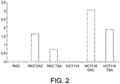
- the invention is based around the finding that NDRG4/2 subfamily genes, undergoe CpG island promoter methylation-associated gene silencing in human cancer cells, in particular colon cancer cells.
- the hypermethylation of the NDRG family gene, such as NDRG4 and/or NDRG2, in particular in the promoter region leads to its loss of expression.
- the presence of aberrant methylation at the NDRG4/2 subfamily gene promoter has a prognostic value.
- the epigenetic loss of NDRG4/2 function can be rescued by the use of DNA demethylating agents and thus provides for a method for treatment.
- the invention is defined by the appended claims.
- the present invention is also based upon the discovery of specific genes and panels of genes whose methylation status is linked to the incidence of, or predisposition to, gastrointestinal cancers such as colorectal cancer.
- Use of these genes for detecting gastrointestinal cancers such as colorectal cancer, in particular in the context of appropriate tissue or faecal (stool) samples or of appropriate blood samples (or derivatives thereof) respectively, has been shown to produce highly sensitive and specific results.
- the disclosure provides also for a method for isolating increased amount of DNA from faecal samples, which results in improved sensitivity of detection of colorectal cancer in faecal samples.
- the disclosure also provides a method for determining the methylation status of a gene of interest in a blood based sample, which requires only low volumes of blood sample equivalent to generate specific and sensitive results. This is advantageous since it permits smaller blood samples to be obtained from the subject under test.
- the disclosure provides a method of detecting a predisposition to, or the incidence of, cancer in a sample comprising detecting an epigenetic change in at least one gene selected from an NDRG4/NDRG2 subfamily gene (in particular NDRG4), GATA4, OSMR, GATA5, SFRP1, ADAM23, JPH3, SFRP2, APC, MGMT, TFPI2, BNIP3, FOXE1, SYNE1, SOX17, PHACTR3 and JAM3, wherein detection of the epigenetic change is indicative of a predisposition to, or the incidence of, cancer.
- Subsets of genes for all aspects and embodiments of the invention include an NDRG4/NDRG2 subfamily gene (in particular NDRG4), GATA4, OSMR, GATA5, SFRP1, ADAM23, JPH3, SFRP2, APC and MGMT and TFPI2, BNIP3, FOXE1, SYNE1, SOX17, PHACTR3 and JAM3 respectively.
- Each subset may be particularly applicable to bodily fluid samples, such as stool and plasma samples as discussed herein.
- epigenetic change is meant a modification in the gene caused by an epigenetic mechanism, such as a change in methylation status or histone acetylation for example.
- the epigenetic change will result in an alteration in the levels of expression of the gene which may be detected (at the RNA or protein level as appropriate) as an indication of the epigenetic change.
- the epigenetic change results in silencing or down regulation of the gene, referred to herein as “epigenetic silencing”.
- the most frequently investigated epigenetic change in the methods of the invention involves determining the methylation status of the gene, where an increased level of methylation is typically associated with the relevant cancer (since it may cause down regulation of gene expression).
- the invention provides a method of diagnosing cancer or predisposition to cancer comprising detecting epigenetic silencing of the NDRG4 gene, wherein epigenetic silencing of the gene is indicative for cancer or predisposition to cancer.
- NDRG family genes have been characterised in the art (see, for example, Qu X et al., 2002 and references cited therein) and their epigenetic silencing can be assessed in terms of DNA methylation status or expression levels as determined by their methylation status.
- the invention provides for a method of diagnosing cancer or predisposition to cancer comprising detecting epigenetic silencing of the NDRG4 gene, wherein epigenetic silencing of the NDRG2/NDRG4-family gene is detected by determination of the methylation status of the NDRG4/2 family gene and wherein methylation of the gene is indicative for cancer or predisposition to cancer.
- the invention also provides for a method of diagnosing cancer or predisposition to cancer comprising detecting epigenetic silencing of the NDRG4 gene, wherein epigenetic silencing of the NDRG2/NDRG4-family gene is determined by measurement of expression levels of the gene, wherein reduced expression of the gene is indicative for cancer or predisposition to cancer.
- the invention provides method of prognosis to cancer or predisposition to cancer comprising detecting epigenetic silencing of the NDRG4/NDRG2 subfamily gene, wherein epigenetic silencing of the gene is indicative for cancer development or predisposition to cancer.
- epigenetic silencing is detected by determination of the methylation status and/or measurement of expression levels of the NDRG2/NDRG4-family gene.
- the disclosure also provides a method of detecting a predisposition to, or the incidence of, cancer and in particular a gastrointestinal cancer such as colorectal cancer in a sample comprising detecting an epigenetic change in at least one gene selected from GATA4, OSMR, NDRG4, GATA5, SFRP1, ADAM23, JPH3, SFRP2, APC, and MGMT, and/or TFPI2, BNIP3, FOXE1, SYNE1, SOX17, PHACTR3 and JAM3, wherein detection of the epigenetic change is indicative of a predisposition to, or the incidence of, cancer and in particular a gastrointestinal cancer such as colorectal cancer.
- the disclosure also provides a method of detecting a predisposition to, or the incidence of, cancer and in particular a gastrointestinal cancer such as colorectal cancer in a sample and in particular in a blood sample, or derivative thereof comprising detecting an epigenetic change in at least one gene selected from GATA4, OSMR, NDRG4, GATA5, SFRP1, ADAM23, JPH3, SFRP2, APC, MGMT, TFPI2, BNIP3, FOXE1, SYNE1, SOX17, PHACTR3 and JAM3(together with any suitable subset or panel thereof), wherein detection of the epigenetic change is indicative of a predisposition to, or the incidence of, cancer and in particular a gastrointestinal cancer such as colorectal cancer.
- NDRG2/NDRG4 subfamily gene any gene which is taken from the subfamily to which NDRG4 and NDRG2 belong and includes according to all aspects of the invention NDRG2 and NDRG4.
- NDRG1, NDRG2, NDRG3 and NDRG4 is the standard nomenclature approved by the human genome organisation for the NDRG family genes, to ensure that each symbol is unique. The listed accession number for these genes can be found at www.gene.ucl.ac.uk/nomenclature.
- NDRG family genes encompass not only the particular sequences found in the publicly available database entries, but also encompass transcript variants of these sequences. Variant forms of the encoded proteins may comprise post-translational modification, may result from spliced messages, etc....
- NDRG4 has transcript variants having the accession numbers NM_020465 and NM_022910. NDRG2 has several transcript variants having the accession numbers, NM_201535, NM_201536, NM_201537, NM_201538, NM_201539, NM_201540, NM_2015401 and NM_016250.
- Variant sequences may have at least 90%, at least 91%, at least 92%, at least 93%, at least 94%, at least 95%, at least 96%, at least 97%, at least 98%, or at least 99% identity to sequences in the database entries or sequence listing.
- Computer programs for determining percent identity are available in the art, including Basic Local Alignment Search Tool (BLAST5) available from the National Center for Biotechnology Information.
- GATA4 is the gene symbol approved by the HUGO Gene Nomenclature Committee. The gene is located on chromosome 8 (location p23.1-p22) and the gene sequence is listed under the accession numbers AK097060, NM_002052 and ENSG00000136574. The gene encodes GATA binding protein 4.
- OSMR is the gene symbol approved by the HUGO Gene Nomenclature Committee.
- the gene is located on chromosome 5 (location p13.2) and the gene sequence is listed under the accession numbers U60805, NM_003999 and ENSG00000145623.
- the gene encodes oncostatin M receptor.
- NDRG4 is the gene symbol approved by the HUGO Gene Nomenclature Committee. The gene is located on chromosome 16 (location q21-q22.3) and the gene sequence is listed under the accession numbers AB044947 and ENSG00000103034. The gene encodes NDRG family member 4.
- GATA5 is the gene symbol approved by the HUGO Gene Nomenclature Committee. The gene is located on chromosome 20 and the gene sequence is listed under the accession number ENSG00000130700. The gene encodes GATA binding protein 5.
- SFRP1 is the gene symbol approved by the HUGO Gene Nomenclature Committee.
- the gene is located on chromosome 8 (location p11.21) and the gene sequence is listed under the accession numbers AF017987, NM_003012 and ENSG00000104332.
- the gene encodes secreted frizzled-related protein 1.
- ADAM23 is the gene symbol approved by the HUGO Gene Nomenclature Committee. The gene is located on chromosome 2 (location q33) and the gene sequence is listed under the accession numbers AB009672 and ENSG00000114948. The gene encodes ADAM metallopeptidase domain 23.
- JPH3 is the gene symbol approved by the HUGO Gene Nomenclature Committee. The gene is located on chromosome 16 (location q24.3) and the gene sequence is listed under the accession numbers AB042636 and ENSG00000154118. The gene encodes junctophilin 3.
- SFRP2 is the gene symbol approved by the HUGO Gene Nomenclature Committee.
- the gene is located on chromosome 4 (location q31.3) and the gene sequence is listed under the accession numbers AF017986 and ENSG00000145423.
- the gene encodes secreted frizzled-related protein 2.
- APC is the gene symbol approved by the HUGO Gene Nomenclature Committee. The gene is located on chromosome 5 (location q21-q22) and the gene sequence is listed under the accession numbers M74088 and ENSG00000134982. The gene encodes adenomatosis polyposis coli.
- the MGMT gene encodes 06-methylguanine-DNA methyltransferase (MGMT), which is a cellular DNA repair protein that rapidly reverses alkylation (e.g. methylation) at the 06 position of guanine, thereby neutralizing the cytotoxic effects of alkylating agents such as temozolomide (TMZ) and carmustine (1-3).
- MGMT is the gene symbol approved by the HUGO Gene Nomenclature Committee. The gene is located on chromosome 10 (location 10q26) and the gene sequence is listed under the accession numbers M29971, NM_002412 and ENSG00000170430.
- BNIP3 is the gene symbol approved by the HUGO Gene Nomenclature Committee. The gene is located on chromosome 10 (location 10q26.3) and the gene sequence is listed under the accession numbers U15174 and ENSG00000176171. The gene encodes the BCL2/adenovirus E1B 19kDa interacting protein 3.
- FOXE1 is the gene symbol approved by the HUGO Gene Nomenclature Committee. The gene is located on chromosome 9 (location 9q22) and the gene sequence is listed under the accession numbers U89995 and ENSG00000178919. The gene encodes the forkhead box E1 (thyroid transcription factor 2)
- JAM3 is the gene symbol approved by the HUGO Gene Nomenclature Committee. The gene is located on chromosome 11 (location 11q25) and the gene sequence is listed under the accession numbers AF356518, NM_032801 and ENSG00000166086. The gene encodes the junctional adhesion molecule 3.
- PHACTR3 is the gene symbol approved by the HUGO Gene Nomenclature Committee. The gene is located on chromosome 20 (location 20q13.32) and the gene sequence is listed under the accession numbers AJ311122, NM_080672 and ENSG00000087495. The gene encodes the phosphatase and actin regulator 3.
- TFPI2 is the gene symbol approved by the HUGO Gene Nomenclature Committee. The gene is located on chromosome 7 (location 7q22) and the gene sequence is listed under the accession numbers L27624 and ENSG00000105825. The gene encodes the tissue factor pathway inhibitor 2.
- SOX17 is the gene symbol approved by the HUGO Gene Nomenclature Committee. The gene is located on chromosome 8 (location 8q11.23) and the gene sequence is listed under the accession numbers AB073988 and ENSG00000164736. The gene encodes the SRY (sex determining region Y)-box 17.
- SYNE1 is the gene symbol approved by the HUGO Gene Nomenclature Committee. The gene is located on chromosome 6 (location 6q25) and the gene sequence is listed under the accession numbers AB018339 and ENSG00000131018. The gene encodes the spectrin repeat containing, nuclear envelope 1.
- variants of each of the gene sequences may also be detected according to the methods of the invention.
- the methylation status of a number of splice variants may be determined according to the methods of the invention.
- Variant sequences preferably have at least 90%, at least 91%, at least 92%, at least 93%, at least 94%, at least 95%, at least 96%, at least 97%, at least 98%, or at least 99% nucleotide sequence identity with the nucleotide sequences in the database entries.
- Computer programs for determining percentage nucleotide sequence identity are available in the art, including the Basic Local Alignment Search Tool (BLAST) available from the National Center for Biotechnology Information.
- the methods of the invention are generally ex vivo or in vitro methods carried out on a test sample, in particular on an isolated test sample.
- the methods can be used to diagnose any suitable type of cancer.
- the cancer comprises, consists essentially of or consists of a neoplasia of the gastrointestinal tract such as gastrointestinal cancer in one embodiment.
- the methods of the invention are applied to colorectal cancer, gastric cancer and/or oesophageal cancer.
- the methods are used to diagnose colorectal cancer, and more particularly to diagnose hereditary nonpolyposis colon cancer and/or sporadic colorectal cancer.
- the methods are aimed at diagnosis of gastric cancer.
- the methods are used to diagnose colorectal cancer and/or gastric cancer.
- the methods may be used to detect carcinoma or adenoma, in particular advanced adenoma.
- the methods may be employed in the diagnosis of both diffuse type and intestinal type carcinomas of the stomach, particularly when the methylation status of NDRG4 is determined.
- the methods may also include the step of obtaining the sample.
- the methods are used to diagnose oesophageal adenocarcinoma.
- the methylation status of the NDRG4 gene has been shown for the first time herein to be linked with high sensitivity and specificity to the incidence of this particular cancer type.
- Oesophageal adenocarcinoma may be distinguished from oesophageal squamous cell carcinomas on this basis.
- test sample can be any tissue sample or body fluid.
- the test sample is obtained from a human subject.
- the sample is taken from the gastrointestinal tract.
- the sample may be a colorectal tissue sample or a colon, rectal, oesophageal, stomach or appendix tissue sample or a faecal or blood based sample from a subject.
- faecal samples the methods are preferably used with respect to detecting gastrointestinal cancers such as colorectal cancer as discussed herein, but may also be useful in identifying potentially dangerous adenomas. Different markers and panels of markers may be most useful with a specific sample type, such as a tissue, blood based or faecal sample as discussed herein in detail.
- the methods of the invention involve detecting an epigenetic change, and in particular determining the methylation status, of (at least) the NDRG4 gene in a faecal test sample, wherein detection of the epigenetic change, in particular (hyper)methylation of the NDRG4 gene (promoter) is indicative of gastrointestinal neoplasias/cancer, in particular colorectal cancer, such as adenomas and carcinomas, gastric cancer and other adenocarcinomas of the gastrointestinal tract (such as oesophageal adenocarcinoma) and/or diffuse type and intestinal type carcinomas of the stomach.
- gastrointestinal neoplasias/cancer in particular colorectal cancer, such as adenomas and carcinomas, gastric cancer and other adenocarcinomas of the gastrointestinal tract (such as oesophageal adenocarcinoma) and/or diffuse type and intestinal type carcinomas of the stomach.
- the subject may be suspected of being tumorigenic. More specifically the subject may be suspected of suffering from a cancer, such as a gastrointestinal cancer and in particular colorectal cancer, as discussed herein.
- a cancer such as a gastrointestinal cancer and in particular colorectal cancer
- any other suitable test samples in which epigenetic silencing of the appropriate gene or genes of the invention, for example an NDRG4/NDRG2 subfamily gene, can be determined to indicate the presence of cancer are included within the scope of the invention.
- Preferred panels and subsets of genes are presented herein which provide sensitive and specific diagnosis, including early stage detection, of a gastrointestinal cancer such as colorectal cancer based upon appropriate samples such as tissue, faecal and plasma samples as discussed herein.
- the methods of the invention may comprise, consist essentially of or consist of detecting an epigenetic change in a panel of genes comprising OSMR, GATA4 and ADAM23 or OSMR, GATA4 and GATA5, wherein detection of the epigenetic change in at least one of the genes in the panel is indicative of a predisposition to, or the incidence of, a gastrointestinal cancer such as colorectal cancer.
- the tissue sample may comprise, consist essentially of or consist of a colon and/or rectal and/or appendix sample for example.
- DNA-containing sample which may be used in the methods of the invention include samples for diagnostic, prognostic, or personalised medicinal uses. These samples may be obtained from surgical samples, such as biopsies or fine needle aspirates, from paraffin embedded tissues, from frozen tumour tissue samples, from fresh tumour tissue samples or from a fresh or frozen body fluid, for example. Non-limiting examples include whole blood or parts/fractions thereof, bone marrow, cerebrospinal fluid, peritoneal fluid, pleural fluid, lymph fluid, serum, plasma, urine, chyle, ejaculate, sputum, nipple aspirate, saliva, swabs specimens, colon wash specimens and brush specimens. The tissues and body fluids can be collected using any suitable method, many such methods are well known in the art. Assessment of a paraffin-embedded specimen can be performed directly or on a tissue section. Tissue samples are generally taken from the tissue suspected of being tumourigenic.
- the test sample is a blood sample. Any blood sample, or derivative thereof may be utilised.
- the blood sample, or derivative thereof may comprise, consist essentially or whole blood or any suitable DNA containing parts/fractions thereof.
- the blood sample or derivative thereof comprises, consist essentially of or consists of serum or plasma.
- the blood sample may be collected using any suitable method, many such methods are well known in the art.
- the methods of the invention also incorporate the step of obtaining the blood sample. Any appropriate blood sample may be utilised in the methods of the invention, provided it contains sufficient DNA.
- the volume of the blood sample, or derivative thereof that is utilised in the methods is around 5 to 15 ml, such as 10 ml.
- Blood samples, or derivatives thereof, may be stored prior to use in the methods of the invention once obtained. They may be frozen for example at a suitable temperature, such as around -80°C.
- the blood sample, or derivative thereof comprises, consists essentially of or consists of a plasma or serum sample.
- Plasma may be derived from whole blood by any suitable means.
- the plasma sample is obtained by centrifugation of whole blood. Centrifugation may be carried out at any suitable speed and for any suitable period of time and under any suitable conditions as may be determined by one skilled in the art. For example, centrifugation may be carried out at between around 1000 and 3000g. Centrifugation may be carried out for between around 1, 2, 3, 4, or 5 and 10, 11, 12, 13, 14 or 15 minutes for example. Centrifugation may be carried out at low temperatures, such as between around 0 and 5°C, for example 4°C, to maintain integrity of the sample. Multiple centrifugation steps may be employed in order to obtain the plasma sample. In a specific embodiment, two centrifugation steps are employed to obtain the plasma sample.
- the at least one gene may be selected from OSMR, SFRP1, NDRG4, GATA5, ADAM23, JPH3, SFRP2 and APC. As shown below, these genes provide sensitive and specific methods for diagnosing colorectal cancer in plasma samples. Suitable panels in this context comprise, consist essentially of or consist of OSMR, NDRG4, GATA5 and ADAM23. Additional genes which may be employed in plasma or serum based methods include TFPI2, BNIP3, FOXE1, SYNE1, SOX17, PHACTR3, and JAM3.
- Diagnosis is defined herein to include screening for a disease or pre-indication of a disease, identifying a disease or pre-indication of a disease, monitoring the staging and the state and progression of the disease, checking for recurrence of disease following treatment and monitoring the success of a particular treatment.
- the methods of the invention may also have prognostic value, and this is included within the definition of the term "diagnosis”.
- the prognostic value of the methods of the invention may be used as a marker of potential susceptibility to a number of gastrointestinal cancers such as colorectal cancer or as a marker for progression from adenoma to cancer for example. Thus patients at risk may be identified before the disease has a chance to manifest itself in terms of symptoms identifiable in the patient.
- the methods of the invention may be carried out on purified or unpurified DNA-containing samples.
- DNA is isolated/extracted/purified from the sample. Any suitable DNA isolation technique may be utilised. Examples of purification techniques may be found in standard texts such as Molecular Cloning - A Laboratory Manual (Third Edition), Sambrook and Russell (see in particular Appendix 8 and Chapter 5 there in).
- purification involves alcohol precipitation of DNA. Preferred alcohols include ethanol and isopropanol.
- Suitable purification techniques also include salt-based precipitation methods.
- the DNA purification technique comprises use of a high concentration of salt to precipitate contaminants.
- the salt may comprise, consist essentially of or consist of potassium acetate and/or ammonium acetate for example.
- the method may further include steps of removal of contaminants which have been precipitated, followed by recovery of DNA through alcohol precipitation.
- the DNA purification technique is based upon use of organic solvents to extract contaminants from cell lysates.
- the method comprises use of phenol, chloroform and isoamyl alcohol to extract the DNA. Suitable conditions are employed to ensure that the contaminants are separated into the organic phase and that DNA remains in the aqueous phase.
- extracted DNA is recovered through alcohol precipitation, such as ethanol or isopropanol precipitation.
- Amplification of DNA (using PCR) from natural sources is often inhibited by co-purified contaminants and various methods adopted for DNA extraction from environmental samples are available and provide an alternative for isolating DNA from faecal or blood based samples, according to specific embodiments of the invention.
- the QIAamp DNA Stool Mini Kit from QIAGEN adsorbs DNA-damaging substances and PCR inhibitors present in the sample by InhibitEX.
- Other examples for application in particular to faecal samples include the Wizard Genomic DNA Purification Kit (Promega), the NucliSENS® easyMAGTM (Biomerieux) and nucleic acid purification kits manufactured by Macherey Nagel.
- the DNA may be isolated by phenol-chloroform extraction since this has been shown to provide particularly high levels of DNA recovery from the sample.
- the ChargeSwitch procedure may be utilised for example.
- Suitable methods and kits for isolating DNA from blood samples are commercially available. Examples, each of which may be utilised in the methods of the invention are provided in the table below. Table 1: Kits and methods for isolating DNA from blood samples. Kit Company Method UltraClean-htpTM BloodSpinTM DNA Mo Bio Laboratories, Inc.
- DNA isolation may be carried out using silica-membranes, isopropanol or magnetic bead based methods for example.
- the methods of the invention may also, as appropriate, incorporate quantification of isolated/extracted/purified DNA in the sample. Quantification of the DNA in the sample may be achieved using any suitable means. Quantitation of nucleic acids may, for example, be based upon use of a spectrophotometer, a fluorometer or a UV transilluminator. Examples of suitable techniques are described in standard texts such as Molecular Cloning - A Laboratory Manual (Third Edition), Sambrook and Russell (see in particular Appendix 8 there in). In one embodiment, kits such as the Picogreen® dsDNA quantitation kit available from Molecular Probes, Invitrogen may be employed to quantify the DNA.
- Neoplasia refers to abnormal new growth and thus means the same as tumor, which may be benign or malignant.
- Particular cancer types which are relevant in accordance with the present invention are discussed above and include those selected from neoplasias of the gastrointestinal tract. Specific examples include colorectal cancer, oesophageal cancer, stomach cancer and gastric cancer.
- Colorectal cancer also called colon cancer or bowel cancer, is defined to include cancerous growths in the colon, rectum and appendix. Specific markers and panels of markers, as described in greater detail herein, may be particularly applicable to certain cancer types.
- cancer types which may be relevant in specific (but not all) embodiments of the invention include prostate cancer, breast cancer, ovarian cancer and thyroid cancer.
- Epigenetic silencing is defined herein to include any alteration in the DNA resulting in diminished gene expression which is mediated by mechanisms other than alterations in the primary nucleotide sequence of a gene. Epigenetic modifications may, in certain circumstances be stable heritable traits. A number of related mechanisms that cause alteration in gene expression are recognised and include DNA methylation, histone changes (for example changes in histone acetylation) which may lead to chromatin remodelling and RNA interference. In many cases, hypermethylation of DNA incorrectly switches off critical genes allowing cancers to develop and progress.
- a subject may be undergoing routine screening and may not necessarily be suspected of having a disease such as a colon neoplasia.
- Detecting epigenetic silencing of the gene or genes in an adenoma of such a subject may indicate that the probable course of the adenoma is development to a carcinoma and thus there is a predisposition to neoplasia. In such cases, preventive treatment may be recommended and involve resection of the advanced adenoma.
- These methods may advantageously involve detection of methylation of the NDRG4 gene, in particular using primer set 1, as discussed herein.
- Advanced adenoma refers to an adenoma in which epigenetic silencing of at least one of the gene linked to colorectal cancer is observed, preferably epigenetic silencing such as methylation of the gene or genes of the invention (such as NDRG4 etc.) is detected.
- the most preferred epigenetic change in the at least one gene selected from an NDRG2/NDRG4 subfamily gene (in particular NDRG4), GATA4, OSMR, GATA5, SFRP1, ADAM23, JPH3, SFRP2, APC, MGMT, TFPI2, BNIP3, FOXE1, SYNE1, SOX17, PHACTR3 and JAM3 (in all permutations and combinations including panels as discussed herein) which is detected comprises, consists essentially of or consists of methylation.
- aberrant methylation which may be referred to as hypermethylation, of the gene or genes is detected.
- the methylation status is determined in suitable CpG islands which are often found in the promoter region of the gene(s).
- methylation refers to the presence or absence of 5-methylcytosine ("5-mCyt") at one or a plurality of CpG dinucleotides within a DNA sequence. CpG dinucleotides are typically concentrated in the promoter regions and exons of human genes.
- Diminished gene expression can be assessed in terms of DNA methylation status or in terms of expression levels as determined by the methylation status of the gene.
- One method to detect epigenetic silencing is to determine that a gene which is expressed in normal cells is less expressed or not expressed in tumor cells. Accordingly, the invention provides for a method of diagnosing cancer or predisposition to cancer comprising detecting epigenetic silencing of the NDRG4/NDRG2 subfamily gene, wherein epigenetic silencing of the NDRG2/NDRG4-family gene is determined by measurement of expression levels of the gene and wherein reduced expression of the gene is indicative for cancer or predisposition to cancer.
- the invention also provides a method of detecting a predisposition to, or the incidence of, a cancer in particular a gastrointestinal cancer such as colorectal cancer in a sample comprising detecting an epigenetic change in at least one gene selected from GATA4, OSMR, NDRG4 (or another NDRG4/NDRG2 subfamily member), GATA5, SFRP1, ADAM23, JPH3, SFRP2, APC and, MGMT and/or at least one gene selected from, TFPI2, BNIP3, FOXE1, SYNE1, SOX17, PHACTR3 and JAM3, wherein detection of the epigenetic change is determined by measuring expression levels of the at least one gene and wherein low level, reduced level or a lack of expression of the at least one gene is indicative of a predisposition to, or the incidence of, cancer and in particular a gastrointestinal cancer such as colorectal cancer.
- the at least one gene may be selected from OSMR, SFRP1, NDRG4, GATA5, ADAM23, JPH3, SFRP2 and APC and/or from TFPI2, BNIP3, FOXE1, SYNE1, SOX17, PHACTR3 and JAM3. These genes are also useful where faecal test samples are employed.
- TFPI2 may be a particularly useful marker.
- Specific genes such as genes selected from TPF12, BNIP3, FOXE1, SYNE1 and SOX17 may be most useful when plasma samples are employed.
- genes such as GATA4, OSMR, NORG4, GATA5, SFRP1, ADAM23, JPH3, SFRP2, APC and MGMT and also genes selected from TFPI2, FOXE1, SYNE1, SOX17, PHACTR3 and JAM3 may usefully be employed. As shown below, these genes provide sensitive and specific methods for diagnosing colorectal cancer in plasma samples. Suitable panels in this context comprise, consist essentially of or consist of OSMR, NDRG4, GATA5 and ADAM23. Further panels are discussed below. This may be utilised in order to diagnose early stage colorectal cancer, in particular stage 0 to II colorectal cancer.
- the method preferably comprises, consists essentially of or consists of detecting an epigenetic change in a panel of genes comprising OSMR, GATA4 and ADAM23 or OSMR, GATA4 and GATA5, wherein detection of the epigenetic change in at least one of the genes in the panel is indicative of a predisposition to, or the incidence of, a gastrointestinal cancer such as colorectal cancer.
- the tissue sample may comprise, consist essentially of or consist of a colon and/or rectal and/or appendix sample for example.
- total loss of protein expression of the at least one gene selected from an NDRG2/NDRG4 subfamily gene (in particular NDRG4), GATA4, OSMR, GATA5, SFRP1, ADAM23, JPH3, SFRP2, APC, MGMT, TFPI2, BNIP3, FOXE1, SYNE1, SOX17, PHACTR3 and JAM3 (in all permutations and combinations including panels as discussed herein) is observed in the sample in order to conclude a diagnosis of cancer and in particular a gastrointestinal cancer such as colorectal cancer or predisposition to cancer and in particular a gastrointestinal cancer such as colorectal cancer, or to make a decision on the best course of treatment in accordance with the other methods of the invention, as described herein (which description applies here mutatis mutandis).
- partial loss of expression of at least one gene selected from an NDRG2/NDRG4 subfamily gene (in particular NDRG4), GATA4, OSMR, GATA5, SFRP1, ADAM23, JPH3, SFRP2, APC, MGMT, TFPI2, BNIP3, FOXE1, SYNE1, SOX17, PHACTR3 and JAM3 may also be relevant, due to methylation of the relevant gene or genes.
- the decreased level of expression of at least one gene selected from an NDRG2/NDRG4 subfamily gene (in particular NDRG4), GATA4, OSMR, GATA5, SFRP1, ADAM23, JPH3, SFRP2, APC, MGMT, TFPI2, BNIP3, FOXE1, SYNE1, SOX17, PHACTR3 and JAM3 may, as necessary, be measured in order to determine if it is statistically significant in the sample. This helps to provide a reliable test for the methods of the invention.
- Any method for determining whether the expression level of at least one gene selected from an NDRG2/NDRG4 subfamily gene (in particular NDRG4), GATA4, OSMR, GATA5, SFRP1, ADAM23, JPH3, SFRP2, APC, MGMT, TFPI2, BNIP3, FOXE1, SYNE1, SOX17, PHACTR3 and JAM3 (in all permutations and combinations including panels as discussed herein) is significantly reduced may be utilised.
- Such methods are well known in the art and routinely employed.
- statistical analyses may be performed.
- One example involves an analysis of variance test. Typical P values for use in such a method would be P values of ⁇ 0.05 or 0.01 or 0.001 when determining whether the relative expression or activity is statistically significant.
- a change in expression may be deemed significant if there is at least a 10% decrease for example.
- the test may be made more selective by making the change at least 15%, 20%, 25%, 30%, 35%, 40% or 50%, for example, in order to
- the decreased level of expression or activity of the at least one gene selected from an NDRG2/NDRG4 subfamily gene (in particular NDRG4), GATA4, OSMR, GATA5, SFRP1, ADAM23, JPH3, SFRP2, APC, MGMT, TFPI2, BNIP3, FOXE1, SYNE1, SOX17, PHACTR3 and JAM3 (in all permutations and combinations including panels as discussed herein) is determined with reference to a control sample.
- This control sample is preferably taken from normal (i.e. non tumorigenic) tissue in the subject, where expression of the corresponding gene or genes is normal.
- control samples may also be utilised in which there is known to be a lack of expression of the at least one gene selected from an NDRG2/NDRG4 subfamily gene (in particular NDRG4), GATA4, OSMR, GATA5, SFRP1, ADAM23, JPH3, SFRP2, APC, MGMT, TFPI2, BNIP3, FOXE1, SYNE1, SOX17, PHACTR3 and JAM3 (in all permutations and combinations including panels as discussed herein).
- NDRG2/NDRG4 subfamily gene in particular NDRG4
- GATA4 GATA4
- OSMR GATA5
- GATA5 SFRP1, ADAM23
- JPH3, SFRP2, APC MGMT
- TFPI2 BNIP3, FOXE1, SYNE1, SOX17, PHACTR3 and JAM3
- Suitable additional controls may also be included to ensure that the test is working properly, such as measuring levels of expression or activity of a suitable reference gene in both test and control samples.
- Suitable reference genes for the present invention include beta-actin, glyceraldehyde-3-phosphate dehydrogenase (GAPDH), ribosomal RNA genes such as 18S ribosomal RNA and RNA polymerase II gene ( Radonic A. et al., Biochem Biophys Res Commun. 2004 Jan 23;313(4):856-62 ).
- the reference gene is beta-actin.
- RNA level or at the protein level.
- Cells in test samples can be lysed and the mRNA levels in the lysates, or in the RNA purified or semi-purified from the lysates, determined. Alternatively, methods can be used on unlysed tissues or cell suspensions.
- RNA level Suitable methods for determining expression of at least one gene selected from an NDRG2/NDRG4 subfamily gene (in particular NDRG4), GATA4, OSMR, GATA5, SFRP1, ADAM23, JPH3, SFRP2, APC, MGMT, TFPI2, BNIP3, FOXE1, SYNE1, SOX17, PHACTR3 and JAM3 (in all permutations and combinations including panels as discussed herein) at the RNA level are well known in the art and described herein.
- Methods employing nucleic acid probe hybridization to the relevant transcript(s) of the at least one gene selected from an NDRG2/NDRG4 subfamily gene (in particular NDRG4), GATA4, OSMR, GATA5, SFRP1, ADAM23, JPH3, SFRP2, APC, MGMT, TFPI2, BNIP3, FOXE1, SYNE1, SOX17, PHACTR3 and JAM3 (in all permutations and combinations including panels as discussed herein) may be employed for measuring the presence and/or level of the respective mRNA.
- Such methods are well known in the art and include use of nucleic acid probe arrays (microarray technology) and Northern blots.
- Sequencing-based methods are an alternative. These methods started with the use of expressed sequence tags (ESTs), and now include methods based on short tags, such as serial analysis of gene expression (SAGE) and massively parallel signature sequencing (MPSS).
- SAGE serial analysis of gene expression
- MPSS massively parallel signature sequencing
- Differential display techniques provide yet another means of analyzing gene expression; this family of techniques is based on random amplification of cDNA fragments generated by restriction digestion, and bands that differ between two tissues identify cDNAs of interest.
- the levels of gene expression of at least one gene selected from an NDRG2/NDRG4 subfamily gene (in particular NDRG4), GATA4, OSMR, GATA5, SFRP1, ADAM23, JPH3, SFRP2, APC, MGMT, TFPI2, BNIP3, FOXE1, SYNE1, SOX17, PHACTR3 and JAM3 are determined using reverse transcriptase polymerase chain reaction (RT-PCR).
- RT-PCR is a well known technique in the art which relies upon the enzyme reverse transcriptase to reverse transcribe mRNA to form cDNA, which can then be amplified in a standard PCR reaction. Protocols and kits for carrying out RT-PCR are extremely well known to those of skill in the art and are commercially available.
- primers useful in RT-PCR carried out on the NDRG4 gene are provided. These primers comprise, consist essentially of or consist of the following sequences:
- Variants of these primers are include within the scope of the invention, as defined herein which definition applies mutatis mutandis.
- RT-PCR can be carried out in a non-quantitative manner. End-point RT-PCR measures changes in expression levels using three different methods: relative, competitive and comparative. These traditional methods are well known in the art. Alternatively, RT-PCR is carried out in a real time and/or in a quantitative manner. Real time quantitative RT-PCR has been thoroughly described in the literature (see Gibson et al for an early example of the technique) and a variety of techniques are possible.
- Examples include use of hydrolytic probes (Taqman), hairpin probes (Molecular Beacons), FRET probe pairs (LightCycler (Roche)), hairpin probes attached to primers (Scorpion), hairpin primers (Plexor and Amplifluor), DzyNA and oligonucleotide blocker systems. All of these systems are commercially available and well characterised, and may allow multiplexing (that is, the determination of expression of multiple genes in a single sample).
- TAQMAN was one of the earliest available real-time PCR techniques and relies upon a probe which binds between the upstream and downstream primer binding sites in a PCR reaction.
- a TAQMAN probe contains a 5' fluorophore and a 3' quencher moiety. Thus, when bound to its binding site on the DNA the probe does not fluoresce due to the presence of the quencher in close proximity to the fluorophore.
- the 5' - 3' exonuclease activity of a suitable polymerase such as Taq digests the probe if it is bound to the strand being amplified. This digestion of the probe causes displacement of the fluorophore.
- the probe is again designed to bind between the primer binding sites.
- the probe is a hairpin shaped probe.
- the hairpin in the probe when not bound to its target sequence means that a fluorophore attached to one end of the probe and a quencher attached to the other end of the probe are brought into close proximity and therefore internal quenching occurs. Only when the target sequence for the probe is formed during the PCR amplification does the probe unfold and bind to this sequence.
- the loop portion of the probe acts as the probe itself, while the stem is formed by complimentary arm sequences (to respective ends of which are attached the fluorophore and quencher moiety).
- the beacon probe detects its target, it undergoes a conformational change forcing the stem apart and this separates the fluorophore and quencher. This causes the energy transfer to the quencher to be disrupted and therefore restores fluorescence.
- the Molecular Beacons assume a random-coil configuration and fluoresce. As the temperature is lowered to allow annealing of the primers, stem hybrids form rapidly, preventing fluorescence. However, at the annealing temperature, Molecular Beacons also bind to the amplicons, undergo conformational reorganisation, leading to fluorescence. When the temperature is raised to allow primer extension, the Molecular Beacons dissociate from their targets and do not interfere with polymerisation. A new hybridisation takes place in the annealing step of every cycle, and the intensity of the resulting fluorescence indicates the amount of accumulated amplicon.
- Scorpions primers are based upon the same principles as Molecular Beacons. However, here, the probe is bound to, and forms an integral part of, an amplification primer. The probe has a blocking group at its 5' end to prevent amplification through the probe sequence. After one round of amplification has been directed by this primer, the target sequence for the probe is produced and to this the probe binds. Thus, the name "scorpion" arises from the fact that the probe as part of an amplification product internally hybridises to its target sequence thus forming a tail type structure. Probe-target binding is kinetically favoured over intrastrand secondary structures.
- Scorpions primers were first described in the paper " Detection of PCR products using self-probing amplicons and fluorescence" (Nature Biotechnology. 17, p804-807 (1999 )) and numerous variants on the basic theme have subsequently been produced.
- Amplifluor primers rely upon incorporation of a Molecular Beacon type probe into a primer. Again, the hairpin structure of the probe forms part of an amplification primer itself. However, in contrast to Scorpions type primers, there is no block at the 5' end of the probe in order to prevent it being amplified and forming part of an amplification product. Accordingly, the primer binds to a template strand and directs synthesis of the complementary strand. The primer therefore becomes part of the amplification product in the first round of amplification. When the complimentary strand is synthesised amplification occurs through the hairpin structure. This separates the fluorophore and quencher molecules, thus leading to generation of florescence as amplification proceeds.
- DzyNA primers incorporate the complementary/antisense sequence of a 10-23 nucleotide DNAzyme.
- amplicons are produced that contain active (sense) copies of DNAzymes that cleave a reporter substrate included in the reaction mixture.
- the accumulation of amplicons during PCR/amplification can be monitored in real time by changes in fluorescence produced by separation of fluorophore and quencher dye molecules incorporated into opposite sides of a DNAzyme cleavage site within the reporter substrate.
- the DNAzyme and reporter substrate sequences can be generic and hence can be adapted for use with primer sets targeting various genes or transcripts ( Todd et al., Clinical Chemistry 46:5, 625-630 (2000 )).
- the PlexorTM qPCR and qRT-PCR Systems take advantage of the specific interaction between two modified nucleotides to achieve quantitative PCR analysis.
- One of the PCR primers contains a fluorescent label adjacent to an iso-dC residue at the 5' terminus.
- the second PCR primer is unlabeled.
- the reaction mix includes deoxynucleotides and iso-dGTP modified with the quencher dabcyl. Dabcyl-iso-dGTP is preferentially incorporated at the position complementary to the iso-dC residue. The incorporation of the dabcyl-iso-dGTP at this position results in quenching of the fluorescent dye on the complementary strand and a reduction in fluorescence, which allows quantitation during amplification.
- a primer pair with a different fluorophore is used for each target sequence.
- Real time quantitative techniques for use in the invention generally produce a fluorescent read-out that can be continuously monitored. Fluorescence signals are generated by dyes that are specific to double stranded DNA, like SYBR Green, or by sequence-specific fluorescently-labeled oligonucleotide primers or probes. Each of the primers or probes can be labelled with a different fluorophore to allow specific detection.
- These real time quantitative techniques are advantageous because they keep the reaction in a "single tube". This means there is no need for downstream analysis in order to obtain results, leading to more rapidly obtained results. Furthermore, keeping the reaction in a "single tube” environment reduces the risk of cross contamination and allows a quantitative output from the methods of the invention. This may be particularly important in a clinical setting for the present invention.
- PCR is a preferred amplification method, to include variants on the basic technique such as nested PCR, equivalents may also be included within the scope of the invention.
- examples include without limitation isothermal amplification techniques such as NASBA, 3SR, TMA and triamplification, all of which are well known in the art and commercially available.
- Other suitable amplification methods without limitation include the ligase chain reaction (LCR) (Barringer et al, 1990), MLPA, selective amplification of target polynucleotide sequences ( US Patent No.
- Suitable methods for determining expression of at least one gene selected from an NDRG2/NDRG4 subfamily gene (in particular NDRG4), GATA4, OSMR, GATA5, SFRP1, ADAM23, JPH3, SFRP2, APC, MGMT, TFPI2, BNIP3, FOXE1, SYNE1, SOX17, PHACTR3 and JAM3 (in all permutations and combinations including panels as discussed herein) at the protein level are also well known to one of skill in the art.
- Examples include western blots, immunohistochemical staining and immunolocalization, immunofluorescence, enzyme-linked immunosorbent assay (ELISA), immunoprecipitation assays, complement fixation assay, agglutination reactions, radioimmunoassay, flow cytometry, mass spectrophotometry, and equilibrium dialysis. These methods generally depend upon a reagent specific for identification of the appropriate gene product. Any suitable reagent may be utilised such as lectins, receptors, nucleic acids, antibodies etc. The reagent is preferably an antibody and may comprise monoclonal or polyclonal antibodies.
- Fragments and derivatized antibodies may also be utilised, to include without limitation Fab fragments, ScFv, single domain antibodies, nano-antibodies, heavy chain antibodies, aptamers etc. which retain gene product binding function. Any detection method may be employed in accordance with the invention. Proteins may be identified on the basis of charge, polarity, amino acid sequence etc. by a range of methods, including SDS-PAGE and amino acid sequencing for example. The nature of the reagent is not limited except that it must be capable of specifically identifying the appropriate gene product.
- the same reagent can be tested on samples in which it is known that the at least one gene selected from an NDRG2/NDRG4 subfamily gene (in particular NDRG4), GATA4, OSMR, GATA5, SFRP1, ADAM23, JPH3, SFRP2, APC, MGMT, TFPI2, BNIP3, FOXE1, SYNE1, SOX17, PHACTR3 and JAM3 (in all permutations and combinations including panels as discussed herein) is expressed at the protein level.
- NDRG2/NDRG4 subfamily gene in particular NDRG4
- OSMR GATA5
- GATA5 SFRP1, ADAM23
- JPH3, SFRP2, APC MGMT
- TFPI2 BNIP3, FOXE1, SYNE1, SOX17, PHACTR3 and JAM3
- the methods of the invention incorporate an appropriate re-expression assay which is designed to reverse epigenetic silencing.
- a demethylating agent such as a DNA-methyltransferase (DMT) inhibitor may reverse epigenetic silencing of the relevant gene.
- DMT DNA-methyltransferase
- Suitable reagents include, but are not limited to, DAC (5'-deazacytidine), TSA or any other treatment affecting epigenetic mechanisms present in cell lines.
- Suitable reagents are discussed herein with respect to the pharmacogenetic and treatment aspects of invention, which discussion applies mutatis mutandis.
- expression is reactivated or reversed upon treatment with such reagents, indicating that the silencing is epigenetic.
- epigenetic silencing resulting in diminished expression of the NDRG4/NDRG2 subfamily gene has been shown in a range of gastrointestinal cancers such as colorectal cancer and gastric cancer.
- the invention provides for a method of diagnosing colorectal cancer and/or gastric cancer or another gastrointestinal cancer as defined herein, predisposition to colorectal cancer and/or gastric cancer or another gastrointestinal cancer as defined herein, comprising detecting epigenetic silencing of the NDRG4/NDRG2 subfamily gene, wherein epigenetic silencing of the NDRG2/NDRG4-family gene is determined by measurement of expression levels of the gene and wherein reduced expression of the gene is indicative for colorectal cancer and/or gastric cancer or another gastrointestinal cancer as defined herein, predisposition to colorectal cancer and/or gastric cancer or another gastrointestinal cancer as defined herein, or progression of adenoma to carcinoma.
- the gene is NDRG4, or a combination of NDRG2 and NDRG4.
- epigenetic silencing resulting in diminished expression of the at least one gene selected from GATA4, OSMR, NDRG4, GATA5, SFRP1, ADAM23, JPH3, SFRP2, APC, MGMT, TFPI2, BNIP3, FOXE1, SYNE1, SOX17, PHACTR3 and JAM3 has been shown to be sensitively and specifically linked with the incidence of gastrointestinal cancer and in particular colorectal cancer.
- the invention provides for a method of diagnosing gastrointestinal cancer and in particular colorectal cancer or predisposition to gastrointestinal cancer and in particular colorectal cancer comprising detecting epigenetic silencing of NDRG4 and optionally also at least one gene selected from GATA4, OSMR, GATA5, SFRP1, ADAM23, JPH3, SFRP2, APC, MGMT, TFPI2, BNIP3, FOXE1, SYNE1, SOX17, PHACTR3 and JAM3, wherein epigenetic silencing of NDRG4 and optionally also the at least one gene selected from GATA4, OSMR, GATA5, SFRP1, ADAM23, JPH3, SFRP2, APC, MGMT, TFPI2, BNIP3, FOXE1, SYNE1, SOX17, PHACTR3 and JAM3 is determined by measurement of expression levels of the gene and wherein reduced expression of the gene is indicative for gastrointestinal cancer and in particular colorectal cancer, predisposition to gastrointestinal cancer and in particular colorec
- markers may usefully be employed when faecal test samples are utilised.
- epigenetic silencing resulting in diminished expression of NDRG4 and optionally also the at least one gene selected from GATA4, OSMR, GATA5, SFRP1, ADAM23, JPH3, SFRP2, APC and MGMT has been shown to be sensitively and specifically linked with the incidence of colorectal cancer in specific tissue and bodily fluid, such as faecal and blood-based samples.
- the invention provides for a method of diagnosing gastrointestinal cancer and in particular colorectal cancer or predisposition to gastrointestinal cancer and in particular colorectal cancer comprising detecting epigenetic silencing of NDRG4 and optionally also at least one gene selected from GATA4, OSMR, GATA5, SFRP1, ADAM23, JPH3, SFRP2, APC and MGMT in a tissue, faecal or a blood (plasma or serum) sample, or derivative thereof, wherein epigenetic silencing of NDRG4 and optionally the at least one gene selected from GATA4, OSMR, GATA5, SFRP1, ADAM23, JPH3, SFRP2, APC and MGMT is determined by measurement of expression levels of the gene and wherein reduced expression of the gene is indicative for gastrointestinal cancer and in particular colorectal cancer, predisposition to gastrointestinal cancer and in particular colorectal cancer, or progression of adenoma to carcinoma.
- the plasma or serum samples are utilised.
- the at least one gene may be selected from TFPI2, BNIP3, FOXE1, SYNE1, SOX17, PHACTR3 and JAM3.
- Panels may be selected from these genes and the other genes of the invention as desired and as discussed herein. Methylation of these genes in stool and plasma samples has been shown for the first time herein to be linked to colorectal cancer.
- Particularly useful markers which give good levels of sensitivity and specificity in both plasma and faecal samples include TFPI2, FOXE1, SYNE1, SOX17, PHACTR3 and JAM3.
- TFPI2 may be particularly useful. Certain genes such as those selected from TFPI2, BNIP3, FOXE1, SYNE1 and SOX17 may prove most useful when testing plasma samples. This discussion applies to all aspects of the invention as appropriate.
- any gene involved in the establishment of cancer such as colorectal cancer, gastric cancer and/or oesophageal cancer, may be utilized in combination with the at least NDRG4, optionally GATA4, OSMR, GATA5, SFRP1, ADAM23, JPH3, SFRP2, APC, MGMT, TFPI2, BNIP3, FOXE1, SYNE1, SOX17, PHACTR3 and JAM3 (in all permutations and combinations including panels as discussed herein) in the methods of present invention.
- the expression level of NDRG4 and optionally also GATA4, OSMR, GATA5, SFRP1, ADAM23, JPH3, SFRP2, APC, MGMT, TFPI2, BNIP3, FOXE1, SYNE1, SOX17, PHACTR3 and JAM3 is analysed in combination with at least one other gene involved in the establishment of cancer.
- At least NDRG4 and optionally also one gene selected from GATA4, OSMR, GATA5, SFRP1, ADAM23, JPH3, SFRP2, APC, MGMT, TFPI2, BNIP3, FOXE1, SYNE1, SOX17, PHACTR3 and JAM3 is combined with at least two other genes involved in the establishment of cancer.
- Other genes involved in the establishment of (colorectal) cancer may be selected from the group consisting of CHFR, p16, Vimentin, p14, RASSF1a, RAB32, SEPTIN-9, RASSF2A, TMEFF2, NGFR and SMARCA3.
- the methods of the invention preferably involve detecting gene methylation.
- the invention provides a method of diagnosing cancer or predisposition to cancer, in particular gastrointestinal cancers such as colorectal cancer comprising detecting epigenetic silencing of NDRG4 and optionally also at least one gene selected from GATA4, OSMR, GATA5, SFRP1, ADAM23, JPH3, SFRP2, APC, MGMT, TFPI2, BNIP3, FOXE1, SYNE1, SOX17, PHACTR3 and JAM3 (in all permutations and combinations including panels as discussed herein), wherein epigenetic silencing of the at least one gene is detected by determination of the methylation status of the at least one gene and wherein methylation of the at least one gene is indicative for cancer or predisposition to cancer, as defined above and in particular gastrointestinal cancers such as colorectal cancer.
- the at least one gene may be selected from OSMR, SFRP1, NDRG4, GATA5, ADAM23, JPH3, SFRP2 and APC. As shown below, these genes provide sensitive and specific methods for diagnosing colorectal cancer in plasma samples. Suitable panels in this context comprise, consist essentially of or consist of OSMR, NDRG4, GATA5 and ADAM23. This may be utilised in order to diagnose early stage colorectal cancer, in particular stage 0 to II colorectal cancer.
- the at least one gene may be selected from TFPI2, BNIP3, FOXE1, SYNE1, SOX17, PHACTR3 and JAM3, such as from TFPI2, BNIP3, FOXE1, SYNE1 and SOX17, in particular TFPI2.
- the methods may comprise, consist essentially of or consist of detecting an epigenetic change in a panel of genes comprising OSMR, GATA4 and ADAM23 or OSMR, GATA4 and GATA5, wherein detection of the epigenetic change in at least one of the genes in the panel is indicative of a predisposition to, or the incidence of, colorectal cancer.
- the tissue sample may comprise, consist essentially of or consist of a colon and/or rectal and/or appendix sample.
- the at least one gene may be selected from GATA4, OSMR, NDRG4, GATA5, SFRP1, ADAM23, JPH3, SFRP2, APC and MGMT.
- the at least one gene may be selected from TFPI2, BNIP3, FOXE1, SYNE1, SOX17, PHACTR3, and JAM3, such as from TFPI2, FOXE1, SYNE1, SOZ17, PHACTR3 and JAM3, in particular TFPI2.
- Two, three, four, five or six etc. gene panels selected from these genes are also envisaged in the present invention.
- CpG dinucleotides susceptible to methylation are typically concentrated in the promoter region, exons and introns of human genes. Promoter, exon and intron regions can be assessed for methylation.
- the methylation status of the promoter region of the at least one gene selected from an NDRG2/NDRG4 subfamily gene (in particular NDRG4), GATA4, OSMR, GATA5, SFRP1, ADAM23, JPH3, SFRP2, APC, MGMT, TFPI2, BNIP3, FOXE1, SYNE1, SOX17, PHACTR3 and JAM3 (in all permutations and combinations including panels as discussed herein) is determined.
- a “promoter” is a region extending typically between approximately 1 Kb, 500 bp or 150 to 300 bp upstream from the transcription start site.
- the methylation status of the exon and/or intron regions of the at least one gene selected from an NDRG2/NDRG4 subfamily gene (in particular NDRG4), GATA4, OSMR, GATA5, SFRP1, ADAM23, JPH3, SFRP2, APC, MGMT, TFPI2, BNIP3, FOXE1, SYNE1, SOX17, PHACTR3 and JAM3 may be determined as appropriate.
- the methylation status of the promoter region of the NDRG4 gene is analysed.
- the promoter region of NDRG2 and NDRG4 are analysed simultaneously.
- the region of the NDRG4/NDRG2 subfamily gene comprising, consisting essentially of, or consisting of the nucleotide sequence of NDRG4 as set forth as SEQ ID NO: 524 ( FIG. 3a ) and/or the nucleotide sequence of NDRG2 as set forth as SEQ ID NO: 525 ( FIG. 3b ) is analysed in order to determine its methylation status.
- methylation assay procedures are known in the art, and can be used in conjunction with the present invention. These assays rely onto two distinct approaches: bisulphite conversion based approaches and non-bisulphite based approaches.
- Non-bisulphite based methods for analysis of DNA methylation rely on the inability of methylation-sensitive enzymes to cleave methylation cytosines in their restriction.
- the bisulphite conversion relies on treatment of DNA samples with sodium bisulphite which converts unmethylated cytosine to uracil, while methylated cytosines are maintained (Furuichi et al., 1970). This conversion results in a change in the sequence of the original DNA.
- DNA methylation analysis has been performed successfully with a number of techniques including: sequencing, methylation-specific PCR (MS-PCR), melting curve methylation-specific PCR (McMS-PCR), MLPA with or without bisulfite treatment, QAMA (Zeschnigk et al, 2004), MSRE-PCR (Melnikov et al, 2005), MethyLight (Eads et al., 2000), ConLight-MSP (Rand et al., 2002), bisulfite conversion-specific methylation-specific PCR (BS-MSP)(Sasaki et al., 2003), COBRA (which relies upon use of restriction enzymes to reveal methylation dependent sequence differences in PCR products of sodium bisulfite - treated DNA), methylation-sensitive single-nucleotide primer extension conformation(MS-SNuPE), methylation-sensitive single-strand conformation analysis (MS-SSCA), Melting curve combined bisulfite restriction analysis (McCOBRA)(A
- methylated CpG dinucleotides utilize the ability of the methyl binding domain (MBD) of the MeCP2 protein to selectively bind to methylated DNA sequences (Cross et al, 1994; Shiraishi et al, 1999).
- MBD methyl binding domain
- the MBD may be obtained from MBP, MBP2, MBP4 or poly-MBD (Jorgensen et al., 2006).
- restriction exonuclease digested genomic DNA is loaded onto expressed His-tagged methyl-CpG binding domain that is immobilized to a solid matrix and used for preparative column chromatography to isolate highly methylated DNA sequences.
- Such methylated DNA enrichment-step may supplement the methods of the invention.
- Several other methods for detecting methylated CpG islands are well known in the art and include amongst others methylated-CpG island recovery assay (MIRA). Any of these methods may be employed in the present invention where desired.
- MSP methylation specific PCR
- DNA may be amplified using primer pairs designed to distinguish methylated from unmethylated DNA by taking advantage of sequence differences as a result of sodium-bisulphite treatment ( Herman et al.,1996; and WO 97/46705 ).
- QMSP real-time quantitative MSP
- SYBR Green I fluorescent dye
- labelled primers and/or labelled probes can be used.
- Heavymethyl described for example in WO02/072880 the priming is methylation specific, but non-extendable oligonucleotide blockers provide this specificity instead of the primers themselves.
- the blockers bind to bisulfite-treated DNA in a methylation-specific manner, and their binding sites overlap the primer binding sites. When the blocker is bound, the primer cannot bind and therefore the amplicon is not generated.
- Heavymethyl can be used in combination with real-time or end point detection in the methods of the invention.
- the methylation status of NDRG4 and optionally also the at least one gene selected from GATA4, OSMR, GATA5, SFRP1, ADAM23, JPH3, SFRP2, APC, MGMT, TFPI2, BNIP3, FOXE1, SYNE1, SOX17, PHACTR3 and JAM3 is determined by methylation specific PCR/amplification, preferably real-time methylation specific PCR/amplification.
- the real time PCR/amplification involves use of hairpin primers (Amplifluor)/hairpin probes (Molecular Beacons)/hydrolytic probes (Taqman)/FRET probe pairs (Lightcycler)/primers incorporating a hairpin probe (Scorpion)/primers incorporating complementary sequences of DNAzymes that cleave a reporter substrate included in the reaction mixture (DzyNA®)/fluorescent dyes (SYBR Green etc.)/oligonucleotide blockers/the specific interaction between two modified nucleotides (Plexor).
- Primers and/or probes can be used to investigate the methylation status of the at least one gene.
- Real-Time PCR detects the accumulation of amplicon during the reaction. Real-time methods do not need to be utilised, however. Many applications do not require quantification and Real -Time PCR is used principally as a tool to obtain convenient results presentation and storage, and at the same time to avoid post-PCR handling. Analyses can be performed only to know if the target DNA is present in the sample or not. End point verification is carried out after the amplification reaction has finished. This knowledge can be used in a medical diagnostic laboratory to detect a predisposition to, or the incidence of, cancer in a patient. In the majority of such cases, the quantification of DNA template is not very important.
- Amplification products may simply be run on a suitable gel, such as an agarose gel, to determine if the expected sized products are present. This may involve use of ethidium bromide staining and visualisation of the DNA bands under a UV illuminator for example. Alternatively, fluorescence or energy transfer can be measured to determine the presence of the methylated DNA.
- the end-point PCR fluorescence detection technique can use the same approaches as widely used for Real Time PCR: TaqMan assay, Molecular Beacons, Scorpion, Amplifluor etc. For example, «Gene» detector allows the measurement of fluorescence directly in PCR tubes.
- quantitation may be on an absolute basis, or may be relative to a constitutively methylated DNA standard, or may be relative to an unmethylated DNA standard.
- Methylation status may be determined by using the ratio between the signal of the marker under investigation and the signal of a reference gene where methylation status is known (such as ⁇ -actin for example), or by using the ratio between the methylated marker and the sum of the methylated and the non-methylated marker.
- absolute copy number of the methylated marker gene can be determined.
- Suitable reference genes for the present invention include beta-actin, glyceraldehyde-3-phosphate dehydrogenase (GAPDH), ribosomal RNA genes such as 18S ribosomal RNA and RNA polymerase II gene ( Radonic A. et al., Biochem Biophys Res Commun. 2004 Jan 23;313(4):856-62 ).
- the reference gene is beta-actin.
- each clinical sample is measured in duplicate and for both C t values (cycles at which the amplification curves crossed the threshold value, set automatically by the relevant software) copy numbers are calculated.
- the average of both copy numbers (for each gene) is used for the result classification.
- two standard curves are used, one for either the reference gene ( ⁇ -actin or the non-methylated marker for example) and one for the methylated version of the marker.
- primers useful in MSP carried out on the promoter region of the NDRG4 gene are provided. These primers comprise, consist essentially of or consist of the following sequences: TABLE 2 SEQ ID NO. NDRG4 Primer Sense primer SEQ ID NO.
- Primer set 1 is useful in particular applications for predicting the progression of adenomas.
- Primer set 2 may provide slightly more sensitive results although both primer sets are clearly useful.
- primers and probes useful in quantitative MSP carried out on the (promoter region of the) NDRG4 gene are provided. These primers and probes comprise, consist essentially of or consist of the following sequences:
- primers useful in MSP carried out on the promoter region of the NDRG2 gene are provided. These primers comprise, consist essentially of or consist of the following sequences:
- primers and/or probes useful in determining the methylation status of the at least one gene selected from GATA4, OSMR, NDRG4, GATA5 ,SFRP1, ADAM23, JPH3, SFRP2, APC and MGMT are provided.
- These primers and/or probes comprise, consist essentially of or consist of the following sequences: Table 3: Primer sequences and beacon (probe) sequences SEQ ID NO.
- the methods of the invention employ or rely upon or utilise primers and/or probes selected from the primers and probes comprising the nucleotide sequences set forth in Table 4 below to determine the methylation status of the at least one gene.
- the table presents specific primer and probe combinations for certain preferred genes whose methylation status may be determined according to the methods of the invention.
- beacon sequences SEQ ID NO 26 ß-Actin forward primer 5' - TAGGGAGTATATAGGTTGGGGAAGTT - 3' 27 reverse primer 5' - AACACACAATAACAAACACAAATTCAC - 3' 28 beacon 5'-FAM-CGACTGCGTGTGGGGTGGTGATGGAGGAGGTTTAGGCAGTCG-3'-DABCYL 29 GATA4 forward primer 5' - AGGTTAGTTAGCGTTTTAGGGTC - 3' 30 reverse primer 5' - ACGACGACGAAACCTCTCG - 3' 31 beacon 5'-FAM-CGACATGCCTCGCGACTCGAATCCCCGACCCAGCATGTCG-3'-DABCYL 32 GATA5 forward primer 5' - AGTTCGTTTAGGTTAGTTTTCGGC - 3' 33 reverse primer 5' - CCAATACAACTAAACGAACGAACCG - 3' 34 beacon 5'-FAM-CGACATGCGTAGGGA
- the methods of the invention employ or rely upon or utilise primers and/or probes selected from the primers comprising the nucleotide sequences set forth in Table 5 below to determine the methylation status of NDRG4.
- the table presents specific primer and probe combinations for determining the methylation status of this gene and the primer pairs and corresponding probe may be selected according to the table.
- Table 5 Primer pairs and probes for determining the methylation status of NDRG4, with predicted amplification product lengths shown.
- the methods of the invention employ or rely upon or utilise primers selected from the primers comprising the nucleotide sequences set forth in Table 6 and 7 below to determine the methylation status of GATA5.
- the table presents specific primer combinations for determining the methylation status of this gene and the primer pairs may be selected according to the table.
- Table 6 also sets forth specific probes which may be utilised to facilitate (quantitative) detection of the methylation status of GATA5 and Table 7 incorporates Amplifluour sequences which allow the primers to act as hairpin primers, thus facilitating quantitative detection (as discussed in detail herein).
- the methods of the invention employ or rely upon or utilise primers selected from the primers comprising the nucleotide sequences set forth in Tables 8 and 9 below to determine the methylation status of OSMR.
- the tables present specific primer combinations for determining the methylation status of this gene and the primer pairs may be selected according to the table.
- Table 8 also sets forth specific probes which may be utilised to facilitate (quantitative) detection of the methylation status of OSMR and Table 9 incorporates Amplifluour sequences which allow the primers to act as hairpin primers, thus facilitating quantitative detection (as discussed in detail herein).
- Table 8 Primer pairs and probes (molecular beacons) for determining the methylation status of OSMR, with predicted amplification product lengths shown.
- the methods of the invention employ or rely upon or utilise primers selected from the primers comprising the nucleotide sequences set forth in Table 10 below to determine the methylation status of ADAM23.
- the table presents specific primer combinations for determining the methylation status of this gene and the primer pairs may be selected according to the table.
- Table 6 also sets forth specific probes which may be utilised to facilitate (quantitative) detection of the methylation status of ADAM23.
- the methods of the invention employ or rely upon or utilise primers selected from the primers comprising the nucleotide sequences set forth in Table 11 below to determine the methylation status of JPH3.
- the table presents specific primer combinations for determining the methylation status of this gene and the primer pairs may be selected according to the table.
- Table 11 also sets forth specific probes which may be utilised to facilitate (quantitative) detection of the methylation status of JPH3.
- the methods of the invention employ or rely upon or utilise primers and/or probes selected from the primers and probes comprising the nucleotide sequences set forth in Table 12 below to determine the methylation status of the at least one gene.
- the table presents specific primer and probe combinations for certain preferred genes whose methylation status may be determined according to the methods of the invention.
- beacon (probe) sequences SEQ ID No BNIP3 348 forward primer 5'-TACGCGTAGGTTTTAAGTCGC-3' 349 reverse primer 5'-TCCCGAACTAAACGAAACCCCG-3' 350 beacon 5'-FAM-CGACATGCCTACGACCGCGTCGCCCATTAGCATGTCG -3'-DABCYL FOXE1 351 forward primer 5'-TTTGTTCGTTTTTCGATTGTTC-3' 352 reverse primer 5'-TAACGCTATAAAACTCCTACCGC-3' 353 beacon JAM3 354 forward primer 5'-GGGATTATAAGTCGCGTCGC-3' 355 reverse primer 5'-CGAACGCAAAACCGAAATCG-3' 356 beacon 5'-FAM-CGACACGATATGGCGTTGAGGCGGTTATCGTGTCG-3'-DABCYL JPH3 51 forward primer 5'-TTAGATTTCGTAAACGGTGAAAAC-3' 52 reverse primer 5'-TCTCCTCCGAAAAAC
- the methods of the invention employ or rely upon or utilise primers selected from the primers and beacons comprising the nucleotide sequences set forth in Table 13 below to determine the methylation status of BNIP3.
- the table presents specific primer combinations for determining the methylation status of this gene and the primer pairs may be selected according to the table.
- Table 13 - Additional assay designs Primer and probe sequences for determining the methylation status of BNIP3, with predicted amplification product lengths shown.
- the methods of the invention employ or rely upon or utilise primers selected from the primers and beacons comprising the nucleotide sequences set forth in Table 14 below to determine the methylation status of FOXE1.
- the table presents specific primer combinations for determining the methylation status of this gene and the primer pairs may be selected according to the table.
- Table 14 - Additional assay designs Primer and probe sequences for determining the methylation status of FOXE1, with predicted amplification product lengths shown.
- the methods of the invention employ or rely upon or utilise primers selected from the primers and beacons comprising the nucleotide sequences set forth in Table 15 below to determine the methylation status of JAM3.
- the table presents specific primer combinations for determining the methylation status of this gene and the primer pairs may be selected according to the table.
- Table 15 - Additional assay designs Primer and probe sequences for determining the methylation status of JAM3, with predicted amplification product lengths shown.
- the methods of the invention employ or rely upon or utilise primers selected from the primers and beacons comprising the nucleotide sequences set forth in Table 16 below to determine the methylation status of PHACTR3.
- the table presents specific primer combinations for determining the methylation status of this gene and the primer pairs may be selected according to the table.
- Table 16 - Additional assay designs Primer and probe sequences for determining the methylation status of PHACTR3, with predicted amplification product lengths shown.
- the methods of the invention employ or rely upon or utilise primers selected from the primers and beacons comprising the nucleotide sequences set forth in Table 17 below to determine the methylation status of TFPI2.
- the table presents specific primer combinations for determining the methylation status of this gene and the primer pairs may be selected according to the table.
- Table 17 - Additional assay designs Primer and probe sequences for determining the methylation status of TFPI2, with predicted amplification product lengths shown.
- the methods of the invention employ or rely upon or utilise primers selected from the primers and beacons comprising the nucleotide sequences set forth in Table 18 below to determine the methylation status of SOX17.
- the table presents specific primer combinations for determining the methylation status of this gene and the primer pairs may be selected according to the table.
- Table 18 - Additional assay designs Primer and probe sequences for determining the methylation status of SOX17, with predicted amplification product lengths shown.
- primer pairs selected from the primer pairs disclosed herein, including in the tables (which may comprise additional sequence over above the basic sequence listed). Further characteristics of these primers are summarized in the detailed description (experimental part) below. It is noted that variants of these sequences may be utilised in the present invention. In particular, additional sequence specific flanking sequences may be added, for example to improve binding specificity, as required.
- Variant sequences preferably have at least 90%, at least 91%, at least 92%, at least 93%, at least 94%, at least 95%, at least 96%, at least 97%, at least 98%, or at least 99% nucleotide sequence identity with the nucleotide sequences of the primers and/or probes set forth in any of tables 2, 3, 4, 5 or 6.
- the primers and probe (including hairpin) structures may incorporate synthetic nucleotide analogues as appropriate or may be RNA or PNA based for example, or mixtures thereof. Similarly alternative fluorescent donor and acceptor moieties/FRET pairs may be utilised as appropriate.
- the primers may include modified oligonucleotides and other appending groups and labels provided that the functionality as a primer in the methods of the invention is not compromised.
- alternative fluorescent donor and acceptor moieties/FRET pairs may be utilised as appropriate.
- Molecules that are commonly used in FRET include fluorescein, 5-carboxyfluorescein (FAM), 2'7'-dimethoxy-4'5'-dichloro-6-carboxyfluorescein (JOE), rhodamine, 6-carboxyrhodamine (R6G), N,N,N',N'-tetramethyl-6-carboxyrhodamine (TAMRA), 6-carboxy-X-rhodamine (ROX), 4-(4'-dimethylaminophenylazo) benzoic acid (DABCYL), and 5-(2'-aminoethyl) aminonaphthalene-1-sulfonic acid (EDANS).
- FAM 5-carboxyfluorescein
- JE 2'7'-dimethoxy-4'5'-dichloro-6-carboxyfluorescein
- rhodamine 6-carboxyrhodamine
- R6G 6-carboxyr
- a fluorophore is a donor or an acceptor is defined by its excitation and emission spectra, and the fluorophore with which it is paired.
- FAM is most efficiently excited by light with a wavelength of 488 nm, and emits light with a spectrum of 500 to 650 nm, and an emission maximum of 525 nm.
- FAM is a suitable donor fluorophore for use with JOE, TAMRA, and ROX (all of which have their excitation maximum at 514 nm).
- said donor moiety and said acceptor moiety are selected from 5-carboxyfluorescein (FAM), 2'7'-dimethoxy-4'5'-dichloro-6-carboxyfluorescein (JOE), rhodamine, 6-carboxyrhodamine (R6G), N,N,N'-tetramethyl-6-carboxyrhodamine (TAMRA), 6-carboxy-X-rhodamine (ROX), 5-(2'-aminoethyl)aminonapthalene-1-sulfoni acid (EDANS), anthranilamide, coumarin, terbium chelate derivatives, Malachite green, Reactive Red 4, DABCYL, tetramethyl rhodamine, pyrene butyrate, eosine nitrotyrosine, ethidium, and Texas Red.
- FAM 5-carboxyfluorescein
- R6G 6-carboxyrhodamine
- said donor moiety is selected from fluorescein, 5-carboxyfluorescein (FAM), rhodamine, 5-(2'-aminoethyl)aminonapthalene-1-sulfonic acid (EDANS), anthranilamide, coumarin, terbium chelate derivatives, Malachite green, and Reactive Red 4, and said acceptor moiety is selected from DABCYL, rhodamine, tetramethyl rhodamine, pyrene butyrate, eosine nitrotyrosine, ethidium, and Texas Red.
- said donor moiety is fluorescein or a derivative thereof, and said acceptor moiety is DABCYL.
- the fluorescein derivative comprises, consists essentially of or consists of 6-carboxy fluorescein.
- the primers and in particular the stem loop/hairpin structures, and/or the probes may be labelled with donor and acceptor moieties during chemical synthesis of the primers or probes or the label may be attached following synthesis using any suitable method. Many such methods are available and well characterised in the art.
- probe types such as the hairpin probe type employed in tables 7 and 9
- probe (or primer) type as appropriate. Equivalents are discussed herein and may be utilised as appropriate.
- bisulphite sequencing is utilised in order to determine the methylation status of NDRG4 and optionally also of at least one gene selected from GATA4, OSMR, GATA5, SFRP1, ADAM23, JPH3, SFRP2, APC, MGMT, TFPI2, BNIP3, FOXE1, SYNE1, SOX17, PHACTR3 and JAM3 (in all permutations and combinations including panels as discussed herein).
- Primers may be designed for use in sequencing through the important CpG islands in the at least one gene selected NDRG4, GATA4, OSMR, GATA5, SFRP1, ADAM23, JPH3, SFRP2, APC, MGMT, TFPI2, BNIP3, FOXE1, SYNE1, SOX17, PHACTR3 and JAM3 (in all permutations and combinations including panels as discussed herein).
- primers may be designed in both the sense and antisense orientation to direct sequencing across the promoter region of the relevant gene or genes.
- bisulphite sequencing may be carried out by using sequencing primers which comprise, consist essentially of or consist of the following sequences, and which may be used in isolation or in combination to sequence both strands:
- sequencing primers form a further aspect of the invention, with suitable variants being included within the scope of the invention (the discussion of which applies mutatis mutandis here).
- nucleic acid amplification techniques in addition to PCR (which includes real-time versions thereof and variants such as nested PCR), may also be utilised, as appropriate, to detect the methylation status of the at least one gene selected from NDRG4, GATA4, OSMR, GATA5, SFRP1, ADAM23, JPH3, SFRP2, APC, MGMT, TFPI2, BNIP3, FOXE1, SYNE1, SOX17, PHACTR3 and JAM3 (in all permutations and combinations including panels as discussed herein).
- amplification techniques are well known in the art, and include methods such as NASBA (Compton, 1991), 3SR (Fahy et al., 1991) and Transcription Mediated Amplification (TMA).
- ligase chain reaction (Barringer et al, 1990), selective amplification of target polynucleotide sequences ( US Patent No. 6,410,276 ), consensus sequence primed polymerase chain reaction ( US Patent No 4,437,975 ), arbitrarily primed polymerase chain reaction ( WO 90/06995 ), invader technology, strand displacement technlology, and nick displacement amplification ( WO 2004/067726 ).
- LCR ligase chain reaction
- SCR consensus sequence primed polymerase chain reaction
- WO 90/06995 arbitrarily primed polymerase chain reaction
- invader technology strand displacement technlology
- nick displacement amplification WO 2004/067726
- This list is not intended to be exhaustive; any nucleic acid amplification technique may be used provided the appropriate nucleic acid product is specifically amplified.
- these amplification techniques may be tied in to MSP and/or bisulphite sequencing techniques for example.
- primers may be designed that themselves do not cover any potential sites of DNA methylation. Sequence variations at sites of differential methylation are located between the two primers. Such primers are used in bisulphite genomic sequencing, COBRA and Ms-SnuPE for example. Secondly, primers may be designed that anneal specifically with either the methylated or unmethylated version of the converted sequence.
- the primer may also contain additional nucleotide residues that do not interfere with hybridization but may be useful for other manipulations. Examples of such other residues may be sites for restriction endonuclease cleavage, for ligand binding or for factor binding or linkers or repeats.
- the oligonucleotide primers may or may not be such that they are specific for modified methylated residues.
- One way to distinguish between modified and unmodified DNA is to hybridize oligonucleotide primers which specifically bind to one form or the other of the DNA. After hybridization, an amplification reaction can be performed and amplification products assayed. The presence of an amplification product indicates that a sample hybridized to the primer. The specificity of the primer indicates whether the DNA had been modified or not, which in turn indicates whether the DNA had been methylated or not.
- oligonucleotide probes which may also be specific for certain products. Such probes may be hybridized directly to modified DNA or to amplification products of modified DNA. Oligonucleotide probes can be labelled using any detection system known in the art. These include but are not limited to fluorescent moieties, radioisotope labelled moieties, bioluminescent moieties, luminescent moieties, chemiluminescent moieties, enzymes, substrates, receptors, or ligands.
- amplification is achieved with the use of primers specific for the sequence of the gene whose methylation status is to be assessed.
- primer binding sites corresponding to a suitable region of the sequence may be selected.
- the nucleic acid molecules may also include sequences other than primer binding sites which are required for detection of the methylation status of the gene, for example RNA Polymerase binding sites or promoter sequences may be required for isothermal amplification technologies, such as NASBA, 3SR and TMA.
- TMA (Gen-probe Inc.) is an RNA transcription amplification system using two enzymes to drive the reaction, namely RNA polymerase and reverse transcriptase.
- the TMA reaction is isothermal and can amplify either DNA or RNA to produce RNA amplified end products.
- TMA may be combined with Gen-probe's Hybridization Protection Assay (HPA) detection technique to allow detection of products in a single tube. Such single tube detection is a preferred method for carrying out the invention.
- HPA Hybridization Protection Assay
- methylation status Whilst the genes (in particular promoters) of the invention appear to be unmethylated in normal tissues, and thus the detection of methylation (or indeed a lack of methylation) in these genes is readily observable as being significant in terms of a cancer diagnosis and also in selecting suitable treatment regimens and for determining the likelihood of successful treatment or resistance to treatment with certain anti-cancer agents etc, when determining methylation status, it may be beneficial to include suitable controls in order to ensure the method chosen to assess this parameter is working correctly and reliably. For example, suitable controls may include assessing the methylation status of a gene known to be methylated. This experiment acts as a positive control to ensure that false negative results are not obtained (i.e.
- a conclusion of a lack of methylation is made even though the at least one gene selected from an NDRG2/NDRG4 subfamily gene (in particular NDRG4), GATA4, OSMR, GATA5, SFRP1, ADAM23, JPH3, SFRP2, APC, MGMT, TFPI2, BNIP3, FOXE1, SYNE1, SOX17, PHACTR3 and JAM3 (in all permutations and combinations including panels as discussed herein) may, in fact, be methylated).
- the gene may be one which is known to be methylated in the sample under investigation or it may have been artificially methylated, for example by using a suitable methyltransferase enzyme, such as SssI methyltransferase.
- the NDRG4/NDRG2 subfamily gene preferably the NDRG4 and/or NDRG2 gene, may be assessed in normal lymphocytes, following treatment with SssI methyltransferase, as a positive control.
- suitable negative controls may be employed with the methods of the invention.
- suitable controls may include assessing the methylation status of a gene known to be unmethylated or carrying out an amplification in the absence of DNA (for example by using a water only sample). The former experiment acts as a negative control to ensure that false positive results are not obtained (i.e.
- a conclusion of methylation is made even though the at least one gene selected from an NDRG2/NDRG4 subfamily gene (in particular NDRG4), GATA4, OSMR, GATA5, SFRP1, ADAM23, JPH3, SFRP2, APC, MGMT, TFPI2, BNIP3, FOXE1, SYNE1, SOX17, PHACTR3 and JAM3 (in all permutations and combinations including panels as discussed herein, such as at least one gene selected from OSMR, SFRP1, NDRG4, GATA5, ADAM23, JPH3, SFRP2 and APC in one specific embodiment) may, in fact, be unmethylated).
- the gene may be one which is known to be unmethylated in the sample under investigation or it may have been artificially demethylated, for example by using a suitable DNA methyltransferase inhibitor, such as those discussed in more detail below.
- the NDRG4/NDRG2 subfamily gene in particular the NDRG4 and/or NDRG2 gene, may be assessed in normal lymphocytes as a negative control, since it has been shown for the first time herein that the NDRG4 and/or NDRG2 gene is unmethylated in normal tissues.
- the application of the methods of present invention to extremely small amounts of abnormally-methylated DNA, that are released into collected fluids, in particular stools, may require the generation and amplification of a DNA library before testing for methylation of any specific gene.
- Suitable methods on whole genome amplification and libraries generation for such amplification e.g. Methylplex and Enzyplex technology, Rubicon Genomics
- WO2005/090507 describes library generation/amplification methods that require either bisulfite conversion or non-bisulfite based application.
- Bisulfite treatment may occur before or after library construction and may require the use of adaptors resistant to bisulfite conversion.
- Meth-DOP-PCR (Di Vinci et al, 2006), a modified degenerate oligonucleotide-primed PCR amplification (DOP-PCR) that is combined with MSP, provides another suitable method for specific detection of methylation in small amounts of DNA. Improved management of patient care may require these existing methods and techniques to supplement the methods of the invention.
- epigenetic silencing resulting in methylation of the NDRG4/NDRG2 subfamily gene has been shown in a number of gastrointestinal cancers such as colorectal cancer and/or gastric cancer, stomach and oesophageal cancers, in particular oesophageal carcinomas.
- the invention provides for a method of diagnosing a gastrointestinal cancer, such as colorectal cancer and/or gastric cancer and/or oesophageal cancer or predisposition to a gastrointestinal cancer, such as colorectal cancer and/or gastric cancer and/or oesophageal cancer comprising detecting the methylation status of the NDRG4 gene, wherein methylation of the gene is indicative for a gastrointestinal cancer, such as colorectal cancer and/or gastric cancer and/or oesophageal cancer, or predisposition to a gastrointestinal cancer, such as colorectal cancer and/or gastric cancer and/or oesophageal cancer.
- the gene is NDRG2, or NDRG4, or a combination of NDRG2 and NDRG4.
- the epigenetic change, in particular methylation status, of NDRG4 and optionally also of at least one gene selected from GATA4, OSMR, GATA5, SFRP1, ADAM23, JPH3, SFRP2, APC, MGMT, TFPI2, BNIP3, FOXE1, SYNE1, SOX17, PHACTR3 and JAM3 may be determined in order to diagnose a gastrointestinal cancer, such as colorectal cancer and/or gastric cancer and/or oesophageal cancer or a predisposition thereto.
- the at least one gene may be selected from GATA4, OSMR, NDRG4 and SFRP2, in particular where faecal samples are utilized. Detecting an epigenetic change, in particular methylation, in these genes results in a particularly sensitive and specific diagnostic method.
- the at least one gene may be selected from OSMR, SFRP1, NDRG4, GATA5, ADAM23, JPH3, SFRP2 and APC and particularly selected from OSMR, NDRG4, GATA5 and ADAM23, in particular where plasma or serum samples are utilised. Detecting an epigenetic change, in particular methylation, in these genes results in a particularly sensitive and specific diagnostic method.
- the at least one gene may be selected from TFPI2, BNIP3, FOXE1, SYNE1, SOX17, PHACTR3 and JAM3, such as from TFPI2, BNIP3, FOXE1, SYNE1 and SOX17, in particular TFPI2.
- the methods may comprise, consist essentially of or consist of detecting an epigenetic change in a panel of genes comprising OSMR, GATA4 and ADAM23 or OSMR, GATA4 and GATA5, wherein detection of the epigenetic change in at least one of the genes in the panel is indicative of a predisposition to, or the incidence of, colorectal cancer.
- the tissue sample may comprise, consist essentially of or consist of a colon and/or rectal and/or appendix sample for example, as discussed herein above.
- the at least one gene may be selected from GATA4, OSMR, NDRG4, GATA5, SFRP1, ADAM23, JPH3, SFRP2, APC and MGMT.
- the at least one gene may be selected from TFPI2, BNIP3, FOXE1, SYNE1, SOX17, PHACTR3, and JAM3, such as from TFPI2, FOXE1, SYNE1, SOZ17, PHACTR3 and JAM3, in particular TFPI2.
- the methods may comprise detecting an epigenetic change in a panel of genes comprising at least two, three, four, five or six of the genes, wherein detection of an epigenetic change in at least one of the genes in the panel is indicative of a predisposition to, or the incidence of, cancer and in particular gastrointestinal cancers as defined herein, such as colorectal cancer.
- the panel of genes may comprise/consist essentially of or consist of two, three, four, five or six genes.
- Certain panels of genes have been found to result in particularly sensitive methods for detecting a gastrointestinal cancer, such as colorectal cancer and/or gastric cancer and/or oesophageal cancer or a predisposition thereto - especially colorectal cancer.
- the panel of genes comprises, consists essentially of or consists of GATA4 and OSMR, GATA4 and NDRG4, GATA4 and SFRP2, OSMR and NDRG4, OSMR and SFRP2, NDRG4 and SFRP2, APC and SFRP2, APC and OSMR, APC and GATA4, APC and NDRG4, MGMT and OSMR, MGMT and GATA4, MGMT and NDRG4, MGMT and SFRP2, MGMT and APC, SFRP1 and MGMT, SFRP1 and OSMR, SFRP1 and GATA4, SFRP1 and NDRG4, SFRP1 and SFRP2, SFRP1 and APC, GATA5 and SFRP1, GATA5 and MGMT, GATA5 and OSMR, GATA5 and GATA4, GATA5 and NDRG4, GATA5 and SFRP2 or GATA5 and APC.
- Suitable panels incorporating other genes such as ADAM23 and/or JPH3 are examples of the panel
- genes comprise, consist essentially of or consists of SFRP1, SFRP2 and APC or SFRP2, OSMR and APC.
- Further panels of genes comprise, consist essentially of or consist of GATA4, OSMR and NDRG4, GATA4, OSMR and SFRP2, GATA4, NDRG4 and SFRP2 or OSMR, NDRG4 and SFRP2.
- One specifc four gene panel consists of GATA4, OSMR, NDRG4 and SFRP2.
- One specific panel of at least six genes comprises, consists essentially of or consists of NDRG4, OSMR, SFRP1, ADAM23, GATA5 and MGMT.
- a panel of genes that consists essentially of or consists of OSMR, GATA4 and ADAM23 or OSMR, GATA4 and GATA5.
- This embodiment applies in particular to tissue samples, which may be colon, rectal or appendix samples for example, as discussed herein.
- the panel of genes comprises, consists essentially of or consists of OSMR, NDRG4, GATA5 and ADAM23, where blood based samples and in particular plasma or serum samples are utilised.
- the disclosure provides a method of detecting a predisposition to, or the incidence of, early stage colorectal cancer and in particular stage 0 to II colorectal cancer in a blood sample, or derivative thereof such as a plasma or serum sample (preferably a plasma sample) comprising detecting an epigenetic change in at least one gene selected from OSMR, NDRG4, GATA5 and ADAM23, wherein detection of the epigenetic change is indicative of a predisposition to, or the incidence of, early stage colorectal cancer and in particular stage 0 to II colorectal cancer.
- This method may be applied to a panel consisting of these four genes.
- the panel of genes comprises, consists essentially of or consists of both OSMR3 and OSMR4.
- the panels of genes in the present invention may incorporate assessment of multiple sites within the same gene as appropriate. Primers investigating multiple sites within the same genes are set forth in the tables above, see particularly tables 2 to 18 (and especially tables 5 to 11 and 13 to 18).
- the detection of an epigenetic change in each of the panel of genes may be carried out in a single reaction.
- Many suitable techniques allowing multiplexing are available and may be utilised in the present invention. Most depend upon use of suitable fluorescent molecules having distinguishable emission spectra. The skilled person can readily select from the many fluorophores available to determine which can be used in a multiplexing context.
- a universal quencher is utilised together with suitable fluorophore donors each having a distinguishable emission wavelength maximum.
- a particularly useful quencher is DABCYL.
- DABCYL a suitable quencher such as DABCYL the following fluorophores may each be utilised to allow multiplexing: Coumarin (emission maximum of 475nm), EDANS (491nm), fluorescein (515nm), Lucifer yellow (523nm), BODIPY (525nm), Eosine (543nm), tetramethylrhodamine (575nm) and texas red (615nm) ( Tyagi et al., Nature Biotechnology, Vol. 16, Jan 1998; 49-53 ).
- methylation status of additional genes may also be determined in order to supplement the methods of the invention. No gene has been found to be epigenetically silenced in every similar tumour. For this reason, it may be advantageous to target multiple DNA alterations to attain high rates of tumour detection.
- the methylation status of the at least one gene selected from an NDRG2/NDRG4 subfamily gene (in particular NDRG4), GATA4, OSMR, GATA5, SFRP1, ADAM23, JPH3, SFRP2, APC, MGMT, TFPI2, BNIP3, FOXE1, SYNE1, SOX17, PHACTR3 and JAM3 is analysed in combination with the methylation status of at least one other gene involved in the establishment of cancer.
- the at least one gene selected from an NDRG2/NDRG4 subfamily gene (in particular NDRG4), GATA4, OSMR, GATA5, SFRP1, ADAM23, JPH3, SFRP2, APC, MGMT, TFPI2, BNIP3, FOXE1, SYNE1, SOX17, PHACTR3 and JAM3 (in all permutations and combinations including panels as discussed herein) may be combined with at least two other genes involved in the establishment of cancer.
- the at least one gene selected from an NDRG2/NDRG4 subfamily gene (in particular NDRG4), GATA4, OSMR, GATA5, SFRP1, ADAM23, JPH3, SFRP2, APC, MGMT, TFPI2, BNIP3, FOXE1, SYNE1, SOX17, PHACTR3 and JAM3 (in all permutations and combinations including panels as discussed herein) may be combined with and at least three, four, five or six other genes involved in the establishment of cancer.
- the other genes involved in the establishment of cancer may be selected from the group consisting of SFRP1, SFRP2, GATA-4, GATA-5, CHFR, APC(2), MGMT, p16, Vimentin, p14, RASSF1a, RAB32, SEPTIN-9, RASSF2A, TMEFF2, NGFR or SMARCA3.
- any gene involved in the establishment of colorectal cancer may be utilized in combination with the at least one gene selected from an NDRG2/NDRG4 subfamily gene (in particular NDRG4), GATA4, OSMR, GATA5, SFRP1, ADAM23, JPH3, SFRP2, APC, MGMT, TFPI2, BNIP3, FOXE1, SYNE1, SOX17, PHACTR3 and JAM3 (in all permutations and combinations including panels as discussed herein) in the methods of present disclosure.
- Genes that become methylated early in the process of carcinogenesis are not only ideal for screening purposes, but also interesting targets for early cancer detection and for monitoring the progression or outcome of cancers.
- the disclosure provides for a method of cancer prognosis (prognosis to cancer) comprising detecting epigenetic silencing of at least one gene selected from an NDRG2/NDRG4 subfamily gene (in particular NDRG4), GATA4, OSMR, GATA5, SFRP1, ADAM23, JPH3, SFRP2, APC, MGMT, TFPI2, BNIP3, FOXE1, SYNE1, SOX17, PHACTR3 and JAM3 (in all permutations and combinations including panels as discussed herein), wherein epigenetic silencing of the gene is indicative for cancer development.
- epigenetic silencing is detected by determination of the methylation status and/or measurement of expression level of the at least one gene selected from an NDRG2/NDRG4 subfamily gene (in particular NDRG4), GATA4, OSMR, GATA5, SFRP1, ADAM23, JPH3, SFRP2, APC, MGMT, TFPI2, BNIP3, FOXE1, SYNE1, SOX17, PHACTR3 and JAM3 (in all permutations and combinations including panels as discussed herein).
- the subject is suffering from advanced adenomas or at risk for developing AJCC stage I, II, III or IV cancer.
- the outcome is the survival of the subject after a surgical resection, e.g. a noncurative or curative surgical resection.
- gene promoter hypermethylation of at least one gene selected from an NDRG2/NDRG4 subfamily gene (in particular NDRG4), GATA4, OSMR, GATA5, SFRP1, ADAM23, JPH3, SFRP2, APC, MGMT, TFPI2, BNIP3, FOXE1, SYNE1, SOX17, PHACTR3 and JAM3 (in all permutations and combinations including panels as discussed herein) was observed at a higher frequency in adenomas with concurrent colorectal cancer when compared to adenomas from patients that did not have colorectal cancer. This prognostic value is included within the definition of diagnosis.
- the disclosure provides a method for determining the histopathological stage of cancer and in particular gastrointestinal cancer, such as colorectal cancer in a sample comprising detecting an epigenetic change in at least one gene selected from an NDRG2/NDRG4 subfamily gene (in particular NDRG4), GATA4, OSMR, GATA5, SFRP1, ADAM23, JPH3, SFRP2, APC, MGMT, TFPI2, BNIP3, FOXE1, SYNE1, SOX17, PHACTR3 and JAM3 (in all permutations and combinations including panels as discussed herein), wherein detection of the epigenetic change is indicative of the histopathological stage of the cancer, such as colorectal cancer for example.
- the epigenetic change is generally one causing gene silencing.
- epigenetic silencing is detected by determination of the methylation status and/or measurement of expression levels of the at least one gene selected from an NDRG2/NDRG4 subfamily gene (in particular NDRG4), GATA4, OSMR, GATA5, SFRP1, ADAM23, JPH3, SFRP2, APC, MGMT, TFPI2, BNIP3, FOXE1, SYNE1, SOX17, PHACTR3 and JAM3 (in all permutations and combinations including panels as discussed herein) and the methylation status and/or expression level of the gene or genes is correlated to a histopathological stage of cancer.
- a sample is obtained from a subject suffering from, or suspected of suffering from any appropriate cancer in accordance with this disclosure such as colorectal cancer for example.
- stage of a cancer is a descriptor (usually numbers I to IV) of how much the cancer has spread.
- the stage often takes into account the size of a tumour, how deep it has penetrated, whether it has invaded adjacent organs, if and how many lymph nodes it has metastasized to, and whether it has spread to distant organs. Staging of cancer is important because the stage at diagnosis is the biggest predictor of survival, and treatments are often changed based on the stage.
- NDRG2/NDRG4 subfamily gene in particular NDRG4
- GATA4 GATA4, OSMR, GATA5, SFRP1, ADAM23, JPH3, SFRP2, APC, MGMT, TFPI2, BNIP3, FOXE1, SYNE1, SOX17, PHACTR3 and JAM3
- NDRG2/NDRG4 subfamily gene in particular NDRG4
- OSMR GATA4, OSMR, GATA5, SFRP1, ADAM23, JPH3, SFRP2, APC, MGMT, TFPI2, BNIP3, FOXE1, SYNE1, SOX17, PHACTR3 and JAM3
- the invention provides a method for determining the histopathological stage of a gastrointestinal cancer, such as colorectal cancer and/or gastric cancer and/or oesophageal cancer or a predisposition thereto in a tissue or blood sample, or derivative thereof such as a plasma or serum sample.
- a gastrointestinal cancer such as colorectal cancer and/or gastric cancer and/or oesophageal cancer or a predisposition thereto in a tissue or blood sample, or derivative thereof such as a plasma or serum sample.
- the most suitable genes and combinations of genes are described hereinabove for these specific test samples (at least one gene selected from OSMR, SFRP1, NDRG4, GATA5, ADAM23, JPH3, SFRP2 and APC for blood samples for example) and are not repeated for reasons of conciseness.
- the invention provides a method for predicting or monitoring progression of an adenoma (to a carcinoma), in particular in the context of gastrointestinal cancers such as colorectal cancer, comprising determining the methylation status of an NDRG2/NDRG4 subfamily gene and in particular the NDRG4 gene in a suitable test sample, wherein an elevated or increased level of methylation indicates that the adenoma is more likely to progress to a carcinoma (than if the level of methylation is lower).
- This embodiment applies particularly to the region of the NDRG4 gene which is amplified using primer set 1 as set out in table 2 above.
- these methods employ primer set 1 in order to determine the methylation status of the NDRG4 gene.
- test sample may be any suitable sample, as discussed extensively above.
- sample is generally a suitable tissue sample, in particular an adenoma sample.
- detecting increased levels of methylation towards the transcription start site of the NDRG4 gene may also be useful for monitoring the progression of cancer, and in particular gastrointestinal cancers such as colorectal cancer (CRC).
- CRC colorectal cancer
- the invention provides a method for predicting or monitoring progression of a gastrointestinal cancer, such as CRC, comprising determining the methylation status of an NDRG2/NDRG4 subfamily gene in a suitable test sample, wherein an elevated or increased level of methylation towards the transcription start site indicates that the cancer is more progressed than if the level of methylation is lower.
- the transcription start site and promoter sequence are known from the published gene sequence information.
- Primer set 1 and 2 as defined herein may be utilised as appropriate in these methods.
- Primer set 1 is used to determine the methylation status of the NDRG4 gene closer to the transcription start site than primer set 2. Thus, a comparison of such results may be useful in these methods.
- the methods of the invention for diagnostic, prognostic, or personalised medicinal care are preferentially used in connection with a gastrointestinal cancer, such as colorectal cancer and/or gastric cancer and/or oesophageal cancer.
- a gastrointestinal cancer such as colorectal cancer and/or gastric cancer and/or oesophageal cancer.
- a number of techniques are currently available for detection of colorectal cancer. These include:
- Faecal DNA testing is an emerging technology in screening for colorectal cancer.
- Pre-malignant adenomas and cancers shed DNA markers from their cells which are not degraded during the digestive process and remain stable in the stool.
- Capture, followed by amplification, for example using the Polymerase Chain Reaction amplifies the DNA to detectable levels for assay.
- the faecal DNA integrity assay has been proposed as a useful tool for the detection of colorectal cancer.
- the presence of high-molecular-weight DNA fragments in stool is associated with colorectal cancer and may be related to disease-associated differences in the regulation of proliferation and apoptosis.
- Detecting colorectal cancer by testing stool for DNA may alternatively be based on identifying oncogene mutations characteristic of colorectal neoplasia that are detectable in exfoliated epithelial cells in the stool. While neoplastic bleeding is intermittent, epithelial shedding is continuous, potentially making stool-based DNA testing (also known as fecal DNA [f-DNA]) testing more sensitive than other methods.
- stool-based DNA tests for colorectal cancer include PreGen-PlusTM (EXACT Sciences Corporation, Marlborough, MA 01752 USA) which is a single test that identifies the presence of 23 different microsatellite (MSI) mutations known to be associated with CRC, including mutations in BAT-26.
- MSI microsatellite
- adenomatous polyposis coli APC
- K-ras K-ras
- protein and molecular size 53,000 daltons p53
- molecular screening of faecal samples focused on oncogene mutations and/or DNA integrity may complement the methods of present invention.
- the methods of the invention are used in combination with detecting DNA integrity, or at least one DNA oncogene mutation, or a combination of both detecting DNA integrity and at least one DNA oncogene mutation in the sample in order to detect a predisposition to, or the incidence of, colorectal cancer.
- the methods may be carried out on a faecal sample.
- the method may also include the step of obtaining and/or processing the sample. Testing can be performed diagnostically or in conjunction with a therapeutic regimen.
- Epigenetic loss of function of at least one gene selected from an NDRG2/NDRG4 subfamily gene (in particular NDRG4), GATA4, OSMR, GATA5, SFRP1, ADAM23, JPH3, SFRP2, APC, MGMT, TFPI2, BNIP3, FOXE1, SYNE1, SOX17, PHACTR3 and JAM3 (in all permutations and combinations including panels as discussed herein) can be rescued by the use of DNA demethylating agents and/or DNA methyltransferase inhibitors. Testing can be used to determine what therapeutic or preventive regimen to employ on a patient and be used to monitor efficacy of a therapeutic regimen.
- the disclosure also provides a method for predicting the likelihood of successful treatment of a cancer as defined herein and in particular gastrointestinal cancer, such as colorectal cancer and/or gastric cancer and/or oesophageal cancer with a DNA demethylating agent and/or a DNA methyltransferase inhibitor and/or HDAC inhibitor comprising detecting an epigenetic change in at least one gene selected from an NDRG2/NDRG4 subfamily gene (in particular NDRG4), GATA4, OSMR, GATA5, SFRP1, ADAM23, JPH3, SFRP2, APC, MGMT, TFPI2, BNIP3, FOXE1, SYNE1, SOX17, PHACTR3 and JAM3 (in all permutations and combinations including panels as discussed herein), wherein detection of the epigenetic change is indicative that the likelihood of successful treatment is higher than if the epigenetic modification is not detected.
- NDRG2/NDRG4 subfamily gene in particular NDRG4
- OSMR GATA5
- GATA5
- the method comprises measurement of expression levels of the at least one gene selected from an NDRG2/NDRG4 subfamily gene (in particular NDRG4), GATA4, OSMR, GATA5, SFRP1, ADAM23, JPH3, SFRP2, APC, MGMT, TFPI2, BNIP3, FOXE1, SYNE1, SOX17, PHACTR3 and JAM3 (in all permutations and combinations including panels as discussed herein), wherein a reduced level of expression indicates the likelihood of successful treatment of cancer is higher than if the at least one gene selected from an NDRG2/NDRG4 subfamily gene (in particular NDRG4), GATA4, OSMR, GATA5, SFRP1, ADAM23, JPH3, SFRP2, APC, MGMT, TFPI2, BNIP3, FOXE1, SYNE1, SOX17, PHACTR3 and JAM3 (in all permutations and combinations including panels as discussed herein) is expressed at a higher level.
- an NDRG2/NDRG4 subfamily gene in particular
- the invention provides a method for predicting the likelihood of resistance to treatment of colorectal cancer with a DNA demethylating agent and/or DNA methyltransferase inhibitor and/or HDAC inhibitor comprising detecting an epigenetic change in at least one gene selected from an NDRG2/NDRG4 subfamily gene (in particular NDRG4), GATA4, OSMR, GATA5, SFRP1, ADAM23, JPH3, SFRP2, APC, MGMT, TFPI2, BNIP3, FOXE1, SYNE1, SOX17, PHACTR3 and JAM3 (in all permutations and combinations including panels as discussed herein), wherein detection of the epigenetic change is indicative that the likelihood of resistance to treatment is lower than if the epigenetic modification is not detected.
- the method comprises measurement of expression levels of the at least one gene selected from an NDRG2/NDRG4 subfamily gene (in particular NDRG4), GATA4, OSMR, GATA5, SFRP1, ADAM23, JPH3, SFRP2, APC, MGMT, TFPI2, BNIP3, FOXE1, SYNE1, SOX17, PHACTR3 and JAM3 (in all permutations and combinations including panels as discussed herein), wherein a higher level of expression indicates the likelihood of resistance to treatment of cancer is higher than if the at least one gene selected from an NDRG2/NDRG4 subfamily gene (in particular NDRG4), GATA4, OSMR, GATA5, SFRP1, ADAM23, JPH3, SFRP2, APC, MGMT, TFPI2, BNIP3, FOXE1, SYNE1, SOX17, PHACTR3 and JAM3 (in all permutations and combinations including panels as discussed herein) is expressed at a reduced level.
- an NDRG2/NDRG4 subfamily gene in
- the patient population may be selected for treatment on the basis of their methylation status with respect to the relevant at least one gene selected from an NDRG2/NDRG4 subfamily gene (in particular NDRG4), GATA4, OSMR, GATA5, SFRP1, ADAM23, JPH3, SFRP2, APC, MGMT, TFPI2, BNIP3, FOXE1, SYNE1, SOX17, PHACTR3 and JAM3 (in all permutations and combinations including panels as discussed herein - such as where the at least one gene is selected from GATA4, OSMR, NDRG4 and SFRP2 or selected from OSMR, NDRG4, GATA5 and ADAM23 where tissues or bodily fluid and in particular faecal or blood based samples and in particular plasma samples are utilised), which leads to down regulation of gene expression of the corresponding gene.
- an NDRG2/NDRG4 subfamily gene in particular NDRG4
- GATA4 GATA5
- SFRP1 SFRP1
- Subjects having a disease such as colon neoplasia may be assayed for methylation of at least one gene selected from an NDRG2/NDRG4 subfamily gene (in particular NDRG4), GATA4, OSMR, GATA5, SFRP1, ADAM23, JPH3, SFRP2, APC, MGMT, TFPI2, BNIP3, FOXE1, SYNE1, SOX17, PHACTR3 and JAM3 (in all permutations and combinations including panels as discussed herein).
- the subject may be undergoing routine screening and may not necessarily be suspected of having a disease such as colon neoplasia.
- Detecting methylation of at least one gene selected from an NDRG2/NDRG4 subfamily gene (in particular NDRG4), GATA4, OSMR, GATA5, SFRP1, ADAM23, JPH3, SFRP2, APC, MGMT, TFPI2, BNIP3, FOXE1, SYNE1, SOX17, PHACTR3 and JAM3 (in all permutations and combinations including panels as discussed herein) in an adenoma can be used to improve sensitivity and/or specificity for detecting a colon neoplasia, since such advanced adenoma may indicate that the probable course of the adenoma is development to a carcinoma. In such case, preventive treatment may be recommended and involve resection of the adenoma.
- the disclosure provides a method for predicting suitable treatment of an adenoma obtained from a subject, comprising determining the methylation status of at least one gene selected from an NDRG2/NDRG4 subfamily gene (in particular NDRG4), GATA4, OSMR, GATA5, SFRP1, ADAM23, JPH3, SFRP2, APC, MGMT, TFPI2, BNIP3, FOXE1, SYNE1, SOX17, PHACTR3 and JAM3 (in all permutations and combinations including panels as discussed herein) in an adenoma, wherein if the at least one gene selected from an NDRG2/NDRG4 subfamily gene (in particular NDRG4), GATA4, OSMR, GATA5, SFRP1, ADAM23, JPH3, SFRP2, APC, MGMT, TFPI2, BNIP3, FOXE1, SYNE1, SOX17, PHACTR3 and JAM3 (in all permutations and combinations including panels as discussed herein) is methylated,
- the disclosure provides a method for predicting suitable treatment of an adenoma obtained from a subject, comprising determining the methylation status of at least one gene selected from at least one gene selected from an NDRG2/NDRG4 subfamily gene (in particular NDRG4), GATA4, OSMR, GATA5, SFRP1, ADAM23, JPH3, SFRP2, APC, MGMT, TFPI2, BNIP3, FOXE1, SYNE1, SOX17, PHACTR3 and JAM3 (in all permutations and combinations including panels as discussed herein) in an adenoma, wherein if the at least one gene selected from an NDRG2/NDRG4 subfamily gene (in particular NDRG4), GATA4, OSMR, GATA5, SFRP1, ADAM23, JPH3, SFRP2, APC, MGMT, TFPI2, BNIP3, FOXE1, SYNE1, SOX17, PHACTR3 and JAM3 (in all permutations and combinations including panels as discussed herein) in
- NDRG2/NDRG4 subfamily gene in particular NDRG4
- GATA4 GATA4, OSMR, GATA5, SFRP1, ADAM23, JPH3, SFRP2, APC, MGMT, TFPI2, BNIP3, FOXE1, SYNE1, SOX17, PHACTR3 and JAM3
- adenomas are typically of colonic origin in certain embodiments.
- the invention further provides for a method of selecting a suitable treatment regimen for cancer or predisposition to cancer comprising determining epigenetic silencing of a NDRG4/2 family gene in a sample obtained from a subject, wherein if the gene is epigenetically silenced, in particular hypermethylated or reduced expressed, a DNA demethylating agent and/or a DNA methyltransferase inhibitor and/or a HDAC inhibitor is selected for treatment.
- the disclosure provides for a method of selecting a suitable treatment regimen for cancer or predisposition to cancer comprising determing the methylation status and/or expression level of a NDRG4/2 family gene in a sample obtained from a subject, wherein if the gene is unmethylated or higher expressed, treatment with a DNA demethylating agent and/or a DNA methyltransferase inhibitor and/or a HDAC inhibitor is contra-indicated.
- a suitable treatment regimen for cancer or predisposition to cancer comprising determing the methylation status and/or expression level of a NDRG4/2 family gene in a sample obtained from a subject, wherein if the gene is unmethylated or higher expressed, treatment with a DNA demethylating agent and/or a DNA methyltransferase inhibitor and/or a HDAC inhibitor is contra-indicated.
- the disclosure also provides a method of selecting a suitable treatment regimen for cancer, in particular a gastrointestinal cancer such as colorectal cancer (as defined herein), comprising detecting an epigenetic change in at least one gene selected from an NDRG2/NDRG4 subfamily gene (in particular NDRG4), GATA4, OSMR, GATA5, SFRP1, ADAM23, JPH3, SFRP2, APC, MGMT, TFPI2, BNIP3, FOXE1, SYNE1, SOX17, PHACTR3 and JAM3 (in all permutations and combinations including panels as discussed herein), wherein detection of the epigenetic change results in selection of a DNA demethylating agent and/or a DNA methyltransferase inhibitor and/or a HDAC inhibitor for treatment and wherein if the epigenetic change is not detected, a DNA demethylating agent and/or a DNA methyltransferase inhibitor and/or a HDAC inhibitor is not selected for treatment.
- a suitable treatment regimen for cancer in particular a
- the at least one gene may be selected from OSMR, SFRP1, NDRG4, GATA5, ADAM23, JPH3, SFRP2 and APC. Suitable panels in this context comprise, consist essentially of or consist of OSMR, NDRG4, GATA5 and ADAM23. Additionally or alternatively, the at least one gene may be selected from TFPI2, BNIP3, FOXE1, SYNE1, SOX17, PHACTR3 and JAM3, such as from TFPI2, BNIP3, FOXE1, SYNE1 and SOX17, in particular TFPI2.
- the at least one gene may be selected from GATA4, OSMR, NDRG4, GATA5, SFRP1, ADAM23, JPH3, SFRP2, APC and MGMT.
- the at least one gene may be selected from TFPI2, BNIP3, FOXE1, SYNE1, SOX17, PHACTR3, and JAM3, such as from TFPI2, FOXE1, SYNE1, SOZ17, PHACTR3 and JAM3, in particular TFPI2.
- Suitable panels, as defined herein, are also envisaged, such as a panel comprising, consisting essentially of or consisting of OSMR, NDRG4, GATA4 and SFRP2 for example.
- the methods may comprise, consist essentially of or consist of detecting an epigenetic change in a panel of genes comprising OSMR, GATA4 and ADAM23 or OSMR, GATA4 and GATA5.
- the tissue sample may comprise, consist essentially of or consist of a colon and/or rectal and/or appendix sample.
- the invention provides for a method of treating cancer and in particular colorectal cancer in a subject comprising administration of a DNA demethylating agent and/or a HDAC inhibitor and/or a DNA methyltransferase inhibitor wherein the subject has been selected for treatment on the basis of a method of the invention.
- NDRG2/NDRG4 subfamily gene in particular NDRG4
- GATA4 GATA4, OSMR, GATA5, SFRP1, ADAM23, JPH3, SFRP2, APC, MGMT, TFPI2, BNIP3, FOXE1, SYNE1, SOX17, PHACTR3 and JAM3
- NDRG2/NDRG4 subfamily gene in particular NDRG4
- OSMR GATA4, OSMR, GATA5, SFRP1, ADAM23, JPH3, SFRP2, APC, MGMT, TFPI2, BNIP3, FOXE1, SYNE1, SOX17, PHACTR3 and JAM3
- the at least one gene may be selected from OSMR, SFRP1, NDRG4, GATA5, ADAM23, JPH3, SFRP2 and APC.
- Suitable panels in this context comprise, consist essentially of or consist of OSMR, NDRG4, GATA5 and ADAM23.
- the at least one gene may be selected from TFPI2, BNIP3, FOXE1, SYNE1, SOX17, PHACTR3 and JAM3, such as from TFPI2, BNIP3, FOXE1, SYNE1 and SOX17, in particular TFPI2.
- the at least one gene may be selected from GATA4, OSMR, NDRG4, GATA5, SFRP1, ADAM23, JPH3, SFRP2, APC and MGMT.
- the at least one gene may be selected from TFPI2, BNIP3, FOXE1, SYNE1, SOX17, PHACTR3, and JAM3, such as from TFPI2, FOXE1, SYNE1, SOZ17, PHACTR3 and JAM3, in particular TFPI2.
- Suitable panels, as defined herein, are also envisaged, such as a panel comprising, consisting essentially of or consisting of OSMR, NDRG4, GATA4 and SFRP2 for example.
- the methods may comprise, consist essentially of or consist of detecting an epigenetic change in a panel of genes comprising OSMR, GATA4 and ADAM23 or OSMR, GATA4 and GATA5.
- the tissue sample may comprise, consist essentially of or consist of a colon and/or rectal and/or appendix sample.
- the disclosure also provides for the use of a DNA demethylating agent and/or a DNA methyltransferase inhibitor and/or HDAC inhibitor (in the manufacture of a medicament for use) in treating cancer, and in particular a gastrointestinal cancer, such as colorectal cancer and/or gastric cancer and/or oesophageal cancer in a subject, wherein the subject has been selected for treatment on the basis of the methods of the invention.
- a DNA demethylating agent and/or a DNA methyltransferase inhibitor and/or HDAC inhibitor in the manufacture of a medicament for use
- a gastrointestinal cancer such as colorectal cancer and/or gastric cancer and/or oesophageal cancer
- the disclosure provides a DNA demethylating agent and/or a DNA methyltransferase inhibitor and/or HDAC inhibitor for use in treating cancer, and in particular a gastrointestinal cancer, such as colorectal cancer and/or gastric cancer and/or oesophageal cancer in a subject, wherein the subject has been selected for treatment on the basis of the methods of the disclosure.
- a DNA demethylating agent and/or a DNA methyltransferase inhibitor and/or HDAC inhibitor for use in treating cancer, and in particular a gastrointestinal cancer, such as colorectal cancer and/or gastric cancer and/or oesophageal cancer in a subject, wherein the subject has been selected for treatment on the basis of the methods of the disclosure.
- the DNA demethylating agent may be any agent capable of up regulating transcription of at least one gene selected from an NDRG2/NDRG4 subfamily gene (in particular NDRG4), GATA4, OSMR, GATA5, SFRP1, ADAM23, JPH3, SFRP2, APC, MGMT, TFPI2, BNIP3, FOXE1, SYNE1, SOX17, PHACTR3 and JAM3 (in all permutations and combinations including panels as discussed herein - such as at least one gene selected from GATA4, OSMR, NDRG4, GATA5 and ADAM23).
- a preferred DNA demethylating agent comprises, consists essentially of or consists of a DNA methyltransferase inhibitor.
- the DNA methyltransferase inhibitor may be any suitable inhibitor of DNA methyltransferase which is suitable for treating cancer in the presence of methylation of the at least one gene selected from an NDRG2/NDRG4 subfamily gene (in particular NDRG4), GATA4, OSMR, GATA5, SFRP1, ADAM23, JPH3, SFRP2, APC, MGMT, TFPI2, BNIP3, FOXE1, SYNE1, SOX17, PHACTR3 and JAM3 (in all permutations and combinations including panels as discussed herein - such as at least one gene selected from OSMR, NDRG4, GATA5 and ADAM23).
- methylation of the at least one gene selected from an NDRG2/NDRG4 subfamily gene is linked to colorectal cancer and so preventing this methylation is predicted to help to treat a gastrointestinal cancer, such as colorectal cancer and/or gastric cancer and/or oesophageal cancer.
- the DNA methyltransferase inhibitor may, in one embodiment, be one which reduces expression of DNMT genes, such as suitable antisense molecules, or siRNA molecules which mediate RNAi for example.
- suitable antisense molecules or siRNA molecules which mediate RNAi for example.
- the design of a suitable siRNA molecule is within the capability of the skilled person and suitable molecules can be made to order by commercial entities (see for example, www.ambion.com).
- the DNA methyltransferase gene is (human) DNMT1.
- the agent may be a direct inhibitor of DNMTs.
- modified nucleotides such as phosphorothioate modified oligonucleotides ( fig 6 of Villar-Garea, A. And Esteller, M. DNA demethylating agents and chromatin-remodelling drugs: which, how and why? Current Drug Metabolism, 2003, 4, 11-31 ) and nucleosides and nucleotides such as cytidine analogues.
- cytidine analogues include 5-azacytidine, 5-aza-2'-deoxycytidine, 5-fluouro-2'-deoxycytidine, pseudoisocytidine, 5,6-dihydro-5-azacytidine, 1- ⁇ -D-arabinofuranosyl-5-azacytosine (known as cambarine) (see figure 4 of Villar-Garea, A. And Esteller, M. DNA demethylating agents and chromatin-remodelling drugs: which, how and why? Current Drug Metabolism, 2003, 4, 11-31 ).
- the DNA methyltransferase inhibitor comprises Decitabine. Full details of this drug can be found at www.supergen.com for example.
- Additional DNMT inhibitors include S-Adenosyl-Methionine (SAM) related compounds like ethyl group donors such as L-ethionine and non-alkylating agents such as S-adenosyl-homocysteine (SAH), sinefungin, (S)-6-methyl-6-deaminosine fungin, 6-deaminosinefungin, N4-adenosyl-N4-methyl-2,4-diaminobutanoic acid, 5'-methylthio-5'-deoxyadenosine (MTA)and 5'-amino-5'-deoxyadenosine ( Villar-Garea, A. And Esteller, M. DNA demethylating agents and chromatin-remodelling drugs: which, how and why? Current Drug Metabolism, 2003, 4, 11-31 ).
- SAM S-Adenosyl-Methionine
- SAH S-adenosyl-homocysteine
- agents which may alter DNA methylation and which may, therefore, be useful in the present compositions include organohalogenated compounds such as chloroform etc, procianamide, intercalating agents such as mitomycin C, 4-aminobiphenyl etc, inorganic salts of arsenic and selenium and antibiotics such as kanamycin, hygromycin and cefotaxim ( Villar-Garea, A. And Esteller, M. DNA demethylating agents and chromatin-remodelling drugs: which, how and why? Current Drug Metabolism, 2003, 4, 11-31 ).
- organohalogenated compounds such as chloroform etc, procianamide
- intercalating agents such as mitomycin C, 4-aminobiphenyl etc
- inorganic salts of arsenic and selenium such as kanamycin, hygromycin and cefotaxim ( Villar-Garea, A. And Esteller, M. DNA demethylating agents and chromatin-remodelling drugs: which, how and why
- Useful DNMT inhibitors in the present invention comprise, consists essentially of or consists of 5-azacytidine and/or zebulaine.
- the disclosure provides a method of processing a faecal sample to isolate and prepare DNA for use in detecting a predisposition to, or the incidence of, colorectal cancer in a faecal sample comprising:
- the inventors have found that by including at least 2.5 ⁇ g of isolated DNA in the reagent treatment step for every downstream amplification that is required, improved detection methods using stool samples are achieved.
- the amount of DNA is expressed per amplification reaction required in particular to allow for multiple parallel reactions to be carried out on the same sample. For example, test and control samples can then be run in parallel.
- NDRG2/NDRG4 subfamily gene in particular NDRG4
- GATA4, OSMR, GATA5, SFRP1, ADAM23, JPH3, SFRP2, APC, MGMT, TFPI2, BNIP3, FOXE1, SYNE1, SOX17, PHACTR3 and JAM3 in all permutations and combinations including panels as discussed herein
- each of a panel of genes may be assessed in a separate reaction.
- a single starting sample may need to be split into a plurality of sub-samples, as required.
- the amount of isolated DNA per amplification reaction is at least approximately 2.5 ⁇ g, 3 ⁇ g, 4 ⁇ g, 5 ⁇ g, 7.5 ⁇ g, 10 ⁇ g etc. to improve sensitivity, and is most preferably approximately 2.5 ⁇ g.
- the method further comprises, preferably prior to isolation of DNA from the sample, adding a homogenization buffer to the faecal sample.
- a homogenization buffer Any suitable buffer may be utilized. Useful buffers are commercially available, for example from Amresco.
- the reagent which selectively modifies unmethylated cytosine residues in the DNA contained in the sample to produce detectable modified residues but which does not modify methylated cytosine residues preferably comprises, consists essentially of or consists of a bisulphite reagent. Suitable reagents are discussed herein, which discussion applies mutatis mutandis.
- the bisulphite reagent comprises, consists essentially of or consists of sodium bisulphite.
- the treated isolated DNA is concentrated.
- Any suitable DNA concentration method may be utilised.
- a DNA-binding reagent may be utilised in order to concentrate DNA from the sample.
- DNA-binding reagents may be selected from DNA-binding buffers, DNA-binding filters, DNA-binding columns etc. and may require use of a centrifugation step.
- Suitable kits are commercially available, such as the ZYMO Clean and Concentrator Kit available from Zymo Research.
- the faecal sample may be at least approximately 4g in weight.
- the faecal sample may be anywhere between approximately 2g and 10g in weight and is most preferably around 4g in weight.
- the methods of the disclosure may thus include steps such as:
- the sensitivity of the methods for processing a faecal sample may be improved further by combining them with known methods for isolating DNA from a faecal sample.
- the human DNA component may be purified from a stool sample using streptavidin-bound magnetic beads (Dong et al., 2001; Ahlquist et al., 2000).
- an electrophoresis-driven separation of target DNA sequences, using oligonucleotide capture probes immobilized in an acrylamide gel (Whitney et al., 2004) may be utilised in order to purify human DNA from the stool sample.
- faecal samples may be frozen as quickly as possible after collection in order to preserve DNA integrity.
- DNA integrity may also be usefully tested in terms of diagnosing colorectal cancer.
- stabilization buffer may be added to the faecal samples before transport of the samples (Olson et al., 2005).
- Methyl-binding domain (MBD) protein may be utilised to enrich methylated human DNA from a faecal sample, in order to specifically improve sensitivity for detecting methylated DNA markers in the sample ( Zou et al., Clin Chem. 2007 Sep;53(9):1646-51 ).
- the methods of processing a faecal sample according to the disclosure are combined with the other methods of the disclosure in order to provide improved diagnosis, histopathological analysis, pharmaogenomic analysis etc. of colorectal cancer. Accordingly, all embodiments of the methods of the disclosure apply mutatis mutandis Thus, the methods of the invention can be performed on the amplified treated DNA to provide particularly sensitive methods relating to colorectal cancer for example.
- the disclosure provides a method of determining the methylation status of at least one gene in a blood sample, in particular a blood plasma or serum sample, comprising:
- the methods thus utilise small volumes in the amplification reactions yet still maintain high sensitivity and specificity of detection.
- a single blood sample may be advantageously utilised to determine the methylation status of a panel of genes in one embodiment.
- the volumes may be anywhere between around 0.07 and around 0.72 ml blood plasma or serum equivalent, and as discussed below preferably plasma equivalent, of DNA per amplification reaction. In specific embodiments, between around 0.07, 0.10, 0.15 and 0.50, 0.60, 0.70 ml, such as between 0.07 and 0.15, 0.16, 0.17, 0.18 or 0.19 ml blood plasma or serum equivalent of DNA is used per amplification reaction.
- substantially the same selected volumes of blood plasma or serum sample, equivalent of DNA is used for each amplification reaction carried out.
- each amplification will utilise 0.07 to 0.72 ml blood plasma or serum sample, equivalent of DNA.
- the blood plasma or serum sample may be derived from whole blood or any suitable plasma or serum containing parts/fractions thereof as appropriate.
- the blood plasma or serum sample comprises, consist essentially of or consists of plasma.
- the blood sample, from which the plasma or serum is derived may be collected using any suitable method Many such methods are well known in the art.
- the methods of the invention also incorporate the step of obtaining the blood sample and/or the plasma or serum sample from whole blood. Any appropriate blood sample may be utilised in the methods of the invention, provided it contains sufficient (free floating) DNA.
- the volume of the blood sample, or derivative thereof that is utilised in the methods is around 5 to 15 ml, such as 10 ml.
- Blood samples, or derivatives thereof and in particular plasma or serum samples may be stored prior to use in the methods of the invention once obtained. They may be frozen, for example, at a suitable temperature. Suitable temperatures may be between around 0°C, -1°C, -2°C, -3°C,-4°C and -20°C, -30°C, -40°C, -50°C, -60°C, -70°C, -80°C, -90°C etc., such as around -80°C. They may also be stored at other temperatures, such as at 4°C or at room temperature depending upon their form. In one specific embodiment, plasma or serum is dried to allow storage at non-freezing temperatures.
- the drying may comprise lyophilization for example, although other dehydration techniques may be employed.
- plasma or serum is stored at temperatures greater (i.e. warmer) than freezing, and in particular greater than -80°C, antimicrobial agents such as antibiotics may be added to the sample to prevent spoiling.
- stabilizers are added to the blood sample, or derivative thereof, in particular serum or plasma. This is particularly relevant where the sample is not frozen.
- stabilizers such as stabilizers selected from EDTA and/or citrate and/or heparin are employed.
- stabilizers such as stabilizers selected from citrate and/or heparin may be utilised.
- the blood plasma or serum sample comprises, consists essentially of or consists of a plasma sample.
- Plasma may be derived from whole blood by any suitable means.
- the plasma sample is obtained by centrifugation of whole blood. Centrifugation may be carried out at any suitable speed and for any suitable period of time and under any suitable conditions as may be determined by one skilled in the art. For example, centrifugation may be carried out at between around 1000 and 3000g. Centrifugation may be carried out for between around 1, 2, 3, 4, or 5 and 10, 11, 12, 13, 14 or 15 minutes for example. Centrifugation may be carried out at low temperatures, such as between around 0 and 5°C, for example 4°C, to maintain integrity of the sample. Multiple centrifugation steps may be employed in order to obtain the plasma sample. In a specific embodiment, two centrifugation steps are employed to obtain the plasma sample.
- the methods may comprise determining the volume of plasma (or serum) obtained from a blood sample prior to DNA isolation. If the volume of the plasma (or serum) obtained from the blood sample is less than around 1 to 3 ml and in particular around 2 ml (such as 1.5 to 2.5 ml), the sample is excluded from further assessment.
- determining the methylation status is meant determining the presence or absence of 5-methylcytosine ("5-mCyt") at one or a plurality of (functionally relevant) CpG dinucleotides within the DNA sequence of the at least one gene.
- 5-mCyt 5-methylcytosine
- aberrant methylation which may be referred to as hypermethylation, of the at least one gene may be detected.
- the methylation status is determined in one or more CpG islands in the at least one gene. These CpG islands are often found in the promoter region of the gene(s).
- CpG dinucleotides are typically concentrated in the promoter regions and exons of human genes and the methylation status of these CpG residues is of functional importance to whether the at least one gene is expressed. Since CpG dinucleotides susceptible to methylation are typically concentrated in the promoter region, exons and introns of human genes, promoter, exon and intron regionsmay be assessed in order to determine the methylation status of the at least one gene.
- a "promoter” is a region extending typically between approximately 1 Kb, 500 bp or 150 to 300 bp upstream from the transcription start site. The CpG island may surround or be positioned around the transcription start site of the at least one gene.
- the methods of the invention involve isolating/extracting/purifying DNA from the blood plasma or serum sample.
- Any suitable DNA isolation technique may be utilised, as discussed herein, which discussion applies here mutatis mutandis.
- suitable methods and kits for isolating DNA from blood samples which are commercially available are discussed and exemplified herein, which discussion applies here mutatis mutandis (see table 1).
- DNA isolation may be carried out using silica-membranes, isopropanol or magnetic bead based methods for example.
- the methods of the invention may also, as appropriate, incorporate quantification of isolated/extracted/purified DNA in the sample. Quantification of the DNA in the sample may be achieved using any suitable means. Quantitation of nucleic acids may, for example, be based upon use of a spectrophotometer, a fluorometer or a UV transilluminator. Examples of suitable techniques are described in standard texts such as Molecular Cloning - A Laboratory Manual (Third Edition), Sambrook and Russell (see in particular Appendix 8 therein). In one embodiment, kits such as the Picogreen® dsDNA quantitation kit available from Molecular Probes, Invitrogen may be employed to quantify the DNA.
- the methods of this aspect of the invention rely upon a reagent which selectively modifies unmethylated cytosine residues in the DNA contained in the sample to produce detectable modified residues but which does not modify methylated cytosine residues.
- Any suitable reagent may be utilised in the methods of the invention. Examples include bisulphite, hydrogen sulphite and disulphite reagents and suitable mixtures thereof.
- the reagent comprises, consists essentially of or consists of a bisulphite reagent.
- the reagent may comprise, consist essentially of or consist of sodium bisulphite.
- the treated isolated DNA is concentrated.
- Any suitable DNA concentration method may be utilised.
- a DNA-binding reagent may be utilised in order to concentrate DNA from the sample.
- DNA-binding reagents may be selected from DNA-binding buffers, DNA-binding filters, DNA-binding columns etc. and may require use of a centrifugation step. Suitable kits are commercially available, such as the ZYMO Clean and Concentrator Kit available from Zymo Research.
- the at least one gene whose methylation status is determined is selected from OSMR, SFRP1, NDRG4, GATA5, ADAM23, JPH3, SFRP2 and APC.
- OSMR OSMR
- SFRP1, NDRG4, GATA5, ADAM23, JPH3, SFRP2 and APC the methylation status of these genes in blood plasma or serum samples is correlated with the incidence of cancer and in particular colorectal cancer. Details of these genes are provided herein which discussion applies to this aspect mutatis mutandis.
- the at least one gene is selected from OSMR, NDRG4, GATA5 and ADAM23 since these four genes have been shown to be particularly reliably linked to the incidence of colorectal cancer using blood derived samples, in particular plasma sample.
- the invention provides a method of determining the methylation status of at least one gene selected from OSMR, NDRG4, GATA5 and ADAM23 in a blood sample, in particular a blood plasma or serum sample, comprising:
- the methods may comprise determining the methylation status of a panel of genes comprising at least two, three, four, five or six (of the) genes.
- the at least one gene forms part of a panel of genes comprising at least two, three, four, five or six genes, wherein the methylation status of each of the genes is determined.
- the panel of genes may comprise, consist essentially of or consist of two, three, four, five or six genes. Suitable panels are discussed herein in respect of other aspects of the invention. That discussion and those embodiments apply here mutatis mutandis.
- the panel of genes comprises, consists essentially of or consists of OSMR, NDRG4, GATA5 and ADAM23.
- This panel may be useful in the diagnosis of early stage colorectal cancer, such as stage 0 to II colorectal cancer.
- each gene it may be possible to determine the methylation status of the gene, in a plurality of locations within the same gene (as discussed herein).
- a gene may incorporate more than one CpG island, or multiple sites within the same CpG island may be investigated as appropriate.
- the determination of the methylation status of each of the panel of genes may be carried out in a single reaction.
- Many suitable techniques allowing multiplexing are available and may be utilised in the present invention. Most depend upon use of suitable fluorescent molecules having distinguishable emission spectra. The skilled person can readily select from the many fluorophores available to determine which can be used in a multiplexing context.
- a universal quencher is utilised together with suitable fluorophore donors each having a distinguishable emission wavelength maximum.
- suitable quencher is DABCYL.
- the following fluorophores may each be utilised to allow multiplexing: Coumarin (emission maximum of 475nm), EDANS (491nm), fluorescein (515nm), Lucifer yellow (523nm), BODIPY (525nm), Eosine (543nm), tetramethylrhodamine (575nm) and texas red (615nm) ( Tyagi et al., Nature Biotechnology, Vol. 16, Jan 1998; 49-53 ).
- genes involved in the establishment of colorectal cancer may be selected from the group consisting of CHFR, MGMT, p16, Vimentin, p14, RASSFla, RAB32, SEPTIN-9, RASSF2A, ALX4 and SMARCA3.
- the final step of the methods of the invention involve amplifying the treated isolated DNA in order to determine the methylation status of at least one gene.
- this amplification utilises 0.07 to 0.72 ml blood plasma or serum sample, equivalent of DNA per amplification reaction. Any suitable amplification technique may be utilised.
- the amplifying step comprises, consists essentially of or consists of the polymerase chain reaction (PCR).
- PCR polymerase chain reaction
- Examples include without limitation isothermal amplification techniques such as NASBA, 3SR, TMA and triamplification, all of which are well known in the art and commercially available.
- Other suitable amplification methods without limitation include the ligase chain reaction (LCR) (Barringer et al, 1990), MLPA, selective amplification of target polynucleotide sequences ( US Patent No. 6,410,276 ), consensus sequence primed polymerase chain reaction ( US Patent No 4,437,975 ), invader technology (Third Wave Technologies, Madison, WI), strand displacement technology, arbitrarily primed polymerase chain reaction ( WO90/06995 ) and nick displacement amplification ( WO2004/067726 ).
- LCR ligase chain reaction
- MLPA selective amplification of target polynucleotide sequences
- US Patent No. 6,410,276 consensus sequence primed polymerase chain reaction
- US Patent No 4,437,975 US Patent No 4,437,975
- the methods of the invention employ or rely upon or utilise primers and/or probes selected from the primers and probes comprising the nucleotide sequences set forth in the relevant tables above (such as tables 2 to 18 and in particular tables 4 to 10) to determine the methylation status of the at least one gene.
- the tables present specific primer and probe combinations for certain preferred genes whose methylation status may be determined according to the methods of the invention.
- Sequence variation that reflects the methylation status at CpG dinucleotides in the original genomic DNA offers two approaches to primer design. Both primer types may be utilised in the methods of the invention as discussed in detail herein, which discussion applies mutatis mutandis here. Suitable probes may also be employed, as described herein.
- Suitable controls are discussed in detail herein, which discussion applies mutatis mutandis.
- the methylation status of the at least one gene may be correlated with the incidence of a disease for specific genes and specific diseases. Accordingly, the methods of the invention may be used in order to detect a predisposition to, or the incidence of, any disease for which gene methylation plays a role.
- the disease comprises a cell proliferative disorder, although in principle any disease may be diagnosed according to these methods provided that gene methylation can be determined in an appropriate blood plasma or serum sample.
- the cell proliferative disorder may comprise, consist essentially of or consist of cancer for example.
- the cancer may comprise, consist essentially of or consist of a gastrointestinal cancer, such as colorectal cancer and/or gastric cancer and/or oesophageal cancer and in particular colorectal cancer for example.
- a gastrointestinal cancer such as colorectal cancer and/or gastric cancer and/or oesophageal cancer and in particular colorectal cancer for example.
- kits which may be used in order to carry out the methods of the invention.
- the kits may incorporate any of the various features, aspects and embodiments mentioned in connection with the various methods (and uses) of the invention above.
- the kit may include suitable primers for determining whether the NDRG2/NDRG4-family gene and preferably the NDRG4 and/or NDRG2 gene is methylated.
- These primers may comprise any of the primers discussed in detail in respect of the various methods of the invention which may be employed in order to determine the methylation status of the NDRG2/NDRG4-family gene and preferably the NDRG4 and/or NDRG2 gene.
- the primers in the kit may comprise, consist essentially of, or consist of primers for the purposes of amplifying methylated or unmethylated DNA (following bisulphite treatment).
- the primers in the kit comprise, consist essentially of, or consist of primers which are capable of amplifying methylated and/or unmethylated DNA following bisulfite treatment which DNA comprises, consists essentially of, or consists of the nucleotide sequence set forth as SEQ ID NO: 524 and/or SEQ ID NO: 525.
- the kit may alternatively or additionally employ bisulphite sequencing in order to determine the methylation status the NDRG2/NDRG4-family gene and in particular the NDRG4 and/or NDRG2 gene.
- the kit may comprise primers for use in sequencing through the important CpG islands in the NDRG2/NDRG4-family gene, in particular the NDRG4 and/or NDRG2 gene.
- primers may be designed in both the sense and antisense orientation to direct sequencing across the promoter region of the gene.
- the primers in the kit comprise, consist essentially of, or consist of primers which are capable of sequencing of DNA following bisulfite treatment which DNA comprises, consists essentially of, or consists of the nucleotide sequence set forth as SEQ ID NO: 524 and/or SEQ ID NO: 525. Suitable primers are discussed herein in greater detail.
- kits for detecting a predisposition to, or the incidence of, a gastrointestinal cancer comprising:
- the at least one gene may be selected from GATA4, OSMR, NDRG4 and SFRP2 since these genes provide a particularly sensitive indication of colorectal cancer.
- the at least one gene may be selected from TFPI2, FOXE1, SYNE1, SOX17, PHACTR3 and JAM3, in particular TFPI2.
- the kit may comprise means for detecting an epigenetic change in a panel of genes comprising at least two, three, four, five or six of the genes, wherein detection of an epigenetic change in at least one of the genes in the panel is indicative of a predisposition to, or the incidence of, colorectal cancer or is used in one of the other application as discussed above.
- the panel of genes comprises two, three, four, five or six genes.
- the panel of genes comprises, consists essentially of or consists of GATA4 and OSMR, GATA4 and NDRG4, GATA4 and SFRP2, OSMR and NDRG4, OSMR and SFRP2 or NDRG4 and SFRP2.
- the panel of genes comprises, consists essentially of or consists of GATA4, OSMR and NDRG4, GATA4, OSMR and SFRP2, GATA4, NDRG4 and SFRP2 or OSMR, NDRG4 and SFRP2.
- Further panels comprise, consist essentially of or consist of GATA4, OSMR, NDRG4 and SFRP2.
- An alternative panel of genes comprises, consists essentially of or consists of NDRG4, OSMR, SFRP1, ADAM23, GATA5 and MGMT.
- OSMR organic radical species
- SFRP1 oxidized metal-oxide-semiconductor
- ADAM23 oxidized metal-oxide-semiconductor
- GATA5 oxidized metal-oxide-semiconductor
- MGMT MGMT
- the means for processing a faecal sample comprise a sealable vessel for collection of a faecal sample. Additionally or alternatively, the means for processing a faecal sample in the kit comprises a homogenization buffer.
- the means for processing a faecal sample may further or alternatively comprise reagents for extraction/isolation/concentration/purification of DNA. Suitable reagents are known in the art and comprise, consist essentially of or consist of alcohols such as ethanol and isopropanol for precipitation of DNA. Salt-based precipitation may require high concentrations of salts to precipitate contaminants.
- the salt may comprise, consist essentially of or consist of potassium acetate and/or ammonium acetate for example.
- the means for processing the faecal sample comprise, consist essentially of or consist of phenol, chloroform and isoamyl alcohol to extract the DNA. Suitable combinations of reagents are envisaged as appropriate.
- the means for processing a faecal sample comprises, consists essentially of or consists of primers for directing amplification of DNA in the sample.
- Any suitable primers which amplify the at least one gene selected from GATA4, OSMR, NDRG4, GATA5, SFRP1, ADAM23, JPH3, SFRP2, APC, MGMT, TFPI2, BNIP3, FOXE1, SYNE1, SOX17, PHACTR3 and JAM3 may be utilised.
- the primers may not discriminate between methylated and unmethylated DNA (i.e. the primer binding sites lies outside of the CpG islands) thus providing a general increase in the amount of DNA prior to determining whether the methylated form of the gene or genes is present in the sample.
- kits for detecting a predisposition to, or the incidence of, a gastrointestinal cancer comprising:
- the at least one gene may be selected from OSMR, NDRG4 GATA5 and ADAM23 since these genes provide a particularly sensitive indication of colorectal cancer in blood samples, or derivatives thereof and in particular plasma.
- the at least one gene may be selected from TFPI2, BNIP3, FOXE1, SYNE1, and SOX17, in particular TFPI2.
- the kit may comprise means for detecting an epigenetic change in a panel of genes comprising at least two, three, four, five or six of the genes, wherein detection of an epigenetic change in at least one of the genes in the panel is indicative of a predisposition to, or the incidence of, colorectal cancer or is used in one of the other application as discussed above.
- the panel of genes comprises two, three, four, five or six genes.
- the panel of genes comprises, consists essentially of or consists of OSMR, NDRG4, GATA5 and ADAM23.
- This kit may be used to diagnose early stage colorectal cancer, in particular stage 0 to II colorectal cancer.
- the means for processing a blood sample or derivative thereof comprises, consists essentially of or consists of a sealable vessel for collection of a blood sample.
- the means for processing a blood sample or derivative thereof may further or alternatively comprises consists essentially of or consists of a reagents for extraction/isolation/concentration/purification of DNA.
- Suitable reagents are known in the art and comprise, consist essentially of or consist of alcohols such as ethanol and isopropanol for precipitation of DNA. Salt-based precipitation may require high concentrations of salts to precipitate contaminants.
- the salt may comprise, consist essentially of or consist of potassium acetate and/or ammonium acetate for example.
- Organic solvents may also be included in the kits to extract contaminants from cell lysates.
- the means for processing the blood sample or derivative thereof comprise, consist essentially of or consist of phenol, chloroform and isoamyl alcohol to extract the DNA. Suitable combinations of reagents are envisaged as appropriate.
- the means for processing a blood sample or derivative thereof may comprise, consist essentially of or consist of isopropanol, magnetic beads or a silica-based membrane for isolating DNA.
- the means for processing a blood sample or derivative thereof may comprise, consist essentially of or consist of a kit as shown in table 1.
- the means for processing a blood sample or derivative thereof, in particular plasma or serum sample may comprise consist essentially of or consist of one or more stabilizers.
- stabilizers are included in the kit to be added to the blood sample, or derivative thereof. This is particularly relevant where the sample is not frozen.
- stabilizers such as stabilizers selected from EDTA and/or citrate and/or heparin are included.
- stabilizers such as stabilizers selected from citrate and/or heparin may be included.
- Antimicrobial agents such as antibiotics may be also be included in the kits of the invention prevent spoiling (of serum and plasma samples).
- kits for detecting a predisposition to, or the incidence of, a gastrointestinal cancer comprising:
- the kit may comprise means for detecting an epigenetic change in a panel of genes comprising at least two, three, four, five or six of the genes, wherein detection of an epigenetic change in at least one of the genes in the panel is indicative of a predisposition to, or the incidence of, a gastrointestinal cancer, such as colorectal cancer and/or gastric cancer and/or oesophageal cancer and in particular colorectal cancer or is used in one of the other applications as discussed above.
- the panel of genes comprises two, three, four, five or six genes.
- the panel of genes comprises, consists essentially of or consists of OSMR, GATA4 and ADAM23 or OSMR, GATA4 and GATA5.
- kits may also be useful in predicting the likelihood of successful treatment of a gastrointestinal cancer, such as colorectal cancer and/or gastric cancer and/or oesophageal cancer and in particular colorectal cancer and/or the likelihood of resistance to treatment of a gastrointestinal cancer, such as colorectal cancer and/or gastric cancer and/or oesophageal cancer and in particular colorectal cancer with a DNA damaging agent and/or a DNA methyltransferase inhibitor and/or a HDAC inhibitor, and/or selecting a suitable treatment regimen for a gastrointestinal cancer, such as colorectal cancer and/or gastric cancer and/or oesophageal cancer and in particular colorectal cancer and/or determining the histopathological stage of a gastrointestinal cancer, such as colorectal cancer and/or gastric cancer and/or oesophageal cancer and in particular colorectal cancer in a sample, as discussed in respect of the methods of the invention (which discussion applies mutatis mutandis
- the means for detecting an epigenetic change in the panel of genes enable the detection to be carried out in a single reaction. Multiplexing is made possible for example through use of appropriate fluorophores having separable emission spectra. TaqMan probes, Molecular Beacons, Scorpions, etc..., as discussed herein, allow multiple markers to be measured in the same sample (multiplex PCR), since fluorescent dyes with different emission spectra may be attached to the different probes. Accordingly, suitably labelled probes and primers are encapsulated by the kits of the invention.
- the epigenetic change which is detected using the kits of the invention is methylation.
- suitable reagents for methylation detection are known in the art, and are discussed herein (which discussion applies here mutatis mutandis ).
- hypermethylation of the promoter region of the gene(s) may be detected using the kits of the invention.
- the means for detecting methylation may comprise methylation specific PCR primers. Suitable primers may be selected from the primers comprising, consisting essentially of or consisting of the nucleotide sequences presented in any one of tables 2 to 18 as appropriate depending upon the kit and gene or genes concerned.
- the kit may also include means for carrying out the methylation specific PCR in real time or at end point.
- the means for carrying out the methylation specific PCR/amplification in real time or at end point may comprise hairpin primers (Amplifluor), hairpin probes (Molecular Beacons), hydrolytic probes (Taqman), FRET probe pairs (Lightcycler), primers incorporating a hairpin probe (Scorpion), fluorescent dyes (SYBR Green etc.), DzyNA primers or oligonucleotide blockers for example.
- Suitable probes may be selected from the probes comprising, consisting essentially of or consisting of the nucleotide sequences presented in tables 2 to 18 as appropriate for the respective genes. All appropriate combinations are envisaged by the invention. Primers and probes for detecting a suitable reference gene, such as beta-actin are displayed in some of these tables (3 and 4).
- kits of the invention may, in certain embodiments, include means for carrying out end-point methylation specific PCR.
- the means for carrying out end-point methylation specific PCR/amplification may comprise primers and/or probes as explained for PCR/amplification in Real-time.
- the probes for detection of amplification products may simply be used to monitor progress of the amplification reaction in real-time and/or they may also have a role in determining the methylation status of the at least one gene selected from an NDRG2/NDRG4 subfamily gene (in particular NDRG4), GATA4, OSMR, GATA5, SFRP1, ADAM23, JPH3, SFRP2, APC, MGMT, TFPI2, BNIP3, FOXE1, SYNE1, SOX17, PHACTR3 and JAM3 (in all permutations and combinations including panels as discussed herein), themselves.
- the probes may be designed in much the same fashion as the primers to take advantage of sequence differences following treatment with a suitable reagent such as sodium bisulphite dependent upon the methylation status of the appropriate cytosine residues (found in CpG dinucleotides).
- a suitable reagent such as sodium bisulphite dependent upon the methylation status of the appropriate cytosine residues (found in CpG dinucleotides).
- the probes may comprise any suitable probe type for real-time detection of amplification products as discussed above. Notably, however, with the AMPLIFLUOR and SCORPION embodiments, the probes are an integral part of the primers which are utilised.
- the probes are typically fluorescently labelled, although other label types may be utilised as appropriate (such as mass labels or radioisotope labels). These probes are also suitable for end-point detection.
- kits of the invention may be kits for use in MSP and in particular in a real-time or end point detection version of MSP.
- kits of the invention may incorporate reagents for quantification of DNA such as those found in the Picogreen® dsDNA quantitation kit available from Molecular Probes, Invitrogen.
- kits of the invention may, additionally or alternatively comprise, consist essentially of or consist of a reagent which selectively modifies unmethylated cytosine residues in the DNA contained in the sample to produce detectable modified residues but which does not modify methylated cytosine residues.
- the reagent preferably comprises, consists essentially of or consists of a bisulphite reagent.
- the bisulphite reagent most preferably comprises, consists essentially of or consists of sodium bisulphite. This reagent is capable of converting unmethylated cytosine residues to uracil whereas methylated cytosines remain unconverted.
- This difference in residue may be utilised to distinguish between methylated and unmethylated nucleic acid in a downstream process, such as PCR using primers which distinguish between cytosine and uracil (cytosine pairs with guanine, whereas uracil pairs with adenine).
- the reagent may be incorporated as the means for processing a faecal sample or means for processing a blood sample or derivative thereof depending upon the kit in question.
- the kit of the invention further comprises, consists essentially of or consists of one or more control nucleic acid molecules of which the methylation status is known.
- control nucleic acid molecules may include both nucleic acids which are known to be, or treated so as to be, methylated and/or nucleic acid molecules which are known to be, or treated so as to be, unmethylated.
- a suitable internal reference gene which is generally unmethylated, but may be treated so as to be methylated, is ß-actin.
- the kit of the disclosure may further comprise, consist essentially of or consist of primers for the amplification of the control nucleic acid.
- primers may be the same primers as those utilised to monitor methylation in the test sample in specific embodiments.
- the control nucleic acid may comprise at least one gene selected from an NDRG2/NDRG4 subfamily gene (in particular NDRG4), GATA4, OSMR, GATA5, SFRP1, ADAM23, JPH3, SFRP2, APC, MGMT, TFPI2, BNIP3, FOXE1, SYNE1, SOX17, PHACTR3 and JAM3 (in all permutations and combinations including panels as discussed herein), for example taken from normal tissues in which it is known to be unmethylated.
- the control nucleic acid may additionally comprise at least one gene selected from an NDRG2/NDRG4 subfamily gene (in particular NDRG4), GATA4, OSMR, GATA5, SFRP1, ADAM23, JPH3, SFRP2, APC, MGMT, TFPI2, BNIP3, FOXE1, SYNE1, SOX17, PHACTR3 and JAM3 (in all permutations and combinations including panels as discussed herein) in methylated form, for example as methylated by a methyltransferase enzyme such as SssI methyltransferase for example.
- Suitable probes and/or oligonucleotide blockers for use in determining the methylation status of the control nucleic acid molecules may also be incorporated into the kits of the invention.
- the probes may comprise any suitable probe type for real-time detection of amplification products. The discussion provided above applies mutatis mutandis.
- kits of the invention may additionally include suitable buffers and other reagents for carrying out the claimed methods of the invention.
- the kit of the invention further comprises, consists essentially of, or consists of nucleic acid amplification buffers.
- Suitable reagents may be selected from (NH 4 ) 2 SO 4 , Tris (pH 8.8), MgCl 2 , ß-mercaptoethanol and stock solutions of dNTPs. Reagents may be supplied at any suitable concentration.
- the kit may also additionally comprise, consist essentially of or consist of enzymes to catalyze nucleic acid amplification.
- the kit may also additionally comprise, consist essentially of or consist of a suitable polymerase for nucleic acid amplification. Examples include those from both family A and family B type polymerases, such as Taq (such as the commercially available Jumpstart DNA Taq polymerase), Pfu, Vent etc.
- kit may be packaged separately in separate compartments or may, for example be stored together where appropriate.
- the kit may also incorporate suitable instructions for use, which may be printed on a separate sheet or incorporated into the kit packaging for example.
- the methods and kits of the invention may be combined with the other methods and kits of the invention in order to provide improved diagnosis, histopathological analysis, pharmacogenomic analysis etc. of a gastrointestinal cancer, such as colorectal cancer and/or gastric cancer and/or oesophageal cancer and in particular colorectal cancer. Accordingly, all embodiments of the methods and kits of the invention apply mutatis mutandis to the respective aspects of the invention.
- Colon cancer cell lines LS174T, HCT116, HT29, RKO, CaCo2, Colo205, SW48 and SW480 were used for MSP, bisulfite sequencing and real time (reexpression) RT-PCR (1 MM DAC and 300 nM TSA).
- Formalin-fixed, paraffin-embedded colon mucosa tissue of colorectal cancer patients and controls over 50 years of age was retrospectively collected from the archive of the dept. of Pathology of the University Hospital Maastricht. Approval was obtained by the Medical Ethical Committee (MEC) of the Maastricht University and the University Hospital Maastricht. If present, also normal and adenoma tissue was collected from these cases.
- the control group consists of histologically normal biopsy material from patients undergoing endoscopy because of non-specific abdominal complaints, adenoma biopsies from patients which did not develop colorectal cancers within 5-10 years. Colorectal cancers patients and controls were excluded if being diagnosed with additional cancers other than non-melanoma skin cancer.
- DNA methylation in the CpG islands of the gene promoter was determined by bisulfite treatment of genomic DNA with sodium bisulfite followed by MSP. Briefly, bisulfite modification of genomic DNA was carried using the EZ DNA methylation kit (Zymo Research). MSP analysis on DNA retrieved from formalin-fixed, paraffin embedded tissue was facilitated by first amplifying the DNA with flanking PCR primers which amplify bisulfite-modified DNA but do not make the distinction between methylated or unmethylated DNA. This PCR product was used as a template for the MSP reaction.
- DNA methylation was determined by QMSP.
- Table 19 NDRG4 and NDRG2b MSP primers SEQ ID No Gene Primer name Sequence 5'-3 Size Ann . Temp. Cycl es Posn. * 5 NDRG4 Flank F ggttygttygggattagttttagg 155 bp 56 35 -144 +10 6 NDRG4 Flank R craacaaccaaaacccctc 7 NDRG4 U sense gattagttttaggtttggtattgttttgt 100 bp 66 25 -133 -34 8 NDRG4 U antisense aaaaccaaactaaaacaatacacca 9 NDRG4 M sense tttaggttcggtatcgtttcgc 88 bp 66 25 -126 -39 10 NDRG4 M antisense cgaactaaaacgatacgccc
- Genomic DNA was isolated using the Wizard Genomic DNA Purification kit (Promega, Leiden, the Netherlands). Bisulfite modification of genomic DNA was carried out using the EZ DNA methylation kit (Zymo Research). PCR products were subcloned using the TA cloning kit (Invitrogen, Breda, the Netherlands) and single colonies were selected and sequenced. Primer sequences and PCR conditions are specified in Table 20. Table 20: NDRG4 and NDRG2b bisulfite sequencing primers SEQ ID NO Gene Prim er name Sequence 5'-3 Size Ann. Temp.
- RNA isolation was isolated by use of the Rneasy Mini kit (Qiagen) cDNA synthesis using the Iscript cDNA synthesis kit (Bio-Rad). Quantitative real-time reverse transcription-PCR was done using SYBR Green PCR Master Mix (Applied Biosystems, Nieuwekerk a/d IJssel, the Netherlands). Primers and PCR conditions are specified in Table 21.
- Table 21 NDRG4 Real time RT-PCR primers SEQ ID NO Gene Primer name Sequence 5'-3 Size Ann. Temp. Cycles 1 NDRG4 F cctgaggagaagccgctg 101bp 60 40 2 NDRG4 R atgtcatgttccttccagtctgt
- NDRG4 gene was determined by real-time reverse-transcription PCR (RT-PCR).
- RT-PCR real-time reverse-transcription PCR
- the NDRG4 gene was found to be well expressed in normal colon cell lines, whereas it was not expressed in the colon cancer cell lines. Since this on its own did not indicate that the silencing is epigenetic, the RKO and HCT116 cell lines were treated with the reagent DAC (5'dazacytidine) and TSA. Relative expression of NDRG4 after treatment with DAC and TSA was compared to untreated cell lines. Cyclophilin was used as a reference gene for expression normalisation.
- FIG 2 shows that treatment resulted in a reactivation of NDRG4 expression, providing evidence for epigenetic silencing of the gene in colon cancer cells.
- NDRG CpG island methylation status was established by PCR analysis of bisulfite-modified genomic DNA, which induces chemical conversion of unmethylated, but not methylated, cytosine to uracil, using the procedures as specified.
- Table V shows that NDRG4 CpG island methylation analysed by MSP was observed in the cancer cell lines LS174T, HCT116, HT29, RKO, CaCO2 and SW48, whereas it was absent in the unmethylated cell line SW480.
- NDRG2 CpG island methylation analysed by MSP with different primer sets (a to d) was observed in most of the cancer cell lines.
- LS174T, HCT116, HT29, RKO, CaCO2 and SW48 NDRG2 CpG island methylation was observed with primer sets b of table 19.
- NDRG4 CpG island promoter hypermethylation was absent in normal mucosa from patients without cancer.
- NDRG4 CpG island promoter hypermethylation was observed with different frequency among each class of neoplasm. NDRG4 was methylated in 76% of the 88 investigated carcinoma tissues and in 57% of 57 adenomas with concurrent colorectal cancer.
- NDRG4 methylation was significantly lower (14%), indicating the prognostic value of this NDRG4 methylation towards colorectal cancer development
- NDRG-4 methylation was assessed on other cancer types showing hypermethylation for certain genes. These cancer types comprised melanoma, clear cell kidney cancer, ovarian carcinoma, prostate cancer, breast cancer and gastric cancer. The results were as follows:
- NDRG4 is a type-specific cancer methylation marker and is preferably used to detect colon cancer and/or gastric cancer.
- stool homogenization buffer Amresco, Solon, Ohio, USA
- Stool homogenization buffer was added to a ratio 1:7, and the samples were homogenized and aliquoted in portions of 32 ml.
- the aqueous layer was transferred to a new tube containing 5 ml of Phenol: Chloroform:Isoamylalcohol. Again, the samples were incubated for 10 minutes at room temperature shaking at 225 RPM and centrifuged for 5 minutes at 3000 RCF at room temperature. The aqueous layer was transferred to a new tube and DNA was precipitated by adding 500 ⁇ l 7.5 M Ammonium Acetate, 5 ⁇ l glycogen and 10 ml of cold 100% Ethanol (-20°C), further incubated at -20°C for at least 1 hour and centrifuged at 15000 RCF for 30 minutes at 4°C. Pellets were washed with 3.5 ml freshly prepared 70% Ethanol and air dried. Pellets were finaly resuspended in 2 ml of LoTE pH 8.0 and stored at -80°C, until further processing. Average yield of DNA was 462 ⁇ g (ranging from 46 - 2127 ⁇ g; SD 420)
- the samples were desalted by incubation on ice for 10 minutes and addition of 400 ⁇ l of M-Binding buffer.
- the samples were loaded on a Zymo-Spin I Column in a collection tube and after centrifugation washed with 200 ⁇ l of M-Wash Buffer. 200 ⁇ l of M-Desulphonation Buffer was put onto the column and incubated at room temperature for 15 minutes. After centrifugation of the columns, they were washed twice with 200 ⁇ l of M-Wash Buffer. Finally, the DNA was washed from the column in 50 ⁇ l Tris-HCl 1mM pH8.0 and stored at - 80°C, until further processing.
- Bisulfite treated DNA is concentrated using the ZYMO Clean and Concentrator Kit (Zymo Research). To each aliquot of DNA 100 ⁇ l of DNA Binding Buffer was added. The equivalent of ⁇ 6 ⁇ g of DNA (quantified before bisulfite treatment) was transferred to a Zymo-SpinTM Column in a collection tube. (16 wells with bisulfite treated DNA per sample are divided over 5 Zymo-SpinTM columns.) The tubes were centrifuged at ⁇ 10,000 rpm for 30 seconds and washed twice with 200 ⁇ l of wash buffer.
- Real-time MSP was applied on a 7900HT fast real-time PCR system (Applied Biosystems).
- 2.4 ⁇ l of the modified DNA (equivalent to 2,5 ⁇ g unconverted DNA) was added to a PCR mix (total volume 12 ⁇ l) containing buffer (16.6mM (NH4)2SO4 67 mM Tris (pH 8.8), 6.7 mM MgCl2, 10 mM ß-mercaptoethanol), dNTPs (5 mM), forward primer (6 ng), reverse primer (18 ng), molecular beacon (0.16 ⁇ M), BSA (0.1 ⁇ g), and Jumpstart DNA Taq polymerase (0.4 units; Sigma Aldrich).
- the ten best performing markers GATA5, GATA4, SFRP1, SFRP2, APC, MGMT, NDRG4, OSMR, JPH3 and ADAM23 were validated with primer sets and beacon probes as specified in Table 24.
- the independent reference gene ß-Actin (ACT) was also measured. The ratios between the colon test genes and ACT were calculated, and are the test result of the assay. The samples were classified as methylated, non-methylated, or invalid based on the decision tree shown in FIG. 5 .
- the individual performance of the ten markers is shown in Table 25. Dependent on the cutoff applied, different sentivities were obtained for the individual markers. For 100% specificity of the marker, sensitivities (%) ranged from 56 to 66 for GATA5, 78 to 82 GATA4, 84 to 92 for SFRP1, 72 to 84 for SFRP2, 40 to 46 for APC, 44 for MGMT, 64 to 66 for NDRG4, 88 for OSMR, 82 for JPH3 and 50 for ADAM23.
- carcinomas and adenomas Important for early cancer detection is the performance of the markers on early stage cancers. Therefore, the 50 cancer cases from the test set were further divided into 2 diagnosis groups: carcinomas and adenomas. Results are summarized in table 27 and 28. Sensitivity for carcinomas ranged from 35% to 88% for detection of colorectal cancer whereas sensitivity for adenomas ranged from 31% to 88% both with a corresponding specificity of 100%. These results indicate that the selected set of genes are highly specific for colorectal cancer and include some promising early stage detection markers.
- Methylated copies of these genes were quantified in all available stool samples by real-time MSP on a 7900HT fast real-time PCR system (Applied Biosystems).
- This combination panel had 95% specificity, 87% sensitivity for CRC, and 46% sensitivity for adenomas.
- the preferred 2-marker combination NDRG4+GATA4 had a 100% specificity, a sensitivity of 73% for CRC, and a 33% sensitivity for adenomas.
- tissue specific markers include: NDRG4, OSMR, SFRP1, ADAM23, GATA5 and MGMT.
- Plasma samples were collected from multiple centers in Germany, The Netherlands and Belgium. 10 ml of blood was obtained per individual using EDTA VacutainerTM tubes. Individuals with no suspicious findings, adenomas or carcinomas based on colonoscopy were enrolled in the present study. Within 4 hrs from the blood drawing, the plasma fraction was separated from the cell fraction by centrifugation at 1500 g for 15 min (4°C). The plasma was transferred to new tubes and once again centrifuged (1500 g, 15 min, 4°C), after which the supernatant was transferred to new tubes and stored at -80°C until further use. Samples were shipped on dry ice.
- DNA isolation from plasma samples (1.2 to 6 ml) was performed using an upscaled phenol-chloroform DNA isolation method using the 15 ml of Heavy Phase lock Gel tubes (PLG tubes) (Eppendorf, cat# 0032 005.152) or alternatively the ChargeSwitch® gDNA 1 ml serum kit from Invitrogen (cat# CS11040).
- SDS 1/10 volume of 10% SDS
- 5 ⁇ l of Proteinase K (20 mg/ml stock solution) per 1 ml of sample e.g. 15 ⁇ l for 3 ml of sample
- the PLG tube was centrifuged at 2500 RCF for 3 min, sample mixture and approximately the same volume of phenol/chloroform (Invitrogen, cat# 15593049) were added to it. This solution was briefly vortexed, mixed for 10 min using a tube rocker at room temperature and centrifuged for 5 min at 2500 RCF. In case the retrieved sample volume was ⁇ 4 ml, an equal volume of phenol/chloroform was added. The upper aqueous layer was phenol/chloroform-treated for a second time.
- DNA was precipated from the the upper aqueous layer by adding 5 ⁇ l glycogen, 1/10 volume of 7.5 M Ammonium Acetate and 2-2.5 volumes of cold (-20°C) 100% ethanol. Tubes were gently inverted and incubated at -20°C for at least 1 h, followed by a centrifugation step at 17000 RCF for 30 min (4°C). Ethanol was carefully removed by pipetting. Pellets were washed with 2 ml freshly prepared 70% ethanol, vortexed gently and submitted to a centrifugation step at 17000 RCF for 15 min at 4°C. After careful removal of the remaining ethanol, pellets were air dried and resuspended in 45 ⁇ l of LoTE pH 8.0. The isolated DNA is stored at -80°C until further processing. This method allowed an average DNA recovery of ⁇ 120 ng per ml of plasma.
- Plasma samples are thawed and DNA is isolated using the ChargeSwitch® gDNA 1 ml serum kit according to the manufacturer's instructions with the exception that the procedure is upscaled for larger sample volumes using the MagnaBot® large volume magnetic separation device from Promega (Cat# V3471). Results are presented in Table 41.
- the complete content of DNA isolated in above procedure was subjected to sodium bisulfite treatment (BT) using the EZ-96 DNA Methylation kit from Zymo Research (Cat# D5003) performed on a pipetting robot (Tecan Freedom EVOII, Roma, Liha, Mca, Te-Vacs). Briefly, 45 ⁇ l of plasma DNA sample was mixed with 5 ⁇ l of M-Dilution Buffer (provided in kit) and incubated at 37°C for 15 min shaking at 1100 RPM. This mixture was further incubated with 100 ⁇ l of diluted CT conversion reagent (provided in kit) shaking at 70°C for 3 hours (protected from light).
- M-Dilution Buffer provided in kit
- This mixture was further incubated with 100 ⁇ l of diluted CT conversion reagent (provided in kit) shaking at 70°C for 3 hours (protected from light).
- the modified DNA was desalted and desulfonated according to manufacturer's instructions and eluted in either 40 ⁇ l or 20 ⁇ l of Tris-HCl 1mM pH8.0, depending on the applied concentration procedure.
- the eluted material was stored at - 80°C until further processing.
- Real-time MSP was performed on a 7900HT fast real-time PCR cycler from Applied Biosystems.
- PCR mix total volume 12 ⁇ l
- home-made buffer solution final concentrations are summarized: 16.6mM (NH 4 ) 2 SO 4 , 67 mM Tris (pH 8.8), 6.7 mM MgCl 2 , 10 mM ß-mercaptoethanol), dNTPs (5 mM; Amersham Biosciences cat# 27-2035-02), methylation specific forward primer (6 ng), methylation specific reverse primer (18 ng), molecular beacon (0.16 ⁇ M) and Jumpstart DNA Taq polymerase (0.4 units; Sigma Cat# D9307).
- Marker candidates identified by re-expression were screened using 37 real-time methylation specific PCR (real-time MSP) assays. These assays were used to assess the methylation status of 29 gene promoters in 293 formalin-fixed paraffin-embedded (FFPE) tissue samples collected from various clinics. Samples included 99 carcinomas of various stages, 16 adenomas, 63 samples from patients with cancer other than CRC (20 breast [stages I-III], 22 bladder [stages I-III], 21 lung [stages I and II]), 39 samples from patients with no evidence of cancer and 76 distant resection ends (histopathologically normal) from CRC patients.
- real-time MSP real-time methylation specific PCR
- the training set included retrospectively collected tumors from 65 colorectal cancer patients (all stages) and 74 distant resection ends. Using the 10 best performing genes the results were confirmed on an independent test set containing 59 samples from patients with cancer other than CRC, 39 non-cancerous controls and 50 cancer cases (34 carcinomas and 16 adenomas).
- the individual performance of the 10 best performing tissue markers OSMR, SFRP1, GATA4, SFRP2, NDRG4, ADAM23, GATA5, MGMT, APC and JPH3 is shown in FIG.7 , when the analytical cut-off was set to give 100% specificity, except for JPH3 where a specificity of 95% was obtained (based on the 39 non-cancerous controls).
- Corresponding primer and beacon sequences are summarized in Table 3 (above).
- the independent reference gene ß-Actin (ACT) was also measured. The ratios between the colon test genes and ACT were calculated, and are the test result of the assay. The samples were classified as methylated, non-methylated, or invalid based on the decision tree shown in Figure 5 .
- Plasma samples included 34 samples with no suspicious findings, 25 samples from patients with cancers other than colon cancer and 42 samples from patients covering all stages of CRC, with 81% representing stages I-III of disease.
- DNA was isolated following the upscaled phenol-chloroform procedure; subsequently the whole DNA sample was modified as described above.
- the individual performance (% sensitivity) of the 8 gene assays in plasma samples is shown in FIG.8 , sensitivity values ranging from 14 to 33%.
- Corresponding specificity values are displayed in Table 37. Obtained specificity values ranged from 97 to 100%.
- Plasmid training set 2 Five of the best performing markers in training set 1 were further studied with an additional, independent sample set prospectively collected from multiple centers (plasma training set 2: Table 33). Reducing the number of gene assays from 8 to 5 resulted in fewer assays per sample and a greater aliquot of plasma equivalent of DNA was added per PCR reaction.
- the modified DNA from sample set 2 was more concentrated by eluting in 20 ⁇ l instead of 40 ⁇ l of BT elution volume.
- 2.4 ⁇ l eluted DNA from sample set 2 was further processed through real-time MSP, this corresponds to 0.16 to 0.72 ml plasma equivalent of DNA per PCR depending on the plasma volume prior to DNA isolation.
- the plasma samples of training set 2 included 64 samples with no suspicious findings, 49 adenomas, 25 samples from patients with cancers other than colon cancer and 78 samples from patients covering all stages of CRC, with 76% representing stages I-III of disease.
- the individual performance (% sensitivity) of the 5 gene assays is shown in FIG. 8 with corresponding specificity values displayed in Table 37. Specificity values ranged from 96 to 99%, with sensitivity ranging from 23 to 47%.
- Table 37 Individual gene assay performance displaying % specificity for both plasma training sets and % sensitivity for adenomas in plasma training set 2 OSMR SFRP1 NDRG4 GATA5 ADAM23 JPH3 SFRP2 APC % Specificity (all 59 controls), plasma set 1 100 98 100 97 98 97 97 97 % Specificity (all 89 controls), plasma set 2: increased plasma equivalent of DNA per real-time MSP assay 99 96 99 99 99 97 N/A N/A N/A % Sensitivity adenomas plasma set 2: increased plasma equivalent of DNA per real-time MSP assay 2 2 0 6 2 N/A N/A N/A Table 38: Performance of a plasma marker panel using real-time MSP (independent of recovered plasma volume prior to DNA isolation) Plasma panel (optimized for sensitivity) OSMR, NDRG4, GATA5 and ADAM23 Sample sets Sample groups Sensitivity % Specificity % (# detected / # total) (# detected / # total
- Plasma DNA (collected after double centrifugation step) from colorectal cancer patients was isolated according to the phenol/chloroform procedure and quantified using the PicoGreen dsDNA quantitation kit from Molecular Probes. The average plasma DNA recovery yield was 117 ng/ml of plasma, with a range of 41 to 384 ng/ml (data obtained from 25 patients).
- Table 40 Average DNA recovery yield plasma samples Sample ng/ml plasma 1 41 2 66 3 264 4 163 5 54 6 121 7 87 8 107 9 53 10 121 11 88 12 201 13 53 14 47 15 384 16 87 17 115 18 107 19 70 20 72 21 122 22 146 23 71 24 195 25 94
- Plasma volumes ranging from 2.5 to 6 ml were processed according to the above discussed upscaled phenol/chloroform and ChargeSwitch® isolation procedure. Plasma derived from ovarian, prostate and colon blood samples were investigated. The objective was to isolate DNA (according to both methods) and further process the samples in parallel through bisuphite treatment and ⁇ -Actin real-time MSP to address the sample quality and DNA yield. The corresponding ⁇ -Actin copies for both isolation procedures are summarized in Table 16.
- Table 41 ⁇ -Actin copies phenol/chloroform versus ChargeSwitch® isolation procedure Sample number Sample origin Plasma volume (ml) B-Actin copies Phenol B-Actin copies ChargeSwitch 1 ovarian cancer 6.0 4349 863 2 ovarian cancer 6.0 2710 466 3 ovarian cancer 6.0 3922 967 4 ovarian cancer 6.0 758 490 5 ovarian cancer 6.0 4201 423 6 ovarian cancer 6.0 2644 139 7 ovarian cancer 6.0 1472 187 8 prostate cancer 2.6 145 7 9 colon cancer 2.5 317 52 10 pos control cell line N/A 8702 1314
- Plasma training set 2 Corrected information was received from the clinics about plasma training set 2.
- plasma training set 2 the cancer cases remained the same, a new category of "unknown" was created, the number of controls was 52 (instead of former 64) and the adenoma cases were 39 (instead of former 49).
- Table 43 Performance characteristics of a 4-gene marker panel using plasma set 2 Sample groups (plasma training set 2) Plasma panel OSMR, GATA5, NDRG4 and ADAM23 optimized for sensitivity optimized for specificity Sensitivity % (# detected / # total) [95% CI] Specificity % (# detected / # total) [95% CI] Sensitivity % (# detected / # total) [95% CI] Specificity % (# detected / # total) [95% CI] Early stages CRC (0-II) 70% (23/33) [54-86] 92% (6/77) [86-98] 58% (19/33) [41-75] 99% (1/77) [96-100] All stages CRC 73% (57/78) [63-83] 64% (50/78) [53-75] Adenoma s 10% (4/39) 5% (2/39) Control s
- N-Myc downstream regulated gene 4 (NDRG4) promoter methylation is a sensitive and specific biomarker for colorectal cancer
- N-myc downregulated gene-4 also known as Smap-8
- Bdm1 NDRG4 is part of the NDRG family which consists of four members, NDRG1, -2, -3 and -4 which have an amino acid sequence homology of 57-65% (Zhou, Kokame et al. 2001; Qu, Zhai et al. 2002).
- NDRG1 is the most extensively studied member of the NDRG family. Expression of NDRG1 is often downregulated in cancer cells (van Belzen, Dinjens et al. 1997; Kurdistani, Arizti et al. 1998; Guan, Ford et al. 2000; Bandyopadhyay, Pai et al. 2003; Bandyopadhyay, Pai et al. 2004; Shah, Kemeny et al. 2005) and upregulated by DAC treatment (Guan, Ford et al.
- NDRG2 has also been described as candidate tumor suppressor gene (Deng, Yao et al. 2003; Lusis, Watson et al. 2005) and reported to be methylated in meningiomas (Lusis, Watson et al. 2005) and different cancer cell lines (Liu, Wang et al. 2007). So far, the function of NDRG3 and NDRG4 in cancer has not been addressed.
- the NDRG4 gene is located on chromosome 16q21-q22.3, spans 26kb and contains 17 exons covering the entire sequence of three cDNA isoforms NDRG4-B, NDRG4-Bvar and NDRG4-H.
- NDRG4 mRNA is predominantly present in the cytoplasm.
- expression of NDRG4 has only been described in brain and heart using Northern blot analysis.
- the molecular characterization of NDRG4 and the role of this protein in the nervous system has mainly been investigated in the rat (Nakada, Hongo et al. 2002; Ohki, Hongo et al. 2002; Maeda, Hongo et al. 2004; Hongo, Watanabe et al. 2006).
- NDRG4 protein may participate in processes that lead to cellular differentiation and neurite formation (Ohki, Hongo et al. 2002).
- NDRG4 to be expressed in normal colon mucosa and downregulated in colon cancer tissue.
- NDRG4 promoter methylation, loss of heterozygosity (LOH) and mutational inactivation were examined.
- LH loss of heterozygosity
- CRC cell lines HT29, SW480, Caco2, Colo205, RKO, LS174T, HCT116 and SW480 were cultured in DMEM (Invitrogen) supplemented with 10% heat-inactivated fetal calf serum (Hyclone).
- DMEM Invitrogen
- HCT116 and RKO were treated with 1 ⁇ M DAC (Sigma).
- NDRG4 promoter methylation was investigated in well-characterized series of colorectal carcinomas, adenomas and controls ( FIG.9 ).
- Table 45 Series characteristics Age* Sex ⁇ Location ⁇ Proximal Distal CRC+ Normal tissue 71.0 ⁇ 8.6 41/38 40/75 (53%) 35/75 (47%) Adenoma tissue 71.7 ⁇ 7.9 32/30 26/59 (44%) 33/59 (56%) Carcinoma tissue 71.5 ⁇ 8.3 44/46 49/88 (56%) 39/88 (44%) CRC- Normal tissue 65.2 ⁇ 9.0 22/29 13/39 (33%) 26/39 (67%) Adenoma tissue 63.1 ⁇ 7.6 16/6 6/18 (33%) 12/18 (67%) Inflamed tissue 65.3 ⁇ 10.1 14/19 10/26 (39%) 16/26 (62%) P-value ⁇ 0 .
- the Medical Ethical Committee (MEC) of the Maastricht University, the University Hospital Maastricht and the Dutch 'Wet op Bevolkingsonderzoek' (WBO) is approving this screening study.
- RNA methylation kit ZYMO research Co., Orange, CA
- NDRG4 MSP analysis on bisulfite treated DNA retrieved from cell lines and formalin-fixed, paraffin embedded tissue was facilitated by first amplifying the DNA with flanking PCR primers which amplify bisulfite-modified DNA but do not discriminate between methylated or unmethylated DNA.
- This PCR product was used as a template for the MSP reaction (Herman, Graff et al. 1996; van Engeland, Weijenberg et al. 2003).
- Flank primers, MSP primers and PCR conditions are listed in table 2 (see above).
- PCRs were performed with controls for unmethylated DNA (DNA from normal lymphocytes), methylated DNA (normal lymphocyte DNA treated in vitro with SssI methyltransferase (New England Biolabs), and a control without DNA.
- Ten ⁇ l of each MSP reaction were directly loaded onto 2% agarose gel and visualized under UV illumination.
- PCR products were amplified and cloned using the TOPO-TA cloning kit (Invitrogen, Breda, the Netherlands). Single colonies were picked and sequenced using an automated sequencer (Applied Biosystems, Foster City, CA).
- Primer sequences used are SEQ ID NO: 570 5'-GATYGGGGTGTTTTTTAGGTTT-3' (sense primer) and SEQ ID NO: 6 5'- CRAACAACCAAAAACCCCTC-3' (antisense primer).
- Quantitative real-time MSP was performed using a 7900HT real-time PCR system (Applied Biosystems). 2.4 ⁇ l of the modified DNA (equivalent to 2,5 ⁇ g unconverted DNA) was added to a PCR mix (total volume 12 ⁇ l) containing buffer (16.6mM (NH4)2SO4, 67 mM Tris (pH 8.8), 6.7 mM MgCl2, 10 mM ß-mercaptoethanol), dNTPs (5 mM), forward primer (6 ng), reverse primer (18 ng), molecular beacon (0.16 ⁇ M), BSA (0.1 ⁇ g), and Jumpstart DNA Taq polymerase (0.4 units; Sigma Aldrich).
- the PCR program was as follows: 5 minutes 95°C, followed by 45 cycles of 30 seconds 95°C, 30 seconds 57°C, and 30 seconds 72°C, followed by 5 minutes 72°C.
- Primer sequences used are SEQ ID NO: 17 5' - GTATTTTAGTCGCGTAGAAGGC - 3' (forward primer), SEQ ID NO: 18 5' - AATTTAACGAATATAAACGCTCGAC - 3' (reverse primer) and SEQ ID NO: 19 5'-FAM-CGACATGCCCGAACGAACCGCGATCCCTGCATGTCG-3'-DABCYL (molecular beacon).
- a standard curve (2x10 6 - 20 copies) was included to determine copy numbers of unknown samples by interpolation of their Ct values to the standard curve.
- DS16S3089 forward primer: SEQ ID NO: 526 AGCCCTGCCTGATGAA; reverse primer: SEQ ID NO: 527 TGTGTGGGTAGCACCAA
- DS16S3071 forward primer: SEQ ID NO: 528 AGCTCTCTGATGGGCAGTG; reverse primer: SEQ ID NO: 529 TGGAAGATAGCCCCCAAAT
- DS16S3089 is situated 1.9Mb downstream of NDRG4 and DS16S3071 1.8 Mb upstream of NDRG4.
- Matched tumor/normal DNA samples were amplified by PCR in a 15 ⁇ l volume containing 0,25 mM dNTP, 0,3 ⁇ M primers, 1,5mM MgCl2 and 0,04 units Taq-polymerase (platinum, Invitrogen) using 50ng DNA as template.
- the reaction mixture was subjected to 3 min of denaturing at 95°C and 30 cycles of 95°C for 1 min, 60°C annealing temperature for 1 min and 72°C for 1 min followed by a final extention step at 72°C for 10 min.
- PCR products were sequenced using an automated sequencer (Applied Biosystems, Foster City, CA) and analyzed using Genemapper software version 4,0 (Applied Biosystems).
- allelic ratio was calculated as (N1/N2)/(T1/T2) for the ratio of area values of tumor (T) versus the normal (N) alleles.
- LOH was defined as an allelic ratio more than 1.35 and less than 0.67.
- the NDRG4 coding exons and their flanking intronic regions were individually amplified using genomic DNA extracted from paraffine embedded colonic adenocarcinoma tissue. Mutation analysis was examined using the nested PCR approach. The outside PCR was performed with 125 ng genomic DNA, 50 pmol of each forward and reverse primer and 1 units of TaqPolymerase mixture (Invitrogen). DNA amplification was done on a thermal cycler using Thermo-Fast 96-well plates (Corning) starting with an initial denaturation step at 95°C for 3 min, followed by 35 cycles of denaturation at 95°C for 30s, annealing with an specific temperature for each primer for 30s and extension at 72°C for 30 sec.
- PCR primer sets for each exon, including intron-exon boundary, are provided in detail in supplemental table 3.
- DNA was purified using the Millipore multiscreen 96 wells plate (Millipore).
- PCR products were amplified using the BigDye® Terminator v1.1 Cycle sequencing kit and amplified products were sequenced using an ABI 3730 DNA Analyzer (Applied Biosystems, Foster City, CA).
- Table 46 NDRG4 mutation analysis primer sequences and PCR conditions.
- Primer SEQ ID NO Sense primer SEQ ID NO Antisense primer Annealing temperature 2 Outside 530 CCCAGCCCCGACTTGC 531 CTAAGACCTCAAAGGCGCG 56 Inside 532 TGTCCTTCTCCGCCCGG 531 CTAAGACCTCAAAGGCGCG 62 3 Outside 533 CCCCTCTGTTTGCCTTCC 534 CTGGCCAGGTGGGGTG 56 Inside 533 CCCCTCTGTTTGCCTTCC 535 GCCAGGTGGGGTGAGGG 62 4 Outside 536 CTGCGTCACCTCATTCCC 537 TCACCGCTCTGGCTGATG 56 Inside 538 GAGGAGCCAAGAGCGGAGG 537 TCACCGCTCTGGCTGATG 62 5 Outside 539 CCCCTCTGCTCAGCCATAG 540 GCTGGAGACAGGCAGAGGG 56 Inside 539 CCCCTCTGCTCAGCCATAG 541 GGAGACAGGCAGAGGG 56 6 Outside 542 GTAGGTACCCTGAGCCCCCTAGC
- RNA from cell lines, normal mucosa and tumor tissue was isolated using the Rneasy Mini kit (Qiagen) following the manufacturers instructions. Possible genomic DNA contaminations were removed by DNAse treatment with the RNase-free DNAse set (Qiagen).
- cDNA synthesis using the Iscript cDNA synthesis kit (Bio-Rad) was performed. Quantitative real-time (RT-PCR) was performed using SYBR Green PCR master mix (Applied Biosystems, Nieuwekerk a/d IJssel, The Netherlands).
- Realtime RT-PCR mixes were composed of 1x iQ SYBR Green Supermix (Bio-Rad), 400 nM of the forward (SEQ ID NO: 3 5'-GGCCTTCTGCATGTAGTGATCCG-3') and reverse (SEQ ID NO: 4 5'-GGTGATCTCCTGCATGTCCTCG-3') primer and cDNA corresponding to 30 ng total RNA per reaction.
- primers targeted against cyclophilin A were used.
- Reactions were run using the iCycler (Bio-Rad) for 40 cycles at a Tm of 60 °C. The comparative Ct method was used to calculate differences in mRNA expression.
- Immunohistochemistry was performed on formalin-fixed, paraffin embedded tissue sections (5 ⁇ m) of normal colon mucosa and CRC tissue. Sections were deparaffinized in xylene, rehydrated and incubated with 1% methanol for 30 minutes to inactivate the endogenous peroxidase. After blocking, sections were stained with the NDRG4 monoclonal antibody (Abnova Corporation), 1:6000 diluted in Tris-buffered saline (TBS) with 0.1% Tween and 0.5% bovine serum albumin (BSA) and incubated for 60 minutes.
- TBS Tris-buffered saline
- BSA bovine serum albumin
- Sections were incubated with the secondary antibody poly-HRP-GAM/R/R IgG (Immunologic, Immunovision Technologies) and staining was visualized as a brown precipitate using DAB substrate chromogen (Dako) followed by haematoxylin counterstaining. Sections incubated without the primary antibody served as a negative control.
- the structure of the NDRG4 gene shows a dense CpG island (GC content > 60%, ratio of observed CpG / expected CpG > 0.6 and minimum length 200 bp (Gardiner-Garden and Frommer 1987)) located -556 to +869 relative to the transcription start site as shown in FIG.10 .
- MSP primer pairs 1 and 2 amplifying overlapping fragments in the CpG island. These primers were initially used to investigate eight CRC cell lines (LS174, HCT116, HT29, RKO, CACO2, COLO2, SW48 and SW480) for DNA methylation. All cell lines except SW480 were methylated as analyzed by MSP using both primer pairs as shown in FIG.11a .
- Methylation of NDRG4 was confirmed in three pairs of primary tumors and matched normal colonic mucosa by sodium bisulfite sequencing.
- the results depicted in FIG.12a show dense methylation of the three tumor samples while almost no methylation was observed in the normal colon mucosa.
- the density of methylation was higher in the upstream region of the NDRG4 CpG island when compared to more downsteam region as shown in FIG.12a .
- Methylation differences are analyzed by logistic regression adjusted for age (NDRG4p1, p2) and location (NDRG4 p1) Carcinoma tissue Controls normal P Normal tissue P Adenoma tissue P controls CRC+ Controls CRC+ NDRG4 p1 71% 0% 0.02x10 -2 0% 3% NS 13% 41% NS NDRG4 p2 86% 4% 0.042x10 -7 4% 12% NS 55% 66% NS
- CRC+ colorectal cancer patients
- P P-value
- NS not significant
- NDRG4 promoter methylation during cancer progression we compared the frequency of methylation from normal mucosa to adenoma and carcinoma tissues in patients for which all the three tissues were available (table 48). Our results show that NDRG4 is significantly (table 48, p ⁇ 0.02 10-2) more frequently methylated in carcinomas (84%) compared to normal mucosa adjacent to the tumor (16%). In addition to the carcinomas, adenoma samples from CRC patients also exhibit significantly (table 48, p ⁇ 0.03 10-3) higher NDRG4 methylation frequencies (61%) compared to normal colon samples (14%).
- NDRG4 methylation was increased in carcinoma tissues (81%) compared to adenoma samples (63%) although this enhancement was not significant (primer pair 2, table 48).
- Table 48 NDRG4 Methylation frequencies (%) of carcinoma tissue, adenoma and normal tissue from colorectal cancer patients. Methylation differences were analyzed by Mc Nemar test.
- NDRG4 promoter methylation was present in other tumor tissues. Therefore 119 primary tumor specimens covering 7 different tumor types were analyzed using MSP primer pair 2. No or little methylation was found in skin (0/8, 0%), kidney (1/10, 10%), ovary (0/20, 0%), prostate (0/10, 0%) and breast (lobular (0/7, 0%) and ductal (0/9, 0%)) carcinomas. In contrast, NDRG4 promoter was frequently methylated in adenocarcinomas of the esophagus (13/16, 81%), while no methylation was found in esophageal squamous cancers (0/12, 0%). Both diffuse type (8/11, 73%) and intestinal type (9/11, 82%) carcinomas of the stomach were frequently methylated while the normal mucosa of the stomach did not show any methylation (0/5, 0%).
- NDRG4 promoter methylation could be a sensitive and specific biomarker for non-invasive detection of CRC. Therefore, we developed a quantitative MSP assay using molecular beacon technology and analyzed fecal DNA of 21 CRC patients and 67 healthy controls. NDGR4 promoter methylation could be detected in 16/21 CRC patients yielding a 76% sensitivity for the detection of CRC. Only 2/67 (3%) of healthy controls tested positive for NDRG4 methylation, which resulted in a clinical specificity for the assay of 97%. Stool samples were obtained from CRC patients covering all different TNM stages. The assay had a 75 % sensitivity among CRC patients with early stage colon cancer (stage I and II) and 80% of sensitivity among later stage patients (stage III and IV).
- mRNA expression of NDRG4 in CRC cell lines three pairs of CRC tissues and matching normal colon mucosa. In all three CRCs, mRNA levels were significantly downregulated (97, 70% and 98% respectively) when compared to normal colon mucosa ( FIG.12b ).
- Macrodissected CRC tissue and corresponding normal tissues of 86 CRC patients were analyzed using the microsatellite markers DS16S3089 and DS16S3071.
- the two markers showed a heterozygosity of 77.4% and 35.4% respectively.
- 59 cases were informative; 18 tumors (30,5%) showed LOH with at least one marker on chromosome 16q.
- CpG island hypermethylation can also be used as a (prognostic) marker for non-invasive detection of CRC in different biological samples (Esteller 2003; Chen, Han et al. 2005; Ebert, Model et al. 2006). Over the last years, several genes have been described to be methylated in CRC using different techniques.
- NDRG4 promoter CpG island was demonstrated to be methylated in two independent large series of CRC cases.
- first series we included normal mucosa of non-cancerous patients since the normal mucosa from the CRC patients is situated within the same bowel segment as the tumor and can be contaminated with malignant cells or a field-effect could have change the molecular signature of this cell as described for MGMT (issa, 2005). Nevertheless, by performing statistical analysis we could not find any significantly difference in methylation between these two groups.
- promoter methylation can be detected in biological fluids such as blood, urine or stool and may allow early diagnosis of various cancers, including CRC.
- Some studies have shown that methylation of one gene promoter can be used as a screening method for fecal DNA methylation detection.
- promoter methylation of SFR2 Vimentin and HIC1 can be detected in fecal DNA of CRC patients with a sensitivity of 77%, 43% and 42% respectively and a specificity of 77%, 90% and 95% respectively
- NDRG4 methylation in fecal DNA as a single marker can differentiate cancer from controls with a sensitivity of 76% and a specificity of 97%.
- NDRG4 methylation in other tumors of the gastrointestinal tract, namely oesophagus and gastric cancers. This data indicate that methylation of NDRG4 may serve as a marker for other gastrointestinal tumors as well.
- methylation of NDRG4 is associated with downregulation of NDRG4 RNA and protein expression. So far, the expression of NDRG4 has only been documented in the brain and heart by use of Northern blotting. We observed expression of NDRG4 in normal colon tissue and downregulation in al three tumor tissues. Subsequently, we performed immunohistochemical analysis of NDRG4 expression on 19 CRC samples from the CRC patients for which paraffin-embedded tissues were available. Eleven of these patients had a methylated NDRG4 promoter. However, we could not find a significant association between NDRG4 promoter methylation and NDRG4 expression. Some tumors had a methylated NDRG4 promoter although still expressed NDRG4 protein.
- methylation that we detected using MSP might reflect methylation of only a few cancer cells or methylation of only one of two NDRG4 alleles (and absence in the other). Nevertheless, some tumors lack expression of NDRG4 protein while no promoter methylation was observed. This observation suggests that other mechanisms might lead to NDRG4 inactivation. No mutations were found, indicating that mutational inactivation of the NDRG4 gene might not play a mayor role in CRC. Our results confirmed previous data on NDRG4 mutation studies (Sjoblom, Jones et al. 2006). However, LOH at 16q is seen in about 30% of the CRC cases.
- NDRG4 is a potential novel marker for CRC with a very high sensitivity and specificity of 76% and 100% respectively.
- the diagnostic accuracy of NDRG4 may be enhanced by the addition of other markers analyzed in patients with CRC as well. This may augment the ability to identify patients with cancer in a multipanel methylation-based diagnostic test.
- Plasmid material corresponding to a promoter region of NDRG4 and OSMR gene was used to test additional assay designs.
- the plasmid for the standard curve was generated as follows: the promoter sequence as defined by the primers is PCR amplified and cloned (using suitable isolated and bisulphite modified cell line DNA). The sequence is verified by sequencing and compared to the published promoter sequence. A serial dilution of either NDRG4 or OSMR plasmid material (2x10 6 to 2x10 1 copies/5 ⁇ l) was loaded in duplicate. 5 ⁇ l of plasmid dilution or buffer (non template control) was added to a 20 ⁇ l PCR mix containing the specified primer and beacon detector sequences as previously described. Results were generated using the SDS 2.2 software from Applied Biosystems with automatic baseline and threshold settings. Data were exported as Ct values (cycle number at which the amplification curves cross the threshold value, set automatically by the software).
- NDRG4_1a corresponds to the preferred NDRG4 assay design, also simply referred to as NDRG4 (see Table 4). Comparable results were obtained for both assay designs.
- Assay Task Ct Quantity Log copies Duplicate Ct Average Ct ⁇ Ct NDRG4_1b Standard 19.48 2000000 6.30 19.59 19.53 0.12 NDRG4_1b Standard 22.93 200000 5.30 22.92 22.92 0.01 NDRG4_1b Standard 26.26 20000 4.30 26.18 26.22 0.08 NDRG4_1b Standard 29.65 2000 3.30 29.67 29.66 0.02 NDRG4_1b Standard 32.82 200 2.30 32.83 32.82 0.01 NDRG4_1b Standard 36.75 20 1.30 36.91 36.83 0.16 NDRG4_1b NTC Undetermined 0 Undetermined Undet Undet
- New markers added BNIP3, FOXE1, JAM3, PHACTR3, TPFI2, SOX17 and SYNE1 (and also JPH3 stool data).
- Suitable primers and probes for determining the methylation status of these genes are set forth in Tables 12 (and 13 to 18) above.
- Table 57 Individual gene performance: Sensitivity and specificity of TFPI2, BNIP3, FOXE1, SYNE1, SOX17, PHACTR3, JAM3 markers on stool and plasma samples. Sensitivity and specificity results for the JPH3 marker were only obtained for stool using this sample set, plasma data were enabled earlier with a different sample set.
- TFPI2 Stool 100 57 10 TFPI2 Plasma 100 70 1 90 80 0 BNIP3 Stool 100 0 7 BNIP3 Plasma 100 30 0 FOXE1 Stool 100 57 0 FOXE1 Plasma 100 60 0 SYNE1 Stool 100 57 2 SYNE1 Plasma 100 60 0 SOX17 Stool 100 57 30 SOX17 Plasma 100 60 2 PHACTR3 Stool 100 43 8 PHACTR3 Plasma 100 40 2 90 50 0 JAM3 Stool 100 43 1 JAM3 Plasma 100 30 1 JPH3 Stool JPH3 Plasma 100 see previous colon results 14 20
Description
- The present invention relates to methods and kits for identifying and diagnosing cancer which include detecting an epigenetic change, such as a change in the methylation status, or the expression levels, or a combination thereof of any one or more of a number of genes. Also described are pharmacogenetic methods for determining suitable treatment regimens for cancer and methods for treating cancer patients, based around selection of the patients according to the methods of the invention. The present invention is also concerned with improved methods of collecting, processing and analyzing samples, in particular body fluid samples. More particularly, the invention relates to methods for identifying epigenetic changes in body fluid samples. These methods may be useful in diagnosing, staging or otherwise characterizing various diseases. The invention also relates to methods for identifying, diagnosing, staging or otherwise characterizing cancers, in particular gastrointestinal cancers such as colorectal cancers, gastric cancers and oesophageal cancers. The methods of the invention relate, inter alia, to isolating and analyzing the human DNA component from faecal samples and blood-based samples.
- In their earliest stages most cancers are clinically silent. Patient diagnosis typically involves invasive procedures that frequently lack sensitivity and accuracy. Highly reliable, non-invasive screening methods would permit easier patient screening, diagnosis and prognostic evaluation. Tumour derived markers are biological substances that are usually produced by malignant tumours. Ideally a tumour derived marker should be tumour-specific, provide an indication of tumour burden and should be produced in sufficient amounts to allow the detection of minimal disease. Most tumour derived markers used in clinical practice are tumour antigens, enzymes, hormones, receptors and growth factors that are detected by biochemical assays. The detection of DNA alterations such as mutations, deletions and epigenetic modifications (Baylin et al., 2000) provide another means for identifying cancers.
- An epigenetic modification can be described as a stable alteration in gene expression potential that takes place during development and cell proliferation, mediated by mechanisms other than alterations in the primary nucleotide sequence of a gene. It is now general knowledge that both genetic and epigenetic alterations can lead to gene silencing and cellular dysfunction. Synergy between these two processes drives tumor progression and malignancy. Three related mechanisms that cause alteration in gene expression are recognised: DNA methylation, histone code changes and RNA interference.
- DNA hypermethylation is an epigenetic modification whereby the gene activity is controlled by adding methyl groups (CH3) to specific cytosines of the DNA. In particular, methylation occurs in the cytosine of the CpG dinucleotides (CpG islands) which are concentrated in the promoter regions and introns in human genes (P.A. Jones et al., 2002; P.W. Laird et al., 2003). Methylation is associated with gene silencing. DNA hypermethylation is found to be involved in a variety of cancers including lung, breast, ovarian, kidney, cervical, prostate and also colorectal cancer. Methylation patterns of DNA from cancer cells are significantly different from those of normal cells. Therefore, detection of methylation patterns in appropriately selected genes of cancer cells can lead to discrimination of cancer cells from normal cells, thereby providing an approach to early detection of cancer.
- DNA tumour markers, in particular DNA methylation markers, offer certain advantages when compared to other biochemical markers. An important advantage is that DNA alterations often precede apparent malignant changes and thus may be of use in early diagnosis of cancer. Since DNA is much more stable and, unlike protein, can be amplified by powerful amplification-based techniques for increased sensitivity, it offers applicability for situations where sensitive detection is necessary, such as when tumour DNA is scarce or diluted by an excess of normal DNA (Sidransky et al.,1997). Bodily fluids provide a cost-effective and early non-invasive procedure for cancer detection. In this context, faecal-based cancer testing has been one area of investigation.
- Human colorectal cancer has provided a good model for investigating whether DNA cancer markers can be adopted as an optimal faecal-based diagnostic screening test. Central to faecal-based colorectal cancer testing has been the identification of specific and sensitive cancer derived markers.
- The N-Myc downstream-regulated gene (NDRG) family comprises four family members: NDRG1 (NDRG-family member 1), NDRG2 (NDRG-family member 2), NDRG3 (NDRG-family member 3) and NDRG4 (NDRG-family member 4). The human NDRG1 and NDRG3 belong to one subfamily, and NDRG2 and NDRG4 to another. At amino acid (aa) level, the four members share 53-65% identity. The four proteins contain an alpha/beta hydrolase fold as in human lysosomal acid lipase but are suggested to display different specific functions in distinct tissues.
- NDRG1 codes for a cytoplasmic protein believed to be involved in stress responses, hormone responses, cell growth, and cell differentiation. NDRG1 has been demonstrated to be upregulated during cell differentiation, repressed by N-myc and c-myc in embryonic cells, and suppressed in several tumor cells (Qu X et al.,2002; Guan et al., 2000).
- NDRG3 is believed to play a role in spermatogenesis since it is highly expressed in testis, prostate and ovary (Zhao W et al., 2001). Its involvement in brain cancer development has also been suggested (Qu X et al. 2002).
- NDRG2 codes for a cytoplasmic protein that seems to be involved in neurite outgrowth and in glioblastoma carcinogenesis (Deng Y et al., 2003). It is upregulated at both the RNA and protein levels in Alzheimer's disease brains (Mitchelmore C et al., 2004), and has also been suggested to play an important role in the development of brain cancer (Qu X et al. 2002), pancreatic cancer and liver cancer (Hu XL et al., 2004).
- The NDRG4 cytoplasmic protein is involved in the regulation of mitogenic signalling in vascular smooth muscles cells (Nishimoto S et al.). The NDRG4 gene contains 17 exons, and several alternatively spliced transcript variants of this gene have been described. NDRG4 may also be involved in brain cancer development (Qu X et al. 2002).
- Suppressed expression of NDRG-family genes has been demonstrated in a number of tumours (Qu X et al. 2002) and the involvement of DNA promoter hypermethylation is limited to the reporting of NDRG2 methylation in brain tumors (Lusis et al., 2005).
- Initially, faecal-based DNA assays investigated the usefulness of specific point mutations markers for detecting colorectal cancer. Later, the DNA integrity in faecal samples proved to be a useful marker (Boynton et al., 2003). Finally, faecal testing based on DNA alterations gradually evolved into the development of a multi-target DNA assay using specific point mutation markers, a microsatellite instability marker and a marker for DNA integrity. Recently, the potential of faecal DNA testing targeting epigenetic alterations has been investigated (Müller et al., 2004, Chen et al., 2005) and has been added to the multi-target DNA assay. Genes having an altered methylation status traceable in faecal DNA from colon cancer patients versus control samples from healthy subjects have been discovered (Belshaw et al., 2004; Petko et al.,2005; Lenhard et al.,2005; Müller et al,. 2004; Chen et al., 2005 and Lueng et al., 2004).
- Factors that may influence the sensitivity of the selected markers are sampling processing procedures and DNA isolation and extraction protocols. One challenge faced by researchers investigating colorectal cancer is the diversity of DNA present in stool samples. Most of the DNA recovered from faecal samples is bacterial in origin, with the human DNA component representing only a very small minority. Human DNA from cells sloughed from the colonic mucosa represents as little as 0.1 to 0.01% of the total DNA recoverable from stool. Additionally, the human DNA recovered is highly heterogeneous. Normal cells are sloughed into the colonic lumen along with only a small amount of tumour cells (approximately 1% of the cells sloughed). Thus, the DNA of interest represents only a very small percentage of the total DNA isolated from stool. Therefore, along with the exploration of suitable DNA markers, techniques for improved DNA isolation and enrichment of the human DNA component from faecal samples have been developed for more sensitive cancer detection.
- The initial DNA isolation techniques typically recovered DNA from 10g to 4g stool and more conveniently purified the human DNA component using streptavidin-bound magnetic beads (Dong et al., 2001; Ahlquist et al., 2000). Further improvements in recovery of target human DNA from stool comprised an electrophoresis-driven separation of target DNA sequences, using oligonucleotide capture probes immobilized in an acrylamide gel (Whitney et al., 2004). Later, when DNA integrity proved to be a suitable marker it was also important to prevent degradation during sample handling. Improved results were obtained with stool samples frozen as quickly as possible after collection. Alternatively, stabilization buffer was added to the stool samples before further transport (Olson et al., 2005). A recent improvement involves the use of an MBD column to extract methylated human DNA in a high background of fecal bacterial DNA (Zou et al., 2007). However, despite these advances, current tools for cancer detection in faecal samples are still unsatisfactory.
- Cancer at its early stage may release its cells or free DNA into blood through apoptosis, necrosis or local angiogenesis, which establishes a basis for blood-based cancer testing. The usefulness of DNA methylation markers for detecting colorectal cancers in serum and plasma has been demonstrated (Grady et al., 2001, Leung et al., 2005; Nakayama et al., 2007). However, the potential use of serum and plasma for cancer detection is hampered by the limited level of methylated DNA present in the total DNA collected from plasma and serum samples (Zou et al. (2002) Clin Cancer Res 188-91). A further drawback is the partial degradation of the methylated DNA due to bisulfite treatment, a treatment step required by many techniques that monitor DNA methylation.
- Methods and compositions for detection of early colorectal cancer or pre-cancer using blood and body fluids have been described.
-
WO 2006/113770 describes methods in which samples are pooled and concentrated in an attempt to maximize DNA input per reaction. The initial processing of 45 ml of blood allowed a median DNA recovery of 3.86 ng/ml plasma. This was shown to result in a sensitivity of 57% and specificity of 96% for detection of colorectal cancer using a specific real-time assay for detecting whether TheSeptin 9 gene was methylated. Bisulphite treatment was focused on large volume treatment and achieving maximal conversion. - Lofton-Day et al. (AACR general meeting April 2007, Los Angeles, USA) mention improved detection of colorectal cancer, and obtained a 70% sensitivity and 90% specificity, with the same marker (Septin 9). The proposed method utilised four blood draws (40 ml blood), double centrifugation for plasma recovery and required four PCR reactions to be carried out for each sample tested. Three out of the four reactions used input DNA equivalent to 2 ml of plasma per PCR reaction. The fourth reaction used a 1/10 dilution of this input DNA. Thus, repeated assays were required (at least 4) and an algorithm utilised to determine the final result. A sample was deemed positive if either two out of the three reactions with input DNA equivalent to 2 ml of plasma, or the diluted measurement, were positive for the
Septin 9 assay. The improved sensitivity by using the diluted samples indicates the presence of inhibitors in the methods, a phenomena also described by Nakayama et al. (2007, Anticancer Res. 27(3B):1459-63). - The processing of smaller amounts of blood have been described as well (
US 20070141582, Hong-Zhi Zou et al. , and Satoru Yamaguchi et al .) but all result in low level of methylated modified DNA detection. - Thus, current blood-based screening methods lack sensitivity.
- The invention, as set out in the claims, is based around the finding that NDRG4/2 subfamily genes, undergoe CpG island promoter methylation-associated gene silencing in human cancer cells, in particular colon cancer cells. The hypermethylation of the NDRG family gene, such as NDRG4 and/or NDRG2, in particular in the promoter region leads to its loss of expression. Importantly, the presence of aberrant methylation at the NDRG4/2 subfamily gene promoter has a prognostic value. The epigenetic loss of NDRG4/2 function can be rescued by the use of DNA demethylating agents and thus provides for a method for treatment. These findings underline the significance of the epigenetic silencing of the NDRG4/2 subfamily genes as one key step in cancer development and may have an important clinical impact for the treatment of the patients.
- The invention is defined by the appended claims. The present invention is also based upon the discovery of specific genes and panels of genes whose methylation status is linked to the incidence of, or predisposition to, gastrointestinal cancers such as colorectal cancer. Use of these genes for detecting gastrointestinal cancers such as colorectal cancer, in particular in the context of appropriate tissue or faecal (stool) samples or of appropriate blood samples (or derivatives thereof) respectively, has been shown to produce highly sensitive and specific results. The disclosure provides also for a method for isolating increased amount of DNA from faecal samples, which results in improved sensitivity of detection of colorectal cancer in faecal samples.
- The disclosure also provides a method for determining the methylation status of a gene of interest in a blood based sample, which requires only low volumes of blood sample equivalent to generate specific and sensitive results. This is advantageous since it permits smaller blood samples to be obtained from the subject under test.
- Accordingly, in a first aspect, the disclosure provides a method of detecting a predisposition to, or the incidence of, cancer in a sample comprising detecting an epigenetic change in at least one gene selected from an NDRG4/NDRG2 subfamily gene (in particular NDRG4), GATA4, OSMR, GATA5, SFRP1, ADAM23, JPH3, SFRP2, APC, MGMT, TFPI2, BNIP3, FOXE1, SYNE1, SOX17, PHACTR3 and JAM3, wherein detection of the epigenetic change is indicative of a predisposition to, or the incidence of, cancer.
- Subsets of genes for all aspects and embodiments of the invention include an NDRG4/NDRG2 subfamily gene (in particular NDRG4), GATA4, OSMR, GATA5, SFRP1, ADAM23, JPH3, SFRP2, APC and MGMT and TFPI2, BNIP3, FOXE1, SYNE1, SOX17, PHACTR3 and JAM3 respectively. Each subset may be particularly applicable to bodily fluid samples, such as stool and plasma samples as discussed herein.
- By "epigenetic change" is meant a modification in the gene caused by an epigenetic mechanism, such as a change in methylation status or histone acetylation for example. Frequently, the epigenetic change will result in an alteration in the levels of expression of the gene which may be detected (at the RNA or protein level as appropriate) as an indication of the epigenetic change. Often the epigenetic change results in silencing or down regulation of the gene, referred to herein as "epigenetic silencing". The most frequently investigated epigenetic change in the methods of the invention involves determining the methylation status of the gene, where an increased level of methylation is typically associated with the relevant cancer (since it may cause down regulation of gene expression).
- In a related aspect, the invention provides a method of diagnosing cancer or predisposition to cancer comprising detecting epigenetic silencing of the NDRG4 gene, wherein epigenetic silencing of the gene is indicative for cancer or predisposition to cancer.
- The NDRG family genes have been characterised in the art (see, for example, Qu X et al., 2002 and references cited therein) and their epigenetic silencing can be assessed in terms of DNA methylation status or expression levels as determined by their methylation status.
- In one embodiment, the invention provides for a method of diagnosing cancer or predisposition to cancer comprising detecting epigenetic silencing of the NDRG4 gene, wherein epigenetic silencing of the NDRG2/NDRG4-family gene is detected by determination of the methylation status of the NDRG4/2 family gene and wherein methylation of the gene is indicative for cancer or predisposition to cancer.
- Since methylation of the NDRG4 gene manifests itself in reduced expression of the gene the invention also provides for a method of diagnosing cancer or predisposition to cancer comprising detecting epigenetic silencing of the NDRG4 gene, wherein epigenetic silencing of the NDRG2/NDRG4-family gene is determined by measurement of expression levels of the gene, wherein reduced expression of the gene is indicative for cancer or predisposition to cancer.
- In a related aspect, the invention provides method of prognosis to cancer or predisposition to cancer comprising detecting epigenetic silencing of the NDRG4/NDRG2 subfamily gene, wherein epigenetic silencing of the gene is indicative for cancer development or predisposition to cancer. Preferably, epigenetic silencing is detected by determination of the methylation status and/or measurement of expression levels of the NDRG2/NDRG4-family gene.
- The disclosure also provides a method of detecting a predisposition to, or the incidence of, cancer and in particular a gastrointestinal cancer such as colorectal cancer in a sample comprising detecting an epigenetic change in at least one gene selected from GATA4, OSMR, NDRG4, GATA5, SFRP1, ADAM23, JPH3, SFRP2, APC, and MGMT, and/or TFPI2, BNIP3, FOXE1, SYNE1, SOX17, PHACTR3 and JAM3, wherein detection of the epigenetic change is indicative of a predisposition to, or the incidence of, cancer and in particular a gastrointestinal cancer such as colorectal cancer. These subsets of genes may be particularly useful where faecal test samples are utilised (and plasma in certain embodiments).
- In a related aspect, the disclosure also provides a method of detecting a predisposition to, or the incidence of, cancer and in particular a gastrointestinal cancer such as colorectal cancer in a sample and in particular in a blood sample, or derivative thereof comprising detecting an epigenetic change in at least one gene selected from GATA4, OSMR, NDRG4, GATA5, SFRP1, ADAM23, JPH3, SFRP2, APC, MGMT, TFPI2, BNIP3, FOXE1, SYNE1, SOX17, PHACTR3 and JAM3(together with any suitable subset or panel thereof), wherein detection of the epigenetic change is indicative of a predisposition to, or the incidence of, cancer and in particular a gastrointestinal cancer such as colorectal cancer.
- By "NDRG2/NDRG4 subfamily gene" is meant any gene which is taken from the subfamily to which NDRG4 and NDRG2 belong and includes according to all aspects of the invention NDRG2 and NDRG4. Note that "NDRG1, NDRG2, NDRG3 and NDRG4" is the standard nomenclature approved by the human genome organisation for the NDRG family genes, to ensure that each symbol is unique. The listed accession number for these genes can be found at www.gene.ucl.ac.uk/nomenclature.
- NDRG family genes encompass not only the particular sequences found in the publicly available database entries, but also encompass transcript variants of these sequences. Variant forms of the encoded proteins may comprise post-translational modification, may result from spliced messages, etc.... NDRG4 has transcript variants having the accession numbers NM_020465 and NM_022910. NDRG2 has several transcript variants having the accession numbers, NM_201535, NM_201536, NM_201537, NM_201538, NM_201539, NM_201540, NM_2015401 and NM_016250. Variant sequences may have at least 90%, at least 91%, at least 92%, at least 93%, at least 94%, at least 95%, at least 96%, at least 97%, at least 98%, or at least 99% identity to sequences in the database entries or sequence listing. Computer programs for determining percent identity are available in the art, including Basic Local Alignment Search Tool (BLAST5) available from the National Center for Biotechnology Information.
- GATA4 is the gene symbol approved by the HUGO Gene Nomenclature Committee. The gene is located on chromosome 8 (location p23.1-p22) and the gene sequence is listed under the accession numbers AK097060, NM_002052 and ENSG00000136574. The gene encodes
GATA binding protein 4. - OSMR is the gene symbol approved by the HUGO Gene Nomenclature Committee. The gene is located on chromosome 5 (location p13.2) and the gene sequence is listed under the accession numbers U60805, NM_003999 and ENSG00000145623. The gene encodes oncostatin M receptor.
- NDRG4 is the gene symbol approved by the HUGO Gene Nomenclature Committee. The gene is located on chromosome 16 (location q21-q22.3) and the gene sequence is listed under the accession numbers AB044947 and ENSG00000103034. The gene encodes
NDRG family member 4. - GATA5 is the gene symbol approved by the HUGO Gene Nomenclature Committee. The gene is located on
chromosome 20 and the gene sequence is listed under the accession number ENSG00000130700. The gene encodesGATA binding protein 5. - SFRP1 is the gene symbol approved by the HUGO Gene Nomenclature Committee. The gene is located on chromosome 8 (location p11.21) and the gene sequence is listed under the accession numbers AF017987, NM_003012 and ENSG00000104332. The gene encodes secreted frizzled-related
protein 1. - ADAM23 is the gene symbol approved by the HUGO Gene Nomenclature Committee. The gene is located on chromosome 2 (location q33) and the gene sequence is listed under the accession numbers AB009672 and ENSG00000114948. The gene encodes
ADAM metallopeptidase domain 23. - JPH3 is the gene symbol approved by the HUGO Gene Nomenclature Committee. The gene is located on chromosome 16 (location q24.3) and the gene sequence is listed under the accession numbers AB042636 and ENSG00000154118. The gene encodes
junctophilin 3. - SFRP2 is the gene symbol approved by the HUGO Gene Nomenclature Committee. The gene is located on chromosome 4 (location q31.3) and the gene sequence is listed under the accession numbers AF017986 and ENSG00000145423. The gene encodes secreted frizzled-related
protein 2. - APC is the gene symbol approved by the HUGO Gene Nomenclature Committee. The gene is located on chromosome 5 (location q21-q22) and the gene sequence is listed under the accession numbers M74088 and ENSG00000134982. The gene encodes adenomatosis polyposis coli.
- The MGMT gene encodes 06-methylguanine-DNA methyltransferase (MGMT), which is a cellular DNA repair protein that rapidly reverses alkylation (e.g. methylation) at the 06 position of guanine, thereby neutralizing the cytotoxic effects of alkylating agents such as temozolomide (TMZ) and carmustine (1-3). MGMT is the gene symbol approved by the HUGO Gene Nomenclature Committee. The gene is located on chromosome 10 (location 10q26) and the gene sequence is listed under the accession numbers M29971, NM_002412 and ENSG00000170430.
- BNIP3 is the gene symbol approved by the HUGO Gene Nomenclature Committee. The gene is located on chromosome 10 (location 10q26.3) and the gene sequence is listed under the accession numbers U15174 and ENSG00000176171. The gene encodes the BCL2/adenovirus E1B
19kDa interacting protein 3. - FOXE1 is the gene symbol approved by the HUGO Gene Nomenclature Committee. The gene is located on chromosome 9 (location 9q22) and the gene sequence is listed under the accession numbers U89995 and ENSG00000178919. The gene encodes the forkhead box E1 (thyroid transcription factor 2)
- JAM3 is the gene symbol approved by the HUGO Gene Nomenclature Committee. The gene is located on chromosome 11 (location 11q25) and the gene sequence is listed under the accession numbers AF356518, NM_032801 and ENSG00000166086. The gene encodes the
junctional adhesion molecule 3. - PHACTR3 is the gene symbol approved by the HUGO Gene Nomenclature Committee. The gene is located on chromosome 20 (location 20q13.32) and the gene sequence is listed under the accession numbers AJ311122, NM_080672 and ENSG00000087495. The gene encodes the phosphatase and
actin regulator 3. - TFPI2 is the gene symbol approved by the HUGO Gene Nomenclature Committee. The gene is located on chromosome 7 (location 7q22) and the gene sequence is listed under the accession numbers L27624 and ENSG00000105825. The gene encodes the tissue
factor pathway inhibitor 2. - SOX17 is the gene symbol approved by the HUGO Gene Nomenclature Committee. The gene is located on chromosome 8 (location 8q11.23) and the gene sequence is listed under the accession numbers AB073988 and ENSG00000164736. The gene encodes the SRY (sex determining region Y)-
box 17. - SYNE1 is the gene symbol approved by the HUGO Gene Nomenclature Committee. The gene is located on chromosome 6 (location 6q25) and the gene sequence is listed under the accession numbers AB018339 and ENSG00000131018. The gene encodes the spectrin repeat containing,
nuclear envelope 1. - Of course, as appropriate, the skilled person would appreciate that functionally relevant variants of each of the gene sequences may also be detected according to the methods of the invention. For example, the methylation status of a number of splice variants may be determined according to the methods of the invention. Variant sequences preferably have at least 90%, at least 91%, at least 92%, at least 93%, at least 94%, at least 95%, at least 96%, at least 97%, at least 98%, or at least 99% nucleotide sequence identity with the nucleotide sequences in the database entries. Computer programs for determining percentage nucleotide sequence identity are available in the art, including the Basic Local Alignment Search Tool (BLAST) available from the National Center for Biotechnology Information.
- The methods of the invention are generally ex vivo or in vitro methods carried out on a test sample, in particular on an isolated test sample. The methods can be used to diagnose any suitable type of cancer. The cancer comprises, consists essentially of or consists of a neoplasia of the gastrointestinal tract such as gastrointestinal cancer in one embodiment. In specific embodiments, the methods of the invention are applied to colorectal cancer, gastric cancer and/or oesophageal cancer. In more specific embodiments, the methods are used to diagnose colorectal cancer, and more particularly to diagnose hereditary nonpolyposis colon cancer and/or sporadic colorectal cancer. Alternatively, the methods are aimed at diagnosis of gastric cancer. Preferably, the methods are used to diagnose colorectal cancer and/or gastric cancer. The methods may be used to detect carcinoma or adenoma, in particular advanced adenoma. The methods may be employed in the diagnosis of both diffuse type and intestinal type carcinomas of the stomach, particularly when the methylation status of NDRG4 is determined. In one embodiment the methods may also include the step of obtaining the sample.
- In one specific embodiment, the methods are used to diagnose oesophageal adenocarcinoma. In particular, the methylation status of the NDRG4 gene (promoter) has been shown for the first time herein to be linked with high sensitivity and specificity to the incidence of this particular cancer type. Oesophageal adenocarcinoma may be distinguished from oesophageal squamous cell carcinomas on this basis.
- The "test sample" can be any tissue sample or body fluid. Preferably, the test sample is obtained from a human subject. In specific embodiments, the sample is taken from the gastrointestinal tract. The sample may be a colorectal tissue sample or a colon, rectal, oesophageal, stomach or appendix tissue sample or a faecal or blood based sample from a subject. For faecal samples the methods are preferably used with respect to detecting gastrointestinal cancers such as colorectal cancer as discussed herein, but may also be useful in identifying potentially dangerous adenomas. Different markers and panels of markers may be most useful with a specific sample type, such as a tissue, blood based or faecal sample as discussed herein in detail.
- Thus, for example, in one embodiment, the methods of the invention involve detecting an epigenetic change, and in particular determining the methylation status, of (at least) the NDRG4 gene in a faecal test sample, wherein detection of the epigenetic change, in particular (hyper)methylation of the NDRG4 gene (promoter) is indicative of gastrointestinal neoplasias/cancer, in particular colorectal cancer, such as adenomas and carcinomas, gastric cancer and other adenocarcinomas of the gastrointestinal tract (such as oesophageal adenocarcinoma) and/or diffuse type and intestinal type carcinomas of the stomach.
- The subject may be suspected of being tumorigenic. More specifically the subject may be suspected of suffering from a cancer, such as a gastrointestinal cancer and in particular colorectal cancer, as discussed herein. However, any other suitable test samples in which epigenetic silencing of the appropriate gene or genes of the invention, for example an NDRG4/NDRG2 subfamily gene, can be determined to indicate the presence of cancer are included within the scope of the invention. Preferred panels and subsets of genes are presented herein which provide sensitive and specific diagnosis, including early stage detection, of a gastrointestinal cancer such as colorectal cancer based upon appropriate samples such as tissue, faecal and plasma samples as discussed herein. Thus, in embodiments in which tissue samples are utilised, the methods of the invention may comprise, consist essentially of or consist of detecting an epigenetic change in a panel of genes comprising OSMR, GATA4 and ADAM23 or OSMR, GATA4 and GATA5, wherein detection of the epigenetic change in at least one of the genes in the panel is indicative of a predisposition to, or the incidence of, a gastrointestinal cancer such as colorectal cancer. The tissue sample may comprise, consist essentially of or consist of a colon and/or rectal and/or appendix sample for example.
- Other DNA-containing sample which may be used in the methods of the invention include samples for diagnostic, prognostic, or personalised medicinal uses. These samples may be obtained from surgical samples, such as biopsies or fine needle aspirates, from paraffin embedded tissues, from frozen tumour tissue samples, from fresh tumour tissue samples or from a fresh or frozen body fluid, for example. Non-limiting examples include whole blood or parts/fractions thereof, bone marrow, cerebrospinal fluid, peritoneal fluid, pleural fluid, lymph fluid, serum, plasma, urine, chyle, ejaculate, sputum, nipple aspirate, saliva, swabs specimens, colon wash specimens and brush specimens. The tissues and body fluids can be collected using any suitable method, many such methods are well known in the art. Assessment of a paraffin-embedded specimen can be performed directly or on a tissue section. Tissue samples are generally taken from the tissue suspected of being tumourigenic.
- In a specific embodiment, the test sample is a blood sample. Any blood sample, or derivative thereof may be utilised. The blood sample, or derivative thereof may comprise, consist essentially or whole blood or any suitable DNA containing parts/fractions thereof. In specific embodiments, the blood sample or derivative thereof comprises, consist essentially of or consists of serum or plasma. The blood sample may be collected using any suitable method, many such methods are well known in the art. In one embodiment, the methods of the invention also incorporate the step of obtaining the blood sample. Any appropriate blood sample may be utilised in the methods of the invention, provided it contains sufficient DNA. In a specific embodiment, the volume of the blood sample, or derivative thereof that is utilised in the methods is around 5 to 15 ml, such as 10 ml.
- Blood samples, or derivatives thereof, may be stored prior to use in the methods of the invention once obtained. They may be frozen for example at a suitable temperature, such as around -80°C.
- It is preferred that the blood sample, or derivative thereof comprises, consists essentially of or consists of a plasma or serum sample. Plasma may be derived from whole blood by any suitable means. In one embodiment, the plasma sample is obtained by centrifugation of whole blood. Centrifugation may be carried out at any suitable speed and for any suitable period of time and under any suitable conditions as may be determined by one skilled in the art. For example, centrifugation may be carried out at between around 1000 and 3000g. Centrifugation may be carried out for between around 1, 2, 3, 4, or 5 and 10, 11, 12, 13, 14 or 15 minutes for example. Centrifugation may be carried out at low temperatures, such as between around 0 and 5°C, for example 4°C, to maintain integrity of the sample. Multiple centrifugation steps may be employed in order to obtain the plasma sample. In a specific embodiment, two centrifugation steps are employed to obtain the plasma sample.
- In embodiments where blood and in particular plasma or serum samples are utilised, the at least one gene may be selected from OSMR, SFRP1, NDRG4, GATA5, ADAM23, JPH3, SFRP2 and APC. As shown below, these genes provide sensitive and specific methods for diagnosing colorectal cancer in plasma samples. Suitable panels in this context comprise, consist essentially of or consist of OSMR, NDRG4, GATA5 and ADAM23. Additional genes which may be employed in plasma or serum based methods include TFPI2, BNIP3, FOXE1, SYNE1, SOX17, PHACTR3, and JAM3.
- "Diagnosis" is defined herein to include screening for a disease or pre-indication of a disease, identifying a disease or pre-indication of a disease, monitoring the staging and the state and progression of the disease, checking for recurrence of disease following treatment and monitoring the success of a particular treatment. The methods of the invention may also have prognostic value, and this is included within the definition of the term "diagnosis". The prognostic value of the methods of the invention may be used as a marker of potential susceptibility to a number of gastrointestinal cancers such as colorectal cancer or as a marker for progression from adenoma to cancer for example. Thus patients at risk may be identified before the disease has a chance to manifest itself in terms of symptoms identifiable in the patient.
- The methods of the invention may be carried out on purified or unpurified DNA-containing samples. However, in specific embodiments, DNA is isolated/extracted/purified from the sample. Any suitable DNA isolation technique may be utilised. Examples of purification techniques may be found in standard texts such as Molecular Cloning - A Laboratory Manual (Third Edition), Sambrook and Russell (see in in). In one embodiment, purification involves alcohol precipitation of DNA. Preferred alcohols include ethanol and isopropanol. Suitable purification techniques also include salt-based precipitation methods. Thus, in one specific embodiment the DNA purification technique comprises use of a high concentration of salt to precipitate contaminants. The salt may comprise, consist essentially of or consist of potassium acetate and/or ammonium acetate for example. The method may further include steps of removal of contaminants which have been precipitated, followed by recovery of DNA through alcohol precipitation.
- In an alternative embodiment, the DNA purification technique is based upon use of organic solvents to extract contaminants from cell lysates. Thus, in one embodiment, the method comprises use of phenol, chloroform and isoamyl alcohol to extract the DNA. Suitable conditions are employed to ensure that the contaminants are separated into the organic phase and that DNA remains in the aqueous phase.
- In specific embodiments of these purification techniques, extracted DNA is recovered through alcohol precipitation, such as ethanol or isopropanol precipitation.
- Amplification of DNA (using PCR) from natural sources is often inhibited by co-purified contaminants and various methods adopted for DNA extraction from environmental samples are available and provide an alternative for isolating DNA from faecal or blood based samples, according to specific embodiments of the invention. For instance, the QIAamp DNA Stool Mini Kit from QIAGEN adsorbs DNA-damaging substances and PCR inhibitors present in the sample by InhibitEX. Other examples for application in particular to faecal samples include the Wizard Genomic DNA Purification Kit (Promega), the NucliSENS® easyMAG™ (Biomerieux) and nucleic acid purification kits manufactured by Macherey Nagel.
- In specific embodiments, where the test sample is a blood based sample, the DNA may be isolated by phenol-chloroform extraction since this has been shown to provide particularly high levels of DNA recovery from the sample.
- Where blood based test samples are employed, the ChargeSwitch procedure may be utilised for example. Suitable methods and kits for isolating DNA from blood samples are commercially available. Examples, each of which may be utilised in the methods of the invention are provided in the table below.
Table 1: Kits and methods for isolating DNA from blood samples. Kit Company Method UltraClean-htp™ BloodSpin™ DNA Mo Bio Laboratories, Inc. Silica-membrane PAXgene Blood DNA Kit Qiagen isopropanol QIAamp DNA Blood Maxi/Mini Kit Qiaqen Silica-membrane FlexiGene DNA Kit Qiagen isopropanol GeneCatcher gDNA 3-10 ml Blood Invitrogen magnetic beads BC-204-10ml-blood - Blood 10 mlBaseclear magnetic beads ZR Genomic DNA I Kit Zymo research magnetic beads DNAzol BD MRC, Inc. isopropanol Gentra pureGene* DNA Purification Blood Fischer isopropanol MasterPure Whole Blood DNA Epicentre Biotech. isopropanol Invisorb® Blood Giga Kit Westburq isopropanol 100436-10 (Maxi) Bioron Silica-membrane MagNA Pure LC DNA Isolation Kit Roche maqnetic beads Nuclisens EasyMag Biomerieux magnetic beads chemagic blood kit special chemagen magnetic beads - The QIAamp DNA Blood Maxi kit available from Qiagen and the GeneCatcher gDNA kit from Invitrogen both utilise plasma or serum as starting material.
- Thus, as can be derived from table 1, DNA isolation may be carried out using silica-membranes, isopropanol or magnetic bead based methods for example.
- The methods of the invention may also, as appropriate, incorporate quantification of isolated/extracted/purified DNA in the sample. Quantification of the DNA in the sample may be achieved using any suitable means. Quantitation of nucleic acids may, for example, be based upon use of a spectrophotometer, a fluorometer or a UV transilluminator. Examples of suitable techniques are described in standard texts such as Molecular Cloning - A Laboratory Manual (Third Edition), Sambrook and Russell (see in in). In one embodiment, kits such as the Picogreen® dsDNA quantitation kit available from Molecular Probes, Invitrogen may be employed to quantify the DNA.
- "Cancer" is defined herein to include neoplasias. Neoplasia refers to abnormal new growth and thus means the same as tumor, which may be benign or malignant. Particular cancer types which are relevant in accordance with the present invention are discussed above and include those selected from neoplasias of the gastrointestinal tract. Specific examples include colorectal cancer, oesophageal cancer, stomach cancer and gastric cancer.
- "Colorectal cancer", also called colon cancer or bowel cancer, is defined to include cancerous growths in the colon, rectum and appendix. Specific markers and panels of markers, as described in greater detail herein, may be particularly applicable to certain cancer types.
- Other cancer types which may be relevant in specific (but not all) embodiments of the invention include prostate cancer, breast cancer, ovarian cancer and thyroid cancer.
- "Epigenetic silencing" is defined herein to include any alteration in the DNA resulting in diminished gene expression which is mediated by mechanisms other than alterations in the primary nucleotide sequence of a gene. Epigenetic modifications may, in certain circumstances be stable heritable traits. A number of related mechanisms that cause alteration in gene expression are recognised and include DNA methylation, histone changes (for example changes in histone acetylation) which may lead to chromatin remodelling and RNA interference. In many cases, hypermethylation of DNA incorrectly switches off critical genes allowing cancers to develop and progress.
- Epigenetic silencing of, or an epigenetic change such as methylation in, the at least one gene selected from an NDRG2/NDRG4 subfamily gene (in particular NDRG4), GATA4, OSMR, GATA5, SFRP1, ADAM23, JPH3, SFRP2, APC, MGMT, TFPI2, BNIP3, FOXE1, SYNE1, SOX17, PHACTR3 and JAM3 (in all permutations and combinations including panels as discussed herein) may manifest itself before abnormal new growth/cancer is observable. A subject may be undergoing routine screening and may not necessarily be suspected of having a disease such as a colon neoplasia. Detecting epigenetic silencing of the gene or genes in an adenoma of such a subject may indicate that the probable course of the adenoma is development to a carcinoma and thus there is a predisposition to neoplasia. In such cases, preventive treatment may be recommended and involve resection of the advanced adenoma. These methods may advantageously involve detection of methylation of the NDRG4 gene, in particular using primer set 1, as discussed herein.
- "Advanced adenoma" refers to an adenoma in which epigenetic silencing of at least one of the gene linked to colorectal cancer is observed, preferably epigenetic silencing such as methylation of the gene or genes of the invention (such as NDRG4 etc.) is detected.
- The most preferred epigenetic change in the at least one gene selected from an NDRG2/NDRG4 subfamily gene (in particular NDRG4), GATA4, OSMR, GATA5, SFRP1, ADAM23, JPH3, SFRP2, APC, MGMT, TFPI2, BNIP3, FOXE1, SYNE1, SOX17, PHACTR3 and JAM3 (in all permutations and combinations including panels as discussed herein) which is detected comprises, consists essentially of or consists of methylation. In particular, aberrant methylation, which may be referred to as hypermethylation, of the gene or genes is detected. Typically, the methylation status is determined in suitable CpG islands which are often found in the promoter region of the gene(s). The term "methylation", "methylation state" or "methylation status" refers to the presence or absence of 5-methylcytosine ("5-mCyt") at one or a plurality of CpG dinucleotides within a DNA sequence. CpG dinucleotides are typically concentrated in the promoter regions and exons of human genes.
- Diminished gene expression can be assessed in terms of DNA methylation status or in terms of expression levels as determined by the methylation status of the gene. One method to detect epigenetic silencing is to determine that a gene which is expressed in normal cells is less expressed or not expressed in tumor cells. Accordingly, the invention provides for a method of diagnosing cancer or predisposition to cancer comprising detecting epigenetic silencing of the NDRG4/NDRG2 subfamily gene, wherein epigenetic silencing of the NDRG2/NDRG4-family gene is determined by measurement of expression levels of the gene and wherein reduced expression of the gene is indicative for cancer or predisposition to cancer. The invention also provides a method of detecting a predisposition to, or the incidence of, a cancer in particular a gastrointestinal cancer such as colorectal cancer in a sample comprising detecting an epigenetic change in at least one gene selected from GATA4, OSMR, NDRG4 (or another NDRG4/NDRG2 subfamily member), GATA5, SFRP1, ADAM23, JPH3, SFRP2, APC and, MGMT and/or at least one gene selected from, TFPI2, BNIP3, FOXE1, SYNE1, SOX17, PHACTR3 and JAM3, wherein detection of the epigenetic change is determined by measuring expression levels of the at least one gene and wherein low level, reduced level or a lack of expression of the at least one gene is indicative of a predisposition to, or the incidence of, cancer and in particular a gastrointestinal cancer such as colorectal cancer.
- In embodiments where blood and in particular plasma or serum samples are utilised, the at least one gene may be selected from OSMR, SFRP1, NDRG4, GATA5, ADAM23, JPH3, SFRP2 and APC and/or from TFPI2, BNIP3, FOXE1, SYNE1, SOX17, PHACTR3 and JAM3. These genes are also useful where faecal test samples are employed. TFPI2 may be a particularly useful marker. Specific genes such as genes selected from TPF12, BNIP3, FOXE1, SYNE1 and SOX17 may be most useful when plasma samples are employed. For stool samples genes such as GATA4, OSMR, NORG4, GATA5, SFRP1, ADAM23, JPH3, SFRP2, APC and MGMT and also genes selected from TFPI2, FOXE1, SYNE1, SOX17, PHACTR3 and JAM3 may usefully be employed. As shown below, these genes provide sensitive and specific methods for diagnosing colorectal cancer in plasma samples. Suitable panels in this context comprise, consist essentially of or consist of OSMR, NDRG4, GATA5 and ADAM23. Further panels are discussed below. This may be utilised in order to diagnose early stage colorectal cancer, in
particular stage 0 to II colorectal cancer. - In embodiments in which tissue samples are utilised, the method preferably comprises, consists essentially of or consists of detecting an epigenetic change in a panel of genes comprising OSMR, GATA4 and ADAM23 or OSMR, GATA4 and GATA5, wherein detection of the epigenetic change in at least one of the genes in the panel is indicative of a predisposition to, or the incidence of, a gastrointestinal cancer such as colorectal cancer. The tissue sample may comprise, consist essentially of or consist of a colon and/or rectal and/or appendix sample for example.
- In specific embodiments, total loss of protein expression of the at least one gene selected from an NDRG2/NDRG4 subfamily gene (in particular NDRG4), GATA4, OSMR, GATA5, SFRP1, ADAM23, JPH3, SFRP2, APC, MGMT, TFPI2, BNIP3, FOXE1, SYNE1, SOX17, PHACTR3 and JAM3 (in all permutations and combinations including panels as discussed herein) is observed in the sample in order to conclude a diagnosis of cancer and in particular a gastrointestinal cancer such as colorectal cancer or predisposition to cancer and in particular a gastrointestinal cancer such as colorectal cancer, or to make a decision on the best course of treatment in accordance with the other methods of the invention, as described herein (which description applies here mutatis mutandis). However, partial loss of expression of at least one gene selected from an NDRG2/NDRG4 subfamily gene (in particular NDRG4), GATA4, OSMR, GATA5, SFRP1, ADAM23, JPH3, SFRP2, APC, MGMT, TFPI2, BNIP3, FOXE1, SYNE1, SOX17, PHACTR3 and JAM3 (in all permutations and combinations including panels as discussed herein) may also be relevant, due to methylation of the relevant gene or genes.
- The decreased level of expression of at least one gene selected from an NDRG2/NDRG4 subfamily gene (in particular NDRG4), GATA4, OSMR, GATA5, SFRP1, ADAM23, JPH3, SFRP2, APC, MGMT, TFPI2, BNIP3, FOXE1, SYNE1, SOX17, PHACTR3 and JAM3 (in all permutations and combinations including panels as discussed herein) may, as necessary, be measured in order to determine if it is statistically significant in the sample. This helps to provide a reliable test for the methods of the invention. Any method for determining whether the expression level of at least one gene selected from an NDRG2/NDRG4 subfamily gene (in particular NDRG4), GATA4, OSMR, GATA5, SFRP1, ADAM23, JPH3, SFRP2, APC, MGMT, TFPI2, BNIP3, FOXE1, SYNE1, SOX17, PHACTR3 and JAM3 (in all permutations and combinations including panels as discussed herein) is significantly reduced may be utilised. Such methods are well known in the art and routinely employed. For example, statistical analyses may be performed. One example involves an analysis of variance test. Typical P values for use in such a method would be P values of < 0.05 or 0.01 or 0.001 when determining whether the relative expression or activity is statistically significant. A change in expression may be deemed significant if there is at least a 10% decrease for example. The test may be made more selective by making the change at least 15%, 20%, 25%, 30%, 35%, 40% or 50%, for example, in order to be considered statistically significant.
- In a specific embodiment of the methods of the invention, the decreased level of expression or activity of the at least one gene selected from an NDRG2/NDRG4 subfamily gene (in particular NDRG4), GATA4, OSMR, GATA5, SFRP1, ADAM23, JPH3, SFRP2, APC, MGMT, TFPI2, BNIP3, FOXE1, SYNE1, SOX17, PHACTR3 and JAM3 (in all permutations and combinations including panels as discussed herein) is determined with reference to a control sample. This control sample is preferably taken from normal (i.e. non tumorigenic) tissue in the subject, where expression of the corresponding gene or genes is normal. Additionally or alternatively control samples may also be utilised in which there is known to be a lack of expression of the at least one gene selected from an NDRG2/NDRG4 subfamily gene (in particular NDRG4), GATA4, OSMR, GATA5, SFRP1, ADAM23, JPH3, SFRP2, APC, MGMT, TFPI2, BNIP3, FOXE1, SYNE1, SOX17, PHACTR3 and JAM3 (in all permutations and combinations including panels as discussed herein).
- Suitable additional controls may also be included to ensure that the test is working properly, such as measuring levels of expression or activity of a suitable reference gene in both test and control samples. Suitable reference genes for the present invention include beta-actin, glyceraldehyde-3-phosphate dehydrogenase (GAPDH), ribosomal RNA genes such as 18S ribosomal RNA and RNA polymerase II gene (Radonic A. et al., Biochem Biophys Res Commun. 2004 ). In specific embodiments, the reference gene is beta-actin.
- Expression of a nucleic acid can be measured at the RNA level or at the protein level. Cells in test samples can be lysed and the mRNA levels in the lysates, or in the RNA purified or semi-purified from the lysates, determined. Alternatively, methods can be used on unlysed tissues or cell suspensions. Suitable methods for determining expression of at least one gene selected from an NDRG2/NDRG4 subfamily gene (in particular NDRG4), GATA4, OSMR, GATA5, SFRP1, ADAM23, JPH3, SFRP2, APC, MGMT, TFPI2, BNIP3, FOXE1, SYNE1, SOX17, PHACTR3 and JAM3 (in all permutations and combinations including panels as discussed herein) at the RNA level are well known in the art and described herein.
- Methods employing nucleic acid probe hybridization to the relevant transcript(s) of the at least one gene selected from an NDRG2/NDRG4 subfamily gene (in particular NDRG4), GATA4, OSMR, GATA5, SFRP1, ADAM23, JPH3, SFRP2, APC, MGMT, TFPI2, BNIP3, FOXE1, SYNE1, SOX17, PHACTR3 and JAM3 (in all permutations and combinations including panels as discussed herein) may be employed for measuring the presence and/or level of the respective mRNA. Such methods are well known in the art and include use of nucleic acid probe arrays (microarray technology) and Northern blots. Advances in genomic technologies now permit the simultaneous analysis of thousands of genes, although many are based on the same concept of specific probe-target hybridization. Sequencing-based methods are an alternative. These methods started with the use of expressed sequence tags (ESTs), and now include methods based on short tags, such as serial analysis of gene expression (SAGE) and massively parallel signature sequencing (MPSS). Differential display techniques provide yet another means of analyzing gene expression; this family of techniques is based on random amplification of cDNA fragments generated by restriction digestion, and bands that differ between two tissues identify cDNAs of interest.
- In certain embodiments of the disclosure , the levels of gene expression of at least one gene selected from an NDRG2/NDRG4 subfamily gene (in particular NDRG4), GATA4, OSMR, GATA5, SFRP1, ADAM23, JPH3, SFRP2, APC, MGMT, TFPI2, BNIP3, FOXE1, SYNE1, SOX17, PHACTR3 and JAM3 (in all permutations and combinations including panels as discussed herein) are determined using reverse transcriptase polymerase chain reaction (RT-PCR). RT-PCR is a well known technique in the art which relies upon the enzyme reverse transcriptase to reverse transcribe mRNA to form cDNA, which can then be amplified in a standard PCR reaction. Protocols and kits for carrying out RT-PCR are extremely well known to those of skill in the art and are commercially available.
- In one embodiment, primers useful in RT-PCR carried out on the NDRG4 gene are provided. These primers comprise, consist essentially of or consist of the following sequences:
- SEQ ID NO: 1 5'- cctgaggagaagccgctg -3' (forward)
- SEQ ID NO: 2 5'- atgtcatgttccttccagtctgt -3' (reverse)
- SEQ ID NO: 3 5'-GGCCTTCTGCATGTAGTGATCCG-3' (forward)
- SEQ ID NO: 4 5'-GGTGATCTCCTGCATGTCCTCG-3' (reverse)
- Variants of these primers are include within the scope of the invention, as defined herein which definition applies mutatis mutandis.
- The RT-PCR can be carried out in a non-quantitative manner. End-point RT-PCR measures changes in expression levels using three different methods: relative, competitive and comparative. These traditional methods are well known in the art. Alternatively, RT-PCR is carried out in a real time and/or in a quantitative manner. Real time quantitative RT-PCR has been thoroughly described in the literature (see Gibson et al for an early example of the technique) and a variety of techniques are possible. Examples include use of hydrolytic probes (Taqman), hairpin probes (Molecular Beacons), FRET probe pairs (LightCycler (Roche)), hairpin probes attached to primers (Scorpion), hairpin primers (Plexor and Amplifluor), DzyNA and oligonucleotide blocker systems. All of these systems are commercially available and well characterised, and may allow multiplexing (that is, the determination of expression of multiple genes in a single sample).
- TAQMAN was one of the earliest available real-time PCR techniques and relies upon a probe which binds between the upstream and downstream primer binding sites in a PCR reaction. A TAQMAN probe contains a 5' fluorophore and a 3' quencher moiety. Thus, when bound to its binding site on the DNA the probe does not fluoresce due to the presence of the quencher in close proximity to the fluorophore. During amplification, the 5' - 3' exonuclease activity of a suitable polymerase such as Taq digests the probe if it is bound to the strand being amplified. This digestion of the probe causes displacement of the fluorophore. Release of the fluorophore means that it is no longer in close proximity to the quencher moiety and this therefore allows the fluorophore to fluoresce. The resulting fluorescence may be measured and is in direct proportion to the amount of target sequence that is being amplified. These probes are sometimes generically referred to as hydrolytic probes.
- In the Molecular Beacons system, the probe is again designed to bind between the primer binding sites. However, here the probe is a hairpin shaped probe. The hairpin in the probe when not bound to its target sequence means that a fluorophore attached to one end of the probe and a quencher attached to the other end of the probe are brought into close proximity and therefore internal quenching occurs. Only when the target sequence for the probe is formed during the PCR amplification does the probe unfold and bind to this sequence. The loop portion of the probe acts as the probe itself, while the stem is formed by complimentary arm sequences (to respective ends of which are attached the fluorophore and quencher moiety). When the beacon probe detects its target, it undergoes a conformational change forcing the stem apart and this separates the fluorophore and quencher. This causes the energy transfer to the quencher to be disrupted and therefore restores fluorescence.
- During the denaturation step, the Molecular Beacons assume a random-coil configuration and fluoresce. As the temperature is lowered to allow annealing of the primers, stem hybrids form rapidly, preventing fluorescence. However, at the annealing temperature, Molecular Beacons also bind to the amplicons, undergo conformational reorganisation, leading to fluorescence. When the temperature is raised to allow primer extension, the Molecular Beacons dissociate from their targets and do not interfere with polymerisation. A new hybridisation takes place in the annealing step of every cycle, and the intensity of the resulting fluorescence indicates the amount of accumulated amplicon.
- Scorpions primers are based upon the same principles as Molecular Beacons. However, here, the probe is bound to, and forms an integral part of, an amplification primer. The probe has a blocking group at its 5' end to prevent amplification through the probe sequence. After one round of amplification has been directed by this primer, the target sequence for the probe is produced and to this the probe binds. Thus, the name "scorpion" arises from the fact that the probe as part of an amplification product internally hybridises to its target sequence thus forming a tail type structure. Probe-target binding is kinetically favoured over intrastrand secondary structures. Scorpions primers were first described in the paper "Detection of PCR products using self-probing amplicons and fluorescence" (Nature Biotechnology. 17, p804-807 (1999)) and numerous variants on the basic theme have subsequently been produced.
- In similar fashion to Scorpions primers, Amplifluor primers rely upon incorporation of a Molecular Beacon type probe into a primer. Again, the hairpin structure of the probe forms part of an amplification primer itself. However, in contrast to Scorpions type primers, there is no block at the 5' end of the probe in order to prevent it being amplified and forming part of an amplification product. Accordingly, the primer binds to a template strand and directs synthesis of the complementary strand. The primer therefore becomes part of the amplification product in the first round of amplification. When the complimentary strand is synthesised amplification occurs through the hairpin structure. This separates the fluorophore and quencher molecules, thus leading to generation of florescence as amplification proceeds.
- DzyNA primers incorporate the complementary/antisense sequence of a 10-23 nucleotide DNAzyme. During amplification, amplicons are produced that contain active (sense) copies of DNAzymes that cleave a reporter substrate included in the reaction mixture. The accumulation of amplicons during PCR/amplification can be monitored in real time by changes in fluorescence produced by separation of fluorophore and quencher dye molecules incorporated into opposite sides of a DNAzyme cleavage site within the reporter substrate. The DNAzyme and reporter substrate sequences can be generic and hence can be adapted for use with primer sets targeting various genes or transcripts (Todd et al., Clinical Chemistry 46:5, 625-630 (2000)).
- The Plexor™ qPCR and qRT-PCR Systems take advantage of the specific interaction between two modified nucleotides to achieve quantitative PCR analysis. One of the PCR primers contains a fluorescent label adjacent to an iso-dC residue at the 5' terminus. The second PCR primer is unlabeled. The reaction mix includes deoxynucleotides and iso-dGTP modified with the quencher dabcyl. Dabcyl-iso-dGTP is preferentially incorporated at the position complementary to the iso-dC residue. The incorporation of the dabcyl-iso-dGTP at this position results in quenching of the fluorescent dye on the complementary strand and a reduction in fluorescence, which allows quantitation during amplification. For these multiplex reactions, a primer pair with a different fluorophore is used for each target sequence.
- Real time quantitative techniques for use in the invention generally produce a fluorescent read-out that can be continuously monitored. Fluorescence signals are generated by dyes that are specific to double stranded DNA, like SYBR Green, or by sequence-specific fluorescently-labeled oligonucleotide primers or probes. Each of the primers or probes can be labelled with a different fluorophore to allow specific detection. These real time quantitative techniques are advantageous because they keep the reaction in a "single tube". This means there is no need for downstream analysis in order to obtain results, leading to more rapidly obtained results. Furthermore, keeping the reaction in a "single tube" environment reduces the risk of cross contamination and allows a quantitative output from the methods of the invention. This may be particularly important in a clinical setting for the present invention.
- It should be noted that whilst PCR is a preferred amplification method, to include variants on the basic technique such as nested PCR, equivalents may also be included within the scope of the invention. Examples include without limitation isothermal amplification techniques such as NASBA, 3SR, TMA and triamplification, all of which are well known in the art and commercially available. Other suitable amplification methods without limitation include the ligase chain reaction (LCR) (Barringer et al, 1990), MLPA, selective amplification of target polynucleotide sequences (
US Patent No. 6,410,276 ), consensus sequence primed polymerase chain reaction (US Patent No 4,437,975 ), invader technology (Third Wave Technologies, Madison, WI), strand displacement technology, arbitrarily primed polymerase chain reaction (WO90/06995 WO2004/067726 ). - Suitable methods for determining expression of at least one gene selected from an NDRG2/NDRG4 subfamily gene (in particular NDRG4), GATA4, OSMR, GATA5, SFRP1, ADAM23, JPH3, SFRP2, APC, MGMT, TFPI2, BNIP3, FOXE1, SYNE1, SOX17, PHACTR3 and JAM3 (in all permutations and combinations including panels as discussed herein) at the protein level are also well known to one of skill in the art. Examples include western blots, immunohistochemical staining and immunolocalization, immunofluorescence, enzyme-linked immunosorbent assay (ELISA), immunoprecipitation assays, complement fixation assay, agglutination reactions, radioimmunoassay, flow cytometry, mass spectrophotometry, and equilibrium dialysis. These methods generally depend upon a reagent specific for identification of the appropriate gene product. Any suitable reagent may be utilised such as lectins, receptors, nucleic acids, antibodies etc. The reagent is preferably an antibody and may comprise monoclonal or polyclonal antibodies. Fragments and derivatized antibodies may also be utilised, to include without limitation Fab fragments, ScFv, single domain antibodies, nano-antibodies, heavy chain antibodies, aptamers etc. which retain gene product binding function. Any detection method may be employed in accordance with the invention. Proteins may be identified on the basis of charge, polarity, amino acid sequence etc. by a range of methods, including SDS-PAGE and amino acid sequencing for example. The nature of the reagent is not limited except that it must be capable of specifically identifying the appropriate gene product.
- Of course, in the case of a positive diagnosis of cancer and in particular gastrointestinal cancer such as colorectal cancer, there will be reduced levels of the relevant protein, and perhaps no protein at all. In one embodiment this will present a negative result, if the protein specific reagent is one which binds to the wild type or full length protein. In this case, use of suitable controls ensures that false diagnoses will not be made, for example caused by degraded or non-specific reagents. Thus, the same reagent can be tested on samples in which it is known that the at least one gene selected from an NDRG2/NDRG4 subfamily gene (in particular NDRG4), GATA4, OSMR, GATA5, SFRP1, ADAM23, JPH3, SFRP2, APC, MGMT, TFPI2, BNIP3, FOXE1, SYNE1, SOX17, PHACTR3 and JAM3 (in all permutations and combinations including panels as discussed herein) is expressed at the protein level. A positive result in this control sample, combined with a negative result in the test sample provides a confident diagnosis of cancer and removes any doubt over the quality of the reagent.
- Measurement of expression of a gene on its own may not necessarily conclusively indicate that the silencing is epigenetic, as the mechanism of silencing could be genetic, for example, by somatic mutation. Accordingly, in one embodiment, the methods of the invention incorporate an appropriate re-expression assay which is designed to reverse epigenetic silencing. Appropriate treatment of the sample using a demethylating agent, such as a DNA-methyltransferase (DMT) inhibitor may reverse epigenetic silencing of the relevant gene. Suitable reagents include, but are not limited to, DAC (5'-deazacytidine), TSA or any other treatment affecting epigenetic mechanisms present in cell lines. Suitable reagents are discussed herein with respect to the pharmacogenetic and treatment aspects of invention, which discussion applies mutatis mutandis. Typically, expression is reactivated or reversed upon treatment with such reagents, indicating that the silencing is epigenetic. As discussed in the experimental section, epigenetic silencing resulting in diminished expression of the NDRG4/NDRG2 subfamily gene has been shown in a range of gastrointestinal cancers such as colorectal cancer and gastric cancer. Thus, in one embodiment, the invention provides for a method of diagnosing colorectal cancer and/or gastric cancer or another gastrointestinal cancer as defined herein, predisposition to colorectal cancer and/or gastric cancer or another gastrointestinal cancer as defined herein, comprising detecting epigenetic silencing of the NDRG4/NDRG2 subfamily gene, wherein epigenetic silencing of the NDRG2/NDRG4-family gene is determined by measurement of expression levels of the gene and wherein reduced expression of the gene is indicative for colorectal cancer and/or gastric cancer or another gastrointestinal cancer as defined herein, predisposition to colorectal cancer and/or gastric cancer or another gastrointestinal cancer as defined herein, or progression of adenoma to carcinoma. Preferably, the gene is NDRG4, or a combination of NDRG2 and NDRG4. As exemplified in the experimental section, epigenetic silencing resulting in diminished expression of the at least one gene selected from GATA4, OSMR, NDRG4, GATA5, SFRP1, ADAM23, JPH3, SFRP2, APC, MGMT, TFPI2, BNIP3, FOXE1, SYNE1, SOX17, PHACTR3 and JAM3 has been shown to be sensitively and specifically linked with the incidence of gastrointestinal cancer and in particular colorectal cancer. Thus, in a further embodiment, the invention provides for a method of diagnosing gastrointestinal cancer and in particular colorectal cancer or predisposition to gastrointestinal cancer and in particular colorectal cancer comprising detecting epigenetic silencing of NDRG4 and optionally also at least one gene selected from GATA4, OSMR, GATA5, SFRP1, ADAM23, JPH3, SFRP2, APC, MGMT, TFPI2, BNIP3, FOXE1, SYNE1, SOX17, PHACTR3 and JAM3, wherein epigenetic silencing of NDRG4 and optionally also the at least one gene selected from GATA4, OSMR, GATA5, SFRP1, ADAM23, JPH3, SFRP2, APC, MGMT, TFPI2, BNIP3, FOXE1, SYNE1, SOX17, PHACTR3 and JAM3 is determined by measurement of expression levels of the gene and wherein reduced expression of the gene is indicative for gastrointestinal cancer and in particular colorectal cancer, predisposition to gastrointestinal cancer and in particular colorectal cancer, or progression of adenoma to carcinoma. These markers may usefully be employed when faecal test samples are utilised. As is also discussed in the experimental section, epigenetic silencing resulting in diminished expression of NDRG4 and optionally also the at least one gene selected from GATA4, OSMR, GATA5, SFRP1, ADAM23, JPH3, SFRP2, APC and MGMT has been shown to be sensitively and specifically linked with the incidence of colorectal cancer in specific tissue and bodily fluid, such as faecal and blood-based samples. Thus, in one specific embodiment, the invention provides for a method of diagnosing gastrointestinal cancer and in particular colorectal cancer or predisposition to gastrointestinal cancer and in particular colorectal cancer comprising detecting epigenetic silencing of NDRG4 and optionally also at least one gene selected from GATA4, OSMR, GATA5, SFRP1, ADAM23, JPH3, SFRP2, APC and MGMT in a tissue, faecal or a blood (plasma or serum) sample, or derivative thereof, wherein epigenetic silencing of NDRG4 and optionally the at least one gene selected from GATA4, OSMR, GATA5, SFRP1, ADAM23, JPH3, SFRP2, APC and MGMT is determined by measurement of expression levels of the gene and wherein reduced expression of the gene is indicative for gastrointestinal cancer and in particular colorectal cancer, predisposition to gastrointestinal cancer and in particular colorectal cancer, or progression of adenoma to carcinoma. As discussed above, where plasma or serum samples are utilised, the at least one gene may be selected from OSMR, SFRP1, NDRG4, GATA5, ADAM23, JPH3, SFRP2 and APC.
- In alternative and complementary methods of the disclosure , in particular where bodily fluid such as faecal and plasma samples are utilised the at least one gene may be selected from TFPI2, BNIP3, FOXE1, SYNE1, SOX17, PHACTR3 and JAM3. Panels may be selected from these genes and the other genes of the invention as desired and as discussed herein. Methylation of these genes in stool and plasma samples has been shown for the first time herein to be linked to colorectal cancer. Particularly useful markers, which give good levels of sensitivity and specificity in both plasma and faecal samples include TFPI2, FOXE1, SYNE1, SOX17, PHACTR3 and JAM3. TFPI2 may be particularly useful. Certain genes such as those selected from TFPI2, BNIP3, FOXE1, SYNE1 and SOX17 may prove most useful when testing plasma samples. This discussion applies to all aspects of the invention as appropriate.
- It is noted that the expression of additional genes may also be determined in order to supplement the methods of the invention. In fact, any gene involved in the establishment of cancer, as defined herein and in particular gastrointestinal cancers such as colorectal cancer, gastric cancer and/or oesophageal cancer, may be utilized in combination with the at least NDRG4, optionally GATA4, OSMR, GATA5, SFRP1, ADAM23, JPH3, SFRP2, APC, MGMT, TFPI2, BNIP3, FOXE1, SYNE1, SOX17, PHACTR3 and JAM3 (in all permutations and combinations including panels as discussed herein) in the methods of present invention. In certain embodiments, the expression level of NDRG4 and optionally also GATA4, OSMR, GATA5, SFRP1, ADAM23, JPH3, SFRP2, APC, MGMT, TFPI2, BNIP3, FOXE1, SYNE1, SOX17, PHACTR3 and JAM3 (in all permutations and combinations including panels as discussed herein) is analysed in combination with at least one other gene involved in the establishment of cancer. In one embodiment, at least NDRG4 and optionally also one gene selected from GATA4, OSMR, GATA5, SFRP1, ADAM23, JPH3, SFRP2, APC, MGMT, TFPI2, BNIP3, FOXE1, SYNE1, SOX17, PHACTR3 and JAM3 (in all permutations and combinations including panels as discussed herein) is combined with at least two other genes involved in the establishment of cancer. In a further embodiment at least NDRG4 and optionally also one gene selected from GATA4, OSMR, GATA5, SFRP1, ADAM23, JPH3, SFRP2, APC, MGMT, TFPI2, BNIP3, FOXE1, SYNE1, SOX17, PHACTR3 and JAM3 (in all permutations and combinations including panels as discussed herein) and at least three, four, five or six other genes involved in the establishment of cancer are combined. Other genes involved in the establishment of (colorectal) cancer may be selected from the group consisting of CHFR, p16, Vimentin, p14, RASSF1a, RAB32, SEPTIN-9, RASSF2A, TMEFF2, NGFR and SMARCA3. Since epigenetic silencing of the at least one gene selected from an NDRG2/NDRG4 subfamily gene (in particular NDRG4), GATA4, OSMR, GATA5, SFRP1, ADAM23, JPH3, SFRP2, APC, MGMT, TFPI2, BNIP3, FOXE1, SYNE1, SOX17, PHACTR3 and JAM3 (in all permutations and combinations including panels as discussed herein) manifests itself in methylation of the gene, the methods of the invention preferably involve detecting gene methylation. Accordingly, the invention provides a method of diagnosing cancer or predisposition to cancer, in particular gastrointestinal cancers such as colorectal cancer comprising detecting epigenetic silencing of NDRG4 and optionally also at least one gene selected from GATA4, OSMR, GATA5, SFRP1, ADAM23, JPH3, SFRP2, APC, MGMT, TFPI2, BNIP3, FOXE1, SYNE1, SOX17, PHACTR3 and JAM3 (in all permutations and combinations including panels as discussed herein), wherein epigenetic silencing of the at least one gene is detected by determination of the methylation status of the at least one gene and wherein methylation of the at least one gene is indicative for cancer or predisposition to cancer, as defined above and in particular gastrointestinal cancers such as colorectal cancer.
- In embodiments of the disclosure where blood and in particular plasma or serum samples are utilised, the at least one gene may be selected from OSMR, SFRP1, NDRG4, GATA5, ADAM23, JPH3, SFRP2 and APC. As shown below, these genes provide sensitive and specific methods for diagnosing colorectal cancer in plasma samples. Suitable panels in this context comprise, consist essentially of or consist of OSMR, NDRG4, GATA5 and ADAM23. This may be utilised in order to diagnose early stage colorectal cancer, in
particular stage 0 to II colorectal cancer. Additionally or alternatively, the at least one gene may be selected from TFPI2, BNIP3, FOXE1, SYNE1, SOX17, PHACTR3 and JAM3, such as from TFPI2, BNIP3, FOXE1, SYNE1 and SOX17, in particular TFPI2. - In embodiments in which tissue samples are utilised, the methods may comprise, consist essentially of or consist of detecting an epigenetic change in a panel of genes comprising OSMR, GATA4 and ADAM23 or OSMR, GATA4 and GATA5, wherein detection of the epigenetic change in at least one of the genes in the panel is indicative of a predisposition to, or the incidence of, colorectal cancer. The tissue sample may comprise, consist essentially of or consist of a colon and/or rectal and/or appendix sample.
- In embodiments where faecal samples are employed, the at least one gene may be selected from GATA4, OSMR, NDRG4, GATA5, SFRP1, ADAM23, JPH3, SFRP2, APC and MGMT. In addition, or alternatively, the at least one gene may be selected from TFPI2, BNIP3, FOXE1, SYNE1, SOX17, PHACTR3, and JAM3, such as from TFPI2, FOXE1, SYNE1, SOZ17, PHACTR3 and JAM3, in particular TFPI2. Two, three, four, five or six etc. gene panels selected from these genes are also envisaged in the present invention.
- CpG dinucleotides susceptible to methylation are typically concentrated in the promoter region, exons and introns of human genes. Promoter, exon and intron regions can be assessed for methylation. In one embodiment, the methylation status of the promoter region of the at least one gene selected from an NDRG2/NDRG4 subfamily gene (in particular NDRG4), GATA4, OSMR, GATA5, SFRP1, ADAM23, JPH3, SFRP2, APC, MGMT, TFPI2, BNIP3, FOXE1, SYNE1, SOX17, PHACTR3 and JAM3 (in all permutations and combinations including panels as discussed herein) is determined. A "promoter" is a region extending typically between approximately 1 Kb, 500 bp or 150 to 300 bp upstream from the transcription start site. Frequently, the CpG island surrounding or positioned around the transcription start site of the at least one gene selected from an NDRG2/NDRG4 subfamily gene (in particular NDRG4), GATA4, OSMR, GATA5, SFRP1, ADAM23, JPH3, SFRP2, APC, MGMT, TFPI2, BNIP3, FOXE1, SYNE1, SOX17, PHACTR3 and JAM3 (in all permutations and combinations including panels as discussed herein) is analysed to determine its methylation status. Alternatively, the methylation status of the exon and/or intron regions of the at least one gene selected from an NDRG2/NDRG4 subfamily gene (in particular NDRG4), GATA4, OSMR, GATA5, SFRP1, ADAM23, JPH3, SFRP2, APC, MGMT, TFPI2, BNIP3, FOXE1, SYNE1, SOX17, PHACTR3 and JAM3 (in all permutations and combinations including panels as discussed herein) may be determined as appropriate.
- In one embodiment of the methods of the invention, the methylation status of the promoter region of the NDRG4 gene is analysed. Alternatively, the promoter region of NDRG2 and NDRG4 are analysed simultaneously.
- In one embodiment, the region of the NDRG4/NDRG2 subfamily gene comprising, consisting essentially of, or consisting of the nucleotide sequence of NDRG4 as set forth as SEQ ID NO: 524 (
FIG. 3a ) and/or the nucleotide sequence of NDRG2 as set forth as SEQ ID NO: 525 (FIG. 3b ) is analysed in order to determine its methylation status. - Various methylation assay procedures are known in the art, and can be used in conjunction with the present invention. These assays rely onto two distinct approaches: bisulphite conversion based approaches and non-bisulphite based approaches. Non-bisulphite based methods for analysis of DNA methylation rely on the inability of methylation-sensitive enzymes to cleave methylation cytosines in their restriction. The bisulphite conversion relies on treatment of DNA samples with sodium bisulphite which converts unmethylated cytosine to uracil, while methylated cytosines are maintained (Furuichi et al., 1970). This conversion results in a change in the sequence of the original DNA. DNA methylation analysis has been performed successfully with a number of techniques including: sequencing, methylation-specific PCR (MS-PCR), melting curve methylation-specific PCR (McMS-PCR), MLPA with or without bisulfite treatment, QAMA (Zeschnigk et al, 2004), MSRE-PCR (Melnikov et al, 2005), MethyLight (Eads et al., 2000), ConLight-MSP (Rand et al., 2002), bisulfite conversion-specific methylation-specific PCR (BS-MSP)(Sasaki et al., 2003), COBRA (which relies upon use of restriction enzymes to reveal methylation dependent sequence differences in PCR products of sodium bisulfite - treated DNA), methylation-sensitive single-nucleotide primer extension conformation(MS-SNuPE), methylation-sensitive single-strand conformation analysis (MS-SSCA), Melting curve combined bisulfite restriction analysis (McCOBRA)(Akey et al., 2002), PyroMethA, HeavyMethyl (Cottrell et al. 2004), MALDI-TOF, MassARRAY, Quantitative analysis of methylated alleles (QAMA), enzymatic regional methylation assay (ERMA), QBSUPT, MethylQuant, Quantitative PCR sequencing and oligonucleotide-based microarray systems, Pyrosequencing, Meth-DOP-PCR. A review of some useful techniques is provided in Nucleic acids research, 1998, Vol. 26, No. 10, 2255-2264, Nature Reviews, 2003, Vol.3, 253_266; Oral Oncology, 2006, Vol. 42, 5-13, which references are incorporated herein in their entirety. Any of these techniques may be utilised in accordance with the present invention, as appropriate.
- Additional methods for the identification of methylated CpG dinucleotides utilize the ability of the methyl binding domain (MBD) of the MeCP2 protein to selectively bind to methylated DNA sequences (Cross et al, 1994; Shiraishi et al, 1999). Alternatively, the MBD may be obtained from MBP, MBP2, MBP4 or poly-MBD (Jorgensen et al., 2006). In one method, restriction exonuclease digested genomic DNA is loaded onto expressed His-tagged methyl-CpG binding domain that is immobilized to a solid matrix and used for preparative column chromatography to isolate highly methylated DNA sequences. Such methylated DNA enrichment-step may supplement the methods of the invention. Several other methods for detecting methylated CpG islands are well known in the art and include amongst others methylated-CpG island recovery assay (MIRA). Any of these methods may be employed in the present invention where desired.
- In specific embodiments, the methylation status of the at least one gene selected from an NDRG2/NDRG4 subfamily gene (in particular NDRG4), GATA4, OSMR, GATA5, SFRP1, ADAM23, JPH3, SFRP2, APC, MGMT, TFPI2, BNIP3, FOXE1, SYNE1, SOX17, PHACTR3 and JAM3 (in all permutations and combinations including panels as discussed herein) (or portion thereof, especially the CpG islands, as discussed herein) is determined using methylation specific PCR (MSP), or an equivalent amplification technique. The MSP technique will be familiar to one of skill in the art. In the MSP approach, DNA may be amplified using primer pairs designed to distinguish methylated from unmethylated DNA by taking advantage of sequence differences as a result of sodium-bisulphite treatment (
Herman et al.,1996; and WO 97/46705 - A specific example of the MSP technique is designated real-time quantitative MSP (QMSP), which permits reliable quantification of methylated DNA in real time. These methods are generally based on the continuous optical monitoring of an amplification procedure and utilise fluorescently labelled reagents whose incorporation in a product can be quantified and whose quantification is indicative of copy number of that sequence in the template. One such reagent is a fluorescent dye, called SYBR Green I that preferentially binds double-stranded DNA and whose fluorescence is greatly enhanced by binding of double-stranded DNA. Alternatively, labelled primers and/or labelled probes can be used. They represent a specific application of the well known and commercially available real-time amplification techniques such as hydrolytic probes (TAQMAN®), hairpin probes (MOLECULAR BEACONS@), hairpin primers (AMPLIFLUOR®), hairpin probes integrated into primers (SCORPION@), oligonucleotide blockers (such as the HeavyMethyl technique) and primers incorporating complementary sequences of DNAzymes (DzyNA®), specific interaction between two modified nucleotides (Plexor™) etc as described in more detail herein. Often, these real-time methods are used with the polymerase chain reaction (PCR). In Heavymethyl, described for example in
WO02/072880 - Thus, in specific emboidiments, the methylation status of NDRG4 and optionally also the at least one gene selected from GATA4, OSMR, GATA5, SFRP1, ADAM23, JPH3, SFRP2, APC, MGMT, TFPI2, BNIP3, FOXE1, SYNE1, SOX17, PHACTR3 and JAM3 (in all permutations and combinations including panels as discussed herein) is determined by methylation specific PCR/amplification, preferably real-time methylation specific PCR/amplification. In specific embodiments, the real time PCR/amplification involves use of hairpin primers (Amplifluor)/hairpin probes (Molecular Beacons)/hydrolytic probes (Taqman)/FRET probe pairs (Lightcycler)/primers incorporating a hairpin probe (Scorpion)/primers incorporating complementary sequences of DNAzymes that cleave a reporter substrate included in the reaction mixture (DzyNA®)/fluorescent dyes (SYBR Green etc.)/oligonucleotide blockers/the specific interaction between two modified nucleotides (Plexor). Primers and/or probes can be used to investigate the methylation status of the at least one gene.
- Real-Time PCR detects the accumulation of amplicon during the reaction. Real-time methods do not need to be utilised, however. Many applications do not require quantification and Real -Time PCR is used principally as a tool to obtain convenient results presentation and storage, and at the same time to avoid post-PCR handling. Analyses can be performed only to know if the target DNA is present in the sample or not. End point verification is carried out after the amplification reaction has finished. This knowledge can be used in a medical diagnostic laboratory to detect a predisposition to, or the incidence of, cancer in a patient. In the majority of such cases, the quantification of DNA template is not very important. Amplification products may simply be run on a suitable gel, such as an agarose gel, to determine if the expected sized products are present. This may involve use of ethidium bromide staining and visualisation of the DNA bands under a UV illuminator for example. Alternatively, fluorescence or energy transfer can be measured to determine the presence of the methylated DNA. The end-point PCR fluorescence detection technique can use the same approaches as widely used for Real Time PCR: TaqMan assay, Molecular Beacons, Scorpion, Amplifluor etc. For example, «Gene» detector allows the measurement of fluorescence directly in PCR tubes.
- In real-time embodiments, quantitation may be on an absolute basis, or may be relative to a constitutively methylated DNA standard, or may be relative to an unmethylated DNA standard. Methylation status may be determined by using the ratio between the signal of the marker under investigation and the signal of a reference gene where methylation status is known (such as β-actin for example), or by using the ratio between the methylated marker and the sum of the methylated and the non-methylated marker. Alternatively, absolute copy number of the methylated marker gene can be determined. Suitable reference genes for the present invention include beta-actin, glyceraldehyde-3-phosphate dehydrogenase (GAPDH), ribosomal RNA genes such as 18S ribosomal RNA and RNA polymerase II gene (Radonic A. et al., Biochem Biophys Res Commun. 2004 ). In a particularly preferred embodiment, the reference gene is beta-actin.
- In one embodiment, each clinical sample is measured in duplicate and for both Ct values (cycles at which the amplification curves crossed the threshold value, set automatically by the relevant software) copy numbers are calculated. The average of both copy numbers (for each gene) is used for the result classification. To quantify the final results for each sample two standard curves are used, one for either the reference gene (β-actin or the non-methylated marker for example) and one for the methylated version of the marker. The results of all clinical samples (when m-Gene was detectable) are expressed as 1000 times the ratio of "copies m-Gene"/"copies reference gene" or "copies m-Gene"/"copies u-Gene+m-Gene" and then classified accordingly (methylated, non-methylated or invalid) (u=unmethylated; m=methylated).
- In one embodiment, primers useful in MSP carried out on the promoter region of the NDRG4 gene are provided. These primers comprise, consist essentially of or consist of the following sequences:
TABLE 2 SEQ ID NO. NDRG4 Primer Sense primer SEQ ID NO. Antisense primer Annealing temp Number of PCR cycles 5 Primer set 1Flank GGTTYGTTYGGGATTAGTTTT AGG 6 
56 35 7 Primer set 1 U GATTAGTTTTAGGTTTGGTATT GTTTTGT 8 
66 25 9 Primer set 1 M TTTAGGTTCGGTATCGTTTCGC C 10 
66 25 11 Primer set 2Flank ATYGGGGTGTTTTTTAGGTTT 12 
56 35 13 Primer set 2 U GGGTGTTTTTTAGGTTTCGCG TCGC 14 
66 30 15 Primer set 2 M TTTTTTAGGTTTCGCGTCGC 16 
66 30 - Where "Flank" = Flanking primers
- "U" = Unmethylated NDRG4 specific primers
- "M" = Methylated NDRG4 specific primers
- Primer set 1 is useful in particular applications for predicting the progression of adenomas. Primer set 2 may provide slightly more sensitive results although both primer sets are clearly useful.
- In a further embodiment, primers and probes useful in quantitative MSP carried out on the (promoter region of the) NDRG4 gene are provided. These primers and probes comprise, consist essentially of or consist of the following sequences:
- SEQ ID NO: 17 5' - GTATTTTAGTCGCGTAGAAGGC - 3' (forward primer)
- SEQ ID NO: 18 5' - AATTTAACGAATATAAACGCTCGAC - 3' (reverse primer) and
- SEQ ID NO: 19 5'-FAM-CGACATGCCCGAACGAACCGCGATCCCTGCATGTCG-3'-DABCYL (molecular beacon probe)
- Further characteristics of these primers and probes are summarized in the experimental part.
- In a further embodiment, primers useful in MSP carried out on the promoter region of the NDRG2 gene are provided. These primers comprise, consist essentially of or consist of the following sequences:
- Flanking primers:
- SEQ ID NO: 20 5'-YGTTTTTTATTTATAGYGGTTTTT-3' (flank up)
- SEQ ID NO: 21 5'- TCCTAATACCTCTCCTCTCTTTACTAC -3' (flank down)
- Unmethylated NDRG2 specific primers:
- SEQ ID NO: 22 5'- TTTTATTTATAGTGGTTTTTTGTATTTTTT -3' (sense)
- SEQ ID NO: 23 5'- TCTCCTCTCTTTACTACATCCCAACA -3' (anti sense)
- Methylated NDRG2 specific primers:
- SEQ ID NO: 24 5'- TTTATAGCGGTTTTTCGTATTTTTC -3' (sense)
- SEQ ID NO: 25 5'- CCTCTCTTTACTACGTCCCGACG -3' (antisense).
- In one embodiment, primers and/or probes useful in determining the methylation status of the at least one gene selected from GATA4, OSMR, NDRG4, GATA5 ,SFRP1, ADAM23, JPH3, SFRP2, APC and MGMT (carried out on the promoter region of at least one gene selected from GATA4, OSMR, NDRG4, GATA5, SFRP1, ADAM23, JPH3, SFRP2, APC and MGMT) are provided. These primers and/or probes comprise, consist essentially of or consist of the following sequences:
Table 3: Primer sequences and beacon (probe) sequences SEQ ID NO. 26 ß-Actin forward primer 5' - TAGGGAGTATAGGTTGGGGAAGTT - 3' 27 reverse primer 5' - AACACACAATAACAAACACAAAATTCAC - 3' 28 beacon 5'-FAM-CGACTGCGTGTGGGGTGGTGATGGAGGAGGTTTAGGCAGTCG-3'-DABCYL 29 GATA4 forward primer 5' - AGGTTAGTTAGCGTTTTAGGGTC - 3' 30 reverse primer 5' - ACGACGACGAAACCTCTCG - 3' 31 beacon 5'-FAM-CGACATGCCTCGCGACTCGAATCCCCGACCCAGCATGTCG-3'-DABCYL 32 GATA5 forward primer 5' - AGTTCGTTTTTAGGTTAGTTTTCGGC - 3' 33 reverse primer 5' - CCAATACAACTAAACGAACGAACCG - 3' 34 beacon 5'-FAM-CGACATGCGTAGGGAGGTAGAGGGTTCGGGATTCGTAGCATGTCG-3'-DABCYL 35 SFRP1 forward primer 5' -TGTAGTTTTCGGAGTTAGTGTCGCGC- 3 36 reverse primer 5' -CCTACGATCGAAAACGACGCGAACG- 3' 37 beacon 5'-FAM-CGACATGCTCGGGAGTCGGGGCGTATTTAGTTCGTAGCGGCATGTCG-3'-DABCYL 38 SFRP2 forward primer 5' - GGGTCGGAGTTTTTCGGAGTTGCGC - 3' 39 reverse primer 5' - CCGCTCTCTTCGCTAAATACGACTCG - 3' 40 beacon 5'-FAM-CGACATGCGGTGTTTCGTTTTTTCGCGTTTTAGTCGTCGGGCATGTCG -3'-DABCYL 17 NDRG4 forward primer 5' - GTATTTTAGTCGCGTAGAAGGC - 3' 18 reverse primer 5' - AATTTAACGAATATAAACGCTCGAC - 3' 19 beacon 5'-FAM-CGACATGCCCGAACGAACCGCGATCCCTGCATGTCG-3'-DABCYL 41 APC forward primer 5'-GAACCAAAACGCTCCCCAT-3' 42 reverse primer 5'-TTATATGTCGGTTACGTGCGTTTATAT-3' 43 beacon 5' -FAM-CGTCTGCCCCGTCGAAAACCCGCCGATTAACGCAGACG-3'-DABCYL 44 ADAM23 forward primer 5' - GAAGGACGAGAAGTAGGCG - 3' 45 reverse primer 5' - CTAACGAACTACAACCTTACCGA - 3' 46 beacon 5`-FAM-CGACATGCCCCCGACCCGCACGCCGCCCTGCATGTCG-3'-DABCYL 47 OSMR (3) forward primer 5' - TTTGGTCGGGGTAGGAGTAGC - 3' 48 reverse primer 5' - CGAACTTTACGAACGAACGAAC - 3' 49 beacon 5'-FAM-CGACATGCCCGTACCCCGCGCGCAGCATGTCG-3'-DABCYL 47 OSMR (4) forward primer 5' - TTTGGTCGGGGTAGGAGTAGC - 3' 50 reverse primer 5' - AAAAACTTAAAAACCGAAAACTCG - 3' 49 beacon 5'-FAM-CGACATGCCCGTACCCCGCGCGCAGCATGTCG-3'-DABCYL 51 JPH3 forward primer 5' - TTAGATTTCGTAAACGGTGAAAAC - 3' 52 reverse primer 5' - TCTCCTCCGAAAAACGCTC - 3' 53 beacon 5'-FAM-CGTCTGCAACCGCCGACGACCGCGACGCAGACG-3'-DABCYL 54 MGMT forward primer 5' - TTTCGACGTTCGTAGGTTTTCGC - 3' 55 reverse primer 5' - GCACTCTTCCGAAAACGAAACG - 3' 56 beacon 5'-FAM-CGTCTCGCGTGCGTATCGTTTGCGATTTGGTGAGTGTTTGGGGCGAGACG-3'-DABCYL - In specific embodiments, the methods of the invention employ or rely upon or utilise primers and/or probes selected from the primers and probes comprising the nucleotide sequences set forth in Table 4 below to determine the methylation status of the at least one gene. The table presents specific primer and probe combinations for certain preferred genes whose methylation status may be determined according to the methods of the invention.
Table 4: Primer sequences and beacon (probe) sequences SEQ ID NO 26 ß-Actin forward primer 5' - TAGGGAGTATATAGGTTGGGGAAGTT - 3' 27 reverse primer 5' - AACACACAATAACAAACACAAATTCAC - 3' 28 beacon 5'-FAM-CGACTGCGTGTGGGGTGGTGATGGAGGAGGTTTAGGCAGTCG-3'-DABCYL 29 GATA4 forward primer 5' - AGGTTAGTTAGCGTTTTAGGGTC - 3' 30 reverse primer 5' - ACGACGACGAAACCTCTCG - 3' 31 beacon 5'-FAM-CGACATGCCTCGCGACTCGAATCCCCGACCCAGCATGTCG-3'-DABCYL 32 GATA5 forward primer 5' - AGTTCGTTTTTAGGTTAGTTTTCGGC - 3' 33 reverse primer 5' - CCAATACAACTAAACGAACGAACCG - 3' 34 beacon 5'-FAM-CGACATGCGTAGGGAGGTAGAGGGTTCGGGATTCGTAGCATGTCG-3'-DABCYL 35 SFRP1 forward primer 5' -TGTAGTTTTCGGAGTTAGTGTCGCGC- 3 36 reverse primer 5' -CCTACGATCGAAAACGACGCGAACG- 3' 37 beacon 
38 SFRP2 forward primer 5' - GGGTCGGAGTTTTTCGGAGTTGCGC - 3' 39 reverse primer 5' - CCGCTCTCTTCGCTAAATACGACTCG - 3' 40 beacon 
17 NDRG4 forward primer 5' - GTATTTTAGTCGCGTAGAAGGC - 3' 18 reverse primer 5' - AATTTAACGAATATAAACGCTCGAC - 3' 19 beacon 5'-FAM-CGACATGCCCGAACGAACCGCGATCCCTGCATGTCG-3'-DABCYL 41 APC forward primer 5'-GAACCAAAACGCTCCCCAT-3' 42 reverse primer 5'-TTATATGTCGGTTACGTGCGTTTATAT-3' 43 beacon 5' -FAM-CGTCTGCCCCGTCGAAAACCCGCCGATTAACGCAGACG-3'-DABCYL 44 ADAM23 forward primer 5' - GAAGGACGAGAAGTAGGCG - 3' 45 reverse primer 5' - CTAACGAACTACAACCTTACCGA - 3' 46 beacon 5'-FAM-CGACATGCCCCCGACCCGCACGCCGCCCTGCATGTCG-3'-DABCYL 47 OSMR (3) forward primer 5' - TTTGGTCGGGGTAGGAGTAGC - 3' 48 reverse primer 5' - CGAACTTTACGAACGAACGAAC - 3' 49 beacon 5'-FAM-CGACATGCCCGTACCCCGCGCGCAGCATGTCG-3'-DABCYL 47 OSMR (4) forward primer 5' - TTTGGTCGGGGTAGGAGTAGC - 3' 50 reverse primer 5' - AAAAACTTAAAAACCGAAAACTCG - 3' 49 beacon 5'-FAM-CGACATGCCCGTACCCCGCGCGCAGCATGTCG-3'-DABCYL 51 JPH3 forward primer 5' - TTAGATTTCGTAAACGGTGAAAAC - 3' 52 reverse primer 5' - TCTCCTCCGAAAAACGCTC - 3' 53 beacon 5'-FAM-CGTCTGCAACCGCCGACGACCGCGACGCAGACG-3'-DABCYL 54 MGMT forward primer 5' - TTTCGACGTTCGTAGGTTTTCGC - 3' 55 reverse primer 5' - GCACTCTTCCGAAAACGAAACG - 3' 56 beacon 
- In a further specific embodiment, the methods of the invention employ or rely upon or utilise primers and/or probes selected from the primers comprising the nucleotide sequences set forth in Table 5 below to determine the methylation status of NDRG4. The table presents specific primer and probe combinations for determining the methylation status of this gene and the primer pairs and corresponding probe may be selected according to the table.
Table 5 Primer pairs and probes for determining the methylation status of NDRG4, with predicted amplification product lengths shown. Assay name Amplicon length SEQ ID NO Oligonucleotides & probes 5' to 3' Sequences (all the beacons are 5'-FAM and 3-DABCYL) NDRG4_1b 112 17 Forward primer GTATTTTAGTCGCGTAGAAGGC 18 Reverse primer AATTTAACGAATATAAACGCTCGAC 57 Beacon CGACATGCAGGGATCGCGGTTCGTTCGGGCATGTCG NDRG4_138 30 105 58 Forward primer GGTATTTTAGTCGCGTAGAAGGC 59 Reverse primer GAATATAAACGCTCGACCCGC 60 Beacon 
NDRG4_2(M vE) 88 9 Forward primer TTTAGGTTCGGTATCGTTTCGC 10 Reverse primer CGAACTAAAAACGATACGCCG 61 Beacon CGTACCCGCGTTTATATTCGTTAAATTTACGCGGGTACG NDRG4_662 92 163 62 Forward primer TAGTCGCGTAGAAGGCGGA 63 Reverse primer GACTACAAAAACGAAAACCGAAC 64 Beacon CGACATCGGGTACGTTTTCGCGGCGATGTCG NDRG4_662 93 168 58 Forward primer GGTATTTTAGTCGCGTAGAAGGC 65 Reverse primer CTACAAAAACGAAAACCGAAC 66 Beacon CGTTTCGCGGGTCGAGCGAAACG NDRG4_662 94 152 62 Forward primer TAGTCGCGTAGAAGGCGGA 67 Reverse primer CGAAAACCGAACTAAAAACGA 68 Beacon CGACATGCCGCGGTTCGTTCGGGATTAGTTTTAGGGCATGTCG NDRG4_662 95 90 69 Forward primer TTTCGTTCGTTTATCGGGT 70 Reverse primer CGAACCTAAAACTAATCCCGAAC 71 Beacon CGACACGCGTAGAAGGCGGAAGTTACGCGCGTGTCG NDRG4_662 96 160 72 Forward primer GGTTTCGTAGCGTATTTAGTATAGTTC 73 Reverse primer GTAACTTCCGCCTTCTACGC 74 Beacon CGACATGCGCGGATCGATCGGGGTGTTTTTTAGGGCATGTCG NDRG4_662 97 143 75 Forward primer GAGTTGTTTTTGTCGTTTCGTTT 76 Reverse primer AACACCTTCATCTCGACGC 77 Beacon CGACATGCGGTTCGGTCGAGCGCGCATGTCG NDRG4_662 98 148 78 Forward primer GTTGTGAGTTGTTTTTGTCGTTTC 76 Reverse primer AACACCTTCATCTCGACGC 79 Beacon 
NDRG4_662 99 144 80 Forward primer TTTTAGTATTTTTATTTCGGCGTTC 81 Reverse primer CTACTCCTACCGCTTCGCTC 82 Beacon CGACATCGCGCTCCTCTCCCCGATGTCG NDRG4_663 00 151 83 Forward primer CGGTGTTTTAGTATTTTTATTTCGG 84 Reverse primer AACTACTCCTACCGCTTCGCT 85 Beacon CGACATCGGTTTTGGGTGGCGGCGATGTCG NDRG4_663 01 120 80 Forward primer TTTTAGTATTTTTATTTCGGCGTTC 86 Reverse primer CTCTCCTACCGCTCCGCTC 87 Beacon CGACATCGCTCCTCTCCCCGACTCGATGTCG NDRG4_663 02 125 83 Forward primer CGGTGTTTTAGTATTTTTATTTCGG 86 Reverse primer CTCTCCTACCGCTCCGCTC 88 Beacon CGACATGCCGAACGCGCTACCCCGCATGTCG NDRG4_663 03 95 89 Forward primer CGAGTCGTTTTAGTTTTCGGT 90 Reverse primer TACTCACAAATACCGCCCG 91 Beacon CGACATCGGAAAGTGGCGGTCGGTTGCGATGTCG NDRG4_663 04 85 92 Forward primer TTCGGTGAATTTTAGGAGGC 93 Reverse primer TCGAACGACGAACACGAAA 94 Beacon CGACATGCGCGGGGTGGGTGCGGCATGTCG - In a further specific embodiment, the methods of the invention employ or rely upon or utilise primers selected from the primers comprising the nucleotide sequences set forth in Table 6 and 7 below to determine the methylation status of GATA5. The table presents specific primer combinations for determining the methylation status of this gene and the primer pairs may be selected according to the table. Table 6 also sets forth specific probes which may be utilised to facilitate (quantitative) detection of the methylation status of GATA5 and Table 7 incorporates Amplifluour sequences which allow the primers to act as hairpin primers, thus facilitating quantitative detection (as discussed in detail herein).
Table 6 - Primer pairs and probes for determining the methylation status of GATA5, with predicted amplification product lengths shown. 5 Assay name Amplicon length SEQ ID NO Oligonucleotides & probes 5' to 3' Sequences (all the beacons are 5-FAM and 3'-DABCYL) GATA5_126 56 94 95 Forward primer TTCGGGTTGGAGTATTTATTAGC 96 Reverse primer CGAACTTCCAATCTTCGACC 97 Beacon CGACATGCGGCGGTGGCGGTGGGTCGGCATGTCG GATA5_126 59 102 98 Forward primer GATTTTTCGGGGTTTACGAAG 99 Reverse primer GAAACTTAACGACAAAAACGCA 100 Beacon CGACATGCGTTTAGTTGTATTGGTTCGGGTTTCGCATGTCG GATE5_126 66 107 101 Forward primer GGTTTGTATTCGGATTCGGTC 102 Reverse TCGATAACAACGTCCTACACG primer 103 Beacon CGACATGCGAAGGTGGGTTTGCGGTTTGGGAGGTCGCATGTCG GATA5_126 69 111 104 Forward primer TAGGGTGCGGGTTTGTATTC 105 Reverse primer AACAACGTCCTACACGACC 106 Beacon CGACATGCGTATTTATCGAAGGTGGGTTTGCGGTTTGCATGTCG GATA5_662 12 118 107 Forward primer TAGTTGGTGTAGTAGAGGTCGGC 108 Reverse primer GACCTAAATCTCGCTTCCGT 109 Beacon CGACATGCCGAGGGAGATTGGAGTGAGTTTCGCATGTCG GATA5_662 13 139 110 Forward primer TATAGCGTGGTGTTGGTCGT 111 Reverse primer CTAAATCTCGCTTCCGTCC 112 Beacon CGACATGCGCGAGGGAGATTGGAGTGAGTTTCGCATGTCG GATA5_662 15 80 113 Forward primer GGTGTCGAGGTTTTTAAGGTTTC 114 Reverse primer TCACTTTCTAACGAAAACGACT 115 Beacon CGACATGCGGGACGGGATGGGTTTTTGCGGGCATGTCG GATA5_662 16 124 116 Forward primer GTAGTTTCGGAGTTGGGTGTC 117 Reverse primer AAAAACGACTCTTCCCGATT 118 Beacon CGACATGCGAGGGACGGGATGGGTTTTTGCATGTCG GATA5_662 17 118 116 Forward primer GTAGTTTCGGAGTTGGGTGTC 119 Reverse primer GACTCTTCCCGATTACAACG 120 Beacon CGACATGCGAGGGACGGGATGGGTTTTTGGCATGTCG GATA5_662 18 71 121 Forward primer TTTTGCGTTAAAGGGTCGG 122 Reverse primer CGAAACCTTAAAAACCTCGACA 123 Beacon CGACATGCCGGGGTTTTAAAGGTAGTTTCGGAGTTGGCATGTCG GATA5_662 19 90 124 Forward primer GATGTCGTTGCGTTCGTTT 125 Reverse primer CCGAAACCTTAAAAACCTCG 126 Beacon CGACATGCCGGCGGGGTTTTAAAGGTAGTTTCGGCATGTCG GATA5_662 20 98 127 Forward primer GTTTTGCGGATGTCGTTGC 125 Reverse primer CCGAAACCTTAAAAACCTCG 126 Beacon CGACATGCCGGCGGGGTTTTAAAGGTAGTTTCGGCATGTCG GATA5_662 21 158 128 Forward primer TAGGGGTTTTGCGGATGTC 114 Reverse primer TCACTTTCTAACGAAAACGACT 126 Beacon CGACATGCCGGCGGGGTTTTAAAGGTAGTTTCGGCATGTCG GATA5_662 22 150 129 Forward primer TCGAGATTGTGGAGTTTTCGT 130 Reverse primer TAAAAACCTCGTACTCCGCC 131 Beacon CGACATCGGTTTGGGAGGTCGTGTAGGACGATGTCG GATA5_662 23 103 129 Forward primer TCGAGATTGTGGAGTTTTCGT 132 Reverse primer GTAACCCAATCCTAAACTACCGA 131 Beacon CGACATCGGTTTGGGAGGTCGTGTAGGACGATGTCG GATA5_662 112 133 Forward GGTTTGTATTCGGATTCGGT 24 primer 134 Reverse primer ACCCTTCGATAACAACGTCC 135 Beacon CGACATGCCGTATTTATCGAAGGTGGGTTTGCGGGCATGTCG GATA5_662 25 76 136 Forward primer GTTTCGAGATTGTGGAGTTTTC 137 Reverse primer GATAACAACGTCCTACACGACC 138 Beacon CGACATGCCGAAGGTGGGTTTGCGGTTTGGGGCATGTCG GATA5_662 26 163 139 Forward primer TTATTCGTTTCGTTTCGGG 140 Reverse primer AAACCCACCTTCGATAAATACG 141 Beacon CGACATCGTTTTTGGTAGGGAGGTTCGGATCGATGTCG GATA5_662 27 164 142 Forward primer CGGGGTGTTATTTAGGTTTATTC 143 Reverse primer AATACGAAAACTCCACAATCTCG 144 Beacon CGACATGCGTTTTTGGTAGGGAGGTTCGGATCGCATGTCG GATA5_662 28 76 145 Forward primer CGTTTTTGGTAGGGAGGTTC 146 Reverse primer ATCCGAATACAAACCCGCA 147 Beacon CGACATGCCGTGGGGGAGGATGAGGGGAGCGTTTCGGCATGTCG GATA5_662 29 113 142 Forward primer CGGGGTGTTATTTAGGTTTATTC 148 Reverse primer AAACCCGCACCCTACGAAA 144 Beacon CGACATGCGTTTTTGGTAGGGAGGTTCGGATCGCATGTCG GATA5_662 30 161 149 Forward primer ATTAGTGTAGTTAGACGGGCGG 150 Reverse primer GACTCAACCACCAAACACGA 151 Beacon CGACATGCGTGGGTTTCGGGGAGTCGCATGTCG GATA5_662 31 116 95 Forward primer TTCGGGTTGGAGTATTTATTAGC 152 Reverse primer AAACTACGAAACCTCAACGACC 153 Beacon CGACATGCGGTGGCGGTGGGTCGCATGTCG GATA5_662 33 134 154 Forward primer GTTACGGGAGTTTTGCGTTT 155 Reverse primer CGATTCCTCTCCCTCGAAT 156 Beacon CGACATGCGAGTTTATGTCGGGTAGGTGTCGCATGTCG GATA5_662 34 105 157 Forward primer AATCGTGTTTCGTTCGTATTTTC 158 Reverse primer GATATACTCCGAACCCGCC 159 Beacon CGACATGCGCGGAGTAGTTTCGTAGGTTGCGGGCATGTCG GATA5_662 35 121 160 Forward primer GCGATTTAGGTTAGGGAATCGT 158 Reverse primer GATATACTCCGAACCCGCC 161 Beacon CGACATGCCGGTGAGGGTTGTATGGAGGCGTCGGCATGTCG GATA5_662 37 99 162 Forward primer TTTCGGTGGGGTTTTTAGTC 163 Reverse primer GATTCCCTAACCTAAATCGCCT 164 Beacon CGACATGCGCGTTAGAAATGCGTGTGGGTAGGAGGCGCATGTCG GATA5_662 38 72 165 Forward primer ATTTCGGTGGGGTTTTTAGTC 166 Reverse primer CACACGCATTTCTAACGCC 167 Beacon CGACATGCCTCTTCCCGAATCCCCGAAAACCGCATGTCG GATA5_662 43 91 168 Forward primer GGGTTTTATCGTCGCGTGT 169 Reverse primer CCGAAAACTAACCTAAAAACGAA 170 Beacon CGACATGCCCCGACCCCGCTCACCGGCATGTCG GATA5_662 44 100 171 Forward primer GGGGTTTACGGGGTTTTATC 172 Reverse primer CGAAAACTAACCTAAAAACGAAC 173 Beacon CGACATGCGATAATCCCGACCCCGCTCACCGCATGTCG GATA5_662 45 152 174 Forward primer TTGTTTAGAAATCGAGGAAATCG 175 Reverse primer CGACGATAAAACCCCGTAA 176 Beacon CGACATGCGAGTTTCGGGTGCGGTTACGCATGTCG GATA5_662 47 163 177 Forward primer TGTGGTTTCGTTTGTTTAGAAATC 175 Reverse primer CGACGATAAAACCCCGTAA 178 Beacon CGACATGCGAGTTTCGGGTGCGGTTACGTAACGCATGTCG GATA5_662 50 151 177 Forward primer TGTGGTTTCGTITGTTTAGAAATC 179 Reverse primer CCCGTAAACCCCCTCGTTA 180 Beacon CGACATGCCGCGGGGTTTTCGTTAGTGTATTTCGGCATGTCG GATA5_662 51 85 181 Forward primer CGTTTGTTTAGAAATCGAGGAAATC 182 Reverse primer CATAAAAACGACCGACTCGAA 183 Beacon CGACATGCGGGGTTTTCGTTAGTGTATTTCGTTTTAGCATGTCG GATA5_662 52 141 184 Forward primer TTCGTATTTCGTTATTTATTCGGTT 185 Reverse primer GAAACTATAAAACCCCCGCA 186 Beacon CGACATGCCGGGTTTTTCGATGGTAGCGTTTTGTACGGCATGTCG GATA5_662 54 131 187 Forward primer CGAGTTTTCGTTAGGTCGTTT 188 Reverse primer ACTCGACTCACACCCGAAC 189 Beacon CGACATGCGTACGTTTCGGGCGTCGGTTTTTCGGCATGTCG GATA5_662 55 119 190 Forward primer CGCGAGTTTTCGTTAGGTC 191 Reverse primer CGAACAAATAAAACAACATCGAA 189 Beacon CGACATGCGTACGTTTCGGGCGTCGGTTTTTCGGCATGTCG GATA5_662 56 95 192 Forward primer TCGGGATTTTGGAGGTTTC 193 Reverse primer CTACGAATACCGCTACGCC 194 Beacon CGACATGCGGGATTTCGTCGGTTTTTTGGCGTAGGGCATGTCG Table 7 - Additional assay designs: Primer and amplifluor sequences for determining the methylation status of GATA5, with predicted amplification product lengths shown. Assay name Amplicon length SEQ ID NO Oligonucleotides 5' to 3' Sequences GATA5_12671_S _AMP 90 195 Forward primer 
196 Reverse primer ATAACTATCTACGCCCAACCGA GATA5_12671_A S_AM 90 197 Forward primer TTTTTCGATGTTGTTTTATTTGTTC 198 Reverse primer 
GATA5_66214_S _AMP 70 199 Forward primer 
200 Reverse primer GCTATAACGACGAAACTCGAA GATA5_66214_A S_AMP 70 201 Forward primer TTCGTGTAGTTTTATGTAGAGGTCG 202 Reverse primer 
GATA5_66236_S _AMP 73 203 Forward primer AGCGATGCGTTCGAGCATCGCUTTAGGCGTTAGAAATGCGTG 204 Reverse primer CACCGAAAATACGAACGAAA GATA5_66236_A S_AMP 73 205 Forward primer TTAGGCGTTAGAAATGCGTG 206 Reverse primer AGCGATGCGTTCGAGCATCGCUCACCGAAAATACGAACGAAA GATA5_66239_S _AMP 101 207 Forward primer 
208 Reverse primer AAAACTACATAAAAACGCCGCTA GATA5_66239_A S_AMP 101 209 Forward primer GGTCGTTAAGTTTGGGTTTATTC 210 Reverse primer 
GATA5_66240_S _AMP 93 207 Forward primer 
211 Reverse primer ATAAAAACGCCGCTACCGC GATA5_66240_A S_AMP 93 209 Forward primer GGTCGTTAAGTTTGGGTTTATTC 212 Reverse primer AGCGATGCGTTCGAGCATCGCUATAAAAACGCCGCTACCGC GATA5_66241_S _AMP 78 213 Forward primer 
214 Reverse primer CTACCGCGAAACAACTCCG GATA5_66241_A S_AMP 78 215 Forward primer CGTTAAGTTTGGGTTTATTCGGT 216 Reverse primer AGCGATGCGTTCGAGCATCGCUCTACCGCGAAACAACTCCG GATA5_66248_S _AMP 86 217 Forward primer 
218 Reverse primer GACTTCCATAAAAACGACCGA GATA5_66248_A S_AMP 86 219 Forward primer GTTTAGAAATCGAGGAAATCGC 220 Reverse primer 
GATA5_66249_S _AMP 80 217 Forward primer 
182 Reverse primer CATAAAAACGACCGACTCGAA GATA5_66249_A S_AMP 80 219 Forward primer GTTTAGAAATCGAGGAAATCGC 221 Reverse primer 
GATA5_66257_S AMP 78 222 Forward primer AGCGATGCGTTCGAGCATCGCUTTTGCGTGGTCGTAAGGTC 223 Reverse primer AAATAAACCCCGAACCGAA GATA5_66257_A S_AMP 78 224 Forward primer TTTGCGTGGTCGTAAGGTC 225 Reverse primer AGCGATGCGTTCGAGCATCGCUAAATAAACCCCGAACCGAA GATA5_66246_S _AMP 70 226 Forward primer 
227 Reverse primer AAACCGACTTCCATAAAAACGA GATA5_66246_A S_AMP 70 228 Forward primer CGGGGTTTTCGTTAGTGTATTTC 229 Reverse primer 
- In a further specific embodiment, the methods of the invention employ or rely upon or utilise primers selected from the primers comprising the nucleotide sequences set forth in Tables 8 and 9 below to determine the methylation status of OSMR. The tables present specific primer combinations for determining the methylation status of this gene and the primer pairs may be selected according to the table. Table 8 also sets forth specific probes which may be utilised to facilitate (quantitative) detection of the methylation status of OSMR and Table 9 incorporates Amplifluour sequences which allow the primers to act as hairpin primers, thus facilitating quantitative detection (as discussed in detail herein).
Table 8 Primer pairs and probes (molecular beacons) for determining the methylation status of OSMR, with predicted amplification product lengths shown. Assay name Amplicon length SEQ ID NO Oligonucleotides & probes 5' to 3' Sequences (all the beacons are 5-FAM and 3'-DABCYL) OSMR_1 148 230 Forward primer GTGTTAAGAGTGCGTAGTAAGACG 231 Reverse primer GAAACGAACGTACAAAAACGA 232 Beacon 
OSMR_2 142 233 Forward primer TTAAGTAAACGTTGGGTAGAGGC 234 Reverse primer CTCGATAACTTTTCCGACGA 235 Beacon CGACATGCCGAGGAGGGGAACGGGTTGTTGGCATGTCG OSMR_252 59 138 236 Forward primer TGTTCGTTCGTTCGTAAAGTTC 237 Reverse primer TACAATTTCCCGTCTTACTACGC 238 Beacon CGACATGCGCGGTCGTTTTTTTTCGGGATTGAAGGCATGTCG OSMR_252 60 139 47 Forward primer TTTGGTCGGGGTAGGAGTAGC 239 Reverse primer CACAACCCGAACTTTACGAAC 240 Beacon CGACATGCGCGGGGTACGGAGTTTCGGTCGCATGTCG OSMR_5 130 241 Forward primer ACGTTGGGTAGAGGCGGTATC 242 Reverse primer ATAACTTTTCCGACGAACGAAC 243 Beacon CGACATGCACCCATCCCGACTAAACGCGACGCATGTCG OSMR_663 07 120 244 Forward primer GTATAGTACGGGGTTCGTTCGT 245 Reverse primer ACTCGTAAAACCCTTCGCC 246 Beacon CGACATGCGGTAGGGCGCGAGTAGAGCGCATGTCG OSMR_663 08 124 247 Forward primer GGTAGAGGCGGTATCGAGG 242 Reverse primer ATAACTTTTCCGACGAACGAAC 248 Beacon CGACATGCGGGATGGGTTGCGAAGTTGTCGCATGTCG OSMR_663 09 130 249 Forward primer ACGTTGGGTAGAGGCGGTA 242 Reverse primer ATAACTTTTCCGACGAACGAAC 250 Beacon CGACACGCGTTTAGTCGGGATGGGTTGCGTGTCG OSMR_663 10 76 251 Forward primer CGGTATCGAGGAGGGGAAC 252 Reverse primer AAATCCGACAACTTCGCAA 253 Beacon 
OSMR_663 11 84 247 Forward primer GGTAGAGGCGGTATCGAGG 252 Reverse primer AAATCCGACAACTTCGCAA 254 Beacon CGACATGCCGGGTTGTTGTATTTTCGGTCGCGGCATGTCG OSMR_663 12 120 255 Forward primer TAGGTAGGTAGGTCGGGGGC 256 Reverse primer CGAAAATACAACAACCCGTTC 257 Beacon CGACATGCGTTGGGTAGAGGCGGTATCGCATGTCG OSMR_Sid 142 258 Forward primer TTCGTGCGTTTTTGGTCG 259 Reverse primer CGAACTTTACGAACGAACG 240 Beacon CGACATGCGCGGGGTACGGAGTTTCGGTCGCATGTCG Table 9 - Additional assay designs: Primer and amplifluor sequences for determining the methylation status of OSMR, with predicted amplification product lengths shown. Assay name Amplicon length SEQ ID NO Oligonucleotides 5' to 3' Sequences OSMR_25258_S_ AMP 135 260 Forward primer 
261 Reverse primer ACGTACAAAAACGACCCGAAC OSMR_25258_AS _AMP 135 262 Forward primer AGAGTGCGTAGTAAGACGGGA 263 Reverse primer 
OSMR_25264_S_ AMP 65 264 Forward primer 
265 Reverse primer CGACTTACCTCTAATTCCGCC OSMR_25264_AS _AMP 65 266 Forward primer GCGTAGCGTTGTTTTTGTTTC 267 Reverse primer 
OSMR_66305_S_ AMP 142 260 Forward primer 
231 Reverse primer GAAACGAACGTACAAAAACGA OSMR_66305_AS _AMP 142 262 Forward primer AGAGTGCGTAGTAAGACGGGA 268 Reverse primer 
OSMR_66306_S_ AMP 98 260 Forward primer 
269 Reverse primer CTACGAAACAAACGCGAAA OSMR_66306_AS _AMP 98 262 Forward primer AGAGTGCGTAGTAAGACGGGA 270 Reverse primer AGCGATGCGTTCGAGCATCGCUCTACGAAACAAACGCGAAA OSMR_66313_S_ AMP 71 271 Forward primer AGCGATGCGTTCGAGCATCGCUCGAGGATTTTTCGAGCGTC 272 Reverse primer ATACCGCCTCTACCCAACG OSMR_66313_AS _AMP 71 273 Forward primer CGAGGATTTTTCGAGCGTC 274 Reverse primer AGCGATGCGTTCGAGCATCGCUATACCGCCTCTACCCAACG - In a further specific embodiment, the methods of the invention employ or rely upon or utilise primers selected from the primers comprising the nucleotide sequences set forth in Table 10 below to determine the methylation status of ADAM23. The table presents specific primer combinations for determining the methylation status of this gene and the primer pairs may be selected according to the table. Table 6 also sets forth specific probes which may be utilised to facilitate (quantitative) detection of the methylation status of ADAM23.
Table 10 - Primer pairs and probes (molecular beacons) for determining the methylation status of ADAM23, with predicted amplification product lengths shown. Assay name Amplicon length SEQ ID NO oligonucleotides 5' to 3' Sequences (all the beacon sequences are 5'-FAM and 3'-DABCYL) ADAM23_5 99 275 Forward primer TAACGTAAAGGGTACGGGG 276 Reverse primer GTCCTTCTCCTACTACCTCCGCT 277 Beacon CGACATGCCCCGACTCGCCTAACCTCGCAAGCATGTCG ADAM23_66 258 98 278 Forward primer GTAGTAGTTCGCGGTAGTCGTTT 279 Reverse primer AACGCTAACAAACACCGAA 280 Beacon CGACATGCGCGGGTTGTAGTTTTGTCGGCGGCATGTCG ADAM23_66 259 169 281 Forward primer TTCGTAGTCGTTGAAGCGG 282 Reverse primer GCGAAACTCGAAACTAAACGA 283 Beacon CGACATCGGGAGTGGTTGCGAGGTTAGGCGATGTCG ADAM23_66 260 81 284 Forward primer GCGTCGTTTTAGTATTTTTAGGTTC 285 Reverse primer GACTACTCCCTCCCCCGAC 286 Beacon CGACATGCGTTTTCGTAGTCGTTGAAGCGGTCGGCATGTCG ADAM23_66 261 104 287 Forward primer GTTTTCGCGTCGTTCGTTT 285 Reverse primer GACTACTCCCTCCCCCGAC 288 Beacon CGACATGCGGTTCGGCGGTAGTTTTCGTAGTCGGCATGTCG ADAM23_66 263 106 289 Forward primer GGGTACGGGGTTATATTTATCGT 290 Reverse primer CTACCGCCTACTTCTCGTCC 291 Beacon CGACATCGGGACGAGGCGGCGATGTCG ADAM23_66 264 90 289 Forward primer GGGTACGGGGTTATATTTATCGT 276 Reverse primer GTCCTTCTCCTACTACCTCCGCT 292 Beacon CGACATGCCCCCGCGCCTAAAAAACTACTACGGCATGTCG ADAM23_66 265 84 293 Forward primer GGTACGGGGTTATATTTATCGTTG 294 Reverse primer TCTCCTACTACCTCCGCTCG 295 Beacon CGACATGCCTCGTCCCGACCCCGCGCATGTCG ADAM23_66 266 125 296 Forward primer GTCGAGTCGGGGATAAGTTC 297 Reverse primer AAAAACTACTACGCCCAACGA 298 Beacon CGACATGCGCGGGAAAGTTAACGTAAAGGGTACGCATGTCG ADAM23_66 267 97 296 Forward primer GTCGAGTCGGGGATAAGTTC 299 Reverse primer AACCCCGTACCCTTTACGTT 300 Beacon CGACGCGCGTTTTTCGTTTTTTTTTTTTGTAGGGTTTCGCGTCG ADAM23_66 268 133 301 Forward primer AAGGAAAGGTCGAGTCGGG 297 Reverse primer AAAAACTACTACGCCCAACGA 302 Beacon CGACATGCGTAGGGTTTCGCGGGAAAGTTAACGGCATGTCG ADAM23_66 269 108 301 Forward primer AAGGAAAGGTCGAGTCGGG 303 Reverse primer TATAACCCCGTACCCTTTACGTT 304 Beacon CGACATGCAGTTCGGAGTATACGGATTCGCGCGCATGTCG ADAM23_66 271 97 305 Forward primer TTCGTCGGTTATACGGAGC 306 Reverse primer GACAAAACTACAACCCGCCA 307 Beacon CGACATGCGGGAGTTATGAGTTATGAAGTCGTTCGCATGTCG ADAM23_A 112 308 Forward primer GAGGTTTTAAGTTGGCGGAGC 309 Reverse primer ACTCGAAACTAAACGACGCCC 277 Beacon CGACATGCCCCGACTCGCCTAACCTCGCAAGCATGTCG - In a further specific embodiment, the methods of the invention employ or rely upon or utilise primers selected from the primers comprising the nucleotide sequences set forth in Table 11 below to determine the methylation status of JPH3. The table presents specific primer combinations for determining the methylation status of this gene and the primer pairs may be selected according to the table. Table 11 also sets forth specific probes which may be utilised to facilitate (quantitative) detection of the methylation status of JPH3.
Table 11 - Primer pairs and probes (molecular beacons) for determining the methylation status of JPH3, with predicted amplification product lengths shown. Assay name Amplicon length SE Q ID NO Oligonucleotide s & probes 5' to 3' Sequences (all the beacons are 5'-FAM and 3'-DABCYL) JPH3_1(MVE) 103 310 Forward primer TTTAATATGGTGTAGTCGTTAGCGTC 311 Reverse primer CCCACCTACGACTACCGCG 312 Beacon CGACATGCACGAAACCCGCGAACGACGACGCATGTCG JPH3_12608 90 313 Forward primer GGGGTAGGTTTAATTTTGACGAC 314 Reverse primer TAAAACCGATACAAACGCCA 315 Beacon CGACATGCGGTTGGGAGGACGGTAAGGCGGCATGTCG JPH3_2 123 316 Forward primer TGTAGTCGTTAGCGTCGTCGT 317 Reverse primer GAAAAACAACTCAAACCCGAA 318 Beacon CGACATGCACCCGCGAACGACGACGACGCATGTCG JPH3_3 88 319 Forward primer GTAGGTTTAATTTTGACGACGGA 320 Reverse primer TTAAAACCGATACAAACGCCA 321 Beacon CGACATGCCCGTACGCCTTACCGTCCTCGCATGTCG JPH3_4 134 322 Forward primer GATATAGTAGAGTCGCGGTCGTC 323 Reverse primer CGATTAACTAAAATTCCTCCGAAA 324 Beacon CGACATGCCCGAAAAACGCTCGCGACCCAGCATGTCG JPH3_5 127 325 Forward primer GGGGTAGTTTAGGTTCGGGTC 326 Reverse primer ATATAATACAACCGCCAACGCC 327 Beacon CGACATGCCCGCAACGCGACAACCGCAGCATGTCG JPH3_67326 122 328 Forward primer GTAGTCGTTAGCGTCGTCGT 317 Reverse primer GAAAAACAACTCAAACCCGAA 329 Beacon CGACATGCGCGGTAGTCGTAGGTGGGCATGTCG JPH3_67329 128 319 Forward primer GTAGGTTTAATTTTGACGACGGA 330 Reverse primer GAAACCGTAACTCCACGAAC 331 Beacon CGACATGCGAGGACGGTAAGGCGTACGGGCATGTCG JPH3_67330 92 319 Forward primer GTAGGTTTAATTTTGACGACGGA 332 Reverse primer ACCCTTAAAACCGATACAAACG 331 Beacon CGACATGCGAGGACGGTAAGGCGTACGGGCATGTCG JPH3_67331 90 313 Forward primer GGGGTAGGTTTAATTTTGACGAC 314 Reverse primer TAAAACCGATACAAACGCCA 331 Beacon CGACATGCGAGGACGGTAAGGCGTACGGGCATGTCG JPH3_67332 115 333 Forward primer TACGGTTTAATCGGAGGACGTAG 334 Reverse primer AACGAAAATAAATACCGCGAA 335 Beacon CGACATGCGGGCGCGATCGGAAGTACGGCATGTCG JPH3_67333 109 333 Forward primer TACGGTTTAATCGGAGGACGTAG 336 Reverse primer AATAAATACCGCGAACCGAA 335 Beacon CGACATGCGGGCGCGATCGGAAGTACGGCATGTCG JPH3_67334 92 333 Forward primer TACGGTTTAATCGGAGGACGTAG 337 Reverse primer GAACCGAACCGAAACGAAA 335 Beacon CGACATGCGGGCGCGATCGGAAGTACGGCATGTCG JPH3_67335 96 51 Forward primer TTAGATTTCGTAAACGGTGAAAAC 52 Reverse primer TCTCCTCCGAAAAACGCTC 338 Beacon CGACATGCGCGGTCGTCGGCGGTTTTGGCATGTCG JPH3_67336 108 339 Forward primer TGTAATTCGGTTTTAGATTTCGT 52 Reverse primer TCTCCTCCGAAAAACGCTC 338 Beacon CGACATGCGCGGTCGTCGGCGGTTTTGGCATGTCG JPH3_67337 91 340 Forward primer GTTCGTTTTTCGTTTTTCGTTT 341 Reverse primer CTAACCTACTAAACCGCGCC 338 Beacon CGACATGCGCGGTCGTCGGCGGTTTTGGCATGTCG JPH3_67338 97 342 Forward primer GTTTTCGTTCGTTTTTCGTTT 341 Reverse primer CTAACCTACTAAACCGCGCC 338 Beacon CGACATGCGCGGTCGTCGGCGGTTTTGGCATGTCG JPH3_67339 120 343 Forward primer AGTAGTAGTAGTAATGCGGCGGT 344 Reverse primer CGAACGAACGAAATACGAAC 345 Beacon CGACATGCGCGITTCGGGTTCGGTTCGGCATGTCG JPH3_67340 126 346 Forward primer GGGTAGTTTAGGT'fCGGGTC 326 Reverse primer ATATAATACAACCGCCAACGCC 347 Beacon CGACATGCGCGGGCGTTCGAGGGCGCATGTCG - In specific embodiments, the methods of the invention employ or rely upon or utilise primers and/or probes selected from the primers and probes comprising the nucleotide sequences set forth in Table 12 below to determine the methylation status of the at least one gene. The table presents specific primer and probe combinations for certain preferred genes whose methylation status may be determined according to the methods of the invention.
Table 12 - Primer sequences and beacon (probe) sequences SEQ ID No BNIP3 348 forward primer 5'-TACGCGTAGGTTTTAAGTCGC-3' 349 reverse primer 5'-TCCCGAACTAAACGAAACCCCG-3' 350 beacon 5'-FAM-CGACATGCCTACGACCGCGTCGCCCATTAGCATGTCG -3'-DABCYL FOXE1 351 forward primer 5'-TTTGTTCGTTTTTCGATTGTTC-3' 352 reverse primer 5'-TAACGCTATAAAACTCCTACCGC-3' 353 beacon 
JAM3 354 forward primer 5'-GGGATTATAAGTCGCGTCGC-3' 355 reverse primer 5'-CGAACGCAAAACCGAAATCG-3' 356 beacon 5'-FAM-CGACACGATATGGCGTTGAGGCGGTTATCGTGTCG-3'-DABCYL JPH3 51 forward primer 5'-TTAGATTTCGTAAACGGTGAAAAC-3' 52 reverse primer 5'-TCTCCTCCGAAAAACGCTC-3' 53 beacon 5'-FAM-CGTCTGCAACCGCCGACGACCGCGACGCAGACG-3'-DABCYL PHACTR3 357 forward primer TTATTTTGCGAGCGGTTTC 358 reverse primer GAATACTCTAATTCCACGCGACT 359 beacon CGACATGCGGGTTCGGTCGGCGCGGGGCATGTCG TFPI2 360 forward primer 5'-GTTCGTTGGGTAAGGCGTTC-3' 361 reverse primer 5'-CATAAAACGAACACCCGAACCG-3' 362 beacon 5'-FAM-CGACATGCACCGCGCACCTCCTCCCGCCAAGCATGTCG-3'-DABCYL SOX17 363 forward primer 5'-GAGATGTTTCGAGGGTTGC-3' 364 reverse primer 5'-CCGCAATATCACTAAACCGA-3' 365 beacon 5'-FAM-CGACATGCGTTCGTGTTTTGGTTTGTCGCGGTTTGGCATGTCG-3'-DABCYL SYNE1 366 forward primer 5'-GTTGGGTTTTCGTAGTTTTGTAGATCGC-3' 367 reverse primer 5'-CTACGCCCAAACTCGACG-3' 368 beacon 5'-FAM-CGACATGCCCCGCCCTATCGCCGAAATCGCATGTCG -3'-DABCYL - In a further specific embodiment, the methods of the invention employ or rely upon or utilise primers selected from the primers and beacons comprising the nucleotide sequences set forth in Table 13 below to determine the methylation status of BNIP3. The table presents specific primer combinations for determining the methylation status of this gene and the primer pairs may be selected according to the table.
Table 13 - Additional assay designs: Primer and probe sequences for determining the methylation status of BNIP3, with predicted amplification product lengths shown. Assay nine Amplicon length SE Q ID NO Oligonucleotide s & probes 5' to 3'Sequences (all the beacons are, 5'-FAM and 3'-DABCYL) BNIP3_1340 994 369 Forward primer AGTGTTTAGAGAGTTCGTCGGTT 370 Reverse primer CGTAACGAATAAACTACGCGAT 371 Beacon 
BNIP3_6722 7 159 372 Forward primer TTTTAGGTGGAATTTTAGTTCGC 373 Reverse primer CCCTCCTACGAACATACGAAA 374 Beacon CGACATGCCGTGCGGTTCGATTCGGGTTTAAGGCATGTCG BNIP3_6722 9 160 375 Forward primer CGGTTTAATTGCGAGACGTAG 376 Reverse primer AACGTAAAAACCCCGCGTA 377 Beacon CGACATGCCGTGCGGTTCGATTCGGGCATGTCG BNIP3_6723 1 107 378 Forward primer GTTTTCGGGTTTTTGTTCGT 379 Reverse primer GACTCTACTCGAACCTCCGCT 380 Beacon CGACATGCGGGCGTTCGTTCGTAGGAAGAAGGCATGTCG BNIP3_6723 2 141 381 Forward primer TGAGGACGTGTAGGGAAGC 382 Reverse primer AAACGAACAAAAACCCGAAA 383 Beacon CGACATGCCGAGCGGTGGGTCGGAGGCATGTCG BNIP3_6723 3 153 384 Forward primer GCGTTAGAGGGTAATTGCG 385 Reverse primer CTATAAATTCCTCCGACCGAAC 386 Beacon CGACATGCCGCGTCGGGTTGCGGGCATGTCG BNIP3_6723 5 94 387 Forward primer TTTGTATTTCGGGCGTTTC 388 Reverse primer GCAACTAAAACACATCCCGC 389 Beacon 
BNIP3_6723 6 106 390 Forward primer GGTTTTTACGGAAGTCGGG 391 Reverse primer AATACAAACGCGATATAAAACGAA 392 Beacon CGACATGCGCGTTATTTCGTTTCGTGGACGGGCATGTCG BNIP3_6723 9 151 393 Forward primer GATTTCGCGTATTGTTCGG 394 Reverse primer GATCCAACTACGAAACGCA 395 Beacon CGACATGCGGTTTGGATTCGGGTCGGATCGGCATGTCG - In a further specific embodiment, the methods of the invention employ or rely upon or utilise primers selected from the primers and beacons comprising the nucleotide sequences set forth in Table 14 below to determine the methylation status of FOXE1. The table presents specific primer combinations for determining the methylation status of this gene and the primer pairs may be selected according to the table.
Table 14 - Additional assay designs: Primer and probe sequences for determining the methylation status of FOXE1, with predicted amplification product lengths shown. Assay name Amplicon length SEQ ID NO Oligonucleotides & probes 5' to 3' Sequences (all the beacons are 5'-FAM and 3'-DABCYL) FOXE1_13297 108 396 Forward primer TTCGTTTCGAGAAGTATTACGC 397 Reverse primer GCGCTAAAAACTCAACGTCC 398 Beacon CGACATGCGAGTCGTCGGTTAGCGGGTTATTTTCGGCATGTCG FOXE1-13307 133 399 Forward primer TTCGTTTTCGGTAGTTATGGC 400 Reverse primer GATCCCCTAAACTCTCCGC 401 Beacon CGACATGCCGGGTTTTGGATTTTCGCGGTTGTCGGCATGTCG FOXE1_13317 111 402 Forward primer CGGAGAGTTTAGGGGATCGT 403 Reverse primer CTCTATCTACACCGCGCCA 404 Beacon CGACATGCGTTTAGGTTGGTACGCGTTGGAGGGCATGTCG FOXE1_67265 118 405 Forward primer ATCGGTGTCGTTTTACGTTTC 406 Reverse primer GTAAATCTCCAACCCTACGAAC 407 Beacon CGACATGCGCGGAGGGAGGAGTCGGGCATGTCG FOXE1_67266 125 408 Forward primer TAGGGAATCGGTGTCGTTTTAC 409 Reverse primer CGTAAATCTCCAACCCTACGAAC 410 Beacon CGACATGCCGGAGGGAGGAGTCGGTTCGGGCATGTCG FOXE1_67267 108 411 Forward primer TGAGGTTTTTCGAGTCGGTT 412 Reverse primer CCACAACGTCAAAACGAAA 413 Beacon CGACATGCCGGGTTTTAGTCGATCGGGGCATGTCG FOXE1_67268 100 414 Forward primer ACGTTCGCGTTATGATTGTC 415 Reverse primer CCGACCCCTACTACCGTCT 416 Beacon CGACATGCCGTAGTCGGAGGTGTTGGTTATCGGCATGTCG FOXE1_67270 124 417 Forward primer GAGGTTATCGTCGTTGTTCGT 397 Reverse primer GCGCTAAAAACTCAACGTCC 418 Beacon CGACATGCCGCGGGTTGAGTCGTCGGGCATGTCG FOXE1_67271 116 419 Forward primer TTAGGGATTATTTTCGGATTTTTC 420 Reverse primer TTCTCGAAACGAACAACGAC 421 Beacon CGACATGCCGTTCGGTATTAGCGCGTAAGGGGCATGTCG FOXE1_67274 92 422 Forward primer CGGTAGAAGGGGAAGCGTT 423 Reverse primer CTCATCGCCATAACCATCG 424 Beacon CGACATGCGCGTGAGGCGGCGTTCGGCATGTCG FOXE1_67276 90 351 Forward primer TTTGTTCGTTTTTCGATTGTTC 425 Reverse primer CTATAAAACTCCTACCGCGCC 426 Beacon CGACATGCCGGGGTTCGGGCGTATTTTTTTTAGGGCATGTCG FOXE1_67278 98 427 Forward primer TGTGCGCGTAGAAGAGGTTTC 428 Reverse primer CGAAAACAAAACATAAACGACC 429 Beacon CGACATGCGGTTAGAGCGAGGGTAGTTAGTATTGGGCATGTCG FOXE1_67279 90 430 Forward primer GTGCGCGTAGAAGAGGTTTC 431 Reverse primer AAAACATAAACGACCCCCG 432 Beacon CGACATGCGAGCGAGGGTAGTTAGTATTGGCGGCATGTCG - In a further specific embodiment, the methods of the invention employ or rely upon or utilise primers selected from the primers and beacons comprising the nucleotide sequences set forth in Table 15 below to determine the methylation status of JAM3. The table presents specific primer combinations for determining the methylation status of this gene and the primer pairs may be selected according to the table.
Table 15 - Additional assay designs: Primer and probe sequences for determining the methylation status of JAM3, with predicted amplification product lengths shown. Assay name Amplic on length SE Q ID NO Oligonu cleotide s& probes 5' to 3' Sequences (all the beacons are 5'-FAM and 3-DABCYL) JAM3_12 721 104 433 Forward primer TGTGTCGGTTTAGAGTATCGTTG 434 Reverse primer CAATTACCATAACGACCGCC 435 Beacon 
JAM3_67 314 108 433 Forward primer TGTGTCGGTTTAGAGTATCGTTG 436 Reverse primer GCCCCAATTACCATAACGACC 435 Beacon 
JAM3_67 315 113 437 Forward primer ATTTATGTGTCGGTTTAGAGTATCG 436 Reverse primer GCCCCAATTACCATAACGACC 435 Beacon 
JAM3_67 317 90 438 Forward primer TCGAGTTTTAGTTTTGGTTGC 439 Reverse primer AAATAACGATCCTAACTCCGAAA 440 Beacon CGACATGCCGGTTCGGGATTTCGGGAGGCATGTCG JAM3_67 318 133 441 Forward primer TTTAGTAAGTTTTAGCGTTTACGTC 442 Reverse primer GAATAAACTCCTCCCAAACGAA 443 Beacon CGACATGCGAGGGTCGTGT-fTATCGTTCGGGCATGTCG - In a further specific embodiment, the methods of the invention employ or rely upon or utilise primers selected from the primers and beacons comprising the nucleotide sequences set forth in Table 16 below to determine the methylation status of PHACTR3. The table presents specific primer combinations for determining the methylation status of this gene and the primer pairs may be selected according to the table.
Table 16 - Additional assay designs: Primer and probe sequences for determining the methylation status of PHACTR3, with predicted amplification product lengths shown. Assay name Amplico n length SEQ ID No Oligonucl eolides & probes 5' to 3' Sequences (all the beacons are 5'-FAM and 3-DABCYL) PHACTR3_672 95 111 444 Forward primer ATTTAGGTAACGGGTTGGGC 445 Reverse primer ACTCCCCGAATACAAACGAA 446 Beacon CGACATGCGGTTCGAGGTAGGTGGCGTTGGCATGTCG PHACTR3_672 96 128 447 Forward primer TTCGTAGAGTGATTTTAGCGTTT 448 Reverse primer AACGCCACCTACCTCGAAC 449 Beacon CGACATGCGCGGACGTCGGGGAGAATTTAGGGCATGTCG PHACTR3_672 97 92 450 Forward primer TAATTTGTTTTCGCGTCGG 451 Reverse primer CTAAAATCACTCTACGAACGACC 452 Beacon CGACATGCGGACGGGAGCGGTTGTTTCGGCATGTCG PHACTR3-672 98 118 453 Forward primer CGTTTCGGATGTTTTGATTTTAC 454 Reverse primer ACTCTACGAACGACCCCGC 455 Beacon CGACATGCCGGAGGACGGGAGCGGGCATGTCG PHACTR3_672 99 136 456 Forward primer TTCGTCGGTGATTTTGGTC 454 Reverse primer ACTCTACGAACGACCCCGC 457 Beacon CGACATGCCGTCGGTCGGGTTTATGGTCGCATGTCG PHACTR3_673 02 128 458 Forward primer ACGTTGTTACGAAATCGGG 459 Reverse primer AAACGCCTAACTCCAACGAAA 460 Beacon 
PHACTR3_673 03 118 458 Forward primer ACGTTGTTACGAAATCGGG 461 Reverse primer CTCCAACGAAACCTAACGCA 460 Beacon 
PHACTR3_673 04 110 462 Forward primer CGTTGTTACGAAATCGGGT 463 Reverse primer GAAACCTAACGCACCTAAACG 460 Beacon 
PHACTR3_673 05 103 462 Forward primer CGTTGTTACGAAATCGGGT 464 Reverse primer AACGCACCTAAACGCGCTA 460 Beacon 
PHACTR3_673 06 93 465 Forward primer GATACGAGGTAGTCGTTTTCGTT 358 Reverse primer GAATACTCTAATTCCACGCGACT 466 Beacon CGACATGCGCGGTTATGGGTTCGGTCGGGCATGTCG PHACTR3_673 08 124 467 Forward primer GACGTTGGGGTTATTTTGC 358 Reverse primer GAATACTCTAATTCCACGCGACT 468 Beacon 
PHACTR3_673 09 92 469 Forward primer CGTCGTTTTCGTTTAGTTCGT 470 Reverse primer GCAAAATAACCCCAACGTCC 471 Beacon CGACATGCGCGGAGGAGGTGGTCGAGGCATGTCG PHACTR3_673 10 133 472 Forward primer GATTGGGGATAGGAATCGC 473 Reverse primer AACGACGAACGAATCGAAA 471 Beacon CGACATGCGCGGAGGAGGTGGTCGAGGCATGTCG PHACTR3_673 11 113 472 Forward primer GATTGGGGATAGGAATCGC 474 Reverse primer AACCCGAAACAAATAACGCT 475 Beacon CGACATGCGCGGTTTTTCGAATGTAGGCGGGCATGTCG PHACTR3_673 12 101 472 Forward primer GATTGGGGATAGGAATCGC 476 Reverse primer ATAACGCTAAAAACAAAACCCCG 475 Beacon CGACATGCGCGGTTTTTCGAATGTAGGCGGGCATGTCG PHACTR3_673 13 92 472 Forward primer GATTGGGGATAGGAATCGC 477 Reverse primer AAAACAAAACCCCGCGAAA 475 Beacon CGACATGCGCGGTTTTTCGAATGTAGGCGGGCATGTCG - In a further specific embodiment, the methods of the invention employ or rely upon or utilise primers selected from the primers and beacons comprising the nucleotide sequences set forth in Table 17 below to determine the methylation status of TFPI2. The table presents specific primer combinations for determining the methylation status of this gene and the primer pairs may be selected according to the table.
Table 17 - Additional assay designs: Primer and probe sequences for determining the methylation status of TFPI2, with predicted amplification product lengths shown. Assay name Amplicon length SE Q ID No Oligonucleotide s & probes 5' to 3' Sequences (all the beacons are 5'-FAM and 3'-DABCYL) TFPI2_1262 0117 478 Forward primer CGGGGTGATAGTTTTCGTG 479 Reverse primer CGACTTTCTACTCCAAACGACC 480 Beacon CGACATGCGGGTCGGTCGGACGTTCGGCATGTCG TFPI2_6724 3 98 481 Forward primer TAGAAATTGTTGGCGTTGTTTTC 482 Reverse primer TACCGAACCCTACTTCTCCGT 483 Beacon 
TFPI2_6724 4 124 484 Forward primer TAGTCGTCGGCGTAAGGAGC 485 Reverse primer AAAACTACGAAAACAACGCCA 486 Beacon CGACATGCTGGGTGCGCGTAGGGTAGCATGTCG TFPI2_6724 5 120 487 Forward primer GTGTTCGT7TTATGCGGGG 488 Reverse primer TCTTACACAATTTACAACGCGAA 489 Beacon CGACATGCCGTTCGGTCGATTTTCGTCGGGCATGTCG TFPI2_6724 6 115 490 Forward primer TTTTTGTTTTAGGCGGTTC 491 Reverse primer GACGAAATAACAATCCCCGT 489 Beacon CGACATGCCGTTCGGTCGATTTTCGTCGGGCATGTCG TFPI2_6724 7 106 492 Forward primer TTCGTTAGGAAAAGTAGTAGAATCG 493 Reverse primer GCCAAACGCTTTCTCGAAC 494 Beacon CGACATGCGGGTAAGGCGTTCGAGAAAGCGGCATGTCG TFPI2_6724 8 117 478 Forward primer CGGGGTGATAGTTTTCGTG 479 Reverse primer CGACTTTCTACTCCAAACGACC 495 Beacon CGACATGCGTCGGTCGGACGTTCGTTTCGGCATGTCG TFPI2_6725 0 120 496 Forward primer GTCGTTAGTTTTTGTACGGGG 497 Reverse primer GAAAATCCTAAATACGCGCAA 498 Beacon 
- In a further specific embodiment, the methods of the invention employ or rely upon or utilise primers selected from the primers and beacons comprising the nucleotide sequences set forth in Table 18 below to determine the methylation status of SOX17. The table presents specific primer combinations for determining the methylation status of this gene and the primer pairs may be selected according to the table.
Table 18 - Additional assay designs: Primer and probe sequences for determining the methylation status of SOX17, with predicted amplification product lengths shown. Assay name Amplicon length SEQ ID NO Oligonucleotides & probes 5' to 3' Sequences (all the beacons are 5'-FAM and 3'-DABCYL) SOX17_66067 117 499 Forward primer GGCGTTAGAGTTTAGTTTCGGT 500 Reverse primer TAATCCGAATCCCACGTCC 501 Beacon CGACATGCGGTGTAGTTTTGGGCGCGGGCATGTCG SOX17_66070 131 502 Forward primer CGGTTTAGTGATATTGCGGG 503 Reverse primer ACGTAAAACTCGAACCACGAC 504 Beacon CGACATGCGATGTGGTTAATGGAGCGGCGAGGGCATGTCG SOX17_66071 110 505 Forward primer TTAGTGATATTGCGGGCGT 506 Reverse primer CGACCTAAACGTAAACCTAACGA 507 Beacon CGACATGCGGAGCGGCGAGGGCGGCATGTCG SOX17_66073 92 508 Forward primer TATTGAGATGTTTCGAGGGTTGC 509 Reverse primer CTAAATACGCTATAAACCAAACCG 510 Beacon CGACATGCCGGTTCGAAGTCGTCGTTCGTGGCATGTCG SOX17_66078 96 511 Forward primer TCGAGTTAAGGGCGAGTTTC 512 Reverse primer TCTAAATTCTACTACGCCAACCG 513 Beacon CGACATGCGGTGTGGGTTAAGGACGAGCGTAAGGCATGTCG SQX17_66079 91 511 Forward primer TCGAGTTAAGGGCGAGTTTC 514 Reverse primer ATTCTACTACGCCAACCGCT 515 Beacon CGACATGCCGGCGGTCGATGAACGTTTTTATGGGCATGTCG SOX17_66080 117 516 Forward primer CGAATAGCGGAGTATCGGTC 517 Reverse primer ACTACGCCAACCGCTTACG 518 Beacon CGACATCGCGGGTCGAGTTAAGGGCGATGTCG SOX17_66082 119 519 Forward primer TTTAGTATTTTGTTTAATTCGGCGT 520 Reverse primer AACGAATCCCGTATCCGAC 521 Beacon CGACATGCGGATTTTGTTGCGTTAGTCGTTTGCGTTCGCATGTCG - Each and all of these primers and probes form separate aspects of the invention. In particular, the invention relates to primer pairs selected from the primer pairs disclosed herein, including in the tables (which may comprise additional sequence over above the basic sequence listed). Further characteristics of these primers are summarized in the detailed description (experimental part) below. It is noted that variants of these sequences may be utilised in the present invention. In particular, additional sequence specific flanking sequences may be added, for example to improve binding specificity, as required. Variant sequences preferably have at least 90%, at least 91%, at least 92%, at least 93%, at least 94%, at least 95%, at least 96%, at least 97%, at least 98%, or at least 99% nucleotide sequence identity with the nucleotide sequences of the primers and/or probes set forth in any of tables 2, 3, 4, 5 or 6. The primers and probe (including hairpin) structures may incorporate synthetic nucleotide analogues as appropriate or may be RNA or PNA based for example, or mixtures thereof. Similarly alternative fluorescent donor and acceptor moieties/FRET pairs may be utilised as appropriate. In addition to being labelled with the fluorescent donor and acceptor moieties, the primers may include modified oligonucleotides and other appending groups and labels provided that the functionality as a primer in the methods of the invention is not compromised. Similarly alternative fluorescent donor and acceptor moieties/FRET pairs may be utilised as appropriate. Molecules that are commonly used in FRET include fluorescein, 5-carboxyfluorescein (FAM), 2'7'-dimethoxy-4'5'-dichloro-6-carboxyfluorescein (JOE), rhodamine, 6-carboxyrhodamine (R6G), N,N,N',N'-tetramethyl-6-carboxyrhodamine (TAMRA), 6-carboxy-X-rhodamine (ROX), 4-(4'-dimethylaminophenylazo) benzoic acid (DABCYL), and 5-(2'-aminoethyl) aminonaphthalene-1-sulfonic acid (EDANS). Whether a fluorophore is a donor or an acceptor is defined by its excitation and emission spectra, and the fluorophore with which it is paired. For example, FAM is most efficiently excited by light with a wavelength of 488 nm, and emits light with a spectrum of 500 to 650 nm, and an emission maximum of 525 nm. FAM is a suitable donor fluorophore for use with JOE, TAMRA, and ROX (all of which have their excitation maximum at 514 nm).
- Thus, in one embodiment, said donor moiety and said acceptor moiety are selected from 5-carboxyfluorescein (FAM), 2'7'-dimethoxy-4'5'-dichloro-6-carboxyfluorescein (JOE), rhodamine, 6-carboxyrhodamine (R6G), N,N,N'-tetramethyl-6-carboxyrhodamine (TAMRA), 6-carboxy-X-rhodamine (ROX), 5-(2'-aminoethyl)aminonapthalene-1-sulfoni acid (EDANS), anthranilamide, coumarin, terbium chelate derivatives, Malachite green,
Reactive Red 4, DABCYL, tetramethyl rhodamine, pyrene butyrate, eosine nitrotyrosine, ethidium, and Texas Red. In a further embodiment, said donor moiety is selected from fluorescein, 5-carboxyfluorescein (FAM), rhodamine, 5-(2'-aminoethyl)aminonapthalene-1-sulfonic acid (EDANS), anthranilamide, coumarin, terbium chelate derivatives, Malachite green, andReactive Red 4, and said acceptor moiety is selected from DABCYL, rhodamine, tetramethyl rhodamine, pyrene butyrate, eosine nitrotyrosine, ethidium, and Texas Red. - In one particular embodiment, said donor moiety is fluorescein or a derivative thereof, and said acceptor moiety is DABCYL. In specific embodiments, the fluorescein derivative comprises, consists essentially of or consists of 6-carboxy fluorescein.
- For all aspects and embodiments of the invention, the primers and in particular the stem loop/hairpin structures, and/or the probes (as appropriate upon the form of detection employed) may be labelled with donor and acceptor moieties during chemical synthesis of the primers or probes or the label may be attached following synthesis using any suitable method. Many such methods are available and well characterised in the art.
- It is noted that the specific exemplified probe types (such as the hairpin probe type employed in tables 7 and 9) may be replaced as appropriate with a different probe (or primer) type as appropriate. Equivalents are discussed herein and may be utilised as appropriate.
- In a further embodiment, bisulphite sequencing is utilised in order to determine the methylation status of NDRG4 and optionally also of at least one gene selected from GATA4, OSMR, GATA5, SFRP1, ADAM23, JPH3, SFRP2, APC, MGMT, TFPI2, BNIP3, FOXE1, SYNE1, SOX17, PHACTR3 and JAM3 (in all permutations and combinations including panels as discussed herein). Primers may be designed for use in sequencing through the important CpG islands in the at least one gene selected NDRG4, GATA4, OSMR, GATA5, SFRP1, ADAM23, JPH3, SFRP2, APC, MGMT, TFPI2, BNIP3, FOXE1, SYNE1, SOX17, PHACTR3 and JAM3 (in all permutations and combinations including panels as discussed herein). Thus, primers may be designed in both the sense and antisense orientation to direct sequencing across the promoter region of the relevant gene or genes.
- In one embodiment, in which the NDRG4 gene is sequenced, bisulphite sequencing may be carried out by using sequencing primers which comprise, consist essentially of or consist of the following sequences, and which may be used in isolation or in combination to sequence both strands:
- NDRG4 primers
- SEQ ID NO: 570 5'- gatyggggtgttttttaggttt -3' (forward)
wherein "Y" represents a pyrimidine mucleotide - SEQ ID NO: 6 5'- craacaaccaaaaacccctc -3' (reverse)
Wherein "r" represents a purine nucleotide.
- SEQ ID NO: 570 5'- gatyggggtgttttttaggttt -3' (forward)
- NDRG2 primers
- SEQ ID NO: 522 5'- tttgttggttattttttttttattttt -3' (forward)
- SEQ ID NO: 523 5'- cccccaaactcaataataaaaac -3' (reverse)
- These sequencing primers form a further aspect of the invention, with suitable variants being included within the scope of the invention (the discussion of which applies mutatis mutandis here).
- Other nucleic acid amplification techniques, in addition to PCR (which includes real-time versions thereof and variants such as nested PCR), may also be utilised, as appropriate, to detect the methylation status of the at least one gene selected from NDRG4, GATA4, OSMR, GATA5, SFRP1, ADAM23, JPH3, SFRP2, APC, MGMT, TFPI2, BNIP3, FOXE1, SYNE1, SOX17, PHACTR3 and JAM3 (in all permutations and combinations including panels as discussed herein). Such amplification techniques are well known in the art, and include methods such as NASBA (Compton, 1991), 3SR (Fahy et al., 1991) and Transcription Mediated Amplification (TMA). Other suitable amplification methods include the ligase chain reaction (LCR) (Barringer et al, 1990), selective amplification of target polynucleotide sequences (
US Patent No. 6,410,276 ), consensus sequence primed polymerase chain reaction (US Patent No 4,437,975 ), arbitrarily primed polymerase chain reaction (WO 90/06995 WO 2004/067726 ). This list is not intended to be exhaustive; any nucleic acid amplification technique may be used provided the appropriate nucleic acid product is specifically amplified. Thus, these amplification techniques may be tied in to MSP and/or bisulphite sequencing techniques for example. - Sequence variation that reflects the methylation status at CpG dinucleotides in the original genomic DNA offers two approaches to primer design. Both primer types may be utilised in the methods of the invention either alone or in combination. Firstly, primers may be designed that themselves do not cover any potential sites of DNA methylation. Sequence variations at sites of differential methylation are located between the two primers. Such primers are used in bisulphite genomic sequencing, COBRA and Ms-SnuPE for example. Secondly, primers may be designed that anneal specifically with either the methylated or unmethylated version of the converted sequence. If there is a sufficient region of complementarity, e.g., 12, 15, 18, or 20 nucleotides, to the target, then the primer may also contain additional nucleotide residues that do not interfere with hybridization but may be useful for other manipulations. Examples of such other residues may be sites for restriction endonuclease cleavage, for ligand binding or for factor binding or linkers or repeats. The oligonucleotide primers may or may not be such that they are specific for modified methylated residues.
- One way to distinguish between modified and unmodified DNA is to hybridize oligonucleotide primers which specifically bind to one form or the other of the DNA. After hybridization, an amplification reaction can be performed and amplification products assayed. The presence of an amplification product indicates that a sample hybridized to the primer. The specificity of the primer indicates whether the DNA had been modified or not, which in turn indicates whether the DNA had been methylated or not.
- Another way to distinguish between modified and unmodified DNA is to use oligonucleotide probes which may also be specific for certain products. Such probes may be hybridized directly to modified DNA or to amplification products of modified DNA. Oligonucleotide probes can be labelled using any detection system known in the art. These include but are not limited to fluorescent moieties, radioisotope labelled moieties, bioluminescent moieties, luminescent moieties, chemiluminescent moieties, enzymes, substrates, receptors, or ligands.
- In the MSP technique, amplification is achieved with the use of primers specific for the sequence of the gene whose methylation status is to be assessed. In order to provide specificity for the nucleic acid molecules, primer binding sites corresponding to a suitable region of the sequence may be selected. The skilled reader will appreciate that the nucleic acid molecules may also include sequences other than primer binding sites which are required for detection of the methylation status of the gene, for example RNA Polymerase binding sites or promoter sequences may be required for isothermal amplification technologies, such as NASBA, 3SR and TMA.
- TMA (Gen-probe Inc.) is an RNA transcription amplification system using two enzymes to drive the reaction, namely RNA polymerase and reverse transcriptase. The TMA reaction is isothermal and can amplify either DNA or RNA to produce RNA amplified end products. TMA may be combined with Gen-probe's Hybridization Protection Assay (HPA) detection technique to allow detection of products in a single tube. Such single tube detection is a preferred method for carrying out the invention.
- Whilst the genes (in particular promoters) of the invention appear to be unmethylated in normal tissues, and thus the detection of methylation (or indeed a lack of methylation) in these genes is readily observable as being significant in terms of a cancer diagnosis and also in selecting suitable treatment regimens and for determining the likelihood of successful treatment or resistance to treatment with certain anti-cancer agents etc, when determining methylation status, it may be beneficial to include suitable controls in order to ensure the method chosen to assess this parameter is working correctly and reliably. For example, suitable controls may include assessing the methylation status of a gene known to be methylated. This experiment acts as a positive control to ensure that false negative results are not obtained (i.e. a conclusion of a lack of methylation is made even though the at least one gene selected from an NDRG2/NDRG4 subfamily gene (in particular NDRG4), GATA4, OSMR, GATA5, SFRP1, ADAM23, JPH3, SFRP2, APC, MGMT, TFPI2, BNIP3, FOXE1, SYNE1, SOX17, PHACTR3 and JAM3 (in all permutations and combinations including panels as discussed herein) may, in fact, be methylated). The gene may be one which is known to be methylated in the sample under investigation or it may have been artificially methylated, for example by using a suitable methyltransferase enzyme, such as SssI methyltransferase. In one specific embodiment, the NDRG4/NDRG2 subfamily gene, preferably the NDRG4 and/or NDRG2 gene, may be assessed in normal lymphocytes, following treatment with SssI methyltransferase, as a positive control.
- Additionally or alternatively, suitable negative controls may be employed with the methods of the invention. Here, suitable controls may include assessing the methylation status of a gene known to be unmethylated or carrying out an amplification in the absence of DNA (for example by using a water only sample). The former experiment acts as a negative control to ensure that false positive results are not obtained (i.e. a conclusion of methylation is made even though the at least one gene selected from an NDRG2/NDRG4 subfamily gene (in particular NDRG4), GATA4, OSMR, GATA5, SFRP1, ADAM23, JPH3, SFRP2, APC, MGMT, TFPI2, BNIP3, FOXE1, SYNE1, SOX17, PHACTR3 and JAM3 (in all permutations and combinations including panels as discussed herein, such as at least one gene selected from OSMR, SFRP1, NDRG4, GATA5, ADAM23, JPH3, SFRP2 and APC in one specific embodiment) may, in fact, be unmethylated). The gene may be one which is known to be unmethylated in the sample under investigation or it may have been artificially demethylated, for example by using a suitable DNA methyltransferase inhibitor, such as those discussed in more detail below. In one specific embodiment, the NDRG4/NDRG2 subfamily gene, in particular the NDRG4 and/or NDRG2 gene, may be assessed in normal lymphocytes as a negative control, since it has been shown for the first time herein that the NDRG4 and/or NDRG2 gene is unmethylated in normal tissues.
- The application of the methods of present invention to extremely small amounts of abnormally-methylated DNA, that are released into collected fluids, in particular stools, may require the generation and amplification of a DNA library before testing for methylation of any specific gene. Suitable methods on whole genome amplification and libraries generation for such amplification (e.g. Methylplex and Enzyplex technology, Rubicon Genomics) are described in
US2003/0143599 ,WO2004/081225 andWO2004/081183 for example. In addition,WO2005/090507 describes library generation/amplification methods that require either bisulfite conversion or non-bisulfite based application. Bisulfite treatment may occur before or after library construction and may require the use of adaptors resistant to bisulfite conversion. Meth-DOP-PCR (Di Vinci et al, 2006), a modified degenerate oligonucleotide-primed PCR amplification (DOP-PCR) that is combined with MSP, provides another suitable method for specific detection of methylation in small amounts of DNA. Improved management of patient care may require these existing methods and techniques to supplement the methods of the invention. - As discussed in the experimental section, epigenetic silencing resulting in methylation of the NDRG4/NDRG2 subfamily gene has been shown in a number of gastrointestinal cancers such as colorectal cancer and/or gastric cancer, stomach and oesophageal cancers, in particular oesophageal carcinomas. Thus, in specific embodiments, the invention provides for a method of diagnosing a gastrointestinal cancer, such as colorectal cancer and/or gastric cancer and/or oesophageal cancer or predisposition to a gastrointestinal cancer, such as colorectal cancer and/or gastric cancer and/or oesophageal cancer comprising detecting the methylation status of the NDRG4 gene, wherein methylation of the gene is indicative for a gastrointestinal cancer, such as colorectal cancer and/or gastric cancer and/or oesophageal cancer, or predisposition to a gastrointestinal cancer, such as colorectal cancer and/or gastric cancer and/or oesophageal cancer. Preferably, the gene is NDRG2, or NDRG4, or a combination of NDRG2 and NDRG4.
- Whilst the epigenetic change, in particular methylation status, of NDRG4 and optionally also of at least one gene selected from GATA4, OSMR, GATA5, SFRP1, ADAM23, JPH3, SFRP2, APC, MGMT, TFPI2, BNIP3, FOXE1, SYNE1, SOX17, PHACTR3 and JAM3 (in all permutations and combinations including panels as discussed herein, such as at least one gene selected from GATA4, OSMR, NDRG4, GATA5, SFRP1, ADAM23, JPH3, SFRP2, APC and MGMT) may be determined in order to diagnose a gastrointestinal cancer, such as colorectal cancer and/or gastric cancer and/or oesophageal cancer or a predisposition thereto. In specific embodiments, the at least one gene may be selected from GATA4, OSMR, NDRG4 and SFRP2, in particular where faecal samples are utilized. Detecting an epigenetic change, in particular methylation, in these genes results in a particularly sensitive and specific diagnostic method. In a further embodiment, where plasma or serum samples are utilised, the at least one gene may be selected from OSMR, SFRP1, NDRG4, GATA5, ADAM23, JPH3, SFRP2 and APC and particularly selected from OSMR, NDRG4, GATA5 and ADAM23, in particular where plasma or serum samples are utilised. Detecting an epigenetic change, in particular methylation, in these genes results in a particularly sensitive and specific diagnostic method.
- Additionally the at least one gene may be selected from TFPI2, BNIP3, FOXE1, SYNE1, SOX17, PHACTR3 and JAM3, such as from TFPI2, BNIP3, FOXE1, SYNE1 and SOX17, in particular TFPI2.
- In embodiments in which tissue samples are utilised, the methods may comprise, consist essentially of or consist of detecting an epigenetic change in a panel of genes comprising OSMR, GATA4 and ADAM23 or OSMR, GATA4 and GATA5, wherein detection of the epigenetic change in at least one of the genes in the panel is indicative of a predisposition to, or the incidence of, colorectal cancer. The tissue sample may comprise, consist essentially of or consist of a colon and/or rectal and/or appendix sample for example, as discussed herein above.
- In embodiments where faecal samples are employed, the at least one gene may be selected from GATA4, OSMR, NDRG4, GATA5, SFRP1, ADAM23, JPH3, SFRP2, APC and MGMT. In addition, or alternatively, the at least one gene may be selected from TFPI2, BNIP3, FOXE1, SYNE1, SOX17, PHACTR3, and JAM3, such as from TFPI2, FOXE1, SYNE1, SOZ17, PHACTR3 and JAM3, in particular TFPI2.
- Moreover, in order to improve the sensitivity of the methods of the invention the methods may comprise detecting an epigenetic change in a panel of genes comprising at least two, three, four, five or six of the genes, wherein detection of an epigenetic change in at least one of the genes in the panel is indicative of a predisposition to, or the incidence of, cancer and in particular gastrointestinal cancers as defined herein, such as colorectal cancer. The panel of genes may comprise/consist essentially of or consist of two, three, four, five or six genes.
- Certain panels of genes have been found to result in particularly sensitive methods for detecting a gastrointestinal cancer, such as colorectal cancer and/or gastric cancer and/or oesophageal cancer or a predisposition thereto - especially colorectal cancer. Accordingly, in one embodiment, the panel of genes comprises, consists essentially of or consists of GATA4 and OSMR, GATA4 and NDRG4, GATA4 and SFRP2, OSMR and NDRG4, OSMR and SFRP2, NDRG4 and SFRP2, APC and SFRP2, APC and OSMR, APC and GATA4, APC and NDRG4, MGMT and OSMR, MGMT and GATA4, MGMT and NDRG4, MGMT and SFRP2, MGMT and APC, SFRP1 and MGMT, SFRP1 and OSMR, SFRP1 and GATA4, SFRP1 and NDRG4, SFRP1 and SFRP2, SFRP1 and APC, GATA5 and SFRP1, GATA5 and MGMT, GATA5 and OSMR, GATA5 and GATA4, GATA5 and NDRG4, GATA5 and SFRP2 or GATA5 and APC. Suitable panels incorporating other genes such as ADAM23 and/or JPH3 are also envisaged in the present invention. These embodiments are of particular applications to faecal test samples.
- Further useful panels of genes comprise, consist essentially of or consists of SFRP1, SFRP2 and APC or SFRP2, OSMR and APC. Further panels of genes comprise, consist essentially of or consist of GATA4, OSMR and NDRG4, GATA4, OSMR and SFRP2, GATA4, NDRG4 and SFRP2 or OSMR, NDRG4 and SFRP2. One specifc four gene panel consists of GATA4, OSMR, NDRG4 and SFRP2. One specific panel of at least six genes comprises, consists essentially of or consists of NDRG4, OSMR, SFRP1, ADAM23, GATA5 and MGMT. These panels may usefully be applied to faecal test samples in certain embodiments.
- Also disclosed is a panel of genes that consists essentially of or consists of OSMR, GATA4 and ADAM23 or OSMR, GATA4 and GATA5. This embodiment applies in particular to tissue samples, which may be colon, rectal or appendix samples for example, as discussed herein.
- In certain embodiments, of the disclosure, the panel of genes comprises, consists essentially of or consists of OSMR, NDRG4, GATA5 and ADAM23, where blood based samples and in particular plasma or serum samples are utilised.
- Thus, the disclosure provides a method of detecting a predisposition to, or the incidence of, early stage colorectal cancer and in
particular stage 0 to II colorectal cancer in a blood sample, or derivative thereof such as a plasma or serum sample (preferably a plasma sample) comprising detecting an epigenetic change in at least one gene selected from OSMR, NDRG4, GATA5 and ADAM23, wherein detection of the epigenetic change is indicative of a predisposition to, or the incidence of, early stage colorectal cancer and inparticular stage 0 to II colorectal cancer. This methodmay be applied to a panel consisting of these four genes. - It is noted that for each gene, it may be possible to detect an epigenetic change, in particular methylation of the gene, in a plurality of locations within the same gene. Thus, for example, a gene may incorporate more than one CpG island, or multiple sites within the same CpG island may be investigated as appropriate. As shown in the detailed description (experimental part) below, for example, OSMR can be assessed at two discrete locations, both providing useful diagnostically relevant results. The respective targets are designated herein as OSMR3 and OSMR4. In one embodiment, the panel of genes comprises, consists essentially of or consists of both OSMR3 and OSMR4. When OSMR is referred to herein, as for all other genes, reference is made to an investigation of an epigenetic change, in particular methylation which is relevant to colorectal cancer. Thus, the panels of genes in the present invention may incorporate assessment of multiple sites within the same gene as appropriate. Primers investigating multiple sites within the same genes are set forth in the tables above, see particularly tables 2 to 18 (and especially tables 5 to 11 and 13 to 18).
- As discussed in greater detail herein, the detection of an epigenetic change in each of the panel of genes may be carried out in a single reaction. Many suitable techniques allowing multiplexing are available and may be utilised in the present invention. Most depend upon use of suitable fluorescent molecules having distinguishable emission spectra. The skilled person can readily select from the many fluorophores available to determine which can be used in a multiplexing context.
- In one embodiment, a universal quencher is utilised together with suitable fluorophore donors each having a distinguishable emission wavelength maximum. A particularly useful quencher is DABCYL. Together with a suitable quencher such as DABCYL the following fluorophores may each be utilised to allow multiplexing: Coumarin (emission maximum of 475nm), EDANS (491nm), fluorescein (515nm), Lucifer yellow (523nm), BODIPY (525nm), Eosine (543nm), tetramethylrhodamine (575nm) and texas red (615nm) (Tyagi et al., Nature Biotechnology, Vol. 16, Jan 1998; 49-53).
- It is noted that the methylation status of additional genes may also be determined in order to supplement the methods of the invention. No gene has been found to be epigenetically silenced in every similar tumour. For this reason, it may be advantageous to target multiple DNA alterations to attain high rates of tumour detection. Thus, in one embodiment of the methods of the disclosure the methylation status of the at least one gene selected from an NDRG2/NDRG4 subfamily gene (in particular NDRG4), GATA4, OSMR, GATA5, SFRP1, ADAM23, JPH3, SFRP2, APC, MGMT, TFPI2, BNIP3, FOXE1, SYNE1, SOX17, PHACTR3 and JAM3 (in all permutations and combinations including panels as discussed herein) is analysed in combination with the methylation status of at least one other gene involved in the establishment of cancer. The at least one gene selected from an NDRG2/NDRG4 subfamily gene (in particular NDRG4), GATA4, OSMR, GATA5, SFRP1, ADAM23, JPH3, SFRP2, APC, MGMT, TFPI2, BNIP3, FOXE1, SYNE1, SOX17, PHACTR3 and JAM3 (in all permutations and combinations including panels as discussed herein) may be combined with at least two other genes involved in the establishment of cancer. The at least one gene selected from an NDRG2/NDRG4 subfamily gene (in particular NDRG4), GATA4, OSMR, GATA5, SFRP1, ADAM23, JPH3, SFRP2, APC, MGMT, TFPI2, BNIP3, FOXE1, SYNE1, SOX17, PHACTR3 and JAM3 (in all permutations and combinations including panels as discussed herein) may be combined with and at least three, four, five or six other genes involved in the establishment of cancer. For colorectal cancer, the other genes involved in the establishment of cancer may be selected from the group consisting of SFRP1, SFRP2, GATA-4, GATA-5, CHFR, APC(2), MGMT, p16, Vimentin, p14, RASSF1a, RAB32, SEPTIN-9, RASSF2A, TMEFF2, NGFR or SMARCA3. However, any gene involved in the establishment of colorectal cancer may be utilized in combination with the at least one gene selected from an NDRG2/NDRG4 subfamily gene (in particular NDRG4), GATA4, OSMR, GATA5, SFRP1, ADAM23, JPH3, SFRP2, APC, MGMT, TFPI2, BNIP3, FOXE1, SYNE1, SOX17, PHACTR3 and JAM3 (in all permutations and combinations including panels as discussed herein) in the methods of present disclosure. Genes that become methylated early in the process of carcinogenesis are not only ideal for screening purposes, but also interesting targets for early cancer detection and for monitoring the progression or outcome of cancers. In a further aspect, the disclosure provides for a method of cancer prognosis (prognosis to cancer) comprising detecting epigenetic silencing of at least one gene selected from an NDRG2/NDRG4 subfamily gene (in particular NDRG4), GATA4, OSMR, GATA5, SFRP1, ADAM23, JPH3, SFRP2, APC, MGMT, TFPI2, BNIP3, FOXE1, SYNE1, SOX17, PHACTR3 and JAM3 (in all permutations and combinations including panels as discussed herein), wherein epigenetic silencing of the gene is indicative for cancer development. Preferably, epigenetic silencing is detected by determination of the methylation status and/or measurement of expression level of the at least one gene selected from an NDRG2/NDRG4 subfamily gene (in particular NDRG4), GATA4, OSMR, GATA5, SFRP1, ADAM23, JPH3, SFRP2, APC, MGMT, TFPI2, BNIP3, FOXE1, SYNE1, SOX17, PHACTR3 and JAM3 (in all permutations and combinations including panels as discussed herein). In one embodiment, the subject is suffering from advanced adenomas or at risk for developing AJCC stage I, II, III or IV cancer. In another embodiment, the outcome is the survival of the subject after a surgical resection, e.g. a noncurative or curative surgical resection.
- Early detection of epigenetic silencing of one of more genes may provide justification for more definitive follow up of patients who have molecular, but not yet all the pathological or clinical, features associated with the malignancy. Identification of cancer at its earliest stage while it is still localized and readily treatable may improve the clinical outcome in patients. Methods with a prognostic value should allow for the specific detection of tumours and not detect (benign) adenomas, and thus provide for a differential diagnosis between advanced adenoma versus benign adenoma. As shown in the detailed description (experimental part), gene promoter hypermethylation of at least one gene selected from an NDRG2/NDRG4 subfamily gene (in particular NDRG4), GATA4, OSMR, GATA5, SFRP1, ADAM23, JPH3, SFRP2, APC, MGMT, TFPI2, BNIP3, FOXE1, SYNE1, SOX17, PHACTR3 and JAM3 (in all permutations and combinations including panels as discussed herein) was observed at a higher frequency in adenomas with concurrent colorectal cancer when compared to adenomas from patients that did not have colorectal cancer. This prognostic value is included within the definition of diagnosis.
- In a related aspect, the disclosure provides a method for
determining the histopathological stage of cancer and in particular gastrointestinal cancer, such as colorectal cancer in a sample comprising detecting an epigenetic change in at least one gene selected from an NDRG2/NDRG4 subfamily gene (in particular NDRG4), GATA4, OSMR, GATA5, SFRP1, ADAM23, JPH3, SFRP2, APC, MGMT, TFPI2, BNIP3, FOXE1, SYNE1, SOX17, PHACTR3 and JAM3 (in all permutations and combinations including panels as discussed herein), wherein detection of the epigenetic change is indicative of the histopathological stage of the cancer, such as colorectal cancer for example. All embodiments of the methods of the invention are hereby incorporated as appropriate and are not repeated for reasons of conciseness. The epigenetic change is generally one causing gene silencing. Preferably, epigenetic silencing is detected by determination of the methylation status and/or measurement of expression levels of the at least one gene selected from an NDRG2/NDRG4 subfamily gene (in particular NDRG4), GATA4, OSMR, GATA5, SFRP1, ADAM23, JPH3, SFRP2, APC, MGMT, TFPI2, BNIP3, FOXE1, SYNE1, SOX17, PHACTR3 and JAM3 (in all permutations and combinations including panels as discussed herein) and the methylation status and/or expression level of the gene or genes is correlated to a histopathological stage of cancer. In this method, a sample is obtained from a subject suffering from, or suspected of suffering from any appropriate cancer in accordance with this disclosure such as colorectal cancer for example. The methylation level of the at least one gene selected from an NDRG2/NDRG4 subfamily gene (in particular NDRG4), GATA4, OSMR, GATA5, SFRP1, ADAM23, JPH3, SFRP2, APC, MGMT, TFPI2, BNIP3, FOXE1, SYNE1, SOX17, PHACTR3 and JAM3 (in all permutations and combinations including panels as discussed herein), the expression level of the at least one gene selected from an NDRG2/NDRG4 subfamily gene (in particular NDRG4), GATA4, OSMR, GATA5, SFRP1, ADAM23, JPH3, SFRP2, APC, MGMT, TFPI2, BNIP3, FOXE1, SYNE1, SOX17, PHACTR3 and JAM3 (in all permutations and combinations including panels as discussed herein), or a combination thereof is determined and correlated to a histopathological stage of the cancer. The "stage" of a cancer is a descriptor (usually numbers I to IV) of how much the cancer has spread. The stage often takes into account the size of a tumour, how deep it has penetrated, whether it has invaded adjacent organs, if and how many lymph nodes it has metastasized to, and whether it has spread to distant organs. Staging of cancer is important because the stage at diagnosis is the biggest predictor of survival, and treatments are often changed based on the stage. As aforementioned, the description of suitable methods for determining epigenetic silencing of the at least one gene selected from an NDRG2/NDRG4 subfamily gene (in particular NDRG4), GATA4, OSMR, GATA5, SFRP1, ADAM23, JPH3, SFRP2, APC, MGMT, TFPI2, BNIP3, FOXE1, SYNE1, SOX17, PHACTR3 and JAM3 (in all permutations and combinations including panels as discussed herein) apply mutatis mutandis to these aspects of the invention and are not repeated here simply for reasons of conciseness. - In a specific embodiment, the invention provides a method for determining the histopathological stage of a gastrointestinal cancer, such as colorectal cancer and/or gastric cancer and/or oesophageal cancer or a predisposition thereto in a tissue or blood sample, or derivative thereof such as a plasma or serum sample. The most suitable genes and combinations of genes are described hereinabove for these specific test samples (at least one gene selected from OSMR, SFRP1, NDRG4, GATA5, ADAM23, JPH3, SFRP2 and APC for blood samples for example) and are not repeated for reasons of conciseness.
- In a further specific embodiment, the invention provides a method for predicting or monitoring progression of an adenoma (to a carcinoma), in particular in the context of gastrointestinal cancers such as colorectal cancer, comprising determining the methylation status of an NDRG2/NDRG4 subfamily gene and in particular the NDRG4 gene in a suitable test sample, wherein an elevated or increased level of methylation indicates that the adenoma is more likely to progress to a carcinoma (than if the level of methylation is lower). This embodiment applies particularly to the region of the NDRG4 gene which is amplified using primer set 1 as set out in table 2 above. Thus, in one embodiment, these methods employ primer set 1 in order to determine the methylation status of the NDRG4 gene. As is discussed below,
primer pair 1 allows distinguishing of adenomas that progress to cancer from those that will not progress. This is highly important for cancer screening The test sample may be any suitable sample, as discussed extensively above. However, the sample is generally a suitable tissue sample, in particular an adenoma sample. - In a related embodiment, detecting increased levels of methylation towards the transcription start site of the NDRG4 gene may also be useful for monitoring the progression of cancer, and in particular gastrointestinal cancers such as colorectal cancer (CRC). As is shown herein, based upon the results obtained, it is predicted that spreading of methylation from more 5' regions of the promoter towards the transcription start site correlates with cancer progression (for example from adenoma to carcinoma). Thus, the invention provides a method for predicting or monitoring progression of a gastrointestinal cancer, such as CRC, comprising determining the methylation status of an NDRG2/NDRG4 subfamily gene in a suitable test sample, wherein an elevated or increased level of methylation towards the transcription start site indicates that the cancer is more progressed than if the level of methylation is lower. The transcription start site and promoter sequence are known from the published gene sequence information. Primer set 1 and 2 as defined herein may be utilised as appropriate in these methods. Primer set 1 is used to determine the methylation status of the NDRG4 gene closer to the transcription start site than primer set 2. Thus, a comparison of such results may be useful in these methods.
- As stated herein the methods of the invention for diagnostic, prognostic, or personalised medicinal care are preferentially used in connection with a gastrointestinal cancer, such as colorectal cancer and/or gastric cancer and/or oesophageal cancer. A number of techniques are currently available for detection of colorectal cancer. These include:
- Faecal occult blood tests (Guaiac and immunochemical)
- Colonoscopy and/or sigmoidoscopy
- X-ray after double-contrast barium enema or CT-colonography
- Faecal DNA test (PreGen-Plus®)
- More accurate screening, surveillance of higher-risk patients and improved management of patient care may advantageously employ these existing methods and techniques to supplement the methods of the invention.
- Faecal DNA testing is an emerging technology in screening for colorectal cancer. Pre-malignant adenomas and cancers shed DNA markers from their cells which are not degraded during the digestive process and remain stable in the stool. Capture, followed by amplification, for example using the Polymerase Chain Reaction, amplifies the DNA to detectable levels for assay. The faecal DNA integrity assay has been proposed as a useful tool for the detection of colorectal cancer. The presence of high-molecular-weight DNA fragments in stool is associated with colorectal cancer and may be related to disease-associated differences in the regulation of proliferation and apoptosis. Detecting colorectal cancer by testing stool for DNA may alternatively be based on identifying oncogene mutations characteristic of colorectal neoplasia that are detectable in exfoliated epithelial cells in the stool. While neoplastic bleeding is intermittent, epithelial shedding is continuous, potentially making stool-based DNA testing (also known as fecal DNA [f-DNA]) testing more sensitive than other methods. Commercially available stool-based DNA tests for colorectal cancer include PreGen-Plus™ (EXACT Sciences Corporation, Marlborough, MA 01752 USA) which is a single test that identifies the presence of 23 different microsatellite (MSI) mutations known to be associated with CRC, including mutations in BAT-26. Additionally, 21 other point mutations in other genes associated with CRC are included in this test: adenomatous polyposis coli (APC), K-ras, and protein and molecular size 53,000 daltons (p53). This test is also designed to detect long DNA fragments, which have been specifically associated with cells called non-apoptotic colonocytes, which are common in CRC.
- Accordingly, molecular screening of faecal samples focused on oncogene mutations and/or DNA integrity may complement the methods of present invention. In specific embodiments, the methods of the invention are used in combination with detecting DNA integrity, or at least one DNA oncogene mutation, or a combination of both detecting DNA integrity and at least one DNA oncogene mutation in the sample in order to detect a predisposition to, or the incidence of, colorectal cancer. The methods may be carried out on a faecal sample. In one embodiment the method may also include the step of obtaining and/or processing the sample. Testing can be performed diagnostically or in conjunction with a therapeutic regimen. Epigenetic loss of function of at least one gene selected from an NDRG2/NDRG4 subfamily gene (in particular NDRG4), GATA4, OSMR, GATA5, SFRP1, ADAM23, JPH3, SFRP2, APC, MGMT, TFPI2, BNIP3, FOXE1, SYNE1, SOX17, PHACTR3 and JAM3 (in all permutations and combinations including panels as discussed herein) can be rescued by the use of DNA demethylating agents and/or DNA methyltransferase inhibitors. Testing can be used to determine what therapeutic or preventive regimen to employ on a patient and be used to monitor efficacy of a therapeutic regimen.
- Accordingly, the disclosure also provides a method for predicting the likelihood of successful treatment of a cancer as defined herein and in particular gastrointestinal cancer, such as colorectal cancer and/or gastric cancer and/or oesophageal cancer with a DNA demethylating agent and/or a DNA methyltransferase inhibitor and/or HDAC inhibitor comprising detecting an epigenetic change in at least one gene selected from an NDRG2/NDRG4 subfamily gene (in particular NDRG4), GATA4, OSMR, GATA5, SFRP1, ADAM23, JPH3, SFRP2, APC, MGMT, TFPI2, BNIP3, FOXE1, SYNE1, SOX17, PHACTR3 and JAM3 (in all permutations and combinations including panels as discussed herein), wherein detection of the epigenetic change is indicative that the likelihood of successful treatment is higher than if the epigenetic modification is not detected. Alternatively, the method comprises measurement of expression levels of the at least one gene selected from an NDRG2/NDRG4 subfamily gene (in particular NDRG4), GATA4, OSMR, GATA5, SFRP1, ADAM23, JPH3, SFRP2, APC, MGMT, TFPI2, BNIP3, FOXE1, SYNE1, SOX17, PHACTR3 and JAM3 (in all permutations and combinations including panels as discussed herein), wherein a reduced level of expression indicates the likelihood of successful treatment of cancer is higher than if the at least one gene selected from an NDRG2/NDRG4 subfamily gene (in particular NDRG4), GATA4, OSMR, GATA5, SFRP1, ADAM23, JPH3, SFRP2, APC, MGMT, TFPI2, BNIP3, FOXE1, SYNE1, SOX17, PHACTR3 and JAM3 (in all permutations and combinations including panels as discussed herein) is expressed at a higher level. For the avoidance of doubt it is stated that the description of suitable methods (sample types, cancer types, panels of genes etc.) for determining epigenetic silencing of the at least one gene selected from an NDRG2/NDRG4 subfamily gene (in particular NDRG4), GATA4, OSMR, GATA5, SFRP1, ADAM23, JPH3, SFRP2, APC, MGMT, TFPI2, BNIP3, FOXE1, SYNE1, SOX17, PHACTR3 and JAM3 (in all permutations and combinations including panels as discussed herein) apply mutatis mutandis to these aspects of the invention and are not repeated here simply for reasons of conciseness.
- In an opposite scenario, the invention provides a method for predicting the likelihood of resistance to treatment of colorectal cancer with a DNA demethylating agent and/or DNA methyltransferase inhibitor and/or HDAC inhibitor comprising detecting an epigenetic change in at least one gene selected from an NDRG2/NDRG4 subfamily gene (in particular NDRG4), GATA4, OSMR, GATA5, SFRP1, ADAM23, JPH3, SFRP2, APC, MGMT, TFPI2, BNIP3, FOXE1, SYNE1, SOX17, PHACTR3 and JAM3 (in all permutations and combinations including panels as discussed herein), wherein detection of the epigenetic change is indicative that the likelihood of resistance to treatment is lower than if the epigenetic modification is not detected.
- Alternatively, the method comprises measurement of expression levels of the at least one gene selected from an NDRG2/NDRG4 subfamily gene (in particular NDRG4), GATA4, OSMR, GATA5, SFRP1, ADAM23, JPH3, SFRP2, APC, MGMT, TFPI2, BNIP3, FOXE1, SYNE1, SOX17, PHACTR3 and JAM3 (in all permutations and combinations including panels as discussed herein), wherein a higher level of expression indicates the likelihood of resistance to treatment of cancer is higher than if the at least one gene selected from an NDRG2/NDRG4 subfamily gene (in particular NDRG4), GATA4, OSMR, GATA5, SFRP1, ADAM23, JPH3, SFRP2, APC, MGMT, TFPI2, BNIP3, FOXE1, SYNE1, SOX17, PHACTR3 and JAM3 (in all permutations and combinations including panels as discussed herein) is expressed at a reduced level.
- Thus, the patient population may be selected for treatment on the basis of their methylation status with respect to the relevant at least one gene selected from an NDRG2/NDRG4 subfamily gene (in particular NDRG4), GATA4, OSMR, GATA5, SFRP1, ADAM23, JPH3, SFRP2, APC, MGMT, TFPI2, BNIP3, FOXE1, SYNE1, SOX17, PHACTR3 and JAM3 (in all permutations and combinations including panels as discussed herein - such as where the at least one gene is selected from GATA4, OSMR, NDRG4 and SFRP2 or selected from OSMR, NDRG4, GATA5 and ADAM23 where tissues or bodily fluid and in particular faecal or blood based samples and in particular plasma samples are utilised), which leads to down regulation of gene expression of the corresponding gene. This leads to a much more focussed and personalised form of medicine and thus leads to improved success rates since patients will be treated with drugs which are most likely to be effective. The description of suitable methods for determining epigenetic silencing of the at least one gene selected from GATA4, OSMR, NDRG4, GATA5, SFRP1, ADAM23, JPH3, SFRP2, APC, MGMT, TFPI2, BNIP3, FOXE1, SYNE1, SOX17, PHACTR3 and JAM3 apply mutatis mutandis to these aspects of the invention and are not repeated here simply for reasons of conciseness.
- In certain aspects, epigenetic loss of function of at least one gene selected from an NDRG2/NDRG4 subfamily gene (in particular NDRG4), GATA4, OSMR, GATA5, SFRP1, ADAM23, JPH3, SFRP2, APC, MGMT, TFPI2, BNIP3, FOXE1, SYNE1, SOX17, PHACTR3 and JAM3 (in all permutations and combinations including panels as discussed herein) in adenoma can identify the need for treatment. Subjects having a disease such as colon neoplasia may be assayed for methylation of at least one gene selected from an NDRG2/NDRG4 subfamily gene (in particular NDRG4), GATA4, OSMR, GATA5, SFRP1, ADAM23, JPH3, SFRP2, APC, MGMT, TFPI2, BNIP3, FOXE1, SYNE1, SOX17, PHACTR3 and JAM3 (in all permutations and combinations including panels as discussed herein). Alternatively, the subject may be undergoing routine screening and may not necessarily be suspected of having a disease such as colon neoplasia. Detecting methylation of at least one gene selected from an NDRG2/NDRG4 subfamily gene (in particular NDRG4), GATA4, OSMR, GATA5, SFRP1, ADAM23, JPH3, SFRP2, APC, MGMT, TFPI2, BNIP3, FOXE1, SYNE1, SOX17, PHACTR3 and JAM3 (in all permutations and combinations including panels as discussed herein) in an adenoma can be used to improve sensitivity and/or specificity for detecting a colon neoplasia, since such advanced adenoma may indicate that the probable course of the adenoma is development to a carcinoma. In such case, preventive treatment may be recommended and involve resection of the adenoma.
- Accordingly, the disclosure provides a method for predicting suitable treatment of an adenoma obtained from a subject, comprising determining the methylation status of at least one gene selected from an NDRG2/NDRG4 subfamily gene (in particular NDRG4), GATA4, OSMR, GATA5, SFRP1, ADAM23, JPH3, SFRP2, APC, MGMT, TFPI2, BNIP3, FOXE1, SYNE1, SOX17, PHACTR3 and JAM3 (in all permutations and combinations including panels as discussed herein) in an adenoma, wherein if the at least one gene selected from an NDRG2/NDRG4 subfamily gene (in particular NDRG4), GATA4, OSMR, GATA5, SFRP1, ADAM23, JPH3, SFRP2, APC, MGMT, TFPI2, BNIP3, FOXE1, SYNE1, SOX17, PHACTR3 and JAM3 (in all permutations and combinations including panels as discussed herein) is methylated, in particular hypermethylated, the need for treatment of the adenoma is identified. Preferably, the treatment comprises resection of the adenoma.
- In an opposite scenario, the disclosure provides a method for predicting suitable treatment of an adenoma obtained from a subject, comprising determining the methylation status of at least one gene selected from at least one gene selected from an NDRG2/NDRG4 subfamily gene (in particular NDRG4), GATA4, OSMR, GATA5, SFRP1, ADAM23, JPH3, SFRP2, APC, MGMT, TFPI2, BNIP3, FOXE1, SYNE1, SOX17, PHACTR3 and JAM3 (in all permutations and combinations including panels as discussed herein) in an adenoma, wherein if the at least one gene selected from an NDRG2/NDRG4 subfamily gene (in particular NDRG4), GATA4, OSMR, GATA5, SFRP1, ADAM23, JPH3, SFRP2, APC, MGMT, TFPI2, BNIP3, FOXE1, SYNE1, SOX17, PHACTR3 and JAM3 (in all permutations and combinations including panels as discussed herein) is unmethylated or methylated to a lesser degree, it is decided that there is no need of resection of the adenoma. The description of suitable methods for determining epigenetic silencing of the at least one gene selected from an NDRG2/NDRG4 subfamily gene (in particular NDRG4), GATA4, OSMR, GATA5, SFRP1, ADAM23, JPH3, SFRP2, APC, MGMT, TFPI2, BNIP3, FOXE1, SYNE1, SOX17, PHACTR3 and JAM3 (in all permutations and combinations including panels as discussed herein) apply mutatis mutandis to these aspects of the invention and are not repeated here simply for reasons of conciseness. The adenomas are typically of colonic origin in certain embodiments.
- The invention further provides for a method of selecting a suitable treatment regimen for cancer or predisposition to cancer comprising determining epigenetic silencing of a NDRG4/2 family gene in a sample obtained from a subject, wherein if the gene is epigenetically silenced, in particular hypermethylated or reduced expressed, a DNA demethylating agent and/or a DNA methyltransferase inhibitor and/or a HDAC inhibitor is selected for treatment.
- In an opposite scenario, the disclosure provides for a method of selecting a suitable treatment regimen for cancer or predisposition to cancer comprising determing the methylation status and/or expression level of a NDRG4/2 family gene in a sample obtained from a subject, wherein if the gene is unmethylated or higher expressed, treatment with a DNA demethylating agent and/or a DNA methyltransferase inhibitor and/or a HDAC inhibitor is contra-indicated. Thus, alternative treatment should be explored.
- In a related aspect, the disclosure also provides a method of selecting a suitable treatment regimen for cancer, in particular a gastrointestinal cancer such as colorectal cancer (as defined herein), comprising detecting an epigenetic change in at least one gene selected from an NDRG2/NDRG4 subfamily gene (in particular NDRG4), GATA4, OSMR, GATA5, SFRP1, ADAM23, JPH3, SFRP2, APC, MGMT, TFPI2, BNIP3, FOXE1, SYNE1, SOX17, PHACTR3 and JAM3 (in all permutations and combinations including panels as discussed herein), wherein detection of the epigenetic change results in selection of a DNA demethylating agent and/or a DNA methyltransferase inhibitor and/or a HDAC inhibitor for treatment and wherein if the epigenetic change is not detected, a DNA demethylating agent and/or a DNA methyltransferase inhibitor and/or a HDAC inhibitor is not selected for treatment. In the event that the epigenetic change is not detected (for example through gene expression detection or any other suitable method), alternative treatments should be explored. The description of suitable methods for determining epigenetic silencing of the at least one gene selected from an NDRG2/NDRG4 subfamily gene (in particular NDRG4), GATA4, OSMR, GATA5, SFRP1, ADAM23, JPH3, SFRP2, APC, MGMT, TFPI2, BNIP3, FOXE1, SYNE1, SOX17, PHACTR3 and JAM3 (in all permutations and combinations including panels as discussed herein) apply mutatis mutandis to these aspects of the invention and are not repeated here simply for reasons of conciseness. In embodiments where blood and in particular plasma or serum samples are utilised, the at least one gene may be selected from OSMR, SFRP1, NDRG4, GATA5, ADAM23, JPH3, SFRP2 and APC. Suitable panels in this context comprise, consist essentially of or consist of OSMR, NDRG4, GATA5 and ADAM23. Additionally or alternatively, the at least one gene may be selected from TFPI2, BNIP3, FOXE1, SYNE1, SOX17, PHACTR3 and JAM3, such as from TFPI2, BNIP3, FOXE1, SYNE1 and SOX17, in particular TFPI2.
- In embodiments where faecal samples are employed, the at least one gene may be selected from GATA4, OSMR, NDRG4, GATA5, SFRP1, ADAM23, JPH3, SFRP2, APC and MGMT. In addition, or alternatively, the at least one gene may be selected from TFPI2, BNIP3, FOXE1, SYNE1, SOX17, PHACTR3, and JAM3, such as from TFPI2, FOXE1, SYNE1, SOZ17, PHACTR3 and JAM3, in particular TFPI2. Suitable panels, as defined herein, are also envisaged, such as a panel comprising, consisting essentially of or consisting of OSMR, NDRG4, GATA4 and SFRP2 for example.
- In embodiments in which tissue samples are utilised, the methods may comprise, consist essentially of or consist of detecting an epigenetic change in a panel of genes comprising OSMR, GATA4 and ADAM23 or OSMR, GATA4 and GATA5. The tissue sample may comprise, consist essentially of or consist of a colon and/or rectal and/or appendix sample.
- In another aspect, the invention provides for a method of treating cancer and in particular colorectal cancer in a subject comprising administration of a DNA demethylating agent and/or a HDAC inhibitor and/or a DNA methyltransferase inhibitor wherein the subject has been selected for treatment on the basis of a method of the invention. Accordingly, the description of suitable methods for determining epigenetic silencing of the at least one gene selected from an NDRG2/NDRG4 subfamily gene (in particular NDRG4), GATA4, OSMR, GATA5, SFRP1, ADAM23, JPH3, SFRP2, APC, MGMT, TFPI2, BNIP3, FOXE1, SYNE1, SOX17, PHACTR3 and JAM3 (in all permutations and combinations including panels as discussed herein) apply mutatis mutandis to these aspects of the invention and are not repeated here simply for reasons of conciseness. Thus, in embodiments where blood and in particular plasma or serum samples are utilised, the at least one gene may be selected from OSMR, SFRP1, NDRG4, GATA5, ADAM23, JPH3, SFRP2 and APC. Suitable panels in this context comprise, consist essentially of or consist of OSMR, NDRG4, GATA5 and ADAM23.
- Additionally or anternatively, the at least one gene may be selected from TFPI2, BNIP3, FOXE1, SYNE1, SOX17, PHACTR3 and JAM3, such as from TFPI2, BNIP3, FOXE1, SYNE1 and SOX17, in particular TFPI2.
- In embodiments where faecal samples are employed, the at least one gene may be selected from GATA4, OSMR, NDRG4, GATA5, SFRP1, ADAM23, JPH3, SFRP2, APC and MGMT. In addition, or alternatively, the at least one gene may be selected from TFPI2, BNIP3, FOXE1, SYNE1, SOX17, PHACTR3, and JAM3, such as from TFPI2, FOXE1, SYNE1, SOZ17, PHACTR3 and JAM3, in particular TFPI2. Suitable panels, as defined herein, are also envisaged, such as a panel comprising, consisting essentially of or consisting of OSMR, NDRG4, GATA4 and SFRP2 for example.
- In embodiments in which tissue samples are utilised, the methods may comprise, consist essentially of or consist of detecting an epigenetic change in a panel of genes comprising OSMR, GATA4 and ADAM23 or OSMR, GATA4 and GATA5. The tissue sample may comprise, consist essentially of or consist of a colon and/or rectal and/or appendix sample. Thus, for the patient population where the at least one gene selected from an NDRG2/NDRG4 subfamily gene (in particular NDRG4), GATA4, OSMR, GATA5, SFRP1, ADAM23, JPH3, SFRP2, APC, MGMT, TFPI2, BNIP3, FOXE1, SYNE1, SOX17, PHACTR3 and JAM3 (in all permutations and combinations including panels as discussed herein) is methylated, which leads to decreased gene expression, this type of treatment is recommended. This method is referred to hereinafter as the "method of treatment" aspect of the disclosure.
- In a related aspect, the disclosure also provides for the use of a DNA demethylating agent and/or a DNA methyltransferase inhibitor and/or HDAC inhibitor (in the manufacture of a medicament for use) in treating cancer, and in particular a gastrointestinal cancer, such as colorectal cancer and/or gastric cancer and/or oesophageal cancer in a subject, wherein the subject has been selected for treatment on the basis of the methods of the invention. Likewise, the disclosure provides a DNA demethylating agent and/or a DNA methyltransferase inhibitor and/or HDAC inhibitor for use in treating cancer, and in particular a gastrointestinal cancer, such as colorectal cancer and/or gastric cancer and/or oesophageal cancer in a subject, wherein the subject has been selected for treatment on the basis of the methods of the disclosure.
- For all of the relevant methods (pharmacogenetic methods, treatment regimen methods and methods of treatment) of the disclosure the DNA demethylating agent may be any agent capable of up regulating transcription of at least one gene selected from an NDRG2/NDRG4 subfamily gene (in particular NDRG4), GATA4, OSMR, GATA5, SFRP1, ADAM23, JPH3, SFRP2, APC, MGMT, TFPI2, BNIP3, FOXE1, SYNE1, SOX17, PHACTR3 and JAM3 (in all permutations and combinations including panels as discussed herein - such as at least one gene selected from GATA4, OSMR, NDRG4, GATA5 and ADAM23). A preferred DNA demethylating agent comprises, consists essentially of or consists of a DNA methyltransferase inhibitor. The DNA methyltransferase inhibitor may be any suitable inhibitor of DNA methyltransferase which is suitable for treating cancer in the presence of methylation of the at least one gene selected from an NDRG2/NDRG4 subfamily gene (in particular NDRG4), GATA4, OSMR, GATA5, SFRP1, ADAM23, JPH3, SFRP2, APC, MGMT, TFPI2, BNIP3, FOXE1, SYNE1, SOX17, PHACTR3 and JAM3 (in all permutations and combinations including panels as discussed herein - such as at least one gene selected from OSMR, NDRG4, GATA5 and ADAM23). As is shown in the experimental section below, methylation of the at least one gene selected from an NDRG2/NDRG4 subfamily gene (in particular NDRG4), GATA4, OSMR, GATA5, SFRP1, ADAM23, JPH3, SFRP2, APC, MGMT, TFPI2, BNIP3, FOXE1, SYNE1, SOX17, PHACTR3 and JAM3 (in all permutations and combinations including panels as discussed herein such as at least one gene selected from OSMR, NDRG4, GATA5 and ADAM23) is linked to colorectal cancer and so preventing this methylation is predicted to help to treat a gastrointestinal cancer, such as colorectal cancer and/or gastric cancer and/or oesophageal cancer.
- The DNA methyltransferase inhibitor may, in one embodiment, be one which reduces expression of DNMT genes, such as suitable antisense molecules, or siRNA molecules which mediate RNAi for example. The design of a suitable siRNA molecule is within the capability of the skilled person and suitable molecules can be made to order by commercial entities (see for example, www.ambion.com). In embodimens, the DNA methyltransferase gene is (human) DNMT1.
- Alternatively, the agent may be a direct inhibitor of DNMTs. Examples include modified nucleotides such as phosphorothioate modified oligonucleotides (
fig 6 of Villar-Garea, A. And Esteller, M. DNA demethylating agents and chromatin-remodelling drugs: which, how and why? Current Drug Metabolism, 2003, 4, 11-31) and nucleosides and nucleotides such as cytidine analogues. Suitable examples of cytidine analogues include 5-azacytidine, 5-aza-2'-deoxycytidine, 5-fluouro-2'-deoxycytidine, pseudoisocytidine, 5,6-dihydro-5-azacytidine, 1-β-D-arabinofuranosyl-5-azacytosine (known as fazabarine) (seefigure 4 of Villar-Garea, A. And Esteller, M. DNA demethylating agents and chromatin-remodelling drugs: which, how and why? Current Drug Metabolism, 2003, 4, 11-31). - In another embodiment, the DNA methyltransferase inhibitor comprises Decitabine. Full details of this drug can be found at www.supergen.com for example.
- Additional DNMT inhibitors include S-Adenosyl-Methionine (SAM) related compounds like ethyl group donors such as L-ethionine and non-alkylating agents such as S-adenosyl-homocysteine (SAH), sinefungin, (S)-6-methyl-6-deaminosine fungin, 6-deaminosinefungin, N4-adenosyl-N4-methyl-2,4-diaminobutanoic acid, 5'-methylthio-5'-deoxyadenosine (MTA)and 5'-amino-5'-deoxyadenosine (Villar-Garea, A. And Esteller, M. DNA demethylating agents and chromatin-remodelling drugs: which, how and why? Current Drug Metabolism, 2003, 4, 11-31).
- Further agents which may alter DNA methylation and which may, therefore, be useful in the present compositions include organohalogenated compounds such as chloroform etc, procianamide, intercalating agents such as mitomycin C, 4-aminobiphenyl etc, inorganic salts of arsenic and selenium and antibiotics such as kanamycin, hygromycin and cefotaxim (Villar-Garea, A. And Esteller, M. DNA demethylating agents and chromatin-remodelling drugs: which, how and why? Current Drug Metabolism, 2003, 4, 11-31).
- Useful DNMT inhibitors in the present invention comprise, consists essentially of or consists of 5-azacytidine and/or zebulaine.
- As discussed above, one challenge faced by researchers investigating colorectal cancer is the diversity of DNA present in stool samples. The DNA of interest represents only a very small percentage of the total DNA isolated from stool. Therefore, along with the exploration of suitable DNA markers, techniques for improved DNA isolation and enrichment of the human DNA component from faecal samples are required for more sensitive cancer detection.
- Most techniques for improved sensitivity of cancer detection from faecal samples focus on improvements in recovery of target human DNA from the total DNA. The inventors have successfully improved the sensitivity of detection of colorectal cancer in faecal samples by increasing the amount of DNA used in the detection reactions. Increasing the amount of DNA in the detection reaction goes along with an increase in substances co-purified with the DNA. An increase in the amount of impurities may be expected to result in PCR-inhibition, and therefore an increased level of input DNA in the detection reaction has not been previously explored for improving the sensitivity of cancer detection in faecal samples.
- Accordingly, in a further aspect, the disclosure provides a method of processing a faecal sample to isolate and prepare DNA for use in detecting a predisposition to, or the incidence of, colorectal cancer in a faecal sample comprising:
- (a) isolating DNA from the faecal sample
- (b) subjecting at least 2.5µg of the isolated DNA per amplification reaction required to treatment with a reagent which selectively modifies unmethylated cytosine residues in the DNA contained in the sample to produce detectable modified residues but which does not modify methylated cytosine residues
- (c) amplifying the treated isolated DNA.
- Thus, the inventors have found that by including at least 2.5µg of isolated DNA in the reagent treatment step for every downstream amplification that is required, improved detection methods using stool samples are achieved. The amount of DNA is expressed per amplification reaction required in particular to allow for multiple parallel reactions to be carried out on the same sample. For example, test and control samples can then be run in parallel. Also, where detection of an epigenetic change, preferably methylation, in at least one gene selected from an NDRG2/NDRG4 subfamily gene (in particular NDRG4), GATA4, OSMR, GATA5, SFRP1, ADAM23, JPH3, SFRP2, APC, MGMT, TFPI2, BNIP3, FOXE1, SYNE1, SOX17, PHACTR3 and JAM3 (in all permutations and combinations including panels as discussed herein) is not carried out in the same reaction (by use of appropriate fluorophores for example) each of a panel of genes may be assessed in a separate reaction. Thus a single starting sample may need to be split into a plurality of sub-samples, as required. This improves the accuracy of the results obtained by minimizing inter-sample variations. The amount of isolated DNA per amplification reaction is at least approximately 2.5µg, 3µg, 4µg, 5µg, 7.5µg, 10µg etc. to improve sensitivity, and is most preferably approximately 2.5µg.
- In one embodiment, the method further comprises, preferably prior to isolation of DNA from the sample, adding a homogenization buffer to the faecal sample. Any suitable buffer may be utilized. Useful buffers are commercially available, for example from Amresco.
- The reagent which selectively modifies unmethylated cytosine residues in the DNA contained in the sample to produce detectable modified residues but which does not modify methylated cytosine residues the reagent preferably comprises, consists essentially of or consists of a bisulphite reagent. Suitable reagents are discussed herein, which discussion applies mutatis mutandis. In specific embodiments, the bisulphite reagent comprises, consists essentially of or consists of sodium bisulphite.
- In a specific embodiment, between treatment of the isolated DNA with the reagent and amplification of the treated isolated DNA, the treated isolated DNA is concentrated. Any suitable DNA concentration method may be utilised. For example, a DNA-binding reagent may be utilised in order to concentrate DNA from the sample. DNA-binding reagents may be selected from DNA-binding buffers, DNA-binding filters, DNA-binding columns etc. and may require use of a centrifugation step. Suitable kits are commercially available, such as the ZYMO Clean and Concentrator Kit available from Zymo Research.
- In order to achieve the necessary recovery of DNA from the faecal sample, the faecal sample may be at least approximately 4g in weight. The faecal sample may be anywhere between approximately 2g and 10g in weight and is most preferably around 4g in weight.
- The methods of the disclosure may thus include steps such as:
- obtaining and processing a stool sample from the subject under test, wherein preferably around (at least) 4g stool is obtained
- adding homogenization buffer, preferably directly after defecation (the subject may add this themselves). The buffer may be added at any suitable ratio, such as at 1:7 for example
- isolating DNA from the stool sample. As mentioned above, any suitable DNA isolation technique may be employed. This may involve (a double) low speed centrifugation. Isolation may require RNase A and Proteinase K treatment followed by DNA extraction, for example by phenol/chloroform extraction. Other DNA purification techniques can be used, as discussed herein
- subjecting the obtained DNA to bisulphite conversion, such as subjecting at least 18 to 32 µg DNA to bisulphite conversion or subjecting at least 2.5 µg DNA to bisulphite conversion for each PCR reaction to be done (input expressed per PCR reaction for reason of multiplexing as discussed earlier)
- concentrating the amount of bisulphite treated DNA obtained from the at least 18 to 32 µg untreated DNA
- amplifying the amount of bisulphite converted DNA. The amount to be amplified may be equivalent to 10 to 2.5 µg unconverted DNA (this equals the amounts for 4 marker panels down to use of a single marker).
- The sensitivity of the methods for processing a faecal sample may be improved further by combining them with known methods for isolating DNA from a faecal sample. For example, the human DNA component may be purified from a stool sample using streptavidin-bound magnetic beads (Dong et al., 2001; Ahlquist et al., 2000). In a further embodiment, an electrophoresis-driven separation of target DNA sequences, using oligonucleotide capture probes immobilized in an acrylamide gel (Whitney et al., 2004) may be utilised in order to purify human DNA from the stool sample. In a still further embodiment, which may be used in the alternative or in combination with earlier embodiments, faecal samples may be frozen as quickly as possible after collection in order to preserve DNA integrity. As discussed above, DNA integrity may also be usefully tested in terms of diagnosing colorectal cancer. Additionally or alternatively, stabilization buffer may be added to the faecal samples before transport of the samples (Olson et al., 2005). In a yet further complementary embodiment, Methyl-binding domain (MBD) protein may be utilised to enrich methylated human DNA from a faecal sample, in order to specifically improve sensitivity for detecting methylated DNA markers in the sample (Zou et al., Clin Chem. 2007 Sep;53(9):1646-51).
- In specifc aspects, the methods of processing a faecal sample according to the disclosure are combined with the other methods of the disclosure in order to provide improved diagnosis, histopathological analysis, pharmaogenomic analysis etc. of colorectal cancer. Accordingly, all embodiments of the methods of the disclosure apply mutatis mutandis Thus, the methods of the invention can be performed on the amplified treated DNA to provide particularly sensitive methods relating to colorectal cancer for example.
- In a still further aspect, the disclosure provides a method of determining the methylation status of at least one gene in a blood sample, in particular a blood plasma or serum sample, comprising:
- (a) isolating DNA from a blood plasma or serum sample
- (b) subjecting the isolated DNA to treatment with a reagent which selectively modifies unmethylated cytosine residues in the DNA contained in the sample to produce detectable modified residues but which does not modify methylated cytosine residues
- (c) amplifying the treated isolated DNA in order to determine the methylation status of at least one gene, characterised in that 0.07 to 0.72 ml blood plasma or serum sample equivalent of DNA is used per amplification reaction.
- The methods thus utilise small volumes in the amplification reactions yet still maintain high sensitivity and specificity of detection. Thus, as discussed herein, a single blood sample may be advantageously utilised to determine the methylation status of a panel of genes in one embodiment. The volumes may be anywhere between around 0.07 and around 0.72 ml blood plasma or serum equivalent, and as discussed below preferably plasma equivalent, of DNA per amplification reaction. In specific embodiments, between around 0.07, 0.10, 0.15 and 0.50, 0.60, 0.70 ml, such as between 0.07 and 0.15, 0.16, 0.17, 0.18 or 0.19 ml blood plasma or serum equivalent of DNA is used per amplification reaction. In a specific embodiment, substantially the same selected volumes of blood plasma or serum sample, equivalent of DNA is used for each amplification reaction carried out. Thus, where multiple amplifications are carried out based upon a single blood sample taken from a subject, each amplification will utilise 0.07 to 0.72 ml blood plasma or serum sample, equivalent of DNA.
- The blood plasma or serum sample may be derived from whole blood or any suitable plasma or serum containing parts/fractions thereof as appropriate. In specific embodiments, the blood plasma or serum sample comprises, consist essentially of or consists of plasma. The blood sample, from which the plasma or serum is derived may be collected using any suitable method Many such methods are well known in the art. In one embodiment, the methods of the invention also incorporate the step of obtaining the blood sample and/or the plasma or serum sample from whole blood. Any appropriate blood sample may be utilised in the methods of the invention, provided it contains sufficient (free floating) DNA. In a specific embodiment, the volume of the blood sample, or derivative thereof that is utilised in the methods is around 5 to 15 ml, such as 10 ml.
- Blood samples, or derivatives thereof and in particular plasma or serum samples, may be stored prior to use in the methods of the invention once obtained. They may be frozen, for example, at a suitable temperature. Suitable temperatures may be between around 0°C, -1°C, -2°C, -3°C,-4°C and -20°C, -30°C, -40°C, -50°C, -60°C, -70°C, -80°C, -90°C etc., such as around -80°C. They may also be stored at other temperatures, such as at 4°C or at room temperature depending upon their form. In one specific embodiment, plasma or serum is dried to allow storage at non-freezing temperatures. The drying may comprise lyophilization for example, although other dehydration techniques may be employed. Where plasma or serum is stored at temperatures greater (i.e. warmer) than freezing, and in particular greater than -80°C, antimicrobial agents such as antibiotics may be added to the sample to prevent spoiling.
- In one embodiment, stabilizers are added to the blood sample, or derivative thereof, in particular serum or plasma. This is particularly relevant where the sample is not frozen. In one specific embodiment, where the sample is serum, stabilizers such as stabilizers selected from EDTA and/or citrate and/or heparin are employed. In a further embodiment, where the sample is plasma, stabilizers such as stabilizers selected from citrate and/or heparin may be utilised.
- It is preferred that the blood plasma or serum sample comprises, consists essentially of or consists of a plasma sample. Plasma may be derived from whole blood by any suitable means. In one embodiment, the plasma sample is obtained by centrifugation of whole blood. Centrifugation may be carried out at any suitable speed and for any suitable period of time and under any suitable conditions as may be determined by one skilled in the art. For example, centrifugation may be carried out at between around 1000 and 3000g. Centrifugation may be carried out for between around 1, 2, 3, 4, or 5 and 10, 11, 12, 13, 14 or 15 minutes for example. Centrifugation may be carried out at low temperatures, such as between around 0 and 5°C, for example 4°C, to maintain integrity of the sample. Multiple centrifugation steps may be employed in order to obtain the plasma sample. In a specific embodiment, two centrifugation steps are employed to obtain the plasma sample.
- It has been shown that sensitivity of the methods of the invention may be improved by excluding samples with a plasma (or serum) volume less than around 1 to 3 ml and in particular around 2 ml (such as 1.5 to 2.5 ml) prior to isolating DNA. Thus, the methods may comprise determining the volume of plasma (or serum) obtained from a blood sample prior to DNA isolation. If the volume of the plasma (or serum) obtained from the blood sample is less than around 1 to 3 ml and in particular around 2 ml (such as 1.5 to 2.5 ml), the sample is excluded from further assessment.
- As stated herein, the methods are useful for determining the methylation status of at least one gene. By "determining the methylation status" is meant determining the presence or absence of 5-methylcytosine ("5-mCyt") at one or a plurality of (functionally relevant) CpG dinucleotides within the DNA sequence of the at least one gene. In particular, aberrant methylation, which may be referred to as hypermethylation, of the at least one gene may be detected. Typically, the methylation status is determined in one or more CpG islands in the at least one gene. These CpG islands are often found in the promoter region of the gene(s). Thus, CpG dinucleotides are typically concentrated in the promoter regions and exons of human genes and the methylation status of these CpG residues is of functional importance to whether the at least one gene is expressed. Since CpG dinucleotides susceptible to methylation are typically concentrated in the promoter region, exons and introns of human genes, promoter, exon and intron regionsmay be assessed in order to determine the methylation status of the at least one gene. A
"promoter" is a region extending typically between approximately 1 Kb, 500 bp or 150 to 300 bp upstream from the transcription start site. The CpG island may surround or be positioned around the transcription start site of the at least one gene. - The methods of the invention involve
isolating/extracting/purifying DNA from the blood plasma or serum sample. Any suitable DNA isolation technique may be utilised, as discussed herein, which discussion applies here mutatis mutandis. Likewise, suitable methods and kits for isolating DNA from blood samples which are commercially available are discussed and exemplified herein, which discussion applies here mutatis mutandis (see table 1). Thus, as can be derived from the table , DNA isolation may be carried out using silica-membranes, isopropanol or magnetic bead based methods for example. - The methods of the invention may also, as appropriate, incorporate quantification of isolated/extracted/purified DNA in the sample. Quantification of the DNA in the sample may be achieved using any suitable means. Quantitation of nucleic acids may, for example, be based upon use of a spectrophotometer, a fluorometer or a UV transilluminator. Examples of suitable techniques are described in standard texts such as Molecular Cloning - A Laboratory Manual (Third Edition), Sambrook and Russell (see in
particular Appendix 8 therein). In one embodiment, kits such as the Picogreen® dsDNA quantitation kit available from Molecular Probes, Invitrogen may be employed to quantify the DNA. - The methods of this aspect of the invention (and other aspects of the invention which involve certain types of methylation detection) rely upon a reagent which selectively modifies unmethylated cytosine residues in the DNA contained in the sample to produce detectable modified residues but which does not modify methylated cytosine residues. Any suitable reagent may be utilised in the methods of the invention. Examples include bisulphite, hydrogen sulphite and disulphite reagents and suitable mixtures thereof. In an embodiment of the invention, the reagent comprises, consists essentially of or consists of a bisulphite reagent. In particular, the reagent may comprise, consist essentially of or consist of sodium bisulphite.
- In a specific embodiment, following treatment of the isolated DNA with the reagent, and preferably between treatment of the isolated DNA with the reagent and amplification of the treated isolated DNA, the treated isolated DNA is concentrated. Any suitable DNA concentration method may be utilised. For example, a DNA-binding reagent may be utilised in order to concentrate DNA from the sample. DNA-binding reagents may be selected from DNA-binding buffers, DNA-binding filters, DNA-binding columns etc. and may require use of a centrifugation step. Suitable kits are commercially available, such as the ZYMO Clean and Concentrator Kit available from Zymo Research.
- In one specific embodiment, the at least one gene whose methylation status is determined is selected from OSMR, SFRP1, NDRG4, GATA5, ADAM23, JPH3, SFRP2 and APC. As is discussed in detail herein, the methylation status of these genes in blood plasma or serum samples is correlated with the incidence of cancer and in particular colorectal cancer. Details of these genes are provided herein which discussion applies to this aspect mutatis mutandis.
- In a specific embodiment, the at least one gene is selected from OSMR, NDRG4, GATA5 and ADAM23 since these four genes have been shown to be particularly reliably linked to the incidence of colorectal cancer using blood derived samples, in particular plasma sample.
- Also, these genes have been shown to be linked to early stage colorectal cancer. Accordingly, the invention provides a method of determining the methylation status of at least one gene selected from OSMR, NDRG4, GATA5 and ADAM23 in a blood sample, in particular a blood plasma or serum sample, comprising:
- (a) isolating DNA from a blood plasma or serum sample
- (b) subjecting the isolated DNA to treatment with a reagent which selectively modifies unmethylated cytosine residues in the DNA contained in the sample to produce detectable modified residues but which does not modify methylated cytosine residues
- (c) amplifying the treated isolated DNA in order to determine the methylation status of at least one gene,
- Moreover, in order to improve the sensitivity of the methods of the invention the methods may comprise determining the methylation status of a panel of genes comprising at least two, three, four, five or six (of the) genes. Thus, in one embodiment, the at least one gene forms part of a panel of genes comprising at least two, three, four, five or six genes, wherein the methylation status of each of the genes is determined. The panel of genes may comprise, consist essentially of or consist of two, three, four, five or six genes. Suitable panels are discussed herein in respect of other aspects of the invention. That discussion and those embodiments apply here mutatis mutandis.
- In specific embodiments, the panel of genes comprises, consists essentially of or consists of OSMR, NDRG4, GATA5 and ADAM23. This panel may be useful in the diagnosis of early stage colorectal cancer, such as
stage 0 to II colorectal cancer. - It is noted that for each gene, it may be possible to determine the methylation status of the gene, in a plurality of locations within the same gene (as discussed herein). Thus, for example, a gene may incorporate more than one CpG island, or multiple sites within the same CpG island may be investigated as appropriate.
- As discussed in greater detail herein, the determination of the methylation status of each of the panel of genes may be carried out in a single reaction. Many suitable techniques allowing multiplexing are available and may be utilised in the present invention. Most depend upon use of suitable fluorescent molecules having distinguishable emission spectra. The skilled person can readily select from the many fluorophores available to determine which can be used in a multiplexing context.
- In one embodiment, a universal quencher is utilised together with suitable fluorophore donors each having a distinguishable emission wavelength maximum. One suitable quencher is DABCYL. Together with a suitable quencher such as DABCYL the following fluorophores may each be utilised to allow multiplexing: Coumarin (emission maximum of 475nm), EDANS (491nm), fluorescein (515nm), Lucifer yellow (523nm), BODIPY (525nm), Eosine (543nm), tetramethylrhodamine (575nm) and texas red (615nm) (Tyagi et al., Nature Biotechnology, Vol. 16, Jan 1998; 49-53).
- As discussed above, the methylation status of additional genes may also be determined in order to supplement the methods of the invention. Other genes involved in the establishment of colorectal cancer may be selected from the group consisting of CHFR, MGMT, p16, Vimentin, p14, RASSFla, RAB32, SEPTIN-9, RASSF2A, ALX4 and SMARCA3.
- The final step of the methods of the invention involve amplifying the treated isolated DNA in order to determine the methylation status of at least one gene. As discussed above, this amplification utilises 0.07 to 0.72 ml blood plasma or serum sample, equivalent of DNA per amplification reaction. Any suitable amplification technique may be utilised. In a specific embodiment, the amplifying step comprises, consists essentially of or consists of the polymerase chain reaction (PCR). It should be noted that whilst PCR is a preferred amplification method, to include variants on the basic technique such as nested PCR, equivalents may also be included within the scope of the invention. Examples include without limitation isothermal amplification techniques such as NASBA, 3SR, TMA and triamplification, all of which are well known in the art and commercially available. Other suitable amplification methods without limitation include the ligase chain reaction (LCR) (Barringer et al, 1990), MLPA, selective amplification of target polynucleotide sequences (
US Patent No. 6,410,276 ), consensus sequence primed polymerase chain reaction (US Patent No 4,437,975 ), invader technology (Third Wave Technologies, Madison, WI), strand displacement technology, arbitrarily primed polymerase chain reaction (WO90/06995 WO2004/067726 ). - Various amplification based assays for determining the methylation status of at least one gene are known in the art, and can be used in conjunction with the present invention. These assays (including techniques such as methylation specific PCR) are described in greater detail herein, which description applies here mutatis mutandis and is not repeated simply for reasons of conciseness.
- In specific embodiments, the methods of the invention employ or rely upon or utilise primers and/or probes selected from the primers and probes comprising the nucleotide sequences set forth in the relevant tables above (such as tables 2 to 18 and in particular tables 4 to 10) to determine the methylation status of the at least one gene. The tables present specific primer and probe combinations for certain preferred genes whose methylation status may be determined according to the methods of the invention.
- Sequence variation that reflects the methylation status at CpG dinucleotides in the original genomic DNA offers two approaches to primer design. Both primer types may be utilised in the methods of the invention as discussed in detail herein, which discussion applies mutatis mutandis here. Suitable probes may also be employed, as described herein.
- When determining methylation status, it may be beneficial to include suitable controls in order to ensure the method chosen to assess this parameter is working correctly and reliably. Suitable (positive and negative) controls are discussed in detail herein, which discussion applies mutatis mutandis.
- As can be derived from the discussion and examples herein, the methylation status of the at least one gene may be correlated with the incidence of a disease for specific genes and specific diseases. Accordingly, the methods of the invention may be used in order to detect a predisposition to, or the incidence of, any disease for which gene methylation plays a role. In a specific embodiment, the disease comprises a cell proliferative disorder, although in principle any disease may be diagnosed according to these methods provided that gene methylation can be determined in an appropriate blood plasma or serum sample. The cell proliferative disorder may comprise, consist essentially of or consist of cancer for example. In particular, the cancer may comprise, consist essentially of or consist of a gastrointestinal cancer, such as colorectal cancer and/or gastric cancer and/or oesophageal cancer and in particular colorectal cancer for example. Further specific gastrointestinal cancers are discussed above and each may be applicable to the present methods. As discussed herein, the methods may have particular application to early stage colorectal cancer, such as
stage 0 to II colorectal cancer. - The invention also provides kits which may be used in order to carry out the methods of the invention. The kits may incorporate any of the various features, aspects and embodiments mentioned in connection with the various methods (and uses) of the invention above.
- Thus, a kit is provided for:
- (a) predicting the likelihood of successful treatment of cancer (as defined herein) and in particular a gastrointestinal cancer, such as colorectal cancer and/or gastric cancer and/or oesophageal cancer and/or the likelihood of resistance to treatment of cancer and in particular a gastrointestinal cancer, such as colorectal cancer and/or gastric cancer and/or oesophageal cancer with a DNA damaging agent and/or a DNA methyltransferase inhibitor and/or a HDAC inhibitor, and/or
- (b) selecting a suitable treatment regimen for cancer and in particular a gastrointestinal cancer, such as colorectal cancer and/or gastric cancer and/or oesophageal cancer and/or
- (c) diagnosing cancer and in particular a gastrointestinal cancer, such as colorectal cancer and/or gastric cancer and/or oesophageal cancer or a predisposition thereto, and/or
- (d) determining the histopathological stage of cancer and in particular a gastrointestinal cancer, such as colorectal cancer and/or gastric cancer and/or oesophageal cancer or a predisposition thereto in a sample comprising carrier means containing therein a set of primers for use in detecting the methylation status of NDRG4 and optionally also of at least one of GATA4, OSMR, GATA5, SFRP1, ADAM23, JPH3, SFRP2, APC, MGMT, TFPI2, BNIP3, FOXE1, SYNE1, SOX17, PHACTR3 and JAM3 (in all permutations and combinations including panels as discussed herein, in particular with respect to the methods of the invention). For example in one specific embodiment, the kit comprises carrier means containing therein a set of primers for use in detecting the methylation status of at least one gene selected from OSMR, SFRP1, NDRG4, GATA5, ADAM23, JPH3, SFRP2 and APC. Any of the NDRG2/NDRG4-family genes may be assessed using the kits of the invention. A more detailed discussion of family members is provided above.
- Thus, the kit may include suitable primers for determining whether the NDRG2/NDRG4-family gene and preferably the NDRG4 and/or NDRG2 gene is methylated. These primers may comprise any of the primers discussed in detail in respect of the various methods of the invention which may be employed in order to determine the methylation status of the NDRG2/NDRG4-family gene and preferably the NDRG4 and/or NDRG2 gene. Thus, the primers in the kit may comprise, consist essentially of, or consist of primers for the purposes of amplifying methylated or unmethylated DNA (following bisulphite treatment). In one embodiment, the primers in the kit comprise, consist essentially of, or consist of primers which are capable of amplifying methylated and/or unmethylated DNA following bisulfite treatment which DNA comprises, consists essentially of, or consists of the nucleotide sequence set forth as SEQ ID NO: 524 and/or SEQ ID NO: 525.
- The kit may alternatively or additionally employ bisulphite sequencing in order to determine the methylation status the NDRG2/NDRG4-family gene and in particular the NDRG4 and/or NDRG2 gene. Thus, the kit may comprise primers for use in sequencing through the important CpG islands in the NDRG2/NDRG4-family gene, in particular the NDRG4 and/or NDRG2 gene. Thus, primers may be designed in both the sense and antisense orientation to direct sequencing across the promoter region of the gene. In one embodiment, the primers in the kit comprise, consist essentially of, or consist of primers which are capable of sequencing of DNA following bisulfite treatment which DNA comprises, consists essentially of, or consists of the nucleotide sequence set forth as SEQ ID NO: 524 and/or SEQ ID NO: 525. Suitable primers are discussed herein in greater detail.
- Similarly, the disclosure provides a kit for detecting a predisposition to, or the incidence of, a gastrointestinal cancer, such as colorectal cancer and/or gastric cancer and/or oesophageal cancer and in particular colorectal cancer in a sample comprising:
- (a) means for detecting an epigenetic change in at least one gene selected from an NDRG2/NDRG4 subfamily gene (in particular NDRG4), GATA4, OSMR, GATA5, SFRP1, ADAM23, JPH3, SFRP2, APC, and MGMT, and/or at least one gene selected from TFPI2, BNIP3, FOXE1, SYNE1, SOX17, PHACTR3 and JAM3 (in all permutations and combinations including panels as discussed herein)
- (b) means for processing a faecal sample.
- As discussed in more detail above, the at least one genemay be selected from GATA4, OSMR, NDRG4 and SFRP2 since these genes provide a particularly sensitive indication of colorectal cancer. The at least one gene may be selected from TFPI2, FOXE1, SYNE1, SOX17, PHACTR3 and JAM3, in particular TFPI2.
- The kit may comprise means for detecting an epigenetic change in a panel of genes comprising at least two, three, four, five or six of the genes, wherein detection of an epigenetic change in at least one of the genes in the panel is indicative of a predisposition to, or the incidence of, colorectal cancer or is used in one of the other application as discussed above. In one embodiment, the panel of genes comprises two, three, four, five or six genes.
- In specific embodiments, the panel of genes comprises, consists essentially of or consists of GATA4 and OSMR, GATA4 and NDRG4, GATA4 and SFRP2, OSMR and NDRG4, OSMR and SFRP2 or NDRG4 and SFRP2. In a more specific embodiment, the panel of genes comprises, consists essentially of or consists of GATA4, OSMR and NDRG4, GATA4, OSMR and SFRP2, GATA4, NDRG4 and SFRP2 or OSMR, NDRG4 and SFRP2. Further panels comprise, consist essentially of or consist of GATA4, OSMR, NDRG4 and SFRP2.
- An alternative panel of genes comprises, consists essentially of or consists of NDRG4, OSMR, SFRP1, ADAM23, GATA5 and MGMT. The skilled person would appreciate that other combinations and permutations may be formed as appropriate, as discussed in respect of the methods of the invention.
- In one embodiment, the means for processing a faecal sample comprise a sealable vessel for collection of a faecal sample. Additionally or alternatively, the means for processing a faecal sample in the kit comprises a homogenization buffer. The means for processing a faecal sample may further or alternatively comprise reagents for extraction/isolation/concentration/purification of DNA. Suitable reagents are known in the art and comprise, consist essentially of or consist of alcohols such as ethanol and isopropanol for precipitation of DNA. Salt-based precipitation may require high concentrations of salts to precipitate contaminants. The salt may comprise, consist essentially of or consist of potassium acetate and/or ammonium acetate for example. Organic solvents may also be included in the kits to extract contaminants from cell lysates. Thus, in one embodiment, the means for processing the faecal sample comprise, consist essentially of or consist of phenol, chloroform and isoamyl alcohol to extract the DNA. Suitable combinations of reagents are envisaged as appropriate.
- As discussed herein, which discussion applies mutatis mutandis, sensitivity of detection may be improved by increasing the quantity of DNA in the sample. Accordingly, in one embodiment the means for processing a faecal sample comprises, consists essentially of or consists of primers for directing amplification of DNA in the sample. Any suitable primers which amplify the at least one gene selected from GATA4, OSMR, NDRG4, GATA5, SFRP1, ADAM23, JPH3, SFRP2, APC, MGMT, TFPI2, BNIP3, FOXE1, SYNE1, SOX17, PHACTR3 and JAM3 may be utilised. The primers may not discriminate between methylated and unmethylated DNA (i.e. the primer binding sites lies outside of the CpG islands) thus providing a general increase in the amount of DNA prior to determining whether the methylated form of the gene or genes is present in the sample.
- Similarly, the disclosure provides a kit for detecting a predisposition to, or the incidence of, a gastrointestinal cancer, such as colorectal cancer and/or gastric cancer and/or oesophageal cancer and in particular colorectal cancer in a sample comprising:
- (a) means for detecting an epigenetic change in at least one gene selected from OSMR, SFRP1, NDRG4, GATA5, ADAM23, JPH3, SFRP2 and APC and/or at least one gene selected from TFPI2, BNIP3, FOXE1, SYNE1, SOX17, PHACTR3 and JAM3.
- (b) means for processing a blood sample or derivative thereof.
- As discussed in more detail above, the at least one gene may be selected from OSMR, NDRG4 GATA5 and ADAM23 since these genes provide a particularly sensitive indication of colorectal cancer in blood samples, or derivatives thereof and in particular plasma. The at least one gene may be selected from TFPI2, BNIP3, FOXE1, SYNE1, and SOX17, in particular TFPI2.
- The kit may comprise means for detecting an epigenetic change in a panel of genes comprising at least two, three, four, five or six of the genes, wherein detection of an epigenetic change in at least one of the genes in the panel is indicative of a predisposition to, or the incidence of, colorectal cancer or is used in one of the other application as discussed above. In one embodiment, the panel of genes comprises two, three, four, five or six genes.
- In one embodiment, the panel of genes comprises, consists essentially of or consists of OSMR, NDRG4, GATA5 and ADAM23. This kit may be used to diagnose early stage colorectal cancer, in
particular stage 0 to II colorectal cancer. - In one embodiment, the means for processing a blood sample or derivative thereof comprises, consists essentially of or consists of a sealable vessel for collection of a blood sample. The means for processing a blood sample or derivative thereof may further or alternatively comprises consists essentially of or consists of a reagents for extraction/isolation/concentration/purification of DNA. Suitable reagents are known in the art and comprise, consist essentially of or consist of alcohols such as ethanol and isopropanol for precipitation of DNA. Salt-based precipitation may require high concentrations of salts to precipitate contaminants. The salt may comprise, consist essentially of or consist of potassium acetate and/or ammonium acetate for example. Organic solvents may also be included in the kits to extract contaminants from cell lysates. Thus, in one embodiment, the means for processing the blood sample or derivative thereof comprise, consist essentially of or consist of phenol, chloroform and isoamyl alcohol to extract the DNA. Suitable combinations of reagents are envisaged as appropriate. The means for processing a blood sample or derivative thereof may comprise, consist essentially of or consist of isopropanol, magnetic beads or a silica-based membrane for isolating DNA. The means for processing a blood sample or derivative thereof may comprise, consist essentially of or consist of a kit as shown in table 1.
- The means for processing a blood sample or derivative thereof, in particular plasma or serum sample may comprise consist essentially of or consist of one or more stabilizers. In one embodiment, stabilizers are included in the kit to be added to the blood sample, or derivative thereof. This is particularly relevant where the sample is not frozen. In one specific embodiment, where the sample is serum, stabilizers such as stabilizers selected from EDTA and/or citrate and/or heparin are included. In a further embodiment, where the sample is plasma, stabilizers such as stabilizers selected from citrate and/or heparin may be included. Antimicrobial agents such as antibiotics may be also be included in the kits of the invention prevent spoiling (of serum and plasma samples).
- Similarly, the disclosure provides a kit for detecting a predisposition to, or the incidence of, a gastrointestinal cancer, such as colorectal cancer and/or gastric cancer and/or oesophageal cancer and in particular colorectal cancer in a sample comprising:
- (a) means for detecting an epigenetic change in at least one gene selected from GATA4, OSMR, NDRG4, GATA5, SFRP1, ADAM23, JPH3, SFRP2, APC and MGMT
- (b) means for processing a tissue sample, in particular a colon, rectal or appendix sample.
- The kit may comprise means for detecting an epigenetic change in a panel of genes comprising at least two, three, four, five or six of the genes, wherein detection of an epigenetic change in at least one of the genes in the panel is indicative of a predisposition to, or the incidence of, a gastrointestinal cancer, such as colorectal cancer and/or gastric cancer and/or oesophageal cancer and in particular colorectal cancer or is used in one of the other applications as discussed above. In one embodiment, the panel of genes comprises two, three, four, five or six genes.
- In specific embodiments, the panel of genes comprises, consists essentially of or consists of OSMR, GATA4 and ADAM23 or OSMR, GATA4 and GATA5.
- These kits may also be useful in predicting the likelihood of successful treatment of a gastrointestinal cancer, such as colorectal cancer and/or gastric cancer and/or oesophageal cancer and in particular colorectal cancer and/or the likelihood of resistance to treatment of a gastrointestinal cancer, such as colorectal cancer and/or gastric cancer and/or oesophageal cancer and in particular colorectal cancer with a DNA damaging agent and/or a DNA methyltransferase inhibitor and/or a HDAC inhibitor, and/or selecting a suitable treatment regimen for a gastrointestinal cancer, such as colorectal cancer and/or gastric cancer and/or oesophageal cancer and in particular colorectal cancer and/or determining the histopathological stage of a gastrointestinal cancer, such as colorectal cancer and/or gastric cancer and/or oesophageal cancer and in particular colorectal cancer in a sample, as discussed in respect of the methods of the invention (which discussion applies mutatis mutandis).
- In a further embodiment, applicable to all relevant kits of the invention, the means for detecting an epigenetic change in the panel of genes enable the detection to be carried out in a single reaction. Multiplexing is made possible for example through use of appropriate fluorophores having separable emission spectra. TaqMan probes, Molecular Beacons, Scorpions, etc..., as discussed herein, allow multiple markers to be measured in the same sample (multiplex PCR), since fluorescent dyes with different emission spectra may be attached to the different probes. Accordingly, suitably labelled probes and primers are encapsulated by the kits of the invention.
- In a particularly preferred embodiment, the epigenetic change which is detected using the kits of the invention is methylation. Many suitable reagents for methylation detection are known in the art, and are discussed herein (which discussion applies here mutatis mutandis). In particular, hypermethylation of the promoter region of the gene(s) may be detected using the kits of the invention. Thus, the means for detecting methylation may comprise methylation specific PCR primers. Suitable primers may be selected from the primers comprising, consisting essentially of or consisting of the nucleotide sequences presented in any one of tables 2 to 18 as appropriate depending upon the kit and gene or genes concerned.
- The kit may also include means for carrying out the methylation specific PCR in real time or at end point. The means for carrying out the methylation specific PCR/amplification in real time or at end point may comprise hairpin primers (Amplifluor), hairpin probes (Molecular Beacons), hydrolytic probes (Taqman), FRET probe pairs (Lightcycler), primers incorporating a hairpin probe (Scorpion), fluorescent dyes (SYBR Green etc.), DzyNA primers or oligonucleotide blockers for example. Suitable probes may be selected from the probes comprising, consisting essentially of or consisting of the nucleotide sequences presented in tables 2 to 18 as appropriate for the respective genes. All appropriate combinations are envisaged by the invention. Primers and probes for detecting a suitable reference gene, such as beta-actin are displayed in some of these tables (3 and 4).
- The end-point PCR fluorescence detection technique can use the same approaches as widely used for Real Time PCR - TaqMan assay, Molecular Beacons, Scorpion etc. Accordingly, the kits of the invention may, in certain embodiments, include means for carrying out end-point methylation specific PCR. The means for carrying out end-point methylation specific PCR/amplification may comprise primers and/or probes as explained for PCR/amplification in Real-time.
- In the real-time and end-point detection embodiments, the probes for detection of amplification products may simply be used to monitor progress of the amplification reaction in real-time and/or they may also have a role in determining the methylation status of the at least one gene selected from an NDRG2/NDRG4 subfamily gene (in particular NDRG4), GATA4, OSMR, GATA5, SFRP1, ADAM23, JPH3, SFRP2, APC, MGMT, TFPI2, BNIP3, FOXE1, SYNE1, SOX17, PHACTR3 and JAM3 (in all permutations and combinations including panels as discussed herein), themselves. Thus, the probes may be designed in much the same fashion as the primers to take advantage of sequence differences following treatment with a suitable reagent such as sodium bisulphite dependent upon the methylation status of the appropriate cytosine residues (found in CpG dinucleotides).
- The probes may comprise any suitable probe type for real-time detection of amplification products as discussed above. Notably, however, with the AMPLIFLUOR and SCORPION embodiments, the probes are an integral part of the primers which are utilised. The probes are typically fluorescently labelled, although other label types may be utilised as appropriate (such as mass labels or radioisotope labels). These probes are also suitable for end-point detection.
- The kits of the invention may be kits for use in MSP and in particular in a real-time or end point detection version of MSP.
- The kits of the invention may incorporate reagents for quantification of DNA such as those found in the Picogreen® dsDNA quantitation kit available from Molecular Probes, Invitrogen.
- The kits of the invention may, additionally or alternatively comprise, consist essentially of or consist of a reagent which selectively modifies unmethylated cytosine residues in the DNA contained in the sample to produce detectable modified residues but which does not modify methylated cytosine residues. The reagent preferably comprises, consists essentially of or consists of a bisulphite reagent. The bisulphite reagent most preferably comprises, consists essentially of or consists of sodium bisulphite. This reagent is capable of converting unmethylated cytosine residues to uracil whereas methylated cytosines remain unconverted. This difference in residue may be utilised to distinguish between methylated and unmethylated nucleic acid in a downstream process, such as PCR using primers which distinguish between cytosine and uracil (cytosine pairs with guanine, whereas uracil pairs with adenine). The reagent may be incorporated as the means for processing a faecal sample or means for processing a blood sample or derivative thereof depending upon the kit in question.
- As discussed with respect to the methods of the invention, suitable controls may be utilised in order to act as quality control for the methods. Accordingly, in one embodiment, the kit of the invention further comprises, consists essentially of or consists of one or more control nucleic acid molecules of which the methylation status is known. These (one or more) control nucleic acid molecules may include both nucleic acids which are known to be, or treated so as to be, methylated and/or nucleic acid molecules which are known to be, or treated so as to be, unmethylated. One example of a suitable internal reference gene, which is generally unmethylated, but may be treated so as to be methylated, is ß-actin.
- Furthermore, the kit of the disclosure may further comprise, consist essentially of or consist of primers for the amplification of the control nucleic acid. These primers may be the same primers as those utilised to monitor methylation in the test sample in specific embodiments. Thus, the control nucleic acid may comprise at least one gene selected from an NDRG2/NDRG4 subfamily gene (in particular NDRG4), GATA4, OSMR, GATA5, SFRP1, ADAM23, JPH3, SFRP2, APC, MGMT, TFPI2, BNIP3, FOXE1, SYNE1, SOX17, PHACTR3 and JAM3 (in all permutations and combinations including panels as discussed herein), for example taken from normal tissues in which it is known to be unmethylated. The control nucleic acid may additionally comprise at least one gene selected from an NDRG2/NDRG4 subfamily gene (in particular NDRG4), GATA4, OSMR, GATA5, SFRP1, ADAM23, JPH3, SFRP2, APC, MGMT, TFPI2, BNIP3, FOXE1, SYNE1, SOX17, PHACTR3 and JAM3 (in all permutations and combinations including panels as discussed herein) in methylated form, for example as methylated by a methyltransferase enzyme such as SssI methyltransferase for example.
- Suitable probes and/or oligonucleotide blockers for use in determining the methylation status of the control nucleic acid molecules may also be incorporated into the kits of the invention. The probes may comprise any suitable probe type for real-time detection of amplification products. The discussion provided above applies mutatis mutandis.
- The kits of the invention may additionally include suitable buffers and other reagents for carrying out the claimed methods of the invention. The discussion provided in respect of the methods of the disclosure as to the requirements for determination of the methylation status of at least one gene selected from an NDRG2/NDRG4 subfamily gene (in particular NDRG4), GATA4, OSMR, GATA5, SFRP1, ADAM23, JPH3, SFRP2, APC, MGMT, TFPI2, BNIP3, FOXE1, SYNE1, SOX17, PHACTR3 and JAM3 (in all permutations and combinations including panels as discussed herein), apply mutatis mutandis here.
- In specific embodiments, the kit of the invention further comprises, consists essentially of, or consists of nucleic acid amplification buffers. Suitable reagents may be selected from (NH4)2SO4, Tris (pH 8.8), MgCl2, ß-mercaptoethanol and stock solutions of dNTPs. Reagents may be supplied at any suitable concentration.
- The kit may also additionally comprise, consist essentially of or consist of enzymes to catalyze nucleic acid amplification. Thus, the kit may also additionally comprise, consist essentially of or consist of a suitable polymerase for nucleic acid amplification. Examples include those from both family A and family B type polymerases, such as Taq (such as the commercially available Jumpstart DNA Taq polymerase), Pfu, Vent etc.
- The various components of the kit may be packaged separately in separate compartments or may, for example be stored together where appropriate.
- The kit may also incorporate suitable instructions for use, which may be printed on a separate sheet or incorporated into the kit packaging for example.
- In one specific aspect, the methods and kits of the invention may be combined with the other methods and kits of the invention in order to provide improved diagnosis, histopathological analysis, pharmacogenomic analysis etc. of a gastrointestinal cancer, such as colorectal cancer and/or gastric cancer and/or oesophageal cancer and in particular colorectal cancer. Accordingly, all embodiments of the methods and kits of the invention apply mutatis mutandis to the respective aspects of the invention.
- The is disclosure also includes the following numbered clauses:
- 1. A method of detecting a predisposition to, or the incidence of, cancer in a sample comprising detecting an epigenetic change in at least one gene selected from an NDRG4/NDRG2 subfamily gene, GATA4, OSMR, GATA5, SFRP1, ADAM23, JPH3, SFRP2, APC, MGMT, TFPI2, BNIP3, FOXE1, SYNE1, SOX17, PHACTR3 and JAM3, wherein detection of the epigenetic change is indicative of a predisposition to, or the incidence of, cancer.
- 2. The method of
clause 1 wherein the cancer is colorectal cancer and the at least one gene is selected from GATA4, OSMR, NDRG4, GATA5, SFRP1, ADAM23, JPH3, SFRP2, APC and MGMT. - 3. The method of
clause - 4. The method of
clause 1 which comprises detecting epigenetic silencing of an NDRG4/NDRG2 subfamily gene, wherein epigenetic silencing of the gene is indicative for cancer or predisposition to cancer or advanced adenoma. - 5. The method according to
clause 1 wherein NDRG4/NDRG2 subfamily gene expression is determined, wherein reduced expression of the gene is indicative for colon cancer and/or gastric cancer, or predisposition to colon cancer and/or gastric cancer, or advanced adenoma. - 6. The method according to
clause 1 wherein NDRG4/NDRG2 subfamily gene methylation status is determined, wherein methylation of the gene is indicative for colon cancer and/or gastric cancer, or predisposition to colon cancer and/or gastric cancer, or advanced adenoma. - 7. The method of
clause 1 wherein the cancer is colorectal cancer, the sample is a blood sample, or derivative thereof and the at least one gene is selected from OSMR, SFRP1, NDRG4, GATA5, ADAM23, JPH3, SFRP2 and APC. - 8. The method of
clause 1 wherein the cancer is colorectal cancer, the sample is a tissue sample and the at least one gene is a panel of genes comprising OSMR, GATA4 and ADAM23 or OSMR, GATA4 and GATA5, wherein detection of the epigenetic change in at least one of the genes in the panel is indicative of a predisposition to, or the incidence of, colorectal cancer. - 9. The method of
clause 1 wherein the cancer comprises a gastrointestinal cancer. - 10. The method of
clause 9 wherein the gastrointestinal cancer comprises colorectal cancer, gastric cancer, stomach cancer and/or oesophageal cancer. - 11. The method of
clause 10 wherein the gastrointestinal cancer is colorectal cancer. - 12. The method of
clause 10 wherein the oesophageal cancer is oesophageal adenocarcinoma. - 13. The method of
clause 10 wherein the stomach cancer comprises a diffuse type and/or intestinal type carcinoma of the stomach. - 14. The method of any preceding clause wherein the at least one gene is an NDRG4/NDRG2 subfamily gene.
- 15. The method of any preceding clause wherein the NDRG4/NDRG2 subfamily gene comprises or consists of the NDRG4 gene.
- 16. The method of any of
clauses 1 to 13 wherein the at least one gene is selected from GATA4, OSMR, NDRG4 and SFRP2. - 17. The method of any of
clauses 1 to 13 wherein the at least one gene is selected from OSMR, NDRG4, GATA5 and ADAM23. - 18. The method of any preceding clause which comprises detecting an epigenetic change in a panel of genes comprising at least two, three, four, five or six of the genes, wherein detection of an epigenetic change in at least one of the genes in the panel is indicative of a predisposition to, or the incidence of, cancer.
- 19. The method of
clause 18 wherein the panel of genes comprises two, three, four, five or six genes. - 20. The method of
clause - 21. The method of any of
clauses 18 to 20 wherein the panel of genes comprises GATA4, OSMR and NDRG4, GATA4, OSMR and SFRP2, GATA4, NDRG4 and SFRP2, SFRP1, SFRP2 and APC, SFRP2, OSMR and APC or OSMR, NDRG4 and SFRP2. - 22. The method of any of
clauses 18 to 21 wherein the panel of genes consists of GATA4, OSMR, NDRG4 and SFRP2. - 23. The method of any of
clauses 18 to 20 wherein the panel of genes comprises NDRG4, OSMR, SFRP1, ADAM23, GATA5, GATA4 and MGMT. - 24. The method of
clause - 25. The method of
clause 24 wherein the panel of genes consists of OSMR, NDRG4, GATA5 and ADAM23. - 26. The method of
clause - 27. The method of any of
clauses 18 to 20 wherein the detection of an epigenetic change in each of the panel of genes is carried out in a single reaction. - 28. The method of any preceding clause wherein the sample comprises a tissue sample and/or a bodily fluid sample.
- 29. The method of
clause 28 wherein the bodily fluid sample comprises a faecal sample. - 30. The method of
clause 29 wherein the at least one gene is selected from GATA4, OSMR, NDRG4, GATA5, SFRP1, ADAM23, JPH3, SFRP2, APC and MGMT and/or selected from TFPI2, FOXE1, SYNE1, SOX17, PHACTR3 and JAM3. - 31. The method of
clause 28 wherein the tissue sample comprises a colon and/or rectal and/or appendix sample. - 32. The method of
clause 28 wherein the bodily fluid sample comprises a blood sample. - 33. The method of
clause 32 wherein the blood sample, or derivative thereof comprises a plasma or a serum sample. - 34. The method of
clause - 35. The method of
clause - 36. The method of clause 35 wherein multiple centrifugation steps are employed to obtain the plasma sample.
- 37. The method of clause 36 wherein two centrifugation steps are employed to obtain the plasma sample.
- 38. The method of clause 37 wherein plasma samples of volume of less than 2 ml are excluded.
- 39. The method of any preceding clause wherein the cancer is colorectal cancer and wherein the colorectal cancer comprises early stage colorectal cancer.
- 40. The method of clause 39 wherein the early stage colorectal cancer comprises a stage 0-II colorectal cancer.
- 41. The method of any preceding clause wherein the epigenetic change is methylation.
- 42. The method of
clause 41 wherein hypermethylation of the promoter region of the gene(s) is detected. - 43. The method of
clause 41 or 42 wherein methylation is determined using methylation specific PCR/amplification. - 44. The method of clause 43 wherein the methylation specific PCR/amplification is carried out in real time or at end point.
- 45. The method of any of
clauses 41 to 44 wherein methylation is quantified against methylation of a reference gene. - 46. The method of any of clauses 43 to 44 which utilises primers selected from primers comprising the nucleotide sequences set forth in tables 2 to 18.
- 47. The method of any of clauses 43 to 46 which utilises probes selected from probes comprising the nucleotide sequences set forth in tables 2 to 18.
- 48. The method of any preceding clause which is used in combination with detecting DNA integrity, or at least one DNA oncogene mutation, or a combination of both detecting DNA integrity and at least one DNA oncogene mutation in the sample in order to detect a predisposition to, or the incidence of, colorectal cancer.
- 49. The method of any preceding clause wherein the methylation status of the CpG island comprising the nucleotide sequence set forth as SEQ ID NO: 524 and/or SEQ ID NO: 525 is determined.
- 50. The method of any preceding clause wherein the epigenetic change is detected by determination of its effect at the level of gene expression.
- 51. The method of
clause 50 wherein gene expression is determined at the protein level. - 52. The method of
clause 50 wherein gene expression is determined at the RNA level. - 53. The method of clause 52 wherein real time or end-point detection is employed.
- 54. A method for determining the histopathological stage of cancer and in particular a gastrointestinal cancer in a sample comprising detecting an epigenetic change in at least one gene selected from an NDRG4/NDRG2 subfamily gene, GATA4, OSMR, GATA5, SFRP1, ADAM23, JPH3, SFRP2, APC, MGMT, TFPI2, BNIP3, FOXE1, SYNE1, SOX17, PHACTR3 and JAM3, wherein detection of the epigenetic change is indicative of the histopathological stage of the cancer.
- 55. The method of
clause 54 wherein the cancer is colorectal cancer and the at least one gene is selected from GATA4, OSMR, NDRG4, GATA5, SFRP1, ADAM23, JPH3, SFRP2, APC and MGMT. - 56. The method of
clause 54 or 55 wherein the test sample is a faecal sample. - 57. The method of
clause 51 which is for determining the histopathological stage of adenoma and/or colorectal cancer and comprises determining epigenetic silencing of the NDRG4/NDRG2 subfamily gene, wherein epigenetic silencing of the NDRG2/NDRG4-family gene is correlated to a histopathological stage of the cancer. - 58. The method of
clause 54 which is for determining the histopathological stage of colorectal cancer in a blood sample, or derivative thereof and comprises detecting an epigenetic change in at least one gene selected from OSMR, SFRP1, NDRG4, GATA5, ADAM23, JPH3, SFRP2 and APC and/or at least one gene selected from TFPI2, BNIP3, FOXE1, SYNE1, SOX17, PHACTR3 and JAM3, wherein detection of the epigenetic change is indicative of the histopathological stage of the colorectal cancer. - 59. The method of
clause 54 which is for determining the histopathological stage of colorectal cancer in a tissue sample and comprises detecting an epigenetic change in a panel of genes comprising OSMR, GATA4 and ADAM23 or OSMR, GATA4 and GATA5, wherein detection of the epigenetic change in at least one of the genes in the panel is indicative of the histopathological stage of the colorectal cancer. - 60. A method for predicting or monitoring progression of an adenoma to a carcinoma comprising determining the methylation status of an NDRG2/NDRG4 subfamily gene in a suitable test sample, wherein an elevated or increased level of methylation indicates that the adenoma is more likely to progress to a carcinoma than if the level of methylation is lower.
- 61. The method of
clause 60 wherein the methylation status of the NDRG4 gene is determined. - 62. The method of clause 61 wherein the methylation status of the NDRG4 gene in the region between and including the primer binding sites of the primers comprising the nucleotide sequences set forth in table 2 is determined.
- 63. A method for predicting or monitoring progression of a gastrointestinal cancer comprising determining the methylation status of the NDRG4 gene in a suitable test sample, wherein an elevated or increased level of methylation towards the transcription start site indicates that the cancer is more progressed than if the level of methylation is lower.
- 64. The method of clause 63 wherein the gastrointestinal cancer is colorectal cancer.
- 65. A method for predicting the likelihood of successful treatment of cancer and in particular a gastrointestinal cancer with a DNA demethylating agent and/or a DNA methyltransferase inhibitor and/or HDAC inhibitor comprising detecting an epigenetic change in at least one gene selected from an NDRG4/NDRG2 subfamily gene, GATA4, OSMR, GATA5, SFRP1, ADAM23, JPH3, SFRP2, APC, MGMT, TFPI2, BNIP3, FOXE1, SYNE1, SOX17, PHACTR3 and JAM3, wherein detection of the epigenetic change is indicative that the likelihood of successful treatment is higher than if the epigenetic modification is not detected.
- 66. The method of clause 65 wherein the cancer is colorectal cancer and the at least one gene is selected from GATA4, OSMR, NDRG4, GATA5, SFRP1, ADAM23, JPH3, SFRP2, APC and MGMT.
- 67. The method of clause 65 or 66 wherein the test sample is a faecal sample.
- 68. The method of clause 65 which comprises determining the methylation status of a NDRG4/2 family gene in a sample obtained from a subject, wherein if the NDRG4/2 family gene is methylated, in particular hypermethylated, the likelihood of successful treatment is higher than if the NDRG4/2 family gene is unmethylated, or methylated to a lesser degree.
- 69. The method of clause 65 which comprises measurement of expression levels of the NDRG4/2 family gene in a sample obtained from a subject, wherein a reduced level of expression indicates the likelihood of successful treatment of cancer is higher than if the NDRG4/2 family gene is expressed at a higher level.
- 70. The method of clause 65 wherein the cancer is colorectal cancer, the at least one gene is selected from OSMR, SFRP1, NDRG4, GATA5, ADAM23, JPH3, SFRP2 and APC and/or selected from TFPI1, BNIP3, FOXE1, SYNE1, SOX17, PHACTR3 and JAM3 and the sample is a blood sample, or derivative thereof.
- 71. The method of clause 65 wherein the cancer is colorectal cancer and the method comprises detecting an epigenetic change in a panel of genes comprising OSMR, GATA4 and ADAM23 or OSMR, GATA4 and GATA5 in a tissue sample, wherein detection of the epigenetic change in at least one of the genes in the panel is indicative that the likelihood of successful treatment is higher than if the epigenetic modification is not detected.
- 72. A method for predicting the likelihood of resistance to treatment of cancer and in particular a gastrointestinal cancer with a DNA demethylating agent and/or DNA methyltransferase inhibitor and/or HDAC inhibitor comprising detecting an epigenetic change in at least one gene selected from an NDRG4/NDRG2 subfamily gene, GATA4, OSMR, GATA5, SFRP1, ADAM23, JPH3, SFRP2, APC, MGMT, TFPI2, BNIP3, FOXE1, SYNE1, SOX17, PHACTR3 and JAM3, wherein detection of the epigenetic change is indicative that the likelihood of resistance to treatment is lower than if the epigenetic modification is not detected.
- 73. The method of clause 72 wherein the cancer is colorectal cancer and the at least one gene is selected from GATA4, OSMR, NDRG4, GATA5, SFRP1, ADAM23, JPH3, SFRP2, APC and MGMT.
- 74. The method of clause 72 or 73 wherein the test sample is a faecal sample.
- 75. The method of clause 72 which comprises determining the methylation status of a NDRG4/2 family gene in a sample obtained from a subject, wherein if the NDRG4/2 family gene is unmethylated, or methylated to a lesser degree, the likelihood of resistance to treatment is higher than if the NDRG4/2 family gene is methylated or hypermethylated.
- 76. A method of clause 72 which comprises measurement of expression levels of the NDRG4/2 family gene in a sample obtained from a subject, wherein a higher level of expression indicates the likelihood of resistance to treatment of cancer is higher than if the NDRG4/2 family gene is expressed at a reduced level.
- 77. The method of clause 72 wherein the cancer is colorectal cancer, the at least one gene is selected from OSMR, SFRP1, NDRG4, GATA5, ADAM23, JPH3, SFRP2 and APC and/or selected from TFPI2, BNIP3, FOXE1, SYNE1, SOX17, PHACTR3 and JAM3 and the sample is a blood sample, or derivative thereof.
- 78. The method of clause 72 wherein the cancer is colorectal cancer and the method comprises detecting an epigenetic change in a panel of genes comprising OSMR, GATA4 and ADAM23 or OSMR, GATA4 and GATA5 in a tissue sample, wherein detection of the epigenetic change in at least one of the genes in the panel is indicative that the likelihood of successful treatment is higher than if the epigenetic modification is not detected.
- 79. A method of selecting a suitable treatment regimen for cancer in particular a gastrointestinal cancer comprising detecting an epigenetic change in at least one gene selected from an NDRG4/NDRG2 subfamily gene, GATA4, OSMR, GATA5, SFRP1, ADAM23, JPH3, SFRP2, APC, MGMT, TFPI2, BNIP3, FOXE1, SYNE1, SOX17, PHACTR3 and JAM3, wherein detection of the epigenetic change results in selection of a DNA demethylating agent and/or a DNA methyltransferase inhibitor and/or a HDAC inhibitor for treatment and wherein if the epigenetic change is not detected, a DNA demethylating agent and/or a DNA methyltransferase inhibitor and/or a HDAC inhibitor is not selected for treatment.
- 80. The method of
clause 79 wherein the cancer is colorectal cancer and the at least one gene is selected from GATA4, OSMR, NDRG4, GATA5, SFRP1, ADAM23, JPH3, SFRP2, APC and MGMT. - 81. The method of
clause - 82. The method of
clause 79 which comprises determining epigenetic silencing of a NDRG4/2 family gene in a sample obtained from a subject, wherein if the gene is epigenetically silenced, in particular hypermethylated or reduced expressed, a DNA demethylating agent and/or a DNA methyltransferase inhibitor and/or a HDAC inhibitor is selected for treatment. - 83. The method of
clause 79 which is for selecting a suitable treatment regimen for cancer or predisposition to cancer or advanced adenoma and comprises determining the methylation status and/or expression level of a NDRG4/2 family gene in a sample obtained from a subject, wherein if the gene is unmethylated or higher expressed, treatment with a DNA demethylating agent and/or a DNA methyltransferase inhibitor and/or a HDAC inhibitor is contra-indicated. - 84. The method of
clause 79 wherein the cancer is colorectal cancer, the at least one gene is selected from OSMR, SFRP1, NDRG4, GATA5, ADAM23, JPH3, SFRP2 and APC and/or is selected from TFPI2, BNIP3, FOXE1, SYNE1, SOX17, PHACTR3 and JAM3 and the sample is a blood sample, or derivative thereof. - 85. The method of
clause 79 wherein the cancer is colorectal cancer and the method comprises detecting an epigenetic change in a panel of genes comprising OSMR, GATA4 and ADAM23 or OSMR, GATA4 and GATA5 in a tissue sample, wherein detection of the epigenetic change in at least one of the genes in the panel results in selection of a DNA demethylating agent and/or a DNA methyltransferase inhibitor and/or a HDAC inhibitor for treatment and wherein if the epigenetic change in at least one of the genes in the panel is not detected, a DNA demethylating agent and/or a DNA methyltransferase inhibitor and/or a HDAC inhibitor is not selected for treatment. - 86. The method of any one of
clauses 54 to 85 further characterised by the features of the method as clauseed in any ofclauses 1 to 53. - 87. A method of treating cancer in a subject comprising administration of a DNA demethylating agent and/or a HDAC inhibitor and/or a DNA methyltransferase inhibitor wherein the subject has been selected for treatment on the basis of a method as clauseed in any preceding clause.
- 88. The method of clause 87 wherein the subject has been selected for treatment on the basis of measuring the methylation status of the NDRG2/NDRG4-family gene promoter, the expression level of the NDRG2/NDRG4-family gene, or a combination thereof.
- 89. The method of
clause 87 or 88 wherein the cancer comprises a gastrointestinal cancer. - 90. The method of clause 89 wherein the gastrointestinal cancer comprises colorectal cancer, gastric cancer, stomach cancer and/or oesophageal cancer, in particular colorectal cancer.
- 91. The method of
clause 90 wherein the oesophageal cancer is oesophageal adenocarcinoma. - 92. The method of
clause 90 wherein the stomach cancer comprises a diffuse type and/or intestinal type carcinoma of the stomach. - 93. A kit for;
- (a) predicting the likelihood of successful treatment of cancer and/or the likelihood of resistance to treatment of cancer with a DNA damaging agent and/or a DNA methyltransferase inhibitor and/or a HDAC inhibitor,
- (b) for selecting a suitable treatment regimen for cancer
- (c) for diagnosing cancer or predisposition to cancer, comprising carrier means containing therein a set of primers for use in detecting the methylation status of at least one gene selected from a NDRG2/NDRG4-family gene, GATA4, OSMR, GATA5, SFRP1, ADAM23, JPH3, SFRP2, APC and MGMT, and/or at least one gene selected from TFPI2, BNIP3, FOXE1, SYNE1, SOX17, PHACTR3 and JAM3.
- 94. The kit of clause 93 wherein the at least one gene is a NDRG2/NDRG4 subfamily gene.
- 95. The kit of clause 94 wherein the NDRG2/NDRG4 subfamily gene is NDRG4.
- 96. The kit of any of clauses 93 to 95 which further comprises a reagent which selectively modifies unmethylated cytosine residues in the DNA contained in the sample to produce detectable modified residues but which does not modify methylated cytosine residues.
- 97. The kit of clause 96 wherein the reagent comprises a bisulphite reagent.
- 98. The kit of clause 97 wherein the bisulphite reagent comprises sodium bisulphite.
- 99. A kit for detecting a predisposition to, or the incidence of a gastrointestinal cancer and in particular colorectal cancer in a sample comprising:
- (a) means for detecting an epigenetic change in at least one gene selected from GATA4, OSMR, NDRG4, GATA5, SFRP1, ADAM23, JPH3, SFRP2, APC and MGMT, and/or at least one gene selected from TFPI2, BNIP3, FOXE1, SYNE1, SOX17, PHACTR3 and JAM3
- (b) means for processing a faecal sample.
- 100. The kit of any of clauses 93 to 99 wherein the at least one gene is selected from GATA4, OSMR, NDRG4, GATA5, SFRP1, ADAM23, JPH3, SFRP2, APC and MGMT.
- 101. The kit of clause 93 wherein the at least one gene is selected from GATA4, OSMR, NDRG4 and SFRP2.
- 102. The kit of any of clauses 93 to 101 which comprises means for detecting an epigenetic change in a panel of genes comprising at least two, three, four, five or six of the genes, wherein detection of an epigenetic change in at least one of the genes in the panel is indicative of a predisposition to, or the incidence of, colorectal cancer.
- 103. The kit of clause 102 wherein the panel of genes comprises two, three, four, five or six genes.
- 104. The kit of clause 102 or 103 wherein the panel of genes comprises GATA4 and OSMR, GATA4 and NDRG4, GATA4 and SFRP2, OSMR and NDRG4, OSMR and SFRP2 or NDRG4 and SFRP2.
- 105. The kit of any of clauses 102 to 104 wherein the panel of genes comprises GATA4, OSMR and NDRG4, GATA4, OSMR and SFRP2, GATA4, NDRG4 and SFRP2 or OSMR, NDRG4 and SFRP2.
- 106. The kit of any of clauses 102 to 105 wherein the panel of three or four genes selected from genes consists of GATA4, OSMR, NDRG4 and SFRP2.
- 107. The kit of clause 102 or 103 wherein the panel of genes comprises NDRG4, OSMR, SFRP1, ADAM23, GATA5 and MGMT.
- 108. The kit of any of clauses 99 to 107 wherein the means for processing a faecal sample comprise a sealable vessel for collection of a faecal sample.
- 109. The kit of any of clauses 99 to 108 wherein the means for processing a faecal sample comprises a homogenization buffer.
- 110. The kit of any of clauses 99 to 109 wherein the means for processing a faecal sample comprises reagents for extraction/isolation/concentration/purification of DNA.
- 111. The kit of any of clauses 99 to 110 wherein the means for processing a faecal sample comprises primers for directing amplification of DNA in the sample.
- 112. A kit for detecting a predisposition to, or the incidence of, gastrointestinal cancer and in particular colorectal cancer in a sample comprising:
- (a) means for detecting an epigenetic change in at least one gene selected from OSMR, SFRP1, NDRG4, GATA5, ADAM23, JPH3, SFRP2 and APC, and/or at least one gene selected from TFPI2, BNIP3, FOXE1, SYNE1, SOX17, PHACTR3 and JAM3
- (b) means for processing a blood sample, or derivative thereof.
- 113. The kit of clause 112 wherein the at least one gene is selected from OSMR, SFRP1, NDRG4, GATA5, ADAM23, JPH3, SFRP2 and APC.
- 114. The kit of clause 112 or 113 wherein the blood sample, or derivative thereof comprises a plasma or a serum sample.
- 115. The kit of any of clauses 112 to 114 wherein the at least one gene is selected from OSMR, NDRG4, GATA5 and ADAM23.
- 116. The kit of any of clauses 112 to 115 which comprises means for detecting an epigenetic change in a panel of genes comprising at least two, three, four, five or six of the genes, wherein detection of an epigenetic change in at least one of the genes in the panel is indicative of a predisposition to, or the incidence of, colorectal cancer.
- 117. The kit of clause 116 wherein the panel of genes comprises two, three, four, five or six genes.
- 118. The kit of clause 116 or 117 wherein the panel of genes consists of OSMR, NDRG4, GATA5 and ADAM23.
- 119. The kit of any of clauses 112 to 118 wherein the colorectal cancer comprises early stage colorectal cancer.
- 120. The kit of clause 119 wherein the early stage colorectal cancer comprises a stage 0-II colorectal cancer.
- 121. The kit of any of clauses 112 to 120 wherein the means for processing a blood sample or derivative thereof comprise a sealable vessel for collection of a blood sample.
- 122. The kit of any of clauses 112 to 121 wherein the means for processing a blood sample, or derivative thereof comprises reagents for
extraction/isolation/concentration/purification of DNA from a blood sample, or derivative thereof. - 123. The kit of clause 122 wherein the reagents for extraction/isolation/concentration/purification of DNA from a blood sample, or derivative thereof are selected from the kits of table 1.
- 124. The kit of any of clauses 112 to 123 wherein the means for processing a blood sample, or derivative thereof comprises a stabilizer.
- 125. A kit for detecting a predisposition to, or the incidence of, colorectal cancer in a sample comprising:
- (a) means for detecting an epigenetic change in a panel of genes comprising OSMR, GATA4 and ADAM23 or OSMR, GATA4 and GATA5
- (b) means for processing a colon, rectal or appendix tissue sample.
- 126. The kit of any of clauses 93 to 125 wherein the means for detecting an epigenetic change in the panel of genes enable the detection to be carried out in a single reaction.
- 127. The kit of any of clauses 93 to 126 wherein the epigenetic change is methylation.
- 128. The kit of clause 127 wherein hypermethylation of the promoter region of the gene(s) is detected.
- 129. The kit of clause 127 or 128 wherein the means for detecting methylation comprises methylation specific PCR primers.
- 130. The kit of clause 129 further comprising means for carrying out the methylation specific PCR in real time or at end point.
- 131. The kit of clause 129 or 130 wherein the methylation specific PCR primers are selected from primers comprising the nucleotide sequences set forth in tables 2 to 18.
- 132. The kit of clause 130 or 131 which comprises probes selected from probes comprising the nucleotide sequences set forth in tables 2 to 18.
- 133. The kit of any of clauses 99 to 132 wherein the means for processing a faecal sample or blood sample, or derivative thereof or a tissue sample comprises a reagent which selectively modifies unmethylated cytosine residues in the DNA contained in the sample to produce detectable modified residues but which does not modify methylated cytosine residues.
- 134. The kit of clause 133 wherein the reagent comprises a bisulphite reagent.
- 135. The kit of clause 134 wherein the bisulphite reagent comprises sodium bisulphite.
- 136. A method of determining the methylation status of at least one gene in a blood plasma or serum sample, comprising:
- (a) isolating DNA from a blood plasma or serum sample,
- (b) subjecting the isolated DNA to treatment with a reagent which selectively modifies unmethylated cytosine residues in the DNA contained in the sample to produce detectable modified residues but which does not modify methylated cytosine residues
- (c) amplifying the treated isolated DNA in order to determine the methylation status of at least one gene, characterised in that 0.07 to 0.72 ml blood plasma or serum sample equivalent of DNA is used per amplification reaction.
- 137. The method of clause 136 wherein the blood plasma or serum sample is a blood plasma sample.
- 138. The method of clause 137 wherein the blood plasma sample is obtained by centrifugation of whole blood.
- 139. The method of clause 138 wherein multiple centrifugation steps are employed to obtain the plasma sample.
- 140. The method of clause 138 wherein two centrifugation steps are employed to obtain the plasma sample.
- 141. The method of any of clauses 136 to 140 wherein samples with a plasma volume less than 2 ml prior to isolating DNA are excluded.
- 142. The method of any of clauses 136 to 141 wherein the reagent comprises a bisulphite reagent.
- 143. The method of any of clauses 136 to 142 wherein the volume of the blood sample, or derivative thereof is around 10 ml.
- 144. The method of any of clauses 136 to 143 further comprising following step (b) concentrating the treated isolated DNA prior to amplification.
- 145. The method of any of clauses 136 to 143 further comprising stabilization of the blood plasma or serum sample with a stabilizer.
- 146. The method of any of clauses 136 to 145 wherein the at least one gene is selected from OSMR, SFRP1, NDRG4, GATA5, ADAM23, JPH3, SFRP2, APC and, MGMT and/or is selected from TFPI2, BNIP3, FOXE1, SYNE1, SOX17, PHACTR3 and JAM3.
- 147. The method of clause 146 wherein the at least one gene is selected from OSMR, SFRP1, NDRG4, GATA5, ADAM23, JPH3, SFRP2 and APC.
- 148. The method of clause 147 wherein the at least one gene is selected from OSMR, NDRG4, GATA5 and ADAM23.
- 149. The method of any of clauses 136 to 148 wherein the at least one gene forms part of a panel of genes comprising at least two, three, four, five or six genes, wherein the methylation status of each of the genes is determined.
- 150. The method of clause 149 wherein the panel of genes comprises two, three, four, five or six genes.
- 151. The method of clause 149 or 150 wherein the panel of genes consists of OSMR, NDRG4, GATA5 and ADAM23.
- 152. The method of any of clauses 149 to 151 wherein the determination of the methylation status of each of the panel of genes is carried out in a single reaction.
- 153. The method of any preceding clause wherein the amplifying step comprises the polymerase chain reaction (PCR).
- 154. The method of clause 153 wherein the PCR is methylation specific PCR.
- 155. The method of clause 154 wherein the methylation specific PCR is carried out in real-time or at end point.
- 156. The method of any of clauses 136 to 155 which employs primers and/or probes selected from the primers and probes comprising the nucleotide sequences set forth in tables 2 to 18 to determine the methylation status of the at least one gene.
- 157. The method of any of clauses 136 to 156 wherein the methylation status of the at least one gene is correlated with the incidence of a disease and the method is used in order to detect a predisposition to, or the incidence of, the disease.
- 158. The method of clause 157 wherein the disease comprises a cell proliferative disorder.
- 159. The method of clause 158 wherein the cell proliferative disorder comprises cancer.
- 160. The method of clause 159 wherein the cancer comprises a gastrointestinal cancer.
- 161. The method of clause 160 wherein the gastrointestinal cancer comprises a colorectal cancer.
- 162. The method of clause 161 wherein the colorectal cancer comprises early stage colorectal cancer.
- 163. The method of clause 162 wherein the early stage colorectal cancer comprises a stage 0-II colorectal cancer.
- 164. A method of processing a faecal sample to isolate and prepare DNA for use in detecting a predisposition to, or the incidence of, colorectal cancer in a faecal sample comprising:
- (a) isolating DNA from the faecal sample
- (b) subjecting at least 2.5µg of the isolated DNA per amplification reaction required to treatment with a reagent which selectively modifies unmethylated cytosine residues in the DNA contained in the sample to produce detectable modified residues but which does not modify methylated cytosine residues
- (c) amplifying the treated isolated DNA.
- 165. The method of clause 164 which comprises, prior to step (a), adding a homogenization buffer to the faecal sample.
- 166. The method of clause 164 or 165 wherein the reagent comprises a bisulphite reagent.
- 167. The method of clause 166 wherein the bisulphite reagent comprises sodium bisulphite.
- 168. The method of any of clauses 164 to 167 wherein following step (b) and prior to step (c) the treated isolated DNA is concentrated.
- 169. The method of any of clauses 164 to 168 wherein the faecal sample is at least 4g in weight.
- 170. The method of any of clauses 164 to 169 further comprising carrying out the method of any of
clauses 1 to 58 on the amplified treated DNA. - 171. A primer pair selected from the primer pairs comprising the nucleotide sequences set forth in tables 2 to 18.
- 172. A probe selected from the probes comprising the nucleotide sequences set forth in tables 2 to 18.
- 173. A kit comprising a primer pair of clause 171 and corresponding probe of clause 172.
- The invention will now be described with respect to the following non-limiting examples.
-
-
FIG. 1 .- a. NDRG4: Bisulfite sequencing of colorectal cancer tissue (T), normal colon mucosa (N), the methylated colorectal cancer cell line (HCT116) and the unmethylated cell line SW480. White and black squares represent methylated and unmethylated CpG dinucleotides in NDRG4 respectively. Each row represents a single clone. Location of the CpG are relative to the transcription start site. The location of the MSP primers is
positions 20 to 23 and 31 to 34 respectively. - b. NDRG2: Bisulfite sequencing of colon carcinoma cell lines (RKO and LS174T). White and black squares represent methylated and unmethylated CpG dinucleotides in NDRG2B respectively.
- a. NDRG4: Bisulfite sequencing of colorectal cancer tissue (T), normal colon mucosa (N), the methylated colorectal cancer cell line (HCT116) and the unmethylated cell line SW480. White and black squares represent methylated and unmethylated CpG dinucleotides in NDRG4 respectively. Each row represents a single clone. Location of the CpG are relative to the transcription start site. The location of the MSP primers is
-
FIG. 2 . Relative expression of NDRG4 after treatment with DAC and TSA compared to untreated cell lines. Cyclophilin Was used as a reference gene for expression normalisation. -
FIG. 3 . a. Methylated NDRG4 sequence (SEQ ID NO: 524) (NM_020465: -1000 to +1000 relative to TSS) Bisulfite sequence primers in mid-grey; Flank primers for the nested MSP are underlined; Methylated MSP primers in light-grey; Unmethylated primers in dark-grey and light-grey -
FIG. 3 . b. Methylated NDRG2 sequence (SEQ ID NO: 525). Bisulfite sequence primers in mid-grey; Flank primers for the nested MSP are underlined; Methylated MSP primers in light-grey; Unmethylated primers in dark-grey and light-grey -
FIG. 4 . Sentivity of different markers, with 100% specificity. X axis=: % positive in real time QMSP; Y axis=: different markers. Case: n = 65 carcinoma's; Controls: n = 33 histologically normal resection endsFIG. 5 shows a decision tree for determination of the methylation status of the gene of interest linked to colorectal cancer in clinical samples (real-time MSP). -
FIG. 6 presents results of real-time MSP carried out on 9 different genes for 34 colon carcinoma tissue samples, 16 colon adenoma tissue samples and 63 breast (20), lung (21) and bladder (22) cancer samples. Sensitivity performance for each gene is shown wherein the analytical cut-off was set to give 100% specificity (based on the non-cancerous controls). -
FIG. 7 . Presents results of real-time MSP carried out on 10 different genes for 34 colon carcinoma tissue samples, 16 colon adenoma tissue samples and 59 samples from patients with cancer other than CRC. Sensitivity performance for each gene is shown wherein the analytical cut-off was set to give 100% specificity (based on the non-cancerous controls), except for JPH3 where 95% specificity was obtained. -
FIG. 8 . Presents results of real-time MSP carried out on 8 different genes for plasma training set 1 and 5 different genes forplasma training set 2. Plasma training set 1 includes 34 samples with no suspicious findings, 25 samples from patients with cancers other than colon and 42 samples from patients covering all stages of CRC, with 81% representing stages I-III of disease. Plasma training set 2 was tested on 64 samples with no suspicious findings, 49 adenomas, 25 samples from patients with cancer other than colon cancer and 78 samples from patients covering all stages of CRC, with 76% representing stages I-III of disease. -
FIG. 9 . Is an overview of the NDRG4 study showing the patient groups which were investigated. -
FIG. 10 . Schematic representation of the promoter region of NDRG4. A dense CpG island from -556 to +869 relative to the transcription start site (TSS) (indicated by a curved arrow) is shown. Locations of CpG dinucleotides (representated by I), ORF NDRG4 (as indicated with a grey rectangle) and the region of the hypermethylated fragment identified by Methylation Specific PCR (MSP), Quantative MSP (qMSP) and Bisulfite sequencing (BS) primers are indicated. -
FIG. 11a . Results of methylation specific PCR (MSP) withprimer pair 2 to detect DNA methylation in eight different CRC cell lines. -
FIG.11b Bisulfite sequencing of two CRC cell lines, namely HCT116 and SW480. Six different clones were sequenced. Each row represents an individual cloned allele that was sequenced following sodium bisulfite DNA modification. Each box indicate a CpG dinucleotide (black box; methylated CpG site, white box; unmethylated CpG site) -
FIG. 11c NDRG4 expression in colon cancer cell lines (RKO and HCT116) after treatment with the methylation inhibitor 5-aza-2'-doxycytidine (DAC). -
FIG. 12a . Bisulfite sequencing of three cases of cancers (T) and their matched normal non malignant mucosa tissue (N). Six different clones were sequenced. -
FIG. 12b Levels of NDRG4 transcript expression measured by realtime PCR in colon cancer tissue (labelled for T) and matched normal colon tissue samples (labelled for N) for three different persons. For each patient, levels of NDRG4 expression in the normal mucosa tissue were set to equal 1. The experiments were performed three times. -
FIG. 12c Localization of NDRG4 expression.
Immunohistochemical staining of NDRG4 in normal mucosa and colon tumor shows no staining in cancer cells but clear staining in the nuclei of normal epithelial cells. - Colon cancer cell lines LS174T, HCT116, HT29, RKO, CaCo2, Colo205, SW48 and SW480 were used for MSP, bisulfite sequencing and real time (reexpression) RT-PCR (1 MM DAC and 300 nM TSA).
- Formalin-fixed, paraffin-embedded colon mucosa tissue of colorectal cancer patients and controls over 50 years of age was retrospectively collected from the archive of the dept. of Pathology of the University Hospital Maastricht. Approval was obtained by the Medical Ethical Committee (MEC) of the Maastricht University and the University Hospital Maastricht. If present, also normal and adenoma tissue was collected from these cases. The control group consists of histologically normal biopsy material from patients undergoing endoscopy because of non-specific abdominal complaints, adenoma biopsies from patients which did not develop colorectal cancers within 5-10 years. Colorectal cancers patients and controls were excluded if being diagnosed with additional cancers other than non-melanoma skin cancer.
- DNA methylation in the CpG islands of the gene promoter was determined by bisulfite treatment of genomic DNA with sodium bisulfite followed by MSP. Briefly, bisulfite modification of genomic DNA was carried using the EZ DNA methylation kit (Zymo Research). MSP analysis on DNA retrieved from formalin-fixed, paraffin embedded tissue was facilitated by first amplifying the DNA with flanking PCR primers which amplify bisulfite-modified DNA but do not make the distinction between methylated or unmethylated DNA. This PCR product was used as a template for the MSP reaction. All PCRs were performed with controls for unmethylated DNA (DNA from normal lymfocytes), methylated DNA (normal lymphocyte DNA treated in vitro with SssI methyltransferase (New England Biolabs)), and a control without DNA. Ten µl of each MSP reaction were directly loaded onto 2% agarose visualized under UV illumination. Primer sequences and PCR conditions, are specified in Table 19.
- Alternatively, DNA methylation was determined by QMSP.
Table 19: NDRG4 and NDRG2b MSP primers SEQ ID No Gene Primer name Sequence 5'-3 Size Ann. Temp. Cycl es Posn.* 5 NDRG4 Flank F ggttygttygggattagttttagg 155 bp 56 35 -144 +10 6 NDRG4 Flank R craacaaccaaaaacccctc 7 NDRG4 U sense gattagttttaggtttggtattgttttgt 100 bp 66 25 -133 -34 8 NDRG4 U antisense aaaaccaaactaaaaacaatacacca 9 NDRG4 M sense tttaggttcggtatcgtttcgc 88 bp 66 25 -126 -39 10 NDRG4 M antisense cgaactaaaaacgatacgccg 20 NDRG 2Flank F YGTTTTTTATTTATAGYGGTTTTT 21 NDRG 2Flank R TCCTAATACCTCTCCTCTCTTTACTAC 22 NDRG 2 U sense TTTTATTTATAGTGGTTTTTTGTATTTTTT 23 NDRG 2 U antisen se TCTCCTCTCTTTACTACATCCCAACA 24 NDRG 2 M sense TTTATAGCGGTTTTTCGTATTTTTC 25 NDRG 2 M antisen se CCTCTCTTTACTACGTCCCGACG * Position relative to transcription start site - Genomic DNA was isolated using the Wizard Genomic DNA Purification kit (Promega, Leiden, the Netherlands). Bisulfite modification of genomic DNA was carried out using the EZ DNA methylation kit (Zymo Research). PCR products were subcloned using the TA cloning kit (Invitrogen, Breda, the Netherlands) and single colonies were selected and sequenced. Primer sequences and PCR conditions are specified in Table 20.
Table 20: NDRG4 and NDRG2b bisulfite sequencing primers SEQ ID NO Gene Prim er name Sequence 5'-3 Size Ann. Temp. Cycles Position 570 NDRG4 F gatyggggtgttttttaggttt 262 bp 64 40 -251 6 NDRG4 R craacaaccaaaaacccctc +10 522 NDRG2 F TTTGTTGGTTATTTTTTTTTTATTTTT 523 NDRG2 R CCCCCAAACTCAATAATAAAAAC - Total RNA isolation was isolated by use of the Rneasy Mini kit (Qiagen) cDNA synthesis using the Iscript cDNA synthesis kit (Bio-Rad). Quantitative real-time reverse transcription-PCR was done using SYBR Green PCR Master Mix (Applied Biosystems, Nieuwekerk a/d IJssel, the Netherlands). Primers and PCR conditions are specified in Table 21.
Table 21: NDRG4 Real time RT-PCR primers SEQ ID NO Gene Primer name Sequence 5'-3 Size Ann. Temp. Cycles 1 NDRG4 F cctgaggagaagccgctg 101bp 60 40 2 NDRG4 R atgtcatgttccttccagtctgt - Expression of the NDRG4 gene was determined by real-time reverse-transcription PCR (RT-PCR). The NDRG4 gene was found to be well expressed in normal colon cell lines, whereas it was not expressed in the colon cancer cell lines. Since this on its own did not indicate that the silencing is epigenetic, the RKO and HCT116 cell lines were treated with the reagent DAC (5'dazacytidine) and TSA. Relative expression of NDRG4 after treatment with DAC and TSA was compared to untreated cell lines. Cyclophilin was used as a reference gene for expression normalisation.
FIG 2 shows that treatment resulted in a reactivation of NDRG4 expression, providing evidence for epigenetic silencing of the gene in colon cancer cells. - Having observed that the silencing of NDRG4 expression was reversed after treatment with DAC and TSA, the association between the transcriptional inactivation and the putative epigenetic aberration was further investigated. The NDRG CpG island methylation status was established by PCR analysis of bisulfite-modified genomic DNA, which induces chemical conversion of unmethylated, but not methylated, cytosine to uracil, using the procedures as specified. Table V shows that NDRG4 CpG island methylation analysed by MSP was observed in the cancer cell lines LS174T, HCT116, HT29, RKO, CaCO2 and SW48, whereas it was absent in the unmethylated cell line SW480.
- Similarly, NDRG2 CpG island methylation analysed by MSP with different primer sets (a to d) was observed in most of the cancer cell lines. In all cancer cell lines LS174T, HCT116, HT29, RKO, CaCO2 and SW48, NDRG2 CpG island methylation was observed with primer sets b of table 19.

- Following the demonstration of the epigenetic loss of function of NDRG4 in cancer-cell lines, we assessed the prevalence of NDRG4 CpG island promoter hypermethylation in cancer patients. As expected, NDRG4 CpG island promoter hypermethylation was absent in normal mucosa from patients without cancer. As indicated in Table 23, NDRG4 CpG island promoter hypermethylation was observed with different frequency among each class of neoplasm. NDRG4 was methylated in 76% of the 88 investigated carcinoma tissues and in 57% of 57 adenomas with concurrent colorectal cancer. In adenomas from patients that did not have colorectal cancer (low-grade dysplastic non-progressed adenomas), NDRG4 methylation was significantly lower (14%), indicating the prognostic value of this NDRG4 methylation towards colorectal cancer development
Table 23: Prevalence of NDRG4 methylation in colorectal tissue Methylation (%) Morphologically normal mucosa adjacent to tumor tissue 2.5 (n=82) Adenomas from patients also presenting a colorectal carcinoma 57 (n=57) Carcinoma tissue 76 (n=88) Normal mucosa from patients without cancer 0 (n=27) Adenomas from patients that did not develop colorectal cancer (low-grade dysplastic non-progressed adenomas) 14 (n=51) - Samples from resected tumors and histologically normal resection were tested for hypermethylation of 13 genes. Representative results are shown in
FIG 4 . The highest methylation was obtained for SFRP1, SFRP2, NDRG4, GATA4 and GATA5. All showed a sensitivity >40% for 100% specificity. We tested the ability of the NDRG4 methylation marker to improve the sensitivity of cancer detection with a number of methylation markers selected on their ability to detect colorectal cancer. The other genes were selected from the group consisting of SFRP1, SFRP2, GATA-4, GATA-5, CHFR, APC(2), MGMT, p16, Vimentin, p14, RASSF1a and RAB32. In a first instance, the ability of NDRG4 to complement SFRP1 was analysed. 30% of colon carcinoma samples (n=18) for which SFRP1 failed to be hypermethylated, showed hypermethylation for NDRG4 (n=6). Similarly, carcinoma samples which failed to be detected by way of SFRP2, GATA4, or GATA5 methylation analysis, showed hypermethylation for NDRG4. In fact, the combination of NDRG4 with any of the methylation markers fromFIG 4 improved diagnosis of cancer - NDRG-4 methylation was assessed on other cancer types showing hypermethylation for certain genes. These cancer types comprised melanoma, clear cell kidney cancer, ovarian carcinoma, prostate cancer, breast cancer and gastric cancer. The results were as follows:
- Melanoma: 0 out of 8 samples were methylated for NDRG4
- Clear cell kidney cancer: only 1 out of 10 samples was methylated for NDRG4
- Ovarium carcinoma: 0 out of 20 samples were methylated for NDRG4
- Prostate cancer: 0 out of 10 samples were methylated for NDRG4
- Breast cancer: 0/7 lobular cancers and 0/9 Ductal cancers were methylated for NDRG4
- In contrast to these results, in all of the 6 gastric cancers tested methylation for NDRG4 was observed. This seems to indicate that NDRG4 is a type-specific cancer methylation marker and is preferably used to detect colon cancer and/or gastric cancer.
-
- Akey, D.T., Akey, J.M., Zhang, K., Jin, L., 2002. Genomics, 80:376-384.
- Angela Di Vinci, Ilaria Gelvi, Barbara Banelli, Ida Casciano, Giorgio Allemanni and Massimo Romani. Laboratory Investigation (2006) 1-7
- Barringer KJ, Orgel L, Wahl G, Gingeras TR.Gene. 1990
- Boggs B.A., Cheung P, Heard E, Spector DL, Chinault AC, Allis CD. Nat. Genet. 2002, 30: 73-76.
- Compton, J. Nature. 1991 .
- Cottrell, S., Distler, J., Goodman, N., Mooney, S., Kluth, A., Olek, A., Schwope, I., Tetzner, R., Ziebarth, H., Berlin, K. Nucleic Acid Res. 2004, 32:E10.
- Cross, SH et al. ;
- Deng Y, Yao L, Chau L, Ng SS, Peng Y, Liu X, Au WS, Wang J, Li F, Ji S, Han H, Nie X, Li Q, Kung HF, Leung SY, Lin MC. Int J Cancer. 2003, 106(6):984.
- Eads, C.A., Danenberg, K.D., Kawakami, K, Saltz, L.B., Blake C., shibata, D; Danenberg, P.V. and Laird P.W. Nucleic acid Res. 2000, 28: E32
- Fahy E, Kwoh DY, Gingeras TR. PCR Methods Appl. 1991 Aug;1(1):25-33
- Furuichi Y, Wataya Y, Hayatsu H, Ukita T. Biochem Biophys Res Commun. 1970
- Guan RJ, Ford HL, Fu Y, Li Y, Shaw LM, Pardee AB. Cancer Res. 2000, 60(3):749-55.
- Herman JG, Graff JR, Myohanen S, Nelkin BD, Baylin SB. Proc.Natl.Acad.Sci.USA. 1996: 93(18):9821-9826
- Hu XL, Liu XP, Lin SX, Deng YC, Liu N, Li X, Yao LB. World J Gastroenterol. 2004, 10(23):3518-21
- Johnstone R.W. Nat. Rev. Drug Discov. 2002, 1: 287-299. Jones PA and Baylin SB Nat. Rev. Genet. 2002, 3: 415-428.
- Jørgensen, HF., Adie, K., Chaubert, P. and Bird A. Nucleic Acids Research, 2006, Vol. 34, No. 13 e96
- Kondo Y, shen L , Issa JP Mol. Cell. Biol. 2003, 23: 206-215.
- Lund AH, and van Lohuizen M. Genes Dev. 2004, 18: 2315-2335.
- Lusis EA, Watson MA, Chicoine MR, Lyman M, Roerig P, Reifenberger G, Gutmann DH, Perry A. Cancer Res. 2005, 65(16):7121-6.
- Mitchelmore C, Buchmann-Moller S, Rask L, West MJ, Troncoso JC, Jensen NA. Neurobiol Dis. 2004, 16(1):48-58
- Nishimoto S, Tawara J, Toyoda H, Kitamura K, Komurasaki T. Eur J Biochem. 2003 Jun;270(11):2521-31
- Qu X, Zhai Y, Wei H, Zhang C, Xing G, Yu Y, He F. Mol Cell Biochem. 2002, 229(1-2):35-44.
- Rand K., Qu, W., Ho, T., Clark, S.J., Molloy, P. Methods. 2002, 27:114-120.
- Sasaki, M., Anast, J., Bassett, W., Kawakami, T., Sakuragi, N., and Dahiya, R. Biochem. Biophys. Res. Commun. 2003, 209: 305-309.
- Shiio Y, Eisenman RN Proc. Natl. Acad. Sci. USA. 2003, 100: 7357-7362.
- Shiraisi, M et al. Biol Chem. 1999, 380(9):1127-1131
- Zhao W, Tang R, Huang Y, Wang W, Zhou Z, Gu S, Dai J, Ying K, Xie Y, Mao Y. Biochim Biophys Acta. 2001, 1519 (1-2) :134-138;
- A standardized multicenter screening trial (The Netherlands) was initiated in 2006. In this trial, non symptomatic subjects aged 50 or above are screened with colonoscopy, FOBT and real-time MSP using DNA from stool and blood. In addition, prospectively collected stool samples from multiple centers (Germany and The Netherlands) were used. In these trials, symptomatic patients, attending a Gastroenterology clinic and ultimately diagnosed with CRC, provided a stool sample for use in real-time MSP. From the ongoing trials 147 stool samples were available for the present study. 3 main categories of stool samples were used: 67 samples with no suspicious findings, 58 adenomas and 22 samples from patients covering all stages of CRC, with 90% representing early stage disease.
- After defecation in a special bucket, patients added 250 ml of stool homogenization buffer (Amresco, Solon, Ohio, USA) to the sample. Samples were shipped to the laboratory and further processed within 72 hours after defecation. Stool homogenization buffer was added to a ratio 1:7, and the samples were homogenized and aliquoted in portions of 32 ml.
- Single aliquots (32 ml containing the equivalent of 4 g of stool) were centrifuged for 5 minutes at 2540 rcf at 20°C. The supernatant was retained and centrifuged a second time (10 minutes at 16500 rcf at 4°C). 22 ml of the supernatant obtained following the second centrifugation step was incubated with 5 µl Rnase A for 60 minutes at 37°C. Total DNA was then SodiumAcetate (pH 5.2) - isopropanol precipitated and washed with 70% ethanol. The DNA was resuspended in 4 ml 1x TE (pH 7.4). 400 µl 10x buffer (240 mM EDTA (pH=8.0), 750 mM NaC), 400
µl 10% SDS, and 20 µl Proteinase K (20 mg/ml) was added and the samples were incubated at 48°C overnight at constant shaking (225 RPM). After centrifugation (3000 RCF for 30 seconds at room temperature), 5 ml of Phenol: Chloroform:Isoamylalcohol (25:24:1, v/v; Invitrogen) was added and incubated for 10 minutes at room temperature shaking at 225 RPM and centrifuged for 5 minutes at 3000 RCF. The aqueous layer was transferred to a new tube containing 5 ml of Phenol: Chloroform:Isoamylalcohol. Again, the samples were incubated for 10 minutes at room temperature shaking at 225 RPM and centrifuged for 5 minutes at 3000 RCF at room temperature. The aqueous layer was transferred to a new tube and DNA was precipitated by adding 500 µl 7.5 M Ammonium Acetate, 5 µl glycogen and 10 ml of cold 100% Ethanol (-20°C), further incubated at -20°C for at least 1 hour and centrifuged at 15000 RCF for 30 minutes at 4°C. Pellets were washed with 3.5 ml freshly prepared 70% Ethanol and air dried. Pellets were finaly resuspended in 2 ml of LoTE pH 8.0 and stored at -80°C, until further processing. Average yield of DNA was 462 µg (ranging from 46 - 2127 µg; SD 420) - An upscaled DNA modification step was applied to 32 µg of the obtained DNA. 16 Aliquots of 2 µg of DNA were subjected to bisulfite modification in 96-wells format on a pipetting robot (Tecan), using the EZ-96DNA Methylation kit (Zymo Research), according to the manufacturer's protocol. Basically, aliquots of 45 µl were mixed with 5 µl of M-Dilution Buffer and incubated at 37°C for 15 minutes shaking at 1100 rpm. Then 100 µl of the diluted CT Conversion Reagent was added and samples were incubated at 70°C for 3 hours, shaking at 1100 rpm in the dark. After conversion, the samples were desalted by incubation on ice for 10 minutes and addition of 400 µl of M-Binding buffer. The samples were loaded on a Zymo-Spin I Column in a collection tube and after centrifugation washed with 200 µl of M-Wash Buffer. 200 µl of M-Desulphonation Buffer was put onto the column and incubated at room temperature for 15 minutes. After centrifugation of the columns, they were washed twice with 200 µl of M-Wash Buffer. Finally, the DNA was washed from the column in 50 µl Tris-HCl 1mM pH8.0 and stored at - 80°C, until further processing.
- Bisulfite treated DNA is concentrated using the ZYMO Clean and Concentrator Kit (Zymo Research). To each aliquot of
DNA 100 µl of DNA Binding Buffer was added. The equivalent of ∼6 µg of DNA (quantified before bisulfite treatment) was transferred to a Zymo-Spin™ Column in a collection tube. (16 wells with bisulfite treated DNA per sample are divided over 5 Zymo-SpinTM columns.) The tubes were centrifuged at ≥10,000 rpm for 30 seconds and washed twice with 200 µl of wash buffer. The DNA was eluted of the column by adding 6 µl of 1 mM Tris-HCl, pH=8.0, incubated for 1 minute and centrifugation at ≥10,000 rpm for 30 seconds. The eluates of columns with the same sample were pooled. The resulting chemical treated DNA was used as template for real-time MSP. - Real-time MSP was applied on a 7900HT fast real-time PCR system (Applied Biosystems). 2.4 µl of the modified DNA (equivalent to 2,5 µg unconverted DNA) was added to a PCR mix (
total volume 12 µl) containing buffer (16.6mM (NH4)2SO4 67 mM Tris (pH 8.8), 6.7 mM MgCl2, 10 mM ß-mercaptoethanol), dNTPs (5 mM), forward primer (6 ng), reverse primer (18 ng), molecular beacon (0.16 µM), BSA (0.1 µg), and Jumpstart DNA Taq polymerase (0.4 units; Sigma Aldrich). The primer sequences and molecular beacon sequences used for each of the genes are summarized in table 1. Cycle program used was as follows: 5 minutes 95°C, followed by 45 cycles of 30 seconds 95°C, 30 seconds 57°C (51°C for APC), and 30 seconds 72°C, followed by 5 minutes 72°C. A standard curve (2x10⁶ - 20 copies) was included to determine copy numbers of unknown samples by interpolation of their Ct values to the standard curve. -
- Assay validity rate in tissue and stool: 230 FFPE and 147 stool samples were processed using real-time MSP. The real-time MSP assays produced valid results in 99% of the FFPE and stool samples.
- Marker selection in colon tissue: Based on re-expression, 224 different gene assays representing 145 gene promotors were tested on the Base5 methylation profiling platform (data not shown, see
reference 1 for details). The 37 most differentially methylated gene sequences assessing 29 gene promoters were validated on retrospectively collected tumors from 65 colorectal cancer patients (all stages) and 74 distant resection ends (histopathologically normal) using real-time MSP. Several markers reliably detected CRC in those tissue samples (data not shown). The results were confirmed on an independent test set containing 39 tissue controls (non-cancerous), 34 carcinomas and 16 adenomas. Several combinations of the tested markers reliably detected CRC with high specificity and sensitivity. - The ten best performing markers GATA5, GATA4, SFRP1, SFRP2, APC, MGMT, NDRG4, OSMR, JPH3 and ADAM23 were validated with primer sets and beacon probes as specified in Table 24. In addition to the colon test genes, the independent reference gene ß-Actin (ACT) was also measured. The ratios between the colon test genes and ACT were calculated, and are the test result of the assay. The samples were classified as methylated, non-methylated, or invalid based on the decision tree shown in
FIG. 5 . - The individual performance of the ten markers is shown in Table 25. Dependent on the cutoff applied, different sentivities were obtained for the individual markers. For 100% specificity of the marker, sensitivities (%) ranged from 56 to 66 for GATA5, 78 to 82 GATA4, 84 to 92 for SFRP1, 72 to 84 for SFRP2, 40 to 46 for APC, 44 for MGMT, 64 to 66 for NDRG4, 88 for OSMR, 82 for JPH3 and 50 for ADAM23.
Table 24: Primers sequences and beacon sequences SEQ ID NO: 26 ß-Actin forward primer 5' - TAGGGAGTATATAGGTTGGGGAAGTT - 3' 27 reverse primer 5' - AACACACAATAACAAACACAAATTCAC - 3' 28 beacon 5'-FAM-CGACTGCGTGTGGGGTGGTGATGGAGGAGGTTTAGGCAGTCG-3'-DABCYL 29 GATA4 forward primer 5' - AGGTTAGTTAGCGTTTTAGGGTC - 3' 30 reverse primer 5' - ACGACGACGAAACCTCTCG - 3' 31 beacon 5'-FAM-CGACATGCCTCGCGACTCGAATCCCCGACCCAGCATGTCG-3'-DABCYL 32 GATA5 Forward primer 5' - AGTTCGTTTTTAGGTTAGTTTTCGGC - 3' 33 reverse primer 5' - CCAATACAACTAAACGAACGAACCG - 3' 34 beacon 5'-FAM-CGACATGCGTAGGGAGGTAGAGGGTTCGGGATTCGTAGCATGTCG-3'-DABCYL 35 SFRP1 forward primer 5' -TGTAGTTTTCGGAGTTAGTGTCGCGC- 3 36 reverse primer 5' -CCTACGATCGAAAACGACGCGAACG- 3' 37 beacon 
38 SFRP2 forward primer 5' - GGGTCGGAGTTTTTCGGAGTTGCGC - 3' 39 reverse primer 5' - CCGCTCTCTTCGCTAAATACGACTCG - 3' 40 beacon 
17 NDRG4 forward primer 5' - GTATTTTAGTCGCGTAGAAGGC - 3' 18 reverse primer 5' - AATTTAACGAATATAAACGCTCGAC - 3' 19 beacon 5'-FAM-CGACATGCCCGAACGAACCGCGATCCCTGCATGTCG-3'-DABCYL 41 APC forward primer 5'-GAACCAAAACGCTCCCCAT-3' 42 reverse primer 5'-TTATATGTCGGTTACGTGCGTTTATAT-3' 43 beacon 5' -FAM-CGTCTGCCCCGTCGAAAACCCGCCGATTAACGCAGACG-3'-DABCYL 44 ADAM23 forward primer 5' - GAAGGACGAGAAGTAGGCG - 3' 45 reverse primer 5' - CTAACGAACTACAACCTTACCGA - 3' 46 beacon 5'-FAM-CGACATGCCCCCGACCCGCACGCCGCCCTGCATGTCG-3'-DABCYL 47 OSMR (3) forward primer 5' - TTTGGTCGGGGTAGGAGTAGC - 3' 48 reverse primer 5' - CGAACTTTACGAACGAACGAAC - 3' 49 beacon 5'-FAM-CGACATGCCCGTACCCCGCGCGCAGCATGTCG-3'-DABCYL 47 OSMR (4) forward primer 5' - TTTGGTCGGGGTAGGAGTAGC - 3' 50 reverse primer 5' - AAAAACTTAAAAACCGAAAACTCG - 3' 49 beacon 5'-FAM-CGACATGCCCGTACCCCGCGCGCAGCATGTCG-3'-DABCYL 51 JPH3 forward primer 5' - TTAGATTTCGTAAACGGTGAAAAC - 3' 52 reverse primer 5' - TCTCCTCCGAAAAACGCTC - 3' 53 beacon 5'-FAM-CGTCTGCAACCGCCGACGACCGCGACGCAGACG-3'-DABCYL 54 MGMT forward primer 5' - TTTCGACGTTCGTAGGTTTTCGC - 3' 55 reverse primer 5' - GCACTCTTCCGAAAACGAAACG - 3' 56 beacon 
Table 25: Individual performance of markers on adenoma and carcinoma colorectal tissue samples Gene * Cases (adenoma+carcinoma) Controls Cut off ratio** Sensitivity (%) Specificity (%) GATA5 50 39 12 (5) 56 (66) 100 GATA4 50 39 17 (12) 78 (82) 100 SFRP1 50 39 47 (25) 84 (92) 100 SFRP2 50 39 28 (9) 72 (84) 100 APC 50 39 16 (5) 40 (46) 100 MGMT 50 39 18 44 100 NDRG4 50 39 7 (1) 64 (66) 100 OSMR (3) 50 39 47 88 100 JPH3 50 39 55 (75) 82 (82) 95 (100) ADAM23 50 39 2 50 100 * (3) reflects the primer combinations used for assessing methylation of the OSMR gene
** In case two sets of cut off ratio were assessed, the second set and its corresponding sensitivity is indicated between ( ). - The different markers were tested on their complementarity. Several combinations of the tested markers reliably detected CRC with high specificity and sensitivity. Results are summarized in Table 26. For 100% specificity, sensitivities (%) ranged between 90 to 98 for combinations of two markers. A sensitivity of 100% was obtained for the 3-marker combinations SFRP1+SFRP2+APC and SFRP2+OSMR+APC.
Table 26: Complementarity of markers on adenoma and carcinoma colorectal tissue samples Genes * Sensitivity** Specificity NDRG4 + OSMR (4) 90% 100% SFRP2 + APC 92% 100% APC + OSMR (3) 92% 100% MGMT + OSMR (3) 92% 100% OSMR (3) + OSMR (4) 92% 100% SFRP1 + APC 94% 100% SFRP1 + GATA-4 94% 100% SFRP1 + NDRG4 94% 100% SFRP1 + OSMR (3) 94% 100% SFRP1 + OSMR (4) 94% 100% GATA-4 + OSMR (4) 94% 100% NDRG4 + OSMR (3) 94% 100% GATA-5 + SFRP1 96% 100% GATA-5 + OSMR (3) 96% 100% SFRP2 +OSMR (4) 96% 100% GATA-4 + OSMR (3) 96% 100% SFRP1 + SFRP2 98% 100% SFRP2 + OSMR (3) 98% 100% SFRP1 + SFRP2 + APC 100% 100% SFRP2 + OSMR (3) + APC 100% 100% * (3) and (4) reflect the primer combinations used for assessing methylation of the OSMR gene
** Sensitivity corresponding to the second cutoff set specified between () in Table 25. - Important for early cancer detection is the performance of the markers on early stage cancers. Therefore, the 50 cancer cases from the test set were further divided into 2 diagnosis groups: carcinomas and adenomas. Results are summarized in table 27 and 28. Sensitivity for carcinomas ranged from 35% to 88% for detection of colorectal cancer whereas sensitivity for adenomas ranged from 31% to 88% both with a corresponding specificity of 100%. These results indicate that the selected set of genes are highly specific for colorectal cancer and include some promising early stage detection markers.
Table 27: Performance of the markers on carcinoma samples Gene * Carcinoma Controls Cut off ratio Sensitivity Specificity GATA5 34 39 12 53 100 GATA4 34 39 17 74 100 SFRP1 34 39 47 82 100 SFRP2 34 39 28 68 100 APC 34 39 16 35 100 MGMT 34 39 18 35 100 NDRG4 34 39 7 62 100 OSMR (3) 34 39 47 88 100 JPH3 34 39 55 82 100 ADAM23 34 39 2 59 100 * (3) reflects the primer combinations used for assessing methylation of the OSMR gene Table 28: Performance of the markers on adenoma samples Gene * adenoma Controls Cut off ratio Sensitivity Specificity GATA5 16 39 12 63 100 GATA4 16 39 17 88 100 SFRP1 16 39 47 88 100 SFRP2 16 39 28 81 100 APC 16 39 16 50 100 MGMT 16 39 18 63 100 NDRG4 16 39 7 69 100 OSMR (3) 16 39 47 88 100 JPH3 16 39 55 81 100 ADAM23 16 39 2 31 100 * (3) reflects the primer combinations used for assessing methylation of the OSMR gene - Nine of the best performing methylation markers in tissue (GATA4, GATA5, SFRP1, SFRP2, NDRG4, APC, ADAM23, OSMR3, and JPH3) were chosen to be evaluated in fecal samples. ß-Actin copy numbers were also quantified as a control for sample quality and DNA yield.
- Methylated copies of these genes were quantified in all available stool samples by real-time MSP on a 7900HT fast real-time PCR system (Applied Biosystems).
- The individual performance of the 9 genes (Actin, SFRP2, GATA5, GATA4, APC, SFRP1, NDRG4, OSMR3 and ADAM23) in fecal samples from adenoma's and colorectal cancers is shown in Table 29. A specificity of 100% was obtained for most of the genes, except for SFRP2. The best performing genes in fecal samples from patients with CRC corresponded to GATA4 with 73% sensitivity, SFRP1 with 67% sensitivity, OSMR3 with 67% sensitivity, and NDRG4 with 60% sensitivity, all with a corresponding specificity of 100%.
Table 29: Performance of the markers in fecal samples Number Act SFRP2 GATA5 GATA4 APC SFRP1 NDRG4 OSMR (3) Adam23 cutoff (copies) of sample s 200 1 1 4 1 1 0 10 1 Sens adenoma 13 15% 38% 0% 15% 8% 8% 15% 8% 0 % Sens CRC 15 67% 67% 27% 73% 47% 67% 60% 67% 40 % Spec 19 95% 84% 100% 100% 100% 100% 100% 100% 100% - Four candidate methylation markers were found to result in the best sensitivity and specificity in stool samples: GATA4, SFRP2, NDRG4, OSMR. ß-Actin copy numbers were also quantified as a control for sample quality and DNA yield. The performance of combination panels of these 4 methylation markers was investigated. Methylated copies of these genes were quantified in all available stool samples by real-time MSP on a 7900HT fast real-time PCR system (Applied Biosystems). Table 30 shows the results and lists the cut-off (copies) applied. For instance for the most sensitive marker combination panel SFRP2+GATA4+NDRG4+OSMR, cutoff values of the individual markers were SFRP2= 2; GATA4= 4; NDRG4= 0.1 and OSMR= 10. This combination panel had 95% specificity, 87% sensitivity for CRC, and 46% sensitivity for adenomas. The preferred 2-marker combination NDRG4+GATA4 had a 100% specificity, a sensitivity of 73% for CRC, and a 33% sensitivity for adenomas.
Table 30: Performance of marker combinations Cutoff (copies) and performance of combination panels * SFRP2+ GATA4+ NDRG4 SFRP2+ NDRG4 SFRP2+ GATA4+ NDRG4+ OSMR (3) SFRP2+ NDRG4+ OSMR (3) NDRG4+ OSMR (3) NDRG4+ GATA4 SFRP 2 cp 1 1 1 1 (-) (-) GATA4 cp 4 (-) 4 (-) (-) 4 NDRG4 cp 0 0 0 0 0 0 OSMR (3) (-) (-) 10 10 10 (-) Sens adenoma 46% 46% 46% 46% 23% 33 % Sens CRC 80% 73% 87% 80% 80% 73% Specificity 95% 95% 95% 95% 95% 100% * Marker not used in the combination panel is indicated by (-) - The performance of the most sensitive marker combination panel SFRP2+GATA4+NDRG4+OSMR was evaluated for the different UICC stages. Results are summarized in Table 31.
Table 31: Performance of combination panel SFRP2+GATA4+NDRG4+OSMR for different UICC stages UICC stage Neg Pos Total ? 1 1 I 1 4 5 II 4 4 III 1 3 4 IV 1 1 Total samples 2 13 15 - Based on re-expression, 224 different gene assays representing 145 gene promotors were tested on the Base5 methylation profiling platform (data not shown, see
reference 2 for details). The 37 most differentially methylated gene sequences assessing 29 gene promoters were validated on retrospectively collected tumors from 65 colorectal cancer patients (all stages) and 74 distant resection ends (histopathologically normal) using real-time MSP. Several markers reliably detected CRC in those tissue samples (data not shown). The results were confirmed on an independent test set containing 59 samples from patients with cancer other than CRC (20 breast, 21 lung and 22 bladder cancer samples covering stages I-III), 39 non-cancerous controls, 34 carcinomas and 16 adenomas. After testing the non-CRC tissue samples, we had 59 results because 4 were invalid. The individual performance of the 9 best performing tissue markers is shown inFigure 2 , when the analytical cut-off was set to give 100% specificity (based on the 39 non-cancerous controls). The most tissue specific markers include: NDRG4, OSMR, SFRP1, ADAM23, GATA5 and MGMT. -
- Ahlquist DA, Skoletsky JE, Boynton KA, Harrington JJ, Mahoney DW, Pierceall WE, Shuber AP. Colorectal cancer screening by detection of altered human DNA in stool: feasibility of a multi-target assay panel. Gastroenterology 2000, 119:1219-1227
- Baylin, S. B., Belinsky, S. A. & Herman, J. G. Aberrant methylation of gene promoters in cancer- concepts, misconcepts, and promise. J. Natl Cancer Inst. 92, 1460-1461 (2000).
- Belshaw NJ, Elliott GO,Williams EA, et al. Use of DNA from human stools to detect aberrant CpG island methylation of genes implicated in colorectal cancer. Cancer Epidemiol Biomarkers Prev 2004;13:1495^501.
- Boynton KA, Summerhayes IC, Ahlquist DA, Shuber AP. DNA integrity as a potential marker for stool-based detection of colorectal cancer. Clin Chem 2003, 49:1058-1065
- W.D. Chen, Z.J. Han, J. Skoletsky, J. Olson, J. Sah, L. Myeroff, P. Platzer, S. Lu, D. Dawson, J.Willis, T.P. Pretlow, J. Lutterbaugh, L. Kasturi, J.K.Willson, J.S. Rao, A. Shuber and S.D. Markowitz. Detection in fecal DNA of colon cancer specific methylation of the nonexpressed vimentin gene. J Natl Cancer Inst 97 (2005), 1124-1132.
- Dong SM, Traverso G, Johnson C, Geng L, Favis R, Boynton K, Hibi K, Goodman SN, D'Allessio M, Paty P, Hamilton SR, Sidransky D, Barany F, Levin B, Shuber A, Kinzler KW, Vogelstein B, Jen J. Detecting colorectal cancer in stool with the use of multiple genetic targets. J Natl Cancer Inst. 2001
- P.A. Jones and S.B. Baylin. The fundamental role of epigenetic events in cancer. Nat Rev Genet 3 (2002), 415-428.
- P.W. Laird. Early detection: The power and the promise of DNA methylation markers. Nat Rev Cancer 3 (2003), 253-266.
- K. Lenhard, G.T. Bommer, S. Asutay, R. Schauer, T. Brabletz, B. Goke, R. Lamerz and F.T. Kolligs. Analysis of promoter methylation in stool: a novel method for the detection of colorectal cancer, Clin Gastroenterol Hepatol 3 (2005), 142-149.
- Leung WK, To KF, Man EP, Chan MW, Bai AH, Hui AJ, Chan FK, Lee JF, Sung JJ. Detection of epigenetic changes in fecal DNA as a molecular screening test for colorectal cancer: a feasibility study.
Clin Chem. 2004 Nov;50(11):2179-82. - H.M. Muller, M. Oberwalder, H. Fiegl, M. Morandell, G. Goebel, M. Zitt, M. Muhlthaler, D. Ofner, R. Margreiter and M. Widschwendter. Methylation changes in faecal DNA: a marker for colorectal cancer screening? Lancet 363 (2004), 1283-1285.
- Olson J, Whitney DH, Durkee K, Shuber AP. DNA Stabilization Is Critical for Maximizing Performance of Fecal DNA-Based Colorectal Cancer Tests Diagn Mol Pathol. 2005 Sep;14(3):183-91.
- Z. Petko, M. Ghiassi, A. Shuber, J. Gorham, W. Smalley, M.K. Washington, S. Schultenover, S. Gautam, S.D. Markowitz and W.M. Grady. Aberrantly methylated CDKN2A, MGMT, and MLH1 in colon polyps and in fecal DNA from patients with colorectal polyps. Clin Cancer Res 11 (2005), 1203-1209.
- Sidransky, D. Nucleic acid-based methods for the detection of cancer. Science 278, 1054-1058 (1997)
- Straub, J. et al., AB-104-AACRMD (2007), poster presented September 2007 at the AACR meeting "Molecular Diagnostics in Cancer Therapeutic Development: Maximizing Opportunities for Personalized Treatment.
- Whitney D, Skoletsky J, Moore K, Boynton K, Kann L, Brand R, Syngal S, Lawson M, Shuber A. Enhanced Retrieval of DNA from Human Fecal Samples Results in Improved Performance of Colorectal Cancer Screening Test. J Mol Diagn. 2004 Nov;6(4):386-95
- Zou et al., Clin Chem. 2007 Sep;53(9):1646-51. A novel method to capture methylated human DNA from stool: implications for colorectal cancer screening.
- Plasma samples were collected from multiple centers in Germany, The Netherlands and Belgium. 10 ml of blood was obtained per individual using EDTA Vacutainer™ tubes. Individuals with no suspicious findings, adenomas or carcinomas based on colonoscopy were enrolled in the present study. Within 4 hrs from the blood drawing, the plasma fraction was separated from the cell fraction by centrifugation at 1500 g for 15 min (4°C). The plasma was transferred to new tubes and once again centrifuged (1500 g, 15 min, 4°C), after which the supernatant was transferred to new tubes and stored at -80°C until further use. Samples were shipped on dry ice.
- Plasma samples from patients with stages I - IV of colorectal cancers and different controls belonging to the following groups were enrolled in this study. Tables 32 and 33 gives an overview of the collected samples sets.
- Colorectal cancer group: patients with pathologically confirmed colorectal cancer with stage I to IV (according to the UICC stage grouping)
- Adenomas
- Non-cancer controls: patients without cancerous disease
- Cancer controls: patients with carcinomas other than colorectal cancer
- DNA isolation from plasma samples (1.2 to 6 ml) was performed using an upscaled phenol-chloroform DNA isolation method using the 15 ml of Heavy Phase lock Gel tubes (PLG tubes) (Eppendorf, cat# 0032 005.152) or alternatively the
ChargeSwitch® gDNA 1 ml serum kit from Invitrogen (cat# CS11040). - Plasma samples were thawed and 1/10 volume of 10x buffer (240 mM EDTA (pH=8.0), 750 mM NaCl), 1/10 volume of 10% SDS and 5 µl of Proteinase K (20 mg/ml stock solution) per 1 ml of sample (e.g. 15 µl for 3 ml of sample) was added to each plasma sample. This mixture was incubated overnight at 48°C at constant shaking (200 RPM).
- Subsequently the PLG tube was centrifuged at 2500 RCF for 3 min, sample mixture and approximately the same volume of phenol/chloroform (Invitrogen, cat# 15593049) were added to it. This solution was briefly vortexed, mixed for 10 min using a tube rocker at room temperature and centrifuged for 5 min at 2500 RCF. In case the retrieved sample volume was ≤ 4 ml, an equal volume of phenol/chloroform was added. The upper aqueous layer was phenol/chloroform-treated for a second time.
- DNA was precipated from the the upper aqueous layer by adding 5 µl glycogen, 1/10 volume of 7.5 M Ammonium Acetate and 2-2.5 volumes of cold (-20°C) 100% ethanol. Tubes were gently inverted and incubated at -20°C for at least 1 h, followed by a centrifugation step at 17000 RCF for 30 min (4°C). Ethanol was carefully removed by pipetting. Pellets were washed with 2 ml freshly prepared 70% ethanol, vortexed gently and submitted to a centrifugation step at 17000 RCF for 15 min at 4°C. After careful removal of the remaining ethanol, pellets were air dried and resuspended in 45 µl of LoTE pH 8.0. The isolated DNA is stored at -80°C until further processing. This method allowed an average DNA recovery of ≅ 120 ng per ml of plasma.
- Plasma samples are thawed and DNA is isolated using the
ChargeSwitch® gDNA 1 ml serum kit according to the manufacturer's instructions with the exception that the procedure is upscaled for larger sample volumes using the MagnaBot® large volume magnetic separation device from Promega (Cat# V3471). Results are presented in Table 41. - The complete content of DNA isolated in above procedure was subjected to sodium bisulfite treatment (BT) using the EZ-96 DNA Methylation kit from Zymo Research (Cat# D5003) performed on a pipetting robot (Tecan Freedom EVOII, Roma, Liha, Mca, Te-Vacs). Briefly, 45 µl of plasma DNA sample was mixed with 5 µl of M-Dilution Buffer (provided in kit) and incubated at 37°C for 15 min shaking at 1100 RPM. This mixture was further incubated with 100 µl of diluted CT conversion reagent (provided in kit) shaking at 70°C for 3 hours (protected from light). Subsequently the modified DNA was desalted and desulfonated according to manufacturer's instructions and eluted in either 40 µl or 20 µl of Tris-HCl 1mM pH8.0, depending on the applied concentration procedure. The eluted material was stored at - 80°C until further processing.
- Real-time MSP was performed on a 7900HT fast real-time PCR cycler from Applied Biosystems.
- 2.4 µl of the modified DNA was added to a PCR mix (
total volume 12 µl) containing home-made buffer solution (final concentrations are summarized: 16.6mM (NH4)2SO4, 67 mM Tris (pH 8.8), 6.7 mM MgCl2, 10 mM ß-mercaptoethanol), dNTPs (5 mM; Amersham Biosciences cat# 27-2035-02), methylation specific forward primer (6 ng), methylation specific reverse primer (18 ng), molecular beacon (0.16 µM) and Jumpstart DNA Taq polymerase (0.4 units; Sigma Cat# D9307). - Cycling conditions are specified in Table 34.
- A standard curve was included (9.6 x 10⁵ - 9.6 copies) to determine copy numbers of unknown samples by interpolation of their Ct values to the standard curve.
Table 34: Cycling profile 1 Activation 95° C 5 min 2 Denaturation 95° C 30 sec 3 Annealing and data collection 57°C (51°C for APC) 30 sec 4 extension 72° C 30 sec 5 cycling Repeat step 2 to 4, 45 times - 293 FFPE and 317 plasma samples were processed using real-time MSP (Table 35). The real-time MSP assays produced valid results in 98% of the FFPE samples and in 100% of the plasma samples.
Table 35: Summary of samples evaluated by real-time MSP Sample Sets Sample Types Sample Numbers Valid Tests [%] Tissue Cancer 65 65/65 [100] Training Controls 76 74/76 [97] Set Total 141 139/141 [99] Tissue Test CRC 34 34/34 [100] Set Controls 39 39/39 [100] Other Cancers 63 59/63 [94] Adenomas 16 16/16 [100] Total 152 148/152 [97] Tissue Sets CRC 99 99/99 [100] combined Controls 115 113/115 [98] Other Cancers 63 59/63 [94] Adenomas 16 16/16 [100] Total 293 287/293 [98] Plasma Cancer 42 42/42 [100] Training Controls 34 34/34 [100] set (1) Other cancers 25 25/25 [100] Total 101 101/101 [100] Plasma Cancer 78 78/78 [100] Training Adenoma 49 49/49 [100] set (2), Controls 64 64/64 [100] increased Other cancers 25 25/25 [100] plasma equivalent of DNA per real-time MSP assay Total 216 216/216 [100] Plasma Sets Cancer 120 120/120 [100] combined Adenoma 49 49/49 [100] Controls 98 98/98 [100] Other cancers 50 50/50 [100] Total 317 317/317 [100] - Using re-expression profiles of colon cancerous cell lines, candidate genes were identified and the most promising markers (224 different gene assays representing 145 gene promoters) were tested on tissue using the Base5 methylation profiling platform (data not shown, see Straub, J. et al for details). Promoter sequences were linked with gene expression to identify epigenetically silenced genes. An established pharmacologic unmasking strategy (5-aza-2'-deoxycytidine (DAC) and trichostatin A (TSA)) for re-expression analysis of epigenetically targeted genes was combined with proprietary advanced bioinformatics tools to identify genes prone to promoter methylation.
- Marker candidates identified by re-expression were screened using 37 real-time methylation specific PCR (real-time MSP) assays. These assays were used to assess the methylation status of 29 gene promoters in 293 formalin-fixed paraffin-embedded (FFPE) tissue samples collected from various clinics. Samples included 99 carcinomas of various stages, 16 adenomas, 63 samples from patients with cancer other than CRC (20 breast [stages I-III], 22 bladder [stages I-III], 21 lung [stages I and II]), 39 samples from patients with no evidence of cancer and 76 distant resection ends (histopathologically normal) from CRC patients. These samples were divided into training and independent test sets, and used to select the gene methylation assays best able to discriminate between cancerous and non-cancerous samples. The training set included retrospectively collected tumors from 65 colorectal cancer patients (all stages) and 74 distant resection ends. Using the 10 best performing genes the results were confirmed on an independent test set containing 59 samples from patients with cancer other than CRC, 39 non-cancerous controls and 50 cancer cases (34 carcinomas and 16 adenomas). The individual performance of the 10 best performing tissue markers OSMR, SFRP1, GATA4, SFRP2, NDRG4, ADAM23, GATA5, MGMT, APC and JPH3 is shown in
FIG.7 , when the analytical cut-off was set to give 100% specificity, except for JPH3 where a specificity of 95% was obtained (based on the 39 non-cancerous controls). Corresponding primer and beacon sequences are summarized in Table 3 (above). In addition to the colon test genes, the independent reference gene ß-Actin (ACT) was also measured. The ratios between the colon test genes and ACT were calculated, and are the test result of the assay. The samples were classified as methylated, non-methylated, or invalid based on the decision tree shown inFigure 5 . - The different markers were tested on their complementarity. Several marker combinations reliably detected CRC with high specificity and sensitivity. Results of the best 2 marker combinations are summarized in Table 36. For 100% specificity, sensitivities ranged between 94 to 100%.
Table 36: Performance of 2 combinations of the markers reliably detecting CRC and adenomas when using real-time MSP (tissue test set: 34 carcinomas, 16 adenomas, 39 controls) Panel 1 (OSMR, GATA4, ADAM23) Samples # detected / tested Sensitivity % [95% CI] Specificity % (# detected / tested) 34 carcinomas 39 controls 33/34 97 [91 - 100] 100 (0/39) 16 adenomas 39 controls 16/16 100 100 (0/39) 50 neoplasms (34 carcinomas and 16 adenomas) 39 controls 49/50 98 [94 - 100] 100 (0/39) Panel 2 (OSMR, GATA4, GATA5) Samples # detected / tested Sensitivity % [95% CI] Specificity % (# detected / tested) 34 carcinomas 39 controls 32/34 94 [86 - 100] 100 (0/39) 16 adenomas 39 controls 16/16 100 100 (0/39) 50 neoplasms (34 carcinomas and 16 adenomas) 39 controls 48/50 96 [90 - 100] 100 (0/39) - Eight of the best performing markers in tissue were assessed (OSMR, SFRP1, NDRG4, GATA5, ADAM23, JPH3, SFRP2 and APC) on 101 available plasma samples from multiple centers (plasma training set 1: Table 32). These plasma samples included 34 samples with no suspicious findings, 25 samples from patients with cancers other than colon cancer and 42 samples from patients covering all stages of CRC, with 81% representing stages I-III of disease.
- DNA was isolated following the upscaled phenol-chloroform procedure; subsequently the whole DNA sample was modified as described above. The plasma training set 1 was eluted in 40 µl of BT elution volume of which 2.4 µl was subjected to real-time MSP, the 2.4 µl of eluted DNA corresponds to an equivalent of 0.07 to 0.36 ml of original plasma sample which went into the isolation procedure (= 0.07 to 0.36 plasma equivalent of DNA per PCR).
- The individual performance (% sensitivity) of the 8 gene assays in plasma samples is shown in
FIG.8 , sensitivity values ranging from 14 to 33%. Corresponding specificity values are displayed in Table 37. Obtained specificity values ranged from 97 to 100%. - Five of the best performing markers in training set 1 were further studied with an additional, independent sample set prospectively collected from multiple centers (plasma training set 2: Table 33). Reducing the number of gene assays from 8 to 5 resulted in fewer assays per sample and a greater aliquot of plasma equivalent of DNA was added per PCR reaction. The modified DNA from sample set 2 was more concentrated by eluting in 20 µl instead of 40 µl of BT elution volume. 2.4 µl eluted DNA from sample set 2 was further processed through real-time MSP, this corresponds to 0.16 to 0.72 ml plasma equivalent of DNA per PCR depending on the plasma volume prior to DNA isolation.
- The plasma samples of training set 2 included 64 samples with no suspicious findings, 49 adenomas, 25 samples from patients with cancers other than colon cancer and 78 samples from patients covering all stages of CRC, with 76% representing stages I-III of disease. The individual performance (% sensitivity) of the 5 gene assays is shown in
FIG. 8 with corresponding specificity values displayed in Table 37. Specificity values ranged from 96 to 99%, with sensitivity ranging from 23 to 47%. - Four candidate methylation markers were found to result in the best sensitivity and specificity in plasma samples: OSMR, NDRG4, GATA5 and ADAM23; performance of this plasma panel is shown in Table 38. Performance characteristics (stages I-III CRC) of this panel of 4 methylation genes demonstrated 73% sensitivity and 92% specificity when optimized for sensitivity, whereas 64% sensitivity and 98% specificity was obtained when optimizing for specificity. Sensitivity can be further improved (from 64% to 68%) when samples with a plasma volume less than 2 ml prior to DNA isolation are excluded from analysis. Results are presented in Table 39.
Table 37: Individual gene assay performance displaying % specificity for both plasma training sets and % sensitivity for adenomas in plasma training set 2 OSMR SFRP1 NDRG4 GATA5 ADAM23 JPH3 SFRP2 APC % Specificity (all 59 controls), plasma set 1 100 98 100 97 98 97 97 97 % Specificity (all 89 controls), plasma set 2: increased plasma equivalent of DNA per real-time MSP assay 99 96 99 99 97 N/A N/A N/A % Sensitivity adenomas plasma set 2: increased plasma equivalent of DNA per real-time MSP assay 2 2 0 6 2 N/A N/A N/A Table 38: Performance of a plasma marker panel using real-time MSP (independent of recovered plasma volume prior to DNA isolation) Plasma panel (optimized for sensitivity) OSMR, NDRG4, GATA5 and ADAM23 Sample sets Sample groups Sensitivity % Specificity % (# detected / # total) (# detected / # total) [95% CI] [95% CI] Plasma Stages I-III CRC 50 (17/34) training set 1 All Stages CRC 60 (25/42) [45 - 83] All Controls 97 (2/59) [93 - 100] Plasma Stages I-III CRC 73 (43/59) training set 2 All Stages CRC 73 (57/78) [63 - 83] (increased Adenomas 12 (6/49) plasma equivalent of DNA per real-time MSP assay) All Controls 92 (7/89) [86 - 98] Plasma panel (optimized for specificity) OSMR, NDRG4, GATA5 and ADAM23 Sample sets Sample groups Sensitivity % Specificity % (# detected / # total) (# detected / # total) [95% CI] [95% CI] Plasma training set 1 Stages I-III CRC All Stages CRC All Controls N/A N/A Plasma Stages I-III CRC 64 (38/59) training set 2 All Stages CRC 64 (50/78) [53 - 75] (increased Adenomas 6 (3/49) plasma equivalent of DNA per real-time MSP assay) All Controls 98 (2/89) [95 - 100] Table 39: Performance of a plasma marker panel using real-time MSP using at least 2 ml of plasma prior to DNA isolation Plasma panel (optimized for sensitivity) OSMR, NDRG4, GATA5 and ADAM23 Sample sets Sample groups Sensitivity % (# detected / # total) [95% CI] Specificity % (# detected / # total) [95% CI] Plasma Stages I-III CRC 73 (41/56) training set 2 All Stages CRC 74 (54/73) [64 - 84] (increased Adenomas 12 (6/49) plasma equivalent of DNA per real-time MSP assay) All Controls 92 (7/89) [86 - 98] Plasma panel (optimized for specificity) OSMR, NDRG4, GATA5 and ADAM23 Sample sets Sample groups Sensitivity % (# detected / # total) [95% CI] Specificity % (# detected / # total) [95% CI] Plasma Stages I-III CRC 68 (38/56) training set 2 All Stages CRC 67 (49/73) [56 - 76] (increased Adenomas 6 (3/49) plasma equivalent of DNA per real-time MSP assay) All Controls 98 (2/89) [95 - 100] - Plasma DNA (collected after double centrifugation step) from colorectal cancer patients was isolated according to the phenol/chloroform procedure and quantified using the PicoGreen dsDNA quantitation kit from Molecular Probes. The average plasma DNA recovery yield was 117 ng/ml of plasma, with a range of 41 to 384 ng/ml (data obtained from 25 patients).
Table 40: Average DNA recovery yield plasma samples Sample ng/ ml plasma 1 41 2 66 3 264 4 163 5 54 6 121 7 87 8 107 9 53 10 121 11 88 12 201 13 53 14 47 15 384 16 87 17 115 18 107 19 70 20 72 21 122 22 146 23 71 24 195 25 94 - This experiment was carried out to show the isolation of DNA from plasma by using the method of this invention. Plasma volumes ranging from 2.5 to 6 ml were processed according to the above discussed upscaled phenol/chloroform and ChargeSwitch® isolation procedure. Plasma derived from ovarian, prostate and colon blood samples were investigated. The objective was to isolate DNA (according to both methods) and further process the samples in parallel through bisuphite treatment and β-Actin real-time MSP to address the sample quality and DNA yield. The corresponding β-Actin copies for both isolation procedures are summarized in Table 16.
Table 41: β-Actin copies phenol/chloroform versus ChargeSwitch® isolation procedure Sample number Sample origin Plasma volume (ml) B-Actin copies Phenol B-Actin copies ChargeSwitch 1 ovarian cancer 6.0 4349 863 2 ovarian cancer 6.0 2710 466 3 ovarian cancer 6.0 3922 967 4 ovarian cancer 6.0 758 490 5 ovarian cancer 6.0 4201 423 6 ovarian cancer 6.0 2644 139 7 ovarian cancer 6.0 1472 187 8 prostate cancer 2.6 145 7 9 colon cancer 2.5 317 52 10 pos control cell line N/A 8702 1314 - Corrected information was received from the clinics about
plasma training set 2. For plasma training set 2: the cancer cases remained the same, a new category of "unknown" was created, the number of controls was 52 (instead of former 64) and the adenoma cases were 39 (instead of former 49). This allowed re-classification of sample types as provided in Table 42. Since the corrected information classified a number of unknown cancer cases (controls) as early stage cancers, additional conclusions on detection of early stage cancers could be drawn. As shown in table 43, the plasma panel allowed very sensitive detection (70%) of early stage samples. Improved detection could be obtained by excluding samples with a plasma volume less than 2 ml (Table 44)

Table 43: Performance characteristics of a 4-gene marker panel using plasma set 2Sample groups (plasma training set 2) Plasma panel OSMR, GATA5, NDRG4 and ADAM23 optimized for sensitivity optimized for specificity Sensitivity % (# detected / # total) [95% CI] Specificity % (# detected / # total) [95% CI] Sensitivity % (# detected / # total) [95% CI] Specificity % (# detected / # total) [95% CI] Early stages CRC (0-II) 70% (23/33) [54-86] 92% (6/77) [86-98] 58% (19/33) [41-75] 99% (1/77) [96-100] All stages CRC 73% (57/78) [63-83] 64% (50/78) [53-75] Adenoma s 10% (4/39) 5% (2/39) Control s 
-
- Baylin, S. B., Belinsky, S. A. & Herman, J. G. Aberrant methylation of gene promoters in cancer- concepts, misconcepts, and promise. J. Natl Cancer Inst. 92, 1460-1461 (2000).
- Catherine Lofton-Day et al, poster presented April 2007 at the AACR Annual meeting 2007, Los Angelos, USA: "Clinical case-control study in plasma shows that the DNA methylation biomarker, Septin 9, detects 70% of Stage I-III colorectal cancer patients "
- W.M. Grady, A. Rajput, J.D. Lutterbaugh and S.D.Markowitz, Detection of aberrantly methylated hMLH1 promoterDNA in the serum of patients with microsatellite unstable colon cancer, Cancer Res 61 (2001), 900-902
- P.A. Jones and S.B. Baylin. The fundamental role of epigenetic events in cancer. Nat Rev Genet 3 (2002), 415-428.
- P.W. Laird. Early detection: The power and the promise of DNA methylation markers. Nat Rev Cancer 3 (2003), 253-266.
- Leung WK, To KF, Man EP, Chan MW, Bai AH, Hui AJ, Chan FK, Sung JJ. Quantitative detection of promoter hypermethylation in multiple genes in the serum of patients with colorectal cancer. Am J Gastroenterol. 2005 Oct;100(10):2274-9
- Nakayama G, Hibi K, Nakayama H, Kodera Y, Ito K, Akiyama S, Nakao A. A highly sensitive method for the detection of p16 methylation in the serum of colorectal cancer patients. Anticancer Res. 2007 May-Jun;27(3B):1459-63
- Straub, J. et al., AB-104-AACRMD (2007), poster presented September 2007 at the AACR meeting "Molecular Diagnostics in Cancer Therapeutic Development: Maximizing Opportunities for Personalized Treatment.
- Yamaguchi S, Asao T, Nakamura J, Ide M, Kuwano H. High frequency of DAP-kinase gene promoter methylation in colorectal cancer specimens and its identification in serum. Cancer Letters, 2003 May 8; 194(1): 99-105
- Hong-Zhi Zou, Bao-Ming Yu2, Zhi-Wei Wang, Ji-Yuan Sun, Hui Cang, Fei Gao, Dong Hua Li, Ren Zhao, Guo-Guang Feng and Jing Yi. Detection of aberrant p16 methylation in the serum of colorectal cancer patients. Clin Cancer Res. Vol. 8, 188-191, January 2002.
-
- Background and aims: N-Myc downstream regulated gene 4 (NDRG4), a gene involved in cellular differentiation and neurite formation, is one of the four members of the NDRG family. Here we address the role of NDRG4 promoter methylation in CRC (CRC).
- Methods: NDRG4 promoter methylation was analyzed in CRC cell lines, well characterised series of normal colon mucosa, colorectal adenomas, carcinomas and other neoplasias using methylation specific PCR (MSP) and bisulfite sequencing. NDRG4 promoter methylation was also analyzed in fecal DNA of CRC patients and controls using quantitative MSP. Loss of heterozygosity (LOH) mapping of the NDRG4 locus and mutation analysis using direct sequencing of NDRG4 coding exons and their flanking intronic regions were performed. NDRG4 mRNA and protein expression was studied using RT-PCR and immunohistochemistry respectively.
- Results: NDRG4 promoter methylation is observed in 7/8 CRC cell lines. The prevalence of NDRG4 promoter methylation in CRC tissue is 86% (71/83) compared to 4% (2/48) in normal colon mucosa. A second, independent series of CRCs confirmed the high prevalence (69%, 127/183) of NDRG4 methylation. NDRG4 methylation was also observed in 81% (13/16) of oesophageal adenocarcinomas and 77% (17/22) of gastric cancers while no or little methylation was observed in skin (0/8), kidney (1/10), ovary (0/20), prostate (0/10), breast (0/16) and oesophageal squamous cell cancers (0/12). NDRG4 promoter methylation can be detected in fecal DNA of 76% (16/21) of CRC patients, while only 3% (2/67) of control patients tested positive yielding a sensitivity of 76% and a specificity of 97%. No mutations were found and 30,5% of tumors showed LOH on the NDRG4 locus. Expression of NDRG4 is decreased at the RNA and protein level in CRC when compared to normal tissue.
- Conclusions: NDRG4 is frequently methylated in CRC cell lines, colorectal adenomas and carcinomas and other adenocarcinomas of the gastrointestinal tract. NDRG4 promoter methylation in fecal DNA can be used as a sensitive and specific biomarker for the detection of CRC.
- Previous microarray experiments to identify genes which are epigenetically regulated in tumor endothelial cells revealed 81 genes that are downregulated in tumor endothelial cells and reexpressed after 5-aza-2'-deoxycytidine (DAC) and trichostatin A (TSA) treatment. Silencing of these genes in tumor- endothelial cells was associated with promoter histone H3 deacetylation and loss of
H3 lysine 4 methylation, however did not involve DNA methylation of promoter CpG islands. Interestingly, 21 of these 81 genes (26%) have been reported to be hypermethylated and silenced in various tumor types suggesting that many of the identified gene promoters have the potential to be regulated by promoter methylation in tumor cells (Hellebrekers, Melotte et al. 2007). Amongst the identified CpG island containing genes is N-myc downregulated gene-4 (NDRG4), also known as Smap-8 and Bdm1 NDRG4 is part of the NDRG family which consists of four members, NDRG1, -2, -3 and -4 which have an amino acid sequence homology of 57-65% (Zhou, Kokame et al. 2001; Qu, Zhai et al. 2002). Phylogenetic analysis verified two subfamilies, one consisting of NDRG1 and -3 and the other consisting of NDRG-2 and -4 (Qu, Zhai et al. 2002). NDRG1 is the most extensively studied member of the NDRG family. Expression of NDRG1 is often downregulated in cancer cells (van Belzen, Dinjens et al. 1997; Kurdistani, Arizti et al. 1998; Guan, Ford et al. 2000; Bandyopadhyay, Pai et al. 2003; Bandyopadhyay, Pai et al. 2004; Shah, Kemeny et al. 2005) and upregulated by DAC treatment (Guan, Ford et al. 2000; Bandyopadhyay, Pai et al. 2004). In addition, NDRG2 has also been described as candidate tumor suppressor gene (Deng, Yao et al. 2003; Lusis, Watson et al. 2005) and reported to be methylated in meningiomas (Lusis, Watson et al. 2005) and different cancer cell lines (Liu, Wang et al. 2007). So far, the function of NDRG3 and NDRG4 in cancer has not been addressed. The NDRG4 gene is located on chromosome 16q21-q22.3, spans 26kb and contains 17 exons covering the entire sequence of three cDNA isoforms NDRG4-B, NDRG4-Bvar and NDRG4-H. NDRG4 mRNA is predominantly present in the cytoplasm. At present, expression of NDRG4 has only been described in brain and heart using Northern blot analysis. The molecular characterization of NDRG4 and the role of this protein in the nervous system has mainly been investigated in the rat (Nakada, Hongo et al. 2002; Ohki, Hongo et al. 2002; Maeda, Hongo et al. 2004; Hongo, Watanabe et al. 2006). NDRG4 protein may participate in processes that lead to cellular differentiation and neurite formation (Ohki, Hongo et al. 2002). - Here, we report NDRG4 to be expressed in normal colon mucosa and downregulated in colon cancer tissue. In addition, NDRG4 promoter methylation, loss of heterozygosity (LOH) and mutational inactivation were examined. We identified the NDRG4 promoter as being frequently methylated in CRC and other neoplasias of the gastrointestinal tract and investigated its potential as a biomarker in stool of CRC patients and controls.
- CRC cell lines HT29, SW480, Caco2, Colo205, RKO, LS174T, HCT116 and SW480 were cultured in DMEM (Invitrogen) supplemented with 10% heat-inactivated fetal calf serum (Hyclone). To investigate reexpression of NDRG4 following inhibition of DNA methyltransferases, HCT116 and RKO were treated with 1 µM DAC (Sigma).
- NDRG4 promoter methylation was investigated in well-characterized series of colorectal carcinomas, adenomas and controls (
FIG.9 ). The first series consists of formalin-fixed, paraffin-embedded CRCs (n=90) of patients over 50 years of age which were retrospectively collected from the archive of the dept. of Pathology of the University Hospital Maastricht. When present, also normal (n=79) and adenoma (n=60) tissue was collected from these patients. Histologically normal biopsy material from patients undergoing endoscopy for non-specific abdominal complaints (n=51), adenoma biopsies (n=22) from patients who did not develop CRC within 10 years, and resected colon mucosa of patients with various inflammatory bowel conditions (n=33) were selected as control tissue. This last group includes Crohn's disease (n=1), colitis ulcerosa (n=6), non-specific inflammation (n=9) and diverticulitis (n=18). A second independent series of CRCs (n=200) was randomly selected from the prospective Netherlands Cohort Study on diet and cancer (NLCS), which has been described in detail elsewhere (van den Brandt, Goldbohm et al. 1990; Brink, de Goeij et al. 2003). Series characteristics are shown in supplemental table 1 In addition, archival, formalin-fixed, paraffin-embedded skin- (n = 8), kidney- (n = 10), ovary- (n = 10), prostate- (n = 10), breast- (n = 15), stomach- (n = 22) and oesophagus (n= 28) cancer tissue was analyzed for NDRG4 promoter methylation. This study was approved by the Medical Ethical Committee (MEC) of the Maastricht University and the University Hospital Maastricht.Table 45: Series characteristics Age* Sex† Location‡ Proximal Distal CRC+ Normal tissue 71.0 ± 8.6 41/38 40/75 (53%) 35/75 (47%) Adenoma tissue 71.7 ± 7.9 32/30 26/59 (44%) 33/59 (56%) Carcinoma tissue 71.5 ± 8.3 44/46 49/88 (56%) 39/88 (44%) CRC- Normal tissue 65.2 ± 9.0 22/29 13/39 (33%) 26/39 (67%) Adenoma tissue 63.1 ± 7.6 16/6 6/18 (33%) 12/18 (67%) Inflamed tissue 65.3 ± 10.1 14/19 10/26 (39%) 16/26 (62%) P-value <0.001 NS NS Table 4.5: Patient characteristics NDRG4b Carcinoma tissue CRC+ CRC- Adenoma tissue Adenoma tissue Histological type Histological type Adenocarcinoma 72/90 (80%) Tubular 39/62 (63%) 16/22 (73%) Mucinous carcinoma 18/90 (20%) Tubulovillous Villous 22/62 (36%) 1/62 (2%) 6/22 (27%) 0/22 (0%) Differentiation: Dysplasia Poor 8/90 (9%) Lowgrade 54/62 (87%) 22/22 (100%) Moderate 70/90 (78%) Highgrade 8/62 (13%) 0/22 (0%) Well 12/90 (13%) TNM stage: I 13/90 (14%) II 29/90 (32%) III 36/90 (40%) IV 12/90 (13%) *years ± SD, analyzed by One-way ANOVA
†Ma/e/Fema/e, analyzed by Pearson's X2
‡analyzed by Pearson's X2. Location could not be traced for all samples explaining different total sample numbers
CRC+: colorectal cancer patients
CRC-: patients without colorectal cancer
NS: not significant
TNM stage: 'Tumour Node Metastasis' Staging - A 5 µm section of each tissue block was stained with haematoxylin and eosin and revised by a pathologist (AdB). Five sections of 20 µm were deparaffinated prior to DNA-isolation. DNA was extracted from these tissue samples and from cell lines using the Puregene® DNA isolation kit (Gentra systems) according to the manufacturers instructions. In brief, cell lysis solution and proteinase K (20 mg/ml, Qiagen) were added to the tissue samples and incubated overnight at 55°C. Subsequently, DNA was extracted for 72 h at 37°C, protein was removed, and DNA was precipitated using 100% 2-propanol. Finally, DNA was rehydrated in hydration buffer.
- Colonoscopy negative control stool samples (n=67) were obtained from a population of healthy subjects over 50 years of age which are being screened within the framework of a workplace-based community CRC screening study at the University Hospital Maastricht. The Medical Ethical Committee (MEC) of the Maastricht University, the University Hospital Maastricht and the Dutch 'Wet op Bevolkingsonderzoek' (WBO) is approving this screening study. Stool samples from colonoscopy confirmed CRC patients (n=21) covering all CRC stages were collected at the Free University Medical Center in Amsterdam. For recovery of human DNA, whole stool samples were homogenized in a 7 excess volume of stool homogenization buffer (Exact sciences, Marlborough, MA, USA) and aliquoted in portions of 32 ml containing the equivalent of 4g of stool each. Single aliquots were centrifuged and the supernatants were incubated with 80 units per ml RNase A for 60 minutes at 37°C. Total DNA was then precipitated using sodium acetate isopropanol (PH 5.2), washed with 70% ethanol and resuspended in 4ml 1xTE (pH 7.4). 400 µl 10x buffer (240mM EDTA (pH 8.0), 750 mM NaC), 400
µl 10% SDS and 20 µl Proteinase K (20 mg/ml) was added, samples were incubated overnight at 48°C at constant shaking and centrifuged the next day. Additionally, 5 ml of phenol-chloroform-isoamylalcohol was added and samples were incubated for 10 minutes at RT before centrifugation. The phenol- chloroform-isoamylalcohol extraction was repeated, the aqueous layer was subsequently transferred in a new tube, DNA was precipitated, washed and pellets were resuspended in 2 ml of LoTE (pH 8.0). - Sodium bisulfite modification of 500 ng genomic DNA was performed using the EZ DNA methylation kit (ZYMO research Co., Orange, CA) according to the manufacturer's instructions. NDRG4 MSP analysis on bisulfite treated DNA retrieved from cell lines and formalin-fixed, paraffin embedded tissue was facilitated by first amplifying the DNA with flanking PCR primers which amplify bisulfite-modified DNA but do not discriminate between methylated or unmethylated DNA. This PCR product was used as a template for the MSP reaction (Herman, Graff et al. 1996; van Engeland, Weijenberg et al. 2003). Flank primers, MSP primers and PCR conditions are listed in table 2(see above). All PCRs were performed with controls for unmethylated DNA (DNA from normal lymphocytes), methylated DNA (normal lymphocyte DNA treated in vitro with SssI methyltransferase (New England Biolabs), and a control without DNA. Ten µl of each MSP reaction were directly loaded onto 2% agarose gel and visualized under UV illumination. For sequencing of sodium bisulfite-converted DNA, PCR products were amplified and cloned using the TOPO-TA cloning kit (Invitrogen, Breda, the Netherlands). Single colonies were picked and sequenced using an automated sequencer (Applied Biosystems, Foster City, CA). Primer sequences used are SEQ ID NO: 570 5'-GATYGGGGTGTTTTTTAGGTTT-3' (sense primer) and SEQ ID NO: 6 5'- CRAACAACCAAAAACCCCTC-3' (antisense primer).
- Quantitative real-time MSP was performed using a 7900HT real-time PCR system (Applied Biosystems). 2.4 µl of the modified DNA (equivalent to 2,5 µg unconverted DNA) was added to a PCR mix (
total volume 12 µl) containing buffer (16.6mM (NH4)2SO4, 67 mM Tris (pH 8.8), 6.7 mM MgCl2, 10 mM ß-mercaptoethanol), dNTPs (5 mM), forward primer (6 ng), reverse primer (18 ng), molecular beacon (0.16 µM), BSA (0.1 µg), and Jumpstart DNA Taq polymerase (0.4 units; Sigma Aldrich). The PCR program was as follows: 5 minutes 95°C, followed by 45 cycles of 30 seconds 95°C, 30 seconds 57°C, and 30 seconds 72°C, followed by 5 minutes 72°C. Primer sequences used are SEQ ID NO: 17 5' - GTATTTTAGTCGCGTAGAAGGC - 3' (forward primer), SEQ ID NO: 18 5' -
AATTTAACGAATATAAACGCTCGAC - 3' (reverse primer) and SEQ ID NO: 19 5'-FAM-CGACATGCCCGAACGAACCGCGATCCCTGCATGTCG-3'-DABCYL (molecular beacon). A standard curve (2x10⁶ - 20 copies) was included to determine copy numbers of unknown samples by interpolation of their Ct values to the standard curve. - Allelic status was analyzed by PCR amplification with specific primer pairs flanking polymorphic microsatellite loci. The fluorescent dye-labeled microsatellite markers DS16S3089 (forward primer: SEQ ID NO: 526 AGCCCTGCCTGATGAA; reverse primer: SEQ ID NO: 527 TGTGTGGGTAGCACCAA) and DS16S3071 (forward primer: SEQ ID NO: 528 AGCTCTCTGATGGGCAGTG; reverse primer: SEQ ID NO: 529 TGGAAGATAGCCCCCAAAT) located on 16q21-22 were selected from genome public database. DS16S3089 is situated 1.9Mb downstream of NDRG4 and DS16S3071 1.8 Mb upstream of NDRG4. Matched tumor/normal DNA samples were amplified by PCR in a 15µl volume containing 0,25 mM dNTP, 0,3µM primers, 1,5mM MgCl2 and 0,04 units Taq-polymerase (platinum, Invitrogen) using 50ng DNA as template. The reaction mixture was subjected to 3 min of denaturing at 95°C and 30 cycles of 95°C for 1 min, 60°C annealing temperature for 1 min and 72°C for 1 min followed by a final extention step at 72°C for 10 min. PCR products were sequenced using an automated sequencer (Applied Biosystems, Foster City, CA) and analyzed using
Genemapper software version 4,0 (Applied Biosystems). Only genotypes demonstrating two different sizes, i.e. heterozygous MS alleles, were used for evaluating allelic status.The allelic ratio was calculated as (N1/N2)/(T1/T2) for the ratio of area values of tumor (T) versus the normal (N) alleles. LOH was defined as an allelic ratio more than 1.35 and less than 0.67. - The NDRG4 coding exons and their flanking intronic regions were individually amplified using genomic DNA extracted from paraffine embedded colonic adenocarcinoma tissue. Mutation analysis was examined using the nested PCR approach. The outside PCR was performed with 125 ng genomic DNA, 50 pmol of each forward and reverse primer and 1 units of TaqPolymerase mixture (Invitrogen). DNA amplification was done on a thermal cycler using Thermo-Fast 96-well plates (Corning) starting with an initial denaturation step at 95°C for 3 min, followed by 35 cycles of denaturation at 95°C for 30s, annealing with an specific temperature for each primer for 30s and extension at 72°C for 30 sec. An additional final extension of 72°C for 5 min was added. Following the outside PCR an inside PCR was done using the same conditions as the outside PCR. PCR primer sets for each exon, including intron-exon boundary, are provided in detail in supplemental table 3. DNA was purified using the Millipore multiscreen 96 wells plate (Millipore). PCR products were amplified using the BigDye® Terminator v1.1 Cycle sequencing kit and amplified products were sequenced using an ABI 3730 DNA Analyzer (Applied Biosystems, Foster City, CA).
Table 46: NDRG4 mutation analysis primer sequences and PCR conditions. Exon No. Primer SEQ ID NO : Sense primer SEQ ID NO Antisense primer Annealing temperature 2 Outside 530 CCCAGCCCCGACTTGC 531 CTAAGACCTCAAAGGCGCG 56 Inside 532 TGTCCTTCTCCGCCCGG 531 CTAAGACCTCAAAGGCGCG 62 3 Outside 533 CCCCTCTGTTTGCCTTCC 534 CTGGCCAGGTGGGGTG 56 Inside 533 CCCCTCTGTTTGCCTTCC 535 GCCAGGTGGGGTGAGGG 62 4 Outside 536 CTGCGTCACCTCATTCCC 537 TCACCGCTCTGGCTGATG 56 Inside 538 GAGGAGCCAAGAGCGGAGG 537 TCACCGCTCTGGCTGATG 62 5 Outside 539 CCCCTCTGCTCAGCCATAG 540 GCTGGAGACAGGCAGAGGG 56 Inside 539 CCCCTCTGCTCAGCCATAG 541 GGAGACAGGCAGAGGGGG 56 6 Outside 542 GTAGGTACCCTGAGCCCCC 543 ACCCCTGGGCCCTAGC 56 Inside 544 CCCTCTGCCTGTCTCCAGC 543 ACCCCTGGGCCCTAGC 62 7 Outside 545 GGAAATGGCACCCCTAGC 547 GGGGGCATGGGGAGAC 56 Inside 548 GCACCCCTAGCCCTAGAGT 547 GGGGGCATGGGGAGAC 56 8 Outside 549 CCTTGAAGACTTTACAGAGTGTTTC 550 GTATACCCACCCCACCCC 56 Inside 551 CTGCACCCATCCTGGCC 550 GTATACCCACCCCACCCC 62 9 Outside 552 GGGGTGGGGTGGGTATAC 553 GCTGGGAGGGGCAAATC 56 Inside 552 GGGGTGGGGTGGGTATAC 554 GGCAAATCCCAGATCACCC 62 10 Outside 555 GCCTCCATCCATCTCCCTG 556 GGCTGCTGATCCCACCC 56 Inside 557 CATGCCTCCATCCATCTCC 556 GGCTGCTGATCCCACCC 62 11+12 Outside 558 CACCTCTGCCTCTGCCCC 559 CCCCAGTGAGCCCACAGC 56 Inside 560 CCTCTGCCCCTCCCCC 559 CCCCAGTGAGCCCACAGC 62 13 outside 561 TGCCTTGGCAATGGGG 562 CAGGGCTGGGGAAGAAAG 56 Inside 563 CTTGGCAATGGGGGTGG 562 CAGGGCTGGGGAAGAAAG 62 14+15 Outside 564 GGAGCTTGTCCTGGAGTGAG 565 GTGGGGTGGAAATGTACTCAC 56 Inside 566 TGGAGTGAGGGCCCTGC 565 GTGGGGTGGAAATGTACTCAC 62 16 Outside 567 TGCCCGCCAGTCCTCAG 568 TAAAGGGAACATGAGCCGG 56 Inside 569 CAGTCCTCAGGCCCATCC 568 TAAAGGGAACATGAGCCGG 56 *Number of cycles in each case was 35 - Total RNA from cell lines, normal mucosa and tumor tissue was isolated using the Rneasy Mini kit (Qiagen) following the manufacturers instructions. Possible genomic DNA contaminations were removed by DNAse treatment with the RNase-free DNAse set (Qiagen). cDNA synthesis using the Iscript cDNA synthesis kit (Bio-Rad) was performed. Quantitative real-time (RT-PCR) was performed using SYBR Green PCR master mix (Applied Biosystems, Nieuwekerk a/d IJssel, The Netherlands). Realtime RT-PCR mixes were composed of 1x iQ SYBR Green Supermix (Bio-Rad), 400 nM of the forward (SEQ ID NO: 3 5'-GGCCTTCTGCATGTAGTGATCCG-3') and reverse (SEQ ID NO: 4 5'-GGTGATCTCCTGCATGTCCTCG-3') primer and cDNA corresponding to 30 ng total RNA per reaction. As standard control, primers targeted against cyclophilin A were used. Reactions were run using the iCycler (Bio-Rad) for 40 cycles at a Tm of 60 °C. The comparative Ct method was used to calculate differences in mRNA expression. To do so, the Ct value of each sample was normalized to the reference gene ([delta]Ct = Ct,sample - Ct,cyclo). Next, the fold difference in expression was calculated as 2-[delta][delta]Ct, with [delta][delta]Ct = [delta] Ct, samplel - [delta]Ct,control.
- Immunohistochemistry was performed on formalin-fixed, paraffin embedded tissue sections (5 µm) of normal colon mucosa and CRC tissue. Sections were deparaffinized in xylene, rehydrated and incubated with 1% methanol for 30 minutes to inactivate the endogenous peroxidase. After blocking, sections were stained with the NDRG4 monoclonal antibody (Abnova Corporation), 1:6000 diluted in Tris-buffered saline (TBS) with 0.1% Tween and 0.5% bovine serum albumin (BSA) and incubated for 60 minutes. Sections were incubated with the secondary antibody poly-HRP-GAM/R/R IgG (Immunologic, Immunovision Technologies) and staining was visualized as a brown precipitate using DAB substrate chromogen (Dako) followed by haematoxylin counterstaining. Sections incubated without the primary antibody served as a negative control.
- We used the Pearson's χ2 or Fisher's Exact test and the One-way ANOVA, Kruskal-Wallis or Mann-Witney test where appropriate to compare non-parametric and categorical data respectively. Paired samples within the group of cases were analyzed using the McNemar test and the paired T-test to compare non-parametric and categorical data respectively. Logistic regression analysis was used to compare categorical data adjusted for age and location of the tissue since significant differences in age and location of the different tissues were observed between CRC cases and controls. All quoted p-values are two-sided, and a p-value 0.05 or lower was considered statistically significant. All statistical tests were corrected for multiple comparisons using the Bonferroni method. Data analysis was done using SPSS software (version 12.0.1).
- The structure of the NDRG4 gene shows a dense CpG island (GC content > 60%, ratio of observed CpG / expected CpG > 0.6 and
minimum length 200 bp (Gardiner-Garden and Frommer 1987)) located -556 to +869 relative to the transcription start site as shown inFIG.10 . To assay this region for potential methylation we designed two different MSP primer pairs (1 and 2) amplifying overlapping fragments in the CpG island. These primers were initially used to investigate eight CRC cell lines (LS174, HCT116, HT29, RKO, CACO2, COLO2, SW48 and SW480) for DNA methylation. All cell lines except SW480 were methylated as analyzed by MSP using both primer pairs as shown inFIG.11a . To further investigate the pattern of CpG island methylation we performed sodium bisulfite sequencing of HCT116 and SW480. The promoter region spanning 39 CpG sites was PCR-amplified using sodium bisulfite-modified genomic DNA as template and six clones of each cell lines were sequenced. Bisulfite sequencing confirmed MSP data in that HCT116 showed almost complete methylation at 39 sites as depicted inFIG.11b , whereas SW480 showed almost no methylated CpG sites. Endogenous NDRG4 mRNA levels in CRC cell lines HCT116 and RKO were significant increased after treatment with DAC (FIG.11c ). - Methylation of NDRG4 was confirmed in three pairs of primary tumors and matched normal colonic mucosa by sodium bisulfite sequencing. The results depicted in
FIG.12a show dense methylation of the three tumor samples while almost no methylation was observed in the normal colon mucosa. Interestingly, the density of methylation was higher in the upstream region of the NDRG4 CpG island when compared to more downsteam region as shown inFIG.12a . - Subsequently, the methylation status of NDRG4 was investigated in colorectal carcinoma, adenoma and normal colorectal mucosa using two different primer pairs (1 and 2). The methylation frequenties using both primer pairs are depicted in table 47. A significant difference (table 47, p=0.042 10-7) was observed in methylation frequencies in normal mucosa of the control group (2/48 (4%)) compared to cancer tissue of CRC patients (71/83 (86%)) using
primer pair 2. In addition, we compared NDRG4 promoter methylation in adjacent normal mucosa tissue of CRC patients (9/78 (12%)) and the normal mucosa of non-cancerous patients (2/48 (4%) but did not find a significant difference among these two groups (table 47). Furthermore, to investigate NDRG4 methylation in premalignant lesions, we compared adenomas obtained from CRC patients that developed synchronously or metachronously to the tumour and adenomas obtained from patients that did not develop CRC after 10 years of follow-up. We observed a higher prevalence of NDRG4 methylation in adenomas from CRC patients although these differences did not reach statistical significance (table 47).Table 47: Methylation frequencies (%) of normal, adenoma, carcinoma tissue from CRC patients and normal, adenoma tissue of non-cancerous patients. Methylation differences are analyzed by logistic regression adjusted for age (NDRG4p1, p2) and location (NDRG4 p1) Carcinoma tissue Controls normal P Normal tissue P Adenoma tissue P controls CRC+ Controls CRC+ NDRG4 p1 71% 0% 0.02 x10 -20% 3% NS 13% 41% NS NDRG4 p2 86% 4% 0.042 x10 -74% 12% NS 55% 66% NS - To confirm the high prevalence of NDRG4 promoter methylation in CRC, we analyzed a second independent series of 183 CRC samples. Comparable to the results of the first study series we observed that 70% (127/183) of CRC patients presented NDRG4 methylation.
- Further analysis of the clinicopathologic features of patients with primary CRC with regard to NDRG4 promoter methylation did not reveal any association with age at diagnosis, sex, location of the tumor or the TNM stage for both independent series using primer 2 (table 49).
- To investigate NDRG4 promoter methylation during cancer progression we compared the frequency of methylation from normal mucosa to adenoma and carcinoma tissues in patients for which all the three tissues were available (table 48). Our results show that NDRG4 is significantly (table 48, p<0.02 10-2) more frequently methylated in carcinomas (84%) compared to normal mucosa adjacent to the tumor (16%). In addition to the carcinomas, adenoma samples from CRC patients also exhibit significantly (table 48, p<0.03 10-3) higher NDRG4 methylation frequencies (61%) compared to normal colon samples (14%). Finally, NDRG4 methylation was increased in carcinoma tissues (81%) compared to adenoma samples (63%) although this enhancement was not significant (
primer pair 2, table 48).Table 48: NDRG4 Methylation frequencies (%) of carcinoma tissue, adenoma and normal tissue from colorectal cancer patients. Methylation differences were analyzed by Mc Nemar test. CRC patients Normal Adenoma P Normal Carcinoma P Adenoma Carcinoma P tissue tissue tissue tissue tissue tissue NDRG4 p1 0% 34% 0.003 0% 73% 0.01x10⁻⁴ 39% 76% 0.012 NDRG4 p2 14% 61% <0.03 x10 -316% 84% <0.02x10⁻² 63% 81% NS Frequencies may vary because of missing data for some variables.
Abbreviations: CRC+, colorectal cancer patients; P, P-value; NS, not significant - The different series were analyzed using two different primer pairs 1 and 2 amplifying overlapping fragments in the CpG island, as depicted in
FIG.10 . Usingprimer pair 1 we observed overall the same results compared toprimer pair 2 however we found an increase of NDRG4 methylation for all the subgroups usingprimer pair 2 compared toprimer pair 1. Interestingly, we found a significant difference (table 2, p=0.012) in promoter methylation in adenomas of CRC patients (55/77 (41%)) compared to the carcinomas (55/77 (71%)) which was not observed usingprimer pair 2. In addition, comparing the NDRG4 methylation status of adenomas obtained from CRC patients that developed synchronously or metachronously to the tumour (24/58(41%)) and adenomas obtained from patients that did not develop CRC (4/31 (13%) we observed a enormous increase of NDRG4 methylation in adenomas from CRC patients usingprimer pair 1 although these differences also did also not reach statistical significance. Further analysis of the clinicopathologic features of patients with primary CRC with regard to NDRG4 promoter methylation for both independent series did not reveal any association with age at diagnosis, sex or the TNM stage. However, we did find a significant correlation between promoter methylation and the location of the tumor using primer pair 1 (table 49, p=0.034).Table 49: Prevalence (%) of promoter methylation of NDRG4 in relation to clinicopathological features of carcinoma tissue for two independent series. Methylation differences were analyzed by chi-square Characteristics %methylation NDRG4p1 %methylation NDRG4p2 % methylation NDRG4p2 Independent series TNM stage* I 15% 16% 23% II 33% 32% 33% III 40% 41% 30 % IV 13% 11% 13% P NS NS NS Tumor Location‡ proximal 65% 56% 37% distal 35% 44% 63% P 0.034 NS NS Sex* Male 42% 48% 55% Female 58% 52% 44% P NS NS NS Age at diagnosis§ <= mean 40% 48% 48% > mean 60% 52% 52% P NS NS NS Abbreviations: P, P-value; NS, not significant - Next, we asked whether NDRG4 promoter methylation is present in other tumor tissues. Therefore 119 primary tumor specimens covering 7 different tumor types were analyzed using
MSP primer pair 2. No or little methylation was found in skin (0/8, 0%), kidney (1/10, 10%), ovary (0/20, 0%), prostate (0/10, 0%) and breast (lobular (0/7, 0%) and ductal (0/9, 0%)) carcinomas. In contrast, NDRG4 promoter was frequently methylated in adenocarcinomas of the esophagus (13/16, 81%), while no methylation was found in esophageal squamous cancers (0/12, 0%). Both diffuse type (8/11, 73%) and intestinal type (9/11, 82%) carcinomas of the stomach were frequently methylated while the normal mucosa of the stomach did not show any methylation (0/5, 0%). - The high prevalence of NDRG4 promoter methylation in CRC and the absence of methylation in normal colon mucosa suggest that NDRG4 promoter methylation could be a sensitive and specific biomarker for non-invasive detection of CRC. Therefore, we developed a quantitative MSP assay using molecular beacon technology and analyzed fecal DNA of 21 CRC patients and 67 healthy controls. NDGR4 promoter methylation could be detected in 16/21 CRC patients yielding a 76% sensitivity for the detection of CRC. Only 2/67 (3%) of healthy controls tested positive for NDRG4 methylation, which resulted in a clinical specificity for the assay of 97%. Stool samples were obtained from CRC patients covering all different TNM stages. The assay had a 75 % sensitivity among CRC patients with early stage colon cancer (stage I and II) and 80% of sensitivity among later stage patients (stage III and IV).
- To analyse whether methylation of the promoter CpG island of NDRG4 is associated with gene silencing we investigated mRNA expression of NDRG4 in CRC cell lines, three pairs of CRC tissues and matching normal colon mucosa. In all three CRCs, mRNA levels were significantly downregulated (97, 70% and 98% respectively) when compared to normal colon mucosa (
FIG.12b ). - To investigate the protein expression of NDRG4 in both normal colonic mucosa and colon cancers, we performed NDRG4 immunohistochemistry demonstrating the presence of NDRG4 protein expression in the cytoplasm of normal colon mucosa while protein expression is lost in half of CRCs (
FIG.12c ). Subsequently, we performed immunohistochemical analysis of NDRG4 expression on 19 CRC samples. Eleven of these patients had a methylated NDRG4 promoter. However, we could not find a significant association between NDRG4 promoter methylation and NDRG4 expression (data not shown). This observation suggests that other mechanisms might lead to NDRG4 inactivation. - Macrodissected CRC tissue and corresponding normal tissues of 86 CRC patients were analyzed using the microsatellite markers DS16S3089 and DS16S3071. The two markers showed a heterozygosity of 77.4% and 35.4% respectively. Of these, 59 cases were informative; 18 tumors (30,5%) showed LOH with at least one marker on chromosome 16q.
- Twelve primary CRC and CRC cell lines HCT116 and SW480 were analyzed for NDRG4 mutations. No inactivating mutations within the coding region of the NDRG4 gene were detected in 12 colorectal carcinomas. However, we found one novel nonsynonymous mutation in the SW480 cell line (40662A→AG Ile65Val). As part of the mutational analysis, 2 previously reported SNPs (NCBI SNP database) were detected. One SNP was observed in 1/12 CRC patients (43760G→GG Val224Val refSNP rs 17821543). The second SNP was observed in 9/12 CRC patients (48311A→AG Ser354Ser refSNP rs 42945).
- The progression of CRC from small benign colorectal adenomas to larger and more dysplastic lesions takes several decades and identifying early stages would improve management and treatment of this disease (Brenner and Rennert 2005). Colonoscopy is currently the best technique for detecting CRC or its precursor lesions from the age of 50 years onwards. Testing for the presence of fecal occult blood (FOBT) as preselection for colonoscopy is the only non-invasive screening method with proven effectiveness, reducing both the incidence and the risk of death from CRC when used programmatically.
- However, both sensitivity and specificity of FOBT is low and therefore there is an urgent need for more sensitive and specific non-invasive screening tests. A promising option is analyzing (expression of) cancer-specific molecules such as DNA, RNA and protein in blood and tissue. First attempts to detect genetic alterations are promising (Dong, Traverso et al. 2001; Traverso, Shuber et al. 2002) although still need improvement. Markers of choice have been TP53, K-ras and APC mutations and in addition BAT-26 instability and long DNA (a marker for non-apoptotic shedding of epithelial colonocytes). Recently, CpG island hypermethylation can also be used as a (prognostic) marker for non-invasive detection of CRC in different biological samples (Esteller 2003; Chen, Han et al. 2005; Ebert, Model et al. 2006). Over the last years, several genes have been described to be methylated in CRC using different techniques.
- Here we used MSP, quantitative MSP and bisulfite sequencing to analyse NDRG4 as a biomarker for the early detection of colorectal and other gastrointestinal cancers. (ARRAY) The NDRG4 promoter CpG island was demonstrated to be methylated in two independent large series of CRC cases. In the first series we included normal mucosa of non-cancerous patients since the normal mucosa from the CRC patients is situated within the same bowel segment as the tumor and can be contaminated with malignant cells or a field-effect could have change the molecular signature of this cell as described for MGMT (issa, 2005). Nevertheless, by performing statistical analysis we could not find any significantly difference in methylation between these two groups. Chronic inflammation has previously been shown to accelerate DNA methylation in normal tissues (Issa, Ahuja et al. 2001). Therefore additional screens with inflamed colon mucosa are expected in a screening setting. In our study population, inclusion of inflamed mucosa to the normal mucosa of control patients slightly reduced the specificity of NDRG4 from 96% to 94%. Because we found a difference in the density of methylation in the promoter area of NDRG4 by bisulfite sequencing, we used two different primer sets to investigated the methylation status of NDRG4. Interestingly, using
primer pair 2, we found 86% of methylation in carcinoma tissue while only 71% was observed by use ofprimer pair 1. This increased detection of methylation usingprimer pair 2 was observed for all the subgroups of this series as shown in table 47.Primer pair 2 is situated more to the 5'region of the gene. The frequencies of methylation were lower near the transcription start site. We hypothesize that NDRG4 hypermethylation initially occurs at the 5' end of the NDRG4 CpG island and spreads towards the transcription start site before ultimately shutting down NDRG4 mRNA expression, as has also been observed for RUNX3 (Turker 2002; Homma, Tamura et al. 2006). In addition, we found a significant difference (p=0.012) in methylation frequency usingprimer pair 1, between adenoma tissue and carcinoma tissue within the group of CRCs. Therefore, we speculated, that spreading of DNA methylation in the promoter area of NDRG4 towards the transcription start site occurs during cancer progression. - Remarkably, using
primer pair 1, hypermethylation was more frequently present in progressed adenomas from the CRC patients (41%) when compared to the non progressing adenomas of the CRC- patients (13%). The capacity to distinguish adenomas that progress to cancer from those that will not progress is highly important for CRC screening (Hermsen, Postma et al. 2002). Whereas this difference can not be made macroscopically, endoscopic screening strategies aiming to detect and remove all adenomas will be inherently unspecific. The majority of adenomas removed would not have progressed to cancer because only a small percentage of these benign precursor lesions will progress into a carcinoma (Lengauer, Kinzler et al. 1998). These data might indicate that NDRG4 promoter methylation in adenoma tissue (in the region we investigated) is a possible risk factor for developing a colon tumor. - Recently, it has been reported that promoter methylation can be detected in biological fluids such as blood, urine or stool and may allow early diagnosis of various cancers, including CRC. Some studies have shown that methylation of one gene promoter can be used as a screening method for fecal DNA methylation detection. For example, promoter methylation of SFR2, Vimentin and HIC1 can be detected in fecal DNA of CRC patients with a sensitivity of 77%, 43% and 42% respectively and a specificity of 77%, 90% and 95% respectively (Muller, Oberwalder et al. 2004; Chen, Han et al. 2005; Lenhard, Bommer et al. 2005). NDRG4 methylation in fecal DNA as a single marker can differentiate cancer from controls with a sensitivity of 76% and a specificity of 97%.
- In order to be a specific biomarker for CRC, analysis of tissue specificity was performed; we found NDRG4 methylation in other tumors of the gastrointestinal tract, namely oesophagus and gastric cancers. This data indicate that methylation of NDRG4 may serve as a marker for other gastrointestinal tumors as well.
- We next studied whether methylation of NDRG4 is associated with downregulation of NDRG4 RNA and protein expression. So far, the expression of NDRG4 has only been documented in the brain and heart by use of Northern blotting. We observed expression of NDRG4 in normal colon tissue and downregulation in al three tumor tissues. Subsequently, we performed immunohistochemical analysis of NDRG4 expression on 19 CRC samples from the CRC patients for which paraffin-embedded tissues were available. Eleven of these patients had a methylated NDRG4 promoter. However, we could not find a significant association between NDRG4 promoter methylation and NDRG4 expression. Some tumors had a methylated NDRG4 promoter although still expressed NDRG4 protein. The methylation that we detected using MSP might reflect methylation of only a few cancer cells or methylation of only one of two NDRG4 alleles (and absence in the other). Nevertheless, some tumors lack expression of NDRG4 protein while no promoter methylation was observed. This observation suggests that other mechanisms might lead to NDRG4 inactivation. No mutations were found, indicating that mutational inactivation of the NDRG4 gene might not play a mayor role in CRC. Our results confirmed previous data on NDRG4 mutation studies (Sjoblom, Jones et al. 2006). However, LOH at 16q is seen in about 30% of the CRC cases. Frequent LOH of 16q had previously been described in a wide variety of solid tumor types as breast (Rakha, Green et al. 2006), liver (Sakai, Nagahara et al. 1992; Bando, Nagai et al. 2000), prostate (Elo, Harkonen et al. 1997), ovarian (Kawakami, Staub et al. 1999) and Wilms' tumors (Mason, Goodfellow et al. 2000) but until now it has not been described in CRC. Because NDRG4 is downregulated in most of the colon cancer cells compared to normal colonic epithelial cells we hypotheses that NDRG4 has a tumor suppressor function in cancer.
- In conclusion, we are the first group who described a role for NDRG4 in cancer and our data indicate that NDRG4 is a potential novel marker for CRC with a very high sensitivity and specificity of 76% and 100% respectively. Although the sensitivity and specificity of NDRG4 as a marker alone is already very high, the diagnostic accuracy of NDRG4 may be enhanced by the addition of other markers analyzed in patients with CRC as well. This may augment the ability to identify patients with cancer in a multipanel methylation-based diagnostic test.
-
- Bando, K., H. Nagai, et al. (2000). "Identification of a 1-Mb common region at 16q24.1-24.2 deleted in hepatocellular carcinoma." Genes Chromosomes Cancer 28(1): 38-44. Bandyopadhyay, S., S. K. Pai, et al. (2003). "The Drg-1 gene suppresses tumor metastasis in prostate cancer." Cancer Res 63(8): 1731-6.
- Bandyopadhyay, S., S. K. Pai, et al. (2004). "Role of the putative tumor metastasis suppressor gene Drg-1 in breast cancer progression." Oncogene 23(33): 5675-81.
- Brenner, D. E. and G. Rennert (2005). "Fecal DNA biomarkers for the detection of colorectal neoplasia: attractive, but is it feasible?" J Natl Cancer Inst 97(15): 1107-9.
- Brink, M., A. F. de Goeij, et al. (2003). "K-ras oncogene mutations in sporadic colorectal cancer in The Netherlands Cohort Study." Carcinogenesis 24(4): 703-10.
- Chen, W. D., Z. J. Han, et al. (2005). "Detection in fecal DNA of colon cancer-specific methylation of the nonexpressed vimentin gene." J Natl Cancer Inst 97(15): 1124-32.
- Deng, Y., L. Yao, et al. (2003). "N-Myc downstream-regulated gene 2 (NDRG2) inhibits glioblastoma cell proliferation." Int J Cancer 106(3): 342-7.
- Dong, S. M., G. Traverso, et al. (2001). "Detecting colorectal cancer in stool with the use of multiple genetic targets." J Natl Cancer Inst 93(11): 858-65.
- Ebert, M. P., F. Model, et al. (2006). "Aristaless-like homeobox-4 gene methylation is a potential marker for colorectal adenocarcinomas." Gastroenterology 131(5): 1418-30.
- Elo, J. P., P. Harkonen, et al. (1997). "Loss of heterozygosity at 16q24.1-q24.2 is significantly associated with metastatic and aggressive behavior of prostate cancer." Cancer Res 57(16): 3356-9.
- Esteller, M. (2003). "Relevance of DNA methylation in the management of cancer." Lancet Oncol 4(6): 351-8. Gardiner-Garden, M. and M. Frommer (1987). "CpG islands in vertebrate genomes." J Mol Biol 196(2): 261-82.
- Guan, R. J., H. L. Ford, et al. (2000). "Drg-1 as a differentiation-related, putative metastatic suppressor gene in human colon cancer." Cancer Res 60(3): 749-55. Hellebrekers, D. M., V. Melotte, et al. (2007). "Identification of epigenetically silenced genes in tumor endothelial cells." Cancer Res 67(9): 4138-48.
- Herman, J. G., J. R. Graff, et al. (1996). "Methylation-specific PCR: a novel PCR assay for methylation status of CpG islands." Proc Natl Acad Sci U S A 93(18): 9821-6.
- Hermsen, M., C. Postma, et al. (2002). "Colorectal adenoma to carcinoma progression follows multiple pathways of chromosomal instability." Gastroenterology 123(4): 1109-19. Homma, N., G. Tamura, et al. (2006). "Spreading of methylation within RUNX3 CpG island in gastric cancer." Cancer Sci 97(1): 51-6.
- Hongo, S., T. Watanabe, et al. (2006). "Ndrg4 enhances NGF-induced ERK activation uncoupled with Elk-1 activation." J Cell Biochem 98(1): 185-93.
- Issa, J. P., N. Ahuja, et al. (2001). "Accelerated age-related CpG island methylation in ulcerative colitis." Cancer Res 61(9): 3573-7.
- Kawakami, M., J. Staub, et al. (1999). "Involvement of H-cadherin (CDH13) on 16q in the region of frequent deletion in ovarian cancer." Int J Oncol 15(4): 715-20.
- Kurdistani, S. K., P. Arizti, et al. (1998). "Inhibition of tumor cell growth by RTP/rit42 and its responsiveness to p53 and DNA damage." Cancer Res 58(19): 4439-44.
- Lengauer, C., K. W. Kinzler, et al. (1998). "Genetic instabilities in human cancers." Nature 396(6712): 643-9.
- Lenhard, K., G. T. Bommer, et al. (2005). "Analysis of promoter methylation in stool: a novel method for the detection of colorectal cancer." Clin Gastroenterol Hepatol 3(2): 142-9.
- Liu, N., L. Wang, et al. (2007). "Promoter methylation, mutation, and genomic deletion are involved in the decreased NDRG2 expression levels in several cancer cell lines." Biochem Biophys Res Commun 358(1): 164-169.
- Lusis, E. A., M. A. Watson, et al. (2005). "Integrative genomic analysis identifies NDRG2 as a candidate tumor suppressor gene frequently inactivated in clinically aggressive meningioma." Cancer Res 65(16): 7121-6.
- Maeda, A., S. Hongo, et al. (2004). "Genomic organization, expression, and comparative analysis of noncoding region of the rat Ndrg4 gene." Gene 324: 149-58.
- Mason, J. E., P. J. Goodfellow, et al. (2000). "16q loss of heterozygosity and microsatellite instability in Wilms' tumor." J Pediatr Surg 35(6): 891-6; discussion 896-7.
- Muller, H. M., M. Oberwalder, et al. (2004). "Methylation changes in faecal DNA: a marker for colorectal cancer screening?" Lancet 363(9417): 1283-5.
- Nakada, N., S. Hongo, et al. (2002). "Molecular characterization of NDRG4/Bdm1 protein isoforms that are differentially regulated during rat brain development." Brain Res Dev Brain Res 135(1-2): 45-53.
- Ohki, T., S. Hongo, et al. (2002). "Inhibition of neurite outgrowth by reduced level of NDRG4 protein in antisense transfected PC12 cells." Brain Res Dev Brain Res 135(1-2): 55-63.
- Qu, X., Y. Zhai, et al. (2002). "Characterization and expression of three novel differentiation-related genes belong to the human NDRG gene family." Mol Cell Biochem 229(1-2): 35-44.
- Rakha, E. A., A. R. Green, et al. (2006). ".
- Sakai, K., H. Nagahara, et al. (1992). "Loss of heterozygosity on .
- Shah, M. A., N. Kemeny, et al. (2005). "Drg1 expression in 131 colorectal liver metastases: correlation with clinical variables and patient outcomes." Clin Cancer Res 11(9): 3296-302.
- Sjoblom, T., S. Jones, et al. (2006). "The consensus coding sequences of human breast and colorectal cancers." Science 314(5797): 268-74.
- Traverso, G., A. Shuber, et al. (2002). "Detection of APC mutations in fecal DNA from patients with colorectal tumors." N Engl J Med 346(5): 311-20.
- Turker, M. S. (2002). "Gene silencing in mammalian cells and the spread of DNA methylation." Oncogene 21(35): 5388-93.
- van Belzen, N., W. N. Dinjens, et al. (1997). "A novel gene which is up-regulated during colon epithelial cell differentiation and down-regulated in colorectal neoplasms." Lab Invest 77(1): 85-92.
- van den Brandt, P. A., R. A. Goldbohm, et al. (1990). "A large-scale prospective cohort study on diet and cancer in The Netherlands." J Clin Epidemiol 43(3): 285-95.
- van Engeland, M., M. P. Weijenberg, et al. (2003). "Effects of dietary folate and alcohol intake on promoter methylation in sporadic colorectal cancer: the Netherlands cohort study on diet and cancer." Cancer Res 63(12): 3133-7.
- Zhou, R. H., K. Kokame, et al. (2001). "Characterization of the human NDRG gene family: a newly identified member, NDRG4, is specifically expressed in brain and heart." Genomics 73(1): 86-97.
- Plasmid material corresponding to a promoter region of NDRG4 and OSMR gene was used to test additional assay designs. The plasmid for the standard curve was generated as follows: the promoter sequence as defined by the primers is PCR amplified and cloned (using suitable isolated and bisulphite modified cell line DNA). The sequence is verified by sequencing and compared to the published promoter sequence. A serial dilution of either NDRG4 or OSMR plasmid material (2x10⁶ to 2x101copies/5µl) was loaded in duplicate. 5 µl of plasmid dilution or buffer (non template control) was added to a 20 µl PCR mix containing the specified primer and beacon detector sequences as previously described. Results were generated using the SDS 2.2 software from Applied Biosystems with automatic baseline and threshold settings. Data were exported as Ct values (cycle number at which the amplification curves cross the threshold value, set automatically by the software).
- Initial real-time results for 2 different NDRG4 assay designs are presented in table 50 and 51. The primer and beacon combinations used for the respective assays NDRG4_1a and NDRG4_1b were previously described. Underscore 1a and 1b reflect the different primer and/or beacon combinations used for assessing the methylation status of the NDRG4 gene. NDRG4_1a corresponds to the preferred NDRG4 assay design, also simply referred to as NDRG4 (see Table 4). Comparable results were obtained for both assay designs. Clinical sample data provided in this invention are generated using the preferred NDRG4 assay design (=NDRG4 = NDRG4_1a)
Table 50: Real time MSP results obtained for NDRG4_1a assay on plasmid material. Resulting standard curve (y = -3.3321x + 39.862; R2 = 0.9991) corresponds to a PCR efficiency of 100%. Assay Task Ct Quantity Log copies Duplicate Ct Average Ct ΔCt NDRG4_1a Standard 18.82 200000 0 6.30 18.92 18.87 0.09 NDRG4_1a Standard 22.09 200000 5.30 22.22 22.15 0.13 NDRG4_1a Standard 25.42 20000 4.30 25.47 25.45 0.06 NDRG4_1a Standard 28.86 2000 3.30 28.94 28.90 0.08 NDRG4_1a Standard 32.58 200 2.30 32.48 32.53 0.10 NDRG4_1a Standard 34.92 20 1.30 35.64 35.28 0.71 NDRG4_1a NTC Undetermined 0 Undetermined Undeter Undeter Table 51: Real time MSP results obtained for NDRG4_1b assay on plasmid material. Resulting standard curve (y = -3.4181x + 40.991; R2 = 0.9991) corresponds to a PCR efficiency of 99.2%. Assay Task Ct Quantity Log copies Duplicate Ct Average Ct ΔCt NDRG4_1b Standard 19.48 2000000 6.30 19.59 19.53 0.12 NDRG4_1b Standard 22.93 200000 5.30 22.92 22.92 0.01 NDRG4_1b Standard 26.26 20000 4.30 26.18 26.22 0.08 NDRG4_1b Standard 29.65 2000 3.30 29.67 29.66 0.02 NDRG4_1b Standard 32.82 200 2.30 32.83 32.82 0.01 NDRG4_1b Standard 36.75 20 1.30 36.91 36.83 0.16 NDRG4_1b NTC Undetermined 0 Undetermined Undet Undet - Initial real-time results for 3 different OSMR assay designs are presented in below Tables 52 to 54. The primer and beacon combinations used for the respective assays OSMR_1, OSMR_3 [=OMSR (3)] and OSMR_4 [=OSMR (4)] were previously described.
Underscore Table 52: Real time MSP result obtained for OSMR_1 assays on plasmid material. Resulting standard curve (y = -3.3326x + 41.136; R2 = 0.9993) corresponds to a PCR efficiency of 99.6%. Assay Task Ct Quantity Log copies Duplicate Ct Average Ct ΔCt OSMR_1 Standard 20.04 2000000 6.30 20.14 20.09 0.09 OSMR_1 Standard 23.48 200000 5.30 23.41 23.44 0.07 OSMR_1 Standard 26.73 20000 4.30 26.85 26.79 0.12 OSMR_1 Standard 30.13 2000 3.30 30.26 30.19 0.13 OSMR_1 Standard 33.55 200 2.30 33.93 33.74 0.38 OSMR_1 Standard 36.54 20 1.30 36.58 36.56 0.04 OSMR_1 NTC Undetermined 0 Undetermined Undet Undet Table 52: Real time MSP result obtained for OSMR_3 assays on plasmid material. Resulting standard curve (y = -3.3909x + 38.398; R2 = 0.9999) corresponds to a PCR efficiency of 97.2%. Assay Task Ct Quantity Log copies Duplicate Ct Average Ct ΔCt OSMR_3 Standard 16.93 2000000 6.30 17.16 17.04 0.23 OSMR_3 Standard 20.41 200000 5.30 20.29 20.35 0.12 OSMR_3 Standard 23.97 20000 4.30 23.83 23.90 0.14 OSMR_3 Standard 27.22 2000 3.30 27.16 27.19 0.06 OSMR_3 Standard 30.51 200 2.30 30.67 30.59 0.16 OSMR_3 Standard 34.18 20 1.30 33.77 33.98 0.41 OSMR_3 NTC 38.13 0 Undetermined Undet Undet Table 54: Real time MSP result obtained for OSMR_4 assays on plasmid material. Resulting standard curve (y = -3.2795x + 38.77; R2 = 0.9997) corresponds to a PCR efficiency of 100.8% Assay Task Ct Quantity Log copies Duplicate Ct Average Ct ΔCt OSMR_4 Standard 18.24 2000000 6.30 17.90 18.07 0.33 OSMR_4 Standard 21.56 200000 5.30 21.05 21.31 0.51 OSMR_4 Standard 24.79 20000 4.30 24.64 24.72 0.15 OSMR_4 Standard 28.24 2000 3.30 27.91 28.08 0.33 OSMR_4 Standard 31.37 200 2.30 31.18 31.28 0.19 OSMR_4 Standard 34.63 20 1.30 34.12 34.37 0.50 OSMR_4 NTC Undetermined 0 Undetermined Undet Undet - New markers added: BNIP3, FOXE1, JAM3, PHACTR3, TPFI2, SOX17 and SYNE1 (and also JPH3 stool data). Suitable primers and probes for determining the methylation status of these genes are set forth in Tables 12 (and 13 to 18) above.
- Samples were collected from centers in Germany and The Netherlands
Table 55: Samples for DNA extraction from plasma (blood origin) type Sample Numbers Normal 10 Colorectal Cancer stage III 6 Colorectal Cancer stage IV 4 Table 56: Samples for DNA extraction from stool type Sample Numbers Control (Normal) 7 Case (Colorectal Cancer) 1 Case (Colorectal Cancer) 6 - Experiments were performed as previously described. Briefly DNA was extracted from stool and/or plasma followed by bisulfite treatment. Samples were tested by real-time MSP assays, using 384 well plates with a 12 µl final volume. The template volume is 2.4 µl with a mix volume of 9.6 µl. Results were generated using the SDS 2.2 software (Applied Biosystems), exported as Ct values (cycle number at which the amplification curves cross the threshold value, set automatically by the software). Copy numbers are extrapolated using a standard curve.
- The individual performance of the 8 gene assays TFPI2, BNIP3, FOXE1, SYNE1, SOX17, PHACTR3, JAM3 and JPH3 in plasma and stool samples is shown in Table XV (except for JPH3: stool data only). Sensitivity values for plasma and stool are ranging from 30 to 70% and 0 to 57% respectively with a corresponding specificity of a 100%. When optimizing for sensitivity, 80% sensitivity for TFPI2 and 50% sensitivity for PHACTR3 is obtained in plasma samples with a corresponding specificity of 90%. It is observed that for some markers (TFPI2, BNIP3, FOXE1, SYNE1 and SOX17) sensitivity of colorectal cancer detection is higher when using plasma samples compared to stool samples.
Table 57: Individual gene performance: Sensitivity and specificity of TFPI2, BNIP3, FOXE1, SYNE1, SOX17, PHACTR3, JAM3 markers on stool and plasma samples. Sensitivity and specificity results for the JPH3 marker were only obtained for stool using this sample set, plasma data were enabled earlier with a different sample set. optimized for Specificity optimized for Sensitivity Specificity Sensitivity cutoff Specificity Sensitivity cutoff TFPI2 Stool 100 57 10 TFPI2 Plasma 100 70 1 90 80 0 BNIP3 Stool 100 0 7 BNIP3 Plasma 100 30 0 FOXE1 Stool 100 57 0 FOXE1 Plasma 100 60 0 SYNE1 Stool 100 57 2 SYNE1 Plasma 100 60 0 SOX17 Stool 100 57 30 SOX17 Plasma 100 60 2 PHACTR3 Stool 100 43 8 PHACTR3 Plasma 100 40 2 90 50 0 JAM3 Stool 100 43 1 JAM3 Plasma 100 30 1 JPH3 Stool JPH3 Plasma 100 see previous colon results 14 20 - The present invention is not to be limited in scope by the specific embodiments described herein. Indeed, various modifications of the invention in addition to those described herein will become apparent to those skilled in the art from the foregoing description and accompanying figures. Such modifications are intended to fall within the scope of the appended claims. Moreover, all embodiments described herein are considered to be broadly applicable and combinable with any and all other consistent embodiments, as appropriate.
-
- <110> Oncomethylome Sciences S.A.
- <120> Improved Methods For Detecting Epigenetic Modifications
- <130> P90428WO00
- <160> 569
- <170> PatentIn version 3.4
- <210> 1
<211> 18
<212> DNA
<213> Artificial - <220>
<223> Primer - <400> 1
cctgaggaga agccgctg 18 - <210> 2
<211> 23
<212> DNA
<213> Artificial - <220>
<223> Primer - <400> 2
atgtcatgtt ccttccagtctgt 23 - <210> 3
<211> 23
<212> DNA
<213> Artificial - <220>
<223> Primer - <400> 3
ggccttctgc atgtagtgat ccg 23 - <210> 4
<211> 22
<212> DNA
<213> Artificial - <220>
<223> Primer - <400> 4
ggtgatctcc tgcatgtcct cg 22 - <210> 5
<211> 24
<212> DNA
<213> Artificial - <220>
<223> Primer - <400> 5
ggttygttyg ggattagttttagg 24 - <210> 6
<211> 20
<212> DNA
<213> Artificial - <220>
<223> Primer - <400> 6
craacaacca aaaacccctc 20 - <210> 7
<211> 29
<212> DNA
<213> Artificial - <220>
<223> Primer - <400> 7
gattagtttt aggtttggta ttgttttgt 29 - <210> 8
<211> 26
<212> DNA
<213> Artificial - <220>
<223> Primer - <400> 8
aaaaccaaac taaaaacaat acacca 26 - <210> 9
<211> 22
<212> DNA
<213> Artificial - <220>
<223> Primer - <400> 9
tttaggttcg gtatcgtttc gc 22 - <210> 10
<211> 21
<212> DNA
<213> Artificial - <220>
<223> Primer - <400> 10
cgaactaaaa acgatacgccg 21 - <210> 11
<211> 21
<212> DNA
<213> Artificial - <220>
<223> Primer - <400> 11
atyggggtgt tttttaggttt 21 - <210> 12
<211> 23
<212> DNA
<213> Artificial - <220>
<223> Primer - <400> 12
ataccraacc taaaactaatccc 23 - <210> 13
<211> 25
<212> DNA
<213> Artificial - <220>
<223> Primer - <400> 13
gggtgttttt taggtttcgc gtcgc 25 - <210> 14
<211> 25
<212> DNA
<213> Artificial - <220>
<223> Primer - <400> 14
cctaaaacta atcccaaaca aacca 25 - <210> 15
<211> 20
<212> DNA
<213> Artificial - <220>
<223> Primer - <400> 15
ttttttaggt ttcgcgtcgc 20 - <210> 16
<211> 21
<212> DNA
<213> Artificial - <220>
<223> Primer - <400> 16
aaactaatcc cgaacgaaccg 21 - <210> 17
<211> 22
<212> DNA
<213> Artificial - <220>
<223> Primer - <400> 17
gtattttagt cgcgtagaag gc 22 - <210> 18
<211> 25
<212> DNA
<213> Artificial - <220>
<223> Primer - <400> 18
aatttaacga atataaacgc tcgac 25 - <210> 19
<211> 36
<212> DNA
<213> Artificial - <220>
<223> Molecular Beacon Probe - <400> 19
cgacatgccc gaacgaaccg cgatccctgc atgtcg 36 - <210> 20
<211> 24
<212> DNA
<213> Artificial - <220>
<223> Primer - <400> 20
ygttttttat ttatagyggt tttt 24 - <210> 21
<211> 27
<212> DNA
<213> Artificial - <220>
<223> Primer - <400> 21
tcctaatacc tctcctctct ttactac 27 - <210> 22
<211> 30
<212> DNA
<213> Artificial - <220>
<223> Primer - <400> 22
ttttatttat agtggttttt tgtatttttt 30 - <210> 23
<211> 26
<212> DNA
<213> Artificial - <220>
<223> Primer - <400> 23
tctcctctct ttactacatc ccaaca 26 - <210> 24
<211> 25
<212> DNA
<213> Artificial - <220>
<223> Primer - <400> 24
tttatagcgg tttttcgtat ttttc 25 - <210> 25
<211> 23
<212> DNA
<213> Artificial - <220>
<223> Primer - <400> 25
cctctcttta ctacgtcccg acg 23 - <210> 26
<211> 26
<212> DNA
<213> Artificial - <220>
<223> Primer - <400> 26
tagggagtat ataggttggg gaagtt 26 - <210> 27
<211> 27
<212> DNA
<213> Artificial - <220>
<223> Primer - <400> 27
aacacacaat aacaaacaca aattcac 27 - <210> 28
<211> 42
<212> DNA
<213> Artificial - <220>
<223> Molecular Beacon Probe - <400> 28
cgactgcgtg tggggtggtg atggaggagg tttaggcagt cg 42 - <210> 29
<211> 23
<212> DNA
<213> Artificial - <220>
<223> Primer - <400> 29
aggttagtta gcgttttagg gtc 23 - <210> 30
<211> 19
<212> DNA
<213> Artificial - <220>
<223> Primer - <400> 30
acgacgacga aacctctcg 19 - <210> 31
<211> 40
<212> DNA
<213> Artificial - <220>
<223> Molecular Beacon Probe - <400> 31
cgacatgcct cgcgactcga atccccgacc cagcatgtcg 40 - <210> 32
<211> 26
<212> DNA
<213> Artificial - <220>
<223> Primer - <400> 32
agttcgtttt taggttagtt ttcggc 26 - <210> 33
<211> 25
<212> DNA
<213> Artificial - <220>
<223> Primer - <400> 33
ccaatacaac taaacgaacg aaccg 25 - <210> 34
<211> 45
<212> DNA
<213> Artificial - <220>
<223> Molecular Beacon Probe - <400> 34
cgacatgcgt agggaggtag agggttcggg attcgtagca tgtcg 45 - <210> 35
<211> 26
<212> DNA
<213> Artificial - <220>
<223> Primer - <400> 35
tgtagttttc ggagttagtg tcgcgc 26 - <210> 36
<211> 25
<212> DNA
<213> Artificial - <220>
<223> Primer - <400> 36
cctacgatcg aaaacgacgc gaacg 25 - <210> 37
<211> 47
<212> DNA
<213> Artificial - <220>
<223> Molecular Beacon Probe - <400> 37
cgacatgctc gggagtcggg gcgtatttag ttcgtagcgg catgtcg 47 - <210> 38
<211> 25
<212> DNA
<213> Artificial - <220>
<223> Primer - <400> 38
gggtcggagt ttttcggagt tgcgc 25 - <210> 39
<211> 26
<212> DNA
<213> Artificial - <220>
<223> Primer - <400> 39
ccgctctctt cgctaaatac gactcg 26 - <210> 40
<211> 48
<212> DNA
<213> Artificial - <220>
<223> Molecular Beacon Probe - <400> 40
cgacatgcgg tgtttcgttt tttcgcgttt tagtcgtcgg gcatgtcg 48 - <210> 41
<211> 19
<212> DNA
<213> Artificial - <220>
<223> Primer - <400> 41
gaaccaaaac gctccccat 19 - <210> 42
<211> 27
<212> DNA
<213> Artificial - <220>
<223> Primer - <400> 42
ttatatgtcg gttacgtgcg tttatat 27 - <210> 43
<211> 38
<212> DNA
<213> Artificial - <220>
<223> Molecular Beacon Probe - <400> 43
cgtctgcccc gtcgaaaacc cgccgattaa cgcagacg 38 - <210> 44
<211> 19
<212> DNA
<213> Artificial - <220>
<223> Primer - <400> 44
gaaggacgag aagtaggcg 19 - <210> 45
<211> 23
<212> DNA
<213> Artificial - <220>
<223> Primer - <400> 45
ctaacgaact acaaccttac cga 23 - <210> 46
<211> 37
<212> DNA
<213> Artificial - <220>
<223> Molecular Beacon Probe - <400> 46
cgacatgccc ccgacccgca cgccgccctg catgtcg 37 - <210> 47
<211> 21
<212> DNA
<213> Artificial - <220>
<223> Primer - <400> 47
tttggtcggg gtaggagtagc 21 - <210> 48
<211> 22
<212> DNA
<213> Artificial - <220>
<223> Primer - <400> 48
cgaactttac gaacgaacga ac 22 - <210> 49
<211> 32
<212> DNA
<213> Artificial - <220>
<223> Molecular Beacon Probe - <400> 49
cgacatgccc gtaccccgcg cgcagcatgtcg 32 - <210> 50
<211> 24
<212> DNA
<213> Artificial - <220>
<223> Primer - <400> 50
aaaaacttaa aaaccgaaaa ctcg 24 - <210> 51
<211> 24
<212> DNA
<213> Artificial - <220>
<223> Primer - <400> 51
ttagatttcg taaacggtga aaac 24 - <210> 52
<211> 19
<212> DNA
<213> Artificial - <220>
<223> Primer - <400> 52
tctcctccga aaaacgctc 19 - <210> 53
<211> 33
<212> DNA
<213> Artificial - <220>
<223> Molecular Beacon Probe - <400> 53
cgtctgcaac cgccgacgac cgcgacgcag acg 33 - <210> 54
<211> 23
<212> DNA
<213> Artificial - <220>
<223> Primer - <400> 54
tttcgacgtt cgtaggtttt cgc 23 - <210> 55
<211> 22
<212> DNA
<213> Artificial - <220>
<223> Primer - <400> 55
gcactcttcc gaaaacgaaacg 22 - <210> 56
<211> 50
<212> DNA
<213> Artificial - <220>
<223> Molecular Beacon Probe - <400> 56
cgtctcgcgt gcgtatcgtt tgcgatttgg tgagtgtttg gggcgagacg 50 - <210> 57
<211> 36
<212> DNA
<213> Artificial - <220>
<223> Molecular Beacon Probe - <400> 57
cgacatgcag ggatcgcggt tcgttcgggc atgtcg 36 - <210> 58
<211> 23
<212> DNA
<213> Artificial - <220>
<223> Primer - <400> 58
ggtattttag tcgcgtagaa ggc 23 - <210> 59
<211> 21
<212> DNA
<213> Artificial - <220>
<223> Primer - <400> 59
gaatataaac gctcgacccgc 21 - <210> 60
<211> 46
<212> DNA
<213> Artificial - <220>
<223> Molecular Beacon Probe - <400> 60
cgacatgcgc ggttcgttcg ggattagttt taggttcggc atgtcg 46 - <210> 61
<211> 39
<212> DNA
<213> Artificial - <220>
<223> Molecular Beacon Probe - <400> 61
cgtacccgcg tttatattcg ttaaatttac gcgggtacg 39 - <210> 62
<211> 19
<212> DNA
<213> Artificial - <220>
<223> Primer - <400> 62
tagtcgcgta gaaggcgga 19 - <210> 63
<211> 23
<212> DNA
<213> Artificial - <220>
<223> Primer - <400> 63
gactacaaaa acgaaaaccgaac 23 - <210> 64
<211> 31
<212> DNA
<213> Artificial - <220>
<223> Molecular Beacon Probe - <400> 64
cgacatcggg tacgttttcg cggcgatgtcg 31 - <210> 65
<211> 21
<212> DNA
<213> Artificial - <220>
<223> Primer - <400> 65
ctacaaaaac gaaaaccgaac 21 - <210> 66
<211> 23
<212> DNA
<213> Artificial - <220>
<223> Molecular Beacon Probe - <400> 66
cgtttcgcgg gtcgagcgaa acg 23 - <210> 67
<211> 21
<212> DNA
<213> Artificial - <220>
<223> Primer - <400> 67
cgaaaaccga actaaaaacg a 21 - <210> 68
<211> 43
<212> DNA
<213> Artificial - <220>
<223> Molecular beacon Probe - <400> 68
cgacatgccg cggttcgttc gggattagtt ttagggcatg tcg 43 - <210> 69
<211> 19
<212> DNA
<213> Artificial - <220>
<223> Primer - <400> 69
tttcgttcgt ttatcgggt 19 - <210> 70
<211> 23
<212> DNA
<213> Artificial - <220>
<223> Primer - <400> 70
cgaacctaaa actaatcccgaac 23 - <210> 71
<211> 36
<212> DNA
<213> Artificial - <220>
<223> Molecular Beacon Probe - <400> 71
cgacacgcgt agaaggcgga agttacgcgc gtgtcg 36 - <210> 72
<211> 27
<212> DNA
<213> Artificial - <220>
<223> Primer - <400> 72
ggtttcgtag cgtatttagt atagttc 27 - <210> 73
<211> 20
<212> DNA
<213> Artificial - <220>
<223> Primer - <400> 73
gtaacttccg ccttctacgc 20 - <210> 74
<211> 42
<212> DNA
<213> Artificial - <220>
<223> Molecular Beacon Probe - <400> 74
cgacatgcgc ggatcgatcg gggtgttttt tagggcatgt cg 42 - <210> 75
<211> 23
<212> DNA
<213> Artificial - <220>
<223> Primer - <400> 75
gagttgtttt tgtcgtttcg ttt 23 - <210> 76
<211> 19
<212> DNA
<213> Artificial - <220>
<223> Primer - <400> 76
aacaccttca tctcgacgc 19 - <210> 77
<211> 31
<212> DNA
<213> Artificial - <220>
<223> Molecular Beacon Probe - <400> 77
cgacatgcgg ttcggtcgag cgcgcatgtcg 31 - <210> 78
<211> 24
<212> DNA
<213> Artificial - <220>
<223> Primer - <400> 78
gttgtgagtt gtttttgtcg tttc 24 - <210> 79
<211> 45
<212> DNA
<213> Artificial - <220>
<223> Molecular Beacon Probe - <400> 79
cgacatgccg ttgtttcgac gtcgttattt agagtcggca tgtcg 45 - <210> 80
<211> 25
<212> DNA
<213> Artificial - <220>
<223> Primer - <400> 80
ttttagtatt tttatttcgg cgttc 25 - <210> 81
<211> 20
<212> DNA
<213> Artificial - <220>
<223> Primer - <400> 81
ctactcctac cgcttcgctc 20 - <210> 82
<211> 28
<212> DNA
<213> Artificial - <220>
<223> Molecular Beacon Probe - <400> 82
cgacatcgcg ctcctctccc cgatgtcg 28 - <210> 83
<211> 25
<212> DNA
<213> Artificial - <220>
<223> Primer - <400> 83
cggtgtttta gtatttttat ttcgg 25 - <210> 84
<211> 21
<212> DNA
<213> Artificial - <220>
<223> Primer - <400> 84
aactactcct accgcttcgct 21 - <210> 85
<211> 30
<212> DNA
<213> Artificial - <220>
<223> Molecular Beacon Probe - <400> 85
cgacatcggt tttgggtggc ggcgatgtcg 30 - <210> 86
<211> 19
<212> DNA
<213> Artificial - <220>
<223> Primer - <400> 86
ctctcctacc gctccgctc 19 - <210> 87
<211> 31
<212> DNA
<213> Artificial - <220>
<223> Molecular Beacon Probe - <400> 87
cgacatcgct cctctccccg actcgatgtcg 31 - <210> 88
<211> 31
<212> DNA
<213> Artificial - <220>
<223> Molecular Beacon Probe - <400> 88
cgacatgccg aacgcgctac cccgcatgtcg 31 - <210> 89
<211> 21
<212> DNA
<213> Artificial - <220>
<223> Primer - <400> 89
cgagtcgttt tagttttcggt 21 - <210> 90
<211> 19
<212> DNA
<213> Artificial - <220>
<223> Primer - <400> 90
tactcacaaa taccgcccg 19 - <210> 91
<211> 34
<212> DNA
<213> Artificial - <220>
<223> Molecular Beacon Probe - <400> 91
cgacatcgga aagtggcggt cggttgcgat gtcg 34 - <210> 92
<211> 20
<212> DNA
<213> Artificial - <220>
<223> Primer - <400> 92
ttcggtgaat tttaggaggc 20 - <210> 93
<211> 19
<212> DNA
<213> Artificial - <220>
<223> Primer - <400> 93
tcgaacgacg aacacgaaa 19 - <210> 94
<211> 30
<212> DNA
<213> Artificial - <220>
<223> Molecular Beacon Probe - <400> 94
cgacatgcgc ggggtgggtg cggcatgtcg 30 - <210> 95
<211> 23
<212> DNA
<213> Artificial - <220>
<223> Primer - <400> 95
ttcgggttgg agtatttatt agc 23 - <210> 96
<211> 20
<212> DNA
<213> Artificial - <220>
<223> Primer - <400> 96
cgaacttcca atcttcgacc 20 - <210> 97
<211> 34
<212> DNA
<213> Artificial - <220>
<223> Molecular Beacon Probe - <400> 97
cgacatgcgg cggtggcggt gggtcggcat gtcg 34 - <210> 98
<211> 21
<212> DNA
<213> Artificial - <220>
<223> Primer - <400> 98
gatttttcgg ggtttacgaag 21 - <210> 99
<211> 22
<212> DNA
<213> Artificial - <220>
<223> Primer - <400> 99
gaaacttaac gacaaaaacgca 22 - <210> 100
<211> 41
<212> DNA
<213> Artificial - <220>
<223> Molecular Beacon Probe - <400> 100
cgacatgcgt ttagttgtat tggttcgggt ttcgcatgtcg 41 - <210> 101
<211> 21
<212> DNA
<213> Artificial - <220>
<223> Primer - <400> 101
ggtttgtatt cggattcggtc 21 - <210> 102
<211> 21
<212> DNA
<213> Artificial - <220>
<223> Primer - <400> 102
tcgataacaa cgtcctacacg 21 - <210> 103
<211> 43
<212> DNA
<213> Artificial - <220>
<223> Molecular Beacon Primer - <400> 103
cgacatgcga aggtgggttt gcggtttggg aggtcgcatg tcg 43 - <210> 104
<211> 20
<212> DNA
<213> Artificial - <220>
<223> Primer - <400> 104
tagggtgcgg gtttgtattc 20 - <210> 105
<211> 19
<212> DNA
<213> Artificial - <220>
<223> Primer - <400> 105
aacaacgtcc tacacgacc 19 - <210> 106
<211> 44
<212> DNA
<213> Artificial - <220>
<223> Molecular Beacon Probe - <400> 106
cgacatgcgt atttatcgaa ggtgggtttg cggtttgcat gtcg 44 - <210> 107
<211> 23
<212> DNA
<213> Artificial - <220>
<223> Primer - <400> 107
tagttggtgt agtagaggtc ggc 23 - <210> 108
<211> 20
<212> DNA
<213> Artificial - <220>
<223> Primer - <400> 108
gacctaaatc tcgcttccgt 20 - <210> 109
<211> 39
<212> DNA
<213> Artificial - <220>
<223> Molecular Beacon Probe - <400> 109
cgacatgccg agggagattg gagtgagttt cgcatgtcg 39 - <210> 110
<211> 20
<212> DNA
<213> Artificial - <220>
<223> Primer - <400> 110
tatagcgtgg tgttggtcgt 20 - <210> 111
<211> 19
<212> DNA
<213> Artificial - <220>
<223> Primer - <400> 111
ctaaatctcg cttccgtcc 19 - <210> 112
<211> 40
<212> DNA
<213> Artificial - <220>
<223> Molecular Beacon Probe - <400> 112
cgacatgcgc gagggagatt ggagtgagtt tcgcatgtcg 40 - <210> 113
<211> 23
<212> DNA
<213> Artificial - <220>
<223> Primer - <400> 113
ggtgtcgagg tttttaaggtttc 23 - <210> 114
<211> 22
<212> DNA
<213> Artificial - <220>
<223> Primer - <400> 114
tcactttcta acgaaaacga ct 22 - <210> 115
<211> 38
<212> DNA
<213> Artificial - <220>
<223> Molecular Beacon Probe - <400> 115
cgacatgcgg gacgggatgg gtttttgcgg gcatgtcg 38 - <210> 116
<211> 21
<212> DNA
<213> Artificial - <220>
<223> Primer - <400> 116
gtagtttcgg agttgggtgtc 21 - <210> 117
<211> 20
<212> DNA
<213> Artificial - <220>
<223> Primer - <400> 117
aaaaacgact cttcccgatt 20 - <210> 118
<211> 36
<212> DNA
<213> Artificial - <220>
<223> Molecular Beacon Probe - <400> 118
cgacatgcga gggacgggat gggtttttgc atgtcg 36 - <210> 119
<211> 20
<212> DNA
<213> Artificial - <220>
<223> Primer - <400> 119
gactcttccc gattacaacg 20 - <210> 120
<211> 37
<212> DNA
<213> Artificial - <220>
<223> Molecular Beacon Probe - <400> 120
cgacatgcga gggacgggat gggtttttgg catgtcg 37 - <210> 121
<211> 19
<212> DNA
<213> Artificial - <220>
<223> Primer - <400> 121
ttttgcgtta aagggtcgg 19 - <210> 122
<211> 22
<212> DNA
<213> Artificial - <220>
<223> Primer - <400> 122
cgaaacctta aaaacctcgaca 22 - <210> 123
<211> 44
<212> DNA
<213> Artificial - <220>
<223> Molecular Beacon Probe - <400> 123
cgacatgccg gggttttaaa ggtagtttcg gagttggcat gtcg 44 - <210> 124
<211> 19
<212> DNA
<213> Artificial - <220>
<223> Primer - <400> 124
gatgtcgttg cgttcgttt 19 - <210> 125
<211> 20
<212> DNA
<213> Artificial - <220>
<223> Primer - <400> 125
ccgaaacctt aaaaacctcg 20 - <210> 126
<211> 41
<212> DNA
<213> Artificial - <220>
<223> Molecular Beacon Probe - <400> 126
cgacatgccg gcggggtttt aaaggtagtt tcggcatgtcg 41 - <210> 127
<211> 19
<212> DNA
<213> Artificial - <220>
<223> Primer - <400> 127
gttttgcgga tgtcgttgc 19 - <210> 128
<211> 19
<212> DNA
<213> Artificial - <220>
<223> Primer - <400> 128
taggggtttt gcggatgtc 19 - <210> 129
<211> 21
<212> DNA
<213> Artificial - <220>
<223> Primer - <400> 129
tcgagattgt ggagttttcgt 21 - <210> 130
<211> 20
<212> DNA
<213> Artificial - <220>
<223> Primer - <400> 130
taaaaacctc gtactccgcc 20 - <210> 131
<211> 36
<212> DNA
<213> Artificial - <220>
<223> Molecular Beacon Probe - <400> 131
cgacatcggt ttgggaggtc gtgtaggacg atgtcg 36 - <210> 132
<211> 23
<212> DNA
<213> Artificial - <220>
<223> Primer - <400> 132
gtaacccaat cctaaactac cga 23 - <210> 133
<211> 20
<212> DNA
<213> Artificial - <220>
<223> Primer - <400> 133
ggtttgtatt cggattcggt 20 - <210> 134
<211> 20
<212> DNA
<213> Artificial - <220>
<223> Primer - <400> 134
acccttcgat aacaacgtcc 20 - <210> 135
<211> 42
<212> DNA
<213> Artificial - <220>
<223> Molecular Beacon Probe - <400> 135
cgacatgccg tatttatcga aggtgggttt gcgggcatgt cg 42 - <210> 136
<211> 22
<212> DNA
<213> Artificial - <220>
<223> Primer - <400> 136
gtttcgagat tgtggagttttc 22 - <210> 137
<211> 22
<212> DNA
<213> Artificial - <220>
<223> Primer - <400> 137
gataacaacg tcctacacga cc 22 - <210> 138
<211> 39
<212> DNA
<213> Artificial - <220>
<223> Molecular Beacon Probe - <400> 138
cgacatgccg aaggtgggtt tgcggtttgg ggcatgtcg 39 - <210> 139
<211> 19
<212> DNA
<213> Artificial - <220>
<223> Primer - <400> 139
ttattcgttt cgtttcggg 19 - <210> 140
<211> 22
<212> DNA
<213> Artificial - <220>
<223> Primer - <400> 140
aaacccacct tcgataaata cg 22 - <210> 141
<211> 38
<212> DNA
<213> Artificial - <220>
<223> Molecular Beacon Probe - <400> 141
cgacatcgtt tttggtaggg aggttcggat cgatgtcg 38 - <210> 142
<211> 23
<212> DNA
<213> Artificial - <220>
<223> Primer - <400> 142
cggggtgtta tttaggttta ttc 23 - <210> 143
<211> 23
<212> DNA
<213> Artificial - <220>
<223> Primer - <400> 143
aatacgaaaa ctccacaatc tcg 23 - <210> 144
<211> 40
<212> DNA
<213> Artificial - <220>
<223> Molecular Beacon Probe - <400> 144
cgacatgcgt ttttggtagg gaggttcgga tcgcatgtcg 40 - <210> 145
<211> 20
<212> DNA
<213> Artificial - <220>
<223> Primer - <400> 145
cgtttttggt agggaggttc 20 - <210> 146
<211> 19
<212> DNA
<213> Artificial - <220>
<223> Primer - <400> 146
atccgaatac aaacccgca 19 - <210> 147
<211> 44
<212> DNA
<213> Artificial - <220>
<223> Molecular Beacon Probe - <400> 147
cgacatgccg tgggggagga tgaggggagc gtttcggcat gtcg 44 - <210> 148
<211> 19
<212> DNA
<213> Artificial - <220>
<223> Primer - <400> 148
aaacccgcac cctacgaaa 19 - <210> 149
<211> 22
<212> DNA
<213> Artificial - <220>
<223> Primer - <400> 149
attagtgtag ttagacgggc gg 22 - <210> 150
<211> 20
<212> DNA
<213> Artificial - <220>
<223> Primer - <400> 150
gactcaacca ccaaacacga 20 - <210> 151
<211> 33
<212> DNA
<213> Artificial - <220>
<223> Molecular Beacon Probe - <400> 151
cgacatgcgt gggtttcggg gagtcgcatg tcg 33 - <210> 152
<211> 22
<212> DNA
<213> Artificial - <220>
<223> Primer - <400> 152
aaactacgaa acctcaacgacc 22 - <210> 153
<211> 30
<212> DNA
<213> Artificial - <220>
<223> Molecular Beacon Probe - <400> 153
cgacatgcgg tggcggtggg tcgcatgtcg 30 - <210> 154
<211> 20
<212> DNA
<213> Artificial - <220>
<223> Primer - <400> 154
gttacgggag ttttgcgttt 20 - <210> 155
<211> 19
<212> DNA
<213> Artificial - <220>
<223> Primer - <400> 155
cgattcctct ccctcgaat 19 - <210> 156
<211> 38
<212> DNA
<213> Artificial - <220>
<223> Molecular Beacon Probe - <400> 156
cgacatgcga gtttatgtcg ggtaggtgtc gcatgtcg 38 - <210> 157
<211> 23
<212> DNA
<213> Artificial - <220>
<223> Primer - <400> 157
aatcgtgttt cgttcgtatt ttc 23 - <210> 158
<211> 19
<212> DNA
<213> Artificial - <220>
<223> Primer - <400> 158
gatatactcc gaacccgcc 19 - <210> 159
<211> 40
<212> DNA
<213> Artificial - <220>
<223> Molecular Beacon Probe - <400> 159
cgacatgcgc ggagtagttt cgtaggttgc gggcatgtcg 40 - <210> 160
<211> 22
<212> DNA
<213> Artificial - <220>
<223> Primer - <400> 160
gcgatttagg ttagggaatcgt 22 - <210> 161
<211> 41
<212> DNA
<213> Artificial - <220>
<223> Molecular Beacon Probe - <400> 161
cgacatgccg gtgagggttg tatggaggcg tcggcatgtcg 41 - <210> 162
<211> 20
<212> DNA
<213> Artificial - <220>
<223> Primer - <400> 162
tttcggtggg gtttttagtc 20 - <210> 163
<211> 22
<212> DNA
<213> Artificial - <220>
<223> Primer - <400> 163
gattccctaa cctaaatcgcct 22 - <210> 164
<211> 44
<212> DNA
<213> Artificial - <220>
<223> Molecular Beacon Probe - <400> 164
cgacatgcgc gttagaaatg cgtgtgggta ggaggcgcat gtcg 44 - <210> 165
<211> 21
<212> DNA
<213> Artificial - <220>
<223> Primer - <400> 165
atttcggtgg ggtttttagtc 21 - <210> 166
<211> 19
<212> DNA
<213> Artificial - <220>
<223> Primer - <400> 166
cacacgcatt tctaacgcc 19 - <210> 167
<211> 39
<212> DNA
<213> Artificial - <220>
<223> Molecular Beacon Probe - <400> 167
cgacatgcct cttcccgaat ccccgaaaac cgcatgtcg 39 - <210> 168
<211> 19
<212> DNA
<213> Artificial - <220>
<223> Primer - <400> 168
gggttttatc gtcgcgtgt 19 - <210> 169
<211> 23
<212> DNA
<213> Artificial - <220>
<223> Primer - <400> 169
ccgaaaacta acctaaaaacgaa 23 - <210> 170
<211> 33
<212> DNA
<213> Artificial - <220>
<223> Molecular Beacon Probe - <400> 170
cgacatgccc cgaccccgct caccggcatg tcg 33 - <210> 171
<211> 20
<212> DNA
<213> Artificial - <220>
<223> Primer - <400> 171
ggggtttacg gggttttatc 20 - <210> 172
<211> 23
<212> DNA
<213> Artificial - <220>
<223> Primer - <400> 172
cgaaaactaa cctaaaaacgaac 23 - <210> 173
<211> 38
<212> DNA
<213> Artificial - <220>
<223> Molecular Beacon Probe - <400> 173
cgacatgcga taatcccgac cccgctcacc gcatgtcg 38 - <210> 174
<211> 23
<212> DNA
<213> Artificial - <220>
<223> Primer - <400> 174
ttgtttagaa atcgaggaaa tcg 23 - <210> 175
<211> 19
<212> DNA
<213> Artificial - <220>
<223> Primer - <400> 175
cgacgataaa accccgtaa 19 - <210> 176
<211> 35
<212> DNA
<213> Artificial - <220>
<223> Molecular Beacon Probe - <400> 176
cgacatgcga gtttcgggtg cggttacgca tgtcg 35 - <210> 177
<211> 24
<212> DNA
<213> Artificial - <220>
<223> Primer - <400> 177
tgtggtttcg tttgtttaga aatc 24 - <210> 178
<211> 40
<212> DNA
<213> Artificial - <220>
<223> Molecular Beacon Probe - <400> 178
cgacatgcga gtttcgggtg cggttacgta acgcatgtcg 40 - <210> 179
<211> 19
<212> DNA
<213> Artificial - <220>
<223> Primer - <400> 179
cccgtaaacc ccctcgtta 19 - <210> 180
<211> 42
<212> DNA
<213> Artificial - <220>
<223> Molecular Beacon Probe - <400> 180
cgacatgccg cggggttttc gttagtgtat ttcggcatgt cg 42 - <210> 181
<211> 25
<212> DNA
<213> Artificial - <220>
<223> Primer - <400> 181
cgtttgttta gaaatcgagg aaatc 25 - <210> 182
<211> 21
<212> DNA
<213> Artificial - <220>
<223> Primer - <400> 182
cataaaaacg accgactcga a 21 - <210> 183
<211> 44
<212> DNA
<213> Artificial - <220>
<223> Molecular Beacon Probe - <400> 183
cgacatgcgg ggttttcgtt agtgtatttc gttttagcat gtcg 44 - <210> 184
<211> 25
<212> DNA
<213> Artificial - <220>
<223> Primer - <400> 184
ttcgtatttc gttatttatt cggtt 25 - <210> 185
<211> 20
<212> DNA
<213> Artificial - <220>
<223> Primer - <400> 185
gaaactataa aacccccgca 20 - <210> 186
<211> 45
<212> DNA
<213> Artificial - <220>
<223> Molecular Beacon Probe - <400> 186
cgacatgccg ggtttttcga tggtagcgtt ttgtacggca tgtcg 45 - <210> 187
<211> 21
<212> DNA
<213> Artificial - <220>
<223> Primer - <400> 187
cgagttttcg ttaggtcgttt 21 - <210> 188
<211> 19
<212> DNA
<213> Artificial - <220>
<223> Primer - <400> 188
actcgactca cacccgaac 19 - <210> 189
<211> 41
<212> DNA
<213> Artificial - <220>
<223> Molecular Beacon Probe - <400> 189
cgacatgcgt acgtttcggg cgtcggtttt tcggcatgtcg 41 - <210> 190
<211> 19
<212> DNA
<213> Artificial - <220>
<223> Primer - <400> 190
cgcgagtttt cgttaggtc 19 - <210> 191
<211> 23
<212> DNA
<213> Artificial - <220>
<223> Primer - <400> 191
cgaacaaata aaacaacatcgaa 23 - <210> 192
<211> 19
<212> DNA
<213> Artificial - <220>
<223> Primer - <400> 192
tcgggatttt ggaggtttc 19 - <210> 193
<211> 19
<212> DNA
<213> Artificial - <220>
<223> Primer - <400> 193
ctacgaatac cgctacgcc 19 - <210> 194
<211> 43
<212> DNA
<213> Artificial - <220>
<223> Molecular Beacon Probe - <400> 194
cgacatgcgg gatttcgtcg gttttttggc gtagggcatg tcg 43 - <210> 195
<211> 47
<212> DNA
<213> Artificial - <220>
<223> Primer - <220>
<221> n
<222> (22)..(22)
<223> Where n = Uracil - <220>
<221> misc_feature
<222> (22)..(22)
<223> n is a, c, g, or t - <400> 195
agcgatgcgt tcgagcatcg cntttttcga tgttgtttta tttgttc 47 - <210> 196
<211> 22
<212> DNA
<213> Artificial - <220>
<223> Primer - <400> 196
ataactatct acgcccaaccga 22 - <210> 197
<211> 25
<212> DNA
<213> Artificial - <220>
<223> Primer - <400> 197
tttttcgatg ttgttttatt tgttc 25 - <210> 198
<211> 44
<212> DNA
<213> Artificial - <220>
<223> Primer - <220>
<221> n
<222> (22)..(22)
<223> Where n = Uracil - <220>
<221> misc_feature
<222> (22)..(22)
<223> n is a, c, g, or t - <400> 198
agcgatgcgt tcgagcatcg cnataactat ctacgcccaa ccga 44 - <210> 199
<211> 47
<212> DNA
<213> Artificial - <220>
<223> Primer - <220>
<221> n
<222> (22)..(22)
<223> Where n = Uracil - <220>
<221> misc_feature
<222> (22)..(22)
<223> n is a, c, g, or t - <400> 199
agcgatgcgt tcgagcatcg cnttcgtgta gttttatgta gaggtcg 47 - <210> 200
<211> 21
<212> DNA
<213> Artificial - <220>
<223> Primer - <400> 200
gctataacga cgaaactcga a 21 - <210> 201
<211> 25
<212> DNA
<213> Artificial - <220>
<223> Primer - <400> 201
ttcgtgtagt tttatgtaga ggtcg 25 - <210> 202
<211> 43
<212> DNA
<213> Artificial - <220>
<223> Primer - <220>
<221> n
<222> (22)..(22)
<223> Where n = Uracil - <220>
<221> misc_feature
<222> (22)..(22)
<223> n is a, c, g, or t - <400> 202
agcgatgcgt tcgagcatcg cngctataac gacgaaactc gaa 43 - <210> 203
<211> 42
<212> DNA
<213> Artificial - <220>
<223> Primer - <220>
<221> n
<222> (22)..(22)
<223> Where n = Uracil - <220>
<221> misc_feature
<222> (22)..(22)
<223> n is a, c, g, or t - <400> 203
agcgatgcgt tcgagcatcg cnttaggcgt tagaaatgcg tg 42 - <210> 204
<211> 20
<212> DNA
<213> Artificial - <220>
<223> Primer - <400> 204
caccgaaaat acgaacgaaa 20 - <210> 205
<211> 20
<212> DNA
<213> Artificial - <220>
<223> Primer - <400> 205
ttaggcgtta gaaatgcgtg 20 - <210> 206
<211> 42
<212> DNA
<213> Artificial - <220>
<223> Primer - <220>
<221> n
<222> (22)..(22)
<223> Where n = Uracil - <220>
<221> misc_feature
<222> (22)..(22)
<223> n is a, c, g, or t - <400> 206
agcgatgcgt tcgagcatcg cncaccgaaa atacgaacga aa 42 - <210> 207
<211> 45
<212> DNA
<213> Artificial - <220>
<223> Primer - <220>
<221> n
<222> (22)..(22)
<223> Where n = Uracil - <220>
<221> misc_feature
<222> (22)..(22)
<223> n is a, c, g, or t - <400> 207
agcgatgcgt tcgagcatcg cnggtcgtta agtttgggtt tattc 45 - <210> 208
<211> 23
<212> DNA
<213> Artificial - <220>
<223> Primer - <400> 208
aaaactacat aaaaacgccgcta 23 - <210> 209
<211> 23
<212> DNA
<213> Artificial - <220>
<223> Primer - <400> 209
ggtcgttaag tttgggttta ttc 23 - <210> 210
<211> 45
<212> DNA
<213> Artificial - <220>
<223> Primer - <220>
<221> n
<222> (22)..(22)
<223> Where n = Uracil - <220>
<221> misc_feature
<222> (22)..(22)
<223> n is a, c, g, or t - <400> 210
agcgatgcgt tcgagcatcg cnaaaactac ataaaaacgc cgcta 45 - <210> 211
<211> 19
<212> DNA
<213> Artificial - <220>
<223> Primer - <400> 211
ataaaaacgc cgctaccgc 19 - <210> 212
<211> 41
<212> DNA
<213> Artificial - <220>
<223> Primer - <220>
<221> n
<222> (22)..(22)
<223> Where n = Uracil - <220>
<221> misc_feature
<222> (22)..(22)
<223> n is a, c, g, or t - <400> 212
agcgatgcgt tcgagcatcg cnataaaaac gccgctaccgc 41 - <210> 213
<211> 45
<212> DNA
<213> Artificial - <220>
<223> Primer - <220>
<221> n
<222> (22)..(22)
<223> Where n = Uracil - <220>
<221> misc_feature
<222> (22)..(22)
<223> n is a, c, g, or t - <400> 213
agcgatgcgt tcgagcatcg cncgttaagt ttgggtttat tcggt 45 - <210> 214
<211> 19
<212> DNA
<213> Artificial - <220>
<223> Primer - <400> 214
ctaccgcgaa acaactccg 19 - <210> 215
<211> 23
<212> DNA
<213> Artificial - <220>
<223> Primer - <400> 215
cgttaagttt gggtttattc ggt 23 - <210> 216
<211> 41
<212> DNA
<213> Artificial - <220>
<223> Primer - <220>
<221> n
<222> (22)..(22)
<223> Where n = Uracil - <220>
<221> misc_feature
<222> (22)..(22)
<223> n is a, c, g, or t - <400> 216
agcgatgcgt tcgagcatcg cnctaccgcg aaacaactccg 41 - <210> 217
<211> 44
<212> DNA
<213> Artificial - <220>
<223> Primer - <220>
<221> n
<222> (22)..(22)
<223> Where n = Uracil - <220>
<221> misc_feature
<222> (22)..(22)
<223> n is a, c, g, or t - <400> 217
agcgatgcgt tcgagcatcg cngtttagaa atcgaggaaa tcgc 44 - <210> 218
<211> 21
<212> DNA
<213> Artificial - <220>
<223> Primer - <400> 218
gacttccata aaaacgaccg a 21 - <210> 219
<211> 22
<212> DNA
<213> Artificial - <220>
<223> Primer - <400> 219
gtttagaaat cgaggaaatc gc 22 - <210> 220
<211> 43
<212> DNA
<213> Artificial - <220>
<223> Primer - <220>
<221> n
<222> (22)..(22)
<223> Where n = Uracil - <220>
<221> misc_feature
<222> (22)..(22)
<223> n is a, c, g, or t - <400> 220
agcgatgcgt tcgagcatcg cngacttcca taaaaacgac cga 43 - <210> 221
<211> 43
<212> DNA
<213> Artificial - <220>
<223> Primer - <220>
<221> n
<222> (22)..(22)
<223> Where n = Uracil - <220>
<221> misc_feature
<222> (22)..(22)
<223> n is a, c, g, or t - <400> 221
agcgatgcgt tcgagcatcg cncataaaaa cgaccgactc gaa 43 - <210> 222
<211> 41
<212> DNA
<213> Artificial - <220>
<223> Primer - <220>
<221> n
<222> (22)..(22)
<223> Where n = Uracil - <220>
<221> misc_feature
<222> (22)..(22)
<223> n is a, c, g, or t - <400> 222
agcgatgcgt tcgagcatcg cntttgcgtg gtcgtaaggtc 41 - <210> 223
<211> 19
<212> DNA
<213> Artificial - <220>
<223> Primer - <400> 223
aaataaaccc cgaaccgaa 19 - <210> 224
<211> 19
<212> DNA
<213> Artificial - <220>
<223> Primer - <400> 224
tttgcgtggt cgtaaggtc 19 - <210> 225
<211> 41
<212> DNA
<213> Artificial - <220>
<223> Primer - <220>
<221> n
<222> (22)..(22)
<223> Where n = Uracil - <220>
<221> misc_feature
<222> (22)..(22)
<223> n is a, c, g, or t - <400> 225
agcgatgcgt tcgagcatcg cnaaataaac cccgaaccga a 41 - <210> 226
<211> 45
<212> DNA
<213> Artificial - <220>
<223> Primer - <220>
<221> n
<222> (22)..(22)
<223> Where n = Uracil - <220>
<221> misc_feature
<222> (22)..(22)
<223> n is a, c, g, or t - <400> 226
agcgatgcgt tcgagcatcg cncggggttt tcgttagtgt atttc 45 - <210> 227
<211> 22
<212> DNA
<213> Artificial - <220>
<223> Primer - <400> 227
aaaccgactt ccataaaaacga 22 - <210> 228
<211> 23
<212> DNA
<213> Artificial - <220>
<223> Primer - <400> 228
cggggttttc gttagtgtatttc 23 - <210> 229
<211> 44
<212> DNA
<213> Artificial - <220>
<223> Primer - <220>
<221> n
<222> (22)..(22)
<223> Where n = Uracil - <220>
<221> misc_feature
<222> (22)..(22)
<223> n is a, c, g, or t - <400> 229
agcgatgcgt tcgagcatcg cnaaaccgac ttccataaaa acga 44 - <210> 230
<211> 24
<212> DNA
<213> Artificial - <220>
<223> Primer - <400> 230
gtgttaagag tgcgtagtaa gacg 24 - <210> 231
<211> 21
<212> DNA
<213> Artificial - <220>
<223> Primer - <400> 231
gaaacgaacg tacaaaaacg a 21 - <210> 232
<211> 47
<212> DNA
<213> Artificial - <220>
<223> Molecular Beacon Probe - <400> 232
cgacatgccg aaactataaa tcaactacga aacaaacgcg catgtcg 47 - <210> 233
<211> 23
<212> DNA
<213> Artificial - <220>
<223> Primer - <400> 233
ttaagtaaac gttgggtaga ggc 23 - <210> 234
<211> 20
<212> DNA
<213> Artificial - <220>
<223> Primer - <400> 234
ctcgataact tttccgacga 20 - <210> 235
<211> 38
<212> DNA
<213> Artificial - <220>
<223> Molecular Beacon Probe - <400> 235
cgacatgccg aggaggggaa cgggttgttg gcatgtcg 38 - <210> 236
<211> 22
<212> DNA
<213> Artificial - <220>
<223> Primer - <400> 236
tgttcgttcg ttcgtaaagttc 22 - <210> 237
<211> 23
<212> DNA
<213> Artificial - <220>
<223> Primer - <400> 237
tacaatttcc cgtcttacta cgc 23 - <210> 238
<211> 42
<212> DNA
<213> Artificial - <220>
<223> Molecular Beacon Probe - <400> 238
cgacatgcgc ggtcgttttt tttcgggatt gaaggcatgt cg 42 - <210> 239
<211> 21
<212> DNA
<213> Artificial - <220>
<223> Primer - <400> 239
cacaacccga actttacgaac 21 - <210> 240
<211> 37
<212> DNA
<213> Artificial - <220>
<223> Molecular Beacon Probe - <400> 240
cgacatgcgc ggggtacgga gtttcggtcg catgtcg 37 - <210> 241
<211> 21
<212> DNA
<213> Artificial - <220>
<223> Primer - <400> 241
acgttgggta gaggcggtatc 21 - <210> 242
<211> 22
<212> DNA
<213> Artificial - <220>
<223> Primer - <400> 242
ataacttttc cgacgaacgaac 22 - <210> 243
<211> 38
<212> DNA
<213> Artificial - <220>
<223> Molecular Beacon Probe - <400> 243
cgacatgcac ccatcccgac taaacgcgac gcatgtcg 38 - <210> 244
<211> 22
<212> DNA
<213> Artificial - <220>
<223> Primer - <400> 244
gtatagtacg gggttcgttcgt 22 - <210> 245
<211> 19
<212> DNA
<213> Artificial - <220>
<223> Primer - <400> 245
actcgtaaaa cccttcgcc 19 - <210> 246
<211> 35
<212> DNA
<213> Artificial - <220>
<223> Molecular Beacon Probe - <400> 246
cgacatgcgg tagggcgcga gtagagcgca tgtcg 35 - <210> 247
<211> 19
<212> DNA
<213> Artificial - <220>
<223> Primer - <400> 247
ggtagaggcg gtatcgagg 19 - <210> 248
<211> 37
<212> DNA
<213> Artificial - <220>
<223> Molecular Beacon Probe - <400> 248
cgacatgcgg gatgggttgc gaagttgtcg catgtcg 37 - <210> 249
<211> 19
<212> DNA
<213> Artificial - <220>
<223> Primer - <400> 249
acgttgggta gaggcggta 19 - <210> 250
<211> 34
<212> DNA
<213> Artificial - <220>
<223> Molecular Beacon Probe - <400> 250
cgacacgcgt ttagtcggga tgggttgcgt gtcg 34 - <210> 251
<211> 19
<212> DNA
<213> Artificial - <220>
<223> Primer - <400> 251
cggtatcgag gaggggaac 19 - <210> 252
<211> 19
<212> DNA
<213> Artificial - <220>
<223> Primer - <400> 252
aaatccgaca acttcgcaa 19 - <210> 253
<211> 44
<212> DNA
<213> Artificial - <220>
<223> Molecular Beacon Probe - <400> 253
cgacatgcgt tgttgtattt tcggtcgcgt ttagtcgcat gtcg 44 - <210> 254
<211> 40
<212> DNA
<213> Artificial - <220>
<223> Molecular Beacon Probe - <400> 254
cgacatgccg ggttgttgta ttttcggtcg cggcatgtcg 40 - <210> 255
<211> 20
<212> DNA
<213> Artificial - <220>
<223> Primer - <400> 255
taggtaggta ggtcgggggc 20 - <210> 256
<211> 21
<212> DNA
<213> Artificial - <220>
<223> Primer - <400> 256
cgaaaataca acaacccgttc 21 - <210> 257
<211> 35
<212> DNA
<213> Artificial - <220>
<223> Molecular Beacon Probe - <400> 257
cgacatgcgt tgggtagagg cggtatcgca tgtcg 35 - <210> 258
<211> 18
<212> DNA
<213> Artificial - <220>
<223> Primer - <400> 258
ttcgtgcgtt tttggtcg 18 - <210> 259
<211> 19
<212> DNA
<213> Artificial - <220>
<223> Primer - <400> 259
cgaactttac gaacgaacg 19 - <210> 260
<211> 43
<212> DNA
<213> Artificial - <220>
<223> Primer - <220>
<221> n
<222> (22)..(22)
<223> Where n = Uracil - <220>
<221> misc_feature
<222> (22)..(22)
<223> n is a, c, g, or t - <400> 260
agcgatgcgt tcgagcatcg cnagagtgcg tagtaagacg gga 43 - <210> 261
<211> 21
<212> DNA
<213> Artificial - <220>
<223> Primer - <400> 261
acgtacaaaa acgacccgaac 21 - <210> 262
<211> 21
<212> DNA
<213> Artificial - <220>
<223> Primer - <400> 262
agagtgcgta gtaagacggg a 21 - <210> 263
<211> 43
<212> DNA
<213> Artificial - <220>
<223> Primer - <220>
<221> n
<222> (22)..(22)
<223> Where n = Uracil - <220>
<221> misc_feature
<222> (22)..(22)
<223> n is a, c, g, or t - <400> 263
agcgatgcgt tcgagcatcg cnacgtacaa aaacgacccg aac 43 - <210> 264
<211> 43
<212> DNA
<213> Artificial - <220>
<223> Primer - <220>
<221> n
<222> (22)..(22)
<223> Where n = Uracil - <220>
<221> misc_feature
<222> (22)..(22)
<223> n is a, c, g, or t - <400> 264
agcgatgcgt tcgagcatcg cngcgtagcg ttgtttttgt ttc 43 - <210> 265
<211> 21
<212> DNA
<213> Artificial - <220>
<223> Primer - <400> 265
cgacttacct ctaattccgcc 21 - <210> 266
<211> 21
<212> DNA
<213> Artificial - <220>
<223> Primer - <400> 266
gcgtagcgtt gtttttgtttc 21 - <210> 267
<211> 43
<212> DNA
<213> Artificial - <220>
<223> Primer - <220>
<221> n
<222> (22)..(22)
<223> Where n = Uracil - <220>
<221> misc_feature
<222> (22)..(22)
<223> n is a, c, g, or t - <400> 267
agcgatgcgt tcgagcatcg cncgacttac ctctaattcc gcc 43 - <210> 268
<211> 43
<212> DNA
<213> Artificial - <220>
<223> Primer - <220>
<221> n
<222> (22)..(22)
<223> Where n = Uracil - <220>
<221> misc_feature
<222> (22)..(22)
<223> n is a, c, g, or t - <400> 268
agcgatgcgt tcgagcatcg cngaaacgaa cgtacaaaaa cga 43 - <210> 269
<211> 19
<212> DNA
<213> Artificial - <220>
<223> Primer - <400> 269
ctacgaaaca aacgcgaaa 19 - <210> 270
<211> 41
<212> DNA
<213> Artificial - <220>
<223> Primer - <220>
<221> n
<222> (22)..(22)
<223> Where n = Uracil - <220>
<221> misc_feature
<222> (22)..(22)
<223> n is a, c, g, or t - <400> 270
agcgatgcgt tcgagcatcg cnctacgaaa caaacgcgaa a 41 - <210> 271
<211> 41
<212> DNA
<213> Artificial - <220>
<223> Primer - <220>
<221> n
<222> (22)..(22)
<223> Where n = Uracil - <220>
<221> misc_feature
<222> (22)..(22)
<223> n is a, c, g, or t - <400> 271
agcgatgcgt tcgagcatcg cncgaggatt tttcgagcgtc 41 - <210> 272
<211> 19
<212> DNA
<213> Artificial - <220>
<223> Primer - <400> 272
ataccgcctc tacccaacg 19 - <210> 273
<211> 19
<212> DNA
<213> Artificial - <220>
<223> Primer - <400> 273
cgaggatttt tcgagcgtc 19 - <210> 274
<211> 41
<212> DNA
<213> Artificial - <220>
<223> Primer - <220>
<221> n
<222> (22)..(22)
<223> Where n = Uracil - <220>
<221> misc_feature
<222> (22)..(22)
<223> n is a, c, g, or t - <400> 274
agcgatgcgt tcgagcatcg cnataccgcc tctacccaacg 41 - <210> 275
<211> 19
<212> DNA
<213> Artificial - <220>
<223> Primer - <400> 275
taacgtaaag ggtacgggg 19 - <210> 276
<211> 23
<212> DNA
<213> Artificial - <220>
<223> Primer - <400> 276
gtccttctcc tactacctcc gct 23 - <210> 277
<211> 38
<212> DNA
<213> Artificial - <220>
<223> Molecular Beacon Probe - <400> 277
cgacatgccc cgactcgcct aacctcgcaa gcatgtcg 38 - <210> 278
<211> 23
<212> DNA
<213> Artificial - <220>
<223> Primer - <400> 278
gtagtagttc gcggtagtcg ttt 23 - <210> 279
<211> 19
<212> DNA
<213> Artificial - <220>
<223> Primer - <400> 279
aacgctaaca aacaccgaa 19 - <210> 280
<211> 38
<212> DNA
<213> Artificial - <220>
<223> Molecular Beacon Probe - <400> 280
cgacatgcgc gggttgtagt tttgtcggcg gcatgtcg 38 - <210> 281
<211> 19
<212> DNA
<213> Artificial - <220>
<223> Primer - <400> 281
ttcgtagtcg ttgaagcgg 19 - <210> 282
<211> 21
<212> DNA
<213> Artificial - <220>
<223> Primer - <400> 282
gcgaaactcg aaactaaacg a 21 - <210> 283
<211> 36
<212> DNA
<213> Artificial - <220>
<223> Molecular Beacon Probe - <400> 283
cgacatcggg agtggttgcg aggttaggcg atgtcg 36 - <210> 284
<211> 25
<212> DNA
<213> Artificial - <220>
<223> Primer - <400> 284
gcgtcgtttt agtattttta ggttc 25 - <210> 285
<211> 19
<212> DNA
<213> Artificial - <220>
<223> Primer - <400> 285
gactactccc tcccccgac 19 - <210> 286
<211> 41
<212> DNA
<213> Artificial - <220>
<223> Molecular Beacon Probe - <400> 286
cgacatgcgt tttcgtagtc gttgaagcgg tcggcatgtcg 41 - <210> 287
<211> 19
<212> DNA
<213> Artificial - <220>
<223> Primer - <400> 287
gttttcgcgt cgttcgttt 19 - <210> 288
<211> 41
<212> DNA
<213> Artificial - <220>
<223> Molecular Beacon Probe - <400> 288
cgacatgcgg ttcggcggta gttttcgtag tcggcatgtcg 41 - <210> 289
<211> 23
<212> DNA
<213> Artificial - <220>
<223> Primer - <400> 289
gggtacgggg ttatatttatcgt 23 - <210> 290
<211> 20
<212> DNA
<213> Artificial - <220>
<223> Primer - <400> 290
ctaccgccta cttctcgtcc 20 - <210> 291
<211> 27
<212> DNA
<213> Artificial - <220>
<223> Molecular Beacon Probe - <400> 291
cgacatcggg acgaggcggc gatgtcg 27 - <210> 292
<211> 40
<212> DNA
<213> Artificial - <220>
<223> Molecular Beacon Probe - <400> 292
cgacatgccc ccgcgcctaa aaaactacta cggcatgtcg 40 - <210> 293
<211> 24
<212> DNA
<213> Artificial - <220>
<223> Primer - <400> 293
ggtacggggt tatatttatc gttg 24 - <210> 294
<211> 20
<212> DNA
<213> Artificial - <220>
<223> Primer - <400> 294
tctcctacta cctccgctcg 20 - <210> 295
<211> 32
<212> DNA
<213> Artificial - <220>
<223> Molecular Beacon Probe - <400> 295
cgacatgcct cgtcccgacc ccgcgcatgtcg 32 - <210> 296
<211> 20
<212> DNA
<213> Artificial - <220>
<223> Primer - <400> 296
gtcgagtcgg ggataagttc 20 - <210> 297
<211> 21
<212> DNA
<213> Artificial - <220>
<223> Primer - <400> 297
aaaaactact acgcccaacg a 21 - <210> 298
<211> 41
<212> DNA
<213> Artificial - <220>
<223> Molecular Beacon Probe - <400> 298
cgacatgcgc gggaaagtta acgtaaaggg tacgcatgtcg 41 - <210> 299
<211> 20
<212> DNA
<213> Artificial - <220>
<223> Primer - <400> 299
aaccccgtac cctttacgtt 20 - <210> 300
<211> 41
<212> DNA
<213> Artificial - <220>
<223> Molecular Beacon Probe - <400> 300
cgacgcgcgt ttttcgtttt tttttgtagg gtttcgcgtcg 41 - <210> 301
<211> 19
<212> DNA
<213> Artificial - <220>
<223> Primer - <400> 301
aaggaaaggt cgagtcggg 19 - <210> 302
<211> 41
<212> DNA
<213> Artificial - <220>
<223> Molecular Beacon Probe - <400> 302
cgacatgcgt agggtttcgc gggaaagtta acggcatgtcg 41 - <210> 303
<211> 23
<212> DNA
<213> Artificial - <220>
<223> Primer - <400> 303
tataaccccg taccctttac gtt 23 - <210> 304
<211> 40
<212> DNA
<213> Artificial - <220>
<223> Molecular Beacon Probe - <400> 304
cgacatgcag ttcggagtat acggattcgc gcgcatgtcg 40 - <210> 305
<211> 19
<212> DNA
<213> Artificial - <220>
<223> Primer - <400> 305
ttcgtcggtt atacggagc 19 - <210> 306
<211> 20
<212> DNA
<213> Artificial - <220>
<223> Primer - <400> 306
gacaaaacta caacccgcca 20 - <210> 307
<211> 42
<212> DNA
<213> Artificial - <220>
<223> Molecular Beacon Probe - <400> 307
cgacatgcgg gagttatgag ttatgaagtc gttcgcatgt cg 42 - <210> 308
<211> 21
<212> DNA
<213> Artificial - <220>
<223> Primer - <400> 308
gaggttttaa gttggcggagc 21 - <210> 309
<211> 21
<212> DNA
<213> Artificial - <220>
<223> Primer - <400> 309
actcgaaact aaacgacgccc 21 - <210> 310
<211> 26
<212> DNA
<213> Artificial - <220>
<223> Primer - <400> 310
tttaatatgg tgtagtcgtt agcgtc 26 - <210> 311
<211> 19
<212> DNA
<213> Artificial - <220>
<223> Primer - <400> 311
cccacctacg actaccgcg 19 - <210> 312
<211> 37
<212> DNA
<213> Artificial - <220>
<223> Molecular Beacon Probe - <400> 312
cgacatgcac gaaacccgcg aacgacgacg catgtcg 37 - <210> 313
<211> 23
<212> DNA
<213> Artificial - <220>
<223> Primer - <400> 313
ggggtaggtt taattttgac gac 23 - <210> 314
<211> 20
<212> DNA
<213> Artificial - <220>
<223> Primer - <400> 314
taaaaccgat acaaacgcca 20 - <210> 315
<211> 37
<212> DNA
<213> Artificial - <220>
<223> Molecluar Beacon Probe - <400> 315
cgacatgcgg ttgggaggac ggtaaggcgg catgtcg 37 - <210> 316
<211> 21
<212> DNA
<213> Artificial - <220>
<223> Primer - <400> 316
tgtagtcgtt agcgtcgtcgt 21 - <210> 317
<211> 21
<212> DNA
<213> Artificial - <220>
<223> Primer - <400> 317
gaaaaacaac tcaaacccga a 21 - <210> 318
<211> 35
<212> DNA
<213> Artificial - <220>
<223> Molecular Beacon Probe - <400> 318
cgacatgcac ccgcgaacga cgacgacgca tgtcg 35 - <210> 319
<211> 23
<212> DNA
<213> Artificial - <220>
<223> Primer - <400> 319
gtaggtttaa ttttgacgac gga 23 - <210> 320
<211> 21
<212> DNA
<213> Artificial - <220>
<223> Primer - <400> 320
ttaaaaccga tacaaacgcc a 21 - <210> 321
<211> 36
<212> DNA
<213> Artificial - <220>
<223> Molecular Beacon Probe - <400> 321
cgacatgccc gtacgcctta ccgtcctcgc atgtcg 36 - <210> 322
<211> 23
<212> DNA
<213> Artificial - <220>
<223> Primer - <400> 322
gatatagtag agtcgcggtc gtc 23 - <210> 323
<211> 24
<212> DNA
<213> Artificial - <220>
<223> Primer - <400> 323
cgattaacta aaattcctcc gaaa 24 - <210> 324
<211> 37
<212> DNA
<213> Artificial - <220>
<223> Molecular Beacon Probe - <400> 324
cgacatgccc gaaaaacgct cgcgacccag catgtcg 37 - <210> 325
<211> 21
<212> DNA
<213> Artificial - <220>
<223> Primer - <400> 325
ggggtagttt aggttcgggtc 21 - <210> 326
<211> 22
<212> DNA
<213> Artificial - <220>
<223> Primer - <400> 326
atataataca accgccaacgcc 22 - <210> 327
<211> 35
<212> DNA
<213> Artificial - <220>
<223> Molecular Beacon Probe - <400> 327
cgacatgccc gcaacgcgac aaccgcagca tgtcg 35 - <210> 328
<211> 20
<212> DNA
<213> Artificial - <220>
<223> Primer - <400> 328
gtagtcgtta gcgtcgtcgt 20 - <210> 329
<211> 33
<212> DNA
<213> Artificial - <220>
<223> Molecular Beacon Probe - <400> 329
cgacatgcgc ggtagtcgta ggtgggcatg tcg 33 - <210> 330
<211> 20
<212> DNA
<213> Artificial - <220>
<223> Primer - <400> 330
gaaaccgtaa ctccacgaac 20 - <210> 331
<211> 36
<212> DNA
<213> Artificial - <220>
<223> Molecular Beacon Probe - <400> 331
cgacatgcga ggacggtaag gcgtacgggc atgtcg 36 - <210> 332
<211> 22
<212> DNA
<213> Artificial - <220>
<223> Primer - <400> 332
acccttaaaa ccgatacaaacg 22 - <210> 333
<211> 23
<212> DNA
<213> Artificial - <220>
<223> Primer - <400> 333
tacggtttaa tcggaggacgtag 23 - <210> 334
<211> 21
<212> DNA
<213> Artificial - <220>
<223> Primer - <400> 334
aacgaaaata aataccgcga a 21 - <210> 335
<211> 35
<212> DNA
<213> Artificial - <220>
<223> Molecular Beacon Probe - <400> 335
cgacatgcgg gcgcgatcgg aagtacggca tgtcg 35 - <210> 336
<211> 20
<212> DNA
<213> Artificial - <220>
<223> Primer - <400> 336
aataaatacc gcgaaccgaa 20 - <210> 337
<211> 19
<212> DNA
<213> Artificial - <220>
<223> Primer - <400> 337
gaaccgaacc gaaacgaaa 19 - <210> 338
<211> 35
<212> DNA
<213> Artificial - <220>
<223> Molecular Beacon Probe - <400> 338
cgacatgcgc ggtcgtcggc ggttttggca tgtcg 35 - <210> 339
<211> 23
<212> DNA
<213> Artificial - <220>
<223> Primer - <400> 339
tgtaattcgg ttttagatttcgt 23 - <210> 340
<211> 22
<212> DNA
<213> Artificial - <220>
<223> Primer - <400> 340
gttcgttttt cgtttttcgt tt 22 - <210> 341
<211> 20
<212> DNA
<213> Artificial - <220>
<223> Primer - <400> 341
ctaacctact aaaccgcgcc 20 - <210> 342
<211> 21
<212> DNA
<213> Artificial - <220>
<223> Primer - <400> 342
gttttcgttc gtttttcgttt 21 - <210> 343
<211> 23
<212> DNA
<213> Artificial - <220>
<223> Primer - <400> 343
agtagtagta gtaatgcggc ggt 23 - <210> 344
<211> 20
<212> DNA
<213> Artificial - <220>
<223> Primer - <400> 344
cgaacgaacg aaatacgaac 20 - <210> 345
<211> 35
<212> DNA
<213> Artificial - <220>
<223> Molecular Beacon Probe - <400> 345
cgacatgcgc gtttcgggtt cggttcggca tgtcg 35 - <210> 346
<211> 20
<212> DNA
<213> Artificial - <220>
<223> Primer - <400> 346
gggtagttta ggttcgggtc 20 - <210> 347
<211> 32
<212> DNA
<213> Artificial - <220>
<223> Molecular Beacon Probe - <400> 347
cgacatgcgc gggcgttcga gggcgcatgtcg 32 - <210> 348
<211> 21
<212> DNA
<213> Artificial - <220>
<223> Primer - <400> 348
tacgcgtagg ttttaagtcgc 21 - <210> 349
<211> 22
<212> DNA
<213> Artificial - <220>
<223> Primer - <400> 349
tcccgaacta aacgaaaccc cg 22 - <210> 350
<211> 37
<212> DNA
<213> Artificial - <220>
<223> Molecular Beacon Probe - <400> 350
cgacatgcct acgaccgcgt cgcccattag catgtcg 37 - <210> 351
<211> 22
<212> DNA
<213> Artificial - <220>
<223> Primer - <400> 351
tttgttcgtt tttcgattgttc 22 - <210> 352
<211> 23
<212> DNA
<213> Artificial - <220>
<223> Primer - <400> 352
taacgctata aaactcctac cgc 23 - <210> 353
<211> 44
<212> DNA
<213> Artificial - <220>
<223> Molecular Beacon Probe - <400> 353
cgtctcgtcg gggttcgggc gtattttttt aggtaggcga gacg 44 - <210> 354
<211> 20
<212> DNA
<213> Artificial - <220>
<223> Primer - <400> 354
gggattataa gtcgcgtcgc 20 - <210> 355
<211> 20
<212> DNA
<213> Artificial - <220>
<223> Primer - <400> 355
cgaacgcaaa accgaaatcg 20 - <210> 356
<211> 35
<212> DNA
<213> Artificial - <220>
<223> Molecular Beacon Probe - <400> 356
cgacacgata tggcgttgag gcggttatcg tgtcg 35 - <210> 357
<211> 19
<212> DNA
<213> Artificial - <220>
<223> Primer - <400> 357
ttattttgcg agcggtttc 19 - <210> 358
<211> 23
<212> DNA
<213> Artificial - <220>
<223> Primer - <400> 358
gaatactcta attccacgcgact 23 - <210> 359
<211> 34
<212> DNA
<213> Artificial - <220>
<223> Molecular Beacon Probe - <400> 359
cgacatgcgg gttcggtcgg cgcggggcat gtcg 34 - <210> 360
<211> 20
<212> DNA
<213> Artificial - <220>
<223> Primer - <400> 360
gttcgttggg taaggcgttc 20 - <210> 361
<211> 22
<212> DNA
<213> Artificial - <220>
<223> Primer - <400> 361
cataaaacga acacccgaac cg 22 - <210> 362
<211> 38
<212> DNA
<213> Artificial - <220>
<223> Molecular Beacon Probe - <400> 362
cgacatgcac cgcgcacctc ctcccgccaa gcatgtcg 38 - <210> 363
<211> 19
<212> DNA
<213> Artificial - <220>
<223> Primer - <400> 363
gagatgtttc gagggttgc 19 - <210> 364
<211> 20
<212> DNA
<213> Artificial - <220>
<223> Primer - <400> 364
ccgcaatatc actaaaccga 20 - <210> 365
<211> 43
<212> DNA
<213> Artificial - <220>
<223> Molecular Beacon Probe - <400> 365
cgacatgcgt tcgtgttttg gtttgtcgcg gtttggcatg tcg 43 - <210> 366
<211> 28
<212> DNA
<213> Artificial - <220>
<223> Primer - <400> 366
gttgggtttt cgtagttttg tagatcgc 28 - <210> 367
<211> 18
<212> DNA
<213> Artificial - <220>
<223> Primer - <400> 367
ctacgcccaa actcgacg 18 - <210> 368
<211> 36
<212> DNA
<213> Artificial - <220>
<223> Molecular Beacon Probe - <400> 368
cgacatgccc cgccctatcg ccgaaatcgc atgtcg 36 - <210> 369
<211> 23
<212> DNA
<213> Artificial - <220>
<223> Primer - <400> 369
agtgtttaga gagttcgtcg gtt 23 - <210> 370
<211> 22
<212> DNA
<213> Artificial - <220>
<223> Primer - <400> 370
cgtaacgaat aaactacgcg at 22 - <210> 371
<211> 42
<212> DNA
<213> Artificial - <220>
<223> Molecular Beacon Probe - <400> 371
cgacatgcgg agaattcggt ttatcgttcg tcgcgcatgt cg 42 - <210> 372
<211> 23
<212> DNA
<213> Artificial - <220>
<223> Primer - <400> 372
ttttaggtgg aattttagtt cgc 23 - <210> 373
<211> 21
<212> DNA
<213> Artificial - <220>
<223> Primer - <400> 373
ccctcctacg aacatacgaa a 21 - <210> 374
<211> 40
<212> DNA
<213> Artificial - <220>
<223> Molecular Beacon Probe - <400> 374
cgacatgccg tgcggttcga ttcgggttta aggcatgtcg 40 - <210> 375
<211> 21
<212> DNA
<213> Artificial - <220>
<223> Primer - <400> 375
cggtttaatt gcgagacgtag 21 - <210> 376
<211> 19
<212> DNA
<213> Artificial - <220>
<223> Primer - <400> 376
aacgtaaaaa ccccgcgta 19 - <210> 377
<211> 33
<212> DNA
<213> Artificial - <220>
<223> Molecular Beacon Probe - <400> 377
cgacatgccg tgcggttcga ttcgggcatg tcg 33 - <210> 378
<211> 20
<212> DNA
<213> Artificial - <220>
<223> Primer - <400> 378
gttttcgggt ttttgttcgt 20 - <210> 379
<211> 21
<212> DNA
<213> Artificial - <220>
<223> Primer - <400> 379
gactctactc gaacctccgct 21 - <210> 380
<211> 39
<212> DNA
<213> Artificial - <220>
<223> Molecular Beacon Probe - <400> 380
cgacatgcgg gcgttcgttc gtaggaagaa ggcatgtcg 39 - <210> 381
<211> 19
<212> DNA
<213> Artificial - <220>
<223> Primer - <400> 381
tgaggacgtg tagggaagc 19 - <210> 382
<211> 20
<212> DNA
<213> Artificial - <220>
<223> Primer - <400> 382
aaacgaacaa aaacccgaaa 20 - <210> 383
<211> 33
<212> DNA
<213> Artificial - <220>
<223> Molecular Beacon Probe - <400> 383
cgacatgccg agcggtgggt cggaggcatg tcg 33 - <210> 384
<211> 19
<212> DNA
<213> Artificial - <220>
<223> Primer - <400> 384
gcgttagagg gtaattgcg 19 - <210> 385
<211> 22
<212> DNA
<213> Artificial - <220>
<223> Primer - <400> 385
ctataaattc ctccgaccgaac 22 - <210> 386
<211> 31
<212> DNA
<213> Artificial - <220>
<223> Molecular Beacon Probe - <400> 386
cgacatgccg cgtcgggttg cgggcatgtcg 31 - <210> 387
<211> 19
<212> DNA
<213> Artificial - <220>
<223> Primer - <400> 387
tttgtatttc gggcgtttc 19 - <210> 388
<211> 20
<212> DNA
<213> Artificial - <220>
<223> Primer - <400> 388
gcaactaaaa cacatcccgc 20 - <210> 389
<211> 42
<212> DNA
<213> Artificial - <220>
<223> Molecular Beacon Probe - <400> 389
cgacatgcgc gatatggcgt tagagggtaa ttgcgcatgt cg 42 - <210> 390
<211> 19
<212> DNA
<213> Artificial - <220>
<223> Primer - <400> 390
ggtttttacg gaagtcggg 19 - <210> 391
<211> 24
<212> DNA
<213> Artificial - <220>
<223> Primer - <400> 391
aatacaaacg cgatataaaa cgaa 24 - <210> 392
<211> 39
<212> DNA
<213> Artificial - <220>
<223> Molecular Beacon Probe - <400> 392
cgacatgcgc gttatttcgt ttcgtggacg ggcatgtcg 39 - <210> 393
<211> 19
<212> DNA
<213> Artificial - <220>
<223> Primer - <400> 393
gatttcgcgt attgttcgg 19 - <210> 394
<211> 19
<212> DNA
<213> Artificial - <220>
<223> Primer - <400> 394
gatccaacta cgaaacgca 19 - <210> 395
<211> 38
<212> DNA
<213> Artificial - <220>
<223> Molecular Beacon Probe - <400> 395
cgacatgcgg tttggattcg ggtcggatcg gcatgtcg 38 - <210> 396
<211> 22
<212> DNA
<213> Artificial - <220>
<223> Primer - <400> 396
ttcgtttcga gaagtattac gc 22 - <210> 397
<211> 20
<212> DNA
<213> Artificial - <220>
<223> Primer - <400> 397
gcgctaaaaa ctcaacgtcc 20 - <210> 398
<211> 43
<212> DNA
<213> Artificial - <220>
<223> Molecular Beacon Probe - <400> 398
cgacatgcga gtcgtcggtt agcgggttat tttcggcatg tcg 43 - <210> 399
<211> 21
<212> DNA
<213> Artificial - <220>
<223> Primer - <400> 399
ttcgttttcg gtagttatggc 21 - <210> 400
<211> 19
<212> DNA
<213> Artificial - <220>
<223> Primer - <400> 400
gatcccctaa actctccgc 19 - <210> 401
<211> 42
<212> DNA
<213> Artificial - <220>
<223> Molecular Beacon Probe - <400> 401
cgacatgccg ggttttggat tttcgcggtt gtcggcatgt cg 42 - <210> 402
<211> 20
<212> DNA
<213> Artificial - <220>
<223> Primer - <400> 402
cggagagttt aggggatcgt 20 - <210> 403
<211> 19
<212> DNA
<213> Artificial - <220>
<223> Primer - <400> 403
ctctatctac accgcgcca 19 - <210> 404
<211> 40
<212> DNA
<213> Artificial - <220>
<223> Molecular Beacon Probe - <400> 404
cgacatgcgt ttaggttggt acgcgttgga gggcatgtcg 40 - <210> 405
<211> 21
<212> DNA
<213> Artificial - <220>
<223> Primer - <400> 405
atcggtgtcg ttttacgtttc 21 - <210> 406
<211> 22
<212> DNA
<213> Artificial - <220>
<223> Primer - <400> 406
gtaaatctcc aaccctacga ac 22 - <210> 407
<211> 33
<212> DNA
<213> Artificial - <220>
<223> Molecular Beacon Probe - <400> 407
cgacatgcgc ggagggagga gtcgggcatg tcg 33 - <210> 408
<211> 22
<212> DNA
<213> Artificial - <220>
<223> Primer - <400> 408
tagggaatcg gtgtcgttttac 22 - <210> 409
<211> 23
<212> DNA
<213> Artificial - <220>
<223> Primer - <400> 409
cgtaaatctc caaccctacgaac 23 - <210> 410
<211> 37
<212> DNA
<213> Artificial - <220>
<223> Moleclular Beacon Probe - <400> 410
cgacatgccg gagggaggag tcggttcggg catgtcg 37 - <210> 411
<211> 20
<212> DNA
<213> Artificial - <220>
<223> primer - <400> 411
tgaggttttt cgagtcggtt 20 - <210> 412
<211> 19
<212> DNA
<213> Artificial - <220>
<223> Primer - <400> 412
ccacaacgtc aaaacgaaa 19 - <210> 413
<211> 35
<212> DNA
<213> Artificial - <220>
<223> Molecular Beacon Probe - <400> 413
cgacatgccg ggttttagtc gatcggggca tgtcg 35 - <210> 414
<211> 20
<212> DNA
<213> Artificial - <220>
<223> primer - <400> 414
acgttcgcgt tatgattgtc 20 - <210> 415
<211> 19
<212> DNA
<213> Artificial - <220>
<223> Primer - <400> 415
ccgaccccta ctaccgtct 19 - <210> 416
<211> 40
<212> DNA
<213> Artificial - <220>
<223> Molecular Beacon Probe - <400> 416
cgacatgccg tagtcggagg tgttggttat cggcatgtcg 40 - <210> 417
<211> 21
<212> DNA
<213> Artificial - <220>
<223> Primer - <400> 417
gaggttatcg tcgttgttcgt 21 - <210> 418
<211> 34
<212> DNA
<213> Artificial - <220>
<223> Molecular Beacon Probe - <400> 418
cgacatgccg cgggttgagt cgtcgggcat gtcg 34 - <210> 419
<211> 24
<212> DNA
<213> Artificial - <220>
<223> primer - <400> 419
ttagggatta ttttcggatt tttc 24 - <210> 420
<211> 20
<212> DNA
<213> Artificial - <220>
<223> Primer - <400> 420
ttctcgaaac gaacaacgac 20 - <210> 421
<211> 39
<212> DNA
<213> Artificial - <220>
<223> Molecular Beacon Probe - <400> 421
cgacatgccg ttcggtatta gcgcgtaagg ggcatgtcg 39 - <210> 422
<211> 19
<212> DNA
<213> Artificial - <220>
<223> Primer - <400> 422
cggtagaagg ggaagcgtt 19 - <210> 423
<211> 19
<212> DNA
<213> Artificial - <220>
<223> Primer - <400> 423
ctcatcgcca taaccatcg 19 - <210> 424
<211> 33
<212> DNA
<213> Artificial - <220>
<223> Molecular Beacon Probe - <400> 424
cgacatgcgc gtgaggcggc gttcggcatg tcg 33 - <210> 425
<211> 21
<212> DNA
<213> Artificial - <220>
<223> Primer - <400> 425
ctataaaact cctaccgcgcc 21 - <210> 426
<211> 41
<212> DNA
<213> Artificial - <220>
<223> Molecular Beacon Probe - <400> 426
cgacatgccg gggttcgggc gtattttttt agggcatgtcg 41 - <210> 427
<211> 21
<212> DNA
<213> Artificial - <220>
<223> Primer - <400> 427
tgtgcgcgta gaagaggtttc 21 - <210> 428
<211> 22
<212> DNA
<213> Artificial - <220>
<223> Primer - <400> 428
cgaaaacaaa acataaacgacc 22 - <210> 429
<211> 43
<212> DNA
<213> Artificial - <220>
<223> Molecular Beacon Probe - <400> 429
cgacatgcgg ttagagcgag ggtagttagt attgggcatg tcg 43 - <210> 430
<211> 20
<212> DNA
<213> Artificial - <220>
<223> Primer - <400> 430
gtgcgcgtag aagaggtttc 20 - <210> 431
<211> 19
<212> DNA
<213> Artificial - <220>
<223> Primer - <400> 431
aaaacataaa cgacccccg 19 - <210> 432
<211> 40
<212> DNA
<213> Artificial - <220>
<223> Molecular Beacon Probe - <400> 432
cgacatgcga gcgagggtag ttagtattgg cggcatgtcg 40 - <210> 433
<211> 23
<212> DNA
<213> Artificial - <220>
<223> Primer - <400> 433
tgtgtcggtt tagagtatcg ttg 23 - <210> 434
<211> 20
<212> DNA
<213> Artificial - <220>
<223> Primer - <400> 434
caattaccat aacgaccgcc 20 - <210> 435
<211> 41
<212> DNA
<213> Artificial - <220>
<223> Molecular Beacon Probe - <400> 435
cgacatgcgt tattatggtg tcggttcggt tgggcatgtcg 41 - <210> 436
<211> 21
<212> DNA
<213> Artificial - <220>
<223> Primer - <400> 436
gccccaatta ccataacgacc 21 - <210> 437
<211> 25
<212> DNA
<213> Artificial - <220>
<223> Primer - <400> 437
atttatgtgt cggtttagag tatcg 25 - <210> 438
<211> 21
<212> DNA
<213> Artificial - <220>
<223> Primer - <400> 438
tcgagtttta gttttggttgc 21 - <210> 439
<211> 23
<212> DNA
<213> Artificial - <220>
<223> Primer - <400> 439
aaataacgat cctaactccgaaa 23 - <210> 440
<211> 35
<212> DNA
<213> Artificial - <220>
<223> Molecular Beacon Probe - <400> 440
cgacatgccg gttcgggatt tcgggaggca tgtcg 35 - <210> 441
<211> 25
<212> DNA
<213> Artificial - <220>
<223> Primer - <400> 441
tttagtaagt tttagcgttt acgtc 25 - <210> 442
<211> 22
<212> DNA
<213> Artificial - <220>
<223> Primer - <400> 442
gaataaactc ctcccaaacgaa 22 - <210> 443
<211> 38
<212> DNA
<213> Artificial - <220>
<223> Molecular Beacon Probe - <400> 443
cgacatgcga gggtcgtgtt tatcgttcgg gcatgtcg 38 - <210> 444
<211> 20
<212> DNA
<213> Artificial - <220>
<223> Primer - <400> 444
atttaggtaa cgggttgggc 20 - <210> 445
<211> 20
<212> DNA
<213> Artificial - <220>
<223> Primer - <400> 445
actccccgaa tacaaacgaa 20 - <210> 446
<211> 37
<212> DNA
<213> Artificial - <220>
<223> Molecular Beacon Probe - <400> 446
cgacatgcgg ttcgaggtag gtggcgttgg catgtcg 37 - <210> 447
<211> 23
<212> DNA
<213> Artificial - <220>
<223> Primer - <400> 447
ttcgtagagt gattttagcg ttt 23 - <210> 448
<211> 19
<212> DNA
<213> Artificial - <220>
<223> Primer - <400> 448
aacgccacct acctcgaac 19 - <210> 449
<211> 39
<212> DNA
<213> Artificial - <220>
<223> Molecular Beacon Probe - <400> 449
cgacatgcgc ggacgtcggg gagaatttag ggcatgtcg 39 - <210> 450
<211> 19
<212> DNA
<213> Artificial - <220>
<223> Primer - <400> 450
taatttgttt tcgcgtcgg 19 - <210> 451
<211> 23
<212> DNA
<213> Artificial - <220>
<223> Primer - <400> 451
ctaaaatcac tctacgaacg acc 23 - <210> 452
<211> 36
<212> DNA
<213> Artificial - <220>
<223> Molecular Beacon Probe - <400> 452
cgacatgcgg acgggagcgg ttgtttcggc atgtcg 36 - <210> 453
<211> 23
<212> DNA
<213> Artificial - <220>
<223> Primer - <400> 453
cgtttcggat gttttgattt tac 23 - <210> 454
<211> 19
<212> DNA
<213> Artificial - <220>
<223> Primer - <400> 454
actctacgaa cgaccccgc 19 - <210> 455
<211> 32
<212> DNA
<213> Artificial - <220>
<223> Molecular Beacon Probe - <400> 455
cgacatgccg gaggacggga gcgggcatgtcg 32 - <210> 456
<211> 19
<212> DNA
<213> Artificial - <220>
<223> Primer - <400> 456
ttcgtcggtg attttggtc 19 - <210> 457
<211> 36
<212> DNA
<213> Artificial - <220>
<223> Molecular Beacon Probe - <400> 457
cgacatgccg tcggtcgggt ttatggtcgc atgtcg 36 - <210> 458
<211> 19
<212> DNA
<213> Artificial - <220>
<223> Primer - <400> 458
acgttgttac gaaatcggg 19 - <210> 459
<211> 21
<212> DNA
<213> Artificial - <220>
<223> Primer - <400> 459
aaacgcctaa ctccaacgaa a 21 - <210> 460
<211> 45
<212> DNA
<213> Artificial - <220>
<223> Molecular Beacon Probe - <400> 460
cgacatgcgg cgtacgtttt tcgttttttt gtcggcggca tgtcg 45 - <210> 461
<211> 20
<212> DNA
<213> Artificial - <220>
<223> Primer - <400> 461
ctccaacgaa acctaacgca 20 - <210> 462
<211> 19
<212> DNA
<213> Artificial - <220>
<223> Primer - <400> 462
cgttgttacg aaatcgggt 19 - <210> 463
<211> 21
<212> DNA
<213> Artificial - <220>
<223> Primer - <400> 463
gaaacctaac gcacctaaacg 21 - <210> 464
<211> 19
<212> DNA
<213> Artificial - <220>
<223> Primer - <400> 464
aacgcaccta aacgcgcta 19 - <210> 465
<211> 23
<212> DNA
<213> Artificial - <220>
<223> Primer - <400> 465
gatacgaggt agtcgttttc gtt 23 - <210> 466
<211> 36
<212> DNA
<213> Artificial - <220>
<223> Molecular Beacon Probe - <400> 466
cgacatgcgc ggttatgggt tcggtcgggc atgtcg 36 - <210> 467
<211> 19
<212> DNA
<213> Artificial - <220>
<223> Primer - <400> 467
gacgttgggg ttattttgc 19 - <210> 468
<211> 46
<212> DNA
<213> Artificial - <220>
<223> Molecular Beacon Probe - <400> 468
cgacatgcgc gatacgaggt agtcgttttc gtttttcggc atgtcg 46 - <210> 469
<211> 21
<212> DNA
<213> Artificial - <220>
<223> Primer - <400> 469
cgtcgttttc gtttagttcgt 21 - <210> 470
<211> 20
<212> DNA
<213> Artificial - <220>
<223> Primer - <400> 470
gcaaaataac cccaacgtcc 20 - <210> 471
<211> 34
<212> DNA
<213> Artificial - <220>
<223> Molecular Beacon Probe - <400> 471
cgacatgcgc ggaggaggtg gtcgaggcat gtcg 34 - <210> 472
<211> 19
<212> DNA
<213> Artificial - <220>
<223> Primer - <400> 472
gattggggat aggaatcgc 19 - <210> 473
<211> 19
<212> DNA
<213> Artificial - <220>
<223> Primer - <400> 473
aacgacgaac gaatcgaaa 19 - <210> 474
<211> 20
<212> DNA
<213> Artificial - <220>
<223> Primer - <400> 474
aacccgaaac aaataacgct 20 - <210> 475
<211> 38
<212> DNA
<213> Artificial - <220>
<223> Molecular Beacon Probe - <400> 475
cgacatgcgc ggtttttcga atgtaggcgg gcatgtcg 38 - <210> 476
<211> 23
<212> DNA
<213> Artificial - <220>
<223> Primer - <400> 476
ataacgctaa aaacaaaacc ccg 23 - <210> 477
<211> 19
<212> DNA
<213> Artificial - <220>
<223> Primer - <400> 477
aaaacaaaac cccgcgaaa 19 - <210> 478
<211> 19
<212> DNA
<213> Artificial - <220>
<223> Primer - <400> 478
cggggtgata gttttcgtg 19 - <210> 479
<211> 22
<212> DNA
<213> Artificial - <220>
<223> Primer - <400> 479
cgactttcta ctccaaacgacc 22 - <210> 480
<211> 34
<212> DNA
<213> Artificial - <220>
<223> Molecular Beacon Probe - <400> 480
cgacatgcgg gtcggtcgga cgttcggcat gtcg 34 - <210> 481
<211> 23
<212> DNA
<213> Artificial - <220>
<223> Primer - <400> 481
tagaaattgt tggcgttgtt ttc 23 - <210> 482
<211> 21
<212> DNA
<213> Artificial - <220>
<223> Primer - <400> 482
taccgaaccc tacttctccgt 21 - <210> 483
<211> 45
<212> DNA
<213> Artificial - <220>
<223> Molecular Beacon Probe - <400> 483
cgacatgccg tataggaatt ggcggtagtt ttgcgtggca tgtcg 45 - <210> 484
<211> 20
<212> DNA
<213> Artificial - <220>
<223> Primer - <400> 484
tagtcgtcgg cgtaaggagc 20 - <210> 485
<211> 21
<212> DNA
<213> Artificial - <220>
<223> Primer - <400> 485
aaaactacga aaacaacgcc a 21 - <210> 486
<211> 33
<212> DNA
<213> Artificial - <220>
<223> Molecular Beacon Probe - <400> 486
cgacatgctg ggtgcgcgta gggtagcatg tcg 33 - <210> 487
<211> 19
<212> DNA
<213> Artificial - <220>
<223> Primer - <400> 487
gtgttcgttt tatgcgggg 19 - <210> 488
<211> 23
<212> DNA
<213> Artificial - <220>
<223> Primer - <400> 488
tcttacacaa tttacaacgcgaa 23 - <210> 489
<211> 37
<212> DNA
<213> Artificial - <220>
<223> Molecular Beacon Probe - <400> 489
cgacatgccg ttcggtcgat tttcgtcggg catgtcg 37 - <210> 490
<211> 19
<212> DNA
<213> Artificial - <220>
<223> Primer - <400> 490
tttttgtttt aggcggttc 19 - <210> 491
<211> 20
<212> DNA
<213> Artificial - <220>
<223> Primer - <400> 491
gacgaaataa caatccccgt 20 - <210> 492
<211> 25
<212> DNA
<213> Artificial - <220>
<223> Primer - <400> 492
ttcgttagga aaagtagtag aatcg 25 - <210> 493
<211> 19
<212> DNA
<213> Artificial - <220>
<223> Primer - <400> 493
gccaaacgct ttctcgaac 19 - <210> 494
<211> 38
<212> DNA
<213> Artificial - <220>
<223> Molecular Beacon Probe - <400> 494
cgacatgcgg gtaaggcgtt cgagaaagcg gcatgtcg 38 - <210> 495
<211> 37
<212> DNA
<213> Artificial - <220>
<223> Molecular Beacon Probe - <400> 495
cgacatgcgt cggtcggacg ttcgtttcgg catgtcg 37 - <210> 496
<211> 21
<212> DNA
<213> Artificial - <220>
<223> Primer - <400> 496
gtcgttagtt tttgtacgggg 21 - <210> 497
<211> 21
<212> DNA
<213> Artificial - <220>
<223> Primer - <400> 497
gaaaatccta aatacgcgca a 21 - <210> 498
<211> 42
<212> DNA
<213> Artificial - <220>
<223> Molecular Beacon Probe - <400> 498
cgacatgcgg gaggtttgcg acgatgtttg ttgggcatgt cg 42 - <210> 499
<211> 22
<212> DNA
<213> Artificial - <220>
<223> Primer - <400> 499
ggcgttagag tttagtttcg gt 22 - <210> 500
<211> 19
<212> DNA
<213> Artificial - <220>
<223> Primer - <400> 500
taatccgaat cccacgtcc 19 - <210> 501
<211> 35
<212> DNA
<213> Artificial - <220>
<223> Molecular Beacon Probe - <400> 501
cgacatgcgg tgtagttttg ggcgcgggca tgtcg 35 - <210> 502
<211> 20
<212> DNA
<213> Artificial - <220>
<223> Primer - <400> 502
cggtttagtg atattgcggg 20 - <210> 503
<211> 21
<212> DNA
<213> Artificial - <220>
<223> Molecular Beacon Probe - <400> 503
acgtaaaact cgaaccacgac 21 - <210> 504
<211> 40
<212> DNA
<213> Artificial - <220>
<223> Molecular Beacon Probe - <400> 504
cgacatgcga tgtggttaat ggagcggcga gggcatgtcg 40 - <210> 505
<211> 19
<212> DNA
<213> Artificial - <220>
<223> Primer - <400> 505
ttagtgatat tgcgggcgt 19 - <210> 506
<211> 23
<212> DNA
<213> Artificial - <220>
<223> Primer - <400> 506
cgacctaaac gtaaacctaa cga 23 - <210> 507
<211> 31
<212> DNA
<213> Artificial - <220>
<223> Molecular BEacon Probe - <400> 507
cgacatgcgg agcggcgagg gcggcatgtcg 31 - <210> 508
<211> 23
<212> DNA
<213> Artificial - <220>
<223> Primer - <400> 508
tattgagatg tttcgagggt tgc 23 - <210> 509
<211> 24
<212> DNA
<213> Artificial - <220>
<223> Primer - <400> 509
ctaaatacgc tataaaccaa accg 24 - <210> 510
<211> 38
<212> DNA
<213> Artificial - <220>
<223> Molecular Beacon Probe - <400> 510
cgacatgccg gttcgaagtc gtcgttcgtg gcatgtcg 38 - <210> 511
<211> 20
<212> DNA
<213> Artificial - <220>
<223> Primer - <400> 511
tcgagttaag ggcgagtttc 20 - <210> 512
<211> 23
<212> DNA
<213> Artificial - <220>
<223> Primer - <400> 512
tctaaattct actacgccaa ccg 23 - <210> 513
<211> 41
<212> DNA
<213> Artificial - <220>
<223> Molecular BEacon Probe - <400> 513
cgacatgcgg tgtgggttaa ggacgagcgt aaggcatgtcg 41 - <210> 514
<211> 20
<212> DNA
<213> Artificial - <220>
<223> Primer - <400> 514
attctactac gccaaccgct 20 - <210> 515
<211> 41
<212> DNA
<213> Artificial - <220>
<223> Molecular Beacon Probe - <400> 515
cgacatgccg gcggtcgatg aacgttttta tgggcatgtcg 41 - <210> 516
<211> 20
<212> DNA
<213> Artificial - <220>
<223> Primer - <400> 516
cgaatagcgg agtatcggtc 20 - <210> 517
<211> 19
<212> DNA
<213> Artificial - <220>
<223> Primer - <400> 517
actacgccaa ccgcttacg 19 - <210> 518
<211> 32
<212> DNA
<213> Artificial - <220>
<223> Molecular Beacon Probe - <400> 518
cgacatcgcg ggtcgagtta agggcgatgtcg 32 - <210> 519
<211> 25
<212> DNA
<213> Artificial - <220>
<223> Primer - <400> 519
tttagtattt tgtttaattc ggcgt 25 - <210> 520
<211> 19
<212> DNA
<213> Artificial - <220>
<223> Primer - <400> 520
aacgaatccc gtatccgac 19 - <210> 521
<211> 45
<212> DNA
<213> Artificial - <220>
<223> Molecular Beacon Probe - <400> 521
cgacatgcgg attttgttgc gttagtcgtt tgcgttcgca tgtcg 45 - <210> 522
<211> 27
<212> DNA
<213> Artificial - <220>
<223> Primer - <400> 522
tttgttggtt attttttttt tattttt 27 - <210> 523
<211> 23
<212> DNA
<213> Artificial - <220>
<223> Primer - <400> 523
cccccaaact caataataaaaac 23 - <210> 524
<211> 2001
<212> DNA
<213> Artificial - <220>
<223> Methylated NDRG4 Sequence - <400> 524


- <210> 525
<211> 1980
<212> DNA
<213> Artificial - <220>
<223> Methylated NDRG2 Sequence - <400> 525


- <210> 526
<211> 16
<212> DNA
<213> Artificial - <220>
<223> Primer - <400> 526
agccctgcct gatgaa 16 - <210> 527
<211> 17
<212> DNA
<213> Artificial - <220>
<223> Primer - <400> 527
tgtgtgggta gcaccaa 17 - <210> 528
<211> 19
<212> DNA
<213> Artificial - <220>
<223> Primer - <400> 528
agctctctga tgggcagtg 19 - <210> 529
<211> 19
<212> DNA
<213> Artificial - <220>
<223> Primer - <400> 529
tggaagatag cccccaaat 19 - <210> 530
<211> 16
<212> DNA
<213> Artificial - <220>
<223> Primer - <400> 530
cccagccccg acttgc 16 - <210> 531
<211> 19
<212> DNA
<213> Artificial - <220>
<223> Primer - <400> 531
ctaagacctc aaaggcgcg 19 - <210> 532
<211> 17
<212> DNA
<213> Artificial - <220>
<223> Primer - <400> 532
tgtccttctc cgcccgg 17 - <210> 533
<211> 18
<212> DNA
<213> Artificial - <220>
<223> Primer - <400> 533
cccctctgtt tgccttcc 18 - <210> 534
<211> 16
<212> DNA
<213> Artificial - <220>
<223> Primer - <400> 534
ctggccaggt ggggtg 16 - <210> 535
<211> 17
<212> DNA
<213> Artificial - <220>
<223> Primer - <400> 535
gccaggtggg gtgaggg 17 - <210> 536
<211> 18
<212> DNA
<213> Artificial - <220>
<223> Primer - <400> 536
ctgcgtcacc tcattccc 18 - <210> 537
<211> 18
<212> DNA
<213> Artificial - <220>
<223> Primer - <400> 537
tcaccgctct ggctgatg 18 - <210> 538
<211> 19
<212> DNA
<213> Artificial - <220>
<223> Primer - <400> 538
gaggagccaa gagcggagg 19 - <210> 539
<211> 19
<212> DNA
<213> Artificial - <220>
<223> Primer - <400> 539
cccctctgct cagccatag 19 - <210> 540
<211> 19
<212> DNA
<213> Artificial - <220>
<223> Primer - <400> 540
gctggagaca ggcagaggg 19 - <210> 541
<211> 18
<212> DNA
<213> Artificial - <220>
<223> Primer - <400> 541
ggagacaggc agaggggg 18 - <210> 542
<211> 19
<212> DNA
<213> Artificial - <220>
<223> Primer - <400> 542
gtaggtaccc tgagccccc 19 - <210> 543
<211> 16
<212> DNA
<213> Artificial - <220>
<223> Primer - <400> 543
acccctgggc cctagc 16 - <210> 544
<211> 19
<212> DNA
<213> Artificial - <220>
<223> Primer - <400> 544
ccctctgcct gtctccagc 19 - <210> 545
<211> 18
<212> DNA
<213> Artificial - <220>
<223> Primer - <400> 545
ggaaatggca cccctagc 18 - <210> 546
<211> 16
<212> DNA
<213> Artificial - <220>
<223> Primer - <400> 546
gggggcatgg ggagac 16 - <210> 547
<211> 19
<212> DNA
<213> Artificial - <220>
<223> Primer - <400> 547
gcacccctag ccctagagt 19 - <210> 548
<211> 25
<212> DNA
<213> Artificial - <220>
<223> Primer - <400> 548
ccttgaagac tttacagagt gtttc 25 - <210> 549
<211> 18
<212> DNA
<213> Artificial - <220>
<223> Primer - <400> 549
gtatacccac cccacccc 18 - <210> 550
<211> 17
<212> DNA
<213> Artificial - <220>
<223> Primer - <400> 550
ctgcacccat cctggcc 17 - <210> 551
<211> 18
<212> DNA
<213> Artificial - <220>
<223> Primer - <400> 551
ggggtggggt gggtatac 18 - <210> 552
<211> 17
<212> DNA
<213> Artificial - <220>
<223> Primer - <400> 552
gctgggaggg gcaaatc 17 - <210> 553
<211> 19
<212> DNA
<213> Artificial - <220>
<223> Primer - <400> 553
ggcaaatccc agatcaccc 19 - <210> 554
<211> 19
<212> DNA
<213> Artificial - <220>
<223> Primer - <400> 554
gcctccatcc atctccctg 19 - <210> 555
<211> 17
<212> DNA
<213> Artificial - <220>
<223> Primer - <400> 555
ggctgctgat cccaccc 17 - <210> 556
<211> 19
<212> DNA
<213> Artificial - <220>
<223> Primer - <400> 556
catgcctcca tccatctcc 19 - <210> 557
<211> 18
<212> DNA
<213> Artificial - <220>
<223> Primer - <400> 557
cacctctgcc tctgcccc 18 - <210> 558
<211> 18
<212> DNA
<213> Artificial - <220>
<223> Primer - <400> 558
ccccagtgag cccacagc 18 - <210> 559
<211> 16
<212> DNA
<213> Artificial - <220>
<223> Primer - <400> 559
cctctgcccc tccccc 16 - <210> 560
<211> 16
<212> DNA
<213> Artificial - <220>
<223> Primer - <400> 560
tgccttggca atgggg 16 - <210> 561
<211> 18
<212> DNA
<213> Artificial - <220>
<223> Primer - <400> 561
cagggctggg gaagaaag 18 - <210> 562
<211> 17
<212> DNA
<213> Artificial - <220>
<223> Primer - <400> 562
cttggcaatg ggggtgg 17 - <210> 563
<211> 20
<212> DNA
<213> Artificial - <220>
<223> Primer - <400> 563
ggagcttgtc ctggagtgag 20 - <210> 564
<211> 21
<212> DNA
<213> Artificial - <220>
<223> Primer - <400> 564
gtggggtgga aatgtactcac 21 - <210> 565
<211> 17
<212> DNA
<213> Artificial - <220>
<223> Primer - <400> 565
tggagtgagg gccctgc 17 - <210> 566
<211> 17
<212> DNA
<213> Artificial - <220>
<223> Primer - <400> 566
tgcccgccag tcctcag 17 - <210> 567
<211> 19
<212> DNA
<213> Artificial - <220>
<223> Primer - <400> 567
taaagggaac atgagccgg 19 - <210> 568
<211> 18
<212> DNA
<213> Artificial - <220>
<223> Primer - <400> 568
cagtcctcag gcccatcc 18 - <210> 569
<211> 22
<212> DNA
<213> Artificial - <220>
<223> Primer - <400> 569
gatyggggtg ttttttaggttt 22
| Diagnosis group | Sample volume | Number of samples | Notes |
| Colorectal cancers | 1.2 to 4.5 ml of plasma (corresponding to 0.07 to 0.27 plasma equivalent of DNA per PCR) | 42 | StageI - IV Grade 1-3 (81% stage I-III) |
| Non-cancer controls | 34 | Symptomatic patients with non-acute conditions | |
| Cancer controls | 4 to 6 ml of plasma (corresponding to 0.24 to 0.36 plasma equivalent of DNA per PCR) | 25 | Predominantly ovarian and prostate cancers |
| Diagnosis group | Sample volume | Number of samples | Notes |
| Colorectal cancers | 1.3 to 4.3 ml of plasma (corresponding to 0.16 to 0.52 plasma equivalent of DNA per PCR) | 78 | StageI - IV Grade 1-3 (76% stage I-III) |
| Adenomas | 49 | ||
| Non-cancer controls | 64 | Symptomatic patients with non-acute conditions | |
| Cancer controls | 4 to 6 ml of plasma (corresponding to 0.48 to 0.72 plasma equivalent of DNA per PCR) | 25 | Predominantly ovarian and prostate cancers |
Claims (15)
- A method of detecting a predisposition to, or the incidence of, gastrointestinal cancer in a sample comprising detecting an epigenetic change in at least NDRG4 and optionally also at least one gene selected from GATA4, OSMR, GATA5, SFRP1, ADAM23, JPH3, SFRP2, APC, MGMT, TFPI2, BNIP3, FOXE1, SYNE1, SOX17, PHACTR3 and JAM3, wherein detection of the epigenetic change is indicative of a predisposition to, or the incidence of, gastrointestinal cancer.
- The method of claim 1 wherein the gastrointestinal cancer comprises colorectal cancer, gastric cancer, stomach cancer and/or oesophageal cancer.
- The method of claim 1 or 2 which comprises detecting an epigenetic change in a panel of genes comprising at least two, three, four, five or six of the genes, wherein detection of an epigenetic change in at least one of the genes in the panel is indicative of a predisposition to, or the incidence of, cancer.
- The method of claim 3 wherein(a) the panel of genes comprises GATA4 and NDRG4, OSMR and NDRG4, NDRG4 and SFRP2, APC and NDRG4, MGMT and NDRG4, SFRP1 and NDRG4, GATA5 and NDRG4, GATA4, OSMR and NDRG4, or(b) the panel of genes comprises GATA4, NDRG4 and SFRP2, or OSMR, NDRG4 and SFRP2, or(c) the panel of genes consists of GATA4, OSMR, NDRG4 and SFRP2 or(d) comprises NDRG4, OSMR, SFRP1, ADAM23, GATA5, GATA4 and MGMT, or(e) the panel of genes is a three gene panel with the genes being selected from NDRG4 and two of OSMR, GATA5 and ADAM23, or(f) wherein the panel of genes consists of OSMR, NDRG4, GATA5 and ADAM23.
- The method of any preceding claim wherein the sample comprises a tissue sample and/or a bodily fluid sample, optionally wherein the bodily fluid sample comprises a faecal sample or a blood sample or wherein the tissue sample comprises a colon and/or rectal and/or appendix sample.
- The method of any preceding claim wherein the epigenetic change is methylation, optionally wherein hypermethylation of the promoter region of the gene(s) is detected.
- The method of any preceding claim which utilises primers and/or probes selected from primers and/or probes comprising the nucleotide sequences set forth as SEQ ID NO: 570 or SEQ ID NO: 6 or in tables 2, 3, 4, 5, 19 or 21 to detect an epigenetic change in the NDRG4 gene.
- A method for determining the histopathological stage of a gastrointestinal cancer in a sample comprising detecting an epigenetic change in at least NDRG4 and optionally also at least one gene selected from GATA4, OSMR, GATA5, SFRP1, ADAM23, JPH3, SFRP2, APC, MGMT, TFPI2, BNIP3, FOXE1, SYNE1, SOX17, PHACTR3 and JAM3, wherein detection of the epigenetic change is indicative of the histopathological stage of the gastrointestinal cancer.
- A method for predicting or monitoring progression of an adenoma to a carcinoma in the context of gastrointestinal cancer comprising determining the methylation status of NDRG4 in a suitable test sample, wherein an elevated or increased level of methylation indicates that the adenoma is more likely to progress to a carcinoma than if the level of methylation is lower.
- A method for predicting the likelihood of successful treatment of a gastrointestinal cancer with a DNA demethylating agent and/or a DNA methyltransferase inhibitor and/or HDAC inhibitor comprising detecting an epigenetic change in at least NDRG4 and optionally also at least one gene selected from GATA4, OSMR, GATA5, SFRP1, ADAM23, JPH3, SFRP2, APC, MGMT, TFPI2, BNIP3, FOXE1, SYNE1, SOX17, PHACTR3 and JAM3, wherein detection of the epigenetic change is indicative that the likelihood of successful treatment is higher than if the epigenetic modification is not detected.
- A method of selecting a suitable treatment regimen for a gastrointestinal cancer comprising detecting an epigenetic change in at least NDRG4 and optionally also at least one gene selected from GATA4, OSMR, GATA5, SFRP1, ADAM23, JPH3, SFRP2, APC, MGMT, TFPI2, BNIP3, FOXE1, SYNE1, SOX17, PHACTR3 and JAM3, wherein detection of the epigenetic change results in selection of a DNA demethylating agent and/or a DNA methyltransferase inhibitor and/or a HDAC inhibitor for treatment and wherein if the epigenetic change is not detected, a DNA demethylating agent and/or a DNA methyltransferase inhibitor and/or a HDAC inhibitor is not selected for treatment.
- The method of any one of claims 8 to 11 further characterised by the features of the method as claimed in any of claims 1 to 7.
- A kit for;(a) predicting the likelihood of successful treatment of cancer and/or the likelihood of resistance to treatment of cancer with a DNA damaging agent and/or a DNA methyltransferase inhibitor and/or a HDAC inhibitor,(b) for selecting a suitable treatment regimen for cancer(c) for diagnosing cancer or predisposition to cancer, comprising carrier means containing therein a set of methylation specific primers for detecting the methylation status of at least NDRG4 and optionally also at least one gene selected from GATA4, OSMR, GATA5, SFRP1, ADAM23, JPH3, SFRP2, APC, MGMT, TFPI2, BNIP3, FOXE1, SYNE1, SOX17, PHACTR3 and JAM3.
- The kit of claim 13 which further comprises(a) a reagent which selectively modifies unmethylated cytosine residues in the DNA contained in the sample to produce detectable modified residues but which does not modify methylated cytosine residues; and/or(b) a homogenization buffer for processing a faecal sample; or(c) a kit specifically adapted for isolating DNA from a blood sample, or derivative thereof and/or a stabilizer for stabilizing a blood sample, or derivative thereof.
- A methylation specific primer pair selected from the primer pairs comprising the nucleotide sequences set forth as SEQ ID NOS: 570, 7, 9, 13, 15, 8, 10, 14, 16, 17, 18, 58, 59, 62, 63, 65, 67, 69, 70, 72, 73, 75, 76, 78, 80, 81, 83, 84, 86, 89, 90, 92,and 93 for detecting an epigenetic change in the NDRG4 gene and/or a methylation specific probe selected from the probes comprising the nucleotide sequences set forth as SEQ ID NOS: 19, 57, 60, 61, 64, 66, 68, 71, 74, 77, 79, 82, 85, 87, 88, 91, and 94 for detecting an epigenetic change in the NDRG4 gene, optionally provided together in the form of a kit.
Applications Claiming Priority (5)
| Application Number | Priority Date | Filing Date | Title |
|---|---|---|---|
| US87933207P | 2007-01-09 | 2007-01-09 | |
| GBGB0700374.2A GB0700374D0 (en) | 2007-01-09 | 2007-01-09 | NDRG family methylation markers |
| US96013007P | 2007-09-17 | 2007-09-17 | |
| US97826107P | 2007-10-08 | 2007-10-08 | |
| EP08700153.3A EP2069527B1 (en) | 2007-01-09 | 2008-01-09 | Epigenetic change in selected genes and cancer |
Related Parent Applications (3)
| Application Number | Title | Priority Date | Filing Date |
|---|---|---|---|
| EP08700153.3 Division | 2008-01-09 | ||
| EP08700153.3A Division EP2069527B1 (en) | 2007-01-09 | 2008-01-09 | Epigenetic change in selected genes and cancer |
| EP08700153.3A Division-Into EP2069527B1 (en) | 2007-01-09 | 2008-01-09 | Epigenetic change in selected genes and cancer |
Publications (2)
| Publication Number | Publication Date |
|---|---|
| EP2650377A1 EP2650377A1 (en) | 2013-10-16 |
| EP2650377B1 true EP2650377B1 (en) | 2016-12-21 |
Family
ID=37801915
Family Applications (3)
| Application Number | Title | Priority Date | Filing Date |
|---|---|---|---|
| EP13156618.4A Active EP2650377B1 (en) | 2007-01-09 | 2008-01-09 | Epigenetic change in selected genes and cancer |
| EP18176135.4A Active EP3434791B1 (en) | 2007-01-09 | 2008-01-09 | Epigenetic change in selected genes and cancer |
| EP08700153.3A Active EP2069527B1 (en) | 2007-01-09 | 2008-01-09 | Epigenetic change in selected genes and cancer |
Family Applications After (2)
| Application Number | Title | Priority Date | Filing Date |
|---|---|---|---|
| EP18176135.4A Active EP3434791B1 (en) | 2007-01-09 | 2008-01-09 | Epigenetic change in selected genes and cancer |
| EP08700153.3A Active EP2069527B1 (en) | 2007-01-09 | 2008-01-09 | Epigenetic change in selected genes and cancer |
Country Status (7)
| Country | Link |
|---|---|
| US (5) | US8969046B2 (en) |
| EP (3) | EP2650377B1 (en) |
| JP (2) | JP5721950B2 (en) |
| CN (1) | CN101680025A (en) |
| CA (1) | CA2674988A1 (en) |
| GB (1) | GB0700374D0 (en) |
| WO (1) | WO2008084219A1 (en) |
Families Citing this family (94)
| Publication number | Priority date | Publication date | Assignee | Title |
|---|---|---|---|---|
| US8415100B2 (en) | 2003-08-14 | 2013-04-09 | Case Western Reserve University | Methods and compositions for detecting gastrointestinal and other cancers |
| EP1660683B1 (en) * | 2003-08-14 | 2017-04-19 | Case Western Reserve University | Methods and compositions for detecting colon cancers |
| GB0700374D0 (en) * | 2007-01-09 | 2007-02-14 | Oncomethylome Sciences S A | NDRG family methylation markers |
| CN101688239A (en) * | 2007-02-12 | 2010-03-31 | 约翰·霍普金斯大学 | The early detection of colorectal carcinoma and prognosis |
| NZ583796A (en) | 2007-09-17 | 2011-12-22 | Oncomethylome Sciences Sa | Improved detection of mage-a expression |
| US20110117551A1 (en) * | 2008-02-19 | 2011-05-19 | Oncomethylome Sciences Sa | Detection and prognosis of lung cancer |
| EP2626435B1 (en) * | 2008-03-21 | 2016-06-01 | MDxHealth S.A. | Detection and prognosis of cervical cancer |
| CN102089444A (en) | 2008-05-14 | 2011-06-08 | 德玛泰克国际公司 | Diagnosis of melanoma and solar lentigo by nucleic acid analysis |
| EP2329040B1 (en) * | 2008-09-22 | 2017-10-11 | ADVPharma, Inc. | Molecular markers for lung and colorectal carcinomas |
| ES2541178T3 (en) | 2009-02-03 | 2015-07-16 | Mdxhealth Sa | Colorectal Cancer Screening Method |
| US9388471B2 (en) | 2009-03-13 | 2016-07-12 | Mdxhealth Sa | Methylation of the GATA4 gene in urine samples as a marker for bladder cancer detection |
| AU2010315400B2 (en) | 2009-10-27 | 2016-07-21 | Caris Mpi, Inc. | Molecular profiling for personalized medicine |
| EP2354242A1 (en) * | 2010-02-03 | 2011-08-10 | Epiontis GmbH | Assay for determining the type and/or status of a cell based on the epigenetic pattern and the chromatin structure |
| WO2011119934A2 (en) | 2010-03-26 | 2011-09-29 | Mayo Foundation For Medical Education And Research | Methods and materials for detecting colorectal neoplasm |
| WO2011135058A2 (en) | 2010-04-30 | 2011-11-03 | Mdxhealth Sa | Methods for detecting epigenetic modifications |
| US9309565B2 (en) * | 2010-05-14 | 2016-04-12 | Life Technologies Corporation | Karyotyping assay |
| JP6222202B2 (en) * | 2010-07-05 | 2017-11-01 | ソニー株式会社 | Biological information processing method and apparatus, and recording medium |
| JP5838557B2 (en) | 2010-07-05 | 2016-01-06 | ソニー株式会社 | Biological information processing method and apparatus, and recording medium |
| CA2826696C (en) * | 2011-02-02 | 2019-12-03 | Exact Sciences Corporation | Digital sequence analysis of dna methylation |
| US8993341B2 (en) | 2011-05-12 | 2015-03-31 | Exact Sciences Corporation | Removal of PCR inhibitors |
| ES2773692T3 (en) | 2011-05-12 | 2020-07-14 | Exact Sciences Corp | Nucleic acid isolation |
| EP2773772A1 (en) * | 2011-11-04 | 2014-09-10 | Oslo Universitetssykehus HF | Methods and biomarkers for analysis of colorectal cancer |
| WO2013084075A2 (en) * | 2011-12-06 | 2013-06-13 | Mdxhealth Sa | Methods of detecting mutations and epigenetic changes |
| US9744155B2 (en) | 2012-03-28 | 2017-08-29 | Ixcela, Inc. | IPA as a therapeutic agent, as a protective agent, and as a biomarker of disease risk |
| WO2013174432A1 (en) | 2012-05-24 | 2013-11-28 | Fundació Institut D'investigació Biomèdica De Bellvitge (Idibell) | Method for the identification of the origin of a cancer of unknown primary origin by methylation analysis |
| RU2700088C2 (en) * | 2012-08-31 | 2019-09-12 | Нэшнл Дифенс Медикл Сентэ | Methods of cancer screening |
| CN102787174B (en) * | 2012-09-06 | 2013-08-07 | 南京大学 | Kit for morbidity-related tumor suppressor gene epigenetic mutation detection of gastrointestinal neoplasms and application thereof |
| EP2929350A4 (en) * | 2012-12-04 | 2016-11-16 | Caris Mpi Inc | Molecular profiling for cancer |
| WO2014159650A2 (en) * | 2013-03-14 | 2014-10-02 | Mayo Foundation For Medical Education And Research | Detecting neoplasm |
| US9644232B2 (en) * | 2013-07-26 | 2017-05-09 | General Electric Company | Method and device for collection and amplification of circulating nucleic acids |
| WO2015066695A1 (en) * | 2013-11-04 | 2015-05-07 | Exact Sciences Corporation | Multiple-control calibrators for dna quantitation |
| EP2886659A1 (en) * | 2013-12-20 | 2015-06-24 | AIT Austrian Institute of Technology GmbH | Gene methylation based colorectal cancer diagnosis |
| CA2938454A1 (en) * | 2014-01-31 | 2015-08-06 | Ixcela, Inc. | Covalently bound metabolites as biomarkers |
| JP2015177745A (en) * | 2014-03-18 | 2015-10-08 | 愛知県 | Method of examining lung cancer |
| EP3839071A1 (en) * | 2014-03-31 | 2021-06-23 | Mayo Foundation for Medical Education and Research | Detecting colorectal neoplasm |
| US10184154B2 (en) | 2014-09-26 | 2019-01-22 | Mayo Foundation For Medical Education And Research | Detecting cholangiocarcinoma |
| CN104561258B (en) * | 2014-10-08 | 2016-08-24 | 王红卫 | A kind of detection kit for straight colon cancer early diagnosis and application thereof |
| GB201418965D0 (en) * | 2014-10-24 | 2014-12-10 | Ospedale San Raffaele And Fond Telethon | |
| CN107001437B (en) | 2014-11-27 | 2021-10-29 | 公立大学法人横滨市立大学 | Method and drug for detecting ovarian clear cell adenocarcinoma |
| US9984201B2 (en) | 2015-01-18 | 2018-05-29 | Youhealth Biotech, Limited | Method and system for determining cancer status |
| US10030272B2 (en) | 2015-02-27 | 2018-07-24 | Mayo Foundation For Medical Education And Research | Detecting gastrointestinal neoplasms |
| CN115927612A (en) | 2015-03-27 | 2023-04-07 | 精密科学公司 | Detecting esophageal disorders |
| JP2016192909A (en) * | 2015-03-31 | 2016-11-17 | シスメックス株式会社 | Method for acquiring information regarding stomach cancer, and marker for acquiring information regarding stomach cancer and kit for detecting stomach cancer |
| CA2992790A1 (en) | 2015-08-31 | 2017-03-09 | Mayo Foundation For Medical Education And Research | Detecting gastric neoplasm |
| GB2578075B (en) | 2015-09-09 | 2020-08-26 | Drawbridge Health Inc | Systems, methods, and devices for sample collection, stabilization and preservation |
| CN105177163B (en) * | 2015-10-21 | 2018-04-13 | 山东大学齐鲁医院 | A kind of kit for cervical carcinoma early screening |
| CN108350485A (en) | 2015-10-30 | 2018-07-31 | 精密科学发展有限责任公司 | The multiplex amplification detection assay of plasma dna and separation and detection |
| US10370726B2 (en) | 2016-04-14 | 2019-08-06 | Mayo Foundation For Medical Education And Research | Detecting colorectal neoplasia |
| CA3020628A1 (en) | 2016-04-14 | 2017-10-19 | Mayo Foundation For Medical Education And Research | Detecting pancreatic high-grade dysplasia |
| US10202650B2 (en) | 2016-05-31 | 2019-02-12 | Youhealth Biotech, Limited | Methods for monitoring ELOVL2, KLF14 and PENK gene expression following treatment with vitamin C |
| CN106119361B (en) * | 2016-06-28 | 2020-01-07 | 宁波思锐生物科技有限公司 | Kit for NDRG4 gene methylation detection for early diagnosis and prognosis evaluation of gastric cancer and application thereof |
| CN106011263B (en) * | 2016-06-28 | 2020-01-07 | 宁波大学 | Kit for detecting gastric cancer-related NDRG4 gene methylation degree and application thereof |
| EP3481941A4 (en) | 2016-07-06 | 2020-04-15 | Youhealth Biotech, Limited | Breast and ovarian cancer methylation markers and uses thereof |
| EP3481403B1 (en) | 2016-07-06 | 2022-02-09 | Youhealth Biotech, Limited | Solid tumor methylation markers and uses thereof |
| US10093986B2 (en) | 2016-07-06 | 2018-10-09 | Youhealth Biotech, Limited | Leukemia methylation markers and uses thereof |
| KR20190045146A (en) * | 2016-07-08 | 2019-05-02 | 하누마트 엘티디 | How to determine the likelihood of developing colon cancer |
| CN106521006A (en) * | 2016-12-27 | 2017-03-22 | 刘鹏飞 | Reagent system and kit for NDRG4 gene methylation detection and application thereof |
| SG10201911874VA (en) | 2017-01-10 | 2020-02-27 | Drawbridge Health Inc | Devices, systems, and methods for sample collection |
| CN108300783A (en) * | 2017-01-11 | 2018-07-20 | 上海易毕恩基因科技有限公司 | The method of gene marker for screening intestinal cancer and/or gastric cancer, the gene marker and application thereof screened with this method |
| CA3054836A1 (en) | 2017-02-28 | 2018-09-07 | Mayo Foundation For Medical Education And Research | Detecting prostate cancer |
| CN110603329B (en) | 2017-03-02 | 2023-12-05 | 优美佳肿瘤技术有限公司 | Methylation markers for diagnosing hepatocellular carcinoma and lung cancer |
| US11614450B2 (en) * | 2017-06-02 | 2023-03-28 | Peter Dominiek Emiel Maria Verhaert | Mass spectrometry histochemistry of peptides from formaldehyde-fixed, paraffin-embedded tissue |
| CN107177679A (en) * | 2017-06-07 | 2017-09-19 | 北京呈诺医学科技有限公司 | A kind of kit for detecting esophageal squamous cell carcinoma related gene TFPI2 and HSPB2 promoter methylation degree |
| EP3655418A4 (en) | 2017-06-22 | 2021-05-19 | Triact Therapeutics, Inc. | Methods of treating glioblastoma |
| IT201700072650A1 (en) * | 2017-06-28 | 2018-12-28 | Univ Degli Studi Cagliari | METHOD FOR READING AND / OR FOR THE PROGNOSIS OF COLON RECTAL NEOPLASES |
| US11628144B2 (en) | 2017-09-29 | 2023-04-18 | Triact Therapeutics, Inc. | Iniparib formulations and uses thereof |
| KR102052089B1 (en) * | 2017-11-22 | 2019-12-05 | 연세대학교 산학협력단 | A method for assessing risk of hepatocellular carcinoma using cpg methylation status of gene |
| EP3717644A4 (en) | 2017-11-30 | 2021-12-29 | Mayo Foundation for Medical Education and Research | Detecting breast cancer |
| CN108460247B (en) * | 2018-03-29 | 2022-06-07 | 上海锐翌生物科技有限公司 | Method and system for determining colorectal tumor cells based on KRAS and NDRG4 genes |
| CN108707667B (en) * | 2018-06-01 | 2021-10-26 | 安徽达健医学科技有限公司 | Kit for early diagnosis of colorectal cancer and use method thereof |
| CN108796078B (en) * | 2018-06-05 | 2022-07-26 | 苏州唯善生物科技有限公司 | Primer and probe set for diagnosing, detecting or screening digestive tract cancer |
| CN108977543B (en) * | 2018-08-06 | 2021-12-14 | 上海锐翌生物科技有限公司 | Colorectal cancer early diagnosis reagent based on joint detection of SDC2 and SFRP2 gene methylation level |
| US11441182B2 (en) * | 2018-08-08 | 2022-09-13 | Virginia Commonwealth University | Multiplexed and recyclable single-molecule sensors for quantitative analysis of nucleic-acid biomarkers |
| AU2019351130A1 (en) | 2018-09-27 | 2021-04-08 | Grail, Llc | Methylation markers and targeted methylation probe panel |
| MX2021006234A (en) | 2018-11-30 | 2021-09-10 | Caris Mpi Inc | Next-generation molecular profiling. |
| US11041847B1 (en) | 2019-01-25 | 2021-06-22 | Ixcela, Inc. | Detection and modification of gut microbial population |
| EP3948290A4 (en) | 2019-03-26 | 2023-08-09 | Dermtech, Inc. | Novel gene classifiers and uses thereof in skin cancers |
| CN109943638A (en) * | 2019-05-08 | 2019-06-28 | 湖南百伯基因技术有限公司 | A kind of primer combination of probe detecting NDRG4, SDC2 and BMP3 gene methylation and its application and kit |
| US11396679B2 (en) | 2019-05-31 | 2022-07-26 | Universal Diagnostics, S.L. | Detection of colorectal cancer |
| CN110283911A (en) * | 2019-06-28 | 2019-09-27 | 四川沃文特生物技术有限公司 | Primer pair and probe and kit for fecal sample progress early stage colorectal cancer gene methylation detection |
| CN110343764B (en) * | 2019-07-22 | 2020-05-26 | 武汉艾米森生命科技有限公司 | Application of detection reagent for detecting methylation of colorectal cancer related genes and kit |
| US11427874B1 (en) | 2019-08-26 | 2022-08-30 | Epi One Inc. | Methods and systems for detection of prostate cancer by DNA methylation analysis |
| US11898199B2 (en) | 2019-11-11 | 2024-02-13 | Universal Diagnostics, S.A. | Detection of colorectal cancer and/or advanced adenomas |
| WO2021112918A1 (en) | 2019-12-02 | 2021-06-10 | Caris Mpi, Inc. | Pan-cancer platinum response predictor |
| CN111549129B (en) * | 2020-03-30 | 2023-08-29 | 宁波美康盛德医学检验所有限公司 | Kit for detecting gastric cancer and application thereof |
| CA3177127A1 (en) * | 2020-04-30 | 2021-11-04 | Guardant Health, Inc. | Methods for sequence determination using partitioned nucleic acids |
| EP4150121A1 (en) | 2020-06-30 | 2023-03-22 | Universal Diagnostics, S.A. | Systems and methods for detection of multiple cancer types |
| CN113278693A (en) * | 2020-07-29 | 2021-08-20 | 上海吉凯医学检验所有限公司 | DNA methylation marker for early colorectal cancer and adenoma, method for detecting same and application thereof |
| CN112941162A (en) * | 2020-12-30 | 2021-06-11 | 昆明理工大学 | Method for detecting correlation between DNA telomere length and gene methylation degree |
| WO2022192510A1 (en) * | 2021-03-10 | 2022-09-15 | University Of Cincinnati | Epigenetic quantification using dna hybridization-based single-molecule immunofluorescent imaging |
| CN113234821B (en) * | 2021-04-29 | 2022-09-16 | 北京艾克伦医疗科技有限公司 | Method and kit for identifying esophageal cancer status |
| TW202307218A (en) * | 2021-05-14 | 2023-02-16 | 西班牙商通用診斷公司 | Methods for disease detection |
| EP4320276A1 (en) * | 2021-05-14 | 2024-02-14 | Universal Diagnostics, S.A. | Methods for disease detection |
| WO2023052640A1 (en) | 2021-09-30 | 2023-04-06 | Tivenix Sa | A method for diagnosing and predicting progression of neurodegenerative diseases or disorders |
Family Cites Families (14)
| Publication number | Priority date | Publication date | Assignee | Title |
|---|---|---|---|---|
| MXPA03001834A (en) * | 2000-09-01 | 2003-08-01 | Epigenomics Ag | Method for determining the degree of methylation of defined cytosines in genomic dna in the sequence context 5 -cpg-3. |
| WO2003076593A2 (en) | 2002-03-07 | 2003-09-18 | The Johns Hopkins University School Of Medicine | Genomic screen for epigenetically silenced genes associated with cancer |
| EP1604013A4 (en) * | 2003-03-17 | 2009-02-11 | Univ Johns Hopkins | Aberrantly methylated genes in pancreatic cancer |
| EP1636381B1 (en) * | 2003-06-23 | 2011-01-05 | Epigenomics AG | Methods and nucleic acids for the analysis of colon cell proliferative disorders |
| JP2005110645A (en) | 2003-09-16 | 2005-04-28 | National Cancer Center-Japan | Method for evaluating degree of canceration in sample originating from mammal |
| WO2006064737A1 (en) | 2004-12-13 | 2006-06-22 | National University Corporation Okayama University | Method of detecting gene methylation and method of examining neoplasm by detecting methylation |
| JP2006166732A (en) | 2004-12-13 | 2006-06-29 | Okayama Univ | Method for detecting neoplasm in digestive organ |
| US20060171952A1 (en) * | 2005-02-02 | 2006-08-03 | Mather Jennie P | JAM-3 and antibodies that bind thereto |
| JP5133238B2 (en) | 2005-04-15 | 2013-01-30 | エピゲノミックス アクチェンゲゼルシャフト | Method for providing a DNA fragment from a remote sample |
| JP2009539404A (en) | 2006-06-12 | 2009-11-19 | オンコメチローム サイエンシズ エス.エイ. | Methylation markers for early detection and prognosis of colorectal cancer |
| GB0614567D0 (en) | 2006-07-21 | 2006-08-30 | Snell & Wilcox Ltd | Motion vector interpolation |
| GB0700374D0 (en) * | 2007-01-09 | 2007-02-14 | Oncomethylome Sciences S A | NDRG family methylation markers |
| CN101688239A (en) | 2007-02-12 | 2010-03-31 | 约翰·霍普金斯大学 | The early detection of colorectal carcinoma and prognosis |
| CN113897432A (en) * | 2014-12-12 | 2022-01-07 | 精密科学公司 | Compositions and methods for performing methylation detection assays |
-
2007
- 2007-01-09 GB GBGB0700374.2A patent/GB0700374D0/en not_active Ceased
-
2008
- 2008-01-09 EP EP13156618.4A patent/EP2650377B1/en active Active
- 2008-01-09 EP EP18176135.4A patent/EP3434791B1/en active Active
- 2008-01-09 CN CN200880007698A patent/CN101680025A/en active Pending
- 2008-01-09 JP JP2009545222A patent/JP5721950B2/en active Active
- 2008-01-09 CA CA002674988A patent/CA2674988A1/en active Granted
- 2008-01-09 WO PCT/GB2008/000056 patent/WO2008084219A1/en active Application Filing
- 2008-01-09 EP EP08700153.3A patent/EP2069527B1/en active Active
- 2008-01-09 US US12/522,648 patent/US8969046B2/en active Active
-
2015
- 2015-02-04 US US14/613,574 patent/US9982308B2/en active Active
- 2015-03-25 JP JP2015062366A patent/JP6141896B2/en active Active
-
2018
- 2018-05-04 US US15/971,461 patent/US10808286B2/en active Active
- 2018-10-08 US US16/154,023 patent/US11149317B2/en active Active
-
2021
- 2021-09-15 US US17/475,584 patent/US20220064740A1/en active Pending
Non-Patent Citations (1)
| Title |
|---|
| None * |
Also Published As
| Publication number | Publication date |
|---|---|
| JP6141896B2 (en) | 2017-06-07 |
| US20220064740A1 (en) | 2022-03-03 |
| JP2010515448A (en) | 2010-05-13 |
| JP5721950B2 (en) | 2015-05-20 |
| US20150240318A1 (en) | 2015-08-27 |
| US11149317B2 (en) | 2021-10-19 |
| US9982308B2 (en) | 2018-05-29 |
| EP3434791B1 (en) | 2020-05-27 |
| US20180327859A1 (en) | 2018-11-15 |
| US20100144836A1 (en) | 2010-06-10 |
| EP2650377A1 (en) | 2013-10-16 |
| GB0700374D0 (en) | 2007-02-14 |
| CA2674988A1 (en) | 2008-07-17 |
| WO2008084219A1 (en) | 2008-07-17 |
| US20190032149A1 (en) | 2019-01-31 |
| WO2008084219A8 (en) | 2008-12-18 |
| EP2069527B1 (en) | 2018-06-06 |
| EP2069527A1 (en) | 2009-06-17 |
| US8969046B2 (en) | 2015-03-03 |
| EP3434791A1 (en) | 2019-01-30 |
| CN101680025A (en) | 2010-03-24 |
| JP2015154775A (en) | 2015-08-27 |
| US10808286B2 (en) | 2020-10-20 |
Similar Documents
| Publication | Publication Date | Title |
|---|---|---|
| US11149317B2 (en) | Methods for detecting epigenetic modifications | |
| US11634781B2 (en) | Fecal sample processing and analysis comprising detection of blood | |
| US10113202B2 (en) | Method for determining the methylation status of the promoter region of the TWIST1 gene in genomic DNA from bladder cells | |
| US20080221056A1 (en) | Early Detection and Prognosis of Colon Cancers | |
| EP1778865A1 (en) | Methods and nucleic acids for the detection of metastasis of colon cell proliferative disorders | |
| CA2656807A1 (en) | Early detection and prognosis of colon cancers | |
| WO2013064163A1 (en) | Methylation markers for colorectal cancer | |
| CA2674988C (en) | Epigenetic change in the ndrg4 gene and cancer | |
| US11970746B2 (en) | Fecal sample processing and analysis comprising detection of blood |
Legal Events
| Date | Code | Title | Description |
|---|---|---|---|
| PUAI | Public reference made under article 153(3) epc to a published international application that has entered the european phase |
Free format text: ORIGINAL CODE: 0009012 |
|
| AC | Divisional application: reference to earlier application |
Ref document number: 2069527 Country of ref document: EP Kind code of ref document: P |
|
| AK | Designated contracting states |
Kind code of ref document: A1 Designated state(s): AT BE BG CH CY CZ DE DK EE ES FI FR GB GR HR HU IE IS IT LI LT LU LV MC MT NL NO PL PT RO SE SI SK TR |
|
| 17P | Request for examination filed |
Effective date: 20140416 |
|
| RBV | Designated contracting states (corrected) |
Designated state(s): AT BE BG CH CY CZ DE DK EE ES FI FR GB GR HR HU IE IS IT LI LT LU LV MC MT NL NO PL PT RO SE SI SK TR |
|
| 17Q | First examination report despatched |
Effective date: 20140522 |
|
| GRAP | Despatch of communication of intention to grant a patent |
Free format text: ORIGINAL CODE: EPIDOSNIGR1 |
|
| INTG | Intention to grant announced |
Effective date: 20151102 |
|
| INTG | Intention to grant announced |
Effective date: 20160129 |
|
| RAP1 | Party data changed (applicant data changed or rights of an application transferred) |
Owner name: MDXHEALTH SA |
|
| GRAP | Despatch of communication of intention to grant a patent |
Free format text: ORIGINAL CODE: EPIDOSNIGR1 |
|
| INTG | Intention to grant announced |
Effective date: 20160630 |
|
| GRAS | Grant fee paid |
Free format text: ORIGINAL CODE: EPIDOSNIGR3 |
|
| GRAA | (expected) grant |
Free format text: ORIGINAL CODE: 0009210 |
|
| AC | Divisional application: reference to earlier application |
Ref document number: 2069527 Country of ref document: EP Kind code of ref document: P |
|
| AK | Designated contracting states |
Kind code of ref document: B1 Designated state(s): AT BE BG CH CY CZ DE DK EE ES FI FR GB GR HR HU IE IS IT LI LT LU LV MC MT NL NO PL PT RO SE SI SK TR |
|
| REG | Reference to a national code |
Ref country code: GB Ref legal event code: FG4D |
|
| REG | Reference to a national code |
Ref country code: CH Ref legal event code: EP |
|
| REG | Reference to a national code |
Ref country code: IE Ref legal event code: FG4D |
|
| REG | Reference to a national code |
Ref country code: CH Ref legal event code: NV Representative=s name: KIRKER AND CIE S.A., CH |
|
| REG | Reference to a national code |
Ref country code: AT Ref legal event code: REF Ref document number: 855588 Country of ref document: AT Kind code of ref document: T Effective date: 20170115 |
|
| REG | Reference to a national code |
Ref country code: NL Ref legal event code: FP |
|
| REG | Reference to a national code |
Ref country code: FR Ref legal event code: PLFP Year of fee payment: 10 |
|
| REG | Reference to a national code |
Ref country code: DE Ref legal event code: R096 Ref document number: 602008048079 Country of ref document: DE |
|
| PG25 | Lapsed in a contracting state [announced via postgrant information from national office to epo] |
Ref country code: LV Free format text: LAPSE BECAUSE OF FAILURE TO SUBMIT A TRANSLATION OF THE DESCRIPTION OR TO PAY THE FEE WITHIN THE PRESCRIBED TIME-LIMIT Effective date: 20161221 |
|
| REG | Reference to a national code |
Ref country code: LT Ref legal event code: MG4D |
|
| PG25 | Lapsed in a contracting state [announced via postgrant information from national office to epo] |
Ref country code: GR Free format text: LAPSE BECAUSE OF FAILURE TO SUBMIT A TRANSLATION OF THE DESCRIPTION OR TO PAY THE FEE WITHIN THE PRESCRIBED TIME-LIMIT Effective date: 20170322 Ref country code: NO Free format text: LAPSE BECAUSE OF FAILURE TO SUBMIT A TRANSLATION OF THE DESCRIPTION OR TO PAY THE FEE WITHIN THE PRESCRIBED TIME-LIMIT Effective date: 20170321 Ref country code: SE Free format text: LAPSE BECAUSE OF FAILURE TO SUBMIT A TRANSLATION OF THE DESCRIPTION OR TO PAY THE FEE WITHIN THE PRESCRIBED TIME-LIMIT Effective date: 20161221 Ref country code: LT Free format text: LAPSE BECAUSE OF FAILURE TO SUBMIT A TRANSLATION OF THE DESCRIPTION OR TO PAY THE FEE WITHIN THE PRESCRIBED TIME-LIMIT Effective date: 20161221 |
|
| REG | Reference to a national code |
Ref country code: AT Ref legal event code: MK05 Ref document number: 855588 Country of ref document: AT Kind code of ref document: T Effective date: 20161221 |
|
| PG25 | Lapsed in a contracting state [announced via postgrant information from national office to epo] |
Ref country code: HR Free format text: LAPSE BECAUSE OF FAILURE TO SUBMIT A TRANSLATION OF THE DESCRIPTION OR TO PAY THE FEE WITHIN THE PRESCRIBED TIME-LIMIT Effective date: 20161221 Ref country code: FI Free format text: LAPSE BECAUSE OF FAILURE TO SUBMIT A TRANSLATION OF THE DESCRIPTION OR TO PAY THE FEE WITHIN THE PRESCRIBED TIME-LIMIT Effective date: 20161221 |
|
| PG25 | Lapsed in a contracting state [announced via postgrant information from national office to epo] |
Ref country code: CZ Free format text: LAPSE BECAUSE OF FAILURE TO SUBMIT A TRANSLATION OF THE DESCRIPTION OR TO PAY THE FEE WITHIN THE PRESCRIBED TIME-LIMIT Effective date: 20161221 Ref country code: SK Free format text: LAPSE BECAUSE OF FAILURE TO SUBMIT A TRANSLATION OF THE DESCRIPTION OR TO PAY THE FEE WITHIN THE PRESCRIBED TIME-LIMIT Effective date: 20161221 Ref country code: IS Free format text: LAPSE BECAUSE OF FAILURE TO SUBMIT A TRANSLATION OF THE DESCRIPTION OR TO PAY THE FEE WITHIN THE PRESCRIBED TIME-LIMIT Effective date: 20170421 Ref country code: EE Free format text: LAPSE BECAUSE OF FAILURE TO SUBMIT A TRANSLATION OF THE DESCRIPTION OR TO PAY THE FEE WITHIN THE PRESCRIBED TIME-LIMIT Effective date: 20161221 Ref country code: RO Free format text: LAPSE BECAUSE OF FAILURE TO SUBMIT A TRANSLATION OF THE DESCRIPTION OR TO PAY THE FEE WITHIN THE PRESCRIBED TIME-LIMIT Effective date: 20161221 |
|
| PGFP | Annual fee paid to national office [announced via postgrant information from national office to epo] |
Ref country code: CH Payment date: 20170419 Year of fee payment: 10 |
|
| RAP2 | Party data changed (patent owner data changed or rights of a patent transferred) |
Owner name: EXACT SCIENCES CORPORATION |
|
| PG25 | Lapsed in a contracting state [announced via postgrant information from national office to epo] |
Ref country code: AT Free format text: LAPSE BECAUSE OF FAILURE TO SUBMIT A TRANSLATION OF THE DESCRIPTION OR TO PAY THE FEE WITHIN THE PRESCRIBED TIME-LIMIT Effective date: 20161221 Ref country code: PT Free format text: LAPSE BECAUSE OF FAILURE TO SUBMIT A TRANSLATION OF THE DESCRIPTION OR TO PAY THE FEE WITHIN THE PRESCRIBED TIME-LIMIT Effective date: 20170421 Ref country code: ES Free format text: LAPSE BECAUSE OF FAILURE TO SUBMIT A TRANSLATION OF THE DESCRIPTION OR TO PAY THE FEE WITHIN THE PRESCRIBED TIME-LIMIT Effective date: 20161221 Ref country code: BG Free format text: LAPSE BECAUSE OF FAILURE TO SUBMIT A TRANSLATION OF THE DESCRIPTION OR TO PAY THE FEE WITHIN THE PRESCRIBED TIME-LIMIT Effective date: 20170321 Ref country code: PL Free format text: LAPSE BECAUSE OF FAILURE TO SUBMIT A TRANSLATION OF THE DESCRIPTION OR TO PAY THE FEE WITHIN THE PRESCRIBED TIME-LIMIT Effective date: 20161221 |
|
| REG | Reference to a national code |
Ref country code: DE Ref legal event code: R097 Ref document number: 602008048079 Country of ref document: DE |
|
| PG25 | Lapsed in a contracting state [announced via postgrant information from national office to epo] |
Ref country code: MC Free format text: LAPSE BECAUSE OF FAILURE TO SUBMIT A TRANSLATION OF THE DESCRIPTION OR TO PAY THE FEE WITHIN THE PRESCRIBED TIME-LIMIT Effective date: 20161221 |
|
| PLBE | No opposition filed within time limit |
Free format text: ORIGINAL CODE: 0009261 |
|
| STAA | Information on the status of an ep patent application or granted ep patent |
Free format text: STATUS: NO OPPOSITION FILED WITHIN TIME LIMIT |
|
| REG | Reference to a national code |
Ref country code: IE Ref legal event code: MM4A |
|
| 26N | No opposition filed |
Effective date: 20170922 |
|
| PG25 | Lapsed in a contracting state [announced via postgrant information from national office to epo] |
Ref country code: LU Free format text: LAPSE BECAUSE OF NON-PAYMENT OF DUE FEES Effective date: 20170109 Ref country code: DK Free format text: LAPSE BECAUSE OF FAILURE TO SUBMIT A TRANSLATION OF THE DESCRIPTION OR TO PAY THE FEE WITHIN THE PRESCRIBED TIME-LIMIT Effective date: 20161221 |
|
| REG | Reference to a national code |
Ref country code: FR Ref legal event code: PLFP Year of fee payment: 11 |
|
| PG25 | Lapsed in a contracting state [announced via postgrant information from national office to epo] |
Ref country code: IE Free format text: LAPSE BECAUSE OF NON-PAYMENT OF DUE FEES Effective date: 20170109 Ref country code: SI Free format text: LAPSE BECAUSE OF FAILURE TO SUBMIT A TRANSLATION OF THE DESCRIPTION OR TO PAY THE FEE WITHIN THE PRESCRIBED TIME-LIMIT Effective date: 20161221 |
|
| REG | Reference to a national code |
Ref country code: CH Ref legal event code: PUE Owner name: EXACT SCIENCES DEVELOPMENT COMPANY, LLC, US Free format text: FORMER OWNER: EXACT SCIENCES CORPORATION, US Ref country code: CH Ref legal event code: PUE Owner name: EXACT SCIENCES CORPORATION, US Free format text: FORMER OWNER: MDXHEALTH SA, BE |
|
| REG | Reference to a national code |
Ref country code: DE Ref legal event code: R081 Ref document number: 602008048079 Country of ref document: DE Owner name: EXACT SCIENCES DEVELOPMENT COMPANY, LLC (N.D.G, US Free format text: FORMER OWNER: MDXHEALTH SA, HERSTAL, BE |
|
| REG | Reference to a national code |
Ref country code: GB Ref legal event code: 732E Free format text: REGISTERED BETWEEN 20180503 AND 20180509 |
|
| REG | Reference to a national code |
Ref country code: CH Ref legal event code: PL |
|
| REG | Reference to a national code |
Ref country code: GB Ref legal event code: 732E Free format text: REGISTERED BETWEEN 20180816 AND 20180822 |
|
| PG25 | Lapsed in a contracting state [announced via postgrant information from national office to epo] |
Ref country code: MT Free format text: LAPSE BECAUSE OF NON-PAYMENT OF DUE FEES Effective date: 20170109 |
|
| PG25 | Lapsed in a contracting state [announced via postgrant information from national office to epo] |
Ref country code: LI Free format text: LAPSE BECAUSE OF NON-PAYMENT OF DUE FEES Effective date: 20180131 Ref country code: CH Free format text: LAPSE BECAUSE OF NON-PAYMENT OF DUE FEES Effective date: 20180131 |
|
| PG25 | Lapsed in a contracting state [announced via postgrant information from national office to epo] |
Ref country code: HU Free format text: LAPSE BECAUSE OF FAILURE TO SUBMIT A TRANSLATION OF THE DESCRIPTION OR TO PAY THE FEE WITHIN THE PRESCRIBED TIME-LIMIT; INVALID AB INITIO Effective date: 20080109 |
|
| REG | Reference to a national code |
Ref country code: NL Ref legal event code: PD Owner name: EXACT SCIENCES DEVELOPMENT COMPANY, LLC; US Free format text: DETAILS ASSIGNMENT: CHANGE OF OWNER(S), ASSIGNMENT; FORMER OWNER NAME: MDXHEALTH SA Effective date: 20190708 |
|
| PG25 | Lapsed in a contracting state [announced via postgrant information from national office to epo] |
Ref country code: CY Free format text: LAPSE BECAUSE OF NON-PAYMENT OF DUE FEES Effective date: 20161221 |
|
| PG25 | Lapsed in a contracting state [announced via postgrant information from national office to epo] |
Ref country code: TR Free format text: LAPSE BECAUSE OF FAILURE TO SUBMIT A TRANSLATION OF THE DESCRIPTION OR TO PAY THE FEE WITHIN THE PRESCRIBED TIME-LIMIT Effective date: 20161221 |
|
| REG | Reference to a national code |
Ref country code: BE Ref legal event code: PD Owner name: EXACT SCIENCES DEVELOPMENT COMPANY, LLC; US Free format text: DETAILS ASSIGNMENT: CHANGE OF OWNER(S), ASSIGNMENT; FORMER OWNER NAME: EXACT SCIENCES CORPORATION Effective date: 20210218 |
|
| REG | Reference to a national code |
Ref country code: DE Ref legal event code: R081 Ref document number: 602008048079 Country of ref document: DE Owner name: EXACT SCIENCES CORPORATION (N.D.GES.D.STAATES , US Free format text: FORMER OWNER: EXACT SCIENCES DEVELOPMENT COMPANY, LLC (N.D.GES.D.STAATES DELAWARE), MADISON, WIS., US |
|
| REG | Reference to a national code |
Ref country code: NL Ref legal event code: PD Owner name: EXACT SCIENCES CORPORATION; US Free format text: DETAILS ASSIGNMENT: CHANGE OF OWNER(S), MERGE; FORMER OWNER NAME: MDXHEALTH SA Effective date: 20230315 Ref country code: GB Ref legal event code: 732E Free format text: REGISTERED BETWEEN 20230302 AND 20230308 |
|
| PGFP | Annual fee paid to national office [announced via postgrant information from national office to epo] |
Ref country code: FR Payment date: 20230125 Year of fee payment: 16 |
|
| REG | Reference to a national code |
Ref country code: BE Ref legal event code: PD Owner name: EXACT SCIENCES CORPORATION; US Free format text: DETAILS ASSIGNMENT: CHANGE OF OWNER(S), MERGE; FORMER OWNER NAME: EXACT SCIENCES DEVELOPMENT COMPANY, LLC Effective date: 20230320 |
|
| PGFP | Annual fee paid to national office [announced via postgrant information from national office to epo] |
Ref country code: IT Payment date: 20230120 Year of fee payment: 16 Ref country code: GB Payment date: 20230127 Year of fee payment: 16 Ref country code: DE Payment date: 20230127 Year of fee payment: 16 Ref country code: BE Payment date: 20230127 Year of fee payment: 16 |
|
| PGFP | Annual fee paid to national office [announced via postgrant information from national office to epo] |
Ref country code: NL Payment date: 20230126 Year of fee payment: 16 |
|
| P01 | Opt-out of the competence of the unified patent court (upc) registered |
Effective date: 20230612 |
|
| REG | Reference to a national code |
Ref country code: DE Ref legal event code: R082 Ref document number: 602008048079 Country of ref document: DE Representative=s name: FORRESTERS IP LLP, DE |
|
| PGFP | Annual fee paid to national office [announced via postgrant information from national office to epo] |
Ref country code: NL Payment date: 20240126 Year of fee payment: 17 |