EP2637581B1 - Patellar ligament spacer for acl injuries - Google Patents
Patellar ligament spacer for acl injuries Download PDFInfo
- Publication number
- EP2637581B1 EP2637581B1 EP11782141.3A EP11782141A EP2637581B1 EP 2637581 B1 EP2637581 B1 EP 2637581B1 EP 11782141 A EP11782141 A EP 11782141A EP 2637581 B1 EP2637581 B1 EP 2637581B1
- Authority
- EP
- European Patent Office
- Prior art keywords
- spacer
- ligament
- tibia
- facing
- patellar ligament
- Prior art date
- Legal status (The legal status is an assumption and is not a legal conclusion. Google has not performed a legal analysis and makes no representation as to the accuracy of the status listed.)
- Active
Links
Images



Classifications
-
- A—HUMAN NECESSITIES
- A61—MEDICAL OR VETERINARY SCIENCE; HYGIENE
- A61F—FILTERS IMPLANTABLE INTO BLOOD VESSELS; PROSTHESES; DEVICES PROVIDING PATENCY TO, OR PREVENTING COLLAPSING OF, TUBULAR STRUCTURES OF THE BODY, e.g. STENTS; ORTHOPAEDIC, NURSING OR CONTRACEPTIVE DEVICES; FOMENTATION; TREATMENT OR PROTECTION OF EYES OR EARS; BANDAGES, DRESSINGS OR ABSORBENT PADS; FIRST-AID KITS
- A61F2/00—Filters implantable into blood vessels; Prostheses, i.e. artificial substitutes or replacements for parts of the body; Appliances for connecting them with the body; Devices providing patency to, or preventing collapsing of, tubular structures of the body, e.g. stents
- A61F2/02—Prostheses implantable into the body
- A61F2/30—Joints
- A61F2/38—Joints for elbows or knees
- A61F2/389—Tibial components
-
- A—HUMAN NECESSITIES
- A61—MEDICAL OR VETERINARY SCIENCE; HYGIENE
- A61B—DIAGNOSIS; SURGERY; IDENTIFICATION
- A61B17/00—Surgical instruments, devices or methods
- A61B17/56—Surgical instruments or methods for treatment of bones or joints; Devices specially adapted therefor
-
- A—HUMAN NECESSITIES
- A61—MEDICAL OR VETERINARY SCIENCE; HYGIENE
- A61F—FILTERS IMPLANTABLE INTO BLOOD VESSELS; PROSTHESES; DEVICES PROVIDING PATENCY TO, OR PREVENTING COLLAPSING OF, TUBULAR STRUCTURES OF THE BODY, e.g. STENTS; ORTHOPAEDIC, NURSING OR CONTRACEPTIVE DEVICES; FOMENTATION; TREATMENT OR PROTECTION OF EYES OR EARS; BANDAGES, DRESSINGS OR ABSORBENT PADS; FIRST-AID KITS
- A61F2/00—Filters implantable into blood vessels; Prostheses, i.e. artificial substitutes or replacements for parts of the body; Appliances for connecting them with the body; Devices providing patency to, or preventing collapsing of, tubular structures of the body, e.g. stents
- A61F2/02—Prostheses implantable into the body
- A61F2/08—Muscles; Tendons; Ligaments
-
- A—HUMAN NECESSITIES
- A61—MEDICAL OR VETERINARY SCIENCE; HYGIENE
- A61F—FILTERS IMPLANTABLE INTO BLOOD VESSELS; PROSTHESES; DEVICES PROVIDING PATENCY TO, OR PREVENTING COLLAPSING OF, TUBULAR STRUCTURES OF THE BODY, e.g. STENTS; ORTHOPAEDIC, NURSING OR CONTRACEPTIVE DEVICES; FOMENTATION; TREATMENT OR PROTECTION OF EYES OR EARS; BANDAGES, DRESSINGS OR ABSORBENT PADS; FIRST-AID KITS
- A61F2/00—Filters implantable into blood vessels; Prostheses, i.e. artificial substitutes or replacements for parts of the body; Appliances for connecting them with the body; Devices providing patency to, or preventing collapsing of, tubular structures of the body, e.g. stents
- A61F2/02—Prostheses implantable into the body
- A61F2/30—Joints
- A61F2/38—Joints for elbows or knees
- A61F2/3877—Patellae or trochleae
-
- A—HUMAN NECESSITIES
- A61—MEDICAL OR VETERINARY SCIENCE; HYGIENE
- A61F—FILTERS IMPLANTABLE INTO BLOOD VESSELS; PROSTHESES; DEVICES PROVIDING PATENCY TO, OR PREVENTING COLLAPSING OF, TUBULAR STRUCTURES OF THE BODY, e.g. STENTS; ORTHOPAEDIC, NURSING OR CONTRACEPTIVE DEVICES; FOMENTATION; TREATMENT OR PROTECTION OF EYES OR EARS; BANDAGES, DRESSINGS OR ABSORBENT PADS; FIRST-AID KITS
- A61F2/00—Filters implantable into blood vessels; Prostheses, i.e. artificial substitutes or replacements for parts of the body; Appliances for connecting them with the body; Devices providing patency to, or preventing collapsing of, tubular structures of the body, e.g. stents
- A61F2/02—Prostheses implantable into the body
- A61F2/30—Joints
- A61F2002/30001—Additional features of subject-matter classified in A61F2/28, A61F2/30 and subgroups thereof
- A61F2002/30667—Features concerning an interaction with the environment or a particular use of the prosthesis
- A61F2002/307—Prostheses for animals
-
- A—HUMAN NECESSITIES
- A61—MEDICAL OR VETERINARY SCIENCE; HYGIENE
- A61F—FILTERS IMPLANTABLE INTO BLOOD VESSELS; PROSTHESES; DEVICES PROVIDING PATENCY TO, OR PREVENTING COLLAPSING OF, TUBULAR STRUCTURES OF THE BODY, e.g. STENTS; ORTHOPAEDIC, NURSING OR CONTRACEPTIVE DEVICES; FOMENTATION; TREATMENT OR PROTECTION OF EYES OR EARS; BANDAGES, DRESSINGS OR ABSORBENT PADS; FIRST-AID KITS
- A61F2220/00—Fixations or connections for prostheses classified in groups A61F2/00 - A61F2/26 or A61F2/82 or A61F9/00 or A61F11/00 or subgroups thereof
- A61F2220/0008—Fixation appliances for connecting prostheses to the body
-
- A—HUMAN NECESSITIES
- A61—MEDICAL OR VETERINARY SCIENCE; HYGIENE
- A61F—FILTERS IMPLANTABLE INTO BLOOD VESSELS; PROSTHESES; DEVICES PROVIDING PATENCY TO, OR PREVENTING COLLAPSING OF, TUBULAR STRUCTURES OF THE BODY, e.g. STENTS; ORTHOPAEDIC, NURSING OR CONTRACEPTIVE DEVICES; FOMENTATION; TREATMENT OR PROTECTION OF EYES OR EARS; BANDAGES, DRESSINGS OR ABSORBENT PADS; FIRST-AID KITS
- A61F2230/00—Geometry of prostheses classified in groups A61F2/00 - A61F2/26 or A61F2/82 or A61F9/00 or A61F11/00 or subgroups thereof
- A61F2230/0002—Two-dimensional shapes, e.g. cross-sections
- A61F2230/0028—Shapes in the form of latin or greek characters
- A61F2230/0045—Omega-shaped
-
- A—HUMAN NECESSITIES
- A61—MEDICAL OR VETERINARY SCIENCE; HYGIENE
- A61F—FILTERS IMPLANTABLE INTO BLOOD VESSELS; PROSTHESES; DEVICES PROVIDING PATENCY TO, OR PREVENTING COLLAPSING OF, TUBULAR STRUCTURES OF THE BODY, e.g. STENTS; ORTHOPAEDIC, NURSING OR CONTRACEPTIVE DEVICES; FOMENTATION; TREATMENT OR PROTECTION OF EYES OR EARS; BANDAGES, DRESSINGS OR ABSORBENT PADS; FIRST-AID KITS
- A61F2230/00—Geometry of prostheses classified in groups A61F2/00 - A61F2/26 or A61F2/82 or A61F9/00 or A61F11/00 or subgroups thereof
- A61F2230/0063—Three-dimensional shapes
- A61F2230/0095—Saddle-shaped
-
- A—HUMAN NECESSITIES
- A61—MEDICAL OR VETERINARY SCIENCE; HYGIENE
- A61F—FILTERS IMPLANTABLE INTO BLOOD VESSELS; PROSTHESES; DEVICES PROVIDING PATENCY TO, OR PREVENTING COLLAPSING OF, TUBULAR STRUCTURES OF THE BODY, e.g. STENTS; ORTHOPAEDIC, NURSING OR CONTRACEPTIVE DEVICES; FOMENTATION; TREATMENT OR PROTECTION OF EYES OR EARS; BANDAGES, DRESSINGS OR ABSORBENT PADS; FIRST-AID KITS
- A61F2250/00—Special features of prostheses classified in groups A61F2/00 - A61F2/26 or A61F2/82 or A61F9/00 or A61F11/00 or subgroups thereof
- A61F2250/0058—Additional features; Implant or prostheses properties not otherwise provided for
- A61F2250/0081—Prosthesis for animals
-
- A—HUMAN NECESSITIES
- A61—MEDICAL OR VETERINARY SCIENCE; HYGIENE
- A61F—FILTERS IMPLANTABLE INTO BLOOD VESSELS; PROSTHESES; DEVICES PROVIDING PATENCY TO, OR PREVENTING COLLAPSING OF, TUBULAR STRUCTURES OF THE BODY, e.g. STENTS; ORTHOPAEDIC, NURSING OR CONTRACEPTIVE DEVICES; FOMENTATION; TREATMENT OR PROTECTION OF EYES OR EARS; BANDAGES, DRESSINGS OR ABSORBENT PADS; FIRST-AID KITS
- A61F2310/00—Prostheses classified in A61F2/28 or A61F2/30 - A61F2/44 being constructed from or coated with a particular material
- A61F2310/00005—The prosthesis being constructed from a particular material
- A61F2310/00011—Metals or alloys
- A61F2310/00017—Iron- or Fe-based alloys, e.g. stainless steel
-
- A—HUMAN NECESSITIES
- A61—MEDICAL OR VETERINARY SCIENCE; HYGIENE
- A61F—FILTERS IMPLANTABLE INTO BLOOD VESSELS; PROSTHESES; DEVICES PROVIDING PATENCY TO, OR PREVENTING COLLAPSING OF, TUBULAR STRUCTURES OF THE BODY, e.g. STENTS; ORTHOPAEDIC, NURSING OR CONTRACEPTIVE DEVICES; FOMENTATION; TREATMENT OR PROTECTION OF EYES OR EARS; BANDAGES, DRESSINGS OR ABSORBENT PADS; FIRST-AID KITS
- A61F2310/00—Prostheses classified in A61F2/28 or A61F2/30 - A61F2/44 being constructed from or coated with a particular material
- A61F2310/00005—The prosthesis being constructed from a particular material
- A61F2310/00011—Metals or alloys
- A61F2310/00023—Titanium or titanium-based alloys, e.g. Ti-Ni alloys
-
- A—HUMAN NECESSITIES
- A61—MEDICAL OR VETERINARY SCIENCE; HYGIENE
- A61F—FILTERS IMPLANTABLE INTO BLOOD VESSELS; PROSTHESES; DEVICES PROVIDING PATENCY TO, OR PREVENTING COLLAPSING OF, TUBULAR STRUCTURES OF THE BODY, e.g. STENTS; ORTHOPAEDIC, NURSING OR CONTRACEPTIVE DEVICES; FOMENTATION; TREATMENT OR PROTECTION OF EYES OR EARS; BANDAGES, DRESSINGS OR ABSORBENT PADS; FIRST-AID KITS
- A61F2310/00—Prostheses classified in A61F2/28 or A61F2/30 - A61F2/44 being constructed from or coated with a particular material
- A61F2310/00005—The prosthesis being constructed from a particular material
- A61F2310/00011—Metals or alloys
- A61F2310/00029—Cobalt-based alloys, e.g. Co-Cr alloys or Vitallium
-
- A—HUMAN NECESSITIES
- A61—MEDICAL OR VETERINARY SCIENCE; HYGIENE
- A61F—FILTERS IMPLANTABLE INTO BLOOD VESSELS; PROSTHESES; DEVICES PROVIDING PATENCY TO, OR PREVENTING COLLAPSING OF, TUBULAR STRUCTURES OF THE BODY, e.g. STENTS; ORTHOPAEDIC, NURSING OR CONTRACEPTIVE DEVICES; FOMENTATION; TREATMENT OR PROTECTION OF EYES OR EARS; BANDAGES, DRESSINGS OR ABSORBENT PADS; FIRST-AID KITS
- A61F2310/00—Prostheses classified in A61F2/28 or A61F2/30 - A61F2/44 being constructed from or coated with a particular material
- A61F2310/00389—The prosthesis being coated or covered with a particular material
- A61F2310/00574—Coating or prosthesis-covering structure made of carbon, e.g. of pyrocarbon
- A61F2310/0058—Coating made of diamond or of diamond-like carbon DLC
-
- A—HUMAN NECESSITIES
- A61—MEDICAL OR VETERINARY SCIENCE; HYGIENE
- A61F—FILTERS IMPLANTABLE INTO BLOOD VESSELS; PROSTHESES; DEVICES PROVIDING PATENCY TO, OR PREVENTING COLLAPSING OF, TUBULAR STRUCTURES OF THE BODY, e.g. STENTS; ORTHOPAEDIC, NURSING OR CONTRACEPTIVE DEVICES; FOMENTATION; TREATMENT OR PROTECTION OF EYES OR EARS; BANDAGES, DRESSINGS OR ABSORBENT PADS; FIRST-AID KITS
- A61F2310/00—Prostheses classified in A61F2/28 or A61F2/30 - A61F2/44 being constructed from or coated with a particular material
- A61F2310/00389—The prosthesis being coated or covered with a particular material
- A61F2310/00592—Coating or prosthesis-covering structure made of ceramics or of ceramic-like compounds
- A61F2310/00856—Coating or prosthesis-covering structure made of compounds based on metal nitrides
Definitions
- the invention relates to implants to treat a knee with an injured or ruptured anterior cruciate ligament.
- ACL Anterior Cruciate Ligament
- CrCL Cranial Cruciate Ligament
- the standard procedure calls for replacement by an ACL allograft, a part of the patient's own patellar ligament, or a part of the tendon removed from his hamstring muscle.
- the procedure results in a stable knee, but the long-term performance is often unsatisfactory -- 75 to 90% of cases result in arthrosis of the joint within 15 years of the procedure.
- the standard procedure is either an extra capsular suture or one of several geometry modifying surgical techniques.
- a suture is placed outside of the joint, usually on the lateral side, to approximate the function of the ligament.
- the intention of the suture is to provide stability of the joint for several weeks, while waiting for fibrosis to form around the joint.
- the fibrosis should provide for long term stability.
- the extra capsular suture technique regularly results in failure. Arthrosis of the joint, at a year or so, is the rule rather exception.
- tibial plateau leveling osteotomy TPLO; Slocum B, Slocum TD; Tibial Plateau Leveling Osteotomy For Repair Of Cranial Cruciate Ligament Rupture In The Canine, Vet. Clin. North Am. 23: 777-795, 1993
- CWO cranial closing wedge osteotomy
- Slocum B Devine T
- Cranial Tibial Wedge Osteotomy A Technique For Eliminating Cranial Tibial Thrust In Cranial Cruciate Ligament Repair, J. Am. Vet. Med. Assoc.
- Intra-articular prostheses are also occasionally used. Again, they require significantly invasive procedures to insert them within the stifle. Even then, they generally end up in failure.
- a spacer is configured to be placed between the distal section of the patellar ligament and the tibia in accordance with claim 1.
- the spacer is sized and shaped to alter the orientation of the patellar ligament by a predetermined amount. The alteration stabilizes the knee joint which has been weakened by an ACL (or CrCL) failure.
- the spacer may be made from an artificial material, e.g.
- the spacer is fixed to the tibia body by either conventional screw(s) or an anchor screw, similar to those used for dental implants.
- the surface of the spacer facing the patellar ligament is smooth and exhibits low friction.
- the material for the spacer should be biocompatible and resistant to wear.
- the surface of the spacer facing the patellar ligament is engineered so as to provide best conditions for bony ongrowth.
- a titanium spacer can be anodized, particularly by a process known as Biocer ® , or plasma sprayed by either a bone-conductive ceramic or titanium.
- the spacer is configured to alter the angle of the patellar ligament relative to the tibial plateau.
- the angle is decreased by between 5 and 15 degrees for dogs.
- the angle is decreased by between 10 and 30 degrees in humans.
- the angle of the patellar ligament relative to the tibial plateau is approximately 90 degrees when the knee is extended.
- the invention may be used in a procedure which provides stabilization of the knee joint caused by failure of the ACL or CrCL.
- the procedure includes moving the patellar ligament away from the tibia just above the tibial insertion.
- a spacer is inserted between the patellar ligament and the tibia and fixed to the tibia.
- the procedure may include altering the angle of the patellar ligament relative to the tibial plateau.
- the present invention provides a spacer for adjusting the position and angle of the patellar ligament in order to stabilize the knee.
- the knee is stabilized through geometric changes similar to those achieved by TTA in a much less invasive procedure.
- the TTA procedure is explained in connection with Figure 1 to illustrate stabilization of the knee.
- the TTA procedure is currently accepted as a standard treatment for ruptured ACL in dogs ( Fossum TW, Small Animal Surgery, Elsevier Health Sciences, 3rd edition, 2007 ; Boudrieau R, Tibial Plateau Leveling Osteotomy Or Tibial Tuberosity Advancement?, Vet. Surg. 38:1-22, 2009 ).
- Figure 1 illustrates the knee joint both before and after the TTA procedure.
- the tibia 1 and the femur 2 articulate at the knee joint via condyles of the femur 21 and condyles of the tibia, also commonly referred to as the tibial plateau 20.
- Pull 24 from the quadriceps muscles acts on the patella 3, which in turn pulls onto the tibia via patellar ligament 5 (4 in its original orientation).
- the force between the condyles of the femur and the tibia is approximately parallel to the patellar ligament 5, having been tilted from its original orientation 6 before the advancement (which was parallel to 4) into the new orientation 7, which is perpendicular to the plateau 20.
- the tension of the patellar ligament 5 is transferred to the body of the tibia via a tension-band plate 10, fixed distally to the tibia with screws 11, and proximally to the tuberosity with a fork 12.
- TTA In addition to balancing the internal force 7 of the knee joint, TTA also reduces the force 23 (originally 22) between the patella and the femur.
- a procedure similar to TTA has been invented and performed in human surgery as well by Maquet ( Maquet P; Advancement Of The Tibial Tuberosity, Clin. Orthop. Rel. Res. 115: 225-230, 1976 ).
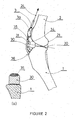
- FIG. 2 shows a medio-lateral view of a dog knee in the extended position with a spacer 30 of the present invention.
- Patellar ligament has been rotated from its original position 34 to a new position 35 by insertion of a spacer 30 between the patellar ligament and the tibia just proximally to the insertion point 36 of the ligament 35 (34) to the tibia 1.
- the angle 31 between the patellar ligament 35 and the tibial plateau 20 should be about 90 degrees when the knee is extended to keep the knee stable.
- FIG. 2a shows a cross-section through the spacer 30 and the tibia 1.
- the spacer should preferably be slightly grooved under the ligament so as to keep it in a stable position. This, however, is not an essential requirement - proximity of the insertion should guarantee a stable guidance of the ligament over the spacer.
- the patella 3 Under the same pull of the quadriceps 24, the patella 3 will get slightly displaced distally to a new position, marked as 3a. However, this will not affect its function.
- Figure 3a shows fixation of the spacer 30 to the tibia 1 by means of a bone screw 37, just proximally to the insertion 36 of the ligament 35.
- the head 38 of the screw 37 should be well countersunk to avoid contact with the patellar ligament 35 which, with flexion, will wrap to a new position marked as 35a.
- the undersurface 39 facing the tibia bone should preferably be suitable for bony ongrowth or ingrowth.
- Figure 3b shows an alternative method of fixation of the spacer 30 to the tibia 1 by means of an anchor 40.
- the anchor 40 is screwed into the bone with its conical head 41 left above the level of the bone.
- the spacer 30 is provided with a matching conical hole.
- the angle of the conical head and the hole should preferably be self-locking, e.g. 1:10 or 1:20.
- the bone facing surface 39 of the spacer 30 should preferably be suitable for bony ongrowth or ingrowth.
- Figure 3c shows yet another method of fixation of the spacer 30 to the tibia 1 by means of at least one flange 50, which allows placement of a transverse screw 51 from the medial aspect of the tibia.
- a second flange could be fixed from the lateral side.
- FIG. 3d Yet another method of fixation of the spacer 30 is shown on Figure 3d using a staple-shaped fixation device 60.
- the surface 42 of the spacer 30 facing the ligament should be highly polished and resistant to wear. Titanium nitride or diamond-like coatings are suitable for that purpose.
- Figure 4 shows a saggital view of a human knee with a spacer 130 inserted under the patellar ligament 105 to advance it from its original position 104.
- the spacer is fixed to the tibia 101.
- Patella 103 will be slightly displaced distally from its original position, marked as 103a, within the patellar groove 106 of the femur 102.
- Figures 5a-5c show orthogonal views of an embodiment of a spacer 130 for use in the human knee.
- the frontal view ( Figure 5a ) shows a central section 131 and side sections, or flanges, 132, provided with holes 133 to accommodate bone screws.
- the top view ( Figure 5c ) shows the central section 131 as being grooved, with the ridges 135 intended for guiding the patellar ligament.
- Side screws 134 can be comfortably placed into the tibial plateau, which in the human knee is much wider than in the dog knee.
- All of the bone facing surfaces 136 should be well adapted for bony ongrowth or ingrowth, while all of the ligament facing surfaces, e.g. 131 and 135, should be polished and preferably hard-coated for resistance to wear.
- ligament facing surfaces can be optimized for bony ongrowth - in the final adaptation, the ligament would be sliding over a bone surface.
- While the primary indication of the invention is rupture of the ACL in human or CrCL in dog, it can also be used for partial ruptures of the same. It may also be used as a supportive measure for ACL repairs, as well as for intra-articular prosthesis or extra-articular sutures. It can also be used as a substitute for Maquet procedure for patello-femoral joint arthrosis.
- Spacers produced from artificial, biocompatible materials have advantages in terms of convenience of use and selection of sizes, but a surgeon could also use a piece of bone, autograft or allograft, appropriately shaped and fixed to the tibia to achieve the same result.
- the procedure of the present disclosure includes advancing the orientation of the patellar ligament to alter the relative angle with the tibial plateau.
- the patellar ligament is advanced so that the relative angle with the tibial plateau is approximately 90 degrees when the knee is extended.
- the patellar ligament is advanced so that the angle is decreased in the range of 10 to 30 degrees.
- the patellar ligament is advanced so that the angle is decreased in the range of 5 to 15 degrees for a dog.
- the patellar ligament is advanced so that the angle is decreased in the range of 10 to 30 degrees for a human.
- the patellar ligament may be advanced by inserting a spacer between the ligament and the tibia proximal to the insertion point of the ligament. The spacer may be fixed to the tibia.
Landscapes
- Health & Medical Sciences (AREA)
- Life Sciences & Earth Sciences (AREA)
- Orthopedic Medicine & Surgery (AREA)
- Surgery (AREA)
- Animal Behavior & Ethology (AREA)
- General Health & Medical Sciences (AREA)
- Biomedical Technology (AREA)
- Heart & Thoracic Surgery (AREA)
- Engineering & Computer Science (AREA)
- Veterinary Medicine (AREA)
- Public Health (AREA)
- Medical Informatics (AREA)
- Nuclear Medicine, Radiotherapy & Molecular Imaging (AREA)
- Molecular Biology (AREA)
- Physical Education & Sports Medicine (AREA)
- Cardiology (AREA)
- Oral & Maxillofacial Surgery (AREA)
- Transplantation (AREA)
- Vascular Medicine (AREA)
- Prostheses (AREA)
Description
- The invention relates to implants to treat a knee with an injured or ruptured anterior cruciate ligament.
- The Anterior Cruciate Ligament (ACL) in the human knee joint, commonly called Cranial Cruciate Ligament (CrCL) in the canine stifle, is frequently torn in trauma. It also frequently fails, particularly in dogs, after a degenerative process of still unknown etiology.
- In human orthopedics, the standard procedure calls for replacement by an ACL allograft, a part of the patient's own patellar ligament, or a part of the tendon removed from his hamstring muscle. The procedure results in a stable knee, but the long-term performance is often unsatisfactory -- 75 to 90% of cases result in arthrosis of the joint within 15 years of the procedure.
- In dogs, the standard procedure is either an extra capsular suture or one of several geometry modifying surgical techniques. In the extra capsular procedure, a suture is placed outside of the joint, usually on the lateral side, to approximate the function of the ligament. The intention of the suture is to provide stability of the joint for several weeks, while waiting for fibrosis to form around the joint. The fibrosis should provide for long term stability. However, the extra capsular suture technique regularly results in failure. Arthrosis of the joint, at a year or so, is the rule rather exception.
- In surgical techniques, the tibia is cut and a segment of it is repositioned to change the geometry in order to stabilize the stifle. Various techniques have been used including: tibial plateau leveling osteotomy (TPLO; Slocum B, Slocum TD; Tibial Plateau Leveling Osteotomy For Repair Of Cranial Cruciate Ligament Rupture In The Canine, Vet. Clin. North Am. 23: 777-795, 1993), cranial closing wedge osteotomy (CWO; Slocum B, Devine T; Cranial Tibial Wedge Osteotomy: A Technique For Eliminating Cranial Tibial Thrust In Cranial Cruciate Ligament Repair, J. Am. Vet. Med. Assoc. 184: 564-569, 1984), and tibial tuberosity advancement (TTA; Tepic S, Damur DM, Montavon PM; Biomechanics Of The Stifle Joint, in Proceedings of the 1 st World Orthopaedic Vet. Congress, Munich, Germany, Sept 2002, pp 189-190). These are significantly invasive procedures which change the overall geometry of the stifle. Of the surgical approaches used in dogs, TTA seems to be associated with lesser morbidity and faster recovery, while providing immediate and durable stability to the joint. Nevertheless, complications and progressing arthrosis are common with each of these techniques.
InWO 2009/018365 A1 , on which the preamble ofclaim 1 is based, the spacer is shown rounded in the transverse direction of the ligament or tibia only but is straight in the longitudinal direction of the ligament or tibia. - Intra-articular prostheses are also occasionally used. Again, they require significantly invasive procedures to insert them within the stifle. Even then, they generally end up in failure.
- This invention provides a solution for a minimally invasive stabilization of a knee joint, in dog and in human, by cranially (anteriorly) advancing the patellar ligament just proximally to its insertion to the tibia. According to one aspect of the invention, a spacer is configured to be placed between the distal section of the patellar ligament and the tibia in accordance with
claim 1. The spacer is sized and shaped to alter the orientation of the patellar ligament by a predetermined amount. The alteration stabilizes the knee joint which has been weakened by an ACL (or CrCL) failure. According to another aspect of the invention, the spacer may be made from an artificial material, e.g. metal (titanium, stainless steel or a cobalt chromium alloy) or polymer (PEEK, UHMWPE), or from bone (autograft or allograft). According to another aspect of the invention, the spacer is fixed to the tibia body by either conventional screw(s) or an anchor screw, similar to those used for dental implants. According to another aspect of the invention, the surface of the spacer facing the patellar ligament is smooth and exhibits low friction. The material for the spacer should be biocompatible and resistant to wear. - According to another aspect of the invention,the surface of the spacer facing the patellar ligament is engineered so as to provide best conditions for bony ongrowth. For example, a titanium spacer can be anodized, particularly by a process known as Biocer®, or plasma sprayed by either a bone-conductive ceramic or titanium.
- According to another aspect of the invention, the spacer is configured to alter the angle of the patellar ligament relative to the tibial plateau. According to one aspect of the invention, the angle is decreased by between 5 and 15 degrees for dogs. According to another aspect of the invention, the angle is decreased by between 10 and 30 degrees in humans. According to another aspect of the invention, the angle of the patellar ligament relative to the tibial plateau is approximately 90 degrees when the knee is extended.
- The invention may be used in a procedure which provides stabilization of the knee joint caused by failure of the ACL or CrCL. The procedure includes moving the patellar ligament away from the tibia just above the tibial insertion. A spacer is inserted between the patellar ligament and the tibia and fixed to the tibia. The procedure may include altering the angle of the patellar ligament relative to the tibial plateau.
-
-
Figure 1 is a side view of a dog knee joint (stifle) on which the TTA procedure has been performed. -
Figure 2 is a side view of a dog knee joint having a patellar ligament spacer according to an embodiment of the invention. -
Figure 2a is a cross sectional view of the dog knee joint ofFig. 2 through the spacer. -
Figures 3a -3d illustrate mechanisms for fixing a spacer in a dog knee. -
Figure 4 is a side view of a human knee with a patellar ligament spacer according to an embodiment of the invention. -
Figures 5a-5c are front, side and top views of a spacer for a human knee joint according to an embodiment of the invention. - The present invention provides a spacer for adjusting the position and angle of the patellar ligament in order to stabilize the knee. The knee is stabilized through geometric changes similar to those achieved by TTA in a much less invasive procedure. The TTA procedure is explained in connection with
Figure 1 to illustrate stabilization of the knee. The TTA procedure is currently accepted as a standard treatment for ruptured ACL in dogs (Fossum TW, Small Animal Surgery, Elsevier Health Sciences, 3rd edition, 2007; Boudrieau R, Tibial Plateau Leveling Osteotomy Or Tibial Tuberosity Advancement?, Vet. Surg. 38:1-22, 2009).Figure 1 illustrates the knee joint both before and after the TTA procedure. Thetibia 1 and thefemur 2 articulate at the knee joint via condyles of thefemur 21 and condyles of the tibia, also commonly referred to as thetibial plateau 20. Pull 24 from the quadriceps muscles acts on thepatella 3, which in turn pulls onto the tibia via patellar ligament 5 (4 in its original orientation). The force between the condyles of the femur and the tibia is approximately parallel to thepatellar ligament 5, having been tilted from itsoriginal orientation 6 before the advancement (which was parallel to 4) into thenew orientation 7, which is perpendicular to theplateau 20. When thejoint force 7 is perpendicular to theplateau 20, neither the cranial (anterior) 8, nor the caudal (posterior)cruciate ligament 9 are needed to stabilize the joint. When this condition is satisfied in the extended position of the knee, such as shown onFigure 1 , the joint will also be stable when flexed, stability provided by the caudal (posterior) cruciate ligament. - Advancement of the tuberosity, after an opening wedge osteotomy, is maintained by a
cage 13 fixed to the tibia with aposterior screw 14 and ananterior screw 15. The tension of thepatellar ligament 5 is transferred to the body of the tibia via a tension-band plate 10, fixed distally to the tibia withscrews 11, and proximally to the tuberosity with afork 12. - In addition to balancing the
internal force 7 of the knee joint, TTA also reduces the force 23 (originally 22) between the patella and the femur. A procedure similar to TTA has been invented and performed in human surgery as well by Maquet (Maquet P; Advancement Of The Tibial Tuberosity, Clin. Orthop. Rel. Res. 115: 225-230, 1976). - The procedure of the present disclosure provides similar changes to those of TTA without the need to cut and reattach the tuberosity. The advantages of the invention are obtained through placement of a spacer between the tibia and the patellar ligament.
Figure 2 shows a medio-lateral view of a dog knee in the extended position with aspacer 30 of the present invention. Patellar ligament has been rotated from itsoriginal position 34 to anew position 35 by insertion of aspacer 30 between the patellar ligament and the tibia just proximally to theinsertion point 36 of the ligament 35 (34) to thetibia 1. Theangle 31 between thepatellar ligament 35 and thetibial plateau 20 should be about 90 degrees when the knee is extended to keep the knee stable. In dogs, this calls for an average rotation of the patellar ligament on the order of 5 to 15 degrees; in humans, a larger correction of 10 to 30 degrees is needed for a full compensation of a ruptured ACL.Figure 2a shows a cross-section through thespacer 30 and thetibia 1. The spacer should preferably be slightly grooved under the ligament so as to keep it in a stable position. This, however, is not an essential requirement - proximity of the insertion should guarantee a stable guidance of the ligament over the spacer. - Under the same pull of the
quadriceps 24, thepatella 3 will get slightly displaced distally to a new position, marked as 3a. However, this will not affect its function. -
Figure 3a shows fixation of thespacer 30 to thetibia 1 by means of abone screw 37, just proximally to theinsertion 36 of theligament 35. Thehead 38 of thescrew 37 should be well countersunk to avoid contact with thepatellar ligament 35 which, with flexion, will wrap to a new position marked as 35a. Theundersurface 39 facing the tibia bone should preferably be suitable for bony ongrowth or ingrowth. -
Figure 3b shows an alternative method of fixation of thespacer 30 to thetibia 1 by means of ananchor 40. Theanchor 40 is screwed into the bone with itsconical head 41 left above the level of the bone. Thespacer 30 is provided with a matching conical hole. The angle of the conical head and the hole should preferably be self-locking, e.g. 1:10 or 1:20. Thebone facing surface 39 of thespacer 30 should preferably be suitable for bony ongrowth or ingrowth. -
Figure 3c shows yet another method of fixation of thespacer 30 to thetibia 1 by means of at least oneflange 50, which allows placement of atransverse screw 51 from the medial aspect of the tibia. A second flange could be fixed from the lateral side. - Yet another method of fixation of the
spacer 30 is shown onFigure 3d using a staple-shapedfixation device 60. - The
surface 42 of thespacer 30 facing the ligament should be highly polished and resistant to wear. Titanium nitride or diamond-like coatings are suitable for that purpose. -
Figure 4 shows a saggital view of a human knee with aspacer 130 inserted under thepatellar ligament 105 to advance it from itsoriginal position 104. The spacer is fixed to thetibia 101.Patella 103 will be slightly displaced distally from its original position, marked as 103a, within thepatellar groove 106 of thefemur 102. -
Figures 5a-5c show orthogonal views of an embodiment of aspacer 130 for use in the human knee. The frontal view (Figure 5a ) shows acentral section 131 and side sections, or flanges, 132, provided withholes 133 to accommodate bone screws. The top view (Figure 5c ) shows thecentral section 131 as being grooved, with theridges 135 intended for guiding the patellar ligament. Side screws 134 can be comfortably placed into the tibial plateau, which in the human knee is much wider than in the dog knee. All of thebone facing surfaces 136 should be well adapted for bony ongrowth or ingrowth, while all of the ligament facing surfaces, e.g. 131 and 135, should be polished and preferably hard-coated for resistance to wear. Alternatively, ligament facing surfaces can be optimized for bony ongrowth - in the final adaptation, the ligament would be sliding over a bone surface. - While the primary indication of the invention is rupture of the ACL in human or CrCL in dog, it can also be used for partial ruptures of the same. It may also be used as a supportive measure for ACL repairs, as well as for intra-articular prosthesis or extra-articular sutures. It can also be used as a substitute for Maquet procedure for patello-femoral joint arthrosis.
- Spacers produced from artificial, biocompatible materials have advantages in terms of convenience of use and selection of sizes, but a surgeon could also use a piece of bone, autograft or allograft, appropriately shaped and fixed to the tibia to achieve the same result.
- The procedure of the present disclosure includes advancing the orientation of the patellar ligament to alter the relative angle with the tibial plateau. In an example, the patellar ligament is advanced so that the relative angle with the tibial plateau is approximately 90 degrees when the knee is extended. In another example, the patellar ligament is advanced so that the angle is decreased in the range of 10 to 30 degrees. In another example, the patellar ligament is advanced so that the angle is decreased in the range of 5 to 15 degrees for a dog. In another example, the patellar ligament is advanced so that the angle is decreased in the range of 10 to 30 degrees for a human. The patellar ligament may be advanced by inserting a spacer between the ligament and the tibia proximal to the insertion point of the ligament. The spacer may be fixed to the tibia.
- Having disclosed at least one embodiment of the present invention, various adaptations, modifications, additions, and improvements will be readily apparent to those of ordinary skill in the art. Such adaptations, modifications, additions and improvements are considered part of the invention which is only limited by the several claims attached hereto.
Claims (12)
- A spacer (30; 130) adapted for implantation onto the anterior aspect of the tibia (1), under the patellar ligament (5; 35), proximally to its tibial attachment (36), whereby the spacer extends anteriorly from the tibia and holds the ligament away therefrom to alter the angle between the patellar ligament and the tibial plateau (20), the spacer having a bone facing surface and a surface facing the patellar ligament,
characterized in that the surface (42; 131) of the spacer facing the patellar ligament is adjacent to the bone facing surface along two parallel sides and is continuously rounded in a direction perpendicular to the parallel sides and parallel to an axis of the ligament. - The spacer of claim 1 whereby its surface (42; 131) facing the ligament is polished and its surface (136) facing the bone is adapted for bony integration.
- The spacer of claim 1 whereby both, its surface (42; 131) facing the ligament and its surface (136) facing the bone are adapted for bony integration.
- The spacer of claim 1 dimensioned so as to make the angle (31) between the patellar ligament and the tibial plateau equal to about 90 degrees with the knee extended.
- The spacer of claim 1 dimensioned so as decrease the angle between the patellar ligament and the tibial plateau by about 5 to 15 degrees in dogs.
- The spacer of claim 1 dimensioned so as decrease the angle between the patellar ligament and the tibial plateau by about 10 to 30 degrees in human patients.
- The spacer of claim 1 provided by at least one bone screw fixation for the tibia.
- The spacer of claim 1 provided by at least one screw anchor fixation for the tibia.
- The spacer of claim 1 provided by at least one flange (132) and a screw (134) for fixation to the tibia.
- The spacer of claim 1 provided by at least one staple for fixation to the tibia.
- The spacer of claim 1 provided by at least two side screws (134) for fixation to the tibia.
- The spacer of claim 1, wherein in a cross-section view through the spacer (30; 130) and the tibia (1) the surface (42; 131) of the spacer (30; 130) facing the ligament (5; 35) is slightly grooved.
Priority Applications (1)
| Application Number | Priority Date | Filing Date | Title |
|---|---|---|---|
| EP11782141.3A EP2637581B1 (en) | 2010-11-12 | 2011-11-11 | Patellar ligament spacer for acl injuries |
Applications Claiming Priority (3)
| Application Number | Priority Date | Filing Date | Title |
|---|---|---|---|
| EP10191075A EP2452641A1 (en) | 2010-11-12 | 2010-11-12 | Patellar ligament spacer for ACL injuries |
| PCT/EP2011/069957 WO2012062908A1 (en) | 2010-11-12 | 2011-11-11 | Patellar ligament spacer for acl injuries |
| EP11782141.3A EP2637581B1 (en) | 2010-11-12 | 2011-11-11 | Patellar ligament spacer for acl injuries |
Publications (2)
| Publication Number | Publication Date |
|---|---|
| EP2637581A1 EP2637581A1 (en) | 2013-09-18 |
| EP2637581B1 true EP2637581B1 (en) | 2015-02-11 |
Family
ID=43606451
Family Applications (2)
| Application Number | Title | Priority Date | Filing Date |
|---|---|---|---|
| EP10191075A Withdrawn EP2452641A1 (en) | 2010-11-12 | 2010-11-12 | Patellar ligament spacer for ACL injuries |
| EP11782141.3A Active EP2637581B1 (en) | 2010-11-12 | 2011-11-11 | Patellar ligament spacer for acl injuries |
Family Applications Before (1)
| Application Number | Title | Priority Date | Filing Date |
|---|---|---|---|
| EP10191075A Withdrawn EP2452641A1 (en) | 2010-11-12 | 2010-11-12 | Patellar ligament spacer for ACL injuries |
Country Status (3)
| Country | Link |
|---|---|
| US (1) | US10130481B2 (en) |
| EP (2) | EP2452641A1 (en) |
| WO (1) | WO2012062908A1 (en) |
Families Citing this family (22)
| Publication number | Priority date | Publication date | Assignee | Title |
|---|---|---|---|---|
| CA2771332C (en) | 2009-08-27 | 2020-11-10 | Cotera, Inc. | Method and apparatus for force redistribution in articular joints |
| US9278004B2 (en) | 2009-08-27 | 2016-03-08 | Cotera, Inc. | Method and apparatus for altering biomechanics of the articular joints |
| US10349980B2 (en) | 2009-08-27 | 2019-07-16 | The Foundry, Llc | Method and apparatus for altering biomechanics of the shoulder |
| US9668868B2 (en) | 2009-08-27 | 2017-06-06 | Cotera, Inc. | Apparatus and methods for treatment of patellofemoral conditions |
| US9861408B2 (en) | 2009-08-27 | 2018-01-09 | The Foundry, Llc | Method and apparatus for treating canine cruciate ligament disease |
| US9737349B2 (en) | 2012-05-30 | 2017-08-22 | Kyon Ag | Tibial tuberosity advancement cage for ACL injuries |
| US8790410B2 (en) | 2012-07-26 | 2014-07-29 | Mwi Veterinary Supply Co. | Tibial tuberosity advancement implant |
| US9468466B1 (en) | 2012-08-24 | 2016-10-18 | Cotera, Inc. | Method and apparatus for altering biomechanics of the spine |
| GB2507965B (en) * | 2012-11-14 | 2018-12-19 | Steris Instrument Man Services Inc | Cage assembly for tibial tuberosity advancement procedure |
| US9095438B1 (en) | 2012-12-17 | 2015-08-04 | Ellen S. Wait | Surgical implant for ostectomy procedures |
| WO2014149934A1 (en) * | 2013-03-15 | 2014-09-25 | Moximed, Inc. | Structure and method for treating patello-femoral osteoarthritis |
| EP3049003B1 (en) * | 2013-09-26 | 2019-09-04 | Matthew Hansen | Prosthetic augments to improve muscle mechanics |
| KR101549327B1 (en) | 2014-02-10 | 2015-09-01 | 전북대학교산학협력단 | composites implant and surgical techniques for a tibial tuberosity advancement in treatment of a cranial cruciate ligament rupture |
| WO2015138594A1 (en) * | 2014-03-11 | 2015-09-17 | Cotera, Inc. | Apparatus and methods for treatment of patellofemoral conditions |
| PL234332B1 (en) * | 2017-11-14 | 2020-02-28 | Chm Spolka Z Ograniczona Odpowiedzialnoscia | An implant for stabilization of the knee joint by TTA method |
| WO2021016418A1 (en) | 2019-07-23 | 2021-01-28 | Coleman Robert Glen | Tibial plateau leveling osteotomy systems and methods |
| WO2021074217A1 (en) * | 2019-10-15 | 2021-04-22 | Kyon Ag | Tibia-calcaneus truss |
| WO2021138080A1 (en) * | 2020-01-02 | 2021-07-08 | Zkr Orthopedics, Inc. | Patella tendon realignment implant with fixation |
| EP4084711A4 (en) | 2020-01-02 | 2024-01-03 | ZKR Orthopedics, Inc. | IMPLANT FOR REALIGNING THE PATELLA TENDON WITH A CHANGABLE SHAPE |
| WO2021231253A1 (en) * | 2020-05-11 | 2021-11-18 | Zkr Orthopedics, Inc. | Adjustable patellar tendon realignment implant |
| CN111870407B (en) * | 2020-08-10 | 2024-12-17 | 北京大学第三医院(北京大学第三临床医学院) | Structure of bone-patellar tendon-bone graft and preparation method thereof |
| AU2024334108A1 (en) | 2023-08-29 | 2026-02-26 | Zkr Orthopedics, Inc. | Multiplanar tendon realignment implants and related systems and methods |
Family Cites Families (39)
| Publication number | Priority date | Publication date | Assignee | Title |
|---|---|---|---|---|
| US3872519A (en) * | 1974-04-04 | 1975-03-25 | Nicholas J Giannestras | Total ankle prosthesis |
| US4052753A (en) * | 1976-08-02 | 1977-10-11 | Dedo Richard G | Knee spacer and method of reforming sliding body surfaces |
| US4431416A (en) * | 1982-04-29 | 1984-02-14 | A & L Investment Company | Endosseous dental implant system for overdenture retention, crown and bridge support |
| DE3413398A1 (en) * | 1984-02-07 | 1985-08-14 | Rufino Madrid Aragoneses Ureña | ARRANGEMENT FOR THE PRODUCTION OF SOLID DENTAL PROSTHESES OR FIXED DENTAL PROSTHESIS |
| DE3434309A1 (en) * | 1984-09-19 | 1986-03-27 | S + G Implants GmbH, 2400 Lübeck | IMPLANT AS REPLACEMENT FOR AN EXTRACTED TOOTH |
| US5151103A (en) * | 1987-11-03 | 1992-09-29 | Synthes (U.S.A.) | Point contact bone compression plate |
| WO1989004150A1 (en) * | 1987-11-03 | 1989-05-18 | Synthes Ag | Implant for osteosynthesis |
| FR2693899B1 (en) * | 1992-07-24 | 1994-09-23 | Laboureau Jacques | Osteosynthesis plate clip. |
| US5662652A (en) * | 1994-04-28 | 1997-09-02 | Schafer Micomed Gmbh | Bone surgery holding apparatus |
| US6290726B1 (en) * | 2000-01-30 | 2001-09-18 | Diamicron, Inc. | Prosthetic hip joint having sintered polycrystalline diamond compact articulation surfaces |
| ES2158127T3 (en) * | 1995-09-06 | 2001-09-01 | Synthes Ag | OSEA SHEET. |
| CA2259656C (en) * | 1996-07-09 | 2007-01-23 | Synthes (U.S.A.) | Device for bone surgery |
| US5954722A (en) * | 1997-07-29 | 1999-09-21 | Depuy Acromed, Inc. | Polyaxial locking plate |
| US6673116B2 (en) * | 1999-10-22 | 2004-01-06 | Mark A. Reiley | Intramedullary guidance systems and methods for installing ankle replacement prostheses |
| US6702821B2 (en) * | 2000-01-14 | 2004-03-09 | The Bonutti 2003 Trust A | Instrumentation for minimally invasive joint replacement and methods for using same |
| US7713305B2 (en) * | 2000-05-01 | 2010-05-11 | Arthrosurface, Inc. | Articular surface implant |
| US8398717B2 (en) * | 2008-11-10 | 2013-03-19 | Acumed Llc | Partial or complete prosthetic replacement arthroplasty of the distal radioulnar joint |
| AU2003297195A1 (en) * | 2002-12-17 | 2004-07-22 | Amedica Corporation | Total disc implant |
| US20040162558A1 (en) * | 2003-02-18 | 2004-08-19 | Hegde Sajan K. | Spinal plate having an integral rod connector portion |
| US20040186585A1 (en) * | 2003-03-21 | 2004-09-23 | Lawrence Feiwell | Sphere-on-sphere ankle prosthesis |
| DE20313183U1 (en) * | 2003-08-22 | 2003-10-16 | Aesculap Ag & Co Kg | Intervertebral implant |
| US7534270B2 (en) * | 2003-09-03 | 2009-05-19 | Integra Lifesciences Corporation | Modular total ankle prosthesis apparatuses and methods |
| US20050165487A1 (en) * | 2004-01-28 | 2005-07-28 | Muhanna Nabil L. | Artificial intervertebral disc |
| US20050288792A1 (en) * | 2004-06-23 | 2005-12-29 | Landes Mark D | Modular ankle prosthesis and associated method |
| US7806898B2 (en) * | 2004-07-09 | 2010-10-05 | Zimmer, Inc. | Modular guide systems and related rasps and methods for resecting a joint articulation surface |
| WO2006023824A2 (en) * | 2004-08-19 | 2006-03-02 | Kinetikos Medical Incorporated | Ankle prosthesis and method of curved resection |
| US20060085077A1 (en) * | 2004-10-18 | 2006-04-20 | Ebi, L.P. | Intervertebral implant and associated method |
| US7160331B2 (en) * | 2004-12-01 | 2007-01-09 | Mayo Foundation For Medical Research And Education | Sigmoid notch implant |
| US20060149372A1 (en) * | 2004-12-17 | 2006-07-06 | Paxson Robert D | Artificial spinal disc |
| CA2601090C (en) * | 2005-03-14 | 2013-08-20 | Inbone Technologies, Inc. | Ankle replacement system |
| US7824433B2 (en) * | 2005-05-03 | 2010-11-02 | Williams Lytton A | Bone anchored surgical mesh |
| JP2009531078A (en) * | 2006-03-02 | 2009-09-03 | タラス メディカル, インコーポレイテッド | Artificial bone |
| US20070288021A1 (en) * | 2006-06-07 | 2007-12-13 | Howmedica Osteonics Corp. | Flexible joint implant |
| EP2073761B1 (en) * | 2006-09-21 | 2010-07-07 | SpineCore, Inc. | Intervertebral disc implants and tooling |
| EP2139434B1 (en) * | 2007-04-20 | 2012-10-10 | Woodwelding AG | Implant sysrem for fastening an implant to bone tissue |
| WO2009018365A1 (en) * | 2007-08-01 | 2009-02-05 | Jeffrey Halbrecht | Method and system for patella tendon realignment |
| AU2009205679B2 (en) * | 2008-01-18 | 2013-12-05 | Spinecore, Inc. | Instruments and methods for inserting artificial intervertebral implants |
| US20100057216A1 (en) * | 2008-07-23 | 2010-03-04 | Jamy Gannoe | System and method for joint resurfacing with dynamic fixation |
| CA2771332C (en) * | 2009-08-27 | 2020-11-10 | Cotera, Inc. | Method and apparatus for force redistribution in articular joints |
-
2010
- 2010-11-12 EP EP10191075A patent/EP2452641A1/en not_active Withdrawn
-
2011
- 2011-11-11 WO PCT/EP2011/069957 patent/WO2012062908A1/en not_active Ceased
- 2011-11-11 EP EP11782141.3A patent/EP2637581B1/en active Active
- 2011-11-11 US US13/877,363 patent/US10130481B2/en active Active
Also Published As
| Publication number | Publication date |
|---|---|
| EP2452641A1 (en) | 2012-05-16 |
| US20130190886A1 (en) | 2013-07-25 |
| WO2012062908A1 (en) | 2012-05-18 |
| EP2637581A1 (en) | 2013-09-18 |
| US10130481B2 (en) | 2018-11-20 |
Similar Documents
| Publication | Publication Date | Title |
|---|---|---|
| US10130481B2 (en) | Patellar ligament spacer for ACL injuries | |
| US8118868B2 (en) | Method and apparatus for attaching soft tissue to an implant | |
| US12115080B2 (en) | Total knee implant prosthesis assembly and method | |
| US5019103A (en) | Tibial wedge system | |
| EP2854677B1 (en) | Tibial tuberosity advancement cage for acl injuries | |
| EP2967877B1 (en) | Quadruped stifle stabilization system | |
| US6485519B2 (en) | Constrained prosthetic knee with rotating bearing | |
| US10736750B2 (en) | Releasable threaded connection for modular implants | |
| US12433753B2 (en) | Unicompartmental knee arthroplasty systems and methods | |
| KR102649339B1 (en) | tibial plateau patch | |
| US20140277536A1 (en) | Orthopaedic knee prosthesis having stem components with a varying number of slots | |
| CA2924694C (en) | Reverse knee prosthesis | |
| US10039645B2 (en) | Releasable threaded connection for modular implants | |
| EP2937051A1 (en) | Implant for tibial plateau leveling osteotomy fixation | |
| US20250339279A1 (en) | Unicompartmental knee arthroplasty systems and methods | |
| GB2534140A (en) | Joint prosthesis supporting assembly | |
| US11129606B2 (en) | Method and apparatus for intercondylar notch distraction knee arthroplasty | |
| WO2023081319A1 (en) | Unicompartmental knee arthroplasty systems and methods | |
| II | ANALYSIS OF COMPONENTS ALIGNMENT IN TOTAL KNEE REPLACEMENT USING TRADITIONAL JIGS AND ITS RELATIONSHIP TO THE FUNCTIONAL OUTCOME |
Legal Events
| Date | Code | Title | Description |
|---|---|---|---|
| PUAI | Public reference made under article 153(3) epc to a published international application that has entered the european phase |
Free format text: ORIGINAL CODE: 0009012 |
|
| 17P | Request for examination filed |
Effective date: 20130430 |
|
| AK | Designated contracting states |
Kind code of ref document: A1 Designated state(s): AL AT BE BG CH CY CZ DE DK EE ES FI FR GB GR HR HU IE IS IT LI LT LU LV MC MK MT NL NO PL PT RO RS SE SI SK SM TR |
|
| DAX | Request for extension of the european patent (deleted) | ||
| RIC1 | Information provided on ipc code assigned before grant |
Ipc: A61F 2/38 20060101ALI20140701BHEP Ipc: A61B 17/56 20060101AFI20140701BHEP |
|
| GRAP | Despatch of communication of intention to grant a patent |
Free format text: ORIGINAL CODE: EPIDOSNIGR1 |
|
| INTG | Intention to grant announced |
Effective date: 20140821 |
|
| GRAS | Grant fee paid |
Free format text: ORIGINAL CODE: EPIDOSNIGR3 |
|
| GRAA | (expected) grant |
Free format text: ORIGINAL CODE: 0009210 |
|
| AK | Designated contracting states |
Kind code of ref document: B1 Designated state(s): AL AT BE BG CH CY CZ DE DK EE ES FI FR GB GR HR HU IE IS IT LI LT LU LV MC MK MT NL NO PL PT RO RS SE SI SK SM TR |
|
| REG | Reference to a national code |
Ref country code: GB Ref legal event code: FG4D |
|
| REG | Reference to a national code |
Ref country code: CH Ref legal event code: EP |
|
| REG | Reference to a national code |
Ref country code: IE Ref legal event code: FG4D |
|
| REG | Reference to a national code |
Ref country code: AT Ref legal event code: REF Ref document number: 709664 Country of ref document: AT Kind code of ref document: T Effective date: 20150315 |
|
| REG | Reference to a national code |
Ref country code: DE Ref legal event code: R096 Ref document number: 602011013672 Country of ref document: DE Effective date: 20150326 |
|
| REG | Reference to a national code |
Ref country code: CH Ref legal event code: NV Representative=s name: E. BLUM AND CO. AG PATENT- UND MARKENANWAELTE , CH |
|
| REG | Reference to a national code |
Ref country code: NL Ref legal event code: T3 |
|
| REG | Reference to a national code |
Ref country code: AT Ref legal event code: MK05 Ref document number: 709664 Country of ref document: AT Kind code of ref document: T Effective date: 20150211 |
|
| REG | Reference to a national code |
Ref country code: LT Ref legal event code: MG4D |
|
| PG25 | Lapsed in a contracting state [announced via postgrant information from national office to epo] |
Ref country code: NO Free format text: LAPSE BECAUSE OF FAILURE TO SUBMIT A TRANSLATION OF THE DESCRIPTION OR TO PAY THE FEE WITHIN THE PRESCRIBED TIME-LIMIT Effective date: 20150511 Ref country code: LT Free format text: LAPSE BECAUSE OF FAILURE TO SUBMIT A TRANSLATION OF THE DESCRIPTION OR TO PAY THE FEE WITHIN THE PRESCRIBED TIME-LIMIT Effective date: 20150211 Ref country code: HR Free format text: LAPSE BECAUSE OF FAILURE TO SUBMIT A TRANSLATION OF THE DESCRIPTION OR TO PAY THE FEE WITHIN THE PRESCRIBED TIME-LIMIT Effective date: 20150211 Ref country code: SE Free format text: LAPSE BECAUSE OF FAILURE TO SUBMIT A TRANSLATION OF THE DESCRIPTION OR TO PAY THE FEE WITHIN THE PRESCRIBED TIME-LIMIT Effective date: 20150211 Ref country code: ES Free format text: LAPSE BECAUSE OF FAILURE TO SUBMIT A TRANSLATION OF THE DESCRIPTION OR TO PAY THE FEE WITHIN THE PRESCRIBED TIME-LIMIT Effective date: 20150211 Ref country code: FI Free format text: LAPSE BECAUSE OF FAILURE TO SUBMIT A TRANSLATION OF THE DESCRIPTION OR TO PAY THE FEE WITHIN THE PRESCRIBED TIME-LIMIT Effective date: 20150211 |
|
| PG25 | Lapsed in a contracting state [announced via postgrant information from national office to epo] |
Ref country code: IS Free format text: LAPSE BECAUSE OF FAILURE TO SUBMIT A TRANSLATION OF THE DESCRIPTION OR TO PAY THE FEE WITHIN THE PRESCRIBED TIME-LIMIT Effective date: 20150611 Ref country code: LV Free format text: LAPSE BECAUSE OF FAILURE TO SUBMIT A TRANSLATION OF THE DESCRIPTION OR TO PAY THE FEE WITHIN THE PRESCRIBED TIME-LIMIT Effective date: 20150211 Ref country code: RS Free format text: LAPSE BECAUSE OF FAILURE TO SUBMIT A TRANSLATION OF THE DESCRIPTION OR TO PAY THE FEE WITHIN THE PRESCRIBED TIME-LIMIT Effective date: 20150211 Ref country code: AT Free format text: LAPSE BECAUSE OF FAILURE TO SUBMIT A TRANSLATION OF THE DESCRIPTION OR TO PAY THE FEE WITHIN THE PRESCRIBED TIME-LIMIT Effective date: 20150211 Ref country code: GR Free format text: LAPSE BECAUSE OF FAILURE TO SUBMIT A TRANSLATION OF THE DESCRIPTION OR TO PAY THE FEE WITHIN THE PRESCRIBED TIME-LIMIT Effective date: 20150512 |
|
| PG25 | Lapsed in a contracting state [announced via postgrant information from national office to epo] |
Ref country code: EE Free format text: LAPSE BECAUSE OF FAILURE TO SUBMIT A TRANSLATION OF THE DESCRIPTION OR TO PAY THE FEE WITHIN THE PRESCRIBED TIME-LIMIT Effective date: 20150211 Ref country code: SK Free format text: LAPSE BECAUSE OF FAILURE TO SUBMIT A TRANSLATION OF THE DESCRIPTION OR TO PAY THE FEE WITHIN THE PRESCRIBED TIME-LIMIT Effective date: 20150211 Ref country code: RO Free format text: LAPSE BECAUSE OF FAILURE TO SUBMIT A TRANSLATION OF THE DESCRIPTION OR TO PAY THE FEE WITHIN THE PRESCRIBED TIME-LIMIT Effective date: 20150211 Ref country code: CZ Free format text: LAPSE BECAUSE OF FAILURE TO SUBMIT A TRANSLATION OF THE DESCRIPTION OR TO PAY THE FEE WITHIN THE PRESCRIBED TIME-LIMIT Effective date: 20150211 Ref country code: DK Free format text: LAPSE BECAUSE OF FAILURE TO SUBMIT A TRANSLATION OF THE DESCRIPTION OR TO PAY THE FEE WITHIN THE PRESCRIBED TIME-LIMIT Effective date: 20150211 |
|
| REG | Reference to a national code |
Ref country code: DE Ref legal event code: R097 Ref document number: 602011013672 Country of ref document: DE |
|
| PG25 | Lapsed in a contracting state [announced via postgrant information from national office to epo] |
Ref country code: PL Free format text: LAPSE BECAUSE OF FAILURE TO SUBMIT A TRANSLATION OF THE DESCRIPTION OR TO PAY THE FEE WITHIN THE PRESCRIBED TIME-LIMIT Effective date: 20150211 |
|
| PLBE | No opposition filed within time limit |
Free format text: ORIGINAL CODE: 0009261 |
|
| STAA | Information on the status of an ep patent application or granted ep patent |
Free format text: STATUS: NO OPPOSITION FILED WITHIN TIME LIMIT |
|
| 26N | No opposition filed |
Effective date: 20151112 |
|
| PG25 | Lapsed in a contracting state [announced via postgrant information from national office to epo] |
Ref country code: SI Free format text: LAPSE BECAUSE OF FAILURE TO SUBMIT A TRANSLATION OF THE DESCRIPTION OR TO PAY THE FEE WITHIN THE PRESCRIBED TIME-LIMIT Effective date: 20150211 |
|
| PG25 | Lapsed in a contracting state [announced via postgrant information from national office to epo] |
Ref country code: BE Free format text: LAPSE BECAUSE OF FAILURE TO SUBMIT A TRANSLATION OF THE DESCRIPTION OR TO PAY THE FEE WITHIN THE PRESCRIBED TIME-LIMIT Effective date: 20150211 |
|
| PG25 | Lapsed in a contracting state [announced via postgrant information from national office to epo] |
Ref country code: MC Free format text: LAPSE BECAUSE OF FAILURE TO SUBMIT A TRANSLATION OF THE DESCRIPTION OR TO PAY THE FEE WITHIN THE PRESCRIBED TIME-LIMIT Effective date: 20150211 Ref country code: LU Free format text: LAPSE BECAUSE OF FAILURE TO SUBMIT A TRANSLATION OF THE DESCRIPTION OR TO PAY THE FEE WITHIN THE PRESCRIBED TIME-LIMIT Effective date: 20151111 |
|
| REG | Reference to a national code |
Ref country code: IE Ref legal event code: MM4A |
|
| REG | Reference to a national code |
Ref country code: FR Ref legal event code: ST Effective date: 20160729 |
|
| PG25 | Lapsed in a contracting state [announced via postgrant information from national office to epo] |
Ref country code: IE Free format text: LAPSE BECAUSE OF NON-PAYMENT OF DUE FEES Effective date: 20151111 |
|
| PG25 | Lapsed in a contracting state [announced via postgrant information from national office to epo] |
Ref country code: FR Free format text: LAPSE BECAUSE OF NON-PAYMENT OF DUE FEES Effective date: 20151130 |
|
| PG25 | Lapsed in a contracting state [announced via postgrant information from national office to epo] |
Ref country code: BG Free format text: LAPSE BECAUSE OF FAILURE TO SUBMIT A TRANSLATION OF THE DESCRIPTION OR TO PAY THE FEE WITHIN THE PRESCRIBED TIME-LIMIT Effective date: 20150211 Ref country code: SM Free format text: LAPSE BECAUSE OF FAILURE TO SUBMIT A TRANSLATION OF THE DESCRIPTION OR TO PAY THE FEE WITHIN THE PRESCRIBED TIME-LIMIT Effective date: 20150211 Ref country code: HU Free format text: LAPSE BECAUSE OF FAILURE TO SUBMIT A TRANSLATION OF THE DESCRIPTION OR TO PAY THE FEE WITHIN THE PRESCRIBED TIME-LIMIT; INVALID AB INITIO Effective date: 20111111 |
|
| PG25 | Lapsed in a contracting state [announced via postgrant information from national office to epo] |
Ref country code: CY Free format text: LAPSE BECAUSE OF FAILURE TO SUBMIT A TRANSLATION OF THE DESCRIPTION OR TO PAY THE FEE WITHIN THE PRESCRIBED TIME-LIMIT Effective date: 20150211 |
|
| PG25 | Lapsed in a contracting state [announced via postgrant information from national office to epo] |
Ref country code: MT Free format text: LAPSE BECAUSE OF FAILURE TO SUBMIT A TRANSLATION OF THE DESCRIPTION OR TO PAY THE FEE WITHIN THE PRESCRIBED TIME-LIMIT Effective date: 20150211 |
|
| PG25 | Lapsed in a contracting state [announced via postgrant information from national office to epo] |
Ref country code: PT Free format text: LAPSE BECAUSE OF FAILURE TO SUBMIT A TRANSLATION OF THE DESCRIPTION OR TO PAY THE FEE WITHIN THE PRESCRIBED TIME-LIMIT Effective date: 20150211 Ref country code: TR Free format text: LAPSE BECAUSE OF FAILURE TO SUBMIT A TRANSLATION OF THE DESCRIPTION OR TO PAY THE FEE WITHIN THE PRESCRIBED TIME-LIMIT Effective date: 20150211 Ref country code: MK Free format text: LAPSE BECAUSE OF FAILURE TO SUBMIT A TRANSLATION OF THE DESCRIPTION OR TO PAY THE FEE WITHIN THE PRESCRIBED TIME-LIMIT Effective date: 20150211 |
|
| PG25 | Lapsed in a contracting state [announced via postgrant information from national office to epo] |
Ref country code: AL Free format text: LAPSE BECAUSE OF FAILURE TO SUBMIT A TRANSLATION OF THE DESCRIPTION OR TO PAY THE FEE WITHIN THE PRESCRIBED TIME-LIMIT Effective date: 20150211 |
|
| PGFP | Annual fee paid to national office [announced via postgrant information from national office to epo] |
Ref country code: NL Payment date: 20231120 Year of fee payment: 13 |
|
| PGFP | Annual fee paid to national office [announced via postgrant information from national office to epo] |
Ref country code: IT Payment date: 20231124 Year of fee payment: 13 Ref country code: DE Payment date: 20231031 Year of fee payment: 13 Ref country code: CH Payment date: 20231201 Year of fee payment: 13 |
|
| PGFP | Annual fee paid to national office [announced via postgrant information from national office to epo] |
Ref country code: GB Payment date: 20241120 Year of fee payment: 14 |
|
| REG | Reference to a national code |
Ref country code: DE Ref legal event code: R119 Ref document number: 602011013672 Country of ref document: DE |
|
| REG | Reference to a national code |
Ref country code: CH Ref legal event code: PL |
|
| REG | Reference to a national code |
Ref country code: NL Ref legal event code: MM Effective date: 20241201 |
|
| REG | Reference to a national code |
Ref country code: CH Ref legal event code: PL |
|
| PG25 | Lapsed in a contracting state [announced via postgrant information from national office to epo] |
Ref country code: CH Free format text: LAPSE BECAUSE OF NON-PAYMENT OF DUE FEES Effective date: 20241130 |
|
| PG25 | Lapsed in a contracting state [announced via postgrant information from national office to epo] |
Ref country code: NL Free format text: LAPSE BECAUSE OF NON-PAYMENT OF DUE FEES Effective date: 20241201 |
|
| PG25 | Lapsed in a contracting state [announced via postgrant information from national office to epo] |
Ref country code: DE Free format text: LAPSE BECAUSE OF NON-PAYMENT OF DUE FEES Effective date: 20250603 |
|
| PG25 | Lapsed in a contracting state [announced via postgrant information from national office to epo] |
Ref country code: IT Free format text: LAPSE BECAUSE OF NON-PAYMENT OF DUE FEES Effective date: 20241111 |