EP2630487B1 - Method for determining a degree of mastitis infection - Google Patents
Method for determining a degree of mastitis infection Download PDFInfo
- Publication number
- EP2630487B1 EP2630487B1 EP10768012.6A EP10768012A EP2630487B1 EP 2630487 B1 EP2630487 B1 EP 2630487B1 EP 10768012 A EP10768012 A EP 10768012A EP 2630487 B1 EP2630487 B1 EP 2630487B1
- Authority
- EP
- European Patent Office
- Prior art keywords
- mastitis
- degree
- indication
- infection
- milk
- Prior art date
- Legal status (The legal status is an assumption and is not a legal conclusion. Google has not performed a legal analysis and makes no representation as to the accuracy of the status listed.)
- Revoked
Links
Images




Classifications
-
- C—CHEMISTRY; METALLURGY
- C12—BIOCHEMISTRY; BEER; SPIRITS; WINE; VINEGAR; MICROBIOLOGY; ENZYMOLOGY; MUTATION OR GENETIC ENGINEERING
- C12Q—MEASURING OR TESTING PROCESSES INVOLVING ENZYMES, NUCLEIC ACIDS OR MICROORGANISMS; COMPOSITIONS OR TEST PAPERS THEREFOR; PROCESSES OF PREPARING SUCH COMPOSITIONS; CONDITION-RESPONSIVE CONTROL IN MICROBIOLOGICAL OR ENZYMOLOGICAL PROCESSES
- C12Q1/00—Measuring or testing processes involving enzymes, nucleic acids or microorganisms; Compositions therefor; Processes of preparing such compositions
- C12Q1/02—Measuring or testing processes involving enzymes, nucleic acids or microorganisms; Compositions therefor; Processes of preparing such compositions involving viable microorganisms
- C12Q1/04—Determining presence or kind of microorganism; Use of selective media for testing antibiotics or bacteriocides; Compositions containing a chemical indicator therefor
- C12Q1/06—Quantitative determination
-
- G—PHYSICS
- G01—MEASURING; TESTING
- G01N—INVESTIGATING OR ANALYSING MATERIALS BY DETERMINING THEIR CHEMICAL OR PHYSICAL PROPERTIES
- G01N33/00—Investigating or analysing materials by specific methods not covered by groups G01N1/00 - G01N31/00
- G01N33/48—Biological material, e.g. blood, urine; Haemocytometers
- G01N33/50—Chemical analysis of biological material, e.g. blood, urine; Testing involving biospecific ligand binding methods; Immunological testing
- G01N33/58—Chemical analysis of biological material, e.g. blood, urine; Testing involving biospecific ligand binding methods; Immunological testing involving labelled substances
- G01N33/582—Chemical analysis of biological material, e.g. blood, urine; Testing involving biospecific ligand binding methods; Immunological testing involving labelled substances with fluorescent label
-
- G—PHYSICS
- G01—MEASURING; TESTING
- G01N—INVESTIGATING OR ANALYSING MATERIALS BY DETERMINING THEIR CHEMICAL OR PHYSICAL PROPERTIES
- G01N33/00—Investigating or analysing materials by specific methods not covered by groups G01N1/00 - G01N31/00
- G01N33/48—Biological material, e.g. blood, urine; Haemocytometers
- G01N33/50—Chemical analysis of biological material, e.g. blood, urine; Testing involving biospecific ligand binding methods; Immunological testing
- G01N33/5005—Chemical analysis of biological material, e.g. blood, urine; Testing involving biospecific ligand binding methods; Immunological testing involving human or animal cells
- G01N33/5008—Chemical analysis of biological material, e.g. blood, urine; Testing involving biospecific ligand binding methods; Immunological testing involving human or animal cells for testing or evaluating the effect of chemical or biological compounds, e.g. drugs, cosmetics
- G01N33/5044—Chemical analysis of biological material, e.g. blood, urine; Testing involving biospecific ligand binding methods; Immunological testing involving human or animal cells for testing or evaluating the effect of chemical or biological compounds, e.g. drugs, cosmetics involving specific cell types
- G01N33/5047—Cells of the immune system
-
- G—PHYSICS
- G01—MEASURING; TESTING
- G01N—INVESTIGATING OR ANALYSING MATERIALS BY DETERMINING THEIR CHEMICAL OR PHYSICAL PROPERTIES
- G01N33/00—Investigating or analysing materials by specific methods not covered by groups G01N1/00 - G01N31/00
- G01N33/48—Biological material, e.g. blood, urine; Haemocytometers
- G01N33/50—Chemical analysis of biological material, e.g. blood, urine; Testing involving biospecific ligand binding methods; Immunological testing
- G01N33/5005—Chemical analysis of biological material, e.g. blood, urine; Testing involving biospecific ligand binding methods; Immunological testing involving human or animal cells
- G01N33/5091—Chemical analysis of biological material, e.g. blood, urine; Testing involving biospecific ligand binding methods; Immunological testing involving human or animal cells for testing the pathological state of an organism
-
- G—PHYSICS
- G01—MEASURING; TESTING
- G01N—INVESTIGATING OR ANALYSING MATERIALS BY DETERMINING THEIR CHEMICAL OR PHYSICAL PROPERTIES
- G01N2800/00—Detection or diagnosis of diseases
- G01N2800/36—Gynecology or obstetrics
- G01N2800/365—Breast disorders, e.g. mastalgia, mastitits, Paget's disease
Definitions
- the present invention relates to a method for determining a degree of mastitis infection from a differential somatic cell count on mammalian milk, particularly to such a method for determining the presence and/or degree of mastitis and most particularly bovine mastitis.
- Mastitis is the inflammation of the mammary gland of a mammal typically caused by bacterial infection which, depending on its degree, can cause severe loss in milk yield as well as undesirable changes to the milk quality. It has been estimated that the annual economic loss for farmers in the USA alone due to bovine mastitis is over $2 billion. A substantial portion of this is estimated to be due to sub-clinical mastitis.
- Clinical mastitis is typically treated with a relatively expensive course of antibiotics. Subclinical mastitis is not necessarily treated with antibiotics during lactation. Often subclinical infections are eliminated by the immune system, but subclinical mastitis may occasionally lead to clinical mastitis or remain as a subclinical infection (chronic mastitis).
- Identification of mastitis is typically based on the total number of somatic cells in a predetermined volume of milk, typically using either imaging or flow cytometry to make the somatic cell count.
- leukocytes white blood cells
- epithelial (skin) cells are typically counted without any discrimination, particularly between the different types of leukocytes present in the milk, being made.
- lymphocyte monocyte and macrophage types of leukocyte will be referred to as 'Group A' leukocytes and polymorphonuclear or PMN (neutrophils, eosinophils and basophils) types of leukocyte will be referred to as 'Group B' leukocytes.
- Group A members account for around 85% of the total number of somatic cells under normal conditions whereas Group B members account for approximately 10% of somatic cells under normal conditions and increase to between 30% and 90% of the total somatic cell count, dependent on the degree of mastitis.
- Differential cell counting is typically achieved by means of a differentiating marker, such as a meta-chromatic stain or fluorescent or radioactive microbeads, which has one or more measurable characteristics that varies dependent on the type of cell it becomes associated with.
- a cytometer is then employed to count events (cells) using a detection system sensitive to the differences in these characteristics, such as changes in fluorescent properties, to differentiate between the cell types in a known manner.
- Flow cytometry is a well known alternative to the above described smear test type assay.
- flow cytometry may be used to distinguish different leukocyte populations.
- a fresh milk sample (no more than 6 hours old) was diluted in a ratio of 1:200 with a hypotonic saline solution containing 0.0004% acridine orange meta-chromatic stain and the assay is performed using standard fluorescent flow cytometry.
- This dilution step is performed to effectively eliminate the possibility of a coincidence of the cells to be measured and interfering particles (such as fat particles) at the measurement point but adds complexity to the assay technique. Moreover the high dilution requires that a very much larger volume of liquid is assayed before a statistically significant cell count can be achieved. This in turn significantly increases the time for the assay.
- differential somatic cell count using a standard flow cytometry assay technique is also described, for example, in US 6,979,550 .
- the somatic cells in order to perform the differential cell counting the somatic cells must first be isolated from interfering particles in the milk sample. This is done here by centrifuging the milk sample in order to remove interfering particles such as fat particles and concentrate the particles of interest. A fluorescent stain is added to discriminate all cells from interfering particles. Particle counting is then performed in a standard flow cytometer using an event detecting system compatible with the fluorescent stain employed. The centrifuging increases the complexity of the assay and in some cases can even modify the sample so that it is no longer representative of the whole.
- the present invention provides a method for determining a degree of mastitis infection comprising the steps of i) preparing an un-isolated sample by adding a reagent containing a differentiating marker, such as meta-chromatic stain, fluorescent or radioactive microspheres, in an amount sufficient to provide a differentiation between cell types to mammalian milk to dilute it in a range less than 1:200, preferably less than 1:50 and most preferably less than 1:5; ii) measuring a differential somatic cell count on the sample by means of a flow cytometer having a detection system sensitive to differences in the differentiating marker resulting from the marker becoming differently associated with different cell types in the sample; and iii) determining an indication of a degree of infection dependent on the measured differential cell count.
- a differentiating marker such as meta-chromatic stain, fluorescent or radioactive microspheres
- a cytometry assay is able to be performed on an un-isolated, essentially undiluted, sample in order to make a differential somatic cell count measurement by monitoring the differentiating characteristic(s) of the marker itself, the results of which may be used to determine an indication of the degree of mastitis infection.
- the deviation of these differentiating characteristic(s) from a norm and/or the extent of deviation may be used to identify the existence of, stage of and/or likelihood of contracting mastitis.
- the indication of the degree infection may include the identification of one or more of the presence, likelihood or level of mastitis. Such identification may be employed in a number of herd management decisions such as the milking order according to mastitis status, better flushing of milking claw after milking an infected cow or using a separate milking claw for infected cows, in order to lower cow-to-cow infection risk; grouping of cows with subclinical mastitis, in order to lower cow-to-cow infection risk; predicting if a subclinical mastitis infection will evolve into a clinical case and consequently identifying those cows likely to benefit from antibiotic treatment; and culling and breading selections.
- An amount of 50 ⁇ l cold and preserved (with BSM II tabs) milk from an individual cow is transferred into an Eppendorf tube.
- 100 ⁇ l PBS is then added to the above mention mixture and mixed.
- the mixture was measured on a standard flow cytometer. In this flow cytometer, and by way of example only, the sample is flowed past the detection region with a speed of 14 ⁇ l/min.
- the differentiating marker is a meta-chromatic stain cell types can be differentiated using cell fluorescence.
- the stain employed in the present example is acridine orange and so fluorescence is, in the present example, excited by a blue laser (488nm) with the intensities of green (here designated FL1-H) and red (here designated FL2-H) being monitored.
- a FL1-H value of 400,000 is used as trigger signal.
- light scatter signals are also acquired and the intensity of the side scatter light (here designated SSC-H) is advantageously employed to reduce noise (i.e. signals not related to the cells to be counted) from the acquired signal.
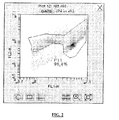
- the side scatter vs. green fluorescence is plotted and is shown in Fig. 1 .
- a gate is drawn to define where in the plot the events of interest, in this example cells, are located.
- Particles showing significant side scatter (SSC-H) signals but relatively low green fluorescence (FL1-H) signals may be readily identified as uninteresting and eliminated from further analysis.
- SSC-H significant side scatter
- FL1-H relatively low green fluorescence
- This gated region is identified as P4 and in the present example represents 52.6% of the total number of counted events.
- the gated events P4 are then plotted in a red (FL2-H) vs. green (FL1-H) fluorescence plot as illustrated in Fig.2 .
- a second gate is drawn to define where in the plot the events of interest (cells) are located and thus additional noise particles are eliminated.
- This gated region is identified as P11 and in the present example represents 96.4% of the total number of counted events in the P4 gated region and corresponds to a total somatic cell count of 450,000 cells per millilitre (cells/ml).
- This plot may be employed in the determination of the percentage of cells belonging to Groups A and B however preferably the gate defining all cells in the P11 region of Fig.
- FIG. 2 is then subdivided into regions defining different cell populations based on the relative ratios of fluorescence detected at wavelengths characteristic of the meta-chromatic stain employed, here acridine orange with associated red/green (FL2-H/FL1-H) fluorescence intensities.
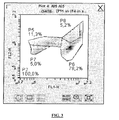
- the P11 region from Fig. 2 is in the present Fig.3 identified as P2.
- the regions of different cell types are illustrated as P5, P6, P7 and P8 in Fig. 3 and from visual observation are considered to most likely be comprised of monocytes (Group A); PMN (Group B); lymphocytes (Group A) and macrophages (Group A) respectively.
- the number of events in the region P6 which defines the so called Group B (or PMN) cell region in Fig.3 is divided with the total number of cells and multiplied with 100 to obtain the %Group B (PMN) which, in the present example, is 78.2%.
- a comparison may be made with reference values established using milk from cows with a known degree of infection. This comparison may be made mathematically using conventional regression or chemometric techniques applied to the reference values in order to establish a mathematical relationship between cell count and degree of infection.
- the result from an unknown sample may then be processed in a data processor using the previously established mathematically relationship in order to arrive at an indication of the degree of infection of the unknown cow.
- ranges of cell counts may be indexed against mastitis diagnosis and the measured cell counts is then compared to the ranges in order to provide an indication of a degree of infection. This is illustrated by way of example only in Table 1 below and may be done either manually or automatically by means of a data processor.
- a PMN value of 78% together with the total somatic cell count of 450,000 indicates subclinical mastitis.
- the indication of the degree of infection may be then presented to a user in a number of different ways without departing from the invention as claimed, such as YES/NO to the presence of one or both mastitis and subclinical mastistis or as a level of infection or as prediction of contracting mastitis.
- Milk from an individual cow is preserved (with BSM II tabs) and heated to 40°C prior to analysis.
- a volume of 50 ⁇ l milk is transferred into an Eppendorf tube.
- 160 ⁇ l of an acridine orange reagent (0.3mg/ml acridine orange and 0.03mg/ml Na 2 H 2 EDTA in PBS) is added and mixed. After reacting for one minute at room temperature, the mixture was measured on the standard flow cytometer used in Example 1. In the flow cytometer the sample is measured with a speed of 100 ⁇ l/min and is again excited by a blue laser (488nm). A FL1-H value of 200,000 is used as trigger signal. Fluorescent signals (FL1-H, FL2-H and light scatter signals (side scatter, SSC-H) are again acquired.
- an appropriate amount of differentiating marker for example acridine orange meta-chromatic stain, is added to a sample which is sufficient to permit somatic cell differentiation using flow cytometry but which does not adversely dilute the sample by providing an separation effect as discussed above.
- An appropriate level may be determined empirically, using reasonable trial and error, by visual inspection of the appropriate dot plots as will be described below by way of example only:
- a milk sample with different types of cells are used and mixed with the reagents using the following combinations: Table 2 1 50 ⁇ l 50 ⁇ l acridine 100 ⁇ l PBS milk orange 2 90 ⁇ l 10 ⁇ l acridine milk orange 3 99 ⁇ l 2 ⁇ l acridine 100 ⁇ l PBS milk orange 4 50 ⁇ l 50 ⁇ l acridine 10 ⁇ l milk orange EDTA 5 90 ⁇ l 10 ⁇ l acridine 10 ⁇ l 100 ⁇ l PBS milk orange EDTA 6 99 ⁇ l 2 ⁇ l acridine 10 ⁇ l milk orange EDTA 7 50 ⁇ l 50 ⁇ l acridine milk orange 8 90 ⁇ l 10 ⁇ l acridine 100 ⁇ l PBS milk orange 9 99 ⁇ l 2 ⁇ l acridine milk orange 10 50 ⁇ l 50 ⁇ l acridine 10 ⁇
- each combination is measured on the standard flow cytometer used in Examples 1 and 2 essentially in the manner described in Examples 1 and 2 above.
- the sample is measured with a speed of 14 ⁇ l/min and is excited by a blue laser (488nm).
- a FL1-H value of 400,000 is used as trigger signal.
- Fluorescent signals (FL1-H, FL2-), and light scatter signals (side scatter, SSC-H) are used.
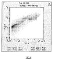
- the red vs. green (FL2-H vs FL1-H) fluorescence is plotted and the results are illustrated in Fig.5 .
- Fig. 6 When using 2 ⁇ l or 10 ⁇ l acridine orange only one cell population is seen in the plots ( Fig. 6 ), i.e. low concentrations of acridine orange do not provide sufficient differentiation. In contrast hereto several populations are seen in plot when using 50 ⁇ l acridine orange (see Fig. 5 .).
- Adding PBS to the sample decreases the noise level, i.e. as shown in Fig. 8 and Fig. 9 it is easier to separate cells from noise.
- the noise is the wedge shaped population in the left part of the plots. In the plot of Fig. 8 (the sample without PBS) the noise population overlaps one of the cell populations. In the plot of Fig. 9 (the sample with PBS) the noise population is better separated from one of the cell populations.
- concentrations of acridine orange greater than around 0.2 mg/ml gives suitable differentiation.
- the amount of reagent (either as a single reagent or individual reagents added separately) that can be added to the sample without causing unwanted dilution effects can be determined by consideration of an optimum throughput for a system employing the present method in routine analysis. This amount must provide a dilution less than 1:200, preferably less than 1:50 and most preferably less than 1:5.
Landscapes
- Health & Medical Sciences (AREA)
- Life Sciences & Earth Sciences (AREA)
- Engineering & Computer Science (AREA)
- Immunology (AREA)
- Chemical & Material Sciences (AREA)
- Biomedical Technology (AREA)
- Molecular Biology (AREA)
- Urology & Nephrology (AREA)
- Hematology (AREA)
- Cell Biology (AREA)
- Biochemistry (AREA)
- Microbiology (AREA)
- Physics & Mathematics (AREA)
- Analytical Chemistry (AREA)
- Biotechnology (AREA)
- General Health & Medical Sciences (AREA)
- Food Science & Technology (AREA)
- Medicinal Chemistry (AREA)
- General Physics & Mathematics (AREA)
- Pathology (AREA)
- Tropical Medicine & Parasitology (AREA)
- Organic Chemistry (AREA)
- Bioinformatics & Cheminformatics (AREA)
- Toxicology (AREA)
- Proteomics, Peptides & Aminoacids (AREA)
- Wood Science & Technology (AREA)
- Zoology (AREA)
- Physiology (AREA)
- Biophysics (AREA)
- General Engineering & Computer Science (AREA)
- Genetics & Genomics (AREA)
- Investigating Or Analysing Biological Materials (AREA)
- Measuring Or Testing Involving Enzymes Or Micro-Organisms (AREA)
Description
- The present invention relates to a method for determining a degree of mastitis infection from a differential somatic cell count on mammalian milk, particularly to such a method for determining the presence and/or degree of mastitis and most particularly bovine mastitis.
- Mastitis is the inflammation of the mammary gland of a mammal typically caused by bacterial infection which, depending on its degree, can cause severe loss in milk yield as well as undesirable changes to the milk quality. It has been estimated that the annual economic loss for farmers in the USA alone due to bovine mastitis is over $2 billion. A substantial portion of this is estimated to be due to sub-clinical mastitis.
- Clinical mastitis is typically treated with a relatively expensive course of antibiotics. Subclinical mastitis is not necessarily treated with antibiotics during lactation. Often subclinical infections are eliminated by the immune system, but subclinical mastitis may occasionally lead to clinical mastitis or remain as a subclinical infection (chronic mastitis).
- Identification of mastitis, particularly bovine mastitis, is typically based on the total number of somatic cells in a predetermined volume of milk, typically using either imaging or flow cytometry to make the somatic cell count. When making this somatic cell count leukocytes (white blood cells) and epithelial (skin) cells are typically counted without any discrimination, particularly between the different types of leukocytes present in the milk, being made.
- Use of the total somatic cell count as one of a panel of indicators employed to quantify the degree of mastitis infection of a cow is disclosed by Hojsgaard and Friggins, Journal of Dairy Science, vol. 93, no. 2, 1 February 2010, pages 582-592. Furthermore, total somatic cell count measurement in ovine milk is disclosed by Gonzalo et al., Journal of Dairy Science, vol. 91, no. 8, 1 August 2008, pages 3114-3118.
- In the following the lymphocyte, monocyte and macrophage types of leukocyte will be referred to as 'Group A' leukocytes and polymorphonuclear or PMN (neutrophils, eosinophils and basophils) types of leukocyte will be referred to as 'Group B' leukocytes.
- It is generally understood Group A members account for around 85% of the total number of somatic cells under normal conditions whereas Group B members account for approximately 10% of somatic cells under normal conditions and increase to between 30% and 90% of the total somatic cell count, dependent on the degree of mastitis.
- It has been long proposed that a differential somatic cell count, being a measure of the relative proportions of Group A and Group B leukocytes and even the proportion of types of leukocytes within one or both of these groups, should be employed as an indication of one or more of the presence or degree of and/or susceptibility to mastitis.
- Differential cell counting is typically achieved by means of a differentiating marker, such as a meta-chromatic stain or fluorescent or radioactive microbeads, which has one or more measurable characteristics that varies dependent on the type of cell it becomes associated with. A cytometer is then employed to count events (cells) using a detection system sensitive to the differences in these characteristics, such as changes in fluorescent properties, to differentiate between the cell types in a known manner.
- It is known from, for example,
US 2009/0233329 , to perform a differential somatic cell count using a 'smear test' which employs a novel wedge shaped microfluidic chamber in which is placed a mixture of milk sample and a meta-chromatic stain. Here 80µl of milk is diluted with 20µl of a meta-chromatic stain and a small drop of the mixture is applied to the chamber. Identification of the different cells according to their coloration or morphology is made either manually or automatically using Image analysis. A problem with this is that a new microfluidic chamber must be used with each test. Furthermore the height of the imaging chamber may cause a blurring of the image which may lead to complications in the identification of the cells. - Flow cytometry is a well known alternative to the above described smear test type assay. In an article by M. Hageltorn and M. Alaa Saad published in Am. J. Vet. Res., pp 2012-2016, Vol. 47, No. 9, September 1986 it is disclosed that flow cytometry may be used to distinguish different leukocyte populations. Here a fresh milk sample (no more than 6 hours old) was diluted in a ratio of 1:200 with a hypotonic saline solution containing 0.0004% acridine orange meta-chromatic stain and the assay is performed using standard fluorescent flow cytometry. This dilution step is performed to effectively eliminate the possibility of a coincidence of the cells to be measured and interfering particles (such as fat particles) at the measurement point but adds complexity to the assay technique. Moreover the high dilution requires that a very much larger volume of liquid is assayed before a statistically significant cell count can be achieved. This in turn significantly increases the time for the assay.
- The performance of differential somatic cell count using a standard flow cytometry assay technique is also described, for example, in
US 6,979,550 . Here it is disclosed that in order to perform the differential cell counting the somatic cells must first be isolated from interfering particles in the milk sample. This is done here by centrifuging the milk sample in order to remove interfering particles such as fat particles and concentrate the particles of interest. A fluorescent stain is added to discriminate all cells from interfering particles. Particle counting is then performed in a standard flow cytometer using an event detecting system compatible with the fluorescent stain employed. The centrifuging increases the complexity of the assay and in some cases can even modify the sample so that it is no longer representative of the whole. - It is an aim of the present invention to address at least one of the problems associated with known cytometry assay techniques for differential somatic cell counting.
- Accordingly, the present invention provides a method for determining a degree of mastitis infection comprising the steps of i) preparing an un-isolated sample by adding a reagent containing a differentiating marker, such as meta-chromatic stain, fluorescent or radioactive microspheres, in an amount sufficient to provide a differentiation between cell types to mammalian milk to dilute it in a range less than 1:200, preferably less than 1:50 and most preferably less than 1:5; ii) measuring a differential somatic cell count on the sample by means of a flow cytometer having a detection system sensitive to differences in the differentiating marker resulting from the marker becoming differently associated with different cell types in the sample; and iii) determining an indication of a degree of infection dependent on the measured differential cell count.
- Thus, the inventor has surprisingly discovered that by judicious choice of the amount of differentiating marker added to a sample a cytometry assay is able to be performed on an un-isolated, essentially undiluted, sample in order to make a differential somatic cell count measurement by monitoring the differentiating characteristic(s) of the marker itself, the results of which may be used to determine an indication of the degree of mastitis infection. The deviation of these differentiating characteristic(s) from a norm and/or the extent of deviation may be used to identify the existence of, stage of and/or likelihood of contracting mastitis.
- Usefully the indication of the degree infection may include the identification of one or more of the presence, likelihood or level of mastitis. Such identification may be employed in a number of herd management decisions such as the milking order according to mastitis status, better flushing of milking claw after milking an infected cow or using a separate milking claw for infected cows, in order to lower cow-to-cow infection risk; grouping of cows with subclinical mastitis, in order to lower cow-to-cow infection risk; predicting if a subclinical mastitis infection will evolve into a clinical case and consequently identifying those cows likely to benefit from antibiotic treatment; and culling and breading selections.
- The present invention will now be illustrated by way of non-limiting examples and with reference to the drawings of the accompanying figures of which:
-
Fig. 1 shows a dot plot of side scatter intensity vs green fluorescence by flow cytometry in a milk sample in which cells are marked according to the present invention; -
Fig. 2 shows a dot plot of red vs green fluorescence of the gated region illustrated inFig.1 ; -
Fig. 3 shows a dot plot of red vs green fluorescence of the gated region illustrated inFig.2 in which different cell populations are identified; -
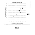
Fig. 4 shows a graph of %PMN measured on Day 0 (fresh milk) vs %PMN measured on subsequent days using a method according to the present invention; -
Fig. 5 shows a typical ungated dot plot of red vs green fluorescence by flow cytometry in a milk sample prepared in accordance with the values of Table 2; -
Fig. 6 shows a dot plot of red vs green fluorescence from a milk sample stained with a relatively low concentration of acridine orange; -
Fig. 7 shows a dot plot of red vs green fluorescence illustrating the effect of adding EDTA to a sample stained with a concentration as employed inFig. 5 ; -
Fig. 8 shows a dot plot of red vs green before PBS is added; and -
Fig. 9 shows the effects of adding PBS. - An amount of 50µl cold and preserved (with BSM II tabs) milk from an individual cow is transferred into an Eppendorf tube. 50µl acridine orange in PBS (1mg/ml) and 10µl Na2H2EDTA (0.1 g/ml, pH=7) are added one reagent at a time and mixed. Furthermore, 100µl PBS is then added to the above mention mixture and mixed. After reacting for 1-2 minutes at room temperature, the mixture was measured on a standard flow cytometer. In this flow cytometer, and by way of example only, the sample is flowed past the detection region with a speed of 14µl/min. As the differentiating marker is a meta-chromatic stain cell types can be differentiated using cell fluorescence. The stain employed in the present example is acridine orange and so fluorescence is, in the present example, excited by a blue laser (488nm) with the intensities of green (here designated FL1-H) and red (here designated FL2-H) being monitored. A FL1-H value of 400,000 is used as trigger signal. In the present arrangement light scatter signals are also acquired and the intensity of the side scatter light (here designated SSC-H) is advantageously employed to reduce noise (i.e. signals not related to the cells to be counted) from the acquired signal.
- For data analysis, the side scatter vs. green fluorescence is plotted and is shown in
Fig. 1 . In this plot a gate is drawn to define where in the plot the events of interest, in this example cells, are located. Particles showing significant side scatter (SSC-H) signals but relatively low green fluorescence (FL1-H) signals may be readily identified as uninteresting and eliminated from further analysis. The exact parameters for the gate may be readily determined by the skilled person using standard techniques known in the art of flow cytometry. - Thus noise particles are eliminated or at least significantly reduced. This gated region is identified as P4 and in the present example represents 52.6% of the total number of counted events.
- The gated events P4 are then plotted in a red (FL2-H) vs. green (FL1-H) fluorescence plot as illustrated in
Fig.2 . Usefully, but not essentially, in this plot a second gate is drawn to define where in the plot the events of interest (cells) are located and thus additional noise particles are eliminated. This gated region is identified as P11 and in the present example represents 96.4% of the total number of counted events in the P4 gated region and corresponds to a total somatic cell count of 450,000 cells per millilitre (cells/ml). This plot may be employed in the determination of the percentage of cells belonging to Groups A and B however preferably the gate defining all cells in the P11 region ofFig. 2 is then subdivided into regions defining different cell populations based on the relative ratios of fluorescence detected at wavelengths characteristic of the meta-chromatic stain employed, here acridine orange with associated red/green (FL2-H/FL1-H) fluorescence intensities. The P11 region fromFig. 2 is in the presentFig.3 identified as P2. The regions of different cell types are illustrated as P5, P6, P7 and P8 inFig. 3 and from visual observation are considered to most likely be comprised of monocytes (Group A); PMN (Group B); lymphocytes (Group A) and macrophages (Group A) respectively. Once these visual correlations have been made then the ratios of characteristic fluorescence associated with these regions may be employed in future analysis without relying on the step of visual identification. - The number of events in the region P6 which defines the so called Group B (or PMN) cell region in
Fig.3 is divided with the total number of cells and multiplied with 100 to obtain the %Group B (PMN) which, in the present example, is 78.2%. - In order to establish an indication of the degree of infection from this result a comparison may be made with reference values established using milk from cows with a known degree of infection. This comparison may be made mathematically using conventional regression or chemometric techniques applied to the reference values in order to establish a mathematical relationship between cell count and degree of infection. The result from an unknown sample may then be processed in a data processor using the previously established mathematically relationship in order to arrive at an indication of the degree of infection of the unknown cow. Alternatively, ranges of cell counts may be indexed against mastitis diagnosis and the measured cell counts is then compared to the ranges in order to provide an indication of a degree of infection. This is illustrated by way of example only in Table 1 below and may be done either manually or automatically by means of a data processor.
Table 1 Somatic cell count (cells/ml) %PMN Mastitis diagnosis >500,000 - Clinical or subclinical mastitis 400,001 - 500,000 ≥30 Subclinical mastitis 400,001 - 500,000 <30 No mastitis 300,001 - 400,000 ≥40 Subclinical mastitis 300,001 - 400,000 <40 No mastitis 200,001 - 300,000 ≥50 Subclinical mastitis 200,001 - 300,000 <50 No mastitis 100,001 - 200,000 ≥60 Subclinical mastitis 100,001 - 200,000 <60 No mastitis 50,000 - 100,000 ≥70 Subclinical mastitis 50,000 - 100,000 <70 No mastitis <50,000 - No mastitis - Thus, in the present example a PMN value of 78%, together with the total somatic cell count of 450,000 indicates subclinical mastitis. It will be appreciated by those skilled in the art that the indication of the degree of infection may be then presented to a user in a number of different ways without departing from the invention as claimed, such as YES/NO to the presence of one or both mastitis and subclinical mastistis or as a level of infection or as prediction of contracting mastitis.
- Milk from an individual cow is preserved (with BSM II tabs) and heated to 40°C prior to analysis. A volume of 50µl milk is transferred into an Eppendorf tube. 160µl of an acridine orange reagent (0.3mg/ml acridine orange and 0.03mg/ml Na2H2EDTA in PBS) is added and mixed. After reacting for one minute at room temperature, the mixture was measured on the standard flow cytometer used in Example 1. In the flow cytometer the sample is measured with a speed of 100µl/min and is again excited by a blue laser (488nm). A FL1-H value of 200,000 is used as trigger signal. Fluorescent signals (FL1-H, FL2-H and light scatter signals (side scatter, SSC-H) are again acquired.
- The data analysis is similar to the one used in Example 1 the results are the same, indicating a PMN level of around 78.2%. This illustrates that the method according to the present invention is well suited to industrial (as opposed to research) application since flow speeds may be made relatively high and the additives may be pre-mixed and added as a single reagent without significant loss in predictive accuracy.
- Using the same experimental parameters measurements were made on nine further samples and measurements repeated after one day (
Day 1 inFig. 4 ) and again after three days (Day 3 inFig. 4 ). The results are illustrated inFig. 4 and show that theDay 1 measurement results are comparable to those results obtained when the sample was fresh (Day 0 inFig. 4 ) and that after three days the measured values are only slightly lower than those of Day 1 (and fresh). This indicates that the method according to the present invention may be usable to determine an indication of a degree of infection even on samples preserved for up to three days. This is an advantage when using the method to measure milk samples received at a central laboratory from dairy farmers some distances away. In this case the samples received are typically one or two day old preserved samples. Moreover, this indicates that the method may also be usable to distinguish viable cells, since mortality increases with age. - It is an essential feature of the present invention that an appropriate amount of differentiating marker, for example acridine orange meta-chromatic stain, is added to a sample which is sufficient to permit somatic cell differentiation using flow cytometry but which does not adversely dilute the sample by providing an separation effect as discussed above. An appropriate level may be determined empirically, using reasonable trial and error, by visual inspection of the appropriate dot plots as will be described below by way of example only:
- The following reagents were prepared: 1mg/ml acridine orange in PBS, 0.1g/ml Na2H2EDTA (pH=7) and normal strength PBS. A milk sample with different types of cells are used and mixed with the reagents using the following combinations:
Table 2 1 50µl 50µl acridine 100µl PBS milk orange 2 90µl 10µl acridine milk orange 3 99µl 2µl acridine 100µl PBS milk orange 4 50µl 50µl acridine 10µl milk orange EDTA 5 90µl 10µl acridine 10µl 100µl PBS milk orange EDTA 6 99µl 2µl acridine 10µl milk orange EDTA 7 50µl 50µl acridine milk orange 8 90µl 10µl acridine 100µl PBS milk orange 9 99µl 2µl acridine milk orange 10 50µl 50µl acridine 10µl 100µl PBS milk orange EDTA 11 90µl 10µl acridine 10µl milk orange EDTA 12 99µl 2µl acridine 10µl 100µl PBS milk orange EDTA - After reacting for 1-2 minutes at room temperature, each combination is measured on the standard flow cytometer used in Examples 1 and 2 essentially in the manner described in Examples 1 and 2 above. In the flow cytometer the sample is measured with a speed of 14µl/min and is excited by a blue laser (488nm). A FL1-H value of 400,000 is used as trigger signal. Fluorescent signals (FL1-H, FL2-), and light scatter signals (side scatter, SSC-H) are used.
- For data analysis, the red vs. green (FL2-H vs FL1-H) fluorescence is plotted and the results are illustrated in
Fig.5 . When using 2µl or 10µl acridine orange only one cell population is seen in the plots (Fig. 6 ), i.e. low concentrations of acridine orange do not provide sufficient differentiation. In contrast hereto several populations are seen in plot when using 50µl acridine orange (seeFig. 5 .). - When the high concentration of acridine orange is combined with EDTA the very dense population moves to the right, i.e. the green fluorescence intensities of these cells increase (
Fig.7 .) as compared to what is illustrated inFig. 5 . - Adding PBS to the sample decreases the noise level, i.e. as shown in
Fig. 8 andFig. 9 it is easier to separate cells from noise. The noise is the wedge shaped population in the left part of the plots. In the plot ofFig. 8 (the sample without PBS) the noise population overlaps one of the cell populations. In the plot ofFig. 9 (the sample with PBS) the noise population is better separated from one of the cell populations. - It can be concluded from this that concentrations of acridine orange greater than around 0.2 mg/ml gives suitable differentiation. The amount of reagent (either as a single reagent or individual reagents added separately) that can be added to the sample without causing unwanted dilution effects can be determined by consideration of an optimum throughput for a system employing the present method in routine analysis. This amount must provide a dilution less than 1:200, preferably less than 1:50 and most preferably less than 1:5.
- Although this experimental determination is illustrated with reference to acridine orange it will be appreciated that following a series of measurements the skilled person can reasonably be expected to determine a suitable range of differentiating marker to employ in the method according to the present invention. It will further be appreciated that while the method has been described with regard to flow cytometry measurements of relative fluorescent intensities as a differentiating measurement for cells other differentiating measurements may be employed dependent on the nature of the differentiating marker employed.
Claims (6)
- A method for determining a degree of mastitis infection comprising the steps ofi) preparing an un-isolated sample of mammalian milk in which somatic cells are un-isolated from interfering particles in the mammalian milk by adding a reagent comprising a meta-chromatic stain differentiating marker in an amount sufficient to provide a differentiation between cell types in mammalian milk in a dilution ratio of less than 1:200;ii) measuring a cell count for each of one or more leukocyte types of somatic cell selected from the group lymphocyte, monocyte, macrophage and polymorphonuclear types of leukocyte differentiated by the marker in the sample by means of a flow cytometer having a detection system sensitive to differences in the differentiating marker resulting from the marker becoming differently associated with different cell types in the sample; andiii) determining an indication of a degree of mastitis infection dependent on the percentage of polymorphonuclear type of leukocyte in the total number of measured cells as a measured cell count by comparing the measured cell count with one or more reference values each being predetermined to be associated with one or more degrees of mastitis infection.
- A method as claimed in Claim 1 characterised in that there is an additional step of measuring a total somatic cell count and in that the step of determining an indication further comprises comparing the measured total somatic cell count with one or more reference values each being predetermined to be associated with one or more degrees of mastitis infection.
- A method as claimed in any preceding claim characterised in that the mammalian milk is bovine milk and in that determining an indication of a degree of mastitis infection comprises determining an indication of one or both the presence and degree of mastitis.
- A method as claimed in Claim 3 characterised in that the indication of mastitis comprises an indication of sub-clinical mastitis.
- A method as claimed in Claim 3 or Claim 4 characterised in that a degree of mastitis infection indicative of a likelihood of developing clinical mastitis is determined.
- A method as claimed in Claim 6 characterised in that the meta-chromatic stain is acridine orange in a concentration of greater than 0.2 mg/ml.
Priority Applications (1)
| Application Number | Priority Date | Filing Date | Title |
|---|---|---|---|
| PL10768012T PL2630487T3 (en) | 2010-10-18 | 2010-10-18 | Method for determining a degree of mastitis infection |
Applications Claiming Priority (1)
| Application Number | Priority Date | Filing Date | Title |
|---|---|---|---|
| PCT/EP2010/065615 WO2012052046A1 (en) | 2010-10-18 | 2010-10-18 | Method for determining a degree of infection |
Publications (2)
| Publication Number | Publication Date |
|---|---|
| EP2630487A1 EP2630487A1 (en) | 2013-08-28 |
| EP2630487B1 true EP2630487B1 (en) | 2016-08-17 |
Family
ID=44144667
Family Applications (1)
| Application Number | Title | Priority Date | Filing Date |
|---|---|---|---|
| EP10768012.6A Revoked EP2630487B1 (en) | 2010-10-18 | 2010-10-18 | Method for determining a degree of mastitis infection |
Country Status (13)
| Country | Link |
|---|---|
| US (1) | US20130157306A1 (en) |
| EP (1) | EP2630487B1 (en) |
| JP (1) | JP5781617B2 (en) |
| KR (1) | KR101770447B1 (en) |
| AU (1) | AU2010362562B2 (en) |
| BR (1) | BR112013008965B1 (en) |
| CA (1) | CA2814559C (en) |
| ES (1) | ES2596678T3 (en) |
| MX (1) | MX342266B (en) |
| NO (1) | NO20130484A1 (en) |
| NZ (1) | NZ608803A (en) |
| PL (1) | PL2630487T3 (en) |
| WO (1) | WO2012052046A1 (en) |
Cited By (1)
| Publication number | Priority date | Publication date | Assignee | Title |
|---|---|---|---|---|
| CN108717125A (en) * | 2018-06-22 | 2018-10-30 | 北京农学院 | Milk cow early stage mammitis monitoring method |
Families Citing this family (3)
| Publication number | Priority date | Publication date | Assignee | Title |
|---|---|---|---|---|
| WO2017065708A1 (en) * | 2015-10-12 | 2017-04-20 | Nehir Biyoteknoloji Ar-Ge Hizm. Dan. Bils. Paz. San. Tic. Ltd. Sti | On-line automatic subclinical mastitis detection device based on optical scattering and an automatic milk sampling system comprising this device |
| EP3196644B1 (en) * | 2016-01-22 | 2019-11-27 | Mastatix, Ltd. | Method for diagnosing mastitis in a lactating animal |
| JP7405555B2 (en) * | 2019-10-08 | 2023-12-26 | 株式会社アドバンテスト | Somatic cell meter, somatic cell measurement method, program and recording medium |
Citations (1)
| Publication number | Priority date | Publication date | Assignee | Title |
|---|---|---|---|---|
| JP2003310299A (en) | 2002-04-25 | 2003-11-05 | Sysmex Corp | Method for treating milk sample, reagent and method each for measuring somatic cell contained in milk sample |
Family Cites Families (6)
| Publication number | Priority date | Publication date | Assignee | Title |
|---|---|---|---|---|
| US3327119A (en) * | 1964-03-26 | 1967-06-20 | Ibm | Method and apparatus for detecting cancer cells |
| JPH0635972B2 (en) * | 1986-11-27 | 1994-05-11 | 東亜医用電子株式会社 | Method for classifying leukocytes by flow cytometry |
| US6979550B1 (en) | 2002-09-05 | 2005-12-27 | Rivas Ariel L | Method for diagnosis of, and determination of susceptibility to bovine mastitis |
| WO2007112332A2 (en) | 2006-03-24 | 2007-10-04 | Advanced Animal Diagnostics | Microfluidic chamber assembly for mastitis assay |
| GB2457094A (en) | 2008-02-04 | 2009-08-05 | Univ Dublin City | A cuvette assembly for holding milking samples |
| JPWO2010013757A1 (en) * | 2008-07-31 | 2012-01-12 | 株式会社クラレ | Somatic cell number measuring device and sensor |
-
2010
- 2010-10-18 MX MX2013004232A patent/MX342266B/en active IP Right Grant
- 2010-10-18 WO PCT/EP2010/065615 patent/WO2012052046A1/en not_active Ceased
- 2010-10-18 ES ES10768012.6T patent/ES2596678T3/en active Active
- 2010-10-18 AU AU2010362562A patent/AU2010362562B2/en active Active
- 2010-10-18 CA CA2814559A patent/CA2814559C/en active Active
- 2010-10-18 US US13/819,616 patent/US20130157306A1/en not_active Abandoned
- 2010-10-18 BR BR112013008965-2A patent/BR112013008965B1/en active IP Right Grant
- 2010-10-18 EP EP10768012.6A patent/EP2630487B1/en not_active Revoked
- 2010-10-18 PL PL10768012T patent/PL2630487T3/en unknown
- 2010-10-18 JP JP2013533098A patent/JP5781617B2/en active Active
- 2010-10-18 NZ NZ608803A patent/NZ608803A/en unknown
- 2010-10-18 KR KR1020137012633A patent/KR101770447B1/en active Active
-
2013
- 2013-04-11 NO NO20130484A patent/NO20130484A1/en not_active Application Discontinuation
Patent Citations (1)
| Publication number | Priority date | Publication date | Assignee | Title |
|---|---|---|---|---|
| JP2003310299A (en) | 2002-04-25 | 2003-11-05 | Sysmex Corp | Method for treating milk sample, reagent and method each for measuring somatic cell contained in milk sample |
Non-Patent Citations (8)
| Title |
|---|
| "Analytical Instruments for the Dairy Industry", BENTLEY INSTRUMENTS, 2004, XP055386789 |
| "Evaluation : ANADIS SCC 500", LETTRE CECALAIT N° 24, 4ÈME TRIMÈSTRE, 1997, XP055386783 |
| "Product description", FM 5000, August 1995 (1995-08-01), XP055386804 |
| BOUTINAUD ET AL.: "potential uses of epithelial cells : a review, reprod.", NUTR . DEV., vol. 42, no. 2, 2002, pages 133 - 147, XP055386772 |
| KOESS ET AL.: "Detection of mastitis in the bovine mammary gland by flow cytometry at early stages", JOURNAL OF DAIRY RESE, vol. 75, no. 2, May 2008 (2008-05-01), pages 225 - 232, XP008183462 |
| M. HAGELTORN ET AL.: "Flow cytofluorometric characterization of bovine blood and milk leukocytes", AM J VET RES, vol. 47, no. 9, September 1996 (1996-09-01), pages 2012 - 2016, XP002205919 |
| PARK ET AL.: "Goat Milk-Chemistry and Nutrition", HANDBOOK OF MILK OF NON-BOVINE MAMMALS, 2006, pages 34 - 35, XP055386771 |
| SCHRÖDER: "Untersuchungen zum Zelldifferentialbild in Milch und Blut unter Berücksichtigung des Gesundheitszustandes der bovinen Milchdrüse", INAUGURAL-DISSERTATION, vol. 12, 2003, pages 142 - 144, XP055386780 |
Cited By (1)
| Publication number | Priority date | Publication date | Assignee | Title |
|---|---|---|---|---|
| CN108717125A (en) * | 2018-06-22 | 2018-10-30 | 北京农学院 | Milk cow early stage mammitis monitoring method |
Also Published As
| Publication number | Publication date |
|---|---|
| AU2010362562A1 (en) | 2013-04-04 |
| KR101770447B1 (en) | 2017-08-22 |
| NZ608803A (en) | 2014-11-28 |
| MX342266B (en) | 2016-09-23 |
| KR20140020835A (en) | 2014-02-19 |
| US20130157306A1 (en) | 2013-06-20 |
| CA2814559A1 (en) | 2012-04-26 |
| WO2012052046A1 (en) | 2012-04-26 |
| JP5781617B2 (en) | 2015-09-24 |
| ES2596678T3 (en) | 2017-01-11 |
| EP2630487A1 (en) | 2013-08-28 |
| JP2013543118A (en) | 2013-11-28 |
| AU2010362562B2 (en) | 2014-05-15 |
| PL2630487T3 (en) | 2017-01-31 |
| MX2013004232A (en) | 2013-05-30 |
| BR112013008965A2 (en) | 2020-06-30 |
| NO20130484A1 (en) | 2013-04-11 |
| CA2814559C (en) | 2018-12-11 |
| BR112013008965B1 (en) | 2021-08-03 |
Similar Documents
| Publication | Publication Date | Title |
|---|---|---|
| Damm et al. | Differential somatic cell count—A novel method for routine mastitis screening in the frame of Dairy Herd Improvement testing programs | |
| EP1032818B1 (en) | Method for differentiation of erythroblasts | |
| EP1049919B1 (en) | Method for differentiation of nucleated red blood cells | |
| US10222320B2 (en) | Identifying and enumerating early granulated cells (EGCs) | |
| Comazzi et al. | Haematological and biochemical abnormalities in canine blood: frequency and associations in 1022 samples | |
| EP0912756A1 (en) | Rapid assay for infection in neonates | |
| CA2814559C (en) | Method for determining a degree of infection | |
| Jiménez-Penago et al. | Mean corpuscular haemoglobin concentration as haematological marker to detect changes in red blood cells in sheep infected with Haemonchus contortus | |
| EP3721231B1 (en) | Method of obtaining an indicator of patient sepsis and apparatus for such a method | |
| Zank et al. | Assessment of subacute mammary inflammation by soluble biomarkers in comparison to somatic cell counts in quarter milk samples from dairy cows | |
| Uztimür | Diagnostic significance of mean platelet volume and erythrocyte distribution width in calves with sepsis | |
| Santoro et al. | Platelet concentrations and platelet‐associated IgG in greyhounds | |
| Shehata et al. | Clinical significance of nucleated red blood cell count in pediatric patients with transfusion-dependent beta thalassemia | |
| Krogh et al. | Comparison of automated total nucleated cell counts vs manual cell counts in the amniotic fluid for the classification of disease status in healthy and diseased pigs | |
| RU2263312C1 (en) | Method for detecting animals suffering with cattle leukosis at aleukemic and leukopenic manifestations of the disease | |
| US20190361023A1 (en) | Trail-based encephalitis differentiation diagnosis method and the apparatus for the same | |
| Susianti et al. | Evaluation of Urine Procalcitonin, Interleukin-6, Heparin Binding Protein, Leukocyturia, and Bacteriuria for Early Detecting Urinary Tract Infection | |
| Glass et al. | Automated platelet counting | |
| CN115201270A (en) | Blood detection method and blood analysis system | |
| CN115201153A (en) | Blood detection method and blood analysis system | |
| é rina Fleming et al. | UF-1000 i: validation of the body fluid mode for counting cells in body fluids |
Legal Events
| Date | Code | Title | Description |
|---|---|---|---|
| PUAI | Public reference made under article 153(3) epc to a published international application that has entered the european phase |
Free format text: ORIGINAL CODE: 0009012 |
|
| 17P | Request for examination filed |
Effective date: 20130320 |
|
| AK | Designated contracting states |
Kind code of ref document: A1 Designated state(s): AL AT BE BG CH CY CZ DE DK EE ES FI FR GB GR HR HU IE IS IT LI LT LU LV MC MK MT NL NO PL PT RO RS SE SI SK SM TR |
|
| DAX | Request for extension of the european patent (deleted) | ||
| 17Q | First examination report despatched |
Effective date: 20150918 |
|
| RIC1 | Information provided on ipc code assigned before grant |
Ipc: C12Q 1/06 20060101ALI20160301BHEP Ipc: G01N 33/569 20060101ALI20160301BHEP Ipc: G01N 33/58 20060101ALI20160301BHEP Ipc: G01N 33/50 20060101AFI20160301BHEP |
|
| GRAP | Despatch of communication of intention to grant a patent |
Free format text: ORIGINAL CODE: EPIDOSNIGR1 |
|
| INTG | Intention to grant announced |
Effective date: 20160509 |
|
| GRAS | Grant fee paid |
Free format text: ORIGINAL CODE: EPIDOSNIGR3 |
|
| GRAA | (expected) grant |
Free format text: ORIGINAL CODE: 0009210 |
|
| AK | Designated contracting states |
Kind code of ref document: B1 Designated state(s): AL AT BE BG CH CY CZ DE DK EE ES FI FR GB GR HR HU IE IS IT LI LT LU LV MC MK MT NL NO PL PT RO RS SE SI SK SM TR |
|
| REG | Reference to a national code |
Ref country code: GB Ref legal event code: FG4D |
|
| REG | Reference to a national code |
Ref country code: CH Ref legal event code: EP |
|
| REG | Reference to a national code |
Ref country code: IE Ref legal event code: FG4D |
|
| REG | Reference to a national code |
Ref country code: AT Ref legal event code: REF Ref document number: 821568 Country of ref document: AT Kind code of ref document: T Effective date: 20160915 |
|
| REG | Reference to a national code |
Ref country code: NL Ref legal event code: FP |
|
| REG | Reference to a national code |
Ref country code: DE Ref legal event code: R096 Ref document number: 602010035638 Country of ref document: DE |
|
| REG | Reference to a national code |
Ref country code: FR Ref legal event code: PLFP Year of fee payment: 7 |
|
| REG | Reference to a national code |
Ref country code: LT Ref legal event code: MG4D |
|
| REG | Reference to a national code |
Ref country code: ES Ref legal event code: FG2A Ref document number: 2596678 Country of ref document: ES Kind code of ref document: T3 Effective date: 20170111 |
|
| REG | Reference to a national code |
Ref country code: AT Ref legal event code: MK05 Ref document number: 821568 Country of ref document: AT Kind code of ref document: T Effective date: 20160817 |
|
| PG25 | Lapsed in a contracting state [announced via postgrant information from national office to epo] |
Ref country code: FI Free format text: LAPSE BECAUSE OF FAILURE TO SUBMIT A TRANSLATION OF THE DESCRIPTION OR TO PAY THE FEE WITHIN THE PRESCRIBED TIME-LIMIT Effective date: 20160817 Ref country code: NO Free format text: LAPSE BECAUSE OF FAILURE TO SUBMIT A TRANSLATION OF THE DESCRIPTION OR TO PAY THE FEE WITHIN THE PRESCRIBED TIME-LIMIT Effective date: 20161117 Ref country code: LT Free format text: LAPSE BECAUSE OF FAILURE TO SUBMIT A TRANSLATION OF THE DESCRIPTION OR TO PAY THE FEE WITHIN THE PRESCRIBED TIME-LIMIT Effective date: 20160817 Ref country code: HR Free format text: LAPSE BECAUSE OF FAILURE TO SUBMIT A TRANSLATION OF THE DESCRIPTION OR TO PAY THE FEE WITHIN THE PRESCRIBED TIME-LIMIT Effective date: 20160817 Ref country code: RS Free format text: LAPSE BECAUSE OF FAILURE TO SUBMIT A TRANSLATION OF THE DESCRIPTION OR TO PAY THE FEE WITHIN THE PRESCRIBED TIME-LIMIT Effective date: 20160817 |
|
| PG25 | Lapsed in a contracting state [announced via postgrant information from national office to epo] |
Ref country code: BE Free format text: LAPSE BECAUSE OF NON-PAYMENT OF DUE FEES Effective date: 20161031 Ref country code: AT Free format text: LAPSE BECAUSE OF FAILURE TO SUBMIT A TRANSLATION OF THE DESCRIPTION OR TO PAY THE FEE WITHIN THE PRESCRIBED TIME-LIMIT Effective date: 20160817 Ref country code: SE Free format text: LAPSE BECAUSE OF FAILURE TO SUBMIT A TRANSLATION OF THE DESCRIPTION OR TO PAY THE FEE WITHIN THE PRESCRIBED TIME-LIMIT Effective date: 20160817 Ref country code: PT Free format text: LAPSE BECAUSE OF FAILURE TO SUBMIT A TRANSLATION OF THE DESCRIPTION OR TO PAY THE FEE WITHIN THE PRESCRIBED TIME-LIMIT Effective date: 20161219 Ref country code: LV Free format text: LAPSE BECAUSE OF FAILURE TO SUBMIT A TRANSLATION OF THE DESCRIPTION OR TO PAY THE FEE WITHIN THE PRESCRIBED TIME-LIMIT Effective date: 20160817 Ref country code: GR Free format text: LAPSE BECAUSE OF FAILURE TO SUBMIT A TRANSLATION OF THE DESCRIPTION OR TO PAY THE FEE WITHIN THE PRESCRIBED TIME-LIMIT Effective date: 20161118 |
|
| PG25 | Lapsed in a contracting state [announced via postgrant information from national office to epo] |
Ref country code: EE Free format text: LAPSE BECAUSE OF FAILURE TO SUBMIT A TRANSLATION OF THE DESCRIPTION OR TO PAY THE FEE WITHIN THE PRESCRIBED TIME-LIMIT Effective date: 20160817 Ref country code: RO Free format text: LAPSE BECAUSE OF FAILURE TO SUBMIT A TRANSLATION OF THE DESCRIPTION OR TO PAY THE FEE WITHIN THE PRESCRIBED TIME-LIMIT Effective date: 20160817 |
|
| REG | Reference to a national code |
Ref country code: DE Ref legal event code: R026 Ref document number: 602010035638 Country of ref document: DE |
|
| PLBI | Opposition filed |
Free format text: ORIGINAL CODE: 0009260 |
|
| PG25 | Lapsed in a contracting state [announced via postgrant information from national office to epo] |
Ref country code: CZ Free format text: LAPSE BECAUSE OF FAILURE TO SUBMIT A TRANSLATION OF THE DESCRIPTION OR TO PAY THE FEE WITHIN THE PRESCRIBED TIME-LIMIT Effective date: 20160817 Ref country code: SK Free format text: LAPSE BECAUSE OF FAILURE TO SUBMIT A TRANSLATION OF THE DESCRIPTION OR TO PAY THE FEE WITHIN THE PRESCRIBED TIME-LIMIT Effective date: 20160817 Ref country code: SM Free format text: LAPSE BECAUSE OF FAILURE TO SUBMIT A TRANSLATION OF THE DESCRIPTION OR TO PAY THE FEE WITHIN THE PRESCRIBED TIME-LIMIT Effective date: 20160817 Ref country code: BG Free format text: LAPSE BECAUSE OF FAILURE TO SUBMIT A TRANSLATION OF THE DESCRIPTION OR TO PAY THE FEE WITHIN THE PRESCRIBED TIME-LIMIT Effective date: 20161117 Ref country code: BE Free format text: LAPSE BECAUSE OF FAILURE TO SUBMIT A TRANSLATION OF THE DESCRIPTION OR TO PAY THE FEE WITHIN THE PRESCRIBED TIME-LIMIT Effective date: 20160817 Ref country code: DK Free format text: LAPSE BECAUSE OF FAILURE TO SUBMIT A TRANSLATION OF THE DESCRIPTION OR TO PAY THE FEE WITHIN THE PRESCRIBED TIME-LIMIT Effective date: 20160817 |
|
| REG | Reference to a national code |
Ref country code: CH Ref legal event code: PL |
|
| PLAX | Notice of opposition and request to file observation + time limit sent |
Free format text: ORIGINAL CODE: EPIDOSNOBS2 |
|
| 26 | Opposition filed |
Opponent name: BENTLEY INSTRUMENTS Effective date: 20170517 |
|
| PG25 | Lapsed in a contracting state [announced via postgrant information from national office to epo] |
Ref country code: CH Free format text: LAPSE BECAUSE OF NON-PAYMENT OF DUE FEES Effective date: 20161031 Ref country code: LI Free format text: LAPSE BECAUSE OF NON-PAYMENT OF DUE FEES Effective date: 20161031 |
|
| PG25 | Lapsed in a contracting state [announced via postgrant information from national office to epo] |
Ref country code: LU Free format text: LAPSE BECAUSE OF NON-PAYMENT OF DUE FEES Effective date: 20161018 Ref country code: SI Free format text: LAPSE BECAUSE OF FAILURE TO SUBMIT A TRANSLATION OF THE DESCRIPTION OR TO PAY THE FEE WITHIN THE PRESCRIBED TIME-LIMIT Effective date: 20160817 |
|
| PLBB | Reply of patent proprietor to notice(s) of opposition received |
Free format text: ORIGINAL CODE: EPIDOSNOBS3 |
|
| REG | Reference to a national code |
Ref country code: FR Ref legal event code: PLFP Year of fee payment: 8 |
|
| PG25 | Lapsed in a contracting state [announced via postgrant information from national office to epo] |
Ref country code: CY Free format text: LAPSE BECAUSE OF FAILURE TO SUBMIT A TRANSLATION OF THE DESCRIPTION OR TO PAY THE FEE WITHIN THE PRESCRIBED TIME-LIMIT Effective date: 20160817 Ref country code: HU Free format text: LAPSE BECAUSE OF FAILURE TO SUBMIT A TRANSLATION OF THE DESCRIPTION OR TO PAY THE FEE WITHIN THE PRESCRIBED TIME-LIMIT; INVALID AB INITIO Effective date: 20101018 |
|
| RDAF | Communication despatched that patent is revoked |
Free format text: ORIGINAL CODE: EPIDOSNREV1 |
|
| STAA | Information on the status of an ep patent application or granted ep patent |
Free format text: STATUS: THE PATENT HAS BEEN GRANTED |
|
| PG25 | Lapsed in a contracting state [announced via postgrant information from national office to epo] |
Ref country code: MC Free format text: LAPSE BECAUSE OF FAILURE TO SUBMIT A TRANSLATION OF THE DESCRIPTION OR TO PAY THE FEE WITHIN THE PRESCRIBED TIME-LIMIT Effective date: 20160817 Ref country code: IS Free format text: LAPSE BECAUSE OF FAILURE TO SUBMIT A TRANSLATION OF THE DESCRIPTION OR TO PAY THE FEE WITHIN THE PRESCRIBED TIME-LIMIT Effective date: 20160817 Ref country code: MK Free format text: LAPSE BECAUSE OF FAILURE TO SUBMIT A TRANSLATION OF THE DESCRIPTION OR TO PAY THE FEE WITHIN THE PRESCRIBED TIME-LIMIT Effective date: 20160817 Ref country code: MT Free format text: LAPSE BECAUSE OF NON-PAYMENT OF DUE FEES Effective date: 20161031 |
|
| APBM | Appeal reference recorded |
Free format text: ORIGINAL CODE: EPIDOSNREFNO |
|
| APBP | Date of receipt of notice of appeal recorded |
Free format text: ORIGINAL CODE: EPIDOSNNOA2O |
|
| APAH | Appeal reference modified |
Free format text: ORIGINAL CODE: EPIDOSCREFNO |
|
| APBQ | Date of receipt of statement of grounds of appeal recorded |
Free format text: ORIGINAL CODE: EPIDOSNNOA3O |
|
| REG | Reference to a national code |
Ref country code: FR Ref legal event code: PLFP Year of fee payment: 9 |
|
| PG25 | Lapsed in a contracting state [announced via postgrant information from national office to epo] |
Ref country code: AL Free format text: LAPSE BECAUSE OF FAILURE TO SUBMIT A TRANSLATION OF THE DESCRIPTION OR TO PAY THE FEE WITHIN THE PRESCRIBED TIME-LIMIT Effective date: 20160817 Ref country code: TR Free format text: LAPSE BECAUSE OF FAILURE TO SUBMIT A TRANSLATION OF THE DESCRIPTION OR TO PAY THE FEE WITHIN THE PRESCRIBED TIME-LIMIT Effective date: 20160817 |
|
| RAP4 | Party data changed (patent owner data changed or rights of a patent transferred) |
Owner name: FOSS ANALYTICAL A/S |
|
| REG | Reference to a national code |
Ref country code: DE Ref legal event code: R103 Ref document number: 602010035638 Country of ref document: DE Ref country code: DE Ref legal event code: R064 Ref document number: 602010035638 Country of ref document: DE |
|
| APBU | Appeal procedure closed |
Free format text: ORIGINAL CODE: EPIDOSNNOA9O |
|
| RDAG | Patent revoked |
Free format text: ORIGINAL CODE: 0009271 |
|
| STAA | Information on the status of an ep patent application or granted ep patent |
Free format text: STATUS: PATENT REVOKED |
|
| PGFP | Annual fee paid to national office [announced via postgrant information from national office to epo] |
Ref country code: NL Payment date: 20211026 Year of fee payment: 12 |
|
| REG | Reference to a national code |
Ref country code: FI Ref legal event code: MGE |
|
| 27W | Patent revoked |
Effective date: 20211116 |
|
| GBPR | Gb: patent revoked under art. 102 of the ep convention designating the uk as contracting state |
Effective date: 20211116 |
|
| PGFP | Annual fee paid to national office [announced via postgrant information from national office to epo] |
Ref country code: IE Payment date: 20211027 Year of fee payment: 12 Ref country code: GB Payment date: 20211027 Year of fee payment: 12 Ref country code: ES Payment date: 20211102 Year of fee payment: 12 |
|
| PGFP | Annual fee paid to national office [announced via postgrant information from national office to epo] |
Ref country code: IT Payment date: 20211021 Year of fee payment: 12 Ref country code: FR Payment date: 20211025 Year of fee payment: 12 |
|
| PGFP | Annual fee paid to national office [announced via postgrant information from national office to epo] |
Ref country code: PL Payment date: 20211004 Year of fee payment: 12 |
|
| PGFP | Annual fee paid to national office [announced via postgrant information from national office to epo] |
Ref country code: DE Payment date: 20211027 Year of fee payment: 12 |