EP1032818B1 - Method for differentiation of erythroblasts - Google Patents
Method for differentiation of erythroblasts Download PDFInfo
- Publication number
- EP1032818B1 EP1032818B1 EP98957975A EP98957975A EP1032818B1 EP 1032818 B1 EP1032818 B1 EP 1032818B1 EP 98957975 A EP98957975 A EP 98957975A EP 98957975 A EP98957975 A EP 98957975A EP 1032818 B1 EP1032818 B1 EP 1032818B1
- Authority
- EP
- European Patent Office
- Prior art keywords
- sample
- red blood
- blood cells
- light scatter
- leukocytes
- Prior art date
- Legal status (The legal status is an assumption and is not a legal conclusion. Google has not performed a legal analysis and makes no representation as to the accuracy of the status listed.)
- Expired - Lifetime
Links
- 238000000034 method Methods 0.000 title claims description 78
- 230000004069 differentiation Effects 0.000 title description 30
- 210000003924 normoblast Anatomy 0.000 title description 2
- 210000000265 leukocyte Anatomy 0.000 claims description 76
- 210000004027 cell Anatomy 0.000 claims description 41
- 239000003153 chemical reaction reagent Substances 0.000 claims description 41
- 210000003743 erythrocyte Anatomy 0.000 claims description 37
- 210000004369 blood Anatomy 0.000 claims description 35
- 239000008280 blood Substances 0.000 claims description 35
- 238000005259 measurement Methods 0.000 claims description 18
- 210000000601 blood cell Anatomy 0.000 claims description 16
- 210000005259 peripheral blood Anatomy 0.000 claims description 8
- 239000011886 peripheral blood Substances 0.000 claims description 8
- 210000001185 bone marrow Anatomy 0.000 claims description 5
- 210000003979 eosinophil Anatomy 0.000 claims description 5
- 210000004698 lymphocyte Anatomy 0.000 claims description 5
- 210000001616 monocyte Anatomy 0.000 claims description 4
- 210000000440 neutrophil Anatomy 0.000 claims description 4
- 238000004458 analytical method Methods 0.000 description 21
- 230000002101 lytic effect Effects 0.000 description 16
- 210000001995 reticulocyte Anatomy 0.000 description 14
- 239000012530 fluid Substances 0.000 description 10
- 238000010186 staining Methods 0.000 description 9
- 238000001514 detection method Methods 0.000 description 7
- 239000000203 mixture Substances 0.000 description 7
- 230000000087 stabilizing effect Effects 0.000 description 7
- -1 aliphatic aldehyde Chemical class 0.000 description 5
- 239000000975 dye Substances 0.000 description 5
- 238000002795 fluorescence method Methods 0.000 description 5
- 230000009089 cytolysis Effects 0.000 description 4
- BDAGIHXWWSANSR-UHFFFAOYSA-N methanoic acid Natural products OC=O BDAGIHXWWSANSR-UHFFFAOYSA-N 0.000 description 4
- 230000003287 optical effect Effects 0.000 description 4
- 239000003755 preservative agent Substances 0.000 description 4
- 239000002253 acid Substances 0.000 description 3
- 238000006243 chemical reaction Methods 0.000 description 3
- 238000000684 flow cytometry Methods 0.000 description 3
- 238000002847 impedance measurement Methods 0.000 description 3
- 230000002934 lysing effect Effects 0.000 description 3
- 238000012758 nuclear staining Methods 0.000 description 3
- 239000002245 particle Substances 0.000 description 3
- 238000002360 preparation method Methods 0.000 description 3
- 238000007430 reference method Methods 0.000 description 3
- 239000002904 solvent Substances 0.000 description 3
- OSWFIVFLDKOXQC-UHFFFAOYSA-N 4-(3-methoxyphenyl)aniline Chemical compound COC1=CC=CC(C=2C=CC(N)=CC=2)=C1 OSWFIVFLDKOXQC-UHFFFAOYSA-N 0.000 description 2
- 229920002257 Plurafac® Polymers 0.000 description 2
- CDBYLPFSWZWCQE-UHFFFAOYSA-L Sodium Carbonate Chemical compound [Na+].[Na+].[O-]C([O-])=O CDBYLPFSWZWCQE-UHFFFAOYSA-L 0.000 description 2
- FAPWRFPIFSIZLT-UHFFFAOYSA-M Sodium chloride Chemical compound [Na+].[Cl-] FAPWRFPIFSIZLT-UHFFFAOYSA-M 0.000 description 2
- 230000002159 abnormal effect Effects 0.000 description 2
- 239000000654 additive Substances 0.000 description 2
- 150000001298 alcohols Chemical class 0.000 description 2
- 230000000845 anti-microbial effect Effects 0.000 description 2
- 230000003078 antioxidant effect Effects 0.000 description 2
- 210000000805 cytoplasm Anatomy 0.000 description 2
- 239000003085 diluting agent Substances 0.000 description 2
- 201000010099 disease Diseases 0.000 description 2
- 208000037265 diseases, disorders, signs and symptoms Diseases 0.000 description 2
- 239000007850 fluorescent dye Substances 0.000 description 2
- 235000019253 formic acid Nutrition 0.000 description 2
- 238000010438 heat treatment Methods 0.000 description 2
- 210000000207 lymphocyte subset Anatomy 0.000 description 2
- 238000002156 mixing Methods 0.000 description 2
- 229920001983 poloxamer Polymers 0.000 description 2
- MCUFTLAXJMCWPZ-UHFFFAOYSA-N 3-butyl-2-methylphenol Chemical compound CCCCC1=CC=CC(O)=C1C MCUFTLAXJMCWPZ-UHFFFAOYSA-N 0.000 description 1
- 229940099451 3-iodo-2-propynylbutylcarbamate Drugs 0.000 description 1
- WYVVKGNFXHOCQV-UHFFFAOYSA-N 3-iodoprop-2-yn-1-yl butylcarbamate Chemical compound CCCCNC(=O)OCC#CI WYVVKGNFXHOCQV-UHFFFAOYSA-N 0.000 description 1
- 241000270728 Alligator Species 0.000 description 1
- KCXVZYZYPLLWCC-UHFFFAOYSA-N EDTA Chemical compound OC(=O)CN(CC(O)=O)CCN(CC(O)=O)CC(O)=O KCXVZYZYPLLWCC-UHFFFAOYSA-N 0.000 description 1
- 208000001953 Hypotension Diseases 0.000 description 1
- 239000007832 Na2SO4 Substances 0.000 description 1
- 229920002012 Pluronic® F 38 Polymers 0.000 description 1
- RVGRUAULSDPKGF-UHFFFAOYSA-N Poloxamer Chemical compound C1CO1.CC1CO1 RVGRUAULSDPKGF-UHFFFAOYSA-N 0.000 description 1
- 229920003171 Poly (ethylene oxide) Polymers 0.000 description 1
- PMZURENOXWZQFD-UHFFFAOYSA-L Sodium Sulfate Chemical compound [Na+].[Na+].[O-]S([O-])(=O)=O PMZURENOXWZQFD-UHFFFAOYSA-L 0.000 description 1
- 208000002903 Thalassemia Diseases 0.000 description 1
- AXIKDPDWFVPGOD-UHFFFAOYSA-O [7-(dimethylamino)phenothiazin-3-ylidene]-dimethylazanium;2-(2,4,5,7-tetrabromo-3,6-dihydroxyxanthen-10-ium-9-yl)benzoic acid Chemical compound C1=CC(=[N+](C)C)C=C2SC3=CC(N(C)C)=CC=C3N=C21.OC(=O)C1=CC=CC=C1C1=C(C=C(Br)C(O)=C2Br)C2=[O+]C2=C1C=C(Br)C(O)=C2Br AXIKDPDWFVPGOD-UHFFFAOYSA-O 0.000 description 1
- 230000005856 abnormality Effects 0.000 description 1
- 230000002378 acidificating effect Effects 0.000 description 1
- 125000003342 alkenyl group Chemical group 0.000 description 1
- 125000000217 alkyl group Chemical group 0.000 description 1
- 125000000304 alkynyl group Chemical group 0.000 description 1
- 150000001412 amines Chemical class 0.000 description 1
- 208000007502 anemia Diseases 0.000 description 1
- 238000013459 approach Methods 0.000 description 1
- 238000000149 argon plasma sintering Methods 0.000 description 1
- 210000003651 basophil Anatomy 0.000 description 1
- 238000004159 blood analysis Methods 0.000 description 1
- 238000004820 blood count Methods 0.000 description 1
- 125000004432 carbon atom Chemical group C* 0.000 description 1
- 210000000170 cell membrane Anatomy 0.000 description 1
- 238000011109 contamination Methods 0.000 description 1
- 229920001577 copolymer Polymers 0.000 description 1
- 238000012937 correction Methods 0.000 description 1
- 238000007405 data analysis Methods 0.000 description 1
- 238000003745 diagnosis Methods 0.000 description 1
- 238000002405 diagnostic procedure Methods 0.000 description 1
- WSDISUOETYTPRL-UHFFFAOYSA-N dmdm hydantoin Chemical compound CC1(C)N(CO)C(=O)N(CO)C1=O WSDISUOETYTPRL-UHFFFAOYSA-N 0.000 description 1
- 230000008030 elimination Effects 0.000 description 1
- 238000003379 elimination reaction Methods 0.000 description 1
- 239000000834 fixative Substances 0.000 description 1
- 238000001917 fluorescence detection Methods 0.000 description 1
- 125000002485 formyl group Chemical class [H]C(*)=O 0.000 description 1
- 239000000383 hazardous chemical Substances 0.000 description 1
- 208000007475 hemolytic anemia Diseases 0.000 description 1
- 208000021822 hypotensive Diseases 0.000 description 1
- 230000001077 hypotensive effect Effects 0.000 description 1
- 238000002955 isolation Methods 0.000 description 1
- 208000032839 leukemia Diseases 0.000 description 1
- 238000012423 maintenance Methods 0.000 description 1
- 239000012528 membrane Substances 0.000 description 1
- 230000007170 pathology Effects 0.000 description 1
- 229920001451 polypropylene glycol Polymers 0.000 description 1
- 238000004321 preservation Methods 0.000 description 1
- 238000012545 processing Methods 0.000 description 1
- 238000010791 quenching Methods 0.000 description 1
- 230000000171 quenching effect Effects 0.000 description 1
- 239000001397 quillaja saponaria molina bark Substances 0.000 description 1
- 210000003660 reticulum Anatomy 0.000 description 1
- 238000005464 sample preparation method Methods 0.000 description 1
- 229930182490 saponin Natural products 0.000 description 1
- 150000007949 saponins Chemical class 0.000 description 1
- 230000035945 sensitivity Effects 0.000 description 1
- 208000031162 sideroblastic anemia Diseases 0.000 description 1
- 229910000029 sodium carbonate Inorganic materials 0.000 description 1
- 239000011780 sodium chloride Substances 0.000 description 1
- 229910052938 sodium sulfate Inorganic materials 0.000 description 1
- 125000004079 stearyl group Chemical group [H]C([*])([H])C([H])([H])C([H])([H])C([H])([H])C([H])([H])C([H])([H])C([H])([H])C([H])([H])C([H])([H])C([H])([H])C([H])([H])C([H])([H])C([H])([H])C([H])([H])C([H])([H])C([H])([H])C([H])([H])C([H])([H])[H] 0.000 description 1
- 238000012360 testing method Methods 0.000 description 1
- 230000000007 visual effect Effects 0.000 description 1
Images


Classifications
-
- G—PHYSICS
- G01—MEASURING; TESTING
- G01N—INVESTIGATING OR ANALYSING MATERIALS BY DETERMINING THEIR CHEMICAL OR PHYSICAL PROPERTIES
- G01N33/00—Investigating or analysing materials by specific methods not covered by groups G01N1/00 - G01N31/00
- G01N33/48—Biological material, e.g. blood, urine; Haemocytometers
- G01N33/50—Chemical analysis of biological material, e.g. blood, urine; Testing involving biospecific ligand binding methods; Immunological testing
- G01N33/80—Chemical analysis of biological material, e.g. blood, urine; Testing involving biospecific ligand binding methods; Immunological testing involving blood groups or blood types or red blood cells
-
- G—PHYSICS
- G01—MEASURING; TESTING
- G01N—INVESTIGATING OR ANALYSING MATERIALS BY DETERMINING THEIR CHEMICAL OR PHYSICAL PROPERTIES
- G01N15/00—Investigating characteristics of particles; Investigating permeability, pore-volume or surface-area of porous materials
- G01N15/10—Investigating individual particles
- G01N15/14—Optical investigation techniques, e.g. flow cytometry
- G01N15/1456—Optical investigation techniques, e.g. flow cytometry without spatial resolution of the texture or inner structure of the particle, e.g. processing of pulse signals
- G01N15/1459—Optical investigation techniques, e.g. flow cytometry without spatial resolution of the texture or inner structure of the particle, e.g. processing of pulse signals the analysis being performed on a sample stream
-
- G—PHYSICS
- G01—MEASURING; TESTING
- G01N—INVESTIGATING OR ANALYSING MATERIALS BY DETERMINING THEIR CHEMICAL OR PHYSICAL PROPERTIES
- G01N15/00—Investigating characteristics of particles; Investigating permeability, pore-volume or surface-area of porous materials
- G01N15/10—Investigating individual particles
- G01N2015/1006—Investigating individual particles for cytology
-
- G—PHYSICS
- G01—MEASURING; TESTING
- G01N—INVESTIGATING OR ANALYSING MATERIALS BY DETERMINING THEIR CHEMICAL OR PHYSICAL PROPERTIES
- G01N15/00—Investigating characteristics of particles; Investigating permeability, pore-volume or surface-area of porous materials
- G01N15/10—Investigating individual particles
- G01N15/14—Optical investigation techniques, e.g. flow cytometry
- G01N2015/1402—Data analysis by thresholding or gating operations performed on the acquired signals or stored data
-
- Y—GENERAL TAGGING OF NEW TECHNOLOGICAL DEVELOPMENTS; GENERAL TAGGING OF CROSS-SECTIONAL TECHNOLOGIES SPANNING OVER SEVERAL SECTIONS OF THE IPC; TECHNICAL SUBJECTS COVERED BY FORMER USPC CROSS-REFERENCE ART COLLECTIONS [XRACs] AND DIGESTS
- Y10—TECHNICAL SUBJECTS COVERED BY FORMER USPC
- Y10T—TECHNICAL SUBJECTS COVERED BY FORMER US CLASSIFICATION
- Y10T436/00—Chemistry: analytical and immunological testing
- Y10T436/10—Composition for standardization, calibration, simulation, stabilization, preparation or preservation; processes of use in preparation for chemical testing
- Y10T436/101666—Particle count or volume standard or control [e.g., platelet count standards, etc.]
-
- Y—GENERAL TAGGING OF NEW TECHNOLOGICAL DEVELOPMENTS; GENERAL TAGGING OF CROSS-SECTIONAL TECHNOLOGIES SPANNING OVER SEVERAL SECTIONS OF THE IPC; TECHNICAL SUBJECTS COVERED BY FORMER USPC CROSS-REFERENCE ART COLLECTIONS [XRACs] AND DIGESTS
- Y10—TECHNICAL SUBJECTS COVERED BY FORMER USPC
- Y10T—TECHNICAL SUBJECTS COVERED BY FORMER US CLASSIFICATION
- Y10T436/00—Chemistry: analytical and immunological testing
- Y10T436/25—Chemistry: analytical and immunological testing including sample preparation
- Y10T436/25125—Digestion or removing interfering materials
Definitions
- the present invention relates to a method for the differentiation of nucleated red blood cells.
- the method provides for a concurrent differentiation of leukocytes in a blood cell sample by suitable electronic and optical measurements.
- NRBCs Nucleated red blood cells
- erythroblasts are immature red blood cells. They normally occur in the bone marrow but not in peripheral blood. However, in certain diseases such as anemia and leukemia, NRBCs also occur in peripheral blood. Therefore, it is of clinical importance to measure NRBC.
- differentiation and enumeration of NRBC are performed manually. The process involves the smearing of a blood sample on a microscope slide and staining the slide, followed by manual visual analysis of the individual slide. The NRBC concentration is reported as number of NRBCs per 100 white blood cells.
- U.S. Patent No. 5,298,426 discloses a fluorescence method for differentiating NRBCs.
- the method utilizes a two-step staining using a first fluid which is an acidic hypotensive fluorescent dye solution, and a second fluid which changes the osmolality and pH of the first fluid.
- the first fluid contains an erythroblast-staining dye that diffuses into nucleated red blood cells to specifically stain their nuclei, and then separating a group of NRBCs from other cell groups on a two-dimensional plot whereby the results of NRBC differentiation are computed.
- the first fluid also contains two additional fluorescence dyes, i.e., an eosinophil/basophil-staining dye and leukocyte-staining dye for specific staining of these cell types.
- two additional fluorescence dyes i.e., an eosinophil/basophil-staining dye and leukocyte-staining dye for specific staining of these cell types.
- U.S. Patent No. 5,559,037 discloses a method for flow cytometric analysis of NRBCs and leukocytes.
- the method comprises lysis of red blood cells and NRBC cytoplasm from a whole blood sample to expose the NRBC nuclei to a vital nuclear stain and minimizing the permeation of the vital nuclear stain into the leukocytes and analyzing the sample by measuring fluorescence and two angles of light scatter.
- This method features a triple triggering method which blocks the signals from debris (fluorescent and non-fluorescent) and identifies the signals which fall below the ALL trigger but above the fluorescence trigger (FL3) as NRBCs.
- ALL is the axial loss of light or the light scatter signals detected at 0° from the incident light. Therefore, pre-gating signals in more than one dimension are required in this method for identification of NRBC population. Since leukocytes are also nucleated cells, staining of these cells needs to be prevented to avoid interference to the fluorescence measurement. The preservation of leukocyte membrane and minimizing the permeation of the nuclear stain into the leukocytes are achieved by concurrently fixing the leukocytes with an aliphatic aldehyde during lysis of red blood cells. The aldehyde fixatives are known as hazardous chemicals. In addition, the method requires heating of the reagent to 42°C in order to obtain the NRBC and leukocyte differentiations.
- U.S. Patent No. 5,155,044 discloses a method for isolation and analysis of leukocytes from a whole blood sample, which enables differentiation of leukocytes into five subpopulations in a one-step measurement on an automated hematology analyzer.
- the detection technique involves a concurrent light scatter measurement and impedance measurements in both DC (direct current) and RF (radio frequency). This method is simple and fast, but it does not provide differentiation of NRBCs.
- U.S. Patent No. 5,384,549 (to Hamaguchi et al.) describes a lysis reagent system and a method for differentiation of leukocytes into five subpopulations by a complex procedure. The method requires three lytic reagents, three separate sample preparations and measurements for the identity of eosinophil, neutrophil and basophil populations in addition to the lymphocyte and monocyte populations. Hamaguchi et al. describe merely the observation of abnormal leukocyte populations and nucleated red blood cells using the lysis reagent system and DC vs. RF detection method. However, this method is limited to only observing the presence of some abnormal cell types, but is not able to differentiate or enumerate the NRBCs.
- U.S. Patent No. 5,686,308 (to Li et al.) describes a lysing reagent system and a method for differentiation of leukocytes into five subpopulations in a one-step measurement on an automated hematology analyzer.
- the lytic reagent comprises a lytic reagent comprising an ethoxylated long chain amine compound and acid to adjust the pH of the lytic reagent to be within the range of 2.0 to 3.6; and a hypertonic, alkaline stabilizing reagent.
- This patent teaches a reagent and method for differentiation of leukocytes subpopulations, but does not teach differentiation of nucleated red blood cells.
- One object of the present invention is to provide a method which permit the differentiation of nucleated red blood cells on an automated hematology analyzer without using fluorescence or nuclear stain.
- the method comprises exposing a blood cell sample to a reagent system to lyse mature red blood cells; analyzing said sample in a flow cell by light scatter measurement to differentiate nucleated red blood cells; and reporting nucleated red blood cells in said blood cell sample.
- Another object of the present invention is to provide a method which permits a concurrent differentiation of nucleated red blood cells and leukocytes.
- the method comprises exposing a blood cell sample to a reagent system to lyse mature red blood cells without damaging leukocytes; analyzing the sample in a flow cell by DC and light scatter measurements to differentiate nucleated red blood cells and leukocyte subpopulations; and reporting NRBCs and leukocyte subpopulations in the blood cell sample.
- the invention is particularly advantageous compared to the prior art in that it provides differentiation of nucleated red blood cells utilizing light scatters without nuclear staining and the use of complex fluorescence detection method.
- An additional feature of the present method is that it does not require heating for sample preparation and operates optimally at room temperature. The invention will be better understood from the ensuing description of preferred embodiments.
- the present invention relates to a method for differentiation of NRBCs.
- the method provides for a concurrent differentiation of leukocytes in a blood cell sample.
- the method of the present invention comprises exposing a blood cell sample to a reagent system to lyse mature red blood cells; subsequently analyzing the sample mixture in a flow cell using light scatters to differentiate NRBCs; and reporting NRBCs in the blood cell sample.
- One reagent system suitable for the present invention comprises a lytic reagent comprising an ethoxylated long chain amine compound represented by the general formula: wherein R is an alkyl, alkenyl or alkynyl group having 12 to 22 carbon atoms, m and n are each 1 or more and m+n is between 20 and 40, and acid to adjust the pH of the lytic reagent to be within the range of 2.0 to 3.6; and a hypertonic, alkaline stabilizing reagent.
- solubilizers can be included in the lytic reagent in an amount effective to reduce red blood cell debris.
- the solubilizers are polyoxyethylene and polyoxypropylene copolymers, and ethoxylated alcohols having a HLB of 16 or greater.
- Suitable copolymers include, but are not limited to, Pluronic copolymer (BASF Corporation, Parsippany, New Jersey) such as Pluronic F38 and Pluronic 25R8, and suitable ethoxylated alcohols include, but are not limited to, Plurafac A38 (BASF) and Hetoxol STA 30 (Heterene, Inc. Paterson, New Jersey).
- Additional optional additives can also be included in the lytic reagent in concentrations that their presence is compatible with the primary functional components of the lytic reagent composition.
- these additives are preservatives which have anti-oxidant properties, to increase the shelf-life of the composition, and which have anti-microbial properties.
- Preservatives which have anti-oxidant properties include, but are not limited to, EDTA and butylmethylphenol.
- Preservatives which have anti-microbial activity include but are not limited to dimethyloldimethyl hydantoin, iodopropynylbutyl carbamate and isothiozolone derivatives.
- U.S. Patent No. 5,155,044 Another reagent system that can be used for the method of the present invention is disclosed in U.S. Patent No. 5,155,044.
- This reagent system utilizes a hypotonic acid lyse to selectively lyse red blood cells and a subsequently added quenching solution to retard the lysing activity and protect leukocytes for differential analysis.
- the differential analysis of NRBCs is performed in a flow cell with a sheath fluid using light scatter measurements.
- a particle such as a blood cell
- the light scatter signals can be detected by a light detector at various angles relative to the incident light beam between 0° to 180°. It has been found that each cell population has different light scattering properties, either significant or minor, which might be utilized for differentiation of different cell populations.
- the light scatter signals detected in less than 10° from the incident light is commonly called low angle light scatter. The characteristics of low angle light scatter are affected by the size of a cell as well as the contents of a cell.
- Light scatter signal from a particle or a cell passing through the flow cell is used for the purposes of the present invention.
- two angles of light scatter signals are used for differentiation of NRBCs. More preferably, both angles of light scatter are less than 10°.
- the more preferred range of the first angle is from about 0° to about 4°.
- the more preferred range of the second angle is from about 3° to about 7°.
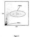
- Figure 1 shows an LS1 (1°-3°) vs. LS2 (4°-6°) scattergram of a clinical whole blood sample, containing 14 NRBC /100 WBC processed and analyzed following the procedure described in Example I.
- NRBCs form a cluster which are clearly distinguished from red blood cell debris which is noted below the NRBCs and are clearly distinguished from leukocytes (which are out of the scope of this scattergram).
- the NRBC populations, described in Figure 1 are below lymphocytes in a region that is usually considered a debris region as shown in Figure 2.
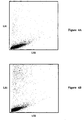
- Figure 4A shows an LS1 vs. LS2 scattergram, in an identical scales as Figure 1, which is obtained from a normal fresh whole blood sample processed and analyzed following the procedure described in Example IV.
- Figure 1 shows an LS1 vs. LS2 scattergram, in an identical scales as Figure 1, which is obtained from a normal fresh whole blood sample processed and analyzed following the procedure described in Example IV.
- Figure 1 shows that there is no cluster present in the region where NRBCs appear (as shown in Figure 1) because a normal peripheral blood does not have NRBCs.
- the same sample was kept at room temperature (about 23°C) for about thirty hours and then processed and analyzed again using the same procedure.
- Figure 4B shows the resultant scattergram. No cluster is found in the scattergram at the NRBC region. This illustrates that the method of the present invention has a low risk of reporting false positive results for normal blood samples even when the samples are aged.
- Figure 5 shows a scattergram obtained from a clinical sample containing 5% reticulocytes analyzed using the method of the present invention described in Example V.
- the scattergram shows an additional cluster to the left of NRBCs, which corresponds to reticulocytes.
- Reticulocyte is another type of immature red blood cell which does not contain nuclei, but contains RNA. Different from NRBCs, reticulocytes appear in both the bone marrow and peripheral blood. In certain diseases, such as hemolytic anemia, thalassemia, and sideroblastic anemia, increased reticulocytes occur. Most nuclear stains used for staining NRBCs also stain RNA in reticulocytes.
- the staining of reticulocytes generates fluorescence signals which overlap with the NRBC fluorescence signals. Therefore, the NRBC results could be erroneous using a fluorescence method if no specific correction is performed for the reticulocytes.
- Figure 6 shows a scattergram of a clinical sample containing both NRBCs and reticulocytes.
- the sample was analyzed using the same procedure of Example V.
- the NRBC report is consistent with the result of the manual reference. Therefore, the presence of reticulocytes does not interfere with the NRBC measurement using the method of the present invention.
- NRBCs by low angle light scatters (LS1 and LS2) are simple and straightforward. It utilizes a regular two-dimensional dotplot, or scattergram, to distinguish NRBCs from other cell types. Because in previous methods, NRBC signals overlap with the signals from red blood cell debris and other cell types, such as leukocytes and reticulocytes, in one or more dimensions, prior art methods require pre-gating or multiple gating of the detected fluorescence and light scatter signals for identifying NRBC population. The present method does not need pre-gating or editing of the detected signals since NRBCs are separated from other cell types including red blood cell debris by the two-dimensional detection method.
- the cell populations above the NRBC cluster in LS1 are counted as total leukocytes (WBC).
- WBC total leukocytes
- another method can be used to determine total leukocytes (WBC). If a DC detection device is also used, the cell populations above the minimum DC volume of the lymphocytes, can be counted as total leukocytes.
- the WBC obtained in both of these methods is free of NRBCs interference, and is equivalent to the corrected WBC, which is generated by subtracting a manual NRBC count from a WBC produced by a regular commercial blood counter.
- the NRBC concentration of the analyzed sample can be calculated by dividing the number of cells counted in the identified NRBC cluster ( Figure 1) by the total leukocytes (WBC) counted and multiplying the quotient by 100.
- the NRBC concentration can be reported as the number of NRBC/100 WBC, which is the same as the manual report unit, or can be reported as an absolute NRBC per ⁇ l of a whole blood.
- a differential analysis of leukocytes can be performed together with the differentiation of NRBCs.
- the differential analysis can be performed using the same reagent system and one sample preparation, in a one step measurement using electronic and optical analysis.
- the electronic and optical analysis includes light scatter and impedance measurements.
- the DC impedance measurement device used for leukocyte analysis by an automated hematology analyzer is known to those skilled in the art and is generally described in U.S. Patent No. 5,125,737, to Rodriguez et al., which is hereby incorporated by reference in its entirety.
- a light scatter detector capable of detecting low and medium angle light scatter signals from a particle or a cell passing through the flow cell is used.
- leukocytes are out of the range of the LS1 vs. LS2 scattergram, and are not shown in this figure.
- the sample analysis for leukocyte differentiation and NRBC differentiation can be performed in an one step measurement.
- the data analysis for both differentiations can be performed simultaneously using different parameters obtained from the one step measurement.
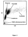
- Figure 2 is a DC vs. LS3 (medium angle light scatter, from about 24° to about 35°) scattergram obtained from a fresh normal whole blood sample processed according to Example II (the same reagents and procedure used in Example I).
- Figure 2 shows four distinct clusters of leukocyte subpopulations, lymphocytes, monocytes, neutrophils and eosinophils. The sample was processed in one sample preparation and analyzed simultaneously with NRBC differentiation.
- WBC total leukocytes
- the method of the present invention reports the differentiation and enumeration of nucleated red blood cells without using nuclear stain and fluorescence. Elimination of nuclear staining provides advantages in reducing instrument and fluid system maintenance because of dye contamination, reducing system's sensitivity to reagent carry-over and reducing reagent cost due to expensive fluorescent dyes.
- the method of the present invention is fast, reliable and suitable for automated blood analysis. In addition, the detection technique is less complex and inexpensive compared to fluorescence method.
- the method of the present invention also provides an advantage of operating entirely at room temperatures, 18 to 28°C.
- Prior art methods operate at elevated temperature for differentiation of leukocyte subpopulations or differentiation of NRBCs. This elevated temperature requirement necessitates analysis instrumentation which is significantly more complex because the reactions must be thermostatically controlled.
- the present invention overcomes this need for thermostatic control by operating optimally at room temperature.
- Another advantageous feature of this invention is its utility for differential analysis of other fluid samples, such as bone marrow, and blood samples of non-human species.
- Example VI provides an example for application of the present method in veterinary sample NRBC analysis.
- the sample mixture was delivered to a flow cell with a sheath fluid, ISOTON® III diluent (product of Coulter Corporation, Miami, Florida), for NRBC and leukocyte differential analysis on a hematology analyzer equipped with DC and light scatter detectors.
- the light scatter detector detects light scatter signals from a cell passing through the flow cell at several ranges of angles, i.e., from about 1° to about 3° (LS1), from about 4° to about 6° (LS2), from about 24° to about 35° (LS3) and higher angles.
- Figure 1 shows a cluster of NRBC distinguished from red cell debris (below) and leukocytes (above, but out of the scope of the scattergram) in an LS1 vs. LS2 scattergram.
- the NRBC concentration of the analyzed sample is calculated by dividing the number of cells counted in the identified NRBC cluster ( Figure 1) by the total leukocytes (WBC) counted and multiplying the quotient by 100.
- the NRBC concentration is reported as number of NRBC/100 WBC, which is the same as the manual report unit. For this sample both the manual reference method and the method described above reported 14 NRBC/100 WBC.
- FIG. 1 is an obtained DC vs. LS3 scattergram, which shows four distinct clusters of leukocyte subpopulations, lymphocytes, monocytes, neutrophils and eosinophils.
- Figure 3 is the resultant LS1 vs. LS2 scattergram (in the same scale as in Figure 1) which shows the NRBC cluster separated from other cell types.
- Example II The reagent and procedure described in Example I were used for analysis of a fresh normal whole blood sample.
- the sample was kept at room temperature (about 23°C) for about thirty hours and then processed and analyzed again.
- the resultant scattergrams are shown in Figure 4A for fresh blood and Figure 4B for aged blood.
- Figure 4A no cluster is present in the region where NRBCs appear because a normal peripheral blood does not contain NRBCs.
- No cluster is found either in Figure 4B at the NRBC region.
- This example shows that the method of the present invention has a low risk in reporting false positive results for normal blood samples even when the normal blood samples are aged.
- Example I The procedure and reagent described in Example I were used for analysis of a blood sample containing reticulocytes (5% reported by manual reference).
- Figure 5 is the resultant LS1 vs. LS2 scattergram. It shows an additional cluster on the left of NRBCs, which corresponds to reticulocytes.
- Figure 6 shows a scattergram obtained from a clinical sample containing both NRBCs and reticulocytes using the same procedure.
- the NRBC report was consistent with the manual reference result, 17 NRBC/100 WBC from the method of the present invention vs. 16 NRBC/100 WBC from the manual reference.
Landscapes
- Health & Medical Sciences (AREA)
- Life Sciences & Earth Sciences (AREA)
- Chemical & Material Sciences (AREA)
- Immunology (AREA)
- Hematology (AREA)
- Engineering & Computer Science (AREA)
- Analytical Chemistry (AREA)
- Biochemistry (AREA)
- Pathology (AREA)
- Molecular Biology (AREA)
- Urology & Nephrology (AREA)
- General Physics & Mathematics (AREA)
- General Health & Medical Sciences (AREA)
- Biomedical Technology (AREA)
- Physics & Mathematics (AREA)
- Medicinal Chemistry (AREA)
- Cell Biology (AREA)
- Food Science & Technology (AREA)
- Biotechnology (AREA)
- Microbiology (AREA)
- Dispersion Chemistry (AREA)
- Investigating Or Analysing Biological Materials (AREA)
Description


Ten seconds after the addition of the stabilizing reagent the sample mixture was delivered to a flow cell with a sheath fluid, ISOTON® III diluent (product of Coulter Corporation, Miami, Florida), for NRBC and leukocyte differential analysis on a hematology analyzer equipped with DC and light scatter detectors. The light scatter detector detects light scatter signals from a cell passing through the flow cell at several ranges of angles, i.e., from about 1° to about 3° (LS1), from about 4° to about 6° (LS2), from about 24° to about 35° (LS3) and higher angles.
Claims (11)
- A method for differentiating nucleated red blood cells comprising:(a) exposing a blood cell sample to a reagent system to lyse mature red blood cells;(b) analyzing said sample in a flow cell by light scatter measurement to differentiate nucleated red blood cells from other cell types; and(c) reporting nucleated red blood cells in said blood cell sample.
- A method for differentiating nucleated red blood cells and leukocytes comprising:(a) exposing a blood cell sample to a reagent system to lyse mature red blood cells without damaging leukocytes;(b) analyzing said sample in a flow cell by DC and light scatter measurements to differentiate nucleated red blood cells and leukocyte subpopulations; and(c) reporting nucleated red blood cells and leukocyte subpopulations in said blood cell sample.
- The method of Claim 1 or 2, wherein said light scatter measurement is performed using two angles of light scatter signals.
- The method of anyone of Claims 1 to 3, wherein said light scatter measurement is performed using low angle light scatter signals detected in less than 10°.
- The method of Claim 3 or 4, wherein said two angles of light scatter signals are low angle light scatter signals detected in less than 10°.
- The method of Claim 5, wherein said one low angle light scatter signal is in a range from about 0° to about 4°.
- The method of Claim 5 or 6, wherein said another low angle light scatter signal is in a range from about 3° to about 7°.
- The method of anyone of Claims 2 to 7, wherein said leukocyte subpopulations are selected from the group consisting of lymphocytes, monocytes, neutrophils and eosinophils.
- The method of anyone of Claims 2 to 8, wherein reporting of said leukocyte subpopulations is in absolute count per unit volume of said blood sample or in percentage of total leukocytes in said blood sample.
- The method of anyone of Claims 1 to 9, wherein reporting of said nucleated red blood cells is in the form of reporting the number of nucleated red blood cells per one hundred of leukocytes, or an absolute number of nucleated red blood cells per unit volume of a blood cell sample.
- The method of anyone of Claims 1 to 10, wherein the said blood cell sample includes peripheral blood of a human, peripheral blood of a non-human, bone marrow of a human and bone marrow of a non-human.
Applications Claiming Priority (3)
| Application Number | Priority Date | Filing Date | Title |
|---|---|---|---|
| US975846 | 1992-11-13 | ||
| US08/975,846 US5874310A (en) | 1997-11-21 | 1997-11-21 | Method for differentiation of nucleated red blood cells |
| PCT/US1998/024349 WO1999027347A1 (en) | 1997-11-21 | 1998-11-13 | Method for differentiation of erythroblasts |
Publications (2)
| Publication Number | Publication Date |
|---|---|
| EP1032818A1 EP1032818A1 (en) | 2000-09-06 |
| EP1032818B1 true EP1032818B1 (en) | 2002-07-24 |
Family
ID=25523478
Family Applications (1)
| Application Number | Title | Priority Date | Filing Date |
|---|---|---|---|
| EP98957975A Expired - Lifetime EP1032818B1 (en) | 1997-11-21 | 1998-11-13 | Method for differentiation of erythroblasts |
Country Status (5)
| Country | Link |
|---|---|
| US (1) | US5874310A (en) |
| EP (1) | EP1032818B1 (en) |
| JP (1) | JP4366478B2 (en) |
| DE (1) | DE69806802T2 (en) |
| WO (1) | WO1999027347A1 (en) |
Families Citing this family (41)
| Publication number | Priority date | Publication date | Assignee | Title |
|---|---|---|---|---|
| US20020115122A1 (en) * | 1998-09-10 | 2002-08-22 | Anderson Jeffrey E. | Method and apparatus for automated assessment of the immunoregulatory status of the mononuclear leukocyte immune system |
| US6509192B1 (en) * | 1992-02-24 | 2003-01-21 | Coulter International Corp. | Quality control method |
| US6214625B1 (en) * | 1999-04-28 | 2001-04-10 | Coulter International Corp. | Composition and method for differentiation of basophils and eosinophils in blood |
| US6232125B1 (en) * | 1999-08-09 | 2001-05-15 | Coulter International Corp. | Method and apparatus for differentiating and enumerating leukocytes |
| US6784981B1 (en) * | 2000-06-02 | 2004-08-31 | Idexx Laboratories, Inc. | Flow cytometry-based hematology system |
| US6448085B1 (en) * | 2001-04-13 | 2002-09-10 | Sysmex Corporation | Quality control material and calibrator for nucleated red blood cell tested on hematology analyzer |
| US6410330B1 (en) | 2001-07-27 | 2002-06-25 | Coulter International Corp. | Method for measurement of nucleated red blood cells |
| US6472215B1 (en) * | 2001-07-27 | 2002-10-29 | Coulter International Corp. | Method of analyzing nucleated red blood cells in a blood sample |
| US6916658B2 (en) * | 2001-07-27 | 2005-07-12 | Beckman Coulter, Inc. | Method for measurement of immature granulocytes |
| CN1276252C (en) | 2001-07-27 | 2006-09-20 | 贝克曼库尔特有限公司 | Method for measurement of nucleated red blood cells |
| US6573102B2 (en) | 2001-07-27 | 2003-06-03 | Coulter International Corp. | Lytic reagent composition for determination of nucleated blood cells |
| US6723563B2 (en) | 2001-12-03 | 2004-04-20 | Streck Laboratories Inc. | Hematology reference control |
| US6653137B2 (en) | 2001-12-03 | 2003-11-25 | Streck Laboratories Inc. | Hematology reference control |
| JP4850711B2 (en) * | 2003-10-02 | 2012-01-11 | ベックマン コールター, インコーポレイテッド | Reference control for optical measurement of nucleated red blood cells in blood samples |
| US7198953B2 (en) * | 2003-10-12 | 2007-04-03 | Beckman Coulter, Inc. | Method of using a reference control composition for measurement of nucleated red blood cells |
| WO2005043113A2 (en) * | 2003-10-12 | 2005-05-12 | Beckman Coulter, Inc. | Method of using a reference control composition for measurement of nucleated red blood cells |
| US7195919B2 (en) * | 2003-12-19 | 2007-03-27 | Beckman Coulter, Inc. | Hematology controls for reticulocytes and nucleated red blood cells |
| JP4911601B2 (en) * | 2004-02-10 | 2012-04-04 | ベックマン コールター, インコーポレイテッド | Method for measuring nucleated red blood cells |
| US7208319B2 (en) * | 2004-02-10 | 2007-04-24 | Beckman Coulter, Inc. | Method of measurement of nucleated red blood cells |
| JP4993603B2 (en) * | 2004-04-07 | 2012-08-08 | ベックマン コールター, インコーポレイテッド | Reference control containing nucleated red blood cell components |
| SE528697C2 (en) * | 2005-03-11 | 2007-01-30 | Hemocue Ab | Volumetric determination of the number of white blood cells in a blood sample |
| FR2893414A1 (en) * | 2005-11-17 | 2007-05-18 | Philippe Claude Loui Daurenjou | SYSTEM FOR DIFFERENTIATING AND DENOMBRATING PARTICLES SUSPENDED IN A LIQUID |
| US7354767B2 (en) * | 2006-03-16 | 2008-04-08 | Beckman Coulter, Inc. | Reference control composition containing a nucleated red blood cell component made of non-nucleated blood cells |
| EP2032979B1 (en) * | 2006-06-06 | 2018-07-18 | Roche Diagnostics GmbH | Differential hemolysis of a whole blood sample |
| EP2030013B1 (en) * | 2006-06-06 | 2009-12-16 | Roche Diagnostics GmbH | Ready-to-use whole blood collection vessel |
| US7754487B2 (en) * | 2006-11-14 | 2010-07-13 | Beckman Coulter, Inc. | Hematology linearity control composition, system and method of use |
| US7674622B2 (en) * | 2006-12-22 | 2010-03-09 | Abbott Laboratories, Inc. | Method for determination of nucleated red blood cells and leukocytes in a whole blood sample in an automated hematology analyzer |
| JP4817450B2 (en) * | 2007-01-31 | 2011-11-16 | シスメックス株式会社 | Blood analyzer, blood analysis method and computer program |
| US7618821B2 (en) * | 2007-04-13 | 2009-11-17 | Streck, Inc. | Simulated blood components and methods |
| US20100086962A1 (en) | 2008-10-08 | 2010-04-08 | Streck, Inc. | Hematology controls and methods |
| CN101723874B (en) * | 2008-10-31 | 2013-09-11 | 深圳迈瑞生物医疗电子股份有限公司 | Cyanine compound and application thereof in dyeing biological samples |
| CN101750476B (en) * | 2008-12-08 | 2015-06-03 | 深圳迈瑞生物医疗电子股份有限公司 | Blood analysis reagent and use method thereof |
| WO2010085815A1 (en) * | 2009-01-26 | 2010-07-29 | Artemis Health, Inc. | Methods and compositions for identifying a fetal cell |
| CN102115456B (en) * | 2009-12-30 | 2014-08-20 | 深圳迈瑞生物医疗电子股份有限公司 | Cyanine compound, composition containing same and application in cell detection thereof |
| US9096885B2 (en) * | 2012-07-05 | 2015-08-04 | Beckman Coulter, Inc. | Method and apparatus for determining white blood cell counts |
| US9581491B2 (en) | 2014-09-30 | 2017-02-28 | Perkinelmer Health Sciences, Inc. | Flow cell modules and liquid sample analyzers and methods including same |
| US9500588B2 (en) | 2014-09-30 | 2016-11-22 | Perkinelmer Health Sciences, Inc. | Flow cell modules and liquid sample analyzers and methods including same |
| WO2016106688A1 (en) * | 2014-12-31 | 2016-07-07 | 深圳迈瑞生物医疗电子股份有限公司 | Nucleated red blood cell warning method and device, and flow cytometer |
| ES2975718T3 (en) | 2018-03-30 | 2024-07-12 | Idexx Lab Inc | Flow Cytometer Laser Optics Assembly |
| EP4439582A3 (en) * | 2018-04-28 | 2024-10-16 | Shenzhen Mindray Bio-Medical Electronics Co., Ltd. | Method and system for analysing a blood sample |
| EP4425235A2 (en) | 2020-06-17 | 2024-09-04 | IDEXX Laboratories, Inc. | Flow cytometer and laser optics assembly thereof |
Family Cites Families (14)
| Publication number | Priority date | Publication date | Assignee | Title |
|---|---|---|---|---|
| US4284412A (en) * | 1979-07-13 | 1981-08-18 | Ortho Diagnostics, Inc. | Method and apparatus for automated identification and enumeration of specified blood cell subclasses |
| US4735504A (en) * | 1983-10-31 | 1988-04-05 | Technicon Instruments Corporation | Method and apparatus for determining the volume & index of refraction of particles |
| US5155044A (en) * | 1987-03-13 | 1992-10-13 | Coulter Electronics, Inc. | Lysing reagent system for isolation, identification and/or analysis of leukocytes from whole blood samples |
| KR970007077B1 (en) * | 1987-03-13 | 1997-05-02 | 코울터 일렉트로닉스 인커퍼레이티드 | Multi-part diefferential analyzing apparatus using light scatter techniques |
| IL85532A (en) * | 1987-03-13 | 1992-03-29 | Coulter Electronics | Method and lytic reagent system for isolation,identification and/or analysis of leukocytes from whole blood samples |
| US5389549A (en) * | 1987-05-29 | 1995-02-14 | Toa Medical Electronics Co., Ltd. | Method for classifying leukocytes and a reagent used therefor |
| CA2016699C (en) * | 1989-05-15 | 2003-11-18 | Paul N. Marshall | Lytic agents and uses thereof |
| JP2927979B2 (en) * | 1991-02-22 | 1999-07-28 | シスメックス株式会社 | Classification of erythroblasts by flow cytometry |
| US5776709A (en) * | 1991-08-28 | 1998-07-07 | Becton Dickinson And Company | Method for preparation and analysis of leukocytes in whole blood |
| AU6235994A (en) * | 1993-02-25 | 1994-09-14 | Abbott Laboratories | Multipurpose reagent system for rapid lysis of whole blood samples |
| US5631165A (en) * | 1994-08-01 | 1997-05-20 | Abbott Laboratories | Method for performing automated hematology and cytometry analysis |
| JP3375203B2 (en) * | 1994-08-08 | 2003-02-10 | シスメックス株式会社 | Cell analyzer |
| US5559037A (en) * | 1994-12-15 | 1996-09-24 | Abbott Laboratories | Method for rapid and simultaneous analysis of nucleated red blood cells |
| US5686308A (en) * | 1995-06-08 | 1997-11-11 | Coulter Corporation | Reagent and method for differential determination of leukocytes in blood |
-
1997
- 1997-11-21 US US08/975,846 patent/US5874310A/en not_active Expired - Lifetime
-
1998
- 1998-11-13 DE DE69806802T patent/DE69806802T2/en not_active Expired - Lifetime
- 1998-11-13 WO PCT/US1998/024349 patent/WO1999027347A1/en active IP Right Grant
- 1998-11-13 EP EP98957975A patent/EP1032818B1/en not_active Expired - Lifetime
- 1998-11-13 JP JP2000522437A patent/JP4366478B2/en not_active Expired - Lifetime
Also Published As
| Publication number | Publication date |
|---|---|
| JP4366478B2 (en) | 2009-11-18 |
| DE69806802D1 (en) | 2002-08-29 |
| US5874310A (en) | 1999-02-23 |
| JP2001524666A (en) | 2001-12-04 |
| DE69806802T2 (en) | 2003-02-27 |
| WO1999027347A1 (en) | 1999-06-03 |
| EP1032818A1 (en) | 2000-09-06 |
Similar Documents
| Publication | Publication Date | Title |
|---|---|---|
| EP1032818B1 (en) | Method for differentiation of erythroblasts | |
| EP1049919B1 (en) | Method for differentiation of nucleated red blood cells | |
| EP1714146B1 (en) | Method of measurement of nucleated red blood cells | |
| EP1417481B1 (en) | Method of analyzing nucleated red blood cells in a blood sample | |
| US6114173A (en) | Fully automated method and reagent composition therefor for rapid identification and characterization of reticulocytes erythrocytes and platelets in whole blood | |
| EP0797762B1 (en) | Method for rapid and simultaneous analysis of nucleated red blood cells | |
| EP1977219B1 (en) | Method of measurement of nucleated red blood cells | |
| US6673618B1 (en) | Method for measurement of nucleated red blood cells | |
| US6410330B1 (en) | Method for measurement of nucleated red blood cells | |
| EP1032819B1 (en) | Method for differentiation of reticulocytes in blood | |
| CA2367780A1 (en) | Single channel, single dilution detection method | |
| JP2004537727A5 (en) | ||
| JP2005506525A5 (en) | ||
| KIM et al. | Simultaneous differentiation and quantitation of erythroblasts and white blood cells on a high throughput clinical haematology analyser |
Legal Events
| Date | Code | Title | Description |
|---|---|---|---|
| PUAI | Public reference made under article 153(3) epc to a published international application that has entered the european phase |
Free format text: ORIGINAL CODE: 0009012 |
|
| 17P | Request for examination filed |
Effective date: 20000620 |
|
| AK | Designated contracting states |
Kind code of ref document: A1 Designated state(s): DE FR GB |
|
| 17Q | First examination report despatched |
Effective date: 20010322 |
|
| GRAG | Despatch of communication of intention to grant |
Free format text: ORIGINAL CODE: EPIDOS AGRA |
|
| GRAG | Despatch of communication of intention to grant |
Free format text: ORIGINAL CODE: EPIDOS AGRA |
|
| GRAG | Despatch of communication of intention to grant |
Free format text: ORIGINAL CODE: EPIDOS AGRA |
|
| GRAH | Despatch of communication of intention to grant a patent |
Free format text: ORIGINAL CODE: EPIDOS IGRA |
|
| RTI1 | Title (correction) |
Free format text: METHOD FOR DIFFERENTIATION OF ERYTHROBLASTS |
|
| GRAH | Despatch of communication of intention to grant a patent |
Free format text: ORIGINAL CODE: EPIDOS IGRA |
|
| GRAA | (expected) grant |
Free format text: ORIGINAL CODE: 0009210 |
|
| AK | Designated contracting states |
Kind code of ref document: B1 Designated state(s): DE FR GB |
|
| REG | Reference to a national code |
Ref country code: GB Ref legal event code: FG4D |
|
| REF | Corresponds to: |
Ref document number: 69806802 Country of ref document: DE Date of ref document: 20020829 |
|
| ET | Fr: translation filed | ||
| PLBE | No opposition filed within time limit |
Free format text: ORIGINAL CODE: 0009261 |
|
| STAA | Information on the status of an ep patent application or granted ep patent |
Free format text: STATUS: NO OPPOSITION FILED WITHIN TIME LIMIT |
|
| 26N | No opposition filed |
Effective date: 20030425 |
|
| REG | Reference to a national code |
Ref country code: FR Ref legal event code: PLFP Year of fee payment: 18 |
|
| REG | Reference to a national code |
Ref country code: FR Ref legal event code: PLFP Year of fee payment: 19 |
|
| REG | Reference to a national code |
Ref country code: FR Ref legal event code: PLFP Year of fee payment: 20 |
|
| PGFP | Annual fee paid to national office [announced via postgrant information from national office to epo] |
Ref country code: FR Payment date: 20171127 Year of fee payment: 20 Ref country code: DE Payment date: 20171129 Year of fee payment: 20 |
|
| PGFP | Annual fee paid to national office [announced via postgrant information from national office to epo] |
Ref country code: GB Payment date: 20171127 Year of fee payment: 20 |
|
| REG | Reference to a national code |
Ref country code: DE Ref legal event code: R071 Ref document number: 69806802 Country of ref document: DE |
|
| REG | Reference to a national code |
Ref country code: GB Ref legal event code: PE20 Expiry date: 20181112 |
|
| PG25 | Lapsed in a contracting state [announced via postgrant information from national office to epo] |
Ref country code: GB Free format text: LAPSE BECAUSE OF EXPIRATION OF PROTECTION Effective date: 20181112 |