EP2630257B1 - Procédés et kits de détection de la 5-hydroxyméthylcytosine - Google Patents
Procédés et kits de détection de la 5-hydroxyméthylcytosine Download PDFInfo
- Publication number
- EP2630257B1 EP2630257B1 EP11775893.8A EP11775893A EP2630257B1 EP 2630257 B1 EP2630257 B1 EP 2630257B1 EP 11775893 A EP11775893 A EP 11775893A EP 2630257 B1 EP2630257 B1 EP 2630257B1
- Authority
- EP
- European Patent Office
- Prior art keywords
- 5hmc
- hydroxymethylcytosine
- sample
- nucleic acid
- dna
- Prior art date
- Legal status (The legal status is an assumption and is not a legal conclusion. Google has not performed a legal analysis and makes no representation as to the accuracy of the status listed.)
- Not-in-force
Links
Images


Classifications
-
- C—CHEMISTRY; METALLURGY
- C12—BIOCHEMISTRY; BEER; SPIRITS; WINE; VINEGAR; MICROBIOLOGY; ENZYMOLOGY; MUTATION OR GENETIC ENGINEERING
- C12Q—MEASURING OR TESTING PROCESSES INVOLVING ENZYMES, NUCLEIC ACIDS OR MICROORGANISMS; COMPOSITIONS OR TEST PAPERS THEREFOR; PROCESSES OF PREPARING SUCH COMPOSITIONS; CONDITION-RESPONSIVE CONTROL IN MICROBIOLOGICAL OR ENZYMOLOGICAL PROCESSES
- C12Q1/00—Measuring or testing processes involving enzymes, nucleic acids or microorganisms; Compositions therefor; Processes of preparing such compositions
- C12Q1/68—Measuring or testing processes involving enzymes, nucleic acids or microorganisms; Compositions therefor; Processes of preparing such compositions involving nucleic acids
- C12Q1/6813—Hybridisation assays
- C12Q1/6827—Hybridisation assays for detection of mutation or polymorphism
-
- C—CHEMISTRY; METALLURGY
- C12—BIOCHEMISTRY; BEER; SPIRITS; WINE; VINEGAR; MICROBIOLOGY; ENZYMOLOGY; MUTATION OR GENETIC ENGINEERING
- C12Q—MEASURING OR TESTING PROCESSES INVOLVING ENZYMES, NUCLEIC ACIDS OR MICROORGANISMS; COMPOSITIONS OR TEST PAPERS THEREFOR; PROCESSES OF PREPARING SUCH COMPOSITIONS; CONDITION-RESPONSIVE CONTROL IN MICROBIOLOGICAL OR ENZYMOLOGICAL PROCESSES
- C12Q1/00—Measuring or testing processes involving enzymes, nucleic acids or microorganisms; Compositions therefor; Processes of preparing such compositions
- C12Q1/68—Measuring or testing processes involving enzymes, nucleic acids or microorganisms; Compositions therefor; Processes of preparing such compositions involving nucleic acids
- C12Q1/6806—Preparing nucleic acids for analysis, e.g. for polymerase chain reaction [PCR] assay
-
- Y—GENERAL TAGGING OF NEW TECHNOLOGICAL DEVELOPMENTS; GENERAL TAGGING OF CROSS-SECTIONAL TECHNOLOGIES SPANNING OVER SEVERAL SECTIONS OF THE IPC; TECHNICAL SUBJECTS COVERED BY FORMER USPC CROSS-REFERENCE ART COLLECTIONS [XRACs] AND DIGESTS
- Y10—TECHNICAL SUBJECTS COVERED BY FORMER USPC
- Y10T—TECHNICAL SUBJECTS COVERED BY FORMER US CLASSIFICATION
- Y10T436/00—Chemistry: analytical and immunological testing
- Y10T436/14—Heterocyclic carbon compound [i.e., O, S, N, Se, Te, as only ring hetero atom]
- Y10T436/145555—Hetero-N
- Y10T436/147777—Plural nitrogen in the same ring [e.g., barbituates, creatinine, etc.]
Definitions
- the present invention relates to methods and kits for the detection of 5-hydroxymethylcytosine (5hmC).
- the present invention relates to methods and kits for detection of 5hmC in nucleic acid (e.g., DNA, RNA).
- the present invention relates to detection of 5hmC in genomic DNA, e.g., mammalian genomic DNA.
- DNA methylation consisting of a cytosine modified by a methyl group at the N5 position (5meC) is a well described modification affecting gene expression in mammalian cells.
- the 5meC modification generally occurs at the CpG dinucleotide sequence; however, it has been identified elsewhere in the genome ( Illingworth et al. (2009) FEBS Letters 583:1713-1720 ).
- Another type of gene-regulatory DNA modification has more recently been described, 5- hydroxymethylcytosine (5hmC) ( Tahiliani et al. (2009) Science 324:930-935 ; Kriaucionis et al. (2009) Science 324:929-930 ).
- the enzyme Tet1 an iron-dependent ⁇ -ketogluterate dioxygenase, catalyzes the formation of 5hmC from 5meC ( Tahiliani et al. (2009) Science 324:930-935 ).
- the 5hmC base may be an intermediate in the conversion of 5meC to cytosine, thus identifying an enzyme that can potentially demethylate DNA ( Tahiliani et al. (2009) Science 324:930-935 ).
- 5hmC is a stable DNA modification found in specialized nondividing neurons and in all animal tissues studied to date ( Kriaucionis et al. (2009) Science 324:929-930 ). Intriguingly, 5hmC was not detected in cancerous cell lines.
- Patent publication WO2010/037001 (Immune Disease Institute Inc.) describes a method for the detection of 5hmC which uses a glycosylation step. Improved methods for detecting 5-hydroxymethylcytosine residues in DNA are needed. In particular, improved methods for identifying the level of 5hmC modification at specific regions (e.g., specific gene loci) are needed.
- the present invention relates to methods and kits for the detection of 5-hydroxymethylcytosine (5hmC).
- the present invention relates to methods and kits for detection of 5hmC in nucleic acid (e.g., DNA, RNA).
- the present invention relates to detection of 5hmC in genomic DNA, e.g., mammalian genomic DNA.
- 5-hydroxymethylcytosine has been identified in mammalian genomic DNA. Identifying the genomic location of this modified base is of interest for research and diagnostic purposes (e.g., tumor detection).
- this method involves the selective glucosylation of 5hmC residues by the ⁇ -glucosyltransferase from T4 bacteriophage creating ⁇ -glucosyl-5-hydroxymethylcytosine ( ⁇ -glu-5hmC).
- the ⁇ -glu-5hmC modification provides a target which can be efficiently and selectively pulled down by J-binding protein 1 coupled to magnetic beads.
- DNA that is precipitated is suitable for analysis by quantitative PCR, microarray, sequencing, or other techniques.
- the methods described herein find use in e.g., research, clinical diagnostics, screening assays, etc.
- methods of the disclosure find use in studying the temporal and spatial effects that 5hmC may have on epigenetic regulation at the single gene level.
- methods of the disclosure find use in identifying regions (e.g., tissues, cells) with increased or decreased levels of 5hmC residues relative to a second region, e.g., as an indication of differential biological state (e.g., neoplastic state, tumor development, etc.).
- the sample is a biological sample.
- the biological sample may be cellular (e.g., tumor sample, biopsy sample, organ sample, tissue sample, bone sample, etc.), or a biological fluid (e.g., sweat, saliva, urine, urine sediment, blood, semen, tears, cerebrospinal fluid, mucus secretion, milk, interstitial fluid, etc.), or a combination thereof.
- Methods of the present invention are not limited by type of subject.
- the subject is a mammal.
- the mammalian subject is human.
- Methods of the present invention are not limited by age of the subject, physical attributes or physical state (e.g., medical condition) of the subject.
- the sample is exposed to an agent capable of transferring a moiety to 5hmC and thereby "tagging" it.
- the agent capable of transferring a moiety to 5hmC is an enzyme.
- the enzyme is a glycosyltransferase and the moiety is a carbohydrate (e.g., monosaccharide).
- Methods of the present invention are not limited by type of glycosyltransferase.
- the glycosyltransferase may be a glucosyltransfearse, galactosyltransfearse, sialyltransferase, xylosyltransferase, or any other type of glycosyltransferase.
- the glycosyltransferase is a beta-glucosyltransferase.
- the beta-glucosyltransferase is a T4 bacteriophage beta-glucosyltransferase.
- the method involves use of a J-binding protein that binds to (e.g., recognizes) 5hmC to which a moiety has been added (e.g., glucosylated 5hmC),
- the method further comprises isolation of bound and/or modified 5hmC without limitation to the technique(s) used for such isolation.
- an agent capable of binding to a modified 5hmC e.g., a J binding protein capable of binding to glucosylated 5hmC
- a solid substrate without limitation to the type of substrate.
- solid substrates include beads, gels, agar, agarose, resins, particles, magnetic particles, paramagnetic particles, impermeable surfaces (e.g., slides, wells (e.g., wells of microtiter plates), chips (e.g., array chips), etc).
- immobilization of an agent capable of binding to a modified 5hmC facilitates isolation (e.g., pull-down, sedimentation) of the modified 5hmC residues.
- modified 5hmC (e.g., glucoslated 5hmC) is subjected to further analysis techniques, e.g., for the purpose of quantifying the amount of 5hmC present in a sample.
- Methods of the present invention are not limited by the type(s) of downstream analysis techniques.
- Analytical methods include, but are not limited to, PCR, real-time PCR, mass spectrometry techniques, FRET, scintillation counting, sequencing, array analysis (e.g., microarray, gene chip, Affymetrix array), colorimetric assay, enzymatic assay, etc.
- Methods of the present invention find use in diagnostic or risk assessment tests, in research (e.g., genetics research, cell biology research, medical research, developmental research), in expression analyses, in drug screening assays, and the like.
- Methods of the present invention are not limited by the biological state correlated with level of 5hmC detected.
- biological states include, but are not limited to, cancer, autoimmune disorders (e.g., diabetes, rheumatoid arthritis, allergies), genetic and epigenetic diseases, environmentally-mediated conditions, and metabolic conditions.
- Kit components may include, but are not limited to, buffers, enzymes (e.g., glycosyltransferases, T4 bacteriophage beta-glucosyltransferase, binding proteins, J-binding protein 1), substrates (e.g., glucose), tubes, vessels, containers, beads, resins, magnets, instructions, and the like.
- enzymes e.g., glycosyltransferases, T4 bacteriophage beta-glucosyltransferase, binding proteins, J-binding protein 1
- substrates e.g., glucose
- the present invention provides a method for detecting 5-hydroxymethylcytosine in a sample comprising: a) obtaining a sample,b) exposing the sample to a glycosyltransferase under conditions that facilitate activity of the glycosyltransferase, and c) contacting the sample exposed to the glycosyltransferase with a J-binding protein.
- the sample is a biological sample.
- the biological sample is obtained from a mammal.
- the mammal is human.
- the glycosyltransferase is a glucosyltransferase.
- the glucosyltransferase is a beta-glucosyltransferase.
- the beta-glucosyltransferase is a T4 bacteriophage beta-glycosyltransferase.
- the J-binding protein is Crithidia fasciculate J-binding protein 1.
- the sample comprises nucleic acid.
- the nucleic acid is DNA.
- the agent is immobilized to a solid substrate.
- the solid substrate is a type such as a particulate material or a nonporous material.
- the particulate material is a type such as a bead, a colloid, a gel, or a magnetic particle.
- the nonporous material is a type such as a well, a plate, or a membrane.
- the method further comprises additional step d) isolating the agent immobilized to the solid substrate to generate purified sample.
- the method further comprises determining the amount of nucleic acid present in the purified sample. In some embodiments, the determining occurs through a such as PCR, real-time PCR, mass spectrometry, hybridization, gene arrays, or DNA sequencing.
- the present invention provides a kit for detecting 5-hydroxymethylcytosine in a sample, the kit comprising a glycosyltransferase and a J-binding protein.
- the glycosyltransferase is a beta-glucosyltransferase.
- the beta-glucosyltransferase is a T4 bacteriophage beta-glucosyltransferase.
- J-binding protein is Crithidia fasciculate J-binding protein 1.
- the term "specificity” is defined as a statistical measure of performance of an assay (e.g., method, test), calculated by dividing the number of true negatives by the sum of true negatives and false positives.
- the term “informative” or “informativeness” refers to a quality of a marker or panel of markers, and specifically to the likelihood of finding a marker (e.g., epigenetic marker; e.g., 5hmC at one or more particular locations) in a positive sample.
- a marker e.g., epigenetic marker; e.g., 5hmC at one or more particular locations
- CpG island refers to a genomic DNA region that contains a high percentage of CpG sites relative to the average genomic CpG incidence (per same species, per same individual, or per subpopulation (e.g., strain, ethnic subpopulation, or the like).
- CpG islands are defined as having a GC percentage that is greater than 50% and with an observed/expected CpG ratio that is greater than 60% ( Gardiner-Garden et al. (1987) J Mol. Biol. 196:261-282 ; Baylin et al. (2006) Nat. Rev. Cancer 6:107-116 ; Irizarry et al. (2009) Nat.
- CpG islands may have a GC content >55% and observed CpG/expected CpG of 0.65 ( Takai et al. (2007) PNAS 99:3740-3745 ).
- Various parameters also exist regarding the length of CpG islands.
- CpG islands may be less than 100 bp; 100-200 bp, 200-300 bp, 300-500 bp, 500-750 bp; 750-1000 bp; 100 or more bp in length.
- CpG islands show altered methylation patterns (e.g., altered 5hmC patterns) relative to controls (e.g., altered 5hmC methylation in cancer subjects relative to subjects without cancer; tissue-specific altered 5hmC patterns; altered 5hmC patterns in biological samples from subjects with a neoplasia or tumor relative to subjects without a neoplasia or tumor.
- altered methylation involves increased incidence of 5hmC.
- altered methylation involves decreased incidence of 5hmC.
- CpG shore or “CpG island shore” refers to a genomic region external to a CpG island that is or that has potential to have altered methylation (e.g., 5hmC) patterns (see, e.g., Irizarry et al. (2009) Nat. Genetics 41:178-186 ).
- CpG island shores may show altered methylation (e.g., 5hmC) patterns relative to controls (e.g., altered 5hmC in cancer subjects relative to subjects without cancer; tissue-specific altered 5hmC patterns; altered 5hmC in biological samples ⁇ from subjects with neoplasia or tumor relative to subjects without neoplasia or tumor.
- CpG island shores may be located in various regions relative to CpG islands (see, e.g., Irizarry et al. (2009) Nat. Genetics 41;178-186 ). Accordingly, in some embodiments, CpG island shores are located less than 100 bp; 100-250 bp; 250-500 bp; 500-1000 bp; 1000-1500 bp; 1500-2000 bp; 2000-3000 bp; 3000 bp or more away from a CpG island.
- Metastasis is meant to refer to the process in which cancer cells originating in one organ or part of the body relocate to another part of the body and continue to replicate. Metastasized cells subsequently form tumors which may further metastasize. Metastasis thus refers to the spread of cancer from the part of the body where it originally occurs to other parts of the body.
- an individual is suspected of being susceptible to metastasized cancer is meant to refer to an individual who is at an above-average risk of developing metastasized cancer.
- individuals at a particular risk of developing cancer of a particular type e.g., colorectal cancer, bladder cancer, breast cancer, prostate cancer
- family medical history indicates above average incidence of such cancer type among family members and/or those who have already developed cancer and have been effectively treated who therefore face a risk of relapse and recurrence.
- Other factors which may contribute to an above-average risk of developing metastasized cancer which would thereby lead to the classification of an individual as being suspected of being susceptible to metastasized cancer may be based upon an individual's specific genetic, medical and/or behavioral background and characteristics.
- neoplasm refers to any new and abnormal growth of tissue.
- a neoplasm can be a premalignant neoplasm or a malignant neoplasm.
- neoplasm-specific marker refers to any biological material that can be used to indicate the presence of a neoplasm. Examples of biological materials include, without limitation, nucleic acids, polypeptides, carbohydrates, fatty acids, cellular components (e.g., cell membranes and mitochondria), and whole cells.
- amplicon refers to a nucleic acid generated using primer pairs.
- the amplicon is typically single-stranded DNA (e.g., the result of asymmetric amplification), however, it may be RNA or dsDNA.
- amplifying or “amplification” in the context of nucleic acids refers to the production of multiple copies of a polynucleotide, or a portion of the polynucleotide, typically starting from a small amount of the polynucleotide (e.g., a single polynucleotide molecule), where the amplification products or amplicons are generally detectable.
- Amplification of polynucleotides encompasses a variety of chemical and enzymatic processes. The generation of multiple DNA copies from one or a few copies of a target or template DNA molecule during a polymerase chain reaction (PCR) or a ligase chain reaction (LCR; see, e.g., U.S. Patent No.
- 5,494,810 are forms of amplification. Additional types of amplification include, but are not limited to, allele-specific PCR (see, e.g., U.S. Patent No. 5,639,611 ), assembly PCR (see, e.g., U.S. Patent No. 5,965,408 ), helicase-dependent amplification (see, e.g., U.S. Patent No. 7,662,594 ), hot-start PCR (see, e.g., U.S. Patent Nos. 5,773,258 and 5,338,671 ), intersequence-specfic PCR, inverse PCR (see, e.g., Triglia, et al.
- allele-specific PCR see, e.g., U.S. Patent No. 5,639,611
- assembly PCR see, e.g., U.S. Patent No. 5,965,408
- helicase-dependent amplification see, e.g., U
- the terms “complementary” or “complementarity” are used in reference to polynucleotides (i.e., a sequence of nucleotides) related by the base-pairing rules.
- sequence “5'-A-G-T-3'” is complementary to the sequence “3'-T-C-A-5'.”
- Complementarity may be “partial,” in which only some of the nucleic acids' bases are matched according to the base pairing rules. Or, there may be “complete” or “total” complementarity between the nucleic acids.
- the degree of complementarity between nucleic acid strands has significant effects on the efficiency and strength of hybridization between nucleic acid strands. This is of particular importance in amplification reactions, as well as detection methods that depend upon binding between nucleic acids.
- the term "primer” refers to an oligonucleotide, whether occurring naturally as in a purified restriction digest or produced synthetically, that is capable of acting as a point of initiation of synthesis when placed under conditions in which synthesis of a primer extension product that is complementary to a nucleic acid strand is induced (e.g., in the presence of nucleotides and an inducing agent such as a biocatalyst (e.g., a DNA polymerase or the like) and at a suitable temperature and pH).
- the primer is typically single stranded for maximum efficiency in amplification, but may alternatively be double stranded.
- the primer is generally first treated to separate its strands before being used to prepare extension products.
- the primer is an oligodeoxyribonucleotide.
- the primer is sufficiently long to prime the synthesis of extension products in the presence of the inducing agent. The exact lengths of the primers will depend on many factors, including temperature, source of primer and the use of the method.
- the primer is a capture primer.
- nucleic acid molecule refers to any nucleic acid containing molecule, including but not limited to, DNA or RNA.
- the term encompasses sequences that include any of the known base analogs of DNA and RNA including, but not limited to, 4 acetylcytosine, 8-hydroxy-N6-methyladenosine, aziridinylcytosine, pseudoisocytosine, 5-(carboxyhydroxyl-methyl) uracil, 5-fluorouracil, 5-bromouracil, 5-carboxymethylaminomethyl-2-thiouracil, 5-carboxymethyl-aminomethyluracil, dihydrouracil, inosine, N6-isopentenyladenine, 1-methyladenine, 1-methylpseudo-uracil, 1-methylguanine, 1-methylinosine, 2,2-dimethyl-guanine, 2-methyladenine, 2-methylguanine, 3-methyl-cytosine, 5-methylcytosine, N
- nucleobase is synonymous with other terms in use in the art including “nucleotide,” “deoxynucleotide,” “nucleotide residue,” “deoxynucleotide residue,” “nucleotide triphosphate (NTP),” or deoxynucleotide triphosphate (dNTP).
- oligonucleotide refers to a nucleic acid that includes at least two nucleic acid monomer units (e.g., nucleotides), typically more than three monomer units, and more typically greater than ten monomer units.
- nucleic acid monomer units e.g., nucleotides
- the exact size of an oligonucleotide generally depends on various factors, including the ultimate function or use of the oligonucleotide. To further illustrate, oligonucleotides are typically less than 200 residues long (e.g., between 15 and 100), however, as used herein, the term is also intended to encompass longer polynucleotide chains. Oligonucleotides are often referred to by their length.
- oligonucleotide For example a 24 residue oligonucleotide is referred to as a "24-mer".
- the nucleoside monomers are linked by phosphodiester bonds or analogs thereof, including phosphorothioate, phosphorodithioate, phosphoroselenoate, phosphorodiselenoate, phosphoroanilothioate, phosphoranilidate, phosphoramidate, and the like, including associated counterions, e.g., H + , NH 4 + , Na + , and the like, if such counterions are present.
- oligonucleotides are typically single-stranded.
- Oligonucleotides are optionally prepared by any suitable method, including, but not limited to, isolation of an existing or natural sequence, DNA replication or amplification, reverse transcription, cloning and restriction digestion of appropriate sequences, or direct chemical synthesis by a method such as the phosphotriester method of Narang et al. (1979) Meth Enzymol. 68: 90-99 ; the phosphodiester method of Brown et al. (1979) Meth Enzymol. 68: 109-151 ; the diethylphosphoramidite method of Beaucage et al. (1981) Tetrahedron Lett. 22: 1859-1862 ; the triester method of Matteucci et al. (1981) J Am Chem Soc.
- a "sequence" of a biopolymer refers to the order and identity of monomer units (e.g., nucleotides, etc.) in the biopolymer.
- the sequence (e.g., base sequence) of a nucleic acid is typically read in the 5' to 3' direction.
- the term “subject” refers to any animal (e.g ., a mammal), including, but not limited to, humans, non-human primates, rodents, and the like, which is to be the recipient of a particular treatment.
- the terms “subject” and “patient” are used interchangeably herein in reference to a human subject.
- gene refers to a nucleic acid (e.g. , DNA) sequence that comprises coding sequences necessary for the production of a polypeptide, RNA (e.g., including but not limited to, mRNA, tRNA and rRNA) or precursor.
- RNA e.g., including but not limited to, mRNA, tRNA and rRNA
- the polypeptide, RNA, or precursor can be encoded by a full length coding sequence or by any portion of the coding sequence so long as the desired activity or functional properties (e.g ., enzymatic activity, ligand binding, signal transduction, etc.) of the full-length or fragment are retained.
- the term also encompasses the coding region of a structural gene and the including sequences located adjacent to the coding region on both the 5' and 3' ends for a distance of about 1 kb on either end such that the gene corresponds to the length of the full-length mRNA.
- the sequences that are located 5' of the coding region and which are present on the mRNA are referred to as 5' untranslated sequences.
- the sequences that are located 3' or downstream of the coding region and that are present on the mRNA are referred to as 3' untranslated sequences.
- gene encompasses both cDNA and genomic forms of a gene.
- a genomic form or clone of a gene contains the coding region interrupted with non-coding sequences termed "introns” or “intervening regions” or “intervening sequences.”
- Introns are segments of a gene that are transcribed into nuclear RNA (hnRNA); introns may contain regulatory elements such as enhancers. Introns are removed or “spliced out” from the nuclear or primary transcript; introns therefore are absent in the messenger RNA (mRNA) processed transcript.
- mRNA messenger RNA
- the mRNA functions during translation to specify the sequence or order of amino acids in a nascent polypeptide.
- the present invention relates to methods and kits for the detection of 5-hydroxymethylcytosine (5hmC) in samples (e.g., biological samples).
- samples e.g., biological samples
- the present invention relates to methods and kits for detection of 5hmC present in nucleic acid (e.g., DNA, RNA).
- nucleic acid e.g., DNA, RNA
- the present invention relates to detection of 5hmC in genomic DNA, e.g., mammalian or plant genomic DNA.
- the present invention provides systems, methods, kits and reagents for purifying or isolated nucleic acid sequences containing 5hmC residues.
- the nucleic acid sequences containing 5hmC residues are analyzed and/or identified, for example by PCR, real time PCR, microarray analysis, sequencing, or other methods that determine the sequence, identity or presence of a nucleic acid sequence of interest.
- DNA is obtained from a subject.
- the DNA isolated by known methods.
- the DNA sample is treated by restriction endonucleases to provide nucleic acid sequences of an approximate desired length.
- the DNA from a subject is treated with a glycosyltransferase to glycosylate any 5hmC that are present in the sample. This results in a sample containing glycosyl-5hmC bases within the nucleic acid.
- the glycosyl-5hmC nucleic acid sample is then contacted with a binding protein specific for the glycosyl-5hmC bases within the nucleic acid.
- a binding protein-glycosyl-5hmC base complex is formed.
- nucleic acids containing glycosyl-5hmC bases are separated from other nucleic acid sequences in the sample based on the formation of the binding protein-glycosyl-5hmC base complex.
- the glycosyl-5hmC binding agent is a J-binding protein (JBP).
- JBP J-binding protein
- Suitable J binding proteins are described in Figure 6 and Table 1. Those of skill in the art will recognize that additional known JBPs and those that may be identified in the future may be tested for use with the present invention. Additionally, the present invention encompasses the use of mutated or modified JBPs which can be identified by their percent identity with reference sequences.
- the JBP is J-binding protein 1 (JBP1).
- the amino acid sequence of the JBP is at least 80%, 90%, 95%, or 99% identical to the sequence of one or more of the JBPs identified in Table 1.
- the JBP is Crithidia fasciculate JBP-1 (SEQ ID NO:1).
- the JBP has an amino acid sequence that is at least 80%, 90%, 95%, or 99% identical to SEQ ID NO:1.
- the present invention is not limited to the use of particular 5hmc modifying agents.
- the 5hmc modifying agent is an enzyme.
- the enzyme is a glyosyltransferase.
- the glycosyltrasferase is at least 80%, 90%, 95%, or 99% identical to the sequence of one or more of the glycosyltransferases.
- the glycosyltransferase is a T2, T4 or T6 glucosyltransferase, or a Uracil glycosylase.
- the glycosyltransferase is an ⁇ - or ⁇ -glucosyltransferase.
- the glucosyltransferase is T4 ⁇ - glucosyltransferase (SEQ ID NO:2).
- glucosyltransferase is at least 80%, 90%, 95%, or 99% identical to SEQ ID NO:2.
- Table 1 - J-binding Proteins Some JBP1 homologues Expasy Accession Number Organism Description Q9U6M2 Crithidia fasciculata JBP1_CRIFA Thymine dioxygenase JBP1 (EC 1.14.11.6) (J-binding protein 1) A4H5X5 Leishmania braziliensis Thymine dioxygenase JBP1 (EC 1.14.11.6) (J-binding protein 1) A4HU70 Leishmania infantum Thymine dioxygenase JBP1 (EC 1.14.11.6) (J-binding protein 1) Q4QHM7 Leishmania major Thymine dioxygenase JBP1 (EC 1.14.11.6) (J-binding protein 1) Q9U6M1 Leishmania tarentolae (Sauroleishmani a tarentolae) Thymine dioxygen
- the binding protein is immobilized on a substrate.
- the binding protein is covalently attached to a bead, such as a magnetic bead.
- Nucleic acid sequences containing glycosyl-5hmC bases bind to the binding proteins immobilized on the beads.
- the beads containing the bound nucleic acid sequences containing glycosyl-5hmC bases can then be washed to remove unbound sequences from the sample and the bound sequences can be analyzed on the beads or eluted from the beads for further analysis.
- the binding protein can be covalently attached to a bead or matrix material suitable for use in column chromatography.
- Samples containing the glycosyl-5hmC bases are applied to columns comprising the beads or matrix.
- Nucleic acids containing the glycosyl-5hmC bases are bound by the binding protein and retaining on the column, allowing separation from sequences that do not contain glycosyl-5hmC bases.
- the nucleic acids containing the glycosyl-5hmC bases can then be eluted from the column and further analyzed.
- the binding protein may be associated with another protein that allows separation of the complex.
- the binding protein may be covalently modified with a second binding molecule.
- Suitable second binding molecules include biotin, avidin, haptens, immunoglobulins and aptamers.
- a nucleic acid glycosyl-FhmC base-binding protein-second binding molecule complex is formed.
- the complex can then be isolated by utilizing reagents that specifically bind the second binding molecules, for examples, beads, magnetic beads or columns comprising avidin, biotin, immunoglobulins specific for the selected hapten, or aptamer binding partner.
- the simple and cost-effective method for identifying genomic regions containing 5hmC presented herein finds use for study of the temporal and spatial patterns of 5hmC residues in genomic regions.
- the methods of the present are utilized to isolate DNA that contains 5hmc modification and then the isolated DNA is analyzed to determine the sequence or identity of the isolated DNA.
- Particular levels (e.g., thresholds, cutoff points) or locations of 5hmC DNA modification may be used that show optimal function with different ethnic groups or sex, different geographic distributions, different stages of disease, different degrees of specificity or different degrees of sensitivity.
- Particular levels (e.g., thresholds, cutoff points) of 5hmC DNA modification may also be developed which are particularly sensitive to the effect of therapeutic regimens on disease progression. Subjects may be monitored after a therapy and/or course of action to determine the effectiveness of that specific therapy and/or course of action.
- indicators of biological state include, for example, epigenetic alterations.
- Epigenetic alterations include but are not limited to DNA methylation (e.g., 5hmC DNA modification).
- the level (e.g., frequency or score) of 5hmC DNA modification e.g., increased levels relative to a control, decreased levels relative to a control) is determined without limitation to the technique used for such determining.
- Methods of the present invention are not limited to particular locations of epigenetic (e.g., 5hmC) alterations (e.g., 5hmC in coding or regulatory regions).
- Altered 5hmC levels may occur in, for example, CpG islands; CpG island shores; or regions other than CpG islands or CpG island shores.
- the present invention finds use in the determination of a diagnosis and/or prognosis for cancers such as gastric cancer, lung cancer, prostate cancer, pancreatic cancer, and breast cancer.
- the present invention also finds use in the determination of a diagnosis or prognosis for other diseases such as diabetes.
- the methods may be used to screen subjects to determine if administration of a particular drug or treatment is indicated.
- methods, kits, and systems of the present invention involve determination of 5hmC state of a locus of interest (e.g., in human DNA) (e.g., in human DNA extracted from a tissue sample, from a tumor sample, etc). Any appropriate method can be used to quantitate 5hmC and/or determine the location of the modification. PCR techniques, for example, can be used to determine which residues are modified by 5hmC following isolation of the nucleic acid sequences by the processes described above.
- PCR reactions can contain, for example, 10 ⁇ L of captured DNA, IX PCR buffer, 0.2 mM dNTPs, 0.5 ⁇ M sequence specific primers (e.g., primers flanking a CpG island or CpG shore within the captured DNA), and 5 units DNA polymerase (e.g., Amplitaq DNA polymerase from PE Applied Biosystems, Norwalk, CT) in a total volume of 50 ⁇ l.
- a typical PCR protocol can include, for example, an initial denaturation step at 94°C for 5 min, 40 amplification cycles consisting of 1 minute at 94°C, 1 minute at 60°C, and 1 minute at 72°C, and a final extension step at 72°C for 5 minutes.
- methods of the present invention involve the determination (e.g., assessment, ascertaining, quantitation) of 5hmC modification level of an indicator of a condition of interest, such as a neoplasm (e.g., the 5hmC level of a CpG island or CpG shore in the coding or regulatory region of a gene locus) in a sample.
- a condition of interest such as a neoplasm
- 5hmC modification level of an indicator of a condition of interest such as a neoplasm
- a reference e.g., a reference level, a control level, a threshold level, or the like.
- the term "elevated 5hmC level" as used herein with respect to the 5hmC status of a gene locus is any 5hmC level that is above a median 5hmC level in a sample from a random population of mammals (e.g., a random population of 10, 20, 30, 40, 50, 100, or 500 mammals) that do not have a neoplasm (e.g., a cancer) or other condition of interest.
- Elevated levels of 5hmC modification can be any level provided that the level is greater than a corresponding reference level.
- an elevated 5hmC level of a locus of interest can be 0.5, 1, 2, 3, 4, 5, 6, 7, 8, 9, 10, or more fold greater than the reference level 5hmC observed in a normal sample.
- a reference level can be any amount.
- the term "elevated 5hmC score" as used herein with respect to detected 5hmC events in a matrix panel of particular nucleic acid markers is any 5hmC score that is above a median 5hmC score in a sample from a random population of mammals (e.g., a random population of 10, 20, 30, 40, 50, 100, or 500 mammals) that do not have a neoplasm (e.g., a cancer).
- An elevated 5hmC score in a matrix panel of particular nucleic acid markers can be any score provided that the score is greater than a corresponding reference score.
- an elevated score of 5hmC in a locus of interest can be 0.5, 1, 2, 3, 4, 5, 6, 7, 8, 9, 10, or more fold greater than the reference 5hmC score observed in a normal sample.
- a reference score can be any amount that is used for comparison.
- the term "decreased 5hmC level" as used herein with respect to the 5hmC status of a gene locus is any 5hmC level that is below a median 5hmC level in a sample from a random population of mammals (e.g., a random population of 10, 20, 30, 40, 50, 100, or 500 mammals) that do not have a neoplasm (e.g., a cancer).
- Decreased levels of 5hmC modification can be any level provided that the level is less than a corresponding reference level.
- a decreased 5hmC level of a locus of interest can be 0.5, 1, 2, 3, 4, 5, 6, 7, 8, 9, 10, or more fold less than the reference level 5hmC observed in a normal sample.
- a reference level can be any amount.
- the term "decreased 5hmC score" as used herein with respect to detected 5hmC events in a matrix panel of particular nucleic acid markers is any 5hmC score that is below a median 5hmC score in a sample from a random population of mammals (e.g., a random population of 10, 20, 30, 40, 50, 100, or 500 mammals) that do not have a neoplasm (e.g., a cancer).
- a decreased 5hmC score in a matrix panel of particular nucleic acid markers can be any score provided that the score is greater than a corresponding reference score.
- a decreased score of 5hmC in a locus of interest can be 0.5, 1, 2, 3, 4, 5, 6, 7, 8, 9, 10, or more fold less than the reference 5hmC score observed in a normal sample.
- a reference score can be any amount that is used for comparison.
- the methods are not limited to a particular type of mammal.
- the mammal is a human.
- the neoplasm is premalignant.
- the neoplasm is malignant.
- the neoplasm is cancer without regard to stage (e.g., stage I, II, III, or IV).
- the present invention also provides methods and materials to assist medical or research professionals in determining whether or not a mammal has a neoplasm (e.g., cancer).
- Medical professionals can be, for example, doctors, nurses, medical laboratory technologists, and pharmacists.
- Research professionals can be, for example, principle investigators, research technicians, postdoctoral trainees, and graduate students.
- a professional can be assisted by (1) determining the ratio of 5hmC and/or other markers in a sample, and (2) communicating information about the ratio to that professional, for example.
- a medical professional can take one or more actions that can affect patient care. For example, a medical professional can record the results in a patient's medical record. In some cases, a medical professional can record a diagnosis of a neoplasia, or otherwise transform the patient's medical record, to reflect the patient's medical condition. In some cases, a medical professional can review and evaluate a patient's entire medical record, and assess multiple treatment strategies, for clinical intervention of a patient's condition. In some cases, a medical professional can record a prediction of tumor occurrence with the reported indicators. In some cases, a medical professional can review and evaluate a patient's entire medical record and assess multiple treatment strategies, for clinical intervention of a patient's condition.
- a medical professional can record the results in a patient's medical record.
- a medical professional can record a diagnosis of a neoplasia, or otherwise transform the patient's medical record, to reflect the patient's medical condition.
- a medical professional can review and evaluate a patient's entire medical record
- a medical professional can initiate or modify treatment of a neoplasm after receiving information regarding the level (score, frequency) associated with 5hmC level in a patient's urine sample.
- a medical professional can compare previous reports and the recently communicated level (score, frequency) of 5hmC modification, and recommend a change in therapy.
- a medical professional can enroll a patient in a clinical trial for novel therapeutic intervention of neoplasm.
- a medical professional can elect waiting to begin therapy until the patient's symptoms require clinical intervention.
- a medical professional can communicate the assay results to a patient or a patient's family.
- a medical professional can provide a patient and/or a patient's family with information regarding neoplasia, including treatment options, prognosis, and referrals to specialists, e.g., oncologists and/or radiologists.
- a medical professional can provide a copy of a patient's medical records to communicate assay results to a specialist.
- a research professional can apply information regarding a subject's assay results to advance neoplasm research. For example, a researcher can compile data on the assay results, with information regarding the efficacy of a drug for treatment of neoplasia to identify an effective treatment.
- a research professional can obtain assay results to evaluate a subject's enrollment, or continued participation in a research study or clinical trial. In some cases, a research professional can classify the severity of a subject's condition, based on assay results. In some cases, a research professional can communicate a subject's assay results to a medical professional. In some cases, a research professional can refer a subject to a medical professional for clinical assessment of neoplasia, and treatment thereof. Any appropriate method can be used to communicate information to another person (e.g., a professional). For example, information can be given directly or indirectly to a professional. For example, a laboratory technician can input the assay results into a computer-based record.
- information is communicated by making a physical alteration to medical or research records.
- a medical professional can make a permanent notation or flag a medical record for communicating a diagnosis to other medical professionals reviewing the record.
- any type of communication can be used to communicate the information.
- mail, e-mail, telephone, and face-to-face interactions can be used.
- the information also can be communicated to a professional by making that information electronically available to the professional.
- the information can be communicated to a professional by placing the information on a computer database such that the professional can access the information.
- the information can be communicated to a hospital, clinic, or research facility serving as an agent for the professional.
- a single sample can be analyzed for one neoplasm-specific marker or for multiple neoplasm-specific markers.
- a single sample is analyzed for multiple neoplasm-specific markers, for example, using multi-marker assays.
- multiple samples can be collected for a single mammal and analyzed as described herein.
- a sample is split into first and second portions, where the first portion undergoes cytological analysis and the second portion undergoes further purification or processing (e.g., sequence-specific capture step(s) (e.g., for isolation of specific loci for analysis of 5hmC levels).
- the sample undergoes one or more preprocessing steps before being split into portions.
- the sample is treated, handled, or preserved in a manner that promotes DNA integrity and/or inhibits DNA degradation (e.g., through use of storage buffers with stabilizing agents (e.g., chelating agents, DNase inhibitors) or handling or processing techniques that promote DNA integrity (e.g., immediate processing or storage at low temperature (e.g., -80 degrees C)).
- stabilizing agents e.g., chelating agents, DNase inhibitors
- processing techniques that promote DNA integrity (e.g., immediate processing or storage at low temperature (e.g., -80 degrees C)).
- kits generally comprise, for example, reagents useful, sufficient, or necessary for detecting and/or characterizing one or more markers (e.g., epigenetic markers; 5hmC modifications) specific for a neoplasm.
- the kits contain enzymes suitable for amplifying nucleic acids including various polymerases, deoxynucleotides and buffers to provide the necessary reaction mixture for amplification.
- kits of the present invention include a means for containing the reagents in close confinement for commercial sale such as, e.g., injection or blow-molded plastic containers into which the desired reagent are retained.
- a means for containing the reagents in close confinement for commercial sale such as, e.g., injection or blow-molded plastic containers into which the desired reagent are retained.
- Other containers suitable for conducting certain steps of the disclosed methods also may be provided.
- the methods disclosed herein are useful in monitoring the treatment of neoplasia (e.g., cancer).
- the methods may be performed immediately before, during and/or after a treatment to monitor treatment success.
- the methods are performed at intervals on disease free patients to ensure treatment success.
- the present invention also provides a variety of computer-related embodiments. Specifically, in some embodiments the invention provides computer programming for analyzing and comparing a pattern of neoplasm-specific marker detection results in a sample obtained from a subject to, for example, a library of such marker patterns known to be indicative of the presence or absence of a neoplasm, or a particular stage or neoplasm.
- the present invention provides computer programming for analyzing and comparing a first and a second pattern of neoplasm-specific marker detection results from a sample taken at least two different time points.
- the first pattern may be indicative of a pre-cancerous condition and/or low risk condition for cancer and/or progression from a pre-cancerous condition to a cancerous condition.
- the comparing provides for monitoring of the progression of the condition from the first time point to the second time point.
- the invention provides computer programming for analyzing and comparing a pattern of neoplasm-specific marker detection results from a sample to a library of neoplasm-specific marker patterns known to be indicative of the presence or absence of a cancer, wherein the comparing provides, for example, a differential diagnosis between a benign neoplasm, and an aggressively malignant neoplasm (e.g., the marker pattern provides for staging and/or grading of the cancerous condition).
- the methods and systems described herein can be implemented in numerous ways. In one embodiment, the methods involve use of a communications infrastructure, for example the internet. Several embodiments of the invention are discussed below. It is also to be understood that the present invention may be implemented in various forms of hardware, software, firmware, processors, distributed servers (e.g., as used in cloud computing) or a combination thereof. The methods and systems described herein can be implemented as a combination of hardware and software.
- the software can be implemented as an application program tangibly embodied on a program storage device, or different portions of the software implemented in the user's computing environment (e.g., as an applet) and on the reviewer's computing environment, where the reviewer may be located at a remote site (e.g., at a service provider's facility).
- portions of the data processing can be performed in the user-side computing environment.
- the user-side computing environment can be programmed to provide for defined test codes to denote platform, carrier/diagnostic test, or both; processing of data using defined flags, and/or generation of flag configurations, where the responses are transmitted as processed or partially processed responses to the reviewer's computing environment in the form of test code and flag configurations for subsequent execution of one or more algorithms to provide a results and/or generate a report in the reviewer's computing environment.
- the application program for executing the algorithms described herein may be uploaded to, and executed by, a machine comprising any suitable architecture.
- the machine involves a computer platform having hardware such as one or more central processing units (CPU), a random access memory (RAM), and input/output (I/O) interface(s).
- the computer platform also includes an operating system and microinstruction code.
- the various processes and functions described herein may either be part of the microinstruction code or part of the application program (or a combination thereof) which is executed via the operating system.
- various other peripheral devices may be connected to the computer platform such as an additional data storage device and a printing device.
- the system generally includes a processor unit.
- the processor unit operates to receive information, which generally includes test data (e.g., specific gene products assayed), and test result data (e.g., the pattern of neoplasm-specific marker (e.g., epigenetic marker, 5hmC modification) detection results from a sample).
- test data e.g., specific gene products assayed
- test result data e.g., the pattern of neoplasm-specific marker (e.g., epigenetic marker, 5hmC modification) detection results from a sample.
- This information received can be stored at least temporarily in a database, and data analyzed in comparison to a library of marker patterns known to be indicative of the presence or absence of a pre-cancerous condition, or known to be indicative of a stage and/or grade of cancer.
- Part or all of the input and output data can also be sent electronically; certain output data (e.g., reports) can be sent electronically or telephonically (e.g., by facsimile, e.g., using devices such as fax back).
- Exemplary output receiving devices can include a display element, a printer, a facsimile device and the like.
- Electronic forms of transmission and/or display can include email, interactive television, and the like.
- all or a portion of the input data and/or all or a portion of the output data are maintained on a server for access, e.g., confidential access.
- the results may be accessed or sent to professionals as desired.
- a system for use in the methods described herein generally includes at least one computer processor (e.g., where the method is carried out in its entirety at a single site) or at least two networked computer processors (e.g., where detected marker data for a sample obtained from a subject is to be input by a user (e.g., a technician or someone performing the assays)) and transmitted to a remote site to a second computer processor for analysis (e.g., where the pattern of neoplasm-specific marker) detection results is compared to a library of patterns known to be indicative of the presence or absence of a pre-cancerous condition), where the first and second computer processors are connected by a network, e.g., via an intranet or internet).
- a network e.g., via an intranet or internet
- the system can also include a user component(s) for input; and a reviewer component(s) for review of data, and generation of reports, including detection of a pre-cancerous condition, staging and/or grading of a neoplasm, or monitoring the progression of a pre-cancerous condition or a neoplasm.
- Additional components of the system can include a server component(s); and a database(s) for storing data (e.g., as in a database of report elements, e.g., a library of marker patterns known to be indicative of the presence or absence of a pre-cancerous condition and/or known to be indicative of a grade and/or a stage of a neoplasm, or a relational database (RDB) which can include data input by the user and data output.
- the computer processors can be processors that are typically found in personal desktop computers (e.g., IBM, Dell, Macintosh), portable computers, mainframes, minicomputers, or other computing devices.
- the input components can be complete, stand-alone personal computers offering a full range of power and features to run applications.
- the user component usually operates under any desired operating system and includes a communication element (e.g., a modem or other hardware for connecting to a network), one or more input devices (e.g., a keyboard, mouse, keypad, or other device used to transfer information or commands), a storage element (e.g., a hard drive or other computer-readable, computer-writable storage medium), and a display element (e.g., a monitor, television, LCD, LED, or other display device that conveys information to the user).
- the user enters input commands into the computer processor through an input device.
- the user interface is a graphical user interface (GUI) written for web browser applications.
- GUI graphical user interface
- the server component(s) can be a personal computer, a minicomputer, or a mainframe, or distributed across multiple servers (e.g., as in cloud computing applications) and offers data management, information sharing between clients, network administration and security.
- the application and any databases used can be on the same or different servers.
- Other computing arrangements for the user and server(s), including processing on a single machine such as a mainframe, a collection of machines, or other suitable configuration are contemplated. In general, the user and server machines work together to accomplish the processing of the present invention.
- the database(s) is usually connected to the database server component and can be any device which will hold data.
- the database can be any magnetic or optical storing device for a computer (e.g., CDROM, internal hard drive, tape drive).
- the database can be located remote to the server component (with access via a network, modem, etc.) or locally to the server component.
- the database can be a relational database that is organized and accessed according to relationships between data items.
- the relational database is generally composed of a plurality of tables (entities). The rows of a table represent records (collections of information about separate items) and the columns represent fields (particular attributes of a record).
- the relational database is a collection of data entries that "relate" to each other through at least one common field.
- Additional workstations equipped with computers and printers may be used at point of service to enter data and, in some embodiments, generate appropriate reports, if desired.
- the computer(s) can have a shortcut (e.g., on the desktop) to launch the application to facilitate initiation of data entry, transmission, analysis, report receipt, etc. as desired.
- the present invention is useful for both the diagnosing diseases and disorders in a subject as well as determining the prognosis of a subject.
- the methods, reagents and systems of the present invention are applicable to a broad variety of diseases and disorders.
- the present invention provides methods for obtaining a subject's risk profile for developing neoplasm (e.g., cancer).
- such methods involve obtaining a sample from a subject (e.g., a human at risk for developing cancer; a human undergoing a routine physical examination), detecting the presence, absence, or level (e.g., 5hmC modification frequency or score) of one or more markers specific for a neoplasm in or associated with the sample (e.g., specific for a neoplasm) in the sample, and generating a risk profile for developing neoplasm (e.g., cancer) based upon the detected level (score, frequency) or presence or absence of the indicators of neoplasia.
- a generated risk profile will change depending upon specific markers and detected as present or absent or at defined threshold levels.
- the present invention is not limited to a particular manner of generating the risk profile.
- a processor e.g., computer
- the processor uses an algorithm (e.g., software) specific for interpreting the presence and absence of specific 5hmC modifications as determined with the methods of the present invention.
- the presence and absence of specific markers as determined with the methods of the present invention are inputed into such an algorithm, and the risk profile is reported based upon a comparison of such input with established norms (e.g., established norm for pre-cancerous condition, established norm for various risk levels for developing cancer, established norm for subjects diagnosed with various stages of cancer).
- established norms e.g., established norm for pre-cancerous condition, established norm for various risk levels for developing cancer, established norm for subjects diagnosed with various stages of cancer.
- the risk profile indicates a subject's risk for developing cancer or a subject's risk for re-developing cancer. In some embodiments, the risk profile indicates a subject to be, for example, a very low, a low, a moderate, a high, and a very high chance of developing or re-developing cancer. In some embodiments, a health care provider (e.g., an oncologist) will use such a risk profile in determining a course of treatment or intervention (e.g., biopsy, wait and see, referral to an oncologist, referral to a surgeon, etc.).
- a health care provider e.g., an oncologist
- Other diseases and disorders that may be diagnosed or prognosed with the methods, reagents and systems of the present invention include, but are not limited to, Prader-Willi syndrome, Angelman syndrome, Beckwith-Wiedemann syndrome, Pseudohypoparathyroidism, Russell-Silver syndrome, ICF syndrome, Rett syndrome, ⁇ -thalassemia/mental retardation, X-linked (ATR-X), Immunoosseous dysplasia, Schimke type, Rubinstein-Taybi syndrome, MTHFR deficiency, Recurrent hydatidiform mole, Fragile X mental retardation syndrome, Deletion LCR ⁇ - and ⁇ -thalassemia, FSH dystrophy, disorders of XIC, Schimke immunoosseous dysplasia (SIOD), Sotos syndrome, Atrichia, X-linked Emery-Dreifuss muscular dystrophy (EDMD), Autosomal EDMD, CMT2B1, mandibul
- the bgt gene was amplified from T4 bacteriophage DNA and was cloned into pET28a.
- Cultures of Rosetta(DE3)pLysS harboring pET28a-bgt were grown in 500 ml Studier auto-inducing media20 to an A600 of 0.6 at 37°C followed by a shift to 18°C for 20 hrs.
- the cells were harvested by centrifugation and suspended in 10 ml ⁇ -gt lysis buffer (500 mM NaCl, 25 mM Hepes KOH (pH 7.9), 5 mM imidazole, 10% (v/v) glycerol). All subsequent steps were carried out at 4°C unless otherwise specified.
- Fractions containing the highest concentration of protein were pooled and dialyzed 3 times against 100 volumes of ⁇ -gt low salt buffer (50 mM NaCl, 25 mM Hepes KOH (pH 7.9), 0.5 mM EDTA (pH 8.0), 10% (v/v) glycerol).
- the pool was then applied to a Resource S cation exchange column. The column was eluted with a 50 to 500 mM linear NaCl gradient.
- JBP1 J-binding protein 1
- the gene encoding J-binding protein 1 (JBP1) from C. fasciculata was synthesized by GeneArt (Germany). After synthesis the gene was cloned into pET28a. Cultures of Rosetta(DE3)pLysS harboring pET28a-JBP1 were grown in 500 ml Studier auto-inducing media to an OD A600 of 0.6 at 37°C followed by a shift to 18oC for 20 hrs. The cells were harvested by centrifugation and suspended in 10 ml JBP1 lysis buffer (500 mM NaCl, 25 mM Tris HCl (pH 7.5), 5 mM imidazole, 10% (v/v) glycerol). All subsequent steps were carried out at 4°C unless otherwise specified.
- JBP1 lysis buffer 500 mM NaCl, 25 mM Tris HCl (pH 7.5), 5 mM imidazole, 10% (v/
- JBP1 was eluted with 20 ml JBP1 lysis buffer containing 200 mM imidazole.
- Fractions containing the highest concentration of protein were pooled and concentrated to 500 ⁇ l using a centricon MWCO 30000 according to the manufacturer's instructions.
- the concentrated protein was applied to a Superdex75 size exclusion column and eluted with JBP1 Superdex Buffer (250 mM NaCl, 25 mM Tris HCl (pH 7.5), 1 mM EDTA (pH 8.0), 1 mM DTT, 10% (v/v) glycerol.
- Fractions containing no detectable contaminants were pooled and 3 times dialyzed against 100 volumes PBS (150 mM NaCl, 50 mM KPO4 (pH 7.2)).
- Epoxy modified magnetic beads Five milligrams of Epoxy modified magnetic beads (Dynal, Oslo, Norway) were equilibrated according to the manufacturer's instructions and suspended in 60 ⁇ l PBS. 100 ⁇ g of JBP1 in PBS was added to the beads and the PBS was added until the volume equalled 120 ⁇ l, followed by the addition of 40 ⁇ l 4 M (NH4)2SO4. The bead/JBP1 solution was slowly rotated at 4°C for 48 h. After incubation, the protein not bound to the beads was removed and binding efficiencies were calculated (typical binding efficiencies were between 70-80%).
- the beads were then blocked with 300 ⁇ l binding buffer (2 mM EDTA (pH 8.0), 10 mM Tris (pH 8.0), 150 mM NaCl, 0.02% (v/v) Tween 20, 1 mg/ml BSA). After blocking beads were washed 3 times with 300 ⁇ l binding buffer and finally suspended in 300 ⁇ l binding buffer. Beads were prepared freshly for each experiment as it was found that several freeze thaw cycles dramatically reduced binding capacity.
- Annealing reactions (50 ⁇ l) containing 100 pmol of each complementary oligonucleotide, 40 mM Tris-HCl (pH 7.5), 10 mM MgCl2, 1 mM dithiothreitol, 50 mM NaCl were heated to 95°C in a thermocycler for 5 minutes followed decrease in temperature of 1°C/min to 25°C.
- Duplex oligonucleotides were then purified from a 15% nondenaturing PAGE according to the protocol established by Sambrook et al ((1989) Molecular Cloning, a Laboratory Manual, 2 nd Ed., Cold Spring Harbor Laboratory Press).
- Substrates containing cytosine residues or 5meC residues were amplified from pUC18 or from specific mouse genomic regions using a PCR reaction containing unmodified dNTPs with Pfu Turbo (Stratagene) according to the manufacturer's instructions. 5meC residues were created by incubating the PCR product amplified using unmodified dNTPs with M. Sssl methyltransferase (NEB) according to the manufacturer's instructions. Substrates containing 5hmC residues or ⁇ -glu-5hmC residues were amplified as other substrates except that d5hmCTP (Bioline) was used in place of dCTP.
- ⁇ -glu-5hmC substrates were generated by incubating the PCR product created using d5hmCTP with 10 ⁇ g ⁇ -gt, 20 ⁇ M UDP-glucose (Sigma) in ⁇ - gt reaction buffer (20 mM KPO4 (pH 8.0), 25 mM MgCl2). Substrates were purified on a 0.8% agarose gel and excised using a gel extraction kit (Qiagen).
- Reactions 50 ⁇ l containing 1 ⁇ g DNA substrate, 10 ⁇ Ci ⁇ -[32P]ATP, 2.5 units T4 polynucleotide kinase (NEB) and buffer supplied by the manufacturer were incubated at 37°C for 30 min. After 30 min 100 ⁇ mol ATP was added and the reaction was allowed to proceed for 10 min. Enzyme and free nucleotide was removed using a nucleotide removal kit (Qiagen).
- Reactions (50 ⁇ l) containing 1 pmol double stranded oligonucleotide substrate, 20 Mm UDP-Glucose, 10 ⁇ g ⁇ -gt, and ⁇ -gt reaction buffer were incubated at 37°C for 60 min. Reactions were terminated by the addition of 10 ⁇ l Stop Buffer (20 ⁇ g Proteinase K, 50 mM Tris HCl (pH 8.0), 100 mM EDTA (pH 8.0), 2% (w/v) SDS) followed by incubation at 42°C for 60 min. Oligonucleotides were purified by phenol:CHCl 3 : IAA extraction followed by an ethanol precipitation.
- Pellets were suspended in 50 ⁇ l NEB buffer 4 and 10 units Taq I (NEB) and were allowed to incubate at 65°C for 16 hrs. Taq I was inactivated by heating to 80°C for 20 min followed by incubation with 2.5 units of shrimp alkaline phosphatase (NEB) for 30 min. The alkaline phosphatase was inactivated by heating to 65°C for 5 min. Reactions were radiolabeled by adding 2.5 units T4 polynucleotide kinase, 10 ⁇ Ci ⁇ -[32P]-ATP, and the volume was raised to 70 ⁇ l with T4 polynucleotide kinase buffer.
- DNA was cleaned by phenol: CHCl 3 :IAA (isoamyl alcohol) extraction followed by an ethanol precipitation. Pellets were suspended in 5 ⁇ l DNase I buffer and 0.2 units DNase I (NEB) and 0.2 units snake venom phosphodiesterase (Worthington) was added to the reactions. Reactions were incubated at 25oC for at least 4 hours. 0.5 ⁇ l of each reaction was spotted onto a 100 ⁇ m x 20 cm x 20 cm cellulose thin layer plate.
- IAA isoamyl alcohol
- the nucleotides were resolved in two phases: the first phase contained isobutyric acid:water:NH3 (66:20:1) the second phase contained 4 M ammonium sulfate: 1 M acetic acid: isopropanol (80:17:2). Radioactive spots were identified using a phosphor screen.
- DNA substrates were composed of 2.7 kb linear pUC18 PCR products that contained only cytosines, 5meC at CpG regions, only 5hmC, or were treated with 100 ng ⁇ -gt in the presence of 20 ⁇ M UDP-glucose created as described above. Substrates were hydrolyzed to nucleosides by incubation with nuclease P1, snake venom phosphodiesterase, and alkaline phosphatase (Sigma-Aldrich, St. Louis, MO) as described ( Crain (1990) Methods Enzymol. 193:782-790 ; herein incorporated by reference in its entirety).
- the mobile phase consisted of A (0.1% formic acid in water) and B (0.1% formic acid in methanol), starting with 95% A/ 5% B for 0.5 min, followed by a 6.5-min linear gradient of 5- 50% B, 2 min with 50% B and 6 min re-equilibration with the initial mobile phase conditions.
- Online mass spectrometry detection was performed using an Applied Biosystems/MDS Sciex 5000 triple quadrupole mass spectrometer (Applied Biosystems Sciex, USA) with TurbolonSpray probe operating in positive electrospray ionization mode.
- the deoxyribonucleosides were monitored by multiple reaction monitoring using the mass transitions 228.2 ⁇ 112.1 (dC), 242.2 ⁇ 126.1 (5-me(dC)), 258.2 ⁇ 142.1 (5-hm(dC)), and 420.2 ⁇ 304.1 (5-Gly-hm(dC)).
- Reactions (200 ⁇ l) contained 10 ng of each DNA substrate combined in one vial, with binding buffer and varying amounts of JBP1 modified magnetic beads were incubated for 60 min at room temperature with gentle rotation. After washing 3 times with 300 ⁇ l binding buffer without BSA and the beads were suspended in 90 ⁇ l binding buffer and 10 ⁇ l stop buffer was added (20 ⁇ g/ml Proteinase K, 50 mM Tris HCl (pH 8.0), 100 mM EDTA (pH 8.0), 2% (w/v) SDS) and allowed to incubate at 42°C for 60 min. After incubation the DNA was cleaned using a PCR clean kit (Qiagen) and the amount of each DNA pulled down relative to the input was measured by quantitative real time PCR.
- Qiagen PCR clean kit
- Reactions (50 ⁇ l) containing 20 ng of each of three DNA substrates (unmodified cytosine, 5meC, 5hmC genomic regions combined in one vial), 20 ⁇ M UDP-Glucose, and ⁇ -gt reaction buffer, and either 10 ⁇ g ⁇ -gt or ⁇ -gt storage buffer were incubated at 37°C for 30 minutes. Reactions were stopped by the addition of 10 ⁇ l stop buffer. DNA was purified using a PCR clean kit (Qiagen). The cleaned DNA was incubated with 20 ⁇ g JBP1 coated magnetic beads in 200 ⁇ l binding buffer for 60 minutes with gentle rotation.
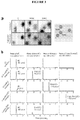
- FIG. 1 A brief overview of a scheme of one embodiment of the present invention to specifically identify genomic regions containing 5hmC residues is shown ( Figure 1 ). Briefly, the ⁇ -gt was used to specifically modify 5hmC residues creating ⁇ -glu-5hmC residues. Following the complete conversion of 5hmC to ⁇ -glu-5hmC, DNA was incubated with JBP1-coated magnetic beads. JBP1 specifically binds ⁇ -glu-5hmC containing DNA, allowing for efficient and selective identification of DNA containing 5hmC. The resulting DNA was purified and ready for analysis by various methods.
- the ⁇ -glucosyltransferase specifically glucosylates 5-hydroxymethylcytosine
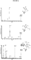
- Recombinant ⁇ -gt from phage T4 was expressed and purified to greater than 90% purity ( Figure 2 ; lane 1) by Cobalt affinity chromatography, followed by cation exchange chromatography.
- the ⁇ -gt specifically glucosylates 5hmC residues( Tomaschewski et al. (1985) Nucleic Acids Res. 13:7551+7568 ; Georgopoulos et al. (1971) Virology 44:271-285 ; Kornberg et al. (1961) J. Biol. Chem.
- the resulting products were digested with Taq I, treated with alkaline phosphatase, 5'-end labelled using T4 polynucleotide kinase, and digested to 5' mononucleotides using DNase I and Snake Venom Phosphodiesterase.
- the resulting nucleotides were then resolved using two-dimensional thin-layer chromatography (TLC). From the TLC analysis it was deduced that the ⁇ -gt has no effect on the substrates that lack 5hmC ( Figure 3 ; left and center); however, upon treatment with ⁇ -gt the 5hmC spot is absent from the TLC plate ( Figure 3 ; right).
- JPB1 from C . fasciculata can specifically pull down ⁇ -glucosyl-5hmC
- Recombinant C. fasciculata JBP1 containing a his-tag was purified to greater than 90% homogeneity by cobalt affinity chromatography followed by size exclusion chromatography. Fractions included in the final pool of purified JBP1 were determined to be free of detectable nucleases ( Figure 2 ; lane 2). Following purification, JBP1 was covalently linked to epoxy modified magnetic beads as described supra.
- JBP1 coated magnetic beads were incubated with four different radiolabeled substrates and precipitated using a magnet. Each substrate was a 2.7 kb linear PCR product created using pUC18 as a template. The substrates contained normal adenines (A), guanines (G) and thymidines (T) and either: only cytosine residues, 5meC residues in CpG sequences, only 5hmC residues, or ⁇ -glu-5hmC residues produced by incubating the 5hmC substrate with the ⁇ -gt. JBP1 coated magnetic beads (132 ng) could effectively pull down up to 6% of the DNA that contained the ⁇ -glu-5hmC modification ( Figure 5a ).
- A normal adenines
- G guanines
- T thymidines
- JBP1 had little affinity for cytosine, 5meC, or 5hmC containing substrates. Furthermore, BSA coated magnetic beads were unable to pull down any of the substrates, suggesting that the binding of ⁇ -glu-5hmC is due to JBP1 bound to the magnetic beads.
- JBP1 coated magnetic beads could pull down 14% of the ⁇ -glu-5hmC DNA and provide an 87-fold enrichment of ⁇ -glu-5hmC modified DNA over cytosine containing DNA and a 319-fold enrichment over 5meC containing DNA.
- JBP1 can pull down DNA containing a single ⁇ -glu-5hmC modification
- JBP1 facilitated pull down of ⁇ -gt treated 5hmC is not sequence specific
Claims (14)
- Procédé pour détecter la 5-hydroxyméthylcytosine dans un échantillon comprenant un acide nucléique comprenant :a) la glycosylation enzymatique des bases 5-hydroxyméthylcytosine dans ledit échantillon pour fournir un acide nucléique comprenant des bases 5-hydroxyméthylcytosine glycosylées ;b) la mise en contact dudit échantillon avec une protéine de liaison à J (JBP) spécifique de la 5-hydroxyméthylcytosine glycosylée pour former un complexe JBP-5-hydroxyméthylcytosine glycosylée ; etc) l'analyse dudit complexe JBP-5-hydroxyméthylcytosine glycosylée pour déterminer la présence de la 5-hydroxyméthylcytosine dans ledit acide nucléique dans ledit échantillon.
- Procédé de la revendication 1, dans lequel ledit échantillon biologique est obtenu à partir d'un sujet choisi dans le groupe constitué d'un mammifère, d'une plante, d'un poisson, d'un oiseau, d'un champignon et d'une bactérie, de préférence ledit mammifère est un être humain.
- Procédé de la revendication 1 ou 2, dans lequel ladite glycosylation est accomplie par une glycosyltransférase.
- Procédé de la revendication 3, dans lequel la glycosyltransférase est une bêta-glucosyltransférase du bactériophage T4.
- Procédé de l'une des revendications 1 à 4, dans lequel ladite JBP est immobilisée sur un substrat solide.
- Procédé de l'une des revendications 1 à 5, dans lequel ladite analyse comprend une technique choisie dans le groupe constitué de PCR, de PCR en temps réel, de la spectrométrie de masse, de l'hybridation, de réseaux de gènes et du séquençage de l'ADN.
- Procédé de l'une des revendications 1 à 6, comprenant en outre le fait de comparer la présence de la 5-hydroxyméthylcytosine dans ledit acide nucléique dans ledit échantillon à un étalon de référence, dans lequel l'augmentation ou la diminution d'un niveau de la 5-hydroxyméthylcytosine dans ledit acide nucléique indique la présence d'une maladie ou l'évolution probable d'une maladie.
- Procédé de la revendication 7, comprenant en outre l'étape qui consiste à fournir un diagnostic ou un pronostic sur la base de l'augmentation ou de la diminution d'un niveau de la 5-hydroxyméthylcytosine dans ledit acide nucléique par rapport à un étalon de référence.
- Complexe de biomolécules isolé comprenant un acide nucléique comprenant une base 5-hydroxyméthylcytosine glycosylée liée à une JBP spécifique de ladite base 5-hydroxyméthylcytosine glycosylée, dans lequel ladite base 5-hydroxyméthylcytosine glycosylée est glycosylée enzymatiquement avec une glycosyltransférase.
- Complexe de biomolécules isolé de la revendication 9, dans lequel ladite JBP est immobilisée.
- Complexe de biomolécules isolé de la revendication 9 ou 10, dans lequel la JBP est modifiée avec une deuxième molécule de liaison.
- Complexe de biomolécules isolé de la revendication 11, dans lequel ladite deuxième molécule de liaison est choisie dans le groupe constitué de la biotine, de l'avidine, d'un haptène, d'une immunoglobuline et d'un aptamère.
- Utilisation du complexe de biomolécules isolé de l'une des revendications 9 à 12 pour fournir un diagnostic ou un pronostic d'une maladie ou d'une affection chez un sujet.
- Kit pour détecter la 5-hydroxyméthylcytosine dans un échantillon, ledit kit comprenant une glycosyltransférase pour la glycosylation enzymatique de ladite 5-hydroxyméthylcytosine dans un échantillon et une JBP spécifique desdits résidus de 5-hydroxyméthylcytosine modifiés dans un acide nucléique.
Applications Claiming Priority (2)
| Application Number | Priority Date | Filing Date | Title |
|---|---|---|---|
| US40570610P | 2010-10-22 | 2010-10-22 | |
| PCT/US2011/057107 WO2012054730A1 (fr) | 2010-10-22 | 2011-10-20 | Procédés et kits de détection de la 5-hydroxyméthylcytosine |
Publications (2)
| Publication Number | Publication Date |
|---|---|
| EP2630257A1 EP2630257A1 (fr) | 2013-08-28 |
| EP2630257B1 true EP2630257B1 (fr) | 2017-08-02 |
Family
ID=44863289
Family Applications (1)
| Application Number | Title | Priority Date | Filing Date |
|---|---|---|---|
| EP11775893.8A Not-in-force EP2630257B1 (fr) | 2010-10-22 | 2011-10-20 | Procédés et kits de détection de la 5-hydroxyméthylcytosine |
Country Status (3)
| Country | Link |
|---|---|
| US (2) | US9677128B2 (fr) |
| EP (1) | EP2630257B1 (fr) |
| WO (1) | WO2012054730A1 (fr) |
Cited By (1)
| Publication number | Priority date | Publication date | Assignee | Title |
|---|---|---|---|---|
| US10533213B2 (en) | 2008-09-26 | 2020-01-14 | Children's Medical Center Corporation | Selective oxidation of 5-methylcytosine by TET-family proteins |
Families Citing this family (11)
| Publication number | Priority date | Publication date | Assignee | Title |
|---|---|---|---|---|
| WO2012149047A1 (fr) * | 2011-04-29 | 2012-11-01 | Sequenom, Inc. | Conjugués multimères de protéines glycosylées de liaison à l'acide nucléique et leurs utilisations |
| EP3904533A1 (fr) | 2011-12-13 | 2021-11-03 | Oslo Universitetssykehus HF | Procédé de détection du statut de hydroxyméthylation |
| EP2925883B1 (fr) | 2012-11-30 | 2018-03-28 | Cambridge Epigenetix Limited | Agent oxydant pour des nucléotides modifiés |
| US20140272970A1 (en) * | 2013-03-15 | 2014-09-18 | Promega Corporation | Method for quantifying 5-hydroxymethylcytosine |
| WO2014206568A1 (fr) * | 2013-06-26 | 2014-12-31 | Universität Konstanz | Détection directe, programmable, de modifications épigénétiques de cytosines de l'adn à l'aide d'effecteurs tal |
| EP3022321B1 (fr) * | 2013-07-16 | 2021-01-13 | Zymo Research Corporation | Analyse miroir faisant appel au bisulfite |
| US11459573B2 (en) | 2015-09-30 | 2022-10-04 | Trustees Of Boston University | Deadman and passcode microbial kill switches |
| MX2023001142A (es) | 2020-07-30 | 2023-05-25 | Cambridge Epigenetix Ltd | Composiciones y metodos para analisis de acidos nucleicos. |
| JP2023551292A (ja) * | 2020-11-30 | 2023-12-07 | ガーダント ヘルス, インコーポレイテッド | メチル化されたポリヌクレオチドを富化するための組成物および方法 |
| CN113096798B (zh) * | 2021-04-19 | 2022-06-10 | 温州医科大学 | 一种基于5hmC修饰的lncRNA的肿瘤诊断设备 |
| CN115851934A (zh) * | 2021-09-17 | 2023-03-28 | 北京大学 | 一种单基因检测方法及其应用 |
Family Cites Families (12)
| Publication number | Priority date | Publication date | Assignee | Title |
|---|---|---|---|---|
| US4458066A (en) | 1980-02-29 | 1984-07-03 | University Patents, Inc. | Process for preparing polynucleotides |
| US5639611A (en) | 1988-12-12 | 1997-06-17 | City Of Hope | Allele specific polymerase chain reaction |
| CA2036946C (fr) | 1990-04-06 | 2001-10-16 | Kenneth V. Deugau | Molecules de liaison pour indexation |
| US5494810A (en) | 1990-05-03 | 1996-02-27 | Cornell Research Foundation, Inc. | Thermostable ligase-mediated DNA amplifications system for the detection of genetic disease |
| US5338671A (en) | 1992-10-07 | 1994-08-16 | Eastman Kodak Company | DNA amplification with thermostable DNA polymerase and polymerase inhibiting antibody |
| US5773258A (en) | 1995-08-25 | 1998-06-30 | Roche Molecular Systems, Inc. | Nucleic acid amplification using a reversibly inactivated thermostable enzyme |
| US5965408A (en) | 1996-07-09 | 1999-10-12 | Diversa Corporation | Method of DNA reassembly by interrupting synthesis |
| US7662594B2 (en) | 2002-09-20 | 2010-02-16 | New England Biolabs, Inc. | Helicase-dependent amplification of RNA |
| US7932025B2 (en) * | 2002-12-10 | 2011-04-26 | Massachusetts Institute Of Technology | Methods for high fidelity production of long nucleic acid molecules with error control |
| EP2354253A3 (fr) | 2003-09-05 | 2011-11-16 | Trustees of Boston University | Procede de diagnostic prenatal non effractif |
| EP3591068A1 (fr) | 2006-02-02 | 2020-01-08 | The Board of Trustees of the Leland Stanford Junior University | Dépistage génétique non invasif du f tus par analyse numérique |
| US9115386B2 (en) * | 2008-09-26 | 2015-08-25 | Children's Medical Center Corporation | Selective oxidation of 5-methylcytosine by TET-family proteins |
-
2011
- 2011-10-20 US US13/878,909 patent/US9677128B2/en active Active
- 2011-10-20 EP EP11775893.8A patent/EP2630257B1/fr not_active Not-in-force
- 2011-10-20 WO PCT/US2011/057107 patent/WO2012054730A1/fr active Application Filing
-
2017
- 2017-06-12 US US15/620,209 patent/US20170283863A1/en not_active Abandoned
Cited By (8)
| Publication number | Priority date | Publication date | Assignee | Title |
|---|---|---|---|---|
| US10533213B2 (en) | 2008-09-26 | 2020-01-14 | Children's Medical Center Corporation | Selective oxidation of 5-methylcytosine by TET-family proteins |
| US10612076B2 (en) | 2008-09-26 | 2020-04-07 | The Children's Medical Center Corporation | Selective oxidation of 5-methylcytosine by TET-family proteins |
| US10731204B2 (en) | 2008-09-26 | 2020-08-04 | Children's Medical Center Corporation | Selective oxidation of 5-methylcytosine by TET-family proteins |
| US10767216B2 (en) | 2008-09-26 | 2020-09-08 | The Children's Medical Center Corporation | Methods for distinguishing 5-hydroxymethylcytosine from 5-methylcytosine |
| US10774373B2 (en) | 2008-09-26 | 2020-09-15 | Children's Medical Center Corporation | Compositions comprising glucosylated hydroxymethylated bases |
| US10793899B2 (en) | 2008-09-26 | 2020-10-06 | Children's Medical Center Corporation | Methods for identifying hydroxylated bases |
| US11072818B2 (en) | 2008-09-26 | 2021-07-27 | The Children's Medical Center Corporation | Selective oxidation of 5-methylcytosine by TET-family proteins |
| US11208683B2 (en) | 2008-09-26 | 2021-12-28 | The Children's Medical Center Corporation | Methods of epigenetic analysis |
Also Published As
| Publication number | Publication date |
|---|---|
| US20130323728A1 (en) | 2013-12-05 |
| US9677128B2 (en) | 2017-06-13 |
| US20170283863A1 (en) | 2017-10-05 |
| WO2012054730A1 (fr) | 2012-04-26 |
| EP2630257A1 (fr) | 2013-08-28 |
Similar Documents
| Publication | Publication Date | Title |
|---|---|---|
| EP2630257B1 (fr) | Procédés et kits de détection de la 5-hydroxyméthylcytosine | |
| US20220145386A1 (en) | Methods and kits for detection of methylation status | |
| AU2020200571B2 (en) | Distinguishing methylation levels in complex biological samples | |
| JP6161607B2 (ja) | サンプルにおける異なる異数性の有無を決定する方法 | |
| EP3117011B1 (fr) | Méthodes et procédés d'évaluation non invasive de variations génétiques | |
| Wittenberger et al. | DNA methylation markers for early detection of women’s cancer: promise and challenges | |
| EA036566B1 (ru) | Способ и система определения статуса злокачественной опухоли | |
| US20120184449A1 (en) | Fetal genetic variation detection | |
| Tournier et al. | Why do results conflict regarding the prognostic value of the methylation status in colon cancers? The role of the preservation method | |
| TW202334438A (zh) | 來自血漿之胚胎或腫瘤甲基化模式組(methylome)之非侵入性測定 | |
| EP1660681A2 (fr) | Procedes et compositions permettant de differencier des types de tissus ou de cellules au moyen de marqueurs epigenetiques | |
| JP2016135129A (ja) | 消化管癌の検出方法および検出マーカー | |
| EP3688195A1 (fr) | Biomarqueurs pour la détection d'un cancer colorectal | |
| US11535897B2 (en) | Composite epigenetic biomarkers for accurate screening, diagnosis and prognosis of colorectal cancer | |
| Mazzonetto et al. | Low‐pass whole genome sequencing is a reliable and cost‐effective approach for copy number variant analysis in the clinical setting | |
| WO2015107430A2 (fr) | Méthodes et biomarqueurs pour la détection et le pronostic du cancer du col de l'utérus | |
| CN113811622A (zh) | 在血浆中检测胰腺导管腺癌 | |
| WO2019195975A1 (fr) | Procédé de construction pour banque de gènes et son utilisation | |
| KR20230049122A (ko) | 비호지킨 림프종의 검출 |
Legal Events
| Date | Code | Title | Description |
|---|---|---|---|
| PUAI | Public reference made under article 153(3) epc to a published international application that has entered the european phase |
Free format text: ORIGINAL CODE: 0009012 |
|
| 17P | Request for examination filed |
Effective date: 20130501 |
|
| AK | Designated contracting states |
Kind code of ref document: A1 Designated state(s): AL AT BE BG CH CY CZ DE DK EE ES FI FR GB GR HR HU IE IS IT LI LT LU LV MC MK MT NL NO PL PT RO RS SE SI SK SM TR |
|
| DAX | Request for extension of the european patent (deleted) | ||
| 17Q | First examination report despatched |
Effective date: 20141126 |
|
| GRAP | Despatch of communication of intention to grant a patent |
Free format text: ORIGINAL CODE: EPIDOSNIGR1 |
|
| RAP1 | Party data changed (applicant data changed or rights of an application transferred) |
Owner name: OSLO UNIVERSITETSSYKEHUS HF |
|
| INTG | Intention to grant announced |
Effective date: 20170228 |
|
| GRAS | Grant fee paid |
Free format text: ORIGINAL CODE: EPIDOSNIGR3 |
|
| GRAA | (expected) grant |
Free format text: ORIGINAL CODE: 0009210 |
|
| AK | Designated contracting states |
Kind code of ref document: B1 Designated state(s): AL AT BE BG CH CY CZ DE DK EE ES FI FR GB GR HR HU IE IS IT LI LT LU LV MC MK MT NL NO PL PT RO RS SE SI SK SM TR |
|
| REG | Reference to a national code |
Ref country code: CH Ref legal event code: EP Ref country code: AT Ref legal event code: REF Ref document number: 914539 Country of ref document: AT Kind code of ref document: T Effective date: 20170815 |
|
| REG | Reference to a national code |
Ref country code: IE Ref legal event code: FG4D |
|
| REG | Reference to a national code |
Ref country code: DE Ref legal event code: R096 Ref document number: 602011040192 Country of ref document: DE |
|
| REG | Reference to a national code |
Ref country code: FR Ref legal event code: PLFP Year of fee payment: 7 |
|
| REG | Reference to a national code |
Ref country code: NL Ref legal event code: MP Effective date: 20170802 |
|
| REG | Reference to a national code |
Ref country code: AT Ref legal event code: MK05 Ref document number: 914539 Country of ref document: AT Kind code of ref document: T Effective date: 20170802 |
|
| REG | Reference to a national code |
Ref country code: LT Ref legal event code: MG4D |
|
| PG25 | Lapsed in a contracting state [announced via postgrant information from national office to epo] |
Ref country code: AT Free format text: LAPSE BECAUSE OF FAILURE TO SUBMIT A TRANSLATION OF THE DESCRIPTION OR TO PAY THE FEE WITHIN THE PRESCRIBED TIME-LIMIT Effective date: 20170802 Ref country code: FI Free format text: LAPSE BECAUSE OF FAILURE TO SUBMIT A TRANSLATION OF THE DESCRIPTION OR TO PAY THE FEE WITHIN THE PRESCRIBED TIME-LIMIT Effective date: 20170802 Ref country code: NO Free format text: LAPSE BECAUSE OF FAILURE TO SUBMIT A TRANSLATION OF THE DESCRIPTION OR TO PAY THE FEE WITHIN THE PRESCRIBED TIME-LIMIT Effective date: 20171102 Ref country code: NL Free format text: LAPSE BECAUSE OF FAILURE TO SUBMIT A TRANSLATION OF THE DESCRIPTION OR TO PAY THE FEE WITHIN THE PRESCRIBED TIME-LIMIT Effective date: 20170802 Ref country code: HR Free format text: LAPSE BECAUSE OF FAILURE TO SUBMIT A TRANSLATION OF THE DESCRIPTION OR TO PAY THE FEE WITHIN THE PRESCRIBED TIME-LIMIT Effective date: 20170802 Ref country code: LT Free format text: LAPSE BECAUSE OF FAILURE TO SUBMIT A TRANSLATION OF THE DESCRIPTION OR TO PAY THE FEE WITHIN THE PRESCRIBED TIME-LIMIT Effective date: 20170802 Ref country code: SE Free format text: LAPSE BECAUSE OF FAILURE TO SUBMIT A TRANSLATION OF THE DESCRIPTION OR TO PAY THE FEE WITHIN THE PRESCRIBED TIME-LIMIT Effective date: 20170802 |
|
| PG25 | Lapsed in a contracting state [announced via postgrant information from national office to epo] |
Ref country code: RS Free format text: LAPSE BECAUSE OF FAILURE TO SUBMIT A TRANSLATION OF THE DESCRIPTION OR TO PAY THE FEE WITHIN THE PRESCRIBED TIME-LIMIT Effective date: 20170802 Ref country code: BG Free format text: LAPSE BECAUSE OF FAILURE TO SUBMIT A TRANSLATION OF THE DESCRIPTION OR TO PAY THE FEE WITHIN THE PRESCRIBED TIME-LIMIT Effective date: 20171102 Ref country code: GR Free format text: LAPSE BECAUSE OF FAILURE TO SUBMIT A TRANSLATION OF THE DESCRIPTION OR TO PAY THE FEE WITHIN THE PRESCRIBED TIME-LIMIT Effective date: 20171103 Ref country code: LV Free format text: LAPSE BECAUSE OF FAILURE TO SUBMIT A TRANSLATION OF THE DESCRIPTION OR TO PAY THE FEE WITHIN THE PRESCRIBED TIME-LIMIT Effective date: 20170802 Ref country code: ES Free format text: LAPSE BECAUSE OF FAILURE TO SUBMIT A TRANSLATION OF THE DESCRIPTION OR TO PAY THE FEE WITHIN THE PRESCRIBED TIME-LIMIT Effective date: 20170802 Ref country code: IS Free format text: LAPSE BECAUSE OF FAILURE TO SUBMIT A TRANSLATION OF THE DESCRIPTION OR TO PAY THE FEE WITHIN THE PRESCRIBED TIME-LIMIT Effective date: 20171202 Ref country code: PL Free format text: LAPSE BECAUSE OF FAILURE TO SUBMIT A TRANSLATION OF THE DESCRIPTION OR TO PAY THE FEE WITHIN THE PRESCRIBED TIME-LIMIT Effective date: 20170802 |
|
| PG25 | Lapsed in a contracting state [announced via postgrant information from national office to epo] |
Ref country code: RO Free format text: LAPSE BECAUSE OF FAILURE TO SUBMIT A TRANSLATION OF THE DESCRIPTION OR TO PAY THE FEE WITHIN THE PRESCRIBED TIME-LIMIT Effective date: 20170802 Ref country code: DK Free format text: LAPSE BECAUSE OF FAILURE TO SUBMIT A TRANSLATION OF THE DESCRIPTION OR TO PAY THE FEE WITHIN THE PRESCRIBED TIME-LIMIT Effective date: 20170802 Ref country code: CZ Free format text: LAPSE BECAUSE OF FAILURE TO SUBMIT A TRANSLATION OF THE DESCRIPTION OR TO PAY THE FEE WITHIN THE PRESCRIBED TIME-LIMIT Effective date: 20170802 |
|
| REG | Reference to a national code |
Ref country code: DE Ref legal event code: R097 Ref document number: 602011040192 Country of ref document: DE |
|
| PG25 | Lapsed in a contracting state [announced via postgrant information from national office to epo] |
Ref country code: SM Free format text: LAPSE BECAUSE OF FAILURE TO SUBMIT A TRANSLATION OF THE DESCRIPTION OR TO PAY THE FEE WITHIN THE PRESCRIBED TIME-LIMIT Effective date: 20170802 Ref country code: SK Free format text: LAPSE BECAUSE OF FAILURE TO SUBMIT A TRANSLATION OF THE DESCRIPTION OR TO PAY THE FEE WITHIN THE PRESCRIBED TIME-LIMIT Effective date: 20170802 Ref country code: IT Free format text: LAPSE BECAUSE OF FAILURE TO SUBMIT A TRANSLATION OF THE DESCRIPTION OR TO PAY THE FEE WITHIN THE PRESCRIBED TIME-LIMIT Effective date: 20170802 Ref country code: MC Free format text: LAPSE BECAUSE OF FAILURE TO SUBMIT A TRANSLATION OF THE DESCRIPTION OR TO PAY THE FEE WITHIN THE PRESCRIBED TIME-LIMIT Effective date: 20170802 Ref country code: EE Free format text: LAPSE BECAUSE OF FAILURE TO SUBMIT A TRANSLATION OF THE DESCRIPTION OR TO PAY THE FEE WITHIN THE PRESCRIBED TIME-LIMIT Effective date: 20170802 |
|
| REG | Reference to a national code |
Ref country code: CH Ref legal event code: PL |
|
| PLBE | No opposition filed within time limit |
Free format text: ORIGINAL CODE: 0009261 |
|
| STAA | Information on the status of an ep patent application or granted ep patent |
Free format text: STATUS: NO OPPOSITION FILED WITHIN TIME LIMIT |
|
| 26N | No opposition filed |
Effective date: 20180503 |
|
| REG | Reference to a national code |
Ref country code: IE Ref legal event code: MM4A |
|
| PG25 | Lapsed in a contracting state [announced via postgrant information from national office to epo] |
Ref country code: LI Free format text: LAPSE BECAUSE OF NON-PAYMENT OF DUE FEES Effective date: 20171031 Ref country code: CH Free format text: LAPSE BECAUSE OF NON-PAYMENT OF DUE FEES Effective date: 20171031 Ref country code: LU Free format text: LAPSE BECAUSE OF NON-PAYMENT OF DUE FEES Effective date: 20171020 |
|
| REG | Reference to a national code |
Ref country code: BE Ref legal event code: MM Effective date: 20171031 |
|
| PG25 | Lapsed in a contracting state [announced via postgrant information from national office to epo] |
Ref country code: SI Free format text: LAPSE BECAUSE OF FAILURE TO SUBMIT A TRANSLATION OF THE DESCRIPTION OR TO PAY THE FEE WITHIN THE PRESCRIBED TIME-LIMIT Effective date: 20170802 Ref country code: BE Free format text: LAPSE BECAUSE OF NON-PAYMENT OF DUE FEES Effective date: 20171031 |
|
| PG25 | Lapsed in a contracting state [announced via postgrant information from national office to epo] |
Ref country code: MT Free format text: LAPSE BECAUSE OF NON-PAYMENT OF DUE FEES Effective date: 20171020 |
|
| REG | Reference to a national code |
Ref country code: FR Ref legal event code: PLFP Year of fee payment: 8 |
|
| PG25 | Lapsed in a contracting state [announced via postgrant information from national office to epo] |
Ref country code: IE Free format text: LAPSE BECAUSE OF NON-PAYMENT OF DUE FEES Effective date: 20171020 |
|
| PGFP | Annual fee paid to national office [announced via postgrant information from national office to epo] |
Ref country code: DE Payment date: 20181029 Year of fee payment: 8 |
|
| PGFP | Annual fee paid to national office [announced via postgrant information from national office to epo] |
Ref country code: FR Payment date: 20181025 Year of fee payment: 8 Ref country code: GB Payment date: 20181029 Year of fee payment: 8 |
|
| PG25 | Lapsed in a contracting state [announced via postgrant information from national office to epo] |
Ref country code: HU Free format text: LAPSE BECAUSE OF FAILURE TO SUBMIT A TRANSLATION OF THE DESCRIPTION OR TO PAY THE FEE WITHIN THE PRESCRIBED TIME-LIMIT; INVALID AB INITIO Effective date: 20111020 |
|
| PG25 | Lapsed in a contracting state [announced via postgrant information from national office to epo] |
Ref country code: CY Free format text: LAPSE BECAUSE OF NON-PAYMENT OF DUE FEES Effective date: 20170802 |
|
| PG25 | Lapsed in a contracting state [announced via postgrant information from national office to epo] |
Ref country code: MK Free format text: LAPSE BECAUSE OF FAILURE TO SUBMIT A TRANSLATION OF THE DESCRIPTION OR TO PAY THE FEE WITHIN THE PRESCRIBED TIME-LIMIT Effective date: 20170802 |
|
| PG25 | Lapsed in a contracting state [announced via postgrant information from national office to epo] |
Ref country code: TR Free format text: LAPSE BECAUSE OF FAILURE TO SUBMIT A TRANSLATION OF THE DESCRIPTION OR TO PAY THE FEE WITHIN THE PRESCRIBED TIME-LIMIT Effective date: 20170802 |
|
| REG | Reference to a national code |
Ref country code: DE Ref legal event code: R119 Ref document number: 602011040192 Country of ref document: DE |
|
| PG25 | Lapsed in a contracting state [announced via postgrant information from national office to epo] |
Ref country code: PT Free format text: LAPSE BECAUSE OF FAILURE TO SUBMIT A TRANSLATION OF THE DESCRIPTION OR TO PAY THE FEE WITHIN THE PRESCRIBED TIME-LIMIT Effective date: 20170802 |
|
| PG25 | Lapsed in a contracting state [announced via postgrant information from national office to epo] |
Ref country code: AL Free format text: LAPSE BECAUSE OF FAILURE TO SUBMIT A TRANSLATION OF THE DESCRIPTION OR TO PAY THE FEE WITHIN THE PRESCRIBED TIME-LIMIT Effective date: 20170802 Ref country code: DE Free format text: LAPSE BECAUSE OF NON-PAYMENT OF DUE FEES Effective date: 20200501 |
|
| GBPC | Gb: european patent ceased through non-payment of renewal fee |
Effective date: 20191020 |
|
| PG25 | Lapsed in a contracting state [announced via postgrant information from national office to epo] |
Ref country code: FR Free format text: LAPSE BECAUSE OF NON-PAYMENT OF DUE FEES Effective date: 20191031 Ref country code: GB Free format text: LAPSE BECAUSE OF NON-PAYMENT OF DUE FEES Effective date: 20191020 |