EP2624873B1 - Injectable, pore-forming hydrogels for materials-based cell therapies - Google Patents
Injectable, pore-forming hydrogels for materials-based cell therapies Download PDFInfo
- Publication number
- EP2624873B1 EP2624873B1 EP11831641.3A EP11831641A EP2624873B1 EP 2624873 B1 EP2624873 B1 EP 2624873B1 EP 11831641 A EP11831641 A EP 11831641A EP 2624873 B1 EP2624873 B1 EP 2624873B1
- Authority
- EP
- European Patent Office
- Prior art keywords
- hydrogel
- cell
- composition
- porogen
- bulk
- Prior art date
- Legal status (The legal status is an assumption and is not a legal conclusion. Google has not performed a legal analysis and makes no representation as to the accuracy of the status listed.)
- Active
Links
- UHOVQNZJYSORNB-UHFFFAOYSA-N c1ccccc1 Chemical compound c1ccccc1 UHOVQNZJYSORNB-UHFFFAOYSA-N 0.000 description 1
Images








Classifications
-
- A—HUMAN NECESSITIES
- A61—MEDICAL OR VETERINARY SCIENCE; HYGIENE
- A61L—METHODS OR APPARATUS FOR STERILISING MATERIALS OR OBJECTS IN GENERAL; DISINFECTION, STERILISATION OR DEODORISATION OF AIR; CHEMICAL ASPECTS OF BANDAGES, DRESSINGS, ABSORBENT PADS OR SURGICAL ARTICLES; MATERIALS FOR BANDAGES, DRESSINGS, ABSORBENT PADS OR SURGICAL ARTICLES
- A61L27/00—Materials for grafts or prostheses or for coating grafts or prostheses
- A61L27/36—Materials for grafts or prostheses or for coating grafts or prostheses containing ingredients of undetermined constitution or reaction products thereof, e.g. transplant tissue, natural bone, extracellular matrix
- A61L27/38—Materials for grafts or prostheses or for coating grafts or prostheses containing ingredients of undetermined constitution or reaction products thereof, e.g. transplant tissue, natural bone, extracellular matrix containing added animal cells
- A61L27/3804—Materials for grafts or prostheses or for coating grafts or prostheses containing ingredients of undetermined constitution or reaction products thereof, e.g. transplant tissue, natural bone, extracellular matrix containing added animal cells characterised by specific cells or progenitors thereof, e.g. fibroblasts, connective tissue cells, kidney cells
- A61L27/3834—Cells able to produce different cell types, e.g. hematopoietic stem cells, mesenchymal stem cells, marrow stromal cells, embryonic stem cells
-
- A—HUMAN NECESSITIES
- A61—MEDICAL OR VETERINARY SCIENCE; HYGIENE
- A61L—METHODS OR APPARATUS FOR STERILISING MATERIALS OR OBJECTS IN GENERAL; DISINFECTION, STERILISATION OR DEODORISATION OF AIR; CHEMICAL ASPECTS OF BANDAGES, DRESSINGS, ABSORBENT PADS OR SURGICAL ARTICLES; MATERIALS FOR BANDAGES, DRESSINGS, ABSORBENT PADS OR SURGICAL ARTICLES
- A61L27/00—Materials for grafts or prostheses or for coating grafts or prostheses
- A61L27/14—Macromolecular materials
- A61L27/26—Mixtures of macromolecular compounds
-
- A—HUMAN NECESSITIES
- A61—MEDICAL OR VETERINARY SCIENCE; HYGIENE
- A61K—PREPARATIONS FOR MEDICAL, DENTAL OR TOILETRY PURPOSES
- A61K9/00—Medicinal preparations characterised by special physical form
- A61K9/14—Particulate form, e.g. powders, Processes for size reducing of pure drugs or the resulting products, Pure drug nanoparticles
- A61K9/16—Agglomerates; Granulates; Microbeadlets ; Microspheres; Pellets; Solid products obtained by spray drying, spray freeze drying, spray congealing,(multiple) emulsion solvent evaporation or extraction
- A61K9/1605—Excipients; Inactive ingredients
- A61K9/1629—Organic macromolecular compounds
- A61K9/1652—Polysaccharides, e.g. alginate, cellulose derivatives; Cyclodextrin
-
- A—HUMAN NECESSITIES
- A61—MEDICAL OR VETERINARY SCIENCE; HYGIENE
- A61L—METHODS OR APPARATUS FOR STERILISING MATERIALS OR OBJECTS IN GENERAL; DISINFECTION, STERILISATION OR DEODORISATION OF AIR; CHEMICAL ASPECTS OF BANDAGES, DRESSINGS, ABSORBENT PADS OR SURGICAL ARTICLES; MATERIALS FOR BANDAGES, DRESSINGS, ABSORBENT PADS OR SURGICAL ARTICLES
- A61L27/00—Materials for grafts or prostheses or for coating grafts or prostheses
- A61L27/14—Macromolecular materials
- A61L27/20—Polysaccharides
-
- A—HUMAN NECESSITIES
- A61—MEDICAL OR VETERINARY SCIENCE; HYGIENE
- A61L—METHODS OR APPARATUS FOR STERILISING MATERIALS OR OBJECTS IN GENERAL; DISINFECTION, STERILISATION OR DEODORISATION OF AIR; CHEMICAL ASPECTS OF BANDAGES, DRESSINGS, ABSORBENT PADS OR SURGICAL ARTICLES; MATERIALS FOR BANDAGES, DRESSINGS, ABSORBENT PADS OR SURGICAL ARTICLES
- A61L27/00—Materials for grafts or prostheses or for coating grafts or prostheses
- A61L27/14—Macromolecular materials
- A61L27/22—Polypeptides or derivatives thereof, e.g. degradation products
-
- A—HUMAN NECESSITIES
- A61—MEDICAL OR VETERINARY SCIENCE; HYGIENE
- A61L—METHODS OR APPARATUS FOR STERILISING MATERIALS OR OBJECTS IN GENERAL; DISINFECTION, STERILISATION OR DEODORISATION OF AIR; CHEMICAL ASPECTS OF BANDAGES, DRESSINGS, ABSORBENT PADS OR SURGICAL ARTICLES; MATERIALS FOR BANDAGES, DRESSINGS, ABSORBENT PADS OR SURGICAL ARTICLES
- A61L27/00—Materials for grafts or prostheses or for coating grafts or prostheses
- A61L27/14—Macromolecular materials
- A61L27/22—Polypeptides or derivatives thereof, e.g. degradation products
- A61L27/227—Other specific proteins or polypeptides not covered by A61L27/222, A61L27/225 or A61L27/24
-
- A—HUMAN NECESSITIES
- A61—MEDICAL OR VETERINARY SCIENCE; HYGIENE
- A61L—METHODS OR APPARATUS FOR STERILISING MATERIALS OR OBJECTS IN GENERAL; DISINFECTION, STERILISATION OR DEODORISATION OF AIR; CHEMICAL ASPECTS OF BANDAGES, DRESSINGS, ABSORBENT PADS OR SURGICAL ARTICLES; MATERIALS FOR BANDAGES, DRESSINGS, ABSORBENT PADS OR SURGICAL ARTICLES
- A61L27/00—Materials for grafts or prostheses or for coating grafts or prostheses
- A61L27/36—Materials for grafts or prostheses or for coating grafts or prostheses containing ingredients of undetermined constitution or reaction products thereof, e.g. transplant tissue, natural bone, extracellular matrix
- A61L27/38—Materials for grafts or prostheses or for coating grafts or prostheses containing ingredients of undetermined constitution or reaction products thereof, e.g. transplant tissue, natural bone, extracellular matrix containing added animal cells
-
- A—HUMAN NECESSITIES
- A61—MEDICAL OR VETERINARY SCIENCE; HYGIENE
- A61L—METHODS OR APPARATUS FOR STERILISING MATERIALS OR OBJECTS IN GENERAL; DISINFECTION, STERILISATION OR DEODORISATION OF AIR; CHEMICAL ASPECTS OF BANDAGES, DRESSINGS, ABSORBENT PADS OR SURGICAL ARTICLES; MATERIALS FOR BANDAGES, DRESSINGS, ABSORBENT PADS OR SURGICAL ARTICLES
- A61L27/00—Materials for grafts or prostheses or for coating grafts or prostheses
- A61L27/36—Materials for grafts or prostheses or for coating grafts or prostheses containing ingredients of undetermined constitution or reaction products thereof, e.g. transplant tissue, natural bone, extracellular matrix
- A61L27/38—Materials for grafts or prostheses or for coating grafts or prostheses containing ingredients of undetermined constitution or reaction products thereof, e.g. transplant tissue, natural bone, extracellular matrix containing added animal cells
- A61L27/3804—Materials for grafts or prostheses or for coating grafts or prostheses containing ingredients of undetermined constitution or reaction products thereof, e.g. transplant tissue, natural bone, extracellular matrix containing added animal cells characterised by specific cells or progenitors thereof, e.g. fibroblasts, connective tissue cells, kidney cells
-
- A—HUMAN NECESSITIES
- A61—MEDICAL OR VETERINARY SCIENCE; HYGIENE
- A61L—METHODS OR APPARATUS FOR STERILISING MATERIALS OR OBJECTS IN GENERAL; DISINFECTION, STERILISATION OR DEODORISATION OF AIR; CHEMICAL ASPECTS OF BANDAGES, DRESSINGS, ABSORBENT PADS OR SURGICAL ARTICLES; MATERIALS FOR BANDAGES, DRESSINGS, ABSORBENT PADS OR SURGICAL ARTICLES
- A61L27/00—Materials for grafts or prostheses or for coating grafts or prostheses
- A61L27/36—Materials for grafts or prostheses or for coating grafts or prostheses containing ingredients of undetermined constitution or reaction products thereof, e.g. transplant tissue, natural bone, extracellular matrix
- A61L27/38—Materials for grafts or prostheses or for coating grafts or prostheses containing ingredients of undetermined constitution or reaction products thereof, e.g. transplant tissue, natural bone, extracellular matrix containing added animal cells
- A61L27/3804—Materials for grafts or prostheses or for coating grafts or prostheses containing ingredients of undetermined constitution or reaction products thereof, e.g. transplant tissue, natural bone, extracellular matrix containing added animal cells characterised by specific cells or progenitors thereof, e.g. fibroblasts, connective tissue cells, kidney cells
- A61L27/3817—Cartilage-forming cells, e.g. pre-chondrocytes
-
- A—HUMAN NECESSITIES
- A61—MEDICAL OR VETERINARY SCIENCE; HYGIENE
- A61L—METHODS OR APPARATUS FOR STERILISING MATERIALS OR OBJECTS IN GENERAL; DISINFECTION, STERILISATION OR DEODORISATION OF AIR; CHEMICAL ASPECTS OF BANDAGES, DRESSINGS, ABSORBENT PADS OR SURGICAL ARTICLES; MATERIALS FOR BANDAGES, DRESSINGS, ABSORBENT PADS OR SURGICAL ARTICLES
- A61L27/00—Materials for grafts or prostheses or for coating grafts or prostheses
- A61L27/36—Materials for grafts or prostheses or for coating grafts or prostheses containing ingredients of undetermined constitution or reaction products thereof, e.g. transplant tissue, natural bone, extracellular matrix
- A61L27/38—Materials for grafts or prostheses or for coating grafts or prostheses containing ingredients of undetermined constitution or reaction products thereof, e.g. transplant tissue, natural bone, extracellular matrix containing added animal cells
- A61L27/3804—Materials for grafts or prostheses or for coating grafts or prostheses containing ingredients of undetermined constitution or reaction products thereof, e.g. transplant tissue, natural bone, extracellular matrix containing added animal cells characterised by specific cells or progenitors thereof, e.g. fibroblasts, connective tissue cells, kidney cells
- A61L27/3821—Bone-forming cells, e.g. osteoblasts, osteocytes, osteoprogenitor cells
-
- A—HUMAN NECESSITIES
- A61—MEDICAL OR VETERINARY SCIENCE; HYGIENE
- A61L—METHODS OR APPARATUS FOR STERILISING MATERIALS OR OBJECTS IN GENERAL; DISINFECTION, STERILISATION OR DEODORISATION OF AIR; CHEMICAL ASPECTS OF BANDAGES, DRESSINGS, ABSORBENT PADS OR SURGICAL ARTICLES; MATERIALS FOR BANDAGES, DRESSINGS, ABSORBENT PADS OR SURGICAL ARTICLES
- A61L27/00—Materials for grafts or prostheses or for coating grafts or prostheses
- A61L27/36—Materials for grafts or prostheses or for coating grafts or prostheses containing ingredients of undetermined constitution or reaction products thereof, e.g. transplant tissue, natural bone, extracellular matrix
- A61L27/38—Materials for grafts or prostheses or for coating grafts or prostheses containing ingredients of undetermined constitution or reaction products thereof, e.g. transplant tissue, natural bone, extracellular matrix containing added animal cells
- A61L27/3804—Materials for grafts or prostheses or for coating grafts or prostheses containing ingredients of undetermined constitution or reaction products thereof, e.g. transplant tissue, natural bone, extracellular matrix containing added animal cells characterised by specific cells or progenitors thereof, e.g. fibroblasts, connective tissue cells, kidney cells
- A61L27/3826—Muscle cells, e.g. smooth muscle cells
-
- A—HUMAN NECESSITIES
- A61—MEDICAL OR VETERINARY SCIENCE; HYGIENE
- A61L—METHODS OR APPARATUS FOR STERILISING MATERIALS OR OBJECTS IN GENERAL; DISINFECTION, STERILISATION OR DEODORISATION OF AIR; CHEMICAL ASPECTS OF BANDAGES, DRESSINGS, ABSORBENT PADS OR SURGICAL ARTICLES; MATERIALS FOR BANDAGES, DRESSINGS, ABSORBENT PADS OR SURGICAL ARTICLES
- A61L27/00—Materials for grafts or prostheses or for coating grafts or prostheses
- A61L27/36—Materials for grafts or prostheses or for coating grafts or prostheses containing ingredients of undetermined constitution or reaction products thereof, e.g. transplant tissue, natural bone, extracellular matrix
- A61L27/38—Materials for grafts or prostheses or for coating grafts or prostheses containing ingredients of undetermined constitution or reaction products thereof, e.g. transplant tissue, natural bone, extracellular matrix containing added animal cells
- A61L27/3839—Materials for grafts or prostheses or for coating grafts or prostheses containing ingredients of undetermined constitution or reaction products thereof, e.g. transplant tissue, natural bone, extracellular matrix containing added animal cells characterised by the site of application in the body
- A61L27/3843—Connective tissue
- A61L27/3847—Bones
-
- A—HUMAN NECESSITIES
- A61—MEDICAL OR VETERINARY SCIENCE; HYGIENE
- A61L—METHODS OR APPARATUS FOR STERILISING MATERIALS OR OBJECTS IN GENERAL; DISINFECTION, STERILISATION OR DEODORISATION OF AIR; CHEMICAL ASPECTS OF BANDAGES, DRESSINGS, ABSORBENT PADS OR SURGICAL ARTICLES; MATERIALS FOR BANDAGES, DRESSINGS, ABSORBENT PADS OR SURGICAL ARTICLES
- A61L27/00—Materials for grafts or prostheses or for coating grafts or prostheses
- A61L27/36—Materials for grafts or prostheses or for coating grafts or prostheses containing ingredients of undetermined constitution or reaction products thereof, e.g. transplant tissue, natural bone, extracellular matrix
- A61L27/38—Materials for grafts or prostheses or for coating grafts or prostheses containing ingredients of undetermined constitution or reaction products thereof, e.g. transplant tissue, natural bone, extracellular matrix containing added animal cells
- A61L27/3839—Materials for grafts or prostheses or for coating grafts or prostheses containing ingredients of undetermined constitution or reaction products thereof, e.g. transplant tissue, natural bone, extracellular matrix containing added animal cells characterised by the site of application in the body
- A61L27/3843—Connective tissue
- A61L27/3852—Cartilage, e.g. meniscus
-
- A—HUMAN NECESSITIES
- A61—MEDICAL OR VETERINARY SCIENCE; HYGIENE
- A61L—METHODS OR APPARATUS FOR STERILISING MATERIALS OR OBJECTS IN GENERAL; DISINFECTION, STERILISATION OR DEODORISATION OF AIR; CHEMICAL ASPECTS OF BANDAGES, DRESSINGS, ABSORBENT PADS OR SURGICAL ARTICLES; MATERIALS FOR BANDAGES, DRESSINGS, ABSORBENT PADS OR SURGICAL ARTICLES
- A61L27/00—Materials for grafts or prostheses or for coating grafts or prostheses
- A61L27/36—Materials for grafts or prostheses or for coating grafts or prostheses containing ingredients of undetermined constitution or reaction products thereof, e.g. transplant tissue, natural bone, extracellular matrix
- A61L27/38—Materials for grafts or prostheses or for coating grafts or prostheses containing ingredients of undetermined constitution or reaction products thereof, e.g. transplant tissue, natural bone, extracellular matrix containing added animal cells
- A61L27/3895—Materials for grafts or prostheses or for coating grafts or prostheses containing ingredients of undetermined constitution or reaction products thereof, e.g. transplant tissue, natural bone, extracellular matrix containing added animal cells using specific culture conditions, e.g. stimulating differentiation of stem cells, pulsatile flow conditions
-
- A—HUMAN NECESSITIES
- A61—MEDICAL OR VETERINARY SCIENCE; HYGIENE
- A61L—METHODS OR APPARATUS FOR STERILISING MATERIALS OR OBJECTS IN GENERAL; DISINFECTION, STERILISATION OR DEODORISATION OF AIR; CHEMICAL ASPECTS OF BANDAGES, DRESSINGS, ABSORBENT PADS OR SURGICAL ARTICLES; MATERIALS FOR BANDAGES, DRESSINGS, ABSORBENT PADS OR SURGICAL ARTICLES
- A61L27/00—Materials for grafts or prostheses or for coating grafts or prostheses
- A61L27/40—Composite materials, i.e. containing one material dispersed in a matrix of the same or different material
- A61L27/44—Composite materials, i.e. containing one material dispersed in a matrix of the same or different material having a macromolecular matrix
- A61L27/48—Composite materials, i.e. containing one material dispersed in a matrix of the same or different material having a macromolecular matrix with macromolecular fillers
-
- A—HUMAN NECESSITIES
- A61—MEDICAL OR VETERINARY SCIENCE; HYGIENE
- A61L—METHODS OR APPARATUS FOR STERILISING MATERIALS OR OBJECTS IN GENERAL; DISINFECTION, STERILISATION OR DEODORISATION OF AIR; CHEMICAL ASPECTS OF BANDAGES, DRESSINGS, ABSORBENT PADS OR SURGICAL ARTICLES; MATERIALS FOR BANDAGES, DRESSINGS, ABSORBENT PADS OR SURGICAL ARTICLES
- A61L27/00—Materials for grafts or prostheses or for coating grafts or prostheses
- A61L27/50—Materials characterised by their function or physical properties, e.g. injectable or lubricating compositions, shape-memory materials, surface modified materials
- A61L27/52—Hydrogels or hydrocolloids
-
- A—HUMAN NECESSITIES
- A61—MEDICAL OR VETERINARY SCIENCE; HYGIENE
- A61L—METHODS OR APPARATUS FOR STERILISING MATERIALS OR OBJECTS IN GENERAL; DISINFECTION, STERILISATION OR DEODORISATION OF AIR; CHEMICAL ASPECTS OF BANDAGES, DRESSINGS, ABSORBENT PADS OR SURGICAL ARTICLES; MATERIALS FOR BANDAGES, DRESSINGS, ABSORBENT PADS OR SURGICAL ARTICLES
- A61L27/00—Materials for grafts or prostheses or for coating grafts or prostheses
- A61L27/50—Materials characterised by their function or physical properties, e.g. injectable or lubricating compositions, shape-memory materials, surface modified materials
- A61L27/54—Biologically active materials, e.g. therapeutic substances
-
- A—HUMAN NECESSITIES
- A61—MEDICAL OR VETERINARY SCIENCE; HYGIENE
- A61L—METHODS OR APPARATUS FOR STERILISING MATERIALS OR OBJECTS IN GENERAL; DISINFECTION, STERILISATION OR DEODORISATION OF AIR; CHEMICAL ASPECTS OF BANDAGES, DRESSINGS, ABSORBENT PADS OR SURGICAL ARTICLES; MATERIALS FOR BANDAGES, DRESSINGS, ABSORBENT PADS OR SURGICAL ARTICLES
- A61L27/00—Materials for grafts or prostheses or for coating grafts or prostheses
- A61L27/50—Materials characterised by their function or physical properties, e.g. injectable or lubricating compositions, shape-memory materials, surface modified materials
- A61L27/56—Porous materials, e.g. foams or sponges
-
- A—HUMAN NECESSITIES
- A61—MEDICAL OR VETERINARY SCIENCE; HYGIENE
- A61L—METHODS OR APPARATUS FOR STERILISING MATERIALS OR OBJECTS IN GENERAL; DISINFECTION, STERILISATION OR DEODORISATION OF AIR; CHEMICAL ASPECTS OF BANDAGES, DRESSINGS, ABSORBENT PADS OR SURGICAL ARTICLES; MATERIALS FOR BANDAGES, DRESSINGS, ABSORBENT PADS OR SURGICAL ARTICLES
- A61L27/00—Materials for grafts or prostheses or for coating grafts or prostheses
- A61L27/50—Materials characterised by their function or physical properties, e.g. injectable or lubricating compositions, shape-memory materials, surface modified materials
- A61L27/58—Materials at least partially resorbable by the body
-
- A—HUMAN NECESSITIES
- A61—MEDICAL OR VETERINARY SCIENCE; HYGIENE
- A61P—SPECIFIC THERAPEUTIC ACTIVITY OF CHEMICAL COMPOUNDS OR MEDICINAL PREPARATIONS
- A61P19/00—Drugs for skeletal disorders
-
- A—HUMAN NECESSITIES
- A61—MEDICAL OR VETERINARY SCIENCE; HYGIENE
- A61P—SPECIFIC THERAPEUTIC ACTIVITY OF CHEMICAL COMPOUNDS OR MEDICINAL PREPARATIONS
- A61P35/00—Antineoplastic agents
-
- A—HUMAN NECESSITIES
- A61—MEDICAL OR VETERINARY SCIENCE; HYGIENE
- A61P—SPECIFIC THERAPEUTIC ACTIVITY OF CHEMICAL COMPOUNDS OR MEDICINAL PREPARATIONS
- A61P43/00—Drugs for specific purposes, not provided for in groups A61P1/00-A61P41/00
-
- A—HUMAN NECESSITIES
- A61—MEDICAL OR VETERINARY SCIENCE; HYGIENE
- A61L—METHODS OR APPARATUS FOR STERILISING MATERIALS OR OBJECTS IN GENERAL; DISINFECTION, STERILISATION OR DEODORISATION OF AIR; CHEMICAL ASPECTS OF BANDAGES, DRESSINGS, ABSORBENT PADS OR SURGICAL ARTICLES; MATERIALS FOR BANDAGES, DRESSINGS, ABSORBENT PADS OR SURGICAL ARTICLES
- A61L2300/00—Biologically active materials used in bandages, wound dressings, absorbent pads or medical devices
- A61L2300/40—Biologically active materials used in bandages, wound dressings, absorbent pads or medical devices characterised by a specific therapeutic activity or mode of action
- A61L2300/412—Tissue-regenerating or healing or proliferative agents
- A61L2300/414—Growth factors
-
- A—HUMAN NECESSITIES
- A61—MEDICAL OR VETERINARY SCIENCE; HYGIENE
- A61L—METHODS OR APPARATUS FOR STERILISING MATERIALS OR OBJECTS IN GENERAL; DISINFECTION, STERILISATION OR DEODORISATION OF AIR; CHEMICAL ASPECTS OF BANDAGES, DRESSINGS, ABSORBENT PADS OR SURGICAL ARTICLES; MATERIALS FOR BANDAGES, DRESSINGS, ABSORBENT PADS OR SURGICAL ARTICLES
- A61L2400/00—Materials characterised by their function or physical properties
- A61L2400/06—Flowable or injectable implant compositions
-
- A—HUMAN NECESSITIES
- A61—MEDICAL OR VETERINARY SCIENCE; HYGIENE
- A61L—METHODS OR APPARATUS FOR STERILISING MATERIALS OR OBJECTS IN GENERAL; DISINFECTION, STERILISATION OR DEODORISATION OF AIR; CHEMICAL ASPECTS OF BANDAGES, DRESSINGS, ABSORBENT PADS OR SURGICAL ARTICLES; MATERIALS FOR BANDAGES, DRESSINGS, ABSORBENT PADS OR SURGICAL ARTICLES
- A61L2430/00—Materials or treatment for tissue regeneration
- A61L2430/02—Materials or treatment for tissue regeneration for reconstruction of bones; weight-bearing implants
-
- A—HUMAN NECESSITIES
- A61—MEDICAL OR VETERINARY SCIENCE; HYGIENE
- A61L—METHODS OR APPARATUS FOR STERILISING MATERIALS OR OBJECTS IN GENERAL; DISINFECTION, STERILISATION OR DEODORISATION OF AIR; CHEMICAL ASPECTS OF BANDAGES, DRESSINGS, ABSORBENT PADS OR SURGICAL ARTICLES; MATERIALS FOR BANDAGES, DRESSINGS, ABSORBENT PADS OR SURGICAL ARTICLES
- A61L2430/00—Materials or treatment for tissue regeneration
- A61L2430/06—Materials or treatment for tissue regeneration for cartilage reconstruction, e.g. meniscus
Definitions
- This invention relates to biocompatible hydrogel compositions.
- biocompatible polymers have been used to form scaffolds that act as carriers for cell transplantation, or to recruit host cell populations into the device.
- the invention provides compositions and methods to form porous hydrogels. For example, pores are formed in situ within hydrogels following hydrogel injection into a subject. Pores that are formed in situ via degradation of sacrificial porogens within the surrounding hydrogel (bulk hydrogel) facilitate recruitment or release of cells. For example, the resulting pore is within 5% of the size of the initial porogen.
- compositions are associated with significant advantages over earlier scaffold compositions.
- the hydrogels described herein are well-suited to initially protect transplanted cells from host inflammatory responses, and then release transplanted cells after inflammation has subsided (e.g., after 12 hours, or 1, 3, 5, 7, or 10 days or more post-transplantation, i.e. resident in the body of the recipient).
- the hydrogels described herein also double as a surgical bulking agent, further minimizing inflammation in the host, and then later releasing cells.
- the composition comprises a porogen hydrogel and a bulk hydrogel, which composition is not initially macroporous and becomes macroporous over time when resident in the body of a receipient, wherein the porogen hydrogel degrades at least 10% faster (e.g. , at least 15%, at least 20%, at least 25%, at least 30%, at least 35%, at least 40%, at least 45%, or at least 50% faster) than the bulk hydrogel following residence in said subject, leaving macropores having a diameter of greater than 20 ⁇ m in its place, and wherein (a) said porogen hydrogel comprises oxidized alginate; or (b) said porogen hydrogel comprises a shorter polymer than said bulk hydrogel.
- a porogen hydrogel comprises oxidized alginate; or (b) said porogen hydrogel comprises a shorter polymer than said bulk hydrogel.
- the first hydrogel is a porogen and the resulting pore after degradation insitu is within 25% of the size of the initial porogen, e.g., within 20%, within 15%, or within 10% of the size of the initial porogen.
- the resulting pore is within 5% of the size of the initial porogen.
- the porogen hydrogel degrades more rapidly than the bulk hydrogel, because the porogen hydrogel is more soluble in water (comprises a lower solubility index).
- the porogen hydrogel degrades more rapidly because it is cross- linked to protease-mediated degradation motifs as described in USSN 10/980,989 to Zilla .
- the molecular mass of the polymers used to form the porogen hydrogel composition are approximately 50 kilodaltons (kDa), and the molecular mass of the polymers used to form the bulk hydrogel composition comprises approximately 250 kDa.
- a shorter polymer e.g. that of a porogen
- a composition is modified to render it more hydrolytically degradable by virtue of the presence of sugar groups (e.g., approximately 3-10% sugar of an alginate composition).
- the porogen hydrogel is more enzymatically degradable compared to the bulk hydrogel.
- the composite (first and second hydrogel) composition is permeable to bodily fluids, e.g., such as enzyme which gain access to the composition to degrade the porogen hydrogel.
- the second hydrogel is cross- linked around the first hydrogel, i.e., the porogens (first hydrogel) are completely physically entrapped in the bulk (second) hydrogel.
- Bioactive factors e.g., growth factors such as granulocyte/macrophage colony stimulating factor (GM-CSF), vascular endothelial growth factor (VEGF), condensed oligonucleotides, e.g., CpG, or plasmid DNA
- GM-CSF granulocyte/macrophage colony stimulating factor
- VEGF vascular endothelial growth factor
- condensed oligonucleotides e.g., CpG, or plasmid DNA
- Suitable bioactive factors include vascular endothelial growth factor (e.g., VEGFA; GenBank Accession Number: (aa) AAA35789.1 (GI:181971), (na) NM_001171630.1 (GI:284172472)), acidic fibroblast growth factor (aFGF, Genbank Accession Number: (aa) AAB29057.2 (GI: 13236891), (na) NM_000800.3 (GI:222144219)), basic fibroblast growth factor (bFGF; GenBank Accession Number: (aa) AAB21432.2 (GI:8250666), (na) A32848.1 (GI:23957592)), placenta growth factor (PIGF or PLGF; GenBank Accession Number: (aa) AAH07789.1 (GI:14043631), (na) NM_002632.4 (GI:56676307)), leptin (Genbank Accession Number: (aa) CBI71013.1 (GI:2853
- anti-VEGF antibody anti-aFGF antibody, anti-bFGF antibody, anti-PIGF antibody, anti-leptin antibody, anti-HGF antibody, anti-VEGFR-1 antibody, anti-VEGFR-2 antibody, batimastat (BB-94), marimastat (BB-2516), thalidomide, O-(chloroacetylcarbamoyl)-fumagillol (TNP-470), carboxyamidotriazole (CAI), mitoxantrone, doxorubicin, SU5416, anti-TGF- ⁇ antibody, anti-BMP antibody, anti-IGF-1 antibody, anti-FGF-2 antibody, anti-PDGF antibody, anti-EGF antibody, anti-TGF-a antibody, and anti-VEGF antibody.
- bioactive factors suitable for encapsulation either into the porogen phase, bulk hydrogel phase, or into both phases include FMS-like tyrosine kinase 3 ligand (Flt3 ligand; Genbank Accession Number: (aa) AAI44040 (GI:219519004), (na) NM_004119 (GI: GI: 121114303)), anti-flt3 ligand, hepatocyte growth factor (Genbank Accession Number: (aa) AAB20169 (GI:237997)), and stromal derived factor 1 (SDF-1).
- FMS-like tyrosine kinase 3 ligand FMS-like tyrosine kinase 3 ligand
- Genbank Accession Number: (aa) AAI44040 GI:219519004
- NM_004119 GI: GI: 121114303
- anti-flt3 ligand anti-flt3 ligand
- an adenovirus is optionally encapsulated either into the porogen phase, bulk hydrogel phase, or into both phases.
- the adenovirus encodes runt-related transcription factor (e.g., Runx2; Genbank Accession Number: (aa) CAI13532 (GI:55959066), (na) NM_001024630 (GI:226442782)), a key transcription factor associated with osteoblast differentiation.
- the adenovirus encodes MyoD (Genbank Accession Number: (aa) CAA40000 (GI:34862), (na) NM_002478 (GI:77695919)), a protein with a key role in regulating muscle differentiation.
- the adenovirus encodes bone morphogenetic protein, e.g., BMP-2 (Genbank Accession Number: (aa) AF040249_1 (GI:6649952), (na) NM_001200 (GI:80861484)) or BMP-4 (Genbank Accession Number: (aa) NP_570912.2 (GI: 157276597), (na) NM_001202.3 (GI: 157276592) reference).
- BMP-2 is involved in, inter alia, bone repair, while BMP-4 is involved in the repair of cardiac tissue.
- an adenovirus that encodes Runx and an adenovirus that encodes BMP-2 are encapsulated into the hydrogel.
- Cells suitable for being encapsulated either into the porogen phase, bulk hydrogel phase, or into both phases include mesenchymal stem cells, myoblasts, vascular progenitor cells ⁇ e.g., an outgrowth endothelial cell), differentiated cells derived from embryonic stem cells or induced pluripotent stem cells, induced pluripotent cells, or cells that were directly reprogrammed from a fibroblast to a differentiated state.
- the porogen composition comprises cells, and in other examples, the bulk composition comprises cells. If cells are present in the composition ⁇ e.g., having been seeded during fabrication), the cells are deployed out of the composition after administration into a mammalian subject. Alternatively, the composition does not comprise cells; however, upon administration into tissues of a mammalian subject ⁇ e.g., implantation into a human patient), cells are recruited into the composition.
- the mammal can be any mammal, e.g., a human, a primate, a mouse, a rat, a dog, a cat, a horse, as well as livestock or animals grown for food consumption, e.g., cattle, sheep, pigs, chickens, and goats. In a preferred embodiment, the mammal is a human. Alternatively, the subject can be a non-mammalian animal such as xenopus, salamander, or newt.
- the invention provides methods of deploying cells from a scaffold into tissues of a mammalian subject, comprising administering to a subject a composition comprising a first hydrogel and a second hydrogel, wherein the first hydrogel degrades at least 10% faster than the second hydrogel, and wherein the composition lacks pores at the time of administration, and wherein the composition comprises pores following residence in said subject, and wherein the first hydrogel or the second hydrogel comprises an isolated cell.
- a methods of recruiting cells into a scaffold in vivo is carried out by administering to a subject a composition comprising a first hydrogel and a second hydrogel, wherein the first hydrogel degrades at least 10% faster than the second hydrogel and wherein the composition lacks pores at the time of administration, but comprises pores following residence in the subject.
- pores are created due to the relative degradability or solubility of a first hydrogel composition compared to a second hydrogel composition, e.g., a porogen composition compared to a bulk composition.
- Pores are nanoporous, microporous, or macroporous.
- the diameter of nanopores is less than about 10 nm.
- Micropores are in the range of about 100 nm to about 20 ⁇ m in diameter.
- Macropores are greater than about 20 ⁇ m ( e.g., greater than about 100 ⁇ m or greater than about 400 ⁇ m).
- Exemplary macropore sizes include 50 ⁇ m, 100 ⁇ m, 150 ⁇ m, 200 ⁇ m, 250 ⁇ m, 300 ⁇ m, 350 ⁇ m, 400 ⁇ m, 450 ⁇ m, 500 ⁇ m, 550 ⁇ m, and 600 ⁇ m.
- Macropores are those of a size that permit a eukaryotic cell to traverse into or out of the composition.
- a macroporous composition has pores of about 400 ⁇ m to 500 ⁇ m in diameter.
- the preferred pore size depends on the application. For example, for cell deployment and cell release, the preferred pore diameter is greater than 50 ⁇ m.
- the size of the porogen is related to the size of the overall composite material. For example, for the material to stay intact, the porogen diameter is ⁇ 10% of the smallest dimension of the overall composite.
- the density of porogens is between 10-80 percent of the overall volume of the composite composition. For example, the density of porogen is between 15% and 75%, between 20% and 70%, between 25% and 65%, between 30% and 60%, or between 35% and 55% of the overall volume. Preferably, the density of porogens is at least 50% of the overall volume to achieve optimal cell recruitment to the hydrogel or cell release from the hydrogel.
- the hydrogel has an elastic modulus of between about 10 to about 1,000,000 Pascals (e.g., from about 10 to about 100,000 Pa, from about 10 to about 150,000 Pa, from about 10 to about 200,000 Pa, from about 10 to about 300,000 Pa, from about 10 to about 400,000 Pa, from about 10 to about 500,000 Pa, from about 10 to about 600,000 Pa, from about 10 to about 700,000 Pa, from about 10 to about 800,000 Pa, or from about 10 to about 900,000 Pa).
- 10 to about 1,000,000 Pascals e.g., from about 10 to about 100,000 Pa, from about 10 to about 150,000 Pa, from about 10 to about 200,000 Pa, from about 10 to about 300,000 Pa, from about 10 to about 400,000 Pa, from about 10 to about 500,000 Pa, from about 10 to about 600,000 Pa, from about 10 to about 700,000 Pa, from about 10 to about 800,000 Pa, or from about 10 to about 900,000 Pa).
- the slowly-degrading hydrogel comprises an elastic modulus of about 20 kilo Pa to 60 kPa, e.g., 25 kPa to 55 kPa, 30 kPa to 50 kPa, or 35 kPa to 45 kPa.
- the rapidly-degrading hydrogel comprises an elastic modulus of at least 40 kPa initially in order to maintain integrity during encapsulation prior to degradation.
- the slowly-degrading hydrogel (i.e., the second hydrogel or "bulk”) comprises high molecular weight peptides with an amino acid sequence of RGD which mimic cell adhesion proteins.
- the slowly-degrading hydrogel comprises a different adhesive peptide amino acid motif such as PHSRN or DGEA.
- the slowly-degrading hydrogels are preferably modified with 2-10 RGD peptides / polymer ( e.g., alginate polymer).
- hydrogel is meant a composition comprising polymer chains that are hydrophilic.
- exemplary hydrogels are comprised of materials that are compatible with cell encapsulation such as alginate, polyethylene glycol (PEG), PEG-acrylate, agarose, and synthetic protein (e.g., collagen or engineered proteins ( i.e., self-assembly peptide-based hydrogels).
- PEG polyethylene glycol
- PEG-acrylate e.g., collagen or engineered proteins (i.e., self-assembly peptide-based hydrogels).
- synthetic protein e.g., collagen or engineered proteins (i.e., self-assembly peptide-based hydrogels).
- BDTM PuraMatrixTM BDTM PuraMatrixTM Peptide Hydrogel is a synthetic matrix that is used to create defined three dimensional (3D) micro-environments for cell culture.
- the hydrogel is a biocompatible polymer matrix that is biodegradable in whole or in part.
- materials which can form hydrogels include alginates and alginate derivatives, polylactic acid, polyglycolic acid, poly(lactic-co-glycolic acid) (PLGA) polymers, gelatin, collagen, agarose, natural and synthetic polysaccharides, polyamino acids such as polypeptides particularly poly(lysine), polyesters such as polyhydroxybutyrate and poly-epsilon.-caprolactone, polyanhydrides; polyphosphazines, poly(vinyl alcohols), poly(alkylene oxides) particularly poly(ethylene oxides), poly(allylamines)(PAM), poly(acrylates), modified styrene polymers such as poly(4-aminomethylstyrene), pluronic polyols, polyoxamers, poly(uronic acids), poly(vinylpyrrolidone), and copolymers of the above, including grafta
- a preferred material for the hydrogel is alginate or modified alginate material.
- Alginate molecules are comprised of (1-4)-linked ⁇ -D-mannuronic acid (M units) and ⁇ L-guluronic acid (G units) monomers, which can vary in proportion and sequential distribution along the polymer chain.
- Alginate polysaccharides are polyelectrolyte systems which have a strong affinity for divalent cations ( e.g., Ca +2 , Mg +2 , Ba +2 ) and form stable hydrogels when exposed to these molecules.
- compositions described herein are suitable for clinical use, e.g., bone repair, regeneration, or formation; muscle repair, regeneration, or formation; and dermal repair, regeneration, or formation.
- the compositions are applied to bone fractures alone or together with a bone adhesive (cement) or glue or to diseased or injured muscle tissue.
- the hydrogels (seeded with cells or without cells) are injected at the site of disease, injury, or fracture (in the case of bone or cartilage).
- the hydrogels are injected into or onto bone.
- Exemplary bioactive factors for use in promoting bone or cartilage repair, regeneration, or formation include BMP-2, BMP-4, or RunX.
- the composition recruits cells to promote bone or cartilage repair, regeneration, or formation.
- the first hydrogel or the second hydrogel comprises an isolated bone cell selected from the group consisting of an osteoblast, an osteocyte, an osteoclast, and an osteoprogenitor.
- the first hydrogel or the second hydrogel comprises an isolated cartilage cell, wherein the isolated cartilage cell comprises a chondroblast.
- the isolated bone cell or an isolated cartilage cell is an autologous cell or an allogeneic cell.
- compositions for muscle applications include a composition comprising a first hydrogel and a second hydrogel, wherein the first hydrogel degrades at least 10% faster than the second hydrogel, and wherein the first hydrogel or the second hydrogel comprises a bioactive factor for use in muscle repair, regeneration, or formation.
- the bioactive factor comprises MyoD.
- the composition recruits cells to promote muscle or cartilage repair, regeneration, or formation.
- the first hydrogel or the second hydrogel comprises an isolated muscle cell selected from the group consisting of a skeletal muscle cell, a cardiac muscle cell, a smooth muscle cell, and a myo-progenitor cell.
- the isolated muscle cell is an autologous cell or an allogeneic cell.
- the hydrogels are applied directly to the site as poultice or wound dressing.
- the majority of porogens e.g., more than 50%, more than 60%, more than 70%, more than 80%, or more than 90%
- FGF exemplary bioactive factor for use in skin repair, regeneration, or formation
- the composition recruits cells to promote skin or cartilage repair, regeneration, or formation.
- the first hydrogel or the second hydrogel comprises an isolated skin cell selected from the group consisting of a fibroblast, a dermal cell, an epidermal cell, or a dermal progenitor cell.
- the isolated skin cell is an autologous cell or an allogeneic cell.
- Bioactive factors such as polynucleotides, polypeptides, or other agents are purified and/or isolated.
- an "isolated” or “purified” nucleic acid molecule, polynucleotide, polypeptide, or protein is substantially free of other cellular material, or culture medium when produced by recombinant techniques, or chemical precursors or other chemicals when chemically synthesized.
- Purified compounds are at least 60% by weight (dry weight) the compound of interest.
- the preparation is at least 75%, more preferably at least 90%, and most preferably at least 99%, by weight the compound of interest.
- a purified compound is one that is at least 90%, 91%, 92%, 93%, 94%, 95%, 98%, 99%, or 100% (w/w) of the desired compound by weight. Purity is measured by any appropriate standard method, for example, by column chromatography, thin layer chromatography, or high-performance liquid chromatography (HPLC) analysis.
- a purified or isolated polynucleotide ribonucleic acid (RNA) or deoxyribonucleic acid (DNA)
- RNA ribonucleic acid
- DNA deoxyribonucleic acid
- Purified also defines a degree of sterility that is safe for administration to a human subject, e.g., lacking infectious or toxic agents.
- substantially pure is meant a nucleotide or polypeptide that has been separated from the components that naturally accompany it.
- the nucleotides and polypeptides are substantially pure when they are at least 60%, 70%, 80%, 90%, 95%, or even 99%, by weight, free from the proteins and naturally-occurring organic molecules with they are naturally associated.
- isolated nucleic acid is a nucleic acid, the structure of which is not identical to that of any naturally occurring nucleic acid, or to that of any fragment of a naturally occurring genomic nucleic acid spanning more than three separate genes.
- the term covers, for example: (a) a DNA which is part of a naturally occurring genomic DNA molecule, but is not flanked by both of the nucleic acid sequences that flank that part of the molecule in the genome of the organism in which it naturally occurs; (b) a nucleic acid incorporated into a vector or into the genomic DNA of a prokaryote or eukaryote in a manner, such that the resulting molecule is not identical to any naturally occurring vector or genomic DNA; (c) a separate molecule such as a cDNA, a genomic fragment, a fragment produced by polymerase chain reaction (PCR), or a restriction fragment; and (d) a recombinant nucleotide sequence that is part of a hybridgene, i.e.
- a small molecule is a compound that is less than 2000 daltons in mass.
- the molecular mass of the small molecule is preferably less than 1000 daltons, more preferably less than 600 daltons, e.g., the compound is less than 500 daltons, 400 daltons, 300 daltons, 200 daltons, or 100 daltons.
- transitional term “comprising,” which is synonymous with “including,” “containing,” or “characterized by,” is inclusive or open-ended and does not exclude additional, unrecited elements or method steps.
- sponges such as poly(lactide-co-glycolide) (PLGA), or synthetic hydrogels such as alginate are used.
- PLGA poly(lactide-co-glycolide)
- alginate synthetic hydrogels
- both sets of materials have disadvantages.
- sponges typically adsorb serum proteins, so it is difficult to control presentation of adhesive proteins or peptides (for example, RGD) from the material.
- Sponge materials also typically are not amenable to injection, and require an invasive surgery for implantation, and also expose transplanted or host cells to a host environment that may initially be hostile (for example, neutrophils present during inflammation may attack stem cells).
- hydrogels are typically injectable, allowing minimally-invasive delivery, and do not interact with proteins.
- the pore size in hydrogels was typically much smaller than the diameter of a eukaryote cell, making it difficult to expand a transplanted cell population, release transplanted cells to allow them to repair damaged tissues, or recruit host cells into the device.
- Pores form in situ via degradation of sacrificial porogens encapsulated within the surrounding hydrogel.
- the kinetics and onset of pore formation are controlled by manipulating material used to form porogens, and cells are encapsulated either into the porogens themselves or the hydrogel surrounding them. Examples demonstrate in vitro deployment, proliferation, and differentiation of stem cells, as well as in vivo stem cell deployment and chemokine-mediated cell recruitment.
- the system mediates controlled deployment of cells out of, or local recruitment of cells into, a polymer matrix via formation of pores within this matrix.
- the size, distribution, and formation kinetics of the pores are predetermined by the user, while the integrity of the matrix surrounding pores, along with cells or biological factors inside this matrix, are unchanged.
- insoluble cues such as hydrogel adhesion ligand presentation and/or elastic modulus ⁇ i.e., stiffness
- insoluble cues such as hydrogel adhesion ligand presentation and/or elastic modulus ⁇ i.e., stiffness
- the methods described herein create pore-forming hydrogels, using a process that allows cells to be encapsulated into either the pore-forming phase (hereafter referred to as "porogen") or the non- or slowly-degrading phase (hereafter referred to as "bulk").
- Described herein are methods for a generalized approach to create pore-forming hydrogels that allow cell encapsulation, and a means to control the kinetics of cell deployment out of, or recruitment into, the hydrogel.
- Hydrogel micro-bead "porogens” are formed, and are next encapsulated into a second, “bulk” hydrogel.
- the composition of polymers used to form porogen and bulk hydrogels may be varied; however, the porogen must degrade more rapidly ⁇ e.g., 10%, 20%, 50%, 2X, 5X, 10X, 20X or faster) than the bulk hydrogel.
- Bioactive factors e.g., growth factors such as granulocyte/macrophage colony stimulating factor (GM-CSF), vascular endothelial growth factor (VEGF), condensed oligonucleotides, e.g., CpG, or plasmid DNA
- GM-CSF granulocyte/macrophage colony stimulating factor
- VEGF vascular endothelial growth factor
- condensed oligonucleotides e.g., CpG, or plasmid DNA
- CpG vascular endothelial growth factor
- plasmid DNA condensed oligonucleotides
- Cellular release or recruitment is manipulated by controlling the kinetics of porogen degradation.
- the alginate polymers are oxidized to produce alginate dialdehyde, and the total number of cells released increases as the extent of oxidation increases ( Figure 2 , Figure 3 ).
- conditions used to crosslink the porogens are altered to manipulate the time at which significant porogen degradation and cell release begin to occur ( Figure 2 ).
- Porogen chemistry can further be varied to facilitate, or inhibit, interaction with host proteins ( Figure 5 ).
- Cell release and cell fate are controlled by manipulating the biophysical and biochemical properties (e.g. elastic modulus and density of integrin-binding adhesion peptides such as RGD) of the bulk hydrogel.
- biophysical and biochemical properties e.g. elastic modulus and density of integrin-binding adhesion peptides such as RGD
- pore formation, bulk hydrogel RGD density and bulk hydrogel elasticity all affect cell proliferation within these materials ( Figure 2 , Figure 6 ).
- the orthogonal processing of porogens and bulk separate from one another enhances the ability to tune the system to manipulate cell release and cell fate.
- stem cell lineage commitment is modulated by varying elastic modulus or RGD density, independent of the kinetics of pore formation.
- other techniques used to form macro-porous materials e.g.
- Hydrogels comprise a network of polymer chains that are hydrophilic. Hydrogel (also called aquagel) is sometimes found as a colloidal gel in which water is the dispersion medium. Hydrogels are highly absorbent (they can contain over 99.9% water) natural or synthetic polymers. Hydrogels also possess a degree of flexibility very similar to natural tissue, due to their significant water content. Hydrogel is comprised of cross-linked polymers. Exemplary hydrogels are comprised materials that are compatible with cell encapsulation such as alginate, polyethylene glycol (PEG), PEG-acrylate, agarose, and synthetic protein (e.g., collagen or engineered proteins ( i.e., self-assembly peptide-based hydrogels). For example, a commercially available hydrogel includes BDTM PuraMatrixTM Peptide Hydrogel, which is a synthetic matrix that is used to create defined three dimensional (3D) micro-environments for cell culture.
- BDTM PuraMatrixTM Peptide Hydrogel which is
- the hydrogel is a biocompatible polymer matrix that is biodegradable in whole or in part.
- materials which can form hydrogels include alginates and alginate derivatives, polylactic acid, polyglycolic acid, poly(lactic-co-glycolic acid) (PLGA) polymers, gelatin, collagen, agarose, natural and synthetic polysaccharides, polyamino acids such as polypeptides particularly poly(lysine), polyesters such as polyhydroxybutyrate and poly-epsilon.-caprolactone, polyanhydrides; polyphosphazines, poly(vinyl alcohols), poly(alkylene oxides) particularly poly(ethylene oxides), poly(allylamines)(PAM), poly(acrylates), modified styrene polymers such as poly(4-aminomethylstyrene), pluronic polyols, polyoxamers, poly(uronic acids), poly(vinylpyrrolidone), and copolymers of the above, including grafta
- a preferred material for the hydrogel is alginate or modified alginate material.
- Alginate molecules are comprised of (1-4)-linked ⁇ -D-mannuronic acid (M units) and ⁇ L-guluronic acid (G units) monomers, which can vary in proportion and sequential distribution along the polymer chain.
- Alginate polysaccharides are polyelectrolyte systems which have a strong affinity for divalent cations ( e.g., Ca +2 , Mg +2 , Ba +2 ) and form stable hydrogels when exposed to these molecules.
- Synthetic hydrogels are typically injectable, allow for minimally-invasive delivery, and do not interact with proteins. Hence, the presentation of adhesion proteins or peptides is precisely controlled. Moreover, synthetic hydrogels typically have a pore mesh size that is much smaller than cells ( ⁇ 10 nm, whereas cells are > 10um), which prevents host cells from attacking transplanted cells. However, this small pore size also prevents transplanted cells from proliferating extensively within the material, and also precludes their eventually being released to affect various functions (for example, regeneration of functional tissue or destruction of diseased tissue).
- porogens Hydrogel micro-beads
- a "bulk” hydrogel that is either non-degradable or which degrades at a slow rate compared to the porogens.
- Cells are optionally encapsulated either into the porogen or bulk compartment.
- the composite material Immediately after hydrogel formation, or injection into the desired site in vivo, the composite material lacks pores, and serves as a surgical bulking agent.
- porogen degradation causes pores to form in situ, and encapsulated cells deploy away from the composite material and into surrounding tissues or remote tissues, e.g., lymph nodes, in the body. The size and distribution of pores are controlled during porogen formation, and mixing with the polymers which form the bulk hydrogel.
- the hydrogel is injected without encapsulated cells, and pore formation is used as a means of recruiting host cells, in combination or independent of chemokines released from either the bulk or porogen component.
- the porogens are comprised of any biocompatible polymer, as long as they degrade more rapidly than the material used to form the "bulk” hydrogel, and are initially mechanically stable enough to withstand being mixed with the polymer which forms the bulk hydrogel phase.
- the "bulk” is comprised of any hydrogel-forming polymer.
- both the porogens and bulk hydrogels are formed from alginate.
- Alginate refers to any number of derivatives of alginic acid (e.g., calcium, sodium or potassium salts, or propylene glycol alginate). See, e.g., PCT/US97/16890 .
- the alginate polymers suitable for porogen formation have a Dalton molecular weight from 5,000 to 500,000 Da.
- the polymers are optionally further modified (e.g., by oxidation with sodium periodate, ( Bouhadir et al., 2001, Biotech. Prog. 17:945-950 ), to facilitate rapid degradation.
- the polymers were crosslinked by extrusion through a nebulizer with co-axial airflow into a bath of divalent cation (for example, Ca2+ or Ba2+) to form hydrogel micro-beads. The higher the airflow rate, the lower the porogen diameter.
- the concentration of divalent ions used to form porogens may vary from 5 to 500 mM, and the concentration of polymer from 1% to 5% by weight. However, any method which produces porogens that are significantly smaller than the bulk phase is suitable. Porogen chemistry can further be manipulated to produce porogens that have a some interaction with host proteins and cells (e.g., alginates oxidized to an extent of >5% of sugar resides interact significantly with host cells, Figure 5 ), or to inhibit this interaction ( e.g., oxidized alginates that are reduced with NaBH4 exhibit minimal interaction with protein or with host cells, Figure 5 ).
- host proteins and cells e.g., alginates oxidized to an extent of >5% of sugar resides interact significantly with host cells, Figure 5
- inhibit this interaction e.g., oxidized alginates that are reduced with NaBH4 exhibit minimal interaction with protein or with host cells, Figure 5 ).
- the alginate polymers suitable for formation of the bulk hydrogel have a Dalton molecular weight from 5,000 to 500,000 Da.
- the polymers may be further modified (for example, by oxidation with sodium periodate), to facilitate degradation, as long as the bulk hydrogel degrades more slowly than the porogen.
- the polymers may also be modified to present biological cues to control cell responses (e.g., integrin binding adhesion peptides such as RGD).
- Either the porogens or the bulk hydrogel may also encapsulate bioactive factors such as oligonucleotides, growth factors or drugs to further control cell responses.
- the concentration of divalent ions used to form the bulk hydrogel may vary from 5 to 500 mM, and the concentration of polymer from 1% to 5% by weight.
- the elastic modulus of the bulk polymer is tailored, e.g., to control the fate of encapsulated cells.
- Example 1 Forming Pores in situ within Hydrogels
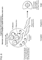
- FIG. 1 The formation of pores in situ within hydrogels as demonstrated via imaging and mechanical properties testing is shown in Figure 1 .
- micro-beads comprised of rapidly degradable hydrogels (red spheres) were mixed with a second hydrogel forming polymer material, which is crosslinked around the beads. After degradation of the micro-beads in situ, an intact hydrogel network (pink) remained with a network of pores.
- the elastic modulus measurements of either standard hydrogels (left bars) or pore-forming hydrogels (right bars) were determined ( Figure ID). At day 0, there was no statistically significant difference in the overall rigidity of pore-forming composites and standard hydrogels because pores have not formed; however, after 4 days, the modulus of the composite drops substantially because of pore formation.
- Polymer 2002; 43: 6239-46 describes the use of gamma-irradiation to reduce the weight-averaged molecular weight (M w ) of guluronic acid (GA) rich alginates without substantially reducing GA content ⁇ e.g., the gamma irradiation selectively attacks mannuronic acid, MA blocks of alginate).
- Alginate is comprised of GA blocks and MA blocks, and it is the GA blocks that give alginate its rigidity (elastic modulus). Kong et al.
- Polymer 2002; 43: 6239- 46 shows that binary combinations of high M w , GA rich alginate with irradiated, low M w , high GA alginate crosslinks with calcium to form rigid hydrogels, but which degrade more rapidly and also have lower solution viscosity than hydrogels made from the same overall weight concentration of only high M w , GA rich alginate.
- Alsberg et al. J Dent Res 2003; 82(11): 903-8 describes degradation profiles of hydrogels made from irradiated, low M w , GA-rich alginate, with application in bone tissue engineering.
- Kong et al. Adv. Mater 2004; 16(21): 1917-21 describes control of hydrogel degradation profile by combining gamma irradiation procedure with oxidation reaction, and application to cartilage engineering.
- Lutolf MP et al. Nat Biotechnol. 2003; 21: 513-8 describes poly(ethylene glycol) based materials engineered to degrade via mammalian enzymes (MMPs).
- MMPs mammalian enzymes
- Bryant SJ et al. Biomaterials 2007; 28(19): 2978-86 US 7,192,693 B2
- a pore template ⁇ e.g., poly-methylmethacrylate beads
- acetone and methanol are used to extract the porogen while leaving the bulk hydrogel intact.
- insoluble cues such as hydrogel adhesion ligand presentation and/or elastic modulus (i.e., stiffness) to generate materials which are 1) injectable; 2) allow the user to control cell fate using insoluble cues; and 3) form pores over time to deploy or recruit cells.
- the methods described herein create pore-forming hydrogels, using a process that allows cells to be encapsulated into either the pore-forming phase (hereafter referred to as "porogen") or the non- or slowly-degrading phase (hereafter referred to as "bulk").
- the porogen degrades by hydrolysis rather than by solvents, which means that cells are encapsulated either into the porogen or the bulk gel around them, and there is very little chance that proteins or other bioactive compounds encapsulated into the gel would be denatured.
- the decrease in composite elastic modulus at one week corresponds to the density of voids, and at low porogen density, there is a linear relationship between the density of voids and decrease in composite elastic modulus.
- the fracture toughness of the composite material (25% porogen volume fraction) was initially similar to the fracture toughness of a standard hydrogel with no porogen, but it decreases to a fraction of the initial value ( Figures H and I).
- the decrease in fracture toughness scales with the density of porogen, though in a non-linear manner.
- Example 2 In vitro and in vivo release of cells
- Pore-forming hydrogels were formed by encapsulating degradable alginate porogens, along with bone marrow stromal stem cells (D1) into high molecular weight bulk alginate gel. Porogens were formed with a binary mixture of 20mg/mL of alginate dialdehyde (theoretical oxidation of 7.5% of alginate sugar residues in high Mw, high guluronic acid content alginate) and 7.5mg/mL high Mw, high guluronic acid (GA) content alginate. This polymer mixture was extruded through a glass nebulizer with co-axial nitrogen airflow into a bath of 0.1M CaCl 2 and 0.1M HEPES to crosslink polymers.
- Porogens were washed extensively with serum free cell culture medium.
- the bulk hydrogel was formed by 20mg/mL high Mw, high GA-content alginate modified with 2 RGD peptides per alginate polymer.
- D1 cells and porogens were mixed into the bulk hydrogel material using syringes and then the composite was crosslinked with Calcium Sulfate.
- the number of D1 cells released from this system over time in vitro is shown in Figure 2 .
- the kinetics of release could be modified by 1) controlling the concentration of CaCl 2 used to form porogens, by 2) varying the composition (degree of oxidation) of porogens, and by 3) varying the compartmentalization of cells (either within porogens or within bulk gel).
- mesenchymal stem cell deployment in-vitro is illustrated in Figure 2 .
- Stem cells were released from pore-forming hydrogels in vitro, and this release can be tuned by varying the composition of porogens and the compartmentalization of cells within porogens versus bulk.
- the effects of porogen density (0-80 volume percent) on cellularity and efflux from pore-forming hydrogels are shown in Figure 2C .
- fluorescence micrographs of pore-forming hydrogels were stained for live mesenchymal stem cells (MSC) (Calcein-AM, green) or dead cells (Ethidium Homodimer, red) after 4-10 days in vitro.
- MSC live mesenchymal stem cells
- dead cells Ethidium Homodimer, red
- the spherical cell morphology denotes cells confined in a nanoporous material, and is present at short time frames in both materials, but only in standard hydrogels over longer time frames.
- the cumulative number of MSC released after 12 days as a function of porogen volume density is shown in Figure 2D .
- the size of the porogen is related to the size of the overall composite material. Specifically, for the material to stay intact, the porogen diameter is ⁇ 10% of the smallest dimension of the overall composite.
- the density of porogens is between 10-80 percent of the overall volume for both cell recruitment and cell release, e.g., between 15% and 75%, between 20% and 70%, between 25% and 65%, between 30% and 60%, or between 35% and 55% of the overall volume. Preferably, the density of porogens is at least 50% of the overall volume.
- Pore-forming hydrogels were formed with a constant bulk component (2 RGD / polymer, 60 kPa), and constant porogen density (50%), but varying porogen composition.
- the chemical composition of porogens was manipulated by varying the theoretical degree of oxidation of the alginate polymers. Oxidation degree was controlled by varying the ratio of sodium periodate to alginate during the oxidation reaction (Bouhadir 2001). Binary mixtures of 20mg/mL oxidized alginate with 5mg/mL unmodified, high M w alginate, were used to form porogens. Porogens were formed by crosslinking in a bath of 25-100mM CaCl 2 .
- Example 3 Controlling mesenchymal stem cell deployment, engraftment, and proliferation in vivo
- D1 cells were modified to constitutively express a detectable marker, e.g., mCherry or green fluorescent protein (GFP), and were encapsulated either into standard bulk gels with no porogens, pore-forming hydrogels, or mixed with saline.
- a detectable marker e.g., mCherry or green fluorescent protein (GFP)
- GFP green fluorescent protein
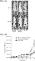
- cells were injected into the backs of Nude mice through 18-gauge needles. Cell release and proliferation over time were monitored via mCherry fluorescence observed on an IVIS system (Caliper Life Sciences).
- IVIS system Caliper Life Sciences
- FIG. 3D The ability of pore-forming hydrogels to enhance human mesenchymal stem cell mediated bone regeneration was demonstrated in a Nude Rat cranial defect model ( Figure 3D ).
- Critically-sized defects were formed in the crania of Nude Rats (Charles River).
- commercially available human mesenchymal stem cells (Lonza) were transplanted into the defect space, either within saline ("Cells Only”), a standard hydrogel (2 RGD / alginate polymer, 60 kPa), or a Pore-Forming Hydrogel.
- the top row of Figure 3D shows representative cross-sections of micro-computed tomographic analysis of new bone formation within cranial defects 4 weeks after implantation.
- Example 4 In vitro release of two different cell populations at distinct times
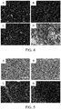
- Pore-forming hydrogels were formed as described in Examples 1 and 2. Equal numbers (approximately 106 cells/mL of composite pore-forming hydrogel) GFP-expressing myoblasts and outgrowth endothelial cells (OECs, vascular progenitor cells) were encapsulated into different compartments of the material. After 5 days of culture in vitro, cells that were released and adherent to tissue culture plastic were stained with Ethidium Homodimer (EtD-1; red). As shown in Figure 4 , the cell type encapsulated into the bulk gel deployed more rapidly. This pattern of deployment occurred even for OECs, which migrate more slowly and proliferate less extensively than GFP-myoblasts (based on analysis of substrates onto which equal numbers of both cell types were added; Figure 4d ).
- EtD-1 Ethidium Homodimer
- FIG 4 Fluorescent micrographs of GFP-expressing myoblasts and outgrowth endothelial cells (OECs) adherent to tissue culture plastic after 4 days of culture within pore-forming hydrogels in which the chemistry used to form porogens was varied and the different cell types were initially placed into distinct compartments are shown in Figures 4A-4C.
- Figure 4A depicts myoblasts in bulk component, OECs in porogen component, while Figure 4B depicts myoblasts in bead component, OECs in porogen component.
- Figure 4C depicts both myoblasts and OECs in bulk component.
- Example 5 Recruitment of host lymphocytes from subcutaneous tissues by pore-forming hydrogels with different porogen formulations
- Pore-forming hydrogels were formed as described in Examples 1 and 2. To form the porogen phase, 7.5mg/mL of high Mw, GA-rich alginate polymer was combined with 20mg/mL of either alginate dialdehyde (7.5% theoretical degree of oxidation) or alginate dialdehyde in which aldehyde groups were reduced to alcohol groups. Pore-forming hydrogels without encapsulated cells were next injected into the backs of C57/BL6 or Balb/c mice. After 14 days, recruitment of host dendritic cells was observed by histology.
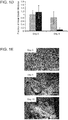
- FIG. 5A and Figure 5B show a comparison of dendritic cell (DC) recruitment by a standard, degradable alginate hydrogel versus a pore-forming alginate hydrogel. Both sets of hydrogels were loaded with 2 ug of granulocyte-macrophage colony stimulating factor. This indicates substantially more infiltration of cells from the tissue adjacent the hydrogel (highly cellular area near the edge of the image) into the pore-forming hydrogel.
- DC dendritic cell
- Figure 5C and 5D show a comparison of baseline DC invasion into pore-forming hydrogels in which porogens were formed from alginate dialdehyde ( Figure 5C ) or from reduced alginate dialdehyde ( Figure 5D ) without GM-CSF. Substantially less baseline cell infiltration occurred absent GM-CSF. For histology, dendritic cells are stained for CD11c (green) and NIHC-II (red), with Hoescht nuclear counterstain (blue). This figure shows the difference in host cell recruitment by materials with porogens formed from the oxidized alginate vs. reduced alginate.
- Example 6 Control of Stem Cell Proliferation Within Pore-Forming Hydrogels by Varying Bulk Phase Composition
- RGD density had significant effects of cell proliferation, whereas elastic modulus had effects on both proliferation and release (p ⁇ 0.05, ANOVA).
- the analysis of DNA synthesis as a function of pore formation is shown in Figure 6C .
- Staining for Ki-67 (proliferation marker, green) in D1 cells in cryosectioned pore-forming hydrogels after 50 days of culture is shown in Figure 6D .
- Pore forming hydrogels were formed as described in Example 1.
- MSC mesenchymal stem cell
- the overall density of mCherry-labeled mouse MSC deployed into the subcutaneous space could be increased by increasing the density of RGD peptides from 2 to 10 peptides per alginate polymer chain ( Figure 6E ).
- human MSC were deployed into Nude Rat cranial defects. After 4 weeks, rats were euthanized, and the degree of healing (due to new bone formation) was assessed by Hematoxylin/Eosin staining.
- the elastic modulus of the bulk hydrogel component had a substantial effect on new bone formation at 4 weeks, as deployment from a pore-forming hydrogel with a 60 kPa, 10 RGD / alginate polymer bulk phase led to significantly more bone formation ( p ⁇ 0.05, 2-tailed t-test) than deployment from a pore-forming hydrogel with an 8 kPa, 10 RGD / alginate polymer bulk phase.
- Example 7 Mechanical properties and in-vitro degradation of hydrogels formed from binary alginates
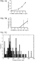
- Elastic moduli and degradation of bulk hydrogels formed by cross-linking binary combinations of oxidized alginate (5% theoretical degree of oxidation) at a constant density of 20 mg/mL with unmodified, high M w alginate are shown in Figure 7A and Figure 7B .
- Degradation was assessed by comparing the dry mass after 4 days in-vitro to initial dry mass.
- the diameters of porogens formed from binary mixtures of 20 mg/mL oxidized alginate with 7.5 mg/mL unmodified alginate is shown in Figure 7C . Porogen diameter was measured by processing fluorescent micrographs of porogens prepared from aminofluorescenin-labeled alginates.
Landscapes
- Health & Medical Sciences (AREA)
- Life Sciences & Earth Sciences (AREA)
- Chemical & Material Sciences (AREA)
- Medicinal Chemistry (AREA)
- Engineering & Computer Science (AREA)
- General Health & Medical Sciences (AREA)
- Veterinary Medicine (AREA)
- Public Health (AREA)
- Animal Behavior & Ethology (AREA)
- Epidemiology (AREA)
- Dermatology (AREA)
- Oral & Maxillofacial Surgery (AREA)
- Transplantation (AREA)
- Biomedical Technology (AREA)
- Cell Biology (AREA)
- Chemical Kinetics & Catalysis (AREA)
- Botany (AREA)
- Zoology (AREA)
- Dispersion Chemistry (AREA)
- Bioinformatics & Cheminformatics (AREA)
- Urology & Nephrology (AREA)
- Pharmacology & Pharmacy (AREA)
- Molecular Biology (AREA)
- Organic Chemistry (AREA)
- General Chemical & Material Sciences (AREA)
- Nuclear Medicine, Radiotherapy & Molecular Imaging (AREA)
- Vascular Medicine (AREA)
- Developmental Biology & Embryology (AREA)
- Materials Engineering (AREA)
- Orthopedic Medicine & Surgery (AREA)
- Composite Materials (AREA)
- Hematology (AREA)
- Rheumatology (AREA)
- Physical Education & Sports Medicine (AREA)
- Medicines Containing Material From Animals Or Micro-Organisms (AREA)
- Materials For Medical Uses (AREA)
- Micro-Organisms Or Cultivation Processes Thereof (AREA)
- Medicines That Contain Protein Lipid Enzymes And Other Medicines (AREA)
- Medicines Containing Antibodies Or Antigens For Use As Internal Diagnostic Agents (AREA)
- Medicinal Preparation (AREA)
Description
- This application claims the benefit of priority under 35 U.S.C. § 119(e) to
U.S. Provisional Application No: 61/390,594, filed on October 6, 2010 - This invention was made with Government support under NIH R37DE013033 awarded by the National Institutes of Health and MRSEC DMR-0820484 awarded by the National Science Foundation. The Government has certain rights in the invention.
- This invention relates to biocompatible hydrogel compositions.
- Over the recent decades, biocompatible polymers have been used to form scaffolds that act as carriers for cell transplantation, or to recruit host cell populations into the device.
- The invention provides compositions and methods to form porous hydrogels. For example, pores are formed in situ within hydrogels following hydrogel injection into a subject. Pores that are formed in situ via degradation of sacrificial porogens within the surrounding hydrogel (bulk hydrogel) facilitate recruitment or release of cells. For example, the resulting pore is within 5% of the size of the initial porogen.
- Disclosed herein is a material that is not initially porous, but which becomes macroporous over time resident in the body of a recipient animal such as a mammalian subject. These compositions are associated with significant advantages over earlier scaffold compositions. The hydrogels described herein are well-suited to initially protect transplanted cells from host inflammatory responses, and then release transplanted cells after inflammation has subsided (e.g., after 12 hours, or 1, 3, 5, 7, or 10 days or more post-transplantation, i.e. resident in the body of the recipient). The hydrogels described herein also double as a surgical bulking agent, further minimizing inflammation in the host, and then later releasing cells.
- Accordingly, the invention provides a composition as defined in the appended claims. The composition comprises a porogen hydrogel and a bulk hydrogel, which composition is not initially macroporous and becomes macroporous over time when resident in the body of a receipient, wherein the porogen hydrogel degrades at least 10% faster (e.g. , at least 15%, at least 20%, at least 25%, at least 30%, at least 35%, at least 40%, at least 45%, or at least 50% faster) than the bulk hydrogel following residence in said subject, leaving macropores having a diameter of greater than 20 µm in its place, and wherein (a) said porogen hydrogel comprises oxidized alginate; or (b) said porogen hydrogel comprises a shorter polymer than said bulk hydrogel. For example, the first hydrogel is a porogen and the resulting pore after degradation insitu is within 25% of the size of the initial porogen, e.g., within 20%, within 15%, or within 10% of the size of the initial porogen. Preferably, the resulting pore is within 5% of the size of the initial porogen. The porogen hydrogel degrades more rapidly than the bulk hydrogel, because the porogen hydrogel is more soluble in water (comprises a lower solubility index). Alternatively, the porogen hydrogel degrades more rapidly because it is cross- linked to protease-mediated degradation motifs as described in USSN
10/980,989 to Zilla - The molecular mass of the polymers used to form the porogen hydrogel composition are approximately 50 kilodaltons (kDa), and the molecular mass of the polymers used to form the bulk hydrogel composition comprises approximately 250 kDa. A shorter polymer (e.g. that of a porogen) degrades more quickly compared to that of a longer polymer (e.g., that of the bulk composition). Alternatively, a composition is modified to render it more hydrolytically degradable by virtue of the presence of sugar groups (e.g., approximately 3-10% sugar of an alginate composition). In another example, the porogen hydrogel is more enzymatically degradable compared to the bulk hydrogel. The composite (first and second hydrogel) composition is permeable to bodily fluids, e.g., such as enzyme which gain access to the composition to degrade the porogen hydrogel. In some cases, the second hydrogel is cross- linked around the first hydrogel, i.e., the porogens (first hydrogel) are completely physically entrapped in the bulk (second) hydrogel.
- Cells or bioactive factors (e.g., growth factors such as granulocyte/macrophage colony stimulating factor (GM-CSF), vascular endothelial growth factor (VEGF), condensed oligonucleotides, e.g., CpG, or plasmid DNA) are optionally encapsulated either into the porogen phase, bulk hydrogel phase, or into both phases. The porogens degrade in situ over a time-course pre-determined by the user. Upon degradation of the porogens, cells are released from or may migrate into the material. However, because they initially lack pores, pore-forming hydrogels are useful to provide mechanical support immediately after formation. Suitable bioactive factors include vascular endothelial growth factor (e.g., VEGFA; GenBank Accession Number: (aa) AAA35789.1 (GI:181971), (na) NM_001171630.1 (GI:284172472)), acidic fibroblast growth factor (aFGF, Genbank Accession Number: (aa) AAB29057.2 (GI: 13236891), (na) NM_000800.3 (GI:222144219)), basic fibroblast growth factor (bFGF; GenBank Accession Number: (aa) AAB21432.2 (GI:8250666), (na) A32848.1 (GI:23957592)), placenta growth factor (PIGF or PLGF; GenBank Accession Number: (aa) AAH07789.1 (GI:14043631), (na) NM_002632.4 (GI:56676307)), leptin (Genbank Accession Number: (aa) CBI71013.1 (GI:285310289), (na) NM_000230.2 (GI:169790920)), hematopoietic growth factor (e.g., HGF, Genbank Accession Number: (aa) AAA64297.1 (GI:337938), (na) NM_000601.4 (GI:58533168)), VEGF receptor-1 (VEGFR-1, Genbank Accession Number: (aa) NP_002010.2 (GI:156104876)), VEGFR-2 (Genbank Accession Number: (aa) AAC16450.1 (GI:3132833), (na) EU826563.1 (GI:194318421)), transforming growth factor-β (TGF-β, Genbank Accession Number: (aa) AAA36738.1 (GI:339564), (na) NM_000660.4 (GI:260655621)), bone morphogenetic protein (e.g. , BMP-4, Genbank Accession Number: (aa) NP_570912.2 (GI:157276597), (na) NM_001202.3 (GI: 157276592)), insulin-like growth factor (IGF-1, Genbank Accession Number: (aa) CAA01954.1 (GI:1247519), (na) NM_001111283.1 (GI: 163659898)), fibroblast growth factor-2 (FGF-2), platelet-derived growth factor (PDGF; GenBank Accession Number: (aa) AAA60552.1 (GI:338209), (na) NM_033023.4 (GI:197333759)), epidermal growth factor (EGF, Genbank Accession Number: (aa) AAH93731.1 (GI:62740195)), transforming growth factor-a (TGF-a, Genbank Accession Number: (na) NM_003236.2 (GI:153791671)), nerve growth factor (NGF, Genbank Accession Number: (aa) AAH32517.2 (GI:34192369), (na) NM_002506.2 (GI:70995318)), brain-derived neurotrophic factor (BDNF, Genbank Accession Number: (aa) CAA62632.1 (GI:987872), (na) NM_170731.4 (GI:219842281)), neurotrophin-3 (NT-3, Genbank Accession Number: (aa) NP_001096124.1 (GI:156630995), (na) NM_001102654.1 (GI:156630994)), ciliary neurotrophic factor (CNTF, Genbank Accession Number: (aa) AAB31818.1 (GI:633830), (na) NM_000614.3 (GI:209574322)), and glial cell line- derived neurotrophic factor (GDNF, Genbank Accession Number: (aa) CAG46721.1 (GI:49456801), (na) NM_000514.3 (GL299473777)). Other suitable factors include anti-VEGF antibody, anti-aFGF antibody, anti-bFGF antibody, anti-PIGF antibody, anti-leptin antibody, anti-HGF antibody, anti-VEGFR-1 antibody, anti-VEGFR-2 antibody, batimastat (BB-94), marimastat (BB-2516), thalidomide, O-(chloroacetylcarbamoyl)-fumagillol (TNP-470), carboxyamidotriazole (CAI), mitoxantrone, doxorubicin, SU5416, anti-TGF-β antibody, anti-BMP antibody, anti-IGF-1 antibody, anti-FGF-2 antibody, anti-PDGF antibody, anti-EGF antibody, anti-TGF-a antibody, and anti-VEGF antibody. Other bioactive factors suitable for encapsulation either into the porogen phase, bulk hydrogel phase, or into both phases include FMS-
like tyrosine kinase 3 ligand (Flt3 ligand; Genbank Accession Number: (aa) AAI44040 (GI:219519004), (na) NM_004119 (GI: GI: 121114303)), anti-flt3 ligand, hepatocyte growth factor (Genbank Accession Number: (aa) AAB20169 (GI:237997)), and stromal derived factor 1 (SDF-1). - Alternatively, an adenovirus is optionally encapsulated either into the porogen phase, bulk hydrogel phase, or into both phases. For example, the adenovirus encodes runt-related transcription factor (e.g., Runx2; Genbank Accession Number: (aa) CAI13532 (GI:55959066), (na) NM_001024630 (GI:226442782)), a key transcription factor associated with osteoblast differentiation. In another aspect, the adenovirus encodes MyoD (Genbank Accession Number: (aa) CAA40000 (GI:34862), (na) NM_002478 (GI:77695919)), a protein with a key role in regulating muscle differentiation. Alternatively, the adenovirus encodes bone morphogenetic protein, e.g., BMP-2 (Genbank Accession Number: (aa) AF040249_1 (GI:6649952), (na) NM_001200 (GI:80861484)) or BMP-4 (Genbank Accession Number: (aa) NP_570912.2 (GI: 157276597), (na) NM_001202.3 (GI: 157276592) reference). BMP-2 is involved in, inter alia, bone repair, while BMP-4 is involved in the repair of cardiac tissue. In one aspect, an adenovirus that encodes Runx and an adenovirus that encodes BMP-2 are encapsulated into the hydrogel.
- Cells suitable for being encapsulated either into the porogen phase, bulk hydrogel phase, or into both phases include mesenchymal stem cells, myoblasts, vascular progenitor cells {e.g., an outgrowth endothelial cell), differentiated cells derived from embryonic stem cells or induced pluripotent stem cells, induced pluripotent cells, or cells that were directly reprogrammed from a fibroblast to a differentiated state.
- In some examples, the porogen composition comprises cells, and in other examples, the bulk composition comprises cells. If cells are present in the composition {e.g., having been seeded during fabrication), the cells are deployed out of the composition after administration into a mammalian subject. Alternatively, the composition does not comprise cells; however, upon administration into tissues of a mammalian subject {e.g., implantation into a human patient), cells are recruited into the composition. The mammal can be any mammal, e.g., a human, a primate, a mouse, a rat, a dog, a cat, a horse, as well as livestock or animals grown for food consumption, e.g., cattle, sheep, pigs, chickens, and goats. In a preferred embodiment, the mammal is a human. Alternatively, the subject can be a non-mammalian animal such as xenopus, salamander, or newt.
- The invention provides methods of deploying cells from a scaffold into tissues of a mammalian subject, comprising administering to a subject a composition comprising a first hydrogel and a second hydrogel, wherein the first hydrogel degrades at least 10% faster than the second hydrogel, and wherein the composition lacks pores at the time of administration, and wherein the composition comprises pores following residence in said subject, and wherein the first hydrogel or the second hydrogel comprises an isolated cell.
- A methods of recruiting cells into a scaffold in vivo is carried out by administering to a subject a composition comprising a first hydrogel and a second hydrogel, wherein the first hydrogel degrades at least 10% faster than the second hydrogel and wherein the composition lacks pores at the time of administration, but comprises pores following residence in the subject. For example, pores are created due to the relative degradability or solubility of a first hydrogel composition compared to a second hydrogel composition, e.g., a porogen composition compared to a bulk composition.
- Porosity influences recruitment and/or egress of the cells from the composition. Pores are nanoporous, microporous, or macroporous. For example, the diameter of nanopores is less than about 10 nm. Micropores are in the range of about 100 nm to about 20 µm in diameter. Macropores are greater than about 20 µm (e.g., greater than about 100 µm or greater than about 400 µm). Exemplary macropore sizes include 50 µm, 100 µm, 150 µm, 200 µm, 250 µm, 300 µm, 350 µm, 400 µm, 450 µm, 500 µm, 550 µm, and 600 µm. Macropores are those of a size that permit a eukaryotic cell to traverse into or out of the composition. In one example, a macroporous composition has pores of about 400 µm to 500 µm in diameter. The preferred pore size depends on the application. For example, for cell deployment and cell release, the preferred pore diameter is greater than 50 µm.
- The size of the porogen is related to the size of the overall composite material. For example, for the material to stay intact, the porogen diameter is < 10% of the smallest dimension of the overall composite. The density of porogens is between 10-80 percent of the overall volume of the composite composition. For example, the density of porogen is between 15% and 75%, between 20% and 70%, between 25% and 65%, between 30% and 60%, or between 35% and 55% of the overall volume. Preferably, the density of porogens is at least 50% of the overall volume to achieve optimal cell recruitment to the hydrogel or cell release from the hydrogel.
- The hydrogel has an elastic modulus of between about 10 to about 1,000,000 Pascals (e.g., from about 10 to about 100,000 Pa, from about 10 to about 150,000 Pa, from about 10 to about 200,000 Pa, from about 10 to about 300,000 Pa, from about 10 to about 400,000 Pa, from about 10 to about 500,000 Pa, from about 10 to about 600,000 Pa, from about 10 to about 700,000 Pa, from about 10 to about 800,000 Pa, or from about 10 to about 900,000 Pa). Preferably, the slowly-degrading hydrogel comprises an elastic modulus of about 20 kilo Pa to 60 kPa, e.g., 25 kPa to 55 kPa, 30 kPa to 50 kPa, or 35 kPa to 45 kPa. The rapidly-degrading hydrogel comprises an elastic modulus of at least 40 kPa initially in order to maintain integrity during encapsulation prior to degradation.
- Preferably, the slowly-degrading hydrogel (i.e., the second hydrogel or "bulk") comprises high molecular weight peptides with an amino acid sequence of RGD which mimic cell adhesion proteins. Alternatively, the slowly-degrading hydrogel comprises a different adhesive peptide amino acid motif such as PHSRN or DGEA. For example, the slowly-degrading hydrogels are preferably modified with 2-10 RGD peptides / polymer (e.g., alginate polymer).
- By "hydrogel" is meant a composition comprising polymer chains that are hydrophilic. Exemplary hydrogels are comprised of materials that are compatible with cell encapsulation such as alginate, polyethylene glycol (PEG), PEG-acrylate, agarose, and synthetic protein (e.g., collagen or engineered proteins (i.e., self-assembly peptide-based hydrogels). For example, a commercially available hydrogel includes BD™ PuraMatrix™. BD™ PuraMatrix™ Peptide Hydrogel is a synthetic matrix that is used to create defined three dimensional (3D) micro-environments for cell culture.
- For example, the hydrogel is a biocompatible polymer matrix that is biodegradable in whole or in part. Examples of materials which can form hydrogels include alginates and alginate derivatives, polylactic acid, polyglycolic acid, poly(lactic-co-glycolic acid) (PLGA) polymers, gelatin, collagen, agarose, natural and synthetic polysaccharides, polyamino acids such as polypeptides particularly poly(lysine), polyesters such as polyhydroxybutyrate and poly-epsilon.-caprolactone, polyanhydrides; polyphosphazines, poly(vinyl alcohols), poly(alkylene oxides) particularly poly(ethylene oxides), poly(allylamines)(PAM), poly(acrylates), modified styrene polymers such as poly(4-aminomethylstyrene), pluronic polyols, polyoxamers, poly(uronic acids), poly(vinylpyrrolidone), and copolymers of the above, including graft copolymers. Synthetic polymers and naturally-occurring polymers such as, but not limited to, collagen, fibrin, hyaluronic acid, agarose, and laminin-rich gels may also be used.
- A preferred material for the hydrogel is alginate or modified alginate material. Alginate molecules are comprised of (1-4)-linked β-D-mannuronic acid (M units) and α L-guluronic acid (G units) monomers, which can vary in proportion and sequential distribution along the polymer chain. Alginate polysaccharides are polyelectrolyte systems which have a strong affinity for divalent cations (e.g., Ca+2, Mg+2, Ba+2) and form stable hydrogels when exposed to these molecules.
- The compositions described herein are suitable for clinical use, e.g., bone repair, regeneration, or formation; muscle repair, regeneration, or formation; and dermal repair, regeneration, or formation. For example, the compositions are applied to bone fractures alone or together with a bone adhesive (cement) or glue or to diseased or injured muscle tissue. The hydrogels (seeded with cells or without cells) are injected at the site of disease, injury, or fracture (in the case of bone or cartilage). For example, the hydrogels are injected into or onto bone. Exemplary bioactive factors for use in promoting bone or cartilage repair, regeneration, or formation include BMP-2, BMP-4, or RunX.
- In some cases, the composition recruits cells to promote bone or cartilage repair, regeneration, or formation. Alternatively, the first hydrogel or the second hydrogel comprises an isolated bone cell selected from the group consisting of an osteoblast, an osteocyte, an osteoclast, and an osteoprogenitor. Alternatively, the first hydrogel or the second hydrogel comprises an isolated cartilage cell, wherein the isolated cartilage cell comprises a chondroblast. The isolated bone cell or an isolated cartilage cell is an autologous cell or an allogeneic cell.
- For muscle applications, e.g., muscle tears, muscle strains, or muscle pulls, the hydrogels (seeded with or without cells) are injected at the site of injury. Suitable compositions for muscle applications include a composition comprising a first hydrogel and a second hydrogel, wherein the first hydrogel degrades at least 10% faster than the second hydrogel, and wherein the first hydrogel or the second hydrogel comprises a bioactive factor for use in muscle repair, regeneration, or formation. For example, the bioactive factor comprises MyoD.
- In some cases, the composition recruits cells to promote muscle or cartilage repair, regeneration, or formation. Alternatively, the first hydrogel or the second hydrogel comprises an isolated muscle cell selected from the group consisting of a skeletal muscle cell, a cardiac muscle cell, a smooth muscle cell, and a myo-progenitor cell. The isolated muscle cell is an autologous cell or an allogeneic cell.
- For dermal applications, e.g., burns, abrasions, lacerations, or disease, the hydrogels (seeded with cells or without cells) are applied directly to the site as poultice or wound dressing. Preferably, the majority of porogens (e.g., more than 50%, more than 60%, more than 70%, more than 80%, or more than 90%) within the bulk hydrogels are directed toward the skin surface and into the skin tissue when applied directly to the site (e.g., the burn). In this manner, the bioactive factors or cells are released into the surface of the skin or lower layers of the skin, and do not migrate away from the skin or target tissue. An exemplary bioactive factor for use in skin repair, regeneration, or formation is FGF.
- In some cases, the composition recruits cells to promote skin or cartilage repair, regeneration, or formation. Alternatively, the first hydrogel or the second hydrogel comprises an isolated skin cell selected from the group consisting of a fibroblast, a dermal cell, an epidermal cell, or a dermal progenitor cell. The isolated skin cell is an autologous cell or an allogeneic cell.
- Bioactive factors such as polynucleotides, polypeptides, or other agents are purified and/or isolated. Specifically, as used herein, an "isolated" or "purified" nucleic acid molecule, polynucleotide, polypeptide, or protein, is substantially free of other cellular material, or culture medium when produced by recombinant techniques, or chemical precursors or other chemicals when chemically synthesized. Purified compounds are at least 60% by weight (dry weight) the compound of interest. Preferably, the preparation is at least 75%, more preferably at least 90%, and most preferably at least 99%, by weight the compound of interest. For example, a purified compound is one that is at least 90%, 91%, 92%, 93%, 94%, 95%, 98%, 99%, or 100% (w/w) of the desired compound by weight. Purity is measured by any appropriate standard method, for example, by column chromatography, thin layer chromatography, or high-performance liquid chromatography (HPLC) analysis. A purified or isolated polynucleotide (ribonucleic acid (RNA) or deoxyribonucleic acid (DNA)) is free of the genes or sequences that flank it in its naturally-occurring state. Purified also defines a degree of sterility that is safe for administration to a human subject, e.g., lacking infectious or toxic agents.
- Similarly, by "substantially pure" is meant a nucleotide or polypeptide that has been separated from the components that naturally accompany it. Typically, the nucleotides and polypeptides are substantially pure when they are at least 60%, 70%, 80%, 90%, 95%, or even 99%, by weight, free from the proteins and naturally-occurring organic molecules with they are naturally associated.
- An "isolated nucleic acid" is a nucleic acid, the structure of which is not identical to that of any naturally occurring nucleic acid, or to that of any fragment of a naturally occurring genomic nucleic acid spanning more than three separate genes. The term covers, for example: (a) a DNA which is part of a naturally occurring genomic DNA molecule, but is not flanked by both of the nucleic acid sequences that flank that part of the molecule in the genome of the organism in which it naturally occurs; (b) a nucleic acid incorporated into a vector or into the genomic DNA of a prokaryote or eukaryote in a manner, such that the resulting molecule is not identical to any naturally occurring vector or genomic DNA; (c) a separate molecule such as a cDNA, a genomic fragment, a fragment produced by polymerase chain reaction (PCR), or a restriction fragment; and (d) a recombinant nucleotide sequence that is part of a hybridgene, i.e., a gene encoding a fusion protein. Isolated nucleic acid molecules according to the present invention further include molecules produced synthetically, as well as any nucleic acids that have been altered chemically and/or that have modified backbones.
- A small molecule is a compound that is less than 2000 daltons in mass. The molecular mass of the small molecule is preferably less than 1000 daltons, more preferably less than 600 daltons, e.g., the compound is less than 500 daltons, 400 daltons, 300 daltons, 200 daltons, or 100 daltons.
- The transitional term "comprising," which is synonymous with "including," "containing," or "characterized by," is inclusive or open-ended and does not exclude additional, unrecited elements or method steps. By contrast, the transitional phrase "consisting of excludes any element, step, or ingredient not specified in the claim. The transitional phrase "consisting essentially of limits the scope of a claim to the specified materials or steps "and those that do not materially affect the basic and novel characteristic(s)" of the claimed invention.
- Other features and advantages of the invention will be apparent from the following description of the preferred embodiments thereof, and from the claims. Unless otherwise defined, all technical and scientific terms used herein have the same meaning as commonly understood by one of ordinary skill in the art to which this invention belongs. Although methods and materials similar or equivalent to those described herein can be used in the practice or testing of the present invention, suitable methods and materials are described below. In the case of conflict, the present specification including definitions, will control. In addition, the materials, methods, and examples are illustrative only and not intended to be limiting.
-
-
Figure 1 is a series of schematics, photographs, bar graphs, and line graphs showing the formation of pores in situ within hydrogels as demonstrated via imaging and mechanical properties testing.Figure 1A is a schematic illustrating the formation of hydrogels. Left: micro-beads comprised of rapidly degradable hydrogels (red spheres) and mesenchymal stem cells (MSCs; green). Middle: micro-beads and MSCs are mixed with a second hydrogel forming polymer material ("bulk gel;" gray), which is crosslinked around the beads. Right: after degradation of the micro-beads in situ, an intact hydrogel network remains with a network of pores through which MSCs are released.Figure 1B is a fluorescence micrograph of fluorescein (green) labeled porogens immediately after fabrication.Figure 1C is a fluorescence micrograph of fluorescein (green) labeled porogens after encapsulation into an alginate hydrogel network.Figure 1D is a bar graph illustrating elastic modulus measurements of either standard hydrogels (blue bars) or pore-forming hydrogels (red bars). Atday 0, there is no statistically significant difference in the overall rigidity of pore-forming composites and standard hydrogels because pores have not formed; however, after 4 days, the modulus of the composite drops substantially because of pore formation.Figure 1E is a series of scanning electron micrographs depicting pore-forming hydrogels immediately after formation (Day 0) showing a grossly intact network, 5 days after fabrication, or 10 days after fabrication, at which time significant pore formation was observed.Figure 1F and Figure 1G verify that the elastic modulus of the composite material (50% porogen volume fraction) is not substantially different from the elastic modulus of a standard hydrogel (no porogens), but that as voids form, the modulus of the composite drops substantially. The decrease in composite elastic modulus at one week corresponds to the density of voids, and at low porogen density, there is a linear relationship between the density of voids and decrease in composite elastic modulus. Figures H and I illustrate that the fracture toughness of the composite material (25% porogen volume fraction) is initially similar to the fracture toughness of a standard hydrogel with no porogen, but shortly decreases to a fraction of the initial value. As with elastic modulus, the decrease in fracture toughness scales with the density of porogen, though in a non-linear manner. Scale bars: (B, C, and E): 1 mm. -
Figure 2 is a series of photographs, line graphs, and schematics illustrating mesenchymal stem cell deployment in-vitro. Specifically,Figure 2 demonstrates stem cell release from pore-forming hydrogels in vitro, which also shows that release can be tuned by varying the composition of porogens and the compartmentalization of cells within porogens versus bulk.Figure 2A is a line graph showing the kinetics of interconnected void formation assessed with a capillary assay (error bars are standard error of the mean, n = 3-4). Interconnected voids formed over the first 7 days unless a very high fraction of porogens (above the percolation limit; 80%) were present. No substantial interconnected void formation was observed with a sub-percolation porogen density.Figure 2B is a series of schematics and photographs showing 3-dimensional reconstructions of Calcein-AM stained cells distributed throughout pore-forming hydrogels. The substantial changes in cell morphology depict the cells' ability to migrate and proliferate within pore-forming hydrogels, whereas cells remained sparse and rounded within standard hydrogels. Ki-67 immunofluorescence (green) indicates increased proliferation, while nuclear counterstain (Hoescht, blue) higher cellularity, within pore-forming hydrogels compared to standard hydrogels.Figure 2C is a set of fluorescence micrographs showing the effects of void formation on cellularity and cell morphology within pore forming hydrogels. Specifically, fluorescence micrographs of pore-forming hydrogels were stained for live mesenchymal stem cells (MSC) (Calcein-AM, green) or dead cells (Ethidium Homodimer, red) after 4-10 days in vitro. The spherical cell morphology denotes cells confined in a nanoporous material, and is present at short time frames in both materials, but only in standard hydrogels over longer time frames.Figure 2D is a line graph showing the cumulative number of MSC released after 12 days as a function of porogen volume density.Figure 2E is a line graph and schematic illustrating the kinetics of deployment for MSC encapsulated either into the bulk phase, the porogen phase of pore-forming scaffolds, or into standard hydrogels. Porogens were prepared with 7.5% oxidized alginate and crosslinked in 100 mM CaCl2.Figure 2F is a line graph showing the kinetics of MSC deployment from the porogen phase of pore-forming hydrogels as a function of porogen fabrication conditions for D1 cells encapsulated into porogens.Figure 2G is a bar graph showing that quantitative analysis of 3H-thymidine incorporation indicates enhanced cell proliferation, in an RGD-dependent manner.Figure 2H is a series of line graphs showing the effects of the degree of alginate oxidation degree on cell release. At a constant level of calcium to crosslink porogens (100 mM), increasing the degree of oxidation from 3-7.5% substantially increased the overall number of release cells, whereas lowered the degree of oxidation slightly delayed cell release. At a constant degree of porogen degree of oxidation (7.5%), increasing the concentration of calcium used to crosslink porogens from 25-100mM lowered the overall number of released cells and slightly delayed the onset of cell release. Scale bars: (A): 100 µm. -
Figure 3 is a series of photographs and line graphs showing the results of controlling mesenchymal stem cell deployment, engraftment and proliferation in vivo. Specifically,Figure 3 demonstrates stem cell release from pore-forming hydrogels in vivo within the subcutaneous space of nude mice.Figure 3A is a photograph showing representative images of Nude mice into which 2 x 10⁶ mCherry-expressing MSC were deployed, either 7 or 30 days after injection in standard hydrogels (left), pore-forming hydrogels in which porogens were crosslinked with either 100 mM CaCl2 (center), or saline (right). The bulk component of hydrogels was modified with 2 RGD / polymer chain. Initially, more cells engrafted in the saline only condition, but at later time-points, there were fewer cells in this condition and substantially more cells eventually engrafting when released from pore-forming hydrogels.Figure 3B is a line graph showing the quantification of the relative radiant efficiency (proportional to cell density) of mCherry-MSC injected in pore-forming hydrogels, standard hydrogels, or saline.Figure 3C is a line graph showing that decreasing the density of calcium used to crosslink porogens substantially decreased the overall density of released cells, and slightly delayed the kinetics of deployment (error bars are SEM, n=4-6).Figure 3D is a series of photographs showing the ability of pore-forming hydrogels to enhance human mesenchymal stem cell mediated bone regeneration using a Nude Rat cranial defect model. Critically-sized defects were formed in the crania of Nude Rats (Charles River). Immediately after defect formation, commercially available human mesenchymal stem cells (Lonza) were transplanted into the defect space, either within saline ("Cells Only"), a standard hydrogel (2 RGD / alginate polymer, 60 kPa), or a Pore-Forming Hydrogel. Representative cross-sections of micro-computed tomographic analysis of new bone formation withincranial defects 4 weeks after implantation. Substantially more new bone forms within defects in which cells were delivered via pore-forming hydrogels. -
Figure 4 is a series of photomicrographs depicting the use of pore-forming hydrogels to release distinct populations at different times.Figure 4A-Figure 4C are fluorescent micrographs of GFP-expressing myoblasts and outgrowth endothelial cells (OECs) adherent to tissue culture plastic after 4 days of culture within pore-forming hydrogels in which the chemistry used to form porogens was varied and the different cell types were initially placed into distinct compartments: (Figure 4A ): myoblasts in bulk component, OECs in porogen component; (Figure 4B ): myoblasts in bead component, OECs in porogen component; (Figure 4C ): both myoblasts and OECs in bulk component.Figure 4D is a representative micrograph of a plastic substrate on which equal numbers of GFP-myoblasts and OECs were seeded. Myoblasts outgrew OECs. Cells were stained with Ethidium Homodimer (red), so that GFP-myoblasts appear yellow and OECs appear red. Images taken at 10x magnification. -
Figure 5 is a series of photomicrographs depicting using pore-forming hydrogels for chemokine-mediated cell recruitment. Specifically,Figure 5 shows the controlling chemokine-mediated cell recruitment by pore-forming hydrogels in vivo. Alginate was first oxidized and then reduced with sodium borohydride to make alcohol groups that replace what were originally sugars.Figure 5A and Figure 5B are fluorescent micrographs of dendritic cell recruitment into (Figure 5A ) standard, injectable alginate gel, and (Figure 5B ) pore-forming hydrogel. Both sets of hydrogels were loaded with 2 pg of granulocyte-macrophage colony stimulating factor.Figure 5C and 5D are fluorescent micrographs of dendritic cell recruitment into pore-forming hydrogels fabricated with (Figure 5C ) oxidized or (Figure 5D ) reduced porogens. No chemokine was added. For histology, dendritic cells are stained for CDllc (green) and NIHC-II (red), with Hoescht nuclear counterstain (blue). InFigure 5C and Figure 5D , only nuclear staining (white) was performed. This figure shows the difference in host cell recruitment by materials with porogens formed from the oxidized alginate vs. reduced alginate.Scale bars: 100 pm. -
Figure 6 is a series of line graphs, bar charts, and photographs demonstrating the control of stem cell proliferation within pore-forming hydrogels, deployment from hydrogels, and ability to regenerate bone by varying bulk phase composition.Figures 6A and Figure 6B are line graphs showing the analysis of 24 hr 3H-thymidine incorporation (proportional to DNA synthesis) by mesenchymal stem cells (D1; red curve) or cumulative MSC deployment (blue curves) from pore-forming hydrogels after 7 days of culture as a function of (Figure 6A ) density of RGD peptides in bulk gels with 60 kPa modulus, or (Figure 6B ) elastic modulus of bulk hydrogels presenting 10 RGD peptides / alginate polymer (data are mean +/- SEM, n = 3-5). RGD density had significant effects of cell proliferation, whereas elastic modulus had effects on both proliferation and release (p < 0.05, ANOVA).Figure 6C is a bar graph showing the analysis of DNA synthesis as a function of pore formation.Figure 6D is a series of photographs showing staining for Ki-67 (proliferation marker, green) in D1 cells in cryosectioned pore-forming hydrogels after 50 days of culture.Figure 6E and6F is a line graph showing the control of mesenchymal stem cell deployment, engraftment and proliferation in vivo.Figure 6E is a line graph showing the quantification of the relative radiant efficiency (proportional to cell density) of mCherry-MSC injected in pore-forming hydrogels in which the bulk phase was modified with either 2 (), or 10 ( ) RGD peptides per alginate polymer. Alternatively, cells were injected in a standard hydrogel with 2 RGD peptides / alginate polymer (▲).
Figure 6F shows the quantification of the percentage of healing (new bone formation due to human MSC transplanted into Nude Rat cranial defects) as a function of the method of MSC delivery. Error bars are SEM, n = 4-6. -
Figure 7 is a series of line graphs and bar frequency charts showing the mechanical properties and in-vitro degradation of hydrogels formed from binary alginates.Figures 7A and Figure 7B show elastic moduli (Figure 7A ) and degradation (Figure 7B ) of bulk hydrogels formed by crosslinking binary combinations of oxidized alginate (5% theoretical degree of oxidation) at a constant density of 20 mg/mL with unmodified, high Mw alginate. Degradation was assessed by comparing the dry mass after 4 days in-vitro to initial dry mass.Figure 7C is a histogram of diameters of porogens formed from binary mixtures of 20 mg/mL oxidized alginate with 7.5 mg/mL unmodified alginate. Porogen diameter was measured by processing fluorescent micrographs of porogens prepared from aminofluorescenin-labeled alginates. Error bars are SD, n = 3-4. -
Figure 8 is a schematic of an implantable biomaterial that mimics certain aspects of stem-cell niches in that it activates transplanted progenitor cells to proliferate and programs them to differentiate into cells that migrate into damaged tissues to participate in regeneration. -
Figure 9 is a schematic of a hydrogel scaffold (left, top), which controls transplanted cell fate through presentation of specific cues, but prevents transplanted cells from migrating out of, and host cells from migrating into, the material. Bottom: example data depicting the ability of a nanoporous hydrogel to control mesenchymal stem cell fate, in this case via elastic modulus. Right, top: schematic of a macroporous sponge which controls transplanted fate through presentation of specific cues, while also allowing host cells to migrate into, and transplanted cells to migrate out of the material. -
Figure 10 is a series of images depicting an alternative strategy to produce macroporous hydrogels. As described in the schematics (center, right), porogens are embedded into a "bulk" hydrogel, or photolithographic techniques are applied, to provide non-crosslinked regions of the bulk hydrogel. After crosslinking the bulk hydrogel, the non-crosslinked portions of the hydrogel and porogens are removed using solvents such as acetone. Left: image of a macroporous hydrogel. -
Figure 11 is a schematic and a bar chart depicting a strategy to create rapidly degrading alginate-based hydrogel porogens. Top left: chemical reaction scheme to oxidize alginate to alginate dialdehyde with NaIO4. Top right: schematic depicting the loss of crosslinkable, guluronic-acid rich portions of alginate (short, straight segments), and the overall decrease in polymer Mw due to sodium periodate oxidation. Bottom: data depicting loss in dry mass over time from hydrogels made from 20mg/mL non-modified alginate (squares), 20mg/mL alginate dialdehyde (5% degree of oxidation; diamonds) or a binary mixture of 20mg/mL alginate dialdehyde with 7.5mg/mL unmodified alginate (triangles). -
Figure 12 is a schematic illustrating porogen fabrication and characterization. -
Figure 13 is a schematic showing the control of host cell recruitment with pore forming hydrogels. Specifically, shown in this Figure is a schematic of an implantable biomaterial system that mimics the icroenvironment of an infection, allowing the recruitment, programming and subsequent targeting of activated antigen-presenting dendritic cells to the lymph nodes to participate in a potent antitumour response. -
Figure 14 is a series of photomicrographs showing how the polymers used to comprise the porogen phase affect the degradation and processing of porogens. Specifically, shown in this Figure are fluorescence micrographs of porogens formed using fluorescein-labeled alginate dialdehyde. Immediately after crosslinking in 100mM CaCl2 (top), porogens are grossly intact, whether made using 20mg/mL alginate dialdehyde (top left) or a binary mixture of 20mg/mL alginate dialdehyde with 7.5mg/mL unmodified alginate. However, after processing steps used to purify porogens and remove excess CaCl2, porogens made with purely alginate dialdehyde were damaged, resulting in substantial change in morphology and release of fluorescein-labeled polymers into solution to yield a substantial level of background fluorescein fluorescence (bottom left). In contrast, binary mixtures of 20mg/mL alginate dialdehyde with 7.5mg/mL unmodified alginate resulted in porogens that could withstand processing steps. - Over the recent decades, biocompatible polymers have been used to form scaffolds that act as carriers for cell transplantation, or to recruit host cell populations into the device. Generally, sponges such as poly(lactide-co-glycolide) (PLGA), or synthetic hydrogels such as alginate are used. However, both sets of materials have disadvantages. For example, sponges typically adsorb serum proteins, so it is difficult to control presentation of adhesive proteins or peptides (for example, RGD) from the material. Sponge materials also typically are not amenable to injection, and require an invasive surgery for implantation, and also expose transplanted or host cells to a host environment that may initially be hostile (for example, neutrophils present during inflammation may attack stem cells). On the other hand, synthetic hydrogels are typically injectable, allowing minimally-invasive delivery, and do not interact with proteins. However, prior to the invention described herein, the pore size in hydrogels was typically much smaller than the diameter of a eukaryote cell, making it difficult to expand a transplanted cell population, release transplanted cells to allow them to repair damaged tissues, or recruit host cells into the device.
- Described herein is a method to form pores in situ within hydrogels following hydrogel injection. Pores form in situ via degradation of sacrificial porogens encapsulated within the surrounding hydrogel. The kinetics and onset of pore formation are controlled by manipulating material used to form porogens, and cells are encapsulated either into the porogens themselves or the hydrogel surrounding them. Examples demonstrate in vitro deployment, proliferation, and differentiation of stem cells, as well as in vivo stem cell deployment and chemokine-mediated cell recruitment. The system mediates controlled deployment of cells out of, or local recruitment of cells into, a polymer matrix via formation of pores within this matrix. The size, distribution, and formation kinetics of the pores are predetermined by the user, while the integrity of the matrix surrounding pores, along with cells or biological factors inside this matrix, are unchanged.
- Accordingly, described herein is the use of insoluble cues such as hydrogel adhesion ligand presentation and/or elastic modulus {i.e., stiffness) to generate materials which are 1) injectable; 2) allow the user to control cell fate using insoluble cues; and 3) form pores over time to deploy or recruit cells. Specifically, the methods described herein create pore-forming hydrogels, using a process that allows cells to be encapsulated into either the pore-forming phase (hereafter referred to as "porogen") or the non- or slowly-degrading phase (hereafter referred to as "bulk").
- Described herein are methods for a generalized approach to create pore-forming hydrogels that allow cell encapsulation, and a means to control the kinetics of cell deployment out of, or recruitment into, the hydrogel. Hydrogel micro-bead "porogens" are formed, and are next encapsulated into a second, "bulk" hydrogel. The composition of polymers used to form porogen and bulk hydrogels may be varied; however, the porogen must degrade more rapidly {e.g., 10%, 20%, 50%, 2X, 5X, 10X, 20X or faster) than the bulk hydrogel. Cells or bioactive factors (e.g., growth factors such as granulocyte/macrophage colony stimulating factor (GM-CSF), vascular endothelial growth factor (VEGF), condensed oligonucleotides, e.g., CpG, or plasmid DNA ) are optionally encapsulated either into the porogen phase, bulk hydrogel phase, or into both phases. The porogens degrade in situ over a time-course pre-determined by the user, at which point cells are released, or may migrate into the material. However, because they initially lack pores, pore-forming hydrogels are useful to provide mechanical support immediately after formation (
Figure 1 ). - Cellular release or recruitment is manipulated by controlling the kinetics of porogen degradation. For example, the alginate polymers are oxidized to produce alginate dialdehyde, and the total number of cells released increases as the extent of oxidation increases (
Figure 2 ,Figure 3 ). Alternatively, conditions used to crosslink the porogens are altered to manipulate the time at which significant porogen degradation and cell release begin to occur (Figure 2 ). Porogen chemistry can further be varied to facilitate, or inhibit, interaction with host proteins (Figure 5 ). - Cell release and cell fate are controlled by manipulating the biophysical and biochemical properties (e.g. elastic modulus and density of integrin-binding adhesion peptides such as RGD) of the bulk hydrogel. For example, pore formation, bulk hydrogel RGD density and bulk hydrogel elasticity all affect cell proliferation within these materials (
Figure 2 ,Figure 6 ). The orthogonal processing of porogens and bulk separate from one another enhances the ability to tune the system to manipulate cell release and cell fate. For example, stem cell lineage commitment is modulated by varying elastic modulus or RGD density, independent of the kinetics of pore formation. In contrast, other techniques used to form macro-porous materials (e.g. solvent-based extraction of porogens) are not compatible with cell encapsulation, and typically affect the physical properties of both the porogen and bulk phases. The physical and biochemical properties rapidly degrading bulk hydrogel materials change continuously over the course of degradation. These parameters are harnessed to design the bulk phase to regulate cell fate. - Hydrogels comprise a network of polymer chains that are hydrophilic. Hydrogel (also called aquagel) is sometimes found as a colloidal gel in which water is the dispersion medium. Hydrogels are highly absorbent (they can contain over 99.9% water) natural or synthetic polymers. Hydrogels also possess a degree of flexibility very similar to natural tissue, due to their significant water content. Hydrogel is comprised of cross-linked polymers. Exemplary hydrogels are comprised materials that are compatible with cell encapsulation such as alginate, polyethylene glycol (PEG), PEG-acrylate, agarose, and synthetic protein (e.g., collagen or engineered proteins (i.e., self-assembly peptide-based hydrogels). For example, a commercially available hydrogel includes BD™ PuraMatrix™ Peptide Hydrogel, which is a synthetic matrix that is used to create defined three dimensional (3D) micro-environments for cell culture.
- For example, the hydrogel is a biocompatible polymer matrix that is biodegradable in whole or in part. Examples of materials which can form hydrogels include alginates and alginate derivatives, polylactic acid, polyglycolic acid, poly(lactic-co-glycolic acid) (PLGA) polymers, gelatin, collagen, agarose, natural and synthetic polysaccharides, polyamino acids such as polypeptides particularly poly(lysine), polyesters such as polyhydroxybutyrate and poly-epsilon.-caprolactone, polyanhydrides; polyphosphazines, poly(vinyl alcohols), poly(alkylene oxides) particularly poly(ethylene oxides), poly(allylamines)(PAM), poly(acrylates), modified styrene polymers such as poly(4-aminomethylstyrene), pluronic polyols, polyoxamers, poly(uronic acids), poly(vinylpyrrolidone), and copolymers of the above, including graft copolymers. Synthetic polymers and naturally-occurring polymers such as, but not limited to, collagen, fibrin, hyaluronic acid, agarose, and laminin-rich gels may also be used.
- A preferred material for the hydrogel is alginate or modified alginate material. Alginate molecules are comprised of (1-4)-linked β-D-mannuronic acid (M units) and α L-guluronic acid (G units) monomers, which can vary in proportion and sequential distribution along the polymer chain. Alginate polysaccharides are polyelectrolyte systems which have a strong affinity for divalent cations (e.g., Ca+2, Mg+2, Ba+2) and form stable hydrogels when exposed to these molecules.
- Synthetic hydrogels are typically injectable, allow for minimally-invasive delivery, and do not interact with proteins. Hence, the presentation of adhesion proteins or peptides is precisely controlled. Moreover, synthetic hydrogels typically have a pore mesh size that is much smaller than cells (< 10 nm, whereas cells are > 10um), which prevents host cells from attacking transplanted cells. However, this small pore size also prevents transplanted cells from proliferating extensively within the material, and also precludes their eventually being released to affect various functions (for example, regeneration of functional tissue or destruction of diseased tissue).
- Several techniques have been introduced to combine desirable features of hydrogels and sponges - for example, rigid microspheres have been encapsulated into hydrogels, and then extracted with solvents (e.g. acetone) to leave behind a macroporous hydrogel, and freeze-drying has been applied to generate macroporous hydrogels. Hydrogels can be modified to rapidly degrade in vivo to release host cells. However, prior to the invention described herein, none of the approaches allowed for the combination of a non-degrading (or slowly degrading) material component with cell encapsulation. The mechanical properties and biochemical composition of hydrogel materials strongly affect cell fate, and degradation in-and-of itself may intrinsically regulate cell fate.
- Hydrogel micro-beads ("porogens") are formed. Next, porogens are encapsulated into a "bulk" hydrogel that is either non-degradable or which degrades at a slow rate compared to the porogens. Cells are optionally encapsulated either into the porogen or bulk compartment. Immediately after hydrogel formation, or injection into the desired site in vivo, the composite material lacks pores, and serves as a surgical bulking agent. Subsequently, porogen degradation causes pores to form in situ, and encapsulated cells deploy away from the composite material and into surrounding tissues or remote tissues, e.g., lymph nodes, in the body. The size and distribution of pores are controlled during porogen formation, and mixing with the polymers which form the bulk hydrogel.
- Alternatively, the hydrogel is injected without encapsulated cells, and pore formation is used as a means of recruiting host cells, in combination or independent of chemokines released from either the bulk or porogen component. The porogens are comprised of any biocompatible polymer, as long as they degrade more rapidly than the material used to form the "bulk" hydrogel, and are initially mechanically stable enough to withstand being mixed with the polymer which forms the bulk hydrogel phase. The "bulk" is comprised of any hydrogel-forming polymer.
- The polymers utilized in the compositions and methods are naturally-occurring or synthetically made. In one example, both the porogens and bulk hydrogels are formed from alginate. "Alginate" as that term is used here, refers to any number of derivatives of alginic acid (e.g., calcium, sodium or potassium salts, or propylene glycol alginate). See, e.g.,
PCT/US97/16890 - The alginate polymers suitable for porogen formation have a Dalton molecular weight from 5,000 to 500,000 Da. The polymers are optionally further modified (e.g., by oxidation with sodium periodate, (Bouhadir et al., 2001, Biotech. Prog. 17:945-950), to facilitate rapid degradation. In the examples described below, the polymers were crosslinked by extrusion through a nebulizer with co-axial airflow into a bath of divalent cation (for example, Ca2+ or Ba2+) to form hydrogel micro-beads. The higher the airflow rate, the lower the porogen diameter.
- The concentration of divalent ions used to form porogens may vary from 5 to 500 mM, and the concentration of polymer from 1% to 5% by weight. However, any method which produces porogens that are significantly smaller than the bulk phase is suitable. Porogen chemistry can further be manipulated to produce porogens that have a some interaction with host proteins and cells (e.g., alginates oxidized to an extent of >5% of sugar resides interact significantly with host cells,
Figure 5 ), or to inhibit this interaction (e.g., oxidized alginates that are reduced with NaBH4 exhibit minimal interaction with protein or with host cells,Figure 5 ). - The alginate polymers suitable for formation of the bulk hydrogel have a Dalton molecular weight from 5,000 to 500,000 Da. The polymers may be further modified (for example, by oxidation with sodium periodate), to facilitate degradation, as long as the bulk hydrogel degrades more slowly than the porogen. The polymers may also be modified to present biological cues to control cell responses (e.g., integrin binding adhesion peptides such as RGD). Either the porogens or the bulk hydrogel may also encapsulate bioactive factors such as oligonucleotides, growth factors or drugs to further control cell responses. The concentration of divalent ions used to form the bulk hydrogel may vary from 5 to 500 mM, and the concentration of polymer from 1% to 5% by weight. The elastic modulus of the bulk polymer is tailored, e.g., to control the fate of encapsulated cells.
- The formation of pores in situ within hydrogels as demonstrated via imaging and mechanical properties testing is shown in
Figure 1 . As shown inFigure 1A , micro-beads comprised of rapidly degradable hydrogels (red spheres) were mixed with a second hydrogel forming polymer material, which is crosslinked around the beads. After degradation of the micro-beads in situ, an intact hydrogel network (pink) remained with a network of pores. The elastic modulus measurements of either standard hydrogels (left bars) or pore-forming hydrogels (right bars) were determined (Figure ID). Atday 0, there was no statistically significant difference in the overall rigidity of pore-forming composites and standard hydrogels because pores have not formed; however, after 4 days, the modulus of the composite drops substantially because of pore formation. - Additional methods relevant to generating the hydrogels described herein are as follows. Bouhadir et al. Polymer 1999; 40: 3575-84 describes the oxidation of alginate with sodium periodate, and characterizes the reaction. Bouhadir et al. Biotechnol. Prog. 2001; 17: 945-50 describes oxidation of high molecular weight alginate to form alginate dialdehyde (alginate dialdehyde is high Mw alginate in which a certain percent, {e.g., 5%), of sugars in alginate are oxidized to form aldehydes), and application to make hydrogels degrade rapidly. Kong et al. Polymer 2002; 43: 6239-46 describes the use of gamma-irradiation to reduce the weight-averaged molecular weight (Mw) of guluronic acid (GA) rich alginates without substantially reducing GA content {e.g., the gamma irradiation selectively attacks mannuronic acid, MA blocks of alginate). Alginate is comprised of GA blocks and MA blocks, and it is the GA blocks that give alginate its rigidity (elastic modulus). Kong et al. Polymer 2002; 43: 6239- 46 shows that binary combinations of high Mw, GA rich alginate with irradiated, low Mw, high GA alginate crosslinks with calcium to form rigid hydrogels, but which degrade more rapidly and also have lower solution viscosity than hydrogels made from the same overall weight concentration of only high Mw, GA rich alginate. Alsberg et al. J Dent Res 2003; 82(11): 903-8 describes degradation profiles of hydrogels made from irradiated, low Mw, GA-rich alginate, with application in bone tissue engineering. Kong et al. Adv. Mater 2004; 16(21): 1917-21 describes control of hydrogel degradation profile by combining gamma irradiation procedure with oxidation reaction, and application to cartilage engineering.
- Techniques to control degradation of hydrogen biomaterials are well known in the art. For example, Lutolf MP et al. Nat Biotechnol. 2003; 21: 513-8 describes poly(ethylene glycol) based materials engineered to degrade via mammalian enzymes (MMPs). Bryant SJ et al. Biomaterials 2007; 28(19): 2978-86 (
US 7,192,693 B2 ) describes a method to produce hydrogels with macro-scale pores. A pore template {e.g., poly-methylmethacrylate beads) is encapsulated within a bulk hydrogel, and then acetone and methanol are used to extract the porogen while leaving the bulk hydrogel intact. Silva et al. Proc. Natl. Acad. Sci USA 2008; 105(38): 14347-52;US 2008/0044900 describes deployment of endothelial progenitor cells from alginate sponges. The sponges are made by forming alginate hydrogels and then freeze- drying them (ice crystals form the pores). These materials improve the therapeutic effect of the cells (compared to cells delivered alone), but these materials must be implanted surgically (i.e., non-injectable), are not amenable to cell encapsulation (cells will die when freeze dried), and this strategy makes it difficult to control cell fate by controlling elastic modulus. Ali et al. Nat Mater 2009 describes the use of porous scaffolds to recruit dendritic cells and program them to elicit anti-tumor responses. Huebsch et al. Nat Mater 2010; 9: 518-26 describes the use of hydrogel elastic modulus to control the differentiation of encapsulated mesenchymal stem cells. - Described herein is the use of insoluble cues such as hydrogel adhesion ligand presentation and/or elastic modulus (i.e., stiffness) to generate materials which are 1) injectable; 2) allow the user to control cell fate using insoluble cues; and 3) form pores over time to deploy or recruit cells. Specifically, the methods described herein create pore-forming hydrogels, using a process that allows cells to be encapsulated into either the pore-forming phase (hereafter referred to as "porogen") or the non- or slowly-degrading phase (hereafter referred to as "bulk"). In the methods described herein, the porogen degrades by hydrolysis rather than by solvents, which means that cells are encapsulated either into the porogen or the bulk gel around them, and there is very little chance that proteins or other bioactive compounds encapsulated into the gel would be denatured.
- As described in detail below, porogens stayed intact during encapsulation, but rapidly degraded to yield voids that were visible by scanning electron microscopy, and resulted in loss of elastic modulus and fracture toughness of the composite materials. Specifically, scanning electron micrographs (SEM) showed that pore-forming hydrogels immediately after formation (Day 0) possessed a grossly intact network; however, by 10 days after fabrication, significant pore formation was observed (Figure IE). The elastic modulus of the composite material (50% porogen volume fraction) was not substantially different from the elastic modulus of a standard hydrogel (no porogens); however, as voids form, the modulus of the composite drops substantially (
Figure 1F and Figure 1G ). The decrease in composite elastic modulus at one week corresponds to the density of voids, and at low porogen density, there is a linear relationship between the density of voids and decrease in composite elastic modulus. The fracture toughness of the composite material (25% porogen volume fraction) was initially similar to the fracture toughness of a standard hydrogel with no porogen, but it decreases to a fraction of the initial value (Figures H and I). As with elastic modulus, the decrease in fracture toughness scales with the density of porogen, though in a non-linear manner. These results demonstrate that porogens stay intact during encapsulation and degrade in situ to form voids. - Pore-forming hydrogels were formed by encapsulating degradable alginate porogens, along with bone marrow stromal stem cells (D1) into high molecular weight bulk alginate gel. Porogens were formed with a binary mixture of 20mg/mL of alginate dialdehyde (theoretical oxidation of 7.5% of alginate sugar residues in high Mw, high guluronic acid content alginate) and 7.5mg/mL high Mw, high guluronic acid (GA) content alginate. This polymer mixture was extruded through a glass nebulizer with co-axial nitrogen airflow into a bath of 0.1M CaCl2 and 0.1M HEPES to crosslink polymers. Porogens were washed extensively with serum free cell culture medium. The bulk hydrogel was formed by 20mg/mL high Mw, high GA-content alginate modified with 2 RGD peptides per alginate polymer. D1 cells and porogens were mixed into the bulk hydrogel material using syringes and then the composite was crosslinked with Calcium Sulfate. The number of D1 cells released from this system over time in vitro is shown in
Figure 2 . The kinetics of release could be modified by 1) controlling the concentration of CaCl2 used to form porogens, by 2) varying the composition (degree of oxidation) of porogens, and by 3) varying the compartmentalization of cells (either within porogens or within bulk gel). - Specifically, mesenchymal stem cell deployment in-vitro is illustrated in
Figure 2 . Stem cells were released from pore-forming hydrogels in vitro, and this release can be tuned by varying the composition of porogens and the compartmentalization of cells within porogens versus bulk. The effects of porogen density (0-80 volume percent) on cellularity and efflux from pore-forming hydrogels are shown inFigure 2C . Specifically, fluorescence micrographs of pore-forming hydrogels were stained for live mesenchymal stem cells (MSC) (Calcein-AM, green) or dead cells (Ethidium Homodimer, red) after 4-10 days in vitro. The spherical cell morphology denotes cells confined in a nanoporous material, and is present at short time frames in both materials, but only in standard hydrogels over longer time frames. The cumulative number of MSC released after 12 days as a function of porogen volume density is shown inFigure 2D . - As shown in
Figure 2D , the size of the porogen is related to the size of the overall composite material. Specifically, for the material to stay intact, the porogen diameter is < 10% of the smallest dimension of the overall composite. The density of porogens is between 10-80 percent of the overall volume for both cell recruitment and cell release, e.g., between 15% and 75%, between 20% and 70%, between 25% and 65%, between 30% and 60%, or between 35% and 55% of the overall volume. Preferably, the density of porogens is at least 50% of the overall volume. - Physical and in vitro studies were performed to determine the kinetics of interconnected void formation, and the corresponding kinetics of mesenchymal stem cell release (
Figure 2A andFigure 2E , respectively. A clonally derived, commercially available mouse mesenchymal stem cell line (D1) was used for these in vitro studies. A capillary assay was used to assess void formation. Briefly, the density of interconnected voids was measured by first weighing buffer-saturated composite pore-forming gels, and then re-weighing gels after wicking away water by gently touching the surface of the gel with a paper towel. The void fraction was calculated based on the relative change in mass. For in vitro cell release assays, the bulk component of pore-forming hydrogels was modified with 2 RGD peptides / alginate polymer, and had an elastic modulus of 60 kPa. Interconnected voids formed over the first 7 days unless a very high fraction of porogens (above the percolation limit; 80%) were present. No substantial interconnected void formation was observed with a sub-percolation porogen density. The kinetics of MSC deployment from the porogen phase of pore-forming hydrogels as a function of porogen fabrication conditions for D1 cells encapsulated into porogens is illustrated inFigure 2F . - Release studies were subsequently performed with a mouse MSC line. Cell release was observed in proportion to overall pore density and gradual change in cell morphology, reflecting a loss of micron-scale confinement. Experiments were performed to determine the effects of pore formation on cellularity and cell proliferation within hydrogels. Cellularity was determined qualitatively using Calcein-AM staining, while proliferation was determined qualitatively by immunostaining for Ki-67 expression or quantitatively by measuring 3H-thymidine incorporation. Three-dimensional reconstructions of Calcein-AM stained cells distributed throughout pore-forming hydrogels are presented in
Figure 2B . The substantial changes in cell morphology depict the cells' ability to migrate and proliferate within pore-forming hydrogels, whereas cells remained sparse and rounded within standard hydrogels. Ki-67 immunofluorescence indicated higher cellularity, and increased proliferation, within pore-forming hydrogels compared to standard hydrogels. Quantitative analysis of 3H-thymidine incorporation indicated enhanced cell proliferation in an RGD-dependent manner (Figure 2G ). - Studies were performed to determine whether varying the chemical composition or cross-linking conditions used to form porogens would modulate the kinetics of cell release (Bouhadir KH, Lee KY, Alsberg E, Damm KL, Anderson KW, Mooney DJ. Degradation of Partially Oxidized Alginate and Its Potential Application for Tissue Engineering. Biotechnol. Prog. 2001; 17: 945-50).
Figure 2H shows that cell release kinetics were controlled by the compartmentalization of cells either into porogens or the bulk gel surrounding them, and that porogens that were crosslinked with a lower concentration of calcium degraded more slowly, releasing cells at a later time point. The lower concentration of calcium used to fabricate porogens led to more homogeneous cross-linking. - Pore-forming hydrogels were formed with a constant bulk component (2 RGD / polymer, 60 kPa), and constant porogen density (50%), but varying porogen composition. The chemical composition of porogens was manipulated by varying the theoretical degree of oxidation of the alginate polymers. Oxidation degree was controlled by varying the ratio of sodium periodate to alginate during the oxidation reaction (Bouhadir 2001). Binary mixtures of 20mg/mL oxidized alginate with 5mg/mL unmodified, high Mw alginate, were used to form porogens. Porogens were formed by crosslinking in a bath of 25-100mM CaCl2. The effects of the degree of alginate oxidation degree on cell release are shown in
Figure 2H . At a constant level of calcium to crosslink porogens (100 mM), increasing the degree of oxidation from 3-7.5% substantially increased the overall number of release cells, whereas lowering the degree of oxidation slightly delayed cell release. At a constant degree of porogen degree of oxidation (7.5%), increasing the concentration of calcium used to crosslink porogens from 25-100 mM lowered the overall number of released cells and slightly delayed the onset of cell release. - Finally, in vivo studies were performed to determine if pore-forming hydrogels could be used to manipulate the release kinetics of MSC in vivo. For this, mouse MSC expressing mCherry were transplanted subcutaneously into Nude mice. Cell engraftment, proliferation and deployment were observed with non-invasive fluorescence imaging. This showed that not only did pore-forming gels delay engraftment compared to cells delivered in saline, but that these materials ultimately led to more proliferation. The hydrogels provide a micro-environment ammenable to proliferation after pores have formed. Finally, as these materials were useful to promote MSC release and expansion in vivo, human MSC were administered to regenerate cranial defects on nude rats. This led to improved regeneration of mineralized bone, even at an early time-point.
- Specifically, for in vivo studies, D1 cells were modified to constitutively express a detectable marker, e.g., mCherry or green fluorescent protein (GFP), and were encapsulated either into standard bulk gels with no porogens, pore-forming hydrogels, or mixed with saline. Next, cells were injected into the backs of Nude mice through 18-gauge needles. Cell release and proliferation over time were monitored via mCherry fluorescence observed on an IVIS system (Caliper Life Sciences). These data revealed significantly more cell release and proliferation from pore-forming hydrogels than from standard gels (
Figure 3 ). Moreover, although cells did proliferate if delivered by simple saline injection, deployment from pore-forming hydrogels 1) altered the kinetics of local cell delivery and proliferation, and 2) eventually led to a substantially higher number of delivered cells (Figure 3 ). - Specifically, experiments were performed to determine the ability to manipulate the kinetics of cell release in vivo by varying the composition of porogens. As shown in
Figure 3 , stem cells were released from pore-forming hydrogels in vivo within the subcutaneous space of nude mice. 2 x 10⁶ mCherry-expressing MSC were deployed into Nude mice, either 7 or 30 days after injection in standard hydrogels (left), pore-forming hydrogels in which porogens were crosslinked with either 100 mM or 50 MM CaCl2 (center), or saline (right). The bulk component of hydrogels was modified with 2 RGD / polymer chain. Initially, more cells engrafted in the saline only condition, but at later time-points, there were fewer cells in this condition and substantially more cells eventually engrafting when released from pore-forming hydrogels. The quantification of the relative radiant efficiency (proportional to cell density) of mCherry-MSC injected in pore-forming hydrogels, standard hydrogels, or saline is shown inFigure 3B . Decreasing the density of calcium used to crosslink porogens substantially decreased the overall density of released cells, and slightly delayed the kinetics of deployment (Figure 3C ; error bars are SEM, n=4-6). The ability of pore-forming hydrogels to enhance human mesenchymal stem cell mediated bone regeneration was demonstrated in a Nude Rat cranial defect model (Figure 3D ). Critically-sized defects were formed in the crania of Nude Rats (Charles River). Immediately after defect formation, commercially available human mesenchymal stem cells (Lonza) were transplanted into the defect space, either within saline ("Cells Only"), a standard hydrogel (2 RGD / alginate polymer, 60 kPa), or a Pore-Forming Hydrogel. The top row ofFigure 3D shows representative cross-sections of micro-computed tomographic analysis of new bone formation withincranial defects 4 weeks after implantation. Substantially more new bone forms within defects in which cells were delivered via pore-forming hydrogels. Doxycycline incorporation (green) into newly-forming bone at 12 weeks after implantation demonstrates that pore-forming hydrogels lead to positive doxycycline staining within the tissue rather than false-positive staining of the subcutaneous tissues. - Pore-forming hydrogels were formed as described in Examples 1 and 2. Equal numbers (approximately 106 cells/mL of composite pore-forming hydrogel) GFP-expressing myoblasts and outgrowth endothelial cells (OECs, vascular progenitor cells) were encapsulated into different compartments of the material. After 5 days of culture in vitro, cells that were released and adherent to tissue culture plastic were stained with Ethidium Homodimer (EtD-1; red). As shown in
Figure 4 , the cell type encapsulated into the bulk gel deployed more rapidly. This pattern of deployment occurred even for OECs, which migrate more slowly and proliferate less extensively than GFP-myoblasts (based on analysis of substrates onto which equal numbers of both cell types were added;Figure 4d ). - Specifically, the use of pore-forming hydrogels to release distinct populations at different times is shown in
Figure 4 . Fluorescent micrographs of GFP-expressing myoblasts and outgrowth endothelial cells (OECs) adherent to tissue culture plastic after 4 days of culture within pore-forming hydrogels in which the chemistry used to form porogens was varied and the different cell types were initially placed into distinct compartments are shown inFigures 4A-4C. Figure 4A depicts myoblasts in bulk component, OECs in porogen component, whileFigure 4B depicts myoblasts in bead component, OECs in porogen component.Figure 4C depicts both myoblasts and OECs in bulk component. Equal numbers of GFP-myoblasts and OECs were seeded onto a plastic substrate (Figure 4D ). Myoblasts outgrew OECs. Cells were stained with Ethidium Homodimer (red), so that GFP-myoblasts appear yellow and OECs appear red. - Pore-forming hydrogels were formed as described in Examples 1 and 2. To form the porogen phase, 7.5mg/mL of high Mw, GA-rich alginate polymer was combined with 20mg/mL of either alginate dialdehyde (7.5% theoretical degree of oxidation) or alginate dialdehyde in which aldehyde groups were reduced to alcohol groups. Pore-forming hydrogels without encapsulated cells were next injected into the backs of C57/BL6 or Balb/c mice. After 14 days, recruitment of host dendritic cells was observed by histology.
- As described in detail below, pore-forming hydrogels were utilized for chemokine-mediated cell recruitment. Alginate was first oxidized and then reduced with sodium borohydride to make alcohol groups that replace what were originally sugars.
Figure 5A and Figure 5B show a comparison of dendritic cell (DC) recruitment by a standard, degradable alginate hydrogel versus a pore-forming alginate hydrogel. Both sets of hydrogels were loaded with 2 ug of granulocyte-macrophage colony stimulating factor. This indicates substantially more infiltration of cells from the tissue adjacent the hydrogel (highly cellular area near the edge of the image) into the pore-forming hydrogel.Figure 5C and 5D show a comparison of baseline DC invasion into pore-forming hydrogels in which porogens were formed from alginate dialdehyde (Figure 5C ) or from reduced alginate dialdehyde (Figure 5D ) without GM-CSF. Substantially less baseline cell infiltration occurred absent GM-CSF. For histology, dendritic cells are stained for CD11c (green) and NIHC-II (red), with Hoescht nuclear counterstain (blue). This figure shows the difference in host cell recruitment by materials with porogens formed from the oxidized alginate vs. reduced alginate. - The purpose of this approach is to manipulate cell expansion and release using insoluble cues. Thus, it was determined whether the density of adhesion ligands and mechanical properties of the non-degrading hydrogel surrounding porogens would have effects on the cells. As shown in
Figure 6 , the density of ligands significantly altered DNA synthesis, whereas altering elastic modulus altered both DNA synthesis and cell release over 1 week. Insoluble cues like adhesion ligand density had effects on cells over long time-frames, as shown by histology inFigure 6D . - Specifically, studies were performed to determine whether the composition of the bulk component of pore-forming hydrogels could modulate cell proliferation and engraftment in vivo. The analysis of 24 hr 3H-thymidine incorporation (proportional to DNA synthesis) by mesenchymal stem cells (D1; red curve) or cumulative MSC deployment (blue curves) from pore-forming hydrogels after 7 days of culture as a function of density of RGD peptides in bulk gels with 60 kPa modulus, or elastic modulus of bulk hydrogels presenting 10 RGD peptides / alginate polymer (data are mean +/- SEM, n = 3-5) is shown in
Figure 6A and 6B . RGD density had significant effects of cell proliferation, whereas elastic modulus had effects on both proliferation and release (p < 0.05, ANOVA). The analysis of DNA synthesis as a function of pore formation is shown inFigure 6C . Staining for Ki-67 (proliferation marker, green) in D1 cells in cryosectioned pore-forming hydrogels after 50 days of culture is shown inFigure 6D . - Pore forming hydrogels were formed as described in Example 1. By manipulating the composition (density of integrin-binding RGD peptides and elastic modulus) of the bulk hydrogel, it was possible to control mesenchymal stem cell (MSC) proliferation and release in vitro. In vivo, the overall density of mCherry-labeled mouse MSC deployed into the subcutaneous space could be increased by increasing the density of RGD peptides from 2 to 10 peptides per alginate polymer chain (
Figure 6E ). For therapeutic studies, human MSC were deployed into Nude Rat cranial defects. After 4 weeks, rats were euthanized, and the degree of healing (due to new bone formation) was assessed by Hematoxylin/Eosin staining. Briefly, the area of newly formed bone within the defect was divided by the total area of the defect to generate the "Percent Healing" metric. Using this quantitative metric, it was found that delivering MSC in pore-forming hydrogels was substantially better than delivery via standard hydrogels or saline in terms of ability to induce new bone formation (Figure 6F ). Moreover, the elastic modulus of the bulk hydrogel component had a substantial effect on new bone formation at 4 weeks, as deployment from a pore-forming hydrogel with a 60 kPa, 10 RGD / alginate polymer bulk phase led to significantly more bone formation (p < 0.05, 2-tailed t-test) than deployment from a pore-forming hydrogel with an 8 kPa, 10 RGD / alginate polymer bulk phase. - Thus, when mCherry-labeled D1 were deployed into the subcutaneous tissues of Nude mice via pore-forming hydrogels, increasing the RGD density of the bulk component from 2 to 10 RGD peptides / alginate polymer substantially increased the overall number of engrafted cells without significantly affecting cell deployment kinetics.
- Though the example here demonstrated an effect of bulk hydrogel elasticity on cell-mediated tissue regeneration, as described herein, many other aspects of the bulk hydrogel phase - for example, the presentation of matrix-bound growth factors or peptide-mimics thereof - are engineered to influence cell-mediated tissue regeneration.
- Elastic moduli and degradation of bulk hydrogels formed by cross-linking binary combinations of oxidized alginate (5% theoretical degree of oxidation) at a constant density of 20 mg/mL with unmodified, high Mw alginate are shown in
Figure 7A and Figure 7B . Degradation was assessed by comparing the dry mass after 4 days in-vitro to initial dry mass. The diameters of porogens formed from binary mixtures of 20 mg/mL oxidized alginate with 7.5 mg/mL unmodified alginate is shown inFigure 7C . Porogen diameter was measured by processing fluorescent micrographs of porogens prepared from aminofluorescenin-labeled alginates. - The patent and scientific literature referred to herein establishes the knowledge that is available to those with skill in the art.
Claims (23)
- A composition comprising a porogen hydrogel and a bulk hydrogel, which composition is not initially macroporous and becomes macroporous over time when resident in the body of a recipient, wherein said porogen hydrogel degrades at least 10% faster than said bulk hydrogel following residence in said subject, leaving macropores having a diameter of greater than 20 µm in its place, and wherein(a) said porogen hydrogel comprises oxidized alginate; or(b) said porogen hydrogel comprises a shorter polymer than said bulk hydrogel.
- The composition of claim 1, wherein said porogen hydrogel or said bulk hydrogel or both hydrogels comprise a bioactive factor.
- The composition of claim 1, wherein said porogen hydrogel or said bulk hydrogel or both hydrogels recruit a cell into the composition.
- The composition of claim 1, wherein said porogen hydrogel or said bulk hydrogel comprises an isolated cell.
- The composition of claim 4, wherein said bulk hydrogel is cross-linked around said porogen hydrogel or wherein said porogen hydrogel is physically entrapped in said bulk hydrogel.
- The composition of claim 1, comprising a bioactive factor selected from the group consisting of vascular endothelial growth factor (VEGF), acidic fibroblast growth factor (aFGF), basic fibroblast growth factor (bFGF), placenta growth factor (PIGF), platelet derived growth factor (PDGF), leptin, hematopoietic growth factor (HGF), VEGF receptor-1 (VEGFR-1), VEGFR-2, a member of the bone morphogenetic protein (BMP) family, granulocyte/macrophage colony stimulating factor (GM-CSF), FMS-like tyrosine kinase 3 ligand (Flt3 ligand), hepatocyte growth factor, stromal derived factor 1 (SDF-1), insulin like growth factor (IGF), anti-VEGF antibody, anti-aFGF antibody, anti-bFGF antibody, anti-PIGF antibody, anti-leptin antibody, anti-HGF antibody, anti-VEGFR-1 antibody, anti-VEGFR-2 antibody, anti-PDGF antibody, anti-BMP antibody, anti-Flt3 ligand, and anti-IGF antibody.
- The composition of claim 4, wherein said cell is a mesenchymal stem cell, a myoblast, a vascular progenitor cell, a differentiated cell derived from an embryonic stem cell or an induced pluripotent stem cell, an induced pluripotent cell, or a cell that was directly reprogrammed from a fibroblast to a differentiated state.
- The composition of claim 1, wherein said porogen hydrogel comprises an elastic modulus of between 20 kPa and 60 kPa.
- The composition of claim 1, wherein said bulk hydrogel(a) comprises a peptide comprising an amino acid sequence of PHSRN, DGEA, or RGD; or(b) comprises Adensity of RGD peptides from 2 to 10 peptides per alginate polymer chain; or(c) comprises an initial elastic modulus of at least 40 kPa.
- The composition of claim 1 or 2, wherein said porogen hydrogel or said bulk hydrogel comprises a bioactive factor for use in promoting bone or cartilage repair, regeneration, or formation, wherein said bioactive factor optionally comprises BMP-2, BMP-4, or RunX.
- The composition of claim 10, wherein said porogen hydrogel or said bulk hydrogel(a) comprises an isolated bone cell selected from the group consisting of an osteoblast, an osteocyte, an osteoclast, and an osteoprogenitor; or(b) comprises an isolated cartilage cell, wherein said isolated cartilage cell comprises a chondroblast; and wherein said isolated bone cell is optionally an autologous or allogenic cell.
- The composition of claim 1 or 2, wherein said porogen hydrogel or said bulk hydrogel comprises a bioactive factor for use in muscle repair, regeneration, or formation.
- The composition of claim 12, wherein(a) said bioactive factor comprises MyoD; or(b) said porogen hydrogel or said bulk hydrogel comprises an isolated muscle cell selected from the group consisting of a skeletal muscle cell, a cardiac muscle cell, a smooth muscle cell, and a myo-progenitor cell, wherein said isolated muscle cell is optionally an autologous or allogenic cell.
- The composition of claim 1 or 2, wherein said porogen hydrogel or said bulk hydrogel comprises a bioactive factor for use in skin repair, regeneration, or formation, wherein said bioactive factor optionally comprises FGF.
- The composition of claim 14, wherein said porogen hydrogel or said bulk hydrogel comprises an isolated skin cell selected from the group consisting of a fibroblast, a dermal cell, an epidermal cell, or a dermal progenitor cell, wherein said isolated skin cell is optionally an autologous cell or an allogeneic cell.
- The composition of claim 1, further comprising an isolated cell, for deploying cells from a scaffold into tissues of a mammalian subject, wherein said composition lacks macropores at the time of administration, and wherein said composition comprises macropores following residence in said subject.
- The composition of claim 1, for recruiting cells into a scaffold in vivo, wherein said composition lacks macropores at the time of administration, and wherein said composition comprises macropores following residence in said subject.
- The composition of claim 1, wherein the porogen hydrogel and/or the bulk hydrogel comprises a cross-linked alginate polymer comprising a divalent cation selected from the group consisting of Ca+2, Mg+2, and Ba+2.
- The composition of claim 1, wherein the porogen hydrogel comprises oxidized alginate, and wherein the porogen hydrogel is more hydrolytically degradable than the bulk hydrogel.
- The composition of claim 19, wherein more than 5% of the sugar residues in the alginate are oxidized.
- The composition of claim 1, wherein said porogen hydrogel comprises a shorter polymer than said bulk hydrogel.
- The composition of claim 1, wherein the porogen hydrogel comprises an oxidized alginate polymer having a molecular weight from 5,000 to 500,000 Daltons (Da).
- The composition of claim 22, wherein the bulk hydrogel comprises an alginate polymer having a molecular weight from 5,000 to 500,000 Da.
Priority Applications (4)
| Application Number | Priority Date | Filing Date | Title |
|---|---|---|---|
| HRP20200310TT HRP20200310T1 (en) | 2010-10-06 | 2011-10-06 | INJECTION-SPECIFYING PORTABLE HYDROGELS FOR MATERIAL-BASED CELL THERAPIES |
| PL11831641T PL2624873T3 (en) | 2010-10-06 | 2011-10-06 | Injectable, pore-forming hydrogels for materials-based cell therapies |
| SI201131846T SI2624873T1 (en) | 2010-10-06 | 2011-10-06 | Injectable, pore-forming hydrogels for materials-based cell therapies |
| EP19204889.0A EP3620185A1 (en) | 2010-10-06 | 2011-10-06 | Injectable, pore-forming hydrogels for materials-based cell therapies |
Applications Claiming Priority (2)
| Application Number | Priority Date | Filing Date | Title |
|---|---|---|---|
| US39059410P | 2010-10-06 | 2010-10-06 | |
| PCT/US2011/055174 WO2012048165A2 (en) | 2010-10-06 | 2011-10-06 | Injectable, pore-forming hydrogels for materials-based cell therapies |
Related Child Applications (2)
| Application Number | Title | Priority Date | Filing Date |
|---|---|---|---|
| EP19204889.0A Division EP3620185A1 (en) | 2010-10-06 | 2011-10-06 | Injectable, pore-forming hydrogels for materials-based cell therapies |
| EP19204889.0A Division-Into EP3620185A1 (en) | 2010-10-06 | 2011-10-06 | Injectable, pore-forming hydrogels for materials-based cell therapies |
Publications (3)
| Publication Number | Publication Date |
|---|---|
| EP2624873A2 EP2624873A2 (en) | 2013-08-14 |
| EP2624873A4 EP2624873A4 (en) | 2014-10-01 |
| EP2624873B1 true EP2624873B1 (en) | 2019-12-04 |
Family
ID=45928447
Family Applications (2)
| Application Number | Title | Priority Date | Filing Date |
|---|---|---|---|
| EP19204889.0A Withdrawn EP3620185A1 (en) | 2010-10-06 | 2011-10-06 | Injectable, pore-forming hydrogels for materials-based cell therapies |
| EP11831641.3A Active EP2624873B1 (en) | 2010-10-06 | 2011-10-06 | Injectable, pore-forming hydrogels for materials-based cell therapies |
Family Applications Before (1)
| Application Number | Title | Priority Date | Filing Date |
|---|---|---|---|
| EP19204889.0A Withdrawn EP3620185A1 (en) | 2010-10-06 | 2011-10-06 | Injectable, pore-forming hydrogels for materials-based cell therapies |
Country Status (16)
| Country | Link |
|---|---|
| US (2) | US11202759B2 (en) |
| EP (2) | EP3620185A1 (en) |
| JP (3) | JP6104806B2 (en) |
| KR (2) | KR102157971B1 (en) |
| CN (2) | CN103237565B (en) |
| AU (1) | AU2011311904B2 (en) |
| CA (1) | CA2813751C (en) |
| CY (1) | CY1122736T1 (en) |
| DK (1) | DK2624873T3 (en) |
| ES (1) | ES2773858T3 (en) |
| HR (1) | HRP20200310T1 (en) |
| LT (1) | LT2624873T (en) |
| PL (1) | PL2624873T3 (en) |
| PT (1) | PT2624873T (en) |
| SI (1) | SI2624873T1 (en) |
| WO (1) | WO2012048165A2 (en) |
Families Citing this family (57)
| Publication number | Priority date | Publication date | Assignee | Title |
|---|---|---|---|---|
| DK2347775T3 (en) | 2005-12-13 | 2020-07-13 | Harvard College | TEMPLATES FOR CELL TRANSPLANTATION |
| US9770535B2 (en) | 2007-06-21 | 2017-09-26 | President And Fellows Of Harvard College | Scaffolds for cell collection or elimination |
| US9370558B2 (en) | 2008-02-13 | 2016-06-21 | President And Fellows Of Harvard College | Controlled delivery of TLR agonists in structural polymeric devices |
| CN102006891B (en) | 2008-02-13 | 2017-04-26 | 哈佛学院董事会 | Continuous cell programming device |
| WO2009146456A1 (en) | 2008-05-30 | 2009-12-03 | President And Fellows Of Harvard College | Controlled release of growth factors and signaling molecules for promoting angiogenesis |
| WO2010120749A2 (en) | 2009-04-13 | 2010-10-21 | President And Fellow Of Harvard College | Harnessing cell dynamics to engineer materials |
| US8728456B2 (en) | 2009-07-31 | 2014-05-20 | President And Fellows Of Harvard College | Programming of cells for tolerogenic therapies |
| WO2011109834A2 (en) | 2010-03-05 | 2011-09-09 | President And Fellows Of Harvard College | Enhancement of skeletal muscle stem cell engrafment by dual delivery of vegf and igf-1 |
| US9693954B2 (en) | 2010-06-25 | 2017-07-04 | President And Fellows Of Harvard College | Co-delivery of stimulatory and inhibitory factors to create temporally stable and spatially restricted zones |
| EP3620185A1 (en) | 2010-10-06 | 2020-03-11 | President and Fellows of Harvard College | Injectable, pore-forming hydrogels for materials-based cell therapies |
| WO2012064697A2 (en) | 2010-11-08 | 2012-05-18 | President And Fellows Of Harvard College | Materials presenting notch signaling molecules to control cell behavior |
| EP2701753B1 (en) | 2011-04-27 | 2018-12-26 | President and Fellows of Harvard College | Cell-friendly inverse opal hydrogels for cell encapsulation, drug and protein delivery, and functional nanoparticle encapsulation |
| US9675561B2 (en) | 2011-04-28 | 2017-06-13 | President And Fellows Of Harvard College | Injectable cryogel vaccine devices and methods of use thereof |
| ES2685327T3 (en) | 2011-04-28 | 2018-10-08 | President And Fellows Of Harvard College | Injectable preformed macroscopic three-dimensional frames for minimally invasive administration |
| AU2012261848B2 (en) | 2011-06-03 | 2017-06-15 | President And Fellows Of Harvard College | In situ antigen-generating cancer vaccine |
| US8753309B2 (en) | 2011-06-24 | 2014-06-17 | The Invention Science Fund I, Llc | Device, system, and method including micro-patterned cell treatment array |
| PT2838515T (en) | 2012-04-16 | 2020-02-25 | Harvard College | Mesoporous silica compositions for modulating immune responses |
| ITTO20121159A1 (en) * | 2012-12-28 | 2014-06-29 | Fond Istituto Italiano Di Tecnologia | PROCEDURE FOR THE PRODUCTION OF POLYMERIC SPONGES |
| JPWO2014133027A1 (en) * | 2013-02-26 | 2017-02-02 | 株式会社スリー・ディー・マトリックス | Hydrogel |
| KR101495281B1 (en) * | 2014-01-10 | 2015-02-24 | (주)안트로젠 | Composition for skin regeneration or wound healing comprising Mesenchymal Stem cells-Hydrogel-Biodegradable scaffold or Mesenchymal Stem cells-Hydrogel-Nondegradable scaffold |
| EP3137105A4 (en) | 2014-04-30 | 2017-12-27 | President and Fellows of Harvard College | Combination vaccine devices and methods of killing cancer cells |
| US11065362B2 (en) | 2014-06-12 | 2021-07-20 | President And Fellows Of Harvard College | Viscoelastic hydrogels with fast stress relaxation |
| US20170182209A1 (en) * | 2014-06-12 | 2017-06-29 | President And Fellows Of Harvard College | Interpenetrating network hydrogels with independently tunable stiffness |
| BR112017000813B1 (en) * | 2014-07-17 | 2021-03-16 | The Regents Of The University Of California | microporous gel systems, uses of a plurality of microgel particles in an aqueous solution and an annealing agent, layers of covalently stabilized scavengers of microgel particles, and a covalently stabilized scavenger of microgel particles with interstitial spaces, as well as method for the preparation of microgel particles |
| MA41044A (en) | 2014-10-08 | 2017-08-15 | Novartis Ag | COMPOSITIONS AND METHODS OF USE FOR INCREASED IMMUNE RESPONSE AND CANCER TREATMENT |
| WO2016061142A1 (en) | 2014-10-14 | 2016-04-21 | Novartis Ag | Antibody molecules to pd-l1 and uses thereof |
| HK1247861A1 (en) | 2015-01-30 | 2018-10-05 | President And Fellows Of Harvard College | Peritumoral and intratumoral materials for cancer therapy |
| US10232345B2 (en) | 2015-02-12 | 2019-03-19 | Arizona Board Of Regents On Behalf Of Arizona State University | Aminoglycoside hydrogel microbeads and macroporous gels with chemical crosslink, method of preparation and use thereof |
| US9856332B2 (en) * | 2015-03-23 | 2018-01-02 | Arizona Board Of Regents On Behalf Of Arizona State University | Methods for high-throughput in-situ manufacture of user desired 3D polymeric scaffolds |
| KR102311639B1 (en) * | 2015-03-26 | 2021-10-12 | 주식회사 티앤알바이오팹 | Process for preparing three-dimensional construct for the regeneration of cardiac muscle tissues |
| CN114099793A (en) | 2015-04-10 | 2022-03-01 | 哈佛学院院长等 | Immune cell capture device and methods of making and using same |
| CN104984403B (en) * | 2015-06-16 | 2017-08-25 | 南方医科大学珠江医院 | A kind of polypeptide hydrogel support of load medicine and adipose-derived mescenchymal stem cell and preparation method thereof |
| US11554195B2 (en) | 2015-06-22 | 2023-01-17 | Cedars-Sinai Medical Center | Method for regenerating the interverterbral disc with notochordal cells |
| EP4378957A3 (en) | 2015-07-29 | 2024-08-07 | Novartis AG | Combination therapies comprising antibody molecules to pd-1 |
| ES2878188T3 (en) | 2015-07-29 | 2021-11-18 | Novartis Ag | Combination therapies comprising antibody molecules against LAG-3 |
| WO2017019897A1 (en) | 2015-07-29 | 2017-02-02 | Novartis Ag | Combination therapies comprising antibody molecules to tim-3 |
| WO2017042301A1 (en) * | 2015-09-09 | 2017-03-16 | ETH Zürich | Injectable macroporous hydrogels |
| CN108430458B (en) | 2015-10-26 | 2022-05-10 | 哈佛学院院长等 | Reduced and oxidized polysaccharides and methods of use thereof |
| CN105250994A (en) * | 2015-10-29 | 2016-01-20 | 广州赛莱拉干细胞科技股份有限公司 | Preparation for promoting skin wound healing and preparation method and application thereof |
| EP3389712B1 (en) | 2015-12-17 | 2024-04-10 | Novartis AG | Antibody molecules to pd-1 and uses thereof |
| CN115487351A (en) | 2016-02-06 | 2022-12-20 | 哈佛学院校长同事会 | Remodel Blood Nest to rebuild immunity |
| EP3484448B1 (en) | 2016-07-13 | 2025-04-09 | President and Fellows of Harvard College | Antigen-presenting cell-mimetic scaffolds and methods for making and using the same |
| RU2770060C2 (en) | 2016-08-02 | 2022-04-14 | Президент Энд Феллоуз Оф Гарвард Колледж | Biological materials for modulating immune responses |
| WO2019066488A2 (en) * | 2017-09-27 | 2019-04-04 | 한양대학교 산학협력단 | Cell-encapsulated hydrogel block preparation for 3d bioprinting-based tissue engineering and macrostructure assembly technology thereof |
| CN107899074A (en) * | 2017-12-29 | 2018-04-13 | 深圳清华大学研究院 | Skin Cell spraying and preparation method thereof |
| EP3762480A4 (en) | 2018-03-06 | 2021-12-22 | Epibone, Inc. | REPAIR COMPOSITIONS FOR CARTILAGES, TENDONS AND LIGAMENTS FOR FREE SALE, AND THEIR PROCESSES FOR USE |
| WO2020061129A1 (en) | 2018-09-19 | 2020-03-26 | President And Fellows Of Harvard College | Compositions and methods for labeling and modulation of cells in vitro and in vivo |
| CN110478532B (en) | 2019-08-22 | 2021-12-28 | 上海交通大学医学院附属第九人民医院 | Injectable in-situ pore-forming hydrogel system and preparation method and application thereof |
| EP4034148A4 (en) | 2019-09-23 | 2025-09-10 | Harvard College | Biomaterial-based antigen-free vaccine and use thereof |
| GB2593203A (en) * | 2020-03-19 | 2021-09-22 | Griffin Paste Res Limited | Combinations, kits and methods |
| CN111888524A (en) * | 2020-08-04 | 2020-11-06 | 宁波迪创医疗科技有限公司 | Tissue filling material, preparation method thereof, tissue engineering scaffold and application |
| US20250281676A1 (en) * | 2020-08-10 | 2025-09-11 | Gel4Med, Inc. | Delivery of cells and tissues with self-assembling peptide hydrogel materials |
| CN113813448B (en) * | 2021-10-08 | 2022-12-06 | 大连大学附属中山医院 | A Stiffness-Tunable Hydrogel Scaffold Containing Cartilage-Like Lacuna Structures |
| US20260061094A1 (en) | 2022-09-01 | 2026-03-05 | Cells For Cells S.A. | Method for obtaining a porous injectable scaffold based on similar biopolymers, but with different melting temperatures |
| CN115820541B (en) * | 2022-12-27 | 2026-04-03 | 苏州永沁泉智能设备有限公司 | A porous scaffold with a cell adhesion layer, its preparation method and application |
| EP4704933A1 (en) | 2023-05-03 | 2026-03-11 | Volumina Medical SA | Devices and methods for preparing and conditioning biological assemblies for minimally invasive delivery for tissue engineering, cell therapies and other therapeutic applications |
| WO2025219066A1 (en) | 2024-04-15 | 2025-10-23 | Merck Patent Gmbh | Alginate microbeads, methods for their preparation, and bioink formulations containing the same. |
Family Cites Families (251)
| Publication number | Priority date | Publication date | Assignee | Title |
|---|---|---|---|---|
| US3773919A (en) | 1969-10-23 | 1973-11-20 | Du Pont | Polylactide-drug mixtures |
| US4522811A (en) | 1982-07-08 | 1985-06-11 | Syntex (U.S.A.) Inc. | Serial injection of muramyldipeptides and liposomes enhances the anti-infective activity of muramyldipeptides |
| US4946778A (en) | 1987-09-21 | 1990-08-07 | Genex Corporation | Single polypeptide chain binding molecules |
| US5091513A (en) | 1987-05-21 | 1992-02-25 | Creative Biomolecules, Inc. | Biosynthetic antibody binding sites |
| US5132405A (en) | 1987-05-21 | 1992-07-21 | Creative Biomolecules, Inc. | Biosynthetic antibody binding sites |
| US6352694B1 (en) | 1994-06-03 | 2002-03-05 | Genetics Institute, Inc. | Methods for inducing a population of T cells to proliferate using agents which recognize TCR/CD3 and ligands which stimulate an accessory molecule on the surface of the T cells |
| US5073627A (en) | 1989-08-22 | 1991-12-17 | Immunex Corporation | Fusion proteins comprising GM-CSF and IL-3 |
| GB9206504D0 (en) | 1992-03-25 | 1992-05-06 | Jevco Ltd | Heteromorphic sponges as wound implants |
| JPH09506866A (en) | 1993-12-14 | 1997-07-08 | ジョーンズ ホプキンス ユニバーシティー スクール オブ メディシン | Controlled release of pharmaceutically active substances for immunotherapy |
| US5538733A (en) | 1994-07-07 | 1996-07-23 | Willmar Poultry Company, Inc. | Method of priming an immune response in a one-day old animal |
| WO1996002555A1 (en) | 1994-07-15 | 1996-02-01 | The University Of Iowa Research Foundation | Immunomodulatory oligonucleotides |
| US6429199B1 (en) | 1994-07-15 | 2002-08-06 | University Of Iowa Research Foundation | Immunostimulatory nucleic acid molecules for activating dendritic cells |
| US5625048A (en) | 1994-11-10 | 1997-04-29 | The Regents Of The University Of California | Modified green fluorescent proteins |
| US6251396B1 (en) | 1994-11-18 | 2001-06-26 | Neurocrine Biosciences, Inc. | Methods for treatment of multiple sclerosis using peptide analogs of human myelin basic protein |
| US6329499B1 (en) | 1994-11-18 | 2001-12-11 | Neurocrine Biosciences, Inc. | Methods for treatment of multiple sclerosis using peptide analogues of human myelin basic protein |
| BR9708387A (en) | 1996-03-28 | 2000-01-04 | Genitrix Llc | Process for vaccination of a mammal to a selected antigen, host and pathogenic cells, vaccine composition, nucleic acid, and engineered opsonin. |
| IL118376A0 (en) | 1996-05-22 | 1996-09-12 | Univ Ben Gurion | Polysaccharide sponges for cell culture and transplantation |
| AU3214797A (en) | 1996-05-28 | 1998-01-05 | Regents Of The University Of Michigan, The | Engineering oral tissues |
| US6124128A (en) | 1996-08-16 | 2000-09-26 | The Regents Of The University Of California | Long wavelength engineered fluorescent proteins |
| ES2317657T3 (en) * | 1996-09-19 | 2009-04-16 | The Regents Of The University Of Michigan | POLYMERS CONTAINING POLYSACARIDS SUCH AS ALGINATES OR MODIFIED ALGINATES. |
| US5863551A (en) | 1996-10-16 | 1999-01-26 | Organogel Canada Ltee | Implantable polymer hydrogel for therapeutic uses |
| GB2318577B (en) | 1996-10-28 | 2000-02-02 | Johnson & Johnson Medical | Solvent dried polysaccharide sponges |
| GB2323282B (en) | 1997-03-17 | 2001-03-07 | Bristol Myers Squibb Co | Improvements relating to hygiene and medical products |
| EP0991705B1 (en) | 1997-03-31 | 2003-09-10 | The Regents Of The University Of Michigan | Open pore biodegradable matrices |
| CA2291718A1 (en) | 1997-05-30 | 1998-12-03 | Osteobiologics, Inc. | Fiber-reinforced, porous, biodegradable implant device |
| WO1999010001A1 (en) | 1997-08-21 | 1999-03-04 | Takara Shuzo Co., Ltd. | Carcinostatic agents |
| ES2221363T3 (en) | 1998-02-23 | 2004-12-16 | Massachusetts Institute Of Technology | BIODEGRADABLE POLYMERS WITH FORM MEMORY. |
| WO1999044583A2 (en) | 1998-03-02 | 1999-09-10 | Applied Vaccine Technologies Corp. | Methods and devices for modulating the immune response |
| US6218371B1 (en) | 1998-04-03 | 2001-04-17 | University Of Iowa Research Foundation | Methods and products for stimulating the immune system using immunotherapeutic oligonucleotides and cytokines |
| US7427602B1 (en) | 1998-05-13 | 2008-09-23 | The Regents Of The University Of Michigan | Sustained DNA delivery from structural matrices |
| US7244714B1 (en) | 1998-06-12 | 2007-07-17 | Aradigm Corporation | Methods of delivering aerosolized polynucleotides to the respiratory tract |
| FR2780730B1 (en) * | 1998-07-01 | 2000-10-13 | Corneal Ind | INJECTABLE BIPHASIC COMPOSITIONS, ESPECIALLY USEFUL IN RESTORATIVE AND AESTHETIC SURGERIES |
| AU766826B2 (en) | 1998-07-30 | 2003-10-23 | Government of The United States of America, as represented by The Secretary Department of Health & Human Services, The National Institutes of Health, The | Thymosin beta4 promotes wound repair |
| EP1374908A3 (en) | 1998-10-09 | 2004-03-24 | The Regents Of The University Of Michigan | Polymer-drug conjugates comprising hydrazide linkers |
| US7109003B2 (en) | 1998-12-23 | 2006-09-19 | Abgenix, Inc. | Methods for expressing and recovering human monoclonal antibodies to CTLA-4 |
| US6974698B1 (en) | 1999-01-15 | 2005-12-13 | The United States Of America As Represented By The Department Of Health And Human Services | Methods for delivering biologically active molecules into cells |
| EP2286792A1 (en) | 1999-02-26 | 2011-02-23 | Novartis Vaccines and Diagnostics, Inc. | Microemulsions with an adsorbent surface, comprising a microdroplet emulsion |
| ES2199815T3 (en) | 1999-03-19 | 2004-03-01 | The Regents Of The University Of Michigan | MINERALIZATION AND SUPEERFICIAL CELLULAR DEVELOPMENT ON BIOMATERIALS |
| US6767928B1 (en) | 1999-03-19 | 2004-07-27 | The Regents Of The University Of Michigan | Mineralization and biological modification of biomaterial surfaces |
| AU778081B2 (en) | 1999-03-25 | 2004-11-11 | Tepha, Inc. | Medical devices and applications of polyhydroxyalkanoate polymers |
| AU776009B2 (en) | 1999-04-09 | 2004-08-26 | Regents Of The University Of Michigan, The | Preparing porous hydrogel products |
| AU782297B2 (en) | 1999-06-30 | 2005-07-14 | Ethicon Inc. | Porous tissue scaffoldings for the repair or regeneration of tissue |
| US8084258B2 (en) | 1999-07-12 | 2011-12-27 | University Of Basel | Manipulation of tissue of organ type using the notch pathway |
| WO2001010421A1 (en) | 1999-08-06 | 2001-02-15 | Board Of Regents, The University Of Texas System | Drug releasing biodegradable fiber implant |
| US7015205B1 (en) | 1999-10-18 | 2006-03-21 | St. Vincent's Hospital And Medical Center Of New York | Melanoma vaccine and methods of making and using same |
| AU1622801A (en) | 1999-11-18 | 2001-05-30 | Regents Of The University Of Michigan, The | Sustained drug delivery from structural matrices |
| US6682754B2 (en) | 1999-11-24 | 2004-01-27 | Willmar Poultry Company, Inc. | Ovo delivery of an immunogen containing implant |
| US6790840B1 (en) | 1999-11-26 | 2004-09-14 | The Regents Of The University Of Michigan | Reversibly cross-linked hydrogels |
| AU2001284695A1 (en) | 2000-07-31 | 2002-02-13 | New York Medical College | Methods and compositions for the repair and/or regeneration of damaged myocardium |
| EP1311657A2 (en) | 2000-08-21 | 2003-05-21 | Rice University | Tissue engineering scaffolds promoting matrix protein production |
| AU8843701A (en) | 2000-09-09 | 2002-03-22 | Univ New York State Res Found | Method and compositions for isolating metastatic cancer cells, and use in measuring metastatic potential of a cancer thereof |
| US6748954B2 (en) | 2000-10-27 | 2004-06-15 | The Regents Of The University Of Michigan | Drug release from polymer matrices through mechanical stimulation |
| JPWO2002040071A1 (en) | 2000-11-14 | 2004-03-18 | 上田 実 | Composition for forming bone or periodontal tissue, and injection for forming bone or periodontal tissue |
| WO2002041913A1 (en) | 2000-11-27 | 2002-05-30 | Jens Christian Jensenius | Collectins as adjuvants |
| US9674575B2 (en) | 2001-01-19 | 2017-06-06 | SITO Mobile R&D IP, LLC | System and method for routing media |
| CA2434320A1 (en) | 2001-01-24 | 2002-08-01 | Schering Corporation | Chemokines as adjuvants of immune response |
| US7029697B2 (en) | 2001-02-14 | 2006-04-18 | Northwestern University | Controlled surface-associated delivery of genes and oligonucleotides |
| CA2439400A1 (en) | 2001-02-26 | 2002-09-06 | The Regents Of The University Of California | Non-oligomerizing tandem fluorescent proteins |
| JP2002263981A (en) | 2001-03-14 | 2002-09-17 | Murata Mach Ltd | Suction control device for plate suction-lifting device |
| US20020131953A1 (en) | 2001-03-14 | 2002-09-19 | Ut Southwestern Medical Center | In situ langerhans cell vaccine |
| US6656488B2 (en) | 2001-04-11 | 2003-12-02 | Ethicon Endo-Surgery, Inc. | Bioabsorbable bag containing bioabsorbable materials of different bioabsorption rates for tissue engineering |
| CN1541113A (en) | 2001-04-27 | 2004-10-27 | �Ƹ��� | Maturation of antigen presenting cells using activated T cells |
| US20030118630A1 (en) | 2001-12-07 | 2003-06-26 | Anthony Cerami | Immune modulation device for use in animals |
| AU2002305079B8 (en) | 2001-05-11 | 2006-05-25 | Ortho-Mcneil Pharmaceutical, Inc. | Immune modulation device for use in animals |
| AU2002345691C1 (en) | 2001-06-13 | 2008-07-24 | Massachusetts Institute Of Technology | In vivo bioreactors |
| US7297343B2 (en) | 2001-07-31 | 2007-11-20 | Biosurface Engineering Technologies, Inc. | Bioactive medical films |
| WO2003020884A2 (en) | 2001-08-14 | 2003-03-13 | The Government Of The United States Of America As Represented By The Secretary Of Health And Human Services | Method for rapid generation of mature dendritic cells |
| CA2412012C (en) | 2001-11-20 | 2011-08-02 | Ed. Geistlich Soehne Ag Fuer Chemische Industrie | Resorbable extracellular matrix containing collagen i and collagen ii for reconstruction of cartilage |
| EP1469865A4 (en) | 2001-12-31 | 2010-04-14 | Crosslink D Inc | Hemostatic compositions and methods for controlling bleeding |
| US7575759B2 (en) | 2002-01-02 | 2009-08-18 | The Regents Of The University Of Michigan | Tissue engineering scaffolds |
| US20080233181A1 (en) | 2002-04-12 | 2008-09-25 | Nagy Jon O | Nanoparticle adjuvants for sub-unit vaccines |
| US6811777B2 (en) | 2002-04-13 | 2004-11-02 | Allan Mishra | Compositions and minimally invasive methods for treating incomplete connective tissue repair |
| AU2003219383B2 (en) | 2002-04-22 | 2010-08-26 | Bioniche Life Sciences Inc. | Oligonucleotide compositions and their use for the modulation of immune responses |
| WO2003089506A1 (en) | 2002-04-22 | 2003-10-30 | Purdue Research Foundation | Hydrogels having enhanced elasticity and mechanical strength properties |
| WO2003096972A2 (en) | 2002-05-13 | 2003-11-27 | Massachusetts Institute Of Technology | Angiogenesis and cardiac tissue engineering with peptide hydrogels and related compositions and methods of use thereof |
| AUPS312602A0 (en) | 2002-06-21 | 2002-07-18 | James Cook University | Organ arrest, protection, preservation and recovery |
| US7332160B2 (en) | 2002-07-12 | 2008-02-19 | Boston Scientific Scimed, Inc. | Medical device and method for tissue removal and repair |
| DK2261249T3 (en) | 2002-09-12 | 2015-02-16 | Oncotherapy Science Inc | KDR peptides and vaccines comprising the same |
| US20040136968A1 (en) | 2002-09-27 | 2004-07-15 | Verigen Ag | Autologous cells on a support matrix for tissue repair |
| US20040063206A1 (en) | 2002-09-30 | 2004-04-01 | Rowley Jon A. | Programmable scaffold and method for making and using the same |
| AU2003272805A1 (en) | 2002-10-01 | 2004-04-23 | Peter K. Law | Bioactive implants |
| JP2004159849A (en) * | 2002-11-12 | 2004-06-10 | National Institute Of Advanced Industrial & Technology | Cell-adhesive bioabsorbable material, artificial blood vessel and method for producing them |
| WO2004056875A1 (en) | 2002-12-23 | 2004-07-08 | Wyeth | Antibodies against pd-1 and uses therefor |
| US8940292B2 (en) | 2003-01-28 | 2015-01-27 | Wake Forest University Health Sciences | Enhancement of angiogenesis to grafts using cells engineered to produce growth factors |
| SE0301109D0 (en) | 2003-04-14 | 2003-04-14 | Mallen Huang | Nucleotide vaccine composition |
| MXPA05011896A (en) * | 2003-05-05 | 2006-05-25 | Univ Ben Gurion | Injectable cross-linked polymeric preparations and uses thereof. |
| EP1475434A1 (en) | 2003-05-09 | 2004-11-10 | Oncoscience AG | Method for storing tumor cells |
| JP2007528848A (en) | 2003-07-09 | 2007-10-18 | ヴァックスデザイン・コーポレーション | Programmed immune response using vaccination nodes |
| US20060264380A1 (en) | 2003-07-21 | 2006-11-23 | Mats Hellstrom | Compounds and Methods for Promoting Angiogenesis |
| EP1651259A4 (en) | 2003-07-31 | 2008-05-28 | Endacea Inc | METHOD AND COMPOSITIONS FOR PRODUCING ANTIGENIC RESPONSES |
| GB0317999D0 (en) | 2003-07-31 | 2003-09-03 | Univ Liege | Improvements in or relating to drug delivery systems |
| AU2004268013B2 (en) * | 2003-08-29 | 2011-05-12 | Mayo Foundation For Medical Education And Research | Hydrogel porogens for fabricating biodegradable scaffolds |
| EP1670509A4 (en) | 2003-09-03 | 2007-10-31 | Dendritherapeutics Inc | Multiplex vaccines |
| GB0321615D0 (en) | 2003-09-15 | 2003-10-15 | Glaxo Group Ltd | Improvements in vaccination |
| WO2005037293A1 (en) | 2003-10-16 | 2005-04-28 | Univ Monash | Immunomodulating compositions and uses therefor |
| EP1689289B1 (en) | 2003-11-07 | 2016-12-21 | Carnegie Mellon University | Robot for minimally invasive interventions |
| CA2448995A1 (en) | 2003-11-12 | 2005-05-12 | James Keenan | Device and method for attracting diseased cells and foreign substances |
| JP2005160669A (en) | 2003-12-02 | 2005-06-23 | Olympus Corp | Manufacturing method of biological tissue prosthesis |
| JP2005170816A (en) | 2003-12-09 | 2005-06-30 | Naoki Ishiguro | Material for restoring cartilage, and method for producing the same |
| WO2005072088A2 (en) | 2003-12-11 | 2005-08-11 | Sciperio, Inc. | Immunotherapy compositions, method of making and method of use thereof |
| US11395865B2 (en) | 2004-02-09 | 2022-07-26 | DePuy Synthes Products, Inc. | Scaffolds with viable tissue |
| US7192693B2 (en) | 2004-02-24 | 2007-03-20 | University Of Washington | Methods for photopatterning hydrogels |
| JP4931798B2 (en) | 2004-03-15 | 2012-05-16 | ケイ.アール. カラオリス,デイヴィッド | Methods for immune, inflammatory or neuroprotective responses |
| US7592326B2 (en) | 2004-03-15 | 2009-09-22 | Karaolis David K R | Method for stimulating the immune, inflammatory or neuroprotective response |
| WO2005104755A2 (en) | 2004-04-28 | 2005-11-10 | Vaxdesign Corporation | Artificial immune system: methods for making and use |
| CA2567789A1 (en) | 2004-06-08 | 2006-08-03 | Coley Pharmaceutical Gmbh | Abasic oligonucleotide as carrier platform for antigen and immunostimulatory agonist and antagonist |
| US8080245B2 (en) | 2004-08-04 | 2011-12-20 | University Of Massachusetts | Anti-pathogen immunoadhesins |
| JP2008511336A (en) | 2004-09-01 | 2008-04-17 | アメリカ合衆国 | Methods for using adenoviral vectors with increased immunogenicity in vivo |
| US7235592B2 (en) | 2004-10-12 | 2007-06-26 | Zimmer Gmbh | PVA hydrogel |
| AU2005295927B2 (en) | 2004-10-12 | 2012-02-02 | Fmc Biopolymer As | Self-gelling alginate systems and uses thereof |
| JP4487734B2 (en) * | 2004-11-10 | 2010-06-23 | 株式会社デンソー | Fin manufacturing equipment |
| US7999161B2 (en) | 2005-01-22 | 2011-08-16 | Alexander Oraevsky | Laser-activated nanothermolysis of cells |
| US7569850B2 (en) | 2005-01-24 | 2009-08-04 | Lawrence Livermore National Security, Llc | Lipid bilayers on nano-templates |
| JP5199866B2 (en) | 2005-03-23 | 2013-05-15 | メイヨ フオンデーシヨン フオー メデイカル エジユケーシヨン アンド リサーチ | Photocrosslinkable oligo (poly (ethylene glycol) fumarate) hydrogels for cell and drug delivery |
| WO2006105278A2 (en) | 2005-03-29 | 2006-10-05 | The Regents Of The University Of California | Controlling stem cell destiny destiny with tunable network |
| EP1712238A1 (en) | 2005-04-11 | 2006-10-18 | Institut Gustave Roussy | Anthracyclin induced immunogenic dead or dying cells composition |
| US20070003595A1 (en) | 2005-04-19 | 2007-01-04 | Shaopeng Wang | Three dimensional micro-environments and methods of making and using same |
| US8828433B2 (en) | 2005-04-19 | 2014-09-09 | Advanced Cardiovascular Systems, Inc. | Hydrogel bioscaffoldings and biomedical device coatings |
| CA2605749C (en) | 2005-04-26 | 2015-06-30 | Eisai Co., Ltd. | Compositions and methods for cancer immunotherapy |
| WO2006119619A1 (en) | 2005-05-06 | 2006-11-16 | Replicor Inc. | Oligonucleotides inhibiting cell proliferation |
| ITPI20050071A1 (en) * | 2005-06-20 | 2006-12-21 | Giuseppe Calvosa | COMPOUND COMPOSITION FOR FABRIC REPLACEMENT / REGENERATION |
| US7645742B2 (en) | 2005-06-22 | 2010-01-12 | Advocare International, L.P. | Composition for enhancing cellular energy |
| CN1302050C (en) | 2005-07-07 | 2007-02-28 | 复旦大学 | Interpenetrating network polymer type super porous aquogel, its prepn. method and application |
| WO2007022188A2 (en) * | 2005-08-15 | 2007-02-22 | New York Society For The Relief Of The Ruptured And Crippled Maintaining The Hospital For Special Surgery | A porous non-biodegradable hydrogel admixed with a chemoattractant for tissue replacement |
| CN101291586A (en) * | 2005-09-02 | 2008-10-22 | 英特菲思生物技术公司 | Method for Cell Implantation |
| BRPI0503817A (en) | 2005-09-12 | 2007-05-15 | Cristalia Prod Quimicos Farm | immunogenic complex formed by nanostructured silica mesoporous encapsulated vaccine antigens |
| CA2623353C (en) | 2005-09-23 | 2017-02-21 | Cellerix, S.L. | Cell populations having immunoregulatory activity, method for isolation and uses |
| US20070081972A1 (en) | 2005-09-30 | 2007-04-12 | The University Of Iowa Research Foundation | Polymer-based delivery system for immunotherapy of cancer |
| KR20080065656A (en) | 2005-10-12 | 2008-07-14 | 캔써 리서치 테크놀로지 리미티드 | Methods and Compositions for Treating Immune Diseases |
| US8029575B2 (en) | 2005-10-25 | 2011-10-04 | Globus Medical, Inc. | Porous and nonporous materials for tissue grafting and repair |
| US20070116680A1 (en) | 2005-11-18 | 2007-05-24 | Rensselaer Polytechnic Institute | Stem cells within gel microenvironments |
| AU2006319216B2 (en) | 2005-11-29 | 2012-09-13 | Intrexon Actobiotics Nv | Induction of mucosal tolerance to antigens |
| KR100687281B1 (en) | 2005-11-30 | 2007-02-27 | 한국과학기술연구원 | Injection-type temperature sensitive pluronic derivative hydrogel for tissue regeneration incorporating a physiologically active substance and its preparation method |
| EP1957044B1 (en) | 2005-12-01 | 2013-03-13 | Pronai Therapeutics, Inc. | Amphoteric liposome formulation |
| DK2347775T3 (en) | 2005-12-13 | 2020-07-13 | Harvard College | TEMPLATES FOR CELL TRANSPLANTATION |
| AU2015264807A1 (en) | 2005-12-13 | 2015-12-17 | President And Fellows Of Harvard College | Scaffolds for Cell Transplantation |
| AU2006326271B2 (en) | 2005-12-14 | 2011-02-24 | Scil Technology Gmbh | A moldable biomaterial for bone regeneration |
| EP1806395A1 (en) | 2006-01-06 | 2007-07-11 | Stichting Sanquin Bloedvoorziening | Maturation of dendritic cells |
| US7615556B2 (en) | 2006-01-27 | 2009-11-10 | Bristol-Myers Squibb Company | Piperazinyl derivatives as modulators of chemokine receptor activity |
| US20070178159A1 (en) | 2006-01-30 | 2007-08-02 | Alza Corporation | In-Situ Forming Porous Scaffold |
| CA2676601A1 (en) | 2006-01-31 | 2007-08-09 | Medivas, Llc | Vaccine delivery compositions and methods of use |
| US20070190646A1 (en) | 2006-02-10 | 2007-08-16 | The Trustees Of The University Of Pennsylvania | Regulating stem cell differentiation by controlling matrix elasticity |
| US20100015709A1 (en) | 2006-02-10 | 2010-01-21 | Trustees Of The University Of Pennsylvania | Regulating Stem Cell Differentiation By Controlling 2D and 3D Matrix Elasticity |
| JP2009528080A (en) | 2006-02-27 | 2009-08-06 | エイジェンシー・フォー・サイエンス,テクノロジー・アンド・リサーチ | Hardenable bone cement |
| US9456860B2 (en) * | 2006-03-14 | 2016-10-04 | Kci Licensing, Inc. | Bioresorbable foaming tissue dressing |
| GB0605521D0 (en) | 2006-03-18 | 2006-04-26 | Isis Innovation | Adjuvant |
| US20090297579A1 (en) | 2006-06-01 | 2009-12-03 | Massachusetts Institute Of Technology | Control of Cells and Cell Multipotentiality in Three Dimensional Matrices |
| WO2007147014A2 (en) * | 2006-06-13 | 2007-12-21 | Fmc Biopolymer As | Method and systems for using biopolymer-based beads and hydrogels |
| US8168215B2 (en) * | 2006-06-16 | 2012-05-01 | Fmc Biopolymer As | Alginate coated, collagen matrix cellular device, preparative methods, and uses thereof |
| US20070298067A1 (en) * | 2006-06-22 | 2007-12-27 | Boston Scientific Scimed, Inc. | Control release drug coating for medical devices |
| CA2690973A1 (en) | 2006-06-23 | 2007-12-27 | Paul M. Simon | Targeted immune conjugates |
| WO2008008266A2 (en) | 2006-07-07 | 2008-01-17 | University Of Pittsburgh- Of The Commonwealth System Of Higher Education | Biohybrid elastomeric scaffolds and methods of use thereof |
| US7993918B2 (en) | 2006-08-04 | 2011-08-09 | Anthrogenesis Corporation | Tumor suppression using placental stem cells |
| KR100802139B1 (en) | 2006-08-08 | 2008-02-11 | 한국생명공학연구원 | Gold Nanocage Containing Magnetic Nanoparticles |
| KR100818708B1 (en) | 2006-08-18 | 2008-04-01 | 주식회사 하이닉스반도체 | Method of manufacturing semiconductor device including surface cleaning |
| ITMI20061726A1 (en) | 2006-09-11 | 2008-03-12 | Fidia Farmaceutici | CROSSLINKATI DERIVATIVES BASED ON HYALURONIC ACID RETICULATED VIA CLICK CHEMISTRY |
| GB2455041B (en) | 2006-09-21 | 2012-03-07 | Purdue Research Foundation | Collagen preparation and method of isolation |
| CA2702340C (en) | 2006-10-12 | 2014-12-16 | The University Of Queensland | Compositions and methods for modulating immune responses |
| SG175690A1 (en) | 2006-11-09 | 2011-11-28 | Kci Licensing Inc | Porous bioresorbable dressing conformable to a wound and methods of making same |
| WO2008086486A1 (en) | 2007-01-10 | 2008-07-17 | The Regents Of The University Of Michigan | Porous objects having immobilized encapsulated biomolecules |
| AU2008222678B2 (en) | 2007-03-07 | 2013-01-17 | The General Hospital Corporation | Compositions and methods for the prevention and treatment of autoimmune conditions |
| CA2684578A1 (en) | 2007-03-21 | 2008-09-25 | Id Biomedical Corporation Of Quebec | Chimeric antigens |
| WO2008118392A2 (en) | 2007-03-22 | 2008-10-02 | The Regents Of The University Of California | Synthetic cell platforms and methods of use thereof |
| EP1975230A1 (en) | 2007-03-30 | 2008-10-01 | Capsulution Nanoscience AG | Method for forming glucose sensing micro-particles, use of micro-particles, and kit |
| WO2008131074A1 (en) | 2007-04-19 | 2008-10-30 | University Of Pittsburgh - Of The Commonwealth System Of Higher Education | Use of toll-like receptor-9 agonists, toll-like receptor-4 antagonists, and/or nuclear oligomerization domain-2 agonists for the treatment or prevention of toll-like receptor-4-associated disorders |
| AU2008258646A1 (en) | 2007-06-05 | 2008-12-11 | Fondazione Centro San Raffaele Del Monte Tabor | Induction of tolerogenic phenotype in mature dendritic cells |
| WO2008157394A2 (en) | 2007-06-13 | 2008-12-24 | La Jolla Institute For Allergy And Immunology | Regulatory t cells and methods of making and using same |
| WO2008157280A1 (en) * | 2007-06-13 | 2008-12-24 | Fmc Corporation | Implantable degradable biopolymer fiber devices |
| US9770535B2 (en) | 2007-06-21 | 2017-09-26 | President And Fellows Of Harvard College | Scaffolds for cell collection or elimination |
| CA2691787A1 (en) | 2007-06-29 | 2009-01-08 | The Trustees Of The University Of Pennsylvania | Soft gel systems in modulating stem cell development |
| CA2695385A1 (en) | 2007-07-31 | 2009-02-05 | Atul Bedi | Polypeptide-nucleic acid conjugate for immunoprophylaxis or immunotherapy for neoplastic or infectious disorders |
| GB0716264D0 (en) | 2007-08-21 | 2007-09-26 | Isis Innovation | Bilayers |
| WO2009032247A1 (en) | 2007-08-30 | 2009-03-12 | Northwestern University | Synthetic peptide and peptide conjugates and related tissue coupling methods via transglutaminase enzyme |
| US20090192079A1 (en) | 2007-10-09 | 2009-07-30 | Genzyme Corporation | Prolonged delivery of heparin-binding growth factors from heparin-derivatized collagen |
| CN101439206A (en) * | 2007-11-22 | 2009-05-27 | 郭倩 | Preparation of enzyme-catalyzed rapid-solidified hydrogel and use thereof |
| KR100900837B1 (en) | 2007-12-07 | 2009-06-04 | (주)두비엘 | Potent vaccine composition comprising lipopeptides and poly (I: C) as adjuvant |
| EP2072617A1 (en) | 2007-12-12 | 2009-06-24 | Trimed Biotech GmbH | Method for producing dendritic cells |
| DE102008008522A1 (en) | 2008-02-11 | 2009-08-13 | Magforce Nanotechnologies Ag | Implantable nanoparticle-containing products |
| US9370558B2 (en) | 2008-02-13 | 2016-06-21 | President And Fellows Of Harvard College | Controlled delivery of TLR agonists in structural polymeric devices |
| WO2011063336A2 (en) | 2009-11-20 | 2011-05-26 | President And Fellows Of Harvard College | Secondary site of antigen stimulation for therapeutic vaccination |
| CN102006891B (en) | 2008-02-13 | 2017-04-26 | 哈佛学院董事会 | Continuous cell programming device |
| WO2009117721A1 (en) | 2008-03-21 | 2009-09-24 | 3D Biotek, Llc | Hybrid biomedical device fabricated from biomaterials and coated with a cell culture derived ecm coating |
| WO2009146456A1 (en) | 2008-05-30 | 2009-12-03 | President And Fellows Of Harvard College | Controlled release of growth factors and signaling molecules for promoting angiogenesis |
| WO2009155583A1 (en) | 2008-06-19 | 2009-12-23 | The Trustees Of The University Of Pennsylvania | Biomaterials for tissue replacement |
| ES2545609T3 (en) | 2008-08-25 | 2015-09-14 | Amplimmune, Inc. | PD-1 antagonist compositions and methods of use |
| US8889124B2 (en) | 2008-09-25 | 2014-11-18 | The Board Of Trustees Of The Leland Stanford Junior University | Tolerogenic populations of dendritic cells |
| US8940331B2 (en) | 2008-11-22 | 2015-01-27 | The Board Of Trustees Of The Leland Stanford Junior University | Hydrogels, methods of making hydrogels, methods of using hydrogels, and methods of isolating, trapping, attracting, and/or killing cancer cells |
| KR101132732B1 (en) | 2008-11-26 | 2012-04-06 | 한국과학기술연구원 | Intelligent porous biodegradable polymer scaffolds for in situ tissue regeneration and method for the preparation thereof |
| US8273373B2 (en) | 2008-12-30 | 2012-09-25 | Case Western Reserve University | Photocrosslinked biodegradable hydrogel |
| AU2010203451B2 (en) | 2009-01-08 | 2016-06-30 | Albert Einstein College Of Medicine, Inc. | Bacterial vaccines with cell wall-associated ceramide-like glycolipids and uses thereof |
| CN101612437B (en) * | 2009-01-09 | 2011-09-14 | 清华大学 | In-situ implantation drug delivery system of naltrexone microsphere-hydrogel matrix |
| JP2010227012A (en) | 2009-03-27 | 2010-10-14 | Seiko Epson Corp | Cancer cell capture device |
| WO2010120749A2 (en) | 2009-04-13 | 2010-10-21 | President And Fellow Of Harvard College | Harnessing cell dynamics to engineer materials |
| US8551749B2 (en) | 2009-04-23 | 2013-10-08 | The Invention Science Fund I, Llc | Device including bone cage and method for treatment of disease in a subject |
| US8728456B2 (en) | 2009-07-31 | 2014-05-20 | President And Fellows Of Harvard College | Programming of cells for tolerogenic therapies |
| CN101655611B (en) | 2009-09-11 | 2011-06-08 | 中国科学院长春应用化学研究所 | Preparation method of inverse opal hydrogel photonic crystal with double layer hybridized structure |
| JP2013507124A (en) | 2009-10-09 | 2013-03-04 | アナフォア インコーポレイテッド | Polypeptide that binds IL-23R |
| WO2011043834A1 (en) | 2009-10-09 | 2011-04-14 | Anphore, Inc | Combinatorial libraries based on c-type lectin domain |
| US20110207166A1 (en) | 2009-11-06 | 2011-08-25 | Sarah Rivkah Vaiselbuh | Human bone marrow microenvironments and uses thereof |
| US8889044B2 (en) | 2009-12-18 | 2014-11-18 | Kao Corporation | Method for producing mesoporous silica particles |
| JP5603063B2 (en) | 2009-12-21 | 2014-10-08 | 花王株式会社 | Method for producing composite silica particles |
| ES2678143T3 (en) | 2010-01-19 | 2018-08-09 | President And Fellows Of Harvard College | Opsonin obtained by genetic engineering for the detection and treatment of pathogenic microorganisms |
| CA2790738C (en) | 2010-02-22 | 2017-03-21 | Le Labogroup Sas | Enclosing materials in natural transport systems |
| EP2542343B1 (en) | 2010-03-02 | 2017-11-08 | King Abdullah University Of Science And Technology | High surface area fibrous silica nanoparticles |
| EP2543636B1 (en) | 2010-03-04 | 2025-08-06 | Tokyo Metropolitan Industrial Technology Research Institute | Process for producing porous silica, |
| WO2011109834A2 (en) | 2010-03-05 | 2011-09-09 | President And Fellows Of Harvard College | Enhancement of skeletal muscle stem cell engrafment by dual delivery of vegf and igf-1 |
| US20130029030A1 (en) | 2010-03-15 | 2013-01-31 | Kristian Larsen | Method for promotion of hemostasis and/or wound healing |
| US20110256184A1 (en) | 2010-04-14 | 2011-10-20 | Battelle Memorial Institute | Non-ordered Mesoporous Silica Structure for Biomolecule Loading and Release |
| US20110300186A1 (en) | 2010-04-14 | 2011-12-08 | Battelle Memorial Institute | Functionalized Nano- and Micro-materials for Medical Therapies |
| CN102905728B (en) | 2010-05-26 | 2015-11-25 | 西莱克塔生物科技公司 | Nanocarrier compositions with uncoupled adjuvants |
| GB201009273D0 (en) | 2010-06-03 | 2010-07-21 | Glaxosmithkline Biolog Sa | Novel vaccine |
| US9693954B2 (en) | 2010-06-25 | 2017-07-04 | President And Fellows Of Harvard College | Co-delivery of stimulatory and inhibitory factors to create temporally stable and spatially restricted zones |
| JP2013531043A (en) | 2010-07-16 | 2013-08-01 | ザ・ジョンズ・ホプキンス・ユニバーシティー | Methods and compositions for cancer immunotherapy |
| US11273250B2 (en) | 2010-08-04 | 2022-03-15 | Georgia Tech Research Corporation | Devices, systems, and methods for excavating cancer cells |
| EP3620185A1 (en) | 2010-10-06 | 2020-03-11 | President and Fellows of Harvard College | Injectable, pore-forming hydrogels for materials-based cell therapies |
| WO2012064697A2 (en) | 2010-11-08 | 2012-05-18 | President And Fellows Of Harvard College | Materials presenting notch signaling molecules to control cell behavior |
| EP2701753B1 (en) | 2011-04-27 | 2018-12-26 | President and Fellows of Harvard College | Cell-friendly inverse opal hydrogels for cell encapsulation, drug and protein delivery, and functional nanoparticle encapsulation |
| US9675561B2 (en) | 2011-04-28 | 2017-06-13 | President And Fellows Of Harvard College | Injectable cryogel vaccine devices and methods of use thereof |
| ES2685327T3 (en) | 2011-04-28 | 2018-10-08 | President And Fellows Of Harvard College | Injectable preformed macroscopic three-dimensional frames for minimally invasive administration |
| KR20140041505A (en) | 2011-04-29 | 2014-04-04 | 셀렉타 바이오사이언시즈, 인크. | Tolerogenic Synthetic Nanocarriers for Regulatory B Cell Induction |
| AU2012261848B2 (en) | 2011-06-03 | 2017-06-15 | President And Fellows Of Harvard College | In situ antigen-generating cancer vaccine |
| EP2723741B8 (en) | 2011-06-24 | 2016-09-21 | ArQule, Inc. | Substituted imidazopyridinyl-aminopyridine compounds |
| EP2734843A2 (en) | 2011-07-18 | 2014-05-28 | President and Fellows of Harvard College | Engineered microbe-targeting molecules and uses thereof |
| CN109996082B (en) | 2011-11-08 | 2022-01-25 | 韩国电子通信研究院 | Method and apparatus for sharing a candidate list |
| US20130145488A1 (en) | 2011-12-06 | 2013-06-06 | Iowa State University Research Foundation, Inc. | Mesoporous silica nanoparticles suitable for co-delivery |
| CA2861107A1 (en) | 2012-01-13 | 2013-07-18 | President And Fellows Of Harvard College | Controlled delivery of tlr agonists in structural polymeric devices |
| NZ631034A (en) | 2012-03-16 | 2016-06-24 | Merck Patent Gmbh | Targeting aminoacid lipids |
| PT2838515T (en) | 2012-04-16 | 2020-02-25 | Harvard College | Mesoporous silica compositions for modulating immune responses |
| KR20220051197A (en) | 2012-05-17 | 2022-04-26 | 익스텐드 바이오사이언시즈, 인크. | Carriers for improved drug delivery |
| EP2744830A4 (en) | 2012-06-21 | 2015-06-17 | Compugen Ltd | ANTI-LSR ANTIBODIES AND THEIR USES FOR THE TREATMENT OF CANCER |
| US20140072510A1 (en) | 2012-09-13 | 2014-03-13 | Northwestern University | Synthetic Scaffolds for Metastasis Detection |
| US11413329B2 (en) | 2012-10-20 | 2022-08-16 | Board Of Regents, The University Of Texas System | Cancer cell trap |
| EA201590396A1 (en) | 2012-12-13 | 2015-12-30 | Адуро Биотек, Инк. | COMPOSITION CONTAINING CYCLIC PURINE DYNUCLEOTIDES WITH A SPECIFIC STEREHEMISTRY, AND THE METHOD OF ITS PREPARATION AND APPLICATION |
| CA2902841A1 (en) | 2013-03-13 | 2014-10-02 | Creatics Llc | Methods and compositions for detecting pancreatic cancer |
| ME03586B (en) | 2013-05-18 | 2020-07-20 | Univ California | Compositions and methods for activating "stimulator of interferon gene"-dependent signalling |
| US9988617B2 (en) | 2013-05-21 | 2018-06-05 | President And Fellows Of Harvard College | Engineered heme-binding compositions and uses thereof |
| WO2014190229A1 (en) | 2013-05-24 | 2014-11-27 | President And Fellows Of Harvard College | Methods of isolating microorganisms and uses thereof |
| KR102460800B1 (en) | 2013-11-01 | 2022-10-31 | 예일 유니버시티 | Modular particles for immunotherapy |
| EP3071209A4 (en) | 2013-11-19 | 2017-08-16 | The University of Chicago | Use of sting agonist as cancer treatment |
| ES2951908T3 (en) | 2014-04-04 | 2023-10-25 | Harvard College | Hydrogels cross-linked by click chemistry and methods of use |
| EP3137105A4 (en) | 2014-04-30 | 2017-12-27 | President and Fellows of Harvard College | Combination vaccine devices and methods of killing cancer cells |
| US11065362B2 (en) | 2014-06-12 | 2021-07-20 | President And Fellows Of Harvard College | Viscoelastic hydrogels with fast stress relaxation |
| WO2016004068A1 (en) | 2014-06-30 | 2016-01-07 | Jae-Won Shin | Hydrogel compositions comprising encapsulated cells and methods of use thereof |
| HK1247861A1 (en) | 2015-01-30 | 2018-10-05 | President And Fellows Of Harvard College | Peritumoral and intratumoral materials for cancer therapy |
| US20180117171A1 (en) | 2015-04-01 | 2018-05-03 | President And Fellows Of Harvard College | Immunoconjugates for programming or reprogramming of cells |
| CN114099793A (en) | 2015-04-10 | 2022-03-01 | 哈佛学院院长等 | Immune cell capture device and methods of making and using same |
| CN108289928B (en) | 2015-08-06 | 2024-09-17 | 哈佛大学校长及研究员协会 | Improved microbe-binding molecules and uses thereof |
| CN115487351A (en) | 2016-02-06 | 2022-12-20 | 哈佛学院校长同事会 | Remodel Blood Nest to rebuild immunity |
| US10813988B2 (en) | 2016-02-16 | 2020-10-27 | President And Fellows Of Harvard College | Pathogen vaccines and methods of producing and using the same |
| EP3484448B1 (en) | 2016-07-13 | 2025-04-09 | President and Fellows of Harvard College | Antigen-presenting cell-mimetic scaffolds and methods for making and using the same |
| RU2770060C2 (en) | 2016-08-02 | 2022-04-14 | Президент Энд Феллоуз Оф Гарвард Колледж | Biological materials for modulating immune responses |
-
2011
- 2011-10-06 EP EP19204889.0A patent/EP3620185A1/en not_active Withdrawn
- 2011-10-06 JP JP2013532958A patent/JP6104806B2/en active Active
- 2011-10-06 HR HRP20200310TT patent/HRP20200310T1/en unknown
- 2011-10-06 KR KR1020197001417A patent/KR102157971B1/en not_active Expired - Fee Related
- 2011-10-06 CN CN201180056559.0A patent/CN103237565B/en active Active
- 2011-10-06 US US13/877,572 patent/US11202759B2/en active Active
- 2011-10-06 KR KR1020137011772A patent/KR102155383B1/en not_active Expired - Fee Related
- 2011-10-06 DK DK11831641.3T patent/DK2624873T3/en active
- 2011-10-06 PL PL11831641T patent/PL2624873T3/en unknown
- 2011-10-06 PT PT118316413T patent/PT2624873T/en unknown
- 2011-10-06 CA CA2813751A patent/CA2813751C/en active Active
- 2011-10-06 WO PCT/US2011/055174 patent/WO2012048165A2/en not_active Ceased
- 2011-10-06 CN CN201710879282.0A patent/CN107648668B/en active Active
- 2011-10-06 EP EP11831641.3A patent/EP2624873B1/en active Active
- 2011-10-06 LT LTEP11831641.3T patent/LT2624873T/en unknown
- 2011-10-06 SI SI201131846T patent/SI2624873T1/en unknown
- 2011-10-06 AU AU2011311904A patent/AU2011311904B2/en active Active
- 2011-10-06 ES ES11831641T patent/ES2773858T3/en active Active
-
2016
- 2016-10-17 JP JP2016203350A patent/JP6567485B2/en not_active Expired - Fee Related
-
2019
- 2019-04-26 JP JP2019084845A patent/JP6774528B2/en active Active
-
2020
- 2020-02-18 CY CY20201100146T patent/CY1122736T1/en unknown
-
2021
- 2021-11-09 US US17/522,297 patent/US20220192986A1/en not_active Abandoned
Non-Patent Citations (1)
| Title |
|---|
| None * |
Also Published As
Similar Documents
| Publication | Publication Date | Title |
|---|---|---|
| US20220192986A1 (en) | Injectable, pore-forming hydrogels for materials-based cell therapies | |
| US20250025605A1 (en) | Tissue scaffold materials for tissue regeneration and methods of making | |
| Chung et al. | Surface engineered and drug releasing pre-fabricated scaffolds for tissue engineering | |
| EP2370114B1 (en) | Bioresorbable foaming tissue dressing | |
| CN1378445B (en) | Drug releasing biodegradable fiber implant | |
| CN102886076A (en) | Bone repair porous bracket and rapid forming method | |
| HK40024950A (en) | Injectable, pore-forming hydrogels for materials-based cell therapies | |
| HK1185572A (en) | Injectable, pore-forming hydrogels for materials-based cell therapies | |
| HK1185572B (en) | Injectable, pore-forming hydrogels for materials-based cell therapies | |
| Babavalian et al. | Growth factor containing hydrogels for tissue engineering applications | |
| Chandana et al. | Scaffolds: A Novel Strategy for Tissue Engineering Applications | |
| CN121401482A (en) | A self-crosslinking composite porous microsphere in contact with blood and its preparation method | |
| Santiago et al. | Tissue Engineering—Nervous System |
Legal Events
| Date | Code | Title | Description |
|---|---|---|---|
| PUAI | Public reference made under article 153(3) epc to a published international application that has entered the european phase |
Free format text: ORIGINAL CODE: 0009012 |
|
| 17P | Request for examination filed |
Effective date: 20130502 |
|
| AK | Designated contracting states |
Kind code of ref document: A2 Designated state(s): AL AT BE BG CH CY CZ DE DK EE ES FI FR GB GR HR HU IE IS IT LI LT LU LV MC MK MT NL NO PL PT RO RS SE SI SK SM TR |
|
| DAX | Request for extension of the european patent (deleted) | ||
| REG | Reference to a national code |
Ref country code: HK Ref legal event code: DE Ref document number: 1185572 Country of ref document: HK |
|
| A4 | Supplementary search report drawn up and despatched |
Effective date: 20140829 |
|
| RIC1 | Information provided on ipc code assigned before grant |
Ipc: A61L 27/54 20060101ALI20140825BHEP Ipc: A61L 27/20 20060101ALI20140825BHEP Ipc: A61L 27/52 20060101AFI20140825BHEP Ipc: A61L 27/58 20060101ALI20140825BHEP Ipc: A61L 27/56 20060101ALI20140825BHEP Ipc: A61L 27/38 20060101ALI20140825BHEP Ipc: A61L 27/48 20060101ALI20140825BHEP Ipc: A61L 27/26 20060101ALI20140825BHEP |
|
| STAA | Information on the status of an ep patent application or granted ep patent |
Free format text: STATUS: EXAMINATION IS IN PROGRESS |
|
| 17Q | First examination report despatched |
Effective date: 20170201 |
|
| GRAP | Despatch of communication of intention to grant a patent |
Free format text: ORIGINAL CODE: EPIDOSNIGR1 |
|
| STAA | Information on the status of an ep patent application or granted ep patent |
Free format text: STATUS: GRANT OF PATENT IS INTENDED |
|
| INTG | Intention to grant announced |
Effective date: 20190524 |
|
| GRAS | Grant fee paid |
Free format text: ORIGINAL CODE: EPIDOSNIGR3 |
|
| GRAA | (expected) grant |
Free format text: ORIGINAL CODE: 0009210 |
|
| STAA | Information on the status of an ep patent application or granted ep patent |
Free format text: STATUS: THE PATENT HAS BEEN GRANTED |
|
| AK | Designated contracting states |
Kind code of ref document: B1 Designated state(s): AL AT BE BG CH CY CZ DE DK EE ES FI FR GB GR HR HU IE IS IT LI LT LU LV MC MK MT NL NO PL PT RO RS SE SI SK SM TR |
|
| REG | Reference to a national code |
Ref country code: GB Ref legal event code: FG4D |
|
| REG | Reference to a national code |
Ref country code: CH Ref legal event code: EP |
|
| REG | Reference to a national code |
Ref country code: AT Ref legal event code: REF Ref document number: 1208586 Country of ref document: AT Kind code of ref document: T Effective date: 20191215 |
|
| REG | Reference to a national code |
Ref country code: DE Ref legal event code: R096 Ref document number: 602011063834 Country of ref document: DE |
|
| REG | Reference to a national code |
Ref country code: IE Ref legal event code: FG4D |
|
| REG | Reference to a national code |
Ref country code: CH Ref legal event code: NV Representative=s name: HEPP WENGER RYFFEL AG, CH |
|
| REG | Reference to a national code |
Ref country code: HR Ref legal event code: TUEP Ref document number: P20200310T Country of ref document: HR |
|
| REG | Reference to a national code |
Ref country code: FI Ref legal event code: FGE |
|
| REG | Reference to a national code |
Ref country code: RO Ref legal event code: EPE |
|
| REG | Reference to a national code |
Ref country code: DK Ref legal event code: T3 Effective date: 20200227 |
|
| REG | Reference to a national code |
Ref country code: PT Ref legal event code: SC4A Ref document number: 2624873 Country of ref document: PT Date of ref document: 20200304 Kind code of ref document: T Free format text: AVAILABILITY OF NATIONAL TRANSLATION Effective date: 20200221 |
|
| REG | Reference to a national code |
Ref country code: SE Ref legal event code: TRGR |
|
| REG | Reference to a national code |
Ref country code: NL Ref legal event code: FP |
|
| REG | Reference to a national code |
Ref country code: NO Ref legal event code: T2 Effective date: 20191204 |
|
| REG | Reference to a national code |
Ref country code: EE Ref legal event code: FG4A Ref document number: E018721 Country of ref document: EE Effective date: 20200226 |
|
| REG | Reference to a national code |
Ref country code: GR Ref legal event code: EP Ref document number: 20200400443 Country of ref document: GR Effective date: 20200511 |
|
| PG25 | Lapsed in a contracting state [announced via postgrant information from national office to epo] |
Ref country code: RS Free format text: LAPSE BECAUSE OF FAILURE TO SUBMIT A TRANSLATION OF THE DESCRIPTION OR TO PAY THE FEE WITHIN THE PRESCRIBED TIME-LIMIT Effective date: 20191204 |
|
| REG | Reference to a national code |
Ref country code: SK Ref legal event code: T3 Ref document number: E 33608 Country of ref document: SK |
|
| PG25 | Lapsed in a contracting state [announced via postgrant information from national office to epo] |
Ref country code: AL Free format text: LAPSE BECAUSE OF FAILURE TO SUBMIT A TRANSLATION OF THE DESCRIPTION OR TO PAY THE FEE WITHIN THE PRESCRIBED TIME-LIMIT Effective date: 20191204 |
|
| REG | Reference to a national code |
Ref country code: ES Ref legal event code: FG2A Ref document number: 2773858 Country of ref document: ES Kind code of ref document: T3 Effective date: 20200715 |
|
| REG | Reference to a national code |
Ref country code: HR Ref legal event code: T1PR Ref document number: P20200310 Country of ref document: HR |
|
| PG25 | Lapsed in a contracting state [announced via postgrant information from national office to epo] |
Ref country code: SM Free format text: LAPSE BECAUSE OF FAILURE TO SUBMIT A TRANSLATION OF THE DESCRIPTION OR TO PAY THE FEE WITHIN THE PRESCRIBED TIME-LIMIT Effective date: 20191204 |
|
| REG | Reference to a national code |
Ref country code: DE Ref legal event code: R097 Ref document number: 602011063834 Country of ref document: DE |
|
| PLBE | No opposition filed within time limit |
Free format text: ORIGINAL CODE: 0009261 |
|
| STAA | Information on the status of an ep patent application or granted ep patent |
Free format text: STATUS: NO OPPOSITION FILED WITHIN TIME LIMIT |
|
| REG | Reference to a national code |
Ref country code: HR Ref legal event code: ODRP Ref document number: P20200310 Country of ref document: HR Payment date: 20200922 Year of fee payment: 10 |
|
| 26N | No opposition filed |
Effective date: 20200907 |
|
| REG | Reference to a national code |
Ref country code: AT Ref legal event code: UEP Ref document number: 1208586 Country of ref document: AT Kind code of ref document: T Effective date: 20191204 |
|
| PG25 | Lapsed in a contracting state [announced via postgrant information from national office to epo] |
Ref country code: MC Free format text: LAPSE BECAUSE OF FAILURE TO SUBMIT A TRANSLATION OF THE DESCRIPTION OR TO PAY THE FEE WITHIN THE PRESCRIBED TIME-LIMIT Effective date: 20191204 |
|
| REG | Reference to a national code |
Ref country code: HR Ref legal event code: ODRP Ref document number: P20200310 Country of ref document: HR Payment date: 20210921 Year of fee payment: 11 |
|
| PG25 | Lapsed in a contracting state [announced via postgrant information from national office to epo] |
Ref country code: MK Free format text: LAPSE BECAUSE OF FAILURE TO SUBMIT A TRANSLATION OF THE DESCRIPTION OR TO PAY THE FEE WITHIN THE PRESCRIBED TIME-LIMIT Effective date: 20191204 |
|
| REG | Reference to a national code |
Ref country code: HR Ref legal event code: ODRP Ref document number: P20200310 Country of ref document: HR Payment date: 20220921 Year of fee payment: 12 |
|
| P01 | Opt-out of the competence of the unified patent court (upc) registered |
Effective date: 20230505 |
|
| REG | Reference to a national code |
Ref country code: HR Ref legal event code: ODRP Ref document number: P20200310 Country of ref document: HR Payment date: 20230919 Year of fee payment: 13 |
|
| REG | Reference to a national code |
Ref country code: DE Ref legal event code: R082 Ref document number: 602011063834 Country of ref document: DE Representative=s name: STRAUS, ALEXANDER, DIPL.-CHEM.UNIV. DR.PHIL., DE |
|
| REG | Reference to a national code |
Ref country code: HR Ref legal event code: ODRP Ref document number: P20200310 Country of ref document: HR Payment date: 20240919 Year of fee payment: 14 |
|
| PGFP | Annual fee paid to national office [announced via postgrant information from national office to epo] |
Ref country code: PT Payment date: 20250918 Year of fee payment: 15 |
|
| PGFP | Annual fee paid to national office [announced via postgrant information from national office to epo] |
Ref country code: LT Payment date: 20250918 Year of fee payment: 15 |
|
| PGFP | Annual fee paid to national office [announced via postgrant information from national office to epo] |
Ref country code: PL Payment date: 20250918 Year of fee payment: 15 Ref country code: TR Payment date: 20250925 Year of fee payment: 15 |
|
| PGFP | Annual fee paid to national office [announced via postgrant information from national office to epo] |
Ref country code: MT Payment date: 20250918 Year of fee payment: 15 |
|
| PGFP | Annual fee paid to national office [announced via postgrant information from national office to epo] |
Ref country code: CZ Payment date: 20250924 Year of fee payment: 15 Ref country code: EE Payment date: 20250919 Year of fee payment: 15 |
|
| PGFP | Annual fee paid to national office [announced via postgrant information from national office to epo] |
Ref country code: RO Payment date: 20250925 Year of fee payment: 15 |
|
| REG | Reference to a national code |
Ref country code: HR Ref legal event code: ODRP Ref document number: P20200310 Country of ref document: HR Payment date: 20251001 Year of fee payment: 15 |
|
| PGFP | Annual fee paid to national office [announced via postgrant information from national office to epo] |
Ref country code: SK Payment date: 20250919 Year of fee payment: 15 |
|
| PGFP | Annual fee paid to national office [announced via postgrant information from national office to epo] |
Ref country code: LV Payment date: 20250919 Year of fee payment: 15 |
|
| PGFP | Annual fee paid to national office [announced via postgrant information from national office to epo] |
Ref country code: SI Payment date: 20250917 Year of fee payment: 15 |
|
| REG | Reference to a national code |
Ref country code: CH Ref legal event code: U11 Free format text: ST27 STATUS EVENT CODE: U-0-0-U10-U11 (AS PROVIDED BY THE NATIONAL OFFICE) Effective date: 20251101 |
|
| PGFP | Annual fee paid to national office [announced via postgrant information from national office to epo] |
Ref country code: HU Payment date: 20250923 Year of fee payment: 15 |
|
| PGFP | Annual fee paid to national office [announced via postgrant information from national office to epo] |
Ref country code: LU Payment date: 20251027 Year of fee payment: 15 Ref country code: NL Payment date: 20251026 Year of fee payment: 15 |
|
| PGFP | Annual fee paid to national office [announced via postgrant information from national office to epo] |
Ref country code: IS Payment date: 20251008 Year of fee payment: 15 Ref country code: DE Payment date: 20251029 Year of fee payment: 15 |
|
| PGFP | Annual fee paid to national office [announced via postgrant information from national office to epo] |
Ref country code: GB Payment date: 20251027 Year of fee payment: 15 |
|
| PGFP | Annual fee paid to national office [announced via postgrant information from national office to epo] |
Ref country code: NO Payment date: 20251029 Year of fee payment: 15 |
|
| PGFP | Annual fee paid to national office [announced via postgrant information from national office to epo] |
Ref country code: AT Payment date: 20251029 Year of fee payment: 15 |
|
| PGFP | Annual fee paid to national office [announced via postgrant information from national office to epo] |
Ref country code: FI Payment date: 20251027 Year of fee payment: 15 Ref country code: DK Payment date: 20251027 Year of fee payment: 15 Ref country code: IT Payment date: 20251021 Year of fee payment: 15 |
|
| PGFP | Annual fee paid to national office [announced via postgrant information from national office to epo] |
Ref country code: HR Payment date: 20251001 Year of fee payment: 15 Ref country code: FR Payment date: 20251027 Year of fee payment: 15 |
|
| PGFP | Annual fee paid to national office [announced via postgrant information from national office to epo] |
Ref country code: BE Payment date: 20251027 Year of fee payment: 15 Ref country code: GR Payment date: 20251029 Year of fee payment: 15 |
|
| PGFP | Annual fee paid to national office [announced via postgrant information from national office to epo] |
Ref country code: SE Payment date: 20251027 Year of fee payment: 15 Ref country code: CH Payment date: 20251101 Year of fee payment: 15 |
|
| PGFP | Annual fee paid to national office [announced via postgrant information from national office to epo] |
Ref country code: CY Payment date: 20250922 Year of fee payment: 15 Ref country code: IE Payment date: 20251027 Year of fee payment: 15 |
|
| PGFP | Annual fee paid to national office [announced via postgrant information from national office to epo] |
Ref country code: BG Payment date: 20251016 Year of fee payment: 15 |
|
| PGFP | Annual fee paid to national office [announced via postgrant information from national office to epo] |
Ref country code: ES Payment date: 20251103 Year of fee payment: 15 |