EP1712238A1 - Anthracyclin induced immunogenic dead or dying cells composition - Google Patents
Anthracyclin induced immunogenic dead or dying cells composition Download PDFInfo
- Publication number
- EP1712238A1 EP1712238A1 EP05290796A EP05290796A EP1712238A1 EP 1712238 A1 EP1712238 A1 EP 1712238A1 EP 05290796 A EP05290796 A EP 05290796A EP 05290796 A EP05290796 A EP 05290796A EP 1712238 A1 EP1712238 A1 EP 1712238A1
- Authority
- EP
- European Patent Office
- Prior art keywords
- cells
- cancer
- immunogenic
- dead
- dying
- Prior art date
- Legal status (The legal status is an assumption and is not a legal conclusion. Google has not performed a legal analysis and makes no representation as to the accuracy of the status listed.)
- Withdrawn
Links
- 230000002163 immunogen Effects 0.000 title claims abstract description 74
- 239000000203 mixture Substances 0.000 title claims abstract description 36
- 206010028980 Neoplasm Diseases 0.000 claims abstract description 121
- 201000011510 cancer Diseases 0.000 claims abstract description 68
- 238000000034 method Methods 0.000 claims abstract description 48
- 230000008569 process Effects 0.000 claims abstract description 31
- 239000012634 fragment Substances 0.000 claims abstract description 28
- 238000011282 treatment Methods 0.000 claims abstract description 20
- 238000004519 manufacturing process Methods 0.000 claims abstract description 14
- 239000003814 drug Substances 0.000 claims abstract description 11
- 229960005486 vaccine Drugs 0.000 claims abstract description 10
- 208000035473 Communicable disease Diseases 0.000 claims abstract description 6
- 210000004027 cell Anatomy 0.000 claims description 225
- AOJJSUZBOXZQNB-TZSSRYMLSA-N Doxorubicin Chemical compound O([C@H]1C[C@@](O)(CC=2C(O)=C3C(=O)C=4C=CC=C(C=4C(=O)C3=C(O)C=21)OC)C(=O)CO)[C@H]1C[C@H](N)[C@H](O)[C@H](C)O1 AOJJSUZBOXZQNB-TZSSRYMLSA-N 0.000 claims description 190
- 229960004679 doxorubicin Drugs 0.000 claims description 104
- 210000004881 tumor cell Anatomy 0.000 claims description 49
- 210000004443 dendritic cell Anatomy 0.000 claims description 31
- 230000006907 apoptotic process Effects 0.000 claims description 28
- 102000011727 Caspases Human genes 0.000 claims description 25
- 108010076667 Caspases Proteins 0.000 claims description 25
- 150000001875 compounds Chemical class 0.000 claims description 20
- 230000030833 cell death Effects 0.000 claims description 19
- STQGQHZAVUOBTE-UHFFFAOYSA-N 7-Cyan-hept-2t-en-4,6-diinsaeure Natural products C1=2C(O)=C3C(=O)C=4C(OC)=CC=CC=4C(=O)C3=C(O)C=2CC(O)(C(C)=O)CC1OC1CC(N)C(O)C(C)O1 STQGQHZAVUOBTE-UHFFFAOYSA-N 0.000 claims description 15
- 229960000975 daunorubicin Drugs 0.000 claims description 15
- STQGQHZAVUOBTE-VGBVRHCVSA-N daunorubicin Chemical compound O([C@H]1C[C@@](O)(CC=2C(O)=C3C(=O)C=4C=CC=C(C=4C(=O)C3=C(O)C=21)OC)C(C)=O)[C@H]1C[C@H](N)[C@H](O)[C@H](C)O1 STQGQHZAVUOBTE-VGBVRHCVSA-N 0.000 claims description 15
- XDXDZDZNSLXDNA-TZNDIEGXSA-N Idarubicin Chemical compound C1[C@H](N)[C@H](O)[C@H](C)O[C@H]1O[C@@H]1C2=C(O)C(C(=O)C3=CC=CC=C3C3=O)=C3C(O)=C2C[C@@](O)(C(C)=O)C1 XDXDZDZNSLXDNA-TZNDIEGXSA-N 0.000 claims description 14
- XDXDZDZNSLXDNA-UHFFFAOYSA-N Idarubicin Natural products C1C(N)C(O)C(C)OC1OC1C2=C(O)C(C(=O)C3=CC=CC=C3C3=O)=C3C(O)=C2CC(O)(C(C)=O)C1 XDXDZDZNSLXDNA-UHFFFAOYSA-N 0.000 claims description 14
- 239000000284 extract Substances 0.000 claims description 14
- 239000001963 growth medium Substances 0.000 claims description 14
- 229960000908 idarubicin Drugs 0.000 claims description 14
- 238000001727 in vivo Methods 0.000 claims description 13
- AOJJSUZBOXZQNB-VTZDEGQISA-N 4'-epidoxorubicin Chemical compound O([C@H]1C[C@@](O)(CC=2C(O)=C3C(=O)C=4C=CC=C(C=4C(=O)C3=C(O)C=21)OC)C(=O)CO)[C@H]1C[C@H](N)[C@@H](O)[C@H](C)O1 AOJJSUZBOXZQNB-VTZDEGQISA-N 0.000 claims description 12
- 238000002347 injection Methods 0.000 claims description 12
- 239000007924 injection Substances 0.000 claims description 12
- BLLIIPIJZPKUEG-HPTNQIKVSA-N chembl3304020 Chemical compound O([C@H]1C[C@@](O)(CC=2C(O)=C3C(=O)C=4C=CC=C(C=4C(=N)C3=C(O)C=21)OC)C(C)=O)[C@H]1C[C@H](N)[C@H](O)[C@H](C)O1 BLLIIPIJZPKUEG-HPTNQIKVSA-N 0.000 claims description 11
- 229930182470 glycoside Natural products 0.000 claims description 11
- 150000002338 glycosides Chemical class 0.000 claims description 11
- 239000002671 adjuvant Substances 0.000 claims description 10
- 201000001441 melanoma Diseases 0.000 claims description 10
- AOJJSUZBOXZQNB-GPUIEZTRSA-N (7s,9s)-7-[(2r,4r,5r,6s)-4-amino-5-hydroxy-6-methyloxan-2-yl]oxy-6,9,11-trihydroxy-9-(2-hydroxyacetyl)-4-methoxy-8,10-dihydro-7h-tetracene-5,12-dione Chemical compound O([C@H]1C[C@@](O)(CC=2C(O)=C3C(=O)C=4C=CC=C(C=4C(=O)C3=C(O)C=21)OC)C(=O)CO)[C@H]1C[C@@H](N)[C@@H](O)[C@H](C)O1 AOJJSUZBOXZQNB-GPUIEZTRSA-N 0.000 claims description 9
- 230000000118 anti-neoplastic effect Effects 0.000 claims description 9
- 230000001413 cellular effect Effects 0.000 claims description 9
- 239000000427 antigen Substances 0.000 claims description 7
- 108091007433 antigens Proteins 0.000 claims description 7
- 102000036639 antigens Human genes 0.000 claims description 7
- 201000009030 Carcinoma Diseases 0.000 claims description 6
- 206010025323 Lymphomas Diseases 0.000 claims description 6
- 238000011065 in-situ storage Methods 0.000 claims description 6
- 230000001939 inductive effect Effects 0.000 claims description 6
- 238000007747 plating Methods 0.000 claims description 6
- 206010039491 Sarcoma Diseases 0.000 claims description 5
- 239000003795 chemical substances by application Substances 0.000 claims description 5
- 230000001472 cytotoxic effect Effects 0.000 claims description 5
- 201000005787 hematologic cancer Diseases 0.000 claims description 5
- 208000024200 hematopoietic and lymphoid system neoplasm Diseases 0.000 claims description 5
- 230000001506 immunosuppresive effect Effects 0.000 claims description 5
- 239000003112 inhibitor Substances 0.000 claims description 5
- 238000011068 loading method Methods 0.000 claims description 5
- 238000010254 subcutaneous injection Methods 0.000 claims description 5
- GBABOYUKABKIAF-GHYRFKGUSA-N vinorelbine Chemical compound C1N(CC=2C3=CC=CC=C3NC=22)CC(CC)=C[C@H]1C[C@]2(C(=O)OC)C1=CC([C@]23[C@H]([C@]([C@H](OC(C)=O)[C@]4(CC)C=CCN([C@H]34)CC2)(O)C(=O)OC)N2C)=C2C=C1OC GBABOYUKABKIAF-GHYRFKGUSA-N 0.000 claims description 5
- 230000004611 cancer cell death Effects 0.000 claims description 4
- 231100000433 cytotoxic Toxicity 0.000 claims description 4
- 230000002401 inhibitory effect Effects 0.000 claims description 4
- 238000001990 intravenous administration Methods 0.000 claims description 4
- 208000032839 leukemia Diseases 0.000 claims description 4
- 239000006166 lysate Substances 0.000 claims description 4
- 210000005134 plasmacytoid dendritic cell Anatomy 0.000 claims description 4
- 238000000746 purification Methods 0.000 claims description 4
- 230000004936 stimulating effect Effects 0.000 claims description 4
- 239000007929 subcutaneous injection Substances 0.000 claims description 4
- 229960002066 vinorelbine Drugs 0.000 claims description 4
- 102000004127 Cytokines Human genes 0.000 claims description 3
- 108090000695 Cytokines Proteins 0.000 claims description 3
- HTIJFSOGRVMCQR-UHFFFAOYSA-N Epirubicin Natural products COc1cccc2C(=O)c3c(O)c4CC(O)(CC(OC5CC(N)C(=O)C(C)O5)c4c(O)c3C(=O)c12)C(=O)CO HTIJFSOGRVMCQR-UHFFFAOYSA-N 0.000 claims description 3
- 102100020715 Fms-related tyrosine kinase 3 ligand protein Human genes 0.000 claims description 3
- 101710162577 Fms-related tyrosine kinase 3 ligand protein Proteins 0.000 claims description 3
- 241000725303 Human immunodeficiency virus Species 0.000 claims description 3
- 102000006992 Interferon-alpha Human genes 0.000 claims description 3
- 108010047761 Interferon-alpha Proteins 0.000 claims description 3
- 102000000588 Interleukin-2 Human genes 0.000 claims description 3
- 108010002350 Interleukin-2 Proteins 0.000 claims description 3
- 108020004459 Small interfering RNA Proteins 0.000 claims description 3
- 239000000872 buffer Substances 0.000 claims description 3
- 229960001904 epirubicin Drugs 0.000 claims description 3
- 238000001914 filtration Methods 0.000 claims description 3
- 230000003308 immunostimulating effect Effects 0.000 claims description 3
- 230000037452 priming Effects 0.000 claims description 3
- 108090000623 proteins and genes Proteins 0.000 claims description 3
- 125000004178 (C1-C4) alkyl group Chemical group 0.000 claims description 2
- 108010029697 CD40 Ligand Proteins 0.000 claims description 2
- 102100032937 CD40 ligand Human genes 0.000 claims description 2
- 108010017213 Granulocyte-Macrophage Colony-Stimulating Factor Proteins 0.000 claims description 2
- 102100039620 Granulocyte-macrophage colony-stimulating factor Human genes 0.000 claims description 2
- 241000711549 Hepacivirus C Species 0.000 claims description 2
- 108010014608 Proto-Oncogene Proteins c-kit Proteins 0.000 claims description 2
- 102000016971 Proto-Oncogene Proteins c-kit Human genes 0.000 claims description 2
- FAPWRFPIFSIZLT-UHFFFAOYSA-M Sodium chloride Chemical compound [Na+].[Cl-] FAPWRFPIFSIZLT-UHFFFAOYSA-M 0.000 claims description 2
- 230000006044 T cell activation Effects 0.000 claims description 2
- 102000002689 Toll-like receptor Human genes 0.000 claims description 2
- 108020000411 Toll-like receptor Proteins 0.000 claims description 2
- 241000700605 Viruses Species 0.000 claims description 2
- 239000004480 active ingredient Substances 0.000 claims description 2
- 125000003342 alkenyl group Chemical group 0.000 claims description 2
- 125000003545 alkoxy group Chemical group 0.000 claims description 2
- 230000000735 allogeneic effect Effects 0.000 claims description 2
- 210000000612 antigen-presenting cell Anatomy 0.000 claims description 2
- 238000000605 extraction Methods 0.000 claims description 2
- 229910052736 halogen Inorganic materials 0.000 claims description 2
- 150000002367 halogens Chemical class 0.000 claims description 2
- 238000010255 intramuscular injection Methods 0.000 claims description 2
- 239000007927 intramuscular injection Substances 0.000 claims description 2
- 239000003446 ligand Substances 0.000 claims description 2
- 210000000066 myeloid cell Anatomy 0.000 claims description 2
- 230000003472 neutralizing effect Effects 0.000 claims description 2
- 230000000644 propagated effect Effects 0.000 claims description 2
- 239000000243 solution Substances 0.000 claims description 2
- 230000001954 sterilising effect Effects 0.000 claims description 2
- 238000004659 sterilization and disinfection Methods 0.000 claims description 2
- 238000012546 transfer Methods 0.000 claims description 2
- 229940121358 tyrosine kinase inhibitor Drugs 0.000 claims description 2
- 239000005483 tyrosine kinase inhibitor Substances 0.000 claims description 2
- 238000005199 ultracentrifugation Methods 0.000 claims description 2
- CMSMOCZEIVJLDB-UHFFFAOYSA-N Cyclophosphamide Chemical compound ClCCN(CCCl)P1(=O)NCCCO1 CMSMOCZEIVJLDB-UHFFFAOYSA-N 0.000 claims 1
- 239000012190 activator Substances 0.000 claims 1
- 229960004397 cyclophosphamide Drugs 0.000 claims 1
- 238000004108 freeze drying Methods 0.000 claims 1
- 208000015181 infectious disease Diseases 0.000 claims 1
- NWIBSHFKIJFRCO-WUDYKRTCSA-N Mytomycin Chemical compound C1N2C(C(C(C)=C(N)C3=O)=O)=C3[C@@H](COC(N)=O)[C@@]2(OC)[C@@H]2[C@H]1N2 NWIBSHFKIJFRCO-WUDYKRTCSA-N 0.000 description 47
- 241000699670 Mus sp. Species 0.000 description 24
- 229960004857 mitomycin Drugs 0.000 description 23
- 241001465754 Metazoa Species 0.000 description 20
- 238000000338 in vitro Methods 0.000 description 18
- 230000000694 effects Effects 0.000 description 13
- 230000004913 activation Effects 0.000 description 11
- 230000001640 apoptogenic effect Effects 0.000 description 11
- 238000002474 experimental method Methods 0.000 description 10
- 230000028993 immune response Effects 0.000 description 9
- 230000005847 immunogenicity Effects 0.000 description 9
- 238000002255 vaccination Methods 0.000 description 9
- GEZLAVXTPLTVRU-LSDHHAIUSA-N (2s)-2-[[4-[(1r)-4-(2,6-diamino-4-oxo-1h-pyrimidin-5-yl)-1-methylsulfanylbutyl]benzoyl]amino]pentanedioic acid Chemical compound C([C@@H](SC)C=1C=CC(=CC=1)C(=O)N[C@@H](CCC(O)=O)C(O)=O)CCC1=C(N)N=C(N)NC1=O GEZLAVXTPLTVRU-LSDHHAIUSA-N 0.000 description 8
- 238000011725 BALB/c mouse Methods 0.000 description 7
- 206010006187 Breast cancer Diseases 0.000 description 7
- 208000026310 Breast neoplasm Diseases 0.000 description 7
- 210000001744 T-lymphocyte Anatomy 0.000 description 7
- 230000037449 immunogenic cell death Effects 0.000 description 7
- 230000002829 reductive effect Effects 0.000 description 7
- 230000004044 response Effects 0.000 description 7
- 238000007920 subcutaneous administration Methods 0.000 description 7
- IAZDPXIOMUYVGZ-UHFFFAOYSA-N Dimethylsulphoxide Chemical compound CS(C)=O IAZDPXIOMUYVGZ-UHFFFAOYSA-N 0.000 description 6
- 230000000259 anti-tumor effect Effects 0.000 description 6
- 230000005975 antitumor immune response Effects 0.000 description 6
- 230000006698 induction Effects 0.000 description 6
- 230000005764 inhibitory process Effects 0.000 description 6
- JVJGCCBAOOWGEO-RUTPOYCXSA-N (2s)-2-[[(2s)-2-[[(2s)-2-[[(2s)-2-[[(2s)-4-amino-2-[[(2s,3s)-2-[[(2s,3s)-2-[[(2s)-2-azaniumyl-3-hydroxypropanoyl]amino]-3-methylpentanoyl]amino]-3-methylpentanoyl]amino]-4-oxobutanoyl]amino]-3-phenylpropanoyl]amino]-4-carboxylatobutanoyl]amino]-6-azaniumy Chemical compound OC[C@H](N)C(=O)N[C@@H]([C@@H](C)CC)C(=O)N[C@@H]([C@@H](C)CC)C(=O)N[C@@H](CC(N)=O)C(=O)N[C@H](C(=O)N[C@@H](CCC(O)=O)C(=O)N[C@@H](CCCCN)C(=O)N[C@@H](CC(C)C)C(O)=O)CC1=CC=CC=C1 JVJGCCBAOOWGEO-RUTPOYCXSA-N 0.000 description 5
- 238000011740 C57BL/6 mouse Methods 0.000 description 5
- 229940123169 Caspase inhibitor Drugs 0.000 description 5
- 108010058846 Ovalbumin Proteins 0.000 description 5
- 101800001271 Surface protein Proteins 0.000 description 5
- 230000034994 death Effects 0.000 description 5
- 210000001165 lymph node Anatomy 0.000 description 5
- FMMDHGNWABITNT-JZWICRQDSA-N mitomycin f Chemical compound O=C1C(OC)=C(C)C(=O)C2=C1[C@@H](COC(N)=O)[C@]1(OC)N2C[C@H]2[C@@H]1N2C FMMDHGNWABITNT-JZWICRQDSA-N 0.000 description 5
- 229940092253 ovalbumin Drugs 0.000 description 5
- 108090000765 processed proteins & peptides Proteins 0.000 description 5
- 206010009944 Colon cancer Diseases 0.000 description 4
- 238000012413 Fluorescence activated cell sorting analysis Methods 0.000 description 4
- 238000002512 chemotherapy Methods 0.000 description 4
- 230000010428 chromatin condensation Effects 0.000 description 4
- 238000002649 immunization Methods 0.000 description 4
- 230000003053 immunization Effects 0.000 description 4
- 238000011081 inoculation Methods 0.000 description 4
- 230000002269 spontaneous effect Effects 0.000 description 4
- 238000010257 thawing Methods 0.000 description 4
- 239000003053 toxin Substances 0.000 description 4
- 231100000765 toxin Toxicity 0.000 description 4
- 108700012359 toxins Proteins 0.000 description 4
- 230000004614 tumor growth Effects 0.000 description 4
- 239000003981 vehicle Substances 0.000 description 4
- 238000011603 BDIX rat Methods 0.000 description 3
- PEDCQBHIVMGVHV-UHFFFAOYSA-N Glycerine Chemical compound OCC(O)CO PEDCQBHIVMGVHV-UHFFFAOYSA-N 0.000 description 3
- 206010058467 Lung neoplasm malignant Diseases 0.000 description 3
- 206010027458 Metastases to lung Diseases 0.000 description 3
- 206010057249 Phagocytosis Diseases 0.000 description 3
- 206010060862 Prostate cancer Diseases 0.000 description 3
- 208000000236 Prostatic Neoplasms Diseases 0.000 description 3
- 208000000453 Skin Neoplasms Diseases 0.000 description 3
- 238000004458 analytical method Methods 0.000 description 3
- 210000000481 breast Anatomy 0.000 description 3
- 230000003111 delayed effect Effects 0.000 description 3
- 201000010099 disease Diseases 0.000 description 3
- 208000037265 diseases, disorders, signs and symptoms Diseases 0.000 description 3
- 238000001943 fluorescence-activated cell sorting Methods 0.000 description 3
- 238000013467 fragmentation Methods 0.000 description 3
- 238000006062 fragmentation reaction Methods 0.000 description 3
- 210000000987 immune system Anatomy 0.000 description 3
- 230000002601 intratumoral effect Effects 0.000 description 3
- 238000010253 intravenous injection Methods 0.000 description 3
- 210000004072 lung Anatomy 0.000 description 3
- 201000005202 lung cancer Diseases 0.000 description 3
- 208000020816 lung neoplasm Diseases 0.000 description 3
- 230000001338 necrotic effect Effects 0.000 description 3
- 230000008782 phagocytosis Effects 0.000 description 3
- 210000002307 prostate Anatomy 0.000 description 3
- 201000000849 skin cancer Diseases 0.000 description 3
- 230000003614 tolerogenic effect Effects 0.000 description 3
- NHBKXEKEPDILRR-UHFFFAOYSA-N 2,3-bis(butanoylsulfanyl)propyl butanoate Chemical compound CCCC(=O)OCC(SC(=O)CCC)CSC(=O)CCC NHBKXEKEPDILRR-UHFFFAOYSA-N 0.000 description 2
- FWBHETKCLVMNFS-UHFFFAOYSA-N 4',6-Diamino-2-phenylindol Chemical compound C1=CC(C(=N)N)=CC=C1C1=CC2=CC=C(C(N)=N)C=C2N1 FWBHETKCLVMNFS-UHFFFAOYSA-N 0.000 description 2
- 102000003952 Caspase 3 Human genes 0.000 description 2
- 108090000397 Caspase 3 Proteins 0.000 description 2
- 101710163595 Chaperone protein DnaK Proteins 0.000 description 2
- 101710178376 Heat shock 70 kDa protein Proteins 0.000 description 2
- 101710152018 Heat shock cognate 70 kDa protein Proteins 0.000 description 2
- 102100022297 Integrin alpha-X Human genes 0.000 description 2
- 108010074328 Interferon-gamma Proteins 0.000 description 2
- 241000699666 Mus <mouse, genus> Species 0.000 description 2
- 241000699660 Mus musculus Species 0.000 description 2
- 230000005867 T cell response Effects 0.000 description 2
- 230000005809 anti-tumor immunity Effects 0.000 description 2
- 238000003556 assay Methods 0.000 description 2
- 210000000227 basophil cell of anterior lobe of hypophysis Anatomy 0.000 description 2
- 210000000170 cell membrane Anatomy 0.000 description 2
- 230000005025 clonogenic survival Effects 0.000 description 2
- 210000001151 cytotoxic T lymphocyte Anatomy 0.000 description 2
- 230000001419 dependent effect Effects 0.000 description 2
- 238000001514 detection method Methods 0.000 description 2
- 238000010790 dilution Methods 0.000 description 2
- 239000012895 dilution Substances 0.000 description 2
- -1 doxorubicin ( US 3 Chemical class 0.000 description 2
- 235000013305 food Nutrition 0.000 description 2
- 230000014509 gene expression Effects 0.000 description 2
- 230000012010 growth Effects 0.000 description 2
- 238000003119 immunoblot Methods 0.000 description 2
- 238000011534 incubation Methods 0.000 description 2
- 239000000463 material Substances 0.000 description 2
- 230000035800 maturation Effects 0.000 description 2
- 230000001404 mediated effect Effects 0.000 description 2
- 239000002609 medium Substances 0.000 description 2
- 230000017074 necrotic cell death Effects 0.000 description 2
- 230000002085 persistent effect Effects 0.000 description 2
- 238000002360 preparation method Methods 0.000 description 2
- 108020003175 receptors Proteins 0.000 description 2
- 102000005962 receptors Human genes 0.000 description 2
- 210000000952 spleen Anatomy 0.000 description 2
- 230000003393 splenic effect Effects 0.000 description 2
- UCSJYZPVAKXKNQ-HZYVHMACSA-N streptomycin Chemical compound CN[C@H]1[C@H](O)[C@@H](O)[C@H](CO)O[C@H]1O[C@@H]1[C@](C=O)(O)[C@H](C)O[C@H]1O[C@@H]1[C@@H](NC(N)=N)[C@H](O)[C@@H](NC(N)=N)[C@H](O)[C@H]1O UCSJYZPVAKXKNQ-HZYVHMACSA-N 0.000 description 2
- 230000001225 therapeutic effect Effects 0.000 description 2
- 230000009261 transgenic effect Effects 0.000 description 2
- 238000011830 transgenic mouse model Methods 0.000 description 2
- TZCPCKNHXULUIY-RGULYWFUSA-N 1,2-distearoyl-sn-glycero-3-phosphoserine Chemical compound CCCCCCCCCCCCCCCCCC(=O)OC[C@H](COP(O)(=O)OC[C@H](N)C(O)=O)OC(=O)CCCCCCCCCCCCCCCCC TZCPCKNHXULUIY-RGULYWFUSA-N 0.000 description 1
- JKMHFZQWWAIEOD-UHFFFAOYSA-N 2-[4-(2-hydroxyethyl)piperazin-1-yl]ethanesulfonic acid Chemical compound OCC[NH+]1CCN(CCS([O-])(=O)=O)CC1 JKMHFZQWWAIEOD-UHFFFAOYSA-N 0.000 description 1
- QKNYBSVHEMOAJP-UHFFFAOYSA-N 2-amino-2-(hydroxymethyl)propane-1,3-diol;hydron;chloride Chemical compound Cl.OCC(N)(CO)CO QKNYBSVHEMOAJP-UHFFFAOYSA-N 0.000 description 1
- UBOKASXZHPZFRZ-UHFFFAOYSA-N 2-phenyl-1h-indole-4,6-diamine Chemical compound N1C2=CC(N)=CC(N)=C2C=C1C1=CC=CC=C1 UBOKASXZHPZFRZ-UHFFFAOYSA-N 0.000 description 1
- AOJJSUZBOXZQNB-UHFFFAOYSA-N 7-[(4-amino-5-hydroxy-6-methyl-2-oxanyl)oxy]-6,9,11-trihydroxy-9-(2-hydroxy-1-oxoethyl)-4-methoxy-8,10-dihydro-7H-tetracene-5,12-dione Chemical compound C1=2C(O)=C3C(=O)C=4C(OC)=CC=CC=4C(=O)C3=C(O)C=2CC(O)(C(=O)CO)CC1OC1CC(N)C(O)C(C)O1 AOJJSUZBOXZQNB-UHFFFAOYSA-N 0.000 description 1
- 108090000672 Annexin A5 Proteins 0.000 description 1
- 102000004121 Annexin A5 Human genes 0.000 description 1
- 108091007065 BIRCs Proteins 0.000 description 1
- 241000283690 Bos taurus Species 0.000 description 1
- 108091003079 Bovine Serum Albumin Proteins 0.000 description 1
- 244000038022 Chenopodium capitatum Species 0.000 description 1
- 235000004391 Chenopodium capitatum Nutrition 0.000 description 1
- 102000029816 Collagenase Human genes 0.000 description 1
- 108060005980 Collagenase Proteins 0.000 description 1
- 102100030497 Cytochrome c Human genes 0.000 description 1
- 108010075031 Cytochromes c Proteins 0.000 description 1
- 238000002965 ELISA Methods 0.000 description 1
- 238000008157 ELISA kit Methods 0.000 description 1
- 101150099213 ERN2 gene Proteins 0.000 description 1
- 102100030011 Endoribonuclease Human genes 0.000 description 1
- SXRSQZLOMIGNAQ-UHFFFAOYSA-N Glutaraldehyde Chemical compound O=CCCCC=O SXRSQZLOMIGNAQ-UHFFFAOYSA-N 0.000 description 1
- ZWZWYGMENQVNFU-UHFFFAOYSA-N Glycerophosphorylserin Natural products OC(=O)C(N)COP(O)(=O)OCC(O)CO ZWZWYGMENQVNFU-UHFFFAOYSA-N 0.000 description 1
- 208000031886 HIV Infections Diseases 0.000 description 1
- 208000037357 HIV infectious disease Diseases 0.000 description 1
- 208000005176 Hepatitis C Diseases 0.000 description 1
- 101001046686 Homo sapiens Integrin alpha-M Proteins 0.000 description 1
- 101000914484 Homo sapiens T-lymphocyte activation antigen CD80 Proteins 0.000 description 1
- 108010001336 Horseradish Peroxidase Proteins 0.000 description 1
- 206010062016 Immunosuppression Diseases 0.000 description 1
- 102100022338 Integrin alpha-M Human genes 0.000 description 1
- 102100037850 Interferon gamma Human genes 0.000 description 1
- 102000008070 Interferon-gamma Human genes 0.000 description 1
- 102000003777 Interleukin-1 beta Human genes 0.000 description 1
- 108090000193 Interleukin-1 beta Proteins 0.000 description 1
- 102000013462 Interleukin-12 Human genes 0.000 description 1
- 108010065805 Interleukin-12 Proteins 0.000 description 1
- 102100024319 Intestinal-type alkaline phosphatase Human genes 0.000 description 1
- 101000839464 Leishmania braziliensis Heat shock 70 kDa protein Proteins 0.000 description 1
- 102000043129 MHC class I family Human genes 0.000 description 1
- 108091054437 MHC class I family Proteins 0.000 description 1
- 102000043131 MHC class II family Human genes 0.000 description 1
- 108091054438 MHC class II family Proteins 0.000 description 1
- 241000124008 Mammalia Species 0.000 description 1
- 206010027476 Metastases Diseases 0.000 description 1
- 108010075205 OVA-8 Proteins 0.000 description 1
- 229930040373 Paraformaldehyde Natural products 0.000 description 1
- 229930182555 Penicillin Natural products 0.000 description 1
- JGSARLDLIJGVTE-MBNYWOFBSA-N Penicillin G Chemical compound N([C@H]1[C@H]2SC([C@@H](N2C1=O)C(O)=O)(C)C)C(=O)CC1=CC=CC=C1 JGSARLDLIJGVTE-MBNYWOFBSA-N 0.000 description 1
- 102000035195 Peptidases Human genes 0.000 description 1
- 108091005804 Peptidases Proteins 0.000 description 1
- 108010039918 Polylysine Proteins 0.000 description 1
- 239000004365 Protease Substances 0.000 description 1
- 102000001253 Protein Kinase Human genes 0.000 description 1
- LCTONWCANYUPML-UHFFFAOYSA-M Pyruvate Chemical compound CC(=O)C([O-])=O LCTONWCANYUPML-UHFFFAOYSA-M 0.000 description 1
- 239000012980 RPMI-1640 medium Substances 0.000 description 1
- 101100177159 Saccharomyces cerevisiae (strain ATCC 204508 / S288c) HAC1 gene Proteins 0.000 description 1
- ZSJLQEPLLKMAKR-UHFFFAOYSA-N Streptozotocin Natural products O=NN(C)C(=O)NC1C(O)OC(CO)C(O)C1O ZSJLQEPLLKMAKR-UHFFFAOYSA-N 0.000 description 1
- 108091008874 T cell receptors Proteins 0.000 description 1
- 102000016266 T-Cell Antigen Receptors Human genes 0.000 description 1
- 102100027222 T-lymphocyte activation antigen CD80 Human genes 0.000 description 1
- 238000012288 TUNEL assay Methods 0.000 description 1
- 102000009618 Transforming Growth Factors Human genes 0.000 description 1
- 108010009583 Transforming Growth Factors Proteins 0.000 description 1
- 229920004896 Triton X-405 Polymers 0.000 description 1
- 206010067584 Type 1 diabetes mellitus Diseases 0.000 description 1
- COQLPRJCUIATTQ-UHFFFAOYSA-N Uranyl acetate Chemical compound O.O.O=[U]=O.CC(O)=O.CC(O)=O COQLPRJCUIATTQ-UHFFFAOYSA-N 0.000 description 1
- 208000036142 Viral infection Diseases 0.000 description 1
- 238000009825 accumulation Methods 0.000 description 1
- 230000009471 action Effects 0.000 description 1
- 230000002001 anti-metastasis Effects 0.000 description 1
- 230000000692 anti-sense effect Effects 0.000 description 1
- 230000000890 antigenic effect Effects 0.000 description 1
- 239000002246 antineoplastic agent Substances 0.000 description 1
- 230000001363 autoimmune Effects 0.000 description 1
- 230000008901 benefit Effects 0.000 description 1
- 108010074890 benzyloxycarbonyl-isoleucyl-glutamyl-threonyl-aspartic acid fluoromethyl ketone Proteins 0.000 description 1
- 238000001574 biopsy Methods 0.000 description 1
- HOQPTLCRWVZIQZ-UHFFFAOYSA-H bis[[2-(5-hydroxy-4,7-dioxo-1,3,2$l^{2}-dioxaplumbepan-5-yl)acetyl]oxy]lead Chemical compound [Pb+2].[Pb+2].[Pb+2].[O-]C(=O)CC(O)(CC([O-])=O)C([O-])=O.[O-]C(=O)CC(O)(CC([O-])=O)C([O-])=O HOQPTLCRWVZIQZ-UHFFFAOYSA-H 0.000 description 1
- 201000008275 breast carcinoma Diseases 0.000 description 1
- 230000000981 bystander Effects 0.000 description 1
- 230000000453 cell autonomous effect Effects 0.000 description 1
- 238000010822 cell death assay Methods 0.000 description 1
- 239000006285 cell suspension Substances 0.000 description 1
- 230000003833 cell viability Effects 0.000 description 1
- 230000005754 cellular signaling Effects 0.000 description 1
- 238000012512 characterization method Methods 0.000 description 1
- 238000006243 chemical reaction Methods 0.000 description 1
- 230000000973 chemotherapeutic effect Effects 0.000 description 1
- 229960002424 collagenase Drugs 0.000 description 1
- 208000029742 colonic neoplasm Diseases 0.000 description 1
- 239000002299 complementary DNA Substances 0.000 description 1
- 230000000139 costimulatory effect Effects 0.000 description 1
- XUJNEKJLAYXESH-UHFFFAOYSA-N cysteine Natural products SCC(N)C(O)=O XUJNEKJLAYXESH-UHFFFAOYSA-N 0.000 description 1
- 235000018417 cysteine Nutrition 0.000 description 1
- 230000009089 cytolysis Effects 0.000 description 1
- 230000007547 defect Effects 0.000 description 1
- 238000013461 design Methods 0.000 description 1
- 238000011161 development Methods 0.000 description 1
- 230000018109 developmental process Effects 0.000 description 1
- 238000010586 diagram Methods 0.000 description 1
- 230000002222 downregulating effect Effects 0.000 description 1
- 239000003937 drug carrier Substances 0.000 description 1
- 239000012636 effector Substances 0.000 description 1
- 238000001493 electron microscopy Methods 0.000 description 1
- 238000005516 engineering process Methods 0.000 description 1
- ITSGNOIFAJAQHJ-BMFNZSJVSA-N esorubicin Chemical compound O([C@H]1C[C@@](O)(CC=2C(O)=C3C(=O)C=4C=CC=C(C=4C(=O)C3=C(O)C=21)OC)C(=O)CO)[C@H]1C[C@H](N)C[C@H](C)O1 ITSGNOIFAJAQHJ-BMFNZSJVSA-N 0.000 description 1
- 229950002017 esorubicin Drugs 0.000 description 1
- 239000012894 fetal calf serum Substances 0.000 description 1
- MHMNJMPURVTYEJ-UHFFFAOYSA-N fluorescein-5-isothiocyanate Chemical compound O1C(=O)C2=CC(N=C=S)=CC=C2C21C1=CC=C(O)C=C1OC1=CC(O)=CC=C21 MHMNJMPURVTYEJ-UHFFFAOYSA-N 0.000 description 1
- 230000002068 genetic effect Effects 0.000 description 1
- 208000010710 hepatitis C virus infection Diseases 0.000 description 1
- 208000033519 human immunodeficiency virus infectious disease Diseases 0.000 description 1
- 230000008073 immune recognition Effects 0.000 description 1
- 230000036039 immunity Effects 0.000 description 1
- 239000002054 inoculum Substances 0.000 description 1
- 229960003130 interferon gamma Drugs 0.000 description 1
- 229940117681 interleukin-12 Drugs 0.000 description 1
- 230000002427 irreversible effect Effects 0.000 description 1
- 239000002960 lipid emulsion Substances 0.000 description 1
- 239000002502 liposome Substances 0.000 description 1
- 239000007788 liquid Substances 0.000 description 1
- 210000002540 macrophage Anatomy 0.000 description 1
- 239000012528 membrane Substances 0.000 description 1
- PHLCQASLWHYEMX-DEKIMQJDSA-N methyl (4s)-5-[[(2s,3r)-1-[[(3s)-5-fluoro-1-methoxy-1,4-dioxopentan-3-yl]amino]-3-hydroxy-1-oxobutan-2-yl]amino]-4-[[(2s,3s)-3-methyl-2-(phenylmethoxycarbonylamino)pentanoyl]amino]-5-oxopentanoate Chemical compound COC(=O)C[C@@H](C(=O)CF)NC(=O)[C@H]([C@@H](C)O)NC(=O)[C@H](CCC(=O)OC)NC(=O)[C@H]([C@@H](C)CC)NC(=O)OCC1=CC=CC=C1 PHLCQASLWHYEMX-DEKIMQJDSA-N 0.000 description 1
- 238000001000 micrograph Methods 0.000 description 1
- 239000003068 molecular probe Substances 0.000 description 1
- 230000000877 morphologic effect Effects 0.000 description 1
- 238000010172 mouse model Methods 0.000 description 1
- OHDXDNUPVVYWOV-UHFFFAOYSA-N n-methyl-1-(2-naphthalen-1-ylsulfanylphenyl)methanamine Chemical compound CNCC1=CC=CC=C1SC1=CC=CC2=CC=CC=C12 OHDXDNUPVVYWOV-UHFFFAOYSA-N 0.000 description 1
- 239000002105 nanoparticle Substances 0.000 description 1
- 238000011275 oncology therapy Methods 0.000 description 1
- 210000000056 organ Anatomy 0.000 description 1
- 229910000489 osmium tetroxide Inorganic materials 0.000 description 1
- 239000012285 osmium tetroxide Substances 0.000 description 1
- 229920002866 paraformaldehyde Polymers 0.000 description 1
- 230000036961 partial effect Effects 0.000 description 1
- 229940049954 penicillin Drugs 0.000 description 1
- 230000000144 pharmacologic effect Effects 0.000 description 1
- 239000008363 phosphate buffer Substances 0.000 description 1
- 230000035790 physiological processes and functions Effects 0.000 description 1
- 229920000656 polylysine Polymers 0.000 description 1
- 239000013641 positive control Substances 0.000 description 1
- 102000004196 processed proteins & peptides Human genes 0.000 description 1
- 230000000069 prophylactic effect Effects 0.000 description 1
- 108060006633 protein kinase Proteins 0.000 description 1
- 235000018102 proteins Nutrition 0.000 description 1
- 102000004169 proteins and genes Human genes 0.000 description 1
- 230000007115 recruitment Effects 0.000 description 1
- 230000009467 reduction Effects 0.000 description 1
- 238000011808 rodent model Methods 0.000 description 1
- 239000000523 sample Substances 0.000 description 1
- 230000028327 secretion Effects 0.000 description 1
- 210000002966 serum Anatomy 0.000 description 1
- 230000007781 signaling event Effects 0.000 description 1
- 241000894007 species Species 0.000 description 1
- 238000001228 spectrum Methods 0.000 description 1
- 210000004989 spleen cell Anatomy 0.000 description 1
- 210000004988 splenocyte Anatomy 0.000 description 1
- 238000010186 staining Methods 0.000 description 1
- 210000000130 stem cell Anatomy 0.000 description 1
- 229960005322 streptomycin Drugs 0.000 description 1
- ZSJLQEPLLKMAKR-GKHCUFPYSA-N streptozocin Chemical compound O=NN(C)C(=O)N[C@H]1[C@@H](O)O[C@H](CO)[C@@H](O)[C@@H]1O ZSJLQEPLLKMAKR-GKHCUFPYSA-N 0.000 description 1
- 229960001052 streptozocin Drugs 0.000 description 1
- 239000006228 supernatant Substances 0.000 description 1
- 230000009897 systematic effect Effects 0.000 description 1
- 230000009885 systemic effect Effects 0.000 description 1
- 230000008685 targeting Effects 0.000 description 1
- 238000011285 therapeutic regimen Methods 0.000 description 1
- 230000004797 therapeutic response Effects 0.000 description 1
- 238000012301 transgenic model Methods 0.000 description 1
- 238000004627 transmission electron microscopy Methods 0.000 description 1
- 238000013042 tunel staining Methods 0.000 description 1
- 241000701447 unidentified baculovirus Species 0.000 description 1
- 210000003462 vein Anatomy 0.000 description 1
- 230000009385 viral infection Effects 0.000 description 1
- XLYOFNOQVPJJNP-UHFFFAOYSA-N water Substances O XLYOFNOQVPJJNP-UHFFFAOYSA-N 0.000 description 1
- 230000003442 weekly effect Effects 0.000 description 1
Images


Classifications
-
- A—HUMAN NECESSITIES
- A61—MEDICAL OR VETERINARY SCIENCE; HYGIENE
- A61K—PREPARATIONS FOR MEDICAL, DENTAL OR TOILETRY PURPOSES
- A61K39/00—Medicinal preparations containing antigens or antibodies
- A61K39/0005—Vertebrate antigens
- A61K39/0011—Cancer antigens
-
- A—HUMAN NECESSITIES
- A61—MEDICAL OR VETERINARY SCIENCE; HYGIENE
- A61P—SPECIFIC THERAPEUTIC ACTIVITY OF CHEMICAL COMPOUNDS OR MEDICINAL PREPARATIONS
- A61P35/00—Antineoplastic agents
-
- A—HUMAN NECESSITIES
- A61—MEDICAL OR VETERINARY SCIENCE; HYGIENE
- A61K—PREPARATIONS FOR MEDICAL, DENTAL OR TOILETRY PURPOSES
- A61K39/00—Medicinal preparations containing antigens or antibodies
- A61K2039/51—Medicinal preparations containing antigens or antibodies comprising whole cells, viruses or DNA/RNA
- A61K2039/515—Animal cells
- A61K2039/5152—Tumor cells
Definitions
- the present invention is aimed at a composition comprising immunogenic dead or dying cancer cells or infected cells and fragments or fractions thereof obtained by treatment with anthracyclins, to a process of manufacturing said composition and to its use for treating cancer and for preparing a vaccine or for treating infectious diseases. It also relates to a medicament comprising at least one anthracyclin to be injected into or at the vicinity of the tumor.
- cytotoxic anti-cancer agents exert immunosuppressive side effects. Even worse, the preponderant type of cell death induced by chemotherapy is apoptosis, and apoptosis is frequently but not unanimously viewed as immunologically silent (leading to ignorance by the immune system) or even as tolerogenic (actively down-regulating the specific anti-tumour immune response) 1,2 . Thus, even after an initial therapeutic success, patients typically fail to mount a clinically relevant anti-tumor immune response and eventually succumb to tumor cell variants that escape from chemotherapy.
- apoptosis is itself non-uniform with respect to the programmed signaling events responsible for cell death 3 , it is morphologically defined as a uniform type of cell death accompanied by chromatin condensation (pyknosis) and nuclear fragmentation (karyorhexis), occurring within an intact plasma membrane 4 . It thus differs from necrosis in which the plasma membrane is destroyed early during the death process. In biochemical terms, apoptosis is frequently accompanied by the activation of a specific subset of cysteine kinases, the caspases 3 .
- apoptosis is a physiological process that attains several millions of cells per second in the human body, it is inferred that this type of cell death occurs without emitting "danger signals" that would elicit a productive immune responses.
- Apoptotic cells would hide away from immune recognition, because they would be rapidly recognized and silently phagocytosed in an efficient manner. Thus, defects in the recognition/phagocytosis of apoptotic cells breach autotolerance and trigger autoimmune reactions 6-8 .
- apoptotic cells would mediate active immunosuppression by inhibiting the production of immunostimulatory cytokines (e.g.
- DC interleukin-1ß by macrophages and interleukin-12 by dendritic cells, DC 9,10 , by stimulating the production of immunosuppressive factors (e.g. transforming growth factor) 11 , and/or by eliciting antigen-specific immune tolerance 12-14 .
- immunosuppressive factors e.g. transforming growth factor
- DC can capture apoptotic tumor cells in vitro and cross-present antigen derived from internalized dying cells on MHC class I molecules for recognition by CD8 + T cells in vivo, thus eliciting a productive immune response 2,15-20 , including in clinical trials 21 .
- Systematic comparisons of the immunogenicity of apoptotic versus necrotic tumor cells have been inconclusive 22-25 , however, suggesting that the nature of tumor cells and/or that the death-inducing stimulus would influence the experimental outcome 1 .
- the present invention is aimed at a process of manufacturing an immunogenic composition comprising the steps consisting of:
- the tumor cells in step a) are the tumor cells of a given human subject.
- the process may optionally comprise the step of isolating tumor cells from a tumor biopsy or sample.
- the process allows the manufacture of a tailored immunogenic composition from one patient's own cancer cells to treat said patient (autologous treatment).
- the process may be performed using established cancer cell lines or freshly harvested cancer cells from one or several patients.
- the process allows the production of an immunogenic composition to treat breast cancer from one or several breast cancer cell lines treated as explicited above with at least one anthracyclin to obtain immunogenic breast cancer dead or dying cells and fragments thereof (allogenic treatment).
- This process may be applied to solid tumors such as carcinomas, melanomas and sarcomas including but not limited to breast, lung, prostate or skin cancer but also to blood cancer such as lymphomas and leukemias.
- cells in step a) are infected cells including cells infected with a virus such as HIV or Hepatitis C virus.
- the above process of manufacturing may further comprise one or several of the following steps : purification, elutriation, ultracentrifugation and loading into myeloid or plasmacytoid dendritic cells of immunogenic dead or dying cells and fragments thereof.
- the process as defiend above allows to target specifically in vivo appropriate antigen presenting cells such as plasmacytoid dendritic cells or myeloid dendritic cells for optimal CTL priming or T cell activation. It also allows ex vivo pulsing or loading of tumor antigens on to ex vivo propagated allogeneic or autologous dendritic cells for further adoptive transfer in vivo in tumor bearing patients or infected patients.
- appropriate antigen presenting cells such as plasmacytoid dendritic cells or myeloid dendritic cells for optimal CTL priming or T cell activation. It also allows ex vivo pulsing or loading of tumor antigens on to ex vivo propagated allogeneic or autologous dendritic cells for further adoptive transfer in vivo in tumor bearing patients or infected patients.
- the immunogenic composition is useful as a medicament or a vaccine for preventing or treating cancer.
- anthracyclin compounds it will be understood that the invention embraces any compounds known in the art to have identical core structure and presenting antineoplastic activity, for example any anthracyclin glycosides, such as doxorubicin ( US 3,590,028 ), 4'-epi-doxorubicin (i.e. epirubicin, US 4,039,663 ), 3',4'-diepidaunorubicin and 3',4'-diepidoxorubicin ( US 4,112,076 ), 4'-desoxy-doxorubicin (i.e.
- doxorubicin US 3,590,028
- 4'-epi-doxorubicin i.e. epirubicin, US 4,039,663
- 3',4'-diepidaunorubicin and 3',4'-diepidoxorubicin US 4,112,076
- 4'-desoxy-doxorubicin i.e.
- the invention also embraces the compounds wherein R1, R2, R3 are independently selected from H, OH, a halogen, a C1-C4 alkyl, alkenyl, or alkoxy group.
- cancer cells are treated with doxorubicin (4-(4-amino-3-hydroxy-2-methyl-tetrahydropyran-6-yl)oxy-2,5,12-trihydroxy-2-(2-hydroxyacetyl)-7-methoxy-1,2,3,4-tetrahydrotetracene-6,11-dione - CAS 25316-40-9).
- doxorubicin 4-(4-amino-3-hydroxy-2-methyl-tetrahydropyran-6-yl)oxy-2,5,12-trihydroxy-2-(2-hydroxyacetyl)-7-methoxy-1,2,3,4-tetrahydrotetracene-6,11-dione - CAS 25316-40-9).
- the invention may also be practiced with the antineoplastic compound vinorelbine (CAS Registry Number 71486-22-1) of formula :
- This process is useful for the manufacture of a medicament or a vaccine for preventing or treating cancer as well as infectious diseases, such as viral infection including but not limited to HIV and Hepatitis C infection.
- the invention in a second aspect, relates to an immunogenic composition comprising immunogenic dead or dying cells or infected dead or dying cells and fragments thereof obtainable by the process as defined above.
- it relates to a composition comprising immunogenic dead or dying cancer cells and fragments or fractions thereof obtained by treatment with anthracyclins.
- a composition comprising immunogenic dead or dying infected cells and fragments or fractions thereof obtained by treatment with anthracyclins.
- this composition comprises anthracyclin glycosides treated cancer cells or infected cells and fragments thereof which can be in the form of a dead or dying cellular extract or fraction thereof or of a purified cellular lysate.
- the composition may further comprise one of the following : a pharmaceutical acceptable carrier, one or several adjuvant(s) and/or active ingredient(s), in particular agents which are cytotoxic for tumors, such as a compound selected from anthracyclin glycosides, preferably doxorubicin, 4'-epi-doxorubicin, 3',4'-diepidaunorubicin, 3',4'-diepidoxorubicin, 4'-desoxy-doxorubicin, daunorubicin, 5-iminodaunorubicin or 4-demethoxy-daunorubicin., vinorelbine, cytokines and interleukines.
- composition may further comprise one or several of the following :
- it may comprise a pharmaceutical vehicle selected from positively or negatively charged liposomes, nanoparticles or lipid emulsions.
- It is preferably formulated to be suitable for intravenous, intradermic, intramuscular or subcutaneous injection, as well as oral or nasal administration.
- the invention concerns an extract of immunogenic dead or dying cancer cells or infected cells and fragments thereof obtainable by the process as defined above, especially in the form of a dead or dying cellular extract or fraction thereof or of a purified cellular lysate.
- the invention also relates to the use of an extract of immunogenic dead or dying cancer cells or infected cells and fragments thereof for preparing a medicament for preventing or treating cancer or infecious diseases. It will be understood that the invention may apply to human as well as to other mammals, such as domestic pets and also cattle.
- the invention is also directed to a method of preventing or treating cancer or infectious diseases comprising administering a composition or extract of immunogenic dead or dying cancer cells as depicted above in a patient in need of such treatment. More particularly, this method includes :
- step d) may further comprise extraction, filtration, purification, sterilization protocols to obtain an administrable composition.
- the preparation may also be lyophilized and then resuspended in a solution or buffer such as an appropriate sterile saline solution.
- a solution or buffer such as an appropriate sterile saline solution.
- one or several administrations over time may be applied to obtained optimized immune response against cancer.
- the above method is tailored to one particular patient since the immunogenic dead or dying cells and fragments thereof composition is prepared from the cancer cells or infected cells of said patient.
- the invention may be practiced using one or several cell lines corresponding to one particular cancer type, for example breast carcinoma.
- the invention is directed to a method of preventing or treating cancer comprising administering a composition or extract of immunogenic dead or dying cancer cells as depicted above in a patient in need of such treatment comprising :
- the invention offers a method for preparing and administering different immunogenic compositions that are each specific to one cancer type.
- the cells of step a) may derive from established appropriate cell lines or from harvested cancer cells from one or several patients afflicted with a particular cancer including solid tumors such as carcinomas, melanomas and sarcomas including but not limited to breast, lung, prostate or skin cancer but also to blood cancer such as lymphomas and leukemias.
- solid tumors such as carcinomas, melanomas and sarcomas including but not limited to breast, lung, prostate or skin cancer but also to blood cancer such as lymphomas and leukemias.
- the immunogenic composition or cell extract administered may also comprise at least one antineoplastic compound selected from anthracyclin glycosides, preferably doxorubicin, 4'-epi-doxorubicin, 3',4'-diepidaunorubicin, 3',4'-diepidoxorubicin, 4'-desoxy-doxorubicin, daunorubicin, 5-iminodaunorubicin or 4-demethoxy-daunorubicin.
- this composition of the invention may advantageously be injected directly in situ into the cancer mass or at the vicinity of localized cancer cells of the patient.
- the invention is aimed at a method of preventing or treating cancer comprising injected directly in situ into the cancer mass or at the vicinity of localized cancer cells of the patient a compound selected from anthracyclin glycosides, preferably doxorubicin, 4'-epi-doxorubicin, 3',4'-diepidaunorubicin, 3',4'-diepidoxorubicin, 4'-desoxy-doxorubicin, daunorubicin, 5-iminodaunorubicin or 4-demethoxy-daunorubicin in an amount sufficient to induce immunogenic cancer cell death.
- doxorubicin preferably doxorubicin, 4'-epi-doxorubicin, 3',4'-diepidaunorubicin, 3',4'-diepidoxorubicin, 4'-desoxy-doxorubicin, daunorubicin, 5-iminodaunorubicin or 4-dem
- the invention relates to the use of a compound selected from anthracyclin glycosides, preferably doxorubicin, 4'-epi-doxorubicin, 3',4'-diepidaunorubicin, 3',4'-diepidoxorubicin, 4'-desoxy-doxorubicin, daunorubicin, 5-iminodaunorubicin or 4-demethoxy-daunorubicin for preparing a medicament suitable to direct injection in situ into the cancer mass or at the vicinity of localized cancer cells of the patient for inducing immunogenic cancer cell death.
- This medicament is applicable to the treatment of solid tumors such as carcinomas, melanomas and sarcomas including but not limited to breast, lung, prostate or skin cancer but also to blood cancer such as lymphomas and leukemias.
- a specific combination of factors, caspase activation and a yet-to-be defined antigenic property of anthracyclin-treated cells has to cooperate to elicit an anti-tumor immune response.
- Disruption of the membrane integrity of DX-treated tumor cells by freeze-thawing is sufficient to abolish their immunogenic property (Fig. 4C, Fig. 5A), meaning that intact apoptotic cells or bodies are required for optimal antigenicity.
- the particular protocol of apoptosis induction determines the immunogenicity of cell death. Two different subcategories of apoptosis exist, one that is immunogenic and the other that is non-immunogenic.
- a PROb clone manipulated to stably express an anti-sense cytochrome c cDNA undergoes atypical apoptosis without caspase activation, yet has an increased immunogenic potential 25 .
- caspases have been documented to destroy immunodominant epitopes, thus blunting the immune response against lymphoma cells 36 .
- NOD mice which are prone to develop autoimmune diabetes
- systemic caspase inhibition reduced the priming of T cells by ⁇ cells undergoing spontaneous apoptosis 37 and, conversely, local caspase inhibition abolished the tolerogenic effect of ⁇ cell apoptosis induced by streptozotocin 33
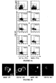
- Figure 1 In vitro characterization of the different cell death modalities induced in CT26 cells.
- C. TUNEL staining (green) of cells counterstained with DAPI (blue). The percentage of TUNEL + cells (X ⁇ SEM, n 3) was determined.
- FIG. 1 Immunogenicity of the different cell death types.
- Figure 3 Inhibition of immunogenicity by the caspase inhibitor p35.
- C D. Evolution of CT26 tumors in animals injected simultaneously with Neo-transfected, DX treated or p35-transfected, DX-treated cells into the opposite flank. Note that only Neo-expressing, DX-treated cells confer tumor immunity (p ⁇ 0.01).
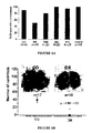
- A,B In vitro phagocytosis of the DX, DXZ or MC-treated cells (stained with CMTMR) by spleen DC from Flt3L injected mice. Representative FACS diagrams are depicted in A, and confocal images of dying tumor cells (red) phagocytosed by purified DC cell subset (green) are shown in B.
- C DC maturation of splenic DC induced by LPS (positive control) and dying or dead or dying CT26 cells. Percentage values in A and C are means of three independent determinations ⁇ SD.
- mice specifically expressing the diphteria toxin (DT) receptor in DC were pretreated with PBS alone or a dose of DT that depletes DC.
- Figure 6 Anti-tumor vaccination with DX-treated melanoma or colon carcinoma cells in distinct rodent models.
- A. Number of metastases induced by intravenous injection of CT26 cells in mice injected simultaneously with vehicle only (CO) or dying tumor cells generated as in Fig. 1. Untreated controls developed 46 ⁇ 22 metastastes (X ⁇ SD, n 46).
- Figure 7 Ex vivo and in vivo induction of immunogenic cell death.
- C. Rechallenge of cured animals (n 7, same mice as in panel B) or age-matched control (CO) mice with live CT26 cells. Similar results were obtained in three independent experiments.
- Example 1 Immunogenicity of cancer cells treated with anthracyclins
- CT26, PROb, B16F10, B16/F10.9-OVA, and B16F10A2/gp100 (B16F10 transfected with gp100 and HLA A2.1 and selected in 50 ⁇ g/ml hygromicin and G418) cells were cultured at 37°C under 5% CO 2 in RPMI 1640 medium supplemented with 10% FCS, penicillin, streptomycin, 1 mM pyruvate and 10 mM Hepes in the presence of doxorubicin (24 h, 25 ⁇ M for CT26, 30 ⁇ M for PROb, and 2.5 ⁇ M for B16F10 and its derivatives), daunorubicin (24h, 5 ⁇ M, Pharmacia), idarubicin (24h, 1 ⁇ M, Aventis, France), mitomycine C (30 ⁇ M, 48 h; Sanofi-Synthelabo, France), and/or zVAD-fink (100
- CT26 cells were stably transfected with vector only (Neo) or with a pcDNA3.1 vector encoding p35 27,28 .
- Cell death assays were trypsinized and subjected to cytofluorometric analysis with a FACS Vantage (Becton Dickinson) after staining with 4,6-diamino-2-phenylindole (DAPI, 2.5 ⁇ M, 10 min, Molecular Probes) for determination of cell viability, and Annexin V conjugated with fluorescein isothiocyanate (Bender Medsystems) for the assessment of phosphatidylserine exposure 41 . TUNEL assays were performed on cells (let to adhere for 1 h in PBS on polylysine-coated slides, O.
- 3 ⁇ 10 6 treated CT26 cells were inoculated s.c. in 200 ⁇ l of PBS into BALB/c six-week-old female mice (Charles River, France), into the lower flank, while 5 ⁇ 10 5 untreated control cells were inoculated into the contralateral flank 42 .
- PROb cells (3 ⁇ 10 6 treated cells and 10 6 control cells) were injected s.c.
- Tumors of the control side were evaluated weekly, using a caliper. Lung metastases were generated by injection of 10 5 CT26 cells into the tail vein, at the same time when 3x10 6 treated cells were injected s.c. Mice were injected with Indian ink 20 days later, sacrificed, and lung metastases were counted with a loupe. For vaccination with primary tumours, subcutaneous tumors were dissected and dissociated with collagenase (1.6 mg/ml) and DNAse (350 U/ml) during 20 minutes at 37°C, treated with 20 ⁇ M Doxorubicin for 24 hours, and then injected s.c (3x10 6 cells).
- the tumors were frozen (in FCS with 10% DMSO) as ⁇ 1 mm 3 cubes at -80°C and dissociated and treated the same way after thawing. All animals were maintained in specific pathogen-free conditions and all experiments followed the FELASA guidelines.
- the CTL response against the immunodominant CT26-specific AH1 H2L d -restricted peptide (SPSYVYHQF, purchased from Neosystem, France) 43 was determined.
- Splenocytes were isolated from immunized mice 10 days after injection of apoptotic cells and restimulated in vitro for 5 days with or without AH1 (5 ⁇ g/ml) in the presence of syngeneic irradiated naive spleen cells.
- the cytotoxic activity was determined during a classic five-hour in vitro [ 51 Cr]-release assay, using [ 51 Cr]-P815 tumor cells loaded with 50 ⁇ M AH1 peptide as target cells 42 .
- a mouse model of vaccination with apoptotic cancer cells A mouse model of vaccination with apoptotic cancer cells.
- the colon carcinoma CT26 line was killed by treatment with doxorubicin (DX) or mitomycin C (MC), as determined by FACS analysis (Fig. 1A), although the phenotypic manifestations of cell death differed. Both DX and MC induced caspase activation (Fig. 1B) and chromatin condensation (Fig. 1C, D), but only MC led to TUNEL-detectable DNA fragmentation (Fig. 1C).
- DX doxorubicin
- MC mitomycin C
- Fig. 1A doxorubicin
- MC mitomycin C
- DXZ-treated cells manifested a strongly reduced clonogenic survival as compared to untreated controls, indicating that most of the cells underwent delayed caspase-independent death in vitro (Fig. 1E).
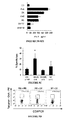
- DX and DXZ-treated cells generated tumors with a significant delay after inoculation into athymic nu / nu mice (Fig. 2A). No tumor growth was found when DX, DXZ or MC-treated cells were inoculated into syngenic, immunocompetent BALB/c mice (Fig. 2B).
- mice were inoculated into one flank with live CT26 cells and, simultaneously, received an injection of DX, DXZ, MC-treated or necrotic (frozen-thawed, F/T) cells into the opposite flank.
- DX but not DXZ, MC or F/T
- MC-treated or necrotic cells were inoculated into one flank with live CT26 cells and, simultaneously, received an injection of DX, DXZ, MC-treated or necrotic (frozen-thawed, F/T) cells into the opposite flank.
- DX but not DXZ, MC or F/T
- This degree of protection was obtained without the addition of any adjuvant. It was also independent of the presence of xenogenic antigens (fetal calf serum) in the culture media (not shown).
- DX (Fig. 2C,D) (and other anthracyclins such as daunorubicin and idarubicin, Fig. 2F) induced immunogenic cell death.
- DXZ broad-spectrum caspase inhibitor Z-VAD-fmk
- Z-VAD-fmk was replaced by chemically related caspase inhibitors with a narrow spectrum of action such as Z-DEVD-fmk, Z-IETD-fmk, Ac-LEHD-cmk, Z-VQID-CHO, or Z-VDVAD-CHO, which are specific for caspases 3, 8, 9, 6 and 2, respectively (data not shown).
- p35-transfected DX-treated CT26 cells failed to elicit an anti-tumor immune response (Fig. 3C, D). Animals protected against CT26 wild type tumors also failed to develop tumors after inoculation of live p35-expressing CT26 cells, indicating that p35 did not interfere with the effector arm of the immune system (not shown). Thus, two different protocols of caspase inhibition (pharmacological in the case of Z-VAD-fink; genetic and cell-autonomous in the case of p35) had similar effects on the immunogenicity of DX-treated tumor cells.
- DC dendritic cells
- DX-induced cell death is immunogenic
- CT26 cells were phagocytosed by all C11c + /IA d+ DC subpopulations (CD11b + , B220 + DC in Fig. 4A and CDB ⁇ + DC not shown) present in the spleen, while non-immunogenic (live, DXZ, MC or F/T) CT26 cells were neglected by DC (Fig. 4A,B).
- DX, DXZ, and F/T but not MC tumor cells possessed the capacity of inducing DC maturation, as indicated by increased expression of the costimulatory molecules CD80 and CD86, as well as induction of MHC class II (Fig. 4C).
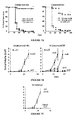
- DX (but not DXZ)-treated CT26 cells elicited a cytotoxic T cell response after in vivo inoculation (Fig. 5A). Similar data were obtained in other tumor-relevant models.
- diphteria toxin was injected into transgenic C57BL/6 mice expressing the diphteria toxin receptor specifically in DC (under the control of the CD11c promoter) 29 .
- Vehicle-injected control animals recruited CD8 + T cells with a TCR specific for the ovalbumin-derived K b /SIINFEKL epitope into the draining lymph node, but DC-depleted animals failed to do so in response to injection of apoptotic ovalbumin-transfected B16F10 cells (Fig. 5D).
- Doxorubicin-elicited apoptosis is immunogenic in several tumor models.
- DX-treated CT26 cells conferred a strong protection against the development of pulmonary metastases induced by simultaneous intravenous injection of tumor cells (Fig. 6A).
- MC-treated and F/T cells were inefficient; freeze-thawing of DX-treated CT26 cells (DX F/T) annihilated their immunogenic effect; and co-incubation with Z-VAD-fmk led to a partial reduction of the DX effect (Fig. 6A).
- the anti-metastatic effect was complete when DX-treated CT26 cells were injected twice before intravenous challenge with untreated CT26 cells (Fig. 6B).
- CT26 tumors were generated in BALB/c mice and then surgically removed and enzymatically dissociated, optionally after a step of cryostorage in DMSO-containing medium, treated with DX in vitro and used as anti-tumor vaccine (Fig. 7A).
- This ex vivo removal/in vitro treatment elicited an efficient anti-tumor immune response, if DX was added during the in vitro incubation, although cryostored cells were less efficient than freshly recovered tumor cells.
- doxorubicin As an alternative to this ex vivo/in vitro protocol of vaccine generation, we injected doxorubicin into palpable (>125 mm 3 ) subcutaneous CT26 tumors established in BALB/c mice. Using this protocol of intratumoral (as opposed to intravenous, not shown) chemotherapy, we achieved stable disease or complete tumor regression in 10 out of 24 (42%) of the animals (Fig. 7B). Rechallenge of animals that manifested a complete therapeutic response with CT26 cells failed to produce tumors (Fig. 7C), indicating that this therapeutic regimen had induced a persistent anti-tumor immunity.
Landscapes
- Health & Medical Sciences (AREA)
- Life Sciences & Earth Sciences (AREA)
- Animal Behavior & Ethology (AREA)
- Chemical & Material Sciences (AREA)
- Veterinary Medicine (AREA)
- Medicinal Chemistry (AREA)
- Public Health (AREA)
- General Health & Medical Sciences (AREA)
- Pharmacology & Pharmacy (AREA)
- Microbiology (AREA)
- Oncology (AREA)
- Immunology (AREA)
- Mycology (AREA)
- Epidemiology (AREA)
- Organic Chemistry (AREA)
- Nuclear Medicine, Radiotherapy & Molecular Imaging (AREA)
- General Chemical & Material Sciences (AREA)
- Chemical Kinetics & Catalysis (AREA)
- Medicines That Contain Protein Lipid Enzymes And Other Medicines (AREA)
- Medicines Containing Antibodies Or Antigens For Use As Internal Diagnostic Agents (AREA)
- Pharmaceuticals Containing Other Organic And Inorganic Compounds (AREA)
Abstract
The present invention is aimed at a composition comprising immunogenic dead or dying cancer cells or infected cells and fragments or fractions thereof obtained by treatment with anthracyclins, to a process of manufacturing said composition and to its use for treating cancer, for preparing a vaccine or for treating infectious diseases. It also relates to a medicament comprising at least one anthracyclin to be injected into or at the vicinity of the tumor.
Description
- The present invention is aimed at a composition comprising immunogenic dead or dying cancer cells or infected cells and fragments or fractions thereof obtained by treatment with anthracyclins, to a process of manufacturing said composition and to its use for treating cancer and for preparing a vaccine or for treating infectious diseases. It also relates to a medicament comprising at least one anthracyclin to be injected into or at the vicinity of the tumor.
- The complete and permanent success of non-surgical cancer therapy relies on the targeting of all tumor cells (including cancer stem cells) or - theoretically - on the direct removal of a fraction of the tumor, accompanied by a "bystander effect" in which the immune system recognizes, attacks and eradicates the remaining tumor cells.
- Unfortunately, however, most widely used cytotoxic anti-cancer agents exert immunosuppressive side effects. Even worse, the preponderant type of cell death induced by chemotherapy is apoptosis, and apoptosis is frequently but not unanimously viewed as immunologically silent (leading to ignorance by the immune system) or even as tolerogenic (actively down-regulating the specific anti-tumour immune response)1,2. Thus, even after an initial therapeutic success, patients typically fail to mount a clinically relevant anti-tumor immune response and eventually succumb to tumor cell variants that escape from chemotherapy.
- Although apoptosis is itself non-uniform with respect to the programmed signaling events responsible for cell death3, it is morphologically defined as a uniform type of cell death accompanied by chromatin condensation (pyknosis) and nuclear fragmentation (karyorhexis), occurring within an intact plasma membrane4. It thus differs from necrosis in which the plasma membrane is destroyed early during the death process. In biochemical terms, apoptosis is frequently accompanied by the activation of a specific subset of cysteine kinases, the caspases3. Since apoptosis is a physiological process that attains several millions of cells per second in the human body, it is inferred that this type of cell death occurs without emitting "danger signals" that would elicit a productive immune responses. Apoptotic cells would hide away from immune recognition, because they would be rapidly recognized and silently phagocytosed in an efficient manner. Thus, defects in the recognition/phagocytosis of apoptotic cells breach autotolerance and trigger autoimmune reactions6-8. Moreover, apoptotic cells would mediate active immunosuppression by inhibiting the production of immunostimulatory cytokines (e.g. interleukin-1ß by macrophages and interleukin-12 by dendritic cells, DC)9,10, by stimulating the production of immunosuppressive factors (e.g. transforming growth factor)11, and/or by eliciting antigen-specific immune tolerance12-14. In apparent contrast with this notion, however, DC can capture apoptotic tumor cells in vitro and cross-present antigen derived from internalized dying cells on MHC class I molecules for recognition by CD8+ T cells in vivo, thus eliciting a productive immune response2,15-20, including in clinical trials21. Systematic comparisons of the immunogenicity of apoptotic versus necrotic tumor cells have been inconclusive22-25, however, suggesting that the nature of tumor cells and/or that the death-inducing stimulus would influence the experimental outcome1.
- Driven by these uncertainties and incognita, we decided to explore the possibility of inducing immunogenic tumor cell apoptosis by chemotherapy. Here, we show that anthracyclins are capable of eliciting immunogenic cell death in vitro, ex vivo and in vivo, in a fashion that is rigorously caspase-dependent.
- These results have profound implications for the design of chemotherapeutic regimens with immunogenic properties and allow for the first time to prepare a tumor immunogenic composition useful as a vaccine by the induction of immunogenic cell death of tumor cells of a patient. This discovery is applicable to infectious diseases as well.
- Therefore, in a first aspect, the present invention is aimed at a process of manufacturing an immunogenic composition comprising the steps consisting of:
- a) suspending or plating tumor cells or infected cells in a culture media,
- b) treating said cells with at least one antineoplastic compound selected from anthracyclins and vinorelbine in a sufficient amount to induce cellular apoptosis, wherein said apoptosis induced by anthracyclins produces immunogenic dead or dying cells, and
- c) recovering immunogenic dead or dying cells and fragments thereof from the culture media.
- In a particular embodiment, the tumor cells in step a) are the tumor cells of a given human subject. In this regard, the process may optionally comprise the step of isolating tumor cells from a tumor biopsy or sample. Thus, in this embodiment, the process allows the manufacture of a tailored immunogenic composition from one patient's own cancer cells to treat said patient (autologous treatment).
- Alternatively, the process may be performed using established cancer cell lines or freshly harvested cancer cells from one or several patients. For example, the process allows the production of an immunogenic composition to treat breast cancer from one or several breast cancer cell lines treated as explicited above with at least one anthracyclin to obtain immunogenic breast cancer dead or dying cells and fragments thereof (allogenic treatment). This process may be applied to solid tumors such as carcinomas, melanomas and sarcomas including but not limited to breast, lung, prostate or skin cancer but also to blood cancer such as lymphomas and leukemias.
- Alternatively, cells in step a) are infected cells including cells infected with a virus such as HIV or Hepatitis C virus.
- The above process of manufacturing may further comprise one or several of the following steps : purification, elutriation, ultracentrifugation and loading into myeloid or plasmacytoid dendritic cells of immunogenic dead or dying cells and fragments thereof.
- The process as defiend above allows to target specifically in vivo appropriate antigen presenting cells such as plasmacytoid dendritic cells or myeloid dendritic cells for optimal CTL priming or T cell activation. It also allows ex vivo pulsing or loading of tumor antigens on to ex vivo propagated allogeneic or autologous dendritic cells for further adoptive transfer in vivo in tumor bearing patients or infected patients.
- In the above process, the immunogenic composition is useful as a medicament or a vaccine for preventing or treating cancer.
- By anthracyclin compounds, it will be understood that the invention embraces any compounds known in the art to have identical core structure and presenting antineoplastic activity, for example any anthracyclin glycosides, such as doxorubicin (
US 3,590,028 ), 4'-epi-doxorubicin (i.e. epirubicin,US 4,039,663 ), 3',4'-diepidaunorubicin and 3',4'-diepidoxorubicin (US 4,112,076 ), 4'-desoxy-doxorubicin (i.e. esorubicin), daunorubicin, 5-iminodaunorubicin and 4-demethoxy-daunorubicin (i.e. idarubicin).
Formula of these compounds is shown below :
Wherein R1 R2 R3 R4 Doxorubicin OH H OH O Daunorubicin H H OH O Epirubicin OH OH H O 5-iminodaunorubicin H H OH NH - In the above formula, the invention also embraces the compounds wherein R1, R2, R3 are independently selected from H, OH, a halogen, a C1-C4 alkyl, alkenyl, or alkoxy group.
- In a preferred embodiment, cancer cells are treated with doxorubicin (4-(4-amino-3-hydroxy-2-methyl-tetrahydropyran-6-yl)oxy-2,5,12-trihydroxy-2-(2-hydroxyacetyl)-7-methoxy-1,2,3,4-tetrahydrotetracene-6,11-dione - CAS 25316-40-9).
- The invention may also be practiced with the antineoplastic compound vinorelbine (CAS Registry Number 71486-22-1) of formula :

- This process is useful for the manufacture of a medicament or a vaccine for preventing or treating cancer as well as infectious diseases, such as viral infection including but not limited to HIV and Hepatitis C infection.
- In a second aspect, the invention relates to an immunogenic composition comprising immunogenic dead or dying cells or infected dead or dying cells and fragments thereof obtainable by the process as defined above. In fact, it relates to a composition comprising immunogenic dead or dying cancer cells and fragments or fractions thereof obtained by treatment with anthracyclins. It also relates to a composition comprising immunogenic dead or dying infected cells and fragments or fractions thereof obtained by treatment with anthracyclins.
- Preferably, this composition comprises anthracyclin glycosides treated cancer cells or infected cells and fragments thereof which can be in the form of a dead or dying cellular extract or fraction thereof or of a purified cellular lysate. The composition may further comprise one of the following : a pharmaceutical acceptable carrier, one or several adjuvant(s) and/or active ingredient(s), in particular agents which are cytotoxic for tumors, such as a compound selected from anthracyclin glycosides, preferably doxorubicin, 4'-epi-doxorubicin, 3',4'-diepidaunorubicin, 3',4'-diepidoxorubicin, 4'-desoxy-doxorubicin, daunorubicin, 5-iminodaunorubicin or 4-demethoxy-daunorubicin., vinorelbine, cytokines and interleukines.
- For example, the composition may further comprise one or several of the following :
- caspase endogenous inhibitors, suh as IAP proteins,
- adjuvants such as interferon-alpha (IFN-α), granulocyte-macrophage colony stimulating factor, soluble CD40 ligand, Flt3 ligand and/or interleukin-2 (IL-2),
- tyrosine kinase inhibitors such as c-kit inhibitors,
- ligands of Toll-like receptors,
- neutralizing antibodies such as anti-CD25, anti-CTLA4,
- stimulating antibodies such as anti-CD40,
- cyclophosphamides in an amount suitable for immunostimulation,
- small interfering RNAs (SiRNA) inhibiting immunosupressive genes.
- For example, it may comprise a pharmaceutical vehicle selected from positively or negatively charged liposomes, nanoparticles or lipid emulsions.
- It is preferably formulated to be suitable for intravenous, intradermic, intramuscular or subcutaneous injection, as well as oral or nasal administration.
- In a third aspect, the invention concerns an extract of immunogenic dead or dying cancer cells or infected cells and fragments thereof obtainable by the process as defined above, especially in the form of a dead or dying cellular extract or fraction thereof or of a purified cellular lysate.
- The invention also relates to the use of an extract of immunogenic dead or dying cancer cells or infected cells and fragments thereof for preparing a medicament for preventing or treating cancer or infecious diseases. It will be understood that the invention may apply to human as well as to other mammals, such as domestic pets and also cattle.
- The invention is also directed to a method of preventing or treating cancer or infectious diseases comprising administering a composition or extract of immunogenic dead or dying cancer cells as depicted above in a patient in need of such treatment. More particularly, this method includes :
- a) obtaining fresh cancer cells or infected cells from a patient,
- b) suspending or plating said cells a) in a culture media,
- c) treating cells of step b) with at least one antineoplastic compound selected from anthracyclins in a sufficient amount to induce cellular apoptosis, wherein said apoptosis induced by anthracyclins produces immunogenic tumor dead or dying cells,
- d) recovering immunogenic tumor dead or dying cells and fragments thereof from the culture media, and
- e) Administering the immunogenic dead or dying cells and fragments thereof, optionally purified, to said patient.
- As mentioned above, step d) may further comprise extraction, filtration, purification, sterilization protocols to obtain an administrable composition. The preparation may also be lyophilized and then resuspended in a solution or buffer such as an appropriate sterile saline solution. In this configuration, one or several administrations over time may be applied to obtained optimized immune response against cancer.
The above method is tailored to one particular patient since the immunogenic dead or dying cells and fragments thereof composition is prepared from the cancer cells or infected cells of said patient. - Nevertheless, it shall be understood, the invention may be practiced using one or several cell lines corresponding to one particular cancer type, for example breast carcinoma. Here, the invention is directed to a method of preventing or treating cancer comprising administering a composition or extract of immunogenic dead or dying cancer cells as depicted above in a patient in need of such treatment comprising :
- a) suspending or plating in a culture media cancer cells from cancer cell lines corresponding to the cancer type afflicting said patient,
- b) treating cells of step a) with at least one antineoplastic compound selected from anthracyclins in a sufficient amount to induce cellular apoptosis, wherein said apoptosis induced by anthracyclins leads to immunogenic tumor cell death,
- d) recovering immunogenic dead or dying cells and fragments thereof from the culture media, and
- e) administering the immunogenic dead or dying cells and fragments thereof, optionally purified, to said patient.
- In this embodiment, the invention offers a method for preparing and administering different immunogenic compositions that are each specific to one cancer type.
- The cells of step a) may derive from established appropriate cell lines or from harvested cancer cells from one or several patients afflicted with a particular cancer including solid tumors such as carcinomas, melanomas and sarcomas including but not limited to breast, lung, prostate or skin cancer but also to blood cancer such as lymphomas and leukemias.
- In the above methods, the immunogenic composition or cell extract administered may also comprise at least one antineoplastic compound selected from anthracyclin glycosides, preferably doxorubicin, 4'-epi-doxorubicin, 3',4'-diepidaunorubicin, 3',4'-diepidoxorubicin, 4'-desoxy-doxorubicin, daunorubicin, 5-iminodaunorubicin or 4-demethoxy-daunorubicin. In this regard, this composition of the invention may advantageously be injected directly in situ into the cancer mass or at the vicinity of localized cancer cells of the patient.
- In still another embodiment, the invention is aimed at a method of preventing or treating cancer comprising injected directly in situ into the cancer mass or at the vicinity of localized cancer cells of the patient a compound selected from anthracyclin glycosides, preferably doxorubicin, 4'-epi-doxorubicin, 3',4'-diepidaunorubicin, 3',4'-diepidoxorubicin, 4'-desoxy-doxorubicin, daunorubicin, 5-iminodaunorubicin or 4-demethoxy-daunorubicin in an amount sufficient to induce immunogenic cancer cell death.
- In other words, the invention relates to the use of a compound selected from anthracyclin glycosides, preferably doxorubicin, 4'-epi-doxorubicin, 3',4'-diepidaunorubicin, 3',4'-diepidoxorubicin, 4'-desoxy-doxorubicin, daunorubicin, 5-iminodaunorubicin or 4-demethoxy-daunorubicin for preparing a medicament suitable to direct injection in situ into the cancer mass or at the vicinity of localized cancer cells of the patient for inducing immunogenic cancer cell death. This medicament is applicable to the treatment of solid tumors such as carcinomas, melanomas and sarcomas including but not limited to breast, lung, prostate or skin cancer but also to blood cancer such as lymphomas and leukemias.
- It is important to underscore the fact that the immunogenic effects of anthracyclin-treated tumor cells were observed in the absence of any adjuvant or co-stimulus. Hence, co-application of adjuvants may be provided to further improve the efficacy of the vaccination schedule. As a fascinating possibility, our invention proposes a strategy of inducing immunogenic cell death in established tumors, either ex vivo or in vivo.
- Even though apoptosis is often considered as an immunologically silent or even tolerogenic death modality6-14,33, we challenge here this viewpoint and we present data demonstrating that anthracyclins-induced apoptosis can elicit an effective anti-tumor immune response. While anthracyclins (and in particular DX, which we used as a model agent) elicited immunogenic apoptosis in vitro (Fig. 2, 3, 5, 6), ex vivo (Fig. 7A) and in vivo (Fig. 7B), MC (Fig. 2, 4C, 5A) was relatively inefficient in eliciting immunogenic cell death. Preparations of DX or MC-treated cells that contained a similar percentage of dying cells (Fig. 1a) were injected, and the morphological aspect of cell death elicited by DX and MC was similar (Fig. 1C,D). Both were accompanied by caspase activation (Fig. 1B), without any difference in the expression of some major endogenous adjuvants (such as HSP70 and HMGB-1, Fig. 1B), and the only difference resided in the degree of DNA fragmentation, which was more advanced with MC (Fig. 1C). Moreover, DC phagocytosed MC-treated cells less efficiently than DX-treated cells (Fig. 4A).
- Thus these data underscore a hitherto unsuspected heterogeneity in apoptotic cells with respect to their uptake by dendritic cells34. Importantly, inhibition of caspase activation by Z-VAD-fink or transfected p35 greatly reduced the immunogenic potential of DX-treated cells, an effect that was observed in a variety of different tumor models (Fig. 2-7) and that also affected DC-mediated recognition and phagocytosis (Fig. 4A). However, caspase activation is not sufficient to elicit an immunogenic cell death because MC-treated cells, which do exhibit caspase activation (Fig. 1B), are inefficient anti-tumoral vaccines (Fig. 2). Thus, a specific combination of factors, caspase activation and a yet-to-be defined antigenic property of anthracyclin-treated cells, has to cooperate to elicit an anti-tumor immune response. Disruption of the membrane integrity of DX-treated tumor cells by freeze-thawing is sufficient to abolish their immunogenic property (Fig. 4C, Fig. 5A), meaning that intact apoptotic cells or bodies are required for optimal antigenicity. Irrespective of these mechanistic details, it appears that the particular protocol of apoptosis induction determines the immunogenicity of cell death. Two different subcategories of apoptosis exist, one that is immunogenic and the other that is non-immunogenic.
- DX-treated, dying tumor cells are efficiently phagocytosed by myeloid and plasmacytoid DC (Fig. 4A,B) and are highly efficient in eliciting antigen-specific cytotoxic T lymphocytes (Fig. 5). Again these properties were largely reduced by simultaneous treatment with the caspase inhibitor Z-VAD-fmk (Fig. 4,5), correlating with a reduced immunogenicity. To our knowledge, this is the first discovery indicating that caspase activation is broadly required for the elicitation of immunogenic tumor cell death. Previous reports have described that caspase activation rather reduces the immunogenic potential of tumor cells. Thus, a PROb clone (REGb) that fails to activate caspases is much more immunogenic than the parental PROb line35. Similarly, a PROb clone manipulated to stably express an anti-sense cytochrome c cDNA undergoes atypical apoptosis without caspase activation, yet has an increased immunogenic potential25. As another example, caspases have been documented to destroy immunodominant epitopes, thus blunting the immune response against lymphoma cells 36. In NOD mice (which are prone to develop autoimmune diabetes), systemic caspase inhibition reduced the priming of T cells by β cells undergoing spontaneous apoptosis 37 and, conversely, local caspase inhibition abolished the tolerogenic effect of β cell apoptosis induced by streptozotocin 33
- A. FACS analysis of cells treated in vitro with the indicated agents (DX, doxorubicin; MC, mitomycin C; ZVAD, Z-VAD-fmk; DXZ, doxorubicin plus Z-VAD-fmk, F/T, freeze-thaw), as specified in Materials and Methods. B. Immunoblot analyses of cells treated as in A. Cellular extracts were subjected to the immunodetection of activated caspase-3 (Casp3a), HMGB-1, and HSP-70. C. TUNEL staining (green) of cells counterstained with DAPI (blue). The percentage of TUNEL+ cells (X±SEM, n=3) was determined. D. Transmission electron microscopy. Representative microphotographs are shown. Apoptotic cells showing chromatin condensation are labeled with an "A". E. Cells treated with the indicated agents as in A for 19 h were washed and plated to determine the frequency of surviving clones, defining the control value of untreated cells as 100%. Results are representative of three independent experiments.
- A. Tumor evolution of DX and DXZ treated cells in nu/nu mice. Cells treated as in Fig. 1 were injected s.c. (3x10⁶/mouse). B. Comparison of the number of tumor-bearing mice between immunocompetent BALB/c and nu/nu mice. Data from nu/nu mice are from the same animals as in A, while data from BALB/c mice were obtained on a minimum of 30 animals per group. C. Immunogenic effect of DX-treated tumor cells. BALB/c mice were injected into one flank with live tumor cells and into the opposite flank with DX, DXZ, MC, or F/T-treated cells, and the evolution of tumors was monitored. D. Persistent, specific anti-tumor immunity elicited by DX-treated cells. Animals immunized with DX-treated CT26 cells that remain tumor-free after 120 days (see lower left panel in C) or age-matched control mice were challenged with CT126 cells or the unrelated TSA tumor cell line. E. Effect of the timing of anti-tumor vaccination. CT26 cells treated as in Fig. 1 were injected at the same time as live tumor cells (as in C) or were injected once one week before, twice one and two weeks before or three times, one, two or three weeks before challenge with live tumor cells (n=15 per group). F. Replacement of DX by other anthracyclins. Animals were injected with CT26 cells treated daunorubicin (DA) or idarubicin (ID), instead of doxorubicin, or PBS only (CO) and the growth of live CT26 cells simultaneous injected into the opposite flank was monitored as in C. All figures represent 3 to 6 independent experiments. * p < 0.05 vs control (CO).
- A. FACS analysis of CT26 cells transfected with vector only (Neo) or p35, cultured alone or in the presence of DX, as in Fig. 1A. B. Clonogenic survival of Neo or p35-transfected cells left untreated (100 % values) or treated with DX. C, D. Evolution of CT26 tumors in animals injected simultaneously with Neo-transfected, DX treated or p35-transfected, DX-treated cells into the opposite flank. Note that only Neo-expressing, DX-treated cells confer tumor immunity (p<0.01).
- A,B. In vitro phagocytosis of the DX, DXZ or MC-treated cells (stained with CMTMR) by spleen DC from Flt3L injected mice. Representative FACS diagrams are depicted in A, and confocal images of dying tumor cells (red) phagocytosed by purified DC cell subset (green) are shown in B. C. DC maturation of splenic DC induced by LPS (positive control) and dying or dead or dying CT26 cells. Percentage values in A and C are means of three independent determinations ± SD.
- A. CTL response of DX and DXZ-treated cells immunized mice. Splenic T cells from animals vaccinated with PBS only (CO), DX-treated CT26 cells or DXZ-treated CT26 cells were tested for their capacity to lyse syngenic cells pulsed with the immunodominant H2Ld restricted CT26-derived peptide SPSYVYHQF. Representative CTL responses from individual mice are shown in the left panel, while mean values are shown on the right. R and NR refer to responders and non-responders, respectively. * p < 0.01 vs control (CO). B. Percentages of CD8+ lymph node cells expressing TCR that interact with the ovalbumin-derived peptide SIINFEKL presented by H-2Kb, five days after challenge with either peptide+adjuvant (P+A), or with DX, DX F/T (DX-treated, then frozen/thawed cells), DXZ, MC and F/T -treated B16-OVA cells (3 experiments). C. IFNγ production, as measured after in vitro culture with SIINFEKL in the same experiment. Control values are <50 pg/ml. D. Effect of the depletion of DC on the accumulation of specific T cells. Transgenic mice specifically expressing the diphteria toxin (DT) receptor in DC were pretreated with PBS alone or a dose of DT that depletes DC. The mice were then challenged with DX-treated B16-OVA cells into the food pad. Draining lymph nodes were recovered 48 hours later and stained for simultaneous detection of CD8 and OVA peptide-specific T cell receptors. Representative FACS pictograms are shown and values are X±SEM (n=3).
- A. Number of metastases induced by intravenous injection of CT26 cells in mice injected simultaneously with vehicle only (CO) or dying tumor cells generated as in Fig. 1. Untreated controls developed 46±22 metastastes (X±SD, n=46). B. Effect of two immunizations with DX-treated CT26 cells, 14 and 7 days before intravenous injection of
live CT 26 cells. All figures are pooled data from 3 independent experiments. * p < 0.001 vs CO. C. DX-treated B16A2 cells were injected twice into mice transgenic for HLA.A2, two weeks and one week before challenge with live B16A2 cells into the opposite flank. * p < 0.05 vs CO. D. The protocol of simultaneous s.c. injection of dying (DX or DXZ-treated) tumor cells was applied to BDIX rats challenged with PROb cells. * p < 0.001 vs CO. This experiment has been repeated three times, yielding similar results. - A. Evolution of tumors in mice vaccinated with cells from freshly resected or cryopreserved CT26 tumors, that were left untreated (CO) or treated with DX. B. Effect of intratumoral DX injection on the growth of CT26 colon cancer cells inoculated into immunocompetent BALB/c mice. The left panel shows the effect of intratumoral injection of vehicle and the right panel that of DX. While some animals showed a normal tumor growth (n=6) or a simple delay in tumor growth (n=8), other animals exhibited stable disease (n=3) or complete tumor regression (n=7). C. Rechallenge of cured animals (n=7, same mice as in panel B) or age-matched control (CO) mice with live CT26 cells. Similar results were obtained in three independent experiments.
- Cell lines and cell death induction. CT26, PROb, B16F10, B16/F10.9-OVA, and B16F10A2/gp100 (B16F10 transfected with gp100 and HLA A2.1 and selected in 50 µg/ml hygromicin and G418) cells were cultured at 37°C under 5% CO2 in RPMI 1640 medium supplemented with 10% FCS, penicillin, streptomycin, 1 mM pyruvate and 10 mM Hepes in the presence of doxorubicin (24 h, 25 µM for CT26, 30 µM for PROb, and 2.5 µM for B16F10 and its derivatives), daunorubicin (24h, 5 µM, Pharmacia), idarubicin (24h, 1 µM, Aventis, France), mitomycine C (30 µM, 48 h; Sanofi-Synthelabo, France), and/or zVAD-fink (100 µM, 24 h; Bachem). Necrosis was induced by one freeze/thaw cycle in liquid N2 and a 37°C water bath. CT26 cells were stably transfected with vector only (Neo) or with a pcDNA3.1 vector encoding p3527,28.
- Cell death assays. Cells were trypsinized and subjected to cytofluorometric analysis with a FACS Vantage (Becton Dickinson) after staining with 4,6-diamino-2-phenylindole (DAPI, 2.5 µM, 10 min, Molecular Probes) for determination of cell viability, and Annexin V conjugated with fluorescein isothiocyanate (Bender Medsystems) for the assessment of phosphatidylserine exposure41. TUNEL assays were performed on cells (let to adhere for 1 h in PBS on polylysine-coated slides, O. Kindler GmbH, Germany, fixed with 4% paraformaldehyde for 30 min) using the In situ Cell Death Detection Kit kit by Roche and a Leica IRE2 fluorescence microscope equipped with a Leica DC300F camera. For electron microscopy, cells (fixed for 1 hour at 4°C in 2.5% glutaraldehyde in phosphate buffer (pH 7.4) were washed and fixed again in 2% osmium tetroxide, then embedded in Epon), and ultrathin sections (80 nm) were stained with uranyl acetate and lead citrate and examined with a Leo 902 electron microscope, at 80 kV.
- Immunoblot analyses. Cells were washed with cold PBS at 4°C and lysed in a buffer containing 50 mM Tris HCl pH 6.8, 10 % glycerol and 2 % SDS. Primary antibodies detecting activated caspase 3 (
dilution 1/1000, Cell Signaling Technology), HSP70 (dilution 1/1000, Stratagene) or HMGB-1 (2 µg/ml, Pharmingen), were revealed with the appropriate horseradish peroxidase-labeled secondary antibody (Southern Biotechnologies Associates) and detected by ECL (Pierce). Anti-actin or anti-GAPDH (Chemicon) was used to control equal loading. - Anti-tumor vaccination and assessment of tumor growth in vivo. 3×10⁶ treated CT26 cells were inoculated s.c. in 200 µl of PBS into BALB/c six-week-old female mice (Charles River, France), into the lower flank, while 5×10⁵ untreated control cells were inoculated into the contralateral flank42. PROb cells (3×10⁶ treated cells and 106 control cells) were injected s.c. into the lower flanks of syngeneic 2-month-old BDIX rats (Charles River), and B16A2 and B16F10 (1.8×10⁵ treated cells and 3×10⁴ control cells) were injected into six-week-old female HHD2 transgenic mice (bred in the IGR animal facility) and C57BL/6 mice (Charles River) respectively. For the tumorigenicity assay, 3x10⁶ treated or untreated CT26 cells were injected s.c. into nu/nu mice (IGR animal facility). To assess the specificity of the immune response against CT26, we injected either 5x10⁵ or 5x10⁶ of CT26 (for the mice immunize in a standard protocol or vaccination protocol, respectively) or 106 TSA cells. Tumors of the control side were evaluated weekly, using a caliper. Lung metastases were generated by injection of 105 CT26 cells into the tail vein, at the same time when 3x10⁶ treated cells were injected s.c. Mice were injected with
Indian ink 20 days later, sacrificed, and lung metastases were counted with a loupe. For vaccination with primary tumours, subcutaneous tumors were dissected and dissociated with collagenase (1.6 mg/ml) and DNAse (350 U/ml) during 20 minutes at 37°C, treated with 20 µM Doxorubicin for 24 hours, and then injected s.c (3x10⁶ cells). Alternatively, the tumors were frozen (in FCS with 10% DMSO) as ~1 mm3 cubes at -80°C and dissociated and treated the same way after thawing. All animals were maintained in specific pathogen-free conditions and all experiments followed the FELASA guidelines. - Assessment of specific T cell responses. The CTL response against the immunodominant CT26-specific AH1 H2Ld-restricted peptide (SPSYVYHQF, purchased from Neosystem, Strasbourg, France)43, was determined. Splenocytes were isolated from immunized
mice 10 days after injection of apoptotic cells and restimulated in vitro for 5 days with or without AH1 (5 µg/ml) in the presence of syngeneic irradiated naive spleen cells. The cytotoxic activity was determined during a classic five-hour in vitro [51Cr]-release assay, using [51Cr]-P815 tumor cells loaded with 50 µM AH1 peptide as target cells42. The percentage of specific lysis was calculated as 100 x (experimental release - spontaneous release)/(maximal release - spontaneous release). Maximum release was obtained by adding 10% Triton X-405 to target cells and spontaneous release was determined by incubating target cells in medium alone. Female C57BL/6 mice (H-2b, 5-6 weeks old) were injected into both footpads with PBS, 50 µg of OVA peptide (H-2Kb restricted, SIINFEKL, aa 257-264, purchased from Eurogentec, Villejuif, France) mixed with incomplete Freund adjuvant (Sigma), or with 106 B16/F10.9-OVA cells. Animals were sacrificed five days after footpad immunization, popliteal and inguinal lymph nodes were removed and processed individually. A single cell suspension was obtained by homogenizing and filtering the organ through a sterile cell strainer (70 µm, Becton Dickinson). For FACS analysis, 2x10⁶ cells were stained at RT with a Tetramer-OVA-PE (Beckman Coulter, UK) and then stained with an anti-CD3-FITC and with an anti-CD8-APC monoclonal antibody (Beckton Dickinson). Cells were analysed using a FACScalibur cytometer (Becton Dickinson) with Cellquest Pro software. Alternatively, 105 cells were cultured in complete culture medium containing 2% of mouse serum, in the presence or absence of 40µg/ml SIINFEKL peptide. Three days later, the supernatants were harvested and IFN-γ secretion was determined by ELISA (OptEIA™ ELISA kit, BD Pharmingen). In ones series of experiments, CD11c-GFP DT Kb mice29 were injected i.p. with 100 ng of diphteric toxin (or PBS as a vehicle control), 48 hours before injection of DX-treated B16/F10.9-OVA cells into the food pad. - The colon carcinoma CT26 line was killed by treatment with doxorubicin (DX) or mitomycin C (MC), as determined by FACS analysis (Fig. 1A), although the phenotypic manifestations of cell death differed. Both DX and MC induced caspase activation (Fig. 1B) and chromatin condensation (Fig. 1C, D), but only MC led to TUNEL-detectable DNA fragmentation (Fig. 1C). When combined with DX, the broad spectrum caspase inhibitor Z-VAD-fmk (DXZ) delayed cell death (Fig. 1A), reduced caspase activation (Fig. 1B) and blunted chromatin condensation (Fig. 1D). However, DXZ-treated cells manifested a strongly reduced clonogenic survival as compared to untreated controls, indicating that most of the cells underwent delayed caspase-independent death in vitro (Fig. 1E). Moreover, as compared to untreated controls, DX and DXZ-treated cells generated tumors with a significant delay after inoculation into athymic nu/nu mice (Fig. 2A). No tumor growth was found when DX, DXZ or MC-treated cells were inoculated into syngenic, immunocompetent BALB/c mice (Fig. 2B). To explore the putative immunogenicity of dead or dying or dying cells, BALB/c mice were inoculated into one flank with live CT26 cells and, simultaneously, received an injection of DX, DXZ, MC-treated or necrotic (frozen-thawed, F/T) cells into the opposite flank. In this therapeutic regime, only DX (but not DXZ, MC or F/T)-treated cells reduced the frequency of tumors developing from live cells by ~45% on
day 30 and ~30% on day 120 (Fig. 2C). This degree of protection was obtained without the addition of any adjuvant. It was also independent of the presence of xenogenic antigens (fetal calf serum) in the culture media (not shown). Animals not developing tumors after challenge with DX-treated CT26 cells were protected against a second inoculation oflive CT 26 cells, but were not immune against the unrelated TSA tumor (Fig. 2D). Of note, the efficacy of the tumor vaccination could be improved to 80% by one prophylactic injection of DX-treated cells before challenge with live tumor cells. Irrespective of the immunization schedule, DX-treated cells did confer a higher degree of protection than DXZ, MC, or F/T-treated cells (Fig. 2E). Thus, dying tumor cells can elicit an effective anti-tumor immune response. - DX (Fig. 2C,D) (and other anthracyclins such as daunorubicin and idarubicin, Fig. 2F) induced immunogenic cell death. However, concomitant treatment with the broad-spectrum caspase inhibitor Z-VAD-fmk (DXZ) abolished the immunogenic potential of the treatment with DX (Fig. 2C). No such inhibitory effects were obtained when Z-VAD-fmk was replaced by chemically related caspase inhibitors with a narrow spectrum of action such as Z-DEVD-fmk, Z-IETD-fmk, Ac-LEHD-cmk, Z-VQID-CHO, or Z-VDVAD-CHO, which are specific for
caspases - In an attempt to understand why DX-induced cell death is immunogenic, we challenged DC with dead or dying or dying cells and measured their response. DX-treated CT26 cells were phagocytosed by all C11c+/IAd+ DC subpopulations (CD11b+, B220+ DC in Fig. 4A and CDBα+ DC not shown) present in the spleen, while non-immunogenic (live, DXZ, MC or F/T) CT26 cells were neglected by DC (Fig. 4A,B). In contrast, several categories of dying cells (DX, DXZ, and F/T but not MC) tumor cells possessed the capacity of inducing DC maturation, as indicated by increased expression of the costimulatory molecules CD80 and CD86, as well as induction of MHC class II (Fig. 4C). DX (but not DXZ)-treated CT26 cells elicited a cytotoxic T cell response after in vivo inoculation (Fig. 5A). Similar data were obtained in other tumor-relevant models. Injection of ovalbumin-transfected B16F10 melanoma cells elicited the local recruitment of ovalbumin-specific CD8+ T cells (which recognize the Kb/SIINFEKL tetramer) into, as well as the production of interferon-γ by cells from the draining lymph node of syngenic C57BL/6 mice, provided that the injected cells were pretreated with DX (Fig. 5B,C). However, this response was blunted when the inoculum had been treated with DXZ, MC or when the DX-treated cells were rendered necrotic by freeze-thawing (Fig. 5B,C). To determine the impact of CD11c+ DC on the immune response in vivo, diphteria toxin was injected into transgenic C57BL/6 mice expressing the diphteria toxin receptor specifically in DC (under the control of the CD11c promoter)29. Vehicle-injected control animals recruited CD8+ T cells with a TCR specific for the ovalbumin-derived Kb/SIINFEKL epitope into the draining lymph node, but DC-depleted animals failed to do so in response to injection of apoptotic ovalbumin-transfected B16F10 cells (Fig. 5D). These data confirm that DX-treated cells are particularly effective in stimulating a cytotoxic immune response and that this response is mediated by DC.
- One single subcutaneous injection of DX-treated CT26 cells conferred a strong protection against the development of pulmonary metastases induced by simultaneous intravenous injection of tumor cells (Fig. 6A). In this system, MC-treated and F/T cells were inefficient; freeze-thawing of DX-treated CT26 cells (DX F/T) annihilated their immunogenic effect; and co-incubation with Z-VAD-fmk led to a partial reduction of the DX effect (Fig. 6A). The anti-metastatic effect was complete when DX-treated CT26 cells were injected twice before intravenous challenge with untreated CT26 cells (Fig. 6B). In a further round of experiments, we determined whether the observed tumor vaccination effects were a general phenomenon. When replacing CT26 cells by B16F10 cells, we found that this melanoma cell line was immunogenic, if DX treated and injected into C57BL/6 mice (not shown). B16F10A2/gp100 melanoma cells present peptides from the human gp100 tumor antigen in the context of HLA class I A230. Again, in vitro treatment with DX, followed by subcutaneous injection of the cells, was able to induce a significant protection against later challenge with live B16F10A2/gp100 tumor cells, in "humanized" mice expressing an HLA.A2 transgene30,31 (Fig. 6C). Thus, DX-treated dying tumor cells turned out to be immunogenic on a human MHC background. Furthermore, inbred BDIX rats inoculated with the syngenic PROb colon carcinoma32 could be protected by injection of DX-treated tumor cells, and this effect was again suppressed by simultaneous pretreatment of the tumor cells with DX and Z-VAD-fmk (Fig. 6D). In conclusion, this protocol of cancer immunization is applicable to a series of distinct tumors, species and MHC backgrounds.
- We investigated whether it would be required to immunize animals with dying cells cultured as an established cell line or whether it would be feasible to take advantage of resected tumors (as this would be the case in human cancer patients). CT26 tumors were generated in BALB/c mice and then surgically removed and enzymatically dissociated, optionally after a step of cryostorage in DMSO-containing medium, treated with DX in vitro and used as anti-tumor vaccine (Fig. 7A). This ex vivo removal/in vitro treatment elicited an efficient anti-tumor immune response, if DX was added during the in vitro incubation, although cryostored cells were less efficient than freshly recovered tumor cells. The omission of DX curtailed the efficacy of the vaccine (Fig. 7A). As an alternative to this ex vivo/in vitro protocol of vaccine generation, we injected doxorubicin into palpable (>125 mm3) subcutaneous CT26 tumors established in BALB/c mice. Using this protocol of intratumoral (as opposed to intravenous, not shown) chemotherapy, we achieved stable disease or complete tumor regression in 10 out of 24 (42%) of the animals (Fig. 7B). Rechallenge of animals that manifested a complete therapeutic response with CT26 cells failed to produce tumors (Fig. 7C), indicating that this therapeutic regimen had induced a persistent anti-tumor immunity.
-
- 1. Zitvogel, L., Casares, N., Pequignot, M., Albert, M.L. & Kroemer, G. The immune response against dying tumor cells. Adv. Immunol. 84, 131-79 (2004).
- 2. Steinman, R.M. & Mellman, I. Immunotherapy: bewitched, bothered, and bewildered no more. Science 305, 197-200 (2004).
- 3. Igney, F.H. & Krammer, P.H. Death and anti-death: tumour resistance to apoptosis. Nat Rev Cancer. 2, 277-88 (2002).
- 4. Kerr, J.F.R., Wyllie, A.H. & Currie, A.R. Apoptosis: a basic biological phenomenon with wide-ranging implications in tissue kinetics. .
- 5. Matzinger, P. The danger model: a renewed sense of self. Science 296, 301-5. (2002).
- 6. Hanayama, R. et al. Autoimmune disease and impaired uptake of apoptotic cells in MFG-E8-deficient mice. Science 3004, 1147-50. (2004).
- 7. Asano, K. et al. Masking of phosphatidylserine inhibits apoptotic cell engulfment and induces autoantibody production in mice. J Exp Med. 200, 459-67. (2004).
- 8. Lauber, K., Blumenthal, S.G., Waibel, M. & Wesselborg, S. Clearance of apoptotic cells: getting rid of the corpses. Mol Cell. 14, 277-87 (14).
- 9. Fadok, V.A., Bratton, D.L. & Henson, P.M. Phagocyte receptors for apoptotic cells: recognition, uptake, and consequences. J Clin Invest. 108, 957-62 (2001).
- 10. Kim, S., Elkon, K.B. & Ma, X. Transcriptional suppression of interleukin-12 gene expression following phagocytosis of apoptotic cells. .
- 11. Hoffmann, P.R. et al. Interaction between Phosphatidylserine and the Phosphatidylserine Receptor Inhibits Immune Responses In Vivo. J Immunol. 174, 1393-404 (2005).
- 12. Steinman, R.M., Turley, S., Mellman, I. & Inaba, K. The induction of tolerance by dendritic cells that have captured apoptotic cells. J Exp Med. 191, 411-6 (2000).
- 13. Liu, K. et al. Immune tolerance after delivery of dying cells to dendritic cells in situ. J Exp Med 196, 1091-1097. (2002).
- 14. Ferguson, T.A. et al. Uptake of apoptotic antigen-coupled cells by lymphoid dendritic cells and cross-priming of CD8+ T cells produce active immune unresponsiveness. J. Immunol. 168, 5589-5595 (2002).
- 15. Banchereau, J. & Steinman, R.M. Dendritic cells and the control of immunity. Nature 392, 245-252 (1998).
- 16. Albert, M.L., Sauter, B. & Bhardwaj, N. Dendritic cells acquire antigen from apoptotic cells and induce class I-restricted CTLs. Nature 392, 86-9 (1998).
- 17. Russo, V. et al. Dendritic cells acquire the MAGE-3 human tumor antigen from apoptotic cells and induce a class I-restricted T cell response. Proc Natl Acad Sci U S A 97, 2185-90 (2000).
- 18. Iyoda, T. et al. The CD8+ dendritic cell subset selectively endocytoses dying cells in culture and in vivo. J Exp Med 195, 1289-302. (2002).
- 19. Maranon, C. et al. Dendritic cells cross-present HIV antigens from live as well as apoptotic infected CD4+ T lymphocytes. Proc Natl .
- 20. Blachere, N.E., Darnell, R.B. & Albert, M.L. Apoptotic cells deliver processed antigen to dendritic cells for cross-presentation. PLoS Biol. in press(2005).
- 21. Hirschowitz, E.A. et al. Autologous dendritic cell vaccines for non-small-cell lung cancer. J Clin Oncol. 22, 2808-15 (2004).
- 22. Strome, S.E. et al. Strategies for antigen loading of dendritic cells to enhance the antitumor immune response. Cancer Res 62, 1884-9. (2002).
- 23. Scheffer, S.R. et al. Apoptotic, but not necrotic, tumor cell vaccines induce a potent immune response in vivo. Int J Cancer. 103, 205-11 (2003).
- 24. Bartholomae, W.C. et al. T cell immunity induced by live, necrotic, and apoptotic tumor cells. J Immunol. 173, 1012-22 (2004).
- 25. Schmitt, E. et al. Increased immunogenicity of colon cancer cells by selective depletion of cytochrome c. Cancer Res 64, 2705-11 (2004).
- 26. Rozman-Pungercar, J. et al. Inhibition of papain-like cysteine proteases and legumain by caspase-specific inhibitors: when reaction mechanism is more important than specificity. Cell Death Differ. 10, 881-8 (2003).
- 27. Stennicke, H.R., Ryan, C.A. & Salvesen, G.S. Reprieval from execution: the molecular basis of caspase inhibition. Trends Biochem Sci. 27, 94-101 (2002).
- 28. Date, T. et al. Myocardial expression of baculoviral p35 alleviates doxorubicin-induced cardiomyopathy in rats. Hum Gene Ther. 14, 947-57 (2003).
- 29. Jung, S. et al. In vivo depletion of CD11c(+) dendritic cells abrogates priming of CD8(+) T cells by exogenous cell-associated antigens. .
- 30. Mullins, D.W., Bullock, T.N., Colella, T.A., Robila, V.V. & Engelhard, V.H. Immune responses to the HLA-A*0201-restricted epitopes of tyrosinase and .
- 31. Shirai, M. et al. CTL responses of HLA-A2. 1-transgenic mice specific for hepatitis C viral peptides predict epitopes for CTL of humans carrying HLA-A2.1. J. Immunol. 154, 2733-42 (1995).
- 32. Caignard, A., Pelletier, H. & Martin, F. pecificity of the immune response leading to protection or enhancement by regressive and progressive variants of a rat colon carcinoma. Int J Cancer. 42, 883-6 (1988).
- 33. Hugues, S. et al. Tolerance to islet antigens and prevention from diabetes induced by limited apoptosis of pancreatic beta cells. .
- 34. Demaria, S. et al. Select forms of tumor cell apoptosis induce dendritic cell maturation. J Leukoc Biol. 77, 361-368. (2005).
- 35. Larmonier, N. et al. An atypical caspase-independent death pathway for an immunogenic cancer cell line. .
- 36. Castiglioni, P. et al. Apoptosis-dependent subversion of the T-lymphocyte epitope hierarchy in lymphoma cells. Cancer Res. 62, 1116-22 (2002).
- 37. Turley, S., Poirot, L., Hattori, M., Benoist, C. & Mathis, D. Physiological beta cell death triggers priming of self-reactive T cells by dendritic cells in a type-1 diabetes model. J Exp Med. 198, 1527-37 (2003).
- 38. Tamm, I. et al. Expression and prognostic significance of IAP-family genes in human cancers and myeloid leukemias. Clin Cancer Res. 6, 1796-803 (2000).
- 39. Ferreira, C.G. et al. Expression of X-linked inhibitor of apoptosis as a novel prognostic marker in radically resected non-small cell lung cancer patients. Clin Cancer Res. 7, 2468-74 (2001).
- 40. Muris, J.J. et al. mmuno-histochemical profiling of caspase signaling pathways predicts clinical response to chemotherapy in primary nodal diffuse large B-cell lymphomas. Bloode [Epub ahead of print](2005).
- 41. Castedo, M. et al. Sequential involvement of Cdk1, mTOR and p53 in apoptosis induced by the human immunodeficiency virus-1 envelope. EMBO J. 21, 4070-4080 (2002).
- 42. Casares, N. et al. Immunization with a tumor-associated CTL epitope plus a tumor-related or unrelated Th1 helper peptide elicits protective CTL immunity. Eur. J. Immunol. 31, 1780-1789 (2001).
- 43. Huang, A.V. et al. The immunodominant major histocompatibility complex class I-restricted antigen of a murine colon tumor derives from an endogenous retroviral gene product. Proc Natl Acad Sci U S A 93, 9730-9735 (1996).
Claims (29)
- A process of manufacturing an immunogenic composition comprising the steps consisting of :a) suspending or plating tumor cells or infected cells in a culture media,b) treating said cells with at least one antineoplastic compound selected from anthracyclins and vinorelbine in a sufficient amount to induce cellular apoptosis, wherein said apoptosis induced by anthracyclins produces immunogenic dead or dying cells, andc) recovering immunogenic dead or dying cells and fragments thereof from the culture media.
- The process according to claim 2, wherein the tumor cells in step a) are the tumor cells of a human subject allowing the manufacture of a tailored immunogenic composition from one patient's own cancer cells to treat said patient (autologous treatment).
- The process according to claim 2, wherein it is performed using established cancer cell lines or freshly harvested cancer cells from one or several patients (allogenic treatment).
- The process according to one of claims 1 to 3, wherein tumor cells are solid tumor cells, such as carcinomas, melanomas and sarcomas.
- The process according to one of claims 1 to 3, wherein tumor cells are blood cancer cells, such as lymphomas and leukemias.
- The process according to claim 1, wherein cells in step a) are infected cells including cells infected with a virus such as HIV or Hepatitis C virus.
- The process according to one of claims 1 to 6, allowing to target specifically in vivo appropriate antigen presenting cells such as plasmacytoid dendritic cells or myeloid dendritic cells for optimal CTL priming or T cell activation.
- The process according to one of claims 1 to 6, allowing ex vivo pulsing or loading of tumor antigens on to ex vivo propagated allogeneic or autologous dendritic cells for further adoptive transfer in vivo in tumor bearing patients or infected patients.
- The process according to one of claims 1 to 8, further comprising one or several of the following steps : purification, elutriation, ultracentrifugation and loading into myeloid or plasmacytoid dendritic cells of immunogenic dead or dying cells and fragments thereof.
- The process according to one of claims 1 to 9, wherein anthracyclin compounds are anthracyclin glycosides selected from doxorubicin, 4'-epi-doxorubicin, 3',4'-diepidaunorubicin and 3',4'-diepidoxorubicin, 4'-desoxy-doxorubicin, daunorubicin, 5-iminodaunorubicin, and 4-demethoxy-daunorubicin, especially a compound having the formula :

R1 R2 R3 R4 Doxorubicin OH H OH O Daunorubicin H H OH O Epirubicin OH OH H O 5-iminodaunorubicin H H OH NH - The process according to one of claims 1 to 10, wherein cancer cells are treated with doxorubicin.
- The use of the process according to one of claims 1 to 11 for the manufacture of a medicament or a vaccine for preventing or treating cancer and infectious diseases.
- A composition comprising immunogenic tumor dead or dying cells or infected dead or dying cells and fragments thereof obtainable by the process as defined in one of claims 1 to 11.
- A composition comprising immunogenic dead or dying cancer cells or infected cells and fragments or fractions thereof obtained by treatment with anthracyclins.
- The composition according to claim 13 or 14 comprising anthracyclin glycosides treated cancer cells or infected cells and fragments thereof which is in the form of a dead or dying cellular extract or fraction thereof or of a purified cellular lysate.
- The composition according to one of claims 13 to 15 further comprising at least one of the following : a pharmaceutical acceptable carrier, one or several adjuvant(s) and/or active ingredient(s), in particular agents which are cytotoxic for tumors, such as a compound selected from anthracyclin glycosides, preferably doxorubicin, 4'-epi-doxorubicin, 3',4'-diepidaunorubicin, 3',4'-diepidoxorubicin, 4'-desoxy-doxorubicin, daunorubicin, 5-iminodaunorubicin or 4-demethoxy-daunorubicin, caspase activators such as inhibitors of LAP proteins, or all kinds of adjuvants including cytokines and interleukines, such as interferon-alpha, granulocyte-macrophage colony stimulating factor, soluble CD40 ligand, Flt3 ligand and/or interleukin-2, ligands of Toll-like receptors, neutralizing antibodies such as anti-CD25, anti-CTLA4, and stimulating antibodies such as anti-CD40, tyrosine kinase inhibitors such as c-kit inhibitors, cyclophosphamide in an amount suitable for immunostimulation, or small interfering RNAs inhibiting immunosupressive genes.
- The composition according to one of claims 13 to 16 which is suitable for intravenous, intradermic, intramuscular or subcutaneous injection, as well as oral or nasal administration.
- An extract of immunogenic dead or dying infected cells or cancer cells and fragments thereof obtainable by the process as defined in one of claims 1 to 11, such as a dead or dying cellular extract or fraction thereof or of a purified cellular lysate.
- The use of an extract of immunogenic dead or dying infected cells or cancer cells and fragments thereof according to claim 18 for preparing a medicament for preventing or treating cancer.
- A method of preventing or treating infection or cancer comprising administering a composition or extract of immunogenic dead or dying infected cells or cancer cells obtainable by a process comprising :a) obtaining fresh infected cells or cancer cells from a patient,b) suspending or plating said cells a) in a culture media,c) treating cells of step b) with at least one antineoplastic compound selected from anthracyclins in a sufficient amount to induce cellular apoptosis, wherein said apoptosis induced by anthracyclins leads to immunogenic tumor cell death,d) recovering immunogenic dead or dying cells and fragments thereof from the culture media, ande) administering the immunogenic dead or dying cells and fragments thereof, optionally purified, to said patient.
- A method according to claim 20, further comprising extraction, filtration, purification, and/or sterilization protocols to obtain an administrable composition.
- A method according to claim 20, further comprising lyophilization and resuspension in a solution or buffer such as an appropriate sterile saline solution, allowing one or several administrations over time.
- A method according to one of claims 20 to 22, which is tailored to one particular patient when the immunogenic dead or dying cells and fragments thereof composition is prepared from the cancer cells of said patient.
- A method according to one of claims 20 to 22, which is practiced using one or several cell lines corresponding to one particular cancer type.
- A method of preventing or treating cancer in a patient in need of such treatment comprising administering a composition or extract of immunogenic dead or dying cancer cells obtainable by a process comprising :a) suspending or plating in a culture media cancer cells from cancer cell lines corresponding to the cancer type afflicting said patient,b) treating cells of step a) with at least one antineoplastic compound selected from anthracyclins in a sufficient amount to induce cellular apoptosis, wherein said apoptosis induced by anthracyclins leads to immunogenic tumor cell death,d) recovering immunogenic dead or dying cells and fragments thereof from the culture media, ande) administering the immunogenic dead or dying cells and fragments thereof, optionally purified, to said patient.
- A method according to claim 25, wherein the cells of step a) derive from established appropriate cell lines or from harvested cancer cells from one or several patients afflicted with a particular cancer including solid tumors such as carcinomas and melanomas and blood cancer such as lymphomas.
- A method of preventing or treating cancer comprising injected directly in situ into the cancer mass or at the vicinity of localized cancer cells of the patient a compound selected from anthracyclin glycosides, preferably doxorubicin, 4'-epi-doxorubicin, 3',4'-diepidaunorubicin, 3',4'-diepidoxorubicin, 4'-desoxy-doxorubicin, daunorubicin, 5-iminodaunorubicin or 4-demethoxy-daunorubicin in an amount sufficient to induce immunogenic cancer cell death.
- Use of a compound selected from anthracyclin glycosides, preferably doxorubicin, 4'-epi-doxorubicin, 3',4'-diepidaunorubicin, 3',4'-diepidoxorubicin, 4'-desoxy-doxorubicin, daunorubicin, 5-iminodaunorubicin or 4-demethoxy-daunorubicin for preparing a medicament suitable to direct injection in situ into the cancer mass or at the vicinity of localized cancer cells of the patient for inducing immunogenic cancer cell death.
- The use according to claim 28 for preparing a medicament for the treatment of solid tumors such as carcinomas, melanomas and sarcomas.
Priority Applications (1)
| Application Number | Priority Date | Filing Date | Title |
|---|---|---|---|
| EP05290796A EP1712238A1 (en) | 2005-04-11 | 2005-04-11 | Anthracyclin induced immunogenic dead or dying cells composition |
Applications Claiming Priority (1)
| Application Number | Priority Date | Filing Date | Title |
|---|---|---|---|
| EP05290796A EP1712238A1 (en) | 2005-04-11 | 2005-04-11 | Anthracyclin induced immunogenic dead or dying cells composition |
Publications (1)
| Publication Number | Publication Date |
|---|---|
| EP1712238A1 true EP1712238A1 (en) | 2006-10-18 |
Family
ID=34942104
Family Applications (1)
| Application Number | Title | Priority Date | Filing Date |
|---|---|---|---|
| EP05290796A Withdrawn EP1712238A1 (en) | 2005-04-11 | 2005-04-11 | Anthracyclin induced immunogenic dead or dying cells composition |
Country Status (1)
| Country | Link |
|---|---|
| EP (1) | EP1712238A1 (en) |
Cited By (13)
| Publication number | Priority date | Publication date | Assignee | Title |
|---|---|---|---|---|
| WO2011012720A2 (en) | 2009-07-30 | 2011-02-03 | Helmholtz-Zentrum für Infektionsforschung GmbH | Compositions for generating an antigen specific immune response |
| WO2016123573A1 (en) * | 2015-01-30 | 2016-08-04 | President And Fellows Of Harvard College | Peritumoral and intratumoral materials for cancer therapy |
| US9486512B2 (en) | 2011-06-03 | 2016-11-08 | President And Fellows Of Harvard College | In situ antigen-generating cancer vaccine |
| CN110392574A (en) * | 2017-02-17 | 2019-10-29 | 艾维塔生物医学公司 | Enhance the method for immunogenicity of tumor using modified tumour cell and modified dendritic cells and for the composition of autologous cancer immunotherapeutic product |
| US10568949B2 (en) | 2008-02-13 | 2020-02-25 | President And Fellows Of Harvard College | Method of eliciting an anti-tumor immune response with controlled delivery of TLR agonists in porous polymerlc devices |
| US10682400B2 (en) | 2014-04-30 | 2020-06-16 | President And Fellows Of Harvard College | Combination vaccine devices and methods of killing cancer cells |
| US20210154281A1 (en) * | 2019-11-26 | 2021-05-27 | Massachusetts Institute Of Technology | Cell-based cancer vaccines and cancer therapies |
| US11096997B2 (en) | 2005-12-13 | 2021-08-24 | President And Fellows Of Harvard College | Scaffolds for cell transplantation |
| US11150242B2 (en) | 2015-04-10 | 2021-10-19 | President And Fellows Of Harvard College | Immune cell trapping devices and methods for making and using the same |
| US11202759B2 (en) | 2010-10-06 | 2021-12-21 | President And Fellows Of Harvard College | Injectable, pore-forming hydrogels for materials-based cell therapies |
| US11278604B2 (en) | 2012-04-16 | 2022-03-22 | President And Fellows Of Harvard College | Mesoporous silica compositions comprising inflammatory cytokines comprising inflammatory cytokines for modulating immune responses |
| US11555177B2 (en) | 2016-07-13 | 2023-01-17 | President And Fellows Of Harvard College | Antigen-presenting cell-mimetic scaffolds and methods for making and using the same |
| US11752238B2 (en) | 2016-02-06 | 2023-09-12 | President And Fellows Of Harvard College | Recapitulating the hematopoietic niche to reconstitute immunity |
Citations (1)
| Publication number | Priority date | Publication date | Assignee | Title |
|---|---|---|---|---|
| FR2766205A1 (en) * | 1997-07-16 | 1999-01-22 | Inst Nat Sante Rech Med | Antigen-loaded vesicles derived from tumour cells or antigen-presenting cells |
-
2005
- 2005-04-11 EP EP05290796A patent/EP1712238A1/en not_active Withdrawn
Patent Citations (1)
| Publication number | Priority date | Publication date | Assignee | Title |
|---|---|---|---|---|
| FR2766205A1 (en) * | 1997-07-16 | 1999-01-22 | Inst Nat Sante Rech Med | Antigen-loaded vesicles derived from tumour cells or antigen-presenting cells |
Non-Patent Citations (9)
| Title |
|---|
| ALBERT MATTHEW L: "Death-defying immunity: do apoptotic cells influence antigen processing and presentation?", NATURE REVIEWS. IMMUNOLOGY. MAR 2004, vol. 4, no. 3, March 2004 (2004-03-01), pages 223 - 231, XP008052766, ISSN: 1474-1733 * |
| BUTTIGLIERI STEFANO ET AL: "Influence of drug-induced apoptotic death on processing and presentation of tumor antigens by dendritic cells.", INTERNATIONAL JOURNAL OF CANCER. JOURNAL INTERNATIONAL DU CANCER. 10 SEP 2003, vol. 106, no. 4, 10 September 2003 (2003-09-10), pages 516 - 520, XP008053022, ISSN: 0020-7136 * |
| FENG HANPING ET AL: "Stressed apoptotic tumor cells express heat shock proteins and elicit tumor-specific immunity", BLOOD, vol. 97, no. 11, 1 June 2001 (2001-06-01), pages 3505 - 3512, XP002346891, ISSN: 0006-4971 * |
| IDANI HITOSHI ET AL: "Intra-tumoral injection of doxorubicin (Adriamycin) encapsulated in liposome inhibits tumor growth, prolongs survival time and is not associated with local or systemic side effects", INTERNATIONAL JOURNAL OF CANCER, vol. 88, no. 4, 15 November 2000 (2000-11-15), pages 645 - 651, XP002346893, ISSN: 0020-7136 * |
| LEUCUTA S E ET AL: "ALBUMIN MICROSPHERES AS A DRUG DELIVERY SYSTEM FOR EPIRUBICIN: PHARMACEUTICAL, PHARMACOKINETIC AND BIOLOGICAL ASPECTS", INTERNATIONAL JOURNAL OF PHARMACEUTICS, AMSTERDAM, NL, vol. 41, 1988, pages 213 - 217, XP002935383, ISSN: 0378-5173 * |
| REN Y ET AL: "APOPTOSIS: THE IMPORTANCE OF BEING EATEN", CELL DEATH AND DIFFERENTIATION, EDWARD ARNOLD, OXFORD, GB, vol. 5, no. 7, July 1998 (1998-07-01), pages 563 - 568, XP008013460, ISSN: 1350-9047 * |
| RONCHETTI A ET AL: "Immunogenicity of apoptotic cells in vivo: role of antigen load, antigen-presenting cells, and cytokines.", JOURNAL OF IMMUNOLOGY (BALTIMORE, MD. : 1950) 1 JUL 1999, vol. 163, no. 1, 1 July 1999 (1999-07-01), pages 130 - 136, XP002346892, ISSN: 0022-1767 * |
| WANG L G ET AL: "THE EFFECT OF ANTIMICROTUBULE AGENTS ON SIGNAL TRANSDUCTION PATHWAYS OF APOPTOSIS: A REVIEW", CANCER CHEMOTHERAPY AND PHARMACOLOGY, SPRINGER VERLAG, BERLIN, DE, vol. 44, no. 5, 1999, pages 355 - 361, XP000909272, ISSN: 0344-5704 * |
| ZITVOGEL LAURENCE ET AL: "Immune response against dying tumor cells.", ADVANCES IN IMMUNOLOGY. 2004, vol. 84, 2004, pages 131 - 179, XP008052746, ISSN: 0065-2776 * |
Cited By (17)
| Publication number | Priority date | Publication date | Assignee | Title |
|---|---|---|---|---|
| US11096997B2 (en) | 2005-12-13 | 2021-08-24 | President And Fellows Of Harvard College | Scaffolds for cell transplantation |
| US10568949B2 (en) | 2008-02-13 | 2020-02-25 | President And Fellows Of Harvard College | Method of eliciting an anti-tumor immune response with controlled delivery of TLR agonists in porous polymerlc devices |
| WO2011012720A3 (en) * | 2009-07-30 | 2011-12-22 | Helmholtz-Zentrum für Infektionsforschung GmbH | Compositions for generating an antigen specific immune response |
| WO2011012720A2 (en) | 2009-07-30 | 2011-02-03 | Helmholtz-Zentrum für Infektionsforschung GmbH | Compositions for generating an antigen specific immune response |
| US11202759B2 (en) | 2010-10-06 | 2021-12-21 | President And Fellows Of Harvard College | Injectable, pore-forming hydrogels for materials-based cell therapies |
| US9486512B2 (en) | 2011-06-03 | 2016-11-08 | President And Fellows Of Harvard College | In situ antigen-generating cancer vaccine |
| US10406216B2 (en) | 2011-06-03 | 2019-09-10 | President And Fellows Of Harvard College | In situ antigen-generating cancer vaccine |
| US11278604B2 (en) | 2012-04-16 | 2022-03-22 | President And Fellows Of Harvard College | Mesoporous silica compositions comprising inflammatory cytokines comprising inflammatory cytokines for modulating immune responses |
| US10682400B2 (en) | 2014-04-30 | 2020-06-16 | President And Fellows Of Harvard College | Combination vaccine devices and methods of killing cancer cells |
| US11998593B2 (en) | 2014-04-30 | 2024-06-04 | President And Fellows Of Harvard College | Combination vaccine devices and methods of killing cancer cells |
| WO2016123573A1 (en) * | 2015-01-30 | 2016-08-04 | President And Fellows Of Harvard College | Peritumoral and intratumoral materials for cancer therapy |
| US11786457B2 (en) * | 2015-01-30 | 2023-10-17 | President And Fellows Of Harvard College | Peritumoral and intratumoral materials for cancer therapy |
| US11150242B2 (en) | 2015-04-10 | 2021-10-19 | President And Fellows Of Harvard College | Immune cell trapping devices and methods for making and using the same |
| US11752238B2 (en) | 2016-02-06 | 2023-09-12 | President And Fellows Of Harvard College | Recapitulating the hematopoietic niche to reconstitute immunity |
| US11555177B2 (en) | 2016-07-13 | 2023-01-17 | President And Fellows Of Harvard College | Antigen-presenting cell-mimetic scaffolds and methods for making and using the same |
| CN110392574A (en) * | 2017-02-17 | 2019-10-29 | 艾维塔生物医学公司 | Enhance the method for immunogenicity of tumor using modified tumour cell and modified dendritic cells and for the composition of autologous cancer immunotherapeutic product |
| US20210154281A1 (en) * | 2019-11-26 | 2021-05-27 | Massachusetts Institute Of Technology | Cell-based cancer vaccines and cancer therapies |
Similar Documents
| Publication | Publication Date | Title |
|---|---|---|
| EP1712238A1 (en) | Anthracyclin induced immunogenic dead or dying cells composition | |
| Casares et al. | Caspase-dependent immunogenicity of doxorubicin-induced tumor cell death | |
| Den Brok et al. | Saponin-based adjuvants induce cross-presentation in dendritic cells by intracellular lipid body formation | |
| US9764014B2 (en) | Use of toll-like receptor ligands as adjuvants to vaccination therapy for brain tumors | |
| EP3320912B1 (en) | Indoleamine 2, 3-dioxygenase based immunotherapy | |
| Staveley-O’Carroll et al. | In vivo ligation of CD40 enhances priming against the endogenous tumor antigen and promotes CD8+ T cell effector function in SV40 T antigen transgenic mice | |
| Jérôme et al. | Cytotoxic T lymphocytes responding to low dose TRP2 antigen are induced against B16 melanoma by liposome-encapsulated TRP2 peptide and CpG DNA adjuvant | |
| KR101374091B1 (en) | Defective ribosomal products in blebs(DRibbles) and methods of use to stimulate an immune response | |
| JP2010215653A (en) | Method for inducing antigen-specific t cell | |
| US9314484B2 (en) | Methods and compositions for cancer immunotherapy using flagellin-tumor associated antigen fusion protein expressing tumor cells | |
| Wells et al. | Combined triggering of dendritic cell receptors results in synergistic activation and potent cytotoxic immunity | |
| KR20120098919A (en) | Therapeutic agent for cancer | |
| ES2375948T3 (en) | PROCEDURE TO INCREASE THE SUBMISSION OF CLASS I OF ANTENDS? EXOGEN GENES BY CELLS DENDRÉ? TICAS HUMANAS. | |
| JPWO2003028757A1 (en) | Novel method for inducing antigen-specific T cells | |
| US20080160049A1 (en) | Tissue Vaccines and Uses Thereof | |
| EP4304638A1 (en) | Uses of amphiphiles in immune cell therapy and compositions therefor | |
| Bellone et al. | Rejection of a nonimmunogenic melanoma by vaccination with natural melanoma peptides on engineered antigen-presenting cells. | |
| US7785583B2 (en) | In situ maturation of dendritic cells | |
| JP2002512202A (en) | Vaccine adjuvant for immunotherapy of melanoma | |
| WO2009052561A1 (en) | Compositions and methods for manipulating an immune response | |
| WO2021085696A1 (en) | Small lipid nanoparticles, and cancer vaccine including same | |
| KR20210052924A (en) | Small lipid nanoparticle and cancer vaccine comprising the same | |
| US10617749B1 (en) | Composition of matter and methods for alteration of dendritic cell metabolism to augment cancer vaccine efficacy | |
| WO2013084250A1 (en) | Combination of cyclophosphamide and dendritic cells for use in the treatment of uterine cervix carcinoma | |
| EP3167898A1 (en) | Method for obtaining tumor peptides and uses thereof |
Legal Events
| Date | Code | Title | Description |
|---|---|---|---|
| PUAI | Public reference made under article 153(3) epc to a published international application that has entered the european phase |
Free format text: ORIGINAL CODE: 0009012 |
|
| AK | Designated contracting states |
Kind code of ref document: A1 Designated state(s): AT BE BG CH CY CZ DE DK EE ES FI FR GB GR HU IE IS IT LI LT LU MC NL PL PT RO SE SI SK TR |
|
| AX | Request for extension of the european patent |
Extension state: AL BA HR LV MK YU |
|
| AKX | Designation fees paid | ||
| REG | Reference to a national code |
Ref country code: DE Ref legal event code: 8566 |
|
| STAA | Information on the status of an ep patent application or granted ep patent |
Free format text: STATUS: THE APPLICATION IS DEEMED TO BE WITHDRAWN |
|
| 18D | Application deemed to be withdrawn |
Effective date: 20070419 |