EP2617732A1 - Werkzeuge und Verfahren zur Expression von Membranproteinen - Google Patents
Werkzeuge und Verfahren zur Expression von Membranproteinen Download PDFInfo
- Publication number
- EP2617732A1 EP2617732A1 EP12151814.6A EP12151814A EP2617732A1 EP 2617732 A1 EP2617732 A1 EP 2617732A1 EP 12151814 A EP12151814 A EP 12151814A EP 2617732 A1 EP2617732 A1 EP 2617732A1
- Authority
- EP
- European Patent Office
- Prior art keywords
- protein
- host cell
- membrane
- membrane protein
- binding domain
- Prior art date
- Legal status (The legal status is an assumption and is not a legal conclusion. Google has not performed a legal analysis and makes no representation as to the accuracy of the status listed.)
- Withdrawn
Links
Images


Classifications
-
- C—CHEMISTRY; METALLURGY
- C12—BIOCHEMISTRY; BEER; SPIRITS; WINE; VINEGAR; MICROBIOLOGY; ENZYMOLOGY; MUTATION OR GENETIC ENGINEERING
- C12N—MICROORGANISMS OR ENZYMES; COMPOSITIONS THEREOF; PROPAGATING, PRESERVING, OR MAINTAINING MICROORGANISMS; MUTATION OR GENETIC ENGINEERING; CULTURE MEDIA
- C12N15/00—Mutation or genetic engineering; DNA or RNA concerning genetic engineering, vectors, e.g. plasmids, or their isolation, preparation or purification; Use of hosts therefor
- C12N15/09—Recombinant DNA-technology
- C12N15/63—Introduction of foreign genetic material using vectors; Vectors; Use of hosts therefor; Regulation of expression
- C12N15/79—Vectors or expression systems specially adapted for eukaryotic hosts
- C12N15/85—Vectors or expression systems specially adapted for eukaryotic hosts for animal cells
-
- C—CHEMISTRY; METALLURGY
- C07—ORGANIC CHEMISTRY
- C07K—PEPTIDES
- C07K14/00—Peptides having more than 20 amino acids; Gastrins; Somatostatins; Melanotropins; Derivatives thereof
- C07K14/435—Peptides having more than 20 amino acids; Gastrins; Somatostatins; Melanotropins; Derivatives thereof from animals; from humans
- C07K14/705—Receptors; Cell surface antigens; Cell surface determinants
- C07K14/715—Receptors; Cell surface antigens; Cell surface determinants for cytokines; for lymphokines; for interferons
- C07K14/7158—Receptors; Cell surface antigens; Cell surface determinants for cytokines; for lymphokines; for interferons for chemokines
-
- C—CHEMISTRY; METALLURGY
- C07—ORGANIC CHEMISTRY
- C07K—PEPTIDES
- C07K14/00—Peptides having more than 20 amino acids; Gastrins; Somatostatins; Melanotropins; Derivatives thereof
- C07K14/435—Peptides having more than 20 amino acids; Gastrins; Somatostatins; Melanotropins; Derivatives thereof from animals; from humans
- C07K14/705—Receptors; Cell surface antigens; Cell surface determinants
- C07K14/72—Receptors; Cell surface antigens; Cell surface determinants for hormones
- C07K14/723—G protein coupled receptor, e.g. TSHR-thyrotropin-receptor, LH/hCG receptor, FSH receptor
-
- C—CHEMISTRY; METALLURGY
- C07—ORGANIC CHEMISTRY
- C07K—PEPTIDES
- C07K16/00—Immunoglobulins [IGs], e.g. monoclonal or polyclonal antibodies
- C07K16/18—Immunoglobulins [IGs], e.g. monoclonal or polyclonal antibodies against material from animals or humans
- C07K16/28—Immunoglobulins [IGs], e.g. monoclonal or polyclonal antibodies against material from animals or humans against receptors, cell surface antigens or cell surface determinants
- C07K16/2866—Immunoglobulins [IGs], e.g. monoclonal or polyclonal antibodies against material from animals or humans against receptors, cell surface antigens or cell surface determinants against receptors for cytokines, lymphokines, interferons
-
- C—CHEMISTRY; METALLURGY
- C12—BIOCHEMISTRY; BEER; SPIRITS; WINE; VINEGAR; MICROBIOLOGY; ENZYMOLOGY; MUTATION OR GENETIC ENGINEERING
- C12N—MICROORGANISMS OR ENZYMES; COMPOSITIONS THEREOF; PROPAGATING, PRESERVING, OR MAINTAINING MICROORGANISMS; MUTATION OR GENETIC ENGINEERING; CULTURE MEDIA
- C12N15/00—Mutation or genetic engineering; DNA or RNA concerning genetic engineering, vectors, e.g. plasmids, or their isolation, preparation or purification; Use of hosts therefor
- C12N15/09—Recombinant DNA-technology
- C12N15/63—Introduction of foreign genetic material using vectors; Vectors; Use of hosts therefor; Regulation of expression
- C12N15/79—Vectors or expression systems specially adapted for eukaryotic hosts
- C12N15/80—Vectors or expression systems specially adapted for eukaryotic hosts for fungi
- C12N15/81—Vectors or expression systems specially adapted for eukaryotic hosts for fungi for yeasts
- C12N15/815—Vectors or expression systems specially adapted for eukaryotic hosts for fungi for yeasts for yeasts other than Saccharomyces
-
- G—PHYSICS
- G01—MEASURING; TESTING
- G01N—INVESTIGATING OR ANALYSING MATERIALS BY DETERMINING THEIR CHEMICAL OR PHYSICAL PROPERTIES
- G01N33/00—Investigating or analysing materials by specific methods not covered by groups G01N1/00 - G01N31/00
- G01N33/48—Biological material, e.g. blood, urine; Haemocytometers
- G01N33/50—Chemical analysis of biological material, e.g. blood, urine; Testing involving biospecific ligand binding methods; Immunological testing
- G01N33/5005—Chemical analysis of biological material, e.g. blood, urine; Testing involving biospecific ligand binding methods; Immunological testing involving human or animal cells
- G01N33/5008—Chemical analysis of biological material, e.g. blood, urine; Testing involving biospecific ligand binding methods; Immunological testing involving human or animal cells for testing or evaluating the effect of chemical or biological compounds, e.g. drugs, cosmetics
- G01N33/502—Chemical analysis of biological material, e.g. blood, urine; Testing involving biospecific ligand binding methods; Immunological testing involving human or animal cells for testing or evaluating the effect of chemical or biological compounds, e.g. drugs, cosmetics for testing non-proliferative effects
- G01N33/5041—Chemical analysis of biological material, e.g. blood, urine; Testing involving biospecific ligand binding methods; Immunological testing involving human or animal cells for testing or evaluating the effect of chemical or biological compounds, e.g. drugs, cosmetics for testing non-proliferative effects involving analysis of members of signalling pathways
-
- G—PHYSICS
- G01—MEASURING; TESTING
- G01N—INVESTIGATING OR ANALYSING MATERIALS BY DETERMINING THEIR CHEMICAL OR PHYSICAL PROPERTIES
- G01N33/00—Investigating or analysing materials by specific methods not covered by groups G01N1/00 - G01N31/00
- G01N33/48—Biological material, e.g. blood, urine; Haemocytometers
- G01N33/50—Chemical analysis of biological material, e.g. blood, urine; Testing involving biospecific ligand binding methods; Immunological testing
- G01N33/68—Chemical analysis of biological material, e.g. blood, urine; Testing involving biospecific ligand binding methods; Immunological testing involving proteins, peptides or amino acids
- G01N33/6863—Cytokines, i.e. immune system proteins modifying a biological response such as cell growth proliferation or differentiation, e.g. TNF, CNF, GM-CSF, lymphotoxin, MIF or their receptors
-
- C—CHEMISTRY; METALLURGY
- C07—ORGANIC CHEMISTRY
- C07K—PEPTIDES
- C07K2317/00—Immunoglobulins specific features
- C07K2317/50—Immunoglobulins specific features characterized by immunoglobulin fragments
- C07K2317/56—Immunoglobulins specific features characterized by immunoglobulin fragments variable (Fv) region, i.e. VH and/or VL
- C07K2317/569—Single domain, e.g. dAb, sdAb, VHH, VNAR or nanobody®
-
- G—PHYSICS
- G01—MEASURING; TESTING
- G01N—INVESTIGATING OR ANALYSING MATERIALS BY DETERMINING THEIR CHEMICAL OR PHYSICAL PROPERTIES
- G01N2333/00—Assays involving biological materials from specific organisms or of a specific nature
- G01N2333/435—Assays involving biological materials from specific organisms or of a specific nature from animals; from humans
- G01N2333/705—Assays involving receptors, cell surface antigens or cell surface determinants
- G01N2333/715—Assays involving receptors, cell surface antigens or cell surface determinants for cytokines; for lymphokines; for interferons
- G01N2333/7158—Assays involving receptors, cell surface antigens or cell surface determinants for cytokines; for lymphokines; for interferons for chemokines
-
- G—PHYSICS
- G01—MEASURING; TESTING
- G01N—INVESTIGATING OR ANALYSING MATERIALS BY DETERMINING THEIR CHEMICAL OR PHYSICAL PROPERTIES
- G01N2500/00—Screening for compounds of potential therapeutic value
- G01N2500/02—Screening involving studying the effect of compounds C on the interaction between interacting molecules A and B (e.g. A = enzyme and B = substrate for A, or A = receptor and B = ligand for the receptor)
-
- G—PHYSICS
- G01—MEASURING; TESTING
- G01N—INVESTIGATING OR ANALYSING MATERIALS BY DETERMINING THEIR CHEMICAL OR PHYSICAL PROPERTIES
- G01N2500/00—Screening for compounds of potential therapeutic value
- G01N2500/10—Screening for compounds of potential therapeutic value involving cells
-
- G—PHYSICS
- G01—MEASURING; TESTING
- G01N—INVESTIGATING OR ANALYSING MATERIALS BY DETERMINING THEIR CHEMICAL OR PHYSICAL PROPERTIES
- G01N2500/00—Screening for compounds of potential therapeutic value
- G01N2500/20—Screening for compounds of potential therapeutic value cell-free systems
Definitions
- the invention relates to the field of protein expression technologies. More specifically, cells or cellular systems are provided that express both a membrane protein and a binding domain directed to said membrane protein. Also, methods are provided that use such cells or cellular systems to produce higher amounts of said membrane proteins. Further, the cells or cellular systems can be used as tools for the structural and functional characterization of membrane proteins, as well as for screening and drug discovery efforts targeting membrane proteins.
- GPCRs G-protein coupled receptors
- HTS Structure-based drug discovery and high throughput screening (HTS) for novel compounds active on a receptor of interest have now become an integrated technology in pharmaceutical laboratories.
- membrane proteins represent 20-30% of all genes in prokaryotes as well as in eukaryotes, only little is known about structure and function relationship of membrane proteins. This can be largely attributed to the low natural expression of membrane proteins, to their hydrophobic character which complicates overexpression of functional membrane proteins, as well as to difficulties during their purification and crystallization.
- high-resolution structures have been characterized: rhodopsin, the ⁇ 1 and ⁇ 2 adrenergic receptors, the adenosine 2A receptor, and more recently the CXCR4 receptor, the dopamine D3 receptor and the histamine H1 receptor.
- membrane proteins express at low levels in non-engineered eukaryotic cells.
- Eukaryotic membrane proteins have been successfully overexpressed in bacteria, yeast, mammalian cell lines and insect cells (reviewed in Freigassner et al. 2009, Microb Cell Fact. 8:69 ).
- expression levels are still rather low and for the majority of these receptors a 5 to 10-fold increase in expression level would lead to sufficient material for subsequent experiments, especially protein purification and characterization, structural and pharmacological studies.
- expression of eukaryotic membrane proteins in prokaryotic systems mostly leads to poor expression levels.
- the protein ends up in denatured form in inclusion bodies and the necessity of complicated refolding processes has hampered the success.
- yeast cells are easy to handle, and can grow in fermentors to very high cell densities.
- Different techniques to increase the expression levels of the membrane protein have been used in yeast such as lowering the induction temperature, adding antagonist DMSO or histidine to the induction medium (André et al., 2006, Protein Sci. 15:1115).
- Other approaches have been taken to enhance membrane protein surface expression in heterologous cells, including addition/deletion of receptor sequences, co-expression with interacting proteins, and treatment with pharmacological chaperones (reviewed in Dunham and Hall, 2009, Trends Biotechnol. 27:541 ). It remains a challenge, however, to significantly improve total yield, conformational stability and/or functionality of wild type surface expressed membrane protein.
- the present invention provides tools and methods for heterologous expression of membrane proteins.
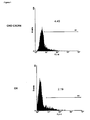
- the human CXCR4 (hCXCR4) GPCR was co-expressed with Nanobodies (Nbs) directed against hCXCR4 in the yeast P. pastoris. It was surprisingly found that hCXCR4 - Nb co-expression results in an increase in expression of the membrane protein, as compared to hCXCR4 expression alone.
- a first aspect of the invention relates to a host cell comprising a first exogenous nucleic acid sequence encoding a membrane protein and a second exogenous nucleic acid encoding a binding domain directed against said membrane protein, each under the control of a promoter.
- said promoter is an inducible promoter.
- said membrane protein and binding domain are co-expressed.
- said membrane protein and/or said binding domain are operably linked to a subcellular targeting sequence, such as an ER or Golgi localization signal or secretion signal.
- the membrane protein in any of the above described host cells is a membrane receptor protein, such as a GPCR, or a membrane transport protein, such as an ion transporter.
- a further embodiment of the present invention relates to any of the above described host cells wherein the binding domain specifically binds to an extracellular conformational epitope of the membrane protein.
- the binding domain in any of the above described host cells is an immunoglobulin single variable domain comprising an amino acid sequence comprising 4 framework regions and 3 complementary determining regions, or any suitable fragment thereof. More preferably, said immunoglobulin single variable domain is a VHH.
- said immunoglobulin single variable domain stabilizes the membrane protein in a functional conformational state, such as an active or an inactive state.
- the host cell according to the present invention may be a eukaryotic host cell, such as a yeast cell, a mammalian cell, an insect cell.
- said yeast may be a Pichia strain, such as a Pichia pastoris, or a Komagataella strain, such as Komagataella pastoris, or a Hansenula strain, such as Hansenula polymorpha, or a Yarrowia strain, such as Yarrowia lipolytica, or a Saccharomyces strain, such as Saccharomyces cerevisiae, and wherein said filamentous fungi is an Aspergillus strain, such as Aspergillus niger or Aspergillus nidulans, or a Penicillium strain, such as Penicillium citrinum or Penicillium chrysogenum, or a Hypocrea strain, such as Hypocrea jecorina.
- the host cell may be a glycoengineered host cell.
- the invention also envisages cell cultures of any of the above described host cells or membrane preparations derived of said host cells or cell cultures.
- Expression vectors comprising the first and/or the second exogenous nucleic acid sequence as comprised in the host cells according to the invention are also encompassed.
- the invention relates to a method of enhancing the production of a membrane protein in a host cell comprising the steps of:
- the method may further comprise the step of isolating the membrane protein and/or the binding domain.
- the invention also relates to the use of the host cells, or the cell cultures, or the membrane preparations, or the proteins isolated therefrom, all as described herein before, for ligand characterization, drug screening, protein capturing and purification, immunization, biophysical studies, amongst others. Other applications will become clear from the Description further herein.
- membrane protein refers to a protein that is attached to or associated with a membrane of a cell or an organelle. Specific non-limiting examples are provided further in the specification.
- protein binding domain or simply “binding domain” refers generally to any non-naturally occurring molecule or part thereof that is able to bind to a protein or peptide using specific intermolecular interactions.
- a variety of molecules can function as protein binding domains, including, but not limited to, proteinaceous molecules (protein, peptide, protein-like or protein containing), nucleic acid molecules (nucleic acid, nucleic acid-like, nucleic acid containing), and carbohydrate molecules (carbohydrate, carbohydrate-like, carbohydrate containing). A more detailed description can be found further in the specification.
- polypeptide As used herein, the terms “polypeptide”, “protein”, “peptide” are used interchangeably herein, and refer to a polymeric form of amino acids of any length, which can include coded and non-coded amino acids, chemically or biochemically modified or derivatized amino acids, and polypeptides having modified peptide backbones.
- nucleic acid molecule As used herein, the terms “nucleic acid molecule”, “polynucleotide”, “polynucleic acid”, “nucleic acid” are used interchangeably and refer to a polymeric form of nucleotides of any length, either deoxyribonucleotides or ribonucleotides, or analogs thereof. Polynucleotides may have any three-dimensional structure, and may perform any function, known or unknown.
- Non-limiting examples of polynucleotides include a gene, a gene fragment, exons, introns, messenger RNA (mRNA), transfer RNA, ribosomal RNA, ribozymes, cDNA, recombinant polynucleotides, branched polynucleotides, plasmids, vectors, isolated DNA of any sequence, control regions, isolated RNA of any sequence, nucleic acid probes, and primers.
- the nucleic acid molecule may be linear or circular.
- conformation or conformational state of a protein refers generally to the range of structures that a protein may adopt at any instant in time.
- determinants of conformation or conformational state include a protein's primary structure as reflected in a protein's amino acid sequence (including modified amino acids) and the environment surrounding the protein.
- the conformation or conformational state of a protein also relates to structural features such as protein secondary structures (e.g., ⁇ -helix, ⁇ -sheet, among others), tertiary structure (e.g., the three dimensional folding of a polypeptide chain), and quaternary structure (e.g., interactions of a polypeptide chain with other protein subunits).
- Post-translational and other modifications to a polypeptide chain such as ligand binding, phosphorylation, sulfation, glycosylation, or attachments of hydrophobic groups, among others, can influence the conformation of a protein.
- environmental factors such as pH, salt concentration, ionic strength, and osmolality of the surrounding solution, and interaction with other proteins and co-factors, among others, can affect protein conformation.
- the conformational state of a protein may be determined by either functional assay for activity or binding to another molecule or by means of physical methods such as X-ray crystallography, NMR, or spin labeling, among other methods.
- a “specific conformational state” is any subset of the range of conformations or conformational states that a protein may adopt.
- a "functional conformation” or a “functional conformational state”, as used herein, refers to the fact that proteins possess different conformational states having a dynamic range of activity, in particular ranging from no activity to maximal activity.
- a functional conformational state is meant to cover any conformational state of a protein, in particular a membrane protein, having any activity, including no activity; and is not meant to cover the denatured states of proteins.
- a particular class of functional conformations is defined as "drugable conformation” and generally refers to a unique therapeutically relevant conformational state of a target protein.
- drugable conformation As an illustration, the active conformation of the ⁇ 2 adrenergic receptor corresponds to the drugable conformation of this receptor for the treatment of astma. It will thus be understood that drugability is confined to particular conformations depending on the therapeutic indication.
- CDR complementarity determining region
- H and VL light chains
- CDR regions account for the basic specificity of the antibody for a particular antigenic determinant structure. Such regions are also referred to as “hypervariable regions.”
- the CDRs represent non-contiguous stretches of amino acids within the variable regions but, regardless of species, the positional locations of these critical amino acid sequences within the variable heavy and light chain regions have been found to have similar locations within the amino acid sequences of the variable chains.
- variable heavy and light chains of all canonical antibodies each have 3 CDR regions, each non- contiguous with the others (termed L1, L2, L3, H1, H2, H3) for the respective light (L) and heavy (H) chains.
- Nanobodies in particular, generally comprise a single amino acid chain that can be considered to comprise 4 "framework sequences or regions" or FRs and 3 "complementary determining regions" or CDRs.
- the nanobodies have 3 CDR regions, each non-contiguous with the others (termed CDR1, CDR2, CDR3).
- the delineation of the FR and CDR sequences is based on the IMGT unique numbering system for V-domains and V-like domains ( Lefranc et al. 2003, Developmental and Comparative Immunology 27:55 ).
- an “epitope”, as used herein, refers to an antigenic determinant of a polypeptide.
- An epitope could comprise 3 amino acids in a spatial conformation, which is unique to the epitope. Generally an epitope consists of at least 4, 5, 6, 7 such amino acids, and more usually, consists of at least 8, 9, 10 such amino acids. Methods of determining the spatial conformation of amino acids are known in the art, and include, for example, x-ray crystallography and 2-dimensional nuclear magnetic resonance.
- a “conformational epitope”, as used herein, refers to an epitope comprising amino acids in a spacial conformation that is unique to a folded 3-dimensional conformation of a polypeptide.
- a conformational epitope consists of amino acids that are discontinuous in the linear sequence that come together in the folded structure of the protein.
- a conformational epitope may also consist of a linear sequence of amino acids that adopts a conformation that is unique to a folded 3-dimensional conformation of the polypeptide (and not present in a denatured state).
- conformational epitopes consist of amino acids that are discontinuous in the linear sequences of one or more polypeptides that come together upon folding of the different folded polypeptides and their association in a unique quaternary structure.
- binding domain refers to the ability of a binding domain, in particular an immunoglobulin or an immunoglobulin fragment, such as a VHH or nanobody, to bind preferentially to one antigen, versus a different antigen, and does not necessarily imply high affinity.
- affinity refers to the degree to which a binding domain, in particular an immunoglobulin, such as an antibody, or an immunoglobulin fragment, such as a VHH or nanobody, binds to an antigen so as to shift the equilibrium of antigen and protein binding domain toward the presence of a complex formed by their binding.
- an antibody (fragment) of high affinity will bind to the available antigen so as to shift the equilibrium toward high concentration of the resulting complex.
- the dissociation constant is commonly used to describe the affinity between the protein binding domain and the antigenic target.
- the dissociation constant is lower than 10 -5 M.
- the dissociation constant is lower than 10 -6 M, more preferably, lower than 10 -7 M.
- the dissociation constant is lower than 10 -8 M.
- telomere binding domain in particular an immunoglobulin, such as an antibody, or an immunoglobulin fragment, such as a VHH or nanobody, to preferentially bind to a particular antigen that is present in a homogeneous mixture of different antigens.
- a specific binding interaction will discriminate between desirable and undesirable antigens in a sample, in some embodiments more than about 10 to 100-fold or more (e.g., more than about 1000- or 10,000-fold).
- the terms particularly refer to the ability of a binding domain (as defined herein) to preferentially recognize and/or bind to a particular conformational state of a GPCR as compared to another conformational state.
- an active state-selective protein binding domain will preferentially bind to a GPCR in an active conformational state and will not or to a lesser degree bind to a GPCR in an inactive conformational state, and will thus have a higher affinity for said active conformational state.
- the terms “specifically bind”, “selectively bind”, “preferentially bind”, and grammatical equivalents thereof, are used interchangeably herein.
- the terms “conformational specific” or “conformational selective” are also used interchangeably herein.
- a “deletion” is defined here as a change in either amino acid or nucleotide sequence in which one or more amino acid or nucleotide residues, respectively, are absent as compared to an amino acid sequence or nucleotide sequence of a parental polypeptide or nucleic acid.
- a deletion can involve deletion of about 2, about 5, about 10, up to about 20, up to about 30 or up to about 50 or more amino acids.
- a protein or a fragment thereof may contain more than one deletion.
- a deletion may also be a loop deletion, or an N- and/or C-terminal deletion.
- an “insertion” or “addition” is that change in an amino acid or nucleotide sequences which has resulted in the addition of one or more amino acid or nucleotide residues, respectively, as compared to an amino acid sequence or nucleotide sequence of a parental protein.
- “Insertion” generally refers to addition to one or more amino acid residues within an amino acid sequence of a polypeptide, while “addition” can be an insertion or refer to amino acid residues added at an N- or C-terminus, or both termini.
- an insertion or addition is usually of about 1, about 3, about 5, about 10, up to about 20, up to about 30 or up to about 50 or more amino acids.
- a protein or fragment thereof may contain more than one insertion.
- substitution results from the replacement of one or more amino acids or nucleotides by different amino acids or nucleotides, respectively as compared to an amino acid sequence or nucleotide sequence of a parental protein or a fragment thereof. It is understood that a protein or a fragment thereof may have conservative amino acid substitutions which have substantially no effect on the protein's activity. By conservative substitutions is intended combinations such as gly, ala; val, ile, leu, met; asp, glu; asn, gln; ser, thr; lys, arg; cys, met; and phe, tyr, trp.
- test compound or “test compound” or “candidate compound” or “drug candidate compound” as used herein describes any molecule, either naturally occurring or synthetic that is tested in an assay, such as a screening assay or drug discovery assay.
- these compounds comprise organic or inorganic compounds.
- the compounds include polynucleotides, lipids or hormone analogs that are characterized by low molecular weights.
- Other biopolymeric organic test compounds include small peptides or peptide-like molecules (peptidomimetics) comprising from about 2 to about 40 amino acids and larger polypeptides comprising from about 40 to about 500 amino acids, such as antibodies, antibody fragments or antibody conjugates.
- Test compounds can also be protein scaffolds.
- test compound libraries may be used, such as combinatorial or randomized libraries that provide a sufficient range of diversity. Examples include, but are not limited to, natural compound libraries, allosteric compound libraries, peptide libraries, antibody fragment libraries, synthetic compound libraries, fragment-based libraries, phage-display libraries, and the like. A more detailed description can be found further in the specification.
- ligand means a molecule that specifically binds to a membrane protein, either intracellularly or extracellularly.
- a ligand may be, without the purpose of being limitative, a protein, a (poly)peptide, a lipid, a small molecule, a protein scaffold, , a nucleic acid, an ion, a carbohydrate, an antibody or an antibody fragment, such as a nanobody (all as defined herein).
- a ligand may be a synthetic or naturally occurring.
- a ligand also includes a "native ligand” which is a ligand that is an endogenous, natural ligand for a native protein.
- determining As used herein, the terms “determining”, “measuring”, “assessing”, “monitoring” and “assaying” are used interchangeably and include both quantitative and qualitative determinations.
- exogenous refers to substances (e.g. genes) originating from within an organism, tissue, or cell.
- exogenous as used herein is any material that comes from outside an organism, tissue, or cell, but that is present (and typically can become active) in that organism, tissue, or cell.
- inducible promoter refers to a promoter that can be switched 'on' or 'off' (thereby regulating gene transcription) in response to external stimuli such as, but not limited to, temperature, pH, certain nutrients, specific cellular signals, etcetera. It is used to distinguish between a "constitutive promoter", by which a promoter is meant that is continuously switched 'on', i.e. from which gene transcription is constitutively active.
- subcellular targeting sequence generally refers to a molecule that directs localization of proteins to different cell compartments. Examples include an "ER localization signal” or a "Golgi localization signal” which is a molecule, typically a peptide, that directs localization of the polypeptide or protein to which it is conjugated to the ER or Golgi apparatus, respectively. Localization thus also implies retention in the ER or Golgi apparatus, respectively. Typically, these localization (or retention) sequences are peptide sequences derived from (pre)proteins that are situated in the ER or Golgi when functionally active as a mature protein. Subcellular targeting sequences also include secretion signals of which examples are provided further herein.
- vector as used herein is intended to refer to a nucleic acid molecule capable of transporting another nucleic acid molecule to which it has been linked.
- plasmid vector refers to a circular double stranded DNA loop into which additional DNA segments may be ligated.
- Other vectors include, without the purpose of being limitative, cosmids and yeast artificial chromosomes (YAC).
- YAC yeast artificial chromosomes
- Certain vectors are capable of autonomous replication in a host cell into which they are introduced (e.g., vectors having an origin of replication which functions in the host cell).
- Other vectors can be integrated into the genome of a host cell upon introduction into the host cell, and are thereby replicated along with the host genome.
- certain preferred vectors are capable of directing the expression of certain genes of interest.
- Such vectors are referred to herein as “recombinant expression vectors” (or simply, “expression vectors”).
- Suitable vectors have regulatory sequences, such as promoters, enhancers, terminator sequences, and the like as desired.
- control sequences refers to polynucleotide sequences which are necessary to affect the expression of coding sequences to which they are operably linked.
- Expression control sequences are sequences which control the transcription, post-transcriptional events and translation of nucleic acid sequences. Expression control sequences include appropriate transcription initiation, termination, promoter and enhancer sequences; efficient RNA processing signals such as splicing and polyadenylation signals; sequences that stabilize cytoplasmic mRMA; sequences that enhance translation efficiency (e.g., ribosome binding sites); sequences that enhance protein stability; and when desired, sequences that enhance protein secretion. The nature of such control sequences differs depending upon the host organism.
- control sequences is intended to include, at a minimum, all components whose presence is essential for expression, and can also include additional components whose presence is advantageous, for example, leader sequences and fusion partner sequences.
- operably linked refers to a linkage in which the regulatory sequence is contiguous with the gene of interest to control the gene of interest, as well as regulatory sequences that act in trans or at a distance to control the gene of interest.
- host cell and equivalent terms like "recombinant host cell”, “expression host cell”, “expression host system”, “expression system”, as used herein, is intended to refer to a cell into which a recombinant vector has been introduced. It should be understood that such terms are intended to refer not only to the particular subject cell but to the progeny of such a cell. Because certain modifications may occur in succeeding generations due to either mutation or environmental influences, such progeny may not, in fact, be identical to the parent cell, but are still included within the scope of the term “host cell” as used herein.
- a recombinant host cell may be an isolated cell or cell line grown in culture or may be a cell which resides in a living tissue or organism. Particular examples are provided further herein.
- the present invention provides engineered cells or cellular systems (cell cultures, organisms) that can express proteins at higher levels in a particular conformation, particularly membrane proteins, either at the cellular surface or in other cell compartments. Further, methods are provided that use such cells or cellular systems to produce these proteins. It will be appreciated that while the invention has been exemplified with GPCRs, it is equally applicable to any membrane protein, especially a membrane protein that is poorly expressed and has a low stability in a recombinant host cell.
- a first aspect of the invention relates to a host cell comprising a first exogenous nucleic acid sequence encoding a membrane protein and a second exogenous nucleic acid encoding a binding domain directed against said membrane protein, each under the control of a promoter.
- the "host cell” can be of any prokaryotic or eukaryotic organism.
- the host cell is a eukaryotic cell and can be of any eukaryotic organism, but in particular embodiments yeast, plant, mammalian and insect cells are envisaged.
- yeast, plant, mammalian and insect cells are envisaged.
- the nature of the cells used will typically depend on the ease and cost of producing the native protein(s), the desired glycosylation properties, the origin of the target protein, the intended application, or any combination thereof.
- Mammalian cells may for instance be used for achieving complex glycosylation, but it may not be cost-effective to produce proteins in mammalian cell systems.
- yeast Plant and insect cells, as well as yeast typically achieve high production levels and are more cost-effective, but additional modifications may be needed to mimic the complex glycosylation patterns of mammalian proteins.
- Yeast cells are currently preferred for expression of proteins because they can be economically cultured, give high yields of protein, and when appropriately modified are capable of producing proteins having suitable glycosylation patterns. Further, yeast offers established genetics allowing for rapid transformations, tested protein localization strategies, and facile gene knock-out techniques.
- Eukaryotic cell or cell lines for protein production are well known in the art, including cell lines with modified glycosylation pathways, and non-limiting examples will be provided hereafter.
- Animal or mammalian host cells suitable for harboring, expressing, and producing proteins for subsequent isolation and/or purification include Chinese hamster ovary cells (CHO), such as CHO-K1 (ATCC CCL-61), DG44 ( Chasin et al., 1986, Som. Cell Molec.
- CHO-K1 Tet-On cell line (Clontech), CHO designated ECACC 85050302 (CAMR, Salisbury, Wiltshire, UK), CHO clone 13 (GEIMG, Genova, IT), CHO clone B (GEIMG, Genova, IT), CHO-K1/SF designated ECACC 93061607 (CAMR, Salisbury, Wiltshire, UK), RR-CHOK1 designated ECACC 92052129 (CAMR, Salisbury, Wiltshire, UK), dihydrofolate reductase negative CHO cells (CHO/-DHFR, Urlaub and Chasin, 1980, Proc.
- human cervical carcinoma cells HELA, ATCC CCL-2
- canine kidney cells MDCK, ATCC CCL-34
- human lung cells W138, ATCC CCL-75
- human hepatoma cells HEP-G2, HB 8065
- mouse mammary tumor cells MMT 060562, ATCC CCL-51
- buffalo rat liver cells BRL 3A, ATCC CRL-1442
- TRI cells Mather, 1982, Annals NYAcad. Sci., 383:44-68
- MCR 5 cells FS4 cells.
- the cells are mammalian cells selected from Hek293 cells or COS cells.
- Exemplary non-mammalian cell lines include, but are not limited to, Sf9 cells, baculovirus-insect cell systems (e.g. review Jarvis, Virology Volume 310, Issue 1, 25 May 2003, Pages 1-7 ), plant cells such as tobacco cells, tomato cells, maize cells, algae cells, or yeasts such as Saccharomyces species, Schizosaccharomyces species, Hansenula species, Yarrowia species or Pichia species.
- the eukaryotic cells are yeast cells from a Saccharomyces species (e.g. Saccharomyces cerevisiae ), Schizosaccharomyces sp.
- the eukaryotic cells are Pichia cells, and in a most particular embodiment Pichia pastoris cells.
- membrane protein is understood a protein that is attached to or associated with a membrane of a cell or an organelle. They are often subdivided into several categories including integral membrane proteins, peripheral membrane proteins and lipid-anchored proteins. Preferably, the membrane protein is an integral membrane protein that is permanently bound to the lipid bilayer and which requires a detergent or another apolar solvent to be removed. Integral membrane proteins include transmembrane proteins that are permanently attached to the lipid membrane and span across the membrane one or several times.
- membrane proteins include receptors such as GPCRs and growth factor receptors; transmembrane ion channels such as ligand-gated and voltage gated ion channels; transmembrane transporters such as neurotransmitter transporters; enzymes; carrier proteins; ion pumps; amongst others.
- membrane proteins as referred to herein can be of any species, such as fungus (including yeast), nematode, virus, insect, plant, bird (e.g. chicken, turkey), reptile, or mammal (e.g., a mouse, rat, rabbit, hamster, gerbil, dog, cat, goat, pig, cow, horse, whale, monkey, or human).
- fungus including yeast
- nematode virus
- insect e.g. chicken, turkey
- bird e.g. chicken, turkey
- reptile or mammal
- mammal e.g., a mouse, rat, rabbit, hamster, gerbil, dog, cat, goat, pig, cow, horse, whale, monkey, or human.
- the membrane protein is a G-protein coupled receptor or GPCR.
- GPCRs can be grouped on the basis of sequence homology into several distinct families. Although all GPCRs have a similar architecture of seven membrane-spanning ⁇ - helices, the different families within this receptor class show no sequence homology to one another, thus suggesting that the similarity of their transmembrane domain structure might define common functional requirements.
- GPCR structure and classification is generally well known in the art and further discussions of GPCRs may be found in Probst et al. 1992, DNA Cell Biol. 1:1 ; Marchese et al. 1994, Genomics 23:609 ; Rosenbaum et al.
- GPCRs include, without limitation, serotonin and olfactory receptors, glycoprotein hormone receptors, chemokine receptors, adenosine receptors, biogenic amine receptors, melanocortin receptors, neuropeptide receptors, chemotactic receptors, somatostatin receptors, opioid receptors, melatonin receptors, calcitonin receptors, PTH/PTHrP receptors, glucagon receptors, secretin receptors, latrotoxin receptors, metabotropic glutamate receptors, calcium receptors, GABA-B receptors, pheromone receptors, histamine receptors, protease-activated receptors, rhodopsins and other G-protein coupled seven transmembrane segment receptors.
- GPCRs also include these GPCR receptors associated with each other as homomeric or heteromeric dimers or as higher-order oligomers.
- the amino acid sequences (and the nucleotide sequences of the cDNAs which encode them) of GPCRs are readily available, for example by reference to GenBank ( http://www.ncbi.nlm.nih.gov/entrez ).
- GPCRs envisaged for increased production using the tools and methods provided herein include, but are not limited to, CXCR4, GPR3, rhodopsin, vasopressin receptor, ⁇ 1 adrenergic receptor, ⁇ 2 adrenergic receptor, ⁇ 3 adrenergic receptor, ⁇ 1 adrenergic receptors and the ⁇ 2 adrenergic receptor, M1 muscarinic receptor, M2 muscarinic receptor, M3 muscarinic receptor, M4 muscarinic receptor, M5 muscarinic receptor, angiotensin II receptors.
- a membrane protein as used herein, may be any naturally occurring or non-naturally occurring (i.e., altered by man) membrane protein.
- naturally-occurring means a membrane protein that is naturally produced (for example and without limitation, by a mammal, more specifically by a human, or by a virus, or by a plant, or by an insect, amongst others). Such membrane proteins are found in nature.
- non-naturally occurring means a membrane protein that is not naturally produced. Wild-type membrane proteins that have been made constitutively active through mutation, and variants of naturally-occurring membrane proteins are examples of non-naturally occurring membrane proteins. Non-naturally occurring membrane proteins may have an amino acid sequence that is at least 80% identical to, at least 90% identical to, at least 95% identical to or at least 99% identical to, a naturally-occurring membrane protein.
- the CXCR4 receptor as a particular non-limiting example of a GPCR within the scope of the present invention, it should be clear from the above that in addition to the human CXCR4 receptor (e.g., the sequence described by Genbank accession number EAX11616, Gene ID 7852), the mouse CXCR4 receptor (e.g., as described by Genbank accession number NP_034041, Gene ID 12767) or other mammalian CXCR4 receptor may also be employed. In addition, the term is intended to encompass wild-type polymorphic variants and certain other active variants from a particular species.
- human CXCR4 receptor e.g., the sequence described by Genbank accession number EAX11616, Gene ID 7852
- the mouse CXCR4 receptor e.g., as described by Genbank accession number NP_034041, Gene ID 12767
- the term is intended to encompass wild-type polymorphic variants and certain other active variants from a particular species.
- a "human CXCR4 receptor” has an amino acid sequence that is at least 95% identical to (e.g., at least 95% or at least 98% identical to) the naturally occurring "human CXCR4" of Genbank accession number EAX11616, Gene ID 7852.
- the present invention also envisages membrane proteins, in particular GPCRs, with a loop deletion, or an N- and/or C-terminal deletion, or a substitution, or an insertion or addition in relation to its amino acid or nucleotide sequence, or any combination thereof (as defined hereinbefore).
- the tools and methods for membrane protein expression provided herein can be further combined with known improvements for membrane protein expression that typically involve production of variants.
- examples thereof include, but are not limited to, the use of a signal sequence specific to the species of eukaryotic cell used rather than the membrane protein-specific signal sequence, the use of a truncated membrane protein (e.g. C-truncated) versus the use of an intact protein, the use of a membrane protein with a sequence insertion (e.g. a T4 lysozyme coding sequence in the 3 rd intracellular loop of a GPCR), and so on.
- a signal sequence specific to the species of eukaryotic cell used rather than the membrane protein-specific signal sequence
- a truncated membrane protein e.g. C-truncated
- a membrane protein with a sequence insertion e.g. a T4 lysozyme coding sequence in the 3 rd intracellular loop of a GPCR
- more than one, i.e. two or more different proteins may be produced simultaneously.
- at least one of the proteins is a membrane protein.
- the proteins may all be membrane-bound, all be secreted proteins or a mixture thereof.
- care will be taken that they can be recovered easily either separately or together.
- even higher production is achieved by expressing multiple copies of the protein to be expressed, e.g. as a polyprotein.
- the produced (membrane) proteins are functional, for instance the produced receptors remain capable of ligand binding and/or signal transduction.
- binding domain is meant any non-naturally occurring molecule or part thereof (as defined hereinbefore) that is directed against a target membrane protein.
- the binding domains as described herein are protein scaffolds. Protein scaffolds refer generally to folding units that form structures, particularly protein or peptide structures, that comprise frameworks for the binding of another molecule, for instance a protein (See, e.g., review of Skerra, J. 2000, Molecular Recognition, 13:167 ). Accordingly, a binding domain can be derived from a naturally occurring molecule, e.g.
- a binding domain can be immunoglobulin-based or it can be based on domains present in proteins, including but limited to microbial proteins, protease inhibitors, toxins, fibronectin, lipocalins, single chain antiparallel coiled coil proteins or repeat motif proteins.
- binding domains which are known in the art include, but are not limited to: antibodies, heavy chain antibodies (hcAb), single domain antibodies (sdAb), minibodies, the variable domain derived from camelid heavy chain antibodies (VHH or nanobodies), the variable domain of the new antigen receptors derived from shark antibodies (VNAR), alphabodies, protein A, protein G, designed ankyrin-repeat domains (DARPins), fibronectin type III repeats, anticalins, knottins, engineered CH2 domains (nanoantibodies), peptides and proteins, lipopeptides (e.g.
- the binding domain according to the invention may generally be directed against any desired membrane protein as described herein before, and may in particular be directed against any conformational epitope of any membrane protein, preferably a functional conformational state of any membrane protein (active, inactive, etc.). More particularly, said conformational epitope can be part of an intracellular or extracellular region, or an intramembraneous region, or a domain or loop structure of any desired membrane protein. According to particular embodiments, the binding domains may be directed against any suitable extracellular region, domain, loop or other extracellular conformational epitope of a membrane protein, but is preferably directed against one of the extracellular parts of the transmembrane domains or more preferably against one of the extracellular loops that link the transmembrane domains.
- the protein binding domains may be directed against any suitable intracellular region, domain, loop or other intracellular conformational epitope of a membrane protein, but is preferably directed against one of the intracellular parts of the transmembrane domains or more preferably against one of the intracellular loops that link the transmembrane domains.
- a binding domain that specifically binds to a "three dimensional" epitope or “conformational” epitope specifically binds to a tertiary (i.e., three dimensional) structure of a folded protein, and binds at much reduced (i.e., by a factor of at least 2, 5, 10, 50 or 100) affinity to the linear (i.e., unfolded, denatured) form of the protein.
- the expression of the proteins in the host cell as described herein is preferably regulated by appropriate promoters.
- the choice of a promoter will typically depend on the nature of the host cell. The choice further depends on the desired temporal expression of a particular protein as described herein.
- the proteins may be expressed constitutively or in an inducible way. Accordingly, the promoter may be a constitutive or inducible promoter.
- the conditions for inducing a promoter may be chosen from the following group of inducing conditions: metabolic, or stress, or pH, or temperature, or drug inducing conditions, or other. Promoters may be derived directly from naturally occurring genes, or may be synthesized to combine regulatory sequences from different promoter regions.
- the promoter is an exogenous promoter that will typically be strong enough to ensure overexpression of the protein(s).
- the promoter is an inducible promoter.
- inducible promoters useful for the practice of the present invention include, without limitation, the promoter of a gene selected from the group comprising alcohol oxidase I (AOXI) ( Tschopp et al. 1987, Nucleic Acids Res 15:3859 ), alcohol oxidase II (AOXII) ( Ohi et al. 1994, Mol Gen Genet 243:489 ), formaldehyde dehydrogenase (FLD) ( US6730499 ; Shen et al. 1998, Gene 216:93 ), galactokinase (GAL1) ( Flick and Johnston 1990, Mol Cell Biol.
- AOXI alcohol oxidase I
- AOXII alcohol oxidase II
- FLD formaldehyde dehydrogenase
- GAL1 galactokinase
- MOX methanol oxidase
- FMD formate dehydrogenase
- AOD1 mitochondrial alternative oxidase
- POX1 peroxisomal acyl coenzyme A oxidase
- constitutive promoters may be used and include, for example, the glyceraldehyde-3-phosphate dehydrogenase promoter (GAP) ( Zhang et al. 2009, Mol Biol Rep 36: 1611 ).
- GAP glyceraldehyde-3-phosphate dehydrogenase promoter
- the invention relates to a host cell engineered as described herein before, wherein the membrane protein and the binding domain are co-expressed.
- co-expressed is meant temporally expressed at the same time or simultaneously expressed, which can be regulated by choosing the appropriate promoter(s) (as described hereinbefore).
- the membrane protein and the binding domain are co-localised in a particular cellular compartment, such as in the ER, in the Golgi apparatus, at the cellular surface (meaning the membrane protein attached to the cell membrane and the binding domain co-localized at the extracellular or intracellular side of the cell membrane).
- co-localised is meant spatially expressed at the same cellular location. This can be done by operably linking the membrane protein and/or the binding domain to an appropriate subcellular targeting sequence (as defined herein), such as an ER or Golgi localization signal, or a secretion signal.
- the membrane protein and binding domain are preferably co-expressed and translocated to the cellular surface or to the extracellular space. This will typically be achieved by making use of secretion signals, so that the co-expressed proteins are transported through the secretory pathway: from the ER to the cis-, medial- and trans-Golgi compartments, finally resulting in secretion into the extracellular medium, or otherwise integration in the cellular membrane, which is typically the case for membrane proteins.
- both the membrane protein and the binding domain are operably linked to a secretion signal. Even more preferably, both the membrane protein and the binding domain are operably linked to the same secretion signal.
- secretion signal will typically not depend on the protein to be secreted, but on the type of eukaryotic cells used. As long as the secretion signal is functional in the cell type in which it is used (i.e. it results in secretion to the extracellular environment (or to the cellular membrane) of the protein or peptide to which it is fused), this feature is not critical to the invention. Thus, secretion signals from other organisms may be used, as long as these signals lead to secretion in the eukaryotic cells used. Secretion signals are well known in the art and may be derived from - typically the N-terminus of - proteins that are secreted, or may be made synthetically (e.g.
- Tan et al., Protein engineering 2002, 15:337 can be derived from genomic sequences using computational methods ( Klee et al., BMC Bioinformatics 2005, 6:256 ). Also, viral or bacterial or even hybrid secretion signals can be used. Further examples of signal peptides that can be used are described in WO2002/048187 (eukaryotic cells), Schaaf et al. (BMC Biotechnol. 2005; 5: 30 ) (moss cells), EP549062 . Specific secretion signals used in yeast include e.g. ⁇ -factor secretory peptide, the PH05 secretory peptide, and the BAR1 secretion signal.
- the membrane protein and binding domain may also be co-expressed and retained in the ER or Golgi compartment.
- ER and Golgi localization signals are well known in the art and may be derived from proteins that are normally localized in the ER or Golgi for their function. Again, localization sequences from one organism may function in other organisms. For example the membrane spanning region of ⁇ -2, 6-sialyltransferase from rats, an enzyme known to localize in the rat trans Golgi, was shown to also localize a reporter gene (invertase) in the yeast Golgi ( Schwientek, et al., 1995, J. Biol. Chem. 270:5483 ).
- Schwientek and co-workers have also shown that fusing 28 amino acids of a yeast mannosyltransferase (Mntl), a region containing an N-terminal cytoplasmic tail, a transmembrane region and eight amino acids of the stem region, to the catalytic domain of human GAlT are sufficient for Golgi localization of an active GalT ( Schwientek et al. 1995 J. Biol. Chem. 270 (10): 5483-5489 ).
- Mntl yeast mannosyltransferase
- leader sequences from Mnsl for ER localization and leader sequences from Och1 and Mnt1 (Golgi-cis localization), from Mnn2 (Golgi medial localization), from Mnn1 (Golgi trans localization), from alpha-2,6-sialyltransferase (trans-Golgi network) and from beta-1,4-galactosyltransferase I (Golgi localization).
- the membrane protein and binding domain may also be co-expressed and deposited in inclusion bodies in the cell, or in membrane-bound organelles or in structures with similar functions.
- the co-expressed proteins may be produced in or transported to specific organs or tissues of the organism from which it can be recovered (e.g. glands or trichomes). It should be noted that, particularly in cases where the protein is not secreted, it is possible that the protein is deposited in an inactive form. Thus, additional refolding or re-activating steps may be needed in order to obtain a physiologically relevant form of the protein.
- the binding domain which is co-expressed with the membrane protein, as described hereinbefore is derived from an innate or adaptive immune system.
- said binding domain is derived from an immunoglobulin.
- the protein binding domain according to the invention is an antibody or a derivative thereof.
- antibody refers generally to a polypeptide encoded by an immunoglobulin gene, or functional fragments thereof, that specifically binds and recognizes an antigen, and is known to the person skilled in the art.
- a conventional immunoglobulin (antibody) structural unit comprises a tetramer.
- Each tetramer is composed of two identical pairs of polypeptide chains, each pair having one "light” (about 25 kDa) and one "heavy” chain (about 50-70 kDa).
- the N-terminus of each chain defines a variable region of about 100 to 110 or more amino acids primarily responsible for antigen recognition.
- VL variable light chain
- VH variable heavy chain
- antibody is meant to include whole antibodies, including single-chain whole antibodies, and antigen-binding fragments.
- antigen-binding fragments may be antigen-binding antibody fragments that include, but are not limited to, Fab, Fab' and F(ab')2, Fd, single-chain Fvs (scFv), single-chain antibodies, disulfide-linked Fvs (dsFv) and fragments comprising or consisting of either a VL or VH domain, and any combination of those or any other functional portion of an immunoglobulin peptide capable of binding to the target antigen.
- the term "antibodies” is also meant to include heavy chain antibodies, or functional fragments thereof, such as single domain antibodies, more specifically, VHHs or nanobodies, as defined further herein.
- said binding domain is an immunoglobulin single variable domain. More preferably, said binding domain comprises an amino acid sequence comprising 4 framework regions and 3 complementary determining regions, preferably in a sequence FR1-CDR1-FR2-CDR2-FR3-CDR3-FR4, or any suitable fragment thereof (which will then usually contain at least some of the amino acid residues that form at least one of the complementary determining regions). Binding domains comprising 4 FRs and 3 CDRs are known to the person skilled in the art and have been described, as a non-limiting example, in Wesolowski et al. (2009, Med. Microbiol. Immunol. 198:157 ).
- the binding domain according to the invention is derived from a camelid antibody. More preferably, the binding domain according to the invention comprises an amino acid sequence of a nanobody, or any suitable fragment thereof. More specifically, the protein binding domain is a nanobody or any suitable fragment thereof.
- Nbs can be designed as bispecific and bivalent antibodies or attached to reporter molecules ( Conrath et al. 2001, Antimicrob Agents Chemother 45: 2807 ). Nbs are stable and rigid single domain proteins that can easily be manufactured and survive the gastro-intestinal system. Therefore, Nbs can be used in many applications including drug discovery and therapy ( Saerens et al. 2008, Curr Opin Pharmacol. 8:600 ) but also as a versatile and valuable tool for purification, functional study and crystallization of proteins ( Conrath et al. 2009, Protein Sci. 18:619 ).
- Xaperones a particular class of nanobodies that act as crystallization chaperones binding conformational epitopes of native targets are called Xaperones and are also envisaged here.
- Xaperones are unique tools in structural biology.
- XaperoneTM is a trademark of VIB and VUB (Belgium).
- Xaperones (1) bind cryptic epitopes and lock proteins in unique native conformations, (2) increase the stability of soluble proteins and solubilized membrane proteins, (3) reduce the conformational complexity of soluble proteins and solubilized membrane proteins, (4) increase the polar surface enabling the growth of diffracting crystals, (5) sequester aggregative or polymerizing surfaces, (6) allow to affinity-trap active protein.
- the nanobodies according to the invention generally comprise a single amino acid chain that can be considered to comprise 4 "framework sequences" or FR's and 3 "complementary determining regions” or CDR's (as defined hereinbefore).
- framework sequences or FR's
- CDR's complementary determining regions
- Non-limiting examples of nanobodies of the invention are described in more detail further herein. It should be clear that framework regions of nanobodies may also contribute to the binding of their antigens ( Desmyter et al. 2002, J Biol Chem. 277:23645 ; Korotkov et al. 2009, Structure 17:255 ).
- the term nanobody as used herein in its broadest sense is not limited to a specific biological source or to a specific method of preparation.
- the nanobodies of the invention can generally be obtained: (1) by isolating the VHH domain of a naturally occurring heavy chain antibody; (2) by expression of a nucleotide sequence encoding a naturally occurring VHH domain; (3) by "humanization” of a naturally occurring VHH domain or by expression of a nucleic acid encoding a such humanized VHH domain; (4) by "camelization” of a naturally occurring VH domain from any animal species, and in particular from a mammalian species, such as from a human being, or by expression of a nucleic acid encoding such a camelized VH domain; (5) by "camelisation” of a "domain antibody” or “Dab” as described in the art, or by expression of a nucleic acid encoding such a camelized VH domain; (6) by using synthetic or semi-synthetic
- the nanobodies or VHHs are directed against a functional conformational state of a membrane protein (as described hereinbefore).
- a preferred embodiment of this invention includes the immunization of a Camelidae with a membrane protein in a functional conformational state, optionally bound to a ligand, to expose the immune system of the animal with the conformational epitopes that are unique to the membrane protein in that particular conformation (for example, agonist-bound GPCR so as to raise antibodies directed against a GPCR in its active conformational state; or antagonist-bound GPCR so as to raise antibodies directed against a GPCR in its inactive conformational state).
- VHH sequences can preferably be generated or obtained by suitably immunizing a species of Camelid with a target membrane protein, preferably a membrane protein in a functional conformational state (i.e. so as to raise an immune response and/or heavy chain antibodies directed against said membrane protein), by obtaining a suitable biological sample from said Camelid (such as a blood sample, or any sample of B-cells), and by generating VHH sequences directed against said membrane protein, starting from said sample.
- a suitable biological sample from said Camelid such as a blood sample, or any sample of B-cells
- VHH sequences directed against said membrane protein starting from said sample.
- the heavy chain antibody-expressing mice and the further methods and techniques described in WO02085945 and in WO04049794 can be used.
- Non-limiting examples of the nanobodies according to the invention include, but are not limited to, nanobodies as defined by SEQ ID NOs: 1-6 (see Table 1). The delineation of the CDR sequences is based on the IMGT unique numbering system for V-domains and V-like domains ( Lefranc et al. 2003, Developmental and Comparative Immunology 27:55 ).
- the above nanobodies can comprise at least one of the complementary determining regions (CDRs) with an amino acid sequence selected from SEQ ID NOs: 7-24 (see Table 2). More specifically, the above nanobodies can be selected from the group comprising SEQ ID NOs: 1-6, or a functional fragment thereof.
- a “functional fragment” or a “suitable fragment”, as used herein, may for example comprise one of the CDR loops.
- said functional fragment comprises CDR3.
- said nanobodies consist of any of SEQ ID NOs: 1-6 and said functional fragment of said nanobodies consist of any of SEQ ID NOs: 7-24.
- nucleic acid sequences encoding any of the above nanobodies or functional fragments are also envisaged in the present invention.
- analogs of the binding domains according to the invention, preferably to the nanobodies, and in particular analogs of the nanobodies of SEQ ID NOs: 1-6 (see Table 1).
- the term "nanobody of the invention” in its broadest sense also covers such analogs.
- one or more amino acid residues may have been replaced, deleted and/or added, compared to the nanobodies of the invention as defined herein.
- Such substitutions, insertions, deletions or additions may be made in one or more of the framework regions and/or in one or more of the CDR's, and in particular analogs of the CDR's of the nanobodies of SEQ ID NOs: 1-6, said CDR's corresponding with SEQ ID NOs: 7-24 (see Table 2).
- Analogs, as used herein, are sequences wherein each or any framework region and each or any complementary determining region shows at least 80% identity, preferably at least 85% identity, more preferably 90% identity, even more preferably 95% identity with the corresponding region in the reference sequence (i.e.
- a substitution may for example be a conservative substitution (as defined herein) and/or an amino acid residue may be replaced by another amino acid residue that naturally occurs at the same position in another VHH domain.
- any one or more substitutions, deletions or insertions, or any combination thereof, that either improve the properties of the nanobody of the invention or that at least do not detract too much from the desired properties or from the balance or combination of desired properties of the nanobody of the invention (i.e. to the extent that the nanobody is no longer suited for its intended use) are included within the scope of the invention.
- a skilled person will generally be able to determine and select suitable substitutions, deletions, insertions, additions, or suitable combinations of thereof, based on the disclosure herein and optionally after a limited degree of routine experimentation, which may for example involve introducing a limited number of possible substitutions and determining their influence on the properties of the nanobodies thus obtained.
- deletions and/or substitutions may be designed in such a way that one or more sites for post-translational modification (such as one or more glycosylation sites) are removed, as will be within the ability of the person skilled in the art.
- substitutions or insertions may be designed so as to introduce one or more sites for attachment of functional groups, residues or moieties.
- modifications, as well as examples of amino acid residues within the binding domain sequence, preferably the nanobody sequence, that can be modified i.e. either on the protein backbone but preferably on a side chain
- a particular type of modification may comprise the introduction of one or more detectable labels or other signal-generating groups or moieties, depending on the intended use of the labelled binding domain, in particular the nanobody.
- Suitable labels and techniques for attaching, using and detecting them will be clear to the skilled person, and for example include, but are not limited to, fluorescent labels, phosphorescent labels, chemiluminescent labels or bioluminescent labels, radio-isotopes, metals, metals chelates or metallic cations or other metals or metallic cations that are particularly suited for in vivo , in vitro or in situ diagnosis and imaging (including immunoassays known per se such as ELISA, RIA, EIA and other "sandwich assays", etc.), as well as chromophores and enzymes.
- Suitable labels will be clear to the skilled person, and for example include moieties that can be detected using NMR or ESR spectroscopy.
- Yet another modification may comprise the introduction of a functional group that is one part of a specific binding pair, such as the biotin-(strept)avidin binding pair.
- the nanobody of the invention is bivalent and formed by bonding, chemically or by recombinant DNA techniques, together two monovalent single domain of heavy chains.
- the nanobody of the invention is bi-specific and formed by bonding together two variable domains of heavy chains, each with a different specificity.
- polypeptides comprising multivalent or multi-specific nanobodies are included here as non-limiting examples.
- a monovalent nanobody of the invention is such that it will bind to an extracellular part, region, domain, loop or other extracellular epitope of a functional conformational state of a membrane protein, with a dissociation constant of less than 500 nM, preferably less than 200 nM, more preferably less than 10 nM, such as less than 500 pM.
- a monovalent nanobody of the invention is such that it will bind to an intracellular part, region, domain, loop or other intracellular epitope of a functional conformational state of a membrane protein, with an dissociation constant of less than 500 nM, preferably less than 200 nM, more preferably less than 10 nM, such as less than 500 pM.
- any multivalent or multispecific (as defined herein) nanobody of the invention may also be suitably directed against two or more different extracellular or intracellular parts, regions, domains, loops or other extracellular or intracellular epitopes on the same antigen, for example against two different extracellular or intracellular loops or against two different extracellular or intracellular parts of the transmembrane domains.
- Such multivalent or multispecific nanobodies of the invention may also have (or be engineered and/or selected for) increased avidity and/or improved selectivity for the desired target protein, and/or for any other desired property or combination of desired properties that may be obtained by the use of such multivalent or multispecific nanobodies.
- such multivalent or multispecific nanobodies of the invention may also have (or be engineered and/or selected for) improved efficacy in modulating signaling activity of a target protein.
- ELISA enzyme linked immunosorbent assays
- flow cytometry surface plasmon resonance assays
- surface plasmon resonance assays and the like, which are common practice in the art, for example, in discussed in Sambrook et al. (2001), Molecular Cloning, A Laboratory Manual. Third Edition. Cold Spring Harbor Laboratory Press, Cold Spring Harbor, NY .
- a unique label or tag will be used, such as a peptide label, a nucleic acid label, a chemical label, a fluorescent label, or a radio frequency tag.
- membrane proteins are conformationally complex proteins that exhibit a spectrum functional behavior in response to natural and synthetic ligands.
- the binding domains are capable of stabilizing, or otherwise, increasing the stability of a particular functional conformational state (as defined herein) of a membrane protein, such as an active or an inactive state, etc.
- the binding domain is capable of inducing the formation of a functional conformational state in a membrane protein upon binding said protein.
- said functional conformation state of can be a basal conformational state, or an active conformational state or an inactive conformational state.
- the membrane protein is stabilized in a drugable conformation.
- a protein that is "conformationally trapped” or “conformationally fixed” or “conformationally locked” or “conformationally frozen”, or in a “stabilized conformation” as used herein is one that is held in a subset of the possible conformations that it could otherwise assume, due to the effects of the interaction of the protein with the binding domain according to the invention.
- a binding domain that specifically or selectively binds to a specific conformation or conformational state of a protein refers to a binding domain that binds with a higher affinity to a protein in a subset of conformations or conformational states than to other conformations or conformational states that the protein may assume.
- binding domains that specifically or selectively bind to a specific conformation or conformational state of a protein will stabilize this specific conformation or conformational state.
- the binding domain according to the invention is capable of increasing the stability of a functional conformational state of a membrane protein under non-physiological conditions induced by dilution, concentration, buffer composition, heating, cooling, freezing, detergent, chaotropic agent, pH.
- thermodynamic studies of membrane protein folding and stability have proven to be extremely challenging, and complicated by the difficulty of finding conditions for reversible folding.
- thermostabilize refers to the functional rather than to the thermodynamic properties of a membrane protein and to the protein's resistance to irreversible denaturation induced by thermal and/or chemical approaches including but not limited to heating, cooling, freezing, chemical denaturants, pH, detergents, salts, additives, proteases or temperature.
- the binding domain according to the invention is capable of increasing the thermostability of a functional conformational state of a membrane protein when co-expressed in a host cell. In relation to an increased stability to heat, this can be readily determined by measuring ligand binding or by using spectroscopic methods such as fluorescence, CD or light scattering that are sensitive to unfolding at increasing temperatures. It is preferred that the binding domain is capable of increasing the stability as measured by an increase in the thermal stability of a membrane protein in a functional conformational state with at least 2°C, at least 5°C, at least 8°C, and more preferably at least 10°C or 15°C or 20°C.
- the GPCR is incubated for a defined time in the presence of a test detergent or a test chaotropic agent and the stability is determined using, for example, ligand binding or a spectroscoptic method, optionally at increasing temperatures as discussed above.
- the protein binding domain according to the invention is capable of increasing the stability to extreme pH of a functional conformational state of a membrane protein.
- a typical test pH would be chosen for example in the range 6 to 8, the range 5.5 to 8.5, the range 5 to 9, the range 4.5 to 9.5, more specifically in the range 4.5 to 5.5 (low pH) or in the range 8.5 to 9.5 (high pH).
- the host cell of the present invention is a glyco-engineered host cell.
- a "glyco-engineered host cell” refers to a host cell that has been genetically modified so that it expresses proteins with an altered N-glycan structure and/or O-glycan structure as compared to in a wild type background.
- the naturally occurring modifications on glycoproteins have been altered by genetic engineering of enzymes involved in the glycosylation pathway.
- sugar chains in N-linked glycosylation may be divided in three types: high-mannose (typically yeast), complex (typically mammalian) and hybrid type glycosylation.
- O-glycan patterns exist, for example with yeast oligomannosylglycans differing from mucin-type O-glycosylation in mammalian cells.
- the different types of N- and O-glycosylation are all well known to the skilled person and defined in the literature. Considerable effort has been directed towards the identification and optimization of strategies for the engineering of eukaryotic host cells that produce glycoproteins having a desired N-and/or O-glycosylation pattern and are known in the art (e.g. De Pourcq et al. 2010, Appl Microbiol Biotechnol. 87:1617 ).
- glyco-engineered expression system relates to a eukaryotic host cell expressing both an endoglucosaminidase and a target protein, and wherein the expressed target proteins are characterized by a uniform N-glycosylation pattern (in particular one GlcNAc residue). This can be for example particularly advantageous in crystallization studies of glycoproteins. Also encompassed are host cells genetically modified so that they express proteins or glycoproteins in which the glycosylation pattern is human-like or humanized (i.e. complex-type glycoproteins).
- host cells in particular lower eukaryotic host cells, having inactivated endogenous glycosylation enzymes and/or comprising at least one other exogenous nucleic acid sequence encoding at least one enzyme needed for complex glycosylation.
- Endogenous glycosylation enzymes which could be inactivated include the alpha-1,6-mannosyltransferase Ochlp, Alg3p, alpha-1,3-mannosyltransferase of the Mnnlp family, beta-1,2-mannosyltransferases.
- Enzymes needed for complex glycosylation include, but are not limited to: N-acetylglucosaminyl transferase I, N-acetylglucosaminyl transferase II, mannosidase II, galactosyltransferase, fucosyltransferase and sialyltransferase, and enzymes that are involved in donor sugar nucleotide synthesis or transport.
- Still other glyco-engineered host cells, in particular yeast cells, that are envisaged here are characterized in that at least one enzyme involved in the production of high mannose structures (high mannose-type glycans) is not expressed. Enzymes involved in the production of high mannose structures typically are mannosyltransferases.
- alpha-1,6-mannosyltransferase Ochlp, Alg3p, alpha-1,3-mannosyltransferase of the Mnnlp family, beta-1,2-mannosyltransferases may not be expressed.
- a host cell can additionally or alternatively be engineered to express one or more enzymes or enzyme activities, which enable the production of particular N-glycan structures at a high yield.
- Such an enzyme can be targeted to a host subcellular organelle in which the enzyme will have optimal activity, for example, by means of signal peptide not normally associated with the enzyme. It should be clear that the enzymes described herein and their activities are well-known in the art.
- Still other preferred host cells are: eukaryotic host cells as described in patent application PCT/EP2011/059959 , that have been engineered so to display increased membrane formation from which increased levels of proteins can be recovered; eukaryotic host cells overexpressing HAC1 as described in Guerfal et al. (2010, Microbial Cell Factories, 9:49 ).
- cell cultures of host cells of the present invention are also provided, as well as membrane preparations derived thereof (including the target membrane protein attached to either the cell surface membrane or retained in another subcellular membrane compartment).
- Membrane preparations include membrane fragments as well as membrane-detergent extracts and can be prepared according to known techniques, for example as reviewed in detail in Cooper (2004, J. Mol. Recognit. 17:286 ), incorporated herein by reference.
- a membrane preparation is also meant to include any liposomal composition which may comprise natural or synthetic lipids or a combination thereof.
- membrane or liposomal compositions include, but are not limited to organelles, membrane preparations, Virus Like Lipoparticles, lipid layers (bilayers and monolayers), lipid vesicles, high-density lipoparticles (e.g. nanodisks), and the like.
- the invention provides vector constructs comprising an exogenous nucleic acid sequence as described hereinbefore.
- Non-limiting examples are provided in the Example section.
- the host cells, or cell cultures or membrane preparations derived thereof, can be used to produce higher amounts of often poorly expressed and unstable target membrane proteins in a particular conformation, either at the cellular surface or in other cell compartments. As such, they are of immediate use as research tool for a wide range of functional and/or structural studies.
- the invention provides a method of enhancing the production of a membrane protein in a host cell comprising the steps of:
- the methods of protein production may optionally also comprising the step of isolating the expressed protein, either alone, or in complex with the binding domain and/or with a ligand and/or with one or more downstream interacting proteins.
- This typically involves recovery of the material wherein the protein(s) are present (e.g. a cell lysate or specific fraction thereof, the medium wherein the protein is secreted) and subsequent purification of the protein.
- Means that may be employed to this end are known to the skilled person and include specific antibodies, tags fused to the proteins, affinity purification columns, and the like.
- the binding domain itself may be a useful tool to purify the membrane protein in a functional conformational state, by fusing the membrane protein- and binding domain-coding genes to a different tag, which can then be used as consecutive handles for affinity chromatography.
- double affinity chromatography only membrane proteins that interact specifically with the binding domain are isolated. Moreover, they are all forced in the same functional conformation, for example, and without the purpose of being limitative, an active or inactive state of a GPCR, depending on the agonistic or antagonistic characteristics of the binding domain, respectively.
- Lipidic cubic phases crystallization methods are described in, for example: Landau et al. 1996, Proc. Natl. Acad. Sci. 93:14532 ; Gouaux 1998, Structure 6:5 ; Rummel et al. 1998, J. Struct. Biol. 121:82 ; Nollert et al. 2004, Methods 34:348 , which publications are incorporated by reference for disclosure of those methods.
- Bicelle crystallization methods are described in, for example: Faham et al. 2005, Protein Sci. 14:836 ; Faham et al. 2002, J Mol Biol. 316:1 , which publications are incorporated by reference for disclosure of those methods.
- protein binding domains in particular VHHs or nanobodies, are very useful tools to improve the diffraction quality of protein crystals, in particular membrane protein crystals (e.g. GPCRs) so that the protein crystal structure can be solved (see e.g. Rasmussen et al. 2011, Nature 469:175 ).
- the structure of a protein, in particular a membrane protein includes the primary, secondary, tertiary and, if applicable, quaternary structure of said protein.
- “Solving the structure” as used herein refers to determining the arrangement of atoms or the atomic coordinates of a protein, and is often done by a biophysical method, such as X-ray crystallography. The acquired structural information can then be used to help guide drug discovery.
- the binding domains are conformation-selective binding domains that allosterically stabilize and/or lock the membrane protein in a functional conformational state (for example, Rasmussen et al. 2011, Nature 469:175 ).
- Such host cells, as well as host cell cultures or membrane preparations derived thereof, can then be used as immunogens or selection reagents for screening in a variety of contexts.
- a major advantage of the combined features of high expression levels and conformational stabilization by the binding domain is that the GPCR can be kept in a stabilized drugable conformation, for example the active state conformation.
- the binding domains increase the thermostability of the membrane protein in a particular functional conformational state, thus protecting the membrane protein irreversible or thermal denaturation induced by the non-native conditions used in compound screening and drug discovery, without the need to rely on, for example, mutant GPCRs with increased stability.
- screening performance for disease indications, associated with a particular functional conformer of a target membrane protein will be improved by making use of the host cells of the present invention.
- the present invention encompasses the use of the host cells, or host cell cultures, or membrane preparations derived thereof, according to the invention and as described hereinbefore, in screening and/or identification programs for conformation-selective binding partners of a membrane protein, which ultimately might lead to potential new drug candidates.
- the invention envisages a method of identifying compounds capable of selectively binding to a functional conformational state of a membrane protein, said method comprising the steps of:
- Screening assays for drug discovery can be solid phase (e.g. beads, columns, slides, chips or plates) or solution phase assays, e.g. a binding assay, such as radioligand binding assays.
- a binding assay such as radioligand binding assays.
- each well of a microtiter plate can be used to run a separate assay against a selected potential modulator, or, if concentration or incubation time effects are to be observed, every 5-10 wells can test a single modulator.
- a single standard microtiter plate can assay about 96 modulators.
- a screening for membrane protein conformation-specific compounds will be performed starting from host cells, or host cell cultures, or membrane preparations derived thereof.
- Various methods may be used to determine binding between the stabilized membrane protein and a test compound, including for example, enzyme linked immunosorbent assays (ELISA), surface Plasmon resonance assays, chip-based assays, immunocytofluorescence, yeast two-hybrid technology and phage display which are common practice in the art, for example, in Sambrook et al. (2001), Molecular Cloning, A Laboratory Manual. Third Edition. Cold Spring Harbor Laboratory Press, Cold Spring Harbor, NY .
- Other methods of detecting binding between a test compound and a membrane protein include ultrafiltration with ion spray mass spectroscopy/HPLC methods or other (bio)physical and analytical methods.
- FRET Fluorescence Energy Resonance Transfer
- the compounds to be tested can be any small chemical compound, or a macromolecule, such as a protein, a sugar, nucleic acid or lipid.
- test compounds will be small chemical compounds, peptides, antibodies or fragments thereof.
- the test compound may be a library of test compounds.
- high-throughput screening assays for therapeutic compounds such as agonists, antagonists or inverse agonists and/or modulators form part of the invention.
- compound libraries or combinatorial libraries may be used such as allosteric compound libraries, peptide libraries, antibody libraries, fragment-based libraries, synthetic compound libraries, natural compound libraries, phage-display libraries and the like. Methodologies for preparing and screening such libraries are known to those of skill in the art.
- the test compound may optionally be covalently or non-covalently linked to a detectable label.
- detectable labels and techniques for attaching, using and detecting them will be clear to the skilled person, and include, but are not limited to, any composition detectable by spectroscopic, photochemical, biochemical, immunochemical, electrical, optical or chemical means.
- Useful labels include magnetic beads (e.g. dynabeads), fluorescent dyes (e.g. all Alexa Fluor dyes, fluorescein isothiocyanate, Texas red, rhodamine, green fluorescent protein and the like), radiolabels (e.g. 3 H, 125 I, 35 S, 14 C, or 32 P), enzymes (e.g.
- horse radish peroxidase, alkaline phosphatase), and colorimetric labels such as colloidal gold or colored glass or plastic (e.g. polystyrene, polypropylene, latex, etc.) beads.
- Means of detecting such labels are well known to those of skill in the art.
- radiolabels may be detected using photographic film or scintillation counters
- fluorescent markers may be detected using a photodetector to detect emitted illumination.
- Enzymatic labels are typically detected by providing the enzyme with a substrate and detecting the reaction product produced by the action of the enzyme on the substrate, and colorimetric labels are detected by simply visualizing the colored label.
- Other suitable detectable labels were described earlier within the context of the first aspect of the invention relating to a binding domain.
- the test compound as used in any of the above screening methods is selected from the group comprising a polypeptide, a peptide, a small molecule, a natural product, a peptidomimetic, a nucleic acid, a lipid, lipopeptide, a carbohydrate, an antibody or any fragment derived thereof, such as Fab, Fab' and F(ab')2, Fd, single-chain Fvs (scFv), single-chain antibodies, disulfide-linked Fvs (dsFv) and fragments comprising either a VL or VH domain, a heavy chain antibody (hcAb), a single domain antibody (sdAb), a minibody, the variable domain derived from camelid heavy chain antibodies (VHH or nanobody), the variable domain of the new antigen receptors derived from shark antibodies (VNAR), a protein scaffold including an alphabody, protein A, protein G, designed ankyrin-repeat domains (DARPins), fibronectin type III
- the test compound is an antibody or any fragment derived thereof, as described above, including a nanobody.
- the test compound may be an antibody (as defined herein in its broadest sense) that has been raised against a target membrane protein in a desired conformational state.
- Methods for raising antibodies in vivo are known in the art.
- immunization of an animal will be done by immunization with a target membrane protein in the presence of compound or in the presence of a functional conformational state stabilizing binding domain.
- immunization methods are also envisaged here.
- Expression libraries encoding immunoglobulin genes, or portions thereof, expressed in bacteria, yeast, filamentous phages, ribosomes or ribosomal subunits or other display systems, can be made according to well-known techniques in the art.
- the antibodies that have been raised as described herein above may also be useful diagnostic tools to specifically detect membrane proteins in a particular conformational state, and thus also form part of the present invention.
- Natural or endogenous ligands of target membrane proteins may be identified from biological samples such as blood or tissue extract or from libraries of ligands.
- the test compound is provided as a biological sample.
- the sample can be any suitable sample taken from an individual.
- the sample may be a body fluid sample such as blood, serum, plasma, spinal fluid.
- the host cells, host cell cultures or membrane preparations derived thereof, as described herein can be used to screen for compounds and/or to validate hits or leads that modulate (increase or decrease) the biological activity of the membrane protein.
- the desired modulation in biological activity will depend on the target of choice. Taking a target GPCR as an example, the compounds may bind to the target GPCR resulting in the modulation (activation or inhibition) of the biological function of the GPCR, in particular the downstream receptor signaling. This modulation of GPCR signaling can occur ortho- or allosterically.
- the compounds may bind to the target GPCR so as to activate or increase receptor signaling; or alternatively so as to decrease or inhibit receptor signaling.
- the compounds may also bind to the target GPCR in such a way that they block off the constitutive activity of the GPCR.
- the compounds may also bind to the target complex in such a way that they mediate allosteric modulation (e.g. bind to the GPCR at an allosteric site). In this way, the compounds may modulate the receptor function by binding to different regions in the GPCR (e.g. at allosteric sites).
- the compounds of the invention may also bind to the target GPCR in such a way that they prolong the duration of the GPCR-mediated signaling or that they enhance receptor signaling by increasing receptor-ligand affinity. Further, the compounds may also bind to the target GPCR in such a way that they inhibit or enhance the assembly of GPCR functional homomers or heteromers.
- the efficacy of the compounds and/or compositions comprising the same can be tested using any suitable in vitro assay, cell-based assay, in vivo assay and/or animal model known per se, or any combination thereof, depending on the specific disease or disorder involved.
- host cells and derivatives thereof, according to the present invention are particularly useful tools for the development or improvement of cell-based assays.
- Cell-based assays are critical for assessing the mechanism of action of new biological targets and biological activity of chemical compounds.
- current cell-based assays for GPCRs include measures of pathway activation (Ca 2+ release, cAMP generation or transcriptional activity); measurements of protein trafficking by tagging GPCRs and downstream elements with GFP; and direct measures of interactions between proteins using Fórster resonance energy transfer (FRET), bioluminescence resonance energy transfer (BRET) or yeast two-hybrid approaches.
- FRET Fórster resonance energy transfer
- BRET bioluminescence resonance energy transfer
- kits comprising a host cell or a host cell culture or a membrane preparation according to the invention.
- the kit may further comprise a combination of reagents such as buffers, molecular tags, vector constructs, reference sample material, as well as a suitable solid supports, and the like.
- reagents such as buffers, molecular tags, vector constructs, reference sample material, as well as a suitable solid supports, and the like.
- Such a kit may be useful for any of the applications of the present invention as described herein.
- the kit may comprise (a library of) test compounds useful for compound screening applications.
- Example 1.1 Camelid immunization and Nanobody repertoire cloning
- the Nanobody repertoire was amplified and cloned as Pst1-Bsi-E II fragments ( Conrath et al. 2001, Antimicrob.
- VLPs virus like particles harbouring CXCR4 (Integral Molecular) were solid phase immobilized on Maxisorp (Nunc) 96-well plates. Phage libraries were rescued according to standard protocols and approximately 1E11 phage particles were allowed to bind to the solid phase immobilized VLPs. After thorough washing to remove aspecific binding, phage was eluted via trypsin treatment and re-infected into E. coli TG1 for a consecutive selection round. Following the second selection round, 92 clones were picked, their Nanobody encoding gene segment was PCR amplified and sequence was analyzed.
- Nanobody sequence profile For 70 out of these 92 clones, a correct Nanobody sequence profile was determined (the rest of the 22 sequences contained stop codons within the Nanobody sequence or sequence ambiguities). After removing redundant sequences, 33 unique sequences were identified belonging to 8 sequence families. A sequence family is identified as a group of Nanobody sequences with similar CDR3 AA sequence. For families 1, 2, 4 and 5, between 3 and 13 family variants (showing minimally 1 AA residue mutation compared to other variants belonging to the same family) were identified while for families 3, 6, 7 and 8 only 1 unique AA Nanobody sequence was obtained. The following Nanobody variants were purified via immobilized metal affinity chromatography after expression in E.coli WK6Su - cells ( Conrath et al. 2001, Antimicrob. Agents Chemother., 45:2807 ): CA4140 (fam 2), CA4142 (fam 3), CA4143 (fam 4), CA4500 (fam 6), CA4501 (fam 7) and CA4502 (fam
- CA4140, CA4142, CA4143, CA4500, CA4501 and CA4502 showed fluorescence shifts of 2.0, 1.8, 9.9, 4.5, 1.3 and 1.1, respectively.
- the signal shift induced by a CXCR4 specific biotinylated mAb was 39.7.
- detection occurred via a streptavidine-FITC conjugate occurred via a streptavidine-FITC conjugate (data not shown). All Nanobodies showed low (MFIs maximally 2.41) fluorescence intensities on the parental CHO cells, except one Nanobody (MFI CA4502 equals 10.65), indicating that CA4502 binds a CHO cell surface marker distinct from CXCR4.
- Nanobodies to modulate CXCR4 function
- a cellular assay was executed measuring G protein mediated signaling.
- the aequorin assay assessed Nanobody mediated responses via CXCR4 by detecting the intracellular mobilization of Ca 2+ , as the secondary messenger. This calcium mobilization assay was performed essentially as described by Blanpain and co-authors (1999, Blood 94:1899 ).
- CXCR4 expressing CHO cells stably co-transfected with the aequorin encoding gene were incubated for 4 hours in the presence of 5 ⁇ M of coelenterazine H (Promega), 2nM of SDF-1 ⁇ (the endogenous ligand for CXCR4), and duplicate serial dilutions of purified Nanobody.
- Luminescence as a result of the interaction of mobilized Ca 2+ ions with coelenterazine activated aequorin, was measured in an EG&G Berthold luminometer (PerkinElmer Life Sciences).
- Non-related Nb CA4910 was not able to inhibit any significant agonist induced signalling (data not shown).
- the combined data obtained via flow cytometry and aequorin assay indicate that CA4143 and CA4500 are Nanobodies which compete with extracellular CXCR4 epitopes involved in SDF-1 ⁇ binding.
- the encoded genes contain a C-terminal His6 tag, are N-terminally fused to the modified signal sequence of the S. cerevisiae ⁇ -mating factor and are under control of the methanol inducible AOX1 promoter.
- the plasmid contains a Zeocine resistance marker for selection in bacterial as well as in yeast cells.
- the vectors were linearized in the AOX1 promoter (with Pmel) before transformation to P. pastoris to promote homologous recombination in the endogenous AOX1 locus for stable integration into the genome ( Figure 4 ).
- the four anti-hCXCR4 Nanobody expression vectors were transformed to the P. pastoris GS115 strain. Stable integrants were selected on YPD plates complemented with Zeocine and confirmed by yeast colony PCR. These were grown and induced in buffered medium as described in Material and Methods.
- the full-length human CXCR4 gene was cloned into the pPIC92 P. pastoris expression vector.
- the encoded gene contains a C-terminal Rho1D4-tag (TETSQVAPA), is N-terminally fused to the signal sequence of the S. cerevisiae ⁇ -mating factor and the expression is under control of the methanol inducible AOX1 promoter.
- the plasmid contains a HIS4 selection marker for selection in HIS4 - P. pastoris strains (e.g. GS115).
- the vector is linearized in the HIS4 marker (with Stul) before transformation to promote homologous recombination in the endogenous HIS4 locus for stable integration into the genome ( Figure 6 ).
- the clones were grown and induced in buffered medium as described in Material and Methods. The cells were lysed and fractionated by centrifugation. Extracted proteins were tested for hCXCR4 expression by SDS-PAGE, followed by immunoblotting. Thereto, equal amounts of the different cell fractions were run on a 12% SDS-PAGE gel and blotted on a nitrocellulose membrane.
- the blot was developed with a primary mouse anti-Rho1D4 monoclonal antibody and a secondary Dylight (800 nm) goat anti-mouse IgG on a LI-COR odyssey system.
- Figure 7 shows different cell fractions of four positive hCXCR4 expressing clones.
- a clear anti-Rho1D4 immunoreactive protein band is visible in the cell lysate fraction of all clones, with only faint signals at higher molecular weight, suggesting that the protein doesn't have the tendency to irreversibly aggregate during expression and extraction.
- the same cell lysate fractions are subsequently centrifuged at maximum speed in a table-top centrifuge.
- the same protein bands can be seen in the dissolved pellet fractions, while almost no signal can be detected in the supernatant. This indicates that the human CXCR4 receptor is indeed expressed in the heavy membrane protein fractions.
- We used clone 1 for further experiments.
- hCXCR4 expressing Pichia clone was transformed with the four linearized Nanobody expressing vectors as described before. Twenty-four positive clones (six per Nb) were analyzed for protein expression. Growth conditions and membrane protein sample preparations were done as previously described. Equal amounts of total membrane protein (determined by a BCA assay) of induced P. pastoris GS115 clones, expressing only the human CXCR4 receptor, or in combination with one of the anti-hCXCR4 Nanobodies, were run on a 12% SDS-PAGE gel and blotted on a nitrocellulose membrane.
- the blot was developed with a primary mouse anti-Rho1D4 monoclonal antibody and a secondary Dylight (800 nm) goat anti-mouse IgG on an LI-COR odyssey system. Protein expression of the highest hCXCR4 expressing clone of each hCXCR4-Nb combination is shown in Figure 8 . For each hCXCR4-Nb co-expressing clone we detect more (2 to 4 times) of the anti-Rho1D4 immunoreactive membrane protein band compared to the GS115hCXCR4 clone without Nb co-expression (except for Nb4500, here we only see a slight increase).
- both the human CXCR4 membrane protein and the anti-hCXCR4 Nanobody are simultaneously expressed in the ER: the receptor is integrated in the ER membrane, while the Nbs will finally be secreted into the extracellular medium, unless they are able to bind their epitopes on the receptor somewhere along the secretory system.
- the receptor is integrated in the ER membrane, while the Nbs will finally be secreted into the extracellular medium, unless they are able to bind their epitopes on the receptor somewhere along the secretory system.
- the P. pastoris GS115 strain was used for all yeast expression experiments.
- the pPIC92 vector was originally obtained from Invitrogen. The origin of the pKai61 vector is described in Schoonooghe et al. (2009, BMC Biotechnol. 9:70 ). MC1061 cells were used for the amplification of recombinant plasmid DNA.
- yeast strains were grown in YPD medium (10 g/L yeast extract, 20 g/L peptone, 20 g/L dextrose, ⁇ 20 g/L agar), BMY buffered complex medium (10 g/L yeast extract, 20 g/L peptone, 100 mM potassium phosphate buffer pH 6.0, 13,4 g/L YNB without amino acids, complemented with 1% glycerol (BMGY) or 1% methanol (BMMY)), Minimal medium without histidine (MM-HIS; 20g/L agar, 13,4 g/L YNB without amino acids, 20 g/L dextrose, 0,79 g/L CSM-HIS).
- YPD medium 10 g/L yeast extract, 20 g/L peptone, 20 g/L dextrose, ⁇ 20 g/L agar
- BMY buffered complex medium 10 g/L yeast extract, 20 g/L peptone, 100
- Nb genes were PCR amplified (using Phusion polymerase) from the pMES4 vector in which they were initially cloned using primers NbXholFw 5'-GAAGAAGGGGTATCT CTCGAG AAAAGG CAGGTGCAGCTGCAGG-3' and NbSpelRev 5'-TCATGTCTAAGGCTA ACTAGT CTAGTGATGGTGATGGTGG -3'.
- the Nanobody specific sequence is shown in italic; the 5' portion of each primer contains an Xhol and a Spel RE recognition site respectively, preceded by 15 bp homology to the pKai61 destination vector.
- the Fw primer also contains extra nucleotides to complete the signal sequence cleavage site.
- Each gel purified Nb PCR fragment was cloned into the linearized (Xhol and Spel, fermentas; gel purified) pKai61 destination vector (in frame with the alpha-mating factor signal sequence from S. cerevisiae and under control of the methanol inducible AOX1 promoter; the vector also contains a Zeocine resistance cassette for bacterial and yeast selection) using the commercial CloneEZ recombination kit according to the manufacturer's instructions (Genscript).
- the reaction mixtures were transformed to E. coli MC1061 competent cells and zeocin-resistant clones were selected on LB Low Salt plates containing 50 ⁇ g/ml zeocin.
- Transformants were screened by colony PCR using the 5' (5'-GACTGGTTCCAATTGACAAGC-3') and 3' (5'-GCAAATGGCATTCTGACATCC-3') AOX1 promoter and terminator primers. All four final pKai61-antihCXCR4Nb plasmids were examined by restriction enzyme digestion and the inserts were sequence verified with the same 5' and 3' AOX1 primers.
- the human CXCR4 gene (GenBank gene ID 7852, isoform a), fused to a sequence coding for the C-terminal Rho1D4 tag (TETSQVAPA), was codon optimized for P. pastoris and synthetically synthesized by the Genscript corporation (cloned and delivered in the standard pUC57 vector).
- the gene was cloned into the pPIC92 P. pastoris expression vector, in frame with the ⁇ -mating factor signal sequence of S. cerevisiae, using conventional RE cloning (Xhol and Notl, fermentas).
- the ligation mixture was transformed to E.
- coli MC1061 competent cells and carbenicillin resistant clones were selected on LB Low Salt plates containing 50 ⁇ g/ml carbenicillin. Transformants were screened by colony PCR using the 5' and 3' AOX1 promoter and terminator primers. The final pPIC92hCXCR4Rho1D4 plasmid was examined by RE digestion and the hCXCR4Rho1D4 insert was sequence verified using the 5' and 3' AOX1 primers.
- the membrane protein gene is under control of the methanol inducible AOX1 promoter and the vector also contains a HIS4 marker for auxotrophic selection in the P. pastoris GS115 strain (HIS4 - ).
- the pPIC92hCXCR4Rho1D4 and the pKai61-antihCXCR4Nb plasmids were linearized (in the HIS4 resistance marker using Stul or in the AOX1 promoter region using Pmel respectively) to increase transformation efficiency and to promote homologous recombination in the endogenous HIS4 or AOX1 locus for stable integration of the vector into the genome.
- Competent P. pastoris GS115 (HIS4 - ) cells were prepared and transformed by electroporation according to the protocol from the Pichia Expression kit (Invitrogen Cat. No. K1710-01).
- Linearized plasmid (1 ⁇ g) was mixed with 80 ⁇ l competent cells in a 0,2 cm electroporation cuvette and the mixture was pulsed according to the following settings: 25 ⁇ F; 200 ⁇ ; 1,5kV.
- Transformants were plated on selective medium (Minimal medium without histidine or YPD plates containing 100 ⁇ g/ml zeocin).
- Pichia colony PCR i.e. yeast cells were pre-treated before the PCR reaction with a commercial zymolyase enzyme mixture and snap-frozen to weaken the cell wall) was performed using the 5' and 3' AOX1 primers to confirm the stable integration of the genes into the P. pastoris genome.
- Stable positive clones were typically grown in 2 ml (24-well format) or 12,5 ml (shake-flasks) BMGY medium for 48 hours (250 rpm; 28°C). The cells were harvested by centrifugation and resuspended in the same amount of BMMY medium (1% methanol; 0,5% of methanol was added after 12 hours to maintain induction). After 24 hours the cells were harvested for membrane protein preparations (analysis of hCXCR4 expression and intracellular Nb retention) and the supernatant was used for analysis of extracellular protein expression on SDS-PAGE (Nanobodies).
- the cell pellet corresponding to 1 ml of saturated yeast culture (OD 600 60 - 80) was suspended in 1 ml of PBS + Protease inhibitor (Roche). Together with 200 ⁇ l of acid washed glass beads, the samples were vortexed heavily at 4°C for 2 x 5 min. Glass beads, cell debris and intact cells were removed by centrifugation at 2000 rpm (5 min; 4°C). The supernatant was kept as such or was centrifuged for 45 min at 13.000 rpm in a desktop centrifuge at 4°C. The supernatant, cytosolic fraction, was discarded and the pellet fraction was redissolved in the same amount of fresh PBS + PI by sonication until a homogenous sample was obtained.
- Total membrane protein concentration was determined by the BCA protein assay kit (Pierce, Thermo Scientific).
- Protein samples were analyzed by SDS-poly-acrylamide gel electrophoresis according to the Laemmli ( Laemmli et al. 1970, Nature, 227:680 ) or the Tricine method ( Schgger et al. 2006, Nat Protoc. 1:16-22 ). Before loading on gel, the samples were mixed with 5x Laemmli (60 mM Tris-HCl pH 6,8; 2% SDS, 10% glycerol, 5% ⁇ -ME; 0,01% bromophenol blue) and heated for 10 min at 95°C (only the ECM samples, not the membrane protein preparations). Prestained protein markers in the range of 10-250 kDa, Precision Plus protein standards (Bio-Rad), were used as molecular standards.
- Proteins in the gel were stained with Coomassie Brilliant Blue R-350 or they were electrotransferred at 10V, 100 mA/gel (semi-dry) for 90 min onto nitrocellulose membranes. The membranes were blocked overnight (4°C) in PBSTween20 (0,05%) containing 5% milk powder.
- the shown relative fluorescent units are calculated by averaging data of two independent experiments.
- the 'fluorescent unit' values are measured by the odyssey software.
Priority Applications (10)
| Application Number | Priority Date | Filing Date | Title |
|---|---|---|---|
| EP12151814.6A EP2617732A1 (de) | 2012-01-19 | 2012-01-19 | Werkzeuge und Verfahren zur Expression von Membranproteinen |
| EP13700751.4A EP2804875B1 (de) | 2012-01-19 | 2013-01-21 | Werkzeuge und verfahren zur expression von membranproteinen |
| CA2861393A CA2861393C (en) | 2012-01-19 | 2013-01-21 | Tools and methods for expression of membrane proteins |
| DK13700751.4T DK2804875T3 (en) | 2012-01-19 | 2013-01-21 | TOOLS AND PROCEDURES FOR EXPRESSION OF MEMBRANEOUS PROTEINS |
| AU2013211001A AU2013211001B2 (en) | 2012-01-19 | 2013-01-21 | Tools and methods for expression of membrane proteins |
| PT137007514T PT2804875T (pt) | 2012-01-19 | 2013-01-21 | Ferramentas e métodos para expressão de proteínas de membrana |
| PCT/EP2013/051041 WO2013107905A1 (en) | 2012-01-19 | 2013-01-21 | Tools and methods for expression of membrane proteins |
| US14/373,329 US9890217B2 (en) | 2012-01-19 | 2013-01-21 | Tools and methods for expression of membrane proteins |
| US15/894,531 US11479791B2 (en) | 2012-01-19 | 2018-02-12 | Tools and methods for expression of membrane proteins |
| US18/048,946 US20230071321A1 (en) | 2012-01-19 | 2022-10-24 | Tools and methods for expression of membrane proteins |
Applications Claiming Priority (1)
| Application Number | Priority Date | Filing Date | Title |
|---|---|---|---|
| EP12151814.6A EP2617732A1 (de) | 2012-01-19 | 2012-01-19 | Werkzeuge und Verfahren zur Expression von Membranproteinen |
Publications (1)
| Publication Number | Publication Date |
|---|---|
| EP2617732A1 true EP2617732A1 (de) | 2013-07-24 |
Family
ID=45524342
Family Applications (2)
| Application Number | Title | Priority Date | Filing Date |
|---|---|---|---|
| EP12151814.6A Withdrawn EP2617732A1 (de) | 2012-01-19 | 2012-01-19 | Werkzeuge und Verfahren zur Expression von Membranproteinen |
| EP13700751.4A Active EP2804875B1 (de) | 2012-01-19 | 2013-01-21 | Werkzeuge und verfahren zur expression von membranproteinen |
Family Applications After (1)
| Application Number | Title | Priority Date | Filing Date |
|---|---|---|---|
| EP13700751.4A Active EP2804875B1 (de) | 2012-01-19 | 2013-01-21 | Werkzeuge und verfahren zur expression von membranproteinen |
Country Status (7)
| Country | Link |
|---|---|
| US (3) | US9890217B2 (de) |
| EP (2) | EP2617732A1 (de) |
| AU (1) | AU2013211001B2 (de) |
| CA (1) | CA2861393C (de) |
| DK (1) | DK2804875T3 (de) |
| PT (1) | PT2804875T (de) |
| WO (1) | WO2013107905A1 (de) |
Families Citing this family (6)
| Publication number | Priority date | Publication date | Assignee | Title |
|---|---|---|---|---|
| JP4842400B2 (ja) | 2007-03-22 | 2011-12-21 | ヘプテアズ・セラピューティクス・リミテッド | Gタンパク質共役型受容体変異体及びその選択方法 |
| GB0724860D0 (en) | 2007-12-20 | 2008-01-30 | Heptares Therapeutics Ltd | Screening |
| US10174101B2 (en) | 2011-08-10 | 2019-01-08 | Heptares Therapeutics Limited | Stable proteins |
| EP2617732A1 (de) | 2012-01-19 | 2013-07-24 | Vib Vzw | Werkzeuge und Verfahren zur Expression von Membranproteinen |
| EP2856161B1 (de) * | 2012-06-01 | 2018-04-18 | Heptares Therapeutics Limited | Tests |
| DK3320105T3 (da) | 2015-07-09 | 2021-06-07 | Vib Vzw | Celler, der producerer glycoproteiner med ændrede o- og o-glycosyleringsmønstre, samt tilhørende fremgangsmåder og anvendelse |
Citations (16)
| Publication number | Priority date | Publication date | Assignee | Title |
|---|---|---|---|---|
| EP0549062A2 (de) | 1991-12-23 | 1993-06-30 | Gist-Brocades N.V. | Eukaryotisches Expressionssystem |
| US5721121A (en) | 1995-06-06 | 1998-02-24 | Genentech, Inc. | Mammalian cell culture process for producing a tumor necrosis factor receptor immunoglobulin chimeric protein |
| WO1999037681A2 (en) | 1998-01-26 | 1999-07-29 | Unilever Plc | Method for producing antibody fragments |
| WO2000043507A1 (en) | 1999-01-19 | 2000-07-27 | Unilever Plc | Method for producing antibody fragments |
| WO2001090190A2 (en) | 2000-05-26 | 2001-11-29 | National Research Council Of Canada | Single-domain antigen-binding antibody fragments derived from llama antibodies |
| WO2002000879A2 (en) | 2000-06-28 | 2002-01-03 | Glycofi, Inc. | Methods for producing modified glycoproteins |
| WO2002048187A2 (de) | 2000-12-14 | 2002-06-20 | Phylos, Inc. | Sekretionssignalpeptide, deren dna-sequenzen, damit herstellbare expressionsvektoren für eukaryotische zellen und deren verwendung zur biotechnologischen herstellung von protein |
| WO2002085945A2 (en) | 2001-04-24 | 2002-10-31 | Erasmus Universiteit Rotterdam | Vhh single heavy chain antibody and a method for its preparation in a mammal |
| WO2003025020A1 (fr) | 2001-09-13 | 2003-03-27 | Institute For Antibodies Co., Ltd. | Procede pour creer une banque d'anticorps de chameaux |
| WO2003035694A2 (en) | 2001-10-24 | 2003-05-01 | Vlaams Interuniversitair Instituut Voor Biotechnologie Vzw | Functional heavy chain antibodies, fragments thereof, library thereof and methods of production thereof |
| US6730499B1 (en) | 1998-07-03 | 2004-05-04 | Research Corporation Technologies, Inc. | Promoter for the Pichia pastoris formaldehyde dehydrogenase gene FLD1 |
| WO2004049794A2 (en) | 2002-12-03 | 2004-06-17 | The Babraham Institute | Single chain antibodies produced in a transgenic mouse |
| WO2004111194A2 (en) * | 2003-06-11 | 2004-12-23 | Biogen Idec Ma Inc. | Method to increase protein production in culture |
| WO2010066740A1 (en) | 2008-12-08 | 2010-06-17 | Complix Nv | Single-chain antiparallel coiled coil proteins |
| US20100160217A1 (en) * | 2005-05-04 | 2010-06-24 | University Of Utah Research Foundation | Acetylycholine gated ion channel chaperons and methods of using the same |
| US20110171627A1 (en) * | 2002-04-19 | 2011-07-14 | Zymogenetics, Inc. | Zcytor19 heterodimeric cytokine receptor polynucleotides, polypeptides, antibodies and methods |
Family Cites Families (4)
| Publication number | Priority date | Publication date | Assignee | Title |
|---|---|---|---|---|
| EP2331701B1 (de) | 2008-08-08 | 2013-03-20 | Vib Vzw | Glykoproteine mit veränderten glykosylierungsmustern produzierende zellen und verfahren und verwendung davon |
| WO2010135678A1 (en) | 2009-05-22 | 2010-11-25 | Research Corporation Technologies, Inc. | Nucleic acids of pichia pastoris and use thereof for recombinant production of proteins |
| CA2802775C (en) | 2010-06-17 | 2019-09-24 | Vib Vzw | Increased protein expression through increased membrane formation |
| EP2617732A1 (de) | 2012-01-19 | 2013-07-24 | Vib Vzw | Werkzeuge und Verfahren zur Expression von Membranproteinen |
-
2012
- 2012-01-19 EP EP12151814.6A patent/EP2617732A1/de not_active Withdrawn
-
2013
- 2013-01-21 US US14/373,329 patent/US9890217B2/en active Active
- 2013-01-21 PT PT137007514T patent/PT2804875T/pt unknown
- 2013-01-21 AU AU2013211001A patent/AU2013211001B2/en active Active
- 2013-01-21 DK DK13700751.4T patent/DK2804875T3/en active
- 2013-01-21 EP EP13700751.4A patent/EP2804875B1/de active Active
- 2013-01-21 WO PCT/EP2013/051041 patent/WO2013107905A1/en active Application Filing
- 2013-01-21 CA CA2861393A patent/CA2861393C/en active Active
-
2018
- 2018-02-12 US US15/894,531 patent/US11479791B2/en active Active
-
2022
- 2022-10-24 US US18/048,946 patent/US20230071321A1/en active Pending
Patent Citations (16)
| Publication number | Priority date | Publication date | Assignee | Title |
|---|---|---|---|---|
| EP0549062A2 (de) | 1991-12-23 | 1993-06-30 | Gist-Brocades N.V. | Eukaryotisches Expressionssystem |
| US5721121A (en) | 1995-06-06 | 1998-02-24 | Genentech, Inc. | Mammalian cell culture process for producing a tumor necrosis factor receptor immunoglobulin chimeric protein |
| WO1999037681A2 (en) | 1998-01-26 | 1999-07-29 | Unilever Plc | Method for producing antibody fragments |
| US6730499B1 (en) | 1998-07-03 | 2004-05-04 | Research Corporation Technologies, Inc. | Promoter for the Pichia pastoris formaldehyde dehydrogenase gene FLD1 |
| WO2000043507A1 (en) | 1999-01-19 | 2000-07-27 | Unilever Plc | Method for producing antibody fragments |
| WO2001090190A2 (en) | 2000-05-26 | 2001-11-29 | National Research Council Of Canada | Single-domain antigen-binding antibody fragments derived from llama antibodies |
| WO2002000879A2 (en) | 2000-06-28 | 2002-01-03 | Glycofi, Inc. | Methods for producing modified glycoproteins |
| WO2002048187A2 (de) | 2000-12-14 | 2002-06-20 | Phylos, Inc. | Sekretionssignalpeptide, deren dna-sequenzen, damit herstellbare expressionsvektoren für eukaryotische zellen und deren verwendung zur biotechnologischen herstellung von protein |
| WO2002085945A2 (en) | 2001-04-24 | 2002-10-31 | Erasmus Universiteit Rotterdam | Vhh single heavy chain antibody and a method for its preparation in a mammal |
| WO2003025020A1 (fr) | 2001-09-13 | 2003-03-27 | Institute For Antibodies Co., Ltd. | Procede pour creer une banque d'anticorps de chameaux |
| WO2003035694A2 (en) | 2001-10-24 | 2003-05-01 | Vlaams Interuniversitair Instituut Voor Biotechnologie Vzw | Functional heavy chain antibodies, fragments thereof, library thereof and methods of production thereof |
| US20110171627A1 (en) * | 2002-04-19 | 2011-07-14 | Zymogenetics, Inc. | Zcytor19 heterodimeric cytokine receptor polynucleotides, polypeptides, antibodies and methods |
| WO2004049794A2 (en) | 2002-12-03 | 2004-06-17 | The Babraham Institute | Single chain antibodies produced in a transgenic mouse |
| WO2004111194A2 (en) * | 2003-06-11 | 2004-12-23 | Biogen Idec Ma Inc. | Method to increase protein production in culture |
| US20100160217A1 (en) * | 2005-05-04 | 2010-06-24 | University Of Utah Research Foundation | Acetylycholine gated ion channel chaperons and methods of using the same |
| WO2010066740A1 (en) | 2008-12-08 | 2010-06-17 | Complix Nv | Single-chain antiparallel coiled coil proteins |
Non-Patent Citations (86)
| Title |
|---|
| ALTSCHUL ET AL., NUCLEIC ACIDS RES., vol. 25, 1997, pages 3389 |
| ANDRE ET AL., PROTEIN SCI., vol. 15, 2006, pages 1115 |
| AUSUBEL ET AL.: "Current Protocols in Molecular Biology", 1992, GREENE PUBLISHING ASSOCIATES |
| BINZ ET AL., NATURE BIOTECH., vol. 22, 2004, pages 575 |
| BLANPAIN, BLOOD, vol. 94, 1999, pages 1899 |
| CAFFREY, J STRUCT. BIOL., vol. 142, 2003, pages 108 |
| CANTOR; SCHIMMEL: "Biophysical Chemistry, Part I: The Conformation of Biological. Macromolecules", 1980, W.H. FREEMAN AND COMPANY |
| CHASIN ET AL., SOM. CELL MOLEC. GENET., vol. 12, 1986, pages 555 - 556 |
| CHIEN ET AL., SCIENCE, vol. 330, 2010, pages 091 |
| CHOMCZYNSKI; SACCHI, ANAL. BIOCHEM., vol. 162, 1987, pages 156 |
| CONRATH ET AL., ANTIMICROB AGENTS CHEMOTHER, vol. 45, 2001, pages 2807 |
| CONRATH ET AL., ANTIMICROB. AGENTS CHEMOTHER., vol. 45, 2001, pages 2807 |
| CONRATH ET AL., PROTEIN SCI., vol. 18, 2009, pages 619 |
| COOPER, J. MOL. RECOGNIT., vol. 17, 2004, pages 286 |
| CREIGHTON: "Proteins: Structures and Molecular Properties", 1993, W.H. FREEMAN AND COMPANY |
| DE POURCQ ET AL., APPL MICROBIOL BIOTECHNOL., vol. 87, 2010, pages 1617 |
| DESMYTER ET AL., J BIOL CHEM., vol. 277, 2002, pages 23645 |
| DESMYTER ET AL., NAT STRUCT BIOL., vol. 3, 1996, pages 803 |
| DIMITROV, MABS., vol. 1, 2009, pages 26 |
| DUNHAM; HALL, TRENDS BIOTECHNOL., vol. 27, 2009, pages 541 |
| EGGELING; SAHM, EUR J APPL MICROBIOL BIOTECHNOL, vol. 5, 1978, pages 197 |
| EI-ASMAR, MOL. PHARMACOL., vol. 67, 2005, pages 460 |
| FAHAM ET AL., J MOL BIOL., vol. 316, 2002, pages 1 |
| FAHAM ET AL., PROTEIN SCI., vol. 14, 2005, pages 836 |
| FLICK; JOHNSTON, MOL CELL BIOL., vol. 10, 1990, pages 4757 |
| FREDRIKSSON ET AL., MOLECULAR PHARMACOLOGY, vol. 63, 2003, pages 1256 |
| FREIGASSNER ET AL., MICROB CELL FACT., vol. 8, 2009, pages 69 |
| GEBAUER; SKERRA, CURR OPIN CHEM BIOL., vol. 13, 2009, pages 245 |
| GEORGE ET AL., NAT REV DRUG DISCOV, vol. 1, 2002, pages 808 |
| GODEKE ET AL., GENE, vol. 139, 1994, pages 35 |
| GOUAUX, STRUCTURE, vol. 6, 1998, pages 5 |
| GRAHAM ET AL., J. GEN. VIROL., vol. 36, 1977, pages 59 |
| GUERFAL ET AL., MICROBIAL CELL FACTORIES, vol. 9, 2010, pages 49 |
| HAMERS-CASTERMAN ET AL., NATURE, vol. 363, 1993, pages 446 |
| JAN STEYAERT ET AL: "Nanobody stabilization of G protein-coupled receptor conformational states", CURRENT OPINION IN STRUCTURAL BIOLOGY, vol. 21, no. 4, 21 July 2011 (2011-07-21), pages 567 - 572, XP028276954, ISSN: 0959-440X, [retrieved on 20110706], DOI: 10.1016/J.SBI.2011.06.011 * |
| JARVIS, VIROLOGY, vol. 310, no. 1, 25 May 2003 (2003-05-25), pages 1 - 7 |
| KENAKIN, TRENDS PHARMACOL SCI, vol. 25, 2002, pages 186 |
| KERN ET AL., MICROBIOLOGY, vol. 153, 2007, pages 1250 |
| KEVIN R. LYNCH: "Identification and Expression of G Protein-Coupled Receptors", March 1998, JOHN WILEY & SONS |
| KLEE ET AL., BMC BIOINFORMATICS, vol. 6, 2005, pages 256 |
| KOBILKA; DEUPI, TRENDS PHARMACOL SCIENCES, vol. 28, 2007, pages 397 |
| KOIDE ET AL., J. MOL. BIOL., vol. 284, 1998, pages 1141 |
| KOLKEKAR ET AL., BIOCHEMISTRY, vol. 36, 1997, pages 10901 - 10909 |
| KOLLER ET AL., YEAST, vol. 15, 1999, pages 1035 |
| KOROTKOV ET AL., STRUCTURE, vol. 17, 2009, pages 255 |
| LAEMMLI ET AL., NATURE, vol. 227, 1970, pages 680 |
| LANDAU ET AL., PROC. NATL. ACAD. SCI., vol. 93, 1996, pages 14532 |
| LEFRANC ET AL., DEVELOPMENTAL AND COMPARATIVE IMMUNOLOGY, vol. 27, 2003, pages 55 |
| MARCHESE ET AL., GENOMICS, vol. 23, 1994, pages 609 |
| MATHER, ANNALS NYACAD. SCI., vol. 383, 1982, pages 44 - 68 |
| MATHER, BIOL. REPROD., vol. 23, 1980, pages 243 - 251 |
| NOLLERT ET AL., METHODS, vol. 34, 2004, pages 348 |
| NYGREN ET AL., FEBS J., vol. 275, 2008, pages 2668 |
| OHI ET AL., MOL GEN GENET, vol. 243, 1994, pages 489 |
| PROBST ET AL., DNA CELL BIOL., vol. 1, 1992, pages 1 |
| RASMUSSEN ET AL., NATURE, vol. 450, 2007, pages 383 |
| RASMUSSEN ET AL., NATURE, vol. 469, 2011, pages 175 |
| REEVES ET AL., PNAS, vol. 99, 2002, pages 13419 |
| RIOS ET AL., PHARMACOL THER, vol. 92, 2001, pages 71 |
| ROSENBAUM ET AL., NATURE, vol. 459, 2009, pages 356 |
| ROSENBAUM ET AL., SCIENCE, vol. 318, 2007, pages 1266 |
| RUMMEL ET AL., J. STRUCT. BIOL., vol. 121, 1998, pages 82 |
| SAERENS ET AL., CURR OPIN PHARMACOL., vol. 8, 2008, pages 600 |
| SAMBROOK ET AL.: "Molecular Cloning, A Laboratory Manual,Third Edition.", 2001, COLD SPRING HARBOR LABORATORY PRESS |
| SAMBROOK ET AL.: "Molecular Cloning: A Laboratory Manual, 2d ed.,", 1989, COLD SPRING HARBOR LABORATORY PRESS |
| SCHAAF ET AL., BMC BIOTECHNOL., vol. 5, 2005, pages 30 |
| SCHAGGER ET AL., NAT PROTOC., vol. 1, 2006, pages 16 - 22 |
| SCHOONOOGHE ET AL., BMC BIOTECHNOL., vol. 9, 2009, pages 70 |
| SCHWIENTEK ET AL., J. BIOL. CHEM., vol. 270, 1995, pages 5483 |
| SCHWIENTEK ET AL., J. BIOL. CHEM., vol. 270, no. 10, 1995, pages 5483 - 5489 |
| SHEN ET AL., GENE, vol. 216, 1998, pages 93 |
| SHIMAMURA ET AL., NATURE, vol. 475, 2011, pages 65 |
| SKERRA, J., MOLECULAR RECOGNITION, vol. 13, 2000, pages 167 |
| SKERRA, MOLECULAR RECOGNITION, vol. 13, 2000, pages 167 |
| SÖREN G F RASMUSSEN ET AL: "Structure of a nanobody-stabilized active state of the .beta.2 adrenoceptor", NATURE, NATURE PUBLISHING GROUP, UNITED KINGDOM, vol. 469, no. 7329, 13 January 2011 (2011-01-13), pages 175 - 180, XP002677648, ISSN: 1476-4687, [retrieved on 20110112], DOI: 10.1038/NATURE09648 * |
| STANLEY; FLEMING, ARCHIVES OF BIOCHEMISTRY AND BIOPHYSICS, vol. 469, 2008, pages 46 |
| STAROVASNIK ET AL., PROC NATL ACAD SCI USA., vol. 94, 1997, pages 10080 |
| TAN ET AL., PROTEIN ENGINEERING, vol. 15, 2002, pages 337 |
| TATSUYA HAGA: "G Protein-Coupled Receptors", 24 September 1999, CRC PRESS |
| TATSUYA HAGA: "G-Protein Linked Receptor Factsbook", 1994, ACADEMIC PRESS |
| TSCHOPP ET AL., NUCLEIC ACIDS RES, vol. 15, 1987, pages 3859 |
| URGEN WESS: "Structure-Function Analysis of G Protein- Coupled Receptors", 15 October 1999, WILEY-LISS |
| WARNE ET AL., NATURE, vol. 454, 2008, pages 486 |
| WESOLOWSKI ET AL., MED. MICROBIOL. IMMUNOL., vol. 198, 2009, pages 157 |
| WU ET AL., SCIENCE, vol. 330, 2010, pages 1066 |
| ZHANG ET AL., MOL BIOL REP, vol. 36, 2009, pages 1611 |
Also Published As
| Publication number | Publication date |
|---|---|
| CA2861393A1 (en) | 2013-07-25 |
| EP2804875A1 (de) | 2014-11-26 |
| US11479791B2 (en) | 2022-10-25 |
| US9890217B2 (en) | 2018-02-13 |
| PT2804875T (pt) | 2017-07-13 |
| EP2804875B1 (de) | 2017-04-19 |
| CA2861393C (en) | 2020-02-04 |
| US20140357521A1 (en) | 2014-12-04 |
| AU2013211001A1 (en) | 2014-09-04 |
| WO2013107905A1 (en) | 2013-07-25 |
| AU2013211001B2 (en) | 2017-03-09 |
| DK2804875T3 (en) | 2017-08-14 |
| US20230071321A1 (en) | 2023-03-09 |
| US20180237530A1 (en) | 2018-08-23 |
Similar Documents
| Publication | Publication Date | Title |
|---|---|---|
| US20230071321A1 (en) | Tools and methods for expression of membrane proteins | |
| EP2951201B1 (de) | Stabilisierung von konformationsspezifischen gpcrs und verwendung in der arzneimittelsuche. | |
| US20210340249A1 (en) | Muscarinic acetylcholine receptor binding agents and uses thereof | |
| EP2646606B1 (de) | Oberflächen-verankertes fc-beute-antikörperanzeigesystem | |
| US10577600B2 (en) | Surface anchored light chain bait antibody display system | |
| EP3212787B1 (de) | Verfahren zur anzeige von nichtkovalenten fc-domain-haltigen proteinen auf der oberfläche von zellen und verfahren zum screening davon | |
| EP3099707B1 (de) | Opioidrezeptorbinder und verwendungen davon | |
| WO2023051972A1 (en) | Method for the generation and selection of a producer cell | |
| US11104721B2 (en) | Surface, anchored FC-bait antibody display system | |
| WO2021140205A1 (en) | Methods for generating antibodies and antibody fragments and libraries comprising same |
Legal Events
| Date | Code | Title | Description |
|---|---|---|---|
| PUAI | Public reference made under article 153(3) epc to a published international application that has entered the european phase |
Free format text: ORIGINAL CODE: 0009012 |
|
| STAA | Information on the status of an ep patent application or granted ep patent |
Free format text: STATUS: THE APPLICATION HAS BEEN WITHDRAWN |
|
| AK | Designated contracting states |
Kind code of ref document: A1 Designated state(s): AL AT BE BG CH CY CZ DE DK EE ES FI FR GB GR HR HU IE IS IT LI LT LU LV MC MK MT NL NO PL PT RO RS SE SI SK SM TR |
|
| AX | Request for extension of the european patent |
Extension state: BA ME |
|
| 18W | Application withdrawn |
Effective date: 20130628 |