EP2493519B1 - Matériau de greffon osseux bioactif dynamique ayant une porosité artificielle - Google Patents
Matériau de greffon osseux bioactif dynamique ayant une porosité artificielle Download PDFInfo
- Publication number
- EP2493519B1 EP2493519B1 EP10827483.8A EP10827483A EP2493519B1 EP 2493519 B1 EP2493519 B1 EP 2493519B1 EP 10827483 A EP10827483 A EP 10827483A EP 2493519 B1 EP2493519 B1 EP 2493519B1
- Authority
- EP
- European Patent Office
- Prior art keywords
- bone graft
- graft material
- fibers
- bone
- matrix
- Prior art date
- Legal status (The legal status is an assumption and is not a legal conclusion. Google has not performed a legal analysis and makes no representation as to the accuracy of the status listed.)
- Active
Links
- 239000000463 material Substances 0.000 title claims description 230
- 210000000988 bone and bone Anatomy 0.000 title claims description 216
- 230000000975 bioactive effect Effects 0.000 title description 12
- 239000000835 fiber Substances 0.000 claims description 146
- 239000005313 bioactive glass Substances 0.000 claims description 63
- 239000011148 porous material Substances 0.000 claims description 49
- 239000011159 matrix material Substances 0.000 claims description 37
- QORWJWZARLRLPR-UHFFFAOYSA-H tricalcium bis(phosphate) Chemical compound [Ca+2].[Ca+2].[Ca+2].[O-]P([O-])([O-])=O.[O-]P([O-])([O-])=O QORWJWZARLRLPR-UHFFFAOYSA-H 0.000 claims description 19
- 239000001506 calcium phosphate Substances 0.000 claims description 18
- 238000009826 distribution Methods 0.000 claims description 14
- 230000000845 anti-microbial effect Effects 0.000 claims description 13
- OSGAYBCDTDRGGQ-UHFFFAOYSA-L calcium sulfate Chemical compound [Ca+2].[O-]S([O-])(=O)=O OSGAYBCDTDRGGQ-UHFFFAOYSA-L 0.000 claims description 13
- PEDCQBHIVMGVHV-UHFFFAOYSA-N Glycerine Chemical compound OCC(O)CO PEDCQBHIVMGVHV-UHFFFAOYSA-N 0.000 claims description 12
- 229910052588 hydroxylapatite Inorganic materials 0.000 claims description 12
- XYJRXVWERLGGKC-UHFFFAOYSA-D pentacalcium;hydroxide;triphosphate Chemical compound [OH-].[Ca+2].[Ca+2].[Ca+2].[Ca+2].[Ca+2].[O-]P([O-])([O-])=O.[O-]P([O-])([O-])=O.[O-]P([O-])([O-])=O XYJRXVWERLGGKC-UHFFFAOYSA-D 0.000 claims description 12
- 235000011010 calcium phosphates Nutrition 0.000 claims description 11
- 102000008186 Collagen Human genes 0.000 claims description 10
- 108010035532 Collagen Proteins 0.000 claims description 10
- 229920001436 collagen Polymers 0.000 claims description 10
- 239000000654 additive Substances 0.000 claims description 9
- 229910000389 calcium phosphate Inorganic materials 0.000 claims description 9
- BQCADISMDOOEFD-UHFFFAOYSA-N Silver Chemical compound [Ag] BQCADISMDOOEFD-UHFFFAOYSA-N 0.000 claims description 7
- 235000011132 calcium sulphate Nutrition 0.000 claims description 7
- 239000012876 carrier material Substances 0.000 claims description 7
- 229910052709 silver Inorganic materials 0.000 claims description 7
- 239000004332 silver Substances 0.000 claims description 7
- 229940078499 tricalcium phosphate Drugs 0.000 claims description 7
- 229910000391 tricalcium phosphate Inorganic materials 0.000 claims description 7
- 235000019731 tricalcium phosphate Nutrition 0.000 claims description 7
- 229910052751 metal Inorganic materials 0.000 claims description 6
- 239000002184 metal Substances 0.000 claims description 6
- 239000004599 antimicrobial Substances 0.000 claims description 5
- 229910052712 strontium Inorganic materials 0.000 claims description 5
- CIOAGBVUUVVLOB-UHFFFAOYSA-N strontium atom Chemical compound [Sr] CIOAGBVUUVVLOB-UHFFFAOYSA-N 0.000 claims description 5
- 239000003443 antiviral agent Substances 0.000 claims description 4
- 229940121357 antivirals Drugs 0.000 claims description 4
- 230000015572 biosynthetic process Effects 0.000 claims description 4
- 150000007524 organic acids Chemical class 0.000 claims description 4
- 229940088594 vitamin Drugs 0.000 claims description 4
- 239000011782 vitamin Substances 0.000 claims description 4
- 235000013343 vitamin Nutrition 0.000 claims description 4
- 229930003231 vitamin Natural products 0.000 claims description 4
- 230000000996 additive effect Effects 0.000 claims description 3
- 239000003814 drug Substances 0.000 claims description 3
- 235000015097 nutrients Nutrition 0.000 claims description 3
- 239000003605 opacifier Substances 0.000 claims description 3
- RYGMFSIKBFXOCR-UHFFFAOYSA-N Copper Chemical compound [Cu] RYGMFSIKBFXOCR-UHFFFAOYSA-N 0.000 claims description 2
- FYYHWMGAXLPEAU-UHFFFAOYSA-N Magnesium Chemical compound [Mg] FYYHWMGAXLPEAU-UHFFFAOYSA-N 0.000 claims description 2
- HCHKCACWOHOZIP-UHFFFAOYSA-N Zinc Chemical compound [Zn] HCHKCACWOHOZIP-UHFFFAOYSA-N 0.000 claims description 2
- 229910052802 copper Inorganic materials 0.000 claims description 2
- 239000010949 copper Substances 0.000 claims description 2
- 229910052749 magnesium Inorganic materials 0.000 claims description 2
- 239000011777 magnesium Substances 0.000 claims description 2
- 229910044991 metal oxide Inorganic materials 0.000 claims description 2
- 150000004706 metal oxides Chemical class 0.000 claims description 2
- 239000011573 trace mineral Substances 0.000 claims description 2
- 235000013619 trace mineral Nutrition 0.000 claims description 2
- 229910052725 zinc Inorganic materials 0.000 claims description 2
- 239000011701 zinc Substances 0.000 claims description 2
- 239000011248 coating agent Substances 0.000 claims 2
- 238000000576 coating method Methods 0.000 claims 2
- 238000002513 implantation Methods 0.000 claims 2
- 239000007943 implant Substances 0.000 description 36
- 239000000203 mixture Substances 0.000 description 27
- 230000035876 healing Effects 0.000 description 24
- 210000004027 cell Anatomy 0.000 description 18
- 238000000034 method Methods 0.000 description 18
- 239000011521 glass Substances 0.000 description 16
- 239000002245 particle Substances 0.000 description 14
- 239000000047 product Substances 0.000 description 13
- 239000012530 fluid Substances 0.000 description 12
- 230000008569 process Effects 0.000 description 12
- 230000008468 bone growth Effects 0.000 description 11
- 230000008901 benefit Effects 0.000 description 9
- 210000001519 tissue Anatomy 0.000 description 9
- VYPSYNLAJGMNEJ-UHFFFAOYSA-N Silicium dioxide Chemical compound O=[Si]=O VYPSYNLAJGMNEJ-UHFFFAOYSA-N 0.000 description 8
- 230000007547 defect Effects 0.000 description 8
- 230000001965 increasing effect Effects 0.000 description 8
- 235000009161 Espostoa lanata Nutrition 0.000 description 7
- 240000001624 Espostoa lanata Species 0.000 description 7
- 239000011575 calcium Substances 0.000 description 7
- 238000007906 compression Methods 0.000 description 7
- 230000006835 compression Effects 0.000 description 7
- 238000012360 testing method Methods 0.000 description 7
- OYPRJOBELJOOCE-UHFFFAOYSA-N Calcium Chemical compound [Ca] OYPRJOBELJOOCE-UHFFFAOYSA-N 0.000 description 6
- FAPWRFPIFSIZLT-UHFFFAOYSA-M Sodium chloride Chemical compound [Na+].[Cl-] FAPWRFPIFSIZLT-UHFFFAOYSA-M 0.000 description 6
- 229910052791 calcium Inorganic materials 0.000 description 6
- 230000000694 effects Effects 0.000 description 6
- 239000006260 foam Substances 0.000 description 6
- 239000007788 liquid Substances 0.000 description 6
- 230000011164 ossification Effects 0.000 description 6
- 210000000963 osteoblast Anatomy 0.000 description 6
- 238000007634 remodeling Methods 0.000 description 6
- 239000011780 sodium chloride Substances 0.000 description 6
- 210000001185 bone marrow Anatomy 0.000 description 5
- 238000006243 chemical reaction Methods 0.000 description 5
- -1 copper Chemical class 0.000 description 5
- 230000006870 function Effects 0.000 description 5
- 230000000278 osteoconductive effect Effects 0.000 description 5
- 230000009772 tissue formation Effects 0.000 description 5
- 210000004369 blood Anatomy 0.000 description 4
- 239000008280 blood Substances 0.000 description 4
- 239000000969 carrier Substances 0.000 description 4
- 230000010261 cell growth Effects 0.000 description 4
- 239000007795 chemical reaction product Substances 0.000 description 4
- 229910052681 coesite Inorganic materials 0.000 description 4
- 230000001276 controlling effect Effects 0.000 description 4
- 229910052906 cristobalite Inorganic materials 0.000 description 4
- 238000012377 drug delivery Methods 0.000 description 4
- 239000003365 glass fiber Substances 0.000 description 4
- BDAGIHXWWSANSR-UHFFFAOYSA-N methanoic acid Natural products OC=O BDAGIHXWWSANSR-UHFFFAOYSA-N 0.000 description 4
- 230000004044 response Effects 0.000 description 4
- 239000000377 silicon dioxide Substances 0.000 description 4
- 229910052682 stishovite Inorganic materials 0.000 description 4
- 238000001356 surgical procedure Methods 0.000 description 4
- 229910052905 tridymite Inorganic materials 0.000 description 4
- 239000011800 void material Substances 0.000 description 4
- KIUKXJAPPMFGSW-DNGZLQJQSA-N (2S,3S,4S,5R,6R)-6-[(2S,3R,4R,5S,6R)-3-Acetamido-2-[(2S,3S,4R,5R,6R)-6-[(2R,3R,4R,5S,6R)-3-acetamido-2,5-dihydroxy-6-(hydroxymethyl)oxan-4-yl]oxy-2-carboxy-4,5-dihydroxyoxan-3-yl]oxy-5-hydroxy-6-(hydroxymethyl)oxan-4-yl]oxy-3,4,5-trihydroxyoxane-2-carboxylic acid Chemical compound CC(=O)N[C@H]1[C@H](O)O[C@H](CO)[C@@H](O)[C@@H]1O[C@H]1[C@H](O)[C@@H](O)[C@H](O[C@H]2[C@@H]([C@@H](O[C@H]3[C@@H]([C@@H](O)[C@H](O)[C@H](O3)C(O)=O)O)[C@H](O)[C@@H](CO)O2)NC(C)=O)[C@@H](C(O)=O)O1 KIUKXJAPPMFGSW-DNGZLQJQSA-N 0.000 description 3
- 241000124008 Mammalia Species 0.000 description 3
- 239000004480 active ingredient Substances 0.000 description 3
- 239000012620 biological material Substances 0.000 description 3
- 210000001124 body fluid Anatomy 0.000 description 3
- 239000010839 body fluid Substances 0.000 description 3
- 239000000512 collagen gel Substances 0.000 description 3
- 230000000052 comparative effect Effects 0.000 description 3
- 230000006378 damage Effects 0.000 description 3
- 238000004090 dissolution Methods 0.000 description 3
- 239000004744 fabric Substances 0.000 description 3
- 230000012010 growth Effects 0.000 description 3
- 229920002674 hyaluronan Polymers 0.000 description 3
- 229960003160 hyaluronic acid Drugs 0.000 description 3
- 230000006698 induction Effects 0.000 description 3
- 229910052500 inorganic mineral Inorganic materials 0.000 description 3
- 230000001788 irregular Effects 0.000 description 3
- 238000004519 manufacturing process Methods 0.000 description 3
- 230000010534 mechanism of action Effects 0.000 description 3
- 239000004005 microsphere Substances 0.000 description 3
- 235000010755 mineral Nutrition 0.000 description 3
- 239000011707 mineral Substances 0.000 description 3
- 239000002121 nanofiber Substances 0.000 description 3
- 238000004806 packaging method and process Methods 0.000 description 3
- 230000008439 repair process Effects 0.000 description 3
- 239000000126 substance Substances 0.000 description 3
- OSWFIVFLDKOXQC-UHFFFAOYSA-N 4-(3-methoxyphenyl)aniline Chemical compound COC1=CC=CC(C=2C=CC(N)=CC=2)=C1 OSWFIVFLDKOXQC-UHFFFAOYSA-N 0.000 description 2
- 229920000742 Cotton Polymers 0.000 description 2
- 102000009123 Fibrin Human genes 0.000 description 2
- 108010073385 Fibrin Proteins 0.000 description 2
- BWGVNKXGVNDBDI-UHFFFAOYSA-N Fibrin monomer Chemical compound CNC(=O)CNC(=O)CN BWGVNKXGVNDBDI-UHFFFAOYSA-N 0.000 description 2
- 108010010803 Gelatin Proteins 0.000 description 2
- 206010061218 Inflammation Diseases 0.000 description 2
- XUIMIQQOPSSXEZ-UHFFFAOYSA-N Silicon Chemical compound [Si] XUIMIQQOPSSXEZ-UHFFFAOYSA-N 0.000 description 2
- 208000027418 Wounds and injury Diseases 0.000 description 2
- 239000003242 anti bacterial agent Substances 0.000 description 2
- 229940088710 antibiotic agent Drugs 0.000 description 2
- 230000010256 bone deposition Effects 0.000 description 2
- 230000010072 bone remodeling Effects 0.000 description 2
- 239000001175 calcium sulphate Substances 0.000 description 2
- 239000003054 catalyst Substances 0.000 description 2
- 230000001413 cellular effect Effects 0.000 description 2
- 239000002131 composite material Substances 0.000 description 2
- 235000009508 confectionery Nutrition 0.000 description 2
- 210000002808 connective tissue Anatomy 0.000 description 2
- 238000011161 development Methods 0.000 description 2
- 230000018109 developmental process Effects 0.000 description 2
- 229940079593 drug Drugs 0.000 description 2
- 238000011156 evaluation Methods 0.000 description 2
- 229950003499 fibrin Drugs 0.000 description 2
- 239000002657 fibrous material Substances 0.000 description 2
- 235000019253 formic acid Nutrition 0.000 description 2
- 239000008273 gelatin Substances 0.000 description 2
- 229920000159 gelatin Polymers 0.000 description 2
- 235000019322 gelatine Nutrition 0.000 description 2
- 235000011852 gelatine desserts Nutrition 0.000 description 2
- 238000001727 in vivo Methods 0.000 description 2
- 208000015181 infectious disease Diseases 0.000 description 2
- 230000004054 inflammatory process Effects 0.000 description 2
- 230000000977 initiatory effect Effects 0.000 description 2
- 238000002347 injection Methods 0.000 description 2
- 239000007924 injection Substances 0.000 description 2
- 208000014674 injury Diseases 0.000 description 2
- 238000003780 insertion Methods 0.000 description 2
- 230000037431 insertion Effects 0.000 description 2
- 230000000670 limiting effect Effects 0.000 description 2
- 230000007246 mechanism Effects 0.000 description 2
- 230000005541 medical transmission Effects 0.000 description 2
- 210000002901 mesenchymal stem cell Anatomy 0.000 description 2
- 230000003278 mimic effect Effects 0.000 description 2
- 235000005985 organic acids Nutrition 0.000 description 2
- 230000036407 pain Effects 0.000 description 2
- 239000013618 particulate matter Substances 0.000 description 2
- 230000035515 penetration Effects 0.000 description 2
- 230000000704 physical effect Effects 0.000 description 2
- 210000002381 plasma Anatomy 0.000 description 2
- 229920000642 polymer Polymers 0.000 description 2
- 238000002360 preparation method Methods 0.000 description 2
- 230000035755 proliferation Effects 0.000 description 2
- 102000004169 proteins and genes Human genes 0.000 description 2
- 108090000623 proteins and genes Proteins 0.000 description 2
- 230000009257 reactivity Effects 0.000 description 2
- 230000000717 retained effect Effects 0.000 description 2
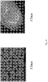
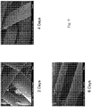
- 238000001878 scanning electron micrograph Methods 0.000 description 2
- 229910052710 silicon Inorganic materials 0.000 description 2
- 239000010703 silicon Substances 0.000 description 2
- 239000012890 simulated body fluid Substances 0.000 description 2
- 230000000638 stimulation Effects 0.000 description 2
- 229920002994 synthetic fiber Polymers 0.000 description 2
- GBNXLQPMFAUCOI-UHFFFAOYSA-H tetracalcium;oxygen(2-);diphosphate Chemical compound [O-2].[Ca+2].[Ca+2].[Ca+2].[Ca+2].[O-]P([O-])([O-])=O.[O-]P([O-])([O-])=O GBNXLQPMFAUCOI-UHFFFAOYSA-H 0.000 description 2
- 230000008467 tissue growth Effects 0.000 description 2
- 210000005166 vasculature Anatomy 0.000 description 2
- IXPNQXFRVYWDDI-UHFFFAOYSA-N 1-methyl-2,4-dioxo-1,3-diazinane-5-carboximidamide Chemical compound CN1CC(C(N)=N)C(=O)NC1=O IXPNQXFRVYWDDI-UHFFFAOYSA-N 0.000 description 1
- 102000002260 Alkaline Phosphatase Human genes 0.000 description 1
- 108020004774 Alkaline Phosphatase Proteins 0.000 description 1
- 206010002091 Anaesthesia Diseases 0.000 description 1
- ZOXJGFHDIHLPTG-UHFFFAOYSA-N Boron Chemical compound [B] ZOXJGFHDIHLPTG-UHFFFAOYSA-N 0.000 description 1
- DGAQECJNVWCQMB-PUAWFVPOSA-M Ilexoside XXIX Chemical compound C[C@@H]1CC[C@@]2(CC[C@@]3(C(=CC[C@H]4[C@]3(CC[C@@H]5[C@@]4(CC[C@@H](C5(C)C)OS(=O)(=O)[O-])C)C)[C@@H]2[C@]1(C)O)C)C(=O)O[C@H]6[C@@H]([C@H]([C@@H]([C@H](O6)CO)O)O)O.[Na+] DGAQECJNVWCQMB-PUAWFVPOSA-M 0.000 description 1
- 241001465754 Metazoa Species 0.000 description 1
- 229920001410 Microfiber Polymers 0.000 description 1
- KKCBUQHMOMHUOY-UHFFFAOYSA-N Na2O Inorganic materials [O-2].[Na+].[Na+] KKCBUQHMOMHUOY-UHFFFAOYSA-N 0.000 description 1
- BPQQTUXANYXVAA-UHFFFAOYSA-N Orthosilicate Chemical group [O-][Si]([O-])([O-])[O-] BPQQTUXANYXVAA-UHFFFAOYSA-N 0.000 description 1
- 241000283973 Oryctolagus cuniculus Species 0.000 description 1
- 102000004067 Osteocalcin Human genes 0.000 description 1
- 108090000573 Osteocalcin Proteins 0.000 description 1
- 229910019142 PO4 Inorganic materials 0.000 description 1
- 238000010521 absorption reaction Methods 0.000 description 1
- 210000000588 acetabulum Anatomy 0.000 description 1
- 230000009471 action Effects 0.000 description 1
- 230000004075 alteration Effects 0.000 description 1
- 230000037005 anaesthesia Effects 0.000 description 1
- 230000002491 angiogenic effect Effects 0.000 description 1
- 238000013459 approach Methods 0.000 description 1
- 239000012237 artificial material Substances 0.000 description 1
- QVGXLLKOCUKJST-UHFFFAOYSA-N atomic oxygen Chemical compound [O] QVGXLLKOCUKJST-UHFFFAOYSA-N 0.000 description 1
- 230000002146 bilateral effect Effects 0.000 description 1
- 230000004071 biological effect Effects 0.000 description 1
- 239000013060 biological fluid Substances 0.000 description 1
- 230000008827 biological function Effects 0.000 description 1
- 230000037176 bone building Effects 0.000 description 1
- 210000002449 bone cell Anatomy 0.000 description 1
- 230000014461 bone development Effects 0.000 description 1
- 230000037180 bone health Effects 0.000 description 1
- 239000000316 bone substitute Substances 0.000 description 1
- 229910052796 boron Inorganic materials 0.000 description 1
- 239000004566 building material Substances 0.000 description 1
- 239000004068 calcium phosphate ceramic Substances 0.000 description 1
- 210000000845 cartilage Anatomy 0.000 description 1
- 230000004663 cell proliferation Effects 0.000 description 1
- 239000004568 cement Substances 0.000 description 1
- 239000000919 ceramic Substances 0.000 description 1
- 230000008859 change Effects 0.000 description 1
- 239000005482 chemotactic factor Substances 0.000 description 1
- 238000005056 compaction Methods 0.000 description 1
- 239000000470 constituent Substances 0.000 description 1
- 238000010276 construction Methods 0.000 description 1
- 229920001577 copolymer Polymers 0.000 description 1
- 230000002596 correlated effect Effects 0.000 description 1
- 230000001054 cortical effect Effects 0.000 description 1
- 238000005520 cutting process Methods 0.000 description 1
- 230000007423 decrease Effects 0.000 description 1
- 230000003247 decreasing effect Effects 0.000 description 1
- 230000001627 detrimental effect Effects 0.000 description 1
- 230000007120 differential activation Effects 0.000 description 1
- 230000004069 differentiation Effects 0.000 description 1
- 238000005315 distribution function Methods 0.000 description 1
- 239000002019 doping agent Substances 0.000 description 1
- 238000001523 electrospinning Methods 0.000 description 1
- 230000002708 enhancing effect Effects 0.000 description 1
- 210000003743 erythrocyte Anatomy 0.000 description 1
- 239000000945 filler Substances 0.000 description 1
- 238000011049 filling Methods 0.000 description 1
- 239000012467 final product Substances 0.000 description 1
- 238000009472 formulation Methods 0.000 description 1
- 239000011491 glass wool Substances 0.000 description 1
- 239000002241 glass-ceramic Substances 0.000 description 1
- 239000001963 growth medium Substances 0.000 description 1
- 238000003306 harvesting Methods 0.000 description 1
- 230000009442 healing mechanism Effects 0.000 description 1
- 239000001307 helium Substances 0.000 description 1
- 229910052734 helium Inorganic materials 0.000 description 1
- SWQJXJOGLNCZEY-UHFFFAOYSA-N helium atom Chemical compound [He] SWQJXJOGLNCZEY-UHFFFAOYSA-N 0.000 description 1
- 238000000338 in vitro Methods 0.000 description 1
- 238000010348 incorporation Methods 0.000 description 1
- 238000011534 incubation Methods 0.000 description 1
- 230000008595 infiltration Effects 0.000 description 1
- 238000001764 infiltration Methods 0.000 description 1
- 239000004615 ingredient Substances 0.000 description 1
- 230000003993 interaction Effects 0.000 description 1
- 239000011344 liquid material Substances 0.000 description 1
- 238000011068 loading method Methods 0.000 description 1
- 230000014759 maintenance of location Effects 0.000 description 1
- 239000000155 melt Substances 0.000 description 1
- QSHDDOUJBYECFT-UHFFFAOYSA-N mercury Chemical compound [Hg] QSHDDOUJBYECFT-UHFFFAOYSA-N 0.000 description 1
- 229910052753 mercury Inorganic materials 0.000 description 1
- 150000002739 metals Chemical class 0.000 description 1
- 239000003658 microfiber Substances 0.000 description 1
- 238000002156 mixing Methods 0.000 description 1
- 238000012986 modification Methods 0.000 description 1
- 230000004048 modification Effects 0.000 description 1
- 239000003607 modifier Substances 0.000 description 1
- 238000000465 moulding Methods 0.000 description 1
- 239000002105 nanoparticle Substances 0.000 description 1
- 239000004745 nonwoven fabric Substances 0.000 description 1
- 238000010899 nucleation Methods 0.000 description 1
- 230000000050 nutritive effect Effects 0.000 description 1
- 210000002997 osteoclast Anatomy 0.000 description 1
- 230000004820 osteoconduction Effects 0.000 description 1
- 210000004409 osteocyte Anatomy 0.000 description 1
- 239000001301 oxygen Substances 0.000 description 1
- 229910052760 oxygen Inorganic materials 0.000 description 1
- 230000036961 partial effect Effects 0.000 description 1
- 238000002459 porosimetry Methods 0.000 description 1
- 230000002028 premature Effects 0.000 description 1
- 230000001737 promoting effect Effects 0.000 description 1
- 230000000541 pulsatile effect Effects 0.000 description 1
- 238000001812 pycnometry Methods 0.000 description 1
- 238000011084 recovery Methods 0.000 description 1
- 230000002829 reductive effect Effects 0.000 description 1
- 238000009877 rendering Methods 0.000 description 1
- 230000000250 revascularization Effects 0.000 description 1
- 238000004626 scanning electron microscopy Methods 0.000 description 1
- 238000005245 sintering Methods 0.000 description 1
- 239000002002 slurry Substances 0.000 description 1
- 150000003384 small molecules Chemical class 0.000 description 1
- 239000011734 sodium Substances 0.000 description 1
- 229910052708 sodium Inorganic materials 0.000 description 1
- 235000010413 sodium alginate Nutrition 0.000 description 1
- 239000000661 sodium alginate Substances 0.000 description 1
- 229940005550 sodium alginate Drugs 0.000 description 1
- 210000004872 soft tissue Anatomy 0.000 description 1
- 239000007787 solid Substances 0.000 description 1
- 238000009987 spinning Methods 0.000 description 1
- 238000010561 standard procedure Methods 0.000 description 1
- 238000003860 storage Methods 0.000 description 1
- 239000013589 supplement Substances 0.000 description 1
- 230000003319 supportive effect Effects 0.000 description 1
- 238000013268 sustained release Methods 0.000 description 1
- 239000012730 sustained-release form Substances 0.000 description 1
- 239000004753 textile Substances 0.000 description 1
- 239000012815 thermoplastic material Substances 0.000 description 1
- 230000017423 tissue regeneration Effects 0.000 description 1
Images


Classifications
-
- A—HUMAN NECESSITIES
- A61—MEDICAL OR VETERINARY SCIENCE; HYGIENE
- A61L—METHODS OR APPARATUS FOR STERILISING MATERIALS OR OBJECTS IN GENERAL; DISINFECTION, STERILISATION OR DEODORISATION OF AIR; CHEMICAL ASPECTS OF BANDAGES, DRESSINGS, ABSORBENT PADS OR SURGICAL ARTICLES; MATERIALS FOR BANDAGES, DRESSINGS, ABSORBENT PADS OR SURGICAL ARTICLES
- A61L27/00—Materials for grafts or prostheses or for coating grafts or prostheses
- A61L27/02—Inorganic materials
- A61L27/10—Ceramics or glasses
-
- A—HUMAN NECESSITIES
- A61—MEDICAL OR VETERINARY SCIENCE; HYGIENE
- A61L—METHODS OR APPARATUS FOR STERILISING MATERIALS OR OBJECTS IN GENERAL; DISINFECTION, STERILISATION OR DEODORISATION OF AIR; CHEMICAL ASPECTS OF BANDAGES, DRESSINGS, ABSORBENT PADS OR SURGICAL ARTICLES; MATERIALS FOR BANDAGES, DRESSINGS, ABSORBENT PADS OR SURGICAL ARTICLES
- A61L27/00—Materials for grafts or prostheses or for coating grafts or prostheses
- A61L27/40—Composite materials, i.e. containing one material dispersed in a matrix of the same or different material
- A61L27/42—Composite materials, i.e. containing one material dispersed in a matrix of the same or different material having an inorganic matrix
- A61L27/427—Composite materials, i.e. containing one material dispersed in a matrix of the same or different material having an inorganic matrix of other specific inorganic materials not covered by A61L27/422 or A61L27/425
-
- A—HUMAN NECESSITIES
- A61—MEDICAL OR VETERINARY SCIENCE; HYGIENE
- A61F—FILTERS IMPLANTABLE INTO BLOOD VESSELS; PROSTHESES; DEVICES PROVIDING PATENCY TO, OR PREVENTING COLLAPSING OF, TUBULAR STRUCTURES OF THE BODY, e.g. STENTS; ORTHOPAEDIC, NURSING OR CONTRACEPTIVE DEVICES; FOMENTATION; TREATMENT OR PROTECTION OF EYES OR EARS; BANDAGES, DRESSINGS OR ABSORBENT PADS; FIRST-AID KITS
- A61F2/00—Filters implantable into blood vessels; Prostheses, i.e. artificial substitutes or replacements for parts of the body; Appliances for connecting them with the body; Devices providing patency to, or preventing collapsing of, tubular structures of the body, e.g. stents
- A61F2/02—Prostheses implantable into the body
- A61F2/28—Bones
-
- A—HUMAN NECESSITIES
- A61—MEDICAL OR VETERINARY SCIENCE; HYGIENE
- A61L—METHODS OR APPARATUS FOR STERILISING MATERIALS OR OBJECTS IN GENERAL; DISINFECTION, STERILISATION OR DEODORISATION OF AIR; CHEMICAL ASPECTS OF BANDAGES, DRESSINGS, ABSORBENT PADS OR SURGICAL ARTICLES; MATERIALS FOR BANDAGES, DRESSINGS, ABSORBENT PADS OR SURGICAL ARTICLES
- A61L27/00—Materials for grafts or prostheses or for coating grafts or prostheses
- A61L27/50—Materials characterised by their function or physical properties, e.g. injectable or lubricating compositions, shape-memory materials, surface modified materials
- A61L27/56—Porous materials, e.g. foams or sponges
-
- A—HUMAN NECESSITIES
- A61—MEDICAL OR VETERINARY SCIENCE; HYGIENE
- A61L—METHODS OR APPARATUS FOR STERILISING MATERIALS OR OBJECTS IN GENERAL; DISINFECTION, STERILISATION OR DEODORISATION OF AIR; CHEMICAL ASPECTS OF BANDAGES, DRESSINGS, ABSORBENT PADS OR SURGICAL ARTICLES; MATERIALS FOR BANDAGES, DRESSINGS, ABSORBENT PADS OR SURGICAL ARTICLES
- A61L27/00—Materials for grafts or prostheses or for coating grafts or prostheses
- A61L27/50—Materials characterised by their function or physical properties, e.g. injectable or lubricating compositions, shape-memory materials, surface modified materials
- A61L27/58—Materials at least partially resorbable by the body
-
- A—HUMAN NECESSITIES
- A61—MEDICAL OR VETERINARY SCIENCE; HYGIENE
- A61L—METHODS OR APPARATUS FOR STERILISING MATERIALS OR OBJECTS IN GENERAL; DISINFECTION, STERILISATION OR DEODORISATION OF AIR; CHEMICAL ASPECTS OF BANDAGES, DRESSINGS, ABSORBENT PADS OR SURGICAL ARTICLES; MATERIALS FOR BANDAGES, DRESSINGS, ABSORBENT PADS OR SURGICAL ARTICLES
- A61L2400/00—Materials characterised by their function or physical properties
- A61L2400/12—Nanosized materials, e.g. nanofibres, nanoparticles, nanowires, nanotubes; Nanostructured surfaces
-
- A—HUMAN NECESSITIES
- A61—MEDICAL OR VETERINARY SCIENCE; HYGIENE
- A61L—METHODS OR APPARATUS FOR STERILISING MATERIALS OR OBJECTS IN GENERAL; DISINFECTION, STERILISATION OR DEODORISATION OF AIR; CHEMICAL ASPECTS OF BANDAGES, DRESSINGS, ABSORBENT PADS OR SURGICAL ARTICLES; MATERIALS FOR BANDAGES, DRESSINGS, ABSORBENT PADS OR SURGICAL ARTICLES
- A61L2430/00—Materials or treatment for tissue regeneration
- A61L2430/02—Materials or treatment for tissue regeneration for reconstruction of bones; weight-bearing implants
Definitions
- the present disclosure relates generally to bone graft materials. More particularly, the present disclosure relates to a dynamic bioactive synthetic bone graft material having an engineered porosity, and implants formed from such materials and their use.
- allograft devices may be used for bone grafts. Allograft devices are processed from donor bone. Allograft devices may have appropriate structure with the added benefit of decreased risk and pain to the patient, but likewise incur the increased risk arising from the potential for disease transmission and rejection. Autograft and allograft devices are further restricted in terms of variations on shape and size.
- autograft and allograft devices are inherently variable, because such devices are made from harvested natural materials.
- autograft supplies are also limited by how much bone may be safely extracted from the patient, and this amount may be severely limited in the case of the seriously ill or weak.
- bone graft materials are currently available for use.
- new materials such as bioactive glass (“BAG”) particulate-based materials, have become an increasingly viable alternative or supplement to natural bone-derived graft materials.
- BAG bioactive glass
- These new (non-bone derived) materials have the advantage of avoiding painful and inherently risky harvesting procedures on patients.
- the use of non-bone derived materials can reduce the risk of disease transmission.
- these new artificial materials can serve as osteoconductive scaffolds that promote bone regrowth.
- the graft material is resorbable and is eventually replaced with new bone tissue.
- compositions containing calcium phosphates comprise materials that have properties similar to natural bone, such as compositions containing calcium phosphates.
- Exemplary calcium phosphate compositions contain type-B carbonated hydroxyapatite (Ca 5 (PO 4 ) 3x (CO 3 ) x (OH)).
- Calcium phosphate ceramics have been fabricated and implanted in mammals in various forms including, but not limited to, shaped bodies and cements.
- Different stoichiometric compositions, such as hydroxyapatite (HA), tricalcium phosphate (TCP), tetracalcium phosphate (TTCP), and other calcium phosphate (CaP) salts and minerals have all been employed in attempts to match the adaptability, biocompatibility, structure, and strength of natural bone.
- Calcium phosphate based materials are widely accepted, they lack the ease of handling, flexibility and capacity to serve as a liquid carrier/storage media necessary to be used in a wide array of clinical applications.
- Calcium phosphate materials are inherently rigid, and to facilitate handling are generally provided as part of an admixture with a carrier material; such admixtures typically have an active calcium phosphate ingredient to carrier ratio of about 50:50, and may have as low as 10:90.
- bone graft materials still lack the requisite chemical and physical properties necessary for an ideal graft material. For instance, currently available graft materials tend to resorb too quickly, while some take too long to resorb due to the material's chemical composition and structure. For example, certain materials made from hydroxyapatite tend to take too long to resorb, while materials made from calcium sulphate or B-TCP tend to resorb too quickly.
- the porosity of the material is too high (e.g., around 90%), there may not be enough base material left after resorption has taken place to support osteoconduction. Conversely, if the porosity of the material is too low (e.g., 30%,) then too much material must be resorbed, leading to longer resorption rates. In addition, the excess material means there may not be enough room left in the residual graft material for cell infiltration. Other times, the graft materials may be too soft, such that any kind of physical pressure exerted on them during clinical usage causes them to lose the fluids retained by them.
- WO 00/76486 relates to a bone graft substitute comprising a bioactive glass composition in the form of fibers having a diameter ranging from 1 to 150 micrometers.
- the bone substitute is in the form of a porous matrix, wherein the pore diameters can be varied from 2 to 40 nm.
- bone graft materials that provide the necessary biomaterial, structure and clinical handling necessary for optimal bone grafting.
- dynamic bone graft materials that provide an improved mechanism of action for bone grafting, by allowing the new tissue formation to be achieved through a physiologic process rather than merely from templating.
- an artificial bone graft material that can be manufactured as required to possess varying levels of porosity, such as nano, micro, meso, and macro porosity.
- a need remains for a bone graft material that can be selectively composed and structured to have differential or staged resorption capacity, while providing material than can be easily molded or shaped into clinically relevant shapes as needed for different surgical and anatomical applications.
- a bone graft material that includes the characteristics of variable degrees of porosity, differential bioresorbability, compression resistance and radiopacity, and also maximizes the content of active ingredient relative to carrier materials such as collagen. Even more desirable would be a bone graft material that possesses all of the advantages mentioned above, and includes antimicrobial properties as well as allowing for drug delivery that can be easily handled in clinical settings. Embodiments of the present disclosure address these and other needs.
- the present disclosure provides bioactive bone graft materials according to claim 1. These graft materials are dynamic and accordingly can be molded and shaped as desired. These bone graft materials address the unmet needs aforementioned by providing the necessary biomaterial, structure and clinical handling for optimal bone grafting. In addition, these bone graft materials provide an improved mechanism of action for bone grafting, by allowing the new tissue formation to be achieved through a physiologic process of induction and formation rather than merely from templating and replacement. Further, these artificial bone graft materials can be manufactured as required to possess varying levels of porosity, such as nano, micro, meso, and macro porosity.
- the bone graft materials can be selectively composed and structured to have differential or staged resorption capacity, while being easily molded or shaped into clinically relevant shapes as needed for different surgical and anatomical applications. Additionally, these bone graft materials may have variable degrees of porosity, differential bioresorbability, compression resistance and radiopacity, and can also maximize the content of active ingredient relative to carrier materials such as collagen. These bone graft materials also possess antimicrobial properties as well as allows for drug delivery. The materials can also be easily handled in clinical settings.
- the material Prior to introducing the bone graft material, the material may be molded or shaped, such as by filling a mold tray with the material. If desired, the material may be compressed into the mold tray. Fluid may be added to the material prior to introduction into the mold tray.
- the fluid may be a saline, or it may be a naturally occurring body fluid such as blood.
- the bone graft material may be differentially activated.
- the porous, fibrous matrix may comprise a combination of bioresorbable subcomponents having different resorption rates.
- the subcomponents may include fibers or particulates, or a combination of both.
- the matrix may include more than one type of fiber, and each fiber may have a different resorption rate. The faster resorbing fiber may be allowed to resorb after the step of introduction, and induce strong initial bone growth.
- the remaining matrix may be designed to stay in the site for an extended period of time to allow for slower growth over time.
- the bone graft material may be injected into the defect, or it may be plastered over the defect. In addition, the material may be plugged into the defect.
- the standard method for healing natural tissue with synthetic materials has been to provide a device having the microstructure and macrostructure of the desired end product.
- the desired end product is cancellous bone
- traditional bone grafts have been engineered to mimic the architecture of cancellous bone.
- This has been the current standard for bone grafts, it does not take into account the fact that bone is a living tissue.
- Each bony trabeculae is constantly undergoing active biologic remodeling in response to load, stress and/or damage.
- cancellous and cortical bone can support a vast network of vasculature. This network not only delivers nutrients to sustain the living environment surrounding bone, but also supports red blood cells and marrow required for basic biologic function. Therefore, merely providing a synthetic material with the same architecture that is non-biologic is insufficient for optimal bone healing and bone health. Instead, what is required is a mechanism that can recreate the living structure of bone.
- Bone is a living biologic tissue and that inert structures will only impede bone healing
- healing is a phasic process starting with some initial reaction. Each phase builds on the reaction that occurred in the prior phase. Only after a cascade of phases does the final development of the end product occur - bone.
- the traditional method has been to replace or somehow stimulate healing by placing an inert final product as a catalyst to the healing process. This premature act certainly does not account for the physiologic process of bone development and healing.
- the physiologic process of bone healing can be broken down to three phases: (a) inflammation; (b) osteogenesis; and (c) remodeling.
- Inflammation is the first reaction to injury and a natural catalyst by providing the chemotactic factors that will initiate the healing process.
- Osteogenesis is the next phase where osteoblasts respond and start creating osteoid, the basic material of bone. Remodeling is the final phase in which osteoclasts and osteocytes then recreate the three-dimensional architecture of bone.
- a fibrin clot is made that provides a fibrous architecture for cells to adhere. This is the cornerstone of all connective tissue healing. It is this fibrous architecture that allows for direct cell attachment and connectivity between cells.
- the goal is to stimulate cell proliferation and osteogenesis in the early healing phase and then allow for physiologic remodeling to take place. Since the desired end product is a living tissue and not an inert scaffold, the primary objective is to stimulate as much living bone as possible by enhancing the natural fiber network involved in initiation and osteogenesis.
- the bone graft material of the present disclosure attempts to recapitulate the normal physiologic healing process by presenting the fibrous structure of the fibrin clot. Since this bioactive material made of fibers is both osteoconductive as well as osteostimulative, this fibrous network will further enhance and accelerate bone induction. Further, the dynamic nature of the bioactive fibrous matrix or scaffold allows for natural initiation and stimulation of bone formation rather than placing a non-biologic template that may impede final formation as with current graft materials.
- the fibers of the present material can also be engineered to provide a chemical reaction known to selectively stimulate osteoblast proliferation or other cellular phenotypes.
- the present disclosure provides bone graft materials and bone graft implants formed from these materials.
- These bone graft materials provide the necessary biomaterial, structure and clinical handling for optimal bone grafting.
- these bone graft materials provide an improved mechanism of action for bone grafting, by allowing the new tissue formation to be achieved through a physiologic process rather than merely from templating.
- these artificial bone graft materials can be manufactured as required to possess varying levels of porosity, such as nano, micro, meso, and macro porosity.
- the bone graft materials can be selectively composed and structured to have differential or staged resorption capacity, while being easily molded or shaped into clinically relevant shapes as needed for different surgical and anatomical applications.
- these bone graft materials may have variable degrees of porosity, differential bioresorbability, compression resistance and radiopacity, and can also maximize the content of active ingredient relative to carrier materials such as collagen.
- These bone graft materials also possess antimicrobial properties as well as allows for drug delivery. The materials can also be easily handled in clinical settings.
- Embodiments of the present disclosure may employ a dynamic, ultraporous bone graft material, for example, having nano, micro, meso and macro porosities.
- the bone graft material can comprise bioactive ("BAG") fibers or a combination of BAG fibers and particulates of materials. Due to the size and length of the fibers, the bone graft material is a dynamic structure that can be molded or packed into a desired shape, while maintaining its porous structure.
- the bone graft material may be osteoconductive and/or osteostimulatory.
- the bone graft material may have differential activation (i.e., resorbability), which may facilitate advanced functions like drug delivery including antibiotics.
- the fibrous nature of the bone graft allows for stimulation and induction of the natural biologic healing process required for bone formation.
- the embodiments of the bone graft material can include BAG fibers having a relatively small diameter, and in particular, a diameter less than 100 nanometers. In one embodiment, the fiber diameter can be less than 10 nanometers, and in another embodiment, the fiber diameter can be in the range of about 5 nanometers. Since the materials used in the embodiments are bioactive materials, the bone graft material may form a CaP layer on its surface when it interacts with body fluids.
- the bone graft material comprises particulates in combination with fibers. The presence of particulate matter may be employed to modify or control the resorption rate and resorption profile of the bone graft material as well as provide mechanical strength and compression resistance.
- the particulate may be bioactive glass, calcium sulphate, calcium phosphate or hydroxyapatite.
- the particulate may be solid, or it may be porous.
- the bone graft material may be moldable and can be packaged in functional molds for convenient clinical handling.
- the bone graft material can be mixed with other additives like collagen, etc., for example, to further facilitate handling.
- the bone graft material and collagen composite may be in the form of a foam, and the foam may additionally be shaped into a strip, a continuous rolled sheet, a sponge or a plug.
- the foam may take any configuration with any variety of shapes and sizes.
- the bone graft material and collagen composite may take the form of a putty or other moldable material.
- the BAG fibers and particulates may be mixed with a slurry of collagen, poured into a mold of a desired shape, and frozen to yield a desire foam shape.
- the foam can have a fixed shape or the foam may be turned into a putty with the addition of fluids such as saline, blood or bone marrow aspirate.
- the bone graft material may be in the form of an injectable material.
- Putties can be made by combining the bone graft material with other additives such as CMC, hyaluronic acid, or sodium alginate, for instance.
- CMC hyaluronic acid
- sodium alginate for instance.
- the ability to provide a bone graft material in the form of a putty renders the material easily usable, since the putty may be applied directly to the injury site by either injection or by plastering. Also, the ease of handling and moldability of the putty composition allows the clinician to form the material easily and quickly into any desired shape.
- the present disclosure relates to a synthetic bone graft material that can be manufactured in a wide variety of compositional and structural forms for the purpose of introducing a biocompatible, bioabsorbable structural matrix in the form of an implant for the repair or treatment of bone.
- the bone graft material can be an osteostimulative and/or osteoconductive implant having differential bioabsorbability.
- the bone graft material may be substantially comprised of BAG fibers.
- the bone graft material can be selectively determined by controlling compositional and manufacturing variables, such as bioactive glass fiber diameter, size, shape, and surface characteristics as well as the amount of bioactive glass particulate content and structural characteristics, and the inclusion of additional additives, such as, for example tricalcium phosphate, hydroxyapatite, and the like.
- compositional and manufacturing variables such as bioactive glass fiber diameter, size, shape, and surface characteristics as well as the amount of bioactive glass particulate content and structural characteristics, and the inclusion of additional additives, such as, for example tricalcium phosphate, hydroxyapatite, and the like.
- the bioactive glass used in the bone graft material may have a composition similar to 45S5 (46.1 mol% SiO 2 , 26.9 mol% CaO, 24.4 mol% Na 2 O and 2.5 mol% P 2 O 5 , 58S (60 mol% SiO 2 , 36 mol% CaO and 4 mol% P 2 O 5 ), S70C30 (70 mol% SiO 2 , 30 mol% CaO), and the like.
- bioactive glasses that are silicon free may also be employed.
- bioactive glass compositions that are SiO 2 free, and having boron instead of silicon may also be used.
- the bone graft material may be tailored to have specific desired characteristics, such as increased X-ray opacity (for example, by incorporating strontium), slower or faster dissolution rate in vivo, surface texturing, or the like.
- the bone graft material may serve as a scaffold for bone activity in the bone defect.
- the scaffolding materials used in the bone graft may be bioactive glasses, such as 45S5 glass, which can be both osteoconductive and osteostimulatory. As determined by applicants, the bioactive glass may have naturally inherent antimicrobial properties due to the presence of sodium in the material's composition. The extensive surface area provided by the present fibrous bone graft material allows for antimicrobial benefits with the use of this material.
- Bone graft materials of the present disclosure is flexible, moldable, or can be preformed to mimic, augment or replace specific shaped structures.
- the bone graft materials can be formed into acetabulum cups and other skeletal modeled components employed in surgical procedures.
- the bone graft materials can be formed into any clinically useful shape, such as strips, blocks, wedges, and the like. The shapes may be formed by molding, as will be described in greater detail below, or simply by cutting, tearing, folding, or separating the fibrous material into the desired configuration for its clinical application
- the bone graft material is formed from bioactive glass fibers, which may be manufactured having predetermined cross-sectional diameters sized as desired.
- the fibers may be formed by electro-spinning or laser spinning, for instance, to create consistently uniform fibers.
- the bone graft material may be formed from a scaffold of fibers of uniform diameters.
- the bioactive glass fibers may be formed having varying diameters and/or cross-sectional shapes, and may even be drawn as hollow tubes. Additionally, the fibers may be meshed, woven, intertangled and the like for provision into a wide variety of shapes.
- a bioactive glass fiber bone graft material manufactured such that each fiber is juxtaposed or out of alignment with the other fibers could result in a bone graft material having a glass-wool or "cotton-ball" appearance due to the large amount of empty space created by the random relationship of the individual glass fibers within the material.
- Such a manufacture enables a bone graft material with an overall soft or pliable texture so as to permit the surgeon to manually form the material into any desired overall shape to meet the surgical or anatomical requirements of a specific patient's surgical procedure.
- bioactive glass particles such as included bioactive glass particles, antimicrobial fibers, particulate medicines, trace elements or metals such as copper, which is a highly angiogenic metal, strontium, magnesium, zinc, etc. mineralogical calcium sources, and the like.
- bioactive glass fibers may also be coated with organic acids (such as formic acid, hyaluronic acid, or the like), mineralogical calcium sources (such as tricalcium phosphate, hydroxyapatite, calcium sulfate, or the like), antimicrobials, antivirals, vitamins, x-ray opacifiers, or other such materials.
- the bone graft material may be engineered with fibers having varying resorption rates.
- the resorption rate of a fiber is determined or controlled by its material composition and by its diameter.
- the material composition may result in a slow reacting vs. faster reacting product.
- smaller diameter fibers can resorb faster than larger diameter fibers.
- the overall porosity of the material can affect resorption rate. Materials possessing a higher porosity mean there is less material for cells to remove. Conversely, materials possessing a lower porosity mean cells have to do more work, and resorption is slower.
- the bone graft material may contain fibers that have the appropriate material composition as well as diameter for optimal performance. A combination of different fibers may be included in the material in order to achieve the desired result.
- bioactive glass particles can be accomplished using particles having a wide range of sizes or configurations to include roughened surfaces, very large surface areas, and the like.
- particles may be tailored to include interior lumens with perforations to permit exposure of the surface of the particles interior. Such particles would be more quickly absorbed, allowing a tailored material characterized by differential resorbability.
- the perforated or porous particles could be characterized by uniform diameters or uniform perforation sizes, for example.
- the porosity provided by the particles may be viewed as a secondary range of porosity accorded the bone graft material or the implant formed from the bone graft material.
- the manufacturer has the ability to provide a bioactive glass bone graft material with selectively variable characteristics that can greatly affect the function of the material before and after it is implanted in a patient.
- the nano and macro sized pores provide superb fluid soak and hold capacity, which enhances the bioactivity and accordingly the repair process.
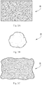
- FIGs. 1A and 1B illustrate a first embodiment bioactive fibrous scaffold 10 according to the present disclosure.
- the scaffold 10 is made up of a plurality of interlocking fibers 15 defining a three-dimensional porous support scaffold or matrix 10.
- the support matrix 10 is made up of bioactive glass fibers 15 that are interlocked or interwoven, not necessarily fused at their intersections 17. At least some of the fibers 15 may thus move over one another with some degree of freedom, yielding a support web 10 that is dynamic in nature.
- the composition of the fibers 15 used as the struts 19 of the resulting dynamic fibrous scaffold 10 are typically bioactive glass, ceramic or glass-ceramic formulations, such that within the range of fiber diameter and construct size, that the scaffolding fibers 15 are generally characterized as having the attributes of bioactivity.
- the diameters of the fibers 15 defining the dynamic scaffold 10 are typically sufficiently small to allow for inherent interlocking of the resulting three-dimensional scaffold 10 upon itself, without the need for sintering, fusing or otherwise attaching the fibers 15 at their intersections 17, although some such fusing or attachment may be employed to further stiffen the scaffold 10 if desired.
- the scaffold 10 is self constrained to not completely fall apart, yet the individual fibers 15 defining the support struts 19 are free to move small distances over each other to grant the scaffold 10 its dynamic qualities such that it remains flexible while offering sufficient support for tissue formation and growth thereupon.
- the availability of nano sized fibers can significantly enhance the surface area available for cell attachment and reactivity.
- pluralities of fibers 15 characterized as substantially having diameters below 1 micrometer (1000 nanometers) are sufficient to form dynamic scaffolding 10, as are pluralities of fibers 15 characterized as substantially having diameters below 100 nanometers.
- the scaffolding 10 may also be constructed from a plurality of fibers 15 having multimoda l diameter distributions, wherein combinations of diameters may be employed to yield specific combinations of dynamic flexibility, structural support, internal void size, void distribution, compressibility, dissolution and resorption rates, and the like. For example, some of the fibers 15 may be fast reacting and resorb quickly into bone to induce initial bone growth.
- some remnant materials of the bone graft material may be designed to resorb over a more extended time and continue to support bone growth after the previously resorbed material has gone.
- This type of layered or staged resorption can be critically important in cases where the surgical site has not sufficiently healed after the first burst of bone growth activity. By providing varying levels of resorption to occur, the material allows greater control over the healing process and avoids the "all or none" situation.
- the ranges of fiber diameters within a construct range starting from the nano level, where a nano fiber is defined as a fiber with a diameter less than 1 micron (submicron), up to about 100 microns; more typically, fiber diameters range from about 0.005 microns to about 10 microns; still more typically, fiber diameters range from about 0.05 to about 6 microns; yet more typically, fiber diameters range from 0.5 to about 20 microns; still more typically, fiber diameters range from about 1 micron to about 6 microns. In all cases, predetermined amounts of larger fibers may be added to vary one or more of the properties of the resultant scaffolding 10 as desired.
- the entire construct 10 typically tends to become less self constrained.
- fibers 15 may be constructed at a particular size, such as at a nano scale of magnitude, to enhance the surface area available for cell attachment and reactivity.
- the bone graft material includes at least one nanofiber.
- Porous, fibrous scaffolds 10 may be made by a variety of methods resulting in an interlocking, entangled, orientated three-dimensional fiber implant 20.
- these fibers 15 are not necessarily continuous, but may be short and discrete, or some combination of long, continuous fibers 15 and short, discrete fibers 15.
- the fibers 15 touch to define intersections 17 and also define pores or voids 37.
- the porosity of the resulting implant, as well as its pore size distribution may be controlled.
- the pores 37 typically range in size from about 100 nanometers to about 1 mm, with the pore size and size distribution a function of the selected fiber size range and size distribution, as well as of the selected forming technique.
- the fiber and pore size is not limited to these ranges, and while the description focuses on the nanofibers and nanopores, it is well understood that the bone graft material of the present disclosure may equally include macro sized fibers and pores to create range of diameters of fibers and pores.
- the resulting implant or device 20 may thus be a nonwoven fabric made via a spunlaid or spun blown process, a melt blown process, a wet laid matt or 'glass tissue' process, or the like and may be formed to have the characteristics of a felt, a gauze, a cotton ball, cotton candy, or the like.
- macro-, meso-, and microporosity occur simultaneously in the device 20 and, more typically, are interconnected. It is unnecessary here to excessively quantify each type of porosity, as those skilled in the art can easily characterize porosity using various techniques, such as mercury intrusion porosimetry, helium pycnometry, scanning electron microscopy and the like. While the presence of more than a handful of pores within the requisite size range is needed in order to characterize a device 20 as having a substantial degree of that particular type of porosity, no specific number or percentage is called for. Rather, a qualitative evaluation by one skilled in the art shall be used to determine macro-, meso-, micro-, and/or nanoporosity.
- the overall porosity of the porous, fibrous implants 20 will be relatively high, as measured by pore volume and typically expressed as a percentage.
- Zero percent pore volume refers to a fully or theoretically dense material. In other words, a material with zero porosity has no pores at all. Likewise, one hundred percent pore volume would designate "all pores" or air.
- One skilled in the art will be versed in the concept of pore volume and will readily be able to calculate and apply it.
- Bone graft implants 20 typically have pore volumes in excess of about 30%, and more typically may have pore volumes in excess of 50% or 60% may also be routinely attainable. In some embodiments, scaffolding implants 20 may have pore volumes of at least about 70%, while other embodiments may typically have pore volumes in excess of about 75% or even 80%. Bone graft implants may even be prepared having pore volumes greater than about 90% - 97%.
- some bone graft implants 20 it is advantageous for some bone graft implants 20 to have a porosity gradient that includes macro-, meso-, and microporosity, and in some cases nanoporosity.
- the implants 20 can possess a porosity gradient such that the size of the pores as well as the placement of the pores can vary throughout the implants 20.
- the combination of fibers and particulates to create the appropriate compression resistance and flexibility is retained when the bone graft implant 20 is wetted.
- Bone graft implants 20 are also typically characterized by interconnected porosity, as such is correlated with increased capillary action and wicking capability. Such bone graft implants 20 should be capable of rapidly wicking and retaining liquid materials for sustained release over time.
- the fibers 15 typically have non-fused linkages 35 that provide subtle flexibility and movement of the scaffolding 10 in response to changes in its environment, such as physiological fluctuations, cellular pressure differences, hydrodynamics in a pulsatile healing environment, and the like. This in vivo environment can and will change over the course of the healing process, which may last as long as several months or even longer.
- the scaffold 10 typically retains its appropriate supportive characteristics and distribution of pores 37 throughout the healing process such that the healing mechanisms are not inhibited.
- the pores 37 defined by the matrix of interlocking and tangled fibers 15 may serve to carry biological fluids and bone-building materials to the site of the new bone growth.
- the fluids likewise slowly dissolve fibers 15 made of bioactive glass and the like, such that the scaffolding 10, and particularly the pores 37, changes in size and shape in dynamic response to the healing process.
- Scaffolds 10 are typically provided with a sufficiently permeable three-dimensional microstructure for cells, small molecules, proteins, physiologic fluids, blood, bone marrow, oxygen and the like to flow throughout the entire volume of the scaffold 10. Additionally, the dynamic nature of the scaffold 10 grants it the ability to detect or respond to the microenvironment and adjust its structure 20 based on forces and pressure exerted elements within the microenvironment.
- scaffolds 10 typically have sufficient three-dimensional geometries for compliance of the bone graft implant or device 20 when physically placed into an irregular shaped defect, such as a void, hole, or tissue plane as are typically found in bone, tissue, or like physiological site.
- the devices 20 typically experience some degree of compaction upon insertion into the defect, while the permeable characteristics of the scaffolds 10 are maintained.
- the device 20 typically remains within 2 mm of the native tissue in the defect wall.
- Bone graft implants or devices 20 made from the scaffolding 10 can appear similar to felts, cotton balls, textile fabrics, gauze and the like. These forms have the ability to wick, attach and contain fluids, proteins, bone marrow aspirate, cells, as well as to retain these entities in a significant volume, though not necessarily all in entirety; for example, if compressed, some fluid may be expulsed from the structure.
- bone graft implants or devices 20 are their ability to modify or blend the dynamic fiber scaffolds 10 with a variety of carriers or modifiers to improve handling, injectability, placement, minimally invasive injection, site conformity and retention, and the like while retaining an equivalent of the 'parent' microstructure.
- Such carriers ideally modify the macro-scale handling characteristic of the device 20 while preserving the micro-scale (typically on the order of less than 100 micrometers) structure of the scaffolding 10.
- These carriers resorb rapidly (typically in less than about 2 weeks; more typically in less than about 2 days) without substantially altering the form, microstructure, chemistry, and/or bioactivity properties of the scaffolding.
- These carriers include polaxamer, glycerol, alkaline oxide copolymers, bone marrow aspirate, and the like.
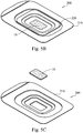
- FIG. 2A shows an embodiment of an implant 20 in the form of a strip or sheet, for example.
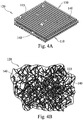
- FIG. 2B shows an embodiment of an implant 20 in the form of a three-dimensional structure similar to a cotton ball, for example.
- a plurality of interlocking fibers 15 are spun or blown into a randomly oriented assemblage 20 having the general appearance of a cotton ball.
- the fibers 15 are typically characterized as having diameters of from less than about 1000 nm (1 micrometer) ranging up to approximately 10, 000 nm (10 micrometers).
- the resulting cotton-ball device 20 may be formed with an uncompressed diameter of typically from between about 1 and about 6 centimeters, although any convenient size may be formed, and may be compressible down to between about 1 ⁇ 2 and 1 ⁇ 4 of its initial size. In some cases, the device 20 can substantially return to its original size and shape once the compressive forces are removed (unless it is wetted with fluids, which kind of locks the device into desired shape and density, or is vacuum compressed). However, in many cases the device 20 may remain deformed. By varying the relative diameters of some of the fibers 15, structures ranging from 'cotton ball' to 'cotton candy' may be produced, with varying ranges of fiber diameters from less than about 10 nm to greater than about 10 microns.
- FIG. 2C shows an embodiment of the implant 20 in the form of a woven mesh or fabric, for example.
- fibers 15 may be woven, knitted, or otherwise formed into a fabric device 20 having a gauze-like consistency.
- the fibers 15 are typically greater than 1 about micrometer in diameters and may be as large as about 100 micrometers in diameter.
- the micro-scale orientation of the fibers 15 is typically random, although the fibers may be somewhat or completely ordered. On a macro-scale, the fibers 15 are typically more ordered.
- the constituency of these devices 20 may have varying amounts of smaller fibers 15 incorporated therein to maintain the self-constrained effect.
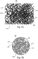
- FIGs. 3A and 3B illustrate another embodiment of the present disclosure, a bioactive fibrous scaffold 110 as described above with respect to FIGs. 1A and 1B , but having glass microspheres or particulate 140 distributed therethrough.
- the glass particulate 140 is typically made of the same general composition as the fibers 115, but may alternately be made of other, different compositions.
- One advantage of the presence of particulate 140 in the implant 120 is its contribution to the implant's 120 overall compression resistance. Since one function of the implant 120 is typically to absorb and retain nutrient fluids that feed the regrowth of bone, it is advantageous for the implant to offer some level of resistance to compressive forces, such that the liquids are not prematurely 'squeezed out'.
- Particulate 140 whether spherical or particulate, stiffens the implant, which is otherwise a porous scaffolding primarily composed of intertangled fibers 115.
- the particulate 140 can act as pillars, lending structural support to the overall implant 120.
- the glass particulate 140 is typically generally spherical, but may have other regular or irregular shapes.
- the glass particulate 140 typically varies in size, having diameters ranging from roughly the width of the fibers 115 (more typically, the struts 119) to diameters orders of magnitude greater than the typical fiber widths.
- Particulate 140 may also vary in shape, from generally spherical to spheroidal, or elliptical to irregular shapes, as desired.
- the particulate 140 may even be formed as generally flat platelets; further, the platelets (or other shapes) may be formed having perforations or internal voids, to increase the effective surface area and dissolution rate.
- the shape of the particulate 140 may be varied to influence such factors as bone cell attachment, particulate coatability, and the like.
- the glass particulates 140 may have an average diameter of about 20 microns to about 1 millimeter. In another embodiment, the particulates 140 may have an average diameter of about 300 to 500 microns. In still another embodiment, the glass particulates 140 may have an average diameter of about 350 microns.
- bioactive glass particulate 140 may be coated with organic acids (such as formic acid, hyaluronic acid, or the like), mineralogical calcium sources (such as tricalcium phosphate, hydroxyapatite, calcium sulfate, or the like), antimicrobials, antivirals, vitamins, x-ray opacifiers, or other such materials. While smaller particulate may tend to lodge in or around fiber intersections 117, larger particulate tend to become embedded in the scaffolding 120 itself and held in place by webs of fibers 115. Pore-sized microspheres may tend to lodge in pores 137.
- organic acids such as formic acid, hyaluronic acid, or the like
- mineralogical calcium sources such as tricalcium phosphate, hydroxyapatite, calcium sulfate, or the like
- antimicrobials such as tricalcium phosphate, hydroxyapatite, calcium sulfate, or the like
- antivirals such as vitamin, x-ray opac
- the glass particulate 140 may be composed of a predetermined bioactive material and tailored to dissolve over a predetermined period of time when the scaffolding 110 is placed in vitro, so as to release a predetermined selection of minerals, bone growth media, and the like at a predetermined rate.
- the composition, size and shape of the glass particulate 140 may be varied to tailor the resorption rate of the bioactive glass, and thus the rate at which minerals and the like are introduced into the body (and likewise, how long the particulate 140 is available to provide increased compression resistance to the scaffolding implant 20). For example, for a given bioactive glass composition and particulate volume, irregularly shaped particulate 140 would have more surface area than spherical particulate 140, and would thus dissolve more rapidly.
- the glass particulate 140 may be hollow bioactive glass, polymer or the like microspheres filled with specific mixture of medicines, antibiotics, antivirals, vitamins or the like to be released at and around the bone regrowth site at a predetermined rate and for a predetermined length of time.
- the release rate and duration of release may be functions of particulate size, porosity and wall thickness as well as the distribution function of the same.
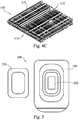
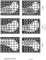
- the shape and texture of the bone graft material may be randomly configured to maximize its overall volume, surface area, and pliability or, in stark contrast, can be manufactured with the bioactive glass fibers in a more rigid and uniform arrangement, such as, for example in a mesh or matrix type assembly.
- a mesh or matrix assembly as illustrated by the non-limiting examples shown in FIGs. 4A to 4C , the glass fibers can be arranged in a stacked arrangement limiting the flexibility in a directional manner, or, the fibers can be layered wherein alternating layers are in a crossed relationship one to the other.
- the matrix assembly 110 is shown having an ordered configuration with discrete layers comprising fibers 115 and particulate 140.
- FIG. 4A the matrix assembly 110 is shown having an ordered configuration with discrete layers comprising fibers 115 and particulate 140.
- the matrix assembly is shown having a randomly arranged configuration of fibers 115 and particulate 140 dispersed throughout.
- the matrix assembly 110 is shown having a configuration in which the layers have different porosities due to differences in the spacing of the fibers 115 and particulate 140 throughout each layer. That is, the size of the pores 137 varies throughout the matrix assembly due to the unevenly spaced fibers 115 and particulate 140.
- FIGs. 4A and 4C show discretely aligned fibers 115 for the purposes of illustrating the concept herein, the individual layers of material 110 may include fibers 115 and particulate 140 that are unorganized and randomly aligned.
- the bone graft material of the present disclosure includes embedded bioactive glass particles within the bioactive glass fiber construct.
- the inclusion of such particles, as determined by the quantity, size, and characteristics of the particles, can affect the compressibility, bioabsorbability, and porosity of the resulting bone graft material.
- Additional additives such as calcium phosphates (CaP), calcium sulfates (CaS), hydroxyapatite (HA), carboxymethycellulose (CMC), collagen, glycerol, gelatin, and the like can also be included in any of the many varied constructions of the bioactive glass fiber bone graft material to assist in bone generation and patient recovery.
- Such additives may be in the range of 0 to 90 percent porous.
- Another additive, collagen may be included and may also be of the ultraporous kind having a porosity of up to 98 percent.
- the surface area of the bone graft material is maximized to increase the bone ingrowth into the structural matrix of the material.
- Another useful variable is the capability of the bone graft material to selectively be composed and configured to provide layers of varying porosity, such as nano-, micro-, meso-, and micro-porosity, so as to act as a cell filter controlling the depth of penetration of selected cells into the material.
- the preparation of the bone graft material can be selectively varied to include bioactive glass fibers and/or particles having different cross-sectional diameters, shapes and/or compositions, the material properties may be tailored to produce a bone graft material with differential absorption capabilities. This feature permits the surgeon to select a bone graft material specifically for the needs of a specific situation or patient. Controlling the pace of bone ingrowth into the bioactive glass matrix of the material allows the surgeon to exercise almost unlimited flexibility in selecting the appropriate bone graft material for an individual patient's specific needs.
- the bioactive glass was formulated with strontium partially replacing calcium.
- the partial replacement of calcium with strontium yields a bioactive glass with a reduced resorption/reaction rate and also with an increased radiodensity or radioopacity.
- the bioactive glass stays present in the body for a longer period of time and also presents a more readily visible x-ray target.
- silver may be incorporated into the bioactive glass fiber scaffolding structural matrix.
- Silver is an antimicrobial material, and enhances the inherent antimicrobial properties of the bioactive glass material.
- silver is added as a dopant to very fine bioactive glass fibers, such that the silver is quickly released as the very fine fibers dissolve at the implant site, allowing the silver to act as an anti-microbial agent to prevent infection immediately after surgery while the remaining scaffolding material does its work.
- Ag may be introduced as fibers and interwoven with the bioactive glass fibers, as particles similar to the glass particulate discussed above, or the like.
- varying the composition of the bioactive glass from which the fibers are formed to create an alkaline (high pH in the range of 8-10) glass may also provide the material with antimicrobial properties.
- One advantage of the current invention is that it is dynamic, and can be easily molded into various shapes or form, without losing the essential structure and porosity.
- the material By packaging the material in a functional tray, where the tray acts as a mold, the material can be provided in various shapes in the operating room. Especially, the material becomes a cohesive mass when a fluid such as blood, saline, bone marrow, other natural body fluids, etc. is added.
- the bone graft material is provided as part of a surgical kit 200.
- the kit 200 includes a tray portion 210 having a recess or well 212, and more typically a set of nested recesses, for storing, holding and manipulating the bone graft material 10, 110, and a lid portion 220 for sealingly engaging the tray portion 210.
- the tray and lid portions 210, 220 are typically formed from thermoplastic materials, but may alternately be made of any convenient materials.
- the deepest recess chamber 212 typically has a simple geometry, such as a rectangular block or wedge shape, such that the so-loaded bone graft material likewise has a simple geometry.
- a simple geometry such as a rectangular block or wedge shape
- Other geometries are described in a copending and commonly owned U.S. Patent Application No. 12/914,376 , entitled “DYNAMIC BIOACTIVE BONE GRAFT MATERIAL AND METHODS FOR HANDLING,” filed October 28, 2010.
- the bone graft material 10, 110 is typically provided as an intertangled or interwoven mass of bioactive glass fibers.
- the bioactive glass fibers may be provided in format that is ready to be surgically emplaced in a bony cavity (such as a woven or mesh format), or may be provided in a format that requires additional preparation prior to emplacement (such as a more loosely intertangled format) that requires the addition of a liquid, such as saline, glycerol, gelatin, plasma, or collagen gel or chips, to assist in rendering the mass of bioactive glass more pliable and structurally unitary.
- a liquid such as saline, glycerol, gelatin, plasma, or collagen gel or chips, to assist in rendering the mass of bioactive glass more pliable and structurally unitary.
- Such liquids may optionally be included in the kit packaging 200, or provided separately.
- a kit 200 including a tray body 210 and a lid 220 engagable with the tray body.
- the tray body 210 includes one or more recesses 212 for containing a volume of bioactive glass fibers 10.
- the volume of bioactive glass fibers may be woven, knitted, intertangled or provided as a loose stack.
- the volume of bioactive glass fibers may optionally include fibers of other compositions, such as antimicrobial silver, polymers, or alternate glass compositions, and may also optionally include particulate matter or particulate of the same bioactive glass composition, or alternate compositions such as alternate glass, metal, metal oxide, medicinal, nutritive, and/or antimicrobial or the like.
- the kit may also optionally include a liquid, such as saline or collagen gel, for mixing with the bioactive glass volume.
- the surgeon removes the lid 220 of the kit 200 and removes a portion of the included bioactive glass material 10.
- the bioactive glass material may then be shaped and sized by the surgeon for insertion into a bony cavity. This process may involve the addition of an appropriate liquid to the bioactive glass material, such as saline, collagen gel, plasma, blood, or the like, to achieve a desired degree of pliability and/or structural integrity. Once the bioactive glass material is sized and shaped as desired, it is inserted into the bony cavity. This process may be done as a single operation or as a series of steps.
- FIGs. 6A and 6B illustrate graphically volumetric contribution and surface area contribution of an embodiment of the bone graft material based on its pore size distribution.
- the bone graft material of an implant 20 may have a structure having varying porosity, such as nano-, micro-, meso-, and macro-porosity.
- the mesopores and micropores contribute to a large portion of the volume of the bone graft material
- the nanopores contribute a significantly large portion of the surface area provided by the bone graft material. That is, for a give volume, the embodiments may utilize a porosity distribution that includes nanopores to obtain a higher surface higher for a given volume.
- these and other features and advantages can be provided by the embodiments.
- FIG. 7 shows time lapse photomicrographs of fibers of an embodiment of the present disclosure immersed in simulated body fluid at 37°C after one day and three days
- FIG. 8 shows time lapse photomicrographs of fibers of an embodiment of the present disclosure immersed in simulated body fluid at 37°C after three days.
- FIG. 9 shows a series of time lapse scanning electron micrographs (SEMs) showing osteoblast cells cultured on glass fiber scaffolds of the present disclosure for 2, 4 and 6 days. As shown, there is increased cell density during the 6-day incubation.
- FIG. 10 shows a graph of osteoblast cell growth exhibited on the glass fiber scaffold of FIG. 9 for 2, 4 and 6 days with an initial seeding of 100,000 MC3T3-E1 cells per scaffold.
- FIG. 11 shows a photomicrograph of a fiber that has been seeded with mesenchymal stem cells. Such cells may assist with the osteostimulative effect of osteoblast proliferation and differentiation. The effect can be measured based on determining DNA content and elevated presence of osteocalcin and alkaline phosphatase levels.
- FIGs. 12 - 14 show some results of testing of an embodiment of the fibrous bone graft material of the present disclosure on a mammal (specifically, in this case a rabbit.)
- a bilateral distal femoral bone defect was created having a size of approximately 5 mm in diameter and 10 mm in length.
- the testing was performed along with a commercially available bone graft substitute, Product #1, in this comparison study.
- Product #1 is a silicate substituted bone graft material (ACTIFUSETM available from ApaTech, Inc.
- the bone graft material of the present disclosure solicits a more dynamic bone growth response than with traditional synthetic bone graft materials, and leads to more physiologic bone healing and remodeling.
- the majority of the base material was resorbed with evidence of bone remodeling at the surgical site. Further, the bone tissue appeared to integrate with surrounding bone.
- FIG. 12 shows a series of radiographic images from testing performed comparing the performance of an embodiment the bone graft material with Product 1 at 6 weeks, 12 weeks and 24 weeks.
- FIG. 13 shows a graphical comparison of percentage of new bone present after 6 weeks, 12 weeks and 24 weeks in the embodiment of the bone graft material with Product 1 during the comparative study.
- FIG. 14 shows a graphical comparison of percentage of residual material remaining after 6 weeks, 12 weeks and 24 weeks in the embodiment of the bone graft material with Product 1 during the comparative study.
- the bone graft material of the present disclosure is described for use in bone grafting, it is contemplated that the graft material of the present disclosure may also be applied to soft tissue or cartilage repair as well. Accordingly, the application of the fibrous graft material provided herein may include many different medical uses, and especially where new connective tissue formation is desired.
Claims (18)
- Matériau de greffe osseuse comprenant :une matrice poreuse et flexible comprenant une pluralité de fibres de verre bioactif chevauchantes et imbriquées et des particules de verre bioactif réparties partout dans les fibres, les fibres et les particules ayant chacune des diamètres variables, et une gamme de pores définis par les espaces entre lesdites fibres et particules, les diamètres variables des fibres et des particules commandant une taille de pore et une répartition de la gamme de pores et une porosité globale de la matrice, la matrice ayant en outre une porosité répartie d'après ladite gamme de pores de sorte que la matrice soit configurée pour une résorption étagée ;dans lequel la gamme de pores comprend des micropores et des macropores ; etdans lequel la matrice permet un mouvement des fibres chevauchantes et imbriquées les unes par rapport aux autres au moins au niveau de certaines de leurs intersections.
- Matériau de greffe osseuse selon la revendication 1, dans lequel la gamme de pores comprend en outre des nanopores.
- Matériau de greffe osseuse selon la revendication 1, dans lequel la gamme de pores est répartie d'après un gradient à travers la matrice.
- Matériau de greffe osseuse selon la revendication 1, dans lequel les fibres sont orientées aléatoirement.
- Matériau de greffe osseuse selon la revendication 1, dans lequel le diamètre des micropores va d'environ 1 micron à environ 10 microns.
- Matériau de greffe osseuse selon la revendication 1, dans lequel le diamètre des macropores va d'environ 100 microns à environ 1 millimètre.
- Matériau de greffe osseuse selon la revendication 1, dans lequel la gamme de pores comprend en outre des mésopores.
- Matériau de greffe osseuse selon la revendication 1, dans lequel la gamme de pores comprend en outre des nanopores et des mésopores.
- Matériau de greffe osseuse selon la revendication 8, dans lequel le diamètre de la gamme de pores va d'environ 100 nanomètres à environ 1 millimètre.
- Matériau de greffe osseuse selon la revendication 1, dans lequel la matrice est configurée avec une porosité d'au moins 30 %.
- Matériau de greffe osseuse selon la revendication 1, dans lequel la matrice est configurée avec une porosité d'au moins 95 % avant formation selon une forme souhaitée en vue d'une implantation.
- Matériau de greffe osseuse selon la revendication 1, dans lequel la matrice est configurée avec une porosité d'au moins 80 % après formation selon une forme souhaitée en vue d'une implantation.
- Matériau de greffe osseuse selon la revendication 1, comprenant en outre un matériau vecteur combiné à la matrice, le matériau vecteur améliorant la manipulation ou l'injectabilité de la matrice tout en préservant une structure à microéchelle de la matrice.
- Matériau de greffe osseuse selon la revendication 13, dans lequel le matériau vecteur comprend un matériau de glycérol.
- Matériau de greffe osseuse selon la revendication 1, ayant en outre des propriétés antimicrobiennes.
- Matériau de greffe osseuse selon la revendication 1, comportant en outre un additif comprenant un phosphate tricalcique, un phosphate de calcium, un sulfate de calcium, une carboxyméthylcellulose, un collagène, une hydroxyapatite, un métal, un oxyde de métal, un nutriment, un remède, un oligoélément ou dans lequel les fibres comportent en outre un enrobage comprenant un phosphate tricalcique, un sulfate de calcium, une hydroxyapatite, des antimicrobiens, des antiviraux, des vitamines, des opacifiants de rayons X ou un acide organique.
- Matériau de greffe osseuse selon la revendication 16, dans lequel l'additif comprend un métal, et le métal comprend de l'argent, du cuivre, du strontium, du magnésium ou du zinc.
- Matériau de greffe osseuse selon la revendication 16, dans lequel les fibres comportent en outre un enrobage comprenant un acide organique.
Priority Applications (1)
| Application Number | Priority Date | Filing Date | Title |
|---|---|---|---|
| EP18167603.2A EP3372253B1 (fr) | 2009-10-29 | 2010-10-28 | Matériau de greffon osseux bioactif dynamique ayant une porosité artificielle |
Applications Claiming Priority (3)
| Application Number | Priority Date | Filing Date | Title |
|---|---|---|---|
| US25628709P | 2009-10-29 | 2009-10-29 | |
| US38996410P | 2010-10-05 | 2010-10-05 | |
| PCT/US2010/054535 WO2011053719A1 (fr) | 2009-10-29 | 2010-10-28 | Matériau de greffon osseux bioactif dynamique ayant une porosité artificielle |
Related Child Applications (1)
| Application Number | Title | Priority Date | Filing Date |
|---|---|---|---|
| EP18167603.2A Division EP3372253B1 (fr) | 2009-10-29 | 2010-10-28 | Matériau de greffon osseux bioactif dynamique ayant une porosité artificielle |
Publications (3)
| Publication Number | Publication Date |
|---|---|
| EP2493519A1 EP2493519A1 (fr) | 2012-09-05 |
| EP2493519A4 EP2493519A4 (fr) | 2014-04-30 |
| EP2493519B1 true EP2493519B1 (fr) | 2018-04-18 |
Family
ID=43922548
Family Applications (2)
| Application Number | Title | Priority Date | Filing Date |
|---|---|---|---|
| EP18167603.2A Active EP3372253B1 (fr) | 2009-10-29 | 2010-10-28 | Matériau de greffon osseux bioactif dynamique ayant une porosité artificielle |
| EP10827483.8A Active EP2493519B1 (fr) | 2009-10-29 | 2010-10-28 | Matériau de greffon osseux bioactif dynamique ayant une porosité artificielle |
Family Applications Before (1)
| Application Number | Title | Priority Date | Filing Date |
|---|---|---|---|
| EP18167603.2A Active EP3372253B1 (fr) | 2009-10-29 | 2010-10-28 | Matériau de greffon osseux bioactif dynamique ayant une porosité artificielle |
Country Status (10)
| Country | Link |
|---|---|
| US (3) | US20110144763A1 (fr) |
| EP (2) | EP3372253B1 (fr) |
| JP (1) | JP5792736B2 (fr) |
| KR (1) | KR20120099058A (fr) |
| CN (1) | CN102596271B (fr) |
| AU (1) | AU2010313415B2 (fr) |
| CA (1) | CA2779109A1 (fr) |
| ES (2) | ES2672590T3 (fr) |
| MX (1) | MX2012004917A (fr) |
| WO (1) | WO2011053719A1 (fr) |
Families Citing this family (35)
| Publication number | Priority date | Publication date | Assignee | Title |
|---|---|---|---|---|
| FI20095084A0 (fi) | 2009-01-30 | 2009-01-30 | Pekka Vallittu | Komposiitti ja sen käyttö |
| EP2493425B1 (fr) | 2009-10-29 | 2016-10-05 | Prosidyan, Inc. | Matériau de greffe osseuse bioactif dynamique et procédés pour son traitement |
| EP3372253B1 (fr) | 2009-10-29 | 2021-10-20 | Prosidyan, Inc. | Matériau de greffon osseux bioactif dynamique ayant une porosité artificielle |
| KR101269127B1 (ko) * | 2011-10-18 | 2013-05-29 | 포항공과대학교 산학협력단 | 멤브레인형 인공 지지체 및 이의 제조 방법 |
| GB201122405D0 (en) * | 2011-12-23 | 2012-02-08 | Univ Aberdeen | Calcium phosphate material |
| US8449904B1 (en) | 2012-03-26 | 2013-05-28 | Mosci, Corp. | Bioactive glass scaffolds, and method of making |
| US11225430B2 (en) | 2012-03-26 | 2022-01-18 | Steven Jung | Bioactive glass scaffolds, and method of making |
| CA2828216C (fr) * | 2012-03-26 | 2015-07-07 | Steven Jung | Matrices en verre bioactif et procede de fabrication |
| US9045362B2 (en) | 2013-03-15 | 2015-06-02 | Mosci Corp. | Bioactive glass scaffolds, and method of making |
| ES2610572T3 (es) * | 2012-05-24 | 2017-04-28 | Takeda As | Envase |
| AU2013267381B2 (en) * | 2012-05-30 | 2016-03-31 | New York University | Tissue repair devices and scaffolds |
| US9339392B2 (en) | 2012-08-02 | 2016-05-17 | Prosidyan, Inc. | Method of dose controlled application of bone graft materials by weight |
| US20140207248A1 (en) * | 2013-01-18 | 2014-07-24 | The Trustees Of The Stevens Institute Of Technology | Hierarchical multiscale fibrous scaffold via 3-d electrostatic deposition prototyping and conventional electrospinning |
| ES2899774T3 (es) | 2013-03-14 | 2022-03-14 | Prosidyan Inc | Implantes de injerto óseo de compuesto poroso bioactivo |
| US9381274B2 (en) | 2013-03-14 | 2016-07-05 | Prosidyan, Inc. | Bone graft implants containing allograft |
| US8889178B2 (en) | 2013-03-14 | 2014-11-18 | Prosidyan, Inc | Bioactive porous bone graft compositions in synthetic containment |
| US8883195B2 (en) * | 2013-03-14 | 2014-11-11 | Prosidyan, Inc. | Bioactive porous bone graft implants |
| US20140277505A1 (en) * | 2013-03-15 | 2014-09-18 | Dale Mitchell | Spinal implants with bioactive glass markers |
| US9622483B2 (en) | 2014-02-19 | 2017-04-18 | Corning Incorporated | Antimicrobial glass compositions, glasses and polymeric articles incorporating the same |
| US11039620B2 (en) | 2014-02-19 | 2021-06-22 | Corning Incorporated | Antimicrobial glass compositions, glasses and polymeric articles incorporating the same |
| US11039621B2 (en) | 2014-02-19 | 2021-06-22 | Corning Incorporated | Antimicrobial glass compositions, glasses and polymeric articles incorporating the same |
| CN106310379A (zh) * | 2016-08-30 | 2017-01-11 | 谢秋艳 | 一种改性纳米仿生复合材料及其应用 |
| FR3058041B1 (fr) * | 2016-10-28 | 2021-06-04 | Centre Hospitalier Regional Univ Lille | Dispositif apte a etre implante dans le corps d'un sujet pour former un implant - masse tissulaire et procede associes. |
| CN106620882A (zh) * | 2016-11-18 | 2017-05-10 | 北京积水潭医院 | 具有密度梯度的人工骨结构 |
| CN108324987B (zh) * | 2018-02-09 | 2020-11-24 | 华南理工大学 | 一种中空多孔球形颗粒人工骨及其制备方法和应用 |
| CN109364307A (zh) * | 2018-10-15 | 2019-02-22 | 福建工程学院 | 一种梯度多孔骨支架材料及其制备方法 |
| US11090412B2 (en) | 2018-12-21 | 2021-08-17 | Zavation Medical Products Llc | Bone repair composition and kit |
| US11426280B2 (en) * | 2019-07-22 | 2022-08-30 | Warsaw Orthopedic, Inc. | Implantable compositions having fibers and methods of making and using them |
| US11583403B2 (en) * | 2019-11-15 | 2023-02-21 | Warsaw Orthopedic, Inc. | Bone implant holding and shaping tray |
| CN115175640A (zh) * | 2019-12-20 | 2022-10-11 | 特法公司 | 用于骨缺损重建的可再吸收的植入物 |
| AU2020445491A1 (en) * | 2020-05-01 | 2022-12-08 | SECADA MEDICAL LLC dba VENTRIS MEDICAL, LLC | Bone graft containment system |
| CA3186145A1 (fr) * | 2020-07-17 | 2022-01-20 | Bryan S. MARGULIES | Methodes et compositions de greffe osseuse au moyen d'excipients ferreux |
| JP2022044295A (ja) * | 2020-09-07 | 2022-03-17 | パナソニックIpマネジメント株式会社 | 繊維シート、繊維シートの製造方法、および細胞培養チップ |
| WO2023056347A1 (fr) * | 2021-09-29 | 2023-04-06 | Allumin8, Llc | Échafaudage, dispositifs et procédés associés pour l'implantation osseuse |
| WO2023137124A2 (fr) * | 2022-01-12 | 2023-07-20 | Orthofundamentals, Llc | Implants médicaux pour générer une fusion entre deux os |
Family Cites Families (65)
| Publication number | Priority date | Publication date | Assignee | Title |
|---|---|---|---|---|
| US5030233A (en) | 1984-10-17 | 1991-07-09 | Paul Ducheyne | Porous flexible metal fiber material for surgical implantation |
| CA1264674A (fr) | 1984-10-17 | 1990-01-23 | Paul Ducheyne | Materiau de fibres metalliques souples et poreuses pour la fabrication d'implants chirurgicaux |
| US4861733A (en) | 1987-02-13 | 1989-08-29 | Interpore International | Calcium phosphate bone substitute materials |
| JPH06116114A (ja) * | 1992-10-09 | 1994-04-26 | Nikon Corp | 骨充填材 |
| FI101129B (sv) | 1995-01-13 | 1998-04-30 | Vivoxid Oy | Nya bioaktiva glas och deras användning |
| US6902584B2 (en) | 1995-10-16 | 2005-06-07 | Depuy Spine, Inc. | Bone grafting matrix |
| US5776193A (en) | 1995-10-16 | 1998-07-07 | Orquest, Inc. | Bone grafting matrix |
| ATE412383T1 (de) | 1997-05-30 | 2008-11-15 | Osteobiologics Inc | Faserverstärkte,poröse,biologisch abbaubare implantatvorrichtung |
| US6139585A (en) | 1998-03-11 | 2000-10-31 | Depuy Orthopaedics, Inc. | Bioactive ceramic coating and method |
| US6406498B1 (en) | 1998-09-04 | 2002-06-18 | Bionx Implants Oy | Bioactive, bioabsorbable surgical composite material |
| US6398814B1 (en) * | 1998-09-14 | 2002-06-04 | Bionx Implants Oy | Bioabsorbable two-dimensional multi-layer composite device and a method of manufacturing same |
| FI110063B (fi) * | 1998-12-11 | 2002-11-29 | Antti Yli-Urpo | Uusi bioaktiivinen tuote ja sen käyttö |
| ES2245644T3 (es) * | 1999-06-14 | 2006-01-16 | Imperial College Innovations | Composiciones de biovidrio derivadas de sol-gel que contienen plata. |
| AU7015301A (en) | 2000-07-03 | 2002-01-14 | Osteotech Inc | Osteogenic implants derived from bone |
| US20040265385A1 (en) * | 2001-04-12 | 2004-12-30 | Therics, Inc. | Porous biostructure partially occupied by interpenetrant and method for making same |
| FI117963B (fi) * | 2001-04-26 | 2007-05-15 | Eija Marjut Pirhonen | Luuta korvaavat materiaalit |
| US20040009598A1 (en) * | 2001-07-11 | 2004-01-15 | Hench Larry L | Use of bioactive glass compositions to stimulate osteoblast production |
| US6955716B2 (en) * | 2002-03-01 | 2005-10-18 | American Dental Association Foundation | Self-hardening calcium phosphate materials with high resistance to fracture, controlled strength histories and tailored macropore formation rates |
| EP1344538A1 (fr) | 2002-03-14 | 2003-09-17 | Degradable Solutions AG | Matériau d'implantation poreux et biodégradable et son procédé de préparation |
| AU2003304348A1 (en) * | 2002-03-15 | 2005-02-04 | The Trustees Of The University Of Pennsylvania | Fibrous composite for tissue engineering |
| AU2003251899A1 (en) * | 2002-07-10 | 2004-01-23 | University Of Florida | Sol-gel derived bioactive glass polymer composite |
| US20040078090A1 (en) * | 2002-10-18 | 2004-04-22 | Francois Binette | Biocompatible scaffolds with tissue fragments |
| US20050118236A1 (en) * | 2002-12-03 | 2005-06-02 | Gentis Inc. | Bioactive, resorbable scaffolds for tissue engineering |
| US8197837B2 (en) * | 2003-03-07 | 2012-06-12 | Depuy Mitek, Inc. | Method of preparation of bioabsorbable porous reinforced tissue implants and implants thereof |
| US20040197375A1 (en) | 2003-04-02 | 2004-10-07 | Alireza Rezania | Composite scaffolds seeded with mammalian cells |
| DE502004002554D1 (de) * | 2003-05-22 | 2007-02-15 | Artoss Gmbh | Anorganisches resorbierbares knochenersatzmaterial |
| FI120333B (fi) | 2003-08-20 | 2009-09-30 | Bioretec Oy | Huokoinen lääketieteellinen väline ja menetelmä sen valmistamiseksi |
| US20050090905A1 (en) | 2003-10-23 | 2005-04-28 | Hawkins Michael E. | Porous implant with a dried, lubricious when wet, in vivo absorbable coating |
| CA2557231C (fr) | 2004-03-05 | 2013-12-31 | The Trustees Of Columbia University In The City Of New York | Echafaudage composite biodegradable d'integration osseuse a phases multiples pour la fixation biologique du tissu mou musculo-squelettique a l'os |
| US8163030B2 (en) | 2004-05-06 | 2012-04-24 | Degradable Solutions Ag | Biocompatible bone implant compositions and methods for repairing a bone defect |
| FR2873683B1 (fr) * | 2004-07-27 | 2007-06-15 | Inst Nat Sciences Appliq | Bioverre poreux et procede de preparation |
| US7641984B2 (en) * | 2004-11-29 | 2010-01-05 | North Carolina State University | Composite metal foam and methods of preparation thereof |
| US8535357B2 (en) * | 2004-12-09 | 2013-09-17 | Biomet Sports Medicine, Llc | Continuous phase compositions for ACL repair |
| US7621963B2 (en) | 2005-04-13 | 2009-11-24 | Ebi, Llc | Composite bone graft material |
| WO2006118554A1 (fr) | 2005-05-02 | 2006-11-09 | Shanghai Institute Of Ceramics | Une matrice en verre bioactif macroporeux, degradable de meme que ses procedes de preparation et de fabrication |
| FI20055304L (fi) | 2005-06-13 | 2007-02-20 | Bioretec Oy | Bioabsorboituva implantti, jolla on muuttuva ominaisuus |
| BRPI0612602A2 (pt) | 2005-07-01 | 2010-11-23 | Cinv Ag | dispositivos médicos compreendendo um material compósito reticulado |
| US8690957B2 (en) * | 2005-12-21 | 2014-04-08 | Warsaw Orthopedic, Inc. | Bone graft composition, method and implant |
| US20070240601A1 (en) | 2006-04-14 | 2007-10-18 | Ming Cherng Chou | Biodegradable BB Pellet |
| WO2007124511A2 (fr) * | 2006-04-25 | 2007-11-01 | Washington State University | Céramique résorbable avec taux de perte de résistance régulés |
| GB0612028D0 (en) | 2006-06-16 | 2006-07-26 | Imp Innovations Ltd | Bioactive glass |
| WO2008002682A2 (fr) | 2006-06-29 | 2008-01-03 | Orthovita, Inc. | Substitut de greffe osseuse bioactif |
| ES2338694T3 (es) | 2006-09-20 | 2010-05-11 | Inion Oy | Composiciones de vidrio bioactivo. |
| ATE521375T1 (de) * | 2006-10-23 | 2011-09-15 | Eth Zuerich | Implantatmaterial |
| US8753391B2 (en) | 2007-02-12 | 2014-06-17 | The Trustees Of Columbia University In The City Of New York | Fully synthetic implantable multi-phased scaffold |
| WO2008122595A2 (fr) * | 2007-04-05 | 2008-10-16 | Cinvention Ag | Implant thérapeutique biodégradable pour réparation osseuse ou cartilagineuse |
| ITTO20070373A1 (it) | 2007-05-29 | 2008-11-30 | Torino Politecnico | Coppa acetabolare ceramica monoblocco per protesi d'anca. |
| FR2918658B1 (fr) | 2007-07-09 | 2010-12-03 | Centre Nat Rech Scient | Verres bioactifs dopes en strontium. |
| US20100136086A1 (en) * | 2008-05-12 | 2010-06-03 | Day Thomas E | Dynamic bioactive nanofiber scaffolding |
| KR100932949B1 (ko) * | 2008-07-14 | 2009-12-21 | 한국기계연구원 | 계층적 기공구조를 포함하는 다공성 세라믹 볼 및 이의제조방법 |
| CN104188707A (zh) * | 2009-01-16 | 2014-12-10 | 卡波菲克斯整形有限公司 | 复合材料骨植入物 |
| WO2011001028A1 (fr) | 2009-06-29 | 2011-01-06 | Bonalive Biomaterials Oy | Verre bioactif destiné à être utilisé dans des pathologies liées aux infections osseuses |
| BR112012000327B8 (pt) | 2009-07-10 | 2023-05-16 | Bio2 Tech Inc | Método de fabricação de uma prótese de osso sintética e prótese de osso sintética |
| US20110206828A1 (en) | 2009-07-10 | 2011-08-25 | Bio2 Technologies, Inc. | Devices and Methods for Tissue Engineering |
| WO2011031821A1 (fr) * | 2009-09-09 | 2011-03-17 | The Ohio State University Research Foundation | Echafaudages en vitrocéramique à topographie complexe |
| CA2776169A1 (fr) | 2009-10-07 | 2011-04-14 | Bio2 Technologies, Inc. | Dispositifs et procedes de genie tissulaire |
| EP2493425B1 (fr) | 2009-10-29 | 2016-10-05 | Prosidyan, Inc. | Matériau de greffe osseuse bioactif dynamique et procédés pour son traitement |
| EP3372253B1 (fr) | 2009-10-29 | 2021-10-20 | Prosidyan, Inc. | Matériau de greffon osseux bioactif dynamique ayant une porosité artificielle |
| EP2322134B1 (fr) | 2009-11-12 | 2012-09-12 | BonAlive Biomaterials Oy | Pâte implantable et son utilisation |
| US8734831B2 (en) | 2010-04-16 | 2014-05-27 | Snu R&Db Foundation | Method for manufacturing a porous ceramic scaffold having an organic/inorganic hybrid coating layer containing a bioactive factor |
| CA2797976A1 (fr) | 2010-05-06 | 2012-02-23 | Bio2 Technologies, Inc. | Dispositifs et procedes pour l'ingenierie tissulaire |
| US20120265167A1 (en) | 2010-12-21 | 2012-10-18 | Simonson Rush E | Biocompatible material for orthopedic uses |
| EP2522373A1 (fr) | 2011-05-11 | 2012-11-14 | Dentsply IH AB | Composant biocompatible |
| US8889178B2 (en) * | 2013-03-14 | 2014-11-18 | Prosidyan, Inc | Bioactive porous bone graft compositions in synthetic containment |
| US9381274B2 (en) * | 2013-03-14 | 2016-07-05 | Prosidyan, Inc. | Bone graft implants containing allograft |
-
2010
- 2010-10-28 EP EP18167603.2A patent/EP3372253B1/fr active Active
- 2010-10-28 CN CN201080049374.2A patent/CN102596271B/zh active Active
- 2010-10-28 CA CA2779109A patent/CA2779109A1/fr not_active Abandoned
- 2010-10-28 KR KR1020127013506A patent/KR20120099058A/ko not_active Application Discontinuation
- 2010-10-28 WO PCT/US2010/054535 patent/WO2011053719A1/fr active Application Filing
- 2010-10-28 MX MX2012004917A patent/MX2012004917A/es not_active Application Discontinuation
- 2010-10-28 AU AU2010313415A patent/AU2010313415B2/en active Active
- 2010-10-28 JP JP2012537071A patent/JP5792736B2/ja active Active
- 2010-10-28 EP EP10827483.8A patent/EP2493519B1/fr active Active
- 2010-10-28 US US12/914,468 patent/US20110144763A1/en not_active Abandoned
- 2010-10-28 ES ES10827483.8T patent/ES2672590T3/es active Active
- 2010-10-28 ES ES18167603T patent/ES2901457T3/es active Active
-
2019
- 2019-11-26 US US16/695,997 patent/US11338061B2/en active Active
-
2022
- 2022-05-23 US US17/750,861 patent/US20230077763A1/en active Pending
Non-Patent Citations (1)
| Title |
|---|
| None * |
Also Published As
| Publication number | Publication date |
|---|---|
| CA2779109A1 (fr) | 2011-05-05 |
| ES2901457T3 (es) | 2022-03-22 |
| US20200093963A1 (en) | 2020-03-26 |
| ES2672590T3 (es) | 2018-06-15 |
| US20110144763A1 (en) | 2011-06-16 |
| EP3372253B1 (fr) | 2021-10-20 |
| JP2013509260A (ja) | 2013-03-14 |
| EP3372253A1 (fr) | 2018-09-12 |
| EP2493519A1 (fr) | 2012-09-05 |
| CN102596271A (zh) | 2012-07-18 |
| AU2010313415A1 (en) | 2012-05-10 |
| KR20120099058A (ko) | 2012-09-06 |
| WO2011053719A1 (fr) | 2011-05-05 |
| US11338061B2 (en) | 2022-05-24 |
| EP2493519A4 (fr) | 2014-04-30 |
| JP5792736B2 (ja) | 2015-10-14 |
| MX2012004917A (es) | 2012-08-15 |
| CN102596271B (zh) | 2017-09-08 |
| AU2010313415B2 (en) | 2015-09-03 |
| US20230077763A1 (en) | 2023-03-16 |
Similar Documents
| Publication | Publication Date | Title |
|---|---|---|
| US11338061B2 (en) | Dynamic bioactive bone graft material having an engineered porosity | |
| US8567162B2 (en) | Dynamic bioactive bone graft material and methods for handling | |
| US20110144764A1 (en) | Bone graft material | |
| US10478528B2 (en) | Bone graft implants containing allograft | |
| US20220080085A1 (en) | Bioactive porous bone graft implants | |
| EP2968658B1 (fr) | Implants pour greffe osseuse composites, poreux et bioactifs |
Legal Events
| Date | Code | Title | Description |
|---|---|---|---|
| PUAI | Public reference made under article 153(3) epc to a published international application that has entered the european phase |
Free format text: ORIGINAL CODE: 0009012 |
|
| 17P | Request for examination filed |
Effective date: 20120424 |
|
| AK | Designated contracting states |
Kind code of ref document: A1 Designated state(s): AL AT BE BG CH CY CZ DE DK EE ES FI FR GB GR HR HU IE IS IT LI LT LU LV MC MK MT NL NO PL PT RO RS SE SI SK SM TR |
|
| DAX | Request for extension of the european patent (deleted) | ||
| A4 | Supplementary search report drawn up and despatched |
Effective date: 20140402 |
|
| RIC1 | Information provided on ipc code assigned before grant |
Ipc: A61L 27/10 20060101AFI20140327BHEP Ipc: A61L 27/42 20060101ALI20140327BHEP |
|
| RIC1 | Information provided on ipc code assigned before grant |
Ipc: A61L 27/42 20060101ALI20140415BHEP Ipc: A61L 27/10 20060101AFI20140415BHEP |
|
| RAP1 | Party data changed (applicant data changed or rights of an application transferred) |
Owner name: PROSIDYAN, INC. |
|
| STAA | Information on the status of an ep patent application or granted ep patent |
Free format text: STATUS: EXAMINATION IS IN PROGRESS |
|
| 17Q | First examination report despatched |
Effective date: 20170120 |
|
| GRAP | Despatch of communication of intention to grant a patent |
Free format text: ORIGINAL CODE: EPIDOSNIGR1 |
|
| STAA | Information on the status of an ep patent application or granted ep patent |
Free format text: STATUS: GRANT OF PATENT IS INTENDED |
|
| INTG | Intention to grant announced |
Effective date: 20171115 |
|
| GRAS | Grant fee paid |
Free format text: ORIGINAL CODE: EPIDOSNIGR3 |
|
| GRAA | (expected) grant |
Free format text: ORIGINAL CODE: 0009210 |
|
| STAA | Information on the status of an ep patent application or granted ep patent |
Free format text: STATUS: THE PATENT HAS BEEN GRANTED |
|
| AK | Designated contracting states |
Kind code of ref document: B1 Designated state(s): AL AT BE BG CH CY CZ DE DK EE ES FI FR GB GR HR HU IE IS IT LI LT LU LV MC MK MT NL NO PL PT RO RS SE SI SK SM TR |
|
| REG | Reference to a national code |
Ref country code: GB Ref legal event code: FG4D |
|
| REG | Reference to a national code |
Ref country code: CH Ref legal event code: EP |
|
| REG | Reference to a national code |
Ref country code: AT Ref legal event code: REF Ref document number: 989756 Country of ref document: AT Kind code of ref document: T Effective date: 20180515 |
|
| REG | Reference to a national code |
Ref country code: IE Ref legal event code: FG4D |
|
| REG | Reference to a national code |
Ref country code: DE Ref legal event code: R096 Ref document number: 602010050082 Country of ref document: DE |
|
| REG | Reference to a national code |
Ref country code: ES Ref legal event code: FG2A Ref document number: 2672590 Country of ref document: ES Kind code of ref document: T3 Effective date: 20180615 |
|
| REG | Reference to a national code |
Ref country code: NL Ref legal event code: MP Effective date: 20180418 |
|
| REG | Reference to a national code |
Ref country code: LT Ref legal event code: MG4D |
|
| PG25 | Lapsed in a contracting state [announced via postgrant information from national office to epo] |
Ref country code: NL Free format text: LAPSE BECAUSE OF FAILURE TO SUBMIT A TRANSLATION OF THE DESCRIPTION OR TO PAY THE FEE WITHIN THE PRESCRIBED TIME-LIMIT Effective date: 20180418 |
|
| REG | Reference to a national code |
Ref country code: FR Ref legal event code: PLFP Year of fee payment: 9 |
|
| PG25 | Lapsed in a contracting state [announced via postgrant information from national office to epo] |
Ref country code: PL Free format text: LAPSE BECAUSE OF FAILURE TO SUBMIT A TRANSLATION OF THE DESCRIPTION OR TO PAY THE FEE WITHIN THE PRESCRIBED TIME-LIMIT Effective date: 20180418 Ref country code: NO Free format text: LAPSE BECAUSE OF FAILURE TO SUBMIT A TRANSLATION OF THE DESCRIPTION OR TO PAY THE FEE WITHIN THE PRESCRIBED TIME-LIMIT Effective date: 20180718 Ref country code: SE Free format text: LAPSE BECAUSE OF FAILURE TO SUBMIT A TRANSLATION OF THE DESCRIPTION OR TO PAY THE FEE WITHIN THE PRESCRIBED TIME-LIMIT Effective date: 20180418 Ref country code: LT Free format text: LAPSE BECAUSE OF FAILURE TO SUBMIT A TRANSLATION OF THE DESCRIPTION OR TO PAY THE FEE WITHIN THE PRESCRIBED TIME-LIMIT Effective date: 20180418 Ref country code: BG Free format text: LAPSE BECAUSE OF FAILURE TO SUBMIT A TRANSLATION OF THE DESCRIPTION OR TO PAY THE FEE WITHIN THE PRESCRIBED TIME-LIMIT Effective date: 20180718 Ref country code: FI Free format text: LAPSE BECAUSE OF FAILURE TO SUBMIT A TRANSLATION OF THE DESCRIPTION OR TO PAY THE FEE WITHIN THE PRESCRIBED TIME-LIMIT Effective date: 20180418 Ref country code: AL Free format text: LAPSE BECAUSE OF FAILURE TO SUBMIT A TRANSLATION OF THE DESCRIPTION OR TO PAY THE FEE WITHIN THE PRESCRIBED TIME-LIMIT Effective date: 20180418 |
|
| PG25 | Lapsed in a contracting state [announced via postgrant information from national office to epo] |
Ref country code: HR Free format text: LAPSE BECAUSE OF FAILURE TO SUBMIT A TRANSLATION OF THE DESCRIPTION OR TO PAY THE FEE WITHIN THE PRESCRIBED TIME-LIMIT Effective date: 20180418 Ref country code: GR Free format text: LAPSE BECAUSE OF FAILURE TO SUBMIT A TRANSLATION OF THE DESCRIPTION OR TO PAY THE FEE WITHIN THE PRESCRIBED TIME-LIMIT Effective date: 20180719 Ref country code: LV Free format text: LAPSE BECAUSE OF FAILURE TO SUBMIT A TRANSLATION OF THE DESCRIPTION OR TO PAY THE FEE WITHIN THE PRESCRIBED TIME-LIMIT Effective date: 20180418 Ref country code: RS Free format text: LAPSE BECAUSE OF FAILURE TO SUBMIT A TRANSLATION OF THE DESCRIPTION OR TO PAY THE FEE WITHIN THE PRESCRIBED TIME-LIMIT Effective date: 20180418 |
|
| REG | Reference to a national code |
Ref country code: AT Ref legal event code: MK05 Ref document number: 989756 Country of ref document: AT Kind code of ref document: T Effective date: 20180418 |
|
| PG25 | Lapsed in a contracting state [announced via postgrant information from national office to epo] |
Ref country code: PT Free format text: LAPSE BECAUSE OF FAILURE TO SUBMIT A TRANSLATION OF THE DESCRIPTION OR TO PAY THE FEE WITHIN THE PRESCRIBED TIME-LIMIT Effective date: 20180820 |
|
| REG | Reference to a national code |
Ref country code: DE Ref legal event code: R097 Ref document number: 602010050082 Country of ref document: DE |
|
| PG25 | Lapsed in a contracting state [announced via postgrant information from national office to epo] |
Ref country code: DK Free format text: LAPSE BECAUSE OF FAILURE TO SUBMIT A TRANSLATION OF THE DESCRIPTION OR TO PAY THE FEE WITHIN THE PRESCRIBED TIME-LIMIT Effective date: 20180418 Ref country code: AT Free format text: LAPSE BECAUSE OF FAILURE TO SUBMIT A TRANSLATION OF THE DESCRIPTION OR TO PAY THE FEE WITHIN THE PRESCRIBED TIME-LIMIT Effective date: 20180418 Ref country code: CZ Free format text: LAPSE BECAUSE OF FAILURE TO SUBMIT A TRANSLATION OF THE DESCRIPTION OR TO PAY THE FEE WITHIN THE PRESCRIBED TIME-LIMIT Effective date: 20180418 Ref country code: EE Free format text: LAPSE BECAUSE OF FAILURE TO SUBMIT A TRANSLATION OF THE DESCRIPTION OR TO PAY THE FEE WITHIN THE PRESCRIBED TIME-LIMIT Effective date: 20180418 Ref country code: SK Free format text: LAPSE BECAUSE OF FAILURE TO SUBMIT A TRANSLATION OF THE DESCRIPTION OR TO PAY THE FEE WITHIN THE PRESCRIBED TIME-LIMIT Effective date: 20180418 Ref country code: RO Free format text: LAPSE BECAUSE OF FAILURE TO SUBMIT A TRANSLATION OF THE DESCRIPTION OR TO PAY THE FEE WITHIN THE PRESCRIBED TIME-LIMIT Effective date: 20180418 |
|
| REG | Reference to a national code |
Ref country code: DE Ref legal event code: R082 Ref document number: 602010050082 Country of ref document: DE Ref country code: DE Ref legal event code: R082 Ref document number: 602010050082 Country of ref document: DE Representative=s name: DF-MP DOERRIES FRANK-MOLNIA & POHLMAN PATENTAN, DE Ref country code: DE Ref legal event code: R082 Ref document number: 602010050082 Country of ref document: DE Representative=s name: MUELLER-BORE & PARTNER PATENTANWAELTE PARTG MB, DE |
|
| PLBE | No opposition filed within time limit |
Free format text: ORIGINAL CODE: 0009261 |
|
| STAA | Information on the status of an ep patent application or granted ep patent |
Free format text: STATUS: NO OPPOSITION FILED WITHIN TIME LIMIT |
|
| PG25 | Lapsed in a contracting state [announced via postgrant information from national office to epo] |
Ref country code: SM Free format text: LAPSE BECAUSE OF FAILURE TO SUBMIT A TRANSLATION OF THE DESCRIPTION OR TO PAY THE FEE WITHIN THE PRESCRIBED TIME-LIMIT Effective date: 20180418 |
|
| 26N | No opposition filed |
Effective date: 20190121 |
|
| REG | Reference to a national code |
Ref country code: DE Ref legal event code: R082 Ref document number: 602010050082 Country of ref document: DE Representative=s name: DF-MP DOERRIES FRANK-MOLNIA & POHLMAN PATENTAN, DE |
|
| PG25 | Lapsed in a contracting state [announced via postgrant information from national office to epo] |
Ref country code: SI Free format text: LAPSE BECAUSE OF FAILURE TO SUBMIT A TRANSLATION OF THE DESCRIPTION OR TO PAY THE FEE WITHIN THE PRESCRIBED TIME-LIMIT Effective date: 20180418 |
|
| REG | Reference to a national code |
Ref country code: CH Ref legal event code: PL |
|
| REG | Reference to a national code |
Ref country code: BE Ref legal event code: MM Effective date: 20181031 |
|
| PG25 | Lapsed in a contracting state [announced via postgrant information from national office to epo] |
Ref country code: MC Free format text: LAPSE BECAUSE OF FAILURE TO SUBMIT A TRANSLATION OF THE DESCRIPTION OR TO PAY THE FEE WITHIN THE PRESCRIBED TIME-LIMIT Effective date: 20180418 Ref country code: LU Free format text: LAPSE BECAUSE OF NON-PAYMENT OF DUE FEES Effective date: 20181028 |
|
| REG | Reference to a national code |
Ref country code: IE Ref legal event code: MM4A |
|
| PG25 | Lapsed in a contracting state [announced via postgrant information from national office to epo] |
Ref country code: LI Free format text: LAPSE BECAUSE OF NON-PAYMENT OF DUE FEES Effective date: 20181031 Ref country code: CH Free format text: LAPSE BECAUSE OF NON-PAYMENT OF DUE FEES Effective date: 20181031 Ref country code: BE Free format text: LAPSE BECAUSE OF NON-PAYMENT OF DUE FEES Effective date: 20181031 |
|
| PG25 | Lapsed in a contracting state [announced via postgrant information from national office to epo] |
Ref country code: IE Free format text: LAPSE BECAUSE OF NON-PAYMENT OF DUE FEES Effective date: 20181028 |
|
| PG25 | Lapsed in a contracting state [announced via postgrant information from national office to epo] |
Ref country code: MT Free format text: LAPSE BECAUSE OF NON-PAYMENT OF DUE FEES Effective date: 20181028 |
|
| PG25 | Lapsed in a contracting state [announced via postgrant information from national office to epo] |
Ref country code: TR Free format text: LAPSE BECAUSE OF FAILURE TO SUBMIT A TRANSLATION OF THE DESCRIPTION OR TO PAY THE FEE WITHIN THE PRESCRIBED TIME-LIMIT Effective date: 20180418 |
|
| PG25 | Lapsed in a contracting state [announced via postgrant information from national office to epo] |
Ref country code: CY Free format text: LAPSE BECAUSE OF FAILURE TO SUBMIT A TRANSLATION OF THE DESCRIPTION OR TO PAY THE FEE WITHIN THE PRESCRIBED TIME-LIMIT Effective date: 20180418 Ref country code: HU Free format text: LAPSE BECAUSE OF FAILURE TO SUBMIT A TRANSLATION OF THE DESCRIPTION OR TO PAY THE FEE WITHIN THE PRESCRIBED TIME-LIMIT; INVALID AB INITIO Effective date: 20101028 Ref country code: MK Free format text: LAPSE BECAUSE OF NON-PAYMENT OF DUE FEES Effective date: 20180418 |
|
| PG25 | Lapsed in a contracting state [announced via postgrant information from national office to epo] |
Ref country code: IS Free format text: LAPSE BECAUSE OF FAILURE TO SUBMIT A TRANSLATION OF THE DESCRIPTION OR TO PAY THE FEE WITHIN THE PRESCRIBED TIME-LIMIT Effective date: 20180818 |
|
| REG | Reference to a national code |
Ref country code: DE Ref legal event code: R082 Ref document number: 602010050082 Country of ref document: DE Representative=s name: MUELLER-BORE & PARTNER PATENTANWAELTE PARTG MB, DE Ref country code: DE Ref legal event code: R082 Ref document number: 602010050082 Country of ref document: DE |
|
| REG | Reference to a national code |
Ref country code: DE Ref legal event code: R082 Ref document number: 602010050082 Country of ref document: DE Representative=s name: MUELLER-BORE & PARTNER PATENTANWAELTE PARTG MB, DE |
|
| PGFP | Annual fee paid to national office [announced via postgrant information from national office to epo] |
Ref country code: IT Payment date: 20230913 Year of fee payment: 14 Ref country code: GB Payment date: 20230907 Year of fee payment: 14 |
|
| PGFP | Annual fee paid to national office [announced via postgrant information from national office to epo] |
Ref country code: FR Payment date: 20230911 Year of fee payment: 14 |
|
| PGFP | Annual fee paid to national office [announced via postgrant information from national office to epo] |
Ref country code: ES Payment date: 20231108 Year of fee payment: 14 |
|
| PGFP | Annual fee paid to national office [announced via postgrant information from national office to epo] |
Ref country code: DE Payment date: 20230906 Year of fee payment: 14 |