EP2491394B1 - Oberflächenunterstützte hämagglutinationshemmungstests - Google Patents
Oberflächenunterstützte hämagglutinationshemmungstests Download PDFInfo
- Publication number
- EP2491394B1 EP2491394B1 EP10825569.6A EP10825569A EP2491394B1 EP 2491394 B1 EP2491394 B1 EP 2491394B1 EP 10825569 A EP10825569 A EP 10825569A EP 2491394 B1 EP2491394 B1 EP 2491394B1
- Authority
- EP
- European Patent Office
- Prior art keywords
- hai
- well
- virus
- agglutination
- erythrocytes
- Prior art date
- Legal status (The legal status is an assumption and is not a legal conclusion. Google has not performed a legal analysis and makes no representation as to the accuracy of the status listed.)
- Active
Links
Images


Classifications
-
- G—PHYSICS
- G01—MEASURING; TESTING
- G01N—INVESTIGATING OR ANALYSING MATERIALS BY DETERMINING THEIR CHEMICAL OR PHYSICAL PROPERTIES
- G01N33/00—Investigating or analysing materials by specific methods not covered by groups G01N1/00 - G01N31/00
- G01N33/48—Biological material, e.g. blood, urine; Haemocytometers
- G01N33/50—Chemical analysis of biological material, e.g. blood, urine; Testing involving biospecific ligand binding methods; Immunological testing
- G01N33/53—Immunoassay; Biospecific binding assay; Materials therefor
- G01N33/569—Immunoassay; Biospecific binding assay; Materials therefor for microorganisms, e.g. protozoa, bacteria, viruses
- G01N33/56983—Viruses
-
- G—PHYSICS
- G01—MEASURING; TESTING
- G01N—INVESTIGATING OR ANALYSING MATERIALS BY DETERMINING THEIR CHEMICAL OR PHYSICAL PROPERTIES
- G01N33/00—Investigating or analysing materials by specific methods not covered by groups G01N1/00 - G01N31/00
- G01N33/48—Biological material, e.g. blood, urine; Haemocytometers
- G01N33/50—Chemical analysis of biological material, e.g. blood, urine; Testing involving biospecific ligand binding methods; Immunological testing
- G01N33/53—Immunoassay; Biospecific binding assay; Materials therefor
- G01N33/543—Immunoassay; Biospecific binding assay; Materials therefor with an insoluble carrier for immobilising immunochemicals
- G01N33/54353—Immunoassay; Biospecific binding assay; Materials therefor with an insoluble carrier for immobilising immunochemicals with ligand attached to the carrier via a chemical coupling agent
-
- G—PHYSICS
- G01—MEASURING; TESTING
- G01N—INVESTIGATING OR ANALYSING MATERIALS BY DETERMINING THEIR CHEMICAL OR PHYSICAL PROPERTIES
- G01N33/00—Investigating or analysing materials by specific methods not covered by groups G01N1/00 - G01N31/00
- G01N33/48—Biological material, e.g. blood, urine; Haemocytometers
- G01N33/50—Chemical analysis of biological material, e.g. blood, urine; Testing involving biospecific ligand binding methods; Immunological testing
- G01N33/53—Immunoassay; Biospecific binding assay; Materials therefor
- G01N33/543—Immunoassay; Biospecific binding assay; Materials therefor with an insoluble carrier for immobilising immunochemicals
- G01N33/54393—Improving reaction conditions or stability, e.g. by coating or irradiation of surface, by reduction of non-specific binding, by promotion of specific binding
-
- G—PHYSICS
- G01—MEASURING; TESTING
- G01N—INVESTIGATING OR ANALYSING MATERIALS BY DETERMINING THEIR CHEMICAL OR PHYSICAL PROPERTIES
- G01N33/00—Investigating or analysing materials by specific methods not covered by groups G01N1/00 - G01N31/00
- G01N33/48—Biological material, e.g. blood, urine; Haemocytometers
- G01N33/50—Chemical analysis of biological material, e.g. blood, urine; Testing involving biospecific ligand binding methods; Immunological testing
- G01N33/53—Immunoassay; Biospecific binding assay; Materials therefor
- G01N33/543—Immunoassay; Biospecific binding assay; Materials therefor with an insoluble carrier for immobilising immunochemicals
- G01N33/554—Immunoassay; Biospecific binding assay; Materials therefor with an insoluble carrier for immobilising immunochemicals the carrier being a biological cell or cell fragment, e.g. bacteria, yeast cells
- G01N33/555—Red blood cell
Definitions
- HAI hemagglutination inhibition
- Hemagglutination assays and hemagglutination inhibition assays were introduced into medical and virology practice more than 60 years ago ( Salk (1944) J. Immunol. 49, 87-98 ). Since that time, they have become important tools for measuring concentrations and strengths of viral cultures, the efficacy of the anti-viral immunization, and for studying the neutralizing capacity of virus-specific antibodies.
- the antigen e.g ., live or inactivated virus
- an anti-serum or antibody of interest is mixed with a suspension of purified erythrocytes, such as human group O erythrocytes, or avian, equine, or murine erythrocytes, depending on the type of the virus and objective of the study.
- erythrocytes such as human group O erythrocytes, or avian, equine, or murine erythrocytes
- the agglutination effect can be observed (optionally) by inability of the agglutinated erythrocytes to flow down the V-surface of the tilted plates.
- the sample is subjected to two-fold serial dilutions, until the agglutination vanishes.
- the serum sample is similarly subjected to serial dilution, until agglutination appears. The last dilution on the "borderline" between agglutination / non-agglutination is called the HA or HAI titer.
- HA and HAI assays are used for the study of immune response to a multitude of different pathogenic viruses, including adenoviruses, enteroviruses, reoviruses, myxoviruses, poxviruses, and flaviviruses, which cause a wide spectrum of human and animal illnesses, from influenza and rubella to smallpox and Dengue hemorrhagic fever ( e.g., Hatgi et al. (1966) Am. J. Trop. Med. Hyg. 15, 601-610 ; Hierholzer et al. (1969) Applied Microbiol. 18, 824-833 ; Cross (2002) Seminars in Avian and Exotic Pet Medicine 11, 15-18 ; Hubby et al.
- HA / HAI assays inspired the development of various versions of agglutination / agglutination inhibition tests, based on latex microbeads coated with various antigens and affinity ligands, including hemagglutinins and virus particles ( Ko et al. (1999), J. Clin. Pathol. 52, 770-772 ; Xu et al. (2005), J. Clin. Microbiol., 43, 1953-1955 ).
- antigens and affinity ligands including hemagglutinins and virus particles
- WO 95/02186 discloses an agglutination assay for the diagnosis of syphilis using human serum or plasma.
- An objective of the invention presented here was the development of a functional assay of enhanced sensitivity that would use real target cells (e.g ., erythrocytes), rather than synthetic particles, and would stay as close as possible to the well-proven and widely accepted classical HA / HAI.
- target cells e.g ., erythrocytes
- basic principles illustrated in embodiments of the current invention could be applied to latex bead agglutination methods to increase their sensitivity and informational capacity.
- HA and HAI assays lack adequate sensitivity in the cases of some conditions, such as measles, yellow fever, and polyoma ( Chapagain et al. (2006) Virology J, 3, 3-5 ; Fujino et al. (2007) J. Virological Methods 142, 15-20 ; Niedrig et al. (1999) Trop. Med. Int. Health 4, 67-71 ). Further, assessments of agglutination are typically performed by the human eye, which can become a source of subjective evaluation.
- the MIMIC® system is based on cultures of human immune-competent cells developed in a 96-well format, which limits the achievable concentrations and total quantities of the antigen-specific antibodies generated in the system.
- Hemagglutination (HA) and hemagglutination inhibition (HAI) functional assays remain important instruments of analysis of virus-cell interaction and protective efficacy of virus-specific antibodies and sera.
- HA and HAI hemagglutination inhibition
- the classical protocols of HA and HAI demonstrate limited sensitivity towards some viruses and require significant volumes of viruses and the tested sera or antibodies, which can constitute an obstacle when experimenting with scarce or precious materials. The latter is especially important when analyzing the samples obtained from the in vitro systems that model immune responses, such as, for example, the MIMIC® system.
- Embodiments include a new method for the functional characterization of viruses and virus-specific antibodies and sera, the Surface-Assisted Hemagglutination / Hemagglutination Inhibition functional assay, "SA-HA / HAI.”
- SA-HA / HAI Surface-Assisted Hemagglutination / Hemagglutination Inhibition functional assay
- Embodiments demonstrate sensitivity of the SA-HA assays to various influenza viruses 7-200 times higher than the traditional HA assay, and sensitivity of the SA-HAI assay to influenza-specific antibodies 7-50 times higher than in the traditional HAI, depending on the types of viruses and erythrocytes used.
- the enhancement in sensitivity allows analysis of experimental samples of low concentrations and saves precious materials, such as convalescent sera and viruses.
- the SA-HA / HAI assays can use the same types of erythrocytes as normally used in the traditional HA / HAI assays: human, mammalian, and avian.
- the SA-HA / HAI assay results can be evaluated visually, in the manner similar to the classical HA / HAI assays. However, visual evaluation lacks the precision necessary in high-sensitivity experiments and it is obviously prone to human errors, due to differences in perceptions by different operators. Photo-registration and digital processing of the SA-HA / HAI images increases the precision of the method and eliminates such subjectivity.
- the image processing can be performed in line with photo registration and in real time.
- the SA-HA / HAI method can be performed in a high-throughput mode and allows automation.
- the present disclosure is directed a SA-HA assay.
- the method can be used to determine whether virus is present in a sample, as well as to quantify the amount of virus in the sample.
- the method comprises: (a) incubating a target object and a sample suspected of containing a virus in an opsonized well of a culture plate under conditions permitting agglutination of the target object by the virus, and (b) detecting agglutination of the target object on the surface of the well bottom of (a), thereby determining presence of virus in a sample.
- the present disclosure is again directed to a SA-HA assay.
- This method can also be used to determine whether virus is present in a sample, as well as to quantify the amount of virus in the sample.
- the method comprises: (a) incubating a sample suspected of containing a virus in an activated well of a culture plate, (b) adding a target object to the well of (a) under conditions permitting agglutination of the target object by the virus, and (c) detecting agglutination of the target object on the surface of the well bottom of (b), thereby determining presence of virus in a sample.
- the second embodiment further comprises adding a blocking agent to the well of (a), prior to adding the target object.
- a blocking agent e.g., 2% BSA in PBS is a suitable blocking agent.
- the virus is a DNA virus, an RNA virus, or a retrovirus.
- the target object is cells or microspheres.
- suitable microspheres include latex microspheres and other microspheres that can be readily bound by virus and agglutinated.
- the microspheres are latex microspheres coated with a receptor that binds with the virus.
- suitable cells include erythrocytes, lymphocytes, epithelial cells, and endothelial cells.
- the erythrocytes are avian erythrocytes or mammalian erythrocytes, such as human erythrocytes.
- the cells may be human group O erythrocytes.
- the cells may also be erythrocytes present at a concentration of below 0.1% hematocrit.
- the lymphocytes are avian lymphocytes or mammalian lymphocytes, such as human lymphocytes.
- the well is opsonized by coating the well with a protein or a lectin.
- the protein is bovine serum albumin or human serum albumin.
- the plate comprising an activated well is an ELISA plate.
- the ELISA plate has U-shaped wells. In another aspect, the ELISA plate has V-shaped wells.
- the assays may be performed such that the results of the noted methods simply indicate whether virus is present in the sample or not.
- the presence of a halo form of agglutination indicates the presence of virus, while the presence of a pellet form of agglutination indicates the absence of the virus.
- both embodiments may include further steps of quantifying the amount of agglutination detected. Such information can be directly correlated with the amount of virus in the sample, where the amount of the target object is held constant.
- a specific determination of the amount of virus in the sample can further be made by comparing the quantified amount of agglutination to a range of quantified agglutination values previously determined for known amounts of the virus in samples, where the amount of the target object is held constant.
- the amount of agglutination detected can be quantified by quantifying two-dimensional agglutination patterns created by the agglutinated target objects on the surface of the well bottom of the plate. Such quantifying can be performed visually or it can be performed using digital photo registration and digital image processing.
- the digital image processing can include calculation of a numerical agglutination parameter that reflects the degree of agglutination.
- the agglutination parameter is a ratio of the size of the image area containing the two-dimensional agglutination patterns on the surface of the well bottom and the average pixel intensity of the agglutination patterns in the area.
- the agglutination is detected in the first and second embodiments at a sensitivity increased by at least about 10 times compared to performing the methods in a non-opsonized or non-activated well under conditions that provide agglutination in the well volume rather than on the surface of the well bottom.
- the present disclosure is directed to a SA-HAI assay.
- the method can be used to determine whether virus is present in a sample, to quantify the amount of virus in the sample, and to determine functional binding activity of a particular antibody.
- the method can be used to determining functional binding activity of a particular antibody and it comprises: (a) incubating an agglutinating factor with an antibody in an opsonized well of a culture plate, (b) adding a target object to the well of (a) under conditions permitting agglutination of the target object by the agglutinating factor, and (c) detecting agglutination of the target object on the surface of the well bottom of (b), wherein when agglutination detected in (c) is less than agglutination detected in the absence of the antibody, the antibody is determined to have functional binding activity.
- the present disclosure is again directed to a SA-HAI assay.
- the method can be used to determine whether virus is present in a sample, to quantify the amount of virus in the sample, and to determine functional binding activity of a particular antibody.
- the method can be used to determining functional binding activity of a particular antibody and it comprises: (a) incubating an agglutinating factor in an activated well of a culture plate, (b) adding an antibody to the well of (a), (c) adding a target object to the well of (b) under conditions permitting agglutination of the target object by the agglutinating factor, and (d) detecting agglutination of the target object on the surface of the well bottom of (c), wherein when agglutination detected in (d) is less than agglutination detected in the absence of the antibody, the antibody is determined to have functional binding activity.
- the fourth embodiment further comprises washing the well of the plate of (c) with a buffered wash solution after adding the antibody to the well. While the skilled artisan will readily recognize suitable wash solutions that may be used in the method, 0.25% BSA + 0.25% OVA in PBS is a suitable wash solution.
- the fourth embodiment may also further comprise adding a blocking agent to the well of (a), prior to adding the antibody.
- a blocking agent to the well of (a), prior to adding the antibody. While the skilled artisan will readily recognize suitable blocking agents that may be used in the method, 2% BSA in PBS is a suitable blocking agent.
- the agglutinating factor is a factor selected from the group consisting of a virus, a virus-like particle, a bacterium, and a protein.
- the agglutinating factor is a virus.
- Suitable viruses include DNA viruses, RNA viruses, and retroviruses.
- the target object is cells or microspheres.
- suitable microspheres include latex microspheres and other microspheres that can be readily bound by virus and agglutinated.
- the microspheres are latex microspheres coated with a receptor that binds with the virus.
- suitable cells include erythrocytes, lymphocytes, epithelial cells, and endothelial cells.
- the erythrocytes are avian erythrocytes or mammalian erythrocytes, such as human erythrocytes.
- the cells may be human group O erythrocytes.
- the cells may also be erythrocytes present at a concentration of below 0.1% hematocrit.
- the lymphocytes are avian lymphocytes or mammalian lymphocytes, such as human lymphocytes.
- the antibody used in the method may be any antibody that has the potential to agglutinize the target objects.
- the antibody may be used in the methods as serum comprising the antibody.
- the serum is a human serum.
- the serum is an animal serum.
- the antibody may be used in the methods as an experimental fluid comprising the antibody, such as MIMIC® supernatant.
- the well is opsonized by coating the well with a protein or a lectin.
- the protein is bovine serum albumin or human serum albumin.
- the plate comprising an activated well is an ELISA plate.
- the ELISA plate has U-shaped wells. In another aspect, the ELISA plate has V-shaped wells.
- the presence of a halo form of agglutination indicates the antibody has binding activity, while the presence of a pellet form of agglutination indicates the antibody does not have binding activity.
- the methods of these embodiments can provide a simple "yes/no" answer to the question of whether the antibody has binding activity.
- both embodiments may include further steps of quantifying the amount of agglutination detected. Such information can be directly correlated with the binding affinity of the antibody for the target object in the sample, such as a virus. Agglutination may be measured by quantifying two-dimensional agglutination patterns created by the agglutinated target objects on the surface of the well bottom of the plate.
- Such quantifying can be performed visually or it can be performed using digital photo registration and digital image processing.
- the digital image processing can include calculation of a numerical agglutination parameter that reflects the degree of agglutination.
- the agglutination parameter is a ratio of the size of the image area containing the two-dimensional agglutination patterns on the surface of the well bottom and the average pixel intensity of the agglutination patterns in the area.
- the methods of the third and fourth embodiments can further comprise determining a relative contribution of high affinity antibodies to the agglutination detected in (d) by comparing the value detected in (d) to a value detected in (d) where the well was not washed with a buffered wash solution after adding the antibody to the well.
- the agglutination is detected in the third and fourth embodiments at a sensitivity increased by at least about 10 times compared to performing the methods in a non-opsonized or non-activated well under conditions that provide agglutination in the well volume rather than on the surface of the well bottom.
- the claims are directed to SA-HAI assays.
- antibody is used in the broadest sense and encompasses monoclonal antibodies, polyclonal antibodies, chimeric antibodies, humanized antibodies, single-chained antibodies, and antibody fragments (e.g., Fab, F(ab'), Fv) from various mammalian and avian species.
- Antibodies useful in the methods of the disclosure have the shared characteristic of a potential for having functional binding activity for an agglutinating factor, such as a virus, or a target object, such as an erythrocyte. Thus, the antibodies have the potential for binding and causing agglutination.
- Reference to a "potential" simply means that the antibodies are of a type known to have such a characteristic. It will not be known until after the antibodies are assayed whether they do, in fact, have functional binding activity for an agglutinating factor or a target object, or whether they can bind and cause agglutination.
- an "agglutinating factor” is a molecule that has the potential to agglutinate the target objects of the present disclosure.
- Agglutinating factors include viruses, virus-like particles, bacteria, proteins. Suitable viruses include DNA viruses, RNA viruses, and retroviruses. Specific viruses include adenoviruses, enteroviruses, reoviruses, myxoviruses (including the influenza viruses), poxviruses, and flaviviruses.
- viruses may be detected and/or quantitated using the methods of the present disclosure.
- Viruses that may be detected and/or quantitated using the methods include any virus that have the potential to form an agglutination with target objects, such as erythrocytes.
- Suitable viruses include DNA viruses, RNA viruses, and retroviruses.
- Specific viruses include adenoviruses, enteroviruses, reoviruses, myxoviruses (including the influenza viruses), poxviruses, and flaviviruses.
- the target objects used in the methods of the disclosure are those that can agglutinate upon binding with the agglutinating factors of the present disclosure.
- the particular identity of the target object is not critical, as long as the characteristics of the object permit consistent, reproducible results in the methods of the present disclosure.
- Suitable target objects include cells and microspheres.
- the cells may be a population of one particular cell type, such as erythrocytes, lymphocytes, epithelial cells, and endothelial cells.
- An exemplary population is a population of erythrocytes.
- the source of the erythrocytes is not particular important, as long as the cells have the potential to form an agglutination in the presence of an agglutinating factor such as a virus.
- Suitable erythrocytes include avian erythrocytes, such as chicken erythrocytes and turkey erythrocytes, and mammalian erythrocytes, such as human erythrocytes, guinea pig erythrocytes, mouse erythrocytes, rat erythrocytes, bovine erythrocytes, equine erythrocytes, goat erythrocytes and sheep erythrocytes.
- Human erythrocytes may be from a donor of any blood group, such as group A erythrocytes, group B erythrocytes, group AB erythrocytes, and group O erythrocytes.
- suitable microspheres include latex microspheres and other microspheres that can be readily bound by virus and agglutinatized. In one aspect, the microspheres are latex microspheres coated with a receptor that binds with the virus.
- erythrocytes may be used as the target objects, and the concentration of the erythrocytes can be selected such that they are present in a well of a plate at a concentration of below about 0.01% hematocrit, below about 0.05% hematocrit, below about 0.1% hematocrit, below about 0.15% hematocrit, or below about 0.2% hematocrit.
- the methods of the present disclosure may be practiced using culture plates, such as tissue culture plates, where the wells have been opsonized by coating the well with a protein or a lectin.
- the wells may be opsonized by inserting a solution comprising one or more proteins, and/or one or more lectins into the well, and allowing the proteins and/or lectins to attach to the surface of the well.
- the solution can then be removed from the well, and the well can optionally be washed.
- Suitable proteins include bovine serum albumin and human serum albumin. Serum albumin from other mammalian species may be used as well, such as from goat, horse, pig, rabbit, mouse and rat.
- the methods of the present disclosure may be practiced using culture plates where the wells are activated.
- Plates having wells with such a characteristic include plates commercially available for use in ELISA assays. While the shape of the wells used in the methods of the present disclosure may vary depending on the particular steps being used, plates having U-shaped wells and plates having V-shaped wells are particularly useful.
- sample refers to any type of material of biological origin including, but not limited to, a cell, fluid, tissue, or organ isolated from a subject, including, for example, blood, plasma, serum, fecal matter, urine, semen, bone marrow, bile, spinal fluid, lymph fluid, samples of the skin, external secretions of the skin, respiratory, intestinal, and genitourinary tracts, tears, saliva, milk, blood cells, organs, or biopsies.
- agglutination is detected in the first and second embodiments at a sensitivity higher than that achieved when the methods are performed in non-opsonized or non-activated wells and under conditions that provide agglutination in the well volume rather than on the surface of the well bottom.
- the sensitivity is increased by at least about 7 times, 8 times, 9 times, 10 times, 20 times, 30 times, 40 times, 50 times, 60 times, 70 times, 80 times, 90 times, 100 times, 150 times, or even 200 times, or more.
- agglutination is detected in the third and fourth embodiments at a sensitivity higher than that achieved when the methods are performed in non-opsonized or non-activated wells.
- the sensitivity is increased by at least about 7 times, 8 times, 9 times, 10 times, 20 times, 30 times, 40 times, or even 50 times, or more.
- concentrations of virus and erythrocytes used in the HA / HAI assay dictate sensitivity of the method. For higher sensitivity, the concentration of the virus used in the assay should be reduced as much as possible. While this statement is self-explanatory for the sensitivity to the virus itself in HA mode, a lower concentration of the virus used in the HAI mode would also result in lower concentrations of antibodies in the experimental fluids necessary for blocking attachment of the virus to the erythrocytes, which is equivalent to increasing sensitivity to the tested sera and antibody solutions.
- reducing the virus concentration in the classical HA / HAI assays is limited by need to discriminate between agglutination and non-agglutination of the erythrocytes by the virus.
- the titer determined for the virus is equal to the virus dilution showing the borderline between agglutination and non-agglutination.
- the HA titer manifests the virus concentration below which no functional observation is possible within the given assay. This is why the concentration of the virus in the HAI assay is normally maintained four times higher than the virus titer ( Hierholzer et al. (1969) Applied Microbiol. 18, 824-833 ; WHO Manual on Animal Influenza Diagnosis and Surveillance, WHO/CDS/CSR/NCS2002.5 Rev. 1).
- the agglutination / non-agglutination discriminating signal is the formation of a halo of the erythrocytes glued into the spatial lattice by virus particles, or a button of precipitated erythrocytes.
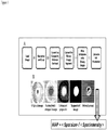
- an alternative version of the SA-HA / HAI was developed based on 96-well ELISA plates ( Fig. 1 ; right columns). Specifically, U-bottom ELISA plates (for example, Immulux HB from Dynex, catalog # 1011, or ImmunoGrade plates from BrandTech Scientific, catalog #781724) were first coated with influenza virus and then blocked with 2% high grade BSA in a manner similar to a regular ELISA protocol. Erythrocytes applied to such plates anchored to the virus particles already attached to the ELISA surface, forming a two-dimensional "micro-halo" that looked quite similar to that observed in the experiments with opsonized plates.
- U-bottom ELISA plates for example, Immulux HB from Dynex, catalog # 1011, or ImmunoGrade plates from BrandTech Scientific, catalog #781724
- Erythrocytes applied to such plates anchored to the virus particles already attached to the ELISA surface, forming a two-dimensional "micro-halo" that looked quite similar to that
- Results of the SA-HA / HAI experiments can be evaluated visually, in a manner similar to the classical HA / HAI. However, using image processing makes such evaluation more precise and significantly reduces the subjectivity of operator.
- Digital images of HA / HAI assay wells can be recorded using an automated imaging system capable of taking high-resolution images of individual wells on a 96-well plate. Adequate systems are available commercially, such as those designed for EliSpot assay analysis, that can acquire the digital images necessary for HA / HAI image analysis. For example, images of HA / HAI assay wells can be recorded on an AID ELISPOT plate reader and stored in JPEG format at 1088 ⁇ 1036 resolution and 24-bit color depth are adequate for HA / HAI analysis.
- the image acquisition software included with these systems is typically full-featured in terms of camera and translation stage control, well selection, and file management; however the included software is intended for ELISPOT analyses and is not capable of proper quantification of HA or HAI titers.
- these types of imagers are useful only for their image acquisition capabilities, and the recorded HA / HAI well images were processed using software developed specifically for determining the HA / HAI titers, as described below.
- HAP Hemagglutination Parameter
- the HAP is minimal for the "button” pattern and maximal for the "halo” pattern.
- the hemagglutination titration curves in the SA-HA or SA-HAI assays can thus be presented as sets of the HAP values linked to the serial dilutions of the virus or sera, respectively, depending on the type of assay. Curve fitting and curve dissecting applied to such a dataset allows precise determination of the titration point.
- Hemagglutination Parameter developed and use of the numerical Hemagglutination Parameter transferred the analysis of the HA or HAI assays from subjective visual evaluation to a precise mathematical calculation.
- the principles of the calculation of the Hemagglutination Parameter (HAP) can be applied to similar calculations of Agglutination Parameters for the target cells other than erythrocytes, or for the target objects other than cells, such as latex beads.
- the objective of the HA / HAI image processing algorithm is to separate the hemagglutination pattern from the rest of the well image and to then determine the HAP value.
- the algorithm developed for the SA-HA / HAI assay is illustrated by the flowchart in Figure 3A . The process is as follows.
- an image of the well is obtained as described above.
- the well image is then cropped to primarily encompass the central pattern-containing portion of the well.
- the cropped image is converted to negative grayscale and then the contrast is adjusted to fill the entire intensity spectrum and enhance the hemagglutination spot.
- Intensity thresholding is applied to remove pixels not corresponding to erythrocytes.
- the image is then segmented using either edge detection or color segmentation algorithms to isolate the erythrocyte pattern.
- Edge detection as illustrated in Figure 3B , required converting of the color image to a binary (black and white) image and segmentation using a binary gradient mask, followed by dilation, hole filling, and image erosion.
- the resulting segmented binary image represents the HA spot pixels, with all background pixels eliminated.
- the grayscale image is then mapped onto the segmented binary image, giving an intensity image of the HA spot.
- the average distance from the spot centroid ⁇ R> and average intensity of the spot ⁇ I> are calculated from the intensity image, and finally the HAP value is calculated as the ratio (1) above.
- the user has control over some of the analysis variables and must either choose their values prior to image processing or accept default values. These variables include, for example, threshold intensity, crop area, and image segmentation type. Once these values are selected they will be applied to all wells in a batch, be it an entire plate or multiple plates.
- the software has a single-well processing mode that allows a user to experiment with the analysis variables on a single well image before applying them to an entire batch. The process is illustrated in the flowchart in Figure 4A . A user selects the single-well analysis mode, loads a single-well image and then sets the analysis variables.
- the image is then processed using the algorithm illustrated by flowcharts in Figures 3A and 4B , and the resulting HAP value is displayed, as well as the processed image, showing the detected hemagglutination pattern.
- the user accepts the values and continues with batch processing or iteratively adjusts the variables and re-processes the image until acceptable values are established.
- values are chosen to provide proper detection of the HA / HAI pattern for the two control cases: the No Virus case in which a micro-button is formed in the well bottom and the No Sera case in which a micro-halo is formed. Because these cases represent the hemagglutination extremes, proper detection of their patterns increases the likelihood of proper detection of the hemagglutination patterns for all wells in the batch.
- an entire plate can be processed, as illustrated by the flowchart in Figure 4C .
- a user selects the single-plate analysis mode and then loads a directory which contains images of wells from the plate.
- the software verifies that the images are valid for processing and then normalizes the background intensity for all images in the folder by sampling each image around the well periphery and normalizing all wells to the maximum average intensity found.
- the HA / HAI well images are then batch-processed by applying the algorithm presented in Figure 4B to each well serially until all wells have been processed. Once a well is processed, its HAP value is readily determined and the results are automatically written to a data file in XML format along with its calculated background value and the analysis variables. Screenshots of the original and processed plate images are also saved along with an Excel file containing the formatted HA parameter values for each well.
- Multiple-plate batch processing mode allows processing of multiple plates at once using the same settings for each plate. This mode requires minimal user interaction and is intended for high-throughput image analysis.
- the process is illustrated by the flowchart in Figure 4D .
- the user first performs a pre-process analysis to determine proper variable values and then selects batch plate analysis mode.
- a directory containing multiple plate directories is then selected and each plate directory is processed serially, similar to the full-plate processing mode. The process continues until all plate directories have been processed. If a directory has been processed previously, the software will check for any missing data files, such as screenshots or Excel files and either re-create them if an XML file is present or re-process the plate in its entirety.
- the titration curves need to be curve-fitted to determine their titration points using the Linked Standard Dilution / Linked HAP value method, LSD / LHAP (below).
- LSD / LHAP the Linked Standard Dilution / Linked HAP value method
- a web-based automated curve fitting and database interface application was developed. The application can automatically curve-fit the SA-HA / HAI titration curves using a weighted five-parameter logistic equation, find the titration point using the LSD / LHAP method and then catalog the results into a central database that can be accessed by multiple users simultaneously.
- the software comprises four main modules: a plate designer module, a file upload module, a curve-fitting module and a database module.
- a plate designer module As illustrated in Figure 4E , the user first creates a virtual plate in the plate designer module and defines the HA / HAI plate layout including all reagent and cell information for each well such as name, type, concentration or dilution, and lot or donor number.
- the virtual HA / HAI plate is stored in the database and can be linked to actual plate data.
- the user then uploads the XML file to the database using the file upload module and links the actual data to the corresponding virtual plate. At this point, the plate is well-defined in the database and ready for curve-fitting.
- the curve-fitting module automatically fits a five-parameter logistic equation to the defined sample and standard curves on the plate and a user-defined linked dilution is applied to find the titration point for each sample on the plate.
- the titration point is stored in the database and made available to users through the database module, which supports complex queries for data mining, plotting and dataset creation.
- the database module can export plots or datasets for use in reports or further analysis using other software.
- HA and HAI assays are prone to significant variability, caused by instability of the virus agglutinating capacity, changes in temperature, and variation in the quality of erythrocytes.
- an advanced standardization protocol named Linked Standard Dilution / Linked Hemagglutination Parameter (LSD / LHAP) was developed.
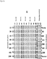
- every SA-HAI plate is organized in a strictly standardized way ( Fig. 5 ).
- the samples are serially diluted in the columns 1 to 10 of the 96-well format, in duplicate.
- Columns 11 and 12 are occupied with a serum selected as a standard for all the SA-HAI plates that are going to be used within a project.
- Wells A11 and A12 contain no virus and no serum (double negative control), and wells H11 and H12 contain virus but no serum (single negative control).
- the standard serum is serially diluted six times starting from the wells B11 and B12, down to the wells G11 and G12, in such a manner that wells containing the standard demonstrate the whole dynamic range of the HAI titration, from a clear micro-button to a clear micro-halo.
- a Linked Standard Dilution (LSD) is selected in the middle of the standard titration array in such a way that the hemagglutination pattern would be between halo and button, as it takes place for wells E11 and E12 containing the standard serum (#10) diluted 1:3200, as shown in Figure 5 .
- the HAP value that is determined for the chosen LSD is named Linked HAP, or LHAP.
- the final LHAP value can differ to a certain extent from the numerical values determined for the LSD wells, because the titration curve is set using a splining procedure that smoothes random scattering of the datapoints ( Fig. 6A ).
- the LSD parameter is kept the same for all the setups performed in a whole study.
- the LHAP value found for the standard sera in each plate is used as a titration target for all the tested samples in the plate ( Fig. 6B ).
- the hemagglutination patterns in the LSD wells of the standard serum would change accordingly (for example, become closer to a button if the agglutinating capacity decreases), and the corresponding LHAP would change as well (for this example, decreases).
- the corresponding HAP values in the wells with the tested sera will shift in the same direction as the LHAP (for this example, decrease).
- the software uses the changed LHAP as the updated titration target, the resulting SA-HAI titers of the tested sera will remain practically unchanged.
- the classical HAI assay integrates neutralizing effects of functional antibodies having different affinities.
- Other immunosorptive methods such as ELISA are able to characterize only antibodies of relatively high affinity.
- the reason for such difference is that in the classical HAI the complexes of antibodies with the virus do not pass through the procedures of sample removal and washing, which constitutes a backbone of the majority of immunosorptive assays, including ELISA.
- even antibodies of relatively low affinity can demonstrate high titers, provided that the antibodies are present in the serum in large quantities.
- sera samples can be removed from the wells after incubation with the virus that is pre-attached to the plates and before application of erythrocytes. This actually triggers dissociation of lower affinity antibodies from the virus particles and removal of those antibodies from the reaction volume, which affects the observed titers.
- hemagglutination (HA) and hemagglutination inhibition (HAI) functional assays remain important instruments of analysis of virus-cell interaction and efficacy of virus-specific antibodies and sera.
- HA and HAI hemagglutination inhibition
- Embodiments of the present disclosure comprise a new method for the functional characterization of viruses and virus-specific antibodies and sera, the Surface-Assisted Hemagglutination / Hemagglutination Inhibition functional assay, the "SA-HA / HAI" assays.
- Embodiments of the present disclosure demonstrate sensitivity of the SA-HA assay to various influenza viruses about 7-200 times higher than the traditional HA, and sensitivity of the SA-HAI assay to influenza-specific antibodies about 10-50 times higher than in the traditional HAI, depending on the type of the virus and the type of erythrocytes used.
- the SA-HA / HAI can use the same types of erythrocytes as the traditional HA / HAI: human, mammalian, and avian.
- the SA-HA / HAI assay results can be evaluated visually, in a manner similar to classical HA / HAI assays.
- visual evaluation lacks adequate precision for high-sensitivity experiments and it is prone to human errors, due to differences in perception of different operators.
- Photo-registration and digital processing of the SA-HA / HAI images increases the precision of the method and eliminates the subjectivity of visual evaluation.
- Introduction of the numerical Hemagglutination Parameter (HAP) that reflects the degree of agglutination in every well of the SA-HA or SA-HAI plate changes the analysis of the HA or HAI assays from a subjective visual evaluation to precise mathematical processing of titration curves.
- the major solvent used for all components of the assay such as media, tested sera, antibody samples, viruses and erythrocytes was PBS saline containing 0.1% of sodium azide NaN 3 (PBS /NaN 3 ). Sodium azide was added to protect the saline from bacterial or yeast contamination.
- Types of erythrocytes used in the SA-HA / HAI experiments were: human group O, turkey, chicken, guinea pig, horse. Human erythrocytes were acquired from Florida Blood Bank or via internal blood donations at Vaxdesign Corp. Turkey, chicken, guinea pig and horse erythrocytes were purchased from Rockland Immunochemicals as suspensions in citrate buffer.
- the erythrocytes were aliquotted immediately after receiving by 1.0 mL in microfuge vials and stored at 4°C until further use.
- the normal storage time was no longer than 3 weeks for human, turkey, horse and chicken erythrocytes, and no longer than 1 week for guinea pig erythrocytes.
- microfuge vials were stored undiluted in 1.5-mL microfuge vials at 4°C. During the processing, the microfuge vial with the virus was stirred vigorously in a bench vortex for 30 s; no sonication was used. Afterwards, the vial was centrifuged (400 g , 5 min). The necessary aliquot of the supernatant was taken out and diluted in 0.5% BSA / PBS / NaN 3 . This processing was performed anew for each day of experiments.
- virus solution was further aliquotted by 0.25 mL, and those secondary aliquots were either used immediately or frozen and stored at -80°C for further use. After thawing, those secondary aliquots could be used immediately without further processing.
- influenza viruses in mice allantoic fluids were obtained from Sanofi Pasteur:
- the plate layout was horizontal (i.e., the placement and serial dilution of the samples performed from left to right).
- the plate was filled with 2% BSA in PBS / NaN 3 , 160 ⁇ L per well, and incubated at 4°C in a planar plate shaker, -600 rpm for at least 40 min.
- the plate was flicked off and tapped upside down on a clean paper towel.
- the plate was filled with 0.5% BSA in PBS / NaN 3 , 40 ⁇ L per well.
- virus initial dilution was 1:50 or 1:100.
- Pre-diluted virus was added to the wells of the column 1, 40 ⁇ L per well, thus becoming diluted 2 times. Dual serial dilutions of the virus were made from left to right, from the column 1 to the column 11 using an 8-channel 200- ⁇ L pipetter and transferring by 40 ⁇ L per channel in every pass. Back-and-forth pipetting in each column was used to mix solutions properly, not less than six pipettings per pass, producing no bubbles. The last 40- ⁇ L portion taken from the column 11 was discarded. The column 12 contained no virus.
- the plate was placed in the plate shaker, such as Digital mini vortexer IKA MS3 from IKA Works, Wilmington, NC, and a short ( ⁇ 5 s) mixing at 1000 rpm was performed three times. After mixing, erythrocytes were added.
- the plate shaker such as Digital mini vortexer IKA MS3 from IKA Works, Wilmington, NC
- Erythrocytes processed and diluted as described above were added to all the wells, 40 ⁇ L per well, the plate was again subjected to short mixing in the plate shaker at 800 rpm, and then incubated with shaking at 500 rpm for 30 min. Afterwards, the plate was left still on the bench for 2-4 h, depending on the type of erythrocytes used in the assay, to allow erythrocytes to precipitate and form the hemagglutination patterns.
- the plate could be read and analyzed visually, or using photo-registration in a short-focus photo reader, such as an ELISPOT plate reader, AID ELR04 AID GmbH, Germany, with subsequent digital processing of the patterns of hemagglutination, as described above, and the SA-HA titer determined as a midpoint of the HA titration ( Fig. 7 ).
- a short-focus photo reader such as an ELISPOT plate reader, AID ELR04 AID GmbH, Germany
- the plate layout was vertical (i.e., the placement and serial dilution of the samples were performed from the row A to the row H of the plate).
- virus titer determined in the previous SA-HA assay was used to calculate the virus dilution to be used in the SA-HAI assay:
- SA - HAI assay virus dilution 4 ⁇ SA - HA titer
- Pre-dilutions of the tested sera were usually from -1:100 to -1:800, depending on the expected immune response.
- Pre-dilutions of MIMIC® samples were usually -1:1 to -1:10, depending on the expected antibody levels.
- Pre-diluted sera or MIMIC® samples were placed in the wells of the row A, 40 ⁇ L per well. Dual serial dilutions of the samples were performed from row A to row G or H using a 12-channel 200- ⁇ L pipetter by transferring 40 ⁇ L from the wells of the previous row to the next row. The technique of the dilution is the same as described above for the typical SA-HA assay.
- Virus diluted according to the results of the SA-HA test as specified above was added to all the wells, 40 ⁇ L per well, except for the No Virus negative control wells, without touching the menisci. For this, the pipette tips were leaned on the top part of the wells.
- the plate was incubated in the planar shaker at 4°C (refrigerator) or at room temperature (on the bench) at 500 rpm, covered, for -1-2 h, depending on the virus type. After the incubation, erythrocytes were added.
- the processing was similar to the described above in the Example 1 for the SA-HA / HAI assays with opsonized plates, except that the final solvent used for viruses before application to the plates was BSA / NaN 3 saline.
- the plate layout was horizontal (i.e., the placement and serial dilution of the samples performed from left to right).
- the filling and serial dilution techniques and the typical layouts were similar to that described above in the Example 1, the protocol for SA-HA / HAI using opsonized plates.
- the filling aliquots were 50 ⁇ L per well.
- the plate was incubated overnight at 4°C (refrigerator), on a planar plate shaker at -500 rpm.
- the plate was flicked off and tapped upside down on a clean paper towel, filled with 2% BSA in PBS / NaN 3 , 200 ⁇ L per well, and incubated at 4°C on a planar plate shaker, -400 rpm for at least 2 h.
- Erythrocytes diluted in the (0.25% BSA + 0.25% OVA) / PBS / NaN 3 were added by 50 ⁇ L per well. Typical concentrations of erythrocytes were 0.01-0.025% HCT. Afterwards, the plate was left still on the bench for -2-4 h., depending on the type of erythrocytes, to allow erythrocytes to precipitate and form the hemagglutination patterns.
- the plate layout was vertical (i.e., the placement and serial dilution of the samples performed from top to bottom).
- the virus titer determined in the SA-HA assay is used to calculate the virus dilution to be used in the SA-HAI assay as shown in the formula (2) above.
- the plate was filled with the virus chosen for the experiment and diluted according to the results of the previously set SA-HA test in PBS / NaN 3 , 50 ⁇ L per well.
- the plate was flicked off and tapped on a clean paper towel, and filled with (0.25% BSA + 0.25% OVA) / PBS / NaN 3 , 50 ⁇ L per well.
- Example 1 As described above for Example 1 with the opsonized plate.
- Erythrocytes at a concentration 10 times higher than the desired final concentration were added by 5.0 ⁇ L to the wells containing 50- ⁇ L samples well by squirting from a 12-channel 200- ⁇ L pipetter. The volumes were squirted in the wells rather than slowly squeezed out. The pipetter was washed with PBS / NaN 3 before every addition of erythrocytes to every row. After adding erythrocytes to all wells, the plate was subjected to short mixing via the planar plate shaker. Typical final concentrations of erythrocytes were -0.01-0.025% HCT.
- the plate was kept still and covered on the bench for -2-4 h, depending on the type of erythrocytes used in the assay to allow erythrocytes to precipitate and form the hemagglutination patterns.
- Typical starting pre-dilution of the virus samples were 1:5 or 1:10.
- HAI assays For most HAI assays, we used a vertical layout (i.e., the serial dilution of the tested sera was performed from the row A to the row G or H of the plate). The virus titer determined in the HA assay was used to calculate the virus dilution to be used in the HAI assay as shown in the formula (2) above.
- MIMIC® samples were not tested using the classical HAI assay due to lack of sensitivity.
- the virus used in the experiment was A/Solomon Islands/3/2006 [H1N1], BPL-inactivated standard from CDC.
- the final concentration of human erythrocytes in the wells of the classical HAI was 0.5% HCT, and the final virus dilution in the wells was 1:120.
- the corresponding numbers were 0.017% HCT and 1:12000.
- the comparison of the classical HAI and SA-HAI with opsonized plates and human erythrocytes is presented in Figure 9 and in the data table presented with the figure. Correlation between the two methods was good, and the SA-HAI demonstrated sensitivity enhancement ⁇ 23-fold over the classical method, as seen from the averaged titer ratio presented in the data table and from the slope of the correlative scattered plot ( Fig. 9 ).
- Example 5 Comparison of the classical HAI performed with human erythrocytes and SA-HAI performed with turkey erythrocytes
- the correlation between the two methods was good, and the SA-HAI assay demonstrated sensitivity enhancement ⁇ 21-27-fold versus the classical method, as seen from the averaged titer ratio and from the slope of the correlative scattered plot.
- Example 6 Comparing classical HAI titers determined with human erythrocytes and SA-HAI titers using guinea pig erythrocytes
- Pre- and post-vaccination sera from three donors immunized for influenza in the season 2007 / 2008 were tested in the classical HAI assay using human group O erythrocytes and the SA-HAI assay with opsonized plates using guinea pig erythrocytes, as specified above.
- the virus was A/Solomon Islands/3/2006 [H1N1], BPL-inactivated standard from CDC.
- the final concentration of guinea pig erythrocytes and final virus dilutions in the SA-HAI assay were 0.0083% HCT and 1:10000, respectively.
- the results presented in Figure 11 and in the data table presented with the figure demonstrated good correlation with the classical HAI method and sensitivity enhancement of ⁇ 30-50-fold, judged from the averaged titer ratio and from the slope of the correlative scattered plot.
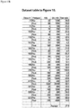
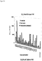
- Example 7 Comparing classical HA titers and SA-HA titers for influenza viruses in allantoic fluids
- the classical HA assay was performed in Low Volume mode, as described above, and the SA-HA assay was performed with ELISA plates in the year 2010 using virus samples in mice allantoic fluids, as listed above.
- the results presented in Figure 12 and in the data table placed beside the figure demonstrate ⁇ 7- to ⁇ 200-fold enhancement of sensitivity in the SA-HA assay versus the classical HA assay, depending on the virus type.
- Example 8 Comparing classical HAI and SA-HAI titers using turkey erythrocytes and H1N1 influenza virus in allantoic fluids
- the classical HAI assay was performed in Low Volume mode, as described above, and the SA-HAI assay was performed with ELISA plates in the year 2010 using virus samples in mice allantoic fluids.
- the virus was A/Brisbane/59/2007 [H1N1].
- the final concentration of turkey erythrocytes and final virus sample dilution in the SA-HAI assay were 0.025% HCT and 1:5280.
- the results presented in Figure 13 and in the data table placed beside the figure demonstrate good correlation between the two assays and a sensitivity enhancement of ⁇ 7-fold, judged from the averaged titer ratio and from the slope of the correlative scattered plot.
- Example 9 Comparing classical HAI and SA-HAI titers using turkey erythrocytes and H3N2 influenza virus in allantoic fluid
- the classical HAI assay was performed in Low Volume mode, as described above, and the SA-HAI assay was performed with ELISA plates in the year 2010 using virus samples in mice allantoic fluids.
- the virus was A/Wisconsin/67/2005 [H3N2].
- the final concentration of turkey erythrocytes and final virus dilution in the SA-HAI assay were 0.025% HCT and 1:1600.
- the results presented in Figure 14 and data table placed beside the figure demonstrate a sensitivity enhancement of ⁇ 30-fold, judged from the averaged titer ratio. Low numbers of available samples did not allow building a comprehensive dual scattered plot as in the previous experiments.
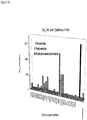
- the SA-HAI assays of the donor sera were performed using A/Brisbane/59/2007 [H1N1] and A/California/7/2009 [H1N1] BPL-inactivated virus standards from the CDC, in the opsonized plate mode.
- the results of sera screening are shown in Figure 15 . While the efficacy of the vaccine against the seasonal virus was high, as can be seen by the difference between the pre- and post-vaccine titers, the cross-activity of the immunized sera against the new Swine Flu virus was significantly lower the level that is required for protection.
- the protection level for SA-HAI titers was estimated -640 to compare with the level -64 accepted for the classical HAI assays. The estimated sensitivity enhancement of the assay towards the classical HAI was -10 (data not shown).
- Example 11 SA-HAI analysis of samples from in vitro MIMIC® setups immunized with vaccine and recombinant antigen
- MIMIC® setups based on immune cells from human donors were immunized in vitro with Fluvirin-2010 seasonal vaccine and with recombinant HI hemagglutinin originated from A/California/7/2009 [H1N1] "Swine Flu” virus (Protein Sciences, Meriden, CT).
- the MIMIC® supernatants were tested with SA-HAI assay using ELISA plates.
- the results of the screening in Figure 16 demonstrate significant increase of virus-specific and potentially neutralizing antibodies after vaccination, in comparison with the No Antigen samples.
- challenging the MIMIC® system with vaccine generally demonstrated higher efficacy than challenging with the recombinant antigen, which may have been due to the effective adjuvant used in the vaccine formulation.
Landscapes
- Health & Medical Sciences (AREA)
- Life Sciences & Earth Sciences (AREA)
- Immunology (AREA)
- Engineering & Computer Science (AREA)
- Chemical & Material Sciences (AREA)
- Biomedical Technology (AREA)
- Hematology (AREA)
- Molecular Biology (AREA)
- Urology & Nephrology (AREA)
- Cell Biology (AREA)
- Microbiology (AREA)
- Food Science & Technology (AREA)
- General Physics & Mathematics (AREA)
- Medicinal Chemistry (AREA)
- Physics & Mathematics (AREA)
- Analytical Chemistry (AREA)
- Biochemistry (AREA)
- General Health & Medical Sciences (AREA)
- Biotechnology (AREA)
- Pathology (AREA)
- Virology (AREA)
- Chemical Kinetics & Catalysis (AREA)
- Tropical Medicine & Parasitology (AREA)
- Mycology (AREA)
- Measuring Or Testing Involving Enzymes Or Micro-Organisms (AREA)
- Peptides Or Proteins (AREA)
- Micro-Organisms Or Cultivation Processes Thereof (AREA)
Claims (14)
- Verfahren zur Bestimmung funktioneller Bindungsaktivität eines Antikörpers für einen agglutinierenden Faktor, umfassend:(a) Inkubieren eines agglutinierenden Faktors mit einem Antikörper in einer Vertiefung einer Kulturplatte, wobei die Vertiefung mit einem Protein oder Lectin beschichtet wurde, und(b) Zugeben eines Zielobjekts zu der Vertiefung von (a) unter Bedingungen, die eine Agglutination des Zielobjekts durch den agglutinierenden Faktor erlauben, und(c) Nachweisen der Agglutination des Zielobjekts auf der Oberfläche des Vertiefungsbodens von (b), wobei, wenn die in (c) nachgewiesene Agglutination geringer ist als die in Abwesenheit des Antikörpers nachgewiesene Agglutination, der Antikörper als funktionale Bindungsaktivität für den agglutinierenden Faktor aufweisend bestimmt wird, undwobei der agglutinierende Faktor aus der Gruppe, bestehend aus einem Virus, einem virusartigen Partikel und einem Bakterium, ausgewählt ist.
- Verfahren zur Bestimmung funktioneller Bindungsaktivität eines Antikörpers für einen agglutinierenden Faktor, umfassend:(a) Inkubieren eines agglutinierenden Faktors in einer Vertiefung einer ELISA-Platte,(b) Zugeben eines Antikörpers zu der Vertiefung von (a),(c) Zugeben eines Zielobjekts zu der Vertiefung von (b) unter Bedingungen, die eine Agglutination des Zielobjekts durch den agglutinierenden Faktor erlauben, und(d) Nachweisen der Agglutination des Zielobjekts auf der Oberfläche des Vertiefungsbodens von (c), wobei, wenn die in (d) nachgewiesene Agglutination geringer ist als die in Abwesenheit des Antikörpers nachgewiesene Agglutination, der Antikörper als funktionale Bindungsaktivität für den agglutinierenden Faktor aufweisend bestimmt wird, undwobei der agglutinierende Faktor aus der Gruppe, bestehend aus einem Virus, einem virusartigen Partikel und einem Bakterium, ausgewählt ist.
- Verfahren nach Anspruch 2, weiter umfassend Waschen der Vertiefung der Platte von (c) mit einer gepufferten Waschlösung nach Zugabe des Antikörpers zu der Vertiefung und Bestimmen eines relativen Beitrags hochaffiner Antikörper zu der in (d) nachgewiesenen Agglutination durch Vergleich des in (d) nachgewiesenen Wertes mit einem in (d) nachgewiesenen Wert, wo die Vertiefung nicht mit einer gepufferten Waschlösung nach Zugabe des Antikörpers zu der Vertiefung gewaschen wurde.
- Verfahren nach Anspruch 2, weiter umfassend die Zugabe eines Blockierungsmittels zu der Vertiefung von (a) vor Zugabe des Antikörpers.
- Verfahren nach Anspruch 4, wobei das Blockierungsmittel 2 % BSA in PBS ist.
- Verfahren nach Anspruch 1 oder Anspruch 2, wobei die Agglutination durch Quantifizierung zweidimensionaler Agglutinationsmuster, erzeugt durch die agglutinierten Zielobjekte, auf der Oberfläche des Vertiefungsbodens der Platte nachgewiesen wird.
- Verfahren nach Anspruch 6, wobei die Quantifizierung zweidimensionaler Muster unter Verwendung digitaler Fotoregistrierung und digitaler Bildverarbeitung durchgeführt wird, wobei die digitale Bildbearbeitung die Berechnung eines numerischen Agglutinationsparameters, der den Agglutinationsgrad widerspiegelt, einschließt.
- Verfahren nach Anspruch 7, wobei der Agglutinationsparameter ein Verhältnis der Größe des Bildbereichs, enthaltend die zweidimensionalen Agglutinationsmuster auf der Oberfläche des Vertiefungsbodens, zu der durchschnittlichen Pixelintensität der Agglutinationsmuster in dem Bereich ist.
- Verfahren nach Anspruch 1 oder Anspruch 2, wobei das Zielobjekt Latexmikrokügelchen sind, die mit einem Rezeptor beschichtet sind, der an den agglutinierenden Faktor bindet.
- Verfahren nach einem der Ansprüche 1 bis 8, wobei das Zielobjekt in Zellen aus der Gruppe, bestehend aus Erythrozyten, Lymphozyten, Epithelzellen und Endothelzellen, ausgewählt ist.
- Verfahren nach Anspruch 10, wobei die Zellen in einer Konzentration von weniger als 0,1 % Hämatokrit vorliegende Erythrozyten sind.
- Verfahren nach Anspruch 1, wobei die Vertiefung mit Rinderserumalbumin oder Humanserumalbumin beschichtet ist.
- Verfahren nach Anspruch 2, wobei die ELISA-Platte eine ELISA-Platte mit U-förmigen Vertiefungen oder V-förmigen Vertiefungen ist.
- Verfahren nach Anspruch 1 oder Anspruch 2, wobei der Antikörper der Vertiefung als den Antikörper umfassendes Serum zugegeben wird.
Applications Claiming Priority (2)
| Application Number | Priority Date | Filing Date | Title |
|---|---|---|---|
| US25326609P | 2009-10-20 | 2009-10-20 | |
| PCT/US2010/053322 WO2011050027A2 (en) | 2009-10-20 | 2010-10-20 | Surface-assisted hemagglutination and hemagglutination inhibition assays |
Publications (3)
| Publication Number | Publication Date |
|---|---|
| EP2491394A2 EP2491394A2 (de) | 2012-08-29 |
| EP2491394A4 EP2491394A4 (de) | 2013-12-25 |
| EP2491394B1 true EP2491394B1 (de) | 2018-04-18 |
Family
ID=43898745
Family Applications (1)
| Application Number | Title | Priority Date | Filing Date |
|---|---|---|---|
| EP10825569.6A Active EP2491394B1 (de) | 2009-10-20 | 2010-10-20 | Oberflächenunterstützte hämagglutinationshemmungstests |
Country Status (4)
| Country | Link |
|---|---|
| US (1) | US8962256B2 (de) |
| EP (1) | EP2491394B1 (de) |
| CA (1) | CA2776405C (de) |
| WO (1) | WO2011050027A2 (de) |
Families Citing this family (5)
| Publication number | Priority date | Publication date | Assignee | Title |
|---|---|---|---|---|
| US9389229B2 (en) | 2012-07-18 | 2016-07-12 | Theranos, Inc. | Methods for detecting and measuring aggregation |
| US10012643B2 (en) * | 2014-07-28 | 2018-07-03 | Sanofi Pasteur Vaxdesign Corporation | Automated imaging and analysis of the hemagglutination inhibition assay (HAI) |
| WO2017100660A1 (en) | 2015-12-09 | 2017-06-15 | Indevr, Inc. | Automated agglutination analyzer with contour comparison |
| CN111198265A (zh) * | 2018-11-20 | 2020-05-26 | 内蒙古正大食品有限公司 | 一种多种禽抗体联合快速检测方法 |
| AU2021253945A1 (en) * | 2020-04-09 | 2022-11-03 | Ilytica Llc | Assays with induced aggregation for enhanced sensitivity |
Family Cites Families (6)
| Publication number | Priority date | Publication date | Assignee | Title |
|---|---|---|---|---|
| FR2450877A1 (fr) * | 1979-03-06 | 1980-10-03 | Inst Nat Sante Rech Med | Nouveaux tests par agglutination pour la detection des virus de la grippe, et reactifs pour la realisation de ces tests |
| US5494800A (en) * | 1987-01-29 | 1996-02-27 | Cytosignet, Inc. | Analyte detection in particulate-containing samples |
| US4921787A (en) * | 1987-05-01 | 1990-05-01 | Cambridge Bioscience Corporation | Detection of antibodies to human immunodeficiency virus by agglutination of antigen coated latex |
| GB9314011D0 (en) * | 1993-07-07 | 1993-08-18 | Porton Cambridge Ltd | New diagnostic assay for detection of syphilis |
| US5583004A (en) * | 1994-12-23 | 1996-12-10 | Pincus; Mathew R. | Direct detection of unexpected alloantibodies in the serum of prospective transfusion recipients using a new hemagglutination inhibition assay |
| WO2010002852A2 (en) * | 2008-06-30 | 2010-01-07 | Vaxdesign Corp. | Bead array reader based-hemagglutination and hemagglutination inhibition assays |
-
2010
- 2010-10-20 WO PCT/US2010/053322 patent/WO2011050027A2/en not_active Ceased
- 2010-10-20 US US12/908,211 patent/US8962256B2/en active Active
- 2010-10-20 CA CA2776405A patent/CA2776405C/en active Active
- 2010-10-20 EP EP10825569.6A patent/EP2491394B1/de active Active
Non-Patent Citations (1)
| Title |
|---|
| MICHAELJANICH ET AL.: "Hemagglutination assay", WIKIPEDIA, 27 March 2017 (2017-03-27), pages 1 - 3, XP055367935, Retrieved from the Internet <URL:https://en.wikipedia.org/wiki/Hemagglutination_assay> [retrieved on 20170426] * |
Also Published As
| Publication number | Publication date |
|---|---|
| EP2491394A2 (de) | 2012-08-29 |
| WO2011050027A2 (en) | 2011-04-28 |
| CA2776405C (en) | 2020-08-18 |
| CA2776405A1 (en) | 2011-04-28 |
| US8962256B2 (en) | 2015-02-24 |
| EP2491394A4 (de) | 2013-12-25 |
| US20110097705A1 (en) | 2011-04-28 |
| WO2011050027A3 (en) | 2011-08-18 |
Similar Documents
| Publication | Publication Date | Title |
|---|---|---|
| Payne | Methods to study viruses | |
| JP5539958B2 (ja) | 薄膜状体液試料において実施される血清学的凝集イムノアッセイ及び他のイムノアッセイのための方法 | |
| KR101877576B1 (ko) | Jc 바이러스 항체에 대한 검정법 | |
| EP3084441B1 (de) | Diagnostischer test auf csfv-antikörper in mit csfv e2 geimpften tieren | |
| KR102039751B1 (ko) | Pml의 위험을 판단하는 방법 | |
| US20210088517A1 (en) | MULTIPLEX HIGH-THROUGHPUT FLOW CYTOMETRY DETECTION OF SARS-COV-2-SPECIFIC IgG, IgA AND IgM | |
| EP2491394B1 (de) | Oberflächenunterstützte hämagglutinationshemmungstests | |
| Xia et al. | The glycan array platform as a tool to identify carbohydrate antigens | |
| US20090325148A1 (en) | Bead Array Reader Based-Hemagglutination and Hemagglutination Inhibition Assay | |
| Hansen et al. | Diagnosing Zika virus infection against a background of other flaviviruses: Studies in high resolution serological analysis | |
| US20230305005A1 (en) | Coronavirus point-of-care agglutination assay | |
| US20180238876A1 (en) | Method For Assessing Cell Surface Receptors of Blood Cells | |
| Fhied et al. | Dynamic Monitoring of Seroconversion using a Multianalyte Immunobead Assay for Covid-19. | |
| Kesari et al. | Suitability of dyed latex bead agglutination test for immunodiagnosis of Karnal bunt (Tilletia indica) teliospores in a single seed of wheat | |
| Ayling et al. | Measuring vaccine responses in the multiplex era | |
| AU2015213838B2 (en) | Crossmatching blood samples | |
| Gantelius et al. | Magnetic bead-based detection of autoimmune responses using protein microarrays | |
| Asati et al. | Fluorescence Adherence Inhibition Assay: A Novel Functional Assessment of Blocking Virus Attachment by Vaccine-Induced Antibodies | |
| US20200400667A1 (en) | Method for the quantification of measles and rubella targets | |
| Schmitz | 14 Serologic Testing for Infectious Diseases | |
| SPENCER | AJ DELLA-PORTA, J. YOUNG, E. HANSSON CSIRO, Australian Animal Health Laboratory, Geelong | |
| Schmitz | 14 Serologic Testing |
Legal Events
| Date | Code | Title | Description |
|---|---|---|---|
| PUAI | Public reference made under article 153(3) epc to a published international application that has entered the european phase |
Free format text: ORIGINAL CODE: 0009012 |
|
| 17P | Request for examination filed |
Effective date: 20120518 |
|
| AK | Designated contracting states |
Kind code of ref document: A2 Designated state(s): AL AT BE BG CH CY CZ DE DK EE ES FI FR GB GR HR HU IE IS IT LI LT LU LV MC MK MT NL NO PL PT RO RS SE SI SK SM TR |
|
| DAX | Request for extension of the european patent (deleted) | ||
| REG | Reference to a national code |
Ref country code: DE Ref legal event code: R079 Ref document number: 602010050079 Country of ref document: DE Free format text: PREVIOUS MAIN CLASS: G01N0033569000 Ipc: G01N0033555000 |
|
| A4 | Supplementary search report drawn up and despatched |
Effective date: 20131127 |
|
| RIC1 | Information provided on ipc code assigned before grant |
Ipc: C07K 16/08 20060101ALI20131121BHEP Ipc: G01N 33/569 20060101ALI20131121BHEP Ipc: G01N 33/543 20060101ALI20131121BHEP Ipc: G01N 33/555 20060101AFI20131121BHEP |
|
| STAA | Information on the status of an ep patent application or granted ep patent |
Free format text: STATUS: EXAMINATION IS IN PROGRESS |
|
| 17Q | First examination report despatched |
Effective date: 20170512 |
|
| GRAP | Despatch of communication of intention to grant a patent |
Free format text: ORIGINAL CODE: EPIDOSNIGR1 |
|
| STAA | Information on the status of an ep patent application or granted ep patent |
Free format text: STATUS: GRANT OF PATENT IS INTENDED |
|
| INTG | Intention to grant announced |
Effective date: 20171106 |
|
| RIN1 | Information on inventor provided before grant (corrected) |
Inventor name: TAPIA, TENEKUA Inventor name: NGUYEN, MIKE Inventor name: DHIR, VIPRA Inventor name: KACHURIN, ANATOLY Inventor name: KACHURINA, OLGA Inventor name: WITTMAN, VAUGHAN Inventor name: KAROL, ALEXANDER |
|
| GRAS | Grant fee paid |
Free format text: ORIGINAL CODE: EPIDOSNIGR3 |
|
| GRAA | (expected) grant |
Free format text: ORIGINAL CODE: 0009210 |
|
| STAA | Information on the status of an ep patent application or granted ep patent |
Free format text: STATUS: THE PATENT HAS BEEN GRANTED |
|
| AK | Designated contracting states |
Kind code of ref document: B1 Designated state(s): AL AT BE BG CH CY CZ DE DK EE ES FI FR GB GR HR HU IE IS IT LI LT LU LV MC MK MT NL NO PL PT RO RS SE SI SK SM TR |
|
| REG | Reference to a national code |
Ref country code: GB Ref legal event code: FG4D |
|
| REG | Reference to a national code |
Ref country code: CH Ref legal event code: EP |
|
| REG | Reference to a national code |
Ref country code: AT Ref legal event code: REF Ref document number: 991044 Country of ref document: AT Kind code of ref document: T Effective date: 20180515 |
|
| REG | Reference to a national code |
Ref country code: IE Ref legal event code: FG4D |
|
| REG | Reference to a national code |
Ref country code: DE Ref legal event code: R096 Ref document number: 602010050079 Country of ref document: DE |
|
| REG | Reference to a national code |
Ref country code: NL Ref legal event code: MP Effective date: 20180418 |
|
| REG | Reference to a national code |
Ref country code: LT Ref legal event code: MG4D |
|
| REG | Reference to a national code |
Ref country code: FR Ref legal event code: PLFP Year of fee payment: 9 |
|
| PG25 | Lapsed in a contracting state [announced via postgrant information from national office to epo] |
Ref country code: NL Free format text: LAPSE BECAUSE OF FAILURE TO SUBMIT A TRANSLATION OF THE DESCRIPTION OR TO PAY THE FEE WITHIN THE PRESCRIBED TIME-LIMIT Effective date: 20180418 |
|
| PG25 | Lapsed in a contracting state [announced via postgrant information from national office to epo] |
Ref country code: AL Free format text: LAPSE BECAUSE OF FAILURE TO SUBMIT A TRANSLATION OF THE DESCRIPTION OR TO PAY THE FEE WITHIN THE PRESCRIBED TIME-LIMIT Effective date: 20180418 Ref country code: NO Free format text: LAPSE BECAUSE OF FAILURE TO SUBMIT A TRANSLATION OF THE DESCRIPTION OR TO PAY THE FEE WITHIN THE PRESCRIBED TIME-LIMIT Effective date: 20180718 Ref country code: ES Free format text: LAPSE BECAUSE OF FAILURE TO SUBMIT A TRANSLATION OF THE DESCRIPTION OR TO PAY THE FEE WITHIN THE PRESCRIBED TIME-LIMIT Effective date: 20180418 Ref country code: PL Free format text: LAPSE BECAUSE OF FAILURE TO SUBMIT A TRANSLATION OF THE DESCRIPTION OR TO PAY THE FEE WITHIN THE PRESCRIBED TIME-LIMIT Effective date: 20180418 Ref country code: LT Free format text: LAPSE BECAUSE OF FAILURE TO SUBMIT A TRANSLATION OF THE DESCRIPTION OR TO PAY THE FEE WITHIN THE PRESCRIBED TIME-LIMIT Effective date: 20180418 Ref country code: SE Free format text: LAPSE BECAUSE OF FAILURE TO SUBMIT A TRANSLATION OF THE DESCRIPTION OR TO PAY THE FEE WITHIN THE PRESCRIBED TIME-LIMIT Effective date: 20180418 Ref country code: FI Free format text: LAPSE BECAUSE OF FAILURE TO SUBMIT A TRANSLATION OF THE DESCRIPTION OR TO PAY THE FEE WITHIN THE PRESCRIBED TIME-LIMIT Effective date: 20180418 Ref country code: BG Free format text: LAPSE BECAUSE OF FAILURE TO SUBMIT A TRANSLATION OF THE DESCRIPTION OR TO PAY THE FEE WITHIN THE PRESCRIBED TIME-LIMIT Effective date: 20180718 |
|
| PG25 | Lapsed in a contracting state [announced via postgrant information from national office to epo] |
Ref country code: RS Free format text: LAPSE BECAUSE OF FAILURE TO SUBMIT A TRANSLATION OF THE DESCRIPTION OR TO PAY THE FEE WITHIN THE PRESCRIBED TIME-LIMIT Effective date: 20180418 Ref country code: LV Free format text: LAPSE BECAUSE OF FAILURE TO SUBMIT A TRANSLATION OF THE DESCRIPTION OR TO PAY THE FEE WITHIN THE PRESCRIBED TIME-LIMIT Effective date: 20180418 Ref country code: GR Free format text: LAPSE BECAUSE OF FAILURE TO SUBMIT A TRANSLATION OF THE DESCRIPTION OR TO PAY THE FEE WITHIN THE PRESCRIBED TIME-LIMIT Effective date: 20180719 Ref country code: HR Free format text: LAPSE BECAUSE OF FAILURE TO SUBMIT A TRANSLATION OF THE DESCRIPTION OR TO PAY THE FEE WITHIN THE PRESCRIBED TIME-LIMIT Effective date: 20180418 |
|
| REG | Reference to a national code |
Ref country code: AT Ref legal event code: MK05 Ref document number: 991044 Country of ref document: AT Kind code of ref document: T Effective date: 20180418 |
|
| PG25 | Lapsed in a contracting state [announced via postgrant information from national office to epo] |
Ref country code: PT Free format text: LAPSE BECAUSE OF FAILURE TO SUBMIT A TRANSLATION OF THE DESCRIPTION OR TO PAY THE FEE WITHIN THE PRESCRIBED TIME-LIMIT Effective date: 20180820 |
|
| REG | Reference to a national code |
Ref country code: DE Ref legal event code: R097 Ref document number: 602010050079 Country of ref document: DE |
|
| PG25 | Lapsed in a contracting state [announced via postgrant information from national office to epo] |
Ref country code: SK Free format text: LAPSE BECAUSE OF FAILURE TO SUBMIT A TRANSLATION OF THE DESCRIPTION OR TO PAY THE FEE WITHIN THE PRESCRIBED TIME-LIMIT Effective date: 20180418 Ref country code: EE Free format text: LAPSE BECAUSE OF FAILURE TO SUBMIT A TRANSLATION OF THE DESCRIPTION OR TO PAY THE FEE WITHIN THE PRESCRIBED TIME-LIMIT Effective date: 20180418 Ref country code: DK Free format text: LAPSE BECAUSE OF FAILURE TO SUBMIT A TRANSLATION OF THE DESCRIPTION OR TO PAY THE FEE WITHIN THE PRESCRIBED TIME-LIMIT Effective date: 20180418 Ref country code: AT Free format text: LAPSE BECAUSE OF FAILURE TO SUBMIT A TRANSLATION OF THE DESCRIPTION OR TO PAY THE FEE WITHIN THE PRESCRIBED TIME-LIMIT Effective date: 20180418 Ref country code: CZ Free format text: LAPSE BECAUSE OF FAILURE TO SUBMIT A TRANSLATION OF THE DESCRIPTION OR TO PAY THE FEE WITHIN THE PRESCRIBED TIME-LIMIT Effective date: 20180418 Ref country code: RO Free format text: LAPSE BECAUSE OF FAILURE TO SUBMIT A TRANSLATION OF THE DESCRIPTION OR TO PAY THE FEE WITHIN THE PRESCRIBED TIME-LIMIT Effective date: 20180418 |
|
| PLBE | No opposition filed within time limit |
Free format text: ORIGINAL CODE: 0009261 |
|
| STAA | Information on the status of an ep patent application or granted ep patent |
Free format text: STATUS: NO OPPOSITION FILED WITHIN TIME LIMIT |
|
| PG25 | Lapsed in a contracting state [announced via postgrant information from national office to epo] |
Ref country code: SM Free format text: LAPSE BECAUSE OF FAILURE TO SUBMIT A TRANSLATION OF THE DESCRIPTION OR TO PAY THE FEE WITHIN THE PRESCRIBED TIME-LIMIT Effective date: 20180418 |
|
| 26N | No opposition filed |
Effective date: 20190121 |
|
| PG25 | Lapsed in a contracting state [announced via postgrant information from national office to epo] |
Ref country code: SI Free format text: LAPSE BECAUSE OF FAILURE TO SUBMIT A TRANSLATION OF THE DESCRIPTION OR TO PAY THE FEE WITHIN THE PRESCRIBED TIME-LIMIT Effective date: 20180418 |
|
| PG25 | Lapsed in a contracting state [announced via postgrant information from national office to epo] |
Ref country code: LU Free format text: LAPSE BECAUSE OF NON-PAYMENT OF DUE FEES Effective date: 20181020 Ref country code: MC Free format text: LAPSE BECAUSE OF FAILURE TO SUBMIT A TRANSLATION OF THE DESCRIPTION OR TO PAY THE FEE WITHIN THE PRESCRIBED TIME-LIMIT Effective date: 20180418 |
|
| PG25 | Lapsed in a contracting state [announced via postgrant information from national office to epo] |
Ref country code: MT Free format text: LAPSE BECAUSE OF NON-PAYMENT OF DUE FEES Effective date: 20181020 |
|
| PG25 | Lapsed in a contracting state [announced via postgrant information from national office to epo] |
Ref country code: TR Free format text: LAPSE BECAUSE OF FAILURE TO SUBMIT A TRANSLATION OF THE DESCRIPTION OR TO PAY THE FEE WITHIN THE PRESCRIBED TIME-LIMIT Effective date: 20180418 |
|
| PG25 | Lapsed in a contracting state [announced via postgrant information from national office to epo] |
Ref country code: MK Free format text: LAPSE BECAUSE OF NON-PAYMENT OF DUE FEES Effective date: 20180418 Ref country code: HU Free format text: LAPSE BECAUSE OF FAILURE TO SUBMIT A TRANSLATION OF THE DESCRIPTION OR TO PAY THE FEE WITHIN THE PRESCRIBED TIME-LIMIT; INVALID AB INITIO Effective date: 20101020 Ref country code: CY Free format text: LAPSE BECAUSE OF FAILURE TO SUBMIT A TRANSLATION OF THE DESCRIPTION OR TO PAY THE FEE WITHIN THE PRESCRIBED TIME-LIMIT Effective date: 20180418 |
|
| PG25 | Lapsed in a contracting state [announced via postgrant information from national office to epo] |
Ref country code: IS Free format text: LAPSE BECAUSE OF FAILURE TO SUBMIT A TRANSLATION OF THE DESCRIPTION OR TO PAY THE FEE WITHIN THE PRESCRIBED TIME-LIMIT Effective date: 20180818 |
|
| PGFP | Annual fee paid to national office [announced via postgrant information from national office to epo] |
Ref country code: IT Payment date: 20250922 Year of fee payment: 16 |
|
| PGFP | Annual fee paid to national office [announced via postgrant information from national office to epo] |
Ref country code: BE Payment date: 20250917 Year of fee payment: 16 Ref country code: GB Payment date: 20250904 Year of fee payment: 16 |
|
| PGFP | Annual fee paid to national office [announced via postgrant information from national office to epo] |
Ref country code: FR Payment date: 20250908 Year of fee payment: 16 |
|
| PGFP | Annual fee paid to national office [announced via postgrant information from national office to epo] |
Ref country code: IE Payment date: 20250909 Year of fee payment: 16 |
|
| REG | Reference to a national code |
Ref country code: CH Ref legal event code: U11 Free format text: ST27 STATUS EVENT CODE: U-0-0-U10-U11 (AS PROVIDED BY THE NATIONAL OFFICE) Effective date: 20251101 |
|
| PGFP | Annual fee paid to national office [announced via postgrant information from national office to epo] |
Ref country code: DE Payment date: 20250902 Year of fee payment: 16 |
|
| PGFP | Annual fee paid to national office [announced via postgrant information from national office to epo] |
Ref country code: CH Payment date: 20251101 Year of fee payment: 16 |