EP2490609B1 - System for normalizing implant strain readings to assess bone healing - Google Patents
System for normalizing implant strain readings to assess bone healing Download PDFInfo
- Publication number
- EP2490609B1 EP2490609B1 EP10774080.5A EP10774080A EP2490609B1 EP 2490609 B1 EP2490609 B1 EP 2490609B1 EP 10774080 A EP10774080 A EP 10774080A EP 2490609 B1 EP2490609 B1 EP 2490609B1
- Authority
- EP
- European Patent Office
- Prior art keywords
- bone
- sensor
- implant
- strain
- fracture
- Prior art date
- Legal status (The legal status is an assumption and is not a legal conclusion. Google has not performed a legal analysis and makes no representation as to the accuracy of the status listed.)
- Active
Links
- 210000000988 bone and bone Anatomy 0.000 title claims description 89
- 239000007943 implant Substances 0.000 title claims description 62
- 230000035876 healing Effects 0.000 title claims description 23
- 230000001054 cortical effect Effects 0.000 claims description 3
- 230000000694 effects Effects 0.000 claims description 2
- 238000005452 bending Methods 0.000 claims 1
- 238000005259 measurement Methods 0.000 description 13
- 239000007787 solid Substances 0.000 description 10
- 238000013480 data collection Methods 0.000 description 2
- 230000003247 decreasing effect Effects 0.000 description 2
- 238000012986 modification Methods 0.000 description 2
- 230000004048 modification Effects 0.000 description 2
- 238000012545 processing Methods 0.000 description 2
- 238000002513 implantation Methods 0.000 description 1
- 210000001930 leg bone Anatomy 0.000 description 1
- 230000007246 mechanism Effects 0.000 description 1
- 238000000034 method Methods 0.000 description 1
- 230000000399 orthopedic effect Effects 0.000 description 1
- 239000000758 substrate Substances 0.000 description 1
Images

Classifications
-
- A—HUMAN NECESSITIES
- A61—MEDICAL OR VETERINARY SCIENCE; HYGIENE
- A61B—DIAGNOSIS; SURGERY; IDENTIFICATION
- A61B5/00—Measuring for diagnostic purposes; Identification of persons
- A61B5/45—For evaluating or diagnosing the musculoskeletal system or teeth
- A61B5/4504—Bones
-
- A—HUMAN NECESSITIES
- A61—MEDICAL OR VETERINARY SCIENCE; HYGIENE
- A61F—FILTERS IMPLANTABLE INTO BLOOD VESSELS; PROSTHESES; DEVICES PROVIDING PATENCY TO, OR PREVENTING COLLAPSING OF, TUBULAR STRUCTURES OF THE BODY, e.g. STENTS; ORTHOPAEDIC, NURSING OR CONTRACEPTIVE DEVICES; FOMENTATION; TREATMENT OR PROTECTION OF EYES OR EARS; BANDAGES, DRESSINGS OR ABSORBENT PADS; FIRST-AID KITS
- A61F2/00—Filters implantable into blood vessels; Prostheses, i.e. artificial substitutes or replacements for parts of the body; Appliances for connecting them with the body; Devices providing patency to, or preventing collapsing of, tubular structures of the body, e.g. stents
- A61F2/02—Prostheses implantable into the body
- A61F2/28—Bones
-
- A—HUMAN NECESSITIES
- A61—MEDICAL OR VETERINARY SCIENCE; HYGIENE
- A61B—DIAGNOSIS; SURGERY; IDENTIFICATION
- A61B5/00—Measuring for diagnostic purposes; Identification of persons
- A61B5/07—Endoradiosondes
- A61B5/076—Permanent implantation
-
- A—HUMAN NECESSITIES
- A61—MEDICAL OR VETERINARY SCIENCE; HYGIENE
- A61B—DIAGNOSIS; SURGERY; IDENTIFICATION
- A61B17/00—Surgical instruments, devices or methods
- A61B17/56—Surgical instruments or methods for treatment of bones or joints; Devices specially adapted therefor
- A61B17/58—Surgical instruments or methods for treatment of bones or joints; Devices specially adapted therefor for osteosynthesis, e.g. bone plates, screws or setting implements
- A61B17/68—Internal fixation devices, including fasteners and spinal fixators, even if a part thereof projects from the skin
- A61B17/80—Cortical plates, i.e. bone plates; Instruments for holding or positioning cortical plates, or for compressing bones attached to cortical plates
-
- A—HUMAN NECESSITIES
- A61—MEDICAL OR VETERINARY SCIENCE; HYGIENE
- A61B—DIAGNOSIS; SURGERY; IDENTIFICATION
- A61B17/00—Surgical instruments, devices or methods
- A61B17/56—Surgical instruments or methods for treatment of bones or joints; Devices specially adapted therefor
-
- A—HUMAN NECESSITIES
- A61—MEDICAL OR VETERINARY SCIENCE; HYGIENE
- A61B—DIAGNOSIS; SURGERY; IDENTIFICATION
- A61B17/00—Surgical instruments, devices or methods
- A61B17/56—Surgical instruments or methods for treatment of bones or joints; Devices specially adapted therefor
- A61B17/58—Surgical instruments or methods for treatment of bones or joints; Devices specially adapted therefor for osteosynthesis, e.g. bone plates, screws or setting implements
-
- A—HUMAN NECESSITIES
- A61—MEDICAL OR VETERINARY SCIENCE; HYGIENE
- A61B—DIAGNOSIS; SURGERY; IDENTIFICATION
- A61B5/00—Measuring for diagnostic purposes; Identification of persons
-
- A—HUMAN NECESSITIES
- A61—MEDICAL OR VETERINARY SCIENCE; HYGIENE
- A61B—DIAGNOSIS; SURGERY; IDENTIFICATION
- A61B5/00—Measuring for diagnostic purposes; Identification of persons
- A61B5/103—Measuring devices for testing the shape, pattern, colour, size or movement of the body or parts thereof, for diagnostic purposes
-
- A—HUMAN NECESSITIES
- A61—MEDICAL OR VETERINARY SCIENCE; HYGIENE
- A61B—DIAGNOSIS; SURGERY; IDENTIFICATION
- A61B5/00—Measuring for diagnostic purposes; Identification of persons
- A61B5/68—Arrangements of detecting, measuring or recording means, e.g. sensors, in relation to patient
- A61B5/6846—Arrangements of detecting, measuring or recording means, e.g. sensors, in relation to patient specially adapted to be brought in contact with an internal body part, i.e. invasive
- A61B5/6847—Arrangements of detecting, measuring or recording means, e.g. sensors, in relation to patient specially adapted to be brought in contact with an internal body part, i.e. invasive mounted on an invasive device
- A61B5/686—Permanently implanted devices, e.g. pacemakers, other stimulators, biochips
-
- A—HUMAN NECESSITIES
- A61—MEDICAL OR VETERINARY SCIENCE; HYGIENE
- A61F—FILTERS IMPLANTABLE INTO BLOOD VESSELS; PROSTHESES; DEVICES PROVIDING PATENCY TO, OR PREVENTING COLLAPSING OF, TUBULAR STRUCTURES OF THE BODY, e.g. STENTS; ORTHOPAEDIC, NURSING OR CONTRACEPTIVE DEVICES; FOMENTATION; TREATMENT OR PROTECTION OF EYES OR EARS; BANDAGES, DRESSINGS OR ABSORBENT PADS; FIRST-AID KITS
- A61F2/00—Filters implantable into blood vessels; Prostheses, i.e. artificial substitutes or replacements for parts of the body; Appliances for connecting them with the body; Devices providing patency to, or preventing collapsing of, tubular structures of the body, e.g. stents
- A61F2/02—Prostheses implantable into the body
- A61F2/30—Joints
- A61F2/46—Special tools for implanting artificial joints
- A61F2/4657—Measuring instruments used for implanting artificial joints
-
- A—HUMAN NECESSITIES
- A61—MEDICAL OR VETERINARY SCIENCE; HYGIENE
- A61B—DIAGNOSIS; SURGERY; IDENTIFICATION
- A61B17/00—Surgical instruments, devices or methods
- A61B17/56—Surgical instruments or methods for treatment of bones or joints; Devices specially adapted therefor
- A61B17/58—Surgical instruments or methods for treatment of bones or joints; Devices specially adapted therefor for osteosynthesis, e.g. bone plates, screws or setting implements
- A61B17/68—Internal fixation devices, including fasteners and spinal fixators, even if a part thereof projects from the skin
- A61B17/84—Fasteners therefor or fasteners being internal fixation devices
- A61B17/86—Pins or screws or threaded wires; nuts therefor
- A61B17/8625—Shanks, i.e. parts contacting bone tissue
-
- A—HUMAN NECESSITIES
- A61—MEDICAL OR VETERINARY SCIENCE; HYGIENE
- A61B—DIAGNOSIS; SURGERY; IDENTIFICATION
- A61B17/00—Surgical instruments, devices or methods
- A61B2017/00017—Electrical control of surgical instruments
- A61B2017/00221—Electrical control of surgical instruments with wireless transmission of data, e.g. by infrared radiation or radiowaves
-
- A—HUMAN NECESSITIES
- A61—MEDICAL OR VETERINARY SCIENCE; HYGIENE
- A61B—DIAGNOSIS; SURGERY; IDENTIFICATION
- A61B90/00—Instruments, implements or accessories specially adapted for surgery or diagnosis and not covered by any of the groups A61B1/00 - A61B50/00, e.g. for luxation treatment or for protecting wound edges
- A61B90/06—Measuring instruments not otherwise provided for
- A61B2090/064—Measuring instruments not otherwise provided for for measuring force, pressure or mechanical tension
-
- A—HUMAN NECESSITIES
- A61—MEDICAL OR VETERINARY SCIENCE; HYGIENE
- A61B—DIAGNOSIS; SURGERY; IDENTIFICATION
- A61B2562/00—Details of sensors; Constructional details of sensor housings or probes; Accessories for sensors
- A61B2562/02—Details of sensors specially adapted for in-vivo measurements
- A61B2562/0261—Strain gauges
-
- A—HUMAN NECESSITIES
- A61—MEDICAL OR VETERINARY SCIENCE; HYGIENE
- A61B—DIAGNOSIS; SURGERY; IDENTIFICATION
- A61B2562/00—Details of sensors; Constructional details of sensor housings or probes; Accessories for sensors
- A61B2562/02—Details of sensors specially adapted for in-vivo measurements
- A61B2562/028—Microscale sensors, e.g. electromechanical sensors [MEMS]
-
- A—HUMAN NECESSITIES
- A61—MEDICAL OR VETERINARY SCIENCE; HYGIENE
- A61B—DIAGNOSIS; SURGERY; IDENTIFICATION
- A61B2562/00—Details of sensors; Constructional details of sensor housings or probes; Accessories for sensors
- A61B2562/16—Details of sensor housings or probes; Details of structural supports for sensors
- A61B2562/166—Details of sensor housings or probes; Details of structural supports for sensors the sensor is mounted on a specially adapted printed circuit board
-
- A—HUMAN NECESSITIES
- A61—MEDICAL OR VETERINARY SCIENCE; HYGIENE
- A61F—FILTERS IMPLANTABLE INTO BLOOD VESSELS; PROSTHESES; DEVICES PROVIDING PATENCY TO, OR PREVENTING COLLAPSING OF, TUBULAR STRUCTURES OF THE BODY, e.g. STENTS; ORTHOPAEDIC, NURSING OR CONTRACEPTIVE DEVICES; FOMENTATION; TREATMENT OR PROTECTION OF EYES OR EARS; BANDAGES, DRESSINGS OR ABSORBENT PADS; FIRST-AID KITS
- A61F2/00—Filters implantable into blood vessels; Prostheses, i.e. artificial substitutes or replacements for parts of the body; Appliances for connecting them with the body; Devices providing patency to, or preventing collapsing of, tubular structures of the body, e.g. stents
- A61F2/02—Prostheses implantable into the body
- A61F2/30—Joints
- A61F2/46—Special tools for implanting artificial joints
- A61F2/4657—Measuring instruments used for implanting artificial joints
- A61F2002/4666—Measuring instruments used for implanting artificial joints for measuring force, pressure or mechanical tension
Definitions
- the present invention relates to a system for tracking the progress of bone healing and, in particular, systems that calculate a ratio of strain at multiple locations along an implant and/or a bone
- Strain gages can be placed on orthopedic implants to track the progress of bone healing. Upon initial implantation, the implants are expected to experience higher levels of strain which decrease during healing as the bone begins to share more of the load with the implant. Currently, however, implant strain values need to be assessed with a known load applied to the bone in order to evaluate bone healing.
- a system for tracking the healing process of a bone, including a bone plate with a plurality of strain gages is known from US 2006/0052782 A1 .
- An intramedullary nail having strain sensors is disclosed in US 2008/0300597 A1 .
- the present invention is directed to a system as defined in claim 1, comprising an implant configured for attachment to a bone and a first sensor measuring a strain on a first portion of the implant, the first portion of the implant being configured to be mechanically coupled to a weakened portion of a bone when the implant is coupled to the bone in a target position in combination with a second sensor measuring strain in a non-weakened portion of the bone.
- the exemplary embodiment of the present invention relate to a system for tracking the progress of bone healing.
- the exemplary embodiments describe systems that calculate a ratio of strain at multiple locations along an implant and/or a bone.
- An exemplary embodiment of the system may include a first sensor on a surface of the implant adapted to be positioned at a location proximate a weakened portion of the bone. Strain on the implant at this location will be affected by the strength or stiffness of the weakened bone and the load placed on the bone by the patient.
- a second sensor may be placed on the implant at a location in which strain measured by the second sensor is affected only by the load placed on the bone such that the measured strain is substantially unchanged by the bone healing process.
- a ratio between the strains measured by the first and second sensors provides information corresponding to bone healing, regardless of the load on the bone.
- a system 100 comprises an implant 102 (e.g., a bone plate) and first and second sensors 104, 106, respectively.
- the implant 102 is configured for fixation over a target portion of a bone 108 to, for example, fix a fracture 110 or to support a weakened portion of the bone 108.
- the first and second sensors 104, 106 are mounted along a surface 114 of the implant 102 such that the first and second sensors 104, 106 may be mechanically coupled to the bone 108.
- the surface 114 is shown as facing away from the bone 108 when the implant 102 is fixed to the bone 108 in a desired location, it will be understood by those of skill in the art that the sensors 104, 106 may be mounted along any surface of the implant 102.
- the sensors 104, 106 may also be mounted on a surface of the implant 102 facing the bone 108 or a surface on a side of the implant 102.
- the first and second sensors 104, 106 are positioned on the implant 102 so that, when the implant is in a desired position on the bone 108, the first sensor 104 is located over a site of the fracture 110 while the second sensor 106 is separated from the fracture 110 over a healthy (i.e., solid) portion 112 of the bone 108 to measure levels of strain and/or load on the implant 102, at these positions along the implant 102.
- the second sensor 106 should be isolated between two screws locked in a healthy portion 112 of the bone 108 to measure a load on the bone 108.
- the sensors 104, 106 in this embodiment may be passively powered MEMs sensors that are used to measure strain and include an interface for wireless connection to a data collection device as would be understood by those skilled in the art.
- the sensors 104, 106 may be powered chips that are connected to a printed circuit board (PCB). This permits strain on the implant 102 to be measured and transmitted to the data collection device for further processing without physically accessing the sensors 104, 106.
- PCB printed circuit board
- the MEMS sensors 104, 106 may be RF devices that deform when a strain is placed thereon, resulting in a frequency shift caused by a change in capacitance of the sensors 104, 106 such that the frequency shift corresponds to a change in strain.
- an external device may be employed to wirelessly provide a signal to the sensors 104, 106. Changes in a returned signal may then be measured to determine a level of strain to which the sensor is subject.
- a ratio of the strain measured by the first sensor 104 to the strain measured by the second sensor 106 may then be determined by a physician or other professional to track healing progress. Alternatively, the ratio may be determined by a processing device that may also store the strain measurements and the determined ratios (e.g., in an internal memory or on an external storage device) so that changes in the ratio may be reviewed to more fully understand the progression of the healing over time.
- strain on the implant 102 at the location of the fracture 110 will vary based on changing mechanical properties of the bone 108 during the healing process and the load placed on the bone 108 (e.g., the weight that the patient places on the leg) while the strain measured in the healthy portion 112 varies based only on the load placed on the bone 108.
- the load placed on the bone 108 e.g., the weight that the patient places on the leg
- the strain measured in the healthy portion 112 varies based only on the load placed on the bone 108.
- taking a ratio of the strains measured by the two sensors 104, 106 normalizes the effects of the load on the sensors 104, 106 providing data corresponding to the stiffness of the bone 108 at the fracture site 110.
- the ratio of the measurements from the first sensor 104 to the measurements from the second sensor 106 during the healing process should trend in a decreasing pattern over time, whereas a lack of healing would show no recognizable trend over time.
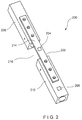
- a system 200 is substantially similar to the system 100, including an implant 202 and at least two sensors 204, 206.
- the first sensor 204 is located on a surface 214 of the implant 202 in a position corresponding to a fracture of a bone 208, while the second sensor 206 is placed directly on a solid portion 212 of the bone 208, outside a perimeter of the implant 202.
- the first sensor 204 measures strain on the implant 202 at a position corresponding to the site of the fracture 210 while the second sensor 206 measures strain on the solid portion 212 of the bone 208.
- a ratio between the strains measured by the first and second sensors 204, 206 is determined and tracked to study the progress of healing in the bone 208.
- the ratio of the strain measurements from the first sensor 204 to the strain measurements from the second sensor 206 trend in a decreasing pattern as the bone 208 heals, whereas a lack of healing will show no recognizable trend over time.
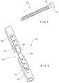
- a system 300 is substantially similar to the system 200, comprising an implant 302 and at least two sensors 304, 306.
- the first sensor 304 is placed on a surface 314 of the implant 302 in a location corresponding to a position of a fracture 310 of a bone 308 (when the implant 302 is mounted on the bone 308 in a desired position) to measure strain on the implant 302 at the position of the fracture 310 while the second sensor 306 is placed directly on a solid portion 312 of the bone 308.
- the second sensor 306 is placed within the solid portion 312 via, for example, a bone fixation element 316 (e.g., screw).
- the second sensor 306 may be attached adjacent to a proximal end 318 of the bone fixation element 316 such that when the bone fixation element 316 is inserted into the solid portion 312 of the bone, the second sensor 306 contacts a cortical wall of the bone 308.
- the second sensor 306 may be printed or mounted around a portion of the bone fixation element 316 to measure deformation of the bone 308 which is directly related to strain on the bone 308. The ratio of the measurements from the first sensor 304 to those of the second sensor 306 may then be determined to track healing progress in the same manner described above.
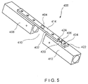
- a system 400 according to a fourth exemplary embodiment of the invention is substantially similar to the system 100, comprising an implant 402 and first and second sensors 404, 406, respectively, both of which are mounted on the implant 402.
- the first sensor 404 is located on the implant 402 in a position which, when the implant 402 is in the desired position, corresponds to the location of a fracture 410 so that the first sensor 404 measures strain on the implant 402 at a position corresponding to the site of the fracture 410.
- the second sensor 406 is positioned on a portion 420 of the implant 402 having greater flexibility than the portion of the implant 402 on which the first sensor 404 is mounted.
- the portion 420 may be made more flexible than other portions of the implant 402 by reducing a width (i.e., an extent of the implant 402 across a bone facing surface thereof in a direction perpendicular to a longitudinal axis of the implant 402) and/or a thickness of the portion 420 (i.e., a distance between the bone facing surface and a surface thereof which faces away from the bone) as compared to remaining portions of the implant 402.
- the flexible portion 420 is adjacent to an end 422 of the implant 402 so that the second sensor 406 is separated from the fracture 410 by a distance great enough to ensure that the underlying portion 412 of the bone 408 is solid.
- the second sensor 406 on the flexible portion 420 of the implant 402 is fixed to the solid portion 412 of the bone 408 via, for example, locking screws inserted in holes 424 on opposing sides thereof.
- the second sensor 406 measures strain on a portion of the implant 402 corresponding to the solid portion 412 of the bone 408 so that measurements from the second sensor 406 may be used to normalize measurements from the first sensor.
- placing the second sensor 406 on a more flexible portion 420 of the implant 402 between two locked screws permits a more accurate measurement of the strain on the underlying solid portion 412 of the bone 408, as compared to the results from placing the second sensor 406 on a stiffer portion of the implant 402.
- the ratio of the measurements from the first sensor 404 to the measurements from the second sensor 406 during the healing process should trend in a pattern indicating an increasing stiffness of the bone 408 over time, whereas a lack of healing should show no recognizable trend over time.
Landscapes
- Health & Medical Sciences (AREA)
- Life Sciences & Earth Sciences (AREA)
- Surgery (AREA)
- Orthopedic Medicine & Surgery (AREA)
- Biomedical Technology (AREA)
- Veterinary Medicine (AREA)
- Public Health (AREA)
- General Health & Medical Sciences (AREA)
- Animal Behavior & Ethology (AREA)
- Heart & Thoracic Surgery (AREA)
- Engineering & Computer Science (AREA)
- Medical Informatics (AREA)
- Molecular Biology (AREA)
- Biophysics (AREA)
- Physics & Mathematics (AREA)
- Pathology (AREA)
- Oral & Maxillofacial Surgery (AREA)
- Dentistry (AREA)
- Nuclear Medicine, Radiotherapy & Molecular Imaging (AREA)
- Neurology (AREA)
- Rheumatology (AREA)
- Transplantation (AREA)
- Cardiology (AREA)
- Vascular Medicine (AREA)
- Physical Education & Sports Medicine (AREA)
- Prostheses (AREA)
- Surgical Instruments (AREA)
- Measurement Of The Respiration, Hearing Ability, Form, And Blood Characteristics Of Living Organisms (AREA)
Description
- The present invention relates to a system for tracking the progress of bone healing and, in particular, systems that calculate a ratio of strain at multiple locations along an implant and/or a bone
- Strain gages can be placed on orthopedic implants to track the progress of bone healing. Upon initial implantation, the implants are expected to experience higher levels of strain which decrease during healing as the bone begins to share more of the load with the implant. Currently, however, implant strain values need to be assessed with a known load applied to the bone in order to evaluate bone healing. A system for tracking the healing process of a bone, including a bone plate with a plurality of strain gages is known from
US 2006/0052782 A1 . An intramedullary nail having strain sensors is disclosed inUS 2008/0300597 A1 . - The present invention is directed to a system as defined in claim 1, comprising an implant configured for attachment to a bone and a first sensor measuring a strain on a first portion of the implant, the first portion of the implant being configured to be mechanically coupled to a weakened portion of a bone when the implant is coupled to the bone in a target position in combination with a second sensor measuring strain in a non-weakened portion of the bone.
-
-
FIG. 1 shows a perspective view of a system according to a first exemplary embodiment of the present invention; -
FIG. 2 shows a perspective view of a system according to a second exemplary embodiment of the present invention; -
FIG. 3 shows a perspective view of a system according to a third exemplary embodiment of the present invention; -
FIG. 4 shows a side view of a bone fixation element of the system ofFIG. 3 ; and -
FIG. 5 shows a perspective view of a system according to a fourth exemplary embodiment of the present invention. - The present invention may be further understood with reference to the following description and the appended drawings, wherein like elements are referred to with the same reference numerals. The exemplary embodiment of the present invention relate to a system for tracking the progress of bone healing. In particular, the exemplary embodiments describe systems that calculate a ratio of strain at multiple locations along an implant and/or a bone. An exemplary embodiment of the system may include a first sensor on a surface of the implant adapted to be positioned at a location proximate a weakened portion of the bone. Strain on the implant at this location will be affected by the strength or stiffness of the weakened bone and the load placed on the bone by the patient. A second sensor may be placed on the implant at a location in which strain measured by the second sensor is affected only by the load placed on the bone such that the measured strain is substantially unchanged by the bone healing process. Thus, a ratio between the strains measured by the first and second sensors provides information corresponding to bone healing, regardless of the load on the bone. It will be understood by those of skill in the art that although the exemplary embodiment specifically describe tracking the healing progress of a leg bone, the present invention may be used to track the progress of healing of any load bearing bone. It will also be understood by those of skill in the art that although the exemplary embodiments specifically show and describe two sensors, the present invention may include additional sensors along different areas of the bone to determine ratios corresponding to the bone healing progress of the different areas. In addition, although exemplary embodiments show a bone plate, the present invention may be used with any other fixation element such as, for example, screws, intramedullary devices, external fixators, spine fixation implants and prosthetics.
- As shown in
Fig. 1 , asystem 100 according to a first exemplary embodiment of the invention comprises an implant 102 (e.g., a bone plate) and first andsecond sensors implant 102 is configured for fixation over a target portion of abone 108 to, for example, fix afracture 110 or to support a weakened portion of thebone 108. The first andsecond sensors surface 114 of theimplant 102 such that the first andsecond sensors bone 108. Although thesurface 114 is shown as facing away from thebone 108 when theimplant 102 is fixed to thebone 108 in a desired location, it will be understood by those of skill in the art that thesensors implant 102. For example, thesensors implant 102 facing thebone 108 or a surface on a side of theimplant 102. The first andsecond sensors implant 102 so that, when the implant is in a desired position on thebone 108, thefirst sensor 104 is located over a site of thefracture 110 while thesecond sensor 106 is separated from thefracture 110 over a healthy (i.e., solid)portion 112 of thebone 108 to measure levels of strain and/or load on theimplant 102, at these positions along theimplant 102. Thesecond sensor 106 should be isolated between two screws locked in ahealthy portion 112 of thebone 108 to measure a load on thebone 108. - The
sensors sensors implant 102 to be measured and transmitted to the data collection device for further processing without physically accessing thesensors sensors MEMS sensors sensors sensors first sensor 104 to the strain measured by thesecond sensor 106 may then be determined by a physician or other professional to track healing progress. Alternatively, the ratio may be determined by a processing device that may also store the strain measurements and the determined ratios (e.g., in an internal memory or on an external storage device) so that changes in the ratio may be reviewed to more fully understand the progression of the healing over time. - It will be understood by those of skill in the art that when the
bone 108 is initially broken or fractured, strain on theimplant 102 at the location of thefracture 110 will vary based on changing mechanical properties of thebone 108 during the healing process and the load placed on the bone 108 (e.g., the weight that the patient places on the leg) while the strain measured in thehealthy portion 112 varies based only on the load placed on thebone 108. Thus, taking a ratio of the strains measured by the twosensors sensors bone 108 at thefracture site 110. The ratio of the measurements from thefirst sensor 104 to the measurements from thesecond sensor 106 during the healing process should trend in a decreasing pattern over time, whereas a lack of healing would show no recognizable trend over time. - As shown in
Fig. 2 , asystem 200 according to a second exemplary embodiment of the invention is substantially similar to thesystem 100, including animplant 202 and at least twosensors sensors implant 202, thefirst sensor 204 is located on asurface 214 of theimplant 202 in a position corresponding to a fracture of abone 208, while thesecond sensor 206 is placed directly on asolid portion 212 of thebone 208, outside a perimeter of theimplant 202. Thus, thefirst sensor 204 measures strain on theimplant 202 at a position corresponding to the site of thefracture 210 while thesecond sensor 206 measures strain on thesolid portion 212 of thebone 208. Similarly to thesystem 100, a ratio between the strains measured by the first andsecond sensors bone 208. As indicated above, the ratio of the strain measurements from thefirst sensor 204 to the strain measurements from thesecond sensor 206 trend in a decreasing pattern as thebone 208 heals, whereas a lack of healing will show no recognizable trend over time. - As shown in
Figs. 3 - 4 , asystem 300 according to a third exemplary embodiment of the invention is substantially similar to thesystem 200, comprising animplant 302 and at least twosensors first sensor 204, thefirst sensor 304 is placed on asurface 314 of theimplant 302 in a location corresponding to a position of afracture 310 of a bone 308 (when theimplant 302 is mounted on thebone 308 in a desired position) to measure strain on theimplant 302 at the position of thefracture 310 while thesecond sensor 306 is placed directly on asolid portion 312 of thebone 308. However, rather than being placed on an exterior surface of thebone 308, thesecond sensor 306 is placed within thesolid portion 312 via, for example, a bone fixation element 316 (e.g., screw). - The
second sensor 306 may be attached adjacent to aproximal end 318 of thebone fixation element 316 such that when thebone fixation element 316 is inserted into thesolid portion 312 of the bone, thesecond sensor 306 contacts a cortical wall of thebone 308. Thesecond sensor 306 may be printed or mounted around a portion of thebone fixation element 316 to measure deformation of thebone 308 which is directly related to strain on thebone 308. The ratio of the measurements from thefirst sensor 304 to those of thesecond sensor 306 may then be determined to track healing progress in the same manner described above. - As shown in
Fig. 5 , asystem 400 according to a fourth exemplary embodiment of the invention is substantially similar to thesystem 100, comprising animplant 402 and first andsecond sensors implant 402. Similarly to thefirst sensor 104, thefirst sensor 404 is located on theimplant 402 in a position which, when theimplant 402 is in the desired position, corresponds to the location of afracture 410 so that thefirst sensor 404 measures strain on theimplant 402 at a position corresponding to the site of thefracture 410. Thesecond sensor 406 is positioned on aportion 420 of theimplant 402 having greater flexibility than the portion of theimplant 402 on which thefirst sensor 404 is mounted. For example, theportion 420 may be made more flexible than other portions of theimplant 402 by reducing a width (i.e., an extent of theimplant 402 across a bone facing surface thereof in a direction perpendicular to a longitudinal axis of the implant 402) and/or a thickness of the portion 420 (i.e., a distance between the bone facing surface and a surface thereof which faces away from the bone) as compared to remaining portions of theimplant 402. In a preferred embodiment, theflexible portion 420 is adjacent to anend 422 of theimplant 402 so that thesecond sensor 406 is separated from thefracture 410 by a distance great enough to ensure that theunderlying portion 412 of thebone 408 is solid. - The
second sensor 406 on theflexible portion 420 of theimplant 402 is fixed to thesolid portion 412 of thebone 408 via, for example, locking screws inserted inholes 424 on opposing sides thereof. Thesecond sensor 406 measures strain on a portion of theimplant 402 corresponding to thesolid portion 412 of thebone 408 so that measurements from thesecond sensor 406 may be used to normalize measurements from the first sensor. Similarly to the placement of a sensor directly in or on a bone, as described in conjunction withsystems second sensor 406 on a moreflexible portion 420 of theimplant 402 between two locked screws permits a more accurate measurement of the strain on the underlyingsolid portion 412 of thebone 408, as compared to the results from placing thesecond sensor 406 on a stiffer portion of theimplant 402. The ratio of the measurements from thefirst sensor 404 to the measurements from thesecond sensor 406 during the healing process should trend in a pattern indicating an increasing stiffness of thebone 408 over time, whereas a lack of healing should show no recognizable trend over time.
It will be understood by those of skill in the art that other mechanisms may be employed for normalizing measurements of strain on a portion of an implant which, when mounted on a bone in a target location, corresponds to a position of a fracture or other weakened portion of that bone.
It will be apparent to those skilled in the art that various modifications and variations can be made in the structure and the methodology of the present invention, without departing from the scope of the invention. Thus, it is intended that the present invention cover the modifications and variations of this invention provided that they come within the scope of the appended claims.
Claims (11)
- A system (100 - 400) for tracking a bone healing progress, comprising:an implant (102 - 402) configured for fixing a fracture (110 - 410) of a bone (108 - 408) including a first sensor (104 - 404) configured for measuring a first strain when mounted thereon at a position which, when the implant (102 - 402) is mounted on the bone (108 - 408) in a target position, is mechanically coupled to a fracture (110 - 410) of the bone (108 - 408); anda second sensor (106 - 406) configured for measuring a second strain on a portion of the bone (108 - 408) separated from the fracture (110 - 410),characterized in that the system is configured to calculate a ratio of the first strain to the second strain so as to normalize an effect of loads on the bone (108 - 408) and indicate an increased stiffness as the fracture (110 - 410) heals.
- The system of claim 1, wherein
the first sensor (104 - 404) is located on the implant (102 - 402) for measuring the first strain; and
the second sensor (106 - 406) is located on the implant (102 - 402) or on the bone (108 - 408) for measuring the second strain. - The system of claim 1 or 2, wherein the implant (102 - 402) is a plate configured to be coupled to a bone overlying a fracture site.
- The system of any one of claims 1 to 3, wherein the second sensor (206; 306) is configured to be coupled directly to a non-weakened portion of the bone.
- The system of any one of claims 1 to 4, wherein the second sensor (306) is mounted to a portion of a bone fixation element (316) which, when in an operative position, is inserted into a non-weakened portion of the bone so that the second sensor (306) contacts a cortical wall of the non-weakened portion of the bone.
- The system of claim 5, wherein the second sensor (306) extends about a circumference of a proximal portion of the bone fixation element (316).
- The system of any one of claims 1 to 4, further comprising:a bone fixation element (316), the second sensor (306) being mounted at a proximal end thereof so that, when the bone fixation element (316) is inserted into the bone to a desired position, the second sensor (306) contacts a cortical wall of the bone.
- The system of any one of claims 1 to 4, wherein the implant (402) includes a flexible portion (420) and a rigid portion, a bending stiffness of the rigid portion being greater than that of the flexible portion (420), the second sensor (406) being mounted on the flexible portion (420) while the first sensor (404) is mounted on the rigid portion.
- The system of claim 8, further comprising:first and second locking screws which, when the implant (402) is coupled to the bone in a target configuration, are inserted through openings (424) in the implant (402) on opposing sides of the flexible portion (420).
- The system of any one of claims 1 to 9, wherein the first and second sensors (104 - 404,
106 - 406) provide data to an external data gathering unit wirelessly. - The system of claim 10, wherein the first and second sensors (104 - 404, 106 - 406) are one of MEMs sensors and powered chips connected to printed circuit boards.
Applications Claiming Priority (2)
| Application Number | Priority Date | Filing Date | Title |
|---|---|---|---|
| US25358309P | 2009-10-21 | 2009-10-21 | |
| PCT/US2010/053519 WO2011050149A1 (en) | 2009-10-21 | 2010-10-21 | Method of normalizing implant strain readings to assess bone healing |
Publications (2)
| Publication Number | Publication Date |
|---|---|
| EP2490609A1 EP2490609A1 (en) | 2012-08-29 |
| EP2490609B1 true EP2490609B1 (en) | 2016-05-11 |
Family
ID=43447692
Family Applications (1)
| Application Number | Title | Priority Date | Filing Date |
|---|---|---|---|
| EP10774080.5A Active EP2490609B1 (en) | 2009-10-21 | 2010-10-21 | System for normalizing implant strain readings to assess bone healing |
Country Status (10)
| Country | Link |
|---|---|
| US (3) | US10441210B2 (en) |
| EP (1) | EP2490609B1 (en) |
| JP (2) | JP6097562B2 (en) |
| KR (2) | KR101713302B1 (en) |
| CN (2) | CN102612348B (en) |
| BR (1) | BR112012008887B1 (en) |
| CA (2) | CA2775398C (en) |
| ES (1) | ES2583687T3 (en) |
| IN (1) | IN2012DN02844A (en) |
| WO (1) | WO2011050149A1 (en) |
Families Citing this family (11)
| Publication number | Priority date | Publication date | Assignee | Title |
|---|---|---|---|---|
| WO2011050149A1 (en) * | 2009-10-21 | 2011-04-28 | Synthes Usa, Llc | Method of normalizing implant strain readings to assess bone healing |
| DE102011115283A1 (en) * | 2011-09-29 | 2013-04-04 | Berufsgenossenschaftliches Unfallkrankenhaus Hamburg | Method for automatically determining elastic resilience of bone during fracture healing, involves determining parameter as description of regression of elasticity if range of measured values of force is not exceeded threshold value |
| US9795423B2 (en) * | 2012-01-23 | 2017-10-24 | DePuy Synthes Products, Inc. | Device and method for normalizing implant strain readings to assess bone healing |
| DE102014115676B3 (en) * | 2014-10-28 | 2015-11-05 | Dieter Föllmer | prosthesis system |
| US11147598B2 (en) | 2015-04-20 | 2021-10-19 | Bioscience Medical Group Ltd. | Bone fixation apparatus |
| TWI547258B (en) * | 2015-10-23 | 2016-09-01 | 國立交通大學 | Sensing bone fixing element |
| EP3664692A1 (en) * | 2017-08-07 | 2020-06-17 | DePuy Synthes Products, Inc. | Sensors implantable into a patient's body, systems, and methods of using the same |
| AU2021312298A1 (en) * | 2020-07-21 | 2023-03-23 | DePuy Synthes Products, Inc. | Bone fixation monitoring system |
| EP4005465A1 (en) | 2020-11-26 | 2022-06-01 | ETH Zurich | Implantable sensor |
| US20220265324A1 (en) | 2021-02-23 | 2022-08-25 | Nuvasive Specialized Orthopedics, Inc. | Adjustable implant, system and methods |
| CN117717319B (en) * | 2023-12-18 | 2024-07-30 | 北京航空航天大学 | System and method for measuring fracture rigidity after lower limb intramedullary nail operation based on RFID |
Family Cites Families (21)
| Publication number | Priority date | Publication date | Assignee | Title |
|---|---|---|---|---|
| US4403607A (en) * | 1980-05-09 | 1983-09-13 | The Regents Of The University Of California | Compatible internal bone fixation plate |
| US6001099A (en) | 1998-06-08 | 1999-12-14 | Huebner; Randall J. | Bone plate with varying rigidity |
| US6034296A (en) * | 1997-03-11 | 2000-03-07 | Elvin; Niell | Implantable bone strain telemetry sensing system and method |
| DE19810182C1 (en) | 1998-03-10 | 2000-03-16 | Univ Eberhard Karls | Overload assessing device for lower limbs or spinal column of person, particularly one equipped with prosthesis, comprises load sensor worn by person in at least one shoe |
| DE19810482A1 (en) * | 1998-03-11 | 1999-09-16 | Weiermann Dieter Weinor | Cassette awning |
| US6273863B1 (en) * | 1999-10-26 | 2001-08-14 | Andante Medical Devices, Ltd. | Adaptive weight bearing monitoring system for rehabilitation of injuries to the lower extremities |
| US6610096B2 (en) * | 2001-08-22 | 2003-08-26 | Macdonald Stuart G. | Prosthetic implants having enhanced utility |
| DE60334459D1 (en) | 2002-07-10 | 2010-11-18 | Orthodata Inc | LOAD MEASURING SYSTEM |
| KR20050083916A (en) | 2002-11-19 | 2005-08-26 | 어큠드 엘엘씨 | Deformable bone plates |
| US7218232B2 (en) | 2003-07-11 | 2007-05-15 | Depuy Products, Inc. | Orthopaedic components with data storage element |
| US20050085814A1 (en) * | 2003-10-21 | 2005-04-21 | Sherman Michael C. | Dynamizable orthopedic implants and their use in treating bone defects |
| US7985222B2 (en) * | 2004-04-21 | 2011-07-26 | Medshape Solutions, Inc. | Osteosynthetic implants and methods of use and manufacture |
| NZ552049A (en) * | 2004-06-07 | 2009-09-25 | Synthes Gmbh | Orthopaedic implant with sensors |
| EP1819278A4 (en) * | 2004-11-15 | 2009-04-08 | Izex Technologies Inc | Instrumented orthopedic and other medical implants |
| WO2007025191A1 (en) | 2005-08-23 | 2007-03-01 | Smith & Nephew, Inc. | Telemetric orthopaedic implant |
| US9295503B2 (en) * | 2005-11-16 | 2016-03-29 | DePuy Synthes Products, Inc. | Device for bone fixation with at least one through hole |
| DE102006006341A1 (en) * | 2006-02-07 | 2007-08-09 | Wolter, Dietmar, Prof.Dr. | Fixation system for bones with a sensor and telemetry system |
| DE102006034041A1 (en) | 2006-07-19 | 2008-01-31 | Berufsgenossenschaftlicher Verein für Heilbehandlung Hamburg e.V. Berufsgenossenschaftliches Unfallkrankenhaus Hamburg | Wireless strain gauge measuring system for use in e.g. mechanical engineering, has strain gauge implemented as single strip, where system is implemented to permit application of strip, so that data and energy are exchanged for operation |
| US20080147125A1 (en) * | 2006-12-12 | 2008-06-19 | Dennis Colleran | Active Settling Plate and Method of Use |
| WO2011050149A1 (en) | 2009-10-21 | 2011-04-28 | Synthes Usa, Llc | Method of normalizing implant strain readings to assess bone healing |
| DE102010022434A1 (en) | 2010-06-02 | 2011-12-08 | Berufsgenossenschaftliches Unfallkrankenhaus Hamburg | Fixation system for consolidation of fractured bone, has sensor system comprising two reference sensors arranged in anchorage regions of connection carrier, and one main sensor in bypass region |
-
2010
- 2010-10-21 WO PCT/US2010/053519 patent/WO2011050149A1/en active Application Filing
- 2010-10-21 IN IN2844DEN2012 patent/IN2012DN02844A/en unknown
- 2010-10-21 BR BR112012008887-4A patent/BR112012008887B1/en active IP Right Grant
- 2010-10-21 KR KR1020127007654A patent/KR101713302B1/en active IP Right Grant
- 2010-10-21 JP JP2012535363A patent/JP6097562B2/en active Active
- 2010-10-21 CA CA2775398A patent/CA2775398C/en active Active
- 2010-10-21 CA CA3006947A patent/CA3006947C/en active Active
- 2010-10-21 CN CN201080046895.2A patent/CN102612348B/en active Active
- 2010-10-21 EP EP10774080.5A patent/EP2490609B1/en active Active
- 2010-10-21 KR KR1020177005816A patent/KR101801774B1/en active IP Right Grant
- 2010-10-21 ES ES10774080.5T patent/ES2583687T3/en active Active
- 2010-10-21 US US12/909,220 patent/US10441210B2/en active Active
- 2010-10-21 CN CN201610330612.6A patent/CN105919596B/en active Active
-
2015
- 2015-05-21 JP JP2015103430A patent/JP6178362B2/en active Active
- 2015-06-25 US US14/750,716 patent/US10595771B2/en active Active
-
2020
- 2020-02-24 US US16/799,297 patent/US20200187849A1/en active Pending
Also Published As
| Publication number | Publication date |
|---|---|
| KR20170026667A (en) | 2017-03-08 |
| BR112012008887A2 (en) | 2020-09-15 |
| US10441210B2 (en) | 2019-10-15 |
| CA3006947C (en) | 2019-11-05 |
| BR112012008887B1 (en) | 2021-09-21 |
| JP6097562B2 (en) | 2017-03-15 |
| CN105919596B (en) | 2018-11-30 |
| WO2011050149A1 (en) | 2011-04-28 |
| ES2583687T3 (en) | 2016-09-21 |
| CN105919596A (en) | 2016-09-07 |
| CA2775398A1 (en) | 2011-04-28 |
| US20110098603A1 (en) | 2011-04-28 |
| CA2775398C (en) | 2019-09-17 |
| JP2013508090A (en) | 2013-03-07 |
| KR20120087905A (en) | 2012-08-07 |
| JP2015180268A (en) | 2015-10-15 |
| KR101801774B1 (en) | 2017-11-27 |
| IN2012DN02844A (en) | 2015-07-24 |
| US20150289796A1 (en) | 2015-10-15 |
| CN102612348A (en) | 2012-07-25 |
| CN102612348B (en) | 2016-07-06 |
| JP6178362B2 (en) | 2017-08-09 |
| US10595771B2 (en) | 2020-03-24 |
| US20200187849A1 (en) | 2020-06-18 |
| KR101713302B1 (en) | 2017-03-07 |
| EP2490609A1 (en) | 2012-08-29 |
| CA3006947A1 (en) | 2011-04-28 |
Similar Documents
| Publication | Publication Date | Title |
|---|---|---|
| EP2490609B1 (en) | System for normalizing implant strain readings to assess bone healing | |
| US11622798B2 (en) | Device and method for normalizing implant strain readings to assess bone healing |
Legal Events
| Date | Code | Title | Description |
|---|---|---|---|
| PUAI | Public reference made under article 153(3) epc to a published international application that has entered the european phase |
Free format text: ORIGINAL CODE: 0009012 |
|
| 17P | Request for examination filed |
Effective date: 20120327 |
|
| AK | Designated contracting states |
Kind code of ref document: A1 Designated state(s): AL AT BE BG CH CY CZ DE DK EE ES FI FR GB GR HR HU IE IS IT LI LT LU LV MC MK MT NL NO PL PT RO RS SE SI SK SM TR |
|
| DAX | Request for extension of the european patent (deleted) | ||
| 17Q | First examination report despatched |
Effective date: 20150408 |
|
| GRAP | Despatch of communication of intention to grant a patent |
Free format text: ORIGINAL CODE: EPIDOSNIGR1 |
|
| RIC1 | Information provided on ipc code assigned before grant |
Ipc: A61B 17/80 20060101AFI20151117BHEP Ipc: A61B 17/86 20060101ALN20151117BHEP Ipc: A61B 5/103 20060101ALI20151117BHEP Ipc: A61B 5/07 20060101ALI20151117BHEP Ipc: A61B 5/00 20060101ALI20151117BHEP |
|
| INTG | Intention to grant announced |
Effective date: 20151204 |
|
| RIN1 | Information on inventor provided before grant (corrected) |
Inventor name: DEIRMENGIAN, CARL Inventor name: PIERSON, GLEN Inventor name: MIKHAIL, GEORGE |
|
| GRAS | Grant fee paid |
Free format text: ORIGINAL CODE: EPIDOSNIGR3 |
|
| GRAA | (expected) grant |
Free format text: ORIGINAL CODE: 0009210 |
|
| AK | Designated contracting states |
Kind code of ref document: B1 Designated state(s): AL AT BE BG CH CY CZ DE DK EE ES FI FR GB GR HR HU IE IS IT LI LT LU LV MC MK MT NL NO PL PT RO RS SE SI SK SM TR |
|
| REG | Reference to a national code |
Ref country code: GB Ref legal event code: FG4D |
|
| REG | Reference to a national code |
Ref country code: CH Ref legal event code: NV Representative=s name: E. BLUM AND CO. AG PATENT- UND MARKENANWAELTE , CH Ref country code: CH Ref legal event code: EP |
|
| REG | Reference to a national code |
Ref country code: AT Ref legal event code: REF Ref document number: 797991 Country of ref document: AT Kind code of ref document: T Effective date: 20160515 |
|
| REG | Reference to a national code |
Ref country code: IE Ref legal event code: FG4D |
|
| REG | Reference to a national code |
Ref country code: DE Ref legal event code: R096 Ref document number: 602010033325 Country of ref document: DE |
|
| REG | Reference to a national code |
Ref country code: LT Ref legal event code: MG4D |
|
| REG | Reference to a national code |
Ref country code: NL Ref legal event code: MP Effective date: 20160511 |
|
| REG | Reference to a national code |
Ref country code: FR Ref legal event code: PLFP Year of fee payment: 7 |
|
| REG | Reference to a national code |
Ref country code: ES Ref legal event code: FG2A Ref document number: 2583687 Country of ref document: ES Kind code of ref document: T3 Effective date: 20160921 |
|
| PG25 | Lapsed in a contracting state [announced via postgrant information from national office to epo] |
Ref country code: LT Free format text: LAPSE BECAUSE OF FAILURE TO SUBMIT A TRANSLATION OF THE DESCRIPTION OR TO PAY THE FEE WITHIN THE PRESCRIBED TIME-LIMIT Effective date: 20160511 Ref country code: NO Free format text: LAPSE BECAUSE OF FAILURE TO SUBMIT A TRANSLATION OF THE DESCRIPTION OR TO PAY THE FEE WITHIN THE PRESCRIBED TIME-LIMIT Effective date: 20160811 Ref country code: NL Free format text: LAPSE BECAUSE OF FAILURE TO SUBMIT A TRANSLATION OF THE DESCRIPTION OR TO PAY THE FEE WITHIN THE PRESCRIBED TIME-LIMIT Effective date: 20160511 Ref country code: FI Free format text: LAPSE BECAUSE OF FAILURE TO SUBMIT A TRANSLATION OF THE DESCRIPTION OR TO PAY THE FEE WITHIN THE PRESCRIBED TIME-LIMIT Effective date: 20160511 |
|
| REG | Reference to a national code |
Ref country code: AT Ref legal event code: MK05 Ref document number: 797991 Country of ref document: AT Kind code of ref document: T Effective date: 20160511 |
|
| PG25 | Lapsed in a contracting state [announced via postgrant information from national office to epo] |
Ref country code: HR Free format text: LAPSE BECAUSE OF FAILURE TO SUBMIT A TRANSLATION OF THE DESCRIPTION OR TO PAY THE FEE WITHIN THE PRESCRIBED TIME-LIMIT Effective date: 20160511 Ref country code: GR Free format text: LAPSE BECAUSE OF FAILURE TO SUBMIT A TRANSLATION OF THE DESCRIPTION OR TO PAY THE FEE WITHIN THE PRESCRIBED TIME-LIMIT Effective date: 20160812 Ref country code: RS Free format text: LAPSE BECAUSE OF FAILURE TO SUBMIT A TRANSLATION OF THE DESCRIPTION OR TO PAY THE FEE WITHIN THE PRESCRIBED TIME-LIMIT Effective date: 20160511 Ref country code: LV Free format text: LAPSE BECAUSE OF FAILURE TO SUBMIT A TRANSLATION OF THE DESCRIPTION OR TO PAY THE FEE WITHIN THE PRESCRIBED TIME-LIMIT Effective date: 20160511 Ref country code: PT Free format text: LAPSE BECAUSE OF FAILURE TO SUBMIT A TRANSLATION OF THE DESCRIPTION OR TO PAY THE FEE WITHIN THE PRESCRIBED TIME-LIMIT Effective date: 20160912 Ref country code: SE Free format text: LAPSE BECAUSE OF FAILURE TO SUBMIT A TRANSLATION OF THE DESCRIPTION OR TO PAY THE FEE WITHIN THE PRESCRIBED TIME-LIMIT Effective date: 20160511 |
|
| PG25 | Lapsed in a contracting state [announced via postgrant information from national office to epo] |
Ref country code: EE Free format text: LAPSE BECAUSE OF FAILURE TO SUBMIT A TRANSLATION OF THE DESCRIPTION OR TO PAY THE FEE WITHIN THE PRESCRIBED TIME-LIMIT Effective date: 20160511 Ref country code: SK Free format text: LAPSE BECAUSE OF FAILURE TO SUBMIT A TRANSLATION OF THE DESCRIPTION OR TO PAY THE FEE WITHIN THE PRESCRIBED TIME-LIMIT Effective date: 20160511 Ref country code: CZ Free format text: LAPSE BECAUSE OF FAILURE TO SUBMIT A TRANSLATION OF THE DESCRIPTION OR TO PAY THE FEE WITHIN THE PRESCRIBED TIME-LIMIT Effective date: 20160511 Ref country code: RO Free format text: LAPSE BECAUSE OF FAILURE TO SUBMIT A TRANSLATION OF THE DESCRIPTION OR TO PAY THE FEE WITHIN THE PRESCRIBED TIME-LIMIT Effective date: 20160511 Ref country code: DK Free format text: LAPSE BECAUSE OF FAILURE TO SUBMIT A TRANSLATION OF THE DESCRIPTION OR TO PAY THE FEE WITHIN THE PRESCRIBED TIME-LIMIT Effective date: 20160511 |
|
| REG | Reference to a national code |
Ref country code: DE Ref legal event code: R097 Ref document number: 602010033325 Country of ref document: DE |
|
| PG25 | Lapsed in a contracting state [announced via postgrant information from national office to epo] |
Ref country code: AT Free format text: LAPSE BECAUSE OF FAILURE TO SUBMIT A TRANSLATION OF THE DESCRIPTION OR TO PAY THE FEE WITHIN THE PRESCRIBED TIME-LIMIT Effective date: 20160511 Ref country code: SM Free format text: LAPSE BECAUSE OF FAILURE TO SUBMIT A TRANSLATION OF THE DESCRIPTION OR TO PAY THE FEE WITHIN THE PRESCRIBED TIME-LIMIT Effective date: 20160511 Ref country code: PL Free format text: LAPSE BECAUSE OF FAILURE TO SUBMIT A TRANSLATION OF THE DESCRIPTION OR TO PAY THE FEE WITHIN THE PRESCRIBED TIME-LIMIT Effective date: 20160511 Ref country code: BE Free format text: LAPSE BECAUSE OF FAILURE TO SUBMIT A TRANSLATION OF THE DESCRIPTION OR TO PAY THE FEE WITHIN THE PRESCRIBED TIME-LIMIT Effective date: 20160511 |
|
| PLBE | No opposition filed within time limit |
Free format text: ORIGINAL CODE: 0009261 |
|
| STAA | Information on the status of an ep patent application or granted ep patent |
Free format text: STATUS: NO OPPOSITION FILED WITHIN TIME LIMIT |
|
| 26N | No opposition filed |
Effective date: 20170214 |
|
| PG25 | Lapsed in a contracting state [announced via postgrant information from national office to epo] |
Ref country code: SI Free format text: LAPSE BECAUSE OF FAILURE TO SUBMIT A TRANSLATION OF THE DESCRIPTION OR TO PAY THE FEE WITHIN THE PRESCRIBED TIME-LIMIT Effective date: 20160511 |
|
| PG25 | Lapsed in a contracting state [announced via postgrant information from national office to epo] |
Ref country code: MC Free format text: LAPSE BECAUSE OF FAILURE TO SUBMIT A TRANSLATION OF THE DESCRIPTION OR TO PAY THE FEE WITHIN THE PRESCRIBED TIME-LIMIT Effective date: 20160511 |
|
| REG | Reference to a national code |
Ref country code: IE Ref legal event code: MM4A |
|
| REG | Reference to a national code |
Ref country code: DE Ref legal event code: R082 Ref document number: 602010033325 Country of ref document: DE Representative=s name: KLUNKER IP PATENTANWAELTE PARTG MBB, DE |
|
| PG25 | Lapsed in a contracting state [announced via postgrant information from national office to epo] |
Ref country code: LU Free format text: LAPSE BECAUSE OF NON-PAYMENT OF DUE FEES Effective date: 20161021 |
|
| REG | Reference to a national code |
Ref country code: FR Ref legal event code: PLFP Year of fee payment: 8 |
|
| PG25 | Lapsed in a contracting state [announced via postgrant information from national office to epo] |
Ref country code: IE Free format text: LAPSE BECAUSE OF NON-PAYMENT OF DUE FEES Effective date: 20161021 |
|
| PG25 | Lapsed in a contracting state [announced via postgrant information from national office to epo] |
Ref country code: HU Free format text: LAPSE BECAUSE OF FAILURE TO SUBMIT A TRANSLATION OF THE DESCRIPTION OR TO PAY THE FEE WITHIN THE PRESCRIBED TIME-LIMIT; INVALID AB INITIO Effective date: 20101021 Ref country code: CY Free format text: LAPSE BECAUSE OF FAILURE TO SUBMIT A TRANSLATION OF THE DESCRIPTION OR TO PAY THE FEE WITHIN THE PRESCRIBED TIME-LIMIT Effective date: 20160511 |
|
| PG25 | Lapsed in a contracting state [announced via postgrant information from national office to epo] |
Ref country code: MT Free format text: LAPSE BECAUSE OF NON-PAYMENT OF DUE FEES Effective date: 20161031 Ref country code: TR Free format text: LAPSE BECAUSE OF FAILURE TO SUBMIT A TRANSLATION OF THE DESCRIPTION OR TO PAY THE FEE WITHIN THE PRESCRIBED TIME-LIMIT Effective date: 20160511 Ref country code: IS Free format text: LAPSE BECAUSE OF FAILURE TO SUBMIT A TRANSLATION OF THE DESCRIPTION OR TO PAY THE FEE WITHIN THE PRESCRIBED TIME-LIMIT Effective date: 20160511 Ref country code: MK Free format text: LAPSE BECAUSE OF FAILURE TO SUBMIT A TRANSLATION OF THE DESCRIPTION OR TO PAY THE FEE WITHIN THE PRESCRIBED TIME-LIMIT Effective date: 20160511 |
|
| PG25 | Lapsed in a contracting state [announced via postgrant information from national office to epo] |
Ref country code: BG Free format text: LAPSE BECAUSE OF FAILURE TO SUBMIT A TRANSLATION OF THE DESCRIPTION OR TO PAY THE FEE WITHIN THE PRESCRIBED TIME-LIMIT Effective date: 20160511 |
|
| REG | Reference to a national code |
Ref country code: FR Ref legal event code: PLFP Year of fee payment: 9 |
|
| PG25 | Lapsed in a contracting state [announced via postgrant information from national office to epo] |
Ref country code: AL Free format text: LAPSE BECAUSE OF FAILURE TO SUBMIT A TRANSLATION OF THE DESCRIPTION OR TO PAY THE FEE WITHIN THE PRESCRIBED TIME-LIMIT Effective date: 20160511 |
|
| PGFP | Annual fee paid to national office [announced via postgrant information from national office to epo] |
Ref country code: ES Payment date: 20221104 Year of fee payment: 13 |
|
| PGFP | Annual fee paid to national office [announced via postgrant information from national office to epo] |
Ref country code: DE Payment date: 20230830 Year of fee payment: 14 Ref country code: CH Payment date: 20231101 Year of fee payment: 14 |
|
| PGFP | Annual fee paid to national office [announced via postgrant information from national office to epo] |
Ref country code: GB Payment date: 20240829 Year of fee payment: 15 |
|
| PGFP | Annual fee paid to national office [announced via postgrant information from national office to epo] |
Ref country code: FR Payment date: 20240909 Year of fee payment: 15 |
|
| PGFP | Annual fee paid to national office [announced via postgrant information from national office to epo] |
Ref country code: IT Payment date: 20240910 Year of fee payment: 15 |