EP2483399B1 - Hydrolysis of mannose-1-phospho-6-mannose linkage to phospho-6-mannose - Google Patents
Hydrolysis of mannose-1-phospho-6-mannose linkage to phospho-6-mannose Download PDFInfo
- Publication number
- EP2483399B1 EP2483399B1 EP10782375.9A EP10782375A EP2483399B1 EP 2483399 B1 EP2483399 B1 EP 2483399B1 EP 10782375 A EP10782375 A EP 10782375A EP 2483399 B1 EP2483399 B1 EP 2483399B1
- Authority
- EP
- European Patent Office
- Prior art keywords
- seq
- mannosidase
- residues
- mannose
- amino acid
- Prior art date
- Legal status (The legal status is an assumption and is not a legal conclusion. Google has not performed a legal analysis and makes no representation as to the accuracy of the status listed.)
- Active
Links
- 230000007062 hydrolysis Effects 0.000 title description 3
- 238000006460 hydrolysis reaction Methods 0.000 title description 3
- 108010054377 Mannosidases Proteins 0.000 claims description 196
- 108090000623 proteins and genes Proteins 0.000 claims description 194
- 102000001696 Mannosidases Human genes 0.000 claims description 193
- 102000004169 proteins and genes Human genes 0.000 claims description 142
- 150000007523 nucleic acids Chemical class 0.000 claims description 118
- 102000039446 nucleic acids Human genes 0.000 claims description 112
- 108020004707 nucleic acids Proteins 0.000 claims description 112
- 125000003275 alpha amino acid group Chemical group 0.000 claims description 104
- 108090000765 processed proteins & peptides Proteins 0.000 claims description 84
- 102000004196 processed proteins & peptides Human genes 0.000 claims description 79
- 230000000694 effects Effects 0.000 claims description 75
- 238000000034 method Methods 0.000 claims description 75
- 229920001184 polypeptide Polymers 0.000 claims description 75
- 230000002538 fungal effect Effects 0.000 claims description 63
- 239000002773 nucleotide Substances 0.000 claims description 60
- 125000003729 nucleotide group Chemical group 0.000 claims description 60
- 230000014509 gene expression Effects 0.000 claims description 59
- 150000001413 amino acids Chemical class 0.000 claims description 46
- 241000235015 Yarrowia lipolytica Species 0.000 claims description 42
- 102000004190 Enzymes Human genes 0.000 claims description 41
- 108090000790 Enzymes Proteins 0.000 claims description 41
- 102000003886 Glycoproteins Human genes 0.000 claims description 41
- 108090000288 Glycoproteins Proteins 0.000 claims description 41
- 229940088598 enzyme Drugs 0.000 claims description 41
- 208000037265 diseases, disorders, signs and symptoms Diseases 0.000 claims description 37
- NBSCHQHZLSJFNQ-QTVWNMPRSA-N D-Mannose-6-phosphate Chemical group OC1O[C@H](COP(O)(O)=O)[C@@H](O)[C@H](O)[C@@H]1O NBSCHQHZLSJFNQ-QTVWNMPRSA-N 0.000 claims description 33
- 239000013604 expression vector Substances 0.000 claims description 31
- 230000026731 phosphorylation Effects 0.000 claims description 31
- 238000006366 phosphorylation reaction Methods 0.000 claims description 31
- 101100184479 Saccharomyces cerevisiae (strain ATCC 204508 / S288c) MNN4 gene Proteins 0.000 claims description 30
- 208000015439 Lysosomal storage disease Diseases 0.000 claims description 29
- 239000012634 fragment Substances 0.000 claims description 28
- WQZGKKKJIJFFOK-QTVWNMPRSA-N D-mannopyranose Chemical compound OC[C@H]1OC(O)[C@@H](O)[C@@H](O)[C@@H]1O WQZGKKKJIJFFOK-QTVWNMPRSA-N 0.000 claims description 27
- 230000003301 hydrolyzing effect Effects 0.000 claims description 26
- 241000186221 Cellulosimicrobium cellulans Species 0.000 claims description 25
- 240000004808 Saccharomyces cerevisiae Species 0.000 claims description 24
- 150000002482 oligosaccharides Chemical class 0.000 claims description 23
- 125000000311 mannosyl group Chemical group C1([C@@H](O)[C@@H](O)[C@H](O)[C@H](O1)CO)* 0.000 claims description 22
- 229920001542 oligosaccharide Polymers 0.000 claims description 22
- 230000008685 targeting Effects 0.000 claims description 21
- 241000235058 Komagataella pastoris Species 0.000 claims description 20
- 201000010099 disease Diseases 0.000 claims description 20
- 230000001737 promoting effect Effects 0.000 claims description 20
- 230000003834 intracellular effect Effects 0.000 claims description 19
- 208000035475 disorder Diseases 0.000 claims description 17
- 238000004519 manufacturing process Methods 0.000 claims description 17
- 206010053185 Glycogen storage disease type II Diseases 0.000 claims description 15
- 230000002950 deficient Effects 0.000 claims description 13
- 230000002132 lysosomal effect Effects 0.000 claims description 13
- 230000028327 secretion Effects 0.000 claims description 13
- 238000003860 storage Methods 0.000 claims description 13
- 239000013592 cell lysate Substances 0.000 claims description 12
- 241000680806 Blastobotrys adeninivorans Species 0.000 claims description 11
- 208000024720 Fabry Disease Diseases 0.000 claims description 11
- 108010028144 alpha-Glucosidases Proteins 0.000 claims description 11
- 108010045758 lysosomal proteins Proteins 0.000 claims description 11
- 102100033448 Lysosomal alpha-glucosidase Human genes 0.000 claims description 10
- 239000002253 acid Substances 0.000 claims description 10
- 108020001507 fusion proteins Proteins 0.000 claims description 10
- 102000037865 fusion proteins Human genes 0.000 claims description 10
- 208000032007 Glycogen storage disease due to acid maltase deficiency Diseases 0.000 claims description 9
- 201000004502 glycogen storage disease II Diseases 0.000 claims description 9
- 241000233866 Fungi Species 0.000 claims description 8
- 241000228212 Aspergillus Species 0.000 claims description 7
- 241000228245 Aspergillus niger Species 0.000 claims description 7
- 108010012236 Chemokines Proteins 0.000 claims description 7
- 102000019034 Chemokines Human genes 0.000 claims description 7
- 208000029448 Chylomicron retention disease Diseases 0.000 claims description 7
- 102000004127 Cytokines Human genes 0.000 claims description 7
- 108090000695 Cytokines Proteins 0.000 claims description 7
- 208000015872 Gaucher disease Diseases 0.000 claims description 7
- 208000006933 Hermanski-Pudlak Syndrome Diseases 0.000 claims description 7
- 206010071775 Hermansky-Pudlak syndrome Diseases 0.000 claims description 7
- 208000008955 Mucolipidoses Diseases 0.000 claims description 7
- 241000320412 Ogataea angusta Species 0.000 claims description 7
- 239000000427 antigen Substances 0.000 claims description 7
- 108091007433 antigens Proteins 0.000 claims description 7
- 102000036639 antigens Human genes 0.000 claims description 7
- 239000003102 growth factor Substances 0.000 claims description 7
- 208000029602 Alpha-N-acetylgalactosaminidase deficiency Diseases 0.000 claims description 6
- 206010068220 Aspartylglucosaminuria Diseases 0.000 claims description 6
- 102100022548 Beta-hexosaminidase subunit alpha Human genes 0.000 claims description 6
- 241000222122 Candida albicans Species 0.000 claims description 6
- 208000031879 Chédiak-Higashi syndrome Diseases 0.000 claims description 6
- 206010011777 Cystinosis Diseases 0.000 claims description 6
- 208000011518 Danon disease Diseases 0.000 claims description 6
- 208000001948 Farber Lipogranulomatosis Diseases 0.000 claims description 6
- 208000033149 Farber disease Diseases 0.000 claims description 6
- 208000017462 Galactosialidosis Diseases 0.000 claims description 6
- 208000010055 Globoid Cell Leukodystrophy Diseases 0.000 claims description 6
- 208000001500 Glycogen Storage Disease Type IIb Diseases 0.000 claims description 6
- 208000035148 Glycogen storage disease due to LAMP-2 deficiency Diseases 0.000 claims description 6
- 108700037017 Hyaluronidase Deficiency Proteins 0.000 claims description 6
- 208000005503 Hyaluronidase deficiency Diseases 0.000 claims description 6
- 208000028226 Krabbe disease Diseases 0.000 claims description 6
- 208000027933 Mannosidase Deficiency disease Diseases 0.000 claims description 6
- 201000011442 Metachromatic leukodystrophy Diseases 0.000 claims description 6
- 206010056886 Mucopolysaccharidosis I Diseases 0.000 claims description 6
- 206010028095 Mucopolysaccharidosis IV Diseases 0.000 claims description 6
- 208000025915 Mucopolysaccharidosis type 6 Diseases 0.000 claims description 6
- 208000000149 Multiple Sulfatase Deficiency Disease Diseases 0.000 claims description 6
- 208000035032 Multiple sulfatase deficiency Diseases 0.000 claims description 6
- 208000014060 Niemann-Pick disease Diseases 0.000 claims description 6
- 241001452677 Ogataea methanolica Species 0.000 claims description 6
- 208000021811 Sandhoff disease Diseases 0.000 claims description 6
- 208000022292 Tay-Sachs disease Diseases 0.000 claims description 6
- 108700001567 Type I Schindler Disease Proteins 0.000 claims description 6
- 208000026589 Wolman disease Diseases 0.000 claims description 6
- 239000012190 activator Substances 0.000 claims description 6
- SQVRNKJHWKZAKO-UHFFFAOYSA-N beta-N-Acetyl-D-neuraminic acid Natural products CC(=O)NC1C(O)CC(O)(C(O)=O)OC1C(O)C(O)CO SQVRNKJHWKZAKO-UHFFFAOYSA-N 0.000 claims description 6
- 208000024042 cholesterol ester storage disease Diseases 0.000 claims description 6
- 208000013760 cholesteryl ester storage disease Diseases 0.000 claims description 6
- 201000008049 fucosidosis Diseases 0.000 claims description 6
- 208000029281 geleophysic dysplasia Diseases 0.000 claims description 6
- 201000007769 mucolipidosis Diseases 0.000 claims description 6
- 206010028093 mucopolysaccharidosis Diseases 0.000 claims description 6
- 208000011045 mucopolysaccharidosis type 3 Diseases 0.000 claims description 6
- 208000010978 mucopolysaccharidosis type 4 Diseases 0.000 claims description 6
- 244000052769 pathogen Species 0.000 claims description 6
- 230000001717 pathogenic effect Effects 0.000 claims description 6
- 201000010108 pycnodysostosis Diseases 0.000 claims description 6
- SQVRNKJHWKZAKO-OQPLDHBCSA-N sialic acid Chemical compound CC(=O)N[C@@H]1[C@@H](O)C[C@@](O)(C(O)=O)OC1[C@H](O)[C@H](O)CO SQVRNKJHWKZAKO-OQPLDHBCSA-N 0.000 claims description 6
- 208000027128 sialidosis type 1 Diseases 0.000 claims description 6
- 201000008892 GM1 Gangliosidosis Diseases 0.000 claims description 5
- 101000739046 Homo sapiens RNA-binding protein PNO1 Proteins 0.000 claims description 5
- 102000003839 Human Proteins Human genes 0.000 claims description 5
- 108090000144 Human Proteins Proteins 0.000 claims description 5
- 241001489174 Ogataea minuta Species 0.000 claims description 5
- 102100037294 RNA-binding protein PNO1 Human genes 0.000 claims description 5
- 208000021386 Sjogren Syndrome Diseases 0.000 claims description 5
- 108010030291 alpha-Galactosidase Proteins 0.000 claims description 5
- 102000005840 alpha-Galactosidase Human genes 0.000 claims description 5
- 238000012258 culturing Methods 0.000 claims description 5
- 101150061302 och1 gene Proteins 0.000 claims description 5
- 241000134821 Aspergillus caesiellus Species 0.000 claims description 4
- 241000131314 Aspergillus candidus Species 0.000 claims description 4
- 241000131965 Aspergillus carneus Species 0.000 claims description 4
- 241000228193 Aspergillus clavatus Species 0.000 claims description 4
- 241000133597 Aspergillus deflectus Species 0.000 claims description 4
- 241000228197 Aspergillus flavus Species 0.000 claims description 4
- 241001225321 Aspergillus fumigatus Species 0.000 claims description 4
- 241000132177 Aspergillus glaucus Species 0.000 claims description 4
- 241000351920 Aspergillus nidulans Species 0.000 claims description 4
- 241000122824 Aspergillus ochraceus Species 0.000 claims description 4
- 240000006439 Aspergillus oryzae Species 0.000 claims description 4
- 235000002247 Aspergillus oryzae Nutrition 0.000 claims description 4
- 241000228230 Aspergillus parasiticus Species 0.000 claims description 4
- 241000134912 Aspergillus penicillioides Species 0.000 claims description 4
- 241000228254 Aspergillus restrictus Species 0.000 claims description 4
- 241000131386 Aspergillus sojae Species 0.000 claims description 4
- 241000134719 Aspergillus tamarii Species 0.000 claims description 4
- 241001465318 Aspergillus terreus Species 0.000 claims description 4
- 241000122818 Aspergillus ustus Species 0.000 claims description 4
- 241000203233 Aspergillus versicolor Species 0.000 claims description 4
- 229940091771 aspergillus fumigatus Drugs 0.000 claims description 4
- 230000000295 complement effect Effects 0.000 claims description 3
- 210000004027 cell Anatomy 0.000 description 135
- 235000018102 proteins Nutrition 0.000 description 86
- 108020004414 DNA Proteins 0.000 description 47
- FAPWRFPIFSIZLT-UHFFFAOYSA-M Sodium chloride Chemical compound [Na+].[Cl-] FAPWRFPIFSIZLT-UHFFFAOYSA-M 0.000 description 41
- 235000001014 amino acid Nutrition 0.000 description 40
- 101001018026 Homo sapiens Lysosomal alpha-glucosidase Proteins 0.000 description 38
- 102000045921 human GAA Human genes 0.000 description 38
- 229940024606 amino acid Drugs 0.000 description 37
- 239000000758 substrate Substances 0.000 description 31
- 239000013598 vector Substances 0.000 description 31
- 235000000346 sugar Nutrition 0.000 description 29
- 230000003197 catalytic effect Effects 0.000 description 25
- 208000030159 metabolic disease Diseases 0.000 description 25
- -1 Val Chemical group 0.000 description 24
- RAXXELZNTBOGNW-UHFFFAOYSA-N imidazole Natural products C1=CNC=N1 RAXXELZNTBOGNW-UHFFFAOYSA-N 0.000 description 24
- 239000002609 medium Substances 0.000 description 24
- 235000014680 Saccharomyces cerevisiae Nutrition 0.000 description 23
- 150000008163 sugars Chemical class 0.000 description 23
- 108020004705 Codon Proteins 0.000 description 22
- 108010076504 Protein Sorting Signals Proteins 0.000 description 21
- 239000011780 sodium chloride Substances 0.000 description 21
- 241000588724 Escherichia coli Species 0.000 description 19
- 229910019142 PO4 Inorganic materials 0.000 description 19
- 238000006467 substitution reaction Methods 0.000 description 19
- PEDCQBHIVMGVHV-UHFFFAOYSA-N Glycerine Chemical compound OCC(O)CO PEDCQBHIVMGVHV-UHFFFAOYSA-N 0.000 description 18
- 244000288561 Torulaspora delbrueckii Species 0.000 description 18
- 238000004458 analytical method Methods 0.000 description 18
- 239000008194 pharmaceutical composition Substances 0.000 description 18
- 235000021317 phosphate Nutrition 0.000 description 17
- MTCFGRXMJLQNBG-REOHCLBHSA-N (2S)-2-Amino-3-hydroxypropansäure Chemical group OC[C@H](N)C(O)=O MTCFGRXMJLQNBG-REOHCLBHSA-N 0.000 description 16
- AYFVYJQAPQTCCC-GBXIJSLDSA-N L-threonine Chemical group C[C@@H](O)[C@H](N)C(O)=O AYFVYJQAPQTCCC-GBXIJSLDSA-N 0.000 description 16
- 239000010452 phosphate Substances 0.000 description 16
- 238000011282 treatment Methods 0.000 description 16
- 241000282414 Homo sapiens Species 0.000 description 14
- OUYCCCASQSFEME-QMMMGPOBSA-N L-tyrosine Chemical group OC(=O)[C@@H](N)CC1=CC=C(O)C=C1 OUYCCCASQSFEME-QMMMGPOBSA-N 0.000 description 14
- 235000014681 Torulaspora delbrueckii Nutrition 0.000 description 14
- NBIIXXVUZAFLBC-UHFFFAOYSA-K phosphate Chemical compound [O-]P([O-])([O-])=O NBIIXXVUZAFLBC-UHFFFAOYSA-K 0.000 description 14
- 239000013612 plasmid Substances 0.000 description 14
- DHMQDGOQFOQNFH-UHFFFAOYSA-N Glycine Chemical compound NCC(O)=O DHMQDGOQFOQNFH-UHFFFAOYSA-N 0.000 description 13
- 125000004429 atom Chemical group 0.000 description 13
- 238000000746 purification Methods 0.000 description 13
- 239000006228 supernatant Substances 0.000 description 13
- DCXYFEDJOCDNAF-REOHCLBHSA-N L-asparagine Chemical group OC(=O)[C@@H](N)CC(N)=O DCXYFEDJOCDNAF-REOHCLBHSA-N 0.000 description 12
- 241000235648 Pichia Species 0.000 description 12
- 235000018370 Saccharomyces delbrueckii Nutrition 0.000 description 12
- QVGXLLKOCUKJST-UHFFFAOYSA-N atomic oxygen Chemical compound [O] QVGXLLKOCUKJST-UHFFFAOYSA-N 0.000 description 12
- 230000010354 integration Effects 0.000 description 12
- 229910052760 oxygen Inorganic materials 0.000 description 12
- 239000001301 oxygen Substances 0.000 description 12
- 239000000523 sample Substances 0.000 description 12
- 230000009466 transformation Effects 0.000 description 12
- 238000006243 chemical reaction Methods 0.000 description 11
- 239000003550 marker Substances 0.000 description 11
- 238000003752 polymerase chain reaction Methods 0.000 description 11
- COLNVLDHVKWLRT-QMMMGPOBSA-N L-phenylalanine Chemical group OC(=O)[C@@H](N)CC1=CC=CC=C1 COLNVLDHVKWLRT-QMMMGPOBSA-N 0.000 description 10
- 238000007792 addition Methods 0.000 description 10
- 102000016679 alpha-Glucosidases Human genes 0.000 description 10
- BFNBIHQBYMNNAN-UHFFFAOYSA-N ammonium sulfate Chemical compound N.N.OS(O)(=O)=O BFNBIHQBYMNNAN-UHFFFAOYSA-N 0.000 description 10
- 229910052921 ammonium sulfate Inorganic materials 0.000 description 10
- 239000000872 buffer Substances 0.000 description 10
- 238000001962 electrophoresis Methods 0.000 description 10
- 239000000047 product Substances 0.000 description 10
- 230000001225 therapeutic effect Effects 0.000 description 10
- XLYOFNOQVPJJNP-UHFFFAOYSA-N water Substances O XLYOFNOQVPJJNP-UHFFFAOYSA-N 0.000 description 10
- 108091032973 (ribonucleotides)n+m Proteins 0.000 description 9
- XUJNEKJLAYXESH-REOHCLBHSA-N L-Cysteine Chemical group SC[C@H](N)C(O)=O XUJNEKJLAYXESH-REOHCLBHSA-N 0.000 description 9
- 230000004988 N-glycosylation Effects 0.000 description 9
- 241000235070 Saccharomyces Species 0.000 description 9
- 238000010367 cloning Methods 0.000 description 9
- 239000000203 mixture Substances 0.000 description 9
- 229910000162 sodium phosphate Inorganic materials 0.000 description 9
- 208000024891 symptom Diseases 0.000 description 9
- 238000012360 testing method Methods 0.000 description 9
- 241000606125 Bacteroides Species 0.000 description 8
- 102100021771 Endoplasmic reticulum mannosyl-oligosaccharide 1,2-alpha-mannosidase Human genes 0.000 description 8
- WHUUTDBJXJRKMK-UHFFFAOYSA-N Glutamic acid Natural products OC(=O)C(N)CCC(O)=O WHUUTDBJXJRKMK-UHFFFAOYSA-N 0.000 description 8
- CKLJMWTZIZZHCS-REOHCLBHSA-N L-aspartic acid Chemical compound OC(=O)[C@@H](N)CC(O)=O CKLJMWTZIZZHCS-REOHCLBHSA-N 0.000 description 8
- WHUUTDBJXJRKMK-VKHMYHEASA-N L-glutamic acid Chemical compound OC(=O)[C@@H](N)CCC(O)=O WHUUTDBJXJRKMK-VKHMYHEASA-N 0.000 description 8
- 238000003556 assay Methods 0.000 description 8
- 230000001580 bacterial effect Effects 0.000 description 8
- 230000015572 biosynthetic process Effects 0.000 description 8
- 239000013078 crystal Substances 0.000 description 8
- 238000009396 hybridization Methods 0.000 description 8
- 125000002887 hydroxy group Chemical group [H]O* 0.000 description 8
- 238000011534 incubation Methods 0.000 description 8
- 230000001939 inductive effect Effects 0.000 description 8
- BPHPUYQFMNQIOC-NXRLNHOXSA-N isopropyl beta-D-thiogalactopyranoside Chemical compound CC(C)S[C@@H]1O[C@H](CO)[C@H](O)[C@H](O)[C@H]1O BPHPUYQFMNQIOC-NXRLNHOXSA-N 0.000 description 8
- 108010009689 mannosyl-oligosaccharide 1,2-alpha-mannosidase Proteins 0.000 description 8
- 210000001322 periplasm Anatomy 0.000 description 8
- 238000002415 sodium dodecyl sulfate polyacrylamide gel electrophoresis Methods 0.000 description 8
- 241000894007 species Species 0.000 description 8
- 238000001262 western blot Methods 0.000 description 8
- 241000606123 Bacteroides thetaiotaomicron Species 0.000 description 7
- 108091026890 Coding region Proteins 0.000 description 7
- 102100038132 Endogenous retrovirus group K member 6 Pro protein Human genes 0.000 description 7
- ZDXPYRJPNDTMRX-VKHMYHEASA-N L-glutamine Chemical compound OC(=O)[C@@H](N)CCC(N)=O ZDXPYRJPNDTMRX-VKHMYHEASA-N 0.000 description 7
- 125000000393 L-methionino group Chemical group [H]OC(=O)[C@@]([H])(N([H])[*])C([H])([H])C(SC([H])([H])[H])([H])[H] 0.000 description 7
- 241000235062 Pichia membranifaciens Species 0.000 description 7
- 238000010828 elution Methods 0.000 description 7
- 150000004676 glycans Chemical class 0.000 description 7
- 239000007788 liquid Substances 0.000 description 7
- 230000007935 neutral effect Effects 0.000 description 7
- 238000012163 sequencing technique Methods 0.000 description 7
- 241000894006 Bacteria Species 0.000 description 6
- 101710121417 Envelope glycoprotein Proteins 0.000 description 6
- 125000000998 L-alanino group Chemical group [H]N([*])[C@](C([H])([H])[H])([H])C(=O)O[H] 0.000 description 6
- 125000003290 L-leucino group Chemical group [H]OC(=O)[C@@]([H])(N([H])[*])C([H])([H])C(C([H])([H])[H])([H])C([H])([H])[H] 0.000 description 6
- 101150021155 LIP2 gene Proteins 0.000 description 6
- 101150102717 MNN4 gene Proteins 0.000 description 6
- 108091028043 Nucleic acid sequence Proteins 0.000 description 6
- 102000004160 Phosphoric Monoester Hydrolases Human genes 0.000 description 6
- 108090000608 Phosphoric Monoester Hydrolases Proteins 0.000 description 6
- 238000002105 Southern blotting Methods 0.000 description 6
- 241000187398 Streptomyces lividans Species 0.000 description 6
- 241000700605 Viruses Species 0.000 description 6
- 239000001166 ammonium sulphate Substances 0.000 description 6
- 235000011130 ammonium sulphate Nutrition 0.000 description 6
- 229910052799 carbon Inorganic materials 0.000 description 6
- 238000012217 deletion Methods 0.000 description 6
- 230000037430 deletion Effects 0.000 description 6
- 230000002255 enzymatic effect Effects 0.000 description 6
- 239000000499 gel Substances 0.000 description 6
- 229910052739 hydrogen Inorganic materials 0.000 description 6
- 238000000338 in vitro Methods 0.000 description 6
- 230000001965 increasing effect Effects 0.000 description 6
- 210000003712 lysosome Anatomy 0.000 description 6
- 230000001868 lysosomic effect Effects 0.000 description 6
- 239000000463 material Substances 0.000 description 6
- 230000035772 mutation Effects 0.000 description 6
- 230000037361 pathway Effects 0.000 description 6
- YBYRMVIVWMBXKQ-UHFFFAOYSA-N phenylmethanesulfonyl fluoride Chemical compound FS(=O)(=O)CC1=CC=CC=C1 YBYRMVIVWMBXKQ-UHFFFAOYSA-N 0.000 description 6
- 238000002360 preparation method Methods 0.000 description 6
- 230000001105 regulatory effect Effects 0.000 description 6
- AJPJDKMHJJGVTQ-UHFFFAOYSA-M sodium dihydrogen phosphate Chemical compound [Na+].OP(O)([O-])=O AJPJDKMHJJGVTQ-UHFFFAOYSA-M 0.000 description 6
- UXVMQQNJUSDDNG-UHFFFAOYSA-L Calcium chloride Chemical compound [Cl-].[Cl-].[Ca+2] UXVMQQNJUSDDNG-UHFFFAOYSA-L 0.000 description 5
- 241000124008 Mammalia Species 0.000 description 5
- 101100071254 Mus musculus Hnrnpul2 gene Proteins 0.000 description 5
- 108091034117 Oligonucleotide Proteins 0.000 description 5
- 241000235072 Saccharomyces bayanus Species 0.000 description 5
- 241000187432 Streptomyces coelicolor Species 0.000 description 5
- 241000235017 Zygosaccharomyces Species 0.000 description 5
- 241000235033 Zygosaccharomyces rouxii Species 0.000 description 5
- 230000002378 acidificating effect Effects 0.000 description 5
- 125000000539 amino acid group Chemical group 0.000 description 5
- 230000037396 body weight Effects 0.000 description 5
- 239000001110 calcium chloride Substances 0.000 description 5
- 229910001628 calcium chloride Inorganic materials 0.000 description 5
- 239000003795 chemical substances by application Substances 0.000 description 5
- 239000002299 complementary DNA Substances 0.000 description 5
- 238000001514 detection method Methods 0.000 description 5
- 238000009472 formulation Methods 0.000 description 5
- 230000002068 genetic effect Effects 0.000 description 5
- 239000001257 hydrogen Substances 0.000 description 5
- 230000002209 hydrophobic effect Effects 0.000 description 5
- 238000001727 in vivo Methods 0.000 description 5
- 229930027917 kanamycin Natural products 0.000 description 5
- 229960000318 kanamycin Drugs 0.000 description 5
- SBUJHOSQTJFQJX-NOAMYHISSA-N kanamycin Chemical compound O[C@@H]1[C@@H](O)[C@H](O)[C@@H](CN)O[C@@H]1O[C@H]1[C@H](O)[C@@H](O[C@@H]2[C@@H]([C@@H](N)[C@H](O)[C@@H](CO)O2)O)[C@H](N)C[C@@H]1N SBUJHOSQTJFQJX-NOAMYHISSA-N 0.000 description 5
- 229930182823 kanamycin A Natural products 0.000 description 5
- 229920000642 polymer Polymers 0.000 description 5
- 108091033319 polynucleotide Proteins 0.000 description 5
- 102000040430 polynucleotide Human genes 0.000 description 5
- 239000002157 polynucleotide Substances 0.000 description 5
- 239000004055 small Interfering RNA Substances 0.000 description 5
- 239000000243 solution Substances 0.000 description 5
- 231100000331 toxic Toxicity 0.000 description 5
- 230000002588 toxic effect Effects 0.000 description 5
- QKNYBSVHEMOAJP-UHFFFAOYSA-N 2-amino-2-(hydroxymethyl)propane-1,3-diol;hydron;chloride Chemical compound Cl.OCC(N)(CO)CO QKNYBSVHEMOAJP-UHFFFAOYSA-N 0.000 description 4
- UMCMPZBLKLEWAF-BCTGSCMUSA-N 3-[(3-cholamidopropyl)dimethylammonio]propane-1-sulfonate Chemical compound C([C@H]1C[C@H]2O)[C@H](O)CC[C@]1(C)[C@@H]1[C@@H]2[C@@H]2CC[C@H]([C@@H](CCC(=O)NCCC[N+](C)(C)CCCS([O-])(=O)=O)C)[C@@]2(C)[C@@H](O)C1 UMCMPZBLKLEWAF-BCTGSCMUSA-N 0.000 description 4
- IFBHRQDFSNCLOZ-ZIQFBCGOSA-N 4-nitrophenyl alpha-D-glucoside Chemical compound O[C@@H]1[C@@H](O)[C@H](O)[C@@H](CO)O[C@@H]1OC1=CC=C([N+]([O-])=O)C=C1 IFBHRQDFSNCLOZ-ZIQFBCGOSA-N 0.000 description 4
- 241000222120 Candida <Saccharomycetales> Species 0.000 description 4
- OKTJSMMVPCPJKN-UHFFFAOYSA-N Carbon Chemical compound [C] OKTJSMMVPCPJKN-UHFFFAOYSA-N 0.000 description 4
- 102000053602 DNA Human genes 0.000 description 4
- LYCAIKOWRPUZTN-UHFFFAOYSA-N Ethylene glycol Chemical compound OCCO LYCAIKOWRPUZTN-UHFFFAOYSA-N 0.000 description 4
- 108010070675 Glutathione transferase Proteins 0.000 description 4
- 239000004471 Glycine Substances 0.000 description 4
- 102100029100 Hematopoietic prostaglandin D synthase Human genes 0.000 description 4
- 102000004157 Hydrolases Human genes 0.000 description 4
- 108090000604 Hydrolases Proteins 0.000 description 4
- CSNNHWWHGAXBCP-UHFFFAOYSA-L Magnesium sulfate Chemical compound [Mg+2].[O-][S+2]([O-])([O-])[O-] CSNNHWWHGAXBCP-UHFFFAOYSA-L 0.000 description 4
- 101710175625 Maltose/maltodextrin-binding periplasmic protein Proteins 0.000 description 4
- 241000179039 Paenibacillus Species 0.000 description 4
- 229920002684 Sepharose Polymers 0.000 description 4
- VYPSYNLAJGMNEJ-UHFFFAOYSA-N Silicium dioxide Chemical compound O=[Si]=O VYPSYNLAJGMNEJ-UHFFFAOYSA-N 0.000 description 4
- 241000187747 Streptomyces Species 0.000 description 4
- 239000007983 Tris buffer Substances 0.000 description 4
- 241000235034 Zygosaccharomyces bisporus Species 0.000 description 4
- 108010012864 alpha-Mannosidase Proteins 0.000 description 4
- 102000019199 alpha-Mannosidase Human genes 0.000 description 4
- 150000001720 carbohydrates Chemical class 0.000 description 4
- 235000014633 carbohydrates Nutrition 0.000 description 4
- 125000003178 carboxy group Chemical group [H]OC(*)=O 0.000 description 4
- 239000000969 carrier Substances 0.000 description 4
- 230000015556 catabolic process Effects 0.000 description 4
- 238000004113 cell culture Methods 0.000 description 4
- 238000005119 centrifugation Methods 0.000 description 4
- 238000004587 chromatography analysis Methods 0.000 description 4
- 150000001875 compounds Chemical class 0.000 description 4
- 230000007812 deficiency Effects 0.000 description 4
- 238000006731 degradation reaction Methods 0.000 description 4
- 230000001419 dependent effect Effects 0.000 description 4
- 210000002472 endoplasmic reticulum Anatomy 0.000 description 4
- 238000002474 experimental method Methods 0.000 description 4
- 239000006260 foam Substances 0.000 description 4
- 238000002523 gelfiltration Methods 0.000 description 4
- 238000010353 genetic engineering Methods 0.000 description 4
- 125000003147 glycosyl group Chemical group 0.000 description 4
- 238000002744 homologous recombination Methods 0.000 description 4
- 230000006801 homologous recombination Effects 0.000 description 4
- 239000003112 inhibitor Substances 0.000 description 4
- 238000002347 injection Methods 0.000 description 4
- 239000007924 injection Substances 0.000 description 4
- 239000006166 lysate Substances 0.000 description 4
- 238000012900 molecular simulation Methods 0.000 description 4
- 239000012038 nucleophile Substances 0.000 description 4
- 230000000750 progressive effect Effects 0.000 description 4
- 230000003248 secreting effect Effects 0.000 description 4
- 239000000126 substance Substances 0.000 description 4
- LENZDBCJOHFCAS-UHFFFAOYSA-N tris Chemical compound OCC(N)(CO)CO LENZDBCJOHFCAS-UHFFFAOYSA-N 0.000 description 4
- 210000005253 yeast cell Anatomy 0.000 description 4
- NDVRKEKNSBMTAX-MVNLRXSJSA-N (2s,3s,4r,5r)-2,3,4,5,6-pentahydroxyhexanal;phosphoric acid Chemical compound OP(O)(O)=O.OC[C@@H](O)[C@@H](O)[C@H](O)[C@H](O)C=O NDVRKEKNSBMTAX-MVNLRXSJSA-N 0.000 description 3
- JKMHFZQWWAIEOD-UHFFFAOYSA-N 2-[4-(2-hydroxyethyl)piperazin-1-yl]ethanesulfonic acid Chemical compound OCC[NH+]1CCN(CCS([O-])(=O)=O)CC1 JKMHFZQWWAIEOD-UHFFFAOYSA-N 0.000 description 3
- BTJIUGUIPKRLHP-UHFFFAOYSA-N 4-nitrophenol Chemical compound OC1=CC=C([N+]([O-])=O)C=C1 BTJIUGUIPKRLHP-UHFFFAOYSA-N 0.000 description 3
- 102100034613 Annexin A2 Human genes 0.000 description 3
- 241000863012 Caulobacter Species 0.000 description 3
- 241001325292 Chitinophaga Species 0.000 description 3
- 241001325302 Chitinophaga pinensis Species 0.000 description 3
- 241000701022 Cytomegalovirus Species 0.000 description 3
- KCXVZYZYPLLWCC-UHFFFAOYSA-N EDTA Chemical compound OC(=O)CN(CC(O)=O)CCN(CC(O)=O)CC(O)=O KCXVZYZYPLLWCC-UHFFFAOYSA-N 0.000 description 3
- 241000196324 Embryophyta Species 0.000 description 3
- 108090000379 Fibroblast growth factor 2 Proteins 0.000 description 3
- 239000007995 HEPES buffer Substances 0.000 description 3
- 101000924474 Homo sapiens Annexin A2 Proteins 0.000 description 3
- AGPKZVBTJJNPAG-WHFBIAKZSA-N L-isoleucine Chemical compound CC[C@H](C)[C@H](N)C(O)=O AGPKZVBTJJNPAG-WHFBIAKZSA-N 0.000 description 3
- ROHFNLRQFUQHCH-YFKPBYRVSA-N L-leucine Chemical compound CC(C)C[C@H](N)C(O)=O ROHFNLRQFUQHCH-YFKPBYRVSA-N 0.000 description 3
- KZSNJWFQEVHDMF-BYPYZUCNSA-N L-valine Chemical compound CC(C)[C@H](N)C(O)=O KZSNJWFQEVHDMF-BYPYZUCNSA-N 0.000 description 3
- 229920000057 Mannan Polymers 0.000 description 3
- 241001661345 Moesziomyces antarcticus Species 0.000 description 3
- 101000946797 Mus musculus C-C motif chemokine 9 Proteins 0.000 description 3
- OSKIPPQETUTOMW-YHLOVPAPSA-N N-[(2R,3R,4R,5S,6R)-5-[(2S,3R,4R,5S,6R)-3-Acetamido-5-[(2R,3S,4S,5R,6R)-4-[(2R,3S,4S,5S,6R)-3-[(2S,3S,4S,5S,6R)-4,5-dihydroxy-6-(hydroxymethyl)-3-[(2R,3S,4S,5S,6R)-3,4,5-trihydroxy-6-(hydroxymethyl)oxan-2-yl]oxyoxan-2-yl]oxy-4,5-dihydroxy-6-(hydroxymethyl)oxan-2-yl]oxy-6-[[(2S,3S,4S,5R,6R)-6-[[(2S,3S,4S,5S,6R)-4,5-dihydroxy-6-(hydroxymethyl)-3-[(2R,3S,4S,5S,6R)-3,4,5-trihydroxy-6-(hydroxymethyl)oxan-2-yl]oxyoxan-2-yl]oxymethyl]-3,5-dihydroxy-4-[(2R,3S,4S,5S,6R)-3,4,5-trihydroxy-6-(hydroxymethyl)oxan-2-yl]oxyoxan-2-yl]oxymethyl]-3,5-dihydroxyoxan-2-yl]oxy-4-hydroxy-6-(hydroxymethyl)oxan-2-yl]oxy-2,4-dihydroxy-6-(hydroxymethyl)oxan-3-yl]acetamide Chemical compound O[C@@H]1[C@@H](NC(=O)C)[C@H](O)O[C@H](CO)[C@H]1O[C@H]1[C@H](NC(C)=O)[C@@H](O)[C@H](O[C@@H]2[C@H]([C@@H](O[C@@H]3[C@H]([C@@H](O)[C@H](O)[C@@H](CO)O3)O[C@@H]3[C@H]([C@@H](O)[C@H](O)[C@@H](CO)O3)O[C@@H]3[C@H]([C@@H](O)[C@H](O)[C@@H](CO)O3)O)[C@H](O)[C@@H](CO[C@@H]3[C@H]([C@@H](O[C@@H]4[C@H]([C@@H](O)[C@H](O)[C@@H](CO)O4)O)[C@H](O)[C@@H](CO[C@@H]4[C@H]([C@@H](O)[C@H](O)[C@@H](CO)O4)O[C@@H]4[C@H]([C@@H](O)[C@H](O)[C@@H](CO)O4)O)O3)O)O2)O)[C@@H](CO)O1 OSKIPPQETUTOMW-YHLOVPAPSA-N 0.000 description 3
- 206010028980 Neoplasm Diseases 0.000 description 3
- 108010025020 Nerve Growth Factor Proteins 0.000 description 3
- 102000006335 Phosphate-Binding Proteins Human genes 0.000 description 3
- 108010058514 Phosphate-Binding Proteins Proteins 0.000 description 3
- 101800004937 Protein C Proteins 0.000 description 3
- 102000017975 Protein C Human genes 0.000 description 3
- 241000221523 Rhodotorula toruloides Species 0.000 description 3
- 101800001700 Saposin-D Proteins 0.000 description 3
- 108020004459 Small interfering RNA Proteins 0.000 description 3
- 108091023040 Transcription factor Proteins 0.000 description 3
- KZSNJWFQEVHDMF-UHFFFAOYSA-N Valine Natural products CC(C)C(N)C(O)=O KZSNJWFQEVHDMF-UHFFFAOYSA-N 0.000 description 3
- 241000944737 Zunongwangia profunda Species 0.000 description 3
- JLCPHMBAVCMARE-UHFFFAOYSA-N [3-[[3-[[3-[[3-[[3-[[3-[[3-[[3-[[3-[[3-[[3-[[5-(2-amino-6-oxo-1H-purin-9-yl)-3-[[3-[[3-[[3-[[3-[[3-[[5-(2-amino-6-oxo-1H-purin-9-yl)-3-[[5-(2-amino-6-oxo-1H-purin-9-yl)-3-hydroxyoxolan-2-yl]methoxy-hydroxyphosphoryl]oxyoxolan-2-yl]methoxy-hydroxyphosphoryl]oxy-5-(5-methyl-2,4-dioxopyrimidin-1-yl)oxolan-2-yl]methoxy-hydroxyphosphoryl]oxy-5-(6-aminopurin-9-yl)oxolan-2-yl]methoxy-hydroxyphosphoryl]oxy-5-(6-aminopurin-9-yl)oxolan-2-yl]methoxy-hydroxyphosphoryl]oxy-5-(6-aminopurin-9-yl)oxolan-2-yl]methoxy-hydroxyphosphoryl]oxy-5-(6-aminopurin-9-yl)oxolan-2-yl]methoxy-hydroxyphosphoryl]oxyoxolan-2-yl]methoxy-hydroxyphosphoryl]oxy-5-(5-methyl-2,4-dioxopyrimidin-1-yl)oxolan-2-yl]methoxy-hydroxyphosphoryl]oxy-5-(4-amino-2-oxopyrimidin-1-yl)oxolan-2-yl]methoxy-hydroxyphosphoryl]oxy-5-(5-methyl-2,4-dioxopyrimidin-1-yl)oxolan-2-yl]methoxy-hydroxyphosphoryl]oxy-5-(5-methyl-2,4-dioxopyrimidin-1-yl)oxolan-2-yl]methoxy-hydroxyphosphoryl]oxy-5-(6-aminopurin-9-yl)oxolan-2-yl]methoxy-hydroxyphosphoryl]oxy-5-(6-aminopurin-9-yl)oxolan-2-yl]methoxy-hydroxyphosphoryl]oxy-5-(4-amino-2-oxopyrimidin-1-yl)oxolan-2-yl]methoxy-hydroxyphosphoryl]oxy-5-(4-amino-2-oxopyrimidin-1-yl)oxolan-2-yl]methoxy-hydroxyphosphoryl]oxy-5-(4-amino-2-oxopyrimidin-1-yl)oxolan-2-yl]methoxy-hydroxyphosphoryl]oxy-5-(6-aminopurin-9-yl)oxolan-2-yl]methoxy-hydroxyphosphoryl]oxy-5-(4-amino-2-oxopyrimidin-1-yl)oxolan-2-yl]methyl [5-(6-aminopurin-9-yl)-2-(hydroxymethyl)oxolan-3-yl] hydrogen phosphate Polymers Cc1cn(C2CC(OP(O)(=O)OCC3OC(CC3OP(O)(=O)OCC3OC(CC3O)n3cnc4c3nc(N)[nH]c4=O)n3cnc4c3nc(N)[nH]c4=O)C(COP(O)(=O)OC3CC(OC3COP(O)(=O)OC3CC(OC3COP(O)(=O)OC3CC(OC3COP(O)(=O)OC3CC(OC3COP(O)(=O)OC3CC(OC3COP(O)(=O)OC3CC(OC3COP(O)(=O)OC3CC(OC3COP(O)(=O)OC3CC(OC3COP(O)(=O)OC3CC(OC3COP(O)(=O)OC3CC(OC3COP(O)(=O)OC3CC(OC3COP(O)(=O)OC3CC(OC3COP(O)(=O)OC3CC(OC3COP(O)(=O)OC3CC(OC3COP(O)(=O)OC3CC(OC3COP(O)(=O)OC3CC(OC3COP(O)(=O)OC3CC(OC3CO)n3cnc4c(N)ncnc34)n3ccc(N)nc3=O)n3cnc4c(N)ncnc34)n3ccc(N)nc3=O)n3ccc(N)nc3=O)n3ccc(N)nc3=O)n3cnc4c(N)ncnc34)n3cnc4c(N)ncnc34)n3cc(C)c(=O)[nH]c3=O)n3cc(C)c(=O)[nH]c3=O)n3ccc(N)nc3=O)n3cc(C)c(=O)[nH]c3=O)n3cnc4c3nc(N)[nH]c4=O)n3cnc4c(N)ncnc34)n3cnc4c(N)ncnc34)n3cnc4c(N)ncnc34)n3cnc4c(N)ncnc34)O2)c(=O)[nH]c1=O JLCPHMBAVCMARE-UHFFFAOYSA-N 0.000 description 3
- 241001147796 [Clostridium] spiroforme Species 0.000 description 3
- 238000002835 absorbance Methods 0.000 description 3
- 239000002671 adjuvant Substances 0.000 description 3
- GLEIMNFBCWCWPW-QOTBAUSGSA-N alpha-D-Man-(1->2)-alpha-D-Man-(1->2)-alpha-D-Man-(1->3)-[alpha-D-Man-(1->3)-[alpha-D-Man-(1->6)]-alpha-D-Man-(1->6)]-beta-D-Man-(1->4)-beta-D-GlcNAc-(1->4)-D-GlcNAc Chemical compound O[C@@H]1[C@@H](NC(=O)C)C(O)O[C@H](CO)[C@H]1O[C@H]1[C@H](NC(C)=O)[C@@H](O)[C@H](O[C@H]2[C@H]([C@@H](O[C@@H]3[C@H]([C@@H](O)[C@H](O)[C@@H](CO)O3)O[C@@H]3[C@H]([C@@H](O)[C@H](O)[C@@H](CO)O3)O[C@@H]3[C@H]([C@@H](O)[C@H](O)[C@@H](CO)O3)O)[C@H](O)[C@@H](CO[C@@H]3[C@H]([C@@H](O[C@@H]4[C@H]([C@@H](O)[C@H](O)[C@@H](CO)O4)O)[C@H](O)[C@@H](CO[C@@H]4[C@H]([C@@H](O)[C@H](O)[C@@H](CO)O4)O)O3)O)O2)O)[C@@H](CO)O1 GLEIMNFBCWCWPW-QOTBAUSGSA-N 0.000 description 3
- 229960000723 ampicillin Drugs 0.000 description 3
- AVKUERGKIZMTKX-NJBDSQKTSA-N ampicillin Chemical compound C1([C@@H](N)C(=O)N[C@H]2[C@H]3SC([C@@H](N3C2=O)C(O)=O)(C)C)=CC=CC=C1 AVKUERGKIZMTKX-NJBDSQKTSA-N 0.000 description 3
- 230000008901 benefit Effects 0.000 description 3
- 108010005774 beta-Galactosidase Proteins 0.000 description 3
- 102000005936 beta-Galactosidase Human genes 0.000 description 3
- AFYNADDZULBEJA-UHFFFAOYSA-N bicinchoninic acid Chemical compound C1=CC=CC2=NC(C=3C=C(C4=CC=CC=C4N=3)C(=O)O)=CC(C(O)=O)=C21 AFYNADDZULBEJA-UHFFFAOYSA-N 0.000 description 3
- 210000004899 c-terminal region Anatomy 0.000 description 3
- 239000000470 constituent Substances 0.000 description 3
- 239000003623 enhancer Substances 0.000 description 3
- 239000000284 extract Substances 0.000 description 3
- 238000000855 fermentation Methods 0.000 description 3
- 230000004151 fermentation Effects 0.000 description 3
- 230000004927 fusion Effects 0.000 description 3
- ZDXPYRJPNDTMRX-UHFFFAOYSA-N glutamine Natural products OC(=O)C(N)CCC(N)=O ZDXPYRJPNDTMRX-UHFFFAOYSA-N 0.000 description 3
- 235000004554 glutamine Nutrition 0.000 description 3
- 230000013595 glycosylation Effects 0.000 description 3
- 238000006206 glycosylation reaction Methods 0.000 description 3
- 210000002288 golgi apparatus Anatomy 0.000 description 3
- 238000004128 high performance liquid chromatography Methods 0.000 description 3
- 230000003993 interaction Effects 0.000 description 3
- 108020004999 messenger RNA Proteins 0.000 description 3
- 108091070501 miRNA Proteins 0.000 description 3
- 239000002679 microRNA Substances 0.000 description 3
- 230000004048 modification Effects 0.000 description 3
- 238000012986 modification Methods 0.000 description 3
- 229910052757 nitrogen Inorganic materials 0.000 description 3
- 230000000269 nucleophilic effect Effects 0.000 description 3
- 230000003287 optical effect Effects 0.000 description 3
- 210000003463 organelle Anatomy 0.000 description 3
- 239000008363 phosphate buffer Substances 0.000 description 3
- 229920002704 polyhistidine Polymers 0.000 description 3
- 230000008569 process Effects 0.000 description 3
- 229960000856 protein c Drugs 0.000 description 3
- 230000010076 replication Effects 0.000 description 3
- 238000011160 research Methods 0.000 description 3
- 238000012216 screening Methods 0.000 description 3
- 239000001488 sodium phosphate Substances 0.000 description 3
- 238000003756 stirring Methods 0.000 description 3
- 238000002560 therapeutic procedure Methods 0.000 description 3
- 239000006163 transport media Substances 0.000 description 3
- RYFMWSXOAZQYPI-UHFFFAOYSA-K trisodium phosphate Chemical compound [Na+].[Na+].[Na+].[O-]P([O-])([O-])=O RYFMWSXOAZQYPI-UHFFFAOYSA-K 0.000 description 3
- 241000701161 unidentified adenovirus Species 0.000 description 3
- 239000004474 valine Substances 0.000 description 3
- WRIDQFICGBMAFQ-UHFFFAOYSA-N (E)-8-Octadecenoic acid Natural products CCCCCCCCCC=CCCCCCCC(O)=O WRIDQFICGBMAFQ-UHFFFAOYSA-N 0.000 description 2
- GHCZTIFQWKKGSB-UHFFFAOYSA-N 2-hydroxypropane-1,2,3-tricarboxylic acid;phosphoric acid Chemical compound OP(O)(O)=O.OC(=O)CC(O)(C(O)=O)CC(O)=O GHCZTIFQWKKGSB-UHFFFAOYSA-N 0.000 description 2
- LQJBNNIYVWPHFW-UHFFFAOYSA-N 20:1omega9c fatty acid Natural products CCCCCCCCCCC=CCCCCCCCC(O)=O LQJBNNIYVWPHFW-UHFFFAOYSA-N 0.000 description 2
- HSHNITRMYYLLCV-UHFFFAOYSA-N 4-methylumbelliferone Chemical compound C1=C(O)C=CC2=C1OC(=O)C=C2C HSHNITRMYYLLCV-UHFFFAOYSA-N 0.000 description 2
- BTJIUGUIPKRLHP-UHFFFAOYSA-M 4-nitrophenolate Chemical compound [O-]C1=CC=C([N+]([O-])=O)C=C1 BTJIUGUIPKRLHP-UHFFFAOYSA-M 0.000 description 2
- KDCGOANMDULRCW-UHFFFAOYSA-N 7H-purine Chemical compound N1=CNC2=NC=NC2=C1 KDCGOANMDULRCW-UHFFFAOYSA-N 0.000 description 2
- QSBYPNXLFMSGKH-UHFFFAOYSA-N 9-Heptadecensaeure Natural products CCCCCCCC=CCCCCCCCC(O)=O QSBYPNXLFMSGKH-UHFFFAOYSA-N 0.000 description 2
- 101150096273 ADE2 gene Proteins 0.000 description 2
- 108020005296 Acid Ceramidase Proteins 0.000 description 2
- 102000006772 Acid Ceramidase Human genes 0.000 description 2
- QGZKDVFQNNGYKY-UHFFFAOYSA-N Ammonia Chemical compound N QGZKDVFQNNGYKY-UHFFFAOYSA-N 0.000 description 2
- 101100179978 Arabidopsis thaliana IRX10 gene Proteins 0.000 description 2
- 101100233722 Arabidopsis thaliana IRX10L gene Proteins 0.000 description 2
- 239000004475 Arginine Substances 0.000 description 2
- 102100022146 Arylsulfatase A Human genes 0.000 description 2
- 102100031491 Arylsulfatase B Human genes 0.000 description 2
- DCXYFEDJOCDNAF-UHFFFAOYSA-N Asparagine Natural products OC(=O)C(N)CC(N)=O DCXYFEDJOCDNAF-UHFFFAOYSA-N 0.000 description 2
- 108010023546 Aspartylglucosylaminase Proteins 0.000 description 2
- IJGRMHOSHXDMSA-UHFFFAOYSA-N Atomic nitrogen Chemical compound N#N IJGRMHOSHXDMSA-UHFFFAOYSA-N 0.000 description 2
- 208000010061 Autosomal Dominant Polycystic Kidney Diseases 0.000 description 2
- 239000007989 BIS-Tris Propane buffer Substances 0.000 description 2
- 241001032450 Bacteroides cellulosilyticus Species 0.000 description 2
- 241001148536 Bacteroides sp. Species 0.000 description 2
- 241001489226 Barnettozyma californica Species 0.000 description 2
- 239000002028 Biomass Substances 0.000 description 2
- 241000283690 Bos taurus Species 0.000 description 2
- 102100023702 C-C motif chemokine 13 Human genes 0.000 description 2
- 102100023700 C-C motif chemokine 16 Human genes 0.000 description 2
- 102100032366 C-C motif chemokine 7 Human genes 0.000 description 2
- 241000283707 Capra Species 0.000 description 2
- 108010022366 Carcinoembryonic Antigen Proteins 0.000 description 2
- 102100025475 Carcinoembryonic antigen-related cell adhesion molecule 5 Human genes 0.000 description 2
- 108090000625 Cathepsin K Proteins 0.000 description 2
- 102000004171 Cathepsin K Human genes 0.000 description 2
- 241000282693 Cercopithecidae Species 0.000 description 2
- 108010036867 Cerebroside-Sulfatase Proteins 0.000 description 2
- 241000283153 Cetacea Species 0.000 description 2
- 108010035563 Chloramphenicol O-acetyltransferase Proteins 0.000 description 2
- HEDRZPFGACZZDS-UHFFFAOYSA-N Chloroform Chemical compound ClC(Cl)Cl HEDRZPFGACZZDS-UHFFFAOYSA-N 0.000 description 2
- 241000193403 Clostridium Species 0.000 description 2
- 206010010904 Convulsion Diseases 0.000 description 2
- 241000699800 Cricetinae Species 0.000 description 2
- 241000878745 Cyberlindnera saturnus Species 0.000 description 2
- 102000004163 DNA-directed RNA polymerases Human genes 0.000 description 2
- 108090000626 DNA-directed RNA polymerases Proteins 0.000 description 2
- 229920002307 Dextran Polymers 0.000 description 2
- RTZKZFJDLAIYFH-UHFFFAOYSA-N Diethyl ether Chemical compound CCOCC RTZKZFJDLAIYFH-UHFFFAOYSA-N 0.000 description 2
- 102100023164 Epididymis-specific alpha-mannosidase Human genes 0.000 description 2
- 241000283073 Equus caballus Species 0.000 description 2
- 102000003951 Erythropoietin Human genes 0.000 description 2
- 108090000394 Erythropoietin Proteins 0.000 description 2
- 241000672609 Escherichia coli BL21 Species 0.000 description 2
- 241000282326 Felis catus Species 0.000 description 2
- 102000003974 Fibroblast growth factor 2 Human genes 0.000 description 2
- 241000699694 Gerbillinae Species 0.000 description 2
- WQZGKKKJIJFFOK-GASJEMHNSA-N Glucose Natural products OC[C@H]1OC(O)[C@H](O)[C@@H](O)[C@@H]1O WQZGKKKJIJFFOK-GASJEMHNSA-N 0.000 description 2
- 108010017080 Granulocyte Colony-Stimulating Factor Proteins 0.000 description 2
- 102000004269 Granulocyte Colony-Stimulating Factor Human genes 0.000 description 2
- 108010017213 Granulocyte-Macrophage Colony-Stimulating Factor Proteins 0.000 description 2
- 102100039620 Granulocyte-macrophage colony-stimulating factor Human genes 0.000 description 2
- 108010043121 Green Fluorescent Proteins Proteins 0.000 description 2
- 102000004144 Green Fluorescent Proteins Human genes 0.000 description 2
- 102000004858 Growth differentiation factor-9 Human genes 0.000 description 2
- 108090001086 Growth differentiation factor-9 Proteins 0.000 description 2
- 102100039939 Growth/differentiation factor 8 Human genes 0.000 description 2
- 244000286779 Hansenula anomala Species 0.000 description 2
- 235000014683 Hansenula anomala Nutrition 0.000 description 2
- 102000003745 Hepatocyte Growth Factor Human genes 0.000 description 2
- 108090000100 Hepatocyte Growth Factor Proteins 0.000 description 2
- 241000238631 Hexapoda Species 0.000 description 2
- 108010093488 His-His-His-His-His-His Proteins 0.000 description 2
- 101000978375 Homo sapiens C-C motif chemokine 16 Proteins 0.000 description 2
- 101000979031 Homo sapiens Epididymis-specific alpha-mannosidase Proteins 0.000 description 2
- 101100084403 Homo sapiens PRODH gene Proteins 0.000 description 2
- 241000701085 Human alphaherpesvirus 3 Species 0.000 description 2
- 241000701044 Human gammaherpesvirus 4 Species 0.000 description 2
- 241000713772 Human immunodeficiency virus 1 Species 0.000 description 2
- 241000701806 Human papillomavirus Species 0.000 description 2
- 108010003272 Hyaluronate lyase Proteins 0.000 description 2
- 102000009066 Hyaluronoglucosaminidase Human genes 0.000 description 2
- 108010031792 IGF Type 2 Receptor Proteins 0.000 description 2
- 101710096421 Iduronate 2-sulfatase Proteins 0.000 description 2
- 102100029199 Iduronate 2-sulfatase Human genes 0.000 description 2
- 108010003381 Iduronidase Proteins 0.000 description 2
- 102000004627 Iduronidase Human genes 0.000 description 2
- 108090000723 Insulin-Like Growth Factor I Proteins 0.000 description 2
- 102000014429 Insulin-like growth factor Human genes 0.000 description 2
- 108010002352 Interleukin-1 Proteins 0.000 description 2
- 241001661539 Kregervanrija fluxuum Species 0.000 description 2
- QNAYBMKLOCPYGJ-REOHCLBHSA-N L-alanine Chemical compound C[C@H](N)C(O)=O QNAYBMKLOCPYGJ-REOHCLBHSA-N 0.000 description 2
- QIVBCDIJIAJPQS-VIFPVBQESA-N L-tryptophane Chemical compound C1=CC=C2C(C[C@H](N)C(O)=O)=CNC2=C1 QIVBCDIJIAJPQS-VIFPVBQESA-N 0.000 description 2
- 241001398921 Leeuwenhoekiella blandensis Species 0.000 description 2
- ROHFNLRQFUQHCH-UHFFFAOYSA-N Leucine Natural products CC(C)CC(N)C(O)=O ROHFNLRQFUQHCH-UHFFFAOYSA-N 0.000 description 2
- 102000004882 Lipase Human genes 0.000 description 2
- 108090001060 Lipase Proteins 0.000 description 2
- 108010013563 Lipoprotein Lipase Proteins 0.000 description 2
- 102100022119 Lipoprotein lipase Human genes 0.000 description 2
- KDXKERNSBIXSRK-UHFFFAOYSA-N Lysine Natural products NCCCCC(N)C(O)=O KDXKERNSBIXSRK-UHFFFAOYSA-N 0.000 description 2
- 239000004472 Lysine Substances 0.000 description 2
- 102000019218 Mannose-6-phosphate receptors Human genes 0.000 description 2
- 201000010991 Marinesco-Sjogren syndrome Diseases 0.000 description 2
- 241001465754 Metazoa Species 0.000 description 2
- 241000235042 Millerozyma farinosa Species 0.000 description 2
- 108010056852 Myostatin Proteins 0.000 description 2
- 102100021003 N(4)-(beta-N-acetylglucosaminyl)-L-asparaginase Human genes 0.000 description 2
- 108010027520 N-Acetylgalactosamine-4-Sulfatase Proteins 0.000 description 2
- 102000015336 Nerve Growth Factor Human genes 0.000 description 2
- 208000002537 Neuronal Ceroid-Lipofuscinoses Diseases 0.000 description 2
- 239000005642 Oleic acid Substances 0.000 description 2
- ZQPPMHVWECSIRJ-UHFFFAOYSA-N Oleic acid Natural products CCCCCCCCC=CCCCCCCCC(O)=O ZQPPMHVWECSIRJ-UHFFFAOYSA-N 0.000 description 2
- 108700026244 Open Reading Frames Proteins 0.000 description 2
- 241000283973 Oryctolagus cuniculus Species 0.000 description 2
- 101150059359 POX2 gene Proteins 0.000 description 2
- 208000002193 Pain Diseases 0.000 description 2
- 241001660097 Pedobacter Species 0.000 description 2
- 241000684988 Pedobacter sp. Species 0.000 description 2
- ISWSIDIOOBJBQZ-UHFFFAOYSA-N Phenol Chemical compound OC1=CC=CC=C1 ISWSIDIOOBJBQZ-UHFFFAOYSA-N 0.000 description 2
- 108090001050 Phosphoric Diester Hydrolases Proteins 0.000 description 2
- 102000004861 Phosphoric Diester Hydrolases Human genes 0.000 description 2
- NBIIXXVUZAFLBC-UHFFFAOYSA-N Phosphoric acid Chemical compound OP(O)(O)=O NBIIXXVUZAFLBC-UHFFFAOYSA-N 0.000 description 2
- 108091000080 Phosphotransferase Proteins 0.000 description 2
- 241000521553 Pichia fermentans Species 0.000 description 2
- 108010038512 Platelet-Derived Growth Factor Proteins 0.000 description 2
- 102000010780 Platelet-Derived Growth Factor Human genes 0.000 description 2
- 241001136637 Priceomyces carsonii Species 0.000 description 2
- 102100028772 Proline dehydrogenase 1, mitochondrial Human genes 0.000 description 2
- 108010072866 Prostate-Specific Antigen Proteins 0.000 description 2
- 102100038358 Prostate-specific antigen Human genes 0.000 description 2
- 102000007056 Recombinant Fusion Proteins Human genes 0.000 description 2
- 108010008281 Recombinant Fusion Proteins Proteins 0.000 description 2
- 241000223253 Rhodotorula glutinis Species 0.000 description 2
- 241000714474 Rous sarcoma virus Species 0.000 description 2
- 241000877399 Saccharomyces chevalieri Species 0.000 description 2
- 244000253911 Saccharomyces fragilis Species 0.000 description 2
- 235000018368 Saccharomyces fragilis Nutrition 0.000 description 2
- 241000582914 Saccharomyces uvarum Species 0.000 description 2
- 241001489222 Saccharomycodes ludwigii Species 0.000 description 2
- 241000235004 Saccharomycopsis fibuligera Species 0.000 description 2
- 241000235347 Schizosaccharomyces pombe Species 0.000 description 2
- 241001123650 Schwanniomyces occidentalis Species 0.000 description 2
- 241001123654 Schwanniomyces pseudopolymorphus Species 0.000 description 2
- MTCFGRXMJLQNBG-UHFFFAOYSA-N Serine Natural products OCC(N)C(O)=O MTCFGRXMJLQNBG-UHFFFAOYSA-N 0.000 description 2
- 241000700584 Simplexvirus Species 0.000 description 2
- 108091027967 Small hairpin RNA Proteins 0.000 description 2
- CDBYLPFSWZWCQE-UHFFFAOYSA-L Sodium Carbonate Chemical compound [Na+].[Na+].[O-]C([O-])=O CDBYLPFSWZWCQE-UHFFFAOYSA-L 0.000 description 2
- 241000589595 Sphingobacterium spiritivorum Species 0.000 description 2
- 208000010112 Spinocerebellar Degenerations Diseases 0.000 description 2
- 241000521540 Starmera quercuum Species 0.000 description 2
- 108091081024 Start codon Proteins 0.000 description 2
- 229930006000 Sucrose Natural products 0.000 description 2
- CZMRCDWAGMRECN-UGDNZRGBSA-N Sucrose Chemical compound O[C@H]1[C@H](O)[C@@H](CO)O[C@@]1(CO)O[C@@H]1[C@H](O)[C@@H](O)[C@H](O)[C@@H](CO)O1 CZMRCDWAGMRECN-UGDNZRGBSA-N 0.000 description 2
- 239000012505 Superdex™ Substances 0.000 description 2
- 241000282898 Sus scrofa Species 0.000 description 2
- 206010057040 Temperature intolerance Diseases 0.000 description 2
- 102000005488 Thioesterase Human genes 0.000 description 2
- AYFVYJQAPQTCCC-UHFFFAOYSA-N Threonine Natural products CC(O)C(N)C(O)=O AYFVYJQAPQTCCC-UHFFFAOYSA-N 0.000 description 2
- 239000004473 Threonine Substances 0.000 description 2
- 102000036693 Thrombopoietin Human genes 0.000 description 2
- 108010041111 Thrombopoietin Proteins 0.000 description 2
- 241000235006 Torulaspora Species 0.000 description 2
- 241000229115 Torulaspora globosa Species 0.000 description 2
- 102000040945 Transcription factor Human genes 0.000 description 2
- 241001480015 Trigonopsis variabilis Species 0.000 description 2
- QIVBCDIJIAJPQS-UHFFFAOYSA-N Tryptophan Natural products C1=CC=C2C(CC(N)C(O)=O)=CNC2=C1 QIVBCDIJIAJPQS-UHFFFAOYSA-N 0.000 description 2
- ISAKRJDGNUQOIC-UHFFFAOYSA-N Uracil Chemical compound O=C1C=CNC(=O)N1 ISAKRJDGNUQOIC-UHFFFAOYSA-N 0.000 description 2
- 108010073929 Vascular Endothelial Growth Factor A Proteins 0.000 description 2
- 108010019530 Vascular Endothelial Growth Factors Proteins 0.000 description 2
- 102100039037 Vascular endothelial growth factor A Human genes 0.000 description 2
- 241000521542 Wickerhamomyces bovis Species 0.000 description 2
- 241001452678 Wickerhamomyces canadensis Species 0.000 description 2
- 101100215634 Yarrowia lipolytica (strain CLIB 122 / E 150) XPR2 gene Proteins 0.000 description 2
- 101100029251 Zea mays PER2 gene Proteins 0.000 description 2
- 241001029157 Zunongwangia Species 0.000 description 2
- 241000144010 Zygosaccharomyces mellis Species 0.000 description 2
- DHKHKXVYLBGOIT-UHFFFAOYSA-N acetaldehyde Diethyl Acetal Natural products CCOC(C)OCC DHKHKXVYLBGOIT-UHFFFAOYSA-N 0.000 description 2
- 235000004279 alanine Nutrition 0.000 description 2
- 108010061314 alpha-L-Fucosidase Proteins 0.000 description 2
- 102000012086 alpha-L-Fucosidase Human genes 0.000 description 2
- 150000001408 amides Chemical class 0.000 description 2
- 210000004102 animal cell Anatomy 0.000 description 2
- 238000010171 animal model Methods 0.000 description 2
- ODKSFYDXXFIFQN-UHFFFAOYSA-N arginine Natural products OC(=O)C(N)CCCNC(N)=N ODKSFYDXXFIFQN-UHFFFAOYSA-N 0.000 description 2
- 235000009582 asparagine Nutrition 0.000 description 2
- 229960001230 asparagine Drugs 0.000 description 2
- 235000003704 aspartic acid Nutrition 0.000 description 2
- 208000022185 autosomal dominant polycystic kidney disease Diseases 0.000 description 2
- 102000006995 beta-Glucosidase Human genes 0.000 description 2
- 108010047754 beta-Glucosidase Proteins 0.000 description 2
- 108010055059 beta-Mannosidase Proteins 0.000 description 2
- 108010085377 beta-N-Acetylhexosaminidases Proteins 0.000 description 2
- 102000007478 beta-N-Acetylhexosaminidases Human genes 0.000 description 2
- OQFSQFPPLPISGP-UHFFFAOYSA-N beta-carboxyaspartic acid Natural products OC(=O)C(N)C(C(O)=O)C(O)=O OQFSQFPPLPISGP-UHFFFAOYSA-N 0.000 description 2
- 230000003115 biocidal effect Effects 0.000 description 2
- 229960000074 biopharmaceutical Drugs 0.000 description 2
- HHKZCCWKTZRCCL-UHFFFAOYSA-N bis-tris propane Chemical compound OCC(CO)(CO)NCCCNC(CO)(CO)CO HHKZCCWKTZRCCL-UHFFFAOYSA-N 0.000 description 2
- 244000309466 calf Species 0.000 description 2
- 229940041514 candida albicans extract Drugs 0.000 description 2
- 239000002775 capsule Substances 0.000 description 2
- 230000001925 catabolic effect Effects 0.000 description 2
- 238000006555 catalytic reaction Methods 0.000 description 2
- 239000006285 cell suspension Substances 0.000 description 2
- 230000001413 cellular effect Effects 0.000 description 2
- 238000012512 characterization method Methods 0.000 description 2
- 239000007795 chemical reaction product Substances 0.000 description 2
- 210000000349 chromosome Anatomy 0.000 description 2
- 238000002648 combination therapy Methods 0.000 description 2
- 239000000356 contaminant Substances 0.000 description 2
- 239000011549 crystallization solution Substances 0.000 description 2
- XUJNEKJLAYXESH-UHFFFAOYSA-N cysteine Natural products SCC(N)C(O)=O XUJNEKJLAYXESH-UHFFFAOYSA-N 0.000 description 2
- 235000018417 cysteine Nutrition 0.000 description 2
- 230000006735 deficit Effects 0.000 description 2
- 238000013461 design Methods 0.000 description 2
- 239000003085 diluting agent Substances 0.000 description 2
- VHJLVAABSRFDPM-QWWZWVQMSA-N dithiothreitol Chemical compound SC[C@@H](O)[C@H](O)CS VHJLVAABSRFDPM-QWWZWVQMSA-N 0.000 description 2
- 238000001035 drying Methods 0.000 description 2
- 238000004520 electroporation Methods 0.000 description 2
- 239000000839 emulsion Substances 0.000 description 2
- 229940105423 erythropoietin Drugs 0.000 description 2
- 230000007717 exclusion Effects 0.000 description 2
- 239000013613 expression plasmid Substances 0.000 description 2
- 238000012239 gene modification Methods 0.000 description 2
- 230000005017 genetic modification Effects 0.000 description 2
- 235000013617 genetically modified food Nutrition 0.000 description 2
- 235000013922 glutamic acid Nutrition 0.000 description 2
- 239000004220 glutamic acid Substances 0.000 description 2
- 125000000291 glutamic acid group Chemical group N[C@@H](CCC(O)=O)C(=O)* 0.000 description 2
- 239000005090 green fluorescent protein Substances 0.000 description 2
- 230000005283 ground state Effects 0.000 description 2
- 230000012010 growth Effects 0.000 description 2
- 239000001963 growth medium Substances 0.000 description 2
- 101150059349 gut2 gene Proteins 0.000 description 2
- 229960002773 hyaluronidase Drugs 0.000 description 2
- 230000002637 immunotoxin Effects 0.000 description 2
- 239000002596 immunotoxin Substances 0.000 description 2
- 229940051026 immunotoxin Drugs 0.000 description 2
- 231100000608 immunotoxin Toxicity 0.000 description 2
- 239000007943 implant Substances 0.000 description 2
- 230000001976 improved effect Effects 0.000 description 2
- 230000006698 induction Effects 0.000 description 2
- 208000015181 infectious disease Diseases 0.000 description 2
- 238000007689 inspection Methods 0.000 description 2
- NOESYZHRGYRDHS-UHFFFAOYSA-N insulin Chemical compound N1C(=O)C(NC(=O)C(CCC(N)=O)NC(=O)C(CCC(O)=O)NC(=O)C(C(C)C)NC(=O)C(NC(=O)CN)C(C)CC)CSSCC(C(NC(CO)C(=O)NC(CC(C)C)C(=O)NC(CC=2C=CC(O)=CC=2)C(=O)NC(CCC(N)=O)C(=O)NC(CC(C)C)C(=O)NC(CCC(O)=O)C(=O)NC(CC(N)=O)C(=O)NC(CC=2C=CC(O)=CC=2)C(=O)NC(CSSCC(NC(=O)C(C(C)C)NC(=O)C(CC(C)C)NC(=O)C(CC=2C=CC(O)=CC=2)NC(=O)C(CC(C)C)NC(=O)C(C)NC(=O)C(CCC(O)=O)NC(=O)C(C(C)C)NC(=O)C(CC(C)C)NC(=O)C(CC=2NC=NC=2)NC(=O)C(CO)NC(=O)CNC2=O)C(=O)NCC(=O)NC(CCC(O)=O)C(=O)NC(CCCNC(N)=N)C(=O)NCC(=O)NC(CC=3C=CC=CC=3)C(=O)NC(CC=3C=CC=CC=3)C(=O)NC(CC=3C=CC(O)=CC=3)C(=O)NC(C(C)O)C(=O)N3C(CCC3)C(=O)NC(CCCCN)C(=O)NC(C)C(O)=O)C(=O)NC(CC(N)=O)C(O)=O)=O)NC(=O)C(C(C)CC)NC(=O)C(CO)NC(=O)C(C(C)O)NC(=O)C1CSSCC2NC(=O)C(CC(C)C)NC(=O)C(NC(=O)C(CCC(N)=O)NC(=O)C(CC(N)=O)NC(=O)C(NC(=O)C(N)CC=1C=CC=CC=1)C(C)C)CC1=CN=CN1 NOESYZHRGYRDHS-UHFFFAOYSA-N 0.000 description 2
- 230000002452 interceptive effect Effects 0.000 description 2
- 238000007912 intraperitoneal administration Methods 0.000 description 2
- 238000001990 intravenous administration Methods 0.000 description 2
- 238000005342 ion exchange Methods 0.000 description 2
- 238000004255 ion exchange chromatography Methods 0.000 description 2
- BAUYGSIQEAFULO-UHFFFAOYSA-L iron(2+) sulfate (anhydrous) Chemical compound [Fe+2].[O-]S([O-])(=O)=O BAUYGSIQEAFULO-UHFFFAOYSA-L 0.000 description 2
- 229910000359 iron(II) sulfate Inorganic materials 0.000 description 2
- 238000002955 isolation Methods 0.000 description 2
- AGPKZVBTJJNPAG-UHFFFAOYSA-N isoleucine Natural products CCC(C)C(N)C(O)=O AGPKZVBTJJNPAG-UHFFFAOYSA-N 0.000 description 2
- 229960000310 isoleucine Drugs 0.000 description 2
- QXJSBBXBKPUZAA-UHFFFAOYSA-N isooleic acid Natural products CCCCCCCC=CCCCCCCCCC(O)=O QXJSBBXBKPUZAA-UHFFFAOYSA-N 0.000 description 2
- 210000003734 kidney Anatomy 0.000 description 2
- 230000000670 limiting effect Effects 0.000 description 2
- KWGKDLIKAYFUFQ-UHFFFAOYSA-M lithium chloride Chemical compound [Li+].[Cl-] KWGKDLIKAYFUFQ-UHFFFAOYSA-M 0.000 description 2
- 238000011068 loading method Methods 0.000 description 2
- 229910052943 magnesium sulfate Inorganic materials 0.000 description 2
- 210000004962 mammalian cell Anatomy 0.000 description 2
- 150000008146 mannosides Chemical class 0.000 description 2
- 238000004949 mass spectrometry Methods 0.000 description 2
- 230000007246 mechanism Effects 0.000 description 2
- 239000012528 membrane Substances 0.000 description 2
- 230000004060 metabolic process Effects 0.000 description 2
- 230000000813 microbial effect Effects 0.000 description 2
- 208000005264 motor neuron disease Diseases 0.000 description 2
- 238000002887 multiple sequence alignment Methods 0.000 description 2
- HNQXDLYBFNWFEE-VHZSLYHRSA-N n-[(2r,3r,4r,5s,6r)-2-[(2r,3r,4s,5r)-2-acetamido-5-[(2r,3s,4s,5r,6r)-5-hydroxy-6-(hydroxymethyl)-3,4-bis[[(2r,3s,4s,5s,6r)-3,4,5-trihydroxy-6-(hydroxymethyl)oxan-2-yl]oxy]oxan-2-yl]oxy-1-oxo-4-[(2s,3s,4s,5s,6r)-3,4,5-trihydroxy-6-(hydroxymethyl)oxan-2-yl] Chemical compound O([C@H]([C@H](C=O)NC(=O)C)[C@H](O[C@H]1[C@H]([C@@H](O)[C@H](O)[C@@H](CO)O1)O)[C@@H](CO[C@@H]1[C@H]([C@@H](O)[C@H](O)[C@@H](CO)O1)O)O[C@@H]1[C@H]([C@@H](O[C@@H]2[C@H]([C@@H](O)[C@H](O)[C@@H](CO)O2)O)[C@H](O)[C@@H](CO)O1)O[C@@H]1[C@H]([C@@H](O)[C@H](O)[C@@H](CO)O1)O)[C@@H]1O[C@H](CO)[C@@H](O)[C@H](O)[C@H]1NC(C)=O HNQXDLYBFNWFEE-VHZSLYHRSA-N 0.000 description 2
- 229940053128 nerve growth factor Drugs 0.000 description 2
- 125000004433 nitrogen atom Chemical group N* 0.000 description 2
- 238000010534 nucleophilic substitution reaction Methods 0.000 description 2
- 239000002674 ointment Substances 0.000 description 2
- ZQPPMHVWECSIRJ-KTKRTIGZSA-N oleic acid Chemical compound CCCCCCCC\C=C/CCCCCCCC(O)=O ZQPPMHVWECSIRJ-KTKRTIGZSA-N 0.000 description 2
- 230000002018 overexpression Effects 0.000 description 2
- 238000007911 parenteral administration Methods 0.000 description 2
- 230000036961 partial effect Effects 0.000 description 2
- 239000002245 particle Substances 0.000 description 2
- 239000000546 pharmaceutical excipient Substances 0.000 description 2
- COLNVLDHVKWLRT-UHFFFAOYSA-N phenylalanine Natural products OC(=O)C(N)CC1=CC=CC=C1 COLNVLDHVKWLRT-UHFFFAOYSA-N 0.000 description 2
- 125000002467 phosphate group Chemical group [H]OP(=O)(O[H])O[*] 0.000 description 2
- 102000020233 phosphotransferase Human genes 0.000 description 2
- 230000004481 post-translational protein modification Effects 0.000 description 2
- OXCMYAYHXIHQOA-UHFFFAOYSA-N potassium;[2-butyl-5-chloro-3-[[4-[2-(1,2,4-triaza-3-azanidacyclopenta-1,4-dien-5-yl)phenyl]phenyl]methyl]imidazol-4-yl]methanol Chemical compound [K+].CCCCC1=NC(Cl)=C(CO)N1CC1=CC=C(C=2C(=CC=CC=2)C2=N[N-]N=N2)C=C1 OXCMYAYHXIHQOA-UHFFFAOYSA-N 0.000 description 2
- 239000013615 primer Substances 0.000 description 2
- 230000002285 radioactive effect Effects 0.000 description 2
- 239000011535 reaction buffer Substances 0.000 description 2
- 239000011541 reaction mixture Substances 0.000 description 2
- 230000003362 replicative effect Effects 0.000 description 2
- 108091008146 restriction endonucleases Proteins 0.000 description 2
- 230000002441 reversible effect Effects 0.000 description 2
- 150000003839 salts Chemical class 0.000 description 2
- 238000002864 sequence alignment Methods 0.000 description 2
- 235000004400 serine Nutrition 0.000 description 2
- 238000007493 shaping process Methods 0.000 description 2
- 239000000377 silicon dioxide Substances 0.000 description 2
- 239000001509 sodium citrate Substances 0.000 description 2
- NLJMYIDDQXHKNR-UHFFFAOYSA-K sodium citrate Chemical compound O.O.[Na+].[Na+].[Na+].[O-]C(=O)CC(O)(CC([O-])=O)C([O-])=O NLJMYIDDQXHKNR-UHFFFAOYSA-K 0.000 description 2
- PUZPDOWCWNUUKD-UHFFFAOYSA-M sodium fluoride Chemical compound [F-].[Na+] PUZPDOWCWNUUKD-UHFFFAOYSA-M 0.000 description 2
- 239000007787 solid Substances 0.000 description 2
- 238000010561 standard procedure Methods 0.000 description 2
- 239000005720 sucrose Substances 0.000 description 2
- 239000003826 tablet Substances 0.000 description 2
- 238000004885 tandem mass spectrometry Methods 0.000 description 2
- 108020002982 thioesterase Proteins 0.000 description 2
- 235000008521 threonine Nutrition 0.000 description 2
- 210000001519 tissue Anatomy 0.000 description 2
- 239000010936 titanium Substances 0.000 description 2
- 238000011200 topical administration Methods 0.000 description 2
- 231100000419 toxicity Toxicity 0.000 description 2
- 230000001988 toxicity Effects 0.000 description 2
- 210000003412 trans-golgi network Anatomy 0.000 description 2
- 238000013518 transcription Methods 0.000 description 2
- 230000035897 transcription Effects 0.000 description 2
- 230000002103 transcriptional effect Effects 0.000 description 2
- 238000000844 transformation Methods 0.000 description 2
- 230000007704 transition Effects 0.000 description 2
- OUYCCCASQSFEME-UHFFFAOYSA-N tyrosine Natural products OC(=O)C(N)CC1=CC=C(O)C=C1 OUYCCCASQSFEME-UHFFFAOYSA-N 0.000 description 2
- 235000002374 tyrosine Nutrition 0.000 description 2
- 241001529453 unidentified herpesvirus Species 0.000 description 2
- 241001430294 unidentified retrovirus Species 0.000 description 2
- 239000012138 yeast extract Substances 0.000 description 2
- DIGQNXIGRZPYDK-WKSCXVIASA-N (2R)-6-amino-2-[[2-[[(2S)-2-[[2-[[(2R)-2-[[(2S)-2-[[(2R,3S)-2-[[2-[[(2S)-2-[[2-[[(2S)-2-[[(2S)-2-[[(2R)-2-[[(2S,3S)-2-[[(2R)-2-[[(2S)-2-[[(2S)-2-[[(2S)-2-[[2-[[(2S)-2-[[(2R)-2-[[2-[[2-[[2-[(2-amino-1-hydroxyethylidene)amino]-3-carboxy-1-hydroxypropylidene]amino]-1-hydroxy-3-sulfanylpropylidene]amino]-1-hydroxyethylidene]amino]-1-hydroxy-3-sulfanylpropylidene]amino]-1,3-dihydroxypropylidene]amino]-1-hydroxyethylidene]amino]-1-hydroxypropylidene]amino]-1,3-dihydroxypropylidene]amino]-1,3-dihydroxypropylidene]amino]-1-hydroxy-3-sulfanylpropylidene]amino]-1,3-dihydroxybutylidene]amino]-1-hydroxy-3-sulfanylpropylidene]amino]-1-hydroxypropylidene]amino]-1,3-dihydroxypropylidene]amino]-1-hydroxyethylidene]amino]-1,5-dihydroxy-5-iminopentylidene]amino]-1-hydroxy-3-sulfanylpropylidene]amino]-1,3-dihydroxybutylidene]amino]-1-hydroxy-3-sulfanylpropylidene]amino]-1,3-dihydroxypropylidene]amino]-1-hydroxyethylidene]amino]-1-hydroxy-3-sulfanylpropylidene]amino]-1-hydroxyethylidene]amino]hexanoic acid Chemical compound C[C@@H]([C@@H](C(=N[C@@H](CS)C(=N[C@@H](C)C(=N[C@@H](CO)C(=NCC(=N[C@@H](CCC(=N)O)C(=NC(CS)C(=N[C@H]([C@H](C)O)C(=N[C@H](CS)C(=N[C@H](CO)C(=NCC(=N[C@H](CS)C(=NCC(=N[C@H](CCCCN)C(=O)O)O)O)O)O)O)O)O)O)O)O)O)O)O)N=C([C@H](CS)N=C([C@H](CO)N=C([C@H](CO)N=C([C@H](C)N=C(CN=C([C@H](CO)N=C([C@H](CS)N=C(CN=C(C(CS)N=C(C(CC(=O)O)N=C(CN)O)O)O)O)O)O)O)O)O)O)O)O DIGQNXIGRZPYDK-WKSCXVIASA-N 0.000 description 1
- SBKVPJHMSUXZTA-MEJXFZFPSA-N (2S)-2-[[(2S)-2-[[(2S)-1-[(2S)-5-amino-2-[[2-[[(2S)-1-[(2S)-6-amino-2-[[(2S)-2-[[(2S)-5-amino-2-[[(2S)-2-[[(2S)-2-[[(2S)-2-[[(2S)-2-amino-3-(1H-indol-3-yl)propanoyl]amino]-3-(1H-imidazol-4-yl)propanoyl]amino]-3-(1H-indol-3-yl)propanoyl]amino]-4-methylpentanoyl]amino]-5-oxopentanoyl]amino]-4-methylpentanoyl]amino]hexanoyl]pyrrolidine-2-carbonyl]amino]acetyl]amino]-5-oxopentanoyl]pyrrolidine-2-carbonyl]amino]-4-methylsulfanylbutanoyl]amino]-3-(4-hydroxyphenyl)propanoic acid Chemical compound C([C@@H](C(=O)N[C@@H](CC=1C2=CC=CC=C2NC=1)C(=O)N[C@@H](CC(C)C)C(=O)N[C@@H](CCC(N)=O)C(=O)N[C@@H](CC(C)C)C(=O)N[C@@H](CCCCN)C(=O)N1CCC[C@H]1C(=O)NCC(=O)N[C@@H](CCC(N)=O)C(=O)N1CCC[C@H]1C(=O)N[C@@H](CCSC)C(=O)N[C@@H](CC=1C=CC(O)=CC=1)C(O)=O)NC(=O)[C@@H](N)CC=1C2=CC=CC=C2NC=1)C1=CNC=N1 SBKVPJHMSUXZTA-MEJXFZFPSA-N 0.000 description 1
- ARQXEQLMMNGFDU-JHZZJYKESA-N 4-methylumbelliferone beta-D-glucuronide Chemical compound C1=CC=2C(C)=CC(=O)OC=2C=C1O[C@@H]1O[C@H](C(O)=O)[C@@H](O)[C@H](O)[C@H]1O ARQXEQLMMNGFDU-JHZZJYKESA-N 0.000 description 1
- ARQXEQLMMNGFDU-UHFFFAOYSA-N 4MUG Natural products C1=CC=2C(C)=CC(=O)OC=2C=C1OC1OC(C(O)=O)C(O)C(O)C1O ARQXEQLMMNGFDU-UHFFFAOYSA-N 0.000 description 1
- UHPMCKVQTMMPCG-UHFFFAOYSA-N 5,8-dihydroxy-2-methoxy-6-methyl-7-(2-oxopropyl)naphthalene-1,4-dione Chemical compound CC1=C(CC(C)=O)C(O)=C2C(=O)C(OC)=CC(=O)C2=C1O UHPMCKVQTMMPCG-UHFFFAOYSA-N 0.000 description 1
- 208000026466 ALG3-CDG Diseases 0.000 description 1
- 208000037190 ALG6-CDG Diseases 0.000 description 1
- 208000002618 Aarskog syndrome Diseases 0.000 description 1
- 208000033745 Aarskog-Scott syndrome Diseases 0.000 description 1
- 208000004998 Abdominal Pain Diseases 0.000 description 1
- 201000007994 Aceruloplasminemia Diseases 0.000 description 1
- 206010000599 Acromegaly Diseases 0.000 description 1
- 208000026872 Addison Disease Diseases 0.000 description 1
- 208000005676 Adrenogenital syndrome Diseases 0.000 description 1
- 201000011452 Adrenoleukodystrophy Diseases 0.000 description 1
- 102100034044 All-trans-retinol dehydrogenase [NAD(+)] ADH1B Human genes 0.000 description 1
- 101710193111 All-trans-retinol dehydrogenase [NAD(+)] ADH4 Proteins 0.000 description 1
- 102100031317 Alpha-N-acetylgalactosaminidase Human genes 0.000 description 1
- 208000031277 Amaurotic familial idiocy Diseases 0.000 description 1
- 108030000961 Aminopeptidase Y Proteins 0.000 description 1
- USFZMSVCRYTOJT-UHFFFAOYSA-N Ammonium acetate Chemical compound N.CC(O)=O USFZMSVCRYTOJT-UHFFFAOYSA-N 0.000 description 1
- 239000005695 Ammonium acetate Substances 0.000 description 1
- 108010087765 Antipain Proteins 0.000 description 1
- 208000003343 Antiphospholipid Syndrome Diseases 0.000 description 1
- 108020005544 Antisense RNA Proteins 0.000 description 1
- 108010039627 Aprotinin Proteins 0.000 description 1
- 206010003084 Areflexia Diseases 0.000 description 1
- 206010003594 Ataxia telangiectasia Diseases 0.000 description 1
- 208000026340 B4GALT1-CDG Diseases 0.000 description 1
- 241000346770 Bispora Species 0.000 description 1
- 201000004569 Blindness Diseases 0.000 description 1
- 206010059027 Brugada syndrome Diseases 0.000 description 1
- 101710112613 C-C motif chemokine 13 Proteins 0.000 description 1
- 102100023705 C-C motif chemokine 14 Human genes 0.000 description 1
- 102100023703 C-C motif chemokine 15 Human genes 0.000 description 1
- 102100023698 C-C motif chemokine 17 Human genes 0.000 description 1
- 102100023701 C-C motif chemokine 18 Human genes 0.000 description 1
- 102100021943 C-C motif chemokine 2 Human genes 0.000 description 1
- 101710155857 C-C motif chemokine 2 Proteins 0.000 description 1
- 102100036849 C-C motif chemokine 24 Human genes 0.000 description 1
- 102100032367 C-C motif chemokine 5 Human genes 0.000 description 1
- 101710155834 C-C motif chemokine 7 Proteins 0.000 description 1
- 102100034871 C-C motif chemokine 8 Human genes 0.000 description 1
- 101710155833 C-C motif chemokine 8 Proteins 0.000 description 1
- 108700012434 CCL3 Proteins 0.000 description 1
- 101100184475 Candida albicans (strain SC5314 / ATCC MYA-2876) MNN24 gene Proteins 0.000 description 1
- 101100130886 Candida albicans (strain SC5314 / ATCC MYA-2876) MNT1 gene Proteins 0.000 description 1
- 241000847663 Candida ascalaphidarum Species 0.000 description 1
- 241000847665 Candida chauliodis Species 0.000 description 1
- 241000847666 Candida corydali Species 0.000 description 1
- 241000144583 Candida dubliniensis Species 0.000 description 1
- 241000736294 Candida ergatensis Species 0.000 description 1
- 241000192414 Candida insectamans Species 0.000 description 1
- 241000192312 Candida lyxosophila Species 0.000 description 1
- 241000222128 Candida maltosa Species 0.000 description 1
- 241000222173 Candida parapsilosis Species 0.000 description 1
- 241000192367 Candida quercitrusa Species 0.000 description 1
- 241000420434 Candida sinolaborantium Species 0.000 description 1
- 241000509448 Candida sojae Species 0.000 description 1
- 241000646536 Candida temnochilae Species 0.000 description 1
- 241000222178 Candida tropicalis Species 0.000 description 1
- 241000222157 Candida viswanathii Species 0.000 description 1
- CURLTUGMZLYLDI-UHFFFAOYSA-N Carbon dioxide Chemical compound O=C=O CURLTUGMZLYLDI-UHFFFAOYSA-N 0.000 description 1
- 208000006029 Cardiomegaly Diseases 0.000 description 1
- 208000031229 Cardiomyopathies Diseases 0.000 description 1
- 102000003952 Caspase 3 Human genes 0.000 description 1
- 108090000397 Caspase 3 Proteins 0.000 description 1
- 108090000994 Catalytic RNA Proteins 0.000 description 1
- 102000053642 Catalytic RNA Human genes 0.000 description 1
- 208000002177 Cataract Diseases 0.000 description 1
- 241000700199 Cavia porcellus Species 0.000 description 1
- 241000186321 Cellulomonas Species 0.000 description 1
- 206010053684 Cerebrohepatorenal syndrome Diseases 0.000 description 1
- 108010083647 Chemokine CCL24 Proteins 0.000 description 1
- 102000000013 Chemokine CCL3 Human genes 0.000 description 1
- 108010055165 Chemokine CCL4 Proteins 0.000 description 1
- 102000001326 Chemokine CCL4 Human genes 0.000 description 1
- 108010055166 Chemokine CCL5 Proteins 0.000 description 1
- 208000033810 Choroidal dystrophy Diseases 0.000 description 1
- 208000005595 Chronic Idiopathic Jaundice Diseases 0.000 description 1
- 241000123346 Chrysosporium Species 0.000 description 1
- 208000003449 Classical Lissencephalies and Subcortical Band Heterotopias Diseases 0.000 description 1
- 241001508813 Clavispora lusitaniae Species 0.000 description 1
- 206010009691 Clubbing Diseases 0.000 description 1
- 108700010070 Codon Usage Proteins 0.000 description 1
- 206010010099 Combined immunodeficiency Diseases 0.000 description 1
- 108020004635 Complementary DNA Proteins 0.000 description 1
- 206010010356 Congenital anomaly Diseases 0.000 description 1
- 206010010510 Congenital hypothyroidism Diseases 0.000 description 1
- 208000025103 Congenital isolated hyperinsulinism Diseases 0.000 description 1
- 208000016998 Conn syndrome Diseases 0.000 description 1
- 208000034656 Contusions Diseases 0.000 description 1
- 238000011537 Coomassie blue staining Methods 0.000 description 1
- 208000006069 Corneal Opacity Diseases 0.000 description 1
- 206010011224 Cough Diseases 0.000 description 1
- 208000014311 Cushing syndrome Diseases 0.000 description 1
- 241000223233 Cutaneotrichosporon cutaneum Species 0.000 description 1
- 241000235646 Cyberlindnera jadinii Species 0.000 description 1
- 239000003155 DNA primer Substances 0.000 description 1
- 230000007023 DNA restriction-modification system Effects 0.000 description 1
- 230000006820 DNA synthesis Effects 0.000 description 1
- 102000016928 DNA-directed DNA polymerase Human genes 0.000 description 1
- 108010014303 DNA-directed DNA polymerase Proteins 0.000 description 1
- 208000037181 DPM1-CDG Diseases 0.000 description 1
- 206010011878 Deafness Diseases 0.000 description 1
- 241000235035 Debaryomyces Species 0.000 description 1
- 201000004943 Dubin-Johnson syndrome Diseases 0.000 description 1
- 208000000059 Dyspnea Diseases 0.000 description 1
- 206010013975 Dyspnoeas Diseases 0.000 description 1
- 239000004129 EU approved improving agent Substances 0.000 description 1
- 208000002197 Ehlers-Danlos syndrome Diseases 0.000 description 1
- 206010014561 Emphysema Diseases 0.000 description 1
- 241000178951 Endomyces Species 0.000 description 1
- 102100023688 Eotaxin Human genes 0.000 description 1
- 101710139422 Eotaxin Proteins 0.000 description 1
- 241001198387 Escherichia coli BL21(DE3) Species 0.000 description 1
- LFQSCWFLJHTTHZ-UHFFFAOYSA-N Ethanol Chemical compound CCO LFQSCWFLJHTTHZ-UHFFFAOYSA-N 0.000 description 1
- 241000206602 Eukaryota Species 0.000 description 1
- 208000010201 Exanthema Diseases 0.000 description 1
- 108700024394 Exon Proteins 0.000 description 1
- 206010016202 Familial Amyloidosis Diseases 0.000 description 1
- 108700000224 Familial apoceruloplasmin deficiency Proteins 0.000 description 1
- 238000012366 Fed-batch cultivation Methods 0.000 description 1
- 102100024785 Fibroblast growth factor 2 Human genes 0.000 description 1
- 206010016654 Fibrosis Diseases 0.000 description 1
- KRHYYFGTRYWZRS-UHFFFAOYSA-M Fluoride anion Chemical compound [F-] KRHYYFGTRYWZRS-UHFFFAOYSA-M 0.000 description 1
- 241000223218 Fusarium Species 0.000 description 1
- 108091092584 GDNA Proteins 0.000 description 1
- 102100028496 Galactocerebrosidase Human genes 0.000 description 1
- 208000001287 Galactorrhea Diseases 0.000 description 1
- 206010017600 Galactorrhoea Diseases 0.000 description 1
- 108010042681 Galactosylceramidase Proteins 0.000 description 1
- 208000009796 Gangliosidoses Diseases 0.000 description 1
- 108700028146 Genetic Enhancer Elements Proteins 0.000 description 1
- 208000034826 Genetic Predisposition to Disease Diseases 0.000 description 1
- 102000006395 Globulins Human genes 0.000 description 1
- 108010044091 Globulins Proteins 0.000 description 1
- 102000004547 Glucosylceramidase Human genes 0.000 description 1
- 108010017544 Glucosylceramidase Proteins 0.000 description 1
- 102000005744 Glycoside Hydrolases Human genes 0.000 description 1
- 108010031186 Glycoside Hydrolases Proteins 0.000 description 1
- 201000001885 Griscelli syndrome Diseases 0.000 description 1
- ZRALSGWEFCBTJO-UHFFFAOYSA-N Guanidine Chemical group NC(N)=N ZRALSGWEFCBTJO-UHFFFAOYSA-N 0.000 description 1
- 101150009006 HIS3 gene Proteins 0.000 description 1
- 206010019842 Hepatomegaly Diseases 0.000 description 1
- 206010019860 Hereditary angioedema Diseases 0.000 description 1
- 208000028572 Hereditary chronic pancreatitis Diseases 0.000 description 1
- 208000033981 Hereditary haemochromatosis Diseases 0.000 description 1
- 206010056976 Hereditary pancreatitis Diseases 0.000 description 1
- VHOLZZKNEBBHTH-YUMQZZPRSA-N His-Glu Chemical compound OC(=O)CC[C@@H](C(O)=O)NC(=O)[C@@H](N)CC1=CNC=N1 VHOLZZKNEBBHTH-YUMQZZPRSA-N 0.000 description 1
- 102100024023 Histone PARylation factor 1 Human genes 0.000 description 1
- 241000282412 Homo Species 0.000 description 1
- 101000978379 Homo sapiens C-C motif chemokine 13 Proteins 0.000 description 1
- 101000978381 Homo sapiens C-C motif chemokine 14 Proteins 0.000 description 1
- 101000978376 Homo sapiens C-C motif chemokine 15 Proteins 0.000 description 1
- 101000978362 Homo sapiens C-C motif chemokine 17 Proteins 0.000 description 1
- 101000978371 Homo sapiens C-C motif chemokine 18 Proteins 0.000 description 1
- 101000797758 Homo sapiens C-C motif chemokine 7 Proteins 0.000 description 1
- 101000856395 Homo sapiens Cullin-9 Proteins 0.000 description 1
- 101000611655 Homo sapiens Prolactin regulatory element-binding protein Proteins 0.000 description 1
- 101001012157 Homo sapiens Receptor tyrosine-protein kinase erbB-2 Proteins 0.000 description 1
- 101000801742 Homo sapiens Triosephosphate isomerase Proteins 0.000 description 1
- 208000023105 Huntington disease Diseases 0.000 description 1
- 208000001021 Hyperlipoproteinemia Type I Diseases 0.000 description 1
- 208000000563 Hyperlipoproteinemia Type II Diseases 0.000 description 1
- 206010020852 Hypertonia Diseases 0.000 description 1
- 206010051125 Hypofibrinogenaemia Diseases 0.000 description 1
- 206010021118 Hypotonia Diseases 0.000 description 1
- 208000001019 Inborn Errors Metabolism Diseases 0.000 description 1
- 102000004877 Insulin Human genes 0.000 description 1
- 108090001061 Insulin Proteins 0.000 description 1
- 108090000174 Interleukin-10 Proteins 0.000 description 1
- 108010065805 Interleukin-12 Proteins 0.000 description 1
- 108090000176 Interleukin-13 Proteins 0.000 description 1
- 108090000172 Interleukin-15 Proteins 0.000 description 1
- 108010002350 Interleukin-2 Proteins 0.000 description 1
- 108010002386 Interleukin-3 Proteins 0.000 description 1
- 108090000978 Interleukin-4 Proteins 0.000 description 1
- 108010002616 Interleukin-5 Proteins 0.000 description 1
- 108090001005 Interleukin-6 Proteins 0.000 description 1
- 108010002586 Interleukin-7 Proteins 0.000 description 1
- 108090001007 Interleukin-8 Proteins 0.000 description 1
- 108010002335 Interleukin-9 Proteins 0.000 description 1
- 108010063738 Interleukins Proteins 0.000 description 1
- 102000015696 Interleukins Human genes 0.000 description 1
- 208000029523 Interstitial Lung disease Diseases 0.000 description 1
- 108091092195 Intron Proteins 0.000 description 1
- 101150045458 KEX2 gene Proteins 0.000 description 1
- 208000003892 Kartagener syndrome Diseases 0.000 description 1
- 241000512931 Kazachstania humilis Species 0.000 description 1
- 206010023379 Ketoacidosis Diseases 0.000 description 1
- 208000007976 Ketosis Diseases 0.000 description 1
- 244000285963 Kluyveromyces fragilis Species 0.000 description 1
- 235000014663 Kluyveromyces fragilis Nutrition 0.000 description 1
- 101100488151 Kluyveromyces lactis (strain ATCC 8585 / CBS 2359 / DSM 70799 / NBRC 1267 / NRRL Y-1140 / WM37) YEA4 gene Proteins 0.000 description 1
- 241000235650 Kluyveromyces marxianus Species 0.000 description 1
- ONIBWKKTOPOVIA-BYPYZUCNSA-N L-Proline Chemical compound OC(=O)[C@@H]1CCCN1 ONIBWKKTOPOVIA-BYPYZUCNSA-N 0.000 description 1
- ODKSFYDXXFIFQN-BYPYZUCNSA-P L-argininium(2+) Chemical compound NC(=[NH2+])NCCC[C@H]([NH3+])C(O)=O ODKSFYDXXFIFQN-BYPYZUCNSA-P 0.000 description 1
- HNDVDQJCIGZPNO-YFKPBYRVSA-N L-histidine Chemical compound OC(=O)[C@@H](N)CC1=CN=CN1 HNDVDQJCIGZPNO-YFKPBYRVSA-N 0.000 description 1
- KDXKERNSBIXSRK-YFKPBYRVSA-N L-lysine Chemical compound NCCCC[C@H](N)C(O)=O KDXKERNSBIXSRK-YFKPBYRVSA-N 0.000 description 1
- FFEARJCKVFRZRR-BYPYZUCNSA-N L-methionine Chemical compound CSCC[C@H](N)C(O)=O FFEARJCKVFRZRR-BYPYZUCNSA-N 0.000 description 1
- 208000006302 Laron syndrome Diseases 0.000 description 1
- 241000794279 Leeuwenhoekiella Species 0.000 description 1
- 206010024214 Lenticular opacities Diseases 0.000 description 1
- 241000713666 Lentivirus Species 0.000 description 1
- 241000270322 Lepidosauria Species 0.000 description 1
- 208000030514 Leukocyte adhesion deficiency type II Diseases 0.000 description 1
- GDBQQVLCIARPGH-UHFFFAOYSA-N Leupeptin Natural products CC(C)CC(NC(C)=O)C(=O)NC(CC(C)C)C(=O)NC(C=O)CCCN=C(N)N GDBQQVLCIARPGH-UHFFFAOYSA-N 0.000 description 1
- 201000009342 Limb-girdle muscular dystrophy Diseases 0.000 description 1
- 108090001030 Lipoproteins Proteins 0.000 description 1
- 102000004895 Lipoproteins Human genes 0.000 description 1
- 206010048911 Lissencephaly Diseases 0.000 description 1
- 206010067125 Liver injury Diseases 0.000 description 1
- 208000007623 Lordosis Diseases 0.000 description 1
- 208000008930 Low Back Pain Diseases 0.000 description 1
- 102100024640 Low-density lipoprotein receptor Human genes 0.000 description 1
- 206010024971 Lower respiratory tract infections Diseases 0.000 description 1
- 108060001084 Luciferase Proteins 0.000 description 1
- 239000005089 Luciferase Substances 0.000 description 1
- 208000016896 MGAT2-CDG Diseases 0.000 description 1
- 101150053531 MNN1 gene Proteins 0.000 description 1
- 101150093457 MNN2 gene Proteins 0.000 description 1
- 101150075484 MNN9 gene Proteins 0.000 description 1
- 208000010829 MOGS-CDG Diseases 0.000 description 1
- 208000031008 MPDU1-CDG Diseases 0.000 description 1
- 208000012213 MPI-CDG Diseases 0.000 description 1
- 206010025391 Macroglossia Diseases 0.000 description 1
- 208000035719 Maculopathy Diseases 0.000 description 1
- 241001344133 Magnaporthe Species 0.000 description 1
- 241000721720 Magnetospirillum Species 0.000 description 1
- 108010038049 Mating Factor Proteins 0.000 description 1
- 238000007476 Maximum Likelihood Methods 0.000 description 1
- 108010052285 Membrane Proteins Proteins 0.000 description 1
- 102000018697 Membrane Proteins Human genes 0.000 description 1
- 208000036626 Mental retardation Diseases 0.000 description 1
- 102000003792 Metallothionein Human genes 0.000 description 1
- 108090000157 Metallothionein Proteins 0.000 description 1
- 241000937897 Meyerozyma caribbica Species 0.000 description 1
- 241000235048 Meyerozyma guilliermondii Species 0.000 description 1
- 208000026940 Microvillus inclusion disease Diseases 0.000 description 1
- 201000004246 Miller-Dieker lissencephaly syndrome Diseases 0.000 description 1
- 208000035022 Miller-Dieker syndrome Diseases 0.000 description 1
- 208000003430 Mitral Valve Prolapse Diseases 0.000 description 1
- 208000034819 Mobility Limitation Diseases 0.000 description 1
- 208000000475 Mohr-Tranebjaerg syndrome Diseases 0.000 description 1
- 208000026072 Motor neurone disease Diseases 0.000 description 1
- 102000015728 Mucins Human genes 0.000 description 1
- 108010063954 Mucins Proteins 0.000 description 1
- 206010072928 Mucolipidosis type II Diseases 0.000 description 1
- 208000003452 Multiple Hereditary Exostoses Diseases 0.000 description 1
- 108010014251 Muramidase Proteins 0.000 description 1
- 102000016943 Muramidase Human genes 0.000 description 1
- 241001529936 Murinae Species 0.000 description 1
- 241000699666 Mus <mouse, genus> Species 0.000 description 1
- 101000713102 Mus musculus C-C motif chemokine 1 Proteins 0.000 description 1
- 101000978374 Mus musculus C-C motif chemokine 12 Proteins 0.000 description 1
- 208000007379 Muscle Hypotonia Diseases 0.000 description 1
- 201000003793 Myelodysplastic syndrome Diseases 0.000 description 1
- 208000033833 Myelomonocytic Chronic Leukemia Diseases 0.000 description 1
- 102100038610 Myeloperoxidase Human genes 0.000 description 1
- 108090000235 Myeloperoxidases Proteins 0.000 description 1
- 108010062010 N-Acetylmuramoyl-L-alanine Amidase Proteins 0.000 description 1
- OVRNDRQMDRJTHS-UHFFFAOYSA-N N-acelyl-D-glucosamine Natural products CC(=O)NC1C(O)OC(CO)C(O)C1O OVRNDRQMDRJTHS-UHFFFAOYSA-N 0.000 description 1
- OVRNDRQMDRJTHS-RTRLPJTCSA-N N-acetyl-D-glucosamine Chemical compound CC(=O)N[C@H]1C(O)O[C@H](CO)[C@@H](O)[C@@H]1O OVRNDRQMDRJTHS-RTRLPJTCSA-N 0.000 description 1
- MBLBDJOUHNCFQT-LXGUWJNJSA-N N-acetylglucosamine Natural products CC(=O)N[C@@H](C=O)[C@@H](O)[C@H](O)[C@H](O)CO MBLBDJOUHNCFQT-LXGUWJNJSA-N 0.000 description 1
- 108010040066 N-acetylglucosamine-1-phosphodiester alpha-N-acetylglucosaminidase Proteins 0.000 description 1
- GVQYIMKPSDOTAH-UHFFFAOYSA-N NCC 1 Natural products CC1=C(C=C)C(=O)NC1CC1=C(C)C(CCC(O)=O)=C(C2C=3NC(CC4=C(C(C)=C(C=O)N4)CCOC(=O)CC(O)=O)=C(C)C=3C(=O)C2C(O)=O)N1 GVQYIMKPSDOTAH-UHFFFAOYSA-N 0.000 description 1
- 241001123224 Naumovozyma dairenensis Species 0.000 description 1
- 206010028813 Nausea Diseases 0.000 description 1
- 241000244206 Nematoda Species 0.000 description 1
- 101000652829 Neosartorya fumigata (strain ATCC MYA-4609 / Af293 / CBS 101355 / FGSC A1100) Exo-alpha-sialidase Proteins 0.000 description 1
- 206010065673 Nephritic syndrome Diseases 0.000 description 1
- 201000005118 Nephrogenic diabetes insipidus Diseases 0.000 description 1
- 102000007072 Nerve Growth Factors Human genes 0.000 description 1
- 208000003450 Neurogenic Diabetes Insipidus Diseases 0.000 description 1
- 241000221960 Neurospora Species 0.000 description 1
- 101710163270 Nuclease Proteins 0.000 description 1
- 108020004711 Nucleic Acid Probes Proteins 0.000 description 1
- 208000035023 Oculocerebrorenal syndrome of Lowe Diseases 0.000 description 1
- 208000033716 Organic aciduria Diseases 0.000 description 1
- 206010031123 Orthopnoea Diseases 0.000 description 1
- 206010031243 Osteogenesis imperfecta Diseases 0.000 description 1
- 208000001132 Osteoporosis Diseases 0.000 description 1
- 238000010222 PCR analysis Methods 0.000 description 1
- 208000016899 PMM2-CDG Diseases 0.000 description 1
- 241000282577 Pan troglodytes Species 0.000 description 1
- 241001504519 Papio ursinus Species 0.000 description 1
- 241000160321 Parabacteroides Species 0.000 description 1
- 241001494479 Pecora Species 0.000 description 1
- 208000017493 Pelizaeus-Merzbacher disease Diseases 0.000 description 1
- 208000004843 Pendred Syndrome Diseases 0.000 description 1
- 108091005804 Peptidases Proteins 0.000 description 1
- 108010055817 Peptide-N4-(N-acetyl-beta-glucosaminyl) Asparagine Amidase Proteins 0.000 description 1
- 102000000447 Peptide-N4-(N-acetyl-beta-glucosaminyl) Asparagine Amidase Human genes 0.000 description 1
- 108010090127 Periplasmic Proteins Proteins 0.000 description 1
- 208000020547 Peroxisomal disease Diseases 0.000 description 1
- 241000009328 Perro Species 0.000 description 1
- 241000235645 Pichia kudriavzevii Species 0.000 description 1
- 241000531871 Pichia terricola Species 0.000 description 1
- 208000010067 Pituitary ACTH Hypersecretion Diseases 0.000 description 1
- 208000020627 Pituitary-dependent Cushing syndrome Diseases 0.000 description 1
- 206010035664 Pneumonia Diseases 0.000 description 1
- 206010036030 Polyarthritis Diseases 0.000 description 1
- 229920002562 Polyethylene Glycol 3350 Polymers 0.000 description 1
- 239000002202 Polyethylene glycol Substances 0.000 description 1
- 206010036105 Polyneuropathy Diseases 0.000 description 1
- 208000016855 Porphyrin metabolism disease Diseases 0.000 description 1
- 208000004777 Primary Hyperoxaluria Diseases 0.000 description 1
- 206010036697 Primary hypothyroidism Diseases 0.000 description 1
- UEKYKRQIAQHOOZ-KBPBESRZSA-N Pro-Trp Chemical group N([C@@H](CC=1C2=CC=CC=C2NC=1)C(=O)[O-])C(=O)[C@@H]1CCC[NH2+]1 UEKYKRQIAQHOOZ-KBPBESRZSA-N 0.000 description 1
- 102100040658 Prolactin regulatory element-binding protein Human genes 0.000 description 1
- ONIBWKKTOPOVIA-UHFFFAOYSA-N Proline Natural products OC(=O)C1CCCN1 ONIBWKKTOPOVIA-UHFFFAOYSA-N 0.000 description 1
- 239000004365 Protease Substances 0.000 description 1
- 201000005660 Protein C Deficiency Diseases 0.000 description 1
- 241000700159 Rattus Species 0.000 description 1
- 208000012322 Raynaud phenomenon Diseases 0.000 description 1
- 102100030086 Receptor tyrosine-protein kinase erbB-2 Human genes 0.000 description 1
- 102000018120 Recombinases Human genes 0.000 description 1
- 108010091086 Recombinases Proteins 0.000 description 1
- 208000037656 Respiratory Sounds Diseases 0.000 description 1
- 206010038910 Retinitis Diseases 0.000 description 1
- 208000007014 Retinitis pigmentosa Diseases 0.000 description 1
- 101100394989 Rhodopseudomonas palustris (strain ATCC BAA-98 / CGA009) hisI gene Proteins 0.000 description 1
- 101001047650 Rhyparobia maderae Leucokinin-3 Proteins 0.000 description 1
- 101100010928 Saccharolobus solfataricus (strain ATCC 35092 / DSM 1617 / JCM 11322 / P2) tuf gene Proteins 0.000 description 1
- 101100434411 Saccharomyces cerevisiae (strain ATCC 204508 / S288c) ADH1 gene Proteins 0.000 description 1
- 101100454113 Saccharomyces cerevisiae (strain ATCC 204508 / S288c) KRE2 gene Proteins 0.000 description 1
- 101100181264 Saccharomyces cerevisiae (strain ATCC 204508 / S288c) KTR6 gene Proteins 0.000 description 1
- 241001489225 Saccharomycopsis capsularis Species 0.000 description 1
- 206010039491 Sarcoma Diseases 0.000 description 1
- 241000192263 Scheffersomyces shehatae Species 0.000 description 1
- 241000235350 Schizosaccharomyces octosporus Species 0.000 description 1
- 201000000552 Scott syndrome Diseases 0.000 description 1
- 206010039808 Secondary aldosteronism Diseases 0.000 description 1
- 108091081021 Sense strand Proteins 0.000 description 1
- 229920005654 Sephadex Polymers 0.000 description 1
- 239000012507 Sephadex™ Substances 0.000 description 1
- 238000012300 Sequence Analysis Methods 0.000 description 1
- 206010040829 Skin discolouration Diseases 0.000 description 1
- 208000032140 Sleepiness Diseases 0.000 description 1
- VMHLLURERBWHNL-UHFFFAOYSA-M Sodium acetate Chemical compound [Na+].CC([O-])=O VMHLLURERBWHNL-UHFFFAOYSA-M 0.000 description 1
- 206010041349 Somnolence Diseases 0.000 description 1
- 241001136275 Sphingobacterium Species 0.000 description 1
- 108010061312 Sphingomyelin Phosphodiesterase Proteins 0.000 description 1
- 102000011971 Sphingomyelin Phosphodiesterase Human genes 0.000 description 1
- 208000009415 Spinocerebellar Ataxias Diseases 0.000 description 1
- 206010041660 Splenomegaly Diseases 0.000 description 1
- 206010072148 Stiff-Person syndrome Diseases 0.000 description 1
- 101150001810 TEAD1 gene Proteins 0.000 description 1
- 101150074253 TEF1 gene Proteins 0.000 description 1
- 208000001163 Tangier disease Diseases 0.000 description 1
- 239000004098 Tetracycline Substances 0.000 description 1
- RTAQQCXQSZGOHL-UHFFFAOYSA-N Titanium Chemical compound [Ti] RTAQQCXQSZGOHL-UHFFFAOYSA-N 0.000 description 1
- 241000723873 Tobacco mosaic virus Species 0.000 description 1
- 102100029898 Transcriptional enhancer factor TEF-1 Human genes 0.000 description 1
- 108020004566 Transfer RNA Proteins 0.000 description 1
- 241000223259 Trichoderma Species 0.000 description 1
- 102100033598 Triosephosphate isomerase Human genes 0.000 description 1
- 208000026911 Tuberous sclerosis complex Diseases 0.000 description 1
- 206010045261 Type IIa hyperlipidaemia Diseases 0.000 description 1
- 208000014769 Usher Syndromes Diseases 0.000 description 1
- 208000027276 Von Willebrand disease Diseases 0.000 description 1
- 206010047924 Wheezing Diseases 0.000 description 1
- 241000521600 Wickerhamomyces strasburgensis Species 0.000 description 1
- 208000006110 Wiskott-Aldrich syndrome Diseases 0.000 description 1
- 201000003412 Wolcott-Rallison syndrome Diseases 0.000 description 1
- 208000031970 X-linked Charcot-Marie-Tooth disease Diseases 0.000 description 1
- 208000010796 X-linked adrenoleukodystrophy Diseases 0.000 description 1
- 238000002441 X-ray diffraction Methods 0.000 description 1
- 201000004525 Zellweger Syndrome Diseases 0.000 description 1
- 208000036813 Zellweger spectrum disease Diseases 0.000 description 1
- GBXZONVFWYCRPT-KVTDHHQDSA-N [(2s,3s,4r,5r)-3,4,5,6-tetrahydroxy-1-oxohexan-2-yl] dihydrogen phosphate Chemical compound OC[C@@H](O)[C@@H](O)[C@H](O)[C@@H](C=O)OP(O)(O)=O GBXZONVFWYCRPT-KVTDHHQDSA-N 0.000 description 1
- 241000420436 [Candida] amphicis Species 0.000 description 1
- 241000192429 [Candida] atlantica Species 0.000 description 1
- 241000192457 [Candida] atmosphaerica Species 0.000 description 1
- 241000847664 [Candida] blattae Species 0.000 description 1
- 241000142807 [Candida] carpophila Species 0.000 description 1
- 241000420432 [Candida] cerambycidarum Species 0.000 description 1
- 241000847667 [Candida] dosseyi Species 0.000 description 1
- 241000203998 [Candida] fructus Species 0.000 description 1
- 241000222126 [Candida] glabrata Species 0.000 description 1
- 241000191353 [Candida] haemulonis Species 0.000 description 1
- 241000192319 [Candida] insectorum Species 0.000 description 1
- 241000191335 [Candida] intermedia Species 0.000 description 1
- 241000192327 [Candida] membranifaciens Species 0.000 description 1
- 241000192351 [Candida] oleophila Species 0.000 description 1
- 241000203996 [Candida] oregonensis Species 0.000 description 1
- 241000192282 [Candida] tenuis Species 0.000 description 1
- 241000201773 [Candida] tsuchiyae Species 0.000 description 1
- 238000010521 absorption reaction Methods 0.000 description 1
- 238000009825 accumulation Methods 0.000 description 1
- 150000001241 acetals Chemical class 0.000 description 1
- 239000003377 acid catalyst Substances 0.000 description 1
- 230000003213 activating effect Effects 0.000 description 1
- 230000004913 activation Effects 0.000 description 1
- 101150102866 adc1 gene Proteins 0.000 description 1
- 201000009628 adenosine deaminase deficiency Diseases 0.000 description 1
- 230000002411 adverse Effects 0.000 description 1
- 238000005273 aeration Methods 0.000 description 1
- 238000001042 affinity chromatography Methods 0.000 description 1
- 238000000246 agarose gel electrophoresis Methods 0.000 description 1
- 108010015684 alpha-N-Acetylgalactosaminidase Proteins 0.000 description 1
- 230000004075 alteration Effects 0.000 description 1
- 229910000147 aluminium phosphate Inorganic materials 0.000 description 1
- 230000037354 amino acid metabolism Effects 0.000 description 1
- 229910021529 ammonia Inorganic materials 0.000 description 1
- 235000019257 ammonium acetate Nutrition 0.000 description 1
- 229940043376 ammonium acetate Drugs 0.000 description 1
- 206010002026 amyotrophic lateral sclerosis Diseases 0.000 description 1
- 208000007502 anemia Diseases 0.000 description 1
- 201000009431 angiokeratoma Diseases 0.000 description 1
- 238000005571 anion exchange chromatography Methods 0.000 description 1
- 239000003242 anti bacterial agent Substances 0.000 description 1
- 230000000692 anti-sense effect Effects 0.000 description 1
- 239000002518 antifoaming agent Substances 0.000 description 1
- SDNYTAYICBFYFH-TUFLPTIASA-N antipain Chemical compound NC(N)=NCCC[C@@H](C=O)NC(=O)[C@H](C(C)C)NC(=O)[C@H](CCCN=C(N)N)NC(=O)N[C@H](C(O)=O)CC1=CC=CC=C1 SDNYTAYICBFYFH-TUFLPTIASA-N 0.000 description 1
- 208000008784 apnea Diseases 0.000 description 1
- 229960004405 aprotinin Drugs 0.000 description 1
- 206010003246 arthritis Diseases 0.000 description 1
- 238000013528 artificial neural network Methods 0.000 description 1
- 208000036556 autosomal recessive T cell-negative B cell-negative NK cell-negative due to adenosine deaminase deficiency severe combined immunodeficiency Diseases 0.000 description 1
- RQVGAIADHNPSME-UHFFFAOYSA-N azinphos-ethyl Chemical compound C1=CC=C2C(=O)N(CSP(=S)(OCC)OCC)N=NC2=C1 RQVGAIADHNPSME-UHFFFAOYSA-N 0.000 description 1
- 238000010923 batch production Methods 0.000 description 1
- 230000006399 behavior Effects 0.000 description 1
- WQZGKKKJIJFFOK-VFUOTHLCSA-N beta-D-glucose Chemical compound OC[C@H]1O[C@@H](O)[C@H](O)[C@@H](O)[C@@H]1O WQZGKKKJIJFFOK-VFUOTHLCSA-N 0.000 description 1
- 230000004071 biological effect Effects 0.000 description 1
- OWMVSZAMULFTJU-UHFFFAOYSA-N bis-tris Chemical compound OCCN(CCO)C(CO)(CO)CO OWMVSZAMULFTJU-UHFFFAOYSA-N 0.000 description 1
- 210000004369 blood Anatomy 0.000 description 1
- 239000008280 blood Substances 0.000 description 1
- 210000001772 blood platelet Anatomy 0.000 description 1
- 210000000988 bone and bone Anatomy 0.000 description 1
- 230000006931 brain damage Effects 0.000 description 1
- 231100000874 brain damage Toxicity 0.000 description 1
- 208000029028 brain injury Diseases 0.000 description 1
- 239000006172 buffering agent Substances 0.000 description 1
- LLSDKQJKOVVTOJ-UHFFFAOYSA-L calcium chloride dihydrate Chemical compound O.O.[Cl-].[Cl-].[Ca+2] LLSDKQJKOVVTOJ-UHFFFAOYSA-L 0.000 description 1
- 238000004422 calculation algorithm Methods 0.000 description 1
- 238000004364 calculation method Methods 0.000 description 1
- 201000011510 cancer Diseases 0.000 description 1
- 229940095731 candida albicans Drugs 0.000 description 1
- 208000032343 candida glabrata infection Diseases 0.000 description 1
- 229940055022 candida parapsilosis Drugs 0.000 description 1
- 230000023852 carbohydrate metabolic process Effects 0.000 description 1
- 235000021256 carbohydrate metabolism Nutrition 0.000 description 1
- 101150055766 cat gene Proteins 0.000 description 1
- 239000003054 catalyst Substances 0.000 description 1
- 150000001768 cations Chemical class 0.000 description 1
- 239000006143 cell culture medium Substances 0.000 description 1
- 230000006037 cell lysis Effects 0.000 description 1
- 230000010307 cell transformation Effects 0.000 description 1
- 230000003833 cell viability Effects 0.000 description 1
- 108091092328 cellular RNA Proteins 0.000 description 1
- 230000008859 change Effects 0.000 description 1
- 239000002738 chelating agent Substances 0.000 description 1
- 239000003638 chemical reducing agent Substances 0.000 description 1
- 210000004978 chinese hamster ovary cell Anatomy 0.000 description 1
- 229960005091 chloramphenicol Drugs 0.000 description 1
- WIIZWVCIJKGZOK-RKDXNWHRSA-N chloramphenicol Chemical compound ClC(Cl)C(=O)N[C@H](CO)[C@H](O)C1=CC=C([N+]([O-])=O)C=C1 WIIZWVCIJKGZOK-RKDXNWHRSA-N 0.000 description 1
- 208000017568 chondrodysplasia Diseases 0.000 description 1
- 208000003571 choroideremia Diseases 0.000 description 1
- 239000013611 chromosomal DNA Substances 0.000 description 1
- 201000010902 chronic myelomonocytic leukemia Diseases 0.000 description 1
- 208000025302 chronic primary adrenal insufficiency Diseases 0.000 description 1
- 230000001886 ciliary effect Effects 0.000 description 1
- 230000007882 cirrhosis Effects 0.000 description 1
- 208000019425 cirrhosis of liver Diseases 0.000 description 1
- 238000004140 cleaning Methods 0.000 description 1
- 238000003776 cleavage reaction Methods 0.000 description 1
- 239000013599 cloning vector Substances 0.000 description 1
- 238000007398 colorimetric assay Methods 0.000 description 1
- 239000003184 complementary RNA Substances 0.000 description 1
- 239000000306 component Substances 0.000 description 1
- 108700036934 congenital Sucrase-isomaltase deficiency Proteins 0.000 description 1
- 208000006520 congenital disorder of glycosylation type 1C Diseases 0.000 description 1
- 208000003871 congenital disorder of glycosylation type 1E Diseases 0.000 description 1
- 201000001578 congenital disorder of glycosylation type IIa Diseases 0.000 description 1
- 201000001577 congenital disorder of glycosylation type IIb Diseases 0.000 description 1
- 201000001575 congenital disorder of glycosylation type IIc Diseases 0.000 description 1
- 201000001572 congenital disorder of glycosylation type IId Diseases 0.000 description 1
- 208000001970 congenital sucrase-isomaltase deficiency Diseases 0.000 description 1
- 208000018631 connective tissue disease Diseases 0.000 description 1
- 230000036461 convulsion Effects 0.000 description 1
- 239000006071 cream Substances 0.000 description 1
- 239000002577 cryoprotective agent Substances 0.000 description 1
- 238000002447 crystallographic data Methods 0.000 description 1
- 239000012228 culture supernatant Substances 0.000 description 1
- 230000006378 damage Effects 0.000 description 1
- 238000007405 data analysis Methods 0.000 description 1
- 201000008696 deafness-dystonia-optic neuronopathy syndrome Diseases 0.000 description 1
- 230000003247 decreasing effect Effects 0.000 description 1
- 238000011118 depth filtration Methods 0.000 description 1
- 238000003795 desorption Methods 0.000 description 1
- 230000001627 detrimental effect Effects 0.000 description 1
- 238000011161 development Methods 0.000 description 1
- 235000005911 diet Nutrition 0.000 description 1
- 230000037213 diet Effects 0.000 description 1
- 235000014113 dietary fatty acids Nutrition 0.000 description 1
- 238000009792 diffusion process Methods 0.000 description 1
- 230000029087 digestion Effects 0.000 description 1
- ZPWVASYFFYYZEW-UHFFFAOYSA-L dipotassium hydrogen phosphate Chemical compound [K+].[K+].OP([O-])([O-])=O ZPWVASYFFYYZEW-UHFFFAOYSA-L 0.000 description 1
- 229910000396 dipotassium phosphate Inorganic materials 0.000 description 1
- 229940042399 direct acting antivirals protease inhibitors Drugs 0.000 description 1
- 230000008034 disappearance Effects 0.000 description 1
- 238000006073 displacement reaction Methods 0.000 description 1
- 239000012153 distilled water Substances 0.000 description 1
- 238000009826 distribution Methods 0.000 description 1
- 239000002552 dosage form Substances 0.000 description 1
- 238000011143 downstream manufacturing Methods 0.000 description 1
- 230000004064 dysfunction Effects 0.000 description 1
- 230000005684 electric field Effects 0.000 description 1
- 239000003792 electrolyte Substances 0.000 description 1
- 230000009881 electrostatic interaction Effects 0.000 description 1
- 238000005421 electrostatic potential Methods 0.000 description 1
- 238000005516 engineering process Methods 0.000 description 1
- 238000001952 enzyme assay Methods 0.000 description 1
- 238000002641 enzyme replacement therapy Methods 0.000 description 1
- 238000011067 equilibration Methods 0.000 description 1
- GZKHDVAKKLTJPO-UHFFFAOYSA-N ethyl 2,2-difluoroacetate Chemical compound CCOC(=O)C(F)F GZKHDVAKKLTJPO-UHFFFAOYSA-N 0.000 description 1
- 239000005038 ethylene vinyl acetate Substances 0.000 description 1
- 238000011156 evaluation Methods 0.000 description 1
- 201000005884 exanthem Diseases 0.000 description 1
- 201000001386 familial hypercholesterolemia Diseases 0.000 description 1
- 208000019298 familial intrahepatic cholestasis Diseases 0.000 description 1
- 201000011110 familial lipoprotein lipase deficiency Diseases 0.000 description 1
- 208000015700 familial long QT syndrome Diseases 0.000 description 1
- 206010016256 fatigue Diseases 0.000 description 1
- 229930195729 fatty acid Natural products 0.000 description 1
- 239000000194 fatty acid Substances 0.000 description 1
- 150000004665 fatty acids Chemical class 0.000 description 1
- 230000035558 fertility Effects 0.000 description 1
- 238000001914 filtration Methods 0.000 description 1
- 210000004905 finger nail Anatomy 0.000 description 1
- 238000005194 fractionation Methods 0.000 description 1
- 201000006440 gangliosidosis Diseases 0.000 description 1
- 210000001035 gastrointestinal tract Anatomy 0.000 description 1
- 108091008053 gene clusters Proteins 0.000 description 1
- 230000004545 gene duplication Effects 0.000 description 1
- 230000009395 genetic defect Effects 0.000 description 1
- 239000008103 glucose Substances 0.000 description 1
- 230000036541 health Effects 0.000 description 1
- 230000010370 hearing loss Effects 0.000 description 1
- 231100000888 hearing loss Toxicity 0.000 description 1
- 208000016354 hearing loss disease Diseases 0.000 description 1
- 231100000753 hepatic injury Toxicity 0.000 description 1
- 208000006454 hepatitis Diseases 0.000 description 1
- 231100000283 hepatitis Toxicity 0.000 description 1
- 208000009601 hereditary spherocytosis Diseases 0.000 description 1
- 208000013746 hereditary thrombophilia due to congenital protein C deficiency Diseases 0.000 description 1
- HNDVDQJCIGZPNO-UHFFFAOYSA-N histidine Natural products OC(=O)C(N)CC1=CN=CN1 HNDVDQJCIGZPNO-UHFFFAOYSA-N 0.000 description 1
- 230000013632 homeostatic process Effects 0.000 description 1
- 125000001165 hydrophobic group Chemical group 0.000 description 1
- 238000004191 hydrophobic interaction chromatography Methods 0.000 description 1
- ZHUXMBYIONRQQX-UHFFFAOYSA-N hydroxidodioxidocarbon(.) Chemical compound [O]C(O)=O ZHUXMBYIONRQQX-UHFFFAOYSA-N 0.000 description 1
- 208000003532 hypothyroidism Diseases 0.000 description 1
- 230000028993 immune response Effects 0.000 description 1
- 238000003119 immunoblot Methods 0.000 description 1
- 230000001771 impaired effect Effects 0.000 description 1
- 208000016245 inborn errors of metabolism Diseases 0.000 description 1
- 239000000411 inducer Substances 0.000 description 1
- 208000017482 infantile neuronal ceroid lipofuscinosis Diseases 0.000 description 1
- 238000001802 infusion Methods 0.000 description 1
- 208000015978 inherited metabolic disease Diseases 0.000 description 1
- 230000005764 inhibitory process Effects 0.000 description 1
- ZPNFWUPYTFPOJU-LPYSRVMUSA-N iniprol Chemical compound C([C@H]1C(=O)NCC(=O)NCC(=O)N[C@H]2CSSC[C@H]3C(=O)N[C@@H](CCCCN)C(=O)N[C@@H](C)C(=O)N[C@@H](CCCNC(N)=N)C(=O)N[C@H](C(N[C@H](C(=O)N[C@@H](CCCNC(N)=N)C(=O)N[C@@H](CC=4C=CC(O)=CC=4)C(=O)N[C@@H](CC=4C=CC=CC=4)C(=O)N[C@@H](CC=4C=CC(O)=CC=4)C(=O)N[C@@H](CC(N)=O)C(=O)N[C@@H](C)C(=O)N[C@@H](CCCCN)C(=O)N[C@@H](C)C(=O)NCC(=O)N[C@@H](CC(C)C)C(=O)N[C@@H](CSSC[C@H](NC(=O)[C@H](CC(O)=O)NC(=O)[C@H](CCC(O)=O)NC(=O)[C@H](C)NC(=O)[C@H](CO)NC(=O)[C@H](CCCCN)NC(=O)[C@H](CC=4C=CC=CC=4)NC(=O)[C@H](CC(N)=O)NC(=O)[C@H](CC(N)=O)NC(=O)[C@H](CCCNC(N)=N)NC(=O)[C@H](CCCCN)NC(=O)[C@H](C)NC(=O)[C@H](CCCNC(N)=N)NC2=O)C(=O)N[C@@H](CCSC)C(=O)N[C@@H](CCCNC(N)=N)C(=O)N[C@@H]([C@@H](C)O)C(=O)N[C@@H](CSSC[C@H](NC(=O)[C@H](CC=2C=CC=CC=2)NC(=O)[C@H](CC(O)=O)NC(=O)[C@H]2N(CCC2)C(=O)[C@@H](N)CCCNC(N)=N)C(=O)N[C@@H](CC(C)C)C(=O)N[C@@H](CCC(O)=O)C(=O)N2[C@@H](CCC2)C(=O)N2[C@@H](CCC2)C(=O)N[C@@H](CC=2C=CC(O)=CC=2)C(=O)N[C@@H]([C@@H](C)O)C(=O)NCC(=O)N2[C@@H](CCC2)C(=O)N3)C(=O)NCC(=O)NCC(=O)N[C@@H](C)C(O)=O)C(=O)N[C@@H](CCC(N)=O)C(=O)N[C@H](C(=O)N[C@@H](CC=2C=CC=CC=2)C(=O)N[C@H](C(=O)N1)C(C)C)[C@@H](C)O)[C@@H](C)CC)=O)[C@@H](C)CC)C1=CC=C(O)C=C1 ZPNFWUPYTFPOJU-LPYSRVMUSA-N 0.000 description 1
- 238000011081 inoculation Methods 0.000 description 1
- 229940125396 insulin Drugs 0.000 description 1
- 229940047122 interleukins Drugs 0.000 description 1
- 230000000968 intestinal effect Effects 0.000 description 1
- 210000000936 intestine Anatomy 0.000 description 1
- 238000001361 intraarterial administration Methods 0.000 description 1
- 238000007918 intramuscular administration Methods 0.000 description 1
- 238000007913 intrathecal administration Methods 0.000 description 1
- JDNTWHVOXJZDSN-UHFFFAOYSA-N iodoacetic acid Chemical compound OC(=O)CI JDNTWHVOXJZDSN-UHFFFAOYSA-N 0.000 description 1
- 208000017476 juvenile neuronal ceroid lipofuscinosis Diseases 0.000 description 1
- 101150066555 lacZ gene Proteins 0.000 description 1
- 238000011031 large-scale manufacturing process Methods 0.000 description 1
- 231100000518 lethal Toxicity 0.000 description 1
- 230000001665 lethal effect Effects 0.000 description 1
- 108010052968 leupeptin Proteins 0.000 description 1
- GDBQQVLCIARPGH-ULQDDVLXSA-N leupeptin Chemical compound CC(C)C[C@H](NC(C)=O)C(=O)N[C@@H](CC(C)C)C(=O)N[C@H](C=O)CCCN=C(N)N GDBQQVLCIARPGH-ULQDDVLXSA-N 0.000 description 1
- 239000003446 ligand Substances 0.000 description 1
- 208000014817 lissencephaly spectrum disease Diseases 0.000 description 1
- 244000144972 livestock Species 0.000 description 1
- 239000012160 loading buffer Substances 0.000 description 1
- 230000004807 localization Effects 0.000 description 1
- 239000007937 lozenge Substances 0.000 description 1
- 210000004072 lung Anatomy 0.000 description 1
- 230000001926 lymphatic effect Effects 0.000 description 1
- 239000004325 lysozyme Substances 0.000 description 1
- 229960000274 lysozyme Drugs 0.000 description 1
- 235000010335 lysozyme Nutrition 0.000 description 1
- 208000002780 macular degeneration Diseases 0.000 description 1
- 230000014759 maintenance of location Effects 0.000 description 1
- 108010005335 mannoproteins Proteins 0.000 description 1
- 238000001840 matrix-assisted laser desorption--ionisation time-of-flight mass spectrometry Methods 0.000 description 1
- AEUKDPKXTPNBNY-XEYRWQBLSA-N mcp 2 Chemical compound C([C@@H](C(=O)N[C@@H](CS)C(=O)N[C@@H](CCCNC(N)=N)C(=O)N[C@@H]([C@@H](C)CC)C(=O)N[C@@H](CCCNC(N)=N)C(=O)NCC(=O)N[C@@H](CCCNC(N)=N)C(=O)N[C@@H]([C@@H](C)CC)C(=O)N[C@@H](CC=1NC=NC=1)C(=O)N1[C@@H](CCC1)C(=O)N[C@@H](CC(C)C)C(=O)N[C@@H](CS)C(=O)N[C@@H](CS)C(=O)N[C@@H](CCCNC(N)=N)C(=O)N[C@@H](CCCNC(N)=N)C(O)=O)NC(=O)CNC(=O)[C@H](C)NC(=O)[C@H](CCCNC(N)=N)NC(=O)[C@H](CCCNC(N)=N)NC(=O)[C@H](CCC(O)=O)NC(=O)[C@H](CC(C)C)NC(=O)[C@H]1N(CCC1)C(=O)[C@H](CC(C)C)NC(=O)[C@H](CS)NC(=O)[C@H](CC(C)C)NC(=O)[C@H](C)NC(=O)[C@H](CCCNC(N)=N)NC(=O)[C@H](CCCNC(N)=N)NC(=O)[C@H](CS)NC(=O)[C@H](C)NC(=O)[C@H](CS)NC(=O)[C@@H](NC(=O)[C@@H](N)C(C)C)C(C)C)C1=CC=CC=C1 AEUKDPKXTPNBNY-XEYRWQBLSA-N 0.000 description 1
- 238000005259 measurement Methods 0.000 description 1
- 210000001006 meconium Anatomy 0.000 description 1
- 239000012533 medium component Substances 0.000 description 1
- 229920006343 melt-processible rubber Polymers 0.000 description 1
- 229910052751 metal Inorganic materials 0.000 description 1
- 239000002184 metal Substances 0.000 description 1
- 229910021645 metal ion Inorganic materials 0.000 description 1
- 229930182817 methionine Natural products 0.000 description 1
- 244000005700 microbiome Species 0.000 description 1
- 230000004898 mitochondrial function Effects 0.000 description 1
- 230000006677 mitochondrial metabolism Effects 0.000 description 1
- 208000005907 mitral valve insufficiency Diseases 0.000 description 1
- 238000002156 mixing Methods 0.000 description 1
- 230000000921 morphogenic effect Effects 0.000 description 1
- 229940051875 mucins Drugs 0.000 description 1
- 208000020460 mucolipidosis II alpha/beta Diseases 0.000 description 1
- 201000000585 muscular atrophy Diseases 0.000 description 1
- 238000002703 mutagenesis Methods 0.000 description 1
- 231100000350 mutagenesis Toxicity 0.000 description 1
- 230000008693 nausea Effects 0.000 description 1
- 239000006199 nebulizer Substances 0.000 description 1
- 201000005119 neurohypophyseal diabetes insipidus Diseases 0.000 description 1
- 201000007642 neuronal ceroid lipofuscinosis 1 Diseases 0.000 description 1
- 201000007607 neuronal ceroid lipofuscinosis 3 Diseases 0.000 description 1
- 231100000956 nontoxicity Toxicity 0.000 description 1
- 239000002853 nucleic acid probe Substances 0.000 description 1
- 230000035764 nutrition Effects 0.000 description 1
- 235000016709 nutrition Nutrition 0.000 description 1
- 201000006352 oculocerebrorenal syndrome Diseases 0.000 description 1
- 201000007909 oculocutaneous albinism Diseases 0.000 description 1
- 238000005457 optimization Methods 0.000 description 1
- 210000000056 organ Anatomy 0.000 description 1
- 201000008152 organic acidemia Diseases 0.000 description 1
- 150000007524 organic acids Chemical class 0.000 description 1
- 208000012144 orthopnea Diseases 0.000 description 1
- 230000003204 osmotic effect Effects 0.000 description 1
- 230000003647 oxidation Effects 0.000 description 1
- 238000007254 oxidation reaction Methods 0.000 description 1
- 229940124583 pain medication Drugs 0.000 description 1
- 239000000137 peptide hydrolase inhibitor Substances 0.000 description 1
- 238000000955 peptide mass fingerprinting Methods 0.000 description 1
- 230000000737 periodic effect Effects 0.000 description 1
- 238000001050 pharmacotherapy Methods 0.000 description 1
- 125000001997 phenyl group Chemical group [H]C1=C([H])C([H])=C(*)C([H])=C1[H] 0.000 description 1
- 150000008300 phosphoramidites Chemical class 0.000 description 1
- 150000003013 phosphoric acid derivatives Chemical class 0.000 description 1
- 238000000554 physical therapy Methods 0.000 description 1
- 239000002504 physiological saline solution Substances 0.000 description 1
- 239000006187 pill Substances 0.000 description 1
- 230000036470 plasma concentration Effects 0.000 description 1
- 229920001200 poly(ethylene-vinyl acetate) Polymers 0.000 description 1
- 229920002401 polyacrylamide Polymers 0.000 description 1
- 208000030428 polyarticular arthritis Diseases 0.000 description 1
- 229920002523 polyethylene Glycol 1000 Polymers 0.000 description 1
- 229920001223 polyethylene glycol Polymers 0.000 description 1
- 230000007824 polyneuropathy Effects 0.000 description 1
- 229920001282 polysaccharide Polymers 0.000 description 1
- 239000005017 polysaccharide Substances 0.000 description 1
- 239000002244 precipitate Substances 0.000 description 1
- 239000002243 precursor Substances 0.000 description 1
- 239000003755 preservative agent Substances 0.000 description 1
- 208000013846 primary aldosteronism Diseases 0.000 description 1
- 201000009266 primary ciliary dyskinesia Diseases 0.000 description 1
- 201000009395 primary hyperaldosteronism Diseases 0.000 description 1
- 239000002987 primer (paints) Substances 0.000 description 1
- 238000012545 processing Methods 0.000 description 1
- 230000002035 prolonged effect Effects 0.000 description 1
- 230000000069 prophylactic effect Effects 0.000 description 1
- 230000004853 protein function Effects 0.000 description 1
- 238000001742 protein purification Methods 0.000 description 1
- 208000005631 pseudoachondroplasia Diseases 0.000 description 1
- 239000012521 purified sample Substances 0.000 description 1
- 230000004144 purine metabolism Effects 0.000 description 1
- 230000004147 pyrimidine metabolism Effects 0.000 description 1
- 238000010791 quenching Methods 0.000 description 1
- 238000003156 radioimmunoprecipitation Methods 0.000 description 1
- 206010037844 rash Diseases 0.000 description 1
- 238000003259 recombinant expression Methods 0.000 description 1
- 230000007115 recruitment Effects 0.000 description 1
- 238000006268 reductive amination reaction Methods 0.000 description 1
- 230000002829 reductive effect Effects 0.000 description 1
- 230000000717 retained effect Effects 0.000 description 1
- 238000004366 reverse phase liquid chromatography Methods 0.000 description 1
- 108020004418 ribosomal RNA Proteins 0.000 description 1
- 108091092562 ribozyme Proteins 0.000 description 1
- 210000003935 rough endoplasmic reticulum Anatomy 0.000 description 1
- 210000003296 saliva Anatomy 0.000 description 1
- 230000007017 scission Effects 0.000 description 1
- 206010039722 scoliosis Diseases 0.000 description 1
- 238000007423 screening assay Methods 0.000 description 1
- 208000002491 severe combined immunodeficiency Diseases 0.000 description 1
- 208000013220 shortness of breath Diseases 0.000 description 1
- 238000002741 site-directed mutagenesis Methods 0.000 description 1
- 230000037370 skin discoloration Effects 0.000 description 1
- 231100000046 skin rash Toxicity 0.000 description 1
- 201000002859 sleep apnea Diseases 0.000 description 1
- 150000003384 small molecules Chemical class 0.000 description 1
- 210000000783 smooth endoplasmic reticulum Anatomy 0.000 description 1
- 239000011734 sodium Substances 0.000 description 1
- 239000001632 sodium acetate Substances 0.000 description 1
- 235000017281 sodium acetate Nutrition 0.000 description 1
- 229910000029 sodium carbonate Inorganic materials 0.000 description 1
- 239000011775 sodium fluoride Substances 0.000 description 1
- 235000013024 sodium fluoride Nutrition 0.000 description 1
- 239000002904 solvent Substances 0.000 description 1
- 210000000952 spleen Anatomy 0.000 description 1
- 201000006831 spondyloepiphyseal dysplasia tarda Diseases 0.000 description 1
- 239000007921 spray Substances 0.000 description 1
- 238000001694 spray drying Methods 0.000 description 1
- 230000000087 stabilizing effect Effects 0.000 description 1
- 239000007858 starting material Substances 0.000 description 1
- 230000037359 steroid metabolism Effects 0.000 description 1
- 150000003431 steroids Chemical class 0.000 description 1
- 238000007920 subcutaneous administration Methods 0.000 description 1
- 239000013589 supplement Substances 0.000 description 1
- 238000000756 surface-enhanced laser desorption--ionisation time-of-flight mass spectrometry Methods 0.000 description 1
- 239000000725 suspension Substances 0.000 description 1
- 230000009747 swallowing Effects 0.000 description 1
- 230000005469 synchrotron radiation Effects 0.000 description 1
- 208000011580 syndromic disease Diseases 0.000 description 1
- 239000006188 syrup Substances 0.000 description 1
- 235000020357 syrup Nutrition 0.000 description 1
- 230000009885 systemic effect Effects 0.000 description 1
- 238000012956 testing procedure Methods 0.000 description 1
- 229960000814 tetanus toxoid Drugs 0.000 description 1
- 229960002180 tetracycline Drugs 0.000 description 1
- 229930101283 tetracycline Natural products 0.000 description 1
- 235000019364 tetracycline Nutrition 0.000 description 1
- 150000003522 tetracyclines Chemical class 0.000 description 1
- 238000010257 thawing Methods 0.000 description 1
- 231100001274 therapeutic index Toxicity 0.000 description 1
- 125000003396 thiol group Chemical group [H]S* 0.000 description 1
- 238000001269 time-of-flight mass spectrometry Methods 0.000 description 1
- 229910052719 titanium Inorganic materials 0.000 description 1
- 210000004906 toe nail Anatomy 0.000 description 1
- 230000000699 topical effect Effects 0.000 description 1
- 239000003053 toxin Substances 0.000 description 1
- 231100000765 toxin Toxicity 0.000 description 1
- 108700012359 toxins Proteins 0.000 description 1
- 230000005030 transcription termination Effects 0.000 description 1
- 238000001890 transfection Methods 0.000 description 1
- 238000011426 transformation method Methods 0.000 description 1
- 238000011269 treatment regimen Methods 0.000 description 1
- 208000009999 tuberous sclerosis Diseases 0.000 description 1
- 125000001493 tyrosinyl group Chemical group [H]OC1=C([H])C([H])=C(C([H])=C1[H])C([H])([H])C([H])(N([H])[H])C(*)=O 0.000 description 1
- 241000701447 unidentified baculovirus Species 0.000 description 1
- 241000712461 unidentified influenza virus Species 0.000 description 1
- 241001515965 unidentified phage Species 0.000 description 1
- 229940035893 uracil Drugs 0.000 description 1
- VBEQCZHXXJYVRD-GACYYNSASA-N uroanthelone Chemical compound C([C@@H](C(=O)N[C@H](C(=O)N[C@@H](CS)C(=O)N[C@@H](CC(N)=O)C(=O)N[C@@H](CS)C(=O)N[C@H](C(=O)N[C@@H]([C@@H](C)CC)C(=O)NCC(=O)N[C@@H](CC=1C=CC(O)=CC=1)C(=O)N[C@@H](CO)C(=O)NCC(=O)N[C@@H](CC(O)=O)C(=O)N[C@@H](CCCNC(N)=N)C(=O)N[C@@H](CS)C(=O)N[C@@H](CCC(N)=O)C(=O)N[C@@H]([C@@H](C)O)C(=O)N[C@@H](CCCNC(N)=N)C(=O)N[C@@H](CC(O)=O)C(=O)N[C@@H](CC(C)C)C(=O)N[C@@H](CCCNC(N)=N)C(=O)N[C@@H](CC=1C2=CC=CC=C2NC=1)C(=O)N[C@@H](CC=1C2=CC=CC=C2NC=1)C(=O)N[C@@H](CCC(O)=O)C(=O)N[C@@H](CC(C)C)C(=O)N[C@@H](CCCNC(N)=N)C(O)=O)C(C)C)[C@@H](C)O)NC(=O)[C@H](CO)NC(=O)[C@H](CC(O)=O)NC(=O)[C@H](CC(C)C)NC(=O)[C@H](CO)NC(=O)[C@H](CCC(O)=O)NC(=O)[C@@H](NC(=O)[C@H](CC=1NC=NC=1)NC(=O)[C@H](CCSC)NC(=O)[C@H](CS)NC(=O)[C@@H](NC(=O)CNC(=O)CNC(=O)[C@H](CC(N)=O)NC(=O)[C@H](CC(C)C)NC(=O)[C@H](CS)NC(=O)[C@H](CC=1C=CC(O)=CC=1)NC(=O)CNC(=O)[C@H](CC(O)=O)NC(=O)[C@H](CC=1C=CC(O)=CC=1)NC(=O)[C@H](CO)NC(=O)[C@H](CO)NC(=O)[C@H]1N(CCC1)C(=O)[C@H](CS)NC(=O)CNC(=O)[C@H]1N(CCC1)C(=O)[C@H](CC=1C=CC(O)=CC=1)NC(=O)[C@H](CO)NC(=O)[C@@H](N)CC(N)=O)C(C)C)[C@@H](C)CC)C1=CC=C(O)C=C1 VBEQCZHXXJYVRD-GACYYNSASA-N 0.000 description 1
- LSGOVYNHVSXFFJ-UHFFFAOYSA-N vanadate(3-) Chemical compound [O-][V]([O-])([O-])=O LSGOVYNHVSXFFJ-UHFFFAOYSA-N 0.000 description 1
- 230000003612 virological effect Effects 0.000 description 1
- 230000004393 visual impairment Effects 0.000 description 1
- 238000012800 visualization Methods 0.000 description 1
- 235000013343 vitamin Nutrition 0.000 description 1
- 239000011782 vitamin Substances 0.000 description 1
- 229940088594 vitamin Drugs 0.000 description 1
- 229930003231 vitamin Natural products 0.000 description 1
- 208000012137 von Willebrand disease (hereditary or acquired) Diseases 0.000 description 1
- 238000005406 washing Methods 0.000 description 1
- 239000003643 water by type Substances 0.000 description 1
- 230000003313 weakening effect Effects 0.000 description 1
- 238000012070 whole genome sequencing analysis Methods 0.000 description 1
- 238000002424 x-ray crystallography Methods 0.000 description 1
- 229910000166 zirconium phosphate Inorganic materials 0.000 description 1
Images
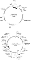
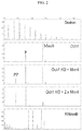


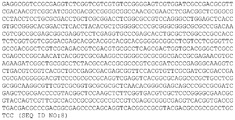


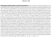


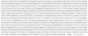


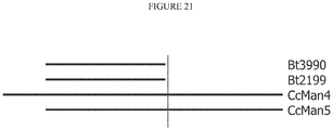
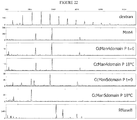
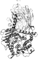


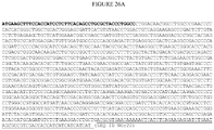


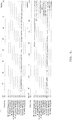


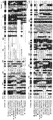


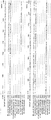


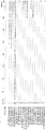


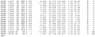
Classifications
-
- C—CHEMISTRY; METALLURGY
- C12—BIOCHEMISTRY; BEER; SPIRITS; WINE; VINEGAR; MICROBIOLOGY; ENZYMOLOGY; MUTATION OR GENETIC ENGINEERING
- C12Q—MEASURING OR TESTING PROCESSES INVOLVING ENZYMES, NUCLEIC ACIDS OR MICROORGANISMS; COMPOSITIONS OR TEST PAPERS THEREFOR; PROCESSES OF PREPARING SUCH COMPOSITIONS; CONDITION-RESPONSIVE CONTROL IN MICROBIOLOGICAL OR ENZYMOLOGICAL PROCESSES
- C12Q1/00—Measuring or testing processes involving enzymes, nucleic acids or microorganisms; Compositions therefor; Processes of preparing such compositions
- C12Q1/34—Measuring or testing processes involving enzymes, nucleic acids or microorganisms; Compositions therefor; Processes of preparing such compositions involving hydrolase
-
- C—CHEMISTRY; METALLURGY
- C12—BIOCHEMISTRY; BEER; SPIRITS; WINE; VINEGAR; MICROBIOLOGY; ENZYMOLOGY; MUTATION OR GENETIC ENGINEERING
- C12N—MICROORGANISMS OR ENZYMES; COMPOSITIONS THEREOF; PROPAGATING, PRESERVING, OR MAINTAINING MICROORGANISMS; MUTATION OR GENETIC ENGINEERING; CULTURE MEDIA
- C12N9/00—Enzymes; Proenzymes; Compositions thereof; Processes for preparing, activating, inhibiting, separating or purifying enzymes
- C12N9/14—Hydrolases (3)
- C12N9/24—Hydrolases (3) acting on glycosyl compounds (3.2)
- C12N9/2402—Hydrolases (3) acting on glycosyl compounds (3.2) hydrolysing O- and S- glycosyl compounds (3.2.1)
- C12N9/2477—Hemicellulases not provided in a preceding group
- C12N9/2488—Mannanases
-
- A—HUMAN NECESSITIES
- A61—MEDICAL OR VETERINARY SCIENCE; HYGIENE
- A61P—SPECIFIC THERAPEUTIC ACTIVITY OF CHEMICAL COMPOUNDS OR MEDICINAL PREPARATIONS
- A61P3/00—Drugs for disorders of the metabolism
-
- C—CHEMISTRY; METALLURGY
- C07—ORGANIC CHEMISTRY
- C07K—PEPTIDES
- C07K14/00—Peptides having more than 20 amino acids; Gastrins; Somatostatins; Melanotropins; Derivatives thereof
-
- C—CHEMISTRY; METALLURGY
- C12—BIOCHEMISTRY; BEER; SPIRITS; WINE; VINEGAR; MICROBIOLOGY; ENZYMOLOGY; MUTATION OR GENETIC ENGINEERING
- C12N—MICROORGANISMS OR ENZYMES; COMPOSITIONS THEREOF; PROPAGATING, PRESERVING, OR MAINTAINING MICROORGANISMS; MUTATION OR GENETIC ENGINEERING; CULTURE MEDIA
- C12N15/00—Mutation or genetic engineering; DNA or RNA concerning genetic engineering, vectors, e.g. plasmids, or their isolation, preparation or purification; Use of hosts therefor
- C12N15/09—Recombinant DNA-technology
- C12N15/63—Introduction of foreign genetic material using vectors; Vectors; Use of hosts therefor; Regulation of expression
- C12N15/79—Vectors or expression systems specially adapted for eukaryotic hosts
- C12N15/80—Vectors or expression systems specially adapted for eukaryotic hosts for fungi
-
- C—CHEMISTRY; METALLURGY
- C12—BIOCHEMISTRY; BEER; SPIRITS; WINE; VINEGAR; MICROBIOLOGY; ENZYMOLOGY; MUTATION OR GENETIC ENGINEERING
- C12N—MICROORGANISMS OR ENZYMES; COMPOSITIONS THEREOF; PROPAGATING, PRESERVING, OR MAINTAINING MICROORGANISMS; MUTATION OR GENETIC ENGINEERING; CULTURE MEDIA
- C12N9/00—Enzymes; Proenzymes; Compositions thereof; Processes for preparing, activating, inhibiting, separating or purifying enzymes
- C12N9/14—Hydrolases (3)
- C12N9/24—Hydrolases (3) acting on glycosyl compounds (3.2)
- C12N9/2402—Hydrolases (3) acting on glycosyl compounds (3.2) hydrolysing O- and S- glycosyl compounds (3.2.1)
- C12N9/2405—Glucanases
- C12N9/2408—Glucanases acting on alpha -1,4-glucosidic bonds
- C12N9/2411—Amylases
- C12N9/2428—Glucan 1,4-alpha-glucosidase (3.2.1.3), i.e. glucoamylase
-
- C—CHEMISTRY; METALLURGY
- C12—BIOCHEMISTRY; BEER; SPIRITS; WINE; VINEGAR; MICROBIOLOGY; ENZYMOLOGY; MUTATION OR GENETIC ENGINEERING
- C12N—MICROORGANISMS OR ENZYMES; COMPOSITIONS THEREOF; PROPAGATING, PRESERVING, OR MAINTAINING MICROORGANISMS; MUTATION OR GENETIC ENGINEERING; CULTURE MEDIA
- C12N9/00—Enzymes; Proenzymes; Compositions thereof; Processes for preparing, activating, inhibiting, separating or purifying enzymes
- C12N9/14—Hydrolases (3)
- C12N9/24—Hydrolases (3) acting on glycosyl compounds (3.2)
- C12N9/2402—Hydrolases (3) acting on glycosyl compounds (3.2) hydrolysing O- and S- glycosyl compounds (3.2.1)
- C12N9/2465—Hydrolases (3) acting on glycosyl compounds (3.2) hydrolysing O- and S- glycosyl compounds (3.2.1) acting on alpha-galactose-glycoside bonds, e.g. alpha-galactosidase (3.2.1.22)
-
- C—CHEMISTRY; METALLURGY
- C12—BIOCHEMISTRY; BEER; SPIRITS; WINE; VINEGAR; MICROBIOLOGY; ENZYMOLOGY; MUTATION OR GENETIC ENGINEERING
- C12P—FERMENTATION OR ENZYME-USING PROCESSES TO SYNTHESISE A DESIRED CHEMICAL COMPOUND OR COMPOSITION OR TO SEPARATE OPTICAL ISOMERS FROM A RACEMIC MIXTURE
- C12P19/00—Preparation of compounds containing saccharide radicals
- C12P19/04—Polysaccharides, i.e. compounds containing more than five saccharide radicals attached to each other by glycosidic bonds
-
- C—CHEMISTRY; METALLURGY
- C12—BIOCHEMISTRY; BEER; SPIRITS; WINE; VINEGAR; MICROBIOLOGY; ENZYMOLOGY; MUTATION OR GENETIC ENGINEERING
- C12P—FERMENTATION OR ENZYME-USING PROCESSES TO SYNTHESISE A DESIRED CHEMICAL COMPOUND OR COMPOSITION OR TO SEPARATE OPTICAL ISOMERS FROM A RACEMIC MIXTURE
- C12P19/00—Preparation of compounds containing saccharide radicals
- C12P19/14—Preparation of compounds containing saccharide radicals produced by the action of a carbohydrase (EC 3.2.x), e.g. by alpha-amylase, e.g. by cellulase, hemicellulase
-
- C—CHEMISTRY; METALLURGY
- C12—BIOCHEMISTRY; BEER; SPIRITS; WINE; VINEGAR; MICROBIOLOGY; ENZYMOLOGY; MUTATION OR GENETIC ENGINEERING
- C12P—FERMENTATION OR ENZYME-USING PROCESSES TO SYNTHESISE A DESIRED CHEMICAL COMPOUND OR COMPOSITION OR TO SEPARATE OPTICAL ISOMERS FROM A RACEMIC MIXTURE
- C12P21/00—Preparation of peptides or proteins
- C12P21/005—Glycopeptides, glycoproteins
-
- C—CHEMISTRY; METALLURGY
- C12—BIOCHEMISTRY; BEER; SPIRITS; WINE; VINEGAR; MICROBIOLOGY; ENZYMOLOGY; MUTATION OR GENETIC ENGINEERING
- C12Y—ENZYMES
- C12Y302/00—Hydrolases acting on glycosyl compounds, i.e. glycosylases (3.2)
- C12Y302/01—Glycosidases, i.e. enzymes hydrolysing O- and S-glycosyl compounds (3.2.1)
- C12Y302/01003—Glucan 1,4-alpha-glucosidase (3.2.1.3), i.e. glucoamylase
-
- C—CHEMISTRY; METALLURGY
- C12—BIOCHEMISTRY; BEER; SPIRITS; WINE; VINEGAR; MICROBIOLOGY; ENZYMOLOGY; MUTATION OR GENETIC ENGINEERING
- C12Y—ENZYMES
- C12Y302/00—Hydrolases acting on glycosyl compounds, i.e. glycosylases (3.2)
- C12Y302/01—Glycosidases, i.e. enzymes hydrolysing O- and S-glycosyl compounds (3.2.1)
- C12Y302/01022—Alpha-galactosidase (3.2.1.22)
-
- Y—GENERAL TAGGING OF NEW TECHNOLOGICAL DEVELOPMENTS; GENERAL TAGGING OF CROSS-SECTIONAL TECHNOLOGIES SPANNING OVER SEVERAL SECTIONS OF THE IPC; TECHNICAL SUBJECTS COVERED BY FORMER USPC CROSS-REFERENCE ART COLLECTIONS [XRACs] AND DIGESTS
- Y02—TECHNOLOGIES OR APPLICATIONS FOR MITIGATION OR ADAPTATION AGAINST CLIMATE CHANGE
- Y02P—CLIMATE CHANGE MITIGATION TECHNOLOGIES IN THE PRODUCTION OR PROCESSING OF GOODS
- Y02P20/00—Technologies relating to chemical industry
- Y02P20/50—Improvements relating to the production of bulk chemicals
- Y02P20/52—Improvements relating to the production of bulk chemicals using catalysts, e.g. selective catalysts
Definitions
- the invention relates to methods of hydrolyzing mannose-1-phospho-6-mannose linkages on glycoproteins, and more particularly, to using a mannosidase to hydrolyze mannose-1-phospho-6-mannose linkages to uncap the phospho-6-mannose residues on the glycoprotein.
- High performance expression systems are required to produce most biopharmaceuticals (e.g., recombinant proteins) currently under development.
- the biological activity of many of these biopharmaceuticals is dependent on their post-translational modification (e.g., phosphorylation or glycosylation).
- a yeast-based expression system combines the ease of genetic manipulation and fermentation of a microbial organism with the capability to secrete and to modify proteins.
- recombinant glycoproteins produced in yeast cells exhibit mainly heterogeneous high-mannose and hyper-mannose glycan structures, which can be detrimental to protein function, downstream processing, and subsequent therapeutic use, particularly where glycosylation plays a biologically significant role.
- the present invention is based, at least in part, on the discovery of a mannosidase that is capable of hydrolyzing mannose-1-phospho-6-mannose linkages on glycoproteins.
- the mannosidase can be used to obtain glycoproteins containing uncapped terminal mannose-6-phosphate residues.
- In vitro and in vivo methods of obtaining such glycoproteins are described herein.
- Genetically engineered cells can be used in the methods to produce target molecules having uncapped terminal mannose-6-phosphate residues.
- the present invention provides subject-matter as set forth in any one and all of (1) to (20) below:
- the present specification further describes a method for uncapping a mannose-6-phosphate residue on an oligosaccharide.
- the method includes providing the oligosaccharide having a mannose-1-phospho-6-mannose linkage; and contacting the oligosaccharide with a mannosidase capable of hydrolyzing the mannose-1-phospho-6-mannose linkage to phospho-6-mannose.
- the contacting step can be performed using a purified mannosidase, a recombinant mannosidase, a cell lysate containing the recombinant mannosidase, or a fungal cell containing the recombinant mannosidase.
- the mannosidase can include a targeting sequence.
- the oligosaccharide can be attached to a protein (e.g., a human protein expressed in a fungal organism).
- the present specification further describes a method of producing a target protein having terminal phospho-6-mannose residues.
- the method includes providing a fungal cell genetically engineered to include a nucleic acid encoding a mannosidase, the mannosidase capable of hydrolyzing a mannose-1-phospho-6-mannose linkage to phospho-6-mannose; and introducing into the cell a nucleic acid encoding a target protein, wherein the cell produces the target protein comprising the terminal phospho-6-mannose residues.
- the present specification further describes a method of producing a target protein having terminal phospho-6-mannose residues in a fungal organism.
- the method includes providing a fungal cell genetically engineered to include a nucleic acid encoding a mannosidase capable of hydrolyzing a mannose-1-phospho-6-mannose linkage to phospho-6-mannose, wherein the fungal cell further includes a nucleic acid encoding a target protein; and isolating the target protein having the terminal phospho-6-mannose residues.
- the fungal cell further can include a nucleic acid encoding a polypeptide capable of promoting mannosyl phosphorylation and/or can be genetically engineered to be deficient in OCH1 activity.
- the present specification further describes an isolated fungal cell genetically engineered to produce glycoproteins comprising terminal phospho-6-mannose residues.
- the fungal cell includes a nucleic acid encoding a mannosidase, wherein expression of the mannosidase in the fungal cell produces glycoproteins comprising the terminal phospho-6-mannose residues.
- the fungal cell further can include a nucleic acid encoding a target glycoprotein protein.
- the present specification further describes a substantially pure culture of Yarrowia lipolytica , Pichia pastoris, Hansenula polymorpha , Arxula adeninivorans , Pichia methanolica, Oogataea minuta, or Aspergillus niger cells, a substantial number of which are genetically engineered to produce glycoproteins comprising a terminal phospho-6-mannose residue, the cells comprising a nucleic acid encoding a mannosidase capable of hydrolyzing a mannose-1-phospho-6-mannose linkage to phospho-6-mannose.
- the fungal organism can be Yarrowia lipolytica or Arxula adeninivorans.
- the fungal organism can be a methylotrophic yeast such as Pichia pastoris, Pichia methanolica, Oogataea minuta, or Hansenula polymorpha.
- the fungal organism can be a filamentous fungus (e.g., a filamentous fungus selected from the group consisting of Aspergillus caesiellus, Aspergillus candidus, Aspergillus carneus, Aspergillus clavatus, Aspergillus deflectus, Aspergillus flavus, Aspergillus fumigatus, Aspergillus glaucus, Aspergillus nidulans, Aspergillus niger, Aspergillus ochraceus, Aspergillus oryzae, Aspergillus parasiticus, Aspergillus penicilloides, Aspergillus restrictus, Aspergillus sojae, Aspergillus sydowi, Aspergillus tamari, Aspergillus terreus, Aspergillus ustus, and Aspergillus versicolor ).
- a filamentous fungus selected from the group consisting of Asper
- the protein can be a pathogen protein, a lysosomal protein, a growth factor, a cytokine, a chemokine, an antibody or antigen-binding fragment thereof, or a fusion protein.
- the lysosomal protein can be a lysosomal enzyme (e.g., a lysosomal enzyme associated with a lysosomal storage disorder (LSD) such as acid alpha glucosidase or alpha galactosidase).
- LSD lysosomal storage disorder
- the LSD can be Fabry's disease, mucopolysaccharidosis I, Farber disease, Gaucher disease, GM1-gangliosidosis, Tay-Sachs disease, Sandhoff disease, GM2 activator disease, Krabbe disease, metachromatic leukodystrophy, Niemann-Pick disease, Scheie disease, Hunter disease, Sanfilippo disease, Morquio disease, Maroteaux-Lamy disease, hyaluronidase deficiency, aspartylglucosaminuria, fucosidosis, mannosidosis, Schindler disease, sialidosis type 1, Pompe disease, Pycnodysostosis, ceroid lipofuscinosis, cholesterol ester storage disease, Wolman disease, Multiple sulfatase deficiency, galactosialidosis, mucolipidosis, cystinosis, sialic acid storage disorder, chylomicron retention disease with Marinesco-Sjogren syndrome, Hermansky-Pudlak syndrome,
- the mannosidase can include an amino acid sequence having at least 90% identity(e.g., at least 95% or 98% identity) to the amino acid sequence set forth in residues 1 to 774 of SEQ ID NO:50 or to the amino acid sequence set forth in SEQ ID NO:50.
- the mannosidase can include an amino acid sequence having (i) a GVGXXGXGG motif, where X is Gly, Ala, Ser, Thr, or Cys; (ii) a VRXE motif, where X is any amino acid other than Pro; (iii) an X 1 YQGX 2 motif, where X 1 is Leu, Ile, Val, Ala, Phe, Tyr or Met, and X 2 is Thr, Ser, or Asn; or (iv) a GDXGN motif, where X can be any amino acid other than Pro.
- the mannosidase can be a C. cellulans mannosidase.
- the fungal cell further can include a nucleic acid encoding a polypeptide capable of promoting mannosyl phosphorylation (e.g., a MNN4 polypeptide such as Yarrowia lipolytica , S. cerevisiae, Ogataea minuta, Pichia pastoris, or C . albicans polypeptide) and/or can be genetically engineered to be deficient in OCH1 activity.
- a polypeptide capable of promoting mannosyl phosphorylation can be a P. pastoris PNO1 polypeptide.
- the mannosidase can include a secretion signal and/or a targeting signal to target the mannosidase to an intracellular compartment.
- the target protein and the mannosidase can be co-secreted.
- This document also features an isolated glycoprotein that includes terminal phospho-6-mannose residues, wherein the protein is produced by a method comprising contacting a glycoprotein expressed in a cell of fungal origin, said glycoprotein containing a mannose-1-phospho-6 mannose linkage, with a mannosidase capable of hydrolyzing said mannose-1-phospho-6-mannose residue to phospho-6-mannose, wherein said mannosidase comprises an amino acid sequence having at least 70% identity to SEQ ID NO:50 or at least 90% identity to residues 1 to 774 of SEQ ID NO:50.
- the present specification further describes a composition that includes a glycoprotein, wherein at least 47% of the N-glycans on the glycoprotein have terminal phospho-6-mannose residues. For example, at least 50%, 75%, 80%, 85%, or 90% of the N-glycans on the glycoprotein can have terminal phospho-6-mannose residues.
- the present specification further describes an isolated nucleic acid that includes a nucleotide sequence set forth in SEQ ID NO:6, SEQ ID NO:8, SEQ ID NO:10, SEQ ID NO:12, or SEQ ID NO:14, or a nucleotide sequence that is at least 90% identical to SEQ ID NO:6, SEQ ID NO:8, SEQ ID NO:10, SEQ ID NO:12, SEQ ID NO:14, or SEQ ID NO:20.
- the present specification further describes a vector that includes a promoter operably linked to such a nucleic acid, wherein the nucleic acid encodes a mannosidase.
- the nucleic acid further can include a secretion signal or targeting sequence to target the mannosidase to an intracellular compartment.
- the present specification describes methods and materials for hydrolyzing mannose-1-phospho-6-mannose linkages on glycoproteins to produce target molecules (e.g., target proteins) having uncapped phospho-6-mannose (M6P) residues.
- target molecules e.g., target proteins
- M6P uncapped phospho-6-mannose residues.
- the methods and materials described herein are particularly useful for producing agents for treating patients with lysosomal storage disorders (LSDs), a diverse group of hereditary metabolic disorders characterized by the accumulation of storage products in the lysosomes due to impaired activity of catabolic enzymes involved in their degradation.
- LSDs lysosomal storage disorders
- Deficiencies in catabolic enzymes can be corrected by enzyme replacement therapy (ERT), provided that the administered enzyme can be targeted to the lysosomes of the diseased cells.
- ERT enzyme replacement therapy
- Lysosomal enzymes typically are glycoproteins that are synthesized in the endoplasmic reticulum (ER), transported via the secretory pathway to the Golgi, and then recruited to the lysosomes.
- ER endoplasmic reticulum
- MPR mannose 6-phosphate receptor
- a microbial based production process can be used to obtain therapeutic proteins with uncapped M6P glycans, which can be delivered to lysosomes by exploiting the same M6P dependent pathway.
- the methods and materials described herein are useful for preparing glycoproteins for the treatment of metabolic disorders such as LSDs.
- nucleic acid and “polynucleotide” are used interchangeably herein, and refer to both RNA and DNA, including cDNA, genomic DNA, synthetic DNA, and DNA (or RNA) containing nucleic acid analogs. Polynucleotides can have any three-dimensional structure.
- a nucleic acid can be double-stranded or single-stranded (i.e., a sense strand or an antisense strand).
- Non-limiting examples of polynucleotides include genes, gene fragments, exons, introns, messenger RNA (mRNA), transfer RNA, ribosomal RNA, siRNA, micro-RNA, ribozymes, cDNA, recombinant polynucleotides, branched polynucleotides, plasmids, vectors, isolated DNA of any sequence, isolated RNA of any sequence, nucleic acid probes, and primers, as well as nucleic acid analogs.
- mRNA messenger RNA
- transfer RNA transfer RNA
- ribosomal RNA siRNA
- micro-RNA micro-RNA
- ribozymes cDNA
- recombinant polynucleotides branched polynucleotides
- plasmids vectors, isolated DNA of any sequence,
- Polypeptide and “protein” are used interchangeably herein and mean any peptide-linked chain of amino acids, regardless of length or post-translational modification.
- a polypeptide described herein e.g., a mannosidase or a target protein having uncapped M6P residues
- a polypeptide described herein is isolated when it constitutes at least 60%, by weight, of the total protein in a preparation, e.g., 60% of the total protein in a sample.
- a polypeptide described herein consists of at least 75%, at least 90%, or at least 99%, by weight, of the total protein in a preparation.
- isolated nucleic acid refers to a nucleic acid that is separated from other nucleic acid molecules that are present in a naturally-occurring genome, including nucleic acids that normally flank one or both sides of the nucleic acid in a naturally-occurring genome (e.g., a yeast genome).
- isolated as used herein with respect to nucleic acids also includes any non-naturally-occurring nucleic acid sequence, since such non-naturally-occurring sequences are not found in nature and do not have immediately contiguous sequences in a naturally-occurring genome.
- an isolated nucleic acid can be, for example, a DNA molecule, provided one of the nucleic acid sequences normally found immediately flanking that DNA molecule in a naturally-occurring genome is removed or absent.
- an isolated nucleic acid includes, without limitation, a DNA molecule that exists as a separate molecule (e.g., a chemically synthesized nucleic acid, or a cDNA or genomic DNA fragment produced by PCR or restriction endonuclease treatment) independent of other sequences as well as DNA that is incorporated into a vector, an autonomously replicating plasmid, a virus (e.g., any paramyxovirus, retrovirus, lentivirus, adenovirus, or herpes virus), or into the genomic DNA of a prokaryote or eukaryote.
- a separate molecule e.g., a chemically synthesized nucleic acid, or a cDNA or genomic DNA fragment produced by PCR or restriction endonucleas
- an isolated nucleic acid can include an engineered nucleic acid such as a DNA molecule that is part of a hybrid or fusion nucleic acid.
- exogenous refers to any nucleic acid that does not occur in (and cannot be obtained from) that particular cell as found in nature.
- a non-naturally-occurring nucleic acid is considered to be exogenous to a host cell once introduced into the host cell. It is important to note that non-naturally-occurring nucleic acids can contain nucleic acid subsequences or fragments of nucleic acid sequences that are found in nature provided that the nucleic acid as a whole does not exist in nature.
- a nucleic acid molecule containing a genomic DNA sequence within an expression vector is non-naturally-occurring nucleic acid, and thus is exogenous to a host cell once introduced into the host cell, since that nucleic acid molecule as a whole (genomic DNA plus vector DNA) does not exist in nature.
- any vector, autonomously replicating plasmid, or virus e.g., retrovirus, adenovirus, or herpes virus
- retrovirus e.g., adenovirus, or herpes virus
- genomic DNA fragments produced by PCR or restriction endonuclease treatment as well as cDNAs are considered to be non-naturally-occurring nucleic acid since they exist as separate molecules not found in nature. It also follows that any nucleic acid containing a promoter sequence and polypeptide-encoding sequence (e.g., cDNA or genomic DNA) in an arrangement not found in nature is non-naturally-occurring nucleic acid.
- a nucleic acid that is naturally-occurring can be exogenous to a particular cell. For example, an entire chromosome isolated from a cell of yeast x is an exogenous nucleic acid with respect to a cell of yeast y once that chromosome is introduced into a cell of yeast.
- a nucleic acid encoding a mannosidase can have at least 70% sequence identity (e.g., at least 80%, 85%, 90%, 95%, 97%, 98%, 99%, or 100% sequence identity) to a nucleotide sequence set forth in SEQ ID NO:6, SEQ ID NO:8, SEQ ID NO:10, SEQ ID NO:12, or SEQ ID NO:14.
- nucleic acids described herein can encode mannosidase polypeptides that have at least 70% (e.g., at least 75, 80, 85, 90, 95, 99, or 100 percent) identity to an amino acid sequence set forth in SEQ ID NOs: 50.
- a nucleic acid can encode a mannosidase having at least 90% (e.g., at least 95 or 98%) identity to the amino acid sequence set forth in SEQ ID NO:50.
- a nucleic acid can encode a mannosidase having at least 90% identity to amino acid residues 1 to 774 of SEQ ID NO:50.
- the percent identity between a particular amino acid sequence and the amino acid sequence set forth in SEQ ID NO:7, SEQ ID NO:9, SEQ ID NO:11, SEQ ID NO:13, SEQ ID NO:15, or SEQ ID NO:50 is determined as follows.
- the amino acid sequences are aligned using the BLAST 2 Sequences (B12seq) program from the stand-alone version of BLASTZ containing BLASTP version 2.0.14.
- This stand-alone version of BLASTZ can be obtained from Fish & Richardson's web site (e.g., www.fr.com/blast/) or the U.S. government's National Center for Biotechnology Information web site (www.ncbi.nlm.nih.gov). Instructions explaining how to use the B12seq program can be found in the readme file accompanying BLASTZ.
- B12seq performs a comparison between two amino acid sequences using the BLASTP algorithm.
- B12seq are set as follows: -i is set to a file containing the first amino acid sequence to be compared (e.g., C: ⁇ seq1.txt); -j is set to a file containing the second amino acid sequence to be compared (e.g., C: ⁇ seq2.txt); -p is set to blastp; -o is set to any desired file name (e.g., C: ⁇ output.txt); and all other options are left at their default setting.
- -i is set to a file containing the first amino acid sequence to be compared (e.g., C: ⁇ seq1.txt)
- -j is set to a file containing the second amino acid sequence to be compared (e.g., C: ⁇ seq2.txt)
- -p is set to blastp
- -o is set to any desired file name (e.g., C: ⁇ output.txt); and all other options are left at their
- the following command can be used to generate an output file containing a comparison between two amino acid sequences: C: ⁇ B12seq -i c: ⁇ seq1.txt -j c: ⁇ seq2.txt -p blastp -o c: ⁇ output.txt. If the two compared sequences share homology, then the designated output file will present those regions of homology as aligned sequences. If the two compared sequences do not share homology, then the designated output file will not present aligned sequences. Similar procedures can be following for nucleic acid sequences except that blastn is used.
- the number of matches is determined by counting the number of positions where an identical amino acid residue is presented in both sequences.
- percent identity value is rounded to the nearest tenth.
- 78.11, 78.12, 78.13, and 78.14 is rounded down to 78.1
- 78.15, 78.16, 78.17, 78.18, and 78.19 is rounded up to 78.2.
- the length value will always be an integer.
- nucleic acids can encode a polypeptide having a particular amino acid sequence.
- the degeneracy of the genetic code is well known to the art; i.e., for many amino acids, there is more than one nucleotide triplet that serves as the codon for the amino acid.
- codons in the coding sequence for a given mannosidase polypeptide can be modified such that optimal expression in a particular species (e.g., bacteria or fungus) is obtained, using appropriate codon bias tables for that species.
- the nucleic acids set forth in SEQ ID NO:6, SEQ ID NO:8, SEQ ID NO:10, SEQ ID NO:12, or SEQ ID NO:14 can be codon optimized for E. coli expression as set forth in FIGs. 14-18 (see SEQ ID NOs:16-20).
- Hybridization also can be used to assess homology between two nucleic acid sequences.
- a nucleic acid sequence described herein, or a fragment or variant thereof can be used as a hybridization probe according to standard hybridization techniques.
- the hybridization of a probe of interest e.g., a probe containing a portion of a CcMan5 nucleotide sequence
- the hybridization of a probe of interest is an indication of the presence of DNA or RNA (e.g., a CcMan5 nucleotide sequence) corresponding to the probe in the test source.
- Hybridization conditions are known to those skilled in the art and can be found in Current Protocols in Molecular Biology, John Wiley & Sons, N.Y., 6.3.1-6.3.6, 1991 .
- Moderate hybridization conditions are defined as equivalent to hybridization in 2X sodium chloride/sodium citrate (SSC) at 30°C, followed by a wash in 1 X SSC, 0.1% SDS at 50°C.
- Highly stringent conditions are defined as equivalent to hybridization in 6X sodium chloride/sodium citrate (SSC) at 45°C, followed by a wash in 0.2 X SSC, 0.1% SDS at 65°C.
- Mannosidase polypeptides capable of hydrolyzing terminal mannose-1-phospho-6-mannose linkages on oligosaccharides also can be identified based on the three dimensional structure described herein for a portion of a mannosidase from C . cellulans (residues 1 to 774 of SEQ ID NO:50, also referred to as CcMan5 1-774 ).
- the three dimensional structure can be determined by, for example, X-ray diffraction of a crystal of CcMan5 1-774 .
- Structural coordinates of CcMan5 1-774 e.g., the coordinates of CcMan5 1-774 deposited with the Protein Data Bank (world wide web at PDB.org under PDB ID No.
- the coordinates set forth in FIG. 33 for the catalytic center of CcMan5, or the coordinates set forth in FIG. 34 for the protein C alpha atoms and the catalytic Ca2+ atoms of the two CcMan5 1-774 molecules in the asymmetric unit in PDB entry 2xsg) are useful for a number of applications, including, but not limited to, the characterization of a three dimensional structure of a mannosidase capable of hydrolyzing terminal mannose-1-phospho-6-mannose linkages on oligosaccharides, as well as the visualization, identification and characterization of regions of the mannosidase that are involved in acceptance of mannose-6-phosphate-alpha,1-mannose (hereafter referred to as Man-P-Man) as a substrate, and conferring its ability to hydrolyse Man-P-Man to produce a terminal phospho-6-mannose.
- Man-P-Man mannose-6-phosphate-alpha,1-mannose
- Structural coordinates are the Cartesian coordinates corresponding to an atom's spatial relationship to other atoms in a molecule or molecular complex. Structural coordinates can be obtained using x-ray crystallography techniques or NMR techniques, or can be derived using molecular replacement analysis or homology modeling. Various software programs allow for the graphical representation of a set of structural coordinates to obtain a three dimensional representation of a molecule or molecular complex.
- the structural coordinates of the structures described herein can be modified from the original set provided in FIG. 33 or FIG. 34 by mathematical manipulation, such as by inversion or integer additions or subtractions. As such, it is recognized that the structural coordinates described herein are relative, and are in no way specifically limited by the actual x, y, z coordinates of FIG. 33 or FIG. 34 .
- CcMan5 1-774 consists of two domains, an N-terminal ⁇ -sandwich domain (residues 8 to 271 of SEQ ID NO:50) and a C-terminal ( ⁇ )6 barrel domain (residues 291 to 771 of SEQ ID NO:50), connected via an ⁇ -helical linker (residues 272 to 290 of SEQ ID NO:50).
- the interface between both domains gives shape to a shallow cavity that harbors a conserved catalytic Ca 2+ ion and gives shape to the -1 substrate binding site (nomenclature as described by Davies et al., Biochem. J.
- the three dimensional structure of CcMan5 1-774 can be characterized in part, or all, using the structural coordinates of PDB ID No. 2xs, or an extract of which that is presented in FIG. 33 , comprising the residues surrounding the active site of CcMan5 1-774 , or an extract of which that is presented in FIG. 34 , comprising the protein C alpha atoms and the catalytic Ca2+ atoms of the two CcMan5 1-774 molecules in the asymmetric unit in PDB entry 2xsg, and describing the overall fold of the protein.
- the three-dimensional structure of CcMan5 1-774 can be characterized by the structural coordinates of amino acid residues 7 to 771 according to PDB ID No.
- CcMan5 1-774 may comprise the complete structural coordinates of the amino acids according to PDB ID No. 2xs, ⁇ a root mean square deviation from the conserved backbone atoms of said amino acids of not more than 2 ⁇ (e.g., not more than 1.5 ⁇ , 1.0 ⁇ or 0.5 ⁇ ).
- root mean square deviation is the square root of the arithmetic mean of the squares of the deviations from the mean, and is a way of expressing deviation or variation from the structural coordinates described herein. Conservative substitutions of the noted amino acid residues may result in same structural coordinates within the stated root mean square deviation.
- the structural coordinates provided herein can be used to characterize a three dimensional structure of a mannosidase polypeptide. From such a structure, substrate binding sites, for example, can be computationally visualized, identified and characterized based on the surface structure of the molecule, surface charge, steric arrangement, the presence of reactive amino acids, regions of hydrophobicity or hydrophilicity, etc.
- the relevant coordinates can be displayed as, or converted to, a three dimensional shape or graphical representation.
- Software programs are commercially available that are capable of generating three dimensional graphical representations of molecules or portions thereof from a set of structural coordinates.
- Examples of commercially available software programs include, without limitation, the following: GRID (Oxford University, Oxford, UK) ; MCSS (Molecular Simulations, San Diego, CA); AUTODOCK (Scripps Research Institute, La Jolla, CA); DOCK (University of California, San Francisco, CA); Flo99 (Thistlesoft, Morris Township, NJ); Ludi (Molecular Simulations, San Diego, CA); QUANTA (Molecular Simulations, San Diego, CA); Insight (Molecular Simulations, San Diego, CA); SYBYL (TRIPOS, Inc., St. Louis. MO); and LEAPFROG (TRIPOS, Inc., St. Louis, MO).
- the structural coordinates described herein can be used with standard homology modeling techniques in order to determine the unknown three-dimensional structure of a molecule or molecular complex.
- Homology modeling involves constructing a model of an unknown structure using structural coordinates of one or more related protein molecules, molecular complexes or parts thereof.
- Homology modeling can be conducted by fitting common or homologous portions of the protein whose three dimensional structure is to be solved to the three dimensional structure of homologous structural elements in the known molecule, specifically using the relevant (i.e., homologous) structural coordinates provided by FIGs. 33 and 34 herein.
- Homology may be determined using amino acid sequence identity, homologous secondary structure elements, and/or homologous tertiary folds.
- Homology modeling can include rebuilding part or all of a three dimensional structure with replacement of amino acids (or other components) by those of the related structure to be solved. Accordingly, a three dimensional structure for the unknown molecule may be generated using the three dimensional structure of CcMan5 1-774 described herein, and refined using a number of techniques well known in the art.
- substitutions can be made in some of the atoms or side groups of CcMan5 1-774 or other mannosidases in order to improve or modify its selectivity.
- CcMan5 contains a non-acidic residue at positions 536 and 588, which may allow the mannosidase to tolerate the phosphate linkage to the anomeric oxygen in Man-P-Man substrates.
- corresponding residues in other mannosidases can be changed to non-acidic residues to increase the ability of the mannosidase to accept Man-P-Man substrates.
- mannosidase polypeptide candidates suitable for use herein can be identified by analysis of nucleotide and polypeptide sequence alignments. For example, performing a query on a database of nucleotide or polypeptide sequences can identify homologs and/or orthologs of mannosidase polypeptides. Sequence analysis can involve BLAST, Reciprocal BLAST, or PSI-BLAST analysis of nonredundant databases using known mannosidase amino acid sequences. Those polypeptides in the database that have greater than 40% sequence identity can be identified as candidates for further evaluation for suitability as a mannosidase polypeptide.
- Amino acid sequence similarity allows for conservative amino acid substitutions, such as substitution of one hydrophobic residue for another or substitution of one polar residue for another. If desired, manual inspection of such candidates can be carried out in order to narrow the number of candidates to be further evaluated. Manual inspection can be performed by selecting those candidates that appear to have domains suspected of being present in mannosidases capable of hydrolyzing terminal mannose-1-phospho-6-mannose linkages, e.g., one or more (e.g., 1, 2, 3, 4 or more) conserved domains or functional regions (e.g., substrate binding cavity).
- Such domains can include a glycine-rich motif GVGXXGXGG, where X is Gly, Ser, Thr, Val, Ala, Cys or Gln (or other amino acid with a small side chain).
- This motif is found at residues 69-77 of SEQ ID NO:50. This region makes a loop that provides essential hydrogen bonds to the -1 mannose and phosphate-binding subsite in the active site of the enzyme.
- Another example of a conserved motif includes a VRXE motif, where Arg (R) makes a hydrogen bond to the -1 ring and possibly the +1 ring, Glu (E) is in a salt bridge to this R residue, probably shaping this motif; and X is Trp or any of the 20 amino acids except Pro.
- This motif is found at residues 404-407 of SEQ ID NO:50.
- a suitable motif also can be an X 1 YQGX 2 motif, where X 1 can be Leu, Ile, Val Ala, Phe, Tyr or Met, and X 2 can be Thr, Ser or Asn.
- This motif is found at residues 534-538 of SEQ ID NO:50.
- the Gln (Q) in this motif is important as an E is present in mannosidases that do not have the ability to hydrolyze terminal mannose-1-phospho-6-mannose linkages on oligosaccharides.
- the Tyr (Y) in this motif also is thought to be important for the +1 site formation.
- G71, G72, D355, R405, Q536, N588, Q589, T626, D660, D662 form the catalytic center, where N588, Q589 and D660 are involved in coordinating the catalytic Ca2+ ion, D662 and D660 are involved in activating the nucleophilic water, Q536 stabilizes the anomeric oxygen during the transition state and G71, G71, D355, R405 and T626 are involved in substrate binding at the -1 site. See FIG. 30 for a representation of this minimal catalytic center.
- a mannosidase can be selected as a candidate mannosidase capable of hydrolyzing terminal mannose-1-phospho-6-mannose linkages when the three dimensional protein coordinates of the atoms in the amino acid side chains located in the minimal catalytic center (e.g., as set forth in FIG. 30 ) fall within 1.5 ⁇ deviation of the coordinates of the equivalent atoms in FIG. 33 .
- a conserved motif also can be a GDXGN motif in the N-terminal domain of the protein, where X can be any amino acid except P.
- This motif is found at residues 21-25 of SEQ ID NO:50 and forms part of the substrate binding pocket of the enzyme as shown in FIG. 24 .
- the side chains of the D and N line the substrate binding cavity and may shape an alternative subpocket to bind the +1 mannose.
- Example 14 performing a query on a database of polypeptide sequences identified homologs of CcMan5 in the following organisms: Streptomyces coelicolor (GenBank Accession No. NP_630514), Streptomyces lividans (GenBank Accession No. ZP_05522540); Streptomyces lividans (GenBank Accession No. ZP_06527366); Clostridium spiroforme (GenBank Accession No. ZP_02866543), Bacteroides thetaiotaomicron (GenBank Accession No. NP_812442), Zunongwangia profunda (GenBank Accession No.
- YP_003584502 Chitinophaga pinensis (GenBank Accession No. YP_003120664); Paenibacillus sp (GenBank Accession No. YP_003013376); Bacteroides sp. (GenBank Accession No. ZP_04848482); Bacteroides cellulosilyticus (GenBank Accession No. ZP_03677957); Leeuwenhoekiella blandensis (GenBank Accession No. ZP_01061975); Sphingobacterium spiritivorum (GenBank Accession No. ZP_07083984); and Pedobacter sp. (GenBank Accession No. ZP_01885202).
- the mannosidases from Streptomyces coelicolor and Streptomyces lividans are similar (66% sequence identity to the CcMan5 GH92 domain, with 501 identities over 765 aligned residues by BLASTP), not only in the above motifs but also in many the loops of the three dimensional structure.
- Isolated nucleic acid molecules encoding mannosidase polypeptides can be produced by standard techniques. For example, polymerase chain reaction (PCR) techniques can be used to obtain an isolated nucleic acid containing a nucleotide sequence described herein. PCR can be used to amplify specific sequences from DNA as well as RNA, including sequences from total genomic DNA or total cellular RNA. Generally, sequence information from the ends of the region of interest or beyond is employed to design oligonucleotide primers that are identical or similar in sequence to opposite strands of the template to be amplified. Various PCR strategies also are available by which site-specific nucleotide sequence modifications can be introduced into a template nucleic acid.
- PCR polymerase chain reaction
- Isolated nucleic acids also can be chemically synthesized, either as a single nucleic acid molecule (e.g., using automated DNA synthesis in the 3' to 5' direction using phosphoramidite technology) or as a series of oligonucleotides.
- one or more pairs of long oligonucleotides e.g., >100 nucleotides
- each pair containing a short segment of complementarity e.g., about 15 nucleotides
- DNA polymerase is used to extend the oligonucleotides, resulting in a single, double-stranded nucleic acid molecule per oligonucleotide pair, which then can be ligated into a vector.
- Isolated nucleic acids of the invention also can be obtained by mutagenesis of, e.g., a naturally occurring DNA.
- the present specification further describes (i) biologically active variants and (ii) biologically active fragments or biologically active variants thereof, of the mannosidases described herein.
- Biologically active variants of mannosidases can contain additions, deletions, or substitutions relative to the sequences set forth in SEQ ID NOs: 7, 9, 11, 13, 15, or 50. Proteins with substitutions will generally have not more than 50 (e.g., not more than one, two, three, four, five, six, seven, eight, nine, ten, 12, 15, 20, 25, 30, 35, 40, or 50) conservative amino acid substitutions.
- a conservative substitution is the substitution of one amino acid for another with similar characteristics.
- Conservative substitutions include substitutions within the following groups: valine, alanine and glycine; leucine, valine, and isoleucine; aspartic acid and glutamic acid; asparagine and glutamine; serine, cysteine, and threonine; lysine and arginine; and phenylalanine and tyrosine.
- the non-polar hydrophobic amino acids include alanine, leucine, isoleucine, valine, proline, phenylalanine, tryptophan and methionine.
- the polar neutral amino acids include glycine, serine, threonine, cysteine, tyrosine, asparagine and glutamine.
- the positively charged (basic) amino acids include arginine, lysine and histidine.
- the negatively charged (acidic) amino acids include aspartic acid and glutamic acid. Any substitution of one member of the above-mentioned polar, basic or acidic groups by another member of the same group can be deemed a conservative substitution. By contrast, a non-conservative substitution is a substitution of one amino acid for another with dissimilar characteristics.
- the sequence alignments set forth in FIGs. 31 and 32 provide numerous examples of amino acid substitutions that can be made.
- Deletion variants can lack one, two, three, four, five, six, seven, eight, nine, ten, 11, 12, 13, 14, 15, 16, 17, 18, 19, or 20 amino acid segments (of two or more amino acids) or non-contiguous single amino acids.
- Additions include fusion proteins containing: (a) a mannosidase set forth in SEQ ID NOs: 7, 9, 11, 13, or 15, or a fragment thereof; and (b) internal or terminal (C or N) irrelevant or heterologous amino acid sequences.
- heterologous amino acid sequences refers to an amino acid sequence other than (a).
- a heterologous sequence can be, for example a sequence used for purification of the recombinant protein (e.g., FLAG, polyhistidine (e.g., hexahistidine), hemagluttanin (HA), glutathione-S-transferase (GST), or maltose-binding protein (MBP)).
- Heterologous sequences also can be proteins useful as diagnostic or detectable markers, for example, luciferase, green fluorescent protein (GFP), or chloramphenicol acetyl transferase (CAT).
- the fusion protein contains a signal sequence from another protein.
- the fusion protein can contain a carrier (e.g., KLH) useful, e.g., in eliciting an immune response for antibody generation) or endoplasmic reticulum or Golgi apparatus retention signals.
- a carrier e.g., KLH
- Heterologous sequences can be of varying length and in some cases can be a longer sequences than the full-length target proteins to which the heterologous sequences are attached.
- Biologically active fragments or biologically active variants of the mannosidases have at least 40% (e.g., at least: 50%; 60%; 70%; 75%; 80%; 85%; 90%; 95%; 97%; 98%; 99%; 99.5%, or 100% or even greater) of the mannosidase activity (e.g., uncapping of M6P residues) of the wild-type, full-length, mature protein.
- a biologically active fragment of a mannosidase can contain residues 1 to 774 of SEQ ID NO:50.
- mannosidases described herein can be used to produce molecules (e.g., target proteins) having uncapped terminal phospho-6-mannose (M6P) residues.
- the methods can be performed in vitro or in vivo.
- a mannosidase described herein can be recombinantly produced and used in vitro to uncap terminal M6P residues on oligosaccharides.
- a vector is used that contains a promoter operably linked to nucleic acid encoding a mannosidase polypeptide.
- a "promoter” refers to a DNA sequence that enables a gene to be transcribed. The promoter is recognized by RNA polymerase, which then initiates transcription. Thus, a promoter contains a DNA sequence that is either bound directly by, or is involved in the recruitment, of RNA polymerase.
- a promoter sequence can also include "enhancer regions,” which are one or more regions of DNA that can be bound with proteins (namely, the trans-acting factors, much like a set of transcription factors) to enhance transcription levels of genes (hence the name) in a gene-cluster.
- the enhancer while typically at the 5' end of a coding region, can also be separate from a promoter sequence and can be, e.g., within an intronic region of a gene or 3' to the coding region of the gene.
- operably linked means incorporated into a genetic construct (e.g., vector) so that expression control sequences effectively control expression of a coding sequence of interest.
- Expression vectors can be introduced into host cells (e.g., by transformation or transfection) for expression of the encoded polypeptide, which then can be purified.
- Expression systems that can be used for small or large scale production of mannosidase polypeptides include, without limitation, microorganisms such as bacteria (e.g., E. coli ) transformed with recombinant bacteriophage DNA, plasmid DNA, or cosmid DNA expression vectors containing the nucleic acid molecules, and fungal (e.g., S .
- Useful expression systems also include insect cell systems infected with recombinant virus expression vectors (e.g., baculovirus) containing the nucleic acid molecules, and plant cell systems infected with recombinant virus expression vectors (e.g., tobacco mosaic virus) or transformed with recombinant plasmid expression vectors (e.g., Ti plasmid) containing the nucleic acid molecules.
- recombinant virus expression vectors e.g., baculovirus
- plant cell systems infected with recombinant virus expression vectors (e.g., tobacco mosaic virus) or transformed with recombinant plasmid expression vectors (e.g., Ti plasmid) containing the nucleic acid molecules.
- Mannosidase polypeptides also can be produced using mammalian expression systems, which include cells (e.g., immortalized cell lines such as COS cells, Chinese hamster ovary cells, HeLa cells, human embryonic kidney 293 cells, and 3T3 L1 cells) harboring recombinant expression constructs containing promoters derived from the genome of mammalian cells (e.g., the metallothionein promoter) or from mammalian viruses (e.g., the adenovirus late promoter and the cytomegalovirus promoter), along with the nucleic acids described herein.
- cells e.g., immortalized cell lines such as COS cells, Chinese hamster ovary cells, HeLa cells, human embryonic kidney 293 cells, and 3T3 L1 cells
- promoters derived from the genome of mammalian cells
- mammalian viruses e.g., the adenovirus late promoter and the cytomegalovirus promoter
- recombinant mannosidase polypeptides are tagged with a heterologous amino acid sequence such FLAG, polyhistidine (e.g., hexahistidine), hemagluttanin (HA), glutathione-S-transferase (GST), or maltose-binding protein (MBP) to aid in purifying the protein.
- a heterologous amino acid sequence such as FLAG, polyhistidine (e.g., hexahistidine), hemagluttanin (HA), glutathione-S-transferase (GST), or maltose-binding protein (MBP)
- Other methods for purifying proteins include chromatographic techniques such as ion exchange, hydrophobic and reverse phase, size exclusion, affinity, hydrophobic charge-induction chromatography, and the like (see, e.g., Scopes, Protein Purification: Principles and Practice, third edition, Springer-Verlag, New York (1993 ); Burton
- a target molecule containing a mannose-1-phospho-6 mannose linkage is contacted under suitable conditions with a purified mannosidase or a cell lysate containing a recombinantly produced mannosidase.
- the cell lysate can be from any genetically engineered cell, including a fungal cell, a plant cell, or animal cell.
- animal cells include nematode, insect, plant, bird, reptile, and mammals such as a mouse, rat, rabbit, hamster, gerbil, dog, cat, goat, pig, cow, horse, whale, monkey, or human.
- the mannosidase Upon contacting the target molecule (e.g., an oligosaccharide or glycoprotein) with the purified mannosidase or cell lysate, the mannosidase hydrolyzes the mannose-1-phospho-6 mannose linkage and produces a target molecule having one or more uncapped terminal M6P residues.
- the methods described in Example 2 can be used to determine if the terminal M6P residues have been uncapped. Following processing by the mannosidase, the target molecule having uncapped terminal M6P residues can be isolated.
- Suitable methods for obtaining cell lysates that preserve the activity or integrity of the mannosidase activity in the lysate can include the use of appropriate buffers and/or inhibitors, including nuclease, protease and phosphatase inhibitors that preserve or minimize changes in N-glycosylation activities in the cell lysate.
- Such inhibitors include, for example, chelators such as ethylenediamine tetraacetic acid (EDTA), ethylene glycol bis(P-aminoethyl ether) N,N,N1,N1-tetraacetic acid (EGTA), protease inhibitors such as phenylmethylsulfonyl fluoride (PMSF), aprotinin, leupeptin, antipain and the like, and phosphatase inhibitors such as phosphate, sodium fluoride, vanadate and the like.
- chelators such as ethylenediamine tetraacetic acid (EDTA), ethylene glycol bis(P-aminoethyl ether) N,N,N1,N1-tetraacetic acid (EGTA), protease inhibitors such as phenylmethylsulfonyl fluoride (PMSF), aprotinin, leupeptin, antipain and the like, and phosphatase inhibitors such as phosphat
- a cell lysate can be further processed to eliminate or minimize the presence of interfering substances, as appropriate.
- a cell lysate can be fractionated by a variety of methods well known to those skilled in the art, including subcellular fractionation, and chromatographic techniques such as ion exchange, hydrophobic and reverse phase, size exclusion, affinity, hydrophobic charge-induction chromatography, and the like.
- a cell lysate can be prepared in which whole cellular organelles remain intact and/or functional.
- a lysate can contain one or more of intact rough endoplasmic reticulum, intact smooth endoplasmic reticulum, or intact Golgi apparatus.
- Suitable methods for preparing lysates containing intact cellular organelles and testing for the functionality of the organelles are described in, e.g., Moreau et al. (1991) J. Biol. Chem. 266(7):4329-4333 ; Moreau et al. (1991) J. Biol. Chem. 266(7):4322-4328 ; Rexach et al. (1991) J. Cell Biol. 114(2):219-229 ; and Paulik et al. (1999) Arch. Biochem. Biophys. 367(2):265-273 .
- Target molecules refer to any molecule containing terminal mannose-1-phospho-6 mannose linkages or any molecule, when expressed in a cell of fungal origin, that contains mannose-1-phospho-6 mannose linkages.
- Suitable target proteins include pathogen proteins such as tetanus toxoid or diptheria toxoid; viral surface proteins such as cytomegalovirus (CMV) glycoproteins B, H and gCIII, human immunodeficiency virus 1 (HIV-1) envelope glycoproteins, Rous sarcoma virus (RSV) envelope glycoproteins, herpes simplex virus (HSV) envelope glycoproteins, Epstein Barr virus (EBV) envelope glycoproteins, varicella-zoster virus (VZV) envelope glycoproteins, human papilloma virus (HPV) envelope glycoproteins, Influenza virus glycoproteins, and Hepatitis family surface antigen; lysosomal proteins (e.g., acid alpha glucosidase, alpha gal
- Growth factors include, e.g., vascular endothelial growth factor (VEGF), Insulin-like growth factor (IGF), bone morphogenic protein (BMP), Granulocyte-colony stimulating factor (G-CSF), Granulocyte-macrophage colony stimulating factor (GM-CSF), Nerve growth factor (NGF); a Neurotrophin, Platelet-derived growth factor (PDGF), Erythropoietin (EPO), Thrombopoietin (TPO), Myostatin (GDF-8), Growth Differentiation factor-9 (GDF9), basic fibroblast growth factor (bFGF or FGF2), Epidermal growth factor (EGF), Hepatocyte growth factor (HGF).
- VEGF vascular endothelial growth factor
- IGF Insulin-like growth factor
- BMP bone morphogenic protein
- G-CSF Granulocyte-colony stimulating factor
- G-CSF Granulocyte-macrophage colony stimulating factor
- NGF Nerve growth
- Cytokines include, for example, interleukins such as IL-1 to IL-33 (e.g., IL-1, IL-2, IL-3, IL-4, IL-5, IL-6, IL-7, IL-8, IL-9, IL-10, IL-12, IL-13, or IL-15)).
- interleukins such as IL-1 to IL-33 (e.g., IL-1, IL-2, IL-3, IL-4, IL-5, IL-6, IL-7, IL-8, IL-9, IL-10, IL-12, IL-13, or IL-15)).
- Chemokines include, e.g., 1-309, TCA-3, MCP-1, MIP-1 ⁇ , MIP-1 ⁇ , RANTES, C10, MRP-2, MARC, MCP-3, MCP-2, MRP-2, CCF18, MIP-1 ⁇ , Eotaxin, MCP-5, MCP-4, NCC-1, Ck ⁇ 10, HCC-1, Leukotactin-1, LEC, NCC-4, TARC, PARC, or Eotaxin-2.
- tumor glycoproteins e.g., tumor-associated antigens
- CEA carcinoembryonic antigen
- human mucins HER-2/neu
- PSA prostate-specific antigen
- the target protein can be one associated with a lysosomal storage disorder, which target proteins include, e.g., acid alpha glucosidase, alpha galactosidase, alpha-L-iduronidase, beta-D-galactosidase, beta-glucosidase, beta-hexosaminidase, beta-D-mannosidase, alpha-L-fucosidase, arylsulfatase B, arylsulfatase A, alpha-N-acetylgalactosaminidase, aspartylglucosaminidase, iduronate-2-sulfatase, alpha-glucosaminide-N-acetyltransferase, beta-D-glucoronidase, hyaluronidase, alpha-L-mannosidase, alpha-neuraminidase, phosphotransferas
- target proteins
- the target proteins are fusion proteins in which the target protein is fused to another polypeptide sequence, or to a polymer, a carrier, an adjuvant, an immunotoxin, or a detectable (e.g., fluorescent, luminescent, or radioactive) moiety.
- a target protein can be joined to a polymer such as polyethyleneglycol to increase the molecular weight of small proteins and/or increase circulation residence time.
- a cell based method can include the steps of introducing into a fungal cell genetically engineered to include a nucleic acid encoding a mannosidase, a nucleic acid encoding a target molecule, wherein the cell produces the target molecule containing uncapped terminal M6P residues.
- the nucleic acids encoding the mannosidase and target molecule contain a secretion sequence such that the mannosidase and target molecule are co-secreted.
- Cells suitable for in vivo production of uncapped M6P residues can be of fungal origin, including Yarrowia lipolytica , Arxula adeninivorans , methylotrophic yeast (such as a methylotrophic yeast of the genus Candida, Hansenula, Oogataea, Pichia or Torulopsis ) or filamentous fungi of the genus Aspergillus, Trichoderma, Neurospora, Fusarium, or Chrysosporium.
- Yarrowia lipolytica Arxula adeninivorans
- methylotrophic yeast such as a methylotrophic yeast of the genus Candida, Hansenula, Oogataea, Pichia or Torulopsis
- filamentous fungi of the genus Aspergillus Trichoderma, Neurospora, Fusarium, or Chrysosporium.
- Exemplary fungal species include, without limitation, Pichia anomala, Pichia bovis, Pichia canadensis, Pichia carsonii, Pichia farinose, Pichia fermentans, Pichia fluxuum, Pichia membranaefaciens, Pichia membranaefaciens, Candida valida, Candida albicans, Candida ascalaphidarum , Candida amphixiae, Candida Antarctica, Candida atlantica, Candida atmosphaerica, Candida blattae, Candida carpophila, Candida cerambycidarum, Candida chauliodes, Candida corydalis, Candida dosseyi, Candida dubliniensis, Candida ergatensis, Candida fructus, Candida glabrata, Candida fermentati, Candida guilliermondii, Candida haemulonii, Candida insectamens, Candida insectorum, Candida intermedia, Candida jeffresii, Candida kefyr, Candida krusei, Candida
- Saccharomyces bisporas Zygosaccharomyces bisporus, Saccharomyces bisporus, Zygosaccharomyces mellis, Zygosaccharomyces priorianus, Zygosaccharomyces rouxiim, Zygosaccharomyces rouxii, Zygosaccharomyces barkeri, Saccharomyces rouxii, Zygosaccharomyces rouxii, Zygosaccharomyces major, Saccharomyces rousii, Pichia anomala, Pichia bovis, Pichia Canadensis, Pichia carsonii, Pichia farinose, Pichia fermentans, Pichia fluxuum, Pichia membranaefaciens, Pichia pseudopolymorpha, Pichia quercuum, Pichia robertsii, Pseudozyma Antarctica, Rhodospor
- Exemplary filamentous fungi include various species of Aspergillus including, but not limited to, Aspergillus caesiellus, Aspergillus candidus, Aspergillus carneus, Aspergillus clavatus, Aspergillus deflectus, Aspergillus flavus, Aspergillus fumigatus, Aspergillus glaucus, Aspergillus nidulans, Aspergillus niger, Aspergillus ochraceus, Aspergillus oryzae, Aspergillus parasiticus, Aspergillus penicilloides, Aspergillus restrictus, Aspergillus sojae, Aspergillus sydowi, Aspergillus tamari, Aspergillus terreus, Aspergillus ustus, or Aspergillus versicolor.
- Such cells prior to the genetic engineering as specified herein, can be obtained from a variety of commercial sources and research resource facilities,
- Genetic engineering of a cell can include, in addition to an exogenous nucleic acid encoding a mannosidase, one or more genetic modifications such as: (i) deletion of an endogenous gene encoding an Outer CHain elongation (OCH1) protein; (ii) introduction of a recombinant nucleic acid encoding a polypeptide capable of promoting mannosyl phosphorylation (e.g, a MNN4 polypeptide from Yarrowia lipolytica , S. cerevisiae, Ogataea minuta, Pichia pastoris, or C. albicans, or PNO1 polypeptide from P.
- OCH1 Outer CHain elongation
- RNA molecules that interferes with the functional expression of an OCH1 protein introduction or expression of an RNA molecule that interferes with the functional expression of an OCH1 protein; (iv) introduction of a recombinant nucleic acid encoding a wild-type (e.g., endogenous or exogenous) protein having a N-glycosylation activity (i.e., expressing a protein having an N-glycosylation activity); (v) introduction of a recombinant nucleic acid encoding a target molecule described above; or (v) altering the promoter or enhancer elements of one or more endogenous genes encoding proteins having N-glycosylation activity to thus alter the expression of their encoded proteins.
- a wild-type e.g., endogenous or exogenous
- N-glycosylation activity i.e., expressing a protein having an N-glycosylation activity
- RNA molecules include, e.g., small-interfering RNA (siRNA), short hairpin RNA (shRNA), anti-sense RNA, or micro RNA (miRNA). Genetic engineering also includes altering an endogenous gene encoding a protein having an N-glycosylation activity to produce a protein having additions (e.g., a heterologous sequence), deletions, or substitutions (e.g., mutations such as point mutations; conservative or non-conservative mutations).
- siRNA small-interfering RNA
- shRNA short hairpin RNA
- miRNA micro RNA
- Genetic engineering also includes altering an endogenous gene encoding a protein having an N-glycosylation activity to produce a protein having additions (e.g., a heterologous sequence), deletions, or substitutions (e.g., mutations such as point mutations; conservative or non-conservative mutations).
- Mutations can be introduced specifically (e.g., by site-directed mutagenesis or homologous recombination) or can be introduced randomly (for example, cells can be chemically mutagenized as described in, e.g., Newman and Ferro-Novick (1987) J. Cell Biol. 105(4):1587 .
- Genetic modifications described herein can result in one or more of (i) an increase in one or more activities in the genetically modified cell, (ii) a decrease in one or more activities in the genetically modified cell, or (iii) a change in the localization or intracellular distribution of one or more activities in the genetically modified cell. It is understood that an increase in the amount of a particular activity (e.g., promoting mannosyl phosphorylation) can be due to overexpressing one or more proteins capable of promoting mannosyl phosphorylation, an increase in copy number of an endogenous gene (e.g., gene duplication), or an alteration in the promoter or enhancer of an endogenous gene that stimulates an increase in expression of the protein encoded by the gene.
- an increase in the amount of a particular activity e.g., promoting mannosyl phosphorylation
- an increase in copy number of an endogenous gene e.g., gene duplication
- a decrease in one or more particular activities can be due to overexpression of a mutant form (e.g., a dominant negative form), introduction or expression of one or more interfering RNA molecules that reduce the expression of one or more proteins having a particular activity, or deletion of one or more endogenous genes that encode a protein having the particular activity.
- a mutant form e.g., a dominant negative form
- a "gene replacement" vector can be constructed in such a way to include a selectable marker gene.
- the selectable marker gene can be operably linked, at both 5' and 3' end, to portions of the gene of sufficient length to mediate homologous recombination.
- the selectable marker can be one of any number of genes which either complement host cell auxotrophy or provide antibiotic resistance, including URA3, LEU2 and HIS3 genes.
- Other suitable selectable markers include the CAT gene, which confers chloramphenicol resistance to yeast cells, or the lacZ gene, which results in blue colonies due to the expression of ⁇ -galactosidase.
- Linearized DNA fragments of the gene replacement vector are then introduced into the cells using methods well known in the art (see below). Integration of the linear fragments into the genome and the disruption of the gene can be determined based on the selection marker and can be verified by, for example, Southern blot analysis. A selectable marker can be removed from the genome of the host cell by, e.g., Cre-loxP systems (see below).
- a gene replacement vector can be constructed in such a way as to include a portion of the gene to be disrupted, which portion is devoid of any endogenous gene promoter sequence and encodes none or an inactive fragment of the coding sequence of the gene.
- An "inactive fragment” is a fragment of the gene that encodes a protein having, e.g., less than about 10% (e.g., less than about 9%, less than about 8%, less than about 7%, less than about 6%, less than about 5%, less than about 4%, less than about 3%, less than about 2%, less than about 1%, or 0%) of the activity of the protein produced from the full-length coding sequence of the gene.
- Such a portion of the gene is inserted in a vector in such a way that no known promoter sequence is operably linked to the gene sequence, but that a stop codon and a transcription termination sequence are operably linked to the portion of the gene sequence.
- This vector can be subsequently linearized in the portion of the gene sequence and transformed into a cell. By way of single homologous recombination, this linearized vector is then integrated in the endogenous counterpart of the gene.
- Expression vectors can be autonomous or integrative.
- a recombinant nucleic acid e.g., one encoding a mannosidase
- the recombinant nucleic acid can be maintained extrachromosomally or it can be integrated into the yeast cell chromosomal DNA.
- Expression vectors can contain selection marker genes encoding proteins required for cell viability under selected conditions (e.g., URA3, which encodes an enzyme necessary for uracil biosynthesis or TRP1, which encodes an enzyme required for tryptophan biosynthesis) to permit detection and/or selection of those cells transformed with the desired nucleic acids (see, e.g., U.S. Pat. No. 4,704,362 ).
- Expression vectors can also include an autonomous replication sequence (ARS).
- ARS autonomous replication sequence
- U.S. Pat. No. 4,837,148 describes autonomous replication sequences which provide a suitable means for maintaining plasmids in Pichia pastoris.
- Integrative vectors are disclosed, e.g., in U.S. Pat. No. 4,882,279 .
- Integrative vectors generally include a serially arranged sequence of at least a first insertable DNA fragment, a selectable marker gene, and a second insertable DNA fragment.
- the first and second insertable DNA fragments are each about 200 (e.g., about 250, about 300, about 350, about 400, about 450, about 500, or about 1000 or more) nucleotides in length and have nucleotide sequences which are homologous to portions of the genomic DNA of the species to be transformed.
- a nucleotide sequence containing a gene of interest (e.g., a gene encoding a protein having N-glycosylation activity) for expression is inserted in this vector between the first and second insertable DNA fragments whether before or after the marker gene.
- Integrative vectors can be linearized prior to yeast transformation to facilitate the integration of the nucleotide sequence of interest into the host cell genome.
- An expression vector can feature a recombinant nucleic acid under the control of a yeast (e.g., Yarrowia lipolytica , Arxula adeninivorans , P. pastoris, or other suitable fungal species) promoter, which enables them to be expressed in fungal cells.
- yeast promoters include, e.g., ADC1, TPI1, ADH2, hp4d, POX, and GallO (see, e.g., Guarente et al. (1982) Proc. Natl. Acad. Sci. USA 79(23):7410 ) promoters. Additional suitable promoters are described in, e.g., Zhu and Zhang (1999) Bioinformatics 15(7-8):608-611 and U.S. Patent No. 6,265,185 .
- a promoter can be constitutive or inducible (conditional).
- a constitutive promoter is understood to be a promoter whose expression is constant under the standard culturing conditions.
- Inducible promoters are promoters that are responsive to one or more induction cues.
- an inducible promoter can be chemically regulated (e.g., a promoter whose transcriptional activity is regulated by the presence or absence of a chemical inducing agent such as an alcohol, tetracycline, a steroid, a metal, or other small molecule) or physically regulated (e.g., a promoter whose transcriptional activity is regulated by the presence or absence of a physical inducer such as light or high or low temperatures).
- An inducible promoter can also be indirectly regulated by one or more transcription factors that are themselves directly regulated by chemical or physical cues.
- a gene can be conditionally deleted using, e.g., a site-specific DNA recombinase such as the Cre-loxP system (see, e.g., Gossen et al. (2002) Ann. Rev. Genetics 36:153-173 and U.S. Application Publication No. 20060014264 ).
- a site-specific DNA recombinase such as the Cre-loxP system (see, e.g., Gossen et al. (2002) Ann. Rev. Genetics 36:153-173 and U.S. Application Publication No. 20060014264 ).
- a recombinant nucleic acid can be introduced into a cell described herein using a variety of methods such as the spheroplast technique or the whole-cell lithium chloride yeast transformation method.
- Other methods useful for transformation of plasmids or linear nucleic acid vectors into cells are described in, for example, U.S. Patent No. 4,929,555 ; Hinnen et al. (1978) Proc. Nat. Acad. Sci. USA 75:1929 ; Ito et al. (1983) J. Bacteriol. 153:163 ; U.S. Patent No. 4,879,231 ; and Sreekrishna et al. (1987) Gene 59:115 .
- Electroporation and PEG1000 whole cell transformation procedures may also be used, as described by Cregg and Russel, Methods in Molecular Biology: Pichia Protocols, Chapter 3, Humana Press, Totowa, N.J., pp. 27-39 (1998 ).
- Transformed fungal cells can be selected for by using appropriate techniques including, but not limited to, culturing auxotrophic cells after transformation in the absence of the biochemical product required (due to the cell's auxotrophy), selection for and detection of a new phenotype, or culturing in the presence of an antibiotic which is toxic to the yeast in the absence of a resistance gene contained in the transformants.
- Transformants can also be selected and/or verified by integration of the expression cassette into the genome, which can be assessed by, e.g., Southern blot or PCR analysis.
- the vectors Prior to introducing the vectors into a target cell of interest, the vectors can be grown (e.g., amplified) in bacterial cells such as Escherichia coli ( E. coli ) as described above.
- the vector DNA can be isolated from bacterial cells by any of the methods known in the art which result in the purification of vector DNA from the bacterial milieu.
- the purified vector DNA can be extracted extensively with phenol, chloroform, and ether, to ensure that no E. coli proteins are present in the plasmid DNA preparation, since these proteins can be toxic to mammalian cells.
- the genetically engineered fungal cell lacks the OCH1 gene or gene products (e.g., mRNA or protein) thereof, and is deficient in OCH1 activity.
- the genetically engineered cell expresses a polypeptide capable of promoting mannosyl phosphorylation (e.g., a MNN4 polypeptide from Yarrowia lipolytica , S. cerevisiae, Ogataea minuta, Pichia pastoris, or C. albicans, or a PNO1 polypeptide from P. pastoris ).
- the fungal cell can express a MNN4 polypeptide from Y.
- the genetically engineered cell is deficient in OCH1 activity and expresses a polypeptide capable of promoting mannosyl phosphorylation.
- the target molecule can be isolated.
- the target molecule is maintained within the yeast cell and released upon cell lysis.
- the target molecule is secreted into the culture medium via a mechanism provided by a coding sequence (either native to the exogenous nucleic acid or engineered into the expression vector), which directs secretion of the molecule from the cell.
- a coding sequence either native to the exogenous nucleic acid or engineered into the expression vector
- such protocols can include, but are not limited to, immunoblotting or radioimmunoprecipitation with an antibody specific for the altered target protein (or the target protein itself), binding of a ligand specific for the altered target protein (or the target protein itself), or testing for a specific enzyme activity of the altered target protein (or the target protein itself).
- At least 47% (e.g., at least 50, 55, 60, 65, 70, 75, 80, 85, or 90%) of the N-glycans on the glycoprotein have terminal phospho-6-mannose residues.
- the percentage of N-glycans having terminal phospho-6-mannose residues can be estimated from the peak areas in the DSA-FACE electropherograms. See Example 13.
- the uncapped target molecule can be attached to a heterologous moiety, e.g., using enzymatic or chemical means.
- a "heterologous moiety" refers to any constituent that is joined (e.g., covalently or non-covalently) to the altered target molecule, which constituent is different from a constituent originally present on the altered target molecule.
- Heterologous moieties include, e.g., polymers, carriers, adjuvants, immunotoxins, or detectable (e.g., fluorescent, luminescent, or radioactive) moieties.
- an additional N-glycan can be added to the altered target molecule.
- Methods for detecting glycosylation of a target molecule include DNA sequencer-assisted (DSA), fluorophore-assisted carbohydrate electrophoresis (FACE) or surface-enhanced laser desorption/ionization time-of-flight mass spectrometry (SELDI-TOF MS).
- DSA DNA sequencer-assisted
- FACE fluorophore-assisted carbohydrate electrophoresis
- SELDI-TOF MS surface-enhanced laser desorption/ionization time-of-flight mass spectrometry
- an analysis can utilize DSA-FACE in which, for example, glycoproteins are denatured followed by immobilization on, e.g., a membrane. The glycoproteins can then be reduced with a suitable reducing agent such as dithiothreitol (DTT) or ⁇ -mercaptoethanol.
- DTT dithiothreitol
- ⁇ -mercaptoethanol a suitable reducing agent
- the sulfhydryl groups of the proteins can be carboxylated using an acid such as iodoacetic acid.
- the N-glycans can be released from the protein using an enzyme such as N-glycosidase F.
- N-glycans optionally, can be reconstituted and derivatized by reductive amination.
- the derivatized N-glycans can then be concentrated.
- Instrumentation suitable for N-glycan analysis includes, e.g., the ABI PRISM® 377 DNA sequencer (Applied Biosystems). Data analysis can be performed using, e.g., GENESCAN® 3.1 software (Applied Biosystems).
- isolated mannoproteins can be further treated with one or more enzymes to confirm their N-glycan status.
- Additional methods of N-glycan analysis include, e.g., mass spectrometry (e.g., MALDI-TOF-MS), high-pressure liquid chromatography (HPLC) on normal phase, reversed phase and ion exchange chromatography (e.g., with pulsed amperometric detection when glycans are not labeled and with UV absorbance or fluorescence if glycans are appropriately labeled). See also Callewaert et al. (2001) Glycobiology 11(4):275-281 and Freire et al. (2006) Bioconjug. Chem. 17(2):559-564 .
- a substantially pure culture of any of the genetically engineered cells described herein is a culture of that cell in which less than about 40% (i.e., less than about: 35%; 30%; 25%; 20%; 15%; 10%; 5%; 2%; 1%; 0.5%; 0.25%; 0.1%; 0.01%; 0.001%; 0.0001%; or even less) of the total number of viable cells in the culture are viable cells other than the genetically engineered cell, e.g., bacterial, fungal (including yeast), mycoplasmal, or protozoan cells.
- Such a culture of genetically engineered cells includes the cells and a growth, storage, or transport medium.
- Media can be liquid, semi-solid (e.g., gelatinous media), or frozen.
- the culture includes the cells growing in the liquid or in/on the semi-solid medium or being stored or transported in a storage or transport medium, including a frozen storage or transport medium.
- the cultures are in a culture vessel or storage vessel or substrate (e.g., a culture dish, flask, or tube or a storage vial or tube).
- the genetically engineered cells described herein can be stored, for example, as frozen cell suspensions, e.g., in buffer containing a cryoprotectant such as glycerol or sucrose, as lyophilized cells.
- a cryoprotectant such as glycerol or sucrose
- they can be stored, for example, as dried cell preparations obtained, e.g., by fluidized bed drying or spray drying, or any other suitable drying method.
- Molecules having uncapped terminal M6P residues can be used to treat a variety of metabolic disorders.
- a metabolic disorder is one that affects the production of energy within individual human (or animal) cells. Most metabolic disorders are genetic, though some can be "acquired” as a result of diet, toxins, infections, etc. Genetic metabolic disorders are also known as inborn errors of metabolism. In general, the genetic metabolic disorders are caused by genetic defects that result in missing or improperly constructed enzymes necessary for some step in the metabolic process of the cell.
- metabolic disorders are disorders of carbohydrate metabolism, disorders of amino acid metabolism, disorders of organic acid metabolism (organic acidurias), disorders of fatty acid oxidation and mitochondrial metabolism, disorders of porphyrin metabolism, disorders of purine or pyrimidine metabolism, disorders of steroid metabolism disorders of mitochondrial function, disorders of peroxisomal function, and lysosomal storage disorders (LSDs).
- Examples of metabolic disorders that can be treated through the administration of one or more molecules having uncapped terminal M6P residues can include hereditary hemochromatosis, oculocutaneous albinism, protein C deficiency, type I hereditary angioedema, congenital sucrase-isomaltase deficiency, Crigler-Najjar type II, Laron syndrome, hereditary Myeloperoxidase, primary hypothyroidism, congenital long QT syndrome, tyroxine binding globulin deficiency, familial hypercholesterolemia, familial chylomicronemia, abeta-lipoproteinema, low plasma lipoprotein A levels, hereditary emphysema with liver injury, congenital hypothyroidism, osteogenesis imperfecta, hereditary hypofibrinogenemia, alpha-lantichymotrypsin deficiency, nephrogenic diabetes insipidus, neurohypophys
- metabolic disorders can also include lysosomal storage disorders such as, but not limited to, Fabry disease, mucopolysaccharidosis I, Farber disease, Gaucher disease, GM 1 -gangliosidosis, Tay-Sachs disease, Sandhoff disease, GM 2 activator disease, Krabbe disease, metachromatic leukodystrophy, Niemann-Pick disease (types A, B, and C), Scheie disease, Hunter disease, Sanfilippo disease, Morquio disease, Maroteaux-Lamy disease, hyaluronidase deficiency, aspartylglucosaminuria, fucosidosis, mannosidosis, Schindler disease, sialidosis type 1, Pompe disease, Pycnodysostosis, ceroid lipofuscinosis, cholesterol ester storage disease, Wolman disease, Multiple sulfatase deficiency, galactosialidosis, mucolipidosis (types II ,III, and IV), cysti
- Symptoms of a metabolic disorder are numerous and diverse and can include one or more of, e.g., anemia, fatigue, bruising easily, low blood platelets, liver enlargement, spleen enlargement, skeletal weakening, lung impairment, infections (e.g., chest infections or pneumonias), kidney impairment, progressive brain damage, seizures, extra thick meconium, coughing, wheezing, excess saliva or mucous production, shortness of breath, abdominal pain, occluded bowel or gut, fertility problems, polyps in the nose, clubbing of the finger/toe nails and skin, pain in the hands or feet, angiokeratoma, decreased perspiration, corneal and lenticular opacities, cataracts, mitral valve prolapse and/or regurgitation, cardiomegaly, temperature intolerance, difficulty walking, difficulty swallowing, progressive vision loss, progressive hearing loss, hypotonia, macroglossia, areflexia, lower back pain, sleep apnea, orthopnea, somnol
- a given disorder will generally present only symptoms characteristic to that particular disorder.
- a patient with Fabry disease can present a particular subset of the above-mentioned symptoms such as, but not limited to, temperature intolerance, corneal whirling, pain, skin rashes, nausea, or dirarrhea.
- a patient with Gaucher syndrome can present with splenomegaly, cirrhosis, convulsions, hypertonia, apnea, osteoporosis, or skin discoloration.
- a metabolic disorder can also be treated by proper nutrition and vitamins (e.g., cofactor therapy), physical therapy, and pain medications.
- vitamins e.g., cofactor therapy
- physical therapy e.g., physical therapy, and pain medications.
- a patient can present these symptoms at any age.
- symptoms can present in childhood or in early adulthood.
- symptoms of Fabry disease can present at an early age, e.g., at 10 or 11 years of age.
- a subject "at risk of developing a metabolic disorder” is a subject that has a predisposition to develop a disorder, i.e., a genetic predisposition to develop metabolic disorder as a result of a mutation in a enzyme such as acid alpha glucosidase, alpha galactosidase, alpha-L-iduronidase, beta-D-galactosidase, beta-glucosidase, beta-hexosaminidase, beta-D-mannosidase, alpha-L-fucosidase, arylsulfatase B, arylsulfatase A, alpha-N-acteylgalactosaminidase, aspartylglucosaminidase, iduronate-2-sulfatase, alpha-glucosaminide-N-acetyltransferase, beta-D-glucoronidase, hyal
- a subject "suspected of having a disorder” is one having one or more symptoms of a metabolic disorder such as any of those described herein.
- a target molecule having uncapped M6P residues can be incorporated into a pharmaceutical composition containing a therapeutically effective amount of the molecule and one or more adjuvants, excipients, carriers, and/or diluents.
- Acceptable diluents, carriers and excipients typically do not adversely affect a recipient's homeostasis (e.g., electrolyte balance).
- Acceptable carriers include biocompatible, inert or bioabsorbable salts, buffering agents, oligo- or polysaccharides, polymers, viscosity-improving agents, preservatives and the like.
- One exemplary carrier is physiologic saline (0.15 M NaCl, pH 7.0 to 7.4).
- Another exemplary carrier is 50 mM sodium phosphate, 100 mM sodium chloride. Further details on techniques for formulation and administration of pharmaceutical compositions can be found in, e.g., Remington's Pharmaceutical Sciences (Maack Publishing Co., Easton, Pa .). Supplementary active compounds can also be incorporated into the compositions.
- compositions can be formulated such that they are suitable for parenteral and/or non-parenteral administration.
- Specific administration modalities include subcutaneous, intravenous, intramuscular, intraperitoneal, transdermal, intrathecal, oral, rectal, buccal, topical, nasal, ophthalmic, intra-articular, intra-arterial, sub-arachnoid, bronchial, lymphatic, vaginal, and intra-uterine administration.
- Administration can be by periodic injections of a bolus of the pharmaceutical composition or can be uninterrupted or continuous by intravenous or intraperitoneal administration from a reservoir which is external (e.g., an IV bag) or internal (e.g., a bioerodable implant, a bioartificial organ, or a colony of implanted altered N-glycosylation molecule production cells). See, e.g., U.S. Pat. Nos. 4,407,957 , 5,798,113 , and 5,800,828 .
- a pharmaceutical composition can be achieved using suitable delivery means such as: a pump (see, e.g., Annals of Pharmacotherapy, 27:912 (1993 ); Cancer, 41:1270 (1993 ); Cancer Research, 44:1698 (1984 ); microencapsulation (see, e.g., U.S. Pat. Nos. 4,352,883 ; 4,353,888 ; and 5,084,350 ); continuous release polymer implants (see, e.g., Sabel, U.S. Pat. No. 4,883,666 ); macroencapsulation (see, e.g., U.S. Pat. Nos.
- parenteral delivery systems include ethylene-vinyl acetate copolymer particles, osmotic pumps, implantable infusion systems, pump delivery, encapsulated cell delivery, liposomal delivery, needle-delivered injection, needle-less injection, nebulizer, aerosolizer, electroporation, and transdermal patch.
- Formulations suitable for parenteral administration conveniently contain a sterile aqueous preparation of the altered N-glycosylation molecule, which preferably is isotonic with the blood of the recipient (e.g., physiological saline solution). Formulations can be presented in unit-dose or multi-dose form.
- Formulations suitable for oral administration can be presented as discrete units such as capsules, cachets, tablets, or lozenges, each containing a predetermined amount of the altered N-glycosylation molecule; or a suspension in an aqueous liquor or a non-aqueous liquid, such as a syrup, an elixir, an emulsion, or a draught.
- a molecule having uncapped M6P residues suitable for topical administration can be administered to a mammal (e.g., a human patient) as, e.g., a cream, a spray, a foam, a gel, an ointment, a salve, or a dry rub.
- a dry rub can be rehydrated at the site of administration.
- Such molecules can also be infused directly into (e.g., soaked into and dried) a bandage, gauze, or patch, which can then be applied topically.
- Such molecules can also be maintained in a semi-liquid, gelled, or fully-liquid state in a bandage, gauze, or patch for topical administration (see, e.g., U.S. Patent No. 4,307,717 ).
- a composition can be administered to the subject, e.g., systemically at a dosage from 0.01 ⁇ g/kg to 10,000 ⁇ g/kg body weight of the subject, per dose.
- the dosage is from 1 ⁇ g/kg to 100 ⁇ g/kg body weight of the subject, per dose.
- the dosage is from 1 ⁇ g/kg to 30 ⁇ g/kg body weight of the subject, per dose, e.g., from 3 ⁇ g/kg to 10 ⁇ g/kg body weight of the subject, per dose.
- a molecule having uncapped M6P residues can be first administered at different dosing regimens.
- the unit dose and regimen depend on factors that include, e.g., the species of mammal, its immune status, the body weight of the mammal.
- levels of a such a molecule in a tissue can be monitored using appropriate screening assays as part of a clinical testing procedure, e.g., to determine the efficacy of a given treatment regimen.
- the frequency of dosing for a molecule having uncapped M6P residues is within the skills and clinical judgement of medical practitioners (e.g., doctors or nurses).
- the administration regime is established by clinical trials which may establish optimal administration parameters.
- the practitioner may vary such administration regimes according to the subject's age, health, weight, sex and medical status.
- the frequency of dosing can be varied depending on whether the treatment is prophylactic or therapeutic.
- Toxicity and therapeutic efficacy of such molecules or pharmaceutical compositions thereof can be determined by known pharmaceutical procedures in, for example, cell cultures or experimental animals. These procedures can be used, e.g., for determining the LD50 (the dose lethal to 50% of the population) and the ED50 (the dose therapeutically effective in 50% of the population). The dose ratio between toxic and therapeutic effects is the therapeutic index and it can be expressed as the ratio LD50/ED50. Pharmaceutical compositions that exhibit high therapeutic indices are preferred. While pharmaceutical compositions that exhibit toxic side effects can be used, care should be taken to design a delivery system that targets such compounds to the site of affected tissue in order to minimize potential damage to normal cells (e.g., non-target cells) and, thereby, reduce side effects.
- LD50 the dose lethal to 50% of the population
- ED50 the dose therapeutically effective in 50% of the population
- the dose ratio between toxic and therapeutic effects is the therapeutic index and it can be expressed as the ratio LD50/ED50.
- Pharmaceutical compositions that exhibit high therapeutic indices are
- the data obtained from the cell culture assays and animal studies can be used in formulating a range of dosage for use in appropriate subjects (e.g., human patients).
- the dosage of such pharmaceutical compositions lies generally within a range of circulating concentrations that include the ED50 with little or no toxicity. The dosage may vary within this range depending upon the dosage form employed and the route of administration utilized.
- the therapeutically effective dose can be estimated initially from cell culture assays.
- a dose can be formulated in animal models to achieve a circulating plasma concentration range that includes the IC50 (i.e., the concentration of the pharmaceutical composition which achieves a half-maximal inhibition of symptoms) as determined in cell culture.
- IC50 i.e., the concentration of the pharmaceutical composition which achieves a half-maximal inhibition of symptoms
- levels in plasma can be measured, for example, by high performance liquid chromatography.
- a "therapeutically effective amount" of a molecule having uncapped M6P residues is an amount of the molecule that is capable of producing a medically desirable result (e.g., amelioration of one or more symptoms of a metabolic disorder) in a treated subject.
- a therapeutically effective amount i.e., an effective dosage
- the subject can be any mammal, e.g., a human (e.g., a human patient) or a non-human primate (e.g., chimpanzee, baboon, or monkey), a mouse, a rat, a rabbit, a guinea pig, a gerbil, a hamster, a horse, a type of livestock (e.g., cow, pig, sheep, or goat), a dog, a cat, or a whale.
- a human e.g., a human patient
- a non-human primate e.g., chimpanzee, baboon, or monkey
- a mouse e.g., a rat, a rabbit, a guinea pig, a gerbil, a hamster, a horse
- a type of livestock e.g., cow, pig, sheep, or goat
- a dog e.g., cow, pig,
- a molecule or pharmaceutical composition thereof described herein can be administered to a subject as a combination therapy with another treatment, e.g., a treatment for a metabolic disorder (e.g., a lysosomal storage disorder).
- the combination therapy can include administering to the subject (e.g., a human patient) one or more additional agents that provide a therapeutic benefit to the subject who has, or is at risk of developing, (or suspected of having) a metabolic disorder (e.g., a lysosomal storage disorder).
- the compound or pharmaceutical composition and the one or more additional agents can be administered at the same time.
- the molecule can be administered first and the one or more additional agents administered second, or vice versa.
- a previous therapy is particularly toxic (e.g., a treatment for a metabolic disorder with significant side-effect profiles)
- administration of a molecule described herein can be used to offset and/or lessen the amount of the previously therapy to a level sufficient to give the same or improved therapeutic benefit, but without the toxicity.
- compositions described herein can be included in a container, pack, or dispenser together with instructions for administration.
- strain MTLY60 was transformed with 2 extra copies of the MNN4 gene, each in a separate expression vector.
- the MNN4 gene is involved in increasing glycan phosphorylation in yeast.
- FIG. 1 contains a schematic of the pYLTmAX plasmid into which the MNN4 gene was cloned to produce pYLTmAXMnn4, which contains the MNN4 open reading frame under control of the TEF promoter.
- a strain was made that contains two extra copies of the MNN4 gene, one under control of the hp4d promoter and one under control of the TEF1 promoter.
- N-glycans were prepared from strain MTLY60 ⁇ och1 (1 wild type copy of MNN4), strain MTLY60 ⁇ och1+Hp4dMNN4 (1WT +1 extra copy of MNN4) and strain MTLY60 ⁇ och1+Hp4dMNN4+TEFMNN4 (1WT +2 extra copies of Mnn4) and assayed by DNA sequencer-assisted (DSA), fluorophore-assisted carbohydrate electrophoresis (FACE). See, Callewaert et al., Glycobiology 11(4):275-281 (2001 ). Based on the results in FIG.
- DSA DNA sequencer-assisted
- FACE fluorophore-assisted carbohydrate electrophoresis
- the phosphorylation of sugars by yeast and filamentous fungi results in a mannose-phospho-mannose di-ester linkage ( FIG. 3 ).
- a mannosidase is required that is able to hydrolyze the mannose-phosphate linkage, leaving the phosphate attached to the 6 position of a mannose of the high mannose glycan structure.
- Chiba et al., Glycobiology, 12(12):821-8 (2002 ) indicate that a mannosidase from a Cellulomonas species is capable of decapping the mannose.
- Chiba et al. only partially purified the mannosidase protein and could not identify the gene encoding the protein.
- a Cellulosimicrobium cellulans (also known as Oerskovia xanthineolytica and Arthrobacter luteus ) isolate was obtained from the LMG bacteria collection and tested for production of mannosidase activity. The bacteria were grown at 30°C and in mannan containing medium to secrete the mannosidase in the medium. Bacterial supernatants (SN) were obtained from the cultures and tested for the desired mannosidase activity by incubating the SN with isolated N-glycans derived from the MNN4 overexpressing strain described in Example 1. After incubation, the glycans were assayed by DSA-FACE ( FIG. 4 ).
- glycans After treatment with the SN, glycans gain an additional charge and migrate faster in the electric field and shift to the left hand side of the electroferogram. If these fast-running structures are indeed phosphomonoester-substituted high mannose glycans, they would be larger in size than the neutral products running at the same position. Treatment of such glycans with a phosphatase would result in neutral oligosaccharides that run much slower. As shown in FIG. 5 , treatment with calf intestine phosphatase (CIP) resulted in the peaks displaying lower electrophoretic mobility, proving that the phosphates are terminal and that the mannose was decapped.
- CIP calf intestine phosphatase
- C. cellulans was grown in 1L of medium B ( Bagiyan et al., Eur. J. Biochem. 249(1):286-92 (1997 )) or medium A (Chiba et al., 2002, supra ) . See Table 1. Thereafter, the medium was precipitated with 40% and 80% ammonium sulphate and the samples were analysed by SDS-PAGE. The ammonium sulphate fractions were dialyzed against 20mM Na-phosphate buffer pH 6.5 with 1 mM CaCl 2 , and then tested for activity on oligosaccharides derived from a MNN4 overexpressing strain (Example 1).
- the 40% ammonium sulphate sample derived from the medium A cultivation was further purified over a silica-based gel filtration column ( FIG. 6 ). This resulted in a peak with a shoulder around 670 kDa.
- the genome of C. cellulans was sequenced using a Titanium 454 sequencing (Eurofins MWG Operon). Due to the high GC content, the sequencing was only partial (1.96 Mbases) and of poor quality (with only a low average contig size). The high GC content of the genome that causes loop formation during the emulsion PCR (emPCR), resulting in deletions and very short sequences.
- emPCR emulsion PCR
- the start codon of CcMan5 could not be predicted since it is missing from the sequence.
- the signal sequence of each gene was predicted with signal P (see the world wide web at cbs.dtu.dk/services/SignalP/) by two methods (neural networks and hidden markov models).
- FIGs. 8-12 contain the nucleotide and encoded amino acid sequences of the 5 mannosidase genes from C. cellulans.
- the mannosidase In order to allow decapping of the yeast type phosphorylation by the mannosidase, it has to be expressed either heterologously in a different host or in the same fungal host from which the protein for therapeutic use is expressed. In the latter case it can be co-secreted or targeted to an intracellular compartment (e.g., Golgi apparatus or endoplasmic reticulum). This can be accomplished by cloning the gene (be it codon optimised for the target host or not) operably linked after a promoter in an expression vector.
- the mannosidase can be tagged with an epitope tag to allow easy detection and purification or expressed as such. It can be secreted in the periplasm of a bacterial cell or expressed intracellularly.
- the sequence can contain a secretion signal or a targeting signal to target the protein to an intracellular compartment or both.
- Table 3 contains a list of secretion and targeting signals for expression in fungal organisms. Examples of such expression vectors are presented in FIG. 13 . TABLE 3 Secretion and targeting signals for expression in fungal organisms Secretion signals Golgi targ eting signal N-terminal C-terminal LIP2 prepro MNN2 KEX2 LIP2 pre MNN4 S.c. ⁇ mating factor MNN6 XPR2 prepro MNN1 XPR2 pre MNN9 OCH1 SEC12 KRE2
- the CcManl-Man5 genes were codon optimized for expression in E. coli. See FIGs. 14-18 for the codon optimized sequences. Table 4 contains the length of each codon optimized nucleotide sequence and the predicted molecular weight of each polypeptide without the signal sequence. TABLE 4 Codon Optimized Genes Length (bp) SEQ ID NO Size (kDa) of encoded product CcMan1 2613 16 92.6 CcMan2 3483 17 121.6 CcMan3 3363 18 116 CcMan4 5283 19 184 CcMan5 4956 20 173
- the CcManl-CcMan5 codon optimized nucleic acids were cloned into E. coli vectors pLSH36, which contains a Spy signal sequence, and/or pLSAH36, which contains a DsbA signal sequence for periplasmic expression. Both pLSH36 and pLSAH36 result in the encoded polypeptide having a polyhistidine tag and a murine caspase-3 site, which can be used for the removal of the His6-tag during purification.
- FIG. 19 contains a schematic of the pLSH36 and pLSAH36 vectors as well as the cloning strategy for introducing the C. cellulans GH92 genes into the vectors.
- the different mannosidases were transformed into E. coli BL21 + pICa2 expression strain.
- the transformed strains were grown to an optical density (OD) of 0.5 to 1 and induced with 1 mM isopropyl ⁇ -D-1-thiogalactopyranoside (IPTG).
- OD optical density
- IPTG isopropyl ⁇ -D-1-thiogalactopyranoside
- Different cell fractions (medium, periplasm, soluble and insoluble fraction) were isolated and analyzed by SDS PAGE and Western blotting with an anti-His6 antibody.
- CcMan1, CcMan2, and CcMan3 expression was detected in all fractions.
- CcMan4 and CcMan5 expression was the highest in the soluble fraction, but some expression also was detected in the other fractions.
- CcMan4 and CcMan5 were aligned with Bt3990 (744 AA) and Bt2199 (739 AA), family 92 mannosidases with known structure (see Zhu et al., Nat. Chem. Biol., 6(2):125-32. Epub 2009 Dec 27 (2010 )). See FIG. 21 . Since only the first part of CcMan4 and CcMan5 aligned with Bt3990 and Bt2199, and because they are large proteins, it was decided to clone the first domain of each protein separately and test the activity.
- CcMan4domain (1-3357 bp, i.e., nucleotides 1-3357 of SEQ ID NO:20) and CcMan5domain (1-2322 bp, i.e., nucleotides 1-2322 of SEQ ID NO:20) were cloned into the pLSAH36 E. coli expression vector. See, FIG. 19 for a schematic of the pLSAH36 cloning vector.
- the expression vectors were transformed into the E. coli BL21 + pICa2 expression strain, which was grown to an OD of 0.5 to 1, and induced with 1 mM IPTG. Different cell fractions (medium, periplasm, soluble and insoluble fraction) were isolated and analyzed by SDS PAGE and Western blotting with an anti-His6 antibody. Expression was detected in all 4 cell fractions.
- the activity of the domains was tested on Mnn4 sugars.
- the periplasmic fraction of each of the CcMan4domain and CcMan5domain was incubated in the presence of Mnn4 sugars ( FIG. 22 ) and analyzed by DSA-FACE. This experiment showed that the CcMan4domain lost its mannosidase activity since no degradation could be detected ( FIG. 22 , panel 4). In contrast, the CcMan5domain kept its uncapping activity ( FIG. 22 , panel 6).
- CCman5 (nucleotides 1-4995 of SEQ ID NO:20 and CCMan5 domain (nucleotides 1-2322 of SEQ ID NO:20)were expressed in E. coli strain BL21codon + pICA2 that were transformed with the expression vectors pLSAHCcMan5 and pLSAHCcMan5domain. Expression was induced by IPTG under control of a ⁇ pL-promotor (see WO 98/48025 and WO 04/074488 ). See Example 6 and FIG. 19 for a descriptionof pLSAH.
- the transformed bacteria were grown in Luria Bertani (LB) medium supplemented with ampicillin (100 ⁇ g/ml) and kanamycin (50 ⁇ g/ml) overnight at 28 °C before 1/100 inoculation in a 20 liter fermenter provided with LB medium supplemented with ampicillin (100 ⁇ g/ml) and 1 % glycerol.
- the initial stirring and airflow was 200 rpm and 1.5 1/min., respectively, and was automatically adapted to keep the pO 2 at 30 %.
- the temperature was kept at 28°C.
- Cells were then harvested and frozen at -20 °C. After thawing, the cells were gently resuspended at a concentration of 3 ml/g in 50 mM NaH 2 PO 4 pH 8.0, 300 mM NaCl, 1 mM PMSF and 10 ⁇ g/ml DNaseI.
- the periplasmic fraction was prepared by stirring the cell suspension for 1 h at 4 °C and was isolated by centrifugation at 18,000 x g for 30 min. All steps were conducted at 4 °C.
- the clear supernatant was applied to a 20 ml Ni-Sepharose 6 FF column (GE Healthcare), equilibrated with 20 mM NaH 2 PO 4 pH 7.4, 300 mM NaCl, 20 mM imidazole, 0.1 % CHAPS.
- the column was eluted with 20 mM NaH 2 PO 4 pH 7.4, 20 mM NaCl, 400 mM imidazole, 0.1 % CHAPS after an extra wash step with 50 mM of imidazole in the same buffer.
- the elution fraction was diluted 1/10 with 20 mM Tris pH 8.0, 0.1 % CHAPS and loaded on an 14 ml Source 15Q column (GE Healthcare) to remove contaminants.
- the protein of interest was eluted by a linear gradient over 10 column volumes of NaCl from 0 to 1 M in 20 mM Tris, 0.1 % CHAPS.
- the CcMan5 and CcMan5 domain containing fractions were further injected on a HiLoad 26/60 Superdex 200 prep grade with PBS as running solution.
- the obtained fractions were analyzed by SDS-PAGE and western blotting with an anti-His6 antibody. Finally, the concentration was determined using the BCA assay (Pierce).
- the purified yield for the full-length CcMan5 protein was 5.7 mg, and for the CcMan5 family 92 domain, it was 110 mg from these 20 L fermentations, showing that the family 92 domain alone can be produced and purified in higher yield.
- the activity of the purified CcMan5 domain was tested on the Mnn4 isolated sugars as set forth in Example 6. A decapped sugar profile was obtained.
- CcMan5 1-774 (residues 1 to 774 of SEQ ID NO:50, encoded by nucleotides 1-2322 of SEQ ID NO:20; corresponding to the mature protein after removal its natural leader sequence) was expressed in E. coli BL21 (DE3) periplasm as a fusion product starting with an N-terminal 6xHis tag followed by a 9 amino acid linker (VGPGSDEVD, SEQ ID NO:21) after the DsbA leader sequence.
- Cells were cultured in M9 medium containing 100 ⁇ g/ml of kanamycin and 100 ⁇ g/ml ampicillin at 28 C.
- CcMan5 1-774 expression was induced by addition of 1mM IPTG and the culture was further grown overnight at 18°C.
- Cells from the overnight culture were harvested by centrifugation, washed and incubated for 20 min at 4°C with buffer containing 20 mM Tris/HCl pH 8.0, 20% sucrose, 5 mM EDTA, and 0.1 mg/ml lysozyme to make spheroplasts.
- Periplasmic proteins were isolated from spheroplasts by centrifugation at 20,000xg for 20 min.
- CcMan5 1-774 was purified from the periplasmic extract by metal ion affinity chromatography (HisTrap HP, GE Healthcare, loading under a buffer containing 50 mM Tris-HCl pH 8.0, 150 mM NaCl, and eluted using an imidazole gradient up to 400 mM), ion exchange chromatography (HiTrap Q FF, GE Healthcare, buffer: 20 mM Tris-HCl pH 8.0, 40 mM NaCl and a NaCl gradient up to 1 M) and hydrophobic interaction chromatography (HiTrap Phenyl HP, GE Healthcare, loading buffer: 20 mM Tris-HCl pH 8.0, 10 mM NaCl, 1 M (NH 4 ) 2 SO 4 and eluted using a (NH 4 ) 2 SO 4 gradient up to 0 mM).
- metal ion affinity chromatography HisTrap HP, GE Healthcare, loading under a buffer containing 50 mM Tris-HCl
- Purified CcMan5 1-774 was concentrated to 130 mg/ml in 10 mM Tris- HCl pH 8.0, 10mM NaCl and plate-like crystals (0.2x0.07x0.01 mm 3 ) were grown by vapor diffusion using a crystallization solution containing 0.2 M Na fluoride, 0.1 M Bis-Tris propane pH 7.5 and 20% PEG 3350. Crystals were briefly transferred into a cryoprotecting solution containing the crystallization solution supplemented with 10%(v/v) glycerol and flash-cooled in liquid nitrogen.
- the final model contains 2 CcMan5 1-774 molecules per asymmetric unit (residues 8 to 771), comprising 11.513 protein atoms, 860 solvent atoms, 2 Ca 2+ ions and 1 bis-tris-propane and glycerol molecule each.
- CcMan5 falls within family 92 of glycosyl hydrolases (GH92), which are defined as exo-acting alpha-mannosidases.
- GH92 glycosyl hydrolases
- the X-ray structures for two GH92 family members with ⁇ 1, 2-mannosidase activity are available: Bt3990 and Bt2199 (PDB access codes 2WVX and 2WVY, respectively).
- PDB entry 2xsg corresponds well with that seen in both Bt3990 and Bt2199, with r.m.s.d (root mean standard deviation) values of 1.99 ⁇ and 2.12 ⁇ over 624 and 621 matched C ⁇ atoms, respectively.
- CcMan5 1-774 consists of two domains, an N-terminal ⁇ -sandwich domain (residues 8 to 271) and a C-terminal ( ⁇ )6 barrel domain (residues 291 to 771), connected via an ⁇ -helical linker (residues 272 to 290).
- the interface between both domains gives shape to a shallow cavity that harbors a conserved catalytic Ca 2+ ion and gives shape to the -1 substrate binding site (nomenclature: Davies et al., Biochem. J. 321:557-9 (1997 )) and the catalytic center ( FIGs. 23 and 24 ).
- GH92 family glycosyl hydrolases are Ca 2+ -dependent alpha-mannosidases that catalyse glycosidic bond hydrolysis through a single displacement mechanism, leading to inversion of the anomeric configuration in the released mannose (Zhu et. al., 2010, supra ).
- the catalytic Ca 2+ is octahedrally coordinated via the carbonyl oxygen of Asn 588, a carboxyl oxygen of Glu589 and Asp662 each, and three water molecules (Wl, W2, W3 - see FIG. 23 ) that lie in the equatorial coordination plane.
- An additional water molecule (W4) is present near the catalytic center, bound to the carboxyl groups of the conserved pair Asp 660 and Asp 662.
- the substrate binding cavity surrounding the catalytic Ca 2+ is lined by the residues Asn 588, Gln 589, Thr 626, Thr 658, Asp 22, Asn 25, Gly 71, Gly 72, Phe 195, Tyr 196, Arg 405, Trp 354, Tyr 535, and and Gln536 ( FIG. 23 ).
- CcMan5 sets itself apart from other alpha-mannosidases in the GH92 family because of its unique ability to accept mannose-alpha-1-phospho-6-mannose (Man-P-Man) as a substrate and a lack of alpha-1,2-, alpha-1,3-, alpha-1,4- or alpha-1,6-mannosidase activity.
- Man-P-Man was modeled into the CcMan5 1-774 active site of molecule B of the asymmetric unit ( FIG. 25 ).
- Positioning of the -1 mannose was based on the gross binding conformation observed in Bt3990 and guided by the positions of two water molecules (W2 and W3) and a glycerol molecule present in the apo active site.
- the O2, O3, O4 and O6 hydroxyl groups of the -1 mannose take equivalent positions to those observed for the water molecules W2, W3 and the O1 and O3 hydroxyl groups of the glycerol molecule, respectively.
- the mannose-1 O2 and O3 hydroxyl group position in the equatorial plane of the actohedral Ca 2+ coordination sphere.
- O3 makes an additional hydrogen bond to the Asp 355 carboxyl group.
- the latter is further provides a H-bond to the O4 hydroxyl, which also comes within H-bonding distance of the Arg 405 guanidinium group.
- the O6 hydoxyl and O5 oxygen can be involved in H-bonding with the Gly 71 amide.
- the -1 mannose was retained in its ground state chair conformation. As observed for Bt3990, positioning of the O2 hydroxyl group to come into idealized coordination with Ca 2+ will lead to a distortion of the sugar ring to a half chair conformation (see FIG. 25 ).
- W4 is in H-bond interaction with the carboxyl groups of Asp 660 and Asp 662, which are conserved throughout GH92 enzymes and are proposed to form the base catalyst(s) for activation of the nucleophile. Therefore, the modeled substrate binding at the -1 site and the position of catalytic residues and nucleophile are consistent with the mechanistic requirements for nucleophilic substitution with inversion of the anomeric center in the released mannose. As discussed above, CcMan5 distinguishes itself by the ability to bind and hydrolyse Man-P-Man. The obtained model for Man-P-Man binding to the CcMan5 active site now provides a rationale for these observations.
- the anomeric oxygen making the glycosidic bond is in electrostatic interaction with the carboxyl group of a conserved glutamic acid residue (Glu 533 in Bt3990).
- the glutamic acid residue has been shown to serve as catalytic acid, stabilizing the transition intermediate by binding the anomeric oxygen and protonating the leaving group (Zhu et. al., 2010, supra).
- CcMan5 the equivalent residue to Bt3990 Glu 533 is mutated to glutamine, which is not able to serve as a proton donor and therefore explains the loss-of-function in CcMan5 for hydrolysis of mannobiosides.
- the nucleotide sequence of the codon optimized nucleotide sequence (SEQ ID NO:22) is set forth in FIG. 26A ; amino acid sequence (SEQ ID NO:23) is presented in FIG. 26B .
- Y. lipolytica MTLY60 with 2 extra copies of MNN4 and one copy of ⁇ -GalactosidaseA was induced in a larger culture to purify over a Ni-NTA column.
- they were grown in YTG and induced in oleic acid medium in 2 x 225 ml (2L shake flask) during 48 hours.
- the culture was centrifuged, followed by filtration of the medium over a 0.22 ⁇ m filter.
- the filtered medium was desalted on a sephadex G25 XK50/100 column (GE Healthcare) to 20 mM NaH 2 PO 4 pH 7.4, 0.5 M NaCl, 20 mM imidazole to remove non-protein disturbing contaminants before purification on Ni-sepharose 6 FF.
- the desalted protein fraction was loaded on a 4.3 ml Ni-sepharose 6 FF column (GE Healthcare), equilibrated with 20 mM NaH 2 PO 4 pH 7.4, 0.5 M NaCl, 20 mM imidazole, washed with 50 mM imidazole in the same buffer and eluted with 20 mM NaH 2 PO 4 pH 7.4, 20 mM NaCl, 400 mM imidazole.
- the purified sample was used to determine the type of sugars on the recombinant ⁇ -GalactosidaseA.
- the sugars were removed in solution and afterwards labelled with APTS. After cleaning the sample by gel filtration, the sugars were analyzed on DSA-FACE.
- the expected sugars, the mono mannophosphorylated Man 8 GlcNAc 2 peak (P) and the double mannophosphorylated Man 8 GlcNAc 2 peak (PP) were present as major peaks.
- Y. lipolytica strain OXYY1589 was constructed that contained three copies of the human alpha glucosidase (also known as acid alpha glucosidase (GAA) or acid maltase EC3.2.1.3) and two copies of the Y. lipolytica MNN4 gene.
- the genotype of strain OXY1589 is as follows:
- Y. lipolytica codon optimized huGAA expression vector The nucleotide sequence encoding the 110 kDA human GAA (huGAA) precursor was chemically synthesized and codon optimized for Y. lipolytica expression. In the synthetic construct, the pre- and the pro- huGAA signal peptides were eliminated such that the protein starts at amino acid 57.
- the synthetic ORF of huGAA ( FIG. 27A ) is fused in frame at the 5' end to the 3'end of the Y. lipolytica LIP2 signal sequence (pre), followed by the coding sequence of two Xxx-Ala cleavage sites and flanked by BamHI and AvrII restriction sites for cloning in expression vector. The construct is under the control of the inducible POX2 promoter. The complete amino acid sequence of the fusion construct is shown on FIG. 27B .
- the bacterial moiety is derived from the plasmid pHSS6, and comprises a bacterial origin of replication (ori) and the kanamycin-resistant gene conferring resistance to kanamycin (KanR).
- the integration cassette comprises a) the selection marker for transformation to Yarrowia lipolytica (URA3; LEU2; GUT2), b) the expression cassette composed of a promoter, c) a multiple cloning site (MCS) to insert huGAA in frame with signal sequence and d) the terminator of the LIP2 gene.
- the integration cassette is flanked by zeta sequences for stable non-homologous integration into the Y. lipolytica genome.
- Plasmids pRAN034, pRAN036 and OXYP183 have been used to generate huGAA expression vectors pRAN058, pRAN059 and pRAN060, respectively, containing URA3, LEU2 and GUT2 transformation markers, respectively.
- Tandem Y1MNN4 expression vector The Y1MNN4 gene was cloned under control of the inducible pPOX2 promoter and the (semi)constitutive hp4d promoter. These two expression cassettes of Y1MNN4 were subcloned in one vector as a tandem construct carrying flanking regions (PT) of the ADE2 gene for targeted integration into the ADE2 locus of the genome and the ADE2 gene as a selection marker.
- PT flanking regions
- Intermediate Strain OXYY1569 The first transformation was a co-transformation of the expression cassette purified from pRAN058 and pRAN059 vectors using URA3 and LEU2 marker to produce intermediate recombinant strain OXYY1569.
- OXYY1569 carries two expression constructs of huGAA under control of the pPOX2 promoter randomly integrated in the genome of strain G014.
- OXYY1569 was selected as follows. PCR screening of genomic DNA was performed in order to confirm the integration of the foreign huGAA DNA into the genome of Y. lipolytica. Primers were designed to amplify a fragment of 2552bp from huGAA nucleotide sequence. Southern blot analysis of the genomic DNA also was performed in order to confirm the integration of at least 2 copies of huGAA DNA. In particular, genomic DNA from OXYY1569 clones were digested with Hind III and probed with huGAA DIG labeled specific probe.
- Production strain OXYY1589 To generate the final prototrophic production strain OXYY1589, a third copy of huGAA was integrated into the genome of recombinant OXYY1584 strain. The transformation was performed with Not I excised expression cassette from pRAN069. Transformants were first screened by PCR on gDNA for presence of the additional copy of huGAA. To evaluate huGAA production arbitrary selected PCR positive clones were further analyzed for expression after a standard shake flask cultivation. The clone expressing the highest level of huGAA (OXYY1589) was chosen after Western blot analysis and enzymatic activity assay. It also was reconfirmed that the conversion levels of M8 to MP2-M8 and MP-M8 N-glycans was not influenced by the presence of the additional huGAA expression cassette.
- the batch phase was about 20 hours (h) and the production phase approximately 72 hours.
- the culture broth was centrifuged and the supernatant was collected. The supernatant was used as starting material for the purification of the GAA (see Example 12).
- Biomass was monitored by measurement of optical density at 600 nm (OD600). The samples were diluted 2 - 1000 times in distilled water to obtain values in the linear range of the spectrophotometer. Product formation was detected by Western blot analysis and specific enzymatic activity tests.
- the supernatant after cultivation was clarified via depth filtration.
- the resulting material was then concentrated 20 times via TFF and diafiltered against 20 mM sodium phosphate pH 6 and 100 mM NaCl on a 10kDa MWCO membrane (Millipore).
- rhGAA Purification of rhGAA was start by adding ammonium sulphate up to a concentration of 1 M. After centrifugation, the supernatant was loaded on a Toyopearl-Phenyl 650M (Tosoh Biosciences) packed XK16/40 column. A linear gradient from 1 to 0 M ammonium sulphate was applied for elution. Those fractions that contain rhGAA were then pooled and subjected to a buffer exchange into 10 mM BIS-TRIS pH 6. Further purification was achieved via anion exchange chromatography on a source 30Q packed Tricorn 10/50 or XK25/20 column (GE Healthcare) using a linear salt gradient from 0 to 1 M NaCl.
- the resulting GAA-containing fractions were then concentrated before loading onto a final Hiload 16/60 superdex 200 gel filtration column (GE Healthcare) that was pre-equilibrated with 50 mM sodium phosphate pH 6 and 200 mM NaCl. Fractions were selected on the basis of specific activity and purity on Coomassie-stained SDS-PAGE gels and then combined and concentrated to a final concentration of 5-10 mg/ml. Protein concentration was done on 15 ml Amicon Ultra centrifugal devices (Millipore) with a MWCO of 10 kDa.
- the reactions for the qualitative screening for rhGAA were started by adding the reaction buffer consisting of 0.35 mM 4-MUG, 0.1% BSA and 100 mM sodium acetate pH 4 in a 10:1 or 20:1 volume proportion to 10 or 5 ⁇ l of elution fraction. All reactions were done in 96-well flat-bottom microtiter plates. After an incubation period of 30 minutes to 1 hour at 37°C, an equal volume of 100 mM glycine pH11 was added to stop the reaction and the release of the fluorogenic reaction product 4-methylumbelliferone was observed under UV-light.
- the huGAA was expressed in Y. lipolytica strain OXYY1589 to yield a glycoprotein with a high degree of phosphorylated N-glycan structures (see Example 10).
- the huGAA was purified as described in Example 12.
- the N-glycan mixture released from purified huGAA is mainly composed of ManP-Man8GlcNAc 2 and (ManP)2-Man8GlcNAc 2 ( FIG. 29 , panel B).
- a peak running slightly faster than ManP-Man 8 GlcNAc 2 can be assigned to ManP-Man7GlcNAc 2 . Only very minor amounts of Man 8 GlcNAc 2 and Man 7 GlcNAc 2 are present.
- CcMan5 After incubation of huGAA with CcMan5 the conversion of ManP-Man8GlcNAc 2 and (ManP)2-Man8GlcNAc 2 to P-Man8GlcNAc 2 and P2-Man8GlcNAc 2 respectively is observed ( FIG.
- the peak in the electropherogram running between P-Man8GlcNAc 2 and P2-Man8GlcNAc 2 corresponds to the partially uncapped bi-phosphorylated (ManP)2-Man8GlcNAc 2 with a phosphodiester- and a phosphomonoester-linkage present ((MP)-M8-P in FIG. 29 , panel C and D).
- This product is further hydrolyzed to the fully uncapped P2-Man8GlcNAc 2 when using a higher concentration of CcMan5 or a longer incubation time.
- the percentage of phosphorylated N-glycans versus neutral N-glycans was estimated from measuring the peak areas in the DSA-FACE electropherograms ( FIG. 29 ).
- the figures related to the area under the curve are presented for the different N-glycans present on huGAA before (Panel B) and after CcMan5 treatment (Panel D).
- the N-glycans released from huGAA were composed of 83 % uncapped phosphorylated structures, 8 % is still mannose-phosphate capped and 9 % neutral N-glycans are present.
- the percentage of uncapped phosphorylated structures can be increased when using a higher concentration of CcMan5 or a longer incubation time.
- GH92 family members with similar predicted catalytic site topology and functionality, curated GH92 family members, as mined from the world wide web at malariay.org/GH92_all.html, were analyzed as were the top 500 hits obtained by Blastp search with the CcMan5 domain sequence on the Non Redundant Protein Sequences database at NCBI. Subsequently, these 392 sequences were used as the input for the multiple sequence alignment package MUSCLE (MUltiple Sequence Comparison by Log-Expectation), which also ranks the sequences in order of 'phylogenetic' distance (from closest related to furthest related).
- MUSCLE multiple sequence alignment package
- top 5 hits in FIG. 31 and the top 13 hits in FIG. 32 uniquely share the following three motifs, which were shown in the crystal structure of Example 8 to be different from the alpha-1,2-mannosidase GH92 family members of which the structure was reported in Zhu et. al., 2010, supra.
- Bt3530, Bt3965 and Bt3994 were expressed in E.coli and purified as described in Zhu et al, 2010, supra.
- Samples (1 ⁇ l enzyme at a concentration of 0.1 mg/ml) were incubated with 7 ⁇ l APTS-labeled MNN4 sugars dissolved in 10 mM HEPES buffer pH 7.0 with 2 mM CaCl 2 in an overnight assay at room temperature.
- a control assay with CcMan5 was included.
- CIP CIP.
- An N-glycan preparation containing Man 8 GlcNAc 2 (M8) and the monophosphorylated ManP-Man8GlcNAc 2 (MP-M8) was used as substrate.
- Bt3965 which was used at a 40 times higher concentration, gave a similar result as Bt3530 at pH 7.0 (Panel G and H) and pH 5.0 (Panel I and J). No activity at all was observed with Bt3994 under the same reaction conditions (Panel K till N).
- phosphate uncapping activity is only a minor side activity of two of the three B. thetaiotaomicron GH 92 enzymes tested on MNN4 sugars.
Description
- The invention relates to methods of hydrolyzing mannose-1-phospho-6-mannose linkages on glycoproteins, and more particularly, to using a mannosidase to hydrolyze mannose-1-phospho-6-mannose linkages to uncap the phospho-6-mannose residues on the glycoprotein.
- High performance expression systems are required to produce most biopharmaceuticals (e.g., recombinant proteins) currently under development. The biological activity of many of these biopharmaceuticals is dependent on their post-translational modification (e.g., phosphorylation or glycosylation). A yeast-based expression system combines the ease of genetic manipulation and fermentation of a microbial organism with the capability to secrete and to modify proteins. However, recombinant glycoproteins produced in yeast cells exhibit mainly heterogeneous high-mannose and hyper-mannose glycan structures, which can be detrimental to protein function, downstream processing, and subsequent therapeutic use, particularly where glycosylation plays a biologically significant role.
- The present invention is based, at least in part, on the discovery of a mannosidase that is capable of hydrolyzing mannose-1-phospho-6-mannose linkages on glycoproteins. As such, the mannosidase can be used to obtain glycoproteins containing uncapped terminal mannose-6-phosphate residues. In vitro and in vivo methods of obtaining such glycoproteins are described herein. Genetically engineered cells can be used in the methods to produce target molecules having uncapped terminal mannose-6-phosphate residues.
- Accordingly, the present invention provides subject-matter as set forth in any one and all of (1) to (20) below:
- (1) A method for uncapping a mannose-6-phosphate residue on an oligosaccharide, said method comprising:
- a) providing said oligosaccharide having a mannose-1-phospho-6-mannose linkage; and
- b) contacting said oligosaccharide with a mannosidase capable of hydrolyzing said mannose-1-phospho-6-mannose residue to phospho-6-mannose, wherein said mannosidase comprises an amino acid sequence having at least 70% identity to SEQ ID NO:50 or at least 90% identity to
residues 1 to 774 of SEQ ID NO:50.
- (2) The method of (1) having one or more of the features listed below:
- (I) the mannosidase has one or more of the following features:
- a) it comprises an amino acid sequence having at least 90% identity to
residues 1 to 774 of SEQ ID NO:50 or to SEQ ID NO:50; - b) it comprises an amino acid sequence having at least 95% identity to
residues 1 to 774 of SEQ ID NO:50 or to SEQ ID NO:50; - c) comprises an amino acid sequence having at least 98% identity to
residues 1 to 774 of SEQ ID NO:50 or to SEQ ID NO:50; - d) it comprises residues 1-774 of SEQ ID NO:50 or SEQ ID NO:50, optionally wherein said amino acid sequence further comprises, N-terminal of residues 1-774 of SEQ ID NO:50 or of SEQ ID NO:50, residues 1-15 of SEQ ID NO:15;
- e) it comprises an amino acid sequence having (i) a GVGXXGXGG motif, where X is Gly, Ala, Ser, Thr, or Cys; (ii) a VRXE motif, where X is any amino acid other than Pro; (iii) an X1YQGX2 motif, where X1 is Leu, Ile, Val, Ala, Phe, Tyr or Met, and X2 is Thr, Ser, or Asn; or (iv) GDXGN, where X can be any amino acid other than Pro;
- f) it comprises a targeting sequence to target said mannosidase to an intracellular compartment;
- g) it is a C. cellulans mannosidase;
- a) it comprises an amino acid sequence having at least 90% identity to
- (II) the contacting step is performed using a purified mannosidase, a recombinant mannosidase, a cell lysate containing said recombinant mannosidase, or a fungal cell containing said recombinant mannosidase;
- (III) the oligosaccharide is attached to a protein;
- (IV) the protein of (III) is a human protein that is expressed in a fungal organism;
- (V) the fungal organism of (IV) is selected from:
- a) Yarrowia lipolytica or Arxula adeninivorans;
- b) a methylotrophic yeast;
- c) Pichia pastoris, Pichia methanolica, Oogataea minuta, or Hansenula polymorpha;
- d) a filamentous fungus; and
- e) Aspergillus caesiellus, Aspergillus candidus, Aspergillus carneus, Aspergillus clavatus, Aspergillus deflectus, Aspergillus flavus, Aspergillus fumigatus, Aspergillus glaucus, Aspergillus nidulans, Aspergillus niger, Aspergillus ochraceus, Aspergillus oryzae, Aspergillus parasiticus, Aspergillus penicilloides, Aspergillus restrictus, Aspergillus sojae, Aspergillus sydowi, Aspergillus tamari, Aspergillus terreus, Aspergillus ustus, or Aspergillus versicolor;
- (VI) the protein of (III) or (IV) is a pathogen protein, a lysosomal protein, a growth factor, a cytokine, a chemokine, an antibody or antigen-binding fragment thereof, or a fusion protein;
- (VII) the lysosomal protein of (VI) is a lysosomal enzyme;
- (VIII) the lysosomal enzyme of (VII) is associated with a lysosomal storage disorder (LSD);
- (IX) the lysosomal storage disorder (LSD) of (VIII) is Fabry's disease, mucopolysaccharidosis I, Farber disease, Gaucher disease, GM1-gangliosidosis, Tay-Sachs disease, Sandhoff disease, GM2 activator disease, Krabbe disease, metachromatic leukodystrophy, Niemann-Pick disease, Scheie disease, Hunter disease, Sanfilippo disease, Morquio disease, Maroteaux-Lamy disease, hyaluronidase deficiency, aspartylglucosaminuria, fucosidosis, mannosidosis, Schindler disease,
sialidosis type 1, Pompe disease, Pycnodysostosis, ceroid lipofuscinosis, cholesterol ester storage disease, Wolman disease, Multiple sulfatase deficiency, galactosialidosis, mucolipidosis, cystinosis, sialic acid storage disorder, chylomicron retention disease with Marinesco-Sjogren syndrome, Hermansky-Pudlak syndrome, Chediak-Higashi syndrome, Danon disease, or Geleophysic dysplasia; - (X) the lysosomal storage disorder (LSD) of (VIII) is Pompe disease or Fabry's disease.
- (I) the mannosidase has one or more of the following features:
- (3) A method of producing a target protein having an uncapped phospho-6-mannose residue, said method comprising:
- (I) providing a fungal cell genetically engineered to comprise and express a nucleic acid encoding a mannosidase, said mannosidase being capable of hydrolyzing a mannose-1-phospho-6-mannose residue to phospho-6-mannose, wherein said mannosidase comprises an amino acid sequence having at least 70% identity to SEQ ID NO:50 or at least 90% identity to
residues 1 to 774 of SEQ ID NO:50; and
introducing into said cell a nucleic acid encoding a target protein, wherein said cell produces said target protein in which a mannose-1-phospho-6-mannose residue on the target is converted to a phospho-6-mannose residue by said expressed mannosidase expressed in the cell; or - (II) providing a fungal cell genetically engineered to comprise and express a nucleic acid encoding a mannosidase capable of hydrolyzing mannose-1-phospho-6-mannose to phospho-6-mannose, wherein said mannosidase comprises an amino acid sequence having at least 70% identity to SEQ ID NO:50 or at least 90% identity to
residues 1 to 774 of SEQ ID NO:50, and said fungal cell further comprising a nucleic acid encoding a target protein in which a mannose-1-phospho-6-mannose residue on the target is converted to a phospho-6-mannose residue by said expressed mannosidase expressed in the cell.
- (I) providing a fungal cell genetically engineered to comprise and express a nucleic acid encoding a mannosidase, said mannosidase being capable of hydrolyzing a mannose-1-phospho-6-mannose residue to phospho-6-mannose, wherein said mannosidase comprises an amino acid sequence having at least 70% identity to SEQ ID NO:50 or at least 90% identity to
- (4) The method of (3), wherein the mannosidase has one or more of the following features:
- a) it comprises an amino acid sequence having at least 90% identity to
residues 1 to 774 of SEQ ID NO:50 or to SEQ ID NO:50; - b) it comprises an amino acid sequence having at least 95% identity to
residues 1 to 774 of SEQ ID NO:50 or to SEQ ID NO:50; - c) comprises an amino acid sequence having at least 98% identity to
residues 1 to 774 of SEQ ID NO:50 or to SEQ ID NO:50; - d) it comprises residues 1-774 of SEQ ID NO:50 or SEQ ID NO:50, optionally wherein said amino acid sequence further comprises, N-terminal of residues 1-774 of SEQ ID NO:50 or of SEQ ID NO:50, residues 1-15 of SEQ ID NO:15;
- e) it comprises an amino acid sequence having (i) a GVGXXGXGG motif, where X is Gly, Ala, Ser, Thr, or Cys; (ii) a VRXE motif, where X is any amino acid other than Pro; (iii) an X1YQGX2 motif, where X1 is Leu, Ile, Val, Ala, Phe, Tyr or Met, and X2 is Thr, Ser, or Asn; or (iv) GDXGN, where X can be any amino acid other than Pro;
- f) it comprises a targeting sequence to target said mannosidase to an intracellular compartment;
- g) it is a C. cellulans mannosidase.
- a) it comprises an amino acid sequence having at least 90% identity to
- (5) The method of (3) or (4), that has one or more of the features listed below:
- (I) the fungal cell further comprises a nucleic acid encoding a polypeptide capable of promoting mannosyl phosphorylation;
- (II) the fungal cell is genetically engineered to be deficient in OCH1 activity.
- (6) The method of any one of (3) to (5) further comprising isolating said target protein having said uncapped phospho-6-mannose residue.
- (7) The method of (6), wherein the target protein and the mannosidase are co-secreted by the cell.
- (8) An isolated fungal cell genetically engineered to produce glycoproteins comprising an uncapped phospho-6-mannose residue, said fungal cell comprising a nucleic acid encoding a mannosidase, wherein expression of said mannosidase in said fungal cell results in the production of glycoproteins comprising said terminal phospho-6-mannose residues, wherein said mannosidase comprises an amino acid sequence having at least 70% identity to SEQ ID NO:50 or at least 90% identity to residues 1 to 774 of SEQ ID NO:50.
- (9) The fungal cell of (8), wherein the mannosidase has one or more of the following features:
- a) it comprises an amino acid sequence having at least 90% identity to
residues 1 to 774 of SEQ ID NO:50 or to SEQ ID NO:50; - b) it comprises an amino acid sequence having at least 95% identity to
residues 1 to 774 of SEQ ID NO:50 or to SEQ ID NO:50; - c) comprises an amino acid sequence having at least 98% identity to
residues 1 to 774 of SEQ ID NO:50 or to SEQ ID NO:50; - d) it comprises residues 1-774 of SEQ ID NO:50 or SEQ ID NO:50, optionally wherein said amino acid sequence further comprises, N-terminal of residues 1-774 of SEQ ID NO:50 or of SEQ ID NO:50, residues 1-15 of SEQ ID NO:15;
- e) it comprises an amino acid sequence having (i) a GVGXXGXGG motif, where X is Gly, Ala, Ser, Thr, or Cys; (ii) a VRXE motif, where X is any amino acid other than Pro; (iii) an X1YQGX2 motif, where X1 is Leu, Ile, Val, Ala, Phe, Tyr or Met, and X2 is Thr, Ser, or Asn; or (iv) GDXGN, where X can be any amino acid other than Pro;
- f) it comprises a targeting sequence to target said mannosidase to an intracellular compartment;
- g) it is a C. cellulans mannosidase.
- a) it comprises an amino acid sequence having at least 90% identity to
- (10) The fungal cell of (8) or (9) that has one or more of the features listed below:
- (I) it further comprises a nucleic acid encoding a polypeptide capable of promoting mannosyl phosphorylation;
- (II) it is genetically engineered to be deficient in OCH1 (Outer CHain elongation) activity;
- (III) it comprises a nucleic acid encoding a polypeptide capable of promoting mannosyl phosphorylation, and wherein said fungal cell is genetically engineered to be deficient in OCH1 activity;
- (IV) it further comprises a nucleic acid encoding a target protein, wherein said target protein is a glycoprotein;
- (V) the target protein of (IV) is a human protein;
- (VI) the target protein of (IV) or (V) is a pathogen protein, a lysosomal protein, a growth factor, a cytokine, a chemokine, an antibody or antigen-binding fragment thereof, or a fusion protein;
- (VII) the lysosomal protein is a lysosomal enzyme;
- (VIII) the lysosomal enzyme of (VII) is acid alpha glucosidase or alpha galactosidase;
- (IX) the target protein of (IV) to (VIII) is a protein associated with a LSD;
- (X) the LSD of (IX) is Fabry's disease, mucopolysaccharidosis I, Farber disease, Gaucher disease, GM1-gangliosidosis, Tay-Sachs disease, Sandhoff disease, GM2 activator disease, Krabbe disease, metachromatic leukodystrophy, Niemann-Pick disease, Scheie disease, Hunter disease, Sanfilippo disease, Morquio disease, Maroteaux-Lamy disease, hyaluronidase deficiency, aspartylglucosaminuria, fucosidosis, mannosidosis, Schindler disease,
sialidosis type 1, Pompe disease, Pycnodysostosis, ceroid lipofuscinosis, cholesterol ester storage disease, Wolman disease, Multiple sulfatase deficiency, galactosialidosis, mucolipidosis, cystinosis, sialic acid storage disorder, chylomicron retention disease with Marinesco-Sjögren syndrome, Hermansky-Pudlak syndrome, Chediak-Higashi syndrome, Danon disease, or Geleophysic dysplasia; - (XI) it is a Yarrowia lipolytica or an Arxula adeninivorans cell;
- (XII) the polypeptide of (I) that is capable of promoting mannosyl phosphorylation is a MNN4 polypeptide;
- (XIII) the MNN4 polypeptide of (XII) is a Yarrowia lipolytica, S. cerevisiae, Ogataea minuta, Pichia pastoris, or C. albicans polypeptide;
- (XIV) the polypeptide of (I) that is capable of promoting mannosyl phosphorylation is a P. pastoris PNO1 polypeptide;
- (XV) the mannosidase comprises a secretion signal;
- (XVI) the mannosidase comprises a targeting signal to target said mannosidase to an intracellular compartment;
- (XVII) the mannosidase comprises a secretion signal and a targeting signal to target said mannosidase to an intracellular compartment.
- (11) A substantially pure culture of Yarrowia lipolytica, Pichia pastoris, Hansenula polymorpha, Arxula adeninivorans, Pichia methanolica, Oogataea minuta, or Aspergillus niger cells, a substantial number of which are genetically engineered to produce a glycoprotein comprising an uncapped phospho-6-mannose residue, said cells comprising a nucleic acid encoding a mannosidase capable of hydrolyzing mannose-1-phospho-6-mannose to phospho-6-mannose, wherein said mannosidase comprises an amino acid sequence having at least 70% identity to SEQ ID NO:50 or at least 90% identity to
residues 1 to 774 of SEQ ID NO:50. - (12) The culture of (11), wherein the mannosidase has one or more of the following features:
- a) it comprises an amino acid sequence having at least 90% identity to
residues 1 to 774 of SEQ ID NO:50 or to SEQ ID NO:50; - b) it comprises an amino acid sequence having at least 95% identity to
residues 1 to 774 of SEQ ID NO:50 or to SEQ ID NO:50; - c) comprises an amino acid sequence having at least 98% identity to
residues 1 to 774 of SEQ ID NO:50 or to SEQ ID NO:50; - d) it comprises residues 1-774 of SEQ ID NO:50 or SEQ ID NO:50, optionally wherein said amino acid sequence further comprises, N-terminal of residues 1-774 of SEQ ID NO:50 or of SEQ ID NO:50, residues 1-15 of SEQ ID NO:15;
- e) it comprises an amino acid sequence having (i) a GVGXXGXGG motif, where X is Gly, Ala, Ser, Thr, or Cys; (ii) a VRXE motif, where X is any amino acid other than Pro; (iii) an X1YQGX2 motif, where X1 is Leu, Ile, Val, Ala, Phe, Tyr or Met, and X2 is Thr, Ser, or Asn; or (iv) GDXGN, where X can be any amino acid other than Pro;
- f) it comprises a targeting sequence to target said mannosidase to an intracellular compartment;
- g) it is a C. cellulans mannosidase.
- a) it comprises an amino acid sequence having at least 90% identity to
- (13) The culture of (11) or (12) that has one or more of the features listed below:
- (I) the cells further comprise a nucleic acid encoding a polypeptide capable of promoting mannosyl phosphorylation;
- (II) the cells are genetically engineered to be deficient in OCH1 activity.
- (14) A glycoprotein comprising terminal phospho-6-mannose residues, wherein the protein is produced by a method comprising contacting a glycoprotein expressed in a cell of fungal origin, said glycoprotein containing a mannose-1-phospho-6 mannose linkage, with a mannosidase capable of hydrolyzing said mannose-1-phospho-6-mannose residue to phospho-6-mannose, wherein said mannosidase comprises an amino acid sequence having at least 70% identity to SEQ ID NO:50 or at least 90% identity to
residues 1 to 774 of SEQ ID NO:50. - (15) A mannosidase polypeptide having at least 70% identity to SEQ ID NO: 50 or at least 90% identity to
residues 1 to 774 of SEQ ID NO: 50 and capable of hydrolyzing mannose-1-phospho-6-mannose residue to phospho-6-mannose. - (16) The mannosidase polypeptide of (15), wherein the mannosidase has one or more of the following features:
- a) it comprises an amino acid sequence having at least 90% identity to
residues 1 to 774 of SEQ ID NO:50 or to SEQ ID NO:50; - b) it comprises an amino acid sequence having at least 95% identity to
residues 1 to 774 of SEQ ID NO:50 or to SEQ ID NO:50; - c) comprises an amino acid sequence having at least 98% identity to
residues 1 to 774 of SEQ ID NO:50 or to SEQ ID NO:50; - d) it comprises residues 1-774 of SEQ ID NO:50 or SEQ ID NO:50, optionally wherein said amino acid sequence further comprises, N-terminal of residues 1-774 of SEQ ID NO:50 or of SEQ ID NO:50, residues 1-15 of SEQ ID NO:15;
- e) it comprises an amino acid sequence having (i) a GVGXXGXGG motif, where X is Gly, Ala, Ser, Thr, or Cys; (ii) a VRXE motif, where X is any amino acid other than Pro; (iii) an X1YQGX2 motif, where X1 is Leu, Ile, Val, Ala, Phe, Tyr or Met, and X2 is Thr, Ser, or Asn; or (iv) GDXGN, where X can be any amino acid other than Pro;
- f) it comprises a targeting sequence to target said mannosidase to an intracellular compartment;
- g) it is a C. cellulans mannosidase.
- a) it comprises an amino acid sequence having at least 90% identity to
- (17) A nucleic acid comprising:
- (a) a nucleotide sequence encoding the polypeptide of (15) or (16); or
- (b) the complement of the nucleotide sequence;
- (18) The nucleic acid of (17), wherein:
- (I) the nucleotide sequence encodes
residues 1 to 774 of SEQ ID NO:50 or SEQ ID NO:50; - (II) the nucleotide sequence comprises
nucleotides 46 to 2322 of SEQ ID NO:20 ornucleotides 46 to 4995 of SEQ ID NO:20, said sequence optionally further comprising, 5' ofnucleotides 46 to 2322 of SEQ ID NO:20 or ofnucleotides 46 to 4995 SEQ ID NO:20, nucleotides 1-45 of SEQ ID NO:20; or - (III) the nucleotide sequence comprises
nucleotides 46 to 4995 of SEQ ID NO: 14, or comprises SEQ ID NO: 14.
- (I) the nucleotide sequence encodes
- (19) An expression vector comprising the nucleic acid of (17); or a host cell comprising said expression vector.
- (20) A method of making the polypeptide of (15) or (16), the method comprising culturing the host cell of (19) and isolating the polypeptide from the culture.
- The present specification further describes a method for uncapping a mannose-6-phosphate residue on an oligosaccharide. The method includes providing the oligosaccharide having a mannose-1-phospho-6-mannose linkage; and contacting the oligosaccharide with a mannosidase capable of hydrolyzing the mannose-1-phospho-6-mannose linkage to phospho-6-mannose. The contacting step can be performed using a purified mannosidase, a recombinant mannosidase, a cell lysate containing the recombinant mannosidase, or a fungal cell containing the recombinant mannosidase. The mannosidase can include a targeting sequence. The oligosaccharide can be attached to a protein (e.g., a human protein expressed in a fungal organism).
- The present specification further describes a method of producing a target protein having terminal phospho-6-mannose residues. The method includes providing a fungal cell genetically engineered to include a nucleic acid encoding a mannosidase, the mannosidase capable of hydrolyzing a mannose-1-phospho-6-mannose linkage to phospho-6-mannose; and introducing into the cell a nucleic acid encoding a target protein, wherein the cell produces the target protein comprising the terminal phospho-6-mannose residues.
- The present specification further describes a method of producing a target protein having terminal phospho-6-mannose residues in a fungal organism. The method includes providing a fungal cell genetically engineered to include a nucleic acid encoding a mannosidase capable of hydrolyzing a mannose-1-phospho-6-mannose linkage to phospho-6-mannose, wherein the fungal cell further includes a nucleic acid encoding a target protein; and isolating the target protein having the terminal phospho-6-mannose residues. The fungal cell further can include a nucleic acid encoding a polypeptide capable of promoting mannosyl phosphorylation and/or can be genetically engineered to be deficient in OCH1 activity.
- The present specification further describes an isolated fungal cell genetically engineered to produce glycoproteins comprising terminal phospho-6-mannose residues. The fungal cell includes a nucleic acid encoding a mannosidase, wherein expression of the mannosidase in the fungal cell produces glycoproteins comprising the terminal phospho-6-mannose residues. The fungal cell further can include a nucleic acid encoding a target glycoprotein protein.
- The present specification further describes a substantially pure culture of Yarrowia lipolytica, Pichia pastoris, Hansenula polymorpha, Arxula adeninivorans, Pichia methanolica, Oogataea minuta, or Aspergillus niger cells, a substantial number of which are genetically engineered to produce glycoproteins comprising a terminal phospho-6-mannose residue, the cells comprising a nucleic acid encoding a mannosidase capable of hydrolyzing a mannose-1-phospho-6-mannose linkage to phospho-6-mannose.
- In any of the embodiments described herein, the fungal organism can be Yarrowia lipolytica or Arxula adeninivorans. The fungal organism can be a methylotrophic yeast such as Pichia pastoris, Pichia methanolica, Oogataea minuta, or Hansenula polymorpha. The fungal organism can be a filamentous fungus (e.g., a filamentous fungus selected from the group consisting of Aspergillus caesiellus, Aspergillus candidus, Aspergillus carneus, Aspergillus clavatus, Aspergillus deflectus, Aspergillus flavus, Aspergillus fumigatus, Aspergillus glaucus, Aspergillus nidulans, Aspergillus niger, Aspergillus ochraceus, Aspergillus oryzae, Aspergillus parasiticus, Aspergillus penicilloides, Aspergillus restrictus, Aspergillus sojae, Aspergillus sydowi, Aspergillus tamari, Aspergillus terreus, Aspergillus ustus, and Aspergillus versicolor).
- In any of embodiments described herein, the protein can be a pathogen protein, a lysosomal protein, a growth factor, a cytokine, a chemokine, an antibody or antigen-binding fragment thereof, or a fusion protein. The lysosomal protein can be a lysosomal enzyme (e.g., a lysosomal enzyme associated with a lysosomal storage disorder (LSD) such as acid alpha glucosidase or alpha galactosidase). The LSD can be Fabry's disease, mucopolysaccharidosis I, Farber disease, Gaucher disease, GM1-gangliosidosis, Tay-Sachs disease, Sandhoff disease, GM2 activator disease, Krabbe disease, metachromatic leukodystrophy, Niemann-Pick disease, Scheie disease, Hunter disease, Sanfilippo disease, Morquio disease, Maroteaux-Lamy disease, hyaluronidase deficiency, aspartylglucosaminuria, fucosidosis, mannosidosis, Schindler disease,
sialidosis type 1, Pompe disease, Pycnodysostosis, ceroid lipofuscinosis, cholesterol ester storage disease, Wolman disease, Multiple sulfatase deficiency, galactosialidosis, mucolipidosis, cystinosis, sialic acid storage disorder, chylomicron retention disease with Marinesco-Sjogren syndrome, Hermansky-Pudlak syndrome, Chediak-Higashi syndrome, Danon disease, or Geleophysic dysplasia. For example, the LSD can be Pompe disease or Fabry's disease. - In any of the embodiments described herein, the mannosidase can include an amino acid sequence having at least 90% identity(e.g., at least 95% or 98% identity) to the amino acid sequence set forth in
residues 1 to 774 of SEQ ID NO:50 or to the amino acid sequence set forth in SEQ ID NO:50. - In any of the embodiments described herein, the mannosidase can include an amino acid sequence having (i) a GVGXXGXGG motif, where X is Gly, Ala, Ser, Thr, or Cys; (ii) a VRXE motif, where X is any amino acid other than Pro; (iii) an X1YQGX2 motif, where X1 is Leu, Ile, Val, Ala, Phe, Tyr or Met, and X2 is Thr, Ser, or Asn; or (iv) a GDXGN motif, where X can be any amino acid other than Pro.
- In any of the embodiments described herein, the mannosidase can be a C. cellulans mannosidase.
- In any of the embodiments described herein, the fungal cell further can include a nucleic acid encoding a polypeptide capable of promoting mannosyl phosphorylation (e.g., a MNN4 polypeptide such as Yarrowia lipolytica, S. cerevisiae, Ogataea minuta, Pichia pastoris, or C. albicans polypeptide) and/or can be genetically engineered to be deficient in OCH1 activity. For example, the polypeptide capable of promoting mannosyl phosphorylation can be a P. pastoris PNO1 polypeptide.
- In any of the embodiments described herein, the mannosidase can include a secretion signal and/or a targeting signal to target the mannosidase to an intracellular compartment. The target protein and the mannosidase can be co-secreted.
- This document also features an isolated glycoprotein that includes terminal phospho-6-mannose residues, wherein the protein is produced by a method comprising contacting a glycoprotein expressed in a cell of fungal origin, said glycoprotein containing a mannose-1-phospho-6 mannose linkage, with a mannosidase capable of hydrolyzing said mannose-1-phospho-6-mannose residue to phospho-6-mannose, wherein said mannosidase comprises an amino acid sequence having at least 70% identity to SEQ ID NO:50 or at least 90% identity to
residues 1 to 774 of SEQ ID NO:50. - The present specification further describes a composition that includes a glycoprotein, wherein at least 47% of the N-glycans on the glycoprotein have terminal phospho-6-mannose residues. For example, at least 50%, 75%, 80%, 85%, or 90% of the N-glycans on the glycoprotein can have terminal phospho-6-mannose residues.
- The present specification further describes an isolated nucleic acid that includes a nucleotide sequence set forth in SEQ ID NO:6, SEQ ID NO:8, SEQ ID NO:10, SEQ ID NO:12, or SEQ ID NO:14, or a nucleotide sequence that is at least 90% identical to SEQ ID NO:6, SEQ ID NO:8, SEQ ID NO:10, SEQ ID NO:12, SEQ ID NO:14, or SEQ ID NO:20. The present specification further describes a vector that includes a promoter operably linked to such a nucleic acid, wherein the nucleic acid encodes a mannosidase. The nucleic acid further can include a secretion signal or targeting sequence to target the mannosidase to an intracellular compartment.
- Unless otherwise defined, all technical and scientific terms used herein have the same meaning as commonly understood by one of ordinary skill in the art to which this invention belongs. The materials, methods, and examples are illustrative only and not intended to be limiting.
- Other features and advantages of the invention will be apparent from the following detailed description, and from the claims.
-
-
FIG. 1 is a schematic of the pYLTmAX and pYLTmAXMnn4 constructs. -
FIG. 2 is a series of electroferograms depicting sugar analysis of MTLY60Δoch1(1 wild type copy of Mnn4), MTLY60Δoch1+Hp4dMnn4 (1WT +1 extra copy of Mnn4) and MTLY60Δoch1+Hp4dMnn4+TEFMnn4. P represents the monophosphorylated peak, PP represents the diphosphorylated peak, and Man8 represents the Man8GlcNAc2 peak. -
FIG. 3 is a schematic of mammalian and yeast glycan phosphorylation pathways. The mammalian glycan phosphorylation pathway involves addition of a phospho-GlcNAc catalyzed by GlcNAc-phosphotransferase to Man8GlcNAc2 glycans followed by decapping of the GlcNAc to expose the phosphate by an uncovering enzyme. In contrast, yeast glycan phosphorylation involves addition of a phospho-mannose to Man8GlcNAc2 glycans, but no endogenous enzyme is present to uncap the mannose to expose the phosphate. -
FIG. 4 is a series of electroferograms depicting N-glycans derived from strain MTLY60Δoch1+Hp4dMnn4+TEFMnn4 treated for different time frames (7 hrs, 8 hrs, or overnight (ON)) with supernatants from C. cellulans medium. -
FIG. 5 is a series of electroferograms depicting N-glycans derived from an MNN4 overexpressing strain treated with C. cellulans supernatant (SN) with and without phosphatase (CIP) incubation. -
FIG. 6 is a graph of the absorbance units (mAU) of elution fractions at the indicated MW. Each elution fraction contained ∼500 µl. -
FIG. 7 is a representation of a SDS-polyacrylamide gel after electrophoresis of elution fractions from silica-based gel filtration (250 µl of each fraction was DOC/TCA precipitated). The boxed bands were cut out for peptide mass fingerprinting and de novo sequencing using tandem mass spectrometry (MS/MS). -
FIG. 8A is the nucleotide sequence (SEQ ID NO:6) encoding CcMan1 (i.e.,mannosidase candidate 1 from C. Cellulans) (on contig 1003), which was identified in the MS/MS de novo sequencing.FIG. 8B is the amino acid sequence (SEQ ID NO:7) ofCcMan 1, including the signal sequence (in bold). The predicted molecular weight of theCcMan 1 polypeptide without the signal sequence is 92.6 kDa. -
FIG. 9A is the nucleotide sequence (SEQ ID NO:8) encoding CcMan2 (on contig 774) andFIG. 9B is the amino acid sequence of CcMan2 (SEQ ID NO:9) with signal sequence (in bold). The predicted molecular weight of the CcMan2 polypeptide without the signal sequence is 121.6 kDa. -
FIG. 10A is the nucleotide sequence (SEQ ID NO:10) encoding CcMan3 (on contig 774) andFIG. 10B is the amino acid sequence of CcMan3 (SEQ ID NO: 11) with signal sequence (in bold). The predicted molecular weight of the CcMan3 polypeptide without the signal sequence is 116 kDa. -
FIG. 11A is the nucleotide sequence (SEQ ID NO:12) encoding CcMan4 (on contig 1237) andFIG. 11B is the amino acid sequence of CcMan4 (SEQ ID NO:13) with signal sequence (in bold). The predicted molecular weight of the CcMan4 polypeptide without the signal sequence is 184 kDa. -
FIG. 12A is the nucleotide sequence (SEQ ID NO:14) encoding CcMan5 (on contig 896).FIG. 12B is the amino acid sequence of CcMan5 with signal sequence (in bold) (SEQ ID NO: 15) andFIG. 12C is the amino acid sequence of CcMan5 without signal sequence (SEQ ID NO:50). The predicted molecular weight of the CcMan5 polypeptide without the signal sequence is 173 kDa. -
FIG. 13 contains examples of expression plasmids for the expression of CcMan1-5 in the periplasm of E. coli (pET25-Man), as secreted proteins in Yarrowia lipolytica (pYLPSecCcMan1-5), as proteins targeted to the secretory pathway of Yarrowia lipolytica, tagged to the N-terminus (pYLPNtCcMan1-5) or tagged to the C-terminus (pYLPCtCcMan1-5). -
FIG. 14 is the nucleotide sequence of CcMan1 that has been codon optimized for expression in E. coli (SEQ ID NO:16). -
FIG. 15 is the nucleotide sequence of CcMan2 that has been codon optimized for expression E. coli (SEQ ID NO:17). -
FIG. 16 is the nucleotide sequence of CcMan3 that has been codon optimized for expression in E. coli (SEQ ID NO:18). -
FIG. 17 is the nucleotide sequence of CcMan4 that has been codon optimized for expression in E. coli (SEQ ID NO:19). -
FIG. 18 is the nucleotide sequence of CcMan5 that has been codon optimized for expression in E. coli (SEQ ID NO:20). -
FIG. 19 is a schematic of the pLSAH36 and pLSH36 vectors and the cloning strategy for introducing the C. cellulans genes into the vectors. -
FIG. 20 is a series of electroferograms depicting analysis of the periplasmic fraction of CcMan4 and CcMan5 expressing E. coli cells. Analysis was performed using DNA sequencer-assisted, fluorophore-assisted carbohydrate electrophoresis (DSA-FACE). The first and second panels represent the dextran ladder and the sugars from RNaseB, respectively. The third panel is the untreated Mnn4 sugars with "P" corresponding to the mono mannophosphorylated Man8GlcNAc2 peak, "PP" corresponding to the double mannophosphorylated Man8GlcNAc2 peak, and "Man8" corresponding to the Man8GlcNAc2 peak.Panels 4 to 9 are the results obtained with Mnn4 glycans incubated with the indicated periplasm, with or without a subsequent calf intestinal phosphatase (CIP) digest. -
FIG. 21 is a schematic alignment of CcMan4 (1759 AA) and CcMan5 (1650 AA) with Bt3990 (744 AA) and Bt2199 (739 AA) mannosidases described in Zhu et al., Nat. Chem. Biol., 6(2): 125-32. Epub 2009 Dec 27 (2010). -
FIG. 22 is a series of electroferograms depicting the analysis of the CcMan4 and CcMan5 enzymes obtained from expressing E. coli cells. Analysis was performed using DSA-FACE using MNN4 overexpressing strain derived glycans (referred to as MNN4 glycans or MNN4 sugars) as a substrate. The first panel represents the dextran ladder and the second panel represents the untreated Mnn4 sugars. In the third through sixth panels, the sugars were incubated with the CcMan4domain periplasmic fraction not induced, induced overnight at 18°C with IPTG, the CcMan5domain periplasmic fraction not induced, and induced overnight at 18°C with IPTG, respectively. The last panel represents the sugars from RNaseB. -
FIG. 23 is a ribbon representation of CcMan51-774. CcMan51-744 consists of a N-terminal β-sandwich domain (residues 8-271; light gray), an α-helical linker (residues 272-290; black) and a (αα)6 barrel domain (residues 291-771; dark gray). The catalytic Ca2+ is shown as a sphere. -
FIG. 24 is a ribbon representation of the CcMan51-774 protein backbone with side chains lining the substrate binding site shown in stick representation. Carbon, oxygen and nitrogen atoms are colored light gray, gray, and dark gray, respectively The Ca2+ ion and waters W1, W2, W3 and W4 in the catalytic center are shown as spheres. -
FIG. 25 is a ribbon representation of the CcMan51-774 protein backbone with side chains lining the substrate binding site and the modeled position of mannose-1-phospho-6-mannose (labeled Man-P-Man) shown in stick representation. Carbon, oxygen and nitrogen atoms are coloured light gray, gray, and dark gray, respectively. The Ca2+ ion and water molecules W1, W2, W3 and W4 in the catalytic center are shown as spheres (for comparison, the positions of W2 and W3 which will be displaced by the substrate O2 and O3 hydroxyl groups are still shown). Yellow, red and black dashed lines indicate coordination bonds with Ca2+, H-bonds with the proposed nucleophilic water (W4) and H-binds with the -1 site mannose and phosphate, respectively. The -1 site mannose is modeled in its ground state chair conformation. During catalysis, its O2 hydroxyl will occupy a position nearer that seen for W2, in the equatorial coordination plane of Ca2+ ion, thereby leading to a distortion to a half-chair conformation in the mannose -1 ring and facilitating in line attack of the nucleophilic water (W4) on the C1 carbon (arrow). -
FIG. 26A is the Y. lipolytica codon optimized nucleotide sequence encoding I-GalactosidaseA with lip2 pre sequence in bold and the Myc His tag underlined (SEQ ID NO:22).FIG. 26B is the amino acid sequence of I-GalactosidaseA with lip2 pre sequence in bold and the Myc His tag underlined (SEQ ID NO:23). -
FIG. 27A is the codon optimized nucleotide sequence of human alpha glucosidase (GAA) with lip2 pre sequence in bold (SEQ ID NO:24).FIG. 27B is the amino acid sequence of human GAA with lip2 pre sequence in bold (SEQ ID NO:25), where the * represents the stop codon. -
FIG. 28 is a schematic of a Y. lipolytica expression vector used for cloning of huGAA. -
FIG. 29 is a series of electroferograms depicting analysis of treatment of huGAA with CcMan5 derived from the periplasmic fraction of E. coli cells. Analysis was performed using DSA-FACE. -
FIG. 30 is a depiction of the minimal catalytic center of CcMan5. The numbering of equivalent residues in SEQ ID NO:50 is given in parenthesis. 1: Q (Q536); 2: N/D-E/Q (N588-Q589); 3: D/E (D355); 4: R (R405); 5: D/E-X-D/E (D660-X-D662); 6: G-G (G71-G72); and 7: T/S/G (T626). -
FIG. 31 is an alignment of the amino acid sequence of CcMan5 (SEQ ID NO:50, the amino acid sequence set forth in SEQ ID NO: 15 without the signal peptide) and 10 of its homologs using MUSCLE (Multiple Sequence Comparison by Log-Expectation). NP_630514 Streptomyces, SEQ ID NO:26; ZP_02866543 Clostridium, SEQ ID NO:27; NP_812442 Bacteroides, SEQ ID NO:28; YP_003584502 Zunongwangia, SEQ ID NO:29; YP_003120664 Chitinophaga, SEQ ID NO:30; AAK22560 Caulobacter, SEQ ID NO:31; ACL94075 Caulobacter, SEQ ID NO:32; ACT03290 Paenibacillus, SEQ ID NO:33; ACU59240 Chitinophaga, SEQ ID NO:34; ACU05553 Pedobacter, SEQ ID NO:35. -
FIG. 32 is an alignment of the amino acid sequence of CcMan5 (SEQ ID NO:50) and 19 of its homologs using MUSCLE. Streptomyces NP_630514, SEQ ID NO:26; Streptomyces ZP_02866543, SEQ ID NO:36, ZP_06527366 Streptomyces, SEQ ID NO:37; YP_003013376 Paenibacillus, SEQ ID NO:38; NP_812442 Bacteroides, SEQ ID NO:28; ZP_04848482 Bacteroides, SEQ ID NO:39; ZP_03677957 Bacteroides, SEQ ID NO:40; YP_003584502 Zunongwangia, SEQ ID NO:29; ZP_01061975 Leeuwenhoekiella, SEQ ID NO:41; ZP_07083984 Sphingobacterium, SEQ ID NO:42; YP_003120664 Chitinophaga, SEQ ID NO:30; ZP_01885202 Pedobacter, SEQ ID NO:43; ZP_02866543 Clostridium, SEQ ID NO:27; XP_367221 Magnaporthe, SEQ ID NO:44; ZP_07042437 Bacteroides, SEQ ID NO:45; ZP_05759807 Bacteroides, SEQ ID NO:46; ZP_05287524 Bacteroides, SEQ ID NO:47; ZP_06076108 Bacteroides, SEQ ID NO:48; YP_001302992 Parabacteroides, SEQ ID NO:49. -
FIG. 33 contains the structural coordinates of the residues surrounding the active site of CcMan51-774. -
FIG. 34 contains the protein C alpha atoms and the catalytic Ca2+ atoms of the two CcMan51-774 molecules in the asymmetric unit in PDB entry 2xsg and describes the overall fold of the protein. - In general, the present specification describes methods and materials for hydrolyzing mannose-1-phospho-6-mannose linkages on glycoproteins to produce target molecules (e.g., target proteins) having uncapped phospho-6-mannose (M6P) residues. The methods and materials described herein are particularly useful for producing agents for treating patients with lysosomal storage disorders (LSDs), a diverse group of hereditary metabolic disorders characterized by the accumulation of storage products in the lysosomes due to impaired activity of catabolic enzymes involved in their degradation. The build-up of storage products leads to cell dysfunction and progressive clinical manifestations. Deficiencies in catabolic enzymes can be corrected by enzyme replacement therapy (ERT), provided that the administered enzyme can be targeted to the lysosomes of the diseased cells. Lysosomal enzymes typically are glycoproteins that are synthesized in the endoplasmic reticulum (ER), transported via the secretory pathway to the Golgi, and then recruited to the lysosomes. One way in which lysosomal enzymes are delivered to the lysosome is via a cation-dependent (CD) mannose 6-phosphate receptor (MPR). M6P terminal glycans are recognized in the trans-Golgi network (TGN) by two MPRs that mediate the sorting of lysosomal enzymes from the secretory pathway and deliver the enzyme to the lysosome. Using the methods and materials described herein, a microbial based production process can be used to obtain therapeutic proteins with uncapped M6P glycans, which can be delivered to lysosomes by exploiting the same M6P dependent pathway. Thus, the methods and materials described herein are useful for preparing glycoproteins for the treatment of metabolic disorders such as LSDs.
- The present specification describes isolated nucleic acids encoding mannosidase polypeptides capable of hydrolyzing terminal mannose-1-phospho-6-mannose linkages on oligosaccharides, as well as isolated mannosidases capable of hydrolyzing terminal mannose-1-phospho-6-mannose linkages on oligosaccharides. The terms "nucleic acid" and "polynucleotide" are used interchangeably herein, and refer to both RNA and DNA, including cDNA, genomic DNA, synthetic DNA, and DNA (or RNA) containing nucleic acid analogs. Polynucleotides can have any three-dimensional structure. A nucleic acid can be double-stranded or single-stranded (i.e., a sense strand or an antisense strand). Non-limiting examples of polynucleotides include genes, gene fragments, exons, introns, messenger RNA (mRNA), transfer RNA, ribosomal RNA, siRNA, micro-RNA, ribozymes, cDNA, recombinant polynucleotides, branched polynucleotides, plasmids, vectors, isolated DNA of any sequence, isolated RNA of any sequence, nucleic acid probes, and primers, as well as nucleic acid analogs.
- "Polypeptide" and "protein" are used interchangeably herein and mean any peptide-linked chain of amino acids, regardless of length or post-translational modification. Typically, a polypeptide described herein (e.g., a mannosidase or a target protein having uncapped M6P residues) is isolated when it constitutes at least 60%, by weight, of the total protein in a preparation, e.g., 60% of the total protein in a sample. In some embodiments, a polypeptide described herein consists of at least 75%, at least 90%, or at least 99%, by weight, of the total protein in a preparation.
- An "isolated nucleic acid" refers to a nucleic acid that is separated from other nucleic acid molecules that are present in a naturally-occurring genome, including nucleic acids that normally flank one or both sides of the nucleic acid in a naturally-occurring genome (e.g., a yeast genome). The term "isolated" as used herein with respect to nucleic acids also includes any non-naturally-occurring nucleic acid sequence, since such non-naturally-occurring sequences are not found in nature and do not have immediately contiguous sequences in a naturally-occurring genome.
- An isolated nucleic acid can be, for example, a DNA molecule, provided one of the nucleic acid sequences normally found immediately flanking that DNA molecule in a naturally-occurring genome is removed or absent. Thus, an isolated nucleic acid includes, without limitation, a DNA molecule that exists as a separate molecule (e.g., a chemically synthesized nucleic acid, or a cDNA or genomic DNA fragment produced by PCR or restriction endonuclease treatment) independent of other sequences as well as DNA that is incorporated into a vector, an autonomously replicating plasmid, a virus (e.g., any paramyxovirus, retrovirus, lentivirus, adenovirus, or herpes virus), or into the genomic DNA of a prokaryote or eukaryote. In addition, an isolated nucleic acid can include an engineered nucleic acid such as a DNA molecule that is part of a hybrid or fusion nucleic acid. A nucleic acid existing among hundreds to millions of other nucleic acids within, for example, cDNA libraries or genomic libraries, or gel slices containing a genomic DNA restriction digest, is not considered an isolated nucleic acid.
- The term "exogenous" as used herein with reference to nucleic acid and a particular host cell refers to any nucleic acid that does not occur in (and cannot be obtained from) that particular cell as found in nature. Thus, a non-naturally-occurring nucleic acid is considered to be exogenous to a host cell once introduced into the host cell. It is important to note that non-naturally-occurring nucleic acids can contain nucleic acid subsequences or fragments of nucleic acid sequences that are found in nature provided that the nucleic acid as a whole does not exist in nature. For example, a nucleic acid molecule containing a genomic DNA sequence within an expression vector is non-naturally-occurring nucleic acid, and thus is exogenous to a host cell once introduced into the host cell, since that nucleic acid molecule as a whole (genomic DNA plus vector DNA) does not exist in nature. Thus, any vector, autonomously replicating plasmid, or virus (e.g., retrovirus, adenovirus, or herpes virus) that as a whole does not exist in nature is considered to be non-naturally-occurring nucleic acid. It follows that genomic DNA fragments produced by PCR or restriction endonuclease treatment as well as cDNAs are considered to be non-naturally-occurring nucleic acid since they exist as separate molecules not found in nature. It also follows that any nucleic acid containing a promoter sequence and polypeptide-encoding sequence (e.g., cDNA or genomic DNA) in an arrangement not found in nature is non-naturally-occurring nucleic acid. A nucleic acid that is naturally-occurring can be exogenous to a particular cell. For example, an entire chromosome isolated from a cell of yeast x is an exogenous nucleic acid with respect to a cell of yeast y once that chromosome is introduced into a cell of yeast.
- A nucleic acid encoding a mannosidase can have at least 70% sequence identity (e.g., at least 80%, 85%, 90%, 95%, 97%, 98%, 99%, or 100% sequence identity) to a nucleotide sequence set forth in SEQ ID NO:6, SEQ ID NO:8, SEQ ID NO:10, SEQ ID NO:12, or SEQ ID NO:14. In some embodiments, nucleic acids described herein can encode mannosidase polypeptides that have at least 70% (e.g., at least 75, 80, 85, 90, 95, 99, or 100 percent) identity to an amino acid sequence set forth in SEQ ID NOs: 50. For example, a nucleic acid can encode a mannosidase having at least 90% (e.g., at least 95 or 98%) identity to the amino acid sequence set forth in SEQ ID NO:50. For example, a nucleic acid can encode a mannosidase having at least 90% identity to
amino acid residues 1 to 774 of SEQ ID NO:50. The percent identity between a particular amino acid sequence and the amino acid sequence set forth in SEQ ID NO:7, SEQ ID NO:9, SEQ ID NO:11, SEQ ID NO:13, SEQ ID NO:15, or SEQ ID NO:50 is determined as follows. First, the amino acid sequences are aligned using theBLAST 2 Sequences (B12seq) program from the stand-alone version of BLASTZ containing BLASTP version 2.0.14. This stand-alone version of BLASTZ can be obtained from Fish & Richardson's web site (e.g., www.fr.com/blast/) or the U.S. government's National Center for Biotechnology Information web site (www.ncbi.nlm.nih.gov). Instructions explaining how to use the B12seq program can be found in the readme file accompanying BLASTZ. B12seq performs a comparison between two amino acid sequences using the BLASTP algorithm. To compare two amino acid sequences, the options of B12seq are set as follows: -i is set to a file containing the first amino acid sequence to be compared (e.g., C:\seq1.txt); -j is set to a file containing the second amino acid sequence to be compared (e.g., C:\seq2.txt); -p is set to blastp; -o is set to any desired file name (e.g., C:\output.txt); and all other options are left at their default setting. For example, the following command can be used to generate an output file containing a comparison between two amino acid sequences: C:\B12seq -i c:\seq1.txt -j c:\seq2.txt -p blastp -o c:\output.txt. If the two compared sequences share homology, then the designated output file will present those regions of homology as aligned sequences. If the two compared sequences do not share homology, then the designated output file will not present aligned sequences. Similar procedures can be following for nucleic acid sequences except that blastn is used. - Once aligned, the number of matches is determined by counting the number of positions where an identical amino acid residue is presented in both sequences. The percent identity is determined by dividing the number of matches by the length of the full-length mannosidase polypeptide amino acid sequence followed by multiplying the resulting value by 100. For example, an amino acid sequence that has 700 matches when aligned with the sequence set forth in SEQ ID NO:7 is 77.8 percent identical to the sequence set forth in SEQ ID NO:7 (i.e., 700÷900*100 = 77.8).
- It is noted that the percent identity value is rounded to the nearest tenth. For example, 78.11, 78.12, 78.13, and 78.14 is rounded down to 78.1, while 78.15, 78.16, 78.17, 78.18, and 78.19 is rounded up to 78.2. It also is noted that the length value will always be an integer.
- It will be appreciated that a number of nucleic acids can encode a polypeptide having a particular amino acid sequence. The degeneracy of the genetic code is well known to the art; i.e., for many amino acids, there is more than one nucleotide triplet that serves as the codon for the amino acid. For example, codons in the coding sequence for a given mannosidase polypeptide can be modified such that optimal expression in a particular species (e.g., bacteria or fungus) is obtained, using appropriate codon bias tables for that species. For example, the nucleic acids set forth in SEQ ID NO:6, SEQ ID NO:8, SEQ ID NO:10, SEQ ID NO:12, or SEQ ID NO:14 can be codon optimized for E. coli expression as set forth in
FIGs. 14-18 (see SEQ ID NOs:16-20). - Hybridization also can be used to assess homology between two nucleic acid sequences. A nucleic acid sequence described herein, or a fragment or variant thereof, can be used as a hybridization probe according to standard hybridization techniques. The hybridization of a probe of interest (e.g., a probe containing a portion of a CcMan5 nucleotide sequence) to DNA or RNA from a test source is an indication of the presence of DNA or RNA (e.g., a CcMan5 nucleotide sequence) corresponding to the probe in the test source. Hybridization conditions are known to those skilled in the art and can be found in Current Protocols in Molecular Biology, John Wiley & Sons, N.Y., 6.3.1-6.3.6, 1991. Moderate hybridization conditions are defined as equivalent to hybridization in 2X sodium chloride/sodium citrate (SSC) at 30°C, followed by a wash in 1 X SSC, 0.1% SDS at 50°C. Highly stringent conditions are defined as equivalent to hybridization in 6X sodium chloride/sodium citrate (SSC) at 45°C, followed by a wash in 0.2 X SSC, 0.1% SDS at 65°C.
- Mannosidase polypeptides capable of hydrolyzing terminal mannose-1-phospho-6-mannose linkages on oligosaccharides also can be identified based on the three dimensional structure described herein for a portion of a mannosidase from C. cellulans (
residues 1 to 774 of SEQ ID NO:50, also referred to as CcMan51-774). The three dimensional structure can be determined by, for example, X-ray diffraction of a crystal of CcMan51-774. Structural coordinates of CcMan51-774 (e.g., the coordinates of CcMan51-774 deposited with the Protein Data Bank (world wide web at PDB.org under PDB ID No. 2xs), the coordinates set forth inFIG. 33 for the catalytic center of CcMan5, or the coordinates set forth inFIG. 34 for the protein C alpha atoms and the catalytic Ca2+ atoms of the two CcMan51-774 molecules in the asymmetric unit in PDB entry 2xsg) are useful for a number of applications, including, but not limited to, the characterization of a three dimensional structure of a mannosidase capable of hydrolyzing terminal mannose-1-phospho-6-mannose linkages on oligosaccharides, as well as the visualization, identification and characterization of regions of the mannosidase that are involved in acceptance of mannose-6-phosphate-alpha,1-mannose (hereafter referred to as Man-P-Man) as a substrate, and conferring its ability to hydrolyse Man-P-Man to produce a terminal phospho-6-mannose. "Structural coordinates" are the Cartesian coordinates corresponding to an atom's spatial relationship to other atoms in a molecule or molecular complex. Structural coordinates can be obtained using x-ray crystallography techniques or NMR techniques, or can be derived using molecular replacement analysis or homology modeling. Various software programs allow for the graphical representation of a set of structural coordinates to obtain a three dimensional representation of a molecule or molecular complex. The structural coordinates of the structures described herein can be modified from the original set provided inFIG. 33 orFIG. 34 by mathematical manipulation, such as by inversion or integer additions or subtractions. As such, it is recognized that the structural coordinates described herein are relative, and are in no way specifically limited by the actual x, y, z coordinates ofFIG. 33 orFIG. 34 . - As set forth in Example 8, the structure of CcMan51-774 consists of two domains, an N-terminal β-sandwich domain (
residues 8 to 271 of SEQ ID NO:50) and a C-terminal (αα)6 barrel domain (residues 291 to 771 of SEQ ID NO:50), connected via an α-helical linker (residues 272 to 290 of SEQ ID NO:50). The interface between both domains gives shape to a shallow cavity that harbors a conserved catalytic Ca2+ ion and gives shape to the -1 substrate binding site (nomenclature as described by Davies et al., Biochem. J. 321:557-9 (1997)) and the catalytic center.Residues - The three dimensional structure of CcMan51-774 can be characterized in part, or all, using the structural coordinates of PDB ID No. 2xs, or an extract of which that is presented in
FIG. 33 , comprising the residues surrounding the active site of CcMan51-774, or an extract of which that is presented inFIG. 34 , comprising the protein C alpha atoms and the catalytic Ca2+ atoms of the two CcMan51-774 molecules in the asymmetric unit in PDB entry 2xsg, and describing the overall fold of the protein. For example, the three-dimensional structure of CcMan51-774 can be characterized by the structural coordinates ofamino acid residues 7 to 771 according to PDB ID No. 2xs, ± a root mean square deviation from the conserved backbone atoms of said amino acids of not more than 2Å. The three dimensional structure of CcMan51-774 may comprise the complete structural coordinates of the amino acids according to PDB ID No. 2xs, ± a root mean square deviation from the conserved backbone atoms of said amino acids of not more than 2Å (e.g., not more than 1.5 Å, 1.0Å or 0.5Å). As used herein, "root mean square deviation" is the square root of the arithmetic mean of the squares of the deviations from the mean, and is a way of expressing deviation or variation from the structural coordinates described herein. Conservative substitutions of the noted amino acid residues may result in same structural coordinates within the stated root mean square deviation. - The structural coordinates provided herein can be used to characterize a three dimensional structure of a mannosidase polypeptide. From such a structure, substrate binding sites, for example, can be computationally visualized, identified and characterized based on the surface structure of the molecule, surface charge, steric arrangement, the presence of reactive amino acids, regions of hydrophobicity or hydrophilicity, etc.
- In order to use the structural coordinates generated for a structure described herein as set forth in
FIGs. 33 ,FIG. 34 , or PDB ID No. 2xs, the relevant coordinates can be displayed as, or converted to, a three dimensional shape or graphical representation. Software programs are commercially available that are capable of generating three dimensional graphical representations of molecules or portions thereof from a set of structural coordinates. Examples of commercially available software programs include, without limitation, the following: GRID (Oxford University, Oxford, UK) ; MCSS (Molecular Simulations, San Diego, CA); AUTODOCK (Scripps Research Institute, La Jolla, CA); DOCK (University of California, San Francisco, CA); Flo99 (Thistlesoft, Morris Township, NJ); Ludi (Molecular Simulations, San Diego, CA); QUANTA (Molecular Simulations, San Diego, CA); Insight (Molecular Simulations, San Diego, CA); SYBYL (TRIPOS, Inc., St. Louis. MO); and LEAPFROG (TRIPOS, Inc., St. Louis, MO). - The structural coordinates described herein can be used with standard homology modeling techniques in order to determine the unknown three-dimensional structure of a molecule or molecular complex. Homology modeling involves constructing a model of an unknown structure using structural coordinates of one or more related protein molecules, molecular complexes or parts thereof. Homology modeling can be conducted by fitting common or homologous portions of the protein whose three dimensional structure is to be solved to the three dimensional structure of homologous structural elements in the known molecule, specifically using the relevant (i.e., homologous) structural coordinates provided by
FIGs. 33 and34 herein. Homology may be determined using amino acid sequence identity, homologous secondary structure elements, and/or homologous tertiary folds. Homology modeling can include rebuilding part or all of a three dimensional structure with replacement of amino acids (or other components) by those of the related structure to be solved. Accordingly, a three dimensional structure for the unknown molecule may be generated using the three dimensional structure of CcMan51-774 described herein, and refined using a number of techniques well known in the art. - Based on the three dimensional structure described herein, substitutions can be made in some of the atoms or side groups of CcMan51-774 or other mannosidases in order to improve or modify its selectivity. For example, CcMan5 contains a non-acidic residue at
positions - Other mannosidase polypeptide candidates suitable for use herein can be identified by analysis of nucleotide and polypeptide sequence alignments. For example, performing a query on a database of nucleotide or polypeptide sequences can identify homologs and/or orthologs of mannosidase polypeptides. Sequence analysis can involve BLAST, Reciprocal BLAST, or PSI-BLAST analysis of nonredundant databases using known mannosidase amino acid sequences. Those polypeptides in the database that have greater than 40% sequence identity can be identified as candidates for further evaluation for suitability as a mannosidase polypeptide. Amino acid sequence similarity allows for conservative amino acid substitutions, such as substitution of one hydrophobic residue for another or substitution of one polar residue for another. If desired, manual inspection of such candidates can be carried out in order to narrow the number of candidates to be further evaluated. Manual inspection can be performed by selecting those candidates that appear to have domains suspected of being present in mannosidases capable of hydrolyzing terminal mannose-1-phospho-6-mannose linkages, e.g., one or more (e.g., 1, 2, 3, 4 or more) conserved domains or functional regions (e.g., substrate binding cavity). Such domains can include a glycine-rich motif GVGXXGXGG, where X is Gly, Ser, Thr, Val, Ala, Cys or Gln (or other amino acid with a small side chain). This motif is found at residues 69-77 of SEQ ID NO:50. This region makes a loop that provides essential hydrogen bonds to the -1 mannose and phosphate-binding subsite in the active site of the enzyme.
- Another example of a conserved motif includes a VRXE motif, where Arg (R) makes a hydrogen bond to the -1 ring and possibly the +1 ring, Glu (E) is in a salt bridge to this R residue, probably shaping this motif; and X is Trp or any of the 20 amino acids except Pro. This motif is found at residues 404-407 of SEQ ID NO:50.
- A suitable motif also can be an X1YQGX2 motif, where X1 can be Leu, Ile, Val Ala, Phe, Tyr or Met, and X2 can be Thr, Ser or Asn. This motif is found at residues 534-538 of SEQ ID NO:50. The Gln (Q) in this motif is important as an E is present in mannosidases that do not have the ability to hydrolyze terminal mannose-1-phospho-6-mannose linkages on oligosaccharides. The Tyr (Y) in this motif also is thought to be important for the +1 site formation.
- In addition, a region defined by
residues FIG. 30 for a representation of this minimal catalytic center. As such, a mannosidase can be selected as a candidate mannosidase capable of hydrolyzing terminal mannose-1-phospho-6-mannose linkages when the three dimensional protein coordinates of the atoms in the amino acid side chains located in the minimal catalytic center (e.g., as set forth inFIG. 30 ) fall within 1.5 Å deviation of the coordinates of the equivalent atoms inFIG. 33 . - A conserved motif also can be a GDXGN motif in the N-terminal domain of the protein, where X can be any amino acid except P. This motif is found at residues 21-25 of SEQ ID NO:50 and forms part of the substrate binding pocket of the enzyme as shown in
FIG. 24 . In particular, the side chains of the D and N line the substrate binding cavity and may shape an alternative subpocket to bind the +1 mannose. - As set forth in Example 14, performing a query on a database of polypeptide sequences identified homologs of CcMan5 in the following organisms: Streptomyces coelicolor (GenBank Accession No. NP_630514), Streptomyces lividans (GenBank Accession No. ZP_05522540); Streptomyces lividans (GenBank Accession No. ZP_06527366); Clostridium spiroforme (GenBank Accession No. ZP_02866543), Bacteroides thetaiotaomicron (GenBank Accession No. NP_812442), Zunongwangia profunda (GenBank Accession No. YP_003584502); Chitinophaga pinensis (GenBank Accession No. YP_003120664); Paenibacillus sp (GenBank Accession No. YP_003013376); Bacteroides sp. (GenBank Accession No. ZP_04848482); Bacteroides cellulosilyticus (GenBank Accession No. ZP_03677957); Leeuwenhoekiella blandensis (GenBank Accession No. ZP_01061975); Sphingobacterium spiritivorum (GenBank Accession No. ZP_07083984); and Pedobacter sp. (GenBank Accession No. ZP_01885202). The mannosidases from Streptomyces coelicolor and Streptomyces lividans are similar (66% sequence identity to the CcMan5 GH92 domain, with 501 identities over 765 aligned residues by BLASTP), not only in the above motifs but also in many the loops of the three dimensional structure.
- Isolated nucleic acid molecules encoding mannosidase polypeptides can be produced by standard techniques. For example, polymerase chain reaction (PCR) techniques can be used to obtain an isolated nucleic acid containing a nucleotide sequence described herein. PCR can be used to amplify specific sequences from DNA as well as RNA, including sequences from total genomic DNA or total cellular RNA. Generally, sequence information from the ends of the region of interest or beyond is employed to design oligonucleotide primers that are identical or similar in sequence to opposite strands of the template to be amplified. Various PCR strategies also are available by which site-specific nucleotide sequence modifications can be introduced into a template nucleic acid. Isolated nucleic acids also can be chemically synthesized, either as a single nucleic acid molecule (e.g., using automated DNA synthesis in the 3' to 5' direction using phosphoramidite technology) or as a series of oligonucleotides. For example, one or more pairs of long oligonucleotides (e.g., >100 nucleotides) can be synthesized that contain the desired sequence, with each pair containing a short segment of complementarity (e.g., about 15 nucleotides) such that a duplex is formed when the oligonucleotide pair is annealed. DNA polymerase is used to extend the oligonucleotides, resulting in a single, double-stranded nucleic acid molecule per oligonucleotide pair, which then can be ligated into a vector. Isolated nucleic acids of the invention also can be obtained by mutagenesis of, e.g., a naturally occurring DNA.
- The present specification further describes (i) biologically active variants and (ii) biologically active fragments or biologically active variants thereof, of the mannosidases described herein. Biologically active variants of mannosidases can contain additions, deletions, or substitutions relative to the sequences set forth in SEQ ID NOs: 7, 9, 11, 13, 15, or 50. Proteins with substitutions will generally have not more than 50 (e.g., not more than one, two, three, four, five, six, seven, eight, nine, ten, 12, 15, 20, 25, 30, 35, 40, or 50) conservative amino acid substitutions. A conservative substitution is the substitution of one amino acid for another with similar characteristics. Conservative substitutions include substitutions within the following groups: valine, alanine and glycine; leucine, valine, and isoleucine; aspartic acid and glutamic acid; asparagine and glutamine; serine, cysteine, and threonine; lysine and arginine; and phenylalanine and tyrosine. The non-polar hydrophobic amino acids include alanine, leucine, isoleucine, valine, proline, phenylalanine, tryptophan and methionine. The polar neutral amino acids include glycine, serine, threonine, cysteine, tyrosine, asparagine and glutamine. The positively charged (basic) amino acids include arginine, lysine and histidine. The negatively charged (acidic) amino acids include aspartic acid and glutamic acid. Any substitution of one member of the above-mentioned polar, basic or acidic groups by another member of the same group can be deemed a conservative substitution. By contrast, a non-conservative substitution is a substitution of one amino acid for another with dissimilar characteristics. The sequence alignments set forth in
FIGs. 31 and32 provide numerous examples of amino acid substitutions that can be made. - Deletion variants can lack one, two, three, four, five, six, seven, eight, nine, ten, 11, 12, 13, 14, 15, 16, 17, 18, 19, or 20 amino acid segments (of two or more amino acids) or non-contiguous single amino acids.
- Additions (addition variants) include fusion proteins containing: (a) a mannosidase set forth in SEQ ID NOs: 7, 9, 11, 13, or 15, or a fragment thereof; and (b) internal or terminal (C or N) irrelevant or heterologous amino acid sequences. In the context of such fusion proteins, the term "heterologous amino acid sequences" refers to an amino acid sequence other than (a). A heterologous sequence can be, for example a sequence used for purification of the recombinant protein (e.g., FLAG, polyhistidine (e.g., hexahistidine), hemagluttanin (HA), glutathione-S-transferase (GST), or maltose-binding protein (MBP)). Heterologous sequences also can be proteins useful as diagnostic or detectable markers, for example, luciferase, green fluorescent protein (GFP), or chloramphenicol acetyl transferase (CAT). In some embodiments, the fusion protein contains a signal sequence from another protein. In certain host cells (e.g., yeast host cells), expression and/or secretion of the target protein can be increased through use of a heterologous signal sequence. In some embodiments, the fusion protein can contain a carrier (e.g., KLH) useful, e.g., in eliciting an immune response for antibody generation) or endoplasmic reticulum or Golgi apparatus retention signals. Heterologous sequences can be of varying length and in some cases can be a longer sequences than the full-length target proteins to which the heterologous sequences are attached.
- Biologically active fragments or biologically active variants of the mannosidases have at least 40% (e.g., at least: 50%; 60%; 70%; 75%; 80%; 85%; 90%; 95%; 97%; 98%; 99%; 99.5%, or 100% or even greater) of the mannosidase activity (e.g., uncapping of M6P residues) of the wild-type, full-length, mature protein. For example, a biologically active fragment of a mannosidase can contain
residues 1 to 774 of SEQ ID NO:50. - The mannosidases described herein can be used to produce molecules (e.g., target proteins) having uncapped terminal phospho-6-mannose (M6P) residues. The methods can be performed in vitro or in vivo.
- A mannosidase described herein can be recombinantly produced and used in vitro to uncap terminal M6P residues on oligosaccharides. To recombinantly produce a mannosidase, a vector is used that contains a promoter operably linked to nucleic acid encoding a mannosidase polypeptide. As used herein, a "promoter" refers to a DNA sequence that enables a gene to be transcribed. The promoter is recognized by RNA polymerase, which then initiates transcription. Thus, a promoter contains a DNA sequence that is either bound directly by, or is involved in the recruitment, of RNA polymerase. A promoter sequence can also include "enhancer regions," which are one or more regions of DNA that can be bound with proteins (namely, the trans-acting factors, much like a set of transcription factors) to enhance transcription levels of genes (hence the name) in a gene-cluster. The enhancer, while typically at the 5' end of a coding region, can also be separate from a promoter sequence and can be, e.g., within an intronic region of a gene or 3' to the coding region of the gene.
- As used herein, "operably linked" means incorporated into a genetic construct (e.g., vector) so that expression control sequences effectively control expression of a coding sequence of interest.
- Expression vectors can be introduced into host cells (e.g., by transformation or transfection) for expression of the encoded polypeptide, which then can be purified. Expression systems that can be used for small or large scale production of mannosidase polypeptides include, without limitation, microorganisms such as bacteria (e.g., E. coli) transformed with recombinant bacteriophage DNA, plasmid DNA, or cosmid DNA expression vectors containing the nucleic acid molecules, and fungal (e.g., S. cerevisiae, Yarrowia lipolytica, Arxula adeninivorans, Pichia pastoris, Hansenula polymorpha, or Aspergillus) transformed with recombinant fungal expression vectors containing the nucleic acid molecules. Useful expression systems also include insect cell systems infected with recombinant virus expression vectors (e.g., baculovirus) containing the nucleic acid molecules, and plant cell systems infected with recombinant virus expression vectors (e.g., tobacco mosaic virus) or transformed with recombinant plasmid expression vectors (e.g., Ti plasmid) containing the nucleic acid molecules. Mannosidase polypeptides also can be produced using mammalian expression systems, which include cells (e.g., immortalized cell lines such as COS cells, Chinese hamster ovary cells, HeLa cells, human
embryonic kidney 293 cells, and 3T3 L1 cells) harboring recombinant expression constructs containing promoters derived from the genome of mammalian cells (e.g., the metallothionein promoter) or from mammalian viruses (e.g., the adenovirus late promoter and the cytomegalovirus promoter), along with the nucleic acids described herein. - Typically, recombinant mannosidase polypeptides are tagged with a heterologous amino acid sequence such FLAG, polyhistidine (e.g., hexahistidine), hemagluttanin (HA), glutathione-S-transferase (GST), or maltose-binding protein (MBP) to aid in purifying the protein. Other methods for purifying proteins include chromatographic techniques such as ion exchange, hydrophobic and reverse phase, size exclusion, affinity, hydrophobic charge-induction chromatography, and the like (see, e.g., Scopes, Protein Purification: Principles and Practice, third edition, Springer-Verlag, New York (1993); Burton and Harding, J. Chromatogr. A 814:71-81 (1998)).
- To produce molecules having uncapped terminal M6P residues in vitro, a target molecule containing a mannose-1-phospho-6 mannose linkage is contacted under suitable conditions with a purified mannosidase or a cell lysate containing a recombinantly produced mannosidase. The cell lysate can be from any genetically engineered cell, including a fungal cell, a plant cell, or animal cell. Non-limiting examples of animal cells include nematode, insect, plant, bird, reptile, and mammals such as a mouse, rat, rabbit, hamster, gerbil, dog, cat, goat, pig, cow, horse, whale, monkey, or human. Upon contacting the target molecule (e.g., an oligosaccharide or glycoprotein) with the purified mannosidase or cell lysate, the mannosidase hydrolyzes the mannose-1-phospho-6 mannose linkage and produces a target molecule having one or more uncapped terminal M6P residues. The methods described in Example 2 can be used to determine if the terminal M6P residues have been uncapped. Following processing by the mannosidase, the target molecule having uncapped terminal M6P residues can be isolated.
- Suitable methods for obtaining cell lysates that preserve the activity or integrity of the mannosidase activity in the lysate can include the use of appropriate buffers and/or inhibitors, including nuclease, protease and phosphatase inhibitors that preserve or minimize changes in N-glycosylation activities in the cell lysate. Such inhibitors include, for example, chelators such as ethylenediamine tetraacetic acid (EDTA), ethylene glycol bis(P-aminoethyl ether) N,N,N1,N1-tetraacetic acid (EGTA), protease inhibitors such as phenylmethylsulfonyl fluoride (PMSF), aprotinin, leupeptin, antipain and the like, and phosphatase inhibitors such as phosphate, sodium fluoride, vanadate and the like. Appropriate buffers and conditions for obtaining lysates containing enzymatic activities are described in, e.g., Ausubel et al. Current Protocols in Molecular Biology (Supplement 47), John Wiley & Sons, New York (1999); Harlow and Lane, Antibodies: A Laboratory Manual Cold Spring Harbor Laboratory Press (1988); Harlow and Lane, Using Antibodies: A Laboratory Manual, Cold Spring Harbor Press (1999); Tietz Textbook of Clinical Chemistry, 3rd ed. Burtis and Ashwood, eds. W.B. Saunders, Philadelphia, (1999).
- A cell lysate can be further processed to eliminate or minimize the presence of interfering substances, as appropriate. If desired, a cell lysate can be fractionated by a variety of methods well known to those skilled in the art, including subcellular fractionation, and chromatographic techniques such as ion exchange, hydrophobic and reverse phase, size exclusion, affinity, hydrophobic charge-induction chromatography, and the like.
- In some embodiments, a cell lysate can be prepared in which whole cellular organelles remain intact and/or functional. For example, a lysate can contain one or more of intact rough endoplasmic reticulum, intact smooth endoplasmic reticulum, or intact Golgi apparatus. Suitable methods for preparing lysates containing intact cellular organelles and testing for the functionality of the organelles are described in, e.g., Moreau et al. (1991) J. Biol. Chem. 266(7):4329-4333; Moreau et al. (1991) J. Biol. Chem. 266(7):4322-4328; Rexach et al. (1991) J. Cell Biol. 114(2):219-229; and Paulik et al. (1999) Arch. Biochem. Biophys. 367(2):265-273.
- Target molecules, as used herein, refer to any molecule containing terminal mannose-1-phospho-6 mannose linkages or any molecule, when expressed in a cell of fungal origin, that contains mannose-1-phospho-6 mannose linkages. Suitable target proteins include pathogen proteins such as tetanus toxoid or diptheria toxoid; viral surface proteins such as cytomegalovirus (CMV) glycoproteins B, H and gCIII, human immunodeficiency virus 1 (HIV-1) envelope glycoproteins, Rous sarcoma virus (RSV) envelope glycoproteins, herpes simplex virus (HSV) envelope glycoproteins, Epstein Barr virus (EBV) envelope glycoproteins, varicella-zoster virus (VZV) envelope glycoproteins, human papilloma virus (HPV) envelope glycoproteins, Influenza virus glycoproteins, and Hepatitis family surface antigen; lysosomal proteins (e.g., acid alpha glucosidase, alpha galatosidase, glucocerebrosidase, cerebrosidase, or galactocerebrosidase); insulin; glucagons; growth factors; cytokines; chemokines; and antibodies or fragments thereof. Growth factors include, e.g., vascular endothelial growth factor (VEGF), Insulin-like growth factor (IGF), bone morphogenic protein (BMP), Granulocyte-colony stimulating factor (G-CSF), Granulocyte-macrophage colony stimulating factor (GM-CSF), Nerve growth factor (NGF); a Neurotrophin, Platelet-derived growth factor (PDGF), Erythropoietin (EPO), Thrombopoietin (TPO), Myostatin (GDF-8), Growth Differentiation factor-9 (GDF9), basic fibroblast growth factor (bFGF or FGF2), Epidermal growth factor (EGF), Hepatocyte growth factor (HGF). Cytokines include, for example, interleukins such as IL-1 to IL-33 (e.g., IL-1, IL-2, IL-3, IL-4, IL-5, IL-6, IL-7, IL-8, IL-9, IL-10, IL-12, IL-13, or IL-15)). Chemokines include, e.g., 1-309, TCA-3, MCP-1, MIP-1α, MIP-1β, RANTES, C10, MRP-2, MARC, MCP-3, MCP-2, MRP-2, CCF18, MIP-1γ, Eotaxin, MCP-5, MCP-4, NCC-1, Ckβ10, HCC-1, Leukotactin-1, LEC, NCC-4, TARC, PARC, or Eotaxin-2. Also included are tumor glycoproteins (e.g., tumor-associated antigens), for example, carcinoembryonic antigen (CEA), human mucins, HER-2/neu, and prostate-specific antigen (PSA) [Henderson and Finn, Advances in Immunology, 62, pp. 217-56 (1996)].
- In some embodiments, the target protein can be one associated with a lysosomal storage disorder, which target proteins include, e.g., acid alpha glucosidase, alpha galactosidase, alpha-L-iduronidase, beta-D-galactosidase, beta-glucosidase, beta-hexosaminidase, beta-D-mannosidase, alpha-L-fucosidase, arylsulfatase B, arylsulfatase A, alpha-N-acetylgalactosaminidase, aspartylglucosaminidase, iduronate-2-sulfatase, alpha-glucosaminide-N-acetyltransferase, beta-D-glucoronidase, hyaluronidase, alpha-L-mannosidase, alpha-neuraminidase, phosphotransferase, acid lipase, acid ceramidase, sphingomyelinase, thioesterase, cathepsin K, and lipoprotein lipase.
- In some embodiments, the target proteins are fusion proteins in which the target protein is fused to another polypeptide sequence, or to a polymer, a carrier, an adjuvant, an immunotoxin, or a detectable (e.g., fluorescent, luminescent, or radioactive) moiety. For example, a target protein can be joined to a polymer such as polyethyleneglycol to increase the molecular weight of small proteins and/or increase circulation residence time.
- Genetically engineered cells described herein can be used to produce target molecules containing uncapped M6P residues. For example, a cell based method can include the steps of introducing into a fungal cell genetically engineered to include a nucleic acid encoding a mannosidase, a nucleic acid encoding a target molecule, wherein the cell produces the target molecule containing uncapped terminal M6P residues. In some embodiments, the nucleic acids encoding the mannosidase and target molecule contain a secretion sequence such that the mannosidase and target molecule are co-secreted.
- Genetically engineered cells described herein contain a nucleic acid encoding a mannosidase and are useful for producing one or more target molecules having uncapped terminal M6P residues. Cells suitable for in vivo production of uncapped M6P residues can be of fungal origin, including Yarrowia lipolytica, Arxula adeninivorans, methylotrophic yeast (such as a methylotrophic yeast of the genus Candida, Hansenula, Oogataea, Pichia or Torulopsis) or filamentous fungi of the genus Aspergillus, Trichoderma, Neurospora, Fusarium, or Chrysosporium. Exemplary fungal species include, without limitation, Pichia anomala, Pichia bovis, Pichia canadensis, Pichia carsonii, Pichia farinose, Pichia fermentans, Pichia fluxuum, Pichia membranaefaciens, Pichia membranaefaciens, Candida valida, Candida albicans, Candida ascalaphidarum, Candida amphixiae, Candida Antarctica, Candida atlantica, Candida atmosphaerica, Candida blattae, Candida carpophila, Candida cerambycidarum, Candida chauliodes, Candida corydalis, Candida dosseyi, Candida dubliniensis, Candida ergatensis, Candida fructus, Candida glabrata, Candida fermentati, Candida guilliermondii, Candida haemulonii, Candida insectamens, Candida insectorum, Candida intermedia, Candida jeffresii, Candida kefyr, Candida krusei, Candida lusitaniae, Candida lyxosophila, Candida maltosa, Candida membranifaciens, Candida milleri, Candida oleophila, Candida oregonensis, Candida parapsilosis, Candida quercitrusa, Candida shehatea, Candida temnochilae, Candida tenuis, Candida tropicalis, Candida tsuchiyae, Candida sinolaborantium, Candida sojae, Candida viswanathii, Candida utilis, Oogataea minuta, Pichia membranaefaciens, Pichia silvestris, Pichia membranaefaciens, Pichia chodati, Pichia membranaefaciens, Pichia menbranaefaciens, Pichia minuscule, Pichia pastoris, Pichia pseudopolymorpha, Pichia quercuum, Pichia robertsii, Pichia saitoi, Pichia silvestrisi, Pichia strasburgensis, Pichia terricola, Pichia vanriji, Pseudozyma Antarctica, Rhodosporidium toruloides, Rhodotorula glutinis, Saccharomyces bayanus, Saccharomyces bayanus, Saccharomyces momdshuricus, Saccharomyces uvarum, Saccharomyces bayanus, Saccharomyces cerevisiae, Saccharomyces bisporus, Saccharomyces chevalieri, Saccharomyces delbrueckii, Saccharomyces exiguous, Saccharomyces fermentati, Saccharomyces fragilis, Saccharomyces marxianus, Saccharomyces mellis, Saccharomyces rosei, Saccharomyces rouxii, Saccharomyces uvarum, Saccharomyces willianus, Saccharomycodes ludwigii, Saccharomycopsis capsularis, Saccharomycopsis fibuligera, Saccharomycopsis fibuligera, Endomyces hordei, Endomycopsis fobuligera. Satumispora saitoi, Schizosaccharomyces octosporus, Schizosaccharomyces pombe, Schwanniomyces occidentalis, Torulaspora delbrueckii, Torulaspora delbrueckii, Saccharomyces dairensis, Torulaspora delbrueckii, Torulaspora fermentati, Saccharomyces fermentati, Torulaspora delbrueckii, Torulaspora rosei, Saccharomyces rosei, Torulaspora delbrueckii, Saccharomyces rosei, Torulaspora delbrueckii, Saccharomyces delbrueckii, Torulaspora delbrueckii, Saccharomyces delbrueckii, Zygosaccharomyces mongolicus, Dorulaspora globosa, Debaryomyces globosus, Torulopsis globosa, Trichosporon cutaneum, Trigonopsis variabilis, Williopsis californica, Williopsis saturnus, Zygosaccharomyces bisporus, Zygosaccharomyces bisporus, Debaryomyces disporua. Saccharomyces bisporas, Zygosaccharomyces bisporus, Saccharomyces bisporus, Zygosaccharomyces mellis, Zygosaccharomyces priorianus, Zygosaccharomyces rouxiim, Zygosaccharomyces rouxii, Zygosaccharomyces barkeri, Saccharomyces rouxii, Zygosaccharomyces rouxii, Zygosaccharomyces major, Saccharomyces rousii, Pichia anomala, Pichia bovis, Pichia Canadensis, Pichia carsonii, Pichia farinose, Pichia fermentans, Pichia fluxuum, Pichia membranaefaciens, Pichia pseudopolymorpha, Pichia quercuum, Pichia robertsii, Pseudozyma Antarctica, Rhodosporidium toruloides, Rhodosporidium toruloides, Rhodotorula glutinis, Saccharomyces bayanus, Saccharomyces bayanus, Saccharomyces bisporus, Saccharomyces cerevisiae, Saccharomyces chevalieri, Saccharomyces delbrueckii, Saccharomyces fermentati, Saccharomyces fragilis, Saccharomycodes ludwigii, Schizosaccharomyces pombe, Schwanniomyces occidentalis, Torulaspora delbrueckii, Torulaspora globosa, Trigonopsis variabilis, Williopsis californica, Williopsis saturnus, Zygosaccharomyces bisporus, Zygosaccharomyces mellis, or Zygosaccharomyces rouxii. Exemplary filamentous fungi include various species of Aspergillus including, but not limited to, Aspergillus caesiellus, Aspergillus candidus, Aspergillus carneus, Aspergillus clavatus, Aspergillus deflectus, Aspergillus flavus, Aspergillus fumigatus, Aspergillus glaucus, Aspergillus nidulans, Aspergillus niger, Aspergillus ochraceus, Aspergillus oryzae, Aspergillus parasiticus, Aspergillus penicilloides, Aspergillus restrictus, Aspergillus sojae, Aspergillus sydowi, Aspergillus tamari, Aspergillus terreus, Aspergillus ustus, or Aspergillus versicolor. Such cells, prior to the genetic engineering as specified herein, can be obtained from a variety of commercial sources and research resource facilities, such as, for example, the American Type Culture Collection (Rockville, MD). Target molecules include proteins such as any of the target proteins described herein (see above).
- Genetic engineering of a cell can include, in addition to an exogenous nucleic acid encoding a mannosidase, one or more genetic modifications such as: (i) deletion of an endogenous gene encoding an Outer CHain elongation (OCH1) protein; (ii) introduction of a recombinant nucleic acid encoding a polypeptide capable of promoting mannosyl phosphorylation (e.g, a MNN4 polypeptide from Yarrowia lipolytica, S. cerevisiae, Ogataea minuta, Pichia pastoris, or C. albicans, or PNO1 polypeptide from P. pastoris) to increasing phosphorylation of mannose residues; (iii) introduction or expression of an RNA molecule that interferes with the functional expression of an OCH1 protein; (iv) introduction of a recombinant nucleic acid encoding a wild-type (e.g., endogenous or exogenous) protein having a N-glycosylation activity (i.e., expressing a protein having an N-glycosylation activity); (v) introduction of a recombinant nucleic acid encoding a target molecule described above; or (v) altering the promoter or enhancer elements of one or more endogenous genes encoding proteins having N-glycosylation activity to thus alter the expression of their encoded proteins. RNA molecules include, e.g., small-interfering RNA (siRNA), short hairpin RNA (shRNA), anti-sense RNA, or micro RNA (miRNA). Genetic engineering also includes altering an endogenous gene encoding a protein having an N-glycosylation activity to produce a protein having additions (e.g., a heterologous sequence), deletions, or substitutions (e.g., mutations such as point mutations; conservative or non-conservative mutations). Mutations can be introduced specifically (e.g., by site-directed mutagenesis or homologous recombination) or can be introduced randomly (for example, cells can be chemically mutagenized as described in, e.g., Newman and Ferro-Novick (1987) J. Cell Biol. 105(4):1587.
- Genetic modifications described herein can result in one or more of (i) an increase in one or more activities in the genetically modified cell, (ii) a decrease in one or more activities in the genetically modified cell, or (iii) a change in the localization or intracellular distribution of one or more activities in the genetically modified cell. It is understood that an increase in the amount of a particular activity (e.g., promoting mannosyl phosphorylation) can be due to overexpressing one or more proteins capable of promoting mannosyl phosphorylation, an increase in copy number of an endogenous gene (e.g., gene duplication), or an alteration in the promoter or enhancer of an endogenous gene that stimulates an increase in expression of the protein encoded by the gene. A decrease in one or more particular activities can be due to overexpression of a mutant form (e.g., a dominant negative form), introduction or expression of one or more interfering RNA molecules that reduce the expression of one or more proteins having a particular activity, or deletion of one or more endogenous genes that encode a protein having the particular activity.
- To disrupt a gene by homologous recombination, a "gene replacement" vector can be constructed in such a way to include a selectable marker gene. The selectable marker gene can be operably linked, at both 5' and 3' end, to portions of the gene of sufficient length to mediate homologous recombination. The selectable marker can be one of any number of genes which either complement host cell auxotrophy or provide antibiotic resistance, including URA3, LEU2 and HIS3 genes. Other suitable selectable markers include the CAT gene, which confers chloramphenicol resistance to yeast cells, or the lacZ gene, which results in blue colonies due to the expression of β-galactosidase. Linearized DNA fragments of the gene replacement vector are then introduced into the cells using methods well known in the art (see below). Integration of the linear fragments into the genome and the disruption of the gene can be determined based on the selection marker and can be verified by, for example, Southern blot analysis. A selectable marker can be removed from the genome of the host cell by, e.g., Cre-loxP systems (see below).
- Alternatively, a gene replacement vector can be constructed in such a way as to include a portion of the gene to be disrupted, which portion is devoid of any endogenous gene promoter sequence and encodes none or an inactive fragment of the coding sequence of the gene. An "inactive fragment" is a fragment of the gene that encodes a protein having, e.g., less than about 10% (e.g., less than about 9%, less than about 8%, less than about 7%, less than about 6%, less than about 5%, less than about 4%, less than about 3%, less than about 2%, less than about 1%, or 0%) of the activity of the protein produced from the full-length coding sequence of the gene. Such a portion of the gene is inserted in a vector in such a way that no known promoter sequence is operably linked to the gene sequence, but that a stop codon and a transcription termination sequence are operably linked to the portion of the gene sequence. This vector can be subsequently linearized in the portion of the gene sequence and transformed into a cell. By way of single homologous recombination, this linearized vector is then integrated in the endogenous counterpart of the gene.
- Expression vectors can be autonomous or integrative. A recombinant nucleic acid (e.g., one encoding a mannosidase) can be in introduced into the cell in the form of an expression vector such as a plasmid, phage, transposon, cosmid or virus particle. The recombinant nucleic acid can be maintained extrachromosomally or it can be integrated into the yeast cell chromosomal DNA. Expression vectors can contain selection marker genes encoding proteins required for cell viability under selected conditions (e.g., URA3, which encodes an enzyme necessary for uracil biosynthesis or TRP1, which encodes an enzyme required for tryptophan biosynthesis) to permit detection and/or selection of those cells transformed with the desired nucleic acids (see, e.g.,
U.S. Pat. No. 4,704,362 ). Expression vectors can also include an autonomous replication sequence (ARS). For example,U.S. Pat. No. 4,837,148 describes autonomous replication sequences which provide a suitable means for maintaining plasmids in Pichia pastoris. - Integrative vectors are disclosed, e.g., in
U.S. Pat. No. 4,882,279 . Integrative vectors generally include a serially arranged sequence of at least a first insertable DNA fragment, a selectable marker gene, and a second insertable DNA fragment. The first and second insertable DNA fragments are each about 200 (e.g., about 250, about 300, about 350, about 400, about 450, about 500, or about 1000 or more) nucleotides in length and have nucleotide sequences which are homologous to portions of the genomic DNA of the species to be transformed. A nucleotide sequence containing a gene of interest (e.g., a gene encoding a protein having N-glycosylation activity) for expression is inserted in this vector between the first and second insertable DNA fragments whether before or after the marker gene. Integrative vectors can be linearized prior to yeast transformation to facilitate the integration of the nucleotide sequence of interest into the host cell genome. - An expression vector can feature a recombinant nucleic acid under the control of a yeast (e.g., Yarrowia lipolytica, Arxula adeninivorans, P. pastoris, or other suitable fungal species) promoter, which enables them to be expressed in fungal cells. Suitable yeast promoters include, e.g., ADC1, TPI1, ADH2, hp4d, POX, and GallO (see, e.g., Guarente et al. (1982) Proc. Natl. Acad. Sci. USA 79(23):7410) promoters. Additional suitable promoters are described in, e.g., Zhu and Zhang (1999) Bioinformatics 15(7-8):608-611 and
U.S. Patent No. 6,265,185 . - A promoter can be constitutive or inducible (conditional). A constitutive promoter is understood to be a promoter whose expression is constant under the standard culturing conditions. Inducible promoters are promoters that are responsive to one or more induction cues. For example, an inducible promoter can be chemically regulated (e.g., a promoter whose transcriptional activity is regulated by the presence or absence of a chemical inducing agent such as an alcohol, tetracycline, a steroid, a metal, or other small molecule) or physically regulated (e.g., a promoter whose transcriptional activity is regulated by the presence or absence of a physical inducer such as light or high or low temperatures). An inducible promoter can also be indirectly regulated by one or more transcription factors that are themselves directly regulated by chemical or physical cues.
- It is understood that other genetically engineered modifications can also be conditional. For example, a gene can be conditionally deleted using, e.g., a site-specific DNA recombinase such as the Cre-loxP system (see, e.g., Gossen et al. (2002) Ann. Rev. Genetics 36:153-173 and
U.S. Application Publication No. 20060014264 ). - A recombinant nucleic acid can be introduced into a cell described herein using a variety of methods such as the spheroplast technique or the whole-cell lithium chloride yeast transformation method. Other methods useful for transformation of plasmids or linear nucleic acid vectors into cells are described in, for example,
U.S. Patent No. 4,929,555 ; Hinnen et al. (1978) Proc. Nat. Acad. Sci. USA 75:1929; Ito et al. (1983) J. Bacteriol. 153:163;U.S. Patent No. 4,879,231 ; and Sreekrishna et al. (1987) Gene 59:115. Electroporation and PEG1000 whole cell transformation procedures may also be used, as described by Cregg and Russel, Methods in Molecular Biology: Pichia Protocols, ). - Transformed fungal cells can be selected for by using appropriate techniques including, but not limited to, culturing auxotrophic cells after transformation in the absence of the biochemical product required (due to the cell's auxotrophy), selection for and detection of a new phenotype, or culturing in the presence of an antibiotic which is toxic to the yeast in the absence of a resistance gene contained in the transformants. Transformants can also be selected and/or verified by integration of the expression cassette into the genome, which can be assessed by, e.g., Southern blot or PCR analysis.
- Prior to introducing the vectors into a target cell of interest, the vectors can be grown (e.g., amplified) in bacterial cells such as Escherichia coli (E. coli) as described above. The vector DNA can be isolated from bacterial cells by any of the methods known in the art which result in the purification of vector DNA from the bacterial milieu. The purified vector DNA can be extracted extensively with phenol, chloroform, and ether, to ensure that no E. coli proteins are present in the plasmid DNA preparation, since these proteins can be toxic to mammalian cells.
- In some embodiments, the genetically engineered fungal cell lacks the OCH1 gene or gene products (e.g., mRNA or protein) thereof, and is deficient in OCH1 activity. In some embodiments, the genetically engineered cell expresses a polypeptide capable of promoting mannosyl phosphorylation (e.g., a MNN4 polypeptide from Yarrowia lipolytica, S. cerevisiae, Ogataea minuta, Pichia pastoris, or C. albicans, or a PNO1 polypeptide from P. pastoris). For example, the fungal cell can express a MNN4 polypeptide from Y. lipolytica (Genbank® Acccession Nos: XM_503217, Genolevures Ref: YALI0D24101g). In some embodiments, the genetically engineered cell is deficient in OCH1 activity and expresses a polypeptide capable of promoting mannosyl phosphorylation.
- Following uncapping of the M6P residues, the target molecule can be isolated. In some embodiments, the target molecule is maintained within the yeast cell and released upon cell lysis. In some embodiments, the target molecule is secreted into the culture medium via a mechanism provided by a coding sequence (either native to the exogenous nucleic acid or engineered into the expression vector), which directs secretion of the molecule from the cell. The presence of the uncapped target molecule in the cell lysate or culture medium can be verified by a variety of standard protocols for detecting the presence of the molecule. For example, where the altered target molecule is a protein, such protocols can include, but are not limited to, immunoblotting or radioimmunoprecipitation with an antibody specific for the altered target protein (or the target protein itself), binding of a ligand specific for the altered target protein (or the target protein itself), or testing for a specific enzyme activity of the altered target protein (or the target protein itself).
- In the target molecules produced using the methods described herein, at least 47% (e.g., at least 50, 55, 60, 65, 70, 75, 80, 85, or 90%) of the N-glycans on the glycoprotein have terminal phospho-6-mannose residues. The percentage of N-glycans having terminal phospho-6-mannose residues can be estimated from the peak areas in the DSA-FACE electropherograms. See Example 13.
- In some embodiments, following isolation, the uncapped target molecule can be attached to a heterologous moiety, e.g., using enzymatic or chemical means. A "heterologous moiety" refers to any constituent that is joined (e.g., covalently or non-covalently) to the altered target molecule, which constituent is different from a constituent originally present on the altered target molecule. Heterologous moieties include, e.g., polymers, carriers, adjuvants, immunotoxins, or detectable (e.g., fluorescent, luminescent, or radioactive) moieties. In some embodiments, an additional N-glycan can be added to the altered target molecule.
- Methods for detecting glycosylation of a target molecule include DNA sequencer-assisted (DSA), fluorophore-assisted carbohydrate electrophoresis (FACE) or surface-enhanced laser desorption/ionization time-of-flight mass spectrometry (SELDI-TOF MS). For example, an analysis can utilize DSA-FACE in which, for example, glycoproteins are denatured followed by immobilization on, e.g., a membrane. The glycoproteins can then be reduced with a suitable reducing agent such as dithiothreitol (DTT) or ϑ-mercaptoethanol. The sulfhydryl groups of the proteins can be carboxylated using an acid such as iodoacetic acid. Next, the N-glycans can be released from the protein using an enzyme such as N-glycosidase F. N-glycans, optionally, can be reconstituted and derivatized by reductive amination. The derivatized N-glycans can then be concentrated. Instrumentation suitable for N-glycan analysis includes, e.g., the
ABI PRISM® 377 DNA sequencer (Applied Biosystems). Data analysis can be performed using, e.g., GENESCAN® 3.1 software (Applied Biosystems). Optionally, isolated mannoproteins can be further treated with one or more enzymes to confirm their N-glycan status. Additional methods of N-glycan analysis include, e.g., mass spectrometry (e.g., MALDI-TOF-MS), high-pressure liquid chromatography (HPLC) on normal phase, reversed phase and ion exchange chromatography (e.g., with pulsed amperometric detection when glycans are not labeled and with UV absorbance or fluorescence if glycans are appropriately labeled). See also Callewaert et al. (2001) Glycobiology 11(4):275-281 and Freire et al. (2006) Bioconjug. Chem. 17(2):559-564. - The present specification further describes a substantially pure culture of any of the genetically engineered cells described herein. As used herein, a "substantially pure culture" of a genetically engineered cell is a culture of that cell in which less than about 40% (i.e., less than about: 35%; 30%; 25%; 20%; 15%; 10%; 5%; 2%; 1%; 0.5%; 0.25%; 0.1%; 0.01%; 0.001%; 0.0001%; or even less) of the total number of viable cells in the culture are viable cells other than the genetically engineered cell, e.g., bacterial, fungal (including yeast), mycoplasmal, or protozoan cells. The term "about" in this context means that the relevant percentage can be 15% percent of the specified percentage above or below the specified percentage. Thus, for example, about 20% can be 17% to 23%. Such a culture of genetically engineered cells includes the cells and a growth, storage, or transport medium. Media can be liquid, semi-solid (e.g., gelatinous media), or frozen. The culture includes the cells growing in the liquid or in/on the semi-solid medium or being stored or transported in a storage or transport medium, including a frozen storage or transport medium. The cultures are in a culture vessel or storage vessel or substrate (e.g., a culture dish, flask, or tube or a storage vial or tube).
- The genetically engineered cells described herein can be stored, for example, as frozen cell suspensions, e.g., in buffer containing a cryoprotectant such as glycerol or sucrose, as lyophilized cells. Alternatively, they can be stored, for example, as dried cell preparations obtained, e.g., by fluidized bed drying or spray drying, or any other suitable drying method.
- Molecules having uncapped terminal M6P residues can be used to treat a variety of metabolic disorders. A metabolic disorder is one that affects the production of energy within individual human (or animal) cells. Most metabolic disorders are genetic, though some can be "acquired" as a result of diet, toxins, infections, etc. Genetic metabolic disorders are also known as inborn errors of metabolism. In general, the genetic metabolic disorders are caused by genetic defects that result in missing or improperly constructed enzymes necessary for some step in the metabolic process of the cell. The largest classes of metabolic disorders are disorders of carbohydrate metabolism, disorders of amino acid metabolism, disorders of organic acid metabolism (organic acidurias), disorders of fatty acid oxidation and mitochondrial metabolism, disorders of porphyrin metabolism, disorders of purine or pyrimidine metabolism, disorders of steroid metabolism disorders of mitochondrial function, disorders of peroxisomal function, and lysosomal storage disorders (LSDs).
- Examples of metabolic disorders that can be treated through the administration of one or more molecules having uncapped terminal M6P residues (or pharmaceutical compositions of the same) can include hereditary hemochromatosis, oculocutaneous albinism, protein C deficiency, type I hereditary angioedema, congenital sucrase-isomaltase deficiency, Crigler-Najjar type II, Laron syndrome, hereditary Myeloperoxidase, primary hypothyroidism, congenital long QT syndrome, tyroxine binding globulin deficiency, familial hypercholesterolemia, familial chylomicronemia, abeta-lipoproteinema, low plasma lipoprotein A levels, hereditary emphysema with liver injury, congenital hypothyroidism, osteogenesis imperfecta, hereditary hypofibrinogenemia, alpha-lantichymotrypsin deficiency, nephrogenic diabetes insipidus, neurohypophyseal diabetes insipidus, adenosine deaminase deficiency, Pelizaeus Merzbacher disease, von Willebrand disease type IIA, combined factors V and VIII deficiency, spondylo-epiphyseal dysplasia tarda, choroideremia, I cell disease, Batten disease, ataxia telangiectasias, ADPKD-autosomal dominant polycystic kidney disease, microvillus inclusion disease, tuberous sclerosis, oculocerebro-renal syndrome of Lowe, amyotrophic lateral sclerosis, myelodysplastic syndrome, Bare lymphocyte syndrome, Tangier disease, familial intrahepatic cholestasis, X-linked adreno-leukodystrophy, Scott syndrome, Hermansky-Pudlak syndrome types 1 and 2, Zellweger syndrome, rhizomelic chondrodysplasia puncta, autosomal recessive primary hyperoxaluria, Mohr Tranebjaerg syndrome, spinal and bullar muscular atrophy, primary ciliary diskenesia (Kartagener's syndrome), giantism and acromegaly, galactorrhea, Addison's disease, adrenal virilism, Cushing's syndrome, ketoacidosis, primary or secondary aldosteronism, Miller Dieker syndrome, lissencephaly, motor neuron disease, Usher's syndrome, Wiskott-Aldrich syndrome, Optiz syndrome, Huntington's disease, hereditary pancreatitis, anti-phospholipid syndrome, overlap connective tissue disease, Sjögren's syndrome, stiff-man syndrome, Brugada syndrome, congenital nephritic syndrome of the Finnish type, Dubin-Johnson syndrome, X-linked hypophosphosphatemia, Pendred syndrome, persistent hyperinsulinemic hypoglycemia of infancy, hereditary spherocytosis, aceruloplasminemia, infantile neuronal ceroid lipofuscinosis, pseudoachondroplasia and multiple epiphyseal, Stargardt-like macular dystrophy, X-linked Charcot-Marie-Tooth disease, autosomal dominant retinitis pigmentosa, Wolcott-Rallison syndrome, Cushing's disease, limb-girdle muscular dystrophy, mucoploy-saccharidosis type IV, hereditary familial amyloidosis of Finish, Anderson disease, sarcoma, chronic myelomonocytic leukemia, cardiomyopathy, faciogenital dysplasia, Torsion disease, Huntington and spinocerebellar ataxias, hereditary hyperhomosyteinemia, polyneuropathy, lower motor neuron disease, pigmented retinitis, seronegative polyarthritis, interstitial pulmonary fibrosis, Raynaud's phenomenon, Wegner's granulomatosis, preoteinuria, CDG-Ia, CDG-Ib, CDG-Ic, CDG-Id, CDG-Ie, CDG-If, CDG-IIa, CDG-IIb, CDG-IIc, CDG-IId, Ehlers-Danlos syndrome, multiple exostoses, Griscelli syndrome (type 1 or type 2), or X-linked non-specific mental retardation. In addition, metabolic disorders can also include lysosomal storage disorders such as, but not limited to, Fabry disease, mucopolysaccharidosis I, Farber disease, Gaucher disease, GM1-gangliosidosis, Tay-Sachs disease, Sandhoff disease, GM2 activator disease, Krabbe disease, metachromatic leukodystrophy, Niemann-Pick disease (types A, B, and C), Scheie disease, Hunter disease, Sanfilippo disease, Morquio disease, Maroteaux-Lamy disease, hyaluronidase deficiency, aspartylglucosaminuria, fucosidosis, mannosidosis, Schindler disease,
sialidosis type 1, Pompe disease, Pycnodysostosis, ceroid lipofuscinosis, cholesterol ester storage disease, Wolman disease, Multiple sulfatase deficiency, galactosialidosis, mucolipidosis (types II ,III, and IV), cystinosis, sialic acid storage disorder, chylomicron retention disease with Marinesco-Sjögren syndrome, Hermansky-Pudlak syndrome, Chediak-Higashi syndrome, Danon disease, or Geleophysic dysplasia. - Symptoms of a metabolic disorder are numerous and diverse and can include one or more of, e.g., anemia, fatigue, bruising easily, low blood platelets, liver enlargement, spleen enlargement, skeletal weakening, lung impairment, infections (e.g., chest infections or pneumonias), kidney impairment, progressive brain damage, seizures, extra thick meconium, coughing, wheezing, excess saliva or mucous production, shortness of breath, abdominal pain, occluded bowel or gut, fertility problems, polyps in the nose, clubbing of the finger/toe nails and skin, pain in the hands or feet, angiokeratoma, decreased perspiration, corneal and lenticular opacities, cataracts, mitral valve prolapse and/or regurgitation, cardiomegaly, temperature intolerance, difficulty walking, difficulty swallowing, progressive vision loss, progressive hearing loss, hypotonia, macroglossia, areflexia, lower back pain, sleep apnea, orthopnea, somnolence, lordosis, or scoliosis. It is understood that due to the diverse nature of the defective or absent proteins and the resulting disease phenotypes (e.g., symptomatic presentation of a metabolic disorder), a given disorder will generally present only symptoms characteristic to that particular disorder. For example, a patient with Fabry disease can present a particular subset of the above-mentioned symptoms such as, but not limited to, temperature intolerance, corneal whirling, pain, skin rashes, nausea, or dirarrhea. A patient with Gaucher syndrome can present with splenomegaly, cirrhosis, convulsions, hypertonia, apnea, osteoporosis, or skin discoloration.
- In addition to the administration of one or more uncapped molecules described herein, a metabolic disorder can also be treated by proper nutrition and vitamins (e.g., cofactor therapy), physical therapy, and pain medications.
- Depending on the specific nature of a given metabolic disorder, a patient can present these symptoms at any age. In many cases, symptoms can present in childhood or in early adulthood. For example, symptoms of Fabry disease can present at an early age, e.g., at 10 or 11 years of age.
- As used herein, a subject "at risk of developing a metabolic disorder" is a subject that has a predisposition to develop a disorder, i.e., a genetic predisposition to develop metabolic disorder as a result of a mutation in a enzyme such as acid alpha glucosidase, alpha galactosidase, alpha-L-iduronidase, beta-D-galactosidase, beta-glucosidase, beta-hexosaminidase, beta-D-mannosidase, alpha-L-fucosidase, arylsulfatase B, arylsulfatase A, alpha-N-acteylgalactosaminidase, aspartylglucosaminidase, iduronate-2-sulfatase, alpha-glucosaminide-N-acetyltransferase, beta-D-glucoronidase, hyaluronidase, alpha-L-mannosidase, alpha-neurominidase, phosphotransferase, acid lipase, acid ceramidase, sphinogmyelinase, thioesterase, cathepsin K, or lipoprotein lipase. Clearly, subjects "at risk of developing a metabolic disorder" are not all the subjects within a species of interest.
- A subject "suspected of having a disorder" is one having one or more symptoms of a metabolic disorder such as any of those described herein.
- A target molecule having uncapped M6P residues can be incorporated into a pharmaceutical composition containing a therapeutically effective amount of the molecule and one or more adjuvants, excipients, carriers, and/or diluents. Acceptable diluents, carriers and excipients typically do not adversely affect a recipient's homeostasis (e.g., electrolyte balance). Acceptable carriers include biocompatible, inert or bioabsorbable salts, buffering agents, oligo- or polysaccharides, polymers, viscosity-improving agents, preservatives and the like. One exemplary carrier is physiologic saline (0.15 M NaCl, pH 7.0 to 7.4). Another exemplary carrier is 50 mM sodium phosphate, 100 mM sodium chloride. Further details on techniques for formulation and administration of pharmaceutical compositions can be found in, e.g., Remington's Pharmaceutical Sciences (Maack Publishing Co., Easton, Pa.). Supplementary active compounds can also be incorporated into the compositions.
- Administration of a pharmaceutical composition containing molecules with uncapped M6P residues can be systemic or local. Pharmaceutical compositions can be formulated such that they are suitable for parenteral and/or non-parenteral administration. Specific administration modalities include subcutaneous, intravenous, intramuscular, intraperitoneal, transdermal, intrathecal, oral, rectal, buccal, topical, nasal, ophthalmic, intra-articular, intra-arterial, sub-arachnoid, bronchial, lymphatic, vaginal, and intra-uterine administration.
- Administration can be by periodic injections of a bolus of the pharmaceutical composition or can be uninterrupted or continuous by intravenous or intraperitoneal administration from a reservoir which is external (e.g., an IV bag) or internal (e.g., a bioerodable implant, a bioartificial organ, or a colony of implanted altered N-glycosylation molecule production cells). See, e.g.,
U.S. Pat. Nos. 4,407,957 ,5,798,113 , and5,800,828 . Administration of a pharmaceutical composition can be achieved using suitable delivery means such as: a pump (see, e.g., Annals of Pharmacotherapy, 27:912 (1993); Cancer, 41:1270 (1993); Cancer Research, 44:1698 (1984); microencapsulation (see, e.g.,U.S. Pat. Nos. 4,352,883 ;4,353,888 ; and5,084,350 ); continuous release polymer implants (see, e.g.,Sabel, U.S. Pat. No. 4,883,666 ); macroencapsulation (see, e.g.,U.S. Pat. Nos. 5,284,761 ,5,158,881 ,4,976,859 and4,968,733 and publishedPCT patent applications WO92/19195 WO 95/05452 - Examples of parenteral delivery systems include ethylene-vinyl acetate copolymer particles, osmotic pumps, implantable infusion systems, pump delivery, encapsulated cell delivery, liposomal delivery, needle-delivered injection, needle-less injection, nebulizer, aerosolizer, electroporation, and transdermal patch.
- Formulations suitable for parenteral administration conveniently contain a sterile aqueous preparation of the altered N-glycosylation molecule, which preferably is isotonic with the blood of the recipient (e.g., physiological saline solution). Formulations can be presented in unit-dose or multi-dose form.
- Formulations suitable for oral administration can be presented as discrete units such as capsules, cachets, tablets, or lozenges, each containing a predetermined amount of the altered N-glycosylation molecule; or a suspension in an aqueous liquor or a non-aqueous liquid, such as a syrup, an elixir, an emulsion, or a draught.
- A molecule having uncapped M6P residues suitable for topical administration can be administered to a mammal (e.g., a human patient) as, e.g., a cream, a spray, a foam, a gel, an ointment, a salve, or a dry rub. A dry rub can be rehydrated at the site of administration. Such molecules can also be infused directly into (e.g., soaked into and dried) a bandage, gauze, or patch, which can then be applied topically. Such molecules can also be maintained in a semi-liquid, gelled, or fully-liquid state in a bandage, gauze, or patch for topical administration (see, e.g.,
U.S. Patent No. 4,307,717 ). - Therapeutically effective amounts of a pharmaceutical composition can be administered to a subject in need thereof in a dosage regimen ascertainable by one of skill in the art. For example, a composition can be administered to the subject, e.g., systemically at a dosage from 0.01µg/kg to 10,000 µg/kg body weight of the subject, per dose. In another example, the dosage is from 1 µg/kg to 100 µg/kg body weight of the subject, per dose. In another example, the dosage is from 1 µg/kg to 30 µg/kg body weight of the subject, per dose, e.g., from 3 µg/kg to 10 µg/kg body weight of the subject, per dose.
- In order to optimize therapeutic efficacy, a molecule having uncapped M6P residues can be first administered at different dosing regimens. The unit dose and regimen depend on factors that include, e.g., the species of mammal, its immune status, the body weight of the mammal. Typically, levels of a such a molecule in a tissue can be monitored using appropriate screening assays as part of a clinical testing procedure, e.g., to determine the efficacy of a given treatment regimen.
- The frequency of dosing for a molecule having uncapped M6P residues is within the skills and clinical judgement of medical practitioners (e.g., doctors or nurses). Typically, the administration regime is established by clinical trials which may establish optimal administration parameters. However, the practitioner may vary such administration regimes according to the subject's age, health, weight, sex and medical status. The frequency of dosing can be varied depending on whether the treatment is prophylactic or therapeutic.
- Toxicity and therapeutic efficacy of such molecules or pharmaceutical compositions thereof can be determined by known pharmaceutical procedures in, for example, cell cultures or experimental animals. These procedures can be used, e.g., for determining the LD50 (the dose lethal to 50% of the population) and the ED50 (the dose therapeutically effective in 50% of the population). The dose ratio between toxic and therapeutic effects is the therapeutic index and it can be expressed as the ratio LD50/ED50. Pharmaceutical compositions that exhibit high therapeutic indices are preferred. While pharmaceutical compositions that exhibit toxic side effects can be used, care should be taken to design a delivery system that targets such compounds to the site of affected tissue in order to minimize potential damage to normal cells (e.g., non-target cells) and, thereby, reduce side effects.
- The data obtained from the cell culture assays and animal studies can be used in formulating a range of dosage for use in appropriate subjects (e.g., human patients). The dosage of such pharmaceutical compositions lies generally within a range of circulating concentrations that include the ED50 with little or no toxicity. The dosage may vary within this range depending upon the dosage form employed and the route of administration utilized. For a pharmaceutical composition used as described herein (e.g., for treating a metabolic disorder in a subject), the therapeutically effective dose can be estimated initially from cell culture assays. A dose can be formulated in animal models to achieve a circulating plasma concentration range that includes the IC50 (i.e., the concentration of the pharmaceutical composition which achieves a half-maximal inhibition of symptoms) as determined in cell culture. Such information can be used to more accurately determine useful doses in humans. Levels in plasma can be measured, for example, by high performance liquid chromatography.
- As defined herein, a "therapeutically effective amount" of a molecule having uncapped M6P residues is an amount of the molecule that is capable of producing a medically desirable result (e.g., amelioration of one or more symptoms of a metabolic disorder) in a treated subject. A therapeutically effective amount (i.e., an effective dosage) can includes milligram or microgram amounts of the compound per kilogram of subject or sample weight (e.g., about 1 microgram per kilogram to about 500 milligrams per kilogram, about 100 micrograms per kilogram to about 5 milligrams per kilogram, or about 1 microgram per kilogram to about 50 micrograms per kilogram).
- The subject can be any mammal, e.g., a human (e.g., a human patient) or a non-human primate (e.g., chimpanzee, baboon, or monkey), a mouse, a rat, a rabbit, a guinea pig, a gerbil, a hamster, a horse, a type of livestock (e.g., cow, pig, sheep, or goat), a dog, a cat, or a whale.
- A molecule or pharmaceutical composition thereof described herein can be administered to a subject as a combination therapy with another treatment, e.g., a treatment for a metabolic disorder (e.g., a lysosomal storage disorder). For example, the combination therapy can include administering to the subject (e.g., a human patient) one or more additional agents that provide a therapeutic benefit to the subject who has, or is at risk of developing, (or suspected of having) a metabolic disorder (e.g., a lysosomal storage disorder). Thus, the compound or pharmaceutical composition and the one or more additional agents can be administered at the same time. Alternatively, the molecule can be administered first and the one or more additional agents administered second, or vice versa.
- It will be appreciated that in instances where a previous therapy is particularly toxic (e.g., a treatment for a metabolic disorder with significant side-effect profiles), administration of a molecule described herein can be used to offset and/or lessen the amount of the previously therapy to a level sufficient to give the same or improved therapeutic benefit, but without the toxicity.
- Any of the pharmaceutical compositions described herein can be included in a container, pack, or dispenser together with instructions for administration.
- The following are examples of the practice of the invention. They are not to be construed as limiting the scope of the invention in any way.
- To upregulate the phosphorylation of glycans in Y. lipolytica, strain MTLY60 was transformed with 2 extra copies of the MNN4 gene, each in a separate expression vector. The MNN4 gene is involved in increasing glycan phosphorylation in yeast.
FIG. 1 contains a schematic of the pYLTmAX plasmid into which the MNN4 gene was cloned to produce pYLTmAXMnn4, which contains the MNN4 open reading frame under control of the TEF promoter. A strain was made that contains two extra copies of the MNN4 gene, one under control of the hp4d promoter and one under control of the TEF1 promoter. N-glycans were prepared from strain MTLY60Δoch1 (1 wild type copy of MNN4), strain MTLY60Δoch1+Hp4dMNN4 (1WT +1 extra copy of MNN4) and strain MTLY60Δoch1+Hp4dMNN4+TEFMNN4 (1WT +2 extra copies of Mnn4) and assayed by DNA sequencer-assisted (DSA), fluorophore-assisted carbohydrate electrophoresis (FACE). See, Callewaert et al., Glycobiology 11(4):275-281 (2001). Based on the results inFIG. 2 , it can be deduced that the mono phosphorylated peak is upregulated in the strain with 1 extra copy and that a peak of double phosphorylation appears. In the strain with 2 extra copies, the double phosphorylated peak was much higher and the peak of neutral Man8GlcNAc2 sugars was much lower. - The phosphorylation of sugars by yeast and filamentous fungi results in a mannose-phospho-mannose di-ester linkage (
FIG. 3 ). To obtain a structure where the phosphate is in a mono-ester linkage, a mannosidase is required that is able to hydrolyze the mannose-phosphate linkage, leaving the phosphate attached to the 6 position of a mannose of the high mannose glycan structure. Chiba et al., Glycobiology, 12(12):821-8 (2002) indicate that a mannosidase from a Cellulomonas species is capable of decapping the mannose. However, Chiba et al. only partially purified the mannosidase protein and could not identify the gene encoding the protein. - A Cellulosimicrobium cellulans (also known as Oerskovia xanthineolytica and Arthrobacter luteus) isolate was obtained from the LMG bacteria collection and tested for production of mannosidase activity. The bacteria were grown at 30°C and in mannan containing medium to secrete the mannosidase in the medium. Bacterial supernatants (SN) were obtained from the cultures and tested for the desired mannosidase activity by incubating the SN with isolated N-glycans derived from the MNN4 overexpressing strain described in Example 1. After incubation, the glycans were assayed by DSA-FACE (
FIG. 4 ). - After treatment with the SN, glycans gain an additional charge and migrate faster in the electric field and shift to the left hand side of the electroferogram. If these fast-running structures are indeed phosphomonoester-substituted high mannose glycans, they would be larger in size than the neutral products running at the same position. Treatment of such glycans with a phosphatase would result in neutral oligosaccharides that run much slower. As shown in
FIG. 5 , treatment with calf intestine phosphatase (CIP) resulted in the peaks displaying lower electrophoretic mobility, proving that the phosphates are terminal and that the mannose was decapped. - To purify the mannosidase, C. cellulans was grown in 1L of medium B (Bagiyan et al., Eur. J. Biochem. 249(1):286-92 (1997)) or medium A (Chiba et al., 2002, supra). See Table 1. Thereafter, the medium was precipitated with 40% and 80% ammonium sulphate and the samples were analysed by SDS-PAGE. The ammonium sulphate fractions were dialyzed against 20mM Na-phosphate buffer pH 6.5 with 1 mM CaCl2, and then tested for activity on oligosaccharides derived from a MNN4 overexpressing strain (Example 1).
TABLE 1 Medium components Medium A (1 liter) Medium B (1 liter) 2 g mannan 2 g mannan 0.5 g (NH4)2SO4 2 g (NH4)2SO4 0.4 g MgSO4 .7H2O 0.02 g MgSO4 .7H2O 20 mg FeSO4 .7H2O 1 mg FeSO4 .7H2O 60 mg CaCl2.2H2O 1 g yeast extract 1 g yeast extract 4.2 g KOH 7.54 g K2HPO4 14 g KH2 PO4 2.32 g KH2 PO4 - Both cultivation conditions resulted in the production of the uncapping activity. Only the 40% ammonium sulphate fraction derived from medium B showed activity, whereas all fractions of the medium A supernatant displayed activity.
- The 40% ammonium sulphate sample derived from the medium A cultivation was further purified over a silica-based gel filtration column (
FIG. 6 ). This resulted in a peak with a shoulder around 670 kDa. - All elution fractions were incubated with oligosaccharides derived from a MNN4 overexpressing Yarrowia lipolytica strain (described in Example 1) (with or without a following CIP-digest) to test for the phosphate uncapping activity. The decapping and mannosidase activity was observed in all of the samples. Samples were also analyzed on SDS-PAGE (
FIG. 7 ), which showed not just one protein band, but several protein bands. Several bands were cut out from the gel and portions of their sequence analyzed by de novo peptide sequencing using Mass Spectrometry. - The de novo sequencing results revealed several peptide sequences, whichwere compared against the sequences in the non redundant database using BLAST. Peptides with homology to the following proteins were identified: a phosphodiesterase, a hypothetical protein, a putative alpha-1,2 mannosidase (identified peptides shown in Table 2) (homology to a mannosidase from Magnetospirillum), and an aminopeptidase Y. The phosphodiesterase was a possible candidate, but here only 2 of the 6 peptides gave a hit. The mannosidase also was a candidate with 3/5 and 5/5 hits for 2 different mannosidases.
TABLE 2 Peptide Sequences Peptide Sequence SEQ ID NO SAYQSFTTR 1 VWGFSHR 2 VEGGWLPR 3 TQGNNFALLLPER 4 DVHAELTAMAR 5 - To identify the mannosidase gene coding for the desired activity, the genome of C. cellulans was sequenced using a
Titanium 454 sequencing (Eurofins MWG Operon). Due to the high GC content, the sequencing was only partial (1.96 Mbases) and of poor quality (with only a low average contig size). The high GC content of the genome that causes loop formation during the emulsion PCR (emPCR), resulting in deletions and very short sequences. - This problem was overcome using new sequencing chemistry for the emPCR that was made available in beta testing by Roche. This gave a much improved sequence (4.7 Mbases), allowing the identification of 5 mannosidase genes belonging to
glycosyl hydrolase family 92, one of which (CcManl, SEQ ID NO:6) corresponds to the sequence from which the peptides described in Example 3 were obtained. No mannosidases fromfamily -
FIGs. 8-12 contain the nucleotide and encoded amino acid sequences of the 5 mannosidase genes from C. cellulans. - In order to allow decapping of the yeast type phosphorylation by the mannosidase, it has to be expressed either heterologously in a different host or in the same fungal host from which the protein for therapeutic use is expressed. In the latter case it can be co-secreted or targeted to an intracellular compartment (e.g., Golgi apparatus or endoplasmic reticulum). This can be accomplished by cloning the gene (be it codon optimised for the target host or not) operably linked after a promoter in an expression vector. The mannosidase can be tagged with an epitope tag to allow easy detection and purification or expressed as such. It can be secreted in the periplasm of a bacterial cell or expressed intracellularly. In case of expression in the fungal host, the sequence can contain a secretion signal or a targeting signal to target the protein to an intracellular compartment or both. Table 3 contains a list of secretion and targeting signals for expression in fungal organisms. Examples of such expression vectors are presented in
FIG. 13 .TABLE 3 Secretion and targeting signals for expression in fungal organisms Secretion signals Golgi targ eting signal N-terminal C-terminal LIP2 prepro MNN2 KEX2 LIP2 pre MNN4 S.c. α mating factor MNN6 XPR2 prepro MNN1 XPR2 pre MNN9 OCH1 SEC12 KRE2 - The CcManl-Man5 genes were codon optimized for expression in E. coli. See
FIGs. 14-18 for the codon optimized sequences. Table 4 contains the length of each codon optimized nucleotide sequence and the predicted molecular weight of each polypeptide without the signal sequence.TABLE 4 Codon Optimized Genes Length (bp) SEQ ID NO Size (kDa) of encoded product CcMan1 2613 16 92.6 CcMan2 3483 17 121.6 CcMan3 3363 18 116 CcMan4 5283 19 184 CcMan5 4956 20 173 - The CcManl-CcMan5 codon optimized nucleic acids were cloned into E. coli vectors pLSH36, which contains a Spy signal sequence, and/or pLSAH36, which contains a DsbA signal sequence for periplasmic expression. Both pLSH36 and pLSAH36 result in the encoded polypeptide having a polyhistidine tag and a murine caspase-3 site, which can be used for the removal of the His6-tag during purification.
FIG. 19 contains a schematic of the pLSH36 and pLSAH36 vectors as well as the cloning strategy for introducing the C. cellulans GH92 genes into the vectors. After cloning, the different mannosidases were transformed into E. coli BL21 + pICa2 expression strain. The transformed strains were grown to an optical density (OD) of 0.5 to 1 and induced with 1 mM isopropyl β-D-1-thiogalactopyranoside (IPTG). Different cell fractions (medium, periplasm, soluble and insoluble fraction) were isolated and analyzed by SDS PAGE and Western blotting with an anti-His6 antibody. For CcMan1, CcMan2, and CcMan3, expression was detected in all fractions. For CcMan4 and CcMan5, expression was the highest in the soluble fraction, but some expression also was detected in the other fractions. - To determine the activity of the CcMan1-CcMan5 proteins, activity tests were performed using methylumbelliferyl alpha mannoside (MUM) as set forth in Chiba et al., 2002, supra. For CcMan1 and CcMan2, the medium and periplasm samples were able to hydrolyze MUM weakly, whereas CcMan3 and CcMan5 were not able to hydrolyze MUM. The soluble fraction of CcMan4 gave the highest fluorescent signal, indicating that CcMan4 is the only mannosidase with α1,2-mannosidase activity.
- All medium and periplasmic samples of the 5 different C. cellulans mannosidases also were tested on sugars derived from the MNN4 overexpressing strain of Example 1 (referred to herein as MNN4 sugars) to see if they were able to degrade the sugars and uncap the mannose of the mannose-6-phosphate. The sugars were incubated overnight and analysed by DNA sequencer-assisted, fluorophore-assisted carbohydrate electrophoresis (DSA-FACE). The sugar profiles of the medium samples could not be analyzed due to fluorophoric molecules in the medium presentation resulting in irrelevant peaks in the electroferogram. The sugar profiles of the periplasm of CcMan1, CcMan2 and CcMan3 showed neither degradation nor decapping, CcMan4 showed degradation, and CcMan5 showed decapping activity (
FIG. 20 ). A CIP-digest on the decapped sugars confirmed the decapping activity of CcMan5 as the dephosphorylated peaks moved to neutral Man8. - The active mannosidases CcMan4 and CcMan5 were aligned with Bt3990 (744 AA) and Bt2199 (739 AA),
family 92 mannosidases with known structure (see Zhu et al., Nat. Chem. Biol., 6(2):125-32. Epub 2009 Dec 27 (2010)). SeeFIG. 21 . Since only the first part of CcMan4 and CcMan5 aligned with Bt3990 and Bt2199, and because they are large proteins, it was decided to clone the first domain of each protein separately and test the activity. CcMan4domain (1-3357 bp, i.e., nucleotides 1-3357 of SEQ ID NO:20) and CcMan5domain (1-2322 bp, i.e., nucleotides 1-2322 of SEQ ID NO:20) were cloned into the pLSAH36 E. coli expression vector. See,FIG. 19 for a schematic of the pLSAH36 cloning vector. The expression vectors were transformed into the E. coli BL21 + pICa2 expression strain, which was grown to an OD of 0.5 to 1, and induced with 1 mM IPTG. Different cell fractions (medium, periplasm, soluble and insoluble fraction) were isolated and analyzed by SDS PAGE and Western blotting with an anti-His6 antibody. Expression was detected in all 4 cell fractions. - The activity of the domains was tested on Mnn4 sugars. Hereto, the periplasmic fraction of each of the CcMan4domain and CcMan5domain was incubated in the presence of Mnn4 sugars (
FIG. 22 ) and analyzed by DSA-FACE. This experiment showed that the CcMan4domain lost its mannosidase activity since no degradation could be detected (FIG. 22 , panel 4). In contrast, the CcMan5domain kept its uncapping activity (FIG. 22 , panel 6). - The recombinant CCman5 (nucleotides 1-4995 of SEQ ID NO:20 and CCMan5 domain (nucleotides 1-2322 of SEQ ID NO:20)were expressed in E. coli strain BL21codon + pICA2 that were transformed with the expression vectors pLSAHCcMan5 and pLSAHCcMan5domain. Expression was induced by IPTG under control of a λpL-promotor (see
WO 98/48025 WO 04/074488 FIG. 19 for a descriptionof pLSAH. The transformed bacteria were grown in Luria Bertani (LB) medium supplemented with ampicillin (100 µg/ml) and kanamycin (50 µg/ml) overnight at 28 °C before 1/100 inoculation in a 20 liter fermenter provided with LB medium supplemented with ampicillin (100 µg/ml) and 1 % glycerol. The initial stirring and airflow was 200 rpm and 1.5 1/min., respectively, and was automatically adapted to keep the pO2 at 30 %. The temperature was kept at 28°C. The cells were grown to an optical density of A600 nm = 1.0, transferred at 20°C, and expression was induced by addition of 1 mM IPTG overnight. Cells were then harvested and frozen at -20 °C. After thawing, the cells were gently resuspended at a concentration of 3 ml/g in 50 mM NaH2PO4 pH 8.0, 300 mM NaCl, 1 mM PMSF and 10 µg/ml DNaseI. The periplasmic fraction was prepared by stirring the cell suspension for 1 h at 4 °C and was isolated by centrifugation at 18,000 x g for 30 min. All steps were conducted at 4 °C. The clear supernatant was applied to a 20 ml Ni-Sepharose 6 FF column (GE Healthcare), equilibrated with 20 mM NaH2PO4 pH 7.4, 300 mM NaCl, 20 mM imidazole, 0.1 % CHAPS. The column was eluted with 20 mM NaH2PO4 pH 7.4, 20 mM NaCl, 400 mM imidazole, 0.1 % CHAPS after an extra wash step with 50 mM of imidazole in the same buffer. The elution fraction was diluted 1/10 with 20 mM Tris pH 8.0, 0.1 % CHAPS and loaded on an 14 ml Source 15Q column (GE Healthcare) to remove contaminants. After equilibration, the protein of interest was eluted by a linear gradient over 10 column volumes of NaCl from 0 to 1 M in 20 mM Tris, 0.1 % CHAPS. The CcMan5 and CcMan5 domain containing fractions were further injected on aHiLoad 26/60Superdex 200 prep grade with PBS as running solution. The obtained fractions were analyzed by SDS-PAGE and western blotting with an anti-His6 antibody. Finally, the concentration was determined using the BCA assay (Pierce). The purified yield for the full-length CcMan5 protein was 5.7 mg, and for theCcMan5 family 92 domain, it was 110 mg from these 20 L fermentations, showing that thefamily 92 domain alone can be produced and purified in higher yield. The activity of the purified CcMan5 domain was tested on the Mnn4 isolated sugars as set forth in Example 6. A decapped sugar profile was obtained. - CcMan51-774 (
residues 1 to 774 of SEQ ID NO:50, encoded by nucleotides 1-2322 of SEQ ID NO:20; corresponding to the mature protein after removal its natural leader sequence) was expressed in E. coli BL21 (DE3) periplasm as a fusion product starting with an N-terminal 6xHis tag followed by a 9 amino acid linker (VGPGSDEVD, SEQ ID NO:21) after the DsbA leader sequence. Cells were cultured in M9 medium containing 100 µg/ml of kanamycin and 100 µg/ml ampicillin at 28 C. At an OD600 of 0.4, CcMan51-774 expression was induced by addition of 1mM IPTG and the culture was further grown overnight at 18°C. Cells from the overnight culture were harvested by centrifugation, washed and incubated for 20 min at 4°C with buffer containing 20 mM Tris/HCl pH 8.0, 20% sucrose, 5 mM EDTA, and 0.1 mg/ml lysozyme to make spheroplasts. Periplasmic proteins were isolated from spheroplasts by centrifugation at 20,000xg for 20 min. CcMan51-774 was purified from the periplasmic extract by metal ion affinity chromatography (HisTrap HP, GE Healthcare, loading under a buffer containing 50 mM Tris-HCl pH 8.0, 150 mM NaCl, and eluted using an imidazole gradient up to 400 mM), ion exchange chromatography (HiTrap Q FF, GE Healthcare, buffer: 20 mM Tris-HCl pH 8.0, 40 mM NaCl and a NaCl gradient up to 1 M) and hydrophobic interaction chromatography (HiTrap Phenyl HP, GE Healthcare, loading buffer: 20 mM Tris-HCl pH 8.0, 10 mM NaCl, 1 M (NH4)2SO4 and eluted using a (NH4)2SO4 gradient up to 0 mM). - Purified CcMan51-774 was concentrated to 130 mg/ml in 10 mM Tris- HCl pH 8.0, 10mM NaCl and plate-like crystals (0.2x0.07x0.01 mm3) were grown by vapor diffusion using a crystallization solution containing 0.2 M Na fluoride, 0.1 M Bis-Tris propane pH 7.5 and 20
% PEG 3350. Crystals were briefly transferred into a cryoprotecting solution containing the crystallization solution supplemented with 10%(v/v) glycerol and flash-cooled in liquid nitrogen. Single crystal diffraction data were collected at 100 K at the PXIII beamline at the Swiss Light Source (SLS, Villigen, Swiss) and beamline BM30A at the European Synchrotron Radiation Facility (ESRF, Grenoble, France). The structure of CcMan51-774 was solved using a KAuC14-soaked crystal for the calculation of experimental phases from a SAD experiment at 11.958 keV, corresponding to the Au L-III absorption edge.FIG. 33 contains the structural coordinates of the catalytic center. The CcMan5 model built from the experimental phases was refined by maximum likelihood methods against 2Å resolution data collected on a native crystal to a final Rand freeR-factor of 19.3 and 23.9 %, respectively. The final model contains 2 CcMan51-774 molecules per asymmetric unit (residues 8 to 771), comprising 11.513 protein atoms, 860 solvent atoms, 2 Ca2+ ions and 1 bis-tris-propane and glycerol molecule each. - Based on sequence similarity, CcMan5 falls within
family 92 of glycosyl hydrolases (GH92), which are defined as exo-acting alpha-mannosidases. The X-ray structures for two GH92 family members with α1, 2-mannosidase activity are available: Bt3990 and Bt2199 (PDB access codes 2WVX and 2WVY, respectively). The overall fold seen from the CcMan51-774 structure solved here, and deposited as PDB entry 2xsg, corresponds well with that seen in both Bt3990 and Bt2199, with r.m.s.d (root mean standard deviation) values of 1.99 Å and 2.12 Å over 624 and 621 matched Cα atoms, respectively. CcMan51-774 consists of two domains, an N-terminal β-sandwich domain (residues 8 to 271) and a C-terminal (αα)6 barrel domain (residues 291 to 771), connected via an α-helical linker (residues 272 to 290). The interface between both domains gives shape to a shallow cavity that harbors a conserved catalytic Ca2+ ion and gives shape to the -1 substrate binding site (nomenclature: Davies et al., Biochem. J. 321:557-9 (1997)) and the catalytic center (FIGs. 23 and24 ). - GH92 family glycosyl hydrolases are Ca2+-dependent alpha-mannosidases that catalyse glycosidic bond hydrolysis through a single displacement mechanism, leading to inversion of the anomeric configuration in the released mannose (Zhu et. al., 2010, supra). In CcMan51-774, the catalytic Ca2+ is octahedrally coordinated via the carbonyl oxygen of
Asn 588, a carboxyl oxygen of Glu589 and Asp662 each, and three water molecules (Wl, W2, W3 - seeFIG. 23 ) that lie in the equatorial coordination plane. An additional water molecule (W4) is present near the catalytic center, bound to the carboxyl groups of the conservedpair Asp 660 andAsp 662. The substrate binding cavity surrounding the catalytic Ca2+ is lined by theresidues Asn 588,Gln 589,Thr 626,Thr 658,Asp 22,Asn 25,Gly 71,Gly 72,Phe 195,Tyr 196,Arg 405,Trp 354,Tyr 535, and and Gln536 (FIG. 23 ). - CcMan5 sets itself apart from other alpha-mannosidases in the GH92 family because of its unique ability to accept mannose-alpha-1-phospho-6-mannose (Man-P-Man) as a substrate and a lack of alpha-1,2-, alpha-1,3-, alpha-1,4- or alpha-1,6-mannosidase activity. In order to obtain insight in the discriminating residues in the CcMan5 active site that give rise to this unique substrate specificity, Man-P-Man was modeled into the CcMan51-774 active site of molecule B of the asymmetric unit (
FIG. 25 ). Positioning of the -1 mannose was based on the gross binding conformation observed in Bt3990 and guided by the positions of two water molecules (W2 and W3) and a glycerol molecule present in the apo active site. In this way, the O2, O3, O4 and O6 hydroxyl groups of the -1 mannose take equivalent positions to those observed for the water molecules W2, W3 and the O1 and O3 hydroxyl groups of the glycerol molecule, respectively. Thus, the mannose-1 O2 and O3 hydroxyl group position in the equatorial plane of the actohedral Ca2+ coordination sphere. O3 makes an additional hydrogen bond to theAsp 355 carboxyl group. The latter is further provides a H-bond to the O4 hydroxyl, which also comes within H-bonding distance of theArg 405 guanidinium group. The O6 hydoxyl and O5 oxygen can be involved in H-bonding with theGly 71 amide. For modeling, the -1 mannose was retained in its ground state chair conformation. As observed for Bt3990, positioning of the O2 hydroxyl group to come into idealized coordination with Ca2+ will lead to a distortion of the sugar ring to a half chair conformation (seeFIG. 25 ). This is in line with the general acceptance that a distortion of the sugar ring during catalysis is required for the nucleophilic substitution at the acetal center in α-mannosides in order to break the 1,2-diaxial interaction of the incoming nucleophile with the O2 hydroxyl (Vocadlo et al., Curr. Opin. Chem. Biol. 12:539-55 (2008)). The obtained model for substrate binding in the -1 site further shows that water molecule W4 lies in a good position to act as nucleophile for in line attack on the acetal carbon. W4 is in H-bond interaction with the carboxyl groups ofAsp 660 andAsp 662, which are conserved throughout GH92 enzymes and are proposed to form the base catalyst(s) for activation of the nucleophile. Therefore, the modeled substrate binding at the -1 site and the position of catalytic residues and nucleophile are consistent with the mechanistic requirements for nucleophilic substitution with inversion of the anomeric center in the released mannose. As discussed above, CcMan5 distinguishes itself by the ability to bind and hydrolyse Man-P-Man. The obtained model for Man-P-Man binding to the CcMan5 active site now provides a rationale for these observations. In known GH92 family members, the anomeric oxygen making the glycosidic bond, is in electrostatic interaction with the carboxyl group of a conserved glutamic acid residue (Glu 533 in Bt3990). The glutamic acid residue has been shown to serve as catalytic acid, stabilizing the transition intermediate by binding the anomeric oxygen and protonating the leaving group (Zhu et. al., 2010, supra). In CcMan5, the equivalent residue toBt3990 Glu 533 is mutated to glutamine, which is not able to serve as a proton donor and therefore explains the loss-of-function in CcMan5 for hydrolysis of mannobiosides. In Man-P-Man substrates, however, the phosphate bound to the anomeric oxygen constitutes a much stronger leaving group that would not require an acid catalyst to protonate the anomeric oxygen, explaining why enzymes like CcMan5 can retain catalytic activity for Man-P-Man substrates. Concomitant with substitution of the catalytic acid, the equivalent ofGlu 585 in Bt3990 is replaced by Thr in CcMan5 (Thr 626). In Bt3990,Glu 585 interacts withGlu 533 and has been suggested to regulate the latter's pKa and/or play a role in binding the leaving group in 2-linked mannosides (Zhu et. al., 2010, supra). - It appears that mutation to non-acidic residues in the
Gln 536 andThr 626 pair alleviates part of the negative electrostatic potential in the binding site, thereby tolerating the phosphate linkage to the anomeric oxygen in Man-P-Man substrates. In CcMan5, the modeled phosphate binding site (P inFIG. 25 ) is shaped byThr 626 and the amide ofGly 72, both of which appear able to donate a H-bond to the non-glycosidic oxygens in the phosphate. - Finally, based on the modeled binding of Man-p-Man in the CcMan51-774 active site, the reducing end mannose comes in the vicinity of two tyrosine residues,
Tyr 535 andTyr 196, suggesting the latter to residues form part of the +1 mannose binding site. Both residues lay at the edge of a shallow cleft that could be involved in further interactions with glycans at the reducing end of the glycan tree. - A nucleic acid encoding human α-GalactosidaseA , without pre and pro sequence, was synthesized with codon optimization for Y. lipolytica and addition of a Myc-His tag. The obtained sequence was cloned in frame after the pre sequence of the lip2 gene. The nucleotide sequence of the codon optimized nucleotide sequence (SEQ ID NO:22) is set forth in
FIG. 26A ; amino acid sequence (SEQ ID NO:23) is presented inFIG. 26B . - Y. lipolytica MTLY60 with 2 extra copies of MNN4 and one copy of α-GalactosidaseA was induced in a larger culture to purify over a Ni-NTA column. Thus, they were grown in YTG and induced in oleic acid medium in 2 x 225 ml (2L shake flask) during 48 hours. The culture was centrifuged, followed by filtration of the medium over a 0.22 µm filter. The filtered medium was desalted on a sephadex G25 XK50/100 column (GE Healthcare) to 20 mM NaH2PO4 pH 7.4, 0.5 M NaCl, 20 mM imidazole to remove non-protein disturbing contaminants before purification on Ni-
sepharose 6 FF. The desalted protein fraction was loaded on a 4.3 ml Ni-sepharose 6 FF column (GE Healthcare), equilibrated with 20 mM NaH2PO4 pH 7.4, 0.5 M NaCl, 20 mM imidazole, washed with 50 mM imidazole in the same buffer and eluted with 20 mM NaH2PO4 pH 7.4, 20 mM NaCl, 400 mM imidazole. Samples 3-10 and 36-49 after the Ni-sepharose column were analysed on SDS-PAGE and Western blotting using an anti-His6 antibody. A protein band of around 50 kDa and of 65 kDa was present on coomassie insamples - The purified sample was used to determine the type of sugars on the recombinant α-GalactosidaseA. The sugars were removed in solution and afterwards labelled with APTS. After cleaning the sample by gel filtration, the sugars were analyzed on DSA-FACE. The expected sugars, the mono mannophosphorylated Man8GlcNAc2 peak (P) and the double mannophosphorylated Man8GlcNAc2 peak (PP) were present as major peaks.
- Y. lipolytica strain OXYY1589 was constructed that contained three copies of the human alpha glucosidase (also known as acid alpha glucosidase (GAA) or acid maltase EC3.2.1.3) and two copies of the Y. lipolytica MNN4 gene. The genotype of strain OXY1589 is as follows:
- MatA, leu2-958, ura3-302, xpr2-322,
- gut2-744, ade2-844
- POX2-Lip2pre-huGAA: URA3Ex::zeta
- POX2-Lip2pre-huGAA:LEU2Ex::zeta
- POX2-Lip2pre-hGM-CSF:GUTEx::zeta
- Y1MNN4-POX2-hp4d-YLMNN4 :ADE2::PT targeted
- All transformations were carried out according to well established protocols with modifications for the different selective markers. In all cases (unless otherwise specified), a huGAA integration fragment has been obtained by NotI restriction digestion in order to remove the kanamycin resistance gene from the expression plasmids. The resulting fragments were all separated by agarose gel electrophoresis followed by Qiagen column purification of the correct huGAA fragment. Strain OXYY1589 was constructed by first cloning human GAA (huGAA) into a Y. lipolytica expression vector and constructing a Y. lipolytica MNN4 tandem expression vector. Three stable integrative transformations then were performed in order to obtain the final huGAA production strain OXYY1589.
- Y. lipolytica codon optimized huGAA expression vector: The nucleotide sequence encoding the 110 kDA human GAA (huGAA) precursor was chemically synthesized and codon optimized for Y. lipolytica expression. In the synthetic construct, the pre- and the pro- huGAA signal peptides were eliminated such that the protein starts at
amino acid 57. The synthetic ORF of huGAA (FIG. 27A ) is fused in frame at the 5' end to the 3'end of the Y. lipolytica LIP2 signal sequence (pre), followed by the coding sequence of two Xxx-Ala cleavage sites and flanked by BamHI and AvrII restriction sites for cloning in expression vector. The construct is under the control of the inducible POX2 promoter. The complete amino acid sequence of the fusion construct is shown onFIG. 27B . - A general scheme of an expression vector is presented in
FIG. 28 . The bacterial moiety is derived from the plasmid pHSS6, and comprises a bacterial origin of replication (ori) and the kanamycin-resistant gene conferring resistance to kanamycin (KanR). The integration cassette comprises a) the selection marker for transformation to Yarrowia lipolytica (URA3; LEU2; GUT2), b) the expression cassette composed of a promoter, c) a multiple cloning site (MCS) to insert huGAA in frame with signal sequence and d) the terminator of the LIP2 gene. The integration cassette is flanked by zeta sequences for stable non-homologous integration into the Y. lipolytica genome. Two NotI restriction sites enable the isolation of the expression cassette before transformation. Plasmids pRAN034, pRAN036 and OXYP183 have been used to generate huGAA expression vectors pRAN058, pRAN059 and pRAN060, respectively, containing URA3, LEU2 and GUT2 transformation markers, respectively. - Tandem Y1MNN4 expression vector: The Y1MNN4 gene was cloned under control of the inducible pPOX2 promoter and the (semi)constitutive hp4d promoter. These two expression cassettes of Y1MNN4 were subcloned in one vector as a tandem construct carrying flanking regions (PT) of the ADE2 gene for targeted integration into the ADE2 locus of the genome and the ADE2 gene as a selection marker.
- Intermediate Strain OXYY1569: The first transformation was a co-transformation of the expression cassette purified from pRAN058 and pRAN059 vectors using URA3 and LEU2 marker to produce intermediate recombinant strain OXYY1569. OXYY1569 carries two expression constructs of huGAA under control of the pPOX2 promoter randomly integrated in the genome of strain G014.
- OXYY1569 was selected as follows. PCR screening of genomic DNA was performed in order to confirm the integration of the foreign huGAA DNA into the genome of Y. lipolytica. Primers were designed to amplify a fragment of 2552bp from huGAA nucleotide sequence. Southern blot analysis of the genomic DNA also was performed in order to confirm the integration of at least 2 copies of huGAA DNA. In particular, genomic DNA from OXYY1569 clones were digested with Hind III and probed with huGAA DIG labeled specific probe.
- In order to select a clone secreting high levels of huGAA, several randomly selected clones that were identified as positive in the PCR screening and Southern blot were grown in shake flasks under POX2 inducing conditions according to a standard procedure. In all cases, the culture supernatant was collected 72h post-induction and screened in a standard Western blot and enzyme activity assay analysis. N-Glycan analysis of OXYY1569 indicated the predominant structure in OXYY1569 is Man8GlcNAc2.
- Intermediate Strain OXYY1584: Recombinant strain OXYY1569 was transformed in order to integrate two copies of the Y. lipolytica MNN4 gene into its genome to produce OXYY1584. The transformation was performed with a SacII/XmaI derived expression cassette excised from plasmid OXYP1479B. The expression cassette was designed for targeted integration into the ADE2 locus of Y.lipolytica genome. The recombinant strain was selected after Southern blotting and glycan analysis to evaluate the strain behavior with respect to the increased phosphorylation. Genomic DNA of several arbitrary chosen transformants was SpeI digested and probed with MNN4 specific DIG labeled probe. Correct targeted integration of MNN4 expression cassette into the ADE2 locus of Y. lipolytica genome should give 4207bp and 5683bp bands. Southern blot positive clones were grown in a standard shake flask procedure. N-glycan analysis of secreted proteins was performed in order to select the intermediate clone OXYY1584. Compared to the parent stain OXXY1569, the predominant structures after MNN4 over-expression are Man8GlcNAc2(PMan)1 and Man8GlcNAc2 (PMan)2.
- Production strain OXYY1589: To generate the final prototrophic production strain OXYY1589, a third copy of huGAA was integrated into the genome of recombinant OXYY1584 strain. The transformation was performed with Not I excised expression cassette from pRAN069. Transformants were first screened by PCR on gDNA for presence of the additional copy of huGAA. To evaluate huGAA production arbitrary selected PCR positive clones were further analyzed for expression after a standard shake flask cultivation. The clone expressing the highest level of huGAA (OXYY1589) was chosen after Western blot analysis and enzymatic activity assay. It also was reconfirmed that the conversion levels of M8 to MP2-M8 and MP-M8 N-glycans was not influenced by the presence of the additional huGAA expression cassette.
- To produce huGAA from strain OXYY1589 (Example 10), a fed batch process was established using a 10 L stirred tank, with a working volume of 6-8 liters. The process was divided in two phases:
- 1) Batch growth on glucose for biomass formation
- 2) Product formation by induction with help of a limited oleic acid feed.
- Typically the batch phase was about 20 hours (h) and the production phase approximately 72 hours. At the end of the process, the culture broth was centrifuged and the supernatant was collected. The supernatant was used as starting material for the purification of the GAA (see Example 12).
- The following parameters were controlled during the fermentation. Aeration was maintained at a constant value of 1.5 vvm air (volume per volume per minute). Dissolved oxygen (DO) was initially kept at 30%. The stirring was increased from 600 to 1200rpm depending on the DO levels. Once it reached the maximum of 1200 rpm, this speed was kept constant and the DO-setpoint was set to 10%. To maintain 10% DO, oxygen was spiked into the reactor with a maximal percentage of 50%. Foam evolution was controlled by a foam probe. In case of foam detection, antifoam was added to the bioreactor. The pH was controlled by adding 14% (v/v) ammonia (base) or 10% phosphoric acid to maintain a constant value of pH 6.8. The temperature was kept constant at 28°C throughout the whole process.
- Biomass was monitored by measurement of optical density at 600 nm (OD600). The samples were diluted 2 - 1000 times in distilled water to obtain values in the linear range of the spectrophotometer. Product formation was detected by Western blot analysis and specific enzymatic activity tests.
- The supernatant after cultivation (see Example 11) was clarified via depth filtration. The resulting material was then concentrated 20 times via TFF and diafiltered against 20 mM
sodium phosphate pH - Purification of rhGAA was start by adding ammonium sulphate up to a concentration of 1 M. After centrifugation, the supernatant was loaded on a Toyopearl-Phenyl 650M (Tosoh Biosciences) packed XK16/40 column. A linear gradient from 1 to 0 M ammonium sulphate was applied for elution. Those fractions that contain rhGAA were then pooled and subjected to a buffer exchange into 10 mM BIS-
TRIS pH 6. Further purification was achieved via anion exchange chromatography on a source 30Q packedTricorn 10/50 or XK25/20 column (GE Healthcare) using a linear salt gradient from 0 to 1 M NaCl. The resulting GAA-containing fractions were then concentrated before loading onto afinal Hiload 16/60superdex 200 gel filtration column (GE Healthcare) that was pre-equilibrated with 50 mMsodium phosphate pH - The reactions for the qualitative screening for rhGAA were started by adding the reaction buffer consisting of 0.35 mM 4-MUG, 0.1% BSA and 100 mM
sodium acetate pH 4 in a 10:1 or 20:1 volume proportion to 10 or 5 µl of elution fraction. All reactions were done in 96-well flat-bottom microtiter plates. After an incubation period of 30 minutes to 1 hour at 37°C, an equal volume of 100 mM glycine pH11 was added to stop the reaction and the release of the fluorogenic reaction product 4-methylumbelliferone was observed under UV-light. Specific activities (units/mg protein) were determined using a colorimetric assay with the synthetic substrate p-nitrophenyl-α-D-glucopyranoside (PNPG) that measures the enzymatic release of the yellow coloured p-nitrophenolate reaction product. The reactions were started by mixing 10 µl of enzyme solution and 90 µl of substrate reaction buffer (2 mM PNPG in 150mM citrate-phosphate buffer pH4, 1% BSA) in reaction wells of a microtiterplate and were subsequently incubated at 37°C. After 1 to 2 hours an equal volume of stop buffer, 10%sodium carbonate pH 12, was added to quench the reaction and bring the released p-nitrophenol (PNP) in its ionized state. Background-corrected absorbances and p-nitrophenolate standards were measured at a wavelength of 405 nm and specific activities were calculated. Protein concentrations were determined with the bicinchoninic acid (BCA) method. One unit was defined as the amount of enzyme that catalyzes the conversion of 1 nmol of PNPG to 1 nmol PNP and D-glucose per min at 37°C at a final substrate concentration of 2 mM in a citrate-phosphate buffer, pH 4.0. - The huGAA was expressed in Y. lipolytica strain OXYY1589 to yield a glycoprotein with a high degree of phosphorylated N-glycan structures (see Example 10). The huGAA was purified as described in Example 12.
- CcMan5 (1 and 5 µl respectively at a concentration of 70 µg/ml) was added to a solution of 4 µg huGAA in 100 mM HEPES buffer pH 7.0 with 2 mM CaCl2. The 20 µl reaction mixture was incubated overnight at room temperature. The N-glycans were released with PNGaseF, labelled with APTS and subsequently analysed on DSA-FACE, essentially as described in Laroy W. et al., Nature Protocols, 1: 397-405 (2006). The N-glycan profiles before and after CcMan5 treatment are shown in
Figure 29 . The N-glycan mixture released from purified huGAA is mainly composed of ManP-Man8GlcNAc2 and (ManP)2-Man8GlcNAc2 (FIG. 29 , panel B). A peak running slightly faster than ManP-Man8GlcNAc2 can be assigned to ManP-Man7GlcNAc2. Only very minor amounts of Man8GlcNAc2 and Man7GlcNAc2 are present. After incubation of huGAA with CcMan5 the conversion of ManP-Man8GlcNAc2 and (ManP)2-Man8GlcNAc2 to P-Man8GlcNAc2 and P2-Man8GlcNAc2 respectively is observed (FIG. 29 , panel C and D). The peak in the electropherogram running between P-Man8GlcNAc2 and P2-Man8GlcNAc2 corresponds to the partially uncapped bi-phosphorylated (ManP)2-Man8GlcNAc2 with a phosphodiester- and a phosphomonoester-linkage present ((MP)-M8-P inFIG. 29 , panel C and D). This product is further hydrolyzed to the fully uncapped P2-Man8GlcNAc2 when using a higher concentration of CcMan5 or a longer incubation time. - The percentage of phosphorylated N-glycans versus neutral N-glycans was estimated from measuring the peak areas in the DSA-FACE electropherograms (
FIG. 29 ). The figures related to the area under the curve are presented for the different N-glycans present on huGAA before (Panel B) and after CcMan5 treatment (Panel D). In huGAA (Panel B), (ManP)2-Man8GlcNAc2 (11597), ManP-Man6GlcNAc2 (1261), ManP-Man7GlcNAc2 (5901), ManP-Man8GlcNAc2 (15576), Man6GlcNAc2 (680), Man7GlcNAc2 (1716), Man8GlcNAc2 (1572) were present. Approximately 90 % of the N-glycans on recombinant huGAA were composed of mannose-phosphate containing structures. - After an overnight treatment of recombinant huGAA with CcMan5 (Panel D), P2-Man8GlcNAc2 (16182), (ManP)P-Man8GlcNAc2 (1997), P-Man7GlcNAc2 (8254), P-Man8GlcNAc2 (17893), ManP-Man6GlcNAc2 (500), ManP-Man7GlcNAc2 (2495), ManP-Man8GlcNAc2 (1326), Man6GlcNAc2 (1097), Man7GlcNAc2 (2143), Man8GlcNAc2 (1599) were present. The N-glycans released from huGAA were composed of 83 % uncapped phosphorylated structures, 8 % is still mannose-phosphate capped and 9 % neutral N-glycans are present. The percentage of uncapped phosphorylated structures can be increased when using a higher concentration of CcMan5 or a longer incubation time.
- To identify other GH92 family members with similar predicted catalytic site topology and functionality, curated GH92 family members, as mined from the world wide web at cazy.org/GH92_all.html, were analyzed as were the top 500 hits obtained by Blastp search with the CcMan5 domain sequence on the Non Redundant Protein Sequences database at NCBI. Subsequently, these 392 sequences were used as the input for the multiple sequence alignment package MUSCLE (MUltiple Sequence Comparison by Log-Expectation), which also ranks the sequences in order of 'phylogenetic' distance (from closest related to furthest related).
- Based on the curated GH92 family members from the Cazy database, MUSCLE alignment of all GH92 protein sequences (392) and the CcMan5 domain sequence identified the following as the closest homologs of CcMan5:
- Streptomyces coelicolor CAA18915 (GenBank Accession No. NP_630514)
- Clostridium spiroforme (GenBank Accession No. ZP_02866543)
- Bacteroides thetaiotaomicron AAO78636 (GenBank Accession No. NP_812442)
- Zunongwangia profunda ADF52306 (GenBank Accession No. YP_003584502)
- Chitinophaga pinensis ACU58463 (GenBank Accession No. YP_003120664)
- Their sequences and those of the next 5 closest homologs are aligned in
FIG. 31 . - Based on MUSCLE alignment of the 500 best scoring blastp protein hits versus the CcMan5 domain, the following were considered the closest homologs of CcMan5
- Streptomyces coelicolor (GenBank Accession No. NP_630514)
- Streptomyces lividans (GenBank Accession No. ZP_05522540)
- Streptomyces lividans (GenBank Accession No. ZP_06527366)
- Paenibacillus sp (GenBank Accession No. YP_003013376)
- Bacteroides thetaiotaomicron (GenBank Accession No. NP_812442)
- Bacteroides sp. (GenBank Accession No. ZP_04848482)
- Bacteroides cellulosilyticus (GenBank Accession No. ZP_03677957)
- Zunongwangia profunda (GenBank Accession No. YP_003584502)
- Leeuwenhoekiella blandensis (GenBank Accession No. ZP_01061975)
- Sphingobacterium spiritivorum (GenBank Accession No. ZP_07083984)
- Chitinophaga pinensis (GenBank Accession No. YP_003120664)
- Pedobacter sp. (GenBank Accession No. ZP_01885202)
- Clostridium spiroforme (GenBank Accession No. ZP_02866543)
- Alignment of these and the 5 next-best homologs can be found in
FIG. 32 . All 5 best hits from the annotated GH92 database are also found in these 13 best hits from the Blast search on the entire sequence database. - The top 5 hits in
FIG. 31 and the top 13 hits inFIG. 32 uniquely share the following three motifs, which were shown in the crystal structure of Example 8 to be different from the alpha-1,2-mannosidase GH92 family members of which the structure was reported in Zhu et. al., 2010, supra. - 1) a glycine-rich motif GVGxxGxGG, with each X being G, S, T, V, A, C or Q (small side chains), numbering of crystal structure residues of CcMan5 domain: 69-77. This region makes a loop that provides essential hydrogen bonds to the -1 and phosphate--binding subsite in the active site of the enzyme.
- 2) a VRxE motif. The R makes a hydrogen bond to the -1 ring and possibly the +1 ring. E is in a salt bridge to this R residue, probably shaping this motif. x is W in the closest-related subfamily (top 3 homologs to CcMan5), or could be any of the 20 amino acids except P. This motif is found at residues 404-407 of SEQ ID NO:50.
- 3) a LYQGT motif, containing the Q which is an E in the mannosidases (proton donor), and which contains Y535, which is important for the +1 site formation. In some of the sequences, the L is A or Y and could reasonably be expected to also be I, V, A, F or M, and in some of them the T is N and can be expected to also tolerate S. Two Caulobacter sequences have an E instead of Q and would thus be predicted not to work on phosphorylated glycans.
- 4) a GDXGN motif. The D and N make part of the substrate binding cavity and may shape an alternative subpocket to bind the +1 mannose. X can be any amino acid other than P. This motif is found at residues 21-25 of SEQ ID NO:50.
- Based on the above bioinformatics workflow and motif search based on the structure, it is thus possible to filter the GH92 sequences present in the non-redundant proteins sequence database (currently containing over 1220 sequences) for those rare family members that are good candidates for having the same substrate specificity to CcMan5, i.e., to be capable of uncapping Man-6-Pi-Man structures. In particular, the 3 sequences from Streptomyces coelicolor and Streptomyces lividans are similar to CcMan5, not only in the above motifs but also in many of the loops of the structure.
- A search with Hidden Markov Models based on the sequence elements unique to CcMan5 and its closest homologs, reveals no further GH92 sequences which contain all of these elements, strongly indicating that no such GH92 members have obtained these elements through convergent evolution (these are the ones that would not be top-ranked in multiple-sequence alignments).
- An enzymatic analysis of 23 family GH92 α-mannosidases from Bacteroides thetaiotaomicron has been reported by Zhu, Y. et al, 2010, supra. Enzymes with α1,2-, α1,4-, α1,3- or α1,6-mannosidase activity are present in this group of enzymes, although some variants display very low activity. The three-dimensional structure of two α-1,2 mannosidases (Bt3990 and Bt2199) allowed to identify key amino-acid residues which seem to be a signature motif for α-1,2-mannosidase activity, i.e. His584-Glu585 and Trp99 in Bt3990. The activity on phosphorylated N-glycans (MNN4 sugars described in Example 1) of three GH92 enzymes from B. thetaiotaomicron, Bt3530 (Genbank nr AA078636.1), Bt3965 (Genbank nr AA079070.1) and Bt3994 (Genbank nr AA079099.1) was tested. These enzymes display low α1,4-mannosidase activity and lack the His-Glu and Pro-Trp motif.
- Bt3530, Bt3965 and Bt3994 were expressed in E.coli and purified as described in Zhu et al, 2010, supra. Samples (1 µl enzyme at a concentration of 0.1 mg/ml) were incubated with 7 µl APTS-labeled MNN4 sugars dissolved in 10 mM HEPES buffer pH 7.0 with 2 mM CaCl2 in an overnight assay at room temperature. A control assay with CcMan5 was included. To confirm the presence of a terminal phosphate the reaction mixture was incubated with CIP. An N-glycan preparation containing Man8GlcNAc2 (M8) and the monophosphorylated ManP-Man8GlcNAc2 (MP-M8) was used as substrate. No uncapping activity for Bt3530, Bt3965 and Bt3994 was detected under the above assay conditions. No shift in electrophoretic mobility of the peaks was observed compared to the CcMan5 control reaction (appearance of fast running P-M8 peak), followed by CIP treatment (disappearance of P-M8).
- In an additional experiment, 1 µl of enzyme, i.e. Bt3530 (0.1 mg/ml), Bt3965 (4.75 mg/ml) and Bt3994 (1.37 mg/ml) respectively, was incubated with MNN4 N-glycans at pH 7.0 (10 mM HEPES buffer pH 7.0 with 2 mM CaCl2) and at pH 5.0 (10 mM Ammonium Acetate pH 5.0 with 2 mM CaCl2) during 60 hours at room temperature. Very minor α1,2-mannosidase activity was observed with Bt3530 at pH 7.0, as a small Man5GlcNAc2 (M5) peak appears in the electropherogram. At pH 5.0, on the other hand, no α1,2-mannosidase activity is present, but a fast running peak at the left hand side of the electropherogram appears . This peak has the same electrophoretic mobility as P-Man8GlcNAc2 (P-M8) and the terminal phosphate is hydrolyzed after incubation with CIP. CcMan5 (used at same concentration as Bt3530) is fully uncapping ManP-Man8GlcNAc2 within 20 hours incubation at room temperature and at pH 7.0; therefore the observed activity of Bt3530 is rather low. After purification, the Bt3530 sample slowly precipitates when stored at 4 °C in 20 mM TRIS buffer, pH 8.0 with 300 mM NaCl. Therefore it is possible that instability of the Bt3530 protein influences the activity under the assay conditions used. Bt3965, which was used at a 40 times higher concentration, gave a similar result as Bt3530 at pH 7.0 (Panel G and H) and pH 5.0 (Panel I and J). No activity at all was observed with Bt3994 under the same reaction conditions (Panel K till N).
- From these experiments can be concluded that phosphate uncapping activity is only a minor side activity of two of the three
B. thetaiotaomicron GH 92 enzymes tested on MNN4 sugars.
Claims (20)
- A method for uncapping a mannose-6-phosphate residue on an oligosaccharide, said method comprising:a) providing said oligosaccharide having a mannose-1-phospho-6-mannose linkage; andb) contacting said oligosaccharide with a mannosidase capable of hydrolyzing said mannose-1-phospho-6-mannose residue to phospho-6-mannose, wherein said mannosidase comprises an amino acid sequence having at least 70% identity to SEQ ID NO:50 or at least 90% identity to residues 1 to 774 of SEQ ID NO:50.
- The method of claim 1 having one or more of the features listed below:(I) the mannosidase has one or more of the following features:a) it comprises an amino acid sequence having at least 90% identity to residues 1 to 774 of SEQ ID NO:50 or to SEQ ID NO:50;b) it comprises an amino acid sequence having at least 95% identity to residues 1 to 774 of SEQ ID NO:50 or to SEQ ID NO:50;c) comprises an amino acid sequence having at least 98% identity to residues 1 to 774 of SEQ ID NO:50 or to SEQ ID NO:50;d) it comprises residues 1-774 of SEQ ID NO:50 or SEQ ID NO:50, optionally wherein said amino acid sequence further comprises, N-terminal of residues 1-774 of SEQ ID NO:50 or of SEQ ID NO:50, residues 1-15 of SEQ ID NO:15;e) it comprises an amino acid sequence having (i) a GVGXXGXGG motif, where X is Gly, Ala, Ser, Thr, or Cys; (ii) a VRXE motif, where X is any amino acid other than Pro; (iii) an X1YQGX2 motif, where X1 is Leu, Ile, Val, Ala, Phe, Tyr or Met, and X2 is Thr, Ser, or Asn; or (iv) GDXGN, where X can be any amino acid other than Pro;f) it comprises a targeting sequence to target said mannosidase to an intracellular compartment;g) it is a C. cellulans mannosidase;(II) the contacting step is performed using a purified mannosidase, a recombinant mannosidase, a cell lysate containing said recombinant mannosidase, or a fungal cell containing said recombinant mannosidase;(III) the oligosaccharide is attached to a protein;(IV) the protein of (III) is a human protein that is expressed in a fungal organism;(V) the fungal organism of (IV) is selected from:a) Yarrowia lipolytica or Arxula adeninivorans;b) a methylotrophic yeast;c) Pichia pastoris, Pichia methanolica, Oogataea minuta, or Hansenula polymorpha;d) a filamentous fungus; ande) Aspergillus caesiellus, Aspergillus candidus, Aspergillus carneus, Aspergillus clavatus, Aspergillus deflectus, Aspergillus flavus, Aspergillus fumigatus, Aspergillus glaucus, Aspergillus nidulans, Aspergillus niger, Aspergillus ochraceus, Aspergillus oryzae, Aspergillus parasiticus, Aspergillus penicilloides, Aspergillus restrictus, Aspergillus sojae, Aspergillus sydowi, Aspergillus tamari, Aspergillus terreus, Aspergillus ustus, or Aspergillus versicolor;(VI) the protein of (III) or (IV) is a pathogen protein, a lysosomal protein, a growth factor, a cytokine, a chemokine, an antibody or antigen-binding fragment thereof, or a fusion protein;(VII) the lysosomal protein of (VI) is a lysosomal enzyme;(VIII) the lysosomal enzyme of (VII) is associated with a lysosomal storage disorder (LSD);(IX) the lysosomal storage disorder (LSD) of (VIII) is Fabry's disease, mucopolysaccharidosis I, Farber disease, Gaucher disease, GM1-gangliosidosis, Tay-Sachs disease, Sandhoff disease, GM2 activator disease, Krabbe disease, metachromatic leukodystrophy, Niemann-Pick disease, Scheie disease, Hunter disease, Sanfilippo disease, Morquio disease, Maroteaux-Lamy disease, hyaluronidase deficiency, aspartylglucosaminuria, fucosidosis, mannosidosis, Schindler disease, sialidosis type 1, Pompe disease, Pycnodysostosis, ceroid lipofuscinosis, cholesterol ester storage disease, Wolman disease, Multiple sulfatase deficiency, galactosialidosis, mucolipidosis, cystinosis, sialic acid storage disorder, chylomicron retention disease with Marinesco-Sjögren syndrome, Hermansky-Pudlak syndrome, Chediak-Higashi syndrome, Danon disease, or Geleophysic dysplasia;(X) the lysosomal storage disorder (LSD) of (VIII) is Pompe disease or Fabry's disease.
- A method of producing a target protein having an uncapped phospho-6-mannose residue, said method comprising:(I) providing a fungal cell genetically engineered to comprise and express a nucleic acid encoding a mannosidase, said mannosidase being capable of hydrolyzing a mannose-1-phospho-6-mannose residue to phospho-6-mannose, wherein said mannosidase comprises an amino acid sequence having at least 70% identity to SEQ ID NO:50 or at least 90% identity to residues 1 to 774 of SEQ ID NO:50; and
introducing into said cell a nucleic acid encoding a target protein, wherein said cell produces said target protein in which a mannose-1-phospho-6-mannose residue on the target is converted to a phospho-6-mannose residue by said expressed mannosidase expressed in the cell; or(II) providing a fungal cell genetically engineered to comprise and express a nucleic acid encoding a mannosidase capable of hydrolyzing mannose-1-phospho-6-mannose to phospho-6-mannose, wherein said mannosidase comprises an amino acid sequence having at least 70% identity to SEQ ID NO:50 or at least 90% identity to residues 1 to 774 of SEQ ID NO:50, and said fungal cell further comprising a nucleic acid encoding a target protein in which a mannose-1-phospho-6-mannose residue on the target is converted to a phospho-6-mannose residue by said expressed mannosidase expressed in the cell. - The method of claim 3, wherein the mannosidase has one or more of the following features:a) it comprises an amino acid sequence having at least 90% identity to residues 1 to 774 of SEQ ID NO:50 or to SEQ ID NO:50;b) it comprises an amino acid sequence having at least 95% identity to residues 1 to 774 of SEQ ID NO:50 or to SEQ ID NO:50;c) comprises an amino acid sequence having at least 98% identity to residues 1 to 774 of SEQ ID NO:50 or to SEQ ID NO:50;d) it comprises residues 1-774 of SEQ ID NO:50 or SEQ ID NO:50, optionally wherein said amino acid sequence further comprises, N-terminal of residues 1-774 of SEQ ID NO:50 or of SEQ ID NO:50, residues 1-15 of SEQ ID NO:15;e) it comprises an amino acid sequence having (i) a GVGXXGXGG motif, where X is Gly, Ala, Ser, Thr, or Cys; (ii) a VRXE motif, where X is any amino acid other than Pro; (iii) an X1YQGX2 motif, where X1 is Leu, Ile, Val, Ala, Phe, Tyr or Met, and X2 is Thr, Ser, or Asn; or (iv) GDXGN, where X can be any amino acid other than Pro;f) it comprises a targeting sequence to target said mannosidase to an intracellular compartment;g) it is a C. cellulans mannosidase.
- The method of claim 3 or 4, that has one or more of the features listed below:(I) the fungal cell further comprises a nucleic acid encoding a polypeptide capable of promoting mannosyl phosphorylation;(II) the fungal cell is genetically engineered to be deficient in OCH1 activity.
- The method of any one of claims 3 to 5 further comprising isolating said target protein having said uncapped phospho-6-mannose residue.
- The method of claim 6, wherein the target protein and the mannosidase are co-secreted by the cell.
- An isolated fungal cell genetically engineered to express an exogenous nucleic acid encoding a mannosidase, wherein expression of said mannosidase in said fungal cell results in the production of glycoproteins comprising uncapped terminal phospho-6-mannose residues, and wherein said mannosidase comprises an amino acid sequence having at least 70% identity to SEQ ID NO:50 or at least 90% identity to residues 1 to 774 of SEQ ID NO:50.
- The fungal cell of claim 8, wherein the mannosidase has one or more of the following features:a) it comprises an amino acid sequence having at least 90% identity to residues 1 to 774 of SEQ ID NO:50 or to SEQ ID NO:50;b) it comprises an amino acid sequence having at least 95% identity to residues 1 to 774 of SEQ ID NO:50 or to SEQ ID NO:50;c) comprises an amino acid sequence having at least 98% identity to residues 1 to 774 of SEQ ID NO:50 or to SEQ ID NO:50;d) it comprises residues 1-774 of SEQ ID NO:50 or SEQ ID NO:50, optionally wherein said amino acid sequence further comprises, N-terminal of residues 1-774 of SEQ ID NO:50 or of SEQ ID NO:50, residues 1-15 of SEQ ID NO:15;e) it comprises an amino acid sequence having (i) a GVGXXGXGG motif, where X is Gly, Ala, Ser, Thr, or Cys; (ii) a VRXE motif, where X is any amino acid other than Pro; (iii) an X1YQGX2 motif, where X1 is Leu, Ile, Val, Ala, Phe, Tyr or Met, and X2 is Thr, Ser, or Asn; or (iv) GDXGN, where X can be any amino acid other than Pro;f) it comprises a targeting sequence to target said mannosidase to an intracellular compartment;g) it is a C. cellulans mannosidase.
- The fungal cell of claim 8 or 9 that has one or more of the features listed below:(I) it further comprises a nucleic acid encoding a polypeptide capable of promoting mannosyl phosphorylation;(II) it is genetically engineered to be deficient in OCH1 (Outer CHain elongation) activity;(III) it comprises a nucleic acid encoding a polypeptide capable of promoting mannosyl phosphorylation, and wherein said fungal cell is genetically engineered to be deficient in OCH1 activity;(IV) it further comprises a nucleic acid encoding a target protein, wherein said target protein is a glycoprotein;(V) the target protein of (IV) is a human protein;(VI) the target protein of (IV) or (V) is a pathogen protein, a lysosomal protein, a growth factor, a cytokine, a chemokine, an antibody or antigen-binding fragment thereof, or a fusion protein;(VII) the lysosomal protein is a lysosomal enzyme;(VIII) the lysosomal enzyme of (VII) is acid alpha glucosidase or alpha galactosidase;(IX) the target protein of (IV) to (VIII) is a protein associated with a LSD;(X) the LSD of (IX) is Fabry's disease, mucopolysaccharidosis I, Farber disease, Gaucher disease, GM1-gangliosidosis, Tay-Sachs disease, Sandhoff disease, GM2 activator disease, Krabbe disease, metachromatic leukodystrophy, Niemann-Pick disease, Scheie disease, Hunter disease, Sanfilippo disease, Morquio disease, Maroteaux-Lamy disease, hyaluronidase deficiency, aspartylglucosaminuria, fucosidosis, mannosidosis, Schindler disease, sialidosis type 1, Pompe disease, Pycnodysostosis, ceroid lipofuscinosis, cholesterol ester storage disease, Wolman disease, Multiple sulfatase deficiency, galactosialidosis, mucolipidosis, cystinosis, sialic acid storage disorder, chylomicron retention disease with Marinesco-Sjögren syndrome, Hermansky-Pudlak syndrome, Chediak-Higashi syndrome, Danon disease, or Geleophysic dysplasia;(XI) it is a Yarrowia lipolytica or an Arxula adeninivorans cell;(XII) the polypeptide of (I) that is capable of promoting mannosyl phosphorylation is a MNN4 polypeptide;(XIII) the MNN4 polypeptide of (XII) is a Yarrowia lipolytica, S. cerevisiae, Ogataea minuta, Pichia pastoris, or C. albicans polypeptide;(XIV) the polypeptide of (I) that is capable of promoting mannosyl phosphorylation is a P. pastoris PNO1 polypeptide;(XV) the mannosidase comprises a secretion signal;(XVI) the mannosidase comprises a targeting signal to target said mannosidase to an intracellular compartment;(XVII) the mannosidase comprises a secretion signal and a targeting signal to target said mannosidase to an intracellular compartment.
- A substantially pure culture of Yarrowia lipolytica, Pichia pastoris, Hansenula polymorpha, Arxula adeninivorans, Pichia methanolica, Oogataea minuta, or Aspergillus niger cells, a substantial number of which are genetically engineered to express an exogenous nucleic acid encoding a mannosidase capable of hydrolyzing mannose-1-phospho-6-mannose to phospho-6-mannose to produce a glycoprotein comprising an uncapped phospho-6-mannose residue, and wherein said mannosidase comprises an amino acid sequence having at least 70% identity to SEQ ID NO:50 or at least 90% identity to residues 1 to 774 of SEQ ID NO:50.
- The culture of claim 11, wherein the mannosidase has one or more of the following features:a) it comprises an amino acid sequence having at least 90% identity to residues 1 to 774 of SEQ ID NO:50 or to SEQ ID NO:50;b) it comprises an amino acid sequence having at least 95% identity to residues 1 to 774 of SEQ ID NO:50 or to SEQ ID NO:50;c) comprises an amino acid sequence having at least 98% identity to residues 1 to 774 of SEQ ID NO:50 or to SEQ ID NO:50;d) it comprises residues 1-774 of SEQ ID NO:50 or SEQ ID NO:50, optionally wherein said amino acid sequence further comprises, N-terminal of residues 1-774 of SEQ ID NO:50 or of SEQ ID NO:50, residues 1-15 of SEQ ID NO:15;e) it comprises an amino acid sequence having (i) a GVGXXGXGG motif, where X is Gly, Ala, Ser, Thr, or Cys; (ii) a VRXE motif, where X is any amino acid other than Pro; (iii) an X1YQGX2 motif, where X1 is Leu, Ile, Val, Ala, Phe, Tyr or Met, and X2 is Thr, Ser, or Asn; or (iv) GDXGN, where X can be any amino acid other than Pro;f) it comprises a targeting sequence to target said mannosidase to an intracellular compartment;g) it is a C. cellulans mannosidase.
- The culture of claim 11 or 12 that has one or more of the features listed below:(I) the cells further comprise a nucleic acid encoding a polypeptide capable of promoting mannosyl phosphorylation;(II) the cells are genetically engineered to be deficient in OCH1 activity.
- A glycoprotein comprising terminal phospho-6-mannose residues, wherein the protein is produced by a method comprising contacting a glycoprotein expressed in a cell of fungal origin, said glycoprotein containing a mannose-1-phospho-6 mannose linkage, with a mannosidase capable of hydrolyzing said mannose-1-phospho-6-mannose residue to phospho-6-mannose, wherein said mannosidase comprises an amino acid sequence having at least 70% identity to SEQ ID NO:50 or at least 90% identity to residues 1 to 774 of SEQ ID NO:50.
- A mannosidase polypeptide having at least 70% identity to SEQ ID NO: 50 or at least 90% identity to residues 1 to 774 of SEQ ID NO: 50 and capable of hydrolyzing mannose-1-phospho-6-mannose residue to phospho-6-mannose.
- The mannosidase polypeptide of claim 15, wherein the mannosidase has one or more of the following features:a) it comprises an amino acid sequence having at least 90% identity to residues 1 to 774 of SEQ ID NO:50 or to SEQ ID NO:50;b) it comprises an amino acid sequence having at least 95% identity to residues 1 to 774 of SEQ ID NO:50 or to SEQ ID NO:50;c) comprises an amino acid sequence having at least 98% identity to residues 1 to 774 of SEQ ID NO:50 or to SEQ ID NO:50;d) it comprises residues 1-774 of SEQ ID NO:50 or SEQ ID NO:50, optionally wherein said amino acid sequence further comprises, N-terminal of residues 1-774 of SEQ ID NO:50 or of SEQ ID NO:50, residues 1-15 of SEQ ID NO:15;e) it comprises an amino acid sequence having (i) a GVGXXGXGG motif, where X is Gly, Ala, Ser, Thr, or Cys; (ii) a VRXE motif, where X is any amino acid other than Pro; (iii) an X1YQGX2 motif, where X1 is Leu, Ile, Val, Ala, Phe, Tyr or Met, and X2 is Thr, Ser, or Asn; or (iv) GDXGN, where X can be any amino acid other than Pro;f) it comprises a targeting sequence to target said mannosidase to an intracellular compartment;g) it is a C. cellulans mannosidase.
- A nucleic acid comprising:(a) a nucleotide sequence encoding the polypeptide of claim 15 or 16; or(b) the complement of the nucleotide sequence;
- The nucleic acid according to claim 17, wherein:(I) the nucleotide sequence encodes residues 1 to 774 of SEQ ID NO:50 or SEQ ID NO:50;(II) the nucleotide sequence comprises nucleotides 46 to 2322 of SEQ ID NO:20 or nucleotides 46 to 4995 of SEQ ID NO:20, said sequence optionally further comprising, 5' of nucleotides 46 to 2322 of SEQ ID NO:20 or of nucleotides 46 to 4995 SEQ ID NO:20, nucleotides 1-45 of SEQ ID NO:20; or(III) the nucleotide sequence comprises nucleotides 46 to 4995 of SEQ ID NO: 14, or comprises SEQ ID NO: 14.
- An expression vector comprising the nucleic acid of claim 17; or a host cell comprising said expression vector.
- A method of making the polypeptide of claim 15 or 16, the method comprising culturing the host cell of claim 19 and isolating the polypeptide from the culture.
Applications Claiming Priority (2)
| Application Number | Priority Date | Filing Date | Title |
|---|---|---|---|
| US24684709P | 2009-09-29 | 2009-09-29 | |
| PCT/IB2010/002589 WO2011039634A2 (en) | 2009-09-29 | 2010-09-29 | Hydrolysis of mannose-1-phospho-6-mannose linkage to phospho-6-mannose |
Publications (2)
| Publication Number | Publication Date |
|---|---|
| EP2483399A2 EP2483399A2 (en) | 2012-08-08 |
| EP2483399B1 true EP2483399B1 (en) | 2019-03-27 |
Family
ID=43706307
Family Applications (1)
| Application Number | Title | Priority Date | Filing Date |
|---|---|---|---|
| EP10782375.9A Active EP2483399B1 (en) | 2009-09-29 | 2010-09-29 | Hydrolysis of mannose-1-phospho-6-mannose linkage to phospho-6-mannose |
Country Status (10)
| Country | Link |
|---|---|
| US (2) | US9598682B2 (en) |
| EP (1) | EP2483399B1 (en) |
| JP (2) | JP5990102B2 (en) |
| KR (2) | KR101824743B1 (en) |
| CN (2) | CN104651428A (en) |
| BR (1) | BR112012007099A2 (en) |
| CA (1) | CA2775938C (en) |
| DK (1) | DK2483399T3 (en) |
| ES (1) | ES2783024T3 (en) |
| WO (1) | WO2011039634A2 (en) |
Families Citing this family (21)
| Publication number | Priority date | Publication date | Assignee | Title |
|---|---|---|---|---|
| KR20170091174A (en) | 2007-04-03 | 2017-08-08 | 옥시레인 유케이 리미티드 | Glycosylation of molecules |
| KR101824743B1 (en) | 2009-09-29 | 2018-02-05 | 유니버시테이트 젠트 | Hydrolysis of mannose-1-phospho-6-mannose linkage to phospho-6-mannose |
| DK2501805T3 (en) | 2009-11-19 | 2019-04-15 | Oxyrane Uk Ltd | Yeast strains producing mammalian-like complex N-Glycans |
| EP2622089B1 (en) * | 2010-09-29 | 2023-11-01 | Oxyrane UK Limited | De-mannosylation of phosphorylated n-glycans |
| BR112013007616B1 (en) | 2010-09-29 | 2023-01-24 | Oxyrane Uk Limited | METHODS FOR DECAPPING ("UNCAPPING") PORTIONS OF MANNOSE-1-PHOSPHO-6-MANNOSE AND DEMANNOSYLING PHOSPHORYLATED N-GLYCANS TO A GLYCOPROTEIN, FOR DIRECTING A GLYCOPROTEIN INTO A MAMMALIAN CELL, AND FOR CONVERTING A GLYCOPROTEIN IN A FIRST WAY THAT DOES NOT SUBSTANTIALLY BINDING TO A MANNOSE-6-PHOSPHATE RECEPTOR IN A SECOND WAY THAT SUBSTANTIALLY BINDS TO A MANNOSE-6-PHOSPHATE RECEPTOR ON A MAMMALIAN CELL |
| US20150031081A1 (en) | 2011-12-30 | 2015-01-29 | Oxyrane Uk Limited | Methods and materials for reducing degradation of recombinant proteins |
| KR20190114003A (en) * | 2012-03-15 | 2019-10-08 | 옥시레인 유케이 리미티드 | Methods and materials for treatment of pompe's disease |
| US10174296B2 (en) | 2013-08-27 | 2019-01-08 | Novogy, Inc. | Microorganisms engineered to use unconventional sources of phosphorous or sulfur |
| EP3042205B1 (en) * | 2013-09-06 | 2019-12-25 | F. Hoffmann-La Roche AG | Method for improving antibody stability |
| CN107075468B (en) * | 2014-09-30 | 2021-12-24 | 阿米库斯治疗学公司 | High strength acidic alpha-glucosidase with enhanced carbohydrate |
| GB201508025D0 (en) | 2015-05-11 | 2015-06-24 | Ucl Business Plc | Fabry disease gene therapy |
| US11040114B2 (en) | 2015-12-24 | 2021-06-22 | Oxyrane Uk Limited | Human alpha-N-acetylgalactosaminidase polypeptide |
| SG11201808592PA (en) * | 2016-03-30 | 2018-10-30 | Amicus Therapeutics Inc | Method for selection of high m6p recombinant proteins |
| EP3293259A1 (en) * | 2016-09-12 | 2018-03-14 | Genethon | Acid-alpha glucosidase variants and uses thereof |
| AU2017322376B2 (en) * | 2016-09-12 | 2023-08-17 | Genethon | Acid-alpha glucosidase variants and uses thereof |
| EA202290114A1 (en) * | 2017-03-30 | 2022-03-29 | Амикус Терапьютикс, Инк. | METHOD FOR SELECTION OF RECOMBINANT PROTEINS WITH HIGH M6P CONTENT |
| JP7193476B2 (en) * | 2017-05-15 | 2022-12-20 | アミカス セラピューティックス インコーポレイテッド | recombinant human acid alpha-glycosidase |
| US10837970B2 (en) | 2017-09-01 | 2020-11-17 | Venn Biosciences Corporation | Identification and use of glycopeptides as biomarkers for diagnosis and treatment monitoring |
| EP3904389A1 (en) | 2017-10-02 | 2021-11-03 | Denali Therapeutics Inc. | Fusion proteins comprising enzyme replacement therapy enzymes |
| CA3127162A1 (en) | 2019-02-01 | 2020-08-06 | Oxyrane Uk Ltd | Glucocerebrosidase polypeptides |
| JP7183238B2 (en) | 2020-10-26 | 2022-12-05 | 三菱電機株式会社 | power converter |
Family Cites Families (90)
| Publication number | Priority date | Publication date | Assignee | Title |
|---|---|---|---|---|
| US4307717A (en) | 1977-11-07 | 1981-12-29 | Lectec Corporation | Sterile improved bandage containing a medicament |
| US4704362A (en) | 1977-11-08 | 1987-11-03 | Genentech, Inc. | Recombinant cloning vehicle microbial polypeptide expression |
| US4352883A (en) | 1979-03-28 | 1982-10-05 | Damon Corporation | Encapsulation of biological material |
| JPS575458A (en) | 1980-06-13 | 1982-01-12 | Hashimoto Corp | Automatic answering telephone device controlled with digital counter |
| JPS5754588A (en) | 1980-09-19 | 1982-04-01 | Eiji Ichijima | Alpha-mannosidase |
| US4353888A (en) | 1980-12-23 | 1982-10-12 | Sefton Michael V | Encapsulation of live animal cells |
| US4407957A (en) | 1981-03-13 | 1983-10-04 | Damon Corporation | Reversible microencapsulation of a core material |
| US4879231A (en) | 1984-10-30 | 1989-11-07 | Phillips Petroleum Company | Transformation of yeasts of the genus pichia |
| US4837148A (en) | 1984-10-30 | 1989-06-06 | Phillips Petroleum Company | Autonomous replication sequences for yeast strains of the genus pichia |
| US4882279A (en) | 1985-10-25 | 1989-11-21 | Phillips Petroleum Company | Site selective genomic modification of yeast of the genus pichia |
| US4883666A (en) | 1987-04-29 | 1989-11-28 | Massachusetts Institute Of Technology | Controlled drug delivery system for treatment of neural disorders |
| US4929555A (en) | 1987-10-19 | 1990-05-29 | Phillips Petroleum Company | Pichia transformation |
| US5283187A (en) | 1987-11-17 | 1994-02-01 | Brown University Research Foundation | Cell culture-containing tubular capsule produced by co-extrusion |
| US5158881A (en) | 1987-11-17 | 1992-10-27 | Brown University Research Foundation | Method and system for encapsulating cells in a tubular extrudate in separate cell compartments |
| DE3829766A1 (en) | 1988-09-01 | 1990-03-22 | Akzo Gmbh | METHOD FOR PRODUCING MEMBRANES |
| DE3829752A1 (en) | 1988-09-01 | 1990-03-22 | Akzo Gmbh | INTEGRAL ASYMMETRICAL POLYAETHERSULPHONE MEMBRANE, METHOD FOR THE PRODUCTION AND USE FOR ULTRAFILTRATION AND MICROFILTRATION |
| US5530101A (en) | 1988-12-28 | 1996-06-25 | Protein Design Labs, Inc. | Humanized immunoglobulins |
| US5084350A (en) | 1990-02-16 | 1992-01-28 | The Royal Institution For The Advance Of Learning (Mcgill University) | Method for encapsulating biologically active material including cells |
| US5272070A (en) | 1991-03-08 | 1993-12-21 | Board Of Regents, The University Of Texas System | Method for the preparation of cell lines producing Man3 GlcNac 2 asparagine-linked gylcans and cell lines produced thereby |
| DK0585368T3 (en) | 1991-04-25 | 1998-03-16 | Univ Brown Res Found | Implantable biocompatible immuno-insulating vehicle for delivery of selected therapeutic products |
| US5733761A (en) | 1991-11-05 | 1998-03-31 | Transkaryotic Therapies, Inc. | Protein production and protein delivery |
| US6042828A (en) | 1992-09-07 | 2000-03-28 | Kyowa Hakko Kogyo Co., Ltd. | Humanized antibodies to ganglioside GM2 |
| AU7568094A (en) | 1993-08-12 | 1995-03-14 | Cytotherapeutics, Inc. | Improved compositions and methods for the delivery of biologically active molecules using genetically altered cells contained in biocompatible immunoisolatory capsules |
| US5589359A (en) | 1994-08-05 | 1996-12-31 | Chiron Corporation | Chimeric proteins |
| US5834251A (en) | 1994-12-30 | 1998-11-10 | Alko Group Ltd. | Methods of modifying carbohydrate moieties |
| WO1997044470A1 (en) | 1996-05-21 | 1997-11-27 | Novo Nordisk A/S | Novel yeast promoters suitable for expression cloning in yeast and heterologous expression of proteins in yeast |
| US6300065B1 (en) | 1996-05-31 | 2001-10-09 | Board Of Trustees Of The University Of Illinois | Yeast cell surface display of proteins and uses thereof |
| US6699658B1 (en) | 1996-05-31 | 2004-03-02 | Board Of Trustees Of The University Of Illinois | Yeast cell surface display of proteins and uses thereof |
| WO1998001535A1 (en) | 1996-07-05 | 1998-01-15 | Novo Nordisk A/S | Method for the production of polypeptides |
| AU3255397A (en) | 1996-07-05 | 1998-02-02 | Novo Nordisk A/S | Method for the production of precursors of insulin, precursors of insulin analogues, and insulin like peptides |
| US6110703A (en) | 1996-07-05 | 2000-08-29 | Novo Nordisk A/S | Method for the production of polypeptides |
| DK0975775T3 (en) | 1997-04-23 | 2004-07-26 | Vlaams Interuniv Inst Biotech | Regulatory system for inducible expression of genes with lambda promoters |
| US7442772B2 (en) | 1997-12-03 | 2008-10-28 | Genentech, Inc. | Antibodies to PRO361 polypeptide |
| AU2241699A (en) | 1998-01-27 | 1999-08-09 | Board Of Regents Of The University And Community College System Of Nevada, The | Expression of proteolytically-sensitive peptides |
| WO2001042462A2 (en) | 1999-12-08 | 2001-06-14 | National University Of Singapore | Phospholipase a2 inhibitory peptides from python reticulatus |
| WO2001049830A2 (en) | 1999-12-30 | 2001-07-12 | Maxygen Aps | Improved lysosomal enzymes and lysosomal enzyme activators |
| US20020127219A1 (en) | 1999-12-30 | 2002-09-12 | Okkels Jens Sigurd | Lysosomal enzymes and lysosomal enzyme activators |
| WO2001088143A1 (en) | 2000-05-17 | 2001-11-22 | Mitsubishi Pharma Corporation | Process for producing protein with reduction of acidic sugar chain and glycoprotein produced thereby |
| ES2252261T3 (en) | 2000-06-28 | 2006-05-16 | Glycofi, Inc. | METHODS TO PRODUCE MODIFIED GLICOPROTEINS. |
| US7795002B2 (en) | 2000-06-28 | 2010-09-14 | Glycofi, Inc. | Production of galactosylated glycoproteins in lower eukaryotes |
| US7625756B2 (en) | 2000-06-28 | 2009-12-01 | GycoFi, Inc. | Expression of class 2 mannosidase and class III mannosidase in lower eukaryotic cells |
| US7863020B2 (en) | 2000-06-28 | 2011-01-04 | Glycofi, Inc. | Production of sialylated N-glycans in lower eukaryotes |
| US7449308B2 (en) | 2000-06-28 | 2008-11-11 | Glycofi, Inc. | Combinatorial DNA library for producing modified N-glycans in lower eukaryotes |
| US7598055B2 (en) | 2000-06-28 | 2009-10-06 | Glycofi, Inc. | N-acetylglucosaminyltransferase III expression in lower eukaryotes |
| US8697394B2 (en) | 2000-06-28 | 2014-04-15 | Glycofi, Inc. | Production of modified glycoproteins having multiple antennary structures |
| AU2001277658B2 (en) | 2000-06-30 | 2006-10-19 | Research Corporation Technologies, Inc. | Protein glycosylation modification in pichia pastoris |
| KR100386836B1 (en) | 2000-08-31 | 2003-06-09 | 동국제약 주식회사 | Transformant of Yeast Producing Recombinant human parathyroid hormone and Method for Producing Said hormone |
| US7001994B2 (en) * | 2001-01-18 | 2006-02-21 | Genzyme Corporation | Methods for introducing mannose 6-phosphate and other oligosaccharides onto glycoproteins |
| JP4774496B2 (en) | 2001-06-14 | 2011-09-14 | 独立行政法人産業技術総合研究所 | α-mannosidase |
| JP4742191B2 (en) | 2001-06-14 | 2011-08-10 | 独立行政法人産業技術総合研究所 | Glycoprotein and method for producing the same |
| ES2327905T3 (en) | 2001-10-01 | 2009-11-05 | Dyax Corp. | MULTI-CHAIN EUCARIOT PRESENTATION VECTORS AND USES OF THE SAME. |
| US7473680B2 (en) | 2001-11-28 | 2009-01-06 | Neose Technologies, Inc. | Remodeling and glycoconjugation of peptides |
| US8932825B2 (en) | 2001-12-27 | 2015-01-13 | Glycofi Inc. | Method to engineer mammalian-type carbohydrate structures |
| US7141653B2 (en) | 2002-03-29 | 2006-11-28 | Schering Corporation | Human monoclonal antibodies to interleukin-5 |
| ATE420198T1 (en) | 2002-04-26 | 2009-01-15 | Kirin Pharma Kk | METHYLOTROPHIC YEAST THAT PRODUCES A MAMMAL SUGAR CHAIN |
| IL165717A0 (en) | 2002-06-26 | 2006-01-15 | Flanders Interuniversity Inst | A strain of methylotrophic yeast for producing proteins |
| DE60334109D1 (en) | 2002-06-29 | 2010-10-21 | Leadbio Inc | YAPSIN LACK OF HANSENULA POLYMORPHA AND METHOD FOR THE PRODUCTION OF RECOMBINANT PROTEINS THEREWITH |
| KR100470978B1 (en) | 2003-01-02 | 2005-03-10 | 한국생명공학연구원 | Yeast mutant strains deficient in the multiple yapsin genes and method for the production of recombinant proteins using the same |
| AU2012206984B2 (en) | 2003-02-11 | 2015-07-09 | Takeda Pharmaceutical Company Limited | Diagnosis and treatment of multiple sulfatase deficiency and other using a formylglycine generating enzyme (FGE) |
| CA2511855A1 (en) | 2003-02-21 | 2004-09-02 | Biotecnol S.A. | Use of caspase enzymes for maturation of engineered recombinant polypeptide fusions |
| JP4259169B2 (en) | 2003-04-16 | 2009-04-30 | 昭和産業株式会社 | Novel α-1,2-mannosidase and gene thereof, and method for producing α-mannosyl sugar compound using the enzyme |
| US7259255B2 (en) | 2003-06-25 | 2007-08-21 | E. I. Du Pont De Nemours And Company | Glyceraldehyde-3-phosphate dehydrogenase and phosphoglycerate mutase promoters for gene expression in oleaginous yeast |
| US7259007B2 (en) | 2003-12-24 | 2007-08-21 | Glycofi, Inc. | Methods for eliminating mannosylphosphorylation of glycans in the production of glycoproteins |
| ATE465250T1 (en) | 2004-02-10 | 2010-05-15 | Zystor Therapeutics Inc | ACID ALPHA-GLUCOSIDASE AND FRAGMENTS THEREOF |
| WO2005089047A2 (en) | 2004-03-18 | 2005-09-29 | Glycofi, Inc. | Glycosylated glucocerebrosidase expression in fungal hosts |
| AU2005233387B2 (en) | 2004-04-15 | 2011-05-26 | Glycofi, Inc. | Production of galactosylated glycoproteins in lower eukaryotes |
| CA2501422C (en) | 2004-04-29 | 2014-08-12 | University Of Rochester | Lymphoid chemokines in the diagnosis, monitoring and treatment of autoimmune disease |
| JP4954866B2 (en) | 2004-04-29 | 2012-06-20 | グライコフィ, インコーポレイテッド | Methods for reducing or eliminating alpha-mannosidase resistant glycans in the production of glycoproteins |
| US20060014264A1 (en) | 2004-07-13 | 2006-01-19 | Stowers Institute For Medical Research | Cre/lox system with lox sites having an extended spacer region |
| US7431927B2 (en) | 2005-03-24 | 2008-10-07 | Epitomics, Inc. | TNFα-neutralizing antibodies |
| CA2604917A1 (en) | 2005-04-14 | 2007-01-25 | Csir | Recombinant yeasts for synthesizing epoxide hydrolases |
| EP1934605B1 (en) | 2005-09-22 | 2014-03-26 | Prosci Incorporated | Glycosylated polypeptides produced in yeast mutants and methods of use thereof |
| US20080081035A1 (en) | 2006-10-03 | 2008-04-03 | National Enzyme Company | Therapeutic protease compositions |
| US20110076752A1 (en) | 2007-02-09 | 2011-03-31 | Medimmune, Llc | Antibody library display by yeast cell plasma membrane |
| KR20170091174A (en) | 2007-04-03 | 2017-08-08 | 옥시레인 유케이 리미티드 | Glycosylation of molecules |
| US8569032B2 (en) | 2007-05-18 | 2013-10-29 | Tokyo Metropolitan Institute Of Medical Science | Proteins having acquired A-galactosidase activity |
| EP2189014B1 (en) | 2007-09-14 | 2014-06-25 | Unwired Planet International Limited | User data notification in a mobile communications system |
| AU2009205978B2 (en) | 2008-01-18 | 2014-04-10 | Biomarin Pharmaceutical Inc. | Manufacture of active highly phosphorylated human lysosomal sulfatase enzymes and uses thereof |
| US20100311122A1 (en) | 2008-02-20 | 2010-12-09 | Glycofi, Inc | Vectors and yeast strains for protein production |
| US20110223147A1 (en) | 2008-05-07 | 2011-09-15 | Zystor Therapeutics, Inc. | Lysosomal targeting peptides and uses thereof |
| EP2921551A3 (en) | 2009-02-26 | 2015-12-02 | GlaxoSmithKline LLC | Host cells and methods of use |
| KR101824743B1 (en) | 2009-09-29 | 2018-02-05 | 유니버시테이트 젠트 | Hydrolysis of mannose-1-phospho-6-mannose linkage to phospho-6-mannose |
| DK2501805T3 (en) | 2009-11-19 | 2019-04-15 | Oxyrane Uk Ltd | Yeast strains producing mammalian-like complex N-Glycans |
| FR2954349A1 (en) | 2009-12-22 | 2011-06-24 | Agronomique Inst Nat Rech | SULFATASE SELECTIVELY MODIFYING GLYCOSAMINOGLYCANS |
| BR112012018163A2 (en) | 2010-01-21 | 2019-09-24 | Oxyrane Uk Ltd | methods and compositions for displaying a polypeptide on the surface of a yeast cell |
| US20120135461A1 (en) | 2010-07-30 | 2012-05-31 | William James Cook | Production of glycoproteins with reduced o-glycosylation comprising the use of an alpha-1,2-mannosidase |
| BR112013007616B1 (en) | 2010-09-29 | 2023-01-24 | Oxyrane Uk Limited | METHODS FOR DECAPPING ("UNCAPPING") PORTIONS OF MANNOSE-1-PHOSPHO-6-MANNOSE AND DEMANNOSYLING PHOSPHORYLATED N-GLYCANS TO A GLYCOPROTEIN, FOR DIRECTING A GLYCOPROTEIN INTO A MAMMALIAN CELL, AND FOR CONVERTING A GLYCOPROTEIN IN A FIRST WAY THAT DOES NOT SUBSTANTIALLY BINDING TO A MANNOSE-6-PHOSPHATE RECEPTOR IN A SECOND WAY THAT SUBSTANTIALLY BINDS TO A MANNOSE-6-PHOSPHATE RECEPTOR ON A MAMMALIAN CELL |
| EP2622089B1 (en) | 2010-09-29 | 2023-11-01 | Oxyrane UK Limited | De-mannosylation of phosphorylated n-glycans |
| US20150031081A1 (en) | 2011-12-30 | 2015-01-29 | Oxyrane Uk Limited | Methods and materials for reducing degradation of recombinant proteins |
| KR20190114003A (en) | 2012-03-15 | 2019-10-08 | 옥시레인 유케이 리미티드 | Methods and materials for treatment of pompe's disease |
-
2010
- 2010-09-29 KR KR1020127011254A patent/KR101824743B1/en active IP Right Grant
- 2010-09-29 BR BR112012007099A patent/BR112012007099A2/en not_active Application Discontinuation
- 2010-09-29 WO PCT/IB2010/002589 patent/WO2011039634A2/en active Application Filing
- 2010-09-29 CN CN201510037529.5A patent/CN104651428A/en active Pending
- 2010-09-29 DK DK10782375.9T patent/DK2483399T3/en active
- 2010-09-29 CN CN2010800540495A patent/CN102712914A/en active Pending
- 2010-09-29 KR KR1020177028666A patent/KR102000383B1/en active IP Right Grant
- 2010-09-29 CA CA2775938A patent/CA2775938C/en active Active
- 2010-09-29 US US13/499,061 patent/US9598682B2/en active Active
- 2010-09-29 EP EP10782375.9A patent/EP2483399B1/en active Active
- 2010-09-29 JP JP2012531516A patent/JP5990102B2/en active Active
- 2010-09-29 ES ES10782375T patent/ES2783024T3/en active Active
-
2016
- 2016-03-04 JP JP2016042290A patent/JP6392263B2/en active Active
- 2016-11-14 US US15/350,648 patent/US10392609B2/en active Active
Non-Patent Citations (2)
| Title |
|---|
| JONATHAN BONES ET AL: "Identification of N -Glycans Displaying Mannose-6-Phosphate and their Site of Attachment on Therapeutic Enzymes for Lysosomal Storage Disorder Treatment", ANALYTICAL CHEMISTRY, vol. 83, no. 13, 1 July 2011 (2011-07-01), pages 5344 - 5352, XP055076168, ISSN: 0003-2700, DOI: 10.1021/ac2007784 * |
| LEE KAREN ET AL: "A biochemical and pharmacological comparison of enzyme replacement therapies for the glycolipid storage disorder Fabry disease", GLYCOBIOLOGY, OXFORD UNIVERSITY PRESS, US, vol. 13, no. 4, 1 April 2003 (2003-04-01), pages 305 - 313, XP002489129, ISSN: 0959-6658, DOI: 10.1093/GLYCOB/CWG034 * |
Also Published As
| Publication number | Publication date |
|---|---|
| WO2011039634A3 (en) | 2011-08-11 |
| WO2011039634A2 (en) | 2011-04-07 |
| CN104651428A (en) | 2015-05-27 |
| ES2783024T3 (en) | 2020-09-16 |
| US9598682B2 (en) | 2017-03-21 |
| JP5990102B2 (en) | 2016-09-07 |
| KR20170116254A (en) | 2017-10-18 |
| JP6392263B2 (en) | 2018-09-19 |
| US20170226493A1 (en) | 2017-08-10 |
| WO2011039634A9 (en) | 2011-11-10 |
| JP2013505735A (en) | 2013-02-21 |
| EP2483399A2 (en) | 2012-08-08 |
| US10392609B2 (en) | 2019-08-27 |
| KR101824743B1 (en) | 2018-02-05 |
| BR112012007099A2 (en) | 2015-09-15 |
| JP2016104043A (en) | 2016-06-09 |
| KR20120107936A (en) | 2012-10-04 |
| DK2483399T3 (en) | 2019-05-13 |
| US20130158239A1 (en) | 2013-06-20 |
| CA2775938A1 (en) | 2011-04-07 |
| KR102000383B1 (en) | 2019-07-15 |
| CN102712914A (en) | 2012-10-03 |
| CA2775938C (en) | 2021-08-10 |
Similar Documents
| Publication | Publication Date | Title |
|---|---|---|
| US10392609B2 (en) | Hydrolysis of mannose-1-phospho-6-mannose linkage to phospho-6-mannose | |
| US10011857B2 (en) | Mannosidases capable of uncapping mannose-1-phospho-6-mannose linkages and demannosylating phosphorylated N-glycans and methods of facilitating mammalian cellular uptake of glycoproteins | |
| US10344310B2 (en) | De-mannosylation of phosphorylated N-glycans |
Legal Events
| Date | Code | Title | Description |
|---|---|---|---|
| PUAI | Public reference made under article 153(3) epc to a published international application that has entered the european phase |
Free format text: ORIGINAL CODE: 0009012 |
|
| 17P | Request for examination filed |
Effective date: 20120427 |
|
| AK | Designated contracting states |
Kind code of ref document: A2 Designated state(s): AL AT BE BG CH CY CZ DE DK EE ES FI FR GB GR HR HU IE IS IT LI LT LU LV MC MK MT NL NO PL PT RO SE SI SK SM TR |
|
| DAX | Request for extension of the european patent (deleted) | ||
| REG | Reference to a national code |
Ref country code: HK Ref legal event code: DE Ref document number: 1171248 Country of ref document: HK |
|
| 17Q | First examination report despatched |
Effective date: 20140613 |
|
| RAP1 | Party data changed (applicant data changed or rights of an application transferred) |
Owner name: OXYRANE UK LIMITED Owner name: VIB VZW Owner name: VRIJE UNIVERSITEIT BRUSSEL Owner name: UNIVERSITEIT GENT |
|
| STAA | Information on the status of an ep patent application or granted ep patent |
Free format text: STATUS: EXAMINATION IS IN PROGRESS |
|
| REG | Reference to a national code |
Ref country code: DE Ref legal event code: R079 Ref document number: 602010057859 Country of ref document: DE Free format text: PREVIOUS MAIN CLASS: C12N0009000000 Ipc: C12P0019140000 |
|
| GRAP | Despatch of communication of intention to grant a patent |
Free format text: ORIGINAL CODE: EPIDOSNIGR1 |
|
| STAA | Information on the status of an ep patent application or granted ep patent |
Free format text: STATUS: GRANT OF PATENT IS INTENDED |
|
| RIC1 | Information provided on ipc code assigned before grant |
Ipc: C12P 19/14 20060101AFI20180911BHEP Ipc: C12N 9/24 20060101ALI20180911BHEP Ipc: C12P 21/00 20060101ALI20180911BHEP |
|
| INTG | Intention to grant announced |
Effective date: 20181017 |
|
| GRAS | Grant fee paid |
Free format text: ORIGINAL CODE: EPIDOSNIGR3 |
|
| GRAA | (expected) grant |
Free format text: ORIGINAL CODE: 0009210 |
|
| STAA | Information on the status of an ep patent application or granted ep patent |
Free format text: STATUS: THE PATENT HAS BEEN GRANTED |
|
| AK | Designated contracting states |
Kind code of ref document: B1 Designated state(s): AL AT BE BG CH CY CZ DE DK EE ES FI FR GB GR HR HU IE IS IT LI LT LU LV MC MK MT NL NO PL PT RO SE SI SK SM TR |
|
| REG | Reference to a national code |
Ref country code: GB Ref legal event code: FG4D |
|
| REG | Reference to a national code |
Ref country code: CH Ref legal event code: EP |
|
| REG | Reference to a national code |
Ref country code: DE Ref legal event code: R096 Ref document number: 602010057859 Country of ref document: DE |
|
| REG | Reference to a national code |
Ref country code: AT Ref legal event code: REF Ref document number: 1113133 Country of ref document: AT Kind code of ref document: T Effective date: 20190415 |
|
| REG | Reference to a national code |
Ref country code: IE Ref legal event code: FG4D |
|
| REG | Reference to a national code |
Ref country code: DK Ref legal event code: T3 Effective date: 20190507 |
|
| REG | Reference to a national code |
Ref country code: SE Ref legal event code: TRGR |
|
| REG | Reference to a national code |
Ref country code: NL Ref legal event code: FP |
|
| PG25 | Lapsed in a contracting state [announced via postgrant information from national office to epo] |
Ref country code: LT Free format text: LAPSE BECAUSE OF FAILURE TO SUBMIT A TRANSLATION OF THE DESCRIPTION OR TO PAY THE FEE WITHIN THE PRESCRIBED TIME-LIMIT Effective date: 20190327 Ref country code: NO Free format text: LAPSE BECAUSE OF FAILURE TO SUBMIT A TRANSLATION OF THE DESCRIPTION OR TO PAY THE FEE WITHIN THE PRESCRIBED TIME-LIMIT Effective date: 20190627 Ref country code: FI Free format text: LAPSE BECAUSE OF FAILURE TO SUBMIT A TRANSLATION OF THE DESCRIPTION OR TO PAY THE FEE WITHIN THE PRESCRIBED TIME-LIMIT Effective date: 20190327 |
|
| PG25 | Lapsed in a contracting state [announced via postgrant information from national office to epo] |
Ref country code: BG Free format text: LAPSE BECAUSE OF FAILURE TO SUBMIT A TRANSLATION OF THE DESCRIPTION OR TO PAY THE FEE WITHIN THE PRESCRIBED TIME-LIMIT Effective date: 20190627 Ref country code: HR Free format text: LAPSE BECAUSE OF FAILURE TO SUBMIT A TRANSLATION OF THE DESCRIPTION OR TO PAY THE FEE WITHIN THE PRESCRIBED TIME-LIMIT Effective date: 20190327 Ref country code: GR Free format text: LAPSE BECAUSE OF FAILURE TO SUBMIT A TRANSLATION OF THE DESCRIPTION OR TO PAY THE FEE WITHIN THE PRESCRIBED TIME-LIMIT Effective date: 20190628 Ref country code: LV Free format text: LAPSE BECAUSE OF FAILURE TO SUBMIT A TRANSLATION OF THE DESCRIPTION OR TO PAY THE FEE WITHIN THE PRESCRIBED TIME-LIMIT Effective date: 20190327 |
|
| REG | Reference to a national code |
Ref country code: AT Ref legal event code: MK05 Ref document number: 1113133 Country of ref document: AT Kind code of ref document: T Effective date: 20190327 |
|
| PG25 | Lapsed in a contracting state [announced via postgrant information from national office to epo] |
Ref country code: PT Free format text: LAPSE BECAUSE OF FAILURE TO SUBMIT A TRANSLATION OF THE DESCRIPTION OR TO PAY THE FEE WITHIN THE PRESCRIBED TIME-LIMIT Effective date: 20190727 Ref country code: SK Free format text: LAPSE BECAUSE OF FAILURE TO SUBMIT A TRANSLATION OF THE DESCRIPTION OR TO PAY THE FEE WITHIN THE PRESCRIBED TIME-LIMIT Effective date: 20190327 Ref country code: AL Free format text: LAPSE BECAUSE OF FAILURE TO SUBMIT A TRANSLATION OF THE DESCRIPTION OR TO PAY THE FEE WITHIN THE PRESCRIBED TIME-LIMIT Effective date: 20190327 Ref country code: EE Free format text: LAPSE BECAUSE OF FAILURE TO SUBMIT A TRANSLATION OF THE DESCRIPTION OR TO PAY THE FEE WITHIN THE PRESCRIBED TIME-LIMIT Effective date: 20190327 Ref country code: CZ Free format text: LAPSE BECAUSE OF FAILURE TO SUBMIT A TRANSLATION OF THE DESCRIPTION OR TO PAY THE FEE WITHIN THE PRESCRIBED TIME-LIMIT Effective date: 20190327 Ref country code: RO Free format text: LAPSE BECAUSE OF FAILURE TO SUBMIT A TRANSLATION OF THE DESCRIPTION OR TO PAY THE FEE WITHIN THE PRESCRIBED TIME-LIMIT Effective date: 20190327 |
|
| PG25 | Lapsed in a contracting state [announced via postgrant information from national office to epo] |
Ref country code: SM Free format text: LAPSE BECAUSE OF FAILURE TO SUBMIT A TRANSLATION OF THE DESCRIPTION OR TO PAY THE FEE WITHIN THE PRESCRIBED TIME-LIMIT Effective date: 20190327 Ref country code: PL Free format text: LAPSE BECAUSE OF FAILURE TO SUBMIT A TRANSLATION OF THE DESCRIPTION OR TO PAY THE FEE WITHIN THE PRESCRIBED TIME-LIMIT Effective date: 20190327 |
|
| PG25 | Lapsed in a contracting state [announced via postgrant information from national office to epo] |
Ref country code: IS Free format text: LAPSE BECAUSE OF FAILURE TO SUBMIT A TRANSLATION OF THE DESCRIPTION OR TO PAY THE FEE WITHIN THE PRESCRIBED TIME-LIMIT Effective date: 20190727 Ref country code: AT Free format text: LAPSE BECAUSE OF FAILURE TO SUBMIT A TRANSLATION OF THE DESCRIPTION OR TO PAY THE FEE WITHIN THE PRESCRIBED TIME-LIMIT Effective date: 20190327 |
|
| REG | Reference to a national code |
Ref country code: DE Ref legal event code: R097 Ref document number: 602010057859 Country of ref document: DE |
|
| PLBE | No opposition filed within time limit |
Free format text: ORIGINAL CODE: 0009261 |
|
| STAA | Information on the status of an ep patent application or granted ep patent |
Free format text: STATUS: NO OPPOSITION FILED WITHIN TIME LIMIT |
|
| PG25 | Lapsed in a contracting state [announced via postgrant information from national office to epo] |
Ref country code: SI Free format text: LAPSE BECAUSE OF FAILURE TO SUBMIT A TRANSLATION OF THE DESCRIPTION OR TO PAY THE FEE WITHIN THE PRESCRIBED TIME-LIMIT Effective date: 20190327 |
|
| 26N | No opposition filed |
Effective date: 20200103 |
|
| PG25 | Lapsed in a contracting state [announced via postgrant information from national office to epo] |
Ref country code: TR Free format text: LAPSE BECAUSE OF FAILURE TO SUBMIT A TRANSLATION OF THE DESCRIPTION OR TO PAY THE FEE WITHIN THE PRESCRIBED TIME-LIMIT Effective date: 20190327 |
|
| PG25 | Lapsed in a contracting state [announced via postgrant information from national office to epo] |
Ref country code: MC Free format text: LAPSE BECAUSE OF FAILURE TO SUBMIT A TRANSLATION OF THE DESCRIPTION OR TO PAY THE FEE WITHIN THE PRESCRIBED TIME-LIMIT Effective date: 20190327 |
|
| REG | Reference to a national code |
Ref country code: ES Ref legal event code: FG2A Ref document number: 2783024 Country of ref document: ES Kind code of ref document: T3 Effective date: 20200916 |
|
| REG | Reference to a national code |
Ref country code: ES Ref legal event code: FD2A Effective date: 20210202 |
|
| PG25 | Lapsed in a contracting state [announced via postgrant information from national office to epo] |
Ref country code: CY Free format text: LAPSE BECAUSE OF FAILURE TO SUBMIT A TRANSLATION OF THE DESCRIPTION OR TO PAY THE FEE WITHIN THE PRESCRIBED TIME-LIMIT Effective date: 20190327 Ref country code: ES Free format text: LAPSE BECAUSE OF NON-PAYMENT OF DUE FEES Effective date: 20190930 |
|
| PG25 | Lapsed in a contracting state [announced via postgrant information from national office to epo] |
Ref country code: MT Free format text: LAPSE BECAUSE OF FAILURE TO SUBMIT A TRANSLATION OF THE DESCRIPTION OR TO PAY THE FEE WITHIN THE PRESCRIBED TIME-LIMIT Effective date: 20190327 Ref country code: HU Free format text: LAPSE BECAUSE OF FAILURE TO SUBMIT A TRANSLATION OF THE DESCRIPTION OR TO PAY THE FEE WITHIN THE PRESCRIBED TIME-LIMIT; INVALID AB INITIO Effective date: 20100929 |
|
| PG25 | Lapsed in a contracting state [announced via postgrant information from national office to epo] |
Ref country code: MK Free format text: LAPSE BECAUSE OF FAILURE TO SUBMIT A TRANSLATION OF THE DESCRIPTION OR TO PAY THE FEE WITHIN THE PRESCRIBED TIME-LIMIT Effective date: 20190327 |
|
| PGFP | Annual fee paid to national office [announced via postgrant information from national office to epo] |
Ref country code: NL Payment date: 20230920 Year of fee payment: 14 Ref country code: LU Payment date: 20230921 Year of fee payment: 14 Ref country code: IE Payment date: 20230920 Year of fee payment: 14 Ref country code: GB Payment date: 20230920 Year of fee payment: 14 |
|
| PGFP | Annual fee paid to national office [announced via postgrant information from national office to epo] |
Ref country code: SE Payment date: 20230920 Year of fee payment: 14 Ref country code: FR Payment date: 20230928 Year of fee payment: 14 Ref country code: DK Payment date: 20230925 Year of fee payment: 14 Ref country code: DE Payment date: 20230920 Year of fee payment: 14 Ref country code: BE Payment date: 20230920 Year of fee payment: 14 |
|
| PGFP | Annual fee paid to national office [announced via postgrant information from national office to epo] |
Ref country code: IT Payment date: 20230927 Year of fee payment: 14 Ref country code: CH Payment date: 20231001 Year of fee payment: 14 |