EP2477551B1 - Verfahren zur messung von mindestens einer eigenschaft von biologischem gewebe - Google Patents
Verfahren zur messung von mindestens einer eigenschaft von biologischem gewebe Download PDFInfo
- Publication number
- EP2477551B1 EP2477551B1 EP10752844.0A EP10752844A EP2477551B1 EP 2477551 B1 EP2477551 B1 EP 2477551B1 EP 10752844 A EP10752844 A EP 10752844A EP 2477551 B1 EP2477551 B1 EP 2477551B1
- Authority
- EP
- European Patent Office
- Prior art keywords
- biological tissue
- parameter
- property
- ultrasonic
- value
- Prior art date
- Legal status (The legal status is an assumption and is not a legal conclusion. Google has not performed a legal analysis and makes no representation as to the accuracy of the status listed.)
- Active
Links
- 238000000034 method Methods 0.000 title claims description 54
- 238000002091 elastography Methods 0.000 claims description 15
- 230000005855 radiation Effects 0.000 claims description 4
- 210000001519 tissue Anatomy 0.000 description 131
- 238000002604 ultrasonography Methods 0.000 description 37
- 239000000523 sample Substances 0.000 description 24
- 210000004185 liver Anatomy 0.000 description 23
- 238000001514 detection method Methods 0.000 description 11
- 238000005259 measurement Methods 0.000 description 10
- 230000000007 visual effect Effects 0.000 description 8
- 210000002615 epidermis Anatomy 0.000 description 7
- 230000017531 blood circulation Effects 0.000 description 6
- 241001465754 Metazoa Species 0.000 description 5
- 239000006185 dispersion Substances 0.000 description 4
- 210000003734 kidney Anatomy 0.000 description 4
- 210000000577 adipose tissue Anatomy 0.000 description 3
- 230000007423 decrease Effects 0.000 description 3
- 229940082150 encore Drugs 0.000 description 3
- 210000000936 intestine Anatomy 0.000 description 3
- 210000004072 lung Anatomy 0.000 description 3
- 230000001902 propagating effect Effects 0.000 description 3
- 238000006073 displacement reaction Methods 0.000 description 2
- 238000007477 logistic regression Methods 0.000 description 2
- 210000000056 organ Anatomy 0.000 description 2
- 206010033675 panniculitis Diseases 0.000 description 2
- 210000004304 subcutaneous tissue Anatomy 0.000 description 2
- 230000002123 temporal effect Effects 0.000 description 2
- 230000001960 triggered effect Effects 0.000 description 2
- 208000003098 Ganglion Cysts Diseases 0.000 description 1
- 206010020843 Hyperthermia Diseases 0.000 description 1
- 208000005400 Synovial Cyst Diseases 0.000 description 1
- 241001080024 Telles Species 0.000 description 1
- 210000001557 animal structure Anatomy 0.000 description 1
- 230000002238 attenuated effect Effects 0.000 description 1
- 238000012550 audit Methods 0.000 description 1
- 230000005540 biological transmission Effects 0.000 description 1
- 210000000481 breast Anatomy 0.000 description 1
- 230000000052 comparative effect Effects 0.000 description 1
- 230000001419 dependent effect Effects 0.000 description 1
- 238000011156 evaluation Methods 0.000 description 1
- 230000004907 flux Effects 0.000 description 1
- 210000004907 gland Anatomy 0.000 description 1
- 230000036031 hyperthermia Effects 0.000 description 1
- 238000000338 in vitro Methods 0.000 description 1
- 238000001727 in vivo Methods 0.000 description 1
- 230000003601 intercostal effect Effects 0.000 description 1
- JEIPFZHSYJVQDO-UHFFFAOYSA-N iron(III) oxide Inorganic materials O=[Fe]O[Fe]=O JEIPFZHSYJVQDO-UHFFFAOYSA-N 0.000 description 1
- 239000011159 matrix material Substances 0.000 description 1
- 230000000737 periodic effect Effects 0.000 description 1
- 238000003908 quality control method Methods 0.000 description 1
- 210000004872 soft tissue Anatomy 0.000 description 1
- 230000003595 spectral effect Effects 0.000 description 1
- 210000001685 thyroid gland Anatomy 0.000 description 1
- 230000001052 transient effect Effects 0.000 description 1
- 210000003462 vein Anatomy 0.000 description 1
Images
Classifications
-
- A—HUMAN NECESSITIES
- A61—MEDICAL OR VETERINARY SCIENCE; HYGIENE
- A61B—DIAGNOSIS; SURGERY; IDENTIFICATION
- A61B8/00—Diagnosis using ultrasonic, sonic or infrasonic waves
- A61B8/08—Detecting organic movements or changes, e.g. tumours, cysts, swellings
-
- A—HUMAN NECESSITIES
- A61—MEDICAL OR VETERINARY SCIENCE; HYGIENE
- A61B—DIAGNOSIS; SURGERY; IDENTIFICATION
- A61B5/00—Measuring for diagnostic purposes; Identification of persons
- A61B5/0048—Detecting, measuring or recording by applying mechanical forces or stimuli
- A61B5/0051—Detecting, measuring or recording by applying mechanical forces or stimuli by applying vibrations
-
- A—HUMAN NECESSITIES
- A61—MEDICAL OR VETERINARY SCIENCE; HYGIENE
- A61B—DIAGNOSIS; SURGERY; IDENTIFICATION
- A61B5/00—Measuring for diagnostic purposes; Identification of persons
- A61B5/41—Detecting, measuring or recording for evaluating the immune or lymphatic systems
- A61B5/414—Evaluating particular organs or parts of the immune or lymphatic systems
- A61B5/415—Evaluating particular organs or parts of the immune or lymphatic systems the glands, e.g. tonsils, adenoids or thymus
-
- A—HUMAN NECESSITIES
- A61—MEDICAL OR VETERINARY SCIENCE; HYGIENE
- A61B—DIAGNOSIS; SURGERY; IDENTIFICATION
- A61B5/00—Measuring for diagnostic purposes; Identification of persons
- A61B5/41—Detecting, measuring or recording for evaluating the immune or lymphatic systems
- A61B5/414—Evaluating particular organs or parts of the immune or lymphatic systems
- A61B5/418—Evaluating particular organs or parts of the immune or lymphatic systems lymph vessels, ducts or nodes
-
- A—HUMAN NECESSITIES
- A61—MEDICAL OR VETERINARY SCIENCE; HYGIENE
- A61B—DIAGNOSIS; SURGERY; IDENTIFICATION
- A61B8/00—Diagnosis using ultrasonic, sonic or infrasonic waves
- A61B8/42—Details of probe positioning or probe attachment to the patient
-
- A—HUMAN NECESSITIES
- A61—MEDICAL OR VETERINARY SCIENCE; HYGIENE
- A61B—DIAGNOSIS; SURGERY; IDENTIFICATION
- A61B8/00—Diagnosis using ultrasonic, sonic or infrasonic waves
- A61B8/48—Diagnostic techniques
- A61B8/485—Diagnostic techniques involving measuring strain or elastic properties
Definitions
- the invention relates to a method for measuring at least one biological tissue property. It finds a particular application in the human or animal field.
- the method disclosed by this document consists in positioning a probe, comprising an ultrasonic transducer and a low frequency vibration generator, in contact with the epidermis and more particularly with respect to the biological tissue to be measured. A low frequency elastic wave is then generated by means of the low frequency vibration generator in the biological tissue. In parallel, ultrasonic transmissions and acquisitions are performed using the ultrasonic transducer during the propagation of the low frequency elastic wave to observe the movement of the biological tissue subjected to the low frequency elastic wave. Values are then calculated based on this displacement.
- a disadvantage of this state of the art is that the operator can not say with certainty that the ultrasound transducer is positioned opposite the tissue to be measured. Consequently, the values obtained by the implementation of such a method may not be representative of the tissue that the operator seeks to measure.
- the invention therefore more particularly aims to overcome the disadvantages of the aforementioned method.
- the invention aims to provide a method for measuring the properties of biological tissue actually corresponding to the tissue that the operator wishes to measure.
- the invention also aims to provide a method for measuring biological tissue properties that do not require the operator of increased knowledge in the human or animal field.
- the invention relates to a method according to claim 1.
- parameter description denotes a measurable value or a combination of measurable values that may correspond to a physical, physiological, viscoelastic, ultrasound or any other characteristic of a medium such as, for example, a biological tissue.
- the parameter is intended to validate the hypothesis of presence of the target tissue opposite the ultrasound transducer by comparison with a reference parameter of the target biological tissue which may be, without limitation, a reference value , a reference value range, a reference chart or an empirically determined reference template.
- a reference parameter of the target biological tissue which may be, without limitation, a reference value , a reference value range, a reference chart or an empirically determined reference template.
- the comparison can consist in determining a physiological phenomenon such as, for example, the detection or absence of detection of a shear wave propagating in the biological tissue.
- a value representative of an intrinsic characteristic of a medium such as, for example, biological tissues, is designated.
- This property can be derived from a measurement or determined by a model of physical or physiological type described by a series of parameters.
- the parameter can be formed by the central ultrasound signal frequency backscattered by the biological tissues.
- the associated property can be for example the ultrasonic attenuation which is connected by a physical model to the decrease of the central frequency in the biological tissues.
- a measured value can be both a parameter or a property.
- the weight is both a value measured by a scale (parameter) and a property of a body.
- the presence of a target biological tissue is verified before at least one property of the biological tissue is determined or verified simultaneously with the determination of at least one property of the biological tissue provided that at least one determined parameter corresponds to at least one corresponding reference parameter. Accordingly, the properties determined by the method of the invention effectively correspond to the properties of the desired biological tissue. This peculiarity confers on this method an ease of use for the operator and the operator need not necessarily possess a strong knowledge in the human or animal field for the implementation of the method according to the invention.
- a low frequency elastic wave can be, for example, between 10 and 1000 Hz.
- an ultrasonic wave can be, for example, between 20 KHz and 1000 MHz.
- the biological tissue used in the following description to illustrate the implementation of the process according to the invention is the liver.
- an ultrasound transducer is positioned facing a biological tissue.
- ultrasonic transducers monoelement or phased elements can be used. It may be a crown-type transducer, annular, 2D matrix, linear or convex bar, a star-type transducer or any other type of transducer able to transmit and receive ultrasonic signals.
- an ultrasound probe comprising at least the ultrasonic transducer able to emit and receive ultrasound signals, is positioned in contact with the epidermis and facing the liver so that the ultrasonic signals emitted by the ultrasound probe can spread within the liver.
- a second step 2 at least one parameter of the biological tissue is measured, the measurement comprising a first sub-step 21 and a second sub-step 22.
- At least one ultrasonic signal is generated by the ultrasound transducer within the biological tissue.
- an acquisition of at least one ultrasonic signal reflected by the biological tissue is performed.
- the measurement of at least one parameter is performed in a region of interest (ROI for Region Of Interest).
- ROI region of interest
- the region of interest is, for example, between 25 and 65 mm below the epidermis.
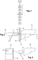
- the measurement of a parameter of the tissue of the region of interest can, for example, be carried out by means of a vibratory elastography method using a vibration elastography probe as illustrated on FIG. figure 2 .
- This third sub-step 23 consists in generating a low frequency elastic wave in the liver.
- the propagation of this low frequency elastic wave being for example, followed by the generation of the ultrasonic signal (first substep 21) and the acquisition of the ultrasonic signal reflected by the liver (second substep 22).
- the probe positioned during the first step 1 is the vibratory elastography probe 10.
- the ultrasound transducer 12 of the probe 10 is positioned, during the first step 1, in contact with the epidermis 13 and in the intercostal space, in other words between two of the four ribs 14.
- the low frequency elastic wave generator 11 generates, by indirect contact with the liver 16, one or more low frequency elastic waves propagating in the subcutaneous tissue 15, then in the liver. 16.
- This (these) elastic wave (s) low frequency (s) is (are) generally obtained (s) mechanically but can (may) also be obtained (s) by radiation pressure, by ultrasound hyperthermia or by internal vibrations of the body (heartbeat, pulse, etc.).
- the temporal form of this (these) elastic wave (s) low frequency can be arbitrary, but more generally of impulse type, transient or periodic (maintained, monochromatic).
- ultrasound waves are generated and acquired along the X axis by means of the ultrasonic transducer 12 to follow the propagation of this (these) low frequency elastic wave (s) within the region of the interest 17, namely between 25 and 65 mm under the epidermis 13.
- At least one parameter of the biological tissue is determined by means of the acquisition of at least one ultrasonic signal reflected by the biological tissue carried out during the second substep 22.
- the parameter (s) ) is (are) representative of the biological tissue.
- a first viscoelastic parameter corresponds, for example, to the detection of a low frequency elastic wave in the region of interest 17.
- a second viscoelastic parameter corresponds, for example, to the speed of displacement of this low frequency elastic wave within the region of interest 17.
- a third viscoelastic parameter corresponds, for example, to an elasticity value of the biological tissue of the region of interest 17.
- the ultrasonic parameter can be formed for example by a power of the ultrasonic signal, an energy of the ultrasonic signal, a correlation coefficient or inter-correlation.
- the ultrasonic parameter can be formed for example by an offset of the central frequency of the received ultrasonic signal with respect to the central frequency of the emitted ultrasonic signal.
- the ultrasound parameter can be obtained in transformed domains such as, for example, the time-frequency domain or the cepstral domain.
- the physiological parameter can be formed by the detection of a blood flow passing through the biological tissue.
- a Doppler ultrasound can be performed to determine if the region of interest of the biological tissue has a blood flow and therefore a vein.
- a fourth step 4 at least one parameter of the biological tissue is compared to at least one reference parameter of a target biological tissue.
- the visual indicator 18 when the visual indicator 18 displays a red color, it indicates to the operator that the ultrasound transducer 12 is not located next to the biological tissue which he wishes to determine a property.
- Steps 1 to 4 are thus repeated until the parameter of the biological tissue lying opposite the ultrasound transducer 12 corresponds substantially to the reference parameter of the target biological tissue.
- This condition may, for example, be indicated to the operator by means of the visual indicator 18 displaying a green color.
- the visual indicator can be displayed by an LED (not shown) that comprises the probe 10 comprising the ultrasonic transducer 12. This LED changes color depending on the result of the comparison of the determined parameter with the reference parameter.
- the indicator may be formed by a sound indicator.
- the indicator can be formed by any other means informing the operator of the result of the comparison of one or more determined parameter (s) with one or more reference parameter (s).
- the operator when a determined parameter differs from the corresponding reference parameter of the target biological tissue, the operator can not determine a property.
- This impossibility is not necessarily indicated by means of an indicator.
- the absence of display of a value representative of a property obtained later in the fifth step 5 can inform the operator that the ultrasound transducer is not located opposite the tissue to be measured.
- the presence of the propagation of a low frequency elastic wave generated during the Step 2 can be used as a viscoelastic parameter of the biological tissue.
- the ultrasonic transducer 12 of the probe 10 is positioned between two of the four ribs 14. This location therefore prohibits access to certain organs such as the thyroid. However, this location can give access to the lungs, intestines and possibly the kidneys.
- the positioning of the vibratory elastography probe 10 between two of the four ribs 14 and the detection of the propagation of a low frequency elastic wave (liver parameter 16 ) in the region of interest 17 formed at a height between 25 and 65 mm ensures that the vibratory elastography probe 10 is correctly positioned to determine at least one property of the liver 16.
- the liver parameter 16 can be formed by a physiological parameter, such as for example the detection of a blood flow by means of a Doppler ultrasound. In such an implementation, if the blood flow corresponds to a reference blood flow then the vibratory elastography probe 10 is correctly positioned to determine a property of the liver 16.
- any representative parameter of the biological tissue can be used to validate the positioning of the ultrasonic transducer 12.
- a plurality of parameters can be used to validate the positioning of the ultrasonic transducer 12.
- a result resulting from the combination of these parameters is compared with a reference parameter, determining a property (fifth step 5) only if the result from the combination is substantially similar to the value of the reference parameter.
- the parameters can be combined according to different models such as, for example, a logistic regression model. Logistic regression allows to build a prediction model including the values of the determined parameters.
- a property of the biological tissue is determined according to the result obtained during the fourth comparative step 4.
- a property of the biological tissue is determined.
- the parameter can be formed by values related to the propagation of a low elastic wave frequency. These values may for example correspond to amplitude levels or image quality criteria.
- the result obtained during the fourth step 4 is formed by "the detection of a low frequency elastic wave". If this low frequency elastic wave is detected then a property of the biological tissue is determined.
- the property of the biological tissue can be determined by the implementation of a method.
- the method may be, in a nonlimiting manner, a vibratory elastography method implemented by means of a vibratory elastography probe 10 conforming to that used during the second step 2 or a process requiring simple emissions and ultrasonic transducers 12.
- the latter implementation can be performed to determine an ultrasonic attenuation generated by the biological tissue.
- the determination of a property can be triggered automatically by the software means (not shown) if the absolute value of the difference between the value of the parameter determined during the third step 3 and the value of the reference parameter is less than a given threshold value. In other words, no action, no trigger to determine a property is performed by the operator.
- the determination of a property can be triggered manually by operator support, after the latter has received an audible and / or visual message (by means of an indicator) indicating that the probe 10 is positioned correctly on a trigger button (not shown) that may optionally comprise the probe 10.
- the center frequency of an ultrasonic signal decreases as it passes through biological tissues. Consequently, an evaluation of an offset of the central frequency of the ultrasonic signal makes it possible to evaluate the ultrasonic attenuation.
- the central frequency of the ultrasound signal can be estimated in the time domain by counting the number of zero crossings in a time window.
- the ultrasonic attenuation of the same biological tissue is different if the latter has a low proportion of adipose tissue or a large proportion of adipose tissue because the impedance of the fat is different from that of the soft tissues.
- the ultrasonic attenuation can make it possible to determine quantitatively and / or qualitatively a proportion of fat that may comprise the measured biological tissue, such as, for example, the liver 16 in our example .
- the property of the biological tissue can be determined according to data extracted from the acquisition of the at least one ultrasonic signal reflected by the biological tissue produced during the second step 2.
- This possibility is particularly advantageous because it requires a single measurement to perform both the third step 3 (determine a biological tissue parameter by acquiring the at least one ultrasonic signal reflected by the biological tissue) and the fifth step (determining a property of the biological tissue depending on the result of the comparison). Consequently, the position of the probe 10 has not been modified and the operator can state with certainty that the property obtained by the implementation of the method of the invention corresponds to a property of the biological tissue that he wishes to measure. .
- the detection of the propagation of a low frequency elastic wave in the biological tissue of the region of interest 17 can be used as a viscoelastic parameter to verify that the measured tissue corresponds to the target tissue and once this hypothesis is validated, the determination of the speed of propagation of the low frequency elastic wave makes it possible to estimate a property of the measured biological tissue, namely the elasticity. A single measure is therefore necessary.
- the measurement of at least one parameter of the biological tissue can make it possible to quantitatively and / or qualitatively determine the elasticity of the biological tissue and / or the ultrasonic attenuation of a biological tissue.
- the characteristic value of the property obtained is representative of the biological tissue that the operator wishes to measure. No specific knowledge is therefore required for the positioning of the probe with respect to the biological tissue.
- the positioning of the probe and therefore the ultrasonic transducer with respect to the target biological tissue may be for example specified to the operator by means of an indicator.
- the method for measuring at least one property of a biological tissue in accordance with the invention offers the possibility to an operator who does not have specific knowledge in the human or animal domain, to perform measuring property of a target biological tissue to determine, for example, the elasticity of the target biological tissue and / or the ultrasound attenuation of the target biological tissue.
- the invention has been more particularly described for application to an organ, the liver 16.
- it may also be desirable to apply the same method in the case of any type of human or animal organ such as by example a breast, adipose tissue, a gland, a ganglion and, this in-vivo or in-vitro or to perform a quality control for industrial applications, including agri-food.
Landscapes
- Health & Medical Sciences (AREA)
- Life Sciences & Earth Sciences (AREA)
- Medical Informatics (AREA)
- Molecular Biology (AREA)
- Veterinary Medicine (AREA)
- Pathology (AREA)
- Public Health (AREA)
- Engineering & Computer Science (AREA)
- Biomedical Technology (AREA)
- Heart & Thoracic Surgery (AREA)
- Physics & Mathematics (AREA)
- Biophysics (AREA)
- Surgery (AREA)
- Animal Behavior & Ethology (AREA)
- General Health & Medical Sciences (AREA)
- Radiology & Medical Imaging (AREA)
- Nuclear Medicine, Radiotherapy & Molecular Imaging (AREA)
- Immunology (AREA)
- Vascular Medicine (AREA)
- Endocrinology (AREA)
- Ultra Sonic Daignosis Equipment (AREA)
- Investigating Or Analyzing Materials By The Use Of Ultrasonic Waves (AREA)
Claims (16)
- Verfahren für die Messung wenigstens einer Eigenschaft eines biologischen Gewebes (12), umfassend die Schritte:- Positionierung (1) eines Ultraschalltransduktors (12) gegenüber dem genannten, zu messenden biologischen Gewebe (16);- Generierung (21) wenigstens eines Ultraschallsignals innerhalb des genannten biologischen Gewebes (16);- Erwerb (22) wenigstens eines von dem genannten biologischen Gewebe (16) reflektierten Ultraschallsignals;- Bestimmung (3) wenigstens eines Parameters des genannten biologischen Gewebes (16) mittels des genannten Erwerbs (22) des genannten wenigstens einen Ultraschallsignals, das durch das genannte biologische Gewebe (16) reflektiert wird, wobei der genannte wenigstens eine Parameter für das genannte biologische Gewebe (16) repräsentativ ist;wobei das genannte Verfahren dadurch gekennzeichnet ist, dass es darüber hinaus die Schritte umfasst:- Vergleich (4) des genannten wenigstens einen Parameters des genannten biologischen Gewebes (16) mit wenigstens einem Referenzparameter eines biologischen Zielgewebes derart, dass die Hypothese der Präsenz des genannten biologischen Gewebes gegenüber dem Ultraschalltransduktor validiert wird; wobei der genannte Vergleich (4) in dem Vergleich eines Wertes des genannten wenigstens einen Referenzparameters besteht, der auf einen Wert des genannten wenigstens einen Referenzparameters bestimmt wird,- Bestimmung (5) wenigstens einer Eigenschaft des genannten biologischen Gewebes (16) in Abhängigkeit von dem Ergebnis des genannten Vergleichsschritts (4), wobei der genannte Bestimmungsschritt (5) der genannten wenigstens einen Eigenschaft nur dann erfolgt, wenn der absolute Wert der Differenz zwischen dem genannten Wert des genannten wenigstens einen bestimmten Parameters und dem genannten Wert des genannten wenigsten einen Referenzparameters geringer ist als ein bestimmter Schwellenwert.
- Verfahren gemäß Anspruch 1, dadurch gekennzeichnet, dass eine Vielzahl von Parametern untereinander kombiniert ist, wobei der genannte Vergleichsschritt (4) im Vergleich eines Ergebnisses besteht, das aus der genannten Kombination von Parametern mit wenigstens einem Referenzparameter stammt.
- Verfahren gemäß irgendeinem der Ansprüche 1 oder 2, dadurch gekennzeichnet, dass es einen Generierungsschritt (23) einer elastischen Welle niedriger Frequenz in dem genannten biologischen Gewebe (16) umfasst.
- Verfahren gemäß Anspruch 3, dadurch gekennzeichnet, dass die genannte elastische Welle niedriger Frequenz per Vibration eines Generators (11) mit einer Welle niedriger Frequenz generiert wird.
- Verfahren gemäß Anspruch 3, dadurch gekennzeichnet, dass die genannte elastische Welle niedriger Frequenz per Strahlungsdruck generiert wird.
- Verfahren gemäß einem der Ansprüche 1 bis 5, dadurch gekennzeichnet, dass ein Indikator einen Operator über das genannte Ergebnis des genannten Vergleichsschritts (4) informiert.
- Verfahren gemäß irgendeinem der Ansprüche 1 bis 6, dadurch gekennzeichnet, dass der genannte wenigstens eine Parameter und die genannte wenigstens eine Eigenschaft des genannten biologischen Gewebes (16) in Abhängigkeit von Daten bestimmt werden, die aus dem genannten Erwerb (22) des genannten wenigstens einen Ultraschallsignals extrahiert werden, das von dem biologischen Gewebe (16) reflektiert wird.
- Verfahren gemäß irgendeinem der Ansprüche 1 bis 7, dadurch gekennzeichnet, dass die genannte wenigstens eine Eigenschaft des genannten biologischen Gewebes (16) durch die Umsetzung eines Elastographieverfahrens bestimmt wird.
- Verfahren gemäß irgendeinem der Ansprüche 1 bis 8, dadurch gekennzeichnet, dass die genannte wenigstens eine Eigenschaft die Elastizität des genannten biologischen Gewebes (16) ist.
- Verfahren gemäß irgendeinem der Ansprüche 1 bis 7, dadurch gekennzeichnet, dass die wenigstens eine Eigenschaft eine Ultraschallabschwächung des genannten biologischen Gewebes (16) ist.
- Verfahren gemäß irgendeinem der Ansprüche 1 bis 10, dadurch gekennzeichnet, dass der genannte wenigstens eine Parameter ein Ultraschallparameter des genannten biologischen Gewebes (16) ist.
- Verfahren gemäß Anspruch 11, dadurch gekennzeichnet, dass der wenigstens eine Ultraschallparameter eine Ultraschallabschwächung des genannten biologischen Gewebes (16) ist.
- Verfahren gemäß einem der Ansprüche 1 bis 10, dadurch gekennzeichnet, dass der genannte wenigstens eine Parameter ein viskoelastischer Parameter des genannten biologischen Gewebes (16) ist.
- Verfahren gemäß Anspruch 13, dadurch gekennzeichnet, dass der genannte viskoelastische Parameter die Elastizität des genannten biologischen Gewebes (16) ist.
- Verfahren gemäß Anspruch 14, dadurch gekennzeichnet, dass die genannte Elastizität durch ein Elastographieverfahren erhalten wird.
- Verfahren gemäß Anspruch 1 bis 10, dadurch gekennzeichnet, dass der genannte wenigstens eine Parameter ein physiologischer Parameter des genannten biologischen Gewebes (16) ist.
Priority Applications (1)
| Application Number | Priority Date | Filing Date | Title |
|---|---|---|---|
| PL10752844T PL2477551T3 (pl) | 2009-09-17 | 2010-09-17 | Sposób pomiaru co najmniej jednej właściwości tkanki biologicznej |
Applications Claiming Priority (2)
| Application Number | Priority Date | Filing Date | Title |
|---|---|---|---|
| FR0956408A FR2949965B1 (fr) | 2009-09-17 | 2009-09-17 | Procede pour la mesure d'au moins une propriete de tissu biologique |
| PCT/EP2010/063677 WO2011033050A1 (fr) | 2009-09-17 | 2010-09-17 | Procede pour la mesure d'au moins une propriete d'un tissu biologique |
Publications (2)
| Publication Number | Publication Date |
|---|---|
| EP2477551A1 EP2477551A1 (de) | 2012-07-25 |
| EP2477551B1 true EP2477551B1 (de) | 2016-12-14 |
Family
ID=42111548
Family Applications (1)
| Application Number | Title | Priority Date | Filing Date |
|---|---|---|---|
| EP10752844.0A Active EP2477551B1 (de) | 2009-09-17 | 2010-09-17 | Verfahren zur messung von mindestens einer eigenschaft von biologischem gewebe |
Country Status (11)
| Country | Link |
|---|---|
| US (1) | US9918695B2 (de) |
| EP (1) | EP2477551B1 (de) |
| JP (1) | JP5635100B2 (de) |
| CN (1) | CN102596051B (de) |
| BR (1) | BR112012006041A2 (de) |
| ES (1) | ES2618570T3 (de) |
| FR (1) | FR2949965B1 (de) |
| IN (1) | IN2012DN02603A (de) |
| PL (1) | PL2477551T3 (de) |
| PT (1) | PT2477551T (de) |
| WO (1) | WO2011033050A1 (de) |
Families Citing this family (19)
| Publication number | Priority date | Publication date | Assignee | Title |
|---|---|---|---|---|
| JP5896623B2 (ja) | 2011-05-02 | 2016-03-30 | キヤノン株式会社 | 被検体情報取得装置およびその制御方法 |
| WO2012158877A2 (en) * | 2011-05-17 | 2012-11-22 | University Of Rochester | Non-invasive assessment of liver fat by crawling wave dispersion |
| FR2978657B1 (fr) * | 2011-08-03 | 2013-08-30 | Echosens | Procede pour determiner en temps-reel une probabilite de presence d'un tissu biologique cible en regard d'un transducteur ultrasonore |
| US20140316267A1 (en) * | 2011-08-15 | 2014-10-23 | University Of Rochester | Non-invasive assessment of liver fat by crawling wave dispersion with emphasis on attenuation |
| FR3036943A1 (fr) | 2015-06-02 | 2016-12-09 | Echosens | Dispositif non invasif de detection de lesion hepatique |
| WO2017176065A1 (ko) * | 2016-04-07 | 2017-10-12 | (주)엠큐브테크놀로지 | 독립형 초음파 스캐너 |
| FR3054123B1 (fr) * | 2016-07-25 | 2021-11-26 | Echosens | Procede de mesure d’un parametre viscoelastique d’un organe humain ou animal |
| JP6855347B2 (ja) | 2017-07-27 | 2021-04-07 | ゼネラル・エレクトリック・カンパニイ | 超音波診断装置及びその制御プログラム |
| KR102627723B1 (ko) * | 2017-09-08 | 2024-01-23 | 삼성메디슨 주식회사 | 초음파 영상장치 및 그 제어방법 |
| EP3453336B1 (de) * | 2017-09-08 | 2020-11-04 | Samsung Medison Co., Ltd. | Ultraschallabbildungsvorrichtung und steuerungsverfahren dafür |
| US11426146B2 (en) | 2017-09-08 | 2022-08-30 | Samsung Medison Co., Ltd. | Ultrasound imaging apparatus and control method thereof |
| US11443422B2 (en) | 2017-10-13 | 2022-09-13 | The Cleveland Clinic Foundation | Advanced ultrasonic detection of different tissue types |
| FR3078485B1 (fr) * | 2018-03-02 | 2020-02-28 | Echosens | Procede d’elastographie hybride, sonde et dispositif pour elastographie hybride |
| FR3078484A1 (fr) | 2018-03-02 | 2019-09-06 | Echosens | Procede de mesure d’un parametre d’attenuation ultrasonore guide par elastographie harmonique, sonde et dispositif pour la mise en œuvre du procede |
| CN109363718A (zh) * | 2018-10-26 | 2019-02-22 | 深圳迈瑞生物医疗电子股份有限公司 | 超声成像方法和超声成像设备 |
| CN113081054B (zh) * | 2018-12-04 | 2022-10-11 | 深圳迈瑞生物医疗电子股份有限公司 | 一种超声成像方法以及超声成像系统 |
| US11850098B2 (en) | 2019-06-14 | 2023-12-26 | Echosens | Method and device for measuring an ultrasound parameter of a viscoelastic medium |
| EP3750483A1 (de) | 2019-06-14 | 2020-12-16 | Echosens | Verfahren und vorrichtung zur messung eines ultraschallparameters eines viskoelastischen mediums |
| CN114224382B (zh) * | 2021-12-17 | 2023-09-15 | 重庆医科大学 | 粘弹性测量方法及其系统 |
Family Cites Families (19)
| Publication number | Priority date | Publication date | Assignee | Title |
|---|---|---|---|---|
| US4389893A (en) * | 1981-06-01 | 1983-06-28 | North American Philips Corporation | Precision ultrasound attenuation measurement |
| US4441368A (en) | 1982-04-19 | 1984-04-10 | General Electric Company | Method and means for determining ultrasonic wave attenuation in tissue |
| US4603703A (en) * | 1984-04-13 | 1986-08-05 | The Board Of Trustees Of The Leland Stanford Junior University | Method for real-time detection and identification of neuroelectric signals |
| JP2590219B2 (ja) * | 1988-08-05 | 1997-03-12 | 株式会社東芝 | 超音波診断装置 |
| US5906578A (en) * | 1997-06-18 | 1999-05-25 | Rajan; Govinda N. | Method and system for probe positioning in transesophageal echocardiography |
| US6013031A (en) * | 1998-03-09 | 2000-01-11 | Mendlein; John D. | Methods and devices for improving ultrasonic measurements using anatomic landmarks and soft tissue correction |
| US6238342B1 (en) * | 1998-05-26 | 2001-05-29 | Riverside Research Institute | Ultrasonic tissue-type classification and imaging methods and apparatus |
| US20020068870A1 (en) * | 2000-07-20 | 2002-06-06 | Alam Sheikh Kaisar | Hand held mechanical compression device for inducing tissue strain |
| FR2843290B1 (fr) * | 2002-08-08 | 2005-06-24 | Echosens | Dispositif et procede pour la mesure de l'elasticite d'un organe humain ou animal |
| US7578789B2 (en) * | 2002-08-08 | 2009-08-25 | Echosens | Device and method for measuring the elasticity of a human or animal organ |
| US7914456B2 (en) * | 2003-05-30 | 2011-03-29 | Hitachi Medical Corporation | Ultrasonic probe and ultrasonic elasticity imaging device |
| US20100168574A1 (en) * | 2005-12-14 | 2010-07-01 | Teijin Pharma Limited | Medical Ultrasound Device Having Irradiation Position Checking Function |
| JP4981023B2 (ja) * | 2006-03-02 | 2012-07-18 | 株式会社日立メディコ | 自動圧迫装置及び同装置を用いた超音波診断装置 |
| US8711144B2 (en) * | 2006-08-01 | 2014-04-29 | Siemens Medical Solutions Usa, Inc. | Perception-based artifact quantification for volume rendering |
| US9125589B2 (en) * | 2007-05-09 | 2015-09-08 | General Electric Company | System and method for tissue characterization using ultrasound imaging |
| JP2010526626A (ja) * | 2007-05-16 | 2010-08-05 | スーパー ソニック イマジン | 関心領域の粘弾性の平均値を測定するための方法および装置 |
| US8131379B2 (en) * | 2007-08-27 | 2012-03-06 | St. Jude Medical Atrial Fibrillation Division, Inc. | Cardiac tissue elasticity sensing |
| JP2009183454A (ja) * | 2008-02-06 | 2009-08-20 | Teijin Pharma Ltd | 周波数減衰特性を利用した超音波検査装置又は超音波照射位置検査方法 |
| JP5426101B2 (ja) * | 2008-02-25 | 2014-02-26 | 株式会社東芝 | 超音波診断装置及、超音波画像処理装置及び超音波画像処理プログラム |
-
2009
- 2009-09-17 FR FR0956408A patent/FR2949965B1/fr active Active
-
2010
- 2010-09-17 EP EP10752844.0A patent/EP2477551B1/de active Active
- 2010-09-17 JP JP2012529279A patent/JP5635100B2/ja active Active
- 2010-09-17 IN IN2603DEN2012 patent/IN2012DN02603A/en unknown
- 2010-09-17 US US13/496,628 patent/US9918695B2/en active Active
- 2010-09-17 PT PT107528440T patent/PT2477551T/pt unknown
- 2010-09-17 CN CN201080048631.0A patent/CN102596051B/zh active Active
- 2010-09-17 ES ES10752844.0T patent/ES2618570T3/es active Active
- 2010-09-17 WO PCT/EP2010/063677 patent/WO2011033050A1/fr active Application Filing
- 2010-09-17 BR BR112012006041-4A patent/BR112012006041A2/pt not_active Application Discontinuation
- 2010-09-17 PL PL10752844T patent/PL2477551T3/pl unknown
Non-Patent Citations (1)
| Title |
|---|
| None * |
Also Published As
| Publication number | Publication date |
|---|---|
| US20120190983A1 (en) | 2012-07-26 |
| CN102596051B (zh) | 2017-08-18 |
| WO2011033050A1 (fr) | 2011-03-24 |
| EP2477551A1 (de) | 2012-07-25 |
| CN102596051A (zh) | 2012-07-18 |
| FR2949965A1 (fr) | 2011-03-18 |
| JP5635100B2 (ja) | 2014-12-03 |
| US9918695B2 (en) | 2018-03-20 |
| JP2013505040A (ja) | 2013-02-14 |
| FR2949965B1 (fr) | 2012-09-28 |
| IN2012DN02603A (de) | 2015-09-04 |
| PL2477551T3 (pl) | 2017-06-30 |
| PT2477551T (pt) | 2017-03-17 |
| ES2618570T3 (es) | 2017-06-21 |
| BR112012006041A2 (pt) | 2020-08-18 |
Similar Documents
| Publication | Publication Date | Title |
|---|---|---|
| EP2477551B1 (de) | Verfahren zur messung von mindestens einer eigenschaft von biologischem gewebe | |
| EP1531733B1 (de) | Vorrichtung und verfahren zur messung der elastizität eines menschlichen oder tierischen organs | |
| EP2739211B1 (de) | Verfahren zur echtzeitbestimmung des vorhandenseins von biologischem gewebe gegenüber einem ultraschallwandler | |
| EP3487412B1 (de) | Verfahren zur messung eines viskoelastischen parameters eines menschlichen oder tierischen organs | |
| FR2899336A1 (fr) | Procede et dispositif pour l'imagerie d'un milieu viscoelastique | |
| Kim et al. | Attenuation estimation using spectral cross-correlation | |
| EP3758608B1 (de) | Hybrides elastografieverfahren, sonde und vorrichtung für hybride elastografie | |
| FR3072870A1 (fr) | Estimation viscoelastique d'un tissu a partir d'une vitesse de cisaillement dans une imagerie medicale a ultrasons | |
| FR3003154A1 (fr) | Estimation de la fraction de matieres grasses en utilisant des ultrasons partir d'une propagation d'onde de cisaillement | |
| EP3084416B1 (de) | Verfahren zur verarbeitung von signalen aus einer ultraschallsondenerfassung, entsprechendes computerprogramm und ultraschallsondenvorrichtung | |
| EP3044580B1 (de) | Verfahren zur zerstörungsfreien ultraschallprüfung eines bauteils mittels echoanalyse | |
| FR2986960A1 (fr) | Procede et systeme de visualisation d'information associee dans une imagerie par onde de cisaillement ultrasonore ainsi que support de stockage lisible par ordinateur | |
| FR2871240A1 (fr) | Procede et systeme d'echographie de contraste | |
| EP3654847B1 (de) | Verfahren zur charakterisierung von knochen mittels ultraschallwellen | |
| EP3758610B1 (de) | Verfahren zur messung eines ultraschalldämpfungsparameters, geführt durch harmonische elastografie, sonde und vorrichtung zur durchführung des besagten verfahrens | |
| EP3860467B1 (de) | Verfahren zur automatischen auswahl eines tiefenbereichs zur berechnung einer eigenschaft eines viskoelastischen mediums | |
| Koda et al. | B-Line Elastography Measurement of Lung Parenchymal Elasticity | |
| CA3046106A1 (fr) | Procede de traitement de signaux issus d'une acquisition par sondage ultrasonore, programme d'ordinateur et dispositif de sondage a ultrasons correspondants | |
| Miele | Principles 4 Physics | |
| EP4007528A1 (de) | Kardiale vorrichtung | |
| FR3099580A1 (fr) | Procédé et système de caractérisation ultrasonore non invasive d’un milieu hétérogène | |
| FR3024350A1 (fr) | Systeme et procede de mesure de flux sanguin | |
| FR3006054A1 (fr) | Procede et dispositif de detection acoustique d'une inclusion dans un milieu |
Legal Events
| Date | Code | Title | Description |
|---|---|---|---|
| PUAI | Public reference made under article 153(3) epc to a published international application that has entered the european phase |
Free format text: ORIGINAL CODE: 0009012 |
|
| 17P | Request for examination filed |
Effective date: 20120403 |
|
| AK | Designated contracting states |
Kind code of ref document: A1 Designated state(s): AL AT BE BG CH CY CZ DE DK EE ES FI FR GB GR HR HU IE IS IT LI LT LU LV MC MK MT NL NO PL PT RO SE SI SK SM TR |
|
| DAX | Request for extension of the european patent (deleted) | ||
| 17Q | First examination report despatched |
Effective date: 20140929 |
|
| GRAP | Despatch of communication of intention to grant a patent |
Free format text: ORIGINAL CODE: EPIDOSNIGR1 |
|
| INTG | Intention to grant announced |
Effective date: 20160704 |
|
| GRAS | Grant fee paid |
Free format text: ORIGINAL CODE: EPIDOSNIGR3 |
|
| GRAA | (expected) grant |
Free format text: ORIGINAL CODE: 0009210 |
|
| AK | Designated contracting states |
Kind code of ref document: B1 Designated state(s): AL AT BE BG CH CY CZ DE DK EE ES FI FR GB GR HR HU IE IS IT LI LT LU LV MC MK MT NL NO PL PT RO SE SI SK SM TR |
|
| REG | Reference to a national code |
Ref country code: GB Ref legal event code: FG4D Free format text: NOT ENGLISH |
|
| REG | Reference to a national code |
Ref country code: CH Ref legal event code: EP |
|
| REG | Reference to a national code |
Ref country code: IE Ref legal event code: FG4D Free format text: LANGUAGE OF EP DOCUMENT: FRENCH |
|
| REG | Reference to a national code |
Ref country code: AT Ref legal event code: REF Ref document number: 852838 Country of ref document: AT Kind code of ref document: T Effective date: 20170115 |
|
| REG | Reference to a national code |
Ref country code: DE Ref legal event code: R096 Ref document number: 602010038812 Country of ref document: DE |
|
| PG25 | Lapsed in a contracting state [announced via postgrant information from national office to epo] |
Ref country code: LV Free format text: LAPSE BECAUSE OF FAILURE TO SUBMIT A TRANSLATION OF THE DESCRIPTION OR TO PAY THE FEE WITHIN THE PRESCRIBED TIME-LIMIT Effective date: 20161214 |
|
| REG | Reference to a national code |
Ref country code: PT Ref legal event code: SC4A Ref document number: 2477551 Country of ref document: PT Date of ref document: 20170317 Kind code of ref document: T Free format text: AVAILABILITY OF NATIONAL TRANSLATION Effective date: 20170309 |
|
| REG | Reference to a national code |
Ref country code: LT Ref legal event code: MG4D |
|
| REG | Reference to a national code |
Ref country code: NL Ref legal event code: MP Effective date: 20161214 |
|
| PG25 | Lapsed in a contracting state [announced via postgrant information from national office to epo] |
Ref country code: LT Free format text: LAPSE BECAUSE OF FAILURE TO SUBMIT A TRANSLATION OF THE DESCRIPTION OR TO PAY THE FEE WITHIN THE PRESCRIBED TIME-LIMIT Effective date: 20161214 Ref country code: NO Free format text: LAPSE BECAUSE OF FAILURE TO SUBMIT A TRANSLATION OF THE DESCRIPTION OR TO PAY THE FEE WITHIN THE PRESCRIBED TIME-LIMIT Effective date: 20170314 Ref country code: SE Free format text: LAPSE BECAUSE OF FAILURE TO SUBMIT A TRANSLATION OF THE DESCRIPTION OR TO PAY THE FEE WITHIN THE PRESCRIBED TIME-LIMIT Effective date: 20161214 |
|
| PG25 | Lapsed in a contracting state [announced via postgrant information from national office to epo] |
Ref country code: FI Free format text: LAPSE BECAUSE OF FAILURE TO SUBMIT A TRANSLATION OF THE DESCRIPTION OR TO PAY THE FEE WITHIN THE PRESCRIBED TIME-LIMIT Effective date: 20161214 Ref country code: HR Free format text: LAPSE BECAUSE OF FAILURE TO SUBMIT A TRANSLATION OF THE DESCRIPTION OR TO PAY THE FEE WITHIN THE PRESCRIBED TIME-LIMIT Effective date: 20161214 |
|
| REG | Reference to a national code |
Ref country code: ES Ref legal event code: FG2A Ref document number: 2618570 Country of ref document: ES Kind code of ref document: T3 Effective date: 20170621 |
|
| PG25 | Lapsed in a contracting state [announced via postgrant information from national office to epo] |
Ref country code: NL Free format text: LAPSE BECAUSE OF FAILURE TO SUBMIT A TRANSLATION OF THE DESCRIPTION OR TO PAY THE FEE WITHIN THE PRESCRIBED TIME-LIMIT Effective date: 20161214 |
|
| PG25 | Lapsed in a contracting state [announced via postgrant information from national office to epo] |
Ref country code: CZ Free format text: LAPSE BECAUSE OF FAILURE TO SUBMIT A TRANSLATION OF THE DESCRIPTION OR TO PAY THE FEE WITHIN THE PRESCRIBED TIME-LIMIT Effective date: 20161214 Ref country code: EE Free format text: LAPSE BECAUSE OF FAILURE TO SUBMIT A TRANSLATION OF THE DESCRIPTION OR TO PAY THE FEE WITHIN THE PRESCRIBED TIME-LIMIT Effective date: 20161214 Ref country code: IS Free format text: LAPSE BECAUSE OF FAILURE TO SUBMIT A TRANSLATION OF THE DESCRIPTION OR TO PAY THE FEE WITHIN THE PRESCRIBED TIME-LIMIT Effective date: 20170414 Ref country code: SK Free format text: LAPSE BECAUSE OF FAILURE TO SUBMIT A TRANSLATION OF THE DESCRIPTION OR TO PAY THE FEE WITHIN THE PRESCRIBED TIME-LIMIT Effective date: 20161214 Ref country code: RO Free format text: LAPSE BECAUSE OF FAILURE TO SUBMIT A TRANSLATION OF THE DESCRIPTION OR TO PAY THE FEE WITHIN THE PRESCRIBED TIME-LIMIT Effective date: 20161214 |
|
| REG | Reference to a national code |
Ref country code: GR Ref legal event code: EP Ref document number: 20170400738 Country of ref document: GR Effective date: 20170804 |
|
| REG | Reference to a national code |
Ref country code: FR Ref legal event code: PLFP Year of fee payment: 8 |
|
| PG25 | Lapsed in a contracting state [announced via postgrant information from national office to epo] |
Ref country code: SM Free format text: LAPSE BECAUSE OF FAILURE TO SUBMIT A TRANSLATION OF THE DESCRIPTION OR TO PAY THE FEE WITHIN THE PRESCRIBED TIME-LIMIT Effective date: 20161214 Ref country code: BG Free format text: LAPSE BECAUSE OF FAILURE TO SUBMIT A TRANSLATION OF THE DESCRIPTION OR TO PAY THE FEE WITHIN THE PRESCRIBED TIME-LIMIT Effective date: 20170314 |
|
| REG | Reference to a national code |
Ref country code: DE Ref legal event code: R097 Ref document number: 602010038812 Country of ref document: DE |
|
| PLBE | No opposition filed within time limit |
Free format text: ORIGINAL CODE: 0009261 |
|
| STAA | Information on the status of an ep patent application or granted ep patent |
Free format text: STATUS: NO OPPOSITION FILED WITHIN TIME LIMIT |
|
| 26N | No opposition filed |
Effective date: 20170915 |
|
| PG25 | Lapsed in a contracting state [announced via postgrant information from national office to epo] |
Ref country code: DK Free format text: LAPSE BECAUSE OF FAILURE TO SUBMIT A TRANSLATION OF THE DESCRIPTION OR TO PAY THE FEE WITHIN THE PRESCRIBED TIME-LIMIT Effective date: 20161214 |
|
| PG25 | Lapsed in a contracting state [announced via postgrant information from national office to epo] |
Ref country code: SI Free format text: LAPSE BECAUSE OF FAILURE TO SUBMIT A TRANSLATION OF THE DESCRIPTION OR TO PAY THE FEE WITHIN THE PRESCRIBED TIME-LIMIT Effective date: 20161214 |
|
| REG | Reference to a national code |
Ref country code: CH Ref legal event code: PL |
|
| PG25 | Lapsed in a contracting state [announced via postgrant information from national office to epo] |
Ref country code: MC Free format text: LAPSE BECAUSE OF FAILURE TO SUBMIT A TRANSLATION OF THE DESCRIPTION OR TO PAY THE FEE WITHIN THE PRESCRIBED TIME-LIMIT Effective date: 20161214 |
|
| REG | Reference to a national code |
Ref country code: IE Ref legal event code: MM4A |
|
| PG25 | Lapsed in a contracting state [announced via postgrant information from national office to epo] |
Ref country code: LU Free format text: LAPSE BECAUSE OF NON-PAYMENT OF DUE FEES Effective date: 20170917 |
|
| PG25 | Lapsed in a contracting state [announced via postgrant information from national office to epo] |
Ref country code: CH Free format text: LAPSE BECAUSE OF NON-PAYMENT OF DUE FEES Effective date: 20170930 Ref country code: LI Free format text: LAPSE BECAUSE OF NON-PAYMENT OF DUE FEES Effective date: 20170930 Ref country code: IE Free format text: LAPSE BECAUSE OF NON-PAYMENT OF DUE FEES Effective date: 20170917 |
|
| REG | Reference to a national code |
Ref country code: FR Ref legal event code: PLFP Year of fee payment: 9 |
|
| PG25 | Lapsed in a contracting state [announced via postgrant information from national office to epo] |
Ref country code: MT Free format text: LAPSE BECAUSE OF FAILURE TO SUBMIT A TRANSLATION OF THE DESCRIPTION OR TO PAY THE FEE WITHIN THE PRESCRIBED TIME-LIMIT Effective date: 20161214 |
|
| PG25 | Lapsed in a contracting state [announced via postgrant information from national office to epo] |
Ref country code: HU Free format text: LAPSE BECAUSE OF FAILURE TO SUBMIT A TRANSLATION OF THE DESCRIPTION OR TO PAY THE FEE WITHIN THE PRESCRIBED TIME-LIMIT; INVALID AB INITIO Effective date: 20100917 |
|
| REG | Reference to a national code |
Ref country code: AT Ref legal event code: UEP Ref document number: 852838 Country of ref document: AT Kind code of ref document: T Effective date: 20161214 |
|
| PG25 | Lapsed in a contracting state [announced via postgrant information from national office to epo] |
Ref country code: CY Free format text: LAPSE BECAUSE OF NON-PAYMENT OF DUE FEES Effective date: 20161214 |
|
| PGFP | Annual fee paid to national office [announced via postgrant information from national office to epo] |
Ref country code: PT Payment date: 20190821 Year of fee payment: 10 Ref country code: TR Payment date: 20190903 Year of fee payment: 10 |
|
| PG25 | Lapsed in a contracting state [announced via postgrant information from national office to epo] |
Ref country code: MK Free format text: LAPSE BECAUSE OF FAILURE TO SUBMIT A TRANSLATION OF THE DESCRIPTION OR TO PAY THE FEE WITHIN THE PRESCRIBED TIME-LIMIT Effective date: 20161214 |
|
| PGFP | Annual fee paid to national office [announced via postgrant information from national office to epo] |
Ref country code: GR Payment date: 20190823 Year of fee payment: 10 Ref country code: PL Payment date: 20190822 Year of fee payment: 10 Ref country code: BE Payment date: 20190823 Year of fee payment: 10 |
|
| PGFP | Annual fee paid to national office [announced via postgrant information from national office to epo] |
Ref country code: AT Payment date: 20190822 Year of fee payment: 10 |
|
| PG25 | Lapsed in a contracting state [announced via postgrant information from national office to epo] |
Ref country code: AL Free format text: LAPSE BECAUSE OF FAILURE TO SUBMIT A TRANSLATION OF THE DESCRIPTION OR TO PAY THE FEE WITHIN THE PRESCRIBED TIME-LIMIT Effective date: 20161214 |
|
| REG | Reference to a national code |
Ref country code: AT Ref legal event code: MM01 Ref document number: 852838 Country of ref document: AT Kind code of ref document: T Effective date: 20200917 |
|
| REG | Reference to a national code |
Ref country code: BE Ref legal event code: MM Effective date: 20200930 |
|
| PG25 | Lapsed in a contracting state [announced via postgrant information from national office to epo] |
Ref country code: PT Free format text: LAPSE BECAUSE OF NON-PAYMENT OF DUE FEES Effective date: 20210413 Ref country code: GR Free format text: LAPSE BECAUSE OF NON-PAYMENT OF DUE FEES Effective date: 20210406 |
|
| PG25 | Lapsed in a contracting state [announced via postgrant information from national office to epo] |
Ref country code: AT Free format text: LAPSE BECAUSE OF NON-PAYMENT OF DUE FEES Effective date: 20200917 Ref country code: BE Free format text: LAPSE BECAUSE OF NON-PAYMENT OF DUE FEES Effective date: 20200930 |
|
| PG25 | Lapsed in a contracting state [announced via postgrant information from national office to epo] |
Ref country code: TR Free format text: LAPSE BECAUSE OF NON-PAYMENT OF DUE FEES Effective date: 20200917 |
|
| PG25 | Lapsed in a contracting state [announced via postgrant information from national office to epo] |
Ref country code: PL Free format text: LAPSE BECAUSE OF NON-PAYMENT OF DUE FEES Effective date: 20200917 |
|
| P01 | Opt-out of the competence of the unified patent court (upc) registered |
Effective date: 20230526 |
|
| PGFP | Annual fee paid to national office [announced via postgrant information from national office to epo] |
Ref country code: IT Payment date: 20230928 Year of fee payment: 14 Ref country code: GB Payment date: 20230928 Year of fee payment: 14 |
|
| PGFP | Annual fee paid to national office [announced via postgrant information from national office to epo] |
Ref country code: FR Payment date: 20230928 Year of fee payment: 14 Ref country code: DE Payment date: 20230928 Year of fee payment: 14 |
|
| PGFP | Annual fee paid to national office [announced via postgrant information from national office to epo] |
Ref country code: ES Payment date: 20231106 Year of fee payment: 14 |