EP2477030A2 - Novel biomarkers of disease histopathology - Google Patents
Novel biomarkers of disease histopathology Download PDFInfo
- Publication number
- EP2477030A2 EP2477030A2 EP10763733A EP10763733A EP2477030A2 EP 2477030 A2 EP2477030 A2 EP 2477030A2 EP 10763733 A EP10763733 A EP 10763733A EP 10763733 A EP10763733 A EP 10763733A EP 2477030 A2 EP2477030 A2 EP 2477030A2
- Authority
- EP
- European Patent Office
- Prior art keywords
- multiple sclerosis
- protein
- fgf
- anosmin
- cns
- Prior art date
- Legal status (The legal status is an assumption and is not a legal conclusion. Google has not performed a legal analysis and makes no representation as to the accuracy of the status listed.)
- Withdrawn
Links
- 201000010099 disease Diseases 0.000 title description 15
- 208000037265 diseases, disorders, signs and symptoms Diseases 0.000 title description 15
- 239000000101 novel biomarker Substances 0.000 title 1
- 108090000379 Fibroblast growth factor 2 Proteins 0.000 claims abstract description 145
- 102000003974 Fibroblast growth factor 2 Human genes 0.000 claims abstract description 144
- 230000003902 lesion Effects 0.000 claims abstract description 106
- 102100023086 Anosmin-1 Human genes 0.000 claims abstract description 102
- 101710188941 Anosmin-1 Proteins 0.000 claims abstract description 100
- 208000016192 Demyelinating disease Diseases 0.000 claims abstract description 75
- 201000006417 multiple sclerosis Diseases 0.000 claims abstract description 68
- 210000001175 cerebrospinal fluid Anatomy 0.000 claims abstract description 67
- 238000000034 method Methods 0.000 claims abstract description 64
- 210000003169 central nervous system Anatomy 0.000 claims abstract description 57
- 108090000623 proteins and genes Proteins 0.000 claims abstract description 40
- 102000004169 proteins and genes Human genes 0.000 claims abstract description 40
- 239000013060 biological fluid Substances 0.000 claims abstract description 29
- 238000001514 detection method Methods 0.000 claims abstract description 25
- 230000001684 chronic effect Effects 0.000 claims description 34
- 230000006378 damage Effects 0.000 claims description 28
- 210000004884 grey matter Anatomy 0.000 claims description 28
- 239000012472 biological sample Substances 0.000 claims description 25
- 210000004885 white matter Anatomy 0.000 claims description 24
- 239000000523 sample Substances 0.000 claims description 23
- 230000008499 blood brain barrier function Effects 0.000 claims description 21
- 210000001218 blood-brain barrier Anatomy 0.000 claims description 21
- 208000007118 chronic progressive multiple sclerosis Diseases 0.000 claims description 21
- 206010012305 Demyelination Diseases 0.000 claims description 20
- 238000002965 ELISA Methods 0.000 claims description 14
- 230000001154 acute effect Effects 0.000 claims description 12
- 239000000090 biomarker Substances 0.000 claims description 12
- 208000007400 Relapsing-Remitting Multiple Sclerosis Diseases 0.000 claims description 11
- 238000003018 immunoassay Methods 0.000 claims description 11
- 230000000750 progressive effect Effects 0.000 claims description 11
- 208000032194 Acute haemorrhagic leukoencephalitis Diseases 0.000 claims description 10
- 206010071068 Clinically isolated syndrome Diseases 0.000 claims description 10
- 201000008628 secondary progressive multiple sclerosis Diseases 0.000 claims description 10
- 206010063401 primary progressive multiple sclerosis Diseases 0.000 claims description 9
- 210000002966 serum Anatomy 0.000 claims description 8
- 210000001428 peripheral nervous system Anatomy 0.000 claims description 7
- 210000004369 blood Anatomy 0.000 claims description 6
- 239000008280 blood Substances 0.000 claims description 6
- 210000001124 body fluid Anatomy 0.000 claims description 6
- 210000002381 plasma Anatomy 0.000 claims description 6
- 210000001138 tear Anatomy 0.000 claims description 6
- 201000011452 Adrenoleukodystrophy Diseases 0.000 claims description 5
- 208000011403 Alexander disease Diseases 0.000 claims description 5
- 208000022526 Canavan disease Diseases 0.000 claims description 5
- 206010053684 Cerebrohepatorenal syndrome Diseases 0.000 claims description 5
- 206010010252 Concentric sclerosis Diseases 0.000 claims description 5
- 208000010055 Globoid Cell Leukodystrophy Diseases 0.000 claims description 5
- 208000035895 Guillain-Barré syndrome Diseases 0.000 claims description 5
- 208000028226 Krabbe disease Diseases 0.000 claims description 5
- 108010049137 Member 1 Subfamily D ATP Binding Cassette Transporter Proteins 0.000 claims description 5
- 206010049567 Miller Fisher syndrome Diseases 0.000 claims description 5
- 208000003926 Myelitis Diseases 0.000 claims description 5
- 206010069350 Osmotic demyelination syndrome Diseases 0.000 claims description 5
- 206010042953 Systemic sclerosis Diseases 0.000 claims description 5
- 201000004525 Zellweger Syndrome Diseases 0.000 claims description 5
- 208000036813 Zellweger spectrum disease Diseases 0.000 claims description 5
- 208000002552 acute disseminated encephalomyelitis Diseases 0.000 claims description 5
- 239000010839 body fluid Substances 0.000 claims description 5
- 208000009885 central pontine myelinolysis Diseases 0.000 claims description 5
- 210000000877 corpus callosum Anatomy 0.000 claims description 5
- 208000008795 neuromyelitis optica Diseases 0.000 claims description 5
- 208000009174 transverse myelitis Diseases 0.000 claims description 5
- 208000018254 acute transverse myelitis Diseases 0.000 claims description 4
- 102000009030 Member 1 Subfamily D ATP Binding Cassette Transporter Human genes 0.000 claims 4
- 210000004027 cell Anatomy 0.000 description 27
- 102100023593 Fibroblast growth factor receptor 1 Human genes 0.000 description 15
- 101710182386 Fibroblast growth factor receptor 1 Proteins 0.000 description 15
- 208000027418 Wounds and injury Diseases 0.000 description 15
- 230000003210 demyelinating effect Effects 0.000 description 15
- 208000014674 injury Diseases 0.000 description 15
- 238000005259 measurement Methods 0.000 description 14
- ZEOWTGPWHLSLOG-UHFFFAOYSA-N Cc1ccc(cc1-c1ccc2c(n[nH]c2c1)-c1cnn(c1)C1CC1)C(=O)Nc1cccc(c1)C(F)(F)F Chemical compound Cc1ccc(cc1-c1ccc2c(n[nH]c2c1)-c1cnn(c1)C1CC1)C(=O)Nc1cccc(c1)C(F)(F)F ZEOWTGPWHLSLOG-UHFFFAOYSA-N 0.000 description 12
- 150000001875 compounds Chemical class 0.000 description 12
- 239000003550 marker Substances 0.000 description 12
- 210000004248 oligodendroglia Anatomy 0.000 description 12
- 210000001519 tissue Anatomy 0.000 description 12
- 102000053171 Glial Fibrillary Acidic Human genes 0.000 description 11
- 101710193519 Glial fibrillary acidic protein Proteins 0.000 description 11
- 108010083674 Myelin Proteins Proteins 0.000 description 11
- 102000006386 Myelin Proteins Human genes 0.000 description 11
- 210000005046 glial fibrillary acidic protein Anatomy 0.000 description 11
- 210000005012 myelin Anatomy 0.000 description 11
- 230000014509 gene expression Effects 0.000 description 10
- 238000012744 immunostaining Methods 0.000 description 10
- 210000000535 oligodendrocyte precursor cell Anatomy 0.000 description 10
- 239000002243 precursor Substances 0.000 description 10
- 230000008439 repair process Effects 0.000 description 10
- 102000006354 HLA-DR Antigens Human genes 0.000 description 9
- 108010058597 HLA-DR Antigens Proteins 0.000 description 9
- 241000283973 Oryctolagus cuniculus Species 0.000 description 9
- 125000003275 alpha amino acid group Chemical group 0.000 description 9
- 230000004075 alteration Effects 0.000 description 8
- 238000004458 analytical method Methods 0.000 description 8
- 210000002540 macrophage Anatomy 0.000 description 8
- 210000000274 microglia Anatomy 0.000 description 8
- 210000003007 myelin sheath Anatomy 0.000 description 8
- 238000002659 cell therapy Methods 0.000 description 7
- 238000013508 migration Methods 0.000 description 7
- 230000005012 migration Effects 0.000 description 7
- 238000010186 staining Methods 0.000 description 7
- 210000001130 astrocyte Anatomy 0.000 description 6
- 238000001962 electrophoresis Methods 0.000 description 6
- 239000012634 fragment Substances 0.000 description 6
- 238000011532 immunohistochemical staining Methods 0.000 description 6
- 238000003364 immunohistochemistry Methods 0.000 description 6
- 238000012360 testing method Methods 0.000 description 6
- RDFLLVCQYHQOBU-GPGGJFNDSA-O Cyanin Natural products O([C@H]1[C@H](O)[C@H](O)[C@H](O)[C@H](CO)O1)c1c(-c2cc(O)c(O)cc2)[o+]c2c(c(O[C@H]3[C@H](O)[C@@H](O)[C@H](O)[C@H](CO)O3)cc(O)c2)c1 RDFLLVCQYHQOBU-GPGGJFNDSA-O 0.000 description 5
- 239000000427 antigen Substances 0.000 description 5
- 102000036639 antigens Human genes 0.000 description 5
- 108091007433 antigens Proteins 0.000 description 5
- 210000004556 brain Anatomy 0.000 description 5
- RDFLLVCQYHQOBU-ZOTFFYTFSA-O cyanin Chemical compound O[C@@H]1[C@@H](O)[C@H](O)[C@@H](CO)O[C@H]1OC(C(=[O+]C1=CC(O)=C2)C=3C=C(O)C(O)=CC=3)=CC1=C2O[C@H]1[C@H](O)[C@@H](O)[C@H](O)[C@@H](CO)O1 RDFLLVCQYHQOBU-ZOTFFYTFSA-O 0.000 description 5
- 210000004969 inflammatory cell Anatomy 0.000 description 5
- CSCPPACGZOOCGX-UHFFFAOYSA-N Acetone Chemical compound CC(C)=O CSCPPACGZOOCGX-UHFFFAOYSA-N 0.000 description 4
- 241000283707 Capra Species 0.000 description 4
- 101001050039 Homo sapiens Anosmin-1 Proteins 0.000 description 4
- 102000001393 Platelet-Derived Growth Factor alpha Receptor Human genes 0.000 description 4
- 108010068588 Platelet-Derived Growth Factor alpha Receptor Proteins 0.000 description 4
- VREFGVBLTWBCJP-UHFFFAOYSA-N alprazolam Chemical compound C12=CC(Cl)=CC=C2N2C(C)=NN=C2CN=C1C1=CC=CC=C1 VREFGVBLTWBCJP-UHFFFAOYSA-N 0.000 description 4
- 210000003710 cerebral cortex Anatomy 0.000 description 4
- 238000013461 design Methods 0.000 description 4
- 238000003119 immunoblot Methods 0.000 description 4
- 230000002401 inhibitory effect Effects 0.000 description 4
- 238000002493 microarray Methods 0.000 description 4
- 230000001537 neural effect Effects 0.000 description 4
- 239000007787 solid Substances 0.000 description 4
- 239000000126 substance Substances 0.000 description 4
- FWMNVWWHGCHHJJ-SKKKGAJSSA-N 4-amino-1-[(2r)-6-amino-2-[[(2r)-2-[[(2r)-2-[[(2r)-2-amino-3-phenylpropanoyl]amino]-3-phenylpropanoyl]amino]-4-methylpentanoyl]amino]hexanoyl]piperidine-4-carboxylic acid Chemical compound C([C@H](C(=O)N[C@H](CC(C)C)C(=O)N[C@H](CCCCN)C(=O)N1CCC(N)(CC1)C(O)=O)NC(=O)[C@H](N)CC=1C=CC=CC=1)C1=CC=CC=C1 FWMNVWWHGCHHJJ-SKKKGAJSSA-N 0.000 description 3
- 102000004190 Enzymes Human genes 0.000 description 3
- 108090000790 Enzymes Proteins 0.000 description 3
- 241000283074 Equus asinus Species 0.000 description 3
- 101000934372 Homo sapiens Macrosialin Proteins 0.000 description 3
- 108060003951 Immunoglobulin Proteins 0.000 description 3
- 102100025136 Macrosialin Human genes 0.000 description 3
- OKKJLVBELUTLKV-UHFFFAOYSA-N Methanol Chemical compound OC OKKJLVBELUTLKV-UHFFFAOYSA-N 0.000 description 3
- 238000005481 NMR spectroscopy Methods 0.000 description 3
- 229940088598 enzyme Drugs 0.000 description 3
- 239000000284 extract Substances 0.000 description 3
- 239000012530 fluid Substances 0.000 description 3
- 102000018358 immunoglobulin Human genes 0.000 description 3
- 239000012528 membrane Substances 0.000 description 3
- 210000002569 neuron Anatomy 0.000 description 3
- 229920001184 polypeptide Polymers 0.000 description 3
- 102000004196 processed proteins & peptides Human genes 0.000 description 3
- 108090000765 processed proteins & peptides Proteins 0.000 description 3
- ANRHNWWPFJCPAZ-UHFFFAOYSA-M thionine Chemical compound [Cl-].C1=CC(N)=CC2=[S+]C3=CC(N)=CC=C3N=C21 ANRHNWWPFJCPAZ-UHFFFAOYSA-M 0.000 description 3
- 238000001262 western blot Methods 0.000 description 3
- 108010037362 Extracellular Matrix Proteins Proteins 0.000 description 2
- 102000010834 Extracellular Matrix Proteins Human genes 0.000 description 2
- 241000124008 Mammalia Species 0.000 description 2
- 102000003992 Peroxidases Human genes 0.000 description 2
- 206010067063 Progressive relapsing multiple sclerosis Diseases 0.000 description 2
- 108010090804 Streptavidin Proteins 0.000 description 2
- 150000001413 amino acids Chemical class 0.000 description 2
- 210000003050 axon Anatomy 0.000 description 2
- 230000031018 biological processes and functions Effects 0.000 description 2
- 208000029028 brain injury Diseases 0.000 description 2
- 239000000872 buffer Substances 0.000 description 2
- 230000021617 central nervous system development Effects 0.000 description 2
- 239000003153 chemical reaction reagent Substances 0.000 description 2
- 239000003795 chemical substances by application Substances 0.000 description 2
- 239000002975 chemoattractant Substances 0.000 description 2
- 239000007979 citrate buffer Substances 0.000 description 2
- 230000002596 correlated effect Effects 0.000 description 2
- 238000003745 diagnosis Methods 0.000 description 2
- 230000005684 electric field Effects 0.000 description 2
- 230000002349 favourable effect Effects 0.000 description 2
- 230000006870 function Effects 0.000 description 2
- 230000001900 immune effect Effects 0.000 description 2
- 230000002757 inflammatory effect Effects 0.000 description 2
- 230000033001 locomotion Effects 0.000 description 2
- 230000001181 motogenic effect Effects 0.000 description 2
- 210000005036 nerve Anatomy 0.000 description 2
- 230000003961 neuronal insult Effects 0.000 description 2
- 230000036961 partial effect Effects 0.000 description 2
- 230000001575 pathological effect Effects 0.000 description 2
- 210000004786 perivascular cell Anatomy 0.000 description 2
- 229910052700 potassium Inorganic materials 0.000 description 2
- 238000011533 pre-incubation Methods 0.000 description 2
- 238000002203 pretreatment Methods 0.000 description 2
- 230000008569 process Effects 0.000 description 2
- 239000000047 product Substances 0.000 description 2
- 238000004393 prognosis Methods 0.000 description 2
- 238000000751 protein extraction Methods 0.000 description 2
- 238000011002 quantification Methods 0.000 description 2
- 230000002285 radioactive effect Effects 0.000 description 2
- 230000008929 regeneration Effects 0.000 description 2
- 238000011069 regeneration method Methods 0.000 description 2
- 238000000926 separation method Methods 0.000 description 2
- 239000008399 tap water Substances 0.000 description 2
- 235000020679 tap water Nutrition 0.000 description 2
- 238000002560 therapeutic procedure Methods 0.000 description 2
- 238000002054 transplantation Methods 0.000 description 2
- 238000005406 washing Methods 0.000 description 2
- 102100024643 ATP-binding cassette sub-family D member 1 Human genes 0.000 description 1
- 102000011767 Acute-Phase Proteins Human genes 0.000 description 1
- 108010062271 Acute-Phase Proteins Proteins 0.000 description 1
- 102000002260 Alkaline Phosphatase Human genes 0.000 description 1
- 108020004774 Alkaline Phosphatase Proteins 0.000 description 1
- 206010003591 Ataxia Diseases 0.000 description 1
- 206010003694 Atrophy Diseases 0.000 description 1
- 206010069632 Bladder dysfunction Diseases 0.000 description 1
- 206010013887 Dysarthria Diseases 0.000 description 1
- 238000008157 ELISA kit Methods 0.000 description 1
- 108010067770 Endopeptidase K Proteins 0.000 description 1
- 101150019331 FGF2 gene Proteins 0.000 description 1
- 102000018233 Fibroblast Growth Factor Human genes 0.000 description 1
- 108050007372 Fibroblast Growth Factor Proteins 0.000 description 1
- 241000282412 Homo Species 0.000 description 1
- 101100281008 Homo sapiens FGF2 gene Proteins 0.000 description 1
- 101001052035 Homo sapiens Fibroblast growth factor 2 Proteins 0.000 description 1
- 206010062717 Increased upper airway secretion Diseases 0.000 description 1
- 206010061218 Inflammation Diseases 0.000 description 1
- 201000007493 Kallmann syndrome Diseases 0.000 description 1
- 241001465754 Metazoa Species 0.000 description 1
- 208000008238 Muscle Spasticity Diseases 0.000 description 1
- 206010028980 Neoplasm Diseases 0.000 description 1
- 208000012902 Nervous system disease Diseases 0.000 description 1
- 208000025966 Neurological disease Diseases 0.000 description 1
- 239000000020 Nitrocellulose Substances 0.000 description 1
- 206010053142 Olfacto genital dysplasia Diseases 0.000 description 1
- 102000057297 Pepsin A Human genes 0.000 description 1
- 108090000284 Pepsin A Proteins 0.000 description 1
- 208000005228 Pericardial Effusion Diseases 0.000 description 1
- 108700020962 Peroxidase Proteins 0.000 description 1
- 238000000692 Student's t-test Methods 0.000 description 1
- 229920004890 Triton X-100 Polymers 0.000 description 1
- 239000013504 Triton X-100 Substances 0.000 description 1
- 108090000704 Tubulin Proteins 0.000 description 1
- 102000004243 Tubulin Human genes 0.000 description 1
- 206010072731 White matter lesion Diseases 0.000 description 1
- 230000002159 abnormal effect Effects 0.000 description 1
- 230000009471 action Effects 0.000 description 1
- 238000007792 addition Methods 0.000 description 1
- 239000000654 additive Substances 0.000 description 1
- 238000000246 agarose gel electrophoresis Methods 0.000 description 1
- 210000004381 amniotic fluid Anatomy 0.000 description 1
- 238000012305 analytical separation technique Methods 0.000 description 1
- 210000001742 aqueous humor Anatomy 0.000 description 1
- 238000003556 assay Methods 0.000 description 1
- 230000003140 astrocytic effect Effects 0.000 description 1
- 230000037444 atrophy Effects 0.000 description 1
- 238000000376 autoradiography Methods 0.000 description 1
- LCPUDZUWZDSKMX-UHFFFAOYSA-K azane;hydrogen sulfate;iron(3+);sulfate;dodecahydrate Chemical compound [NH4+].O.O.O.O.O.O.O.O.O.O.O.O.[Fe+3].[O-]S([O-])(=O)=O.[O-]S([O-])(=O)=O LCPUDZUWZDSKMX-UHFFFAOYSA-K 0.000 description 1
- 230000004888 barrier function Effects 0.000 description 1
- 230000008901 benefit Effects 0.000 description 1
- 208000036815 beta tubulin Diseases 0.000 description 1
- 238000001574 biopsy Methods 0.000 description 1
- 210000004271 bone marrow stromal cell Anatomy 0.000 description 1
- 210000000133 brain stem Anatomy 0.000 description 1
- 210000005013 brain tissue Anatomy 0.000 description 1
- 201000011510 cancer Diseases 0.000 description 1
- 238000005251 capillar electrophoresis Methods 0.000 description 1
- 230000001364 causal effect Effects 0.000 description 1
- 239000002771 cell marker Substances 0.000 description 1
- 230000001413 cellular effect Effects 0.000 description 1
- 230000036755 cellular response Effects 0.000 description 1
- 229920002301 cellulose acetate Polymers 0.000 description 1
- 210000001638 cerebellum Anatomy 0.000 description 1
- 238000006243 chemical reaction Methods 0.000 description 1
- 239000007795 chemical reaction product Substances 0.000 description 1
- 239000002838 chemorepellent Substances 0.000 description 1
- 239000011248 coating agent Substances 0.000 description 1
- 238000000576 coating method Methods 0.000 description 1
- 230000003920 cognitive function Effects 0.000 description 1
- 239000003086 colorant Substances 0.000 description 1
- 238000004737 colorimetric analysis Methods 0.000 description 1
- 230000001010 compromised effect Effects 0.000 description 1
- 238000004590 computer program Methods 0.000 description 1
- 238000011109 contamination Methods 0.000 description 1
- 230000001276 controlling effect Effects 0.000 description 1
- 238000007796 conventional method Methods 0.000 description 1
- 230000000875 corresponding effect Effects 0.000 description 1
- 230000034994 death Effects 0.000 description 1
- 230000007423 decrease Effects 0.000 description 1
- 230000006735 deficit Effects 0.000 description 1
- 238000012217 deletion Methods 0.000 description 1
- 230000037430 deletion Effects 0.000 description 1
- 210000001787 dendrite Anatomy 0.000 description 1
- 230000001627 detrimental effect Effects 0.000 description 1
- 238000011161 development Methods 0.000 description 1
- 230000018109 developmental process Effects 0.000 description 1
- 230000004069 differentiation Effects 0.000 description 1
- LOKCTEFSRHRXRJ-UHFFFAOYSA-I dipotassium trisodium dihydrogen phosphate hydrogen phosphate dichloride Chemical compound P(=O)(O)(O)[O-].[K+].P(=O)(O)([O-])[O-].[Na+].[Na+].[Cl-].[K+].[Cl-].[Na+] LOKCTEFSRHRXRJ-UHFFFAOYSA-I 0.000 description 1
- 238000012137 double-staining Methods 0.000 description 1
- 230000004064 dysfunction Effects 0.000 description 1
- 230000000694 effects Effects 0.000 description 1
- 238000006911 enzymatic reaction Methods 0.000 description 1
- 230000005713 exacerbation Effects 0.000 description 1
- 210000003722 extracellular fluid Anatomy 0.000 description 1
- YAGKRVSRTSUGEY-UHFFFAOYSA-N ferricyanide Chemical compound [Fe+3].N#[C-].N#[C-].N#[C-].N#[C-].N#[C-].N#[C-] YAGKRVSRTSUGEY-UHFFFAOYSA-N 0.000 description 1
- 238000011049 filling Methods 0.000 description 1
- GNBHRKFJIUUOQI-UHFFFAOYSA-N fluorescein Chemical compound O1C(=O)C2=CC=CC=C2C21C1=CC=C(O)C=C1OC1=CC(O)=CC=C21 GNBHRKFJIUUOQI-UHFFFAOYSA-N 0.000 description 1
- 230000008717 functional decline Effects 0.000 description 1
- 238000010353 genetic engineering Methods 0.000 description 1
- 230000013595 glycosylation Effects 0.000 description 1
- 238000006206 glycosylation reaction Methods 0.000 description 1
- 230000003118 histopathologic effect Effects 0.000 description 1
- 238000007489 histopathology method Methods 0.000 description 1
- 230000006801 homologous recombination Effects 0.000 description 1
- 238000002744 homologous recombination Methods 0.000 description 1
- 210000004251 human milk Anatomy 0.000 description 1
- 235000020256 human milk Nutrition 0.000 description 1
- 238000010166 immunofluorescence Methods 0.000 description 1
- 238000013115 immunohistochemical detection Methods 0.000 description 1
- 230000036046 immunoreaction Effects 0.000 description 1
- 238000011534 incubation Methods 0.000 description 1
- 230000004054 inflammatory process Effects 0.000 description 1
- 239000003112 inhibitor Substances 0.000 description 1
- 150000002500 ions Chemical class 0.000 description 1
- 238000002372 labelling Methods 0.000 description 1
- 208000036546 leukodystrophy Diseases 0.000 description 1
- 239000003446 ligand Substances 0.000 description 1
- 230000000670 limiting effect Effects 0.000 description 1
- 238000011068 loading method Methods 0.000 description 1
- 210000002751 lymph Anatomy 0.000 description 1
- 210000004698 lymphocyte Anatomy 0.000 description 1
- 229920002521 macromolecule Polymers 0.000 description 1
- 238000012423 maintenance Methods 0.000 description 1
- 238000004949 mass spectrometry Methods 0.000 description 1
- 239000011159 matrix material Substances 0.000 description 1
- 239000002923 metal particle Substances 0.000 description 1
- 238000005088 metallography Methods 0.000 description 1
- 230000011987 methylation Effects 0.000 description 1
- 238000007069 methylation reaction Methods 0.000 description 1
- 239000000203 mixture Substances 0.000 description 1
- 230000007659 motor function Effects 0.000 description 1
- 210000003097 mucus Anatomy 0.000 description 1
- 230000023105 myelination Effects 0.000 description 1
- 239000013642 negative control Substances 0.000 description 1
- 230000004770 neurodegeneration Effects 0.000 description 1
- 230000009251 neurologic dysfunction Effects 0.000 description 1
- 208000011851 neurological alteration Diseases 0.000 description 1
- 230000007971 neurological deficit Effects 0.000 description 1
- 208000015015 neurological dysfunction Diseases 0.000 description 1
- 230000000926 neurological effect Effects 0.000 description 1
- 230000004112 neuroprotection Effects 0.000 description 1
- 229920001220 nitrocellulos Polymers 0.000 description 1
- 102000039446 nucleic acids Human genes 0.000 description 1
- 108020004707 nucleic acids Proteins 0.000 description 1
- 150000007523 nucleic acids Chemical class 0.000 description 1
- 231100000862 numbness Toxicity 0.000 description 1
- 210000001328 optic nerve Anatomy 0.000 description 1
- 208000035824 paresthesia Diseases 0.000 description 1
- 230000007170 pathology Effects 0.000 description 1
- 229940111202 pepsin Drugs 0.000 description 1
- 210000004912 pericardial fluid Anatomy 0.000 description 1
- 230000002093 peripheral effect Effects 0.000 description 1
- 108040007629 peroxidase activity proteins Proteins 0.000 description 1
- 238000011422 pharmacological therapy Methods 0.000 description 1
- 208000026435 phlegm Diseases 0.000 description 1
- 239000008363 phosphate buffer Substances 0.000 description 1
- 239000002953 phosphate buffered saline Substances 0.000 description 1
- 230000026731 phosphorylation Effects 0.000 description 1
- 238000006366 phosphorylation reaction Methods 0.000 description 1
- 230000000704 physical effect Effects 0.000 description 1
- 238000002264 polyacrylamide gel electrophoresis Methods 0.000 description 1
- 238000007694 polyacrylamide gel isoelectric focusing Methods 0.000 description 1
- 239000013641 positive control Substances 0.000 description 1
- 230000004481 post-translational protein modification Effects 0.000 description 1
- 230000007542 postnatal development Effects 0.000 description 1
- 239000011591 potassium Substances 0.000 description 1
- 230000009237 prenatal development Effects 0.000 description 1
- 230000002265 prevention Effects 0.000 description 1
- 230000017854 proteolysis Effects 0.000 description 1
- 238000004445 quantitative analysis Methods 0.000 description 1
- 238000011084 recovery Methods 0.000 description 1
- 230000000306 recurrent effect Effects 0.000 description 1
- 239000013074 reference sample Substances 0.000 description 1
- 230000004044 response Effects 0.000 description 1
- 230000008458 response to injury Effects 0.000 description 1
- 230000000717 retained effect Effects 0.000 description 1
- 210000003296 saliva Anatomy 0.000 description 1
- 238000003118 sandwich ELISA Methods 0.000 description 1
- 210000002374 sebum Anatomy 0.000 description 1
- 210000000582 semen Anatomy 0.000 description 1
- 230000035945 sensitivity Effects 0.000 description 1
- 230000001953 sensory effect Effects 0.000 description 1
- 230000009528 severe injury Effects 0.000 description 1
- 239000000243 solution Substances 0.000 description 1
- 208000018198 spasticity Diseases 0.000 description 1
- 210000000278 spinal cord Anatomy 0.000 description 1
- 238000007619 statistical method Methods 0.000 description 1
- 238000006467 substitution reaction Methods 0.000 description 1
- 210000004243 sweat Anatomy 0.000 description 1
- 208000024891 symptom Diseases 0.000 description 1
- MPLHNVLQVRSVEE-UHFFFAOYSA-N texas red Chemical compound [O-]S(=O)(=O)C1=CC(S(Cl)(=O)=O)=CC=C1C(C1=CC=2CCCN3CCCC(C=23)=C1O1)=C2C1=C(CCC1)C3=[N+]1CCCC3=C2 MPLHNVLQVRSVEE-UHFFFAOYSA-N 0.000 description 1
- 230000004614 tumor growth Effects 0.000 description 1
- 238000000539 two dimensional gel electrophoresis Methods 0.000 description 1
- 238000001419 two-dimensional polyacrylamide gel electrophoresis Methods 0.000 description 1
- 230000003827 upregulation Effects 0.000 description 1
- 210000002700 urine Anatomy 0.000 description 1
- 230000000007 visual effect Effects 0.000 description 1
- 230000004393 visual impairment Effects 0.000 description 1
- 238000012800 visualization Methods 0.000 description 1
- 230000037314 wound repair Effects 0.000 description 1
Images


Classifications
-
- G—PHYSICS
- G01—MEASURING; TESTING
- G01N—INVESTIGATING OR ANALYSING MATERIALS BY DETERMINING THEIR CHEMICAL OR PHYSICAL PROPERTIES
- G01N33/00—Investigating or analysing materials by specific methods not covered by groups G01N1/00 - G01N31/00
- G01N33/48—Biological material, e.g. blood, urine; Haemocytometers
- G01N33/50—Chemical analysis of biological material, e.g. blood, urine; Testing involving biospecific ligand binding methods; Immunological testing
- G01N33/68—Chemical analysis of biological material, e.g. blood, urine; Testing involving biospecific ligand binding methods; Immunological testing involving proteins, peptides or amino acids
- G01N33/6893—Chemical analysis of biological material, e.g. blood, urine; Testing involving biospecific ligand binding methods; Immunological testing involving proteins, peptides or amino acids related to diseases not provided for elsewhere
- G01N33/6896—Neurological disorders, e.g. Alzheimer's disease
-
- G—PHYSICS
- G01—MEASURING; TESTING
- G01N—INVESTIGATING OR ANALYSING MATERIALS BY DETERMINING THEIR CHEMICAL OR PHYSICAL PROPERTIES
- G01N2333/00—Assays involving biological materials from specific organisms or of a specific nature
- G01N2333/435—Assays involving biological materials from specific organisms or of a specific nature from animals; from humans
- G01N2333/475—Assays involving growth factors
- G01N2333/50—Fibroblast growth factors [FGF]
-
- G—PHYSICS
- G01—MEASURING; TESTING
- G01N—INVESTIGATING OR ANALYSING MATERIALS BY DETERMINING THEIR CHEMICAL OR PHYSICAL PROPERTIES
- G01N2800/00—Detection or diagnosis of diseases
- G01N2800/28—Neurological disorders
- G01N2800/285—Demyelinating diseases; Multipel sclerosis
-
- G—PHYSICS
- G01—MEASURING; TESTING
- G01N—INVESTIGATING OR ANALYSING MATERIALS BY DETERMINING THEIR CHEMICAL OR PHYSICAL PROPERTIES
- G01N2800/00—Detection or diagnosis of diseases
- G01N2800/50—Determining the risk of developing a disease
Definitions
- the present invention relates to the field of Biomedicine. Specifically, it refers to the use of proteins FGF-2 and Anosmin-1, to predict the histopathology of the lesions of an individual with a demyelinating disease of the central nervous system (CNS) by detecting the amount of such proteins in an isolated biological fluid sample. Furthermore, this invention relates to a method to understand the histopathological features of the lesions of an individual with a demyelinating disease of the CNS by means of a kit for the implementation of this method.
- the CNS demyelinating disease is multiple sclerosis and the biological fluid is cerebrospinal fluid (CSF).
- Demyelinating diseases are a serious clinical and social problem. They are a set of neurological diseases in which, for various reasons, there is damage to the myelin sheath coating neuronal axons, which protects and facilitates nerve impulse conduction. The most prevalent of these diseases is multiple sclerosis.
- multiple sclerosis begins as a disease with outbreaks (or acute phases) with the typical neurological alterations of this pathology. Each outbreak is followed by a phase of remission after which the patient may or may not suffer after-effects with different severity degree. In most cases, after a phase of multiple sclerosis outbreaks, the disease derives to a secondary progressive form in which the patient suffers continuous neurological deficits, and disability increases steadily and progressively (cf. Confavreux et al., N. Eng. J. Med. 2000, vol. 343, pp. 1430-1438 ).
- oligodendrocytes myelin-producing cells in the CNS, die in the injured areas, also known as demyelinating plaques. Oligodendrocytes originate during pre- and postnatal development from oligodendrocyte precursor cells, cells that also exist in the adult brain of all mammals. In response to injury, oligodendrocyte precursor cells are mobilized to the injured brain area, although, depending on the stage of evolution of the lesion, they may or may not enter and repair the myelin sheaths (cf. Wolswijk, J . Neurosci. 1998, vol. 18, pp. 601-609 , Chang et al., J. Neurosci. 2000, vol. 20, pp.
- Demyelinating lesions observed in the CNS of a patient with multiple sclerosis can be classified histopathologically into three types according to the distribution of myelin-destroying cells (lymphocytes, macrophages and microglia) and the degree of myelin destruction (cf. Trapp et al., J. Neuroimmunol., 1999, vol. 98, pp. 49-56 , Benito et al., J. Neurosci., 2007, vol. 27, pp. 2396-2402 ):
- bio-markers present in CSF or serum of multiple sclerosis patients
- the more efficient treatment for the repair of these lesions including the design of clinical trials
- the possible evolution of the disease both in the white matter and grey matter.
- the authors of this invention provide a new tool to understand, through the FGF-2 and/or Anosmin-1 levels in patients' cerebrospinal fluid (CSF), the degree of their brain injuries, the environment enabling or inhibiting the migration of oligodendrocyte precursor cells, restorative cells of the myelin sheath, the prognosis of the possible evolution of the disease towards neuronal damage in the grey matter and the identification of the appropriate treatment for the same (including the design of clinical trials).
- CSF cerebrospinal fluid
- the present invention therefore provides a non-invasive tool for the histopathological cataloguing of lesions in the white matter of multiple sclerosis patients by only analysing the FGF-2 and/or Anosmin-1 level in CSF.
- the molecule FGF-2 and/or Anosmin-1 levels in the CSF reflect the type or types of lesions of the central nervous system (CNS) of a multiple sclerosis patient without need for more invasive tests or diagnostic errors, with the same level of certainty as if a biopsy was performed, the only reliable test of the degree of damage found in the CNS of any neurological patient, including patients with multiple sclerosis.
- the present invention shows that the FGF-2 and/or Anosmin-1 levels in CSF reflect the status of the molecular environment, enabling or inhibiting the arrival of the restorative cells of the myelin sheath in demyelinating lesions within the white matter.
- ACTIVE lesions are associated with the presence of the motogenic and chemoattractant molecule of the oligodendrocyte precursor cells, FGF-2, expressed only by certain microglia/macrophage cells within the lesion, but FGF-2 or its receptor FGFR1 are never detected in the vicinity of the demyelination plaque.
- the presence of molecules which, such as Anosmin-1, prevent or hinder the migration of oligodendrocyte precursor cells is not detected within active lesions, clearly indicating that these lesions present the right circumstances for a possible remyelination and, therefore, for their repair.
- CHRONIC and/or INACTIVE injuries found in multiple sclerosis patients with these FGF-2 levels in CSF show a high presence of Anosmin-1 in their interior, which is detrimental (inhibitory, chemorepellant) for migration of oligodendrocyte precursors, restorative cells of the injury, so they fail to enter and therefore the myelin sheath repair is not carried out.
- measurements of the FGF-2 and/or Anosmin-1 levels in the CSF of MULTIPLE SCLEROSIS patients reflect changes in the grey matter, both at the cellular level and the state of blood-brain barrier of grey matter, thus prognosticating the onset of neuronal damage.
- the measurement of the FGF-2 and/or Anosmin-1 levels in CSF serves as a biomarker of histopathological damage level, of the susceptibility to repair by using oligodendrocyte precursors, as well as the damage of the grey matter.
- a biomarker is a substance that can be measured and whose values can be objectively associated to a biological state, such as suffering from a disease or not. The biomarkers are also used in the study of the response to treatment.
- a first aspect of the invention relates to the use of proteins FGF-2, Anosmin-1 or both, to predict the histopathology of the lesions of an individual with a possible demyelinating disease, preferably of the CNS, by detecting the amount of such proteins in a sample of isolated biological fluid.
- FGF-2 and Anosmin-1 proteins were used to predict the histopathology of the lesions of an individual with a possible demyelinating disease, preferably of the CNS, by detecting the amount of such proteins in an isolated biological fluid sample.
- the FGF-2 and Anosmin-1 proteins were used as remyelination biomarkers in a hypothetical cell therapy in a patient with a demyelinating disease. These proteins may therefore serve as biomarkers to monitor the development of a patient diagnosed with a demyelinating disease and to monitor progress during and after treatment, as the levels of these proteins are indicative of the patient's state of myelination.
- a patient can be treated, for example, but not limited to, with a cell therapy aimed at myelin regeneration.
- the FGF-2 or Anosmin-1 proteins, or both are used to predict the presence of at least one CHRONIC or INACTIVE demyelinating lesion.
- the FGF-2 protein is used.
- the Anosmin-1 protein is used.
- a combination of both is used.
- the Anosmin-1 protein is used as a biomarker for the detection of demyelinating disease, preferably of the CNS.
- the Anosmin-1 protein is used to detect a demyelinating disease in an isolated biological sample, preferably in an isolated biological fluid.
- isolated biological fluid sample or simply “biological sample” refer to an individual's biological fluid, obtained by any method known by one skilled in the art that serves for that purpose.
- the biological fluid may include fluids excreted or secreted from the body, as well as fluids that normally are not.
- Biological fluid may include amniotic fluid surrounding the foetus, aqueous humor, interstitial fluid, lymph, breast milk, mucus (including nasal drainage and phlegm), saliva, sebum (skin oil), sweat, urine, semen and pericardial fluid.
- the isolated biological sample can be, for example, but not limited to, fresh, frozen or fixed.
- the isolated biological fluid sample is any bodily fluid, preferably selected from the list that includes: CSF, blood, blood serum, blood plasma and tears.
- the isolated sample of biological fluid is CSF.
- subject as used in the description applies to animals, preferably mammals and most preferably, humans. Preferably, this term refers to an individual who has been diagnosed with a CNS demyelinating disease. A subject may be, therefore, a patient suffering from a demyelinating disease or a healthy individual.
- CNS demyelinating disease refers to a disease characterised by loss or dysfunction of myelin in the CNS.
- the demyelinating disease affects the CNS and is selected from the list that includes: multiple sclerosis, Devic's neuromyelitis optica, acute disseminated encephalitis, acute transverse myelitis, acute or sub-acute hemorrhagic leukoencephalitis, acute disseminated, diffuse sclerosis, central demyelination of the corpus callosum, central pontine myelinolysis, sub-acute necrotising myelitis, concentric sclerosis, adrenoleukodystrophy, Alexander disease, Canavan disease, Krabbe disease and Zellweger syndrome.
- CNS demyelinating disease is the most common CNS demyelinating disease.
- Pathologic findings include multiple well-defined areas of demyelination throughout the CNS white matter.
- Clinical manifestations include visual loss, abnormal extraocular movements, paresthesia, numbness, weakness, dysarthria, spasticity, ataxia, and bladder dysfunction.
- the CNS demyelinating disease is multiple sclerosis, in any of its clinical variants.
- the most common clinical variants of the disease are: relapsing-remitting multiple sclerosis, Secondary Progressive multiple sclerosis, Primary Progressive Multiple Sclerosis, Progressive Relapsing multiple sclerosis and clinically isolated syndrome (CIS).
- Relapsing-remitting Multiple Sclerosis is the most common clinical variant of Multiple Sclerosis characterised by outbreaks or acute and recurrent exacerbations of neurological dysfunction followed by partial or complete recoveries. Common clinical manifestations include loss of visual, motor, sensory or bladder functions. Acute demyelination episodes can occur anywhere in the CNS, commonly affecting the optic nerves, spinal cord, brain, brain stem and cerebellum.
- Secondary Progressive Multiple Sclerosis is the usual clinical course of relapsing-remitting Multiple Sclerosis; there is a decline in the number of outbreaks and disability progression.
- Primary Progressive Multiple Sclerosis is characterised by a gradual disability progression from the beginning; patients do not experience relapses, but a steady functional decline from the onset of disease.
- the CNS demyelinating disease is the primary progressive or secondary progressive clinical variant of multiple sclerosis.
- the demyelinating disease affects the peripheral nervous system (PNS) and is, preferably, the Guillain-Barré syndrome.
- PNS peripheral nervous system
- both proteins are used as biomarkers for the detection of demyelinating disease, preferably of the CNS.
- the levels of both proteins are detected in an isolated biological sample, preferably an isolated biological fluid.
- a second aspect of the invention relates to a method for obtaining useful data for the detection of demyelinating disease or to predict the histopathology of the lesions of a subject or a patient with a CNS demyelinating disease (henceforth, method of the invention) comprising the following steps:
- the method of the invention comprises the following step:
- Steps (b) and/or (c) of the method of invention can be wholly or partially automated, for example, through robotic sensor equipment for the detection of the amount in step (b) or the computerised comparison in step (c).
- additional steps may be included, for example related to the pre-treatment of the sample or the assessment of the results obtained by this method.
- the term "predict”, as used in the present invention relates to the ability to classify the lesions of a subject with a demyelinating disease of the CNS according to their histopathologic characteristics, for example, the type of lesions present in the white matter or the existence of blood-brain barrier alterations in grey matter, when using a sample-classification method based on the analysis of the amount of FGF-2 and/or Anosmin-1 protein and on the comparison of the amount detected with respect to a reference quantity.
- This discrimination as understood by an expert in the field is not intended to be correct in 100% of the samples analysed. However, it requires that a statistically significant number of the samples are classified correctly.
- the amount that is statistically significant can be established by one skilled in the art by using various statistical tools, for example, but not limited to, by determining confidence intervals, p value determination, Student's test or Fisher's discriminant functions.
- the confidence intervals are at least 90%, at least 95%, at least 97%, at least 98% or at least 99%.
- the p value is less than 0.1, 0.05, 0.01, 0.005 or 0.0001.
- the present invention can correctly detect the disease in at least 60%, in at least 70%, in at least 80%, or at least 90% of the subjects of a particular group or population analysed.
- white matter refers to part of the CNS and, more preferably, the brain, composed mainly of myelinated nerve fibres.
- grey matter refers to part of the CNS and, more preferably, the brain, composed primarily of dendrites and neuronal bodies, which have no myelin.
- FGF-2 fibroblast growth factor-2 protein
- bFGF basic fibroblast growth factor
- FGF-2 protein refers to any protein resulting from the expression of the human FGF-2 gene (GenID: 2247).
- the prototypical amino acid sequence of the FGF-2 protein is SEQ ID NO: 1 (NP_001997.5). Therefore, the term “FGF-2 protein” also refers to the protein whose amino acid sequence is SEQ ID NO: 1, a variant thereof or a fragment of the above, provided that said variant or fragment is functionally equivalent.
- the Anosmin-1 extracellular matrix protein also known as “Kallmann syndrome protein” and "Adhesion molecule-like X-linked” is encoded in the human gene KAL1 and has been implicated in diverse biological processes both in CNS development and in pathological conditions (Kallmann syndrome, cancer).
- Anosmin-1 protein refers to any protein resulting from human gene KAL1 expression (GenID: 3730).
- the prototypical amino acid sequence of the Anosmin-1 protein is SEQ ID NO: 2 (NP_000207.2). Therefore, the term “Anosmin-1 protein” also refers to the protein whose amino acid sequence is SEQ ID NO: 2, a variant thereof or a fragment of the above, provided that said variant or fragment is functionally equivalent.
- variant refers to a protein substantially homologous to FGF-2 and/or Anosmin-1 proteins.
- a variant includes additions, deletions or substitutions of amino acids.
- variant also includes protein resulting from post-translational modifications such as, without limitation, glycosylation, phosphorylation or methylation.
- a protein is "substantially homologous to the FGF-2 or Anosmin-1 protein" when its amino acid sequence is in good alignment with the amino acid sequences SEQ ID NO: 1 and/or SEQ ID NO: 2 , i.e. when its amino acid sequence shows a degree of identity with respect to the amino acid sequence SEQ ID NO: 1, of at least 50%, typically of at least 80%, advantageously of at least 85%, preferably of at least 90%, more preferably of at least 95%, and even more preferably of at least 99%.
- identity refers to the proportion of identical amino acids between two amino acid sequences being compared.
- the percentage of identity between two sequences can be easily identified by one skilled in the art, for example, with the help of a suitable computer program for comparing sequences.
- fragment refers to a portion of the FGF-2 and Anosmin-1 proteins or one of their variants
- Direct measurement refers to the amount or concentration measure of the FGF-2 and/or Anosmin-1 proteins, based on a signal obtained directly from the protein, and which is directly correlated with the number of protein molecules present in the sample.
- This signal - which can also be referred to as intensity signal - can be obtained, for example, by measuring an intensity value of a chemical or physical property of the FGF-2 and/or Anosmin-1 proteins.
- Indirect measurements include the measurement obtained from a secondary component (e.g., a different component from the gene expression product) and a biological measurement system (e.g. the measurement of cellular responses, ligands, "tags” or enzymatic reaction products).
- amount refers but is not limited to the absolute or relative amount of proteins, and any other value or parameter associated with the same or which may result from these.
- values or parameters comprise intensity values of the signal obtained from either physical or chemical properties of the protein, obtained by direct measurement, for example, intensity values in an immunoassay, mass spectroscopy or a nuclear magnetic resonance. Additionally, these values or parameters include those obtained by indirect measurement, for example, any of the measurement systems described elsewhere in this document.
- comparison refers but is not limited to the comparison of the amount of FGF-2 and/or Anosmin-1 proteins in the biological sample obtained in step (a) of the method of invention, with an amount of reference of FGF-2 and/or Anosmin-1 proteins.
- the comparison described in paragraph (b) of the method of the present invention can be computer assisted or performed manually.
- reference amount refers to the amount of expression product needed to classify the injury of a subject with a demyelinating disease of the CNS, according to the presence or absence of at least one chronic or inactive injury in the white matter and/or whether or not the blood-brain barrier changes in the grey matter.
- the reference amount can be calculated by statistical methods known in the state of the art from a population of subjects for which the amount of expression of FGF-2 and/or Anosmin-1 present in the CSF is known, as well as the histopathological characteristics of lesions in the white matter or grey matter.
- the detection of the amount of FGF-2 and/or Anosmin-1 expression present in the CSF and the analysis of the histopathological features of lesions in the white matter or grey matter of the population of subjects can be done by any procedure described in the prior art, including but not limited to, those described in the examples of this description.
- the reference quantity defines a threshold amount.
- the appropriate reference or threshold amounts can be determined by the method of the present invention using a reference sample that can be analysed, for example, simultaneously or consecutively, along with the problem biological sample. More preferably, the threshold amount can be derived from the normal distribution limits of a physiological amount found in a population of control subjects.
- step (d) of the method of the invention a greater amount of FGF-2 and/or Anosmin-1 protein detected in step (b) than the reference amount to which it is compared in step (c) is indicative of at least one CHRONIC or INACTIVE injury in the white matter and/or the existence of blood-brain barrier changes in the grey matter.
- the amount of FGF-2 and the amount of Anosmin-1 in control subjects is, on average, at least twice as low as the average amount detected in patients with at least one chronic or inactive demyelinating lesion.
- the isolated biological sample in step (a) is a biological body fluid, and preferably it is selected from the list that includes: CSF, blood, blood serum, blood plasma and tears. More preferably, the sample is CSF.
- the method of invention is preferably used to detect or predict the histopathology associated with the demyelinating diseases described above.
- the detection of the amount of FGF-2 and/or Anosmin-1 proteins was performed by incubation with a specific antibody in an immunoassay.
- immunoassay refers to any analytical technique that is based on the reaction of the combination of an antibody with the sample. Examples of immunoassays known in the prior art are, for example, but not limited to: immunoblot, enzyme-linked immunosorbent assay (ELISA) or protein microarrays.
- antibody refers to immunoglobulin molecules and immunologically active portions of immunoglobulin molecules, i.e. molecules that contain an antigen binding site that specifically binds (immunoreaction) with the FGF-2 or Anosmin-1 proteins.
- portions of immunologically active immunoglobulin molecules include F (ab) and F (ab ') 2 that can be generated by treating the antibody with an enzyme such as pepsin.
- the antibodies may be polyclonal (typically including different antibodies directed against different determinants or epitopes) or monoclonal (directed against a single determinant in the antigen).
- the monoclonal antibody can be altered biochemically, by genetic manipulation, or it may be synthetic, the antibody perhaps lacking, in whole or in parts, parts that are not necessary for the recognition of the FGF-2 and/or Anosmin -1 proteins and being replaced by others that communicate additional advantageous properties to the antibody.
- the antibody may also be recombinant, chimeric, humanised, synthetic or a combination of any of the foregoing.
- a "recombinant antibody or polypeptide (rAC) is an antibody that has been produced in a host cell that has been transformed or transfected with nucleic acid encoding the polypeptide, or the polypeptide is produced as a result of homologous recombination.
- Antibodies that recognise the FGF-2 or Anosmin-1 protein are known in the prior art, such as those described, but not limited to, in the examples of this description.
- the immunoassay is an immunoblot or Western blot.
- a protein extract is obtained from an isolated biological fluid sample from a subject and proteins are separated in a supporting medium capable of retaining them by electrophoresis. After separation, proteins are transferred to a different medium where they can be detected using specific antibodies that recognise the FGF-2 or Anosmin-1 proteins.
- Electrophoresis is a kinetic-based analytical separation technique based on the movement or migration of macromolecules dissolved in a certain medium (electrophoresis buffer), through a matrix or grid support as a result of the action of an electric field.
- the behaviour of the molecule is given by its electrophoretic mobility and the latter by the charge, size and shape of it. The higher the charge/size the faster an ion migrates within the electric field.
- Electrophoresis can be carried out in non-denaturing or denaturing conditions.
- the proteins are transferred to a support or a membrane, for example, but not limited to, PDVF, nitrocellulose or cellulose acetate.
- This membrane is hybridised with a specific antibody (also called primary antibody) which recognises the FGF-2 or Anosmin-1 proteins.
- an antibody also called secondary antibody
- it is the antibody that recognises the FGF-2 or Anosmin-1 proteins the one which is linked or joined to a marker compound, and the use a secondary antibody is not necessary.
- the immunoassay is an enzyme-linked immunosorbent assay or ELISA.
- the ELISA is based on the premise that an immunoreagent (biological sample antigen or antibody) can be immobilised in a solid medium, then putting the system in contact with a fluid phase containing the additional reagent which can bind to a marker compound.
- ELISA There are different types of ELISA.
- the solid medium is coated with the biological sample and incubated with an antibody that recognises the FGF-2 or Anosmin-1 proteins, conjugated or attached to a marker compound.
- the solid medium is coated with the biological sample and incubated with a primary antibody that recognises the FGF-2 or Anosmin-1 proteins and then a secondary antibody that recognises the primary antibody, conjugated or attached to a marker compound.
- the well is coated with a first antibody that recognises and binds to the FGF-2 protein or Anosmin-1, the problem biological sample is applied, so the FGF-2 protein or Anosmin-1 will be retained in the well having been recognised by the first antibody, and then a second antibody is applied that recognises the FGF-2 or Anosmin-1 proteins, bound to a marker compound.
- the immunoassay is a protein microarray whereby a collection of proteins are immobilised in a solid medium in a regular and preestablished pattern.
- protein microarrays There are several important factors to consider in the design of protein microarrays, such as, for example, the nature of the medium in which the immobilisation takes place, the technique of protein immobilisation, the microarray format, the capture agent or the detection method used. Different formats, media and techniques that can be used to achieve this preferred aspect of first and second methods of the invention are known in the prior art.
- marker compound refers to a compound capable of giving rise to a chromogenic, fluorogenic, radioactive and/or chemiluminescent signal allowing the detection and quantification of the amount of FGF-2 and/or Anosmin-1 proteins.
- the marker compound is selected from the list comprising radioisotopes, enzymes, fluorophores or any molecule capable of being combined with another molecule or detected and/or quantified directly. This marker compound can bind to the antibody directly, or through another compound.
- marker compounds that bind directly are, without limitation, enzymes such as alkaline phosphatase or peroxidase, radioactive isotopes such as 33P or 35S, fluorophores such as fluorescein or metal particles, for direct detection by colorimetry, auto-radiography, fluorimetry or metallography respectively.
- enzymes such as alkaline phosphatase or peroxidase
- radioactive isotopes such as 33P or 35S
- fluorophores such as fluorescein or metal particles
- the FGF-2 and Anosmin-1 protein levels are detected in isolated biological samples from the same patient or subject, and these levels are compared with the respective reference levels.
- the biological sample in which the FGF-2 levels are detected may be the same biological sample in which Anosmin-1 levels are detected or a different one.
- the present invention also relates to a kit that contains the information required to carry out the method of invention.
- a third aspect of the present invention relates to a kit for carrying out any of the embodiments of the method of the invention.
- the kit can contain all those reagents needed to detect the amount of FGF-2 and/or Anosmin-1 proteins by means of any of the methods described earlier in this document, for example, but not limited to, specific antibodies against FGF-2 and/or Anosmin-1 proteins, secondary antibodies or positive and/or negative controls.
- the kit may also include, without limitation, buffers, protein extraction solutions, contamination prevention agents, protein degradation inhibitors, etc.
- the kit may include all media and containers required for its implementation and optimisation.
- the kit also includes instructions for carrying out the method of the invention.
- the methods and kits described in the present invention enable the classification of the lesions of a subject with a demyelinating disease of the CNS based on the presence or absence of at least one chronic or inactive lesion in the white matter.
- the methods and kits described in this invention are therefore useful to select the type of treatment for a patient with this disease (including clinical trials), because high levels of this molecule in the CSF indicate a greater degree of damage and, therefore, the impossibility of remyelination, including through the use of restorative cell therapy based on the use of transplantation with oligodendrocyte precursors (or other cell types), whichever the source (autotransplantation, donor, etc..).
- FGF-2 and/or Anosmin-1 levels similar to those found in non demyelinating conditions indicate that lesions in these patients' CNS are capable of regeneration, and therefore it is possible to use, with a greater expectation of success, cellular therapies to help such repair by using transplantation of oligodendrocyte precursors (and other types of cells) of the same individual or a donor, or pharmacological therapies for the potentiation of endogenous oligodendrocyte precursors.
- the methods and kits described in the present invention enable the classification of the lesions of a subject with a CNS demyelinating disease depending on whether or not there are blood-brain barrier alterations in the grey matter.
- the methods and kits described in this invention are therefore useful for predicting the evolution of a patient with a demyelinating disease towards a possible damage to neurons, something that would happen in the case of finding a significantly higher FGF-2 and/or Anosmin-1 level in the CSF than that found in non demyelinating conditions. Therefore, this measure in the patient's CSF is the basis for the use of other therapies (including new clinical trial designs) that lead to neuroprotection, not only neurorepair, i.e. focused on protecting neurons rather than repairing the myelin sheaths surrounding the axons.
- a preferred embodiment of the aspects of the present invention relates to the use of the FGF-2 protein.
- a preferred embodiment of the aspects of the present invention relates to the use of the Anosmin-1 protein.
- a preferred embodiment of the aspects of the present invention relates to the use of the combination of the FGF-2 protein and the Anosmin-1 protein.
- the inventors demonstrate how measuring the FGF-2 level in the CSF of multiple sclerosis patients correlates with the type of lesions histologically found in the cerebral cortex of these patients.
- the classification of lesions was performed by double staining for HLA-DR (MHC-II) and Eriochrome Cyanine histological staining for myelin. 50- ⁇ m-thick histological sections were obtained using a sliding microtome coupled to a cold source and were collected and stored until use.
- the sections were dried overnight at room temperature and 2 hours more at 37oC, after which they were immersed in acetone for 5 minutes and allowed to dry at room temperature for 30 minutes.
- the sections were stained for one hour with Eriochrome Cyanine and then washed in running tap water, after which the differentiation process was carried out: i) iron alum 5% (w/v) for 5 minutes, ii) washing with tap water and iii) borax-potassium ferricyanide 1% (w/v) and 1.25% (w/v), respectively, for 10 minutes, controlling the colour under the microscope.
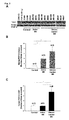
- the CSF of multiple sclerosis patients were centrifuged at 14,000 rpm for 5 minutes at 4°C and analysed by using an ELISA kit specific for human FGF-2 with a sensitivity limit of 2 pg/ml.
- C Chronic lesions. I: Inactive lesions. ⁇ shows the presence of lesions, while - indicates their absence. Regarding FGF-2 immunoreactivity, Ast: Astrocytes. Per: Periplaque. + shows that the labelling was positive and - negative.
- the inventors demonstrate how by measuring the FGF-2 level in CSF the presence of two groups of patients can be defined, while they cannot be distinguished when FGF-2 is measured directly from the tissue (WB).
- Proteins were extracted from histological sections of cerebral cortex using a protein extraction kit from fixed tissue, and the presence of FGF-2 or ⁇ -tubulin (as loading control) was immunodetected using the Western blot technique ( Figure 2A ) hybridising with a primary polyclonal antibody made in goat (1:200) or monoclonal antibody made in mouse (1:30,000) followed by an anti-goat biotinylated secondary antibody made in horse (1:5,000) or anti-mouse polyclonal antibody made in rabbit HRP-conjugated, respectively.
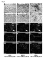
- the inventors demonstrate how measuring the FGF-2 and/or Anosmin-1 levels in the CSF of multiple sclerosis patients is a good bioindicator of the molecular environment found in the white matter demyelination lesions.
- Anosmin-1 immunostaining a polyclonal antibody made in rabbit (1:500) was used, which was later bound to a biotinylated polyclonal antibody made in goat (1:200). The process of immunolabeling was similar and was exposed with DAB and H 2 O 2 .
- the active lesions found in the some patients' tissue with a high FGF-2 level in the CSF showed no difference from those observed in multiple sclerosis patients with similar FGF-2 levels in CSF to those of control subjects, indicating that the presence of FGF-2, FGFR1 and Anosmin-1 is specific to the type of chronic or inactive lesion and not to the individual's age, type of multiple sclerosis or post-mortem interval between death and sample collection).
- the inventors disclose the identity of FGF-2 and FGFR1- producing cells in ACTIVE lesions on the one hand and in CHRONIC and INACTIVE on the other. Double histological immunostaining was performed using an antibody for FGF-2 rabbit polyclonal (1:200 v/v) together with the inflammatory cell marker HLA-DR (see above examples) and that specific to microglia/macrophages, CD68 (monoclonal antibody made in mouse, 1:200).
- FGFR1 polyclonal antibody made in rabbit 1:500 v/v
- PDGFR ⁇ polyclonal antibody made in rabbit, 1:100 v/v
- GFAP monoclonal antibody made in mouse; 1: 500 v/v
- immunofluorescence was intensified by using a biotinylated anti-rabbit seconday antibody made in goat, the ABC kit as well as the enhancement kit Tyramide (TSA) and Streptavidin Texas Red (1:500 v/v) .
- GFAP visualisation a mouse polyclonal antibody made in donkey bound to Alexa 633 was used and for PDGFR ⁇ a rabbit polyclonal antibody made in donkey bound to Alexa 488.
- the nuclei were counterstained with Hoechst.
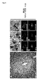
- the inventors show how measurement of the FGF-2 level in CSF is correlated with alterations in the grey matter of multiple sclerosis patients, regardless of the presence of demyelinating lesions in the white matter.
- measuring the FGF-2 level in CSF is a good bioindicator of the state of impairment of the blood-brain barrier of the grey matter, even in regions where demyelination has not occurred, which may serve as a prognostic test to avoid future neurodegeneration.
Landscapes
- Life Sciences & Earth Sciences (AREA)
- Health & Medical Sciences (AREA)
- Engineering & Computer Science (AREA)
- Biomedical Technology (AREA)
- Hematology (AREA)
- Chemical & Material Sciences (AREA)
- Urology & Nephrology (AREA)
- Molecular Biology (AREA)
- Immunology (AREA)
- Proteomics, Peptides & Aminoacids (AREA)
- Medicinal Chemistry (AREA)
- Microbiology (AREA)
- Biotechnology (AREA)
- Neurosurgery (AREA)
- Neurology (AREA)
- Food Science & Technology (AREA)
- Cell Biology (AREA)
- Physics & Mathematics (AREA)
- Analytical Chemistry (AREA)
- Biochemistry (AREA)
- General Health & Medical Sciences (AREA)
- General Physics & Mathematics (AREA)
- Pathology (AREA)
- Investigating Or Analysing Biological Materials (AREA)
- Peptides Or Proteins (AREA)
Applications Claiming Priority (3)
| Application Number | Priority Date | Filing Date | Title |
|---|---|---|---|
| ES200930661A ES2356540B1 (es) | 2009-09-07 | 2009-09-07 | Método para predecir las características histopatológicas de las lesiones de un sujeto con una enfermedad desmielinizante del sistema nervioso central. |
| ES201030090A ES2369099B1 (es) | 2010-01-25 | 2010-01-25 | La proteina Anosmina-1 como biomarcador para la clasificación histopatológica de lesiones de un sujeto con enfermedad desmielinizante del Sistema Nervioso Central. |
| PCT/ES2010/070584 WO2011045456A2 (es) | 2009-09-07 | 2010-09-07 | Nuevos biomarcadores de la histopatología de enfermedades desmielinizantes |
Publications (1)
| Publication Number | Publication Date |
|---|---|
| EP2477030A2 true EP2477030A2 (en) | 2012-07-18 |
Family
ID=43334531
Family Applications (1)
| Application Number | Title | Priority Date | Filing Date |
|---|---|---|---|
| EP10763733A Withdrawn EP2477030A2 (en) | 2009-09-07 | 2010-09-07 | Novel biomarkers of disease histopathology |
Country Status (6)
| Country | Link |
|---|---|
| US (1) | US20120252688A1 (cg-RX-API-DMAC7.html) |
| EP (1) | EP2477030A2 (cg-RX-API-DMAC7.html) |
| JP (1) | JP2013504075A (cg-RX-API-DMAC7.html) |
| AU (1) | AU2010306515A1 (cg-RX-API-DMAC7.html) |
| CA (1) | CA2773527A1 (cg-RX-API-DMAC7.html) |
| WO (1) | WO2011045456A2 (cg-RX-API-DMAC7.html) |
Family Cites Families (1)
| Publication number | Priority date | Publication date | Assignee | Title |
|---|---|---|---|---|
| FR2762602B1 (fr) * | 1997-04-28 | 1999-06-04 | Inst Nat Sante Rech Med | Moyens pour la detection precoce de pathologies auto-immunes inflammatoires |
-
2010
- 2010-09-07 WO PCT/ES2010/070584 patent/WO2011045456A2/es not_active Ceased
- 2010-09-07 EP EP10763733A patent/EP2477030A2/en not_active Withdrawn
- 2010-09-07 CA CA2773527A patent/CA2773527A1/en not_active Abandoned
- 2010-09-07 US US13/394,501 patent/US20120252688A1/en not_active Abandoned
- 2010-09-07 AU AU2010306515A patent/AU2010306515A1/en not_active Abandoned
- 2010-09-07 JP JP2012528396A patent/JP2013504075A/ja active Pending
Non-Patent Citations (1)
| Title |
|---|
| See references of WO2011045456A2 * |
Also Published As
| Publication number | Publication date |
|---|---|
| CA2773527A1 (en) | 2011-04-21 |
| WO2011045456A3 (es) | 2011-06-30 |
| AU2010306515A1 (en) | 2012-04-12 |
| JP2013504075A (ja) | 2013-02-04 |
| WO2011045456A2 (es) | 2011-04-21 |
| WO2011045456A9 (es) | 2011-11-03 |
| US20120252688A1 (en) | 2012-10-04 |
Similar Documents
| Publication | Publication Date | Title |
|---|---|---|
| US12422433B2 (en) | Blood biomarker that predicts persistent cognitive dysfunction after concussion | |
| AU2015212908B2 (en) | Biomarker and methods for early diagnosis of Alzheimer's disease | |
| Giovannoni | Multiple sclerosis cerebrospinal fluid biomarkers | |
| KR102064060B1 (ko) | 뇌의 베타 아밀로이드 축적 감별용 혈중 바이오마커 | |
| JP2012515335A (ja) | 線維症と肝硬変を識別する手段及び方法 | |
| US20110143380A1 (en) | Alzheimer's disease biomarkers and methods of use | |
| US9977036B2 (en) | Diagnostic markers for multiple sclerosis | |
| EP3514245A1 (en) | Method for aiding differential diagnosis of stroke | |
| Agnello et al. | The value of serum glial fibrillary acidic protein as a biomarker of astrogliosis in different neurological diseases | |
| US8298784B2 (en) | In vitro procedure for diagnosis and early diagnosis of neurodegenerative diseases | |
| WO2021009074A1 (en) | Novel markers as early predictors of alzheimer's pathology | |
| KR20200047371A (ko) | 인지기능 정상군 또는 경도 인지장애에서 아밀로이드 베타의 뇌 침착 검출용 혈액 바이오 마커 | |
| WO2011028960A1 (en) | Biomarkers for neurological conditions | |
| EP2477030A2 (en) | Novel biomarkers of disease histopathology | |
| JP2010507093A (ja) | バイオマーカー | |
| ES2356540B1 (es) | Método para predecir las características histopatológicas de las lesiones de un sujeto con una enfermedad desmielinizante del sistema nervioso central. | |
| US9618522B2 (en) | Diagnostic testing in dementia and methods related thereto | |
| WO2011109503A1 (en) | Novel csf biomarkers for alzheimer's disease and frontotemporal lobar degeneration | |
| CN114137214B (zh) | 用于预测应激后精神心理症状发生的免疫检测试剂盒及应用 | |
| ES2369099B1 (es) | La proteina Anosmina-1 como biomarcador para la clasificación histopatológica de lesiones de un sujeto con enfermedad desmielinizante del Sistema Nervioso Central. | |
| Liu et al. | The clinical significance of S100B, APP, and CHI3L1 in autoimmune GFAP astrocytopathy | |
| WO2024040103A2 (en) | Methods for early diagnosis and treatment of open neural tube defects | |
| CN120330385A (zh) | MxA的定量检测剂在急性呼吸道感染病程监测中的应用 | |
| Pulinx et al. | Biomarker discovery in MScl | |
| WO2008148490A1 (en) | Hsp27 as biomarker for alzheimer's disease |
Legal Events
| Date | Code | Title | Description |
|---|---|---|---|
| PUAI | Public reference made under article 153(3) epc to a published international application that has entered the european phase |
Free format text: ORIGINAL CODE: 0009012 |
|
| 17P | Request for examination filed |
Effective date: 20120330 |
|
| AK | Designated contracting states |
Kind code of ref document: A2 Designated state(s): AL AT BE BG CH CY CZ DE DK EE ES FI FR GB GR HR HU IE IS IT LI LT LU LV MC MK MT NL NO PL PT RO SE SI SK SM TR |
|
| DAX | Request for extension of the european patent (deleted) | ||
| 17Q | First examination report despatched |
Effective date: 20140722 |
|
| STAA | Information on the status of an ep patent application or granted ep patent |
Free format text: STATUS: THE APPLICATION IS DEEMED TO BE WITHDRAWN |
|
| 18D | Application deemed to be withdrawn |
Effective date: 20160317 |