EP2434952B1 - Vorrichtung und verfahren für spektrofotometrische messungen von blutparametern - Google Patents
Vorrichtung und verfahren für spektrofotometrische messungen von blutparametern Download PDFInfo
- Publication number
- EP2434952B1 EP2434952B1 EP10728297.2A EP10728297A EP2434952B1 EP 2434952 B1 EP2434952 B1 EP 2434952B1 EP 10728297 A EP10728297 A EP 10728297A EP 2434952 B1 EP2434952 B1 EP 2434952B1
- Authority
- EP
- European Patent Office
- Prior art keywords
- blood
- electromagnetic radiation
- duct
- emitting
- seat
- Prior art date
- Legal status (The legal status is an assumption and is not a legal conclusion. Google has not performed a legal analysis and makes no representation as to the accuracy of the status listed.)
- Active
Links
Images


Classifications
-
- A—HUMAN NECESSITIES
- A61—MEDICAL OR VETERINARY SCIENCE; HYGIENE
- A61B—DIAGNOSIS; SURGERY; IDENTIFICATION
- A61B5/00—Measuring for diagnostic purposes; Identification of persons
- A61B5/145—Measuring characteristics of blood in vivo, e.g. gas concentration, pH value; Measuring characteristics of body fluids or tissues, e.g. interstitial fluid, cerebral tissue
- A61B5/1455—Measuring characteristics of blood in vivo, e.g. gas concentration, pH value; Measuring characteristics of body fluids or tissues, e.g. interstitial fluid, cerebral tissue using optical sensors, e.g. spectral photometrical oximeters
- A61B5/14551—Measuring characteristics of blood in vivo, e.g. gas concentration, pH value; Measuring characteristics of body fluids or tissues, e.g. interstitial fluid, cerebral tissue using optical sensors, e.g. spectral photometrical oximeters for measuring blood gases
- A61B5/14557—Measuring characteristics of blood in vivo, e.g. gas concentration, pH value; Measuring characteristics of body fluids or tissues, e.g. interstitial fluid, cerebral tissue using optical sensors, e.g. spectral photometrical oximeters for measuring blood gases specially adapted to extracorporeal circuits
-
- A—HUMAN NECESSITIES
- A61—MEDICAL OR VETERINARY SCIENCE; HYGIENE
- A61B—DIAGNOSIS; SURGERY; IDENTIFICATION
- A61B5/00—Measuring for diagnostic purposes; Identification of persons
- A61B5/145—Measuring characteristics of blood in vivo, e.g. gas concentration, pH value; Measuring characteristics of body fluids or tissues, e.g. interstitial fluid, cerebral tissue
- A61B5/14535—Measuring characteristics of blood in vivo, e.g. gas concentration, pH value; Measuring characteristics of body fluids or tissues, e.g. interstitial fluid, cerebral tissue for measuring haematocrit
Definitions
- the present invention has as its object an apparatus and a method for spectrophotometry measurements of blood parameters.
- the field of application of the present invention is the medical field and in particular the monitoring of blood parameters during the course of therapies that require extracorporeal blood circulation like for example: haemodialysis, plasmapheresis, extracorporeal membrane oxygenation (ECMO), conservation of transplant organs and regional oncological therapies.
- extracorporeal blood circulation like for example: haemodialysis, plasmapheresis, extracorporeal membrane oxygenation (ECMO), conservation of transplant organs and regional oncological therapies.
- haemodialysis treatment as the preferred field of application, but it should be understood that the applicability of the present invention is not exclusive to this field.
- Haemodialysis is a replacement therapy to kidney function, which is applied to subjects that have little or no kidney activity (renal failure).
- a dialyzer i.e. an element with a double compartment in which a semipermeable membrane of suitably porosity is used.
- a first compartment the blood is made to flow, and in a second compartment an aqueous solution (dialyzing solution), enriched with the solutes that it is necessary to give to the blood and low in or without those to be taken out, is made to flow.
- the hematocrit the ratio between the corpuscular part and the liquid aliquot of the blood, is the indicator of the amount of liquid present in the blood.
- these techniques make use of an emitting device and a detecting device of electromagnetic waves so that the detector of electromagnetic waves is able to measure the amount of electromagnetic energy absorbed or reflected by the blood.
- the cuvette is a container provided with an inlet duct and an outlet duct to be connected to the duct of the extracorporeal blood. It is also provided with two parallel and opposite flat surfaces where the electromagnetic emitter and detector are positioned, facing the bloodflow and one another.
- the space between emitter and receiver must be small, so that the light emitted by the emitter can be detected by the detector.
- it is disadvantageous to narrow the duct, due to the loss of energy that would be given to the bloodflow.
- some protuberances are provided, on one of the two parallel surfaces described above, projecting towards the inside of the duct, which allow an accurate measurement without creating a throttling of the duct.
- the aforementioned solution is not without drawbacks, like for example the impossibility of being able to install said system on any type of extracorporeal blood duct, without providing an interruption of the duct and thus a duct in two parts.
- the sensors are positioned along the duct in contiguous positions arranged on a line parallel to the axis of the tubing.
- the aforementioned sensors are an emitting device and a detecting device of electromagnetic radiation, positioned so that they are in optical connection with the bloodflow, and so that the detecting device can receive the light reflected by the blood.
- the device foresees the use of a carcass containing the emitting/receiving devices, inside of which the blood duct passes.
- the operation of the device comprises an emission step and a reception step of the light reflected by the blood.
- the light that is detected by the receiver is translated into a potential difference and processed by a processing unit.
- the length of the slit is adjusted so as to make the detected signal proportional to the hematocrit value over time.
- the system in this case is unable to proceed to a measurement of the hematocrit or of the other parameters of interest in an absolute manner, i.e. it restores variations of the value of the parameter with respect to a first measurement.
- a first compensation of background noise i.e. the light that enters the device from the outside, is possible.
- a further parameter that it would be advisable to detect is the presence of gaseous emboli inside the bloodflow to be returned to the patient.
- gaseous emboli is of fundamental importance in systems used for parenteral nutrition, infusion of drugs, blood transfusion and extracorporeal circulation since they can cause even irreversible damage to the patient being treated.
- the techniques for measuring gaseous emboli can be of two types: optical or ultrasound type.
- the optical technique of the state of the art is based on the difference in the amount of light transmitted between the air and the water media (the blood and the infusion substances mostly consist of water).
- Patent US 6,529,751 describes the use of electromagnetic sources in the field from 800 nm to 850 nm to quantify microemboli in the blood.
- US 6,144,444 Therein an apparatus for monitoring blood parameters during cardio-pulmonary bypass surgery or during other procedures which utilize an extracorporeal circuit, is disclosed.
- the apparatus is typically used to monitor the percentage of haemoglobin bound with oxygen (oxygen saturation), the total amount of haemoglobin in the blood, and the percentage of blood which is comprised of red blood cells (hematocrit), although the apparatus can be adapted to measure other blood parameters.
- the apparatus (or monitor) provides real time results to show immediate changes (trending) in the monitored parameters.
- a system can include an analyte detection system configured to measure first analyte data in a fluid sample received from a patient, a medical sensor configured to measure second analyte data in the patient, and a processor configured to receive the first analyte data and the second analyted data and to determine a physiological parameter based at least in part on the first analyte data and the second analyte data.
- the medical sensor may be a pulse oximeter
- the physiological parameter may include a cardiovascular parameter including, for example, cardiac output.
- a disposable cuvette through which passes pulsatile flowing blood is disclosed in WO 94/27495 A1 .
- the cuvette has a conduit with two opposed walls having a predetermined separation therebetween that varies with each pulse of the flowing blood.
- a sealed air pocket damps the variation of the predetermined separation.
- the conduit is comprised of materials which permit passage therethrough of wavelengths of selected electromagnetic radiation.
- the radiation is emitted from a photoemitter which, and after passing through the cuvette, is detected by a photodetector.
- the quantities of detected radiation are operated on by a computer which uses a spectrophotometry technique to derive therefrom a blood constituent concentration value.
- both the cuvette and the spectrophotometry technique are used during hemodialysis to derive changes in the hematocrit value of the blood of a dialyzed patient, thereby to deduce therefrom changes in the blood volume of the patient during dialysis.
- WO 2008/136548 discloses a device for measuring and monitoring hemoglobin values through a blood tube, comprising
- the purpose of the present invention is therefore to overcome the drawbacks of the prior art.
- a first task of the present invention is to measure some blood parameters of interest, for wide operating conditions and with high precision.
- the parameters of interest considered are: hematocrit, oxygen saturation, blood temperature and the presence of gaseous microemboli.
- the imprecision of these measurements must be preferably no more than ⁇ 5 percentage units for the hematocrit value and the oxygen saturation value and ⁇ 0,5°C for the temperature value.
- a second task of the present invention is to carry out the measurements directly on the tubing without it having particular optical and/or geometric characteristics.
- a third task of the present invention is to be able to carry out an absolute measurement of the hematocrit value and oxygen saturation.
- a further task of the present invention is to provide an apparatus capable of detecting the presence in the blood of gaseous emboli, and in particular their number and their size.
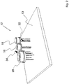
- Figures 1 , 4 , 6 and 8 represent an apparatus for measuring the blood parameters in an extracorporeal circuit according to the invention, wholly indicated with reference numeral 12.
- the apparatus 12 comprises a seat 23 suitable for containing a duct 14 for the blood flow in said extracorporeal circuit.
- Electromagnetic radiation emitting means 16 and electromagnetic radiation detecting means 18 face the seat 23. Said means 16, 18 are also connected to a control unit 38.
- the apparatus 12 is characterised in that said emitting means 16 are suitable for producing electromagnetic radiation at different wavelengths and the detecting means 18 are suitable for detecting the electromagnetic radiation diffused in the blood at said wavelengths.
- the apparatus 12 is characterised in that the control unit 38 is suitable for calculating values of blood parameters through a correlation between reference values and ratios obtained from values of the light intensity of the radiation detected at at least two different wavelengths.
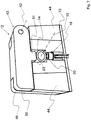
- the apparatus 12 according to a first embodiment of the invention comprises a body 20 suitable for containing inside it the means 16 and 18 connected to a base 13.
- the body 20 is associated with the base 13 so that the means 16 and 18 are contained inside the space located between body 20 and base 13 as shown in fig. 4 .
- a seat 23 is arranged, comprising three parts having a different section in the transversal plane:
- holes 24 and 26 are formed, arranged in a substantially radial direction, for the optical connection between the duct 14 and the detecting means 18 and emitting means 16, respectively.
- the emitting means 16 and the detecting means 18, axially spaced, are arranged on the same side of the seat 23.
- the measurement carried out by the apparatus 12 takes place in reflection, since the detecting means 18 measure the fraction of light reflected by the blood.
- a first shoulder 44 and a second shoulder 46 are rigidly fixed on two parallel sides of the body.
- the apparatus also comprises a cover 40 able to rotate around an axis 42 positioned on a side of the cover 40 itself and parallel to a side of the body 20.
- the rotation axis is formed by means of a hinge 43 that connects the cover 40 and the second shoulder 46.
- the cover 40 has a projection 51 (as shown in fig. 7 ) such as to couple with the seat 23 for the duct 14, so as to cover the portion of circumference of the duct 14 not covered by the body 20 .
- a recess 48 is formed for a coupling surface 50 formed on the cover 40.
- the duct 14 for the blood When the duct 14 for the blood is inserted into the body 20 and the cover 40 is closed, the duct 14 is slightly squashed by the projection 51 thus creating two parallel flat surfaces: one in contact with the cover 40 and one in contact with the body 20 at the holes 24, 26 for the means 16, 18.
- the duct 14, selected among one of those commonly used in the medical field, is substantially transparent to the wavelengths of the emitted radiation.
- the duct 14 consists of a polymer like plasticized PVC.
- the part of said radiation absorbed by the duct 14 must be less than 50% for each individual used wavelength of the total emitted radiation.
- the duct 14 which is introduced into the seat 23 and through which the blood parameters are measured is any section of a common disposable extracorporeal circuit.
- the electromagnetic radiation emitting means 16 comprise light emitting diodes (LED) (not shown).
- the emitting means 16 comprise a single LED for each of the wavelengths that are used for calculating the blood parameters.
- the characteristics of some particular types of LED involve an emission spectrum that assumes the profile of a very narrow bell, with a very pronounced peak at a particular wavelength.
- Such LEDs, considered hereafter, have a very narrow emission spectrum and have no secondary emissions. For this reason, the approximation is commonly accepted based on which each type of LED is attributed with one wavelength only.
- the emitting means 16 preferably comprise at least two different LEDs, each dedicated to the emission of electromagnetic radiation at one single wavelength.
- the emitting means 16 comprise four different LEDs: a first LED (A) suitable for emitting light with a wavelength equal to 805nm, a second LED (B) suitable for emitting light with a wavelength equal to 660nm, a third LED (C) suitable for emitting light with a wavelength equal to 1450nm and finally a fourth LED (D) suitable for emitting light with a wavelength equal to 1550nm.
- a first LED (A) suitable for emitting light with a wavelength equal to 805nm
- B suitable for emitting light with a wavelength equal to 660nm
- C suitable for emitting light with a wavelength equal to 1450nm
- D fourth LED
- the electromagnetic radiation detecting means 18 comprise a wide band sensor for example of the InGaAs type.
- the InGaAs sensor is a semiconductor made up of indium, gallium and arsenic which is typically sensitive to the band of electromagnetic radiation within the range from 600 nm up to 2600 nm.
- a temperature detector 28 measuring the bloodflow temperature, placed inside the apparatus in a position adjacent to the emitting means 16 and also connected to the base 13.
- the temperature detector 28 has a wide band reception within the range of middle-infrared electromagnetic radiation up to 15000 nm.
- the temperature detector 28 measures the bloodflow temperature and, in the case in which it is an infrared detector, it is in light communication with the duct 14 through a hole 30 adjacent to the hole 26.
- the temperature detector 28 makes it possible to take also into account, in the calculation of the blood parameters, the influence of the blood temperature itself. Indeed, keeping every other condition the same, the optical characteristics of blood vary as the temperature varies, i.e. the proportions between the amount of radiation absorbed and diffused are altered. Such a variation is monotonic with respect to the values of the electromagnetic radiation used and therefore to a large extent the measurement error is compensated by adopting a rateometric measurement technique as described hereafter.
- Such a variation of the optical characteristics of blood can be determined experimentally for each electromagnetic radiation adopted, hi particular, it is possible to observe a variation for amounts of absorbed and diffused electromagnetic radiation that on average is about 0.25% for a temperature variation of 1°C. Therefore, it is possible to take into account such a variation by providing the control unit 38 with the signal of the temperature detector 28.
- a first temperature sensor 32 measuring the operating temperature of the detector 18 arranged in contact with it, and connected to the base 13.
- the use of the first temperature sensor 32 is justified by the fact that the responsivity of the detection device 18 depends upon the temperature, which can translate into a drift of the measured hematocrit value by about 0.5% for a temperature variation of 1 °C.
- the drift can be seen through a comparison between the data obtained by measuring a blood sample through spectrophotometry and the value obtained from the same blood sample from laboratory apparatuses commonly used in the medical field (for example a centrifuge).
- a second temperature sensor 34 which controls the operation of the temperature detector 28.
- the sensor 34 is placed in contact with the duct 14 through the hole 36 formed between the hole 26 and the hole 30 and measures the blood temperature through contact with the duct 14.
- the second sensor 34 is used as a safety sensor in the case of malfunction of the temperature detector 28.
- the emitting means 16 and the detecting means 18 are connected to a control unit 38.
- the control unit 38 regulates the power supply currents for the emitting device 16, i.e. of the light emitting diodes, through a digital to analogue converter with resolution preferably of no less than 12 bit.
- the guide current of each element is generated and regulated through the combination of a field effect transistor (FET) and an operational amplifier connected to a digital to analogue converter with resolution preferably of no less than 12 bit indicated with reference numeral 31.
- FET field effect transistor
- the detecting means 18 convert the radiation diffused into current and then into a voltage through a transimpedance amplifier.
- a second amplification is digitally controlled through a variable analogue gain amplifier 33, the output voltage of which is converted into a digital signal through an analogue to digital converter with resolution preferably of at least 16 bit, indicated with reference numeral 25.
- the temperature sensor 32 is connected to the control unit 38 described earlier that, according to calibration tables of the receiving instrument, takes care of correcting the signal acquired by the detecting means 18.
- control unit 38 has the temperature detector 28 connected to it.
- the radiation emitted by the emitting means 16 strikes the duct 14 and in part is attenuated and in part diffused in the blood.
- the detecting means 18 detect the electromagnetic radiation diffused or emitted by the blood which is a function of the concentration of the biologic constituents and of its temperature.
- the LEDs of the emitting means 16 are suitable for emitting single pulses of radiation, each of which has a selected and very precise wavelength.
- Such a characteristic of the apparatus 12 thus makes it possible to use extremely simple detecting means 18.
- Such a characteristic makes it superfluous to use a spectrophotometer that analyses the radiation reflected by the blood.
- the spectrophotometer used in the prior art, comprises a prism and a plurality of detecting elements arranged so as to create a practically continuous detection band to receive the spectrum generated by the prism. The greater the number of such detecting elements, the better the approximation of the continuum that is obtained.
- a common spectrophotometer used for analyses similar to those that are the purpose of the present invention 128, 256 or more of such detecting elements can be used.
- the spectrophotometer is a rather delicate (for example for the optical alignment of the components) and rather expensive (for example for the plurality of detecting elements used, that have sensitivity to electromagnetic radiation also in the spectral field of the near-infrared) component.
- the detecting means 18 used in the apparatus 12 according to the invention can, in principle, comprise a single detecting element compared to the 128, 256 or even more used in the prior art. Therefore, there is no need to analyse the wavelength of the detected radiation since it is already selected at source thanks to the use of LEDs in the emitting means 16.
- the wavelengths of the electromagnetic radiation emitted are within the range from 660 nm to 1550 nm, whereas those diffused or emitted by the blood and able to be detected by the measuring apparatus are within the range from 600 nm to 15000 nm.
- the emitting means 16 there are at least two wavelengths emitted by the emitting means 16, selected at the water absorption peaks and at particular transitions of the absorption spectra of HbO 2 and Hb molecules.
- hematocrit Ht% or the oxygen saturation (sO 2 %)
- sO 2 oxygen saturation
- Said reference values are values obtained through the normal analysis techniques.
- the reference values can also be presented in the form of a calibration curve.
- a calibration of the apparatus is carried out by correlating the value of the ratios between different wavelengths with values calculated in the laboratory through the usual techniques, e.g. for the case of the hematocrit the calculation is carried out through centrifuging of the blood in microcapillaries.
- the four wavelengths emitted by the emitting means 16 are 805nm (A), 660nm (B), 1450nm (C) and 1550nm (D). Such wavelengths were selected according to the following criteria:
- the water absorption peak at the wavelength of 1450 nm, and the area at 1550 nm near to it, are particularly important for determining the hematocrit. Indeed, it is possible to make the ratio between the absorption of the blood at the wavelengths of 1450 nm and 1550 nm, due therefore substantially to water, and that at 805 nm, where on the other hand there is no absorption by water.
- the choice of the wavelength of 660 nm where the absorption difference between the oxygenated form of haemoglobin and the non-oxygenated or reduced form is at its maximum makes it possible to measure the percentage oxygen saturation of the blood by making a ratio with the absorption at 805 nm where the two forms of haemoglobin have the same absorption.
- the emitting means 16 emit a sequence of pulses at different wavelengths, for example the sequence can be the one represented in fig. 9 , i.e. A-B-C-D (805nm, 660nm, 1450nm, 1550nm).
- the frequency at which the pulses are emitted is constant, but their intensity is variable according to their wavelength.
- This choice has been made to compensate for the different absorption in the blood and to optimise, in the field of variation of the parameters to be measured, the dynamics of the electromagnetic radiation signal detected for each wavelength.
- the variation in intensity is determined for each LED by the control unit 38.
- the diffused radiation is detected by the detecting means 18 in a synchronous manner but it is slightly delayed with respect to the emitted radiation, so as to wait for the stable level condition.
- the intensities of the detected radiations are therefore I ⁇ (A), I ⁇ (B), I ⁇ (C) and I ⁇ (D).
- the intensity of the detected electromagnetic radiation will be the sum of two contributions: a first contribution due to the radiation diffused by the blood and a second contribution due to a background noise, i.e. light radiation detected when the light source is switched off.
- the background noise corresponds to the value of the electromagnetic radiation measured at point F, whereas the total value, diffused by the blood and due to the light emitting diode, measured at the pulse is that at point G.
- the actual value of the amount of diffused radiation is obtained as the difference between the value corresponding to point G and the value corresponding to point F.
- the ratio R 1 I ⁇ (A)/[I ⁇ (C)+I ⁇ (D)] is calculated using the intensities of electromagnetic radiation diffused by the blood and detected at three different wavelengths. Such a ratio is correlated to the values obtained through laboratory measurements through the use of "gold standard" apparatuses.
- Ht % R 1 3 * ⁇ 3 + R 1 2 * ⁇ 2 + R 1 * ⁇ 1 + ⁇ 0 + a * SO 2 % ⁇ 75
- ⁇ 0 , ⁇ 1 , ⁇ 2 , ⁇ 3 and ⁇ where ⁇ 0 , ⁇ 1 , ⁇ 2 , ⁇ 3 and ⁇ are experimentally obtained coefficients.
- sO 2 % R 2 2 * ⁇ 1 Ht % + R 2 * ⁇ 2 Ht % + ⁇ 0 Ht %
- ⁇ 1 (Ht%), ⁇ 2 (Ht%) and ⁇ 0 (Ht%) are experimentally obtained as a function of the value of Ht%, considering the variation range divided into three fields, the first for values of Ht% ⁇ 25, the second field for Ht%>40 and the third intermediate field for values of Ht% comprised between 25 and 40.
- the value of sO 2 % is corrected with a function of the value of Ht% calculated previously.
- the reference values for sO 2 % will be a function of the value of Ht%, and thus of R 1 and of R 2 .
- the value of Ht% is corrected through a function of the value of sO 2 %.
- Both the hematocrit value, and the oxygen saturation value thus calculated can be corrected based on the responsivity of the detecting means 18, through the sensor 32, and based on the blood temperature through the temperature detector 28.
- the hematocrit value is correlated to calibration values measured in the laboratory and takes into account the responsivity of the detecting means 18, the temperature variations of the blood, and the variation of oxygen saturation.
- the oxygen saturation value is correlated to calibration values measured in the laboratory and takes into account the responsivity of the detecting means 18, the temperature variations of the blood, and the variation of the hematocrit value of the blood.
- the measurement of the hematocrit and oxygen saturation values can with this method also be carried out by using just three wavelengths, in an analogous manner to what has just been seen.
- the detection of gaseous emboli is obtained through three main steps.
- a first step consists of emitting a train of pulses of constant intensity at a determined frequency.
- the effects of the measurement are not affected by the used wavelength, but preferably a wavelength is used that has high optical efficiency as a function of the used detecting means.
- the emitting means 16 emit a signal at the wavelength C and at the frequency preferably of no less than 13.33KHz within the time period in which the emitting device 16 does not emit the wavelength C used to measure hematocrit or oxygen saturation.
- the second step referring to figure 11 (b) is the detection of the electromagnetic radiation diffused by the blood at said wavelength.
- the detected radiation has the same characteristics as the radiation used to calculate the hematocrit or oxygen saturation value.
- the third step consists of processing the signal detected in the previous step.
- the value of the width L of the signal is used for sampling to be used to extrapolate the curve indicated in figure 11 (c) and 12 .
- Said curve describes the trend of the intensity of the detected electromagnetic radiation and therefore the variations in trend undergone by the curve that, when above a certain threshold, are interpreted as the presence of emboli.
- the apparatus according to the invention also allows a link between size of the embolus and variation of the curve.
- ⁇ L indicates a tolerance range within which the detected radiation can vary without causing alarms due to the presence of emboli.
- a step known as "START" begins in which the measurement of a time starts.
- the time passed between "START” and "END” is known as ⁇ T and it is used to calculate the size of the embolus.
- a possible embodiment of the present invention can foresee the use of a plurality of emitting means and a plurality of detecting means, differently positioned with respect to the axial direction.
Landscapes
- Health & Medical Sciences (AREA)
- Life Sciences & Earth Sciences (AREA)
- Physics & Mathematics (AREA)
- Medical Informatics (AREA)
- Surgery (AREA)
- Biophysics (AREA)
- Pathology (AREA)
- Engineering & Computer Science (AREA)
- Biomedical Technology (AREA)
- Heart & Thoracic Surgery (AREA)
- Veterinary Medicine (AREA)
- Molecular Biology (AREA)
- Optics & Photonics (AREA)
- Animal Behavior & Ethology (AREA)
- General Health & Medical Sciences (AREA)
- Public Health (AREA)
- Spectroscopy & Molecular Physics (AREA)
- Measurement Of The Respiration, Hearing Ability, Form, And Blood Characteristics Of Living Organisms (AREA)
- Investigating Or Analysing Biological Materials (AREA)
- Investigating Or Analysing Materials By Optical Means (AREA)
- Measuring Pulse, Heart Rate, Blood Pressure Or Blood Flow (AREA)
Claims (13)
- Vorrichtung (12) zum Messen der Blutparameter in einem extrakorporalen Kreislauf, aufweisend:einen Sitz (23), der geeignet ist, einen Kanal (14) für die Blutströmung des extrakorporalen Kreislaufs aufzunehmen;eine elektromagnetische Strahlung aussendende Einrichtung (16);eine elektromagnetische Strahlung erfassende Einrichtung (18), wobei die Einrichtungen (16, 18) dem Sitz (23) zugewandt ist;einen ersten Temperatursensor (32) zum Messen der Betriebstemperatur der erfassenden Einrichtung (18);einen zweiten Temperatursensor (34), der zum Messen der Bluttemperatur geeignet ist,einen zentralen Körper (20), der geeignet ist, darin die mit einer Basis (13) verbundenen Einrichtungen (16) und (18) aufzunehmen, undeine Steuereinheit (38), mit der die emittierende Einrichtung (16) und die erfassende Einrichtung (18) verbunden sind; wobeidie elektromagnetische Strahlung aussendende Einrichtung (16) und die elektromagnetische Strahlung erfassende Einrichtung (18) innerhalb des Raumes zwischen dem Körper (20) und der Basis (13) untergebracht sind, Löcher (24, 26) für eine optische Verbindung zwischen dem Kanal (14) und der erfassenden Einrichtung (18) und emittierenden Einrichtung (16) auf dem Sitz ausgebildet sind, die elektromagnetische Strahlung aussendende Einrichtung (16) ist dazu geeignet elektromagnetische Strahlung mit verschiedenen Wellenlängen zu erzeugen, die elektromagnetische Strahlung erfassende Einrichtung (18) ist dazu geeignet elektromagnetische Strahlung, die im Blut bei den Wellenlängen gestreut wird, zu detektieren, und die Steuereinheit (38) ist dazu geeignet, die Werte von Blutparametern zu kalkulieren, durch eine Korrelation zwischen Referenzwerten und Verhältnissen, die aus Werten der Lichtintensität der detektierten Strahlung bei mindestens zwei verschiedenen Wellenlängen erhalten werden,wobei der Kanal (14) irgendein Abschnitt eines wegwerfbaren extrakorporalen Kreislaufs ist, wobei die Werte der berechneten Blutparameter basierend auf der Bluttemperatur und/oder basierend auf dem Wert der Betriebstemperatur der erfassenden Einrichtung (18) korrigiert werden undwobei die Vorrichtung eine Abdeckung (40) aufweist, wobei die Abdeckung (40) fähig ist, um eine Achse (42) zu rotieren, die parallel zu einer Seite des Körpers (20) ist, die Abdeckung hat einen Vorsprung (51), welcher angepasst ist den in den Sitz (23) im Körper (20) eingesetzten Kanal (14) zusammenzudrücken, wenn die Abdeckung geschlossen wird,wobei zwei parallele, flache Flächen in dem Kanal in einer Weise ausgebildet werden, dass eine Fläche mit der Abdeckung (40) in Kontakt ist und die andere Fläche mit dem Körper (20) an den Löchern (24, 26) für die Strahlung emittierende Einrichtung (16) und die Strahlung erfassende Einrichtung (18) in Kontakt ist.
- Vorrichtung (12) nach Anspruch 1, dadurch gekennzeichnet, dass die emittierende Einrichtung (16) geeignet ist, elektromagnetische Strahlung mit vier verschiedenen Wellenlängen zu emittieren und die emittierende Einrichtung (16) vorzugsweise geeignet ist, elektromagnetische Strahlung zu emittieren mit den Wellenlängen: A = 805nm, B = 660nm, C = 1450nm, D =1550 nm.
- Vorrichtung (12) nach einem der vorhergehenden Ansprüche, dadurch gekennzeichnet, dass die emittierende Einrichtung (16) geeignet ist, elektromagnetische Strahlung bei Wellenlängen zu emittieren, die den Wasserabsorptionspeaks entsprechen und/oder einen Temperaturdetektor (28) aufweist, der dem Sitz (23) zugewandt ist und mit der Steuereinheit (38) verbunden ist, wobei der Temperaturdetektor (28) vorzugsweise ein Infrarot-Temperatursensor ist mit einem Breitbandempfang im Bereich von mittlerer-Infrarot elektromagnetischer Strahlung bis zu 15000nm.
- Vorrichtung (12) nach einem der vorhergehenden Ansprüche, dadurch gekennzeichnet, dass die emittierende Einrichtung (16) eine Folge von Pulsen konstanter Breite und Frequenz bei mindestens einer Wellenlänge emittiert.
- Vorrichtung (12) nach einem der vorhergehenden Ansprüche, dadurch gekennzeichnet, dass sie aufweist:den zentralen Körper (20), der den Sitz (23) aufweist;die dem zentralen Körper (20) zugeordnete Basis (13), mit der die emittierende und erfassende Einrichtung (16, 18) so verbunden sind, dass sie innerhalb des zwischen dem Körper (20) und der Basis (13) angeordneten Raums enthalten sind; unddie an dem Sitz (23) ausgebildeten Löcher (24, 26), die in einer im Wesentlichen axialen Richtung verteilt sind.
- Vorrichtung (12) nach einem der vorhergehenden Ansprüche, dadurch gekennzeichnet, dass sie auf dem Sitz (23) ausgebildete und in im Wesentlichen axialer Richtung verteilte zusätzliche Löcher (30, 36), jeweils für die optische Verbindung zwischen dem Kanal (14) und dem Temperaturdetektor (28) und für den Kontakt zwischen dem zweiten Sensor (34) und dem Kanal (14), aufweist.
- Verfahren zum Messen von Blutparametern unter Verwendung der Vorrichtung von Anspruch 1, wobei das Verfahren folgende Schritte aufweist:Einführen eines Kanals für Blut in den Sitz in dem Körper,Schließen der Abdeckung des Körpers,Zusammendrücken des Kanals mit dem Vorsprung auf dem Deckel, wodurch zwei parallele, flache Oberflächen gebildet werden,Emittieren elektromagnetischer Strahlung durch die parallelen flachen Oberflächen in Form einer Folge von Pulsen von mindestens zwei verschiedenen Wellenlängen;Erfassen der gestreuten Strahlung im Blut, durch die mindestens zwei Wellenlängen;Berechnen der Werte der Blutparameter korrelierenden Referenzwerte mit dem Verhältnis zwischen der Intensität der erfassten elektromagnetischen Strahlung, bei den mindestens zwei verschiedenen Wellenlängen.
- Verfahren nach dem vorhergehenden Anspruch, dadurch gekennzeichnet, dass der Sauerstoffsättigungswert (sO2%) erhalten wird durch Korrelation des Verhältnisses R2 = Iλ(A)Iλ(B) zwischen einer ersten Intensität von erfasster elektromagnetischer Strahlung Iλ(A) und einer zweiten Intensität von erfasster elektromagnetischer Strahlung Iλ(B) mit Kalibrationskurven und/oder durch Korrelation des Verhältnisses R2 = Iλ(A)Iλ(B) zwischen der Intensität von erfasster elektromagnetischer Strahlung mit Kalibrationswerten angenähert mit einer mathematischen Funktion zweiter Ordnung.
- Verfahren nach einem der Ansprüche 7 oder 8, dadurch gekennzeichnet, dass der Hämatokritwert (Ht%) erhalten wird durch Korrelation des Verhältnisses R1 = Iλ(A)/[Iλ(C) + Iλ(D)] zwischen den Intensitäten von erfasster elektromagnetischer Strahlung Iλ(A),Iλ(C) und Iλ(D) mit Kalibrationskurven und/oder durch Korrelation des Verhältnisses R1 = Iλ(A)/[Iλ(C) + Iλ(D)] zwischen den Intensitäten von erfasster elektromagnetischer Strahlung mit Kalibrationswerten angenähert mit einer mathematischen Funktion dritter Ordnung.
- Verfahren nach einem der Ansprüche 7 bis 9, dadurch gekennzeichnet, dass der berechnete Sauerstoffsättigungswert des Blutes (sO2%) basierend auf dem Hämatokritwert (Ht%) korrigiert wird.
- Verfahren nach einem der Ansprüche 7 bis 10, dadurch gekennzeichnet, dass der berechnete Hämatokritwert des Blutes (Ht%) basierend auf dem Sauerstoffsättigungswert (sO2%) korrigiert wird.
- Verfahren nach einem der Ansprüche 7 bis 11, aufweisend die Schritte:Emittieren einer Folge von Pulsen bei konstanter Intensität und Frequenz;Erfassen der Folge von Pulsen; undVerarbeiten der erfassten Folge von Pulsen undAssoziieren der Anwesenheit und Größe eines Embolus mit dem Zeitraum, in dem die Änderung der Intensität der erfassten Strahlung einen bestimmten Schwellenwert überschreitet.
- Verfahren nach einem der Ansprüche 7 bis 13, dadurch gekennzeichnet, dass eine Berechnung des Luftvolumens das als einzelner Embolus und als Summe von Mikroemboli akkumuliert wird, durchgeführt wird und somit ein Alarm und/oder ein Stopp des Blutflusses erfolgt, wenn ein Risiko-Schwellwert für den Patienten überschritten wird.
Applications Claiming Priority (2)
| Application Number | Priority Date | Filing Date | Title |
|---|---|---|---|
| IT000926A ITMI20090926A1 (it) | 2009-05-26 | 2009-05-26 | Apparato e metodo per misure spettrofotometriche di parametri del sangue. |
| PCT/IB2010/052306 WO2010136962A1 (en) | 2009-05-26 | 2010-05-25 | Apparatus and method for spectrophotometric measurements of blood parameters |
Publications (2)
| Publication Number | Publication Date |
|---|---|
| EP2434952A1 EP2434952A1 (de) | 2012-04-04 |
| EP2434952B1 true EP2434952B1 (de) | 2017-01-11 |
Family
ID=41559436
Family Applications (1)
| Application Number | Title | Priority Date | Filing Date |
|---|---|---|---|
| EP10728297.2A Active EP2434952B1 (de) | 2009-05-26 | 2010-05-25 | Vorrichtung und verfahren für spektrofotometrische messungen von blutparametern |
Country Status (4)
| Country | Link |
|---|---|
| US (1) | US20120108981A1 (de) |
| EP (1) | EP2434952B1 (de) |
| IT (1) | ITMI20090926A1 (de) |
| WO (1) | WO2010136962A1 (de) |
Families Citing this family (4)
| Publication number | Priority date | Publication date | Assignee | Title |
|---|---|---|---|---|
| DE102012104461A1 (de) * | 2012-05-23 | 2013-12-12 | B. Braun Avitum Ag | Medizinisches Gerät zur extrakorporalen Blutbehandlung mit mehreren Sensoreinheiten |
| DE102015015587A1 (de) * | 2015-08-27 | 2017-03-02 | Em-Tec Gmbh | Haltevorrichtung für einen Schlauch |
| US20180000394A1 (en) * | 2016-06-30 | 2018-01-04 | Fresenius Medical Care Holdings, Inc. | Method and system for creating a diagnostic vascular window |
| JP2019213570A (ja) * | 2016-10-19 | 2019-12-19 | アルプスアルパイン株式会社 | 計測装置および血液循環装置 |
Citations (4)
| Publication number | Priority date | Publication date | Assignee | Title |
|---|---|---|---|---|
| US5615672A (en) * | 1993-01-28 | 1997-04-01 | Optiscan, Inc. | Self-emission noninvasive infrared spectrophotometer with body temperature compensation |
| US6208880B1 (en) * | 1997-02-27 | 2001-03-27 | Terumo Cardiovascular Systems Corporation | Blood parameter measurement device |
| US20060189926A1 (en) * | 2005-02-14 | 2006-08-24 | Hall W D | Apparatus and methods for analyzing body fluid samples |
| WO2008136548A1 (en) * | 2007-05-07 | 2008-11-13 | Jsm Healthcare Inc | Device of measuring and monitoring a hemoglobin value through blood tube |
Family Cites Families (10)
| Publication number | Priority date | Publication date | Assignee | Title |
|---|---|---|---|---|
| US4913150A (en) * | 1986-08-18 | 1990-04-03 | Physio-Control Corporation | Method and apparatus for the automatic calibration of signals employed in oximetry |
| US5351686A (en) * | 1990-10-06 | 1994-10-04 | In-Line Diagnostics Corporation | Disposable extracorporeal conduit for blood constituent monitoring |
| US5331958A (en) * | 1992-03-31 | 1994-07-26 | University Of Manitoba | Spectrophotometric blood analysis |
| DE19612425C2 (de) * | 1995-03-31 | 2000-08-31 | Nihon Kohden Corp | Apparat zur Messung von Hämoglobinkonzentration |
| AU3721997A (en) * | 1996-07-19 | 1998-02-10 | Alexander K. Mills | Device for noninvasive determination of blood parameters |
| US6090061A (en) | 1997-10-22 | 2000-07-18 | In-Line Diagnostics Corporation | Disposable extracorporeal conduit for blood constituent monitoring |
| WO1998048867A1 (en) | 1997-04-29 | 1998-11-05 | Medtronic, Inc. | Optical detection and quantification of microair in blood |
| US6144444A (en) * | 1998-11-06 | 2000-11-07 | Medtronic Avecor Cardiovascular, Inc. | Apparatus and method to determine blood parameters |
| JP4129867B2 (ja) | 2002-07-18 | 2008-08-06 | 日機装株式会社 | ヘマトクリットセンサ |
| US8412293B2 (en) * | 2007-07-16 | 2013-04-02 | Optiscan Biomedical Corporation | Systems and methods for determining physiological parameters using measured analyte values |
-
2009
- 2009-05-26 IT IT000926A patent/ITMI20090926A1/it unknown
-
2010
- 2010-05-25 US US13/322,756 patent/US20120108981A1/en not_active Abandoned
- 2010-05-25 EP EP10728297.2A patent/EP2434952B1/de active Active
- 2010-05-25 WO PCT/IB2010/052306 patent/WO2010136962A1/en active Application Filing
Patent Citations (4)
| Publication number | Priority date | Publication date | Assignee | Title |
|---|---|---|---|---|
| US5615672A (en) * | 1993-01-28 | 1997-04-01 | Optiscan, Inc. | Self-emission noninvasive infrared spectrophotometer with body temperature compensation |
| US6208880B1 (en) * | 1997-02-27 | 2001-03-27 | Terumo Cardiovascular Systems Corporation | Blood parameter measurement device |
| US20060189926A1 (en) * | 2005-02-14 | 2006-08-24 | Hall W D | Apparatus and methods for analyzing body fluid samples |
| WO2008136548A1 (en) * | 2007-05-07 | 2008-11-13 | Jsm Healthcare Inc | Device of measuring and monitoring a hemoglobin value through blood tube |
Also Published As
| Publication number | Publication date |
|---|---|
| WO2010136962A1 (en) | 2010-12-02 |
| US20120108981A1 (en) | 2012-05-03 |
| EP2434952A1 (de) | 2012-04-04 |
| ITMI20090926A1 (it) | 2010-11-27 |
Similar Documents
| Publication | Publication Date | Title |
|---|---|---|
| US10960125B2 (en) | Fluid flow rate measuring and gas bubble detecting apparatus | |
| EP2355690B1 (de) | Hämatokritmessung und schätzung von hämoglobinwerten mit einem nichtinvasiven optischen blutüberwachungssystem | |
| EP1579196B1 (de) | Verfahren und vorrichtung für messungen im blut | |
| US9173988B2 (en) | Sensor clip assembly for an optical monitoring system | |
| AU764970B2 (en) | Apparatus and method to determine blood parameters | |
| US9849225B2 (en) | Self calibrating blood chamber | |
| US9265872B2 (en) | Device and method for measuring a blood constituent in blood for an extracorporeal blood treating device | |
| AU2002233597B2 (en) | A method for measuring hemoglobin concentration (HGB) in the blood in a circuit of a dialysis machine, measuring device and circuit for the application of the method | |
| EP2434952B1 (de) | Vorrichtung und verfahren für spektrofotometrische messungen von blutparametern | |
| Cattini et al. | An optical technique for real-time monitoring of hemolysis during hemodialysis | |
| JP2009508562A (ja) | 医療用デバイス | |
| US10863937B2 (en) | Ex vivo calibration of a photoplethysmographic device | |
| Nakano et al. | Estimation of blood oxygen saturation in the circulation circuit for extracorporeal membrane oxygenation | |
| Fricke et al. | Blood circulatory system for noninvasive diagnostics | |
| Buinevicius et al. | A three-wavelength pulse oximeter for carboxyhemoglobin determination | |
| Opp et al. | A sensor for the non-invasive determination of hemoglobin in sampling tubes |
Legal Events
| Date | Code | Title | Description |
|---|---|---|---|
| PUAI | Public reference made under article 153(3) epc to a published international application that has entered the european phase |
Free format text: ORIGINAL CODE: 0009012 |
|
| 17P | Request for examination filed |
Effective date: 20111214 |
|
| AK | Designated contracting states |
Kind code of ref document: A1 Designated state(s): AL AT BE BG CH CY CZ DE DK EE ES FI FR GB GR HR HU IE IS IT LI LT LU LV MC MK MT NL NO PL PT RO SE SI SK SM TR |
|
| DAX | Request for extension of the european patent (deleted) | ||
| 17Q | First examination report despatched |
Effective date: 20140408 |
|
| GRAP | Despatch of communication of intention to grant a patent |
Free format text: ORIGINAL CODE: EPIDOSNIGR1 |
|
| INTG | Intention to grant announced |
Effective date: 20160726 |
|
| RIN1 | Information on inventor provided before grant (corrected) |
Inventor name: POZZI, ROBERTO Inventor name: TORINESI, ALESSANDRO Inventor name: PORRO, GIAMPIERO |
|
| GRAS | Grant fee paid |
Free format text: ORIGINAL CODE: EPIDOSNIGR3 |
|
| GRAA | (expected) grant |
Free format text: ORIGINAL CODE: 0009210 |
|
| AK | Designated contracting states |
Kind code of ref document: B1 Designated state(s): AL AT BE BG CH CY CZ DE DK EE ES FI FR GB GR HR HU IE IS IT LI LT LU LV MC MK MT NL NO PL PT RO SE SI SK SM TR |
|
| REG | Reference to a national code |
Ref country code: GB Ref legal event code: FG4D |
|
| REG | Reference to a national code |
Ref country code: CH Ref legal event code: EP |
|
| REG | Reference to a national code |
Ref country code: AT Ref legal event code: REF Ref document number: 860509 Country of ref document: AT Kind code of ref document: T Effective date: 20170115 |
|
| REG | Reference to a national code |
Ref country code: IE Ref legal event code: FG4D |
|
| REG | Reference to a national code |
Ref country code: DE Ref legal event code: R096 Ref document number: 602010039506 Country of ref document: DE |
|
| REG | Reference to a national code |
Ref country code: SE Ref legal event code: TRGR |
|
| REG | Reference to a national code |
Ref country code: LT Ref legal event code: MG4D |
|
| REG | Reference to a national code |
Ref country code: NL Ref legal event code: MP Effective date: 20170111 |
|
| REG | Reference to a national code |
Ref country code: AT Ref legal event code: MK05 Ref document number: 860509 Country of ref document: AT Kind code of ref document: T Effective date: 20170111 |
|
| PG25 | Lapsed in a contracting state [announced via postgrant information from national office to epo] |
Ref country code: NL Free format text: LAPSE BECAUSE OF FAILURE TO SUBMIT A TRANSLATION OF THE DESCRIPTION OR TO PAY THE FEE WITHIN THE PRESCRIBED TIME-LIMIT Effective date: 20170111 |
|
| PG25 | Lapsed in a contracting state [announced via postgrant information from national office to epo] |
Ref country code: GR Free format text: LAPSE BECAUSE OF FAILURE TO SUBMIT A TRANSLATION OF THE DESCRIPTION OR TO PAY THE FEE WITHIN THE PRESCRIBED TIME-LIMIT Effective date: 20170412 Ref country code: FI Free format text: LAPSE BECAUSE OF FAILURE TO SUBMIT A TRANSLATION OF THE DESCRIPTION OR TO PAY THE FEE WITHIN THE PRESCRIBED TIME-LIMIT Effective date: 20170111 Ref country code: NO Free format text: LAPSE BECAUSE OF FAILURE TO SUBMIT A TRANSLATION OF THE DESCRIPTION OR TO PAY THE FEE WITHIN THE PRESCRIBED TIME-LIMIT Effective date: 20170411 Ref country code: HR Free format text: LAPSE BECAUSE OF FAILURE TO SUBMIT A TRANSLATION OF THE DESCRIPTION OR TO PAY THE FEE WITHIN THE PRESCRIBED TIME-LIMIT Effective date: 20170111 Ref country code: LT Free format text: LAPSE BECAUSE OF FAILURE TO SUBMIT A TRANSLATION OF THE DESCRIPTION OR TO PAY THE FEE WITHIN THE PRESCRIBED TIME-LIMIT Effective date: 20170111 Ref country code: IS Free format text: LAPSE BECAUSE OF FAILURE TO SUBMIT A TRANSLATION OF THE DESCRIPTION OR TO PAY THE FEE WITHIN THE PRESCRIBED TIME-LIMIT Effective date: 20170511 |
|
| PG25 | Lapsed in a contracting state [announced via postgrant information from national office to epo] |
Ref country code: PT Free format text: LAPSE BECAUSE OF FAILURE TO SUBMIT A TRANSLATION OF THE DESCRIPTION OR TO PAY THE FEE WITHIN THE PRESCRIBED TIME-LIMIT Effective date: 20170511 Ref country code: LV Free format text: LAPSE BECAUSE OF FAILURE TO SUBMIT A TRANSLATION OF THE DESCRIPTION OR TO PAY THE FEE WITHIN THE PRESCRIBED TIME-LIMIT Effective date: 20170111 Ref country code: LU Free format text: LAPSE BECAUSE OF NON-PAYMENT OF DUE FEES Effective date: 20170531 Ref country code: ES Free format text: LAPSE BECAUSE OF FAILURE TO SUBMIT A TRANSLATION OF THE DESCRIPTION OR TO PAY THE FEE WITHIN THE PRESCRIBED TIME-LIMIT Effective date: 20170111 Ref country code: BG Free format text: LAPSE BECAUSE OF FAILURE TO SUBMIT A TRANSLATION OF THE DESCRIPTION OR TO PAY THE FEE WITHIN THE PRESCRIBED TIME-LIMIT Effective date: 20170411 Ref country code: AT Free format text: LAPSE BECAUSE OF FAILURE TO SUBMIT A TRANSLATION OF THE DESCRIPTION OR TO PAY THE FEE WITHIN THE PRESCRIBED TIME-LIMIT Effective date: 20170111 Ref country code: PL Free format text: LAPSE BECAUSE OF FAILURE TO SUBMIT A TRANSLATION OF THE DESCRIPTION OR TO PAY THE FEE WITHIN THE PRESCRIBED TIME-LIMIT Effective date: 20170111 |
|
| REG | Reference to a national code |
Ref country code: DE Ref legal event code: R097 Ref document number: 602010039506 Country of ref document: DE |
|
| PG25 | Lapsed in a contracting state [announced via postgrant information from national office to epo] |
Ref country code: EE Free format text: LAPSE BECAUSE OF FAILURE TO SUBMIT A TRANSLATION OF THE DESCRIPTION OR TO PAY THE FEE WITHIN THE PRESCRIBED TIME-LIMIT Effective date: 20170111 Ref country code: RO Free format text: LAPSE BECAUSE OF FAILURE TO SUBMIT A TRANSLATION OF THE DESCRIPTION OR TO PAY THE FEE WITHIN THE PRESCRIBED TIME-LIMIT Effective date: 20170111 Ref country code: CZ Free format text: LAPSE BECAUSE OF FAILURE TO SUBMIT A TRANSLATION OF THE DESCRIPTION OR TO PAY THE FEE WITHIN THE PRESCRIBED TIME-LIMIT Effective date: 20170111 Ref country code: SK Free format text: LAPSE BECAUSE OF FAILURE TO SUBMIT A TRANSLATION OF THE DESCRIPTION OR TO PAY THE FEE WITHIN THE PRESCRIBED TIME-LIMIT Effective date: 20170111 |
|
| PLBE | No opposition filed within time limit |
Free format text: ORIGINAL CODE: 0009261 |
|
| STAA | Information on the status of an ep patent application or granted ep patent |
Free format text: STATUS: NO OPPOSITION FILED WITHIN TIME LIMIT |
|
| PG25 | Lapsed in a contracting state [announced via postgrant information from national office to epo] |
Ref country code: SM Free format text: LAPSE BECAUSE OF FAILURE TO SUBMIT A TRANSLATION OF THE DESCRIPTION OR TO PAY THE FEE WITHIN THE PRESCRIBED TIME-LIMIT Effective date: 20170111 Ref country code: DK Free format text: LAPSE BECAUSE OF FAILURE TO SUBMIT A TRANSLATION OF THE DESCRIPTION OR TO PAY THE FEE WITHIN THE PRESCRIBED TIME-LIMIT Effective date: 20170111 |
|
| 26N | No opposition filed |
Effective date: 20171012 |
|
| REG | Reference to a national code |
Ref country code: CH Ref legal event code: PL |
|
| GBPC | Gb: european patent ceased through non-payment of renewal fee |
Effective date: 20170525 |
|
| PG25 | Lapsed in a contracting state [announced via postgrant information from national office to epo] |
Ref country code: MC Free format text: LAPSE BECAUSE OF FAILURE TO SUBMIT A TRANSLATION OF THE DESCRIPTION OR TO PAY THE FEE WITHIN THE PRESCRIBED TIME-LIMIT Effective date: 20170111 |
|
| REG | Reference to a national code |
Ref country code: IE Ref legal event code: MM4A |
|
| PG25 | Lapsed in a contracting state [announced via postgrant information from national office to epo] |
Ref country code: SI Free format text: LAPSE BECAUSE OF FAILURE TO SUBMIT A TRANSLATION OF THE DESCRIPTION OR TO PAY THE FEE WITHIN THE PRESCRIBED TIME-LIMIT Effective date: 20170111 Ref country code: LI Free format text: LAPSE BECAUSE OF NON-PAYMENT OF DUE FEES Effective date: 20170531 Ref country code: CH Free format text: LAPSE BECAUSE OF NON-PAYMENT OF DUE FEES Effective date: 20170531 |
|
| REG | Reference to a national code |
Ref country code: FR Ref legal event code: ST Effective date: 20180131 |
|
| PG25 | Lapsed in a contracting state [announced via postgrant information from national office to epo] |
Ref country code: LU Free format text: LAPSE BECAUSE OF NON-PAYMENT OF DUE FEES Effective date: 20170525 |
|
| REG | Reference to a national code |
Ref country code: BE Ref legal event code: MM Effective date: 20170531 |
|
| PG25 | Lapsed in a contracting state [announced via postgrant information from national office to epo] |
Ref country code: GB Free format text: LAPSE BECAUSE OF NON-PAYMENT OF DUE FEES Effective date: 20170525 Ref country code: IE Free format text: LAPSE BECAUSE OF NON-PAYMENT OF DUE FEES Effective date: 20170525 |
|
| PG25 | Lapsed in a contracting state [announced via postgrant information from national office to epo] |
Ref country code: FR Free format text: LAPSE BECAUSE OF NON-PAYMENT OF DUE FEES Effective date: 20170531 |
|
| PG25 | Lapsed in a contracting state [announced via postgrant information from national office to epo] |
Ref country code: BE Free format text: LAPSE BECAUSE OF NON-PAYMENT OF DUE FEES Effective date: 20170531 |
|
| PG25 | Lapsed in a contracting state [announced via postgrant information from national office to epo] |
Ref country code: MT Free format text: LAPSE BECAUSE OF NON-PAYMENT OF DUE FEES Effective date: 20170525 |
|
| PG25 | Lapsed in a contracting state [announced via postgrant information from national office to epo] |
Ref country code: HU Free format text: LAPSE BECAUSE OF FAILURE TO SUBMIT A TRANSLATION OF THE DESCRIPTION OR TO PAY THE FEE WITHIN THE PRESCRIBED TIME-LIMIT; INVALID AB INITIO Effective date: 20100525 |
|
| PG25 | Lapsed in a contracting state [announced via postgrant information from national office to epo] |
Ref country code: CY Free format text: LAPSE BECAUSE OF NON-PAYMENT OF DUE FEES Effective date: 20170111 |
|
| PG25 | Lapsed in a contracting state [announced via postgrant information from national office to epo] |
Ref country code: MK Free format text: LAPSE BECAUSE OF FAILURE TO SUBMIT A TRANSLATION OF THE DESCRIPTION OR TO PAY THE FEE WITHIN THE PRESCRIBED TIME-LIMIT Effective date: 20170111 |
|
| PG25 | Lapsed in a contracting state [announced via postgrant information from national office to epo] |
Ref country code: TR Free format text: LAPSE BECAUSE OF FAILURE TO SUBMIT A TRANSLATION OF THE DESCRIPTION OR TO PAY THE FEE WITHIN THE PRESCRIBED TIME-LIMIT Effective date: 20170111 |
|
| PG25 | Lapsed in a contracting state [announced via postgrant information from national office to epo] |
Ref country code: AL Free format text: LAPSE BECAUSE OF FAILURE TO SUBMIT A TRANSLATION OF THE DESCRIPTION OR TO PAY THE FEE WITHIN THE PRESCRIBED TIME-LIMIT Effective date: 20170111 |
|
| PGFP | Annual fee paid to national office [announced via postgrant information from national office to epo] |
Ref country code: IT Payment date: 20230531 Year of fee payment: 14 Ref country code: DE Payment date: 20230519 Year of fee payment: 14 |
|
| PGFP | Annual fee paid to national office [announced via postgrant information from national office to epo] |
Ref country code: SE Payment date: 20230522 Year of fee payment: 14 |