EP2403509B1 - Activated leukocyte composition - Google Patents
Activated leukocyte composition Download PDFInfo
- Publication number
- EP2403509B1 EP2403509B1 EP10723285.2A EP10723285A EP2403509B1 EP 2403509 B1 EP2403509 B1 EP 2403509B1 EP 10723285 A EP10723285 A EP 10723285A EP 2403509 B1 EP2403509 B1 EP 2403509B1
- Authority
- EP
- European Patent Office
- Prior art keywords
- leukocytes
- cells
- wound
- composition
- bag
- Prior art date
- Legal status (The legal status is an assumption and is not a legal conclusion. Google has not performed a legal analysis and makes no representation as to the accuracy of the status listed.)
- Not-in-force
Links
- 210000000265 leukocyte Anatomy 0.000 title claims description 102
- 239000000203 mixture Substances 0.000 title claims description 55
- 206010052428 Wound Diseases 0.000 claims description 69
- 208000027418 Wounds and injury Diseases 0.000 claims description 68
- 210000004027 cell Anatomy 0.000 claims description 60
- 238000000034 method Methods 0.000 claims description 48
- 210000004369 blood Anatomy 0.000 claims description 46
- 239000008280 blood Substances 0.000 claims description 46
- 210000003714 granulocyte Anatomy 0.000 claims description 31
- 210000001616 monocyte Anatomy 0.000 claims description 21
- 230000035939 shock Effects 0.000 claims description 17
- FAPWRFPIFSIZLT-UHFFFAOYSA-M Sodium chloride Chemical compound [Na+].[Cl-] FAPWRFPIFSIZLT-UHFFFAOYSA-M 0.000 claims description 16
- 210000004698 lymphocyte Anatomy 0.000 claims description 16
- 239000000243 solution Substances 0.000 claims description 15
- 210000002966 serum Anatomy 0.000 claims description 11
- 101000581981 Homo sapiens Neural cell adhesion molecule 1 Proteins 0.000 claims description 10
- 102100027347 Neural cell adhesion molecule 1 Human genes 0.000 claims description 10
- XLYOFNOQVPJJNP-UHFFFAOYSA-N water Substances O XLYOFNOQVPJJNP-UHFFFAOYSA-N 0.000 claims description 9
- 208000025865 Ulcer Diseases 0.000 claims description 8
- 239000011780 sodium chloride Substances 0.000 claims description 8
- 231100000397 ulcer Toxicity 0.000 claims description 8
- 210000001744 T-lymphocyte Anatomy 0.000 claims description 6
- 210000003719 b-lymphocyte Anatomy 0.000 claims description 5
- 208000014674 injury Diseases 0.000 claims description 5
- 230000008733 trauma Effects 0.000 claims description 5
- 208000004210 Pressure Ulcer Diseases 0.000 claims description 4
- 239000000701 coagulant Substances 0.000 claims description 4
- 206010012601 diabetes mellitus Diseases 0.000 claims description 4
- 210000003979 eosinophil Anatomy 0.000 claims description 4
- 210000000822 natural killer cell Anatomy 0.000 claims description 4
- 210000000440 neutrophil Anatomy 0.000 claims description 4
- 239000012266 salt solution Substances 0.000 claims description 4
- 210000003752 saphenous vein Anatomy 0.000 claims description 4
- 102100024222 B-lymphocyte antigen CD19 Human genes 0.000 claims description 3
- 101000980825 Homo sapiens B-lymphocyte antigen CD19 Proteins 0.000 claims description 3
- 210000003651 basophil Anatomy 0.000 claims description 3
- 210000002443 helper t lymphocyte Anatomy 0.000 claims description 3
- 239000011159 matrix material Substances 0.000 claims description 3
- 210000000581 natural killer T-cell Anatomy 0.000 claims description 3
- 230000002980 postoperative effect Effects 0.000 claims description 3
- 230000007704 transition Effects 0.000 claims description 3
- 206010002153 Anal fissure Diseases 0.000 claims description 2
- 208000016583 Anus disease Diseases 0.000 claims description 2
- 208000009531 Fissure in Ano Diseases 0.000 claims description 2
- 239000003814 drug Substances 0.000 claims description 2
- 238000003306 harvesting Methods 0.000 claims description 2
- 230000000250 revascularization Effects 0.000 claims description 2
- 206010011985 Decubitus ulcer Diseases 0.000 claims 1
- 208000000558 Varicose Ulcer Diseases 0.000 claims 1
- 210000003141 lower extremity Anatomy 0.000 claims 1
- 239000003357 wound healing promoting agent Substances 0.000 claims 1
- 238000011534 incubation Methods 0.000 description 28
- 210000002381 plasma Anatomy 0.000 description 25
- 210000003743 erythrocyte Anatomy 0.000 description 15
- 230000004913 activation Effects 0.000 description 14
- 238000011282 treatment Methods 0.000 description 13
- 230000029663 wound healing Effects 0.000 description 11
- 230000008569 process Effects 0.000 description 10
- 238000004458 analytical method Methods 0.000 description 9
- 238000005119 centrifugation Methods 0.000 description 9
- 101001046686 Homo sapiens Integrin alpha-M Proteins 0.000 description 8
- 102100022338 Integrin alpha-M Human genes 0.000 description 8
- 238000002560 therapeutic procedure Methods 0.000 description 8
- 101001018097 Homo sapiens L-selectin Proteins 0.000 description 7
- 101001070790 Homo sapiens Platelet glycoprotein Ib alpha chain Proteins 0.000 description 7
- 102100033467 L-selectin Human genes 0.000 description 7
- 102100034173 Platelet glycoprotein Ib alpha chain Human genes 0.000 description 7
- 210000001772 blood platelet Anatomy 0.000 description 7
- 239000000306 component Substances 0.000 description 7
- UXVMQQNJUSDDNG-UHFFFAOYSA-L Calcium chloride Chemical compound [Cl-].[Cl-].[Ca+2] UXVMQQNJUSDDNG-UHFFFAOYSA-L 0.000 description 6
- 206010054880 Vascular insufficiency Diseases 0.000 description 6
- 230000000735 allogeneic effect Effects 0.000 description 6
- 238000002347 injection Methods 0.000 description 6
- 239000007924 injection Substances 0.000 description 6
- 208000023577 vascular insufficiency disease Diseases 0.000 description 6
- 108010004729 Phycoerythrin Proteins 0.000 description 5
- 210000001565 alc Anatomy 0.000 description 5
- 230000015271 coagulation Effects 0.000 description 5
- 238000005345 coagulation Methods 0.000 description 5
- 230000007423 decrease Effects 0.000 description 5
- 239000012153 distilled water Substances 0.000 description 5
- 238000001943 fluorescence-activated cell sorting Methods 0.000 description 5
- 239000003102 growth factor Substances 0.000 description 5
- 230000007170 pathology Effects 0.000 description 5
- 210000003491 skin Anatomy 0.000 description 5
- 230000035899 viability Effects 0.000 description 5
- 108010035532 Collagen Proteins 0.000 description 4
- 102000008186 Collagen Human genes 0.000 description 4
- 239000012503 blood component Substances 0.000 description 4
- 239000001110 calcium chloride Substances 0.000 description 4
- 229910001628 calcium chloride Inorganic materials 0.000 description 4
- 229920001436 collagen Polymers 0.000 description 4
- 201000010099 disease Diseases 0.000 description 4
- 208000037265 diseases, disorders, signs and symptoms Diseases 0.000 description 4
- 239000012467 final product Substances 0.000 description 4
- MHMNJMPURVTYEJ-UHFFFAOYSA-N fluorescein-5-isothiocyanate Chemical compound O1C(=O)C2=CC(N=C=S)=CC=C2C21C1=CC=C(O)C=C1OC1=CC(O)=CC=C21 MHMNJMPURVTYEJ-UHFFFAOYSA-N 0.000 description 4
- 239000003550 marker Substances 0.000 description 4
- 238000000926 separation method Methods 0.000 description 4
- 210000001519 tissue Anatomy 0.000 description 4
- 238000003466 welding Methods 0.000 description 4
- GLNADSQYFUSGOU-GPTZEZBUSA-J Trypan blue Chemical compound [Na+].[Na+].[Na+].[Na+].C1=C(S([O-])(=O)=O)C=C2C=C(S([O-])(=O)=O)C(/N=N/C3=CC=C(C=C3C)C=3C=C(C(=CC=3)\N=N\C=3C(=CC4=CC(=CC(N)=C4C=3O)S([O-])(=O)=O)S([O-])(=O)=O)C)=C(O)C2=C1N GLNADSQYFUSGOU-GPTZEZBUSA-J 0.000 description 3
- 239000000427 antigen Substances 0.000 description 3
- 102000036639 antigens Human genes 0.000 description 3
- 108091007433 antigens Proteins 0.000 description 3
- 230000020411 cell activation Effects 0.000 description 3
- 239000006285 cell suspension Substances 0.000 description 3
- 230000001413 cellular effect Effects 0.000 description 3
- 230000006378 damage Effects 0.000 description 3
- 230000036512 infertility Effects 0.000 description 3
- 238000001802 infusion Methods 0.000 description 3
- 238000004519 manufacturing process Methods 0.000 description 3
- 239000012528 membrane Substances 0.000 description 3
- 238000002360 preparation method Methods 0.000 description 3
- 238000010186 staining Methods 0.000 description 3
- 239000007858 starting material Substances 0.000 description 3
- 239000000126 substance Substances 0.000 description 3
- 239000006228 supernatant Substances 0.000 description 3
- 238000012546 transfer Methods 0.000 description 3
- 102000004127 Cytokines Human genes 0.000 description 2
- 108090000695 Cytokines Proteins 0.000 description 2
- 206010063560 Excessive granulation tissue Diseases 0.000 description 2
- 108010037362 Extracellular Matrix Proteins Proteins 0.000 description 2
- 102000010834 Extracellular Matrix Proteins Human genes 0.000 description 2
- 101000946889 Homo sapiens Monocyte differentiation antigen CD14 Proteins 0.000 description 2
- 206010020772 Hypertension Diseases 0.000 description 2
- 102100035877 Monocyte differentiation antigen CD14 Human genes 0.000 description 2
- PXIPVTKHYLBLMZ-UHFFFAOYSA-N Sodium azide Chemical compound [Na+].[N-]=[N+]=[N-] PXIPVTKHYLBLMZ-UHFFFAOYSA-N 0.000 description 2
- 239000007864 aqueous solution Substances 0.000 description 2
- 230000008901 benefit Effects 0.000 description 2
- 230000015572 biosynthetic process Effects 0.000 description 2
- 230000017531 blood circulation Effects 0.000 description 2
- 230000000052 comparative effect Effects 0.000 description 2
- 230000001010 compromised effect Effects 0.000 description 2
- 238000001804 debridement Methods 0.000 description 2
- 210000004207 dermis Anatomy 0.000 description 2
- 238000002635 electroconvulsive therapy Methods 0.000 description 2
- 210000002615 epidermis Anatomy 0.000 description 2
- 230000007717 exclusion Effects 0.000 description 2
- 210000002744 extracellular matrix Anatomy 0.000 description 2
- 239000000835 fiber Substances 0.000 description 2
- 210000002950 fibroblast Anatomy 0.000 description 2
- 239000011888 foil Substances 0.000 description 2
- 239000012737 fresh medium Substances 0.000 description 2
- 210000001126 granulation tissue Anatomy 0.000 description 2
- 230000035876 healing Effects 0.000 description 2
- 208000015181 infectious disease Diseases 0.000 description 2
- 210000002510 keratinocyte Anatomy 0.000 description 2
- 210000002540 macrophage Anatomy 0.000 description 2
- 238000005259 measurement Methods 0.000 description 2
- 229910052760 oxygen Inorganic materials 0.000 description 2
- 239000001301 oxygen Substances 0.000 description 2
- 238000012545 processing Methods 0.000 description 2
- 239000000047 product Substances 0.000 description 2
- 230000001737 promoting effect Effects 0.000 description 2
- 238000007634 remodeling Methods 0.000 description 2
- 238000012216 screening Methods 0.000 description 2
- -1 sterile Substances 0.000 description 2
- 238000001356 surgical procedure Methods 0.000 description 2
- 239000000725 suspension Substances 0.000 description 2
- 208000024891 symptom Diseases 0.000 description 2
- 238000003786 synthesis reaction Methods 0.000 description 2
- 238000012360 testing method Methods 0.000 description 2
- FHVDTGUDJYJELY-UHFFFAOYSA-N 6-{[2-carboxy-4,5-dihydroxy-6-(phosphanyloxy)oxan-3-yl]oxy}-4,5-dihydroxy-3-phosphanyloxane-2-carboxylic acid Chemical compound O1C(C(O)=O)C(P)C(O)C(O)C1OC1C(C(O)=O)OC(OP)C(O)C1O FHVDTGUDJYJELY-UHFFFAOYSA-N 0.000 description 1
- 108010023728 Alloderm Proteins 0.000 description 1
- 102100035248 Alpha-(1,3)-fucosyltransferase 4 Human genes 0.000 description 1
- 108010001781 Apligraf Proteins 0.000 description 1
- 201000001320 Atherosclerosis Diseases 0.000 description 1
- 241000894006 Bacteria Species 0.000 description 1
- 210000001266 CD8-positive T-lymphocyte Anatomy 0.000 description 1
- 229920002134 Carboxymethyl cellulose Polymers 0.000 description 1
- 241001227713 Chiron Species 0.000 description 1
- 208000003322 Coinfection Diseases 0.000 description 1
- 208000008960 Diabetic foot Diseases 0.000 description 1
- 206010056340 Diabetic ulcer Diseases 0.000 description 1
- 108090000790 Enzymes Proteins 0.000 description 1
- 102000004190 Enzymes Human genes 0.000 description 1
- 108010080379 Fibrin Tissue Adhesive Proteins 0.000 description 1
- 101001022185 Homo sapiens Alpha-(1,3)-fucosyltransferase 4 Proteins 0.000 description 1
- 101000619542 Homo sapiens E3 ubiquitin-protein ligase parkin Proteins 0.000 description 1
- 241000598436 Human T-cell lymphotropic virus Species 0.000 description 1
- 206010061218 Inflammation Diseases 0.000 description 1
- 208000007766 Kaposi sarcoma Diseases 0.000 description 1
- 102000018697 Membrane Proteins Human genes 0.000 description 1
- 108010052285 Membrane Proteins Proteins 0.000 description 1
- 102000008212 P-Selectin Human genes 0.000 description 1
- 108010035766 P-Selectin Proteins 0.000 description 1
- 206010037660 Pyrexia Diseases 0.000 description 1
- 101150055297 SET1 gene Proteins 0.000 description 1
- BQCADISMDOOEFD-UHFFFAOYSA-N Silver Chemical compound [Ag] BQCADISMDOOEFD-UHFFFAOYSA-N 0.000 description 1
- CZMRCDWAGMRECN-UGDNZRGBSA-N Sucrose Chemical compound O[C@H]1[C@H](O)[C@@H](CO)O[C@@]1(CO)O[C@@H]1[C@H](O)[C@@H](O)[C@H](O)[C@@H](CO)O1 CZMRCDWAGMRECN-UGDNZRGBSA-N 0.000 description 1
- 229930006000 Sucrose Natural products 0.000 description 1
- 208000033809 Suppuration Diseases 0.000 description 1
- 102000040945 Transcription factor Human genes 0.000 description 1
- 108091023040 Transcription factor Proteins 0.000 description 1
- 230000003187 abdominal effect Effects 0.000 description 1
- 239000013543 active substance Substances 0.000 description 1
- 239000000853 adhesive Substances 0.000 description 1
- 230000001070 adhesive effect Effects 0.000 description 1
- 230000004931 aggregating effect Effects 0.000 description 1
- 229940072056 alginate Drugs 0.000 description 1
- 235000010443 alginic acid Nutrition 0.000 description 1
- 229920000615 alginic acid Polymers 0.000 description 1
- 108010004469 allophycocyanin Proteins 0.000 description 1
- 230000004075 alteration Effects 0.000 description 1
- 230000000202 analgesic effect Effects 0.000 description 1
- 230000033115 angiogenesis Effects 0.000 description 1
- 230000036436 anti-hiv Effects 0.000 description 1
- 230000000845 anti-microbial effect Effects 0.000 description 1
- 238000002617 apheresis Methods 0.000 description 1
- QVGXLLKOCUKJST-UHFFFAOYSA-N atomic oxygen Chemical compound [O] QVGXLLKOCUKJST-UHFFFAOYSA-N 0.000 description 1
- 230000004888 barrier function Effects 0.000 description 1
- 229920006232 basofil Polymers 0.000 description 1
- 230000000975 bioactive effect Effects 0.000 description 1
- 230000003115 biocidal effect Effects 0.000 description 1
- 238000001815 biotherapy Methods 0.000 description 1
- 210000001754 blood buffy coat Anatomy 0.000 description 1
- 239000001768 carboxy methyl cellulose Substances 0.000 description 1
- 235000010948 carboxy methyl cellulose Nutrition 0.000 description 1
- 239000008112 carboxymethyl-cellulose Substances 0.000 description 1
- 230000015556 catabolic process Effects 0.000 description 1
- 230000005779 cell damage Effects 0.000 description 1
- 208000037887 cell injury Diseases 0.000 description 1
- 210000000170 cell membrane Anatomy 0.000 description 1
- 230000005754 cellular signaling Effects 0.000 description 1
- 210000000038 chest Anatomy 0.000 description 1
- 239000000470 constituent Substances 0.000 description 1
- 208000029078 coronary artery disease Diseases 0.000 description 1
- 239000006071 cream Substances 0.000 description 1
- 238000005520 cutting process Methods 0.000 description 1
- 210000000805 cytoplasm Anatomy 0.000 description 1
- 230000003247 decreasing effect Effects 0.000 description 1
- 238000006731 degradation reaction Methods 0.000 description 1
- 230000004069 differentiation Effects 0.000 description 1
- 229940079593 drug Drugs 0.000 description 1
- 230000002500 effect on skin Effects 0.000 description 1
- 230000000694 effects Effects 0.000 description 1
- 210000000981 epithelium Anatomy 0.000 description 1
- 238000011156 evaluation Methods 0.000 description 1
- 230000003090 exacerbative effect Effects 0.000 description 1
- 230000027950 fever generation Effects 0.000 description 1
- 238000000684 flow cytometry Methods 0.000 description 1
- GNBHRKFJIUUOQI-UHFFFAOYSA-N fluorescein Chemical compound O1C(=O)C2=CC=CC=C2C21C1=CC=C(O)C=C1OC1=CC(O)=CC=C21 GNBHRKFJIUUOQI-UHFFFAOYSA-N 0.000 description 1
- 239000006260 foam Substances 0.000 description 1
- 238000007710 freezing Methods 0.000 description 1
- 210000004475 gamma-delta t lymphocyte Anatomy 0.000 description 1
- 210000001035 gastrointestinal tract Anatomy 0.000 description 1
- 238000010438 heat treatment Methods 0.000 description 1
- 208000002672 hepatitis B Diseases 0.000 description 1
- 239000000416 hydrocolloid Substances 0.000 description 1
- 239000000017 hydrogel Substances 0.000 description 1
- 210000000987 immune system Anatomy 0.000 description 1
- 230000001771 impaired effect Effects 0.000 description 1
- 238000011065 in-situ storage Methods 0.000 description 1
- 230000002757 inflammatory effect Effects 0.000 description 1
- 230000004054 inflammatory process Effects 0.000 description 1
- 229910001410 inorganic ion Inorganic materials 0.000 description 1
- 230000002452 interceptive effect Effects 0.000 description 1
- 210000000936 intestine Anatomy 0.000 description 1
- 238000001990 intravenous administration Methods 0.000 description 1
- 230000001788 irregular Effects 0.000 description 1
- 230000003902 lesion Effects 0.000 description 1
- 150000002632 lipids Chemical class 0.000 description 1
- 239000007788 liquid Substances 0.000 description 1
- 210000000207 lymphocyte subset Anatomy 0.000 description 1
- 239000012139 lysis buffer Substances 0.000 description 1
- 230000005389 magnetism Effects 0.000 description 1
- 230000007246 mechanism Effects 0.000 description 1
- 230000002503 metabolic effect Effects 0.000 description 1
- 238000012986 modification Methods 0.000 description 1
- 230000004048 modification Effects 0.000 description 1
- 210000000214 mouth Anatomy 0.000 description 1
- 210000000651 myofibroblast Anatomy 0.000 description 1
- 239000013642 negative control Substances 0.000 description 1
- 235000015097 nutrients Nutrition 0.000 description 1
- 239000002674 ointment Substances 0.000 description 1
- 230000005305 organ development Effects 0.000 description 1
- 230000000065 osmolyte Effects 0.000 description 1
- 206010033675 panniculitis Diseases 0.000 description 1
- 102000045222 parkin Human genes 0.000 description 1
- 239000006072 paste Substances 0.000 description 1
- 239000008188 pellet Substances 0.000 description 1
- 230000010412 perfusion Effects 0.000 description 1
- 238000001126 phototherapy Methods 0.000 description 1
- 230000004962 physiological condition Effects 0.000 description 1
- 230000010118 platelet activation Effects 0.000 description 1
- 229920001184 polypeptide Polymers 0.000 description 1
- 229920002635 polyurethane Polymers 0.000 description 1
- 239000004814 polyurethane Substances 0.000 description 1
- 102000004196 processed proteins & peptides Human genes 0.000 description 1
- 108090000765 processed proteins & peptides Proteins 0.000 description 1
- 230000035752 proliferative phase Effects 0.000 description 1
- 230000002035 prolonged effect Effects 0.000 description 1
- 102000004169 proteins and genes Human genes 0.000 description 1
- 108090000623 proteins and genes Proteins 0.000 description 1
- 230000007115 recruitment Effects 0.000 description 1
- 230000008439 repair process Effects 0.000 description 1
- 229920006395 saturated elastomer Polymers 0.000 description 1
- 102000035025 signaling receptors Human genes 0.000 description 1
- 108091005475 signaling receptors Proteins 0.000 description 1
- 229910052709 silver Inorganic materials 0.000 description 1
- 239000004332 silver Substances 0.000 description 1
- 239000000344 soap Substances 0.000 description 1
- 208000010110 spontaneous platelet aggregation Diseases 0.000 description 1
- 239000012192 staining solution Substances 0.000 description 1
- 238000010561 standard procedure Methods 0.000 description 1
- 239000008223 sterile water Substances 0.000 description 1
- 230000000638 stimulation Effects 0.000 description 1
- 210000002784 stomach Anatomy 0.000 description 1
- 238000003860 storage Methods 0.000 description 1
- 210000004304 subcutaneous tissue Anatomy 0.000 description 1
- 239000005720 sucrose Substances 0.000 description 1
- 235000000346 sugar Nutrition 0.000 description 1
- 150000008163 sugars Chemical class 0.000 description 1
- 230000002459 sustained effect Effects 0.000 description 1
- 208000006379 syphilis Diseases 0.000 description 1
- 230000001225 therapeutic effect Effects 0.000 description 1
- 239000003106 tissue adhesive Substances 0.000 description 1
- 229940075469 tissue adhesives Drugs 0.000 description 1
- 230000000699 topical effect Effects 0.000 description 1
- 230000005945 translocation Effects 0.000 description 1
- 230000000472 traumatic effect Effects 0.000 description 1
- 238000011277 treatment modality Methods 0.000 description 1
- 238000002604 ultrasonography Methods 0.000 description 1
- 230000002792 vascular Effects 0.000 description 1
- 210000005166 vasculature Anatomy 0.000 description 1
- 235000012431 wafers Nutrition 0.000 description 1
- 238000010792 warming Methods 0.000 description 1
- 238000005406 washing Methods 0.000 description 1
Images

Classifications
-
- A—HUMAN NECESSITIES
- A61—MEDICAL OR VETERINARY SCIENCE; HYGIENE
- A61K—PREPARATIONS FOR MEDICAL, DENTAL OR TOILETRY PURPOSES
- A61K35/00—Medicinal preparations containing materials or reaction products thereof with undetermined constitution
- A61K35/12—Materials from mammals; Compositions comprising non-specified tissues or cells; Compositions comprising non-embryonic stem cells; Genetically modified cells
- A61K35/14—Blood; Artificial blood
- A61K35/19—Platelets; Megacaryocytes
-
- A—HUMAN NECESSITIES
- A61—MEDICAL OR VETERINARY SCIENCE; HYGIENE
- A61K—PREPARATIONS FOR MEDICAL, DENTAL OR TOILETRY PURPOSES
- A61K35/00—Medicinal preparations containing materials or reaction products thereof with undetermined constitution
- A61K35/12—Materials from mammals; Compositions comprising non-specified tissues or cells; Compositions comprising non-embryonic stem cells; Genetically modified cells
- A61K35/14—Blood; Artificial blood
- A61K35/15—Cells of the myeloid line, e.g. granulocytes, basophils, eosinophils, neutrophils, leucocytes, monocytes, macrophages or mast cells; Myeloid precursor cells; Antigen-presenting cells, e.g. dendritic cells
-
- A—HUMAN NECESSITIES
- A61—MEDICAL OR VETERINARY SCIENCE; HYGIENE
- A61K—PREPARATIONS FOR MEDICAL, DENTAL OR TOILETRY PURPOSES
- A61K35/00—Medicinal preparations containing materials or reaction products thereof with undetermined constitution
- A61K35/12—Materials from mammals; Compositions comprising non-specified tissues or cells; Compositions comprising non-embryonic stem cells; Genetically modified cells
- A61K35/14—Blood; Artificial blood
- A61K35/16—Blood plasma; Blood serum
-
- A—HUMAN NECESSITIES
- A61—MEDICAL OR VETERINARY SCIENCE; HYGIENE
- A61K—PREPARATIONS FOR MEDICAL, DENTAL OR TOILETRY PURPOSES
- A61K35/00—Medicinal preparations containing materials or reaction products thereof with undetermined constitution
- A61K35/12—Materials from mammals; Compositions comprising non-specified tissues or cells; Compositions comprising non-embryonic stem cells; Genetically modified cells
- A61K35/14—Blood; Artificial blood
- A61K35/17—Lymphocytes; B-cells; T-cells; Natural killer cells; Interferon-activated or cytokine-activated lymphocytes
-
- A—HUMAN NECESSITIES
- A61—MEDICAL OR VETERINARY SCIENCE; HYGIENE
- A61K—PREPARATIONS FOR MEDICAL, DENTAL OR TOILETRY PURPOSES
- A61K35/00—Medicinal preparations containing materials or reaction products thereof with undetermined constitution
- A61K35/12—Materials from mammals; Compositions comprising non-specified tissues or cells; Compositions comprising non-embryonic stem cells; Genetically modified cells
- A61K35/14—Blood; Artificial blood
- A61K35/18—Erythrocytes
-
- A—HUMAN NECESSITIES
- A61—MEDICAL OR VETERINARY SCIENCE; HYGIENE
- A61L—METHODS OR APPARATUS FOR STERILISING MATERIALS OR OBJECTS IN GENERAL; DISINFECTION, STERILISATION OR DEODORISATION OF AIR; CHEMICAL ASPECTS OF BANDAGES, DRESSINGS, ABSORBENT PADS OR SURGICAL ARTICLES; MATERIALS FOR BANDAGES, DRESSINGS, ABSORBENT PADS OR SURGICAL ARTICLES
- A61L26/00—Chemical aspects of, or use of materials for, wound dressings or bandages in liquid, gel or powder form
- A61L26/0057—Ingredients of undetermined constitution or reaction products thereof
-
- A—HUMAN NECESSITIES
- A61—MEDICAL OR VETERINARY SCIENCE; HYGIENE
- A61P—SPECIFIC THERAPEUTIC ACTIVITY OF CHEMICAL COMPOUNDS OR MEDICINAL PREPARATIONS
- A61P17/00—Drugs for dermatological disorders
- A61P17/02—Drugs for dermatological disorders for treating wounds, ulcers, burns, scars, keloids, or the like
-
- A—HUMAN NECESSITIES
- A61—MEDICAL OR VETERINARY SCIENCE; HYGIENE
- A61P—SPECIFIC THERAPEUTIC ACTIVITY OF CHEMICAL COMPOUNDS OR MEDICINAL PREPARATIONS
- A61P3/00—Drugs for disorders of the metabolism
- A61P3/08—Drugs for disorders of the metabolism for glucose homeostasis
- A61P3/10—Drugs for disorders of the metabolism for glucose homeostasis for hyperglycaemia, e.g. antidiabetics
-
- A—HUMAN NECESSITIES
- A61—MEDICAL OR VETERINARY SCIENCE; HYGIENE
- A61P—SPECIFIC THERAPEUTIC ACTIVITY OF CHEMICAL COMPOUNDS OR MEDICINAL PREPARATIONS
- A61P41/00—Drugs used in surgical methods, e.g. surgery adjuvants for preventing adhesion or for vitreum substitution
-
- A—HUMAN NECESSITIES
- A61—MEDICAL OR VETERINARY SCIENCE; HYGIENE
- A61P—SPECIFIC THERAPEUTIC ACTIVITY OF CHEMICAL COMPOUNDS OR MEDICINAL PREPARATIONS
- A61P43/00—Drugs for specific purposes, not provided for in groups A61P1/00-A61P41/00
-
- A—HUMAN NECESSITIES
- A61—MEDICAL OR VETERINARY SCIENCE; HYGIENE
- A61P—SPECIFIC THERAPEUTIC ACTIVITY OF CHEMICAL COMPOUNDS OR MEDICINAL PREPARATIONS
- A61P9/00—Drugs for disorders of the cardiovascular system
-
- C—CHEMISTRY; METALLURGY
- C12—BIOCHEMISTRY; BEER; SPIRITS; WINE; VINEGAR; MICROBIOLOGY; ENZYMOLOGY; MUTATION OR GENETIC ENGINEERING
- C12N—MICROORGANISMS OR ENZYMES; COMPOSITIONS THEREOF; PROPAGATING, PRESERVING, OR MAINTAINING MICROORGANISMS; MUTATION OR GENETIC ENGINEERING; CULTURE MEDIA
- C12N5/00—Undifferentiated human, animal or plant cells, e.g. cell lines; Tissues; Cultivation or maintenance thereof; Culture media therefor
- C12N5/06—Animal cells or tissues; Human cells or tissues
- C12N5/0602—Vertebrate cells
- C12N5/0634—Cells from the blood or the immune system
- C12N5/0642—Granulocytes, e.g. basopils, eosinophils, neutrophils, mast cells
Definitions
- the present application is related to Application Serial No. 61/209,298, filed March 5, 2009 , entitled Activated Leukocyte Composition, and Application Serial No. 61/211,587, filed April 1, 2009 , entitled Activated Leukocyte Composition for Vascular Insufficiencies.
- the wound healing process involves participation of white blood cells, also known as leukocytes.
- Leukocytes include lymphocytes, granulocytes and monocytes.
- Three common types of lymphocytes are T-cells, B-cells and natural killer cells.
- T-cells and B-cells play important roles in the recognition of antigens in the body (Parkin, 2001).
- Natural killer (NK) cells identify infected cells by alterations in the levels of the major histocompatability complex (MHC), and destroy the infected cells (Moretta, 2008).
- MHC major histocompatability complex
- the participation of lymphocytes in the healing process is largely associated with their production of cytokines and growth factors (Keen, 2008).
- a new class of gamma-delta-T cells has been described in the skin (Jameson, 2002.
- granulocytes are neutrophils, basophils and eosinophils.
- Monocytes differentiate into macrophages, which are responsible for destruction of tissue debris or invading foreign substances. Macrophages also produce molecules that control inflammation and repair (Riches, 1996).
- the process of wound healing occurs in three overlapping phases. (Li, 2007; Broughton, 2006; Tsirogianni, 2006; Singer, 1999; Martin, 1997).
- the first phase is the inflammatory phase. It is characterized by recruitment of neutrophils, followed by monocytes to the wound site, where they kill and phagocytize bacteria (Agaiby, 1999).
- the second wound healing phase which is known as the proliferative phase, involves formation of new granulation tissue. Fibroblasts proliferate and migrate into the wound space and synthesize collagen and other components of extracellular matrix (Greiling, 1997). At the same time, angiogenesis occurs, providing nutrients and oxygen to the metabolically active new granulation tissue (Tonnesen, 2000). Keratinocytes from the intact epidermis start to migrate over the provisional matrix and begin to proliferate, leading the way for new epithelial tissue (Kim, 1992).
- Remodeling is the third and final phase in wound healing. It is characterized by fibroblast differentiation into myofibroblasts, which contract and bring the wound edges closer together (Tomasek, 2002). Remodeling of the collagen fibers by degradation and re-synthesis allows the wound to gain strength by re-orientation of the collagen fibers (a process tightly controlled by growth factors) (Werner, 2003).
- Conventional wound treatments include surgical debridement, antibiotic therapies and various dressings (Moran, 2008; Fonder, 2008). Wounds resistant to conventional treatment are also referred to as refractory wounds. These wounds lead to a decrease in quality of life and can result in increased morbidity and mortality. Thus, a need continues to exist for effective wound healing compositions and methods.
- the present invention relates to a method for making an activated leukocyte composition comprising:
- the activated leukocyte composition of the present invention includes, in terms of the population of leukocytes present therein, about 40% to about 90% granulocytes, about 5% to about 20% monocytes and about 5% to about 30% lymphocytes, based on the total number of leukocytes in the ALC.
- the inventive ALCs may also be characterized and distinguished from known compositions in terms of minimum yield of leukocytes (relative to the whole blood sample), viability of leukocytes, and minimum activation levels of granulocytes, e.g ., as indicated by CD11b.
- the ALC may further contain residual levels of platelets (in amounts of about 46.8+/- 39.2(10 3 / ⁇ l) and red blood cells (in the amount of about 0.1 +/-0.06(10 6 / ⁇ l) of the ALC.
- the population of lymphocytes may include about 7% to about 25% B cells (CD19+), about 20% to about 30% NK cells (CD3-/CD56+), about 40% to about 60% T cells (CD3+), about 0.1% to about 30% of NKT cells CD3+/CD56+, about 8% to about 20% of T helper cells (CD4+/CD3+), and about 20% to about 30% of CD8+/CD3+ cells.
- the cells may be suspended in a carrier such as serum (which may be autologous or allogeneic with respect to recipient) or some other physiologically acceptable iso-normal liquid suitable for storing and administering cells, such as the solution used to restore isotonicity.
- the present invention can be used for promoting wound healing, by administering or otherwise applying the ALC to a wound.
- the disclosed invention achieves several unexpected results compared to at least one known wound healing composition containing white blood cells. As demonstrated in working examples herein, these results include increased yield and viability of leukocytes (WBCs), and higher percentage of activated granulocytes. The disclosed invention is also believed to include an unexpectedly higher percentage of activated monocytes and a relatively higher percentage of CD4 T-cells compared to CD8 T-cells.
- WBCs leukocytes
- CD4 T-cells relatively higher percentage of CD4 T-cells compared to CD8 T-cells.
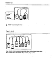
- Figure 1 schematically depicts a first portion of a representative system for producing the ALC compositions of the present invention, which includes bags A-C (Set-1), which are blood storage bags or containers, wherein bag A contains packed red blood cells collected from a donor; bag B contains plasma; and bag C contains leukocytes (which following initial separation from whole blood forms a layer commonly referred to as the buffy coat).
- bags A-C Set-1
- bag A contains packed red blood cells collected from a donor
- bag B contains plasma
- bag C contains leukocytes (which following initial separation from whole blood forms a layer commonly referred to as the buffy coat).
- Figure 2 schematically depicts a second portion of the representative system for producing ALC compositions of the present invention, which includes a 7-bag set after bag A with RBC is removed from the system and bags B and C from figure 1 are welded to bags 1-5 (Set-2).
- Figures 3A and 3B are graphs illustrating the general trends of both CD62L and CD42b expression, which are indicators of cell activation.
- Blood is defined herein as whole blood or any of its constituent parts (e.g ., plasma, leukocytes, platelets or red blood cells).
- the amounts of platelets and red blood cells that may be present in the ALC of the present invention may be lower than that in whole blood.
- the starting materials for producing the inventive ALCs may be obtained from several sources.
- Whole blood or one or more components thereof e.g ., leukocytes and plasma
- the blood sample is collected from the patient who will ultimately be treated with the ALC, which is referred to herein as an autologous blood sample or source.
- the source(s) i.e., the blood or its components
- these starting materials may be conveniently obtained from a blood bank.
- the samples may be screened by the blood bank for blood type (ABO, Rh), irregular antibodies to red cell antigens, and transfusion-transmittable diseases. More specifically, screening can be conducted with antibodies using an Abbott PRISM instrument against: Hepatitis B, C, HIV 1/2, HTLV and Syphilis (-HCV; HbsAg; anti-HIV 1/2 O+; and anti-HTLV I/II).
- the samples can also be screened for HIV, HCV and HBV by molecular methods (NAT-Nucleic Acid Testing). Molecular screening can be accomplished using commercially available instrumentation, e.g., the TIGRIS system of Chiron.
- the samples can be obtained from donors with the same blood type as the intended ALC recipient.
- plasma samples can be obtained from donors with AB+ blood and the leukocytes can be obtained from patients with O- blood.
- Patients with AB+ blood are universal donors for plasma and patients with O- blood are universal donors for leukocytes. Regardless of the source, all necessary processing of the sample(s) can be carried out without the need for highly specialized equipment.
- FIGs. 1 and 2 illustrate a system containing two sets of interconnected sterile infusion bags.
- the system is sealed so that there is no exposure to the outside environment.
- the tubes connecting the two sets are welded together to form one system using a Sterile Connecting Device (e.g ., TSCD®-II Cat number ME-203AH of Terumo).
- a Sterile Connecting Device e.g ., TSCD®-II Cat number ME-203AH of Terumo.
- the welding and cutting of the tubes is done by pre-heating special wafers to about 300°C. This high temperature increases the sterility of the welding procedure.
- the welding may be performed in a class 100 Biological Safety Cabinet within a class 100,000 containment area.
- the system contains two sterile bag sets.
- Set 1 containing bags A, B, and C, is a standard, commercially available triple bag set commonly used for blood transfusion.
- a human blood sample typically in the volume of about 400 to about 550 ml, is collected in a blood bank via venipuncture and placed into bag A, and then fractionated into its component parts using standard techniques into bags A, B and C.
- bag A containing the blood sample is centrifuged. After centrifugation, the blood components are separated, e.g ., using a blood component extractor manufactured by Baxter.
- the buffy coat containing leukocytes is placed into bag C, plasma is placed into bag B and erythrocytes remain in bag A.
- bag A contains packed erythrocytes
- bag B contains plasma
- bag C contains the buffy coat containing leukocytes (and possibly residual plasma and erythrocytes).
- the blood components can be separated from whole blood via apheresis techniques known in the art.
- Bag A is then disconnected from the three-bag set.
- bags B and C are then welded to custom made infusion bags 1-5 (Set-2) to form the system used to make the activated leukocyte composition.
- welding is performed with a sterile connecting device.
- Bag 1 contains a first aqueous solution (e.g ., 200ml of sterile distilled water), which is used for the purpose of exposing the cells in the buffy coat to hypo-osmotic shock. This serves to lyse residual erythrocytes that may be present.
- Bag 2 contains a second solution (e.g ., 20ml of buffered sodium chloride solution (8.91% NaCl, USP), or any other other physiologically acceptable solution containing inorganic ions, organic osmolytes such as sucrose, or some combination thereof, such as Lactated Ringers (Hartmans) solution), which serves to restore the leukocytes to isotonicity following hypo-osmotic shock.
- a second solution e.g ., 20ml of buffered sodium chloride solution (8.91% NaCl, USP), or any other physiologically acceptable solution containing inorganic ions, organic osmolytes such as sucrose, or some combination thereof, such as Lactated Ringers (Hartmans) solution
- the sodium chloride solution is added to 200ml of distilled water, it becomes a 0.9% NaCl solution.
- Bag 3 contains a third solution (e.g ., about 60ml of buffered calcium chloride solution (1.17% CaCl 2 Dihydrate, USP)), which acts to coagulate the plasma in bag B, and to facilitate separation into platelets and serum.
- Bag 4 and bag 5 each contains sterile filtered air, about 60ml and about 500ml respectively. The set is packed as a single unit and sterilized using high pressure steam, which greatly reduces the risk of secondary infection to the patient.
- cell activation is defined as a process by which the cells (leukocytes) are allowed to become activated, and more specifically, as a transition from a quiescent to a functionally active state which is accompanied by synthesis of biologically active substances or translocation of pre-synthesized substances from cytoplasm to the cellular membrane or their release out of the cells.
- These substances may include proteins or polypeptides, lipids, sugars, oxygen radicals and other biochemical moieties that function as adhesion molecules, cytokines, growth factors, enzymes, transcription factors and cell signaling receptors and mediators. When detected and identified inside the cells or on the cell surface, these molecules are called activation markers.
- the leukocytes are incubated simply by allowing them to stand at room temperature.
- room temperature refers to a temperature in the range of about 12°C to about 28°C, and in some embodiments from about 16°C to about 25°C.
- the time period of incubation may vary depending upon the temperature. Incubation times will be lower at increased temperatures.
- the incubation time needed to activate the leukocytes will be roughly inversely proportional to the temperature at which the incubation is conducted. For example, in embodiments where leukocytes are allowed to stand at room temperature, the incubation time generally ranges from about 90 minutes, and upwards of 2, 3, 4, 5, 6, 7, or 8 to about 20 hours.
- the incubation time ranges from about 8 to about 20 hours. In a more preferred embodiment, incubation of the leukocytes occurs at about 18°C to about 24°C for about 8 hours to about 12 hours. In other embodiments, incubation of the leukocytes involves exposing them to heat, e.g., at a temperature above room temperature and up to about 37°C. The time period for incubation at elevated temperatures generally ranges from 5 hours to about 24 hours.
- bag 4 which is typically smaller than bag C (e.g. about 250ml - about 500ml). Also, cells can be transferred into another 500ml bag (bag 5). To allow for activation of the leukocytes, bag 4 (or bag 5) is placed in a vertical position and incubated, for about 8 to about 20 hours at room temperature.
- hypo-osmotic shock which lyses erythrocytes.
- hypo-osmotic shock is performed immediately ( i.e ., upon completion of the preceding step without any intervening step or unnecessary delay, typically less than 2 minutes).
- the hypo-osmotic shock may be initiated by transferring the distilled water from bag 1 to bag 4 (or bag 5) containing the leukocytes.
- the hypo-osmotic shock treatment is typically conducted for about 45 seconds.
- isotonicity is restored to the leukocytes by transferring the sodium chloride solution from bag 2 to bag 4 (or bag 5).
- the ratio of the volume of sodium chloride solution to cell suspension in water is generally about 1:10.
- the contents of bag 4 are then transferred into Bag 1 (or left in bag 5), which is larger in volume than bag 4.
- this composition may be used therapeutically in the inventive methods.
- the preferred embodiment involves at least one additional step.
- plasma is separated into platelets and serum by the use of a coagulant such as CaCl 2 , followed by centrifugation
- CaCl 2 from bag 3 is transferred to bag B.
- Bag B which now contains a composition of plasma and CaCl 2 . is typically allowed to coagulate at a temperature of about 37°C.
- the plasma remains in contact with the coagulating agent for substantially the same period of time the leukocytes are incubated.
- the entire 7-bag system is centrifuged.
- the centrifugation is conducted immediately following transfer of the leukocytes into bag 1 (or remain in bag 5), in order not to expose cells to hemolysate
- the supernatant from bag 1 (or bag 5) is transferred into bag C, and the leukocyte pellet formed in bag 1 (or bag 5) as a result of the centrifugation is re-suspended in about 20ml to about 50ml of serum from bag B.
- the activated leukocytes may be incubated in the coagulated plasma mentioned above before the final composition is made. Generally, this second incubation period is conducted for about 1-2 hours at 37°C.
- the ALC composition may be prepared from smaller volumes of blood samples, with commensurate decreases in volumes of all solutions and use of smaller bags. Furthermore, use of these different size bags yield ALCs with different compositions. Even in these embodiments, allogeneic or autologous blood samples may be used as starting materials. Use of smaller volumes provides the clinician with the ability to perform the blood collection autonomously, without using an external blood bank, such as in emergency situations in treating patients with otherwise healthy immune systems but suffering from some type of traumatic wound ( e.g ., battlefield and combat conditions). In these embodiments, testing for transmittable diseases and antigens may be dispensed with. However in such cases, patients with refractory wounds are not clinically acceptable blood donors for effective ALC preparation. When this situation arises, ALC will be produced from allogeneic donors by the means described herein.
- the ALCs of the present invention include leukocytes, e.g ., granulocytes, monocytes and lymphocytes.
- Granulocytes include neutrofils, eosinophils and basofils.
- the leukocyte population of the ALC generally contains about 40% to about 90% granulocytes, about 5% to about 20% monocytes and about 5% to about 30% lymphocytes. Specific amounts of the cells may differ based on the analysis techniques employed.
- the leukocyte composition When analysis is performed using FACS (e.g ., using a side-scatter versus a forward-scatter dot plot analysis), the leukocyte composition generally contains about 55% to about 80% granulocytes; about 5% to about 15% monocytes and about 5% to about 30% lymphocytes, and in some embodiments, comprises about 58-76% granulocytes; about 5-11% monocytes and about 9-23% lymphocytes.
- the leukocyte composition When analysis is performed using a Cell Dyn Analyzer, the leukocyte composition generally contains about 50% to about 90% granulocytes; about 5% to about 15% monocytes; and about 10% to about 25% lymphocytes.
- the subpopulation of lymphocytes in the ALC may confirm the following cells in the general ranges as follows: about 7% to about 25% B cells (CD19+); about 20% to about 30% NK cells (CD3-/CD56+), about 40% to about 60% T cells (CD3+); about 0.1% to about 30% of NKT cells CD3+/CD56+, about 8% to about 20% of T helper cells (CD4+/CD3+), and about 20% to about 30% of CD8+/CD3+ cells.
- the lymphocyte subpopulation is enriched with at least 9% CD56+ cells (CD3-/CD56+;CD3+/CD56+;CD3+/CD56+/CD8+), the amount of the T helper lymphocytes (CD4+/CD3+) is decreased to less than 20%, and/or the ratio of T-helper to T-suppressor cells (CD4+/CD3+:CD8+/CD3+) is less than 0.8.
- Patients suffering from wounds can be physiologically compromised or otherwise healthy.
- diabetics and other medically compromised patients are candidates for ALC derived from heterologous blood, as their own leukocytes may not be optimal for the procedure.
- otherwise healthy patients as in the example of trauma patients, are also good candidates for ALC compositions of the present invention.
- the present invention is useful in promoting healing in a multitude of wound types. Although in practice it may be used in combination with other treatment modalities, it does not require them to achieve effective wound healing.
- the inventors have contemplated application of the ALC to any type of wound and foresee no limitations as to the type of wound that can be treated. The ease of application, e.g., with a standard syringe or similar application device, makes the inventive ALC compositions safe and easy to use.
- Wounds amenable to treatment with the invention are typically in the form of punctures, cuts or tears of the living tissues. Wounds of the skin can penetrate the epidermis, dermis or in the case of full-thickness wounds, the subcutaneous tissue.
- representative types of wounds amenable to treatment with the compositions and methods of the present invention include decubital or pressure ulcers; diabetic ulcers, deep sternal wounds, e.g ., following open heart surgery (to the great saphenous vein after coronary revascularization and harvesting of the great saphenous vein); and post-operative wounds following abdominal and any other types of surgery.
- wounds are those which result from trauma such as by gun shots, knives, or any other object able to cause a cut or tear in the skin.
- Wounds of the oral cavity e.g ., teeth
- wounds that arise as a side-effect of medication or as a symptom of various pathologies e.g ., sores associated with Kaposi's Sarcoma
- internal wounds e.g . anal fissures, and wounds or lesions to the gastrointestinal tract, such as ulcers in the stomach or intestines
- the ALC may also be used to treat any wounds exacerbated by vascular insufficiency.
- Vascular insufficiency refers to inadequate blood circulation resulting in insufficient perfusion to the afflicted areas.
- Such insufficiency can be caused by trauma (e.g . damage to the vasculature adjacent to a skeletal fracture), or various pathologies ( e.g . diabetes and atherosclerosis). In either instance, whether trauma or disease induced, vascular insufficiency decreases the likelihood of effective wound healing.
- the ALC may be useful in improving wound healing outcomes in these patients and should be administered according to the methods described herein. Additionally, treatment algorithms should not be limited by the severity or type of wound, or the extent of vascular insufficiency. ALC may be more efficacious in patients presenting with the most severe wounds and vascular insufficiency.
- application of the activated leukocyte composition is accomplished by means of one or more injections of the ALC directly into the wound or the tissue surrounding the wound.
- the ALC may be applied directly into an open wound.
- Luer-Lock syringe For injection into the wound, it is preferred to use a Luer-Lock syringe or any other commercially available syringe that has a locking mechanism between the syringe and the needle.
- the biological space of a wound, particularly a pressure wound, is often limited. When injecting into a wound, there is a risk of pressure causing the syringe to separate from the needle. Using a locking syringe eliminates this risk.
- the ALC can be applied directly into the cavity of the wound.
- Application in this method can be done using direct application with a syringe or tubing.
- the ALC may be applied to or around the wound site with the aid of a dressing.
- Dry dressings include gauze and bandages, non-adhesive meshes, membranes and foils, foams, and tissue adhesives.
- Moisture-keeping barrier dressings include pastes, creams and ointments, nonpermeable or semi-permeable membranes or foils, hydrocolloids, hydrogels, and combination products.
- Bioactive dressings include antimicrobial dressings, interactive dressings, single-component biologic dressings, and combination products.
- the wound is packed with sterile gauze soaked in the ALC.
- the dressing e.g., such as sterile gauze pads
- the dressing may be saturated with compositions such as Lactated Ringer (Hartman) Solution, alginate containing dressing, polyurethane dressing or carboxymethylcellulose dressing, which is applied to cover the wound, followed by application of dry dressing. If the subject wound is highly infected, then silver dressings such as Silverlon can be applied.
- the choice of post-injection dressing is based on the determination of the clinician. Commercial availability, history of past clinical success, and patient tolerance are all factors to be considered in the selection of a wound dressing.
- the dressing may be removed periodically, e.g ., typically after about 24 hours, in order to irrigate the wound e.g ., with sterile water and soap.
- the composition can be placed onto a physiologically inert and/or resorbable matrix or scaffold (e.g ., collagen) and inserted by means of a press fit, into the wound.
- a physiologically inert and/or resorbable matrix or scaffold e.g ., collagen
- the ALC compositions may be applied to the wound once or more than once, e.g. , after 4 weeks, once a clinician determines whether another application is necessary. Factors that may be taken into account include increased wound dimensions (width, length and depth), suppuration, pyrexia or any other sign or symptom indicating a recalcitrant infection such that re-treatment is warrranted. In addition to re-treatment, referral for surgical debridement may be indicated at any point the clinician deems appropriate.
- the ALC may be used in conjunction with any other conventional wound treatment, such as warming (therapeutic heat), electrical stimulation, magnetism, laser phototherapy, cycloidal vibration therapy and ultrasound. It also can be used with biological therapy such as larva therapy, skin substitutes, cultured keratinocytes (Epicel, Genzyme biosurgery), human dermal replacement (Dermagraft, Smith and Nephew Inc.), cadaver derived processed dermis (Alloderm, Life Cell Corporation), Bilayered Skin Equivalent (Apligraf, Organogenesis Inc.), TransCyte (Smith and Nephew Inc.), Growth Factors (PDGF is currently the only growth factor licensed for topical use), and fibrin sealant.
- warming therapeutic heat
- electrical stimulation magnetism
- laser phototherapy laser phototherapy
- cycloidal vibration therapy and ultrasound.
- biological therapy such as larva therapy, skin substitutes, cultured keratinocytes (Epicel, Genzyme biosurgery), human dermal replacement (Dermagraft, Smith and Nephe
- the ALC is used in conjunction with VAC, which is a commercially available wound therapy manufactured by KCI. VAC promotes wound healing by applying negative pressure to a wound.
- VAC promotes wound healing by applying negative pressure to a wound.
- ALC is preferably applied to a wound prior to VAC therapy.
- the ALC is used in conjunction with hyperbaric therapy (Thackham, 2008).
- the ALC can be applied to a wound just prior to a patient receiving hyperbaric therapy.
- the ALC may also be used in conjunction with low-energy shock wave therapy (e.g ., impulses of about 0.1 mJ/mm 2 ; 5 Hz) per centimeter of wound length). See, e.g ., Dumfarth, et al., Ann. Thorac. Surg. 86:1909-13 (2008 ).
- the wounds may be evaluated for length, width and height measurements. Typically, a wound is considered healed when all measurements of these parameters are negligible.
- the ALC may also provide an analgesic effect.
- the activated leukocyte composition is particularly useful in wounds including diabetic foot ulcers and decubital ulcers.
- Decubital ulcers are pressure ulcers caused by impeded blood flow, usually due to prolonged pressure on a particular area. (Berlowitz, 2007) Decubital ulcers cause morbidity and mortality in elderly people. At least 48% of stage IV pressure ulcers remain unhealed after one year of treatment. (Girouard, 2008).
- Patients suffering from decubiti also commonly have co-morbid pathologies such as diabetes and hypertension. These pathologies further complicate the successful treatment of decubiti.
- the composition is aspirated into a sterile syringe of any size, using an 18-gauge (18G) needle. Aspiration is performed slowly to minimize damage to the cells. While the size of the syringe and needle are by no means limiting, a large gauge needle is preferred for aspiration. This facilitates the transfer and reduces cell damage.
- 18G 18-gauge
- Application of the activated leukocyte composition to the ulcer comprises injecting the composition into the wound.
- the entire sample in the syringe can be deployed and the clinician can choose to administer additional ALC if it is determined to be necessary based on clinical parameters.
- the 18G needle used for aspiration is exchanged with a needle ranging in size from 22-35G.
- the ALC may be injected into the wound in various locations. Injection can occur about every one centimeter to about every three centimeters for the entire length of the wound. At each injection site, 0.1-0.3 ml of ALC is injected.
- the entire syringe can be injected at one time into a single site within the wound.
- An activated leukocyte composition made in accordance with the preferred embodiment of the present invention was quantified by the analyis of various cell surface markers.
- An increase in platelet aggregation with either monocytes or granulocytes is a sign of activation of the monocytes and granulocytes through the expression of P selectin.
- CD62L is a plasma membrane protein which is shed during activation and thus decreases with cell activation.
- CD42b is a platelet activation marker involved in the process of coagulation as an aggregating factor. It interacts with extra-cellular matrix as well as with adhesion molecules and also used in the present invention as an indicator of monocyte and granulocyte activation.
- Fr.BC was taken immediately after the preparation of Buffy Coat. IBC was taken after the first incubation period and FP was taken from the Final Product. At each time point, cells were labeled with specific monoclonal antibodies (allophycocyanin (APC) conjugated anti-CD14, phycoerythrin(PE) conjugated anti-CD42b, and fluorescein-isothiocyanate (FITC) conjugated anti-CD62L antibodies), and then analyzed by FACS.
- APC allophycocyanin conjugated anti-CD14
- PE phycoerythrin(PE) conjugated anti-CD42b
- FITC fluorescein-isothiocyanate
- FACS staining solution PBS, 2% Normal Mouse Serum; 0.02% Sodium Azide
- 0.5x(10 6 / ⁇ l) cells were incubated with appropriate monoclonal antibodies for 30 min. at room temperature (RT) or at +4°C in the dark.
- RT room temperature
- +4°C room temperature
- the cell composition was treated with erythrocyte lysis buffer, washed, and then finally re-susupended in PBS for FACS acquisition.
- FITC anti-human CD14
- the cell composition was incubated with anti-human-CD14 (APC) and anti-human CD42b (PE) at room temperature (RT). Negative control cell composition was incubated with anti-human-CD14 and appropriate isotype controls under the relevant conditions.
- Cell-associated light scatter and fluorescence were analyzed using a FACS CALIBUR (Becton Dickinson). Monocytes were determined as CD14 positive cells and granulocytes were identified by their characteristic light scatter properties.
- the CD62L or CD42b positive cells were defined as the percentage of monocytes and granulocytes with fluorescence greater than a threshold, as determined by monocytes and granulocytes incubated with appropriate isotype control antibody.
- Tables 2 and 3 depict the cellular compositions of the final ALC as determined by analysis with a Cell Dyn analyzer. Cells were analyzed after re-suspending the activated leukocytes in serum. Viable cells were stained using trypan blue exclusion and observed under a microscope.
- Table 2 Composition of Activated Leukocyte Composition Platelets (10 3 / ⁇ l) Erythrocytes (10 6 / ⁇ l) Leukocytes (10 3 / ⁇ l) Concentration in final ALC 46.8 0.1 6.8 Standard Deviation 39.2 0.06 3.8
- Table 3 Leukocyte Composition in ALC Leukocytes Granulocytes Neutrophills % Basophilis % Eosinophilsophills% Monocytes % Lymphocytes % % in final ALC 65.5 1.6 4.6 9.1 18.5 Standard Deviation 8.2 0.3 3 2.1 4.1 Range 52-78 1-2 1-9 6-12 13-24
- the Cell Dyn results show the ALC contained platelets in the amount of 46.8+/- 39.2 (103/ ⁇ l), erythrocytes in the amount of 0.1 +/-.03(106/ ⁇ l) and leukocytes in the amount of 6.8 +/- 3.8(103/ ⁇ l). Based on the Cell Dyn analysis, it was also determined that the leukocyte composition of the ALC (as shown in Table 3) contained 52%-78% neutrophils, 1-2% basophils, 1-9% eosinophils, 6%-12% monocytes, and 13%-24% lymphocytes, taking into account standard deviation. The ranges depicted in these tables represent the high and low results after 8 separate analyses using the Cell Dyn analyzer.
- the Buffy coat was transferred from Bag C to Bag 4 and incubated for 12 hours ⁇ 2 hours at room temperature. This step was followed by transferring the distilled water from Bag 1 to Bag 4 (or bag 5), for purposes of conducting hypo-osmotic shock treatment (resulting in the production of a hemolysate). This treatment was conducted for approximately 45 seconds. Immediately thereafter, the buffered sodium chloride solution contained in Bag 2 was transferred to Bag 4 (or bag 5) for purposes of restoring isotonicity to the Buffy coat (leukocytes).
- the buffered calcium chloride solution in Bag 3 was transferred to Bag B containing the plasma portion, for purposes of allowing for coagulation of the plasma. This was allowed to take place over the course of 12 hours ⁇ 2 hours, plus the additional short time during which the leukocytes were subjected to hypo-osmotic shock and then restoration of isotonicity.
- the entire bag assembly was subjected to centrifugation (typically about 8 to about 10 minutes), followed by separation of the cells from the hemolysate. In so doing, the cells were exposed to hemolysate influence for a minimal period of time, i.e., approximately 10 minutes. Following centrifugation, the supernatant of the hemolysate in Bag 1 (or bag 5) was discarded, and fresh medium was added to Bag 1 (or bag 5), followed by incubation for about 1-2 hours at 37°C. Following incubation, the cells were subjected to a single wash step.
- centrifugation typically about 8 to about 10 minutes
- the Buffy coat was subjected to hypo-osmotic shock immediately after processing the whole blood and separating it into its 3 main components, i.e., red blood cells, plasma, and the Buffy coat.
- Danon's procedure does not entail incubation of the Buffy coat after separation of the main blood components and prior to subjecting the Buffy coat to hypo-osmotic shock.
- the hypo-osmotic shock was conducted while the Buffy coat was contained in Bag PB 3 .
- the buffered calcium chloride solution was transferred from Bag PB 6 to Bag PB 2 which contains the plasma, followed by deep-freezing the plasma in PB 2 for 10 minutes, and then placing it in a water bath at 37°C for an additional 30 minute incubation period. During this time, the Buffy coat fraction which had been subjected to hypo-osmotic shock was allowed to stand wherein the leukocytes remained exposed to the hemolysate.
- the entire bag system was subject to centrifugation, and the hemolysate was discarded.
- the cells were exposed to the hemolysate for a period of at least about 55 minutes (i.e., which included the 40-minute standing period that coincided with the plasma coagulation, and an additional 15 minutes for centrifugation).
- the leukocytes were centrifuged immediately for about 5 min and thus were exposed to hemolysate for much less time, i.e., less than 10 minutes.
- the present method results in a yield of at least about 100 x 10 6 , 125 x 10 6 , 150 x 10 6 , 175 x 10 6 , 200 x 10 6 , 225 x 10 6 , 250 x 10 6 , 275 x 10 6 , 300 x 10 6 , 325 x 10 6 , 350 x 10 6 , 375 x 10 6 , 400 x 10 6 , 425 x 10 6 , 450 x 10 6 , 475 x 10 6 , 500 x 10 6 , or higher leukocytes per standard blood unit (including all sub-ranges thereof).
- the number of viable cells contained within the total cell population was measured by the Trypan Blue exclusion method.
- the cells were counted in a Newbauer hamemocytometer after suspension in Trypan Blue (1:1 ratio) for evaluation of cell count and percentage of viability. The results are presented in Table 5.
- the inventive ALC may contain at least 80%, 81%, 82%, 83%, 84%, 85%, 86%, 87%, 88%, 89%, 90%, 91%, 92%, 93%, 94%, 95%, 96%, 97%, 98% or greater, viable leukocytes based on this total number of leukocyte cells in the ALC (including all sub-ranges thereof).
- CD11b activation marker on granulocytes was measured by flow cytometry, and presented as a percentage of CD11b positive cells in granulocyte population (CD15 positive cells).
- Cells sampled from final product were co-stained with anti-CD11b conjugated to fluorescein isothiocynate (FITC) and anti-CD15 conjugated to phycoerythrin (PE), and analyzed using FACSCalibur flow cytometer (Becton Dickinson Immunocytometry Systems, San Jose, CA, USA).
- FITC fluorescein isothiocynate
- PE phycoerythrin
- the ALCs of the present invention may contain at least 50%, 51%, 52%, 53%, 54%, 55%, 56%, 57%, 58%, 59%, 60%, 61%, 62%, 63%, 64%, 65%, 66%, 67%, 68%, 69%, 70%, 71%, 72%, 73%, 74%, 75%, 76%, 77%, 78%, 79%, 80%, 81%, 82%, 83%, 84%, 85%, or higher, CD11b(+) granulocytes, relative to the total granulocyte population in the ALC (including all sub-ranges thereof).
Description
- The present application is related to Application Serial No.
61/209,298, filed March 5, 2009 61/211,587, filed April 1, 2009 - The wound healing process involves participation of white blood cells, also known as leukocytes. Leukocytes include lymphocytes, granulocytes and monocytes. Three common types of lymphocytes are T-cells, B-cells and natural killer cells. T-cells and B-cells play important roles in the recognition of antigens in the body (Parkin, 2001). Natural killer (NK) cells identify infected cells by alterations in the levels of the major histocompatability complex (MHC), and destroy the infected cells (Moretta, 2008). The participation of lymphocytes in the healing process is largely associated with their production of cytokines and growth factors (Keen, 2008). A new class of gamma-delta-T cells has been described in the skin (Jameson, 2002. Havran, 2005). Among the different types of granulocytes are neutrophils, basophils and eosinophils. Monocytes differentiate into macrophages, which are responsible for destruction of tissue debris or invading foreign substances. Macrophages also produce molecules that control inflammation and repair (Riches, 1996).
- The process of wound healing occurs in three overlapping phases. (Li, 2007; Broughton, 2006; Tsirogianni, 2006; Singer, 1999; Martin, 1997). The first phase is the inflammatory phase. It is characterized by recruitment of neutrophils, followed by monocytes to the wound site, where they kill and phagocytize bacteria (Agaiby, 1999).
- The second wound healing phase which is known as the proliferative phase, involves formation of new granulation tissue. Fibroblasts proliferate and migrate into the wound space and synthesize collagen and other components of extracellular matrix (Greiling, 1997). At the same time, angiogenesis occurs, providing nutrients and oxygen to the metabolically active new granulation tissue (Tonnesen, 2000). Keratinocytes from the intact epidermis start to migrate over the provisional matrix and begin to proliferate, leading the way for new epithelial tissue (Kim, 1992).
- Remodeling is the third and final phase in wound healing. It is characterized by fibroblast differentiation into myofibroblasts, which contract and bring the wound edges closer together (Tomasek, 2002). Remodeling of the collagen fibers by degradation and re-synthesis allows the wound to gain strength by re-orientation of the collagen fibers (a process tightly controlled by growth factors) (Werner, 2003).
- The challenge of treating wounds is often compounded by patients with multiple pathologies such as diabetes, coronary artery disease and hypertension. These diseases have the common effect of exacerbating vascular complications due to various physiological conditions. Complications from wounds may result in increased morbidity and mortality (Doshi, 2008).
- Conventional wound treatments include surgical debridement, antibiotic therapies and various dressings (Moran, 2008; Fonder, 2008). Wounds resistant to conventional treatment are also referred to as refractory wounds. These wounds lead to a decrease in quality of life and can result in increased morbidity and mortality. Thus, a need continues to exist for effective wound healing compositions and methods.
- The present invention relates to a method for making an activated leukocyte composition comprising:
- a. incubating human leukocytes at a (i) temperature of 12° C - 28°C for a time ranging from about 90 minutes upwards of about 2, 3, 4, 5, 6, 7, 8, or 12 to 20 hours or (ii) temperature of 12°C - 37°C for a time ranging from 5-24 hours so that the leukocytes transition from a quiescent to a functionally active state;
- b. subjecting the leukocytes to hypo-osmotic shock; and
- c. adding to the leukocytes of step b, a physiologically acceptable salt solution in an amount effective to restore isotonicity.
- Another aspect of the present invention is directed to an ALC derived from blood. The activated leukocyte composition of the present invention includes, in terms of the population of leukocytes present therein, about 40% to about 90% granulocytes, about 5% to about 20% monocytes and about 5% to about 30% lymphocytes, based on the total number of leukocytes in the ALC. As shown in the working examples, the inventive ALCs may also be characterized and distinguished from known compositions in terms of minimum yield of leukocytes (relative to the whole blood sample), viability of leukocytes, and minimum activation levels of granulocytes, e.g., as indicated by CD11b. The ALC may further contain residual levels of platelets (in amounts of about 46.8+/- 39.2(103/µl) and red blood cells (in the amount of about 0.1 +/-0.06(106/µl) of the ALC. The population of lymphocytes may include about 7% to about 25% B cells (CD19+), about 20% to about 30% NK cells (CD3-/CD56+), about 40% to about 60% T cells (CD3+), about 0.1% to about 30% of NKT cells CD3+/CD56+, about 8% to about 20% of T helper cells (CD4+/CD3+), and about 20% to about 30% of CD8+/CD3+ cells. The cells may be suspended in a carrier such as serum (which may be autologous or allogeneic with respect to recipient) or some other physiologically acceptable iso-normal liquid suitable for storing and administering cells, such as the solution used to restore isotonicity.
- The present invention can be used for promoting wound healing, by administering or otherwise applying the ALC to a wound.
- The disclosed invention achieves several unexpected results compared to at least one known wound healing composition containing white blood cells. As demonstrated in working examples herein, these results include increased yield and viability of leukocytes (WBCs), and higher percentage of activated granulocytes. The disclosed invention is also believed to include an unexpectedly higher percentage of activated monocytes and a relatively higher percentage of CD4 T-cells compared to CD8 T-cells.
-
Figure 1 schematically depicts a first portion of a representative system for producing the ALC compositions of the present invention, which includes bags A-C (Set-1), which are blood storage bags or containers, wherein bag A contains packed red blood cells collected from a donor; bag B contains plasma; and bag C contains leukocytes (which following initial separation from whole blood forms a layer commonly referred to as the buffy coat). -
Figure 2 schematically depicts a second portion of the representative system for producing ALC compositions of the present invention, which includes a 7-bag set after bag A with RBC is removed from the system and bags B and C fromfigure 1 are welded to bags 1-5 (Set-2). -
Figures 3A and 3B are graphs illustrating the general trends of both CD62L and CD42b expression, which are indicators of cell activation. - Blood is defined herein as whole blood or any of its constituent parts (e.g., plasma, leukocytes, platelets or red blood cells). The amounts of platelets and red blood cells that may be present in the ALC of the present invention may be lower than that in whole blood.
- The term "about" as used herein in connection with any and all values (including lower and upper ends of numerical ranges) as any value having an acceptable range of deviation of +/- 0.5% to +/- 20% (and values therebetween, e.g., ± 1%, ± 1.5%, ± 2%, ± 2.5%, ± 3%, ± 3.5%, ± 4%, ± 4.5%, ± 5%, ± 5.5%, ± 6%, ± 6.5%, ± 7%, ± 7.5%, ± 8%, ± 8.5%, ± 9%, ± 9.5%, ± 10%, ± 10.5 %, ± 11%, ± 11.5%, ± 12%, ± 12.5%, ± 13, ± 13.5%, ± 14%, ± 14.5%, ± 15%, ± 15.5%, ± 16%, ± 16.5%, ± 17%, ± 17.5%, ± 18%, ± 18.5%, ± 19%, ± 19.5%, and ± 20%).
- The starting materials for producing the inventive ALCs may be obtained from several sources. Whole blood or one or more components thereof (e.g., leukocytes and plasma) may be obtained from autologous or allogeneic sources. In one embodiment of the present invention, the blood sample is collected from the patient who will ultimately be treated with the ALC, which is referred to herein as an autologous blood sample or source. In embodiments wherein the source(s) i.e., the blood or its components, is obtained from an individual other than the intended ALC recipient, which is referred to as an allogeneic blood sample or source, these starting materials may be conveniently obtained from a blood bank. The samples may be screened by the blood bank for blood type (ABO, Rh), irregular antibodies to red cell antigens, and transfusion-transmittable diseases. More specifically, screening can be conducted with antibodies using an Abbott PRISM instrument against: Hepatitis B, C,
HIV 1/2, HTLV and Syphilis (-HCV; HbsAg;anti-HIV 1/2 O+; and anti-HTLV I/II). The samples can also be screened for HIV, HCV and HBV by molecular methods (NAT-Nucleic Acid Testing). Molecular screening can be accomplished using commercially available instrumentation, e.g., the TIGRIS system of Chiron. - In these embodiments involving allogeneic sources, the samples can be obtained from donors with the same blood type as the intended ALC recipient. Alternatively and as further described herein, plasma samples can be obtained from donors with AB+ blood and the leukocytes can be obtained from patients with O- blood. Patients with AB+ blood are universal donors for plasma and patients with O- blood are universal donors for leukocytes. Regardless of the source, all necessary processing of the sample(s) can be carried out without the need for highly specialized equipment.
- A preferred method of making the ALC composition of the present invention is now described with reference to
Figs. 1 and 2 , which illustrate a system containing two sets of interconnected sterile infusion bags. The system is sealed so that there is no exposure to the outside environment. Specifically, the tubes connecting the two sets are welded together to form one system using a Sterile Connecting Device (e.g., TSCD®-II Cat number ME-203AH of Terumo). More specifically, to ensure compliance with sterility standards, the welding and cutting of the tubes is done by pre-heating special wafers to about 300°C. This high temperature increases the sterility of the welding procedure. To further ensure sterility, the welding may be performed in aclass 100 Biological Safety Cabinet within a class 100,000 containment area. - As illustrated in these figures, the system contains two sterile bag sets.
Set 1, containing bags A, B, and C, is a standard, commercially available triple bag set commonly used for blood transfusion. A human blood sample, typically in the volume of about 400 to about 550 ml, is collected in a blood bank via venipuncture and placed into bag A, and then fractionated into its component parts using standard techniques into bags A, B and C. For example, bag A containing the blood sample is centrifuged. After centrifugation, the blood components are separated, e.g., using a blood component extractor manufactured by Baxter. The buffy coat containing leukocytes is placed into bag C, plasma is placed into bag B and erythrocytes remain in bag A. Thus, as a result of this process; bag A contains packed erythrocytes; bag B contains plasma; and bag C contains the buffy coat containing leukocytes (and possibly residual plasma and erythrocytes). Alternatively, the blood components can be separated from whole blood via apheresis techniques known in the art. - Bag A is then disconnected from the three-bag set. As illustrated in
Fig. 2 , bags B and C are then welded to custom made infusion bags 1-5 (Set-2) to form the system used to make the activated leukocyte composition. As disclosed above, welding is performed with a sterile connecting device.Bag 1 contains a first aqueous solution (e.g., 200ml of sterile distilled water), which is used for the purpose of exposing the cells in the buffy coat to hypo-osmotic shock. This serves to lyse residual erythrocytes that may be present.Bag 2 contains a second solution (e.g., 20ml of buffered sodium chloride solution (8.91% NaCl, USP), or any other other physiologically acceptable solution containing inorganic ions, organic osmolytes such as sucrose, or some combination thereof, such as Lactated Ringers (Hartmans) solution), which serves to restore the leukocytes to isotonicity following hypo-osmotic shock. When the sodium chloride solution is added to 200ml of distilled water, it becomes a 0.9% NaCl solution. Bag 3 contains a third solution (e.g., about 60ml of buffered calcium chloride solution (1.17% CaCl2 Dihydrate, USP)), which acts to coagulate the plasma in bag B, and to facilitate separation into platelets and serum. Bag 4 and bag 5 each contains sterile filtered air, about 60ml and about 500ml respectively. The set is packed as a single unit and sterilized using high pressure steam, which greatly reduces the risk of secondary infection to the patient. - The leukocytes are then transferred from bag C into bag 4 (or bag 5) and incubated, preferably in a vertical position, to allow them to activate. For purposes of the present invention, cell activation is defined as a process by which the cells (leukocytes) are allowed to become activated, and more specifically, as a transition from a quiescent to a functionally active state which is accompanied by synthesis of biologically active substances or translocation of pre-synthesized substances from cytoplasm to the cellular membrane or their release out of the cells. These substances may include proteins or polypeptides, lipids, sugars, oxygen radicals and other biochemical moieties that function as adhesion molecules, cytokines, growth factors, enzymes, transcription factors and cell signaling receptors and mediators. When detected and identified inside the cells or on the cell surface, these molecules are called activation markers.
- In some embodiments, the leukocytes are incubated simply by allowing them to stand at room temperature. For purposes of the present invention, room temperature refers to a temperature in the range of about 12°C to about 28°C, and in some embodiments from about 16°C to about 25°C. The time period of incubation may vary depending upon the temperature. Incubation times will be lower at increased temperatures. The incubation time needed to activate the leukocytes will be roughly inversely proportional to the temperature at which the incubation is conducted. For example, in embodiments where leukocytes are allowed to stand at room temperature, the incubation time generally ranges from about 90 minutes, and upwards of 2, 3, 4, 5, 6, 7, or 8 to about 20 hours. In preferred embodiments, the incubation time ranges from about 8 to about 20 hours. In a more preferred embodiment, incubation of the leukocytes occurs at about 18°C to about 24°C for about 8 hours to about 12 hours. In other embodiments, incubation of the leukocytes involves exposing them to heat, e.g., at a temperature above room temperature and up to about 37°C. The time period for incubation at elevated temperatures generally ranges from 5 hours to about 24 hours.
- Referring again to
Fig. 2 , the leukocytes in bag C are transferred to bag 4 which is typically smaller than bag C (e.g. about 250ml - about 500ml). Also, cells can be transferred into another 500ml bag (bag 5). To allow for activation of the leukocytes, bag 4 (or bag 5) is placed in a vertical position and incubated, for about 8 to about 20 hours at room temperature. - After incubation, the leukocytes are subjected to hypo-osmotic shock, which lyses erythrocytes. In preferred embodiments, hypo-osmotic shock is performed immediately (i.e., upon completion of the preceding step without any intervening step or unnecessary delay, typically less than 2 minutes). The hypo-osmotic shock may be initiated by transferring the distilled water from
bag 1 to bag 4 (or bag 5) containing the leukocytes. The hypo-osmotic shock treatment is typically conducted for about 45 seconds. Following this step, and preferably immediately thereafter, isotonicity is restored to the leukocytes by transferring the sodium chloride solution frombag 2 to bag 4 (or bag 5). The ratio of the volume of sodium chloride solution to cell suspension in water is generally about 1:10. The contents of bag 4 are then transferred into Bag 1 (or left in bag 5), which is larger in volume than bag 4. As described above, this composition may be used therapeutically in the inventive methods. - The preferred embodiment involves at least one additional step. Thus, in a separate step, which may be conducted concurrently with the leukocyte incubation, plasma is separated into platelets and serum by the use of a coagulant such as CaCl2, followed by centrifugation Thus, in this representative embodiment, CaCl2 from bag 3 is transferred to bag B. Bag B, which now contains a composition of plasma and CaCl2. is typically allowed to coagulate at a temperature of about 37°C. The plasma remains in contact with the coagulating agent for substantially the same period of time the leukocytes are incubated.
- After hypo-osmotic shock and restoration of isotonicity of the leukocytes, and subsequent transfer of the contents of bag 4 into bag 1 (or remain in bag 5, and after coagulation of plasma in bag B, the entire 7-bag system is centrifuged. In preferred embodiments, the centrifugation is conducted immediately following transfer of the leukocytes into bag 1 (or remain in bag 5), in order not to expose cells to hemolysate After centrifugation, the supernatant from bag 1 (or bag 5) is transferred into bag C, and the leukocyte pellet formed in bag 1 (or bag 5) as a result of the centrifugation is re-suspended in about 20ml to about 50ml of serum from bag B.
- In yet another step of the preferred embodiment of the present invention, the activated leukocytes may be incubated in the coagulated plasma mentioned above before the final composition is made. Generally, this second incubation period is conducted for about 1-2 hours at 37°C.
- In other embodiments, the ALC composition may be prepared from smaller volumes of blood samples, with commensurate decreases in volumes of all solutions and use of smaller bags. Furthermore, use of these different size bags yield ALCs with different compositions. Even in these embodiments, allogeneic or autologous blood samples may be used as starting materials. Use of smaller volumes provides the clinician with the ability to perform the blood collection autonomously, without using an external blood bank, such as in emergency situations in treating patients with otherwise healthy immune systems but suffering from some type of traumatic wound (e.g., battlefield and combat conditions). In these embodiments, testing for transmittable diseases and antigens may be dispensed with. However in such cases, patients with refractory wounds are not clinically acceptable blood donors for effective ALC preparation. When this situation arises, ALC will be produced from allogeneic donors by the means described herein.
- The ALCs of the present invention include leukocytes, e.g., granulocytes, monocytes and lymphocytes. Granulocytes include neutrofils, eosinophils and basofils. In its broadest sense, the leukocyte population of the ALC generally contains about 40% to about 90% granulocytes, about 5% to about 20% monocytes and about 5% to about 30% lymphocytes. Specific amounts of the cells may differ based on the analysis techniques employed. When analysis is performed using FACS (e.g., using a side-scatter versus a forward-scatter dot plot analysis), the leukocyte composition generally contains about 55% to about 80% granulocytes; about 5% to about 15% monocytes and about 5% to about 30% lymphocytes, and in some embodiments, comprises about 58-76% granulocytes; about 5-11% monocytes and about 9-23% lymphocytes. When analysis is performed using a Cell Dyn Analyzer, the leukocyte composition generally contains about 50% to about 90% granulocytes; about 5% to about 15% monocytes; and about 10% to about 25% lymphocytes. The subpopulation of lymphocytes in the ALC may confirm the following cells in the general ranges as follows: about 7% to about 25% B cells (CD19+); about 20% to about 30% NK cells (CD3-/CD56+), about 40% to about 60% T cells (CD3+); about 0.1% to about 30% of NKT cells CD3+/CD56+, about 8% to about 20% of T helper cells (CD4+/CD3+), and about 20% to about 30% of CD8+/CD3+ cells. In preferred embodiments, the lymphocyte subpopulation is enriched with at least 9% CD56+ cells (CD3-/CD56+;CD3+/CD56+;CD3+/CD56+/CD8+), the amount of the T helper lymphocytes (CD4+/CD3+) is decreased to less than 20%, and/or the ratio of T-helper to T-suppressor cells (CD4+/CD3+:CD8+/CD3+) is less than 0.8.
- Patients suffering from wounds can be physiologically compromised or otherwise healthy. For example, due to already impaired metabolic systems, diabetics and other medically compromised patients are candidates for ALC derived from heterologous blood, as their own leukocytes may not be optimal for the procedure. However, otherwise healthy patients, as in the example of trauma patients, are also good candidates for ALC compositions of the present invention.
- The present invention is useful in promoting healing in a multitude of wound types. Although in practice it may be used in combination with other treatment modalities, it does not require them to achieve effective wound healing. The inventors have contemplated application of the ALC to any type of wound and foresee no limitations as to the type of wound that can be treated. The ease of application, e.g., with a standard syringe or similar application device, makes the inventive ALC compositions safe and easy to use.
- Wounds amenable to treatment with the invention are typically in the form of punctures, cuts or tears of the living tissues. Wounds of the skin can penetrate the epidermis, dermis or in the case of full-thickness wounds, the subcutaneous tissue. Thus, representative types of wounds amenable to treatment with the compositions and methods of the present invention include decubital or pressure ulcers; diabetic ulcers, deep sternal wounds, e.g., following open heart surgery (to the great saphenous vein after coronary revascularization and harvesting of the great saphenous vein); and post-operative wounds following abdominal and any other types of surgery. Other wounds are those which result from trauma such as by gun shots, knives, or any other object able to cause a cut or tear in the skin. Wounds of the oral cavity (e.g., teeth), as well as wounds that arise as a side-effect of medication or as a symptom of various pathologies (e.g., sores associated with Kaposi's Sarcoma), as well as internal wounds (e.g. anal fissures, and wounds or lesions to the gastrointestinal tract, such as ulcers in the stomach or intestines) may also be amenable to treatment with the present invention.
- The ALC may also be used to treat any wounds exacerbated by vascular insufficiency. Vascular insufficiency, for purposes of the present invention, refers to inadequate blood circulation resulting in insufficient perfusion to the afflicted areas. Such insufficiency can be caused by trauma (e.g. damage to the vasculature adjacent to a skeletal fracture), or various pathologies (e.g. diabetes and atherosclerosis). In either instance, whether trauma or disease induced, vascular insufficiency decreases the likelihood of effective wound healing. The ALC may be useful in improving wound healing outcomes in these patients and should be administered according to the methods described herein. Additionally, treatment algorithms should not be limited by the severity or type of wound, or the extent of vascular insufficiency. ALC may be more efficacious in patients presenting with the most severe wounds and vascular insufficiency.
- In general, application of the activated leukocyte composition is accomplished by means of one or more injections of the ALC directly into the wound or the tissue surrounding the wound. The ALC may be applied directly into an open wound.
- For injection into the wound, it is preferred to use a Luer-Lock syringe or any other commercially available syringe that has a locking mechanism between the syringe and the needle. The biological space of a wound, particularly a pressure wound, is often limited. When injecting into a wound, there is a risk of pressure causing the syringe to separate from the needle. Using a locking syringe eliminates this risk.
- When injection into the wound tissue is not possible, the ALC can be applied directly into the cavity of the wound. Application in this method can be done using direct application with a syringe or tubing.
- The ALC may be applied to or around the wound site with the aid of a dressing. Dry dressings include gauze and bandages, non-adhesive meshes, membranes and foils, foams, and tissue adhesives. Moisture-keeping barrier dressings include pastes, creams and ointments, nonpermeable or semi-permeable membranes or foils, hydrocolloids, hydrogels, and combination products. Bioactive dressings include antimicrobial dressings, interactive dressings, single-component biologic dressings, and combination products. In some embodiments, the wound is packed with sterile gauze soaked in the ALC. The dressing, e.g., such as sterile gauze pads, may be saturated with compositions such as Lactated Ringer (Hartman) Solution, alginate containing dressing, polyurethane dressing or carboxymethylcellulose dressing, which is applied to cover the wound, followed by application of dry dressing. If the subject wound is highly infected, then silver dressings such as Silverlon can be applied. The choice of post-injection dressing is based on the determination of the clinician. Commercial availability, history of past clinical success, and patient tolerance are all factors to be considered in the selection of a wound dressing. The dressing may be removed periodically, e.g., typically after about 24 hours, in order to irrigate the wound e.g., with sterile water and soap.
- In another embodiment, the composition can be placed onto a physiologically inert and/or resorbable matrix or scaffold (e.g., collagen) and inserted by means of a press fit, into the wound. This allows for a sustained delivery of the ALC into the site which benefits the patient in that the cells have a longer period in situ.
- The ALC compositions may be applied to the wound once or more than once, e.g., after 4 weeks, once a clinician determines whether another application is necessary. Factors that may be taken into account include increased wound dimensions (width, length and depth), suppuration, pyrexia or any other sign or symptom indicating a recalcitrant infection such that re-treatment is warrranted. In addition to re-treatment, referral for surgical debridement may be indicated at any point the clinician deems appropriate.
- The ALC may be used in conjunction with any other conventional wound treatment, such as warming (therapeutic heat), electrical stimulation, magnetism, laser phototherapy, cycloidal vibration therapy and ultrasound. It also can be used with biological therapy such as larva therapy, skin substitutes, cultured keratinocytes (Epicel, Genzyme biosurgery), human dermal replacement (Dermagraft, Smith and Nephew Inc.), cadaver derived processed dermis (Alloderm, Life Cell Corporation), Bilayered Skin Equivalent (Apligraf, Organogenesis Inc.), TransCyte (Smith and Nephew Inc.), Growth Factors (PDGF is currently the only growth factor licensed for topical use), and fibrin sealant. In some embodiments, the ALC is used in conjunction with VAC, which is a commercially available wound therapy manufactured by KCI. VAC promotes wound healing by applying negative pressure to a wound. In these embodiments, ALC is preferably applied to a wound prior to VAC therapy. In yet other embodiments, the ALC is used in conjunction with hyperbaric therapy (Thackham, 2008). For example, the ALC can be applied to a wound just prior to a patient receiving hyperbaric therapy. The ALC may also be used in conjunction with low-energy shock wave therapy (e.g., impulses of about 0.1 mJ/mm2; 5 Hz) per centimeter of wound length). See, e.g., Dumfarth, et al., Ann. Thorac. Surg. 86:1909-13 (2008).
- After treatment, the wounds may be evaluated for length, width and height measurements. Typically, a wound is considered healed when all measurements of these parameters are negligible. The ALC may also provide an analgesic effect.
- The activated leukocyte composition is particularly useful in wounds including diabetic foot ulcers and decubital ulcers. Decubital ulcers are pressure ulcers caused by impeded blood flow, usually due to prolonged pressure on a particular area. (Berlowitz, 2007) Decubital ulcers cause morbidity and mortality in elderly people. At least 48% of stage IV pressure ulcers remain unhealed after one year of treatment. (Girouard, 2008). Patients suffering from decubiti also commonly have co-morbid pathologies such as diabetes and hypertension. These pathologies further complicate the successful treatment of decubiti.
- For treating decubital ulcers, the composition is aspirated into a sterile syringe of any size, using an 18-gauge (18G) needle. Aspiration is performed slowly to minimize damage to the cells. While the size of the syringe and needle are by no means limiting, a large gauge needle is preferred for aspiration. This facilitates the transfer and reduces cell damage.
- Application of the activated leukocyte composition to the ulcer comprises injecting the composition into the wound. The entire sample in the syringe can be deployed and the clinician can choose to administer additional ALC if it is determined to be necessary based on clinical parameters.
- The 18G needle used for aspiration is exchanged with a needle ranging in size from 22-35G. The ALC may be injected into the wound in various locations. Injection can occur about every one centimeter to about every three centimeters for the entire length of the wound. At each injection site, 0.1-0.3 ml of ALC is injected.
- The entire syringe can be injected at one time into a single site within the wound.
- Aspect(s) of the present invention will now be described in accordance with the following non-limiting examples.
- Example 1 - Analysis of Cellular Activation.
- An activated leukocyte composition made in accordance with the preferred embodiment of the present invention was quantified by the analyis of various cell surface markers. An increase in platelet aggregation with either monocytes or granulocytes is a sign of activation of the monocytes and granulocytes through the expression of P selectin. CD62L is a plasma membrane protein which is shed during activation and thus decreases with cell activation. CD42b is a platelet activation marker involved in the process of coagulation as an aggregating factor. It interacts with extra-cellular matrix as well as with adhesion molecules and also used in the present invention as an indicator of monocyte and granulocyte activation.
- Cells were sampled at three time points: fresh buffy coat (Fr.BC); incubated buffy coat (IBC); and final activated leukocyte composition (FP) (Table 1).
Table 1: Cell surface markers indicating leukocyte activation % of CD62L positive cells Fr.BC IBC FP Monocytes 68 55 44 Granulocytes 97 47 39 % of CD42b positive cells Monocytes 26 74 92 Granulocytes 3 15 39 - Fr.BC was taken immediately after the preparation of Buffy Coat. IBC was taken after the first incubation period and FP was taken from the Final Product. At each time point, cells were labeled with specific monoclonal antibodies (allophycocyanin (APC) conjugated anti-CD14, phycoerythrin(PE) conjugated anti-CD42b, and fluorescein-isothiocyanate (FITC) conjugated anti-CD62L antibodies), and then analyzed by FACS.
- Cells from each time point were washed with FACS staining solution (PBS, 2% Normal Mouse Serum; 0.02% Sodium Azide), aliquoted and stained according to staining protocol. Briefly, 0.5x(106/µl) cells were incubated with appropriate monoclonal antibodies for 30 min. at room temperature (RT) or at +4°C in the dark. After incubation, the cell composition was treated with erythrocyte lysis buffer, washed, and then finally re-susupended in PBS for FACS acquisition. For CD62L positive (+) staining, the cell composition was incubated with anti-human CD14 (APC) and anti-human CD62L (FITC) antibodies at +4°C. For CD42b+ staining, the cell composition was incubated with anti-human-CD14 (APC) and anti-human CD42b (PE) at room temperature (RT). Negative control cell composition was incubated with anti-human-CD14 and appropriate isotype controls under the relevant conditions. Cell-associated light scatter and fluorescence were analyzed using a FACS CALIBUR (Becton Dickinson). Monocytes were determined as CD14 positive cells and granulocytes were identified by their characteristic light scatter properties. The CD62L or CD42b positive cells were defined as the percentage of monocytes and granulocytes with fluorescence greater than a threshold, as determined by monocytes and granulocytes incubated with appropriate isotype control antibody.
- As shown in
Figures 3A and 3B and Table 1, the results were consistent with the expectation that CD62L expression would decrease as activation progressed and CD42b would increase as activation progressed. These data demonstrate that leukocytes became activated. - Tables 2 and 3 depict the cellular compositions of the final ALC as determined by analysis with a Cell Dyn analyzer. Cells were analyzed after re-suspending the activated leukocytes in serum. Viable cells were stained using trypan blue exclusion and observed under a microscope.
Table 2: Composition of Activated Leukocyte Composition Platelets (103/µl) Erythrocytes (106/µl) Leukocytes (103/µl) Concentration in final ALC 46.8 0.1 6.8 Standard Deviation 39.2 0.06 3.8 Table 3: Leukocyte Composition in ALC Leukocytes Granulocytes Neutrophills % Basophilis % Eosinophilsophills% Monocytes % Lymphocytes % % in final ALC 65.5 1.6 4.6 9.1 18.5 Standard Deviation 8.2 0.3 3 2.1 4.1 Range 52-78 1-2 1-9 6-12 13-24 - The Cell Dyn results, as shown in Table 2, show the ALC contained platelets in the amount of 46.8+/- 39.2 (103/µl), erythrocytes in the amount of 0.1 +/-.03(106/µl) and leukocytes in the amount of 6.8 +/- 3.8(103/µl). Based on the Cell Dyn analysis, it was also determined that the leukocyte composition of the ALC (as shown in Table 3) contained 52%-78% neutrophils, 1-2% basophils, 1-9% eosinophils, 6%-12% monocytes, and 13%-24% lymphocytes, taking into account standard deviation. The ranges depicted in these tables represent the high and low results after 8 separate analyses using the Cell Dyn analyzer.
- Example 3
- Comparison Between Inventive Method And Prior Art Process
- To highlight the various unexpected advantages associated with the present invention, embodiments thereof were compared to the process disclosed in
U.S. Patent 6, 146, 890, to Danon - Inventive Embodiments
- Referring to
Figs. 1 and 2 , immediately after separating whole blood into its primary components, i.e., red blood cells, plasma, and Buffy coat, the Buffy coat was transferred from Bag C to Bag 4 and incubated for 12 hours ±2 hours at room temperature. This step was followed by transferring the distilled water fromBag 1 to Bag 4 (or bag 5), for purposes of conducting hypo-osmotic shock treatment (resulting in the production of a hemolysate). This treatment was conducted for approximately 45 seconds. Immediately thereafter, the buffered sodium chloride solution contained inBag 2 was transferred to Bag 4 (or bag 5) for purposes of restoring isotonicity to the Buffy coat (leukocytes). - Contemporaneous with the incubation of the Buffy coat, above, the buffered calcium chloride solution in Bag 3 was transferred to Bag B containing the plasma portion, for purposes of allowing for coagulation of the plasma. This was allowed to take place over the course of 12 hours ±2 hours, plus the additional short time during which the leukocytes were subjected to hypo-osmotic shock and then restoration of isotonicity.
- Immediately following restoration of isotonicity, the entire bag assembly was subjected to centrifugation (typically about 8 to about 10 minutes), followed by separation of the cells from the hemolysate. In so doing, the cells were exposed to hemolysate influence for a minimal period of time, i.e., approximately 10 minutes. Following centrifugation, the supernatant of the hemolysate in Bag 1 (or bag 5) was discarded, and fresh medium was added to Bag 1 (or bag 5), followed by incubation for about 1-2 hours at 37°C. Following incubation, the cells were subjected to a single wash step.
- Comparative (Prior Art) Procedure
- Following the teachings in Danon, the Buffy coat was subjected to hypo-osmotic shock immediately after processing the whole blood and separating it into its 3 main components, i.e., red blood cells, plasma, and the Buffy coat. Thus, in sharp contrast to the present invention, Danon's procedure does not entail incubation of the Buffy coat after separation of the main blood components and prior to subjecting the Buffy coat to hypo-osmotic shock. The hypo-osmotic shock was conducted while the Buffy coat was contained in Bag PB3.
- As a separate step, and after hypo-osmotic shock the buffered calcium chloride solution was transferred from Bag PB6 to Bag PB2 which contains the plasma, followed by deep-freezing the plasma in PB2 for 10 minutes, and then placing it in a water bath at 37°C for an additional 30 minute incubation period. During this time, the Buffy coat fraction which had been subjected to hypo-osmotic shock was allowed to stand wherein the leukocytes remained exposed to the hemolysate.
- Following the conclusion of this coagulation process, which in total lasted about 40 minutes, the entire bag system was subject to centrifugation, and the hemolysate was discarded. Thus, the cells were exposed to the hemolysate for a period of at least about 55 minutes (i.e., which included the 40-minute standing period that coincided with the plasma coagulation, and an additional 15 minutes for centrifugation). In contrast, in the inventive method, the leukocytes were centrifuged immediately for about 5 min and thus were exposed to hemolysate for much less time, i.e., less than 10 minutes.
- After the supernatant from PB3 was discarded, fresh medium was added thereto, followed by transferring the entire suspension back to PB7, which was then incubated for about 17 hours at 37°C. Following incubation, the cells were washed 3 times. In contrast, in the embodiments of the present invention, the incubation period was conducted in coagulated plasma for 1-2 hours, and washed only one time.
- Until the aforesaid incubation was conducted, all prior steps were carried out in transfusion bags, which were the bags the whole blood was collected in. In contrast, the inventive embodiments entailed use of infusion bags which are usually used to administer intravenous solutions such as saline.
- Danon's procedure was conducted 4 times and compared to the results generated by the embodiment of the present invention. The results, as set forth below, were averaged.
- RESULTS
- The total amount of the white blood cells (leukocytes) obtained from a standard blood unit, calculated as a final concentration multiplied by final volume of cell suspension, was determined for each batch of whole blood. The averaged results for the 111 batches of whole blood processed according to the method of the present invention, and the 4 batches that were processed in accordance with the procedure disclosed in Danon, are set forth in Table 4.
-
Table 4. Yield of WBC per blood unit - (The present invention yielded ~90-fold higher amounts of WBCs.) Number of batches WBC, (x106) Average SD Present invention (n=111) 449 220 Danon (n=4) 5 4 - The number of viable cells contained within the total cell population, expressed as a percentage, was measured by the Trypan Blue exclusion method. The cells were counted in a Newbauer hamemocytometer after suspension in Trypan Blue (1:1 ratio) for evaluation of cell count and percentage of viability. The results are presented in Table 5.
-
Table 5. Viability (% live cells) Procedure Average SD Present invention (n=111) 98% 0.02% Danon (n=4) 77% 6% - Expression of the CD11b activation marker on granulocytes was measured by flow cytometry, and presented as a percentage of CD11b positive cells in granulocyte population (CD15 positive cells). Cells sampled from final product were co-stained with anti-CD11b conjugated to fluorescein isothiocynate (FITC) and anti-CD15 conjugated to phycoerythrin (PE), and analyzed using FACSCalibur flow cytometer (Becton Dickinson Immunocytometry Systems, San Jose, CA, USA). CD11b expression on granulocytes in cells from 81 final product batches performed in accordance with the inventive method, and from 3 final product batches produced according Danon's procedure, were analyzed.
-
Table 6 CD11b expression on Granulocytes % of Granulocytes expressing CD11b activation marker Procedure Average SD Present invention (n=81) 84.6 6.2 Danon (n=3) 46.9 9.2 - The results of the comparative experimentation demonstrate that the presently disclosed invention achieves at least 3 unexpected results as compared to the process disclosed in Danon, namely, a greater yield of viable leukocytes, a high percentage of viable cells, and a higher level of granulocyte activation, as shown by higher expression of the CD11b activation marker. These increases are dramatic and unexpected.
- List of Publications cited in Application
- J. Li, et al., Pathophysiology of Acute Wound Healing. Clinical Dermatol. 2007 Jan-Feb; 25(1):9-18.
- G. Broughton 2nd, et al., Wound Healing: An Overview. Plast Reconstr Surg. 2006 Jun; 117(7 Suppl):1e-S-32e-S.
- AK Tsirogianni, et al., Wound Healing: Immunological Aspects. Injury. 2006 Apr; 37 Suppl 1:S5-12. Epub 2006
- AJ Singer, et al., Cutaneous Wound Healing. New Eng. J. Med. 1999; 341(10):738-46.
- P. Martin, Wound Healing--Aiming For Perfect Skin Regeneration. Science 1997; 276(5309):75-81.
- AD Agaiby, et al., Immuno-Inflammatory Cell Dynamics During Cutaneous Wound Healing. J. Anat. 1999 Nov; 195 (Pt 4):531-42.
- DWH Riches, Macrophage Involvement In Wound Repair, Remodeling and ®brosis. In The Molecular and Cellular Biology of Wound Repair, (1996) lnd edn (ed. Clark RAF), pp. 95±141. New York, London: Plenum Press.
- D. Greiling, et al., Fibronectin Provides a Conduit for Fibroblast Transmigration from Collagenous Stroma into Fibrin Clot Provisional Matrix. J. Cell. Sci. 1997;110:861-70
- MG Tonnesen, et al., Angiogenesis In Wound Healing. J. Investig. Dermatol. Symp. Proc. 2000; 5(1):40-6.
- JP Kim, et al., Mechanisms of Human Keratinocyte Migration on Fibronectin: Unique Role of RGD Site and Integrins. J. Cell. Physiol. 1992; 151:443-50.
- JJ Tomasek et al., Myofibroblasts and Mechano-Regulation of Connective Tissue Remodelling. Nat. Rev. Mol. Cell. Biol. (2002) 3:349-363
- S Werner, et al., Regulation of Wound Healing by Growth Factors and Cytokines. Physiol Rev. 2003 Jul; 83 (3) :835-70.
- D.A. Keen, Review of Research Examining the Regulatory Role of Lymphocytes in Normal Wound Healing. J. Wound Care. 2008
- J Jameson, et al., A Role for Skin - T Cells in Wound Repair. Science 296: 747-749, 1992.
- WL Havran, et al., Epithelial Cells and Their Neighbors. III. Interactions Between Intraepithelial Lymphocytes and Neighboring Epithelial Cells. Am. J. Physiol. Gastrointest Liver Physiol. 2005 Oct; 289(4):G627-30
- J Parkin, et al., An Overview of the Immune System. Lancet. 2001; 357:1777-1789
- A Moretta, et al., Human NK Cells: From HLA Class ISpecific Killer Ig-like Receptors to the Therapy of Acute Leukemias. Immunol. Rev. 2008 Aug; 224:58-69.
- BM Doshi, et al., Wound Healing From a Cellular Stress Response Perspective. Cell Stress Chaperones. 2008 Dec; 13(4):393-9.
- GJ Moran, et al., Antimicrobial Prophylaxis for Wounds and Procedures in the Emergency Department. Infect. Dis. Clin. North Am. 2008
- MA Fonder, et al., Treating the Chronic Wound: A Practical Approach to the Care of Nonhealing Wounds and Wound Care Dressings. J. Am. Acad. Dermatol. 2008 Feb; 58(2):185-206.
- JA Thackham, et al., The Use of Hyperbaric Oxygen Therapy to Treat Chronic Wounds: A Review. Wound Repair Regen. 2008, 1998 May-Jun;16(3):321-30
- DR Berlowitz, et al., Are all pressure ulcers the result of deep tissue injury? A Review of the Literature. Ostomy Wound Manage. 2007 Oct; 53(10):34-8.
- K. Girouard, et al., The symptom of pain with pressure ulcers: a review of the literature. Ostomy Wound Manage. 2008 May; 54(5):30-40, 32.
- All publications cited in the specification, including both patent publications and non-patent publications, are indicative of the level of skill of those skilled in the art to which this invention pertains.
- Although the invention herein has been described with reference to particular embodiments, it is to be understood that these embodiments are merely illustrative of the principles and applications of the present invention. It is therefore to be understood that numerous modifications may be made to the illustrative embodiments and that other arrangements may be devised without departing from the scope of the present invention as defined by the appended claims.
Claims (14)
- A method for making an activated leukocyte composition comprising:a. incubating human leukocytes at a(i) temperature of 12°C - 28°C for a time ranging from about 90 minutes upwards of about 2, 3, 4, 5, 6, 7, 8, or 12 to 20 hours or(ii) temperature of 12 °C - 37°C for a time ranging from 5-24 hours so that the leukocytes transition from a quiescent to a functionally active state;b. subjecting the leukocytes to hypo-osmotic shock; andc. adding to the leukocytes of step b, a physiologically acceptable salt solution in an amount effective to restore isotonicity.
- A method of claim 1 further comprising:contacting a separate sample of human plasma with a coagulating agent to obtain serum, wherein the leukocytes and the plasma may be obtained from the same or different donors; andmixing the leukocytes of step c with the serum of step d.
- The method of any of claim 1 or 2, wherein the leukocytes are obtained from a donor having an O negative blood type and the plasma is obtained from a donor having an AB positive blood type.
- The method of any one of claims 1-3, wherein the hypo-osmotic shock comprises contacting the leukocytes with water, and wherein the physiologically acceptable salt solution is a 0.9% sodium chloride solution.
- The method of any one of claims 1-4, further comprising incubating the leukocytes in the serum at 37°C.
- An activated leukocyte composition prepared by the method of claim 1, wherein the composition comprises:a) 40% - 90% granulocytes;b) 5% - 20% monocytes; andc) 5% - 30% lymphocytes.
- The composition of claim 6, wherein the granulocytes comprise:a) 52% - 78% neutrophils;b) 1% - 9% eosinophils; andc) 1% - 2% basophils.
- The composition of claim 6 or 7, wherein the lymphocytes comprise:a) 7% - 25% B cells (CD19+);b) 20% - 30% NK cells (CD3-/CD56+);c) 40% - 60 T cells (CD3+);d) 0% - 30 of NKT cells (CD3+/CD56+);e) 8% - 20% of T helper cells, (CD4+/CD3+); andf) 20% - 30% of CD8+/CD3+ cells.
- The composition of any one of claims 6-8 for use as a medicament.
- The composition of any one of claims 6-8 for use as a wound healing agent.
- The compositions of any of claims 6-8 for use in treating a wound.
- The composition for use according to claim 11, wherein the wound is a decubital ulcer, a pressure ulcer, a lower extremity ulcer in a diabetic patient, a deep sternal wound, a post-operative wound, a refractory post-operative wound of the trunk area, a wound to the great saphenous vein after coronary revascularization and harvesting of the great saphenous vein, a wound caused by trauma, an anal fissure, or a venous ulcer.
- A dressing comprising a composition of any one of claims 6-8.
- A physiologically inert and/or resorbable matrix or scaffold comprising a composition of any one of claims 6-8.
Priority Applications (2)
| Application Number | Priority Date | Filing Date | Title |
|---|---|---|---|
| PL10723285T PL2403509T3 (en) | 2009-03-05 | 2010-03-05 | Activated leukocyte composition |
| EP13176068.8A EP2650001B1 (en) | 2009-03-05 | 2010-03-05 | Activated leukocyte composition |
Applications Claiming Priority (3)
| Application Number | Priority Date | Filing Date | Title |
|---|---|---|---|
| US20929809P | 2009-03-05 | 2009-03-05 | |
| US21158709P | 2009-04-01 | 2009-04-01 | |
| PCT/IB2010/000882 WO2010100570A2 (en) | 2009-03-05 | 2010-03-05 | Activated leukocyte composition |
Related Child Applications (2)
| Application Number | Title | Priority Date | Filing Date |
|---|---|---|---|
| EP13176068.8A Division EP2650001B1 (en) | 2009-03-05 | 2010-03-05 | Activated leukocyte composition |
| EP13176068.8 Division-Into | 2013-07-11 |
Publications (2)
| Publication Number | Publication Date |
|---|---|
| EP2403509A2 EP2403509A2 (en) | 2012-01-11 |
| EP2403509B1 true EP2403509B1 (en) | 2013-08-21 |
Family
ID=42710060
Family Applications (2)
| Application Number | Title | Priority Date | Filing Date |
|---|---|---|---|
| EP13176068.8A Not-in-force EP2650001B1 (en) | 2009-03-05 | 2010-03-05 | Activated leukocyte composition |
| EP10723285.2A Not-in-force EP2403509B1 (en) | 2009-03-05 | 2010-03-05 | Activated leukocyte composition |
Family Applications Before (1)
| Application Number | Title | Priority Date | Filing Date |
|---|---|---|---|
| EP13176068.8A Not-in-force EP2650001B1 (en) | 2009-03-05 | 2010-03-05 | Activated leukocyte composition |
Country Status (15)
| Country | Link |
|---|---|
| EP (2) | EP2650001B1 (en) |
| JP (2) | JP5713924B2 (en) |
| KR (1) | KR101548783B1 (en) |
| CN (1) | CN102438635B (en) |
| AU (1) | AU2010220132B2 (en) |
| BR (1) | BRPI1006759A2 (en) |
| CA (2) | CA2754190A1 (en) |
| DK (1) | DK2403509T3 (en) |
| ES (1) | ES2428102T3 (en) |
| IL (1) | IL214953A0 (en) |
| MX (1) | MX2011009277A (en) |
| PL (1) | PL2403509T3 (en) |
| RU (2) | RU2015102067A (en) |
| WO (1) | WO2010100570A2 (en) |
| ZA (1) | ZA201107269B (en) |
Families Citing this family (6)
| Publication number | Priority date | Publication date | Assignee | Title |
|---|---|---|---|---|
| US8574902B2 (en) | 2009-03-05 | 2013-11-05 | Macrocure Ltd. | Activated leukocyte composition and uses for wound healing |
| CA2810629A1 (en) | 2010-09-09 | 2012-03-15 | Macrocure, Ltd. | Activated leukocyte conditioned supernatant and uses for wound healing |
| WO2013136191A1 (en) | 2012-03-15 | 2013-09-19 | Macrocure, Ltd. | Activated immunostimulatory cell composition for therapy of infection |
| CA2865553A1 (en) | 2012-03-15 | 2013-09-19 | Macrocure, Ltd. | Activated immunostimulatory cell composition and uses thereof |
| EP2781224A1 (en) * | 2013-03-18 | 2014-09-24 | Khorionyx | Implantable preparations comrpsing globin insoluble at physiological pH and serum for regeneration of tissues and treatment of wounds. |
| US10905716B2 (en) | 2015-02-16 | 2021-02-02 | Nayacure Therapeutics Ltd. | Modified blood clots |
Family Cites Families (15)
| Publication number | Priority date | Publication date | Assignee | Title |
|---|---|---|---|---|
| JPS60120821A (en) * | 1983-12-05 | 1985-06-28 | Asahi Chem Ind Co Ltd | Leukocytic stimulating material for treating malignant tumor and method for stimulating |
| US4751001A (en) * | 1984-09-24 | 1988-06-14 | Becton Dickinson And Company | Blood partitioning apparatus |
| JPH02167071A (en) * | 1988-12-19 | 1990-06-27 | Terumo Corp | Separating material for leukocytic cell of nonhuman animal origin, separator therefor and separation thereof |
| JPH07102151B2 (en) * | 1994-06-03 | 1995-11-08 | 中外製薬株式会社 | Novel polypeptide production method |
| IL110195A (en) * | 1994-07-03 | 2003-10-31 | David Danon | Method and system for cultivating macrophages |
| US6146890A (en) | 1994-07-03 | 2000-11-14 | Danon; David | Method and system for cultivating macrophages |
| KR19980703072A (en) * | 1995-03-17 | 1998-09-05 | 스티븐슨 린다 에스 | Treatment of Tumors |
| JPH10259134A (en) * | 1997-01-16 | 1998-09-29 | Sekisui Chem Co Ltd | Wound healing promoter |
| RU2143685C1 (en) * | 1998-05-19 | 1999-12-27 | Коган Александр Харитонович | Method of differential count of granulocytes, monocytes and lymphocytes |
| EP1731175B1 (en) * | 1998-11-12 | 2009-09-30 | International Manufacturing Group, Inc. | Hemostatic cross-linked dextran beads useful for rapid blood coagulation and hemostatis |
| KR20100087781A (en) * | 2001-11-21 | 2010-08-05 | 예다 리서치 앤드 디벨럽먼트 캄파니 리미티드 | Process for the manufacture of human mononuclear phagocytic leukocytes |
| JP4108393B2 (en) * | 2002-07-15 | 2008-06-25 | テルモ株式会社 | Method for producing cell proliferating agent |
| JP4245324B2 (en) * | 2002-10-09 | 2009-03-25 | 旭化成クラレメディカル株式会社 | How to collect and recover monocytes |
| EP2201954A1 (en) * | 2008-12-18 | 2010-06-30 | Aposcience AG | Pharmaceutical preparation |
| EP2198873A1 (en) * | 2008-12-18 | 2010-06-23 | Aposcience AG | Pharmaceutical preparation comprising supernatant of blood mononuclear cell culture |
-
2010
- 2010-03-05 CA CA2754190A patent/CA2754190A1/en not_active Abandoned
- 2010-03-05 JP JP2011552537A patent/JP5713924B2/en not_active Expired - Fee Related
- 2010-03-05 MX MX2011009277A patent/MX2011009277A/en active IP Right Grant
- 2010-03-05 RU RU2015102067/15A patent/RU2015102067A/en not_active Application Discontinuation
- 2010-03-05 BR BRPI1006759A patent/BRPI1006759A2/en not_active IP Right Cessation
- 2010-03-05 WO PCT/IB2010/000882 patent/WO2010100570A2/en active Application Filing
- 2010-03-05 EP EP13176068.8A patent/EP2650001B1/en not_active Not-in-force
- 2010-03-05 KR KR1020117023251A patent/KR101548783B1/en not_active IP Right Cessation
- 2010-03-05 AU AU2010220132A patent/AU2010220132B2/en not_active Ceased
- 2010-03-05 PL PL10723285T patent/PL2403509T3/en unknown
- 2010-03-05 DK DK10723285.2T patent/DK2403509T3/en active
- 2010-03-05 RU RU2011139016/15A patent/RU2548741C2/en not_active IP Right Cessation
- 2010-03-05 CA CA2867976A patent/CA2867976A1/en not_active Abandoned
- 2010-03-05 CN CN201080020134.XA patent/CN102438635B/en not_active Expired - Fee Related
- 2010-03-05 ES ES10723285T patent/ES2428102T3/en active Active
- 2010-03-05 EP EP10723285.2A patent/EP2403509B1/en not_active Not-in-force
-
2011
- 2011-09-04 IL IL214953A patent/IL214953A0/en unknown
- 2011-10-05 ZA ZA2011/07269A patent/ZA201107269B/en unknown
-
2015
- 2015-03-10 JP JP2015046833A patent/JP2015129180A/en active Pending
Also Published As
| Publication number | Publication date |
|---|---|
| AU2010220132A1 (en) | 2011-10-13 |
| WO2010100570A3 (en) | 2010-12-16 |
| WO2010100570A2 (en) | 2010-09-10 |
| BRPI1006759A2 (en) | 2016-03-15 |
| EP2650001B1 (en) | 2016-09-07 |
| RU2015102067A (en) | 2015-06-20 |
| CN102438635B (en) | 2014-09-17 |
| CN102438635A (en) | 2012-05-02 |
| MX2011009277A (en) | 2011-12-16 |
| AU2010220132B2 (en) | 2013-10-10 |
| DK2403509T3 (en) | 2013-09-16 |
| PL2403509T3 (en) | 2013-11-29 |
| CA2867976A1 (en) | 2010-09-10 |
| KR101548783B1 (en) | 2015-08-31 |
| ES2428102T3 (en) | 2013-11-05 |
| JP5713924B2 (en) | 2015-05-07 |
| JP2012519681A (en) | 2012-08-30 |
| EP2650001A1 (en) | 2013-10-16 |
| RU2548741C2 (en) | 2015-04-20 |
| EP2403509A2 (en) | 2012-01-11 |
| IL214953A0 (en) | 2011-11-30 |
| ZA201107269B (en) | 2014-03-26 |
| JP2015129180A (en) | 2015-07-16 |
| KR20120002530A (en) | 2012-01-05 |
| RU2011139016A (en) | 2013-04-10 |
| CA2754190A1 (en) | 2010-09-10 |
Similar Documents
| Publication | Publication Date | Title |
|---|---|---|
| JP6892485B2 (en) | Preparation of thrombin serum, its utilization and its preparation equipment | |
| EP2403509B1 (en) | Activated leukocyte composition | |
| US9439950B2 (en) | Activated leukocyte conditioned supernatant and uses for wound healing | |
| US20150352151A1 (en) | Activated leukocyte composition and uses for wound healing | |
| WO2016075542A1 (en) | Methods for extended storage of activated leukocyte compositions | |
| Ratan | Study of Efficacy of Autologous Platelet Rich Fibrin Versus Platelet Rich Plasma as a Regenerative Medicine Strategy For Chronic Cutaneous Ulcers. | |
| EP3560504A1 (en) | Cells differentiated from mesenchymal stem cells for the treatment of dermal lesions |
Legal Events
| Date | Code | Title | Description |
|---|---|---|---|
| PUAI | Public reference made under article 153(3) epc to a published international application that has entered the european phase |
Free format text: ORIGINAL CODE: 0009012 |
|
| 17P | Request for examination filed |
Effective date: 20110929 |
|
| AK | Designated contracting states |
Kind code of ref document: A2 Designated state(s): AT BE BG CH CY CZ DE DK EE ES FI FR GB GR HR HU IE IS IT LI LT LU LV MC MK MT NL NO PL PT RO SE SI SK SM TR |
|
| DAX | Request for extension of the european patent (deleted) | ||
| RIC1 | Information provided on ipc code assigned before grant |
Ipc: A61P 17/02 20060101ALI20120719BHEP Ipc: A61K 35/14 20060101AFI20120719BHEP |
|
| GRAP | Despatch of communication of intention to grant a patent |
Free format text: ORIGINAL CODE: EPIDOSNIGR1 |
|
| RAP1 | Party data changed (applicant data changed or rights of an application transferred) |
Owner name: MACROCURE, LTD. |
|
| GRAP | Despatch of communication of intention to grant a patent |
Free format text: ORIGINAL CODE: EPIDOSNIGR1 |
|
| INTG | Intention to grant announced |
Effective date: 20130408 |
|
| GRAS | Grant fee paid |
Free format text: ORIGINAL CODE: EPIDOSNIGR3 |
|
| GRAA | (expected) grant |
Free format text: ORIGINAL CODE: 0009210 |
|
| AK | Designated contracting states |
Kind code of ref document: B1 Designated state(s): AT BE BG CH CY CZ DE DK EE ES FI FR GB GR HR HU IE IS IT LI LT LU LV MC MK MT NL NO PL PT RO SE SI SK SM TR |
|
| REG | Reference to a national code |
Ref country code: GB Ref legal event code: FG4D |
|
| REG | Reference to a national code |
Ref country code: CH Ref legal event code: EP |
|
| REG | Reference to a national code |
Ref country code: AT Ref legal event code: REF Ref document number: 627646 Country of ref document: AT Kind code of ref document: T Effective date: 20130915 |
|
| REG | Reference to a national code |
Ref country code: DK Ref legal event code: T3 |
|
| REG | Reference to a national code |
Ref country code: IE Ref legal event code: FG4D |
|
| REG | Reference to a national code |
Ref country code: SE Ref legal event code: TRGR |
|
| REG | Reference to a national code |
Ref country code: DE Ref legal event code: R096 Ref document number: 602010009651 Country of ref document: DE Effective date: 20131017 |
|
| REG | Reference to a national code |
Ref country code: NL Ref legal event code: T3 |
|
| REG | Reference to a national code |
Ref country code: NO Ref legal event code: T2 Effective date: 20130821 |
|
| REG | Reference to a national code |
Ref country code: ES Ref legal event code: FG2A Ref document number: 2428102 Country of ref document: ES Kind code of ref document: T3 Effective date: 20131105 |
|
| REG | Reference to a national code |
Ref country code: GR Ref legal event code: EP Ref document number: 20130401873 Country of ref document: GR Effective date: 20131015 |
|
| REG | Reference to a national code |
Ref country code: PL Ref legal event code: T3 |
|
| REG | Reference to a national code |
Ref country code: LT Ref legal event code: MG4D |
|
| PG25 | Lapsed in a contracting state [announced via postgrant information from national office to epo] |
Ref country code: CY Free format text: LAPSE BECAUSE OF FAILURE TO SUBMIT A TRANSLATION OF THE DESCRIPTION OR TO PAY THE FEE WITHIN THE PRESCRIBED TIME-LIMIT Effective date: 20130807 Ref country code: IS Free format text: LAPSE BECAUSE OF FAILURE TO SUBMIT A TRANSLATION OF THE DESCRIPTION OR TO PAY THE FEE WITHIN THE PRESCRIBED TIME-LIMIT Effective date: 20131221 Ref country code: LT Free format text: LAPSE BECAUSE OF FAILURE TO SUBMIT A TRANSLATION OF THE DESCRIPTION OR TO PAY THE FEE WITHIN THE PRESCRIBED TIME-LIMIT Effective date: 20130821 Ref country code: PT Free format text: LAPSE BECAUSE OF FAILURE TO SUBMIT A TRANSLATION OF THE DESCRIPTION OR TO PAY THE FEE WITHIN THE PRESCRIBED TIME-LIMIT Effective date: 20131223 Ref country code: HR Free format text: LAPSE BECAUSE OF FAILURE TO SUBMIT A TRANSLATION OF THE DESCRIPTION OR TO PAY THE FEE WITHIN THE PRESCRIBED TIME-LIMIT Effective date: 20130821 |
|
| PG25 | Lapsed in a contracting state [announced via postgrant information from national office to epo] |
Ref country code: LV Free format text: LAPSE BECAUSE OF FAILURE TO SUBMIT A TRANSLATION OF THE DESCRIPTION OR TO PAY THE FEE WITHIN THE PRESCRIBED TIME-LIMIT Effective date: 20130821 Ref country code: SI Free format text: LAPSE BECAUSE OF FAILURE TO SUBMIT A TRANSLATION OF THE DESCRIPTION OR TO PAY THE FEE WITHIN THE PRESCRIBED TIME-LIMIT Effective date: 20130821 |
|
| REG | Reference to a national code |
Ref country code: HU Ref legal event code: AG4A Ref document number: E018578 Country of ref document: HU |
|
| PG25 | Lapsed in a contracting state [announced via postgrant information from national office to epo] |
Ref country code: CY Free format text: LAPSE BECAUSE OF FAILURE TO SUBMIT A TRANSLATION OF THE DESCRIPTION OR TO PAY THE FEE WITHIN THE PRESCRIBED TIME-LIMIT Effective date: 20130821 |
|
| PG25 | Lapsed in a contracting state [announced via postgrant information from national office to epo] |
Ref country code: RO Free format text: LAPSE BECAUSE OF FAILURE TO SUBMIT A TRANSLATION OF THE DESCRIPTION OR TO PAY THE FEE WITHIN THE PRESCRIBED TIME-LIMIT Effective date: 20130821 Ref country code: CZ Free format text: LAPSE BECAUSE OF FAILURE TO SUBMIT A TRANSLATION OF THE DESCRIPTION OR TO PAY THE FEE WITHIN THE PRESCRIBED TIME-LIMIT Effective date: 20130821 Ref country code: SK Free format text: LAPSE BECAUSE OF FAILURE TO SUBMIT A TRANSLATION OF THE DESCRIPTION OR TO PAY THE FEE WITHIN THE PRESCRIBED TIME-LIMIT Effective date: 20130821 Ref country code: EE Free format text: LAPSE BECAUSE OF FAILURE TO SUBMIT A TRANSLATION OF THE DESCRIPTION OR TO PAY THE FEE WITHIN THE PRESCRIBED TIME-LIMIT Effective date: 20130821 |
|
| PLBE | No opposition filed within time limit |
Free format text: ORIGINAL CODE: 0009261 |
|
| STAA | Information on the status of an ep patent application or granted ep patent |
Free format text: STATUS: NO OPPOSITION FILED WITHIN TIME LIMIT |
|
| 26N | No opposition filed |
Effective date: 20140522 |
|
| REG | Reference to a national code |
Ref country code: DE Ref legal event code: R097 Ref document number: 602010009651 Country of ref document: DE Effective date: 20140522 |
|
| PG25 | Lapsed in a contracting state [announced via postgrant information from national office to epo] |
Ref country code: LU Free format text: LAPSE BECAUSE OF FAILURE TO SUBMIT A TRANSLATION OF THE DESCRIPTION OR TO PAY THE FEE WITHIN THE PRESCRIBED TIME-LIMIT Effective date: 20140305 |
|
| PGFP | Annual fee paid to national office [announced via postgrant information from national office to epo] |
Ref country code: TR Payment date: 20150225 Year of fee payment: 6 |
|
| PG25 | Lapsed in a contracting state [announced via postgrant information from national office to epo] |
Ref country code: MT Free format text: LAPSE BECAUSE OF FAILURE TO SUBMIT A TRANSLATION OF THE DESCRIPTION OR TO PAY THE FEE WITHIN THE PRESCRIBED TIME-LIMIT Effective date: 20130821 |
|
| REG | Reference to a national code |
Ref country code: FR Ref legal event code: PLFP Year of fee payment: 7 |
|
| PG25 | Lapsed in a contracting state [announced via postgrant information from national office to epo] |
Ref country code: SM Free format text: LAPSE BECAUSE OF FAILURE TO SUBMIT A TRANSLATION OF THE DESCRIPTION OR TO PAY THE FEE WITHIN THE PRESCRIBED TIME-LIMIT Effective date: 20130821 |
|
| PGFP | Annual fee paid to national office [announced via postgrant information from national office to epo] |
Ref country code: NL Payment date: 20160321 Year of fee payment: 7 Ref country code: DK Payment date: 20160321 Year of fee payment: 7 Ref country code: CH Payment date: 20160323 Year of fee payment: 7 Ref country code: IE Payment date: 20160323 Year of fee payment: 7 Ref country code: ES Payment date: 20160309 Year of fee payment: 7 Ref country code: NO Payment date: 20160329 Year of fee payment: 7 |
|
| PG25 | Lapsed in a contracting state [announced via postgrant information from national office to epo] |
Ref country code: MC Free format text: LAPSE BECAUSE OF FAILURE TO SUBMIT A TRANSLATION OF THE DESCRIPTION OR TO PAY THE FEE WITHIN THE PRESCRIBED TIME-LIMIT Effective date: 20130821 |
|
| PGFP | Annual fee paid to national office [announced via postgrant information from national office to epo] |
Ref country code: FR Payment date: 20160321 Year of fee payment: 7 Ref country code: PL Payment date: 20160218 Year of fee payment: 7 Ref country code: SE Payment date: 20160321 Year of fee payment: 7 Ref country code: BE Payment date: 20160321 Year of fee payment: 7 Ref country code: AT Payment date: 20160322 Year of fee payment: 7 Ref country code: FI Payment date: 20160311 Year of fee payment: 7 Ref country code: GB Payment date: 20160321 Year of fee payment: 7 Ref country code: GR Payment date: 20160311 Year of fee payment: 7 Ref country code: HU Payment date: 20160316 Year of fee payment: 7 |
|
| PG25 | Lapsed in a contracting state [announced via postgrant information from national office to epo] |
Ref country code: BG Free format text: LAPSE BECAUSE OF FAILURE TO SUBMIT A TRANSLATION OF THE DESCRIPTION OR TO PAY THE FEE WITHIN THE PRESCRIBED TIME-LIMIT Effective date: 20130821 |
|
| PGFP | Annual fee paid to national office [announced via postgrant information from national office to epo] |
Ref country code: DE Payment date: 20160330 Year of fee payment: 7 |
|
| PGFP | Annual fee paid to national office [announced via postgrant information from national office to epo] |
Ref country code: IT Payment date: 20160324 Year of fee payment: 7 |
|
| REG | Reference to a national code |
Ref country code: DE Ref legal event code: R119 Ref document number: 602010009651 Country of ref document: DE |
|
| REG | Reference to a national code |
Ref country code: DK Ref legal event code: EBP Effective date: 20170331 Ref country code: NO Ref legal event code: MMEP |
|
| PG25 | Lapsed in a contracting state [announced via postgrant information from national office to epo] |
Ref country code: FI Free format text: LAPSE BECAUSE OF NON-PAYMENT OF DUE FEES Effective date: 20170305 |
|
| REG | Reference to a national code |
Ref country code: SE Ref legal event code: EUG Ref country code: CH Ref legal event code: PL |
|
| REG | Reference to a national code |
Ref country code: NL Ref legal event code: MM Effective date: 20170401 |
|
| REG | Reference to a national code |
Ref country code: AT Ref legal event code: MM01 Ref document number: 627646 Country of ref document: AT Kind code of ref document: T Effective date: 20170305 |
|
| GBPC | Gb: european patent ceased through non-payment of renewal fee |
Effective date: 20170305 |
|
| PG25 | Lapsed in a contracting state [announced via postgrant information from national office to epo] |
Ref country code: SE Free format text: LAPSE BECAUSE OF NON-PAYMENT OF DUE FEES Effective date: 20170306 |
|
| REG | Reference to a national code |
Ref country code: IE Ref legal event code: MM4A |
|
| REG | Reference to a national code |
Ref country code: FR Ref legal event code: ST Effective date: 20171130 |
|
| PG25 | Lapsed in a contracting state [announced via postgrant information from national office to epo] |
Ref country code: AT Free format text: LAPSE BECAUSE OF NON-PAYMENT OF DUE FEES Effective date: 20170305 Ref country code: HU Free format text: LAPSE BECAUSE OF NON-PAYMENT OF DUE FEES Effective date: 20170306 Ref country code: DE Free format text: LAPSE BECAUSE OF NON-PAYMENT OF DUE FEES Effective date: 20171003 Ref country code: NO Free format text: LAPSE BECAUSE OF NON-PAYMENT OF DUE FEES Effective date: 20170331 Ref country code: NL Free format text: LAPSE BECAUSE OF NON-PAYMENT OF DUE FEES Effective date: 20170401 Ref country code: FR Free format text: LAPSE BECAUSE OF NON-PAYMENT OF DUE FEES Effective date: 20170331 |
|
| PG25 | Lapsed in a contracting state [announced via postgrant information from national office to epo] |
Ref country code: LI Free format text: LAPSE BECAUSE OF NON-PAYMENT OF DUE FEES Effective date: 20170331 Ref country code: CH Free format text: LAPSE BECAUSE OF NON-PAYMENT OF DUE FEES Effective date: 20170331 Ref country code: IE Free format text: LAPSE BECAUSE OF NON-PAYMENT OF DUE FEES Effective date: 20170305 Ref country code: GR Free format text: LAPSE BECAUSE OF NON-PAYMENT OF DUE FEES Effective date: 20171005 Ref country code: IT Free format text: LAPSE BECAUSE OF NON-PAYMENT OF DUE FEES Effective date: 20170305 Ref country code: GB Free format text: LAPSE BECAUSE OF NON-PAYMENT OF DUE FEES Effective date: 20170305 |
|
| REG | Reference to a national code |
Ref country code: BE Ref legal event code: MM Effective date: 20170331 |
|
| PG25 | Lapsed in a contracting state [announced via postgrant information from national office to epo] |
Ref country code: DK Free format text: LAPSE BECAUSE OF NON-PAYMENT OF DUE FEES Effective date: 20170331 |
|
| PG25 | Lapsed in a contracting state [announced via postgrant information from national office to epo] |
Ref country code: BE Free format text: LAPSE BECAUSE OF NON-PAYMENT OF DUE FEES Effective date: 20170331 |
|
| PG25 | Lapsed in a contracting state [announced via postgrant information from national office to epo] |
Ref country code: MK Free format text: LAPSE BECAUSE OF FAILURE TO SUBMIT A TRANSLATION OF THE DESCRIPTION OR TO PAY THE FEE WITHIN THE PRESCRIBED TIME-LIMIT Effective date: 20130821 |
|
| REG | Reference to a national code |
Ref country code: ES Ref legal event code: FD2A Effective date: 20180706 |
|
| PG25 | Lapsed in a contracting state [announced via postgrant information from national office to epo] |
Ref country code: PL Free format text: LAPSE BECAUSE OF NON-PAYMENT OF DUE FEES Effective date: 20170305 Ref country code: ES Free format text: LAPSE BECAUSE OF NON-PAYMENT OF DUE FEES Effective date: 20170306 |
|
| PG25 | Lapsed in a contracting state [announced via postgrant information from national office to epo] |
Ref country code: TR Free format text: LAPSE BECAUSE OF NON-PAYMENT OF DUE FEES Effective date: 20160305 |